Biomarkers For Antibody-drug Conjugate Monotherapy Or Combination Therapy
Sperber; Thorsten RJ ; et al.
U.S. patent application number 17/038868 was filed with the patent office on 2021-04-01 for biomarkers for antibody-drug conjugate monotherapy or combination therapy. The applicant listed for this patent is Immunomedics, Inc.. Invention is credited to Thomas M. Cardillo, Trishna Goswami, Thorsten RJ Sperber.
| Application Number | 20210093730 17/038868 |
| Document ID | / |
| Family ID | 1000005165885 |
| Filed Date | 2021-04-01 |


| United States Patent Application | 20210093730 |
| Kind Code | A1 |
| Sperber; Thorsten RJ ; et al. | April 1, 2021 |
BIOMARKERS FOR ANTIBODY-DRUG CONJUGATE MONOTHERAPY OR COMBINATION THERAPY
Abstract
The present invention relates to biomarkers of use in cancer therapy, wherein the therapy comprises treatment with anti-Trop-2, anti-CEACAM5 or anti-HLA-DR ADCs (antibody-drug conjugates), alone or in combination with and one or more anti-cancer agents, such as a DDR inhibitor, an ABCG2 inhibitor, a microtubule inhibitor, a checkpoint inhibitor, a PI3K inhibitor, an AKT inhibitor, a CDK 4 inhibitor, a CDK 5 inhibior, a tyrosine kinase inhibitor or a platinum-based chemotherapeutic agent. Preferably, the combination therapy has a synergistic effect on inhibiting tumor growth. The biomarkers are of use to predict efficacy and/or toxicity of ADC therapy, determine tumor response to treatment, identify minimal residual disease or relapse, determine prognosis, stratify patients for initial therapy or to optimize treatment for the patient, based on the specific biomarkers detected.
| Inventors: | Sperber; Thorsten RJ; (Warren, NJ) ; Goswami; Trishna; (Mendham, NJ) ; Cardillo; Thomas M.; (Cedar Knolls, NJ) | ||||||||||
| Applicant: |
|
||||||||||
|---|---|---|---|---|---|---|---|---|---|---|---|
| Family ID: | 1000005165885 | ||||||||||
| Appl. No.: | 17/038868 | ||||||||||
| Filed: | September 30, 2020 |
Related U.S. Patent Documents
| Application Number | Filing Date | Patent Number | ||
|---|---|---|---|---|
| 62908950 | Oct 1, 2019 | |||
| Current U.S. Class: | 1/1 |
| Current CPC Class: | A61K 47/6803 20170801; C12Q 1/6869 20130101; C07K 2317/565 20130101; A61K 47/06 20130101; C12Q 1/6886 20130101 |
| International Class: | A61K 47/68 20060101 A61K047/68; A61K 47/06 20060101 A61K047/06; C12Q 1/6886 20060101 C12Q001/6886; C12Q 1/6869 20060101 C12Q001/6869 |
Claims
1. A method of selecting patients to be treated with an anti-Trop-2, anti-CEACAM5 or anti-HLA-DR ADC (antibody-drug conjugate) comprising: a) analyzing a sample from a human cancer patient for the presence of one or more cancer biomarkers; b) detecting one or more biomarkers associated with sensitivity to or toxicity of an anti-Trop-2, anti-CEACAM5 or anti-HLA-DR ADC; c) selecting patients to be treated with an anti-Trop-2, anti-CEACAM5 or anti-HLA-DR ADC based on the presence of the one or more biomarkers; and d) treating the selected patients with an anti-Trop-2, anti-CEACAM5 or anti-HLA-DR ADC.
2. The method of claim 1, further comprising: e) selecting patients to be treated with a combination therapy, based on the presence of the one or more biomarkers; and f) treating the patients with a combination of (i) an anti-Trop-2, anti-CEACAM5 or anti-HLA-DR ADC; and (ii) at least one other anti-cancer therapy.
3. The method of claim 2, wherein the an anti-Trop-2, anti-CEACAM5 or anti-HLA-DR ADC is administered to the patient as a neoadjuvant therapy, prior to administration of the at least one other anti-cancer therapy.
4. The method of claim 2, wherein the at least one other anti-cancer therapy is selected from the group consisting of surgery, chemotherapy, radiation therapy, immunotherapy, and treatment with another ADC.
5. The method of claim 1 or claim 2, further comprising: e) continuing to monitor the patient for the presence of one or more biomarkers; and f) determining the response of the cancer to the treatment.
6. The method of claim 5, further comprising monitoring for residual disease or relapse of the patient based on biomarker analysis.
7. The method of claim 1 or claim 2, further comprising determining a prognosis for disease outcome or progression based on biomarker analysis.
8. The method of claim 1 or claim 2, further comprising selecting an optimized individual therapy for the patient based on biomarker analysis.
9. The method of claim 1, further comprising staging the cancer based on biomarker analysis.
10. The method of claim 1, further comprising stratifying a population of patients for initial therapy based on the biomarker analysis.
11. The method of claim 1, further comprising recommending supportive therapy to ameliorate side effects of ADC treatment, based on biomarker analysis.
12. The method of claim 1 wherein the sample is a biopsy sample from a solid tumor.
13. The method of claim 1 wherein the sample is a liquid biopsy sample selected from the group consisting of blood, plasma, serum, cerebrospinal fluid, urine, sputum and lymphatic fluid.
14. The method of claim 13, wherein the sample comprises cfDNA (cell free DNA), ctDNA (circulating tumor DNA) or circulating tumor cells (CTCs).
15. The method of claim 14, wherein the sample comprises CTCs and the CTCs are analyzed for the presence of one or more cancer biomarkers.
16. The method of claim 1, wherein the biomarker is a genetic biomarker in a gene selected from the group consisting of 53BP1, AKT1, AKT2, AKT3, APE1, ATM, ATR, BARD1, BAP1, BLM, BRAF, BRCA1, BRCA2, BRIP1 (FANCJ), CCND1, CCNE1, CEACAM5, CDKN1, CDK12, CHEK1, CHEK2, CK-19, CSA, CSB, DCLRE1C, DNA2, DSS1, EEPD1, EFHD1, EpCAM ERCC1, ESR1, EXO1, FAAP24, FANC1, FANCA, FANCC, FANCD1, FANCD2, FANCE, FANCF, FANCM, HER2, HLA-DR, HMBS, HR23B, KRT19, KU70, KU80, hMAM, MAGEA1, MAGEA3, MAPK, MGP, MLH1, MRE11, MRN, MSH2, MSH3, MSH6, MUC16, NBM, NBS1, NER, NF-.kappa.B, P53, PALB2, PARP1, PARP2, PIK3CA, PMS2, PTEN, RAD23B, RAD50, RAD51, RAD51AP1, RAD51C, RAD51D, RAD52, RAD54, RAF, K-ras, H-ras, N-ras, RBBP8, c-myc, RIF1, RPA1, SCGB2A2, SLFN11, SLX1, SLX4, TMPRSS4, TP53, TROP-2, USP11, VEGF, WEE1, WRN, XAB2, XLF, XPA, XPC, XPD, XPF, XPG, XRCC4 and XRCC7.
17. The method of claim 1, wherein the biomarker is selected from the group consisting of a mutation, insertion, deletion, chromosomal rearrangement, SNP (single nucleotide polymorphism), DNA methylation, gene amplification, RNA splice variant, miRNA, increased expression of a gene, decreased expression of a gene, phosphorylation of a protein and dephosphorylation of a protein.
18. The method of claim 1, wherein the biomarker is selected from the group consisting of a BRCA1 mutation, BRCA2 mutation, p53 mutation, NRAS mutation, KRAS mutation, BRAF mutation, PARP1 mutation, PARP2 mutation, ATR mutation, ATM mutation, CHEK1 mutation, CHEK2 mutation, CDK12 mutation, RAD51 mutation, WEE1 mutation, MSH2 mutation, ERCC1 mutation, PIK3CA mutation, EGFR mutation, AKT1 mutation, PTEN mutation, MRE11 mutation, SMC1 mutation, XRCC7 mutation, PI3K mutation, TDP1 mutation, XPF mutation, APTX mutation, MSH2 mutation, HLM1 mutation, PARB2 mutation, BRIP1 mutation, BARD1 mutation, CDK12 mutation, ERCC1 expression, XRCC1 expression, RAD51 expression, TROP-2 expression, CEACAM5 expression, HLA-DR expression, ATR expression, MRE11 expression, ATM expression, XRCC7 expression, CHEK1 expression, CHEK2 expression, PTEN expression, RHEB expression, FANCD2 expression, PARP1 expression, CHD4 expression, SLFN11 expression, GRIM-19 expression, NF-.kappa.B expression, IKK2 expression, 53BP1 expression, REV7 expression, MAD2L2 expression, PAXIPI expression, PTIP expression, Artemis expression, ARAP1 expression, AKT amplification, SEPT9 methylation, UGT1A1 haplotype or genotype, TOP1 haplotype or genotype, TDP1 haplotype or genotype and phosphorylated MAPK p38.
19. The method of claim 1, wherein the sample analysis comprises next generation sequencing of DNA or RNA.
20. The method of claim 1, wherein the anti-Trop-2, anti-CEACAM5 or anti-HLA-DR ADC comprises a topoisomerase I inhibitor.
21. The method of claim 20, wherein the topoisomerase I inhibitor is SN-38 or DxD.
22. The method of claim 1, wherein the ADC is selected from the group consisting of sacituzumab govitecan, labetuzumab govitecan, IMMU-140 and DS-1062.
23. The method of claim 1, wherein the ADC comprises a linker between the antibody and the drug.
24. The method of claim 23, wherein the linker is a CL2A linker.
25. The method of claim 1, wherein the anti-Trop-2 ADC comprises an hRS7 antibody comprising the light chain CDR sequences CDR1 (KASQDVSIAVA, SEQ ID NO:1); CDR2 (SASYRYT, SEQ ID NO:2); and CDR3 (QQHYITPLT, SEQ ID NO:3) and the heavy chain CDR sequences CDR1 (NYGMN, SEQ ID NO:4); CDR2 (WINTYTGEPTYTDDFKG, SEQ ID NO:5) and CDR3 (GGFGSSYWYFDV, SEQ ID NO:6).
26. The method of claim 1, wherein the anti-CEACAM5 ADC comprises an hMN-14 antibody comprising the light chain CDR sequences CDR1 (KASQDVGTSVA; SEQ ID NO:7), CDR2 (WTSTRHT; SEQ ID NO:8), and CDR3 (QQYSLYRS; SEQ ID NO:9), and the heavy chain variable region CDR sequences CDR1 (TYWMS; SEQ ID NO:10), CDR2 (EIHPDSSTINYAPSLKD; SEQ ID NO:11) and CDR3 (LYFGFPWFAY; SEQ ID NO:12).
27. The method of claim 1, wherein the anti-HLA-DR ADC comprises an hL243 antibody comprising the heavy chain CDR sequences CDR1 (NYGMN, SEQ ID NO:13), CDR2 (WINTYTREPTYADDFKG, SEQ ID NO:14), and CDR3 (DITAVVPTGFDY, SEQ ID NO:15) and light chain CDR sequences CDR1 (RASENIYSNLA, SEQ ID NO:16), CDR2 (AASNLAD, SEQ ID NO:17), and CDR3 (QHFWTTPWA, SEQ ID NO:18).
28. The method of claim 2, wherein the anti-cancer therapy comprises treatment with an agent selected from the group consisting of olaparib, rucaparib, talazoparib, veliparib, niraparib, acalabrutinib, temozolomide, atezolizumab, pembrolizumab, nivolumab, ipilimumab, pidilizumab, durvalumab, BMS-936559, BMN-673, tremelimumab, idelalisib, imatinib, ibrutinib, eribulin mesylate, abemaciclib, palbociclib, ribociclib, trilaciclib, berzosertib, ipatasertib, uprosertib, afuresertib, triciribine, ceralasertib, dinaciclib, flavopiridol, roscovitine, G1T38, SHR6390, copanlisib, temsirolimus, everolimus, KU 60019, KU 55933, KU 59403, AZ20, AZD0156, AZD1390, AZD1775, AZD2281, AZD5363, AZD6738, AZD7762, AZD8055, AZD9150, BAY-937, BAY1895344, BEZ235, CCT241533, CCT244747, CGK 733, C1D44640177, C1D1434724, C1D46245505, CHIR-124, EPT46464, FTC, VE-821, VRX0466617, VX-970, LY294002, LY2603618, M1216, M3814, M4344, M6620, MK-2206, NSC19630, NSC109555, NSC130813, NSC205171, NU6027, NU7026, prexasertib, PD0166285, PD407824, PV1019, SCH900776, SRA737, BMN 673, CYT-0851, mirin, Torin-2, fluoroquinoline 2, fumitremorgin C, curcurmin, Kol43, GF120918, YHO-13351, YHO-13177, XL9844, Wortmannin, lapatinib, sorafenib, sunitinib, nilotinib, gemcitabine, bortezomib, trichostatin A, paclitaxel, cytarabine, cisplatin, oxaliplatin and carboplatin.
29. The method of claim 2, wherein the at least one other anti-cancer therapy comprises treating the patient with an agent selected from the group consisting of a DDR inhibitor, an ABCG2 inhibitor, a microtubule inhibitor, a checkpoint inhibitor, a PARP inhibitor, a PI3K inhibitor, an AKT inhibitor, a CDK 4 inhibitor, a CDK 5 inhibitor, a CDK 12 inhibitor, a RAD51 inhibitor, a tyrosine kinase inhibitor and a platinum-based chemotherapeutic agent.
30. The method of claim 29, wherein the DDR inhibitor is an inhibitor of 53BP1, APE1, Artemis, ATM, ATR, ATRIP, BAP1, BARD1, BLM, BRCA1, BRCA2, BRIP1, CDC2, CDC25A, CDC25C, CDK1, CDK12, CHK1, CHK2, CSA, CSB, CtIP, Cyclin B, Dna2, DNA-PK, EEPD1, EME1, ERCC1, ERCC2, ERCC3, ERCC4, Exol, FAAP24, FANC1, FANCM, FAND2, HR23B, HUS1, KU70, KU80, Lig III, Ligase IV, Mdm2, MLH1, MRE11, MSH2, MSH3, MSH6, MUS81, MutS.alpha., MutS.beta., NBS1, NER, p21, p53, PALB2, PARP, PMS2, Pol .mu., Pol .beta., Pol .delta., Pol .epsilon., Pol .kappa., Pol .lamda., PTEN, RAD1, RAD17, RAD23B, RAD50, RAD51, RAD51C, RAD52, RAD54, RADS, RFC2, RFC3, RFC4, RFC5, RIF1, RPA, SLX1, SLX4, TopBP1, USP11, WEE1, WRN, XAB2, XLF, XPA, XPC, XPD, XPF, XPG, XRCC1, or XRCC4.
31. The method of claim 29, wherein the DDR inhibitor is an inhibitor of PARP, CDK12, ATR, ATM, CHK1, CHK2, CDK12, RAD51, RAD52 or WEE1.
32. The method of claim 31, wherein the PARP inhibitor is selected from the group consisting of olaparib, talazoparib (BMN-673), rucaparib, veliparib, niraparib, CEP 9722, MK 4827, BGB-290 (pamiparib), ABT-888, AG014699, BSI-201, CEP-8983, E7016 and 3-aminobenzamide.
33. The method of claim 31, wherein the CDK12 inhibitor is selected from the group consisting of dinaciclib, flavopiridol, roscovitine, THZ1 and THZ531.
34. The method of claim 31, wherein the RAD51 inhibitor is selected from the group consisting of B02 ((E)-3-benzyl-2(2-(pyridin-3-yl)vinyl) quinazolin-4(3H)-one); RI-1 (3-chloro-1-(3,4-dichlorophenyl)-4-(4-morpholinyl)-1H-pyrrole-2,5-dione); DIDS (4,4'-diisothiocyanostilbene-2,2'-disulfonic acid); halenaquinone; CYT-0851, IBR.sub.2 and imatinib.
35. The method of claim 31, wherein the ATM inhibitor is selected from the group consisting of Wortmannin, CP-466722, KU-55933, KU-60019, KU-59403, AZD0156, AZD1390, CGK733, NVP-BEZ 235, Torin-2, fluoroquinoline 2 and SJ573017.
36. The method of claim 31, wherein the ATR inhibitor is selected from the group consisting of Schisandrin B, NU6027, BEZ235, ETP46464, Torin 2, VE-821, VE-822, AZ20, AZD6738 (ceralasertib), M4344, BAY1895344, BAY-937, AZD6738, BEZ235 (dactolisib), CGK 733 and VX-970.
37. The method of claim 31, wherein the CHK1 inhibitor is selected from the group consisting of XL9844, UCN-01, CHIR-124, AZD7762, AZD1775, XL844, LY2603618, LY2606368 (prexasertib), GDC-0425, PD-321852, PF-477736, CBP501, CCT-244747, CEP-3891, SAR-020106, Arry-575, SRA737, V158411 and SCH 900776 (MK-8776).
38. The method of claim 31, wherein the CHK2 inhibitor is selected from the group consisting of NSC205171, PV1019, CI2, CI3, 2-arylbenzimidazole, NSC109555, VRX0466617 and CCT241533.
39. The method of claim 31, wherein the WEE1 inhibitor is selected from the group consisting of AZD1775 (MK1775), PD0166285 and PD407824.
40. The method of claim 29, wherein the DDR inhibitor is selected from the group consisting of mirin, M1216, NSC19630, NSC130813, LY294002 and NU7026.
41. The method of claim 29, wherein the ABCG2 inhibitor is selected from the group consisting of lapatinib, LY294002, CCT129202, gefitinib, imatinib mesylate, curcumin, FTC, fumitremorgin C, Kol43, GF120918, YHO13177 and YHO-13351.
42. The method of claim 29, wherein the checkpoint inhibitor is selected from the group consisting of lambrolizumab (MK-3475), nivolumab (BMS-936558), pidilizumab (CT-011), durvalumab, atezolizumab, BMN-673, AMP-224, MDX-1105, MEDI4736, MPDL3280A, BMS-936559, ipilimumab, lirlumab, IPH2101 and tremelimumab.
43. The method of claim 29, wherein the PI3K inhibitor is selected from the group consisting of idelalisib, Wortmannin, demethoxyviridin, perifosine, PX-866, IPI-145 (duvelisib), BAY 80-6946, BEZ235, RP6530, TGR1202, SF1126, INK1117, GDC-0941, GDC-0980, BKM120, XL147, XL765, Palomid 529, GSK1059615, ZSTK474, PWT33597, IC87114, TG100-115, CAL263, PI-103, GNE477, CUDC-907, AEZS-136, NVP-BYL719, NVP-BEZ235, SAR260301, TGR1202 and LY294002.
44. The method of claim 29, wherein the AKT inhibitor is selected from the group consisting of MK2206, GDC0068 (ipatasertib), AZD5663, ARQ092, BAY1125976, TAS-117, AZD5363, GSK2141795 (uprosertib), GSK690693, GSK2110183 (afuresertib), CCT128930, A-674563, A-443654, AT867, AT13148, triciribine and MSC2363318A.
45. The method of claim 29, wherein the microtubule inhibitor is selected from the group consisting of a vinca alkaloid, a taxane, a maytansinoid, an auristatin, vincristine, vinblastine, paclitaxel, mertansine, demecolcine, nocodazole, epothilone, docetaxel, disodermolide, colchicine, combrestatin, podophyllotoxin, CI-980, phenylahistins, steganacins, curacins, 2-methoxy estradiol, E7010, methoxy benzenesuflonamides, vinorelbine, vinflunine, vindesine, dolastatins, spongistatin, rhizoxin, tasidotin, halichondrins, hemiasterlins, cryptophycin 52, MMAE and eribulin mesylate.
46. The method of claim 29, wherein the DDR inhibitor is not an inhibitor of PARP or RAD51.
Description
RELATED APPLICATIONS
[0001] This application claims the benefit under 35 U.S.C. 119(e) of U.S. Provisional Patent Application 62/908,950, filed Oct. 1, 2019, the text of which is incorporated herein by reference in its entirety.
SEQUENCE LISTING
[0002] The instant application contains a Sequence Listing which has been submitted in ASCII format via EFS-Web and is hereby incorporated by reference in its entirety. Said ASCII copy, created on Sep. 30, 2020, is named IMM375US1_SL.txt and is 4,516 bytes in size.
FIELD OF THE INVENTION
[0003] The present invention relates to use of anti-Trop-2, anti-CEACAM5 or anti-HLA-DR antibody-drug conjugates (ADCs), such as sacituzumab govitecan, labetuzumab govitecan and/or IMMU-140 (hL243-CL2A-SN-38), for treatment of Trop-2, CEACAM5 or HLA-DR positive cancers. In certain embodiments, the ADC may be used with one or more diagnostic assays, for example a genomic assay to detect mutations or genetic variations, or a functional assay, such as Trop-2, CEACAM5 or HLA-DR expression levels. In specific embodiments, a single genetic or functional marker (collectively, "biomarker"), or a combination of two or more such biomarkers, may be of use to predict sensitivity to and/or toxicity of the subject ADCs, alone or in combination with other therapeutic agents; to determine the response of targeted cancers to ADC monotherapy or combination therapy; to select patients for specific targeted therapies or combination therapies; and/or to provide a prognosis for disease outcome with or without specific therapies. In preferred embodiments, the anti-Trop-2 antibody may be an hRS7 antibody, as described below. More preferably, the anti-Trop-2 antibody may be attached to a chemotherapeutic agent using a cleavable linker, such as a CL2A linker. Most preferably the drug is SN-38, and the ADC is sacituzumab govitecan (aka IMMU-132 or hRS7-CL2A-SN-38). However, other known anti-Trop-2 antibodies and/or anti-cancer drugs may be utilized. Other embodiments may relate to therapy with an anti-CEACAM5 ADC, in which the antibody component may be hMN-14 (labetuzumab), which may be attached via a CL2A linker to SN-38 (i.e., labetuzumab govitecan). However, other known anti-CEACAM5 antibodies and DNA-damaging drugs may be utilized. Still other embodiments relate to an anti-HLA-DR ADC, such as IMMU-140. However, other known anti-HLA-DR antibodies and/or anti-cancer drugs may be utilized. The invention is not limited as to the scope of combinations of agents of use for cancer therapy but may also include treatment with an ADC combined with any other known cancer treatment, including but not limited to PARP inhibitors, ATM inhibitors, ATR inhibitors, CHK1 inhibitors, CHK2 inhibitors, Rad51 inhibitors, WEE1 inhibitors, DDR inhibitors, ABCG2 inhibitors, microtubule inhibitors, checkpoint inhibitors, PI3K inhibitors, AKT inhibitors, CDK 4/6 inhibitors, tyrosine kinase inhibitors and/or platinum-based chemotherapeutic agents. Specific anti-cancer agents of use in combination therapies with an anti-Trop-2, anti-CEACAM5 or anti-HLA-DR ADC may include, but are not limited to, olaparib, rucaparib, talazoparib, veliparib, niraparib, acalabrutinib, temozolomide, atezolizumab, pembrolizumab, nivolumab, ipilimumab, pidilizumab, durvalumab, BMS-936559, BMN-673, tremelimumab, idelalisib, imatinib, ibrutinib, eribulin mesylate, abemaciclib, palbociclib, ribociclib, trilaciclib, berzosertib, ipatasertib, uprosertib, afuresertib, triciribine, ceralasertib, dinaciclib, flavopiridol, roscovitine, G1T38, SHR6390, copanlisib, temsirolimus, everolimus, KU 60019, KU 55933, KU 59403, AZ20, AZD0156, AZD1390, AZD1775, AZD2281, AZD5363, AZD6738, AZD7762, AZD8055, AZD9150, BAY-937, BAY1895344, BEZ235, CCT241533, CCT244747, CGK 733, CID44640177, CID1434724, CID46245505, CHIR-124, EPT46464, FTC, VE-821, VRX0466617, VX-970, LY294002, LY2603618, M1216, M3814, M4344, M6620, MK-2206, NSC19630, NSC109555, NSC130813, NSC205171, NU6027, NU7026, prexasertib (LY2606368), PD0166285, PD407824, PV1019, SCH900776, SRA737, BMN 673, CYT-0851, mirin, Torin-2, fluoroquinoline 2, fumitremorgin C, curcurmin, Kol43, GF120918, YHO-13351, YHO-13177, XL9844, Wortmannin, lapatinib, sorafenib, sunitinib, nilotinib, gemcitabine, bortezomib, trichostatin A, paclitaxel, cytarabine, cisplatin, oxaliplatin and/or carboplatin. In certain embodiments, the combination therapy may include an anti-Trop-2, anti-CEACAM5 or anti-HLA-DR ADC and one or more of the anti-cancer agents recited above. Preferably, the combination therapy, with or without biomarker analysis, is effective to treat resistant/relapsed cancers that are not susceptible to standard anti-cancer therapies, or that exhibit resistance to ADC monotherapy. The person of ordinary skill will be aware that the subject biomarkers are of use for a variety of purposes, such as increasing diagnostic accuracy, individualizing patient therapy (precision medicine), establishing a prognosis, predicting treatment outcomes and relapse, monitoring disease progression and/or identifying early relapse from cancer therapy.
BACKGROUND OF THE INVENTION
[0004] Sacituzumab govitecan is an anti-Trop-2 antibody-drug conjugate (ADC) that has demonstrated efficacy against a wide range of Trop-2 expressing epithelial cancers, including but not limited to breast cancer, triple negative breast cancer (TNBC), HR+/HER2- metastatic breast cancer, urothelial cancer, small cell lung cancer (SCLC), non-small cell lung cancer (NSCLC), colorectal cancer, stomach cancer, bladder cancer, renal cancer, ovarian cancer, uterine cancer, prostate cancer, esophageal cancer and head-and-neck cancer (Ocean et al., 2017, Cancer 123:3843-54; Starodub et al., 2015, Clin Cancer Res 21:3870-78; Bardia et al., 2018, J Clin Oncol 36(15_suppl):1004).
[0005] Unlike most other current ADCs, sacituzumab govitecan (SG) is not conjugated to an ultratoxic drug or toxin (Cardillo et al., 2015, Bioconj Chem 26:919-31). Rather, SG comprises an anti-Trop-2 hRS7 antibody (e.g., U.S. Pat. Nos. 7,238,785; 8,574,575) conjugated via a CL2A linker (U.S. Pat. No. 7,999,083) to the topoisomerase I inhibitor SN-38. Perhaps due to the use of a lower toxicity conjugated drug, as well as the targeting effects of the anti-Trop-2 antibody, sacituzumab govitecan exhibits only moderate systemic toxicity, primarily neutropenia (Bardia et al., 2019, N Engl J Med 380:741-51) and has a highly favorable therapeutic window (Ocean et al., 2017, Cancer 123:3843-54; Cardillo et al., 2011, Clin Cancer Res 17:3157-69).
[0006] Sacituzumab govitecan is efficacious in second line or later treatment of diverse tumors, with activity in patients who are relapsed/refractory to standard chemotherapeutic agents and/or checkpoint inhibitors (Bardia et al., 2019, N Engl J Med 380:741-51; Faltas et al., 2016, Clin Genitourin Cancer 14:e75-9). For example, in a second line or later setting, phase I/II clinical trials with SG have reported a 33.3% response rate in metastatic TNBC, with a clinical benefit ratio of 45.5%, 5.5 months median progression-free survival (PFS) and overall survival (OS) of 13.0 months (Bardia et al., 2019, N Engl J Med 380:741-51). The patients treated with SG had previously failed therapy with taxanes, anthracyclines and checkpoint inhibitor antibodies (Bardia et al., 2019, N Engl J Med 380:741-51).
[0007] In 6 patients with metastatic platinum-resistant urothelial cancer, SG produced a 50% clinically significant response, with PFS of 6.7 to 8.2 months and OS of 7.5+ to 11.4+ months (Faltas et al., 2016, Clin Genitourin Cancer 14:e75-9). The safety and efficacy of SG in metastatic urothelial cancer (mUC) was confirmed in a subsequent study with 32 patients who had failed at least one prior therapy, including with checkpoint inhibitors (U.S. patent application Ser. No. 15/820,708, Example 2). Of the 25 assessable patients, the ORR was 36%, with 1 CR (complete response) and 8 PR (partial response) and 44% with stable disease (SD). Patients with 1 line of prior chemotherapy had an ORR of 53.8%, compared to 16.7% with two or more prior therapies. Median PFS was 7.2 months.
[0008] Clinical results with SG have also been obtained in patients with non-small cell lung cancer (NSCLC) (Heist et al., 2017, J Clin Oncol 35:2790-97). In 47 response assessable patients, treated with a median of three prior therapies (including checkpoint inhibitors), the ORR was 19%, with a clinical benefit rate of 43%. Median PFS was 5.2 months, with median OS of 9.5 months. A similar result was obtained in metastatic SCLC (Gray et al., 2017, Clin Cancer Res 23:5711-19). Of 53 mSCLC patients given SC, the ORR was 14%, with median response duration of 5.7 months, median PFS of 3.7 months and median OS of 7.5 months. Sixty percent of patients showed tumor shrinkage from baseline. Based on the results discussed above, it was concluded that SG is safe and efficacious for use in treating a wide variety of Trop-2+ cancers.
[0009] Other ADCs have been targeted against different tumor-associated antigens, such as CEACAM5. A phase I/II clinical trial was performed with the anti-CEACAM5 ADC, labetuzumab govitecan (hMN-14-CL2A-SN-38), in patients with relapsed or refractory metastatic colorectal cancer (Dotan et al., 2017, J Clin Oncol 35:3338-46). Of 72 assessable patients, 38% experienced a reduction in tumor size, as well as in plasma CEA levels. One patient achieved a partial response and 42 had stable disease. Median PFS and OS were 3.6 and 6.9 months, respectively. [Dotan et al., 2017, J Clin Oncol 35:3338-46] These results compare very favorably with standard chemotherapy in late stage colorectal cancer (Dotan et al., 2017, J Clin Oncol 35:3338-46). In this heavily pretreated and refractory patient population, therapeutic benefit of labetuzumab govitecan was observed with manageable toxicities.
[0010] Despite these favorable responses to therapy with an anti-Trop-2, anti-CEACAM5 or anti-HLA-DR ADC, a substantial percentage of patients will still fail to respond or will develop resistance to monotherapy with the ADC. A need exists for a diagnostic assay, or a combination of assays, that can identify patients with tumors that may be more susceptible to treatment with ADCs, such as sacituzumab govitecan, labetuzumab govitecan or IMMU-140, or to combination therapy with an ADC and one or more other known anti-cancer treatments. A further need exists for biomarkers that can identify patients with residual disease and/or at high risk of relapse who might benefit from therapy with the subject ADCS, alone or in combination with other agents.
SUMMARY OF THE INVENTION
[0011] Certain embodiments of the invention concern use of one or more diagnostic assays to predict responsiveness of and/or to indicate a need for treatment of cancers that express Trop-2, CEACAM5 or HLA-DR with anti-Trop-2, anti-CEACAM5 or anti-HLA-DR ADCs, either alone or in combination with at least one other known anti-cancer treatment. Such assays may detect the presence and/or absence of DNA or RNA biomarkers, such as mutations, promoter methylation, chromosomal rearrangements, gene amplification, and/or RNA splice variants. Alternatively, such assays may detect overexpression of mRNA or protein products of key genes, such as Trop-2, CEACAM5 or HLA-DR. Genes of interest for diagnostic assay may include, but are not limited to 53BP1, AKT1, AKT2, AKT3, APE1, ATM, ATR, BARD1, BAP1, BLM, BRAF, BRCA1, BRCA2, BRIP1 (FANCJ), CCND1, CCNE1, CEACAM5, CDKN1, CDK12, CHEK1, CHEK2, CK-19, CSA, CSB, DCLRE1C, DNA2, DSS1, EEPD1, EFHD1, EpCAM ERCC1, ESR1, EXO1, FAAP24, FANC1, FANCA, FANCC, FANCD1, FANCD2, FANCE, FANCF, FANCM, HER2, HLA-DR, HMBS, HR23B, KRT19, KU70, KU80, hMAM MAGEA1, MAGEA3, MAPK, MGP, MLH1, MRE11, MRN, MSH2, MSH3, MSH6, MUC16, NBM, NBS1, NER, NF-.kappa.B, P53, PALB2, PARP1, PARP2, PIK3CA, PMS2, PTEN, RAD23B, RAD50, RAD51, RAD51AP1, RAD51C, RAD51D, RAD52, RAD54, RAF, K-ras, H-ras, N-ras, RBBP8, c-myc, RIF1, RPA1, SCGB2A2, SLFN11, SLX1, SLX4, TMPRSS4, TP53, TROP-2, USP11, VEGF, WEE1, WRN, XAB2, XLF, XPA, XPC, XPD, XPF, XPG, XRCC4 and XRCC7. (See, e.g., Kwan et al., 2018, Cancer Discov 8:1286-99; Vardakis et al., 2010, Clin Cancer Res, 17:165-73; Lianidou & Markou, 2011, Clin Chem 57:1242-55; Xing et al., 2019, Breast Cancer Res 21:78; Banno et al., 2017, Int J Oncol 50:2049-58; Yaganeh et al., 2017, Genes Cancer 8:784-98; Kitazano et al., Cancer Sci, Jul. 30, 2019 (Epub ahead of print); Allegra et al., 2016, J Clin Oncol 34:179-85; Shaw et al., 2017, Clin Cancer Res 23:88-96; Jin et al., 2017, Cancer Biol Ther 18:369-78; Williamson et al., 2016, Nature Commun 7:13837; McCabe et al., 2006, Cancer Res 66:8109-15; Srivastava & Raghavan, 2015, Chem Biol 22:17-29).
[0012] Different forms of biomolecules may be detected, purified, and/or analyzed. In certain embodiments, cancer biomarkers may be detected by direct sampling (biopsy) of a suspected tumor, for example using immunohistochemistry, Western blotting, RT-PCR or other known techniques. Preferably, biomarkers may be detected in blood, lymph, serum, plasma, urine or other fluids (liquid biopsy). Biomarkers in liquid biopsy samples come in a variety of forms, such as proteins, cfDNA (cell-free DNA), ctDNA (circulating tumor DNA), and CTCs (circulating tumor cells) and each may be detected using specific advanced detection technologies discussed in detail below. While the methods and compositions disclosed herein are of use for detection, identification, characterization and/or prognosis of cancers in general, in more specific embodiments they may be applied to tumors that express a particular tumor-associated antigen (TAA), such as Trop-2, CEACAM5 or HLA-DR. In such embodiments, the expression level or copy number of the TAA (e.g., Trop-2, CEACAM5or HLA-DR) may have predictive value independently of or in combination with other cancer biomarkers. Such predictive biomarkers may be of use to predict sensitivity or resistance to or toxicity of or need for treatment with ADC monotherapy or ADC combination therapy with other anti-cancer agents. Such biomarkers may also be of use to confirm the presence or absence of specific tumor types or to predict the course of disease in patients exhibiting specific biomarkers or combinations of biomarkers. Other uses of biomarkers include increasing diagnostic accuracy, individualizing patient therapy (precision medicine), monitoring disease progression and/or detecting earlyk response to or relapse from cancer therapy.
[0013] In certain embodiments, circulating tumor cells (CTCs) may be separated from blood, serum or plasma. The presence of CTCs in a patient's blood, plasma or serum may be predictive of metastatic cancer or indicative of residual cancer cells following earlier anti-cancer treatment. In addition to the diagnostic value of the presence of CTCs per se, the separated CTCs may also be assayed for the presence or absence of one or more biomarkers (see, e.g., Shaw et al., 2017, Clin Cancer Res 23:88-96; Tellez-Gabriel et al., 2019, Theranostics 9:4580-94; Kwan et al., 2018, Cancer Discov 8:1286-99). Techniques for separating CTCs from serum or plasma are discussed in more detail below, for example using a CELLSEARCH.RTM. system. Anti-Trop-2, anti-CEACAM5, anti-EpCAM or other known anti-cancer antibodies may be used as capture antibodies to isolate Trop-2+, CEACAM5+ or EpCAM+ CTCs. Alternatively, combinations of capture antibodies of use in CTC detection or separation are known and may be used.
[0014] In preferred embodiments, the invention involves combination therapy using an anti-Trop-2, anti-CEACAM5 or anti-HLA-DR ADC, in combination with one or more known anti-cancer agents. Such agents may include, but are not limited to, PARP inhibitors, ATM inhibitors, ATR inhibitors, CHK1 inhibitors, CHK2 inhibitors, Rad51 inhibitors, WEE1 inhibitors, other DDR inhibitors, ABCG2 inhibitors, microtubule inhibitors, checkpoint inhibitors, PI3K inhibitors, AKT inhibitors, CDK 4/6 inhibitors, tyrosine kinase inhibitors and/or platinum-based chemotherapeutic agents. Specific agents of use in combination therapy are discussed in more detail below, but may include olaparib, rucaparib, talazoparib, veliparib, niraparib, acalabrutinib, temozolomide, atezolizumab, pembrolizumab, nivolumab, ipilimumab, pidilizumab, durvalumab, BMS-936559, BMN-673, tremelimumab, idelalisib, imatinib, ibrutinib, eribulin mesylate, abemaciclib, palbociclib, ribociclib, trilaciclib, berzosertib, ipatasertib, uprosertib, afuresertib, triciribine, ceralasertib, dinaciclib, flavopiridol, roscovitine, G1T38, SHR6390, copanlisib, temsirolimus, everolimus, KU 60019, KU 55933, KU 59403, AZ20, AZD0156, AZD1390, AZD1775, AZD2281, AZD5363, AZD6738, AZD7762, AZD8055, AZD9150, BAY-937, BAY1895344, BEZ235, CCT241533, CCT244747, CGK 733, C1D44640177, C1D1434724, CID46245505, CHIR-124, EPT46464, FTC, VE-821, VRX0466617, VX-970, LY294002, LY2603618, M1216, M3814, M4344, M6620, MK-2206, NSC19630, NSC109555, NSC130813, NSC205171, NU6027, NU7026, prexasertib (LY2606368), PD0166285, PD407824, PV1019, SCH900776, SRA737, BMN 673, CYT-0851, mirin, Torin-2, fluoroquinoline 2, fumitremorgin C, curcurmin, Kol43, GF120918, YHO-13351, YHO-13177, XL9844, Wortmannin, lapatinib, sorafenib, sunitinib, nilotinib, gemcitabine, bortezomib, trichostatin A, paclitaxel, cytarabine, cisplatin, oxaliplatin and/or carboplatin. More preferably, the combination therapy is more effective than the ADC alone, the anti-cancer agent alone, or the sum of the effects of ADC and anti-cancer agent. Most preferably, the combination exhibits synergistic effects for treatment of diseases, such as cancer, in human subjects. In alternative embodiments, the ADC or combination therapy may be used as a neoadjuvant or adjuvant therapy along with surgery, radiation therapy, chemotherapy, immunotherapy, radioimmunotherapy, immunomodulators, vaccines, and other standard cancer treatments.
[0015] In embodiments utilizing an anti-Trop-2 ADC, the anti-Trop-2 antibody moiety is preferably an hRS7 antibody, comprising the light chain CDR sequences CDR1 (KASQDVSIAVA, SEQ ID NO:1); CDR2 (SASYRYT, SEQ ID NO:2); and CDR3 (QQHYITPLT, SEQ ID NO:3) and the heavy chain CDR sequences CDR1 (NYGMN, SEQ ID NO:4); CDR2 (WINTYTGEPTYTDDFKG, SEQ ID NO:5) and CDR3 (GGFGSSYWYFDV, SEQ ID NO:6). In more preferred embodiments, the anti-Trop-2 ADC is sacituzumab govitecan (hRS7-CL2A-SN-38). However, in alternative embodiments other known anti-Trop-2 ADCs may be utilized, as discussed below.
[0016] In embodiments utilizing an anti-CEACAM5 ADC, the anti-CEACAM5 antibody moiety is preferably an hMN-14 antibody, comprising the light chain CDR sequences CDR1 (KASQDVGTSVA; SEQ ID NO:7), CDR2 (WTSTRHT; SEQ ID NO:8), and CDR3 (QQYSLYRS; SEQ ID NO:9), and the heavy chain variable region CDR sequences CDR1 (TYWMS; SEQ ID NO:10), CDR2 (EIHPDSSTINYAPSLKD; SEQ ID NO:11) and CDR3 (LYFGFPWFAY; SEQ ID NO:12). More preferably, the anti-CEACAM5 ADC is labetuzumab govitecan (hMN-14-CL2A-SN-38). However, in alternative embodiments other known anti-CEACAM5 ADCs may be utilized, as discussed below.
[0017] In embodiments utilizing an anti-HLA-DR ADC, the anti-HLA-DR antibody moiety is preferably an hL243 antibody, comprising the heavy chain CDR sequences CDR1 (NYGMN, SEQ ID NO:13), CDR2 (WINTYTREPTYADDFKG, SEQ ID NO:14), and CDR3 (DITAVVPTGFDY, SEQ ID NO:15) and light chain CDR sequences CDR1 (RASENIYSNLA, SEQ ID NO:16), CDR2 (AASNLAD, SEQ ID NO:17), and CDR3 (QHFWTTPWA, SEQ ID NO:18). More preferably, the anti-HLA-DR ADC is IMMU-140 (hL243-CL2A-SN-38). However, in alternative embodiments other known anti-HLA-DR ADCs may be utilized.
[0018] In alternative embodiments, ADCs of use may incorporate other known antibodies such as hR1 (anti-IGF-1R, U.S. Pat. No. 9,441,043), hPAM4 (anti-mucin, U.S. Pat. No. 7,282,567), hA20 (anti-CD20, U.S. Pat. No. 7,151,164), hA19 (anti-CD19, U.S. Pat. No. 7,109,304), hIMMU31 (anti-AFP, U.S. Pat. No. 7,300,655), hLL1 (anti-CD74, U.S. Pat. No. 7,312,318), hLL2 (anti-CD22, U.S. Pat. No. 5,789,554), hMu-9 (anti-CSAp, U.S. Pat. No. 7,387,772), hL243 (anti-HLA-DR, U.S. Pat. No. 7,612,180), hMN-14 (anti-CEACAM5, U.S. Pat. No. 6,676,924), hMN-15 (anti-CEACAM6 and anti-CEACAM5, U.S. Pat. No. 8,287,865), hRS7 (anti-EGP-1, U.S. Pat. No. 7,238,785), and hMN-3 (anti-CEACAM6, U.S. Pat. No. 7,541,440), the Examples section of each cited patent or application incorporated herein by reference. More preferably, the antibody is IMMU-31 (anti-AFP), hRS7 (anti-TROP-2), hMN-14 (anti-CEACAM5), hMN-3 (anti-CEACAM6), hMN-15 (anti-CEACAM6 and anti-CEACAM5), hLL1 (anti-CD74), hLL2 (anti-CD22), hL243 or IMMU-114 (anti-HLA-DR), hA19 (anti-CD19) or hA20 (anti-CD20).
[0019] In a preferred embodiment, a drug moiety conjugated to a subject antibody to form an ADC is a topoisomerase I inhibitor, such as SN-38 (Moon et al., 2008, J Med Chem 51:6916-26) or DxD (Ogitani et al., 2016 Clin Cancer Res 22:5097-108; Ogitani et al., 2016 Bioorg Med Chem Lett 26:5069-72). However, other drug moieties that may be utilized include taxanes (e.g., baccatin III, taxol), auristatins (e.g., MMAE), calicheamicins, epothilones, anthracyclines (e.g., doxorubicin (DOX), epirubicin, morpholinodoxorubicin, cyanomorpholino-doxorubicin, 2-pyrrolinodoxorubicin), topotecan, etoposide, cisplatin, oxaliplatin, or carboplatin (see, e.g., Priebe W (ed.), 1995, ACS symposium series 574, published by American Chemical Society, Washington D.C., (332 pp); Nagy et al., 1996, Proc. Natl. Acad. Sci. USA 93:2464-2469). Generally, any anti-cancer cytotoxic drug, more preferably a drug that results in DNA damage may be utilized. Preferably, the antibody or fragment thereof links to at least one chemotherapeutic drug moiety; preferably 1 to 5 drug moieties; more preferably 6 to 12 drug moieties, most preferably about 6 to about 8 drug moieties per antibody molecule. In different embodiments, more than one type of drug may be conjugated to a single antibody molecule, although in preferred embodiments each antibody molecule is conjugated to multiple copies of a single drug.
[0020] Various embodiments may concern use of the subject methods and compositions to treat a cancer, including but not limited to oral, esophageal, gastrointestinal, lung, stomach, colon, rectal, breast, ovarian, prostatic, pancreatic, uterine, endometrial, cervical, urinary bladder, bone, brain, connective tissue, thyroid, liver, gall bladder, urothelial, renal, skin, central nervous system (e.g., glioblastoma), hematopoietic and testicular cancer. Preferably, the cancer may be metastatic triple-negative breast cancer, metastatic HR+/HER2- breast cancer, metastatic non-small-cell lung cancer, metastatic small-cell lung cancer, metastatic endometrial cancer, metastatic urothelial cancer, metastatic pancreatic cancer, metastatic prostate cancer or metastatic colorectal cancer. The cancer to be treated may be metastatic or non-metastatic and the subject therapy may be used in a first-line, second-line, third-line or later stage cancer and in a neoadjuvant, adjuvant metastatic or maintenance setting.
[0021] Preferred optimal dosing of ADCs may include a dosage of between 4 to 16 mg/kg, preferably 6 to 12 mg/kg, more preferably 8 to 10 mg/kg, given either weekly, twice weekly, every other week, or every third week. The optimal dosing schedule may include treatment cycles of two consecutive weeks of therapy followed by one, two, three or four weeks of rest, or alternating weeks of therapy and rest, or one week of therapy followed by two, three or four weeks of rest, or three weeks of therapy followed by one, two, three or four weeks of rest, or four weeks of therapy followed by one, two, three or four weeks of rest, or five weeks of therapy followed by one, two, three, four or five weeks of rest, or administration once every two weeks, once every three weeks or once a month. Treatment may be extended for any number of cycles. Exemplary dosages of use may include 1 mg/kg, 2 mg/kg, 3 mg/kg, 4 mg/kg, 5 mg/kg, 6 mg/kg, 7 mg/kg, 8 mg/kg, 9 mg/kg, 10 mg/kg, 11 mg/kg, 12 mg/kg, 13 mg/kg, 14 mg/kg, 15 mg/kg, 16 mg/kg, 17 mg/kg, and 18 mg/kg. The person of ordinary skill will realize that a variety of factors, such as age, general health, specific organ function or weight, as well as effects of prior therapy on specific organ systems (e.g., bone marrow) and the intent of therapy (curative or palliative) may be considered in selecting an optimal dosage and schedule of ADC, and that the dosage and/or frequency of administration may be increased or decreased during the course of therapy. The dosage may be repeated as needed, with evidence of tumor shrinkage observed after as few as 4 to 8 doses. The use of combination therapies can allow lower doses of each therapeutic to be given in such combinations, thus reducing certain severe side effects, and potentially reducing the courses of therapy required. When there is no or minimal overlapping toxicity, full doses of each can also be given.
[0022] The claimed methods provide for shrinkage of solid tumors, of 15% or more, preferably 20% or more, preferably 30% or more, more preferably 40% or more in size (as measured by summing the longest diameter of target lesions, as per RECIST or RECIST 1.1). The person of ordinary skill will realize that tumor size may be measured by a variety of different techniques, such as total tumor volume, maximal tumor size in any dimension or a combination of size measurements in several dimensions. This may be with standard radiological procedures, such as computed tomography, magnetic resonance imaging, ultrasonography, and/or positron-emission tomography The means of measuring size is less important than observing a trend of decreasing tumor size with antibody or immunoconjugate treatment, preferably resulting in elimination of the tumor. However, to comply with RECIST guidelines, CT or MM is preferred on a serial basis, and should be repeated to confirm measurements. For hematological malignancies, any standard measure for cancer response may be utilized, such as cell counts of different cell populations, detection and/or level of circulating tumor cells, immunohistology, cytology or fluorescent microscopy and similar techniques.
[0023] The optimized dosages and schedules of administration disclosed herein, used with or without biomarker analysis, show unexpected superior efficacy and reduced toxicity in human subjects, which could not have been predicted from animal model studies. Surprisingly, the superior efficacy allows treatment of tumors that were previously found to be resistant to one or more standard anti-cancer therapies, including some tumors that failed prior treatment with the irinotecan parent compound of SN-38.
BRIEF DESCRIPTION OF THE FIGURES
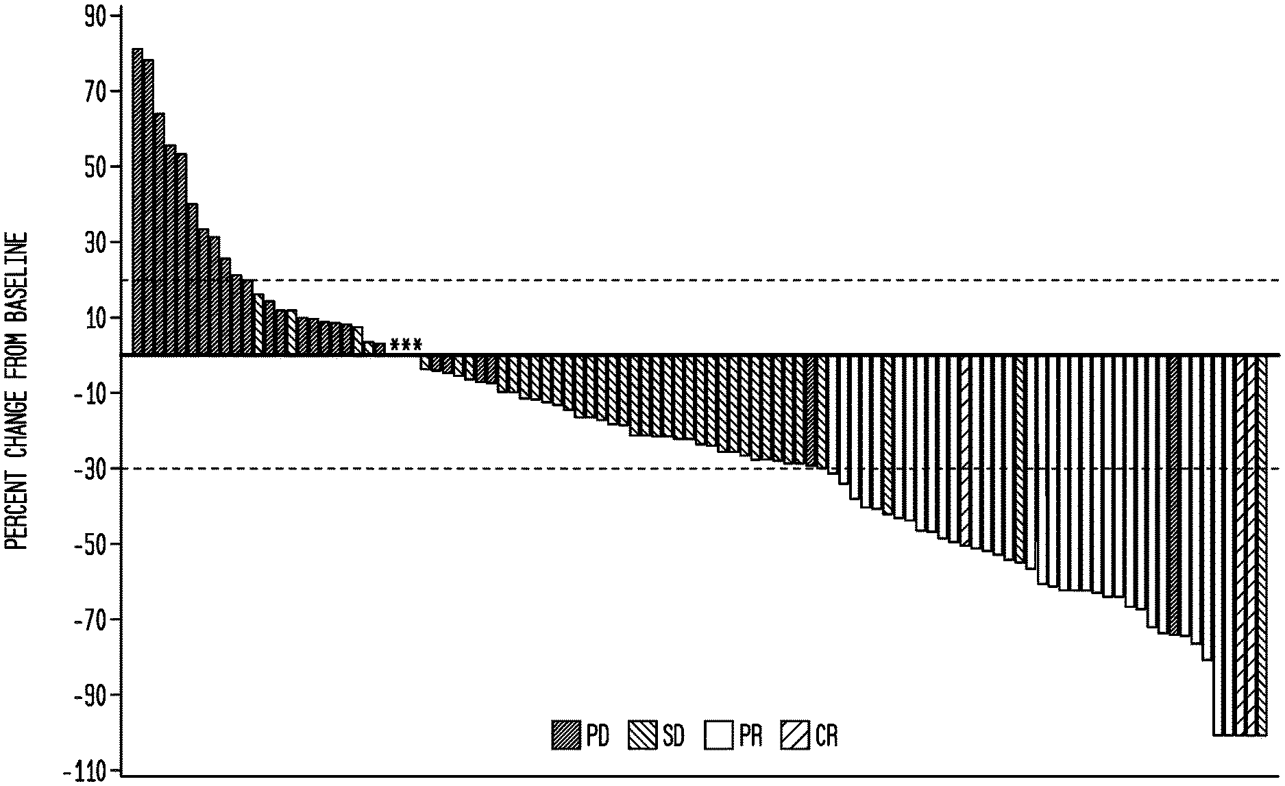
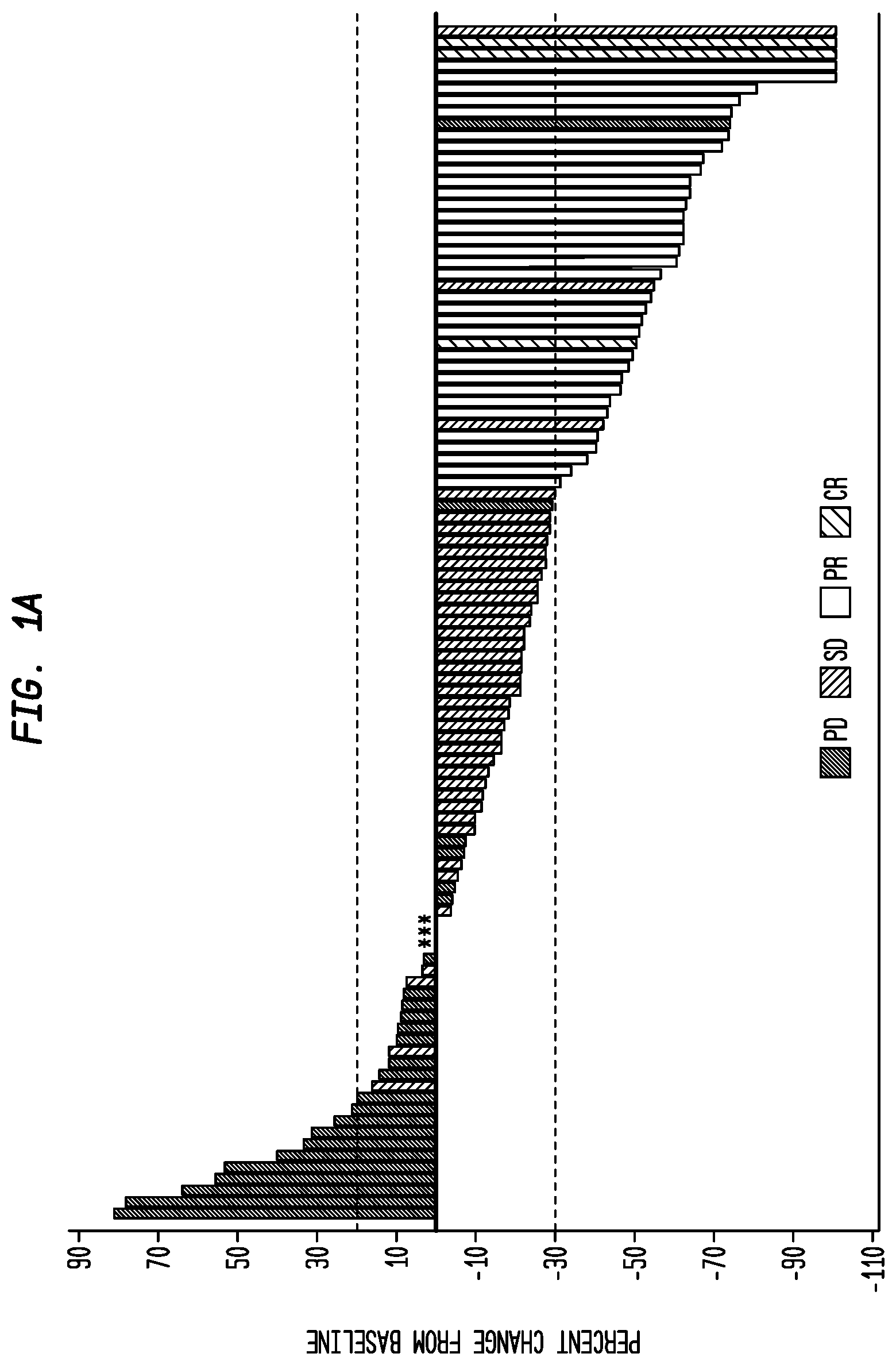
[0024] FIG. 1A. Response and treatment analyses. Waterfall plot showing best percent change from baseline in the sum of target lesion diameters (longest diameter for non-nodal lesions and short axis for nodal lesions). Asterisks denote 3 patients whose best percent change is zero percent (2 SD, 1 PD). The dashed lines at 20% and -30% indicate progressive disease and partial response, respectively, according to RECIST.
[0025] FIG. 1B. Swimmer plot of the objective responses (according to RECIST, version 1.1) from start of treatment to disease progression, as determined by local assessment. At the time of the analysis, 6 patients had a continuing response. The vertical dashed lines show the response at 6 months and 12 months.
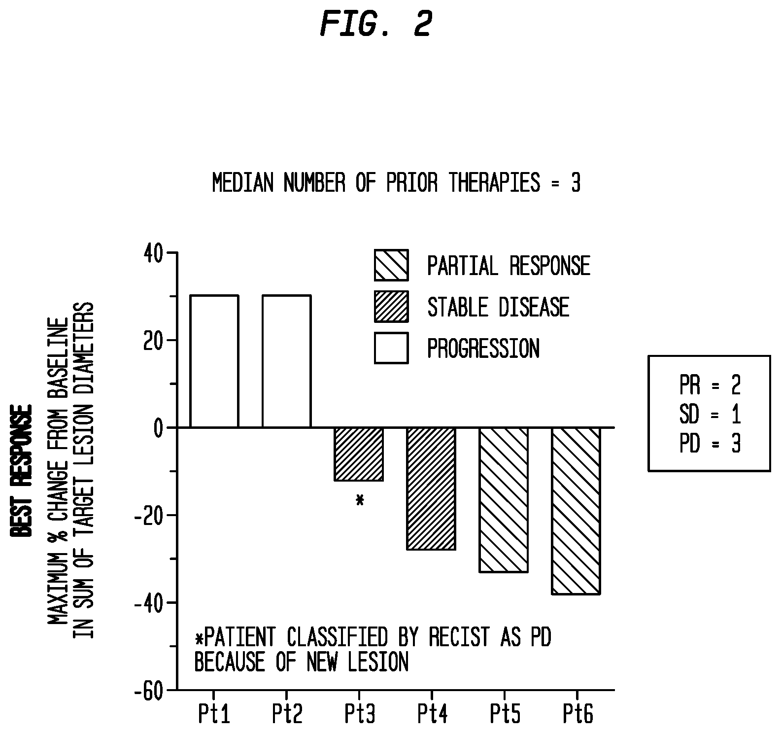
[0026] FIG. 2. Waterfall plot of best responses in 6 patients with urothelial carcinoma treated with sacituzumab govitecan. Clinical trial with sacituzumab govitecan was performed as described in the Examples below.
[0027] FIG. 3A. Graphic representation of anti-tumor response and duration in response-assessable patients. Best percentage change in the sum of the diameters for the selected target lesion and best overall response descriptor according to RECIST 1.1 criteria. Patients are identified with respect to the sacituzumab govitecan starting dose and whether they were sensitive or resistant to prior first-line therapy. Patient with unconfirmed partial responses failed to maintain at least a 30% tumor reduction on their next CT assessment 4-6 weeks after the first observed objective response. The best overall response for these patients by RECIST 1.0 is stable disease.
[0028] FIG. 3B. Graphic representation of anti-tumor response and duration in response-assessable patients. Duration of response from the start of treatment for those patients who achieved partial or complete response. Timing when tumor shrinkage achieved .gtoreq.30% is shown, along with sacituzumab govitecan starting dose and sensitivity to first-line therapy.
[0029] FIG. 3C. Graphic representation of anti-tumor response and duration in response-assessable patients. Dynamics of response for patients who achieved stable disease or better. Two patients with confirmed partial responses who are continuing treatment are shown with dashed line.
[0030] FIG. 4A-B. Kaplan-Meier derived progression-free and overall survival curves for all 53 SCLC patients enrolled in the sacituzumab govitecan trial.
DETAILED DESCRIPTION OF THE INVENTION
[0031] Definitions
[0032] In the description that follows, a number of terms are used and the following definitions are provided to facilitate understanding of the claimed subject matter. Terms that are not expressly defined herein are used in accordance with their plain and ordinary meanings.
[0033] Unless otherwise specified, a or an means "one or more."
[0034] The term about is used herein to mean plus or minus ten percent (10%) of a value. For example, "about 100" refers to any number between 90 and 110.
[0035] An antibody, as used herein, refers to a full-length (i.e., naturally occurring or formed by normal immunoglobulin gene fragment recombinatorial processes) immunoglobulin molecule (e.g., an IgG antibody). An antibody may be conjugated or otherwise derivatized within the scope of the claimed subject matter. Such antibodies include but are not limited to IgG1, IgG2, IgG3, IgG4 (and IgG4 subforms), as well as IgA isotypes. As used below, the abbreviations "MAb" or "mAb" may be used interchangeably to refer to an antibody, antibody fragment, monoclonal antibody or multispecific antibody.
[0036] An antibody fragment is a portion of an antibody such as F(ab').sub.2, F(ab).sub.2, Fab', Fab, Fv, scFv (single chain Fv), single domain antibodies (DABs or VHHs) and the like, including half-molecules of IgG4 (van der Neut Kolfschoten et al. (Science, 2007; 317:1554-1557). Regardless of structure, an antibody fragment of use binds with the same antigen that is recognized by the intact antibody. The term "antibody fragment" also includes synthetic or genetically engineered proteins that act like an antibody by binding to a specific antigen to form a complex. For example, antibody fragments include isolated fragments consisting of the variable regions, such as the "Fv" fragments consisting of the variable regions of the heavy and light chains and recombinant single chain polypeptide molecules in which light and heavy variable regions are connected by a peptide linker ("scFv proteins"). The fragments may be constructed in different ways to yield multivalent and/or multispecific binding forms.
[0037] A therapeutic agent is an atom, molecule, or compound that is useful in the treatment of a disease. Examples of therapeutic agents include, but are not limited to, antibodies, antibody fragments, drug-conjugated antibodies, immunoconjugates, checkpoint inhibitors, drugs, cytotoxic agents, pro-apoptotic agents, toxins, nucleases (including DNAse and RNAse), hormones, immunomodulators, chelators, photoactive agents or dyes, radionuclides, oligonucleotides, interference RNA, siRNA, RNAi, anti-angiogenic agents, chemotherapeutic agents, cytokines, chemokines, prodrugs, enzymes, binding proteins or peptides or combinations thereof.
[0038] As used herein, where reference is made to increased or decreased expression of a particular gene, the term refers to an increase or decrease in a cancer cell compared to normal, benign and/or wild-type cells.
[0039] Antibodies and Antibody-Drug Conjugates (ADCs)
[0040] Certain embodiments relate to use of anti-cancer antibodies, either in unconjugated form or else as an immunoconjugate (e.g., an ADC) attached to one or more therapeutic agents. Preferably the conjugated agent is one that induces DNA strand breaks, more preferably by inhibiting topoisomerase I. Exemplary inhibitors of topoisomerase I include SN-38 and DxD. However, other topoisomerase I inhibitors are known in the art and any such known topoisomerase I inhibitors may be used in an anti-Trop-2, anti-CEACAM5 or anti-HLA-DR ADC. Exemplary topoisomerase I inhibitors include the camptothecins, such as irinotecan, topotecan, SN-38, diflomotecan, S39625, silatecan, belotecan, namitecan, gimatecan, belotecan or camptothecin, as well as non-camptothecins, such as indolocarbazole, phenanthridine, indenoisoquinoline, and their derivatives, such as NSC 314622, NSC 725776, NSC 724998, ARC-111, isoindolo[2,1-a]quinoxalines, indotecan, indimitecan, CRLX101, rebeccamycin, edotecarin, or becatecarin. [See, e.g., Hevener et al., 2018, Acta Pharm Sin B 8:844-61]
[0041] In alternative embodiments, a topoisomerase II inhibitor may be utilized, such as anthracyclines, doxorubucin, epirubicin, valrubicin, daunorubicin, idarubicin, aldoxorubicin, anthracenediones, mitoxantrone, pixantrone, amsacrine, dexrazoxane, epipodophyllotoxins, ciprofloxacin, vosaroxin, teniposide or etoposide. [See, e.g., Hevener et al., 2018, Acta Pharm Sin B 8:844-61]
[0042] Although topoisomerase inhibitors are preferred for antibody conjugation, other agents that induce DNA damage and/or strand breaks are known and may be utilized in alternative embodiments. Such known anti-cancer agents include, but are not limited to, nitrogen mustards, folate analogs such as aminopterin or methotrexate, alkylating agents such as cyclophosphamide, chlorambucil, mitomycin C, streptozotocin or melphalan, nitrosoureas such as carmustine, lomustine or semustine, triazenes such as dacarbazine or temozolomide, or platinum-based inhibitors such as cisplatin, carboplatin, picoplatin or oxaliplatin. [See, e.g., Ong et al., 2013, Chem Biol 20:648-59]
[0043] In a preferred embodiment, antibodies or immunoconjugates comprising an anti-Trop-2 antibody, such as the hRS7 Mab, can be used to treat carcinomas such as carcinomas of the esophagus, pancreas, lung, stomach, colon, rectum, urinary bladder, urothelium, breast, ovary, cervix, endometrium, uterus, kidney, head-and-neck, brain and prostate, as disclosed in U.S. Pat. Nos. 7,238,785; 7,999,083; 8,758,752; 9,028,833; 9,745,380; and 9,770,517; the Examples section of each incorporated herein by reference. An hRS7 antibody is a humanized antibody that comprises light chain complementarity-determining region (CDR) sequences CDR1 (KASQDVSIAVA, SEQ ID NO:1); CDR2 (SASYRYT, SEQ ID NO:2); and CDR3 (QQHYITPLT, SEQ ID NO:3) and heavy chain CDR sequences CDR1 (NYGMN, SEQ ID NO:4); CDR2 (WINTYTGEPTYTDDFKG, SEQ ID NO:5) and CDR3 (GGFGSSYWYFDV, SEQ ID NO:6). However, in alternative embodiments other anti-Trop-2 antibodies are known and may be utilized in an anti-Trop-2 ADC. Exemplary anti-Trop-2 antibodies include, but are not limited to, catumaxomab, VB4-845, IGN-101, adecatumumab, ING-1, EMD 273 066 or hTINA1 (see U.S. Pat. No. 9,850,312). Anti-Trop-2 antibodies are commercially available from a number of sources and include LS-C126418, LS-C178765, LS-C126416, LS-C126417 (LifeSpan BioSciences, Inc., Seattle, Wash.); 10428-MM01, 10428-MM02, 10428-R001, 10428-R030 (Sino Biological Inc., Beijing, China); MR54 (eBioscience, San Diego, Calif.); sc-376181, sc-376746, Santa Cruz Biotechnology (Santa Cruz, Calif.); MM0588-49D6, (Novus Biologicals, Littleton, Colo.); ab79976, and ab89928 (ABCAM.RTM., Cambridge, Mass.).
[0044] Other anti-Trop-2 antibodies have been disclosed in the patent literature. For example, U.S. Publ. No. 2013/0089872 discloses anti-Trop-2 antibodies K5-70 (Accession No. FERM BP-11251), K5-107 (Accession No. FERM BP-11252), K5-116-2-1 (Accession No. FERM BP-11253), T6-16 (Accession No. FERM BP-11346), and T5-86 (Accession No. FERM BP-11254), deposited with the International Patent Organism Depositary, Tsukuba, Japan. U.S. Pat. No. 5,840,854 disclosed the anti-Trop-2 monoclonal antibody BR110 (ATCC No. HB11698). U.S. Pat. No. 7,420,040 disclosed an anti-Trop-2 antibody produced by hybridoma cell line AR47A6.4.2, deposited with the IDAC (International Depository Authority of Canada, Winnipeg, Canada) as accession number 141205-05. U.S. Pat. No. 7,420,041 disclosed an anti-Trop-2 antibody produced by hybridoma cell line AR52A301.5, deposited with the IDAC as accession number 141205-03. U.S. Publ. No. 2013/0122020 disclosed anti-Trop-2 antibodies 3E9, 6G11, 7E6, 15E2, 18B1. Hybridomas encoding a representative antibody were deposited with the American Type Culture Collection (ATCC), Accession Nos. PTA-12871 and PTA-12872. U.S. Pat. No. 8,715,662 discloses anti-Trop-2 antibodies produced by hybridomas deposited at the AID-ICLC (Genoa, Italy) with deposit numbers PD 08019, PD 08020 and PD 08021. U.S. Patent Application Publ. No. 20120237518 discloses anti-Trop-2 antibodies 77220, KM4097 and KM4590. U.S. Pat. No. 8,309,094 (Wyeth) discloses antibodies A1 and A3, identified by sequence listing. U.S. Pat. No. 9,850,312 disclosed the anti-Trop-2 antibodies TINA1, cTINA1 and hTINA1. The Examples section of each patent or patent application cited above in this paragraph is incorporated herein by reference. Non-patent publication Lipinski et al. (1981, Proc Natl. Acad Sci USA, 78:5147-50) disclosed anti-Trop-2 antibodies 162-25.3 and 162-46.2.
[0045] In another preferred embodiment, antibodies or immunoconjugates comprising an anti-CEACAM5 antibody (e.g., hMN-14, labetuzumab) may be used to treat any of a variety of cancers that express CEACAM5, as disclosed in U.S. Pat. Nos. 5,874,540; 6,676,924, 7,999,083, 9,226,973, 9,458,242, 9,499,631 and 9,481,732, the Examples section of each incorporated herein by reference. Solid tumors that may be treated using anti-CEACAM5 include but are not limited to breast, lung, pancreatic, esophageal, medullary thyroid, ovarian, colon, rectum, urinary bladder, prostate, mouth and stomach cancers. A majority of carcinomas, including gastrointestinal, respiratory, genitourinary and breast cancers express CEACAM5 and may be treated with the subject antibodies or immunoconjugates. An hMN-14 antibody is a humanized antibody that comprises light chain variable region CDR sequences CDR1 (KASQDVGTSVA; SEQ ID NO:7), CDR2 (WTSTRHT; SEQ ID NO8), and CDR3 (QQYSLYRS; SEQ ID NO:9), and the heavy chain variable region CDR sequences CDR1 (TYWMS; SEQ ID NO:10), CDR2 (EIHPDSSTINYAPSLKD; SEQ ID NO:11) and CDR3 (LYFGFPWFAY; SEQ ID NO:12). However, other known anti-CEACAM5 antibodies may be incorporated in an ADC. Such known antibodies include CC4 (Zheng et al., 2011, PLoS One 6:e21146), SAR408701 (Decary et al., 2015, Exp Mol Ther 75(Suppl 15) Abstract 1688) and numerous commercially available anti-CEACAM-5 antibodies, e.g. from ThermoFisher Scientific (Cat. No. MIC0101), SigmaAldrich (Cat. No. SAB5300130), Sino Biological (Cat. No. 11077-R076), BosterBio (Cat. No. RP1018), Millipore (Cat. No. MABC1123) and many others.
[0046] In another preferred embodiment, antibodies or immunoconjugates comprising an anti-HLA-DR antibody (e.g., hL243) may be used to treat any of a variety of cancers that express HLA-DR, as disclosed in U.S. Pat. Nos. 7,612,180, 8,613,903, 8,992,917, 8,722,047, 9,187,561, 9,493,573, 9,552,959, or 9,707,302 the Examples section of each incorporated herein by reference. Cancers that may be treated using anti-HLA-DR include but are not limited to lymphoma, leukemia, acute lymphocytic leukemia, chronic lymphocytic leukemia, acute myeloid leukemia, diffuse large B-cell lymphoma, Non-Hodgkin's lymphoma, malignant melanoma, cancers of the skin, esophagus, stomach, colon, rectum, pancreas, lung, breast, ovary, bladder, endometrium, cervix, testes, kidney, liver, melanoma or other HLA-DR-producing tumors (see U.S. Pat. No. 7,612,180; Cardillo et al., 2017, Mol Cancer Ther 17:150-60). An hL243 antibody is a humanized antibody that comprises heavy chain CDR sequences CDR1 (NYGMN, SEQ ID NO:13), CDR2 (WINTYTREPTYADDFKG, SEQ ID NO:14), and CDR3 (DITAVVPTGFDY, SEQ ID NO:15) and light chain CDR sequences CDR1 (RASENIYSNLA, SEQ ID NO:16), CDR2 (AASNLAD, SEQ ID NO:17), and CDR3 (QHFWTTPWA, SEQ ID NO:18). However, other known anti-HLA-DR antibodies may be incorporated in an ADC. Such known antibodies include 1D09C3 (Malviya et al., 2011, Mol Imaging Biol 13:930-9), Lym-1 (Pagel et al., 2007, Cancer Res 67:5921-8), 1D10 (Kostelny et al., 2001, Int J Cancer 93:556-65) H81.9, Ca1.41 (Yamaguchi et al., 1999, Transplantation 68:1161-71) and many others. Anti-HLA-DR antibodies are commercially available from numerous sources, including Abcam, Sino Biological, Inc., Bio-Rad, Beckman Coulter, BioLegend, Novus, Thermo Fisher and many other vendors of biological reagents.
[0047] In alternative embodiments, ADCs of use may incorporate other known antibodies such as hR1 (anti-IGF-1R, U.S. Pat. No. 9,441,043), hPAM4 (anti-mucin, U.S. Pat. No. 7,282,567), hA20 (anti-CD20, U.S. Pat. No. 7,151,164), hA19 (anti-CD19, U.S. Pat. No. 7,109,304), hIMMU31 (anti-AFP, U.S. Pat. No. 7,300,655), hLL1 (anti-CD74, U.S. Pat. No. 7,312,318), hLL2 (anti-CD22, U.S. Pat. No. 5,789,554), hMu-9 (anti-CSAp, U.S. Pat. No. 7,387,772), hL243 (anti-HLA-DR, U.S. Pat. No. 7,612,180), hMN-14 (anti-CEACAM5, U.S. Pat. No. 6,676,924), hMN-15 (anti-CEACAM6 and anti-CEACAM5, U.S. Pat. No. 8,287,865), hRS7 (anti-EGP-1, U.S. Pat. No. 7,238,785), and hMN-3 (anti-CEACAM6, U.S. Pat. No. 7,541,440), the Examples section of each cited patent or application incorporated herein by reference. More preferably, the antibody is IMMU-31 (anti-AFP), hRS7 (anti-TROP-2), hMN-14 (anti-CEACAM5), hMN-3 (anti-CEACAM6), hMN-15 (anti-CEACAM6 and anti-CEACAM5), hLL1 (anti-CD74), hLL2 (anti-CD22), hL243 or IMMU-114 (anti-HLA-DR), hA19 (anti-CD19) or hA20 (anti-CD20). Each antibody may be conjugated, for example, to CL2A-SN-38 as disclosed in U.S. Pat. No. 7,999,083.
[0048] In a preferred embodiment, the antibodies that are used in the treatment of human disease are human or humanized (CDR-grafted) versions of antibodies, although murine and chimeric versions of antibodies can be used. Same species IgG molecules as delivery agents are mostly preferred to minimize immune responses. This is particularly important when considering repeat treatments. For humans, a human or humanized IgG antibody is less likely to generate an anti-IgG immune response from patients.
[0049] Formulation and Administration of ADCs
[0050] Antibodies or immunoconjugates (e.g., ADCs) can be formulated according to known methods to prepare pharmaceutically useful compositions, whereby the antibody or immunoconjugate is combined in a mixture with a pharmaceutically suitable excipient. Sterile phosphate-buffered saline is one example of a pharmaceutically suitable excipient. Other suitable excipients are well-known to those in the art. See, for example, Ansel et al., PHARMACEUTICAL DOSAGE FORMS AND DRUG DELIVERY SYSTEMS, 5th Edition (Lea & Febiger 1990), and Gennaro (ed.), REMINGTON'S PHARMACEUTICAL SCIENCES, 18th Edition (Mack Publishing Company 1990), and revised editions thereof.
[0051] In a preferred embodiment, the antibody or immunoconjugate is formulated in Good's biological buffer (pH 6-7), using a buffer selected from the group consisting of N-(2-acetamido)-2-aminoethanesulfonic acid (ACES); N-(2-acetamido)iminodiacetic acid (ADA); N,N-bis(2-hydroxyethyl)-2-aminoethanesulfonic acid (BES); 4-(2-hydroxyethyl)piperazine-1-ethanesulfonic acid (HEPES); 2-(N-morpholino)ethanesulfonic acid (WS); 3-(N-morpholino)propanesulfonic acid (MOPS); 3-(N-morpholinyl)-2-hydroxypropanesulfonic acid (MOPSO); and piperazine-N,N'-bis(2-ethanesulfonic acid) [Pipes]. More preferred buffers are MES or MOPS, preferably in the concentration range of 20 to 100 mM, more preferably about 25 mM. Most preferred is 25 mM MES, pH 6.5. The formulation may further comprise 25 mM trehalose and 0.01% v/v polysorbate 80 as excipients, with the final buffer concentration modified to 22.25 mM as a result of added excipients. The preferred method of storage is as a lyophilized formulation of the conjugates, stored in the temperature range of -20 .degree. C. to 2 .degree. C., with the most preferred storage at 2 .degree. C. to 8 .degree. C.
[0052] The antibody or immunoconjugate can be formulated for intravenous administration via, for example, bolus injection, slow infusion or continuous infusion. Preferably, the antibody of the present invention is infused over a period of less than about 4 hours, and more preferably, over a period of less than about 3 hours. For example, the first 25-50 mg could be infused within 30 minutes, preferably even 15 min, and the remainder infused over the next 2-3 hrs. Formulations for injection can be presented in unit dosage form, e.g., in ampoules or in multi-dose containers, with an added preservative. The compositions can take such forms as suspensions, solutions or emulsions in oily or aqueous vehicles, and can contain formulatory agents such as suspending, stabilizing and/or dispersing agents. Alternatively, the active ingredient can be in powder form for constitution with a suitable vehicle, e.g., sterile pyrogen-free water, before use.
[0053] Generally, the dosage of an administered antibody or immunoconjugate for humans will vary depending upon such factors as the patient's age, weight, height, sex, general medical condition and previous medical history. It may be desirable to provide the recipient with a dosage of immunoconjugate that is in the range of from about 1 mg/kg to 24 mg/kg as a single intravenous infusion, although a lower or higher dosage also may be administered as circumstances dictate. A dosage of 1-20 mg/kg for a 70 kg patient, for example, is 70-1,400 mg, or 41-824 mg/m.sup.2 for a 1.7-m patient. The dosage may be repeated as needed, for example, once per week for 4-10 weeks, once per week for 8 weeks, or once per week for 4 weeks. It may also be given less frequently, such as every other week for several months, or monthly or quarterly for many months, as needed in a maintenance therapy. Preferred dosages may include, but are not limited to, 1 mg/kg, 2 mg/kg, 3 mg/kg, 4 mg/kg, 5 mg/kg, 6 mg/kg, 7 mg/kg, 8 mg/kg, 9 mg/kg, 10 mg/kg, 11 mg/kg, 12 mg/kg, 13 mg/kg, 14 mg/kg, 15 mg/kg, 16 mg/kg, 17 mg/kg, and 18 mg/kg. The dosage is preferably administered multiple times, once or twice a week, or as infrequently as once every 3 or 4 weeks. A minimum dosage schedule of 4 weeks, more preferably 8 weeks, more preferably 16 weeks or longer may be used. The schedule of administration may comprise administration once or twice a week, on a cycle selected from the group consisting of: (i) weekly; (ii) every other week; (iii) one week of therapy followed by two, three or four weeks off; (iv) two weeks of therapy followed by one, two, three or four weeks off; (v) three weeks of therapy followed by one, two, three, four or five week off; (vi) four weeks of therapy followed by one, two, three, four or five week off; (vii) five weeks of therapy followed by one, two, three, four or five week off; (viii) monthly and (ix) every 3 weeks. The cycle may be repeated 2, 4, 6, 8, 10, 12, 16 or 20 times or more.
[0054] Alternatively, an antibody or immunoconjugate may be administered as one dosage every 2 or 3 weeks, repeated for a total of at least 3 dosages. Or, twice per week for 4-6 weeks. If the dosage is lowered to approximately 200-300 mg/m.sup.2 (340 mg per dosage for a 1.7-m patient, or 4.9 mg/kg for a 70 kg patient), it may be administered once or even twice weekly for 4 to 10 weeks. Alternatively, the dosage schedule may be decreased, namely every 2 or 3 weeks for 2-3 months. It has been determined, however, that even higher doses, such as 12 mg/kg once weekly or once every 2-3 weeks can be administered by slow i.v. infusion, for repeated dosing cycles. The dosing schedule can optionally be repeated at other intervals and dosage may be given through various parenteral routes, with appropriate adjustment of the dose and schedule
[0055] DNA Damage and Repair Pathways
[0056] Use of anti-cancer ADCs with drug moieties targeted against topoisomerases can result in accumulation of single- or double-stranded breaks in cancer cell DNA. Resistance to or relapse from the anti-cancer effects of topoisomerase I inhibitors, or other anti-cancer agents that damage DNA, may result from the existence of DNA repair mechanisms, such as the DNA damage response (DDR). DDR is a complex set of pathways responsible for repair of damage to DNA in normal and tumor cells. Inhibitors directed against DDR pathways may be utilized in combination with anti-Trop-2, anti-CEACAM5 or anti-HLA-DR ADCs to provide increased anti-cancer efficacy in tumors that are relapsed from or resistant to monotherapy with anti-Trop-2, anti-CEACAM5 or anti-HLA-DR ADCs. In addition, the presence of mutations, other genetic defects or changes in expression levels of genes encoding DDR components may be predictive of the efficacy of anti-Trop-2, anti-CEACAM5 or anti-HLA-DR ADCs and/or of combination therapy with an anti-Trop-2, anti-CEACAM5 or anti-HLA-DR ADC and one or more other anti-cancer agents.
[0057] In preferred embodiments, the subject ADCs may be used in combination with one or more known anti-cancer agents that inhibit various steps in the DDR pathways. There are numerous pathways involved in cellular DNA repair, with partial overlap in the protein effectors of the different pathways. Use of topoisomerase-inhibiting ADCs in combination with other inhibitors directed against different steps in the DNA damage repair pathways may exhibit synthetic lethality, wherein simultaneous loss of function in two different genes results in cell death, whereas loss of function in just one gene does not (e.g., Cardillo et al., 2017, Clin Cancer Res 23:3405-15). The concept may also be applied in cancer therapy, wherein a cancer cell carrying a mutation in one gene is targeted by a chemotherapeutic agent that inhibits the function of a second gene used by the cell to overcome the first mutation (Cardillo et al., 2017, Clin Cancer Res 23:3405-15). This concept has been applied, for example, to use of PARP inhibitors in cells bearing BRCA gene mutations (Benafif & Hall, 2015, Onco Targets Ther 8:519-28). In principle, synthetic lethality may be applied with or without the presence of underlying cancer cell mutations, for example by using combination therapy with two or more inhibitors targeted against different aspects of DDR pathways, alone or in combination with DNA damage-inducing agents.
[0058] Double-strand DNA breaks (DSBs) are repaired by two major pathways--homologous recombination (HR) and nonhomologous end joining (NHEJ). [See, e.g., Srivastava & Raghavan, 2015, Chem Biol 22:17-29] Each of these comprises subpathways--classical or alternative subpathways for NHEJ (respectively, cNHEJ and aNHEJ) and single-strand annealing (SSA) for the HR pathway. HR requires extensive homology for repair of DSBs and is most active in the S and G2 phases of the cell cycle, while NHEJ utilizes limited or no homology for end joining and can act throughout the cell cycle (Srivastava & Raghavan, 2015, Chem Biol 22:17-29).
[0059] Activation of DDR pathways by DSB includes checkpoint arrest, mediated via ATM, ATR and DNA-PKcs (Nickoloff et al., 2017, J Natl Cancer Inst 109:djx059). ATM is required for DSB repair by HR and triggers DSB end resection by stimulating nucleolytic activity of CtIP and MRE11 to generate 3'-ssDNA overhangs, followed by RPA loading and RAD51 nucleofilament formation (Bakr et al., 2015, Nucleic Acids Res 43:3154). ATR responds to a broader spectrum of DNA damage, including DSBs and ssDNA (Marechal et al., 2013, Cold Spring Harb Perspect Biol 5:a012716). However, the functions of ATR and ATM are not mutually exclusive and both are required for DSB-induced checkpoint responses and DSB repair (Marechal et al., 2013, Cold Spring Harb Perspect Biol 5:a012716). Localization of the ATR-ATRIP complex to sites of DNA damage is dependent on the presence of long stretches of RPA-coated ssDNA, which may be generated by resection as discussed below (Marechal et al., 2013, Cold Spring Harb Perspect Biol 5:a012716). DNA-PKcs is the catalytic subunit of DNA-PK and is primarily involved in the NHEJ pathway (Marechal et al., 2013, Cold Spring Harb Perspect Biol 5:a012716).
[0060] Determination of which DSB repair pathway is utilized is mediated by the amount of 5' end resection at the DSB, which is inhibited by 53BP1/RIF1 and promoted by BRCA1/CtIP. Increased resection favors the HR repair pathways, while decreased resection promotes the NHEJ pathways (Nickoloff et al., 2017, J Natl Cancer Inst 109:djx059). At the start of the HR pathways, MRE11 (part of the MRN complex along with RAD50 and NBS1) initiates limited end resection, which is followed by Exol/EEPD1 and Dna2 for extensive resection (Nickoloff et al., 2017, J Natl Cancer Inst 109:djx059). In the NHEJ pathways, 53BP1/RIF1 and KU70/80 inhibit resection and promote classical NHEJ, while PARP1 competes with the KU proteins and promotes limited end resection for alternative NHEJ (Nickoloff et al., 2017, J Natl Cancer Inst 109:djx059). Pol .theta. is also involved in aNHEJ.
[0061] Further steps in the HR pathway are promoted by RPA, BRCA2, RAD51, RAD52, RAD54, and Pol 6 (Nickoloff et al., 2017, J Natl Cancer Inst 109:djx059). RAD52 is also involved in SSA, along with ERCC1, ERCC2, ERCC3 and ERCC4 (Nickoloff et al., 2017, J Natl Cancer Inst 109:djx059). Other proteins involved in HR include RAD50, NBS1, BLM, XPF, FANCM, FAAP24, FANC1, FAND2, MSH3, SLX4, MUS81, EME1, SLX1, PALB2, BRIP1, BARD1, BAP1, PTEN, RAD51C, USP11, WRN and NER. [Nickoloff et al., 2017, J Natl Cancer Inst 109:djx059, Srivastava & Raghavan, 2015, Chem Biol 22:17-29] Other proteins involved in NHEJ include Artemis, Pol .mu., Pol .lamda., Ligase IV, XRCC4, and XLF. [Nickoloff et al., 2017, J Natl Cancer Inst 109:djx059, Srivastava & Raghavan, 2015, Chem Biol 22:17-29] Further details regarding the roles of these various DDR proteins and inhibitors for each are provided below.
[0062] Repair of single-stranded DNA lesions can also occur via multiple pathways--base excision repair (BER), nucleotide excision repair (NER) and mismatch repair (MMR). The BER pathway is facilitated by APE1, PARP1, Pol .beta., Lig III and XRCC1. NER is facilitated by XPC, RAD23B, HR23B, XPF, ERCC1, XPG, XPA, RPA, XPD, CSA, CSB, XAB2 and Pol .delta./.kappa./.epsilon.. MMR is facilitated by MutS.alpha./.beta., MLH1, PMS2, Exo1, PARP1, MSH2, MSH6 and Pol .delta./.epsilon. (Nickoloff et al., 2017, J Natl Cancer Inst 109:djx059). Mutations in MSH2 predispose cancers to sensitivity to methotrexate and psoralen (Nickoloff et al., 2017, J Natl Cancer Inst 109:djx059). Defects in NER, such as decreased expression of ERCC1, predispose to sensitivity to cross-linking agents such as cisplatin as well as PARP1 or ATR inhibitors (Nickoloff et al., 2017, J Natl Cancer Inst 109:djx059).
[0063] As discussed below, inhibitors of various of these DDR proteins are known, and any such known inhibitor may be utilized in combination with a subject ADC. In more preferred embodiments, the presence of mutations in BRCA1 and/or BRCA2 may be predictive of efficacy of either ADC monotherapy or combination therapy with an ADC and an inhibitor of DSB repair.
[0064] Combination Therapy With ADCs and Inhibitors of DNA Damage Repair
[0065] As discussed above, a key objective of combination therapy with anti-Trop-2, anti-CEACAM5 or anti-HLA-DR ADCs, together with one or more inhibitors of DDR pathways, is to induce an artificial (as opposed to genetic) synthetic lethality, where the combination of agents that produce DNA damage (e.g., topoisomerase I inhibitors) with agents that inhibit steps in the DDR damage repair pathways is effective to kill cancer cells that are resistant to either type of agent alone. DDR inhibitors of particular interest for combination therapies are directed against PARP, ATR, ATM, CHK1, CHK2, CDK12, RAD51, RAD52 and WEE1. In alternative embodiments, the DDR inhibitor of interest may be a DDR inhibitor that is not a PARP inhibitor or RAD51 inhibitor.
[0066] PARP Inhibitors
[0067] Poly-(ADP-ribose) polymerase (PARP) plays a key role in the DNA damage response and either directly or indirectly affects numerous DDR pathways, including BER, HR, NER, NHEJ and MMR (Gavande et al., 2016, Pharmacol Ther 160:65-83). A number of PARP inhibitors are known in the art, such as olaparib, talazoparib (BMN-673), rucaparib, veliparib, niraparib, CEP 9722, MK 4827, BGB-290 (pamiparib), ABT-888, AG014699, BSI-201, CEP-8983, E7016 and 3-aminobenzamide (see, e.g., Rouleau et al., 2010, Nat Rev Cancer 10:293-301, Bao et al., 2015, Oncotarget [Epub ahead of print, Sep. 22, 2015]). PARP inhibitors are known to exhibit synthetic lethality, for example in tumors with mutations in BRCA1/2. Olaparib has received FDA approval for treatment of ovarian cancer patients with mutations in BRCA1 or BRCA2. In addition to olaparib, other FDA-approved PARP inhibitors for ovarian cancer include nirapirib and rucaparib. Talazoparib was recently approved for treatment of breast cancer with germline BRCA mutations and is in phase III trials for hematological malignancies and solid tumors and has reported efficacy in SCLC, ovarian, breast, and prostate cancers (Bitler et al., 2017, Gynecol Oncol 147:695-704). Veliparib is in phase III trials for advanced ovarian cancer, TNBC and NSCLC (see Wikipedia under "PARP_inhibitor"). Not all PARP inhibitors are dependent on BRCA mutation status and niraparib has been approved for maintenance therapy of recurrent platinum sensitive ovarian, fallopian tube or primary peritoneal cancer, independent of BRCA status (Bitler et al., 2017, Gynecol Oncol 147:695-704).
[0068] Any such known PARP inhibitor may be utilized in combination with an anti-Trop-2, anti-CEACAM5 or anti-HLA-DR ADC, such as sacituzumab govitecan, DS-1062, labetuzumab govitecan or IMMU-140. Synthetic lethality and synergistic inhibition of tumor growth has been demonstrated for the combination of sacituzumab govitecan with olaparib, rucaparib and talazoparib in nude mice bearing TNBC xenografts (Cardillo et al., 2017, Clin Cancer Res 23:3405-15). The beneficial effects of combination therapy were observed independently of BRCA1/2 mutation status (Cardillo et al., 2017, Clin Cancer Res 23:3405-15).
[0069] CDK12 Inhibitors
[0070] Cyclin-dependent kinase 12 (CDK12) is a cell cycle regulator that has been reported to act in concert with PARP inhibitors and knockdown of CDK12 activity was observed to promote sensitivity to olaparib (Bitler et al., 2017, Gynecol Oncol 147:695-704). CDK12 appears to act at least in part by regulating expression of DDR genes (Krajewska et al., 2019, Nature Commun 10:1757). Various inhibitors of CDK12 are known, such as dinaciclib, flavopiridol, roscovitine, THZ1 or THZ531 (Bitler et al., 2017, Gynecol Oncol 147:695-704; Krajewska et al., 2019, Nature Commun 10:1757; Paculova & Kohoutek, 2017, Cell Div 12:7). Combination therapy with PARP inhibitors and dinaciclib reverses resistance to PARP inhibitors (Bitler et al., 2017, Gynecol Oncol 147:695-704). In the subject methods, it may be of use to combine therapy with an anti-Trop-2, anti-CEACAM5 or anti-HLA-DR ADC with the combination of a PARP inhibitor and/or a CDK12 inhibitor.
[0071] RAD51 Inhibitors
[0072] BRCA1 and BRCA2 encode proteins that are essential for the HR DNA repair pathway and mutations in these genes require increased reliance on NHEJ pathways for tumor survival. PARP is a critical protein for NHEJ mediated DNA repair and use of PARP inhibitors (PARPi) in BRCA mutated tumors (e.g., ovarian cancer, TNBC) provides synthetic lethality. However, not all BRCA mutated tumors are sensitive to PARPi and many that are initially sensitive will develop resistance.
[0073] RAD51 is another central protein in the HR pathway and is frequently overexpressed in cancer cells (see Wikipedia under "RAD51"). Increased expression of RAD51 may compensate, in part, for BRCA mutations and decrease sensitivity to PARP inhibitors. It has been demonstrated that sacituzumab govitecan, an anti-Trop-2 ADC carrying a topoisomerase I inhibitor, can at least partially compensate for RAD51 overexpression (see U.S. patent application Ser. No. 15/926,537). Thus, a rationale exists for combination therapy using a topoisomerase I-inhibiting ADC with a RAD51 inhibitor, with or without a PARP inhibitor.
[0074] Combination therapy with ADCs may utilize any Rad51 inhibitor known in the art, including but not limited to B02 ((E)-3-benzyl-2(2-(pyridin-3 -yl)vinyl) quinazolin-4(3H)-one) (Huang & Mazin, 2014, PLoS ONE 9(6):e100993); RI-1 (3-chloro-1-(3,4-dichlorophenyl)-4-(4-morpholinyl)-1H-pyrrole-2,5-dione) (Budke et al., 2012, Nucl Acids Res 40:7347-57); DIDS (4,4'-diisothiocyanostilbene-2,2'-disulfonic acid) (Ishida et al., 2009, Nucl Acids Res 37:3367-76); halenaquinone (Takaku et al., 2011, Genes Cells 16:427-36); CYT-0851 (Cyteir Therapeutics, Inc.), IBR.sub.2 (Ferguson et al., 2018, J Pharm Exp Ther 364:46-54) or imatinib (Choudhury et al., 2009, Mol Cancer Ther 8:203-13). Many of these are available from commercial sources (e.g., B02, Calbiochem; RI-1, Calbiochem; DIDS, Tocris Bioscience; halenaquinone, Angene International Ltd., Hong Kong; imatinib (GLEEVAC.RTM.), Novartis).
[0075] As discussed above, combination therapy with an ADC and a RAD51 inhibitor with or without a PARP inhibitor may be of use for treating cancer.
[0076] ATM Inhibitors
[0077] ATM and ATR are key mediators of DDR, acting to induce cell cycle arrest and facilitate DNA repair via their downstream targets (Weber & Ryan, 2015, Pharmacol Ther 149:124-38). Many malignant tumors show functional loss or deregulation of key proteins involved in DDR and cell cycle regulation, such as p53, ATM, MRE11, BRCA1/2 or SMC1 (Weber & Ryan, 2015, Pharmacol Ther 149:124-38). As discussed above, defects in certain DDR pathways, such as HRD, may increase reliance of the cancer cell on alternative DDR pathways, thus providing targets for selective inhibition of cancer cells bearing such DDR mutations (Weber & Ryan, 2015, Pharmacol Ther 149:124-38). In addition to the effects of BRCA1/2 mutations on susceptibility to PARP inhibitors, other functional changes in DDR proteins that can increase sensitivity to DNA damaging anti-cancer treatments can include changes in DNA-PKcs (Zhao et al., 2006, Cancer Res 66:5354-62), ATM (Golding et al., 2012, Cell Cycle 11:1167-73), ATR (Fokas et al., 2012, Cell Death Dis 3:e441), CHK1 and CHK2 (Mathews et al., 2007, Cell Cycle 6:104-10; Riesterer et al., 2011, Invest New Drugs 29:514-22). In principle, the effects of such sensitizing mutations may be reproduced by combination therapy using inhibitors against the relevant DDR proteins.
[0078] ATM and ATR are members of the phosphatidylinositol 2-kinase-related kinase (PIKK) family, which also includes DNA-PKcs/PRKDC, MTOR/FRAP and SMG1 (Weber & Ryan, 2015, Pharmacol Ther 149:124-38). Due to the high degree of sequence homology between the various PIKK proteins, cross-reactivity is often observed between inhibitors of different PIKK proteins and may result in undesirable toxicities. Use of inhibitors with high affinity for ATM or ATR, compared to other PIKK proteins, is preferred.
[0079] ATM attaches to sites of DSBs by binding to the MRN complex (MRE11-RAD5O-NBS1) (Weber & Ryan, 2015, Pharmacol Ther 149:124-38). Binding to MRN activates ATM kinase and promotes phosphorylation of its downstream targets--p53, CHK2 and Mdm2--which in turn activates cell cycle checkpoint activity (Weber & Ryan, 2015, Pharmacol Ther 149:124-38). Other downstream effectors of ATM include BRCA1, H2AX and p21 (Ronco et al., 2017, Med Chem Commun 8:295-319). Both the ATM and ATR pathways inhibit activity of CDC25C and CDK1 (Ronco et al., 2017, Med Chem Commun 8:295-319).
[0080] Various inhibitors of ATM are known in the art. Caffeine inhibits both ATM and ATR and sensitizes cells to the effects of ionizing radiation (Weber & Ryan, 2015, Pharmacol Ther 149:124-38). Wortmannin is a relatively non-specific inhibitor of PIKK and has activity against ATM, PI3K and DNA-PKcs (Weber & Ryan, 2015, Pharmacol Ther 149:124-38). CP-466722, KU-55933, KU-60019, and KU-59403 are all relatively selective for ATM and have been reported to sensitize cells to the effects of ionizing radiation (Weber & Ryan, 2015, Pharmacol Ther 149:124-38). KU-59403 also increased the anti-tumor efficacy of etoposide and irinotecan, while KU-55933 increased cancer sensitivity to doxorubicin and etoposide (Weber & Ryan, 2015, Pharmacol Ther 149:124-38). The effect of KU-60019 was substantially enhanced in p53 mutant cancer cells, suggesting that p53 mutations might be a biomarker for use of ATM inhibitors. The ATM inhibitor AZD0156 has been used in combination with the PARP inhibitor olaparib (Cruz et al., 2018, Ann Oncol 29:1203-10). AZD0156 in combination with the WEE1 inhibitor AZD1775 produced a synergistic anti-tumor effect in prostate cancer xenografts (Jin et al., Cancer Res Treat [Epub ahead of print Jun. 25, 2019]. Other reported ATM inhibitors include CGK733, NVP-BEZ 235, Torin-2, fluoroquinoline 2 and SJ573017 (Ronco et al., 2017, Med Chem Commun 8:295-319). A significant anti-tumor effect was reported for combination therapy with fluoroquinoline 2 and irinotecan (Ronco et al., 2017, Med Chem Commun 8:295-319).
[0081] Although none have yet received FDA approval, ATM inhibitors in clinical trials include AZD1390 (AstraZeneca), Ku-60019 (AstraZeneca), AZD0156 (AstraZeneca). Combination therapy with anti-Trop-2, anti-CEACAM-5 or anti-HLA-DR ADCs and an ATM inhibitor alone, or in combination with other DDR inhibitors, may be of use for cancer treatment.
[0082] ATR Inhibitors
[0083] ATR is another central kinase involved in regulation of DDR. In contrast to ATM, ATR is activated by single-stranded DNA structures (ssDNA), which may occur at resected DSBs or stalled replication forks (Weber & Ryan, 2015, Pharmacol Ther 149:124-38). ATR binds to ATRIP (ATR-interacting protein), which controls localization of ATR to sites of DNA damage (Weber & Ryan, 2015, Pharmacol Ther 149:124-38). ssDNA binds to RPA, which can bind to ATR/ATRIP and also to RAD17/RFC2-5 which in turn promote binding of RAD9-HUS1-RAD1 (9-1-1 complex) onto the DNA ends (Weber & Ryan, 2015, Pharmacol Ther 149:124-38). The 9-1-1 complex recruits TopBP1, which activates ATR (Weber & Ryan, 2015, Pharmacol Ther 149:124-38). ATR then activates CHK1, which promotes DNA repair, stabilization and transient cell cycle arrest (Weber & Ryan, 2015, Pharmacol Ther 149:124-38). Other downstream effectors of ATR function include Cdc25A, Cdc25C, WEE1, Cyclin B and cdc2 (Ronco et al., 2017, Med Chem Commun 8:295-319). The ATM and ATR pathways are partially overlapping and inhibition of one pathway may be partially compensated by activity of the other pathway. In certain embodiments, combination therapy with inhibitors of ATM and ATR, or use of inhibitors that are active against both ATM and ATR, may be preferred. In other embodiments, ATR inhibitors may be indicated for treating cancers where a mutation or other inactivating change inhibits ATM function in the cancer cell.
[0084] A number of ATR selective inhibitors have been developed. Schisandrin B is purported to be selective for ATR (Nischida et al., 2009, Nucleic Acids Res 73:5678-89), however with only weak toxicity. More potent inhibitors such as NU6027, BEZ235, ETP46464 and Torin 2 showed cross-reactivity with other PIKK proteins (Weber & Ryan, 2015, Pharmacol Ther 149:124-38). More potent and selective ATR inhibitors have been developed by Vertex Pharmaceuticals, such as VE-821 and VE-822 (aka VX-970, M6620, berzosertib, Merck). Other ATR inhibitors include AZ20 (AstraZeneca), AZD6738 (ceralasertib), M4344 (Merck), (Weber & Ryan, 2015, Pharmacol Ther 149:124-38) as well as EPT-46464 (Brandsma et al., 2017, Expert Opin Investig Drugs 26:1341-55). BAY1895344 (Bayer), BAY-937 (Bayer), AZD6738 (AstraZeneca), BEZ235 (dactolisib), CGK 733 and VX-970 (M6620) are in clinical trials for cancer therapy. AZD6738 was reported to be synthetically lethal with p53 and ATM defects (Ronco et al., 2017, Med Chem Commun 8:295-319).
[0085] Combination therapy with VE-821 was shown to enhance sensitivity to cisplatin and gemcitabine in vivo, while AZD6738 significantly increased sensitivity to carboplatin (Weber & Ryan, 2015, Pharmacol Ther 149:124-38). VX970 (M6620) increased sensitivity to a variety of DNA damaging agents, such as cisplatin, oxaliplatin, gemcitabine, etoposide and SN-38 (Weber & Ryan, 2015, Pharmacol Ther 149:124-38). Chemisensitization was more pronounced in cancer cells with p53-deficiency (Weber & Ryan, 2015, Pharmacol Ther 149:124-38). A phase I study of combination therapy with M6620 and topotecan showed improved efficacy in platinum-refractory SCLC, which tends to be non-responsive to topotecan alone (Thomas et al. 2018, J Clin Oncol 36:1594-1602). AZD6738 enhanced sensitivity to carboplatin (Weber & Ryan, 2015, Pharmacol Ther 149:124-38). Various cancer chemotherapeutic agents have been reported to have additive and/or synergistic effects with ATR inhibitors. These include, but are not limited to, gemcitabine, cytarabine, 5-fluorouracil, camptothecin, SN-38, cisplatin, carboplatin and oxaliplatin. [See, e.g., Wagner and Kaufmann, 2010, Pharmaceuticals 3:1311-34] Such agents may be utilized to further enhance combination therapy with anti-Trop-2, anti-CEACAM5 or anti-HLA-DR ADCs and ATR inhibitors.
[0086] CHK1 Inhibitors
[0087] CHK1 is a phosphorylation target of the ATR kinase and is a downstream mediator of ATR activity. Phosphorylation of CHK1 by ATR activates CHK1 activity, which in turn phosphorylates Cdc25A and Cdc25C, mediating ATR dependent DNA repair mechanisms (Wagner and Kaufmann, 2010, Pharmaceuticals 3:1311-34).
[0088] A variety of CHK1 inhibitors are known in the art, including some that are currently in clinical trials for cancer treatment. Any known CHK1 inhibitor may be utilized in combination with an anti-Trop-2, anti-CEACAM5 or anti-HLA-DR ADC, alone or in combination with other DDR inhibitors. CHK1 inhibitors of interest include but are not limited to XL9844 (Exelixis, Inc.), UCN-01, CHIR-124, AZD7762 (AstraZeneca), AZD1775 (Astrazeneca), XL844, LY2603618 (Eli Lilly), LY2606368 (prexasertib, Eli Lilly), GDC-0425 (Genentech), PD-321852, PF-477736 (Pfizer), CBP501, CCT-244747 (Sareum), CEP-3891 (Cephalon), SAR-020106 (Sareum), Arry-575 (Array), SRA737 (Sareum), V158411 and SCH 900776 (aka MK-8776, Merck). [See Wagner and Kaufmann, 2010, Pharmaceuticals 3:1311-34; Thompson and Eastman, 2013, Br J Clin Pharmacol 76:3; Ronco et al., 2017, Med Chem Commun 8:295-319] CHIR-124 was reported to potentiate the activity of topoisomerase I inhibitors in mouse xenografts (Ronco et al., 2017, Med Chem Commun 8:295-319). CCT244747 showed anti-tumor activity in combination with gemcitabine and irinotecan (Ronco et al., 2017, Med Chem Commun 8:295-319). Clinical trials have been performed with LY2603618 and SCH900776 (Ronco et al., 2017, Med Chem Commun 8:295-319).
[0089] CHK2 Inhibitors
[0090] Several CHK2 inhibitors are known and may be utilized in combination with an ADC and/or other DDR inhibitors or anti-cancer agents. Such known CHK2 inhibitors include, but are not limited to, NSC205171, PV1019, CI2, CI3 (Gokare et al., 2016, Oncotarget 7:29520-30), 2-arylbenzimidazole (ABI, Johnson & Johnson), NSC109555, VRX0466617 and CCT241533 (Ronco et al., 2017, Med Chem Commun 8:295-319). PV1019 showed enhanced activity in combination with topotecan or camptothecin (Ronco et al., 2017, Med Chem Commun 8:295-319). However, the required dosages were too high to be of therapeutic use (Ronco et al., 2017, Med Chem Commun 8:295-319). Ronco et al. concluded that the CHK2 inhibitors developed to date were significantly less active as anti-cancer agents than CHK1, ATM or ATR inhibitors (Ronco et al., 2017, Med Chem Commun 8:295-319).
[0091] WEE1 Inhibitors
[0092] WEE1 is overexpressed in many forms of cancer including breast cancer, glioma, glioblastoma, nasopharyngial and drug-resistant cancers (Ronco et al., 2017, Med Chem Commun 8:295-319). WEE1 is a key intermediary in the ATR pathway and is activated by CHK1 (Ronco et al., 2017, Med Chem Commun 8:295-319). WEE1 exerts an inhibitory effect on Cyclin B/cdc2 and CDK1, which in turn regulate cell cycle arrest (Ronco et al., 2017, Med Chem Commun 8:295-319. There are relatively few WEE1 inhibitors available, compared to other components of DDR.
[0093] The WEE1 inhibitor AZD1775 (MK1775) has been used in clinical trials in combination with DNA-damaging therapies, such as fludarabine, cisplatin, carboplatin, paclitaxel, gemcitabine, docetaxel, irinotecan or cytarabine (Matheson et al, 2016, Trends Pharm Sci 37:P872-81; see also clinicaltrials.gov). Combination therapy with inhibitors of WEE1 and CHK1/2 is reported to produce a synergistic effect in cancer xenografts (Ronco et al., 2017, Med Chem Commun 8:295-319). Thus, it may be of use to combine therapy with an anti-Trop-2, anti-CEACAM5 or anti-HLA-DR ADC, an inhibitor of WEE1 and one or more inhibitors of CHK1/2. Other known WEE1 inhibitors include PD0166285 and PD407824. However, these appear to be significantly less clinically useful than MK-1775 (Ronco et al., 2017, Med Chem Commun 8:295-319).
[0094] Other DDR Inhibitors
[0095] In addition to the major control points discussed above, various inhibitors of other proteins in the DDR pathways have been discovered (Srivastava & Raghavan, 2015, Chem Biol 22:17-29). Due to non-specific interaction and the high degree of homology between various kinases in DDR, some of these inhibitors exhibit cross-reactivity with other DDR proteins.
[0096] Mirin is an HR inhibitor that is targeted against MRE11 (Srivastava & Raghavan, 2015, Chem Biol 22:17-29). Ml216 and NSC19630 inhibit, respectively, the RecQ helicases BLM and WRN (Srivastava & Raghavan, 2015, Chem Biol 22:17-29). NSC130813 was developed as an ERCC1 inhibitor, which shows synergistic activity with cisplatin and mitomycin C (Srivastava & Raghavan, 2015, Chem Biol 22:17-29). Among the NHEJ proteins, DNA-PKcs is inhibited by Wortmannin, LY294002, MSC2490484A (M3814), VX-984 (M9831) and NU7026 (Srivastava & Raghavan, 2015, Chem Biol 22:17-29; Brandsma et al., 2017, Expert Opin Investig Drugs 26:1341-55). These and other known DDR inhibitors may be used in combination therapy with an anti-Trop-2, anti-CEACAM5 or anti-HLA-DR ADC in the subject methods and compositions.
[0097] Combination Therapy With ADCs and Other Anti-Cancer Drugs
[0098] ABCG2 Inhibitors
[0099] In certain embodiments, an anti-Trop-2, anti-CEACAM5 or anti-HLA-DR ADC may be combined with anti-cancer agents that act by mechanisms other than DNA damage repair. For example, one mechanism by which resistance to anti-cancer agents, such as topoisomerase I inhibitors, develops is by increased efflux of the agent from the targeted cell. This may occur via the family of ATP-binding cassette (ABC) transporters, such as ABCB1, ABCC1 or ABCG2. (Ricci et al., 2015, J Develop Drugs 4:138). Transporter proteins are known to be involved in resistance to certain topoisomerase I inhibitors and other small molecule anti-cancer drugs such as camptothecins, anthracyclines, anthracenediones, taxanes, vinca alkaloids, epipodophyllotoxins and platinum compounds. [See, e.g., Szakacs et al., 2006, Nat Rev Drug Discov 5:219-34; Brangi et al., 1999, Cancer Res 59:5938-46; Kawabata et al., 2001, Biochem Biophys Res Commun 280:1216-23] The main ABC transporter found in solid tumors is ABCG2 (Ricci et al., 2015, J Develop Drugs 4:138).
[0100] The role of ABCG2 inhibitors in combination cancer therapy has been recently reviewed by Ricci et al. (2015, J Develop Drugs 4:138). ABCG2 is unique among the ABC transporters in that it is mainly overexpressed in drug-resistant solid tumors, although it has also been found to be overexpressed in a number of hematopoietic tumors along with ABCB1 and ABCC1 (Ricci et al., 2015, J Develop Drugs 4:138). Although ABCG2 can transport a number of chemotherapeutic agents, the most well known include topotecan, mitoxantrone, SN-38, doxorubicin and daunorubicin (Ricci et al., 2015, J Develop Drugs 4:138). Elevated expression of ABCG2 has been reported to be associated with decreased survival rates in small cell lung cancer, non-small cell lung cancer, pancreatic cancer, mantle cell lymphoma, acute myeloid leukemia, ovarian cancer, colorectal cancer and breast cancer (Ricci et al., 2015, J Develop Drugs 4:138).
[0101] Many drugs have been found to be inhibitors of ABCG2 activity (see Table 1). However, of these, only a handful have been tested in vivo and/or in humans, with relatively limited success to date in improving chemotherapeutic efficacy (Ricci et al., 2015, J Develop Drugs 4:138).
TABLE-US-00001 TABLE 1 ABCG2 Inhibitors With In vitro Efficacy* in Clinical in Clinical Drug vivo trials Drug vivo trials 1,4-dihydropyridines[48] Lapatinib[45-47, 49] X X Artesunate[39] X LY294002[50] AST1306[51] MBLI-87[52] X Bifendate-chalcone Methoxy Stilbenes[54] hybrids[53] Botryllamides[55] Mithramycin A[56] Cadmium[57] Quercetin derivatives[58] Calcium Channel Blockers Naphthopyrones[60] (nicardipine, nitrendipine, nimodipine, dipyridamole)[59] Camptothecin analog X Nilotinib[61] (ST1481)[40] Camptothecin analog X Novebiocin[62] X (CHO793076)[41] Cannabinoids[63] NP-1250[64] CCT129202[42] X Olomoucine II and purvalanol A[65] Chalcone[66] Organ chlorine and Pyrethroid[67] Curcumin[30] X OSI-930[68] Cyclosporin A[69] Phytoestrogens/Flavonoids[70] Dihydropyridines and X Piperazinobenzopyranones[72] Pyridines[71] Dimethoxyaurones[73] Ponatinib[74] Dofequidar fumarate[38] X PZ-39[75] Repurposed Drugs[76] Quinazolines[77] EGFR Inhibitors[78] Quizartinib[79] Flavones & Benzoflavones[80] Sildenafil[81] Tropical Plant Sorafenib[83] Flavonoids[82] Fruit Juices (quercetin, Substituted Chromones[85] kaempferol, bergamotin, 6',7'-dihydroxybergamottin, tangeretin, nobiletin, hesperidin, hesperetin)[84] Fumitremorgin C[29, 31] X Sunitinib[86] Fumitremorgin C analogue X Tandutinib[87] (ko143)[32] Gefitinib[44, 88] X Tariquidar[89] GF120918, BNP1350[32, 33] X X Terpenoids[90] GW583340 and GW2974[91] CI1033[92] HM30181 Derivatives[93] Toremifene[94] Human cathelicidin[95] XR9577[96], X WK-X-34[96, 97], WK-X-50[96], and WK-X-84[96] Imatinib mesylate[43, 98] X X YHO-13177[34]and X YHO-13351[34] *From Ricci et al., 2015, J Develop Drugs 4: 138
[0102] Fumitremorgin C was the first ABCG2 inhibitor to be described which reversed chemoresistance of colon carcinoma to MTX (Rabindran et al., 1998, Cancer Res 58:5850-58). Since that time, over 60 agents have been described that inhibit the action of ABCG2 in vitro (Table 1). Of those, only 15 compounds inhibiting ABCG2 activity have exhibited anti-cancer activity in vivo in animal models of human cancer xenografts (Table 1). Only 6 of those compounds are direct antagonists specific for ABCG2: curcumin, FTC, Kol43, GF120918 (Elacridar), YHO-13177, YHO-13351, along with the recently reported compounds 177, 724, and 505 (Shukla et al., 2009, Pharm Res 26:480-87; Garimella et al., 2005, Cancer Chemother Pharmacol 55:101-9; Allen et al., 2002, Mol Cancer Ther 1:417-25; Hyafil et al., Cancer Res 53:4595-602; Yamazaki et al., 2011, Mol Cancer Ther 10:1252-63; Strouse et al., 2013, J Biomol Screen 18:26-38; Strouse et al., 2013, Anal Biochem 437:77-87). Of the specific ABCG2 antagonists, only YHO-13177 and the recently reported compounds CID44640177, CID1434724, and CID46245505 (Ricci et al., 2016, Mol Cancer Ther 15:2853-62) were reported to have an antitumor effect when combined with topotecan (Ricci et al., 2015, J Develop Drugs 4:138).
[0103] As disclosed in U.S. patent application Ser. No. 15/429,671, the ABCG2 inhibitors fumitremorgin C, Kol43 and YHO-13351 restored toxicity of SN-38 in MDA-MB-231 human breast cancer cells and NCI-N87-S120 human gastric cancer cells with induced resistance to SN-38. The combination of YHO-13351 with IMMU-132 (anti-Trop-2 ADC) increased median survival of mice bearing NCI-N87-S120 xenografts. These results support the use of ABCG2 inhibitors with anti-cancer ADCs, preferably anti-Trop-2, anti-CEACAM5 or anti-HLA-DR ADCs, more preferably sacituzumab govitecan or labetuzumab govitecan, for combination therapy in drug resistant cancers.
[0104] Checkpoint Inhibitor Antibodies
[0105] Studies with checkpoint inhibitor antibodies for cancer therapy have generated unprecedented response rates in cancers previously thought to be resistant to cancer treatment (see, e.g., Ott & Bhardwaj, 2013, Frontiers in Immunology 4:346; Menzies & Long, 2013, Ther Adv Med Oncol 5:278-85; Pardoll, 2012, Nature Reviews 12:252-264). Therapy with antagonistic checkpoint blocking antibodies against CTLA-4, PD-1 and PD-L1 are one of the most promising new avenues of immunotherapy for cancer and other diseases. In contrast to most anti-cancer agents, checkpoint inhibitors do not target tumor cells directly, but rather target lymphocyte receptors or their ligands in order to enhance the endogenous antitumor activity of the immune system (Pardoll, 2012, Nature Reviews 12:252-264). Because such antibodies act primarily by regulating the immune response to diseased cells, tissues or pathogens, they may be used in combination with other therapeutic modalities, such as ADCs and/or DDR inhibitors, to enhance their anti-tumor effect.
[0106] Programmed cell death protein 1 (PD-1, also known as CD279) encodes a cell surface membrane protein of the immunoglobulin superfamily, which is expressed in B cells and NK cells (Shinohara et al., 1995, Genomics 23:704-6; Blank et al., 2007, Cancer Immunol Immunother 56:739-45; Finger et al., 1997, Gene 197:177-87; Pardoll, 2012, Nature Reviews 12:252-264). Anti-PD1 antibodies have been used for treatment of melanoma, non-small-cell lung cancer, bladder cancer, prostate cancer, colorectal cancer, head and neck cancer, triple-negative breast cancer, leukemia, lymphoma and renal cell cancer (Topalian et al., 2012, N Engl J Med 366:2443-54; Lipson et al., 2013, Clin Cancer Res 19:462-8; Berger et al., 2008, Clin Cancer Res 14:3044-51; Gildener-Leapman et al., 2013, Oral Oncol 49:1089-96; Menzies & Long, 2013, Ther Adv Med Oncol 5:278-85). Exemplary anti-PD1 antibodies include pembrolizumab (MK-3475, MERCK), nivolumab (BMS-936558, BRISTOL-MYERS SQUIBB), and pidilizumab (CT-011, CURETECH LTD.) Anti-PD1 antibodies are commercially available, for example from ABCAM.RTM. (AB137132), BIOLEGEND.RTM. (EH12.2H7, RMP1-14) and AFFYMETRIX EBIOSCIENCE (J105, J116, MIH4).
[0107] Programmed cell death 1 ligand 1 (PD-L1, also known as CD274) is a ligand for PD-1, found on activated T cells, B cells, myeloid cells and macrophages. The complex of PD-1 and PD-L1 inhibits proliferation of CD8+ T cells and reduces the immune response (Topalian et al., 2012, N Engl J Med 366:2443-54; Brahmer et al., 2012, N Eng J Med 366:2455-65). Anti-PD-L1 antibodies have been used for treatment of non-small cell lung cancer, melanoma, colorectal cancer, renal-cell cancer, pancreatic cancer, gastric cancer, ovarian cancer, breast cancer, and hematologic malignancies (Brahmer et al., N Eng J Med 366:2455-65; Ott et al., 2013, Clin Cancer Res 19:5300-9; Radvanyi et al., 2013, Clin Cancer Res 19:5541; Menzies & Long, 2013, Ther Adv Med Oncol 5:278-85; Berger et al., 2008, Clin Cancer Res 14:13044-51). Exemplary anti-PD-L1 antibodies include MDX-1105 (MEDAREX), MEDI4736 [durvalumab] (MEDIMMUNE) MPDL3280A [atezolizumab] (GENENTECH), BMS-936559 [nivolumab] (BRISTOL-MYERS SQUIBB) and avelumab (MERCK). Anti-PDL1 antibodies are also commercially available, for example from AFFYMETRIX EBIOSCIENCE (MIH1).
[0108] Cytotoxic T-lymphocyte antigen 4 (CTLA-4, also known as CD152) is also a member of the immunoglobulin superfamily that is expressed exclusively on T-cells. CTLA-4 acts to inhibit T cell activation and is reported to inhibit helper T cell activity and enhance regulatory T cell immunosuppressive activity (Pardoll, 2012, Nature Reviews 12:252-264). Anti-CTL4A antibodies have been used in clinical trials for treatment of melanoma, prostate cancer, small cell lung cancer, non-small cell lung cancer (Robert & Ghiringhelli, 2009, Oncologist 14:848-61; Ott et al., 2013, Clin Cancer Res 19:5300; Weber, 2007, Oncologist 12:864-72; Wada et al., 2013, J Transl Med 11:89). Exemplary anti-CTLA4 antibodies include ipilimumab (Bristol-Myers Squibb) and tremelimumab (PFIZER). Anti-CTLA4 antibodies are commercially available, for example from ABCAM.RTM. (AB134090), SINO BIOLOGICAL INC. (11159-H03H, 11159-H08H), and THERMO SCIENTIFIC PIERCE (PA5-29572, PA5-23967, PA5-26465, MA1-12205, MA1-35914). Ipilimumab recently received FDA approval for treatment of metastatic melanoma (Wada et al., 2013, J Transl Med 11:89).
[0109] These and other known checkpoint inhibitor antibodies may be used in combination with anti-Trop-2, anti-CEACAM5 or anti-HLA-DR ADCs alone or in further combination with a DDR inhibitor for improved cancer therapy. Preferred checkpoint inhibitor antibodies may be selected from pembrolizumab (MK-3475, Merck), nivolumab (BMS-936558, Bristol-Myers Squibb), pidilizumab (CT-011, CureTech Ltd.), AMP-224 (Merck), MDX-1105 (Medarex), MEDI4736 (MedImmune), atezolizumab (MPDL3280A) (Genentech), BMS-936559 (Bristol-Myers Squibb), ipilimumab (Bristol-Myers Squibb), durvalumab (Astrazeneca) and tremelimumab (Pfizer).
[0110] Microtubule Inhibitors
[0111] A variety of anti-cancer agents are known which interact with microtubules (MTs) and interfere with cell division by disrupting formation of the mitotic spindle assembly. Due to their disruption of the cell cycle, MT inhibitors produce a temporally controlled DNA damage response (DDR) that is characterized by caspase-dependent formation of yH2AX foci in non-apoptotic cells (Colin et al., 2015, Open Biol 5:140156). The mitotic DDR promotes p53 activation and inhibits cell cycle progression (Colin et al., 2015, Open Biol 5:140156). Thus, there is an interaction between DDR and microtubule inhibition, suggesting that there may be a synergistic effect of combination therapy with microtubule inhibitors and DDR inhibitors. We have previously demonstrated that certain microtubule inhibitors, such as eribulin mesylate or paclitaxel, can enhance the anti-cancer effect of anti-Trop-2 ADC (see U.S. Pat. No. 9,707,302).
[0112] In various embodiments, combination therapy may utilize an anti-Trop-2, anti-CEACAM5 or anti-HLA-DR ADC and a microtubule inhibitor, alone or in further combination with a DDR inhibitor as discussed above. Any microtubule inhibitor known in the art may be utilized, such as a vinca alkaloid, a taxane, a maytansinoid, an auristatin, vincristine, vinblastine, paclitaxel, mertansine, demecolcine, nocodazole, epothilone, docetaxel, disodermolide, colchicine, combrestatin, epipodophyllotoxin, CI-980, phenylahistins, steganacins, curacins, 2-methoxy estradiol, E7010, methoxy benzenesuflonamides, vinorelbine, vinflunine, vindesine, dolastatins, spongistatin, rhizoxin, tasidotin, halichondrins, hemiasterlins, cryptophycin 52, MMAE or eribulin mesylate.
[0113] PI3K/AKT Inhibitors
[0114] The phophatidylinositol-3-kinase (PI3K)/AKT pathway is genetically targeted in more tumor types than any other growth factor signaling pathway and is frequently activated as a cancer driver (Guo et al., 2015, J Genet Genomics 42:343-53). There is considerable sequence homology between PI3K and the PI3K-related kinases (PIKK) ATM, ATR and DNA-PK, with frequent cross-reactivity between inhibitors of the different kinases. Inhibitors of PI3K, AKT and PIKK are being actively pursued for cancer therapy (Guo et al., 2015, J Genet Genomics 42:343-53).
[0115] In certain embodiments, inhibitors of PI3K and/or the various AKT isoforms (AKT1, AKT2, AKT3) may be utilized in combination therapy with an anti-Trop-2, anti-CEACAM5 or anti-HLA-DR ADC, alone or in combination with other DDR inhibitors. A variety of PI3K inhibitors are known, such as idelalisib, Wortmannin, demethoxyviridin, perifosine, PX-866, IPI-145 (duvelisib), BAY 80-6946, BEZ235, RP6530, TGR1202, SF1126, INK1117, GDC-0941, GDC-0980, BKM120, XL147, XL765, Palomid 529, GSK1059615, ZSTK474, PWT33597, IC87114, TG100-115, CAL263, PI-103, GNE477, CUDC-907, AEZS-136, NVP-BYL719, NVP-BEZ235, SAR260301, TGR1202 or LY294002. BEZ235, a pan-PI3K inhibitor, was reported to potently kill B-cell lymphomas and human cell lines bearing IG-cMYC translocations (Shortt et al., 2013, Blood 121:2964-74).
[0116] AKT is a downstream mediator of PI3K activity. AKT is composed of three isoforms in mammals--AKT1, AKT2 and AKT3 (Guo et al., 2015, J Genet Genomics 42:343-53). The different isoforms have different functions. AKT1 appears to regulate tumor initiation, while AKT2 primarily promotes tumor metastasis (Guo et al., 2015, J Genet Genomics 42:343-53). Following activation by PI3K, AKT phosphorylates a number of downstream effectors that have widespread effects on cell survival, growth, metabolism, tumorigenesis and metastasis (Guo et al., 2015, J Genet Genomics 42:343-53).
[0117] AKT inhibitors include MK2206, GDC0068 (ipatasertib), AZD5663, ARQ092, BAY1125976, TAS-117, AZD5363, GSK2141795 (uprosertib), GSK690693, GSK2110183 (afuresertib), CCT128930, A-674563, A-443654, AT867, AT13148, triciribine and MSC2363318A (Guo et al., 2015, J Genet Genomics 42:343-53; Xing et al., 2019, Breast Cancer Res 21:78; Nitulescu et al., 2016, Int J Oncol 48:869-85). Any such known AKT inhibitor may be used in combination therapy with anti-Trop-2, anti-CEACAM5 or anti-HLA-DR ADCs and/or DDR inhibitors. MK-2206 monotherapy showed limited clinical activity in patients with advanced breast cancer who showed mutations in PIK3CA, AKT1 or PTEN and/or PTEN loss (Xing et al., 2019, Breast Cancer Res 21:78). MK-2206 appeared to be more efficacious in combination with paclitaxel to treat breast cancer (Xing et al., 2019, Breast Cancer Res 21:78).
[0118] mTOR is a key downstream target of AKT, with global effects on cell metabolism. Inhibitors for mTOR that have been developed for cancer therapy include temsirolimus, everolimus, AZD8055, MLN0128 and OSI-027 (Guo et al., 2015, J Genet Genomics 42:343-53). Such mTOR inhibitors may also be utilized in combination therapy with ADCs and/or DRR inhibitors.
[0119] Guo et al. (2015, J Genet Genomics 42:343-53) analyzed genetic alterations in 20 components of the PI3K/AKT pathway, including GNB2LI, EGFR, PIK3CA, PIK3R1, PIK3R2, PTEN, PDPKI, AKTJ, AKT2, AKT3, FOXO1, FOXO3, MTOR, RICTOR, TSC1, TSC2, RHEB, AKT1SI, RPTOR and MLST8. They observed genetic alterations in every component of the PI3K/AKT pathway in different cancer cells. Genetic alterations were identified in every form of cancer examined, ranging from 6% in thyroid cancer to 95% in endometrioid cancer (Guo et al., 2015, J Genet Genomics 42:343-53). The PIK3CA gene, encoding the p110.alpha. subunit of PI3K, was found to be the most commonly altered oncogene in cancers in general (Guo et al., 2015, J Genet Genomics 42:343-53). Mutations in PTEN were also common, as was overexpression of RHEB (Guo et al., 2015, J Genet Genomics 42:343-53). Although not commonly mutated, AKT amplification was frequently observed in ovarian, uterine, breast, liver and bladder cancers (Guo et al., 2015, J Genet Genomics 42:343-53). However, AKT3 expression was reported to be downregulated in high-grade serous ovarian cancer (Yeganeh et al., 2017, Genes & Cancer 8:784-98).
[0120] CDK4 is a downstream effector of PI3K, in a pathway mediated by protein kinase C. CDK4/6 inhibitors interfere with cell cycle progression and include abemaciclib, palbociclib and ribociclib (Schettini et al., 2018, Front Oncol 12:608). Such inhibitors may be used in combination with the subject ADCs alone, or with additional DDR inhibitors.
[0121] Tyrosine Kinase Inhibitors
[0122] In alternative embodiments, an anti-Trop-2, anti-CEACAM5 or anti-HLA-DR ADC and/or DDR inhibitor may be used in combination with a tyrosine kinase inhibitor. Inhibitors of Bruton tyrosine kinases are preferred. Many such inhibitors are known in the art, such as ibrutinib (PCI-32765), PCI-45292, CC-292 (AVL-292), ONO-4059, GDC-0834, LFM-A13 or RN486, or a PI3K inhibitor, such as idelalisib, Wortmannin, demethoxyviridin, perifosine, PX-866, IPI-145 (duvelisib), BAY 80-6946, BEZ235, RP6530, TGR1202, SF1126, INK1117, GDC-0941, BKM120, XL147, XL765, Palomid 529, GSK1059615, ZSTK474, PWT33597, IC87114, TG100-115, CAL263, PI-103, GNE477, CUDC-907, AEZS-136 or LY294002. Any such known inhibitor may be used in the subject methods and compositions for combination therapy of cancer.
[0123] Other Anti-Cancer Agents
[0124] Although the emphasis in the present application is on combinations of anti-Trop-2, anti-CEACAM5 or anti-HLA-DR ADCs with DDR inhibitors, the subject methods and compositions may include use of one or more other known anti-cancer agents. Any such anti-cancer agent may be used with the subject ADCs, with or without a DDR inhibitor. The various anti-cancer therapeutic agents may be administered concurrently or sequentially. Such agents may include, for example, drugs, toxins, oligonucleotides, immunomodulators, hormones, hormone antagonists, enzymes, enzyme inhibitors, radionuclides, angiogenesis inhibitors, etc. Exemplary anti-cancer agents include, but are not limited to, cytotoxic drugs such as vinca alkaloids, anthracyclines such as doxorubicin, gemcitabine, epipodophyllotoxins, taxanes, antimetabolites, alkylating agents, antibiotics, SN-38, COX-2 inhibitors, antimitotics, anti-angiogenic and pro-apoptotic agents, platinum-based agents, taxol, camptothecins, proteosome inhibitors, mTOR inhibitors, HDAC inhibitors, tyrosine kinase inhibitors, and others. Other useful anti-cancer cytotoxic drugs include nitrogen mustards, alkyl sulfonates, nitrosoureas, triazenes, folic acid analogs, COX-2 inhibitors, antimetabolites, pyrimidine analogs, purine analogs, platinum coordination complexes, mTOR inhibitors, tyrosine kinase inhibitors, proteosome inhibitors, HDAC inhibitors, camptothecins, hormones, and the like. Suitable cytotoxic agents are described in REMINGTON'S PHARMACEUTICAL SCIENCES, 19th Ed. (Mack Publishing Co. 1995), and in GOODMAN AND GILMAN'S THE PHARMACOLOGICAL BASIS OF THERAPEUTICS, 7th Ed. (MacMillan Publishing Co. 1985), as well as revised editions of these publications.
[0125] Specific drugs of use for combination therapy may include 5-fluorouracil, afatinib, aplidin, azaribine, anastrozole, anthracyclines, axitinib, AVL-101, AVL-291, bendamustine, bleomycin, bortezomib, bosutinib, bryostatin-1, busulfan, calicheamycin, camptothecin, carboplatin, 10-hydroxycamptothecin, carmustine, celecoxib, chlorambucil, cisplatin, COX-2 inhibitors, irinotecan (CPT-11), SN-38, carboplatin, cladribine, crizotinib, cyclophosphamide, cytarabine, dacarbazine, dasatinib, dinaciclib, docetaxel, dactinomycin, daunorubicin, DM1, DM3, DM4, doxorubicin, 2-pyrrolinodoxorubicine (2-PDox), cyano-morpholino doxorubicin, doxorubicin glucuronide, endostatin, epirubicin glucuronide, erlotinib, estramustine, epipodophyllotoxin, erlotinib, entinostat, estrogen receptor binding agents, etoposide (VP16), etoposide glucuronide, etoposide phosphate, exemestane, fingolimod, floxuridine (FUdR), 3',5'-O-dioleoyl-FudR (FUdR-dO), fludarabine, flutamide, farnesyl-protein transferase inhibitors, flavopiridol, fostamatinib, ganetespib, GDC-0834, GS-1101, gefitinib, gemcitabine, hydroxyurea, ibrutinib, idarubicin, idelalisib, ifosfamide, imatinib, lapatinib, lenolidamide, leucovorin, LFM-A13, lomustine, mechlorethamine, melphalan, mercaptopurine, 6-mercaptopurine, methotrexate, mitoxantrone, mithramycin, mitomycin, mitotane, monomethylauristatin F (MMAF), monomethylauristatin D (MMAD), monomethylauristatin E (MMAE), navelbine, neratinib, nilotinib, nitrosourea, olaparib, plicamycin, procarbazine, paclitaxel, PCI-32765, pentostatin, PSI-341, raloxifene, semustine, SN-38, sorafenib, streptozocin, SU11248, sunitinib, tamoxifen, temazolomide, transplatin, thalidomide, thioguanine, thiotepa, teniposide, topotecan, uracil mustard, vatalanib, vinorelbine, vinblastine, vincristine, vinca alkaloids and ZD1839.
[0126] Exemplary immunomodulators of use in combination therapy include a cytokine, a lymphokine, a monokine, a stem cell growth factor, a lymphotoxin, a hematopoietic factor, a colony stimulating factor (CSF), an interferon (IFN), parathyroid hormone, thyroxine, insulin, proinsulin, relaxin, prorelaxin, follicle stimulating hormone (FSH), thyroid stimulating hormone (TSH), luteinizing hormone (LH), hepatic growth factor, prostaglandin, fibroblast growth factor, prolactin, placental lactogen, OB protein, a transforming growth factor (TGF), TGF-.alpha., TGF-.beta., insulin-like growth factor (ILGF), erythropoietin, thrombopoietin, tumor necrosis factor (TNF), TNF-.alpha., TNF-.beta., a mullerian-inhibiting substance, mouse gonadotropin-associated peptide, inhibin, activin, vascular endothelial growth factor, integrin, interleukin (IL), granulocyte-colony stimulating factor (G-CSF), granulocyte macrophage-colony stimulating factor (GM-CSF), interferon-.alpha., interferon-.beta., interferon-.gamma., interferon-.lamda., S1 factor, IL-1, IL-1cc, IL-2, IL-3, IL-4, IL-5, IL-6, IL-7, IL-8, IL-9, IL-10, IL-11, IL-12, IL-13, IL-14, IL-15, IL-16, IL-17, IL-18 IL-21 and IL-25, LIF, kit-ligand, FLT-3, angiostatin, thrombospondin, endostatin, lymphotoxin, and the like.
[0127] These and other known anti-cancer agents may be used in combination with an ADC and/or DDR inhibitor to treat cancer.
[0128] Biomarker Detection
[0129] Various biomarkers are discussed above, in connection with inhibitors for specific classes of DDR proteins. For example, BRCA mutations are well known to be of use for predicting susceptibility to PARP inhibitors. The use of these and other cancer biomarkers is discussed in more detail below. Such biomarkers may be of use to: (i) detect or diagnose various forms of cancer; (ii) to predict the efficacy and/or toxicity of ADC monotherapy or of combination therapies with ADCs and one or more other anti-cancer agents, such as DDR inhibitors, chemotherapeutic agents and/or checkpoint inhibitors; (iii) to detect tumor response to ADC monotherapy or combination therapy with other agents; (iv) to identify categories of cancer patients for specific targeted therapies; (v) to determine a prognosis; (vi) to indicate the stage of the cancer; (vii) stratification of initial therapy; and/or (viii) monotoring residual disease and relapse.
[0130] A cancer biomarker, as used herein, is a molecular marker associated with malignant cells. Protein biomarkers for cancer have been known and detected since the mid-19.sup.th century. For example, Bence Jones proteins were first identified in the urine of multiple myeloma patients in 1846, while prostatic acid phosphatase was detected in the serum of prostate cancer patients as early as 1933 (Virji et al., 1988, CA Cancer J Clin 38:104-26). Numerous other tumor-associated antigens (TAAs) have been detected in various forms of cancer, including but not limited to carbonic anhydrase IX, CCL19, CCL21, CSAp, HER-2/neu, CD1, CD1a, CD2, CD3, CD4, CD5, CD8, CD11A, CD14, CD15, CD16, CD18, CD19, CD20, CD21, CD22, CD23, CD25, CD29, CD30, CD32b, CD33, CD37, CD38, CD40, CD4OL, CD44, CD45, CD46, CD52, CD54, CD55, CD59, CD64, CD67, CD70, CD74, CD79a, CD80, CD83, CD95, CD126, CD133, CD138, CD147, CD154, CEACAM5, CEACAM6, alpha-fetoprotein (AFP), VEGF, ED-B, EGP-1 (Trop-2), EGP-2, EGF receptor (ErbB1), ErbB2, ErbB3, Factor H, Flt-3, HMGB-1, hypoxia inducible factor (HIF), HM1.24, HER-2/neu, insulin-like growth factor (ILGF), insulin-like growth factor 1 receptor (IGF-1R), IL-2R, IL-4R, IL-6R, IL-13R, IL-15R, IL-17R, IL-18R, IL-2, IL-6, IL-8, IL-12, IL-15, IL-17, IL-18, IL-25, IP-10, IGF-1R, Ia, HCG, HLA-DR, CD66a-d, MAGE, MCP-1, MIP-1A, MIP-1B, MUC5ac, PSA (prostate-specific antigen), PSMA, NCA-95, NCA-90, Ep-CAM, KS-1, Le(y), mesothelin, tenascin, TAC, Tn antigen, Thomas-Friedenreich antigens, TNF-alpha, TRAIL receptor R1, TRAIL receptor R2, VEGFR, RANTES and various oncogene proteins.
[0131] Such protein biomarkers have historically been detected in either biopsy samples of solid tumors, or in biological fluids such as blood or urine (liquid biopsy). Many techniques for protein detection are well known in the art and may be utilized to detect protein biomarkers, such as ELISA, Western blotting, immunohistochemistry, HPLC, mass spectroscopy, protein microarrays, fluorescence microscopy and similar techniques. Many protein-based assays rely on specific protein/antibody interactions for detection. Such protein-based assays are of standard use in clinical cancer diagnostics and may be utilized in the subject methods and compositions. Alternative embodiments may be based on detection of nucleic acid biomarkers for cancer. Preferably, such nucleic acid biomarkers are detected in liquid samples (blood, plasma, serum, lymphatic fluid, urine, cerebrospinal fluid, etc.) from a patient. This is a rapidly evolving field and highly sensitive and specific tests for detecting nucleic acid biomarkers are still being developed. In general, the discussion of liquid biopsy nucleic acid biomarkers below will focus on analysis of cell-free DNA (cfDNA), circulating tumor DNA (ctDNA) or circulating tumor cells (CTCs).
[0132] cfDNA Analysis
[0133] cfDNA (cell free DNA) refers to extracellular DNA occurring in blood or other body fluids. cfDNA is present primarily in the form of short nucleic acid fragments of about 150 to 180 bp in length that are released from normal or tumor cells by apoptosis and necrosis, or are shed from cells by formation of exosomes or microvesicles (Huang et al., 2019, Cancers 11:E805; Kubiritova et al., 2019, Int J Mol Sci 20:3662). Longer fragment length cfDNA may also be present, and in cancer patients may range up to 10,000 bp in size (Bronkhorst et al., 2019, Biomol Detect Quantif 18:100087). cfDNA levels are typically elevated in cancer patients (Pos et al., 2018, J Immunol 26:937-45) and a fraction of the cfDNA in the plasma of cancer patients is derived from cancer cells (Stroun et al., 1989, Oncology 46:318-22).
[0134] It has been proposed that cfDNA may be of wide utility in cancer management, including staging and prognosis, tumor localization, stratification of initial therapy, monitoring therapeutic response, monitoring residual disease and relapse and identifying mechanisms of acquired drug resistance (Bronkhorst et al., 2019, Biomol Detect Quantif 18:100087). The utility of cfDNA in clinical practice has been validated by FDA approval of the cobas EGFR Mutation Test v2, designed to identify lung cancer patients eligible for therapy with erlotinib or osimertinib; and Epi proColon, a colorectal cancer screening test based on the methylation status of the SEPT9 promoter (Bronkhorst et al., 2019, Biomol Detect Quantif 18:100087)
[0135] Analysis of cfDNA from a liquid sample may involve preanalytical separation, concentration and purification. While these may be performed manually, several automated systems or kits for extracting cfDNA from liquid samples are available and may be preferably utilized. These include the NUCLEOMAG.RTM. DNA Plasma kit (Takara), MAGMAXTM Cell-Free DNA Isolation kit for use with the KINGFISHER.TM. instrument (ThermoFisher), the Omega Bio-tek automated system for use with the Hamilton MICROLAB.RTM. STAR.TM. platform, the MAXWELL.RTM. RSC (MR) cfDNA Plasma Kit, and numerous others. Such methods and apparatus for isolation of cfDNA from liquid samples are well known in the art and any such known method or apparatus may be used in the practice of the subject methods.
[0136] Once isolated, cfDNA may be analyzed for the presence of biomarkers. Traditional methods have been used to detect DNA mutations, insertions, deletions, recombinations or other biomarkers, such as Sanger dideoxy sequencing (manually or by Applied Biosystems workstation), RT-PCR, fluorescence microscopy, SNP hybridization, GENECHIP.RTM. and other known techniques. Where specific mutational "hot spots" are known and well characterized, PCR-based analysis can be used for biomarker detection. For example, Qiagen sells a PI3K Mutation Test Kit to detect 4 mutations (H1047R, E542K, E545D, E545K) in exons 9 and 20 of the PI3K oncogene, using ARMS.RTM. and SCORPION.RTM. technology. Detection of 1% mutant sequences in a background of wild-type genomic DNA is possible. BRCANALYSISCDX.RTM. (Myriad) is another PCR based test to detect mutations in BRCA1 or BRCA2. Other tests designed to detect biomarkers in specific genes or panels of genes are commercially available.
[0137] While these are sufficient to detect a limited number of nucleic acid biomarkers that are well characterized and known to be associated with specific types of cancers, a more global approach to detect a panoply of biomarkers, which may occur in multiple locations or which are heterogenous or poorly characterized, requires use of a more advanced DNA analytical technique, such as next generation sequencing, discussed below (Kubiritova et al., 2019, Int J Mol Sci 20:3662). NGS techniques of use with liquid biopsy samples have been reviewed (e.g., Chen & Zhao, 2019, Human Genomics 13:34).
[0138] Next generation sequencing (NGS) may be directed towards coding regions of DNA (whole exome sequencing) or to both coding and non-coding regions (whole-genome sequencing). The analysis of cancer biomarkers is generally more concerned with coding region variation and regulatory sequences, such as promoters. Specific target gene panels may also be optimized for NGS (Johnson et al., 2013, Blood 122:3268-75). There are many variations of NGS techniques and apparatus in use. The following discussion is a generalized discussion of some common features of NGS.
[0139] After obtaining a sample of, for example, cfDNA, the initial step in NGS is to cut genomic DNA or cDNA into short fragments of a few hundred basepairs, which is the average size of cfDNA. If longer DNA sequences are present, they may need to be fragmented to appropriate size. Short oligonucleotide linkers (adaptors) may be added to the DNA fragments. If multiple samples are to be analyzed simultaneously, the linkers may be labeled with unique fluorescent or other detectable probes (molecular barcodes) to allow assignment of sequences to different individuals or to different genes. Linkers also allow for PCR amplification if the source DNA is too limited for signal detection. Barcode technology may also be used, as discussed below, to identify specific nucleic acid sequences against a background of numerous other nucleic acid species.
[0140] The short DNA fragments are converted to single stranded DNA and hybridized to complementary oligonucleotides located in channels on a microscope slide or another type of microfluidic chip apparatus, although other types of solid surfaces may be used. The location of hybridized fragments may detected, e.g. by fluorescence microscopy (Johnson et al., 2013, Blood 122:3268-75). Because the location and sequence of the complementary oligonucleotides are known, the corresponding sequence of the hybridizing DNA fragments may be identified. In various embodiments, the complementary oligonucleotides may serve as primers for further extension by DNA polymerase activity to generate additional sequence data.
[0141] In the Illumina NGS system, complementary DNA attached to primers on the surface of a flow cell is replicated to form small clusters of identical DNA sequence for signal amplification. Unlabeled dNTPs and DNA polymerase are added to lengthen and join the attached strands of DNA to make "bridges" of dsDNA between the primers on the flow cell. The dsDNA is then broken down into ssDNA. Primers and fluorescently labeled terminators that are specific for each of the four nucleotides are added. Once a nucleotide is incorporated in a growing chain, further elongation is blocked until the terminator is removed. Fluorescence microscopy is used to identify which nucleotide has been incorporated at each location of the flow cell. The terminators are removed and the next round of polymerization proceeds. The individual short (about 150 bp) sequences may be compiled into larger exonic or non-coding genomic sequences.
[0142] The Illumina platform is exemplary only and many other NGS systems are available, each of which uses some variations in the techniques, chemistries and protocols used to obtain nucleic acid sequences (see, e.g., Besser et al., 2018, Clin Microbiol Infect. 24:335-41). Other common detection platforms may involve pyrosequencing (based on pyrophosphate release) or ION TORRENT.TM. NGS (based on release of hydrogen ions when a DNTP is incorporated).
[0143] ctDNA Analysis
[0144] ctDNA is cell free DNA that originates in tumor cells. Typically a small fraction of cfDNA, ctDNA may be 0.1% or less of cfDNA in individuals with early stage cancer (Huang et al., 2019, Cancers 11:E805), although estimates of ctDNA frequency as high as 90% of cfDNA have been reported (Volik et al., 2016, Mol Cancer Res 14:898-908). Because of its slightly different size range, ctDNA may be partially enriched from cfDNA by polyacrylamide gel electrophoresis, followed by excision of the appropriate size range (Huang et al., 2019, Cancers 11:E805). However, although such techniques may enrich for ctDNA, the majority of cfDNA at least in early stage cancer will still come from normal cells, resulting in a high signal-to-noise background. The analysis of ctDNA is also complicated by tumor heterogeneity. Techniques have been developed to deal with the low incidence of ctDNA, including droplet digital PCR (ddPCR) and molecular index-based next generation sequencing. (Volik et al., 2016, Mol Cancer Res 14:898-908; Wood-Bouwens et al., 2017, J Mol Diagn 19:697-710).
[0145] Initial studies of ctDNA relied on real-time allele-specific PCR to detect mutations of interest (Yi et al., 2017, Int J Cancer 140:2642-47). The technique was designed to detect mutations that were only present in cancer cells. However, the sensitivity and specificity of the technique limited its use primarily to individuals with high tumor burden. Digital PCR has increased sensitivity and specificity by limiting dilution of DNA samples, so that individual DNA molecules are present in water-oil emulsion droplets or chambers (Yi et al., 2017, Int J Cancer 140:2642-47). Primers and probes designed to distinguish between mutant and normal alleles of specific genes may be used for amplification and to quantify mutant allele frequency. However, such techniques require prior knowledge of the nucleic acid biomarker to be detected.
[0146] Next generation sequencing, particularly massive parallel sequencing, has been applied to ctDNA as well as cfDNA. These methods and systems are discussed in detail in the preceding section. As discussed above, because of the size overlap between cfDNA of normal cells and ctDNA, separation of ctDNA from a much higher concentration of cfDNA is technically difficult. Therefore, analysis of ctDNA has frequently attempted to detect tumor-specific nucleic acid biomarkers against a high background of cfDNA, using the same analytic techniques discussed above.
[0147] An interesting variation on this approach utilized capture-based next generation sequencing to detect ALK (anaplastic lymphoma kinase) rearrangement in NSCLC (Wang et al., 2016, Oncotarget 7:65208-17). A capture-based targeted sequencing panel (Burning Rock Biotech Ltd, Guangzhou China) targeting 168 genes and spanning 160 kb of human genomic DNA sequence was used. cfDNA was hybridized with capture probes, separated by magnetic bead binding and then PCR amplified. The amplified samples were sequenced on a NextSeq 500 system (Illumina). Given the difficulties with sizing-based separation techniques, use of capture techniques may be superior for separation of ctDNA from cfDNA. However, this requires targeted analysis of specific sets of genes or prior knowledge of nucleic acid sequence variants present in the tumor cells.
[0148] A growing number of studies have examined cancer biomarkers based on ctDNA analysis. Angus et al. (Mol Oncol Jul. 26, 2019 [Epub ahead of print]) analyzed ctDNA of metastatic colorectal cancer (mCRC) patients by NGS for mutations in RAS and BRAF. Patients with mCRC harboring RAS or BRAF mutations do not respond to anti-EGFR antibodies, such as cetuximab and panitumumab (Angus et al., Mol Oncol Jul. 26, 2019 [Epub ahead of print]). Despite selection of patients for anti-EGFR therapy based on RAS mutations, less than 50% of patients with wild-type mCRC show clinical benefit (Angus et al., Mol Oncol Jul. 26, 2019 [Epub ahead of print]). ctDNA analysis of plasma samples demonstrated heterogeneity in RAS and BRAF mutations in patients identified as wild-type RAS by tumor biopsy. Relative to patients without mutations, those with RAS/BRAF mutations had shorter progression-free survival (1.8 vs. 4.9 months) and overall survival (3.1 vs. 9.4 months) (Angus et al., Mol Oncol Jul. 26, 2019 [Epub ahead of print]). It was concluded that RAS and BRAF mutations in cfDNA/ctDNA are predictive of outcome of cetuximab monotherapy (Angus et al., Mol Oncol Jul. 26, 2019 [Epub ahead of print]).
[0149] Galbiati et al. (2019, Cells 8:769) used a combination of microarray probe hybridization with droplet digital PCR (ddPCR) to detect specific mutations in KRAS, NRAS and BRAF and to determine the fractional abundance of the mutant alleles in ctDNA of mCRC patients. The microarray capture probes were specific for KRAS (G12A, G12C, G12D, G12R, G12S, G12V, G13D, Q61H(A>C), Q61H(A>T), Q61K, Q61L, Q61R, A146T), NRAS (G12A, G12C, G12D, G12S, G12V, G13D, G13V) and BRAF (V600E), as well as wild-type sequences (Galbiati et al., 2019, Cells 8:769). After allele-specific hybridization, ssPCR-reporter hybrids were used for detection. ddPCR was performed with the QX100.TM. DROPLET DIGITALTM PCR system (Bio-Rad) following microarray analysis. Comparison of the microarray results with tissue biopsy analysis showed an overall concordance of 95%, with two additional KRAS mutations observed that were not found on tissue biopsy (Galbiati et al., 2019, Cells 8:769). It was concluded that ctDNA analysis could be used for non-invasive biomarker detection to guide anti-EGFR antibody therapy in mCRC (Galbiati et al., 2019, Cells 8:769).
[0150] These and many other reported studies on cfDNA or ctDNA analysis demonstrate the utility of circulating nucleic acids for detection, prognosis, monitoring response to disease and predicting responsiveness to specific anti-cancer agents and/or combination therapies. It should be noted that, in general, studies of ctDNA have not separated the tumor-derived nucleic acids from normal cell cfDNA, rather the analysis of ctDNA is based on the detection of tumor-specific or tumor-selective markers. The distinction between analysis of cfDNA and ctDNA in cancer diagnostics is therefore somewhat semantic in nature, and all of the techniques, methods and apparatus described in the preceding section on cfDNA may also be used for analysis of ctDNA.
[0151] Analysis of Circulating Tumor Cells (CTCs)
[0152] It has been proposed that early in tumor progression, cancer cells may be found in low concentration in the circulation (see, e.g., Krishnamurthy et al., 2013, Cancer Medicine 2:226-33; Alix-Panabieres & Pantel, 2013, Clin Chem 50:110-18; Wang et al., 2015, Int J Clin Oncol, 20:878-90). Due to the relatively non-invasive nature of blood sample collection, there has been great interest in the isolation and detection of CTCs, to promote cancer diagnosis at an earlier stage of the disease and as a predictor for tumor progression, disease prognosis and/or responsiveness to drug therapy (see, e.g., Alix-Panabieres & Pantel, 2013, Clin Chem 50:110-18; Winer-Jones et al., 2014, PLoS One 9:e86717; U.S. Patent Appl. Publ. No. 2014/0357659).
[0153] Various techniques and apparatus have been developed to isolate and/or detect circulating tumor cells. Several reviews of the field have recently been published (see, e.g., Alix-Panabieres & Pantel, 2013, Clin Chem 50:110-18; Joosse et al., 2014, EMBO Mol Med 7:1-11; Truini et al., 2014, Fron Oncol 4:242). The techniques have involved enrichment and/or isolation of CTCs, generally using capture antibodies against an antigen expressed on tumor cells, and separation with magnetic nanoparticles, microfluidic devices, filtration, magnetic separation, centrifugation, flow cytometry and/or cell sorting devices (e.g., Krishnamurthy et al., 2013, Cancer Medicine 2:226-33; Alix-Panabieres & Pantel, 2013, Clin Chem 50:110-18; Joosse et al., 2014, EMBO Mol Med 7:1-11; Truini et al., 2014, Fron Oncol 4:242; Powell et al., 2012, PLoS ONE 7:e33788; Winer-Jones et al., 2014, PLoS One 9:e86717; Gupta et al., 2012, Biomicrofluidics 6:24133; Saucedo-Zeni et al., 2012, Int J Oncol 41:1241-50; Harb et al., 2013, Transl Oncol 6:528-38). The enriched or isolated CTCs may then be analyzed using a variety of known methods, as discussed further below.
[0154] Systems or apparatus that have been used for CTC isolation and detection include the CELLSEARCH.RTM. system (e.g., Truini et al., 2014, Front Oncol 4:242), MagSweeper device (e.g., Powell et al., 2012, PLoS ONE 7:e33788), LIQUIDBIOPSY.RTM. system (Winer-Jones et al., 2014, PLoS One 9:e86717), APOSTREAM.RTM. system (e.g., Gupta et al., 2012, Biomicrofluidics 6:24133), GILUPI CELLCOLLECTOR.TM. (e.g., Saucedo-Zeni et al., 2012, Int J Oncol 41:1241-50), and ISOFLUX.TM. system (Harb et al., 2013, Transl Oncol 6:528-38).
[0155] To date, the only FDA-approved technology for CTC detection involves the CELLSEARCH.RTM. platform (Veridex LLC, Raritan, N.J.), which utilizes anti-EpCAM antibodies attached to magnetic nanoparticles to capture CTCs. Detection of bound cells occurs with fluorescent-labeled antibodies against cytokeratin (CK) and CD45. Fluorescently labeled cells bound to magnetic particles are separated out using a strong magnetic field and are counted by digital fluorescence microscopy. The CELLSEARCH.RTM. system has received FDA approval for detection of metastatic breast, prostate and colorectal cancers.
[0156] Most CTC detection systems have focused on use of anti-EpCAM capture antibodies (see, e.g., Truini et al., 2014, Front Oncol 4:242; Powell et al., 2012, PLoS ONE 7:e33788; Alix-Panabieres & Pantel, 2013, Clin Chem 50:110-18; Lin et al., 2013, Biosens Bioelectron 40:63-67; Magbanua et al., 2015, Clin Cancer Res 21:1098-105; Harb et al., 2013, Transl Oncol 6:528-38). However, not all metastatic tumors express EpCAM (see, e.g., Mikolajcyzyk et al., 2011, J Oncol 2011:252361; Pecot et al., 2011, Cancer Discovery 1:580-86; Gupta et al., 2012, Biomicrofluidics 6:24133). Attempts have been made to utilize alternative schemes for isolating and detecting EpCAM-negative CTCs, such as use of antibody combinations against TAAs. Antibodies against as many as 10 different TAAs have been utilized in an attempt to increase recovery of metastatic circulating tumor cells (e.g., Mikolajcyzyk et al., 2011, J Oncol 2011:252361; Pecot et al., 2011, Cancer Discovery 1:580-86; Krishnamurthy et al., 2013, Cancer Medicine 2:226-33; Winer-Jones et al., 2014, PLoS One 9:e86717).
[0157] The present methods for CTC analysis may be used with an affinity-based enrichment step or without an enrichment step, such as MAINTRAC.RTM. (Pachmann et al. 2005, Breast Cancer Res, 7: R975). Methods that use a magnetic device for affinity-based enrichment, include the CELLSEARCH.RTM. system (Veridex), the LIQUIDBIOPSY.RTM. platform (Cynvenio Biosystems) and the MagSweeper device (Talasaz et al, PNAS, 2009, 106: 3970). Methods that do not use a magnetic device for affinity-based enrichment, include a variety of fabricated microfluidic devices, such as CTC-chips (Stott et al. 2010, Sci Transl Med, 2: 25ra23), HB-chips (Stott et al, 2010, PNAS, 107: 18392), NanoVelcro chips (Lu et al., 2013, Methods, 64: 144), GEDI microdevice (Kirby et al., 2012, PLoS ONE, 7: e35976), and Biocept's ONCOCEE.TM. technology (Pecot et al., 2011, Cancer Discov, 1: 580).
[0158] Use of the FDA-approved CELLSEARCH.RTM. system for CTC detection in non-small cell and small cell lung cancer patients is discussed in Truini et al. (2014, Front Oncol 4:242). A 7.5 ml sample of peripheral blood is mixed with magnetic iron nanoparticles coated with an anti-EpCAM antibody. A strong magnetic field is used to separate EpCAM positive from EpCAM-negative cells. Detection of bound CTCs was performed using fluorescently labeled anti-CK and anti-CD45 antibodies, along with DAPI (4',6'diamidino-2-phynlindole) fluorescent labeling of cell nuclei. CTCs were identified by fluorescent detection as CK positive, CD45 negative and DAPI positive.
[0159] The VERIFAST.TM. system was used for diagnosis and pharmacodynamic analysis of circulating tumor cells (CTCs) in non-small cell lung cancer (NSCLC) (Casavant et al., 2013, Lab Chip 13:391-6; 2014, Lab Chip 14:99-105). The VerIFAST platform utilizes the relative dominance of surface tension over gravity in the microscale to load immiscible phases side by side. This pins aqueous and oil fields in adjacent chambers to create a virtual filter between two aqueous wells (Casavant et al., 2013, Lab Chip 13:391-6). Using paramagnetic particles (PMPs) with attached antibody or other targeting moieties, specific cell populations can be targeted and isolated from complex backgrounds through a simple traverse of the oil barrier. In the NSCLC example, streptavidin was conjugated to DYNABEADS.RTM. FLOWCOMP.TM. PMPs (Life Technologies, USA) and cells were captured using biotinylated anti-EpCAM antibody. A handheld magnet was used to transfer CTCs bound to PMPs between aqueous chambers. Collected CTCs were released with PMP release buffer (DYNABEADS.RTM.) and stained for EpCAM, EGFR or transcription termination factor (TTF-1). The VERIFAST.TM. platform integrates a microporous membrane into an aqueous chamber to enable multiple fluid transfers without the need for cell transfer or centrifugation. With physical characteristic scales enabling high precision relative to macroscale techniques, such microfluidic techniques are well adapted to capture and assess CTCs with minimal sample loss. The VERIFAST.TM. platform effectively captured CTCs from blood of NSCLC patients.
[0160] The GILUPI CELLCOLLECTOR.TM. (Saucedo-Zeni et al., 2012, Int J Oncol 41:1241-50) is based on a functionalized medical Seldinger guidewire (FSMW) coated with chimeric anti-EpCAM antibody. The guidewire was functionalized with a polycarboxylate hydrogel layer that was activated with EDC and NHS, allowing covalent bonding of antibody. The antibody-coated FSMW was inserted in the cubital veins of breast cancer or NSCLC lung cancer patients through a standard venous cannula for 30 minutes. Following binding of cells to the guidewire, CTCs were identified by immunocytochemical staining of EpCAM and/or cytokeratins and nuclear staining. Fluorescent labeling was analyzed with an Axio Imager.A1m microscope (Zeiss, Jena, Germany). The FSMW system was capable of enriching EpCAM-positive CTCs from 22 of 24 patients tested, including those with early stage cancer in which distant metastases had not yet been diagnosed. No CTCs were detected in healthy volunteers. An advantage of the FSMW system is that it is not limited by the volume of ex vivo blood samples that may be processed using alternative methodologies. Estimated blood volume in contact with the FSMW during the 30 minute exposure was 1.5 to 3 liters.
[0161] These and other methods for CTC isolation may be used to obtain samples for biomarker analysis. Although EpCAM is the most commonly used target for capture antibodies, the various devices may also be used with a different capture antibody, such as an anti-Trop-2, anti-CEACAM5 or anti-HLA-DR antibody. As the cancer types to be targeted with the ADC combination therapies disclosed herein will generally have high expression of Trop-2, CEACAM5 or HLA-DR, such antibodies may be more efficient for capturing CTCs in patients with such cancers. It is not precluded that the same antibody (e.g., hRS7, hMN-14 or hL243) might be used both for capture and characterization of CTCs and for treating the underlying tumor, in the form of ADCs incorporating topoisomerase I inhibitors.
[0162] Once CTCs have been isolated from the circulation, they may be analyzed for the presence of biomarkers using standard methodologies disclosed elsewhere herein, for example by PCR, RT-PCR, fluorescence microscopy, ELISA, Western blotting, immunohistochemistry, microfluidic chip technologies, SNP hybridization, molecular barcode analysis or next generation sequencing. Kwan et al. (2018, Cancer Discov 8:1286-99) performed digital analysis of RNA from CTCs in breast cancer. Chemotherapy resistance was associated with ESR1 mutations (L536R, Y537C, Y537N, Y537S, D538G), elevated CTC score and persistent CTC signal after 4 weeks of treatment (Kwan et al., 2018, Cancer Discov 8:1286-99). Rapid tumor progression was associated with biomarkers for PIP, SERPINA3, AGR2, SCGB2A1, EFHD1 and WFDC2.
[0163] Shaw et al. (2017, Clin Cancer Res 23:88-96) performed analysis of cfDNA and single CTCs in metastatic breast cancer patients. CTCs were obtained with the CELLSEARCH.RTM. apparatus using anti-EpCAM antibodies. Analysis was performed by next generation sequencing of about 2200 mutations in 50 cancer genes. Mutational heterogeneity between individual CTCs was observed in PIK3CA, TP53, ESR1 and KRAS (Shaw et al., 2017, Clin Cancer Res 23:88-96). The cfDNA profiles correlated with those obtained from CTCs (Shaw et al., 2017, Clin Cancer Res 23:88-96). ESR1 and KRAS mutations seen in CTCs were not observed in the primary tumor samples and it was suggested they represent a sub-clonal population of cells or else were acquired with disease progression (Shaw et al., 2017, Clin Cancer Res 23:88-96).
[0164] Other Techniques for Biomarker Detection
[0165] Detection of nucleic acid biomarkers is not limited to any specific technique or type of molecule or cell. In other embodiments, biomarkers may be in the form of RNA, for example. RNA samples may be obtained from circulation, although they are typically present in very low concentration due to endogenous ribonuclease activity. Alternatively, mRNA may be extracted from solid biopsy samples using standard techniques (see, e.g., Singh et al., 2018, J Biol Methods 5:e95).
[0166] Automated systems for detecting RNA biomarkers are commercially available. One such system is the NanoString NCOUNTER.RTM. technology. If sufficient RNA is present in a sample, solution phase hybridization of the mRNA occurs with capture probes and fluorescent barcode-labeled reporter probes. The sequences of reporter probes are designed to hybridize to specific nucleic acid biomarkers of interest. Following removal of unhybridized material, the hybridized probes are immobilized and aligned on the surface of a cartridge. The barcode-labeled mRNA is then identified by fluorescent detection of the localized barcodes. The NCOUNTER.RTM. system allows simultaneous detection of up to 800 selected nucleic acid targets. Although direct detection of circulating or solid biopsy RNsA is preferred, if the sample size is insufficient an RT-PCT step may be added. This inherently reduces the accuracy of the technique, due to amplification bias or other errors that may occur. Direct detection is preferred where reliable quantification is desired, such as determining gene expression levels of various biomarker genes. The NanoString technology may also be used to analyze cfDNA or ctDNA samples.
[0167] Souza et al. (2019, J Oncol 8393769) used the NanoString NCOUNTER.RTM. Human v3 miRNA Expression panel to analyze circulating cell-free microRNAs in the serum of breast cancer patients. Out of 800 microRNA probes analyzed, 42 showed the presence of significant differentially expressed circulating microRNAs in breast cancer patients and further showed differential expression in different subtypes of breast cancer (Souza et al., 2019, J Oncol 8393769). The biomarker miR-2503p showed the highest correlation with TNBC. It was concluded that liquid biopsy of circulating microRNAs could be suitable for early detection of breast cancer (Souza et al., 2019, J Oncol 8393769).
[0168] Another platform for detection of nucleic acid biomarkers is the Affymetrix GENECHIP.RTM.. The system can be used with a variety of GENECHIP.RTM. microarrays that are preloaded with hybridization probes for RNA or DNA analysis. The probe sets may be custom designed or may be selected from standard chips for SNP detection and can contain up to a million probes per chip (Dalma-Weiszhausz et al., 2006, Methods Enzymol 410:3-28). Different chips have been designed for genomic SNP detection, whole genome expression profiling, whole genome sequencing, differential splice variation and numerous other applications. For example, the Affymetrix Genome-Wide Human SNP Array 6.0 contains 1.8 million genetic markers, including 906,600 SNPs and more than 946,000 probes for detection of copy number variation. The Agilent miRNA Microarray Human Release 12.0 can assay for the presence of 866 miRNA species. The Affymetrix GENECHIP.RTM. Human Genome U133 Plus 2.0 Array can analyze the expression of more than 47,000 transcripts, including 38,500 well characterized genes.
[0169] DNA methylation may be assayed using standard techniques and apparatus. For example, information on genome-wide DNA methylation may be obtained using the INFINIUM.RTM. HumanMethylation450 dataset of The Cancer Genome Atlas (TCGA). Methylation may be detected using the INFINIUM.RTM. MethylationEpic Beadchip Kit (Illumina) or INFINIUM.RTM. 450K Methylation arrays (Illumina). Alternatively, methylation can be detected using the GOLDENGATE.RTM. Assay for Methylation and BEADARRAY.TM. Technology. The Illumina INFINIUM.RTM. HD Beadchip can assay almost 1.2 million genomic loci for genotyping and copy number variation. These and many other standard platforms or systems are well known in the art for detecting and identifying cancer biomarkers.
[0170] Biomarkers for Anti-Cancer Efficacy and/or Toxicity
[0171] Numerous cancer biomarkers have been identified above in this patent application, such as mutations in NRAS, KRAS, BRCA1, BRCA2, p53, ATM, MRE11, SMC1, DNA-PKcs, PI3K, or BRAF. Genes (or their encoded proteins) of interest for biomarker analysis include, but are not limited to, 53BP1, AKT1, AKT2, AKT3, APE1, ATM, ATR, BARD1, BAP1, BLM, BRAF, BRCA1, BRCA2, BRIP1 (FANCJ), CCND1, CCNE1, CEACAM5, CDKN1, CDK12, CHEK1, CHEK2, CK-19, CSA, CSB, DCLRE1C, DNA2, DSS1, EEPD1, EFHD1, EpCAM, ERCC1, ESR1, EXO1, FAAP24, FANC1, FANCA, FANCC, FANCD1, FANCD2, FANCE, FANCF, FANCM, HER2, HLA-DR, HMBS, HR23B, KRT19, KU70, KU80, hMAM, MAGEA1, MAGEA3, MAPK, MGP, MLH1, MRE11, MRN, MSH2, MSH3, MSH6, MUC16, NBM, NBS1, NER, NF-.kappa.B, P53, PALB2, PARP1, PARP2, PIK3CA, PMS2, PTEN, RAD23B, RAD50, RAD51, RAD51AP1, RAD51C, RAD51D, RAD52, RAD54, RAF, K-ras, H-ras, N-ras, RBBP8, c-myc, RIF1, RPA1, SCGB2A2, SLFN11, SLX1, SLX4, TMPRSS4, TP53, TROP-2, USP11, VEGF, WEE1, WRN, XAB2, XLF, XPA, XPC, XPD, XPF, XPG, XRCC4 and XRCC7.
[0172] Biomarkers of use may come in a variety of forms, such as mutations, insertions, deletions, gene amplification, duplication or rearrangement, promoter methylation, RNA splice variants, SNPs, increased or decreased levels of specific mRNAs or proteins and any other form of biomolecule variation. A number of cancer biomarkers have been identified in the literature, some with predictive value for determining which monotherapy or combination therapy is likely to be effective in a given cancer. Any such known biomarker may be used in the subject methods. The text below summarizes various biomarkers that have been identified to be of use in cancer diagnostics. However, the subject methods are not limited to the specific biomarkers disclosed herein, but may include any biomarkers known in the art.
[0173] Biomarkers for Use of Topoisomerase I Inhibitors
[0174] Biomarkers for cancer cell sensitivity to or toxicity of inhibitors of topoisomerase I are expected to correlate with sensitivity to or toxicity of topoisomerase I-inhibiting ADCs, such as sacituzumab govitecan, labetuzumab govitecan, DS-1062 or IMMU-140. Cecchin et al. (2009, J Clin Oncol 27:2457-65) examined the predictive value of haplotypes in UGT1A1, UGT1A7 and UGT1A9 in metastatic colorectal cancer (mCRC) patients treated with irinotecan, the parent compound of SN-38. UGT1A1*28, UGT1A1*60, UGT1A1*93, UGT1A7*3 AND UGT1A9*22 were genotyped in 250 mCRC patients (Cecchin et al., 2009, J Clin Oncol 27:2457-65). The UGT1A7*3 haplotype was the only biomarker for severe hematologic toxicity after first cycle treatment and was associated with glucuronidation of SN-38, while UGT1A1*28 was the only biomarker associated with time to progression (Cecchin et al., 2009, J Clin Oncol 27:2457-65). Other studies have concluded that UGT1A1*6 and UGT1A1*28 were significantly associated with toxicity induced by irinotecan (Yang et al., 2018, Asia Pac J Clin Oncol, 14:e479-89). However, results with these biomarkers have been inconsistent (Yang et al., 2018, Asia Pac J Clin Oncol, 14:e479-89). UGT1A encodes a UDP glucuronosyltransferase, which inactivates SN-38 by glucuronidation. Because the SN-38 conjugated to sacituzumab govitecan or labetuzumab govitecan is protected from glucuronidation (Sharkey et al., 2015, Clin Cancer Res 21:5131-8), the UGT1A1 biomarkers may or may not be relevant to toxicity of these ADCs. A study by Ocean et al. (2017, Cancer 123:3843-54) of sacituzumab govitecan (SG) in treatment of diverse epithelial cancers found only a slight apparent correlation between UGT1A1 genotype (specifically UGT1A1*28/UGT1A1*28) and toxicity of SG. The UGT1A1*28/UGT1A1*28 was not indicative of dose-limiting toxicity of sacituzumab govitecan in this study.
[0175] P38 is a downstream effector kinase of the DNA damage sensor system, starting with activation of ATM, ATR and DNA-PK (Paillas et al., 2011, Cancer Res 71:1041-9). Elevated levels of activated (phosphorylated) MAPK p38 are associated with resistance to SN-38 and treatment of SN-38 resistant cells with the p38 inhibitor SB202190 enhances the cytotoxic effect of SN-38 (Paillas et al., 2011, Cancer Res 71:1041-9). Primary colon cancers of patients sensitive to irinotecan showed decreased levels of phosphorylated p38 (Paillas et al., 2011, Cancer Res 71:1041-9). Levels of phosphorylated p38 may be a biomarker of use for anti-Trop-2, anti-CEACAM5 or anti-HLA-DR ADCs, with low levels of phosphorylated p38 indicative of sensitivity to ADC, and high levels indicative of resistance (Paillas et al., 2011, Cancer Res 71:1041-9). Further, inhibitors of p38 may be of use in combination therapy with topoisomerase I-inhibiting ADCs in resistant tumors.
[0176] Other DDR genes reported to be associated with topoisomerase I inhibitor sensitivity or resistance include PARP, TDP1, XPF, APTX, MSH2, MLH1 and ERCC1 (Gilbert et al., 2012, Br J Cancer 106:18-24). The same biomarkers may be of use to predict sensitivity or resistance to topoisomerase I-inhibiting ADCs. In addition, inhibitory agents against the respective expressed proteins may be of use in combination therapy with topoisomerase I-inhibiting ADCs.
[0177] Hoskins et al. (2008, Clin Cancer Res 14:1788-96) examined the effect of genetic variants in CDC45L, NFKB1, PARP1, TDP1, XRCC1 and TOP1 on irinotecan cytotoxicity. SNP markers were identified based on haplotype compositions of subjects of different ethnicities. Haplotype-tagging SNPs (htSNPs) were used to genotype irinotecan-treated patients with advanced colorectal cancer (Hoskins et al., 2008, Clin Cancer Res 14:1788-96). htSNPs in the TOP1 gene were associated with grade 3/4 neutropenia and in the TDP1 gene were associated with response to irinotecan (Hoskins et al., 2008, Clin Cancer Res 14:1788-96). The TOP1 htSNP was located at IVS4+61. The TDP1 SNP was located at IVS12+79 (Hoskins et al., 2008, Clin Cancer Res 14:1788-96). At TOP1 IVS4+61, the G/G genotype showed an 8% incidence of grade 3/4 neutropenia while the A/A genotype showed a 50% incidence (in a small sample size). At TDP1 IVS12+79, the G/G genotype showed a 64% response to irinotecan, while the T/T genotype showed a 25% response (Hoskins et al., 2008, Clin Cancer Res 14:1788-96). A non-significant association was observed between genotype at XRCC1c.1196G>A and clinical response.
[0178] Recently, expression of the Schlafen 11 (SLFN11) gene has been identified as a biomarker for sensitivity to DNA damage repair inhibitors, including topoisomerase I inhibitors (Thomas & Pommier, Jun. 21, 2019, Clin Cancer Res [Epub ahead of print]; Ballestrero et al., 2017, J Transl Med 15:199). SLFN11 is a putative DNA/RNA helicase associated with resistance to topoisomerase I and II inhibitors, platinum compounds and other DNA damaging agents, as well as antiviral response (Ballestrero et al., 2017, J Transl Med 15:199). SLFN11 hypermethylation (resulting in decreased expression) is associated with poor prognosis in ovarian cancer and resistance to platinum compounds in lung cancer, while high expression of SLFN11 was correlated with improved survival following chemotherapy in breast cancer (Ballestrero et al., 2017, J Transl Med 15:199). Thus, SLFN11 expression levels and/or methylation status in cancer cells may be predictive of sensitivity to topoisomerase-inhibiting ADCs, alone or in combination with one or more DDR inhibitors.
[0179] A novel phosphorylation site at serine residue 506 in the topoisomerase I sequence has been identified as widely expressed in cancer but not in normal tissue and associated with increased sensitivity to camptothecin type topoisomerase I inhibitors (Zhao & Gjerset, 2015, PLoS One 10:e0134929).
[0180] Increased expression of c-Met was associated with poor clinical outcome and resistance to inhibitors of topoisomerase II in breast cancer (Jia et al., 2018, Med Sci Monit 24:8239-49). Increased expression of APTX was also reported to be associated with resistance to camptothecin (Gilbert et al., 2012, Br J Cancer 106:18-24).
[0181] These and other biomarkers may be predictive of toxicity and/or efficacy of topoisomerase I-inhibiting ADCs.
[0182] Biomarkers for Sensitivity to PARP Inhibitors
[0183] It is well known in the art that BRCA1/2 mutations are indicative of susceptibility to PARP inhibitors, and in fact the FDA-approved clinical use of PARP inhibitors such as olaparib in ovarian cancer is directed to treatment of patients with germline BRCA mutations. Diagnostic and predictive use of BRCA mutations is not limited to ovarian cancer, but may also apply to other cancer types such as TNBC (see, e.g., Cardillo et al., 2017, Clin Cancer Res 23:3405-15). Similar mutations have been suggested to be indicative of "BRCAness," such as mutations in the CHEK2, NBN, PTEN and ATM genes (Cardillo et al., 2017, Clin Cancer Res 23:3405-15; Turner et al. 2004, Nat Rev Cancer 4:814-19; Lips et al., 2011, Ann Oncol 22:870-76). Mutations in other genes predisposing to PARP1 sensitivity include PARB2, BRIP1, BARD1, CDK12, RAD51 and p53 (Bitler et al., 2017, Gynecol Oncol 147:695-704; Lui et al., J Clin Pathol 71:957-62; Weber & Ryan, 2015, Pharmacol Ther 149:124-38). BRCA methylation resulting in epigenetic silencing has also been suggested to predispose to PARP inhibitor sensitivity (see, e.g., Bitler et al., 2017, Gynecol Oncol 147:695-704). BRCA 1/2 mutation and silencing occur in about 30% of high grade serous ovarian cancers and frequently results in diminished HR pathway activity (Bitler et al., 2017, Gynecol Oncol 147:695-704). Other biomarkers for PARPi resistance include overexpression of FANCD2, loss of PARP1, loss of CHD4, inactivation of SLFN11 or loss of 53BP1, REV7/MAD2L2, PAXIPI/PTIP or Artemis (Cruz et al., 2018, Ann Oncol 29:1203-10). In addition, secondary mutations may restore function of BRCA1/2 to overcome inhibition of PARP (Cruz et al., 2018, Ann Oncol 29:1203-10).
[0184] The effect of changes in RAD51 function on PARP resistance has been examined in BRCA-mutated breast cancer (Cruz et al., 2018, Ann Oncol 29:1203-10). RAD51 is frequently overexpressed in cancers (see, e.g., Wikipedia under "Rad51"). As a key protein in the HR pathway, overexpression of RAD51 in gBRCA1/2 mutants may partially compensate for loss of HR function and decrease susceptibility to PARPi (Cruz et al., 2018, Ann Oncol 29:1203-10). Cruz et al. used exome sequencing and immunostaining of DDR proteins to investigate the mechanism of PARPi resistance in BRCA mutant breast cancer. RAD51 nuclear foci, a surrogate marker for HR functionality, was the only common feature observed in PARPi resistant tumors, while low RAD51 expression was associated with increased response to PARPi (Cruz et al., 2018, Ann Oncol 29:1203-10). These results suggest that use of PARP inhibitors may be contraindicated by the presence of RAD51 foci, while low expression of RAD51 may be a positive biomarker for susceptibility to PARPi. No correlation was observed between RAD51 foci and sensitivity to platinum-based chemotherapeutic agents (Cruz et al., 2018, Ann Oncol 29:1203-10).
[0185] The discussion above relates to biomarkers for sensitivity to PARP inhibitors, such as olaparib. They may therefore be relevant to combination therapy using an anti-Trop-2, anti-CEACAM5 or anti-HLA-DR ADC and a PARP inhibitor. Further, since the biomarkers are indicative of the status of DDR pathways, which may in turn relate to sensitivity to DNA damaging agents like topoisomerase I inhibitors and corresponding ADCs, any such biomarkers may be of use to predict sensitivity to ADCs bearing topo I inhibitors, like SN-38 or DxD.
[0186] Other Biomarkers for Sensitivity to Anti-Cancer Agents
[0187] It has been suggested that p53 mutations, which are common in cancer, may predispose cancer cells to inhibitors targeted to ATM and/or ATR kinases (Weber & Ryan, 2015, Pharmacol Ther 149:124-38), as well as to combination therapy with ATM and PARP inhibitors (Brandsma et al., 2017, Expert Opin Investig Drugs 26:1341-55).
[0188] Sensitivity to the ATR inhibitor AZD6738 was enhanced in ATM deficient xenografts, compared to ATM-proficient tumors, suggesting that synthetic lethality may be achieved by mutations or inhibitors that block both ATM and ATR pathways (Weber & Ryan, 2015, Pharmacol Ther 149:124-38). NSCLC tumors that were deficient in both ATM and p53 showed particular sensitivity to ATR inhibition (Weber & Ryan, 2015, Pharmacol Ther 149:124-38). Synthetic lethality has been observed between the ATM or ATR pathways and multiple components of DDR, including the Fanconi anemia pathway, APE1 inhibitors, functional loss of XRCC1, ERCC1, ERCC4 (XPF) or MRE11A (Weber & Ryan, 2015, Pharmacol Ther 149:124-38; Brandsma et al., 2017, Expert Opin Investig Drugs 26:1341-55). Other defects that increase sensitivity to ATM and/or ATR inhibitors include FANCD2, RAD50, BRCA1 and ATM. These results relate to combination therapies with DNA-damaging ADCs and ATM and/or ATR inhibitors. Where both ATM and ATR regulated pathways are active, use of anti-Trop-2, anti-CEACAM5 or anti-HLA-DR ADC in combination with both an ATM and an ATR inhibitor may be indicated. Where there is a mutation in an ATM regulated DNA repair pathway, combination therapy with ADC and an ATR inhibitor may be indicated. Similarly, mutations in an ATR regulated pathway may indicate use of ADC in combination with an ATM inhibitor. The person of ordinary skill is aware that ATM and ATR catalyze the initial steps in pathways contain multiple downstream effectors discussed in detail above, and that use of an ATM or ATR inhibitor may be substituted by an inhibitor of a downstream effector in the same DDR pathway.
[0189] Synthetic lethality for ATR, based on RNAi experiments, have been suggested for silencing of ATRIP, RAD17, RAD9A, RAD1, HUS1, POLD1, ARID1A and TOPBP1, and these also sensitized cells to VE821 (Brandsma et al., 2017, Expert Opin Investig Drugs 26:1341-55). Loss of CDC25A function is suggested to be associated with ATR inhibitor resistance (Brandsma et al., 2017, Expert Opin Investig Drugs 26:1341-55).
[0190] Biomarkers for DNA-PK inhibitor sensitivity include defects in AKT1, CDK4, CDK9, CHK1, IGFR1, mTOR, VHL, RRM2, MYC, MSH3, BRCA1, BRCA2, ATM and other HR associated genes (Brandsma et al., 2017, Expert Opin Investig Drugs 26:1341-55).
[0191] Mutations in p53 have been suggested as indicating increased susceptibility to WEE1 inhibitors or to combination therapy with CHK1 inhibitors and DNA damaging agents (Ronco et al., 2017, Med Chem Commun 8:295-319). WEE1 inhibitors are also more effective in cells with lower expression of PKMYT1 and mutations in FANCC, FANCG and BRCA2 (Brandsma et al., 2017, Expert Opin Investig Drugs 26:1341-55).
[0192] Nadaraja et al. (Sep. 3, 2019, Acta Oncol, [Epub ahead of print]) examined alterations in transcriptomic profiles of patients with high-grade serous carcinoma (HGSC) receiving first-line platinum-based therapy. A gene expression array was used to detect changes in mRNA, while the protein expression of selected biomarkers was examined by IHC (Nadaraja et al., Sep. 3, 2019, Acta Oncol [Epub ahead of print]). Expression of ARAP1 (ankyrin repeat and PH domain 1) was significantly lower in early progressors vs. late progressors. ARAP1 expression identified 64.7% of early progressors, with a sensitivity of 78.6% (Nadaraj a et al., Sep. 3, 2019, Acta Oncol [Epub ahead of print]). These results indicate that ARAP1 expression is indicative of sensitivity to platinum-based anti-cancer agents and may be of use to predict sensitive to other DNA-damaging agents, such as topoisomerase I-inhibiting ADCs.
[0193] A similar study was performed by Ilelis et al. (2018, Pathol Res Pract 214:187-94), using ICH to examine expression of GRIM-19, NF-.kappa.B and IKK2 in HGSC patients treated with platinum-based chemotherapy. It was concluded that high IKK2 and NF-.kappa.B expression were associated with poor survival and resistance to platinum-based agents, while high expression of GRIM-19 was predictive of higher disease-free survival and negatively associated with relapse. Expression of GRIM-19 may be a useful biomarker for sensitivity to platinum-based therapy and potentially other DNA-damaging treatments, such as topoisomerase I-inhibiting ADCs.
[0194] Miao et al. (2019, Cell Mol biol 65:64-72) used quantitative PCR to determine cfDNA levels in breast cancer patients, compared to benign and normal samples. Plasma CEA, CA125 and CA15-3 were also determined. The cfDNA concentration and integrity of breast cancer patients were significantly higher than control groups, and both biomarkers significantly decreased following chemotherapy (Miao et al., 2019, Cell Mol biol 65:64-72). The sensitivity and specificity of cfDNA analysis were significantly higher than those of traditional tumor biomarkers (Miao et al., 2019, Cell Mol biol 65:64-72). Thus, in addition to examining specific biomarkers in cfDNA, the levels of total cfDNA in serum may serve as a biomarker for the presence of cancer and for the efficacy of anti-cancer therapies.
[0195] Faltas et al. (2016 Nat Genet 48:1490-99) reported that mutations in L1CAM(L1-cell adhesion molecule) were associated with resistance to chemotherapy (e.g., cisplatin resistance) in metastatic urothelial cancer. The majority of these were missense mutations. The analysis was performed using whole exome sequencing, analyzing 21,522 genes including 250 targeted cancer genes.
[0196] These and other known biomarkers may be used to predict sensitivity, resistance or toxicity of ADCs used for cancer treatment alone or in combination with other ant-cancer agents. The person of ordinary skill will be aware that such cancer biomarkers may have other uses, such as increasing diagnostic accuracy, individualizing patient therapy (precision medicine), establishing a prognosis, predicting treatment outcomes and relapse, monitoring disease progression and/or identifying early relapse from cancer therapy.
[0197] Kits
[0198] Various embodiments may concern kits containing components suitable for testing or treating diseased tissue in a patient. Exemplary kits may contain at least one antibody or ADC as described herein. A kit may also include a drug such as a DDR inhibitor or other known anti-cancer therapeutic agent. If the composition containing components for administration is not formulated for delivery via the alimentary canal, such as by oral delivery, a device capable of delivering the kit components through some other route may be included. One type of device, for applications such as parenteral delivery, is a syringe that is used to inject the composition into the body of a subject. Inhalation devices may also be used.
[0199] The kit components may be packaged together or separated into two or more containers. In some embodiments, the containers may be vials that contain sterile, lyophilized formulations of a composition that are suitable for reconstitution. A kit may also contain one or more buffers suitable for reconstitution and/or dilution of other reagents. Other containers that may be used include, but are not limited to, a pouch, tray, box, tube, or the like. Kit components may be packaged and maintained sterilely within the containers. Another component that can be included is instructions to a person using a kit for its use.
EXAMPLES
[0200] Various embodiments of the present invention are illustrated by the following examples, without limiting the scope thereof.
Example 1
Treatment of Metastatic Triple-Negative Breast Cancer With the Anti-Trop-2 ADC Sacituzumab Govitecan
[0201] Triple-negative breast cancer (TNBC) is characterized by the absence of the estrogen receptor, progesterone receptor and HER2 expression. TNBC accounts for approximately 20% of breast cancers and shows a more aggressive clinical course and higher risk of recurrence and death. Because of the absence of hormone receptor targets, there is a lack of appropriate targeted therapies for TNBC (Jin et al., 2017, Cancer Biol Ther 18:369-78), although atezolizumab in combination with abraxane chemotherapy has recently been approved for first line therapy of TNBC. To date, the main systemic treatment for TNBC has been platinum-based chemotherapy, primarily with cisplatin and carboplatin (Jin et al., 2017, Cancer Biol Ther 18:369-78). However, resistance to or relapse from these agents is common. Over 75% of BRCA1/2 mutated breast cancers show the TNBC phenotype, and homologous recombination deficiency (HRD) resulting from the loss of BRCA function due to mutation or methylation has been suggested to be predictive of platinum efficacy (Jin et al., 2017, Cancer Biol Ther 18:369-78). The present study reports the results of a phase I/II clinical trial (NCT01631552) in patients with metastatic TNBC who had previously failed therapy with at least one standard anti-cancer treatment. The results reported below demonstrate the safety and efficacy of sacituzumab govitecan, an anti-Trop-2 ADC, in a heavily pretreated population of metastatic, relapsed/refractory TNBC.
[0202] Methods and Materials
[0203] Patients with relapsed/refractory TNBC who had previously failed at at least one prior line of therapy were enrolled in a single-arm, multicenter trial (Bardia et al., 2019, N Engl J Med 380:741-51). The present study reports on 108 patients who had failed at least two prior lines of therapy (median three prior therapies) (Bardia et al., 2019, N Engl J Med 380:741-51). Patients received a 10 mg/kg starting dose on days 1 and 8 of a 21 day cycle that was repeated until disease progression or unacceptable adverse events. For severe treatment-related adverse events, a 25% dose reduction was allowed after the first occurrence, 50% after the second and discontinuation after the third. Of the 108 patients, 107 were female and 1 was male, with a median age of 55. Prior therapies included treatment with taxanes (98%), anthracyclines (86%), platinum agents (69%), gemcitabine (55%), eribulin (45%) and checkpoint inhibitors (17%). Tumor staging was performed by computed tomography (CT) and MRI at baseline, followed up at 8 week intervals from the start of treatment until disease progression.
[0204] Results
[0205] The most common adverse events included nausea (67% of patients, 6% with grade 3), diarrhea (62%, 8% grade 3), vomiting (49%, 6% grade 3), fatigue (55%, 8% grade 3), neutropenia (64%, 26% grade 3), and anemia (50%, 11% grade 3). The only grade 4 adverse events observed were neutropenia (16%), hyperglycemia (1%), and decreased white blood cell count (3%). Four patients died during the course of study. Each of these was attributed by the investigators to disease progression and not to toxicity of sacituzumab govitecan (Bardia et al., 2019, N Engl J Med 380:741-51). Three patients discontinued treatment due to adverse events. At the time of data cutoff, the median duration of follow-up among the 108 patients was 9.7 months. Eight patients were continuing to receive therapy and 100 had discontinued therapy, with 86 discontinuing therapy due to disease progression. Transient changes in laboratory safety values included decreases in blood cell counts and alterations in biochemical values, which generally recovered by the end of treatment.
[0206] FIG. 1A shows a waterfall plot illustrating the breadth and depth of responses according to local assessment. The response rate (CR+PR) was 33.3%, including 2.8% complete responses (CR). The clinical benefit ratio (including stable disease for at least 6 months) was 45.5%. FIG. 1B shows a swimmer plot of the onset and durability of response in 36 patients who exhibited an objective response. The median time to response was 2.0 months and median duration of response was 7.7 months. The estimated probability that a patient would exhibit a response was 59.7% at 6 months and 27.0% at 12 months. As of the data cutoff date, 6 patients had long-term responses of more than 12 months. No significant difference in response to sacituzumab govitecan was observed as a function of patient age, onset of metastatic disease, number of previous therapies or the presence of visceral metastases. The response rate was 44% among patients who had failed previous checkpoint inhibitor therapy. Median progression-free survival was 5.5 months and median overall survival was 13.0 months.
[0207] Discussion
[0208] The majority of patients with TNBC will progress after receiving first line therapy, and standard therapeutic options are limited to chemotherapy. Chemotherapy is associated with a low response rate (10-15%) and short PFS (2-3 months) in patients with metastatic TNBC who have previously failed standard chemotherapy. Because of the lack of normal breast tissue receptors, there are no present options for targeted therapy of TNBC.
[0209] Sacituzumab govitecan (SG) is an anti-Trop-2 ADC, with a humanized RS7 antibody conjugated via a CL2A linker to the topoisomerase I inhibitor, SN-38 (a metabolite of irinotecan). Trop-2 is reported to be expressed in more than 85% of breast cancer tumors (Bardia et al., 2019, N Engl J Med 380:741-51).
[0210] The present study shows that in a heavily pretreated population with metastatic, resistant/refractory TNBC, treatment with an optimized dosage of 10 mg/kg of SG resulted in a 33.3% response rate, with a median duration of 7.7 months, median PFS of 5.5 months and median OS of 13.0 months. These numbers are substantially better than the present standard of care in second line or later TNBC patients, which is limited to systemic chemotherapy. Further use of targeted anti-Trop-2 ADCs, alone or in combination with one or more other therapeutic modalities, and with or without use of diagnostic assays to predict which patients are more likely to benefit from monotherapy or combination therapy, will further improve the efficacy of this therapeutic approach for this highly refractory and lethal form of cancer.
Example 2
Clinical Trial of Sacituzumab Govitecan (IMMU-132) for Metastatic Urothelial Cancer
[0211] Patients with metastatic, platinum-resistant urothelial carcinoma (PRUC) have no FDA-approved therapies (Faltas et al., 2016, Clin Genitourin Cancer 14:e75-9). The response rates to second-line chemotherapy have generally been <20%, with a median overall survival of <1 year (Faltas et al., 2016, Clin Genitourin Cancer 14:e75-9). The present Example reports a study with 6 heavily pretreated patients with advanced PRUC (ClinicalTrials identifier NCT01631552), treated with the novel ADC sacituzumab govitecan (IMMU-132).
[0212] Trop-2 is widely expressed in .ltoreq.83% of urothelial carcinomas (Faltas et al., 2016, Clin Genitourin Cancer 14:e75-9). Of the 6 patients, 3 had a clinically significant response (progression-free survival, 6.7 to 8.2 months; overall survival, 7.5+ to 11.4+ months). Sacituzumab govitecan was well tolerated. Because of these results, a phase II trial has been initiated. The present report demonstrates the utility of anti-Trop-2 antibody-drug conjugates, such as sacituzumab govitecan, as a novel therapeutic strategy for the treatment of PRUC.
[0213] Introduction
[0214] Urothelial bladder carcinoma (UC) is the sixth most frequent form of cancer (e.g., Sharma et al., 2009, Am Fam Physician 80:717-23). Cisplatin-based combination chemotherapy is the only known treatment that has demonstrated a survival benefit for patients with advanced disease (Logothetis et al., 1990, J Clin Oncol 8:1050-55; Loehrer et al., 1992, J Clin Oncol 10:1066-73). However, only a small subset will attain long-term survival. The median overall survival has been 15 months and the 5-year survival has been only 15% (von der Maase et al., 2005, J Clin Oncol 23:4602-8). After progression within 6 to 12 months of platinum-based chemotherapy (platinum-resistant urothelial carcinoma [PRUC]), whether delivered in the perioperative or advanced setting, survival has been only 4 to 9 months for subjects eligible for enrollment in clinical trials (Faltas et al., 2016, Clin Genitourin Cancer 14:e75-9). No chemotherapy agents have been approved in the second-line metastatic setting in the United States. Developing effective second-line therapies for advanced urothelial cancer represents an important unmet medical need (Faltas et al., 2015, Expert Opin Ther Targets 19:515-25).
[0215] Trop-2 protein is known to be expressed in normal urothelium (Stepan et al., 2011, J Histochem Cytochem 59:701-10) and in .ltoreq.83% of urothelial carcinomas (Faltas et al., 2016, Clin Genitourin Cancer 14:e75-9). A phase II clinical trial with irinotecan in patients with PRUC (platinum-resistant urothelial cancer) demonstrated an overall response rate of only 5% (95% confidence interval, 1%-17%), including a complete response lasting 33 months and overall survival of 5.4 months (Beer et al., 2008, Clin Genitourin Cancer 6:36-9). Irinotecan has also been used in combination with other drugs (Chaudhary et al., 2014, Am J Clin Oncol 37:188-93).
[0216] As part of an extended trial evaluating sacituzumab govitecan (ClinicalTrials identifier, NCT01631552), we initially studied 6 patients with PRUC, 3 of whom achieved clinically significant responses. The present Example describes this clinical experience, which demonstrates that this ADC is an attractive candidate for treatment of PRUC.
[0217] Materials and Methods
[0218] The humanized RS7 (hRS7) anti-Trop-2 antibody was produced as described in U.S. Pat. No. 7,238,785, the Figures and Examples section of which are incorporated herein by reference. SN-38 attached to a CL2A hydrolysable linker was produced and conjugated to hRS7 (anti-Trop-2) according to U.S. Pat. No. 7,999,083 (Example 10 and 12 of which are incorporated herein by reference). The conjugation protocol resulted in a ratio of between about 6 to 8 SN-38 molecules attached per antibody molecule.
[0219] Patients were eligible for the clinical trial with sacituzumab govitecan if they had advanced urothelial cancer, an Eastern Cooperative Oncology Group performance status of 0 to 1, and intact organ function (Starodub et al., 2015, Clin Cancer Res 21:3870-78). Sacituzumab govitecan was administered intravenously on days 1 and 8 of 21-day cycles that were repeated until dose-limiting toxicity or progression developed. Response was assessed using the Response Evaluation Criteria in Solid Tumors, version 1.1. When available, immunohistochemical staining of archival tumor biopsy specimens obtained from treated patients was performed as described previously (Starodub et al., 2015, Clin Cancer Res 21:3870-78).
[0220] Results
[0221] The median patient age was 72.5 years (range, 42-80 years). All patients had metastatic disease and had been previously treated with platinum-containing regimens and other lines of therapy (median number of previous therapies=3). Of the 6 patients, 5 were in the poor or intermediate-risk groups according to the prognostic model for patients with UC receiving salvage systemic therapy (Sonpavde et al., 2015, J Clin Oncol 33 (abstract 311). All 6 patients with PRUC were available for the response assessment. Two achieved a partial response, with the best responder having a 38% reduction in target lesions, including liver metastases (FIG. 2). One patient had stable disease, with a 28% reduction in target lesions, and 3 patients had progressive disease, including 1 patient who was considered to have progressive disease using the Response Evaluation Criteria in Solid Tumors, version 1.1, because of a new lesion, despite a 12% reduction in his target lesions with treatment. For the 3 patients with a clinically significant response, the progression-free survival was 6.7 to 8.2 months and overall survival was 7.5+ to 11.4+ months.
[0222] Sacituzumab govitecan was generally well tolerated. Two patients experienced grade 3 toxicities (flank pain and bacteremia). No grade 4 non-hematologic toxicities were observed. Immunohistochemical analysis of archival PRUC tumor tissue from patients treated with sacituzumab govitecan showed significant cell surface expression of Trop-2 protein (not shown).
[0223] Discussion
[0224] Although the vinca alkaloid vinflunine is available in Europe because of results from a phase III trial comparing it with the best supportive care in the second-line setting, its efficacy was marginal, with no overall survival advantage (Bellmunt et al., 2009, J Clin Oncol 27:4454-61). The overall response rate for patients treated with second-line therapy, such as vinflunine or other agents, including the taxanes and pemetrexed, has usually been <20%, with a median overall survival of only 7 to 8 months (Bellmunt et al., 2009, J Clin Oncol 27:4454-61; Sweeney et al., 2006, J Clin Oncol 24:3451-57; Galsky et al., 2007, Invest New Drugs 25:265-70; Petrylak et al., 2017, The Lancet 390:2266-77). A recently presented positive phase II randomized trial of docetaxel with or without ramucirumab or icrucumab demonstrated a response rate of 5% and a progression-free survival of 10.4 weeks in the docetaxel-alone control arm (Petrylak et al., 2012, J Clin Oncol 30 (Abstract TPS4675). A large institutional review of the frequently prescribed second-line agent, pemetrexed, showed an objective response rate of 5% (95% confidence interval, 1%-9%) and a median progression-free survival of 2.4 months (Bambury et al., 2015, Oncologist 20:50-15). Thus, at present, patients with PRUC have limited therapeutic options.
[0225] In this first group of patients with PRUC enrolled in a phase I/II trial, sacituzumab govitecan showed an early signal of significant clinical activity in this heavily pretreated cohort. As previously observed in UC cell lines and patient-derived PRUC tumors, we detected high levels of Trop-2 protein expression in tumor biopsies from patients treated with sacituzumab govitecan (not shown). Our sample size did not permit a correlation between the Trop-2 expression levels and clinical response. However, the activity observed in this small subset of patients with PRUC, with good overall tolerability, is consistent with preclinical results indicating that the ADC selectively delivers a significant proportion of the potent drug to the tumor cells rather than to normal cells (Sharkey et al., 2015, Clin Cancer Res 21:3870-78). The data presented above demonstrate the safety and efficacy of sacituzumab govitecan for metastatic urothelial cancer.
Example 3
Further Studies on Sacituzumab Govitecan in Metastatic Urothelial Cancer
[0226] Following Example 2, further studies were performed in patients with mUC pre-treated with platinum-containing chemotherapy. Such patients have limited therapeutic options, with checkpoint-inhibitor immunotherapy (IO) responses in a minority of patients. We provide further evidence of the safety and activity of sacituzumab govitecan (IMMU-132) as therapy for chemotherapy-pretreated mUC pts (ClinicalTrials.gov, NCT01631552).
[0227] Method
[0228] We enrolled 32 pts with mUC and ECOG PS 0-1 who failed .gtoreq.1 prior standard therapy (median=3; range, 1-5). Sacituzumab govitecan was administered at 8 or 10 mg/kg on days 1 and 8 every 21 days, continued until disease progression (PD) or unacceptable toxicity. Response-evaluable pts received .gtoreq.2 doses, and had .gtoreq.1 post-baseline response assessment.
[0229] Results
[0230] Twenty-five pts [median age 68 yrs (range: 50-91), 24 males] were assessable for safety and response; 23 had prior platinum-containing therapy; 46% had .gtoreq.2 prior therapies; 4 also had IO (immuno-oncology) agents. Sites of metastases included liver (N=4; 16%), lungs (N=7; 28%), bone (N=4; 16%), and lymph nodes (N=16; 64%). Pts received a median of 7 cycles (range, 2-23) of sacituzumab govitecan. ORR was 36% (9/25) [1 complete (CR) and 8 partial responses (PR)]; 44% (11/25) had stable disease (SD). Further, pts with 1 line of prior chemotherapy had an ORR of 53.8% (7/13), and 16.7% for those with 2 to 5 prior therapy lines. Median PFS for all patients is 7.2 mos (95% CI, 4.9-10.7); median survival is not reached yet. Of the 4 pts with progression after prior IO, there were 1 PR and 2 SDs with sacituzumab govitecan. Duration of response for CR/PR pts is currently 5.1 mos (95% CI, 4.1-12.9) and 10/11 pts (5 with .gtoreq.20% tumor reduction) had stable disease >4 mos. Grade 4 neutropenia (16%) lasted <7 days, and non-hematological grade 3 AEs included fatigue (12%) and hypophosphatemia (8%). No treatment-related deaths were observed. Analysis of Trop-2 expression revealed 1+ to 3+ positive staining in 95% of 19 archival patient specimens.
[0231] Conclusion
[0232] With an ORR of 36% and a median PFS of 7.2 months in a heavily pretreated population, these interim results show the efficacy and tolerability of sacituzumab govitecan as 2.sup.nd line or later therapy for platinum- or IO-pretreated mUC pts
Example 4
Therapy of mSCLC Patients with Anti-Trop-2 ADC
[0233] Topotecan, a topoisomerase I inhibitor, is approved as a second-line therapy in patients sensitive to first-line platinum-containing regimens, but only a few new therapeutic agents have been approved for the treatment of metastatic small-cell lung cancer (mSCLC) (Gray et al., 2016, Clin Cancer Res 23:5711-9). In this Example, a novel anti-Trop-2 ADC, sacituzumab govitecan, was studied. Patients with a median of 2 prior therapies (range 1-7) received the ADC on days 1 and 8 of 21-day cycles, with a median of ten doses (range, 1 to 63) being given. The principal grade .gtoreq.3 toxicities were manageable neutropenia, fatigue, and diarrhea. Despite up to 63 repeated doses, the ADC was not immunogenic.
[0234] Forty-nine percent of the 43 assessable patients had a reduction of tumor size from baseline; the objective response rate (partial responses) was 16% and stable disease was achieved in 49% of patients. Median progression-free survival and median overall survival were 3.6 and 7.0 months, respectively, based on an intention-to-treat (N=53) analysis. This ADC was active in patients who were chemosensitive or chemoresistant to first-line chemotherapy and also in patients who failed second-line topotecan therapy (Gray et al., 2016, Clin Cancer Res 23:5711-9). These data support the use of sacituzumab govitecan as a new therapeutic for advanced mSCLC.
[0235] Methods
[0236] Patients .gtoreq.18 years of age with mSCLC who had relapsed or were refractory to at least one prior standard line of therapy for stage IV metastatic disease, and with measurable tumors by CT, were enrolled. They were required to have Eastern Cooperative Oncology Group (ECOG) performance status of 0 or 1, adequate bone marrow, hepatic and renal function, and other eligibility as described in the phase I trial (Starodub et al., 2015, Clin Cancer Res 21:3870-8). Previous therapy had to be completed at least 4 weeks before enrollment.
[0237] The overall objective of this portion of the basket trial being conducted for diverse cancers (ClinicalTrials.gov, NCT01631552) was to evaluate safety and antitumor activity of sacituzumab govitecan in patients with mSCLC. Doses of 8 or 10 mg/kg were given on days 1 and 8 of a 21-day cycle, with contingencies to delay (maximum of 2 weeks). Toxicities were managed by supportive hematopoietic growth-factor therapy for blood cell reduction, dose delays and/or modification as specified in the protocol (e.g., 25% of prior dose), or by standard medical practice. Treatment was continued until disease progression, initiation of alternative anticancer therapy, unacceptable toxicity, or withdrawal of consent.
[0238] Fifty-three patients were enrolled with mSCLC (30 females, 23 males, with a median age 63 years (range, 44-82). The median time from initial diagnosis to treatment with sacituzumab govitecan was 9.5 months (range, 3 to 53). Most patients were heavily pretreated, with a median of 2 prior lines of therapy (range, 1 to 7). Everyone had received cisplatin or carboplatin plus etoposide. Twenty-two (41%) patients had 1 prior line of therapy, while 14 (26%) and 17 (32%) were given 2 and >3 prior chemotherapy regimens, respectively. In addition, 18 (33%) received topotecan and/or irinotecan, 9 (16%) had a taxane, and 5 (9%) had an immune checkpoint inhibitor therapy, comprising nivolumab (N=4) or atezolizumab (N=1).
[0239] Based on a duration of response to a platinum-containing frontline therapy greater or less than 3 months, there were 27 (51%) and 26 (49%) chemosensitive and chemoresistant patients, respectively. Most patients had extensive disease, with metastases to multiple organs, including lungs (66%), liver (59%), lymph nodes (76%), chest (34%), adrenals (25%), bone (23%), and pleura (6%). Other sites of disease included pancreas (N=4), brain (N=2), skin (N=2), and esophageal wall, ovary, and sinus (1 each).
[0240] The primary endpoint was the proportion of patients with a confirmed objective response, assessed approximately every 8 weeks until disease progression, by each institution's radiology group or a contracted local radiology service. Objective responses were assessed by Response Evaluation Criteria in Solid Tumors, version 1.1 (RECIST 1.1) (Eisenhauer et al., 2009, Eur J Cancer 45:228-47). Partial (PR) or complete responses (CR) required confirmation within 4 to 6 weeks after the initial response. Clinical benefit rate (CBR) is defined as those patients with an objective response plus stable disease (SD).gtoreq.4 months. Survival was monitored every 3 months until death or withdrawal of consent.
[0241] Safety evaluations were conducted during scheduled visits or more frequently if warranted. Blood count and serum chemistries were checked routinely before administration of sacituzumab govitecan and when clinically indicated.
[0242] Statistical Analyses--The data included in the analyses were derived from patients enrolled from November 2013 to June 2016, with follow-up through Jan. 31, 2017. The frequency and severity of adverse events (AEs) were defined by MedDRA Preferred Term and System Organ Class (SOC) version 10, with severity assessed by NCI-CTCAE v4.03. All patients who received sacituzumab govitecan were evaluated for toxicities.
[0243] The protocol provided that objective response rates (ORR) were determined for patients who received .gtoreq.2 doses (1 cycle) and had their initial 8-week CT assessment. Duration of response is defined in accordance to RECIST 1.1 criteria, with those having an objective response marked from time of the first evidence of response until progression, while stable disease duration is marked from the start of treatment until progression. PFS and OS were defined from the start of treatment until an objective assessment of progression was determined (PFS) or death (OS). Duration of response, PFS, and OS were estimated by Kaplan-Meier methods, with 95% confidence intervals (CI), using MedCalc Statistical Software, version 16.4.3 (Ostend, Belgium).
[0244] Tumor Trop-2 Immunohistochemistry and Immunogenicity of Sacituzumab Govitecan and Components--Archival tumor specimens for Trop-2 were stained by IHC and interpreted as reported previously (Starodub et al., 2015, Clin Cancer Res 21:3870-8). Positivity required at least 10% of the tumor cells to be stained, with an intensity scored as 1+ (weak), 2+ (moderate), and 3+ (strong). Antibody responses to sacituzumab govitecan, the IgG antibody, and SN-38 were monitored in serum samples taken at baseline and then prior to each even-numbered cycle by enzyme-linked immunosorbent assays performed by the sponsor (Starodub et al., 2015, Clin Cancer Res 21:3870-8). Assay sensitivity is 50 ng/mL for the ADC and the IgG, and 170 ng/mL for anti-SN-38 antibody.
[0245] Results
[0246] Patients--From November 2013 to June 2016, 53 patients were enrolled with mSCLC (30 females, 23 males, with a median age 63 years (range, 44-82). The median time from initial diagnosis to treatment with sacituzumab govitecan was 9.5 months (range, 3 to 53). Most patients were heavily pretreated, with a median of 2 prior lines of therapy (range, 1 to 7). Everyone received cisplatin or carboplatin plus etoposide. Twenty-two (41%) patients had 1 prior line of therapy, while 14 (26%) and 17 (32%) were given 2 and .gtoreq.3 prior chemotherapy regimens, respectively. In addition, 18 (33%) received topotecan and/or irinotecan, 9 (16%) had a taxane, and 5 (9%) had an immune checkpoint inhibitor therapy, comprising nivolumab (N=4) or atezolizumab (N=1). Most patients had extensive disease, with metastases to multiple organs, including lungs (66%), liver (59%), lymph nodes (76%), chest (34%), adrenals (25%), bone (23%), and pleura (6%). Other sites of disease included pancreas (N=4), brain (N=2), skin (N=2), and esophageal wall, ovary, and sinus (1 each).
[0247] Treatment Exposure, Safety and Tolerability--Of the 53 patients enrolled, two first treated in May 2016 were continuing sacituzumab govitecan therapy at the cutoff date of Jan. 31, 2017. All other patients had discontinued treatment and otherwise were being monitored for survival. More than 590 doses (over 295 cycles) have been administered, with a median of 10 doses (range, 1-63) per patient. No infusion-related reactions were reported.
[0248] The initial doses in 15 patients were given at a starting dose of 8 mg/kg; 10 mg/kg was the starting dose for the next 38 patients. Between the 2 dose groups, 25 patients received .gtoreq.10 doses (.gtoreq.5 cycles), and 2 received 62 and 63 doses (>30 cycles). The median treatment duration was 2.5 months (range, 1 to 23). Neutropenia (grade.gtoreq.2) was the only indication for dose reduction and was recorded in 29% (11/38) patients at the 10 mg/kg dose level after a median of 2.5 doses (range, 1 to 9). Two of the fifteen patients (13%) treated at 8 mg/kg had reductions, one after 2 doses and another after 41 doses (20 cycles). Once reduced, additional reductions were infrequent. No treatment-related deaths were observed.
[0249] In this trial, ten patients dropped out before the first response assessment; four received 1 dose, five received 2 doses, and another after 4 doses. Three were ineligible for response evaluation after receiving 1 or 2 doses, because one had mixed histology of SCLC and NSCLC, and the other 2 were diagnosed with pre-trial brain and/or spinal cord metastases after receiving the first dose of sacituzumab govitecan. Two patients who reported CTCAE grade 3 adverse events (neutropenia and fatigue) after one dose that did not recover in time for the second dose were discontinued per protocol guidelines. Four patients withdrew from the study after 2 doses, 2 withdrew consent and 2 withdrew due to grade 2 fatigue. An additional patient left the study after 4 treatments because of concurrent multiple comorbidities, dying suddenly before the first response assessment.
[0250] The most frequently reported AEs in the 53 patients receiving at least one dose of sacituzumab govitecan were nausea, diarrhea, fatigue, alopecia, neutropenia, vomiting and anemia (data not shown). Grade 3 or 4 neutropenia occurred in 34% (18/53) of patients, and only one patient had febrile neutropenia. Other grade 3 or 4 adverse events were few, and included fatigue (13%), diarrhea (9%), anemia (8%), increased alkaline phosphatase (8%), and hyponatremia (8%). While there were fewer patients requiring dose reduction in the 8 mg/kg dose group (13% vs 28% in 10 mg/kg), the 10 mg/kg dose level was equally well tolerated, with dose modification and/or growth factor support in a few patients.
[0251] Efficacy--As described, of the 53 mNSCLC patients enrolled, ten discontinued prior to their first CT response assessment, leaving 43 patients with the protocol-required objective assessment of response after receiving at least two doses of sacituzumab govitecan and at least one follow-up scan. FIG. 3 provides a series of graphic representations of the responses, including a waterfall plot of the best percentage change in the diameter sum of the target lesions for the 43 patients (FIG. 3A), a graph showing the duration of the responses for those achieving PR or SD status (FIG. 3B), and a plot tracking the response changes of the patients with PR and SD over time (FIG. 3C).
[0252] Twenty-one of the 43 CT-assessable patients (49%) experienced a reduction of tumor size from baseline (FIG. 3A). Confirmed partial responses (.gtoreq.30% reduction) occurred in seven patients, yielding an ORR of 16% (Table 2). The median time to response in these patients was 2.0 months (range, 1.8 to 3.6 months), with a Kaplan-Meier estimated median duration of response of 5.7 months (95% CI: 3.6, 19.9). Two of the seven responders had ongoing responses at the last follow-up (i.e., patients were alive, free of disease progression, and had not started alternate anticancer treatments), one at 7.2+ months and the other 8.7+ months from start of treatment.
TABLE-US-00002 TABLE 2 Response summary of sacituzumab govitecan (SG) in SCLC patients Best overall response, N (%) Total with response assessment 43 PR (confirmed) 7 (16%) PRu (unconfirmed; SD with .gtoreq. 6 (14%) 30% shrinkage as best response) SD 15 (35%) PD 15 (35%) Clinical benefit rate (PR + 17/43 (40%) SD .gtoreq. 4 months) N (%) Duration of confirmed objective 5.7 (3.6, 19.9) response, months median (95% CI) Progression-free survival, months 3.6 (2.0, 4.3) (N = 53), median (95% CI) Overall survival, months (N = 7.0 (5.5, 8.3) 53), median (95% CI) SG response assessment in patients who were sensitive (N = 24) to 1.sup.st line. PFS (median months; 95% CI) 3.8 (2.8, 6.0) OS (median months; 95% CI) 8.3 (7.0, 13.2) Clinical benefit rate (PR + 12/24 (50%) SD .gtoreq. 4 months) N (%) SG response assessment in patients who were resistant (N = 19) to 1.sup.st line. PFS (median months; 95% CI) 3.6 (1.8, 3.8) OS (median months; 95% CI) 6.2 (4.0, 10.5) Clinical benefit rate (PR + 5/19 (26%) SD .gtoreq. 4 months) N (%) Patients receiving SG as second line (N = 19) PFS, median months (95% CI) 3.6 (2.0, 5.3) OS (median months; 95% CI) 8.1 (7.5, 10.5) Clinical benefit rate (PR + 7/19 (37%) SD .gtoreq. 4 months) N (%) Patients receiving SG as .gtoreq. 3 line (N = 24) PFS, median months (95% CI) 3.7 (1.8, 5.5) OS (median months; 95% CI) 7.0 (6.2, 20.9) Clinical benefit rate (PR + 9/24 (38%) SD .gtoreq. 4 months) N (%) SG given as >3 line and Prior topotecan/irinotecan (N = 15) PFS, median months (95% CI) 3.6 (3.3, 5.5) OS (median months; 95% CI) 8.8 (6.2, 20.9) Clinical benefit rate 6/15 (40%) (PR + SD .gtoreq. 4 months) N (%) No prior topotecan/irinotecan (N = 9) PFS, median months (95% CI) 3.7 (1.7, 4.3) OS (median months; 95% CI) 5.5 (3.2, 8.3) Clinical benefit rate 3/9 (33%) (PR + SD .gtoreq. 4 months) N (%)
[0253] Stable disease (SD) was determined in 21 patients (49%), and included six (14%) who initially had >30% tumor reduction that was not maintained at the subsequent confirmatory CT (unconfirmed PR, or PRu), and three patients who had .gtoreq.20% tumor reduction. It is important to note that ten patients had SD for .gtoreq.4 months (Kaplan-Meier-derived median=5.6 months, 95% CI: 5.2, 9.7), which was not significantly different from the median PFS for the confirmed PR group (7.9 months, 95% CI: 7.6, 21.9; P=0.1620), and a clinical benefit rate (CBR: PR+SD.gtoreq.4 months) of 40% (17/43). Indeed, even the OS for these ten SD patients was not significantly different from the seven confirmed PR patients (8.3 months, 95% CI 7.5, 22.4 months vs 9.2 months, 95% CI: 6.2, 20.9, respectively; P=0.5599). This suggests that maintaining SD for a suitable duration (.gtoreq.4 months) should be an endpoint of interest. On an intention-to-treat (ITT) basis (N=53), the median PFS was 3.6 months (95% CI: 2.0, 4.3) (FIG. 4A), while the median OS was 7.0 months (95% CI: 5.5, 8.3), with 17 patients alive and 5 lost to follow-up (one after 1.8 months, one after 5 months, and three after 11.4-12.8 months) (FIG. 4B).
[0254] Thirteen of the 43 patients with an objective response assessment were treated at 8 mg/kg, with one confirmed (8%), one unconfirmed PR, and three SD. In the 10 mg/kg group (N=30), six patients had confirmed PR (20%) and twelve had SD, including five with one CT showing a reduction >30% (PRu). The CBR was 47% (14/30), suggesting that the starting dose of 10 mg/kg provided a better overall response.
[0255] Twenty-four patients with a response assessment were classified as sensitive to the first line of platinum-based chemotherapy (Table 2). Four (17%) achieved a confirmed PR and nine had SD, including four with a single scan showing a >30% tumor reduction (PRu). Nineteen patients were resistant, with three (16%) having confirmed PR and six with SD, including two with PRu. The median PFS for the chemosensitive and chemoresistant groups was 3.8 months (95% CI: 2.8, 6.0) and 3.6 months (95% CI: 1.8, 3.8), respectively, while the median OS was 8.3 months (95% CI: 7.0, 13.2) and 6.2 months (95% CI: 4.0, 10.5), respectively (Table 2). No significant differences in PFS or OS were found between the chemosensitive and chemoresistant groups (P=0.3981 and P=0.3100, respectively).
[0256] Nineteen of the 43 patients received sacituzumab govitecan in the second-line setting, and 3/19 (16%) had a PR and seven SD as best response (two of the latter had one >30% tumor shrinkage). The response seen in these patients was the same as that found for the patients who were given sacituzumab govitecan as their third or higher line of therapy (N=24), with four confirmed PR (16%) and 8 SD, including four SD patients with >30% tumor shrinkage on one CT. No significant differences in duration of the PFS or OS were found (P=0.9538 and P=0.6853, respectively). Response analyses are summarized in Table 2.
[0257] Among the five patients who received prior treatment with an immune checkpoint inhibitor (CPI), one experienced an unconfirmed PR (54% shrinkage on first assessment, withdrew consent without additional treatment or assessments), two achieved SD with one having 17% tumor shrinkage lasting 8.7 months and the other no change in tumor size for 3.7 months, one had progressing disease, while the fifth patient withdrew consent after one cycle of sacituzumab govitecan. All of the CPI-treated patients either failed to respond to the CPI or progressed before receiving sacituzumab govitecan, indicating that patients can be responsive to sacituzumab govitecan after receiving CPI-treatment.
[0258] Of the 24 patients who received sacituzumab govitecan as third- or later-line therapy, fifteen had previously received topotecan and/or irinotecan, while nine never received these agents. The objective responses in these two groups were similar, with no significant difference in PFS (3.8 vs 3.7 months; P=0.7341). However, those treated with sacituzumab govitecan who received prior topotecan therapy had a significantly longer OS than those who did not (8.8 months, 95% CI: 6.2, 20.9 vs 5.5 months, 95% CI: 3.2, 8.3; P=0.0357). The longer OS in this group may reflect the known activity of topotecan in patients who are platinum-sensitive, and therefore may have a better long-term outcome.
[0259] Immunohistochemical (IHC) Staining of Tumor Specimens--Archival tumor specimens were obtained from 29 patients, but four were inadequate for review, leaving 25 assessable tumors, of which 92% were positive, with two (8%) having strong (3+) and thirteen (52%) moderate (2+) staining. Twenty-three of these patients had an objective response assessment. There were five with confirmed PR and two unconfirmed PR in this group; five had 2+staining, while the other two were 1+ (not shown), suggesting that higher expression provided better responses. However, an assessment of PFS and OS values against IHC score showed no clear trend (not shown), and Kaplan-Meier estimates for PFS and OS for patients with IHC scores of 0 and 1+ combined (N=10) vs 2+ and 3+ combined (N=13) indicated no significant differences (PFS, P=0.2661; OS, P=0.7186) based on IHC score (not shown).
[0260] Immunogenicity of ADC, SN-38, or hRS7 Antibody--No neutralizing antibodies to sacituzumab govitecan, the hRS7 antibody, or SN-38 were detected in patients who maintained treatment for even up to 22 months.
[0261] Discussion
[0262] The relapse of SCLC to frontline chemotherapy continues to be divided into two categories, resistant relapse, occurring within three months of the first platinum-based therapy, and sensitive relapse, which occurs after at least 3 months post treatment (O'Brien et al., 2006, J Clin Oncol 24:5441-7; Perez-Soler et al., 1996, J Clin Oncol 14:2785-90). Although there is still some ambiguity regarding the best management of recurrent SCLC, topotecan, a topoisomerase-I inhibitor similar to the SN-38 used in the ADC studied here, is the only product approved for chemosensitive relapse, as supported by numerous trials (O'Brien et al., 2006, J Clin Oncol 24:5441-7; Horita et al., 2015, Sci Rep 5:15437). However, the efficacy and adverse events of topotecan have varied considerably in prior studies, as demonstrated in a meta-analysis of over a thousand patients reported in 14 articles that topotecan had an objective response rate of 5% in chemoresistant frontline patients and 17% in chemosensitive patients (Horita et al., 2015, Sci Rep 5:15437). There were grade .gtoreq.3 neutropenia, thrombocytopenia, and anemia in 69%, 1%, and 24% of patients, respectively, and approximately 2% of patients died from this chemotherapy (Horita et al., 2015, Sci Rep 5:15437). Thus, topotecan shows some promise in this second-line setting in patients who relapsed after showing sensitivity to a platinum-based chemotherapy, but with considerable hematological toxicity. However, even this conclusion was challenged recently by Lara et al. (2015, J Thorac Oncol 10:110-5), who asserted that platinum-sensitivity is not strongly associated with improved PFS and OS following treatment with topotecan, which is its currently approved indication.
[0263] It is in this setting that the results reported here with sacituzumab govitecan in extended, advanced-disease patients (stage IV) following a median of 2 (range, 1 to 7) prior therapies are promising. Forty-nine percent of patients showed a reduction of tumor measurements from baseline, according to RECIST 1.1, with an ORR of 16% and a median duration of response of 5.7 months (95% CI: 3.6, 19.9). Stable disease was found in 35% of patients, where 14% of these SD patients had >30% tumor shrinkage as best response, although not maintained on the second scan. The clinical benefit rate at .gtoreq.4 months was 40%. Median PFS and OS were 3.6 and 7.0 months, respectively. It is interesting that the median OS for the ten patients with SD was 8.3 months (95% CI: 7.5, 22.4), which is not statistically different from the median OS of 9.2 months (95% CI: 6.2, 20.9) for patients with a PR (P=0.5599). In the group receiving 10 mg/kg as their starting dose (N=30), there was a confirmed objective response in six (20%), with an additional five patients having a single CT showing .gtoreq.30% tumor reduction (PRu). Also, the clinical benefit rate for this group at the 10 mg/kg dose was 47%. This supports the preferred dose of 10 mg/kg. Noteworthy also is the lack of patient selection required based on immunohistochemical staining of tumor Trop-2, although there was a suggestion that stronger staining correlated with better response, but no significant difference in PFS or OS was found with regard to IHC score.
[0264] As mentioned, PFS and OS did not differ substantially between patients with SD>4 months or PR. Patients with unconfirmed PR (i.e., >30% tumor reduction on one CT) or with SD generally are not considered in most ORR assessments. However, the results here indicate no difference in duration of response between patients with confirmed PR or SD lasting for more than 4 months (FIG. 3B). Indeed, the dynamic tracking of the individual patient responses for PR or SD (especially when the SD last .gtoreq.4months, which is a similar time frame for confirming PR) suggests a clinical benefit for both groups by remaining below the baseline tumor size for several months (FIG. 3C). Although there was a trend for the PFS of patients with confirmed PR to be longer than the group of patients with SD lasting .gtoreq.4 months (P=0.1620), the OS for these 2 groups was not significantly different (P=0.5599). Therefore, while the number of patients in this initial analysis is relatively small, the data suggest that more consideration should be given to disease stabilization as an important indicator of clinical activity when an appropriate duration is achieved, similar to follow-up for patients receiving immune checkpoint inhibitors.
[0265] Evaluating patients based on prior chemosensitivity (N=24) or chemoresistance (N=19) shows no response differences with sacituzumab govitecan treatment (Table 2). PFS and OS results were 3.8 and 8.3 months for patients who were chemosensitive in first-line, compared to a PFS and OS of 3.6 months and 6.2 months, respectively, for the chemoresistant group. With no statistical difference, it appears that sacituzumab govitecan can be administered to patients in second- or later-line therapies irrespective of the patients being chemosensitive or chemoresistant to first-line chemotherapy. This differs from topotecan, which is indicated only in those SCLC patients who showed a .gtoreq.3-month response to first-line cisplatin and etoposide chemotherapy (O'Brien et al., 2006, J Clin Oncol 24:5441-7; Perez-Soler et al., 1996, J Clin Oncol 14:2785-90). Of 28 patients studied by Perez-Solar et al. (1996, J Clin Oncol 14:2785-90), 11% had a PR, with a median survival of 5 months and a one-year survival of 3.5%.
[0266] Although both topotecan and SN-38 are inhibitors of the DNA topoisomerase I enzyme, which is responsible for relaxing a supercoiled DNA helix when DNA is synthesized by stabilizing the DNA complex, causing accumulation of single strand DNA breaks (Takimoto & Arbuck, 1966, Camptothecins. In: Chabner & Long (Eds.). Cancer Chemotherapy and Biotherapy. Second ed. Philadelphia: Lippincott-Raven; p. 463-84), sacituzumab govitecan showed activity in patients who relapsed after topotecan therapy. Thus, topotecan resistance or relapse may not be a contraindication for administering sacituzumab govitecan, and because of being similarly active in patients who were chemoresistant to cisplatin and etoposide, may be of particular value as a second-line therapeutic in patients with metastatic SCLC regardless of chemosensitivity status.
[0267] In the twenty years since the approval of topotecan in the second-line setting, no new agent has been licensed for metastatic SCLC therapy in second-line or later therapy. However, there has been progress more recently with inhibitors of the T-cell checkpoint receptors programmed cell-death protein (PD-1) and cytotoxic T-lymphocyte-associated protein 4 (CTLA-4) (Antonia et al., 2016, Lancet Oncol 17:883-95). These authors conducted a phase I-II trial of nivolumab with or without CTLA-4 antibody ipilimumab in patients with recurrent SCLC. Nisvolumab alone achieved a 10% response rate, while the combination had response rates of 19 to 23%, and a disease-control rate of 32% (Antonia et al., 2016, Lancet Oncol 17:883-95). However, a recent study of ipilimumab with or without chemotherapy in SCLC failed to confirm these results (Reck et al., 2016, J Clin Oncol 34:3740-48). Since we observed that sacituzumab govitecan may have activity in patients failing therapy with immune checkpoint inhibitors, we are studying this further, especially because of evidence showing such responses after therapy with an immune checkpoint inhibitor in patients with other cancer types (Bardia et al., 2017, J Clin Oncol 35:2141-48; Faltas et al., 2016, Clin Genitourin Cancer 14:e75-9; Gray et al., 2017, Clin Cancer Res 23:5711-19; Heist et al., 2017, J Clin Oncol 35:2790-97; Tagawa et al., 2017, J Clin Oncol 35:abstract 327; Han et al., 2018, Gynecol Oncol Rep 25:37-40).
[0268] Despite recent progress in immunotherapy and the identification of other novel targets for SCLC (Rudin et al., 2017, Lancet Oncol 18:42-51), this still is a lethal disease, especially in the population that is chemoresistant to first-line therapy. The current results of sacituzumab govitecan in heavily-pretreated patients with advanced, relapsed, stage IV, SCLC suggest that this anti-Trop-2 ADC is of use in the therapy of both chemosensitive and chemoresistant SCLC patients, both before or after topotecan.
Example 5
Clinical Trials With Sacituzumab Govitecan In a Variety of Epithelial Cancers
[0269] The present Example reports results from a phase I clinical trial and ongoing phase II extension with sacituzumab govitecan, an ADC of the internalizing, humanized, hRS7 anti-Trop-2 antibody conjugated by a pH-sensitive linker to SN-38 (mean drug-antibody ratio=7.6). Trop-2 is a type I transmembrane, calcium-transducing, protein expressed at high density (.about.1.times.10.sup.5), frequency, and specificity by many human carcinomas, with limited normal tissue expression. Preclinical studies in nude mice bearing Capan-1 human pancreatic tumor xenografts have revealed sacituzumab govitecan is capable of delivering as much as 120-fold more SN-38 to tumor than derived from a maximally tolerated irinotecan therapy.
[0270] The present Example reports the initial Phase I trial of 25 patients (pts) who had failed multiple prior therapies (some including topoisomerase-I/II inhibiting drugs), and the ongoing Phase II extension now reporting on 69 pts, including in colorectal (CRC), small-cell and non-small cell lung (SCLC, NSCLC, respectively), triple-negative breast (TNBC), pancreatic (PDC), esophageal, gastric, prostate, ovarian, renal, urinary bladder, head/neck and hepatocellular cancers. Patients were refractory/relapsed after standard treatment regimens for metastatic cancer.
[0271] As discussed in detail below, Trop-2 was not detected in serum, but was strongly expressed (.gtoreq.2.sup.+) in most archived tumors. In a 3+3 trial design, sacituzumab govitecan was given on days 1 and 8 in repeated 21-day cycles, starting at 8 mg/kg/dose, then 12 and 18 mg/kg before dose-limiting neutropenia. To optimize cumulative treatment with minimal delays, phase II is focusing on 8 and 10 mg/kg (n=30 and 14, respectively). In 49 pts reporting related AE at this time, neutropenia .gtoreq.G3 occurred in 28% (4% G4). Most common non-hematological toxicities initially in these pts have been fatigue (55%;.gtoreq.G3=9%), nausea (53%;.gtoreq.G3=0%), diarrhea (47%;.gtoreq.G3=9%), alopecia (40%), and vomiting (32%;.gtoreq.G3 =2%). Homozygous UGT1A1 *28/*28 was found in 6 pts, 2 of whom had more severe hematological and GI toxicities. In the Phase I and the expansion phases, there are now 48 pts (excluding PDC) who are assessable by RECIST/CT for best response. Seven (15%) of the patients had a partial response (PR), including patients with CRC (N=1), TNBC (N=2), SCLC (N=2), NSCLC (N=1), and esophageal cancers (N=1), and another 27 pts (56%) had stable disease (SD), for a total of 38 pts (79%) with disease response; 8 of 13 CT-assessable PDC pts (62%) had SD, with a median time to progression (TTP) of 12.7 wks compared to 8.0 weeks in their last prior therapy. The TTP for the remaining 48 pts is 12.6+ wks (range 6.0 to 51.4 wks). Plasma CEA and CA19-9 correlated with responses. No anti-hRS7 or anti-SN-38 antibodies were detected despite dosing over months. The conjugate cleared from the serum within 3 days, consistent with in vivo animal studies where 50% of the SN-38 was released daily, with >95% of the SN-38 in the serum being bound to the IgG in a non-glucuronidated form, and at concentrations as much as 100-fold higher than SN-38 reported in patients given irinotecan. These results show that the anti-Trop-2 ADC is therapeutically active in numerous metastatic solid cancers, with manageable diarrhea and neutropenia.
[0272] Pharmacokinetics
[0273] Two ELISA methods were used to measure the clearance of the IgG (capture with anti-hRS7 idiotype antibody) and the intact conjugate (capture with anti-SN-38 IgG/probe with anti-hRS7 idiotype antibody). SN-38 was measured by HPLC. Total sacituzumab govitecan fraction (intact conjugate) cleared more quickly than the IgG (not shown), reflecting known gradual release of SN-38 from the conjugate. HPLC determination of SN-38 (Unbound and TOTAL) showed >95% the SN-38 in the serum was bound to the IgG. Low concentrations of SN-38G suggest SN-38 bound to the IgG is protected from glucuronidation. Comparison of ELISA for conjugate and SN-38 HPLC revealed both overlap, suggesting the ELISA is a surrogate for monitoring SN-38 clearance.
[0274] Clinical Trial Status
[0275] A total of 69 patients (including 25 patients in Phase I) with diverse metastatic cancers having a median of 3 prior therapies were reported. Eight patients had clinical progression and withdrew before CT assessment. Thirteen CT-assessable pancreatic cancer patients were separately reported. The median TTP (time to progression) in PDC patients was 11.9 wks (range 2 to 21.4 wks) compared to median 8 wks TTP for the preceding last therapy.
[0276] A total of 48 patients with diverse cancers had at least 1 CT-assessment from which Best Response (not shown) and Time to Progression (TTP; not shown) were determined. To summarize the Best Response data, of 8 assessable patients with TNBC (triple-negative breast cancer), there were 2 PR (partial response), 4 SD (stable disease) and 2 PD (progressive disease) for a total response [PR+SD] of 6/8 (75%). For SCLC (small cell lung cancer), of 4 assessable patients there were 2 PR, 0 SD and 2 PD for a total response of 2/4 (50%). For CRC (colorectal cancer), of 18 assessable patients there were 1 PR, 11 SD and 6 PD for a total response of 12/18 (67%). For esophageal cancer, of 4 assessable patients there were 1 PR, 2 SD and 1 PD for a total response of 3/4 (75%). For NSCLC (non-small cell lung cancer), of 5 assessable patients there were 1 PR, 3 SD and 1 PD for a total response of 4/5 (80%). Over all patients treated, of 48 assessable patients there were 7 PR, 27 SD and 14 PD for a total response of 34/48 (71%). These results demonstrate that the anti-TROP-2 ADC (hRS7-SN-38) showed significant clinical efficacy against a wide range of solid tumors in human patients.
[0277] The reported side effects of therapy (adverse events) are summarized in Table 3. As apparent from the data of Table 3, the therapeutic efficacy of sacituzumab govitecan was achieved at dosages of ADC showing an acceptably low level of adverse side effects.
TABLE-US-00003 TABLE 3 Related Adverse Events Listing for sacituzumab govitecan-01 Criteria: Total .gtoreq. 10% or .gtoreq. Grade 3 N = 47 patients TOTAL Grade 3 Grade 4 Fatigue 55% 4 (9%) 0 Nausea 53% 0 0 Diarrhea 47% 4 (9%) 0 Neutropenia 43% 11 (24%) 2 (4%) Alopecia 40% -- -- Vomiting 32% 1 (2%) 0 Anemia 13% 2 (4%) 0 Dysgeusia 15% 0 0 Pyrexia 13% 0 0 Abdominal pain 11% 0 0 Hypokalemia 11% 1 (2%) 0 WBC Decrease 6% 1 (2%) 0 Febrile Neutropenia 6% 1 (2%) 2 (4%) Deep vein thrombosis 2% 1 (2%) 0 Grading by CTCAE v 4.0
[0278] Exemplary partial responses to the anti-Trop-2 ADC were confirmed by CT data (not shown). As an exemplary PR in CRC, a 62 year-old woman first diagnosed with CRC underwent a primary hemicolectomy. Four months later, she had a hepatic resection for liver metastases and received 7 mos of treatment with FOLFOX and 1 mo SFU. She presented with multiple lesions primarily in the liver (3+ Trop-2 by immunohistology), entering the sacituzumab govitecan trial at a starting dose of 8 mg/kg about 1 year after initial diagnosis. On her first CT assessment, a PR was achieved, with a 37% reduction in target lesions (not shown). The patient continued treatment, achieving a maximum reduction of 65% decrease after 10 months of treatment (not shown) with decrease in CEA from 781 ng/mL to 26.5 ng/mL), before progressing 3 months later.
[0279] As an exemplary PR in NSCLC, a 65 year-old male was diagnosed with stage IIIB NSCLC (sq. cell). Initial treatment of carboplatin/etoposide (3 mo) in concert with 7000 cGy XRT resulted in a response lasting 10 mo. He was then started on erlotinib maintenance therapy, which he continued until he was considered for the sacituzumab govitecan trial, in addition to undergoing a lumbar laminectomy. He received the first dose of sacituzumab govitecan after 5 months of erlotinib, presenting at the time with a 5.6 cm lesion in the right lung with abundant pleural effusion. He had just completed his 6.sup.th dose two months later when the first CT showed the primary target lesion reduced to 3.2 cm (not shown).
[0280] As an exemplary PR in SCLC, a 65 year-old woman was diagnosed with poorly differentiated SCLC. After receiving carboplatin/etoposide (Topo-II inhibitor) that ended after 2 months with no response, followed with topotecan (Topo-I inhibitor) that ended after 2 months, also with no response, she received local XRT (3000 cGy) that ended 1 month later. However, by the following month progression had continued. The patient started with sacituzumab govitecan the next month (12 mg/kg; reduced to 6.8 mg/kg; Trop-2 expression 3+), and after two months of sacituzumab govitecan, a 38% reduction in target lesions, including a substantial reduction in the main lung lesion occurred (not shown). The patient progressed 3 months later after receiving 12 doses.
[0281] These results are significant in that they demonstrate that the anti-Trop-2 ADC was efficacious, even in patients who had failed or progressed after multiple previous therapies.
[0282] In conclusion, at the dosages used, the primary toxicity was a manageable neutropenia, with few Grade 3 toxicities. sacituzumab govitecan showed evidence of activity (PR and durable SD) in relapsed/refractory patients with triple-negative breast cancer, small cell lung cancer, non-small cell lung cancer, colorectal cancer and esophageal cancer, including patients with a previous history of relapsing on topoisomerase-I inhibitor therapy. These results show efficacy of the anti-Trop-2 ADC in a wide range of cancers that are resistant to existing therapies.
Example 6
Collection and Analysis of Circulating Tumor Cells (CTCs) and cfDNA
[0283] CTC cells are collected from the blood of patients with metastatic TNBC. Samples of 7.5 ml whole blood are collected into CELLSAVE.TM. preservative tubes for CTC capture with the CELLSEARCH.RTM. CTC test (Janssen Diagnostics). Samples of 20 ml whole blood are collected into EDTA-tubes and processed to plasma for cfDNA, as disclosed in Page et al. (2013, PLoS One 8:e77963). CfDNA is isolated from 3 ml of plasma using the QIAAMP.RTM. Circulating Nucleic Acid Kit (Qiagen) according to the manufacturer's instructions. Single CTCs are isolated using a DEPARRAY.TM. system and CTC nucleic acids are subject to AMPLI1.TM. whole genome amplification.
[0284] Custom AMPLISEQ.TM. panels (Fisher) are designed to screen for mutations in the following genes: 53BP1, AKT1, AKT2, AKT3, APE1, ATM, ATR, BARD1, BAP1, BLM, BRAF, BRCA1, BRCA2, BRIP1 (FANCJ), CCND1, CCNE1, CEACAM5, CDKKN1, CDK12, CHEK1, CHEK2, CK-19, CSA, CSB, DCLRE1C, DNA2, DSS1, EEPD1, EFHD1, EpCAM ERCC1, ESR1, EXO1, FAAP24, FANC1, FANCA, FANCC, FANCD1, FANCD2, FANCE, FANCF, FANCM, HER2, HLA-DR, HMBS, HR23B, KRT19, KU70, KU80, hMAM, MAGEA1, MAGEA3, MAPK, MGP, MLH1, MRE11, MRN, MSH2, MSH3, MSH6, MUC16, NBM, NBS1, NER, NF-.kappa.B, P53, PALB2, PARP1, PARP2, PIK3CA, PMS2, PTEN, RAD23B, RAD50, RAD51, RAD51AP1, RAD51C, RAD51D, RAD52, RAD54, RAF, K-ras, H-ras, N-ras, RBBP8, c-myc, RIF1, RPA1, SCGB2A2, SLFN11, SLX1, SLX4, TMPRSS4, TP53, TROP-2, USP11, VEGF, WEE1, WRN, XAB2, XLF, XPA, XPC, XPD, XPF, XPG, XRCC4 and XRCC7. AMPLISEQ.TM. reactions are set up using 10 ng WGA DNA or 8 ng cfDNA. Next generation sequencing is performed on an Ion 316.TM. chip (ThermoFisher) using an ION PERSONAL GENOME MACHINE.RTM. (ThermoFisher), as described in Guttery et al. (2015, Clin Chem 61:974-82). Selected mutations are validated by droplet digital PCR using a Bio-Rad QX200.TM. droplet digital PCR system as described in Hindson et al. (2011, Anal Chem 83:8604-10). Trop-2 expression levels in CTCs are determined by ELISA, using RS7 anti-Trop-2 antibody.
[0285] Patients are treated with combination therapy with olaparib (200 to 300 mg twice a day, depending on patient's calculated creatinine clearance) for 21 days and sacituzumab govitecan (10 mg/kg iv on days 1 and 8 of each 21 day cycle).
[0286] Patients are divided into responders (CR +PR +SD>6 months) or non-responders to the combination therapy. Correlation of sensitivity to the combination therapy with the biomarker data from CTC and cfDNA, as well as Trop-2 expression, shows that sensitivity to combination therapy with olaparib and SG is positively correlated with Trop-2 expression and with mutations in BRCA1, BRCA2, PTEN, ERCC1 and ATM These biomarkers are used as positive indicators for future therapy with the combination of PARP inhibitors and sacituzumab govitecan.
Example 7
Therapy of Relapsed Metastatic Ovarian Cancer with IMMU-130 plus Prexasertib (LY2606368), a CHK1 Inhibitor
[0287] A 66-year-old woman with FIGO stage IV ovarian cancer positive for BRCA1 mutation undergoes primary surgery and postoperative paclitaxel and carboplatin (TC). After a 20-month platinum-free interval, an elevated CA125 level and recurrence in the peritoneum is confirmed by CT. Following retreatment with TC, a hypersensitivity reaction occurs to the carboplatin, which is changed to nedaplatin. A complete response is confirmed by CT. After an 8-month PFI, an elevated serum CA125 level and recurrence in the peritoneum and liver are confirmed.
[0288] She is then given combination therapy with anti-CEACAM5 IMMU-130 (hMN-14-SN-38) plus prexasertib, a CHK1 inhibitor. IMMU-130 is administered at 10 mg/kg on days 1 and 8 of a 28-day cycle, while prexasertib is administered i.v. at 105 mg/m.sup.2 every 14 days of the 28 day cycle. Except for transient grade 2 neutropenia and some initial diarrhea, she tolerates the therapy well, which is then repeated, after a rest of 2 months, for another course. Radiological examination indicates that she has partial response by RECIST criteria, because the sum of the diameters of the index lesions decrease by 45%. Her general condition also improves, and she returns to almost the same level of activity as prior to her illness.
Example 8
Cell Surface Expression of Trop-2 in Normal vs. Cancer Tissues
[0289] Trop-2 expression and localization were determined in a series of normal tissue samples and corresponding cancer tissues by immunohistochemistry (IHC). Trop-2 was typically expressed in a smaller proportion of normal tissue samples and at weaker IHC staining intensities compared to corresponding cancer tissues (Table 4). In tumor cells, Trop-2 overexpression was almost exclusively membranous. However, in associated normal tissues, membranous Trop-2 expression was typically weak or not observed.
TABLE-US-00004 TABLE 4 Trop-2 Expression in Normal vs. Cancer Tissues Moderate IHC Staining Strong IHC Staining (% of normal vs (% of normal vs cancer tissue samples) cancer tissue samples) Ovarian: 0% vs 46%.sup.1 Ovarian: 0% vs 16%.sup.1 Colorectal: 0% vs 26%.sup.2 Colorectal: 0% vs 21%.sup.2 Gastric: 0% vs 34%.sup.3 Gastric: 0% vs 22%.sup.3 Oral: 0% vs 46%.sup.4 Oral: 0% vs 12%.sup.4 Pancreatic: NR* vs 26%.sup.5 Pancreatic: 0% vs 29%.sup.5 .sup.1Bignotti E, et al. Eur J Cancer. 2010; 46: 944-953. .sup.2Ohmachi T, et al. Clin Cancer Res. 2006; 12: 3057-3063. .sup.3Muhlmann G, et al. J Clin Pathol. 2009; 62: 152-158. .sup.4Fong D, et al. Mod Pathol. 2008; 21: 186-191. .sup.5Fong D, et al. Br J Cancer. 2008; 99: 1290-1295.
[0290] From the foregoing description, one skilled in the art can easily ascertain the essential characteristics of this invention, and without departing from the spirit and scope thereof, can make various changes and modifications of the invention to adapt it to various usage and conditions without undue experimentation. All patents, patent applications and publications cited herein are incorporated by reference.
Sequence CWU 1
1
18111PRTArtificial SequenceDescription of Artificial Sequence
Synthetic peptide 1Lys Ala Ser Gln Asp Val Ser Ile Ala Val Ala1 5
1027PRTArtificial SequenceDescription of Artificial Sequence
Synthetic peptide 2Ser Ala Ser Tyr Arg Tyr Thr1 539PRTArtificial
SequenceDescription of Artificial Sequence Synthetic peptide 3Gln
Gln His Tyr Ile Thr Pro Leu Thr1 545PRTArtificial
SequenceDescription of Artificial Sequence Synthetic peptide 4Asn
Tyr Gly Met Asn1 5517PRTArtificial SequenceDescription of
Artificial Sequence Synthetic peptide 5Trp Ile Asn Thr Tyr Thr Gly
Glu Pro Thr Tyr Thr Asp Asp Phe Lys1 5 10 15Gly612PRTArtificial
SequenceDescription of Artificial Sequence Synthetic peptide 6Gly
Gly Phe Gly Ser Ser Tyr Trp Tyr Phe Asp Val1 5 10711PRTArtificial
SequenceDescription of Artificial Sequence Synthetic peptide 7Lys
Ala Ser Gln Asp Val Gly Thr Ser Val Ala1 5 1087PRTArtificial
SequenceDescription of Artificial Sequence Synthetic peptide 8Trp
Thr Ser Thr Arg His Thr1 598PRTArtificial SequenceDescription of
Artificial Sequence Synthetic peptide 9Gln Gln Tyr Ser Leu Tyr Arg
Ser1 5105PRTArtificial SequenceDescription of Artificial Sequence
Synthetic peptide 10Thr Tyr Trp Met Ser1 51117PRTArtificial
SequenceDescription of Artificial Sequence Synthetic peptide 11Glu
Ile His Pro Asp Ser Ser Thr Ile Asn Tyr Ala Pro Ser Leu Lys1 5 10
15Asp1210PRTArtificial SequenceDescription of Artificial Sequence
Synthetic peptide 12Leu Tyr Phe Gly Phe Pro Trp Phe Ala Tyr1 5
10135PRTArtificial SequenceDescription of Artificial Sequence
Synthetic peptide 13Asn Tyr Gly Met Asn1 51417PRTArtificial
SequenceDescription of Artificial Sequence Synthetic peptide 14Trp
Ile Asn Thr Tyr Thr Arg Glu Pro Thr Tyr Ala Asp Asp Phe Lys1 5 10
15Gly1512PRTArtificial SequenceDescription of Artificial Sequence
Synthetic peptide 15Asp Ile Thr Ala Val Val Pro Thr Gly Phe Asp
Tyr1 5 101611PRTArtificial SequenceDescription of Artificial
Sequence Synthetic peptide 16Arg Ala Ser Glu Asn Ile Tyr Ser Asn
Leu Ala1 5 10177PRTArtificial SequenceDescription of Artificial
Sequence Synthetic peptide 17Ala Ala Ser Asn Leu Ala Asp1
5189PRTArtificial SequenceDescription of Artificial Sequence
Synthetic peptide 18Gln His Phe Trp Thr Thr Pro Trp Ala1 5
D00000
D00001
D00002
D00003
D00004
D00005
D00006
S00001
XML
uspto.report is an independent third-party trademark research tool that is not affiliated, endorsed, or sponsored by the United States Patent and Trademark Office (USPTO) or any other governmental organization. The information provided by uspto.report is based on publicly available data at the time of writing and is intended for informational purposes only.
While we strive to provide accurate and up-to-date information, we do not guarantee the accuracy, completeness, reliability, or suitability of the information displayed on this site. The use of this site is at your own risk. Any reliance you place on such information is therefore strictly at your own risk.
All official trademark data, including owner information, should be verified by visiting the official USPTO website at www.uspto.gov. This site is not intended to replace professional legal advice and should not be used as a substitute for consulting with a legal professional who is knowledgeable about trademark law.