Combination Therapy Of Cancer Involving Multi-specific Binding Proteins That Activate Natural Killer Cells
Chang; Gregory P. ; et al.
U.S. patent application number 16/967218 was filed with the patent office on 2021-03-18 for combination therapy of cancer involving multi-specific binding proteins that activate natural killer cells. The applicant listed for this patent is Dragonfly Therapeutics, Inc.. Invention is credited to Gregory P. Chang, Ann F. Cheung, Asya Grinberg, Eva Gutierrez, William Haney, Bradley M. Lunde, Bianka Prinz, Nicolai Wagtmann.
| Application Number | 20210079102 16/967218 |
| Document ID | / |
| Family ID | 1000005276493 |
| Filed Date | 2021-03-18 |


View All Diagrams
| United States Patent Application | 20210079102 |
| Kind Code | A1 |
| Chang; Gregory P. ; et al. | March 18, 2021 |
COMBINATION THERAPY OF CANCER INVOLVING MULTI-SPECIFIC BINDING PROTEINS THAT ACTIVATE NATURAL KILLER CELLS
Abstract
Combination therapy of a cancer with a multi-specific binding protein that bind a tumor associated antigen, the NKG2D receptor, and CD16, in combination with a second anti-cancer agent are described. Also described are pharmaceutical compositions of the multi-specific binding protein, and therapeutic methods useful for the treatment of cancer in combination with a second anti-cancer agent.
| Inventors: | Chang; Gregory P.; (Medford, MA) ; Cheung; Ann F.; (Lincoln, MA) ; Grinberg; Asya; (Lexington, MA) ; Gutierrez; Eva; (Waltham, MA) ; Haney; William; (Wayland, MA) ; Wagtmann; Nicolai; (Concord, MA) ; Lunde; Bradley M.; (Lebanon, NH) ; Prinz; Bianka; (Lebanon, NH) | ||||||||||
| Applicant: |
|
||||||||||
|---|---|---|---|---|---|---|---|---|---|---|---|
| Family ID: | 1000005276493 | ||||||||||
| Appl. No.: | 16/967218 | ||||||||||
| Filed: | February 8, 2019 | ||||||||||
| PCT Filed: | February 8, 2019 | ||||||||||
| PCT NO: | PCT/US19/17284 | ||||||||||
| 371 Date: | August 4, 2020 |
Related U.S. Patent Documents
| Application Number | Filing Date | Patent Number | ||
|---|---|---|---|---|
| 62628178 | Feb 8, 2018 | |||
| Current U.S. Class: | 1/1 |
| Current CPC Class: | A61P 35/00 20180101; C07K 16/2803 20130101; C07K 2317/31 20130101; C07K 14/55 20130101; C07K 2317/52 20130101; C07K 2317/94 20130101; C07K 2317/565 20130101; C07K 16/283 20130101; C07K 2317/524 20130101; C07K 16/2818 20130101; A61K 31/7084 20130101; C07K 2317/732 20130101; A61K 47/643 20170801; C07K 2317/569 20130101; A61K 39/3955 20130101; C07K 16/2878 20130101; A61K 47/6929 20170801; C07K 16/32 20130101; A61P 35/02 20180101; A61K 31/675 20130101; A61K 39/39558 20130101; C07K 16/2851 20130101; C07K 2317/734 20130101; A61K 31/337 20130101; C07K 2317/53 20130101; A61K 2039/505 20130101 |
| International Class: | C07K 16/28 20060101 C07K016/28; A61P 35/00 20060101 A61P035/00; C07K 16/32 20060101 C07K016/32; A61K 39/395 20060101 A61K039/395; A61K 31/7084 20060101 A61K031/7084; A61K 31/337 20060101 A61K031/337; A61K 31/675 20060101 A61K031/675; A61K 47/64 20060101 A61K047/64; A61K 47/69 20060101 A61K047/69; A61P 35/02 20060101 A61P035/02; C07K 14/55 20060101 C07K014/55 |
Claims
1. A method of enhancing tumor cell death directly or indirectly, the method comprising exposing a tumor and natural killer cells to a protein comprising: (a) a first antigen-binding site that binds NKG2D; (b) a second antigen-binding site that binds a tumor-associated antigen; and (c) an antibody Fc domain or a portion thereof sufficient to bind CD16, or a third antigen-binding site that binds CD16; in combination with a second therapeutic agent selected from: a checkpoint blocker, a cytokine, a TLR agonist, a STING agonist, a chemotherapeutic agent, a cancer targeted agent, an oncolytic virus, a vaccine, radiation, an adoptive NK therapy, a stem cell transplant (SCT) therapy, a chimeric antigen receptor (CAR) T cell therapy, and an agent that induces cellular senescence, wherein the first antigen-binding site comprises a heavy chain variable domain and a light chain variable domain, the heavy chain variable domain comprising an amino acid sequence at least 90% identical to the amino acid sequence of SEQ ID NO:47 and the light chain variable domain comprising an amino acid sequence at least 90% identical to the amino acid sequence of SEQ ID NO:48.
2. A method of treating cancer in a subject in need thereof, the method comprising administering to the subject a protein comprising: (a) a first antigen-binding site that binds NKG2D; (b) a second antigen-binding site that binds a tumor-associated antigen; and (c) an antibody Fc domain or a portion thereof sufficient to bind CD16, or a third antigen-binding site that binds CD16; or a formulation comprising the protein; in combination with a second therapeutic agent selected from: a checkpoint blocker, a cytokine, a TLR agonist, a STING agonist, a chemotherapeutic agent, a cancer target agent, an oncolytic virus, a vaccine, radiation, an adoptive NK therapy, a stem cell transplant (SCT) therapy, a chimeric antigen receptor (CAR) T cell therapy, and an agent that induces cellular senescence, wherein the first antigen-binding site comprises a heavy chain variable domain and a light chain variable domain, the heavy chain variable domain comprising an amino acid sequence at least 90% identical to the amino acid sequence of SEQ ID NO:47 and the light chain variable domain comprising an amino acid sequence at least 90% identical to the amino acid sequence of SEQ ID NO:48.
3. The method of claim 1 or 2, wherein the heavy chain variable domain comprises a complementarity-determining region 1 (CDR1) sequence represented by the amino acid sequence of SEQ ID NO:92, a complementarity-determining region 2 (CDR2) sequence represented by the amino acid sequence of SEQ ID NO:58, and a complementarity-determining region 3 (CDR3) sequence represented by the amino acid sequence of SEQ ID NO:113; and the light chain variable domain comprises a CDR1 sequence represented by the amino acid sequence of SEQ ID NO:60, a CDR2 sequence represented by the amino acid sequence of SEQ ID NO:61, and a CDR3 sequence represented by the amino acid sequence of SEQ ID NO:62.
4. The method of claim 3, wherein the heavy chain variable domain comprises a CDR1 sequence represented by the amino acid sequence of SEQ ID NO:92, a CDR2 sequence represented by the amino acid sequence of SEQ ID NO:58, and a CDR3 sequence represented by the amino acid sequence of SEQ ID NO:93; and the light chain variable domain comprises a CDR1 sequence represented by the amino acid sequence of SEQ ID NO:60, a CDR2 sequence represented by the amino acid sequence of SEQ ID NO:61, and a CDR3 sequence represented by the amino acid sequence of SEQ ID NO:62.
5. The method of claim 3, wherein the heavy chain variable domain comprises a CDR1 sequence represented by the amino acid sequence of SEQ ID NO:57, a CDR2 sequence represented by the amino acid sequence of SEQ ID NO:58, and a CDR3 sequence represented by the amino acid sequence of SEQ ID NO:59; and the light chain variable domain comprises a CDR1 sequence represented by the amino acid sequence of SEQ ID NO:60, a CDR2 sequence represented by the amino acid sequence of SEQ ID NO:61, and a CDR3 sequence represented by the amino acid sequence of SEQ ID NO:62.
6. The method of claim 3, wherein the heavy chain variable domain comprises a CDR1 sequence represented by the amino acid sequence of SEQ ID NO:92, a CDR2 sequence represented by the amino acid sequence of SEQ ID NO:58, and a CDR3 sequence represented by the amino acid sequence of SEQ ID NO:104; and the light chain variable domain comprises a CDR1 sequence represented by the amino acid sequence of SEQ ID NO:60, a CDR2 sequence represented by the amino acid sequence of SEQ ID NO:61, and a CDR3 sequence represented by the amino acid sequence of SEQ ID NO:62.
7. The method of claim 3, wherein the heavy chain variable domain comprises a CDR1 sequence represented by the amino acid sequence of SEQ ID NO:57, a CDR2 sequence represented by the amino acid sequence of SEQ ID NO:58, and a CDR3 sequence represented by the amino acid sequence of SEQ ID NO:103; and the light chain variable domain comprises a CDR1 sequence represented by the amino acid sequence of SEQ ID NO:60, a CDR2 sequence represented by the amino acid sequence of SEQ ID NO:61, and a CDR3 sequence represented by the amino acid sequence of SEQ ID NO:62.
8. A method of enhancing tumor cell death directly or indirectly, the method comprising exposing a tumor and natural killer cells to a protein comprising: (a) a first antigen-binding site that binds NKG2D; (b) a second antigen-binding site that binds a tumor-associated antigen; and (c) an antibody Fc domain or a portion thereof sufficient to bind CD16, or a third antigen-binding site that binds CD16; in combination with a second therapeutic agent selected from: a checkpoint blocker, a cytokine, a TLR agonist, a STING agonist, a chemotherapeutic agent, a cancer targeted agent, an oncolytic virus, a vaccine, radiation, an adoptive NK therapy, and a stem cell transplant (SCT) therapy, a chimeric antigen receptor (CAR) T cell therapy, and an agent that induces cellular senescence, wherein the first antigen-binding site comprises a heavy chain variable domain and a light chain variable domain, the heavy chain variable domain comprising an amino acid sequence at least 90% identical to the amino acid sequence of SEQ ID NO:45 and the light chain variable domain comprising an amino acid sequence at least 90% identical to the amino acid sequence of SEQ ID NO:46.
9. A method of treating cancer in a subject in need thereof, the method comprising administering to the subject a protein comprising: (a) a first antigen-binding site that binds NKG2D; (b) a second antigen-binding site that binds a tumor-associated antigen; and (c) an antibody Fc domain or a portion thereof sufficient to bind CD16, or a third antigen-binding site that binds CD16; or a formulation comprising the protein; in combination with a second therapeutic agent selected from: a checkpoint blocker, a cytokine, a TLR agonist, a STING agonist, a chemotherapeutic agent, a cancer target agent, an oncolytic virus, a vaccine, radiation, an adoptive NK therapy, and a stem cell transplant (SCT) therapy, a chimeric antigen receptor (CAR) T cell therapy, and an agent that induces cellular senescence, wherein the first antigen-binding site comprises a heavy chain variable domain and a light chain variable domain, the heavy chain variable domain comprising an amino acid sequence at least 90% identical to the amino acid sequence of SEQ ID NO:45 and the light chain variable domain comprising an amino acid sequence at least 90% identical to the amino acid sequence of SEQ ID NO:46.
10. The method of claim 8 or 9, wherein the heavy chain variable domain comprises a CDR1 sequence represented by the amino acid sequence of SEQ ID NO:90, a CDR2 sequence represented by the amino acid sequence of SEQ ID NO:52, and a CDR3 sequence represented by the amino acid sequence of SEQ ID NO:91; and the light chain variable domain comprises a CDR1 sequence represented by the amino acid sequence of SEQ ID NO:54, a CDR2 sequence represented by the amino acid sequence of SEQ ID NO:55, and a CDR3 sequence represented by the amino acid sequence of SEQ ID NO:56.
11. The method of claim 8 or 9, wherein the heavy chain variable domain comprises a CDR1 sequence represented by the amino acid sequence of SEQ ID NO:51, a CDR2 sequence represented by the amino acid sequence of SEQ ID NO:52, and a CDR3 sequence represented by the amino acid sequence of SEQ ID NO:53; and the light chain variable domain comprises a CDR1 sequence represented by the amino acid sequence of SEQ ID NO:54, a CDR2 sequence represented by the amino acid sequence of SEQ ID NO:55, and a CDR3 sequence represented by the amino acid sequence of SEQ ID NO:56.
12. A method of enhancing tumor cell death directly or indirectly, the method comprising exposing a tumor and natural killer cells to a protein comprising: (a) a first antigen-binding site that binds NKG2D; (b) a second antigen-binding site that binds a tumor-associated antigen; and (c) an antibody Fc domain or a portion thereof sufficient to bind CD16, or a third antigen-binding site that binds CD16; in combination with a second therapeutic agent selected from: a checkpoint blocker, a cytokine, a TLR agonist, a STING agonist, a chemotherapeutic agent, a cancer targeted agent, an oncolytic virus, a vaccine, radiation, an adoptive NK therapy, and a stem cell transplant (SCT) therapy, a chimeric antigen receptor (CAR) T cell therapy, and an agent that induces cellular senescence, wherein the first antigen-binding site comprises a heavy chain variable domain and a light chain variable domain, the heavy chain variable domain comprising an amino acid sequence at least 90% identical to the amino acid sequence of SEQ ID NO:49 and the light chain variable domain comprising an amino acid sequence at least 90% identical to the amino acid sequence of SEQ ID NO:50.
13. A method of treating cancer in a subject in need thereof, the method comprising administering to the subject a protein comprising: (a) a first antigen-binding site that binds NKG2D; (b) a second antigen-binding site that binds a tumor-associated antigen; and (c) an antibody Fc domain or a portion thereof sufficient to bind CD16, or a third antigen-binding site that binds CD16; or a formulation comprising the protein; in combination with a second therapeutic agent selected from: a checkpoint blocker, a cytokine, a TLR agonist, a STING agonist, a chemotherapeutic agent, a cancer target agent, an oncolytic virus, a vaccine, radiation, an adoptive NK therapy, and a stem cell transplant (SCT) therapy, a chimeric antigen receptor (CAR) T cell therapy, and an agent that induces cellular senescence, wherein the first antigen-binding site comprises a heavy chain variable domain and a light chain variable domain, the heavy chain variable domain comprising an amino acid sequence at least 90% identical to the amino acid sequence of SEQ ID NO:49 and the light chain variable domain comprising an amino acid sequence at least 90% identical to the amino acid sequence of SEQ ID NO:50.
14. The method of claim 12 or 13, wherein the heavy chain variable domain comprises a CDR1 sequence represented by the amino acid sequence of SEQ ID NO:94, a CDR2 sequence represented by the amino acid sequence of SEQ ID NO:64, and a CDR3 sequence represented by the amino acid sequence of SEQ ID NO:95; and the light chain variable domain comprises a CDR1 sequence represented by the amino acid sequence of SEQ ID NO:66, a CDR2 sequence represented by the amino acid sequence of SEQ ID NO:67, and a CDR3 sequence represented by the amino acid sequence of SEQ ID NO:68.
15. The method of claim 12 or 13, wherein the heavy chain variable domain comprises a CDR1 sequence represented by the amino acid sequence of SEQ ID NO:63, a CDR2 sequence represented by the amino acid sequence of SEQ ID NO:64, and a CDR3 sequence represented by the amino acid sequence of SEQ ID NO:65; and the light chain variable domain comprises a CDR1 sequence represented by the amino acid sequence of SEQ ID NO:66, a CDR2 sequence represented by the amino acid sequence of SEQ ID NO:67, and a CDR3 sequence represented by the amino acid sequence of SEQ ID NO:68.
16. A method of enhancing tumor cell death directly or indirectly, the method comprising exposing a tumor and natural killer cells to a protein comprising: (a) a first antigen-binding site that binds NKG2D; (b) a second antigen-binding site that binds a tumor-associated antigen; and (c) an antibody Fc domain or a portion thereof sufficient to bind CD16, or a third antigen-binding site that binds CD16; in combination with a second therapeutic agent selected from: a checkpoint blocker, a cytokine, a TLR agonist, a STING agonist, a chemotherapeutic agent, a cancer targeted agent, an oncolytic virus, a vaccine, radiation, an adoptive NK therapy, and a stem cell transplant (SCT) therapy, a chimeric antigen receptor (CAR) T cell therapy, and an agent that induces cellular senescence, wherein the first antigen-binding site comprises a heavy chain variable domain and a light chain variable domain, the heavy chain variable domain comprising an amino acid sequence at least 90% identical to the amino acid sequence of SEQ ID NO:114 and the light chain variable domain comprising an amino acid sequence at least 90% identical to the amino acid sequence of SEQ ID NO:115.
17. A method of treating cancer in a subject in need thereof, the method comprising administering to the subject a protein comprising: (a) a first antigen-binding site that binds NKG2D; (b) a second antigen-binding site that binds a tumor-associated antigen; and (c) an antibody Fc domain or a portion thereof sufficient to bind CD16, or a third antigen-binding site that binds CD16; or a formulation comprising the protein; in combination with a second therapeutic agent selected from: a checkpoint blocker, a cytokine, a TLR agonist, a STING agonist, a chemotherapeutic agent, a cancer target agent, an oncolytic virus, a vaccine, radiation, an adoptive NK therapy, and a stem cell transplant (SCT) therapy, a chimeric antigen receptor (CAR) T cell therapy, and an agent that induces cellular senescence, wherein the first antigen-binding site comprises a heavy chain variable domain and a light chain variable domain, the heavy chain variable domain comprising an amino acid sequence at least 90% identical to the amino acid sequence of SEQ ID NO:114 and the light chain variable domain comprising an amino acid sequence at least 90% identical to the amino acid sequence of SEQ ID NO:115.
18. The method of claim 16 or 17, wherein the heavy chain variable domain comprises a CDR1 sequence represented by the amino acid sequence of SEQ ID NO:122, a CDR2 sequence represented by the amino acid sequence of SEQ ID NO:117, and a CDR3 sequence represented by the amino acid sequence of SEQ ID NO:123; and the light chain variable domain comprises a CDR1 sequence represented by the amino acid sequence of SEQ ID NO:119, a CDR2 sequence represented by the amino acid sequence of SEQ ID NO:120, and a CDR3 sequence represented by the amino acid sequence of SEQ ID NO:121.
19. The method of claim 16 or 17, wherein the heavy chain variable domain comprises a CDR1 sequence represented by the amino acid sequence of SEQ ID NO:116, a CDR2 sequence represented by the amino acid sequence of SEQ ID NO:117, and a CDR3 sequence represented by the amino acid sequence of SEQ ID NO:118; and the light chain variable domain comprises a CDR1 sequence represented by the amino acid sequence of SEQ ID NO:119, a CDR2 sequence represented by the amino acid sequence of SEQ ID NO:120, and a CDR3 sequence represented by the amino acid sequence of SEQ ID NO:121.
20. A method of enhancing tumor cell death directly or indirectly, the method comprising exposing a tumor and natural killer cells to a protein comprising: (a) a first antigen-binding site that binds NKG2D; (b) a second antigen-binding site that binds a tumor-associated antigen; and (c) an antibody Fc domain or a portion thereof sufficient to bind CD16, or a third antigen-binding site that binds CD16; in combination with a second therapeutic agent selected from: a checkpoint blocker, a cytokine, a TLR agonist, a STING agonist, a chemotherapeutic agent, a cancer targeted agent, an oncolytic virus, a vaccine, radiation, an adoptive NK therapy, and a stem cell transplant (SCT) therapy, a chimeric antigen receptor (CAR) T cell therapy, and an agent that induces cellular senescence, wherein the first antigen-binding site comprises a heavy chain variable domain and a light chain variable domain, the heavy chain variable domain comprising an amino acid sequence at least 90% identical to the amino acid sequence of SEQ ID NO:124 and the light chain variable domain comprising an amino acid sequence at least 90% identical to the amino acid sequence of SEQ ID NO:125.
21. A method of treating cancer in a subject in need thereof, the method comprising administering to the subject a protein comprising: (a) a first antigen-binding site that binds NKG2D; (b) a second antigen-binding site that binds a tumor-associated antigen; and (c) an antibody Fc domain or a portion thereof sufficient to bind CD16, or a third antigen-binding site that binds CD16; or a formulation comprising the protein; in combination with a second therapeutic agent selected from: a checkpoint blocker, a cytokine, a TLR agonist, a STING agonist, a chemotherapeutic agent, a cancer target agent, an oncolytic virus, a vaccine, radiation, an adoptive NK therapy, and a stem cell transplant (SCT) therapy, a chimeric antigen receptor (CAR) T cell therapy, and an agent that induces cellular senescence, wherein the first antigen-binding site comprises a heavy chain variable domain and a light chain variable domain, the heavy chain variable domain comprising an amino acid sequence at least 90% identical to the amino acid sequence of SEQ ID NO:124 and the light chain variable domain comprising an amino acid sequence at least 90% identical to the amino acid sequence of SEQ ID NO:125.
22. The method of claim 20 or 21, wherein the heavy chain variable domain comprises a CDR1 sequence represented by the amino acid sequence of SEQ ID NO:122, a CDR2 sequence represented by the amino acid sequence of SEQ ID NO:117, and a CDR3 sequence represented by the amino acid sequence of SEQ ID NO:130; and the light chain variable domain comprises a CDR1 sequence represented by the amino acid sequence of SEQ ID NO:127, a CDR2 sequence represented by the amino acid sequence of SEQ ID NO:128, and a CDR3 sequence represented by the amino acid sequence of SEQ ID NO:129.
23. The method of claim 20 or 21, wherein the heavy chain variable domain comprises a CDR1 sequence represented by the amino acid sequence of SEQ ID NO:116, a CDR2 sequence represented by the amino acid sequence of SEQ ID NO:117, and a CDR3 sequence represented by the amino acid sequence of SEQ ID NO:126; and the light chain variable domain comprises a CDR1 sequence represented by the amino acid sequence of SEQ ID NO:127, a CDR2 sequence represented by the amino acid sequence of SEQ ID NO:128, and a CDR3 sequence represented by the amino acid sequence of SEQ ID NO:129.
24. A method of enhancing tumor cell death directly or indirectly, the method comprising exposing a tumor and natural killer cells to a protein comprising: (a) a first antigen-binding site that binds NKG2D; (b) a second antigen-binding site that binds a tumor-associated antigen; and (c) an antibody Fc domain or a portion thereof sufficient to bind CD16, or a third antigen-binding site that binds CD16; in combination with a second therapeutic agent selected from: a TLR agonist, a STING agonist, an oncolytic virus, a vaccine, an adoptive NK therapy, a stem cell transplant (SCT) therapy, a chimeric antigen receptor (CAR) T cell therapy, and an agent that induces cellular senescence.
25. A method of treating cancer in a subject in need thereof, the method comprising administering to the subject a protein comprising: (a) a first antigen-binding site that binds NKG2D; (b) a second antigen-binding site that binds a tumor-associated antigen; and (c) an antibody Fc domain or a portion thereof sufficient to bind CD16, or a third antigen-binding site that binds CD16; or a formulation comprising the protein; in combination with a second therapeutic agent selected from: a TLR agonist, a STING agonist, an oncolytic virus, a vaccine, an adoptive NK therapy, and a stem cell transplant (SCT) therapy, a chimeric antigen receptor (CAR) T cell therapy, and an agent that induces cellular senescence.
26. The method of any one of claims 1-23, wherein the checkpoint blocker is selected from: an anti-PD1 antibody, an anti-PD-L1 antibody, an anti-CTLA4 antibody, an anti-KIR antibody, an anti-NKG2A antibody, an anti-LAG3 antibody, and an anti-TIM3 antibody.
27. The method of any one of claims 1-23, wherein the cytokine is selected from: IL-2, IL-15, IL-12, INF.alpha., IL-21, PEG-IL-2 and IL15/IL15R heterodimers.
28. The method of any one of claims 1-25, wherein the TLR agonist is selected from a TLR7 agonist, a TLR8 agonist, a TLR7/8 agonist, a TLR9 agonist, a TLR4 agonist, and a TLR3 agonist.
29. The method of any one of claims 1-25, wherein the STING agonist is ADU-S100.
30. The method of any one of claims 1-23, wherein the chemotherapeutic agent is paclitaxel, nab-paclitaxel or cyclophosphamide.
31. The method of any one of claims 1-23, wherein the checkpoint blocker is selected from: nivolumab, pembrolizumab, atezolizumab, durvalumab, avelumab, ipilimumab, tremelimumab, lirilumab, and monalizumab.
32. The method of any one of claims 1-25, wherein the TLR agonist is selected from: R848/resiquimod, VTX-2337, imiquimod, and CpG oligodeoxynucleotide.
33. The method of any one of claims 2-7, 9-11, 13-15, 17-19, and 21-32, wherein the cancer is selected from the group consisting of acute myeloid leukemia, acute myelomonocytic leukemia, B cell lymphoma, bladder cancer, breast cancer, colorectal cancer, diffuse large B cell lymphoma esophageal cancer, Ewing's sarcoma, follicular lymphoma, gastric cancer, gastrointestinal cancer, gastrointestinal stromal tumors, glioblastoma, head and neck cancer, melanoma, mesothelioma, multiple myeloma, myelodysplastic syndrome, renal cell carcinoma, neuroblastoma, non-small cell lung cancer, neuroendocrine tumors, ovarian cancer, and pancreatic cancer, prostate cancer, sarcomas, small cell lung cancer, T cell lymphoma, testis cancer, thymic carcinoma, thyroid cancer, urothelial cancer, cancers infiltrated by myeloid-derived suppressor cells, cancers with extracellular matrix deposition, cancers with high levels of reactive stroma, and cancers with neoangiogenesis.
34. The method of any one of claims 1-33, wherein the first antigen-binding site of the protein binds to NKG2D in humans, non-human primates, and rodents.
35. The method of any one of claims 24-34, wherein the first antigen-binding site comprises a heavy chain variable domain and a light chain variable domain.
36. The method of any one of claims 1-35, wherein the heavy chain variable domain and the light chain variable domain are present on the same polypeptide.
37. The method of any one of claims 1-36, wherein the second antigen-binding site also comprises a heavy chain variable domain and a light chain variable domain.
38. The method of claim 37, wherein the light chain variable domain of the first antigen-binding site has an amino acid sequence identical to the amino acid sequence of the light chain variable domain of the second antigen-binding site.
39. The method of any one of claims 24-34, wherein the first antigen-binding site is a single-domain antibody.
40. The method of claim 39, wherein the single-domain antibody is a V.sub.HH fragment or a V.sub.NAR fragment.
41. The method of any one of claims 1-36, 39, and 40, wherein the second antigen-binding site is a single-domain antibody.
42. The method of claim 41, wherein the second antigen-binding site is a V.sub.HH fragment or a V.sub.NAR fragment.
43. The method of any one of claims 1-40, wherein the second antigen-binding site comprises a heavy chain variable domain and a light chain variable domain.
44. The method of any one of claims 1-43, wherein the tumor-associated antigen is selected from the group consisting of EpCAM, CD2, CD19, CD20, CD30, CD38, CD40, CD52, CD70, EGFR/ERBB1, IGF1R, HER3/ERBB3, HER4/ERBB4, MUC1, cMET, SLAMF7, PSCA, MICA, MICB, TRAILR1, TRAILR2, MAGE-A3, B7.1, B7.2, CTLA4, and PD-L1.
45. The method of any one of claims 1-44, wherein the protein comprises a portion of an antibody Fc domain sufficient to bind CD16, wherein the antibody Fc domain comprises hinge and CH2 domains.
46. The method of any one of claims 1-44, wherein the protein comprises an amino acid sequence at least 90% identical to amino acids 234-332 of a human IgG1 antibody.
47. The method of any one of claims 2-7, 9-11, 13-15, 17-19, and 21-46, wherein the formulation further comprises a pharmaceutically acceptable carrier.
Description
CROSS-REFERENCE TO RELATED APPLICATIONS
[0001] This application claims the benefit of and priority to U.S. Provisional Patent Application No. 62/628,178, filed Feb. 8, 2018, the disclosure of which is hereby incorporated by reference in its entirety for all purposes.
FIELD OF THE INVENTION
[0002] Combination therapy of a cancer with a multi-specific binding protein that bind a tumor associated antigen, the NKG2D receptor, and CD16, in combination with a second anti-cancer agent are described. Also described are pharmaceutical compositions of the multi-specific binding protein, and therapeutic methods useful for the treatment of cancer in combination with a second anti-cancer agent.
BACKGROUND
[0003] Cancer continues to be a significant health problem despite the substantial research efforts and scientific advances reported in the literature for treating this disease. Some of the most frequently diagnosed cancers include prostate cancer, breast cancer, and lung cancer. Prostate cancer is the most common form of cancer in men. Breast cancer remains a leading cause of death in women. Current treatment options for these cancers are not effective for all patients and/or can have substantial adverse side effects. Other types of cancer also remain challenging to treat using existing therapeutic options.
[0004] Cancer immunotherapies are desirable because they are highly specific and can facilitate destruction of cancer cells using the patient's own immune system. Fusion proteins such as bi-specific T-cell engagers are cancer immunotherapies described in the literature that bind to tumor cells and T-cells to facilitate destruction of tumor cells. Antibodies that bind to certain tumor-associated antigens and to certain immune cells have been described in the literature. See, for example WO 2016/134371 and WO 2015/095412.
[0005] Natural killer (NK) cells are a component of the innate immune system and make up approximately 15% of circulating lymphocytes. NK cells infiltrate virtually all tissues and were originally characterized by their ability to kill tumor cells effectively without the need for prior sensitization. Activated NK cells kill target cells by means similar to cytotoxic T cells--i.e. via cytolytic granules that contain perforin and granzymes as well as via death receptor pathways. Activated NK cells also secrete inflammatory cytokines such as IFN-gamma and chemokines that promote the recruitment of other leukocytes to the target tissue.
[0006] NK cells respond to signals through a variety of activating and inhibitory receptors on their surface. For example, when NK cells encounter healthy self-cells, their activity is inhibited through activation of the killer-cell immunoglobulin-like receptors (KIRs). Alternatively, when NK cells encounter foreign cells or cancer cells, they are activated via their activating receptors (e.g. NKG2D, NCRs, DNAM1). NK cells are also activated by the constant region of some immunoglobulins through CD16 receptors on their surface. The overall sensitivity of NK cells to activation depends on the sum of stimulatory and inhibitory signals.
SUMMARY
[0007] In one aspect, the invention provides a method of enhancing tumor cell death directly or indirectly, the method includes exposing a tumor and natural killer cells to a protein comprising: (a) a first antigen-binding site that binds NKG2D; (b) a second antigen-binding site that binds a tumor-associated antigen; and (c) an antibody Fc domain or a portion thereof sufficient to bind CD16, or a third antigen-binding site that binds CD16; in combination with a second therapeutic agent selected from: a checkpoint blocker; a cytokine; a TLR agonist; a STING agonist; a chemotherapeutic agent; a cancer target agent that interferes with specific molecules in cancer cells that are involved in cancer cell growth or survival, including, for example, kinase inhibitors such as Ibrutinib, Vemurafenib, or Gleevec; an oncolytic virus; a vaccine; radiation; an adoptive NK therapy which involves infusion of ex vivo expanded NK cells, an adoptive T cell therapy which involves infusion of ex vivo expanded T cells, including cell that have been modified in vitro to express a chimeric antigen receptor (e.g., CAR-T cells); a stem cell transplant (SCT) therapy, and an agent that induces cellular senescence.
[0008] In one aspect, the invention provides a method of treating cancer in a subject in need thereof, the method comprising administering to the subject a protein comprising: (a) a first antigen-binding site that binds NKG2D; (b) a second antigen-binding site that binds a tumor-associated antigen; and (c) an antibody Fc domain or a portion thereof sufficient to bind CD16, or a third antigen-binding site that binds CD16; or a formulation comprising the protein; in combination with a second therapeutic agent selected from: a checkpoint blocker, a cytokine, a TLR agonist, a STING agonist, a chemotherapeutic agent, a cancer target agent, an oncolytic virus, a vaccine, radiation, an adoptive NK therapy which involves infusion of ex vivo expanded NK cells, an adoptive T cell therapy which involves infusion of ex vivo expanded T cells, including cell that have been modified in vitro to express a chimeric antigen receptor (e.g., CAR-T cells), a stem cell transplant (SCT) therapy, and an agent that induces cellular senescence.
[0009] The present disclosure provides a method of enhancing tumor cell death directly or indirectly and/or a method of treating cancer with a protein comprising: (a) a first antigen-binding site that binds NKG2D; (b) a second antigen-binding site that binds a tumor-associated antigen; and (c) an antibody Fc domain or a portion thereof sufficient to bind CD16, or a third antigen-binding site that binds CD16, in combination with a checkpoint blocker selected from: an anti-PD1 antibody, an anti-PD-L1 antibody, an anti-CTLA4 antibody, an anti-KIR antibody, an anti-NKG2A antibody, an anti-LAG3 antibody, and an anti-TIM3 antibody.
[0010] The present disclosure provides a method of enhancing tumor cell death directly or indirectly and/or a method of treating cancer with a protein comprising: (a) a first antigen-binding site that binds NKG2D; (b) a second antigen-binding site that binds a tumor-associated antigen; and (c) an antibody Fc domain or a portion thereof sufficient to bind CD16, or a third antigen-binding site that binds CD16, in combination with a cytokine including interferons and interleukins, such as IL-2, IL-15, IL-12, INF.alpha., IL-21, PEG-IL-2 (polyethylene glycol-modified interleukin-2), and IL15/IL15R heterodimers.
[0011] The present disclosure provides a method of enhancing tumor cell death directly or indirectly and/or a method of treating cancer with a protein comprising: (a) a first antigen-binding site that binds NKG2D; (b) a second antigen-binding site that binds a tumor-associated antigen; and (c) an antibody Fc domain or a portion thereof sufficient to bind CD16, or a third antigen-binding site that binds CD16, in combination with a TLR agonist selected from a TLR7 agonist, a TLR8 agonist, a TLR7/8 agonist, a TLR9 agonist, a TLR4 agonist, and a TLR3 agonist.
[0012] The present disclosure provides a method of enhancing tumor cell death directly or indirectly and/or a method of treating cancer with a protein comprising: (a) a first antigen-binding site that binds NKG2D; (b) a second antigen-binding site that binds a tumor-associated antigen; and (c) an antibody Fc domain or a portion thereof sufficient to bind CD16, or a third antigen-binding site that binds CD16, in combination with a STING agonist ADU-S100.
[0013] The present disclosure provides a method of enhancing tumor cell death directly or indirectly and/or a method of treating cancer with a protein comprising: (a) a first antigen-binding site that binds NKG2D; (b) a second antigen-binding site that binds a tumor-associated antigen; and (c) an antibody Fc domain or a portion thereof sufficient to bind CD16, or a third antigen-binding site that binds CD16, in combination with a chemotherapeutic agent including alkylating agents such as cyclophosphamide, mechlorethamine, chlorambucil, melphalan, dacarbazine (DTIC), nitrosoureas, temozolomide (Oral dacarbazine); anthracyclines, such as daunorubicin, doxorubicin, epirubicin, idarubicin, mitoxantrone, and valrubicin; cytoskeletal disruptors, such as paclitaxel, nab-paclitaxel, docetaxel, abraxane, and taxotere; epothilones; histone deacetylase inhibitors such as vorinostat and romidepsin; inhibitors of topoisomerase I such as irinotecan and topotecan; inhibitors of topoisomerase II such as etoposide, teniposide and tafluposide; kinase inhibitors such as bortezomib, erlotinib, gefitinib, imatinib, vemurafenib and vismodegib; nucleotide analogs and precursor analogs such as azacitidine, azathioprine, capecitabine; peptide antibiotics such as bleomycin and actinomycin; platinum-based agents, such as carboplatin, cisplatin and oxaliplatin; retinoids such as tretinoin and alitretinoin; and vinca alkaloids and derivatives such as vinblastine, vincristine, vindesine and vinorelbine.
[0014] The present disclosure provides a method of enhancing tumor cell death directly or indirectly and/or a method of treating cancer with a protein comprising: (a) a first antigen-binding site that binds NKG2D; (b) a second antigen-binding site that binds a tumor-associated antigen; and (c) an antibody Fc domain or a portion thereof sufficient to bind CD16, or a third antigen-binding site that binds CD16, in combination with a checkpoint blocker selected from: nivolumab, pembrolizumab, atezolizumab, durvalumab, avelumab, ipilimumab, tremelimumab, lirilumab, and monalizumab.
[0015] The present disclosure provides a method of enhancing tumor cell death directly or indirectly and/or a method of treating cancer with a protein comprising: (a) a first antigen-binding site that binds NKG2D; (b) a second antigen-binding site that binds a tumor-associated antigen; and (c) an antibody Fc domain or a portion thereof sufficient to bind CD16, or a third antigen-binding site that binds CD16, in combination with a TLR agonist selected from: R848/resiquimod, VTX-2337, imiquimod, and CpG oligodeoxynucleotide.
[0016] The invention provides multi-specific binding proteins that bind to a tumor-associated antigen on a cancer cell and the NKG2D receptor and CD16 receptor on natural killer cells to activate the natural killer cells, pharmaceutical compositions comprising such multi-specific binding proteins, and therapeutic methods using such multi-specific proteins and pharmaceutical compositions, including for the treatment of cancer. Such proteins can engage more than one kind of NK activating receptor, and may block the binding of natural ligands to NKG2D. In certain embodiments, the protein can agonize NK cells in humans, and in other species such as rodents and/or cynomolgus monkeys. Various aspects and embodiments of the invention are described in further detail below.
[0017] In some embodiments, the multi-specific binding protein can incorporate a first antigen-binding site that binds NKG2D; a second antigen-binding site that binds a tumor-associated antigen; and an antibody Fc domain, a portion thereof sufficient to bind CD16, or a third antigen-binding site that binds CD16.
[0018] In some embodiments, the multi-specific binding protein is trivalent, which includes a first and a second antigen binding site that both bind the same tumor-associated antigen; a third antigen binding site that binds NKG2D; and an antibody Fc domain, a portion thereof sufficient to bind CD16.
[0019] In some embodiments, the multi-specific binding protein is tetravalent, which includes a first and a second antigen binding site that both bind the same tumor-associated antigen; a third and fourth antigen binding site that both bind NKG2D; and an antibody Fc domain, a portion thereof sufficient to bind CD16.
[0020] The antigen-binding sites may each incorporate an antibody heavy chain variable domain and an antibody light chain variable domain (e.g. arranged as in an antibody, or fused together to from an scFv), or one or more of the antigen-binding sites may be a single domain antibody, such as a V.sub.HH antibody like a camelid antibody or a V.sub.NAR antibody like those found in cartilaginous fish. In some instances, the tumor-associated antigen can be selected from the group consisting of HER2, CD20, CD33, B-cell maturation antigen (BCMA), EpCAM, CD2, CD19, CD25, CD30, CD38, CD40, CD52, CD70, CLL1/CLEC12A, FLT3, EGFR/ERBB1, IGF1R, HER3/ERBB3, HER4/ERBB4, MUC1, cMET, SLAMF7, PSCA, MICA, MICB, TRAILR1, TRAILR2, MAGE-A3, B7.1, B7.2, CTLA4, HLA-E, and PD-L1.
[0021] In some embodiments, the antigen binding site that binds NKG2D comprises a heavy chain variable domain and a light chain variable domain, the heavy chain variable domain and the light chain variable domain each comprising an amino acid sequence at least 90% (e.g., 91%, 92%, 93%, 94%, 95%, 96%, 97%, 98%, 99%, or 100%) identical to the amino acid sequences of the heavy chain variable region and the light chain variable region of an antigen binding site disclosed in Table 1. In some embodiments, the heavy chain variable domain comprises the heavy chain complementarity-determining region 1 (CDR1), complementarity-determining region 2 (CDR2), and complementarity-determining region 3 (CDR3) sequences and the light chain CDR1, CDR2, and CDR3 sequences as disclosed in Table 1 of the antigen binding site.
[0022] For example, in some embodiments, the antigen binding site that binds NKG2D comprises a heavy chain variable domain and a light chain variable domain, the heavy chain variable domain comprising an amino acid sequence at least 90% (e.g., 91%, 92%, 93%, 94%, 95%, 96%, 97%, 98%, 99%, or 100%) identical to the amino acid sequence of SEQ ID NO:47 and the light chain variable domain comprising an amino acid sequence at least 90% (e.g., 91%, 92%, 93%, 94%, 95%, 96%, 97%, 98%, 99%, or 100%) identical to the amino acid sequence of SEQ ID NO:48. In some embodiments, the heavy chain variable domain comprises a CDR1 sequence represented by the amino acid sequence of SEQ ID NO:92, a CDR2 sequence represented by the amino acid sequence of SEQ ID NO:58, and a CDR3 sequence represented by the amino acid sequence of SEQ ID NO:113; and the light chain variable domain comprises a CDR1 sequence represented by the amino acid sequence of SEQ ID NO:60, a CDR2 sequence represented by the amino acid sequence of SEQ ID NO:61, and a CDR3 sequence represented by the amino acid sequence of SEQ ID NO:62.
[0023] In some embodiments, the antigen binding site that binds NKG2D comprises a heavy chain variable domain and a light chain variable domain, the heavy chain variable domain comprising an amino acid sequence at least 90% (e.g., 91%, 92%, 93%, 94%, 95%, 96%, 97%, 98%, 99%, or 100%) identical to the amino acid sequence of SEQ ID NO:47 and the light chain variable domain comprising an amino acid sequence at least 90% (e.g., 91%, 92%, 93%, 94%, 95%, 96%, 97%, 98%, 99%, or 100%) identical to the amino acid sequence of SEQ ID NO:48. In some embodiments, the heavy chain variable domain comprises a CDR1 sequence represented by the amino acid sequence of SEQ ID NO:92, a CDR2 sequence represented by the amino acid sequence of SEQ ID NO:58, and a CDR3 sequence represented by the amino acid sequence of SEQ ID NO:93; and the light chain variable domain comprises a CDR1 sequence represented by the amino acid sequence of SEQ ID NO:60, a CDR2 sequence represented by the amino acid sequence of SEQ ID NO:61, and a CDR3 sequence represented by the amino acid sequence of SEQ ID NO:62. In some embodiments, the heavy chain variable domain comprises a CDR1 sequence represented by the amino acid sequence of SEQ ID NO:57, a CDR2 sequence represented by the amino acid sequence of SEQ ID NO:58, and a CDR3 sequence represented by the amino acid sequence of SEQ ID NO:59; and the light chain variable domain comprises a CDR1 sequence represented by the amino acid sequence of SEQ ID NO:60, a CDR2 sequence represented by the amino acid sequence of SEQ ID NO:61, and a CDR3 sequence represented by the amino acid sequence of SEQ ID NO:62.
[0024] In some embodiments, the antigen binding site that binds NKG2D comprises a heavy chain variable domain and a light chain variable domain, the heavy chain variable domain comprising an amino acid sequence at least 90% (e.g., 91%, 92%, 93%, 94%, 95%, 96%, 97%, 98%, 99%, or 100%) identical to the amino acid sequence of SEQ ID NO:47 and the light chain variable domain comprising an amino acid sequence at least 90% (e.g., 91%, 92%, 93%, 94%, 95%, 96%, 97%, 98%, 99%, or 100%) identical to the amino acid sequence of SEQ ID NO:48. In some embodiments, the heavy chain variable domain comprises a CDR1 sequence represented by the amino acid sequence of SEQ ID NO:92, a CDR2 sequence represented by the amino acid sequence of SEQ ID NO:58, and a CDR3 sequence represented by the amino acid sequence of SEQ ID NO:104; and the light chain variable domain comprises a CDR1 sequence represented by the amino acid sequence of SEQ ID NO:60, a CDR2 sequence represented by the amino acid sequence of SEQ ID NO:61, and a CDR3 sequence represented by the amino acid sequence of SEQ ID NO:62. In some embodiments, the heavy chain variable domain comprises a CDR1 sequence represented by the amino acid sequence of SEQ ID NO:57, a CDR2 sequence represented by the amino acid sequence of SEQ ID NO:58, and a CDR3 sequence represented by the amino acid sequence of SEQ ID NO:103; and the light chain variable domain comprises a CDR1 sequence represented by the amino acid sequence of SEQ ID NO:60, a CDR2 sequence represented by the amino acid sequence of SEQ ID NO:61, and a CDR3 sequence represented by the amino acid sequence of SEQ ID NO:62.
[0025] In some embodiments, the antigen binding site that binds NKG2D comprises a heavy chain variable domain and a light chain variable domain, the heavy chain variable domain comprising an amino acid sequence at least 90% (e.g., 91%, 92%, 93%, 94%, 95%, 96%, 97%, 98%, 99%, or 100%) identical to the amino acid sequence of SEQ ID NO:45 and the light chain variable domain comprising an amino acid sequence at least 90% (e.g., 91%, 92%, 93%, 94%, 95%, 96%, 97%, 98%, 99%, or 100%) identical to the amino acid sequence of SEQ ID NO:46. In some embodiments, the heavy chain variable domain comprises a CDR1 sequence represented by the amino acid sequence of SEQ ID NO:90, a CDR2 sequence represented by the amino acid sequence of SEQ ID NO:52, and a CDR3 sequence represented by the amino acid sequence of SEQ ID NO:91; and the light chain variable domain comprises a CDR1 sequence represented by the amino acid sequence of SEQ ID NO:54, a CDR2 sequence represented by the amino acid sequence of SEQ ID NO:55, and a CDR3 sequence represented by the amino acid sequence of SEQ ID NO:56. In some embodiments, the heavy chain variable domain comprises a CDR1 sequence represented by the amino acid sequence of SEQ ID NO:51, a CDR2 sequence represented by the amino acid sequence of SEQ ID NO:52, and a CDR3 sequence represented by the amino acid sequence of SEQ ID NO:53; and the light chain variable domain comprises a CDR1 sequence represented by the amino acid sequence of SEQ ID NO:54, a CDR2 sequence represented by the amino acid sequence of SEQ ID NO:55, and a CDR3 sequence represented by the amino acid sequence of SEQ ID NO:56.
[0026] In some embodiments, the antigen binding site that binds NKG2D comprises a heavy chain variable domain and a light chain variable domain, the heavy chain variable domain comprising an amino acid sequence at least 90% (e.g., 91%, 92%, 93%, 94%, 95%, 96%, 97%, 98%, 99%, or 100%) identical to the amino acid sequence of SEQ ID NO:49 and the light chain variable domain comprising an amino acid sequence at least 90% (e.g., 91%, 92%, 93%, 94%, 95%, 96%, 97%, 98%, 99%, or 100%) identical to the amino acid sequence of SEQ ID NO:50. In some embodiments, the heavy chain variable domain comprises a CDR1 sequence represented by the amino acid sequence of SEQ ID NO:94, a CDR2 sequence represented by the amino acid sequence of SEQ ID NO:64, and a CDR3 sequence represented by the amino acid sequence of SEQ ID NO:95; and the light chain variable domain comprises a CDR1 sequence represented by the amino acid sequence of SEQ ID NO:66, a CDR2 sequence represented by the amino acid sequence of SEQ ID NO:67, and a CDR3 sequence represented by the amino acid sequence of SEQ ID NO:68. In some embodiments, the heavy chain variable domain comprises a CDR1 sequence represented by the amino acid sequence of SEQ ID NO:63, a CDR2 sequence represented by the amino acid sequence of SEQ ID NO:64, and a CDR3 sequence represented by the amino acid sequence of SEQ ID NO:65; and the light chain variable domain comprises a CDR1 sequence represented by the amino acid sequence of SEQ ID NO:66, a CDR2 sequence represented by the amino acid sequence of SEQ ID NO:67, and a CDR3 sequence represented by the amino acid sequence of SEQ ID NO:68.
[0027] In some embodiments, the antigen binding site that binds NKG2D comprises a heavy chain variable domain and a light chain variable domain, the heavy chain variable domain comprising an amino acid sequence at least 90% (e.g., 91%, 92%, 93%, 94%, 95%, 96%, 97%, 98%, 99%, or 100%) identical to the amino acid sequence of SEQ ID NO:114 and the light chain variable domain comprising an amino acid sequence at least 90% (e.g., 91%, 92%, 93%, 94%, 95%, 96%, 97%, 98%, 99%, or 100%) identical to the amino acid sequence of SEQ ID NO:115. In some embodiments, the heavy chain variable domain comprises a CDR1 sequence represented by the amino acid sequence of SEQ ID NO:122, a CDR2 sequence represented by the amino acid sequence of SEQ ID NO:117, and a CDR3 sequence represented by the amino acid sequence of SEQ ID NO:123; and the light chain variable domain comprises a CDR1 sequence represented by the amino acid sequence of SEQ ID NO:119, a CDR2 sequence represented by the amino acid sequence of SEQ ID NO:120, and a CDR3 sequence represented by the amino acid sequence of SEQ ID NO:121. In some embodiments, the heavy chain variable domain comprises a CDR1 sequence represented by the amino acid sequence of SEQ ID NO:116, a CDR2 sequence represented by the amino acid sequence of SEQ ID NO:117, and a CDR3 sequence represented by the amino acid sequence of SEQ ID NO:118; and the light chain variable domain comprises a CDR1 sequence represented by the amino acid sequence of SEQ ID NO:119, a CDR2 sequence represented by the amino acid sequence of SEQ ID NO:120, and a CDR3 sequence represented by the amino acid sequence of SEQ ID NO:121.
[0028] In some embodiments, the antigen binding site that binds NKG2D comprises a heavy chain variable domain and a light chain variable domain, the heavy chain variable domain comprising an amino acid sequence at least 90% (e.g., 91%, 92%, 93%, 94%, 95%, 96%, 97%, 98%, 99%, or 100%) identical to the amino acid sequence of SEQ ID NO:124 and the light chain variable domain comprising an amino acid sequence at least 90% (e.g., 91%, 92%, 93%, 94%, 95%, 96%, 97%, 98%, 99%, or 100%) identical to the amino acid sequence of SEQ ID NO:125. In some embodiments, the heavy chain variable domain comprises a CDR1 sequence represented by the amino acid sequence of SEQ ID NO:122, a CDR2 sequence represented by the amino acid sequence of SEQ ID NO:117, and a CDR3 sequence represented by the amino acid sequence of SEQ ID NO:130; and the light chain variable domain comprises a CDR1 sequence represented by the amino acid sequence of SEQ ID NO:127, a CDR2 sequence represented by the amino acid sequence of SEQ ID NO:128, and a CDR3 sequence represented by the amino acid sequence of SEQ ID NO:129. In some embodiments, the heavy chain variable domain comprises a CDR1 sequence represented by the amino acid sequence of SEQ ID NO:116, a CDR2 sequence represented by the amino acid sequence of SEQ ID NO:117, and a CDR3 sequence represented by the amino acid sequence of SEQ ID NO:126; and the light chain variable domain comprises a CDR1 sequence represented by the amino acid sequence of SEQ ID NO:127, a CDR2 sequence represented by the amino acid sequence of SEQ ID NO:128, and a CDR3 sequence represented by the amino acid sequence of SEQ ID NO:129.
[0029] In some embodiments, the antigen binding site that binds NKG2D comprises a heavy chain variable domain comprising an amino acid sequence at least 90% (e.g., 91%, 92%, 93%, 94%, 95%, 96%, 97%, 98%, 99%, or 100%) identical to the amino acid sequence of SEQ ID NO:1.
[0030] In some embodiments, the antigen binding site that binds NKG2D comprises a heavy chain variable domain and a light chain variable domain, the heavy chain variable domain comprising an amino acid sequence at least 90% (e.g., 91%, 92%, 93%, 94%, 95%, 96%, 97%, 98%, 99%, or 100%) identical to the amino acid sequence of SEQ ID NO:41 and the light chain variable domain comprising an amino acid sequence at least 90% (e.g., 91%, 92%, 93%, 94%, 95%, 96%, 97%, 98%, 99%, or 100%) identical to the amino acid sequence of SEQ ID NO:42.
[0031] In some embodiments, the antigen binding site that binds NKG2D comprises a heavy chain variable domain and a light chain variable domain, the heavy chain variable domain comprising an amino acid sequence at least 90% (e.g., 91%, 92%, 93%, 94%, 95%, 96%, 97%, 98%, 99%, or 100%) identical to the amino acid sequence of SEQ ID NO:43 and the light chain variable domain comprising an amino acid sequence at least 90% (e.g., 91%, 92%, 93%, 94%, 95%, 96%, 97%, 98%, 99%, or 100%) identical to the amino acid sequence of SEQ ID NO:44.
[0032] In some embodiments, the antigen binding site that binds NKG2D comprises a heavy chain variable domain and a light chain variable domain, the heavy chain variable domain comprising an amino acid sequence at least 90% (e.g., 91%, 92%, 93%, 94%, 95%, 96%, 97%, 98%, 99%, or 100%) identical to the amino acid sequence of SEQ ID NO:69 and the light chain variable domain comprising an amino acid sequence at least 90% (e.g., 91%, 92%, 93%, 94%, 95%, 96%, 97%, 98%, 99%, or 100%) identical to the amino acid sequence of SEQ ID NO:70.
[0033] In some embodiments, the antigen binding site that binds NKG2D comprises a heavy chain variable domain and a light chain variable domain, the heavy chain variable domain comprising an amino acid sequence at least 90% (e.g., 91%, 92%, 93%, 94%, 95%, 96%, 97%, 98%, 99%, or 100%) identical to the amino acid sequence of SEQ ID NO:71 and the light chain variable domain comprising an amino acid sequence at least 90% (e.g., 91%, 92%, 93%, 94%, 95%, 96%, 97%, 98%, 99%, or 100%) identical to the amino acid sequence of SEQ ID NO:72.
[0034] Another aspect of the invention provides a method of treating cancer in a patient. The method comprises administering to a patient in need thereof a therapeutically effective amount of a multi-specific binding protein described herein to treat the cancer. Exemplary cancers for treatment using the multi-specific binding proteins include, for example, a carcinoma that expresses HER2.
BRIEF DESCRIPTION OF THE DRAWINGS
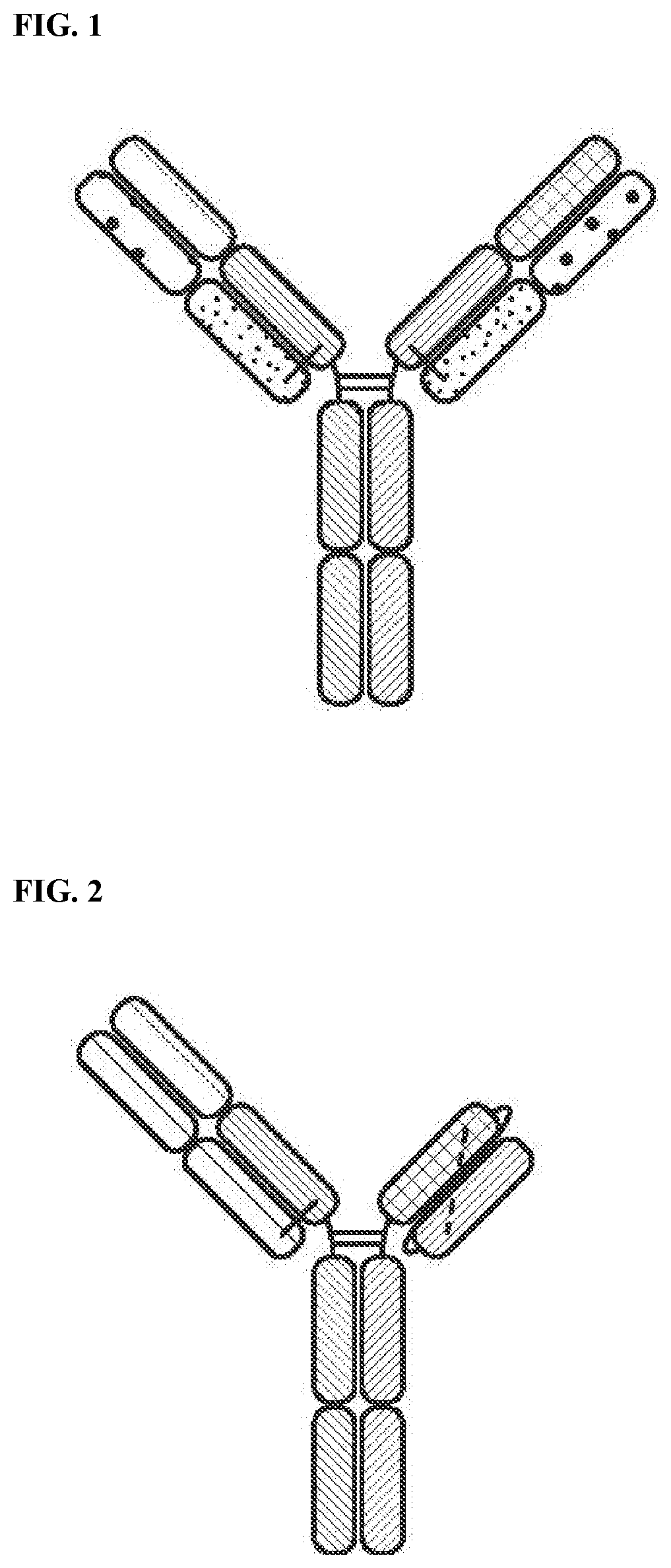
[0035] FIG. 1 is a representation of a multi-specific binding protein that contains an NKG2D-binding domain (right arm), a tumor associated antigen-binding domain (left arm) and an Fc domain or a portion thereof that binds to CD16.
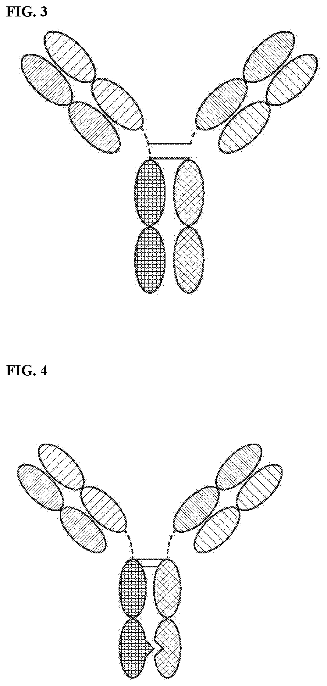
[0036] FIG. 2 is a representation of a multi-specific binding protein that contains an NKG2D-binding domain in a scFv format (right arm), a tumor associated antigen-binding domain (left arm) and an Fc domain or a portion thereof that binds to CD16.
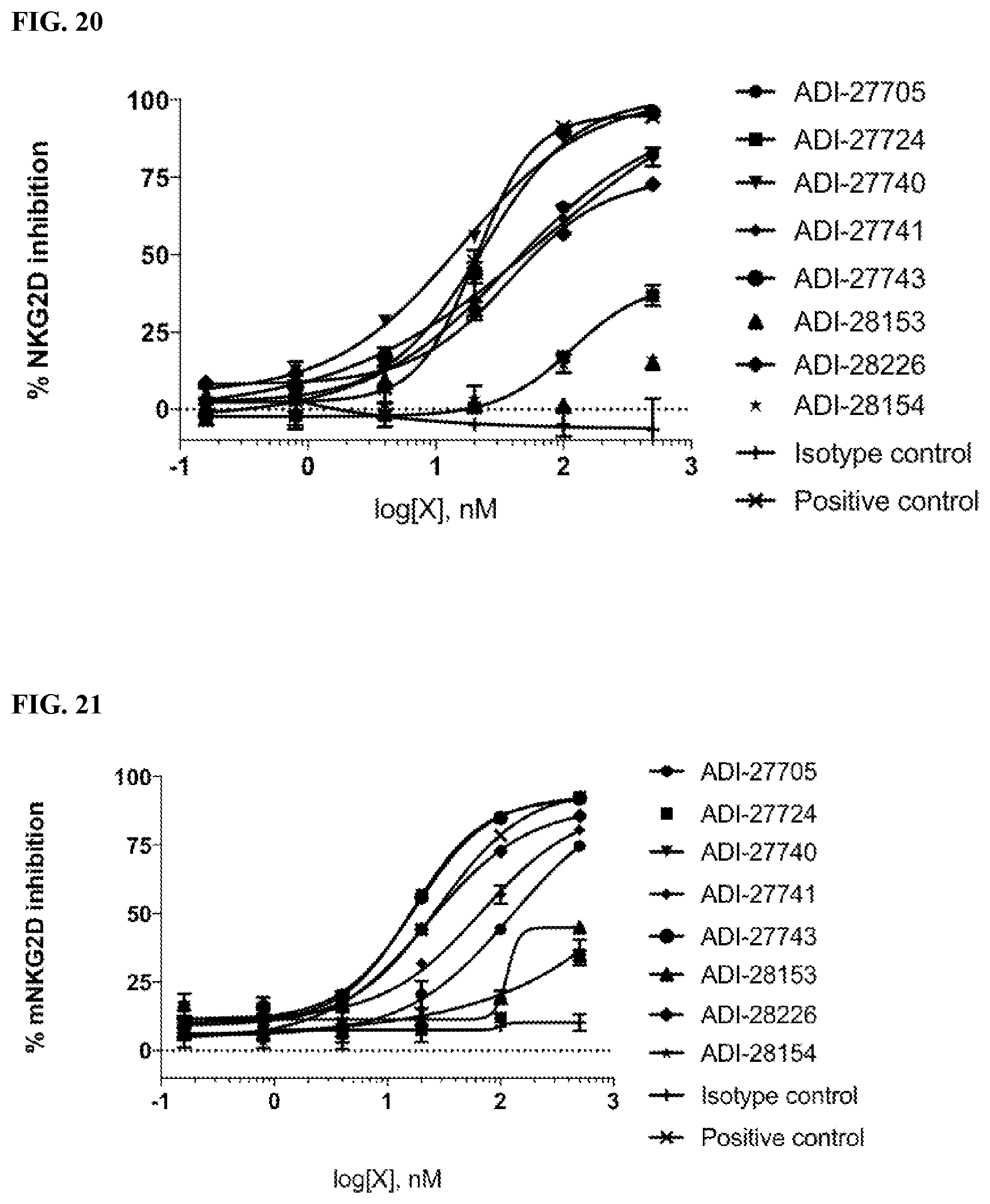
[0037] FIG. 3 is a representation of a TriNKET in the Triomab form, which is a trifunctional, bispecific antibody that maintains an IgG-like shape. This chimera consists of two half antibodies, each with one light and one heavy chain, that originate from two parental antibodies. Triomab form is an heterodimeric construct containing 1/2 of rat antibody and 1/2 of mouse antibody.
[0038] FIG. 4 is a representation of a TriNKET in the KiH Common Light Chain (LC) form, which involves the knobs-into-holes (KIHs) technology. KiH is a heterodimer containing 2 Fabs binding to target 1 and 2, and an F.sub.C stabilized by heterodimerization mutations. TriNKET in the KiH format may be an heterodimeric construct with 2 fabs binding to target 1 and target 2, containing 2 different heavy chains and a common light chain that pairs with both HC.
[0039] FIG. 5 is a representation of a TriNKET in the dual-variable domain immunoglobulin (DVD-Ig.TM.) form, which combines the target binding domains of two monoclonal antibodies via flexible naturally occurring linkers, and yields a tetravalent IgG-like molecule. DVD-Ig.TM. is an homodimeric construct where variable domain targeting antigen 2 is fused to the N terminus of variable domain of Fab targeting antigen 1 Construct contains normal Fc.
[0040] FIG. 6 is a representation of a TriNKET in the Orthogonal Fab interface (Ortho-Fab) form, which is an heterodimeric construct that contains 2 Fabs binding to target1 and target2 fused to Fc. LC-HC pairing is ensured by orthogonal interface. Heterodimerization is ensured by mutations in the F.sub.C.
[0041] FIG. 7 is a representation of a TrinKET in the 2 in 1Ig format.
[0042] FIG. 8 is a representation of a TriNKET in the ES form, which is an heterodimeric construct containing 2 different Fabs binding to target 1 and target 2 fused to the F.sub.C. Heterodimerization is ensured by electrostatic steering mutations in the Fc.
[0043] FIG. 9 is a representation of a TriNKET in the Fab Arm Exchange form: antibodies that exchange Fab arms by swapping a heavy chain and attached light chain (half-molecule) with a heavy-light chain pair from another molecule, resulting in bispecific antibodies. Fab Arm Exchange form (cFae) is a heterodimer containing 2 Fabs binding to target 1 and 2, and an F.sub.C stabilized by heterodimerization mutations.
[0044] FIG. 10 is a representation of a TriNKET in the SEED Body form, which is an heterodimer containing 2 Fabs binding to target 1 and 2, and an F.sub.C stabilized by heterodimerization mutations.
[0045] FIG. 11 is a representation of a TriNKET in the LuZ-Y form, in which leucine zipper is used to induce heterodimerization of two different HCs. LuZ-Y form is a heterodimer containing 2 different scFabs binding to target 1 and 2, fused to F.sub.C. Heterodimerization is ensured through leucine zipper motifs fused to C-terminus of F.sub.C.
[0046] FIG. 12 is a representation of a TriNKET in the Cov-X-Body form.
[0047] FIGS. 13A-13B are representations of TriNKETs in the .kappa..lamda.-Body forms, which are an heterodimeric constructs with 2 different Fabs fused to Fc stabilized by heterodimerization mutations: Fab1 targeting antigen 1 contains kappa LC, while second Fab targeting antigen 2 contains lambda LC. FIG. 13A is an exemplary representation of one form of a .kappa..lamda.-Body; FIG. 13B is an exemplary representation of another .kappa..lamda.-Body.
[0048] FIG. 14 is a graph demonstrating the binding affinity of NKG2D-binding domains (listed as clones) to human recombinant NKG2D in an ELISA assay.
[0049] FIG. 15 is a graph demonstrating the binding affinity of NKG2D-binding domains (listed as clones) to cynomolgus recombinant NKG2D in an ELISA assay.
[0050] FIG. 16 is a graph demonstrating the binding affinity of NKG2D-binding domains (listed as clones) to mouse recombinant NKG2D in an ELISA assay.
[0051] FIG. 17 is a graph demonstrating the binding of NKG2D-binding domains (listed as clones) to EL4 cells expressing human NKG2D by flow cytometry showing mean fluorescence intensity (MFI) fold over background.
[0052] FIG. 18 is a graph demonstrating the binding of NKG2D-binding domains (listed as clones) to EL4 cells expressing mouse NKG2D by flow cytometry showing mean fluorescence intensity (MFI) fold over background.
[0053] FIG. 19 is a graph demonstrating specific binding affinity of NKG2D-binding domains (listed as clones) to recombinant human NKG2D-Fc by competing with natural ligand ULBP-6.
[0054] FIG. 20 is a graph demonstrating specific binding affinity of NKG2D-binding domains (listed as clones) to recombinant human NKG2D-Fc by competing with natural ligand MICA.
[0055] FIG. 21 is a graph demonstrating specific binding affinity of NKG2D-binding domains (listed as clones) to recombinant mouse NKG2D-Fc by competing with natural ligand Rae-1 delta.
[0056] FIG. 22 is a graph showing activation of human NKG2D by NKG2D-binding domains (listed as clones) by quantifying the percentage of TNF-alpha positive cells which express human NKG2D-CD3 zeta fusion proteins.
[0057] FIG. 23 is a graph showing activation of mouse NKG2D by NKG2D-binding domains (listed as clones) by quantifying the percentage of TNF-alpha positive cells which express mouse NKG2D-CD3 zeta fusion proteins.
[0058] FIG. 24 is a graph showing activation of human NK cells by NKG2D-binding domains (listed as clones).
[0059] FIG. 25 is a graph showing activation of human NK cells by NKG2D-binding domains (listed as clones).
[0060] FIG. 26 is a graph showing activation of mouse NK cells by NKG2D-binding domains (listed as clones).
[0061] FIG. 27 is a graph showing activation of mouse NK cells by NKG2D-binding domains (listed as clones).
[0062] FIG. 28 is a graph showing the cytotoxic effect of NKG2D-binding domains (listed as clones) on tumor cells.
[0063] FIG. 29 is a graph showing the melting temperature of NKG2D-binding domains (listed as clones) measured by differential scanning fluorimetry.
[0064] FIG. 30 is a graph showing enhanced activation of human NK cells by multi-specific binding proteins.
[0065] FIG. 31 is a graph showing multi-specific binding proteins induced higher levels of cytotoxicity towards tumor target cells by human NK cells.
[0066] FIG. 32 is a graph showing multi-specific binding proteins induced higher levels of cytotoxicity towards tumor target cells by human NK cells.
[0067] FIG. 33 is a graph showing multi-specific binding proteins induced higher levels of cytotoxicity towards tumor target cells by human NK cells.
[0068] FIG. 34 is a graph showing multi-specific binding proteins induced higher levels of cytotoxicity towards tumor target cells by human NK cells.
[0069] FIG. 35 is a graph showing multi-specific binding proteins induced higher levels of cytotoxicity towards tumor target cells by mouse NK cells.
[0070] FIG. 36 is a graph showing multi-specific binding proteins induced higher levels of cytotoxicity towards tumor target cells by mouse NK cells.
[0071] FIG. 37 is a binding profile of CD33-targeting TriNKETs to NKG2D expressed on EL4 cells. FIG. 37 shows binding of the two TriNKETs when a CD33-binding domain is used as the second targeting arm.
[0072] FIG. 38 is a binding profile of HER2-targeting TriNKETs to NKG2D expressed on EL4 cells. FIG. 38 shows the same two NKG2D-binding domains now paired with a HER2 second targeting arm.
[0073] FIG. 39 is a binding profile of BCMA-targeting TriNKETs to NKG2D expressed on EL4 cells.
[0074] FIG. 40 is a histogram of CD20-targeting TriNKETs that bind to NKG2D expressed on EL4 cells. Unstained EL4 cells were used a negative control for fluorescence signal. Unstained: filled; CD20-TriNKET-F04: solid line; CD20-TriNKET-C26: dashed line.
[0075] FIG. 41 is a binding profile of CD33-targeting TriNKETs to CD33 expressed on MV4-11 human AML cells.
[0076] FIG. 42 is a binding profile of HER2-targeting TriNKETs to HER2 expressed on human 786-O renal cell carcinoma cells.
[0077] FIG. 43 is a binding profile of BCMA-targeting TriNKETs to BCMA expressed on MM.1S human myeloma cells.
[0078] FIG. 44 is a histogram of CD20-targeting TriNKETs that bind to CD20 expressed on Raji human lymphoma cells. Unstained cells were used a negative control for fluorescence signal. Unstained: filled; CD20-TriNKET-F04: solid line; CD20-TriNKET-C26: dashed line.
[0079] FIGS. 45A-45B are bar graphs of synergistic activation of NK cells using CD16 and NKG2D. FIG. 45A demonstrates levels of CD107a; FIG. 45B demonstrates levels of IFN.gamma.; FIG. 45C demonstrates levels of CD107a. Graphs indicate the mean (n=2).+-.SD. Data are representative of five independent experiments using five different healthy donors.
[0080] FIG. 46 is a bar graph showing activation of NK cells using TriNKETs targeting NKG2D and CD16. Antibodies tested were of human IgG1 isotypes. Graphs indicate the mean (n=2).+-.SD.
[0081] FIGS. 47A-47C are bar graphs demonstrating that TriNKETs and trastuzumab were able to activate primary human NK cells in co-culture with HER2-positive human tumor cells, indicated by an increase in CD107a degranulation and IFN.gamma. cytokine production. Compared to the monoclonal antibody trastuzumab, both TriNKETs showed superior activation of human NK cells with a variety of human HER2 cancer cells. FIG. 47A shows that human NK cells are activated by TriNKETs when cultured with SkBr-3 cells. FIG. 47B shows that human NK cells are activated by TriNKETs when cultured with Colo201 cells. FIG. 47C shows that human NK cell are activated by TriNKETs when cultured with HCC1954 cells.
[0082] FIGS. 48A-48B are line graphs demonstrating TriNKET-mediated activation of rested or IL-2-activated human NK cells in co-culture with the CD33-expressing human AML cell line MV4-11. FIG. 48A shows TriNKET-mediated activation of resting human NK cells. FIG. 48B shows TriNKET-mediated activation of IL-2-activated human NK cells from the same donor.
[0083] FIGS. 49A-49B are bar graphs demonstrating TriNKET enhancement of cytotoxic activity using IL-2-activated and rested human NK cells. FIG. 49A shows percent specific lysis of SkBr-3 tumor cells by rested human NK cells. FIG. 49B shows percent specific lysis of SkBr-3 tumor cells by IL-2-activated human NK cells.
[0084] FIGS. 50A-50B are bar graphs demonstrating TriNKETs provide the greatest advantage against HER2 medium and low cancers compared to trastuzumab. FIG. 50A shows activated human NK cell killing of HER2 high-SkBr-3 tumor cells. FIG. 50B shows human NK cell killing of HER2 low-786-O tumor cells. TriNKETs provide a greater advantage compared to trastuzumab against cancer cells with low HER2 expression.
[0085] FIGS. 51A-51C are histograms showing that the expression of the high-affinity FcR.gamma.I (CD64) on three human AML cells lines, Molm-13 cell line (FIG. 51A), Mv4-11 cell line (FIG. 51B), and THP-1 cell line (FIG. 51C).
[0086] FIGS. 52A-52B are line graphs of monoclonal antibody or TriNKET mediated activation of human NK cells in co-culture with either Molm-13 (FIG. 52B) or THP-1 (FIG. 52A) cells.
[0087] FIGS. 53A-53C are line graphs of human NK cytotoxicity assays using the three human AML cell lines as targets. FIG. 53A shows that Mv4-11 cells, which express CD64, but at a lower level than THP-1, showed reduced efficacy with the monoclonal anti-CD33. FIG. 53B demonstrates that a monoclonal antibody against CD33 shows good efficacy against Molm-13 cells, which do not express CD64. FIG. 53C demonstrates that THP-1 cells showed no effect with monoclonal anti-CD33 alone. The identities of the line graphs noted in FIG. 53C are also applicable to the line graphs in FIGS. 53A-53B.
[0088] FIGS. 54A & 54B are bar graphs showing B cells from a health donor are sensitive to TriNKET-mediated lysis.
[0089] FIGS. 54C & 54D are bar graphs showing myeloid cells are resistant to TriNKET-mediated lysis.
[0090] FIG. 55 are line graphs of TriNKETs-mediated hPBMC killing of SkBr-3 tumor cells in long-term co-cultures.
[0091] FIG. 56 is a flowchart of study design of RMA/S-HER2 subcutaneous SC2.2 efficacy.
[0092] FIG. 57 are line graphs showing that SC2.2 has no effect on subcutaneous RMA/S-HER2 tumor growth.
[0093] FIGS. 58A-58B are graphs showing in vitro binding by mcFAE-C26.99 TriNKET. 60 .mu.g/mL of indicated antibodies with four-fold dilutions were added to 2.times.10.sup.5 B16F10 tumor cells (FIG. 58A) or EL4-mNKG2D cells (FIG. 58B). Binding was assessed using a goat anti-mouse PE secondary antibody followed by flow cytometric analysis.
[0094] FIG. 59 is a graph showing increased NK cytotoxicity mediated by mcFAE-C26.99 TriNKET.
[0095] FIGS. 60A-60B show the anti-tumor efficacy of mcFAE-C26.99 TriNKET in B16F10 s.c. models. Mice were treated intraperitoneally with (FIG. 60A) isotype control mouse IgG2a monoclonal antibody C1.18.4 and mouse anti-Tyrp-1 monoclonal antibody or (FIG. 60B) isotype control mouse IgG2a monoclonal antibody C1.18.4 and mcFAE-C26.99 TriNKET, injected at a dose of 150 .mu.g (days 6, 8, 10, 12, 14, 16, and 21). Tumor growth was assessed for 28 days. Graphs show tumor growth curves of individual mice.
[0096] FIGS. 61A-61B show anti-tumor efficacy of mcFAE-C26.99 TriNKET in B16F10 i.v. models. FIG. 61A represents tumor burden when antibodies were administered at a 150-.mu.g dose (days 4, 6, 8, 11, 13, 15). FIG. 61B represents tumor burden when antibodies were administered at a 150-.mu.g dose (days 7, 9, 11, 13, 15). 18 days after tumor challenge, mice were euthanized and surface lung metastases were scored.
[0097] FIG. 62 is bar graph showing that human NK cells are activated by TriNKETs when cultured with CD20+ Raji cells.
[0098] FIG. 63 is a bar graph showing that human NK activation in culture with BCMA positive MM.1S human myeloma cells.
[0099] FIG. 64 is a graph showing that TriNKETs enhance human NK cell lysis of KMS12-PE myeloma cells.
[0100] FIG. 65 is a graph showing that BCMA targeting TriNKETs with different NKG2D-binding domains enhance human NK cell lysis of KMS12-PE myeloma cells.
[0101] FIGS. 66A-66C are line graphs showing effects of combination therapy using mcFAE-C26.99 TriNKET and anti-PD-1 antibodies in B16F10 s.c. models. FIG. 66A are line graphs showing tumor size (mm.sup.3) in mice treated intraperitoneally with isotype controls mouse IgG2a monoclonal antibody C1.18.4 with rat IgG2a monoclonal antibody 2A3, or with mcFAE-C26.99. FIG. 66B are line graphs showing tumor size (mm.sup.3) in mice treated intraperitoneally with isotype controls or anti-PD-1 monoclonal antibody clone RPM1-14. FIG. 66C are line graphs showing tumor size (mm.sup.3) in mice treated intraperitoneally with combination of mcFAE-C26.99 and anti-PD-1 monoclonal antibody. Tumor growth was assessed for 30 days. Graphs show tumor growth curves of individual mice.
[0102] FIGS. 67A-67C are line graphs showing effects of combination therapy using mcFAE-C26.99 TriNKET and recombinant human IL-2 in B16F10 s.c. models. FIG. 67A are line graphs showing tumor size (mm.sup.3) in mice treated intraperitoneally with isotype control mouse IgG2a monoclonal antibody C1.18.4 or with mcFAE-C26.99. FIG. 67B are line graphs showing tumor size (mm.sup.3) in mice treated intraperitoneally with isotype control or with IL-2. FIG. 67C are line graphs showing tumor size (mm.sup.3) in mice treated intraperitoneally with a combination of mcFAE-C26.99 and IL-2. Tumor growth was assessed for 40 days with 3 mice from the combination group remaining tumor-free. Graphs show tumor growth curves of individual mice.
[0103] FIG. 68 is a line graph showing that tri-specific binding in one molecule is important for maximal NK cell activity.
[0104] FIG. 69 is an Oasc-Fab heterodimeric construct that includes Fab binding to target 1 and scFab binding to target 2 fused to F.sub.C. Heterodimerization is ensured by mutations in the F.sub.C.
[0105] FIG. 70 is a DuetMab, which is an heterodimeric construct containing 2 different Fabs binding to antigen 1 and 2 and F.sub.C stabilized by heterodimerization mutations. Fab 1 and 2 contain differential S-S bridges that ensure correct LC and HC pairing.
[0106] FIG. 71 is a CrossmAb, which is an heterodimeric construct with 2 different Fabs binding to target 1 and 2 fused to Fc stabilized by heterodimerization. CL and CH1 domains and VH and VL domains are switched, e.g., CH1 is fused in-line with VL, while CL is fused in-line with VH.
[0107] FIG. 72 is a Fit-Ig, which is an homodimeric constructs where Fab binding to antigen 2 is fused to the N terminus of HC of Fab that binds to antigen 1. The construct contains wild-type F.sub.C.
[0108] FIGS. 73A-73D are line graphs showing the percentage lysis of 786-O target cells by rested NK cells (FIG. 73A) or NK cells activated by IL-2 (FIG. 73B), IL-12 (FIG. 73C), or IL-15 (FIG. 73D) in the presence of trastuzumab or a HER2-TriNKET.
[0109] FIGS. 74A-74D are line graphs showing the percentage lysis of Molm-13 target cells by rested NK cells (FIG. 74A) or NK cells activated by IL-2 (FIG. 74B), IL-12 (FIG. 74C), or IL-15 (FIG. 74D) in the presence of lintuzumab, a proprietary anti-CD33 monoclonal antibody, or a CD33-TriNKET.
[0110] FIGS. 75A-75D are line graphs showing the percentage lysis of KMS12-PE target cells by rested NK cells (FIG. 75A) or NK cells activated by IL-2 (FIG. 75B), IL-12 (FIG. 75C), or IL-15 (FIG. 75D) in the presence of BCMA monoclonal antibody EM-901 or a BCMA-TriNKET.
[0111] FIGS. 76A-76D are line graphs showing the percentage lysis of KMS12-PE target cells by rested NK cells (FIG. 76A) or NK cells activated by pomalidomide (FIG. 76B), IL-2 (FIG. 76C), or a combination of IL-2 and pomalidomide (FIG. 76D) in the presence of BCMA monoclonal antibody EM-901 or a BCMA-TriNKET.
[0112] FIGS. 77A-77B are graphs showing flow cytometry analysis of purified CD8.sup.+ T cells and HCC1954 target cells.
[0113] FIGS. 78A-78B are line graphs showing growth of HCC1954 target cells in the presence of CD8.sup.+ T cells and a HER2-TriNKET, pembrolizumab ("Keytruda"), or a combination thereof. The CD8.sup.+ T cells used in FIG. 78A and FIG. 78B were isolated from different donors.
[0114] FIG. 79 is a line graph showing growth of Skbr-3 target cells in the presence of PBMCs and a HER2-TriNKET, a TLR agonist, or a combination thereof.
[0115] FIGS. 80A-80C are line graphs showing tumor growth curves of individual mice inoculated with Bl6F10 tumor cells and treated with 7.5 mg/kg mcFAE-C26.99 TriNKET or 7.5 mg/kg isotype control mouse IgG2a monoclonal antibody C1.18.4 (FIG. 80A), 1 .mu.g recombinant murine IL-12 (rmIL-12) or 7.5 mg/kg isotype control mouse IgG2a monoclonal antibody C1.18.4 (FIG. 80B), or a combination of 7.5 mg/kg mcFAE-C26.99 TriNKET and 1 .mu.g rmlL-12 (FIG. 80C). In FIGS. 80B and 80C, the lower panels represent plotting on a small scale of the y-axis.
[0116] FIG. 81 is a Kaplan-Meier curve showing the percentage of animals that survived after the Bl6F10 tumor cell inoculation and the treatments of 7.5 mg/kg isotype control mouse IgG2a monoclonal antibody C1.18.4, 7.5 mg/kg mcFAE-C26.99 TriNKET, 1 .mu.g rmlL-12, or a combination of 7.5 mg/kg mcFAE-C26.99 TriNKET and 1 .mu.g rmlL-12.
DETAILED DESCRIPTION
[0117] The invention provides multi-specific binding proteins that bind a tumor-associated antigen on a cancer cell and the NKG2D receptor and CD16 receptor on natural killer cells to activate the natural killer cell, pharmaceutical compositions comprising such multi-specific binding proteins, and therapeutic methods using such multi-specific proteins and pharmaceutical compositions, including for the treatment of cancer. Various aspects of the invention are set forth below in sections; however, aspects of the invention described in one particular section are not to be limited to any particular section.
[0118] To facilitate an understanding of the present invention, a number of terms and phrases are defined below.
[0119] The terms "a" and "an" as used herein mean "one or more" and include the plural unless the context is inappropriate.
[0120] As used herein, the term "antigen-binding site" refers to the part of the immunoglobulin molecule that participates in antigen binding. In human antibodies, the antigen binding site is formed by amino acid residues of the N-terminal variable ("V") regions of the heavy ("H") and light ("L") chains. Three highly divergent stretches within the V regions of the heavy and light chains are referred to as "hypervariable regions" which are interposed between more conserved flanking stretches known as "framework regions," or "FRs". Thus the term "FR" refers to amino acid sequences which are naturally found between and adjacent to hypervariable regions in immunoglobulins. In a human antibody molecule, the three hypervariable regions of a light chain and the three hypervariable regions of a heavy chain are disposed relative to each other in three dimensional space to form an antigen-binding surface. The antigen-binding surface is complementary to the three-dimensional surface of a bound antigen, and the three hypervariable regions of each of the heavy and light chains are referred to as "complementarity-determining regions," or "CDRs." In certain animals, such as camels and cartilaginous fish, the antigen-binding site is formed by a single antibody chain providing a "single domain antibody." Antigen-binding sites can exist in an intact antibody, in an antigen-binding fragment of an antibody that retains the antigen-binding surface, or in a recombinant polypeptide such as an scFv, using a peptide linker to connect the heavy chain variable domain to the light chain variable domain in a single polypeptide.
[0121] The term "tumor associated antigen" as used herein means any antigen including but not limited to a protein, glycoprotein, ganglioside, carbohydrate, lipid that is associated with cancer. Such antigen can be expressed on malignant cells or in the tumor microenvironment such as on tumor-associated blood vessels, extracellular matrix, mesenchymal stroma, or immune infiltrates.
[0122] As used herein, the terms "subject" and "patient" refer to an organism to be treated by the methods and compositions described herein. Such organisms preferably include, but are not limited to, mammals (e.g., murines, simians, equines, bovines, porcines, canines, felines, and the like), and more preferably include humans.
[0123] As used herein, the term "effective amount" refers to the amount of a compound (e.g., a compound of the present invention) sufficient to effect beneficial or desired results. An effective amount can be administered in one or more administrations, applications or dosages and is not intended to be limited to a particular formulation or administration route. As used herein, the term "treating" includes any effect, e.g., lessening, reducing, modulating, ameliorating or eliminating, that results in the improvement of the condition, disease, disorder, and the like, or ameliorating a symptom thereof.
[0124] As used herein, the term "pharmaceutical composition" refers to the combination of an active agent with a carrier, inert or active, making the composition especially suitable for diagnostic or therapeutic use in vivo or ex vivo.
[0125] As used herein, the term "pharmaceutically acceptable carrier" refers to any of the standard pharmaceutical carriers, such as a phosphate buffered saline solution, water, emulsions (e.g., such as an oil/water or water/oil emulsions), and various types of wetting agents. The compositions also can include stabilizers and preservatives. For examples of carriers, stabilizers and adjuvants, see e.g., Martin, Remington's Pharmaceutical Sciences, 15th Ed., Mack Publ. Co., Easton, Pa. [1975].
[0126] As used herein, the term "pharmaceutically acceptable salt" refers to any pharmaceutically acceptable salt (e.g., acid or base) of a compound of the present invention which, upon administration to a subject, is capable of providing a compound of this invention or an active metabolite or residue thereof. As is known to those of skill in the art, "salts" of the compounds of the present invention may be derived from inorganic or organic acids and bases. Exemplary acids include, but are not limited to, hydrochloric, hydrobromic, sulfuric, nitric, perchloric, fumaric, maleic, phosphoric, glycolic, lactic, salicylic, succinic, toluene-p-sulfonic, tartaric, acetic, citric, methanesulfonic, ethanesulfonic, formic, benzoic, malonic, naphthalene-2-sulfonic, benzenesulfonic acid, and the like. Other acids, such as oxalic, while not in themselves pharmaceutically acceptable, may be employed in the preparation of salts useful as intermediates in obtaining the compounds of the invention and their pharmaceutically acceptable acid addition salts.
[0127] Exemplary bases include, but are not limited to, alkali metal (e.g., sodium) hydroxides, alkaline earth metal (e.g., magnesium) hydroxides, ammonia, and compounds of formula NW.sub.4.sup.+, wherein W is C.sub.1-4 alkyl, and the like.
[0128] Exemplary salts include, but are not limited to: acetate, adipate, alginate, aspartate, benzoate, benzenesulfonate, bisulfate, butyrate, citrate, camphorate, camphorsulfonate, cyclopentanepropionate, digluconate, dodecylsulfate, ethanesulfonate, fumarate, flucoheptanoate, glycerophosphate, hemisulfate, heptanoate, hexanoate, hydrochloride, hydrobromide, hydroiodide, 2-hydroxyethanesulfonate, lactate, maleate, methanesulfonate, 2-naphthalenesulfonate, nicotinate, oxalate, palmoate, pectinate, persulfate, phenylpropionate, picrate, pivalate, propionate, succinate, tartrate, thiocyanate, tosylate, undecanoate, and the like. Other examples of salts include anions of the compounds of the present invention compounded with a suitable cation such as Na.sup.+, NH.sub.4.sup.+, and NW.sub.4.sup.+ (wherein W is a C.sub.1-4 alkyl group), and the like.
[0129] For therapeutic use, salts of the compounds of the present invention are contemplated as being pharmaceutically acceptable. However, salts of acids and bases that are non-pharmaceutically acceptable may also find use, for example, in the preparation or purification of a pharmaceutically acceptable compound.
[0130] Throughout the description, where compositions are described as having, including, or comprising specific components, or where processes and methods are described as having, including, or comprising specific steps, it is contemplated that, additionally, there are compositions of the present invention that consist essentially of, or consist of, the recited components, and that there are processes and methods according to the present invention that consist essentially of, or consist of, the recited processing steps.
[0131] As a general matter, compositions specifying a percentage are by weight unless otherwise specified. Further, if a variable is not accompanied by a definition, then the previous definition of the variable controls.
I. Proteins
[0132] The invention provides multi-specific binding proteins that bind a tumor-associated antigen on a cancer cell and the NKG2D receptor and CD16 receptor on natural killer cells to activate the natural killer cell. The multi-specific binding proteins are useful in the pharmaceutical compositions and therapeutic methods described herein. Binding of the multi-specific binding protein to the NKG2D receptor and CD16 receptor on natural killer cell enhances the activity of the natural killer cell toward destruction of a cancer cell. Binding of the multi-specific binding protein to a tumor-associated antigen on a cancer cell brings the cancer cell into proximity to the natural killer cell, which facilitates direct and indirect destruction of the cancer cell by the natural killer cell. Further description of exemplary multi-specific binding proteins are provided below.
[0133] The first component of the multi-specific binding proteins binds to NKG2D receptor-expressing cells, which can include but are not limited to NK cells, .gamma..delta. T cells and CD8.sup.+.alpha..beta. T cells. In some embodiments, upon NKG2D-binding, the multi-specific binding proteins can block natural ligands, such as ULBP6 and MICA, from binding to NKG2D.
[0134] The second component of the multi-specific binding proteins binds to one or more tumor-associated antigens, which can include, but are not limited to HER2, CD20, CD33, BCMA, EpCAM, CD2, CD19, CD30, CD38, CD40, CD52, CD70, EGFR/ERBB1, IGF1R, HER3/ERBB3, HER4/ERBB4, MUC1, cMET, SLAMF7, PSCA, MICA, MICB, TRAILR1, TRAILR2, MAGE-A3, B7.1, B7.2, CTLA4, and PD-L1.
[0135] The third component for the multi-specific binding proteins binds to cells expressing CD16, a Fc receptor on the surface of leukocytes including natural killer cells, macrophages, neutrophils, eosinophils, mast cells, and follicular dendritic cells.
[0136] The multi-specific binding proteins can take several formats as shown in but not limited to the examples below. One format is a heterodimeric, multi-specific antibody that includes a first immunoglobulin heavy chain, a second immunoglobulin heavy chain and an immunoglobulin light chain. The first immunoglobulin heavy chain includes a first Fc (hinge-CH2-CH3) domain, a first variable heavy chain domain and an optional first CH1 heavy chain domain. The immunoglobulin light chain includes a variable light chain domain and a constant light chain domain; together with the first immunoglobulin heavy chain, the immunoglobulin light chain forms an antigen-binding site that binds NKG2D. The second immunoglobulin heavy chain comprises a second Fc (hinge-CH2-CH3) domain, a second variable heavy chain domain and a second optional CH1 heavy chain domain that may pair with an immunoglobulin light chain identical to the one that pairs with the first immunoglobulin heavy chain, except that when immunoglobulin light chain is paired with the second immunoglobulin heavy chain, the resulting antigen binding site binds to a tumor antigen. The first Fc domain and second Fc domain together are able to bind to CD16 (FIG. 1).
[0137] Another exemplary format involves a heterodimeric, multi-specific antibody which includes a first immunoglobulin heavy chain, an immunoglobulin light chain and a second immunoglobulin heavy chain. The first immunoglobulin heavy chain includes a first Fc (hinge-CH2-CH3) domain fused via either a linker or an antibody hinge to a single chain Fv (scFv) that binds NKG2D. A variety of linkers could be used for linking the scFv to the first Fc domain or within the scFv itself. In addition, the scFv can incorporate mutations that enable the formation of a disulfide bond to stabilize the overall scFv structure. The scFv can also incorporate mutations to modify the isoelectric point of the overall first immunoglobulin heavy chain and/or to enable more facile downstream purification. The second immunoglobulin heavy chain includes a second Fc (hinge-CH2-CH3) domain and a second variable heavy chain domain and a second optional CH1 heavy chain domain. The immunoglobulin light chain includes a variable light chain domain and a constant light chain domain. The second immunoglobulin heavy chain pairs with the immunoglobulin light chain and binds to a tumor antigen. The first Fc domain and the second Fc domain together are able to bind to CD16 (FIG. 2).
[0138] An alternative format of the heterodimeric multi-specific proteins includes a first immunoglobulin heavy chain, a second immunoglobulin heavy chain, a first immunoglobulin light chain and a second immunoglobulin light chain. The first immunoglobulin heavy chain includes a first Fc (hinge-CH2-CH3) domain, a first variable heavy chain domain and an optional first CH1 heavy chain domain. The first immunoglobulin light chain includes a variable light chain domain and a constant light chain domain. Together with the first immunoglobulin heavy chain, the first immunoglobulin light chain forms an antigen-binding site that binds a tumor antigen. The second immunoglobulin heavy chain comprises a second Fc (hinge-CH2-CH3) domain, a second variable heavy chain domain and a second optional CH1 heavy chain domain. The second immunoglobulin light chain includes a variable light chain domain and a constant light chain domain. Together with the second immunoglobulin heavy chain, the immunoglobulin light chain forms an antigen-binding site that binds to the same tumor antigen. The second immunoglobulin heavy chain may pair with an immunoglobulin light chain, which may be identical to the immunoglobulin light chain that pairs with the first immunoglobulin heavy chain, except that when immunoglobulin light chain is paired with the second immunoglobulin heavy chain, the resulting antigen binding site is a second antigen-binding site that binds to a tumor antigen. In certain embodiments, the first Fc domain and second Fc domain together are able to bind to CD16 (FIG. 1).
[0139] One or more additional binding motifs may be fused to the C-terminus of the constant region CH3 domain, optionally via a linker sequence. In certain embodiments, the antigen-binding site could be a single-chain or disulfide-stabilized variable region (ScFv) or could form a tetravalent or trivalent molecule.
[0140] In some embodiments, the multi-specific binding protein is in the Triomab form, which is a trifunctional, bispecific antibody that maintains an IgG-like shape. This chimera consists of two half antibodies, each with one light and one heavy chain, that originate from two parental antibodies.
[0141] In some embodiments, the multi-specific binding protein is the KiH Common Light Chain (LC) form, which involves the knobs-into-holes (KIHs) technology. The KIH involves engineering C.sub.H3 domains to create either a "knob" or a "hole" in each heavy chain to promote heterodimerization. The concept behind the "Knobs-into-Holes (KiH)" Fc technology was to introduce a "knob" in one CH3 domain (CH3A) by substitution of a small residue with a bulky one (i.e., T366W.sub.CH3A in EU numbering). To accommodate the "knob," a complementary "hole" surface was created on the other CH3 domain (CH3B) by replacing the closest neighboring residues to the knob with smaller ones (i.e., T366S/L368A/Y407V.sub.CH3B). The "hole" mutation was optimized by structured-guided phage library screening (Atwell S, Ridgway J B, Wells J A, Carter P. Stable heterodimers from remodeling the domain interface of a homodimer using a phage display library. J Mol Biol (1997) 270(1):26-35). X-ray crystal structures of KiH Fc variants (Elliott J M, Ultsch M, Lee J, Tong R, Takeda K, Spiess C, et al., Antiparallel conformation of knob and hole aglycosylated half-antibody homodimers is mediated by a CH2-CH3 hydrophobic interaction. J Mol Biol (2014) 426(9):1947-57; Mimoto F, Kadono S, Katada H, Igawa T, Kamikawa T, Hattori K. Crystal structure of a novel asymmetrically engineered Fc variant with improved affinity for FcgammaRs. Mol Immunol (2014) 58(1):132-8) demonstrated that heterodimerization is thermodynamically favored by hydrophobic interactions driven by steric complementarity at the inter-CH3 domain core interface, whereas the knob-knob and the hole-hole interfaces do not favor homodimerization owing to steric hindrance and disruption of the favorable interactions, respectively.
[0142] In some embodiments, the multi-specific binding protein is in the dual-variable domain immunoglobulin (DVD-Ig.TM.) form, which combines the target binding domains of two monoclonal antibodies via flexible naturally occurring linkers, and yields a tetravalent IgG--like molecule.
[0143] In some embodiments, the multi-specific binding protein is in the Orthogonal Fab interface (Ortho-Fab) form. In ortho-Fab IgG approach (Lewis S M, Wu X, Pustilnik A, Sereno A, Huang F, Rick H L, et al. Generation of bispecific IgG antibodies by structure-based design of an orthogonal Fab interface. Nat. Biotechnol. (2014) 32(2):191-8), structure-based regional design introduces complementary mutations at the LC and H.sub.VH-CH1 interface in only one Fab, without any changes being made to the other Fab.
[0144] In some embodiments, the multi-specific binding protein is in the 2 in 1Ig format. In some embodiments, the multi-specific binding protein is in the ES form, which is an heterodimeric construct containing 2 different Fabs binding to target 1 and target 2 fused to the F.sub.C. Heterodimerization is ensured by electrostatic steering mutations in the Fc. In some embodiments, the multi-specific binding protein is in the .kappa..lamda.-Body form, which is an heterodimeric constructs with 2 different Fabs fused to Fc stabilized by heterodimerization mutations: Fab 1 targeting antigen 1 contains kappa LC, while second Fab targeting antigen 2 contains lambda LC. FIG. 13A is an exemplary representation of one form of a .kappa..lamda.-Body; FIG. 13B is an exemplary representation of another .kappa..lamda.-Body.
[0145] In some embodiments, the multi-specific binding protein is in Fab Arm Exchange form (antibodies that exchange Fab arms by swapping a heavy chain and attached light chain (half-molecule) with a heavy-light chain pair from another molecule, which results in bispecific antibodies). In some embodiments, the multi-specific binding protein is in the SEED Body form (The strand-exchange engineered domain (SEED) platform was designed to generate asymmetric and bispecific antibody-like molecules, a capability that expands therapeutic applications of natural antibodies. This protein engineered platform is based on exchanging structurally related sequences of immunoglobulin within the conserved CH3 domains. The SEED design allows efficient generation of AG/GA heterodimers, while disfavoring homodimerization of AG and GA SEED CH3 domains. (Muda M. et al., Protein Eng. Des. Sel. (2011, 24(5):447-54)). In some embodiments, the multi-specific binding protein is in the LuZ-Y form, in which leucine zipper is used to induce heterodimerization of two different HCs. (Wranik, B J. et al., J. Biol. Chem. (2012), 287:43331-9).
[0146] In some embodiments, the multi-specific binding protein is in the Cov-X-Body form (In bispecific CovX-Bodies, two different peptides are joined together using a branched azetidinone linker and fused to the scaffold antibody under mild conditions in a site-specific manner. Whereas the pharmacophores are responsible for functional activities, the antibody scaffold imparts long half-life and Ig-like distribution. The pharmacophores can be chemically optimized or replaced with other pharmacophores to generate optimized or unique bispecific antibodies. (Doppalapudi V R et al., PNAS (2010), 107(52); 22611-22616).
[0147] In some embodiments, the multi-specific binding protein is in an Oasc-Fab heterodimeric form that includes Fab binding to target 1 and scFab binding to target 2 fused to Fc. Heterodimerization is ensured by mutations in the F.sub.C.
[0148] In some embodiments, the multi-specific binding protein is in a DuetMab form, which is an heterodimeric construct containing 2 different Fabs binding to antigen 1 and 2 and F.sub.C stabilized by heterodimerization mutations. Fab 1 and 2 contain differential S-S bridges that ensure correct LC and HC pairing.
[0149] In some embodiments, the multi-specific binding protein is in a CrossmAb form, which is an heterodimeric construct with 2 different Fabs binding to Target 1 and 2 fused to Fc stabilized by heterodimerization. CL and CH1 domains and VH and VL domains are switched, e.g., CH1 is fused in-line with VL, while CL is fused in-line with VH.
[0150] In some embodiments, the multi-specific binding protein is in a Fit-Ig form, which is an homodimeric constructs where Fab binding to antigen 2 is fused to the N terminus of HC of Fab that binds to antigen 1. The construct contains wild-type Fc.
[0151] Table 1 lists peptide sequences of heavy chain variable domains and light chain variable domains that, in combination, can bind to NKG2D. Unless otherwise indicated, the CDR sequences provided in Table 1 are determined under Kabat.
TABLE-US-00001 TABLE 1 Heavy chain variable region Light chain variable Clones amino acid sequence region amino acid sequence ADI-27705 QVQLQQWGAGLLKPSETLSLTCAV DIQMTQSPSTLSASVGDRVTITC YGGSFSGYYWSWIRQPPGKGLEWI RASQSISSWLAWYQQKPGKAP GEIDHSGSTNYNPSLKSRVTISVDT KLLIYKASSLESGVPSRFSGSGS SKNQFSLKLSSVTAADTAVYYCAR GTEFTLTISSLQPDDFATYYCQQ ARGPWSFDPWGQGTLVTVSS YNSYPITFGGGTKVEIK (SEQ ID NO: 1) (SEQ ID NO: 2) ADI-27724 QVQLQQWGAGLLKPSETLSLTCAV EIVLTQSPGTLSLSPGERATLSC YGGSFSGYYWSWIRQPPGKGLEWI RASQSVSSSYLAWYQQKPGQA GEIDHSGSTNYNPSLKSRVTISVDT PRLLIYGASSRATGIPDRFSGSGS SKNQFSLKLSSVTAADTAVYYCAR GTDFTLTISRLEPEDFAVYYCQQ ARGPWSFDPWGQGTLVTVSS YGSSPITFGGGTKVEIK (SEQ ID NO: 3) (SEQ ID NO: 4) ADI-27740 QVQLQQWGAGLLKPSETLSLTCAV DIQMTQSPSTLSASVGDRVTITC (A40) YGGSFSGYYWSWIRQPPGKGLEWI RASQSIGSWLAWYQQKPGKAP GEIDHSGSTNYNPSLKSRVTISVDT KLLIYKASSLESGVPSRFSGSGS SKNQFSLKLSSVTAADTAVYYCAR GTEFTLTISSLQPDDFATYYCQQ ARGPWSFDPWGQGTLVTVSS YHSFYTFGGGTKVEIK (SEQ ID NO: 5) (SEQ ID NO: 6) ADI-27741 QVQLQQWGAGLLKPSETLSLTCAV DIQMTQSPSTLSASVGDRVTITC YGGSFSGYYWSWIRQPPGKGLEWI RASQSIGSWLAWYQQKPGKAP GEIDHSGSTNYNPSLKSRVTISVDT KLLIYKASSLESGVPSRFSGSGS SKNQFSLKLSSVTAADTAVYYCAR GTEFTLTISSLQPDDFATYYCQQ ARGPWSFDPWGQGTLVTVSS SNSYYTFGGGTKVEIK (SEQ ID NO: 7) (SEQ ID NO: 8) ADI-27743 QVQLQQWGAGLLKPSETLSLTCAV DIQMTQSPSTLSASVGDRVTITC YGGSFSGYYWSWIRQPPGKGLEWI RASQSISSWLAWYQQKPGKAP GEIDHSGSTNYNPSLKSRVTISVDT KLLIYKASSLESGVPSRFSGSGS SKNQFSLKLSSVTAADTAVYYCAR GTEFTLTISSLQPDDFATYYCQQ ARGPWSFDPWGQGTLVTVSS YNSYPTFGGGTKVEIK (SEQ ID NO: 9) (SEQ ID NO: 10) ADI-28153 QVQLQQWGAGLLKPSETLSLTCAV ELQMTQSPSSLSASVGDRVTITC YGGSFSGYYWSWIRQPPGKGLEWI RTSQSISSYLNWYQQKPGQPPK GEIDHSGSTNYNPSLKSRVTISVDT LLIYWASTRESGVPDRFSGSGSG SKNQFSLKLSSVTAADTAVYYCAR TDFTLTISSLQPEDSATYYCQQS ARGPWGFDPWGQGTLVTVSS YDIPYTFGQGTKLEIK (SEQ ID NO: 11) (SEQ ID NO: 12) ADI-28226 QVQLQQWGAGLLKPSETLSLTCAV DIQMTQSPSTLSASVGDRVTITC (C26) YGGSFSGYYWSWIRQPPGKGLEWI RASQSISSWLAWYQQKPGKAP GEIDHSGSTNYNPSLKSRVTISVDT KLLIYKASSLESGVPSRFSGSGS SKNQFSLKLSSVTAADTAVYYCAR GTEFTLTISSLQPDDFATYYCQQ ARGPWSFDPWGQGTLVTVSS YGSFPITFGGGTKVEIK (SEQ ID NO: 13) (SEQ ID NO: 14) ADI-28154 QVQLQQWGAGLLKPSETLSLTCAV DIQMTQSPSTLSASVGDRVTITC YGGSFSGYYWSWIRQPPGKGLEWI RASQSISSWLAWYQQKPGKAP GEIDHSGSTNYNPSLKSRVTISVDT KLLIYKASSLESGVPSRFSGSGS SKNQFSLKLSSVTAADTAVYYCAR GTDFTLTISSLQPDDFATYYCQQ ARGPWSFDPWGQGTLVTVSS SKEVPWTFGQGTKVEIK (SEQ ID NO: 15) (SEQ ID NO: 16) ADI-29399 QVQLQQWGAGLLKPSETLSLTCAV DIQMTQSPSTLSASVGDRVTITC YGGSFSGYYWSWIRQPPGKGLEWI RASQSISSWLAWYQQKPGKAP GEIDHSGSTNYNPSLKSRVTISVDT KLLIYKASSLESGVPSRFSGSGS SKNQFSLKLSSVTAADTAVYYCAR GTEFTLTISSLQPDDFATYYCQQ ARGPWSFDPWGQGTLVTVSS YNSFPTFGGGTKVEIK (SEQ ID NO: 17) (SEQ ID NO: 18) ADI-29401 QVQLQQWGAGLLKPSETLSLTCAV DIQMTQSPSTLSASVGDRVTITC YGGSFSGYYWSWIRQPPGKGLEWI RASQSIGSWLAWYQQKPGKAP GEIDHSGSTNYNPSLKSRVTISVDT KLLIYKASSLESGVPSRFSGSGS SKNQFSLKLSSVTAADTAVYYCAR GTEFTLTISSLQPDDFATYYCQQ ARGPWSFDPWGQGTLVTVSS YDIYPTFGGGTKVEIK (SEQ ID NO: 19) (SEQ ID NO: 20) ADI-29403 QVQLQQWGAGLLKPSETLSLTCAV DIQMTQSPSTLSASVGDRVTITC YGGSFSGYYWSWIRQPPGKGLEWI RASQSISSWLAWYQQKPGKAP GEIDHSGSTNYNPSLKSRVTISVDT KLLIYKASSLESGVPSRFSGSGS SKNQFSLKLSSVTAADTAVYYCAR GTEFTLTISSLQPDDFATYYCQQ ARGPWSFDPWGQGTLVTVSS YDSYPTFGGGTKVEIK (SEQ ID NO: 21) (SEQ ID NO: 22) ADI-29405 QVQLQQWGAGLLKPSETLSLTCAV DIQMTQSPSTLSASVGDRVTITC YGGSFSGYYWSWIRQPPGKGLEWI RASQSISSWLAWYQQKPGKAP GEIDHSGSTNYNPSLKSRVTISVDT KLLIYKASSLESGVPSRFSGSGS SKNQFSLKLSSVTAADTAVYYCAR GTEFTLTISSLQPDDFATYYCQQ ARGPWSFDPWGQGTLVTVSS YGSFPTFGGGTKVEIK (SEQ ID NO: 23) (SEQ ID NO: 24) ADI-29407 QVQLQQWGAGLLKPSETLSLTCAV DIQMTQSPSTLSASVGDRVTITC YGGSFSGYYWSWIRQPPGKGLEWI RASQSISSWLAWYQQKPGKAP GEIDHSGSTNYNPSLKSRVTISVDT KLLIYKASSLESGVPSRFSGSGS SKNQFSLKLSSVTAADTAVYYCAR GTEFTLTISSLQPDDFATYYCQQ ARGPWSFDPWGQGTLVTVSS YQSFPTFGGGTKVEIK (SEQ ID NO: 25) (SEQ ID NO: 26) ADI-29419 QVQLQQWGAGLLKPSETLSLTCAV DIQMTQSPSTLSASVGDRVTITC YGGSFSGYYWSWIRQPPGKGLEWI RASQSISSWLAWYQQKPGKAP GEIDHSGSTNYNPSLKSRVTISVDT KLLIYKASSLESGVPSRFSGSGS SKNQFSLKLSSVTAADTAVYYCAR GTEFTLTISSLQPDDFATYYCQQ ARGPWSFDPWGQGTLVTVSS YSSFSTFGGGTKVEIK (SEQ ID NO: 27) (SEQ ID NO: 28) ADI-29421 QVQLQQWGAGLLKPSETLSLTCAV DIQMTQSPSTLSASVGDRVTITC YGGSFSGYYWSWIRQPPGKGLEWI RASQSISSWLAWYQQKPGKAP GEIDHSGSTNYNPSLKSRVTISVDT KLLIYKASSLESGVPSRFSGSGS SKNQFSLKLSSVTAADTAVYYCAR GTEFTLTISSLQPDDFATYYCQQ ARGPWSFDPWGQGTLVTVSS YESYSTFGGGTKVEIK (SEQ ID NO: 29) (SEQ ID NO: 30) ADI-29424 QVQLQQWGAGLLKPSETLSLTCAV DIQMTQSPSTLSASVGDRVTITC YGGSFSGYYWSWIRQPPGKGLEWI RASQSISSWLAWYQQKPGKAP GEIDHSGSTNYNPSLKSRVTISVDT KLLIYKASSLESGVPSRFSGSGS SKNQFSLKLSSVTAADTAVYYCAR GTEFTLTISSLQPDDFATYYCQQ ARGPWSFDPWGQGTLVTVSS YDSFITFGGGTKVEIK (SEQ ID NO: 31) (SEQ ID NO: 32) ADI-29425 QVQLQQWGAGLLKPSETLSLTCAV DIQMTQSPSTLSASVGDRVTITC YGGSFSGYYWSWIRQPPGKGLEWI RASQSISSWLAWYQQKPGKAP GEIDHSGSTNYNPSLKSRVTISVDT KLLIYKASSLESGVPSRFSGSGS SKNQFSLKLSSVTAADTAVYYCAR GTEFTLTISSLQPDDFATYYCQQ ARGPWSFDPWGQGTLVTVSS YQSYPTFGGGTKVEIK (SEQ ID NO: 33) (SEQ ID NO: 34) ADI-29426 QVQLQQWGAGLLKPSETLSLTCAV DIQMTQSPSTLSASVGDRVTITC YGGSFSGYYWSWIRQPPGKGLEWI RASQSIGSWLAWYQQKPGKAP GEIDHSGSTNYNPSLKSRVTISVDT KLLIYKASSLESGVPSRFSGSGS SKNQFSLKLSSVTAADTAVYYCAR GTEFTLTISSLQPDDFATYYCQQ ARGPWSFDPWGQGTLVTVSS YHSFPTFGGGTKVEIK (SEQ ID NO: 35) (SEQ ID NO: 36) ADI-29429 QVQLQQWGAGLLKPSETLSLTCAV DIQMTQSPSTLSASVGDRVTITC YGGSFSGYYWSWIRQPPGKGLEWI RASQSIGSWLAWYQQKPGKAP GEIDHSGSTNYNPSLKSRVTISVDT KLLIYKASSLESGVPSRFSGSGS SKNQFSLKLSSVTAADTAVYYCAR GTEFTLTISSLQPDDFATYYCQQ ARGPWSFDPWGQGTLVTVSS YELYSYTFGGGTKVEIK (SEQ ID NO: 37) (SEQ ID NO: 38) ADI-29447 QVQLQQWGAGLLKPSETLSLTCAV DIQMTQSPSTLSASVGDRVTITC (F47) YGGSFSGYYWSWIRQPPGKGLEWI RASQSISSWLAWYQQKPGKAP GEIDHSGSTNYNPSLKSRVTISVDT KLLIYKASSLESGVPSRFSGSGS SKNQFSLKLSSVTAADTAVYYCAR GTEFTLTISSLQPDDFATYYCQQ ARGPWSFDPWGQGTLVTVSS YDTFITFGGGTKVEIK (SEQ ID NO: 39) (SEQ ID NO: 40) ADI-27727 QVQLVQSGAEVKKPGSSVKVSCK DIVMTQSPDSLAVSLGERATINC ASGGTFSSYAISWVRQAPGQGLEW KSSQSVLYSSNNKNYLAWYQQ MGGIIPIFGTANYAQKFQGRVTITA KPGQPPKLLIYWASTRESGVPD DESTSTAYMELSSLRSEDTAVYYC RFSGSGSGTDFTLTISSLQAEDV ARGDSSIRHAYYYYGMDVWGQGT AVYYCQQYYSTPITFGGGTKVE TVTVSS IK (SEQ ID NO: 41) (SEQ ID NO: 42) ADI-29443 QLQLQESGPGLVKPSETLSLTCTVS EIVLTQSPATLSLSPGERATLSC (F43) GGSISSSSYYWGWIRQPPGKGLEWI RASQSVSRYLAWYQQKPGQAP GSIYYSGSTYYNPSLKSRVTISVDT RLLIYDASNRATGIPARFSGSGS SKNQFSLKLSSVTAADTAVYYCAR GTDFTLTISSLEPEDFAVYYCQQ GSDRFHPYFDYWGQGTLVTVSS FDTWPPTFGGGTKVEIK (SEQ ID NO: 43) (SEQ ID NO: 44) ADI-27744 EVQLLESGGGLVQPGGSLRLSCAA DIQMTQSPSSVSASVGDRVTITC (A44) SGFTFSSYAMSWVRQAPGKGLEW RASQGIDSWLAWYQQKPGKAP VSAISGSGGSTYYADSVKGRFTISR KLLIYAASSLQSGVPSRFSGSGS DNSKNTLYLQMNSLRAEDTAVYY GTDFTLTISSLQPEDFATYYCQQ CAKDGGYYDSGAGDYWGQGTLV GVSYPRTFGGGTKVEIK TVSS (SEQ ID NO: 46) (SEQ ID NO: 45) CDR1 (SEQ ID NO: 54) - CDR1 non-Kabat (SEQ ID NO: 51) - RASQGIDSWLA FTFSSYAMS or CDR1 (SEQ ID CDR2 (SEQ ID NO: 55) - NO: 90) - SYAMS AASSLQS CDR2 (SEQ ID NO: 52) - CDR3 (SEQ ID NO: 56) - AISGSGGSTYYADSVKG QQGVSYPRT CDR3 non-Kabat (SEQ ID NO: 53) - AKDGGYYDSGAGDY or CDR3 (SEQ ID NO: 91) - DGGYYDSGAGDY ADI-27749 EVQLVESGGGLVKPGGSLRLSCAA DIQMTQSPSSVSASVGDRVTITC (A49) SGFTFSSYSMNWVRQAPGKGLEW RASQGISSWLAWYQQKPGKAP VSSISSSSSYIYYADSVKGRFTISRD KLLIYAASSLQSGVPSRFSGSGS NAKNSLYLQMNSLRAEDTAVYYC GTDFTLTISSLQPEDFATYYCQQ ARGAPMGAAAGWFDPWGQGTLV GVSFPRTFGGGTKVEIK TVSS (SEQ ID NO: 48) (SEQ ID NO: 47) CDR1 (SEQ ID NO: 60) - CDR1 non-Kabat (SEQ ID NO: 57) - RASQGISSWLA FTFSSYSMN or CDR1 (SEQ ID CDR2 (SEQ ID NO: 61) - NO: 92) - SYSMN AASSLQS CDR2 (SEQ ID NO: 58) - CDR3 (SEQ ID NO: 62) - SIS SS S SYIYYADSVKG QQGVSFPRT CDR3 non-Kabat (SEQ ID NO: 59) - ARGAPMGAAAGWFDP or CDR3 (SEQ ID NO: 93) - GAPMGAAAGWFDP ADI-29463 QVQLVQSGAEVKKPGASVKVSCK EIVLTQSPGTLSLSPGERATLSC (F63) ASGYTFTGYYMHWVRQAPGQGLE RASQSVSSNLAWYQQKPGQAP WMGWINPNSGGTNYAQKFQGRVT RLLIYGASTRATGIPARFSGSGS MTRDTSISTAYMELSRLRSDDTAV GTEFTLTISSLQSEDFAVYYCQQ YYCARDTGEYYDTDDHGMDVWG DDYWPPTFGGGTKVEIK QGTTVTVSS (SEQ ID NO: 50) (SEQ ID NO: 49) CDR1 (SEQ ID NO: 66) - CDR1 non-Kabat (SEQ ID NO: 63) - RASQSVSSNLA YTFTGYYMH or CDR1 (SEQ ID CDR2 (SEQ ID NO: 67) - NO: 94) - GYYMH GASTRAT CDR2 (SEQ ID NO: 64) - CDR3 (SEQ ID NO: 68) - WINPNSGGTNYAQKFQG QQDDYWPPT CDR3 non-Kabat (SEQ ID NO: 65) - ARDTGEYYDTDDHGMDV or CDR3 (SEQ ID NO: 95) - DTGEYYDTDDHGMDV ADI-29404 QVQLQQWGAGLLKPSETLSLTCAV DIQMTQSPSTLSASVGDRVTITC (F04) YGGSFSGYYWSWIRQPPGKGLEWI RASQSISSWLAWYQQKPGKAP GEIDHSGSTNYNPSLKSRVTISVDT KLLIYKASSLESGVPSRFSGSGS SKNQFSLKLSSVTAADTAVYYCAR GTEFTLTISSLQPDDFATYYCEQ ARGPWSFDPWGQGTLVTVSS (SEQ YDSYPTFGGGTKVEIK (SEQ ID ID NO: 78) NO: 79) ADI-28200 QVQLVQSGAEVKKPGSSVKVSCK DIVMTQSPDSLAVSLGERATINC ASGGTFSSYAISWVRQAPGQGLEW ESSQSLLNSGNQKNYLTWYQQ MGGIIPIFGTANYAQKFQGRVTITA KPGQPPKPLIYWASTRESGVPD DESTSTAYMELSSLRSEDTAVYYC RFSGSGSGTDFTLTISSLQAEDV ARRGRKASGSFYYYYGMDVWGQ AVYYCQNDYSYPYTFGQGTKL GTTVTVSS EIK (SEQ ID NO: 80) (SEQ ID NO: 81) A49MQ EVQLVESGGGLVKPGGSLRLSCAA DIQMTQSPSSVSASVGDRVTITC SGFTFSSYSMNWVRQAPGKGLEW RASQGISSWLAWYQQKPGKAP VSSISSSSSYIYYADSVKGRFTISRD KLLIYAASSLQSGVPSRFSGSGS NAKNSLYLQMNSLRAEDTAVYYC GTDFTLTISSLQPEDFATYYCQQ ARGAPQGAAAGWFDPWGQGTLV GVSFPRTFGGGTKVEIK TVSS (SEQ ID NO: 48) (SEQ ID NO: 96) CDR1 (SEQ ID NO: 60) - CDR1 non-Kabat (SEQ ID NO: 57) - RASQGISSWLA FTFSSYSMN or CDR1 (SEQ ID CDR2 (SEQ ID NO: 61) - NO: 92) - SYSMN AAS SLQ S CDR2 (SEQ ID NO: 58) - CDR3 (SEQ ID NO: 62) - SISSSSSYIYYADSVKG QQGVSFPRT CDR3 non-Kabat (SEQ ID NO: 97) - ARGAPQGAAAGWFDP or CDR3 (SEQ ID NO: 98) - GAPQGAAAGWFDP A49ML EVQLVESGGGLVKPGGSLRLSCAA DIQMTQSPSSVSASVGDRVTITC SGFTFSSYSMNWVRQAPGKGLEW RASQGISSWLAWYQQKPGKAP VSSISSSSSYIYYADSVKGRFTISRD KLLIYAASSLQSGVPSRFSGSGS NAKNSLYLQMNSLRAEDTAVYYC GTDFTLTISSLQPEDFATYYCQQ ARGAPLGAAAGWFDPWGQGTLVT GVSFPRTFGGGTKVEIK VSS (SEQ ID NO: 48) (SEQ ID NO: 99) CDR1 (SEQ ID NO: 60) -
CDR1 non-Kabat (SEQ ID NO: 57) - RASQGISSWLA FTFSSYSMN or CDR1 (SEQ ID CDR2 (SEQ ID NO: 61) - NO: 92) - SYSMN AASSLQS CDR2 (SEQ ID NO: 58) - CDR3 (SEQ ID NO: 62) - SISSSSSYWYADSVKG QQGVSFPRT CDR3 non-Kabat (SEQ ID NO: 100) - ARGAPLGAAAGWFDP or CDR3 (SEQ ID NO: 101) - GAPLGAAAGWFDP A49MI EVQLVESGGGLVKPGGSLRLSCAA DIQMTQSPSSVSASVGDRVTITC SGFTFSSYSMNWVRQAPGKGLEW RASQGISSWLAWYQQKPGKAP VSSISSSSSYIYYADSVKGRFTISRD KLLIYAASSLQSGVPSRFSGSGS NAKNSLYLQMNSLRAEDTAVYYC GTDFTLTISSLQPEDFATYYCQQ ARGAPIGAAAGWFDPWGQGTLVT GVSFPRTFGGGTKVEIK VSS (SEQ ID NO: 48) (SEQ ID NO: 102) CDR1 (SEQ ID NO: 60) - CDR1 non-Kabat (SEQ ID NO: 57) - RASQGISSWLA FTFSSYSMN or CDR1 (SEQ ID CDR2 (SEQ ID NO: 61) - NO: 92) - SYSMN AASSLQS CDR2 (SEQ ID NO: 58) - CDR3 (SEQ ID NO: 62) - SISSSSSYWYADSVKG QQGVSFPRT CDR3 non-Kabat (SEQ ID NO: 103) - ARGAPIGAAAGWFDP or CDR3 (SEQ ID NO: 104) - GAPIGAAAGWFDP A49MF EVQLVESGGGLVKPGGSLRLSCAA DIQMTQSPSSVSASVGDRVTITC SGFTFSSYSMNWVRQAPGKGLEW RASQGISSWLAWYQQKPGKAP VSSISSSSSYIYYADSVKGRFTISRD KLLIYAASSLQSGVPSRFSGSGS NAKNSLYLQMNSLRAEDTAVYYC GTDFTLTISSLQPEDFATYYCQQ ARGAPFGAAAGWFDPWGQGTLVT GVSFPRTFGGGTKVEIK VSS (SEQ ID NO: 48) (SEQ ID NO: 105) CDR1 (SEQ ID NO: 60) - CDR1 non-Kabat (SEQ ID NO: 57) - RASQGISSWLA FTFSSYSMN or CDR1 (SEQ ID CDR2 (SEQ ID NO: 61) - NO: 92) - SYSMN AASSLQS CDR2 (SEQ ID NO: 58) - CDR3 (SEQ ID NO: 62) - SISSSSSYWYADSVKG QQGVSFPRT CDR3 non-Kabat (SEQ ID NO: 106) - ARGAPFGAAAGWFDP or CDR3 (SEQ ID NO: 107) - GAPFGAAAGWFDP A49MV EVQLVESGGGLVKPGGSLRLSCAA DIQMTQSPSSVSASVGDRVTITC SGFTFSSYSMNWVRQAPGKGLEW RASQGISSWLAWYQQKPGKAP VSSISSSSSYIYYADSVKGRFTISRD KLLIYAASSLQSGVPSRFSGSGS NAKNSLYLQMNSLRAEDTAVYYC GTDFTLTISSLQPEDFATYYCQQ ARGAPVGAAAGWFDPWGQGTLVT GVSFPRTFGGGTKVEIK VSS (SEQ ID NO: 48) (SEQ ID NO: 108) CDR1 (SEQ ID NO: 60) - CDR1 non-Kabat (SEQ ID NO: 57) - RASQGISSWLA FTFSSYSMN or CDR1 (SEQ ID CDR2 (SEQ ID NO: 61) - NO: 92) - SYSMN AASSLQS CDR2 (SEQ ID NO: 58) - CDR3 (SEQ ID NO: 62) - SISSSSSYIYYADSVKG QQGVSFPRT CDR3 non-Kabat (SEQ ID NO: 109) - ARGAPVGAAAGWFDP or CDR3 (SEQ ID NO: 110) - GAPVGAAAGWFDP A49- EVQLVESGGGLVKPGGSLRLSCAA DIQMTQSPSSVSASVGDRVTITC consensus SGFTFSSYSMNWVRQAPGKGLEW RASQGISSWLAWYQQKPGKAP VSSISSSSSYIYYADSVKGRFTISRD KLLIYAASSLQSGVPSRFSGSGS NAKNSLYLQMNSLRAEDTAVYYC GTDFTLTISSLQPEDFATYYCQQ ARGAPXGAAAGWFDPWGQGTLVT GVSFPRTFGGGTKVEIK VSS, wherein X is M, L, I, V, Q, (SEQ ID NO: 48) or F (SEQ ID NO: 111) CDR1 (SEQ ID NO: 60) - CDR1 non-Kabat (SEQ ID NO: 57) - RASQGISSWLA FTFSSYSMN or CDR1 (SEQ ID CDR2 (SEQ ID NO: 61) - NO: 92) - SYSMN AASSLQS CDR2 (SEQ ID NO: 58) - CDR3 (SEQ ID NO: 62) - SISSSSSYIYYADSVKG QQGVSFPRT CDR3 non-Kabat (SEQ ID NO: 112) - ARGAPXGAAAGWFDP or CDR3 (SEQ ID NO: 113) - GAPXGAAAGWFDP, wherein X is M, L, I, V, Q, or F ADI-29378 QVQLVQSGAEVKKPGASVKVSCK EIVLTQSPATLSLSPGERATLSC (E78) ASGYTFTSYYMHWVRQAPGQGLE RASQSVSSYLAWYQQKPGQAP WMGIINPSGGSTSYAQKFQGRVTM RLLIYDASNRATGIPARFSGSGS TRDTSTSTVYMELSSLRSEDTAVY GTDFTLTISSLEPEDFAVYYCQQ YCAREGAGFAYGMDYYYMDVWG SDNWPFTFGGGTKVEIK KGTTVTVSS (SEQ ID NO: 115) (SEQ ID NO: 114) CDR1 (SEQ ID NO: 119) - CDR1 non-Kabat (SEQ ID NO: 116) - RASQSVSSYLA YTFTSYYMH or CDR1 (SEQ ID CDR2 (SEQ ID NO: 120) - NO: 122) - SYYMH DASNRAT CDR2 (SEQ ID NO: 117) - CDR3 (SEQ ID NO: 121) - IINPSGGSTSYAQKFQG QQSDNWPFT CDR3 non-Kabat (SEQ ID NO: 118) - AREGAGFAYGMDYYYMDV or CDR3 (SEQ ID NO: 123) - EGAGFAYGMDYYYMDV ADI-29379 QVQLVQSGAEVKKPGASVKVSCK EIVMTQSPATLSVSPGERATLSC (E79) ASGYTFTSYYMHWVRQAPGQGLE RASQSVSSNLAWYQQKPGQAP WMGIINPSGGSTSYAQKFQGRVTM RLLIYGASTRATGIPARFSGSGS TRDTSTSTVYMELSSLRSEDTAVY GTEFTLTISSLQSEDFAVYYCQQ YCARGAPNYGDTTHDYYYMDVW YDDWPFTFGGGTKVEIK GKGTTVTVSS (SEQ ID NO: 125) (SEQ ID NO: 124) CDR1 (SEQ ID NO: 127) - CDR1 non-Kabat (SEQ ID NO: 116) - RASQSVSSNLA YTFTSYYMH or CDR1 (SEQ ID CDR2 (SEQ ID NO: 128) - NO: 122) - SYYMH GASTRAT CDR2 (SEQ ID NO: 117) - CDR3 (SEQ ID NO: 129) - IINPSGGSTSYAQKFQG QQYDDWPFT CDR3 non-Kabat (SEQ ID NO: 126) - ARGAPNYGDTTHDYYYMDV or CDR3 (SEQ ID NO: 130) - GAPNYGDTTHDYYYMDV
[0152] Alternatively, a heavy chain variable domain defined by SEQ ID NO:69 can be paired with a light chain variable domain defined by SEQ ID NO:70 to form an antigen-binding site that can bind to NKG2D, as illustrated in U.S. Pat. No. 9,273,136.
TABLE-US-00002 SEQ ID NO: 69 QVQLVESGGGLVKPGGSLRLSCAASGFTFSSYGMHWVRQAPGKGLEWVAF IRYDGSNKYYADSVKGRFTISRDNSKNTLYLQMNSLRAEDTAVYYCAKDR GLGDGTYFDYWGQGTTVTVSS SEQ ID NO: 70 QSALTQPASVSGSPGQSITISCSGSSSNIGNNAVNWYQQLPGKAPKLLIY YDDLLPSGVSDRFSGSKSGTSAFLAISGLOSEDEADYYCAAWDDSLNGPV FGGGTKLTVL
[0153] Alternatively, heavy chain variable domain defined by SEQ ID NO:71 can be paired with light chain variable domain defined by SEQ ID NO:72 to form an antigen-binding site that can bind to NKG2D, as illustrated in U.S. Pat. No. 7,879,985.
TABLE-US-00003 SEQ ID NO: 71 QVHLQESGPGLVKPSETLSLTCTVSDDSISSYYWSWIRQPPGKGLEWIGH ISYSGSANYNPSLKSRVTISVDTSKNQFSLKLSSVTAADTAVYYCANWDD AFNIWGQGTMVTVSS SEQ ID NO: 72 EIVLTQSPGTLSLSPGERATLSCRASQSVSSSYLAWYQQKPGQAPRLLIY GASSRATGIPDRFSGSGSGTDFTLTISRLEPEDFAVYYCQQYGSSPWTFG QGTKVEIK
[0154] Within the Fc domain, CD16 binding is mediated by the hinge region and the CH2 domain. For example, within human IgG1, the interaction with CD16 is primarily focused on amino acid residues Asp 265-Glu 269, Asn 297-Thr 299, Ala 327-Ile 332, Leu 234-Ser 239, and carbohydrate residue N-acetyl-D-glucosamine in the CH2 domain (see, Sondermann et al, Nature, 406(6793):267-273). Based on the known domains, mutations can be selected to enhance or reduce the binding affinity to CD16, such as by using phage-displayed libraries or yeast surface-displayed cDNA libraries, or can be designed based on the known three-dimensional structure of the interaction.
[0155] The assembly of heterodimeric antibody heavy chains can be accomplished by expressing two different antibody heavy chain sequences in the same cell, which may lead to the assembly of homodimers of each antibody heavy chain as well as assembly of heterodimers. Promoting the preferential assembly of heterodimers can be accomplished by incorporating different mutations in the CH3 domain of each antibody heavy chain constant region as shown in U.S. Ser. No. 13/494,870, U.S. Ser. No. 16/028,850, U.S. Ser. No. 11/533,709, U.S. Ser. No. 12/875,015, U.S. Ser. No. 13/289,934, U.S. Ser. No. 14/773,418, U.S. Ser. No. 12/811,207, U.S. Ser. No. 13/866,756, U.S. Ser. No. 14/647,480, U.S. Ser. No. 14/830,336. For example, mutations can be made in the CH3 domain based on human IgG1 and incorporating distinct pairs of amino acid substitutions within a first polypeptide and a second polypeptide that allow these two chains to selectively heterodimerize with each other. The positions of amino acid substitutions illustrated below are all numbered according to the EU index as in Kabat.
[0156] In one scenario, an amino acid substitution in the first polypeptide replaces the original amino acid with a larger amino acid, selected from arginine (R), phenylalanine (F), tyrosine (Y) or tryptophan (W), and at least one amino acid substitution in the second polypeptide replaces the original amino acid(s) with a smaller amino acid(s), chosen from alanine (A), serine (S), threonine (T), or valine (V), such that the larger amino acid substitution (a protuberance) fits into the surface of the smaller amino acid substitutions (a cavity). For example, one polypeptide can incorporate a T366W substitution, and the other can incorporate three substitutions including T366S, L368A, and Y407V.
[0157] An antibody heavy chain variable domain of the invention can optionally be coupled to an amino acid sequence at least 90% identical to an antibody constant region, such as an IgG constant region including hinge, CH2 and CH3 domains with or without CH1 domain. In some embodiments, the amino acid sequence of the constant region is at least 90% identical to a human antibody constant region, such as an human IgG1 constant region, an IgG2 constant region, IgG3 constant region, or IgG4 constant region. In some other embodiments, the amino acid sequence of the constant region is at least 90% identical to an antibody constant region from another mammal, such as rabbit, dog, cat, mouse, or horse. One or more mutations can be incorporated into the constant region as compared to human IgG1 constant region, for example at Q347, Y349, L351, S354, E356, E357, K360, Q362, S364, T366, L368, K370, N390, K392, T394, D399, S400, D401, F405, Y407, K409, T411 and/or K439. Exemplary substitutions include, for example, Q347E, Q347R, Y349S, Y349K, Y349T, Y349D, Y349E, Y349C, T350V, L351K, L351D, L351Y, S354C, E356K, E357Q, E357L, E357W, K360E, K360W, Q362E, S364K, S364E, S364H, S364D, T366V, T3661, T366L, T366M, T366K, T366W, T366S, L368E, L368A, L368D, K370S, N390D, N390E, K392L, K392M, K392V, K392F, K392D, K392E, T394F, T394W, D399R, D399K, D399V, S400K, S400R, D401K, F405A, F405T, Y407A, Y4071, Y407V, K409F, K409W, K409D, T411D, T411E, K439D, and K439E.
[0158] In certain embodiments, mutations that can be incorporated into the CH1 of a human IgG1 constant region may be at amino acid V125, F126, P127, T135, T139, A140, F170, P171, and/or V173. In certain embodiments, mutations that can be incorporated into the C.kappa. of a human IgG1 constant region may be at amino acid E123, F116, S176, V163, S174, and/or T164.
[0159] Alternatively, amino acid substitutions could be selected from the following sets of substitutions shown in Table 2.
TABLE-US-00004 TABLE 2 First Polypeptide Second Polypeptide Set 1 S364E/F405A Y349K/T394F Set 2 S364H/D401K Y349T/T411E Set 3 S364H/T394F Y349T/F405A Set 4 S364E/T394F Y349K/F405A Set 5 S364E/T411E Y349K/D401K Set 6 S364D/T394F Y349K/F405A Set 7 S364H/F405A Y349T/T394F Set 8 S364K/E357Q L368D/K370S Set 9 L368D/K370S S364K Set 10 L368E/K370S S364K Set 11 K360E/Q362E D401K Set 12 L368D/K370S S364K/E357L Set 13 K370S S364K/E357Q Set 14 F405L K409R Set 15 K409R F405L
[0160] Alternatively, amino acid substitutions could be selected from the following sets of substitutions shown in Table 3.
TABLE-US-00005 TABLE 3 First Polypeptide Second Polypeptide Set 1 K409W D399V/F405T Set 2 Y349S E357W Set 3 K360E Q347R Set 4 K360E/K409W Q347R/D399V/F405T Set 5 Q347E/K360E/K409W Q347R/D399V/F405T Set 6 Y349S/K409W E357W/D399V/F405T
[0161] Alternatively, amino acid substitutions could be selected from the following set of substitutions shown in Table 4.
TABLE-US-00006 TABLE 4 First Polypeptide Second Polypeptide Set 1 T366K/L351K L351D/L368E Set 2 T366K/L351K L351D/Y349E Set 3 T366K/L351K L351D/Y349D Set 4 T366K/L351K L351D/Y349E/L368E Set 5 T366K/L351K L351D/Y349D/L368E Set 6 E356K/D399K K392D/K409D
[0162] Alternatively, at least one amino acid substitution in each polypeptide chain could be selected from Table 5.
TABLE-US-00007 TABLE 5 First Polypeptide Second Polypeptide L351Y, D399R, T366V, T366I, T366L, T366M, N390D, D399K, S400K, S400R, N390E, K392L, K392M, K392V, K392F Y407A, Y407I, Y407V K392D, K392E, K409F, K409W, T411D and T411E
[0163] Alternatively, at least one amino acid substitutions could be selected from the following set of substitutions in Table 6, where the position(s) indicated in the First Polypeptide column is replaced by any known negatively-charged amino acid, and the position(s) indicated in the Second Polypeptide Column is replaced by any known positively-charged amino acid.
TABLE-US-00008 TABLE 6 First Polypeptide Second Polypeptide K392, K370, K409, or K439 D399, E356, or E357
[0164] Alternatively, at least one amino acid substitutions could be selected from the following set of in Table 7, where the position(s) indicated in the First Polypeptide column is replaced by any known positively-charged amino acid, and the position(s) indicated in the Second Polypeptide Column is replaced by any known negatively-charged amino acid.
TABLE-US-00009 TABLE 7 First Polypeptide Second Polypeptide D399, E356, or E357 K409, K439, K370, or K392
[0165] Alternatively, or in addition, the structural stability of a heteromultimer protein may be increased by introducing S354C on either of the first or second polypeptide chain, and Y349C on the opposing polypeptide chain, which forms an artificial disulfide bridge within the interface of the two polypeptides.
[0166] The multi-specific proteins described above can be made using recombinant DNA technology well known to a skilled person in the art. For example, a first nucleic acid sequence encoding the first immunoglobulin heavy chain can be cloned into a first expression vector; a second nucleic acid sequence encoding the second immunoglobulin heavy chain can be cloned into a second expression vector; a third nucleic acid sequence encoding the immunoglobulin light chain can be cloned into a third expression vector; the first, second, and third expression vectors can be stably transfected together into host cells to produce the multimeric proteins
[0167] To achieve the highest yield of the multi-specific protein, different ratios of the first, second, and third expression vector can be explored to determine the optimal ratio for transfection into the host cells. After transfection, single clones can be isolated for cell bank generation using methods known in the art, such as limited dilution, ELISA, FACS, microscopy, or Clonepix.
[0168] Clones can be cultured under conditions suitable for bio-reactor scale-up and maintained expression of the multi-specific protein. The multi-specific proteins can be isolated and purified using methods known in the art including centrifugation, depth filtration, cell lysis, homogenization, freeze-thawing, affinity purification, gel filtration, ion exchange chromatography, hydrophobic interaction exchange chromatography, and mixed-mode chromatography.
II. Characteristics of TriNKETs
[0169] In certain embodiments, TriNKETs described herein, which include an NKG2D-binding domain and a binding domain for a tumor associated antigen, bind to cells expressing human NKG2D. In certain embodiments, TriNKETs, which include an NKG2D-binding domain and a binding domain for a tumor associated antigen, bind to the tumor associated antigen at a comparable level to that of a monoclonal antibody having the same tumor associated antigen-binding domain. For example, TriNKETs that include an NKG2D-binding domain and a HER2-binding domain from Trastuzumab can bind to HER2 expressed on cells at a level comparable to that of Trastuzumab.
[0170] However, the TriNKETs described herein are more effective in reducing tumor growth and killing cancer cells. For example, a TriNKET of the present disclosure that targets HER2 expressing tumor/cancer cells is more effective than SC2.2--a single chain bispecific molecule built from an scFv derived from trastuzumab linked to ULBP-6, a ligand for NKG2D. SC2.2 binds HER2+ cancer cells and NKG2D+NK cells simultaneously. Therefore, effectiveness of SC2.2 in reducing HER2+ cancer cell number was investigated. In vitro activation and cytotoxity assays demonstrated that SC2.2 was effective in activating and killing NK cells. However, SC2.2 failed to demonstrate efficacy in the RMA/S-HER2 subcutaneous tumor model. The efficacy of SC2.2 was also tested in vivo using an RMA/S-HER2 overexpressing syngeneic mouse model. In this mouse model, SC2.2 failed to demonstrate control of tumor growth compared to vehicle control. Thus, although SC2.2 was able to activate and kill NK cells, and binds to HER2+ cancer cells, these properties were insufficient to effectively control HER2+ tumor growth.
[0171] In certain embodiments, TriNKETs described herein, which include an NKG2D-binding domain and a binding domain for tumor associated antigen, activate primary human NK cells when culturing with tumor cells expressing the antigen. NK cell activation is marked by the increase in CD107a degranulation and IFN.gamma. cytokine production. Furthermore, compared to a monoclonal antibody that includes the tumor associated antigen-binding domain, TriNKETs show superior activation of human NK cells in the presence of tumor cells expressing the antigen. For example, compared to the monoclonal antibody trastuzumab, TriNKETs of the present disclosure having a HER2-binding domain, have a superior activation of human NK cells in the presence of HER2-expressing cancer cells.
[0172] In certain embodiments, TriNKETs described herein, which include an NKG2D-binding domain and a binding domain for a tumor associated antigen, enhance the activity of rested and IL-2-activated human NK cells in the presence of tumor cells expressing the antigen. Rested NK cells showed less background IFN.gamma. production and CD107a degranulation than IL-2-activated NK cells. In certain embodiments, IL-2-activated NK cells show a greater percentage of cells becoming IFN.gamma.+; CD107a+ after stimulation with TriNKETs.
[0173] In certain embodiments, TriNKETs described herein, which include an NKG2D-binding domain and a binding domain for a tumor associated antigen (non-limiting examples of tumor associated antigens including CD20, BCMA, and HER2), enhance the cytotoxic activity of rested and IL-2-activated human NK cells in the presence of tumor cells expressing the antigen. Furthermore, TriNKETs (e.g., A40-TriNKET, A44-TriNKET, A49-TriNKET, C26-TriNKET, F04-TriNKET, F43-TriNKET, F47-TriNKET, and F63-TriNKET), which include a binding domain for a tumor associated antigen (non-limiting examples of tumor associated antigens including CD20, BCMA, and HER2) more potently direct activated and rested NK cell responses against the tumor cells, compared to a monoclonal antibody that includes the same tumor associated antigen binding site. In certain embodiments, TriNKETs offer advantage against tumor cells expressing medium and low tumor antigens compared to monoclonal antibodies that include the same tumor antigen binding site. Therefore, a therapy including TriNKETs can be superior to a monoclonal antibody therapy. In all these settings, TriNKETs induced greater activation of NK cells, and greater tumor cell killing, when NK cells were incubated with IL-2 compared to NK cells without IL-2, demonstrating synergy between TriNKETs and IL-2.
[0174] In certain embodiments, compared to monoclonal antibodies, TriNKETs described herein (e.g., A40-TriNKET, A44-TriNKET, A49-TriNKET, C26-TriNKET, F04-TriNKET, F43-TriNKET, F47-TriNKET, and F63-TriNKET), which include a binding domain for a tumor associated antigen (non-limiting examples of tumor associated antigens including CD20, BCMA, and HER2) are advantageous in treating cancers with high expression of Fc receptor (FcR), or cancers residing in a tumor microenvironment with high levels of FcR. Monoclonal antibodies exert their effects on tumor growth through multiple mechanisms including ADCC, CDC, phagocytosis, and signal blockade amongst others. Amongst Fc.gamma.Rs, CD16 has the lowest affinity for IgG Fc; Fc.gamma.RI (CD64) is the high-affinity FcR, which binds about 1000 times more strongly to IgG Fc than CD16. CD64 is normally expressed on many hematopoietic lineages such as the myeloid lineage, and can be expressed on tumors derived from these cell types, such as acute myeloid leukemia (AML). Immune cells infiltrating into the tumor, such as MDSCs and monocytes, also express CD64 and are known to infiltrate the tumor microenvironment. Expression of CD64 by the tumor or in the tumor microenvironment can have a detrimental effect on monoclonal antibody therapy. Expression of CD64 in the tumor microenvironment makes it difficult for these antibodies to engage CD16 on the surface of NK cells, as the antibodies prefer to bind the high-affinity receptor. TriNKETs, through targeting two activating receptors on the surface of NK cells, can overcome the detrimental effect of CD64 expression (either on tumor or tumor microenvironment) on monoclonal antibody therapy. Regardless of CD64 expression on the tumor cells, TriNKETs are able to mediate human NK cell responses against all tumor cells, because dual targeting of two activating receptors on NK cells provides stronger specific binding to NK cells.
[0175] In some embodiments, TriNKETs described herein (e.g., A40-TriNKET, A44-TriNKET, A49-TriNKET, C26-TriNKET, F04-TriNKET, F43-TriNKET, F47-TriNKET, and F63-TriNKET), which include a binding domain for a tumor associated antigen (non-limiting examples of tumor associated antigens including CD20, BCMA, and HER2) provide a better safety profile through reduced on-target off-tumor side effects. Natural killer cells and CD8 T cells are both able to directly lyse tumor cells, although the mechanisms through which NK cells and CD8 T cell recognize normal self from tumor cells differ. The activity of NK cells is regulated by the balance of signals from activating (NCRs, NKG2D, CD16, etc.) and inhibitory (KIRs, NKG2A, etc.) receptors. The balance of these activating and inhibitory signals allow NK cells to determine healthy self-cells from stressed, virally infected, or transformed self-cells. This `built-in` mechanism of self-tolerance will help protect normal heathy tissue from NK cell responses. To extend this principle, the self-tolerance of NK cells will allow TriNKETs to target antigens expressed both on self and tumor without off tumor side effects, or with an increased therapeutic window. Unlike natural killer cells, T cells require recognition of a specific peptide presented by MHC molecules for activation and effector functions. T cells have been the primary target of immunotherapy, and many strategies have been developed to redirect T cell responses against the tumor. T cell bispecifics, checkpoint inhibitors, and CAR-T cells have all been approved by the FDA, but often suffer from dose-limiting toxicities. T cell bispecifics and CAR-T cells work around the TCR-MHC recognition system by using binding domains to target antigens on the surface of tumor cells, and using engineered signaling domains to transduce the activation signals into the effector cell. Although effective at eliciting an anti-tumor immune response these therapies are often coupled with cytokine release syndrome (CRS), and on-target off-tumor side effects. TriNKETs are unique in this context as they will not override the natural systems of NK cell activation and inhibition. Instead, TriNKETs are designed to sway the balance, and provide additional activation signals to the NK cells, while maintaining NK tolerance to healthy self.
[0176] In some embodiments, TriNKETs described herein including an NKG2D-binding domain (e.g., A40-TriNKET, A44-TriNKET, A49-TriNKET, C26-TriNKET, F04-TriNKET, F43-TriNKET, F47-TriNKET, and F63-TriNKET), which include a binding domain for a tumor associated antigen (non-limiting examples of tumor associated antigens including CD20, BCMA, and HER2) delay progression of the tumor more effectively than monoclonal antibodies that include the same tumor antigen-binding domain. In some embodiments, TriNKETs including an NKG2D-binding domain and a tumor antigen-binding domain are more effective against cancer metastases than monoclonal antibodies that include the same tumor antigen-binding domain.
[0177] The description above describes multiple aspects and embodiments of the invention. The patent application specifically contemplates all combinations and permutations of the aspects and embodiments.
III. Therapeutic Applications
[0178] The invention provides methods for treating cancer using a multi-specific binding protein described herein (e.g., A40-TriNKET, A44-TriNKET, A49-TriNKET, C26-TriNKET, F04-TriNKET, F43-TriNKET, F47-TriNKET, and F63-TriNKET), which include a binding domain for a tumor associated antigen (non-limiting examples of tumor associated antigens including CD20, BCMA, and HER2) and/or a pharmaceutical composition described herein. The methods may be used to treat a variety of cancers, including a solid tumor, a lymphoma, and a leukemia. The type of cancer to be treated is desirably matched with the type of cancer cell to which the multi-specific binding protein binds. For example, treatment of a cancer expressing epithelial cell adhesion molecule (EpCAM), such as a colon cancer expressing EpCAM, is desirably treated using a multi-specific binding protein described herein that binds to NKG2D, CD16 and EpCAM. Additional aspects and embodiments of the therapeutic methods are described below.
[0179] Accordingly, one aspect of the invention provides a method of treating cancer in a patient, wherein the method comprises administering to a patient in need thereof a therapeutically effective amount of a multi-specific binding protein described herein (e.g., A40-TriNKET, A44-TriNKET, A49-TriNKET, C26-TriNKET, F04-TriNKET, F43-TriNKET, F47-TriNKET, and F63-TriNKET), which include a binding domain for a tumor associated antigen (non-limiting examples of tumor associated antigens including CD20, BCMA, and HER2) to treat the cancer. Exemplary cancers for treatment include a solid tumor, leukemia, and lymphoma.
[0180] The therapeutic method can be characterized according to the cancer to be treated. For example, in certain embodiments, the cancer is a solid tumor. In certain other embodiments, the cancer is brain cancer, head and neck cancer, bladder cancer, breast cancer, cervical cancer, colon cancer, colorectal cancer, endometrial cancer, esophageal cancer, leukemia, lung cancer, liver cancer, melanoma, ovarian cancer, pancreatic cancer, prostate cancer, rectal cancer, renal cancer, stomach cancer, testicular cancer, or uterine cancer. In yet other embodiments, the cancer is a vascularized tumor, squamous cell carcinoma, adenocarcinoma, small cell carcinoma, melanoma, glioma, neuroblastoma, sarcoma (e.g., an angiosarcoma or chondrosarcoma), larynx cancer, parotid cancer, bilary tract cancer, thyroid cancer, acral lentiginous melanoma, actinic keratoses, acute lymphocytic leukemia, acute myeloid leukemia, adenoid cycstic carcinoma, adenomas, adenosarcoma, adenosquamous carcinoma, anal canal cancer, anal cancer, anorectum cancer, astrocytic tumor, bartholin gland carcinoma, basal cell carcinoma, biliary cancer, bone cancer, bone marrow cancer, bronchial cancer, bronchial gland carcinoma, carcinoid, cholangiocarcinoma, chondosarcoma, choriod plexus papilloma/carcinoma, chronic lymphocytic leukemia, chronic myeloid leukemia, clear cell carcinoma, connective tissue cancer, cystadenoma, digestive system cancer, duodenum cancer, endocrine system cancer, endodermal sinus tumor, endometrial hyperplasia, endometrial stromal sarcoma, endometrioid adenocarcinoma, endothelial cell cancer, ependymal cancer, epithelial cell cancer, Ewing's sarcoma, eye and orbit cancer, female genital cancer, focal nodular hyperplasia, gallbladder cancer, gastric antrum cancer, gastric fundus cancer, gastrinoma, glioblastoma, glucagonoma, heart cancer, hemangiblastomas, hemangioendothelioma, hemangiomas, hepatic adenoma, hepatic adenomatosis, hepatobiliary cancer, hepatocellular carcinoma, Hodgkin's disease, ileum cancer, insulinoma, intaepithelial neoplasia, interepithelial squamous cell neoplasia, intrahepatic bile duct cancer, invasive squamous cell carcinoma, jejunum cancer, joint cancer, Kaposi's sarcoma, pelvic cancer, large cell carcinoma, large intestine cancer, leiomyosarcoma, lentigo maligna melanomas, lymphoma, male genital cancer, malignant melanoma, malignant mesothelial tumors, medulloblastoma, medulloepithelioma, meningeal cancer, mesothelial cancer, metastatic carcinoma, mouth cancer, mucoepidermoid carcinoma, multiple myeloma, muscle cancer, nasal tract cancer, nervous system cancer, neuroepithelial adenocarcinoma nodular melanoma, non-epithelial skin cancer, non-Hodgkin's lymphoma, oat cell carcinoma, oligodendroglial cancer, oral cavity cancer, osteosarcoma, papillary serous adenocarcinoma, penile cancer, pharynx cancer, pituitary tumors, plasmacytoma, pseudosarcoma, pulmonary blastoma, rectal cancer, renal cell carcinoma, respiratory system cancer, retinoblastoma, rhabdomyosarcoma, sarcoma, serous carcinoma, sinus cancer, skin cancer, small cell carcinoma, small intestine cancer, smooth muscle cancer, soft tissue cancer, somatostatin-secreting tumor, spine cancer, squamous cell carcinoma, striated muscle cancer, submesothelial cancer, superficial spreading melanoma, T cell leukemia, tongue cancer, undifferentiated carcinoma, ureter cancer, urethra cancer, urinary bladder cancer, urinary system cancer, uterine cervix cancer, uterine corpus cancer, uveal melanoma, vaginal cancer, verrucous carcinoma, VIPoma, vulva cancer, well differentiated carcinoma, or Wilms tumor.
[0181] In certain other embodiments, the cancer is non-Hodgkin's lymphoma, such as a B-cell lymphoma or a T-cell lymphoma. In certain embodiments, the non-Hodgkin's lymphoma is a B-cell lymphoma, such as a diffuse large B-cell lymphoma, primary mediastinal B-cell lymphoma, follicular lymphoma, small lymphocytic lymphoma, mantle cell lymphoma, marginal zone B-cell lymphoma, extranodal marginal zone B-cell lymphoma, nodal marginal zone B-cell lymphoma, splenic marginal zone B-cell lymphoma, Burkitt lymphoma, lymphoplasmacytic lymphoma, hairy cell leukemia, or primary central nervous system (CNS) lymphoma. In certain other embodiments, the non-Hodgkin's lymphoma is a T-cell lymphoma, such as a precursor T-lymphoblastic lymphoma, peripheral T-cell lymphoma, cutaneous T-cell lymphoma, angioimmunoblastic T-cell lymphoma, extranodal natural killer/T-cell lymphoma, enteropathy type T-cell lymphoma, subcutaneous panniculitis-like T-cell lymphoma, anaplastic large cell lymphoma, or peripheral T-cell lymphoma.
[0182] The cancer to be treated can be characterized according to the presence of a particular antigen expressed on the surface of the cancer cell. In certain embodiments, the cancer cell expresses one or more of the following: HER2, CD20, CD33, BCMA, EpCAM, CD2, CD19, CD30, CD38, CD40, CD52, CD70, EGFR/ERBB1, IGF1R, HER3/ERBB3, HER4/ERBB4, MUC1, cMET, SLAMF7, PSCA, MICA, MICB, TRAILR1, TRAILR2, MAGE-A3, B7.1, B7.2, CTLA4, and PD-L1.
[0183] In certain embodiments, a protein of the present disclosure is used in a method of enhancing tumor cell death (synonymous with lysis, killing, ablation, reducing survival or cell proliferation, and the like) directly or indirectly, or manufacture of a medicament for use in a method of enhancing tumor cell death (synonymous with lysis, killing, ablation, reducing survival or cell proliferation, and the like) directly or indirectly, by exposing a tumor or cancer cell and natural killer cells to a protein of the present disclosure (e.g., A40-TriNKET, A44-TriNKET, A49-TriNKET, C26-TriNKET, F04-TriNKET, F43-TriNKET, F47-TriNKET, and F63-TriNKET), which include a binding domain for a tumor associated antigen (non-limiting examples of tumor associated antigens including CD20, BCMA, and HER2). The tumor cell that is responsive to a protein, as described above, expresses the tumor-associated antigen to which the second antigen-binding site of the protein binds. For example, in an exemplary embodiment the C26-TriNKET-CD20 is used to target a CD20-expressing tumor or cancer cell (either rested or activated); in another exemplary embodiment, C26-TriNKET-BMCA is used to target a BMCA-expressing tumor or cancer cell (either rested or activated).
[0184] In certain embodiments, a protein of the present disclosure is used in a method of treating a cancer in a subject in need thereof, or manufacture of a medicament for use in a method of treating a cancer in a subject in need thereof, which involves administration to the subject a protein or a formulation including the protein of the present disclosure (e.g., A40-TriNKET, A44-TriNKET, A49-TriNKET, C26-TriNKET, F04-TriNKET, F43-TriNKET, F47-TriNKET, and F63-TriNKET), which include a binding domain for a tumor associated antigen (non-limiting examples of tumor associated antigens including CD20, BCMA, and HER2). The cancer cell responsive to a protein, as described above, expresses the tumor-associated antigen to which the second antigen-binding site of the protein binds. For example, in an exemplary embodiment the C26-TriNKET-CD20 is used to target a CD20-expressing cancer cell (either rested or activated); in another exemplary embodiment, C26-TriNKET-BMCA is used to target a BMCA-expressing tumor or cancer cell (either rested or activated).
IV. Combination Therapy
[0185] Another aspect of the invention provides for combination therapy. Multi-specific binding proteins described herein be used in combination with additional therapeutic agents to treat the cancer.
[0186] In one aspect, the invention provides a method of enhancing tumor cell death directly or indirectly, the method includes exposing a tumor and natural killer cells to a protein comprising: (a) a first antigen-binding site that binds NKG2D; (b) a second antigen-binding site that binds a tumor-associated antigen; and (c) an antibody Fc domain or a portion thereof sufficient to bind CD16, or a third antigen-binding site that binds CD16; in combination with a second therapeutic agent selected from: a checkpoint blocker; a cytokine; a TLR agonist; a STING agonist; a chemotherapeutic agent; a cancer target agent that interferes with specific molecules in cancer cells that are involved in cancer cell growth or survival, including, for example, kinase inhibitors such as Ibrutinib, Vemurafenib, or Gleevec; an oncolytic virus; a vaccine; radiation; an adoptive NK therapy which involves infusion of ex vivo expanded NK cells or T cells, including cell that have been modified in vitro to express a chimeric antigen receptor (e.g., CAR-T cells); and stem cell transplant (SCT).
[0187] In certain embodiments, the second therapeutic agent comprises a CAR-T cell. The CAR-T technology is known in the art and is described in U.S. Pat. No. 10,174,095, U.S. Patent Application Publication No. 2015/0283178, and reviewed in Guedan et al., Mol Ther Methods Clin Dev. (2018) 12:145-156. CARs may target a variety of antigens. For example, in certain embodiments, the CAR-T cell expresses or is capable of expressing a CAR that specifically binds a tumor-associated antigen, e.g., CD19, CD22, BCMA, PSMA, mesothelin (MSLN), ROR1, WT1, L1 CAM, MUC16, or LeY. In certain embodiments, the tumor-associated antigen bound by the CAR is the same antigen bound by the second antigen-binding site of the multi-specific binding protein. In certain embodiments, the CAR comprises an antigen-binding site that is the same or substantially the same as the second antigen-binding site of the multi-specific binding protein.
[0188] In one aspect, the invention provides a method of treating cancer in a subject in need thereof, the method comprising administering to the subject a protein comprising: (a) a first antigen-binding site that binds NKG2D; (b) a second antigen-binding site that binds a tumor-associated antigen; and (c) an antibody Fc domain or a portion thereof sufficient to bind CD16, or a third antigen-binding site that binds CD16; or a formulation comprising the protein; in combination with a second therapeutic agent selected from: a checkpoint blocker; a cytokine; a TLR agonist; a STING agonist; a chemotherapeutic agent; a cancer target agent that interferes with specific molecules in cancer cells that are involved in cancer cell growth or survival, including, for example, kinase inhibitors such as Ibrutinib, Vemurafenib, or Gleevec; an oncolytic virus; a vaccine; radiation; an adoptive NK therapy which involves infusion of ex vivo expanded NK cells or T cells, including cell that have been modified in vitro to express a chimeric antigen receptor (e.g., CAR-T cells); and stem cell transplant (SCT).
[0189] The present disclosure provides a method of enhancing tumor cell death directly or indirectly and/or a method of treating cancer with a protein comprising: (a) a first antigen-binding site that binds NKG2D; (b) a second antigen-binding site that binds a tumor-associated antigen; and (c) an antibody Fc domain or a portion thereof sufficient to bind CD16, or a third antigen-binding site that binds CD16, in combination with a checkpoint blocker selected from: an anti-PD1 antibody, an anti-PD-L1 antibody, an anti-CTLA4 antibody, an anti-KIR antibody, an anti-NKG2A antibody, an anti-LAG3 antibody, and an anti-TIM3 antibody.
[0190] The present disclosure provides a method of enhancing tumor cell death directly or indirectly and/or a method of treating cancer with a protein comprising: (a) a first antigen-binding site that binds NKG2D; (b) a second antigen-binding site that binds a tumor-associated antigen; and (c) an antibody Fc domain or a portion thereof sufficient to bind CD16, or a third antigen-binding site that binds CD16, in combination with a cytokine including interferons and interleukins, such as IL-2, IL-15, IL-12, INF.alpha., IL-21, PEG-IL-2 (polyethylene glycol-modified interleukin-2), and IL15/IL15R heterodimers.
[0191] The present disclosure provides a method of enhancing tumor cell death directly or indirectly and/or a method of treating cancer with a protein comprising: (a) a first antigen-binding site that binds NKG2D; (b) a second antigen-binding site that binds a tumor-associated antigen; and (c) an antibody Fc domain or a portion thereof sufficient to bind CD16, or a third antigen-binding site that binds CD16, in combination with a TLR agonist selected from a TLR7 agonist, a TLR8 agonist, a TLR7/8 agonist, a TLR9 agonist, a TLR4 agonist, and a TLR3 agonist.
[0192] The present disclosure provides a method of enhancing tumor cell death directly or indirectly and/or a method of treating cancer with a protein comprising: (a) a first antigen-binding site that binds NKG2D; (b) a second antigen-binding site that binds a tumor-associated antigen; and (c) an antibody Fc domain or a portion thereof sufficient to bind CD16, or a third antigen-binding site that binds CD16, in combination with a STING agonist ADU-S100.
[0193] The present disclosure provides a method of enhancing tumor cell death directly or indirectly and/or a method of treating cancer with a protein comprising: (a) a first antigen-binding site that binds NKG2D; (b) a second antigen-binding site that binds a tumor-associated antigen; and (c) an antibody Fc domain or a portion thereof sufficient to bind CD16, or a third antigen-binding site that binds CD16, in combination with a chemotherapeutic agent including alkylating agents such as cyclophosphamide, mechlorethamine, chlorambucil, melphalan, dacarbazine (DTIC), nitrosoureas, temozolomide (Oral dacarbazine); anthracyclines, such as daunorubicin, doxorubicin, epirubicin, idarubicin, mitoxantrone, and valrubicin; cytoskeletal disruptors, such as paclitaxel, nab-paclitaxel, docetaxel, abraxane, and taxotere; epothilones; histone deacetylase inhibitors such as vorinostat and romidepsin; inhibitors of topoisomerase I such as irinotecan and topotecan; inhibitors of topoisomerase II such as etoposide, teniposide and tafluposide; kinase inhibitors such as bortezomib, erlotinib, gefitinib, imatinib, vemurafenib and vismodegib; nucleotide analogs and precursor analogs such as azacitidine, azathioprine, capecitabine; peptide antibiotics such as bleomycin and actinomycin; platinum-based agents, such as carboplatin, cisplatin and oxaliplatin; retinoids such as tretinoin and alitretinoin; and vinca alkaloids and derivatives such as vinblastine, vincristine, vindesine and vinorelbine.
[0194] The present disclosure provides a method of enhancing tumor cell death directly or indirectly and/or a method of treating cancer with a protein comprising: (a) a first antigen-binding site that binds NKG2D; (b) a second antigen-binding site that binds a tumor-associated antigen; and (c) an antibody Fc domain or a portion thereof sufficient to bind CD16, or a third antigen-binding site that binds CD16, in combination with a checkpoint blocker selected from: nivolumab, pembrolizumab, atezolizumab, durvalumab, avelumab, ipilimumab, tremelimumab, lirilumab, and monalizumab.
[0195] The present disclosure provides a method of enhancing tumor cell death directly or indirectly and/or a method of treating cancer with a protein comprising: (a) a first antigen-binding site that binds NKG2D; (b) a second antigen-binding site that binds a tumor-associated antigen; and (c) an antibody Fc domain or a portion thereof sufficient to bind CD16, or a third antigen-binding site that binds CD16, in combination with a TLR agonist selected from: R848/resiquimod, VTX-2337, imiquimod, and CpG oligodeoxynucleotide.
[0196] The present disclosure provides a method of enhancing tumor cell death directly or indirectly and/or a method of treating cancer with a protein comprising: (a) a first antigen-binding site that binds NKG2D; (b) a second antigen-binding site that binds a tumor-associated antigen; and (c) an antibody Fc domain or a portion thereof sufficient to bind CD16, or a third antigen-binding site that binds CD16, in combination with an agent that induces cellular senescence. The agent that induces cancer cells into senescence, thereby sensitizing the cells to killing and/or clearance by immune cells (e.g., NK cells). Cellular senescence can be induced by a number of cellular events, such as cell cycle arrest, DNA damage, and inhibition of the mitogen-activated protein kinase (MAPK) cascade. Agents that induce cellular senescence are known in the art and are described in, for example, Ruscetti et al., Science (2018) 362, 1416-22; and Herranz et al., J. Clin. Invest. (2018) 128(4):1238-1246. In certain embodiments, the agent that induces cellular senescence comprises a combination of an agent that induces cell cycle arrest, such as an inhibitor to a cyclin-dependent kinase (CDK), and an inhibitor of a MAPK kinase (MEK). In certain embodiments, the agent that induces cellular senescence comprises a combination of a CDK4/6 inhibitor (e.g., palbociclib, abemaciclib, or ribociclib) and an MEK1/2 inhibitor (e.g., trametinib, cobimetinib, refametinib, or selumetinib).
[0197] Proteins of the invention can also be used as an adjunct to surgical removal of the primary lesion.
[0198] The amount of multi-specific binding protein and additional therapeutic agent and the relative timing of administration may be selected in order to achieve a desired combined therapeutic effect. For example, when administering a combination therapy to a patient in need of such administration, the therapeutic agents in the combination, or a pharmaceutical composition or compositions comprising the therapeutic agents, may be administered in any order such as, for example, sequentially, concurrently, together, simultaneously and the like. Further, for example, a multi-specific binding protein may be administered during a time when the additional therapeutic agent(s) exerts its prophylactic or therapeutic effect, or vice versa.
V. Pharmaceutical Compositions
[0199] The present disclosure also features pharmaceutical compositions that contain a therapeutically effective amount of a protein described herein (e.g., A40-TriNKET, A44-TriNKET, A49-TriNKET, C26-TriNKET, F04-TriNKET, F43-TriNKET, F47-TriNKET, and F63-TriNKET), which include a binding domain for a tumor associated antigen (non-limiting examples of tumor associated antigens including CD20, BCMA, and HER2). The composition can be formulated for use in a variety of drug delivery systems. One or more physiologically acceptable excipients or carriers can also be included in the composition for proper formulation. Suitable formulations for use in the present disclosure are found in Remington's Pharmaceutical Sciences, Mack Publishing Company, Philadelphia, Pa., 17th ed., 1985. For a brief review of methods for drug delivery, see, e.g., Langer (Science 249:1527-1533, 1990).
[0200] The intravenous drug delivery formulation of the present disclosure may be contained in a bag, a pen, or a syringe. In certain embodiments, the bag may be connected to a channel comprising a tube and/or a needle. In certain embodiments, the formulation may be a lyophilized formulation or a liquid formulation. In certain embodiments, the formulation may freeze-dried (lyophilized) and contained in about 12-60 vials. In certain embodiments, the formulation may be freeze-dried and 45 mg of the freeze-dried formulation may be contained in one vial. In certain embodiments, the about 40 mg--about 100 mg of freeze-dried formulation may be contained in one vial. In certain embodiments, freeze dried formulation from 12, 27, or 45 vials are combined to obtained a therapeutic dose of the protein in the intravenous drug formulation. In certain embodiments, the formulation may be a liquid formulation and stored as about 250 mg/vial to about 1000 mg/vial. In certain embodiments, the formulation may be a liquid formulation and stored as about 600 mg/vial. In certain embodiments, the formulation may be a liquid formulation and stored as about 250 mg/vial.
[0201] This present disclosure could exist in a liquid aqueous pharmaceutical formulation including a therapeutically effective amount of the protein in a buffered solution forming a formulation.
[0202] These compositions may be sterilized by conventional sterilization techniques, or may be sterile filtered. The resulting aqueous solutions may be packaged for use as-is, or lyophilized, the lyophilized preparation being combined with a sterile aqueous carrier prior to administration. The pH of the preparations typically will be between 3 and 11, more preferably between 5 and 9 or between 6 and 8, and most preferably between 7 and 8, such as 7 to 7.5. The resulting compositions in solid form may be packaged in multiple single dose units, each containing a fixed amount of the above-mentioned agent or agents. The composition in solid form can also be packaged in a container for a flexible quantity.
[0203] In certain embodiments, the present disclosure provides a formulation with an extended shelf life including the protein of the present disclosure, in combination with mannitol, citric acid monohydrate, sodium citrate, disodium phosphate dihydrate, sodium dihydrogen phosphate dihydrate, sodium chloride, polysorbate 80, water, and sodium hydroxide.
[0204] In certain embodiments, an aqueous formulation is prepared including the protein of the present disclosure in a pH-buffered solution. The buffer of this invention may have a pH ranging from about 4 to about 8, e.g., from about 4.5 to about 6.0, or from about 4.8 to about 5.5, or may have a pH of about 5.0 to about 5.2. Ranges intermediate to the above recited pH's are also intended to be part of this disclosure. For example, ranges of values using a combination of any of the above recited values as upper and/or lower limits are intended to be included. Examples of buffers that will control the pH within this range include acetate (e.g. sodium acetate), succinate (such as sodium succinate), gluconate, histidine, citrate and other organic acid buffers.
[0205] In certain embodiments, the formulation includes a buffer system which contains citrate and phosphate to maintain the pH in a range of about 4 to about 8. In certain embodiments the pH range may be from about 4.5 to about 6.0, or from about pH 4.8 to about 5.5, or in a pH range of about 5.0 to about 5.2. In certain embodiments, the buffer system includes citric acid monohydrate, sodium citrate, disodium phosphate dihydrate, and/or sodium dihydrogen phosphate dihydrate. In certain embodiments, the buffer system includes about 1.3 mg/ml of citric acid (e.g., 1.305 mg/ml), about 0.3 mg/ml of sodium citrate (e.g., 0.305 mg/ml), about 1.5 mg/ml of disodium phosphate dihydrate (e.g. 1.53 mg/ml), about 0.9 mg/ml of sodium dihydrogen phosphate dihydrate (e.g., 0.86), and about 6.2 mg/ml of sodium chloride (e.g., 6.165 mg/ml). In certain embodiments, the buffer system includes 1-1.5 mg/ml of citric acid, 0.25 to 0.5 mg/ml of sodium citrate, 1.25 to 1.75 mg/ml of disodium phosphate dihydrate, 0.7 to 1.1 mg/ml of sodium dihydrogen phosphate dihydrate, and 6.0 to 6.4 mg/ml of sodium chloride. In certain embodiments, the pH of the formulation is adjusted with sodium hydroxide.
[0206] A polyol, which acts as a tonicifier and may stabilize the antibody, may also be included in the formulation. The polyol is added to the formulation in an amount which may vary with respect to the desired isotonicity of the formulation. In certain embodiments, the aqueous formulation may be isotonic. The amount of polyol added may also be altered with respect to the molecular weight of the polyol. For example, a lower amount of a monosaccharide (e.g. mannitol) may be added, compared to a disaccharide (such as trehalose). In certain embodiments, the polyol which may be used in the formulation as a tonicity agent is mannitol. In certain embodiments, the mannitol concentration may be about 5 to about 20 mg/ml. In certain embodiments, the concentration of mannitol may be about 7.5 to 15 mg/ml. In certain embodiments, the concentration of mannitol may be about 10-14 mg/ml. In certain embodiments, the concentration of mannitol may be about 12 mg/ml. In certain embodiments, the polyol sorbitol may be included in the formulation.
[0207] A detergent or surfactant may also be added to the formulation. Exemplary detergents include nonionic detergents such as polysorbates (e.g. polysorbates 20, 80 etc.) or poloxamers (e.g., poloxamer 188). The amount of detergent added is such that it reduces aggregation of the formulated antibody and/or minimizes the formation of particulates in the formulation and/or reduces adsorption. In certain embodiments, the formulation may include a surfactant which is a polysorbate. In certain embodiments, the formulation may contain the detergent polysorbate 80 or Tween 80. Tween 80 is a term used to describe polyoxyethylene (20) sorbitanmonooleate (see Fiedler, Lexikon der Hifsstoffe, Editio Cantor Verlag Aulendorf, 4th edi., 1996). In certain embodiments, the formulation may contain between about 0.1 mg/mL and about 10 mg/mL of polysorbate 80, or between about 0.5 mg/mL and about 5 mg/mL. In certain embodiments, about 0.1% polysorbate 80 may be added in the formulation.
[0208] In embodiments, the protein product of the present disclosure is formulated as a liquid formulation. The liquid formulation may be presented at a 10 mg/mL concentration in eithera USP/Ph Eur type I 50R vial closed with a rubber stopper and sealed with an aluminum crimp seal closure. The stopper may be made of elastomer complying with USP and Ph Eur. In certain embodiments vials may be filled with 61.2 mL of the protein product solution in order to allow an extractable volume of 60 mL. In certain embodiments, the liquid formulation may be diluted with 0.9% saline solution.
[0209] In certain embodiments, the liquid formulation of the disclosure may be prepared as a 10 mg/mL concentration solution in combination with a sugar at stabilizing levels. In certain embodiments the liquid formulation may be prepared in an aqueous carrier. In certain embodiments, a stabilizer may be added in an amount no greater than that which may result in a viscosity undesirable or unsuitable for intravenous administration. In certain embodiments, the sugar may be disaccharides, e.g., sucrose. In certain embodiments, the liquid formulation may also include one or more of a buffering agent, a surfactant, and a preservative.
[0210] In certain embodiments, the pH of the liquid formulation may be set by addition of a pharmaceutically acceptable acid and/or base. In certain embodiments, the pharmaceutically acceptable acid may be hydrochloric acid. In certain embodiments, the base may be sodium hydroxide.
[0211] In addition to aggregation, deamidation is a common product variant of peptides and proteins that may occur during fermentation, harvest/cell clarification, purification, drug substance/drug product storage and during sample analysis. Deamidation is the loss of NH.sub.3 from a protein forming a succinimide intermediate that can undergo hydrolysis. The succinimide intermediate results in a 17 daltons mass decrease of the parent peptide. The subsequent hydrolysis results in an 18 daltons mass increase. Isolation of the succinimide intermediate is difficult due to instability under aqueous conditions. As such, deamidation is typically detectable as a 1 dalton mass increase. Deamidation of an asparagine results in either aspartic or isoaspartic acid. The parameters affecting the rate of deamidation include pH, temperature, solvent dielectric constant, ionic strength, primary sequence, local polypeptide conformation and tertiary structure. The amino acid residues adjacent to Asn in the peptide chain affect deamidation rates. Gly and Ser following an Asn in protein sequences results in a higher susceptibility to deamidation.
[0212] In certain embodiments, the liquid formulation of the present disclosure may be preserved under conditions of pH and humidity to prevent deamination of the protein product.
[0213] The aqueous carrier of interest herein is one which is pharmaceutically acceptable (safe and non-toxic for administration to a human) and is useful for the preparation of a liquid formulation. Illustrative carriers include sterile water for injection (SWFI), bacteriostatic water for injection (BWFI), a pH buffered solution (e.g. phosphate-buffered saline), sterile saline solution, Ringer's solution or dextrose solution.
[0214] A preservative may be optionally added to the formulations herein to reduce bacterial action. The addition of a preservative may, for example, facilitate the production of a multi-use (multiple-dose) formulation.
[0215] Intravenous (IV) formulations may be the preferred administration route in particular instances, such as when a patient is in the hospital after transplantation receiving all drugs via the IV route. In certain embodiments, the liquid formulation is diluted with 0.9% Sodium Chloride solution before administration. In certain embodiments, the diluted drug product for injection is isotonic and suitable for administration by intravenous infusion.
[0216] In certain embodiments, a salt or buffer components may be added in an amount of 10 mM-200 mM. The salts and/or buffers are pharmaceutically acceptable and are derived from various known acids (inorganic and organic) with "base forming" metals or amines. In certain embodiments, the buffer may be phosphate buffer. In certain embodiments, the buffer may be glycinate, carbonate, citrate buffers, in which case, sodium, potassium or ammonium ions can serve as counterion.
[0217] A preservative may be optionally added to the formulations herein to reduce bacterial action. The addition of a preservative may, for example, facilitate the production of a multi-use (multiple-dose) formulation.
[0218] The aqueous carrier of interest herein is one which is pharmaceutically acceptable (safe and non-toxic for administration to a human) and is useful for the preparation of a liquid formulation. Illustrative carriers include sterile water for injection (SWFI), bacteriostatic water for injection (BWFI), a pH buffered solution (e.g. phosphate-buffered saline), sterile saline solution, Ringer's solution or dextrose solution.
[0219] This present disclosure could exist in a lyophilized formulation including the proteins and a lyoprotectant. The lyoprotectant may be sugar, e.g., disaccharides. In certain embodiments, the lycoprotectant may be sucrose or maltose. The lyophilized formulation may also include one or more of a buffering agent, a surfactant, a bulking agent, and/or a preservative.
[0220] The amount of sucrose or maltose useful for stabilization of the lyophilized drug product may be in a weight ratio of at least 1:2 protein to sucrose or maltose. In certain embodiments, the protein to sucrose or maltose weight ratio may be of from 1:2 to 1:5.
[0221] In certain embodiments, the pH of the formulation, prior to lyophilization, may be set by addition of a pharmaceutically acceptable acid and/or base. In certain embodiments the pharmaceutically acceptable acid may be hydrochloric acid. In certain embodiments, the pharmaceutically acceptable base may be sodium hydroxide.
[0222] Before lyophilization, the pH of the solution containing the protein of the present disclosure may be adjusted between 6 to 8. In certain embodiments, the pH range for the lyophilized drug product may be from 7 to 8.
[0223] In certain embodiments, a salt or buffer components may be added in an amount of 10 mM-200 mM. The salts and/or buffers are pharmaceutically acceptable and are derived from various known acids (inorganic and organic) with "base forming" metals or amines. In certain embodiments, the buffer may be phosphate buffer. In certain embodiments, the buffer may be glycinate, carbonate, citrate buffers, in which case, sodium, potassium or ammonium ions can serve as counterion.
[0224] In certain embodiments, a "bulking agent" may be added. A "bulking agent" is a compound which adds mass to a lyophilized mixture and contributes to the physical structure of the lyophilized cake (e.g., facilitates the production of an essentially uniform lyophilized cake which maintains an open pore structure). Illustrative bulking agents include mannitol, glycine, polyethylene glycol and sorbitol. The lyophilized formulations of the present invention may contain such bulking agents.
[0225] A preservative may be optionally added to the formulations herein to reduce bacterial action. The addition of a preservative may, for example, facilitate the production of a multi-use (multiple-dose) formulation.
[0226] In certain embodiments, the lyophilized drug product may be constituted with an aqueous carrier. The aqueous carrier of interest herein is one which is pharmaceutically acceptable (e.g., safe and non-toxic for administration to a human) and is useful for the preparation of a liquid formulation, after lyophilization. Illustrative diluents include sterile water for injection (SWFI), bacteriostatic water for injection (BWFI), a pH buffered solution (e.g. phosphate-buffered saline), sterile saline solution, Ringer's solution or dextrose solution.
[0227] In certain embodiments, the lyophilized drug product of the current disclosure is reconstituted with either Sterile Water for Injection, USP (SWFI) or 0.9% Sodium Chloride Injection, USP. During reconstitution, the lyophilized powder dissolves into a solution.
[0228] In certain embodiments, the lyophilized protein product of the instant disclosure is constituted to about 4.5 mL water for injection and diluted with 0.9% saline solution (sodium chloride solution).
[0229] Actual dosage levels of the active ingredients in the pharmaceutical compositions of this invention may be varied so as to obtain an amount of the active ingredient which is effective to achieve the desired therapeutic response for a particular patient, composition, and mode of administration, without being toxic to the patient.
[0230] The specific dose can be a uniform dose for each patient, for example, 50-5000 mg of protein. Alternatively, a patient's dose can be tailored to the approximate body weight or surface area of the patient. Other factors in determining the appropriate dosage can include the disease or condition to be treated or prevented, the severity of the disease, the route of administration, and the age, sex and medical condition of the patient. Further refinement of the calculations necessary to determine the appropriate dosage for treatment is routinely made by those skilled in the art, especially in light of the dosage information and assays disclosed herein. The dosage can also be determined through the use of known assays for determining dosages used in conjunction with appropriate dose-response data. An individual patient's dosage can be adjusted as the progress of the disease is monitored. Blood levels of the targetable construct or complex in a patient can be measured to see if the dosage needs to be adjusted to reach or maintain an effective concentration. Pharmacogenomics may be used to determine which targetable constructs and/or complexes, and dosages thereof, are most likely to be effective for a given individual (Schmitz et al., Clinica Chimica Acta 308: 43-53, 2001; Steimer et al., Clinica Chimica Acta 308: 33-41, 2001).
[0231] In general, dosages based on body weight are from about 0.01 .mu.g to about 100 mg per kg of body weight, such as about 0.01 .mu.g to about 100 mg/kg of body weight, about 0.01 ng to about 50 mg/kg of body weight, about 0.01 .mu.g to about 10 mg/kg of body weight, about 0.01 .mu.g to about 1 mg/kg of body weight, about 0.01 .mu.g to about 100 .mu.g/kg of body weight, about 0.01 .mu.g to about 50 .mu.g/kg of body weight, about 0.01 .mu.g to about 10 .mu.g/kg of body weight, about 0.01 .mu.g to about 1 .mu.g/kg of body weight, about 0.01 .mu.g to about 0.1 .mu.g/kg of body weight, about 0.1 .mu.g to about 100 mg/kg of body weight, about 0.1 .mu.g to about 50 mg/kg of body weight, about 0.1 .mu.g to about 10 mg/kg of body weight, about 0.1 .mu.g to about 1 mg/kg of body weight, about 0.1 .mu.g to about 100 .mu.g/kg of body weight, about 0.1 .mu.g to about 10 .mu.g/kg of body weight, about 0.1 .mu.g to about 1 .mu.g/kg of body weight, about 1 .mu.g to about 100 mg/kg of body weight, about 1 .mu.g to about 50 mg/kg of body weight, about 1 .mu.g to about 10 mg/kg of body weight, about 1 .mu.g to about 1 mg/kg of body weight, about 1 .mu.g to about 100 .mu.g/kg of body weight, about 1 .mu.g to about 50 .mu.g/kg of body weight, about 1 .mu.g to about 10 .mu.g/kg of body weight, about 10 .mu.g to about 100 mg/kg of body weight, about 10 .mu.g to about 50 mg/kg of body weight, about 10 .mu.g to about 10 mg/kg of body weight, about 10 ng to about 1 mg/kg of body weight, about 10 .mu.g to about 100 .mu.g/kg of body weight, about 10 .mu.g to about 50 .mu.g/kg of body weight, about 50 .mu.g to about 100 mg/kg of body weight, about 50 .mu.g to about 50 mg/kg of body weight, about 50 .mu.g to about 10 mg/kg of body weight, about 50 .mu.g to about 1 mg/kg of body weight, about 50 .mu.g to about 100 .mu.g/kg of body weight, about 100 .mu.g to about 100 mg/kg of body weight, about 100 .mu.g to about 50 mg/kg of body weight, about 100 .mu.g to about 10 mg/kg of body weight, about 100 .mu.g to about 1 mg/kg of body weight, about 1 mg to about 100 mg/kg of body weight, about 1 mg to about 50 mg/kg of body weight, about 1 mg to about 10 mg/kg of body weight, about 10 mg to about 100 mg/kg of body weight, about 10 mg to about 50 mg/kg of body weight, about 50 mg to about 100 mg/kg of body weight.
[0232] Doses may be given once or more times daily, weekly, monthly or yearly, or even once every 2 to 20 years. Persons of ordinary skill in the art can easily estimate repetition rates for dosing based on measured residence times and concentrations of the targetable construct or complex in bodily fluids or tissues. Administration of the present invention could be intravenous, intraarterial, intraperitoneal, intramuscular, subcutaneous, intrapleural, intrathecal, intracavitary, by perfusion through a catheter or by direct intralesional injection. This may be administered once or more times daily, once or more times weekly, once or more times monthly, and once or more times annually.
[0233] The description above describes multiple aspects and embodiments of the invention. The patent application specifically contemplates all combinations and permutations of the aspects and embodiments.
EXAMPLES
[0234] The invention now being generally described, will be more readily understood by reference to the following examples, which are included merely for purposes of illustration of certain aspects and embodiments of the present invention, and is not intended to limit the invention.
Example 1--NKG2D-Binding Domains Bind to NKG2D
NKG2D-Binding Domains Bind to Purified Recombinant NKG2D
[0235] The nucleic acid sequences of human, mouse or cynomolgus NKG2D ectodomains were fused with nucleic acid sequences encoding human IgG1 Fc domains and introduced into mammalian cells to be expressed. After purification, NKG2D-Fc fusion proteins were adsorbed to wells of microplates. After blocking the wells with bovine serum albumin to prevent non-specific binding, NKG2D-binding domains were titrated and added to the wells pre-adsorbed with NKG2D-Fc fusion proteins. Primary antibody binding was detected using a secondary antibody which was conjugated to horseradish peroxidase and specifically recognizes a human kappa light chain to avoid Fc cross-reactivity. 3,3',5,5'-Tetramethylbenzidine (TMB), a substrate for horseradish peroxidase, was added to the wells to visualize the binding signal, whose absorbance was measured at 450 nM and corrected at 540 nM. An NKG2D-binding domain clone, an isotype control or a positive control (selected from SEQ ID NO: 45-48, or anti-mouse NKG2D clones MI-6 and CX-5 available at eBioscience) was added to each well.
[0236] The isotype control showed minimal binding to recombinant NKG2D-Fc proteins, while the positive control bound strongest to the recombinant antigens. NKG2D-binding domains produced by all clones demonstrated binding across human, mouse, and cynomolgus recombinant NKG2D-Fc proteins, although with varying affinities from clone to clone. Generally, each anti-NKG2D clone bound to human (FIG. 14) and cynomolgus (FIG. 15) recombinant NKG2D-Fc with similar affinity, but with lower affinity to mouse (FIG. 16) recombinant NKG2D-Fc.
NKG2D-Binding Domains Bind to Cells Expressing NKG2D
[0237] EL4 mouse lymphoma cell lines were engineered to express human or mouse NKG2D-CD3 zeta signaling domain chimeric antigen receptors. An NKG2D-binding clone, an isotype control or a positive control was used at a 100 nM concentration to stain extracellular NKG2D expressed on the EL4 cells. The antibody binding was detected using fluorophore-conjugated anti-human IgG secondary antibodies. Cells were analyzed by flow cytometry, and fold-over-background (FOB) was calculated using the mean fluorescence intensity (MFI) of NKG2D-expressing cells compared to parental EL4 cells.
[0238] NKG2D-binding domains produced by all clones bound to EL4 cells expressing human and mouse NKG2D. Positive control antibodies (selected from SEQ ID NO: 45-48, or anti-mouse NKG2D clones MI-6 and CX-5 available at eBioscience) gave the best FOB binding signal. The NKG2D binding affinity for each clone was similar between cells expressing human (FIG. 17) and mouse (FIG. 18) NKG2D.
Example 2--NKG2D-Binding Domains Block Natural Ligand Binding to NKG2D
[0239] Competition with ULBP-6
[0240] Recombinant human NKG2D-Fc proteins were adsorbed to wells of a microplate, and the wells were blocked with bovine serum albumin reduce non-specific binding. A saturating concentration of ULBP-6-His-biotin was added to the wells, followed by addition of the NKG2D-binding domain clones. After a 2-hour incubation, wells were washed and ULBP-6-His-biotin that remained bound to the NKG2D-Fc coated wells was detected by streptavidin conjugated to horseradish peroxidase and TMB substrate. Absorbance was measured at 450 nM and corrected at 540 nM. After subtracting background, specific binding of NKG2D-binding domains to the NKG2D-Fc proteins was calculated from the percentage of ULBP-6-His-biotin that was blocked from binding to the NKG2D-Fc proteins in wells. The positive control antibody (selected from SEQ ID NO: 45-48) and various NKG2D-binding domains blocked ULBP-6 binding to NKG2D, while isotype control showed little competition with ULBP-6 (FIG. 19).
Competition With MICA
[0241] Recombinant human MICA-Fc proteins were adsorbed to wells of a microplate, and the wells were blocked with bovine serum albumin to reduce non-specific binding. NKG2D-Fc-biotin was added to wells followed by NKG2D-binding domains. After incubation and washing, NKG2D-Fc-biotin that remained bound to MICA-Fc coated wells was detected using streptavidin-HRP and TMB substrate. Absorbance was measured at 450 nM and corrected at 540 nM. After subtracting background, specific binding of NKG2D-binding domains to the NKG2D-Fc proteins was calculated from the percentage of NKG2D-Fc-biotin that was blocked from binding to the MICA-Fc coated wells. The positive control antibody (selected from SEQ ID NO: 45-48) and various NKG2D-binding domains blocked MICA binding to NKG2D, while isotype control showed little competition with MICA (FIG. 20).
Competition with Rae-1 Delta
[0242] Recombinant mouse Rae-1delta-Fc (purchased from R&D Systems) was adsorbed to wells of a microplate, and the wells were blocked with bovine serum albumin to reduce non-specific binding. Mouse NKG2D-Fc-biotin was added to the wells followed by NKG2D-binding domains. After incubation and washing, NKG2D-Fc-biotin that remained bound to Rae-1delta-Fc coated wells was detected using streptavidin-HRP and TMB substrate. Absorbance was measured at 450 nM and corrected at 540 nM. After subtracting background, specific binding of NKG2D-binding domains to the NKG2D-Fc proteins was calculated from the percentage of NKG2D-Fc-biotin that was blocked from binding to the Rae-1delta-Fc coated wells. The positive control (selected from SEQ ID NO: 45-48, or anti-mouse NKG2D clones MI-6 and CX-5 available at eBioscience) and various NKG2D-binding domain clones blocked Rae-1delta binding to mouse NKG2D, while the isotype control antibody showed little competition with Rae-1delta (FIG. 21).
Example 3--NKG2D-Binding Domain Clones Activate NKG2D
[0243] Nucleic acid sequences of human and mouse NKG2D were fused to nucleic acid sequences encoding a CD3 zeta signaling domain to obtain chimeric antigen receptor (CAR) constructs. The NKG2D-CAR constructs were then cloned into a retrovirus vector using Gibson assembly and transfected into expi293 cells for retrovirus production. EL4 cells were infected with viruses containing NKG2D-CAR together with 8 .mu.g/mL polybrene. 24 hours after infection, the expression levels of NKG2D-CAR in the EL4 cells were analyzed by flow cytometry, and clones which express high levels of the NKG2D-CAR on the cell surface were selected.
[0244] To determine whether NKG2D-binding domains activate NKG2D, they were adsorbed to wells of a microplate, and NKG2D-CAR EL4 cells were cultured on the antibody fragment-coated wells for 4 hours in the presence of brefeldin-A and monensin. Intracellular TNF-alpha production, an indicator for NKG2D activation, was assayed by flow cytometry. The percentage of TNF-alpha positive cells was normalized to the cells treated with the positive control. All NKG2D-binding domains activated both human (FIG. 22) and mouse (FIG. 23) NKG2D.
Example 4--NKG2D-Binding Domains Activate NK Cells
Primary Human NK Cells
[0245] Peripheral blood mononuclear cells (PBMCs) were isolated from human peripheral blood buffy coats using density gradient centrifugation. NK cells (CD3.sup.-CD56.sup.+) were isolated using negative selection with magnetic beads from PBMCs, and the purity of the isolated NK cells was typically >95%. Isolated NK cells were then cultured in media containing 100 .mu.g/mL IL-2 for 24-48 hours before they were transferred to the wells of a microplate to which the NKG2D-binding domains were adsorbed, and cultured in the media containing fluorophore-conjugated anti-CD107a antibody, brefeldin-A, and monensin. Following culture, NK cells were assayed by flow cytometry using fluorophore-conjugated antibodies against CD3, CD56 and IFN-gamma. CD107a and IFN-gamma staining were analyzed in CD3.sup.-CD56.sup.+ cells to assess NK cell activation. The increase in CD107a/IFN-gamma double-positive cells is indicative of better NK cell activation through engagement of two activating receptors rather than one receptor. NKG2D-binding domains and the positive control (selected from SEQ ID NO: 45-48) showed a higher percentage of NK cells becoming CD107a.sup.+ and IFN-gamma.sup.+ than the isotype control (FIG. 24 & FIG. 25 represent two independent experiments each using a different donor's PBMC for NK cell preparation).
Primary Mouse NK Cells
[0246] Spleens were obtained from C57Bl/6 mice and crushed through a 70 .mu.m cell strainer to obtain single cell suspension. Cells were pelleted and resuspended in ACK lysis buffer (purchased from Thermo Fisher Scientific #A1049201; 155 mM ammonium chloride, 10 mM potassium bicarbonate, 0.01 mM EDTA) to remove red blood cells. The remaining cells were cultured with 100 .mu.g/mL hIL-2 for 72 hours before being harvested and prepared for NK cell isolation. NK cells (CD3.sup.- NK1.1.sup.+) were then isolated from spleen cells using a negative depletion technique with magnetic beads with typically >90% purity. Purified NK cells were cultured in media containing 100 .mu.g/mL mIL-15 for 48 hours before they were transferred to the wells of a microplate to which the NKG2D-binding domains were adsorbed, and cultured in the media containing fluorophore-conjugated anti-CD107a antibody, brefeldin-A, and monensin. Following culture in NKG2D-binding domain-coated wells, NK cells were assayed by flow cytometry using fluorophore-conjugated antibodies against CD3, NK1.1 and IFN-gamma. CD107a and IFN-gamma staining were analyzed in CD3.sup.- NK1.1.sup.+ cells to assess NK cell activation. The increase in CD107a/IFN-gamma double-positive cells is indicative of better NK cell activation through engagement of two activating receptors rather than one receptor. NKG2D-binding domains and the positive control (selected from anti-mouse NKG2D clones MI-6 and CX-5 available at eBioscience) showed a higher percentage of NK cells becoming CD107a.sup.+ and IFN-gamma.sup.+ than the isotype control (FIG. 26 & FIG. 27 represent two independent experiments each using a different mouse for NK cell preparation).
Example 5--NKG2D-Binding Domains Enable Cytotoxicity of Target Tumor Cells
[0247] Human and mouse primary NK cell activation assays demonstrate increased cytotoxicity markers on NK cells after incubation with NKG2D-binding domains. To address whether this translates into increased tumor cell lysis, a cell-based assay was utilized where each NKG2D-binding domain was developed into a monospecific antibody. The Fc region was used as one targeting arm, while the Fab region (NKG2D-binding domain) acted as another targeting arm to activate NK cells. THP-1 cells, which are of human origin and express high levels of Fc receptors, were used as a tumor target and a Perkin Elmer DELFIA Cytotoxicity Kit was used. THP-1 cells were labeled with BATDA reagent, and resuspended at 10.sup.5 cells/mL in culture media. Labeled THP-1 cells were then combined with NKG2D antibodies and isolated mouse NK cells in wells of a microtiter plate at 37.degree. C. for 3 hours. After incubation, 20 .mu.l of the culture supernatant was removed, mixed with 200 .mu.l of Europium solution and incubated with shaking for 15 minutes in the dark. Fluorescence was measured over time by a PheraStar plate reader equipped with a time-resolved fluorescence module (Excitation 337 nm, Emission 620 nm) and specific lysis was calculated according to the kit instructions.
[0248] The positive control, ULBP-6--a natural ligand for NKG2D, showed increased specific lysis of THP-1 target cells by mouse NK cells. NKG2D antibodies also increased specific lysis of THP-1 target cells, while isotype control antibody showed reduced specific lysis. The dotted line indicates specific lysis of THP-1 cells by mouse NK cells without antibody added (FIG. 28).
Example 6--NKG2D Antibodies Show High Thermostability
[0249] Melting temperatures of NKG2D-binding domains were assayed using differential scanning fluorimetry. The extrapolated apparent melting temperatures are high relative to typical IgG1 antibodies (FIG. 29).
Example 7--Multi-Specific Binding Proteins Display Enhanced Ability to Activate NK Cells
[0250] Peripheral blood mononuclear cells (PBMCs) were isolated from human peripheral blood buffy coats using density gradient centrifugation. NK cells (CD3.sup.-CD56.sup.+) were isolated using negative selection with magnetic beads from PBMCs, and the purity of the isolated NK cells was typically >95%. Isolated NK cells were then cultured in media containing 100 .mu.g/mL IL-2 for 24-48 hours before they were transferred to the wells of a microplate to which multi-specific and bispecific binding proteins were adsorbed respectively, and cultured in the media containing fluorophore-conjugated anti-CD107a antibody, brefeldin-A, and monensin. Following culture, NK cells were assayed by flow cytometry using fluorophore-conjugated antibodies against CD3, CD56 and IFN-gamma. CD107a and IFN-gamma staining were analyzed in CD3.sup.-CD56.sup.+ cells to assess NK cell activation. The increase in CD107a/IFN-gamma double-positive cells is indicative of better NK cell activation. AL2.2 is a multi-specific binding protein containing HER2-binding domain (trastuzumab), NKG2D-binding domain (ULBP-6) and a human IgG1 Fc domain. It was made through a controlled Fab-arm exchange reaction (cFAE) starting from trastuzumab homodimer and ULBP-6-Fc homodimer (see Labrijn et al. Nature Protocols 9, 2450-2463). SC2.2 is single chain protein including an scFv derived from trastuzumab, and ULBP-6 (SEQ ID NO:73).
TABLE-US-00010 SEQ ID NO: 73 MAAAAIPALLLCLPLLFLLFGWSRARRDDPHSLCYDITVIPKFRPGPRWC AVQGQVDEKTFLHYDCGNKTVTPVSPLGKKLNVTMAWKAQNPVLREVVDI LTEQLLDIQLENYTPKEPLTLQARMSCEQKAEGHSSGSWQFSIDGQTFLL FDSEKRMWTTVHPGARKMKEKWENDKDVAMSFHYISMGDCIGWLEDFLMG MDSTLEPSAGAPLAMSSGTTQLRATATTLILCCLLIILPCFILPGI
[0251] Analysis of CD107a and IFN-gamma staining indicated that isotype control IgG showed no activation of NK cells, while a higher percentage of NK cells becoming CD107a.sup.+ and IFN-gamma.sup.+ after stimulation with a multi-specific binding protein compared with a bispecific protein, demonstrating stronger NK cell activation through engagement of two activating receptors (NKG2D and CD16) rather than just one (NKG2D) (FIG. 30). This increase in NK cell activation is expected to translate into more potent tumor cell killing.
Example 8--Multi-Specific Binding Proteins Display Enhanced Cytotoxicity Towards Target Tumor Cells
Primary Human NK Cell Cytotoxicity Assay
[0252] Peripheral blood mononuclear cells (PBMCs) were isolated from human peripheral blood buffy coats using density gradient centrifugation. NK cells (CD3.sup.-CD56.sup.+) were isolated using negative selection with magnetic beads from PBMCs, and the purity of the isolated NK cells was typically >95%. NK cells were then cultured overnight in media containing 100 .mu.g/mL IL-2 before used in cytotoxicity assays. The following day NK cells were resuspended at 5.times.10.sup.5 cells/mL in fresh culture media. Human breast cancer cell SkBr-3 cells were labeled with BATDA reagent according to Perkin Elmer DELFIA Cytotoxicity Kit and resuspended at 5.times.10.sup.4 cells/mL in culture media. Various dilution of the multi-specific binding proteins were made into culture media. NK cells, the labeled SkBr-3 cells and the multi-specific binding proteins were then combined in wells of a microtiter plate and incubated at 37.degree. C. for 3 hours. After incubation, 20 .mu.l of the culture supernatant was removed, mixed with 200 .mu.l of Europium solution and incubated with shaking for 15 minutes in the dark. Fluorescence was measured over time by a PheraStar plate reader equipped with a time-resolved fluorescence module (Excitation 337 nm, Emission 620 nm) and specific lysis was calculated according to the kit instructions. AL0.2 is a multi-specific binding protein containing HER2-binding domain (trastuzumab), NKG2D-binding domain (selected from SEQ ID NO: 1-44)) and a human IgG1 Fc domain. It was made through a controlled Fab-arm exchange reaction (cFAE) starting from trastuzumab homodimer and anti-NKG2D homodimer. AL0.2si is based on AL0.2 and contains an additional D265A mutation in Fc domain which abrogates CD16 binding. Trastuzumab-si is based on Trastuzumab and contains an additional D265A mutation in Fc domain which abrogates CD16 binding. AL2.2 is a multi-specific binding protein containing HER2-binding domain (trastuzumab), NKG2D-binding domain (ULBP-6) and a human IgG1 Fc domain. SC2.2 is single chain protein including an scFv derived from trastuzumab, and ULBP-6.
[0253] AL0.2 showed enhanced lysis of SkBr-3 target cells by human NK cells than trastuzumab in a does dependent manner, with a p value of 0.0311 in EC50 (FIG. 31). AL0.2si (FIG. 32) and trastuzumab-si (FIG. 33) showed reduction in both potency and maximum specific lysis of SkBr-3 cells compared to AL0.2, with a p-value of 0.0002, and 0.0001 in EC50 respectively (FIGS. 32-33). In addition, AL0.2 showed enhanced lysis of SkBr-3 cells than AL2.2 in a dose-dependent manner (FIG. 34). Isotype control IgG showed no increase in specific lysis at any of the concentrations tested. Together the data have demonstrated that multi-specific binding proteins engaging 2 activating receptors on NK cells and one tumor antigen, induce more potent killing of tumor cells by human NK cells compared to bispecific proteins engaging one activating receptor on NK cells and one tumor antigen.
Primary Mouse NK Cell Cytotoxicity Assay
[0254] Spleens were obtained from C57Bl/6 mice and crushed through a 70 .mu.m cell strainer to obtain single cell suspension. Cells were pelleted and resuspended in ACK lysis buffer (purchased from Thermo Fisher Scientific #A1049201; 155 mM ammonium chloride, 10 mM potassium bicarbonate, 0.01 mM EDTA) to remove red blood cells. The remaining cells were cultured with 100 .mu.g/mL hIL-2 for 72 hours before being harvested and prepared for NK cell isolation. NK cells (CD3.sup.- NK1.1.sup.+) were then isolated from spleen cells using a negative depletion technique with magnetic beads with typically >90% purity. Purified NK cells were cultured in media containing 100 .mu.g/mL mIL-15 for 48 hours before resuspended in culture media at 10.sup.6 cells/mL for cytotoxic assays. RMA-HER2-dTomato, a mouse tumor cell line engineered to express HER2 and dTomato, and its control counterpart, RMA cells expressing zsGreen were used as targets. The cells were resuspended at 2.times.10.sup.5 cells/mL in culture media and seeded into wells of a micro plate at 1:1 ratio. Dilutions of multi-specific protein were made into culture media, and added to the RMA cells together with the NK cells. After incubation overnight at 37.degree. C. with 5% CO.sub.2, the percentage of RMA-HER2-dTomato and RMA-zsGreen cells were determined by flow cytometry using the fluorescent reporter to identify the two cells types. Specific target cell death=(1-((% RMA-Ca2T-dTomato cells in treatment group*% RMA-zsGreen cells in control group)/(% RMA-Ca2T-dTomato cells in control group*% RMA-zsGreen cells in treatment group)))*100%.
[0255] AL2.2 is more potent in redirecting NK cell responses to tumor targets than SC2.2 (FIG. 36) and trastuzumab (FIG. 35). Control protein showed little impact on specific target death. These data demonstrate the multi-specific binding proteins engaging 2 activating receptors on NK cells and one tumor antigen, induce more potent killing of tumor cells by mouse NK cells compared to bispecific proteins engaging one activating receptor on NK cells and one tumor antigen.
Example 9--Multi-Specific Binding Proteins Bind to NKG2D
[0256] EL4 mouse lymphoma cell lines were engineered to express human NKG2D. Trispecific binding proteins (TriNKETs) that each contain an NKG2D-binding domain, a tumor-associated antigen binding domain (such as a CD33-, a HER2-, a CD20-, or a BCMA-binding domain), and an Fc domain that binds to CD16 as shown in FIG. 1, were tested for their affinity to extracellular NKG2D expressed on EL4 cells. The binding of the multi-specific binding proteins to NKG2D was detected using fluorophore-conjugated anti-human IgG secondary antibodies. Cells were analyzed by flow cytometry, and fold-over-background (FOB) was calculated using the mean fluorescence intensity (MFI) of NKG2D-expressing cells compared to parental EL4 cells.
[0257] TriNKETs tested include CD33-TriNKET-C26 (ADI-28226 and a CD33-binding domain), CD33-TriNKET-F04 (ADI-29404 and a CD33-binding domain), HER2-TriNKET-C26 (ADI-28226 and a HER2-binding domain), HER2-TriNKET-F04 (ADI-29404 and a HER2-binding domain), CD20-TriNKET-C26 (ADI-28226 and a CD20-binding domain), CD20-TriNKET-F04 (ADI-29404 and a CD20-binding domain), BCMA-TriNKET-C26 (ADI-28226 and a BCMA-binding domain), BCMA-TriNKET-F04 (ADI-29404 and a BCMA-binding domain), BCMA-TriNKET-F43 (ADI-29443 and a BCMA-binding domain), and BCMA-TriNKET-F47 (ADI-29447 and a BCMA-binding domain). The HER2-binding domain used in the tested molecules was composed of a heavy chain variable domain and a light chain variable domain of Trastuzumab. The CD33-binding domain was composed of a heavy chain variable domain and a light chain variable domain listed below.
TABLE-US-00011 SEQ ID NO: 74: QVQLVQSGAEVKKPGASVKVSCKASGYTFTDYVVHWVRQAPGQGLEWMG CDR1 YINPYNDGTKYNEKFKGRVTMTRDTSISTAYMELSRLRSDDTAVYYCAR CDR2 DYRYEVYGMDYWGQGTLVTVSS CDR3 SEQ ID NO: 75: DIVLTQSPASLAVSPGQRATITCTASSSVNYIHWYQQKPGQPPKLLIY CDR1 DTSKVASGVPARFSGSGSGTDFTLT1NPVEANDTANYYCQQWRSYPLT CDR1 CDR3 FGQGTKLEIK
The CD20-binding domain used in the tested molecules was composed of a heavy chain variable domain and a light chain variable domain. The BCMA-binding domain used in the tested molecules was composed of a heavy chain variable domain and light chain variable domain as listed below.
TABLE-US-00012 EM-801 heavy chain variable domain (SEQ ID NO: 82): EVQLLESGGGLVQPGGSLRLSCAASGFTFSSYAMSWVRQAPGKGLEWVS CDR1 AISGSGGSTYYADSVKGRFTISRDNSKNTLYLQMNSLRAEDTAVYYCAK CDR2 VLGWFDYWGQGTLVTVSS CDR3 EM-801 light chain variable domain (SEQ ID NO: 83): EIVLTQSPGTLSLSPGERATLSCRASQSVSSSYLAWYQQKPGQAPRLLI CDR1 YGASSRATGIPDRFSGSGSGTDFTLTISRLEPEDFAVYYCQQYGYPPDFT CDR2 CDR3 FGQGTKVEIK EM-901 heavy chain variable domain (SEQ ID NO: 76) EVQLLESGGGLVQPGGSLRLSCAASGFTFSDNAMGWVRQAPGKGLEWVS CDR1 AISGPGSSTYYADSVKGRFTISRDNSKNTLYLQMNSLRAEDTAVYYCAK CDR2 VLGWFDYWGQGTLVTVSS CDR3 EM-901 light chain variable domain (SEQ ID NO: 77) EIVLTQSPGTLSLSPGERATLSCRASQSVSDEYLSWYQQKPGQAPRLLI CDR1 HSASTRATGIPDRFSGSGSGTDFTLAISRLEPEDFAVYYCQQYGYPPDFT CDR2 CDR3 FGQGTKVEIK
[0258] The data show that a TriNKET of the present disclosure binds to NKG2D when the protein includes a tumor antigen-binding domain such as CD33, HER2, CD20, and BCMA.
Example 10--Multi-Specific Binding Proteins Bind to Human Tumor Antigens
Trispecific Binding Proteins Bind to CD33, HER2, CD20 and BCMA
[0259] Human AML cell line MV4-11 expressing CD33 was used to assay the binding of TriNKETs to the tumor associated antigen. TriNKETs and the parental anti-CD33 monoclonal antibody were incubated with the cells, and the binding was detected using fluorophore-conjugated anti-human IgG secondary antibodies. Cells were analyzed by flow cytometry, and fold-over-background (FOB) was calculated using the mean fluorescence intensity (MFI) from TriNKETs and the parental monoclonal anti-CD33 antibody normalized to secondary antibody controls. CD33-TriNKET-C26, and CD33-TriNKET-F04 show comparable levels of binding to CD33 as compared with the parental anti-CD33 antibody (FIG. 41).
[0260] Human cancer cell lines expressing HER2 were used to assay the binding of TriNKETs to the tumor associated antigen. Renal cell carcinoma cell line 786-O expresses low levels of HER2. TriNKETs and optionally the parental anti-HER2 monoclonal antibody (Trastuzumab) were incubated with the cells, and the binding was detected using fluorophore-conjugated anti-human IgG secondary antibodies. Cells were analyzed by flow cytometry, and fold-over-background (FOB) was calculated using the mean fluorescence intensity (MFI) from TriNKETs and Trastuzumab normalized to secondary antibody controls. HER2-TriNKET-C26, and HER2-TriNKET-F04 show comparable levels of binding to HER2 expressed on 786-O cells as compared with Trastuzumab (FIG. 42).
[0261] MM.1S human myeloma cells expressing BCMA were used to assay the binding of TriNKETs to the tumor associated antigen BCMA. TriNKETs and optionally the parental anti-BCMA monoclonal antibody (EM-801) were incubated with the cells, and the binding was detected using fluorophore-conjugated anti-human IgG secondary antibodies. Cells were analyzed by flow cytometry, and fold-over-background (FOB) was calculated using the mean fluorescence intensity (MFI) from TriNKETs and EM-801 normalized to secondary antibody controls. C26-TriNKET-BCMA, F04-TriNKET-BCMA, F43-TriNKET-BCMA, and F47-TriNKET-BCMA show comparable levels of binding to BCMA expressed on MM.1S cells as compared with EM-801 (FIG. 43).
[0262] Raji human lymphoma cells expressing CD20 were used to assay the binding of TriNKETs to the tumor associated antigen CD20. TriNKETs were incubated with the cells, and the binding was detected using fluorophore-conjugated anti-human IgG secondary antibodies. Cells were analyzed by flow cytometry and histogram was plot. As shown in FIG. 44, CD20-TriNKET-C26 and CD20-TriNKET-F04 bind to CD20 equally well.
Example 11--Multi-Specific Binding Proteins Activate NK Cells
[0263] Peripheral blood mononuclear cells (PBMCs) were isolated from human peripheral blood buffy coats using density gradient centrifugation. NK cells (CD3.sup.-CD56.sup.+) were isolated using negative selection with magnetic beads from PBMCs, and the purity of the isolated NK cells was typically >90%. Isolated NK cells were cultured in media containing 100 .mu.g/mL IL-2 for activation or rested overnight without cytokine. IL-2-activated NK cells were used within 24-48 hours after activation.
[0264] Human cancer cells expressing a tumor antigen were harvested and resuspended in culture media at 2.times.10.sup.6 cells/mL. Monoclonal antibodies or TriNKETs targeting the tumor antigen were diluted in culture media. Activated NK cells were harvested, washed, and resuspended at 2.times.10.sup.6 cells/mL in culture media. Cancer cells were then mixed with monoclonal antibodies/TriNKETs and activated NK cells in the presence of IL-2. Brefeldin-A and monensin were also added to the mixed culture to block protein transport out of the cell for intracellular cytokine staining. Fluorophore-conjugated anti-CD107a was added to the mixed culture and the culture was incubated for 4 hours before samples were prepared for FACS analysis using fluorophore-conjugated antibodies against CD3, CD56 and IFN-gamma. CD107a and IFN-gamma staining was analyzed in CD3.sup.-CD56.sup.+ cells to assess NK cell activation. The increase in CD107a/IFN-gamma double-positive cells is indicative of better NK cell activation through engagement of two activating receptors rather than one receptor.
[0265] TriNKETs mediate activation of human NK cells co-cultured with HER2-expressing SkBr-3 cells (FIG. 47A), Colo201 cells (FIG. 47B), and HCC1954 cells (FIG. 47C) respectively as indicated by an increase of CD107a degranulation and IFN-gamma production. SkBr-3 cells and HCC1954 cells have high levels of surface HER2 expression, and Colo201 has medium HER2 expression. Compared to the monoclonal antibody trastuzumab, TriNKETs show superior activation of human NK cells in the presence of human cancer cells. NK cells alone, NK cells plus SkBr-3 cells are used as negative controls.
[0266] TriNKETs (C26-TriNKET-HER2 and F04-TriNKET-HER2) mediate activation of human NK cells co-cultured with CD33-expressing human AML Mv4-11 cells showed an increase of CD107a degranulation and IFN-gamma production. Compared to the monoclonal anti-CD33 antibody, TriNKETs (C26-TriNKET-HER2 and F04-TriNKET-HER2) showed superior activation of human NK cells in the presence of human cancer cells expressing HER2 (FIGS. 47A-47C).
Primary Human NK Cells are Activated by TriNKETs in Co-Culture with Target Expressing Human Cancer Cell Lines
[0267] Co-culturing primary human NK cells with CD20-positive human cancer cells resulted in TriNKET-mediated activation of primary human NK cells (FIG. 62). TriNKETs targeting CD20 (e.g., C26-TriNKET-CD20 and F04-TriNKET-CD20), mediated activation of human NK cells co-cultured with CD20-positive Raji cells, as indicated by an increase in CD107a degranulation and IFN.gamma. cytokine production (FIG. 62). Compared to the monoclonal antibody Rituximab, both TriNKETs (e.g., C26-TriNKET-CD20 and F04-TriNKET-CD20) showed superior activation of human NK cells (FIG. 62).
TABLE-US-00013 Rituximab_vH (SEQ ID NO: 84) QVQLQQPGAELVKPGASVKMSCKASGYTFTSYNMHWVKQTPGRGLEWI CDR1 GAIYPGNGDTSYNQKFKGKATLTADKSSSTAYMQLSSLTSEDSAVYYCAR CDR2 STYYGGDWYFNVWGAGTTVTVSA CDR3 Rituximab vL (SEQ ID NO: 85) QIVLSQSPAILSASPGEKVTMTCRASSSVSYIHWFQQKPGSSPKPWI CDR1 YATSNLASGVPVRFSGSGSGTSYSLTISRVEAEDAATYYCQQWTSNPPT CDR2 CDR3 FGGGTKLEIK
[0268] Co-culturing primary human NK cells with BCMA-positive MM.1S myeloma cells resulted in TriNKET-mediated activation of the primary human NK cells. TriNKETs targeting BCMA (e.g., C26-TriNKET-BMCA and F04-TriNKET-BMCA) mediated activation of human NK cells co-cultured with MM.1S myeloma cells, as indicated by an increase in CD107a degranulation and IFN.gamma. cytokine production (FIG. 63). Compared to isotype TriNKET, TriNKETs targeting BCMA (e.g., A44-TriNKET-BMCA, A49-TriNKET-BMCA, C26-TriNKET-BMCA, F04-TriNKET-BMCA, F43-TriNKET-BMCA, F43-TriNKET-BMCA, F47-TriNKET-BMCA, and F63-TriNKET-BMCA) showed increased NK cell activity (FIG. 63).
Example 12--Trispecific Binding Proteins Enable Cytotoxicity of Target Cancer Cells
[0269] Peripheral blood mononuclear cells (PBMCs) were isolated from human peripheral blood buffy coats using density gradient centrifugation. NK cells (CD3.sup.-CD56.sup.+) were isolated using negative selection with magnetic beads from PBMCs, and the purity of the isolated NK cells was typically >90%. Isolated NK cells were cultured in media containing 100 .mu.g/mL IL-2 for activation or rested overnight without cytokine. IL-2-activated or rested NK cells were used the following day in cytotoxicity assays.
[0270] In order to test the ability of human NK cells to lyse cancer cells in the presence of TriNKETs, a cyto Tox 96 non-radioactive cytotoxicity assay from Promega (G1780) was used according to the manufacturer's instructions. Briefly, human cancer cells expressing a tumor antigen were harvested, washed, and resuspended in culture media at 1-2.times.10.sup.5/mL. Rested and/or activated NK cells were harvested, washed, and resuspended at 10.sup.5-2.0.times.10.sup.6 cells/mL in the same culture media as that of the cancer cells. In each well of a 96 well plate, 50 .mu.l of the cancer cell suspension was mixed with 50 .mu.l of NK cell suspension with or without TriNKETs targeting the tumor antigen expressed on the cancer cells. After incubation at 37.degree. C. with 5% CO.sub.2 for 3 hours and 15 minutes, 10.times. lysis buffer was added to wells containing only cancer cells, and to wells containing only media for the maximum lysis and negative reagent controls, respectively. The plate was then placed back into the incubator for an additional 45 minutes to reach a total of 4-hour incubation. Cells were then pelleted, and the culture supernatant was transferred to a new 96 well plate and mixed with a substrate for development. The new plate was incubated for 30 minutes at room temperature, and the absorbance was read at 492 nm on a SpectraMax i3x. Percentage of specific lysis of the cancer cells was calculated as follows: % Specific lysis=((experimental lysis-spontaneous lysis from NK cells alone-spontaneous lysis from cancer cells alone)/(Maximum lysis-negative reagent control)).times.100%.
[0271] TriNKETs mediate cytotoxicity of human NK cells against the CD33 positive Molm-13 human AML cell line. As shown in FIG. 53B, rested human NK cells were mixed with Molm-13 cancer cells, and TriNKETs (e.g., C26-TriNKET-CD33 and F04-TriNKET-CD33) are able to enhance the cytotoxic activity of rested human NK cells in a dose-responsive manner against the cancer cells. The dotted line indicates cytotoxic activity of rested NK cells without TriNKETs. Activated human NK cells were mixed with Molm-13 cancer cells, and TriNKETs enhance the cytotoxic activity of activated human NK cells even further, compared to an anti-CD33 antibody, in a dose-responsive manner against the cancer cells (FIG. 53B).
[0272] TriNKETs enhance NK cell cytotoxicity against targets with low surface expression compared to the cytotoxic activity of trastuzumab, an anti-HER2 monoclonal antibody. Rested human NK cells were mixed with high HER2-expressing SkBr tumor cells and low HER2-expressing 786-O cancer cells, and TriNKETs' ability to enhance the cytotoxic activity of rested human NK cells against the high and low HER2-expressing cancer cells in a dose-responsive manner was assayed. Dotted lines in FIG. 50A and FIG. 50B indicate the cytotoxic activity of rested NK cells against the cancer cells in the absence of TriNKETs. As shown in FIG. 50B, upon mixing activated human NK cells with low HER2-expressing 786-O cells, and TriNKET (e.g., CD26-TriNKET-HER2 and F04-TriNKET-HER2) dose-responsive cytotoxic activity of activated human NK cells against the cancer cells was observed.
[0273] TriNKET-mediated lysis of BCMA positive myeloma cells was assayed. FIG. 64 shows-TriNKET-mediated lysis of BCMA-positive KMS12-PE myeloma cells by rested human NK effector cells. Two TriNKETs (cFAE-A49.801 and cFAE-A49.901) using the same NKG2D-binding domain (A49), but different BCMA targeting domains were tested for efficacy in vitro. Both TriNKETs enhanced NK cell lysis of KMS12-PE cells to a similar extent, but TriNKETs using the EM-901 targeting domain provided increased potency.
[0274] FIG. 65 shows cytotoxic activity of several TriNKETs using different NKG2D-binding domains (A40, A44, A49, C26, and F47), but the same BCMA targeting domain. Changing the NKG2D-binding domain of the BCMA-targeted TriNKET produced variations in maximal killing as well as potency of the TriNKETs. All TriNKETs demonstrated increased killing of KMS12-PE target cells compared to EM-901 monoclonal antibody (FIG. 65).
Example 13
[0275] Synergistic activation of human NK cells by cross-linking NKG2D and CD 16 was investigated.
Primary Human NK Cell Activation Assay
[0276] Peripheral blood mononuclear cells (PBMCs) were isolated from peripheral human blood buffy coats using density gradient centrifugation. NK cells were purified from PBMCs using negative magnetic beads (StemCell #17955). NK cells were >90% CD3.sup.-CD56.sup.+ as determined by flow cytometry. Cells were then expanded 48 hours in media containing 100 .mu.g/mL hIL-2 (Peprotech #200-02) before use in activation assays. Antibodies were coated onto a 96-well flat-bottom plate at a concentration of 2 .mu.g/ml (anti-CD16, Biolegend #302013) and 5 .mu.g/mL (anti-NKG2D, R&D #MAB139) in 100 .mu.l sterile PBS overnight at 4.degree. C. followed by washing the wells thoroughly to remove excess antibody. For the assessment of degranulation IL-2-activated NK cells were resuspended at 5.times.10.sup.5 cells/ml in culture media supplemented with 100 .mu.g/mL hIL2 and 1 .mu.g/mL APC-conjugated anti-CD107a mAb (Biolegend #328619). 1.times.10.sup.5 cells/well were then added onto antibody coated plates. The protein transport inhibitors Brefeldin A (BFA, Biolegend #420601) and Monensin (Biolegend #420701) were added at a final dilution of 1:1000 and 1:270 respectively. Plated cells were incubated for 4 hours at 37.degree. C. in 5% CO.sub.2. For intracellular staining of IFN-.gamma. NK cells were labeled with anti-CD3 (Biolegend #300452) and anti-CD56 mAb (Biolegend #318328) and subsequently fixed and permeabilized and labeled with anti-IFN-.gamma. mAb (Biolegend #506507). NK cells were analyzed for expression of CD107a and IFN-.gamma. by flow cytometry after gating on live CD56.sup.+CD3.sup.- cells.
[0277] To investigate the relative potency of receptor combination, crosslinking of NKG2D or CD16 and co-crosslinking of both receptors by plate-bound stimulation was performed. As shown in FIG. 45 (FIGS. 45A-45C), combined stimulation of CD16 and NKG2D resulted in highly elevated levels of CD107a (degranulation) (FIG. 45A) and/or IFN-.gamma. production (FIG. 45B). Dotted lines represent an additive effect of individual stimulations of each receptor.
[0278] CD107a levels and intracellular IFN-.gamma. production of IL-2-activated NK cells were analyzed after 4 hours of plate-bound stimulation with anti-CD16, anti-NKG2D or a combination of both monoclonal antibodies. Graphs indicate the mean (n=2).+-.SD. FIG. 45A demonstrates levels of CD107a; FIG. 45B demonstrates levels of IFN.gamma.; FIG. 45C demonstrates levels of CD107a and IFN-.gamma.. Data shown in FIGS. 45A-45C are representative of five independent experiments using five different healthy donors.
[0279] CD107a degranulation and intracellular IFN-.gamma. production of IL-2-activated NK cells were analyzed after 4 hours of plate-bound stimulation with trastuzumab, anti-NKG2D, or a TriNKET derived from the binding domains of trastuzumab and the anti-NKG2D antibody (FIG. 46). In all cases antibodies tested were of the human IgG1 isotype. Graphs indicate the mean (n=2).+-.SD.
Example 14
Assessment of TriNKET Binding to Cell-Expressed Human NKG2D
[0280] EL4 cells transduced with human NKG2D were used to test binding to cell-expressed human NKG2D. TriNKETs were diluted to 20 .mu.g/mL, and then diluted serially. The mAb or TriNKET dilutions were used to stain cells, and binding of the TriNKET or mAb was detected using a fluorophore-conjugated anti-human IgG secondary antibody. Cells were analyzed by flow cytometry, binding MFI was normalized to secondary antibody controls to obtain fold over background values.
Assessment of TriNKET Binding to Cell-Expressed Human Cancer Antigens
[0281] Human cancer cell lines expressing either CD33 or HER2 were used to assess tumor antigen binding of TriNKETs derived from different NKG2D targeting clones. The human AML cell line MV4-11 was used to assess binding of TriNKETs to cell-expressed CD33. The human renal cell carcinoma cell line 786-O expresses low levels of HER2 and was used to assess TriNKET binding to cell-expressed HER2. TriNKETs were diluted to 20 .mu.g/mL, and were incubated with the respective cells. Binding of the TriNKET was detected using a fluorophore-conjugated anti-human IgG secondary antibody. Cells were analyzed by flow cytometry, binding MFI to cell expressed CD33 and HER2 was normalized to secondary antibody controls to obtain fold over background values.
Determination of Antibody Binding Capacity of Human HER2-Positive Cancer Cell Lines
[0282] Antibody binding capacity (ABC) of HER2-positive human cancer cell lines was measured. The Quantum Simply Cellular kit from Bangs Lab was used (#815), and the manufacturer instructions were followed for the preparation of antibody labeled beads. Briefly, each of the four populations of beads were stained with a saturating amount of anti-HER2 antibody, and the cell populations were also stained with a saturating amount of the same antibody. Sample data was acquired for each bead population, as well as the cell populations. The QuickCal worksheet, provided with the kit, was used for the generation of a standard curve and extrapolation of ABC values for each of the cell lines.
Activation of Primary NK Cells by TriNKETs
[0283] PBMCs were isolated from human peripheral blood buffy coats using density gradient centrifugation. Isolated PBMCs were washed and prepared for NK cell isolation. NK cells were isolated using a negative selection technique with magnetic beads; the purity of isolated NK cells was typically >90% CD3-CD56+. Isolated NK cells were cultured in media containing 100 .mu.g/mL IL-2 for activation or rested overnight without cytokine. IL-2-activated NK cells were used 24-48 hours later; rested NK cells were always used the day after purification.
[0284] Human cancer cell lines expressing a cancer target of interest were harvested from culture, and cells were adjusted to 2.times.10.sup.6 cells/mL. Monoclonal antibodies or TriNKETs targeting the cancer target of interest were diluted in culture media. Rested and/or activated NK cells were harvested from culture, cells were washed, and were resuspended at 2.times.10.sup.6 cells/mL in culture media. IL-2, and fluorophore-conjugated anti-CD107a were added to the NK cells for the activation culture. Brefeldin-A and monensin were diluted into culture media to block protein transport out of the cell for intracellular cytokine staining. Into a 96-well plate 50 .mu.l of tumor targets, mAbs/TriNKETs, BFA/monensin, and NK cells were added for a total culture volume of 200 .mu.l. The plate was cultured for 4 hours before samples were prepared for FACS analysis.
[0285] Following the 4 hour activation culture, cells were prepared for analysis by flow cytometry using fluorophore-conjugated antibodies against CD3, CD56 and IFN.gamma.. CD107a and IFN.gamma. staining was analyzed in CD3-CD56+ populations to assess NK cell activation.
Primary Human NK Cell Cytotoxicity Assay
[0286] PBMCs were isolated from human peripheral blood buffy coats using density gradient centrifugation. Isolated PBMCs were washed and prepared for NK cell isolation. NK cells were isolated using a negative selection technique with magnetic beads, purity of isolated NK cells was typically >90% CD3-CD56+. Isolated NK cells were cultured in media containing 100 .mu.g/mL IL-2 or were rested overnight without cytokine. IL-2-activated or rested NK cells were used the following day in cytotoxicity assays.
Cyto Tox 96 LHD Release Assay:
[0287] The ability of human NK cells to lyse tumor cells was measured with or without the addition of TriNKETs using the cyto Tox 96 non-radioactive cytotoxicity assay from Promega (G1780). Human cancer cell lines expressing a cancer target of interest were harvested from culture, cells were washed with PBS, and were resuspended in growth media at 1-2.times.10.sup.5 cells/mL for use as target cells. 50 .mu.l of the target cell suspension were added to each well. Monoclonal antibodies or TriNKETs targeting a cancer antigen of interest were diluted in culture media, 50 .mu.l of diluted mAb or TriNKET were added to each well. Rested and/or activated NK cells were harvested from culture, cells were washed, and were resuspended at 10.sup.5-2.0.times.10.sup.6 cells/mL in culture media depending on the desired E:T ratio. 50 .mu.l of NK cells were added to each well of the plate to make a total of 150 .mu.l culture volume. The plate was incubated at 37.degree. C. with 5% CO2 for 3 hours and 15 minutes. After the incubation, 10.times. lysis buffer was added to wells of target cells alone, and to wells containing media alone, for maximum lysis and volume controls. The plate was then placed back into the incubator for an additional 45 minutes, to make to total of 4 hours of incubation before development.
[0288] After incubation, the plate was removed from the incubator and the cells were pelleted by centrifugation at 200 g for 5 minutes. 50 .mu.l of culture supernatant were transferred to a clean microplate and 50 .mu.l of substrate solution were added to each well. The plate was protected from the light and incubated for 30 minutes at room temperature. 50 .mu.l of stop solution were added to each well, and absorbance was read at 492 nm on a SpectraMax i3x. % Specific lysis was calculated as follows: % Specific lysis=((Experimental release-Spontaneous release from effector-Spontaneous release from target)/(Maximum release-Spontaneous release))*100%.
DELFIA Cytotoxicity Assay:
[0289] Human cancer cell lines expressing a target of interest were harvested from culture, cells were washed with PBS, and were resuspended in growth media at 10.sup.6 cells/mL for labeling with BATDA reagent (Perkin Elmer AD0116). Manufacturer instructions were followed for labeling of the target cells. After labeling cells were washed 3.times. with PBS, and were resuspended at 0.5-1.0.times.10.sup.5 cell s/mL in culture media. To prepare the background wells an aliquot of the labeled cells was put aside, and the cells were spun out of the media. 100 .mu.l of the media were carefully added to wells in triplicate to avoid disturbing the pelleted cells. 100 .mu.l of BATDA labeled cells were added to each well of the 96-well plate. Wells were saved for spontaneous release from target cells, and wells were prepared for max lysis of target cells by addition of 1% Triton-X. Monoclonal antibodies or TriNKETs against the tumor target of interest were diluted in culture media and 50 .mu.l of diluted mAb or TriNKET were added to each well. Rested and/or activated NK cells were harvested from culture, cells were washed, and were resuspended at 10.sup.5-2.0.times.10.sup.6 cells/mL in culture media depending on the desired E:T ratio. 50 .mu.l of NK cells were added to each well of the plate to make a total of 200 .mu.l culture volume. The plate was incubated at 37.degree. C. with 5% CO.sub.2 for 2-3 hours before developing the assay.
[0290] After culturing for 2-3 hours, the plate was removed from the incubator and the cells were pelleted by centrifugation at 200 g for 5 minutes. 20 .mu.l of culture supernatant was transferred to a clean microplate provided from the manufacturer, 200 .mu.l of room temperature europium solution was added to each well. The plate was protected from the light and incubated on a plate shaker at 250 rpm for 15 minutes. Plate was read using either Victor 3 or SpectraMax i3X instruments. % Specific lysis was calculated as follows: % Specific lysis=((Experimental release-Spontaneous release)/(Maximum release-Spontaneous release))*100%.
Long Term Human PBMC Cytotoxicity Assay:
[0291] SkBr-3 target cells were labeled with BacMam 3.0 NucLight Green (#4622) to allow for tracking of the target cells. The manufacturer's protocol was followed for labeling of SkBr-3 target cells. Annexin V Red (Essen Bioscience #4641) was diluted and prepared according to the manufacturer's instructions. Monoclonal antibodies or TriNKETs were diluted into culture media. 50 .mu.l of mAbs or TriNKETs, Annexin V, and rested NK cells were added to wells of a 96 well plate already containing labeled SkBr-3 cells; 50 .mu.l of complete culture media was added for a total of 200 .mu.l culture volume.
[0292] Image collection was setup on the IncuCyte S3. Images for the phase, green, and red channels were collected every hour, with 2 images per well. Image analysis was done using the IncuCyte S3 software. Masks for the green and red channels were created to count the number of tumor cells, and annexin V positive cells respectively. To calculate the % annexin V positive Mv4-11 target cells the following formula was used. % Annexin V positive SkBr-3 cells=((overlap object count)/(green object count))*100%.
Comparing a TriNKET that Targets HER+ Cancer Cells with SC2.2
[0293] A TriNKET targeting HER2 is more effective than Trastuzumab at reducing SkBr-3 cell number, and only 60% of the cells from time zero were left after 60 hours. A TriNKET of the present disclosure that targets HER2 expressing tumor/cancer cells is more effective than SC2.2--a single chain bispecific molecule built from an scFv derived from trastuzumab linked to ULBP-6, a ligand for NKG2D. SC2.2 binds HER2+ cancer cells and NKG2D+NK cells simultaneously. Therefore, effectiveness of SC2.2 in reducing HER2+ cancer cell number was investigated. In vitro activation and cytotoxity assays demonstrated that SC2.2 was effective in activating and killing NK cells. However, SC2.2 failed to demonstrate efficacy in the RMA/S-HER2 subcutaneous tumor model. The efficacy of SC2.2 was also tested in vivo using an RMA/S-HER2 overexpressing syngeneic mouse model. In this mouse model, SC2.2 failed to demonstrate control of tumor growth compared to vehicle control. Thus, although SC2.2 was able to activate and kill NK cells, and binds to HER2+ cancer cells, these properties were insufficient to effectively control HER2+ tumor growth.
Assessment of SC2.2 Serum Half-Life in C57Bl/6 Mice
[0294] To determine the serum half-life of SC2.2 in C57Bl/6 mice, SC2.2 was labeled with a fluorescent tag to track its concentration in vivo. SC2.2 was labeled with IRDye 800CW (Licor #929-70020). The labeled protein was injected intravenously into 3 C57Bl/6 mice, blood was taken from each mouse at the indicated time points. After collection blood was centrifuged at 1000 g for 15 minutes and serum was collected from each sample and stored at 4.degree. C. until all time points were collected.
[0295] Serum was imaged using an Odyssey CLx infrared imaging system, the fluorescent signal from the 800 channel was quantified using Image J software. Image intensities were normalized to the first time point, and the data was fit to a biphasic decay equation. In this experimental system the beta half-life of SC2.2 was calculated to be around 7 hours.
In Vivo Testing of SC2.2 Against RMA/S-HER2 Subcutaneous Tumors
[0296] An in vivo study was designed according to FIG. 56 to test the efficacy of SC2.2 against subcutaneous RMA/S-HER2 tumors. 10.sup.6 RMA/S cells transduced with human HER2 were injected subcutaneously into the flank of 20 C57Bl/6 mice. Starting day 2 after tumor innoculation SC2.2 was dosed daily via IP injection. SC2.2 was dosed at a high and a low concentrations along with a vehicle control. Starting day 4 after tumor innoculation tumors were measured Monday, Wednesday, and Friday for the duration of the study. Tumor volume was calculated using the following formula: Tumor volume=Length.times.width.times.height
TriNKETs Bind to Cells Expressing Human NKG2D
[0297] The ability of a TriNKET to bind cells expressing human NKG2D was determined. FIG. 37 and FIG. 38 show dose responsive binding of two TriNKETs containing different NKG2D-binding domains. FIG. 37 shows binding of the two TriNKETs when a CD33-binding domain is used as the second targeting arm. FIG. 38 shows the same two NKG2D-binding domains now paired with a HER2 second targeting arm. The six NKG2D-binding domains retain the same binding profile with both tumor targeting domains.
TriNKETs Bind to Cells Expressing Human Cancer Antigens
[0298] The ability of a TriNKET to bind cells expressing human cancer antigens was determined. FIG. 41 and FIG. 42 show binding of TriNKETs to cell-expressed CD33 (FIG. 41) and HER2 (FIG. 42). TriNKET binding to cell-expressed antigen was consistent between NKG2D-binding domains. TriNKETs bound to comparable levels as the parental monoclonal antibody.
Antibody Binding Capacity of Human HER2-Positive Cancer Cell Lines
[0299] Table 8 shows the results of HER2 surface quantification. SkBr-3 and HCC1954 cells were identified to have high (+++) levels of surface HER2. ZR-75-1 and Colo201 showed medium levels (++) of surface HER2, and 786-O showed the lowest level of HER2 (+).
TABLE-US-00014 TABLE 8 ABC of HER2-positive cancer cell lines Cell Line HER2 expression ABC 786-0 Low 28,162 Colo201 Medium 273,568 ZR-75-1 Medium 281,026 SkBr-3 High 6,820,532 HCC1954 High 10,569,869
Primary Human NK Cells are Activated by TriNKETs in Co-Culture with Human Cancer Lines Expressing Varying Levels of HER2
[0300] FIGS. 47A-47C show that TriNKETs and trastuzumab were able to activate primary human NK cells in co-culture with HER2-positive human tumor cells, indicated by an increase in CD107a degranulation and IFNg cytokine production. Compared to the monoclonal antibody trastuzumab, both TriNKETs showed superior activation of human NK cells with a variety of human HER2 cancer cells.
[0301] FIG. 47A shows that human NK cells are activated by TriNKETs when cultured with SkBr-3 cells. FIG. 47B shows that human NK cells are activated by TriNKETs when cultured with Colo201 cells. FIG. 47C shows that human NK cell are activated by TriNKETs when cultured with HCC1954 cells.
TriNKETs Enhance Activity of Rested and IL-2-Activated Human NK Cells
[0302] FIGS. 48A-48B show TriNKET-mediated activation of rested or IL-2-activated human NK cells in co-culture with the CD33-expressing human AML cell line MV4-11. FIG. 48 A shows TriNKET-mediated activation of resting human NK cells. FIG. 48B shows TriNKET-mediated activation of IL-2-activated human NK cells from the same donor. Rested NK cells showed less background IFN.gamma. production and CD107a degranulation, than IL-2-activated NK cells. Rested NK cells showed a greater change in IFN.gamma. production and CD107a degranulation compared to IL-2-activated NK cells. IL-2-activated NK cells showed a greater percentage of cells becoming IFN.gamma.+; CD107a+ after stimulation with TriNKETs.
TriNKETs Enhance Cytotoxicity of Rested and IL-2-Activated Human NK Cells
[0303] FIGS. 49A-49B show TriNKET enhancement of cytotoxic activity using IL-2-activated and rested human NK cells. FIG. 49A shows percent specific lysis of SkBr-3 tumor cells by rested human NK cells. FIG. 49B shows percent specific lysis of SkBr-3 tumor cells by IL-2-activated human NK cells. IL-2-activated and rested NK cell populations came from the same donor. Compared to trastuzumab, TriNKETs more potently direct responses against SkBr-3 cells by either activated or rested NK cell populations.
TriNKETs Enhance NK Cell Cytotoxicity Against Targets with Low Surface Expression
[0304] FIGS. 50A-50B show TriNKETs provide a greater advantage against HER2-medium and low cancers compared to trastuzumab. FIG. 50A shows activated human NK cell killing of HER2-high SkBr-3 tumor cells. FIG. 50B shows human NK cell killing of HER2-low 786-O tumor cells. TriNKETs provide a greater advantage compared to trastuzumab against cancer cells with low HER2 expression. TriNKETs provide the greatest advantage against targets with low surface expression.
[0305] The advantage of TriNKETs in treating cancers with high expression of FcR, or in tumor microenvironments with high levels of FcR
[0306] Monoclonal antibody therapy has been approved for the treatment of many cancer types, including both hematological and solid tumors. While the use of monoclonal antibodies in cancer treatment has improved patient outcomes, there are still limitations. Mechanistic studies have demonstrated monoclonal antibodies exert their effects on tumor growth through multiple mechanisms including ADCC, CDC, phagocytosis, and signal blockade amongst others.
[0307] Most notably, ADCC is thought to be a major mechanism through which monoclonal antibodies exert their effect. ADCC relies on antibody Fc engagement of the low-affinity Fc.gamma.RIII (CD16) on the surface of natural killer cells, which mediate direct lysis of the tumor cell. Amongst Fc.gamma.R, CD16 has the lowest affinity for IgG Fc, Fc.gamma.RI (CD64) is the high-affinity FcR, and binds about 1000 times stronger to IgG Fc than CD16.
[0308] CD64 is normally expressed on many hematopoietic lineages such as the myeloid lineage, and can be expressed on tumors derived from these cell types, such as acute myeloid leukemia (AML). Immune cells infiltrating into the tumor, such as MDSCs and monocytes, also express CD64 and are known to infiltrate the tumor microenvironment. Expression of CD64 by the tumor or in the tumor microenvironment can have a detrimental effect on monoclonal antibody therapy. Expression of CD64 in the tumor microenvironment makes it difficult for these antibodies to engage CD16 on the surface of NK cells, as the antibodies prefer to bind the high-affinity receptor. Through targeting two activating receptors on the surface of NK cells, TriNKETs may be able to overcome the detrimental effect of CD64 expression on monoclonal antibody therapy.
FcR.gamma.I (CD64) Expression on Three AML Cell Lines
[0309] An in vitro culture system was developed to test the activity of TriNKETs and monoclonal antibodies against tumors with high and low levels of CD64 surface expression. Molm-13 and THP-1 are two human AML cell lines which have similar expression of surface CD33, but Molm-13 cells do not express CD64, while THP-1 cells express CD64 on their surface (FIGS. 51A-51C). Using monoclonal antibodies or TriNKETs directed to target CD33, the effect of CD64 expression by the tumor on monoclonal antibody or TriNKET therapy was tested. FIGS. 51A-51C show the expression of the high-affinity FcR.gamma.I (CD64) on three human AML cells lines, Molm-13 cell line (FIG. 51A), Mv4-11 cell line (FIG. 51B), and THP-1 cell line (FIG. 51C). Molm-13 cells do not express CD64, while Mv4-11 cells have a low level, and THP-1 have a high level of cell surface CD64.
TriNKETs have an Advantage in Targeting Tumor Cells with High Surface Expression of FcRs
[0310] FIGS. 52A-52B show monoclonal antibody or TriNKET mediated activation of human NK cells in co-culture with either Molm-13 (FIG. 52B) or THP-1 (FIG. 52A) cells. A monoclonal antibody against human CD33 demonstrated good activation of human NK cells, in the Molm-13 co-culture system as evidenced by increased CD107a degranulation and IFN.gamma. production. The monoclonal antibody has no effect in the THP-1 co-culture system, where high levels of CD64 are present on the tumor. Interestingly, TriNKETs were effective against both Molm-13 (FIG. 52B) and THP-1 (FIG. 52A) cells, while monoclonal antibodies fail to activate NK cells in culture with FcR-Hi THP-1 cells, indicating TriNKETs are able to overcome binding to CD64 on the tumor, and effectively target NK cells for activation. Dual targeting of two activating receptors on NK cells provided stronger specific binding to NK cells. Monoclonal antibodies, which only target CD16 on NK cells, can be bound by other high-affinity FcRs, and prevent engagement of CD16 on NK cells.
[0311] Human NK cell cytotoxicity assays using the Molm-13 and THP-1 co-culture systems provide additional evidence to support the efficacy of TriNKETs in the presence of high-levels of CD64. In these cytotoxicity assays a third human AML cell line was used, Mv4-11. Mv4-11 cells express low levels of CD64, and fall in between THP-1 and Molm-13 cells for the levels of CD64 on their surface (FIGS. 51A-51C).
TriNKETs Demonstrate Efficacy on AML Cell Lines Regardless of Fc.gamma.RI Expression
[0312] FIGS. 53A-53C show human NK cytotoxicity assays using the three human AML cell lines as targets. A monoclonal antibody against CD33 shows good efficacy against Molm-13 cells (FIG. 53B), which do not express CD64. Mv4-11 cells (FIG. 53A), which express CD64, but at a lower level than THP-1, showed reduced efficacy with the monoclonal anti-CD33. THP-1 cells (FIG. 53C) showed no effect with monoclonal anti-CD33 alone. Regardless of CD64 expression on the tumor cells, TriNKETs were able to mediate human NK cell responses against all tumor cells tested here.
[0313] FIGS. 53A-53C show that THP-1 cells were protected against monoclonal antibody therapy, due to high levels of high-affinity FcR expression on their surface. TriNKETs circumvented this protection by targeting two activating receptors on the surface of NK cells. Cytotoxicity data correlated directly to what was seen in the co-culture activation experiments. TriNKETs were able to circumvent protection from mAb therapy seen with THP-1 cells, and induce NK cell mediated lysis despite high levels of FcR.
Killing of Normal Myeloid and Normal B Cells in PBMC Cultures: TriNKETs Provide Better Safety Profile Through Less On-Target Off-Tumor Side Effects
[0314] Natural killer cells and CD8 T cells are both able to directly lyse tumor cells, although the mechanisms through which NK cells and CD8 T cell recognize normal self from tumor cells differ. The activity of NK cells is regulated by the balance of signals from activating (NCRs, NKG2D, CD16, etc.) and inhibitory (KIRs, NKG2A, etc.) receptors. The balance of these activating and inhibitory signals allow NK cells to determine healthy self-cells from stressed, virally infected, or transformed self-cells. This `built-in` mechanism of self-tolerance, will help protect normal heathy tissue from NK cell responses. To extend this principle, the self-tolerance of NK cells will allow TriNKETs to target antigens expressed both on self and tumor without off tumor side effects, or with an increased therapeutic window.
[0315] Unlike natural killer cells, T cells require recognition of a specific peptide presented by MEW molecules for activation and effector functions. T cells have been the primary target of immunotherapy, and many strategies have been developed to redirect T cell responses against the tumor. T cell bispecifics, checkpoint inhibitors, and CAR-T cells have all been approved by the FDA, but often suffer from dose-limiting toxicities. T cell bispecifics and CAR-T cells work around the TCR-MHC recognition system by using binding domains to target antigens on the surface of tumor cells, and using engineered signaling domains to transduce the activation signals into the effector cell. Although effective at eliciting an anti-tumor immune response these therapies are often coupled with cytokine release syndrome (CRS), and on-target off-tumor side effects. TriNKETs are unique in this context as they will not `override` the natural systems of NK cell activation and inhibition. Instead, TriNKETs are designed to sway the balance, and provide additional activation signals to the NK cells, while maintaining NK tolerance to healthy self
[0316] PBMCs were isolated from whole blood by density gradient centrifugation. Any contaminating red blood cells were lysed by incubation in ACK lysis buffer. PBMCs were washed 3x in PBS, and total PBMCs were counted. PBMCs were adjusted to 10.sup.6 cells/mL in primary cell culture media. 1 mL of PBMCs were seeded into wells of a 24 well plate, the indicated TriNKETs or mAbs were added to the PBMC cultures at 10 .mu.g/mL. Cells were cultured overnight at 37.degree. C. with 5% CO2. The following day (24 hrs later) PBMCs were harvested from culture and prepared for FACS analysis. The percentage of CD45+; CD19+B cells and CD45+; CD33+; CD11b+ myeloid cells was analyzed over the different treatment groups.
[0317] FIGS. 54B & 54D show that autologous myeloid cells are protected from TriNKET mediated NK cell responses. FIGS. 54A & 54B shows B cells from a health donor are sensitive to TriNKET mediated lysis, while myeloid cells are resistant to TriNKET lysis. PBMCs treated with TriNKETs targeting CD20 showed reduced frequency of CD19+B cells with the CD45+ lymphocyte population (FIG. 54A), but no effect in CD45+, CDD3-, CD56- lymphocyte population (FIG. 54C). In these cultures the frequency of CD45+, CD19+ myeloid cells (FIG. 54B), or the frequency of CD33+, CD 33+, CD11b+ myeloid cells (FIG. 54D) were unchanged.
TriNKETs Mediate hPBMC Killing of SkBr-3 Tumor Cells in Long-Term Co-Cultures
Primary Human PBMC Cytotoxicity Assay
[0318] FIG. 55 shows long term killing of SkBr-3 cells in culture with human PBMCs. When cultured alone SkBr-3 cells proliferate and almost double in 60 hours. When human PBMCs are added to SkBr-3 cells in culture the rate of proliferation is slowed, and when an isotype control TriNKET targeting CD33 is added proliferation is also slowed, but to a lesser extent. When cultures are treated with Trastuzumab SkBr-3 no longer proliferate, and after 60 hours only 80% of the cells from time zero are left. Since SkBr-3 cells are sensitive to HER2 signal blockade the effect on SkBr-3 cell growth could be mediated by HER2 signal blockade or through Fc effector functions such as ADCC.
Example 15
[0319] Anti-Tumor Efficacy of mcFAE-C26.99 TriNKETs In Vitro
[0320] To verify binding activities of the murine cFAE-C26.99 TriNKET, direct binding was measured in comparison to its monoclonal antibodies by flow cytometry assays against Tyrp-1-positive B16F10 melanoma cells (FIG. 58A) and the EL4 line overexpressing murine NKG2D (EL4-mNKG2D, FIG. 58B).
[0321] To test whether mcFAE-C26.99 TriNKETs retained the ability to mediate cytotoxicity, killing of Tyrp-1-positive B16F10 tumor targets by murine IL-2-activated NK cells was measured. As shown in FIG. 59, murine NK cells increased their cytotoxic activity in the presence of mcFAE-C26.99. Importantly, the anti-Tyrp-1 monoclonal antibody TA99 exhibited only marginal effects.
Increased NK Cytotoxicity Mediated by mcFAE-C26.99 TriNKET
[0322] About 5.times.10.sup.3 Bl6F10 melanoma cells per well were seeded two days prior to assay. On the day of the experiment 5.times.10.sup.4 murine IL-2-activated NK cells were added in the presence of TA99 monoclonal antibody or mcFAE-C26.99 TriNKET (mcFAE-C26.99 is a heterodimer of mC26 and TA99 with mouse IgG2c as the Fc. Gm mutations refer to heterodimerization mutations used to generate heterodimer). 20 .mu.g/mL of antibodies with four-fold dilutions were used. After 4 hours of co-culture, percentage of cytotoxicity was assessed using CytoTox96 kit for LDH release. Dotted line represents baseline cytotoxicity in the absence of antibodies.
TABLE-US-00015 mC26_hvL_mCL (bolded section) (italicized underlined amino acids are the heterodimerization mutations used to generate heterodimer): (SEQ ID NO: 86) DIQMTQSPSTLSASVGDRVTITCRASQSISSWLAWYQQKPGKAPKLLIYK ASSLESGVPSRFSGSGSGTEFTLTISSLQPDDFATYYCQQYGSFPITFGG GTKVEIKRADAAPTVSIFPPSSEQLTSGGASVVCFLNNFYPKDINVKWKI DGSERQNGVLNSWTDQDSKDSTYSMSSTLTLTKDEYERHNSYTCEATHKT STSPIVKSFNRNEC mC26_hvH_IgG2C (SEQ ID NO: 87) QVQLQQWGAGLLKPSETLSLTCAVYGGSFSGYYWSWIRQPPGKGLEWIGE IDHSGSTNYNPSLKSRVTISVDTSKNQFSLKLSSVTAADTAVYYCARARG PWSFDPWGQGTLVTVSSAKTTAPSVYPLAPVCGGTTGSSVTLGCLVKGYF PEPVTLTWNSGSLSSGVHTFPALLQSGLYTLSSSVTVTSNTWPSQTITCN VAHPASSTKVDKKIEPRVPITQNPCPPLKECPPCAAPDLLGGPSVFIFPP KIKDVLMISLSPMVTCVVVDVSEDDPDVQISWFVNNVEVHTAQTQTHRED YNSTLRVVSALPIQHQDWMSGKEFKCKVNNRALPSPIEKTISKPRGPVRA PQVYVLPPPAEEMTKKEFSLTCMI GFLPAEIAVDWTSNGRTEQNYKNTA TVLDSDGSYFMYS LRVQKSTWERGSLFACSVVHEGLHNHLTTKTISRSL GK TA99_mvL_mCL (SEQ ID NO: 88) DIQMSQSPASLSASVGETVTITCRASGNIYNYLAWYQQKQGKSPHLLVYD AKTLADGVPSRFSGSGSGTQYSLKISSLQTEDSGNYYCQHFWSLPFTFGS GTKLEIKRADAAPTVSIFPPSSEQLTSGGASVVCFLNNFYPKDINVKWKI DGSERQNGVLNSWTDQDSKDSTYSMSSTLTLTKDEYERHNSYTCEATHKT STSPIVKSFNRNEC TA99_mvH_IgG2C (SEQ ID NO: 89) EVQLQQSGAELVRPGALVKLSCKTSGFNIKDYFLHWVRQRPDQGLEWIGW INPDNGNTVYDPKFQGTASLTADTSSNTVYLQLSGLTSEDTAVYFCTRRD YTYEKAALDYWGQGASVIVSSAKTTAPSVYPLAPVCGGTTGSSVTLGCLV KGYFPEPVTLTWNSGSLSSGVHTFPALLQSGLYTLSSSVTVTSNTWPSQT ITCNVAHPASSTKVDKKIEPRVPITQNPCPPLKECPPCAAPDLLGGPSVF IFPPKIKDVLMISLSPMVTCVVVDVSEDDPDVQISWFVNNVEVHTAQTQT HREDYNSTLRVVSALPIQHQDWMSGKEFKCKVNNRALPSPIEKTISKPRG PVRAPQVYVLPPPAEEMTKKEFSLTCMITGFLPAEIAVDWTSNGRTEQNY KNTATVLDSDGSY MYSKL VQKSTWERGSLFACSVVHEGLHNHLTTKTI SRSLGK
Example 16
[0323] Anti-Tumor Efficacy of mcFAE-C26.99 TriNKETs In Vivo
[0324] To test whether mcFAE-C26.99 elicits antitumor functions in vivo, C57BL/6 mice were injected subcutaneously with 2.times.10.sup.5 B16F10 tumor cells. Mice were treated either with the isotype control, monoclonal TA99 antibody or with the mcFAE-C26.99 TriNKET. Treatment with the monoclonal TA99 antibody showed similar tumor progression as in the control group treated with the isotype. However, administration of the mcFAE-C26.99 TriNKET resulted in delayed tumor progression compared to the isotype-treated group. About 2.times.10.sup.5 B16F10 melanoma cells were injected subcutaneously into the flank of
[0325] C57BL/6 mice. On Day 6 after tumor inoculation mice were randomized (n=10 per group). Mice were treated intraperitoneally with (FIG. 60A) isotype control mouse IgG2a monoclonal antibody C1.18.4 and mouse anti-Tyrp-1 monoclonal antibody or (FIG. 60B) isotype control mouse IgG2a monoclonal antibody C1.18.4 and mcFAE-C26.99 TriNKET, injected at a dose of 150 .mu.g (days 6, 8, 10, 12, 14, 16, and 21). Tumor growth was assessed for 28 days. Graphs show tumor growth curves of individual mice.
[0326] In addition to the subcutaneous Bl6F10 tumor model, the mcFAE-C26.99 TriNKET was also tested for its tumor efficacy in a disseminated tumor setting. 1.times.10.sup.5 Bl6F10 cells were intravenously injected into mice. Treatment started either on day 4 or day 7 with a low (300 .mu.g/injection) and high (600 .mu.g/injection) antibody dose. On day 18 after tumor inoculation, lung metastases were counted. Treatment started at day 4 and 7 after tumor inoculation resulted in reduced numbers of lung metastases when TA99 monoclonal antibody or mcFAE-C26.99 TriNKET was used at high concentration compared to the isotype-treated control group. At low concentrations only mcFAE-C26.99 TriNKET diminished tumor burden (FIG. 61A). Similar effects were seen when antibodies were administered starting on day 7 after tumor inoculation. Overall, mcFAE-C26.99 TriNKET therapy resulted in lower numbers of lung metastases compared to the monoclonal TA99 antibody in all tested conditions. About 1.times.10.sup.5 B16F10 melanoma cells were injected intravenously into the tail vein of C57BL/6 mice (n=8 per group). Mice were either left untreated or treated intraperitoneally with control monoclonal antibody (isotype, clone C1.18.4), monoclonal TA99 antibody or TA99 TriNKET (mcFAE-C26.99). FIG. 61A represents tumor burden when antibodies were administered at a 150-.mu.g dose (days 4, 6, 8, 11, 13, 15). FIG. 61B represents tumor burden when antibodies were administered at a 150-.mu.g dose (days 7, 9, 11, 13, 15). 18 days after tumor challenge, mice were euthanized and surface lung metastases were scored (FIG. 61B).
Example 17
[0327] Combination Therapies with mcFAE-C26.99 TriNKETs In Vivo
[0328] The levels of anti-tumor response of multi-specific proteins of the present disclosure when combined with another anti-tumor agent was determined. To determine whether anti-tumor immune responses mediated by the mcFAE-C26.99 can be amplified, combination studies using anti-PD-1 antibodies or IL-2 cytokines were performed. C57BL/6 mice were injected subcutaneously with 2.times.10.sup.5 B16F10 tumor cells. Mice were treated with the isotype control, mcFAE-C26.99 TriNKET, an anti-PD-1 monoclonal antibody, or the combination of mcFAE-C26.99 and anti-PD-1 monoclonal antibody. Monotherapy with either mcFAE-C26.99 or anti-PD-1 resulted in 10% responder mice. However, combination therapy of mcFAE-C26.99 and anti-PD-1 monoclonal antibody delayed tumor progression and led to 40% responder mice compared to the isotype-treated group.
Combination Therapy with Anti-PD-1 Monoclonal Antibody
[0329] 2.times.10.sup.5 B16F10 melanoma cells were injected subcutaneously into the flank of C57BL/6 mice. On Day 6 after tumor, inoculated mice were randomized (n=10 per group). Mice were treated intraperitoneally with isotype controls mouse IgG2a monoclonal antibody C1.18.4 with rat IgG2a monoclonal antibody 2A3 and mcFAE-C26.99; with isotype controls and anti-PD-1 monoclonal antibody clone RPM1-14; or combination of mcFAE-C26.99 and anti-PD-1 monoclonal antibody. Animals were injected as indicated above with single doses of 150 .mu.g (mcFAE-C26.99 and C1.18.4) and 200 .mu.g (anti-PD-1 monoclonal antibody and 2A3). Tumor growth was assessed for 30 days. Graphs in FIGS. 66A-66C show tumor growth curves of individual mice. FIG. 66A are line graphs showing tumor size (mm.sup.3) in mice treated intraperitoneally with isotype controls mouse IgG2a monoclonal antibody C1.18.4 with rat IgG2a monoclonal antibody 2A3, or with mcFAE-C26.99. FIG. 66B are line graphs showing tumor size (mm.sup.3) in mice treated intraperitoneally with isotype controls or anti-PD-1 monoclonal antibody clone RPM1-14. FIG. 66C are line graphs showing tumor size (mm.sup.3) in mice treated intraperitoneally with combination of mcFAE-C26.99 and anti-PD-1 monoclonal antibody.
Combination Therapy with IL-2
[0330] Effects on tumor size upon administration of a combination of mcFAE-C26.99 and recombinant human IL-2 was also evaluated. C57BL/6 mice were injected subcutaneously with 2.times.10.sup.5B16F10 tumor cells. These mice were treated with the isotype control, mcFAE-C26.99 TriNKET, or IL-2 individually, or treated with a combination of mcFAE-C26.99 and IL-2. Monotherapy with either mcFAE-C26.99 (FIG. 67A) or IL-2 (FIG. 67B) resulted in 10% responder mice. However, combination therapy (FIG. 67C) delayed tumor progression and led to 70-90% responder mice compared to the isotype-treated group.
[0331] For this experiment, 2.times.10.sup.5 Bl6F10 melanoma cells were injected subcutaneously into the flank of C57BL/6 mice. On Day 6 after tumor, inoculated mice were randomized (n=10 per group). Mice were treated intraperitoneally with isotype control mouse IgG2a monoclonal antibody C1.18.4 or mcFAE-C26.99; with isotype control or IL-2; or with a combination of mcFAE-C26.99 and IL-2. Animals were injected as indicated above with single doses of 150 .mu.g (mcFAE-C26.99 and C1.18.4) and 100,000 IU (IL-2, twice a day). Tumor growth was assessed for 40 days with 3 mice from the combination group remaining tumor-free. Graphs show tumor growth curves of individual mice.
[0332] FIG. 67A are line graphs showing tumor size (mm.sup.3) in mice treated intraperitoneally with isotype control mouse IgG2a monoclonal antibody C1.18.4 or with mcFAE-C26.99. FIG. 67B are line graphs showing tumor size (mm.sup.3) in mice treated intraperitoneally with isotype control or with IL-2. FIG. 67C are line graphs showing tumor size (mm.sup.3) in mice treated intraperitoneally with a combination of mcFAE-C26.99 and IL-2.
Example 18
Cytotoxic Activity of Rested Human NK Cells Mediated by TriNKETs, Monoclonal Antibodies, or Bispecific Antibodies Against HER2-Positive Cells
[0333] PBMCs were isolated from human peripheral blood buffy coats using density gradient centrifugation. Isolated PBMCs were washed and prepared for NK cell isolation. NK cells were isolated using a negative selection technique with magnetic beads; the purity of the isolated NK cells was typically >90% CD3-CD56+. Isolated NK cells were cultured in media containing 100 .mu.g/mL IL-2 or were rested overnight without cytokine. IL-2-activated or rested NK cells were used the following day in cytotoxicity assays.
DELFIA Cytotoxicity Assay:
[0334] Human cancer cell lines expressing a target of interest were harvested from culture, cells were washed with HBS, and were resuspended in growth media at 10.sup.6 cells/mL for labeling with BATDA reagent (Perkin Elmer AD0116). Manufacturer instructions were followed for labeling of the target cells. After labeling, cells were washed 3x with HBS, and were resuspended at 0.5-1.0.times.10.sup.5 cells/mL in culture media. To prepare the background wells an aliquot of the labeled cells was put aside, and the cells were spun out of the media. 100 .mu.l of the media was carefully added to wells in triplicate to avoid disturbing the pelleted cells. 100 .mu.l of BATDA labeled cells were added to each well of the 96-well plate. Wells were saved for spontaneous release from target cells, and wells were prepared for maximal lysis of target cells by addition of 1% Triton-X. Monoclonal antibodies or TriNKETs against the tumor target of interest were diluted in culture media and 50 .mu.l of diluted mAb or TriNKET was added to each well. Rested and/or activated NK cells were harvested from culture, the cells were washed and were resuspended at 10.sup.5-2.0.times.10.sup.6 cells/mL in culture media depending on the desired E:T ratio. 50 .mu.l of NK cells were added to each well of the plate to make a total 200 .mu.l culture volume. The plate was incubated at 37.degree. C. with 5% CO2 for 2-3 hours before developing the assay.
[0335] After culturing for 2-3 hours, the plate was removed from the incubator and the cells were pelleted by centrifugation at 200 g for 5 minutes. 20 .mu.l of culture supernatant was transferred to a clean microplate provided from the manufacturer and 200 .mu.l of room temperature europium solution was added to each well. The plate was protected from the light and incubated on a plate shaker at 250 rpm for 15 minutes. The plate was read using either Victor 3 or SpectraMax i3X instruments. % Specific lysis was calculated as follows: % Specific lysis=((Experimental release-Spontaneous release)/(Maximum release-Spontaneous release))*100%.
Combination of Monoclonal Antibody and Bispecifc NK Cell Engager does not Recapitulate TriNKET Activity
[0336] FIG. 68 shows the cytotoxic activity of rested human NK cells mediated by TriNKETs, monoclonal antibodies, or bispecific antibodies against the HER2-positive Colo-201 cell line. A TriNKET (ADI-29404 (F04)) targeting HER2 induced maximum lysis of Colo-201 cells by rested human NK cells. The D265A mutation was introduced into the CH2 domain of the TriNKET to abrogate FcR binding. The HER2-TriNKET (ADI-29404 (F04))-D265A fails to mediate lysis of Colo-201 cells, demonstrating the importance of dual targeting of CD16 and NKG2D on NK cells. To further demonstrate the importance of dual targeting on NK cells the monoclonal antibody Trastuzumab was used to target HER2 and mediate ADCC by NK cells, Trastuzumab alone was able to increase NK cell lysis of Colo-201 cells, but maximum lysis achieved by Trastuzumab alone was about 4x lower compared to the TriNKET. To understand the importance of having CD16 and NKG2D targeting on the same molecule, TriNKET (ADI-29404 (F04)) activity was compared to the activity of a bispecific antibody targeting HER2 and NKG2D combined with Trastuzumab. When used at equimolar concentrations the combination of bispecific and Trastuzumab was not able to mediate maximal lysis of Colo-201 cells by rested human NK cells. The failure of Trastuzumab+ bispecific combination demonstrates the importance of containing the trispecific-binding of TriNKETs in one molecule.
Example 19
[0337] Example 14 demonstrates that HER2-, CD33-, and BCMA-TriNKETs enhanced cytotoxicity of rested and IL-2-activated human NK cells. This example further characterizes the effects of the TriNKETs on IL-12- and IL-15-activated human NK cells. The cytotoxicity was measured in the context of three different tumor-associated antigens, HER2, CD33, and BCMA, with NK cells isolated from human PBMC using the DELFIA cytotoxicity assay as described in Example 14.
[0338] To measure the cytotoxicity against HER2-expressing cells, human NK cells were cultured with IL-2, IL-12, or IL-15, or were rested overnight without cytokines. Rested or cytokine-activated NK cells were co-cultured with HER2-low 786-O tumor cells in the presence of serially diluted trastuzumab or a trastuzumab-derived HER2-TriNKET. As shown in FIG. 73A, rested human NK cells showed no killing of 786-O target cells, and trastuzumab was unable to increase lysis of 786-O target cells. The HER2-TriNKET was able to increase rested NK cells lysis of 786-O target cells, but lysis only reached about 20%. As shown in FIGS. 73B-D, cytokine-activated NK cells showed about 20% of specific lysis when co-cultured with 786-O target cells. Unlike with rested NK cells, trastuzumab was able to increase the activity of cytokine-activated NK cells to about 40% of specific lysis. The HER2-TriNKET more potently enhanced cytokine-activated NK cell lysis of 786-O target cells. Specific lysis also reached a higher maximum value with the HER2-TriNKET compared to trastuzumab with cytokine-activated NK effector cells.
[0339] To measure the cytotoxicity against CD33-expressing cells, human NK cells were cultured with IL-2, IL-12, or IL-15, or were rested overnight without cytokines. Rested or cytokine-activated NK cells were co-cultured with CD33-positive Molm-13 tumor cells in the presence of serially diluted lintuzumab, a proprietary anti-CD33 monoclonal antibody, or a CD33-TriNKET derived from the proprietary anti-CD33 antibody. As shown in FIG. 74A, rested human NK cells showed no killing of Molm-13 target cells, and both monoclonal antibodies led to only a small increase in NK cell lysis of Molm-13 target cells. As shown in FIGS. 74B-D, the CD33-TriNKET was able to increase rested NK cell lysis of Molm-13 target cells to about 30% of specific lysis. Cytokine-activated NK cells showed about 35-55% lysis when co-cultured with Molm-13 target cells. Unlike with rested NK cells, both monoclonal antibodies were able to increase the activity of cytokine-activated NK cells to about 60-70% of specific lysis. The CD33-TriNKET more potently enhanced cytokine-activated NK cell lysis of Molm-13 target cells. Specific lysis also reached a higher maximum value with the CD33-TriNKET compared to either monoclonal antibody with cytokine-activated NK effector cells.
[0340] To measure the cytotoxicity against BCMA-expressing cells, human NK cells were cultured with IL-2, IL-12, or IL-15, or were rested overnight without cytokines. Rested or cytokine-activated NK cells were co-cultured with BCMA-positive KMS12-PE tumor cells in the presence of serially diluted EM-901 anti-BCMA antibody or a EM-901-derived BCMA-TriNKET. As shown in FIG. 75A, rested human NK cells showed no killing of KMS12-PE target cells, and EM-901 and BCMA-TriNKET showed only a small increase in NK cell lysis of KMS12-PE target cells. As shown in FIGS. 75B-D, cytokine-activated NK cells showed about 10-40% lysis when co-cultured with KMS12-PE target cells, the degree of lysis varying for NK cells treated with the different cytokines. Unlike with rested NK cells, EM-901 was able to increase the activity of cytokine-activated NK cells to about 40-80% of specific lysis depending on the cytokine used. The BCMA-TriNKET more potently enhanced cytokine activated NK cell lysis of KMS12-PE target cells. Specific lysis also reached a higher maximum value with the BCMA-TriNKET compared to the parental monoclonal antibody with cytokine activated NK effector cells.
[0341] Pomalidomide is a compound reported to have an immunomodulatory activity. To assess the effect of pomalidomide and/or IL-2 on the cytotoxicity of NK cells against BCMA-expressing cells, human NK cells were rested or cultured with IL-2, pomalidomide, or combination of IL-2 and pomalidomide overnight. Rested or activated NK cells were co-cultured with KMS12-PE target cells in the presence of serially diluted EM-901 or a EM-901-derived BCMA-TriNKET. As shown in FIG. 76A, rested human NK cells showed no killing of KMS12-PE target cells. EM-901 showed only a small increase in NK cell lysis of KMS12-PE target cells, while the BCMA-TriNKET demonstrated higher and more potent specific lysis. As shown in FIG. 76B, pomalidomide activated NK cells showed similar lysis of KMS12-PE target cells as rested NK cells. The BCMA-TriNKET increased lysis of target cells by pomalidomide-activated NK cells to a greater degree than the increase of lysis by rested NK cells. As shown in FIG. 76C, IL-2-activated NK cells lysed about 20% of target cells in the co-culture. EM-901 increased NK cell lysis of KMS12-PE target cells to about 40%, while the BCMA-TriNKET increased specific lysis to about 60%. Therefore, NK cells were more active after treatment with IL-2 compared to treatment with pomalidomide. When IL-2 and pomalidomide are combined for NK cell activation, the NK cells demonstrated even higher activity of about 30% of specific lysis. EM-901 increased lysis by IL-2/pomalidomide-activated NK cells to about 60%, and the BCMA-TriNKET increased lysis to about 80%. This result demonstrated greater potency of a TriNKET compared to its parental antibody and is consistent with the other conditions tested in this example.
Example 20
[0342] This example shows the cytotoxicity of human CD8.sup.+ T cells against HER2-expressing target cells in the presence of a HER2-TriNKET and anti-PD-1 antibody pembrolizumab.
[0343] Briefly, human PBMCs were isolated from human peripheral blood buffy coats using density gradient centrifugation. Isolated PBMCs were stimulated with 1 .mu.g/ml Concanavalin A (ConA) at 37.degree. C. for 18 hours. Then ConA was removed, and the PBMCs were cultured with 25 U/ml IL-2 at 37.degree. C. for 4 days. CD8.sup.+ T cells were purified using a negative selection method with magnetic beads. The purified CD8.sup.+ T cells were cultured in media containing 10 .mu.g/ml IL-15 at 37.degree. C. for 3-14 days.
[0344] The purity of cell population, as well as the expression of NKG2D, CD16 and PD-1, was assessed. Briefly, the cells were stained with fluorophore conjugated antibodies against CD3, CD8, CD56, CD4, NKG2D, and CD16, and analyzed by flow cytometry. As shown in FIG. 77A, the CD8.sup.+ T cells were of high purity. The CD3.sup.+CD8.sup.+ T cells were uniformly positive for NKG2D and negative for CD16. About 20% of the CD3.sup.+CD8.sup.+ T cells expressed PD-1.
[0345] Human cancer cell line HCC1954 that expressed HER2, transduced with BacMam 3.0 NucLight Green (#4622) to allow for tracking of the cells, was used as target cells. The cells were stained with fluorophore-conjugated antibodies against HER2 and PD-L1, and expression of HER2 and PD-L1 was analyzed by flow cytometry. As shown in FIG. 77B, HER2 and PD-L1 expression was detected on the HCC1954 cells.
[0346] The effect of a HER2-TriNKET, pembrolizumab, and a combination thereof on T cell cytotoxicity was assessed using a long-term CD8.sup.+ T cell cytotoxicity assay. Briefly, HCC1954 target cells were harvested from culture, washed, resuspended in growth media, and plate at 5,000 cells/well in a 96-well plate. The plate was incubated at 37.degree. C. with 5% CO2 overnight. The HER2-TriNKET, pembrolizumab, and their respective isotype controls were diluted in culture media. 50 .mu.l of antibodies and TriNKETs combined was added to each well. CD8.sup.+ effector T cells were harvested from culture, washed, and resuspended at 1.times.10.sup.6 cells/mL (for E:T ratio of 10:1) or 2.5.times.10.sup.6 cells/mL (for E:T ratio of 25:1) in culture media. 50 .mu.l of CD8.sup.+ T cells were added to each well of the plate to make a total of 200 .mu.l culture volume in each well. The plate was incubated at 37.degree. C. with 5% CO.sub.2 for up to 7 days. Images in the phase and green channels were collected every hour, with 2 images per well, using an IncuCyte S3 instrument. The images were analyzed using the IncuCyte S3 software. The numbers of live tumor cells in the wells were represented by the counts of green objects.
[0347] As shown in FIG. 78A, the combination of 20 nM HER2-TriNKET and 6.7 nM, 20 nM, or 67 nM pembrolizumab showed stronger tumor killing effect than the HER2-TriNKET or pembrolizumab alone. The combination effect was more substantial with higher E:T ratio and increased pembrolizumab concentration. A similar combination effect was observed with CD8.sup.+ effector T cells isolated from a different donor, when the HER2-TriNKET was used at a low dose of 0.04 nM and pembrolizumab was used at 67 nM (FIG. 78B).
Example 21
[0348] This example shows the cytotoxicity of human PBMCs against a HER2-expressing human breast cancer cell line Skbr-3 in the presence of a HER2-TriNKET and a TLR agonist (Invivogen TL8-506).
[0349] Briefly, Skbr-3 cells were transduced to stably express NucLight Green (Essen BioScience 4475). After puromycin selection, the cells were harvested from culture and resuspended in culture media. 3.times.10.sup.3 target cells were added to each well of a flat-bottom 96-well plate in 100 .mu.l medium. The plate was incubated at 37.degree. C. with 5% CO2 for 20 hours.
[0350] 50 .mu.l of the HER2-TriNKET and/or the TLR agonist diluted in culture media were added at final assay concentrations of 10 .mu.g/ml and 50 .mu.g/ml, respectively. Freshly processed human PBMCs were resuspended at 1.2.times.10.sup.6 cells/mL in culture media, and 50 .mu.l of the PBMCs were added to all wells except for the target-only control group (which received 50 .mu.l of the culture media). The plate was placed into the Incucyte instrument (Essen BioScience) for the duration of the assay, with phase and green fluorescent images acquired hourly for four days. Cell counts were obtained using green event masks with minimum area restrictions to exclude debris. The cell count at each time point was normalized to that of the starting SKBR-3 green cell count to obtain percent growth.
[0351] As shown in FIG. 79, addition of human PBMCs alone exerted no effect on the proliferation of SKBR-3 target cells, but simultaneous inclusion of the HER2-TriNKET in the culture enabled the PBMCs to substantially inhibit the tumor cell growth. At the tested dose, the TLR agonist stimulated the PBMCs to slowly reduce the population of SKBR-3 cells from the initial count. The combination of HER2-TriNKET and TLR agonist was the most potent, facilitating killing of almost all target cells within 4 days.
Example 22
[0352] Example 17 demonstrates that mcFAE-C26.99 TriNKET suppressed tumor growth alone or in combination with IL-2 or with an anti-PD-1 monoclonal antibody in the B16F10 tumor cell xenograft mouse model. This example further characterizes the combination of mcFAE-C26.99 TriNKET with IL-12.
[0353] Briefly, 2.times.10.sup.5 B16F10 melanoma cells were injected subcutaneously into the flank of C57BL/6 mice. On Day 5 after tumor, the inoculated mice were randomized (n=10 per group). The mice were injected intraperitoneally with (A) 7.5 mg/kg mcFAE-C26.99 TriNKET or 7.5 mg/kg isotype control mouse IgG2a monoclonal antibody C1.18.4, (B) 1 recombinant murine IL-12 (rmIL-12) or 7.5 mg/kg isotype control mouse IgG2a monoclonal antibody C1.18.4, or (C) a combination of 7.5 mg/kg mcFAE-C26.99 TriNKET and 1 rmlL-12. Tumor growth was assessed for 61 days, and survival of the mice was monitored.
[0354] As shown in FIGS. 80A-C, monotherapy of mcFAE-C26.99 TriNKET (FIG. 80A) or murine IL-12 (FIG. 80B) was effective by itself in suppressing Bl6F10 tumor growth, but the combination therapy of mcFAE-C26.99 TriNKET and IL-12 (FIG. 80C) had a more substantial effect, leading to full tumor regression in 40% of the treated mice. As shown in FIG. 81, overall survival was significantly extended by the combination therapy: 70% of the mice treated with the combination therapy were still alive at day 61, whereas the median survival time was only 20 days in the control group and the TriNKET-treated group, and 37 days in the IL-12 single agent treatment group.
INCORPORATION BY REFERENCE
[0355] The entire disclosure of each of the patent documents and scientific articles referred to herein is incorporated by reference for all purposes.
EQUIVALENTS
[0356] The invention may be embodied in other specific forms without departing from the spirit or essential characteristics thereof. The foregoing embodiments are therefore to be considered in all respects illustrative rather than limiting the invention described herein. Scope of the invention is thus indicated by the appended claims rather than by the foregoing description, and all changes that come within the meaning and range of equivalency of the claims are intended to be embraced therein.
Sequence CWU 1
1
1301117PRTArtificial SequenceDescription of Artificial Sequence
Synthetic polypeptide 1Gln Val Gln Leu Gln Gln Trp Gly Ala Gly Leu
Leu Lys Pro Ser Glu1 5 10 15Thr Leu Ser Leu Thr Cys Ala Val Tyr Gly
Gly Ser Phe Ser Gly Tyr 20 25 30Tyr Trp Ser Trp Ile Arg Gln Pro Pro
Gly Lys Gly Leu Glu Trp Ile 35 40 45Gly Glu Ile Asp His Ser Gly Ser
Thr Asn Tyr Asn Pro Ser Leu Lys 50 55 60Ser Arg Val Thr Ile Ser Val
Asp Thr Ser Lys Asn Gln Phe Ser Leu65 70 75 80Lys Leu Ser Ser Val
Thr Ala Ala Asp Thr Ala Val Tyr Tyr Cys Ala 85 90 95Arg Ala Arg Gly
Pro Trp Ser Phe Asp Pro Trp Gly Gln Gly Thr Leu 100 105 110Val Thr
Val Ser Ser 1152107PRTArtificial SequenceDescription of Artificial
Sequence Synthetic polypeptide 2Asp Ile Gln Met Thr Gln Ser Pro Ser
Thr Leu Ser Ala Ser Val Gly1 5 10 15Asp Arg Val Thr Ile Thr Cys Arg
Ala Ser Gln Ser Ile Ser Ser Trp 20 25 30Leu Ala Trp Tyr Gln Gln Lys
Pro Gly Lys Ala Pro Lys Leu Leu Ile 35 40 45Tyr Lys Ala Ser Ser Leu
Glu Ser Gly Val Pro Ser Arg Phe Ser Gly 50 55 60Ser Gly Ser Gly Thr
Glu Phe Thr Leu Thr Ile Ser Ser Leu Gln Pro65 70 75 80Asp Asp Phe
Ala Thr Tyr Tyr Cys Gln Gln Tyr Asn Ser Tyr Pro Ile 85 90 95Thr Phe
Gly Gly Gly Thr Lys Val Glu Ile Lys 100 1053117PRTArtificial
SequenceDescription of Artificial Sequence Synthetic polypeptide
3Gln Val Gln Leu Gln Gln Trp Gly Ala Gly Leu Leu Lys Pro Ser Glu1 5
10 15Thr Leu Ser Leu Thr Cys Ala Val Tyr Gly Gly Ser Phe Ser Gly
Tyr 20 25 30Tyr Trp Ser Trp Ile Arg Gln Pro Pro Gly Lys Gly Leu Glu
Trp Ile 35 40 45Gly Glu Ile Asp His Ser Gly Ser Thr Asn Tyr Asn Pro
Ser Leu Lys 50 55 60Ser Arg Val Thr Ile Ser Val Asp Thr Ser Lys Asn
Gln Phe Ser Leu65 70 75 80Lys Leu Ser Ser Val Thr Ala Ala Asp Thr
Ala Val Tyr Tyr Cys Ala 85 90 95Arg Ala Arg Gly Pro Trp Ser Phe Asp
Pro Trp Gly Gln Gly Thr Leu 100 105 110Val Thr Val Ser Ser
1154108PRTArtificial SequenceDescription of Artificial Sequence
Synthetic polypeptide 4Glu Ile Val Leu Thr Gln Ser Pro Gly Thr Leu
Ser Leu Ser Pro Gly1 5 10 15Glu Arg Ala Thr Leu Ser Cys Arg Ala Ser
Gln Ser Val Ser Ser Ser 20 25 30Tyr Leu Ala Trp Tyr Gln Gln Lys Pro
Gly Gln Ala Pro Arg Leu Leu 35 40 45Ile Tyr Gly Ala Ser Ser Arg Ala
Thr Gly Ile Pro Asp Arg Phe Ser 50 55 60Gly Ser Gly Ser Gly Thr Asp
Phe Thr Leu Thr Ile Ser Arg Leu Glu65 70 75 80Pro Glu Asp Phe Ala
Val Tyr Tyr Cys Gln Gln Tyr Gly Ser Ser Pro 85 90 95Ile Thr Phe Gly
Gly Gly Thr Lys Val Glu Ile Lys 100 1055117PRTArtificial
SequenceDescription of Artificial Sequence Synthetic polypeptide
5Gln Val Gln Leu Gln Gln Trp Gly Ala Gly Leu Leu Lys Pro Ser Glu1 5
10 15Thr Leu Ser Leu Thr Cys Ala Val Tyr Gly Gly Ser Phe Ser Gly
Tyr 20 25 30Tyr Trp Ser Trp Ile Arg Gln Pro Pro Gly Lys Gly Leu Glu
Trp Ile 35 40 45Gly Glu Ile Asp His Ser Gly Ser Thr Asn Tyr Asn Pro
Ser Leu Lys 50 55 60Ser Arg Val Thr Ile Ser Val Asp Thr Ser Lys Asn
Gln Phe Ser Leu65 70 75 80Lys Leu Ser Ser Val Thr Ala Ala Asp Thr
Ala Val Tyr Tyr Cys Ala 85 90 95Arg Ala Arg Gly Pro Trp Ser Phe Asp
Pro Trp Gly Gln Gly Thr Leu 100 105 110Val Thr Val Ser Ser
1156106PRTArtificial SequenceDescription of Artificial Sequence
Synthetic polypeptide 6Asp Ile Gln Met Thr Gln Ser Pro Ser Thr Leu
Ser Ala Ser Val Gly1 5 10 15Asp Arg Val Thr Ile Thr Cys Arg Ala Ser
Gln Ser Ile Gly Ser Trp 20 25 30Leu Ala Trp Tyr Gln Gln Lys Pro Gly
Lys Ala Pro Lys Leu Leu Ile 35 40 45Tyr Lys Ala Ser Ser Leu Glu Ser
Gly Val Pro Ser Arg Phe Ser Gly 50 55 60Ser Gly Ser Gly Thr Glu Phe
Thr Leu Thr Ile Ser Ser Leu Gln Pro65 70 75 80Asp Asp Phe Ala Thr
Tyr Tyr Cys Gln Gln Tyr His Ser Phe Tyr Thr 85 90 95Phe Gly Gly Gly
Thr Lys Val Glu Ile Lys 100 1057117PRTArtificial
SequenceDescription of Artificial Sequence Synthetic polypeptide
7Gln Val Gln Leu Gln Gln Trp Gly Ala Gly Leu Leu Lys Pro Ser Glu1 5
10 15Thr Leu Ser Leu Thr Cys Ala Val Tyr Gly Gly Ser Phe Ser Gly
Tyr 20 25 30Tyr Trp Ser Trp Ile Arg Gln Pro Pro Gly Lys Gly Leu Glu
Trp Ile 35 40 45Gly Glu Ile Asp His Ser Gly Ser Thr Asn Tyr Asn Pro
Ser Leu Lys 50 55 60Ser Arg Val Thr Ile Ser Val Asp Thr Ser Lys Asn
Gln Phe Ser Leu65 70 75 80Lys Leu Ser Ser Val Thr Ala Ala Asp Thr
Ala Val Tyr Tyr Cys Ala 85 90 95Arg Ala Arg Gly Pro Trp Ser Phe Asp
Pro Trp Gly Gln Gly Thr Leu 100 105 110Val Thr Val Ser Ser
1158106PRTArtificial SequenceDescription of Artificial Sequence
Synthetic polypeptide 8Asp Ile Gln Met Thr Gln Ser Pro Ser Thr Leu
Ser Ala Ser Val Gly1 5 10 15Asp Arg Val Thr Ile Thr Cys Arg Ala Ser
Gln Ser Ile Gly Ser Trp 20 25 30Leu Ala Trp Tyr Gln Gln Lys Pro Gly
Lys Ala Pro Lys Leu Leu Ile 35 40 45Tyr Lys Ala Ser Ser Leu Glu Ser
Gly Val Pro Ser Arg Phe Ser Gly 50 55 60Ser Gly Ser Gly Thr Glu Phe
Thr Leu Thr Ile Ser Ser Leu Gln Pro65 70 75 80Asp Asp Phe Ala Thr
Tyr Tyr Cys Gln Gln Ser Asn Ser Tyr Tyr Thr 85 90 95Phe Gly Gly Gly
Thr Lys Val Glu Ile Lys 100 1059117PRTArtificial
SequenceDescription of Artificial Sequence Synthetic polypeptide
9Gln Val Gln Leu Gln Gln Trp Gly Ala Gly Leu Leu Lys Pro Ser Glu1 5
10 15Thr Leu Ser Leu Thr Cys Ala Val Tyr Gly Gly Ser Phe Ser Gly
Tyr 20 25 30Tyr Trp Ser Trp Ile Arg Gln Pro Pro Gly Lys Gly Leu Glu
Trp Ile 35 40 45Gly Glu Ile Asp His Ser Gly Ser Thr Asn Tyr Asn Pro
Ser Leu Lys 50 55 60Ser Arg Val Thr Ile Ser Val Asp Thr Ser Lys Asn
Gln Phe Ser Leu65 70 75 80Lys Leu Ser Ser Val Thr Ala Ala Asp Thr
Ala Val Tyr Tyr Cys Ala 85 90 95Arg Ala Arg Gly Pro Trp Ser Phe Asp
Pro Trp Gly Gln Gly Thr Leu 100 105 110Val Thr Val Ser Ser
11510106PRTArtificial SequenceDescription of Artificial Sequence
Synthetic polypeptide 10Asp Ile Gln Met Thr Gln Ser Pro Ser Thr Leu
Ser Ala Ser Val Gly1 5 10 15Asp Arg Val Thr Ile Thr Cys Arg Ala Ser
Gln Ser Ile Ser Ser Trp 20 25 30Leu Ala Trp Tyr Gln Gln Lys Pro Gly
Lys Ala Pro Lys Leu Leu Ile 35 40 45Tyr Lys Ala Ser Ser Leu Glu Ser
Gly Val Pro Ser Arg Phe Ser Gly 50 55 60Ser Gly Ser Gly Thr Glu Phe
Thr Leu Thr Ile Ser Ser Leu Gln Pro65 70 75 80Asp Asp Phe Ala Thr
Tyr Tyr Cys Gln Gln Tyr Asn Ser Tyr Pro Thr 85 90 95Phe Gly Gly Gly
Thr Lys Val Glu Ile Lys 100 10511117PRTArtificial
SequenceDescription of Artificial Sequence Synthetic polypeptide
11Gln Val Gln Leu Gln Gln Trp Gly Ala Gly Leu Leu Lys Pro Ser Glu1
5 10 15Thr Leu Ser Leu Thr Cys Ala Val Tyr Gly Gly Ser Phe Ser Gly
Tyr 20 25 30Tyr Trp Ser Trp Ile Arg Gln Pro Pro Gly Lys Gly Leu Glu
Trp Ile 35 40 45Gly Glu Ile Asp His Ser Gly Ser Thr Asn Tyr Asn Pro
Ser Leu Lys 50 55 60Ser Arg Val Thr Ile Ser Val Asp Thr Ser Lys Asn
Gln Phe Ser Leu65 70 75 80Lys Leu Ser Ser Val Thr Ala Ala Asp Thr
Ala Val Tyr Tyr Cys Ala 85 90 95Arg Ala Arg Gly Pro Trp Gly Phe Asp
Pro Trp Gly Gln Gly Thr Leu 100 105 110Val Thr Val Ser Ser
11512107PRTArtificial SequenceDescription of Artificial Sequence
Synthetic polypeptide 12Glu Leu Gln Met Thr Gln Ser Pro Ser Ser Leu
Ser Ala Ser Val Gly1 5 10 15Asp Arg Val Thr Ile Thr Cys Arg Thr Ser
Gln Ser Ile Ser Ser Tyr 20 25 30Leu Asn Trp Tyr Gln Gln Lys Pro Gly
Gln Pro Pro Lys Leu Leu Ile 35 40 45Tyr Trp Ala Ser Thr Arg Glu Ser
Gly Val Pro Asp Arg Phe Ser Gly 50 55 60Ser Gly Ser Gly Thr Asp Phe
Thr Leu Thr Ile Ser Ser Leu Gln Pro65 70 75 80Glu Asp Ser Ala Thr
Tyr Tyr Cys Gln Gln Ser Tyr Asp Ile Pro Tyr 85 90 95Thr Phe Gly Gln
Gly Thr Lys Leu Glu Ile Lys 100 10513117PRTArtificial
SequenceDescription of Artificial Sequence Synthetic polypeptide
13Gln Val Gln Leu Gln Gln Trp Gly Ala Gly Leu Leu Lys Pro Ser Glu1
5 10 15Thr Leu Ser Leu Thr Cys Ala Val Tyr Gly Gly Ser Phe Ser Gly
Tyr 20 25 30Tyr Trp Ser Trp Ile Arg Gln Pro Pro Gly Lys Gly Leu Glu
Trp Ile 35 40 45Gly Glu Ile Asp His Ser Gly Ser Thr Asn Tyr Asn Pro
Ser Leu Lys 50 55 60Ser Arg Val Thr Ile Ser Val Asp Thr Ser Lys Asn
Gln Phe Ser Leu65 70 75 80Lys Leu Ser Ser Val Thr Ala Ala Asp Thr
Ala Val Tyr Tyr Cys Ala 85 90 95Arg Ala Arg Gly Pro Trp Ser Phe Asp
Pro Trp Gly Gln Gly Thr Leu 100 105 110Val Thr Val Ser Ser
11514107PRTArtificial SequenceDescription of Artificial Sequence
Synthetic polypeptide 14Asp Ile Gln Met Thr Gln Ser Pro Ser Thr Leu
Ser Ala Ser Val Gly1 5 10 15Asp Arg Val Thr Ile Thr Cys Arg Ala Ser
Gln Ser Ile Ser Ser Trp 20 25 30Leu Ala Trp Tyr Gln Gln Lys Pro Gly
Lys Ala Pro Lys Leu Leu Ile 35 40 45Tyr Lys Ala Ser Ser Leu Glu Ser
Gly Val Pro Ser Arg Phe Ser Gly 50 55 60Ser Gly Ser Gly Thr Glu Phe
Thr Leu Thr Ile Ser Ser Leu Gln Pro65 70 75 80Asp Asp Phe Ala Thr
Tyr Tyr Cys Gln Gln Tyr Gly Ser Phe Pro Ile 85 90 95Thr Phe Gly Gly
Gly Thr Lys Val Glu Ile Lys 100 10515117PRTArtificial
SequenceDescription of Artificial Sequence Synthetic polypeptide
15Gln Val Gln Leu Gln Gln Trp Gly Ala Gly Leu Leu Lys Pro Ser Glu1
5 10 15Thr Leu Ser Leu Thr Cys Ala Val Tyr Gly Gly Ser Phe Ser Gly
Tyr 20 25 30Tyr Trp Ser Trp Ile Arg Gln Pro Pro Gly Lys Gly Leu Glu
Trp Ile 35 40 45Gly Glu Ile Asp His Ser Gly Ser Thr Asn Tyr Asn Pro
Ser Leu Lys 50 55 60Ser Arg Val Thr Ile Ser Val Asp Thr Ser Lys Asn
Gln Phe Ser Leu65 70 75 80Lys Leu Ser Ser Val Thr Ala Ala Asp Thr
Ala Val Tyr Tyr Cys Ala 85 90 95Arg Ala Arg Gly Pro Trp Ser Phe Asp
Pro Trp Gly Gln Gly Thr Leu 100 105 110Val Thr Val Ser Ser
11516107PRTArtificial SequenceDescription of Artificial Sequence
Synthetic polypeptide 16Asp Ile Gln Met Thr Gln Ser Pro Ser Thr Leu
Ser Ala Ser Val Gly1 5 10 15Asp Arg Val Thr Ile Thr Cys Arg Ala Ser
Gln Ser Ile Ser Ser Trp 20 25 30Leu Ala Trp Tyr Gln Gln Lys Pro Gly
Lys Ala Pro Lys Leu Leu Ile 35 40 45Tyr Lys Ala Ser Ser Leu Glu Ser
Gly Val Pro Ser Arg Phe Ser Gly 50 55 60Ser Gly Ser Gly Thr Asp Phe
Thr Leu Thr Ile Ser Ser Leu Gln Pro65 70 75 80Asp Asp Phe Ala Thr
Tyr Tyr Cys Gln Gln Ser Lys Glu Val Pro Trp 85 90 95Thr Phe Gly Gln
Gly Thr Lys Val Glu Ile Lys 100 10517117PRTArtificial
SequenceDescription of Artificial Sequence Synthetic polypeptide
17Gln Val Gln Leu Gln Gln Trp Gly Ala Gly Leu Leu Lys Pro Ser Glu1
5 10 15Thr Leu Ser Leu Thr Cys Ala Val Tyr Gly Gly Ser Phe Ser Gly
Tyr 20 25 30Tyr Trp Ser Trp Ile Arg Gln Pro Pro Gly Lys Gly Leu Glu
Trp Ile 35 40 45Gly Glu Ile Asp His Ser Gly Ser Thr Asn Tyr Asn Pro
Ser Leu Lys 50 55 60Ser Arg Val Thr Ile Ser Val Asp Thr Ser Lys Asn
Gln Phe Ser Leu65 70 75 80Lys Leu Ser Ser Val Thr Ala Ala Asp Thr
Ala Val Tyr Tyr Cys Ala 85 90 95Arg Ala Arg Gly Pro Trp Ser Phe Asp
Pro Trp Gly Gln Gly Thr Leu 100 105 110Val Thr Val Ser Ser
11518106PRTArtificial SequenceDescription of Artificial Sequence
Synthetic polypeptide 18Asp Ile Gln Met Thr Gln Ser Pro Ser Thr Leu
Ser Ala Ser Val Gly1 5 10 15Asp Arg Val Thr Ile Thr Cys Arg Ala Ser
Gln Ser Ile Ser Ser Trp 20 25 30Leu Ala Trp Tyr Gln Gln Lys Pro Gly
Lys Ala Pro Lys Leu Leu Ile 35 40 45Tyr Lys Ala Ser Ser Leu Glu Ser
Gly Val Pro Ser Arg Phe Ser Gly 50 55 60Ser Gly Ser Gly Thr Glu Phe
Thr Leu Thr Ile Ser Ser Leu Gln Pro65 70 75 80Asp Asp Phe Ala Thr
Tyr Tyr Cys Gln Gln Tyr Asn Ser Phe Pro Thr 85 90 95Phe Gly Gly Gly
Thr Lys Val Glu Ile Lys 100 10519117PRTArtificial
SequenceDescription of Artificial Sequence Synthetic polypeptide
19Gln Val Gln Leu Gln Gln Trp Gly Ala Gly Leu Leu Lys Pro Ser Glu1
5 10 15Thr Leu Ser Leu Thr Cys Ala Val Tyr Gly Gly Ser Phe Ser Gly
Tyr 20 25 30Tyr Trp Ser Trp Ile Arg Gln Pro Pro Gly Lys Gly Leu Glu
Trp Ile 35 40 45Gly Glu Ile Asp His Ser Gly Ser Thr Asn Tyr Asn Pro
Ser Leu Lys 50 55 60Ser Arg Val Thr Ile Ser Val Asp Thr Ser Lys Asn
Gln Phe Ser Leu65 70 75 80Lys Leu Ser Ser Val Thr Ala Ala Asp Thr
Ala Val Tyr Tyr Cys Ala 85 90 95Arg Ala Arg Gly Pro Trp Ser Phe Asp
Pro Trp Gly Gln Gly Thr Leu 100 105 110Val Thr Val Ser Ser
11520106PRTArtificial SequenceDescription of Artificial Sequence
Synthetic polypeptide 20Asp Ile Gln Met Thr Gln Ser Pro Ser Thr Leu
Ser Ala Ser Val Gly1 5 10 15Asp Arg Val Thr Ile Thr Cys Arg Ala Ser
Gln Ser Ile Gly Ser Trp 20 25 30Leu Ala Trp Tyr Gln Gln Lys Pro Gly
Lys Ala Pro Lys Leu Leu Ile 35 40 45Tyr Lys Ala Ser Ser Leu Glu Ser
Gly Val Pro Ser Arg Phe Ser Gly 50 55 60Ser Gly Ser Gly Thr Glu Phe
Thr Leu Thr Ile Ser Ser Leu Gln Pro65 70 75 80Asp Asp Phe Ala Thr
Tyr Tyr Cys Gln Gln Tyr Asp Ile Tyr Pro Thr 85 90 95Phe Gly Gly Gly
Thr Lys Val Glu Ile Lys 100 10521117PRTArtificial
SequenceDescription of Artificial Sequence
Synthetic polypeptide 21Gln Val Gln Leu Gln Gln Trp Gly Ala Gly Leu
Leu Lys Pro Ser Glu1 5 10 15Thr Leu Ser Leu Thr Cys Ala Val Tyr Gly
Gly Ser Phe Ser Gly Tyr 20 25 30Tyr Trp Ser Trp Ile Arg Gln Pro Pro
Gly Lys Gly Leu Glu Trp Ile 35 40 45Gly Glu Ile Asp His Ser Gly Ser
Thr Asn Tyr Asn Pro Ser Leu Lys 50 55 60Ser Arg Val Thr Ile Ser Val
Asp Thr Ser Lys Asn Gln Phe Ser Leu65 70 75 80Lys Leu Ser Ser Val
Thr Ala Ala Asp Thr Ala Val Tyr Tyr Cys Ala 85 90 95Arg Ala Arg Gly
Pro Trp Ser Phe Asp Pro Trp Gly Gln Gly Thr Leu 100 105 110Val Thr
Val Ser Ser 11522106PRTArtificial SequenceDescription of Artificial
Sequence Synthetic polypeptide 22Asp Ile Gln Met Thr Gln Ser Pro
Ser Thr Leu Ser Ala Ser Val Gly1 5 10 15Asp Arg Val Thr Ile Thr Cys
Arg Ala Ser Gln Ser Ile Ser Ser Trp 20 25 30Leu Ala Trp Tyr Gln Gln
Lys Pro Gly Lys Ala Pro Lys Leu Leu Ile 35 40 45Tyr Lys Ala Ser Ser
Leu Glu Ser Gly Val Pro Ser Arg Phe Ser Gly 50 55 60Ser Gly Ser Gly
Thr Glu Phe Thr Leu Thr Ile Ser Ser Leu Gln Pro65 70 75 80Asp Asp
Phe Ala Thr Tyr Tyr Cys Gln Gln Tyr Asp Ser Tyr Pro Thr 85 90 95Phe
Gly Gly Gly Thr Lys Val Glu Ile Lys 100 10523117PRTArtificial
SequenceDescription of Artificial Sequence Synthetic polypeptide
23Gln Val Gln Leu Gln Gln Trp Gly Ala Gly Leu Leu Lys Pro Ser Glu1
5 10 15Thr Leu Ser Leu Thr Cys Ala Val Tyr Gly Gly Ser Phe Ser Gly
Tyr 20 25 30Tyr Trp Ser Trp Ile Arg Gln Pro Pro Gly Lys Gly Leu Glu
Trp Ile 35 40 45Gly Glu Ile Asp His Ser Gly Ser Thr Asn Tyr Asn Pro
Ser Leu Lys 50 55 60Ser Arg Val Thr Ile Ser Val Asp Thr Ser Lys Asn
Gln Phe Ser Leu65 70 75 80Lys Leu Ser Ser Val Thr Ala Ala Asp Thr
Ala Val Tyr Tyr Cys Ala 85 90 95Arg Ala Arg Gly Pro Trp Ser Phe Asp
Pro Trp Gly Gln Gly Thr Leu 100 105 110Val Thr Val Ser Ser
11524106PRTArtificial SequenceDescription of Artificial Sequence
Synthetic polypeptide 24Asp Ile Gln Met Thr Gln Ser Pro Ser Thr Leu
Ser Ala Ser Val Gly1 5 10 15Asp Arg Val Thr Ile Thr Cys Arg Ala Ser
Gln Ser Ile Ser Ser Trp 20 25 30Leu Ala Trp Tyr Gln Gln Lys Pro Gly
Lys Ala Pro Lys Leu Leu Ile 35 40 45Tyr Lys Ala Ser Ser Leu Glu Ser
Gly Val Pro Ser Arg Phe Ser Gly 50 55 60Ser Gly Ser Gly Thr Glu Phe
Thr Leu Thr Ile Ser Ser Leu Gln Pro65 70 75 80Asp Asp Phe Ala Thr
Tyr Tyr Cys Gln Gln Tyr Gly Ser Phe Pro Thr 85 90 95Phe Gly Gly Gly
Thr Lys Val Glu Ile Lys 100 10525117PRTArtificial
SequenceDescription of Artificial Sequence Synthetic polypeptide
25Gln Val Gln Leu Gln Gln Trp Gly Ala Gly Leu Leu Lys Pro Ser Glu1
5 10 15Thr Leu Ser Leu Thr Cys Ala Val Tyr Gly Gly Ser Phe Ser Gly
Tyr 20 25 30Tyr Trp Ser Trp Ile Arg Gln Pro Pro Gly Lys Gly Leu Glu
Trp Ile 35 40 45Gly Glu Ile Asp His Ser Gly Ser Thr Asn Tyr Asn Pro
Ser Leu Lys 50 55 60Ser Arg Val Thr Ile Ser Val Asp Thr Ser Lys Asn
Gln Phe Ser Leu65 70 75 80Lys Leu Ser Ser Val Thr Ala Ala Asp Thr
Ala Val Tyr Tyr Cys Ala 85 90 95Arg Ala Arg Gly Pro Trp Ser Phe Asp
Pro Trp Gly Gln Gly Thr Leu 100 105 110Val Thr Val Ser Ser
11526106PRTArtificial SequenceDescription of Artificial Sequence
Synthetic polypeptide 26Asp Ile Gln Met Thr Gln Ser Pro Ser Thr Leu
Ser Ala Ser Val Gly1 5 10 15Asp Arg Val Thr Ile Thr Cys Arg Ala Ser
Gln Ser Ile Ser Ser Trp 20 25 30Leu Ala Trp Tyr Gln Gln Lys Pro Gly
Lys Ala Pro Lys Leu Leu Ile 35 40 45Tyr Lys Ala Ser Ser Leu Glu Ser
Gly Val Pro Ser Arg Phe Ser Gly 50 55 60Ser Gly Ser Gly Thr Glu Phe
Thr Leu Thr Ile Ser Ser Leu Gln Pro65 70 75 80Asp Asp Phe Ala Thr
Tyr Tyr Cys Gln Gln Tyr Gln Ser Phe Pro Thr 85 90 95Phe Gly Gly Gly
Thr Lys Val Glu Ile Lys 100 10527117PRTArtificial
SequenceDescription of Artificial Sequence Synthetic polypeptide
27Gln Val Gln Leu Gln Gln Trp Gly Ala Gly Leu Leu Lys Pro Ser Glu1
5 10 15Thr Leu Ser Leu Thr Cys Ala Val Tyr Gly Gly Ser Phe Ser Gly
Tyr 20 25 30Tyr Trp Ser Trp Ile Arg Gln Pro Pro Gly Lys Gly Leu Glu
Trp Ile 35 40 45Gly Glu Ile Asp His Ser Gly Ser Thr Asn Tyr Asn Pro
Ser Leu Lys 50 55 60Ser Arg Val Thr Ile Ser Val Asp Thr Ser Lys Asn
Gln Phe Ser Leu65 70 75 80Lys Leu Ser Ser Val Thr Ala Ala Asp Thr
Ala Val Tyr Tyr Cys Ala 85 90 95Arg Ala Arg Gly Pro Trp Ser Phe Asp
Pro Trp Gly Gln Gly Thr Leu 100 105 110Val Thr Val Ser Ser
11528106PRTArtificial SequenceDescription of Artificial Sequence
Synthetic polypeptide 28Asp Ile Gln Met Thr Gln Ser Pro Ser Thr Leu
Ser Ala Ser Val Gly1 5 10 15Asp Arg Val Thr Ile Thr Cys Arg Ala Ser
Gln Ser Ile Ser Ser Trp 20 25 30Leu Ala Trp Tyr Gln Gln Lys Pro Gly
Lys Ala Pro Lys Leu Leu Ile 35 40 45Tyr Lys Ala Ser Ser Leu Glu Ser
Gly Val Pro Ser Arg Phe Ser Gly 50 55 60Ser Gly Ser Gly Thr Glu Phe
Thr Leu Thr Ile Ser Ser Leu Gln Pro65 70 75 80Asp Asp Phe Ala Thr
Tyr Tyr Cys Gln Gln Tyr Ser Ser Phe Ser Thr 85 90 95Phe Gly Gly Gly
Thr Lys Val Glu Ile Lys 100 10529117PRTArtificial
SequenceDescription of Artificial Sequence Synthetic polypeptide
29Gln Val Gln Leu Gln Gln Trp Gly Ala Gly Leu Leu Lys Pro Ser Glu1
5 10 15Thr Leu Ser Leu Thr Cys Ala Val Tyr Gly Gly Ser Phe Ser Gly
Tyr 20 25 30Tyr Trp Ser Trp Ile Arg Gln Pro Pro Gly Lys Gly Leu Glu
Trp Ile 35 40 45Gly Glu Ile Asp His Ser Gly Ser Thr Asn Tyr Asn Pro
Ser Leu Lys 50 55 60Ser Arg Val Thr Ile Ser Val Asp Thr Ser Lys Asn
Gln Phe Ser Leu65 70 75 80Lys Leu Ser Ser Val Thr Ala Ala Asp Thr
Ala Val Tyr Tyr Cys Ala 85 90 95Arg Ala Arg Gly Pro Trp Ser Phe Asp
Pro Trp Gly Gln Gly Thr Leu 100 105 110Val Thr Val Ser Ser
11530106PRTArtificial SequenceDescription of Artificial Sequence
Synthetic polypeptide 30Asp Ile Gln Met Thr Gln Ser Pro Ser Thr Leu
Ser Ala Ser Val Gly1 5 10 15Asp Arg Val Thr Ile Thr Cys Arg Ala Ser
Gln Ser Ile Ser Ser Trp 20 25 30Leu Ala Trp Tyr Gln Gln Lys Pro Gly
Lys Ala Pro Lys Leu Leu Ile 35 40 45Tyr Lys Ala Ser Ser Leu Glu Ser
Gly Val Pro Ser Arg Phe Ser Gly 50 55 60Ser Gly Ser Gly Thr Glu Phe
Thr Leu Thr Ile Ser Ser Leu Gln Pro65 70 75 80Asp Asp Phe Ala Thr
Tyr Tyr Cys Gln Gln Tyr Glu Ser Tyr Ser Thr 85 90 95Phe Gly Gly Gly
Thr Lys Val Glu Ile Lys 100 10531117PRTArtificial
SequenceDescription of Artificial Sequence Synthetic polypeptide
31Gln Val Gln Leu Gln Gln Trp Gly Ala Gly Leu Leu Lys Pro Ser Glu1
5 10 15Thr Leu Ser Leu Thr Cys Ala Val Tyr Gly Gly Ser Phe Ser Gly
Tyr 20 25 30Tyr Trp Ser Trp Ile Arg Gln Pro Pro Gly Lys Gly Leu Glu
Trp Ile 35 40 45Gly Glu Ile Asp His Ser Gly Ser Thr Asn Tyr Asn Pro
Ser Leu Lys 50 55 60Ser Arg Val Thr Ile Ser Val Asp Thr Ser Lys Asn
Gln Phe Ser Leu65 70 75 80Lys Leu Ser Ser Val Thr Ala Ala Asp Thr
Ala Val Tyr Tyr Cys Ala 85 90 95Arg Ala Arg Gly Pro Trp Ser Phe Asp
Pro Trp Gly Gln Gly Thr Leu 100 105 110Val Thr Val Ser Ser
11532106PRTArtificial SequenceDescription of Artificial Sequence
Synthetic polypeptide 32Asp Ile Gln Met Thr Gln Ser Pro Ser Thr Leu
Ser Ala Ser Val Gly1 5 10 15Asp Arg Val Thr Ile Thr Cys Arg Ala Ser
Gln Ser Ile Ser Ser Trp 20 25 30Leu Ala Trp Tyr Gln Gln Lys Pro Gly
Lys Ala Pro Lys Leu Leu Ile 35 40 45Tyr Lys Ala Ser Ser Leu Glu Ser
Gly Val Pro Ser Arg Phe Ser Gly 50 55 60Ser Gly Ser Gly Thr Glu Phe
Thr Leu Thr Ile Ser Ser Leu Gln Pro65 70 75 80Asp Asp Phe Ala Thr
Tyr Tyr Cys Gln Gln Tyr Asp Ser Phe Ile Thr 85 90 95Phe Gly Gly Gly
Thr Lys Val Glu Ile Lys 100 10533117PRTArtificial
SequenceDescription of Artificial Sequence Synthetic polypeptide
33Gln Val Gln Leu Gln Gln Trp Gly Ala Gly Leu Leu Lys Pro Ser Glu1
5 10 15Thr Leu Ser Leu Thr Cys Ala Val Tyr Gly Gly Ser Phe Ser Gly
Tyr 20 25 30Tyr Trp Ser Trp Ile Arg Gln Pro Pro Gly Lys Gly Leu Glu
Trp Ile 35 40 45Gly Glu Ile Asp His Ser Gly Ser Thr Asn Tyr Asn Pro
Ser Leu Lys 50 55 60Ser Arg Val Thr Ile Ser Val Asp Thr Ser Lys Asn
Gln Phe Ser Leu65 70 75 80Lys Leu Ser Ser Val Thr Ala Ala Asp Thr
Ala Val Tyr Tyr Cys Ala 85 90 95Arg Ala Arg Gly Pro Trp Ser Phe Asp
Pro Trp Gly Gln Gly Thr Leu 100 105 110Val Thr Val Ser Ser
11534106PRTArtificial SequenceDescription of Artificial Sequence
Synthetic polypeptide 34Asp Ile Gln Met Thr Gln Ser Pro Ser Thr Leu
Ser Ala Ser Val Gly1 5 10 15Asp Arg Val Thr Ile Thr Cys Arg Ala Ser
Gln Ser Ile Ser Ser Trp 20 25 30Leu Ala Trp Tyr Gln Gln Lys Pro Gly
Lys Ala Pro Lys Leu Leu Ile 35 40 45Tyr Lys Ala Ser Ser Leu Glu Ser
Gly Val Pro Ser Arg Phe Ser Gly 50 55 60Ser Gly Ser Gly Thr Glu Phe
Thr Leu Thr Ile Ser Ser Leu Gln Pro65 70 75 80Asp Asp Phe Ala Thr
Tyr Tyr Cys Gln Gln Tyr Gln Ser Tyr Pro Thr 85 90 95Phe Gly Gly Gly
Thr Lys Val Glu Ile Lys 100 10535117PRTArtificial
SequenceDescription of Artificial Sequence Synthetic polypeptide
35Gln Val Gln Leu Gln Gln Trp Gly Ala Gly Leu Leu Lys Pro Ser Glu1
5 10 15Thr Leu Ser Leu Thr Cys Ala Val Tyr Gly Gly Ser Phe Ser Gly
Tyr 20 25 30Tyr Trp Ser Trp Ile Arg Gln Pro Pro Gly Lys Gly Leu Glu
Trp Ile 35 40 45Gly Glu Ile Asp His Ser Gly Ser Thr Asn Tyr Asn Pro
Ser Leu Lys 50 55 60Ser Arg Val Thr Ile Ser Val Asp Thr Ser Lys Asn
Gln Phe Ser Leu65 70 75 80Lys Leu Ser Ser Val Thr Ala Ala Asp Thr
Ala Val Tyr Tyr Cys Ala 85 90 95Arg Ala Arg Gly Pro Trp Ser Phe Asp
Pro Trp Gly Gln Gly Thr Leu 100 105 110Val Thr Val Ser Ser
11536106PRTArtificial SequenceDescription of Artificial Sequence
Synthetic polypeptide 36Asp Ile Gln Met Thr Gln Ser Pro Ser Thr Leu
Ser Ala Ser Val Gly1 5 10 15Asp Arg Val Thr Ile Thr Cys Arg Ala Ser
Gln Ser Ile Gly Ser Trp 20 25 30Leu Ala Trp Tyr Gln Gln Lys Pro Gly
Lys Ala Pro Lys Leu Leu Ile 35 40 45Tyr Lys Ala Ser Ser Leu Glu Ser
Gly Val Pro Ser Arg Phe Ser Gly 50 55 60Ser Gly Ser Gly Thr Glu Phe
Thr Leu Thr Ile Ser Ser Leu Gln Pro65 70 75 80Asp Asp Phe Ala Thr
Tyr Tyr Cys Gln Gln Tyr His Ser Phe Pro Thr 85 90 95Phe Gly Gly Gly
Thr Lys Val Glu Ile Lys 100 10537117PRTArtificial
SequenceDescription of Artificial Sequence Synthetic polypeptide
37Gln Val Gln Leu Gln Gln Trp Gly Ala Gly Leu Leu Lys Pro Ser Glu1
5 10 15Thr Leu Ser Leu Thr Cys Ala Val Tyr Gly Gly Ser Phe Ser Gly
Tyr 20 25 30Tyr Trp Ser Trp Ile Arg Gln Pro Pro Gly Lys Gly Leu Glu
Trp Ile 35 40 45Gly Glu Ile Asp His Ser Gly Ser Thr Asn Tyr Asn Pro
Ser Leu Lys 50 55 60Ser Arg Val Thr Ile Ser Val Asp Thr Ser Lys Asn
Gln Phe Ser Leu65 70 75 80Lys Leu Ser Ser Val Thr Ala Ala Asp Thr
Ala Val Tyr Tyr Cys Ala 85 90 95Arg Ala Arg Gly Pro Trp Ser Phe Asp
Pro Trp Gly Gln Gly Thr Leu 100 105 110Val Thr Val Ser Ser
11538107PRTArtificial SequenceDescription of Artificial Sequence
Synthetic polypeptide 38Asp Ile Gln Met Thr Gln Ser Pro Ser Thr Leu
Ser Ala Ser Val Gly1 5 10 15Asp Arg Val Thr Ile Thr Cys Arg Ala Ser
Gln Ser Ile Gly Ser Trp 20 25 30Leu Ala Trp Tyr Gln Gln Lys Pro Gly
Lys Ala Pro Lys Leu Leu Ile 35 40 45Tyr Lys Ala Ser Ser Leu Glu Ser
Gly Val Pro Ser Arg Phe Ser Gly 50 55 60Ser Gly Ser Gly Thr Glu Phe
Thr Leu Thr Ile Ser Ser Leu Gln Pro65 70 75 80Asp Asp Phe Ala Thr
Tyr Tyr Cys Gln Gln Tyr Glu Leu Tyr Ser Tyr 85 90 95Thr Phe Gly Gly
Gly Thr Lys Val Glu Ile Lys 100 10539117PRTArtificial
SequenceDescription of Artificial Sequence Synthetic polypeptide
39Gln Val Gln Leu Gln Gln Trp Gly Ala Gly Leu Leu Lys Pro Ser Glu1
5 10 15Thr Leu Ser Leu Thr Cys Ala Val Tyr Gly Gly Ser Phe Ser Gly
Tyr 20 25 30Tyr Trp Ser Trp Ile Arg Gln Pro Pro Gly Lys Gly Leu Glu
Trp Ile 35 40 45Gly Glu Ile Asp His Ser Gly Ser Thr Asn Tyr Asn Pro
Ser Leu Lys 50 55 60Ser Arg Val Thr Ile Ser Val Asp Thr Ser Lys Asn
Gln Phe Ser Leu65 70 75 80Lys Leu Ser Ser Val Thr Ala Ala Asp Thr
Ala Val Tyr Tyr Cys Ala 85 90 95Arg Ala Arg Gly Pro Trp Ser Phe Asp
Pro Trp Gly Gln Gly Thr Leu 100 105 110Val Thr Val Ser Ser
11540106PRTArtificial SequenceDescription of Artificial Sequence
Synthetic polypeptide 40Asp Ile Gln Met Thr Gln Ser Pro Ser Thr Leu
Ser Ala Ser Val Gly1 5 10 15Asp Arg Val Thr Ile Thr Cys Arg Ala Ser
Gln Ser Ile Ser Ser Trp 20 25 30Leu Ala Trp Tyr Gln Gln Lys Pro Gly
Lys Ala Pro Lys Leu Leu Ile 35 40 45Tyr Lys Ala Ser Ser Leu Glu Ser
Gly Val Pro Ser Arg Phe Ser Gly 50 55 60Ser Gly Ser Gly Thr Glu Phe
Thr Leu Thr Ile Ser Ser Leu Gln Pro65 70 75 80Asp Asp Phe Ala Thr
Tyr Tyr Cys Gln Gln Tyr Asp Thr Phe Ile Thr 85 90 95Phe Gly Gly Gly
Thr Lys Val Glu Ile Lys 100 10541125PRTArtificial
SequenceDescription of Artificial Sequence Synthetic polypeptide
41Gln Val Gln Leu Val Gln Ser Gly Ala Glu Val Lys
Lys Pro Gly Ser1 5 10 15Ser Val Lys Val Ser Cys Lys Ala Ser Gly Gly
Thr Phe Ser Ser Tyr 20 25 30Ala Ile Ser Trp Val Arg Gln Ala Pro Gly
Gln Gly Leu Glu Trp Met 35 40 45Gly Gly Ile Ile Pro Ile Phe Gly Thr
Ala Asn Tyr Ala Gln Lys Phe 50 55 60Gln Gly Arg Val Thr Ile Thr Ala
Asp Glu Ser Thr Ser Thr Ala Tyr65 70 75 80Met Glu Leu Ser Ser Leu
Arg Ser Glu Asp Thr Ala Val Tyr Tyr Cys 85 90 95Ala Arg Gly Asp Ser
Ser Ile Arg His Ala Tyr Tyr Tyr Tyr Gly Met 100 105 110Asp Val Trp
Gly Gln Gly Thr Thr Val Thr Val Ser Ser 115 120
12542113PRTArtificial SequenceDescription of Artificial Sequence
Synthetic polypeptide 42Asp Ile Val Met Thr Gln Ser Pro Asp Ser Leu
Ala Val Ser Leu Gly1 5 10 15Glu Arg Ala Thr Ile Asn Cys Lys Ser Ser
Gln Ser Val Leu Tyr Ser 20 25 30Ser Asn Asn Lys Asn Tyr Leu Ala Trp
Tyr Gln Gln Lys Pro Gly Gln 35 40 45Pro Pro Lys Leu Leu Ile Tyr Trp
Ala Ser Thr Arg Glu Ser Gly Val 50 55 60Pro Asp Arg Phe Ser Gly Ser
Gly Ser Gly Thr Asp Phe Thr Leu Thr65 70 75 80Ile Ser Ser Leu Gln
Ala Glu Asp Val Ala Val Tyr Tyr Cys Gln Gln 85 90 95Tyr Tyr Ser Thr
Pro Ile Thr Phe Gly Gly Gly Thr Lys Val Glu Ile 100 105
110Lys43121PRTArtificial SequenceDescription of Artificial Sequence
Synthetic polypeptide 43Gln Leu Gln Leu Gln Glu Ser Gly Pro Gly Leu
Val Lys Pro Ser Glu1 5 10 15Thr Leu Ser Leu Thr Cys Thr Val Ser Gly
Gly Ser Ile Ser Ser Ser 20 25 30Ser Tyr Tyr Trp Gly Trp Ile Arg Gln
Pro Pro Gly Lys Gly Leu Glu 35 40 45Trp Ile Gly Ser Ile Tyr Tyr Ser
Gly Ser Thr Tyr Tyr Asn Pro Ser 50 55 60Leu Lys Ser Arg Val Thr Ile
Ser Val Asp Thr Ser Lys Asn Gln Phe65 70 75 80Ser Leu Lys Leu Ser
Ser Val Thr Ala Ala Asp Thr Ala Val Tyr Tyr 85 90 95Cys Ala Arg Gly
Ser Asp Arg Phe His Pro Tyr Phe Asp Tyr Trp Gly 100 105 110Gln Gly
Thr Leu Val Thr Val Ser Ser 115 12044107PRTArtificial
SequenceDescription of Artificial Sequence Synthetic polypeptide
44Glu Ile Val Leu Thr Gln Ser Pro Ala Thr Leu Ser Leu Ser Pro Gly1
5 10 15Glu Arg Ala Thr Leu Ser Cys Arg Ala Ser Gln Ser Val Ser Arg
Tyr 20 25 30Leu Ala Trp Tyr Gln Gln Lys Pro Gly Gln Ala Pro Arg Leu
Leu Ile 35 40 45Tyr Asp Ala Ser Asn Arg Ala Thr Gly Ile Pro Ala Arg
Phe Ser Gly 50 55 60Ser Gly Ser Gly Thr Asp Phe Thr Leu Thr Ile Ser
Ser Leu Glu Pro65 70 75 80Glu Asp Phe Ala Val Tyr Tyr Cys Gln Gln
Phe Asp Thr Trp Pro Pro 85 90 95Thr Phe Gly Gly Gly Thr Lys Val Glu
Ile Lys 100 10545121PRTArtificial SequenceDescription of Artificial
Sequence Synthetic polypeptide 45Glu Val Gln Leu Leu Glu Ser Gly
Gly Gly Leu Val Gln Pro Gly Gly1 5 10 15Ser Leu Arg Leu Ser Cys Ala
Ala Ser Gly Phe Thr Phe Ser Ser Tyr 20 25 30Ala Met Ser Trp Val Arg
Gln Ala Pro Gly Lys Gly Leu Glu Trp Val 35 40 45Ser Ala Ile Ser Gly
Ser Gly Gly Ser Thr Tyr Tyr Ala Asp Ser Val 50 55 60Lys Gly Arg Phe
Thr Ile Ser Arg Asp Asn Ser Lys Asn Thr Leu Tyr65 70 75 80Leu Gln
Met Asn Ser Leu Arg Ala Glu Asp Thr Ala Val Tyr Tyr Cys 85 90 95Ala
Lys Asp Gly Gly Tyr Tyr Asp Ser Gly Ala Gly Asp Tyr Trp Gly 100 105
110Gln Gly Thr Leu Val Thr Val Ser Ser 115 12046107PRTArtificial
SequenceDescription of Artificial Sequence Synthetic polypeptide
46Asp Ile Gln Met Thr Gln Ser Pro Ser Ser Val Ser Ala Ser Val Gly1
5 10 15Asp Arg Val Thr Ile Thr Cys Arg Ala Ser Gln Gly Ile Asp Ser
Trp 20 25 30Leu Ala Trp Tyr Gln Gln Lys Pro Gly Lys Ala Pro Lys Leu
Leu Ile 35 40 45Tyr Ala Ala Ser Ser Leu Gln Ser Gly Val Pro Ser Arg
Phe Ser Gly 50 55 60Ser Gly Ser Gly Thr Asp Phe Thr Leu Thr Ile Ser
Ser Leu Gln Pro65 70 75 80Glu Asp Phe Ala Thr Tyr Tyr Cys Gln Gln
Gly Val Ser Tyr Pro Arg 85 90 95Thr Phe Gly Gly Gly Thr Lys Val Glu
Ile Lys 100 10547122PRTArtificial SequenceDescription of Artificial
Sequence Synthetic polypeptide 47Glu Val Gln Leu Val Glu Ser Gly
Gly Gly Leu Val Lys Pro Gly Gly1 5 10 15Ser Leu Arg Leu Ser Cys Ala
Ala Ser Gly Phe Thr Phe Ser Ser Tyr 20 25 30Ser Met Asn Trp Val Arg
Gln Ala Pro Gly Lys Gly Leu Glu Trp Val 35 40 45Ser Ser Ile Ser Ser
Ser Ser Ser Tyr Ile Tyr Tyr Ala Asp Ser Val 50 55 60Lys Gly Arg Phe
Thr Ile Ser Arg Asp Asn Ala Lys Asn Ser Leu Tyr65 70 75 80Leu Gln
Met Asn Ser Leu Arg Ala Glu Asp Thr Ala Val Tyr Tyr Cys 85 90 95Ala
Arg Gly Ala Pro Met Gly Ala Ala Ala Gly Trp Phe Asp Pro Trp 100 105
110Gly Gln Gly Thr Leu Val Thr Val Ser Ser 115
12048107PRTArtificial SequenceDescription of Artificial Sequence
Synthetic polypeptide 48Asp Ile Gln Met Thr Gln Ser Pro Ser Ser Val
Ser Ala Ser Val Gly1 5 10 15Asp Arg Val Thr Ile Thr Cys Arg Ala Ser
Gln Gly Ile Ser Ser Trp 20 25 30Leu Ala Trp Tyr Gln Gln Lys Pro Gly
Lys Ala Pro Lys Leu Leu Ile 35 40 45Tyr Ala Ala Ser Ser Leu Gln Ser
Gly Val Pro Ser Arg Phe Ser Gly 50 55 60Ser Gly Ser Gly Thr Asp Phe
Thr Leu Thr Ile Ser Ser Leu Gln Pro65 70 75 80Glu Asp Phe Ala Thr
Tyr Tyr Cys Gln Gln Gly Val Ser Phe Pro Arg 85 90 95Thr Phe Gly Gly
Gly Thr Lys Val Glu Ile Lys 100 10549124PRTArtificial
SequenceDescription of Artificial Sequence Synthetic polypeptide
49Gln Val Gln Leu Val Gln Ser Gly Ala Glu Val Lys Lys Pro Gly Ala1
5 10 15Ser Val Lys Val Ser Cys Lys Ala Ser Gly Tyr Thr Phe Thr Gly
Tyr 20 25 30Tyr Met His Trp Val Arg Gln Ala Pro Gly Gln Gly Leu Glu
Trp Met 35 40 45Gly Trp Ile Asn Pro Asn Ser Gly Gly Thr Asn Tyr Ala
Gln Lys Phe 50 55 60Gln Gly Arg Val Thr Met Thr Arg Asp Thr Ser Ile
Ser Thr Ala Tyr65 70 75 80Met Glu Leu Ser Arg Leu Arg Ser Asp Asp
Thr Ala Val Tyr Tyr Cys 85 90 95Ala Arg Asp Thr Gly Glu Tyr Tyr Asp
Thr Asp Asp His Gly Met Asp 100 105 110Val Trp Gly Gln Gly Thr Thr
Val Thr Val Ser Ser 115 12050107PRTArtificial SequenceDescription
of Artificial Sequence Synthetic polypeptide 50Glu Ile Val Leu Thr
Gln Ser Pro Gly Thr Leu Ser Leu Ser Pro Gly1 5 10 15Glu Arg Ala Thr
Leu Ser Cys Arg Ala Ser Gln Ser Val Ser Ser Asn 20 25 30Leu Ala Trp
Tyr Gln Gln Lys Pro Gly Gln Ala Pro Arg Leu Leu Ile 35 40 45Tyr Gly
Ala Ser Thr Arg Ala Thr Gly Ile Pro Ala Arg Phe Ser Gly 50 55 60Ser
Gly Ser Gly Thr Glu Phe Thr Leu Thr Ile Ser Ser Leu Gln Ser65 70 75
80Glu Asp Phe Ala Val Tyr Tyr Cys Gln Gln Asp Asp Tyr Trp Pro Pro
85 90 95Thr Phe Gly Gly Gly Thr Lys Val Glu Ile Lys 100
105519PRTArtificial SequenceDescription of Artificial Sequence
Synthetic peptide 51Phe Thr Phe Ser Ser Tyr Ala Met Ser1
55217PRTArtificial SequenceDescription of Artificial Sequence
Synthetic peptide 52Ala Ile Ser Gly Ser Gly Gly Ser Thr Tyr Tyr Ala
Asp Ser Val Lys1 5 10 15Gly5314PRTArtificial SequenceDescription of
Artificial Sequence Synthetic peptide 53Ala Lys Asp Gly Gly Tyr Tyr
Asp Ser Gly Ala Gly Asp Tyr1 5 105411PRTArtificial
SequenceDescription of Artificial Sequence Synthetic peptide 54Arg
Ala Ser Gln Gly Ile Asp Ser Trp Leu Ala1 5 10557PRTArtificial
SequenceDescription of Artificial Sequence Synthetic peptide 55Ala
Ala Ser Ser Leu Gln Ser1 5569PRTArtificial SequenceDescription of
Artificial Sequence Synthetic peptide 56Gln Gln Gly Val Ser Tyr Pro
Arg Thr1 5579PRTArtificial SequenceDescription of Artificial
Sequence Synthetic peptide 57Phe Thr Phe Ser Ser Tyr Ser Met Asn1
55817PRTArtificial SequenceDescription of Artificial Sequence
Synthetic peptide 58Ser Ile Ser Ser Ser Ser Ser Tyr Ile Tyr Tyr Ala
Asp Ser Val Lys1 5 10 15Gly5915PRTArtificial SequenceDescription of
Artificial Sequence Synthetic peptide 59Ala Arg Gly Ala Pro Met Gly
Ala Ala Ala Gly Trp Phe Asp Pro1 5 10 156011PRTArtificial
SequenceDescription of Artificial Sequence Synthetic peptide 60Arg
Ala Ser Gln Gly Ile Ser Ser Trp Leu Ala1 5 10617PRTArtificial
SequenceDescription of Artificial Sequence Synthetic peptide 61Ala
Ala Ser Ser Leu Gln Ser1 5629PRTArtificial SequenceDescription of
Artificial Sequence Synthetic peptide 62Gln Gln Gly Val Ser Phe Pro
Arg Thr1 5639PRTArtificial SequenceDescription of Artificial
Sequence Synthetic peptide 63Tyr Thr Phe Thr Gly Tyr Tyr Met His1
56417PRTArtificial SequenceDescription of Artificial Sequence
Synthetic peptide 64Trp Ile Asn Pro Asn Ser Gly Gly Thr Asn Tyr Ala
Gln Lys Phe Gln1 5 10 15Gly6517PRTArtificial SequenceDescription of
Artificial Sequence Synthetic peptide 65Ala Arg Asp Thr Gly Glu Tyr
Tyr Asp Thr Asp Asp His Gly Met Asp1 5 10 15Val6611PRTArtificial
SequenceDescription of Artificial Sequence Synthetic peptide 66Arg
Ala Ser Gln Ser Val Ser Ser Asn Leu Ala1 5 10677PRTArtificial
SequenceDescription of Artificial Sequence Synthetic peptide 67Gly
Ala Ser Thr Arg Ala Thr1 5689PRTArtificial SequenceDescription of
Artificial Sequence Synthetic peptide 68Gln Gln Asp Asp Tyr Trp Pro
Pro Thr1 569121PRTArtificial SequenceSynthetic peptide 69Gln Val
Gln Leu Val Glu Ser Gly Gly Gly Leu Val Lys Pro Gly Gly1 5 10 15Ser
Leu Arg Leu Ser Cys Ala Ala Ser Gly Phe Thr Phe Ser Ser Tyr 20 25
30Gly Met His Trp Val Arg Gln Ala Pro Gly Lys Gly Leu Glu Trp Val
35 40 45Ala Phe Ile Arg Tyr Asp Gly Ser Asn Lys Tyr Tyr Ala Asp Ser
Val 50 55 60Lys Gly Arg Phe Thr Ile Ser Arg Asp Asn Ser Lys Asn Thr
Leu Tyr65 70 75 80Leu Gln Met Asn Ser Leu Arg Ala Glu Asp Thr Ala
Val Tyr Tyr Cys 85 90 95Ala Lys Asp Arg Gly Leu Gly Asp Gly Thr Tyr
Phe Asp Tyr Trp Gly 100 105 110Gln Gly Thr Thr Val Thr Val Ser Ser
115 12070110PRTArtificial SequenceSynthetic peptide 70Gln Ser Ala
Leu Thr Gln Pro Ala Ser Val Ser Gly Ser Pro Gly Gln1 5 10 15Ser Ile
Thr Ile Ser Cys Ser Gly Ser Ser Ser Asn Ile Gly Asn Asn 20 25 30Ala
Val Asn Trp Tyr Gln Gln Leu Pro Gly Lys Ala Pro Lys Leu Leu 35 40
45Ile Tyr Tyr Asp Asp Leu Leu Pro Ser Gly Val Ser Asp Arg Phe Ser
50 55 60Gly Ser Lys Ser Gly Thr Ser Ala Phe Leu Ala Ile Ser Gly Leu
Gln65 70 75 80Ser Glu Asp Glu Ala Asp Tyr Tyr Cys Ala Ala Trp Asp
Asp Ser Leu 85 90 95Asn Gly Pro Val Phe Gly Gly Gly Thr Lys Leu Thr
Val Leu 100 105 11071115PRTArtificial SequenceSynthetic peptide
71Gln Val His Leu Gln Glu Ser Gly Pro Gly Leu Val Lys Pro Ser Glu1
5 10 15Thr Leu Ser Leu Thr Cys Thr Val Ser Asp Asp Ser Ile Ser Ser
Tyr 20 25 30Tyr Trp Ser Trp Ile Arg Gln Pro Pro Gly Lys Gly Leu Glu
Trp Ile 35 40 45Gly His Ile Ser Tyr Ser Gly Ser Ala Asn Tyr Asn Pro
Ser Leu Lys 50 55 60Ser Arg Val Thr Ile Ser Val Asp Thr Ser Lys Asn
Gln Phe Ser Leu65 70 75 80Lys Leu Ser Ser Val Thr Ala Ala Asp Thr
Ala Val Tyr Tyr Cys Ala 85 90 95Asn Trp Asp Asp Ala Phe Asn Ile Trp
Gly Gln Gly Thr Met Val Thr 100 105 110Val Ser Ser
11572108PRTArtificial SequenceSynthetic peptide 72Glu Ile Val Leu
Thr Gln Ser Pro Gly Thr Leu Ser Leu Ser Pro Gly1 5 10 15Glu Arg Ala
Thr Leu Ser Cys Arg Ala Ser Gln Ser Val Ser Ser Ser 20 25 30Tyr Leu
Ala Trp Tyr Gln Gln Lys Pro Gly Gln Ala Pro Arg Leu Leu 35 40 45Ile
Tyr Gly Ala Ser Ser Arg Ala Thr Gly Ile Pro Asp Arg Phe Ser 50 55
60Gly Ser Gly Ser Gly Thr Asp Phe Thr Leu Thr Ile Ser Arg Leu Glu65
70 75 80Pro Glu Asp Phe Ala Val Tyr Tyr Cys Gln Gln Tyr Gly Ser Ser
Pro 85 90 95Trp Thr Phe Gly Gln Gly Thr Lys Val Glu Ile Lys 100
10573246PRTArtificial SequenceSynthetic peptide 73Met Ala Ala Ala
Ala Ile Pro Ala Leu Leu Leu Cys Leu Pro Leu Leu1 5 10 15Phe Leu Leu
Phe Gly Trp Ser Arg Ala Arg Arg Asp Asp Pro His Ser 20 25 30Leu Cys
Tyr Asp Ile Thr Val Ile Pro Lys Phe Arg Pro Gly Pro Arg 35 40 45Trp
Cys Ala Val Gln Gly Gln Val Asp Glu Lys Thr Phe Leu His Tyr 50 55
60Asp Cys Gly Asn Lys Thr Val Thr Pro Val Ser Pro Leu Gly Lys Lys65
70 75 80Leu Asn Val Thr Met Ala Trp Lys Ala Gln Asn Pro Val Leu Arg
Glu 85 90 95Val Val Asp Ile Leu Thr Glu Gln Leu Leu Asp Ile Gln Leu
Glu Asn 100 105 110Tyr Thr Pro Lys Glu Pro Leu Thr Leu Gln Ala Arg
Met Ser Cys Glu 115 120 125Gln Lys Ala Glu Gly His Ser Ser Gly Ser
Trp Gln Phe Ser Ile Asp 130 135 140Gly Gln Thr Phe Leu Leu Phe Asp
Ser Glu Lys Arg Met Trp Thr Thr145 150 155 160Val His Pro Gly Ala
Arg Lys Met Lys Glu Lys Trp Glu Asn Asp Lys 165 170 175Asp Val Ala
Met Ser Phe His Tyr Ile Ser Met Gly Asp Cys Ile Gly 180 185 190Trp
Leu Glu Asp Phe Leu Met Gly Met Asp Ser Thr Leu Glu Pro Ser 195 200
205Ala Gly Ala Pro Leu Ala Met Ser Ser Gly Thr Thr Gln Leu Arg Ala
210 215 220Thr Ala Thr Thr Leu Ile Leu Cys Cys Leu Leu Ile Ile Leu
Pro Cys225 230 235 240Phe Ile Leu Pro Gly Ile 24574120PRTArtificial
SequenceDescription of Artificial Sequence Synthetic polypeptide
74Gln Val Gln Leu Val Gln Ser Gly Ala Glu Val Lys Lys Pro Gly Ala1
5 10 15Ser Val Lys Val Ser Cys Lys Ala Ser Gly Tyr Thr Phe Thr Asp
Tyr 20 25 30Val Val His Trp Val Arg Gln Ala Pro Gly Gln Gly Leu Glu
Trp Met 35 40 45Gly Tyr Ile Asn Pro Tyr Asn Asp Gly Thr Lys Tyr Asn
Glu Lys Phe 50 55 60Lys Gly Arg Val Thr Met Thr Arg Asp Thr Ser Ile
Ser
Thr Ala Tyr65 70 75 80Met Glu Leu Ser Arg Leu Arg Ser Asp Asp Thr
Ala Val Tyr Tyr Cys 85 90 95Ala Arg Asp Tyr Arg Tyr Glu Val Tyr Gly
Met Asp Tyr Trp Gly Gln 100 105 110Gly Thr Leu Val Thr Val Ser Ser
115 12075106PRTArtificial SequenceDescription of Artificial
Sequence Synthetic polypeptide 75Asp Ile Val Leu Thr Gln Ser Pro
Ala Ser Leu Ala Val Ser Pro Gly1 5 10 15Gln Arg Ala Thr Ile Thr Cys
Thr Ala Ser Ser Ser Val Asn Tyr Ile 20 25 30His Trp Tyr Gln Gln Lys
Pro Gly Gln Pro Pro Lys Leu Leu Ile Tyr 35 40 45Asp Thr Ser Lys Val
Ala Ser Gly Val Pro Ala Arg Phe Ser Gly Ser 50 55 60Gly Ser Gly Thr
Asp Phe Thr Leu Thr Ile Asn Pro Val Glu Ala Asn65 70 75 80Asp Thr
Ala Asn Tyr Tyr Cys Gln Gln Trp Arg Ser Tyr Pro Leu Thr 85 90 95Phe
Gly Gln Gly Thr Lys Leu Glu Ile Lys 100 10576116PRTArtificial
SequenceDescription of Artificial Sequence Synthetic polypeptide
76Glu Val Gln Leu Leu Glu Ser Gly Gly Gly Leu Val Gln Pro Gly Gly1
5 10 15Ser Leu Arg Leu Ser Cys Ala Ala Ser Gly Phe Thr Phe Ser Asp
Asn 20 25 30Ala Met Gly Trp Val Arg Gln Ala Pro Gly Lys Gly Leu Glu
Trp Val 35 40 45Ser Ala Ile Ser Gly Pro Gly Ser Ser Thr Tyr Tyr Ala
Asp Ser Val 50 55 60Lys Gly Arg Phe Thr Ile Ser Arg Asp Asn Ser Lys
Asn Thr Leu Tyr65 70 75 80Leu Gln Met Asn Ser Leu Arg Ala Glu Asp
Thr Ala Val Tyr Tyr Cys 85 90 95Ala Lys Val Leu Gly Trp Phe Asp Tyr
Trp Gly Gln Gly Thr Leu Val 100 105 110Thr Val Ser Ser
11577109PRTArtificial SequenceDescription of Artificial Sequence
Synthetic polypeptide 77Glu Ile Val Leu Thr Gln Ser Pro Gly Thr Leu
Ser Leu Ser Pro Gly1 5 10 15Glu Arg Ala Thr Leu Ser Cys Arg Ala Ser
Gln Ser Val Ser Asp Glu 20 25 30Tyr Leu Ser Trp Tyr Gln Gln Lys Pro
Gly Gln Ala Pro Arg Leu Leu 35 40 45Ile His Ser Ala Ser Thr Arg Ala
Thr Gly Ile Pro Asp Arg Phe Ser 50 55 60Gly Ser Gly Ser Gly Thr Asp
Phe Thr Leu Ala Ile Ser Arg Leu Glu65 70 75 80Pro Glu Asp Phe Ala
Val Tyr Tyr Cys Gln Gln Tyr Gly Tyr Pro Pro 85 90 95Asp Phe Thr Phe
Gly Gln Gly Thr Lys Val Glu Ile Lys 100 10578117PRTArtificial
SequenceDescription of Artificial Sequence Synthetic polypeptide
78Gln Val Gln Leu Gln Gln Trp Gly Ala Gly Leu Leu Lys Pro Ser Glu1
5 10 15Thr Leu Ser Leu Thr Cys Ala Val Tyr Gly Gly Ser Phe Ser Gly
Tyr 20 25 30Tyr Trp Ser Trp Ile Arg Gln Pro Pro Gly Lys Gly Leu Glu
Trp Ile 35 40 45Gly Glu Ile Asp His Ser Gly Ser Thr Asn Tyr Asn Pro
Ser Leu Lys 50 55 60Ser Arg Val Thr Ile Ser Val Asp Thr Ser Lys Asn
Gln Phe Ser Leu65 70 75 80Lys Leu Ser Ser Val Thr Ala Ala Asp Thr
Ala Val Tyr Tyr Cys Ala 85 90 95Arg Ala Arg Gly Pro Trp Ser Phe Asp
Pro Trp Gly Gln Gly Thr Leu 100 105 110Val Thr Val Ser Ser
11579106PRTArtificial SequenceDescription of Artificial Sequence
Synthetic polypeptide 79Asp Ile Gln Met Thr Gln Ser Pro Ser Thr Leu
Ser Ala Ser Val Gly1 5 10 15Asp Arg Val Thr Ile Thr Cys Arg Ala Ser
Gln Ser Ile Ser Ser Trp 20 25 30Leu Ala Trp Tyr Gln Gln Lys Pro Gly
Lys Ala Pro Lys Leu Leu Ile 35 40 45Tyr Lys Ala Ser Ser Leu Glu Ser
Gly Val Pro Ser Arg Phe Ser Gly 50 55 60Ser Gly Ser Gly Thr Glu Phe
Thr Leu Thr Ile Ser Ser Leu Gln Pro65 70 75 80Asp Asp Phe Ala Thr
Tyr Tyr Cys Glu Gln Tyr Asp Ser Tyr Pro Thr 85 90 95Phe Gly Gly Gly
Thr Lys Val Glu Ile Lys 100 10580126PRTArtificial
SequenceDescription of Artificial Sequence Synthetic polypeptide
80Gln Val Gln Leu Val Gln Ser Gly Ala Glu Val Lys Lys Pro Gly Ser1
5 10 15Ser Val Lys Val Ser Cys Lys Ala Ser Gly Gly Thr Phe Ser Ser
Tyr 20 25 30Ala Ile Ser Trp Val Arg Gln Ala Pro Gly Gln Gly Leu Glu
Trp Met 35 40 45Gly Gly Ile Ile Pro Ile Phe Gly Thr Ala Asn Tyr Ala
Gln Lys Phe 50 55 60Gln Gly Arg Val Thr Ile Thr Ala Asp Glu Ser Thr
Ser Thr Ala Tyr65 70 75 80Met Glu Leu Ser Ser Leu Arg Ser Glu Asp
Thr Ala Val Tyr Tyr Cys 85 90 95Ala Arg Arg Gly Arg Lys Ala Ser Gly
Ser Phe Tyr Tyr Tyr Tyr Gly 100 105 110Met Asp Val Trp Gly Gln Gly
Thr Thr Val Thr Val Ser Ser 115 120 12581113PRTArtificial
SequenceDescription of Artificial Sequence Synthetic polypeptide
81Asp Ile Val Met Thr Gln Ser Pro Asp Ser Leu Ala Val Ser Leu Gly1
5 10 15Glu Arg Ala Thr Ile Asn Cys Glu Ser Ser Gln Ser Leu Leu Asn
Ser 20 25 30Gly Asn Gln Lys Asn Tyr Leu Thr Trp Tyr Gln Gln Lys Pro
Gly Gln 35 40 45Pro Pro Lys Pro Leu Ile Tyr Trp Ala Ser Thr Arg Glu
Ser Gly Val 50 55 60Pro Asp Arg Phe Ser Gly Ser Gly Ser Gly Thr Asp
Phe Thr Leu Thr65 70 75 80Ile Ser Ser Leu Gln Ala Glu Asp Val Ala
Val Tyr Tyr Cys Gln Asn 85 90 95Asp Tyr Ser Tyr Pro Tyr Thr Phe Gly
Gln Gly Thr Lys Leu Glu Ile 100 105 110Lys82116PRTArtificial
SequenceDescription of Artificial Sequence Synthetic polypeptide
82Glu Val Gln Leu Leu Glu Ser Gly Gly Gly Leu Val Gln Pro Gly Gly1
5 10 15Ser Leu Arg Leu Ser Cys Ala Ala Ser Gly Phe Thr Phe Ser Ser
Tyr 20 25 30Ala Met Ser Trp Val Arg Gln Ala Pro Gly Lys Gly Leu Glu
Trp Val 35 40 45Ser Ala Ile Ser Gly Ser Gly Gly Ser Thr Tyr Tyr Ala
Asp Ser Val 50 55 60Lys Gly Arg Phe Thr Ile Ser Arg Asp Asn Ser Lys
Asn Thr Leu Tyr65 70 75 80Leu Gln Met Asn Ser Leu Arg Ala Glu Asp
Thr Ala Val Tyr Tyr Cys 85 90 95Ala Lys Val Leu Gly Trp Phe Asp Tyr
Trp Gly Gln Gly Thr Leu Val 100 105 110Thr Val Ser Ser
11583109PRTArtificial SequenceDescription of Artificial Sequence
Synthetic polypeptide 83Glu Ile Val Leu Thr Gln Ser Pro Gly Thr Leu
Ser Leu Ser Pro Gly1 5 10 15Glu Arg Ala Thr Leu Ser Cys Arg Ala Ser
Gln Ser Val Ser Ser Ser 20 25 30Tyr Leu Ala Trp Tyr Gln Gln Lys Pro
Gly Gln Ala Pro Arg Leu Leu 35 40 45Ile Tyr Gly Ala Ser Ser Arg Ala
Thr Gly Ile Pro Asp Arg Phe Ser 50 55 60Gly Ser Gly Ser Gly Thr Asp
Phe Thr Leu Thr Ile Ser Arg Leu Glu65 70 75 80Pro Glu Asp Phe Ala
Val Tyr Tyr Cys Gln Gln Tyr Gly Tyr Pro Pro 85 90 95Asp Phe Thr Phe
Gly Gln Gly Thr Lys Val Glu Ile Lys 100 10584121PRTArtificial
SequenceDescription of Artificial Sequence Synthetic polypeptide
84Gln Val Gln Leu Gln Gln Pro Gly Ala Glu Leu Val Lys Pro Gly Ala1
5 10 15Ser Val Lys Met Ser Cys Lys Ala Ser Gly Tyr Thr Phe Thr Ser
Tyr 20 25 30Asn Met His Trp Val Lys Gln Thr Pro Gly Arg Gly Leu Glu
Trp Ile 35 40 45Gly Ala Ile Tyr Pro Gly Asn Gly Asp Thr Ser Tyr Asn
Gln Lys Phe 50 55 60Lys Gly Lys Ala Thr Leu Thr Ala Asp Lys Ser Ser
Ser Thr Ala Tyr65 70 75 80Met Gln Leu Ser Ser Leu Thr Ser Glu Asp
Ser Ala Val Tyr Tyr Cys 85 90 95Ala Arg Ser Thr Tyr Tyr Gly Gly Asp
Trp Tyr Phe Asn Val Trp Gly 100 105 110Ala Gly Thr Thr Val Thr Val
Ser Ala 115 12085106PRTArtificial SequenceDescription of Artificial
Sequence Synthetic polypeptide 85Gln Ile Val Leu Ser Gln Ser Pro
Ala Ile Leu Ser Ala Ser Pro Gly1 5 10 15Glu Lys Val Thr Met Thr Cys
Arg Ala Ser Ser Ser Val Ser Tyr Ile 20 25 30His Trp Phe Gln Gln Lys
Pro Gly Ser Ser Pro Lys Pro Trp Ile Tyr 35 40 45Ala Thr Ser Asn Leu
Ala Ser Gly Val Pro Val Arg Phe Ser Gly Ser 50 55 60Gly Ser Gly Thr
Ser Tyr Ser Leu Thr Ile Ser Arg Val Glu Ala Glu65 70 75 80Asp Ala
Ala Thr Tyr Tyr Cys Gln Gln Trp Thr Ser Asn Pro Pro Thr 85 90 95Phe
Gly Gly Gly Thr Lys Leu Glu Ile Lys 100 10586214PRTArtificial
SequenceDescription of Artificial Sequence Synthetic polypeptide
86Asp Ile Gln Met Thr Gln Ser Pro Ser Thr Leu Ser Ala Ser Val Gly1
5 10 15Asp Arg Val Thr Ile Thr Cys Arg Ala Ser Gln Ser Ile Ser Ser
Trp 20 25 30Leu Ala Trp Tyr Gln Gln Lys Pro Gly Lys Ala Pro Lys Leu
Leu Ile 35 40 45Tyr Lys Ala Ser Ser Leu Glu Ser Gly Val Pro Ser Arg
Phe Ser Gly 50 55 60Ser Gly Ser Gly Thr Glu Phe Thr Leu Thr Ile Ser
Ser Leu Gln Pro65 70 75 80Asp Asp Phe Ala Thr Tyr Tyr Cys Gln Gln
Tyr Gly Ser Phe Pro Ile 85 90 95Thr Phe Gly Gly Gly Thr Lys Val Glu
Ile Lys Arg Ala Asp Ala Ala 100 105 110Pro Thr Val Ser Ile Phe Pro
Pro Ser Ser Glu Gln Leu Thr Ser Gly 115 120 125Gly Ala Ser Val Val
Cys Phe Leu Asn Asn Phe Tyr Pro Lys Asp Ile 130 135 140Asn Val Lys
Trp Lys Ile Asp Gly Ser Glu Arg Gln Asn Gly Val Leu145 150 155
160Asn Ser Trp Thr Asp Gln Asp Ser Lys Asp Ser Thr Tyr Ser Met Ser
165 170 175Ser Thr Leu Thr Leu Thr Lys Asp Glu Tyr Glu Arg His Asn
Ser Tyr 180 185 190Thr Cys Glu Ala Thr His Lys Thr Ser Thr Ser Pro
Ile Val Lys Ser 195 200 205Phe Asn Arg Asn Glu Cys
21087452PRTArtificial SequenceDescription of Artificial Sequence
Synthetic polypeptide 87Gln Val Gln Leu Gln Gln Trp Gly Ala Gly Leu
Leu Lys Pro Ser Glu1 5 10 15Thr Leu Ser Leu Thr Cys Ala Val Tyr Gly
Gly Ser Phe Ser Gly Tyr 20 25 30Tyr Trp Ser Trp Ile Arg Gln Pro Pro
Gly Lys Gly Leu Glu Trp Ile 35 40 45Gly Glu Ile Asp His Ser Gly Ser
Thr Asn Tyr Asn Pro Ser Leu Lys 50 55 60Ser Arg Val Thr Ile Ser Val
Asp Thr Ser Lys Asn Gln Phe Ser Leu65 70 75 80Lys Leu Ser Ser Val
Thr Ala Ala Asp Thr Ala Val Tyr Tyr Cys Ala 85 90 95Arg Ala Arg Gly
Pro Trp Ser Phe Asp Pro Trp Gly Gln Gly Thr Leu 100 105 110Val Thr
Val Ser Ser Ala Lys Thr Thr Ala Pro Ser Val Tyr Pro Leu 115 120
125Ala Pro Val Cys Gly Gly Thr Thr Gly Ser Ser Val Thr Leu Gly Cys
130 135 140Leu Val Lys Gly Tyr Phe Pro Glu Pro Val Thr Leu Thr Trp
Asn Ser145 150 155 160Gly Ser Leu Ser Ser Gly Val His Thr Phe Pro
Ala Leu Leu Gln Ser 165 170 175Gly Leu Tyr Thr Leu Ser Ser Ser Val
Thr Val Thr Ser Asn Thr Trp 180 185 190Pro Ser Gln Thr Ile Thr Cys
Asn Val Ala His Pro Ala Ser Ser Thr 195 200 205Lys Val Asp Lys Lys
Ile Glu Pro Arg Val Pro Ile Thr Gln Asn Pro 210 215 220Cys Pro Pro
Leu Lys Glu Cys Pro Pro Cys Ala Ala Pro Asp Leu Leu225 230 235
240Gly Gly Pro Ser Val Phe Ile Phe Pro Pro Lys Ile Lys Asp Val Leu
245 250 255Met Ile Ser Leu Ser Pro Met Val Thr Cys Val Val Val Asp
Val Ser 260 265 270Glu Asp Asp Pro Asp Val Gln Ile Ser Trp Phe Val
Asn Asn Val Glu 275 280 285Val His Thr Ala Gln Thr Gln Thr His Arg
Glu Asp Tyr Asn Ser Thr 290 295 300Leu Arg Val Val Ser Ala Leu Pro
Ile Gln His Gln Asp Trp Met Ser305 310 315 320Gly Lys Glu Phe Lys
Cys Lys Val Asn Asn Arg Ala Leu Pro Ser Pro 325 330 335Ile Glu Lys
Thr Ile Ser Lys Pro Arg Gly Pro Val Arg Ala Pro Gln 340 345 350Val
Tyr Val Leu Pro Pro Pro Ala Glu Glu Met Thr Lys Lys Glu Phe 355 360
365Ser Leu Thr Cys Met Ile Lys Gly Phe Leu Pro Ala Glu Ile Ala Val
370 375 380Asp Trp Thr Ser Asn Gly Arg Thr Glu Gln Asn Tyr Lys Asn
Thr Ala385 390 395 400Thr Val Leu Asp Ser Asp Gly Ser Tyr Phe Met
Tyr Ser Arg Leu Arg 405 410 415Val Gln Lys Ser Thr Trp Glu Arg Gly
Ser Leu Phe Ala Cys Ser Val 420 425 430Val His Glu Gly Leu His Asn
His Leu Thr Thr Lys Thr Ile Ser Arg 435 440 445Ser Leu Gly Lys
45088214PRTArtificial SequenceDescription of Artificial Sequence
Synthetic polypeptide 88Asp Ile Gln Met Ser Gln Ser Pro Ala Ser Leu
Ser Ala Ser Val Gly1 5 10 15Glu Thr Val Thr Ile Thr Cys Arg Ala Ser
Gly Asn Ile Tyr Asn Tyr 20 25 30Leu Ala Trp Tyr Gln Gln Lys Gln Gly
Lys Ser Pro His Leu Leu Val 35 40 45Tyr Asp Ala Lys Thr Leu Ala Asp
Gly Val Pro Ser Arg Phe Ser Gly 50 55 60Ser Gly Ser Gly Thr Gln Tyr
Ser Leu Lys Ile Ser Ser Leu Gln Thr65 70 75 80Glu Asp Ser Gly Asn
Tyr Tyr Cys Gln His Phe Trp Ser Leu Pro Phe 85 90 95Thr Phe Gly Ser
Gly Thr Lys Leu Glu Ile Lys Arg Ala Asp Ala Ala 100 105 110Pro Thr
Val Ser Ile Phe Pro Pro Ser Ser Glu Gln Leu Thr Ser Gly 115 120
125Gly Ala Ser Val Val Cys Phe Leu Asn Asn Phe Tyr Pro Lys Asp Ile
130 135 140Asn Val Lys Trp Lys Ile Asp Gly Ser Glu Arg Gln Asn Gly
Val Leu145 150 155 160Asn Ser Trp Thr Asp Gln Asp Ser Lys Asp Ser
Thr Tyr Ser Met Ser 165 170 175Ser Thr Leu Thr Leu Thr Lys Asp Glu
Tyr Glu Arg His Asn Ser Tyr 180 185 190Thr Cys Glu Ala Thr His Lys
Thr Ser Thr Ser Pro Ile Val Lys Ser 195 200 205Phe Asn Arg Asn Glu
Cys 21089456PRTArtificial SequenceDescription of Artificial
Sequence Synthetic polypeptide 89Glu Val Gln Leu Gln Gln Ser Gly
Ala Glu Leu Val Arg Pro Gly Ala1 5 10 15Leu Val Lys Leu Ser Cys Lys
Thr Ser Gly Phe Asn Ile Lys Asp Tyr 20 25 30Phe Leu His Trp Val Arg
Gln Arg Pro Asp Gln Gly Leu Glu Trp Ile 35 40 45Gly Trp Ile Asn Pro
Asp Asn Gly Asn Thr Val Tyr Asp Pro Lys Phe 50 55 60Gln Gly Thr Ala
Ser Leu Thr Ala Asp Thr Ser Ser Asn Thr Val Tyr65 70 75 80Leu Gln
Leu Ser Gly Leu Thr Ser Glu Asp Thr Ala Val Tyr Phe Cys 85 90 95Thr
Arg Arg Asp Tyr Thr Tyr Glu Lys Ala Ala Leu Asp Tyr Trp Gly 100 105
110Gln Gly Ala Ser Val Ile Val Ser Ser Ala Lys Thr Thr Ala Pro Ser
115 120
125Val Tyr Pro Leu Ala Pro Val Cys Gly Gly Thr Thr Gly Ser Ser Val
130 135 140Thr Leu Gly Cys Leu Val Lys Gly Tyr Phe Pro Glu Pro Val
Thr Leu145 150 155 160Thr Trp Asn Ser Gly Ser Leu Ser Ser Gly Val
His Thr Phe Pro Ala 165 170 175Leu Leu Gln Ser Gly Leu Tyr Thr Leu
Ser Ser Ser Val Thr Val Thr 180 185 190Ser Asn Thr Trp Pro Ser Gln
Thr Ile Thr Cys Asn Val Ala His Pro 195 200 205Ala Ser Ser Thr Lys
Val Asp Lys Lys Ile Glu Pro Arg Val Pro Ile 210 215 220Thr Gln Asn
Pro Cys Pro Pro Leu Lys Glu Cys Pro Pro Cys Ala Ala225 230 235
240Pro Asp Leu Leu Gly Gly Pro Ser Val Phe Ile Phe Pro Pro Lys Ile
245 250 255Lys Asp Val Leu Met Ile Ser Leu Ser Pro Met Val Thr Cys
Val Val 260 265 270Val Asp Val Ser Glu Asp Asp Pro Asp Val Gln Ile
Ser Trp Phe Val 275 280 285Asn Asn Val Glu Val His Thr Ala Gln Thr
Gln Thr His Arg Glu Asp 290 295 300Tyr Asn Ser Thr Leu Arg Val Val
Ser Ala Leu Pro Ile Gln His Gln305 310 315 320Asp Trp Met Ser Gly
Lys Glu Phe Lys Cys Lys Val Asn Asn Arg Ala 325 330 335Leu Pro Ser
Pro Ile Glu Lys Thr Ile Ser Lys Pro Arg Gly Pro Val 340 345 350Arg
Ala Pro Gln Val Tyr Val Leu Pro Pro Pro Ala Glu Glu Met Thr 355 360
365Lys Lys Glu Phe Ser Leu Thr Cys Met Ile Thr Gly Phe Leu Pro Ala
370 375 380Glu Ile Ala Val Asp Trp Thr Ser Asn Gly Arg Thr Glu Gln
Asn Tyr385 390 395 400Lys Asn Thr Ala Thr Val Leu Asp Ser Asp Gly
Ser Tyr Leu Met Tyr 405 410 415Ser Lys Leu Thr Val Gln Lys Ser Thr
Trp Glu Arg Gly Ser Leu Phe 420 425 430Ala Cys Ser Val Val His Glu
Gly Leu His Asn His Leu Thr Thr Lys 435 440 445Thr Ile Ser Arg Ser
Leu Gly Lys 450 455905PRTArtificial SequenceDescription of
Artificial Sequence Synthetic peptide 90Ser Tyr Ala Met Ser1
59112PRTArtificial SequenceDescription of Artificial Sequence
Synthetic peptide 91Asp Gly Gly Tyr Tyr Asp Ser Gly Ala Gly Asp
Tyr1 5 10925PRTArtificial SequenceDescription of Artificial
Sequence Synthetic peptide 92Ser Tyr Ser Met Asn1
59313PRTArtificial SequenceDescription of Artificial Sequence
Synthetic peptide 93Gly Ala Pro Met Gly Ala Ala Ala Gly Trp Phe Asp
Pro1 5 10945PRTArtificial SequenceDescription of Artificial
Sequence Synthetic peptide 94Gly Tyr Tyr Met His1
59515PRTArtificial SequenceDescription of Artificial Sequence
Synthetic peptide 95Asp Thr Gly Glu Tyr Tyr Asp Thr Asp Asp His Gly
Met Asp Val1 5 10 1596122PRTArtificial SequenceDescription of
Artificial Sequence Synthetic polypeptide 96Glu Val Gln Leu Val Glu
Ser Gly Gly Gly Leu Val Lys Pro Gly Gly1 5 10 15Ser Leu Arg Leu Ser
Cys Ala Ala Ser Gly Phe Thr Phe Ser Ser Tyr 20 25 30Ser Met Asn Trp
Val Arg Gln Ala Pro Gly Lys Gly Leu Glu Trp Val 35 40 45Ser Ser Ile
Ser Ser Ser Ser Ser Tyr Ile Tyr Tyr Ala Asp Ser Val 50 55 60Lys Gly
Arg Phe Thr Ile Ser Arg Asp Asn Ala Lys Asn Ser Leu Tyr65 70 75
80Leu Gln Met Asn Ser Leu Arg Ala Glu Asp Thr Ala Val Tyr Tyr Cys
85 90 95Ala Arg Gly Ala Pro Gln Gly Ala Ala Ala Gly Trp Phe Asp Pro
Trp 100 105 110Gly Gln Gly Thr Leu Val Thr Val Ser Ser 115
1209715PRTArtificial SequenceDescription of Artificial Sequence
Synthetic peptide 97Ala Arg Gly Ala Pro Gln Gly Ala Ala Ala Gly Trp
Phe Asp Pro1 5 10 159813PRTArtificial SequenceDescription of
Artificial Sequence Synthetic peptide 98Gly Ala Pro Gln Gly Ala Ala
Ala Gly Trp Phe Asp Pro1 5 1099122PRTArtificial SequenceDescription
of Artificial Sequence Synthetic polypeptide 99Glu Val Gln Leu Val
Glu Ser Gly Gly Gly Leu Val Lys Pro Gly Gly1 5 10 15Ser Leu Arg Leu
Ser Cys Ala Ala Ser Gly Phe Thr Phe Ser Ser Tyr 20 25 30Ser Met Asn
Trp Val Arg Gln Ala Pro Gly Lys Gly Leu Glu Trp Val 35 40 45Ser Ser
Ile Ser Ser Ser Ser Ser Tyr Ile Tyr Tyr Ala Asp Ser Val 50 55 60Lys
Gly Arg Phe Thr Ile Ser Arg Asp Asn Ala Lys Asn Ser Leu Tyr65 70 75
80Leu Gln Met Asn Ser Leu Arg Ala Glu Asp Thr Ala Val Tyr Tyr Cys
85 90 95Ala Arg Gly Ala Pro Leu Gly Ala Ala Ala Gly Trp Phe Asp Pro
Trp 100 105 110Gly Gln Gly Thr Leu Val Thr Val Ser Ser 115
12010015PRTArtificial SequenceDescription of Artificial Sequence
Synthetic peptide 100Ala Arg Gly Ala Pro Leu Gly Ala Ala Ala Gly
Trp Phe Asp Pro1 5 10 1510113PRTArtificial SequenceDescription of
Artificial Sequence Synthetic peptide 101Gly Ala Pro Leu Gly Ala
Ala Ala Gly Trp Phe Asp Pro1 5 10102122PRTArtificial
SequenceDescription of Artificial Sequence Synthetic polypeptide
102Glu Val Gln Leu Val Glu Ser Gly Gly Gly Leu Val Lys Pro Gly Gly1
5 10 15Ser Leu Arg Leu Ser Cys Ala Ala Ser Gly Phe Thr Phe Ser Ser
Tyr 20 25 30Ser Met Asn Trp Val Arg Gln Ala Pro Gly Lys Gly Leu Glu
Trp Val 35 40 45Ser Ser Ile Ser Ser Ser Ser Ser Tyr Ile Tyr Tyr Ala
Asp Ser Val 50 55 60Lys Gly Arg Phe Thr Ile Ser Arg Asp Asn Ala Lys
Asn Ser Leu Tyr65 70 75 80Leu Gln Met Asn Ser Leu Arg Ala Glu Asp
Thr Ala Val Tyr Tyr Cys 85 90 95Ala Arg Gly Ala Pro Ile Gly Ala Ala
Ala Gly Trp Phe Asp Pro Trp 100 105 110Gly Gln Gly Thr Leu Val Thr
Val Ser Ser 115 12010315PRTArtificial SequenceDescription of
Artificial Sequence Synthetic peptide 103Ala Arg Gly Ala Pro Ile
Gly Ala Ala Ala Gly Trp Phe Asp Pro1 5 10 1510413PRTArtificial
SequenceDescription of Artificial Sequence Synthetic peptide 104Gly
Ala Pro Ile Gly Ala Ala Ala Gly Trp Phe Asp Pro1 5
10105122PRTArtificial SequenceDescription of Artificial Sequence
Synthetic polypeptide 105Glu Val Gln Leu Val Glu Ser Gly Gly Gly
Leu Val Lys Pro Gly Gly1 5 10 15Ser Leu Arg Leu Ser Cys Ala Ala Ser
Gly Phe Thr Phe Ser Ser Tyr 20 25 30Ser Met Asn Trp Val Arg Gln Ala
Pro Gly Lys Gly Leu Glu Trp Val 35 40 45Ser Ser Ile Ser Ser Ser Ser
Ser Tyr Ile Tyr Tyr Ala Asp Ser Val 50 55 60Lys Gly Arg Phe Thr Ile
Ser Arg Asp Asn Ala Lys Asn Ser Leu Tyr65 70 75 80Leu Gln Met Asn
Ser Leu Arg Ala Glu Asp Thr Ala Val Tyr Tyr Cys 85 90 95Ala Arg Gly
Ala Pro Phe Gly Ala Ala Ala Gly Trp Phe Asp Pro Trp 100 105 110Gly
Gln Gly Thr Leu Val Thr Val Ser Ser 115 12010615PRTArtificial
SequenceDescription of Artificial Sequence Synthetic peptide 106Ala
Arg Gly Ala Pro Phe Gly Ala Ala Ala Gly Trp Phe Asp Pro1 5 10
1510713PRTArtificial SequenceDescription of Artificial Sequence
Synthetic peptide 107Gly Ala Pro Phe Gly Ala Ala Ala Gly Trp Phe
Asp Pro1 5 10108122PRTArtificial SequenceDescription of Artificial
Sequence Synthetic polypeptide 108Glu Val Gln Leu Val Glu Ser Gly
Gly Gly Leu Val Lys Pro Gly Gly1 5 10 15Ser Leu Arg Leu Ser Cys Ala
Ala Ser Gly Phe Thr Phe Ser Ser Tyr 20 25 30Ser Met Asn Trp Val Arg
Gln Ala Pro Gly Lys Gly Leu Glu Trp Val 35 40 45Ser Ser Ile Ser Ser
Ser Ser Ser Tyr Ile Tyr Tyr Ala Asp Ser Val 50 55 60Lys Gly Arg Phe
Thr Ile Ser Arg Asp Asn Ala Lys Asn Ser Leu Tyr65 70 75 80Leu Gln
Met Asn Ser Leu Arg Ala Glu Asp Thr Ala Val Tyr Tyr Cys 85 90 95Ala
Arg Gly Ala Pro Val Gly Ala Ala Ala Gly Trp Phe Asp Pro Trp 100 105
110Gly Gln Gly Thr Leu Val Thr Val Ser Ser 115
12010915PRTArtificial SequenceDescription of Artificial Sequence
Synthetic peptide 109Ala Arg Gly Ala Pro Val Gly Ala Ala Ala Gly
Trp Phe Asp Pro1 5 10 1511013PRTArtificial SequenceDescription of
Artificial Sequence Synthetic peptide 110Gly Ala Pro Val Gly Ala
Ala Ala Gly Trp Phe Asp Pro1 5 10111122PRTArtificial
SequenceDescription of Artificial Sequence Synthetic
polypeptideVARIANT(102)..(102)Met, Leu, Ile, Val, Gln, or Phe
111Glu Val Gln Leu Val Glu Ser Gly Gly Gly Leu Val Lys Pro Gly Gly1
5 10 15Ser Leu Arg Leu Ser Cys Ala Ala Ser Gly Phe Thr Phe Ser Ser
Tyr 20 25 30Ser Met Asn Trp Val Arg Gln Ala Pro Gly Lys Gly Leu Glu
Trp Val 35 40 45Ser Ser Ile Ser Ser Ser Ser Ser Tyr Ile Tyr Tyr Ala
Asp Ser Val 50 55 60Lys Gly Arg Phe Thr Ile Ser Arg Asp Asn Ala Lys
Asn Ser Leu Tyr65 70 75 80Leu Gln Met Asn Ser Leu Arg Ala Glu Asp
Thr Ala Val Tyr Tyr Cys 85 90 95Ala Arg Gly Ala Pro Xaa Gly Ala Ala
Ala Gly Trp Phe Asp Pro Trp 100 105 110Gly Gln Gly Thr Leu Val Thr
Val Ser Ser 115 12011215PRTArtificial SequenceDescription of
Artificial Sequence Synthetic peptideVARIANT(6)..(6)Met, Leu, Ile,
Val, Gln, or Phe 112Ala Arg Gly Ala Pro Xaa Gly Ala Ala Ala Gly Trp
Phe Asp Pro1 5 10 1511313PRTArtificial SequenceDescription of
Artificial Sequence Synthetic peptideVARIANT(4)..(4)Met, Leu, Ile,
Val, Gln, or Phe 113Gly Ala Pro Xaa Gly Ala Ala Ala Gly Trp Phe Asp
Pro1 5 10114125PRTArtificial SequenceDescription of Artificial
Sequence Synthetic polypeptide 114Gln Val Gln Leu Val Gln Ser Gly
Ala Glu Val Lys Lys Pro Gly Ala1 5 10 15Ser Val Lys Val Ser Cys Lys
Ala Ser Gly Tyr Thr Phe Thr Ser Tyr 20 25 30Tyr Met His Trp Val Arg
Gln Ala Pro Gly Gln Gly Leu Glu Trp Met 35 40 45Gly Ile Ile Asn Pro
Ser Gly Gly Ser Thr Ser Tyr Ala Gln Lys Phe 50 55 60Gln Gly Arg Val
Thr Met Thr Arg Asp Thr Ser Thr Ser Thr Val Tyr65 70 75 80Met Glu
Leu Ser Ser Leu Arg Ser Glu Asp Thr Ala Val Tyr Tyr Cys 85 90 95Ala
Arg Glu Gly Ala Gly Phe Ala Tyr Gly Met Asp Tyr Tyr Tyr Met 100 105
110Asp Val Trp Gly Lys Gly Thr Thr Val Thr Val Ser Ser 115 120
125115107PRTArtificial SequenceDescription of Artificial Sequence
Synthetic polypeptide 115Glu Ile Val Leu Thr Gln Ser Pro Ala Thr
Leu Ser Leu Ser Pro Gly1 5 10 15Glu Arg Ala Thr Leu Ser Cys Arg Ala
Ser Gln Ser Val Ser Ser Tyr 20 25 30Leu Ala Trp Tyr Gln Gln Lys Pro
Gly Gln Ala Pro Arg Leu Leu Ile 35 40 45Tyr Asp Ala Ser Asn Arg Ala
Thr Gly Ile Pro Ala Arg Phe Ser Gly 50 55 60Ser Gly Ser Gly Thr Asp
Phe Thr Leu Thr Ile Ser Ser Leu Glu Pro65 70 75 80Glu Asp Phe Ala
Val Tyr Tyr Cys Gln Gln Ser Asp Asn Trp Pro Phe 85 90 95Thr Phe Gly
Gly Gly Thr Lys Val Glu Ile Lys 100 1051169PRTArtificial
SequenceDescription of Artificial Sequence Synthetic peptide 116Tyr
Thr Phe Thr Ser Tyr Tyr Met His1 511717PRTArtificial
SequenceDescription of Artificial Sequence Synthetic peptide 117Ile
Ile Asn Pro Ser Gly Gly Ser Thr Ser Tyr Ala Gln Lys Phe Gln1 5 10
15Gly11818PRTArtificial SequenceDescription of Artificial Sequence
Synthetic peptide 118Ala Arg Glu Gly Ala Gly Phe Ala Tyr Gly Met
Asp Tyr Tyr Tyr Met1 5 10 15Asp Val11911PRTArtificial
SequenceDescription of Artificial Sequence Synthetic peptide 119Arg
Ala Ser Gln Ser Val Ser Ser Tyr Leu Ala1 5 101207PRTArtificial
SequenceDescription of Artificial Sequence Synthetic peptide 120Asp
Ala Ser Asn Arg Ala Thr1 51219PRTArtificial SequenceDescription of
Artificial Sequence Synthetic peptide 121Gln Gln Ser Asp Asn Trp
Pro Phe Thr1 51225PRTArtificial SequenceDescription of Artificial
Sequence Synthetic peptide 122Ser Tyr Tyr Met His1
512316PRTArtificial SequenceDescription of Artificial Sequence
Synthetic peptide 123Glu Gly Ala Gly Phe Ala Tyr Gly Met Asp Tyr
Tyr Tyr Met Asp Val1 5 10 15124126PRTArtificial SequenceDescription
of Artificial Sequence Synthetic polypeptide 124Gln Val Gln Leu Val
Gln Ser Gly Ala Glu Val Lys Lys Pro Gly Ala1 5 10 15Ser Val Lys Val
Ser Cys Lys Ala Ser Gly Tyr Thr Phe Thr Ser Tyr 20 25 30Tyr Met His
Trp Val Arg Gln Ala Pro Gly Gln Gly Leu Glu Trp Met 35 40 45Gly Ile
Ile Asn Pro Ser Gly Gly Ser Thr Ser Tyr Ala Gln Lys Phe 50 55 60Gln
Gly Arg Val Thr Met Thr Arg Asp Thr Ser Thr Ser Thr Val Tyr65 70 75
80Met Glu Leu Ser Ser Leu Arg Ser Glu Asp Thr Ala Val Tyr Tyr Cys
85 90 95Ala Arg Gly Ala Pro Asn Tyr Gly Asp Thr Thr His Asp Tyr Tyr
Tyr 100 105 110Met Asp Val Trp Gly Lys Gly Thr Thr Val Thr Val Ser
Ser 115 120 125125107PRTArtificial SequenceDescription of
Artificial Sequence Synthetic polypeptide 125Glu Ile Val Met Thr
Gln Ser Pro Ala Thr Leu Ser Val Ser Pro Gly1 5 10 15Glu Arg Ala Thr
Leu Ser Cys Arg Ala Ser Gln Ser Val Ser Ser Asn 20 25 30Leu Ala Trp
Tyr Gln Gln Lys Pro Gly Gln Ala Pro Arg Leu Leu Ile 35 40 45Tyr Gly
Ala Ser Thr Arg Ala Thr Gly Ile Pro Ala Arg Phe Ser Gly 50 55 60Ser
Gly Ser Gly Thr Glu Phe Thr Leu Thr Ile Ser Ser Leu Gln Ser65 70 75
80Glu Asp Phe Ala Val Tyr Tyr Cys Gln Gln Tyr Asp Asp Trp Pro Phe
85 90 95Thr Phe Gly Gly Gly Thr Lys Val Glu Ile Lys 100
10512619PRTArtificial SequenceDescription of Artificial Sequence
Synthetic peptide 126Ala Arg Gly Ala Pro Asn Tyr Gly Asp Thr Thr
His Asp Tyr Tyr Tyr1 5 10 15Met Asp Val12711PRTArtificial
SequenceDescription of Artificial Sequence Synthetic peptide 127Arg
Ala Ser Gln Ser Val Ser Ser Asn Leu Ala1 5 101287PRTArtificial
SequenceDescription of Artificial Sequence Synthetic peptide 128Gly
Ala Ser Thr Arg Ala Thr1 51299PRTArtificial SequenceDescription of
Artificial Sequence Synthetic peptide 129Gln Gln Tyr Asp Asp Trp
Pro Phe Thr1 513017PRTArtificial SequenceDescription of Artificial
Sequence Synthetic peptide 130Gly Ala Pro Asn Tyr Gly Asp Thr Thr
His Asp Tyr Tyr Tyr Met Asp1 5 10 15Val
D00000
D00001
D00002
D00003
D00004
D00005
D00006
D00007
D00008
D00009
D00010
D00011
D00012
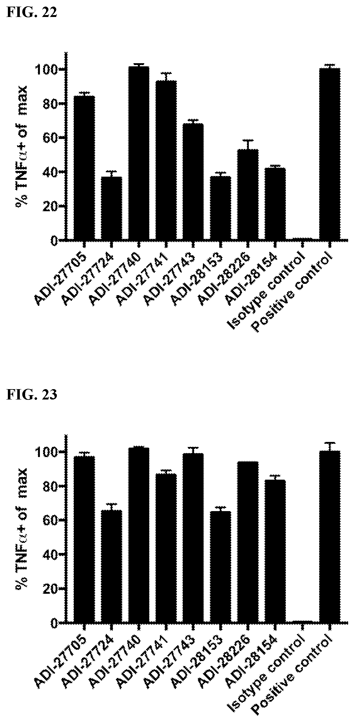
D00013
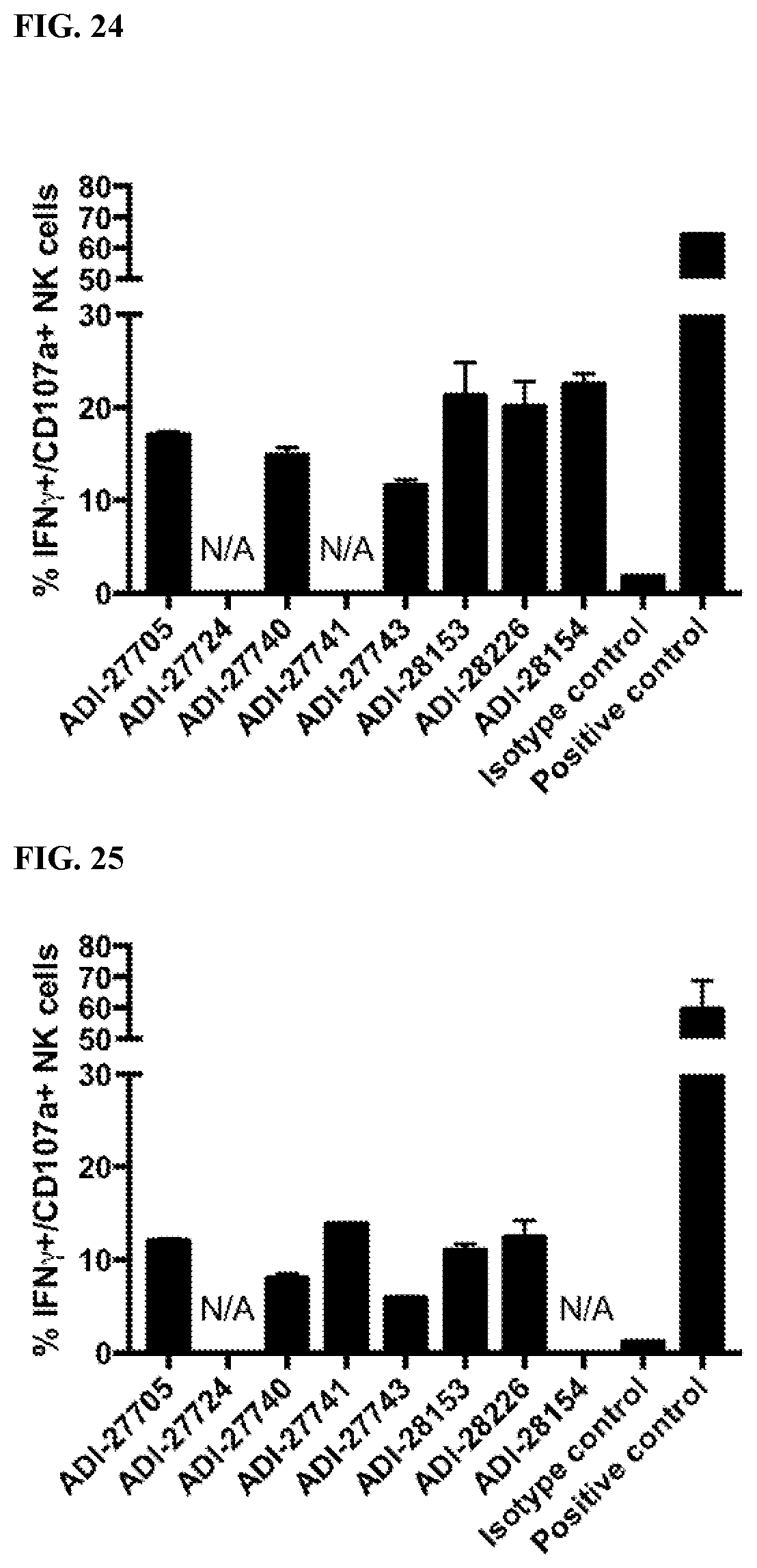
D00014
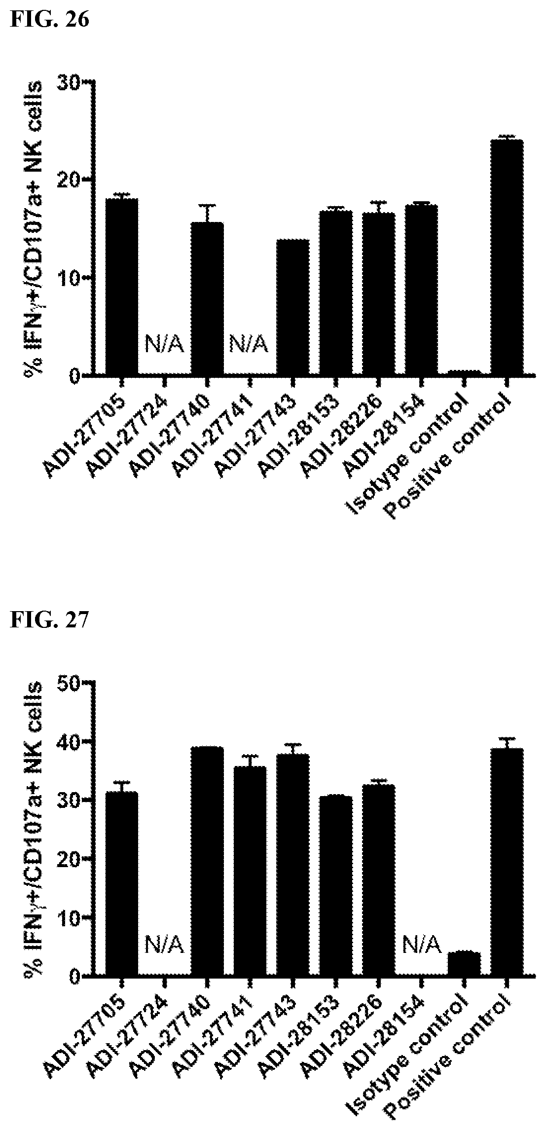
D00015

D00016
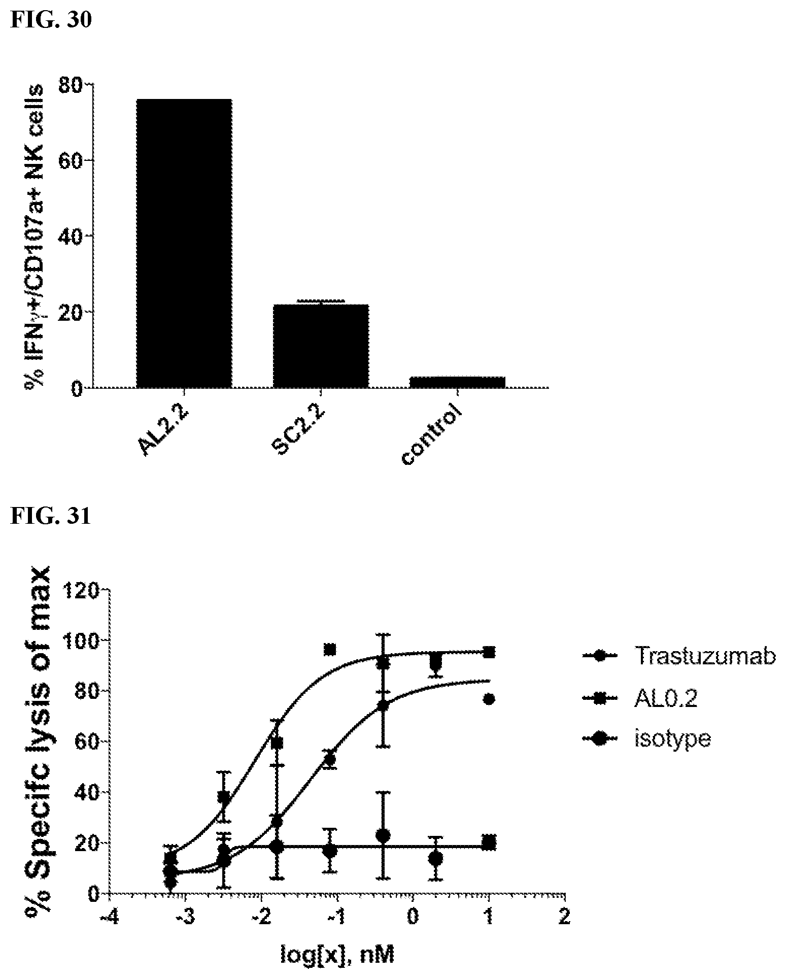
D00017

D00018

D00019
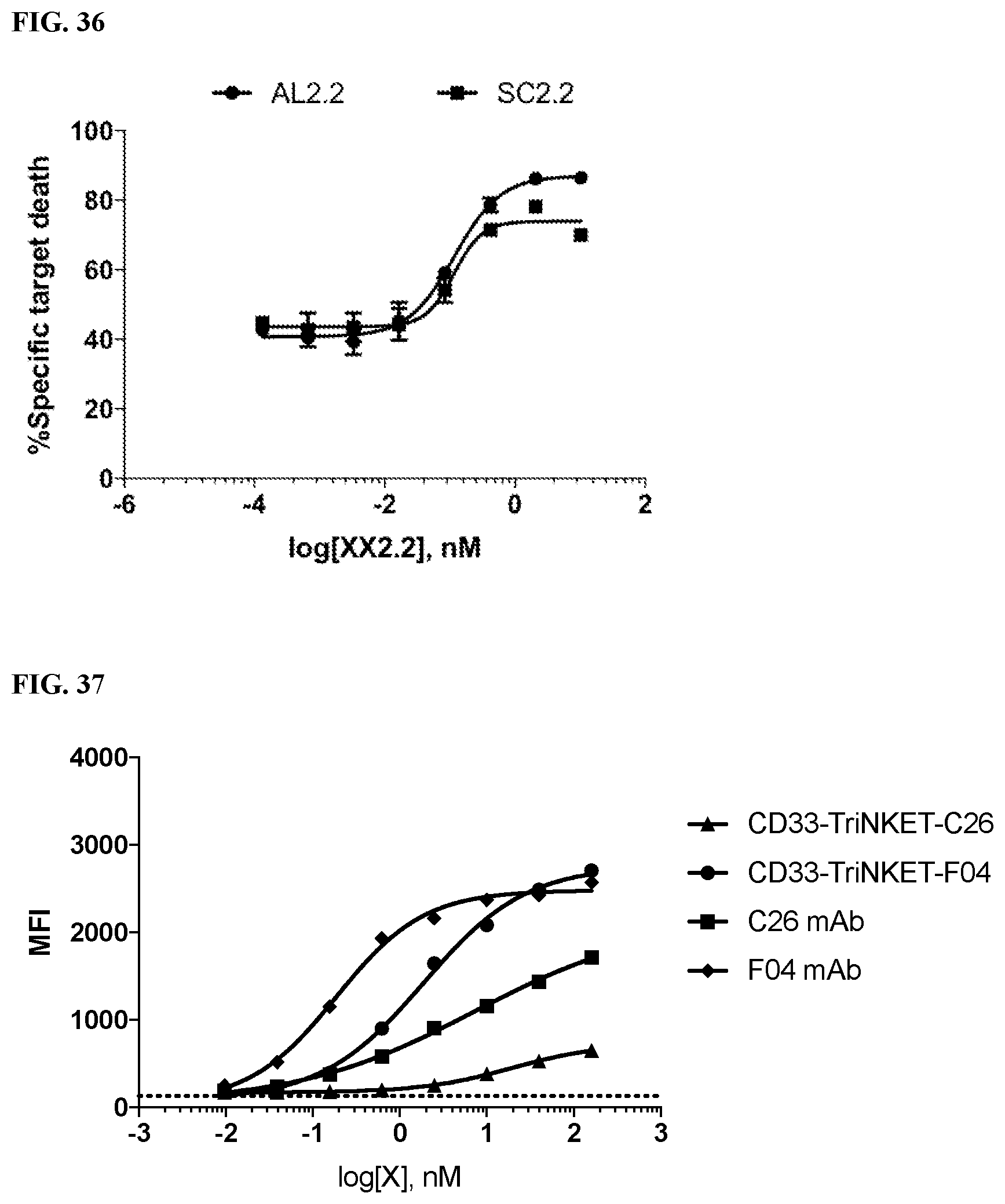
D00020

D00021

D00022
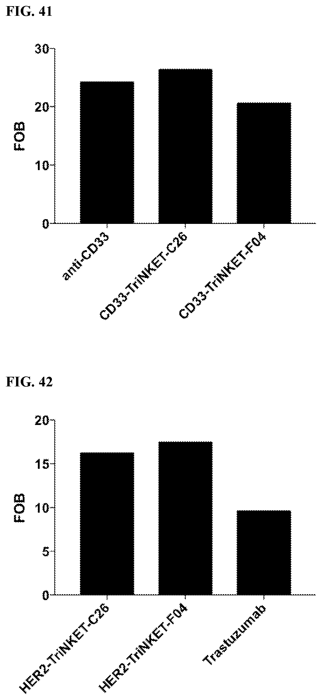
D00023

D00024
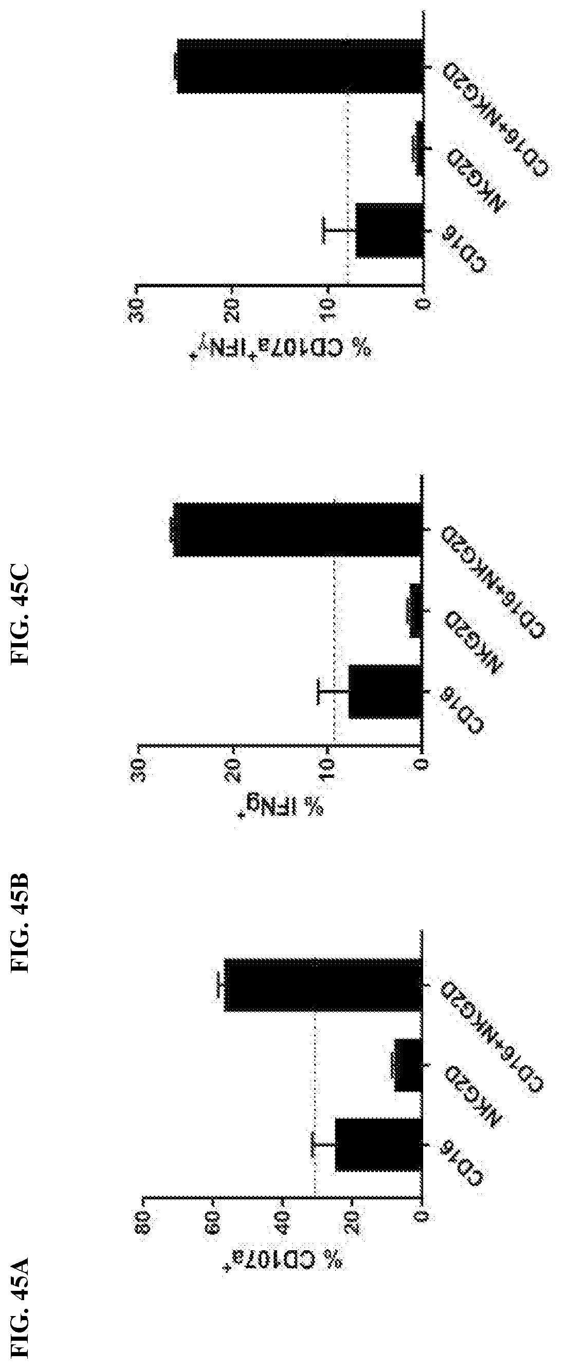
D00025

D00026

D00027

D00028

D00029

D00030
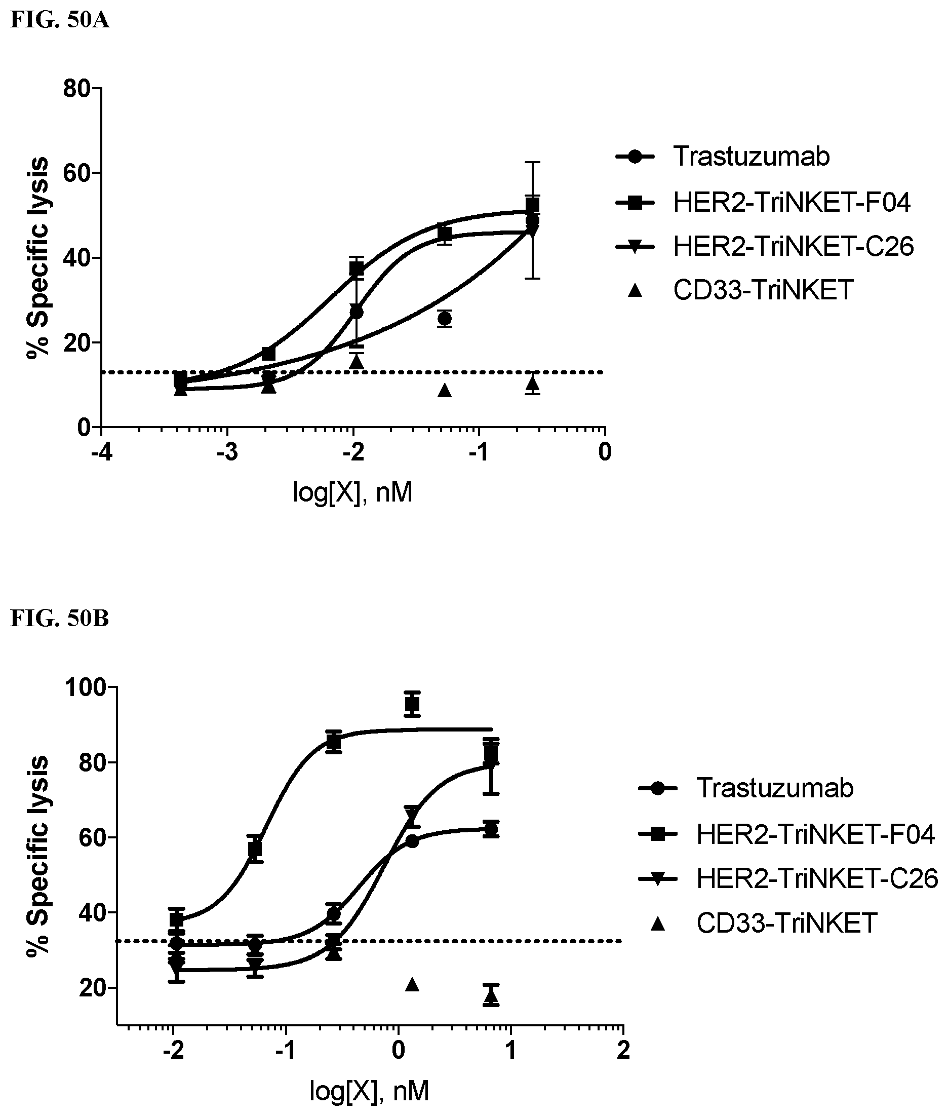
D00031

D00032

D00033
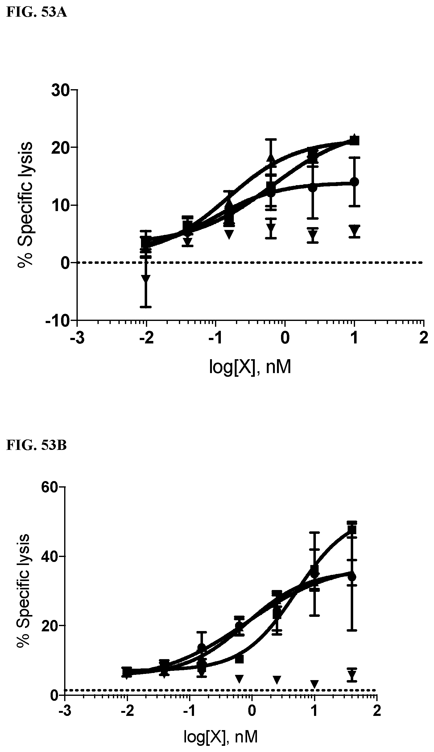
D00034
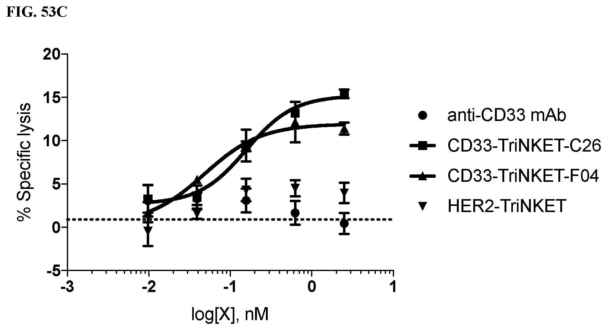
D00035

D00036

D00037

D00038

D00039

D00040

D00041
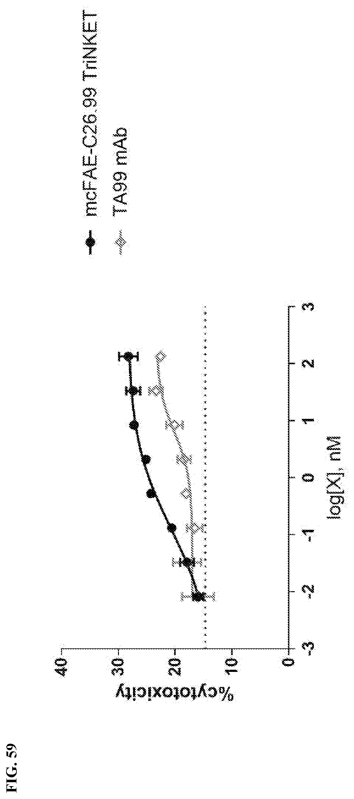
D00042

D00043

D00044

D00045

D00046
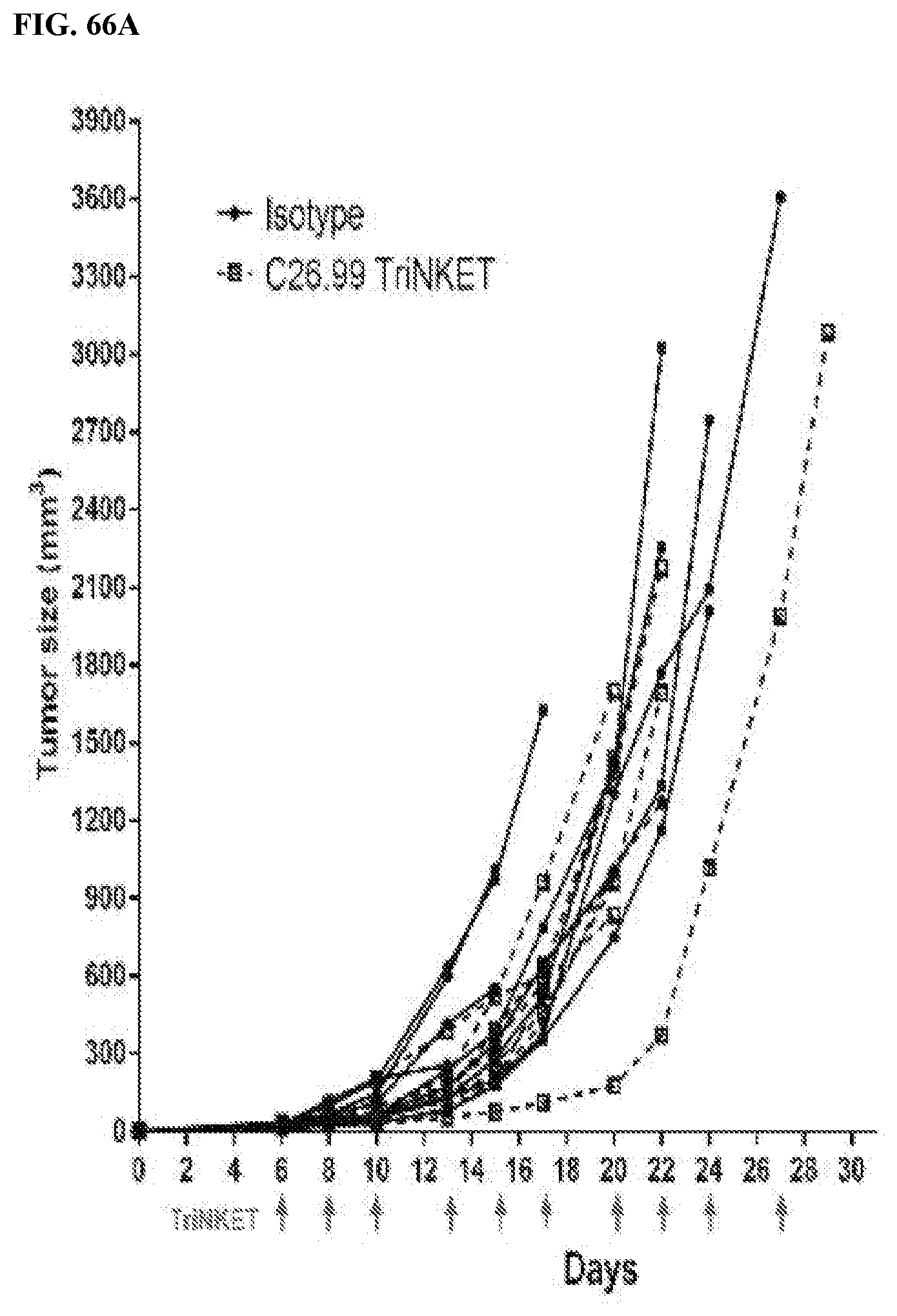
D00047

D00048
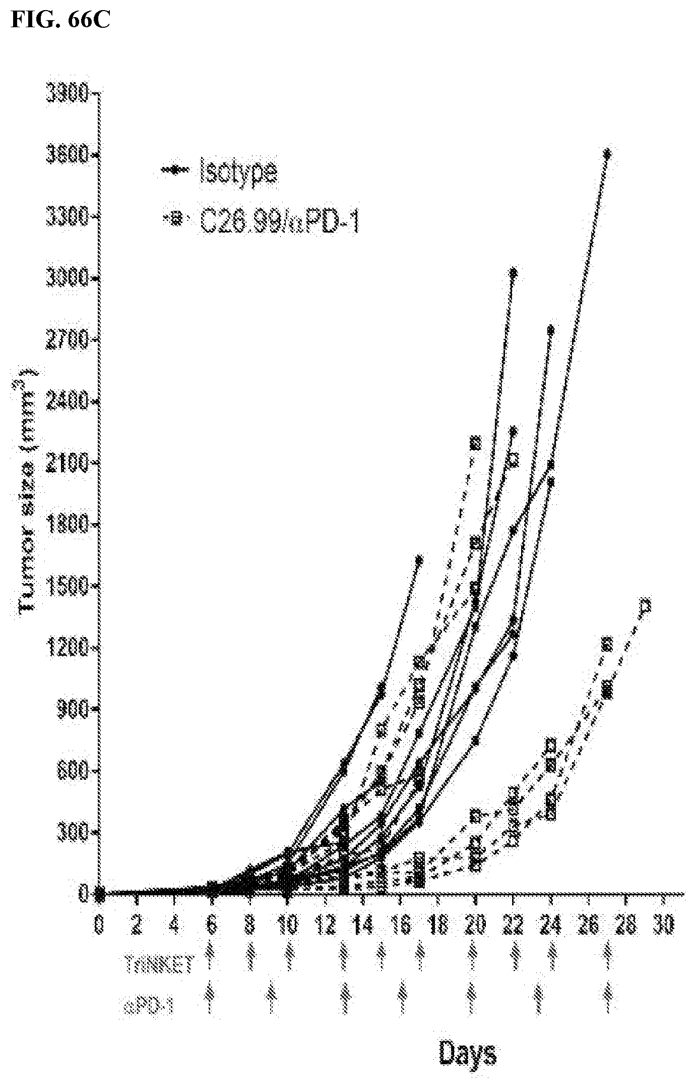
D00049

D00050

D00051
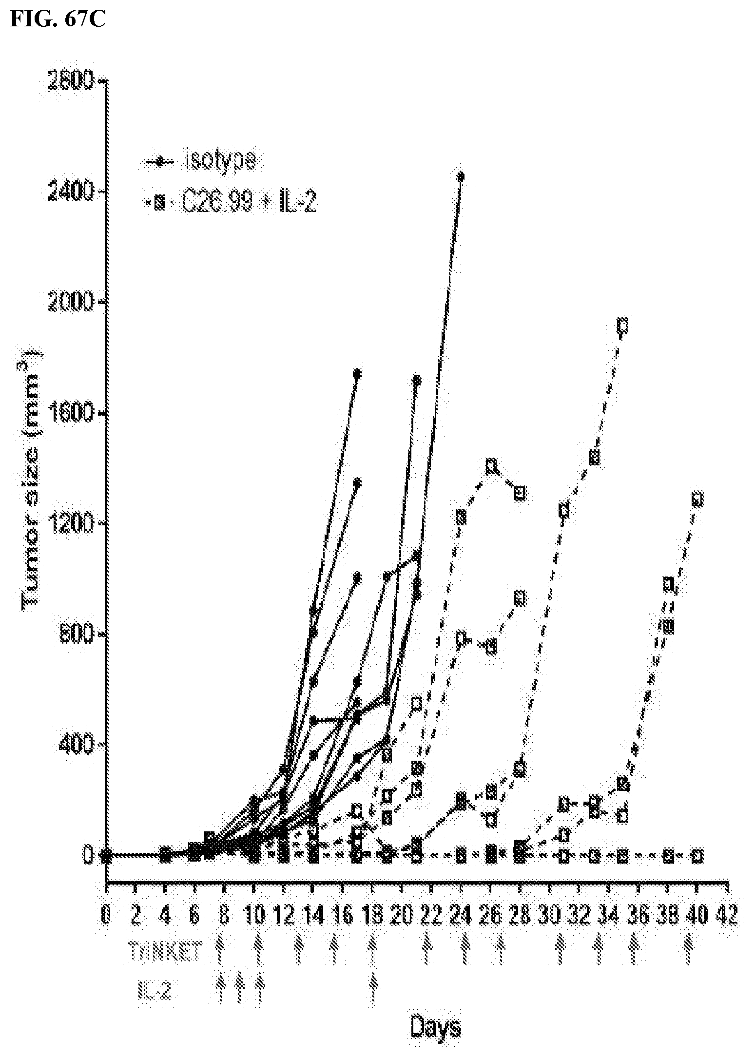
D00052

D00053
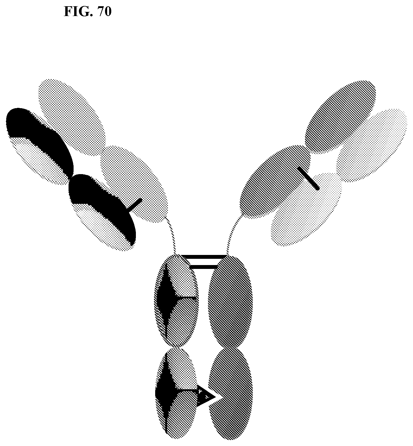
D00054
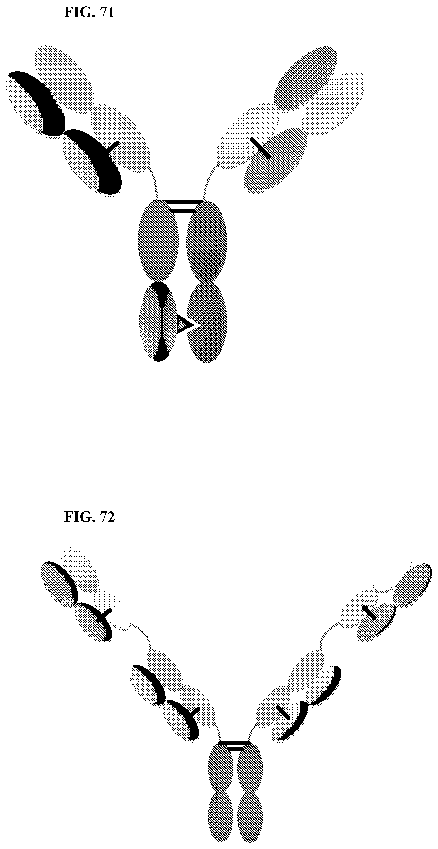
D00055

D00056

D00057

D00058
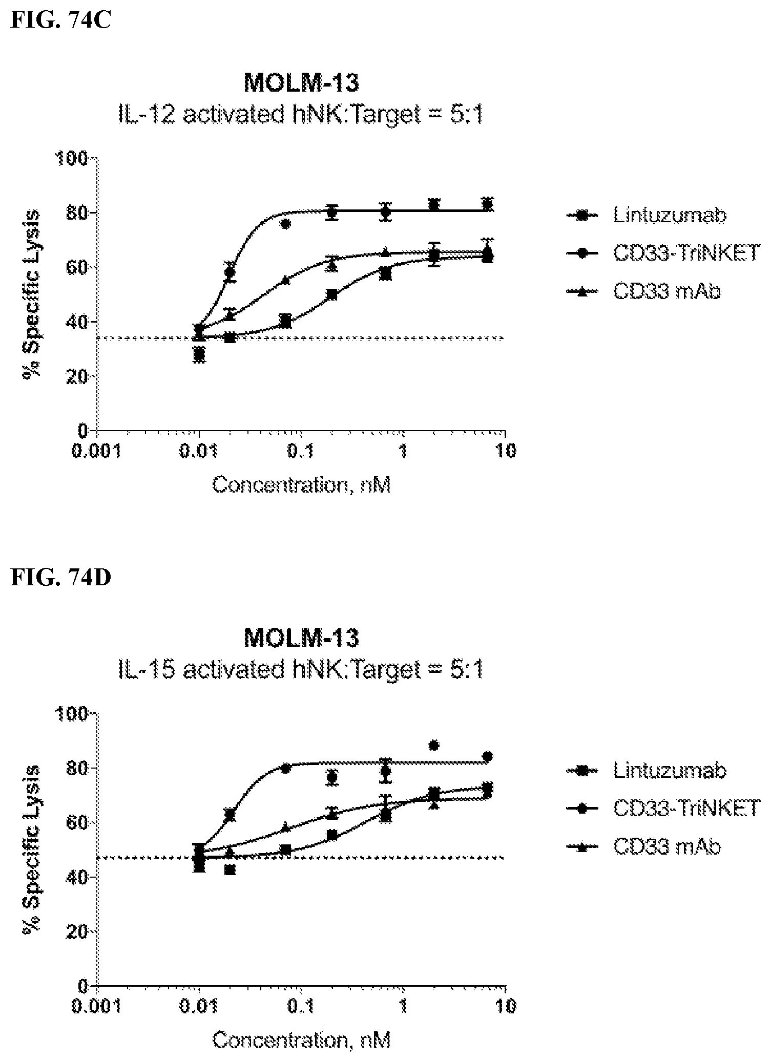
D00059

D00060
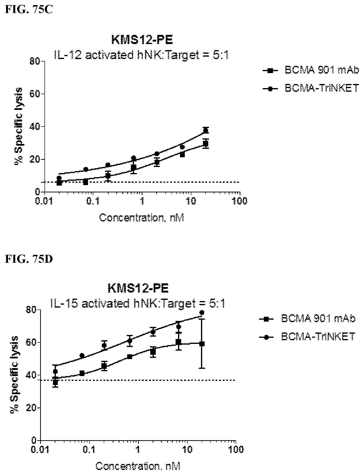
D00061

D00062

D00063

D00064
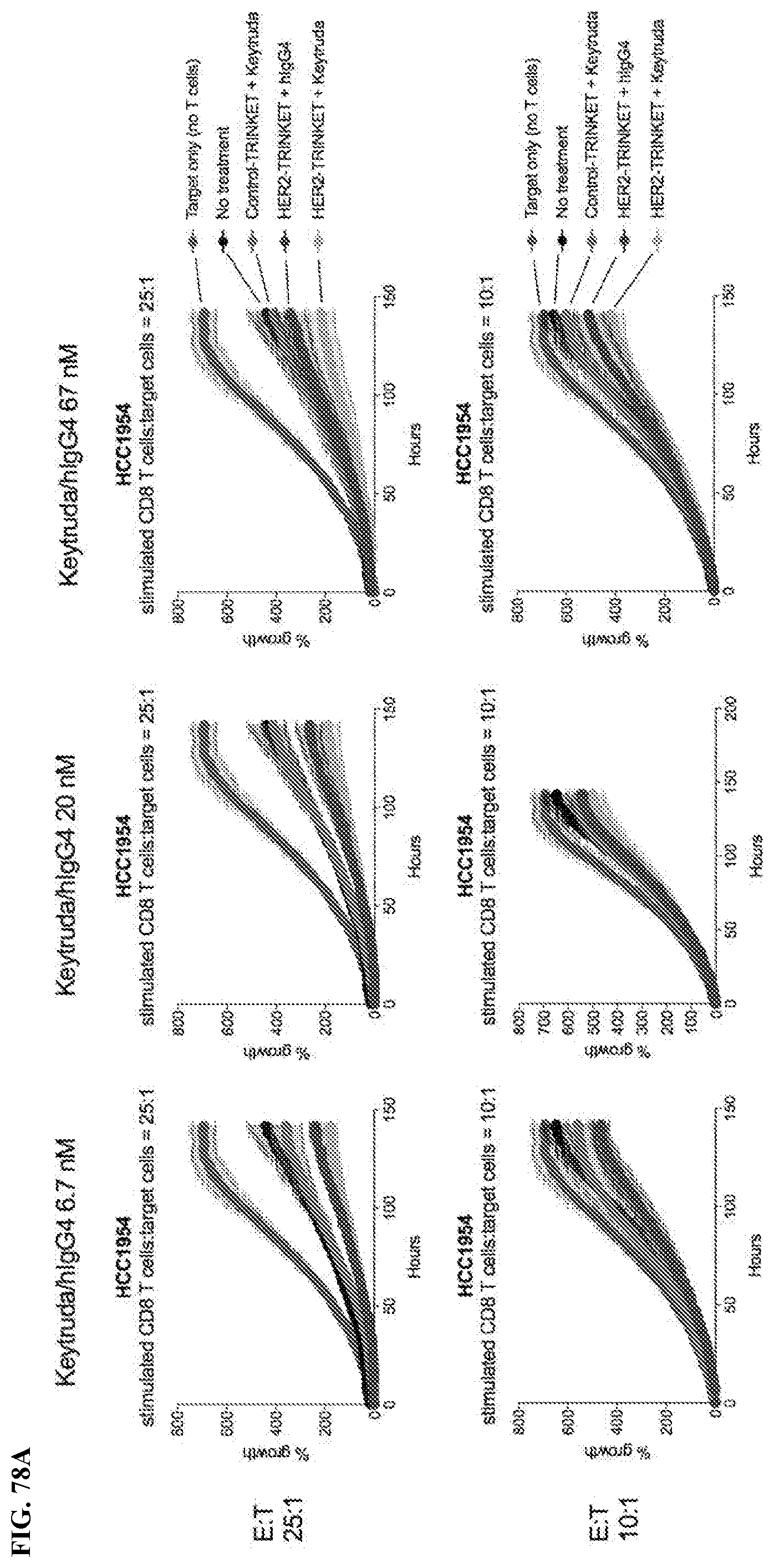
D00065

D00066

D00067

D00068

D00069

P00001

P00002

P00003

P00004

P00005

P00006

S00001
XML
uspto.report is an independent third-party trademark research tool that is not affiliated, endorsed, or sponsored by the United States Patent and Trademark Office (USPTO) or any other governmental organization. The information provided by uspto.report is based on publicly available data at the time of writing and is intended for informational purposes only.
While we strive to provide accurate and up-to-date information, we do not guarantee the accuracy, completeness, reliability, or suitability of the information displayed on this site. The use of this site is at your own risk. Any reliance you place on such information is therefore strictly at your own risk.
All official trademark data, including owner information, should be verified by visiting the official USPTO website at www.uspto.gov. This site is not intended to replace professional legal advice and should not be used as a substitute for consulting with a legal professional who is knowledgeable about trademark law.