Neutralizing Antibodies To Ebola Virus Glycoprotein And Their Use
Sullivan; Nancy ; et al.
U.S. patent application number 16/959644 was filed with the patent office on 2021-03-18 for neutralizing antibodies to ebola virus glycoprotein and their use. This patent application is currently assigned to The U.S.A., as represented by the Secretary, Department of Health and Human Services. The applicant listed for this patent is The U.S.A., as represented by the Secretary, Department of Health and Human Services, The U.S.A., as represented by the Secretary, Department of Health and Human Services. Invention is credited to Alberto Cagigi, Kendra Leigh, John Misasi, Nancy Sullivan.
| Application Number | 20210079067 16/959644 |
| Document ID | / |
| Family ID | 1000005273611 |
| Filed Date | 2021-03-18 |


View All Diagrams
| United States Patent Application | 20210079067 |
| Kind Code | A1 |
| Sullivan; Nancy ; et al. | March 18, 2021 |
NEUTRALIZING ANTIBODIES TO EBOLA VIRUS GLYCOPROTEIN AND THEIR USE
Abstract
Antibodies and antigen binding fragments that specifically bind to ebolavirus glycoprotein and neutralize ebolavirus infection are disclosed. Nucleic acids encoding these antibodies, vectors, and host cells are also provided. The disclosed antibodies, antigen binding fragments, nucleic acids and vectors can be used, for example, to inhibit an ebolavirus infection in a subject.
| Inventors: | Sullivan; Nancy; (Kensington, MD) ; Leigh; Kendra; (Frankfurt am Main, DE) ; Misasi; John; (Kensington, MD) ; Cagigi; Alberto; (Stockholm, SE) | ||||||||||
| Applicant: |
|
||||||||||
|---|---|---|---|---|---|---|---|---|---|---|---|
| Assignee: | The U.S.A., as represented by the
Secretary, Department of Health and Human Services Bethesda MD |
||||||||||
| Family ID: | 1000005273611 | ||||||||||
| Appl. No.: | 16/959644 | ||||||||||
| Filed: | December 31, 2018 | ||||||||||
| PCT Filed: | December 31, 2018 | ||||||||||
| PCT NO: | PCT/US18/68198 | ||||||||||
| 371 Date: | July 1, 2020 |
Related U.S. Patent Documents
| Application Number | Filing Date | Patent Number | ||
|---|---|---|---|---|
| 62612982 | Jan 2, 2018 | |||
| Current U.S. Class: | 1/1 |
| Current CPC Class: | A61K 2039/507 20130101; C07K 2317/76 20130101; C07K 2317/94 20130101; C07K 2317/72 20130101; C07K 2317/565 20130101; G01N 2400/02 20130101; A61P 31/14 20180101; C07K 2317/33 20130101; C07K 16/10 20130101; C07K 2317/92 20130101; G01N 2469/10 20130101; G01N 33/56983 20130101; C07K 2317/567 20130101; G01N 2333/08 20130101; A61K 2039/505 20130101 |
| International Class: | C07K 16/10 20060101 C07K016/10; G01N 33/569 20060101 G01N033/569; A61P 31/14 20060101 A61P031/14 |
Claims
1. An isolated monoclonal antibody, comprising: a heavy chain variable region (V.sub.H) comprising a heavy chain complementarity determining region (HCDR)1, a HCDR2, and a HCDR3 of the V.sub.H set forth as SEQ ID NO: 1; a light chain variable region (V.sub.L) comprising a light chain complementarity determining region (LCDR)1, a LCDR2, and a LCDR3 of the V.sub.L set forth as SEQ ID NO: 2; and wherein the monoclonal antibody specifically binds to an ebolavirus glycoprotein (GP) and neutralizes an ebolavirus.
2. The antibody of claim 1, wherein the HCDR1, the HCDR2, the HCDR3, the LCDR1, the LCDR2, and the LCDR3 comprise the amino acids sequences set forth as SEQ ID NOs: 7-12, respectively.
3. The antibody of claim 1, wherein the V.sub.H does not contain an N-linked glycan sequon beginning at any of Kabat residues 58-60.
4. The antibody of claim 1, wherein the V.sub.H does not contain an N-linked glycan sequon in a framework region 3 of the antibody.
5. The antibody of claim 1, comprising one or more amino acid substitutions to remove an N-linked glycan sequon beginning at Kabat position 60.
6. The antibody of claim 5, wherein the V.sub.H comprises an A61P substitution (Kabat numbering) to remove the N-linked glycan sequon.
7. The antibody of claim 1, wherein the V.sub.H comprises an amino acid sequence at least 90% identical to SEQ ID NO: 1 or SEQ ID NO: 3.
8. The antibody of claim 1, wherein the V.sub.L comprises an amino acid sequence at least 90% identical to SEQ ID NO: 2.
9. The antibody of claim 1, comprising human framework regions.
10. The antibody of claim 1, wherein the V.sub.H comprises the amino acid sequence set forth as SEQ ID NO: 3.
11. The antibody of claim 1, wherein the V.sub.H comprises the amino acid sequence set forth as SEQ ID NO: 1.
12. The antibody of claim 1, wherein the V.sub.L comprises the amino acid sequence set forth as SEQ ID NO: 2.
13. The antibody of claim 1, wherein the V.sub.H and the V.sub.L comprise the amino acid sequences set forth as SEQ ID NOs: 1 and 2, respectively, or SEQ ID NOs: 3 and 2, respectively.
14. The antibody of claim 1, wherein the antibody comprises a human constant domain.
15. The antibody of claim 1, wherein the antibody is a human antibody.
16. The antibody of claim 1, wherein the antibody is an IgG.
17. The antibody of claim 16, wherein the antibody is an IgG.sub.1.
18. The antibody of claim 1, comprising a recombinant constant domain comprising a modification that increases the half-life of the antibody.
19. The antibody of claim 18, wherein the modification increases binding to the neonatal Fc receptor.
20. The antibody of claim 1, comprising heavy and light chains comprising the amino acid sequences set forth as SEQ ID NOs: 4 and 5, respectively, or SEQ ID NOs: 6 and 5, respectively.
21. An isolated antigen binding fragment of the antibody of claim 1, wherein the antigen binding fragment comprises the V.sub.H and the V.sub.L of the antibody, specifically binds to the ebolavirus GP and neutralizes an ebolavirus.
22. The antigen binding fragment of claim 21, wherein the antigen binding fragment is a Fv, Fab, F(ab').sub.2, scFV or a scFV.sub.2 fragment.
23. The antibody of claim 1, conjugated to an effector molecule or a detectable marker.
24. The antibody claim 1, wherein the antibody specifically binds to Zaire ebolavirus GP and neutralizes Zaire ebolavirus.
25. An isolated nucleic acid molecule encoding the V.sub.H, the V.sub.L, or the V.sub.H and the V.sub.L of the antibody of claim 1.
26. The nucleic acid molecule of claim 25, wherein the nucleic acid sequence of the V.sub.H is set forth as SEQ ID NO: 13 or SEQ ID NO: 15, and the nucleic acid sequence of the V.sub.L is set forth as SEQ ID NO: 14.
27. The nucleic acid molecule of claim 25, comprising a cDNA or RNA encoding the antibody or antigen binding fragment.
28. The nucleic acid molecule of claim 25, operably linked to a promoter.
29. An expression vector comprising the nucleic acid molecule of claim 25.
30. A pharmaceutical composition for use in inhibiting an ebolavirus infection, comprising: an effective amount of the antibody of claim 1; and a pharmaceutically acceptable carrier.
31. A method of producing an antibody or antigen binding fragment that specifically binds to ebolavirus GP, comprising: expressing one or more nucleic acid molecules encoding the antibody of claim 1 in a host cell; and purifying the antibody or antigen binding fragment.
32. A method of detecting the presence of an ebolavirus in a biological sample from a subject, comprising: contacting the biological sample with an effective amount of the antibody of claim 1 under conditions sufficient to form an immune complex; and detecting the presence of the immune complex on the biological sample, wherein the presence of the immune complex on the biological sample indicates the presence of the ebolavirus in the sample.
33. The method of claim 32, wherein detecting the presence of the immune complex in the biological sample indicates that the subject has an ebolavirus infection.
34. A method of inhibiting an ebolavirus infection in a subject, comprising administering an effective amount of the antibody of claim 1 to the subject, wherein the subject has or is at risk of an ebolavirus infection.
35. The method of claim 34, further comprising administering to the subject one or more additional antibodies or antigen binding fragments that specifically bind to the ebolavirus GP and neutralize the ebolavirus, or one or more nucleic acid molecules encoding the additional antibodies or antigen binding fragments.
36. The method of claim 34, wherein the ebolavirus is Zaire ebolavirus.
37. (canceled)
Description
CROSS REFERENCE TO RELATED APPLICATIONS
[0001] This application claims the benefit of U.S. Provisional Application No. 62/612,982, filed Jan. 2, 2018, which is herein incorporated by reference in its entirety.
FIELD
[0002] This relates to monoclonal antibodies and antigen binding fragments that specifically bind to ebolavirus glycoprotein (GP) and their use, for example, in methods of inhibiting ebolavirus infection or ebolavirus disease (EVD) in a subject.
BACKGROUND
[0003] EVD is a disease in humans, chimpanzees, and monkeys, caused by infection with an ebolavirus. The prototypic member of this genus, Zaire ebolavirus, was first identified in the Democratic Republic of Congo (formerly known as the Republic of Zaire) in 1976. Ebolaviruses are members of the Filoviridae family of RNA viruses and cause a severe hemorrhagic fever with a high mortality rate. For example, infection with Zaire ebolavirus is associated with a mortality rate of up to 90% in humans.
[0004] While prior outbreaks of EVD have been localized to regions of Africa, the potential threat of dissemination to other countries has been exacerbated by the frequency of international travel. The 2014 outbreak in West Africa was first recognized in March 2014, and as of Apr. 13, 2016, the number of cases far exceeded the largest prior EVD outbreak with a combined total (suspected, probable, and laboratory-confirmed) 28616 cases and 11310 deaths (case fatality rate=39.5%). The largest previous outbreak of an ebolavirus occurred in Uganda in 2000-2001 with 425 cases and 224 deaths (case-fatality rate=53%).
[0005] During infection, proteases of the host cell cleave a precursor of GP, termed GP.sub.0, into GP.sub.1 and GP.sub.2. GP.sub.2 is an integral membrane protein, while GP.sub.1 protrudes from the mature virus. Three copies of the GP-GP.sub.2 heterodimer make up the ebolavirus envelope spike, which is a target for neutralizing antibodies.
[0006] Although certain neutralizing antibodies that bind to ebolavirus GP have been identified, there is a need to develop additional neutralizing antibodies with varying ebolavirus GP recognition profiles and increased neutralization potency. The challenges of a large outbreak and the failure of traditional quarantine and contact tracing measures to control an outbreak of this scale highlights the urgency for therapies.
SUMMARY
[0007] Isolated antibodies and antigen binding fragments that specifically bind to ebolavirus GP and neutralize ebolavirus are provided herein.
[0008] In some embodiments, the antibody or antigen binding fragment comprises a heavy chain variable region (V.sub.H) comprising a heavy chain complementarity determining region (HCDR)1, a HCDR2, and a HCDR3 of the V.sub.H set forth as SEQ ID NO: 1 (S1-4-A09 V.sub.H) and a light chain variable region (V.sub.L) comprising a light chain complementarity determining region (LCDR)1, a LCDR2, and a LCDR3 of the V.sub.L set forth as SEQ ID NO: 2 (S1-4-A09 V.sub.L). In some embodiments, the HCDR1, the HCDR2, the HCDR3, the LCDR1, the LCDR2, and the LCDR3 comprise the amino acids sequences set forth as SEQ ID NOs: 7-12, respectively. In some embodiments, the antibody or antigen binding fragment comprises a V.sub.H and a V.sub.L comprising the amino acid sequences set forth as SEQ ID NOs: 1 and 2, respectively. In some embodiments, the antibody or antigen binding fragment comprises a V.sub.H and a V.sub.L comprising the amino acid sequences set forth as SEQ ID NOs: 3 and 2, respectively.
[0009] In some embodiments, the V.sub.H of the antibody does not contain an N-linked glycan sequon beginning at any of Kabat residues 58-60. For example, the V.sub.H of the antibody comprises one or more amino acid substitutions to remove an N-linked glycan sequon beginning at any of Kabat residues 58-60. In some embodiments, the V.sub.H of the antibody does not contain an N-linked glycan sequon beginning at Kabat residue 60. For example, the V.sub.H of the antibody comprises an A61P substitution (Kabat numbering) to remove an N-linked glycan sequon beginning at Kabat position 60.
[0010] Also disclosed are compositions including the antibodies and antigen binding fragments, nucleic acids encoding the antibodies and antigen binding fragments, expression vectors comprising the nucleic acids, and isolated host cells that comprise the nucleic acids. In several embodiments, the nucleic acid molecule encoding a disclosed antibody or antigen binding fragment can be a cDNA molecule that encodes the antibody or antigen binding fragment. In additional embodiments, the nucleic acid molecule can be a bicistronic expression construct encoding the V.sub.H and V.sub.L of the antibody or antigen binding fragment.
[0011] Surprisingly, the disclosed antibodies and antigen binding fragments potently neutralize ebolavirus and inhibit ebolavirus infection in accepted in vitro and in vivo models. Accordingly, a method is disclosed for inhibiting (including preventing) ebolavirus infection in a subject. The method comprises administering an effective amount (that is, an amount effective to inhibit ebolavirus infection in a subject) of one or more of the disclosed antibodies, antigen binding fragments, nucleic acid molecules, vectors, or compositions, to the subject, such as a subject at risk of or having an ebolavirus infection.
[0012] The antibodies, antigen binding fragments, nucleic acid molecules, vectors, and compositions disclosed herein can be used for a variety of additional purposes, such as for diagnosing ebolavirus infection in a subject, or detecting ebolavirus in a sample.
[0013] The foregoing and other features and advantages of this disclosure will become more apparent from the following detailed description of several embodiments, which proceeds with reference to the accompanying figures.
BRIEF DESCRIPTION OF THE FIGURES
[0014] FIG. 1. Sequences of S1-4-A09 V.sub.H (SEQ ID NO: 1) and V.sub.L (SEQ ID NO: 2) showing CDRs (IMGT) and comparison with mAb100 V.sub.H (SEQ ID NO: 23) and V.sub.L (SEQ ID NO: 24) sequences.
[0015] FIG. 2. Binding of S1-4-A09 to Zaire ebolavirus GP as assayed by enzyme-linked immunosorbent assay (ELISA). Shown are binding curves generated from S1-4-A09, mAb100 and an isotype control antibody binding to either Zaire ebolavirus sGP (sGP(Z)), full length GP (GP(Z)-full length) or mucin-domain-deleted GP (GP(Z)dMuc). Error bars shown represent the standard error of the mean of triplicate wells for each dilution point.
[0016] FIG. 3. In vitro neutralization of Zaire ebolavirus GP-pseudotyped lentiviral vectors by S1-4-A09. mAb100 and an isotype control were included in the assay for comparison. The IC.sub.50 for mAb100 and S1-4-A09 as calculated from the neutralization curve using a four-parameter logistic curve fit is shown in the inset table. Error bars shown represent the standard deviation of triplicate well values.
[0017] FIG. 4. Kinetics of S1-4-A09 Fab binding to GP and GP variants. S1-4-A09 Fab binding to mucin-domain-deleted Zaire ebolavirus GP (.DELTA.Muc), Zaire ebolavirus secreted GP (sGP), and cleaved Zaire ebolavirus GP (THL) at pH 7.4 and 5.3 were measured by biolayer interferometry. k.sub.on, k.sub.off, and K.sub.D values were calculated based on a global, nonlinear, least squares, 1:1 binding model curve fit with the assumption of fully reversible binding.
[0018] FIG. 5. Immunoblot analysis of antibody-antigen complexes incubated with thermolysin. Mucin-domain-deleted GP (GP.DELTA.M) was incubated with mAb100, S1-4-A09, or a mAb control (an Ebola GP-directed mAb known to not inhibit cleavage) for 30 min at room temperature before incubation with thermolysin. The starting antigen is indicated on the blot by "GP.DELTA.M" and the cleavage product is indicated by "GP.DELTA.M.sub.cleaved." The dashed box indicates the region of the blot where signal from cleavage product is absent, indicating protection from thermolysin.
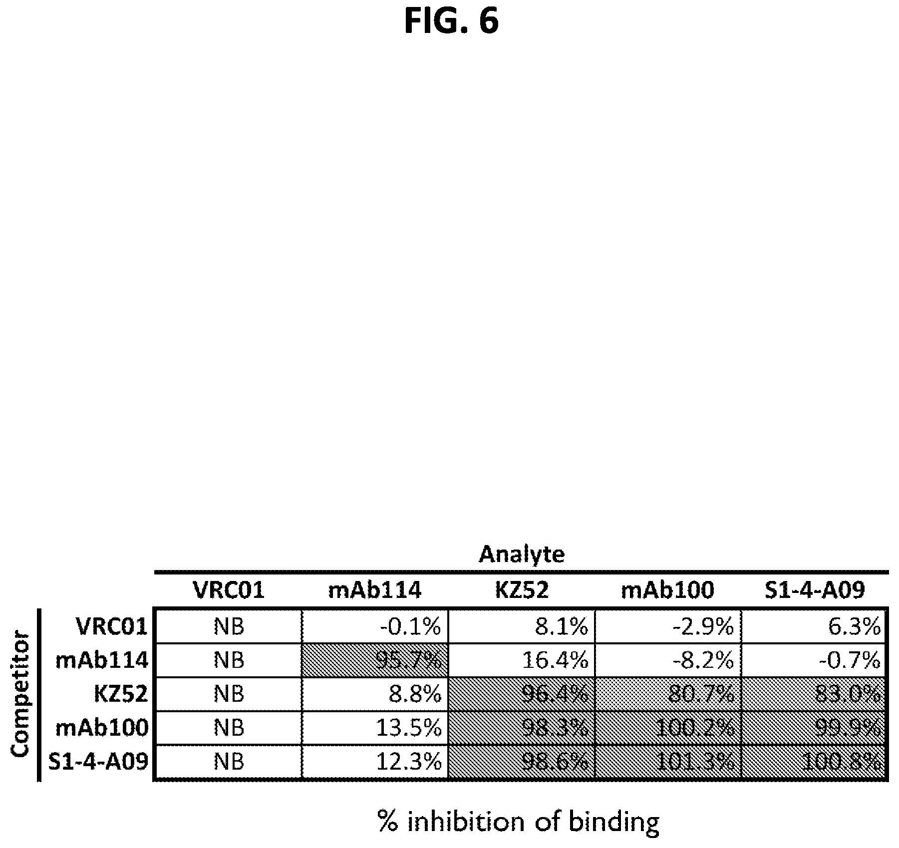
[0019] FIG. 6. S1-4-A09 competition group as determined by biolayer interferometry. The order of addition to the biosensors was mucin-domain-deleted GP (antigen), competitor mAb, analyte mAb. VRC01 is an isotype control mAb that does not bind GP. mAb114 binds the GP trimer at its membrane distal apex. KZ52 and mAb100 are two survivor-isolated mAbs that bind at the base (membrane-proximal) of the GP trimer.
[0020] FIG. 7. Exemplary class averages of particles chosen from negative-stain transmission electron micrographs of S1-4-A09 Fab complexed with mucin-domain-deleted GP. Density from GP is outlined in dotted lines. Fab density is indicated by arrows.
[0021] FIG. 8. Antinuclear antibody staining analysis of S1-4-A09 autoreactivity. Staining analysis was done in HEp-2 cells. The top four panels are representative fields of view from an assay with antibodies that have increasing degrees of autoreactivity. The bottom panel is a representative field of view from an assay with S1-4-A09. The autoreactivity score is indicated by the number in the upper right corner of each image.
[0022] FIG. 9. In vivo neutralization of Zaire ebolavirus (EBOV) infection by S1-4-A09 IgG. Top, schematic of Zaire ebolavirus challenge and S1-4-A09 dosing timeline for in vivo efficacy studies. Middle, dosage of S1-4-A09 administered intravenously to macaques for in vivo efficacy studies. Bottom, Kaplan-Meier curves for survival in the in vivo efficacy study for S1-4-A09 alone. Animals in the A09 group received three doses of S1-4-A09 at 50 mg/kg/dose. The "No Treatment" group did not receive any antibody.
[0023] FIG. 10. In vivo neutralization of Zaire ebolavirus infection by combination therapy with S1-4-A09 IgG and mAb114 IgG. Top, schematic of Zaire ebolavirus challenge and S1-4-A09-mAb114 antibody mixture dosing timeline for in vivo efficacy studies. Middle, dosages of S1-4-A09 and mAb114 antibody mixtures administered intravenously to macaques for in vivo neutralization studies. Bottom, Kaplan-Meier curves for survival in the in vivo efficacy study for S1-4-A09 in combination with mAb114. The 50:50 114/A09 curve represents the group where animals received three doses of S1-4-A09 with mAb114 in a 50% to 50% ratio (A09:114) at a total antibody dosage of 50 mg/kg/dose. The 92:8 11/4A09 curve represents the group where animals received three doses of S1-4-A09 with mAb114 in an 8% to 92% ratio (A09:114) at a total antibody dosage of 50 mg/kg/dose. Animals of the control group were not administered any antibody.
[0024] FIG. 11. Alignment of the S1-4-A09, S-14-A09 A61P, mAb100, and mAb100 unmutated common ancestor (UCA) heavy chain sequences, with the N-linked glycan sequon and A61P mutation annotated. Residues 70-88 (IMGT numbering) from the heavy chains of the mAb100 UCA (SEQ ID NO: 31), mAb100 (corresponding to residues 51-69 of SEQ ID NO: 23), S1-4-A09 (corresponding to residues 51-69 of SEQ ID NO: 1) and S1-4-A09 A61P (corresponding to residues 51-69 of SEQ ID NO: 3) are shown. The CDR2 of the heavy chain is underlined and highlighted by the highlighted box. The N-linked glycan sequons are shown by small open boxes. The A61P point mutation is indicated with an arrow.
[0025] FIG. 12. Binding of S1-4-A09 A61P to Zaire ebolavirus mucin-domain-deleted GP (GP.DELTA.M) as assayed by ELISA. Binding curves for the isotype control, S1-4-A09, and S1-4-A09 A61P are shown. Error bars shown represent the standard error of the mean of triplicate wells for each dilution point.
[0026] FIG. 13. In vitro neutralization of Zaire ebolavirus GP-pseudotyped lentiviral vectors by S1-4-A09 A61P. S1-4-A09 and an isotype control were included in the assay for comparison. The IC.sub.50 for S1-4-A09 and S1-4-A09 A61P as calculated from the neutralization curve using a four-parameter logistic curve fit is shown in the inset table. Error bars shown represent the standard deviation of triplicate well values.
[0027] FIG. 14. Immunoblot analysis of antibody-antigen complexes incubated with thermolysin. Mucin-domain-deleted GP (GP.DELTA.M) was incubated with S1-4-A09, S1-4-A09 A61P, a mAb control (an Ebola GP-directed mAb known to not inhibit cleavage), and an isotype control (a mAb not directed towards Ebola GP) for 30 min at room temperature before incubation with thermolysin. The starting antigen is indicated on the blot by "GP.DELTA.M" and the cleavage product is indicated by "GP.DELTA.M.sub.cleaved." The dashed box indicates the region of the blot where signal from cleavage product is absent, indicating protection from thermolysin.
SEQUENCE LISTING
[0028] The nucleic and amino acid sequences listed in the accompanying sequence listing are shown using standard letter abbreviations for nucleotide bases, and three letter code for amino acids, as defined in 37 C.F.R. 1.822. Only one strand of each nucleic acid sequence is shown, but the complementary strand is understood as included by any reference to the displayed strand. The Sequence Listing is submitted as an ASCII text file, created on Dec. 31, 2018, 58.2 KB, which is incorporated by reference herein. In the accompanying sequence listing:
TABLE-US-00001 SEQ ID NO: 1 is the amino acid sequence of the S1-4-A09 V.sub.H. QVQLQESGPGLVKPSETLSLTCVVSggilssfyWNWIRQPPGKGLEWIGNiyysgspNYNASFQSRVAISVDTS- KNQIS LNLKSVTAADTAMYYCvrasraylwgsyrptaldlWGQGSLVTVSS SEQ ID NO: 2 is the amino acid sequence of the S1-4-A09 V.sub.L. SYELTQPPSVSVSPGQTATITCSGDklgdkyTSWFQQRPGQSPLLVIYqdnKRPSGLPARFSGSNSGNTATLTI- SGTQA MDEADYLCqvwdsgavFGGGTKLTVL SEQ ID NO: 3 is the amino acid sequence of the S1-4-A09 A61P V.sub.H. QVQLQESGPGLVKPSETLSLTCVVSggilssfyWNWIRQPPGKGLEWIGNiyysgspNYNPSFQSRVAISVDTS- KNQIS LNLKSVTAADTAMYYCvrasraylwgsyrptaldlWGQGSLVTVSS SEQ ID NO: 4 is the amino acid sequence of an IgG1 heavy chain including the S1-4-A09 V.sub.H. QVQLQESGPGLVKPSETLSLTCVVSggilssfyWNWIRQPPGKGLEWIGNiyysgspNYNASFQSRVAISVDTS- KNQIS LNLKSVTAADTAMYYCvrasraylwgsyrptaldlWGQGSLVTVSSASTKGPSVFPLAPSSKSTSGGTAALGCL- VKDYF PEPVTVSWNSGALTSGVHTFPAVLQSSGLYSLSSVVTVPSSSLGTQTYICNVNHKPSNTKVDKKVEPKSCDKTH- TCPPC PAPELLGGPSVFLFPPKPKDTLMISRTPEVTCVVVDVSHEDPEVKFNWYVDGVEVHNAKTKPREEQYNSTYRVV- SVLTV LHQDWLNGKEYKCKVSNKALPAPIEKTISKAKGQPREPQVYTLPPSRDELTKNQVSLTCLVKGFYPSDIAVEWE- SNGQP ENNYKTTPPVLDSDGSFFLYSKLTVDKSRWQQGNVFSCSVMHEALHNHYTQKSLSLSPGK SEQ ID NO: 5 is the amino acid sequence of an IgG1 light chain including the S1-4-A09 V.sub.L. SYELTQPPSVSVSPGQTATITCSGDklgdkyTSWFQQRPGQSPLLVIYqdnKRPSGLPARFSGSNSGNTATLTI- SGTQA MDEADYLCqvwdsgavFGGGTKLTVLGQPKAAPSVTLFPPSSEELQANKATLVCLISDFYPGAVTVAWKADSSP- VKAGV ETTTPSKQSNNKYAASSYLSLTPEQWKSHRSYSCQVTHEGSTVEKTVAPTECS SEQ ID NO: 6 is the amino acid sequence of an IgG1 heavy chain including the S1-4-A09 A61P V.sub.H. QVQLQESGPGLVKPSETLSLTCVVSggilssfyWNWIRQPPGKGLEWIGNiyysgspNYNPSFQSRVAISVDTS- KNQIS LNLKSVTAADTAMYYCvrasraylwgsyrptaldlWGQGSLVTVSSASTKGPSVFPLAPSSKSTSGGTAALGCL- VKDYF PESPVTVSWKSGALTSGVHTFPAVLQSSGLYSLSSVVTVPSSSLGTQTYICNVNHKPSNTKVDKKVEPKSCDKT- HTCPPC PAPELLGGPSVFLFPPKPKDTLMISRTPEVTCVVVDVSHEDPEVKFNWYVDGVEVHNAKTKPREEQYNSTYRVV- SVLTV LHQDWLNGKEYKCKVSNKALPAPIEKTISKAKGQPREPQVYTLPPSRDELTKNQVSLTCLVKGFYPSDIAVEWE- SNGQP ENNYKTTPPVLDSDGSFFLYSKLTVDKSRWQQGNVFSCSVMHEALHNHYTQKSLSLSPGK SEQ ID NOs: 7-12 are the amino acid sequences of antibody CDRs. SEQ ID NO: 13 is an exemplary nucleic acid sequence encoding the S1-4-A09 V.sub.H. atgggatggtcatgtatcatcctttttctagtagcaactgcaaccggtgtacattctcaggtgcagctgcagga- gtcgg gaccaggactggtgaagccttcggagaccctgtccctcacctgcgttgtctctggtggcatcctcagtagtttt- tactg gaactggatccggcagcccccaggaaagggactggagtggattggaaacatctattacagtgggagccccaact- ataat gcctccttccagagtcgagtcgccatttcggtggacacgtccaagaaccagatctccctgaacctcaagtctgt- gaccg ctgcggacacggccatgtattactgtgtgagagcctcccgcgcttacctttgggggagttatcgtccaacggct- cttga cctctggggccagggatccctggtcaccgtctcctca SEQ ID NO: 14 is an exemplary nucleic acid sequence encoding the S1-4-A09 V.sub.L. atgggatggtcatgtatcatcctttttctagtagcaactgcaaccggtgtccatgcttcctatgagctgactca- gccac cctcagtgtccgtgtccccaggacagacagccaccatcacgtgctctggagataaattgggtgataaatatact- tcctg gttccagcagaggccaggccagtcccctctactggtcatctatcaggataataagcggccctcagggctccctg- cgcga ttttctggctccaactctgggaacacagccactctgaccatcagcggcacccaggctatggatgaggctgacta- tttgt gtcaggtgtgggacagcggtgcggtgttcggcggagggaccaagctgaccgtccta SEQ ID NO: 15 is an exemplary nucleic acid sequence encoding the S1-4-A09 A61P V.sub.H. atgggatggtcatgtatcatcctttttctagtagcaactgcaaccggtgtacattctcaggtgcagctgcagga- gtcgg gaccaggactggtgaagccttcggagaccctgtccctcacctgcgttgtctctggtggcatcctcagtagtttt- tactg gaactggatccggcagcccccaggaaagggactggagtggattggaaacatctattacagtgggagccccaact- ataat ccctccttccagagtcgagtcgccatttcggtggacacgtccaagaaccagatctccctgaacctcaagtctgt- gaccg ctgcggacacggccatgtattactgtgtgagagcctcccgcgcttacctttgggggagttatcgtccaacggct- cttga cctctggggccagggatccctggtcaccgtctcctca SEQ ID NO: 16 is an exemplary nucleic acid sequence encoding the amino acid sequence of an IgG1 heavy chain including the S1-4-A09 V.sub.H. atgggatggtcatgtatcatcctttttctagtagcaactgcaaccggtgtacattctcaggtgcagctgcagga- gtcgg gaccaggactggtgaagccttcggagaccctgtccctcacctgcgttgtctctggtggcatcctcagtagtttt- tactg gaactggatccggcagcccccaggaaagggactggagtggattggaaacatctattacagtgggagccccaact- ataat gcctccttccagagtcgagtcgccatttcggtggacacgtccaagaaccagatctccctgaacctcaagtctgt- gaccg ctgcggacacggccatgtattactgtgtgagagcctcccgcgcttacctttgggggagttatcgtccaacggct- cttga cctctggggccagggatccctggtcaccgtctcctcagcgtcgaccaagggcccatcggtcttccccctggcac- cctcc tccaagagcacctctgggggcacagcggccctgggctgcctggtcaaggactacttccccgaacccgtgacggt- gtcgt ggaactcaggcgccctgaccagcggcgtgcacaccttcccggctgtcctacagtcctcaggactctactccctc- agcag cgtggtgaccgtgccctccagcagcttgggcacccagacctacatctgcaacgtgaatcacaagcccagcaaca- ccaag gtggacaagaaagttgagcccaaatcttgtgacaaaactcacacatgcccaccgtgcccagcacctgaactcct- ggggg gaccgtcagtcttcctcttccccccaaaacccaaggacaccctcatgatctcccggacccctgaggtcacatgc- gtggt ggtggacgtgagccacgaagaccctgaggtcaagttcaactggtacgtggacggcgtggaggtgcataatgcca- agaca aagccgcgggaggagcagtacaacagcacgtaccgtgtggtcagcgtcctcaccgtcctgcaccaggactggct- gaatg gcaaggagtacaagtgcaaggtctccaacaaagccctcccagcccccatcgagaaaaccatctccaaagccaaa- gggca gccccgagaaccacaggtgtacaccctgcccccatcccgggatgagctgaccaagaaccaggtcagcctgacct- gcctg gtcaaaggcttctatcccagcgacatcgccgtggagtgggagagcaatgggcagccggagaacaactacaagac- cacgc ctcccgtgctggactccgacggctccttcttcctctacagcaagctcaccgtggacaagagcaggtggcagcag- gggaa cgtcttctcatgctccgtgatgcatgaggctctgcacaaccactacacgcagaagagcctctccctgtctccgg- gtaaa tga SEQ ID NO: 17 is an exemplary nucleic acid sequence encoding the amino acid sequence of an IgG1 light chain including the S1-4-A09 V.sub.L. atgggatggtcatgtatcatcctttttctagtagcaactgcaaccggtgtccatgcttcctatgagctgactca- gccac cctcagtgtccgtgtccccaggacagacagccaccatcacgtgctctggagataaattgggtgataaatatact- tcctg gttccagcagaggccaggccagtcccctctactggtcatctatcaggataataagcggccctcagggctccctg- cgcga ttttctggctccaactctgggaacacagccactctgaccatcagcggcacccaggctatggatgaggctgacta- tttgt gtcaggtgtgggacagcggtgcggtgttcggcggagggaccaagctgaccgtcctaggtcagcccaaggctgcc- ccctc ggtcactctgttcccaccctcgagtgaggagcttcaagccaacaaggccacactggtgtgtctcataagtgact- tctac ccgggagccgtgacagtggcctggaaggcagatagcagccccgtcaaggcgggagtggagaccaccacaccctc- caaac aaagcaacaacaagtacgcggccagcagctacctgagcctgacgcctgagcagtggaagtcccacagaagctac- agctg ccaggtcacgcatgaagggagcaccgtggagaagacagtggcccctacagaatgttcatag SEQ ID NO: 18 is an exemplary nucleic acid sequence encoding the amino acid sequence of an IgG1 heavy chain including the S1-4-A09 A6IP V.sub.H. atgggatggtcatgtatcatcctttttctagtagcaactgcaaccggtgtacattctcaggtgcagctgcagga- gtcgg gaccaggactggtgaagccttcggagaccctgtccctcacctgcgttgtctctggtggcatcctcagtagtttt- tactg gaactggatccggcagcccccaggaaagggactggagtggattggaaacatctattacagtgggagccccaact- ataat ccctccttccagagtcgagtcgccatttcggtggacacgtccaagaaccagatctccctgaacctcaagtctgt- gaccg ctgcggacacggccatgtattactgtgtgagagcctcccgcgcttacctttgggggagttatcgtccaacggct- cttga cctctggggccagggatccctggtcaccgtctcctcagcgtcgaccaagggcccatcggtcttccccctggcac- cctcc tccaagagcacctctgggggcacagcggccctgggctgcctggtcaaggactacttccccgaacccgtgacggt- gtcgt ggaactcaggcgccctgaccagcggcgtgcacaccttcccggctgtcctacagtcctcaggactctactccctc- agcag cgtggtgaccgtgccctccagcagcttgggcacccagacctacatctgcaacgtgaatcacaagcccagcaaca- ccaag gtggacaagaaagttgagcccaaatcttgtgacaaaactcacacatgcccaccgtgcccagcacctgaactcct- ggggg gaccgtcagtcttcctcttccccccaaaacccaaggacaccctcatgatctcccggacccctgaggtcacatgc- gtggt ggtggacgtgagccacgaagaccctgaggtcaagttcaactggtacgtggacggcgtggaggtgcataatgcca- agaca aagccgcgggaggagcagtacaacagcacgtaccgtgtggtcagcgtcctcaccgtcctgcaccaggactggct- gaatg gcaaggagtacaagtgcaaggtctccaacaaagccctcccagcccccatcgagaaaaccatctccaaagccaaa- gggca gccccgagaaccacaggtgtacaccctgcccccatcccgggatgagctgaccaagaaccaggtcagcctgacct- gcctg gtcaaaggcttctatcccagcgacatcgccgtggagtgggagagcaatgggcagccggagaacaactacaagac- cacgc ctcccgtgctggactccgacggctccttcttcctctacagcaagctcaccgtggacaagagcaggtggcagcag- gggaa cgtcttctcatgctccgtgatgcatgaggctctgcacaaccactacacgcagaagagcctctccctgtctccgg- gtaaa tga SEQ ID NO: 19 is an exemplary signal peptide amino acid sequence for an antibody heavy chain or heavy chain variable region as described herein. MGWSCIILFLVATATGVHS SEQ ID NO: 20 is an exemplary signal peptide amino acid sequence for an antibody light chain or light chain variable region as described herein. MGWSCIILFLVATATGVHA SEQ ID NO: 21 is the amino acid sequence of the V.sub.H of the EVB114 mAb. EVQLVESGGGLIQPGGSLRLSCAASgfalrmydMHWVRQTIDKRLEWVSAvgpsgdtYYADSVKGRFAVSRENA- KNSLS LQMNSLTAGDTAIYYCvrsdrgvaglfdsWGQGILVTVSS SEQ ID NO: 22 is the amino acid sequence of the V.sub.L of the EVB114 mAb. DIQMTQSPSSLSASVGDRITITCRASqafdnyVAWYQQRPGKVPKLLISaasALHAGVPSRFSGSGSGTHFTLT- ISSLQ PEDVATYYCqnynsapltFGGGTKVEIK SEQ ID NO: 23 is the amino acid sequence of the V.sub.H of the EVB100 mAb. QVQLQESGPGLVKPSDTLSLTCTVSggslssfyWSWIRQPPGKGLEWIGYiyysgspNYSPSLESRVTMSVDTT- RNQIS LKLDSVTAADTAVYYCvrasrsyywgsyrptafdsWGQGTLVTVSS SEQ ID NO: 24 is the amino acid sequence of the V.sub.L of the EVB100 mAb. SYELTQPLSVSVSPGQTAIFTCSGDnlgdkyVCWFQQRPGQSPMLLIYqdnKRPSGIPERFSGSNSGNTATLTI- SGTQS TDEADYYCqtwdstvvFGGGTKLTVL SEQ ID NO: 25 is an exemplary amino acid sequence of a precursor of the GP from Butidibugyo ebolavirus (GENBANK Acc. No. ACI28624.1, which is incorporated by reference herein in its entirety). MVTSGILQLPRERFRKTSFFVWVIILFHKVFPIPLGVVHNNTLQVSDIDKLVCRDKLSSTSQLKSVGLNLEGNG- VATDV PTATKRWGFRAGVPPKVVNYEAGEWAENCYNLDIKKADGSECLPEAPEGVRGFPRCRYVHKVSGTGPCPEGYAF- HKEGA FFLYDRLASTIIYRSTTFSEGVVAFLILPETKKDFFQSPPLHEPANMTTDPSSYYHTVTLNYVADNFGTNMTNF- LFQVD HLTYVQLEPRFTPQFLVQLNETIYTNGRRSNTTGTLIWKVNPTVDTGVGEWAFWENKKNFTKTLSSEELSVIFV- PRAQD PGSNQKTKVTPTSFANNQTSKNHEDLVPEDPASVVQVRDLQRENTVPTPPPDTVPTTLIPQTMEEQTTSHYEPP- NISRN HQERNNTAHPETLANNPPDNTTPSTPPQDGERTSSHTTPSPRPVPTSTIHPTTRETHIPTTMTTSHDTDSNRPN- PIDIS ESTEPGPLTNTTRGAANLLTGSRRTRREITLRTQAKCNPNLHYWTTQDEGAAIGLAWIPYFGPAAEGIYTEGIM-
HNQNG LICGLRQLANETTQALQLFLRATTELRTFSILNRKAIDFLLQRWGGTCHILGPDCCIEPHDWTKNITDKIDQII- HDFID KPLPDQTDNDNWWTGWRQWVPAGIGITGVIIAVIALLCICKFLL SEQ ID NO: 26 is an exemplary amino acid sequence of a precursor of the GP from Sudan ebolavirus (GENBANK Acc. No. ACR33190.1, which is incorporated by reference herein in its entirety). MEGLSLLQLPRDKFRKSSFFVWVIILFQKAFSMPLGVVTNSTLEVTEIDQLVCKDHLASTDQLKSVGLNLEGSG- VSTDI PSATKRWGFRSGVPPKVFSYEAGEWAENCYNLEIKKPDGSECLPPPPDGVRGFPRCRYVHKAQGTGPCPGDYAF- HKDGA FFLYDRLASTVIYRGVNFAEGVIAFLILAKPKETFLQSPPIREAVNYTENTSSYYATSYLEYEIENFGAQHSTT- LFKIN NNTFVLLDRPHTPQFLFQLNDTIHLHQQLSNTTGKLIWTLDANINADIGEWAFWENKKNLSEQLRGEELSFETL- SLNET EDDDATSSRTTKGRISDRATRKYSDLVPKDSPGMVSLHVPEGETTLPSQNSTEGRRVDVNTQETITETTATIIG- TNGNN MQISTIGTGLSSSQILSSSPTMAPSPETQTSTTYTPKLPVMTTEESTTPPRNSPGSTTEAPTLTTPENITTAVK- TVLPQ ESTSNGLITSTVTGILGSLGLRKRSRRQVNTRATGKCNPNLHYWTAQEQHNAAGIAWIPYFGPGAEGIYTEGLM- HNQNA LVCGLRQLANETTQALQLFLRATTELRTYTILNRKAIDFLLRRWGGTCRILGPDCCIEPHDWTKNITDKINQII- HDFID NPLPNQDNDDNWWTGWRQWIPAGIGITGIIIAIIALLCVCKLLC SEQ ID NO: 27 is an exemplary amino acid sequence of a precursor of the GP from Zaire ebolavirus (GENBANK Acc. No. AIO11753.1, which is incorporated by reference herein in its entirety). MGVTGILQLPRDRFKKTSFFLWVIILFQRTFSIPLGVIHNSTLQVSDVDKLVCRDKLSSTNQLRSVGLNLEGNG- VATDV PSATKRWGFRSGVPPKVVNYEAGEWAENCYNLEIKKPDGSECLPAAPDGIRGFPRCRYVHKVSGTGPCAGDFAF- HKEGA FFLYDRLASTVIYRGTTFAEGVVAFLILPQAKKDFFSSHPLREPVNATEDPSSGYYSTTIRYQATGFGTNETEY- LFEVD NLTYVQLESRFTPQFLLQLNETIYTSGKRSNTTGKLIWKVNPEIDTTIGEWAFWETKKNLTRKIRSEELSFTAV- SNRAK NISGQSPARTSSDPGTNTTTEDHKIMASENSSAMVQVHSQGREAAVSHLTTLATISTSPQPPTTKPGPDNSTHN- TPVYK LDISEATQAEQHHRRTDNDSTTSDTPPAMTAAGPPKAENTNTSKGTDLPDPATTTSPQNHSETAGNNKTHHQDT- GEESA SSGKLGLITNTIAGVAGLITGGRRTRREAIVNAQPKCNPKLHYWTTQDEGAAIGLAWIPYFGPAAEGIYTEGLM- HNQDG LICGLRQLANETTQALQLFLRATTELRTFSILNRKAIDFLLQRWGGTCHILGPDCCIEPHDWTKNITDKIDQII- HDFVD KTLPDQGDNDNWWTGWRQWIPAGIGVTGVIIAVIALFCICKFVF SEQ ID NO: 28 is an exemplary amino acid sequence of a precursor of the GP from Reston ebolavirus (GENBANK Acc. No. AAC54891.1, which is incorporated by reference herein in its entirety). MGSGYQLLQLPRERFRKTSFLVWVIILFQRAISMPLGIVTNSTLKATEIDQLVCRDKLSSTSQLKSVGLNLEGN- GIATD VPSATKRWGFRSGVPPKVVSYEAGEWAENCYNLEIKKSDGSECLPLPPDGVRGFPRCRYVHKVQGTGPCPGDLA- FHKNG AFFLYDRLASTVIYRGTTFTEGVVAFLILSEPKKHFWKATPAHEPVNTTDDSTSYYMTLTLSYEMSNFGGKESN- TLFKV DNHTYVQLDRPHTPQFLVQLNETLRRNNRLSNSTGRLTWTLDPKIEPDVGEWAFWETKKKFSQQLHGENLHFQI- LSTHT NNSSDQSPAGTVQGKISYHPPTNNSELVPTDSPPVVSVLTAGRTEEMSTQGLTNGETITGFTANPMTTTIAPSP- TMTSE VDNNVPSEQPNNTASIEDSPPSASNETIDHSEMNPIQGSNNSAQSPQTKTTPAPTASPMTQDPQETANSSKLGT- SPGSA AEPSQPGFTINTVSKVADSLSPTRKQKRSVRQNTANKCNPDLHYWTAVDEGAAVGLAWIPYFGPAAEGIYIEGV- MHNQN GLICGLRQLANETTQALQLFLRATTELRTYSLLNRKAIDFLLQRWGGTCRILGPSCCIEPHDWTKNITDEINQI- KHDFI DNPLPDHGDDLNLWTGWRQWIPAGIGIIGVIIAIIALLCICKILC SEQ ID NO: 29 is an exemplary amino acid sequence of a precursor of the GP from Ta{umlaut over (i)} Forest ebolavirus (GENBANK Acc. No. ACI28632.1, which is incorporated by reference herein in its entirety). MGASGILQLPRERFRKTSFFVWVIILFHKVFSIPLGVVHNNTLQVSDIDKFVCRDKLSSTSQLKSVGLNLEGNG- VATDV PTATKRWGFRAGVPPKVVNCEAGEWAENCYNLAIKKVDGSECLPEAPEGVRDFPRCRYVHKVSGTGPCPGGLAF- HKEGA FFLYDRLASTIIYRGTTFAEGVIAFLILPKARKDFFQSPPLHEPANMTTDPSSYYHTTTINYVVDNFGTNTTEF- LFQVD HLTYVQLEARFTPQFLVLLNETIYSDNRRSNTTGKLIWKINPTVDTSMGEWAFWENKKNFTKTLSSEELSFVPV- PETQN QVLDTTATVSPPISAHNHAAEDHKELVSEDSTPVVQMQNIKGKDTMPTTVTGVPTTTPSPFPINARNTDHTKSF- IGLEG PQEDHSTTQPAKTTSQPTNSTESTTLNPTSEPSSRGTGPSSPTVPNTTESHAELGKTTPTTLPEQHTAASAIPR- AVHPD ELSGPGFLTNTIRGVTNLLTGSRRKRRDVTPNTQPKCNPKLHYWTALDEGAAIGLAWIPYFGPAAEGIYTEGIM- ENQNG LICGLRQLANETTQALQLFLRATTELRTFSILNRKAIDFLLQRWGGTCHILGPDCCIEPQDWTKNITDKIDQII- HDFVD NNLPNQNDGSNWWTGWKQWVPAGIGITGVIIAIIALLCICKFML SEQ ID NO: 30 is an exemplary amino acid sequence of a precursor of the soluble form of GP from Zaire ebolavirus (GENBANK Acc. No. AAD14584.1, which is incorporated by reference herein in its entirety). MGVTGILQLPRDRFKRTSFFLWVIILFQRTFSIPLGVIHNSTLQVSDVDKLVCRDKLSSTNQLRSVGLNLEGNG- VATDV PSATKRWGFRSGVPPKVVNYEAGEWAENCYNLEIKKPDGSECLPAAPDGIRGFPRCRYVHKVSGTGPCAGDFAF- HKEGA FFLYDRLASTVIYRGTTFAEGVVAFLILPQAKKDFFSSHPLREPVNATEDPSSGYYSTTIRYQATGFGTNETEY- LFEVD NLTYVQLESRFTPQFLLQLNETIYTSGKRSNTTGKLIWKVNPEIDTTIGEWAFWETKKTSLEKFAVKSCLSQLY- QTEPK TSVVRVRRELLPTQGPTQQLKTTKSWLQKIPLQWFKCTVKEGKLQCRI SEQ ID NO: 31 is the amino acid sequence of a region of the mAb100 UCA heavy chain (see FIG. 11).
DETAILED DESCRIPTION
I. Summary of Terms
[0029] Unless otherwise noted, technical terms are used according to conventional usage. Definitions of many common terms in molecular biology may be found in Krebs et al. (eds.), Lewin's genes XII, published by Jones & Bartlett Learning, 2017. As used herein, the singular forms "a," "an," and "the," refer to both the singular as well as plural, unless the context clearly indicates otherwise. For example, the term "an antigen" includes singular or plural antigens and can be considered equivalent to the phrase "at least one antigen." As used herein, the term "comprises" means "includes." It is further to be understood that any and all base sizes or amino acid sizes, and all molecular weight or molecular mass values, given for nucleic acids or polypeptides are approximate, and are provided for descriptive purposes, unless otherwise indicated. Although many methods and materials similar or equivalent to those described herein can be used, particular suitable methods and materials are described herein. In case of conflict, the present specification, including explanations of terms, will control. In addition, the materials, methods, and examples are illustrative only and not intended to be limiting. To facilitate review of the various embodiments, the following explanations of terms are provided:
[0030] Administration: The introduction of a composition into a subject by a chosen route. Administration can be local or systemic. For example, if the chosen route is intravenous, the composition is administered by introducing the composition into a vein of the subject. Exemplary routes of administration include, but are not limited to, oral, injection (such as subcutaneous, intramuscular, intradermal, intraperitoneal, and intravenous), sublingual, rectal, transdermal (for example, topical), intranasal, vaginal, and inhalation routes.
[0031] Antibody and Antigen Binding Fragment: An immunoglobulin, antigen-binding fragment, or derivative thereof, that specifically binds and recognizes an analyte (antigen) such as Ebolavirus GP. The term "antibody" is used herein in the broadest sense and encompasses various antibody structures, including but not limited to monoclonal antibodies, polyclonal antibodies, multispecific antibodies (e.g., bispecific antibodies), and antibody fragments, so long as they exhibit the desired antigen-binding activity.
[0032] Non-limiting examples of antibodies include, for example, intact immunoglobulins and variants and fragments thereof known in the art that retain binding affinity for the antigen. Examples of antibody fragments include but are not limited to Fv, Fab, Fab', Fab'-SH, F(ab').sub.2; diabodies; linear antibodies; single-chain antibody molecules (e.g. scFv); and multispecific antibodies formed from antibody fragments. Antibody fragments include antigen binding fragments either produced by the modification of whole antibodies or those synthesized de novo using recombinant DNA methodologies (see, e.g., Kontermann and Dubel (Eds.), Antibody Engineering, Vols. 1-2, 2.sup.nd ed., Springer-Verlag, 2010).
[0033] A single-chain antibody (scFv) is a genetically engineered molecule containing the V.sub.H and V.sub.L domains of one or more antibody(ies) linked by a suitable polypeptide linker as a genetically fused single chain molecule (see, for example, Bird et al., Science, 242(4877):423-426, 1988; Huston et al., Proc. Natl. Acad. Sci. U.S.A., 85(16):5879-5883, 1988; Ahmad et al., Clin. Dev. Immunol., 2012, doi:10.1155/2012/980250; Marbry and Snavely, IDrugs, 13(8):543-549, 2010). The intramolecular orientation of the V.sub.H-domain and the V.sub.L-domain in a scFv, is typically not decisive for scFvs. Thus, scFvs with both possible arrangements (V.sub.H-domain-linker domain-V.sub.L-domain; V.sub.L-domain-linker domain-V.sub.H-domain) may be used.
[0034] In a dsFv the V.sub.H and V.sub.L have been mutated to introduce a disulfide bond to stabilize the association of the chains. Diabodies also are included, which are bivalent, bispecific antibodies in which V.sub.H and V.sub.L domains are expressed on a single polypeptide chain, but using a linker that is too short to allow for pairing between the two domains on the same chain, thereby forcing the domains to pair with complementary domains of another chain and creating two antigen binding sites (see, for example, Holliger et al., Proc. Natl. Acad. Sci. U.S.A., 90(14):6444-6448, 1993; Poljak et al., Structure, 2(12):1121-1123, 1994).
[0035] Antibodies also include genetically engineered forms such as chimeric antibodies (such as humanized murine antibodies) and heteroconjugate antibodies (such as bispecific antibodies).
[0036] Typically, a naturally occurring immunoglobulin has heavy (H) chains and light (L) chains interconnected by disulfide bonds. Immunoglobulin genes include the kappa, lambda, alpha, gamma, delta, epsilon and mu constant region genes, as well as the myriad immunoglobulin variable domain genes. There are two types of light chain, lambda (.lamda.) and kappa (.kappa.). There are five main heavy chain classes (or isotypes) which determine the functional activity of an antibody molecule: IgM, IgD, IgG, IgA and IgE.
[0037] Each heavy and light chain contains a constant region (or constant domain) and a variable region (or variable domain). In several embodiments, the V.sub.H and V.sub.L combine to specifically bind the antigen. In additional embodiments, only the V.sub.H is required. For example, naturally occurring camelid antibodies consisting of a heavy chain only are functional and stable in the absence of light chain. Any of the disclosed antibodies can include a heterologous constant domain. For example the antibody can include constant domain that is different from a native constant domain, such as a constant domain including one or more modifications (such as the "LS" mutations) to increase half-life.
[0038] References to "V.sub.H" or "V.sub.H" refer to the variable region of an antibody heavy chain, including that of an antigen binding fragment, such as Fv, scFv, dsFv or Fab. References to "V.sub.L" or "VL" refer to the variable domain of an antibody light chain, including that of an Fv, scFv, dsFv or Fab.
[0039] The V.sub.H and V.sub.L contain a "framework" region interrupted by three hypervariable regions, also called "complementarity-determining regions" or "CDRs" (see, e.g., Kabat et al., Sequences of Proteins of Immunological Interest, 5.sup.th ed., NIH Publication No. 91-3242, Public Health Service, National Institutes of Health, U.S. Department of Health and Human Services, 1991). The sequences of the framework regions of different light or heavy chains are relatively conserved within a species. The framework region of an antibody, that is the combined framework regions of the constituent light and heavy chains, serves to position and align the CDRs in three-dimensional space.
[0040] The CDRs are primarily responsible for binding to an epitope of an antigen. The amino acid sequence boundaries of a given CDR can be readily determined using any of a number of well-known schemes, including those described by Kabat et al. (Sequences of Proteins of Immunological Interest, 5.sup.th ed., NIH Publication No. 91-3242, Public Health Service, National Institutes of Health, U.S. Department of Health and Human Services, 1991; "Kabat" numbering scheme), Al-Lazikani et al., ("Standard conformations for the canonical structures of immunoglobulins," J. Mol. Bio., 273(4):927-948, 1997; "Chothia" numbering scheme), and Lefranc et al. ("IMGT unique numbering for immunoglobulin and T cell receptor variable domains and Ig superfamily V-like domains," Dev. Comp. Immunol., 27(1):55-77, 2003; "IMGT" numbering scheme). The CDRs of each chain are typically referred to as CDR1, CDR2, and CDR3 (from the N-terminus to C-terminus), and are also typically identified by the chain in which the particular CDR is located. Thus, a V.sub.H CDR3 is the CDR3 from the V.sub.H of the antibody in which it is found, whereas a V.sub.L CDR1 is the CDR1 from the V.sub.L of the antibody in which it is found. Light chain CDRs are sometimes referred to as LCDR1, LCDR2, and LCDR3. Heavy chain CDRs are sometimes referred to as HCDR1, HCDR2, and HCDR3.
[0041] A "monoclonal antibody" is an antibody obtained from a population of substantially homogeneous antibodies, that is, the individual antibodies comprising the population are identical and/or bind the same epitope, except for possible variant antibodies, for example, containing naturally occurring mutations or arising during production of a monoclonal antibody preparation, such variants generally being present in minor amounts. In contrast to polyclonal antibody preparations, which typically include different antibodies directed against different determinants (epitopes), each monoclonal antibody of a monoclonal antibody preparation is directed against a single determinant on an antigen. Thus, the modifier "monoclonal" indicates the character of the antibody as being obtained from a substantially homogeneous population of antibodies, and is not to be construed as requiring production of the antibody by any particular method. For example, the monoclonal antibodies may be made by a variety of techniques, including but not limited to the hybridoma method, recombinant DNA methods, phage-display methods, and methods utilizing transgenic animals containing all or part of the human immunoglobulin loci, such methods and other exemplary methods for making monoclonal antibodies being described herein. In some examples monoclonal antibodies are isolated from a subject. Monoclonal antibodies can have conservative amino acid substitutions which have substantially no effect on antigen binding or other immunoglobulin functions. (See, for example, Greenfield (Ed.), Antibodies: A Laboratory Manual, 2.sup.nd ed. New York: Cold Spring Harbor Laboratory Press, 2014.)
[0042] A "humanized" antibody or antigen binding fragment includes a human framework region and one or more CDRs from a non-human (such as a mouse, rat, or synthetic) antibody or antigen binding fragment. The non-human antibody or antigen binding fragment providing the CDRs is termed a "donor," and the human antibody or antigen binding fragment providing the framework is termed an "acceptor." In one embodiment, all the CDRs are from the donor immunoglobulin in a humanized immunoglobulin. Constant regions need not be present, but if they are, they can be substantially identical to human immunoglobulin constant regions, such as at least about 85-90%, such as about 95% or more identical. Hence, all parts of a humanized antibody or antigen binding fragment, except possibly the CDRs, are substantially identical to corresponding parts of natural human antibody sequences.
[0043] A "chimeric antibody" is an antibody which includes sequences derived from two different antibodies, which typically are of different species. In some examples, a chimeric antibody includes one or more CDRs and/or framework regions from one human antibody and CDRs and/or framework regions from another human antibody.
[0044] A "fully human antibody" or "human antibody" is an antibody which includes sequences from (or derived from) the human genome, and does not include sequence from another species. In some embodiments, a human antibody includes CDRs, framework regions, and (if present) an Fc region from (or derived from) the human genome. Human antibodies can be identified and isolated using technologies for creating antibodies based on sequences derived from the human genome, for example by phage display or using transgenic animals (see, e.g., Barbas et al. Phage display: A Laboratory Manuel. 1.sup.st ed. New York: Cold Spring Harbor Laboratory Press, 2004; Lonberg, Nat. Biotechnol., 23(9): 1117-1125, 2005; Lonberg, Curr. Opin. Immunol. 20(4):450-459, 2008)
[0045] Antibody or antigen binding fragment that neutralizes an ebolavirus: An antibody or antigen binding fragment that specifically binds to an ebolavirus GP (such as Zaire ebolavirus GP) in such a way as to inhibit a biological function associated with the ebolavirus GP (such as binding to its target receptor). In several embodiments, an antibody or antigen binding fragment that neutralizes an ebolavirus reduces the infectious titer of the ebolavirus. In some embodiments, an antibody or antigen binding fragment that specifically binds to an ebolavirus GP can neutralize two or more (such as three, four, five, or more) species of Ebolavirus.
[0046] Antibody self-reactivity or autoreactivity: A property of an antibody, whereby the antibody reacts with self-epitopes, that is epitopes of proteins and/or lipids that are produced by the subject. An antibody that does not have self-reactivity does not substantially bind to epitopes or lipids present on the membrane of a cell from a subject. Methods of determining if an antibody reacts with self-epitopes are known to the person of ordinary skill in the art. In one example, antibody self-reactivity is evaluated using HEp-2 cell staining, a cardiolipin binding assay, or an anti-nuclear antigen (ANA) assay. The anti-ANA assay can include an anti-ANA LUMINEX.RTM. assay or an ANA cell-staining assay, for example. In several embodiments, a disclosed antibody is not self-reactive (or autoreactive), or is minimally self-reactive. In one non-limiting example, a disclosed antibody is less self-reactive that the mAb100 and/or mAb114 antibody. For example, the disclosed antibody or antigen binding fragment can have no more than 90% autoreactivity when compared to the mAb100 and/or mAb114 antibody, for example as measured using HEp-2 cell staining, cardiolipin binding, an anti-ANA LUMINEX.RTM. assay, or an ANA cell-staining assay. In another non-limiting example, a disclosed antibody does not have self-reactivity above background levels, for example, as measured using an anti-ANA LUMINEX.RTM. assay or an ANA cell-staining assay.
[0047] Biological sample: A sample obtained from a subject. Biological samples include all clinical samples useful for detection of disease or infection (for example, ebolavirus infection) in subjects, including, but not limited to, cells, tissues, and bodily fluids, such as blood, derivatives and fractions of blood (such as serum), cerebrospinal fluid; as well as biopsied or surgically removed tissue, for example tissues that are unfixed, frozen, or fixed in formalin or paraffin. In a particular example, a biological sample is obtained from a subject having or suspected of having an ebolavirus infection.
[0048] Bispecific antibody: A recombinant molecule composed of two different antigen binding domains that consequently binds to two different antigenic epitopes. Bispecific antibodies include chemically or genetically linked molecules of two antigen-binding domains. The antigen binding domains can be linked using a linker. The antigen binding domains can be monoclonal antibodies, antigen-binding fragments (e.g., Fab, scFv), or combinations thereof. A bispecific antibody can include one or more constant domains, but does not necessarily include a constant domain.
[0049] Conditions sufficient to form an immune complex: Conditions which allow an antibody or antigen binding fragment to bind to its cognate epitope to a detectably greater degree than, and/or to the substantial exclusion of, binding to substantially all other epitopes. Conditions sufficient to form an immune complex are dependent upon the format of the binding reaction and typically are those utilized in immunoassay protocols or those conditions encountered in vivo. See Greenfield (Ed.), Antibodies: A Laboratory Manual, 2.sup.nd ed. New York: Cold Spring Harbor Laboratory Press, 2014, for a description of immunoassay formats and conditions. The conditions employed in the methods are "physiological conditions" which include reference to conditions (e.g., temperature, osmolarity, pH) that are typical inside a living mammal or a mammalian cell. While it is recognized that some organs are subject to extreme conditions, the intra-organismal and intracellular environment normally lies around pH 7 (e.g., from pH 6.0 to pH 8.0, more typically pH 6.5 to 7.5), contains water as the predominant solvent, and exists at a temperature above 0.degree. C. and below 50.degree. C. Osmolarity is within the range that is supportive of cell viability and proliferation.
[0050] The formation of an immune complex can be detected through conventional methods, for instance immunohistochemistry (IHC), immunoprecipitation (IP), flow cytometry, immunofluorescence microscopy, ELISA, immunoblotting (for example, Western blot), magnetic resonance imaging (MRI), computed tomography (CT) scans, radiography, and affinity chromatography. Immunological binding properties of selected antibodies may be quantified using known methods.
[0051] Conjugate: A complex of two molecules linked together, for example, linked together by a covalent bond. In one embodiment, an antibody is linked to an effector molecule; for example, an antibody that specifically binds to ebolavirus GP covalently linked to an effector molecule. The linkage can be by chemical or recombinant means. In one embodiment, the linkage is chemical, wherein a reaction between the antibody moiety and the effector molecule has produced a covalent bond formed between the two molecules to form one molecule. A peptide linker (short peptide sequence) can optionally be included between the antibody and the effector molecule. Because conjugates can be prepared from two molecules with separate functionalities, such as an antibody and an effector molecule, they are also sometimes referred to as "chimeric molecules."
[0052] Conservative amino acid substitution: "Conservative" amino acid substitutions are those substitutions that do not substantially affect a function of a protein, such as the ability of the protein to interact with a target protein.
[0053] In some embodiments, a conservative amino acid substitution in an ebolavirus GP-specific antibody is one that does not reduce binding of the antibody to ebolavirus GP by more than 10% (such as by more than 5%) compared to the ebolavirus GP binding of the corresponding antibody lacking the conservative amino acid substitution. In some embodiments, the ebolavirus GP-specific antibody can include up to 1, 2, 3, 4, 5, 6, 7, 8, 9, or up to 10 conservative substitutions compared to a reference antibody and retain specific binding activity for GP, and/or ebolavirus neutralization activity.
[0054] Typically, individual substitutions, deletions or additions which alter, add or delete a single amino acid or a small percentage of amino acids (for instance less than 5%, in some embodiments less than 1%) in an encoded sequence are conservative variations where the alterations result in the substitution of an amino acid with a chemically similar amino acid. The following six groups are examples of amino acids that are considered to be conservative substitutions for one another:
[0055] 1) Alanine (A), Serine (S), Threonine (T);
[0056] 2) Aspartic acid (D), Glutamic acid (E);
[0057] 3) Asparagine (N), Glutamine (Q);
[0058] 4) Arginine (R), Lysine (K);
[0059] 5) Isoleucine (I), Leucine (L), Methionine (M), Valine (V); and
[0060] 6) Phenylalanine (F), Tyrosine (Y), Tryptophan (W).
[0061] Contacting: Placement in direct physical association; includes both in solid and liquid form, which can take place either in vivo or in vitro. Contacting includes contact between one molecule and another molecule, for example the amino acid on the surface of one polypeptide, such as an antigen, that contacts another polypeptide, such as an antibody. Contacting can also include contacting a cell for example by placing an antibody in direct physical association with a cell.
[0062] Control: A reference standard. In some embodiments, the control is a negative control, such as sample obtained from a healthy patient not infected with an ebolavirus. In other embodiments, the control is a positive control, such as a tissue sample obtained from a patient diagnosed with an ebolavirus infection. In still other embodiments, the control is a historical control or standard reference value or range of values (such as a previously tested control sample, such as a group of EVD patients with known prognosis or outcome, or group of samples that represent baseline or normal values).
[0063] A difference between a test sample and a control can be an increase or conversely a decrease. The difference can be a qualitative difference or a quantitative difference, for example a statistically significant difference. In some examples, a difference is an increase or decrease, relative to a control, of at least about 5%, such as at least about 10%, at least about 20%, at least about 30%, at least about 40%, at least about 50%, at least about 60%, at least about 70%, at least about 80%, at least about 90%, at least about 100%, at least about 150%, at least about 200%, at least about 250%, at least about 300%, at least about 350%, at least about 400%, or at least about 500%.
[0064] Degenerate variant: In the context of the present disclosure, a "degenerate variant" refers to a polynucleotide encoding a protein (for example, an antibody that specifically binds ebolavirus GP or a variable region thereof) that comprises a sequence that is degenerate as a result of the genetic code. There are twenty natural amino acids, most of which are specified by more than one codon. Therefore, all degenerate nucleotide sequences are included as long as the amino acid sequence of the antibody that binds ebolavirus GP encoded by the nucleotide sequence is unchanged.
[0065] Detectable marker: A detectable molecule (also known as a label) that is conjugated directly or indirectly to a second molecule, such as an antibody, to facilitate detection of the second molecule. For example, the detectable marker can be capable of detection by ELISA, spectrophotometry, flow cytometry, microscopy or diagnostic imaging techniques (such as CT scans, MRIs, ultrasound, fiberoptic examination, and laparoscopic examination). Specific, non-limiting examples of detectable markers include fluorophores, chemiluminescent agents, enzymatic linkages, radioactive isotopes and heavy metals or compounds (for example super paramagnetic iron oxide nanocrystals for detection by MRI). Methods for using detectable markers and guidance in the choice of detectable markers appropriate for various purposes are discussed for example in Green and Sambrook (Molecular Cloning: A Laboratory Manual, 4.sup.th ed., New York: Cold Spring Harbor Laboratory Press, 2012) and Ausubel et al. (Eds.) (Current Protocols in Molecular Biology, New York: John Wiley and Sons, including supplements, 2017).
[0066] Detecting: To identify the existence, presence, or fact of something.
[0067] Ebolavirus: A genus of enveloped, non-segmented, negative-sense, single-stranded RNA viruses that causes EVD, formerly known as Ebola hemorrhagic fever (EHF), in humans. Ebolaviruses spread through human-to-human transmission, with infection resulting from direct contact with blood, secretions, organs or other bodily fluids of infected people, and indirect contact with environments contaminated by such fluids.
[0068] The symptoms of ebolavirus infection and EVD are well-known. Briefly, in humans, ebolaviruses have an initial incubation period of 2 to 21 days (7 days on average, depending on the Ebolavirus species) followed by rapid onset of non-specific symptoms such as fever, extreme fatigue, gastrointestinal complaints, abdominal pain, anorexia, headache, myalgias and/or arthralgias. These initial symptoms last for about 2 to 7 days after which more severe symptoms related to hemorrhagic fever occur, including hemorrhagic rash, epistaxis, mucosal bleeding, hematuria, hemoptysis, hematemesis, melena, conjunctival hemorrhage, tachypnea, confusion, somnolence, and hearing loss. In general, the symptoms last for about 7 to 14 days after which recovery may occur. Death can occur 6 to 16 days after the onset of symptoms. People are infectious as long as their blood and secretions contain the virus, which in some instances can be more than 60 days.
[0069] Immunoglobulin M (IgM) antibodies to the virus appear 2 to 9 days after infection whereas immunoglobulin G (IgG) antibodies appear approximately 17 to 25 days after infection, which coincides with the recovery phase. In survivors of EVD, both humoral and cellular immunity are detected, however, their relative contribution to protection is unknown.
[0070] Five distinct species of Ebolavirus are known, including Bundibugyo ebolavirus, Reston ebolavirus, Sudan ebolavirus, Tai Forest ebolavirus, and Zaire ebolavirus. Bundibugyo ebolavirus, Sudan ebolavirus, and Zaire ebolavirus have been associated with large outbreaks of EVD in Africa and reported case fatality rates of up to 90%. Exemplary amino acid sequences of GP from Bundibugyo ebolavirus, Reston ebolavirus, Sudan ebolavirus, Tai Forest ebolavirus, and Zaire ebolavirus are set forth as SEQ ID NOs: 25-29.
[0071] The ebolavirus genome includes about 19 kb, which encode seven structural proteins including NP (a nucleoprotein), VP35 (a polymerase cofactor), VP30 (a transcriptional activator), VP24, L (a RNA polymerase), and GP (a glycoprotein).
[0072] Ebolavirus glycoprotein (GP): The virion-associated transmembrane glycoprotein of Ebolavirus is initially synthesized as a precursor protein of about 676 amino acids in size, designated GP.sub.0. Individual GP.sub.0 polypeptides form a homotrimer and undergo glycosylation and processing to remove the signal peptide, as well as cleavage by a cellular protease between approximately positions 501/502 (from the initiating methionine) to generate separate GP.sub.1 and GP.sub.2 polypeptide chains, which remain associated via disulfide bonds as GP.sub.1/GP.sub.2 protomers within the homotrimer. The extracellular GP.sub.1 trimer (approx. 153 kDa) is derived from the amino-terminal portion of the GP.sub.0 precursors, and the GP.sub.2 trimer (approx. 59 kDa), which includes extracellular, transmembrane, and cytosolic domains, is derived from the carboxyl-terminal portion of the GP.sub.0 precursors. GP.sub.1 is responsible for attachment to new host cells while GP.sub.2 mediates fusion with those cells.
[0073] A variant transcript of the gene encoding ebolavirus GP encodes a soluble glycoprotein (sGP) that is secreted from the viral host cell. The transcript for sGP is created via stuttering of the polymerase on a slippery sequence composed of 7U's resulting in either transcript with 7A's, which codes for sGP, or 8A's, which codes for GP. sGP and GP.sub.1 are identical in their first 295 N-terminal amino acids, whereas the remaining 69 C-terminal amino acids of sGP and 206 amino acids of GP.sub.1 are encoded by different reading frames. It has been suggested that secreted sGP may effectively bind antibodies that might otherwise be protective (see, e.g., Sanchez et al., Proc. Natl. Acad. Sci. U.S.A., 93(8): 3602-3607, 1996; and Volchkov et al., Virology, 245(1): 110-119, 1998, each of which is incorporated by reference herein in its entirety).
[0074] Comparisons of the predicted amino acid sequences for the GPs of the different ebolaviruses show conservation of amino acids in the amino-terminal and carboxy-terminal regions with a highly variable region in the middle of the protein (Sanchez et al., Virus Res. 29(3): 215-240, 1993; Sanchez et al. Proc. Natl. Acad. Sci. U.S.A., 93(8): 3602-3607, 1996). The GPs of the ebolaviruses are highly glycosylated and contain both N-linked and O-linked carbohydrates that contribute up to 50% of the molecular weight of the protein. Most of the glycosylation sites are found in the central variable region of GP.
[0075] The numbering used in the disclosed ebolavirus GPs and fragments thereof is relative to the Zaire ebolavirus GP protein set forth as SEQ ID NO: 27, unless context indicates otherwise.
[0076] Effective amount: A quantity of a specific substance sufficient to achieve a desired effect in a subject to whom the substance is administered. For instance, this can be the amount necessary to inhibit an infection with one or more ebolaviruses or to measurably alter outward symptoms of the infection.
[0077] In some embodiments, administration of an effective amount of a disclosed antibody or antigen binding fragment that binds to ebolavirus GP can reduce or inhibit an ebolavirus infection (for example, as measured by infection of cells, or by number or percentage of subjects infected by the ebolavirus, or by an increase in the survival time of infected subjects, or by reduction in symptoms associated with ebolavirus infection) by a desired amount, for example by at least 10%, at least 20%, at least 50%, at least 60%, at least 70%, at least 80%, at least 90%, at least 95%, at least 98%, or even at least 100% (elimination or prevention of detectable ebolavirus infection), as compared to a suitable control.
[0078] The effective amount of an antibody or antigen binding fragment that specifically binds ebolavirus GP that is administered to a subject to inhibit ebolavirus infection will vary depending upon a number of factors associated with that subject, for example the overall health and/or weight of the subject. An effective amount can be determined by varying the dosage and measuring the resulting response, such as, for example, a reduction in pathogen titer. Effective amounts also can be determined through various in vitro, in vivo or in situ immunoassays.
[0079] An effective amount encompasses a fractional dose that contributes in combination with previous or subsequent administrations to attaining an effective response. For example, an effective amount of an agent can be administered in a single dose, or in several doses, for example daily, during a course of treatment lasting several days or weeks. However, the effective amount can depend on the subject being treated, the severity and type of the condition being treated, and the manner of administration. A unit dosage form of the agent can be packaged in an amount, or in multiples of the effective amount, for example, in a vial (e.g., with a pierceable lid) or syringe having sterile components.
[0080] Effector molecule: A molecule intended to have or produce a desired effect; for example, a desired effect on a cell to which the effector molecule is targeted. Effector molecules can include, for example, polypeptides and small molecules. In one non-limiting example, the effector molecule is a toxin. Some effector molecules may have or produce more than one desired effect.
[0081] Epitope: An antigenic determinant. These are particular chemical groups or peptide sequences on a molecule that are antigenic, i.e. that elicit a specific immune response. An antibody specifically binds a particular antigenic epitope on a polypeptide. In some examples, a disclosed antibody specifically binds to an epitope on GP from ebolavirus.
[0082] Expression: Transcription or translation of a nucleic acid sequence. For example, an encoding nucleic acid sequence (such as a gene) can be expressed when its DNA is transcribed into RNA or an RNA fragment, which in some examples is processed to become mRNA. An encoding nucleic acid sequence (such as a gene) may also be expressed when its mRNA is translated into an amino acid sequence, such as a protein or a protein fragment. In a particular example, a heterologous gene is expressed when it is transcribed into RNA. In another example, a heterologous gene is expressed when its RNA is translated into an amino acid sequence. Regulation of expression can include controls on transcription, translation, RNA transport and processing, degradation of intermediary molecules such as mRNA, or through activation, inactivation, compartmentalization or degradation of specific protein molecules after they are produced.
[0083] Expression Control Sequences: Nucleic acid sequences that regulate the expression of a heterologous nucleic acid sequence to which it is operatively linked. Expression control sequences are operatively linked to a nucleic acid sequence when the expression control sequences control and regulate the transcription and, as appropriate, translation of the nucleic acid sequence. Thus, expression control sequences can include appropriate promoters, enhancers, transcriptional terminators, a start codon (ATG) in front of a protein-encoding gene, splice signals for introns, maintenance of the correct reading frame of that gene to permit proper translation of mRNA, and stop codons. The term "control sequences" is intended to include, at a minimum, components whose presence can influence expression, and can also include additional components whose presence is advantageous, for example, leader sequences and fusion partner sequences. Expression control sequences can include a promoter.
[0084] Expression vector: A vector comprising a recombinant polynucleotide comprising expression control sequences operatively linked to a nucleotide sequence to be expressed. An expression vector comprises sufficient cis-acting elements for expression; other elements for expression can be supplied by the host cell or in an in vitro expression system. Expression vectors include all those known in the art, such as cosmids, plasmids (e.g., naked or contained in liposomes) and viruses (e.g., lentiviruses, retroviruses, adenoviruses, and adeno-associated viruses) that incorporate the recombinant polynucleotide.
[0085] A polynucleotide can be inserted into an expression vector that contains a promoter sequence which facilitates the efficient transcription of the inserted genetic sequence of the host. The expression vector typically contains an origin of replication, a promoter, as well as specific nucleic acid sequences that allow phenotypic selection of the transformed cells.
[0086] Fc region: The constant region of an antibody excluding the first heavy chain constant domain. Fc region generally refers to the last two heavy chain constant domains of IgA, IgD, and IgG, and the last three heavy chain constant domains of IgE and IgM. An Fc region may also include part or all of the flexible hinge N-terminal to these domains. For IgA and IgM, an Fc region may or may not include the tailpiece, and may or may not be bound by the J chain. For IgG, the Fc region is typically understood to include immunoglobulin domains C.gamma.2 and C.gamma.3 and optionally the lower part of the hinge between C.gamma.1 and C.gamma.2. Although the boundaries of the Fc region may vary, the human IgG heavy chain Fc region is usually defined to include residues following C226 or P230 to the Fc carboxyl-terminus, wherein the numbering is according to Kabat. For IgA, the Fc region includes immunoglobulin domains C.alpha.2 and C.alpha.3 and optionally the lower part of the hinge between C.alpha.1 and C.alpha.2.
[0087] IgA: A polypeptide belonging to the class of antibodies that are substantially encoded by a recognized immunoglobulin alpha gene. In humans, this class or isotype comprises IgA.sub.1 and IgA.sub.2. IgA antibodies can exist as monomers, polymers (referred to as pIgA) of predominantly dimeric form, and secretory IgA. The constant chain of wild-type IgA contains an 18-amino-acid extension at its C-terminus called the tail piece (tp). Polymeric IgA is secreted by plasma cells with a 15-kDa peptide called the J chain linking two monomers of IgA through the conserved cysteine residue in the tail piece.
[0088] IgG: A polypeptide belonging to the class or isotype of antibodies that are substantially encoded by a recognized immunoglobulin gamma gene. In humans, this class comprises IgG1, IgG2, IgG.sub.3, and IgG4.
[0089] Immune complex: The binding of antibody or antigen binding fragment (such as a scFv) to a soluble antigen forms an immune complex. The formation of an immune complex can be detected through conventional methods, for instance immunohistochemistry, immunoprecipitation, flow cytometry, immunofluorescence microscopy, ELISA, immunoblotting (for example, Western blot), magnetic resonance imaging, CT scans, radiography, and affinity chromatography.
[0090] Inhibiting a disease or condition: Reducing the full development of a disease or condition in a subject, for example, reducing the full development of EVD in a subject who has an ebolavirus infection, and/or reducing ebolavirus infection in a subject or population of subjects at risk thereof. This includes neutralizing, antagonizing, prohibiting, preventing, restraining, slowing, disrupting, stopping, or reversing progression or severity of the disease or condition.
[0091] Inhibiting a disease or condition refers to a prophylactic intervention administered before the disease or condition has begun to develop (for example a treatment initiated in a subject at risk of an ebolavirus infection, but not infected by an ebolavirus) that reduces subsequent development of the disease or condition, and also to amelioration of one or more signs or symptoms of the disease or condition following development. The term "ameliorating," with reference to inhibiting a disease or condition refers to any observable beneficial effect of the intervention intended to inhibit the disease or condition. The beneficial effect can be evidenced, for example, by a delayed onset of clinical symptoms of the disease or condition in a susceptible subject, a reduction in severity of some or all clinical symptoms of the disease or condition, a slower progression of the disease or condition, an improvement in the overall health or well-being of the subject, a reduction in infection, or by other parameters well known in the art that are specific to the particular disease or condition.
[0092] In some embodiments, an antibody or antigen binding fragment that specifically binds to ebolavirus GP and is neutralizing inhibits infection of a human subject by an ebolavirus (such as Zaire ebolavirus), for example, by at least 50% (such as at least 60%, at least 70%, at least 80%, at least 90%, or more) compared to a control antibody or antigen binding fragment.
[0093] Isolated: A biological component (such as a nucleic acid, peptide, protein or protein complex, for example an antibody) that has been substantially separated, produced apart from, or purified away from other biological components in the cell of the organism in which the component naturally occurs, that is, other chromosomal and extra-chromosomal DNA and RNA, and proteins. Thus, isolated nucleic acids, peptides and proteins include nucleic acids and proteins purified by standard purification methods. The term also embraces nucleic acids, peptides and proteins prepared by recombinant expression in a host cell, as well as, chemically synthesized nucleic acids. An isolated nucleic acid, peptide or protein, for example an antibody, can be at least 50%, at least 60%, at least 70%, at least 80%, at least 90%, at least 95%, at least 96%, at least 97%, at least 98%, or at least 99% pure.
[0094] Kabat position: A position of a residue in an amino acid sequence that follows the numbering convention delineated by Kabat et al. (Sequences of Proteins of Immunological Interest, 5.sup.th ed., NIH Publication No. 91-3242, Public Health Service, National Institutes of Health, U.S. Department of Health and Human Services, 1991).
[0095] Linker: A bi-functional molecule that can be used to link two molecules into one contiguous molecule, for example, to link an effector molecule to an antibody. Non-limiting examples of peptide linkers include glycine-serine linkers.
[0096] The terms "conjugating," "joining," "bonding," or "linking" can refer to making two molecules into one contiguous molecule; for example, linking two polypeptides into one contiguous polypeptide, or covalently attaching an effector molecule or detectable marker radionuclide or other molecule to a polypeptide, such as an scFv. The linkage can be either by chemical or recombinant means. "Chemical means" refers to a reaction between the antibody moiety and the effector molecule such that there is a covalent bond formed between the two molecules to form one molecule.
[0097] Nucleic acid (molecule or sequence): A deoxyribonucleotide or ribonucleotide polymer or combination thereof including without limitation, cDNA, mRNA, genomic DNA, and synthetic (such as chemically synthesized) DNA or RNA. The nucleic acid can be double stranded (ds) or single stranded (ss). Where single stranded, the nucleic acid can be the sense strand or the antisense strand. Nucleic acids can include natural nucleotides (such as A, T/U, C, and G), and can include analogs of natural nucleotides, such as labeled nucleotides.
[0098] "cDNA" refers to a DNA that is complementary or identical to an mRNA, in either single stranded or double stranded form.
[0099] "Encoding" refers to the inherent property of specific sequences of nucleotides in a polynucleotide, such as a gene, a cDNA, or an mRNA, to serve as templates for synthesis of other polymers and macromolecules in biological processes having either a defined sequence of nucleotides (i.e., rRNA, tRNA and mRNA) or a defined sequence of amino acids and the biological properties resulting therefrom. Thus, a gene encodes a protein if transcription and translation of mRNA produced by that gene produces the protein in a cell or other biological system. Both the coding strand, the nucleotide sequence of which is identical to the mRNA sequence and is usually provided in sequence listings, and the non-coding strand, used as the template for transcription, of a gene or cDNA can be referred to as encoding the protein or other product of that gene or cDNA. Unless otherwise specified, a "nucleotide sequence encoding an amino acid sequence" includes all nucleotide sequences that are degenerate versions of each other and that encode the same amino acid sequence. Nucleotide sequences that encode proteins and RNA may include introns.
[0100] Operably linked A first nucleic acid sequence is operably linked with a second nucleic acid sequence when the first nucleic acid sequence is placed in a functional relationship with the second nucleic acid sequence. For instance, a promoter, such as the CMV promoter, is operably linked to a coding sequence if the promoter affects the transcription or expression of the coding sequence. Generally, operably linked DNA sequences are contiguous and, where necessary to join two protein-coding regions, in the same reading frame.
[0101] Pharmaceutically acceptable carriers: The pharmaceutically acceptable carriers of use are conventional. Remington: The Science and Practice of Pharmacy, 22.sup.nd ed., London, UK: Pharmaceutical Press, 2013, describes compositions and formulations suitable for pharmaceutical delivery of the disclosed agents.
[0102] In general, the nature of the carrier will depend on the particular mode of administration being employed. For instance, parenteral formulations usually include injectable fluids that include pharmaceutically and physiologically acceptable fluids such as water, physiological saline, balanced salt solutions, aqueous dextrose, glycerol or the like as a vehicle. For solid compositions (e.g., powder, pill, tablet, or capsule forms), conventional non-toxic solid carriers can include, for example, pharmaceutical grades of mannitol, lactose, starch, or magnesium stearate. In addition to biologically neutral carriers, pharmaceutical compositions to be administered can contain minor amounts of non-toxic auxiliary substances, such as wetting or emulsifying agents, added preservatives (such as non-natural preservatives), and pH buffering agents and the like, for example sodium acetate or sorbitan monolaurate. In particular examples, the pharmaceutically acceptable carrier is sterile and suitable for parenteral administration to a subject for example, by injection. In some embodiments, the active agent and pharmaceutically acceptable carrier are provided in a unit dosage form such as a pill or in a selected quantity in a vial. Unit dosage forms can include one dosage or multiple dosages (for example, in a vial from which metered dosages of the agents can selectively be dispensed).
[0103] Polypeptide: A polymer in which the monomers are amino acid residues that are joined together through amide bonds. When the amino acids are alpha-amino acids, either the L-optical isomer or the D-optical isomer can be used, the L-isomers being preferred. The terms "polypeptide" or "protein" as used herein are intended to encompass any amino acid sequence and include modified sequences such as glycoproteins. A polypeptide includes both naturally occurring proteins, as well as those that are recombinantly or synthetically produced. A polypeptide has an amino terminal (N-terminal) end and a carboxy-terminal (C-terminal) end. In some embodiments, the polypeptide is a disclosed antibody or a fragment thereof.
[0104] Purified: The term purified does not require absolute purity; rather, it is intended as a relative term. Thus, for example, a purified peptide preparation is one in which the peptide or protein (such as an antibody) is more enriched than the peptide or protein is in its natural environment within a cell. In one embodiment, a preparation is purified such that the protein or peptide represents at least 50% of the total peptide or protein content of the preparation.
[0105] Recombinant: A recombinant nucleic acid is one that has a sequence that is not naturally occurring or has a sequence that is made by an artificial combination of two otherwise separated segments of sequence. This artificial combination can be accomplished by chemical synthesis or, more commonly, by the artificial manipulation of isolated segments of nucleic acids, for example, by genetic engineering techniques. A recombinant protein is one that has a sequence that is not naturally occurring or has a sequence that is made by an artificial combination of two otherwise separated segments of sequence. In several embodiments, a recombinant protein is encoded by a heterologous (for example, recombinant) nucleic acid that has been introduced into a host cell, such as a bacterial or eukaryotic cell. The nucleic acid can be introduced, for example, on an expression vector having signals capable of expressing the protein encoded by the introduced nucleic acid or the nucleic acid can be integrated into the host cell chromosome.
[0106] Sequence identity: The identity between two or more nucleic acid sequences, or two or more amino acid sequences, is expressed in terms of the identity between the sequences. Sequence identity can be measured in terms of percentage identity; the higher the percentage, the more identical the sequences are. Homologs and variants of a V.sub.L or a V.sub.H of an antibody that specifically binds a target antigen are typically characterized by possession of at least about 75%, for example at least about 80%, 85%, 90%, 91%, 92%, 93%, 94%, 95%, 96%, 97%, 98% or 99% sequence identity counted over the full-length alignment with the amino acid sequence of interest.
[0107] Methods of alignment of sequences for comparison are well known in the art. Various programs and alignment algorithms are described in: Smith and Waterman, Adv. Appl. Math. 2(4):482-489, 1981; Needleman and Wunsch, J. Mol. Biol. 48(3):443-453, 1970; Pearson and Lipman, Proc. Natl. Acad. Sci. U.S.A. 85(8):2444-2448, 1988; Higgins and Sharp, Gene, 73(1):237-244, 1988; Higgins and Sharp, Bioinformatics, 5(2):151-3, 1989; Corpet, Nucleic Acids Res. 16(22):10881-10890, 1988; Huang et al. Bioinformatics, 8(2):155-165, 1992; and Pearson, Methods Mol. Biol. 24:307-331, 1994. Altschul et al., J. Mol. Biol. 215(3):403-410, 1990, presents a detailed consideration of sequence alignment methods and homology calculations. The NCBI Basic Local Alignment Search Tool (BLAST) (Altschul et al., J. Mol. Biol. 215(3):403-410, 1990) is available from several sources, including the National Center for Biological Information and on the Internet, for use in connection with the sequence analysis programs blastp, blastn, blastx, tblastn, and tblastx. Blastn is used to compare nucleic acid sequences, while blastp is used to compare amino acid sequences. Additional information can be found at the NCBI web site.
[0108] Once aligned, the number of matches is determined by counting the number of positions where an identical nucleotide or amino acid residue is present in both sequences. The percent sequence identity is determined by dividing the number of matches either by the length of the sequence set forth in the identified sequence, or by an articulated length (such as 100 consecutive nucleotides or amino acid residues from a sequence set forth in an identified sequence), followed by multiplying the resulting value by 100.
[0109] Specifically bind: When referring to an antibody or antigen binding fragment, refers to a binding reaction which determines the presence of a target protein in the presence of a heterogeneous population of proteins and other biologics. Thus, under designated conditions, an antibody binds preferentially to a particular target protein, peptide or polysaccharide (such as an antigen present on the surface of a pathogen, for example ebolavirus GP) and does not bind in a significant amount to other proteins present in the sample or subject. Specific binding can be determined by methods known in the art. See Greenfield (Ed.), Antibodies: A Laboratory Manual, 2.sup.nd ed. New York: Cold Spring Harbor Laboratory Press, 2014, for a description of immunoassay formats and conditions that can be used to determine specific immunoreactivity.
[0110] With reference to an antibody-antigen complex, specific binding of the antigen and antibody has a K.sub.D of less than about 10.sup.-7 Molar, such as less than about 10.sup.-8 Molar, 10.sup.-9, or even less than about 10.sup.-10 Molar. K.sub.D refers to the dissociation constant for a given interaction, such as a polypeptide-ligand interaction or an antibody-antigen interaction. For example, for the bimolecular interaction of an antibody or antigen binding fragment and an antigen it is the concentration of the individual components of the bimolecular interaction divided by the concentration of the complex.
[0111] An antibody that specifically binds to an epitope on ebolavirus GP is an antibody that binds substantially to ebolavirus GP, including cells or tissue expressing ebolavirus GP, substrate to which the ebolavirus GP is attached, or ebolavirus GP in a biological specimen. It is, of course, recognized that a certain degree of non-specific interaction may occur between an antibody or conjugate including an antibody (such as an antibody that specifically binds ebolavirus GP or conjugate including such antibody) and a non-target (such as a cell that does not express ebolavirus GP). Typically, specific binding results in a much stronger association between the antibody and protein or cells bearing the antigen than between the antibody and protein or cells lacking the antigen. Specific binding typically results in greater than 2-fold, such as greater than 5-fold, greater than 10-fold, or greater than 100-fold increase in amount of bound antibody (per unit time) to a protein including the epitope or cell or tissue expressing the target epitope as compared to a protein or cell or tissue lacking this epitope. Specific binding to a protein under such conditions requires an antibody that is selected for its specificity for a particular protein. A variety of immunoassay formats are appropriate for selecting antibodies or other ligands specifically immunoreactive with a particular protein. For example, solid-phase ELISA immunoassays are routinely used to select monoclonal antibodies specifically immunoreactive with a protein.
[0112] Subject: Living multicellular vertebrate organisms, a category that includes human and non-human mammals. In an example, a subject is a human. In an additional example, a subject is selected that is in need of inhibiting an ebolavirus infection. For example, the subject is uninfected and at risk of ebolavirus infection.
[0113] Transformed: A transformed cell is a cell into which a nucleic acid molecule has been introduced by molecular biology techniques. As used herein, the term transformed and the like (e.g., transformation, transfection, transduction, etc.) encompasses all techniques by which a nucleic acid molecule might be introduced into such a cell, including transduction with viral vectors, transformation with plasmid vectors, and introduction of DNA by electroporation, lipofection, and particle gun acceleration.
[0114] Vector: An entity containing a nucleic acid molecule (such as a DNA or RNA molecule) bearing a promoter(s) that is operationally linked to the coding sequence of a protein of interest and can express the coding sequence. Non-limiting examples include a naked or packaged (lipid and/or protein) DNA, a naked or packaged RNA, a subcomponent of a virus or bacterium or other microorganism that may be replication-incompetent, or a virus or bacterium or other microorganism that may be replication-competent. A vector is sometimes referred to as a construct. Recombinant DNA vectors are vectors having recombinant DNA. A vector can include nucleic acid sequences that permit it to replicate in a host cell, such as an origin of replication. A vector can also include one or more selectable marker genes and other genetic elements known in the art. Viral vectors are recombinant nucleic acid vectors having at least some nucleic acid sequences derived from one or more viruses. In some embodiments, a viral vector comprises a nucleic acid molecule encoding a disclosed antibody or antigen binding fragment that specifically binds to ebolavirus GP and neutralizes an ebolavirus. In some embodiments, the viral vector can be an adeno-associated virus (AAV) viral vector.
II. Description of Several Embodiments
A. Neutralizing Monoclonal Antibodies and Antigen Binding Fragments to Ebolavirus GP
[0115] Isolated monoclonal antibodies and antigen binding fragments that specifically bind an epitope on ebolavirus GP are provided. The antibodies and antigen binding fragments can be fully human. The antibodies and antigen binding fragments can neutralize neutralizes an ebolavirus. Also disclosed herein are compositions including the antibodies and antigen binding fragments and a pharmaceutically acceptable carrier. Nucleic acids encoding the antibodies or antigen binding fragments, expression vectors (such as (AAV) viral vectors) including these nucleic acids are also provided. The antibodies, antigen binding fragments, nucleic acid molecules, host cells, and compositions can be used for research, diagnostic and prophylactic purposes. For example, the disclosed antibodies and antigen binding fragments can be used to diagnose a subject with an ebolavirus infection, or can be administered prophylactically to inhibit ebolavirus infection in a subject.
[0116] In several embodiments, the antibody or antigen binding fragment includes heavy and light chain variable regions including the HCDR1, HCDR2, and HCDR3, and LCDR1, LCDR2, and LCDR3, respectively, of the S1-4-A09 antibody, and specifically binds to ebolavirus GP and neutralizes ebolaviruses.
[0117] The discussion of monoclonal antibodies below refers to isolated monoclonal antibodies that include heavy and/or light chain variable domains (or antigen binding fragments thereof) including a CDR1, CDR2, and/or CDR3 with reference to the Kabat numbering scheme (unless the context indicates otherwise). Various CDR numbering schemes (such as the Kabat, Chothia or IMGT numbering schemes) can be used to determine CDR positions. The amino acid sequence and positions of the heavy and light chain CDRs of the S1-4-A09 antibody according to the IMGT numbering scheme are shown in Table 1.
TABLE-US-00002 TABLE 1 IMGT CDR sequences of ebolavirus GP-specific antibodies A09 V.sub.H SEQ ID NO: 1 CDR V.sub.H residues A.A. Sequence SEQ ID NO HCDR1 26-33 GGILSSFY 7 HCDR2 51-57 IYYSGSP 8 HCDR3 96-114 VRASRAYLWGSYRPTALDL 9 A09 V.sub.L SEQ ID NO: 2 CDR V.sub.L residues A.A. Sequence SEQ ID NO LCDR1 26-31 KLGDKY 10 LCDR2 49-51 QDN 11 LCDR3 88-95 QVWDSGAV 12
[0118] In some embodiments, the antibody or antigen binding fragment can be based on or derived from the S1-4-A09 antibody, and specifically binds to ebolavirus GP and neutralizes an ebolavirus. For example, the antibody or antigen binding fragment comprises a V.sub.H and a V.sub.L comprising the HCDR1, the HCDR2, and the HCDR3, and the LCDR1, the LCDR2, and the LCDR3, respectively (for example, according to IMGT or Kabat), of the S1-4-A09 antibody, and specifically binds to ebolavirus GP and neutralizes an ebolavirus.
[0119] In some embodiments, the antibody or antigen binding fragment comprises a V.sub.H comprising the HCDR1, the HCDR2, and the HCDR3 of the S1-4-A09 V.sub.H as set forth in Table 1, and specifically binds to ebolavirus GP and neutralizes an ebolavirus. In some embodiments, the antibody or antigen binding fragment comprises a V.sub.L comprising the LCDR1, the LCDR2, and the LCDR3 of the S1-4-A09 V.sub.L as set forth in Table 1, and specifically binds to ebolavirus GP and neutralizes an ebolavirus. In some embodiments, the antibody or antigen binding fragment comprises a V.sub.H and a V.sub.L comprising the HCDR1, the HCDR2, the HCDR3, the LCDR1, the LCDR2, and the LCDR3 of the S1-4-A09 V.sub.H and V.sub.L as set forth in Table 1, and specifically binds to ebolavirus GP and neutralizes an ebolavirus.
[0120] In some embodiments, the antibody or antigen binding fragment includes a V.sub.H comprising a HCDR1, a HCDR2, and a HCDR3 comprising amino acid sequences at least 90% (such as at least 95%, at least 96%, at least 97%, at least 98%, or at least 99%) identical to amino acids 26-33, 51-57, and 96-114, respectively, of SEQ ID NO: 1, and specifically binds to ebolavirus GP and neutralizes an ebolavirus. In some embodiments, the antibody or antigen binding fragment comprises a V.sub.L comprising a LCDR1, a LCDR2, and a LCDR3 comprising amino acid sequences at least 90% (such as at least 95%, at least 96%, at least 97%, at least 98%, or at least 99%) identical to amino acids amino acids 26-31, 49-51, and 88-95, respectively, of SEQ ID NO: 2, and specifically binds to ebolavirus GP and neutralizes an ebolavirus. In additional embodiments, the antibody or antigen binding fragment comprises a V.sub.H comprising a HCDR1, a HCDR2, and a HCDR3 comprising amino acid sequences at least 90% (such as at least 95%, at least 96%, at least 97%, at least 98%, or at least 99%) identical to amino acids 26-33, 51-57, and 96-114, respectively, of SEQ ID NO: 1, and a V.sub.L comprising a LCDR1, a LCDR2, and a LCDR3 comprising amino acid sequences at least 90% (such as at least 95%, at least 96%, at least 97%, at least 98%, or at least 99%) identical to amino acids amino acids 26-31, 49-51, and 88-95, respectively, of SEQ ID NO: 2, and specifically binds to ebolavirus GP and neutralizes an ebolavirus.
[0121] In some embodiments, the antibody or antigen binding fragment comprises a V.sub.H comprising an amino acid sequence at least 90% (such as at least 95%, at least 96%, at least 97%, at least 98%, or at least 99%) identical to the amino acid sequence set forth as SEQ ID NO: 1, and specifically binds to ebolavirus GP and neutralizes an ebolavirus. In more embodiments, the antibody or antigen binding fragment comprises a V.sub.L comprising an amino acid sequence at least 90% (such as at least 95%, at least 96%, at least 97%, at least 98%, or at least 99%) identical to the amino acid sequence set forth as SEQ ID NO: 2, and specifically binds to ebolavirus GP and neutralizes an ebolavirus. In additional embodiments, the antibody or antigen binding fragment comprises a V.sub.H and a V.sub.L independently comprising amino acid sequences at least 90% (such as at least 95%, at least 96%, at least 97%, at least 98%, or at least 99%) identical to the amino acid sequences set forth as SEQ ID NOs: 1 and 2, respectively, and specifically binds to ebolavirus GP and neutralizes an ebolavirus.
[0122] The V.sub.H of the S1-4-A09 antibody contains an N-linked glycan sequon beginning at Kabat position 60. As disclosed herein, it is believed that removal of this N-linked glycan sequon leads to improved antibody characteristics, such as improved solubility, autoreactivity, and/or neutralization. Accordingly, in some embodiments, the V.sub.H of the antibody or antigen binding fragment provided herein does not comprise an N-linked glycan sequon beginning at Kabat position 60. In some embodiments, the V.sub.H of the antibody or antigen binding fragment provided herein does not comprise an N-linked glycan sequon beginning at any of Kabat positions 58-60. In some embodiments, the V.sub.H of the antibody or antigen binding fragment provided herein does not comprise an N-linked glycan sequon in the framework region 3 of the antibody. N-linked glycan sequons are defined by the sequence NX(S/T), where X is any amino acid except proline. Accordingly, removal of an N-linked glycan sequon at any of the positions noted above can be accomplished by appropriate amino acid substitution, for example, by substitution to replace the serine or threonine of the sequon with an amino acid that is not serine or threonine, or by substitution of a proline at the "X" position. In some embodiments, the N-linked glycan sequon beginning at Kabat position 60 of the V.sub.H of the S1-4-A09 antibody is removed by an A61P substitution.
[0123] In some embodiments, the antibody or antigen binding fragment comprises a V.sub.H comprising an amino acid sequence at least 90% (such as at least 95%, at least 96%, at least 97%, at least 98%, or at least 99%) identical to the amino acid sequence set forth as SEQ ID NO: 3, and does not contain an N-linked glycan sequon beginning at Kabat position 60 (for example by A61P substitution), and specifically binds to ebolavirus GP and neutralizes an ebolavirus. In more embodiments, the antibody or antigen binding fragment comprises a V.sub.L comprising an amino acid sequence at least 90% (such as at least 95%, at least 96%, at least 97%, at least 98%, or at least 99%) identical to the amino acid sequence set forth as SEQ ID NO: 2, and specifically binds to ebolavirus GP and neutralizes an ebolavirus. In some embodiments, the antibody or antigen binding fragment comprises a V.sub.H comprising an amino acid sequence at least 90% (such as at least 95%, at least 96%, at least 97%, at least 98%, or at least 99%) identical to the amino acid sequence set forth as SEQ ID NO: 3, does not contain an N-linked glycan sequon beginning at Kabat position 60 (for example by A61P substitution), comprises a V.sub.L comprising an amino acid sequence at least 90% (such as at least 95%, at least 96%, at least 97%, at least 98%, or at least 99%) identical to the amino acid sequence set forth as SEQ ID NO: 2, and specifically binds to ebolavirus GP and neutralizes an ebolavirus.
[0124] In additional embodiments, the antibody or antigen binding fragment comprises a V.sub.H comprising the amino acid sequence set forth as one of SEQ ID NO: 1 or SEQ ID NO: 3, and specifically binds to ebolavirus GP and neutralizes an ebolavirus. In more embodiments, the antibody or antigen binding fragment comprises a V.sub.L comprising the amino acid sequence set forth as SEQ ID NO: 2, and specifically binds to ebolavirus GP and neutralizes an ebolavirus. In some embodiments, the antibody or antigen binding fragment comprises a V.sub.H and a V.sub.L comprising the amino acid sequences set forth as SEQ ID NOs: 1 and 2, respectively, or SEQ ID NOs: 3 and 2, respectively, and specifically binds to ebolavirus GP and neutralizes an ebolavirus.
Additional Description of Antibodies and Antigen Binding Fragments
[0125] The antibody or antigen binding fragment can be a human antibody or fragment thereof. Chimeric antibodies are also provided. The antibody or antigen binding fragment can include any suitable framework region, such as (but not limited to) a human framework region. Human framework regions, and mutations that can be made in human antibody framework regions, are known in the art (see, for example, in U.S. Pat. No. 5,585,089, which is incorporated herein by reference). Alternatively, a heterologous framework region, such as, but not limited to a mouse or monkey framework region, can be included in the heavy or light chain of the antibodies. (See, for example, Jones et al., Nature, 321(6069):522-525, 1986; Riechmann et al., Nature, 332(6162):323-327, 1988; Verhoeyen et al., Science 239(4847):1534-1536, 1988; Carter et al., Proc. Natl. Acad. Sci. U.S.A. 89(10):4285-4289, 1992; Sandhu, Crit. Rev. Biotechnol. 12(5-6):437-462, 1992; and Singer et al., J. Immunol. 150(7):2844-2857, 1993.)
[0126] The antibody can be of any isotype. The antibody can be, for example, an IgM or an IgG antibody, such as IgG.sub.1, IgG.sub.2, IgG.sub.3, or IgG.sub.4. The class of an antibody that specifically binds ebolavirus GP can be switched with another. In one aspect, a nucleic acid molecule encoding V.sub.L or V.sub.H is isolated using methods well-known in the art, such that it does not include any nucleic acid sequences encoding the constant region of the light or heavy chain, respectively. A nucleic acid molecule encoding V.sub.L or V.sub.H is then operatively linked to a nucleic acid sequence encoding a C.sub.L or C.sub.H from a different class of immunoglobulin molecule. This can be achieved using a vector or nucleic acid molecule that comprises a C.sub.L or C.sub.H chain, as known in the art. For example, an antibody that specifically binds ebolavirus GP, that was originally IgG may be class switched to an IgM. Class switching can be used to convert one IgG subclass to another, such as from IgG.sub.1 to IgG.sub.2, IgG.sub.3, or IgG.sub.4.
[0127] In some examples, the disclosed antibodies are oligomers of antibodies, such as dimers, trimers, tetramers, pentamers, hexamers, septamers, octomers and so on.
(a) Binding Affinity
[0128] In several embodiments, the antibody or antigen binding fragment specifically binds ebolavirus GP with an affinity (e.g., measured by K.sub.D) of no more than 1.0.times.10.sup.-8 M, no more than 5.0.times.10.sup.-8 M, no more than 1.0.times.10.sup.-9 M, no more than 5.0.times.10.sup.-9 M, no more than 1.0.times.10.sup.-10 M, no more than 5.0.times.10.sup.-10 M, or no more than 1.0.times.10.sup.-11 M. K.sub.D can be measured, for example, by a radiolabeled antigen binding assay (RIA) performed with the Fab version of an antibody of interest and its antigen using known methods. In one assay, solution binding affinity of Fabs for antigen is measured by equilibrating Fab with a minimal concentration of (.sup.125I)-labeled antigen in the presence of a titration series of unlabeled antigen, then capturing bound antigen with an anti-Fab antibody-coated plate (see, e.g., Chen et al., J. Mol. Biol. 293(4):865-881, 1999). To establish conditions for the assay, MICROTITER.RTM. multi-well plates (Thermo Scientific) are coated overnight with 5 .mu.g/ml of a capturing anti-Fab antibody (Cappel Labs) in 50 mM sodium carbonate (pH 9.6), and subsequently blocked with 2% (w/v) bovine serum albumin in PBS for two to five hours at room temperature (approximately 23.degree. C.). In a non-adsorbent plate (Nunc.TM. Catalog #269620), 100 .mu.M or 26 .mu.M [.sup.125I]-antigen are mixed with serial dilutions of a Fab of interest (e.g., consistent with assessment of the anti-VEGF antibody, Fab-12, in Presta et al., Cancer Res. 57(20):4593-4599, 1997). The Fab of interest is then incubated overnight; however, the incubation may continue for a longer period (e.g., about 65 hours) to ensure that equilibrium is reached. Thereafter, the mixtures are transferred to the capture plate for incubation at room temperature (e.g., for one hour). The solution is then removed and the plate washed eight times with 0.1% polysorbate 20 (TWEEN-20.RTM.) in PBS. When the plates have dried, 150 .mu.l/well of scintillant (MicroScint.TM.-20; PerkinEmler) is added, and the plates are counted on a TOPCOUNT.TM. gamma counter (PerkinEmler) for ten minutes. Concentrations of each Fab that give less than or equal to 20% of maximal binding are chosen for use in competitive binding assays.
(b) Multispecific Antibodies
[0129] In some embodiments, the antibody or antigen binding fragment is included on a multispecific antibody, such as a bi-specific antibody. Such multispecific antibodies can be produced by known methods, such as crosslinking two or more antibodies, antigen binding fragments (such as scFvs) of the same type or of different types. Exemplary methods of making multispecific antibodies include those described in PCT Pub. No. WO2013/163427, which is incorporated by reference herein in its entirety. Suitable crosslinkers include those that are heterobifunctional, having two distinctly reactive groups separated by an appropriate spacer (such as m-maleimidobenzoyl-N-hydroxysuccinimide ester) or homobifunctional (such as disuccinimidyl suberate). Such linkers are commercially available, for example, from Thermo Fisher Scientific, Waltham, Mass., and MilliporeSigma Corporation, St. Louis, Mo.
[0130] Various types of multi-specific antibodies are known. Bispecific single chain antibodies can be encoded by a single nucleic acid molecule. Examples of bispecific single chain antibodies, as well as methods of constructing such antibodies are known in the art (see, e.g., U.S. Pat. Nos. 8,076,459, 8,017,748, 8,007,796, 7,919,089, 7,820,166, 7,635,472, 7,575,923, 7,435,549, 7,332,168, 7,323,440, 7,235,641, 7,229,760, 7,112,324, 6,723,538, incorporated by reference herein). Additional examples of bispecific single chain antibodies can be found in PCT application No. WO 99/54440; Mack et al., J. Immunol., 158(8):3965-3970, 1997; Mack et al., Proc. Natl. Acad. Sci. U.S.A., 92(15):7021-7025, 1995; Kufer et al., Cancer Immunol. Immunother., 45(3-4):193-197, 1997; Loffler et al., Blood, 95(6):2098-2103, 2000; and Bruhl et al., J. Immunol., 166(4):2420-2426, 2001. Production of bispecific Fab-scFv ("bibody") molecules are described, for example, in Schoonjans et al. (J. Immunol., 165(12):7050-7057, 2000) and Willems et al. (J. Chromatogr, B Analyt. Technol. Biomed Life Sci. 786(1-2):161-176, 2003). For bibodies, a scFv molecule can be fused to one of the VL-CL (L) or VH-CH1 chains, e.g., to produce a bibody one scFv is fused to the C-term of a Fab chain.
(c) Fragments
[0131] Antigen binding fragments are encompassed by the present disclosure, such as Fab, F(ab').sub.2, and Fv which include a heavy chain and V.sub.L and specifically bind ebolavirus GP. These antibody fragments retain the ability to selectively bind with the antigen and are "antigen-binding" fragments. Non-limiting examples of such fragments include:
[0132] (1) Fab, the fragment which contains a monovalent antigen-binding fragment of an antibody molecule, can be produced by digestion of whole antibody with the enzyme papain to yield an intact light chain and a portion of one heavy chain;
[0133] (2) Fab', the fragment of an antibody molecule can be obtained by treating whole antibody with pepsin, followed by reduction, to yield an intact light chain and a portion of the heavy chain;
[0134] (3) (Fab').sub.2, the fragment of the antibody that can be obtained by treating whole antibody with the enzyme pepsin without subsequent reduction; F(ab').sub.2 is a dimer of two Fab' fragments held together by two disulfide bonds;
[0135] (4) Fv, a genetically engineered fragment containing the V.sub.L and V.sub.L expressed as two chains; and
[0136] (5) Single chain antibody (such as scFv), defined as a genetically engineered molecule containing the V.sub.H and the V.sub.L linked by a suitable polypeptide linker as a genetically fused single chain molecule (see, e.g., Ahmad et al., Clin. Dev. Immunol., 2012, doi:10.1155/2012/980250; Marbry and Snavely, IDrugs, 13(8):543-549, 2010). The intramolecular orientation of the V.sub.H-domain and the V.sub.L-domain in a scFv, is not decisive for the provided antibodies (e.g., for the provided multispecific antibodies). Thus, scFvs with both possible arrangements (V.sub.H-domain-linker domain-V.sub.L-domain; V.sub.L-domain-linker domain-V.sub.H-domain) may be used.
[0137] (6) A dimer of a single chain antibody (scFV.sub.2), defined as a dimer of a scFV. This has also been termed a "miniantibody."
[0138] Methods of making these fragments are known in the art (see for example, Greenfield (Ed.), Antibodies: A Laboratory Manual, 2.sup.nd ed. New York: Cold Spring Harbor Laboratory Press, 2014).
[0139] In some embodiments, the antigen binding fragment can be an Fv antibody, which is typically about 25 kDa and contains a complete antigen-binding site with three CDRs per each heavy chain and each light chain. To produce F.sub.v antibodies, the V.sub.H and the V.sub.L can be expressed from two individual nucleic acid constructs in a host cell. If the V.sub.H and the V.sub.L are expressed non-contiguously, the chains of the Fv antibody are typically held together by noncovalent interactions. However, these chains tend to dissociate upon dilution, so methods have been developed to crosslink the chains through glutaraldehyde, intermolecular disulfides, or a peptide linker. Thus, in one example, the Fv can be a disulfide stabilized Fv (dsFv), wherein the V.sub.H and the V.sub.L are chemically linked by disulfide bonds. In an additional example, the Fv fragments include V.sub.H and V.sub.L chains connected by a peptide linker. These single-chain antigen binding proteins (scFv) can be prepared by constructing a nucleic acid molecule encoding the V.sub.H and V.sub.L domains connected by an oligonucleotide. The nucleic acid molecule is inserted into an expression vector, which is subsequently introduced into a host cell such as a mammalian cell. The recombinant host cells synthesize a single polypeptide chain with a linker peptide bridging the two V domains. Methods for producing scFvs are known in the art (see Whitlow and Filpula, Methods, 2(2): 97-105, 1991; Bird et al., Science, 242(4877):423-426, 1988; U.S. Pat. No. 4,946,778; Pack et al., Biotechnology (N.Y.), 11(11):1271-1277, 1993; Ahmad et al., Clin. Dev. Immunol., 2012, doi:10.1155/2012/980250; Marbry and Snavely, IDrugs, 13(8):543-549, 2010). Dimers of a single chain antibody (scFV.sub.2), are also contemplated.
[0140] Antigen binding fragments can be prepared by proteolytic hydrolysis of the antibody or by expression in a host cell (such as an E. coli cell) of DNA encoding the fragment. Antigen binding fragments can also be obtained by pepsin or papain digestion of whole antibodies by conventional methods. For example, antigen binding fragments can be produced by enzymatic cleavage of antibodies with pepsin to provide a 5S fragment denoted F(ab').sub.2. This fragment can be further cleaved using a thiol reducing agent, and optionally a blocking group for the sulfhydryl groups resulting from cleavage of disulfide linkages, to produce 3.5S Fab' monovalent fragments.
[0141] Other methods of cleaving antibodies, such as separation of heavy chains to form monovalent light-heavy chain fragments, further cleavage of fragments, or other enzymatic, chemical, or genetic techniques may also be used, so long as the fragments bind to the antigen that is recognized by the intact antibody. In some examples, the antibody heavy chain can include an engineered protease cleave site (such as an HRV3C protease cleavage site) in place of or in addition to the typical papain cleavage site to facilitate cleavage by proteases other than papain.
[0142] Antigen binding single V.sub.H domains, called domain antibodies (dAb), have also been identified from a library of murine V.sub.H genes amplified from genomic DNA of immunized mice (Ward et al. Nature, 341(6242):544-546, 1989). Human single immunoglobulin variable domain polypeptides capable of binding antigen with high affinity have also been described (see, for example, PCT Publication Nos. WO 2005/035572 and WO 2003/002609). The CDRs disclosed herein can also be included in a dAb.
[0143] In some embodiments, one or more of the heavy and/or light chain complementarity determining regions (CDRs) from a disclosed antibody (such as the S1-4-A09 antibody) is expressed on the surface of another protein, such as a scaffold protein. The expression of domains of antibodies on the surface of a scaffolding protein are known in the art (see e.g., Liu et al., J. Virology, 85(17): 8467-8476, 2011). Such expression creates a chimeric protein that retains binding for ebolavirus GP. In some specific embodiments, one or more of the heavy chain CDRs is grafted onto a scaffold protein, such as one or more of heavy chain CDR1, CDR2, and/or CDR3. One or more CDRs can also be included in a diabody or another type of single chain antibody molecule.
(d) Variants
[0144] In some embodiments, amino acid sequence variants of the antibodies provided herein are provided. For example, it may be desirable to improve the binding affinity and/or other biological properties of the antibody Amino acid sequence variants of an antibody may be prepared by introducing appropriate modifications into the nucleotide sequence encoding the antibody, or by peptide synthesis. Such modifications include, for example, deletions from, and/or insertions into and/or substitutions of residues within the amino acid sequences of the antibody. Any combination of deletion, insertion, and substitution can be made to arrive at the final construct, provided that the final construct possesses the desired characteristics, e.g., antigen-binding.
[0145] In some embodiments, antibody variants having one or more amino acid substitutions are provided. Sites of interest for substitutional mutagenesis include the CDRs and the framework regions Amino acid substitutions may be introduced into an antibody of interest and the products screened for a desired activity, e.g., retained/improved antigen binding, decreased immunogenicity, or improved antibody dependent cellular cytotoxicity (ADCC) or complement dependent cytotoxicity (CDC).
[0146] The variants typically retain amino acid residues necessary for correct folding and stabilizing between the V.sub.H and the V.sub.L regions, and will retain the charge characteristics of the residues in order to preserve the low pI and low toxicity of the molecules Amino acid substitutions can be made in the V.sub.H and the V.sub.L regions to increase yield.
[0147] In some embodiments, the V.sub.H of the antibody comprises up to 10 (such as up to 1, up to 2, up to 3, up to 4, up to 5, up to 6, up to 7, up to 8, or up to 9) amino acid substitutions (such as conservative amino acid substitutions) compared to the amino acid sequence set forth as SEQ ID NOs: 1 or 3. In some embodiments, the V.sub.L of the antibody comprises up to 10 (such as up to 1, up to 2, up to 3, up to 4, up to 5, up to 6, up to 7, up to 8, or up to 9) amino acid substitutions (such as conservative amino acid substitutions) compared to the amino acid sequence set forth as SEQ ID NO: 2.
[0148] In some embodiments, the antibody or antigen binding fragment can include up to 10 (such as up to 1, up to 2, up to 3, up to 4, up to 5, up to 6, up to 7, up to 8, or up to 9) amino acid substitutions (such as conservative amino acid substitutions) in the framework regions of the heavy chain of the antibody, or the light chain of the antibody, or the heavy and light chains of the antibody compared to the framework regions of the S1-4-A09 antibody, and maintain the specific binding activity for ebolavirus GP.
[0149] In some embodiments, substitutions, insertions, or deletions may occur within one or more CDRs so long as such alterations do not substantially reduce the ability of the antibody to bind antigen. For example, conservative alterations (e.g., conservative substitutions as provided herein) that do not substantially reduce binding affinity may be made in CDRs. In some embodiments of the variant V.sub.H and V.sub.L sequences provided above, each CDR either is unaltered, or contains no more than one, no more than two, or no more than three amino acid substitutions.
[0150] To increase binding affinity of the antibody, the V.sub.L and V.sub.H segments can be randomly mutated, such as within the HCDR3 region or the LCDR3 region, in a process analogous to the in vivo somatic mutation process responsible for affinity maturation of antibodies during a natural immune response. Thus in vitro affinity maturation can be accomplished by amplifying V.sub.H and V.sub.L regions using PCR primers complementary to the HCDR3 or LCDR3, respectively. In this process, the primers have been "spiked" with a random mixture of the four nucleotide bases at certain positions such that the resultant PCR products encode V.sub.H and V.sub.L segments into which random mutations have been introduced in the V.sub.H and/or V.sub.L CDR3 regions. These randomly mutated V.sub.H and V.sub.L segments can be tested to determine the binding affinity for ebolavirus GP. In particular examples, the V.sub.H amino acid sequence is one of SEQ ID NOs: 1 or 3. In other examples, the V.sub.L amino acid sequence is SEQ ID NO: 2. Methods of in vitro affinity maturation are known (see, e.g., Chowdhury, Methods Mol. Biol., 207:179-196, 2003, and Hoogenboom, Methods Mol. Biol., 178:1-37, 2002.)
[0151] In some embodiments, an antibody or antigen binding fragment is altered to increase or decrease the extent to which the antibody or antigen binding fragment is glycosylated. Addition or deletion of glycosylation sites may be conveniently accomplished by altering the amino acid sequence such that one or more glycosylation sites is created or removed.
[0152] Where the antibody comprises an Fc region, the carbohydrate attached thereto may be altered. Native antibodies produced by mammalian cells typically comprise a branched, biantennary oligosaccharide that is generally attached by an N-linkage to Asn297 of the CH.sub.2 domain of the Fc region. See, e.g., Wright et al. Trends Biotechnol. 15(1):26-32, 1997. The oligosaccharide may include various carbohydrates, e.g., mannose, N-acetyl glucosamine (GlcNAc), galactose, and sialic acid, as well as a fucose attached to a GlcNAc in the "stem" of the biantennary oligosaccharide structure. In some embodiments, modifications of the oligosaccharide in an antibody may be made in order to create antibody variants with certain improved properties.
[0153] In one embodiment, antibody variants are provided having a carbohydrate structure that lacks fucose attached (directly or indirectly) to an Fc region. For example, the amount of fucose in such antibody may be from 1% to 80%, from 1% to 65%, from 5% to 65% or from 20% to 40%. The amount of fucose is determined by calculating the average amount of fucose within the sugar chain at Asn297, relative to the sum of all glycostructures attached to Asn 297 (e.g. complex, hybrid and high mannose structures) as measured by MALDI-TOF mass spectrometry, as described in WO 2008/077546, for example. Asn297 refers to the asparagine residue located at about position 297 in the Fc region; however, Asn297 may also be located about .+-.3 amino acids upstream or downstream of position 297, i.e., between positions 294 and 300, due to minor sequence variations in antibodies. Such fucosylation variants may have improved ADCC function. See, e.g., US Patent Publication Nos. US 2003/0157108 (Presta, L.); US 2004/0093621 (Kyowa Hakko Kogyo Co., Ltd). Examples of publications related to "defucosylated" or "fucose-deficient" antibody variants include: US 2003/0157108; WO 2000/61739; WO 2001/29246; US 2003/0115614; US 2002/0164328; US 2004/0093621; US 2004/0132140; US 2004/0110704; US 2004/0110282; US 2004/0109865; WO 2003/085119; WO 2003/084570; WO 2005/035586; WO 2005/035778; WO2005/053742; WO 2002/031140; Okazaki et al., J. Mol. Biol., 336(5):1239-1249, 2004; Yamane-Ohnuki et al., Biotechnol. Bioeng. 87(5):614-622, 2004. Examples of cell lines capable of producing defucosylated antibodies include Lec 13 CHO cells deficient in protein fucosylation (Ripka et al., Arch. Biochem. Biophys. 249(2):533-545, 1986; US Pat. Appl. No. US 2003/0157108 and WO 2004/056312, especially at Example 11), and knockout cell lines, such as alpha-1,6-fucosyltransferase gene, FUT8, knockout CHO cells (see, e.g., Yamane-Ohnuki et al., Biotechnol. Bioeng., 87(5): 614-622, 2004; Kanda et al., Biotechnol. Bioeng., 94(4):680-688, 2006; and WO2003/085107).
[0154] Antibodies variants are further provided with bisected oligosaccharides, e.g., in which a biantennary oligosaccharide attached to the Fc region of the antibody is bisected by GlcNAc. Such antibody variants may have reduced fucosylation and/or improved ADCC function. Examples of such antibody variants are described, e.g., in WO 2003/011878 (Jean-Mairet et al.); U.S. Pat. No. 6,602,684 (Umana et al.); and US 2005/0123546 (Umana et al.). Antibody variants with at least one galactose residue in the oligosaccharide attached to the Fc region are also provided. Such antibody variants may have improved CDC function. Such antibody variants are described, e.g., in WO 1997/30087; WO 1998/58964; and WO 1999/22764.
[0155] In several embodiments, the constant region of the antibody comprises one or more amino acid substitutions to optimize in vivo half-life of the antibody. The serum half-life of IgG Abs is regulated by the neonatal Fc receptor (FcRn). Thus, in several embodiments, the antibody comprises an amino acid substitution that increases binding to the FcRn. Several such substitutions are known, such as substitutions at IgG constant regions T250Q and M428L (see, e.g., Hinton et al., J Immunol., 176(1):346-356, 2006); M428L and N434S (the "LS" mutation, see, e.g., Zalevsky, et al., Nature Biotechnol., 28(2):157-159, 2010); N434A (see, e.g., Petkova et al., Int. Immunol., 18(12):1759-1769, 2006); T307A, E380A, and N434A (see, e.g., Petkova et al., Int. Immunol., 18(12):1759-1769, 2006); and M252Y, S254T, and T256E (see, e.g., Dall'Acqua et al., J. Biol. Chem., 281(33):23514-23524, 2006). The disclosed antibodies and antigen binding fragments can be linked to or comprise a Fc polypeptide including any of the substitutions listed above, for example, the Fc polypeptide can include the M428L and N434S substitutions.
[0156] In some embodiments, the constant region of the antibody comprises one or more amino acid substitutions to optimize ADCC. ADCC is mediated primarily through a set of closely related Fey receptors. In some embodiments, the antibody comprises one or more amino acid substitutions that increase binding to Fc.gamma.RIIIa. Several such substitutions are known, such as substitutions at IgG constant regions S239D and I332E (see, e.g., Lazar et al., Proc. Natl., Acad. Sci. U.S.A., 103(11):4005-4010, 2006); and S239D, A330L, and I332E (see, e.g., Lazar et al., Proc. Natl., Acad. Sci. U.S.A., 103(11):4005-4010, 2006).
[0157] Combinations of the above substitutions are also included, to generate an IgG constant region with increased binding to FcRn and Fc.gamma.RIIIa. The combinations increase antibody half-life and ADCC. For example, such combinations include antibodies with the following amino acid substitutions in the Fc region: (1) S239D/I332E and T250Q/M428L; (2) S239D/I332E and M428L/N434S; (3) S239D/I332E and N434A; (4) S239D/I332E and T307A/E380A/N434A; (5) S239D/I332E and M252Y/S254T/T256E; (6) S239D/A330L/I332E and 250Q/M428L; (7) S239D/A330L/I332E and M428L/N434S; (8) S239D/A330L/I332E and N434A; (9) S239D/A330L/I332E and T307A/E380A/N434A; or (10) S239D/A330L/I332E and M252Y/S254T/T256E. In some examples, the antibodies, or an antigen binding fragment thereof is modified such that it is directly cytotoxic to infected cells, or uses natural defenses such as complement, ADCC, or phagocytosis by macrophages.
[0158] In some embodiments, an antibody provided herein may be further modified to contain additional nonproteinaceous moieties that are known in the art and readily available. The moieties suitable for derivatization of the antibody include but are not limited to water soluble polymers. Non-limiting examples of water soluble polymers include, but are not limited to, polyethylene glycol (PEG), copolymers of ethylene glycol/propylene glycol, carboxymethylcellulose, dextran, polyvinyl alcohol, polyvinyl pyrrolidone, poly-1,3-dioxolane, poly-1,3,6-trioxane, ethylene/maleic anhydride copolymer, polyaminoacids (either homopolymers or random copolymers), and dextran or poly(n-vinyl pyrrolidone)polyethylene glycol, propropylene glycol homopolymers, prolypropylene oxide/ethylene oxide co-polymers, polyoxyethylated polyols (e.g., glycerol), polyvinyl alcohol, and mixtures thereof. Polyethylene glycol propionaldehyde may have advantages in manufacturing due to its stability in water. The polymer may be of any molecular weight, and may be branched or unbranched. The number of polymers attached to the antibody may vary, and if more than one polymer are attached, they can be the same or different molecules. In general, the number and/or type of polymers used for derivatization can be determined based on considerations including, but not limited to, the particular properties or functions of the antibody to be improved, whether the antibody derivative will be used in an application under defined conditions, etc.
[0159] The antibody or antigen binding fragment can be derivatized or linked to another molecule (such as another peptide or protein). In general, the antibody or antigen binding fragment is derivatized such that the binding to ebolavirus GP is not affected adversely by the derivatization or labeling. For example, the antibody or antigen binding fragment can be functionally linked (by chemical coupling, genetic fusion, noncovalent association or otherwise) to one or more other molecular entities, such as another antibody (for example, a bi-specific antibody or a diabody), a detectable marker, an effector molecule, or a protein or peptide that can mediate association of the antibody or antibody portion with another molecule (such as a streptavidin core region or a polyhistidine tag).
B. Conjugates
[0160] The antibodies and antigen binding fragments that specifically bind to ebolavirus GP can be conjugated to an agent, such as an effector molecule or detectable marker. Both covalent and noncovalent attachment means may be used. Various effector molecules and detectable markers can be conjugated to the antibody or antigen binding fragment, including (but not limited to) toxins and radioactive agents such as .sup.125I, .sup.32P, .sup.14C, .sup.3H and .sup.35S and other labels, target moieties and ligands, etc. The choice of a particular effector molecule or detectable marker depends on the particular target molecule or cell, and the desired biological effect.
[0161] The procedure for attaching an effector molecule or detectable marker to an antibody or antigen binding fragment varies according to the chemical structure of the effector. Polypeptides typically contain a variety of functional groups, such as carboxyl (--COOH), free amine (--NH.sub.2) or sulfhydryl (--SH) groups, which are available for reaction with a suitable functional group on a polypeptide to result in the binding of the effector molecule or detectable marker. Alternatively, the antibody or antigen binding fragment is derivatized to expose or attach additional reactive functional groups. The derivatization may involve attachment of any of a number of known linker molecules, such as those available from Thermo Fisher Scientific, Waltham, Mass. and MilliporeSigma Corporation, St. Louis, Mo. The linker is capable of forming covalent bonds to both the antibody or antigen binding fragment and to the effector molecule or detectable marker. Suitable linkers include, but are not limited to, straight or branched-chain carbon linkers, heterocyclic carbon linkers, or peptide linkers. Where the antibody or antigen binding fragment and the effector molecule or detectable marker are polypeptides, the linkers may be joined to the constituent amino acids through their side chains (such as through a disulfide linkage to cysteine) or the alpha carbon, or through the amino, and/or carboxyl groups of the terminal amino acids.
[0162] In view of the large number of methods that have been reported for attaching a variety of radiodiagnostic compounds, radiotherapeutic compounds, labels (such as enzymes or fluorescent molecules), toxins, and other agents to antibodies, a suitable method for attaching a given agent to an antibody or antigen binding fragment or other polypeptide can be determined.
[0163] In some embodiments, the antibody or antigen binding fragment can be conjugated with effector molecules such as small molecular weight drugs such as Monomethyl Auristatin E (MMAE), Monomethyl Auristatin F (MMAF), maytansine, maytansine derivatives, including the derivative of maytansine known as DM1 (also known as mertansine), or other agents to make an antibody drug conjugate (ADC). In several embodiments, conjugates of an antibody or antigen binding fragment and one or more small molecule toxins, such as a calicheamicin, maytansinoids, dolastatins, auristatins, a trichothecene, and CC1065, and the derivatives of these toxins that have toxin activity, are provided.
[0164] The antibody or antigen binding fragment can be conjugated with a detectable marker; for example, a detectable marker capable of detection by ELISA, spectrophotometry, flow cytometry, microscopy or diagnostic imaging techniques (such as CT, computed axial tomography (CAT), (MRI, magnetic resonance tomography (MTR), ultrasound, fiberoptic examination, and laparoscopic examination). Specific, non-limiting examples of detectable markers include fluorophores, chemiluminescent agents, enzymatic linkages, radioactive isotopes and heavy metals or compounds (for example super paramagnetic iron oxide nanocrystals for detection by MRI). For example, useful detectable markers include fluorescent compounds, including fluorescein, fluorescein isothiocyanate, rhodamine, 5-dimethylamine-1-napthalenesulfonyl chloride, phycoerythrin, lanthanide phosphors and the like. Bioluminescent markers are also of use, such as luciferase, green fluorescent protein (GFP), and yellow fluorescent protein (YFP). An antibody or antigen binding fragment can also be conjugated with enzymes that are useful for detection, such as horseradish peroxidase, .beta.-galactosidase, luciferase, alkaline phosphatase, glucose oxidase and the like. When an antibody or antigen binding fragment is conjugated with a detectable enzyme, it can be detected by adding additional reagents that the enzyme uses to produce a reaction product that can be discerned. For example, when the agent horseradish peroxidase is present, the addition of hydrogen peroxide and diaminobenzidine leads to a colored reaction product, which is visually detectable. An antibody or antigen binding fragment may also be conjugated with biotin, and detected through indirect measurement of avidin or streptavidin binding. It should be noted that the avidin itself can be conjugated with an enzyme or a fluorescent label.
[0165] The antibody or antigen binding fragment can be conjugated with a paramagnetic agent, such as gadolinium. Paramagnetic agents such as superparamagnetic iron oxide are also of use as labels. Antibodies can also be conjugated with lanthanides (such as europium and dysprosium), and manganese. An antibody or antigen binding fragment may also be labeled with a predetermined polypeptide epitope recognized by a secondary reporter (such as leucine zipper pair sequences, binding sites for secondary antibodies, metal binding domains, epitope tags).
[0166] The antibody or antigen binding fragment can be conjugated with a radiolabeled amino acid, for example, for diagnostic purposes. For instance, the radiolabel may be used to detect ebolavirus GP expressing cells by radiography, emission spectra, or other diagnostic techniques. Examples of labels for polypeptides include, but are not limited to, the following radioisotopes: .sup.3H, .sup.14C, .sup.35S, .sup.90Y, .sup.99mTc, .sup.111In, .sup.125I, .sup.131I. The radiolabels may be detected, for example, using photographic film or scintillation counters, fluorescent markers may be detected using a photodetector to detect emitted illumination. Enzymatic labels are typically detected by providing the enzyme with a substrate and detecting the reaction product produced by the action of the enzyme on the substrate, and colorimetric labels are detected by simply visualizing the colored label.
[0167] The average number of effector molecule or detectable marker moieties per antibody or antigen binding fragment in a conjugate can range, for example, from 1 to 20 moieties per antibody or antigen binding fragment. In some embodiments, the average number of effector molecules or detectable marker moieties per antibody or antigen binding fragment in a conjugate range from about 1 to about 2, from about 1 to about 3, about 1 to about 8; from about 2 to about 6; from about 3 to about 5; or from about 3 to about 4. The loading (for example, effector molecule per antibody ratio) of a conjugate may be controlled in different ways, for example, by: (i) limiting the molar excess of effector molecule-linker intermediate or linker reagent relative to antibody, (ii) limiting the conjugation reaction time or temperature, (iii) partial or limiting reducing conditions for cysteine thiol modification, (iv) engineering by recombinant techniques the amino acid sequence of the antibody such that the number and position of cysteine residues is modified for control of the number or position of linker-effector molecule attachments.
C. Polynucleotides and Expression
[0168] Nucleic acid molecules (for example, cDNA or RNA molecules) encoding the amino acid sequences of antibodies, antigen binding fragments, and conjugates that specifically bind to ebolavirus GP are provided. Nucleic acids encoding these molecules can readily be produced using the amino acid sequences provided herein (such as the CDR sequences and V.sub.H and V.sub.L sequences), sequences available in the art (such as framework or constant region sequences), and the genetic code. In several embodiments, nucleic acid molecules can encode the V.sub.H, the V.sub.L, or both the V.sub.H and V.sub.L (for example in a bicistronic expression vector) of a disclosed antibody or antigen binding fragment. In several embodiments, the nucleic acid molecules can be expressed in a host cell (such as a mammalian cell) to produce a disclosed antibody or antigen binding fragment.
[0169] The genetic code can be used to construct a variety of functionally equivalent nucleic acid sequences, such as nucleic acids which differ in sequence but which encode the same antibody sequence or a conjugate or fusion protein including the V.sub.L and/or V.sub.H of the antibody.
[0170] In a non-limiting example, an isolated nucleic acid molecule encodes the V.sub.H of a disclosed antibody or antigen binding fragment and comprises the nucleic acid sequence set forth as SEQ ID NO: 13 or 15. In a non-limiting example, an isolated nucleic acid molecule encodes the V.sub.L of a disclosed antibody or antigen binding fragment and comprises the nucleic acid sequence set forth as SEQ ID NO: 14. In a non-limiting example, an isolated nucleic acid molecule encodes the V.sub.H and V.sub.L of a disclosed antibody or antigen binding fragment and comprises the nucleic acid sequences set forth as any one of SEQ ID NOs: 13 and 14, respectively, or 15 and 14, respectively.
[0171] Nucleic acid molecules encoding the antibodies, antigen binding fragments, and conjugates that specifically bind to ebolavirus GP can be prepared by any suitable method including, for example, cloning of appropriate sequences or by direct chemical synthesis by standard methods. Chemical synthesis produces a single stranded oligonucleotide. This can be converted into double stranded DNA by hybridization with a complementary sequence or by polymerization with a DNA polymerase using the single strand as a template.
[0172] Exemplary nucleic acids can be prepared by cloning techniques. Examples of appropriate cloning and sequencing techniques can be found, for example, in Green and Sambrook (Molecular Cloning: A Laboratory Manual, 4.sup.th ed., New York: Cold Spring Harbor Laboratory Press, 2012) and Ausubel et al. (Eds.) (Current Protocols in Molecular Biology, New York: John Wiley and Sons, including supplements, 2017).
[0173] Nucleic acids can also be prepared by amplification methods. Amplification methods include the polymerase chain reaction (PCR), the ligase chain reaction (LCR), the transcription-based amplification system (TAS), and the self-sustained sequence replication system (3SR).
[0174] The nucleic acid molecules can be expressed in a recombinantly engineered cell such as bacteria, plant, yeast, insect and mammalian cells. The antibodies, antigen binding fragments, and conjugates can be expressed as individual proteins including the V.sub.H and/or V.sub.L (linked to an effector molecule or detectable marker as needed), or can be expressed as a fusion protein. Methods of expressing and purifying antibodies and antigen binding fragments are known and further described herein (see, e.g., Al-Rubeai (Ed.), Antibody Expression and Production, Dordrecht; New York: Springer, 2011). An immunoadhesin can also be expressed. Thus, in some examples, nucleic acids encoding a V.sub.H and V.sub.L, and immunoadhesin are provided. The nucleic acid sequences can optionally encode a leader sequence.
[0175] To create a scFv the V.sub.H- and V.sub.L-encoding DNA fragments can be operatively linked to another fragment encoding a flexible linker, e.g., encoding the amino acid sequence (Gly.sub.4-Ser).sub.3, such that the V.sub.H and V.sub.L sequences can be expressed as a contiguous single-chain protein, with the V.sub.L and V.sub.H domains joined by the flexible linker (see, e.g., Bird et al., Science, 242(4877):423-426, 1988; Huston et al., Proc. Natl. Acad. Sci. U.S.A., 85(16):5879-5883, 1988; McCafferty et al., Nature, 348:552-554, 1990; Kontermann and Dubel (Eds.), Antibody Engineering, Vols. 1-2, 2.sup.nd ed., Springer-Verlag, 2010; Greenfield (Ed.), Antibodies: A Laboratory Manual, 2.sup.nd ed. New York: Cold Spring Harbor Laboratory Press, 2014). Optionally, a cleavage site can be included in a linker, such as a furin cleavage site.
[0176] The single chain antibody may be monovalent, if only a single V.sub.H and V.sub.L are used, bivalent, if two V.sub.H and V.sub.L are used, or polyvalent, if more than two V.sub.H and V.sub.L are used. Bispecific or polyvalent antibodies may be generated that bind specifically to ebolavirus GP and another antigen. The encoded V.sub.H and V.sub.L optionally can include a furin cleavage site between the V.sub.H and V.sub.L domains.
[0177] One or more DNA sequences encoding the antibodies, antigen binding fragments, or conjugates can be expressed in vitro by DNA transfer into a suitable host cell. The cell may be prokaryotic or eukaryotic. Numerous expression systems available for expression of proteins including E. coli, other bacterial hosts, yeast, and various higher eukaryotic cells such as the COS, CHO, HeLa and myeloma cell lines, can be used to express the disclosed antibodies and antigen binding fragments. Methods of stable transfer, meaning that the foreign DNA is continuously maintained in the host, are known in the art. Hybridomas expressing the antibodies of interest are also encompassed by this disclosure.
[0178] The expression of nucleic acids encoding the antibodies and antigen binding fragments described herein can be achieved by operably linking the DNA or cDNA to a promoter (which is either constitutive or inducible), followed by incorporation into an expression cassette. The promoter can be any promoter of interest, including a cytomegalovirus promoter. Optionally, an enhancer, such as a cytomegalovirus enhancer, is included in the construct. The cassettes can be suitable for replication and integration in either prokaryotes or eukaryotes. Typical expression cassettes contain specific sequences useful for regulation of the expression of the DNA encoding the protein. For example, the expression cassettes can include appropriate promoters, enhancers, transcription and translation terminators, initiation sequences, a start codon (i.e., ATG) in front of a protein-encoding gene, splicing signals for introns, sequences for the maintenance of the correct reading frame of that gene to permit proper translation of mRNA, and stop codons. The vector can encode a selectable marker, such as a marker encoding drug resistance (for example, ampicillin or tetracycline resistance).
[0179] To obtain high level expression of a cloned gene, it is desirable to construct expression cassettes which contain, for example, a strong promoter to direct transcription, a ribosome binding site for translational initiation (e.g., internal ribosomal binding sequences), and a transcription/translation terminator. For E. coli, this can include a promoter such as the T7, tip, lac, or lambda promoters, a ribosome binding site, and preferably a transcription termination signal. For eukaryotic cells, the control sequences can include a promoter and/or an enhancer derived from, for example, an immunoglobulin gene, HTLV, SV40 or cytomegalovirus, and a polyadenylation sequence, and can further include splice donor and/or acceptor sequences (for example, CMV and/or HTLV splice acceptor and donor sequences). The cassettes can be transferred into the chosen host cell by well-known methods such as transformation or electroporation for E. coli and calcium phosphate treatment, electroporation or lipofection for mammalian cells. Cells transformed by the cassettes can be selected by resistance to antibiotics conferred by genes contained in the cassettes, such as the amp, GPt, neo, and hyg genes.
[0180] Modifications can be made to a nucleic acid encoding a polypeptide described herein without diminishing its biological activity. Some modifications can be made to facilitate the cloning, expression, or incorporation of the targeting molecule into a fusion protein. Such modifications include, for example, termination codons, sequences to create conveniently located restriction sites, and sequences to add a methionine at the amino terminus to provide an initiation site, or additional amino acids (such as poly His) to aid in purification steps.
[0181] Once expressed, the antibodies, antigen binding fragments, and conjugates can be purified according to standard procedures in the art, including ammonium sulfate precipitation, affinity columns, column chromatography, and the like (see, generally, Simpson et al. (Eds.), Basic methods in Protein Purification and Analysis: A Laboratory Manual, New York: Cold Spring Harbor Laboratory Press, 2009). The antibodies, antigen binding fragment, and conjugates need not be 100% pure. Once purified, partially or to homogeneity as desired, if to be used prophylatically, the polypeptides should be substantially free of endotoxin.
[0182] Methods for expression of antibodies, antigen binding fragments, and conjugates, and/or refolding to an appropriate active form, from mammalian cells, and bacteria such as E. coli have been described and are applicable to the antibodies disclosed herein. See, e.g., Greenfield (Ed.), Antibodies: A Laboratory Manual, 2.sup.nd ed. New York: Cold Spring Harbor Laboratory Press, 2014, Simpson et al. (Eds.), Basic methods in Protein Purification and Analysis: A Laboratory Manual, New York: Cold Spring Harbor Laboratory Press, 2009, and Ward et al., Nature 341(6242):544-546, 1989.
D. Methods and Compositions
1. Inhibiting Ebolavirus Infection
[0183] Methods are disclosed herein for the inhibition of an ebolavirus infection (such as Zaire ebolavirus infection). The methods include administering to a subject an effective amount (that is, an amount effective to inhibit ebolavirus infection in a subject) of a disclosed antibody, antigen binding fragment, conjugate, or a nucleic acid encoding such an antibody, antigen binding fragment, or conjugate, to a subject at risk of ebolavirus infection (such as Zaire ebolavirus infection). The methods can be used pre-exposure or post-exposure.
[0184] The disclosed antibodies can be administered to the subject alone, or in combination with other antibodies that target ebolavirus antigens, such as GP, to inhibit ebolavirus infection in the subject. In some embodiments, a disclosed antibody (such as the S1-4-A09 or S1-4-A09-A61P antibody) is administered to the subject in combination with the mAb114 antibody to inhibit ebolavirus infection (such as Zaire ebolavirus infection) in the subject.
[0185] The ebolavirus infection does not need to be completely eliminated or inhibited for the method to be effective. For example, the method can decrease the ebolavirus infection by a desired amount, for example by at least 10%, at least 20%, at least 50%, at least 60%, at least 70%, at least 80%, at least 90%, at least 95%, at least 98%, or even at least 100% (elimination or prevention of detectable ebolavirus infection) as compared to ebolavirus infection in the absence of the treatment.
[0186] In some embodiments, administration of an effective amount of a disclosed antibody, antigen binding fragment, conjugate, or nucleic acid molecule, inhibits the establishment of ebolavirus infection and/or subsequent EVD progression in a subject, which can encompass any statistically significant reduction in ebolavirus activity or symptoms of ebolavirus infection in the subject.
[0187] Antibodies and antigen binding fragments thereof are typically administered by intravenous infusion. Doses of the antibody or antigen binding fragment vary, but generally range between about 0.5 mg/kg to about 50 mg/kg, such as a dose of about 1 mg/kg, about 5 mg/kg, about 10 mg/kg, about 20 mg/kg, about 30 mg/kg, about 40 mg/kg, or about 50 mg/kg. In some embodiments, the dose of the antibody or antigen binding fragment can be from about 0.5 mg/kg to about 5 mg/kg, such as a dose of about 1 mg/kg, about 2 mg/kg, about 3 mg/kg, about 4 mg/kg or about 5 mg/kg. The antibody or antigen binding fragment is administered according to a dosing schedule determined by a medical practitioner. In some examples, the antibody or antigen binding fragment is administered weekly, every two weeks, every three weeks or every four weeks.
[0188] In some examples, a subject is administered DNA or RNA encoding a disclosed antibody to provide in vivo antibody production, for example using the cellular machinery of the subject. Administration of nucleic acid constructs is known in the art and taught, for example, in U.S. Pat. Nos. 5,643,578, 5,593,972 and 5,817,637. U.S. Pat. No. 5,880,103 describes several methods of delivery of nucleic acids encoding proteins to an organism. One approach to administration of nucleic acids is direct administration with plasmid DNA, such as with a mammalian expression plasmid. The nucleotide sequence encoding the disclosed antibody, or antigen binding fragments thereof, can be placed under the control of a promoter to increase expression. The methods include liposomal delivery of the nucleic acids. Such methods can be applied to the production of an antibody, or antigen binding fragments thereof. In some embodiments, a disclosed antibody or antigen binding fragment is expressed in a subject using the pVRC8400 vector (described in Barouch et al., J. Virol., 79(14), 8828-8834, 2005, which is incorporated by reference herein).
[0189] In some embodiments, a subject (such as a human subject at risk of ebolavirus infection) can be administered an effective amount of a viral vector comprising a nucleic acid molecule encoding a disclosed antibody or antigen binding fragment. A number of viral vectors are available that can be used to express the disclosed antibodies or antigen binding fragments, such as a retroviral vector, an adenoviral vector, or an AAV viral vector. In several examples, the viral vector can be replication-competent. For example, the viral vector can have a mutation in the viral genome that does not inhibit viral replication in host cells. The viral vector also can be conditionally replication-competent. In other examples, the viral vector is replication-deficient in host cells.
[0190] In several embodiments, a subject (such as a human subject at risk of ebolavirus infection) can be administered an effective amount of an AAV viral vector that includes one or more nucleic acid molecules encoding a disclosed antibody or antigen binding fragment. The AAV viral vector is designed for expression of the nucleic acid molecules encoding a disclosed antibody or antigen binding fragment, and administration of the effective amount of the AAV viral vector to the subject leads to expression of an effective amount of the antibody or antigen binding fragment in the subject. Non-limiting examples of AAV viral vectors that can be used to express a disclosed antibody or antigen binding fragment in a subject include those provided in Johnson et al., Nat. Med., 15(8):901-906, 2009 and Gardner et al., Nature, 519(7541):87-91, 2015, each of which is incorporated by reference herein in its entirety.
[0191] In one embodiment, a nucleic acid encoding a disclosed antibody, or antigen binding fragment thereof, is introduced directly into tissue. For example, the nucleic acid can be loaded onto gold microspheres by standard methods and introduced into the skin by a device such as Bio-Rad's HELIOS.TM. Gene Gun. The nucleic acids can be "naked," consisting of plasmids under control of a strong promoter.
[0192] Typically, the DNA is injected into muscle, although it can also be injected directly into other sites. Dosages for injection are usually around 0.5 .mu.g/kg to about 50 mg/kg, and typically are about 0.005 mg/kg to about 5 mg/kg (see, e.g., U.S. Pat. No. 5,589,466).
[0193] Single or multiple administrations of a composition including a disclosed ebolavirus GP-specific antibody, antigen binding fragment, conjugate, or nucleic acid molecule encoding such molecules, can be administered depending on the dosage and frequency as required and tolerated by the patient. The dosage can be administered once, but may be applied periodically until either a desired result is achieved or until side effects warrant discontinuation of therapy. Generally, the dose is sufficient to inhibit ebolavirus infection without producing unacceptable toxicity to the patient.
[0194] Data obtained from cell culture assays and animal studies can be used to formulate a range of dosage for use in humans. The dosage normally lies within a range of circulating concentrations that include the ED.sub.50, with little or minimal toxicity. The dosage can vary within this range depending upon the dosage form employed and the route of administration utilized. The effective dose can be determined from cell culture assays and animal studies.
[0195] The ebolavirus GP-specific antibody, antigen binding fragment, conjugate, or nucleic acid molecule encoding such molecules, or a composition including such molecules, can be administered to subjects in various ways, including local and systemic administration, such as, e.g., by injection subcutaneously, intravenously, intra-arterially, intraperitoneally, intramuscularly, intradermally, or intrathecally. In an embodiment, the antibody, antigen binding fragment, conjugate, or nucleic acid molecule encoding such molecules, or a composition including such molecules, is administered by a single subcutaneous, intravenous, intra-arterial, intraperitoneal, intramuscular, intradermal or intrathecal injection once a day. The antibody, antigen binding fragment, conjugate, or nucleic acid molecule encoding such molecules, or a composition including such molecules, can also be administered by direct injection at or near the site of disease. A further method of administration is by osmotic pump (e.g., an Alzet pump) or mini-pump (e.g., an Alzet mini-osmotic pump), which allows for controlled, continuous and/or slow-release delivery of the antibody, antigen binding fragment, conjugate, or nucleic acid molecule encoding such molecules, or a composition including such molecules, over a pre-determined period. The osmotic pump or mini-pump can be implanted subcutaneously, or near a target site.
2. Compositions
[0196] Compositions are provided that include one or more of the ebolavirus GP-specific antibody, antigen binding fragment, conjugate, or nucleic acid molecule encoding such molecules, that are disclosed herein in a carrier. The compositions are useful, for example, for the inhibition or detection of an ebolavirus infection. The compositions can be prepared in unit dosage forms for administration to a subject. The amount and timing of administration are at the discretion of the administering physician to achieve the desired purposes. The ebolavirus GP-specific antibody, antigen binding fragment, conjugate, or nucleic acid molecule encoding such molecules can be formulated for systemic or local administration. In one example, the ebolavirus GP-specific antibody, antigen binding fragment, conjugate, or nucleic acid molecule encoding such molecules, is formulated for parenteral administration, such as intravenous administration.
[0197] In some embodiments, the antibody, antigen binding fragment, or conjugate thereof, in the composition is at least 70% (such as at least 75%, at least 80%, at least 85%, at least 90%, at least 95%, at least 96%, at least 97%, at least 98% or at least 99%) pure. In some embodiments, the composition contains less than 10% (such as less than 5%, less than 4%, less than 3%, less than 2%, less than 1%, less than 0.5%, or even less) of macromolecular contaminants, such as other mammalian (e.g., human) proteins.
[0198] The compositions for administration can include a solution of the ebolavirus GP-specific antibody, antigen binding fragment, conjugate, or nucleic acid molecule encoding such molecules, dissolved in a pharmaceutically acceptable carrier, such as an aqueous carrier. A variety of aqueous carriers can be used, for example, buffered saline and the like. These solutions are sterile and generally free of undesirable matter. These compositions may be sterilized by conventional, well-known sterilization techniques. The compositions may contain pharmaceutically acceptable auxiliary substances as required to approximate physiological conditions such as pH adjusting and buffering agents, toxicity adjusting agents and the like, for example, sodium acetate, sodium chloride, potassium chloride, calcium chloride, sodium lactate and the like. The concentration of antibody in these formulations can vary widely, and will be selected primarily based on fluid volumes, viscosities, body weight and the like in accordance with the particular mode of administration selected and the subject's needs.
[0199] A typical composition for intravenous administration includes about 0.01 to about 30 mg/kg of antibody or antigen binding fragment or conjugate per subject per day (or the corresponding dose of a conjugate including the antibody or antigen binding fragment). Actual methods for preparing administrable compositions are known and are described in more detail in such publications as Remington: The Science and Practice of Pharmacy, 22.sup.nd ed., London, UK: Pharmaceutical Press, 2013. In some embodiments, the composition can be a liquid formulation including one or more antibodies, antigen binding fragments (such as an antibody or antigen binding fragment that specifically binds to ebolavirus GP), in a concentration range from about 0.1 mg/ml to about 20 mg/ml, or from about 0.5 mg/ml to about 20 mg/ml, or from about 1 mg/ml to about 20 mg/ml, or from about 0.1 mg/ml to about 10 mg/ml, or from about 0.5 mg/ml to about 10 mg/ml, or from about 1 mg/ml to about 10 mg/ml.
[0200] Antibodies, or an antigen binding fragment thereof or a conjugate or a nucleic acid encoding such molecules, can be provided in lyophilized form and rehydrated with sterile water before administration, although they are also provided in sterile solutions of known concentration. The antibody solution, or an antigen binding fragment or a nucleic acid encoding such antibodies or antigen binding fragments, can then be added to an infusion bag containing 0.9% sodium chloride, USP, and typically administered at a dosage of from 0.5 to 15 mg/kg of body weight. Considerable experience is available in the art in the administration of antibody drugs, which have been marketed in the U.S. since the approval of Rituximab in 1997. Antibodies, antigen binding fragments, conjugates, or a nucleic acid encoding such molecules, can be administered by slow infusion, rather than in an intravenous push or bolus. In one example, a higher loading dose is administered, with subsequent, maintenance doses being administered at a lower level. For example, an initial loading dose of 4 mg/kg may be infused over a period of some 90 minutes, followed by weekly maintenance doses for 4-8 weeks of 2 mg/kg infused over a 30-minute period if the previous dose was well tolerated.
[0201] Controlled-release parenteral formulations can be made as implants, oily injections, or as particulate systems. For a broad overview of protein delivery systems see, Banga, Therapeutic Peptides and Proteins: Formulation, Processing, and Delivery Systems, Lancaster, Pa.: Technomic Publishing Company, Inc., 1995. Particulate systems include microspheres, microparticles, microcapsules, nanocapsules, nanospheres, and nanoparticles. Microcapsules contain the active protein agent, such as a cytotoxin or a drug, as a central core. In microspheres, the active protein agent is dispersed throughout the particle. Particles, microspheres, and microcapsules smaller than about 1 .mu.m are generally referred to as nanoparticles, nanospheres, and nanocapsules, respectively. Capillaries have a diameter of approximately 5 .mu.m so that only nanoparticles are administered intravenously. Microparticles are typically around 100 .mu.m in diameter and are administered subcutaneously or intramuscularly. See, for example, Kreuter, Colloidal Drug Delivery Systems, J. Kreuter (Ed.), New York, N.Y.: Marcel Dekker, Inc., pp. 219-342, 1994; and Tice and Tabibi, Treatise on Controlled Drug Delivery: Fundamentals, Optimization, Applications, A. Kydonieus (Ed.), New York, N.Y.: Marcel Dekker, Inc., pp. 315-339, 1992.
[0202] Polymers can be used for ion-controlled release of the antibody compositions disclosed herein. Various degradable and nondegradable polymeric matrices for use in controlled drug delivery are known in the art (Langer, Acc. Chem. Res. 26(10):537-542, 1993). For example, the block copolymer, polaxamer 407, exists as a viscous yet mobile liquid at low temperatures but forms a semisolid gel at body temperature. It has been shown to be an effective vehicle for formulation and sustained delivery of recombinant interleukin-2 and urease (Johnston et al., Pharm. Res., 9(3):425-434, 1992; and Pec et al., J. Parent. Sci. Tech., 44(2):58-65, 1990). Alternatively, hydroxyapatite has been used as a microcarrier for controlled release of proteins (Ijntema et al., Int. J. Pharm. 112(3):215-224, 1994). In yet another aspect, liposomes are used for controlled release as well as drug targeting of the lipid-capsulated drug (Betageri et al., Liposome Drug Delivery Systems, Lancaster, Pa.: Technomic Publishing Co., Inc., 1993). Numerous additional systems for controlled delivery of active protein agent are known (see U.S. Pat. Nos. 5,055,303; 5,188,837; 4,235,871; 4,501,728; 4,837,028; 4,957,735; 5,019,369; 5,055,303; 5,514,670; 5,413,797; 5,268,164; 5,004,697; 4,902,505; 5,506,206; 5,271,961; 5,254,342 and 5,534,496).
3. Methods of Detection and Diagnosis
[0203] Methods are also provided for the detection of the presence of ebolavirus GP in vitro or in vivo. In one example, the presence of ebolavirus GP is detected in a biological sample from a subject, and can be used to identify a subject with ebolavirus infection. The sample can be any sample, including, but not limited to, tissue from biopsies, autopsies and pathology specimens. Biological samples also include sections of tissues, for example, frozen sections taken for histological purposes. Biological samples further include body fluids, such as blood, serum, plasma, sputum, spinal fluid or urine. The method of detection can include contacting a cell or sample, with an antibody or antigen binding fragment that specifically binds to ebolavirus GP, or conjugate thereof (e.g. a conjugate including a detectable marker) under conditions sufficient to form an immune complex, and detecting the immune complex (e.g., by detecting a detectable marker conjugated to the antibody or antigen binding fragment).
[0204] In one embodiment, the antibody or antigen binding fragment is directly labeled with a detectable marker. In another embodiment, the antibody that binds ebolavirus GP (the primary antibody) is unlabeled and a secondary antibody or other molecule that can bind the primary antibody is utilized for detection. The secondary antibody is chosen that is able to specifically bind the specific species and class of the first antibody. For example, if the first antibody is a human IgG, then the secondary antibody may be an anti-human-IgG. Other molecules that can bind to antibodies include, without limitation, Protein A and Protein G, both of which are available commercially. Suitable labels for the antibody, antigen binding fragment or secondary antibody are known and described above, and include various enzymes, prosthetic groups, fluorescent materials, luminescent materials, magnetic agents and radioactive materials.
[0205] In some embodiments, the disclosed antibodies or antigen binding fragments thereof are used to test vaccines. For example, to test if a vaccine composition including an ebolavirus GP or fragment thereof assumes a conformation including the epitope of a disclosed antibody. Thus, provided herein is a method for testing a vaccine, wherein the method includes contacting a sample containing the vaccine, such as an ebolavirus-GP immunogen, with a disclosed antibody or antigen binding fragment under conditions sufficient for formation of an immune complex, and detecting the immune complex, to detect the vaccine, such as an ebolavirus-GP immunogen including the epitope, in the sample. In one example, the detection of the immune complex in the sample indicates that the vaccine component, such as an ebolavirus-GP immunogen, assumes a conformation capable of binding the antibody or antigen binding fragment.
III. EXAMPLES
[0206] The following example is provided to illustrate particular features of certain embodiments, but the scope of the claims should not be limited to those features exemplified.
Example 1
Ebolavirus GP-Specific Monoclonal Antibodies
[0207] This example illustrates the identification and characterization of the S1-4-A09 antibody, which specifically binds to Zaire ebolavirus GP and can neutralize Zaire ebolavirus.
Isolation and Sequence of S1-4-A09
[0208] S1-4-A09 is a mAb whose variable domain was isolated through nested PCR of the heavy and light chain immunoglobulin genes from GP-probe+, single-cell-sorted B-cells from a human EVD survivor. Due to the experimental method of sequence isolation, the original IgG subclass of S1-4-A09 is unknown. For this example, nucleic acids encoding the S1-4-A09 heavy and light chain variable regions were cloned into a standard construct used for antibody expression that is IgG1 based. The nucleotide sequences for the heavy and light chains of the expressed version of S1-4-A09 are provided as SEQ ID NOs: 16 and 17, respectively. The amino acid sequences of the V.sub.H and V.sub.L regions of the S1-4-A09 antibody are provided as SEQ ID NOs: 1 and 2, respectively, and are shown in FIG. 1. Gene family identification of the heavy and light chain variable domains is as follows:
TABLE-US-00003 V-gene Family D-gene Family J-gene Family Heavy Chain IGHV4-59*01 IGHD3-16*02 IGHJ4*02 Light Chain IGLV3-1*01 IGLJ2*01
Analysis of the S1-4-A09 heavy and light chain variable domain sequences and comparison with the corresponding sequences of mAb100 revealed several differences that are believed to contribute to improved manufacturability properties of S1-4-A09 as compared to those of mAb100 (FIG. 1). For example, there are differences in the HCDR1, HCDR3, LCDR1, and LCDR3 of these two antibodies. Additionally, an N-linked glycan sequon found at Kabat position 58 of the mAb100 V.sub.H ( . . . N.sub.58YS . . . ) is not present in the S1-4-A09 V.sub.H. Instead, the S1-4-A09 V.sub.H contains an N-linked glycan sequon beginning at Kabat position 60 ( . . . N60AS . . . ). The shift of the glycan sequon to position 60 places this glycan farther from the HCDR2 which may improve target binding. Further, a free cysteine found at Kabat position 34 of the mAb100 VL ( . . . VC.sub.34W . . . ) is not present in the S1-4-A09 V.sub.L. Instead, the corresponding sequence of the S1-4-A09 VL ( . . . TS.sub.34W . . . ) contains a serine in place of the cysteine. The absence of the cysteine may improve manufacturability (such as solubility, stability, and purification) of the S1-4-A09 antibody.
Molecular and Functional Characterization
[0209] Several assays were used to determine mAb in vitro functionality, including reactivity with the Zaire ebolavirus surface glycoprotein GP. The mAbs were tested for binding to Zaire ebolavirus GP and mucin-domain-deleted Zaire ebolavirus GP by antigen-capture ELISA and by biolayer interferometry (BLI), and for inhibition of infection by single-round infection assays in cultured cells (293T). The mAbs were further tested for inhibition of Zaire ebolavirus GP cleavage by the protease thermolysin, which mimics cleavage by Cathepsin L, an event necessary for viral entry. mAbs were also evaluated for potential autoreactivity and manufacturability.
ELISA Binding to Zaire ebolavirus GP
[0210] ELISA assays were performed using plates coated with bicarbonate buffer containing purified protein expressed from Expi293 cells (Invitrogen). Three forms of Zaire ebolavirus GP were evaluated: full-length GP, mucin-domain-deleted GP, and sGP. S1-4-A09 binding was evaluated in comparison to mAb100 and an isotype control. S1-4-A09 shows binding to all forms of GP evaluated similar to that of mAb100 (FIG. 2).
In Vitro Neutralization
[0211] S1-4-A09 was evaluated for its ability to neutralize Zaire ebolavirus GP-pseudotyped lentiviral vectors. Antibodies were pre-incubated with the lentiviral vectors prior to their addition to 293T cells in a 96-well plate format. Percent inhibition is calculated relative to infection in the absence of mAb. S1-4-A09 has an IC.sub.50 of approximately 0.1 .mu.g/mL and shows complete neutralization at 1 .mu.g/mL (FIG. 3).
Kinetics of Binding to Alternate Forms of Zaire ebolavirus
[0212] Fab generated from S1-4-A09 was evaluated for binding to the mucin-domain-deleted form of Zaire ebolavirus GP at pH 7.4 and 5.3, to Zaire ebolavirus sGP at pH 7.4, and to cleaved Zaire ebolavirus GP (THL) at pH 5.3 by biolayer interferometry (FIG. 4). S1-4-A09 Fab shows binding to mucin-domain-deleted Zaire ebolavirus GP at pH 7.4 and pH 5.3 with affinity constants (K.sub.D) of 1.04 nM and 2.10 nM respectively, and binding to Zaire ebolavirus GP.sub.THL at pH 5.3 with a K.sub.D of 3.23 nM. As seen in the ELISA data (FIG. 2) with mAb, S1-4-A09 Fab shows no binding to Zaire ebolavirus sGP.
Protection of GP from Thermolysin Cleavage
[0213] During cellular entry of Zaire ebolavirus, the surface trimeric glycoprotein (GP) undergoes an essential cleavage by cysteine proteases called cathepsins (cathepsin L and cathepsin B). This cleavage exposes the receptor binding domain and primes the virus for fusion with the endosomal membrane. This cleavage event can be mimicked in vitro using a bacterial enzyme called thermolysin. Accordingly, S1-4-A09 was evaluated for protection of purified Zaire ebolavirus GP from digestion by thermolysin (a surrogate for Cathepsin B) in an in vitro protease protection assay.
[0214] Briefly, S1-4-A09 was pre-incubated with mucin-domain-deleted Zaire ebolavirus GP for 30 min at room temperature. Samples were then incubated with 0.02 mg/mL of thermolysin and samples were removed at 0 min, 10 min, and 20 min post-enzyme addition. The removed samples were combined with a stop solution, boiled for 10 min and analyzed by immunoblot. S1-4-A09 protected purified mucin-domain-deleted Zaire ebolavirus GP from cleavage by thermolysin (FIG. 5).
Epitope Mapping
[0215] Gross mapping to determine the mAb epitope on Zaire ebolavirus GP was performed by competition measured using biolayer interferometry with other mAbs who have known epitopes. Binding site was also assessed by generating class averages from single-particle transmission electron micrographs of negative-stained samples.
Competition Group Analysis
[0216] By assessing how S1-4-A09 competes with previously characterized mAbs, gross epitope can be determined. Competition class was determined using biolayer interferometry. Briefly, biosensors were loaded with purified mucin-domain-deleted Zaire ebolavirus GP. The competitor mAb (the mAb determining the class or gross epitope) is then allowed to bind to the antigen and the degree of binding is recorded. Then the analyte mAb is allowed to bind and the degree of binding is recorded. Percent inhibition of the binding of the analyte is calculated as follows:
% Inhibition = 100 ( 1 - signal of analyte binding in the presence of competitor signal of analyte binding in the absence of competitor ) ##EQU00001##
[0217] The assay puts S1-4-A09 in the same competition class as mAb100 and KZ52, which are antibodies which bind at the base of Zaire ebolavirus GP, suggesting the S1-4-A09 epitope is in a similar location (FIG. 6).
Transmission Electron Microscopy
[0218] Mucin-domain-deleted Zaire ebolavirus GP was incubated with molar excess Fab generated from S1-4-A09 to form complexes that were assessed by negative-stain transmission electron microscopy. Class averages were generated from single particle image analysis. Within the set of class averages, classes were identified that showed the binding of Fab to Zaire ebolavirus GP in a manner similar to that seen for Fab generated from mAb100 indicating that the binding site on Zaire ebolavirus GP is likely to be very similar (FIG. 7).
Manufacturing and Biophysical Risk Assessment of S1-4-A09
[0219] S1-4-A09 was evaluated by manufacturing and biophysical risk assessment (MBRA) to determine if the purified mAb had acceptable characteristics for advancement into product development. As shown in Tables 2A and 2B, S1-4-A09 had favorable outcomes for all the parameters assessed.
TABLE-US-00004 TABLE 2A Manufacturing and Biophysical Risk Assessment Assays Assay Assay Description Pro A Determination of Protein A Titer by ultra-performance liquid chromatography (UPLC) SEC Determination of high molecular weight species by size exclusion chromatography SEC Determination of high molecular weight (Freeze-Thaw species by size exclusion chromatography 5x) of sample that has undergone five freeze-thaw cycles. SEC-MALS Determination of absolute molecular mass by size exclusion chromatography followed by multi-angle light scattering GXII (R/NR) Determination of purity by R/NR gel electrophoresis. cIEF Determination of purity by capillary isoelectric focusing. Appearance Visual examination of the sample UV-VIS Determination of absorbance of the (A280, OD350) sample by UV-VIS spectroscopy DSC Determination of melting temperature by differential scanning calorimetry CD Determination of secondary structure by circular dichroism DLS Determination of population size and heterogeneity by dynamic light scattering DLS Melt Determination of colloidal thermal transition by dynamic light scattering thermal ramping PEG Determination of solubility by PEG exclusion assay ICD Determination of pH stability by isothermal chemical denaturation
TABLE-US-00005 TABLE 2B Manufacturing and Biophysical Risk Assessment Summary of Assay Outcomes for S1-4-A09. The assay outcomes for S1-4-A09 (rightmost column) meet, and pass, the pre-set target values for the assays. Assay Attribute Target Values (VRC01) S1-4-A09 Pro A Chromatographic Profile Report Result 2.36 mg/mL SEC % High Molecular Weight Total HMW <5% Total HMW: 2.7% Species (HMW) Monomer >95% Monomer: 97.3% SEC Freeze Stress No significant change Total HMW: 2.5% Monomer: 97.5% (Freeze-Thaw No Significant Change 5x) SEC-MALS Absolute Molar Mass Expected Size HMW 1: 10930 kDa HMW 2: 608 kDa Monomer: 146 kDa GXII (R/NR) Size/Fragmentation Reduced (R): Purity >95% R: 98.2% Non-Reduced (NR): Report NR: 98.7% Result cIEF Charge Heterogeneity Report Result Two Populations: 6 peaks, pI range: 6.13, 8.45-8.88 Acidic population observed Appearance Appearance Clear, colorless; no visible Clear, colorless; no visible particles particles UV-VIS Conc./optical density Report Result A280: 0.77 (A280, OD350 <0.05 OD350: 0.008 OD350) DSC Thermal Transition T.sub.m1 .gtoreq.70.degree. C. T.sub.m1: 75.5.degree. C. Temperature CD Secondary Structure Primary Minimum at ~218 nm Primary Min. ~217 nm; Min. at ~230 nm DLS Pop. Size and heterogeneity .gtoreq.95% material is primary 100% material is primary population (~5-6 nm) population (5-6.5 nm) DLS Melt Colloidal/Structural Thermal T.sub.onset .gtoreq.64.degree. C. T.sub.onset = 64.degree. C. Transition PEG Solubility as a function of pH % PEG.sub.midpt .gtoreq.9 at one or more % PEG.sub.midpt .gtoreq.10 (all conditions tested conditions tested) ICD Stability as a function of pH C1/2 .gtoreq.5.5 at one or more C.sub.1/2 .gtoreq.5.5 (pH .gtoreq.5.8) conditions tested
Autoreactivity Assessment of S1-4-A09
[0220] Potential autoreactivity of S1-4-A09 was evaluated using two assays: an antinuclear antibody staining analysis in HEp-2 cells and an anti-cardiolipin ELISA. Antinuclear antibody staining in HEp-2 cells is evaluated by scoring autoreactivity on a scale from 0 to 3. Scores greater than or equal to 1 are considered autoreactive. S1-4-A09 scored as non-reactive (a score of 0) in duplicate ANA HEp-2 assays (FIG. 8).
[0221] The anti-cardiolipin ELISA was run using the Inova Diagnostics QUANTA Lite ACA IgG III kit. S1-4-A09 was evaluated with a 3-fold serial dilution from 100 .mu.g/mL to 1.2 .mu.g/mL. The measured absorbance is converted to a GPL score and the GPL score at the 33 .mu.g/mL dilution is used to evaluate reactivity. A GPL score less than 20 is considered non-reactive, between 20 and 80 inclusive is a low positive, and greater than 80 is a high positive. As shown in Table 3, S1-4-A09 scored as non-reactive.
TABLE-US-00006 TABLE 3 Anti-cardiolipin ELISA for S1-4-A09. Absorbance, GPL units, and reactivity interpretation at the 33 .mu.g/mL dilution point are listed for three control antibodies and S1-4-A09. mAb Absorbance GPL units Reactivity Negative control mAb 0.04 -3.45 Non-reactive Low reactive control mAb 0.36 25.35 Low positive High reactive control mAb 1.91 163.07 High positive S1-4-A09 0.15 6.73 Non-reactive
In Vivo Efficacy Against Lethal Zaire ebolavirus Challenge of Macaques
[0222] The macaque model of Zaire ebolavirus infection is the standard for assessing vaccines and antivirals against EVD caused by this species of ebolavirus. Macaques are challenged by the intramuscular route (IM) with a target dose of 1000 PFU early-passage Zaire ebolavirus. This virus dose is uniformly lethal in naive macaques and death occurs between 6 and 12 days after virus challenge. Challenges are performed at USAMRIID, where >50 historical controls have been infected. The use of historical controls in challenge studies allows statistically significant determination of treatment efficacy using small treatment groups of 3-4 macaques per group and a single untreated control subject.
Administration of S1-4-A09 Alone
[0223] S1-4-A09 was administered to macaques in three intravenous (IV) injections at 24-hour intervals at a dosage of 50 mg/kg/dose beginning 24 hours after lethal challenge (1000 PFU) with Zaire ebolavirus (FIG. 9). Two out of the three animals in the group administered S1-4-A09 alone survived Zaire ebolavirus challenge to at least 28 days post-infection.
Administration of S1-4-A09 in Combination with mAb114
[0224] S1-4-A09-mAb114 antibody mixtures were administered to macaques in three intravenous (IV) injections at 24-hour intervals at a dosage of 50 mg/kg/dose beginning 24 hours after lethal challenge (1000 PFU) with Zaire ebolavirus (FIG. 10). Two different ratios of mAb114 to S1-4-A09 were tested: 1) 92% mAb114:8% S1-4-A09; 2) 50% mAb114:50% S1-4-A09 (FIG. 10). Three out of the three animals in each of the groups administered S1-4-A09-mAb114 mixtures survived Zaire ebolavirus challenge to at least 28 days post-infection.
Example 2
Ebolavirus GP Specific Monoclonal Antibody S1-4-A09 A61P
[0225] This example illustrates the identification and characterization of the S1-4-A09 A61P antibody, which specifically binds to Zaire ebolavirus GP and can neutralize Zaire ebolavirus.
N-Linked Glycan Sequon Mutant of S1-4-A09
[0226] The presence of the sequon Asn-X-Thr where X is an amino acid that is not proline in the antibody variable domain is considered a potential manufacturing liability due to the potential for heterogeneous N-linked glycosylation occurring at this site during production. Both S1-4-A09 and the related mAb100 have sequons present C-terminal to the HCDR2. In order to abolish the sequon in S1-4-A09, residue A61 (Kabat positioning) was mutated to proline (FIG. 11). Proline was chosen as this is the amino acid present at this position in the unmutated common ancestor (UCA) of mAb100.
ELISA Binding of S1-4-A09 A61P to Zaire ebolavirus GP
[0227] ELISA assays were performed using plates coated with bicarbonate buffer containing purified mucin-domain-deleted GP (GP.DELTA.M) expressed from Expi293 cells (Invitrogen). S1-4-A09 A61P binding was evaluated in comparison to S1-4-A09 and an isotype control. S1-4-A09 A61P shows binding to GP.DELTA.M very similar to that of S1-4-A09 (FIG. 12).
In Vitro Neutralization by S1-4-A09 A61P
[0228] S1-4-A09 A61P was evaluated for its ability to neutralize Zaire ebolavirus GP-pseudotyped lentiviral vectors. mAbs were pre-incubated with the lentiviral vectors prior to their addition to 293T cells in a 96-well plate format. Percent inhibition is calculated relative to infection in the absence of mAb. S1-4-A09 A61P has an IC.sub.50 of approximately 0.3 .mu.g/mL and shows complete neutralization at 1 .mu.g/mL, similarly to S1-4-A09 in the same assay (FIG. 13).
Protection by S1-4-A09 A61P of GP from Thermolysin Cleavage
[0229] S1-4-A09 A61P was pre-incubated with mucin-domain-deleted GP for 30 min at room temperature. Samples were then incubated with 0.02 mg/mL of thermolysin and samples were removed at 0 min, 10 min, and 20 min post-enzyme addition. The removed samples were combined with a stop solution, boiled for 10 min and analyzed by immunoblot. S1-4-A09 A61P protected purified mucin-domain-deleted GP from cleavage by thermolysin (FIG. 14).
S1-4-A09 Kinetics of Binding to Zaire ebolavirus GP
[0230] S1-4-A09 A61P was evaluated for binding to the mucin-domain-deleted form of Zaire ebolavirus GP at pH 7.4 by biolayer interferometry (Table 4). S1-4-A09 A61P shows binding to mucin-domain-deleted Zaire ebolavirus GP at pH 7.4 with an apparent affinity constant (K.sub.D) of approximately 0.1 nM.
TABLE-US-00007 TABLE 4 Kinetics of S1-4-A09 A61P mAb Binding to Zaire ebolavirus GP. Binding of S1-4-A09 and S1- 4-A09 A61P mAb to mucin-domain-deleted Zaire ebolavirus GP (.DELTA.Muc) at pH 7.4 was measured by biolayer interferometry. k.sub.on, k.sub.off, and K.sub.D values were calculated based on a global, nonlinear, least squares, 1:2 bivalent binding model curve fit with the assumption of fully reversible binding. K.sub.D (M) K.sub.on (M.sup.-1s.sup.-1) K.sub.off (s.sup.-1) mAb100 6.28 .times. 10.sup.-11 1.43 .times. 10.sup.5 8.91 .times. 10.sup.-6 S1-4-A09 1.27 .times. 10.sup.-10 1.66 .times. 10.sup.5 2.09 .times. 10.sup.-5 S1-4-A09 A61P 9.86 .times. 10.sup.-11 1.55 .times. 10.sup.5 1.51 .times. 10.sup.-5
Example 3
Antibodies Specific to Zaire ebolavirus GP for Detecting Zaire ebolavirus in a Sample or a Subject
[0231] This example describes an exemplary use of a monoclonal antibody that specifically binds to Zaire ebolavirus GP for the detection of Zaire ebolavirus in a sample or a subject. This example further describes the use of these antibodies to confirm the diagnosis of Zaire ebolavirus infection in a subject.
[0232] A biological sample, such as a blood sample, is obtained from the patient diagnosed with, undergoing screening for, or suspected of having, a Zaire ebolavirus infection. A blood sample can be taken from a patient who is not infected and used as a control; alternatively, a standard result can also be used as a control. An ELISA is performed to detect the presence of Zaire ebolavirus in the blood sample. Proteins present in the blood samples (the patient sample and control sample) are immobilized on a solid support, such as a 96-well plate, according to standard methods (see, for example, Robinson et al., Lancet, 362(9396):1612-1616, 2003, incorporated herein by reference). Following immobilization, antibody that specifically binds to Zaire ebolavirus GP and are directly labeled with a fluorescent marker are applied to the protein-immobilized plate. The plate is washed in an appropriate buffer, such as PBS, to remove any unbound antibody and to minimize non-specific binding of antibody. Fluorescence can be detected using a fluorometric plate reader according to standard methods. An increase in fluorescence intensity of the patient sample, relative to the control sample, indicates the Zaire ebolavirus GP antibody specifically bound proteins from the blood sample, thus detecting the presence of Zaire ebolavirus protein in the sample. Detection of Zaire ebolavirus protein in the patient sample indicates the patient has Zaire ebolavirus infection, or confirms diagnosis of Zaire ebolavirus in the subject.
Example 4
Monoclonal Antibodies Specific for Zaire ebolavirus GP for the Treatment of Zaire ebolavirus
[0233] This example describes a particular method that can be used to treat Zaire ebolavirus infection in a human subject by administration of one or more antibodies or antigen binding fragments that specifically bind to Zaire ebolavirus GP and neutralize Zaire ebolavirus. Although particular methods, dosages, and modes of administrations are provided, one skilled in the art will appreciate that variations can be made without substantially affecting the treatment.
Screening Subjects
[0234] In particular examples, the subject is first screened to determine if they have a Zaire ebolavirus infection. Examples of methods that can be used to screen for Zaire ebolavirus infection include evaluating the patient for EVD (e.g., hemorrhagic fever), determining prior exposure to Zaire-ebolavirus-infected subjects or Zaire ebolavirus materials (e.g., bodily fluids from a Zaire-ebolavirus-infected patient), and/or measuring the levels of one or more Zaire ebolavirus proteins or nucleic acids in a biological sample from the subject (e.g., assaying for Zaire ebolavirus sGP in a blood sample from the subject).
[0235] In some examples, Zaire ebolavirus testing consists of initial screening with an ELISA to detect antibodies to a Zaire ebolavirus protein, such as Zaire ebolavirus GP. Specimens with a reactive ELISA result are retested in duplicate. If the result of the duplicate test is reactive, the specimen is reported as repeatedly reactive and undergoes confirmatory testing with a more specific supplemental test (e.g., Western blot or an immunofluorescence assay (IFA)). Specimens that are repeatedly reactive by ELISA and positive by IFA or reactive by Western blot are considered Zaire ebolavirus-positive and indicative of Zaire ebolavirus infection. In additional examples, nucleic acid testing (e.g., viral RNA amplification method) can also help diagnosis in certain situations.
[0236] The detection of Zaire ebolavirus protein in a subject's blood is indicative that the subject is infected with Zaire ebolavirus and is a candidate for receiving the therapeutic compositions disclosed herein. However, pre-screening is not required prior to administration of the therapeutic compositions disclosed herein.
Administration of Therapeutic Compositions
[0237] Following subject selection, a therapeutically effective amount of an ebolavirus GP-specific mAb described herein (e.g., S1-4-A09 or S1-4-A09 A61P) or a combination of such mAbs is administered to the subject (such as an adult human either at risk for contracting Zaire ebolavirus or known to be infected with Zaire ebolavirus). Additional agents, such as anti-viral agents, can also be administered to the subject simultaneously or prior to or following administration of the disclosed mAb. Typically, the antibody is administered intravenously.
[0238] The amount of the antibody administered to prevent, reduce, inhibit, and/or treat Zaire ebolavirus or a condition associated with it depends on the subject being treated, the severity of the disorder, and the manner of administration of the therapeutic composition. Ideally, a therapeutically effective amount of an agent is the amount sufficient to prevent, reduce, and/or inhibit, and/or treat the condition (e.g., EVD) in a subject without causing a substantial cytotoxic effect in the subject. An effective amount can be readily determined by one skilled in the art, for example using routine trials establishing dose-response curves. As such, these compositions may be formulated with an inert diluent or with a pharmaceutically acceptable carrier.
[0239] In one specific example, a subject known to have a Zaire ebolavirus infection is administered 50 mg/kg of a disclosed antibody (or combination thereof) every day for 3 days following initial diagnosis of Zaire ebolavirus infection. In another example, the antibodies are administered continuously.
Assessment
[0240] Following the administration of one or more therapies, subjects with Zaire ebolavirus can be monitored for a reduction in Zaire ebolavirus levels (such as through viral titer or ebolavirus GP levels in serum), or reductions in one or more clinical symptoms associated with Zaire ebolavirus infection. Subjects can be monitored using any method known in the art. For example, biological samples from the subject, including blood, can be obtained and alterations in Zaire ebolavirus levels evaluated.
Additional Treatments
[0241] In particular examples, if subjects are stable or have a minor, mixed or partial response to treatment, they can be re-treated after re-evaluation with the same schedule and preparation of agents that they previously received for the desired amount of time, including the duration of a subject's lifetime. A partial response is a reduction, such as at least a 50% reduction in Zaire ebolavirus infection (e.g., as measured by Zaire ebolavirus GP level or viral titer in serum), Zaire ebolavirus replication, or combination thereof.
[0242] It will be apparent that the precise details of the embodiments described may be varied or modified without departing from the spirit of the described embodiments. We claim all such modifications and variations that fall within the scope and spirit of the claims below.
Sequence CWU 1
1
311125PRTArtificial SequenceSynthetic polypeptide 1Gln Val Gln Leu
Gln Glu Ser Gly Pro Gly Leu Val Lys Pro Ser Glu1 5 10 15Thr Leu Ser
Leu Thr Cys Val Val Ser Gly Gly Ile Leu Ser Ser Phe 20 25 30Tyr Trp
Asn Trp Ile Arg Gln Pro Pro Gly Lys Gly Leu Glu Trp Ile 35 40 45Gly
Asn Ile Tyr Tyr Ser Gly Ser Pro Asn Tyr Asn Ala Ser Phe Gln 50 55
60Ser Arg Val Ala Ile Ser Val Asp Thr Ser Lys Asn Gln Ile Ser Leu65
70 75 80Asn Leu Lys Ser Val Thr Ala Ala Asp Thr Ala Met Tyr Tyr Cys
Val 85 90 95Arg Ala Ser Arg Ala Tyr Leu Trp Gly Ser Tyr Arg Pro Thr
Ala Leu 100 105 110Asp Leu Trp Gly Gln Gly Ser Leu Val Thr Val Ser
Ser 115 120 1252105PRTArtificial SequenceSynthetic polypeptide 2Ser
Tyr Glu Leu Thr Gln Pro Pro Ser Val Ser Val Ser Pro Gly Gln1 5 10
15Thr Ala Thr Ile Thr Cys Ser Gly Asp Lys Leu Gly Asp Lys Tyr Thr
20 25 30Ser Trp Phe Gln Gln Arg Pro Gly Gln Ser Pro Leu Leu Val Ile
Tyr 35 40 45Gln Asp Asn Lys Arg Pro Ser Gly Leu Pro Ala Arg Phe Ser
Gly Ser 50 55 60Asn Ser Gly Asn Thr Ala Thr Leu Thr Ile Ser Gly Thr
Gln Ala Met65 70 75 80Asp Glu Ala Asp Tyr Leu Cys Gln Val Trp Asp
Ser Gly Ala Val Phe 85 90 95Gly Gly Gly Thr Lys Leu Thr Val Leu 100
1053125PRTArtificial SequenceSynthetic polypeptide 3Gln Val Gln Leu
Gln Glu Ser Gly Pro Gly Leu Val Lys Pro Ser Glu1 5 10 15Thr Leu Ser
Leu Thr Cys Val Val Ser Gly Gly Ile Leu Ser Ser Phe 20 25 30Tyr Trp
Asn Trp Ile Arg Gln Pro Pro Gly Lys Gly Leu Glu Trp Ile 35 40 45Gly
Asn Ile Tyr Tyr Ser Gly Ser Pro Asn Tyr Asn Pro Ser Phe Gln 50 55
60Ser Arg Val Ala Ile Ser Val Asp Thr Ser Lys Asn Gln Ile Ser Leu65
70 75 80Asn Leu Lys Ser Val Thr Ala Ala Asp Thr Ala Met Tyr Tyr Cys
Val 85 90 95Arg Ala Ser Arg Ala Tyr Leu Trp Gly Ser Tyr Arg Pro Thr
Ala Leu 100 105 110Asp Leu Trp Gly Gln Gly Ser Leu Val Thr Val Ser
Ser 115 120 1254455PRTArtificial SequenceSynthetic polypeptide 4Gln
Val Gln Leu Gln Glu Ser Gly Pro Gly Leu Val Lys Pro Ser Glu1 5 10
15Thr Leu Ser Leu Thr Cys Val Val Ser Gly Gly Ile Leu Ser Ser Phe
20 25 30Tyr Trp Asn Trp Ile Arg Gln Pro Pro Gly Lys Gly Leu Glu Trp
Ile 35 40 45Gly Asn Ile Tyr Tyr Ser Gly Ser Pro Asn Tyr Asn Ala Ser
Phe Gln 50 55 60Ser Arg Val Ala Ile Ser Val Asp Thr Ser Lys Asn Gln
Ile Ser Leu65 70 75 80Asn Leu Lys Ser Val Thr Ala Ala Asp Thr Ala
Met Tyr Tyr Cys Val 85 90 95Arg Ala Ser Arg Ala Tyr Leu Trp Gly Ser
Tyr Arg Pro Thr Ala Leu 100 105 110Asp Leu Trp Gly Gln Gly Ser Leu
Val Thr Val Ser Ser Ala Ser Thr 115 120 125Lys Gly Pro Ser Val Phe
Pro Leu Ala Pro Ser Ser Lys Ser Thr Ser 130 135 140Gly Gly Thr Ala
Ala Leu Gly Cys Leu Val Lys Asp Tyr Phe Pro Glu145 150 155 160Pro
Val Thr Val Ser Trp Asn Ser Gly Ala Leu Thr Ser Gly Val His 165 170
175Thr Phe Pro Ala Val Leu Gln Ser Ser Gly Leu Tyr Ser Leu Ser Ser
180 185 190Val Val Thr Val Pro Ser Ser Ser Leu Gly Thr Gln Thr Tyr
Ile Cys 195 200 205Asn Val Asn His Lys Pro Ser Asn Thr Lys Val Asp
Lys Lys Val Glu 210 215 220Pro Lys Ser Cys Asp Lys Thr His Thr Cys
Pro Pro Cys Pro Ala Pro225 230 235 240Glu Leu Leu Gly Gly Pro Ser
Val Phe Leu Phe Pro Pro Lys Pro Lys 245 250 255Asp Thr Leu Met Ile
Ser Arg Thr Pro Glu Val Thr Cys Val Val Val 260 265 270Asp Val Ser
His Glu Asp Pro Glu Val Lys Phe Asn Trp Tyr Val Asp 275 280 285Gly
Val Glu Val His Asn Ala Lys Thr Lys Pro Arg Glu Glu Gln Tyr 290 295
300Asn Ser Thr Tyr Arg Val Val Ser Val Leu Thr Val Leu His Gln
Asp305 310 315 320Trp Leu Asn Gly Lys Glu Tyr Lys Cys Lys Val Ser
Asn Lys Ala Leu 325 330 335Pro Ala Pro Ile Glu Lys Thr Ile Ser Lys
Ala Lys Gly Gln Pro Arg 340 345 350Glu Pro Gln Val Tyr Thr Leu Pro
Pro Ser Arg Asp Glu Leu Thr Lys 355 360 365Asn Gln Val Ser Leu Thr
Cys Leu Val Lys Gly Phe Tyr Pro Ser Asp 370 375 380Ile Ala Val Glu
Trp Glu Ser Asn Gly Gln Pro Glu Asn Asn Tyr Lys385 390 395 400Thr
Thr Pro Pro Val Leu Asp Ser Asp Gly Ser Phe Phe Leu Tyr Ser 405 410
415Lys Leu Thr Val Asp Lys Ser Arg Trp Gln Gln Gly Asn Val Phe Ser
420 425 430Cys Ser Val Met His Glu Ala Leu His Asn His Tyr Thr Gln
Lys Ser 435 440 445Leu Ser Leu Ser Pro Gly Lys 450
4555211PRTArtificial SequenceSynthetic polypeptide 5Ser Tyr Glu Leu
Thr Gln Pro Pro Ser Val Ser Val Ser Pro Gly Gln1 5 10 15Thr Ala Thr
Ile Thr Cys Ser Gly Asp Lys Leu Gly Asp Lys Tyr Thr 20 25 30Ser Trp
Phe Gln Gln Arg Pro Gly Gln Ser Pro Leu Leu Val Ile Tyr 35 40 45Gln
Asp Asn Lys Arg Pro Ser Gly Leu Pro Ala Arg Phe Ser Gly Ser 50 55
60Asn Ser Gly Asn Thr Ala Thr Leu Thr Ile Ser Gly Thr Gln Ala Met65
70 75 80Asp Glu Ala Asp Tyr Leu Cys Gln Val Trp Asp Ser Gly Ala Val
Phe 85 90 95Gly Gly Gly Thr Lys Leu Thr Val Leu Gly Gln Pro Lys Ala
Ala Pro 100 105 110Ser Val Thr Leu Phe Pro Pro Ser Ser Glu Glu Leu
Gln Ala Asn Lys 115 120 125Ala Thr Leu Val Cys Leu Ile Ser Asp Phe
Tyr Pro Gly Ala Val Thr 130 135 140Val Ala Trp Lys Ala Asp Ser Ser
Pro Val Lys Ala Gly Val Glu Thr145 150 155 160Thr Thr Pro Ser Lys
Gln Ser Asn Asn Lys Tyr Ala Ala Ser Ser Tyr 165 170 175Leu Ser Leu
Thr Pro Glu Gln Trp Lys Ser His Arg Ser Tyr Ser Cys 180 185 190Gln
Val Thr His Glu Gly Ser Thr Val Glu Lys Thr Val Ala Pro Thr 195 200
205Glu Cys Ser 2106455PRTArtificial SequenceSynthetic polypeptide
6Gln Val Gln Leu Gln Glu Ser Gly Pro Gly Leu Val Lys Pro Ser Glu1 5
10 15Thr Leu Ser Leu Thr Cys Val Val Ser Gly Gly Ile Leu Ser Ser
Phe 20 25 30Tyr Trp Asn Trp Ile Arg Gln Pro Pro Gly Lys Gly Leu Glu
Trp Ile 35 40 45Gly Asn Ile Tyr Tyr Ser Gly Ser Pro Asn Tyr Asn Pro
Ser Phe Gln 50 55 60Ser Arg Val Ala Ile Ser Val Asp Thr Ser Lys Asn
Gln Ile Ser Leu65 70 75 80Asn Leu Lys Ser Val Thr Ala Ala Asp Thr
Ala Met Tyr Tyr Cys Val 85 90 95Arg Ala Ser Arg Ala Tyr Leu Trp Gly
Ser Tyr Arg Pro Thr Ala Leu 100 105 110Asp Leu Trp Gly Gln Gly Ser
Leu Val Thr Val Ser Ser Ala Ser Thr 115 120 125Lys Gly Pro Ser Val
Phe Pro Leu Ala Pro Ser Ser Lys Ser Thr Ser 130 135 140Gly Gly Thr
Ala Ala Leu Gly Cys Leu Val Lys Asp Tyr Phe Pro Glu145 150 155
160Pro Val Thr Val Ser Trp Asn Ser Gly Ala Leu Thr Ser Gly Val His
165 170 175Thr Phe Pro Ala Val Leu Gln Ser Ser Gly Leu Tyr Ser Leu
Ser Ser 180 185 190Val Val Thr Val Pro Ser Ser Ser Leu Gly Thr Gln
Thr Tyr Ile Cys 195 200 205Asn Val Asn His Lys Pro Ser Asn Thr Lys
Val Asp Lys Lys Val Glu 210 215 220Pro Lys Ser Cys Asp Lys Thr His
Thr Cys Pro Pro Cys Pro Ala Pro225 230 235 240Glu Leu Leu Gly Gly
Pro Ser Val Phe Leu Phe Pro Pro Lys Pro Lys 245 250 255Asp Thr Leu
Met Ile Ser Arg Thr Pro Glu Val Thr Cys Val Val Val 260 265 270Asp
Val Ser His Glu Asp Pro Glu Val Lys Phe Asn Trp Tyr Val Asp 275 280
285Gly Val Glu Val His Asn Ala Lys Thr Lys Pro Arg Glu Glu Gln Tyr
290 295 300Asn Ser Thr Tyr Arg Val Val Ser Val Leu Thr Val Leu His
Gln Asp305 310 315 320Trp Leu Asn Gly Lys Glu Tyr Lys Cys Lys Val
Ser Asn Lys Ala Leu 325 330 335Pro Ala Pro Ile Glu Lys Thr Ile Ser
Lys Ala Lys Gly Gln Pro Arg 340 345 350Glu Pro Gln Val Tyr Thr Leu
Pro Pro Ser Arg Asp Glu Leu Thr Lys 355 360 365Asn Gln Val Ser Leu
Thr Cys Leu Val Lys Gly Phe Tyr Pro Ser Asp 370 375 380Ile Ala Val
Glu Trp Glu Ser Asn Gly Gln Pro Glu Asn Asn Tyr Lys385 390 395
400Thr Thr Pro Pro Val Leu Asp Ser Asp Gly Ser Phe Phe Leu Tyr Ser
405 410 415Lys Leu Thr Val Asp Lys Ser Arg Trp Gln Gln Gly Asn Val
Phe Ser 420 425 430Cys Ser Val Met His Glu Ala Leu His Asn His Tyr
Thr Gln Lys Ser 435 440 445Leu Ser Leu Ser Pro Gly Lys 450
45578PRTArtificial SequenceSynthetic polypeptide 7Gly Gly Ile Leu
Ser Ser Phe Tyr1 587PRTArtificial SequenceSynthetic polypeptide
8Ile Tyr Tyr Ser Gly Ser Pro1 5919PRTArtificial SequenceSynthetic
polypeptide 9Val Arg Ala Ser Arg Ala Tyr Leu Trp Gly Ser Tyr Arg
Pro Thr Ala1 5 10 15Leu Asp Leu106PRTArtificial SequenceSynthetic
polypeptide 10Lys Leu Gly Asp Lys Tyr1 5113PRTArtificial
SequenceSynthetic polypeptide 11Gln Asp Asn1128PRTArtificial
SequenceSynthetic polypeptide 12Gln Val Trp Asp Ser Gly Ala Val1
513432DNAArtificial SequenceSynthetic polynucleotide 13atgggatggt
catgtatcat cctttttcta gtagcaactg caaccggtgt acattctcag 60gtgcagctgc
aggagtcggg accaggactg gtgaagcctt cggagaccct gtccctcacc
120tgcgttgtct ctggtggcat cctcagtagt ttttactgga actggatccg
gcagccccca 180ggaaagggac tggagtggat tggaaacatc tattacagtg
ggagccccaa ctataatgcc 240tccttccaga gtcgagtcgc catttcggtg
gacacgtcca agaaccagat ctccctgaac 300ctcaagtctg tgaccgctgc
ggacacggcc atgtattact gtgtgagagc ctcccgcgct 360tacctttggg
ggagttatcg tccaacggct cttgacctct ggggccaggg atccctggtc
420accgtctcct ca 43214372DNAArtificial SequenceSynthetic
polynucleotide 14atgggatggt catgtatcat cctttttcta gtagcaactg
caaccggtgt ccatgcttcc 60tatgagctga ctcagccacc ctcagtgtcc gtgtccccag
gacagacagc caccatcacg 120tgctctggag ataaattggg tgataaatat
acttcctggt tccagcagag gccaggccag 180tcccctctac tggtcatcta
tcaggataat aagcggccct cagggctccc tgcgcgattt 240tctggctcca
actctgggaa cacagccact ctgaccatca gcggcaccca ggctatggat
300gaggctgact atttgtgtca ggtgtgggac agcggtgcgg tgttcggcgg
agggaccaag 360ctgaccgtcc ta 37215432DNAArtificial SequenceSynthetic
polynucleotide 15atgggatggt catgtatcat cctttttcta gtagcaactg
caaccggtgt acattctcag 60gtgcagctgc aggagtcggg accaggactg gtgaagcctt
cggagaccct gtccctcacc 120tgcgttgtct ctggtggcat cctcagtagt
ttttactgga actggatccg gcagccccca 180ggaaagggac tggagtggat
tggaaacatc tattacagtg ggagccccaa ctataatccc 240tccttccaga
gtcgagtcgc catttcggtg gacacgtcca agaaccagat ctccctgaac
300ctcaagtctg tgaccgctgc ggacacggcc atgtattact gtgtgagagc
ctcccgcgct 360tacctttggg ggagttatcg tccaacggct cttgacctct
ggggccaggg atccctggtc 420accgtctcct ca 432161425DNAArtificial
SequenceSynthetic nucleic acid 16atgggatggt catgtatcat cctttttcta
gtagcaactg caaccggtgt acattctcag 60gtgcagctgc aggagtcggg accaggactg
gtgaagcctt cggagaccct gtccctcacc 120tgcgttgtct ctggtggcat
cctcagtagt ttttactgga actggatccg gcagccccca 180ggaaagggac
tggagtggat tggaaacatc tattacagtg ggagccccaa ctataatgcc
240tccttccaga gtcgagtcgc catttcggtg gacacgtcca agaaccagat
ctccctgaac 300ctcaagtctg tgaccgctgc ggacacggcc atgtattact
gtgtgagagc ctcccgcgct 360tacctttggg ggagttatcg tccaacggct
cttgacctct ggggccaggg atccctggtc 420accgtctcct cagcgtcgac
caagggccca tcggtcttcc ccctggcacc ctcctccaag 480agcacctctg
ggggcacagc ggccctgggc tgcctggtca aggactactt ccccgaaccc
540gtgacggtgt cgtggaactc aggcgccctg accagcggcg tgcacacctt
cccggctgtc 600ctacagtcct caggactcta ctccctcagc agcgtggtga
ccgtgccctc cagcagcttg 660ggcacccaga cctacatctg caacgtgaat
cacaagccca gcaacaccaa ggtggacaag 720aaagttgagc ccaaatcttg
tgacaaaact cacacatgcc caccgtgccc agcacctgaa 780ctcctggggg
gaccgtcagt cttcctcttc cccccaaaac ccaaggacac cctcatgatc
840tcccggaccc ctgaggtcac atgcgtggtg gtggacgtga gccacgaaga
ccctgaggtc 900aagttcaact ggtacgtgga cggcgtggag gtgcataatg
ccaagacaaa gccgcgggag 960gagcagtaca acagcacgta ccgtgtggtc
agcgtcctca ccgtcctgca ccaggactgg 1020ctgaatggca aggagtacaa
gtgcaaggtc tccaacaaag ccctcccagc ccccatcgag 1080aaaaccatct
ccaaagccaa agggcagccc cgagaaccac aggtgtacac cctgccccca
1140tcccgggatg agctgaccaa gaaccaggtc agcctgacct gcctggtcaa
aggcttctat 1200cccagcgaca tcgccgtgga gtgggagagc aatgggcagc
cggagaacaa ctacaagacc 1260acgcctcccg tgctggactc cgacggctcc
ttcttcctct acagcaagct caccgtggac 1320aagagcaggt ggcagcaggg
gaacgtcttc tcatgctccg tgatgcatga ggctctgcac 1380aaccactaca
cgcagaagag cctctccctg tctccgggta aatga 142517693DNAArtificial
SequenceSynthetic nucleic acid 17atgggatggt catgtatcat cctttttcta
gtagcaactg caaccggtgt ccatgcttcc 60tatgagctga ctcagccacc ctcagtgtcc
gtgtccccag gacagacagc caccatcacg 120tgctctggag ataaattggg
tgataaatat acttcctggt tccagcagag gccaggccag 180tcccctctac
tggtcatcta tcaggataat aagcggccct cagggctccc tgcgcgattt
240tctggctcca actctgggaa cacagccact ctgaccatca gcggcaccca
ggctatggat 300gaggctgact atttgtgtca ggtgtgggac agcggtgcgg
tgttcggcgg agggaccaag 360ctgaccgtcc taggtcagcc caaggctgcc
ccctcggtca ctctgttccc accctcgagt 420gaggagcttc aagccaacaa
ggccacactg gtgtgtctca taagtgactt ctacccggga 480gccgtgacag
tggcctggaa ggcagatagc agccccgtca aggcgggagt ggagaccacc
540acaccctcca aacaaagcaa caacaagtac gcggccagca gctacctgag
cctgacgcct 600gagcagtgga agtcccacag aagctacagc tgccaggtca
cgcatgaagg gagcaccgtg 660gagaagacag tggcccctac agaatgttca tag
693181425DNAArtificial SequenceSynthetic nucleic acid 18atgggatggt
catgtatcat cctttttcta gtagcaactg caaccggtgt acattctcag 60gtgcagctgc
aggagtcggg accaggactg gtgaagcctt cggagaccct gtccctcacc
120tgcgttgtct ctggtggcat cctcagtagt ttttactgga actggatccg
gcagccccca 180ggaaagggac tggagtggat tggaaacatc tattacagtg
ggagccccaa ctataatccc 240tccttccaga gtcgagtcgc catttcggtg
gacacgtcca agaaccagat ctccctgaac 300ctcaagtctg tgaccgctgc
ggacacggcc atgtattact gtgtgagagc ctcccgcgct 360tacctttggg
ggagttatcg tccaacggct cttgacctct ggggccaggg atccctggtc
420accgtctcct cagcgtcgac caagggccca tcggtcttcc ccctggcacc
ctcctccaag 480agcacctctg ggggcacagc ggccctgggc tgcctggtca
aggactactt ccccgaaccc 540gtgacggtgt cgtggaactc aggcgccctg
accagcggcg tgcacacctt cccggctgtc 600ctacagtcct caggactcta
ctccctcagc agcgtggtga ccgtgccctc cagcagcttg 660ggcacccaga
cctacatctg caacgtgaat cacaagccca gcaacaccaa ggtggacaag
720aaagttgagc ccaaatcttg tgacaaaact cacacatgcc caccgtgccc
agcacctgaa 780ctcctggggg gaccgtcagt cttcctcttc cccccaaaac
ccaaggacac cctcatgatc 840tcccggaccc ctgaggtcac atgcgtggtg
gtggacgtga gccacgaaga ccctgaggtc 900aagttcaact ggtacgtgga
cggcgtggag gtgcataatg ccaagacaaa gccgcgggag 960gagcagtaca
acagcacgta ccgtgtggtc agcgtcctca ccgtcctgca ccaggactgg
1020ctgaatggca aggagtacaa gtgcaaggtc tccaacaaag ccctcccagc
ccccatcgag 1080aaaaccatct ccaaagccaa agggcagccc cgagaaccac
aggtgtacac cctgccccca 1140tcccgggatg agctgaccaa gaaccaggtc
agcctgacct gcctggtcaa aggcttctat 1200cccagcgaca tcgccgtgga
gtgggagagc aatgggcagc cggagaacaa ctacaagacc 1260acgcctcccg
tgctggactc cgacggctcc ttcttcctct acagcaagct caccgtggac
1320aagagcaggt ggcagcaggg gaacgtcttc tcatgctccg tgatgcatga
ggctctgcac 1380aaccactaca cgcagaagag cctctccctg tctccgggta
aatga
14251919PRTArtificial SequenceSynthetic polypeptide 19Met Gly Trp
Ser Cys Ile Ile Leu Phe Leu Val Ala Thr Ala Thr Gly1 5 10 15Val His
Ser2019PRTArtificial SequenceSynthetic polypeptide 20Met Gly Trp
Ser Cys Ile Ile Leu Phe Leu Val Ala Thr Ala Thr Gly1 5 10 15Val His
Ala21119PRTArtificial SequenceSynthetic polypeptide 21Glu Val Gln
Leu Val Glu Ser Gly Gly Gly Leu Ile Gln Pro Gly Gly1 5 10 15Ser Leu
Arg Leu Ser Cys Ala Ala Ser Gly Phe Ala Leu Arg Met Tyr 20 25 30Asp
Met His Trp Val Arg Gln Thr Ile Asp Lys Arg Leu Glu Trp Val 35 40
45Ser Ala Val Gly Pro Ser Gly Asp Thr Tyr Tyr Ala Asp Ser Val Lys
50 55 60Gly Arg Phe Ala Val Ser Arg Glu Asn Ala Lys Asn Ser Leu Ser
Leu65 70 75 80Gln Met Asn Ser Leu Thr Ala Gly Asp Thr Ala Ile Tyr
Tyr Cys Val 85 90 95Arg Ser Asp Arg Gly Val Ala Gly Leu Phe Asp Ser
Trp Gly Gln Gly 100 105 110Ile Leu Val Thr Val Ser Ser
11522107PRTArtificial SequenceSynthetic polypeptide 22Asp Ile Gln
Met Thr Gln Ser Pro Ser Ser Leu Ser Ala Ser Val Gly1 5 10 15Asp Arg
Ile Thr Ile Thr Cys Arg Ala Ser Gln Ala Phe Asp Asn Tyr 20 25 30Val
Ala Trp Tyr Gln Gln Arg Pro Gly Lys Val Pro Lys Leu Leu Ile 35 40
45Ser Ala Ala Ser Ala Leu His Ala Gly Val Pro Ser Arg Phe Ser Gly
50 55 60Ser Gly Ser Gly Thr His Phe Thr Leu Thr Ile Ser Ser Leu Gln
Pro65 70 75 80Glu Asp Val Ala Thr Tyr Tyr Cys Gln Asn Tyr Asn Ser
Ala Pro Leu 85 90 95Thr Phe Gly Gly Gly Thr Lys Val Glu Ile Lys 100
10523125PRTArtificial SequenceSynthetic polypeptide 23Gln Val Gln
Leu Gln Glu Ser Gly Pro Gly Leu Val Lys Pro Ser Asp1 5 10 15Thr Leu
Ser Leu Thr Cys Thr Val Ser Gly Gly Ser Leu Ser Ser Phe 20 25 30Tyr
Trp Ser Trp Ile Arg Gln Pro Pro Gly Lys Gly Leu Glu Trp Ile 35 40
45Gly Tyr Ile Tyr Tyr Ser Gly Ser Pro Asn Tyr Ser Pro Ser Leu Glu
50 55 60Ser Arg Val Thr Met Ser Val Asp Thr Thr Arg Asn Gln Ile Ser
Leu65 70 75 80Lys Leu Asp Ser Val Thr Ala Ala Asp Thr Ala Val Tyr
Tyr Cys Val 85 90 95Arg Ala Ser Arg Ser Tyr Tyr Trp Gly Ser Tyr Arg
Pro Thr Ala Phe 100 105 110Asp Ser Trp Gly Gln Gly Thr Leu Val Thr
Val Ser Ser 115 120 12524105PRTArtificial SequenceSynthetic
polypeptide 24Ser Tyr Glu Leu Thr Gln Pro Leu Ser Val Ser Val Ser
Pro Gly Gln1 5 10 15Thr Ala Ile Phe Thr Cys Ser Gly Asp Asn Leu Gly
Asp Lys Tyr Val 20 25 30Cys Trp Phe Gln Gln Arg Pro Gly Gln Ser Pro
Met Leu Leu Ile Tyr 35 40 45Gln Asp Asn Lys Arg Pro Ser Gly Ile Pro
Glu Arg Phe Ser Gly Ser 50 55 60Asn Ser Gly Asn Thr Ala Thr Leu Thr
Ile Ser Gly Thr Gln Ser Thr65 70 75 80Asp Glu Ala Asp Tyr Tyr Cys
Gln Thr Trp Asp Ser Thr Val Val Phe 85 90 95Gly Gly Gly Thr Lys Leu
Thr Val Leu 100 10525676PRTBundibugyo ebolavirus 25Met Val Thr Ser
Gly Ile Leu Gln Leu Pro Arg Glu Arg Phe Arg Lys1 5 10 15Thr Ser Phe
Phe Val Trp Val Ile Ile Leu Phe His Lys Val Phe Pro 20 25 30Ile Pro
Leu Gly Val Val His Asn Asn Thr Leu Gln Val Ser Asp Ile 35 40 45Asp
Lys Leu Val Cys Arg Asp Lys Leu Ser Ser Thr Ser Gln Leu Lys 50 55
60Ser Val Gly Leu Asn Leu Glu Gly Asn Gly Val Ala Thr Asp Val Pro65
70 75 80Thr Ala Thr Lys Arg Trp Gly Phe Arg Ala Gly Val Pro Pro Lys
Val 85 90 95Val Asn Tyr Glu Ala Gly Glu Trp Ala Glu Asn Cys Tyr Asn
Leu Asp 100 105 110Ile Lys Lys Ala Asp Gly Ser Glu Cys Leu Pro Glu
Ala Pro Glu Gly 115 120 125Val Arg Gly Phe Pro Arg Cys Arg Tyr Val
His Lys Val Ser Gly Thr 130 135 140Gly Pro Cys Pro Glu Gly Tyr Ala
Phe His Lys Glu Gly Ala Phe Phe145 150 155 160Leu Tyr Asp Arg Leu
Ala Ser Thr Ile Ile Tyr Arg Ser Thr Thr Phe 165 170 175Ser Glu Gly
Val Val Ala Phe Leu Ile Leu Pro Glu Thr Lys Lys Asp 180 185 190Phe
Phe Gln Ser Pro Pro Leu His Glu Pro Ala Asn Met Thr Thr Asp 195 200
205Pro Ser Ser Tyr Tyr His Thr Val Thr Leu Asn Tyr Val Ala Asp Asn
210 215 220Phe Gly Thr Asn Met Thr Asn Phe Leu Phe Gln Val Asp His
Leu Thr225 230 235 240Tyr Val Gln Leu Glu Pro Arg Phe Thr Pro Gln
Phe Leu Val Gln Leu 245 250 255Asn Glu Thr Ile Tyr Thr Asn Gly Arg
Arg Ser Asn Thr Thr Gly Thr 260 265 270Leu Ile Trp Lys Val Asn Pro
Thr Val Asp Thr Gly Val Gly Glu Trp 275 280 285Ala Phe Trp Glu Asn
Lys Lys Asn Phe Thr Lys Thr Leu Ser Ser Glu 290 295 300Glu Leu Ser
Val Ile Phe Val Pro Arg Ala Gln Asp Pro Gly Ser Asn305 310 315
320Gln Lys Thr Lys Val Thr Pro Thr Ser Phe Ala Asn Asn Gln Thr Ser
325 330 335Lys Asn His Glu Asp Leu Val Pro Glu Asp Pro Ala Ser Val
Val Gln 340 345 350Val Arg Asp Leu Gln Arg Glu Asn Thr Val Pro Thr
Pro Pro Pro Asp 355 360 365Thr Val Pro Thr Thr Leu Ile Pro Asp Thr
Met Glu Glu Gln Thr Thr 370 375 380Ser His Tyr Glu Pro Pro Asn Ile
Ser Arg Asn His Gln Glu Arg Asn385 390 395 400Asn Thr Ala His Pro
Glu Thr Leu Ala Asn Asn Pro Pro Asp Asn Thr 405 410 415Thr Pro Ser
Thr Pro Pro Gln Asp Gly Glu Arg Thr Ser Ser His Thr 420 425 430Thr
Pro Ser Pro Arg Pro Val Pro Thr Ser Thr Ile His Pro Thr Thr 435 440
445Arg Glu Thr His Ile Pro Thr Thr Met Thr Thr Ser His Asp Thr Asp
450 455 460Ser Asn Arg Pro Asn Pro Ile Asp Ile Ser Glu Ser Thr Glu
Pro Gly465 470 475 480Pro Leu Thr Asn Thr Thr Arg Gly Ala Ala Asn
Leu Leu Thr Gly Ser 485 490 495Arg Arg Thr Arg Arg Glu Ile Thr Leu
Arg Thr Gln Ala Lys Cys Asn 500 505 510Pro Asn Leu His Tyr Trp Thr
Thr Gln Asp Glu Gly Ala Ala Ile Gly 515 520 525Leu Ala Trp Ile Pro
Tyr Phe Gly Pro Ala Ala Glu Gly Ile Tyr Thr 530 535 540Glu Gly Ile
Met His Asn Gln Asn Gly Leu Ile Cys Gly Leu Arg Gln545 550 555
560Leu Ala Asn Glu Thr Thr Gln Ala Leu Gln Leu Phe Leu Arg Ala Thr
565 570 575Thr Glu Leu Arg Thr Phe Ser Ile Leu Asn Arg Lys Ala Ile
Asp Phe 580 585 590Leu Leu Gln Arg Trp Gly Gly Thr Cys His Ile Leu
Gly Pro Asp Cys 595 600 605Cys Ile Glu Pro His Asp Trp Thr Lys Asn
Ile Thr Asp Lys Ile Asp 610 615 620Gln Ile Ile His Asp Phe Ile Asp
Lys Pro Leu Pro Asp Gln Thr Asp625 630 635 640Asn Asp Asn Trp Trp
Thr Gly Trp Arg Gln Trp Val Pro Ala Gly Ile 645 650 655Gly Ile Thr
Gly Val Ile Ile Ala Val Ile Ala Leu Leu Cys Ile Cys 660 665 670Lys
Phe Leu Leu 67526676PRTSudan ebolavirus 26Met Glu Gly Leu Ser Leu
Leu Gln Leu Pro Arg Asp Lys Phe Arg Lys1 5 10 15Ser Ser Phe Phe Val
Trp Val Ile Ile Leu Phe Gln Lys Ala Phe Ser 20 25 30Met Pro Leu Gly
Val Val Thr Asn Ser Thr Leu Glu Val Thr Glu Ile 35 40 45Asp Gln Leu
Val Cys Lys Asp His Leu Ala Ser Thr Asp Gln Leu Lys 50 55 60Ser Val
Gly Leu Asn Leu Glu Gly Ser Gly Val Ser Thr Asp Ile Pro65 70 75
80Ser Ala Thr Lys Arg Trp Gly Phe Arg Ser Gly Val Pro Pro Lys Val
85 90 95Phe Ser Tyr Glu Ala Gly Glu Trp Ala Glu Asn Cys Tyr Asn Leu
Glu 100 105 110Ile Lys Lys Pro Asp Gly Ser Glu Cys Leu Pro Pro Pro
Pro Asp Gly 115 120 125Val Arg Gly Phe Pro Arg Cys Arg Tyr Val His
Lys Ala Gln Gly Thr 130 135 140Gly Pro Cys Pro Gly Asp Tyr Ala Phe
His Lys Asp Gly Ala Phe Phe145 150 155 160Leu Tyr Asp Arg Leu Ala
Ser Thr Val Ile Tyr Arg Gly Val Asn Phe 165 170 175Ala Glu Gly Val
Ile Ala Phe Leu Ile Leu Ala Lys Pro Lys Glu Thr 180 185 190Phe Leu
Gln Ser Pro Pro Ile Arg Glu Ala Val Asn Tyr Thr Glu Asn 195 200
205Thr Ser Ser Tyr Tyr Ala Thr Ser Tyr Leu Glu Tyr Glu Ile Glu Asn
210 215 220Phe Gly Ala Gln His Ser Thr Thr Leu Phe Lys Ile Asn Asn
Asn Thr225 230 235 240Phe Val Leu Leu Asp Arg Pro His Thr Pro Gln
Phe Leu Phe Gln Leu 245 250 255Asn Asp Thr Ile His Leu His Gln Gln
Leu Ser Asn Thr Thr Gly Lys 260 265 270Leu Ile Trp Thr Leu Asp Ala
Asn Ile Asn Ala Asp Ile Gly Glu Trp 275 280 285Ala Phe Trp Glu Asn
Lys Lys Asn Leu Ser Glu Gln Leu Arg Gly Glu 290 295 300Glu Leu Ser
Phe Glu Thr Leu Ser Leu Asn Glu Thr Glu Asp Asp Asp305 310 315
320Ala Thr Ser Ser Arg Thr Thr Lys Gly Arg Ile Ser Asp Arg Ala Thr
325 330 335Arg Lys Tyr Ser Asp Leu Val Pro Lys Asp Ser Pro Gly Met
Val Ser 340 345 350Leu His Val Pro Glu Gly Glu Thr Thr Leu Pro Ser
Gln Asn Ser Thr 355 360 365Glu Gly Arg Arg Val Asp Val Asn Thr Gln
Glu Thr Ile Thr Glu Thr 370 375 380Thr Ala Thr Ile Ile Gly Thr Asn
Gly Asn Asn Met Gln Ile Ser Thr385 390 395 400Ile Gly Thr Gly Leu
Ser Ser Ser Gln Ile Leu Ser Ser Ser Pro Thr 405 410 415Met Ala Pro
Ser Pro Glu Thr Gln Thr Ser Thr Thr Tyr Thr Pro Lys 420 425 430Leu
Pro Val Met Thr Thr Glu Glu Ser Thr Thr Pro Pro Arg Asn Ser 435 440
445Pro Gly Ser Thr Thr Glu Ala Pro Thr Leu Thr Thr Pro Glu Asn Ile
450 455 460Thr Thr Ala Val Lys Thr Val Leu Pro Gln Glu Ser Thr Ser
Asn Gly465 470 475 480Leu Ile Thr Ser Thr Val Thr Gly Ile Leu Gly
Ser Leu Gly Leu Arg 485 490 495Lys Arg Ser Arg Arg Gln Val Asn Thr
Arg Ala Thr Gly Lys Cys Asn 500 505 510Pro Asn Leu His Tyr Trp Thr
Ala Gln Glu Gln His Asn Ala Ala Gly 515 520 525Ile Ala Trp Ile Pro
Tyr Phe Gly Pro Gly Ala Glu Gly Ile Tyr Thr 530 535 540Glu Gly Leu
Met His Asn Gln Asn Ala Leu Val Cys Gly Leu Arg Gln545 550 555
560Leu Ala Asn Glu Thr Thr Gln Ala Leu Gln Leu Phe Leu Arg Ala Thr
565 570 575Thr Glu Leu Arg Thr Tyr Thr Ile Leu Asn Arg Lys Ala Ile
Asp Phe 580 585 590Leu Leu Arg Arg Trp Gly Gly Thr Cys Arg Ile Leu
Gly Pro Asp Cys 595 600 605Cys Ile Glu Pro His Asp Trp Thr Lys Asn
Ile Thr Asp Lys Ile Asn 610 615 620Gln Ile Ile His Asp Phe Ile Asp
Asn Pro Leu Pro Asn Gln Asp Asn625 630 635 640Asp Asp Asn Trp Trp
Thr Gly Trp Arg Gln Trp Ile Pro Ala Gly Ile 645 650 655Gly Ile Thr
Gly Ile Ile Ile Ala Ile Ile Ala Leu Leu Cys Val Cys 660 665 670Lys
Leu Leu Cys 67527676PRTZaire ebolavirus 27Met Gly Val Thr Gly Ile
Leu Gln Leu Pro Arg Asp Arg Phe Lys Lys1 5 10 15Thr Ser Phe Phe Leu
Trp Val Ile Ile Leu Phe Gln Arg Thr Phe Ser 20 25 30Ile Pro Leu Gly
Val Ile His Asn Ser Thr Leu Gln Val Ser Asp Val 35 40 45Asp Lys Leu
Val Cys Arg Asp Lys Leu Ser Ser Thr Asn Gln Leu Arg 50 55 60Ser Val
Gly Leu Asn Leu Glu Gly Asn Gly Val Ala Thr Asp Val Pro65 70 75
80Ser Ala Thr Lys Arg Trp Gly Phe Arg Ser Gly Val Pro Pro Lys Val
85 90 95Val Asn Tyr Glu Ala Gly Glu Trp Ala Glu Asn Cys Tyr Asn Leu
Glu 100 105 110Ile Lys Lys Pro Asp Gly Ser Glu Cys Leu Pro Ala Ala
Pro Asp Gly 115 120 125Ile Arg Gly Phe Pro Arg Cys Arg Tyr Val His
Lys Val Ser Gly Thr 130 135 140Gly Pro Cys Ala Gly Asp Phe Ala Phe
His Lys Glu Gly Ala Phe Phe145 150 155 160Leu Tyr Asp Arg Leu Ala
Ser Thr Val Ile Tyr Arg Gly Thr Thr Phe 165 170 175Ala Glu Gly Val
Val Ala Phe Leu Ile Leu Pro Gln Ala Lys Lys Asp 180 185 190Phe Phe
Ser Ser His Pro Leu Arg Glu Pro Val Asn Ala Thr Glu Asp 195 200
205Pro Ser Ser Gly Tyr Tyr Ser Thr Thr Ile Arg Tyr Gln Ala Thr Gly
210 215 220Phe Gly Thr Asn Glu Thr Glu Tyr Leu Phe Glu Val Asp Asn
Leu Thr225 230 235 240Tyr Val Gln Leu Glu Ser Arg Phe Thr Pro Gln
Phe Leu Leu Gln Leu 245 250 255Asn Glu Thr Ile Tyr Thr Ser Gly Lys
Arg Ser Asn Thr Thr Gly Lys 260 265 270Leu Ile Trp Lys Val Asn Pro
Glu Ile Asp Thr Thr Ile Gly Glu Trp 275 280 285Ala Phe Trp Glu Thr
Lys Lys Asn Leu Thr Arg Lys Ile Arg Ser Glu 290 295 300Glu Leu Ser
Phe Thr Ala Val Ser Asn Arg Ala Lys Asn Ile Ser Gly305 310 315
320Gln Ser Pro Ala Arg Thr Ser Ser Asp Pro Gly Thr Asn Thr Thr Thr
325 330 335Glu Asp His Lys Ile Met Ala Ser Glu Asn Ser Ser Ala Met
Val Gln 340 345 350Val His Ser Gln Gly Arg Glu Ala Ala Val Ser His
Leu Thr Thr Leu 355 360 365Ala Thr Ile Ser Thr Ser Pro Gln Pro Pro
Thr Thr Lys Pro Gly Pro 370 375 380Asp Asn Ser Thr His Asn Thr Pro
Val Tyr Lys Leu Asp Ile Ser Glu385 390 395 400Ala Thr Gln Ala Glu
Gln His His Arg Arg Thr Asp Asn Asp Ser Thr 405 410 415Thr Ser Asp
Thr Pro Pro Ala Met Thr Ala Ala Gly Pro Pro Lys Ala 420 425 430Glu
Asn Thr Asn Thr Ser Lys Gly Thr Asp Leu Pro Asp Pro Ala Thr 435 440
445Thr Thr Ser Pro Gln Asn His Ser Glu Thr Ala Gly Asn Asn Asn Thr
450 455 460His His Gln Asp Thr Gly Glu Glu Ser Ala Ser Ser Gly Lys
Leu Gly465 470 475 480Leu Ile Thr Asn Thr Ile Ala Gly Val Ala Gly
Leu Ile Thr Gly Gly 485 490 495Arg Arg Thr Arg Arg Glu Ala Ile Val
Asn Ala Gln Pro Lys Cys Asn 500 505 510Pro Asn Leu His Tyr Trp Thr
Thr Gln Asp Glu Gly Ala Ala Ile Gly 515 520 525Leu Ala Trp Ile Pro
Tyr Phe Gly Pro Ala Ala Glu Gly Ile Tyr Thr 530 535 540Glu Gly Leu
Met His Asn Gln Asp Gly Leu Ile Cys Gly Leu Arg Gln545 550 555
560Leu Ala Asn Glu Thr Thr Gln Ala Leu Gln Leu Phe Leu Arg Ala Thr
565 570 575Thr Glu Leu Arg Thr
Phe Ser Ile Leu Asn Arg Lys Ala Ile Asp Phe 580 585 590Leu Leu Gln
Arg Trp Gly Gly Thr Cys His Ile Leu Gly Pro Asp Cys 595 600 605Cys
Ile Glu Pro His Asp Trp Thr Lys Asn Ile Thr Asp Lys Ile Asp 610 615
620Gln Ile Ile His Asp Phe Val Asp Lys Thr Leu Pro Asp Gln Gly
Asp625 630 635 640Asn Asp Asn Trp Trp Thr Gly Trp Arg Gln Trp Ile
Pro Ala Gly Ile 645 650 655Gly Val Thr Gly Val Ile Ile Ala Val Ile
Ala Leu Phe Cys Ile Cys 660 665 670Lys Phe Val Phe
67528677PRTReston ebolavirus 28Met Gly Ser Gly Tyr Gln Leu Leu Gln
Leu Pro Arg Glu Arg Phe Arg1 5 10 15Lys Thr Ser Phe Leu Val Trp Val
Ile Ile Leu Phe Gln Arg Ala Ile 20 25 30Ser Met Pro Leu Gly Ile Val
Thr Asn Ser Thr Leu Lys Ala Thr Glu 35 40 45Ile Asp Gln Leu Val Cys
Arg Asp Lys Leu Ser Ser Thr Ser Gln Leu 50 55 60Lys Ser Val Gly Leu
Asn Leu Glu Gly Asn Gly Ile Ala Thr Asp Val65 70 75 80Pro Ser Ala
Thr Lys Arg Trp Gly Phe Arg Ser Gly Val Pro Pro Lys 85 90 95Val Val
Ser Tyr Glu Ala Gly Glu Trp Ala Glu Asn Cys Tyr Asn Leu 100 105
110Glu Ile Lys Lys Ser Asp Gly Ser Glu Cys Leu Pro Leu Pro Pro Asp
115 120 125Gly Val Arg Gly Phe Pro Arg Cys Arg Tyr Val His Lys Val
Gln Gly 130 135 140Thr Gly Pro Cys Pro Gly Asp Leu Ala Phe His Lys
Asn Gly Ala Phe145 150 155 160Phe Leu Tyr Asp Arg Leu Ala Ser Thr
Val Ile Tyr Arg Gly Thr Thr 165 170 175Phe Thr Glu Gly Val Val Ala
Phe Leu Ile Leu Ser Glu Pro Lys Lys 180 185 190His Phe Trp Lys Ala
Thr Pro Ala His Glu Pro Val Asn Thr Thr Asp 195 200 205Asp Ser Thr
Ser Tyr Tyr Met Thr Leu Thr Leu Ser Tyr Glu Met Ser 210 215 220Asn
Phe Gly Gly Lys Glu Ser Asn Thr Leu Phe Lys Val Asp Asn His225 230
235 240Thr Tyr Val Gln Leu Asp Arg Pro His Thr Pro Gln Phe Leu Val
Gln 245 250 255Leu Asn Glu Thr Leu Arg Arg Asn Asn Arg Leu Ser Asn
Ser Thr Gly 260 265 270Arg Leu Thr Trp Thr Leu Asp Pro Lys Ile Glu
Pro Asp Val Gly Glu 275 280 285Trp Ala Phe Trp Glu Thr Lys Lys Asn
Phe Ser Gln Gln Leu His Gly 290 295 300Glu Asn Leu His Phe Gln Ile
Leu Ser Thr His Thr Asn Asn Ser Ser305 310 315 320Asp Gln Ser Pro
Ala Gly Thr Val Gln Gly Lys Ile Ser Tyr His Pro 325 330 335Pro Thr
Asn Asn Ser Glu Leu Val Pro Thr Asp Ser Pro Pro Val Val 340 345
350Ser Val Leu Thr Ala Gly Arg Thr Glu Glu Met Ser Thr Gln Gly Leu
355 360 365Thr Asn Gly Glu Thr Ile Thr Gly Phe Thr Ala Asn Pro Met
Thr Thr 370 375 380Thr Ile Ala Pro Ser Pro Thr Met Thr Ser Glu Val
Asp Asn Asn Val385 390 395 400Pro Ser Glu Gln Pro Asn Asn Thr Ala
Ser Ile Glu Asp Ser Pro Pro 405 410 415Ser Ala Ser Asn Glu Thr Ile
Asp His Ser Glu Met Asn Pro Ile Gln 420 425 430Gly Ser Asn Asn Ser
Ala Gln Ser Pro Gln Thr Lys Thr Thr Pro Ala 435 440 445Pro Thr Ala
Ser Pro Met Thr Gln Asp Pro Gln Glu Thr Ala Asn Ser 450 455 460Ser
Lys Leu Gly Thr Ser Pro Gly Ser Ala Ala Glu Pro Ser Gln Pro465 470
475 480Gly Phe Thr Ile Asn Thr Val Ser Lys Val Ala Asp Ser Leu Ser
Pro 485 490 495Thr Arg Lys Gln Lys Arg Ser Val Arg Gln Asn Thr Ala
Asn Lys Cys 500 505 510Asn Pro Asp Leu His Tyr Trp Thr Ala Val Asp
Glu Gly Ala Ala Val 515 520 525Gly Leu Ala Trp Ile Pro Tyr Phe Gly
Pro Ala Ala Glu Gly Ile Tyr 530 535 540Ile Glu Gly Val Met His Asn
Gln Asn Gly Leu Ile Cys Gly Leu Arg545 550 555 560Gln Leu Ala Asn
Glu Thr Thr Gln Ala Leu Gln Leu Phe Leu Arg Ala 565 570 575Thr Thr
Glu Leu Arg Thr Tyr Ser Leu Leu Asn Arg Lys Ala Ile Asp 580 585
590Phe Leu Leu Gln Arg Trp Gly Gly Thr Cys Arg Ile Leu Gly Pro Ser
595 600 605Cys Cys Ile Glu Pro His Asp Trp Thr Lys Asn Ile Thr Asp
Glu Ile 610 615 620Asn Gln Ile Lys His Asp Phe Ile Asp Asn Pro Leu
Pro Asp His Gly625 630 635 640Asp Asp Leu Asn Leu Trp Thr Gly Trp
Arg Gln Trp Ile Pro Ala Gly 645 650 655Ile Gly Ile Ile Gly Val Ile
Ile Ala Ile Ile Ala Leu Leu Cys Ile 660 665 670Cys Lys Ile Leu Cys
67529676PRTTai Forest ebolavirus 29Met Gly Ala Ser Gly Ile Leu Gln
Leu Pro Arg Glu Arg Phe Arg Lys1 5 10 15Thr Ser Phe Phe Val Trp Val
Ile Ile Leu Phe His Lys Val Phe Ser 20 25 30Ile Pro Leu Gly Val Val
His Asn Asn Thr Leu Gln Val Ser Asp Ile 35 40 45Asp Lys Phe Val Cys
Arg Asp Lys Leu Ser Ser Thr Ser Gln Leu Lys 50 55 60Ser Val Gly Leu
Asn Leu Glu Gly Asn Gly Val Ala Thr Asp Val Pro65 70 75 80Thr Ala
Thr Lys Arg Trp Gly Phe Arg Ala Gly Val Pro Pro Lys Val 85 90 95Val
Asn Cys Glu Ala Gly Glu Trp Ala Glu Asn Cys Tyr Asn Leu Ala 100 105
110Ile Lys Lys Val Asp Gly Ser Glu Cys Leu Pro Glu Ala Pro Glu Gly
115 120 125Val Arg Asp Phe Pro Arg Cys Arg Tyr Val His Lys Val Ser
Gly Thr 130 135 140Gly Pro Cys Pro Gly Gly Leu Ala Phe His Lys Glu
Gly Ala Phe Phe145 150 155 160Leu Tyr Asp Arg Leu Ala Ser Thr Ile
Ile Tyr Arg Gly Thr Thr Phe 165 170 175Ala Glu Gly Val Ile Ala Phe
Leu Ile Leu Pro Lys Ala Arg Lys Asp 180 185 190Phe Phe Gln Ser Pro
Pro Leu His Glu Pro Ala Asn Met Thr Thr Asp 195 200 205Pro Ser Ser
Tyr Tyr His Thr Thr Thr Ile Asn Tyr Val Val Asp Asn 210 215 220Phe
Gly Thr Asn Thr Thr Glu Phe Leu Phe Gln Val Asp His Leu Thr225 230
235 240Tyr Val Gln Leu Glu Ala Arg Phe Thr Pro Gln Phe Leu Val Leu
Leu 245 250 255Asn Glu Thr Ile Tyr Ser Asp Asn Arg Arg Ser Asn Thr
Thr Gly Lys 260 265 270Leu Ile Trp Lys Ile Asn Pro Thr Val Asp Thr
Ser Met Gly Glu Trp 275 280 285Ala Phe Trp Glu Asn Lys Lys Asn Phe
Thr Lys Thr Leu Ser Ser Glu 290 295 300Glu Leu Ser Phe Val Pro Val
Pro Glu Thr Gln Asn Gln Val Leu Asp305 310 315 320Thr Thr Ala Thr
Val Ser Pro Pro Ile Ser Ala His Asn His Ala Ala 325 330 335Glu Asp
His Lys Glu Leu Val Ser Glu Asp Ser Thr Pro Val Val Gln 340 345
350Met Gln Asn Ile Lys Gly Lys Asp Thr Met Pro Thr Thr Val Thr Gly
355 360 365Val Pro Thr Thr Thr Pro Ser Pro Phe Pro Ile Asn Ala Arg
Asn Thr 370 375 380Asp His Thr Lys Ser Phe Ile Gly Leu Glu Gly Pro
Gln Glu Asp His385 390 395 400Ser Thr Thr Gln Pro Ala Lys Thr Thr
Ser Gln Pro Thr Asn Ser Thr 405 410 415Glu Ser Thr Thr Leu Asn Pro
Thr Ser Glu Pro Ser Ser Arg Gly Thr 420 425 430Gly Pro Ser Ser Pro
Thr Val Pro Asn Thr Thr Glu Ser His Ala Glu 435 440 445Leu Gly Lys
Thr Thr Pro Thr Thr Leu Pro Glu Gln His Thr Ala Ala 450 455 460Ser
Ala Ile Pro Arg Ala Val His Pro Asp Glu Leu Ser Gly Pro Gly465 470
475 480Phe Leu Thr Asn Thr Ile Arg Gly Val Thr Asn Leu Leu Thr Gly
Ser 485 490 495Arg Arg Lys Arg Arg Asp Val Thr Pro Asn Thr Gln Pro
Lys Cys Asn 500 505 510Pro Asn Leu His Tyr Trp Thr Ala Leu Asp Glu
Gly Ala Ala Ile Gly 515 520 525Leu Ala Trp Ile Pro Tyr Phe Gly Pro
Ala Ala Glu Gly Ile Tyr Thr 530 535 540Glu Gly Ile Met Glu Asn Gln
Asn Gly Leu Ile Cys Gly Leu Arg Gln545 550 555 560Leu Ala Asn Glu
Thr Thr Gln Ala Leu Gln Leu Phe Leu Arg Ala Thr 565 570 575Thr Glu
Leu Arg Thr Phe Ser Ile Leu Asn Arg Lys Ala Ile Asp Phe 580 585
590Leu Leu Gln Arg Trp Gly Gly Thr Cys His Ile Leu Gly Pro Asp Cys
595 600 605Cys Ile Glu Pro Gln Asp Trp Thr Lys Asn Ile Thr Asp Lys
Ile Asp 610 615 620Gln Ile Ile His Asp Phe Val Asp Asn Asn Leu Pro
Asn Gln Asn Asp625 630 635 640Gly Ser Asn Trp Trp Thr Gly Trp Lys
Gln Trp Val Pro Ala Gly Ile 645 650 655Gly Ile Thr Gly Val Ile Ile
Ala Ile Ile Ala Leu Leu Cys Ile Cys 660 665 670Lys Phe Met Leu
67530364PRTZaire ebolavirus 30Met Gly Val Thr Gly Ile Leu Gln Leu
Pro Arg Asp Arg Phe Lys Arg1 5 10 15Thr Ser Phe Phe Leu Trp Val Ile
Ile Leu Phe Gln Arg Thr Phe Ser 20 25 30Ile Pro Leu Gly Val Ile His
Asn Ser Thr Leu Gln Val Ser Asp Val 35 40 45Asp Lys Leu Val Cys Arg
Asp Lys Leu Ser Ser Thr Asn Gln Leu Arg 50 55 60Ser Val Gly Leu Asn
Leu Glu Gly Asn Gly Val Ala Thr Asp Val Pro65 70 75 80Ser Ala Thr
Lys Arg Trp Gly Phe Arg Ser Gly Val Pro Pro Lys Val 85 90 95Val Asn
Tyr Glu Ala Gly Glu Trp Ala Glu Asn Cys Tyr Asn Leu Glu 100 105
110Ile Lys Lys Pro Asp Gly Ser Glu Cys Leu Pro Ala Ala Pro Asp Gly
115 120 125Ile Arg Gly Phe Pro Arg Cys Arg Tyr Val His Lys Val Ser
Gly Thr 130 135 140Gly Pro Cys Ala Gly Asp Phe Ala Phe His Lys Glu
Gly Ala Phe Phe145 150 155 160Leu Tyr Asp Arg Leu Ala Ser Thr Val
Ile Tyr Arg Gly Thr Thr Phe 165 170 175Ala Glu Gly Val Val Ala Phe
Leu Ile Leu Pro Gln Ala Lys Lys Asp 180 185 190Phe Phe Ser Ser His
Pro Leu Arg Glu Pro Val Asn Ala Thr Glu Asp 195 200 205Pro Ser Ser
Gly Tyr Tyr Ser Thr Thr Ile Arg Tyr Gln Ala Thr Gly 210 215 220Phe
Gly Thr Asn Glu Thr Glu Tyr Leu Phe Glu Val Asp Asn Leu Thr225 230
235 240Tyr Val Gln Leu Glu Ser Arg Phe Thr Pro Gln Phe Leu Leu Gln
Leu 245 250 255Asn Glu Thr Ile Tyr Thr Ser Gly Lys Arg Ser Asn Thr
Thr Gly Lys 260 265 270Leu Ile Trp Lys Val Asn Pro Glu Ile Asp Thr
Thr Ile Gly Glu Trp 275 280 285Ala Phe Trp Glu Thr Lys Lys Thr Ser
Leu Glu Lys Phe Ala Val Lys 290 295 300Ser Cys Leu Ser Gln Leu Tyr
Gln Thr Glu Pro Lys Thr Ser Val Val305 310 315 320Arg Val Arg Arg
Glu Leu Leu Pro Thr Gln Gly Pro Thr Gln Gln Leu 325 330 335Lys Thr
Thr Lys Ser Trp Leu Gln Lys Ile Pro Leu Gln Trp Phe Lys 340 345
350Cys Thr Val Lys Glu Gly Lys Leu Gln Cys Arg Ile 355
3603119PRTArtificial SequenceSynthetic peptide 31Ile Tyr Tyr Ser
Gly Ser Thr Asn Tyr Asn Pro Ser Leu Lys Ser Arg1 5 10 15Val Thr
Ile
D00001
D00002
D00003
D00004
D00005
D00006
D00007
D00008
D00009
D00010
D00011
D00012
D00013

D00014


S00001
XML
uspto.report is an independent third-party trademark research tool that is not affiliated, endorsed, or sponsored by the United States Patent and Trademark Office (USPTO) or any other governmental organization. The information provided by uspto.report is based on publicly available data at the time of writing and is intended for informational purposes only.
While we strive to provide accurate and up-to-date information, we do not guarantee the accuracy, completeness, reliability, or suitability of the information displayed on this site. The use of this site is at your own risk. Any reliance you place on such information is therefore strictly at your own risk.
All official trademark data, including owner information, should be verified by visiting the official USPTO website at www.uspto.gov. This site is not intended to replace professional legal advice and should not be used as a substitute for consulting with a legal professional who is knowledgeable about trademark law.