Radiolabeled Met Binding Proteins For Immuno-pet Imaging
Kelly; Marcus ; et al.
U.S. patent application number 17/021497 was filed with the patent office on 2021-03-18 for radiolabeled met binding proteins for immuno-pet imaging. The applicant listed for this patent is REGENERON PHARMACEUTICALS, INC.. Invention is credited to Marcus Kelly, Dangshe Ma, William Olson.
| Application Number | 20210077638 17/021497 |
| Document ID | / |
| Family ID | 1000005182621 |
| Filed Date | 2021-03-18 |


View All Diagrams
| United States Patent Application | 20210077638 |
| Kind Code | A1 |
| Kelly; Marcus ; et al. | March 18, 2021 |
RADIOLABELED MET BINDING PROTEINS FOR IMMUNO-PET IMAGING
Abstract
Radiolabeled anti-MET antibodies and MET.times.MET bispecific antibodies and their use in immuno-PET imaging are provided herein. Included are methods of detecting the presence of MET proteins in a subject or sample and methods of monitoring efficacy of treatment of a Met expressing tumor.
| Inventors: | Kelly; Marcus; (New York, NY) ; Ma; Dangshe; (Millwood, NY) ; Olson; William; (Yorktown Heights, NY) | ||||||||||
| Applicant: |
|
||||||||||
|---|---|---|---|---|---|---|---|---|---|---|---|
| Family ID: | 1000005182621 | ||||||||||
| Appl. No.: | 17/021497 | ||||||||||
| Filed: | September 15, 2020 |
Related U.S. Patent Documents
| Application Number | Filing Date | Patent Number | ||
|---|---|---|---|---|
| 62901003 | Sep 16, 2019 | |||
| Current U.S. Class: | 1/1 |
| Current CPC Class: | C07K 2317/31 20130101; C07K 2317/94 20130101; C07K 2317/565 20130101; C07K 2317/92 20130101; C07K 16/2863 20130101; C07K 2317/77 20130101; A61K 51/103 20130101; C07K 2317/21 20130101 |
| International Class: | A61K 51/10 20060101 A61K051/10; C07K 16/28 20060101 C07K016/28 |
Claims
1. A radiolabeled antibody conjugate comprising an antibody or antigen binding fragment thereof that binds MET, a chelating moiety, and a positron emitter.
2. The conjugate of claim 1, wherein said conjugate comprises an antibody or antigen-binding fragment thereof that binds MET, wherein said antibody or antigen-binding fragment thereof is covalently bonded to one or more moieties of formula (A): -L-M.sub.Z (A) wherein L is a chelating moiety; M is a positron emitter; and z, independently at each occurrence, is 0 or 1; and wherein at least one of z is 1.
3. The conjugate of claim 1, wherein the chelating moiety comprises desferrioxamine.
4. The conjugate of claim 1, wherein the positron emitter is .sup.89Zr.
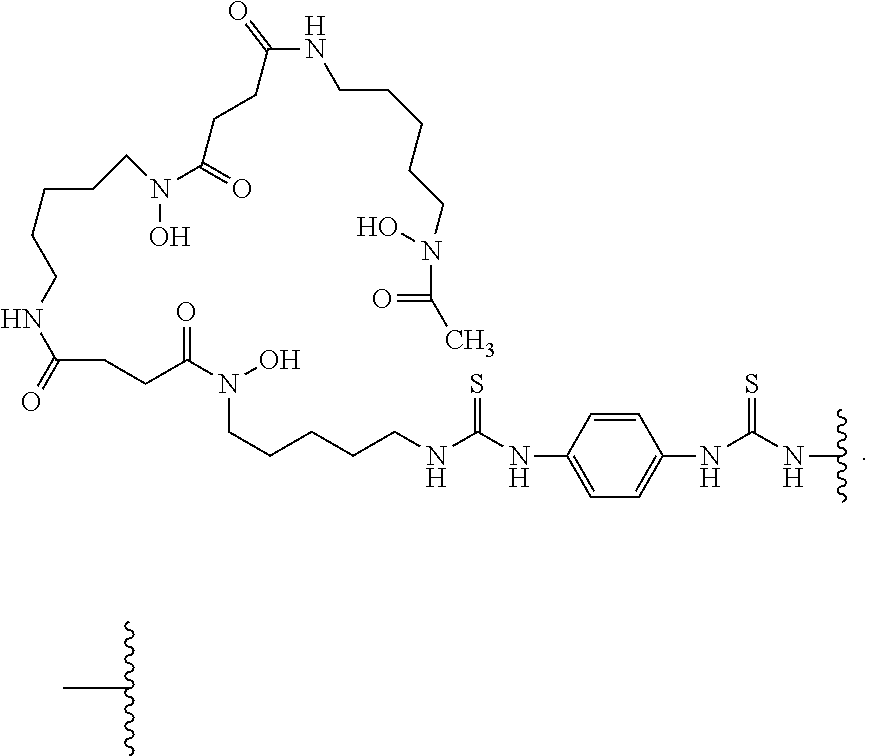
5. The conjugate of claim 2, wherein -L-M is ##STR00007## wherein Zr is the positron emitter
6. The conjugate of claim 2, wherein antibody or antigen-binding fragment thereof is covalently bonded to one, two, or three moieties of Formula (A).
7. The conjugate of claim 1, wherein the antibody has one or more properties selected from the group consisting of: (i) comprises a HCVR having an amino acid sequence selected from the group consisting of SEQ ID NO: 2, 10, 18, 26, 34, 42, 50, 58, 66, 74, 82, 90, 98, 106, 114, 122, and 130, or a substantially similar sequence thereof having at least 90%, at least 95%, at least 98% or at least 99% sequence identity; (ii) comprises a LCVR having an amino acid sequence of SEQ ID NO: 138, or a substantially similar sequence thereof having at least 90%, at least 95%, at least 98% or at least 99% sequence identity; (iii) comprises a HCDR3 domain having an amino acid sequence selected from the group consisting of SEQ ID NO: 8, 16, 24, 32, 40, 48, 56, 64, 72, 80, 88, 96, 104, 112, 120, 128 and 136, or a substantially similar sequence thereof having at least 90%, at least 95%, at least 98% or at least 99% sequence identity; and a LCDR3 domain having an amino acid sequence of SEQ ID NO: 144, or a substantially similar sequence thereof having at least 90%, at least 95%, at least 98% or at least 99% sequence identity; (iv) comprises a HCDR1 domain having an amino acid sequence selected from the group consisting of SEQ ID NO: 4, 12, 20, 28, 36, 44, 52, 60, 68, 76, 84, 92, 100, 108, 116, 124, and 132, or a substantially similar sequence thereof having at least 90%, at least 95%, at least 98% or at least 99% sequence identity; a HCDR2 domain having an amino acid sequence selected from the group consisting of SEQ ID NO: 6, 14, 22, 30, 38, 46, 54, 62, 70, 78, 86, 94, 102, 110, 118, 126, and 134, or a substantially similar sequence thereof having at least 90%, at least 95%, at least 98% or at least 99% sequence identity; a LCDR1 domain having an amino acid sequence of SEQ ID NO: 140, or a substantially similar sequence thereof having at least 90%, at least 95%, at least 98% or at least 99% sequence identity; and a LCDR2 domain having an amino acid sequence of SEQ ID NO: 142, or a substantially similar sequence thereof having at least 90%, at least 95%, at least 98% or at least 99% sequence identity; (v) is a multi-specific antigen-binding molecule comprising a first binding specificity to MET and a second binding specificity to a tumor specific antigen; (vi) is a multi-specific antigen-binding molecule comprising a first binding specificity to one epitope of MET and a second binding specificity to a second epitope of MET; (vii) binds to monomeric human MET (e.g., hMET.mmh) with a K.sub.D of less than about 230 nM as measured by surface plasmon resonance at 25.degree. C. or 37.degree. C.; (viii) binds to dimeric human MET with a K.sub.D of less than about 3 nM as measured by surface plasmon resonance at 25.degree. C. or 37.degree. C.; (ix) blocks the binding of HGF to MET; and (x) suppresses tumor growth and increases survival in subjects with cancer.
8. The conjugate of claim 1, wherein the antibody comprises three heavy chain complementarity determining regions (HCDRs) in a heavy chain variable region (HCVR), wherein the HCVR has an amino acid sequence selected from the group consisting of SEQ ID NOs: 2, 10, 18, 26, 34, 42, 50, 58, 66, 74, 82, 90, 98, 106, 114, 122, and 130; and three light chain complementarity determining regions (LCDRs) in a light chain variable region (LCVR), wherein the LCVR has an amino acid sequence of SEQ ID NOs: 138.
9. The conjugate of claim 1, wherein the antibody comprises three CDRs in a HCVR of SEQ ID NO: 18.
10. The conjugate of claim 1, wherein the antibody comprises three CDRs in a HCVR of SEQ ID NO: 58.
11. The conjugate of claim 1, wherein the antibody comprises three CDRs in a HCVR of SEQ ID NO: 82.
12. The conjugate of claim 1, wherein the antibody comprises three CDRs in a LCVR of SEQ ID NO: 138.
13. The conjugate of claim 1, wherein the antibody comprises: (i) a first antigen-binding domain (D1); and (ii) a second antigen-binding domain (D2); wherein D1 specifically binds a first epitope of human MET; and wherein D2 specifically binds a second epitope of human MET.
14. The conjugate of claim 13, wherein: (i) D1 comprises the CDRs within an HCVR amino acid sequence of SEQ ID NO: 58; and (ii) D2 comprises the CDRs within an HCVR amino acid sequence of SEQ ID NOs: 82.
15. The conjugate of claim 13, wherein: (i) D1 comprises a HCDR1-HCDR2-HCDR3-LCDR1-LCDR2-LCDR3 set of amino acid sequences of SEQ ID NOs: 60-62-64-140-142-144; and (ii) D2 comprises a HCDR1-HCDR2-HCDR3-LCDR1-LCDR2-LCDR3 set of amino acid sequences of SEQ ID NOs: 84-86-88-140-142-144.
16. A method of imaging a tissue that expresses MET comprising administering a radiolabeled antibody conjugate of claim 1 to the tissue; and visualizing MET expression by positron emission tomography (PET) imaging.
17. A method of identifying a MET expressing tumor in a subject, the method comprising administering a radiolabeled antibody conjugate of claim 1 to the subject; imaging the radiolabeled antibody conjugate via positron emission tomography (PET); wherein localization of the radiolabeled antibody conjugate in the subject indicates a MET expressing tumor.
18. A method for treating a subject having a solid tumor comprising: (a) determining that the solid tumor is MET-positive; and (b) administering one or more doses of an inhibitor of the HGF/MET signaling pathway to the subject in need thereof.
19. The method of claim 19, wherein step (a) comprises: (i) administering a radiolabeled antibody conjugate of claim 1 to the subject in need thereof; and (ii) imaging localization of the radiolabeled antibody conjugate in the tumor by positron emission tomography (PET) imaging, wherein presence of the radiolabeled antibody conjugate in the tumor indicates that the tumor is MET-positive.
20. The method of claim 19, wherein the subject is administered 0.1-10 mg/kg of the radiolabeled antibody conjugate.
21. The method of claim 19, wherein the radiolabeled antibody conjugate is administered sub-cutaneously or intravenously to the subject.
22. The method of claim 19, wherein PET imaging is done 2-7 days after administering the radiolabeled antibody conjugate.
23. The method of claim 18, wherein step (a) is carried out before treating the subject with an inhibitor of the HGF/MET signaling pathway.
24. The method of claim 18 further comprising: (a) administering the radiolabeled antibody conjugate after treating the subject with at least one dose of an inhibitor of the HGF/MET signaling pathway; and (b) imaging localization of the radiolabeled antibody conjugate in the tumor by PET imaging, wherein a decrease from the baseline in the area of localization of the radiolabeled antibody conjugate in the tumor indicates tumor regression.
25. The method of claim 19, wherein the subject is administered the radiolabeled antibody conjugate 1-20 weeks after administration of the inhibitor of the HGF/MET signaling pathway.
26. The method of claim 18, wherein the tumor is selected from the group consisting of acute myelogenous leukemia, adult T-cell leukemia, astrocytomas, bladder cancer, breast cancer, cervical cancer, cholangiocarcinoma, chronic myeloid leukemia, colorectal cancer, endometrial cancer, esophageal cancer, gastric cancer (e.g., gastric cancer with MET amplification), glioblastomata, head and neck cancer (e.g., head and neck squamous cell carcinoma [HNSCC]), Kaposi's sarcoma, kidney cancer, leiomyosarcomas, liver cancer, lung cancer (e.g., non-small cell lung cancer [NSCLC]), lymphomas, malignant gliomas, malignant mesothelioma, melanoma, mesothelioma, MFH/fibrosarcoma, multiple myeloma, nasopharyngeal cancer, osteosarcoma, ovarian cancer, pancreatic carcinoma, prostate cancer, renal cell carcinoma, rhabdomyosarcoma, small cell lung cancer, synovial sarcoma, thyroid cancer, and Wilms' tumor.
27. The method of claim 18, wherein the tumor is selected from the group consisting of gastric cancer or non-small cell lung cancer.
28. The method of claim 18, wherein the inhibitor of the HGF/MET signaling pathway is an antibody or antigen-binding fragment thereof.
29. The method of claim 18, wherein the inhibitor of the HGF/MET signaling pathway is a MET.times.MET bispecific antibody or antigen-binding fragment thereof.
30. The method of claim 29, wherein the MET.times.MET bispecific antibody or antigen-binding fragment thereof comprises: (i) a first antigen-binding domain (D1) comprising the CDRs within an HCVR amino acid sequence of SEQ ID NO: 58; and (ii) a second antigen-binding domain (D2) comprising the CDRs within an HCVR amino acid sequence of SEQ ID NOs: 82.
31. The method of claim 30, wherein the MET.times.MET bispecific antibody or antigen-binding fragment thereof comprises the CDRs within an LCVR amino acid sequence of SEQ ID NO: 138.
32. The method of claim 30, wherein: D1 comprises a heavy chain variable region (HCVR) of SEQ ID NO: 58; and D2 comprises a HCVR of amino acid sequence NO: 82.
33. A compound of Formula (III): ##STR00008## wherein A is an antibody or antigen binding fragment thereof that binds MET and k is an integer from 1-30.
34. The compound of claim 33, wherein k is 1 or 2.
35. An antibody conjugate comprising (i) an antibody or antigen-binding fragment thereof that binds MET and (ii) one or more chelating moieties.
36. The antibody conjugate of claim 35, wherein the chelating moiety is ##STR00009## wherein ##STR00010## is a covalent bond to the antibody or antigen-binding fragment thereof.
37. The antibody conjugate of claim 36, wherein said conjugate has a chelating moiety to antibody ratio of from 1.0 to 3.0.
38. The antibody conjugate of claim 36, wherein the chelating moiety-to-antibody ratio is about 1.3.
Description
RELATED APPLICATIONS
[0001] This application claims priority under 35 U.S.C. 119 (e) to U.S. Provisional Patent Application Ser. No. 62/901,003, entitled "RADIOLABELED MET BINDING PROTEINS FOR IMMUNO-PET IMAGING", filed Sep. 16, 2019, the disclosure of which is hereby incorporated by reference in its entirety.
FIELD
[0002] This disclosure relates to radiolabeled MET binding proteins and their use in immuno-PET imaging.
SEQUENCE LISTING
[0003] An official copy of the sequence listing is submitted concurrently with the specification electronically via EFS-Web as an ASCII formatted sequence listing with a file name of "10649US01SEQ_LIST_ST25.txt", a creation date of Sep. 15, 2020, and a size of about 136 KB. The sequence listing contained in this ASCII formatted document is part of the specification and is herein incorporated by reference in its entirety.
BACKGROUND
[0004] Hepatocyte growth factor (HGF) (a.k.a. scatter factor [SF]) is a heterodimeric paracrine growth factor that exerts its activity by interacting with the HGF receptor (HGFR). HGFR is the product of the c-Met oncogene and is also known as MET. MET is a receptor tyrosine kinase consisting of a transmembrane beta chain linked via a disulfide bridge to an extracellular alpha chain. The binding of HGF to MET activates the kinase catalytic activity of MET resulting in the phosphorylation of Tyr 1234 and Tyr 1235 of the beta chain and subsequent activation of downstream signaling pathways.
[0005] MET and/or HGF overexpression, activation, or amplification has been shown to be involved in non-small cell lung carcinoma (NSCLC), gastric, ovarian, pancreatic, thyroid, breast, head and neck, colon and kidney carcinomas (Sierra and Tsao, Ther. Adv. Med. Oncol., 3(1 Suppl): S21-S35, 2011). MET amplification is thought to be a key driver of oncogenesis in NSCLCs and oesophagogastric malignancies. In addition, mutations resulting in exon 14 deletion of MET have been described as oncogenic drivers in a subset of NSCLC. Tumor cell lines having MET gene amplification are highly dependent on MET for growth and survival. Preclinical data implicate MET signaling in resistance to targeted therapies in multiple tumor types, such as NSCLC, colorectal cancer, and head and neck squamous-cell carcinoma (HNSCC).
[0006] Immuno-positron emission tomography (PET) is a diagnostic imaging tool that utilizes monoclonal antibodies labeled with positron emitters, combining the targeting properties of an antibody with the sensitivity of positron emission tomography cameras. See, e.g., The Oncologist, 12: 1379 (2007); Journal of Nuclear Medicine, 52(8): 1171 (2011). Immuno-PET enables the visualization and quantification of antigen and antibody accumulation in vivo and, as such, can serve as an important tool for diagnostics and complementing therapy. For example, immuno-PET can aid in the selection of potential candidates for a particular therapy, as well as in the monitoring of treatment.
[0007] Both preclinical and recent clinical results indicate that tumors harboring MET genetic alterations respond to MET inhibitors, validating MET as a cancer driver. As such, there is need for diagnostic tools for anti-MET and/or anti-MET therapy, including, inter alia, diagnostic tools that enable the detection of suitable candidates for said therapy.
BRIEF SUMMARY
[0008] Included in this disclosure are radiolabeled anti-MET antibody conjugates and MET.times.MET bispecific antibody conjugates for use in immuno-PET imaging.
[0009] In one aspect, the conjugate comprises an anti-MET antibody, a MET.times.MET bispecific antibody, or an antigen-binding fragment thereof, a chelating moiety, and a positron emitter.
[0010] Provided herein are also processes for synthesizing said conjugates and synthetic intermediates useful for the same.
[0011] Provided herein are also methods of imaging a tissue that expresses MET, the methods comprising administering a radiolabeled anti-MET antibody conjugate or MET.times.MET bispecific antibody conjugate described herein to the tissue; and visualizing the MET expression by positron emission tomography (PET) imaging.
[0012] Provided herein are also methods for detecting MET in a tissue, the methods comprising administering a radiolabeled anti-MET antibody conjugate or MET.times.MET bispecific antibody conjugate described herein to the tissue; and visualizing the MET expression by PET imaging. In one embodiment, the tissue is present in a human subject. In certain embodiments, the subject is a non-human mammal. In certain embodiments, the subject has a disease or disorder such as cancer.
[0013] Provided herein are also methods for determining the presence of MET expressing cells in a subject. The methods comprise administering a radiolabeled anti-MET antibody conjugate or MET.times.MET bispecific antibody conjugate described herein to the subject and visualizing MET expression by PET imaging.
[0014] Provided herein are also methods for identifying a subject having a solid tumor to be suitable for anti-tumor therapy comprising an inhibitor of the HGF/MET signaling pathway, for example, an anti-MET antibody, a MET.times.MET bispecific antibody, or an antibody drug conjugate (ADC) thereof. The methods comprise administering a radiolabeled antibody conjugate described herein to the subject, and visualizing the administered radiolabeled antibody conjugate in the tumor by PET imaging wherein presence of the radiolabeled antibody conjugate in the tumor identifies the subject as suitable for anti-tumor therapy comprising an inhibitor of the HGF/MET signaling pathway.
[0015] Provided herein are also methods of treating a solid tumor in a subject, the methods comprising determining that the solid tumor is MET-positive; and administering an anti-tumor therapy to the subject in need thereof. In certain embodiments, the anti-tumor therapy comprises an anti-MET antibody or a MET.times.MET bispecific antibody. In certain embodiments, the subject is administered a radiolabeled antibody conjugate described herein, and localization of the radiolabeled antibody conjugate is imaged via positron emission tomography (PET) imaging to determine if the tumor is MET-positive.
[0016] Provided herein are also methods for monitoring the efficacy of an anti-tumor therapy in a subject being treated with an anti-tumor therapy, wherein the methods comprise administering a radiolabeled conjugate described herein to the subject; imaging the localization of the administered radiolabeled conjugate in the tumor by PET imaging; and determining tumor growth, wherein a decrease from the baseline in uptake of the conjugate or radiolabeled signal indicates tumor regression and efficacy of the anti-tumor therapy. In certain embodiments, the anti-tumor therapy comprises an inhibitor of the HGF/MET signaling pathway (e.g., an anti-MET antibody or a MET.times.MET bispecific antibody, or an ADC of either).
[0017] Provided herein are also methods for predicting response of a subject to an anti-tumor therapy comprising an inhibitor of the HGF/MET signaling pathway, the methods comprising determining if the tumor is MET-positive, wherein if the tumor is MET-positive it indicates a positive response of the subject to an anti-tumor therapy comprising an inhibitor of the HGF/MET signaling pathway. In certain embodiments, the tumor is determined positive by administering a radiolabeled antibody conjugate of the present disclosure and localizing the radiolabeled antibody conjugate in the tumor by PET imaging wherein presence of the radiolabeled antibody conjugate in the tumor indicates that the tumor is MET-positive.
[0018] Provided herein are methods for diagnosing and treating a subject with a tumor, the methods comprising administering a radiolabeled conjugate described herein to the subject wherein localization of the radiolabeled antibody conjugate is imaged via PET imaging to determine if the tumor is MET-positive; diagnosing the subject with a MET-positive tumor; and administering to the subject an anti-tumor therapy comprising an inhibitor of the HGF/MET signaling pathway.
[0019] Provided herein are methods for diagnosing a subject having a MET expressing tumor, the methods comprising administering a radiolabeled anti-MET antibody conjugate or MET.times.MET bispecific antibody conjugate described herein to the subject; visualizing MET expression by PET imaging; and diagnosing the subject with a MET expressing tumor when MET expression is visualized by PET imaging.
BRIEF DESCRIPTION OF THE FIGURES
[0020] FIG. 1 depicts an SE-HPLC chromatogram of a 5 ug injection of DFO-MET.times.MET immunoconjugate conjugate on Superdex 200 Increase column with UV 280 nm absorbance detection. Monomeric (99.6%) and high molecular weight (HMW) species (0.4%) are indicated.
[0021] FIG. 2 depicts an image of SDS-PAGE of the DFO-MET.times.MET immunoconjugate. The gel demonstrates that the antibody integrity remains unchanged after DFO conjugation. Lanes are labeled as follows: 1) Standard ladder (BioRad, Cat. #: 161-0374), 2) bispecific antibody non-reduced, 3) DFO-Ab immunoconjugate non-reduced, 4) blank, 5) bispecific antibody reduced, 6) DFO-Ab immunoconjugate non-reduced. Each well was loaded with approximately 2 ug of protein. Note that non-reduced antibodies typically demonstrate less electrophoretic motility than expected as compared to the ladder for the standard SDS-PAGE setup.
[0022] FIG. 3 depicts a representative SE-HPLC radiochromatogram of a 5 ug injection of a radioimmunoconjugate (DFO-MET.times.MET bispecific antibody) with gamma emission detection. The RCP of is greater than 95% while unincorporated .sup.89Zr makes up less than 1% of total integrated activity.
[0023] FIG. 4 depicts a representative SE-HPLC UV absorption chromatogram of a 5 ug injection of a radioimmunoconjugate (DFO-MET.times.MET bispecific antibody). Main (97.9%) and HMW (2.1%) species are indicated. The elution peaks between 25 and 31 minutes is a phenomenon of formulation buffer/mobile phase mixing and is deemed not proteinaceous in origin.
[0024] FIG. 5 depicts PET/CT images of EBC-1 tumor xenografts in mice. The mice were administered radiolabeled MET.times.MET bispecific antibody conjugate and over several days, the conjugate specifically localized to the MET expressing tumor xenografts.
[0025] FIG. 6 depicts PET/CT images of NCI-H441 tumor xenografts in mice. The mice were administered radiolabeled MET.times.MET bispecific antibody conjugate and over several days, the conjugate specifically localized to the MET expressing tumor xenografts.
[0026] FIG. 7 depicts PET/CT images of NCI-H358 tumor xenografts in mice. The mice were administered radiolabeled MET.times.MET bispecific antibody conjugate and over several days, the conjugate specifically localized to the MET expressing tumor xenografts.
[0027] FIG. 8A, FIG. 8B, FIG. 8C, FIG. 8D, FIG. 8E, and FIG. 8F provide ex vivo biodistribution data for .sup.89Zr-DFO-MET.times.MET bispecific antibody conjugate in SCID mice with tumor xenografts. Mice were administered a single IV dose 0.1 mg/kg, 0.5 mg/kg, or 5.0 mg/kg .sup.89Zr-DFO-MET.times.MET bispecific antibody conjugate and were sacrificed 6 days later. Blood, collected via cardiac puncture, and the indicated harvested tissues were weighed and radioactivity was determined. The percent injected dose per gram (% ID/g) values for individual samples collected on day 6 were calculated relative to the radioactivity of a dose-standard from injected material (.sup.89Zr-DFO-MET.times.MET bispecific antibody conjugate) and the weight of the individual samples. Data are plotted as mean.+-.SD.
[0028] FIG. 9 shows the correlation between uptake of .sup.89Zr-DFO-MET.times.MET bispecific antibody and MET expression level in the tumor xenografts from three MET expressing cell lines.
[0029] FIG. 10A and FIG. 10B show antibody saturation binding data for three MET expressing cell lines.
DETAILED DESCRIPTION
I. Definitions
[0030] Before the present invention is described, it is to be understood that this invention is not limited to particular methods and experimental conditions described, as such methods and conditions may vary. It is also to be understood that the terminology used herein is for the purpose of describing particular embodiments only, and is not intended to be limiting, since the scope of the present invention will be limited only by the appended claims.
[0031] Unless defined otherwise herein, all technical and scientific terms used herein have the same meaning as commonly understood by one of ordinary skill in the art to which the disclosed subject matter belongs. As used herein, the term "about," when used in reference to a particular recited numerical value, means that the value may vary from the recited value by no more than 1%. For example, as used herein, the expression "about 100" includes 99 and 101 and all values in between (e.g., 99.1, 99.2, 99.3, 99.4, etc.).
[0032] Although any methods and materials similar or equivalent to those described herein can be used in the practice or testing of the present invention, the preferred methods and materials are now described. All patents, applications and non-patent publications mentioned in this specification are incorporated herein by reference in their entireties.
MET Protein
[0033] The expressions "MET," "c-Met," and the like, as used herein, refer to the human membrane spanning receptor tyrosine kinase comprising (1) the amino acid sequence as set forth in SEQ ID NO:145, and/or having the amino acid sequence as set forth in NCBI accession No. NM_001127500.2, representing the unprocessed preproprotein of isoform "a", (2) the amino acid sequence as set forth in SEQ ID NO:146, and/or having the amino acid sequence as set forth in NCBI accession No. NM_000236.2, representing the unprocessed preproprotein of isoform "b", (3) the amino acid sequence as set forth in SEQ ID NO:147, and/or having the amino acid sequence as set forth in NCBI accession No. NM_001311330.1, representing the unprocessed preproprotein of isoform "c", and/or (3) the mature protein comprising the cytoplasmic alpha subunit (SEQ ID NO:148) shared by all three isoforms and the transmembrane beta subunit (SEQ ID NO:149, 150, or 151 of isoform a, b and c, respectively). The expression "MET" includes both monomeric and multimeric MET molecules. As used herein, the expression "monomeric human MET" means a MET protein or portion thereof that does not contain or possess any multimerizing domains and that exists under normal conditions as a single MET molecule without a direct physical connection to another MET molecule. An exemplary monomeric MET molecule is the molecule referred to herein as "hMET.mmh" comprising the amino acid sequence of SEQ ID NO:152 (see, e.g., Example 3 of US-2018-0134794). As used herein, the expression "dimeric human MET" means a construct comprising two MET molecules connected to one another through a linker, covalent bond, non-covalent bond, or through a multimerizing domain such as an antibody Fc domain. An exemplary dimeric MET molecule is the molecule referred to herein as "hMET.mFc" comprising the amino acid sequence of SEQ ID NO:153 (see, e.g., Example 3 of US-2018-0134794).
[0034] All references to proteins, polypeptides and protein fragments herein are intended to refer to the human version of the respective protein, polypeptide or protein fragment unless explicitly specified as being from a non-human species. Thus, the expression "MET" means human MET unless specified as being from a non-human species, e.g., "mouse MET," "monkey MET," etc.
[0035] As used herein, the expression "cell surface-expressed MET" means one or more MET protein(s), or the extracellular domain thereof, that is/are expressed on the surface of a cell in vitro or in vivo, such that at least a portion of a MET protein is exposed to the extracellular side of the cell membrane and is accessible to an antigen-binding portion of an antibody. A "cell surface-expressed MET" can comprise or consist of a MET protein expressed on the surface of a cell which normally expresses MET protein. Alternatively, "cell surface-expressed MET" can comprise or consist of MET protein expressed on the surface of a cell that normally does not express human MET on its surface but has been artificially engineered to express MET on its surface.
Other Definitions
[0036] The term "antibody", as used herein, is intended to refer to immunoglobulin molecules comprised of four polypeptide chains, two heavy (H) chains and two light (L) chains inter-connected by disulfide bonds (i.e., "full antibody molecules"), as well as multimers thereof (e.g. IgM) or antigen-binding fragments thereof. Each heavy chain is comprised of a heavy chain variable region ("HCVR" or "V.sub.H") and a heavy chain constant region (comprised of domains C.sub.H1, C.sub.H2 and C.sub.H3). Each light chain is comprised of a light chain variable region ("LCVR or "V.sub.L") and a light chain constant region (C.sub.L). The V.sub.H and V.sub.L regions can be further subdivided into regions of hypervariability, termed complementarity determining regions (CDR), interspersed with regions that are more conserved, termed framework regions (FR). Each V.sub.H and V.sub.L is composed of three CDRs and four FRs, arranged from amino-terminus to carboxy-terminus in the following order: FR1, CDR1, FR2, CDR2, FR3, CDR3, FR4. In certain embodiments, the FRs of the antibody (or antigen binding fragment thereof) may be identical to the human germline sequences, or may be naturally or artificially modified. An amino acid consensus sequence may be defined based on a side-by-side analysis of two or more CDRs.
[0037] Substitution of one or more CDR residues or omission of one or more CDRs is also possible. Antibodies have been described in the scientific literature in which one or two CDRs can be dispensed with for binding. Padlan et al. (1995 FASEB J. 9:133-139) analyzed the contact regions between antibodies and their antigens, based on published crystal structures, and concluded that only about one fifth to one third of CDR residues actually contact the antigen. Padlan also found many antibodies in which one or two CDRs had no amino acids in contact with an antigen (see also, Vajdos et al. 2002 J Mol Biol 320:415-428).
[0038] CDR residues not contacting antigen can be identified based on previous studies (for example residues H60-H65 in CDRH2 are often not required), from regions of Kabat CDRs lying outside Chothia CDRs, by molecular modeling and/or empirically. If a CDR or residue(s) thereof is omitted, it is usually substituted with an amino acid occupying the corresponding position in another human antibody sequence or a consensus of such sequences. Positions for substitution within CDRs and amino acids to substitute can also be selected empirically. Empirical substitutions can be conservative or non-conservative substitutions.
[0039] The human anti-MET antibodies or MET.times.MET bispecific antibodies useful herein may comprise one or more amino acid substitutions, insertions and/or deletions in the framework and/or CDR regions of the heavy and light chain variable domains as compared to the corresponding germline sequences. Such mutations can be readily ascertained by comparing the amino acid sequences of Table 1 to germline sequences available from, for example, public antibody sequence databases. Useful according to the present disclosure are antibodies, and antigen-binding fragments thereof, which are derived from any of the amino acid sequences provided in Table 1, wherein one or more amino acids within one or more framework and/or CDR regions are mutated to the corresponding residue(s) of the germline sequence from which the antibody was derived, or to the corresponding residue(s) of another human germline sequence, or to a conservative amino acid substitution of the corresponding germline residue(s) (such sequence changes are referred to herein collectively as "germline mutations"). A person of ordinary skill in the art, starting with the heavy and light chain variable region sequences according to Table 1, can easily produce numerous antibodies and antigen-binding fragments which comprise one or more individual germline mutations or combinations thereof. In certain embodiments, all of the framework and/or CDR residues within the V.sub.H and/or V.sub.L domains are mutated back to the residues found in the original germline sequence from which the antibody was derived. In other embodiments, only certain residues are mutated back to the original germline sequence, e.g., only the mutated residues found within the first 8 amino acids of FR1 or within the last 8 amino acids of FR4, or only the mutated residues found within CDR1, CDR2 or CDR3. In other embodiments, one or more of the framework and/or CDR residue(s) are mutated to the corresponding residue(s) of a different germline sequence (i.e., a germline sequence that is different from the germline sequence from which the antibody was originally derived). Furthermore, the antibodies of the present disclosure may contain any combination of two or more germline mutations within the framework and/or CDR regions, e.g., wherein certain individual residues are mutated to the corresponding residue of a particular germline sequence while certain other residues that differ from the original germline sequence are maintained or are mutated to the corresponding residue of a different germline sequence. Once obtained, antibodies and antigen-binding fragments that contain one or more germline mutations can be easily tested for one or more desired property such as, improved binding specificity, increased binding affinity, improved or enhanced antagonistic or agonistic biological properties (as the case may be), reduced immunogenicity, etc. Antibodies and antigen-binding fragments obtained in this general manner are encompassed within the present disclosure.
[0040] Useful herein are MET binding proteins such as human anti-MET antibodies and MET.times.MET bispecific antibodies comprising variants of any of the HCVR, LCVR, and/or CDR amino acid sequences shown in Table 1 herein having one or more conservative substitutions. For example, the present disclosure includes MET.times.MET bispecific antibodies having HCVR, LCVR, and/or CDR amino acid sequences with, e.g., 10 or fewer, 8 or fewer, 6 or fewer, 4 or fewer, etc. conservative amino acid substitutions relative to any of the HCVR, LCVR, and/or CDR amino acid sequences of Table 1.
[0041] The term "human antibody", as used herein, is intended to include antibodies having variable and constant regions derived from human germline immunoglobulin sequences. The human monoclonal antibodies of the disclosure may include amino acid residues not encoded by human germline immunoglobulin sequences (e.g., mutations introduced by random or site-specific mutagenesis in vitro or by somatic mutation in vivo), for example in the CDRs and in particular CDR3. However, the term "human antibody", as used herein, is not intended to include monoclonal antibodies in which CDR sequences derived from the germline of another mammalian species (e.g., mouse), have been grafted onto human FR sequences.
[0042] The term "multi-specific antigen-binding molecules", as used herein refers to bispecific, tri-specific or multi-specific antigen-binding molecules, and antigen-binding fragments thereof. Multi-specific antigen-binding molecules may be specific for different epitopes of one target polypeptide or may contain antigen-binding domains specific for epitopes of more than one target polypeptide. A multi-specific antigen-binding molecule can be a single multifunctional polypeptide, or it can be a multimeric complex of two or more polypeptides that are covalently or non-covalently associated with one another. The term "multi-specific antigen-binding molecules" includes antibodies of the present disclosure that may be linked to or co-expressed with another functional molecule, e.g., another peptide or protein. For example, an antibody or fragment thereof can be functionally linked (e.g., by chemical coupling, genetic fusion, non-covalent association or otherwise) to one or more other molecular entities, such as a protein or fragment thereof to produce a bi-specific or a multi-specific antigen-binding molecule with a second binding specificity. According to the present disclosure, the term "multi-specific antigen-binding molecules" also includes bi-specific, tri-specific or multi-specific antibodies or antigen-binding fragments thereof. In certain embodiments, an antibody of the present disclosure is functionally linked to another antibody or antigen-binding fragment thereof to produce a bispecific antibody with a second binding specificity. Bispecific and multi-specific antibodies of the present disclosure are described elsewhere herein.
[0043] The term "specifically binds," or "binds specifically to", or the like, means that an antibody or antigen-binding fragment thereof forms a complex with an antigen that is relatively stable under physiologic conditions. Specific binding can be characterized by an equilibrium dissociation constant of at least about 1.times.10.sup.-8 M or less (e.g., a smaller K.sub.D denotes a tighter binding). Methods for determining whether two molecules specifically bind are well known in the art and include, for example, equilibrium dialysis, surface plasmon resonance, and the like. As described herein, antibodies have been identified by surface plasmon resonance, e.g., BIACORE.TM., which bind specifically to MET. Moreover, multi-specific antibodies that bind to one domain in MET and one or more additional antigens or a bi-specific that binds to two different regions of MET are nonetheless considered antibodies that "specifically bind", as used herein.
[0044] The terms "antigen-binding portion" of an antibody, "antigen-binding fragment" of an antibody, and the like, as used herein, include any naturally occurring, enzymatically obtainable, synthetic, or genetically engineered polypeptide or glycoprotein that specifically binds an antigen to form a complex. The terms "antigen-binding fragment" of an antibody, or "antibody fragment", as used herein, refers to one or more fragments of an antibody that retain the ability to bind to MET.
[0045] An "isolated antibody", as used herein, is intended to refer to an antibody that is substantially free of other antibodies (Abs) having different antigenic specificities (e.g., an isolated antibody that specifically binds MET, or a fragment thereof, is substantially free of Abs that specifically bind antigens other than MET.
[0046] The term "surface plasmon resonance", as used herein, refers to an optical phenomenon that allows for the analysis of real-time biomolecular interactions by detection of alterations in protein concentrations within a biosensor matrix, for example using the BIACORE.TM. system (Pharmacia Biosensor AB, Uppsala, Sweden and Piscataway, N.J.).
[0047] The term "K.sub.D", as used herein, is intended to refer to the equilibrium dissociation constant of a particular antibody-antigen interaction.
[0048] The term "epitope" refers to an antigenic determinant that interacts with a specific antigen binding site in the variable region of an antibody molecule known as a paratope. A single antigen may have more than one epitope. Thus, different antibodies may bind to different areas on an antigen and may have different biological effects. The term "epitope" also refers to a site on an antigen to which B and/or T cells respond. It also refers to a region of an antigen that is bound by an antibody. Epitopes may be defined as structural or functional. Functional epitopes are generally a subset of the structural epitopes and have those residues that directly contribute to the affinity of the interaction. Epitopes may also be conformational, that is, composed of non-linear amino acids. In certain embodiments, epitopes may include determinants that are chemically active surface groupings of molecules such as amino acids, sugar side chains, phosphoryl groups, or sulfonyl groups, and, in certain embodiments, may have specific three-dimensional structural characteristics, and/or specific charge characteristics.
[0049] The term "substantial identity" or "substantially identical," when referring to a nucleic acid or fragment thereof, indicates that, when optimally aligned with appropriate nucleotide insertions or deletions with another nucleic acid (or its complementary strand), there is nucleotide sequence identity in at least about 90%, and more preferably at least about 95%, 96%, 97%, 98% or 99% of the nucleotide bases, as measured by any well-known algorithm of sequence identity, such as FASTA, BLAST or GAP.
[0050] As applied to polypeptides, the term "substantial similarity" or "substantially similar" means that two peptide sequences, when optimally aligned, such as by the programs GAP or BESTFIT using default gap weights, share at least 90% sequence identity, even more preferably at least 95%, 98% or 99% sequence identity. Preferably, residue positions, which are not identical, differ by conservative amino acid substitutions. A "conservative amino acid substitution" is one in which an amino acid residue is substituted by another amino acid residue having a side chain (R group) with similar chemical properties (e.g., charge or hydrophobicity). In general, a conservative amino acid substitution will not substantially change the functional properties of a protein. In cases where two or more amino acid sequences differ from each other by conservative substitutions, the percent or degree of similarity may be adjusted upwards to correct for the conservative nature of the substitution. Means for making this adjustment are well known to those of skill in the art. See, e.g., Pearson (1994) Methods Mol. Biol. 24: 307-331, which is herein incorporated by reference. Examples of groups of amino acids that have side chains with similar chemical properties include 1) aliphatic side chains: glycine, alanine, valine, leucine and isoleucine; 2) aliphatic-hydroxyl side chains: serine and threonine; 3) amide-containing side chains: asparagine and glutamine; 4) aromatic side chains: phenylalanine, tyrosine, and tryptophan; 5) basic side chains: lysine, arginine, and histidine; 6) acidic side chains: aspartate and glutamate, and 7) sulfur-containing side chains: cysteine and methionine. Preferred conservative amino acids substitution groups are: valine-leucine-isoleucine, phenylalanine-tyrosine, lysine-arginine, alanine-valine, glutamate-aspartate, and asparagine-glutamine. Alternatively, a conservative replacement is any change having a positive value in the PAM250 log-likelihood matrix disclosed in Gonnet et al. (1992) Science 256: 1443 45, herein incorporated by reference. A "moderately conservative" replacement is any change having a nonnegative value in the PAM250 log-likelihood matrix. Sequence similarity for polypeptides is typically measured using sequence analysis software. Protein analysis software matches similar sequences using measures of similarity assigned to various substitutions, deletions and other modifications, including conservative amino acid substitutions. For instance, GCG software contains programs such as GAP and BESTFIT which can be used with default parameters to determine sequence homology or sequence identity between closely related polypeptides, such as homologous polypeptides from different species of organisms or between a wild type protein and a mutein thereof. See, e.g., GCG Version 6.1. Polypeptide sequences also can be compared using FASTA with default or recommended parameters; a program in GCG Version 6.1. FASTA (e.g., FASTA2 and FASTA3) provides alignments and percent sequence identity of the regions of the best overlap between the query and search sequences (Pearson (2000) supra). Another preferred algorithm when comparing a sequence of the disclosure to a database containing a large number of sequences from different organisms is the computer program BLAST, especially BLASTP or TBLASTN, using default parameters. See, e.g., Altschul et al. (1990) J. Mol. Biol. 215: 403-410 and (1997) Nucleic Acids Res. 25:3389-3402, each of which is herein incorporated by reference.
[0051] By the phrase "therapeutically effective amount" is meant an amount that produces the desired effect for which it is administered. The exact amount will depend on the purpose of the treatment, and will be ascertainable by one skilled in the art using known techniques (see, for example, Lloyd (1999) The Art, Science and Technology of Pharmaceutical Compounding).
[0052] As used herein, the term "subject" refers to an animal, preferably a mammal, in need of amelioration, prevention and/or treatment of a disease or disorder such as cancer.
II. Radiolabeled Immunoconjugates of MET Antibodies for Immuno-PET Imaging
[0053] Provided herein are radiolabeled antigen-binding proteins that bind MET protein. In some embodiments, the radiolabeled antigen-binding proteins comprise an antigen-binding protein covalently linked to one or more chelating moieties, which are chemical moieties that are capable of chelating a positron emitter.
[0054] In some embodiments, provided herein are antigen-binding proteins that bind MET, e.g., anti-MET antibodies or MET.times.MET bispecific antibodies, wherein said antigen-binding proteins that bind MET are covalently bonded to one or more moieties having the following structure:
-L-M.sub.Z
wherein L is a chelating moiety; M is a positron emitter; and z, independently at each occurrence, is 0 or 1; and wherein at least one of z is 1.
[0055] In some embodiments, the radiolabeled antigen-binding protein is a compound of Formula (I):
M-L-A-[L-M.sub.Z].sub.k (I)
A is a protein that binds MET; L is a chelating moiety; M is a positron emitter; z is 0 or 1; and k is an integer from 0-30. In some embodiments, k is 1.
[0056] In certain embodiments, the radiolabeled antigen-binding protein is a compound of Formula (II):
A-[L-M].sub.k (II)
wherein A is a protein that binds MET; L is a chelating moiety; M is a positron emitter; and k is an integer from 1-30.
[0057] In some embodiments, provided herein are compositions comprising a conjugate having the following structure:
A-L.sub.k
wherein A is a protein that binds MET; L is a chelating moiety; and k is an integer from 1-30; wherein the conjugate is chelated with a positron emitter in an amount sufficient to provide a specific activity suitable for clinical PET imaging.
[0058] Suitable binding proteins, chelating moieties, and positron emitters are provided below.
A. MET Binding Proteins
[0059] Suitable MET binding protein are proteins that specifically bind to MET, including those described in U.S. Patent Publication No. 2018-0134794, incorporated herein by reference in its entirety. Amino acid sequence identifiers of exemplary anti-MET antibodies useful herein are listed in Table 1 of U.S. Patent Publication No. 2018-0134794 and amino acid sequence identifiers of exemplary MET.times.MET bispecific antibodies useful herein are listed in Table 5 of U.S. Patent Publication No. 2018-0134794. Both Tables are included below as Tables 1 and 2, respectively.
TABLE-US-00001 TABLE 1 Amino Acid Sequence Identifiers Antibody SEQ ID NOs: Designation HCVR HCDR1 HCDR2 HCDR3 LCVR LCDR1 LCDR2 LCDR3 H4H13290P2 2 4 6 8 138 140 142 144 H4H13291P2 10 12 14 16 138 140 142 144 H4H13295P2 18 20 22 24 138 140 142 144 H4H13299P2 26 28 30 32 138 140 142 144 H4H13300P2 34 36 38 40 138 140 142 144 H4H13301P2 42 44 46 48 138 140 142 144 H4H13302P2 50 52 54 56 138 140 142 144 H4H13306P2 58 60 62 64 138 140 142 144 H4H13309P2 66 68 70 72 138 140 142 144 H4H13311P2 74 76 78 80 138 140 142 144 H4H13312P2 82 84 86 88 138 140 142 144 H4H13313P2 90 92 94 96 138 140 142 144 H4H13316P2 98 100 102 104 138 140 142 144 H4H13318P2 106 108 110 112 138 140 142 144 H4H13319P2 114 116 118 120 138 140 142 144 H4H13325P2 122 124 126 128 138 140 142 144 H4H13331P2 130 132 134 136 138 140 142 144
TABLE-US-00002 TABLE 2 MET .times. MET Bispecific Antibody Components Summary SEQ ID NOs: (Amino Acid Sequences) First Antigen-Binding Second Antigen-Binding Domain (D1) Domain (D2) Bispecific D1- D1- D1- D1- D2- D2- D2- D2- Antibody HCVR HCDR1 HCDR2 HCDR3 HCVR HCDR1 HCDR2 HCDR3 H4H14634D H4H13290P2 H4H13312P2 (No. 10) 2 4 6 8 82 84 86 88 H4H14635D H4H13295P2 H4H13312P2 (No. 42) 18 20 22 24 82 84 86 88 H4H14636D H4H13299P2 H4H13312P2 (No. 74) 26 28 30 32 82 84 86 88 H4H14637D H4H13301P2 H4H13312P2 (No. 90) 42 44 46 48 82 84 86 88 H4H14638D H4H13302P2 H4H13312P2 (No. 106) 50 52 54 56 82 84 86 88 H4H14639D H4H13306P2 H4H13312P2 (No. 122) 58 60 62 64 82 84 86 88 H4H14640D H4H13309P2 H4H13312P2 (No. 138) 66 68 70 72 82 84 86 88 H4H14641D H4H13313P2 H4H13312P2 (No. 187) 90 92 94 96 82 84 86 88 H4H16445D H4H13291P2 H4H13312P2 (No. 26) 10 12 14 16 82 84 86 88 H4H16446D H4H13300P2 H4H13312P2 (No. 58) 34 36 38 40 82 84 86 88 H4H16447D H4H13311P2 H4H13312P2 (No. 154) 74 76 78 80 82 84 86 88 H4H16448D H4H13318P2 H4H13312P2 (No. 219) 106 108 110 112 82 84 86 88 H4H16449D H4H13319P2 H4H13312P2 (No. 235) 114 116 118 120 82 84 86 88 * The number designation in parentheses under the bispecific antibody identifiers (e.g., "No. 10") indicates the bispecific antibody number depicted in the MET .times. MET bispecific antibody matrix of U.S. Patent Publication No. 2018-0134794, FIG. 1.
[0060] Table 1 sets forth the amino acid sequence identifiers of the heavy chain variable regions (HCVRs), light chain variable regions (LCVRs), heavy chain complementarity determining regions (HCDR1, HCDR2 and HCDR3), and light chain complementarity determining regions (LCDR1, LCDR2 and LCDR3) of the exemplary anti-MET antibodies.
[0061] In some embodiments, the binding protein is an antibody or antigen binding fragment comprising an HCVR comprising an amino acid sequence selected from any of the HCVR amino acid sequences listed in Table 1, or a substantially similar sequence thereof having at least 90%, at least 95%, at least 98% or at least 99% sequence identity thereto.
[0062] In some embodiments, the binding protein is an antibody or antigen binding fragment comprising an LCVR amino acid sequence shown in Table 1, or a substantially similar sequence thereof having at least 90%, at least 95%, at least 98% or at least 99% sequence identity thereto.
[0063] In some embodiments, the binding protein is an antibody or antigen binding fragment comprising an HCVR and an LCVR amino acid sequence pair (HCVR/LCVR) comprising any of the HCVR amino acid sequences listed in Table 1 paired with the LCVR amino acid sequence shown in Table 1. According to certain embodiments, the present disclosure provides antibodies, or antigen-binding fragments thereof, comprising an HCVR/LCVR amino acid sequence pair contained within any of the exemplary anti-MET antibodies listed in Table 1. In certain embodiments, the HCVR/LCVR amino acid sequence pair is selected from the group consisting of SEQ ID NOs: 2/138, 10/138, 18/138, 26/138, 34/138, 42/138, 50/138, 58/138, 66/138, 74/138, 82/138, 90/138, 98/138, 106/138, 114/138, 122/138, and 130/138. In certain embodiments, the HCVR/LCVR amino acid sequence pair is selected from one of SEQ ID NOs: 58/138 (e.g., H4H13306P2) and 82/138 (e.g., H4H13312P2).
[0064] In some embodiments, the binding protein is an antibody or antigen binding fragment comprising a heavy chain CDR1 (HCDR1) comprising an amino acid sequence selected from any of the HCDR1 amino acid sequences listed in Table 1 or a substantially similar sequence thereof having at least 90%, at least 95%, at least 98% or at least 99% sequence identity.
[0065] In some embodiments, the binding protein is an antibody or antigen binding fragment comprising a heavy chain CDR2 (HCDR2) comprising an amino acid sequence selected from any of the HCDR2 amino acid sequences listed in Table 1 or a substantially similar sequence thereof having at least 90%, at least 95%, at least 98% or at least 99% sequence identity.
[0066] In some embodiments, the binding protein is an antibody or antigen binding fragment comprising a heavy chain CDR3 (HCDR3) comprising an amino acid sequence selected from any of the HCDR3 amino acid sequences listed in Table 1 or a substantially similar sequence thereof having at least 90%, at least 95%, at least 98% or at least 99% sequence identity.
[0067] In some embodiments, the binding protein is an antibody or antigen binding fragment comprising a light chain CDR1 (LCDR1) comprising an amino acid sequence shown in Table 1 or a substantially similar sequence thereof having at least 90%, at least 95%, at least 98% or at least 99% sequence identity.
[0068] In some embodiments, the binding protein is an antibody or antigen binding fragment comprising a light chain CDR2 (LCDR2) comprising an amino acid sequence shown in Table 1 or a substantially similar sequence thereof having at least 90%, at least 95%, at least 98% or at least 99% sequence identity.
[0069] In some embodiments, the binding protein is an antibody or antigen binding fragment comprising a light chain CDR3 (LCDR3) comprising an amino acid sequence shown in Table 1 or a substantially similar sequence thereof having at least 90%, at least 95%, at least 98% or at least 99% sequence identity.
[0070] In some embodiments, the binding protein is an antibody or antigen binding fragment comprising an HCDR3 and an LCDR3 amino acid sequence pair (HCDR3/LCDR3) comprising any of the HCDR3 amino acid sequences listed in Table 1 paired with the LCDR3 amino acid sequences shown in Table 1. According to certain embodiments, the present disclosure provides antibodies, or antigen-binding fragments thereof, comprising an HCDR3/LCDR3 amino acid sequence pair contained within any of the exemplary anti-MET antibodies listed in Table 1. In certain embodiments, the HCDR3/LCDR3 amino acid sequence pair is selected from the group consisting of SEQ ID NOs: 8/144 (e.g. H4H13290P2), 16/144 (e.g. H4H13291P2), 24/144 (H4H13295P2), 32/144 (H4H13299P2), 40/144 (H4H13300P2), 48/144 (H4H13301P2), 56/144 (H4H13302P2), 64/144 (H4H13306P2), 72/144 (H4H13309P2), 80/144 (H4H13311P2), 88/144 (H4H13312P2), 96/144 (H4H13313P2), 104/144 (H4H13316P2), 112/144 (H4H13318P2), 120/144 (H4H13319P2), 128/144 (H4H13325P2), and 136/144 (H4H13331P2).
[0071] In some embodiments, the binding protein is an antibody or antigen binding fragment comprising a set of six CDRs (i.e., HCDR1-HCDR2-HCDR3-LCDR1-LCDR2-LCDR3) contained within any of the exemplary anti-MET antibodies listed in Table 1. In certain embodiments, the HCDR1-HCDR2-HCDR3-LCDR1-LCDR2-LCDR3 amino acid sequence set is selected from the group consisting of SEQ ID NOs: 4-6-8-140-142-144 (e.g. H4H13290P2), 12-14-16-140-142-144 (e.g. H4H13291P2), 20-22-24-140-142-144 (H4H13295P2), 28-30-32-140-142-144 (H4H13299P2), 36-38-40-140-142-144 (H4H13300P2), 44-44-48-140-142-144 (H4H13301P2), 52-54-56-140-142-144 (H4H13302P2), 60-62-64-140-142-144 (H4H13306P2), 68-70-72-140-142-144 (H4H13309P2), 76-78-80-140-142-144 (H4H13311P2), 84-86-88-140-142-144 (H4H13312P2), 92-94-96-140-142-144 (H4H13313P2), 100-102-104-140-142-144 (H4H13316P2), 108-110-112-140-142-144 (H4H13318P2), 116-118-120-140-142-144 (H4H13319P2), 124-126-128-140-142-144 (H4H13325P2), and 132-134-136-140-142-144 (H4H13331P2).
[0072] In some embodiments, the binding protein is an antibody or antigen binding fragment comprising a set of six CDRs (i.e., HCDR1-HCDR2-HCDR3-LCDR1-LCDR2-LCDR3) contained within an HCVR/LCVR amino acid sequence pair as defined by any of the exemplary anti-MET antibodies listed in Table 1. For example, in some embodiments, the binding protein is an antibody or antigen binding fragment comprising the HCDR1-HCDR2-HCDR3-LCDR1-LCDR2-LCDR3 amino acid sequences set contained within an HCVR/LCVR amino acid sequence pair selected from the group consisting of SEQ ID NOs: 2/138 (e.g. H4H13290P2), 10/138 (e.g. H4H13291P2), 18/138 (H4H13295P2), 26/138 (H4H13299P2), 34/138 (H4H13300P2), 42/138 (H4H13301P2), 50/138 (H4H13302P2), 58/138 (H4H13306P2), 66/138 (H4H13309P2), 74/138 (H4H13311P2), 82/138 (H4H13312P2), 90/138 (H4H13313P2), 98/138 (H4H13316P2), 106/138 (H4H13318P2), 114/138 (H4H13319P2), 122/138 (H4H13325P2), and 130/138 (H4H13331P2).
[0073] Methods and techniques for identifying CDRs within HCVR and LCVR amino acid sequences are well known in the art and can be used to identify CDRs within the specified HCVR and/or LCVR amino acid sequences useful herein. Exemplary conventions that can be used to identify the boundaries of CDRs include, e.g., the Kabat definition, the Chothia definition, and the AbM definition. In general terms, the Kabat definition is based on sequence variability, the Chothia definition is based on the location of the structural loop regions, and the AbM definition is a compromise between the Kabat and Chothia approaches. See, e.g., Kabat, "Sequences of Proteins of Immunological Interest," National Institutes of Health, Bethesda, Md. (1991); Al-Lazikani et al., J. Mol. Biol. 273:927-948 (1997); and Martin et al., Proc. Natl. Acad. Sci. USA 86:9268-9272 (1989). Public databases are also available for identifying CDR sequences within an antibody.
[0074] In some embodiments, binding proteins are antibodies and antigen-binding fragments thereof that compete for specific binding to MET with an antibody or antigen-binding fragment thereof comprising the CDRs of a HCVR and the CDRs of a LCVR, wherein the HCVR and LCVR each has an amino acid sequence selected from the HCVR and LCVR sequences listed in Table 1.
[0075] Table 2 sets forth the amino acid sequence identifiers of the heavy chain variable regions (HCVRs), light chain variable regions (LCVRs), heavy chain complementarity determining regions (HCDR1, HCDR2 and HCDR3), and light chain complementarity determining regions (LCDR1, LCDR2 and LCDR3) of the first antigen-binding domains (D1) and second antigen-binding domains (D2) of several exemplary MET.times.MET bispecific antibodies.
[0076] The individual anti-MET antigen-binding domains used to construct the bispecific antibodies useful herein were derived from various bivalent, monospecific anti-MET antibodies described in Examples 1 through 3 of U.S. Publication No. 2018-0134794. All anti-MET antibodies described herein comprise the same ("common") light chain (comprising the light chain variable region [LCVR] amino acid sequence of SEQ ID NO:138, and light chain CDR [LCDR1, LCDR2 and LCDR3] amino acid sequences of SEQ ID NOs: 140, 142 and 144). In addition, all of the bispecific antibodies contain a "D2" arm derived from the exemplary anti-MET antibody H4H13312P2. Thus, both antigen-binding domains (D1 and D2) of all of the bispecific antibodies described in this example comprise this common light chain variable region, and all D2 binding arms comprise the heavy chain variable region from H4H13312P2; however, the bispecific antibodies differ from one another in terms of their D1 heavy chain variable regions (HCVRs) and heavy chain CDRs (HCDRs). D1 and D2 are derived from different anti-MET antibodies and, consequently, bind to separate epitopes on the MET extracellular domain. I.e., D1 can bind a first epitope of human MET, e.g. an epitope comprising amino acids 192-204 of SEQ ID NO:155, and D2 can bind a second epitope of human MET comprising amino acids 305-315 and 421-455 of SEQ ID NO:155.
[0077] As used herein, the expression "antigen-binding domain" means any peptide, polypeptide, nucleic acid molecule, scaffold-type molecule, peptide display molecule, or polypeptide-containing construct that is capable of specifically binding a particular antigen of interest (e.g., human MET). The term "specifically binds" or the like, as used herein, means that the antigen-binding domain forms a complex with a particular antigen characterized by a dissociation constant (K.sub.D) of 500 pM or less, and does not bind other unrelated antigens under ordinary test conditions. "Unrelated antigens" are proteins, peptides or polypeptides that have less than 95% amino acid identity to one another.
[0078] Exemplary categories of antigen-binding domains that can be used in the context of the present disclosure include antibodies, antigen-binding portions of antibodies, peptides that specifically interact with a particular antigen (e.g., peptibodies), receptor molecules that specifically interact with a particular antigen, proteins comprising a ligand-binding portion of a receptor that specifically binds a particular antigen, antigen-binding scaffolds (e.g., DARPins, HEAT repeat proteins, ARM repeat proteins, tetratricopeptide repeat proteins, and other scaffolds based on naturally occurring repeat proteins, etc., [see, e.g., Boersma and Pluckthun, 2011, Curr. Opin. Biotechnol. 22:849-857, and references cited therein]), and aptamers or portions thereof.
[0079] Methods for determining whether two molecules specifically bind one another are well known in the art and include, for example, equilibrium dialysis, surface plasmon resonance, and the like. For example, an antigen-binding domain, as used in the context of the present disclosure, includes polypeptides that bind a particular antigen (e.g., a target molecule [T] or an internalizing effector protein [E]) or a portion thereof with a K.sub.D of less than about 500 pM, less than about 400 pM, less than about 300 pM, less than about 200 pM, less than about 100 pM, less than about 90 pM, less than about 80 pM, less than about 70 pM, less than about 60 pM, less than about 50 pM, less than about 40 pM, less than about 30 pM, less than about 20 pM, less than about 10 pM, less than about 5 pM, less than about 4 pM, less than about 2 pM, less than about 1 pM, less than about 0.5 pM, less than about 0.2 pM, less than about 0.1 pM, or less than about 0.05 pM, as measured in a surface plasmon resonance assay.
[0080] The term "surface plasmon resonance", as used herein, refers to an optical phenomenon that allows for the analysis of real-time interactions by detection of alterations in protein concentrations within a biosensor matrix, for example using the BIAcore.TM. system (Biacore Life Sciences division of GE Healthcare, Piscataway, N.J.).
[0081] The term "K.sub.D", as used herein, means the equilibrium dissociation constant of a particular protein-protein interaction (e.g., antibody-antigen interaction). Unless indicated otherwise, the K.sub.D values exhibited by the antibodies useful herein refer to K.sub.D values determined by surface plasmon resonance assay at 25.degree. C. or 37.degree. C.
[0082] As indicated above, an "antigen-binding domain" (D1 and/or D2) may comprise or consist of an antibody or antigen-binding fragment of an antibody. The term "antibody," as used herein, means any antigen-binding molecule or molecular complex comprising at least one complementarity determining region (CDR) that specifically binds to or interacts with a particular antigen (e.g., human MET). The term "antibody" includes immunoglobulin molecules comprising four polypeptide chains, two heavy (H) chains and two light (L) chains inter-connected by disulfide bonds, as well as multimers thereof (e.g., IgM). Each heavy chain comprises a heavy chain variable region (abbreviated herein as HCVR or V.sub.H) and a heavy chain constant region. The heavy chain constant region comprises three domains, C.sub.H1, C.sub.H2 and C.sub.H3. Each light chain comprises a light chain variable region (abbreviated herein as LCVR or V.sub.L) and a light chain constant region. The light chain constant region comprises one domain (C.sub.L1). The V.sub.H and V.sub.L regions can be further subdivided into regions of hypervariability, termed complementarity determining regions (CDRs), interspersed with regions that are more conserved, termed framework regions (FR). Each V.sub.H and V.sub.L is composed of three CDRs and four FRs, arranged from amino-terminus to carboxy-terminus in the following order: FR1, CDR1, FR2, CDR2, FR3, CDR3, FR4. In different embodiments, the FRs of the antibodies provided herein (or antigen-binding portion thereof) may be identical to the human germline sequences, or may be naturally or artificially modified. An amino acid consensus sequence may be defined based on a side-by-side analysis of two or more CDRs.
[0083] The D1 and/or D2 components of the bispecific antigen-binding molecules useful herein may comprise or consist of antigen-binding fragments of full antibody molecules. The terms "antigen-binding portion" of an antibody, "antigen-binding fragment" of an antibody, and the like, as used herein, include any naturally occurring, enzymatically obtainable, synthetic, or genetically engineered polypeptide or glycoprotein that specifically binds an antigen to form a complex. Antigen-binding fragments of an antibody may be derived, e.g., from full antibody molecules using any suitable standard techniques such as proteolytic digestion or recombinant genetic engineering techniques involving the manipulation and expression of DNA encoding antibody variable and optionally constant domains. Such DNA is known and/or is readily available from, e.g., commercial sources, DNA libraries (including, e.g., phage-antibody libraries), or can be synthesized. The DNA may be sequenced and manipulated chemically or by using molecular biology techniques, for example, to arrange one or more variable and/or constant domains into a suitable configuration, or to introduce codons, create cysteine residues, modify, add or delete amino acids, etc.
[0084] Non-limiting examples of antigen-binding fragments include: (i) Fab fragments; (ii) F(ab')2 fragments; (iii) Fd fragments; (iv) Fv fragments; (v) single-chain Fv (scFv) molecules; (vi) dAb fragments; and (vii) minimal recognition units consisting of the amino acid residues that mimic the hypervariable region of an antibody (e.g., an isolated complementarity determining region (CDR) such as a CDR3 peptide), or a constrained FR3-CDR3-FR4 peptide. Other engineered molecules, such as domain-specific antibodies, single domain antibodies, domain-deleted antibodies, chimeric antibodies, CDR-grafted antibodies, diabodies, triabodies, tetrabodies, minibodies, nanobodies (e.g. monovalent nanobodies, bivalent nanobodies, etc.), small modular immunopharmaceuticals (SMIPs), and shark variable IgNAR domains, are also encompassed within the expression "antigen-binding fragment," as used herein.
[0085] An antigen-binding fragment of an antibody will typically comprise at least one variable domain. The variable domain may be of any size or amino acid composition and will generally comprise at least one CDR which is adjacent to or in frame with one or more framework sequences. In antigen-binding fragments having a V.sub.H domain associated with a V.sub.L domain, the V.sub.H and V.sub.L domains may be situated relative to one another in any suitable arrangement. For example, the variable region may be dimeric and contain V.sub.H-V.sub.H, V.sub.H-V.sub.L or V.sub.L-V.sub.L dimers. Alternatively, the antigen-binding fragment of an antibody may contain a monomeric V.sub.H or V.sub.L domain.
[0086] In certain embodiments, an antigen-binding fragment of an antibody may contain at least one variable domain covalently linked to at least one constant domain. Non-limiting, exemplary configurations of variable and constant domains that may be found within an antigen-binding fragment of an antibody of the present disclosure include: (i) V.sub.H-C.sub.H1; (ii) V.sub.H-C.sub.H2, (iii) V.sub.H-C.sub.H3; (iv) V.sub.H-C.sub.H1-C.sub.H2; (V) V.sub.H-C.sub.H1-C.sub.H2-C.sub.H3; (vi) V.sub.H-C.sub.H2-C.sub.H3; (vii) V.sub.H-C.sub.L; (viii) V.sub.L-C.sub.H1; (ix) V.sub.L-C.sub.H2; (x) V.sub.L-C.sub.H3; (xi) V.sub.L-C.sub.H1-C.sub.H2; (xii) V.sub.L-C.sub.H1-C.sub.H2-C.sub.H3; (xiii) V.sub.L-C.sub.H2-C.sub.H3; and (xiv) V.sub.L-C.sub.L. In any configuration of variable and constant domains, including any of the exemplary configurations listed above, the variable and constant domains may be either directly linked to one another or may be linked by a full or partial hinge or linker region. A hinge region may consist of at least 2 (e.g., 5, 10, 15, 20, 40, 60 or more) amino acids which result in a flexible or semi-flexible linkage between adjacent variable and/or constant domains in a single polypeptide molecule. Moreover, an antigen-binding fragment may comprise a homo-dimer or hetero-dimer (or other multimer) of any of the variable and constant domain configurations listed above in non-covalent association with one another and/or with one or more monomeric V.sub.H or V.sub.L domain (e.g., by disulfide bond(s)).
[0087] In some embodiments, the binding protein is a bispecific antigen-binding molecule comprising or consisting of human antibodies and/or recombinant human antibodies, or fragments thereof. The term "human antibody", as used herein, includes antibodies having variable and constant regions derived from human germline immunoglobulin sequences. Human antibodies may nonetheless include amino acid residues not encoded by human germline immunoglobulin sequences (e.g., mutations introduced by random or site-specific mutagenesis in vitro or by somatic mutation in vivo), for example in the CDRs and in particular CDR3. However, the term "human antibody", as used herein, is not intended to include antibodies in which CDR sequences derived from the germline of another mammalian species, such as a mouse, have been grafted onto human framework sequences.
[0088] The term "recombinant human antibody", as used herein, is intended to include all human antibodies that are prepared, expressed, created or isolated by recombinant means, such as antibodies expressed using a recombinant expression vector transfected into a host cell (described further below), antibodies isolated from a recombinant, combinatorial human antibody library (described further below), antibodies isolated from an animal (e.g., a mouse) that is transgenic for human immunoglobulin genes (see e.g., Taylor et al. (1992) Nucl. Acids Res. 20:6287-6295) or antibodies prepared, expressed, created or isolated by any other means that involves splicing of human immunoglobulin gene sequences to other DNA sequences. Such recombinant human antibodies have variable and constant regions derived from human germline immunoglobulin sequences. In certain embodiments, however, such recombinant human antibodies are subjected to in vitro mutagenesis (or, when an animal transgenic for human Ig sequences is used, in vivo somatic mutagenesis) and thus the amino acid sequences of the V.sub.H and V.sub.L regions of the recombinant antibodies are sequences that, while derived from and related to human germline V.sub.H and V.sub.L sequences, may not naturally exist within the human antibody germline repertoire in vivo.
[0089] Methods for making bispecific antibodies are known in the art and may be used to construct bispecific antigen-binding molecules useful in the conjugates described herein. Exemplary bispecific formats that can be used in the context of the present disclosure include, without limitation, e.g., scFv-based or diabody bispecific formats, IgG-scFv fusions, dual variable domain (DVD)-Ig, Quadroma, knobs-into-holes, common light chain (e.g., common light chain with knobs-into-holes, etc.), CrossMab, CrossFab, (SEED)body, leucine zipper, Duobody, IgG1/IgG2, dual acting Fab (DAF)-IgG, and Mab.sup.2 bispecific formats (see, e.g., Klein et al. 2012, mAbs 4:6, 1-11, and references cited therein, for a review of the foregoing formats).
[0090] Exemplary antigen-binding domains (D1 and D2) that can be included in the MET.times.MET bispecific antigen-binding molecules useful herein include antigen-binding domains derived from any of the anti-MET antibodies disclosed in Table 1. For example, the present disclosure includes MET.times.MET bispecific antigen-binding molecules comprising a D1 or D2 antigen-binding domain comprising an HCVR comprising an amino acid sequence selected from any of the HCVR amino acid sequences listed in Table 1, or a substantially similar sequence thereof having at least 90%, at least 95%, at least 98% or at least 99% sequence identity thereto.
[0091] The binding protein can be a MET.times.MET bispecific antigen-binding molecule comprising a D1 or D2 antigen-binding domain comprising an LCVR comprising an amino acid sequence shown in Table 1, or a substantially similar sequence thereof having at least 90%, at least 95%, at least 98% or at least 99% sequence identity thereto.
[0092] In some embodiments, the binding protein is a MET.times.MET bispecific antigen-binding molecule comprising a D1 or D2 antigen-binding domain comprising an HCVR and an LCVR amino acid sequence pair (HCVR/LCVR) comprising any of the HCVR amino acid sequences listed in Table 1 paired with the LCVR amino acid sequence shown in Table 1.
[0093] In some embodiments, the binding protein is a MET.times.MET bispecific antigen-binding molecule comprising a D1 or D2 antigen-binding domain comprising a heavy chain CDR1 (HCDR1) comprising an amino acid sequence selected from any of the HCDR1 amino acid sequences listed in Table 1 or a substantially similar sequence thereof having at least 90%, at least 95%, at least 98% or at least 99% sequence identity.
[0094] In some embodiments, the binding protein is a MET.times.MET bispecific antigen-binding molecule comprising a D1 or D2 antigen-binding domain comprising a heavy chain CDR2 (HCDR2) comprising an amino acid sequence selected from any of the HCDR2 amino acid sequences listed in Table 1 or a substantially similar sequence thereof having at least 90%, at least 95%, at least 98% or at least 99% sequence identity.
[0095] In some embodiments, the binding protein is a MET.times.MET bispecific antigen-binding molecule comprising a D1 or D2 antigen-binding domain comprising a heavy chain CDR3 (HCDR3) comprising an amino acid sequence selected from any of the HCDR3 amino acid sequences listed in Table 1 or a substantially similar sequence thereof having at least 90%, at least 95%, at least 98% or at least 99% sequence identity.
[0096] In some embodiments, the binding protein is a MET.times.MET bispecific antigen-binding molecule comprising a D1 or D2 antigen-binding domain comprising a light chain CDR1 (LCDR1) comprising an LCDR1 amino acid sequence shown in Table 1 or a substantially similar sequence thereof having at least 90%, at least 95%, at least 98% or at least 99% sequence identity.
[0097] In some embodiments, the binding protein is a MET.times.MET bispecific antigen-binding molecule comprising a D1 or D2 antigen-binding domain comprising a light chain CDR2 (LCDR2) comprising an LCDR2 amino acid sequence shown in Table 1 or a substantially similar sequence thereof having at least 90%, at least 95%, at least 98% or at least 99% sequence identity.
[0098] In some embodiments, the binding protein is a MET.times.MET bispecific antigen-binding molecule comprising a D1 or D2 antigen-binding domain comprising a light chain CDR3 (LCDR3) comprising an LCDR3 amino acid sequence shown in Table 1 or a substantially similar sequence thereof having at least 90%, at least 95%, at least 98% or at least 99% sequence identity.
[0099] In some embodiments, the binding protein is a MET.times.MET bispecific antigen-binding molecule comprising a D1 or D2 antigen-binding domain comprising an HCDR3 and an LCDR3 amino acid sequence pair (HCDR3/LCDR3) comprising any of the HCDR3 amino acid sequences listed in Table 1 paired with the LCDR3 amino acid sequence shown in Table 1.
[0100] In some embodiments, the binding protein is a MET.times.MET bispecific antigen-binding molecule comprising a D1 or D2 antigen-binding domain comprising a set of six CDRs (i.e., HCDR1-HCDR2-HCDR3-LCDR1-LCDR2-LCDR3) contained within any of the exemplary anti-MET antibodies listed in Table 1.
[0101] In some embodiments, the binding protein is a MET.times.MET bispecific antigen-binding molecule comprising a D1 or D2 antigen-binding domain comprising a set of six CDRs (i.e., HCDR1-HCDR2-HCDR3-LCDR1-LCDR2-LCDR3) contained within an HCVR/LCVR amino acid sequence pair as defined by any of the exemplary anti-MET antibodies listed in Table 1.
[0102] The MET.times.MET bispecific antigen-binding molecules useful herein may comprise a D1 antigen-binding domain derived from any of the anti-MET antibodies of Table 1, and a D2 antigen-binding domain derived from any other anti-MET antibody of Table 1. Non-limiting examples of MET.times.MET bispecific antibodies of the present disclosure are depicted in FIG. 1 of U.S. Patent Publication No. 2018-0134794, which illustrates the components of 272 exemplary MET.times.MET bispecific antibodies. Each numbered cell of the matrix (numbered 1 through 272) identifies a unique bispecific antibody comprising a "D1" antigen binding domain and a "D2" antigen binding domain, wherein the D1 antigen binding domain comprises the immunoglobulin variable domain (HCVR/LCVR amino acid sequence pair) or CDRs from the corresponding anti-MET antibody listed along the Y-axis, and wherein the D2 antigen binding domain comprises the immunoglobulin variable domain (HCVR/LCVR amino acid sequence pair) or CDRs from the corresponding anti-MET antibody listed along the X-axis. Thus, for example, the MET.times.MET bispecific antigen-binding molecule "number 10" shown in the matrix comprises a D1 antigen-binding domain comprising an HCVR/LCVR pair, or 6-CDR set, from the exemplary anti-MET antibody H4H13290P2, and a D2 antigen-binding domain comprising an HCVR/LCVR pair, or 6-CDR set, from the exemplary anti-MET antibody H4H13321P2. Additional examples of MET.times.MET bispecific antibodies provided herein are described in Example 4 of U.S. Patent Publication No. 2018-0134794.
[0103] In some embodiments, the binding protein is a MET.times.MET bispecific antigen binding molecule comprising a D1 antigen-binding domain and a D2 antigen-binding domain, wherein the D1 antigen binding domain comprises an HCVR/LCVR amino acid sequence pair of SEQ ID NOs: 58/138, or a substantially similar sequence thereof having at least 90%, at least 95%, at least 98% or at least 99% sequence identity, or a set of heavy and light chain CDRs (HCDR1-HCDR2-HCDR3-LCDR1-LCDR2-LCDR3) comprising SEQ ID NOs: 60-62-64-140-142-144, or a substantially similar sequence thereof having at least 90%, at least 95%, at least 98% or at least 99% sequence identity, and wherein the D2 antigen-binding domain comprises an HCVR/LCVR amino acid sequence pair of SEQ ID NOs: 82/138, or a substantially similar sequence thereof having at least 90%, at least 95%, at least 98% or at least 99% sequence identity, or a set of heavy and light chain CDRs (HCDR1-HCDR2-HCDR3-LCDR1-LCDR2-LCDR3) comprising SEQ ID NOs: 84-86-88-140-142-144, or a substantially similar sequence thereof having at least 90%, at least 95%, at least 98% or at least 99% sequence identity.
[0104] In some embodiments, the binding protein is a MET.times.MET bispecific antigen binding molecule comprising a D1 antigen-binding domain and a D2 antigen-binding domain, wherein the D1 antigen binding domain comprises an HCVR/LCVR amino acid sequence pair of SEQ ID NOs: 18/138, or a substantially similar sequence thereof having at least 90%, at least 95%, at least 98% or at least 99% sequence identity, or a set of heavy and light chain CDRs (HCDR1-HCDR2-HCDR3-LCDR1-LCDR2-LCDR3) comprising SEQ ID NOs: 20-22-24-140-142-144, or a substantially similar sequence thereof having at least 90%, at least 95%, at least 98% or at least 99% sequence identity, and wherein the D2 antigen-binding domain comprises an HCVR/LCVR amino acid sequence pair of SEQ ID NOs: 82/138, or a substantially similar sequence thereof having at least 90%, at least 95%, at least 98% or at least 99% sequence identity, or a set of heavy and light chain CDRs (HCDR1-HCDR2-HCDR3-LCDR1-LCDR2-LCDR3) comprising SEQ ID NOs: 84-86-88-140-142-144, or a substantially similar sequence thereof having at least 90%, at least 95%, at least 98% or at least 99% sequence identity.
[0105] In some embodiments, the binding protein is a MET.times.MET bispecific antigen binding molecule comprising a D1 antigen-binding domain and a D2 antigen-binding domain, wherein the D1 antigen binding domain comprises an HCVR/LCVR amino acid sequence pair of SEQ ID NOs: 58/138, or a set of heavy and light chain CDRs (HCDR1-HCDR2-HCDR3-LCDR1-LCDR2-LCDR3) comprising SEQ ID NOs: 60-62-64-140-142-144, and wherein the D2 antigen-binding domain comprises an HCVR/LCVR amino acid sequence pair of SEQ ID NOs: 82/138, or a set of heavy and light chain CDRs (HCDR1-HCDR2-HCDR3-LCDR1-LCDR2-LCDR3) comprising SEQ ID NOs: 84-86-88-140-142-144. An exemplary MET.times.MET bispecific antibody having these sequence characteristics is the bispecific antibody designated H4H14639D, also referred to as bispecific antibody No. 122, which comprises a D1 derived from H4H13306P2 and a D2 derived from H4H13312P2 (see Table 2 herein).
[0106] In some embodiments, the binding protein is a MET.times.MET bispecific antigen binding molecule comprising a D1 antigen-binding domain and a D2 antigen-binding domain, wherein the D1 antigen binding domain comprises an HCVR/LCVR amino acid sequence pair of SEQ ID NOs: 18/138, or a set of heavy and light chain CDRs (HCDR1-HCDR2-HCDR3-LCDR1-LCDR2-LCDR3) comprising SEQ ID NOs: 20-22-24-140-142-144, and wherein the D2 antigen-binding domain comprises an HCVR/LCVR amino acid sequence pair of SEQ ID NOs: 82/138, or a set of heavy and light chain CDRs (HCDR1-HCDR2-HCDR3-LCDR1-LCDR2-LCDR3) comprising SEQ ID NOs: 84-86-88-140-142-144. An exemplary MET.times.MET bispecific antibody having these sequence characteristics is the bispecific antibody designated H4H14635D, also referred to as bispecific antibody No. 42, which comprises a D1 derived from H4H13295P2 and a D2 derived from H4H13312P2 (see Table 2 herein).
[0107] The bispecific antigen-binding molecules useful herein may also comprise one or more multimerizing component(s). The multimerizing components can function to maintain the association between the antigen-binding domains (D1 and D2). As used herein, a "multimerizing component" is any macromolecule, protein, polypeptide, peptide, or amino acid that has the ability to associate with a second multimerizing component of the same or similar structure or constitution. For example, a multimerizing component may be a polypeptide comprising an immunoglobulin C.sub.H3 domain. A non-limiting example of a multimerizing component is an Fc portion of an immunoglobulin, e.g., an Fc domain of an IgG selected from the isotypes IgG1, IgG2, IgG3, and IgG4, as well as any allotype within each isotype group. In certain embodiments, the multimerizing component is an Fc fragment or an amino acid sequence of 1 to about 200 amino acids in length containing at least one cysteine residues. In other embodiments, the multimerizing component is a cysteine residue, or a short cysteine-containing peptide. Other multimerizing domains include peptides or polypeptides comprising or consisting of a leucine zipper, a helix-loop motif, or a coiled-coil motif.
[0108] In certain embodiments, the bispecific antigen-binding molecules useful herein comprise two multimerizing domains, M1 and M2, wherein D1 is attached to M1 and D2 is attached to M2, and wherein the association of M1 with M2 facilitates the physical linkage of D1 and D2 to one another in a single bispecific antigen-binding molecule. In certain embodiments, M1 and M2 are identical to one another. For example, M1 can be an Fc domain having a particular amino acid sequence, and M2 is an Fc domain with the same amino acid sequence as M1. Alternatively, M1 and M2 may differ from one another at one or more amino acid position. For example, M1 may comprise a first immunoglobulin (Ig) C.sub.H3 domain and M2 may comprise a second Ig C.sub.H3 domain, wherein the first and second Ig C.sub.H3 domains differ from one another by at least one amino acid, and wherein at least one amino acid difference reduces binding of the targeting construct to Protein A as compared to a reference construct having identical M1 and M2 sequences. In one embodiment, the Ig C.sub.H3 domain of M1 binds Protein A and the Ig C.sub.H3 domain of M2 contains a mutation that reduces or abolishes Protein A binding such as an H95R modification (by IMGT exon numbering; H435R by EU numbering). The C.sub.H3 of M2 may further comprise a Y96F modification (by IMGT; Y436F by EU). Further modifications that may be found within the C.sub.H3 of M2 include: D16E, L18M, N44S, K52N, V57M, and V82I (by IMGT; D356E, L358M, N384S, K392N, V397M, and V422I by EU) in the case of an IgG1 Fc domain; N44S, K52N, and V82I (IMGT; N384S, K392N, and V422I by EU) in the case of an IgG2 Fc domain; and Q15R, N44S, K52N, V57M, R69K, E79Q, and V82I (by IMGT; Q355R, N384S, K392N, V397M, R409K, E419Q, and V422I by EU) in the case of an IgG4 Fc domain.
[0109] In some embodiments, the binding protein may be "isolated." An "isolated bispecific antigen-binding molecule," as used herein, means a bispecific antigen-binding molecule that has been identified and separated and/or recovered from at least one component of its natural environment. For example, a bispecific antibody that has been separated or removed from at least one component of an organism, or from a tissue or cell in which the antibody is produced, is an "isolated bispecific antibody" for purposes of the present disclosure. An isolated bispecific antigen-binding molecule also includes molecules in situ within a recombinant cell. Isolated bispecific antigen-binding molecules are molecules that have been subjected to at least one purification or isolation step. According to certain embodiments, an isolated bispecific antigen-binding molecule may be substantially free of other cellular material and/or chemicals.
[0110] The bispecific antigen-binding molecules useful herein, or the antigen-binding domains thereof (D1 and/or D2) may comprise one or more amino acid substitutions, insertions and/or deletions in the framework and/or CDR regions of the heavy and light chain variable domains as compared to the corresponding germline sequences from which the antigen-binding proteins or antigen-binding domains were derived. Such mutations can be readily ascertained by comparing the amino acid sequences of Table 1 to germline sequences available from, for example, public antibody sequence databases. The bispecific antigen-binding molecules useful herein, or the antigen-binding domains thereof (D1 and/or D2), which are derived from any of the amino acid sequences shown in Tables 1 and 2, can comprise one or more amino acids within one or more framework and/or CDR regions that are mutated to the corresponding residue(s) of the germline sequence from which the antibody was derived, or to the corresponding residue(s) of another human germline sequence, or to a conservative amino acid substitution of the corresponding germline residue(s) (such sequence changes are referred to herein collectively as "germline mutations").
[0111] A person of ordinary skill in the art, starting with the heavy and light chain variable region sequences of Tables 1 and 2, can easily produce numerous bispecific antigen-binding molecules, or antigen-binding domains thereof (D1 and/or D2), which comprise one or more individual germline mutations or combinations thereof. In certain embodiments, all of the framework and/or CDR residues within the V.sub.H and/or V.sub.L domains are mutated back to the residues found in the original germline sequence from which the antibody was derived. In other embodiments, only certain residues are mutated back to the original germline sequence, e.g., only the mutated residues found within the first 8 amino acids of FR1 or within the last 8 amino acids of FR4, or only the mutated residues found within CDR1, CDR2 or CDR3. In other embodiments, one or more of the framework and/or CDR residue(s) are mutated to the corresponding residue(s) of a different germline sequence (i.e., a germline sequence that is different from the germline sequence from which the antibody was originally derived).
[0112] In some embodiments, the binding protein may contain any combination of two or more germline mutations within the framework and/or CDR regions, e.g., wherein certain individual residues are mutated to the corresponding residue of a particular germline sequence while certain other residues that differ from the original germline sequence are maintained or are mutated to the corresponding residue of a different germline sequence. For example, bispecific antigen-binding molecules, or the antigen-binding domains thereof (D1 and/or D2), that contain one or more germline mutations can be easily tested for one or more desired property such as, improved binding specificity, increased binding affinity, improved or enhanced antagonistic or agonistic biological properties (as the case may be), reduced immunogenicity, etc. Bispecific antigen-binding molecules, or the antigen-binding domains thereof (D1 and/or D2), obtained in this general manner are encompassed within the present disclosure.
[0113] In some embodiments, the binding protein is an anti-MET antibody or bispecific antigen-binding molecule comprising variants of any of the HCVR, LCVR, and/or CDR amino acid sequences provided in Tables 1 and 2. Exemplary variants included within this aspect include variants of any of the HCVR, LCVR, and/or CDR amino acid sequences of Tables 1 and 2 having one or more conservative substitutions. For example, binding proteins useful herein include anti-MET antibodies and MET.times.MET bispecific antigen-binding molecules having HCVR, LCVR, and/or CDR amino acid sequences with, e.g., 10 or fewer, 8 or fewer, 6 or fewer, 4 or fewer, etc. conservative amino acid substitutions relative to any of the HCVR, LCVR, and/or CDR amino acid sequences set forth in Table 1 herein.
[0114] Exemplary variants also include variants having substantial sequence identity to any of the HCVR, LCVR, and/or CDR amino acid sequences of the antibodies provided in Table 1. As used herein in the context of amino acid sequences, the term "substantial identity" or "substantially identical" means that two amino acid sequences, when optimally aligned, such as by the programs GAP or BESTFIT using default gap weights, share at least 95%, 98% or 99% sequence identity. In certain embodiments, residue positions which are not identical differ by conservative amino acid substitutions. A "conservative amino acid substitution" is one in which an amino acid residue is substituted by another amino acid residue having a side chain (R group) with similar chemical properties (e.g., charge or hydrophobicity). In general, a conservative amino acid substitution will not substantially change the functional properties of a protein. In cases where two or more amino acid sequences differ from each other by conservative substitutions, the percent sequence identity or degree of similarity may be adjusted upwards to correct for the conservative nature of the substitution. Means for making this adjustment are well-known to those of skill in the art. See, e.g., Pearson (1994) Methods Mol. Biol. 24: 307-331, herein incorporated by reference. Examples of groups of amino acids that have side chains with similar chemical properties include (1) aliphatic side chains: glycine, alanine, valine, leucine and isoleucine; (2) aliphatic-hydroxyl side chains: serine and threonine; (3) amide-containing side chains: asparagine and glutamine; (4) aromatic side chains: phenylalanine, tyrosine, and tryptophan; (5) basic side chains: lysine, arginine, and histidine; (6) acidic side chains: aspartate and glutamate, and (7) sulfur-containing side chains are cysteine and methionine. Preferred conservative amino acids substitution groups are: valine-leucine-isoleucine, phenylalanine-tyrosine, lysine-arginine, alanine-valine, glutamate-aspartate, and asparagine-glutamine. Alternatively, a conservative replacement is any change having a positive value in the PAM250 log-likelihood matrix disclosed in Gonnet et al. (1992) Science 256: 1443-1445, herein incorporated by reference. A "moderately conservative" replacement is any change having a nonnegative value in the PAM250 log-likelihood matrix.
[0115] Sequence identity between two different amino acid sequences is typically measured using sequence analysis software. Sequence analysis software matches similar sequences using measures of similarity assigned to various substitutions, deletions and other modifications, including conservative amino acid substitutions. For instance, GCG software contains programs such as GAP and BESTFIT which can be used with default parameters to determine sequence homology or sequence identity between closely related polypeptides, such as homologous polypeptides from different species of organisms or between a wild type protein and a mutein thereof. See, e.g., GCG Version 6.1. Polypeptide sequences also can be compared using FASTA using default or recommended parameters, a program in GCG Version 6.1. FASTA (e.g., FASTA2 and FASTA3) provides alignments and percent sequence identity of the regions of the best overlap between the query and search sequences (Pearson (2000) supra). Another preferred algorithm when comparing a sequence provided herein to a database containing a large number of sequences from different organisms is the computer program BLAST, especially BLASTP or TBLASTN, using default parameters. See, e.g., Altschul et al. (1990) J. Mol. Biol. 215:403-410 and Altschul et al. (1997) Nucleic Acids Res. 25:3389-402, each herein incorporated by reference.
[0116] In some embodiments, the binding protein is an anti-MET antibody or MET.times.MET bispecific antigen binding protein comprising an Fc domain comprising one or more mutations which enhance or diminish antibody binding to the FcRn receptor, e.g., at acidic pH as compared to neutral pH. For example, anti-MET antibodies and MET.times.MET bispecific antigen binding proteins can comprise a mutation in the C.sub.H2 or a C.sub.H3 region of the Fc domain, wherein the mutation(s) increases the affinity of the Fc domain to FcRn in an acidic environment (e.g., in an endosome where pH ranges from about 5.5 to about 6.0). Such mutations may result in an increase in serum half-life of the antibody when administered to an animal. Non-limiting examples of such Fc modifications include, e.g., a modification at position 250 (e.g., E or Q); 250 and 428 (e.g., L or F); 252 (e.g., L/Y/F/W or T), 254 (e.g., S or T), and 256 (e.g., S/R/Q/E/D or T); or a modification at position 428 and/or 433 (e.g., H/L/R/S/P/Q or K) and/or 434 (e.g., H/F or Y); or a modification at position 250 and/or 428; or a modification at position 307 or 308 (e.g., 308F, V308F), and 434. In one embodiment, the modification comprises a 428L (e.g., M428L) and 434S (e.g., N434S) modification; a 428L, 259I (e.g., V259I), and 308F (e.g., V308F) modification; a 433K (e.g., H433K) and a 434 (e.g., 434Y) modification; a 252, 254, and 256 (e.g., 252Y, 254T, and 256E) modification; a 250Q and 428L modification (e.g., T250Q and M428L); and a 307 and/or 308 modification (e.g., 308F or 308P).
[0117] For example, the binding protein can be anti-MET antibodies and MET.times.MET bispecific antigen binding proteins comprising an Fc domain comprising one or more pairs or groups of mutations selected from the group consisting of: 250Q and 248L (e.g., T250Q and M248L); 252Y, 254T and 256E (e.g., M252Y, S254T and T256E); 428L and 434S (e.g., M428L and N434S); and 433K and 434F (e.g., H433K and N434F). All possible combinations of the foregoing Fc domain mutations, and other mutations within the antibody variable domains useful herein, are contemplated within the scope of the present disclosure.
[0118] In some embodiments, the binding proteins are antibodies and antigen-binding fragments thereof that cross-compete for binding to MET with a reference antibody or antigen-binding fragment thereof comprising the CDRs of a HCVR and the CDRs of a LCVR, wherein the HCVR and LCVR each has an amino acid sequence selected from the HCVR and LCVR sequences listed in Table 1.
Binding Protein Characteristics
[0119] In some embodiments, the binding protein is an isolated antibody or antigen-binding fragment that binds monomeric human MET with high affinity. For example, binding proteins useful herein include anti-MET antibodies that bind monomeric human MET (e.g., hMET.mmh) with a K.sub.D of less than about 230 nM as measured by surface plasmon resonance at 25.degree. C. or 37.degree. C., e.g., using an assay format as defined in Example 3 of U.S. Patent Publication No. 2018-0134794, or a substantially similar assay. According to certain embodiments, anti-MET antibodies useful herein bind monomeric human MET at 37.degree. C. with a K.sub.D of less than about 230 nM, less than about 200 nM, less than about 150 nM, less than about 100 nM, less than about 50 nM, less than about 25 nM, less than about 20 nM, less than about 10 nM, less than about 8 nM, less than about 6 nM, less than about 5 nM, less than about 4 nM, or less than about 3 nM, as measured by surface plasmon resonance, e.g., using an assay format as defined in Example 3 of U.S. Patent Publication No. 2018-0134794, or a substantially similar assay.
[0120] In some embodiments, the binding protein is an antibody or antigen-binding fragment thereof that binds monomeric human MET (e.g., hMET.mmh) with a dissociative half-life (t1/2) of greater than about 1 minute as measured by surface plasmon resonance at 25.degree. C. or 37.degree. C., e.g., using an assay format as defined in Example 3 of U.S. Patent Publication No. 2018-0134794, or a substantially similar assay. According to certain embodiments, such anti-MET antibodies bind monomeric human MET at 37.degree. C. with a t1/2 of greater than about 1 minute, greater than about 2 minutes, greater than about 4 minutes, greater than about 6 minutes, greater than about 8 minutes, greater than about 10 minutes, greater than about 12 minutes, greater than about 14 minutes, greater than about 16 minutes, greater than about 18 minutes, or greater than about 20 minutes, or longer, as measured by surface plasmon resonance, e.g., using an assay format as defined in Example 3 of U.S. Patent Publication No. 2018-0134794, or a substantially similar assay.
[0121] In some embodiments, the binding protein is an antibody or antigen-binding fragment thereof that binds dimeric human MET (e.g., hMET.mFc) with high affinity. For example, such anti-MET antibodies bind dimeric human MET with a K.sub.D of less than about 3 nM as measured by surface plasmon resonance at 25.degree. C. or 37.degree. C., e.g., using an assay format as defined in Example 3 of U.S. Patent Publication No. 2018-0134794, or a substantially similar assay. Anti-MET antibodies useful herein can bind dimeric human MET at 37.degree. C. with a K.sub.D of less than about 3 nM, less than about 2 nM, less than about 1 nM, less than about 0.9 nM, less than about 0.8 nM, less than about 0.7 nM, less than about 0.6 nM, less than about 0.5 nM, less than about 0.4 nM, less than about 0.3 nM, or less than about 0.25 nM, as measured by surface plasmon resonance, e.g., using an assay format as defined in Example 3 of U.S. Patent Publication No. 2018-0134794, or a substantially similar assay.
[0122] In some embodiments, the binding protein is an antibody or antigen-binding fragment thereof that binds dimeric human MET (e.g., hMET.mFc) with a dissociative half-life (t1/2) of greater than about 4 minutes as measured by surface plasmon resonance at 25.degree. C. or 37.degree. C., e.g., using an assay format as defined in Example 3 of U.S. Patent Publication No. 2018-0134794, or a substantially similar assay. According to certain embodiments, anti-MET antibodies useful herein bind dimeric human MET at 37.degree. C. with a t1/2 of greater than about 4 minutes, greater than about 5 minutes, greater than about 10 minutes, greater than about 20 minutes, greater than about 30 minutes, greater than about 40 minutes, greater than about 50 minutes, greater than about 60 minutes, greater than about 70 minutes, greater than about 80 minutes, greater than about 90 minutes, greater than about 100 minutes, greater than about 105 minutes, or longer, as measured by surface plasmon resonance, e.g., using an assay format as defined in Example 3 of U.S. Patent Publication No. 2018-0134794, or a substantially similar assay.
[0123] In some embodiments, the binding protein is an antibody or antigen-binding fragment thereof that binds dimeric human MET (e.g., hMET.mFc) with a dissociative half-life (t1/2) of greater than about 10 minutes as measured by surface plasmon resonance at 25.degree. C. or 37.degree. C., e.g., using an assay format as defined in Example 5 of U.S. Patent Publication No. 2018-0134794, or a substantially similar assay. According to certain embodiments, a MET.times.MET bispecific antigen-binding protein useful herein binds dimeric human MET at 37.degree. C. with a t1/2 of greater than about 10 minutes, greater than about 20 minutes, greater than about 30 minutes, greater than about 40 minutes, greater than about 50 minutes, greater than about 60 minutes, greater than about 70 minutes, greater than about 80 minutes, greater than about 90 minutes, greater than about 100 minutes, greater than about 200 minutes, greater than about 300 minutes, greater than about 400 minutes, greater than about 500 minutes, greater than about 600 minutes, greater than about 700 minutes, greater than about 800 minutes, greater than about 900 minutes, greater than about 1000 minutes, greater than about 1100 minutes, or longer, as measured by surface plasmon resonance, e.g., using an assay format as defined in Example 5 of U.S. Patent Publication No. 2018-0134794, or a substantially similar assay.
[0124] In some embodiments, the binding protein is an antibody or antigen-binding fragment thereof, for example, a MET.times.MET bispecific antigen-binding protein, that blocks the interaction between HGF and MET, e.g., in an in vitro ligand-binding assay. A MET.times.MET bispecific antigen-binding protein useful herein can block HGF binding to cells expressing human MET, and induce minimal or no MET activation in the absence of HGF signaling. For example, useful herein are MET.times.MET bispecific antigen-binding proteins that exhibit a degree of MET agonist activity in a cell-based MET activity reporter assay that is less than 50%, less than 40%, less than 30%, less than 20%, less than 10%, less than 5%, less than 3%, less than 2% or less than 1% of the MET agonist activity observed in an equivalent activity reporter assay using a monospecific antibody comprising D1 or D2 alone.
[0125] In one embodiment, the antibody or fragment thereof is a human monoclonal antibody or antigen-binding fragment thereof that binds to MET, wherein the antibody or fragment thereof exhibits one or more of the following characteristics: (i) comprises a HCVR having an amino acid sequence selected from the group consisting of SEQ ID NO: 2, 10, 18, 26, 34, 42, 50, 58, 66, 74, 82, 90, 98, 106, 114, 122, and 130, or a substantially similar sequence thereof having at least 90%, at least 95%, at least 98% or at least 99% sequence identity; (ii) comprises a LCVR having an amino acid sequence of SEQ ID NO: 138, or a substantially similar sequence thereof having at least 90%, at least 95%, at least 98% or at least 99% sequence identity; (iii) comprises a HCDR3 domain having an amino acid sequence selected from the group consisting of SEQ ID NO: 8, 16, 24, 32, 40, 48, 56, 64, 72, 80, 88, 96, 104, 112, 120, 128 and 136, or a substantially similar sequence thereof having at least 90%, at least 95%, at least 98% or at least 99% sequence identity; and a LCDR3 domain having an amino acid sequence of SEQ ID NO: 144, or a substantially similar sequence thereof having at least 90%, at least 95%, at least 98% or at least 99% sequence identity; (iv) comprises a HCDR1 domain having an amino acid sequence selected from the group consisting of SEQ ID NO: 4, 12, 20, 28, 36, 44, 52, 60, 68, 76, 84, 92, 100, 108, 116, 124, and 132, or a substantially similar sequence thereof having at least 90%, at least 95%, at least 98% or at least 99% sequence identity; a HCDR2 domain having an amino acid sequence selected from the group consisting of SEQ ID NO: 6, 14, 22, 30, 38, 46, 54, 62, 70, 78, 86, 94, 102, 110, 118, 126, and 134, or a substantially similar sequence thereof having at least 90%, at least 95%, at least 98% or at least 99% sequence identity; a LCDR1 domain having an amino acid sequence of SEQ ID NO: 140, or a substantially similar sequence thereof having at least 90%, at least 95%, at least 98% or at least 99% sequence identity; and a LCDR2 domain having an amino acid sequence of SEQ ID NO: 142, or a substantially similar sequence thereof having at least 90%, at least 95%, at least 98% or at least 99% sequence identity; (v) is a multi-specific antigen-binding molecule comprising a first binding specificity to MET and a second binding specificity to a tumor specific antigen; (vi) is a multi-specific antigen-binding molecule comprising a first binding specificity to one epitope of MET and a second binding specificity to a second epitope of MET; (vii) binds to monomeric human MET (e.g., hMET.mmh) with a K.sub.D of less than about 230 nM as measured by surface plasmon resonance at 25.degree. C. or 37.degree. C.; (viii) binds to dimeric human MET with a K.sub.D of less than about 3 nM as measured by surface plasmon resonance at 25.degree. C. or 37.degree. C.; (ix) blocks the binding of HGF to MET; and (x) suppresses tumor growth and increases survival in subjects with cancer.
[0126] In one embodiment, the antibody or fragment thereof is a human monoclonal antibody or antigen-binding fragment thereof that blocks HGF binding to MET, wherein the antibody or fragment thereof exhibits one or more of the following characteristics: (i) comprises a HCVR having an amino acid sequence selected from the group consisting of SEQ ID NO: 2, 10, 18, 26, 34, 42, 50, 58, 66, 74, 82, 90, 98, 106, 114, 122, and 130, or a substantially similar sequence thereof having at least 90%, at least 95%, at least 98% or at least 99% sequence identity; (ii) comprises a LCVR having an amino acid sequence of SEQ ID NO: 138, or a substantially similar sequence thereof having at least 90%, at least 95%, at least 98% or at least 99% sequence identity; (iii) comprises a HCDR3 domain having an amino acid sequence selected from the group consisting of SEQ ID NO: 8, 16, 24, 32, 40, 48, 56, 64, 72, 80, 88, 96, 104, 112, 120, 128 and 136, or a substantially similar sequence thereof having at least 90%, at least 95%, at least 98% or at least 99% sequence identity; and a LCDR3 domain having an amino acid sequence of SEQ ID NO: 144, or a substantially similar sequence thereof having at least 90%, at least 95%, at least 98% or at least 99% sequence identity; (iv) comprises a HCDR1 domain having an amino acid sequence selected from the group consisting of SEQ ID NO: 4, 12, 20, 28, 36, 44, 52, 60, 68, 76, 84, 92, 100, 108, 116, 124, and 132, or a substantially similar sequence thereof having at least 90%, at least 95%, at least 98% or at least 99% sequence identity; a HCDR2 domain having an amino acid sequence selected from the group consisting of SEQ ID NO: 6, 14, 22, 30, 38, 46, 54, 62, 70, 78, 86, 94, 102, 110, 118, 126, and 134, or a substantially similar sequence thereof having at least 90%, at least 95%, at least 98% or at least 99% sequence identity; a LCDR1 domain having an amino acid sequence of SEQ ID NO: 140, or a substantially similar sequence thereof having at least 90%, at least 95%, at least 98% or at least 99% sequence identity; and a LCDR2 domain having an amino acid sequence of SEQ ID NO: 142, or a substantially similar sequence thereof having at least 90%, at least 95%, at least 98% or at least 99% sequence identity; (v) is a multi-specific antigen-binding molecule comprising a first binding specificity to MET and a second binding specificity to a tumor specific antigen; (vi) is a multi-specific antigen-binding molecule comprising a first binding specificity to one epitope of MET and a second binding specificity to a second epitope of MET; (vii) binds to monomeric human MET (e.g., hMET.mmh) with a K.sub.D of less than about 230 nM as measured by surface plasmon resonance at 25.degree. C. or 37.degree. C.; (viii) binds to dimeric human MET with a K.sub.D of less than about 3 nM as measured by surface plasmon resonance at 25.degree. C. or 37.degree. C.; and (ix) suppresses tumor growth and increases survival in subjects with cancer.
[0127] In certain embodiments, the binding protein is a MET.times.MET bispecific antibody or antigen-binding fragment thereof, wherein a first antigen-binding domain (D1) binds a first epitope of human MET and a second antigen-binding domain (D2) binds a second epitope of human MET, either in natural form, or recombinantly produced, or to a fragment thereof. In some aspects, D1 and D2 do not compete with one another for binding to human MET. In some embodiments, the binding protein exhibits minimal agonist activity in a cell-based MET activity reporter assay. In some embodiments, the bispecific antigen-binding molecule exhibits a degree of MET agonist activity in a cell-based MET activity reporter assay that is less than 10% of the MET agonist activity of a monovalent antigen-binding molecule comprising D1 or D2 alone. In some embodiments, the bispecific antigen-binding molecule promotes degradation of cell surface-expressed MET. In some embodiments, the bispecific antigen-binding molecule inhibits the growth or promotes tumor regression of tumors harboring MET genetic alterations. In some embodiments, the bispecific antigen-binding molecule inhibits the growth or promotes tumor regression of tumors whose growth is driven by autocrine HGF signaling.
[0128] In some embodiments, anti-MET antibodies or MET.times.MET bispecific antibodies useful herein bind to the same epitope, or a portion of the epitope, as any of the specific exemplary antibodies described herein in Table 1 or Table 2, or an antibody having the CDR sequences of any of the exemplary antibodies described in Table 1 or Table 2. Likewise, suitable binding proteins also include anti-MET antibodies or MET.times.MET bispecific antibodies that compete for binding to MET or a MET fragment with any of the specific exemplary antibodies described herein in Table 1 or Table 2, or an antibody having the CDR sequences of any of the exemplary antibodies described in Table 1 or Table 2. For example, suitable binding proteins include anti-MET antibodies and MET.times.MET bispecific antibodies that cross-compete for binding to MET with one or more antibodies as defined in U.S. Patent Publication No. 2018-0134794, or cross-compete for binding to MET with one or more antibodies as defined in U.S. Patent Publication No. 2018-0134794.
[0129] The antibodies and antigen-binding fragments described herein specifically bind to MET and modulate the interaction of MET with HGF. The MET.times.MET bispecific antibodies may bind to MET with high affinity or with low affinity. In certain embodiments, the antibodies are blocking antibodies wherein the antibodies bind to MET and block the interaction of MET with HGF. In some embodiments, the blocking antibodies of the disclosure block the binding of HGF to MET. In some embodiments, the blocking antibodies are useful for treating a subject suffering from cancer. They may be used to inhibit the growth of tumor cells in a subject. They may be used alone or as adjunct therapy with other therapeutic moieties or modalities known in the art for treating cancer. In certain embodiments, the MET.times.MET bispecific antibodies that bind to MET with a low affinity are used as multi-specific antigen-binding molecules wherein the first binding specificity binds to MET with a low affinity and the second binding specificity binds to a different epitope of MET or a tumor specific antigen.
[0130] Certain anti-MET antibodies and MET.times.MET bispecific antibodies of the present disclosure are able to bind to and neutralize the activity of MET, as determined by in vitro or in vivo assays. The ability of the antibodies of the disclosure to bind to and neutralize the activity of MET may be measured using any standard method known to those skilled in the art, including binding assays, or activity assays, as described herein.
[0131] Non-limiting, exemplary in vitro assays for measuring binding activity are illustrated in Examples 3 and 6 of US-2018-0134794 A1. In Example 6, the binding affinities and kinetic constants of human MET.times.MET bispecific antibodies for human MET were determined by surface plasmon resonance and the measurements were conducted on a T200 Biacore instrument. In Example 7 US-2018-0134794, blocking assays were used to determine the ability of the anti-MET antibodies and MET.times.MET bispecific antibodies to block MET-binding ability of HGF. In Example 4 of US-2018-0134794, blocking assays were used to determine cross-competition between different anti-MET antibodies. Example 8 of US-2018-0134794 describes the growth inhibition of cells overexpressing MET by anti-MET antibodies and MET.times.MET bispecific antibodies. In Example 10 of US-2018-0134794, a MET.times.MET bispecific antibody is shown to induce MET degradation and inhibit both MET and ERK phosphorylation. US-2018-0134794 also provides several examples demonstrating tumor growth inhibition or tumor regression, both in vivo and in vitro, induced by a MET.times.MET bispecific antibody.
[0132] Unless specifically indicated otherwise, the term "antibody," as used herein, shall be understood to encompass antibody molecules comprising two immunoglobulin heavy chains and two immunoglobulin light chains (i.e., "full antibody molecules") as well as antigen-binding fragments thereof. The terms "antigen-binding portion" of an antibody, "antigen-binding fragment" of an antibody, and the like, as used herein, include any naturally occurring, enzymatically obtainable, synthetic, or genetically engineered polypeptide or glycoprotein that specifically binds an antigen to form a complex. The terms "antigen-binding fragment" of an antibody, or "antibody fragment", as used herein, refers to one or more fragments of an antibody that retain the ability to specifically bind to MET. An antibody fragment may include a Fab fragment, a F(ab')2 fragment, a Fv fragment, a dAb fragment, a fragment containing a CDR, or an isolated CDR. In certain embodiments, the term "antigen-binding fragment" refers to a polypeptide or fragment thereof of a multi-specific antigen-binding molecule. In such embodiments, the term "antigen-binding fragment" includes, e.g., an extracellular domain of HGF which binds specifically to MET. Antigen-binding fragments of an antibody may be derived, e.g., from full antibody molecules using any suitable standard techniques such as proteolytic digestion or recombinant genetic engineering techniques involving the manipulation and expression of DNA encoding antibody variable and (optionally) constant domains. Such DNA is known and/or is readily available from, e.g., commercial sources, DNA libraries (including, e.g., phage-antibody libraries), or can be synthesized. The DNA may be sequenced and manipulated chemically or by using molecular biology techniques, for example, to arrange one or more variable and/or constant domains into a suitable configuration, or to introduce codons, create cysteine residues, modify, add or delete amino acids, etc.
[0133] Non-limiting examples of antigen-binding fragments include: (i) Fab fragments; (ii) F(ab')2 fragments; (iii) Fd fragments; (iv) Fv fragments; (v) single-chain Fv (scFv) molecules; (vi) dAb fragments; and (vii) minimal recognition units consisting of the amino acid residues that mimic the hypervariable region of an antibody (e.g., an isolated complementarity determining region (CDR) such as a CDR3 peptide), or a constrained FR3-CDR3-FR4 peptide. Other engineered molecules, such as domain-specific antibodies, single domain antibodies, domain-deleted antibodies, chimeric antibodies, CDR-grafted antibodies, diabodies, triabodies, tetrabodies, minibodies, nanobodies (e.g. monovalent nanobodies, bivalent nanobodies, etc.), small modular immunopharmaceuticals (SMIPs), and shark variable IgNAR domains, are also encompassed within the expression "antigen-binding fragment," as used herein.
[0134] An antigen-binding fragment of an antibody will typically comprise at least one variable domain. The variable domain may be of any size or amino acid composition and will generally comprise at least one CDR, which is adjacent to or in frame with one or more framework sequences. In antigen-binding fragments having a V.sub.H domain associated with a V.sub.L domain, the V.sub.H and V.sub.L domains may be situated relative to one another in any suitable arrangement. For example, the variable region may be dimeric and contain V.sub.H-V.sub.H, V.sub.H-V.sub.L or V.sub.L-V.sub.L dimers. Alternatively, the antigen-binding fragment of an antibody may contain a monomeric V.sub.H or V.sub.L domain.
[0135] In certain embodiments, an antigen-binding fragment of an antibody may contain at least one variable domain covalently linked to at least one constant domain. Non-limiting, exemplary configurations of variable and constant domains that may be found within an antigen-binding fragment of an antibody of the present disclosure include: (i) V.sub.H-C.sub.H1; (ii) V.sub.H-C.sub.H2; (iii) V.sub.H-C.sub.H3; (iv) V.sub.H-C.sub.H1-C.sub.H2; (v) V.sub.H-C.sub.H1-C.sub.H2-C.sub.H3; (Vi) V.sub.H-C.sub.H2-C.sub.H3; (vii) V.sub.H-C.sub.L; (viii) V.sub.L-C.sub.H1; (ix) V.sub.L-C.sub.H2; (x) V.sub.L-C.sub.H3; (xi) V.sub.L-C.sub.H1-C.sub.H2; (XII) V.sub.L-C.sub.H1-C.sub.H2-C.sub.H3; (xiii) V.sub.L-C.sub.H2-C.sub.H3, and (xiv) V.sub.L-C.sub.L. In any configuration of variable and constant domains, including any of the exemplary configurations listed above, the variable and constant domains may be either directly linked to one another or may be linked by a full or partial hinge or linker region. A hinge region may consist of at least 2 (e.g., 5, 10, 15, 20, 40, 60 or more) amino acids, which result in a flexible or semi-flexible linkage between adjacent variable and/or constant domains in a single polypeptide molecule. Moreover, an antigen-binding fragment of an antibody of the present disclosure may comprise a homo-dimer or hetero-dimer (or other multimer) of any of the variable and constant domain configurations listed above in non-covalent association with one another and/or with one or more monomeric V.sub.H or V.sub.L domain (e.g., by disulfide bond(s)).
[0136] As with full antibody molecules, antigen-binding fragments may be mono-specific or multi-specific (e.g., bi-specific). A multi-specific antigen-binding fragment of an antibody will typically comprise at least two different variable domains, wherein each variable domain is capable of specifically binding to a separate antigen or to a different epitope on the same antigen. Any multi-specific antibody format, including the exemplary bi-specific antibody formats disclosed herein, may be adapted for use in the context of an antigen-binding fragment of an antibody of the present disclosure using routine techniques available in the art.
[0137] The anti-MET antibodies and MET.times.MET bispecific antibodies and antibody fragments useful herein encompass proteins having amino acid sequences that vary from those of the described antibodies, but that retain the ability to bind MET. Such variant antibodies and antibody fragments comprise one or more additions, deletions, or substitutions of amino acids when compared to parent sequence, but exhibit biological activity that is essentially equivalent to that of the described antibodies. Likewise, the antibody-encoding DNA sequences of the present disclosure encompass sequences that comprise one or more additions, deletions, or substitutions of nucleotides when compared to the disclosed sequence, but that encode an antibody or antibody fragment that is essentially bioequivalent to an antibody or antibody fragment of the disclosure.
[0138] Two antigen-binding proteins, or antibodies, are considered bioequivalent if, for example, they are pharmaceutical equivalents or pharmaceutical alternatives whose rate and extent of absorption do not show a significant difference when administered at the same molar dose under similar experimental conditions, either single dose or multiple doses. Some antibodies will be considered equivalents or pharmaceutical alternatives if they are equivalent in the extent of their absorption but not in their rate of absorption and yet may be considered bioequivalent because such differences in the rate of absorption are intentional and are reflected in the labeling, are not essential to the attainment of effective body drug concentrations on, e.g., chronic use, and are considered medically insignificant for the particular drug product studied.
[0139] In one embodiment, two antigen-binding proteins are bioequivalent if there are no clinically meaningful differences in their safety, purity, or potency.
[0140] In one embodiment, two antigen-binding proteins are bioequivalent if a subject can be switched one or more times between the reference product and the biological product without an expected increase in the risk of adverse effects, including a clinically significant change in immunogenicity, or diminished effectiveness, as compared to continued therapy without such switching.
[0141] In one embodiment, two antigen-binding proteins are bioequivalent if they both act by a common mechanism or mechanisms of action for the condition or conditions of use, to the extent that such mechanisms are known.
[0142] Bioequivalence may be demonstrated by in vivo and/or in vitro methods. Bioequivalence measures include, e.g., (a) an in vivo test in humans or other mammals, in which the concentration of the antibody or its metabolites is measured in blood, plasma, serum, or other biological fluid as a function of time; (b) an in vitro test that has been correlated with and is reasonably predictive of human in vivo bioavailability data; (c) an in vivo test in humans or other mammals in which the appropriate acute pharmacological effect of the antibody (or its target) is measured as a function of time; and (d) in a well-controlled clinical trial that establishes safety, efficacy, or bioavailability or bioequivalence of an antibody.
[0143] Bioequivalent variants of the antibodies of the disclosure may be constructed by, for example, making various substitutions of residues or sequences or deleting terminal or internal residues or sequences not needed for biological activity. For example, cysteine residues not essential for biological activity can be deleted or replaced with other amino acids to prevent formation of unnecessary or incorrect intramolecular disulfide bridges upon renaturation. In other contexts, bioequivalent antibodies may include antibody variants comprising amino acid changes, which modify the glycosylation characteristics of the antibodies, e.g., mutations that eliminate or remove glycosylation.
[0144] Anti-MET antibodies and MET.times.MET bispecific antibodies useful herein can comprise an Fc domain comprising one or more mutations which enhance or diminish antibody binding to the FcRn receptor, e.g., at acidic pH as compared to neutral pH. For example, the present disclosure includes MET.times.MET bispecific antibodies comprising a mutation in the C.sub.H2 or a C.sub.H3 region of the Fc domain, wherein the mutation(s) increases the affinity of the Fc domain to FcRn in an acidic environment (e.g., in an endosome where pH ranges from about 5.5 to about 6.0). Such mutations may result in an increase in serum half-life of the antibody when administered to an animal. Non-limiting examples of such Fc modifications include, e.g., a modification at position 250 (e.g., E or Q); 250 and 428 (e.g., L or F); 252 (e.g., L/Y/F/W or T), 254 (e.g., S or T), and 256 (e.g., S/R/Q/E/D or T); or a modification at position 428 and/or 433 (e.g., H/L/R/S/P/Q or K) and/or 434 (e.g., A, W, H, F or Y [N434A, N434W, N434H, N434F or N434Y]); or a modification at position 250 and/or 428; or a modification at position 307 or 308 (e.g., 308F, V308F), and 434. In one embodiment, the modification comprises a 428L (e.g., M428L) and 434S (e.g., N434S) modification; a 428L, 259I (e.g., V259I), and 308F (e.g., V308F) modification; a 433K (e.g., H433K) and a 434 (e.g., 434Y) modification; a 252, 254, and 256 (e.g., 252Y, 254T, and 256E) modification; a 250Q and 428L modification (e.g., T250Q and M428L); and a 307 and/or 308 modification (e.g., 308F or 308P). In yet another embodiment, the modification comprises a 265A (e.g., D265A) and/or a 297A (e.g., N297A) modification.
[0145] For example, useful herein are anti-MET antibodies and MET.times.MET bispecific antibodies comprising an Fc domain comprising one or more pairs or groups of mutations selected from the group consisting of: 250Q and 248L (e.g., T250Q and M248L); 252Y, 254T and 256E (e.g., M252Y, S254T and T256E); 428L and 434S (e.g., M428L and N434S); 2571 and 3111 (e.g., P2571 and Q3111); 2571 and 434H (e.g., P2571 and N434H); 376V and 434H (e.g., D376V and N434H); 307A, 380A and 434A (e.g., T307A, E380A and N434A); and 433K and 434F (e.g., H433K and N434F). In one embodiment, the present disclosure includes MET.times.MET bispecific antibodies comprising an Fc domain comprising a S108P mutation in the hinge region of IgG4 to promote dimer stabilization. All possible combinations of the foregoing Fc domain mutations, and other mutations within the antibody variable domains provided herein in Table 1, are contemplated within the scope of the present disclosure.
[0146] Anti-MET antibodies and MET.times.MET bispecific antibodies useful herein can comprise a chimeric heavy chain constant (C.sub.H) region, wherein the chimeric C.sub.H region comprises segments derived from the C.sub.H regions of more than one immunoglobulin isotype. For example, the antibodies may comprise a chimeric C.sub.H region comprising part or all of a C.sub.H2 domain derived from a human IgG1, human IgG2 or human IgG4 molecule, combined with part or all of a C.sub.H3 domain derived from a human IgG1, human IgG2 or human IgG4 molecule. According to certain embodiments, the antibodies may comprise a chimeric C.sub.H region having a chimeric hinge region. For example, a chimeric hinge may comprise an "upper hinge" amino acid sequence (amino acid residues from positions 216 to 227 according to EU numbering) derived from a human IgG1, a human IgG2 or a human IgG4 hinge region, combined with a "lower hinge" sequence (amino acid residues from positions 228 to 236 according to EU numbering) derived from a human IgG1, a human IgG2 or a human IgG4 hinge region. According to certain embodiments, the chimeric hinge region comprises amino acid residues derived from a human IgG1 or a human IgG4 upper hinge and amino acid residues derived from a human IgG2 lower hinge. An antibody comprising a chimeric C.sub.H region as described herein may, in certain embodiments, exhibit modified Fc effector functions without adversely affecting the therapeutic or pharmacokinetic properties of the antibody. (See, e.g., U.S. Ser. No. 14/170,166, filed Jan. 31, 2014, the disclosure of which is hereby incorporated by reference in its entirety).
B. Positron Emitters and Chelating Moieties
[0147] Suitable positron emitters include, but are not limited to, those that form stable complexes with the chelating moiety and have physical half-lives suitable for immuno-PET imaging purposes. Illustrative positron emitters include, but are not limited to, .sup.89Zr, .sup.68Ga, .sup.64Cu, .sup.44Sc, and .sup.86Y. Suitable positron emitters also include those that directly bond with the MET binding protein, including, but not limited to, .sup.76Br and .sup.124I, and those that are introduced via prosthetic group, e.g., .sup.18F,
[0148] The chelating moieties described herein are chemical moieties that are covalently linked to the MET binding protein, e.g., MET.times.MET bispecific antibody, and comprise a portion capable of chelating a positron emitter, i.e., capable of reacting with a positron emitter to form a coordinated chelate complex. Suitable moieties include those that allow efficient loading of the particular metal and form metal-chelator complexes that are sufficiently stable in vivo for diagnostic uses, e.g., immuno-PET imaging. Illustrative chelating moieties include those that minimize dissociation of the positron emitter and accumulation in mineral bone, plasma proteins, and/or bone marrow depositing to an extent suitable for diagnostic uses.
[0149] Examples of chelating moieties include, but are not limited to, those that form stable complexes with positron emitters .sup.89Zr, .sup.68Ga, .sup.64Cu, .sup.44Sc, and/or .sup.86Y. Illustrative chelating moieties include, but are not limited to, those described in Nature Protocols, 5(4): 739, 2010; Bioconjugate Chem., 26(12): 2579 (2015); Chem Commun (Camb), 51(12): 2301 (2015); Mol. Pharmaceutics, 12: 2142 (2015); Mol. Imaging Biol., 18: 344 (2015); Eur. J. Nucl. Med. Mol. Imaging, 37:250 (2010); Eur. J. Nucl. Med. Mol. Imaging (2016). doi:10.1007/s00259-016-3499-x; Bioconjugate Chem., 26(12): 2579 (2015); WO 2015/140212A1; and U.S. Pat. No. 5,639,879, incorporated by reference in their entireties.
[0150] Illustrative chelating moieties also include, but are not limited to, those that comprise desferrioxamine (DFO) (also known as deferoxamine), 1,4,7,10-tetraacetic acid (DOTA), diethylenetriaminepentaacetic acid (DTPA), ethylenediaminetetraacetic acid (EDTA), (1,4,7,10-Tetraazacyclododecane-1,4,7,10-tetra(methylene phosphonic) acid (DOTP), 1R,4R,7R,10R)-.alpha.'.alpha.''.alpha.'''-Tetramethyl-1,4,7,10-tetraazacy- clododecane-1,4,7,10-tetraacetic acid (DOTMA), 1,4,8,11-Tetraazacyclotetradecane-1,4,8,11-tetraacetic acid (TETA), H.sub.4octapa, H.sub.6phospa, H.sub.2dedpa, H.sub.5decapa, H.sub.2azapa, HOPO, DO2A, 1,4,7,10-Tetrakis(carbamoylmethyl)-1,4,7,10-tetraazacyclododecane (DOTAM), 1,4,7-triazacyclononane-N,N',N''-triacetic acid (NOTA), 1,4,7,10-Tetrakis(carbamoylmethyl)-1,4,7,10-tetraazacyclododecane (DOTAM), 1,4,8,11-tetraazabicyclo[6.6.2]hexadecane-4,11-dicetic acid (CB-TE2A), 1,4,7,10-Tetraazacyclododecane (Cyclen), 1,4,8,11-Tetraazacyclotetradecane (Cyclam), octadentate chelators, hexadentate chelators, phosphonate-based chelators, macrocyclic chelators, chelators comprising macrocyclic terephthalamide ligands, bifunctional chelators, fusarinine C and fusarinine C derivative chelators, triacetylfusarinine C (TAFC), ferrioxamine E (FOXE), ferrioxamine B (FOXB), ferrichrome A (FCHA), and the like.
[0151] In some embodiments, the chelating moieties are covalently bonded to the MET binding protein, e.g., antibody or antigen binding fragment thereof, via a linker moiety, which covalently attaches the chelating portion of the chelating moiety to the binding protein. In some embodiments, these linker moieties are formed from a reaction between a reactive moiety of the MET binding protein, e.g., cysteine or lysine of an antibody, and reactive moiety that is attached to a chelator, including, for example, a p-isothiocyanatobenyl group and the reactive moieties provided in the conjugation methods below. In addition, such linker moieties optionally comprise chemical groups used for purposes of adjusting polarity, solubility, steric interactions, rigidity, and/or the length between the chelating portion and MET binding protein.
C. Preparation of Radiolabeled MET Binding Protein Conjugates
[0152] The radiolabeled anti-MET antibody or MET.times.MET bispecific antibody conjugates can be prepared by (1) reacting a MET binding protein, e.g., a MET.times.MET bispecific antibody, with a molecule comprising a positron emitter chelator and a moiety reactive to the desirable conjugation site of the MET binding protein and (2) loading the desirable positron emitter.
[0153] Suitable conjugation sites include, but are not limited to, lysine and cysteine, both of which can be, for example, native or engineered, and can be, for example, present on the heavy or light chain of an antibody. Cysteine conjugation sites include, but are not limited to, those obtained from mutation, insertion, or reduction of antibody disulfide bonds. Methods for making cysteine engineered antibodies include, but are not limited to, those disclosed in WO2011/056983. Site-specific conjugation methods can also be used to direct the conjugation reaction to specific sites of an antibody, achieve desirable stoichiometry, and/or achieve desirable drug-to-antibody (DAR) ratios. Such conjugation methods are known to those of ordinary skill in the art and include, but are not limited to cysteine engineering and enzymatic and chemo-enzymatic methods, including, but not limited to, glutamine conjugation, Q295 conjugation, and transglutaminase-mediated conjugation, as well as those described in J. Clin. Immunol., 36: 100 (2016), incorporated herein by reference in its entirety. Suitable moieties reactive to the desirable conjugation site generally enable efficient and facile coupling of the MET binding protein, e.g., antibody and positron emitter chelator. Moieties reactive to lysine and cysteine sites include electrophilic groups, which are known to those of ordinary skill. In certain aspects, when the desired conjugation site is lysine, the reactive moiety is an isothiocyanate, e.g., p-isothiocyanatobenyl group or reactive ester. In certain aspects, when the desired conjugation site is cysteine, the reactive moiety is a maleimide.
[0154] When the chelator is desferrioxamine (DFO) (also known as deferoxamine), suitable reactive moieties include, but are not limited to, an isothiocyantatobenzyl group, an n-hydroxysuccinimide ester, 2,3,5,6 tetraflurorphenol ester, n-succinimidyl-S-acetylthioacetate, and those described in BioMed Research International, Vol 2014, Article ID 203601, incorporated herein by reference in its entirety. In certain embodiments, the MET binding protein is an antibody and the molecule comprising a positron emitter chelator and moiety reactive to the conjugation site is p-isothiocyantatobenzyl-desferrioxamine (p-SCN-Bn-DFO):
##STR00001##
[0155] Positron emitter loading is accomplished by incubating the MET binding protein chelator conjugate with the positron emitter for a time sufficient to allow coordination of said positron emitter to the chelator, e.g., by performing the methods described in the examples provided herein, or substantially similar method.
D. Illustrative Embodiments of Conjugates
[0156] Included in the instant disclosure are radiolabeled antibody conjugates comprising an antibody or antigen binding fragment thereof that binds human MET, e.g. an anti-MET antibody or a MET.times.MET bispecific antibody, and a positron emitter. Also included in the instant disclosure are radiolabeled antibody conjugates comprising an anti-MET antibody or a MET.times.MET bispecific antibody, a chelating moiety, and a positron emitter.
[0157] In some embodiments, the chelating moiety comprises a chelator capable of forming a complex with .sup.89Zr. In certain embodiments, the chelating moiety comprises desferrioxamine. In certain embodiments, the chelating moiety is p-isothiocyanatobenzyl-desferrioxamine.
[0158] In some embodiments, the positron emitter is .sup.89Zr. In some embodiments, less than 1.0% of the MET binding protein is conjugated with the positron emitter, less than 0.9% of the MET binding protein is conjugated with the positron emitter, less than 0.8% of the MET binding protein is conjugated with the positron emitter, less than 0.7% of the MET binding protein is conjugated with the positron emitter, less than 0.6% of the MET binding protein is conjugated with the positron emitter, less than 0.5% of the MET binding protein is conjugated with the positron emitter, less than 0.4% of the MET binding protein is conjugated with the positron emitter, less than 0.3% of the MET binding protein is conjugated with the positron emitter, less than 0.2% of the MET binding protein is conjugated with the positron emitter, or less than 0.1% of the MET binding protein is conjugated with the positron emitter.
[0159] In some embodiments, the chelating moiety-to-antibody ratio of the conjugate is from 1 to 2. As used herein, "chelating moiety-to-antibody ratio" is the average chelator moiety to antibody ratio and is a measure of chelator load per antibody. This ratio is analogous to "DAR", i.e., drug-antibody ratio, which is used by those skilled in the art to measure drug load per antibody for antibody-drug conjugates (ADCs); in the context of the conjugates described herein for iPET imaging, the chelating moiety-to-antibody ratio can be ascertained using methods described herein and others known in the art for the determination of DAR, e.g. those described in Wang et al., Antibody-Drug Conjugates, The 21.sup.st Century Magic Bullets for Cancer (2015). In some embodiments, the chelating moiety-to-antibody ratio is from 1.0 to 4.0, or about 1.0 to 3.0, or about 1.0 to 2.0. In some embodiments, the chelating moiety-to-antibody ratio is about 1.26, for example, about 1.3.
[0160] In a particular embodiment, chelating moiety is p-isothiocyanatobenzyl-desferrioxamine and the positron emitter is .sup.89Zr. In another particular embodiment, the chelating moiety is p-isothiocyanatobenzyl-desferrioxamine and the positron emitter is .sup.89Zr, and the chelating moiety-to-antibody ratio of the conjugate is from 1 to 2.
[0161] In some embodiments, provided herein are antigen-binding proteins that bind MET, wherein said antigen-binding proteins that bind MET are covalently bonded to one or more moieties having the following structure:
-L-M.sub.Z
wherein L is a chelating moiety; M is a positron emitter; and z, independently at each occurrence, is 0 or 1; and wherein at least one of z is 1. In certain embodiments, the radiolabeled antigen-binding protein is a compound of Formula (I):
M-L-A-[L-M.sub.Z].sub.k (I)
A is a protein that binds MET; L is a chelating moiety; M is a positron emitter; z is 0 or 1; and k is an integer from 0-30. In some embodiments, k is 1. In some embodiments, k is 2.
[0162] In some embodiments, L is:
##STR00002##
[0163] In some embodiments, M is .sup.89Zr.
[0164] In some embodiments, k is an integer from 1 to 2. In some embodiments, k is 1. In some embodiments, k is 2.
[0165] In some embodiments, -L-M is
##STR00003##
[0166] Included in the instant disclosure are also methods of synthesizing a radiolabeled antibody conjugates comprising contacting a compound of Formula (III):
##STR00004##
with .sup.89Zr, wherein A is an antibody or antigen-binding fragment thereof that binds MET. In certain embodiments, the compound of Formula (III) is synthesized by contacting an antibody, or antigen binding fragment thereof, that binds MET, with p-SCN-Bn-DFO.
[0167] Provided herein is also the product of the reaction between a compound of Formula (III) with .sup.89Zr.
[0168] Provided herein are compounds of Formula (III):
##STR00005##
wherein A is an antibody or antigen binding fragment thereof that binds MET and k is an integer from 1-30. In some embodiments, k is 1 or 2.
[0169] Provided herein are antibody conjugates comprising (i) an antibody or antigen-binding fragment thereof that binds MET and (ii) one or more chelating moieties.
[0170] In some embodiments, the chelating moiety comprises:
##STR00006##
is a covalent bond to the antibody or antigen-binding fragment thereof.
[0171] In some aspects, the antibody conjugate has a chelating moiety-to-antibody ratio of from about 1.0 to about 2.0. In some aspects, the antibody conjugate has a chelating moiety-to-antibody ratio of about 1.3.
[0172] In some embodiments, provided herein are compositions comprising a conjugate having the following structure:
A-L.sub.k
wherein A is a protein that binds MET; L is a chelating moiety; and k is an integer from 1-30; wherein the conjugate is chelated with a positron emitter in an amount sufficient to provide a specific activity suitable for clinical PET imaging. In some embodiments, the amount of chelated positron emitter is an amount sufficient to provide a specific activity of about 1 to about 50 mCi per 1-50 mg of the protein that binds MET.
[0173] In some embodiments, the amount of chelated positron emitter is an amount sufficient to provide a specific activity of up to 25 mCi, up to 20 mCi, up to 15 mCi, up to 12 mCi, or up to 10 mCi per 1-50 mg of the protein that binds MET, for example, in a range of about 3 to about 25 mCi, about 10 to about 25 mCi, about 1 to about 15 mCi, about 3 to about 15 mCi, about 5 to about 25 mCi, about 15 to about 25 mCi, or about 3 to about 10 mCi, or about 12 mCi, or about 21 mCi.
[0174] In some embodiments, the antibody or antigen-binding fragment thereof binds monomeric human MET with a binding dissociation equilibrium constant (K.sub.D) of less than about 230 nM as measured in a surface plasmon resonance assay at 25.degree. C. or 37.degree. C.
[0175] In some embodiments, the antibody or antigen-binding fragment thereof binds dimeric human MET with a K.sub.D less than about 3 nM in a surface plasmon resonance assay at 25.degree. C. or 37.degree. C.
[0176] In some embodiments, the antibody or antigen-binding fragment thereof competes for binding to human MET with a reference antibody comprising the complementarity determining regions (CDRs) of a HCVR, wherein the HCVR has an amino acid sequence selected from the group consisting of HCVR sequences listed in Table 1; and the CDRs of a LCVR, wherein the LCVR has an amino acid sequence shown in Table 1. In some embodiments, the reference antibody or antigen-binding fragment thereof comprises an HCVR/LCVR amino acid sequence pair as set forth in Table 1. In some embodiments, the reference antibody comprises an HCVR/LCVR amino acid sequence pair selected from the group consisting of SEQ ID NOs: 2/138, 10/138, 18/138, 26/138, 34/138, 42/138, 50/138, 58/138, 66/138, 74/138, 82/138, 90/138, 98/138, 106/138, 114/138, 122/138 and 130/138.
[0177] In some embodiments, the antibody or antigen binding fragment thereof blocks HGF binding to MET. In some embodiments, the antibody or antigen binding fragment thereof do not increase or decrease MET binding to its ligands.
[0178] In some embodiments, the antibody or antigen-binding fragment thereof comprises the complementarity determining regions (CDRs) of a HCVR, wherein the HCVR has an amino acid sequence selected from the group consisting of SEQ ID NOs: 2, 10, 18, 26, 34, 42, 50, 58, 66, 74, 82, 90, 98, 106, 114, 122 and 130; and the CDRs of a LCVR, wherein the LCVR has an amino acid sequence of SEQ ID NO: 138. In certain embodiments, the isolated antibody comprises an HCVR/LCVR amino acid sequence pair selected from the group consisting of SEQ ID NOs: 2/138, 10/138, 18/138, 26/138, 34/138, 42/138, 50/138, 58/138, 66/138, 74/138, 82/138, 90/138, 98/138, 106/138, 114/138, 122/138 and 130/138.
[0179] In some embodiments, the antibody is a human monoclonal antibody or antigen-binding fragment thereof that binds specifically to human MET, wherein the antibody or antigen-binding fragment thereof comprises a heavy chain variable region (HCVR) having an amino acid sequence selected from the group consisting of HCVR sequences listed in Table 1.
[0180] In some embodiments, the antibody is a human monoclonal antibody or antigen-binding fragment thereof that binds specifically to human MET, wherein the antibody or antigen-binding fragment thereof comprises a light chain variable region (LCVR) having an amino acid sequence as shown in Table 1.
[0181] In some embodiments, the antibody a human monoclonal antibody or antigen-binding fragment thereof that binds specifically to human MET, wherein the antibody or antigen-binding fragment thereof comprises (a) a HCVR having an amino acid sequence selected from the group consisting of HCVR sequences listed in Table 1; and (b) a LCVR having an amino acid sequence as shown in Table 1.
[0182] In some embodiments, the antibody or antigen-binding fragment thereof comprises three heavy chain complementarity determining regions (CDRs) (HCDR1, HCDR2 and HCDR3) contained within any one of the heavy chain variable region (HCVR) sequences listed in Table 1; and three light chain CDRs (LCDR1, LCDR2 and LCDR3) contained within the light chain variable region (LCVR) sequence shown in Table 1.
[0183] In some embodiments, the antibody or antigen-binding fragment thereof comprises: [0184] (a) a HCDR1 domain having an amino acid sequence selected from the group consisting of SEQ ID NOs: 4, 12, 20, 28, 36, 44, 52, 60, 68, 76, 84, 92, 100, 108, 116, 124 and 132; [0185] (b) a HCDR2 domain having an amino acid sequence selected from the group consisting of SEQ ID NOs: 6, 14, 22, 30, 38, 46, 54, 62, 70, 78, 86, 94, 102, 110, 118, 126, and 134; [0186] (c) a HCDR3 domain having an amino acid sequence selected from the group consisting of SEQ ID NOs: 8, 16, 24, 32, 40, 48, 56, 64, 72, 80, 88, 96, 104, 112, 120, 128 and 136; [0187] (d) a LCDR1 domain having an amino acid sequence of SEQ ID NO: 140; [0188] (e) a LCDR2 domain having an amino acid sequence of SEQ ID NO: 142; and [0189] (f) a LCDR3 domain having an amino acid sequence of SEQ ID NO: 144.
[0190] In some embodiments, the antibody or antigen-binding fragment comprises a HCVR/LCVR amino acid sequence pair selected from the group consisting of SEQ ID NOs: 2/138, 10/138, 18/138, 26/138, 34/138, 42/138, 50/138, 58/138, 66/138, 74/138, 82/138, 90/138, 98/138, 106/138, 114/138, 122/138, and 130/138.
[0191] In some embodiments, the antibody or antigen-binding fragment thereof comprises the CDRs of a HCVR, wherein the HCVR has an amino acid sequence selected from the group consisting of SEQ ID NOs: 2, 10, 18, 26, 34, 42, 50, 58, 66, 74, 82, 90, 98, 106, 114, 122 and 130; and the CDRs of a LCVR, wherein the LCVR has an amino acid sequence of SEQ ID NO: 138.
[0192] In some embodiments, the antibody or antigen-binding fragment thereof is an anti-MET antibody comprising the CDRs within the HCVR/LCVR amino acid sequence pair of SEQ ID NOs: 82/138. In some embodiments, antibody or antigen-binding fragment thereof is an anti-MET antibody comprising the HCVR amino acid sequence of SEQ ID NO: 82 and the LCVR amino acid sequence of SEQ ID NO: 138.
[0193] In some embodiments, the antibody or antigen-binding fragment thereof is an anti-MET antibody comprising the CDRs within the HCVR/LCVR amino acid sequence pair of SEQ ID NOs: 18/138. In some embodiments, antibody or antigen-binding fragment thereof is an anti-MET antibody comprising the HCVR amino acid sequence of SEQ ID NO: 18 and the LCVR amino acid sequence of SEQ ID NO: 138.
[0194] In some embodiments, the antibody or antigen-binding fragment thereof is an anti-MET antibody comprising the CDRs within the HCVR/LCVR amino acid sequence pair of SEQ ID NOs: 58/138. In some embodiments, antibody or antigen-binding fragment thereof is an anti-MET antibody comprising the HCVR amino acid sequence of SEQ ID NO: 58 and the LCVR amino acid sequence of SEQ ID NO: 138.
[0195] In some embodiments, the antibody or antigen-binding fragment thereof is a MET.times.MET bispecific antigen-binding protein comprising the CDRs within the D1-HCVR amino acid sequence of SEQ ID NO: 58 and the CDRs within the D2-HCVR amino acid sequence of SEQ ID NO: 82. In some aspects, the MET.times.MET bispecific antigen-binding protein further comprises the CDRs within the LCVR amino acid sequence of SEQ ID NO: 138. In some embodiments, the antibody or antigen-binding fragment thereof is a MET.times.MET bispecific antigen-binding protein comprising the D1-HCVR amino acid sequence of SEQ ID NO: 58 and the D2-HCVR amino acid sequence of SEQ ID NO: 82. In some aspects, the MET.times.MET bispecific antigen-binding protein further comprises the LCVR amino acid sequence of SEQ ID NO: 138.
[0196] In some embodiments, the antibody or antigen-binding fragment thereof is a MET.times.MET bispecific antigen-binding protein comprising the CDRs within the D1-HCVR amino acid sequence of SEQ ID NO: 18 and the CDRs within the D2-HCVR amino acid sequence of SEQ ID NO: 82. In some aspects, the MET.times.MET bispecific antigen-binding protein further comprises the CDRs within the LCVR amino acid sequence of SEQ ID NO: 138. In some embodiments, the antibody or antigen-binding fragment thereof is a MET.times.MET bispecific antigen-binding protein comprising the D1-HCVR amino acid sequence of SEQ ID NO: 18 and the D2-HCVR amino acid sequence of SEQ ID NO: 82.
[0197] In some embodiments, the radiolabeled antibody conjugate comprises an antibody or antigen binding fragment thereof that binds MET, a chelating moiety, and a positron emitter, wherein the antibody or antigen-binding fragment thereof that binds MET comprises the CDRs within the D1-HCVR amino acid sequence of SEQ ID NO: 58, the CDRs within the D2-HCVR amino acid sequence of SEQ ID NO: 82, and the CDRs within the LCVR amino acid sequence of SEQ ID NO: 138, the chelating moiety is desferrioxamine, and the positron emitter is .sup.89Zr.
[0198] In some embodiments, the radiolabeled antibody conjugate comprises an antibody or antigen binding fragment thereof that binds MET, a chelating moiety, and a positron emitter, wherein the antibody or antigen-binding fragment thereof that binds MET comprises the D1-HCVR amino acid sequence of SEQ ID NO: 58, the D2-HCVR amino acid sequence of SEQ ID NO: 82, and the LCVR amino acid sequence of SEQ ID NO: 138, the chelating moiety is desferrioxamine, and the positron emitter is .sup.89Zr.
III. Methods of Using Radiolabeled Immunoconjugates
[0199] In certain aspects, the present disclosure provides diagnostic and therapeutic methods of use of the radiolabeled antibody conjugates of the present disclosure.
[0200] According to one aspect, the present disclosure provides methods of detecting MET in a tissue, the methods comprising administering a radiolabeled antibody conjugate of the provided herein to the tissue; and visualizing the MET expression by positron emission tomography (PET) imaging. In certain embodiments, the tissue comprises cells or cell lines. In certain embodiments, the tissue is present in a subject, wherein the subject is a mammal. In certain embodiments, the subject is a human subject. In certain embodiments, the subject has cancer.
[0201] According to one aspect, the present disclosure provides methods of imaging a tissue that expresses MET comprising administering a radiolabeled antibody conjugate of the present disclosure to the tissue; and visualizing the MET expression by positron emission tomography (PET) imaging. In one embodiment, the tissue is comprised in a tumor. In one embodiment, the tissue is comprised in a tumor cell culture or tumor cell line. In one embodiment, the tissue is comprised in a tumor lesion in a subject.
[0202] According to one aspect, the present disclosure provides methods for measuring response to an anti-MET therapy in a subject having cancer, wherein the response to therapy is measured by the change in MET expression relative to MET expression prior to therapy. The methods, according to this aspect, comprise administering a radiolabeled antibody conjugate provided herein to a subject in need thereof and visualizing the MET expression by positron emission tomography (PET) imaging. A decrease in MET expression, relative to MET expression prior to therapy, correlates to a positive response to anti-MET therapy.
[0203] According to one aspect, the present disclosure provides methods for determining if a subject with a solid tumor is suitable for anti-tumor therapy comprising an inhibitor of the HGF/MET signaling pathway, the methods comprising administering a radiolabeled antibody conjugate of the present disclosure to the subject, and localizing the administered radiolabeled antibody conjugate in the tumor by PET imaging wherein presence of the radiolabeled antibody conjugate in the tumor identifies the subject as suitable for anti-tumor therapy comprising an inhibitor of the HGF/MET signaling pathway.
[0204] Anti-tumor therapy useful according to the methods disclosed herein can be any therapeutically useful inhibitor of the HGF/MET signaling pathway, i.e. an inhibitor of HGF, an inhibitor of MET, or an inhibitor of EKR or any other downstream protein in the HGF/MET signaling pathway. In some aspects, the anti-tumor therapy comprises an anti-MET antibody or antigen-binding fragment thereof, for example, any one or more of the antibodies listed in Table 1. In some aspects, the anti-tumor therapy comprises any one or more of the MET.times.Met bispecific antibodies, for example, any one of the bispecific antibodies listed in Table 2. In some aspects, antibody, bispecific antibody, or antigen-binding fragment thereof is conjugated to a drug (i.e. an ADC) useful for treating cancer. Exemplary anti-MET ADCs are disclosed in US-2018-0134794 A1.
[0205] According to one aspect, the present disclosure provides methods for identifying a candidate subject for anti-tumor therapy comprising an inhibitor of the HGF/MET signaling pathway, the methods comprising administering a radiolabeled antibody conjugate of the present disclosure to a subject having a tumor, and localizing the administered radiolabeled antibody conjugate in the tumor by PET imaging wherein presence of the radiolabeled antibody conjugate in the tumor identifies the subject as suitable for anti-tumor therapy comprising an inhibitor of the HGF/MET signaling pathway.
[0206] According to one aspect, the present disclosure provides methods for predicting response of a subject to an anti-tumor therapy, the methods comprising determining if the tumor is MET-positive, wherein if the tumor is MET positive it predicts a positive response of the subject to an anti-tumor therapy. In certain embodiments, the tumor is determined positive by administering a radiolabeled antibody conjugate of the present disclosure and localizing the radiolabeled antibody conjugate in the tumor by PET imaging wherein presence of the radiolabeled antibody conjugate in the tumor indicates that the tumor is MET-positive.
[0207] According to one aspect, the present disclosure provides methods for predicting response of a subject having a solid tumor to an anti-tumor therapy, the methods comprising determining if the tumor is MET positive, wherein a positive response of the subject is predicted if the tumor is MET positive. In certain embodiments, the tumor is determined positive by administering a radiolabeled antibody conjugate of the present disclosure and localizing the radiolabeled antibody conjugate in the tumor by PET imaging wherein presence of the radiolabeled antibody conjugate in the tumor indicates that the tumor is MET positive.
[0208] According to one aspect, the present disclosure provides methods for detecting a MET-positive tumor in a subject. The methods, according to this aspect, comprise administering a radiolabeled antibody conjugate of the present disclosure to the subject; and determining localization of the radiolabeled antibody conjugate by PET imaging, wherein presence of the radiolabeled antibody conjugate in a tumor indicates that the tumor is MET-positive. In some aspects, the method for detecting identifies the location of the tumor. In some aspects, the method for detecting permits monitoring progress of anti-tumor treatment, for example, whether the tumor regresses or stops growing.
[0209] According to one aspect, the present disclosure provides methods for determining the size of a MET-positive tumor in a subject. The methods, according to this aspect, comprise administering a radiolabeled antibody conjugate of the present disclosure to the subject; and visualizing the radiolabeled antibody conjugate by PET imaging, wherein the size of the tumor can be determined.
[0210] Provided herein are also methods for determining the presence of MET expressing cells in a subject. The methods comprise administering a radiolabeled anti-MET antibody conjugate or MET.times.MET bispecific antibody conjugate described herein to the subject and visualizing MET expression by PET imaging.
[0211] Provided herein are methods for diagnosing and treating a subject with a tumor, the methods comprising administering a radiolabeled conjugate described herein to the subject wherein localization of the radiolabeled antibody conjugate is imaged via PET imaging to determine if the tumor is MET-positive; diagnosing the subject with a MET-positive tumor; and administering to the subject an anti-tumor therapy comprising an inhibitor of the HGF/MET signaling pathway.
[0212] Provided herein are methods for diagnosing a subject having a MET expressing tumor, the methods comprising administering a radiolabeled anti-MET antibody conjugate or MET.times.MET bispecific antibody conjugate described herein to the subject; visualizing MET expression by PET imaging; and diagnosing the subject with a MET expressing tumor when MET expression is visualized by PET imaging.
[0213] As used herein, the expression "a subject in need thereof" means a human or non-human mammal that exhibits one or more symptoms or indications of cancer, and/or who has been diagnosed with cancer, including a solid tumor and who needs treatment for the same. In many embodiments, the term "subject" may be interchangeably used with the term "patient". For example, a human subject may be diagnosed with a primary or a metastatic tumor and/or with one or more symptoms or indications including, but not limited to, unexplained weight loss, general weakness, persistent fatigue, loss of appetite, fever, night sweats, bone pain, shortness of breath, swollen abdomen, chest pain/pressure, enlargement of spleen, and elevation in the level of a cancer-related biomarker (e.g., CA125). The expression includes subjects with primary or established tumors. In specific embodiments, the expression includes human subjects that have and/or need treatment for a solid tumor, e.g., colon cancer, breast cancer, lung cancer, prostate cancer, skin cancer, liver cancer, bone cancer, ovarian cancer, cervical cancer, pancreatic cancer, head and neck cancer, and brain cancer. The term includes subjects with primary or metastatic tumors (advanced malignancies). In certain embodiments, the expression "a subject in need thereof" includes patients with a solid tumor that is resistant to or refractory to or is inadequately controlled by prior therapy (e.g., treatment with an anti-cancer agent). For example, the expression includes subjects who have been treated with one or more lines of prior therapy such as treatment with chemotherapy (e.g., carboplatin or docetaxel). In certain embodiments, the expression "a subject in need thereof" includes patients with a solid tumor which has been treated with one or more lines of prior therapy but which has subsequently relapsed or metastasized.
[0214] In certain embodiments, the methods of the present disclosure are used in a subject with a solid tumor. The terms "tumor", "cancer" and "malignancy" are interchangeably used herein. As used herein, the term "solid tumor" refers to an abnormal mass of tissue that usually does not contain cysts or liquid areas. Solid tumors may be benign (not cancer) or malignant (cancer). For the purposes of the present disclosure, the term "solid tumor" means malignant solid tumors. The term includes different types of solid tumors named for the cell types that form them, viz. sarcomas, carcinomas and lymphomas. In certain embodiments, the term "solid tumor" includes cancers including, but not limited to, colorectal cancer, ovarian cancer, prostate cancer, breast cancer, brain cancer, cervical cancer, bladder cancer, anal cancer, uterine cancer, colon cancer, liver cancer, pancreatic cancer, lung cancer, endometrial cancer, bone cancer, testicular cancer, skin cancer, kidney cancer, stomach cancer, esophageal cancer, head and neck cancer, salivary gland cancer, and myeloma.
[0215] In some aspects, the tumor is selected from the group consisting of acute myelogenous leukemia, adult T-cell leukemia, astrocytomas, cholangiocarcinoma, chronic myeloid leukemia, gastric cancer (e.g., gastric cancer with MET amplification), glioblastomata, head and neck cancer (e.g., head and neck squamous cell carcinoma [HNSCC]), Kaposi's sarcoma, leiomyosarcomas, lung cancer (e.g., non-small cell lung cancer [NSCLC]), lymphomas, malignant gliomas, malignant mesothelioma, melanoma, mesothelioma, MFH/fibrosarcoma, multiple myeloma, nasopharyngeal cancer, osteosarcoma, pancreatic carcinoma, prostate cancer, renal cell carcinoma, rhabdomyosarcoma, small cell lung cancer, synovial sarcoma, thyroid cancer, and Wilms' tumor.
[0216] According to one aspect, the present disclosure provides methods of treating a tumor in a subject. The methods, according to this aspect, comprise determining that the tumor is MET-positive; and administering one or more doses of an inhibitor of the HGF/MET signaling pathway. In some aspects, the inhibitor is an anti-MET antibody, a MET.times.MET bispecific antibody, or a drug conjugate thereof. In certain embodiments, the tumor is determined to be MET-positive by administering a radiolabeled antibody conjugate of the present disclosure to the subject; and visualizing the radiolabeled antibody conjugate in the tumor by PET imaging. Presence of the radiolabeled antibody conjugate in the tumor indicates that the tumor is MET-positive. In some embodiments, the cancer is lung cancer. In some embodiments, the cancer is non-small cell lung cancer. In some embodiments, the cancer is gastric cancer.
[0217] As used herein, the terms "treat", "treating", or the like, mean to alleviate symptoms, eliminate the causation of symptoms either on a temporary or permanent basis, to delay or inhibit tumor growth, to reduce tumor cell load or tumor burden, to promote tumor regression, to cause tumor shrinkage, necrosis and/or disappearance, to prevent tumor recurrence, to prevent or inhibit metastasis, to inhibit metastatic tumor growth, and/or to increase duration of survival of the subject.
[0218] According to one aspect, the present disclosure provides methods for monitoring the efficacy of an anti-tumor therapy in a subject undergoing treatment for cancer, wherein the methods comprise administering a radiolabeled antibody conjugate of the present disclosure to the subject; imaging the localization of the administered radiolabeled conjugate in the tumor by PET imaging; and determining tumor growth, wherein a decrease from the baseline in radiolabeled signal indicates tumor regression and efficacy of the anti-tumor therapy. In certain embodiments, the anti-tumor therapy comprises an inhibitor of the HGF/MET signaling pathway (e.g., a MET.times.MET bispecific antibody).
[0219] As used herein, the term "baseline," with respect to the MET expression in the tumor, means the numerical value of uptake of the radiolabeled conjugate for a subject prior to or at the time of administration of a dose of anti-tumor therapy. The uptake of the radiolabeled conjugate is determined using methods known in the art (see, for example, Oosting et al 2015, J. Nucl. Med. 56: 63-69). In certain embodiments, the anti-tumor therapy comprises an inhibitor of the HGF/MET signaling pathway.
[0220] To determine whether there is tumor regression, the uptake of the radiolabeled conjugate is quantified at baseline and at one or more time points after administration of the inhibitor of the HGF/MET signaling pathway (e.g., a MET.times.MET bispecific antibody). For example, the uptake of the administered radiolabeled antibody conjugate (e.g., radiolabeled MET.times.MET bispecific antibody conjugate) may be measured at day 2, day 3, day 4, day 5, day 6, day 7, day 8, day 9, day 10, day 11, day 12, day 14, day 15, day 22, day 25, day 29, day 36, day 43, day 50, day 57, day 64, day 71, day 85; or at the end of week 1, week 2, week 3, week 4, week 5, week 6, week 7, week 8, week 9, week 10, week 11, week 12, week 13, week 14, week 15, week 16, week 17, week 18, week 19, week 20, week 21, week 22, week 23, week 24, or longer, after the initial treatment with the inhibitor of the HGF/MET signaling pathway (e.g., a MET.times.MET bispecific antibody). The difference between the value of the uptake at a particular time point following initiation of treatment and the value of the uptake at baseline is used to establish whether there has been a difference in amount of tumor tissue (tumor regression or progression). For example, a decrease from baseline in the uptake upon treatment with at least one dose of the inhibitor of the HGF/MET signaling pathway means tumor regression and indicates efficacy of the anti-tumor therapy.
[0221] In certain embodiments, the radiolabeled antibody conjugate is administered intravenously or subcutaneously to the subject. In certain embodiments, the radiolabeled antibody conjugate is administered intra-tumorally. Upon administration, the radiolabeled antibody conjugate is localized in the tumor. The localized radiolabeled antibody conjugate is imaged by PET imaging and the uptake of the radiolabeled antibody conjugate by the tumor is measured by methods known in the art. In certain embodiments, the imaging is carried out 1, 2, 3, 4, 5, 6 or 7 days after administration of the radiolabeled conjugate. In certain embodiments, the imaging is carried out on the same day upon administration of the radiolabeled antibody conjugate.
[0222] In certain embodiments, the radiolabeled anti-MET conjugate can be administered at a dose of about 0.1 mg/kg of body weight to about 100 mg/kg of body weight of the subject, for example, about 0.1 mg/kg to about 50 mg/kg, or about 0.5 mg/kg to about 25 mg/kg, or about 0.1 mg/kg to about 1.0 mg/kg of body weight.
IV. Examples
[0223] Certain embodiments of the disclosure are illustrated by the following non-limiting examples.
Example 1: Generation of Human Antibodies to MET
[0224] Human anti-MET antibodies, including those listed in Table 1, were prepared and characterized as described in US-2018-0134794, which is incorporated herein by reference in its entirety. In brief, human antibodies to MET were generated using an immunogen comprising recombinant human MET extracellular domain fused to human Fc (R&D Systems, Catalog #358-MT, Minneapolis, Minn.). The mice used for the immunizations express a "universal light chain." That is, the antibodies produced in this mouse have different heavy chain variable regions but essentially identical light chain variable domains.
[0225] The antibody immune response was monitored by a MET-specific immunoassay. When a desired immune response was achieved splenocytes were harvested and fused with mouse myeloma cells to preserve their viability and form hybridoma cell lines. The hybridoma cell lines were screened and selected to identify cell lines that produce MET-specific antibodies. Using this technique, and the immunogen described above, several anti-MET chimeric antibodies (i.e., antibodies possessing human variable domains and mouse constant domains) were obtained. In addition, several fully human anti-MET antibodies were isolated directly from antigen-positive B cells without fusion to myeloma cells, as described in U.S. 2007/0280945. Exemplary antibodies generated in this manner were designated as H4H13290P2, H4H13291P2, H4H13295P2, H4H13299P2, H4H13300P2, H4H13301P2, H4H13302P2, H4H13306P2, H4H13309P2, H4H13311P2, H4H13312P2, H4H13313P2, H4H13316P2, H4H13318P2, H4H13319P2, H4H13325P2, and H4H13331P2; sequences for these antibodies are shown in Table 1, above.
[0226] MET.times.MET bispecific antibodies were constructed from the anti-MET antibodies of Table 1. All anti-MET antibodies described herein comprise the same ("common") light chain (comprising the light chain variable region [LCVR] amino acid sequence of SEQ ID NO: 138, and light chain CDR [LCDR1, LCDR2 and LCDR3] amino acid sequences of SEQ ID NOs: 140, 142 and 144). In addition, all of the bispecific antibodies illustrated in this Example contain a "D2" arm derived from the exemplary anti-MET antibody H4H13312P2. Thus, both antigen-binding domains (D1 and D2) of all of the bispecific antibodies described in this example comprise this common light chain variable region, and all D2 binding arms comprise the heavy chain variable region from H4H13312P2; however, the bispecific antibodies differ from one another in terms of their D1 heavy chain variable regions (HCVRs) and heavy chain CDRs (HCDRs). Exemplary antibodies generated in this manner were designated as H4H14634D, H4H14635D, H4H14636D, H4H14637D, H4H14638D, H4H14639D, H4H14640D, H4H14641D, H4H16445D, H4H16446D, H4H16447D, H4H16448D, and H4H16449D; sequences for these antibodies are shown in Table 2, above. For example, the MET.times.MET bispecific antibody H4H14639D comprises D1-HCVR (SEQ ID NO: 58) from the anti-MET antibody of H4H13306 and D2-HCVR (SEQ ID NO: 82) from the anti-MET antibody of H4H13312P2.
Example 2: Conjugation of a MET.times.MET Bispecific Antibody H4H14639D with p-SCN-Bn-DFO
[0227] To modify the parental MET.times.MET bispecific antibody, H4H14639D, and an isotype control antibody to be suitable for ImmunoPET studies with radiolabeling, a chelator, p-SCN-bn-Deferoxamine (DFO, aka desferrioxamine; Macrocylics, Cat #: B-705), was attached to the antibodies.
[0228] For the modification, 100 mg of 26.1 mg/mL H4H14639D was split into four aliquots and buffer exchanged into conjugation buffer (150 mM NaCl, 50 mM sodium carbonate, pH 9.0; Sigma-Aldrich, Cat. #: S6297-1KG and Gibco, Cat. #: 24740-011, respectively) via four pre-equilibrated PD-10 desalting columns (GE Healthcare, Cat. #: 17-0851-01), as per the manufacturer's instructions. The elution products were combined, and the concentration was determined by UV absorption spectroscopy (Thermo Scientific NanoDrop 2000c, Cat. #: ND-2000c-US-CAN) measured at 280 nm and calculated from the primary-sequence-based extinction coefficient. This elution product was further diluted to 10.4 mg/mL with the conjugation buffer. In a separate vial, p-SCN-Bn-DFO was prepared in neat anhydrous dimethyl sulfoxide (DMSO; Sigma-Aldrich, Cat #: 276855-100ML) at a concentration of 13.8 mM. The p-SCN-Bn-DFO solution was added to the diluted elution product in 1/4 increments, mixed by gentle pipetting, such that the final reaction solution makeup was 10 mg/mL bispecific antibody in conjugation buffer, 2% DMSO and 4-fold mole-to-mole excess of p-SCN-Bn-DFO to bispecific antibody. This solution was allowed to incubate in a 37.degree. C. water bath with no additional agitation. After 30 minutes at 37.degree. C., the reaction solution was split into four aliquots and promptly passed through four PD-10 desalting columns pre-equilibrated with a buffer containing 50 mM sodium acetate at pH 5.0 (formulation buffer; Sigma-Aldrich, Cat #32319-1KG-R). The final elution solutions were combined and sterile-filtered via a syringe filter (Acrodisc 13 mm syringe filter, Pall Corporation, Cat #: 4602) and referred to as the DFO-Ab immunoconjugate, DFO-H4H14639D immunoconjugate.
[0229] The concentration and DFO-to-Antibody Ratio (DAR, i.e. chelator-to-antibody ratio) was subsequently measured by UV absorption spectroscopy. For the absorbance measurement, the DFO-conjugated antibody was measured against the formulation buffer at 252 nm (A252), 280 nm (A280) and 600 nm (A600); see Tables 3 and 4. For the calculation, the background was corrected at each absorbance value using the equation:
A'.sub..lamda.=A.sub..lamda.-A.sub.600
[0230] The antibody concentration, conjugate concentration, and DAR were calculated using the equations below: MW=144950 g mol.sup.-1, .epsilon..sub.280=207729 M.sup.-1 cm.sup.-1, .epsilon..sub.252=79048 M.sup.-1 cm.sup.-1.
Antibody Concentration Calculation
[0231] Conc m Ab ( mg / mL ) = A 280 ' 280 * MW ##EQU00001##
Conjugate Concentration Calculation
[0232] Conc conjugate ( mg / mL ) = A 252 ' - 1.53 A 280 ' 252 - 1.53 280 * MW ##EQU00002##
DAR Calculation
[0233] DAR = 252 A 280 ' - 280 A 252 ' 18800 A 252 ' - 28700 A 280 ' ##EQU00003##
[0234] The final DFO-Ab immunoconjugate yield was 61 mg.
[0235] The DFO-Ab immunoconjugate was assayed for monomeric purity with size-exclusion high performance liquid chromatography (SE-HPLC), using a Superdex 200 Increase 10/300 GL column (GE Healthcare, Cat. #: 28990944) with inline UV absorbance detector monitored at 280 nm and a PBS mobile phase at 0.75 mL/min (see FIG. 1). The main elution peak at approximately 15 minutes corresponds the monomeric species. The DFO-Ab immunoconjugate was also evaluated by sodium dodecyl sulfate polyacrylamide gel electrophoresis (SDS-PAGE; Invitrogen, Novex 4-20% Tris-Glycine Mini Gels, Cat. #: XP04200) against the unmodified bispecific antibody, H4H14639D, and executed as per manufacturer's instructions (see FIG. 2). Target binding equilibrium constant, i.e. K.sub.D, of DFO-Ab immunoconjugate was assayed by SPR (GE Healthcare, Biacore 8k) and was determined to be within 10% of the bispecific antibody K.sub.D.
TABLE-US-00003 TABLE 3 DAR, Concentration, and Monomeric Purity of Conjugate Concentration Antibody DAR (mg/mL) % Monomeric H4H14639D 1.26 6.76 99.6%
TABLE-US-00004 TABLE 4 Background-Subtracted UV Absorption Spectroscopy Antibody A'.sub.280 A'.sub.252 H4H14639D 1.08 0.538
[0236] The bispecific antibody was successfully conjugated with p-SCN-Bn-DFO via primary amine chemistry as shown by UV absorption spectroscopy, SE-HPLC and SDS-PAGE. The calculated DAR of 1.26 was within the expected range of 1.0 to 2.0. SE-HPLC chromatogram demonstrated a highly monomeric product (99.6%) with no detectable lower molecular weight species. SDS-PAGE demonstrated bispecific antibody remains unchanged after DFO-conjugation.
Example 3: .sup.89Zr Chelation of DFO Conjugated Bispecific Antibodies
[0237] For use in ImmunoPET in vivo studies, the DFO-conjugated MET.times.MET bispecific antibody, H4H14639D, and a DFO-conjugated isotype control antibody were radiolabeled with .sup.89Zr.
[0238] DFO-conjugated antibody (250 ug) was first brought to a total volume of 200 .mu.L with 1 M HEPES, pH 7.4 (Teknova, Cat. #: H1030). Separately, approximately 5 mCi (<150 uL).sup.89Zr-oxalic acid solution (3D Imaging, Little Rock Ark.) was neutralized and buffered to pH 6.8 to 7.4 by bringing up to a total volume of 1000 uL of 1 M HEPES, pH 7.4. The DFO-Ab immunoconjugate and buffered .sup.89Zr solutions were combined, then gently mixed by pipet and incubated at room temperature for 45 minutes, quiescent. Upon completion, the reaction mixture was promptly buffer exchanged by a PD-10 column (GE Healthcare, Cat. #: 17-0851-01), preconditioned with 250 mM sodium acetate, pH 5.5 (Sigma-Aldrich, Cat. #: 32319-1KG-R), as per manufacturer's instructions. The concentration of the elution product, now referred to as DFO-Ab radioimmunoconjugate, was determined by UV absorption spectroscopy (Thermo Scientific NanoDrop 2000c, Cat. #: ND-2000c-US-CAN) measured at 280 nm and calculated from the on DFO-contribution-adjusted, primary-sequence-based extinction coefficient using the equation:
Concentration in mg/mL=Absorption at 280 nm in AU/1.86 mL/mg 1/cm
[0239] The DFO-Ab radioimmunoconjugate was sterile filtered and assayed for protein yield, specific activity (SA), radiochemical purity (RCP), protein purity and the target specific binding, i.e. immunoreactivity (IR). Data is reported in Table 5. The activity yield in mCi was measured using the dose calibrator (Capintec CRC-25R; Cat #: 5130-3215). The protein yield and specific activity of DFO-Ab radioimmunoconjugate was determined using the following equations:
protein yield in mg=concentration in mg/mL.times.mass of solution in grams a.
SA in mCi/mg=activity yield in mCi/mass of conjugate in mg b.
[0240] The RCP, unincorporated .sup.89Zr, and protein purity were assayed by size-exclusion high performance liquid chromatography (SE-HPLC) using a Superdex 200 Increase 10/300 GL column (GE Healthcare, Cat. No. 28990944) with inline UV 280 absorption and gamma emission detector connected in series (Agilent Technologies, Model 1260 configured with Lablogic SCAN-RAM radiodetector) using PBS mobile phase at a flow rate of 0.75 mL/min. The percent protein purity was determined by comparing the relative integration of the high molecular weight (HMW) species peak (.about.10 to .about.15 minutes) to the main peak (.about.15 to .about.18 minutes) in the UV 280 chromatogram. Low molecular weight species (.about.18 to .about.25 minutes) were not observed. The radio-chromatogram (gamma emission) was used to determine the radiochemical purity by relative comparison of the integration of the main to the unincorporated .sup.89Zr peak (.about.25 min) and % HMW species.
[0241] IR of the DFO-Ab radioimmunoconjugate was measured by a cell binding assay requiring two 500 uL aliquots, A and B, of EBC-1 cells (JCRB No. JCRB0820) at A at 2.0.times.10.sup.7 cells/mL and B at 0.5.times.10.sup.7 cells/mL. The DFO-Ab radioimmunoconjugate (20 ng) was added to aliquot A and allowed to incubate at 37.degree. C., 5% CO.sub.2 for 45 minutes. Both aliquots A and B were centrifuged (Eppendorf; Model #5504R) at 1500 rpm for 5 minutes. The supernatant from cell pellet B was discarded. The supernatant from cell pellet A was transferred to cell pellet B, followed by incubation and separation as above. Each cell pellet (A and B) was washed twice with 1 mL fresh cell culture media, centrifuging at 1500 rpm for 5 minutes between each wash. Supernatants from the washes were collected. The final activities for all components (each cell pellet resuspended in 500 uL of cell culture media, the supernatant and the four wash supernatants) were measured with a gamma counter (Perkin Elmer Wizard2; Model #2470-0020). IR was determined by sum of both pellets' activity divided by sum of the activity of all components, times 100%. This process was tested against a non-specific DFO-Ab radioimmunoconjugate (n=1) and the IR was determined to be 2.8%.
[0242] Assay results for the DFO-Ab radioimmunoconjugates generated above (n=5) are recorded in Table 5. Notably the average RCP was 94.6.+-.1.2% with 2.7.+-.1.9% unincorporated .sup.89Zr present and protein purity was 98.3.+-.1.5%. Representative chromatograms are shown in FIGS. 3 and 4, respectively. The SA ranged from 11.9 to 21.3 mCi/mg and was appropriate for in vivo dosing. The average IR was 84.+-.7% whereas a non-specific control was 2.8%.
TABLE-US-00005 TABLE 5 Summary of DFO-Ab radioimmunoconjugate for in vivo imaging and biodistribution studies Radio- labeling/ Average .+-. Study # 1 2 3 4 5 Stdev Concen- 0.100 0.103 0.120 0.115 0.120 0.11 .+-. 0.01 tration (mg/mL) Protein 171 168 203 196 209 189 .+-. 19 yield (ug) SA 16.0 11.9 21.3 12.8 17.2 15.8 .+-. 3.4 (mCi/mg) RCP (%) 94.6 92.3 94.8 95.7 95.4 94.6 .+-. 1.2 Unincor- 5.4 4.6 1.1 1.4 0.8 2.7 .+-. 1.9 porated .sup.89Zr Protein ND 98.2 95.9 99.5 99.5 98.3 .+-. 1.5 Purity (%) Appearance Clear Clear Clear Clear Clear NA IR (%) 91 ND 85 78 86 84 .+-. 7 Stdev: standard deviation. NA: not applicable. ND: not determined. mAb: H4H14639D
Example 4: Binding Affinities and Kinetic Constants of MET.times.MET Bispecific and DFO-conjugated MET.times.MET Bispecific
[0243] Equilibrium dissociation constants (K.sub.D values) for hMET.mmh binding to purified anti-MET.times.MET bispecific mAb (H4H14639D) or anti-MET.times.MET bispecific mAb conjugated to DFO (H4H14639D-DFO) were determined using a real-time surface plasmon resonance biosensor using a Biacore T-200 instrument. The CM5 Biacore sensor surface was derivatized by amine coupling with a monoclonal mouse anti-human Fc antibody to capture purified H4H14639D or H4H14639D-DFO. This Biacore binding study was performed in a buffer composed of 0.01M HEPES pH 7.4, 0.15M NaCl, 0.05% v/v Surfactant P20 (HBS-P running buffer). Different concentrations of hMET with a C-terminal myc.myc hexahistidine tag prepared in HBS-ET running buffer were injected over the antibody captured surface at a flow rate of 50 .mu.L/minute. Association of hMET.mmh to the captured monoclonal antibody was monitored for 5 minutes and the dissociation of hMET.mmh in HBS-ET running buffer was monitored for 10 minutes. All of the binding kinetics experiments were performed at 25.degree. C. Kinetic association (k.sub.a) and dissociation (k.sub.d) rate constants were determined by fitting the real-time sensorgrams to a 1:1 binding model using Scrubber 2.0c curve fitting software. Binding dissociation equilibrium constants (K.sub.D) and dissociative half-lives (t1/2) were calculated from the kinetic rate constants as:
K.sub.D (M)=k.sub.d/k.sub.a, and t1/2 (min)=0.693/k.sub.d/60
[0244] Binding kinetic parameters for human MET binding to purified antibodies at 25.degree. C. are shown below in Table 6.
TABLE-US-00006 TABLE 6 Human MET Binding Kinetics to MET .times. MET Bispecific Antibody and DFO Conjugated MET .times. MET Bispecific Antibody at 25.degree. C. % Theo- Bound/ mab Antigen Antigen retical Theo- Common Captured Bound Conc ka kd KD t1/2 Rmax retical Name (RU/nm) (RU/nm) (nM) (1/Ms) (1/s) (M) (min) Chi{circumflex over ( )}2 (RU) Rmax Non-binding 70.5 .+-. 13.1 0.25 90 NB NB NB NB NB NB NB isotype Control H4H14639D 49.3 .+-. 1.0 26.35 90 3.25E+05 2.72E-03 8.36E-09 4.25 0.469 71.4 36.92 (unconjugated) H4H14639D- 49.1 .+-. 0.8 26.68 90 3.02E+05 2.39E-03 7.90E-09 4.84 0.422 71.1 37.54 DFO DAR 0.59 Non-binding 120.1 .+-. 3.3 -1.46 90 NB NB NB NB NB NB NB isotype Control H4H14639D- 82.3 .+-. 2.4 46.99 90 2.84E+05 1.83E-03 6.47E-09 6.30 0.656 119.1 39.44 DFO DAR 1.02 H4H14639D- 83.3 .+-. 1.8 48.39 90 3.69E+05 1.62E-03 4.38E-09 7.14 0.661 120.6 40.13 DFO DAR 2.14
Example 5: In Vivo Biodistribution of Radiolabeled MET.times.MET Bispecific Antibody
[0245] Tumor xenografts that differ based on MET expression levels, EBC1 (MET high), NCI-H441 (MET moderate) and NCI-H358 (MET low), were selected for imaging in immunocompromised mice. SCID mice were implanted with 5.times.10.sup.6 tumor cells and allowed to grow for 10-14 days. Mice were then dosed with 0.1 mg/kg .sup.89Zr-DFO-H4H14639D and increasing amounts of unlabeled unconjugated antibody to achieve final protein doses of 0.1, 0.5 and 5 mg/kg. Control animals were dosed with 0.1 mg/kg .sup.89Zr-DFO-Isotype Control Antibody and unlabeled unconjugated antibody to a final protein dose of 0.5 mg/kg. PET Imaging was conducted at Days 0, 1, 4 and 6. Biodistribution was performed at Day 6.
[0246] A Sofie Biosciences G8 PET/CT was used to acquire PET/CT images (Sofie Biosciences and Perkin Elmer). The instrument was pre-calibrated for detection of .sup.89Zr prior to image acquisition. The energy window ranged from 150 to 650 keV with a reconstructed resolution of 1.4 mm at the center of the field of view. Mice underwent induction anesthesia using isoflurane and were kept under continuous flow of isoflurane during imaging. Static 10-minute images were acquired using the G8 acquisition software and subsequently reconstructed using the pre-configured settings. Image data was corrected for decay and other parameters. CT images were acquired following PET acquisition and subsequently co-registered with the PET images. Images were prepared using VivoQuant post-processing software (inviCRO Imaging Services).
[0247] For biodistribution studies, mice were euthanized at the final time-point (6 days post-.sup.89Zr-DFO-H4H14639D administration) and blood was collected via cardiac puncture. Tumors and normal tissues were then excised, placed in counting tubes, and weighed. Count data for .sup.89Zr in CPM was then collected by measuring samples on an automatic gamma counter (Wizard 2470, Perkin Elmer). The percent-injected dose per gram (% ID/g) was calculated for each sample using standards prepared from the injected material.
[0248] The imaging results demonstrate that .sup.89Zr-DFO-H4H14639D specifically localizes to MET expressing tumor xenografts (FIGS. 5-7), which is further demonstrated by the biodistribution data. The blocking dose of 5 mg/kg .sup.89Zr-DFO-H4H14639D showed increased blood uptake (% ID/g) and lower tumor uptake (% ID/g) in NCI-H441 (MET Moderate) and NCI-H358 (MET Low) tumors compared to the lower doses of 0.1 and 0.5 mg/kg .sup.89Zr-DFO-H4H14639D (FIG. 8). Overall tumor uptake of .sup.89Zr-DFO-H4H14639D in different tumor xenografts demonstrates a good correlation to relative MET expression (FIG. 9).
Example 6: MET.times.MET Bispecific Antibody Binding Capacity (ABC) by the Saturation Radioligand Binding Assay
[0249] An Antibody Binding Capacity (ABC) assay was performed on the EBC-1, NCI-H441, NCI-H358 cell lines using .sup.89Zr-DFO-anti-Met single-arm comparator antibody (Comp1; Onartuzumab, mentioned in U.S. 2016/0222115 and Martens et al., Clin Cancer Res 2006, 12(20): 6144-6152) and .sup.89Zr-DFO-H4H14639D as the radioligands. Examples of radioligand preparation (the DFO antibody conjugation and subsequent Zr.sup.89-radiolabeling) are described vide supra. All six experiments were performed in similar fashion with the aid of a Hamilton Starlet liquid handling system. In short, cells were first harvested, assayed for viability (>90% confirmed by trypan blue exclusion staining), and brought to 2 to 5.times.10.sup.6 cells/mL in complete media. Next, 100 .mu.L (0.2 to 0.5 million cells, N.sub.c) was aliquoted along duplicate or quadruplicate rows of a V-bottom 96-well plate ("cell plate"). The cell plate was held at 4.degree. C. until dosed with radiolabeled antibody in a later step. In a second V-bottom 96-well plate ("mAb plate"), 150 .mu.L of radiolabeled antibody (0.10 to 0.12 mg/mL) was aliquoted across the rows of column 1 as well as a single well in column 12 as an internalization control. Columns 2-9 were then serial diluted by a factor 2.8 with cold media. Next, 50 .mu.L of mAb plate was stamped into the cell plate. The cell plate was incubated with gentle agitation for 45 minutes at 4.degree. C. After incubation, wells from columns 1 through 9 were first gently mixed by pipet to make a momentary cell suspension, then 30 .mu.L (or 20% of total) was collected from each well into flip-cap tubes representing the total antibody dosed. The remaining cell plate was spun at 150 g for 5 minutes before removing the supernatants and discarding. The cell plate was then washed with 200 .mu.L cold media, with five aspirate/dispense cycles before spinning the plate again, then removing the supernatants and discarding. This wash process was repeated two more times, before resuspending cells in 200 .mu.L of cold buffer (10% FBS in PBS, v/v). Of the 200 .mu.L, 180 .mu.L from each well was collected and dispensed into flip-cap tubes, representing the cell-bound antibody. The activity of the total and bound antibody samples was measured using a gamma counter (Hidex Automatic Gamma Counter, Model #425-601). A calibration curve was generated from the total antibody dosed samples using counts detected vs total antibody dosed per well, T, based on the dilution series starting from the initial antibody concentration (divided by five because of sampling). The calibration curve was used to convert the bound fraction counts (multiplied by 1.38 because of sampling) to mass (or concentration) of bound antibody, B.
Internalization Control:
[0250] The radioligand internalization was also assayed at the highest dosed concentration at 4.degree. C. for the 45 minutes. If significant, the fraction of internalized antibody was used to scale ABC accordingly. For the internalization control, the entire well contents (200 .mu.L) of terminal column 12 was transferred to a 1.5 mL Eppendorf tube containing 1 mL low pH stripping buffer (50 mM glycine, 150 mM NaCl, pH 2.4) and left at room temperature for ten minutes. After the ten-minute incubation, the Eppendorf tube was spun at 150 g for 5 minutes. The supernatant was removed without disturbing the cell pellet and reserved for counting. The cell pellet was washed with 1 mL cold buffer (10% FBS in PBS, v/v) by aspirating 10 times, spinning, removing, and reserving the supernatant between washes. Activity of cell pellets, stripping supernatants, and wash supernatants were measured using a gamma counter. The fraction internalized, I, was calculated as the ratio of activity of the cell pellet over sum of the total activity of the cell pellet, stripping supernatant and wash supernatants.
ABC of Comp1:
[0251] Binding saturation data was fit using Equation 1 assuming the law of mass action under single-site conditions. The bound value was determined and converted to ABC via Equation 2. Under our binding/washing protocol, non-specific binding was determined to be a negligible component for all runs and was not considered as a part of the analysis. Internalization was determined to have a negligible contribution to the bound radioactivity and was also not considered as part of the analysis.
B=1/2(K.sub.D+T.sub.Ag+T- {square root over ((K.sub.D+T.sub.Ag+T).sup.2-4T.sub.AgT)}), Equation 1 (for Comp1):
where, K.sub.D is the equilibrium disassociation constant in nM (fit parameter), T.sub.Ag is the total Ag present in nM (fit parameter), T is the total antibody dosed in nM (measured), B is the bound concentration in nM (measured).
[0252] Equation 2:
A B C = N A T A g V N c 1 0 1 5 , ##EQU00004##
where N.sub.A is Avogadro's number, V is the well volume (150 .mu.L), N.sub.c is the number cells per well (known).
ABC of H4H14639D:
[0253] Because there are multiple binding formats for H4H14639D, the data set was fit by the Hill-Langmuir equation (equation 3). The ABC was then calculated using equation 2. Non-specific binding was determined to be a negligible component for all runs was not considered as part of the analysis. The radioligand internalization was determined to have a non-negligible contribution to the bound radioactivity determination and therefore was compensated accordingly.
[0254] Equation 3 (for H4H14639D):
B = T A g ( 1 - I ) 1 + ( K l T ) n ##EQU00005##
where, B and T.sub.Ag as above, I is the fraction internalized (measured), K.sub.l, n, and T.sub.Ag are fit parameters.
[0255] The ABC results are summarized in Tables 7 and 8 and shown in FIGS. 10A and 10B.
TABLE-US-00007 TABLE 7 ABC for Comp1 for three cell lines tested using equations 1 and 2. Fraction internalized at 4.degree. C. was negligible. ABC/Cell line EBC-1 NCI-H441 NCI-H358 Average .+-. 250 .+-. 10, 4 62 .+-. 3, 4 21 .+-. 4, 2 standard deviation .times. 10.sup.3, n
TABLE-US-00008 TABLE 8 ABC for H4H14639D for three cell lines tested using equation 2 and 3. ABC/Cell line EBC-1 NCI-H441 NCI-H358 Average .+-. 310 .+-. 30, 4 46.25 .+-. 0.01, 2 37 .+-. 4, 2 standard deviation .times. 10.sup.3, n Fraction 0.237 0.172 0.09 internalized under experimental conditions (4.degree. C.)
[0256] The ABC value using Comp1, an anti-Met, single-armed/mono-valent format antibody, is a reasonable estimate of the Met receptor copy number (i.e. 1-to-1 antibody to receptor). However, the ABC value using the antibody H4H14639D is not expected to represent the 1-to-1 Met receptor copy number a priori.
[0257] The embodiments and examples described above are intended to be merely illustrative and non-limiting. Those skilled in the art will recognize or will be able to ascertain using no more than routine experimentation, numerous equivalents of specific compounds, materials and procedures. All such equivalents are considered to be within the scope and are encompassed by the appended claims.
Sequence CWU 1
1
1581372DNAHomo sapiens 1caggtgcagc tgcaggagtc gggcccagga ctggtgaagc
cttcggagac cctgtccctc 60acctgcactg tctctggtga ctccatcagt agttactatt
ggacctggat ccggcagccc 120ccagggaagg gactggagtg gattgggtat
atcttttaca gggggggcac cacctacaac 180ccctccctca agagtcgagt
caccatatca gtagacacgt ccaagaacca gttctccctg 240aagttgaggt
ctgtgaccgc cgcagacacg gccgtgtatt actgtgcgag gggagacgat
300cttttagtgg tgacaagtgt ctactggtac atcgatctct ggggccgtgg
caccctggtc 360accgtctcct ca 3722124PRTHomo sapiens 2Gln Val Gln Leu
Gln Glu Ser Gly Pro Gly Leu Val Lys Pro Ser Glu1 5 10 15Thr Leu Ser
Leu Thr Cys Thr Val Ser Gly Asp Ser Ile Ser Ser Tyr 20 25 30Tyr Trp
Thr Trp Ile Arg Gln Pro Pro Gly Lys Gly Leu Glu Trp Ile 35 40 45Gly
Tyr Ile Phe Tyr Arg Gly Gly Thr Thr Tyr Asn Pro Ser Leu Lys 50 55
60Ser Arg Val Thr Ile Ser Val Asp Thr Ser Lys Asn Gln Phe Ser Leu65
70 75 80Lys Leu Arg Ser Val Thr Ala Ala Asp Thr Ala Val Tyr Tyr Cys
Ala 85 90 95Arg Gly Asp Asp Leu Leu Val Val Thr Ser Val Tyr Trp Tyr
Ile Asp 100 105 110Leu Trp Gly Arg Gly Thr Leu Val Thr Val Ser Ser
115 120324DNAHomo sapiens 3ggtgactcca tcagtagtta ctat 2448PRTHomo
sapiens 4Gly Asp Ser Ile Ser Ser Tyr Tyr1 5521DNAHomo sapiens
5atcttttaca gggggggcac c 2167PRTHomo sapiens 6Ile Phe Tyr Arg Gly
Gly Thr1 5754DNAHomo sapiens 7gcgaggggag acgatctttt agtggtgaca
agtgtctact ggtacatcga tctc 54818PRTHomo sapiens 8Ala Arg Gly Asp
Asp Leu Leu Val Val Thr Ser Val Tyr Trp Tyr Ile1 5 10 15Asp
Leu9360DNAHomo sapiens 9caggtgcagc tggtggagtc tgggggaggc gtggtccagc
ctggggggtc cctgagactc 60tcctgtgcag cgtccggatt caccttcagt ggctatggca
tgcactgggt ccgccaggct 120ccaggcaagg ggctggagtg gatggcagtt
atatggtatg atggaagtaa tgattactat 180ccagactccg tgaagggccg
attcaccatc tccagagaca attccaagaa cacgctgtat 240ctgcaaatga
acagcctgag agtcgaggac acggctgtgt attactgtgc gcgagatgcg
300tgggacctac tacgttcctt tgactactgg ggccagggaa ccctggtcac
cgtctcctca 36010120PRTHomo sapiens 10Gln Val Gln Leu Val Glu Ser
Gly Gly Gly Val Val Gln Pro Gly Gly1 5 10 15Ser Leu Arg Leu Ser Cys
Ala Ala Ser Gly Phe Thr Phe Ser Gly Tyr 20 25 30Gly Met His Trp Val
Arg Gln Ala Pro Gly Lys Gly Leu Glu Trp Met 35 40 45Ala Val Ile Trp
Tyr Asp Gly Ser Asn Asp Tyr Tyr Pro Asp Ser Val 50 55 60Lys Gly Arg
Phe Thr Ile Ser Arg Asp Asn Ser Lys Asn Thr Leu Tyr65 70 75 80Leu
Gln Met Asn Ser Leu Arg Val Glu Asp Thr Ala Val Tyr Tyr Cys 85 90
95Ala Arg Asp Ala Trp Asp Leu Leu Arg Ser Phe Asp Tyr Trp Gly Gln
100 105 110Gly Thr Leu Val Thr Val Ser Ser 115 1201124DNAHomo
sapiens 11ggattcacct tcagtggcta tggc 24128PRTHomo sapiens 12Gly Phe
Thr Phe Ser Gly Tyr Gly1 51324DNAHomo sapiens 13atatggtatg
atggaagtaa tgat 24148PRTHomo sapiens 14Ile Trp Tyr Asp Gly Ser Asn
Asp1 51539DNAHomo sapiens 15gcgcgagatg cgtgggacct actacgttcc
tttgactac 391613PRTHomo sapiens 16Ala Arg Asp Ala Trp Asp Leu Leu
Arg Ser Phe Asp Tyr1 5 1017363DNAHomo sapiens 17caggtgcagc
tggtggagtc tgggggaggc gtggtccagc ctgggaggtc cctgagagtc 60tcttgtgtag
tgtctggatt caccttcagc agctttggca tgcattgggt ccgccaggct
120ccagacaagg ggctggagtg ggtggcagtt atatggtatg atggaagtaa
tgattactat 180tcagactccg tgaagggccg attcaccatc tccagagaca
attccaagaa cacgctgttt 240ctacaaatga accgcctgag agccgaagac
acggctgttt attactgtgc gcgagctaat 300aactggaacc gttttgatgc
ctttgatctc tggggccaag ggacaatggt caccgtctct 360tca 36318121PRTHomo
sapiens 18Gln Val Gln Leu Val Glu Ser Gly Gly Gly Val Val Gln Pro
Gly Arg1 5 10 15Ser Leu Arg Val Ser Cys Val Val Ser Gly Phe Thr Phe
Ser Ser Phe 20 25 30Gly Met His Trp Val Arg Gln Ala Pro Asp Lys Gly
Leu Glu Trp Val 35 40 45Ala Val Ile Trp Tyr Asp Gly Ser Asn Asp Tyr
Tyr Ser Asp Ser Val 50 55 60Lys Gly Arg Phe Thr Ile Ser Arg Asp Asn
Ser Lys Asn Thr Leu Phe65 70 75 80Leu Gln Met Asn Arg Leu Arg Ala
Glu Asp Thr Ala Val Tyr Tyr Cys 85 90 95Ala Arg Ala Asn Asn Trp Asn
Arg Phe Asp Ala Phe Asp Leu Trp Gly 100 105 110Gln Gly Thr Met Val
Thr Val Ser Ser 115 1201924DNAHomo sapiens 19ggattcacct tcagcagctt
tggc 24208PRTHomo sapiens 20Gly Phe Thr Phe Ser Ser Phe Gly1
52124DNAHomo sapiens 21atatggtatg atggaagtaa tgat 24228PRTHomo
sapiens 22Ile Trp Tyr Asp Gly Ser Asn Asp1 52342DNAHomo sapiens
23gcgcgagcta ataactggaa ccgttttgat gcctttgatc tc 422414PRTHomo
sapiens 24Ala Arg Ala Asn Asn Trp Asn Arg Phe Asp Ala Phe Asp Leu1
5 1025372DNAHomo sapiens 25gaggtgcagc tggtggagtc tgggggaggc
ttggttcagc ctggagggtc cctgagactc 60tcctgtgcag cctctggatt catcttcagt
aattatgaaa tgaactgggt ccgccaggct 120ccagggaagg gactggaatg
gatttcatac attactagta gtggtaatat gaaatattac 180gcagactctg
tgaagggccg attcaccatc tccagagaca acgacaagaa ttcactgtat
240ctgcaaatga gtagtctgag agtcgaggac acggctgttt attattgtgt
gagaggaggg 300cgatttttgg agtggttgac ctactacgtt atggtcgtct
ggggccaagg gaccacggtc 360accgtctcct ca 37226124PRTHomo sapiens
26Glu Val Gln Leu Val Glu Ser Gly Gly Gly Leu Val Gln Pro Gly Gly1
5 10 15Ser Leu Arg Leu Ser Cys Ala Ala Ser Gly Phe Ile Phe Ser Asn
Tyr 20 25 30Glu Met Asn Trp Val Arg Gln Ala Pro Gly Lys Gly Leu Glu
Trp Ile 35 40 45Ser Tyr Ile Thr Ser Ser Gly Asn Met Lys Tyr Tyr Ala
Asp Ser Val 50 55 60Lys Gly Arg Phe Thr Ile Ser Arg Asp Asn Asp Lys
Asn Ser Leu Tyr65 70 75 80Leu Gln Met Ser Ser Leu Arg Val Glu Asp
Thr Ala Val Tyr Tyr Cys 85 90 95Val Arg Gly Gly Arg Phe Leu Glu Trp
Leu Thr Tyr Tyr Val Met Val 100 105 110Val Trp Gly Gln Gly Thr Thr
Val Thr Val Ser Ser 115 1202724DNAHomo sapiens 27ggattcatct
tcagtaatta tgaa 24288PRTHomo sapiens 28Gly Phe Ile Phe Ser Asn Tyr
Glu1 52924DNAHomo sapiens 29attactagta gtggtaatat gaaa 24308PRTHomo
sapiens 30Ile Thr Ser Ser Gly Asn Met Lys1 53151DNAHomo sapiens
31gtgagaggag ggcgattttt ggagtggttg acctactacg ttatggtcgt c
513217PRTHomo sapiens 32Val Arg Gly Gly Arg Phe Leu Glu Trp Leu Thr
Tyr Tyr Val Met Val1 5 10 15Val33360DNAHomo sapiens 33caggtgcagc
tggtggagtc tgggggaggc gtggtccagc ctgggaggtc cctgagactc 60tcctgtgcag
tgtctggatt caccttcagt agctatggca tgcactgggt ccgccaggct
120ccaggcaagg ggctggagtg ggtggcaaat atttggtatg atggaactaa
tgattactat 180ccatactccg tgaagggccg attcaccatc tccagagaca
attcccagaa cacactatat 240ctgcaaatga acagcctgag agccgaggac
acggctgtat attactgtgc gagagaggac 300ttcattaact accggtcttt
tgactattgg ggccagggaa ccctggtcac cgtctcctca 36034120PRTHomo sapiens
34Gln Val Gln Leu Val Glu Ser Gly Gly Gly Val Val Gln Pro Gly Arg1
5 10 15Ser Leu Arg Leu Ser Cys Ala Val Ser Gly Phe Thr Phe Ser Ser
Tyr 20 25 30Gly Met His Trp Val Arg Gln Ala Pro Gly Lys Gly Leu Glu
Trp Val 35 40 45Ala Asn Ile Trp Tyr Asp Gly Thr Asn Asp Tyr Tyr Pro
Tyr Ser Val 50 55 60Lys Gly Arg Phe Thr Ile Ser Arg Asp Asn Ser Gln
Asn Thr Leu Tyr65 70 75 80Leu Gln Met Asn Ser Leu Arg Ala Glu Asp
Thr Ala Val Tyr Tyr Cys 85 90 95Ala Arg Glu Asp Phe Ile Asn Tyr Arg
Ser Phe Asp Tyr Trp Gly Gln 100 105 110Gly Thr Leu Val Thr Val Ser
Ser 115 1203524DNAHomo sapiens 35ggattcacct tcagtagcta tggc
24368PRTHomo sapiens 36Gly Phe Thr Phe Ser Ser Tyr Gly1
53724DNAHomo sapiens 37atttggtatg atggaactaa tgat 24388PRTHomo
sapiens 38Ile Trp Tyr Asp Gly Thr Asn Asp1 53939DNAHomo sapiens
39gcgagagagg acttcattaa ctaccggtct tttgactat 394013PRTHomo sapiens
40Ala Arg Glu Asp Phe Ile Asn Tyr Arg Ser Phe Asp Tyr1 5
1041372DNAHomo sapiens 41caggtgcagc tggtggagtc tgggggaggc
gtggtccagc cagggacgtc cctgagactc 60tcctgtgtcg cgtctggatt caccttcaga
aattttggaa tgcactgggt ccgccaggct 120ccaggcaagg ggctggagtg
ggtggcaaat atatggtttg acggaagtaa tgagaactat 180gtcgagtcca
ttcagggccg attcaccatc tccagagaca attccaagaa cacactgaat
240ctgcagatga acagcctgag agccgaggac tcggctgtct attactgtgt
gagagaggga 300atcctaggaa ctactaatcc ttatgatgct tttgatgtct
ggggccaagg gacaatggtc 360accgtctctt ca 37242124PRTHomo sapiens
42Gln Val Gln Leu Val Glu Ser Gly Gly Gly Val Val Gln Pro Gly Thr1
5 10 15Ser Leu Arg Leu Ser Cys Val Ala Ser Gly Phe Thr Phe Arg Asn
Phe 20 25 30Gly Met His Trp Val Arg Gln Ala Pro Gly Lys Gly Leu Glu
Trp Val 35 40 45Ala Asn Ile Trp Phe Asp Gly Ser Asn Glu Asn Tyr Val
Glu Ser Ile 50 55 60Gln Gly Arg Phe Thr Ile Ser Arg Asp Asn Ser Lys
Asn Thr Leu Asn65 70 75 80Leu Gln Met Asn Ser Leu Arg Ala Glu Asp
Ser Ala Val Tyr Tyr Cys 85 90 95Val Arg Glu Gly Ile Leu Gly Thr Thr
Asn Pro Tyr Asp Ala Phe Asp 100 105 110Val Trp Gly Gln Gly Thr Met
Val Thr Val Ser Ser 115 1204324DNAHomo sapiens 43ggattcacct
tcagaaattt tgga 24448PRTHomo sapiens 44Gly Phe Thr Phe Arg Asn Phe
Gly1 54524DNAHomo sapiens 45atatggtttg acggaagtaa tgag 24468PRTHomo
sapiens 46Ile Trp Phe Asp Gly Ser Asn Glu1 54751DNAHomo sapiens
47gtgagagagg gaatcctagg aactactaat ccttatgatg cttttgatgt c
514817PRTHomo sapiens 48Val Arg Glu Gly Ile Leu Gly Thr Thr Asn Pro
Tyr Asp Ala Phe Asp1 5 10 15Val49378DNAHomo sapiens 49caggtgcagc
tggtggagtc tgggggaggc gtggtccagc ctgggaggtc cctgagactc 60tcctgtgcag
cgtctggatt cacctttagt aactttggaa tgcactgggt ccgccaggcg
120ccaggcaagg gactggagtg ggtggcaggt atatggtttg atggaagtaa
taaaaactat 180atagactccg tgaagggccg attcaccatc tcaagagaca
attccaagaa cacgctgtat 240ctgcaaatga acagcctgag agccgaggac
acggctgtgt attactgtgc gagagagggc 300tatgattcgg ggactgatta
tatcccctat gatatttttg atatttgggg ccaagggaca 360atggtcaccg tctcttca
37850126PRTHomo sapiens 50Gln Val Gln Leu Val Glu Ser Gly Gly Gly
Val Val Gln Pro Gly Arg1 5 10 15Ser Leu Arg Leu Ser Cys Ala Ala Ser
Gly Phe Thr Phe Ser Asn Phe 20 25 30Gly Met His Trp Val Arg Gln Ala
Pro Gly Lys Gly Leu Glu Trp Val 35 40 45Ala Gly Ile Trp Phe Asp Gly
Ser Asn Lys Asn Tyr Ile Asp Ser Val 50 55 60Lys Gly Arg Phe Thr Ile
Ser Arg Asp Asn Ser Lys Asn Thr Leu Tyr65 70 75 80Leu Gln Met Asn
Ser Leu Arg Ala Glu Asp Thr Ala Val Tyr Tyr Cys 85 90 95Ala Arg Glu
Gly Tyr Asp Ser Gly Thr Asp Tyr Ile Pro Tyr Asp Ile 100 105 110Phe
Asp Ile Trp Gly Gln Gly Thr Met Val Thr Val Ser Ser 115 120
1255124DNAHomo sapiens 51ggattcacct ttagtaactt tgga 24528PRTHomo
sapiens 52Gly Phe Thr Phe Ser Asn Phe Gly1 55324DNAHomo sapiens
53atatggtttg atggaagtaa taaa 24548PRTHomo sapiens 54Ile Trp Phe Asp
Gly Ser Asn Lys1 55557DNAHomo sapiens 55gcgagagagg gctatgattc
ggggactgat tatatcccct atgatatttt tgatatt 575619PRTHomo sapiens
56Ala Arg Glu Gly Tyr Asp Ser Gly Thr Asp Tyr Ile Pro Tyr Asp Ile1
5 10 15Phe Asp Ile57363DNAHomo sapiens 57gaagtgcagc tggtggagtc
tgggggaggc ttggtacagc ctggcacgtc cctgagactc 60tcctgtgcag cctctggatt
cacctttgat gattatgcca tgcactgggt ccggcaagct 120ccagggaagg
gcctggagtg ggtctcaggt attacttgga atagttataa catagactat
180gctgactctg tgaagggccg attcaccatc tccagagaca acgccaagaa
ctccctgtat 240ctgcaaatga acagtctgag agctgaggac acggccttgt
attactgtgc aaaagatgat 300gactacagta actacgttta ctttgactac
tggggccagg gaaccctggt caccgtctcc 360tca 36358121PRTHomo sapiens
58Glu Val Gln Leu Val Glu Ser Gly Gly Gly Leu Val Gln Pro Gly Thr1
5 10 15Ser Leu Arg Leu Ser Cys Ala Ala Ser Gly Phe Thr Phe Asp Asp
Tyr 20 25 30Ala Met His Trp Val Arg Gln Ala Pro Gly Lys Gly Leu Glu
Trp Val 35 40 45Ser Gly Ile Thr Trp Asn Ser Tyr Asn Ile Asp Tyr Ala
Asp Ser Val 50 55 60Lys Gly Arg Phe Thr Ile Ser Arg Asp Asn Ala Lys
Asn Ser Leu Tyr65 70 75 80Leu Gln Met Asn Ser Leu Arg Ala Glu Asp
Thr Ala Leu Tyr Tyr Cys 85 90 95Ala Lys Asp Asp Asp Tyr Ser Asn Tyr
Val Tyr Phe Asp Tyr Trp Gly 100 105 110Gln Gly Thr Leu Val Thr Val
Ser Ser 115 1205924DNAHomo sapiens 59ggattcacct ttgatgatta tgcc
24608PRTHomo sapiens 60Gly Phe Thr Phe Asp Asp Tyr Ala1
56124DNAHomo sapiens 61attacttgga atagttataa cata 24628PRTHomo
sapiens 62Ile Thr Trp Asn Ser Tyr Asn Ile1 56342DNAHomo sapiens
63gcaaaagatg atgactacag taactacgtt tactttgact ac 426414PRTHomo
sapiens 64Ala Lys Asp Asp Asp Tyr Ser Asn Tyr Val Tyr Phe Asp Tyr1
5 1065360DNAHomo sapiens 65caggttcagc tggtgcagtc cggaactgag
gtgaaggagc ctggggcctc agtgaaggtc 60tcctgtaagg cctctggtta ctcctttacc
acctatggta tcagctggct gcgacaggcc 120cctggacaag gacttgagtg
gatgggatgg atcagcactt acaatggtga cacaatctct 180gcacagatgc
tccaggacag agtcaccctg accgcagaca catccacgcg cacagcctac
240atggaactga gaagcctgag atctgacgac acggccgtgt attactgtgc
gagaggtcat 300gagtatgata gtcttgttta ttcttactgg ggccagggaa
ccctggtcac cgtctcctca 36066120PRTHomo sapiens 66Gln Val Gln Leu Val
Gln Ser Gly Thr Glu Val Lys Glu Pro Gly Ala1 5 10 15Ser Val Lys Val
Ser Cys Lys Ala Ser Gly Tyr Ser Phe Thr Thr Tyr 20 25 30Gly Ile Ser
Trp Leu Arg Gln Ala Pro Gly Gln Gly Leu Glu Trp Met 35 40 45Gly Trp
Ile Ser Thr Tyr Asn Gly Asp Thr Ile Ser Ala Gln Met Leu 50 55 60Gln
Asp Arg Val Thr Leu Thr Ala Asp Thr Ser Thr Arg Thr Ala Tyr65 70 75
80Met Glu Leu Arg Ser Leu Arg Ser Asp Asp Thr Ala Val Tyr Tyr Cys
85 90 95Ala Arg Gly His Glu Tyr Asp Ser Leu Val Tyr Ser Tyr Trp Gly
Gln 100 105 110Gly Thr Leu Val Thr Val Ser Ser 115 1206724DNAHomo
sapiens 67ggttactcct ttaccaccta tggt 24688PRTHomo sapiens 68Gly Tyr
Ser Phe Thr Thr Tyr Gly1 56924DNAHomo sapiens 69atcagcactt
acaatggtga caca 24708PRTHomo sapiens 70Ile Ser Thr Tyr Asn Gly Asp
Thr1 57139DNAHomo sapiens 71gcgagaggtc atgagtatga tagtcttgtt
tattcttac 397213PRTHomo sapiens 72Ala Arg Gly His Glu Tyr Asp Ser
Leu Val Tyr Ser Tyr1 5 1073360DNAHomo sapiens 73caggtgcagc
tggtggagtc tgggggaggc gtggtccagc ctgggaggtc cctgagactc 60tcctgtgcag
cgtctggatt caccttcagt agttatgcca tgcactgggt ccgccaggct
120ccaggcaggg ggctggagtg ggtggcggtt atatggcatg atggagatgt
tgaatactat 180gtagactccg tgaaggaccg attcaccatc tccagagaca
attccaagag cacgctgtat 240ctgcaaatga acagcctgag agccgaagat
acggctttat attattgtgc gagagaggcg
300tgggacctac tacgtccctt tgactattgg ggccagggaa ccctggtcac
cgtctcctca 36074120PRTHomo sapiens 74Gln Val Gln Leu Val Glu Ser
Gly Gly Gly Val Val Gln Pro Gly Arg1 5 10 15Ser Leu Arg Leu Ser Cys
Ala Ala Ser Gly Phe Thr Phe Ser Ser Tyr 20 25 30Ala Met His Trp Val
Arg Gln Ala Pro Gly Arg Gly Leu Glu Trp Val 35 40 45Ala Val Ile Trp
His Asp Gly Asp Val Glu Tyr Tyr Val Asp Ser Val 50 55 60Lys Asp Arg
Phe Thr Ile Ser Arg Asp Asn Ser Lys Ser Thr Leu Tyr65 70 75 80Leu
Gln Met Asn Ser Leu Arg Ala Glu Asp Thr Ala Leu Tyr Tyr Cys 85 90
95Ala Arg Glu Ala Trp Asp Leu Leu Arg Pro Phe Asp Tyr Trp Gly Gln
100 105 110Gly Thr Leu Val Thr Val Ser Ser 115 1207524DNAHomo
sapiens 75ggattcacct tcagtagtta tgcc 24768PRTHomo sapiens 76Gly Phe
Thr Phe Ser Ser Tyr Ala1 57724DNAHomo sapiens 77atatggcatg
atggagatgt tgaa 24788PRTHomo sapiens 78Ile Trp His Asp Gly Asp Val
Glu1 57939DNAHomo sapiens 79gcgagagagg cgtgggacct actacgtccc
tttgactat 398013PRTHomo sapiens 80Ala Arg Glu Ala Trp Asp Leu Leu
Arg Pro Phe Asp Tyr1 5 1081351DNAHomo sapiens 81gaggtgcagc
tggtggagtc tggaggaggc ttggtccagc ctggggggtc cctgagactc 60tcctgtgcag
cctctgggtt catcgtcacc accaactaca tgacctggct ccgccaggct
120ccagggaagg ggctggagtg ggtctcactt atttatagca gtggtcacac
atactacgca 180gactccgtga agggccgatt caccatctcc agacacaatt
ccaagaacac actgtatcta 240caaatggaca gcctgagagc tgaggacacg
gccgtgtatt actgtgcgag tgctttcgca 300gcggatgttt ttgatatctg
gggccaaggg acaatggtca ccgtctcttc a 35182117PRTHomo sapiens 82Glu
Val Gln Leu Val Glu Ser Gly Gly Gly Leu Val Gln Pro Gly Gly1 5 10
15Ser Leu Arg Leu Ser Cys Ala Ala Ser Gly Phe Ile Val Thr Thr Asn
20 25 30Tyr Met Thr Trp Leu Arg Gln Ala Pro Gly Lys Gly Leu Glu Trp
Val 35 40 45Ser Leu Ile Tyr Ser Ser Gly His Thr Tyr Tyr Ala Asp Ser
Val Lys 50 55 60Gly Arg Phe Thr Ile Ser Arg His Asn Ser Lys Asn Thr
Leu Tyr Leu65 70 75 80Gln Met Asp Ser Leu Arg Ala Glu Asp Thr Ala
Val Tyr Tyr Cys Ala 85 90 95Ser Ala Phe Ala Ala Asp Val Phe Asp Ile
Trp Gly Gln Gly Thr Met 100 105 110Val Thr Val Ser Ser
1158324DNAHomo sapiens 83gggttcatcg tcaccaccaa ctac 24848PRTHomo
sapiens 84Gly Phe Ile Val Thr Thr Asn Tyr1 58521DNAHomo sapiens
85atttatagca gtggtcacac a 21867PRTHomo sapiens 86Ile Tyr Ser Ser
Gly His Thr1 58733DNAHomo sapiens 87gcgagtgctt tcgcagcgga
tgtttttgat atc 338811PRTHomo sapiens 88Ala Ser Ala Phe Ala Ala Asp
Val Phe Asp Ile1 5 1089372DNAHomo sapiens 89caggtgcagc tggtggagtc
tgggggaggc gtggtccagc ctgggaggtc cctgagactc 60tcctgttcag cgtctggatt
ctccttcagt cactttggca tgcactgggt ccgccaggtt 120ccaggcgggg
gcctggagtg ggtgacaagt atatggtttg atggaagtaa tagatattat
180gcagactcct tgaagggccg attcaccatc tccagagaca attccaagaa
tactctgtat 240ctggaaatga acagcctgag agccgaggac acggctgtgt
attactgtgt gagagagggg 300atactgggaa ctactaatcc ttatgatgtt
tttgatgtct ggggtcaggg gacaatggtc 360accgtctctt ca 37290124PRTHomo
sapiens 90Gln Val Gln Leu Val Glu Ser Gly Gly Gly Val Val Gln Pro
Gly Arg1 5 10 15Ser Leu Arg Leu Ser Cys Ser Ala Ser Gly Phe Ser Phe
Ser His Phe 20 25 30Gly Met His Trp Val Arg Gln Val Pro Gly Gly Gly
Leu Glu Trp Val 35 40 45Thr Ser Ile Trp Phe Asp Gly Ser Asn Arg Tyr
Tyr Ala Asp Ser Leu 50 55 60Lys Gly Arg Phe Thr Ile Ser Arg Asp Asn
Ser Lys Asn Thr Leu Tyr65 70 75 80Leu Glu Met Asn Ser Leu Arg Ala
Glu Asp Thr Ala Val Tyr Tyr Cys 85 90 95Val Arg Glu Gly Ile Leu Gly
Thr Thr Asn Pro Tyr Asp Val Phe Asp 100 105 110Val Trp Gly Gln Gly
Thr Met Val Thr Val Ser Ser 115 1209124DNAHomo sapiens 91ggattctcct
tcagtcactt tggc 24928PRTHomo sapiens 92Gly Phe Ser Phe Ser His Phe
Gly1 59324DNAHomo sapiens 93atatggtttg atggaagtaa taga 24948PRTHomo
sapiens 94Ile Trp Phe Asp Gly Ser Asn Arg1 59551DNAHomo sapiens
95gtgagagagg ggatactggg aactactaat ccttatgatg tttttgatgt c
519617PRTHomo sapiens 96Val Arg Glu Gly Ile Leu Gly Thr Thr Asn Pro
Tyr Asp Val Phe Asp1 5 10 15Val97372DNAHomo sapiens 97gaggtgcagc
tggtggagtc tgggggaggc ttggtacagc cgggggggtc cctgagactc 60tcctgtgcag
cctctggatt cacctttaga agctatgtca tgagctggtt ccgccaggct
120ccagggaagg ggctggagtg ggtctcagga atgagtggga gtggtggaag
cacatcctac 180gcagactccg tgaagggccg gttcaccatc tccagagaca
attcaaagaa tacgctgtat 240ctgctaatga acagcctgag aaccgaggac
acggccgtat attattgtgc gaaagaaaac 300ggggctaact ggaactacgg
ctactacggt atggacgtct ggggccaagg gaccacggtc 360accgtctcct ca
37298124PRTHomo sapiens 98Glu Val Gln Leu Val Glu Ser Gly Gly Gly
Leu Val Gln Pro Gly Gly1 5 10 15Ser Leu Arg Leu Ser Cys Ala Ala Ser
Gly Phe Thr Phe Arg Ser Tyr 20 25 30Val Met Ser Trp Phe Arg Gln Ala
Pro Gly Lys Gly Leu Glu Trp Val 35 40 45Ser Gly Met Ser Gly Ser Gly
Gly Ser Thr Ser Tyr Ala Asp Ser Val 50 55 60Lys Gly Arg Phe Thr Ile
Ser Arg Asp Asn Ser Lys Asn Thr Leu Tyr65 70 75 80Leu Leu Met Asn
Ser Leu Arg Thr Glu Asp Thr Ala Val Tyr Tyr Cys 85 90 95Ala Lys Glu
Asn Gly Ala Asn Trp Asn Tyr Gly Tyr Tyr Gly Met Asp 100 105 110Val
Trp Gly Gln Gly Thr Thr Val Thr Val Ser Ser 115 1209924DNAHomo
sapiens 99ggattcacct ttagaagcta tgtc 241008PRTHomo sapiens 100Gly
Phe Thr Phe Arg Ser Tyr Val1 510124DNAHomo sapiens 101atgagtggga
gtggtggaag caca 241028PRTHomo sapiens 102Met Ser Gly Ser Gly Gly
Ser Thr1 510351DNAHomo sapiens 103gcgaaagaaa acggggctaa ctggaactac
ggctactacg gtatggacgt c 5110417PRTHomo sapiens 104Ala Lys Glu Asn
Gly Ala Asn Trp Asn Tyr Gly Tyr Tyr Gly Met Asp1 5 10
15Val105360DNAHomo sapiens 105caggtgcagc tggtggagtc tgggggaggc
gtggtccagc ctgggaggtc cctgagactc 60tcctgtgtag cgtctggatt ctccttcagt
aactttggca tgcactgggt ccgccaggct 120ccaggcaagg ggctggagtg
ggtggcaatt atatggtatg atggaagtaa taaatactat 180tcagactccg
tgaagggccg cttcaccatc tccagagaca attccaagaa cacgctgtat
240ctgcaaatga acagcctgag agtcgacgac acggctgtgt attactgtgc
gagattcgat 300cgctggaaat ttgacgcttt tgatatctgg ggccaaggga
caatggtcac cgtctcttca 360106120PRTHomo sapiens 106Gln Val Gln Leu
Val Glu Ser Gly Gly Gly Val Val Gln Pro Gly Arg1 5 10 15Ser Leu Arg
Leu Ser Cys Val Ala Ser Gly Phe Ser Phe Ser Asn Phe 20 25 30Gly Met
His Trp Val Arg Gln Ala Pro Gly Lys Gly Leu Glu Trp Val 35 40 45Ala
Ile Ile Trp Tyr Asp Gly Ser Asn Lys Tyr Tyr Ser Asp Ser Val 50 55
60Lys Gly Arg Phe Thr Ile Ser Arg Asp Asn Ser Lys Asn Thr Leu Tyr65
70 75 80Leu Gln Met Asn Ser Leu Arg Val Asp Asp Thr Ala Val Tyr Tyr
Cys 85 90 95Ala Arg Phe Asp Arg Trp Lys Phe Asp Ala Phe Asp Ile Trp
Gly Gln 100 105 110Gly Thr Met Val Thr Val Ser Ser 115
12010724DNAHomo sapiens 107ggattctcct tcagtaactt tggc 241088PRTHomo
sapiens 108Gly Phe Ser Phe Ser Asn Phe Gly1 510924DNAHomo sapiens
109atatggtatg atggaagtaa taaa 241108PRTHomo sapiens 110Ile Trp Tyr
Asp Gly Ser Asn Lys1 511139DNAHomo sapiens 111gcgagattcg atcgctggaa
atttgacgct tttgatatc 3911213PRTHomo sapiens 112Ala Arg Phe Asp Arg
Trp Lys Phe Asp Ala Phe Asp Ile1 5 10113360DNAHomo sapiens
113caggtgcagc tggtggagtc tgggggaggc gtggtccagc ctgggaggtc
cctgagactc 60tcctgtgcag cgtctggatt caccttcagt agctttgcca tgcactgggt
ccgccaggct 120ccaggcaagg ggctggagtg ggtggcagtt atatggtatg
atggaagtaa tgattactat 180gcagcctccg tgaagggccg tttcaccatc
tccagagaca attccaagaa cacgctgtat 240ctggaaatga acagactgag
agccgaggac acggctgtgt atcactgtgc gagagataac 300tggaattact
gggggggtat ggacgtctgg ggccaaggga ccacggtcac cgtctcctca
360114120PRTHomo sapiens 114Gln Val Gln Leu Val Glu Ser Gly Gly Gly
Val Val Gln Pro Gly Arg1 5 10 15Ser Leu Arg Leu Ser Cys Ala Ala Ser
Gly Phe Thr Phe Ser Ser Phe 20 25 30Ala Met His Trp Val Arg Gln Ala
Pro Gly Lys Gly Leu Glu Trp Val 35 40 45Ala Val Ile Trp Tyr Asp Gly
Ser Asn Asp Tyr Tyr Ala Ala Ser Val 50 55 60Lys Gly Arg Phe Thr Ile
Ser Arg Asp Asn Ser Lys Asn Thr Leu Tyr65 70 75 80Leu Glu Met Asn
Arg Leu Arg Ala Glu Asp Thr Ala Val Tyr His Cys 85 90 95Ala Arg Asp
Asn Trp Asn Tyr Trp Gly Gly Met Asp Val Trp Gly Gln 100 105 110Gly
Thr Thr Val Thr Val Ser Ser 115 12011524DNAHomo sapiens
115ggattcacct tcagtagctt tgcc 241168PRTHomo sapiens 116Gly Phe Thr
Phe Ser Ser Phe Ala1 511724DNAHomo sapiens 117atatggtatg atggaagtaa
tgat 241188PRTHomo sapiens 118Ile Trp Tyr Asp Gly Ser Asn Asp1
511939DNAHomo sapiens 119gcgagagata actggaatta ctgggggggt atggacgtc
3912013PRTHomo sapiens 120Ala Arg Asp Asn Trp Asn Tyr Trp Gly Gly
Met Asp Val1 5 10121375DNAHomo sapiens 121gaggtgcagc tgttggagtc
tgggggaggc tgggtgcagc cgggggggtc cctgagactc 60tcctgtgcag cctctggatt
cgcctttagt aattatgcca tgaactgggt ccgccagact 120ccagggaagg
ggctggagtg ggtctcagtt attagtagta gtggtggaaa cacatactac
180gcagactccg tgaagggccg gttcgccatc tccagagaca attccaggga
tacgctgcat 240ctgcaaatga acagactgag agtcgaggac acggccgtct
attactgtgc gaaagaaata 300cgtccgtatt acgatctttc ctactattac
ggtatggacg tctggggcca agggaccacg 360gtcaccgtct cctca
375122125PRTHomo sapiens 122Glu Val Gln Leu Leu Glu Ser Gly Gly Gly
Trp Val Gln Pro Gly Gly1 5 10 15Ser Leu Arg Leu Ser Cys Ala Ala Ser
Gly Phe Ala Phe Ser Asn Tyr 20 25 30Ala Met Asn Trp Val Arg Gln Thr
Pro Gly Lys Gly Leu Glu Trp Val 35 40 45Ser Val Ile Ser Ser Ser Gly
Gly Asn Thr Tyr Tyr Ala Asp Ser Val 50 55 60Lys Gly Arg Phe Ala Ile
Ser Arg Asp Asn Ser Arg Asp Thr Leu His65 70 75 80Leu Gln Met Asn
Arg Leu Arg Val Glu Asp Thr Ala Val Tyr Tyr Cys 85 90 95Ala Lys Glu
Ile Arg Pro Tyr Tyr Asp Leu Ser Tyr Tyr Tyr Gly Met 100 105 110Asp
Val Trp Gly Gln Gly Thr Thr Val Thr Val Ser Ser 115 120
12512324DNAHomo sapiens 123ggattcgcct ttagtaatta tgcc 241248PRTHomo
sapiens 124Gly Phe Ala Phe Ser Asn Tyr Ala1 512524DNAHomo sapiens
125attagtagta gtggtggaaa caca 241268PRTHomo sapiens 126Ile Ser Ser
Ser Gly Gly Asn Thr1 512754DNAHomo sapiens 127gcgaaagaaa tacgtccgta
ttacgatctt tcctactatt acggtatgga cgtc 5412818PRTHomo sapiens 128Ala
Lys Glu Ile Arg Pro Tyr Tyr Asp Leu Ser Tyr Tyr Tyr Gly Met1 5 10
15Asp Val129348DNAHomo sapiens 129caggtgcagc tgcaggagtc gggcccagga
ctggtgaagt cttcggagac cctgtccctc 60acctgcactg tctctggtgg ctccatcaga
aatttctact ggagctggct ccggcagccc 120ccagggaagg gactagagtg
gattgggcac atcaattaca atgggggcac cgactacaac 180ccctccctca
agagtcgagt caccatatca gtagacacgt ccaagaatca gttctccctg
240aatttgaact ctgtgaccgc cgcagacacg gccgtgtatt actgtgcgag
acagagattc 300tacggtatgg acgtctgggg tccagggacc acggtcaccg tctcctca
348130116PRTHomo sapiens 130Gln Val Gln Leu Gln Glu Ser Gly Pro Gly
Leu Val Lys Ser Ser Glu1 5 10 15Thr Leu Ser Leu Thr Cys Thr Val Ser
Gly Gly Ser Ile Arg Asn Phe 20 25 30Tyr Trp Ser Trp Leu Arg Gln Pro
Pro Gly Lys Gly Leu Glu Trp Ile 35 40 45Gly His Ile Asn Tyr Asn Gly
Gly Thr Asp Tyr Asn Pro Ser Leu Lys 50 55 60Ser Arg Val Thr Ile Ser
Val Asp Thr Ser Lys Asn Gln Phe Ser Leu65 70 75 80Asn Leu Asn Ser
Val Thr Ala Ala Asp Thr Ala Val Tyr Tyr Cys Ala 85 90 95Arg Gln Arg
Phe Tyr Gly Met Asp Val Trp Gly Pro Gly Thr Thr Val 100 105 110Thr
Val Ser Ser 11513124DNAHomo sapiens 131ggtggctcca tcagaaattt ctac
241328PRTHomo sapiens 132Gly Gly Ser Ile Arg Asn Phe Tyr1
513321DNAHomo sapiens 133atcaattaca atgggggcac c 211347PRTHomo
sapiens 134Ile Asn Tyr Asn Gly Gly Thr1 513530DNAHomo sapiens
135gcgagacaga gattctacgg tatggacgtc 3013610PRTHomo sapiens 136Ala
Arg Gln Arg Phe Tyr Gly Met Asp Val1 5 10137324DNAHomo sapiens
137gacatccaga tgacccagtc tccatcctcc ctgtctgcat ctgtaggaga
cagagtcacc 60atcacttgcc gggcaagtca gagcattagc agctatttaa attggtatca
gcagaaacca 120gggaaagccc ctaagctcct gatctatgct gcatccagtt
tgcaaagtgg ggtcccgtca 180aggttcagtg gcagtggatc tgggacagat
ttcactctca ccatcagcag tctgcaacct 240gaagattttg caacttacta
ctgtcaacag agttacagta cccctccgat caccttcggc 300caagggacac
gactggagat taaa 324138108PRTHomo sapiens 138Asp Ile Gln Met Thr Gln
Ser Pro Ser Ser Leu Ser Ala Ser Val Gly1 5 10 15Asp Arg Val Thr Ile
Thr Cys Arg Ala Ser Gln Ser Ile Ser Ser Tyr 20 25 30Leu Asn Trp Tyr
Gln Gln Lys Pro Gly Lys Ala Pro Lys Leu Leu Ile 35 40 45Tyr Ala Ala
Ser Ser Leu Gln Ser Gly Val Pro Ser Arg Phe Ser Gly 50 55 60Ser Gly
Ser Gly Thr Asp Phe Thr Leu Thr Ile Ser Ser Leu Gln Pro65 70 75
80Glu Asp Phe Ala Thr Tyr Tyr Cys Gln Gln Ser Tyr Ser Thr Pro Pro
85 90 95Ile Thr Phe Gly Gln Gly Thr Arg Leu Glu Ile Lys 100
10513918DNAHomo sapiens 139cagagcatta gcagctat 181406PRTHomo
sapiens 140Gln Ser Ile Ser Ser Tyr1 51419DNAHomo sapiens
141gctgcatcc 91423PRTHomo sapiens 142Ala Ala Ser114330DNAHomo
sapiens 143caacagagtt acagtacccc tccgatcacc 3014410PRTHomo sapiens
144Gln Gln Ser Tyr Ser Thr Pro Pro Ile Thr1 5 101451408PRTHomo
sapiens 145Met Lys Ala Pro Ala Val Leu Ala Pro Gly Ile Leu Val Leu
Leu Phe1 5 10 15Thr Leu Val Gln Arg Ser Asn Gly Glu Cys Lys Glu Ala
Leu Ala Lys 20 25 30Ser Glu Met Asn Val Asn Met Lys Tyr Gln Leu Pro
Asn Phe Thr Ala 35 40 45Glu Thr Pro Ile Gln Asn Val Ile Leu His Glu
His His Ile Phe Leu 50 55 60Gly Ala Thr Asn Tyr Ile Tyr Val Leu Asn
Glu Glu Asp Leu Gln Lys65 70 75 80Val Ala Glu Tyr Lys Thr Gly Pro
Val Leu Glu His Pro Asp Cys Phe 85 90 95Pro Cys Gln Asp Cys Ser Ser
Lys Ala Asn Leu Ser Gly Gly Val Trp 100 105 110Lys Asp Asn Ile Asn
Met Ala Leu Val Val Asp Thr Tyr Tyr Asp Asp 115 120 125Gln Leu Ile
Ser Cys Gly Ser Val Asn Arg Gly Thr Cys Gln Arg His 130 135 140Val
Phe Pro His Asn His Thr Ala Asp Ile Gln Ser Glu Val His Cys145 150
155 160Ile Phe
Ser Pro Gln Ile Glu Glu Pro Ser Gln Cys Pro Asp Cys Val 165 170
175Val Ser Ala Leu Gly Ala Lys Val Leu Ser Ser Val Lys Asp Arg Phe
180 185 190Ile Asn Phe Phe Val Gly Asn Thr Ile Asn Ser Ser Tyr Phe
Pro Asp 195 200 205His Pro Leu His Ser Ile Ser Val Arg Arg Leu Lys
Glu Thr Lys Asp 210 215 220Gly Phe Met Phe Leu Thr Asp Gln Ser Tyr
Ile Asp Val Leu Pro Glu225 230 235 240Phe Arg Asp Ser Tyr Pro Ile
Lys Tyr Val His Ala Phe Glu Ser Asn 245 250 255Asn Phe Ile Tyr Phe
Leu Thr Val Gln Arg Glu Thr Leu Asp Ala Gln 260 265 270Thr Phe His
Thr Arg Ile Ile Arg Phe Cys Ser Ile Asn Ser Gly Leu 275 280 285His
Ser Tyr Met Glu Met Pro Leu Glu Cys Ile Leu Thr Glu Lys Arg 290 295
300Lys Lys Arg Ser Thr Lys Lys Glu Val Phe Asn Ile Leu Gln Ala
Ala305 310 315 320Tyr Val Ser Lys Pro Gly Ala Gln Leu Ala Arg Gln
Ile Gly Ala Ser 325 330 335Leu Asn Asp Asp Ile Leu Phe Gly Val Phe
Ala Gln Ser Lys Pro Asp 340 345 350Ser Ala Glu Pro Met Asp Arg Ser
Ala Met Cys Ala Phe Pro Ile Lys 355 360 365Tyr Val Asn Asp Phe Phe
Asn Lys Ile Val Asn Lys Asn Asn Val Arg 370 375 380Cys Leu Gln His
Phe Tyr Gly Pro Asn His Glu His Cys Phe Asn Arg385 390 395 400Thr
Leu Leu Arg Asn Ser Ser Gly Cys Glu Ala Arg Arg Asp Glu Tyr 405 410
415Arg Thr Glu Phe Thr Thr Ala Leu Gln Arg Val Asp Leu Phe Met Gly
420 425 430Gln Phe Ser Glu Val Leu Leu Thr Ser Ile Ser Thr Phe Ile
Lys Gly 435 440 445Asp Leu Thr Ile Ala Asn Leu Gly Thr Ser Glu Gly
Arg Phe Met Gln 450 455 460Val Val Val Ser Arg Ser Gly Pro Ser Thr
Pro His Val Asn Phe Leu465 470 475 480Leu Asp Ser His Pro Val Ser
Pro Glu Val Ile Val Glu His Thr Leu 485 490 495Asn Gln Asn Gly Tyr
Thr Leu Val Ile Thr Gly Lys Lys Ile Thr Lys 500 505 510Ile Pro Leu
Asn Gly Leu Gly Cys Arg His Phe Gln Ser Cys Ser Gln 515 520 525Cys
Leu Ser Ala Pro Pro Phe Val Gln Cys Gly Trp Cys His Asp Lys 530 535
540Cys Val Arg Ser Glu Glu Cys Leu Ser Gly Thr Trp Thr Gln Gln
Ile545 550 555 560Cys Leu Pro Ala Ile Tyr Lys Val Phe Pro Asn Ser
Ala Pro Leu Glu 565 570 575Gly Gly Thr Arg Leu Thr Ile Cys Gly Trp
Asp Phe Gly Phe Arg Arg 580 585 590Asn Asn Lys Phe Asp Leu Lys Lys
Thr Arg Val Leu Leu Gly Asn Glu 595 600 605Ser Cys Thr Leu Thr Leu
Ser Glu Ser Thr Met Asn Thr Leu Lys Cys 610 615 620Thr Val Gly Pro
Ala Met Asn Lys His Phe Asn Met Ser Ile Ile Ile625 630 635 640Ser
Asn Gly His Gly Thr Thr Gln Tyr Ser Thr Phe Ser Tyr Val Asp 645 650
655Pro Val Ile Thr Ser Ile Ser Pro Lys Tyr Gly Pro Met Ala Gly Gly
660 665 670Thr Leu Leu Thr Leu Thr Gly Asn Tyr Leu Asn Ser Gly Asn
Ser Arg 675 680 685His Ile Ser Ile Gly Gly Lys Thr Cys Thr Leu Lys
Ser Val Ser Asn 690 695 700Ser Ile Leu Glu Cys Tyr Thr Pro Ala Gln
Thr Ile Ser Thr Glu Phe705 710 715 720Ala Val Lys Leu Lys Ile Asp
Leu Ala Asn Arg Glu Thr Ser Ile Phe 725 730 735Ser Tyr Arg Glu Asp
Pro Ile Val Tyr Glu Ile His Pro Thr Lys Ser 740 745 750Phe Ile Ser
Thr Trp Trp Lys Glu Pro Leu Asn Ile Val Ser Phe Leu 755 760 765Phe
Cys Phe Ala Ser Gly Gly Ser Thr Ile Thr Gly Val Gly Lys Asn 770 775
780Leu Asn Ser Val Ser Val Pro Arg Met Val Ile Asn Val His Glu
Ala785 790 795 800Gly Arg Asn Phe Thr Val Ala Cys Gln His Arg Ser
Asn Ser Glu Ile 805 810 815Ile Cys Cys Thr Thr Pro Ser Leu Gln Gln
Leu Asn Leu Gln Leu Pro 820 825 830Leu Lys Thr Lys Ala Phe Phe Met
Leu Asp Gly Ile Leu Ser Lys Tyr 835 840 845Phe Asp Leu Ile Tyr Val
His Asn Pro Val Phe Lys Pro Phe Glu Lys 850 855 860Pro Val Met Ile
Ser Met Gly Asn Glu Asn Val Leu Glu Ile Lys Gly865 870 875 880Asn
Asp Ile Asp Pro Glu Ala Val Lys Gly Glu Val Leu Lys Val Gly 885 890
895Asn Lys Ser Cys Glu Asn Ile His Leu His Ser Glu Ala Val Leu Cys
900 905 910Thr Val Pro Asn Asp Leu Leu Lys Leu Asn Ser Glu Leu Asn
Ile Glu 915 920 925Trp Lys Gln Ala Ile Ser Ser Thr Val Leu Gly Lys
Val Ile Val Gln 930 935 940Pro Asp Gln Asn Phe Thr Gly Leu Ile Ala
Gly Val Val Ser Ile Ser945 950 955 960Thr Ala Leu Leu Leu Leu Leu
Gly Phe Phe Leu Trp Leu Lys Lys Arg 965 970 975Lys Gln Ile Lys Asp
Leu Gly Ser Glu Leu Val Arg Tyr Asp Ala Arg 980 985 990Val His Thr
Pro His Leu Asp Arg Leu Val Ser Ala Arg Ser Val Ser 995 1000
1005Pro Thr Thr Glu Met Val Ser Asn Glu Ser Val Asp Tyr Arg Ala
1010 1015 1020Thr Phe Pro Glu Asp Gln Phe Pro Asn Ser Ser Gln Asn
Gly Ser 1025 1030 1035Cys Arg Gln Val Gln Tyr Pro Leu Thr Asp Met
Ser Pro Ile Leu 1040 1045 1050Thr Ser Gly Asp Ser Asp Ile Ser Ser
Pro Leu Leu Gln Asn Thr 1055 1060 1065Val His Ile Asp Leu Ser Ala
Leu Asn Pro Glu Leu Val Gln Ala 1070 1075 1080Val Gln His Val Val
Ile Gly Pro Ser Ser Leu Ile Val His Phe 1085 1090 1095Asn Glu Val
Ile Gly Arg Gly His Phe Gly Cys Val Tyr His Gly 1100 1105 1110Thr
Leu Leu Asp Asn Asp Gly Lys Lys Ile His Cys Ala Val Lys 1115 1120
1125Ser Leu Asn Arg Ile Thr Asp Ile Gly Glu Val Ser Gln Phe Leu
1130 1135 1140Thr Glu Gly Ile Ile Met Lys Asp Phe Ser His Pro Asn
Val Leu 1145 1150 1155Ser Leu Leu Gly Ile Cys Leu Arg Ser Glu Gly
Ser Pro Leu Val 1160 1165 1170Val Leu Pro Tyr Met Lys His Gly Asp
Leu Arg Asn Phe Ile Arg 1175 1180 1185Asn Glu Thr His Asn Pro Thr
Val Lys Asp Leu Ile Gly Phe Gly 1190 1195 1200Leu Gln Val Ala Lys
Gly Met Lys Tyr Leu Ala Ser Lys Lys Phe 1205 1210 1215Val His Arg
Asp Leu Ala Ala Arg Asn Cys Met Leu Asp Glu Lys 1220 1225 1230Phe
Thr Val Lys Val Ala Asp Phe Gly Leu Ala Arg Asp Met Tyr 1235 1240
1245Asp Lys Glu Tyr Tyr Ser Val His Asn Lys Thr Gly Ala Lys Leu
1250 1255 1260Pro Val Lys Trp Met Ala Leu Glu Ser Leu Gln Thr Gln
Lys Phe 1265 1270 1275Thr Thr Lys Ser Asp Val Trp Ser Phe Gly Val
Leu Leu Trp Glu 1280 1285 1290Leu Met Thr Arg Gly Ala Pro Pro Tyr
Pro Asp Val Asn Thr Phe 1295 1300 1305Asp Ile Thr Val Tyr Leu Leu
Gln Gly Arg Arg Leu Leu Gln Pro 1310 1315 1320Glu Tyr Cys Pro Asp
Pro Leu Tyr Glu Val Met Leu Lys Cys Trp 1325 1330 1335His Pro Lys
Ala Glu Met Arg Pro Ser Phe Ser Glu Leu Val Ser 1340 1345 1350Arg
Ile Ser Ala Ile Phe Ser Thr Phe Ile Gly Glu His Tyr Val 1355 1360
1365His Val Asn Ala Thr Tyr Val Asn Val Lys Cys Val Ala Pro Tyr
1370 1375 1380Pro Ser Leu Leu Ser Ser Glu Asp Asn Ala Asp Asp Glu
Val Asp 1385 1390 1395Thr Arg Pro Ala Ser Phe Trp Glu Thr Ser 1400
14051461390PRTHomo sapiens 146Met Lys Ala Pro Ala Val Leu Ala Pro
Gly Ile Leu Val Leu Leu Phe1 5 10 15Thr Leu Val Gln Arg Ser Asn Gly
Glu Cys Lys Glu Ala Leu Ala Lys 20 25 30Ser Glu Met Asn Val Asn Met
Lys Tyr Gln Leu Pro Asn Phe Thr Ala 35 40 45Glu Thr Pro Ile Gln Asn
Val Ile Leu His Glu His His Ile Phe Leu 50 55 60Gly Ala Thr Asn Tyr
Ile Tyr Val Leu Asn Glu Glu Asp Leu Gln Lys65 70 75 80Val Ala Glu
Tyr Lys Thr Gly Pro Val Leu Glu His Pro Asp Cys Phe 85 90 95Pro Cys
Gln Asp Cys Ser Ser Lys Ala Asn Leu Ser Gly Gly Val Trp 100 105
110Lys Asp Asn Ile Asn Met Ala Leu Val Val Asp Thr Tyr Tyr Asp Asp
115 120 125Gln Leu Ile Ser Cys Gly Ser Val Asn Arg Gly Thr Cys Gln
Arg His 130 135 140Val Phe Pro His Asn His Thr Ala Asp Ile Gln Ser
Glu Val His Cys145 150 155 160Ile Phe Ser Pro Gln Ile Glu Glu Pro
Ser Gln Cys Pro Asp Cys Val 165 170 175Val Ser Ala Leu Gly Ala Lys
Val Leu Ser Ser Val Lys Asp Arg Phe 180 185 190Ile Asn Phe Phe Val
Gly Asn Thr Ile Asn Ser Ser Tyr Phe Pro Asp 195 200 205His Pro Leu
His Ser Ile Ser Val Arg Arg Leu Lys Glu Thr Lys Asp 210 215 220Gly
Phe Met Phe Leu Thr Asp Gln Ser Tyr Ile Asp Val Leu Pro Glu225 230
235 240Phe Arg Asp Ser Tyr Pro Ile Lys Tyr Val His Ala Phe Glu Ser
Asn 245 250 255Asn Phe Ile Tyr Phe Leu Thr Val Gln Arg Glu Thr Leu
Asp Ala Gln 260 265 270Thr Phe His Thr Arg Ile Ile Arg Phe Cys Ser
Ile Asn Ser Gly Leu 275 280 285His Ser Tyr Met Glu Met Pro Leu Glu
Cys Ile Leu Thr Glu Lys Arg 290 295 300Lys Lys Arg Ser Thr Lys Lys
Glu Val Phe Asn Ile Leu Gln Ala Ala305 310 315 320Tyr Val Ser Lys
Pro Gly Ala Gln Leu Ala Arg Gln Ile Gly Ala Ser 325 330 335Leu Asn
Asp Asp Ile Leu Phe Gly Val Phe Ala Gln Ser Lys Pro Asp 340 345
350Ser Ala Glu Pro Met Asp Arg Ser Ala Met Cys Ala Phe Pro Ile Lys
355 360 365Tyr Val Asn Asp Phe Phe Asn Lys Ile Val Asn Lys Asn Asn
Val Arg 370 375 380Cys Leu Gln His Phe Tyr Gly Pro Asn His Glu His
Cys Phe Asn Arg385 390 395 400Thr Leu Leu Arg Asn Ser Ser Gly Cys
Glu Ala Arg Arg Asp Glu Tyr 405 410 415Arg Thr Glu Phe Thr Thr Ala
Leu Gln Arg Val Asp Leu Phe Met Gly 420 425 430Gln Phe Ser Glu Val
Leu Leu Thr Ser Ile Ser Thr Phe Ile Lys Gly 435 440 445Asp Leu Thr
Ile Ala Asn Leu Gly Thr Ser Glu Gly Arg Phe Met Gln 450 455 460Val
Val Val Ser Arg Ser Gly Pro Ser Thr Pro His Val Asn Phe Leu465 470
475 480Leu Asp Ser His Pro Val Ser Pro Glu Val Ile Val Glu His Thr
Leu 485 490 495Asn Gln Asn Gly Tyr Thr Leu Val Ile Thr Gly Lys Lys
Ile Thr Lys 500 505 510Ile Pro Leu Asn Gly Leu Gly Cys Arg His Phe
Gln Ser Cys Ser Gln 515 520 525Cys Leu Ser Ala Pro Pro Phe Val Gln
Cys Gly Trp Cys His Asp Lys 530 535 540Cys Val Arg Ser Glu Glu Cys
Leu Ser Gly Thr Trp Thr Gln Gln Ile545 550 555 560Cys Leu Pro Ala
Ile Tyr Lys Val Phe Pro Asn Ser Ala Pro Leu Glu 565 570 575Gly Gly
Thr Arg Leu Thr Ile Cys Gly Trp Asp Phe Gly Phe Arg Arg 580 585
590Asn Asn Lys Phe Asp Leu Lys Lys Thr Arg Val Leu Leu Gly Asn Glu
595 600 605Ser Cys Thr Leu Thr Leu Ser Glu Ser Thr Met Asn Thr Leu
Lys Cys 610 615 620Thr Val Gly Pro Ala Met Asn Lys His Phe Asn Met
Ser Ile Ile Ile625 630 635 640Ser Asn Gly His Gly Thr Thr Gln Tyr
Ser Thr Phe Ser Tyr Val Asp 645 650 655Pro Val Ile Thr Ser Ile Ser
Pro Lys Tyr Gly Pro Met Ala Gly Gly 660 665 670Thr Leu Leu Thr Leu
Thr Gly Asn Tyr Leu Asn Ser Gly Asn Ser Arg 675 680 685His Ile Ser
Ile Gly Gly Lys Thr Cys Thr Leu Lys Ser Val Ser Asn 690 695 700Ser
Ile Leu Glu Cys Tyr Thr Pro Ala Gln Thr Ile Ser Thr Glu Phe705 710
715 720Ala Val Lys Leu Lys Ile Asp Leu Ala Asn Arg Glu Thr Ser Ile
Phe 725 730 735Ser Tyr Arg Glu Asp Pro Ile Val Tyr Glu Ile His Pro
Thr Lys Ser 740 745 750Phe Ile Ser Gly Gly Ser Thr Ile Thr Gly Val
Gly Lys Asn Leu Asn 755 760 765Ser Val Ser Val Pro Arg Met Val Ile
Asn Val His Glu Ala Gly Arg 770 775 780Asn Phe Thr Val Ala Cys Gln
His Arg Ser Asn Ser Glu Ile Ile Cys785 790 795 800Cys Thr Thr Pro
Ser Leu Gln Gln Leu Asn Leu Gln Leu Pro Leu Lys 805 810 815Thr Lys
Ala Phe Phe Met Leu Asp Gly Ile Leu Ser Lys Tyr Phe Asp 820 825
830Leu Ile Tyr Val His Asn Pro Val Phe Lys Pro Phe Glu Lys Pro Val
835 840 845Met Ile Ser Met Gly Asn Glu Asn Val Leu Glu Ile Lys Gly
Asn Asp 850 855 860Ile Asp Pro Glu Ala Val Lys Gly Glu Val Leu Lys
Val Gly Asn Lys865 870 875 880Ser Cys Glu Asn Ile His Leu His Ser
Glu Ala Val Leu Cys Thr Val 885 890 895Pro Asn Asp Leu Leu Lys Leu
Asn Ser Glu Leu Asn Ile Glu Trp Lys 900 905 910Gln Ala Ile Ser Ser
Thr Val Leu Gly Lys Val Ile Val Gln Pro Asp 915 920 925Gln Asn Phe
Thr Gly Leu Ile Ala Gly Val Val Ser Ile Ser Thr Ala 930 935 940Leu
Leu Leu Leu Leu Gly Phe Phe Leu Trp Leu Lys Lys Arg Lys Gln945 950
955 960Ile Lys Asp Leu Gly Ser Glu Leu Val Arg Tyr Asp Ala Arg Val
His 965 970 975Thr Pro His Leu Asp Arg Leu Val Ser Ala Arg Ser Val
Ser Pro Thr 980 985 990Thr Glu Met Val Ser Asn Glu Ser Val Asp Tyr
Arg Ala Thr Phe Pro 995 1000 1005Glu Asp Gln Phe Pro Asn Ser Ser
Gln Asn Gly Ser Cys Arg Gln 1010 1015 1020Val Gln Tyr Pro Leu Thr
Asp Met Ser Pro Ile Leu Thr Ser Gly 1025 1030 1035Asp Ser Asp Ile
Ser Ser Pro Leu Leu Gln Asn Thr Val His Ile 1040 1045 1050Asp Leu
Ser Ala Leu Asn Pro Glu Leu Val Gln Ala Val Gln His 1055 1060
1065Val Val Ile Gly Pro Ser Ser Leu Ile Val His Phe Asn Glu Val
1070 1075 1080Ile Gly Arg Gly His Phe Gly Cys Val Tyr His Gly Thr
Leu Leu 1085 1090 1095Asp Asn Asp Gly Lys Lys Ile His Cys Ala Val
Lys Ser Leu Asn 1100 1105 1110Arg Ile Thr Asp Ile Gly Glu Val Ser
Gln Phe Leu Thr Glu Gly 1115 1120 1125Ile Ile Met Lys Asp Phe Ser
His Pro Asn Val Leu Ser Leu Leu 1130 1135 1140Gly Ile Cys Leu Arg
Ser Glu Gly Ser Pro Leu Val Val Leu Pro 1145 1150 1155Tyr Met Lys
His Gly Asp Leu Arg Asn Phe Ile Arg Asn Glu Thr 1160 1165 1170His
Asn Pro Thr Val Lys Asp Leu Ile Gly Phe Gly Leu Gln Val 1175 1180
1185Ala Lys Gly Met Lys Tyr Leu Ala Ser Lys Lys Phe Val His Arg
1190 1195 1200Asp Leu Ala Ala Arg Asn Cys Met Leu Asp Glu Lys Phe
Thr Val 1205
1210 1215Lys Val Ala Asp Phe Gly Leu Ala Arg Asp Met Tyr Asp Lys
Glu 1220 1225 1230Tyr Tyr Ser Val His Asn Lys Thr Gly Ala Lys Leu
Pro Val Lys 1235 1240 1245Trp Met Ala Leu Glu Ser Leu Gln Thr Gln
Lys Phe Thr Thr Lys 1250 1255 1260Ser Asp Val Trp Ser Phe Gly Val
Leu Leu Trp Glu Leu Met Thr 1265 1270 1275Arg Gly Ala Pro Pro Tyr
Pro Asp Val Asn Thr Phe Asp Ile Thr 1280 1285 1290Val Tyr Leu Leu
Gln Gly Arg Arg Leu Leu Gln Pro Glu Tyr Cys 1295 1300 1305Pro Asp
Pro Leu Tyr Glu Val Met Leu Lys Cys Trp His Pro Lys 1310 1315
1320Ala Glu Met Arg Pro Ser Phe Ser Glu Leu Val Ser Arg Ile Ser
1325 1330 1335Ala Ile Phe Ser Thr Phe Ile Gly Glu His Tyr Val His
Val Asn 1340 1345 1350Ala Thr Tyr Val Asn Val Lys Cys Val Ala Pro
Tyr Pro Ser Leu 1355 1360 1365Leu Ser Ser Glu Asp Asn Ala Asp Asp
Glu Val Asp Thr Arg Pro 1370 1375 1380Ala Ser Phe Trp Glu Thr Ser
1385 1390147934PRTHomo sapiens 147Met Lys Ala Pro Ala Val Leu Ala
Pro Gly Ile Leu Val Leu Leu Phe1 5 10 15Thr Leu Val Gln Arg Ser Asn
Gly Glu Cys Lys Glu Ala Leu Ala Lys 20 25 30Ser Glu Met Asn Val Asn
Met Lys Tyr Gln Leu Pro Asn Phe Thr Ala 35 40 45Glu Thr Pro Ile Gln
Asn Val Ile Leu His Glu His His Ile Phe Leu 50 55 60Gly Ala Thr Asn
Tyr Ile Tyr Val Leu Asn Glu Glu Asp Leu Gln Lys65 70 75 80Val Ala
Glu Tyr Lys Thr Gly Pro Val Leu Glu His Pro Asp Cys Phe 85 90 95Pro
Cys Gln Asp Cys Ser Ser Lys Ala Asn Leu Ser Gly Gly Val Trp 100 105
110Lys Asp Asn Ile Asn Met Ala Leu Val Val Asp Thr Tyr Tyr Asp Asp
115 120 125Gln Leu Ile Ser Cys Gly Ser Val Asn Arg Gly Thr Cys Gln
Arg His 130 135 140Val Phe Pro His Asn His Thr Ala Asp Ile Gln Ser
Glu Val His Cys145 150 155 160Ile Phe Ser Pro Gln Ile Glu Glu Pro
Ser Gln Cys Pro Asp Cys Val 165 170 175Val Ser Ala Leu Gly Ala Lys
Val Leu Ser Ser Val Lys Asp Arg Phe 180 185 190Ile Asn Phe Phe Val
Gly Asn Thr Ile Asn Ser Ser Tyr Phe Pro Asp 195 200 205His Pro Leu
His Ser Ile Ser Val Arg Arg Leu Lys Glu Thr Lys Asp 210 215 220Gly
Phe Met Phe Leu Thr Asp Gln Ser Tyr Ile Asp Val Leu Pro Glu225 230
235 240Phe Arg Asp Ser Tyr Pro Ile Lys Tyr Val His Ala Phe Glu Ser
Asn 245 250 255Asn Phe Ile Tyr Phe Leu Thr Val Gln Arg Glu Thr Leu
Asp Ala Gln 260 265 270Thr Phe His Thr Arg Ile Ile Arg Phe Cys Ser
Ile Asn Ser Gly Leu 275 280 285His Ser Tyr Met Glu Met Pro Leu Glu
Cys Ile Leu Thr Glu Lys Arg 290 295 300Lys Lys Arg Ser Thr Lys Lys
Glu Val Phe Asn Ile Leu Gln Ala Ala305 310 315 320Tyr Val Ser Lys
Pro Gly Ala Gln Leu Ala Arg Gln Ile Gly Ala Ser 325 330 335Leu Asn
Asp Asp Ile Leu Phe Gly Val Phe Ala Gln Ser Lys Pro Asp 340 345
350Ser Ala Glu Pro Met Asp Arg Ser Ala Met Cys Ala Phe Pro Ile Lys
355 360 365Tyr Val Asn Asp Phe Phe Asn Lys Ile Val Asn Lys Asn Asn
Val Arg 370 375 380Cys Leu Gln His Phe Tyr Gly Pro Asn His Glu His
Cys Phe Asn Arg385 390 395 400Thr Leu Leu Arg Asn Ser Ser Gly Cys
Glu Ala Arg Arg Asp Glu Tyr 405 410 415Arg Thr Glu Phe Thr Thr Ala
Leu Gln Arg Val Asp Leu Phe Met Gly 420 425 430Gln Phe Ser Glu Val
Leu Leu Thr Ser Ile Ser Thr Phe Ile Lys Gly 435 440 445Asp Leu Thr
Ile Ala Asn Leu Gly Thr Ser Glu Gly Arg Phe Met Gln 450 455 460Val
Val Val Ser Arg Ser Gly Pro Ser Thr Pro His Val Asn Phe Leu465 470
475 480Leu Asp Ser His Pro Val Ser Pro Glu Val Ile Val Glu His Thr
Leu 485 490 495Asn Gln Asn Gly Tyr Thr Leu Val Ile Thr Gly Lys Lys
Ile Thr Lys 500 505 510Ile Pro Leu Asn Gly Leu Gly Cys Arg His Phe
Gln Ser Cys Ser Gln 515 520 525Cys Leu Ser Ala Pro Pro Phe Val Gln
Cys Gly Trp Cys His Asp Lys 530 535 540Cys Val Arg Ser Glu Glu Cys
Leu Ser Gly Thr Trp Thr Gln Gln Ile545 550 555 560Cys Leu Pro Ala
Ile Tyr Lys Val Phe Pro Asn Ser Ala Pro Leu Glu 565 570 575Gly Gly
Thr Arg Leu Thr Ile Cys Gly Trp Asp Phe Gly Phe Arg Arg 580 585
590Asn Asn Lys Phe Asp Leu Lys Lys Thr Arg Val Leu Leu Gly Asn Glu
595 600 605Ser Cys Thr Leu Thr Leu Ser Glu Ser Thr Met Asn Thr Leu
Lys Cys 610 615 620Thr Val Gly Pro Ala Met Asn Lys His Phe Asn Met
Ser Ile Ile Ile625 630 635 640Ser Asn Gly His Gly Thr Thr Gln Tyr
Ser Thr Phe Ser Tyr Val Asp 645 650 655Pro Val Ile Thr Ser Ile Ser
Pro Lys Tyr Gly Pro Met Ala Gly Gly 660 665 670Thr Leu Leu Thr Leu
Thr Gly Asn Tyr Leu Asn Ser Gly Asn Ser Arg 675 680 685His Ile Ser
Ile Gly Gly Lys Thr Cys Thr Leu Lys Ser Val Ser Asn 690 695 700Ser
Ile Leu Glu Cys Tyr Thr Pro Ala Gln Thr Ile Ser Thr Glu Phe705 710
715 720Ala Val Lys Leu Lys Ile Asp Leu Ala Asn Arg Glu Thr Ser Ile
Phe 725 730 735Ser Tyr Arg Glu Asp Pro Ile Val Tyr Glu Ile His Pro
Thr Lys Ser 740 745 750Phe Ile Ser Gly Gly Ser Thr Ile Thr Gly Val
Gly Lys Asn Leu Asn 755 760 765Ser Val Ser Val Pro Arg Met Val Ile
Asn Val His Glu Ala Gly Arg 770 775 780Asn Phe Thr Val Ala Cys Gln
His Arg Ser Asn Ser Glu Ile Ile Cys785 790 795 800Cys Thr Thr Pro
Ser Leu Gln Gln Leu Asn Leu Gln Leu Pro Leu Lys 805 810 815Thr Lys
Ala Phe Phe Met Leu Asp Gly Ile Leu Ser Lys Tyr Phe Asp 820 825
830Leu Ile Tyr Val His Asn Pro Val Phe Lys Pro Phe Glu Lys Pro Val
835 840 845Met Ile Ser Met Gly Asn Glu Asn Val Leu Glu Ile Lys Gly
Asn Asp 850 855 860Ile Asp Pro Glu Ala Val Lys Gly Glu Val Leu Lys
Val Gly Asn Lys865 870 875 880Ser Cys Glu Asn Ile His Leu His Ser
Glu Ala Val Leu Cys Thr Val 885 890 895Pro Asn Asp Leu Leu Lys Leu
Asn Ser Glu Leu Asn Ile Glu Val Gly 900 905 910Phe Leu His Ser Ser
His Asp Val Asn Lys Glu Ala Ser Val Ile Met 915 920 925Leu Phe Ser
Gly Leu Lys 930148281PRTHomo sapiens 148Lys Glu Ala Leu Ala Lys Ser
Glu Met Asn Val Asn Met Lys Tyr Gln1 5 10 15Leu Pro Asn Phe Thr Ala
Glu Thr Pro Ile Gln Asn Val Ile Leu His 20 25 30Glu His His Ile Phe
Leu Gly Ala Thr Asn Tyr Ile Tyr Val Leu Asn 35 40 45Glu Glu Asp Leu
Gln Lys Val Ala Glu Tyr Lys Thr Gly Pro Val Leu 50 55 60Glu His Pro
Asp Cys Phe Pro Cys Gln Asp Cys Ser Ser Lys Ala Asn65 70 75 80Leu
Ser Gly Gly Val Trp Lys Asp Asn Ile Asn Met Ala Leu Val Val 85 90
95Asp Thr Tyr Tyr Asp Asp Gln Leu Ile Ser Cys Gly Ser Val Asn Arg
100 105 110Gly Thr Cys Gln Arg His Val Phe Pro His Asn His Thr Ala
Asp Ile 115 120 125Gln Ser Glu Val His Cys Ile Phe Ser Pro Gln Ile
Glu Glu Pro Ser 130 135 140Gln Cys Pro Asp Cys Val Val Ser Ala Leu
Gly Ala Lys Val Leu Ser145 150 155 160Ser Val Lys Asp Arg Phe Ile
Asn Phe Phe Val Gly Asn Thr Ile Asn 165 170 175Ser Ser Tyr Phe Pro
Asp His Pro Leu His Ser Ile Ser Val Arg Arg 180 185 190Leu Lys Glu
Thr Lys Asp Gly Phe Met Phe Leu Thr Asp Gln Ser Tyr 195 200 205Ile
Asp Val Leu Pro Glu Phe Arg Asp Ser Tyr Pro Ile Lys Tyr Val 210 215
220His Ala Phe Glu Ser Asn Asn Phe Ile Tyr Phe Leu Thr Val Gln
Arg225 230 235 240Glu Thr Leu Asp Ala Gln Thr Phe His Thr Arg Ile
Ile Arg Phe Cys 245 250 255Ser Ile Asn Ser Gly Leu His Ser Tyr Met
Glu Met Pro Leu Glu Cys 260 265 270Ile Leu Thr Glu Lys Arg Lys Lys
Arg 275 2801491101PRTHomo sapiens 149Ser Thr Lys Lys Glu Val Phe
Asn Ile Leu Gln Ala Ala Tyr Val Ser1 5 10 15Lys Pro Gly Ala Gln Leu
Ala Arg Gln Ile Gly Ala Ser Leu Asn Asp 20 25 30Asp Ile Leu Phe Gly
Val Phe Ala Gln Ser Lys Pro Asp Ser Ala Glu 35 40 45Pro Met Asp Arg
Ser Ala Met Cys Ala Phe Pro Ile Lys Tyr Val Asn 50 55 60Asp Phe Phe
Asn Lys Ile Val Asn Lys Asn Asn Val Arg Cys Leu Gln65 70 75 80His
Phe Tyr Gly Pro Asn His Glu His Cys Phe Asn Arg Thr Leu Leu 85 90
95Arg Asn Ser Ser Gly Cys Glu Ala Arg Arg Asp Glu Tyr Arg Thr Glu
100 105 110Phe Thr Thr Ala Leu Gln Arg Val Asp Leu Phe Met Gly Gln
Phe Ser 115 120 125Glu Val Leu Leu Thr Ser Ile Ser Thr Phe Ile Lys
Gly Asp Leu Thr 130 135 140Ile Ala Asn Leu Gly Thr Ser Glu Gly Arg
Phe Met Gln Val Val Val145 150 155 160Ser Arg Ser Gly Pro Ser Thr
Pro His Val Asn Phe Leu Leu Asp Ser 165 170 175His Pro Val Ser Pro
Glu Val Ile Val Glu His Thr Leu Asn Gln Asn 180 185 190Gly Tyr Thr
Leu Val Ile Thr Gly Lys Lys Ile Thr Lys Ile Pro Leu 195 200 205Asn
Gly Leu Gly Cys Arg His Phe Gln Ser Cys Ser Gln Cys Leu Ser 210 215
220Ala Pro Pro Phe Val Gln Cys Gly Trp Cys His Asp Lys Cys Val
Arg225 230 235 240Ser Glu Glu Cys Leu Ser Gly Thr Trp Thr Gln Gln
Ile Cys Leu Pro 245 250 255Ala Ile Tyr Lys Val Phe Pro Asn Ser Ala
Pro Leu Glu Gly Gly Thr 260 265 270Arg Leu Thr Ile Cys Gly Trp Asp
Phe Gly Phe Arg Arg Asn Asn Lys 275 280 285Phe Asp Leu Lys Lys Thr
Arg Val Leu Leu Gly Asn Glu Ser Cys Thr 290 295 300Leu Thr Leu Ser
Glu Ser Thr Met Asn Thr Leu Lys Cys Thr Val Gly305 310 315 320Pro
Ala Met Asn Lys His Phe Asn Met Ser Ile Ile Ile Ser Asn Gly 325 330
335His Gly Thr Thr Gln Tyr Ser Thr Phe Ser Tyr Val Asp Pro Val Ile
340 345 350Thr Ser Ile Ser Pro Lys Tyr Gly Pro Met Ala Gly Gly Thr
Leu Leu 355 360 365Thr Leu Thr Gly Asn Tyr Leu Asn Ser Gly Asn Ser
Arg His Ile Ser 370 375 380Ile Gly Gly Lys Thr Cys Thr Leu Lys Ser
Val Ser Asn Ser Ile Leu385 390 395 400Glu Cys Tyr Thr Pro Ala Gln
Thr Ile Ser Thr Glu Phe Ala Val Lys 405 410 415Leu Lys Ile Asp Leu
Ala Asn Arg Glu Thr Ser Ile Phe Ser Tyr Arg 420 425 430Glu Asp Pro
Ile Val Tyr Glu Ile His Pro Thr Lys Ser Phe Ile Ser 435 440 445Thr
Trp Trp Lys Glu Pro Leu Asn Ile Val Ser Phe Leu Phe Cys Phe 450 455
460Ala Ser Gly Gly Ser Thr Ile Thr Gly Val Gly Lys Asn Leu Asn
Ser465 470 475 480Val Ser Val Pro Arg Met Val Ile Asn Val His Glu
Ala Gly Arg Asn 485 490 495Phe Thr Val Ala Cys Gln His Arg Ser Asn
Ser Glu Ile Ile Cys Cys 500 505 510Thr Thr Pro Ser Leu Gln Gln Leu
Asn Leu Gln Leu Pro Leu Lys Thr 515 520 525Lys Ala Phe Phe Met Leu
Asp Gly Ile Leu Ser Lys Tyr Phe Asp Leu 530 535 540Ile Tyr Val His
Asn Pro Val Phe Lys Pro Phe Glu Lys Pro Val Met545 550 555 560Ile
Ser Met Gly Asn Glu Asn Val Leu Glu Ile Lys Gly Asn Asp Ile 565 570
575Asp Pro Glu Ala Val Lys Gly Glu Val Leu Lys Val Gly Asn Lys Ser
580 585 590Cys Glu Asn Ile His Leu His Ser Glu Ala Val Leu Cys Thr
Val Pro 595 600 605Asn Asp Leu Leu Lys Leu Asn Ser Glu Leu Asn Ile
Glu Trp Lys Gln 610 615 620Ala Ile Ser Ser Thr Val Leu Gly Lys Val
Ile Val Gln Pro Asp Gln625 630 635 640Asn Phe Thr Gly Leu Ile Ala
Gly Val Val Ser Ile Ser Thr Ala Leu 645 650 655Leu Leu Leu Leu Gly
Phe Phe Leu Trp Leu Lys Lys Arg Lys Gln Ile 660 665 670Lys Asp Leu
Gly Ser Glu Leu Val Arg Tyr Asp Ala Arg Val His Thr 675 680 685Pro
His Leu Asp Arg Leu Val Ser Ala Arg Ser Val Ser Pro Thr Thr 690 695
700Glu Met Val Ser Asn Glu Ser Val Asp Tyr Arg Ala Thr Phe Pro
Glu705 710 715 720Asp Gln Phe Pro Asn Ser Ser Gln Asn Gly Ser Cys
Arg Gln Val Gln 725 730 735Tyr Pro Leu Thr Asp Met Ser Pro Ile Leu
Thr Ser Gly Asp Ser Asp 740 745 750Ile Ser Ser Pro Leu Leu Gln Asn
Thr Val His Ile Asp Leu Ser Ala 755 760 765Leu Asn Pro Glu Leu Val
Gln Ala Val Gln His Val Val Ile Gly Pro 770 775 780Ser Ser Leu Ile
Val His Phe Asn Glu Val Ile Gly Arg Gly His Phe785 790 795 800Gly
Cys Val Tyr His Gly Thr Leu Leu Asp Asn Asp Gly Lys Lys Ile 805 810
815His Cys Ala Val Lys Ser Leu Asn Arg Ile Thr Asp Ile Gly Glu Val
820 825 830Ser Gln Phe Leu Thr Glu Gly Ile Ile Met Lys Asp Phe Ser
His Pro 835 840 845Asn Val Leu Ser Leu Leu Gly Ile Cys Leu Arg Ser
Glu Gly Ser Pro 850 855 860Leu Val Val Leu Pro Tyr Met Lys His Gly
Asp Leu Arg Asn Phe Ile865 870 875 880Arg Asn Glu Thr His Asn Pro
Thr Val Lys Asp Leu Ile Gly Phe Gly 885 890 895Leu Gln Val Ala Lys
Gly Met Lys Tyr Leu Ala Ser Lys Lys Phe Val 900 905 910His Arg Asp
Leu Ala Ala Arg Asn Cys Met Leu Asp Glu Lys Phe Thr 915 920 925Val
Lys Val Ala Asp Phe Gly Leu Ala Arg Asp Met Tyr Asp Lys Glu 930 935
940Tyr Tyr Ser Val His Asn Lys Thr Gly Ala Lys Leu Pro Val Lys
Trp945 950 955 960Met Ala Leu Glu Ser Leu Gln Thr Gln Lys Phe Thr
Thr Lys Ser Asp 965 970 975Val Trp Ser Phe Gly Val Leu Leu Trp Glu
Leu Met Thr Arg Gly Ala 980 985 990Pro Pro Tyr Pro Asp Val Asn Thr
Phe Asp Ile Thr Val Tyr Leu Leu 995 1000 1005Gln Gly Arg Arg Leu
Leu Gln Pro Glu Tyr Cys Pro Asp Pro Leu 1010 1015 1020Tyr Glu Val
Met Leu Lys Cys Trp His Pro Lys Ala Glu Met Arg 1025 1030 1035Pro
Ser Phe Ser Glu Leu Val Ser Arg Ile Ser Ala Ile Phe Ser 1040 1045
1050Thr Phe Ile Gly Glu His Tyr Val His Val Asn Ala Thr Tyr Val
1055 1060 1065Asn Val Lys Cys
Val Ala Pro Tyr Pro Ser Leu Leu Ser Ser Glu 1070 1075 1080Asp Asn
Ala Asp Asp Glu Val Asp Thr Arg Pro Ala Ser Phe Trp 1085 1090
1095Glu Thr Ser 11001501082PRTHomo sapiens 150Thr Lys Lys Glu Val
Phe Asn Ile Leu Gln Ala Ala Tyr Val Ser Lys1 5 10 15Pro Gly Ala Gln
Leu Ala Arg Gln Ile Gly Ala Ser Leu Asn Asp Asp 20 25 30Ile Leu Phe
Gly Val Phe Ala Gln Ser Lys Pro Asp Ser Ala Glu Pro 35 40 45Met Asp
Arg Ser Ala Met Cys Ala Phe Pro Ile Lys Tyr Val Asn Asp 50 55 60Phe
Phe Asn Lys Ile Val Asn Lys Asn Asn Val Arg Cys Leu Gln His65 70 75
80Phe Tyr Gly Pro Asn His Glu His Cys Phe Asn Arg Thr Leu Leu Arg
85 90 95Asn Ser Ser Gly Cys Glu Ala Arg Arg Asp Glu Tyr Arg Thr Glu
Phe 100 105 110Thr Thr Ala Leu Gln Arg Val Asp Leu Phe Met Gly Gln
Phe Ser Glu 115 120 125Val Leu Leu Thr Ser Ile Ser Thr Phe Ile Lys
Gly Asp Leu Thr Ile 130 135 140Ala Asn Leu Gly Thr Ser Glu Gly Arg
Phe Met Gln Val Val Val Ser145 150 155 160Arg Ser Gly Pro Ser Thr
Pro His Val Asn Phe Leu Leu Asp Ser His 165 170 175Pro Val Ser Pro
Glu Val Ile Val Glu His Thr Leu Asn Gln Asn Gly 180 185 190Tyr Thr
Leu Val Ile Thr Gly Lys Lys Ile Thr Lys Ile Pro Leu Asn 195 200
205Gly Leu Gly Cys Arg His Phe Gln Ser Cys Ser Gln Cys Leu Ser Ala
210 215 220Pro Pro Phe Val Gln Cys Gly Trp Cys His Asp Lys Cys Val
Arg Ser225 230 235 240Glu Glu Cys Leu Ser Gly Thr Trp Thr Gln Gln
Ile Cys Leu Pro Ala 245 250 255Ile Tyr Lys Val Phe Pro Asn Ser Ala
Pro Leu Glu Gly Gly Thr Arg 260 265 270Leu Thr Ile Cys Gly Trp Asp
Phe Gly Phe Arg Arg Asn Asn Lys Phe 275 280 285Asp Leu Lys Lys Thr
Arg Val Leu Leu Gly Asn Glu Ser Cys Thr Leu 290 295 300Thr Leu Ser
Glu Ser Thr Met Asn Thr Leu Lys Cys Thr Val Gly Pro305 310 315
320Ala Met Asn Lys His Phe Asn Met Ser Ile Ile Ile Ser Asn Gly His
325 330 335Gly Thr Thr Gln Tyr Ser Thr Phe Ser Tyr Val Asp Pro Val
Ile Thr 340 345 350Ser Ile Ser Pro Lys Tyr Gly Pro Met Ala Gly Gly
Thr Leu Leu Thr 355 360 365Leu Thr Gly Asn Tyr Leu Asn Ser Gly Asn
Ser Arg His Ile Ser Ile 370 375 380Gly Gly Lys Thr Cys Thr Leu Lys
Ser Val Ser Asn Ser Ile Leu Glu385 390 395 400Cys Tyr Thr Pro Ala
Gln Thr Ile Ser Thr Glu Phe Ala Val Lys Leu 405 410 415Lys Ile Asp
Leu Ala Asn Arg Glu Thr Ser Ile Phe Ser Tyr Arg Glu 420 425 430Asp
Pro Ile Val Tyr Glu Ile His Pro Thr Lys Ser Phe Ile Ser Gly 435 440
445Gly Ser Thr Ile Thr Gly Val Gly Lys Asn Leu Asn Ser Val Ser Val
450 455 460Pro Arg Met Val Ile Asn Val His Glu Ala Gly Arg Asn Phe
Thr Val465 470 475 480Ala Cys Gln His Arg Ser Asn Ser Glu Ile Ile
Cys Cys Thr Thr Pro 485 490 495Ser Leu Gln Gln Leu Asn Leu Gln Leu
Pro Leu Lys Thr Lys Ala Phe 500 505 510Phe Met Leu Asp Gly Ile Leu
Ser Lys Tyr Phe Asp Leu Ile Tyr Val 515 520 525His Asn Pro Val Phe
Lys Pro Phe Glu Lys Pro Val Met Ile Ser Met 530 535 540Gly Asn Glu
Asn Val Leu Glu Ile Lys Gly Asn Asp Ile Asp Pro Glu545 550 555
560Ala Val Lys Gly Glu Val Leu Lys Val Gly Asn Lys Ser Cys Glu Asn
565 570 575Ile His Leu His Ser Glu Ala Val Leu Cys Thr Val Pro Asn
Asp Leu 580 585 590Leu Lys Leu Asn Ser Glu Leu Asn Ile Glu Trp Lys
Gln Ala Ile Ser 595 600 605Ser Thr Val Leu Gly Lys Val Ile Val Gln
Pro Asp Gln Asn Phe Thr 610 615 620Gly Leu Ile Ala Gly Val Val Ser
Ile Ser Thr Ala Leu Leu Leu Leu625 630 635 640Leu Gly Phe Phe Leu
Trp Leu Lys Lys Arg Lys Gln Ile Lys Asp Leu 645 650 655Gly Ser Glu
Leu Val Arg Tyr Asp Ala Arg Val His Thr Pro His Leu 660 665 670Asp
Arg Leu Val Ser Ala Arg Ser Val Ser Pro Thr Thr Glu Met Val 675 680
685Ser Asn Glu Ser Val Asp Tyr Arg Ala Thr Phe Pro Glu Asp Gln Phe
690 695 700Pro Asn Ser Ser Gln Asn Gly Ser Cys Arg Gln Val Gln Tyr
Pro Leu705 710 715 720Thr Asp Met Ser Pro Ile Leu Thr Ser Gly Asp
Ser Asp Ile Ser Ser 725 730 735Pro Leu Leu Gln Asn Thr Val His Ile
Asp Leu Ser Ala Leu Asn Pro 740 745 750Glu Leu Val Gln Ala Val Gln
His Val Val Ile Gly Pro Ser Ser Leu 755 760 765Ile Val His Phe Asn
Glu Val Ile Gly Arg Gly His Phe Gly Cys Val 770 775 780Tyr His Gly
Thr Leu Leu Asp Asn Asp Gly Lys Lys Ile His Cys Ala785 790 795
800Val Lys Ser Leu Asn Arg Ile Thr Asp Ile Gly Glu Val Ser Gln Phe
805 810 815Leu Thr Glu Gly Ile Ile Met Lys Asp Phe Ser His Pro Asn
Val Leu 820 825 830Ser Leu Leu Gly Ile Cys Leu Arg Ser Glu Gly Ser
Pro Leu Val Val 835 840 845Leu Pro Tyr Met Lys His Gly Asp Leu Arg
Asn Phe Ile Arg Asn Glu 850 855 860Thr His Asn Pro Thr Val Lys Asp
Leu Ile Gly Phe Gly Leu Gln Val865 870 875 880Ala Lys Gly Met Lys
Tyr Leu Ala Ser Lys Lys Phe Val His Arg Asp 885 890 895Leu Ala Ala
Arg Asn Cys Met Leu Asp Glu Lys Phe Thr Val Lys Val 900 905 910Ala
Asp Phe Gly Leu Ala Arg Asp Met Tyr Asp Lys Glu Tyr Tyr Ser 915 920
925Val His Asn Lys Thr Gly Ala Lys Leu Pro Val Lys Trp Met Ala Leu
930 935 940Glu Ser Leu Gln Thr Gln Lys Phe Thr Thr Lys Ser Asp Val
Trp Ser945 950 955 960Phe Gly Val Leu Leu Trp Glu Leu Met Thr Arg
Gly Ala Pro Pro Tyr 965 970 975Pro Asp Val Asn Thr Phe Asp Ile Thr
Val Tyr Leu Leu Gln Gly Arg 980 985 990Arg Leu Leu Gln Pro Glu Tyr
Cys Pro Asp Pro Leu Tyr Glu Val Met 995 1000 1005Leu Lys Cys Trp
His Pro Lys Ala Glu Met Arg Pro Ser Phe Ser 1010 1015 1020Glu Leu
Val Ser Arg Ile Ser Ala Ile Phe Ser Thr Phe Ile Gly 1025 1030
1035Glu His Tyr Val His Val Asn Ala Thr Tyr Val Asn Val Lys Cys
1040 1045 1050Val Ala Pro Tyr Pro Ser Leu Leu Ser Ser Glu Asp Asn
Ala Asp 1055 1060 1065Asp Glu Val Asp Thr Arg Pro Ala Ser Phe Trp
Glu Thr Ser 1070 1075 1080151626PRTHomo sapiens 151Thr Lys Lys Glu
Val Phe Asn Ile Leu Gln Ala Ala Tyr Val Ser Lys1 5 10 15Pro Gly Ala
Gln Leu Ala Arg Gln Ile Gly Ala Ser Leu Asn Asp Asp 20 25 30Ile Leu
Phe Gly Val Phe Ala Gln Ser Lys Pro Asp Ser Ala Glu Pro 35 40 45Met
Asp Arg Ser Ala Met Cys Ala Phe Pro Ile Lys Tyr Val Asn Asp 50 55
60Phe Phe Asn Lys Ile Val Asn Lys Asn Asn Val Arg Cys Leu Gln His65
70 75 80Phe Tyr Gly Pro Asn His Glu His Cys Phe Asn Arg Thr Leu Leu
Arg 85 90 95Asn Ser Ser Gly Cys Glu Ala Arg Arg Asp Glu Tyr Arg Thr
Glu Phe 100 105 110Thr Thr Ala Leu Gln Arg Val Asp Leu Phe Met Gly
Gln Phe Ser Glu 115 120 125Val Leu Leu Thr Ser Ile Ser Thr Phe Ile
Lys Gly Asp Leu Thr Ile 130 135 140Ala Asn Leu Gly Thr Ser Glu Gly
Arg Phe Met Gln Val Val Val Ser145 150 155 160Arg Ser Gly Pro Ser
Thr Pro His Val Asn Phe Leu Leu Asp Ser His 165 170 175Pro Val Ser
Pro Glu Val Ile Val Glu His Thr Leu Asn Gln Asn Gly 180 185 190Tyr
Thr Leu Val Ile Thr Gly Lys Lys Ile Thr Lys Ile Pro Leu Asn 195 200
205Gly Leu Gly Cys Arg His Phe Gln Ser Cys Ser Gln Cys Leu Ser Ala
210 215 220Pro Pro Phe Val Gln Cys Gly Trp Cys His Asp Lys Cys Val
Arg Ser225 230 235 240Glu Glu Cys Leu Ser Gly Thr Trp Thr Gln Gln
Ile Cys Leu Pro Ala 245 250 255Ile Tyr Lys Val Phe Pro Asn Ser Ala
Pro Leu Glu Gly Gly Thr Arg 260 265 270Leu Thr Ile Cys Gly Trp Asp
Phe Gly Phe Arg Arg Asn Asn Lys Phe 275 280 285Asp Leu Lys Lys Thr
Arg Val Leu Leu Gly Asn Glu Ser Cys Thr Leu 290 295 300Thr Leu Ser
Glu Ser Thr Met Asn Thr Leu Lys Cys Thr Val Gly Pro305 310 315
320Ala Met Asn Lys His Phe Asn Met Ser Ile Ile Ile Ser Asn Gly His
325 330 335Gly Thr Thr Gln Tyr Ser Thr Phe Ser Tyr Val Asp Pro Val
Ile Thr 340 345 350Ser Ile Ser Pro Lys Tyr Gly Pro Met Ala Gly Gly
Thr Leu Leu Thr 355 360 365Leu Thr Gly Asn Tyr Leu Asn Ser Gly Asn
Ser Arg His Ile Ser Ile 370 375 380Gly Gly Lys Thr Cys Thr Leu Lys
Ser Val Ser Asn Ser Ile Leu Glu385 390 395 400Cys Tyr Thr Pro Ala
Gln Thr Ile Ser Thr Glu Phe Ala Val Lys Leu 405 410 415Lys Ile Asp
Leu Ala Asn Arg Glu Thr Ser Ile Phe Ser Tyr Arg Glu 420 425 430Asp
Pro Ile Val Tyr Glu Ile His Pro Thr Lys Ser Phe Ile Ser Gly 435 440
445Gly Ser Thr Ile Thr Gly Val Gly Lys Asn Leu Asn Ser Val Ser Val
450 455 460Pro Arg Met Val Ile Asn Val His Glu Ala Gly Arg Asn Phe
Thr Val465 470 475 480Ala Cys Gln His Arg Ser Asn Ser Glu Ile Ile
Cys Cys Thr Thr Pro 485 490 495Ser Leu Gln Gln Leu Asn Leu Gln Leu
Pro Leu Lys Thr Lys Ala Phe 500 505 510Phe Met Leu Asp Gly Ile Leu
Ser Lys Tyr Phe Asp Leu Ile Tyr Val 515 520 525His Asn Pro Val Phe
Lys Pro Phe Glu Lys Pro Val Met Ile Ser Met 530 535 540Gly Asn Glu
Asn Val Leu Glu Ile Lys Gly Asn Asp Ile Asp Pro Glu545 550 555
560Ala Val Lys Gly Glu Val Leu Lys Val Gly Asn Lys Ser Cys Glu Asn
565 570 575Ile His Leu His Ser Glu Ala Val Leu Cys Thr Val Pro Asn
Asp Leu 580 585 590Leu Lys Leu Asn Ser Glu Leu Asn Ile Glu Val Gly
Phe Leu His Ser 595 600 605Ser His Asp Val Asn Lys Glu Ala Ser Val
Ile Met Leu Phe Ser Gly 610 615 620Leu
Lys625152650PRTArtificialsynthetic 152Thr Lys Lys Glu Val Phe Asn
Ile Leu Gln Ala Ala Tyr Val Ser Lys1 5 10 15Pro Gly Ala Gln Leu Ala
Arg Gln Ile Gly Ala Ser Leu Asn Asp Asp 20 25 30Ile Leu Phe Gly Val
Phe Ala Gln Ser Lys Pro Asp Ser Ala Glu Pro 35 40 45Met Asp Arg Ser
Ala Met Cys Ala Phe Pro Ile Lys Tyr Val Asn Asp 50 55 60Phe Phe Asn
Lys Ile Val Asn Lys Asn Asn Val Arg Cys Leu Gln His65 70 75 80Phe
Tyr Gly Pro Asn His Glu His Cys Phe Asn Arg Thr Leu Leu Arg 85 90
95Asn Ser Ser Gly Cys Glu Ala Arg Arg Asp Glu Tyr Arg Thr Glu Phe
100 105 110Thr Thr Ala Leu Gln Arg Val Asp Leu Phe Met Gly Gln Phe
Ser Glu 115 120 125Val Leu Leu Thr Ser Ile Ser Thr Phe Ile Lys Gly
Asp Leu Thr Ile 130 135 140Ala Asn Leu Gly Thr Ser Glu Gly Arg Phe
Met Gln Val Val Val Ser145 150 155 160Arg Ser Gly Pro Ser Thr Pro
His Val Asn Phe Leu Leu Asp Ser His 165 170 175Pro Val Ser Pro Glu
Val Ile Val Glu His Thr Leu Asn Gln Asn Gly 180 185 190Tyr Thr Leu
Val Ile Thr Gly Lys Lys Ile Thr Lys Ile Pro Leu Asn 195 200 205Gly
Leu Gly Cys Arg His Phe Gln Ser Cys Ser Gln Cys Leu Ser Ala 210 215
220Pro Pro Phe Val Gln Cys Gly Trp Cys His Asp Lys Cys Val Arg
Ser225 230 235 240Glu Glu Cys Leu Ser Gly Thr Trp Thr Gln Gln Ile
Cys Leu Pro Ala 245 250 255Ile Tyr Lys Val Phe Pro Asn Ser Ala Pro
Leu Glu Gly Gly Thr Arg 260 265 270Leu Thr Ile Cys Gly Trp Asp Phe
Gly Phe Arg Arg Asn Asn Lys Phe 275 280 285Asp Leu Lys Lys Thr Arg
Val Leu Leu Gly Asn Glu Ser Cys Thr Leu 290 295 300Thr Leu Ser Glu
Ser Thr Met Asn Thr Leu Lys Cys Thr Val Gly Pro305 310 315 320Ala
Met Asn Lys His Phe Asn Met Ser Ile Ile Ile Ser Asn Gly His 325 330
335Gly Thr Thr Gln Tyr Ser Thr Phe Ser Tyr Val Asp Pro Val Ile Thr
340 345 350Ser Ile Ser Pro Lys Tyr Gly Pro Met Ala Gly Gly Thr Leu
Leu Thr 355 360 365Leu Thr Gly Asn Tyr Leu Asn Ser Gly Asn Ser Arg
His Ile Ser Ile 370 375 380Gly Gly Lys Thr Cys Thr Leu Lys Ser Val
Ser Asn Ser Ile Leu Glu385 390 395 400Cys Tyr Thr Pro Ala Gln Thr
Ile Ser Thr Glu Phe Ala Val Lys Leu 405 410 415Lys Ile Asp Leu Ala
Asn Arg Glu Thr Ser Ile Phe Ser Tyr Arg Glu 420 425 430Asp Pro Ile
Val Tyr Glu Ile His Pro Thr Lys Ser Phe Ile Ser Thr 435 440 445Trp
Trp Lys Glu Pro Leu Asn Ile Val Ser Phe Leu Phe Cys Phe Ala 450 455
460Ser Gly Gly Ser Thr Ile Thr Gly Val Gly Lys Asn Leu Asn Ser
Val465 470 475 480Ser Val Pro Arg Met Val Ile Asn Val His Glu Ala
Gly Arg Asn Phe 485 490 495Thr Val Ala Cys Gln His Arg Ser Asn Ser
Glu Ile Ile Cys Cys Thr 500 505 510Thr Pro Ser Leu Gln Gln Leu Asn
Leu Gln Leu Pro Leu Lys Thr Lys 515 520 525Ala Phe Phe Met Leu Asp
Gly Ile Leu Ser Lys Tyr Phe Asp Leu Ile 530 535 540Tyr Val His Asn
Pro Val Phe Lys Pro Phe Glu Lys Pro Val Met Ile545 550 555 560Ser
Met Gly Asn Glu Asn Val Leu Glu Ile Lys Gly Asn Asp Ile Asp 565 570
575Pro Glu Ala Val Lys Gly Glu Val Leu Lys Val Gly Asn Lys Ser Cys
580 585 590Glu Asn Ile His Leu His Ser Glu Ala Val Leu Cys Thr Val
Pro Asn 595 600 605Asp Leu Leu Lys Leu Asn Ser Glu Leu Asn Ile Glu
Trp Lys Gln Ala 610 615 620Glu Gln Lys Leu Ile Ser Glu Glu Asp Leu
Glu Gln Lys Leu Ile Ser625 630 635 640Glu Glu Asp Leu His His His
His His His 645 650153838PRTArtificialsynthetic 153Thr Lys Lys Glu
Val Phe Asn Ile Leu Gln Ala Ala Tyr Val Ser Lys1 5 10 15Pro Gly Ala
Gln Leu Ala Arg Gln Ile Gly Ala Ser Leu Asn Asp Asp 20 25 30Ile Leu
Phe Gly Val Phe Ala Gln Ser Lys Pro Asp Ser Ala Glu Pro 35 40 45Met
Asp Arg Ser Ala Met Cys Ala Phe Pro Ile Lys Tyr Val Asn Asp 50 55
60Phe Phe Asn Lys Ile Val Asn Lys Asn Asn Val Arg Cys Leu Gln
His65
70 75 80Phe Tyr Gly Pro Asn His Glu His Cys Phe Asn Arg Thr Leu Leu
Arg 85 90 95Asn Ser Ser Gly Cys Glu Ala Arg Arg Asp Glu Tyr Arg Thr
Glu Phe 100 105 110Thr Thr Ala Leu Gln Arg Val Asp Leu Phe Met Gly
Gln Phe Ser Glu 115 120 125Val Leu Leu Thr Ser Ile Ser Thr Phe Ile
Lys Gly Asp Leu Thr Ile 130 135 140Ala Asn Leu Gly Thr Ser Glu Gly
Arg Phe Met Gln Val Val Val Ser145 150 155 160Arg Ser Gly Pro Ser
Thr Pro His Val Asn Phe Leu Leu Asp Ser His 165 170 175Pro Val Ser
Pro Glu Val Ile Val Glu His Thr Leu Asn Gln Asn Gly 180 185 190Tyr
Thr Leu Val Ile Thr Gly Lys Lys Ile Thr Lys Ile Pro Leu Asn 195 200
205Gly Leu Gly Cys Arg His Phe Gln Ser Cys Ser Gln Cys Leu Ser Ala
210 215 220Pro Pro Phe Val Gln Cys Gly Trp Cys His Asp Lys Cys Val
Arg Ser225 230 235 240Glu Glu Cys Leu Ser Gly Thr Trp Thr Gln Gln
Ile Cys Leu Pro Ala 245 250 255Ile Tyr Lys Val Phe Pro Asn Ser Ala
Pro Leu Glu Gly Gly Thr Arg 260 265 270Leu Thr Ile Cys Gly Trp Asp
Phe Gly Phe Arg Arg Asn Asn Lys Phe 275 280 285Asp Leu Lys Lys Thr
Arg Val Leu Leu Gly Asn Glu Ser Cys Thr Leu 290 295 300Thr Leu Ser
Glu Ser Thr Met Asn Thr Leu Lys Cys Thr Val Gly Pro305 310 315
320Ala Met Asn Lys His Phe Asn Met Ser Ile Ile Ile Ser Asn Gly His
325 330 335Gly Thr Thr Gln Tyr Ser Thr Phe Ser Tyr Val Asp Pro Val
Ile Thr 340 345 350Ser Ile Ser Pro Lys Tyr Gly Pro Met Ala Gly Gly
Thr Leu Leu Thr 355 360 365Leu Thr Gly Asn Tyr Leu Asn Ser Gly Asn
Ser Arg His Ile Ser Ile 370 375 380Gly Gly Lys Thr Cys Thr Leu Lys
Ser Val Ser Asn Ser Ile Leu Glu385 390 395 400Cys Tyr Thr Pro Ala
Gln Thr Ile Ser Thr Glu Phe Ala Val Lys Leu 405 410 415Lys Ile Asp
Leu Ala Asn Arg Glu Thr Ser Ile Phe Ser Tyr Arg Glu 420 425 430Asp
Pro Ile Val Tyr Glu Ile His Pro Thr Lys Ser Phe Ile Ser Thr 435 440
445Trp Trp Lys Glu Pro Leu Asn Ile Val Ser Phe Leu Phe Cys Phe Ala
450 455 460Ser Gly Gly Ser Thr Ile Thr Gly Val Gly Lys Asn Leu Asn
Ser Val465 470 475 480Ser Val Pro Arg Met Val Ile Asn Val His Glu
Ala Gly Arg Asn Phe 485 490 495Thr Val Ala Cys Gln His Arg Ser Asn
Ser Glu Ile Ile Cys Cys Thr 500 505 510Thr Pro Ser Leu Gln Gln Leu
Asn Leu Gln Leu Pro Leu Lys Thr Lys 515 520 525Ala Phe Phe Met Leu
Asp Gly Ile Leu Ser Lys Tyr Phe Asp Leu Ile 530 535 540Tyr Val His
Asn Pro Val Phe Lys Pro Phe Glu Lys Pro Val Met Ile545 550 555
560Ser Met Gly Asn Glu Asn Val Leu Glu Ile Lys Gly Asn Asp Ile Asp
565 570 575Pro Glu Ala Val Lys Gly Glu Val Leu Lys Val Gly Asn Lys
Ser Cys 580 585 590Glu Asn Ile His Leu His Ser Glu Ala Val Leu Cys
Thr Val Pro Asn 595 600 605Asp Leu Leu Lys Leu Asn Ser Glu Leu Asn
Ile Glu Trp Lys Gln Ala 610 615 620Met Glu Val Ser Ser Val Phe Ile
Phe Pro Pro Lys Pro Lys Asp Val625 630 635 640Leu Thr Ile Thr Leu
Thr Pro Lys Val Thr Cys Val Val Val Asp Ile 645 650 655Ser Lys Asp
Asp Pro Glu Val Gln Phe Ser Trp Phe Val Asp Asp Val 660 665 670Glu
Val His Thr Ala Gln Thr Gln Pro Arg Glu Glu Gln Phe Asn Ser 675 680
685Thr Phe Arg Ser Val Ser Glu Leu Pro Ile Met His Gln Asp Trp Leu
690 695 700Asn Gly Lys Glu Phe Lys Cys Arg Val Asn Ser Ala Ala Phe
Pro Ala705 710 715 720Pro Ile Glu Lys Thr Ile Ser Lys Thr Lys Gly
Arg Pro Lys Ala Pro 725 730 735Gln Val Tyr Thr Ile Pro Pro Pro Lys
Glu Gln Met Ala Lys Asp Lys 740 745 750Val Ser Leu Thr Cys Met Ile
Thr Asp Phe Phe Pro Glu Asp Ile Thr 755 760 765Val Glu Trp Gln Trp
Asn Gly Gln Pro Ala Glu Asn Tyr Lys Asn Thr 770 775 780Gln Pro Ile
Met Asp Thr Asp Gly Ser Tyr Phe Val Tyr Ser Lys Leu785 790 795
800Asn Val Gln Lys Ser Asn Trp Glu Ala Gly Asn Thr Phe Thr Cys Ser
805 810 815Val Leu His Glu Gly Leu His Asn His His Thr Glu Lys Ser
Leu Ser 820 825 830His Ser Pro Gly Lys Glu
8351541100PRTArtificialsynthetic 154Ser Thr Lys Lys Glu Val Phe Asn
Ile Leu Gln Ala Ala Tyr Val Ser1 5 10 15Lys Pro Gly Ala Gln Leu Ala
Arg Gln Ile Gly Ala Ser Leu Asn Asp 20 25 30Asp Ile Leu Phe Gly Val
Phe Ala Gln Ser Lys Pro Asp Ser Ala Glu 35 40 45Pro Met Asp Arg Ser
Ala Met Cys Ala Phe Pro Ile Lys Tyr Val Asn 50 55 60Asp Phe Phe Asn
Lys Ile Val Asn Lys Asn Asn Val Arg Cys Leu Gln65 70 75 80His Phe
Tyr Gly Pro Asn His Glu His Cys Phe Asn Arg Thr Leu Leu 85 90 95Arg
Asn Ser Ser Gly Cys Glu Ala Arg Arg Asp Glu Tyr Arg Ala Glu 100 105
110Phe Thr Thr Ala Leu Gln Arg Val Asp Leu Phe Met Gly Gln Phe Ser
115 120 125Glu Val Leu Leu Thr Ser Ile Ser Thr Phe Val Lys Gly Asp
Leu Thr 130 135 140Ile Ala Asn Leu Gly Thr Ser Glu Gly Arg Phe Met
Gln Val Val Val145 150 155 160Ser Arg Ser Gly Pro Ser Thr Pro His
Val Asn Phe Leu Leu Asp Ser 165 170 175His Pro Val Ser Pro Glu Val
Ile Val Glu His Pro Leu Asn Gln Asn 180 185 190Gly Tyr Thr Leu Val
Val Thr Gly Lys Lys Ile Thr Lys Ile Pro Leu 195 200 205Asn Gly Leu
Gly Cys Arg His Phe Gln Ser Cys Ser Gln Cys Leu Ser 210 215 220Ala
Pro Pro Phe Val Gln Cys Gly Trp Cys His Asp Lys Cys Val Arg225 230
235 240Ser Glu Glu Cys Pro Ser Gly Thr Trp Thr Gln Gln Ile Cys Leu
Pro 245 250 255Ala Ile Tyr Lys Val Phe Pro Thr Ser Ala Pro Leu Glu
Gly Gly Thr 260 265 270Arg Leu Thr Ile Cys Gly Trp Asp Phe Gly Phe
Arg Arg Asn Asn Lys 275 280 285Phe Asp Leu Lys Lys Thr Arg Val Leu
Leu Gly Asn Glu Ser Cys Thr 290 295 300Leu Thr Leu Ser Glu Ser Thr
Met Asn Thr Leu Lys Cys Thr Val Gly305 310 315 320Pro Ala Met Asn
Lys His Phe Asn Met Ser Ile Ile Ile Ser Asn Gly 325 330 335His Gly
Thr Thr Gln Tyr Ser Thr Phe Ser Tyr Val Asp Pro Ile Ile 340 345
350Thr Ser Ile Ser Pro Lys Tyr Gly Pro Met Ala Gly Gly Thr Leu Leu
355 360 365Thr Leu Thr Gly Asn Tyr Leu Asn Ser Gly Asn Ser Arg His
Ile Ser 370 375 380Ile Gly Gly Lys Thr Cys Thr Leu Lys Ser Val Ser
Asn Ser Ile Leu385 390 395 400Glu Cys Tyr Thr Pro Ala Gln Thr Ile
Ser Thr Glu Phe Ala Val Lys 405 410 415Leu Lys Ile Asp Leu Ala Asn
Arg Glu Thr Ser Ile Phe Ser Tyr Arg 420 425 430Glu Asp Pro Ile Val
Tyr Glu Ile His Pro Thr Lys Ser Phe Ile Ser 435 440 445Gly Gly Ser
Thr Ile Thr Gly Val Gly Lys Asn Leu His Ser Val Ser 450 455 460Val
Pro Arg Met Val Ile Asn Val His Glu Ala Gly Arg Asn Phe Thr465 470
475 480Val Ala Cys Gln His Arg Ser Asn Ser Glu Ile Ile Cys Cys Thr
Thr 485 490 495Pro Ser Leu Gln Gln Leu Asn Leu Gln Leu Pro Leu Lys
Thr Lys Ala 500 505 510Phe Phe Met Leu Asp Gly Ile Leu Ser Lys Tyr
Phe Asp Leu Ile Tyr 515 520 525Val His Asn Pro Val Phe Lys Pro Phe
Glu Lys Pro Val Met Ile Ser 530 535 540Met Gly Asn Glu Asn Val Leu
Glu Ile Lys Gly Asn Asp Ile Asp Pro545 550 555 560Glu Ala Val Lys
Gly Glu Val Leu Lys Val Gly Asn Lys Ser Cys Glu 565 570 575Asn Ile
His Leu His Ser Glu Ala Val Leu Cys Thr Val Pro Asn Asp 580 585
590Leu Leu Lys Leu Asn Ser Glu Leu Asn Ile Glu Trp Lys Gln Ala Ile
595 600 605Ser Ser Thr Val Leu Gly Lys Val Ile Val Gln Pro Asp Gln
Asn Phe 610 615 620Thr Gly Leu Ile Ala Gly Val Val Ser Ile Ser Ile
Ala Leu Leu Leu625 630 635 640Leu Leu Gly Leu Phe Leu Trp Leu Lys
Lys Arg Lys Gln Ile Lys Asp 645 650 655Leu Gly Ser Glu Leu Val Arg
Tyr Asp Ala Arg Val His Thr Pro His 660 665 670Leu Asp Arg Leu Val
Ser Ala Arg Ser Val Ser Pro Thr Thr Glu Met 675 680 685Val Ser Asn
Glu Ser Val Asp Tyr Arg Ala Thr Phe Pro Glu Asp Gln 690 695 700Phe
Pro Asn Ser Ser Gln Asn Gly Ser Cys Arg Gln Val Gln Tyr Pro705 710
715 720Leu Thr Asp Met Ser Pro Ile Leu Thr Ser Gly Asp Ser Asp Ile
Ser 725 730 735Ser Pro Leu Leu Gln Asn Thr Val His Ile Asp Leu Ser
Ala Leu Asn 740 745 750Pro Glu Leu Val Gln Ala Val Gln His Val Val
Ile Gly Pro Ser Ser 755 760 765Leu Ile Val His Phe Asn Glu Val Ile
Gly Arg Gly His Phe Gly Cys 770 775 780Val Tyr His Gly Thr Leu Leu
Asp Asn Asp Gly Lys Lys Ile His Cys785 790 795 800Ala Val Lys Ser
Leu Asn Arg Ile Thr Asp Ile Gly Glu Val Ser Gln 805 810 815Phe Leu
Thr Glu Gly Ile Ile Met Lys Asp Phe Ser His Pro Asn Val 820 825
830Leu Ser Leu Leu Gly Ile Cys Leu Arg Ser Glu Gly Ser Pro Leu Val
835 840 845Val Leu Pro Tyr Met Lys His Gly Asp Leu Arg Asn Phe Ile
Arg Asn 850 855 860Glu Thr His Asn Pro Thr Val Lys Asp Leu Ile Gly
Phe Gly Leu Gln865 870 875 880Val Ala Lys Gly Met Lys Tyr Leu Ala
Ser Lys Lys Phe Val His Arg 885 890 895Asp Leu Ala Ala Arg Asn Cys
Met Leu Asp Glu Lys Phe Thr Val Lys 900 905 910Val Ala Asp Phe Gly
Leu Ala Arg Asp Met Tyr Asp Lys Glu Tyr Tyr 915 920 925Ser Val His
Asn Lys Thr Gly Ala Lys Leu Pro Val Lys Trp Met Ala 930 935 940Leu
Glu Ser Leu Gln Thr Gln Lys Phe Thr Thr Lys Ser Asp Val Trp945 950
955 960Ser Phe Gly Val Leu Leu Trp Glu Leu Met Thr Arg Gly Ala Pro
Pro 965 970 975Tyr Pro Asp Val Asn Thr Phe Asp Ile Thr Val Tyr Leu
Leu Gln Gly 980 985 990Arg Arg Leu Leu Gln Pro Glu Tyr Cys Pro Asp
Pro Leu Tyr Glu Val 995 1000 1005Met Leu Lys Cys Trp His Pro Lys
Ala Glu Met Arg Pro Ser Phe 1010 1015 1020Ser Glu Leu Val Ser Arg
Ile Ser Ala Ile Phe Ser Thr Phe Ile 1025 1030 1035Gly Glu His Tyr
Val His Val Asn Ala Thr Tyr Val Asn Val Lys 1040 1045 1050Cys Val
Ala Pro Tyr Pro Ser Leu Leu Ser Ser Glu Asp Asn Ala 1055 1060
1065Asp Asp Glu Val Asp Thr Glu Gln Lys Leu Ile Ser Glu Glu Asp
1070 1075 1080Leu Glu Gln Lys Leu Ile Ser Glu Glu Asp Leu His His
His His 1085 1090 1095His His 1100155936PRTArtificial
Sequencesynthetic 155Glu Cys Lys Glu Ala Leu Ala Lys Ser Glu Met
Asn Val Asn Met Lys1 5 10 15Tyr Gln Leu Pro Asn Phe Thr Ala Glu Thr
Pro Ile Gln Asn Val Ile 20 25 30Leu His Glu His His Ile Phe Leu Gly
Ala Thr Asn Tyr Ile Tyr Val 35 40 45Leu Asn Glu Glu Asp Leu Gln Lys
Val Ala Glu Tyr Lys Thr Gly Pro 50 55 60Val Leu Glu His Pro Asp Cys
Phe Pro Cys Gln Asp Cys Ser Ser Lys65 70 75 80Ala Asn Leu Ser Gly
Gly Val Trp Lys Asp Asn Ile Asn Met Ala Leu 85 90 95Val Val Asp Thr
Tyr Tyr Asp Asp Gln Leu Ile Ser Cys Gly Ser Val 100 105 110Asn Arg
Gly Thr Cys Gln Arg His Val Phe Pro His Asn His Thr Ala 115 120
125Asp Ile Gln Ser Glu Val His Cys Ile Phe Ser Pro Gln Ile Glu Glu
130 135 140Pro Ser Gln Cys Pro Asp Cys Val Val Ser Ala Leu Gly Ala
Lys Val145 150 155 160Leu Ser Ser Val Lys Asp Arg Phe Ile Asn Phe
Phe Val Gly Asn Thr 165 170 175Ile Asn Ser Ser Tyr Phe Pro Asp His
Pro Leu His Ser Ile Ser Val 180 185 190Arg Arg Leu Lys Glu Thr Lys
Asp Gly Phe Met Phe Leu Thr Asp Gln 195 200 205Ser Tyr Ile Asp Val
Leu Pro Glu Phe Arg Asp Ser Tyr Pro Ile Lys 210 215 220Tyr Val His
Ala Phe Glu Ser Asn Asn Phe Ile Tyr Phe Leu Thr Val225 230 235
240Gln Arg Glu Thr Leu Asp Ala Gln Thr Phe His Thr Arg Ile Ile Arg
245 250 255Phe Cys Ser Ile Asn Ser Gly Leu His Ser Tyr Met Glu Met
Pro Leu 260 265 270Glu Cys Ile Leu Thr Glu Lys Arg Lys Lys Arg Ser
Thr Lys Lys Glu 275 280 285Val Phe Asn Ile Leu Gln Ala Ala Tyr Val
Ser Lys Pro Gly Ala Gln 290 295 300Leu Ala Arg Gln Ile Gly Ala Ser
Leu Asn Asp Asp Ile Leu Phe Gly305 310 315 320Val Phe Ala Gln Ser
Lys Pro Asp Ser Ala Glu Pro Met Asp Arg Ser 325 330 335Ala Met Cys
Ala Phe Pro Ile Lys Tyr Val Asn Asp Phe Phe Asn Lys 340 345 350Ile
Val Asn Lys Asn Asn Val Arg Cys Leu Gln His Phe Tyr Gly Pro 355 360
365Asn His Glu His Cys Phe Asn Arg Thr Leu Leu Arg Asn Ser Ser Gly
370 375 380Cys Glu Ala Arg Arg Asp Glu Tyr Arg Thr Glu Phe Thr Thr
Ala Leu385 390 395 400Gln Arg Val Asp Leu Phe Met Gly Gln Phe Ser
Glu Val Leu Leu Thr 405 410 415Ser Ile Ser Thr Phe Ile Lys Gly Asp
Leu Thr Ile Ala Asn Leu Gly 420 425 430Thr Ser Glu Gly Arg Phe Met
Gln Val Val Val Ser Arg Ser Gly Pro 435 440 445Ser Thr Pro His Val
Asn Phe Leu Leu Asp Ser His Pro Val Ser Pro 450 455 460Glu Val Ile
Val Glu His Thr Leu Asn Gln Asn Gly Tyr Thr Leu Val465 470 475
480Ile Thr Gly Lys Lys Ile Thr Lys Ile Pro Leu Asn Gly Leu Gly Cys
485 490 495Arg His Phe Gln Ser Cys Ser Gln Cys Leu Ser Ala Pro Pro
Phe Val 500 505 510Gln Cys Gly Trp Cys His Asp Lys Cys Val Arg Ser
Glu Glu Cys Leu 515 520 525Ser Gly Thr Trp Thr Gln Gln Ile Cys Leu
Pro Ala Ile Tyr Lys Val 530 535 540Phe Pro Asn Ser Ala Pro Leu Glu
Gly Gly Thr Arg Leu Thr Ile Cys545 550 555 560Gly Trp Asp Phe Gly
Phe Arg Arg Asn Asn Lys Phe Asp Leu Lys Lys 565 570 575Thr Arg Val
Leu Leu Gly Asn Glu Ser Cys Thr Leu Thr Leu Ser Glu 580 585 590Ser
Thr Met Asn Thr
Leu Lys Cys Thr Val Gly Pro Ala Met Asn Lys 595 600 605His Phe Asn
Met Ser Ile Ile Ile Ser Asn Gly His Gly Thr Thr Gln 610 615 620Tyr
Ser Thr Phe Ser Tyr Val Asp Pro Val Ile Thr Ser Ile Ser Pro625 630
635 640Lys Tyr Gly Pro Met Ala Gly Gly Thr Leu Leu Thr Leu Thr Gly
Asn 645 650 655Tyr Leu Asn Ser Gly Asn Ser Arg His Ile Ser Ile Gly
Gly Lys Thr 660 665 670Cys Thr Leu Lys Ser Val Ser Asn Ser Ile Leu
Glu Cys Tyr Thr Pro 675 680 685Ala Gln Thr Ile Ser Thr Glu Phe Ala
Val Lys Leu Lys Ile Asp Leu 690 695 700Ala Asn Arg Glu Thr Ser Ile
Phe Ser Tyr Arg Glu Asp Pro Ile Val705 710 715 720Tyr Glu Ile His
Pro Thr Lys Ser Phe Ile Ser Gly Gly Ser Thr Ile 725 730 735Thr Gly
Val Gly Lys Asn Leu Asn Ser Val Ser Val Pro Arg Met Val 740 745
750Ile Asn Val His Glu Ala Gly Arg Asn Phe Thr Val Ala Cys Gln His
755 760 765Arg Ser Asn Ser Glu Ile Ile Cys Cys Thr Thr Pro Ser Leu
Gln Gln 770 775 780Leu Asn Leu Gln Leu Pro Leu Lys Thr Lys Ala Phe
Phe Met Leu Asp785 790 795 800Gly Ile Leu Ser Lys Tyr Phe Asp Leu
Ile Tyr Val His Asn Pro Val 805 810 815Phe Lys Pro Phe Glu Lys Pro
Val Met Ile Ser Met Gly Asn Glu Asn 820 825 830Val Leu Glu Ile Lys
Gly Asn Asp Ile Asp Pro Glu Ala Val Lys Gly 835 840 845Glu Val Leu
Lys Val Gly Asn Lys Ser Cys Glu Asn Ile His Leu His 850 855 860Ser
Glu Ala Val Leu Cys Thr Val Pro Asn Asp Leu Leu Lys Leu Asn865 870
875 880Ser Glu Leu Asn Ile Glu Trp Lys Gln Ala Ile Ser Ser Thr Val
Leu 885 890 895Gly Lys Val Ile Val Gln Pro Asp Gln Asn Phe Thr Glu
Gln Lys Leu 900 905 910Ile Ser Glu Glu Asp Leu Gly Gly Glu Gln Lys
Leu Ile Ser Glu Glu 915 920 925Asp Leu His His His His His His 930
93515612PRTArtificial Sequencesynthetic 156Val Arg Arg Leu Lys Glu
Thr Lys Asp Gly Phe Met1 5 1015711PRTArtificial Sequencesynthetic
157Leu Ala Arg Gln Ile Gly Ala Ser Leu Asn Asp1 5
1015835PRTArtificial Sequencesynthetic 158Phe Ile Lys Gly Asp Leu
Thr Ile Ala Asn Leu Gly Thr Ser Glu Gly1 5 10 15Arg Phe Met Gln Val
Val Val Ser Arg Ser Gly Pro Ser Thr Pro His 20 25 30Val Asn Phe
35
D00001
D00002
D00003

D00004

D00005

D00006

D00007

D00008

D00009

D00010
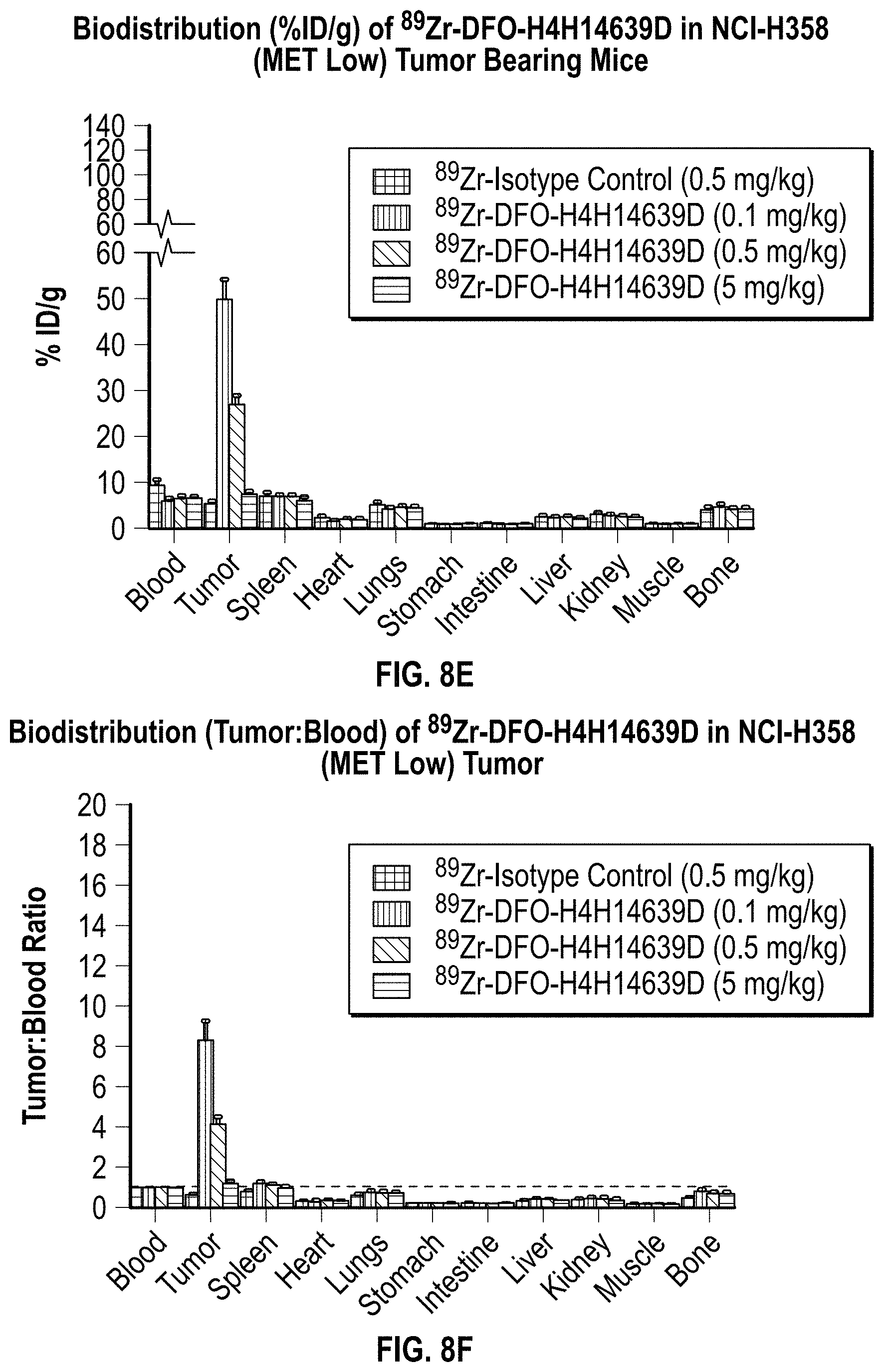
D00011

D00012


S00001
XML
uspto.report is an independent third-party trademark research tool that is not affiliated, endorsed, or sponsored by the United States Patent and Trademark Office (USPTO) or any other governmental organization. The information provided by uspto.report is based on publicly available data at the time of writing and is intended for informational purposes only.
While we strive to provide accurate and up-to-date information, we do not guarantee the accuracy, completeness, reliability, or suitability of the information displayed on this site. The use of this site is at your own risk. Any reliance you place on such information is therefore strictly at your own risk.
All official trademark data, including owner information, should be verified by visiting the official USPTO website at www.uspto.gov. This site is not intended to replace professional legal advice and should not be used as a substitute for consulting with a legal professional who is knowledgeable about trademark law.