Side Chain Modified Peptoids Useful As Structure-stabilizing Coatings For Biomaterials
Wang; Shih-Ting ; et al.
U.S. patent application number 17/015532 was filed with the patent office on 2021-03-11 for side chain modified peptoids useful as structure-stabilizing coatings for biomaterials. The applicant listed for this patent is Brookhaven Science Associates, LLC, Columbia University, Lawrence Berkeley National Laboratory, Stanford University. Invention is credited to Carolyn R. Bertozzi, Oleg Gang, Shih-Ting Wang, Ronald N. Zuckermann.
| Application Number | 20210070886 17/015532 |
| Document ID | / |
| Family ID | 1000005272915 |
| Filed Date | 2021-03-11 |


View All Diagrams
| United States Patent Application | 20210070886 |
| Kind Code | A1 |
| Wang; Shih-Ting ; et al. | March 11, 2021 |
SIDE CHAIN MODIFIED PEPTOIDS USEFUL AS STRUCTURE-STABILIZING COATINGS FOR BIOMATERIALS
Abstract
The current invention pertains compositions and methods to generate compositions providing stability to biomolecules, including providing physiologically stable and functional DNA origami-based drug/gene delivery carriers by surface coating with the oligo-ethylene glycol conjugated peptoids of Formulas (I), (II), and (III).
| Inventors: | Wang; Shih-Ting; (Mastic, NY) ; Gang; Oleg; (Setauket, NY) ; Zuckermann; Ronald N.; (El Cerrito, CA) ; Bertozzi; Carolyn R.; (Menlo Park, CA) | ||||||||||
| Applicant: |
|
||||||||||
|---|---|---|---|---|---|---|---|---|---|---|---|
| Family ID: | 1000005272915 | ||||||||||
| Appl. No.: | 17/015532 | ||||||||||
| Filed: | September 9, 2020 |
Related U.S. Patent Documents
| Application Number | Filing Date | Patent Number | ||
|---|---|---|---|---|
| 62897451 | Sep 9, 2019 | |||
| Current U.S. Class: | 1/1 |
| Current CPC Class: | C07K 17/06 20130101; C07K 1/10 20130101; B82Y 5/00 20130101; C07K 7/06 20130101; A61K 47/6923 20170801 |
| International Class: | C07K 17/06 20060101 C07K017/06; A61K 47/69 20060101 A61K047/69; C07K 1/10 20060101 C07K001/10; C07K 7/06 20060101 C07K007/06 |
Goverment Interests
STATEMENT OF GOVERNMENT RIGHTS
[0001] This invention was made with government support under DE-SC0012704 and DE-AC02-05CH11231 awarded by Department of Energy. The government has certain rights in the invention(s).
Claims
1. A composition for stabilizing a biomaterial, said composition comprising a compound of Formula (I) T.sub.1-[A.sub.m-E.sub.n].sub.b1-[G.sub.p-J.sub.q].sub.b2-[L.sub.r-M.sub.- s].sub.b3-[Q.sub.t-U.sub.u].sub.b4-T.sub.2 (I) wherein A is ##STR00088## E is ##STR00089## G is ##STR00090## J is ##STR00091## L is ##STR00092## M is ##STR00093## Q is ##STR00094## U is ##STR00095## m, n, p, q, r, s, t, and u are independently 0, 1, 2, 3, 4, or 5; b1, b2, b3, and b4 are independently 0, 1, 2, 3, 4, 5, 6, 7, 8, 9, 10, 15, or 20; R.sub.1, R.sub.2, R.sub.3, R.sub.4, R.sub.5, R.sub.6, R.sub.7, R.sub.8 are independently H, OH, (C.sub.1-C.sub.8)alkyl, (C.sub.2-C.sub.6)alkenyl, (C.sub.2-C.sub.6)alkynyl, (C.sub.1-C.sub.8)alkoxy, (C.sub.1-C.sub.6)alkylene-O--(C.sub.1-C.sub.6)alkyl, (C.sub.1-C.sub.6)alkylene-O--(C.sub.1-C.sub.6)alkylene-O--(C.sub.1-C.sub.- 6)alkyl, --((CH.sub.2).sub.1-8--O).sub.1-14--CH.sub.3, --((CH.sub.2).sub.1-8--O).sub.1-14-alkyl, (C.sub.1-C.sub.8)alkylene-C(O)OH, (C.sub.1-C.sub.8)alkylene-SO.sub.1-3H.sub.1-2, (C.sub.1-C.sub.8)alkylene-PO.sub.1-3H.sub.1-2, (C.sub.1-C.sub.8)alkylene-SO.sub.2--NH.sub.2, (C.sub.1-C.sub.8)alkoxy, (C.sub.1-C.sub.6)alkylene-(NH)NR.sub.10R.sub.11, (C.sub.1-C.sub.8)alkylene-NR.sub.12R.sub.13, (C.sub.1-C.sub.8)alkylene-C(O)NR.sub.14R.sub.15, or --(C.sub.1-C.sub.6)alkylene-R.sub.16, where alkyl, alkenyl, alkynyl, alkylene, and alkoxy are optionally substituted with one or more substituent each independently selected from (C.sub.1-C.sub.3)alkyl, oxo, NH.sub.2, --COOH, halogen, hydroxyl, methoxy, ethoxy, N.sub.3, biotinyl, sulfhydryl, cyano, hydrazido, carbodiimide, succinimide, or maleimide; T.sub.1 is ##STR00096## or H; T.sub.2 is ##STR00097## H, --OH, or NH.sub.2; T.sub.1.1, T.sub.1.2, T.sub.1.3, T.sub.2.1, and T.sub.2.2 are independently R.sub.1, R.sub.8, --(C.sub.1-C.sub.6)alkylene(NR.sub.17R.sub.18), --NR.sub.14R.sub.15, (C.sub.1-C.sub.6)alkylene-C(O)R.sub.16, --C(O)(C.sub.1-C.sub.6)alkyl, --C(O)NR.sub.17R.sub.18, SO.sub.2, --OH, --SH, --COOH, --(C.sub.1-C.sub.6)alkylene-N3, --(C.sub.2-C.sub.6)alkynyl, or alkyl, alkenyl, alkynyl, alkylene, or alkoxy that is substituted with at least one of biotinyl, sulfhydryl, cyano, hydrazido, carbodiimide, halo, succinimide, and maleimide; R.sub.16 is partially or fully saturated (C.sub.3-C.sub.8)carbocyclic, phenyl, 4- to 8-membered heterocycle containing 1 to 4 heteroatoms each independently selected from O, S, or N, 5- to 7-membered heteroaryl containing 1 to 3 heteroatoms each independently selected from O, N, or S, where R.sub.16 is optionally substituted with one or more substituents each independently selected from (C.sub.1-C.sub.3)alkyl, (C.sub.1-C.sub.3)alkyl-NH.sub.2, (C.sub.1-C.sub.3)alkoxy, halogen, hydroxyl, --C(O)NH.sub.2, --COOH, or --CN; and R.sub.10, R.sub.11, R.sub.12, R.sub.13, R.sub.14, R.sub.15, R.sub.16, R.sub.17, and R.sub.18 are each independently H, (C.sub.1-C.sub.6)alkyl, (C.sub.2-C.sub.6)alkenyl, (C.sub.2-C.sub.6)alkynyl, or halo-substituted (C.sub.1-C.sub.6)alkyl; wherein R.sub.1-R.sub.8, each defines a submonomer, and wherein Formula (I) defines a peptoid.
2. The composition according to claim 1, wherein R.sub.1, R.sub.2, R.sub.3, R.sub.4, R.sub.5, R.sub.6, R.sub.7, and R.sub.8 are independently --(CH.sub.2)--C.sub.2alkyne, --((CH.sub.2).sub.2--O)--CH.sub.3, --((CH.sub.2).sub.2--O).sub.3--CH.sub.3, or --(CH.sub.2).sub.2--NH.sub.2.
3. The composition according to of claim 1, wherein at least one of R.sub.1-R.sub.4 comprise a positively charged group; and R.sub.5-R.sub.8 do not comprise a positively charged group.
4. The composition according to claim 1, wherein R.sub.1, R.sub.2, R.sub.3, R.sub.4, R.sub.5, R.sub.6, R.sub.7, and R.sub.8 are independently selected from the group consisting of --(CH.sub.2).sub.2--NH.sub.2, --(CH.sub.2).sub.3--NH.sub.2, --(CH.sub.2).sub.4--NH.sub.2, --(CH.sub.2).sub.5--NH.sub.2, --(CH.sub.2).sub.6--NH.sub.2, --(CH.sub.2).sub.7--NH.sub.2, --(CH.sub.2).sub.8--NH.sub.2, --(CH.sub.2)--C.sub.2alkyne, --((CH.sub.2).sub.2--O)--CH.sub.3, --((CH.sub.2).sub.2--O).sub.3--CH.sub.3, and --(C.sub.1-C.sub.6)alkylene-N.sub.3.
5. A composition for stabilizing a biomaterial, said composition comprising a compound of Formula (II) T.sub.1-[X.sub.b1--Y.sub.b2--Z.sub.b3].sub.n-T.sub.2 (II) wherein X is ##STR00098## Y is ##STR00099## Z is ##STR00100## T.sub.1 is ##STR00101## or H; and T.sub.2 is ##STR00102## H, --OH, or NH.sub.2; wherein, n is 1, 2, 3, 4, or 5; b1, b2, and b3 are independently 0, 1, 2, 3, 4, 5, 6, 7, 8, 9, 10, 15, and 20; R.sub.1, R.sub.2, R.sub.3, R.sub.4, R.sub.5, R.sub.6, are independently H, OH, (C.sub.1-C.sub.8)alkyl, (C.sub.2-C.sub.6)alkenyl, (C.sub.2-C.sub.6)alkynyl, (C.sub.1-C.sub.8)alkoxy, (C.sub.1-C.sub.6)alkylene-O--(C.sub.1-C.sub.6)alkyl, (C.sub.1-C.sub.6)alkylene-O--(C.sub.1-C.sub.6)alkylene-O--(C.sub.1-C.sub.- 6)alkyl, --((CH.sub.2).sub.1-8--O).sub.1-14--CH.sub.3, --((CH.sub.2).sub.1-8--O).sub.1-14-alkyl, (C.sub.1-C.sub.8)alkylene-C(O)OH, (C.sub.1-C.sub.8)alkylene-SO.sub.1-3H.sub.1-2, (C.sub.1-C.sub.8)alkylene-PO.sub.1-3H.sub.1-2, (C.sub.1-C.sub.8)alkylene-SO.sub.2--NH.sub.2, (C.sub.1-C.sub.8)alkoxy, (C.sub.1-C.sub.6)alkylene-(NH)NR.sub.10R.sub.11, (C.sub.1-C.sub.8)alkylene-NR.sub.12R.sub.13, (C.sub.1-C.sub.8)alkylene-C(O)NR.sub.14R.sub.15, or --(C.sub.1-C.sub.6)alkylene-R.sub.16, where alkyl, alkenyl, alkynyl, alkylene, and alkoxy are optionally substituted with one or more substituent each independently selected from (C.sub.1-C.sub.3)alkyl, oxo, NH.sub.2, --COOH, halogen, hydroxyl, methoxy, ethoxy, N.sub.3, biotinyl, sulfhydryl, cyano, hydrazido, carbodiimide, succinimide, or maleimide; R.sub.16 is partially or fully saturated (C.sub.3-C.sub.8)carbocyclic, phenyl, 4- to 8-membered heterocycle containing 1 to 4 heteroatoms each independently selected from O, S, or N, 5- to 7-membered heteroaryl containing 1 to 3 heteroatoms each independently selected from O, N, or S, where R.sub.16 is optionally substituted with one or more substituents each independently selected from (C.sub.1-C.sub.3)alkyl, (C.sub.1-C.sub.3)alkyl-NH.sub.2, (C.sub.1-C.sub.3)alkoxy, halogen, hydroxyl, --C(O)NH.sub.2, --COOH, or --CN; T.sub.1.1, T.sub.1.2, T.sub.1.3, T.sub.2.1, and T.sub.2.2 are independently R.sub.1, R.sub.6, --(C.sub.1-C.sub.6)alkylene(NR.sub.17R.sub.18), --NR.sub.14R.sub.15, (C.sub.1-C.sub.6)alkylene-C(O)R.sub.16, --C(O)(C.sub.1-C.sub.6)alkyl, --C(O)NR.sub.17R.sub.18, SO.sub.2, --OH, --SH, --COOH, --(C.sub.1-C.sub.6)alkyl azide, --(C.sub.2-C.sub.6)alkynyl, or alkyl, alkenyl, alkynyl, alkylene, or alkoxy that is substituted with at least one of biotinyl, sulfhydryl, cyano, hydrazido, carbodiimide, halo, succinimide, and maleimide; and R.sub.10, R.sub.11, R.sub.12, R.sub.13, R.sub.14, R.sub.15, R.sub.16, R.sub.17, and R.sub.18 are each independently H, (C.sub.1-C.sub.6)alkyl, (C.sub.2-C.sub.6)alkenyl, (C.sub.2-C.sub.6)alkynyl, or halo-substituted (C.sub.1-C.sub.6)alkyl; wherein R.sub.1-R.sub.6, each defines a submonomer, and wherein Formula (II) defines a peptoid.
6. A composition for stabilizing a biomaterial, said composition comprising a compound of Formula (III): ##STR00103## wherein r is 1, 2, 3, 4, 5, 6, 7, 8, 9, 10, 11, 12, 13, 14, 15, 20, 25, 50, 100, or 200; T.sub.1 is ##STR00104## or H; and T.sub.2 is ##STR00105## H, --OH, or NH.sub.2; wherein T.sub.1.1, T.sub.1.2, T.sub.1.3, T.sub.2.1, and T.sub.2.2 are independently --(CH.sub.2)--NH.sub.2, --((CH.sub.2).sub.2--O).sub.3--CH.sub.3, --(C.sub.1-C.sub.6)alkylene(NR.sub.17R.sub.18), --NR.sub.14R.sub.15, (C.sub.1-C.sub.6)alkylene-C(O)R.sub.16, --C(O)(C.sub.1-C.sub.6)alkyl, --C(O)NR.sub.17R.sub.18, SO.sub.2, --OH, --SH, --COOH, --(C.sub.1-C.sub.6)alkylene-azide, --(C.sub.2-C.sub.6)alkynyl, or alkyl, alkenyl, alkynyl, alkylene, or alkoxy that is substituted with at least one of biotinyl, sulfhydryl, cyano, hydrazido, carbodiimide, halo, succinimide, and maleimide; and R.sub.14, R.sub.15, R.sub.16, R.sub.17, and R.sub.18 are each independently H, (C.sub.1-C.sub.6)alkyl, (C.sub.2-C.sub.6)alkenyl, (C.sub.2-C.sub.6)alkynyl, or halo-substituted (C.sub.1-C.sub.6)alkyl; wherein formula (III) defines a peptoid.
7. The composition according to claim 1, wherein Formula (I) comprises: ##STR00106## ##STR00107## ##STR00108## ##STR00109##
8. The composition according to claim 1, wherein the compound of Formula (I), is conjugated to an antibody, imaging reagent, biomaterial, biomolecule, glycan, polymer, or a peptoid.
9. A drug delivery carrier comprising a pre-defined nucleic acid nanostructure and a compound of Formula (I), (II), or (III).
10. A stabilized complex comprising: a pre-defined nucleic acid nanostructure; a compound of Formula (I), (II), or (III); and a drug molecule or protein.
11. The stabilized complex according to claim 10, wherein an antibody is conjugated to the compound of Formula (I), (II), or (III).
12. A method of stabilizing a pre-defined nucleic acid nanostructure, said method comprising: (i) complexing a pre-defined nucleic acid nanostructure with a composition comprising a compound of Formula (I), (II), or (III).
13. The method according to claim 12, wherein complexing comprises: (i) contacting pre-defined nucleic acid nanostructure composition with a compound of Formula (I), (II), or (III).
14. The method according to claim 12 wherein the pre-defined nucleic acid nanostructure is coated with a compound according to any of Formulas (I), (II), or (III), wherein one terminal end of Formula (I), (II), or (III) is conjugated to the pre-defined nucleic acid structure and one terminal end of Formula (I), (II), or (III) is conjugated to a nucleic acid, protein, a different pre-defined nucleic acid nanostructure, a small molecule therapeutic, or a protein therapeutic.
15. A stabilized nanoparticle complex comprising: a nanoparticle; and a compound according to Formula (I), (II), or (III).
16. The stabilized nanoparticle according to claim 15, wherein the nanoparticle comprises silver or gold.
17. The stabilized nanoparticle according to claim 15, wherein the compound according to Formula (I), (II), or (III) comprises: ##STR00110##
Description
FIELD OF THE INVENTION
[0002] This invention relates to stabilizing biomaterials for biomedical applications.
BACKGROUND OF THE INVENTION
[0003] DNA nanotechnology allows to program self-assembly of synthetic oligonucleotides into structures with prescribed topologies and spatial configurations. One of such approaches is called DNA origami. Advancement in DNA origami design and synthesis with high-precision structural controls has enabled potential biomedical applications, including smart drug/gene delivery carriers and biomolecular devices at the cellular level. Additionally, DNA origamis designed with controlled shape, biocompatibility and responsiveness toward other biomolecules (e.g., proteins, lipids, DNAs, and RNAs), may serve as a reliable molecular interface and augment functionalities of the hybrid bio/nano system. This technology applies a bottom-up approach in nanocarrier design and synthesis that may bring potential solutions in targeted drug/gene delivery and biomedical imaging and sensing, in which translation of nanoformulation carriers into clinical applications has remained challenging due to the difficulty to regulate interfaces between the nanocarriers and biological systems.
[0004] There are some roadblocks to maximizing this technology, however. For example, biomedical applications of DNA origamis with precisely defined nanostructures are often incompatible with the comparatively high content (10-20 mM) of magnesium ions required for the DNA origami self-assembly and its long-term stability. In fact, low transfection due to poor structural integrity of DNA origamis in physiological fluids, which typically contain low content of the magnesium ions (.about.1 mM), has been identified as a major challenge. Moreover, change in solution components and pH as well as nuclease degradation have also been shown to affect the DNA origami superstructures. It is therefore crucial to stabilize DNA origami structures under the damaging factors in order for them to be effectively used for broader biomedical applications.
[0005] Therefore, there is a need to overcome these obstacles and create a composition which can stabilize a biomaterial, such as pre-defined nucleic acid based nanostructures (e.g., DNA origami).
SUMMARY OF THE INVENTION
[0006] The present invention relates to compositions and methods for the stabilization of biomaterials.
[0007] In some embodiments, the invention relates to a composition for stabilizing a biomaterial, said composition including a compound of Formula (I).
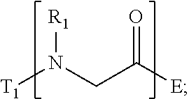
T.sub.1-[A.sub.m-E.sub.n].sub.b1-[G.sub.p-J.sub.q].sub.b2-[L.sub.r-M.sub- .s].sub.b3-[Q.sub.t-U.sub.u].sub.b4-T.sub.2 (I)
wherein
A is
##STR00001##
[0008] E is
##STR00002##
[0009] G is
##STR00003##
[0010] J is
##STR00004##
[0011] L is
##STR00005##
[0012] M is
##STR00006##
[0013] Q is
##STR00007##
[0014] U is
##STR00008##
[0015] m, n, p, q, r, s, t, and u are independently 0, 1, 2, 3, 4, or 5; b1, b2, b3, and b4 are independently 0, 1, 2, 3, 4, 5, 6, 7, 8, 9, 10, 15, or 20; R.sub.1, R.sub.2, R.sub.3, R.sub.4, R.sub.5, R.sub.6, R.sub.7, R.sub.8 are independently H, OH, (C.sub.1-C.sub.8)alkyl, (C.sub.2-C.sub.6)alkenyl, (C.sub.2-C.sub.6)alkynyl, (C.sub.1-C.sub.8)alkoxy, (C.sub.1-C.sub.6)alkylene-O--(C.sub.1-C.sub.6)alkyl, (C.sub.1-C.sub.6)alkylene-O--(C.sub.1-C.sub.6)alkylene-O--(C.sub.1-C.sub.- 6)alkyl, --((CH.sub.2).sub.1-8--O).sub.1-14--CH.sub.3, --((CH.sub.2).sub.1-8--O).sub.1-14-alkyl, (C.sub.1-C.sub.8)alkylene-C(O)OH, (C.sub.1-C.sub.8)alkylene-SO.sub.1-3H.sub.1-2, (C.sub.1-C.sub.8)alkylene-PO.sub.1-3H.sub.1-2, (C.sub.1-C.sub.8)alkylene-SO.sub.2--NH.sub.2, (C.sub.1-C.sub.8)alkoxy, (C.sub.1-C.sub.6)alkylene-(NH)NR.sub.10R.sub.11, (C.sub.1-C.sub.8)alkylene-NR.sub.12R.sub.13, (C.sub.1-C.sub.8)alkylene-C(O)NR.sub.14R.sub.15, or --(C.sub.1-C.sub.6)alkylene-R.sub.16, where alkyl, alkenyl, alkynyl, alkylene, and alkoxy are optionally substituted with one or more substituent each independently selected from (C.sub.1-C.sub.3)alkyl, oxo, NH.sub.2, --COOH, halogen, hydroxyl, methoxy, ethoxy, N.sub.3, biotinyl, sulfhydryl, cyano, hydrazido, carbodiimide, succinimide, or maleimide;
T.sub.1 is
##STR00009##
[0016] or H;
T.sub.2 is
##STR00010##
[0017] H, --OH, or NH.sub.2;
[0018] T.sub.1.1, T.sub.1.2, T.sub.1.3, T.sub.2.1, and T.sub.2.2 are independently R.sub.1, R.sub.8, --(C.sub.1-C.sub.6)alkylene(NR.sub.17R.sub.18), --NR.sub.14R.sub.15, (C.sub.1-C.sub.6)alkylene-C(O)R.sub.16, --C(O)(C.sub.1-C.sub.6)alkyl, --C(O)NR.sub.17R.sub.18, SO.sub.2, --OH, --SH, --COOH, --(C.sub.1-C.sub.6)alkylene-N.sub.3, --(C.sub.2-C.sub.6)alkynyl, or alkyl, alkenyl, alkynyl, alkylene, or alkoxy that is substituted with at least one of biotinyl, sulfhydryl, cyano, hydrazido, carbodiimide, halo, succinimide, and maleimide; R.sub.16 is partially or fully saturated (C.sub.3-C.sub.8)carbocyclic, phenyl, 4- to 8-membered heterocycle containing 1 to 4 heteroatoms each independently selected from O, S, or N, 5- to 7-membered heteroaryl containing 1 to 3 heteroatoms each independently selected from O, N, or S, where R.sub.16 is optionally substituted with one or more substituents each independently selected from (C.sub.1-C.sub.3)alkyl, (C.sub.1-C.sub.3)alkyl-NH.sub.2, (C.sub.1-C.sub.3)alkoxy, halogen, hydroxyl, --C(O)NH.sub.2, --COOH, or --CN; and R.sub.10, R.sub.11, R.sub.12, R.sub.13, R.sub.14, R.sub.15, R.sub.16, R.sub.17, and R.sub.18 are each independently H, (C.sub.1-C.sub.6)alkyl, (C.sub.2-C.sub.6)alkenyl, (C.sub.2-C.sub.6)alkynyl, or halo-substituted (C.sub.1-C.sub.6)alkyl; wherein R.sub.1-R.sub.8, each defines a submonomer, and wherein Formula (I) defines a peptoid.
[0019] In some embodiments, the invention relates to a composition for stabilizing a biomaterial, said composition includes a compound of Formula (II).
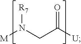
T.sub.1-[X.sub.b1--Y.sub.b2--Z.sub.b3].sub.n-T.sub.2 (II)
wherein
X is
##STR00011##
[0020] Y is
##STR00012##
[0021] Z is
##STR00013##
[0022] T.sub.1 is
##STR00014##
[0023] or H; and
T.sub.2 is
##STR00015##
[0024] H, --OH, or NH.sub.2;
[0025] wherein, n is 1, 2, 3, 4, or 5; b1, b2, and b3 are independently 0, 1, 2, 3, 4, 5, 6, 7, 8, 9, 10, 15, and 20; R.sub.1, R.sub.2, R.sub.3, R.sub.4, R.sub.5, R.sub.6, are independently H, OH, (C.sub.1-C.sub.8)alkyl, (C.sub.2-C.sub.6)alkenyl, (C.sub.2-C.sub.6)alkynyl, (C.sub.1-C.sub.8)alkoxy, (C.sub.1-C.sub.6)alkylene-O--(C.sub.1-C.sub.6)alkyl, (C.sub.1-C.sub.6)alkylene-O--(C.sub.1-C.sub.6)alkylene-O--(C.sub.1-C.sub.- 6)alkyl, --((CH.sub.2).sub.1-8--O).sub.1-14--CH.sub.3, --((CH.sub.2).sub.1-8--O).sub.1-14-alkyl, (C.sub.1-C.sub.8)alkylene-C(O)OH, (C.sub.1-C.sub.8)alkylene-SO.sub.1-3H.sub.1-2, (C.sub.1-C.sub.8)alkylene-PO.sub.1-3H.sub.1-2, (C.sub.1-C.sub.8)alkylene-SO.sub.2--NH.sub.2, (C.sub.1-C.sub.8)alkoxy, (C.sub.1-C.sub.6)alkylene-(NH)NR.sub.10R.sub.11, (C.sub.1-C.sub.8)alkylene-NR.sub.12R.sub.13, (C.sub.1-C.sub.8)alkylene-C(O)NR.sub.14R.sub.15, or --(C.sub.1-C.sub.6)alkylene-R.sub.16, where alkyl, alkenyl, alkynyl, alkylene, and alkoxy are optionally substituted with one or more substituent each independently selected from (C.sub.1-C.sub.3)alkyl, oxo, NH.sub.2, --COOH, halogen, hydroxyl, methoxy, ethoxy, N.sub.3, biotinyl, sulfhydryl, cyano, hydrazido, carbodiimide, succinimide, or maleimide; R.sub.16 is partially or fully saturated (C.sub.3-C.sub.8)carbocyclic, phenyl, 4- to 8-membered heterocycle containing 1 to 4 heteroatoms each independently selected from O, S, or N, 5- to 7-membered heteroaryl containing 1 to 3 heteroatoms each independently selected from O, N, or S, where R.sub.16 is optionally substituted with one or more substituents each independently selected from (C.sub.1-C.sub.3)alkyl, (C.sub.1-C.sub.3)alkyl-NH.sub.2, (C.sub.1-C.sub.3)alkoxy, halogen, hydroxyl, --C(O)NH.sub.2, --COOH, or --CN; T.sub.1.1, T.sub.1.2, T.sub.1.3, T.sub.2.1, and T.sub.2.2 are independently R.sub.1, R.sub.6, --(C.sub.1-C.sub.6)alkylene(NR.sub.17R.sub.18), --NR.sub.14R.sub.15, (C.sub.1-C.sub.6)alkylene-C(O)R.sub.16, --C(O)(C.sub.1-C.sub.6)alkyl, --C(O)NR.sub.17R.sub.18, SO.sub.2, --OH, --SH, --COOH, --(C.sub.1-C.sub.6)alkyl azide, --(C.sub.2-C.sub.6)alkynyl, or alkyl, alkenyl, alkynyl, alkylene, or alkoxy that is substituted with at least one of biotinyl, sulfhydryl, cyano, hydrazido, carbodiimide, halo, succinimide, and maleimide; and R.sub.10, R.sub.11, R.sub.12, R.sub.13, R.sub.14, R.sub.15, R.sub.16, R.sub.17, and R.sub.18 are each independently H, (C.sub.1-C.sub.6)alkyl, (C.sub.2-C.sub.6)alkenyl, (C.sub.2-C.sub.6)alkynyl, or halo-substituted (C.sub.1-C.sub.6)alkyl; wherein R.sub.1-R.sub.6, each defines a submonomer, and wherein Formula (II) defines a peptoid.
[0026] In some embodiments, the invention relates to a composition for stabilizing a biomaterial, said composition including a compound of Formula (III).
##STR00016##
wherein r is 1, 2, 3, 4, 5, 6, 7, 8, 9, 10, 11, 12, 13, 14, 15, 20, 25, 50, 100, or 200;
T.sub.1 is
##STR00017##
[0027] or H; and
T.sub.2 is
##STR00018##
[0028] H, --OH, or NH.sub.2;
[0029] wherein T.sub.1.1, T.sub.1.2, T.sub.1.3, T.sub.2.1, and T.sub.2.2 are independently --(CH.sub.2)--NH.sub.2, --((CH.sub.2).sub.2--O).sub.3--CH.sub.3, --(C.sub.1-C.sub.6)alkylene(NR.sub.17R.sub.18), --NR.sub.14R.sub.15, (C.sub.1-C.sub.6)alkylene-C(O)R.sub.16, --C(O)(C.sub.1-C.sub.6)alkyl, --C(O)NR.sub.17R.sub.18, SO.sub.2, --OH, --SH, --COOH, --(C.sub.1-C.sub.6)alkylene-azide, --(C.sub.2-C.sub.6)alkynyl, or alkyl, alkenyl, alkynyl, alkylene, or alkoxy that is substituted with at least one of biotinyl, sulfhydryl, cyano, hydrazido, carbodiimide, halo, succinimide, and maleimide; and R.sub.14, R.sub.15, R.sub.16, R.sub.17, and R.sub.18 are each independently H, (C.sub.1-C.sub.6)alkyl, (C.sub.2-C.sub.6)alkenyl, (C.sub.2-C.sub.6)alkynyl, or halo-substituted (C.sub.1-C.sub.6)alkyl; wherein formula (III) defines a peptoid.
[0030] In some embodiments, the invention relates to a drug delivery carrier having a pre-defined nucleic acid nanostructure and a compound of Formula (I), (II), or (III).
[0031] In some embodiments, the invention relates to a stabilized complex having a pre-defined nucleic acid nanostructure; a compound of Formula (I), (II), or (III); and a drug molecule or protein.
[0032] In some embodiments, the invention relates to a method of stabilizing a pre-defined nucleic acid nanostructure, said method including (i) complexing a pre-defined nucleic acid nanostructure with a composition comprising a compound of Formula (I), (II), or (III).
[0033] In some embodiments, the invention relates to a stabilized nanoparticle complex having a nanoparticle; and a compound according to Formula (I), (II), or (III).
BRIEF DESCRIPTION OF THE DRAWINGS
[0034] FIG. 1. (A) Chemical structures of peptoids designed to protect 3D octahedra-shaped DNA origamis (OC). Nae: (2-aminoethyl)glycine, Nte: N-2-(2-(2-methoxyethoxy)ethoxy)ethylglycine and Nme: N-(2-methoxyethyl)glycine. (B) Transmission electron microscope (TEM) image and schematic view (inset) of the OC structure (scale bar: 50 nm). (C) Schematic view showing the different surface coating of the two types ("brush" and "block") of peptoids on the OC structure proposed in this work, which leads to varied protection effect.
[0035] FIG. 2. Fluorescence assay monitoring peptoid-stabilized duplex DNA (dsDNA). Real-time SYBR Green I (SG) fluorescence assay of a 15-bp dsDNA (500 .mu.M) in the presence of peptoids at different ratios of peptoid amines to phosphate groups of the DNA (N/P). The fluorescence signals of (A) dsDNA/peptoid complexes at N/P of 4 and (B) dsDNA/PE2 complexes at different N/P are plotted against the increasing temperature.
[0036] FIG. 3 Molecular dynamics (MD) simulations of the interactions of PE1 and PE4 with dsDNA. Molecular representation of the most visited binding sites and structures of (A) PE1 and (B) PE4 with dsDNA (blue: peptoid backbone, red: Nae residues, and green: Nte residues). The most visited binding sites represented as occupancy volume areas (shown as transparent white) where the peptoids were present for at least 6% of total contact time. (C-F) Radial distribution functions (RDF) of water near the (C, D) minor and (E, F) major grooves of the dsDNA. RDFs were calculated on the H (donor) atoms of water and the N and O (acceptor) atoms of (C, E) AT and (D, F) GC base-pairs. The structuring of water around DNA only is shown in orange, while for the dsDNA/PE1 and dsDNA/PE4 complexes are shown in green and blue, where the AT and GC base-pairs are distinguished by light and dark colors respectively.
[0037] FIG. 4. Analysis of peptoid-coated OCs in low Mg.sup.2+ solution. (A) Schematic view showing peptoid-coated OCs (OC/peptoid) was protected against Mg.sup.2+ depletion. (B, C) Agarose gel electrophoresis (AGE) was used to analyze the structural integrity of OCs in TAE buffer at MgCl.sub.2 concentrations of 12.5 mM (+) and 1.25 mM (-). In (c), the electrophoretic shift was measured from the reference band at 0.5 kb and the relative value was calculated from that of the control OCs (n=3). (D) TEM imaging was performed on OCs extracted from the agarose gels (bands a and b in B). Scale bars: 200 nm. The insets show magnified images of the OC structures (scale bars: 100 nm). (E) Dynamic light scattering (DLS) and (F) in situ small angle X-ray scattering (SAXS) spectra show bare OCs and OC/PE2 treated with EDTA (5 or 10 mM) for 20-30 min at room temperature.
[0038] FIG. 5. Analysis of peptoid-coated OCs in the presence of deoxyribonuclease (DN). (A) Schematic view showing peptoid-coated OCs (OC/peptoid) were protected against DN (image obtained from PDB: 2DNJ) degradation. (B) TEM images show bare OCs and OC/peptoid (N/P: 0.5) in solution containing DN of 15 g/mL. Imaging was performed on samples extracted from the agarose gel (scale bars: 200 nm). The insets show magnified images of the OC structures (scale bars: 100 nm).
[0039] FIG. 6. Analysis of peptoid-coated OCs in cell media and presence of serum nuclease. TEM images show (A, C) bare OCs and (B, D) PE2-coated OCs (OC/PE2) in (A, B) Roswell Park Memorial Institute (RPMI) 1640 medium and (C, D) Dulbecco's Modified Eagle Medium (DMEM) containing 0%, 5%, and 10% of fetal bovine serum (FBS) and incubated for 24 h at 37.degree. C. The final concentrations of MgCl.sub.2 were 1.25 mM. Imaging was performed on samples extracted from the agarose gels (scale bars: 200 nm). The insets show magnified images of the OC structures (scale bars: 50 nm).
[0040] FIG. 7. Protection of protein-encapsulated in OCs by PE2 coating. (A) Schematic view showing that OC/PE2 reduce trypsin (image obtained from PDB: 1S0Q) digestion of fluorescein labeled BSA encapsulated in the OCs. (B) Fluorescence kinetics show enhanced fluorescence of the fluorescein labeled BSA upon trypsin cleavage (.lamda..sub.ex=490 nm and .lamda..sub.em=525 nm, 37.degree. C.).
[0041] FIG. 8. Surface functionalization of peptoid-stabilized OCs with antibody and fluorophore. (A) Schematic view showing alkyne-modified peptoids conjugates azide-modified cargos through click chemistry. Here, azide fluor 488 (AF) and Trastuzumab (Tz) were used as the presenting cargos (label 1). (B) Chemical structure of PE8 and PE9. (C) Fluorescence assay of bare OCs, OC/PE8-AF and OC/PE9-AF extracted from the agarose gel (.lamda..sub.ex=485 nm and .lamda..sub.em=510-800 nm). (D) TEM images show surface coating of OCs with PE8-Tz and PE9-Tz. The samples were labeled with immunogold (label 2 in A) prior to TEM imaging (scale bars: 50 nm).
[0042] FIG. 9. Stabilization of Au NPs by peptoid coating. (A) Schematic view showing the block-type peptoid Nae.sub.6-Nte.sub.12 was used to stabilize 20 nm citrated-capped gold nanoparticles (Au NPs). (B) UV-visible spectroscopy (UV-vis) showing stable Au NPs (0.5 nM) after coating with Nae.sub.6-Nte.sub.12 block at different concentrations. The buffer was 10 mM phosphate, pH 7.0.
[0043] FIG. 10. Peptoid stabilizes Au NPs in solution containing high salt concentration. UV-vis showing that Au NPs remains stable in solution containing 200 mM of sodium chloride after coating with Nae.sub.6-Nte.sub.12 block. Below 0.375 .mu.M of Nae6-Nte12 block, the extent of Au NP aggregation is dependent on the peptoid concentration.
[0044] FIG. 11 Cryogenic protection of peptoid stabilized Au NPs. (A) Pictures showing Nae.sub.6-Nte.sub.12 block stabilizes the Au NPs at frozen (top) and melted (bottom) states.
[0045] FIG. 12 UV-vis showing stable Au NPs in the melted state with Nae.sub.6-Nte.sub.12 block coating. Higher concentration of peptoid enables higher Au NP stability.
[0046] FIG. 13 Mass spectrometry of peptoids PE1-9 prepared by solid phase peptoid synthesis.
[0047] FIG. 14 Real-time SYBR Green I (SG) fluorescence assay of the 15-bp dsDNA in the presence of peptoids (A, B) PE1, (C, D) PE3, (E, F) PE4 and (G, H) PE5 at different N/P. Derivatives of the fluorescence intensities were plotted against the increasing temperature.
[0048] FIG. 15 Real-time SG fluorescence assay of peptoid only. (A) Fluorescence intensities and (B) derivatives of the fluorescence intensities were plotted against the increasing temperature.
[0049] FIG. 16 Real-time SG fluorescence assay of 15-bp dsDNA in the presence of (A) PE1-5 at N/P of 4 (B) PE2 at different N/P as in FIGS. 2A and 2B, respectively. Derivatives of fluorescence intensities were plotted against the increasing temperature.
[0050] FIG. 17 Fluorescence spectra of the 15-bp dsDNA in the presence of SG and PE2 at different N/P. Sample preparation and fluorescence measurements were performed at room temperature (.lamda..sub.ex=495 nm and .lamda..sub.em=510-650 nm). The concentration of PE2 only in solution was the same as that of N/P of 8, which was 1.85 .mu.M.
[0051] FIG. 18 AGE shows the electrophoretic shift of dsDNA/peptoid complexes. (A) dsDNA/peptoid of 0.5; (B) dsDNA/PE2 at different N/P.
[0052] FIG. 19 Atomistic structures of the peptoid and dsDNA models used in the MD simulations. The peptoids (A) PE1 and (B) PE4 are showing the peptoid backbone in blue, charged (Nae) residues in red and ethylene glycols (Nte) in green. (C) Molecular and (D) cartoon representation of the dsDNA model are showing the DNA backbone in grey, and the bases colored individually with cytosine in red, guanine in green, thymine in yellow and adenine in blue.
[0053] FIG. 20 Negative-stained TEM images show peptoid-coated OCs at N/P of 0.5, 0.25 and 0.1 (scale bars: 200 nm). The insets show magnified images of the OC structures (scale bars: 100 nm).
[0054] FIG. 21 (A) Chemical structure of the Nae6-Nte16 block peptoid and (B) negative-stained TEM images of Nae.sub.6-Nte.sub.16 block-coated OCs at N/P of 0.5 (scale bar: 200 nm). The inset shows magnified image of the OC structures (scale bar: 100 nm).
[0055] FIG. 22 Top: Molecular structure of PE6. Bottom: Negative-stained TEM images of PE6-coated OC structures at N/P of (A) 2, (B) 1 and (C) 0.5 (scale bars: 200 nm). The insets show magnified images of the OC structures (scale bars: 100 nm).
[0056] FIG. 23 Top: molecular structure of PE7. Bottom: negative-stained TEM images of PE7-coated OCs at N/P of (A) 2, (B) 1 and (C) 0.5 (scale bars: 200 nm). The insets show magnified images of the OC structures (scale bars: 100 nm).
[0057] FIG. 24 AGE showing the electrophoretic shift of peptoid-coated OCs (4.3 nM). Lanes 1-3: N/P of 0.5, 0.25, and 0.1, respectively; Lane 4: peptoid/OC=1; and Lane 5: OC only.
[0058] FIG. 25 AGE of OC/peptoid in TAE buffer. The final concentration of MgCl.sub.2 was 1.25 mM. The result was used in the calculation of electrophoretic shift in FIG. 4C.
[0059] FIG. 26 Negative-stained TEM images of (A) bare OCs and OC/PE2 at (B) N/P of 0.1 and (C) N/P of 0.5. The final concentration of MgCl.sub.2 in TAE buffer was 1.25 mM. TEM samples were extracted from the agarose gels (scale bars: 200 nm). The insets show magnified images of the OC structures (scale bars: 100 nm).
[0060] FIG. 27 Negative-stained TEM images of peptoid (PE1, PE3, PE4, and PE5)-coated OCs in TAE buffer. The final concentrations of MgCl.sub.2 were 12.5 mM (TAE-Mg.sup.2+) and 1.25 mM (TAE low Mg.sup.2+). TEM samples were extracted from the agarose gels (scale bars: 200 nm). The insets show magnified images of the OC structures (scale bars: 100 nm).
[0061] FIG. 28 Real-time SG fluorescence assay of bare and peptoid-coated OCs (1 nM, N/P: 0.125) in PBS buffer. Derivatives of the fluorescence intensities were normalized to 0-1 range and plotted against the increasing temperature.
[0062] FIG. 29 Negative-stained TEM images of OCs in the presence of different amounts of EDTA: (A) 12.5 mM, (B) 10 mM, (C) 6.25 mM, (D) 3.5 mM and (E) 0 mM. The concentration of MgCl.sub.2 in TAE buffer was 12.5 mM. EDTA was added to the OC solution and left undisturbed for .about.4 h at room temperature prior to TEM samples preparation (scale bars: 200 nm). The insets show magnified images of the OC structures (scale bars: 100 nm).
[0063] FIG. 30 Kratky analysis of SAXS data in FIGS. 4F and G. Bare OCs appeared to be more flexible upon treatment with EDTA and did not plateau at higher q compared to the PE2-coated OCs.
[0064] FIG. 31 Left: AGE of bare and peptoid-coated OCs in PBS buffer. The final concentration of MgCl.sub.2 was 1.25 mM. Right: TEM samples were extracted from the agarose gels (bands a-f). Scale bars: 200 nm. The insets show magnified images of the OC structures (scale bars: 100 nm).
[0065] FIG. 32 AGE of OCs in the presence of different concentrations of deoxyribonuclease I (DN). Degradation of OC nanostructures by DN was shown by the electrophoretic shift toward the end of lower molecular weight and the presence of new bands representing degraded OCs.
[0066] FIG. 33 TEM images of bare OCs and peptoid-coated OCs in the absence (left) and presence of DN (15 .mu.g/mL, right). The samples were extracted from agarose gels. The concentration of MgCl.sub.2 in TAE buffer was 12.5 mM (scale bars: 200 nm). The insets show magnified images of the OC structures (scale bars: 100 nm). Among the peptoid sequences, only OC/PE2 showed protection of the OC nanostructures.
[0067] FIG. 34 DLS of (A) bare and (B) PE2-coated OCs (N/P: 0.5) in the presence of DN. The extent of size reduction represents degradation of the OC nanostructures by DN.
[0068] FIG. 35 Negative-stained TEM images of (A) bare OCs and (B) PE2-coated OCs in DMEM cell media containing FBS (0%, 5%, and 10%). The final concentration of MgCl.sub.2 was 1.25 mM. TEM samples were extracted from the agarose gels (scale bars: 200 nm). The insets show the magnified images of the OC structures (scale bars: 100 nm).
[0069] FIG. 36 UV-vis spectra of step-wise functionalization of 10 nm gold nanoparticles (Au NPs) with Cys-Ala-Leu-Asp-Asp-Lys(N3) (SEQ ID NO: 127) (pep) and followed by a DBCO-modified single-stranded DNA (ssDNA, 5'-TATGAAGTGATGGATGAT/3DBCO/), (SEQ ID NO: 1) which complemented with the eight ssDNAs located in the OCs.
[0070] FIG. 37 (A) AGE and (B) TEM images show PE2 coated and Au NP-encapsulated OCs in DMEM media containing FBS (0%, 5%, and 10%). AGE was performed and imaged by white light (top) and UV light (bottom). TEM samples were extracted from the agarose gels (scale bars: 200 nm). The insets show magnified images of the OC structures (scale bars: 50 nm).
[0071] FIG. 38 (A) Fluorescence spectrum of doxorubicin (Dox, 100 .mu.M) in PBS buffer. Excitation and emission wavelengths were measured at 485 nm and 510-800 nm, respectively. (B) Fluorescence signals at 597 nm were plotted against Dox concentrations ([Dox]). A linear relationship between fluorescence signal and [Dox] was observed below 3.2 .mu.M.
[0072] FIG. 39 Plot showing the total Dox release from bare OCs and OC/PE2 (n=2). The Dox release was determined by the remaining fluorescence of OCs after incubating in PBS buffer at 37.degree. C. for 48 h. A reduction of total Dox release from the OC/PE2 compared to bare OCs was observed at both pH 7 and 5.5.
[0073] FIG. 40 Fluorescence enhancement of fluorescein labeled BSA (80 nM) in the presence and absence of trypsin (50 nM). The concentration of MgCl.sub.2 in TAE buffer was 12.5 mM. The solution was incubated overnight (>12 h) at 37.degree. C. prior to fluorescence measurement (.lamda..sub.ex=490 nm and .lamda..sub.em=510-800 nm).
[0074] FIG. 41 Fluorescence spectra of fluorescein-labelled BSA encapsulated in bare and PE2-coated OCs (20 nM) in the presence and absence of trypsin (50 nM). The concentration of MgCl.sub.2 in TAE buffer was 12.5 mM. The mixtures were incubated at 37.degree. C. for .about.15 h prior to fluorescence measurement (.lamda..sub.ex=485 nm and .lamda..sub.em=510-800 nm).
[0075] FIG. 42 TEM images of OC in the presence of different trypsin concentrations: (A) 5 .mu.M, (B) 0.5 .mu.M, (C) 0.1 .mu.M and (D) 0 .mu.M (scale bars: 200 nm). The insets show magnified images of the OC structures (scale bars: 100 nm).
[0076] FIG. 43 Site-specific modification of Trastuzumab. Top: schematic view of the modification. Bottom: ESI-LC/MS analyses of the Trastuzumab-azide.
DETAILED DESCRIPTION
[0077] The present invention relates to compositions and methods for the stabilization of biomaterials.
[0078] As used herein, compositions of the present invention for stabilization of biomaterials are referred to as "peptoids". In other words, the peptoids of the present invention can also be referred to as a series of N-substituted glycines. Peptoids according to the present invention are exemplified by Formulas (I), (II), and (III) described herein.
[0079] In one embodiment, the composition of the invention includes a compound of Formula (I).
T.sub.1-[A.sub.m-E.sub.n].sub.b1-[G.sub.p-J.sub.q].sub.b2-[L.sub.r-M.sub- .s].sub.b3-[Q.sub.t-U.sub.u].sub.b4-T.sub.2 (I)
wherein
A is
##STR00019##
[0080] E is
##STR00020##
[0081] G is
##STR00021##
[0082] J is
##STR00022##
[0083] L is
##STR00023##
[0084] M is
##STR00024##
[0085] Q is
##STR00025##
[0086] U is
##STR00026##
[0087] wherein m, n, p, q, r, s, t, and u are independently 0, 1, 2, 3, 4, or 5; b1, b2, b3, and b4 are independently 0, 1, 2, 3, 4, 5, 6, 7, 8, 9, 10, 15, or 20; R.sub.1, R.sub.2, R.sub.3, R.sub.4, R.sub.5, R.sub.6, R.sub.7, R.sub.8 are independently H, OH, (C.sub.1-C.sub.8)alkyl, (C.sub.2-C.sub.6)alkenyl, (C.sub.2-C.sub.6)alkynyl, (C.sub.1-C.sub.8)alkoxy, (C.sub.1-C.sub.6)alkylene-O--(C.sub.1-C.sub.6)alkyl, (C.sub.1-C.sub.6)alkylene-O--(C.sub.1-C.sub.6)alkylene-O--(C.sub.1-C.sub.- 6)alkyl, --((CH.sub.2).sub.1-8--O).sub.1-14--CH.sub.3, --((CH.sub.2).sub.1-8--O).sub.1-14-alkyl, (C.sub.1-C.sub.8)alkylene-C(O)OH, (C.sub.1-C.sub.8)alkylene-SO.sub.1-3H.sub.1-2, (C.sub.1-C.sub.8)alkylene-PO.sub.1-3H.sub.1-2, (C.sub.1-C.sub.8)alkylene-SO.sub.2--NH.sub.2, (C.sub.1-C.sub.8)alkoxy, (C.sub.1-C.sub.6)alkylene-(NH)NR.sub.10R.sub.11, (C.sub.1-C.sub.8)alkylene-NR.sub.12R.sub.13, (C.sub.1-C.sub.8)alkylene-C(O)NR.sub.14R.sub.15, or --(C.sub.1-C.sub.6)alkylene-R.sub.16, where alkyl, alkenyl, alkynyl, alkylene, and alkoxy are optionally substituted with one or more substituent each independently selected from (C.sub.1-C.sub.3)alkyl, oxo, NH.sub.2, --COOH, halogen, hydroxyl, methoxy, ethoxy, N3, biotinyl, sulfhydryl, cyano, hydrazido, carbodiimide, succinimide, or maleimide;
T.sub.1 is
##STR00027##
[0088] or H;
T.sub.2 is
##STR00028##
[0089] H, --OH, or NH.sub.2;
[0090] T.sub.1.1, T.sub.1.2, T.sub.1.3, T.sub.2.1, and T.sub.2.2 are independently R.sub.1, R.sub.8, --(C.sub.1-C.sub.6)alkylene(NR.sub.17R.sub.18), --NR.sub.14R.sub.15, (C.sub.1-C.sub.6)alkylene-C(O)R.sub.16, --C(O)(C.sub.1-C.sub.6)alkyl, --C(O)NR.sub.17R.sub.18, SO.sub.2, --OH, --SH, --COOH, --(C.sub.1-C.sub.6)alkylene N.sub.3, --(C.sub.2-C.sub.6)alkynyl, or alkyl, alkenyl, alkynyl, alkylene, or alkoxy that is substituted with at least one of biotinyl, sulfhydryl, cyano, hydrazido, carbodiimide, halo, succinimide, and maleimide; wherein R1-R8, each defines a submonomer, R.sub.16 is partially or fully saturated (C.sub.3-C.sub.8)carbocyclic, phenyl, 4- to 8-membered heterocycle containing 1 to 4 heteroatoms each independently selected from O, S, or N, 5- to 7-membered heteroaryl containing 1 to 3 heteroatoms each independently selected from O, N, or S, where R.sub.16 is optionally substituted with one or more substituents each independently selected from (C.sub.1-C.sub.3)alkyl, (C.sub.1-C.sub.3)alkyl-NH.sub.2, (C.sub.1-C.sub.3)alkoxy, halogen, hydroxyl, --C(O)NH.sub.2, --COOH, or --CN; and R.sub.10, R.sub.11, R.sub.12, R.sub.13, R.sub.14, R.sub.15, R.sub.16, R.sub.17, and R.sub.18 are each independently H, (C.sub.1-C.sub.6)alkyl, (C.sub.2-C.sub.6)alkenyl, (C.sub.2-C.sub.6)alkynyl, or halo-substituted (C.sub.1-C.sub.6)alkyl; wherein Formula (I) defines a peptoid, and wherein T.sub.1 and T.sub.2 define terminal groups.
[0091] For purposes of interpreting this specification, the following definitions will apply and whenever appropriate, terms used in the singular will also include the plural and vice versa.
[0092] As used herein, the term "alkyl" refers to a fully saturated branched or unbranched hydrocarbon moieties having up to 12 carbon atoms. Unless otherwise provided, alkyl refers to hydrocarbon moieties having 1 to 12 carbon atoms, 1 to 8 carbon atoms, 1 to 6 carbon atoms (i.e., (C.sub.1-C.sub.6)alkyl), or 1 to 3 carbon atoms (i.e., (C.sub.1-C.sub.3)alkyl). Representative examples of alkyl include, but are not limited to, methyl, ethyl, n-propyl, i-propyl, n-butyl, i-butyl, s-butyl, t-butyl, n-pentyl, 1-methylbutyl, 2-methylbutyl, 3-methylbutyl, neopentyl, 3,3-dimethylpropyl, hexyl, 2-methylpentyl, and the like.
[0093] As used herein, "alkylene" or "alkylenyl" refers to a branched or unbranched alkyl (as defined herein) chain having two free radicals available for bonding. Alkylene refers to divalent alkyl group as defined herein above having 1 to 12 carbon atoms. Unless otherwise provided, alkylene refers to moieties having 1 to 12 carbon atoms, 1 to 8 carbon atoms, 1 to 6 carbon atoms, or 1 to 3 carbon atoms. For example, methylene or methylenyl (e.g, --CH.sub.2--), ethylene or ethylenyl (e.g, --CH.sub.2--CH.sub.2-- or --CH(CH.sub.3)--), propylene or propylenyl (e.g., --CH.sub.2--CH.sub.2--CH.sub.2--, --CH(CH.sub.3)--CH.sub.2--, --CH.sub.2--CH(CH.sub.3)--, or --C(CH.sub.3).sub.2--. Representative examples of alkylene further include, but are not limited to (--CH.sub.2--CH.sub.2--CH.sub.2--CH.sub.2--, --CH(CH.sub.3)--CH.sub.2--CH.sub.2--, --CH.sub.2--CH(CH.sub.3)--CH.sub.2--, --C(CH.sub.3)(CH.sub.3)--CH.sub.2--), and the like.
[0094] As used herein, the term "alkenyl" refers to a monovalent group derived from a hydrocarbon having at least one carbon-carbon double bond. The term "C.sub.2-C.sub.6-alkenyl" refers to a monovalent group derived from a hydrocarbon having two to six carbon atoms and comprising at least one carbon-carbon double bond. The alkenyl group can be unbranched or branched. Representative examples of alkenyl include vinyl, 1-propenyl, 2-propenyl, 1-methyl-1-propenyl, 1-methyl-2-propenyl, 2-methyl-1-propenyl, 2-methyl-2-propenyl, 1-butenyl, 2-butenyl, 3-butenyl, and so on.
[0095] As used herein, the term "alkynyl" refers to a monovalent group derived from a hydrocarbon having at least one carbon-carbon triple bond. The term "(C.sub.2-C.sub.6)alkynyl" refers to a monovalent group derived from a hydrocarbon having 2 to 6 carbon atoms and comprising at least one carbon-carbon triple bond. The alkynyl group can be unbranched or branched. Representative examples include ethynyl, propynyl, butyn-1-yl, butyn-2-yl, and so on.
[0096] As used herein, the term "alkoxy" refers to --O-alkyl, wherein alkyl is as defined herein. Representative examples of alkoxy include, but are not limited to, methoxy, ethoxy, propoxy, 2-propoxy, butoxy, tert-butoxy, pentyloxy, hexyloxy, cyclopropyloxy-, cyclohexyloxy- and the like. In some embodiments, alkoxy groups may have about 1 to 8 carbon atoms. Typically, alkoxy groups have about 1 to 6 carbons, and more preferably about 1 to 3 carbons.
[0097] As used herein, the term "partially or fully saturated carbocyclic" (also referred to as "partially or fully saturated cycloalkyl") refers to nonaromatic rings that are either partially or fully hydrogenated and may exist as a single ring of 3 to 8 members or bicyclic ring of 9 to 14 members. Unless specified otherwise, the carbocyclic ring is generally a 3- to 8-membered ring. In another embodiment, the carbocyclic ring is a 3- to 6-membered ring. For example, partially or fully saturated carbocyclic rings (or cycloalkyl) include groups such as cyclopropyl, cyclopropenyl, cyclobutyl, cyclobutenyl, cyclopentyl, cyclpentenyl, cyclopentadienyl, cyclohexyl, cyclohexenyl, cyclohexadienyl, norbornyl, norbornenyl, and the like. The term "cycloalkylene" refers to cycloalkyl as defined herein having two free radicals available for bonding.
[0098] As used herein, the term "heteroaryl" refers to a 5- to 14-membered monocyclic- or bicyclic- or tricyclic-aromatic ring system, having 1 to 8 heteroatoms selected from N, O, or S. Typically, the heteroaryl is a 5- to 10-membered ring system (e.g., 5- to 7-membered monocycle or an 8- to 10-membered bicycle). A 5-7 membered monocyclic ring system preferably contains 1 to 3 heteroatoms each independently selected from O, N, or S. Typical heteroaryl groups include 2- or 3-thienyl, 2- or 3-furyl, 2- or 3-pyrrssolyl, 2-, 4-, or 5-imidazolyl, 3-, 4-, or 5-pyrazolyl, 2-, 4-, or 5-thiazolyl, 3-, 4-, or 5-isothiazolyl, 2-, 4-, or 5-oxazolyl, 3-, 4-, or 5-isoxazolyl, 3- or 5-1,2,4-triazolyl, 4- or 5-1,2, 3-triazolyl, tetrazolyl, 2-, 3-, or 4-pyridyl, 3- or 4-pyridazinyl, 3-, 4-, or 5-pyrazinyl, 2-pyrazinyl, and 2-, 4-, or 5-pyrimidinyl. As used herein, the term "heteroarylene" refers to heteroaryl as defined herein having two free radicals available for bonding.
[0099] As used herein, the term "halogen" or "halo" refers to fluoro, chloro, bromo, and iodo. It is to be understood that the terminology oxo or C(O) refers to a --C.dbd.O group, whether it be ketone, aldehyde or acid or acid derivative. Similarly, S(O) refers to a --S.dbd.O group.
Submonomers
[0100] As used herein, the term "submonomer" refers to a substituent that is covalently bonded to the amino group of the peptoid backbone of Formulas (I), (II), and (III). More specifically, in the case of Formula (I), submonomer refers to R.sub.1-R.sub.8 (individually or collectively); in the case of Formula (II), submonomer refers to R.sub.1-R.sub.6 (individually or collectively); and in the case of Formula (III), submonomer refers to the groups bound to the amino group of the peptoid backbone (individually or collectively). More specifically, submonomers of Formula (III) include --(CH.sub.2).sub.2NH.sub.2, and --(CH.sub.2).sub.2--O--(CH.sub.2).sub.2--O--(CH.sub.2).sub.2O--CH.sub.3.
[0101] In general, each of the submonomer moiety has a molecular weight of 1 to 250 daltons or 1 to 500 daltons. The submonomers can be positively charged, negatively charged, or neutrally charged. Submonomers may include functional groups that add functionality to the peptoid.
[0102] In some embodiments, the N-substitutions can include guanidoalkyl, alkylphenyl, indolylalkyl, alkoxyphenyl, hydroxyphenylalkyl, and halophenylalkyl and without limitation to (S, R)-.alpha.-methylbenzyl, benzyl, phenethyl, naphthylmethyl, methoxyethyl, (S)-.alpha.-methylnaphthylmethyl, N-pyrrolidinopropyl, furfurylmethyl, cyclohexylmethyl, 3,4,5-trimethoxybenzyl, phenylpropyl, 6-galactosyl, 3'-indolylethyl, p-methoxyphenylethyl, p-chlorophenylethyl, or p-hydroxyphenylethyl groups.
[0103] The submonomers may be protected by tert-butyloxycarbonyl (Boc), fluorenylmethoxycarbonyl (Fmoc), or allyloxycarbonyl (Aloc) and cleaved after the peptoid synthesis.
Positively-Charged Submonomers
[0104] In some embodiments, the positively charged submonomers are aminoalkyl groups having about 1-20 carbon atoms. The N-substitutions can include guanidoalkyl, alkylphenyl, halophenylalkyl, indolylalkyl, alkoxyphenyl, hydroxyphenylalkyl and without limitation to (S, R)-.alpha.-methylbenzyl, benzyl, phenethyl, naphthylmethyl, methoxyethyl, (S)-.alpha.-methylnaphthylmethyl, N-pyrrolidinopropyl, furfurylmethyl, cyclohexylmethyl, 3,4,5-trimethoxybenzyl, phenylpropyl, 6-galactosyl, 3'-indolylethyl, p-methoxyphenylethyl, p-chlorophenylethyl, or p-hydroxyphenylethyl groups. The cationic side chains can include the following: aminoethyl, aminopropyl, aminohexyl, 1,4-butadiamine (lysine mimic), (S)-1-methylethylenediamine, trimethylaminoethyl, quanidinoethyl, or quanidinopropyl.
[0105] The positively charged submonomers can include functional groups discussed herein.
[0106] Examples of positively charged submonomers are shown in Table 1.
TABLE-US-00001 TABLE 1 Positively-charged submonomers ##STR00029## ##STR00030## ##STR00031## ##STR00032## ##STR00033## ##STR00034## ##STR00035## ##STR00036## ##STR00037## ##STR00038## ##STR00039## ##STR00040##
[0107] Wherein n is an integer between 1 and 100. Wherein X represents the amino group of the peptoid backbone.
Negatively-Charged Submonomers
[0108] In some embodiments, negatively charged submonomers are carboxyalkyl groups having 1-20 carbon atoms. The anionic side chain can include carboxyl, carbonyl, sulfonic acid, sulfonamide, phosphate, and phosphonic acid-based submonomers. Similar to the positively charged submonomers, the N-substituted side chains can include functional groups discussed herein.
[0109] The negatively charged submonomers can include functional groups discussed herein.
[0110] Examples of negatively-charged submonomers are shown in Table 2.
TABLE-US-00002 TABLE 2 Negative submonomers ##STR00041## ##STR00042## ##STR00043## ##STR00044## ##STR00045## ##STR00046## ##STR00047## ##STR00048## ##STR00049## ##STR00050##
[0111] Wherein n is an integer between 1 and 100. Wherein X represents the amino group of the peptoid backbone.
Neutrally-Charged Oligo-Ethylene Glycol Motifs:
[0112] The term "oligo" as in "oligo-ethylene glycol" includes without limitation to polymers, copolymers, and interpolymers of any length. In addition, the oligomers may consist of a single repeating monomer, two alternating monomer units, and two or more monomer units randomly or purposely spaced relative to each other. The oligo-ethylene glycol comprises at least 1 ethylene glycol unit (i.e., submonomer: methoxyethylamine) moiety, typically at least 2 repeating units (i.e., submonomer: 2-(2-methoxyethoxy)ethanamine) and preferably at least 3 repeating units (i.e., submonomer: 2-(2-(2-methoxyethoxy)ethoxy)ethanamine).
[0113] The water-soluble motifs can also be substituted with oligomers or polymers composed of .alpha.-methylbenzylamine which typically comprise 2 to 13 repeating units. Examples can be found in works by Kirshenbaum (44), Zuckermann (45), Barron (46) and their co-workers. Carboxyamide, taurine- and phosphonate-based submonomers may also be used (47-49).
[0114] Examples of neutrally-charged submonomers are shown in Table 3.
TABLE-US-00003 TABLE 3 Neutral submonomers ##STR00051## ##STR00052## HO--X ##STR00053## ##STR00054## ##STR00055## ##STR00056## ##STR00057## ##STR00058## ##STR00059## ##STR00060## ##STR00061## ##STR00062## ##STR00063## ##STR00064## ##STR00065## ##STR00066## ##STR00067## ##STR00068## ##STR00069## ##STR00070## ##STR00071## ##STR00072## ##STR00073## ##STR00074## ##STR00075##
[0115] Wherein n is an integer between 1 and 100. Wherein X represents the amino group of the peptoid backbone.
Neutrally Charged Functional Submonomers:
[0116] The functional groups exhibit no net positive or negative charge. Functional groups include any group that allows for chemical conjugatison (or strong binding) to a chemical compound, imaging reagents, biomolecules, ligands, polymers, or glycans discussed above on the surfaces of DNA origami and nanoformulation carriers. Examples of a functional group includes alkyne, azide, sulfhydryl, maleimide biotinyl, cyano, hydrazido, carbodiimide, halo, succinimide, and maleimide.
[0117] Examples of functional submonomers are shown in Table 4.
TABLE-US-00004 TABLE 4 Functional submonomers ##STR00076## ##STR00077## ##STR00078## ##STR00079##
[0118] Wherein n is an integer between 1 and 100. Wherein X represents the amino group of the peptoid backbone.
[0119] In one embodiment, the composition of the invention includes a compound of Formula (II).
T.sub.1-[X.sub.b1--Y.sub.b2--Z.sub.b3].sub.n-T.sub.2 (II)
wherein
X is
##STR00080##
[0120] Y is
##STR00081##
[0121] Z is
##STR00082##
[0122] T.sub.1 is
##STR00083##
[0123] or H; and
T.sub.2 is
##STR00084##
[0124] H, --OH, or NH.sub.2;
[0125] wherein, n is 1, 2, 3, 4, or 5; b1, b2, and b3 are independently 0, 1, 2, 3, 4, 5, 6, 7, 8, 9, 10, 15, or 20; T.sub.1.1, T.sub.1.2, T.sub.1.3, T.sub.2.1, and T.sub.2.2 are independently R.sub.1, R.sub.6, --(C.sub.1-C.sub.6)alkylene(NR.sub.17R.sub.18), --NR.sub.14R.sub.15, (C.sub.1-C.sub.6)alkylene-C(O)R.sub.16, --C(O)(C.sub.1-C.sub.6)alkyl, --C(O)NR.sub.17R.sub.18, SO.sub.2, --OH, --SH, --COOH, --(C.sub.1-C.sub.6)alkylene-N.sub.3, --(C.sub.2-C.sub.6)alkynyl, or alkyl, alkenyl, alkynyl, alkylene, or alkoxy that is substituted with at least one of biotinyl, sulfhydryl, cyano, hydrazido, carbodiimide, halo, succinimide, and maleimide; R.sub.1, R.sub.2, R.sub.3, R.sub.4, R.sub.5, R.sub.6, are independently H, OH, (C.sub.1-C.sub.8)alkyl, (C.sub.2-C.sub.6)alkenyl, (C.sub.2-C.sub.6)alkynyl, (C.sub.1-C.sub.8)alkoxy, (C.sub.1-C.sub.6)alkylene-O--(C.sub.1-C.sub.6)alkyl, (C.sub.1-C.sub.6)alkylene-O--(C.sub.1-C.sub.6)alkylene-O--(C.sub.1-C.sub.- 6)alkyl, --((CH.sub.2).sub.1-8--O).sub.1-14--CH.sub.3, --((CH.sub.2).sub.1-8--O).sub.1-14-alkyl, (C.sub.1-C.sub.8)alkylene-C(O)OH, (C.sub.1-C.sub.8)alkylene-SO.sub.1-3H.sub.1-2, (C.sub.1-C.sub.8)alkylene-PO.sub.1-3H.sub.1-2, (C.sub.1-C.sub.8)alkylene-SO.sub.2--NH.sub.2, (C.sub.1-C.sub.8)alkoxy, (C.sub.1-C.sub.6)alkylene-(NH)NR.sub.10R.sub.11, (C.sub.1-C.sub.8)alkylene-NR.sub.12R.sub.13, (C.sub.1-C.sub.8)alkylene-C(O)NR.sub.14R.sub.15, or --(C.sub.1-C.sub.6)alkylene-R.sub.16, where alkyl, alkenyl, alkynyl, alkylene, and alkoxy are optionally substituted with one or more substituent each independently selected from (C.sub.1-C.sub.3)alkyl, oxo, NH.sub.2, --COOH, halogen, hydroxyl, methoxy, ethoxy, N3, biotinyl, sulfhydryl, cyano, hydrazido, carbodiimide, succinimide, or maleimide; R.sub.16 is partially or fully saturated (C.sub.3-C.sub.8)carbocyclic, phenyl, 4- to 8-membered heterocycle containing 1 to 4 heteroatoms each independently selected from O, S, or N, 5- to 7-membered heteroaryl containing 1 to 3 heteroatoms each independently selected from O, N, or S, where R.sub.16 is optionally substituted with one or more substituents each independently selected from (C.sub.1-C.sub.3)alkyl, (C.sub.1-C.sub.3)alkyl-NH.sub.2, (C.sub.1-C.sub.3)alkoxy, halogen, hydroxyl, --C(O)NH.sub.2, --COOH, or --CN; and R10, R.sub.11, R.sub.12, R.sub.13, R.sub.14, R.sub.15, R.sub.16, R.sub.17, and R.sub.18 are each independently H, (C.sub.1-C.sub.6)alkyl, (C.sub.2-C.sub.6)alkenyl, (C.sub.2-C.sub.6)alkynyl, or halo-substituted (C.sub.1-C.sub.5)alkyl; wherein R.sub.1-R.sub.6, each defines a submonomer, wherein Formula (II) defines a peptoid, and wherein T.sub.1 and T.sub.2 define terminal groups.
[0126] In some embodiments, the peptoid of Formula (I) or Formula (II) may include alternating submonomers across a portion of the peptoid sequence, or across the entirety of the peptoid sequence, excluding the terminal groups. For example, the peptoid may include submonomers that alternate between any combination of positive submonomers, negative submonomers, and neutral submonomers.
[0127] In one embodiment, the peptoid may include submonomers that alternate between positive submonomers and neutral submonomers. In one embodiment, the peptoid may include submonomers that alternate between negative submonomers and neutral submonomers. In one embodiment, the peptoid may include submonomers that alternate between positive submonomers and negative submonomers. In one embodiment, the peptoid may include submonomers that alternate between two types of negative submonomers, two types of positive submonomers, or two types of neutral submonomers.
[0128] In some embodiments, the peptoid sequence may include semi-alternating submonomers. For example, the peptoid sequence may alternate between one pair of submonomers for one portion of the peptoid sequence, and alternate between a second pair of submonomers for another portion of the peptoid sequence.
[0129] As used herein, the term "repeat unit" or "repeating unit" corresponds to the smallest constitutional unit, the repetition of which constitutes a regular macromolecule (or oligomer molecule or block).
[0130] By way of example, with reference to Formula (I), "[A.sub.m, -E.sub.n]" defines a repeat unit having two monomers; with reference to Formula (II), "[X.sub.b1--Y.sub.b2--Z.sub.b3]" defines a repeat unit, having three monomers.
[0131] By way of further example, in the case of peptoid PE1, the repeat unit is --(CH.sub.2).sub.2--NH.sub.2 and --((CH.sub.2).sub.2--O).sub.3--CH.sub.3. By way of further example, in the case of peptoid PE3, the repeat unit is --(CH.sub.2).sub.2--NH.sub.2, --((CH.sub.2).sub.2--O).sub.3--CH.sub.3, --(CH.sub.2).sub.2O--CH.sub.3, and --((CH.sub.2).sub.2--O).sub.3--CH.sub.3, wherein one monomer is repeated in this repeat unit. In some embodiments, the repeating unit may contain a sequence of four unique submonomers.
[0132] In one embodiment, the peptoid sequence may have alternating groups of submonomers, wherein each repeating unit includes a unique sequence of 2, 3, 4, 5, or 6 submonomers. By way of example, see peptoid PE3 of FIG. 1. In the case of PE3, group 1 includes two monomers, having the following submonomers --(CH.sub.2).sub.2--NH.sub.2, --((CH.sub.2).sub.2--O).sub.3--CH.sub.3; and group 2 includes two monomers having the following two submonomers --(CH.sub.2).sub.2O--CH.sub.3, and --((CH.sub.2).sub.2--O).sub.3--CH.sub.3. In this way, peptoid PE3 alternates between group 1 and group 2 along its sequence.
[0133] In some embodiments, the peptoids of the present invention may have the charged submonomers distributed across the length of the peptoid. For example, the charged submonomers may be sporadically, evenly, or periodically distributed across the length of the peptoid. This type of peptoid structure is herein referred to as a brush structure.
[0134] In some embodiments, the peptoids of the present invention may have an uneven distribution of charged submonomers across the length of the peptoid. In some embodiments, positively-charged submonomers, negatively-charged submonomers, or neutrally-charged submonomers are clustered at the N-terminus, at the C-terminus, or internally, resulting in uneven charge distribution across the length of the peptoid. Such peptoids are referred to as having a block structure. In one aspect of this embodiment, positively-charged submonomers are clustered at the N-terminus. For example, in the case of a peptoid of Formula (I), submonomers R.sub.1-R.sub.4 include at least one positive submonomer and no more than four positive submonomers, and submonomers R.sub.5-R.sub.8 include no positive submonomers, or less submonomers than submonomers R.sub.1-R.sub.4. In another aspect of this embodiment, negatively-charged submonomers are clustered at the C-terminus.
[0135] Examples of peptoids having a brush structure and block structure are shown in FIG. 1. Accordingly, brush-type peptoids and block-type peptoids interact differently with the subject biomaterial. In the case of the brush-type peptoids, both termini of the peptoid are proximal to the biomaterial (e.g., pre-defined nucleic acid nanostructure). In the case of block-type peptoids, one terminus is proximal to the biomaterial (e.g., pre-defined nucleic acid nanostructure and one terminus is distal to the biomaterial (e.g., pre-defined nucleic acid nanostructure).
[0136] In some embodiments, the peptoids described herein may have a net positive charge. Such peptoids can interact with nucleic acid to facilitate their binding thereto.
[0137] In some embodiments, out of the total submonomers present in a peptoid of the invention, and described herein, 0-80%, 20-80%, 20-50%, 20-40%, 20-30%, 30-60%, 30-50%, or 30-40% of the submonomers are positively charged.
[0138] In some embodiments, out of the total submonomers present in a peptoid of the invention, and described herein, 0-80%, 20-80%, 20-50%, 20-40%, 20-30%, 30-60%, 30-50%, or 30-40% of the submonomers are negatively charged.
[0139] In some embodiments, out of the total submonomers present in a peptoid of the invention, and described herein, less than 20%, less than 25%, less than 30%, less than 25% of the submonomers are neutrally charged.
[0140] In some embodiments, the ratio of positive:neutral, negative:neutral, or positive:negative submonomers is 0.001:1, 0.01:1, 0.1:1, 0.5:1; 1:1, 1:3, 1:5, 1:10, or a range in between.
[0141] In some embodiments, the ratio of neutral submonomers to charged submonomers is approximately 1:1. In some embodiments, the ratio of neutral submonomers to charged submonomers is approximately 2-5:1.
[0142] In some embodiments, out of the total submonomers present in a peptoid of the invention, 20-80%, 30-80%, or 40-80% of the total submonomer content includes oligo-ethylene glycol.
[0143] In a preferred embodiment, N-substituted glycine, L-amino acid, D-amino acid and analogue thereof (e.g., aliphatic guanidines) are preferred to be incorporated as the charged motifs that facilitate peptoid interactions with the surfaces of drug/gene carriers. More specific to peptoid-DNA origami interactions, the positively charged submonomers are not limited to functional groups, including the amino, guanidino, hydrazido, and amidino groups. These functional groups can be aromatic or aliphatic and may include enantiomers (e.g., L-amino acid and D-amino acid).
[0144] The peptoid sequences can be chemically conjugated with polyalkylene glycol, in particular, polyethylene glycol is preferred and typically has a molecular weight from 2 to 50 kDa. The peptoid sequences can also be chemically conjugated to mono-, di-, or polysaccharide. This may include chitosan, alginate, heparin, hyaluronic acid, chondroitin sulfate, cyclodextrin, pectin, amylose, dextran and analog thereof. Finally, the peptoid designs also permit crosslinking between the polymers, for example, through disulfide bond at the terminating group or side chains.
[0145] The amount of neutrally charged functional groups in a peptoid sequence 12 repeating units can be less than 30% in order to ensure peptoid-DNA binding by the positively charged moieties and DNA origami stabilization by the oligo-ethylene glycol moieties. In some embodiments, a peptoid sequence having 12 repeating units the amount of neutrally charged functional groups is less than 35%. In some embodiments, a peptoid sequence having 12 repeating units the amount of neutrally charged functional groups is less than 25%.
[0146] In one embodiment, the composition of the invention includes a compound of Formula (III).
##STR00085##
wherein r is 1, 2, 3, 4, 5, 6, 7, 8, 9, 10, 11, 12, 13, 14, 15, 20, 25, 50, 100, or 200;
T.sub.1 is
##STR00086##
[0147] or H; and
T.sub.2 is
##STR00087##
[0148] H, --OH, or NH.sub.2;
[0149] wherein T.sub.1.1, T.sub.1.2, T.sub.1.3, T.sub.2.1, and T.sub.2.2 are independently --(CH.sub.2)--NH.sub.2, --((CH.sub.2).sub.2--O).sub.3--CH.sub.3, --(C.sub.1-C.sub.6)alkylene(NR.sub.17R.sub.18), --NR.sub.14R.sub.15, (C.sub.1-C.sub.6)alkylene-C(O)R.sub.16, --C(O)(C.sub.1-C.sub.6)alkyl, --C(O)NR.sub.17R.sub.18, SO.sub.2, --OH, --SH, --COOH, --(C.sub.1-C.sub.6)alkylene-N.sub.3, --(C.sub.2-C.sub.6)alkynyl, or alkyl, alkenyl, alkynyl, alkylene, or alkoxy that is substituted with at least one of biotinyl, sulfhydryl, cyano, hydrazido, carbodiimide, halo, succinimide, and maleimide; and R.sub.14, R.sub.15, R.sub.16, R.sub.17, and R.sub.18 are each independently H, (C.sub.1-C.sub.6)alkyl, (C.sub.2-C.sub.6)alkenyl, (C.sub.2-C.sub.6)alkynyl, or halo-substituted (C.sub.1-C.sub.6)alkyl; wherein formula (III) defines a peptoid, and wherein T.sub.1 and T.sub.2 define terminal groups.
[0150] As used herein, and with reference to Formulas (I), (II), and (III), T.sub.1 defines the N-terminus and T.sub.2 defines the C-terminus.
[0151] In some embodiments, the peptoid includes 1, 2, 3, 4, or more functional groups. The functional groups may be located exclusively at a single terminus, both termini, internal to the peptoid sequence, or combinations thereof.
[0152] The peptoid sequences of the present disclosure can be up to 200 monomer units in length. Typically, the length of the peptoid sequence is at least 2 monomers; at least 3 monomers; more usually 4 monomers, more usually 6-12 monomers and preferably 24-48 monomers. In addition, the typical repeating units of peptoid sequences comprise 2 monomers including 1 positively-charged and 1 neutrally-charged monomer, 3 monomers including 1 positively-charged and 2 neutrally-charged monomers, and 4 monomers including 2 positive-charged and 2 neutrally-charged monomers. As used herein, a "monomer" means one N-substituted glycine unit. By way of example, referencing Formula (I), A.sub.m, E.sub.n, G.sub.p, J.sub.q, L.sub.r, M.sub.s, Q.sub.t, and U.sub.u each define a monomer.
[0153] In some embodiments, the present invention includes a peptoid according to Formula (I), (II), and (III), wherein a peptoid is conjugated to a chemical compound, imaging reagent, targeting ligand, protein, peptide, aptamer, peptide nucleic acid, dendrimer, oligomer, polymer, antibody, antibody fragment, another peptoid, a biomaterial as disclosed herein, or combinations thereof. Examples of suitable chemical compounds for conjugation include hormones, steroids, estrogens, androgens, thyroid hormone, vitamins, or folic acid.
[0154] The conjugation may be by way of a functional group, as described herein and commonly known in the art.
[0155] Peptoid synthesis follows the solid-phase peptoid synthesis which is well known in the art, see, e.g., reference nos. 25, 26; where the designed functionality is incorporated during synthesis, enabling controlled peptoid components and architectures.
[0156] The peptoids disclosed herein can be prepared by solid-phase synthesis and polymerization methods. The solid-phase synthesis includes methods developed by 1) Merrifield (36), which involves coupling amino acid units (commonly protected by Fmoc) to the growing chain anchored on a resin; and 2) the "submonomer method" developed by Zuckermann (26), which builds peptoid sequences using acylation and nucleophilic displacement with bromoacetic acid and secondary amines. In the polymerization method, peptoids with high molecular weights can be synthesized through ring-opening polymerizations of N-substituted N-carboxyanhydride monomers (37-40) or N-thiocarboxyanhydrides (41-43).
[0157] Regardless of type of peptoid monomers, the full-length peptoids described herein may be produced by the same general procedure which includes repeating a two-step or three step cycle wherein a new monomer unit is added in each cycle until a desired length is obtained.
[0158] With regards to Formulas (I), (II), and (III) as described herein. When the specific number denoting a chemical group is the integer 0 (zero), a bond is intended to link the adjacent groups onto which the said group is substituted. For example, "(G1)(G2).sub.0(G3)" is equivalent to "G1-G3". Wherein G1 and G3 are adjacent to the subject chemical group G1 (wherein 0 is the specific number denoting the chemical group).
Stabilized Biomaterials
[0159] In some embodiments, the present invention provides a stabilized biomaterial and methods for generating the same.
Biomaterials
[0160] As used herein, biomaterials include materials useful for biomedical applications. Examples of biomaterials include nucleic acid (DNA and RNA) based nano-structures; lipid based nanostructures; and nanoparticles.
[0161] The nucleic acid based nano-structure may be a pre-defined nucleic acid nanostructure. An example of a pre-defined nucleic acid nanostructure includes pre-defined DNA nanostructure. The terms "DNA origami" and "pre-defined DNA nanostructure" are used interchangeably herein.
[0162] As used herein, pre-defined nucleic acid nanostructure includes a polynucleotide molecule that has been rationally designed to self-assemble into a pre-defined shape or structure.
[0163] The pre-defined nucleic acid nanostructure in accordance with the present disclosure has a size of 1 to 2000 nm, 1 to 1000 nm, 1 to 900 nm, 1 to 800 nm, 1 to 700 nm, 1 to 600 nm, 1 to 500 nm, 1 to 400 nm, 1 to 300 nm, 1 to 200 nm, 1 to 100 nm, or 1 to 50 nm. The size of the pre-defined nucleic acid nanostructure is determined by the longest length of the structure.
[0164] As used herein, "self-assemble" refers to the ability of a single-strand of nucleic acid to anneal to itself or another single-strand of nucleic acid, in a sequence-specific manner, in a predicted and non-arbitrary manner, and without external physical control.
[0165] In some embodiments, the nucleic acid based nanostructure is double stranded DNA.
[0166] In some embodiments, the pre-defined nucleic acid nanostructure is an octahedral-shaped DNA structure having a size of approximately -60 nm. (29, 30)
[0167] In some embodiments, the pre-defined nucleic acid nanostructure is a lattice-based DNA structure that includes a template DNA and staple DNAs with defined shapes and sizes.
[0168] In some embodiments, the pre-defined nucleic acid nanostructure is a large-scale DNA structure formed by self-assembly of several smaller pre-defined DNA nanostructures.
[0169] The term "lipid" as used herein refers to any suitable material resulting in a single to multi-layered structures such that a hydrophobic portion of the lipid material orients toward the inner lipid layer while a hydrophilic portion orients toward the aqueous phase. An example of a single layer lipid structure includes micelle. Lipids include highly hydrophobic compounds such as triglycerides, phospholipids, glycolipids, and sterols such as cholesterol and amphipathic lipids. An example of a lipid-based structure includes liposomes.
[0170] As used herein, the terms "liposome" and "liposomes" refer to a spherical structure having at least one lipid bilayer. A liposome can be used for the administration of therapeutic agents. A liposome can comprise a combination of one or more phospholipids, an optional lipid that is not a phospholipid, such as sterols, glycolipids, amphipathic lipids, pegylated lipids, or a combination thereof. As used herein, a liposome may have a diameter of about 20 nm to about 3,000 nm. In one embodiment, the diameter of the liposomes is about 75 nm to about 600 nm. In certain embodiments, the liposomes can have diameters precisely falling within 110 nm and 125 nm.
[0171] In one embodiment, the invention includes stabilized nanoparticles. The nanoparticles are metallic (e.g., monometallic, bimetallic, or polymetallic), and may be inorganic or organic. In some embodiments, the nanoparticles comprise gold, silver, copper, iron oxide, titanium nanoparticles, or alloys thereof, ceramics, carbon, or silica nano- or microparticles. In particular embodiments the nanoparticles are gold or silver nanoparticles.
[0172] In some embodiments, the present invention includes a composition having a compound of Formula (I), Formula (II), or Formula (III) and a biomaterial, wherein the compound of Formula (I), Formula (II), or Formula (III) is complexed with a biomaterial. In some embodiments, the complex further includes a small molecule therapeutic or protein. Such a complex defines a drug carrier complex according to the present disclosure.
[0173] In embodiments wherein the biomaterial is a nucleic acid, the complex between the peptoid of Formula (I), Formula (II), or Formula (III) and the biomaterial can be characterized as having a molar ratio of peptoid amines to nucleic acid phosphates (N/P).
[0174] The following values for N/P be combined in any manner to create a range with a minima and maxima for ratio necessary for a stable complex between the peptoid and nucleic acid biomaterial: 0.001, 0.01, 0.1, 0.125, 0.25, 0.5, 1, 2, 3, 4, 5, 8, and 10. As an example, the N/P may be between 0.001 and 3, 0.001 and 2, 0.01 and 2, or 0.1 and 2.
[0175] In some embodiments, the N/P is less than 4, less than 3, less than 2, or less than 1.
[0176] In some embodiments, the biomaterial is a pre-defined nucleic acid nanostructure and the complex with a peptoid of the present invention includes a N/P of less than 3 or less than 2.
[0177] In an embodiment of the invention, the peptoid sequences comprise 1 to 100 (or any range in between) of the repeating dimers, trimers or tetramers. Optionally, the designs are not limited to peptoid sequences that contain only the positively-, negatively- or neutrally charged monomers, excluding the terminal groups for specific purposes.
[0178] As used herein, the submonomers and peptoids are referenced as having a charge. The net charges are also determined by the solution pH, in which the physiologically relevant pH is at least 4 and is no more than 8.5.
Method of Stabilizing
[0179] In some embodiments, the present invention includes a method of stabilizing a biomaterial, said method includes complexing with the biomaterial with a composition including a compound of Formula (I), (II), or (III), as described herein.
[0180] As used herein, "complexing" means forming a complex of two or more components. As used herein, "complex" refers to a combination of two or more components having intramolecular or intermolecular covalent or noncovalent interactions. The complexed components are said to be stabilized. As used herein, "stabilized" means that the component that is stabilized has improved properties as compared to properties of the non-stabilized or non-complexed component. By way of example, and as described herein, a biomaterial is stabilized if it has increased nuclease resistance. By way of further example, a stabilized pre-defined DNA nanostructure has increased structural integrity in environments with low salt or low monovalent or bivalent ions.
[0181] In one embodiment, complexing includes (i) contacting the biomaterial with a composition with a compound of Formula (I), (II), or (III). The contacting conditions include with compound of Formula (I), (II), or (III) for between 0.5 and 20, 10 and 20, 8 and 12, or 0.5 and 4 hours; and at a temperature of between 1.degree. C. and 4.degree. C., 20.degree. C. and 40.degree. C., or 2.degree. C. and 10.degree. C.
[0182] In one embodiment, the method includes a method of stabilizing a pre-defined nucleic acid nanostructure. In some aspects of this embodiment, the contacting and complexing include a ratio of Formula (I), (II), or (III) amines to nucleic acid phosphates is less than 20, less than 15, less than 10, less than 8, less than 5, or less than 2. In some aspects of this embodiment, the contacting includes a ratio of Formula (I), (II), or (III) amines to nucleic acid phosphates of the pre-defined nucleic acid nanostructure is between 3 and 0.01, 3 and 0.1, or 2 and 0.2.
[0183] Formation of the origami nanostructures can include and without limitation to basically negatively charged "polynucleotide" or "nucleic acid", which refers to DNA, RNA, peptide nucleic acids and their analogues thereof. In addition, the nucleic acids may be single-stranded, double-stranded or combination of the two types of molecules.
[0184] Throughout this specification, quantities are defined by ranges having a lower boundary and upper boundary, and by lower or upper boundaries. Each lower boundary can be combined with each upper boundary to define a range. Two lower boundary values can be combined to define a range, and two upper boundary values can be combined to define a range. The lower and upper boundaries should each be taken as a separate element.
Examples
Materials
[0185] All single-stranded DNA (ssDNA) sequences were purchased from Integrated DNA Technologies and the M13mp18 ssDNA scaffold was purchased from Bayou Biolabs. 2-(2-(2-methoxyethoxy)ethoxy)ethanamine was purchased from Aurum Pharmatech. Rink Amide resin, 2-methoxyethylamine, propargylamine, magnesium chloride (MgCl.sub.2), copper (II) sulphate (CuSO.sub.4), aminoguanidine hydrochloride, DNase I, doxorubicin, agarose (medium EEO), bovine serum albumin, trypsin and phosphate buffer saline (PBS) were purchased from Sigma Aldrich.
Method
[0186] Preparation of octahedra-shaped DNA origamis (OCs). OCs were folded by mixing 20 nM of M13mp18 scaffold DNA and 100 nM of each staple oligonucleotides in TAE (1.times.) buffer containing 12.5 mM MgCl.sub.2. The mixed solution was then cooled from 90.degree. C. to room temperature over 20 hours to obtain the target OC structure. After synthesis, OCs were purified using the Amicon 100 k centrifugal filter units (Millipore Sigma) and centrifuged at 400 g and at 4.degree. C. The purification process was repeated 6 times by adding fresh TAE (1.times.) buffer containing 12.5 mM MgCl.sub.2 in each cycle.
OC Sequence
TABLE-US-00005 [0187] OC-staple-1 (SEQ ID NO: 2) TCAAAGCGAACCAGACCGTTTTATATAGTC OC-staple-2 (SEQ ID NO: 3) GCTTTGAGGACTAAAGAGCAACGGGGAGTT OC-staple-3 (SEQ ID NO: 4) GTAAATCGTCGCTATTGAATAACTCAAGAA OC-staple-4 (SEQ ID NO: 5) AAGCCTTAAATCAAGACTTGCGGAGCAAAT OC-staple-5 (SEQ ID NO: 6) ATTTTAAGAACTGGCTTGAATTATCAGTGA OC-staple-6 (SEQ ID NO: 7) GTTAAAATTCGCATTATAAACGTAAACTAG OC-staple-7 (SEQ ID NO: 8) AGCACCATTACCATTACAGCAAATGACGGA OC-staple-8 (SEQ ID NO: 9) ATTGCGTAGATTTTCAAAACAGATTGTTTG OC-staple-9 (SEQ ID NO: 10) TAACCTGTTTAGCTATTTTCGCATTCATTC OC-staple-10 (SEQ ID NO: 11) GTCAGAGGGTAATTGAGAACACCAAAATAG OC-staple-11 (SEQ ID NO: 12) CTCCAGCCAGCTTTCCCCTCAGGACGTTGG OC-staple-12 (SEQ ID NO: 13) GTCCACTATTAAAGAACCAGTTTTGGTTCC OC-staple-13 (SEQ ID NO: 14) TAAAGGTGGCAACATAGTAGAAAATAATAA OC-staple-14 (SEQ ID NO: 15) GATAAGTCCTGAACAACTGTTTAAAGAGAA OC-staple-15 (SEQ ID NO: 16) GGTAATAGTAAAATGTAAGTTTTACACTAT OC-staple-16 (SEQ ID NO: 17) TCAGAACCGCCACCCTCTCAGAGTATTAGC OC-staple-17 (SEQ ID NO: 18) AAGGGAACCGAACTGAGCAGACGGTATCAT OC-staple-18 (SEQ ID NO: 19) GTAAAGATTCAAAAGGCCTGAGTTGACCCT OC-staple-19 (SEQ ID NO: 20) AGGCGTTAAATAAGAAGACCGTGTCGCAAG OC-staple-20 (SEQ ID NO: 21) CAGGTCGACTCTAGAGCAAGCTTCAAGGCG OC-staple-21 (SEQ ID NO: 22) CAGAGCCACCACCCTCTCAGAACTCGAGAG OC-staple-22 (SEQ ID NO: 23) TTCACGTTGAAAATCTTGCGAATGGGATTT OC-staple-23 (SEQ ID NO: 24) AAGTTTTAACGGGGTCGGAGTGTAGAATGG OC-staple-24 (SEQ ID NO: 25) TTGCGTATTGGGCGCCCGCGGGGTGCGCTC OC-staple-25 (SEQ ID NO: 26) GTCACCAGAGCCATGGTGAATTATCACCAATCAGAAAAGCCT OC-staple-26 (SEQ ID NO: 27) GGACAGAGTTACTTTGTCGAAATCCGCGTGTATCACCGTACG OC-staple-27 (SEQ ID NO: 28) CAACATGATTTACGAGCATGGAATAAGTAAGACGACAATAAA OC-staple-28 (SEQ ID NO: 29) AACCAGACGCTACGTTAATAAAACGAACATACCACATTCAGG OC-staple-29 (SEQ ID NO: 30) TGACCTACTAGAAAAAGCCCCAGGCAAAGCAATTTCATCTTC OC-staple-30 (SEQ ID NO: 31) TGCCGGAAGGGGACTCGTAACCGTGCATTATATTTTAGTTCT OC-staple-31 (SEQ ID NO: 32) AGAACCCCAAATCACCATCTGCGGAATCGAATAAAAATTTTT OC-staple-32 (SEQ ID NO: 33) GCTCCATTGTGTACCGTAACACTGAGTTAGTTAGCGTAACCT OC-staple-33 (SEQ ID NO: 34) AGTACCGAATAGGAACCCAAACGGTGTAACCTCAGGAGGTTT OC-staple-34 (SEQ ID NO: 35) CAGTTTGAATGTTTAGTATCATATGCGTAGAATCGCCATAGC OC-staple-35 (SEQ ID NO: 36) AAGATTGTTTTTTAACCAAGAAACCATCGACCCAAAAACAGG OC-staple-36 (SEQ ID NO: 37) TCAGAGCGCCACCACATAATCAAAATCAGAACGAGTAGTATG OC-staple-37 (SEQ ID NO: 38) GATGGTTGGGAAGAAAAATCCACCAGAAATAATTGGGCTTGA OC-staple-38 (SEQ ID NO: 39) CTCCTTAACGTAGAAACCAATCAATAATTCATCGAGAACAGA OC-staple-39 (SEQ ID NO: 40) AGACACCTTACGCAGAACTGGCATGATTTTCTGTCCAGACAA OC-staple-40 (SEQ ID NO: 41) GCCAGCTAGGCGATAGCTTAGATTAAGACCTTTTTAACCTGT OC-staple-41 (SEQ ID NO: 42) CCGACTTATTAGGAACGCCATCAAAAATGAGTAACAACCCCA OC-staple-42 (SEQ ID NO: 43) GTCCAATAGCGAGAACCAGACGACGATATTCAACGCAAGGGA OC-staple-43 (SEQ ID NO: 44) CCAAAATACAATATGATATTCAACCGTTAGGCTATCAGGTAA OC-staple-44 (SEQ ID NO: 45) AACAGTACTTGAAAACATATGAGACGGGTCTTTTTTAATGGA OC-staple-45 (SEQ ID NO: 46) TTTCACCGCATTAAAGTCGGGAAACCTGATTTGAATTACCCA OC-staple-46 (SEQ ID NO: 47) GAGAATAGAGCCTTACCGTCTATCAAATGGAGCGGAATTAGA OC-staple-47 (SEQ ID NO: 48) ATAATTAAATTTAAAAAACTTTTTCAAACTTTTAACAACGCC OC-staple-48 (SEQ ID NO: 49) GCACCCAGCGTTTTTTATCCGGTATTCTAGGCGAATTATTCA OC-staple-49 (SEQ ID NO: 50) GGAAGCGCCCACAAACAGTTAATGCCCCGACTCCTCAAGATA OC-staple-50 (SEQ ID NO: 51) GTTTGCCTATTCACAGGCAGGTCAGACGCCACCACACCACCC OC-staple-51 (SEQ ID NO: 52) CGCGAGCTTAGTTTTTCCCAATTCTGCGCAAGTGTAAAGCCT OC-staple-52 (SEQ ID NO: 53) AGAAGCAACCAAGCCAAAAGAATACACTAATGCCAAAACTCC OC-staple-53 (SEQ ID NO: 54) ATTAAGTATAAAGCGGCAAGGCAAAGAAACTAATAGGGTACC OC-staple-54 (SEQ ID NO: 55) CAGTGCCTACATGGGAATTTACCGTTCCACAAGTAAGCAGAT OC-staple-55 (SEQ ID NO: 56) ATAAGGCGCCAAAAGTTGAGATTTAGGATAACGGACCAGTCA OC-staple-56 (SEQ ID NO: 57) TGCTAAACAGATGAAGAAACCACCAGAATTTAAAAAAAGGCT OC-staple-57 (SEQ ID NO: 58) CAGCCTTGGTTTTGTATTAAGAGGCTGACTGCCTATATCAGA OC-staple-58 (SEQ ID NO: 59) CGGAATAATTCAACCCAGCGCCAAAGACTTATTTTAACGCAA OC-staple-59 (SEQ ID NO: 60) CGCCTGAATTACCCTAATCTTGACAAGACAGACCATGAAAGA OC-staple-60 (SEQ ID NO: 61) ACGCGAGGCTACAACAGTACCTTTTACAAATCGCGCAGAGAA OC-staple-61 (SEQ ID NO: 62) CAGCGAACATTAAAAGAGAGTACCTTTACTGAATATAATGAA OC-staple-62 (SEQ ID NO: 63) GGACGTTTAATTTCGACGAGAAACACCACCACTAATGCAGAT OC-staple-63 (SEQ ID NO: 64)
AAAGCGCCAAAGTTTATCTTACCGAAGCCCAATAATGAGTAA OC-staple-64 (SEQ ID NO: 65) GAGCTCGTTGTAAACGCCAGGGTTTTCCAAAGCAATAAAGCC OC-staple-65 (SEQ ID NO: 66) AATTATTGTTTTCATGCCTTTAGCGTCAGATAGCACGGAAAC OC-staple-66 (SEQ ID NO: 67) AAGTTTCAGACAGCCGGGATCGTCACCCTTCTGTAGCTCAAC OC-staple-67 (SEQ ID NO: 68) ACAAAGAAATTTAGGTAGGGCTTAATTGTATACAACGGAATC OC-staple-68 (SEQ ID NO: 69) AACAAAAATAACTAGGTCTGAGAGACTACGCTGAGTTTCCCT OC-staple-69 (SEQ ID NO: 70) CATAACCTAAATCAACAGTTCAGAAAACGTCATAAGGATAGC OC-staple-70 (SEQ ID NO: 71) CACGACGAATTCGTGTGGCATCAATTCTTTAGCAAAATTACG OC-staple-71 (SEQ ID NO: 72) CCTACCAACAGTAATTTTATCCTGAATCAAACAGCCATATGA OC-staple-72 (SEQ ID NO: 73) GATTATAAAGAAACGCCAGTTACAAAATTTACCAACGTCAGA OC-staple-73 (SEQ ID NO: 74) AGTAGATTGAAAAGAATCATGGTCATAGCCGGAAGCATAAGT OC-staple-74 (SEQ ID NO: 75) TAGAATCCATAAATCATTTAACAATTTCTCCCGGCTTAGGTT OC-staple-75 (SEQ ID NO: 76) AAAGGCCAAATATGTTAGAGCTTAATTGATTGCTCCATGAGG OC-staple-76 (SEQ ID NO: 77) CCAAAAGGAAAGGACAACAGTTTCAGCGAATCATCATATTCC OC-staple-77 (SEQ ID NO: 78) GAAATCGATAACCGGATACCGATAGTTGTATCAGCTCCAACG OC-staple-78 (SEQ ID NO: 79) TGAATATTATCAAAATAATGGAAGGGTTAATATTTATCCCAA OC-staple-79 (SEQ ID NO: 80) GAGGAAGCAGGATTCGGGTAAAATACGTAAAACACCCCCCAG OC-staple-80 (SEQ ID NO: 81) GGTTGATTTTCCAGCAGACAGCCCTCATTCGTCACGGGATAG OC-staple-81 (SEQ ID NO: 82) CAAGCCCCCACCCTTAGCCCGGAATAGGACGATCTAAAGTTT OC-staple-82 (SEQ ID NO: 83) TGTAGATATTACGCGGCGATCGGTGCGGGCGCCATCTTCTGG OC-staple-83 (SEQ ID NO: 84) CATCCTATTCAGCTAAAAGGTAAAGTAAAAAGCAAGCCGTTT OC-staple-84 (SEQ ID NO: 85) CAGCTCATATAAGCGTACCCCGGTTGATGTGTCGGATTCTCC OC-staple-85 (SEQ ID NO: 86) CATGTCACAAACGGCATTAAATGTGAGCAATTCGCGTTAAAT OC-staple-86 (SEQ ID NO: 87) AGCGTCACGTATAAGAATTGAGTTAAGCCCTTTTTAAGAAAG OC-staple-87 (SEQ ID NO: 88) TATAAAGCATCGTAACCAAGTACCGCACCGGCTGTAATATCC OC-staple-88 (SEQ ID NO: 89) ATAGCCCGCGAAAATAATTGTATCGGTTCGCCGACAATGAGT OC-staple-89 (SEQ ID NO: 90) AGACAGTTCATATAGGAGAAGCCTTTATAACATTGCCTGAGA OC-staple-90 (SEQ ID NO: 91) AACAGGTCCCGAAATTGCATCAAAAAGATCTTTGATCATCAG OC-staple-91 (SEQ ID NO: 92) ACTGCCCTTGCCCCGTTGCAGCAAGCGGCAACAGCTTTTTCT OC-staple-92 (SEQ ID NO: 93) TCAAAGGGAGATAGCCCTTATAAATCAAGACAACAACCATCG OC-staple-93 (SEQ ID NO: 94) GTAATACGCAAACATGAGAGATCTACAACTAGCTGAGGCCGG OC-staple-94 (SEQ ID NO: 95) GAGATAACATTAGAAGAATAACATAAAAAGGAAGGATTAGGA OC-staple-95 (SEQ ID NO: 96) CAGATATTACCTGAATACCAAGTTACAATCGGGAGCTATTTT OC-staple-96 (SEQ ID NO: 97) CATATAACTAATGAACACAACATACGAGCTGTTTCTTTGGGG OC-staple-97 (SEQ ID NO: 98) ATGTTTTGCTTTTGATCGGAACGAGGGTACTTTTTCTTTTGATAAGAGGT CATT OC-staple-98 (SEQ ID NO: 99) GGGGTGCCAGTTGAGACCATTAGATACAATTTTCACTGTGTGAAATTGTT ATCC OC-staple-99 (SEQ ID NO: 100) CTTCGCTGGGCGCAGACGACAGTATCGGGGCACCGTCGCCATTCAGGCTG CGCA OC-staple-100 (SEQ ID NO: 101) TCAGAGCTGGGTAAACGACGGCCAGTGCGATCCCCGTAGTAGCATTAACA TCCA OC-staple-101 (SEQ ID NO: 102) TTAGCGGTACAGAGCGGGAGAATTAACTGCGCTAATTTCGGAACCTATTA TTCT OC-staple-102 (SEQ ID NO: 103) GATATTCTAAATTGAGCCGGAACGAGGCCCAACTTGGCGCATAGGCTGGC TGAC OC-staple-103 (SEQ ID NO: 104) TGTCGTCATAAGTACAGAACCGCCACCCATTTTCACAGTACAAACTACAA CGCC OC-staple-104 (SEQ ID NO: 105) CGATTATAAGCGGAGACTTCAAATATCGCGGAAGCCTACGAAGGCACCAA CCTA OC-staple-105 (SEQ ID NO: 106) AACATGTACGCGAGTGGTTTGAAATACCTAAACACATTCTTACCAGTATA AAGC OC-staple-106 (SEQ ID NO: 107) GTCTGGATTTTGCGTTTTAAATGCAATGGTGAGAAATAAATTAATGCCGG AGAG OC-staple-107 (SEQ ID NO: 108) GCCTTGAATCTTTTCCGGAACCGCCTCCCAGAGCCCAGAGCCGCCGCCAG CATT OC-staple-108 (SEQ ID NO: 109) CGCTGGTGCTTTCCTGAATCGGCCAACGAGGGTGGTGATTGCCCTTCACC GCCT OC-staple-109 (SEQ ID NO: 110) TGATTATCAACTTTACAACTAAAGGAATCCAAAAAGTTTGAGTAACATTA TCAT OC-staple-110 (SEQ ID NO: 111) ACATAACTTGCCCTAACTTTAATCATTGCATTATAACAACATTATTACAG GTAG 0C-staple-111 (SEQ ID NO: 112) GTAGCGCCATTAAATTGGGAATTAGAGCGCAAGGCGCACCGTAATCAGTA GCGA OC-staple-112 (SEQ ID NO: 113) TTATTTTTACCGACAATGCAGAACGCGCGAAAAATCTTTCCTTATCATTC CAAG OC-staple-113 (SEQ ID NO: 114) TTTCAATAGAAGGCAGCGAACCTCCCGATTAGTTGAAACAATAACGGATT CGCC OC-staple-114 (SEQ ID NO: 115) GGGCGACCCCAAAAGTATGTTAGCAAACTAAAAGAGTCACAATCAATAGA AAAT OC-staple-115 (SEQ ID NO: 116) AGCCGAAAGTCTCTCTTTTGATGATACAAGTGCCTTAAGAGCAAGAAACA ATGA OC-staple-116 (SEQ ID NO: 117) GTGGGAAATCATATAAATATTTAAATTGAATTTTTGTCTGGCCTTCCTGT
AGCC OC-staple-117 (SEQ ID NO: 118) CCCACGCGCAAAATGGTTGAGTGTTGTTCGTGGACTTGCTTTCGAGGTGA ATTT OC-staple-118 (SEQ ID NO: 119) ATGACCACTCGTTTGGCTTTTGCAAAAGTTAGACTATATTCATTGAATCC CCCT OC-staple-119 (SEQ ID NO: 120) TCCAAATCTTCTGAATTATTTGCACGTAGGTTTAACGCTAACGAGCGTCT TTCC OC-staple-120 (SEQ ID NO: 121) GGGTTATTTAATTACAATATATGTGAGTAATTAATAAGAGTCAATAGTGA ATTT Note: To encapsulate Au NPs and BSA inside the OCs, add (SEQ ID NO: 122) `ATCCATCACTTCATACTCTACGTTGTTGTT` in front of the red-marked sequences.
M13mp18 scaffold DNA sequence.
TABLE-US-00006 >FOUNDATION_ssDNA_7249 (SEQ ID NO: 123) TGATAGACGGTTTTTCGCCCTTTGACGTTGGAGTCCACGTTCTTTAATAGT GGACTCTTGTTCCAAACTGGAACAACACTCAACCCTATCTCGGGCTATTCT TTTGATTTATAAGGGATTTTGCCGATTTCGGAACCACCATCAAACAGGATT TTCGCCTGCTGGGGCAAACCAGCGTGGACCGCTTGCTGCAACTCTCTCAGG GCCAGGCGGTGAAGGGCAATCAGCTGTTGCCCGTCTCACTGGTGAAAAGAA AAACCACCCTGGCGCCCAATACGCAAACCGCCTCTCCCCGCGCGTTGGCCG ATTCATTAATGCAGCTGGCACGACAGGTTTCCCGACTGGAAAGCGGGCAGT GAGCGCAACGCAATTAATGTGAGTTAGCTCACTCATTAGGCACCCCAGGCT TTACACTTTATGCTTCCGGCTCGTATGTTGTGTGGAATTGTGAGCGGATAA CAATTTCACACAGGAAACAGCTATGACCATGATTACGAATTCGAGCTCGGT ACCCGGGGATCCTCTAGAGTCGACCTGCAGGCATGCAAGCTTGGCACTGGC CGTCGTTTTACAACGTCGTGACTGGGAAAACCCTGGCGTTACCCAACTTAA TCGCCTTGCAGCACATCCCCCTTTCGCCAGCTGGCGTAATAGCGAAGAGGC CCGCACCGATCGCCCTTCCCAACAGTTGCGCAGCCTGAATGGCGAATGGCG CTTTGCCTGGTTTCCGGCACCAGAAGCGGTGCCGGAAAGCTGGCTGGAGTG CGATCTTCCTGAGGCCGATACTGTCGTCGTCCCCTCAAACTGGCAGATGCA CGGTTACGATGCGCCCATCTACACCAACGTGACCTATCCCATTACGGTCAA TCCGCCGTTTGTTCCCACGGAGAATCCGACGGGTTGTTACTCGCTCACATT TAATGTTGATGAAAGCTGGCTACAGGAAGGCCAGACGCGAATTATTTTTGA TGGCGTTCCTATTGGTTAAAAAATGAGCTGATTTAACAAAAATTTAATGCG AATTTTAACAAAATATTAACGTTTACAATTTAAATATTTGCTTATACAATC TTCCTGTTTTTGGGGCTTTTCTGATTATCAACCGGGGTACATATGATTGAC ATGCTAGTTTTACGATTACCGTTCATCGATTCTCTTGTTTGCTCCAGACTC TCAGGCAATGACCTGATAGCCTTTGTAGATCTCTCAAAAATAGCTACCCTC TCCGGCATTAATTTATCAGCTAGAACGGTTGAATATCATATTGATGGTGAT TTGACTGTCTCCGGCCTTTCTCACCCTTTTGAATCTTTACCTACACATTAC TCAGGCATTGCATTTAAAATATATGAGGGTTCTAAAAATTTTTATCCTTGC GTTGAAATAAAGGCTTCTCCCGCAAAAGTATTACAGGGTCATAATGTTTTT GGTACAACCGATTTAGCTTTATGCTCTGAGGCTTTATTGCTTAATTTTGCT AATTCTTTGCCTTGCCTGTATGATTTATTGGATGTTAATGCTACTACTATT AGTAGAATTGATGCCACCTTTTCAGCTCGCGCCCCAAATGAAAATATAGCT AAACAGGTTATTGACCATTTGCGAAATGTATCTAATGGTCAAACTAAATCT ACTCGTTCGCAGAATTGGGAATCAACTGTTATATGGAATGAAACTTCCAGA CACCGTACTTTAGTTGCATATTTAAAACATGTTGAGCTACAGCATTATATT CAGCAATTAAGCTCTAAGCCATCCGCAAAAATGACCTCTTATCAAAAGGAG CAATTAAAGGTACTCTCTAATCCTGACCTGTTGGAGTTTGCTTCCGGTCTG GTTCGCTTTGAAGCTCGAATTAAAACGCGATATTTGAAGTCTTTCGGGCTT CCTCTTAATCTTTTTGATGCAATCCGCTTTGCTTCTGACTATAATAGTCAG GGTAAAGACCTGATTTTTGATTTATGGTCATTCTCGTTTTCTGAACTGTTT AAAGCATTTGAGGGGGATTCAATGAATATTTATGACGATTCCGCAGTATTG GACGCTATCCAGTCTAAACATTTTACTATTACCCCCTCTGGCAAAACTTCT TTTGCAAAAGCCTCTCGCTATTTTGGTTTTTATCGTCGTCTGGTAAACGAG GGTTATGATAGTGTTGCTCTTACTATGCCTCGTAATTCCTTTTGGCGTTAT GTATCTGCATTAGTTGAATGTGGTATTCCTAAATCTCAACTGATGAATCTT TCTACCTGTAATAATGTTGTTCCGTTAGTTCGTTTTATTAACGTAGATTTT TCTTCCCAACGTCCTGACTGGTATAATGAGCCAGTTCTTAAAATCGCATAA GGTAATTCACAATGATTAAAGTTGAAATTAAACCATCTCAAGCCCAATTTA CTACTCGTTCTGGTGTTTCTCGTCAGGGCAAGCCTTATTCACTGAATGAGC AGCTTTGTTACGTTGATTTGGGTAATGAATATCCGGTTCTTGTCAAGATTA CTCTTGATGAAGGTCAGCCAGCCTATGCGCCTGGTCTGTACACCGTTCATC TGTCCTCTTTCAAAGTTGGTCAGTTCGGTTCCCTTATGATTGACCGTCTGC GCCTCGTTCCGGCTAAGTAACATGGAGCAGGTCGCGGATTTCGACACAATT TATCAGGCGATGATACAAATCTCCGTTGTACTTTGTTTCGCGCTTGGTATA ATCGCTGGGGGTCAAAGATGAGTGTTTTAGTGTATTCTTTTGCCTCTTTCG TTTTAGGTTGGTGCCTTCGTAGTGGCATTACGTATTTTACCCGTTTAATGG AAACTTCCTCATGAAAAAGTCTTTAGTCCTCAAAGCCTCTGTAGCCGTTGC TACCCTCGTTCCGATGCTGTCTTTCGCTGCTGAGGGTGACGATCCCGCAAA AGCGGCCTTTAACTCCCTGCAAGCCTCAGCGACCGAATATATCGGTTATGC GTGGGCGATGGTTGTTGTCATTGTCGGCGCAACTATCGGTATCAAGCTGTT TAAGAAATTCACCTCGAAAGCAAGCTGATAAACCGATACAATTAAAGGCTC CTTTTGGAGCCTTTTTTTTGGAGATTTTCAACGTGAAAAAATTATTATTCG CAATTCCTTTAGTTGTTCCTTTCTATTCTCACTCCGCTGAAACTGTTGAAA GTTGTTTAGCAAAATCCCATACAGAAAATTCATTTACTAACGTCTGGAAAG ACGACAAAACTTTAGATCGTTACGCTAACTATGAGGGCTGTCTGTGGAATG CTACAGGCGTTGTAGTTTGTACTGGTGACGAAACTCAGTGTTACGGTACAT GGGTTCCTATTGGGCTTGCTATCCCTGAAAATGAGGGTGGTGGCTCTGAGG GTGGCGGTTCTGAGGGTGGCGGTTCTGAGGGTGGCGGTACTAAACCTCCTG AGTACGGTGATACACCTATTCCGGGCTATACTTATATCAACCCTCTCGACG GCACTTATCCGCCTGGTACTGAGCAAAACCCCGCTAATCCTAATCCTTCTC TTGAGGAGTCTCAGCCTCTTAATACTTTCATGTTTCAGAATAATAGGTTCC GAAATAGGCAGGGGGCATTAACTGTTTATACGGGCACTGTTACTCAAGGCA CTGACCCCGTTAAAACTTATTACCAGTACACTCCTGTATCATCAAAAGCCA TGTATGACGCTTACTGGAACGGTAAATTCAGAGACTGCGCTTTCCATTCTG GCTTTAATGAGGATTTATTTGTTTGTGAATATCAAGGCCAATCGTCTGACC TGCCTCAACCTCCTGTCAATGCTGGCGGCGGCTCTGGTGGTGGTTCTGGTG GCGGCTCTGAGGGTGGTGGCTCTGAGGGTGGCGGTTCTGAGGGTGGCGGCT CTGAGGGAGGCGGTTCCGGTGGTGGCTCTGGTTCCGGTGATTTTGATTATG AAAAGATGGCAAACGCTAATAAGGGGGCTATGACCGAAAATGCCGATGAAA ACGCGCTACAGTCTGACGCTAAAGGCAAACTTGATTCTGTCGCTACTGATT ACGGTGCTGCTATCGATGGTTTCATTGGTGACGTTTCCGGCCTTGCTAATG GTAATGGTGCTACTGGTGATTTTGCTGGCTCTAATTCCCAAATGGCTCAAG TCGGTGACGGTGATAATTCACCTTTAATGAATAATTTCCGTCAATATTTAC CTTCCCTCCCTCAATCGGTTGAATGTCGCCCTTTTGTCTTTGGCGCTGGTA AACCATATGAATTTTCTATTGATTGTGACAAAATAAACTTATTCCGTGGTG TCTTTGCGTTTCTTTTATATGTTGCCACCTTTATGTATGTATTTTCTACGT TTGCTAACATACTGCGTAATAAGGAGTCTTAATCATGCCAGTTCTTTTGGG TATTCCGTTATTATTGCGTTTCCTCGGTTTCCTTCTGGTAACTTTGTTCGG CTATCTGCTTACTTTTCTTAAAAAGGGCTTCGGTAAGATAGCTATTGCTAT TTCATTGTTTCTTGCTCTTATTATTGGGCTTAACTCAATTCTTGTGGGTTA TCTCTCTGATATTAGCGCTCAATTACCCTCTGACTTTGTTCAGGGTGTTCA GTTAATTCTCCCGTCTAATGCGCTTCCCTGTTTTTATGTTATTCTCTCTGT AAAGGCTGCTATTTTCATTTTTGACGTTAAACAAAAAATCGTTTCTTATTT GGATTGGGATAAATAATATGGCTGTTTATTTTGTAACTGGCAAATTAGGCT CTGGAAAGACGCTCGTTAGCGTTGGTAAGATTCAGGATAAAATTGTAGCTG GGTGCAAAATAGCAACTAATCTTGATTTAAGGCTTCAAAACCTCCCGCAAG TCGGGAGGTTCGCTAAAACGCCTCGCGTTCTTAGAATACCGGATAAGCCTT CTATATCTGATTTGCTTGCTATTGGGCGCGGTAATGATTCCTACGATGAAA ATAAAAACGGCTTGCTTGTTCTCGATGAGTGCGGTACTTGGTTTAATACCC GTTCTTGGAATGATAAGGAAAGACAGCCGATTATTGATTGGTTTCTACATG CTCGTAAATTAGGATGGGATATTATTTTTCTTGTTCAGGACTTATCTATTG TTGATAAACAGGCGCGTTCTGCATTAGCTGAACATGTTGTTTATTGTCGTC GTCTGGACAGAATTACTTTACCTTTTGTCGGTACTTTATATTCTCTTATTA CTGGCTCGAAAATGCCTCTGCCTAAATTACATGTTGGCGTTGTTAAATATG GCGATTCTCAATTAAGCCCTACTGTTGAGCGTTGGCTTTATACTGGTAAGA ATTTGTATAACGCATATGATACTAAACAGGCTTTTTCTAGTAATTATGATT CCGGTGTTTATTCTTATTTAACGCCTTATTTATCACACGGTCGGTATTTCA AACCATTAAATTTAGGTCAGAAGATGAAATTAACTAAAATATATTTGAAAA AGTTTTCTCGCGTTCTTTGTCTTGCGATTGGATTTGCATCAGCATTTACAT ATAGTTATATAACCCAACCTAAGCCGGAGGTTAAAAAGGTAGTCTCTCAGA CCTATGATTTTGATAAATTCACTATTGACTCTTCTCAGCGTCTTAATCTAA GCTATCGCTATGTTTTCAAGGATTCTAAGGGAAAATTAATTAATAGCGACG ATTTACAGAAGCAAGGTTATTCACTCACATATATTGATTTATGTACTGTTT CCATTAAAAAAGGTAATTCAAATGAAATTGTTAAATGTAATTAATTTTGTT TTCTTGATGTTTGTTTCATCATCTTCTTTTGCTCAGGTAATTGAAATGAAT AATTCGCCTCTGCGCGATTTTGTAACTTGGTATTCAAAGCAATCAGGCGAA TCCGTTATTGTTTCTCCCGATGTAAAAGGTACTGTTACTGTATATTCATCT GACGTTAAACCTGAAAATCTACGCAATTTCTTTATTTCTGTTTTACGTGCA AATAATTTTGATATGGTAGGTTCTAACCCTTCCATTATTCAGAAGTATAAT CCAAACAATCAGGATTATATTGATGAATTGCCATCATCTGATAATCAGGAA TATGATGATAATTCCGCTCCTTCTGGTGGTTTCTTTGTTCCGCAAAATGAT AATGTTACTCAAACTTTTAAAATTAATAACGTTCGGGCAAAGGATTTAATA CGAGTTGTCGAATTGTTTGTAAAGTCTAATACTTCTAAATCCTCAAATGTA TTATCTATTGACGGCTCTAATCTATTAGTTGTTAGTGCTCCTAAAGATATT TTAGATAACCTTCCTCAATTCCTTTCAACTGTTGATTTGCCAACTGACCAG
ATATTGATTGAGGGTTTGATATTTGAGGTTCAGCAAGGTGATGCTTTAGAT TTTTCATTTGCTGCTGGCTCTCAGCGTGGCACTGTTGCAGGCGGTGTTAAT ACTGACCGCCTCACCTCTGTTTTATCTTCTGCTGGTGGTTCGTTCGGTATT TTTAATGGCGATGTTTTAGGGCTATCAGTTCGCGCATTAAAGACTAATAGC CATTCAAAAATATTGTCTGTGCCACGTATTCTTACGCTTTCAGGTCAGAAG GGTTCTATCTCTGTTGGCCAGAATGTCCCTTTTATTACTGGTCGTGTGACT GGTGAATCTGCCAATGTAAATAATCCATTTCAGACGATTGAGCGTCAAAAT GTAGGTATTTCCATGAGCGTTTTTCCTGTTGCAATGGCTGGCGGTAATATT GTTCTGGATATTACCAGCAAGGCCGATAGTTTGAGTTCTTCTACTCAGGCA AGTGATGTTATTACTAATCAAAGAAGTATTGCTACAACGGTTAATTTGCGT GATGGACAGACTCTTTTACTCGGTGGCCTCACTGATTATAAAAACACTTCT CAGGATTCTGGCGTACCGTTCCTGTCTAAAATCCCTTTAATCGGCCTCCTG TTTAGCTCCCGCTCTGATTCTAACGAGGAAAGCACGTTATACGTGCTCGTC AAAGCAACCATAGTACGCGCCCTGTAGCGGCGCATTAAGCGCGGCGGGTGT GGTGGTTACGCGCAGCGTGACCGCTACACTTGCCAGCGCCCTAGCGCCCGC TCCTTTCGCTTTCTTCCCTTCCTTTCTCGCCACGTTCGCCGGCTTTCCCCG TCAAGCTCTAAATCGGGGGCTCCCTTTAGGGTTCCGATTTAGTGCTTTACG GCACCTCGACCCCAAAAAACTTGATTTGGGTGATGGTTCACGTAGTGGGCC ATCGCCC
Peptoid Synthesis and Purification.
[0188] Peptoids were synthesized by solid phase peptoid synthesis on the Symphony X (Gyros Protein Technologies) and the synthesis procedure followed the method previously described (1). All solvents and reagents were purchased from commercial sources and used without further purification. Briefly, 200 mg of Rink amide polystyrene resin (0.61 mmol/g, Novabiochem) was swelled in DMF and the Fmoc group was deprotected in 20% (v/v) 4-methylpiperidine/DMF prior to the submonomer cycle. The acylation step was performed on the amino resin by the addition of 1.0 mL of 0.8 M bromoacetic acid and 1.0 mL of 0.8 M N,N'-diisopropylcarbodiimide in DMF and agitated with N2 for 20 min. Displacement of the bromide with the submonomers was performed by adding 1 M solution of the primary amine in N-methyl-2-pyrrolidone followed by agitation for 30 min. The peptoids were cleaved from the resin by the addition of trifluoroacetic acid/triisopropylsilane/deionized water (95:2.5:2.5, v/v/v) solution for .about.2 h, followed by evaporation using the Biotage V10 Evapoator and precipitation with an excess of cold diethyl ether. The crude peptoids were re-dispersed in deionized water followed by lyophilization. Finally, the peptoids were purified by reverse-phase high-performance liquid chromatography (HPLC, Shimadzu) using a linear gradient of 5-95% acetonitrile in water with 0.1% TFA. The Phenomenex C18 Gemini NX column was 150.times.21.2 mm and had a 5 m pore size and 100 .ANG. particle size.
[0189] Preparation of peptoid-coated OCs. The synthesized peptoids were dissolved in deionized water at a concentration of 10 mM and stored at 4.degree. C. For all experiments, peptoids at different concentrations based on the target ratios between the free peptoid amine and DNA phosphate (N/P) were mixed with OCs (5-45 nM) and incubated overnight (.gtoreq.12 h) at 4.degree. C. in TAE (1.times.) buffer.
[0190] The buffer is TAE (1.times.) buffer composed of 40 mM tris base, 20 mM acetic acid and 1 mM of ethylenediaminetetraacetic acid (EDTA) sodium salt dihydrate, and containing 12.5 mM magnesium chloride.
[0191] Negative-stained TEM imaging. The peptoid-coated OC structures were typically confirmed using negative-stained TEM imaging. In brief, 5 L of the peptoid-coated OC (5-10 nM) solution was dropped on a carbon film for 1-3 min and the residual liquid was removed with a piece of filter paper. After that, the grid was washed with 5 L of deionized water followed by staining with 5 L of 2 wt % uranyl acetate for 15 sec. The excess liquid was removed with filter papers. TEM imaging was performed on a JEOL 1400 TEM with an acceleration voltage of 120 kV.
[0192] Agarose Gel Electrophoresis (AGE).
[0193] In a typical experiment, agarose (0.8 wt %) was prepared in TBE (1.times.) buffer containing 12.5 mM MgCl.sub.2 and 1.times.SYBR Gold dye. OCs (4.3 nM, .about.40 L) were mixed with 1.times. BlueJuice gel loading buffer (Thermo Fisher Scientific) prior to loading into the gel. The gel electrophoresis was performed at 60 V on ice to prevent heating damage.
[0194] SYBR Green I (SG) fluorescence assay of OCs. OCs (1 nM) were mixed with peptoids in PBS (1.times.) buffer at an N/P of 0.125 and incubated overnight at room temperature. Prior to measuring fluorescence, 0.8.times.SG was added to the peptoid-coated OC solutions for 2 h at room temperature. The fluorescence signal was recorded from 37.degree. C. to 85.degree. C. at a step of 0.03.degree. C./sec by LightCycler 480 (Roche).
SYBR Green I (SG) Fluorescence of Duplex DNA (dsDNA).
[0195] A15-bp dsDNA was designed with a sequence of 5'-ATTACCGTATAGCAT-3' (SEQ ID NO: 124) with a complementary sequence of 5'-ATGCTATACGGTAAT-3' (SEQ ID NO: 125). The dsDNA (500 .mu.M) was formed in PBS (1.times.) buffer and cooled from 70.degree. C. to room temperature over 11 h. Next, the dsDNA (100 nM) solution was mixed with different peptoid solutions at varied concentrations in PBS overnight at room temperature. Prior to measuring fluorescence, SG (1.times.) was added to the dsDNA/peptoid mixtures for 2 h at room temperature. The fluorescence signal was recorded from 37.degree. C. to 85.degree. C. at a step of 0.03.degree. C./sec by LightCycler 480. The concentrations of only peptoids in solution were the same as those in the N/P of 8, which were 3.43 .mu.M for PE1, PE3, PE4, PE5, and 1.85 .mu.M for PE2, respectively.
[0196] Molecular dynamics (MD) simulation of peptoid-DNA interactions.
[0197] All-atom MD simulations were performed to investigate the mechanisms of interactions and binding of brush-type PE1 and block-type PE4 peptoids with DNA in explicit solvent. The 15-bp dsDNA (5'-ATT ACC GTA TAG CAT-3') (SEQ ID NO: 126) structure was generated using the nab program available through AMBER18 (2). The peptoids were build using the Dassault Systemes BIOVIA Materials Studio package (3), and their atomic charges were calculated with B3LYP/6-31G* level of theory using the RESP procedure (4) and Gaussian16 (5). The force field parameters for the peptoids, except the atomic charges, were generated using antechamber (6) and gaff (7). The ParmBSC1 force field (8) was employed to model the dsDNA. The molecular structures of the dsDNA, PE1 and PE4 peptoids are shown in FIG. 19. Each peptoid was placed in a triclinic simulation cell .about.11 .ANG. away from the pre-equilibrated dsDNA structure to avoid any initial interactions. To solvate the systems, explicit water (TIP3P) molecules were added to the simulation cell to a corresponding water density of .about.1.0 g/cm.sup.3. Counter-ions (Na.sup.+) were included to neutralize the net negative charge of the system, and 150 mM NaCl to represent physiological environment.
[0198] All simulations were performed using the GROMACS simulations package (9). Particle mesh Ewald (PME) (10) electrostatic summation was truncated at 11 .ANG., while a force-switched cut-off starting at 9 .ANG. and ending at 10 .ANG. was used for the Lennard-Jones non-bonded interactions. Cubic interpolation was used with 10 .ANG. Fourier spacing and an Ewald tolerance of 10.sup.-6. The MD simulations were performed in the NPT (isothermal-isobaric) ensemble. The temperature of the system was coupled at 300 K using the Nose-Hoover thermostat (11) and pressure was maintained at 1 bar using the Parinello-Rahman barostat (12). An integration time-step of 2 fs was applied with all hydrogen bond lengths constrained using the LINCS algorithm (13). Each system was energy minimized using the steepest descent approach with a convergence criteria of 500 kJ mol.sup.-1 nm.sup.-1 to remove any steric clashes. Following the energy minimization, a 200 ps of MD simulation was performed with position restraints applied to the solute to allow the solvent to equilibrate. Initial unrestrained MD simulations were performed on the dsDNA for 50 ns to equilibrate its structure. A peptoid was then added to the simulation box sufficiently apart to prevent any initial interactions with the dsDNA. To further emulate spontaneous DNA-peptoid binding and explore wider conformational space four different starting arrangements were simulated, where the peptoid was positioned at 0.degree., 90.degree., 180.degree., and 270.degree. relative to the dsDNA. A 100 ns unrestrained MD was conducted for each peptoid arrangement where the peptoid was allowed to spontaneously adsorb onto the DNA from solution. The simulation trajectories from all four starting arrangements per dsDNA/peptoid complex were concatenated for further analysis. A total of 800 ns of simulation data was collected.
[0199] The different modes of interaction between the peptoids and dsDNA were investigated using contact analysis. The total time each peptoid was in contact with the dsDNA was determined, and these frames were extracted for further analysis. A contact was defined when any peptoid atom was within 4 .ANG. of any DNA atom. The number of positively charged (Nae) residues and ethylene glycol (Nte) chains interacting with DNA and their contact stabilities was calculated. The preference of binding to specific structural features of DNA was also determined by calculating the total time each peptoid was interacting exclusively with the major or minor groove, or with both simultaneously (atomic definition for the DNA grooves is provided in the caption of Table S1). The average contact area between each peptoid and DNA was also determined. The average solvent accessible surface area of each peptoid while bound to dsDNA was calculated using a probe radius of 1.4 .ANG. (water) and Lennard-Jones hard-shell radii for each atom to define the surface of the peptoid. The elongation or compactness of the peptoid was estimated by measuring the average distance between the outermost nitrogen atoms of the peptoid backbone. Visualization of the trajectories and analysis was performed using the VMD software (14).
TABLE-US-00007 TABLE S1 Peptoid-DNA interaction characterization and properties. Properties PE1 PE4 .sup.a Persistent Contact (%) 98.2% 98.7% .sup.bMinor/Major groove interactions .sup.cMinor Groove Contacts (%) 28.5% 241.0% .sup.dMajor Groove Contacts (%) 6.1% 39.6% Major and Minor Groove Contacts (%) 48.8% 18.5% .sup.bNte/Nae residue interaction time Nte Contact (%) 98.01% 70.39% Nte (1 residue) 12.55% 30.45% Nte (2 residues) 74.75% 24.94% Nte (3 residues) 31.79% 11.82% Nte (4 residues) 20.19% 2.90% Nte (5 residues) 8.07% 0.28% Nte (6 residues) 0.67% 0.00% Nae Contact Time (%) 98.08% 99.90% Nae (1 residue) 14.79% 0.66% Nae (2 residues) 25.53% 3.87% Nae (3 residues) 31.53% 12.74% Nae (4 residues) 19.87% 28.43% Nae (5 residues) 6.45% 35.86% Nae (6 residues) 0.41% 18.33% Average contact area (nm.sup.2) 2.88 .+-. 1.11 2.54 .+-. 1.05 Peptoid solvent accessible surface area 18.82 .+-. 1.47 17.81 .+-. 1.64 (nm.sup.2) Average separation distance of outermost 2.33 .+-. 0.6 2.20 .+-. 0.6 nitrogens d.sub.N2-N19 (nm)
[0200] Magnesium depletion assays.
[0201] Bare OCs and peptoid-coated OCs were diluted in TAE (1.times.) buffer, PBS (1.times.) buffer, Roswell Park Memorial Institute 1640 medium (RPMI, Thermo Fisher Scientific) and the Dulbecco's Modified Eagle Medium (DMEM, Gibco) such that the final concentrations of origami and MgCl.sub.2 were 4.3 nM and 1.25 mM, respectively. The samples were incubated at 4.degree. C. (TAE and PBS) or 37.degree. C. (RPMI and DMEM) for 24 h and characterized by AGE and negative-stained TEM imaging. In all figures of AGE analyses, label "+" represents the final MgCl.sub.2 concentration of 12.5 mM; label "-" represents depleted Mg.sup.2+ and the final MgCl.sub.2 concentration was 1.25 mM. To prepare samples for TEM imaging, OCs were extracted from the agarose gels using the Freeze 'N Squeeze.TM. DNA Gel Extraction Spin Columns (Bio Rad) and centrifuged at 1000 rcf for 3 min at room temperature.
[0202] Dynamic light scattering (DLS). In FIG. 4E, bare (4 nM) and PE2-coated OCs (4 nM, N/P: 0.5) in TAE (1.times.) buffer containing 12.5 mM MgCl.sub.2 were incubated with EDTA (10 mM) for .about.30 min prior to DLS measurement. In FIG. 35, bare (4 nM) and PE2-coated OC solutions (4 nM, N/P: 0.5) were incubated with DN of 15 and 20 .mu.g/mL at 37.degree. C. for 30 min prior to measurement. The concentration of MgCl.sub.2 in the TAE buffer was 12.5 mM. The samples were measured 3 times with Zetasizer Nano Z (Malvern Panalytical) with an equilibrium time of 120 sec. Small-angle X-ray scattering (SAXS). Solution Scattering data was collected at the Life Sciences X-ray Scattering beamline (LiX) at NSLSII, Brookhaven National Laboratory, Upton, N.Y. LiX utilizes an undulator source and a Si(111) monochromator. KB mirrors focus the beam on a secondary source and X-ray energy was 12 keV with a beamsize of .about.400 um. An in-house solution scattering box houses a movable 3 channel flow cell such that proteins in solution flow through the beam during collection. Data is collected on 3 Pilatus detectors (SAXS: Pilatus 1M, 2 offset WAXS detectors: Pilatus 300K) https://doi.org/10.1063/1.4952872. The data was merged, averaged, subtracted and packed into HDF5 format using our in-house py4xs software https://doi.org/10.1107/S0909049512048984, with data visualization in jupyter notebook. Bare (28 nM) and PE2-coated OCs (28 nM, N/P: 0.5) in TAE (1.times.) buffer containing 12.5 mM MgCl.sub.2 were mixed with EDTA (10 mM) and immediately loaded to the SAXS flow cell. The total processing time prior to measurement is .about.20-30 min. For each sample exposed to the X-ray beam, five frames, with an exposure time of 1 sec was collected and processed using the py4xs software. TAE (1.times.) buffer containing 12.5 mM MgCl.sub.2 was used as reference and was subtracted from the samples.
[0203] Nuclease degradation assays.
[0204] Bare OCs (4.3 nM) and peptoid-coated OCs (4.3 nM, N/P: 0.5) were mixed with different concentrations of deoxyribonuclease I (DN) in TAE (1.times.) buffer containing 12.5 mM MgCl.sub.2. The samples were incubated at 37.degree. C. for 30 min at 650 rpm on a thermal cycler (Eppendorf) and characterized using AGE and negative-stained TEM imaging.
Encapsulation of 10 nm gold nanoparticles (Au NPs) in OCs.
[0205] (1) Peptide Synthesis.
[0206] Solid-phase peptide synthesis was performed to synthesize the azido peptide (CALDDK(N3)) for Au NP functionalization. Briefly, Rink Amide resins and the protected amino acids were added to the growing peptide chain with the activating reagent 2-(1H-benzotriazol-1-yl)-1,1,3,3-tetramethyluronium hexafluorophosphate (HBTU). Following addition of the submonomers, the Fmoc group was removed under 20% piperidine in DMF deprotection conditions. The peptides were cleaved by trifluoroacetic acid/triisopropylsilane/deionized water (95:2.5:2.5, v/v/v). The crude peptide was precipitated by cold ether several times and lyophilized. The as-synthesized peptides were purified by reverse-phase high-performance liquid chromatography (HPLC, Shimadzu). The Phenomenex C18 Gemini NX column was 150.times.21.2 mm and had a 5 m pore size and 100 .ANG. particle size.
[0207] (2) Au NP Functionalization.
[0208] First, 990 L of 10 nm Au NPs (9.5 nM, BBI solutions) were mixed with 10 L of peptide CALDDK(N3) (pep, 1 mM) overnight at room temperature (.about.12 h). The solution was washed 3 times with 10 mM phosphate buffer (pH 7.4) by centrifugation at 10,000 rcf and at 4.degree. C. Next, DBCO-modified single-stranded DNA (ssDNA, 5'-TATGAAGTGATGGATGAT/3DBCO/)) (SEQ ID NO: 1) was added to the Au NP-pep solution at concentration ratio of 300:1 in 10 mM phosphate buffer (pH 7.4) and incubated at least 4 h at room temperature. A final concentration of 100 mM of NaCl was added to the Au NP-solution and left incubating overnight. The solution was washed 3 times with 10 mM phosphate (pH 7.4) containing 100 mM NaCl by centrifugation at 10,000 rcf and at 4.degree. C. UV-vis spectroscopy (Lambda 25, PerkinElmer) was used to calculate the final Au NP-pep-ssDNA concentration.
[0209] (3) Au NP-pep-ssDNA Encapsulation in OCs.
[0210] OCs (40 nM) were mixed with the Au NP-pep-ssDNA (48 nM) and cooled from 50.degree. C. to room temperature over a time course of 40 h.
[0211] Doxorubicin (Dox) release from bare OCs and peptoid-coated OCs. OCs (10 nM) were incubated with doxorubicin (Dox, 0.1 mM) at room temperature for 24 h and purified 3 times with TAE (1.times.) buffer containing 12.5 mM MgCl.sub.2 using a 100 kDa filter and centrifuged at 400 rcf and at 15.degree. C. OCs were concentrated to 50 nM in the final centrifugation. PE2-coated OCs were prepared by mixing PE2 (27.7 .mu.M) with Dox-loaded OCs (50 nM) overnight (.about.12 h) at room temperature. Dox-loaded bare and PE2-coated OCs were diluted in PBS (1.times.) at pH 7 or 5.5 and incubated at 37.degree. C. for 48 h, followed by centrifugation with 50 kDa filters at 1000 rcf for 30 min at room temperature, of which the supernatant solution was collected. Fluorescence spectra were measured in a 384 well plate (Corning) using the Spark microplate reader (Tecan). The .lamda.ex and .lamda.em were 485 nm and 515-800 nm, respectively with a step size of 2 nm.
Encapsulation of fluorescein-modified bovine serum albumin (BSA) in OCs.
[0212] First, NHS-fluorescein (240 .mu.M) and NHS-PEG-azide (60 .mu.M) was mixed with BSA (10 .mu.M) in PBS (1.times.) buffer for 3 h at room temperature. The solution was purified 8 times with PBS using a 50 kDa filter and centrifuged at 3000 rcf and at 4.degree. C. Next, the DBCO-modified single-stranded DNA (ssHy, 5'-TATGAAGTGATGGATGAT/3DBCO/, 250 .mu.M) (SEQ ID NO: 1) was mixed with the BSA solution (10 .mu.M) overnight (.about.12 h) at 4.degree. C. The solution was purified 8 times with PBS using a 50 kDa filter and centrifuged at 3000 rcf at 4.degree. C. Finally, the surface-modified BSA (200 nM) was added to the OCs (40 nM) and the solution was cooled down from 45.degree. C. to room temperature over a time course of 60 h. The solution was purified 2 times with TAE (1.times.) buffer containing 12.5 mM MgCl.sub.2 using a 100 kDa filter and centrifuged at 400 rcf and at 4.degree. C.
Fluorescence Assay for Tryptic Digestion of BSA.
[0213] (1) The fluorescein-modified BSA (80 nM) solution without OCs was incubated at 37.degree. C. in the presence or absence of trypsin (50 nM) overnight .about.12 h prior to fluorescence measurement. Fluorescence spectra were measured with excitation and emission wavelengths of 490 nm and 510-750 nm, respectively and a step size of 2 nm. (2) The fluorescein-modified BSA encapsulated OCs (20 nM) were incubated at 37.degree. C. in the presence or absence of trypsin (50 nM) in a 384 well plate. The fluorescence kinetics were measured at .lamda..sub.ex and .lamda..sub.em of 490 nm and 525 nm, respectively and time interval of 3 min. Fluorescence spectra were measured after 15 h at excitation and emission wavelengths of 490 nm and 510-800 nm, respectively and a step size of 2 nm. TAE (1.times.) buffer containing 12.5 mM MgCl.sub.2 was used for both experiments.
Surface conjugation of peptoid-protected OCs with azide fluor 488.
[0214] Alkyne-modified peptoids (PE8 and PE9, 20 .mu.M) were mixed with azide fluor 488 (60 .mu.M), 0.1 mM CuSO.sub.4, 0.5 mM THPTA, 5 mM aminoguanidine and 5 mM sodium ascorbate in 100 mM sodium phosphate buffer (pH=7.4) for 2 h at room temperature. The peptoid-fluorophore (PE8-FL and PE9-FL) conjugates were dialyzed against deionized water with 0.2 mM EDTA for 12 h at room temperature using a 3,500 Da membrane, followed by dialysis against deionized water for 24 h at room temperature.
[0215] Synthesis of Trastuzumab-azide.
[0216] Trastuzumab expressed with two heavy chain C-terminal formylglycine residues (SMARTag antibody CT) was a gift from David Rabuka (Catalent) (15). A small molecule azide linker with a hydrazino moiety was synthesized in 9 steps as described previously (16, 17). The trastuzumab-aldehyde was reacted with the azide via the hydrazino-iso-Pictet Spengler ligation as described previously (17). Briefly, trastuzumab with C-terminal heavy-chain formylglycine residues was buffer exchanged via PD-10 column (GE Life Sciences, 17085101) into 50 mM sodium citrate (Millipore Sigma C8532). To trastuzumab fGly (182.7 nmol, 2.19 mL, 1 equiv.) in sodium citrate buffer was added the azide molecule (4 .mu.mol, 135 .mu.L, 21.9 equiv.) freshly dissolved in DMSO in a falcon tube. The falcon tube was closed after flushing with argon gas, and the reaction was mixed 250 rpm in the dark at 37.degree. C. for 22 h. Following this, the reaction was buffer exchanged via PD-10 desalting columns into PBS (Corning, 21-040-CM), with .about.90% recovery.
[0217] Trastuzumab-azide was analyzed by mass spectrometry to confirm the azide addition, with no detected unmodified trastuzumab-azide remaining. Trastuzumab-azide (20 L in PBS was treated with 0.75 L PNGaseF (NEB, P0704S) at 37.degree. C. overnight in an eppendorf tube. After 16 h, the DTT was added (30 mM, 0.6 L from frozen stock solution in water) (Thermo Fisher Scientific, 15508013), and the antibody was heated at 65.degree. C. for 5 min using a Thermomixer. Antibody was placed on ice and analyzed in the same day at the SUMS facility at Stanford University by ESI-LC/MS on an Agilent 1260 HPLC and Bruker MicroTOF-Q II time-of-flight mass spectrometer. A Waters BioResolve RP mAb Polyphenyl 450 .ANG. 2.7 m 100.times.2.1 mm column was maintained at 50.degree. C. Five microliters of reduced, de-glycosylated antibody conjugate were injected at a flow rate of 0.3 mL/min at 95% solvent A (0.05% trifluoroacetic acid in water) and 5% solvent B (0.1% formic acid in acetonitrile). This was held for 1.5 min, then ramped to 35% B at 2 min, 46% B at 10 min, and 95% B at 11 min, which was held for 1 min. Data was collected in full scan MS mode with a mass range of 400-4000 Da and Collision RF setting equal to 800 V.
Surface conjugation of peptoid-protected OCs with Trastuzumab.
[0218] Alkyne-modified peptoids (PE8 and PE9, 30 .mu.M) were mixed with azide-modified Trastuzumab (10 .mu.M), 0.1 mM CuSO.sub.4, 0.5 mM THPTA, 5 mM aminoguanidine and 5 mM sodium ascorbate in 100 mM sodium phosphate buffer (pH=7.4) for 2 h at room temperature. The peptoid-Trastuzumab (PE8-Tz and PE9-Tz) conjugates were dialyzed against PBS (1.times.) buffer with 0.2 mM EDTA for 12 h at 4.degree. C. using a 100-500 Da membrane for 2 times, followed by dialysis against PBS (1.times.) buffer for 24 h at 4.degree. C.
Results and Discussion
[0219] Two types of polycationic peptoids were designed to protect DNA origamis by multivalent peptoid-DNA interactions and compact surface coating (FIGS. 1A and 13). In the brush-type peptoids (PE1-3), positively charged (2-aminoethyl)glycine (Nae) and neutrally charged N-2-(2-(2-methoxyethoxy)ethoxy)ethylglycine (Nte) moieties were assembled alternately. In the block-type peptoids (PE4-5), Nae and Nte clustered together to form a peptoid having positively-charged submonomers clustered at the N-terminus and neutrally charged submonomers clustered at the C-terminus.
[0220] These polycationic peptoids are proposed to electrostatically bind to the anionic phosphate backbone of the DNA and compensate charge repulsions between DNA strands. In addition, incorporation of neutral oligo-Nte is expected to prevent aggregation of the DNA origami structures. The anti-fouling oligo-Nte can also prevent adsorption of biomolecules in physiological environments (51, 52) and enzymatic degradation of DNA origamis triggered by protease and nuclease.
[0221] In this disclosure, the interactions between a duplex DNA and peptoids were first studied by comprehensive spectroscopic approach and molecular dynamics simulations. The enhanced stability of 3D octahedra-shaped DNA origamis (OCs) was then investigated by surface coating with peptoids in physiological solutions, including the depletion of magnesium ions, the presence of nuclease and the addition of cell culture media. The effect of different peptoid structures on OC protection was discussed. Next, the functionality of peptoid-stabilized OCs was explored, where controlled release of anti-cancer drug and tryptic digestion of proteins encapsulated in OC nanostructures were examined. Finally, alkyne modified peptoids were used to conjugate functional biomolecules and present them on the surface of peptoid-coated OCs.
[0222] The effect of the two types (i.e., brush and block) of peptoids on stabilizing duplex DNA (dsDNA) was investigated by real-time SYBR Green I (SG) fluorescence assay. SG is known to have a stronger affinity toward dsDNA than to single-stranded DNA (ssDNA) due to the favored intercalation between base pairs and increased stability of the SG/dsDNA complex. The enhanced SG fluorescence in complex with dsDNA is due to the dampening of its intra-molecular motions (53, 54). Here, a 15-base pair dsDNA with a melting temperature (Tm) of .about.44.degree. C. was designed to study dsDNA stability in the presence of peptoids at varied molar ratios of peptoid amines to phosphate groups of the DNA (i.e., N/P). As shown in FIGS. 2A and 14, fluorescence signals of SG/dsDNA complexes were observed to increase in the presence of peptoids, indicating enhanced stability of DNA base-pairing as the temperature approaches Tm of the dsDNA. The increase of fluorescence signal was believed to originate from the association of SG with dsDNA (53) since no signal was observed when mixing SG with peptoids only (FIG. 15). Tm of dsDNA/peptoid complexes was presented by the derivatives of fluorescence intensities plotted against temperature. As shown in FIGS. 2B, 14 and 16, depending on the amount of peptoid added, Tm of the dsDNA increased 1-6.degree. C. Among the sequences, PE2 exhibited the highest performance in raising the Tm of the dsDNA, where the Tm shifted from 44.degree. C. to 50.degree. C. as the N/P increased from 0.125 to 8 (FIGS. 2B and 16). Moreover, brush-type peptoids were observed to confer higher stability of the dsDNA compared to block-type peptoids, where Tm (dsDNA/PE2)>Tm (dsDNA/PE1)>Tm (dsDNA/PE4) and Tm (dsDNA/PE3)>Tm (dsDNA/PE5).
[0223] Polycationic polymers and lipids often cause DNA to form polyplexes which can significantly quench the DNA-binding fluorophores (55). Here, the oligo-Nte motifs are expected to inhibit DNA condensation and polyplex formation. Indeed, the fluorescence of SG intercalated inside the dsDNA remained unchanged after the addition of PE2 at room temperature (25.degree. C.) (FIG. 17). This was confirmed by agarose gel electrophoresis (AGE), where no aggregation of dsDNA/PE2 was noted at N/P from 0.125 to 8 (FIG. 18). These indicated that the peptoid-dsDNA interactions did not induce DNA condensation at this level.
[0224] Explicit solvent molecular dynamics (MD) simulations were performed to investigate the binding mechanisms of brush-type PE1 and block-type PE4 with dsDNA in solution (FIG. 3A, 3B, 19 and FIG. 13). The simulations showed strong binding affinities of both peptoids to dsDNA, of which PE1 and PE4 exhibited persistent contacts (cut-off=4 .ANG.) for 98.2% and 98.7% of the total simulation time, respectively. The peptoids experienced almost immediate binding to the dsDNA regardless of their starting structures in the simulations. This strong attraction was due to the electrostatic attractive forces between the positively charged Nae residues of the peptoids and negatively charged phosphate groups of the DNA backbone.
[0225] PE1 and PE4 exhibited distinct differences in the binding mechanisms to dsDNA. PE1 demonstrated persistent interactions (.about.98% of the total contact time) with DNA via both the Nae and Nte motifs, where at least 58% (Nae) and 61% (Nte) of the time, 3 or more residues were in close contact with the dsDNA. In addition, the "brush" arrangement facilitated PE1 to spread across the DNA backbone where it interacted with both major and minor grooves concurrently (48.8% of the total contact time), while a preferred binding toward the minor groove (28.5%) exclusively over the major groove (6.1%) of the dsDNA was also observed.
[0226] In contrast, the PE4-dsDNA interactions dominated binding through the Nae moieties (.about.100% of the total contact time), where 4 or more Nae residues were in close contact with the dsDNA for more than 85% of the time. Meanwhile, the Nte moieties exhibited significantly less persistent interactions with DNA, where only the closest two Nte residues were involved in binding for 55% of the total contact time. In addition, the "block" arrangement facilitated the Nte end of PE4 to be solvent exposed, and therefore PE4 was observed to embed in either the major (39.6%) or minor (24%) grooves of the dsDNA, while simultaneous interactions with both major and minor grooves (18.5%) were less favored. As expected, PE1 formed a larger contact area with dsDNA (2.88.+-.1.11 nm.sup.2) compared to PE4 (2.54.+-.1.05 nm.sup.2), whereas PE4 had lower average solvent accessible surface area than PE1 due to a more compact structure overall while bound to DNA (PE1: 18.82.+-.1.47 nm.sup.2 and PE4: 17.81.+-.1.64 nm.sup.2) (Table S). Interestingly, MD simulations showed the PE1 structure to be more flexible and dynamic while interacting with the dsDNA compared to PE4, which was frequently restrained due to the embedding of the Nae end within the DNA grooves (demonstrated by the larger occupancy volume areas adopted by PE4 compared to PE1 in FIGS. 3A and 3B).
[0227] Without wishing to be bound by theory, it is believed that water plays an important role in the binding phenomena of the architecturally different peptoids with dsDNA. The structuring of water surrounding the pristine dsDNA in respective to the dsDNA/peptoid complex was investigated through radial distribution functions (RDFs). FIG. 3C-F showed a higher density of water molecules surrounding the AT base-pairs versus GC base-pairs with notable preference for the minor grooves of the AT base-pairs. The higher solvation of the minor groove vis-a-vis major groove of DNA was reported previously (56). Interestingly, the binding of PE4 contributed to significant expulsion of water from the dsDNA, with most evident reduction in water density at the minor grooves of the AT base-pairs and major grooves of the GC base-pairs. Without wishing to be bound by theory, it is believed that this was due to the "block" arrangement of PE4, where the "block" comprised osmotic ethylene glycol residues which could alter the surrounding dielectric constant, resulting in effective displacement of water molecules from the DNA structure (57, 58). On the other hand, PE1 was observed to have the least effect on the structuring of water surrounding the dsDNA, suggesting that such "brush" arrangement of PE1 did not affect the water uptake by dsDNA. At the same time, water facilitated the interactions of the polar Nte moieties to achieve a close (albeit dynamic) binding and larger contact area with the dsDNA. Since it is known that DNA hydration directly relates with the stability of duplex formation specifically through Watson-Crick base pairs (59), the observed differences in water structuring and binding mechanisms of PE1 and PE4 to dsDNA play important roles in the stability of dsDNA and thus DNA origamis, as observed from the SG fluorescence assays (FIG. 2).
[0228] The polycationic peptoids were then applied to protect OCs, which possessed six DNA bundles at each edge that confers rigidity of the 3D nanostructure (FIG. 1B). The structural stability of OCs in the presence of peptoids was evaluated. As shown in FIG. 20, transmission electron microscopic (TEM) imaging showed that the morphology of OCs was maintained when N/P was not higher than 0.5. Higher N/P could lead to aggregation, while this was improved by increasing the number of Nte residues, of which PE2 (Nae.sub.12-Nte.sub.12 brush) and PE3 (Nae.sub.6-Nte.sub.12 brush) coated OCs exhibited less aggregation than PE1 (Nae.sub.6-Nte.sub.6 brush). Similarly, increasing the number of Nte residues in the block-type peptoids, PE4 (Nae.sub.6-Nte.sub.6 block) and PE5 (Nae.sub.6-Nte.sub.12 block), to further of 16 Nte residues enabled more stable OC structures (FIGS. 20 and 21). This implied that incorporation of the hydrophilic Nte motifs could reduce aggregation of DNA origamis. On the other hand, PE6 and PE7 peptoids, where the positively charged Nae residues was substituted by neutrally charged N-(2-methoxyethyl)glycine (Nme) residues on the peptoid backbone, exhibited no obvious structural changes at even higher N/P (i.e., 2, 1, and 0.5, FIGS. 22 and 23). This supported that the Nae moieties were the primary driving force for the peptoid-DNA interactions as demonstrated by MD simulations (FIG. 3). .xi.-potential was also analyzed (n=3), where bare OCs, PE2- and PE3-coated OCs were measured at -13.3.+-.0.34, -11.7.+-.0.42, and -12.6.+-.0.40, respectively. The low .xi.-potential of OCs was likely due to the high ionic strength and Mg.sup.2+ content of the buffer, which effectively shielded the surface charge of OCs.
[0229] Mg.sup.2+ is known to play the central role for the integrity of DNA origami nanostructures by stabilizing DNA base-pairing (28). Here, a Mg.sup.2+ depleted condition was generated by diluting the bare OCs and peptoid-coated OCs (OC/peptoid) so that the final concentration of magnesium chloride (MgCl2) in solution was 1.25 mM (FIG. 4). The structural integrity of OCs was first assessed by agarose gel electrophoresis (AGE). The peptoid-protected OCs show bands on the agarose gel similar to bare OCs, confirming the intact OC nanostructures (FIG. 24). However, when MgCl.sub.2 concentration was reduced from 12.5 mM to 1.25 mM, a notable electrophoretic band shift in the agarose gel was observed (FIGS. 4B, 4C, and 25), indicating the damage of OCs. At N/P of 0.5, OC/peptoid showed less electrophoretic shift than bare OCs, indicating an improved stability in the presence of peptoids (FIG. 4C). Among the peptoid sequences, OC/PE2 showed the least electrophoretic shift at N/P of 0.5, while the protection effect was significantly reduced at N/P of 0.1, supporting that higher concentration of positively charged moieties could compensate the depletion of Mg.sup.2+. TEM imaging was further performed on bare OCs and OC/peptoid extracted from the agarose gel. As shown in FIG. 4D and S14A, the structure of bare OCs in a low Mg.sup.2+ condition was significantly distorted and expanded. At an N/P of 0.5, TEM imaging ensured the dense coating of PE2 did not comprise the structural integrity of OCs in the Mg.sup.2+ depleted solution (FIGS. 4D and 26), while the protection effect was not observed in other peptoid sequences (FIG. 27). This stronger PE2-DNA interaction was consistent with result obtained from the SG fluorescence assay (FIG. 2). In fact, the assay was performed on OC/peptoid at an N/P of 0.125, in which only OC/PE2 exhibited a clear Tm shift from 48.degree. C. to 52.degree. C. (FIG. 28).
[0230] Dynamic light scattering (DLS) and in situ small angle X-ray scattering (SAXS) were used to analyze the overall average size and structure of bare OCs and OC/PE2 in solution, respectively. Here, ethylenediaminetetraacetic acid (EDTA), a strong metal ion chelating reagent, was used to remove Mg.sup.2+ from the solution, where TEM images showed that the extent of damaged OCs increased with the concentration of EDTA (FIG. 29). As shown in FIG. 4E, DLS revealed a broadened peak width when the bare OCs were treated with high concentration of EDTA (10 mM), indicating distorted OCs and increased structural heterogeneity. On the other hand, peak broadening was inhibited by PE2 coating, suggesting that OC stability was significantly improved in the presence of PE2. Similarly, SAXS showed that the q value at 0.021 .ANG..sup.1, representing the shape factor of OCs, continued to shift toward the lower q-regime in the presence of EDTA (5 and 10 mM). While some destabilization was observed in OC/PE2 at higher amount of EDTA, it is clear that the structural changes of OCs were effectively inhibited by PE2 coating (FIGS. 4F and 4G). The flexibility or "unfoldedness" of the OC structures obtained from SAXS were also assessed using the Kratky analysis. As shown in FIG. 30, bare OCs became more flexible and did not plateau at higher q upon treatment with EDTA while this effect was inhibited by PE2 coating. These results were consistent with AGE and TEM imaging (FIG. 4A-C).
[0231] When bare OCs and OC/peptoid were dispersed in phosphate buffered saline (PBS) at low Mg.sup.2+ content, all OC/peptoid exhibited more stable structures compared to those observed in TAE buffer (FIG. 31). This was likely due to the Na+ in PBS buffer compensated the loss of Mg.sup.2+ and assisted the stability of OC structures.
[0232] Next, the stability of OC/peptoid against enzymatic degradation was investigated in solution containing deoxyribonuclease I (DN). As shown in AGE, bare OCs could be degraded by DN at a concentration as low as 2.5 .mu.g/mL, where the increased electrophoretic mobility indicated dissociation of staple strands from the OCs (FIG. 32). To demonstrate the protecting effect of peptoids, bare OCs and OC/peptoid were incubated with DN (15 .mu.g/mL) for 30 min at 37.degree. C. and inspected by TEM imaging (FIGS. 5 and 33). As expected, bare OC structures were degraded, and the octahedra shape was damaged after incubating with DN. In the presence of peptoids, the OC/PE2 structure remained intact and PE1, PE3, and PE4 show some extents of capability to preserve the origami structure. DLS also confirmed a smaller size reduction of the OC/PE2 at DN levels of 15 and 20 .mu.g/mL compared to bare OCs (FIG. 34). Without wishing to be bound by theory, it is believed that the stronger multivalent interactions in the OC/PE2 system is required to prevent DN adsorption and degradation. The oligo-Nte of the peptoid sequences is also likely to provide steric shielding to reduce DN binding to the DNA backbone.
[0233] Since PE2 exhibited the best protection of OC structures, the stability of OC/PE2 in cell media was investigated, including the Dulbecco's Modified Eagle Medium (DMEM) (FIGS. 6C, 6D and 35) and Roswell Park Memorial Institute (RPMI) 1640 medium (FIGS. 6A and 6B) in a low Mg.sup.2+ condition. TEM images showed that OC/PE2 (N/P: 0.5) were protected after incubating in both the RPMI and DMEM media for 24 h at 37.degree. C., while the OC structures were distorted without PE2 coating (FIGS. 6 and 35). In the presence of fetal bovine serum (FBS), the combination of Mg.sup.2+ depletion and FBS nuclease led to enhanced structural damages of bare OCs beyond recognition (FIG. 6C). On the other hand, TEM imaging showed presence of stable OC/PE2 in the DMEM-FBS media although reduced numbers of OCs were found after incubation for 24 h at 37.degree. C. (FIG. 6D). The protection is expected to be further improved by increasing the length of PE2 sequence, enabling stronger peptoid-DNA interactions whilst preventing OC aggregation and condensation, which requires further experimentation. In an independent experiment, 10 nm gold nanoparticles (Au NPs) were encapsulated in the OC structures by surface functionalization of Au NPs with single-stranded DNAs (ssDNAs) that complemented eight ssDNA linkers located in the OCs prior to PE2 coating (FIG. 36). As shown in FIG. 37, the Au NPs remained encapsulated in the OC/PE2 after incubation in the DMEM and DMEM-FBS media for 24 h at 37.degree. C., which supported the protection by PE2 coating. The resistance of OC/PE2 against Mg.sup.2+ depletion, change in buffer component and nuclease degradation makes it an attractive candidate for biomedical applications. In fact, OC has a size of .about.60 nm which is ideal for encapsulation/immobilization of biomolecules and drugs of a range of sizes. Doxorubicin (Dox), a common anti-cancer drug used in chemotherapy, was loaded to bare OCs and OC/PE2 (N/P: 0.5) and measured its release from the OC structures. Dox-loaded DNA origamis have been reported in the literature, where the molecule intercalates the DNA backbone and releases from the origamis structures over time (24-26). The intrinsic Dox fluorescence can be used to measure the loading and release from the OCs (24-26), where the fluorescence intensity was proportional to the Dox concentration in the sub-micromolar range (FIG. 38). In our case, -10% of Dox was loaded onto the OCs. As shown in FIG. 39, a reduction of the total Dox release from the OC/PE2 (.about.10% release) compared to bare OCs (.about.30% release) was observed at both pH 7 and 5.5 after incubating for 48 hrs at 37.degree. C. in PBS buffer. This property may be useful for modulating the desired release during the drug delivery processes.
[0234] It was demonstrated that surface coating of OCs with PE2 provides protease resistance to proteins encapsulated in the OC structures. Here, bovine serum albumin (BSA) was modified with fluorescein and encapsulated in the bare OCs and OC/PE2 via DNA hybridization (see Method). The fluorescence signal was self-quenched due to multiple fluorescein labeled on a single BSA (60). Fluorescence recovery was attained in the presence of trypsin, which catalyzed hydrolysis of BSA preferentially at sites of lysine and arginine (61), and subsequently released the fluorescein-conjugated fragments (FIG. 7A). In the presence of trypsin, an 85% increase in the fluorescence intensity was observed in the fluorescein labeled BSA in solution (FIG. 40). This fluorescence enhancement representing trypsin digestion of the target BSA sequences was reduced to .about.40% by protein encapsulation inside the OCs and further reduced to .about.14% by PE2 coating on the OC surface (FIG. 41). In addition, the fluorescence kinetics assay showed a slower trypsin digestion with PE2 coating (FIG. 7B). Peptoid coating can increase protection of biological cargos inside OCs from solution environments. Note that higher levels of trypsin (0.1-5 .mu.M) in the OC solution were examined by TEM imaging and no observable effect to the OC structures was found (FIG. 42).
[0235] Peptoid-protected OCs can be modified with different biological moieties by incorporating reactive groups into the peptoid sequences. Toward this end, two clickable peptoids (brush and block types, PE8 and PE9) were synthesized (FIG. 8) and used to stabilize OCs. Azide fluor 488 was then conjugated to OCs via copper-catalyzed click chemistry. As shown in FIG. 8C, the success of surface conjugation was confirmed by the increased fluorescence from OCs. The peptoid-protected OCs can also be functionalized with larger biomolecules such as Trastuzumab (Tz), an IgG1 monoclonal antibody used to treat breast cancers that are HER2 receptor positive. To this end, Trastuzumab was expressed with two azide linkers at the C-terminal fGly residue of the Tz sequence prior to peptoid conjugation (62, 63). This allows us to perform site-specific antibody-peptoid conjugation via alkyne-azide reaction (FIG. 43), where the active antibody domains are presented in an orientated fashion. As shown in FIG. 8D, the conjugation of Trastuzumab on OCs was confirmed by immunogold labelling, where the Tz was stained by Au NP (6 nm)-modified protein G and visualized by TEM. The Tz-displaying property of OCs can be potentially combined with the capability of anti-cancer drug loading to provide a combinatory method toward cancer therapy.
SEQUENCE LISTING
[0236] A Sequence Listing conforming to the rules of WIPO Standard ST.25 is hereby incorporated by reference. The Sequence Listing has been filed as an electronic document via EFS-Web in ASCII format. The electronic document, created on Sep. 25, 2020, is entitled "SEQ-369-304_ST25.txt", and is 37,860 bytes in size.
REFERENCES
[0237] 1. Seeman N C (2003) DNA in a material world. Nature 421:427. [0238] 2. Lin C, Liu Y, Yan H (2009) Designer DNA Nanoarchitectures. Biochemistry 48:1663-1674. [0239] 3. Pinheiro A V, Han D, Shih W M, Yan H (2011) Challenges and opportunities for structural DNA nanotechnology. Nat Nanotechnol 6:763-772. [0240] 4. Rothemund PWK (2006) Folding DNA to create nanoscale shapes and patterns. Nature 440:297. [0241] 5. Mirkin C A, Letsinger R L, Mucic R C, Storhoff J J (1996) A DNA-based method for rationally assembling nanoparticles into macroscopic materials. Nature 382:607-609. [0242] 6. Niemeyer C M, Burger W, Peplies J (1998) Covalent DNA-Streptavidin Conjugates as Building Blocks for Novel Biometallic Nanostructures. Angew Chemie Int Ed 37:2265-2268. [0243] 7. Keren K, et al. (2002) Sequence-Specific Molecular Lithography on Single DNA Molecules. Science (80-) 297:72 LP-75. [0244] 8. Ford W E, Harnack O, Yasuda A, Wessels J M (2001) Platinated DNA as Precursors to Templated Chains of Metal Nanoparticles. Adv Mater 13:1793-1797. [0245] 9. Alivisatos A P, et al. (1996) Organization of "nanocrystal molecules" using DNA. Nature 382:609-611. [0246] 10. Nykypanchuk D, Maye M M, van der Lelie D, Gang O (2008) DNA-guided crystallization of colloidal nanoparticles. Nature 451:549. [0247] 11. Zhang Y, Lu F, Yager K G, van der Lelie D, Gang O (2013) A general strategy for the DNA-mediated self-assembly of functional nanoparticles into heterogeneous systems. Nat Nanotechnol 8:865. [0248] 12. Chandrasekaran A R, Levchenko O (2016) DNA Nanocages. Chem Mater 28:5569-5581. [0249] 13. Schneider A-K, Niemeyer C M (2018) DNA Surface Technology: From Gene Sensors to Integrated Systems for Life and Materials Sciences. Angew Chemie Int Ed 57:16959-16967. [0250] 14. Zhu B, Wang L, Li J, Fan C (2017) Precisely Tailored DNA Nanostructures and their Theranostic Applications. Chem Rec 17:1213-1230. [0251] 15. Zhang Y, et al. (2018) Programmable and Multifunctional DNA-Based Materials for Biomedical Applications. Adv Mater 30:1703658. [0252] 16. Fu J, Liu M, Liu Y, Yan H (2012) Spatially-Interactive Biomolecular Networks Organized by Nucleic Acid Nanostructures. Acc Chem Res 45:1215-1226. [0253] 17. Li J, Fan C, Pei H, Shi J, Huang Q (2013) Smart Drug Delivery Nanocarriers with Self-Assembled DNA Nanostructures. Adv Mater 25:4386-4396. [0254] 18. Amir Y, et al. (2014) Universal computing by DNA origami robots in a living animal. Nat 20 Nanotechnol 9:353. [0255] 19. Douglas S M, Bachelet I, Church G M (2012) A Logic-Gated Nanorobot for Targeted Transport of Molecular Payloads. Science (80-) 335:831 LP-834. [0256] 20. Bujold K E, Hsu JCC, Sleiman H F (2016) Optimized DNA "Nanosuitcases" for Encapsulation and Conditional Release of siRNA. J Am Chem Soc 138:14030-14038. [0257] 21. Schuller V J, et al. (2011) Cellular Immunostimulation by CpG-Sequence-Coated DNA Origami Structures. ACS Nano 5:9696-9702. [0258] 22. Grossi G, Dalgaard Ebbesen Jepsen M, Kjems J, Andersen E S (2017) Control of enzyme reactions by a reconfigurable DNA nanovault. Nat Commun 8:992. [0259] 23. Ora A, et al. (2016) Cellular delivery of enzyme-loaded DNA origami. Chem Commun 52:14161-14164. [0260] 24. Jiang Q, et al. (2012) DNA Origami as a Carrier for Circumvention of Drug Resistance. J Am Chem Soc 134:13396-13403. [0261] 25. Zhang Q, et al. (2014) DNA Origami as an In Vivo Drug Delivery Vehicle for Cancer Therapy. ACS Nano 8:6633-6643. [0262] 26. Sun W, et al. (2014) Cocoon-Like Self-Degradable DNA Nanoclew for Anticancer Drug Delivery. J Am Chem Soc 136:14722-14725. [0263] 27. Douglas S M, et al. (2009) Self-assembly of DNA into nanoscale three-dimensional shapes. Nature 459:414. [0264] 28. Hahn J, Wickham S F J, Shih W M, Perrault S D (2014) Addressing the Instability of DNA Nanostructures in Tissue Culture. ACS Nano 8:8765-8775. [0265] 29. Kielar C, et al. (2018) On the Stability of DNA Origami Nanostructures in Low-Magnesium Buffers. Angew Chemie Int Ed 57:9470-9474. [0266] 30. Kim H, Surwade S P, Powell A, O'Donnell C, Liu H (2014) Stability of DNA Origami Nanostructure under Diverse Chemical Environments. Chem Mater 26:5265-5273. [0267] 31. Benson E, et al. (2018) Effects of Design Choices on the Stiffness of Wireframe DNA Origami Structures. ACS Nano 12:9291-9299. [0268] 32. Benson E, et al. (2015) DNA rendering of polyhedral meshes at the nanoscale. Nature 523:441. [0269] 33. Veneziano R, et al. (2016) Designer nanoscale DNA assemblies programmed from the top down. Science (80-) 352:1534 LP-1534. [0270] 34. Gerling T, Kube M, Kick B, Dietz H (2018) Sequence-programmable covalent bonding of designed DNA assemblies. Sci Adv 4:eaau1157. [0271] 35. Cassinelli V, et al. (2015) One-Step Formation of "Chain-Armor"-Stabilized DNA Nanostructures. Angew Chemie Int Ed 54:7795-7798. 21 [0272] 36. Ponnuswamy N, et al. (2017) Oligolysine-based coating protects DNA nanostructures from low-salt denaturation and nuclease degradation. Nat Commun 8:15654. [0273] 37. Kiviaho J K, et al. (2016) Cationic polymers for DNA origami coating--examining their binding efficiency and tuning the enzymatic reaction rates. Nanoscale 8:11674-11680. [0274] 38. Agarwal N P, Matthies M, Gur F N, Osada K, Schmidt T L (2017) Block Copolymer Micellization as a Protection Strategy for DNA Origami. Angew Chemie Int Ed 56:5460-5464. [0275] 39. Mikkila J, et al. (2014) Virus-Encapsulated DNA Origami Nanostructures for Cellular Delivery. Nano Lett 14:2196-2200. [0276] 40. Auvinen H, et al. (2017) Protein Coating of DNA Nanostructures for Enhanced Stability and Immunocompatibility. Adv Healthc Mater 6:1700692. [0277] 41. Sun J, Zuckermann R N (2013) Peptoid Polymers: A Highly Designable Bioinspired Material. ACS Nano 7:4715-4732. [0278] 42. Figliozzi G M, Goldsmith R, Ng S C, Banville S C, Zuckermann RNBT-M in E (1996) [25] Synthesis of N-substituted glycine peptoid libraries. Combinatorial Chemistry (Academic Press), pp 437-447. [0279] 43. Webster A M, Cobb S L (2018) Recent Advances in the Synthesis of Peptoid Macrocycles. Chem--A Eur J 24:7560-7573. [0280] 44. Miller S M, et al. (1994) Proteolytic studies of homologous peptide and N-substituted glycine peptoid oligomers. Bioorg Med Chem Lett 4:2657-2662. [0281] 45. Wender P A, et al. (2000) The design, synthesis, and evaluation of molecules that enable or enhance cellular uptake: Peptoid molecular transporters. Proc Natl Acad Sci 97:13003-13008. [0282] 46. Rothbard J B, et al. (2002) Arginine-Rich Molecular Transporters for Drug Delivery: Role of Backbone Spacing in Cellular Uptake. J Med Chem 45:3612-3618. [0283] 47. Schroder T, et al. (2008) Peptoidic Amino- and Guanidinium-Carrier Systems: Targeted Drug Delivery into the Cell Cytosol or the Nucleus. J Med Chem 51:376-379. [0284] 48. Murphy J E, et al. (1998) A combinatorial approach to the discovery of efficient cationic peptoid reagents for gene delivery. Proc Natl Acad Sci 95:1517-1522. [0285] 49. Tian Y, et al. (2015) Prescribed nanoparticle cluster architectures and low-dimensional arrays built using octahedral DNA origami frames. Nat Nanotechnol 10:637. [0286] 50. Tian Y, et al. (2016) Lattice engineering through nanoparticle-DNA frameworks. Nat Mater 15:654. [0287] 51. Statz A R, Meagher R J, Barron A E, Messersmith P B (2005) New Peptidomimetic Polymers for Antifouling Surfaces. J Am Chem Soc 127:7972-7973. [0288] 52. Xuan S, et al. (2017) Synthesis and Characterization of Well-Defined PEGylated Polypeptoids as Protein-Resistant Polymers. Biomacromolecules 18:951-964. [0289] 53. Dragan, A. I.; Pavlovic, R.; McGivney, J. B.; Casas-Finet, J. R.; Bishop E. S.; Strouse, R. J.; Schenerman M A. GCD (2012) SYBR Green I: Fluorescence Properties and Interaction with DNA. J Fluoresc 22:1189-1199. [0290] 54. Zipper H, Brunner H, Bernhagen J, Vitzthum F (2004) Investigations on DNA intercalation and surface binding by SYBR Green I, its structure determination and methodological implications. Nucleic Acids Res 32:e103-e103. [0291] 55. Krishnamoorthy G, Duportail G, M61y Y (2002) Structure and Dynamics of Condensed DNA Probed by 1,1'-(4,4,8,8-Tetramethyl-4,8-diazaundecamethylene)bis[4-[[3-methylbenz-1- ,3-oxazol-2-yl]methylidine]-1,4-dihydroquinolinium] Tetraiodide Fluorescence. Biochemistry 41:15277-15287. [0292] 56. Guarnieri F, Mezei M (1996) Simulated Annealing of Chemical Potential: A General Procedure for Locating Bound Waters. Application to the Study of the Differential Hydration Propensities of the Major and Minor Grooves of DNA. J Am Chem Soc 118:8493-8494. [0293] 57. Spink C H, Chaires J B (1999) Effects of Hydration, Ion Release, and Excluded Volume on the Melting of Triplex and Duplex DNA. Biochemistry 38:496-508. [0294] 58. Tateishi-Karimta H, Sugimoto N (2014) Control of stability and structure of nucleic acids using cosolutes. Methods 67:151-158. [0295] 59. Miyoshi D, Karimata H, Sugimoto N (2006) Hydration Regulates Thermodynamics of G-Quadruplex Formation under Molecular Crowding Conditions. J Am Chem Soc 128:7957-7963. [0296] 60. Munkholm C, Parkinson D R, Walt D R (1990) Intramolecular fluorescence self-quenching of fluoresceinamine. J Am Chem Soc 112:2608-2612. [0297] 61. Hu L, et al. (2012) Highly sensitive fluorescent detection of trypsin based on BSA-stabilized gold nanoclusters. Biosens Bioelectron 32:297-299. [0298] 62. Xiao H, Woods E C, Vukojicic P, Bertozzi C R (2016) Precision glycocalyx editing as a strategy for cancer immunotherapy. Proc Nat Acad Sci 113:10304 LP-10309. [0299] 63. Gray, Melissa A.; Stanczak Michal A.; Xiao Han; Pijnenborg, Johan F. A.; Malaker, Stacy A.; Tanzo, Julia T.; Ahn, Green; Weidenbacher, Payton A.; Woods, Elliot C.; Laubli H, Bertozzi C R (2019) Targeted desialylation overcomes glyco-immune checkpoints and potentiates the anticancer immune response in vivo. https://doi.org/026434/chemrxiv8l87146.v1.
[0300] Thus while there have been described what are presently believed to be preferred embodiments of the invention, those skilled in the art will realize that changes and modifications may be made thereto without departing from the spirit of the invention, and it is intended to claim all such changes and modifications as fall within the true scope of the invention.
Sequence CWU 1
1
127118DNAArtificial SequenceDescription of artificial sequence
DBCO-modified ssDNA 1tatgaagtga tggatgat 18230DNAArtificial
SequenceDescription of artificial sequence DNA origamis 2tcaaagcgaa
ccagaccgtt ttatatagtc 30330DNAArtificial SequenceDescription of
artificial sequence DNA origamis 3gctttgagga ctaaagagca acggggagtt
30430DNAArtificial SequenceDescription of artificial sequence DNA
origamis 4gtaaatcgtc gctattgaat aactcaagaa 30530DNAArtificial
SequenceDescription of artificial sequence DNA origamis 5aagccttaaa
tcaagacttg cggagcaaat 30630DNAArtificial SequenceDescription of
artificial sequence DNA origamis 6attttaagaa ctggcttgaa ttatcagtga
30730DNAArtificial SequenceDescription of artificial sequence DNA
origamis 7gttaaaattc gcattataaa cgtaaactag 30830DNAArtificial
SequenceDescription of artificial sequence DNA origamis 8agcaccatta
ccattacagc aaatgacgga 30930DNAArtificial SequenceDescription of
artificial sequence DNA origamis 9attgcgtaga ttttcaaaac agattgtttg
301030DNAArtificial SequenceDescription of artificial sequence DNA
origamis 10taacctgttt agctattttc gcattcattc 301130DNAArtificial
SequenceDescription of artificial sequence DNA origamis
11gtcagagggt aattgagaac accaaaatag 301230DNAArtificial
SequenceDescription of artificial sequence DNA origamis
12ctccagccag ctttcccctc aggacgttgg 301330DNAArtificial
SequenceDescription of artificial sequence DNA origamis
13gtccactatt aaagaaccag ttttggttcc 301430DNAArtificial
SequenceDescription of artificial sequence DNA origamis
14taaaggtggc aacatagtag aaaataataa 301530DNAArtificial
SequenceDescription of artificial sequence DNA origamis
15gataagtcct gaacaactgt ttaaagagaa 301630DNAArtificial
SequenceDescription of artificial sequence DNA origamis
16ggtaatagta aaatgtaagt tttacactat 301730DNAArtificial
SequenceDescription of artificial sequence DNA origamis
17tcagaaccgc caccctctca gagtattagc 301830DNAArtificial
SequenceDescription of artificial sequence DNA origamis
18aagggaaccg aactgagcag acggtatcat 301930DNAArtificial
SequenceDescription of artificial sequence DNA origamis
19gtaaagattc aaaaggcctg agttgaccct 302030DNAArtificial
SequenceDescription of artificial sequence DNA origamis
20aggcgttaaa taagaagacc gtgtcgcaag 302130DNAArtificial
SequenceDescription of artificial sequence DNA origamis
21caggtcgact ctagagcaag cttcaaggcg 302230DNAArtificial
SequenceDescription of artificial sequence DNA origamis
22cagagccacc accctctcag aactcgagag 302330DNAArtificial
SequenceDescription of artificial sequence DNA origamis
23ttcacgttga aaatcttgcg aatgggattt 302430DNAArtificial
SequenceDescription of artificial sequence DNA origamis
24aagttttaac ggggtcggag tgtagaatgg 302530DNAArtificial
SequenceDescription of artificial sequence DNA origamis
25ttgcgtattg ggcgcccgcg gggtgcgctc 302642DNAArtificial
SequenceDescription of artificial sequence DNA origamis
26gtcaccagag ccatggtgaa ttatcaccaa tcagaaaagc ct
422742DNAArtificial SequenceDescription of artificial sequence DNA
origamis 27ggacagagtt actttgtcga aatccgcgtg tatcaccgta cg
422842DNAArtificial SequenceDescription of artificial sequence DNA
origamis 28caacatgatt tacgagcatg gaataagtaa gacgacaata aa
422942DNAArtificial SequenceDescription of artificial sequence DNA
origamis 29aaccagacgc tacgttaata aaacgaacat accacattca gg
423042DNAArtificial SequenceDescription of artificial sequence DNA
origamis 30tgacctacta gaaaaagccc caggcaaagc aatttcatct tc
423142DNAArtificial SequenceDescription of artificial sequence DNA
origamis 31tgccggaagg ggactcgtaa ccgtgcatta tattttagtt ct
423242DNAArtificial SequenceDescription of artificial sequence DNA
origamis 32agaaccccaa atcaccatct gcggaatcga ataaaaattt tt
423342DNAArtificial SequenceDescription of artificial sequence DNA
origamis 33gctccattgt gtaccgtaac actgagttag ttagcgtaac ct
423442DNAArtificial SequenceDescription of artificial sequence DNA
origamis 34agtaccgaat aggaacccaa acggtgtaac ctcaggaggt tt
423542DNAArtificial SequenceDescription of artificial sequence DNA
origamis 35cagtttgaat gtttagtatc atatgcgtag aatcgccata gc
423642DNAArtificial SequenceDescription of artificial sequence DNA
origamis 36aagattgttt tttaaccaag aaaccatcga cccaaaaaca gg
423742DNAArtificial SequenceDescription of artificial sequence DNA
origamis 37tcagagcgcc accacataat caaaatcaga acgagtagta tg
423842DNAArtificial SequenceDescription of artificial sequence DNA
origamis 38gatggttggg aagaaaaatc caccagaaat aattgggctt ga
423942DNAArtificial SequenceDescription of artificial sequence DNA
origamis 39ctccttaacg tagaaaccaa tcaataattc atcgagaaca ga
424042DNAArtificial SequenceDescription of artificial sequence DNA
origamis 40agacacctta cgcagaactg gcatgatttt ctgtccagac aa
424142DNAArtificial SequenceDescription of artificial sequence DNA
origamis 41gccagctagg cgatagctta gattaagacc tttttaacct gt
424242DNAArtificial SequenceDescription of artificial sequence DNA
origamis 42ccgacttatt aggaacgcca tcaaaaatga gtaacaaccc ca
424342DNAArtificial SequenceDescription of artificial sequence DNA
origamis 43gtccaatagc gagaaccaga cgacgatatt caacgcaagg ga
424442DNAArtificial SequenceDescription of artificial sequence DNA
origamis 44ccaaaataca atatgatatt caaccgttag gctatcaggt aa
424542DNAArtificial SequenceDescription of artificial sequence DNA
origamis 45aacagtactt gaaaacatat gagacgggtc ttttttaatg ga
424642DNAArtificial SequenceDescription of artificial sequence DNA
origamis 46tttcaccgca ttaaagtcgg gaaacctgat ttgaattacc ca
424742DNAArtificial SequenceDescription of artificial sequence DNA
origamis 47gagaatagag ccttaccgtc tatcaaatgg agcggaatta ga
424842DNAArtificial SequenceDescription of artificial sequence DNA
origamis 48ataattaaat ttaaaaaact ttttcaaact tttaacaacg cc
424942DNAArtificial SequenceDescription of artificial sequence DNA
origamis 49gcacccagcg ttttttatcc ggtattctag gcgaattatt ca
425042DNAArtificial SequenceDescription of artificial sequence DNA
origamis 50ggaagcgccc acaaacagtt aatgccccga ctcctcaaga ta
425142DNAArtificial SequenceDescription of artificial sequence DNA
origamis 51gtttgcctat tcacaggcag gtcagacgcc accacaccac cc
425242DNAArtificial SequenceDescription of artificial sequence DNA
origamis 52cgcgagctta gtttttccca attctgcgca agtgtaaagc ct
425342DNAArtificial SequenceDescription of artificial sequence DNA
origamis 53agaagcaacc aagccaaaag aatacactaa tgccaaaact cc
425442DNAArtificial SequenceDescription of artificial sequence DNA
origamis 54attaagtata aagcggcaag gcaaagaaac taatagggta cc
425542DNAArtificial SequenceDescription of artificial sequence DNA
origamis 55cagtgcctac atgggaattt accgttccac aagtaagcag at
425642DNAArtificial SequenceDescription of artificial sequence DNA
origamis 56ataaggcgcc aaaagttgag atttaggata acggaccagt ca
425742DNAArtificial SequenceDescription of artificial sequence DNA
origamis 57tgctaaacag atgaagaaac caccagaatt taaaaaaagg ct
425842DNAArtificial SequenceDescription of artificial sequence DNA
origamis 58cagccttggt tttgtattaa gaggctgact gcctatatca ga
425942DNAArtificial SequenceDescription of artificial sequence DNA
origamis 59cggaataatt caacccagcg ccaaagactt attttaacgc aa
426042DNAArtificial SequenceDescription of artificial sequence DNA
origamis 60cgcctgaatt accctaatct tgacaagaca gaccatgaaa ga
426142DNAArtificial SequenceDescription of artificial sequence DNA
origamis 61acgcgaggct acaacagtac cttttacaaa tcgcgcagag aa
426242DNAArtificial SequenceDescription of artificial sequence DNA
origamis 62cagcgaacat taaaagagag tacctttact gaatataatg aa
426342DNAArtificial SequenceDescription of artificial sequence DNA
origamis 63ggacgtttaa tttcgacgag aaacaccacc actaatgcag at
426442DNAArtificial SequenceDescription of artificial sequence DNA
origamis 64aaagcgccaa agtttatctt accgaagccc aataatgagt aa
426542DNAArtificial SequenceDescription of artificial sequence DNA
origamis 65gagctcgttg taaacgccag ggttttccaa agcaataaag cc
426642DNAArtificial SequenceDescription of artificial sequence DNA
origamis 66aattattgtt ttcatgcctt tagcgtcaga tagcacggaa ac
426742DNAArtificial SequenceDescription of artificial sequence DNA
origamis 67aagtttcaga cagccgggat cgtcaccctt ctgtagctca ac
426842DNAArtificial SequenceDescription of artificial sequence DNA
origamis 68acaaagaaat ttaggtaggg cttaattgta tacaacggaa tc
426942DNAArtificial SequenceDescription of artificial sequence DNA
origamis 69aacaaaaata actaggtctg agagactacg ctgagtttcc ct
427042DNAArtificial SequenceDescription of artificial sequence DNA
origamis 70cataacctaa atcaacagtt cagaaaacgt cataaggata gc
427142DNAArtificial SequenceDescription of artificial sequence DNA
origamis 71cacgacgaat tcgtgtggca tcaattcttt agcaaaatta cg
427242DNAArtificial SequenceDescription of artificial sequence DNA
origamis 72cctaccaaca gtaattttat cctgaatcaa acagccatat ga
427342DNAArtificial SequenceDescription of artificial sequence DNA
origamis 73gattataaag aaacgccagt tacaaaattt accaacgtca ga
427442DNAArtificial SequenceDescription of artificial sequence DNA
origamis 74agtagattga aaagaatcat ggtcatagcc ggaagcataa gt
427542DNAArtificial SequenceDescription of artificial sequence DNA
origamis 75tagaatccat aaatcattta acaatttctc ccggcttagg tt
427642DNAArtificial SequenceDescription of artificial sequence DNA
origamis 76aaaggccaaa tatgttagag cttaattgat tgctccatga gg
427742DNAArtificial SequenceDescription of artificial sequence DNA
origamis 77ccaaaaggaa aggacaacag tttcagcgaa tcatcatatt cc
427842DNAArtificial SequenceDescription of artificial sequence DNA
origamis 78gaaatcgata accggatacc gatagttgta tcagctccaa cg
427942DNAArtificial SequenceDescription of artificial sequence DNA
origamis 79tgaatattat caaaataatg gaagggttaa tatttatccc aa
428042DNAArtificial SequenceDescription of artificial sequence DNA
origamis 80gaggaagcag gattcgggta aaatacgtaa aacacccccc ag
428142DNAArtificial SequenceDescription of artificial sequence DNA
origamis 81ggttgatttt ccagcagaca gccctcattc gtcacgggat ag
428242DNAArtificial SequenceDescription of artificial sequence DNA
origamis 82caagccccca cccttagccc ggaataggac gatctaaagt tt
428342DNAArtificial SequenceDescription of artificial sequence DNA
origamis 83tgtagatatt acgcggcgat cggtgcgggc gccatcttct gg
428442DNAArtificial SequenceDescription of artificial sequence DNA
origamis 84catcctattc agctaaaagg taaagtaaaa agcaagccgt tt
428542DNAArtificial SequenceDescription of artificial sequence DNA
origamis 85cagctcatat aagcgtaccc cggttgatgt gtcggattct cc
428642DNAArtificial SequenceDescription of artificial sequence DNA
origamis 86catgtcacaa acggcattaa atgtgagcaa ttcgcgttaa at
428742DNAArtificial SequenceDescription of artificial sequence DNA
origamis 87agcgtcacgt ataagaattg agttaagccc tttttaagaa ag
428842DNAArtificial SequenceDescription of artificial sequence DNA
origamis 88tataaagcat cgtaaccaag taccgcaccg gctgtaatat cc
428942DNAArtificial SequenceDescription of artificial sequence DNA
origamis 89atagcccgcg aaaataattg tatcggttcg ccgacaatga gt
429042DNAArtificial SequenceDescription of artificial sequence DNA
origamis 90agacagttca tataggagaa gcctttataa cattgcctga ga
429142DNAArtificial SequenceDescription of artificial sequence DNA
origamis 91aacaggtccc gaaattgcat caaaaagatc tttgatcatc ag
429242DNAArtificial SequenceDescription of artificial sequence DNA
origamis 92actgcccttg ccccgttgca gcaagcggca acagcttttt ct
429342DNAArtificial SequenceDescription of artificial sequence DNA
origamis 93tcaaagggag atagccctta taaatcaaga caacaaccat cg
429442DNAArtificial SequenceDescription of artificial sequence DNA
origamis 94gtaatacgca aacatgagag atctacaact agctgaggcc gg
429542DNAArtificial SequenceDescription of artificial sequence DNA
origamis 95gagataacat tagaagaata acataaaaag gaaggattag ga
429642DNAArtificial SequenceDescription of artificial sequence DNA
origamis 96cagatattac ctgaatacca agttacaatc gggagctatt tt
429742DNAArtificial SequenceDescription of artificial sequence DNA
origamis 97catataacta atgaacacaa catacgagct gtttctttgg gg
429854DNAArtificial SequenceDescription of artificial sequence DNA
origamis 98atgttttgct tttgatcgga acgagggtac tttttctttt gataagaggt
catt 549954DNAArtificial SequenceDescription of artificial sequence
DNA origamis 99ggggtgccag ttgagaccat tagatacaat tttcactgtg
tgaaattgtt atcc 5410054DNAArtificial SequenceDescription of
artificial sequence DNA origamis 100cttcgctggg cgcagacgac
agtatcgggg caccgtcgcc attcaggctg cgca 5410154DNAArtificial
SequenceDescription of artificial sequence DNA origamis
101tcagagctgg gtaaacgacg gccagtgcga tccccgtagt agcattaaca tcca
5410254DNAArtificial SequenceDescription of artificial sequence DNA
origamis 102ttagcggtac agagcgggag aattaactgc gctaatttcg gaacctatta
ttct 5410354DNAArtificial SequenceDescription of artificial
sequence DNA origamis 103gatattctaa attgagccgg aacgaggccc
aacttggcgc ataggctggc tgac 5410454DNAArtificial SequenceDescription
of artificial sequence DNA origamis 104tgtcgtcata agtacagaac
cgccacccat tttcacagta caaactacaa cgcc 5410554DNAArtificial
SequenceDescription of artificial sequence DNA origamis
105cgattataag cggagacttc aaatatcgcg gaagcctacg aaggcaccaa ccta
5410654DNAArtificial SequenceDescription of artificial sequence DNA
origamis 106aacatgtacg cgagtggttt gaaataccta aacacattct taccagtata
aagc 5410754DNAArtificial SequenceDescription of artificial
sequence DNA origamis 107gtctggattt tgcgttttaa atgcaatggt
gagaaataaa ttaatgccgg agag 5410854DNAArtificial SequenceDescription
of artificial sequence DNA origamis 108gccttgaatc ttttccggaa
ccgcctccca gagcccagag ccgccgccag catt 5410954DNAArtificial
SequenceDescription of artificial sequence DNA origamis
109cgctggtgct ttcctgaatc ggccaacgag ggtggtgatt gcccttcacc gcct
5411054DNAArtificial SequenceDescription of artificial sequence DNA
origamis 110tgattatcaa ctttacaact aaaggaatcc aaaaagtttg agtaacatta
tcat 5411154DNAArtificial SequenceDescription of artificial
sequence DNA origamis 111acataacttg ccctaacttt aatcattgca
ttataacaac attattacag gtag 5411254DNAArtificial SequenceDescription
of artificial sequence DNA origamis 112gtagcgccat taaattggga
attagagcgc aaggcgcacc gtaatcagta gcga 5411354DNAArtificial
SequenceDescription of artificial sequence DNA origamis
113ttatttttac cgacaatgca gaacgcgcga aaaatctttc cttatcattc caag
5411454DNAArtificial SequenceDescription of artificial sequence DNA
origamis 114tttcaataga aggcagcgaa cctcccgatt agttgaaaca ataacggatt
cgcc
5411554DNAArtificial SequenceDescription of artificial sequence DNA
origamis 115gggcgacccc aaaagtatgt tagcaaacta aaagagtcac aatcaataga
aaat 5411654DNAArtificial SequenceDescription of artificial
sequence DNA origamis 116agccgaaagt ctctcttttg atgatacaag
tgccttaaga gcaagaaaca atga 5411754DNAArtificial SequenceDescription
of artificial sequence DNA origamis 117gtgggaaatc atataaatat
ttaaattgaa tttttgtctg gccttcctgt agcc 5411854DNAArtificial
SequenceDescription of artificial sequence DNA origamis
118cccacgcgca aaatggttga gtgttgttcg tggacttgct ttcgaggtga attt
5411954DNAArtificial SequenceDescription of artificial sequence DNA
origamis 119atgaccactc gtttggcttt tgcaaaagtt agactatatt cattgaatcc
ccct 5412054DNAArtificial SequenceDescription of artificial
sequence DNA origamis 120tccaaatctt ctgaattatt tgcacgtagg
tttaacgcta acgagcgtct ttcc 5412154DNAArtificial SequenceDescription
of artificial sequence DNA origamis 121gggttattta attacaatat
atgtgagtaa ttaataagag tcaatagtga attt 5412230DNAArtificial
SequenceDescription of artificial sequence encapsulating sequence
122atccatcact tcatactcta cgttgttgtt 301237249DNAbacteriophage M13
123tgatagacgg tttttcgccc tttgacgttg gagtccacgt tctttaatag
tggactcttg 60ttccaaactg gaacaacact caaccctatc tcgggctatt cttttgattt
ataagggatt 120ttgccgattt cggaaccacc atcaaacagg attttcgcct
gctggggcaa accagcgtgg 180accgcttgct gcaactctct cagggccagg
cggtgaaggg caatcagctg ttgcccgtct 240cactggtgaa aagaaaaacc
accctggcgc ccaatacgca aaccgcctct ccccgcgcgt 300tggccgattc
attaatgcag ctggcacgac aggtttcccg actggaaagc gggcagtgag
360cgcaacgcaa ttaatgtgag ttagctcact cattaggcac cccaggcttt
acactttatg 420cttccggctc gtatgttgtg tggaattgtg agcggataac
aatttcacac aggaaacagc 480tatgaccatg attacgaatt cgagctcggt
acccggggat cctctagagt cgacctgcag 540gcatgcaagc ttggcactgg
ccgtcgtttt acaacgtcgt gactgggaaa accctggcgt 600tacccaactt
aatcgccttg cagcacatcc ccctttcgcc agctggcgta atagcgaaga
660ggcccgcacc gatcgccctt cccaacagtt gcgcagcctg aatggcgaat
ggcgctttgc 720ctggtttccg gcaccagaag cggtgccgga aagctggctg
gagtgcgatc ttcctgaggc 780cgatactgtc gtcgtcccct caaactggca
gatgcacggt tacgatgcgc ccatctacac 840caacgtgacc tatcccatta
cggtcaatcc gccgtttgtt cccacggaga atccgacggg 900ttgttactcg
ctcacattta atgttgatga aagctggcta caggaaggcc agacgcgaat
960tatttttgat ggcgttccta ttggttaaaa aatgagctga tttaacaaaa
atttaatgcg 1020aattttaaca aaatattaac gtttacaatt taaatatttg
cttatacaat cttcctgttt 1080ttggggcttt tctgattatc aaccggggta
catatgattg acatgctagt tttacgatta 1140ccgttcatcg attctcttgt
ttgctccaga ctctcaggca atgacctgat agcctttgta 1200gatctctcaa
aaatagctac cctctccggc attaatttat cagctagaac ggttgaatat
1260catattgatg gtgatttgac tgtctccggc ctttctcacc cttttgaatc
tttacctaca 1320cattactcag gcattgcatt taaaatatat gagggttcta
aaaattttta tccttgcgtt 1380gaaataaagg cttctcccgc aaaagtatta
cagggtcata atgtttttgg tacaaccgat 1440ttagctttat gctctgaggc
tttattgctt aattttgcta attctttgcc ttgcctgtat 1500gatttattgg
atgttaatgc tactactatt agtagaattg atgccacctt ttcagctcgc
1560gccccaaatg aaaatatagc taaacaggtt attgaccatt tgcgaaatgt
atctaatggt 1620caaactaaat ctactcgttc gcagaattgg gaatcaactg
ttatatggaa tgaaacttcc 1680agacaccgta ctttagttgc atatttaaaa
catgttgagc tacagcatta tattcagcaa 1740ttaagctcta agccatccgc
aaaaatgacc tcttatcaaa aggagcaatt aaaggtactc 1800tctaatcctg
acctgttgga gtttgcttcc ggtctggttc gctttgaagc tcgaattaaa
1860acgcgatatt tgaagtcttt cgggcttcct cttaatcttt ttgatgcaat
ccgctttgct 1920tctgactata atagtcaggg taaagacctg atttttgatt
tatggtcatt ctcgttttct 1980gaactgttta aagcatttga gggggattca
atgaatattt atgacgattc cgcagtattg 2040gacgctatcc agtctaaaca
ttttactatt accccctctg gcaaaacttc ttttgcaaaa 2100gcctctcgct
attttggttt ttatcgtcgt ctggtaaacg agggttatga tagtgttgct
2160cttactatgc ctcgtaattc cttttggcgt tatgtatctg cattagttga
atgtggtatt 2220cctaaatctc aactgatgaa tctttctacc tgtaataatg
ttgttccgtt agttcgtttt 2280attaacgtag atttttcttc ccaacgtcct
gactggtata atgagccagt tcttaaaatc 2340gcataaggta attcacaatg
attaaagttg aaattaaacc atctcaagcc caatttacta 2400ctcgttctgg
tgtttctcgt cagggcaagc cttattcact gaatgagcag ctttgttacg
2460ttgatttggg taatgaatat ccggttcttg tcaagattac tcttgatgaa
ggtcagccag 2520cctatgcgcc tggtctgtac accgttcatc tgtcctcttt
caaagttggt cagttcggtt 2580cccttatgat tgaccgtctg cgcctcgttc
cggctaagta acatggagca ggtcgcggat 2640ttcgacacaa tttatcaggc
gatgatacaa atctccgttg tactttgttt cgcgcttggt 2700ataatcgctg
ggggtcaaag atgagtgttt tagtgtattc ttttgcctct ttcgttttag
2760gttggtgcct tcgtagtggc attacgtatt ttacccgttt aatggaaact
tcctcatgaa 2820aaagtcttta gtcctcaaag cctctgtagc cgttgctacc
ctcgttccga tgctgtcttt 2880cgctgctgag ggtgacgatc ccgcaaaagc
ggcctttaac tccctgcaag cctcagcgac 2940cgaatatatc ggttatgcgt
gggcgatggt tgttgtcatt gtcggcgcaa ctatcggtat 3000caagctgttt
aagaaattca cctcgaaagc aagctgataa accgatacaa ttaaaggctc
3060cttttggagc cttttttttg gagattttca acgtgaaaaa attattattc
gcaattcctt 3120tagttgttcc tttctattct cactccgctg aaactgttga
aagttgttta gcaaaatccc 3180atacagaaaa ttcatttact aacgtctgga
aagacgacaa aactttagat cgttacgcta 3240actatgaggg ctgtctgtgg
aatgctacag gcgttgtagt ttgtactggt gacgaaactc 3300agtgttacgg
tacatgggtt cctattgggc ttgctatccc tgaaaatgag ggtggtggct
3360ctgagggtgg cggttctgag ggtggcggtt ctgagggtgg cggtactaaa
cctcctgagt 3420acggtgatac acctattccg ggctatactt atatcaaccc
tctcgacggc acttatccgc 3480ctggtactga gcaaaacccc gctaatccta
atccttctct tgaggagtct cagcctctta 3540atactttcat gtttcagaat
aataggttcc gaaataggca gggggcatta actgtttata 3600cgggcactgt
tactcaaggc actgaccccg ttaaaactta ttaccagtac actcctgtat
3660catcaaaagc catgtatgac gcttactgga acggtaaatt cagagactgc
gctttccatt 3720ctggctttaa tgaggattta tttgtttgtg aatatcaagg
ccaatcgtct gacctgcctc 3780aacctcctgt caatgctggc ggcggctctg
gtggtggttc tggtggcggc tctgagggtg 3840gtggctctga gggtggcggt
tctgagggtg gcggctctga gggaggcggt tccggtggtg 3900gctctggttc
cggtgatttt gattatgaaa agatggcaaa cgctaataag ggggctatga
3960ccgaaaatgc cgatgaaaac gcgctacagt ctgacgctaa aggcaaactt
gattctgtcg 4020ctactgatta cggtgctgct atcgatggtt tcattggtga
cgtttccggc cttgctaatg 4080gtaatggtgc tactggtgat tttgctggct
ctaattccca aatggctcaa gtcggtgacg 4140gtgataattc acctttaatg
aataatttcc gtcaatattt accttccctc cctcaatcgg 4200ttgaatgtcg
cccttttgtc tttggcgctg gtaaaccata tgaattttct attgattgtg
4260acaaaataaa cttattccgt ggtgtctttg cgtttctttt atatgttgcc
acctttatgt 4320atgtattttc tacgtttgct aacatactgc gtaataagga
gtcttaatca tgccagttct 4380tttgggtatt ccgttattat tgcgtttcct
cggtttcctt ctggtaactt tgttcggcta 4440tctgcttact tttcttaaaa
agggcttcgg taagatagct attgctattt cattgtttct 4500tgctcttatt
attgggctta actcaattct tgtgggttat ctctctgata ttagcgctca
4560attaccctct gactttgttc agggtgttca gttaattctc ccgtctaatg
cgcttccctg 4620tttttatgtt attctctctg taaaggctgc tattttcatt
tttgacgtta aacaaaaaat 4680cgtttcttat ttggattggg ataaataata
tggctgttta ttttgtaact ggcaaattag 4740gctctggaaa gacgctcgtt
agcgttggta agattcagga taaaattgta gctgggtgca 4800aaatagcaac
taatcttgat ttaaggcttc aaaacctccc gcaagtcggg aggttcgcta
4860aaacgcctcg cgttcttaga ataccggata agccttctat atctgatttg
cttgctattg 4920ggcgcggtaa tgattcctac gatgaaaata aaaacggctt
gcttgttctc gatgagtgcg 4980gtacttggtt taatacccgt tcttggaatg
ataaggaaag acagccgatt attgattggt 5040ttctacatgc tcgtaaatta
ggatgggata ttatttttct tgttcaggac ttatctattg 5100ttgataaaca
ggcgcgttct gcattagctg aacatgttgt ttattgtcgt cgtctggaca
5160gaattacttt accttttgtc ggtactttat attctcttat tactggctcg
aaaatgcctc 5220tgcctaaatt acatgttggc gttgttaaat atggcgattc
tcaattaagc cctactgttg 5280agcgttggct ttatactggt aagaatttgt
ataacgcata tgatactaaa caggcttttt 5340ctagtaatta tgattccggt
gtttattctt atttaacgcc ttatttatca cacggtcggt 5400atttcaaacc
attaaattta ggtcagaaga tgaaattaac taaaatatat ttgaaaaagt
5460tttctcgcgt tctttgtctt gcgattggat ttgcatcagc atttacatat
agttatataa 5520cccaacctaa gccggaggtt aaaaaggtag tctctcagac
ctatgatttt gataaattca 5580ctattgactc ttctcagcgt cttaatctaa
gctatcgcta tgttttcaag gattctaagg 5640gaaaattaat taatagcgac
gatttacaga agcaaggtta ttcactcaca tatattgatt 5700tatgtactgt
ttccattaaa aaaggtaatt caaatgaaat tgttaaatgt aattaatttt
5760gttttcttga tgtttgtttc atcatcttct tttgctcagg taattgaaat
gaataattcg 5820cctctgcgcg attttgtaac ttggtattca aagcaatcag
gcgaatccgt tattgtttct 5880cccgatgtaa aaggtactgt tactgtatat
tcatctgacg ttaaacctga aaatctacgc 5940aatttcttta tttctgtttt
acgtgcaaat aattttgata tggtaggttc taacccttcc 6000attattcaga
agtataatcc aaacaatcag gattatattg atgaattgcc atcatctgat
6060aatcaggaat atgatgataa ttccgctcct tctggtggtt tctttgttcc
gcaaaatgat 6120aatgttactc aaacttttaa aattaataac gttcgggcaa
aggatttaat acgagttgtc 6180gaattgtttg taaagtctaa tacttctaaa
tcctcaaatg tattatctat tgacggctct 6240aatctattag ttgttagtgc
tcctaaagat attttagata accttcctca attcctttca 6300actgttgatt
tgccaactga ccagatattg attgagggtt tgatatttga ggttcagcaa
6360ggtgatgctt tagatttttc atttgctgct ggctctcagc gtggcactgt
tgcaggcggt 6420gttaatactg accgcctcac ctctgtttta tcttctgctg
gtggttcgtt cggtattttt 6480aatggcgatg ttttagggct atcagttcgc
gcattaaaga ctaatagcca ttcaaaaata 6540ttgtctgtgc cacgtattct
tacgctttca ggtcagaagg gttctatctc tgttggccag 6600aatgtccctt
ttattactgg tcgtgtgact ggtgaatctg ccaatgtaaa taatccattt
6660cagacgattg agcgtcaaaa tgtaggtatt tccatgagcg tttttcctgt
tgcaatggct 6720ggcggtaata ttgttctgga tattaccagc aaggccgata
gtttgagttc ttctactcag 6780gcaagtgatg ttattactaa tcaaagaagt
attgctacaa cggttaattt gcgtgatgga 6840cagactcttt tactcggtgg
cctcactgat tataaaaaca cttctcagga ttctggcgta 6900ccgttcctgt
ctaaaatccc tttaatcggc ctcctgttta gctcccgctc tgattctaac
6960gaggaaagca cgttatacgt gctcgtcaaa gcaaccatag tacgcgccct
gtagcggcgc 7020attaagcgcg gcgggtgtgg tggttacgcg cagcgtgacc
gctacacttg ccagcgccct 7080agcgcccgct cctttcgctt tcttcccttc
ctttctcgcc acgttcgccg gctttccccg 7140tcaagctcta aatcgggggc
tccctttagg gttccgattt agtgctttac ggcacctcga 7200ccccaaaaaa
cttgatttgg gtgatggttc acgtagtggg ccatcgccc 724912415DNAArtificial
SequenceDescription of artificial sequence fluorescence assay
sequence 124attaccgtat agcat 1512515DNAArtificial
SequenceDescription of artificial sequence fluorescence assay
sequence 125atgctatacg gtaat 1512615DNAArtificial
SequenceDescription of artificial sequence MD simulation nucleic
acid 126attaccgtat agcat 151276PRTArtificial SequenceDescription of
artificial sequence AuCoated OC structure 127Cys Ala Leu Asp Asp
Lys1 5
References

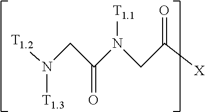


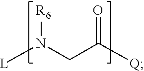


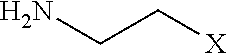


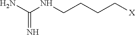


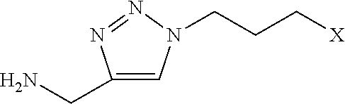


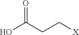


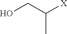


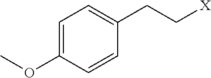


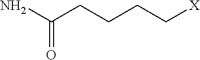

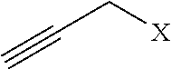
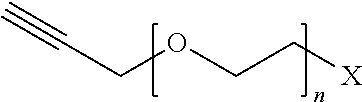
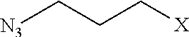


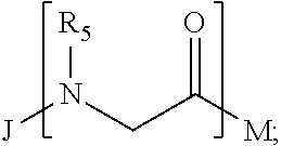


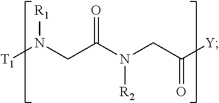

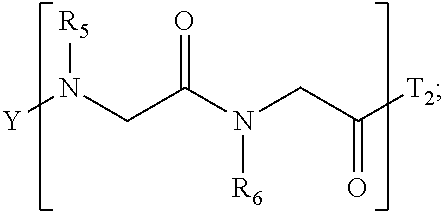


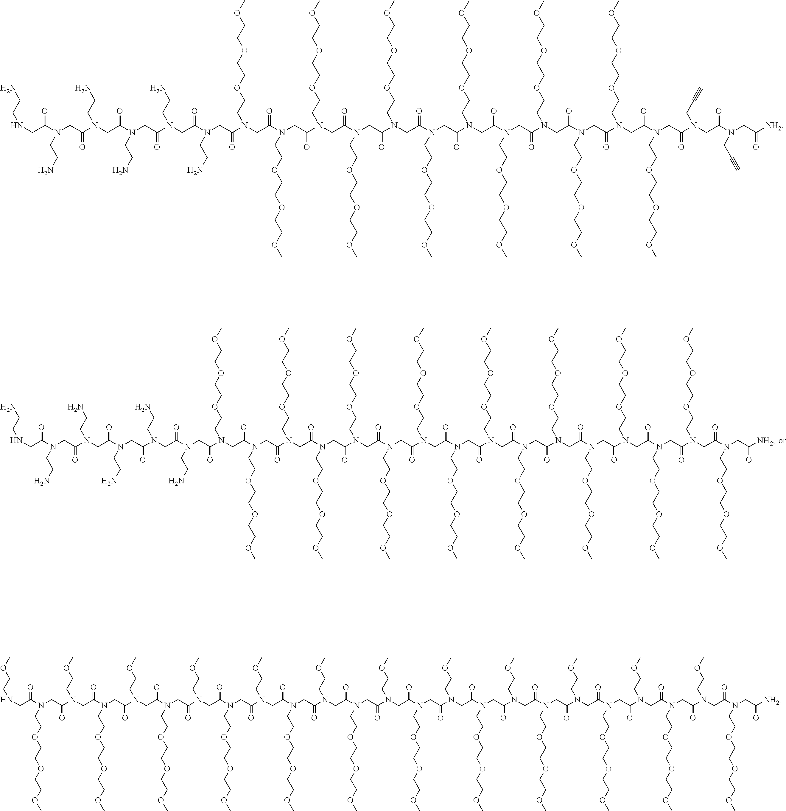
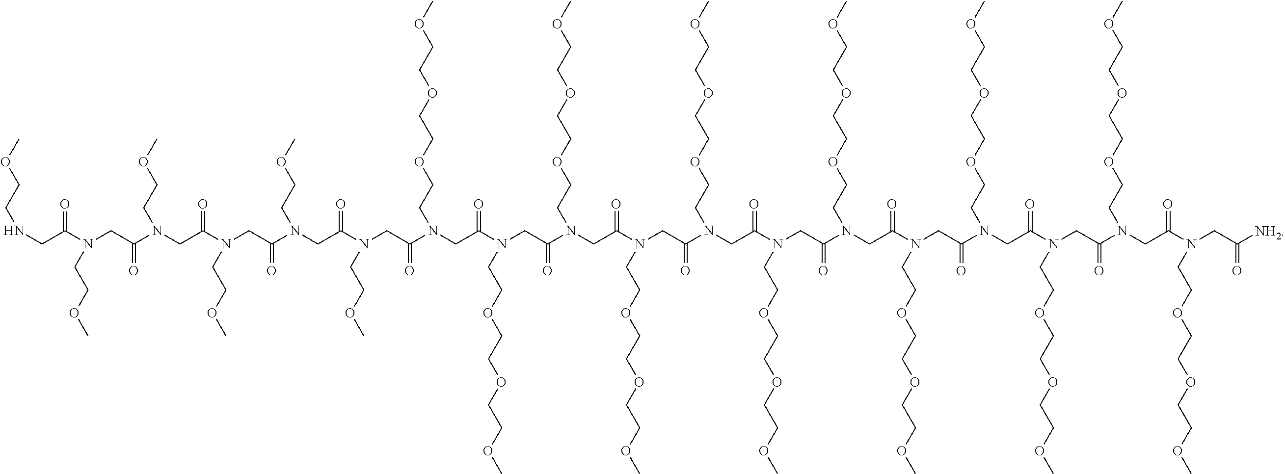

D00000

D00001

D00002

D00003

D00004

D00005

D00006

D00007

D00008

D00009

D00010

D00011

D00012

D00013

D00014

D00015

D00016

D00017

D00018

D00019

D00020

D00021

D00022

D00023

D00024

D00025

D00026

D00027

D00028

D00029

D00030

D00031

D00032

D00033

D00034

D00035

D00036

D00037

D00038
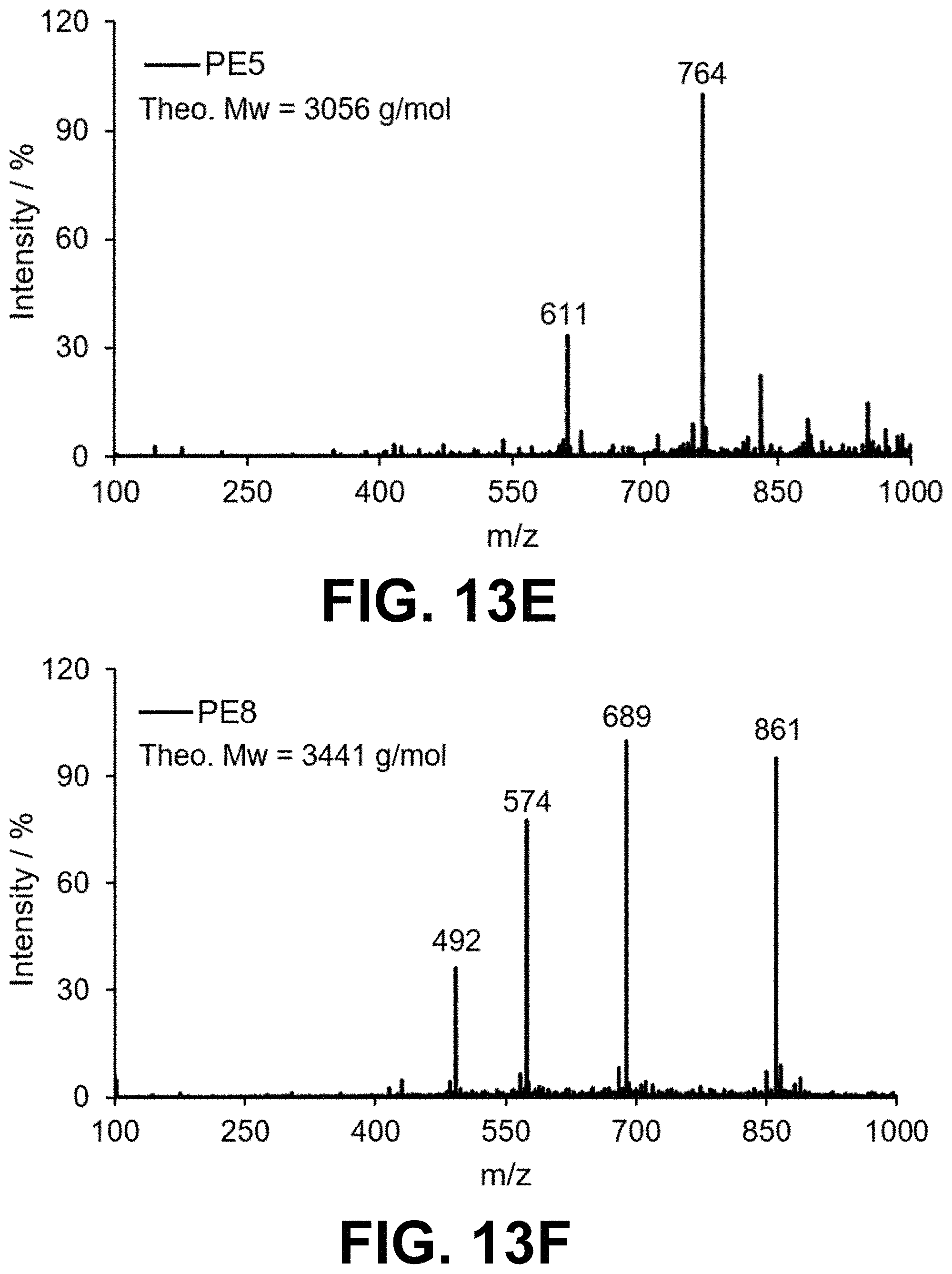
D00039
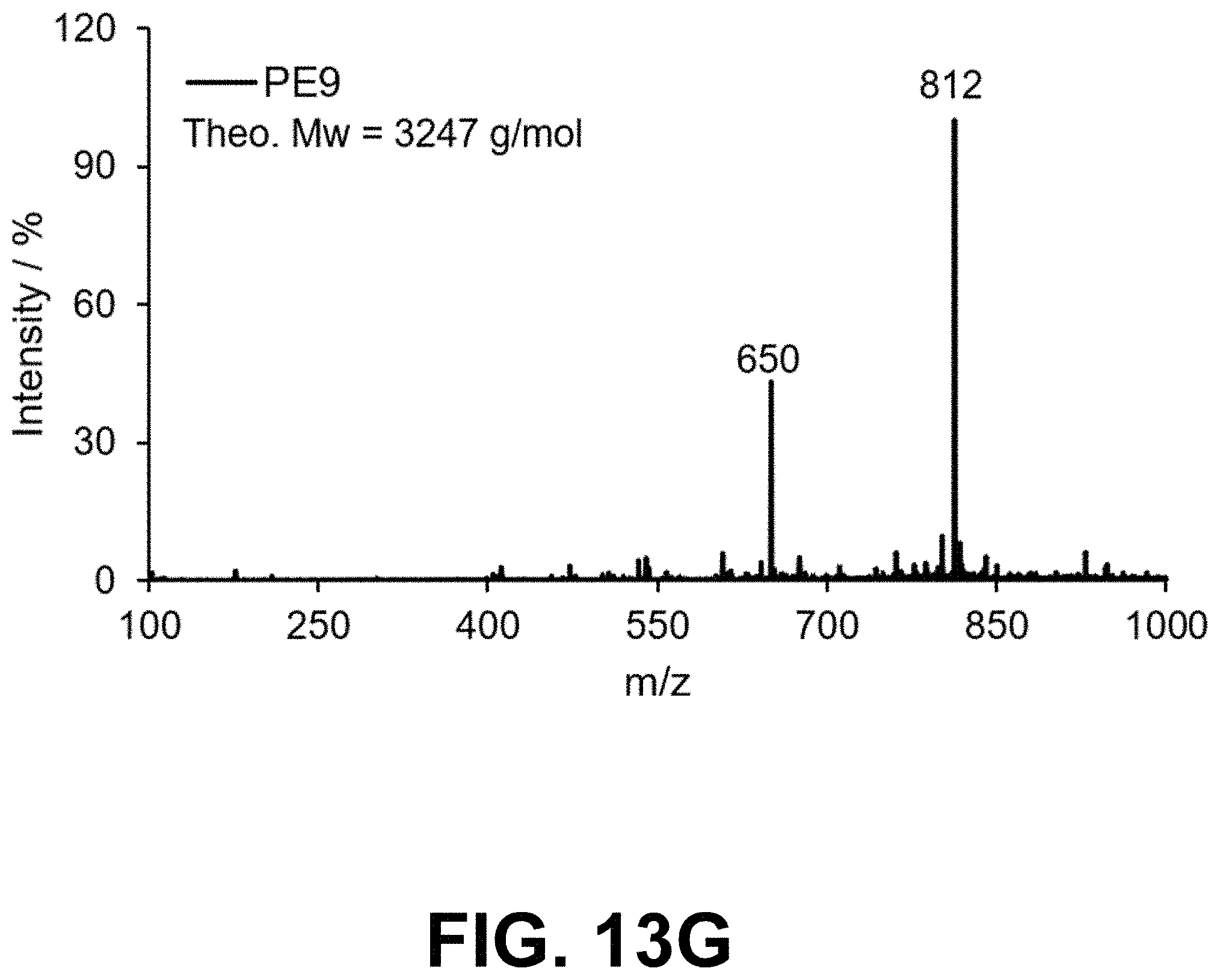
D00040
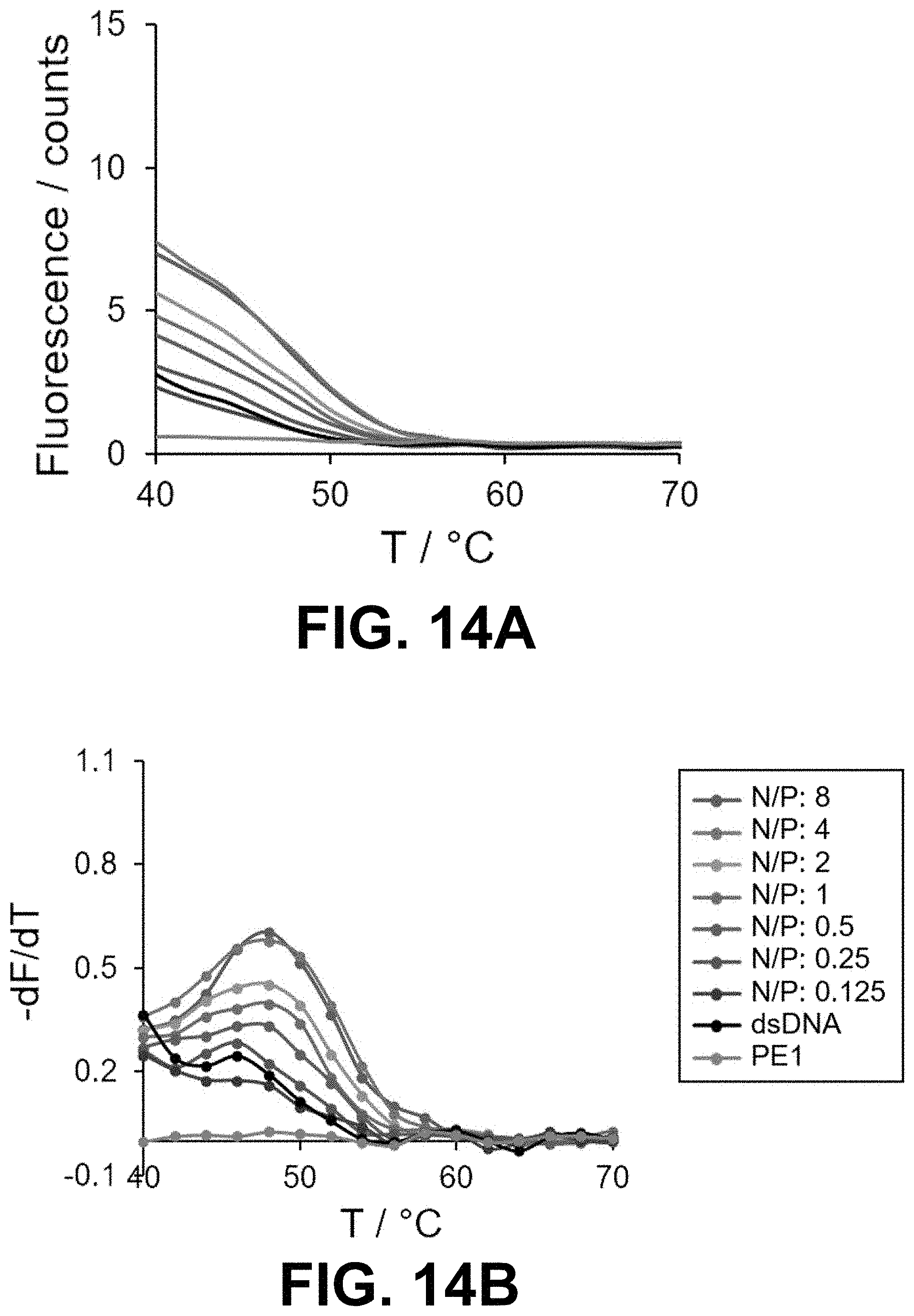
D00041
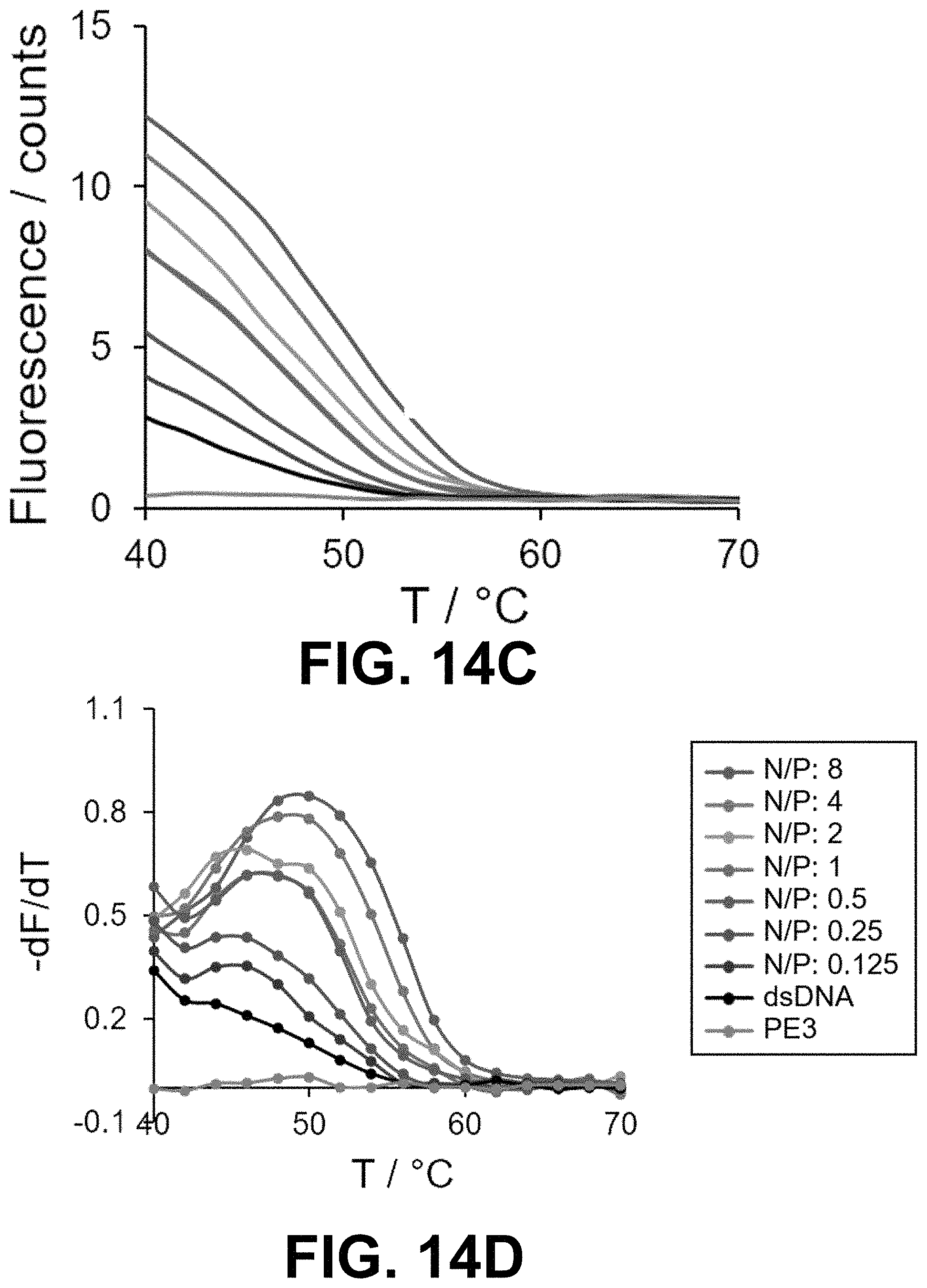
D00042

D00043
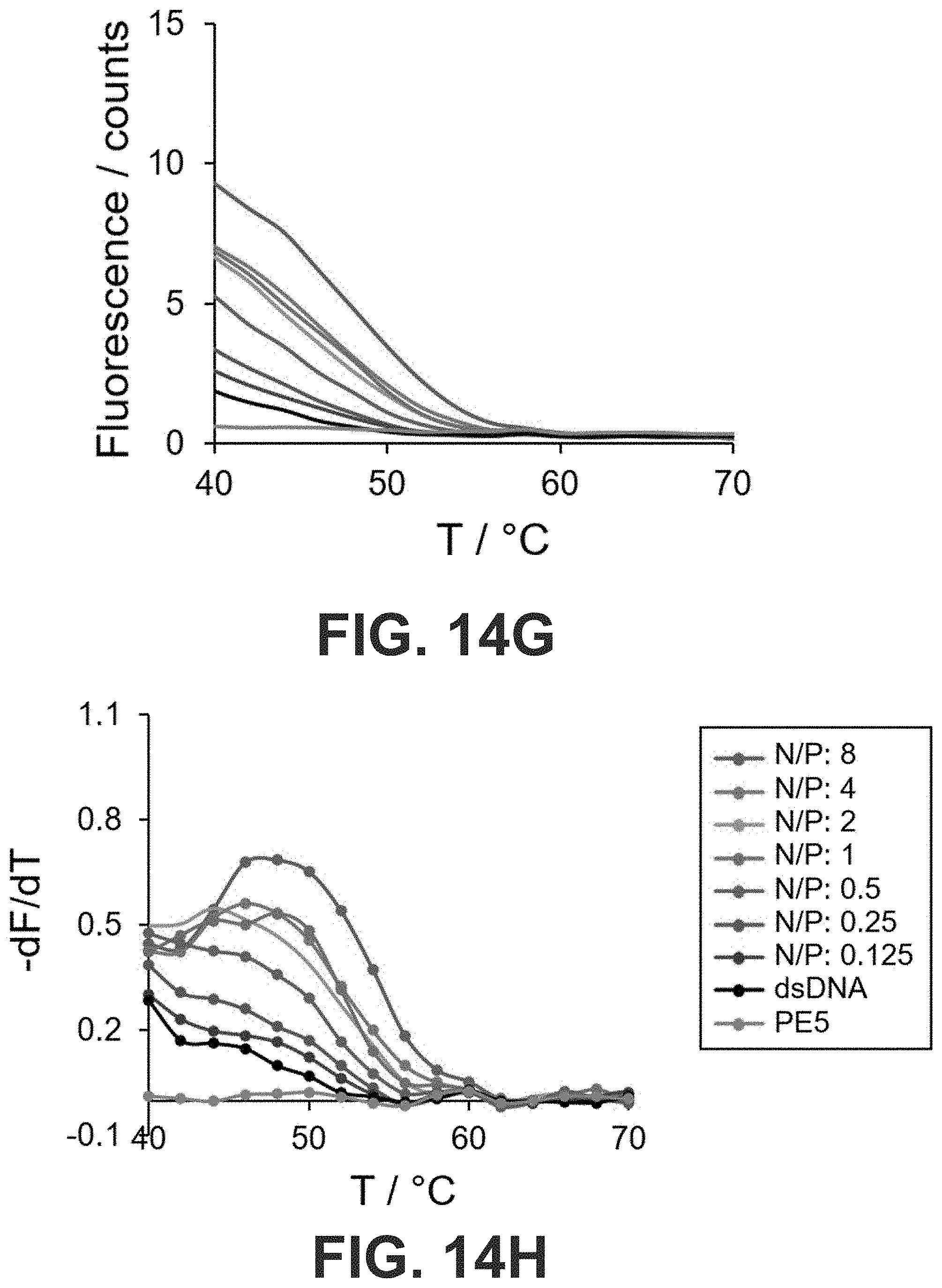
D00044

D00045

D00046

D00047

D00048

D00049

D00050
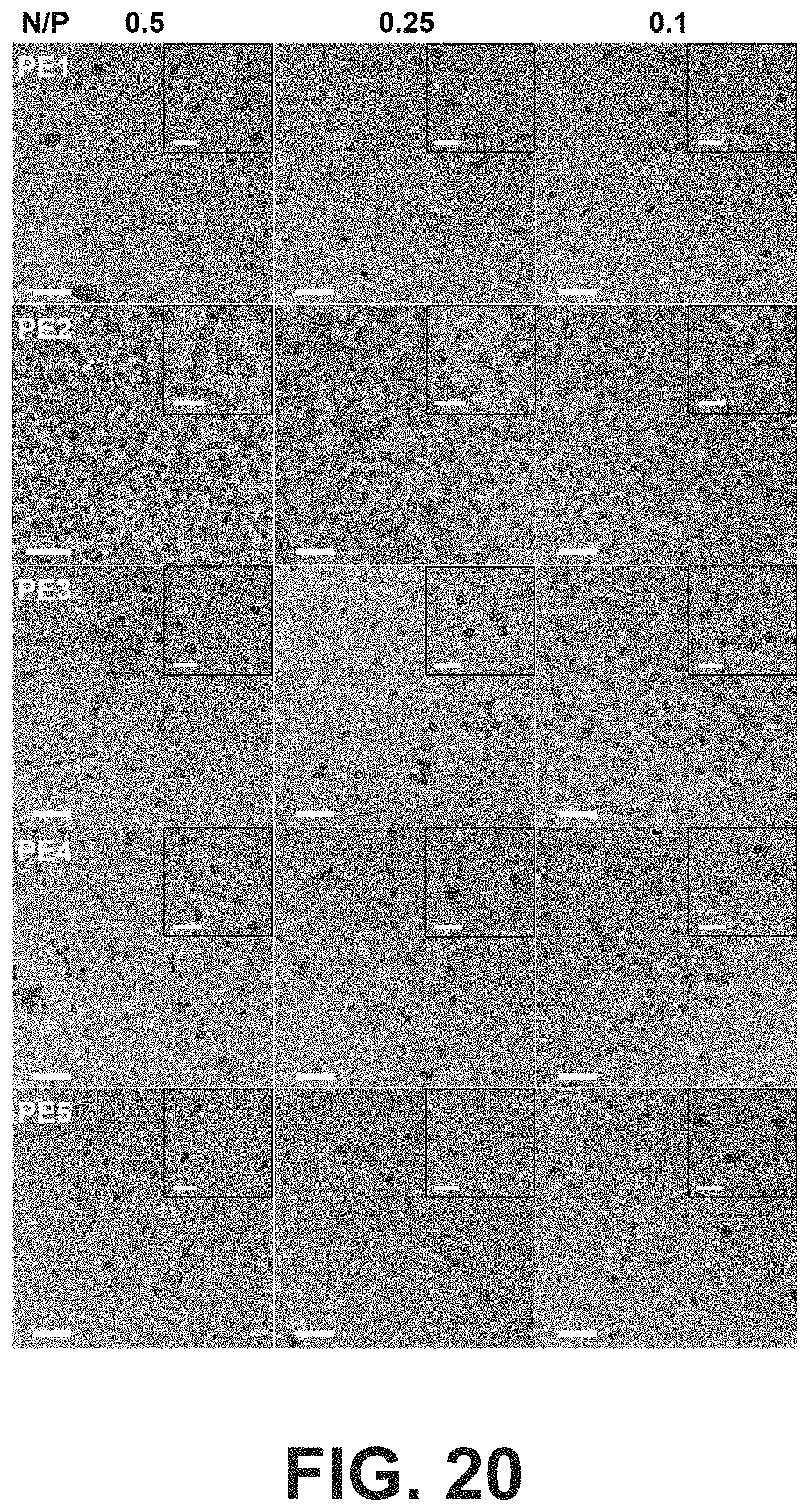
D00051

D00052
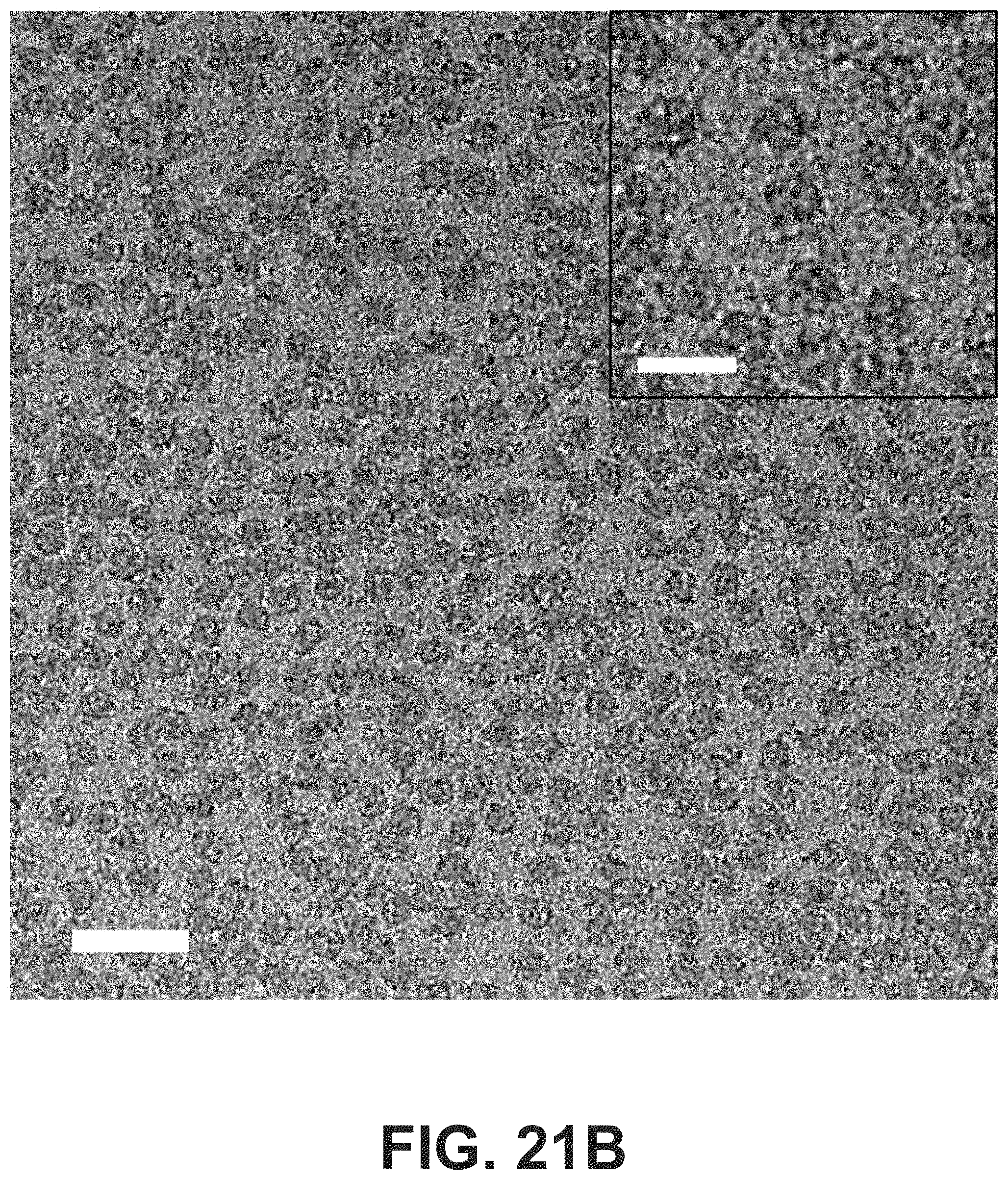
D00053

D00054

D00055

D00056

D00057

D00058

D00059

D00060
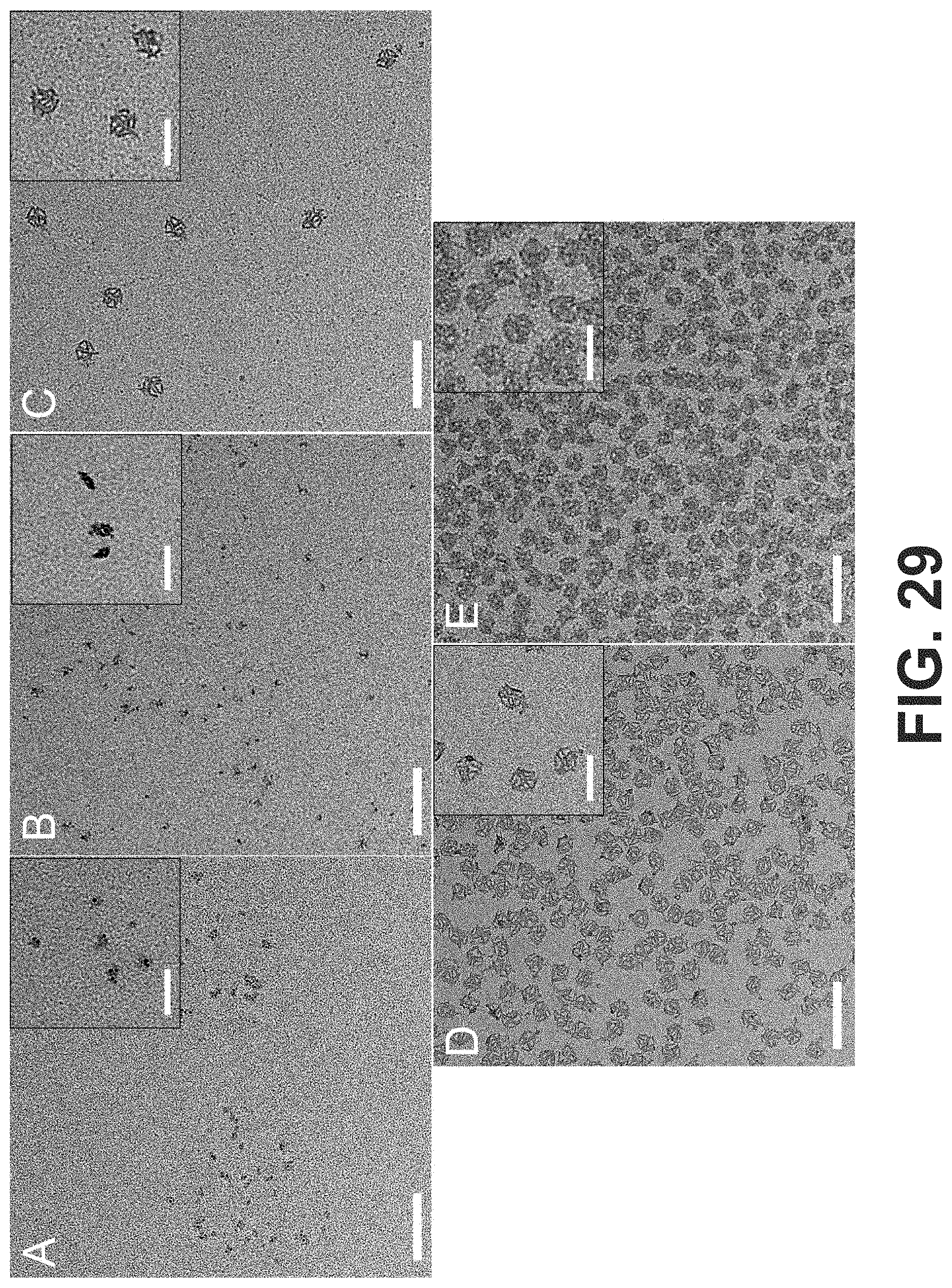
D00061
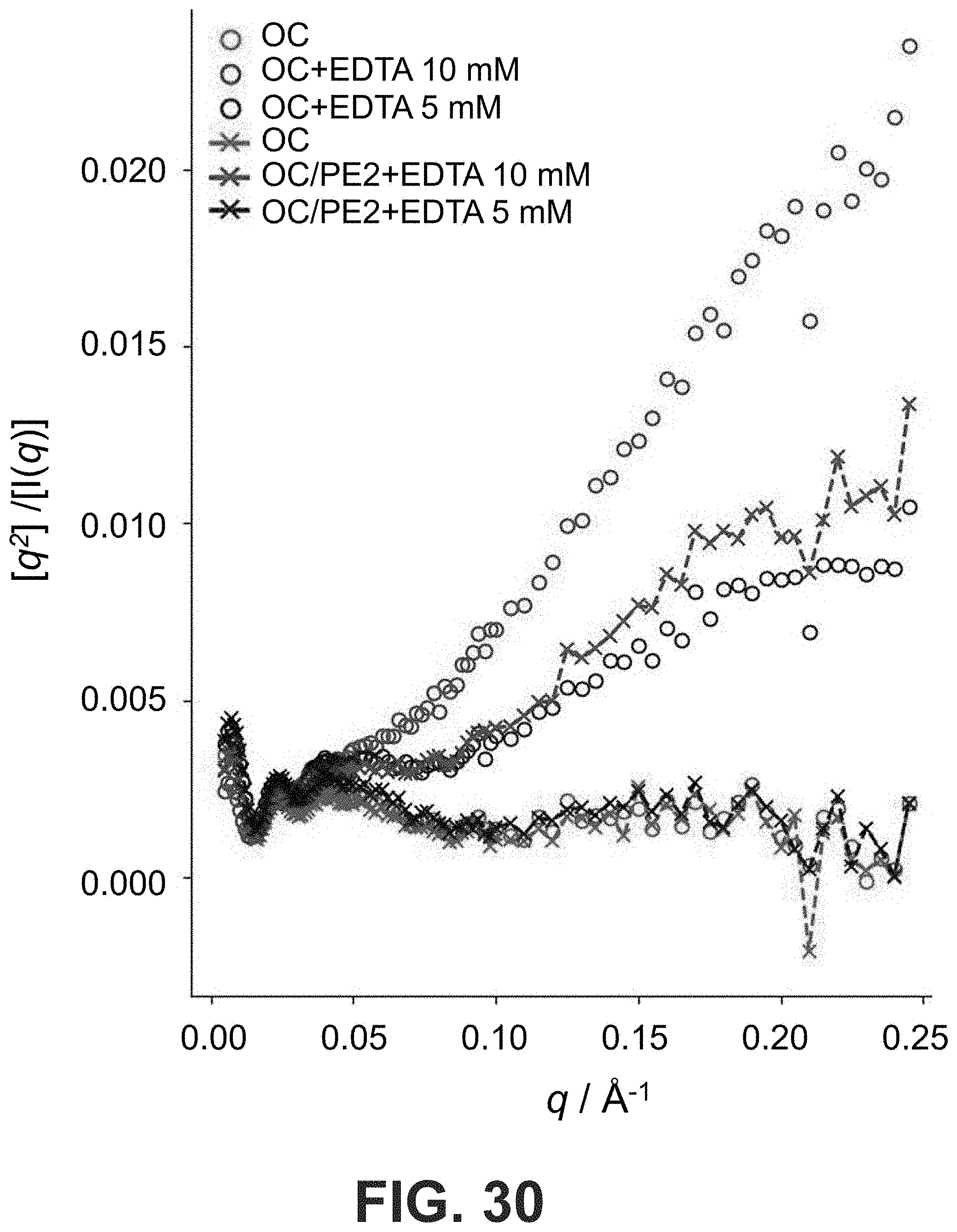
D00062

D00063

D00064

D00065

D00066

D00067

D00068
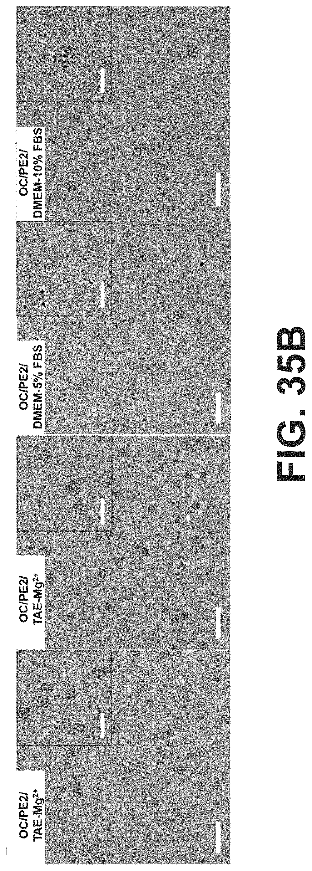
D00069

D00070

D00071

D00072
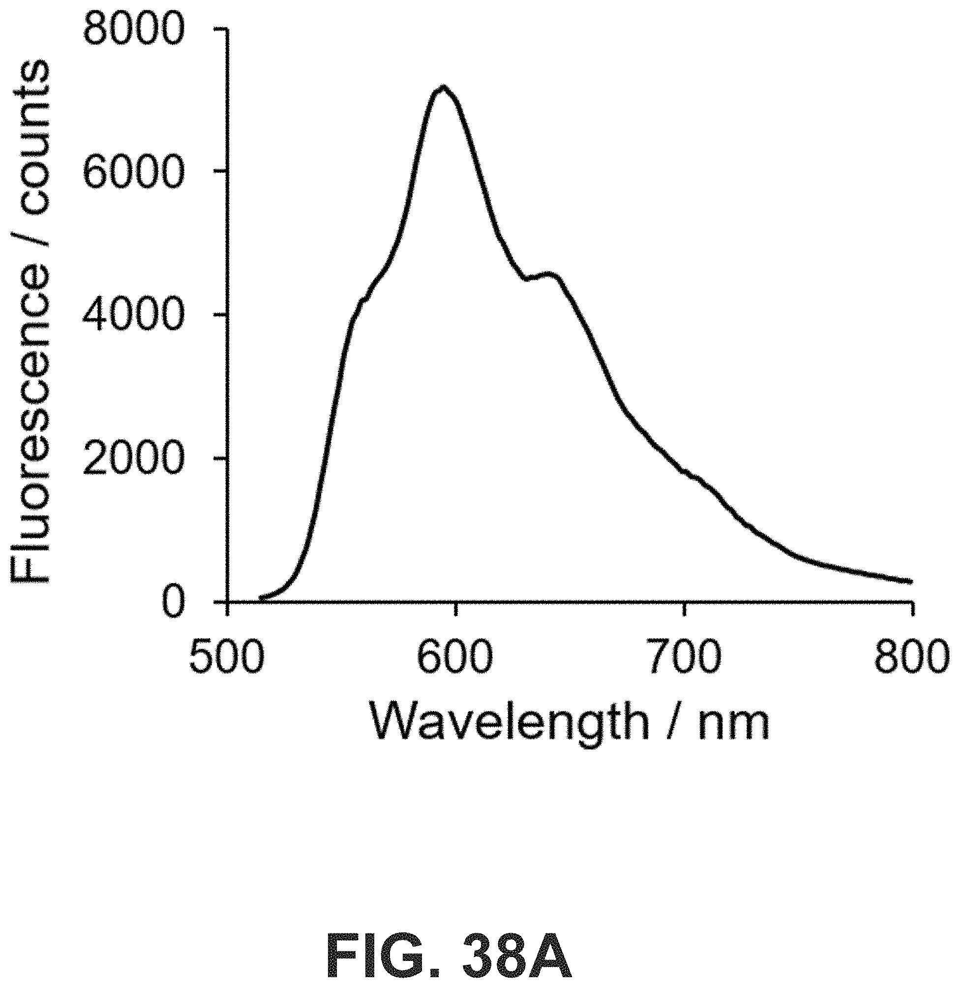
D00073
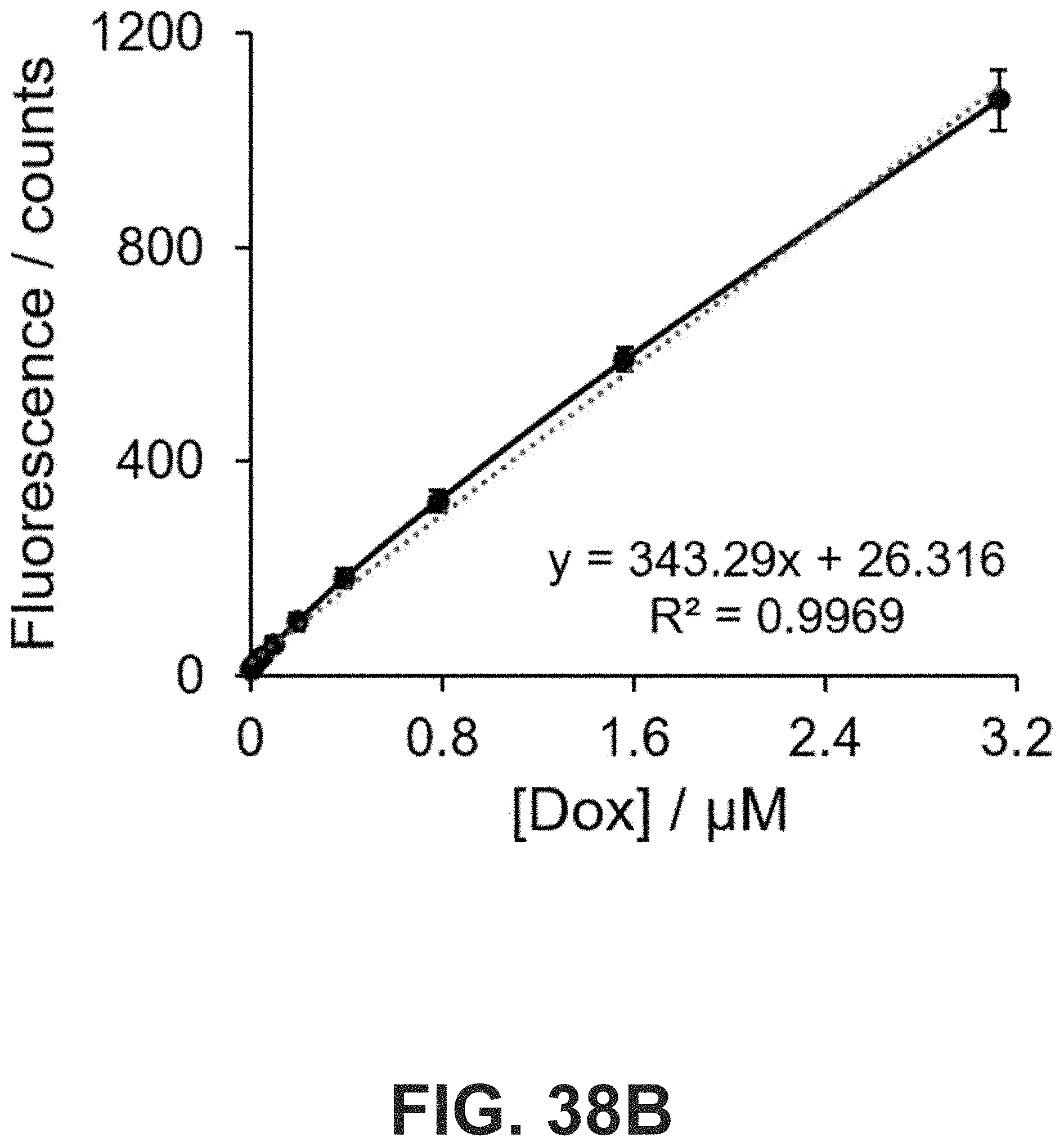
D00074

D00075

D00076

D00077

D00078

S00001
XML
uspto.report is an independent third-party trademark research tool that is not affiliated, endorsed, or sponsored by the United States Patent and Trademark Office (USPTO) or any other governmental organization. The information provided by uspto.report is based on publicly available data at the time of writing and is intended for informational purposes only.
While we strive to provide accurate and up-to-date information, we do not guarantee the accuracy, completeness, reliability, or suitability of the information displayed on this site. The use of this site is at your own risk. Any reliance you place on such information is therefore strictly at your own risk.
All official trademark data, including owner information, should be verified by visiting the official USPTO website at www.uspto.gov. This site is not intended to replace professional legal advice and should not be used as a substitute for consulting with a legal professional who is knowledgeable about trademark law.