Bispecific T Cell Activating Antigen Binding Molecules
BACAC; Marina ; et al.
U.S. patent application number 16/877150 was filed with the patent office on 2021-03-11 for bispecific t cell activating antigen binding molecules. The applicant listed for this patent is Hoffmann-La Roche Inc.. Invention is credited to Marina BACAC, Sylvia HERTER, Sabine IMHOF-JUNG, Christian KLEIN, Stefan KLOSTERMANN, Michael MOLHOJ, Christiane NEUMANN, Joerg Thomas REGULA, Wolfgang SCHAEFER, Pablo UMANA.
| Application Number | 20210070882 16/877150 |
| Document ID | / |
| Family ID | 1000005223218 |
| Filed Date | 2021-03-11 |


View All Diagrams
| United States Patent Application | 20210070882 |
| Kind Code | A1 |
| BACAC; Marina ; et al. | March 11, 2021 |
BISPECIFIC T CELL ACTIVATING ANTIGEN BINDING MOLECULES
Abstract
The present invention generally relates to novel bispecific antigen binding molecules for T cell activation and re-direction to specific target cells. In addition, the present invention relates to polynucleotides encoding such bispecific antigen binding molecules, and vectors and host cells comprising such polynucleotides. The invention further relates to methods for producing the bispecific antigen binding molecules of the invention, and to methods of using these bispecific antigen binding molecules in the treatment of disease.
| Inventors: | BACAC; Marina; (Zurich, CH) ; KLEIN; Christian; (Bonstetten, CH) ; SCHAEFER; Wolfgang; (Mannheim, DE) ; KLOSTERMANN; Stefan; (Neuried, DE) ; IMHOF-JUNG; Sabine; (Planegg, DE) ; MOLHOJ; Michael; (Munich, DE) ; REGULA; Joerg Thomas; (Munich, DE) ; UMANA; Pablo; (Wollerau, CH) ; HERTER; Sylvia; (Regensdorf, CH) ; NEUMANN; Christiane; (Niederweningen, CH) | ||||||||||
| Applicant: |
|
||||||||||
|---|---|---|---|---|---|---|---|---|---|---|---|
| Family ID: | 1000005223218 | ||||||||||
| Appl. No.: | 16/877150 | ||||||||||
| Filed: | May 18, 2020 |
Related U.S. Patent Documents
| Application Number | Filing Date | Patent Number | ||
|---|---|---|---|---|
| 15281493 | Sep 30, 2016 | |||
| 16877150 | ||||
| Current U.S. Class: | 1/1 |
| Current CPC Class: | C07K 2317/73 20130101; C07K 2317/626 20130101; C07K 16/3007 20130101; C07K 2319/00 20130101; C07K 2317/64 20130101; C07K 2317/526 20130101; A61K 2039/505 20130101; C07K 16/2803 20130101; C07K 2317/31 20130101; C07K 2317/52 20130101; C07K 2317/94 20130101; C07K 2317/66 20130101; C07K 16/468 20130101; C07K 16/2809 20130101; C07K 16/3053 20130101; C07K 2317/24 20130101; C07K 2317/55 20130101; C07K 2317/33 20130101; C07K 16/40 20130101; C07K 16/2863 20130101; C07K 2317/92 20130101; C07K 2317/35 20130101 |
| International Class: | C07K 16/40 20060101 C07K016/40; C07K 16/28 20060101 C07K016/28; C07K 16/46 20060101 C07K016/46; C07K 16/30 20060101 C07K016/30 |
Foreign Application Data
| Date | Code | Application Number |
|---|---|---|
| Oct 2, 2015 | EP | 15188093.7 |
| May 11, 2016 | EP | 16169160.5 |
Claims
1. A T cell activating bispecific antigen binding molecule comprising (a) a first antigen binding moiety which specifically binds to a first antigen; (b) a second antigen binding moiety which specifically binds to a second antigen; wherein the first antigen is an activating T cell antigen and the second antigen is CD19, or the first antigen is CD19 and the second antigen is an activating T cell antigen; and wherein the antigen binding moiety which specifically binds to CD19 a comprises a heavy chain variable region, particularly a humanized heavy chain variable region, comprising the heavy chain complementarity determining region (HCDR) 1 of SEQ ID NO: 14, the HCDR 2 of SEQ ID NO: 15 and the HCDR 3 of SEQ ID NO: 16, and a light chain variable region, particularly a humanized light chain variable region, comprising the light chain complementarity determining region (LCDR) 1 of SEQ ID NO: 17, the LCDR 2 of SEQ ID NO: 18 and the LCDR 3 of SEQ ID NO: 19.
2. The T cell activating bispecific antigen binding molecule according to claim 1, wherein the antigen binding moiety which specifically binds to CD19 comprises a heavy chain variable region comprising an amino acid sequence that is at least about 95%, 96%, 97%, 98%, 99% or 100% identical to the amino acid sequence of SEQ ID NO: 20 and a light chain variable region comprising an amino acid sequence that is at least about 95%, 96%, 97%, 98%, 99% or 100% identical to the amino acid sequence of SEQ ID NO: 21.
3. (canceled)
4. The T cell activating bispecific antigen binding molecule according to claim 1, wherein the second antigen binding moiety is a Fab molecule which specifically binds to a second antigen, and wherein the variable domains VL and VH or the constant domains CL and CH1 of the Fab light chain and the Fab heavy chain are replaced by each other.
5. (canceled)
6. The T cell activating bispecific antigen binding molecule according to claim 1, wherein the activating T cell antigen is CD3, particularly CD3 epsilon.
7. The T cell activating bispecific antigen binding molecule according to claim 1, wherein the antigen binding moiety which specifically binds to the activating T cell antigen comprises the heavy chain complementarity determining region (CDR) 1 of SEQ ID NO: 4, the heavy chain CDR 2 of SEQ ID NO: 5, the heavy chain CDR 3 of SEQ ID NO: 6, the light chain CDR 1 of SEQ ID NO: 8, the light chain CDR 2 of SEQ ID NO: 9 and the light chain CDR 3 of SEQ ID NO: 10.
8. The T cell activating bispecific antigen binding molecule according to claim 1, wherein the antigen binding moiety which specifically binds to the activating T cell antigen comprises a heavy chain variable region comprising an amino acid sequence that is at least about 95%, 96%, 97%, 98%, 99% or 100% identical to the amino acid sequence of SEQ ID NO: 3 and a light chain variable region comprising an amino acid sequence that is at least about 95%, 96%, 97%, 98%, 99% or 100% identical to the amino acid sequence of SEQ ID NO: 7.
9. The T cell activating bispecific antigen binding molecule according to claim 1, wherein the first antigen binding moiety under (a) is a first Fab molecule which specifically binds to a first antigen, the second antigen binding moiety under (b) is a second Fab molecule which specifically binds to a second antigen wherein the variable domains VL and VH of the Fab light chain and the Fab heavy chain are replaced by each other; and i) in the constant domain CL of the first Fab molecule under a) the amino acid at position 124 is substituted independently by lysine (K), arginine (R) or histidine (H) (numbering according to Kabat), and wherein in the constant domain CH1 of the first Fab molecule under a) the amino acid at position 147 or the amino acid at position 213 is substituted independently by glutamic acid (E), or aspartic acid (D) (numbering according to Kabat EU index); or ii) in the constant domain CL of the second Fab molecule under b) the amino acid at position 124 is substituted independently by lysine (K), arginine (R) or histidine (H) (numbering according to Kabat), and wherein in the constant domain CH1 of the second Fab molecule under b) the amino acid at position 147 or the amino acid at position 213 is substituted independently by glutamic acid (E), or aspartic acid (D) (numbering according to Kabat EU index).
10-11. (canceled)
12. The T cell activating bispecific antigen binding molecule according to claim 9, wherein in the constant domain CL of the first Fab molecule under a) the amino acid at position 124 is substituted independently by lysine (K), arginine (R) or histidine (H) (numbering according to Kabat) and the amino acid at position 123 is substituted independently by lysine (K), arginine (R) or histidine (H) (numbering according to Kabat), and wherein in the constant domain CH1 of the first Fab molecule under a) the amino acid at position 147 is substituted independently by glutamic acid (E), or aspartic acid (D) (numbering according to Kabat EU index) and the amino acid at position 213 is substituted independently by glutamic acid (E), or aspartic acid (D) (numbering according to Kabat EU index).
13. The T cell activating bispecific antigen binding molecule according to claim 9, wherein in the constant domain CL of the first Fab molecule under a) the amino acid at position 124 is substituted by lysine (K) (numbering according to Kabat) and the amino acid at position 123 is substituted by arginine (R) (numbering according to Kabat), and wherein in the constant domain CH1 of the first Fab molecule under a) the amino acid at position 147 is substituted by glutamic acid (E) (numbering according to Kabat EU index) and the amino acid at position 213 is substituted by glutamic acid (E) (numbering according to Kabat EU index).
14-19. (canceled)
20. The T cell activating bispecific antigen binding molecule according to claim 1, further comprising a third antigen binding moiety which specifically binds to the first antigen.
21. (canceled)
22. The T cell activating bispecific antigen binding molecule according to claim 20, wherein the third antigen binding moiety is identical to the first antigen binding moiety.
23. The T cell activating bispecific antigen binding molecule according to claim 20, wherein the first and the third antigen binding moiety specifically bind to a target cell antigen, and the second antigen binding moiety specifically binds to an activating T cell antigen, particularly CD3, more particularly CD3 epsilon.
24. The T cell activating bispecific antigen binding molecule according to claim 1, additionally comprising an Fc domain composed of a first and a second subunit capable of stable association.
25. (canceled)
26. The T cell activating bispecific antigen binding molecule according to claim 1, wherein the first and the second antigen binding moieties are Fab molecules and the second antigen binding moiety is fused at the C-terminus of the Fab heavy chain to the N-terminus of the Fab heavy chain of the first antigen binding moiety.
27-28. (canceled)
29. The T cell activating bispecific antigen binding molecule according to claim 24, wherein the first and the second antigen binding moieties are Fab molecules and the second antigen binding moiety is fused at the C-terminus of the Fab heavy chain to the N-terminus of the first or the second subunit of the Fc domain.
30-31. (canceled)
32. The T cell activating bispecific antigen binding molecule according to claim 24, further comprising a third antigen binding moiety which specifically binds to the first antigen, wherein the third antigen binding moiety is a Fab molecule and is fused at the C-terminus of the Fab heavy chain to the N-terminus of the first or second subunit of the Fc domain.
33. (canceled)
34. The T cell activating bispecific antigen binding molecule according to claim 24, further comprising a third antigen binding moiety which specifically binds to the first antigen, wherein the first, second and third antigen binding moieties are Fab molecules and the first and the third antigen binding moiety are each fused at the C-terminus of the Fab heavy chain to the N-terminus of one of the subunits of the Fc domain, and the second antigen binding moiety is fused at the C-terminus of the Fab heavy chain to the N-terminus of the Fab heavy chain of the first antigen binding moiety.
35. (canceled)
36. The T cell activating bispecific antigen binding molecule according to claim 24, wherein the Fc domain is an IgG1 or IgG4 Fc domain.
37. The T cell activating bispecific antigen binding molecule according to claim 24, wherein the Fc domain is a human Fc domain.
38. The T cell activating bispecific antigen binding molecule according to claim 24, wherein the Fc domain comprises a modification promoting the association of the first and the second subunit of the Fc domain.
39. The T cell activating bispecific antigen binding molecule of claim 38, wherein in the CH3 domain of the first subunit of the Fc domain an amino acid residue is replaced with an amino acid residue having a larger side chain volume, thereby generating a protuberance within the CH3 domain of the first subunit which is positionable in a cavity within the CH3 domain of the second subunit, and in the CH3 domain of the second subunit of the Fc domain an amino acid residue is replaced with an amino acid residue having a smaller side chain volume, thereby generating a cavity within the CH3 domain of the second subunit within which the protuberance within the CH3 domain of the first subunit is positionable.
40. (canceled)
41. The T cell activating bispecific antigen binding molecule of claim 39, wherein in the CH3 domain of the first subunit of the Fc domain the threonine residue at position 366 is replaced with a tryptophan residue (T366W), and in the CH3 domain of the second subunit of the Fc domain the tyrosine residue at position 407 is replaced with a valine residue (Y407V), and optionally in the second subunit of the Fc domain additionally the threonine residue at position 366 is replaced with a serine residue (T366S) and the leucine residue at position 368 is replaced with an alanine residue (L368A) (numberings according to Kabat EU index).
42. The T cell activating bispecific antigen binding molecule of claim 41, wherein in the first subunit of the Fc domain additionally the serine residue at position 354 is replaced with a cysteine residue (S354C) or the glutamic acid residue at position 356 is replaced with a cysteine residue (E356C), and in the second subunit of the Fc domain additionally the tyrosine residue at position 349 is replaced by a cysteine residue (Y349C) (numberings according to Kabat EU index).
43-44. (canceled)
45. The T cell activating bispecific antigen binding molecule according to claim 24, wherein the Fc domain comprises one or more amino acid substitution that reduces binding to an Fc receptor and/or effector function.
46. The T cell activating bispecific antigen binding molecule according to claim 45, wherein said one or more amino acid substitution is at one or more position selected from the group of L234, L235, and P329 (Kabat EU index numbering).
47. The T cell activating bispecific antigen binding molecule according to claim 24, wherein each subunit of the Fc domain comprises the amino acid substitutions L234A, L235A and P329G (Kabat EU index numbering).
48-49. (canceled)
50. One or more isolated polynucleotide encoding the T cell activating bispecific antigen binding molecule of claim 1.
51. (canceled)
52. A host cell comprising the polynucleotide(s) of claim 50.
53. A method of producing a T cell activating bispecific antigen binding molecule capable of specific binding to CD19 and an activating T cell antigen, comprising the steps of a) culturing the host cell of claim 52 under conditions suitable for the expression of the T cell activating bispecific antigen binding molecule and b) optionally recovering the T cell activating bispecific antigen binding molecule.
54. A T cell activating bispecific antigen binding molecule produced by the method of claim 53.
55. A pharmaceutical composition comprising the T cell activating bispecific antigen binding molecule of claim 1 and a pharmaceutically acceptable carrier.
56. A method of treating a disease in an individual, comprising administering to said individual a therapeutically effective amount of a composition comprising the T cell activating bispecific antigen binding molecule of claim 1 in a pharmaceutically acceptable form.
57. (canceled)
58. A method for inducing lysis of a target cell, comprising contacting a target cell with the T cell activating bispecific antigen binding molecule of claim 1 in the presence of a T cell.
59. A T cell activating bispecific antigen binding molecule comprising (a) a first antigen binding moiety which specifically binds to a first antigen; (b) a second antigen binding moiety which specifically binds to a second antigen; wherein the first antigen is an activating T cell antigen and the second antigen is CD19, or the first antigen is CD19 and the second antigen is an activating T cell antigen; and wherein the antigen binding moiety which specifically binds to CD19 comprises a heavy chain variable region, particularly a humanized heavy chain variable region, comprising the heavy chain complementarity determining region (HCDR) 1 of SEQ ID NO: 50, the HCDR 2 of SEQ ID NO: 51 and the HCDR 3 of SEQ ID NO: 52, and a light chain variable region, particularly a humanized light chain variable region, comprising the light chain complementarity determining region (LCDR) 1 of SEQ ID NO: 53, the LCDR 2 of SEQ ID NO: 54 and the LCDR 3 of SEQ ID NO: 55.
60. The T cell activating bispecific antigen binding molecule according to claim 59, wherein the antigen binding moiety which specifically binds to CD19 comprises a heavy chain variable region comprising an amino acid sequence that is at least about 95%, 96%, 97%, 98%, 99% or 100% identical to the amino acid sequence of SEQ ID NO: 56 and a light chain variable region comprising an amino acid sequence that is at least about 95%, 96%, 97%, 98%, 99% or 100% identical to the amino acid sequence of SEQ ID NO: 57.
61. One or more isolated polynucleotide encoding the T cell activating bispecific antigen binding molecule of claim 59.
62. A host cell comprising the polynucleotide(s) of claim 61.
63. A method of producing a T cell activating bispecific antigen binding molecule capable of specific binding to CD19 and an activating T cell antigen, comprising the steps of a) culturing the host cell of claim 62 under conditions suitable for the expression of the T cell activating bispecific antigen binding molecule and b) optionally recovering the T cell activating bispecific antigen binding molecule.
64. A T cell activating bispecific antigen binding molecule produced by the method of claim 63.
65. A pharmaceutical composition comprising the T cell activating bispecific antigen binding molecule of claim 59 and a pharmaceutically acceptable carrier.
66. A method of treating a disease in an individual, comprising administering to said individual a therapeutically effective amount of a composition comprising the T cell activating bispecific antigen binding molecule of claim 59 in a pharmaceutically acceptable form.
67. A method for inducing lysis of a target cell, comprising contacting a target cell with the T cell activating bispecific antigen binding molecule of claim 59 in the presence of a T cell.
Description
CROSS REFERENCE TO RELATED APPLICATIONS
[0001] This application is a continuation of U.S. patent application Ser. No. 15/281,493, filed Sep. 30, 2016, which claims priority to European Patent Application No. EP 15188093.7, filed Oct. 2, 2015, and European Patent Application No. EP 16169160.5, filed May 11, 2016, the disclosures of which are incorporated herein by reference in their entirety.
SEQUENCE LISTING
[0002] The present application contains a Sequence Listing which has been submitted in ASCII format via EFS-Web and is hereby incorporated by reference in its entirety. Said ASCII copy, created on May 15, 2020, is named 51177-013002_Sequence_Listing_5_15_20_ST25 and is 130,834 bytes in size.
FIELD OF THE INVENTION
[0003] The present invention generally relates to bispecific antigen binding molecules for activating T cells. In addition, the present invention relates to polynucleotides encoding such bispecific antigen binding molecules, and vectors and host cells comprising such polynucleotides. The invention further relates to methods for producing the bispecific antigen binding molecules of the invention, and to methods of using these bispecific antigen binding molecules in the treatment of disease.
BACKGROUND
[0004] The selective destruction of an individual cell or a specific cell type is often desirable in a variety of clinical settings. For example, it is a primary goal of cancer therapy to specifically destroy tumor cells, while leaving healthy cells and tissues intact and undamaged.
[0005] An attractive way of achieving this is by inducing an immune response against the tumor, to make immune effector cells such as natural killer (NK) cells or cytotoxic T lymphocytes (CTLs) attack and destroy tumor cells. CTLs constitute the most potent effector cells of the immune system, however they cannot be activated by the effector mechanism mediated by the Fc domain of conventional therapeutic antibodies.
[0006] In this regard, bispecific antibodies designed to bind with one "arm" to a surface antigen on target cells, and with the second "arm" to an activating, invariant component of the T cell receptor (TCR) complex, have become of interest in recent years. The simultaneous binding of such an antibody to both of its targets will force a temporary interaction between target cell and T cell, causing activation of any cytotoxic T cell and subsequent lysis of the target cell. Hence, the immune response is re-directed to the target cells and is independent of peptide antigen presentation by the target cell or the specificity of the T cell as would be relevant for normal MHC-restricted activation of CTLs. In this context it is crucial that CTLs are only activated when a target cell is presenting the bispecific antibody to them, i.e. the immunological synapse is mimicked. Particularly desirable are bispecific antibodies that do not require lymphocyte preconditioning or co-stimulation in order to elicit efficient lysis of target cells.
[0007] Several bispecific antibody formats have been developed and their suitability for T cell mediated immunotherapy investigated. Out of these, the so-called BiTE (bispecific T cell engager) molecules have been very well characterized and already shown some promise in the clinic (reviewed in Nagorsen and Bauerle, Exp Cell Res 317, 1255-1260 (2011)). BiTEs are tandem scFv molecules wherein two scFv molecules are fused by a flexible linker. Further bispecific formats being evaluated for T cell engagement include diabodies (Holliger et al., Prot Eng 9, 299-305 (1996)) and derivatives thereof, such as tandem diabodies (Kipriyanov et al., J Mol Biol 293, 41-66 (1999)). A more recent development are the so-called DART (dual affinity retargeting) molecules, which are based on the diabody format but feature a C-terminal disulfide bridge for additional stabilization (Moore et al., Blood 117, 4542-51 (2011)). The so-called triomabs, which are whole hybrid mouse/rat IgG molecules and also currently being evaluated in clinical trials, represent a larger sized format (reviewed in Seimetz et al., Cancer Treat Rev 36, 458-467 (2010)).
[0008] The variety of formats that are being developed shows the great potential attributed to T cell re-direction and activation in immunotherapy. The task of generating bispecific antibodies suitable therefor is, however, by no means trivial, but involves a number of challenges that have to be met related to efficacy, toxicity, applicability and produceability of the antibodies.
[0009] Small constructs such as, for example, BiTE molecules--while being able to efficiently crosslink effector and target cells--have a very short serum half life requiring them to be administered to patients by continuous infusion. IgG-like formats on the other hand--while having the great benefit of a long half life--suffer from toxicity associated with the native effector functions inherent to IgG molecules. Their immunogenic potential constitutes another unfavorable feature of IgG-like bispecific antibodies, especially non-human formats, for successful therapeutic development. Finally, a major challenge in the general development of bispecific antibodies has been the production of bispecific antibody constructs at a clinically sufficient quantity and purity, due to the mispairing of antibody heavy and light chains of different specificities upon co-expression, which decreases the yield of the correctly assembled construct and results in a number of non-functional side products from which the desired bispecific antibody may be difficult to separate.
[0010] Different approaches have been taken to overcome the chain association issue in bispecific antibodies (see e.g. Klein et al., mAbs 6, 653-663 (2012)). For example, the `knobs-into-holes` strategy aims at forcing the pairing of two different antibody heavy chains by introducing mutations into the CH3 domains to modify the contact interface. On one chain bulky amino acids are replaced by amino acids with short side chains to create a `hole`. Conversely, amino acids with large side chains are introduced into the other CH3 domain, to create a `knob`. By coexpressing these two heavy chains (and two identical light chains, which have to be appropriate for both heavy chains), high yields of heterodimer (`knob-hole`) versus homodimer (`hole-hole` or `knob-knob`) are observed (Ridgway, J. B., et al., Protein Eng. 9 (1996) 617-621; and WO 96/027011). The percentage of heterodimer could be further increased by remodeling the interaction surfaces of the two CH3 domains using a phage display approach and the introduction of a disulfide bridge to stabilize the heterodimers (Merchant, A. M., et al., Nature Biotech. 16 (1998) 677-681; Atwell, S., et al., J. Mol. Biol. 270 (1997) 26-35). New approaches for the knobs-into-holes technology are described in e.g. in EP 1870459 A1.
[0011] The `knobs-into-holes` strategy does, however, not solve the problem of heavy chain-light chain mispairing, which occurs in bispecific antibodies comprising different light chains for binding to the different target antigens.
[0012] A strategy to prevent heavy chain-light chain mispairing is to exchange domains between the heavy and light chains of one of the binding arms of a bispecific antibody (see WO 2009/080251, WO 2009/080252, WO 2009/080253, WO 2009/080254 and Schaefer, W. et al, PNAS, 108 (2011) 11187-11191, which relate to bispecific IgG antibodies with a domain crossover).
[0013] Exchanging the heavy and light chain variable domains VH and VL in one of the binding arms of the bispecific antibody (WO2009/080252, see also Schaefer, W. et al, PNAS, 108 (2011) 11187-11191) clearly reduces the side products caused by the mispairing of a light chain against a first antigen with the wrong heavy chain against the second antigen (compared to approaches without such domain exchange). Nevertheless, these antibody preparations are not completely free of side products. The main side product is based on a Bence Jones-type interaction (Schaefer, W. et al, PNAS, 108 (2011) 11187-11191; in FIG. S1I of the Supplement). A further reduction of such side products is thus desirable to improve e.g. the yield of such bispecific antibodies.
[0014] The choice of target antigens and appropriate binders for both the T cell antigen and the target cell antigen is a further crucial aspect in the generation of T cell bispecific (TCB) antibodies for therapeutic application.
[0015] Human CD19 is a 95 kDa transmembrane protein (B-cell co-receptor) exclusively expressed on B-cells and on follicular dendritic cells. CD 19 is found in association with CD21 and CD81. CD19 and CD21 are required for normal B-cell differentiation (Carter, R. H., et al., Immunol. Res. 26 (2002) 45-54). CD19 is expressed on most B-cells (pan-B-cell marker) with the exception of stem cells and plasma cells, and is frequently expressed on most human B-cell malignancies (tumor associated antigen), such as lymphoma and leukemias except for multiple myeloma, e.g. in non-Hodgkin lymphoma and acute lymphoblastic leukemia. Antibodies against CD19 have been used in several clinical trials (see e. g. Hekman, A., et al., Cancer Immunol. Immunother. 32 (191) 364-372; Vlasfeld, L. T., et al., Cancer Immunol. Immunother. 40 (1995) 37-47; Conry, R. M., et al., J. Immunother. Emphasis Tumor Immunol. 18 (1995) 231-241; Manzke, O., et al., Int. J. Cancer 91 (2001) 516-522). In WO 2011/147834 antibodies against CD19 and uses thereof are reported. In WO 99/54440 and WO 2004/106381 pharmaceutical compositions comprising bispecific anti-CD3, anti-CD19 antibody constructs for the treatment of B-cell related disorders are reported.
[0016] The present invention provides novel, improved bispecific antigen binding molecules designed for T cell activation and re-direction, targeting CD3 and CD19, that combine good efficacy and produceability with low toxicity and favorable pharmacokinetic properties.
SUMMARY OF THE INVENTION
[0017] The present inventors have developed a novel T cell activating bispecific antigen binding molecule with unexpected, improved properties using a particular anti-CD19 antibody. Thus, in a first aspect the present invention provides a T cell activating bispecific antigen binding molecule comprising
[0018] (a) a first antigen binding moiety which specifically binds to a first antigen;
[0019] (b) a second antigen binding moiety which specifically binds to a second antigen;
[0020] wherein the first antigen is an activating T cell antigen and the second antigen is CD19, or the first antigen is CD19 and the second antigen is an activating T cell antigen; and
[0021] wherein the antigen binding moiety which specifically binds to CD19 comprises (i) a heavy chain variable region, particularly a humanized heavy chain variable region, comprising the heavy chain complementarity determining region (HCDR) 1 of SEQ ID NO: 14, the HCDR 2 of SEQ ID NO: 15 and the HCDR 3 of SEQ ID NO: 16, and a light chain variable region, particularly a humanized light chain variable region, comprising the light chain complementarity determining region (LCDR) 1 of SEQ ID NO: 17, the LCDR 2 of SEQ ID NO: 18 and the LCDR 3 of SEQ ID NO: 19, or (ii) a heavy chain variable region, particularly a humanized heavy chain variable region, comprising the heavy chain complementarity determining region (HCDR) 1 of SEQ ID NO: 50, the HCDR 2 of SEQ ID NO: 51 and the HCDR 3 of SEQ ID NO: 52, and a light chain variable region, particularly a humanized light chain variable region, comprising the light chain complementarity determining region (LCDR) 1 of SEQ ID NO: 53, the LCDR 2 of SEQ ID NO: 54 and the LCDR 3 of SEQ ID NO: 55.
[0022] In one embodiment, the antigen binding moiety which specifically binds to CD19 comprises (i) a heavy chain variable region comprising an amino acid sequence that is at least about 95%, 96%, 97%, 98%, 99% or 100% identical to the amino acid sequence of SEQ ID NO: 20 and a light chain variable region comprising an amino acid sequence that is at least about 95%, 96%, 97%, 98%, 99% or 100% identical to the amino acid sequence of SEQ ID NO: 21, or (ii) a heavy chain variable region comprising an amino acid sequence that is at least about 95%, 96%, 97%, 98%, 99% or 100% identical to the amino acid sequence of SEQ ID NO: 56 and a light chain variable region comprising an amino acid sequence that is at least about 95%, 96%, 97%, 98%, 99% or 100% identical to the amino acid sequence of SEQ ID NO: 57.
[0023] In particular embodiments, the first and/or the second antigen binding moiety is a Fab molecule. In a particular embodiment, the second antigen binding moiety is a Fab molecule which specifically binds to a second antigen, and wherein the variable domains VL and VH or the constant domains CL and CH1 of the Fab light chain and the Fab heavy chain are replaced by each other (i.e. according to such embodiment, the second Fab molecule is a crossover Fab molecule wherein the variable or constant domains of the Fab light chain and the Fab heavy chain are exchanged).
[0024] In particular embodiments, the first (and the third, if any) Fab molecule is a conventional Fab molecule. In a further particular embodiment, not more than one Fab molecule capable of specific binding to an activating T cell antigen is present in the T cell activating bispecific antigen binding molecule (i.e. the T cell activating bispecific antigen binding molecule provides monovalent binding to the activating T cell antigen).
[0025] In one embodiment, the first antigen is CD19 and the second antigen is an activating T cell antigen. In a more specific embodiment, the activating T cell antigen is CD3, particularly CD3 epsilon.
[0026] In a particular embodiment, the T cell activating bispecific antigen binding molecule of the invention comprises
[0027] (a) a first Fab molecule which specifically binds to a first antigen;
[0028] (b) a second Fab molecule which specifically binds to a second antigen, and wherein the variable domains VL and VH or the constant domains CL and CH1 of the Fab light chain and the Fab heavy chain are replaced by each other;
[0029] wherein the first antigen is CD19 and the second antigen is an activating T cell antigen;
[0030] wherein the first Fab molecule under (a) comprises (i) a heavy chain variable region, particularly a humanized heavy chain variable region, comprising the heavy chain complementarity determining region (HCDR) 1 of SEQ ID NO: 14, the HCDR 2 of SEQ ID NO: 15 and the HCDR 3 of SEQ ID NO: 16, and a light chain variable region, particularly a humanized light chain variable region, comprising the light chain complementarity determining region (LCDR) 1 of SEQ ID NO: 17, the LCDR 2 of SEQ ID NO: 18 and the LCDR 3 of SEQ ID NO: 19, or (ii) a heavy chain variable region, particularly a humanized heavy chain variable region, comprising the heavy chain complementarity determining region (HCDR) 1 of SEQ ID NO: 50, the HCDR 2 of SEQ ID NO: 51 and the HCDR 3 of SEQ ID NO: 52, and a light chain variable region, particularly a humanized light chain variable region, comprising the light chain complementarity determining region (LCDR) 1 of SEQ ID NO: 53, the LCDR 2 of SEQ ID NO: 54 and the LCDR 3 of SEQ ID NO: 55.
[0031] According to a further aspect of the invention, the ratio of a desired bispecific antibody compared to undesired side products, in particular Bence Jones-type side products occurring in bispecific antibodies with a VH/VL domain exchange in one of their binding arms, can be improved by the introduction of charged amino acids with opposite charges at specific amino acid positions in the CH1 and CL domains (sometimes referred to herein as "charge modifications").
[0032] Thus, in some embodiments the first antigen binding moiety under (a) is a first Fab molecule which specifically binds to a first antigen, the second antigen binding moiety under (b) is a second Fab molecule which specifically binds to a second antigen wherein the variable domains VL and VH of the Fab light chain and the Fab heavy chain are replaced by each other;
[0033] and [0034] i) in the constant domain CL of the first Fab molecule under a) the amino acid at position 124 is substituted independently by lysine (K), arginine (R) or histidine (H) (numbering according to Kabat), and wherein in the constant domain CH1 of the first Fab molecule under a) the amino acid at position 147 or the amino acid at position 213 is substituted independently by glutamic acid (E), or aspartic acid (D) (numbering according to Kabat EU index); or [0035] ii) in the constant domain CL of the second Fab molecule under b) the amino acid at position 124 is substituted independently by lysine (K), arginine (R) or histidine (H) (numbering according to Kabat), and wherein in the constant domain CH1 of the second Fab molecule under b) the amino acid at position 147 or the amino acid at position 213 is substituted independently by glutamic acid (E), or aspartic acid (D) (numbering according to Kabat EU index).
[0036] In one such embodiment, in the constant domain CL of the first Fab molecule under a) the amino acid at position 124 is substituted independently by lysine (K), arginine (R) or histidine (H) (numbering according to Kabat) (in one preferred embodiment independently by lysine (K) or arginine (R)), and in the constant domain CH1 of the first Fab molecule under a) the amino acid at position 147 or the amino acid at position 213 is substituted independently by glutamic acid (E), or aspartic acid (D) (numbering according to Kabat EU index).
[0037] In a further embodiment, in the constant domain CL of the first Fab molecule under a) the amino acid at position 124 is substituted independently by lysine (K), arginine (R) or histidine (H) (numbering according to Kabat), and in the constant domain CH1 of the first Fab molecule under a) the amino acid at position 147 is substituted independently by glutamic acid (E), or aspartic acid (D) (numbering according to Kabat EU index).
[0038] In yet another embodiment, in the constant domain CL of the first Fab molecule under a) the amino acid at position 124 is substituted independently by lysine (K), arginine (R) or histidine (H) (numbering according to Kabat) (in one preferred embodiment independently by lysine (K) or arginine (R)) and the amino acid at position 123 is substituted independently by lysine (K), arginine (R) or histidine (H) (numbering according to Kabat) (in one preferred embodiment independently by lysine (K) or arginine (R)), and in the constant domain CH1 of the first Fab molecule under a) the amino acid at position 147 is substituted independently by glutamic acid (E), or aspartic acid (D) (numbering according to Kabat EU index) and the amino acid at position 213 is substituted independently by glutamic acid (E), or aspartic acid (D) (numbering according to Kabat EU index).
[0039] In a particular embodiment, in the constant domain CL of the first Fab molecule under a) the amino acid at position 124 is substituted by lysine (K) (numbering according to Kabat) and the amino acid at position 123 is substituted by lysine (K) (numbering according to Kabat), and in the constant domain CH1 of the first Fab molecule under a) the amino acid at position 147 is substituted by glutamic acid (E) (numbering according to Kabat EU index) and the amino acid at position 213 is substituted by glutamic acid (E) (numbering according to Kabat EU index).
[0040] In another particular embodiment, in the constant domain CL of the first Fab molecule under a) the amino acid at position 124 is substituted by lysine (K) (numbering according to Kabat) and the amino acid at position 123 is substituted by arginine (R) (numbering according to Kabat), and in the constant domain CH1 of the first Fab molecule under a) the amino acid at position 147 is substituted by glutamic acid (E) (numbering according to Kabat EU index) and the amino acid at position 213 is substituted by glutamic acid (E) (numbering according to Kabat EU index).
[0041] In an alternative embodiment, in the constant domain CL of the second Fab molecule under b) the amino acid at position 124 is substituted independently by lysine (K), arginine (R) or histidine (H) (numbering according to Kabat) (in one preferred embodiment independently by lysine (K) or arginine (R)), and in the constant domain CH1 of the second Fab molecule under b) the amino acid at position 147 or the amino acid at position 213 is substituted independently by glutamic acid (E), or aspartic acid (D) (numbering according to Kabat EU index).
[0042] In a further embodiment, in the constant domain CL of the second Fab molecule under b) the amino acid at position 124 is substituted independently by lysine (K), arginine (R) or histidine (H) (numbering according to Kabat), and in the constant domain CH1 of the second Fab molecule under b) the amino acid at position 147 is substituted independently by glutamic acid (E), or aspartic acid (D) (numbering according to Kabat EU index).
[0043] In still another embodiment, in the constant domain CL of the second Fab molecule under b) the amino acid at position 124 is substituted independently by lysine (K), arginine (R) or histidine (H) (numbering according to Kabat) (in one preferred embodiment independently by lysine (K) or arginine (R)) and the amino acid at position 123 is substituted independently by lysine (K), arginine (R) or histidine (H) (numbering according to Kabat) (in one preferred embodiment independently by lysine (K) or arginine (R)), and in the constant domain CH1 of the second Fab molecule under b) the amino acid at position 147 is substituted independently by glutamic acid (E), or aspartic acid (D) (numbering according to Kabat EU index) and the amino acid at position 213 is substituted independently by glutamic acid (E), or aspartic acid (D) (numbering according to Kabat EU index).
[0044] In one embodiment, in the constant domain CL of the second Fab molecule under b) the amino acid at position 124 is substituted by lysine (K) (numbering according to Kabat) and the amino acid at position 123 is substituted by lysine (K) (numbering according to Kabat), and in the constant domain CH1 of the second Fab molecule under b) the amino acid at position 147 is substituted by glutamic acid (E) (numbering according to Kabat EU index) and the amino acid at position 213 is substituted by glutamic acid (E) (numbering according to Kabat EU index).
[0045] In another embodiment, in the constant domain CL of the second Fab molecule under b) the amino acid at position 124 is substituted by lysine (K) (numbering according to Kabat) and the amino acid at position 123 is substituted by arginine (R) (numbering according to Kabat), and in the constant domain CH1 of the second Fab molecule under b) the amino acid at position 147 is substituted by glutamic acid (E) (numbering according to Kabat EU index) and the amino acid at position 213 is substituted by glutamic acid (E) (numbering according to Kabat EU index).
[0046] In a particular embodiment, the T cell activating bispecific antigen binding molecule of the invention comprises
[0047] (a) a first Fab molecule which specifically binds to a first antigen;
[0048] (b) a second Fab molecule which specifically binds to a second antigen, and wherein the variable domains VL and VH of the Fab light chain and the Fab heavy chain are replaced by each other; wherein the first antigen is CD19 and the second antigen is an activating T cell antigen; wherein the first Fab molecule under (a) comprises (i) a heavy chain variable region, particularly a humanized heavy chain variable region, comprising the heavy chain complementarity determining region (HCDR) 1 of SEQ ID NO: 14, the HCDR 2 of SEQ ID NO: 15 and the HCDR 3 of SEQ ID NO: 16, and a light chain variable region, particularly a humanized light chain variable region, comprising the light chain complementarity determining region (LCDR) 1 of SEQ ID NO: 17, the LCDR 2 of SEQ ID NO: 18 and the LCDR 3 of SEQ ID NO: 19, or (ii) a heavy chain variable region, particularly a humanized heavy chain variable region, comprising the heavy chain complementarity determining region (HCDR) 1 of SEQ ID NO: 50, the HCDR 2 of SEQ ID NO: 51 and the HCDR 3 of SEQ ID NO: 52, and a light chain variable region, particularly a humanized light chain variable region, comprising the light chain complementarity determining region (LCDR) 1 of SEQ ID NO: 53, the LCDR 2 of SEQ ID NO: 54 and the LCDR 3 of SEQ ID NO: 55; and
[0049] wherein in the constant domain CL of the first Fab molecule under a) the amino acid at position 124 is substituted independently by lysine (K), arginine (R) or histidine (H) (numbering according to Kabat) (in one preferred embodiment independently by lysine (K) or arginine (R)) and the amino acid at position 123 is substituted independently by lysine (K), arginine (R) or histidine (H) (numbering according to Kabat) (in one preferred embodiment independently by lysine (K) or arginine (R)), and in the constant domain CH1 of the first Fab molecule under a) the amino acid at position 147 is substituted independently by glutamic acid (E), or aspartic acid (D) (numbering according to Kabat EU index) and the amino acid at position 213 is substituted independently by glutamic acid (E), or aspartic acid (D) (numbering according to Kabat EU index).
[0050] In some embodiments, the T cell activating bispecific antigen binding molecule according to the invention further comprises a third antigen binding moiety which specifically binds to the first antigen. In particular embodiments, the third antigen binding moiety is identical to the first antigen binding moiety. In one embodiment, the third antigen binding moiety is a Fab molecule.
[0051] In particular embodiments, the third and the first antigen binding moiety are each a Fab molecule and the third Fab molecule is identical to the first Fab molecule. In these embodiments, the third Fab molecule thus comprises the same amino acid substitutions, if any, as the first Fab molecule. Like the first Fab molecule, the third Fab molecule particularly is a conventional Fab molecule.
[0052] If a third antigen binding moiety is present, in a particular embodiment the first and the third antigen moiety specifically bind to CD19, and the second antigen binding moiety specifically binds to an activating T cell antigen, particularly CD3, more particularly CD3 epsilon. In some embodiments of the T cell activating bispecific antigen binding molecule according to the invention the first antigen binding moiety under a) and the second antigen binding moiety under b) are fused to each other, optionally via a peptide linker. In particular embodiments, the first and the second antigen binding moiety are each a Fab molecule. In a specific such embodiment, the second Fab molecule is fused at the C-terminus of the Fab heavy chain to the N-terminus of the Fab heavy chain of the first Fab molecule. In an alternative such embodiment, the first Fab molecule is fused at the C-terminus of the Fab heavy chain to the N-terminus of the Fab heavy chain of the second Fab molecule. In embodiments wherein either (i) the second Fab molecule is fused at the C-terminus of the Fab heavy chain to the N-terminus of the Fab heavy chain of the first Fab molecule or (ii) the first Fab molecule is fused at the C-terminus of the Fab heavy chain to the N-terminus of the Fab heavy chain of the second Fab molecule, additionally the Fab light chain of the Fab molecule and the Fab light chain of the second Fab molecule may be fused to each other, optionally via a peptide linker.
[0053] In particular embodiments, the T cell activating bispecific antigen binding molecule according to the invention additionally comprises an Fc domain composed of a first and a second subunit capable of stable association.
[0054] The T cell activating bispecific antigen binding molecule according to the invention can have different configurations, i.e. the first, second (and optionally third) antigen binding moiety may be fused to each other and to the Fc domain in different ways. The components may be fused to each other directly or, preferably, via one or more suitable peptide linkers. Where fusion of a Fab molecule is to the N-terminus of a subunit of the Fc domain, it is typically via an immunoglobulin hinge region.
[0055] In one embodiment, the first and the second antigen binding moiety are each a Fab molecule and the second antigen binding moiety is fused at the C-terminus of the Fab heavy chain to the N-terminus of the first or the second subunit of the Fc domain. In such embodiment, the first antigen binding moiety may be fused at the C-terminus of the Fab heavy chain to the N-terminus of the Fab heavy chain of the second antigen binding moiety or to the N-terminus of the other one of the subunits of the Fc domain.
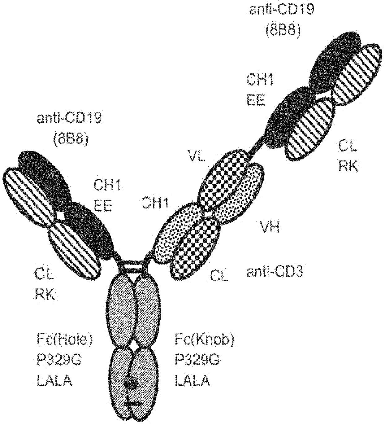
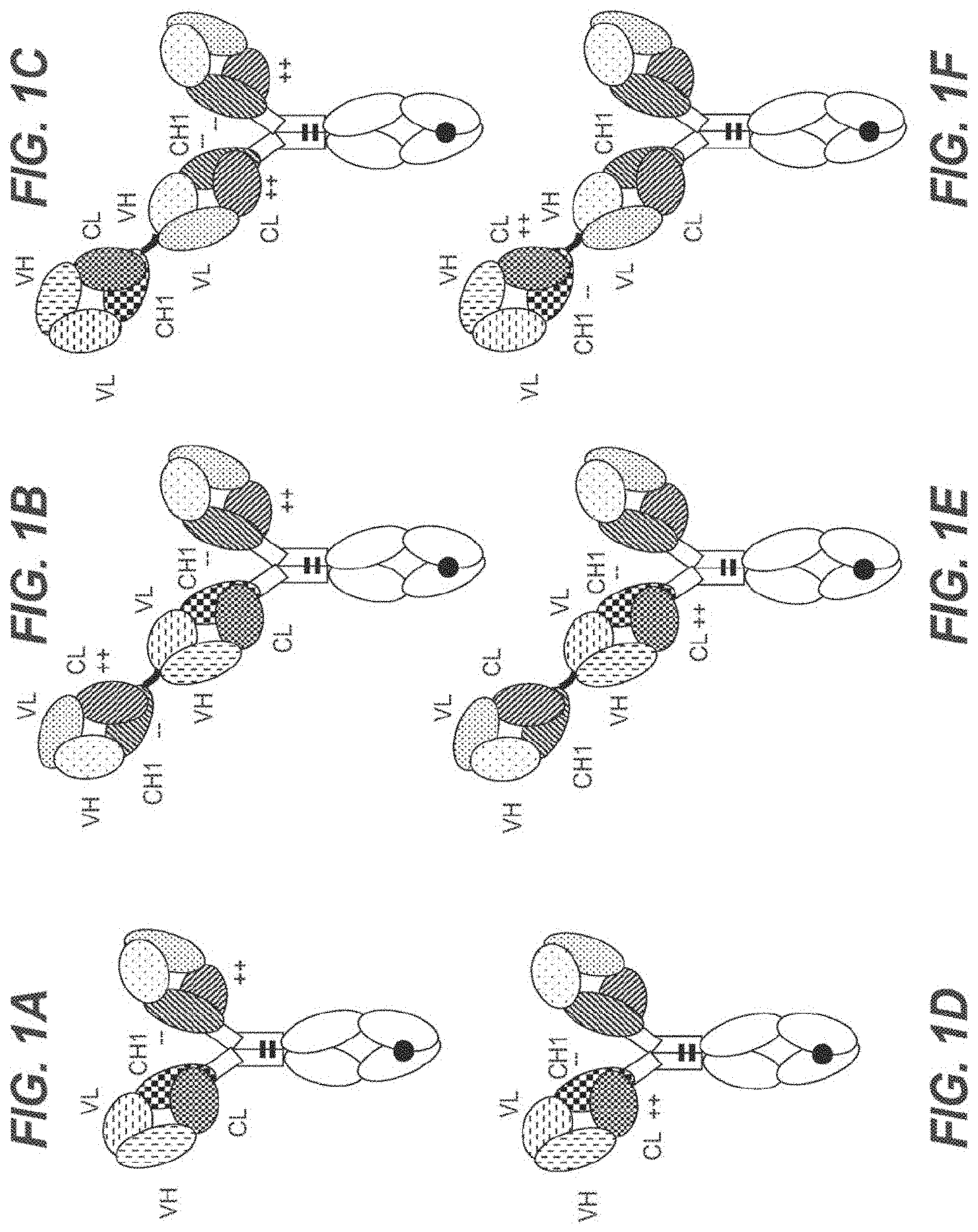
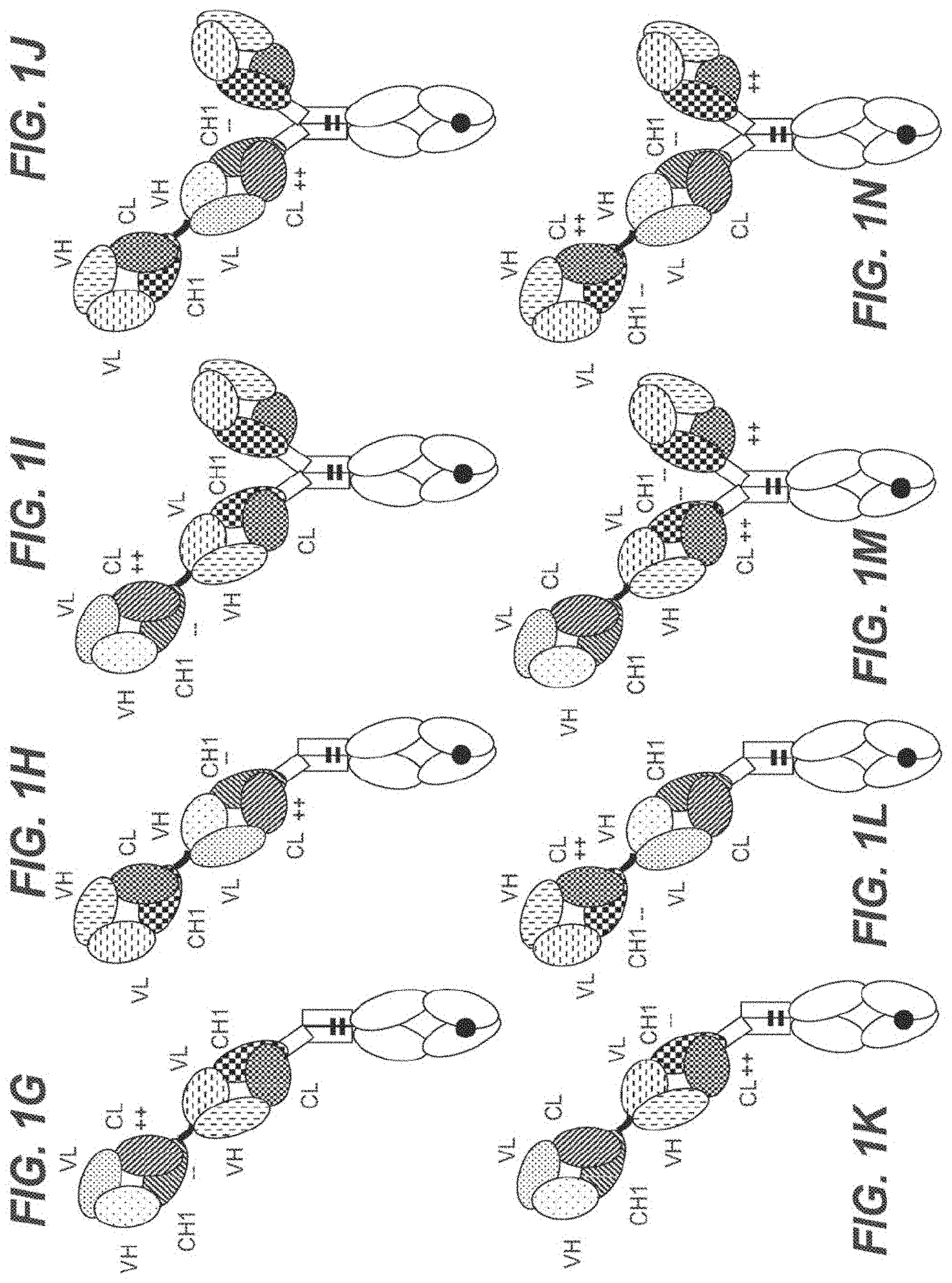
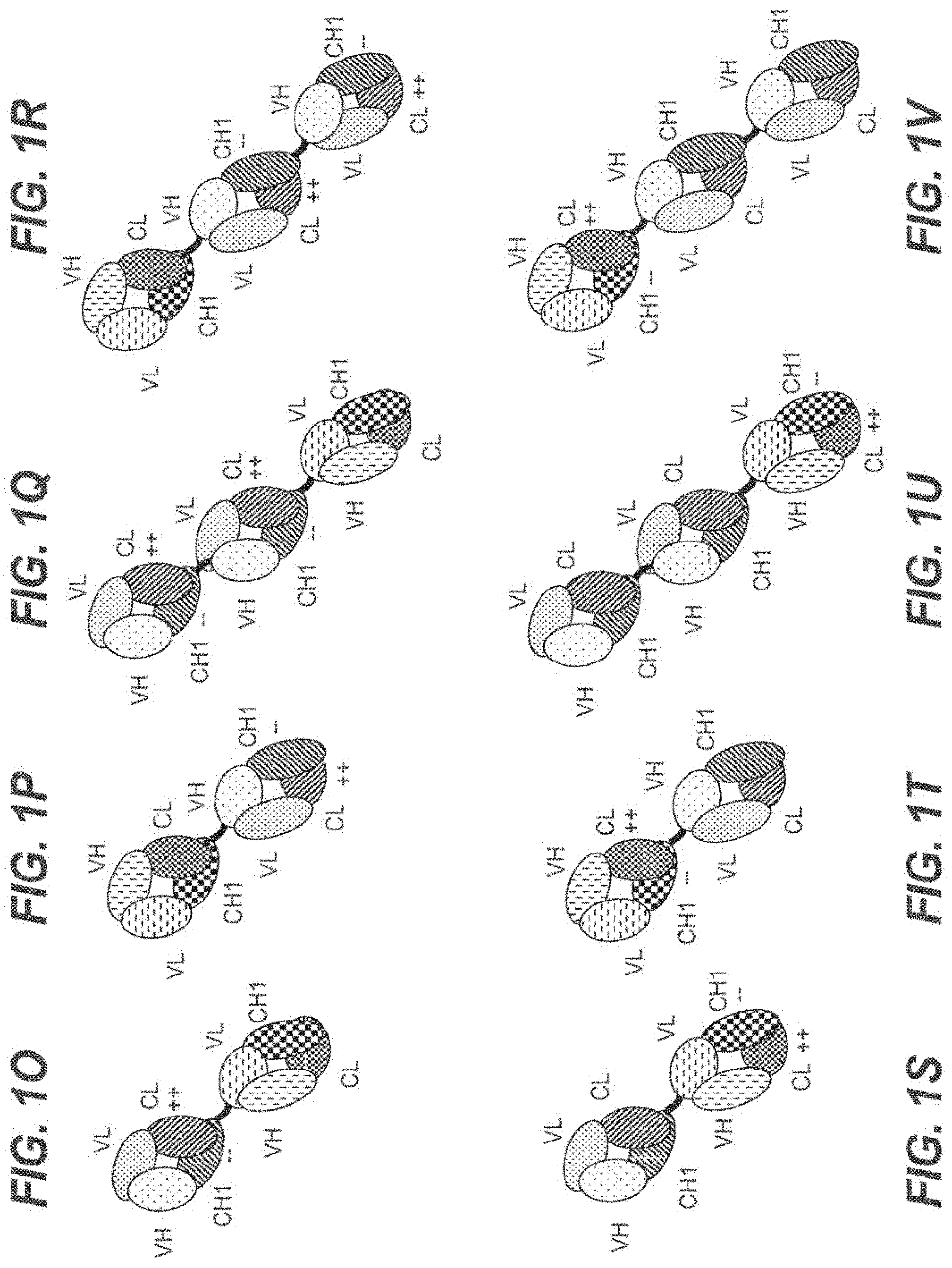
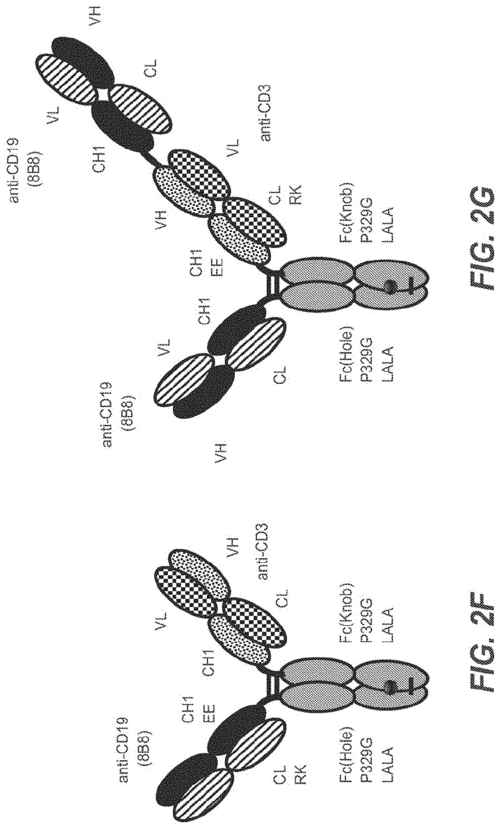
[0056] In one embodiment, the first and the second antigen binding moiety are each a Fab molecule and the first and the second antigen binding moiety are each fused at the C-terminus of the Fab heavy chain to the N-terminus of one of the subunits of the Fc domain. In this embodiment, the T cell activating bispecific antigen binding molecule essentially comprises an immunoglobulin molecule, wherein in one of the Fab arms the heavy and light chain variable regions VH and VL (or the constant regions CH1 and CL in embodiments wherein no charge modifications as described herein are introduced in CH1 and CL domains) are exchanged/replaced by each other (see FIGS. 1A and 1D).
[0057] In alternative embodiments, a third antigen binding moiety, particularly a third Fab molecule, is fused at the C-terminus of the Fab heavy chain to the N-terminus of the first or second subunit of the Fc domain. In a particular such embodiment, the second and the third antigen binding moiety are each fused at the C-terminus of the Fab heavy chain to the N-terminus of one of the subunits of the Fc domain, and the first antigen binding moiety is fused at the C-terminus of the Fab heavy chain to the N-terminus of the Fab heavy chain of the second Fab molecule. In this embodiment, the T cell activating bispecific antigen binding molecule essentially comprises an immunoglobulin molecule, wherein in one of the Fab arms the heavy and light chain variable regions VH and VL (or the constant regions CH1 and CL in embodiments wherein no charge modifications as described herein are introduced in CH1 and CL domains) are exchanged/replaced by each other, and wherein an additional (conventional) Fab molecule is N-terminally fused to said Fab arm (see FIGS. 1B and 1E). In another such embodiment, the first and the third antigen binding moiety are each fused at the C-terminus of the Fab heavy chain to the N-terminus of one of the subunits of the Fc domain, and the second antigen binding moiety is fused at the C-terminus of the Fab heavy chain to the N-terminus of the Fab heavy chain of the first antigen binding moiety. In this embodiment, the T cell activating bispecific antigen binding molecule essentially comprises an immunoglobulin molecule with an additional Fab molecule N-terminally fused to one of the immunoglobulin Fab arms, wherein in said additional Fab molecule the heavy and light chain variable regions VH and VL (or the constant regions CH1 and CL in embodiments wherein no charge modifications as described herein are introduced in
[0058] CH1 and CL domains) are exchanged/replaced by each other (see FIGS. 1C and 1F).
[0059] In a particular embodiment, the immunoglobulin molecule comprised in the T cell activating bispecific antigen binding molecule according to the invention is an IgG class immunoglobulin. In an even more particular embodiment the immunoglobulin is an IgG.sub.1 subclass immunoglobulin. In another embodiment, the immunoglobulin is an IgG.sub.4 subclass immunoglobulin.
[0060] In a particular embodiment, the invention provides a T cell activating bispecific antigen binding molecule comprising
[0061] a) a first Fab molecule which specifically binds to a first antigen;
[0062] b) a second Fab molecule which specifically binds to a second antigen, and wherein the variable domains VL and VH or the constant domains CL and CH1 of the Fab light chain and the Fab heavy chain are replaced by each other;
[0063] c) a third Fab molecule which specifically binds to the first antigen; and
[0064] d) an Fc domain composed of a first and a second subunit capable of stable association;
[0065] wherein the first antigen is CD19 and the second antigen is an activating T cell antigen, particularly CD3, more particularly CD3 epsilon;
[0066] wherein the third Fab molecule under c) is identical to the first Fab molecule under a);
[0067] wherein
[0068] (i) the first Fab molecule under a) is fused at the C-terminus of the Fab heavy chain to the N-terminus of the Fab heavy chain of the second Fab molecule under b), and the second Fab molecule under b) and the third Fab molecule under c) are each fused at the C-terminus of the Fab heavy chain to the N-terminus of one of the subunits of the Fc domain under d), or
[0069] (ii) the second Fab molecule under b) is fused at the C-terminus of the Fab heavy chain to the N-terminus of the Fab heavy chain of the first Fab molecule under a), and the first Fab molecule under a) and the third Fab molecule under c) are each fused at the C-terminus of the Fab heavy chain to the N-terminus of one of the subunits of the Fc domain under d); and
[0070] wherein the first Fab molecule under a) and the third Fab molecule under c) comprise (i) a heavy chain variable region, particularly a humanized heavy chain variable region, comprising the heavy chain complementarity determining region (HCDR) 1 of SEQ ID NO: 14, the HCDR 2 of SEQ ID NO: 15 and the HCDR 3 of SEQ ID NO: 16, and a light chain variable region, particularly a humanized light chain variable region, comprising the light chain complementarity determining region (LCDR) 1 of SEQ ID NO: 17, the LCDR 2 of SEQ ID NO: 18 and the LCDR 3 of SEQ ID NO: 19, or (ii) a heavy chain variable region, particularly a humanized heavy chain variable region, comprising the heavy chain complementarity determining region (HCDR) 1 of SEQ ID NO: 50, the HCDR 2 of SEQ ID NO: 51 and the HCDR 3 of SEQ ID NO: 52, and a light chain variable region, particularly a humanized light chain variable region, comprising the light chain complementarity determining region (LCDR) 1 of SEQ ID NO: 53, the LCDR 2 of SEQ ID NO: 54 and the LCDR 3 of SEQ ID NO: 55.
[0071] In another embodiment, the invention provides a T cell activating bispecific antigen binding molecule comprising
[0072] a) a first Fab molecule which specifically binds to a first antigen;
[0073] b) a second Fab molecule which specifically binds to a second antigen, and wherein the variable domains VL and VH or the constant domains CL and CH1 of the Fab light chain and the Fab heavy chain are replaced by each other;
[0074] c) an Fc domain composed of a first and a second subunit capable of stable association;
[0075] wherein the first antigen is CD19 and the second antigen is an activating T cell antigen, particularly CD3, more particularly CD3 epsilon;
[0076] wherein
[0077] (i) the first Fab molecule under a) is fused at the C-terminus of the Fab heavy chain to the N-terminus of the Fab heavy chain of the second Fab molecule under b), and the second Fab molecule under b) is fused at the C-terminus of the Fab heavy chain to the N-terminus of one of the subunits of the Fc domain under c), or
[0078] (ii) the second Fab molecule under b) is fused at the C-terminus of the Fab heavy chain to the N-terminus of the Fab heavy chain of the first Fab molecule under a), and the first Fab molecule under a) is fused at the C-terminus of the Fab heavy chain to the N-terminus of one of the subunits of the Fc domain under c); and
[0079] wherein the first Fab molecule under a) comprises (i) a heavy chain variable region, particularly a humanized heavy chain variable region, comprising the heavy chain complementarity determining region (HCDR) 1 of SEQ ID NO: 14, the HCDR 2 of SEQ ID NO: 15 and the HCDR 3 of SEQ ID NO: 16, and a light chain variable region, particularly a humanized light chain variable region, comprising the light chain complementarity determining region (LCDR) 1 of SEQ ID NO: 17, the LCDR 2 of SEQ ID NO: 18 and the LCDR 3 of SEQ ID NO: 19, or (ii) a heavy chain variable region, particularly a humanized heavy chain variable region, comprising the heavy chain complementarity determining region (HCDR) 1 of SEQ ID NO: 50, the HCDR 2 of SEQ ID NO: 51 and the HCDR 3 of SEQ ID NO: 52, and a light chain variable region, particularly a humanized light chain variable region, comprising the light chain complementarity determining region (LCDR) 1 of SEQ ID NO: 53, the LCDR 2 of SEQ ID NO: 54 and the LCDR 3 of SEQ ID NO: 55.
[0080] In a further embodiment, the invention provides a T cell activating bispecific antigen binding molecule comprising
[0081] a) a first Fab molecule which specifically binds to a first antigen;
[0082] b) a second Fab molecule which specifically binds to a second antigen, and wherein the variable domains VL and VH or the constant domains CL and CH1 of the Fab light chain and the Fab heavy chain are replaced by each other; and
[0083] c) an Fc domain composed of a first and a second subunit capable of stable association;
[0084] wherein
[0085] (i) the first antigen is CD19 and the second antigen is an activating T cell antigen, particularly CD3, more particularly CD3 epsilon; or
[0086] (ii) the second antigen is CD19 and the first antigen is an activating T cell antigen, particularly CD3, more particularly CD3 epsilon;
[0087] wherein the first Fab molecule under a) and the second Fab molecule under b) are each fused at the C-terminus of the Fab heavy chain to the N-terminus of one of the subunits of the Fc domain under c); and
[0088] wherein the Fab molecule which specifically binds to CD19 comprises (i) a heavy chain variable region, particularly a humanized heavy chain variable region, comprising the heavy chain complementarity determining region (HCDR) 1 of SEQ ID NO: 14, the HCDR 2 of SEQ ID NO: 15 and the HCDR 3 of SEQ ID NO: 16, and a light chain variable region, particularly a humanized light chain variable region, comprising the light chain complementarity determining region (LCDR) 1 of SEQ ID NO: 17, the LCDR 2 of SEQ ID NO: 18 and the LCDR 3 of SEQ ID NO: 19, or (ii) a heavy chain variable region, particularly a humanized heavy chain variable region, comprising the heavy chain complementarity determining region (HCDR) 1 of SEQ ID NO: 50, the HCDR 2 of SEQ ID NO: 51 and the HCDR 3 of SEQ ID NO: 52, and a light chain variable region, particularly a humanized light chain variable region, comprising the light chain complementarity determining region (LCDR) 1 of SEQ ID NO: 53, the LCDR 2 of SEQ ID NO: 54 and the LCDR 3 of SEQ ID NO: 55.
[0089] In all of the different configurations of the T cell activating bispecific antigen binding molecule according to the invention, the amino acid substitutions described herein, if present, may either be in the CH1 and CL domains of the first and (if present) the third Fab molecule, or in the CH1 and CL domains of the second Fab molecule. Preferably, they are in the CH1 and CL domains of the first and (if present) the third Fab molecule. In accordance with the concept of the invention, if amino acid substitutions as described herein are made in the first (and, if present, the third) Fab molecule, no such amino acid substitutions are made in the second Fab molecule. Conversely, if amino acid substitutions as described herein are made in the second Fab molecule, no such amino acid substitutions are made in the first (and, if present, the third) Fab molecule. No amino acid substitutions are made in T cell activating bispecific antigen binding molecules comprising a Fab molecule wherein the constant domains CL and CH1 of the Fab light chain and the Fab heavy chain are replaced by each other.
[0090] In particular embodiments of the T cell activating bispecific antigen binding molecule according to the invention, particularly wherein amino acid substitutions as described herein are made in the first (and, if present, the third) Fab molecule, the constant domain CL of the first (and, if present, the third) Fab molecule is of kappa isotype. In other embodiments of the T cell activating bispecific antigen binding molecule according to the invention, particularly wherein amino acid substitutions as described herein are made in the second Fab molecule, the constant domain CL of the second Fab molecule is of kappa isotype. In some embodiments, the constant domain CL of the first (and, if present, the third) Fab molecule and the constant domain CL of the second Fab molecule are of kappa isotype.
[0091] In a particular embodiment, the invention provides a T cell activating bispecific antigen binding molecule comprising
[0092] a) a first Fab molecule which specifically binds to a first antigen;
[0093] b) a second Fab molecule which specifically binds to a second antigen, and wherein the variable domains VL and VH of the Fab light chain and the Fab heavy chain are replaced by each other;
[0094] c) a third Fab molecule which specifically binds to the first antigen; and
[0095] d) an Fc domain composed of a first and a second subunit capable of stable association;
[0096] wherein the first antigen is CD19 and the second antigen is an activating T cell antigen, particularly CD3, more particularly CD3 epsilon;
[0097] wherein the third Fab molecule under c) is identical to the first Fab molecule under a);
[0098] wherein in the constant domain CL of the first Fab molecule under a) and the third Fab molecule under c) the amino acid at position 124 is substituted by lysine (K) (numbering according to Kabat) and the amino acid at position 123 is substituted by lysine (K) or arginine (R) (numbering according to Kabat), and wherein in the constant domain CH1 of the first Fab molecule under a) and the third Fab molecule under c) the amino acid at position 147 is substituted by glutamic acid (E) (numbering according to Kabat EU index) and the amino acid at position 213 is substituted by glutamic acid (E) (numbering according to Kabat EU index);
[0099] wherein
[0100] (i) the first Fab molecule under a) is fused at the C-terminus of the Fab heavy chain to the N-terminus of the Fab heavy chain of the second Fab molecule under b), and the second Fab molecule under b) and the third Fab molecule under c) are each fused at the C-terminus of the Fab heavy chain to the N-terminus of one of the subunits of the Fc domain under d), or
[0101] (ii) the second Fab molecule under b) is fused at the C-terminus of the Fab heavy chain to the N-terminus of the Fab heavy chain of the first Fab molecule under a), and the first Fab molecule under a) and the third Fab molecule under c) are each fused at the C-terminus of the Fab heavy chain to the N-terminus of one of the subunits of the Fc domain under d); and
[0102] wherein the first Fab molecule under a) and the third Fab molecule under c) comprise (i) a heavy chain variable region, particularly a humanized heavy chain variable region, comprising the heavy chain complementarity determining region (HCDR) 1 of SEQ ID NO: 14, the HCDR 2 of SEQ ID NO: 15 and the HCDR 3 of SEQ ID NO: 16, and a light chain variable region, particularly a humanized light chain variable region, comprising the light chain complementarity determining region (LCDR) 1 of SEQ ID NO: 17, the LCDR 2 of SEQ ID NO: 18 and the LCDR 3 of SEQ ID NO: 19, or (ii) a heavy chain variable region, particularly a humanized heavy chain variable region, comprising the heavy chain complementarity determining region (HCDR) 1 of SEQ ID NO: 50, the HCDR 2 of SEQ ID NO: 51 and the HCDR 3 of SEQ ID NO: 52, and a light chain variable region, particularly a humanized light chain variable region, comprising the light chain complementarity determining region (LCDR) 1 of SEQ ID NO: 53, the LCDR 2 of SEQ ID NO: 54 and the LCDR 3 of SEQ ID NO: 55.
[0103] In an even more particular embodiment, the invention provides a T cell activating bispecific antigen binding molecule comprising
[0104] a) a first Fab molecule which specifically binds to a first antigen;
[0105] b) a second Fab molecule which specifically binds to a second antigen, and wherein the variable domains VL and VH of the Fab light chain and the Fab heavy chain are replaced by each other;
[0106] c) a third Fab molecule which specifically binds to the first antigen; and
[0107] d) an Fc domain composed of a first and a second subunit capable of stable association;
[0108] wherein the first antigen is CD19 and the second antigen is an activating T cell antigen, particularly CD3, more particularly CD3 epsilon;
[0109] wherein the third Fab molecule under c) is identical to the first Fab molecule under a);
[0110] wherein in the constant domain CL of the first Fab molecule under a) and the third Fab molecule under c) the amino acid at position 124 is substituted by lysine (K) (numbering according to Kabat) and the amino acid at position 123 is substituted by arginine (R) (numbering according to Kabat), and wherein in the constant domain CH1 of the first Fab molecule under a) and the third Fab molecule under c) the amino acid at position 147 is substituted by glutamic acid (E) (numbering according to Kabat EU index) and the amino acid at position 213 is substituted by glutamic acid (E) (numbering according to Kabat EU index);
[0111] wherein the first Fab molecule under a) is fused at the C-terminus of the Fab heavy chain to the N-terminus of the Fab heavy chain of the second Fab molecule under b), and the second Fab molecule under b) and the third Fab molecule under c) are each fused at the C-terminus of the Fab heavy chain to the N-terminus of one of the subunits of the Fc domain under d); and
[0112] wherein the first Fab molecule under a) and the third Fab molecule under c) comprise (i) a heavy chain variable region, particularly a humanized heavy chain variable region, comprising the heavy chain complementarity determining region (HCDR) 1 of SEQ ID NO: 14, the HCDR 2 of SEQ ID NO: 15 and the HCDR 3 of SEQ ID NO: 16, and a light chain variable region, particularly a humanized light chain variable region, comprising the light chain complementarity determining region (LCDR) 1 of SEQ ID NO: 17, the LCDR 2 of SEQ ID NO: 18 and the LCDR 3 of SEQ ID NO: 19, or (ii) a heavy chain variable region, particularly a humanized heavy chain variable region, comprising the heavy chain complementarity determining region (HCDR) 1 of SEQ ID NO: 50, the HCDR 2 of SEQ ID NO: 51 and the HCDR 3 of SEQ ID NO: 52, and a light chain variable region, particularly a humanized light chain variable region, comprising the light chain complementarity determining region (LCDR) 1 of SEQ ID NO: 53, the LCDR 2 of SEQ ID NO: 54 and the LCDR 3 of SEQ ID NO: 55.
[0113] In another embodiment, the invention provides a T cell activating bispecific antigen binding molecule comprising
[0114] a) a first Fab molecule which specifically binds to a first antigen;
[0115] b) a second Fab molecule which specifically binds to a second antigen, and wherein the variable domains VL and VH of the Fab light chain and the Fab heavy chain are replaced by each other;
[0116] c) an Fc domain composed of a first and a second subunit capable of stable association;
[0117] wherein the first antigen is CD19 and the second antigen is an activating T cell antigen, particularly CD3, more particularly CD3 epsilon;
[0118] wherein in the constant domain CL of the first Fab molecule under a) the amino acid at position 124 is substituted by lysine (K) (numbering according to Kabat) and the amino acid at position 123 is substituted by lysine (K) or arginine (R) (numbering according to Kabat), and wherein in the constant domain CH1 of the first Fab molecule under a) the amino acid at position 147 is substituted by glutamic acid (E) (numbering according to Kabat EU index) and the amino acid at position 213 is substituted by glutamic acid (E) (numbering according to Kabat EU index);
[0119] wherein
[0120] (i) the first Fab molecule under a) is fused at the C-terminus of the Fab heavy chain to the N-terminus of the Fab heavy chain of the second Fab molecule under b), and the second Fab molecule under b) is fused at the C-terminus of the Fab heavy chain to the N-terminus of one of the subunits of the Fc domain under c), or
[0121] (ii) the second Fab molecule under b) is fused at the C-terminus of the Fab heavy chain to the N-terminus of the Fab heavy chain of the first Fab molecule under a), and the first Fab molecule under a) is fused at the C-terminus of the Fab heavy chain to the N-terminus of one of the subunits of the Fc domain under c); and
[0122] wherein the first Fab molecule under a) comprises (i) a heavy chain variable region, particularly a humanized heavy chain variable region, comprising the heavy chain complementarity determining region (HCDR) 1 of SEQ ID NO: 14, the HCDR 2 of SEQ ID NO: 15 and the HCDR 3 of SEQ ID NO: 16, and a light chain variable region, particularly a humanized light chain variable region, comprising the light chain complementarity determining region (LCDR) 1 of SEQ ID NO: 17, the LCDR 2 of SEQ ID NO: 18 and the LCDR 3 of SEQ ID NO: 19, or (ii) a heavy chain variable region, particularly a humanized heavy chain variable region, comprising the heavy chain complementarity determining region (HCDR) 1 of SEQ ID NO: 50, the HCDR 2 of SEQ ID NO: 51 and the HCDR 3 of SEQ ID NO: 52, and a light chain variable region, particularly a humanized light chain variable region, comprising the light chain complementarity determining region (LCDR) 1 of SEQ ID NO: 53, the LCDR 2 of SEQ ID NO: 54 and the LCDR 3 of SEQ ID NO: 55.
[0123] In a further embodiment, the invention provides a T cell activating bispecific antigen binding molecule comprising
[0124] a) a first Fab molecule which specifically binds to a first antigen;
[0125] b) a second Fab molecule which specifically binds to a second antigen, and wherein the variable domains VL and VH of the Fab light chain and the Fab heavy chain are replaced by each other; and
[0126] c) an Fc domain composed of a first and a second subunit capable of stable association;
[0127] wherein
[0128] (i) the first antigen is CD19 and the second antigen is an activating T cell antigen, particularly CD3, more particularly CD3 epsilon; or
[0129] (ii) the second antigen is CD19 and the first antigen is an activating T cell antigen, particularly CD3, more particularly CD3 epsilon;
[0130] wherein in the constant domain CL of the first Fab molecule under a) the amino acid at position 124 is substituted by lysine (K) (numbering according to Kabat) and the amino acid at position 123 is substituted by lysine (K) or arginine (R) (numbering according to Kabat), and wherein in the constant domain CH1 of the first Fab molecule under a) the amino acid at position 147 is substituted by glutamic acid (E) (numbering according to Kabat EU index) and the amino acid at position 213 is substituted by glutamic acid (E) (numbering according to Kabat EU index);
[0131] wherein the first Fab molecule under a) and the second Fab molecule under b) are each fused at the C-terminus of the Fab heavy chain to the N-terminus of one of the subunits of the Fc domain under c); and
[0132] wherein the Fab molecule which specifically binds to CD19 comprises (i) a heavy chain variable region, particularly a humanized heavy chain variable region, comprising the heavy chain complementarity determining region (HCDR) 1 of SEQ ID NO: 14, the HCDR 2 of SEQ ID NO: 15 and the HCDR 3 of SEQ ID NO: 16, and a light chain variable region, particularly a humanized light chain variable region, comprising the light chain complementarity determining region (LCDR) 1 of SEQ ID NO: 17, the LCDR 2 of SEQ ID NO: 18 and the LCDR 3 of SEQ ID NO: 19, or (ii) a heavy chain variable region, particularly a humanized heavy chain variable region, comprising the heavy chain complementarity determining region (HCDR) 1 of SEQ ID NO: 50, the HCDR 2 of SEQ ID NO: 51 and the HCDR 3 of SEQ ID NO: 52, and a light chain variable region, particularly a humanized light chain variable region, comprising the light chain complementarity determining region (LCDR) 1 of SEQ ID NO: 53, the LCDR 2 of SEQ ID NO: 54 and the LCDR 3 of SEQ ID NO: 55.
[0133] In particular embodiments of the T cell activating bispecific antigen binding molecule, the Fc domain is an IgG Fc domain. In a specific embodiment, the Fc domain is an IgG.sub.1 Fc domain. In another specific embodiment, the Fc domain is an IgG.sub.4 Fc domain. In an even more specific embodiment, the Fc domain is an IgG.sub.4 Fc domain comprising the amino acid substitution S228P (Kabat numbering). In particular embodiments the Fc domain is a human Fc domain.
[0134] In particular embodiments, the Fc domain comprises a modification promoting the association of the first and the second Fc domain subunit. In a specific such embodiment, an amino acid residue in the CH3 domain of the first subunit of the Fc domain is replaced with an amino acid residue having a larger side chain volume, thereby generating a protuberance within the CH3 domain of the first subunit which is positionable in a cavity within the CH3 domain of the second subunit, and an amino acid residue in the CH3 domain of the second subunit of the Fc domain is replaced with an amino acid residue having a smaller side chain volume, thereby generating a cavity within the CH3 domain of the second subunit within which the protuberance within the CH3 domain of the first subunit is positionable.
[0135] In a particular embodiment the Fc domain exhibits reduced binding affinity to an Fc receptor and/or reduced effector function, as compared to a native IgG.sub.1 Fc domain. In certain embodiments the Fc domain is engineered to have reduced binding affinity to an Fc receptor and/or reduced effector function, as compared to a non-engineered Fc domain. In one embodiment, the Fc domain comprises one or more amino acid substitution that reduces binding to an Fc receptor and/or effector function. In one embodiment, the one or more amino acid substitution in the Fc domain that reduces binding to an Fc receptor and/or effector function is at one or more position selected from the group of L234, L235, and P329 (Kabat EU index numbering). In particular embodiments, each subunit of the Fc domain comprises three amino acid substitutions that reduce binding to an Fc receptor and/or effector function wherein said amino acid substitutions are L234A, L235A and P329G (Kabat EU index numbering). In one such embodiment, the Fc domain is an IgG.sub.1 Fc domain, particularly a human IgG.sub.1 Fc domain. In other embodiments, each subunit of the Fc domain comprises two amino acid substitutions that reduce binding to an Fc receptor and/or effector function wherein said amino acid substitutions are L235E and P329G (Kabat EU index numbering). In one such embodiment, the Fc domain is an IgG.sub.4 Fc domain, particularly a human IgG.sub.4 Fc domain. In one embodiment, the Fc domain of the T cell activating bispecific antigen binding molecule is an IgG.sub.4 Fc domain and comprises the amino acid substitutions L235E and S228P (SPLE) (Kabat EU index numbering).
[0136] In one embodiment the Fc receptor is an Fc.gamma. receptor. In one embodiment the Fc receptor is a human Fc receptor. In one embodiment, the Fc receptor is an activating Fc receptor. In a specific embodiment, the Fc receptor is human Fc.gamma.RIIa, Fc.gamma.RI, and/or Fc.gamma.RIIIa. In one embodiment, the effector function is antibody-dependent cell-mediated cytotoxicity (ADCC).
[0137] In a specific embodiment of the T cell activating bispecific antigen binding molecule according to the invention, the antigen binding moiety which specifically binds to an activating T cell antigen, particularly CD3, more particularly CD3 epsilon, comprises a heavy chain variable region comprising the heavy chain complementarity determining region (HCDR) 1 of SEQ ID NO: 4, the HCDR 2 of SEQ ID NO: 5, the HCDR 3 of SEQ ID NO: 6, and a light chain variable region comprising the light chain complementarity determining region (LCDR) 1 of SEQ ID NO: 8, the LCDR 2 of SEQ ID NO: 9 and the LCDR 3 of SEQ ID NO: 10. In an even more specific embodiment, the antigen binding moiety which specifically binds to an activating T cell antigen, particularly CD3, more particularly CD3 epsilon, comprises a heavy chain variable region comprising an amino acid sequence that is at least about 95%, 96%, 97%, 98%, 99% or 100% identical to the amino acid sequence of SEQ ID NO: 3 and a light chain variable region comprising an amino acid sequence that is at least about 95%, 96%, 97%, 98%, 99% or 100% identical to the amino acid sequence of SEQ ID NO: 7. In some embodiments, the antigen binding moiety which specifically binds to an activating T cell antigen is a Fab molecule. In one specific embodiment, the second antigen binding moiety, particularly Fab molecule, comprised in the T cell activating bispecific antigen binding molecule according to the invention specifically binds to CD3, more particularly CD3 epsilon, and comprises the heavy chain complementarity determining region (CDR) 1 of SEQ ID NO: 4, the heavy chain CDR 2 of SEQ ID NO: 5, the heavy chain CDR 3 of SEQ ID NO: 6, the light chain CDR 1 of SEQ ID NO: 8, the light chain CDR 2 of SEQ ID NO: 9 and the light chain CDR 3 of SEQ ID NO: 10. In an even more specific embodiment, said second antigen binding moiety, particularly Fab molecule, comprises a heavy chain variable region comprising the amino acid sequence of SEQ ID NO: 3 and a light chain variable region comprising the amino acid sequence of SEQ ID NO: 7.
[0138] In a further specific embodiment of the T cell activating bispecific antigen binding molecule according to the invention, the antigen binding moiety, particularly Fab molecule, which specifically binds to CD19 comprises the heavy chain complementarity determining region (CDR) 1 of SEQ ID NO: 14, the heavy chain CDR 2 of SEQ ID NO: 15, the heavy chain CDR 3 of SEQ ID NO: 16, the light chain CDR 1 of SEQ ID NO: 17, the light chain CDR 2 of SEQ ID NO: 18 and the light chain CDR 3 of SEQ ID NO: 19. In an even more specific embodiment, the antigen binding moiety, particularly Fab molecule, which specifically binds to CD19 comprises a heavy chain variable region comprising an amino acid sequence that is at least about 95%, 96%, 97%, 98%, 99% or 100% identical to the amino acid sequence of SEQ ID NO: 20 and a light chain variable region comprising an amino acid sequence that is at least about 95%, 96%, 97%, 98%, 99% or 100% identical to the amino acid sequence of SEQ ID NO: 21. In one specific embodiment, the first (and, if present, the third) antigen binding moiety, particularly Fab molecule, comprised in the T cell activating bispecific antigen binding molecule according to the invention specifically binds to CD19, and comprises the heavy chain complementarity determining region (CDR) 1 of SEQ ID NO: 14, the heavy chain CDR 2 of SEQ ID NO: 15, the heavy chain CDR 3 of SEQ ID NO: 16, the light chain CDR 1 of SEQ ID NO: 17, the light chain CDR 2 of SEQ ID NO: 18 and the light chain CDR 3 of SEQ ID NO: 19. In an even more specific embodiment, said first (and, if present, said third) antigen binding moiety, particularly Fab molecule, comprises a heavy chain variable region comprising the amino acid sequence of SEQ ID NO: 20 and a light chain variable region comprising the amino acid sequence of SEQ ID NO: 21.
[0139] In another specific embodiment of the T cell activating bispecific antigen binding molecule according to the invention, the antigen binding moiety, particularly Fab molecule, which specifically binds to CD19 comprises the heavy chain complementarity determining region (CDR) 1 of SEQ ID NO: 50, the heavy chain CDR 2 of SEQ ID NO: 51, the heavy chain CDR 3 of SEQ ID NO: 52, the light chain CDR 1 of SEQ ID NO: 53, the light chain CDR 2 of SEQ ID NO: 54 and the light chain CDR 3 of SEQ ID NO: 55. In an even more specific embodiment, the antigen binding moiety, particularly Fab molecule, which specifically binds to CD19 comprises a heavy chain variable region comprising an amino acid sequence that is at least about 95%, 96%, 97%, 98%, 99% or 100% identical to the amino acid sequence of SEQ ID NO: 56 and a light chain variable region comprising an amino acid sequence that is at least about 95%, 96%, 97%, 98%, 99% or 100% identical to the amino acid sequence of SEQ ID NO: 57. In one specific embodiment, the first (and, if present, the third) antigen binding moiety, particularly Fab molecule, comprised in the T cell activating bispecific antigen binding molecule according to the invention specifically binds to CD19, and comprises the heavy chain complementarity determining region (CDR) 1 of SEQ ID NO: 50, the heavy chain CDR 2 of SEQ ID NO: 51, the heavy chain CDR 3 of SEQ ID NO: 52, the light chain CDR 1 of SEQ ID NO: 53, the light chain CDR 2 of SEQ ID NO: 54 and the light chain CDR 3 of SEQ ID NO: 55. In an even more specific embodiment, said first (and, if present, said third) antigen binding moiety, particularly Fab molecule, comprises a heavy chain variable region comprising the amino acid sequence of SEQ ID NO: 56 and a light chain variable region comprising the amino acid sequence of SEQ ID NO: 57.
[0140] In a particular aspect, the invention provides a T cell activating bispecific antigen binding molecule comprising
[0141] a) a first Fab molecule which specifically binds to a first antigen;
[0142] b) a second Fab molecule which specifically binds to a second antigen, and wherein the variable domains VL and VH or the constant domains CL and CH1 of the Fab light chain and the Fab heavy chain are replaced by each other;
[0143] c) a third Fab molecule which specifically binds to the first antigen; and
[0144] d) an Fc domain composed of a first and a second subunit capable of stable association;
[0145] wherein
[0146] (i) the first antigen is CD19 and the second antigen is CD3, particularly CD3 epsilon;
[0147] (ii) the first Fab molecule under a) and the third Fab molecule under c) each comprise the heavy chain complementarity determining region (CDR) 1 of SEQ ID NO: 14, the heavy chain CDR 2 of SEQ ID NO: 15, the heavy chain CDR 3 of SEQ ID NO: 16, the light chain CDR 1 of SEQ ID NO: 17, the light chain CDR 2 of SEQ ID NO: 18 and the light chain CDR 3 of SEQ ID NO: 19, or each comprise the heavy chain CDR 1 of SEQ ID NO: 50, the heavy chain CDR 2 of SEQ ID NO: 51, the heavy chain CDR 3 of SEQ ID NO: 52, the light chain CDR 1 of SEQ ID NO: 53, the light chain CDR 2 of SEQ ID NO: 54 and the light chain CDR 3 of SEQ ID NO: 55, and the second Fab molecule under b) comprises the heavy chain CDR 1 of SEQ ID NO: 4, the heavy chain CDR 2 of SEQ ID NO: 5, the heavy chain CDR 3 of SEQ ID NO: 6, the light chain CDR 1 of SEQ ID NO: 8, the light chain CDR 2 of SEQ ID NO: 9 and the light chain CDR 3 of SEQ ID NO: 10; and
[0148] (iii) the first Fab molecule under a) is fused at the C-terminus of the Fab heavy chain to the N-terminus of the Fab heavy chain of the second Fab molecule under b), and the second Fab molecule under b) and the third Fab molecule under c) are each fused at the C-terminus of the Fab heavy chain to the N-terminus of one of the subunits of the Fc domain under d).
[0149] In one embodiment, in the second Fab molecule under b) the variable domains VL and VH are replaced by each other and further (iv) in the constant domain CL of the first Fab molecule under a) and the third Fab molecule under c) the amino acid at position 124 is substituted by lysine (K) (numbering according to Kabat) and the amino acid at position 123 is substituted by lysine (K) or arginine (R), particularly by arginine (R) (numbering according to Kabat), and in the constant domain CH1 of the first Fab molecule under a) and the third Fab molecule under c) the amino acid at position 147 is substituted by glutamic acid (E) (numbering according to Kabat EU index) and the amino acid at position 213 is substituted by glutamic acid (E) (numbering according to Kabat EU index).
[0150] According to another aspect of the invention there is provided one or more isolated polynucleotide(s) encoding a T cell activating bispecific antigen binding molecule of the invention. The invention further provides one or more expression vector(s) comprising the isolated polynucleotide(s) of the invention, and a host cell comprising the isolated polynucleotide(s) or the expression vector(s) of the invention. In some embodiments the host cell is a eukaryotic cell, particularly a mammalian cell.
[0151] In another aspect is provided a method of producing the T cell activating bispecific antigen binding molecule of the invention, comprising the steps of a) culturing the host cell of the invention under conditions suitable for the expression of the T cell activating bispecific antigen binding molecule and b) recovering the T cell activating bispecific antigen binding molecule. The invention also encompasses a T cell activating bispecific antigen binding molecule produced by the method of the invention.
[0152] The invention further provides a pharmaceutical composition comprising the T cell activating bispecific antigen binding molecule of the invention and a pharmaceutically acceptable carrier.
[0153] Also encompassed by the invention are methods of using the T cell activating bispecific antigen binding molecule and pharmaceutical composition of the invention. In one aspect the invention provides a T cell activating bispecific antigen binding molecule or a pharmaceutical composition of the invention for use as a medicament. In one aspect is provided a T cell activating bispecific antigen binding molecule or a pharmaceutical composition according to the invention for use in the treatment of a disease in an individual in need thereof. In a specific embodiment the disease is cancer.
[0154] Also provided is the use of a T cell activating bispecific antigen binding molecule of the invention for the manufacture of a medicament for the treatment of a disease in an individual in need thereof; as well as a method of treating a disease in an individual, comprising administering to said individual a therapeutically effective amount of a composition comprising the T cell activating bispecific antigen binding molecule according to the invention in a pharmaceutically acceptable form. In a specific embodiment the disease is cancer. In any of the above embodiments the individual preferably is a mammal, particularly a human.
[0155] The invention also provides a method for inducing lysis of a target cell, particularly a tumor cell, comprising contacting a target cell with a T cell activating bispecific antigen binding molecule of the invention in the presence of a T cell, particularly a cytotoxic T cell.
BRIEF DESCRIPTION OF THE DRAWINGS
[0156] FIGS. 1A-1Z show exemplary configurations of the T cell activating bispecific antigen binding molecules (TCBs) of the invention. FIGS. 1A and 1D are illustrations of the "1+1 CrossMab" molecule. FIGS. 1B and 1E are illustrations of the "2+1 IgG Crossfab" molecule with alternative order of Crossfab and Fab components ("inverted"). FIGS. 1C and 1F are illustrations of the "2+1 IgG Crossfab" molecule. FIGS. 1G and 1K are illustrations of the "1+1 IgG Crossfab" molecule with alternative order of Crossfab and Fab components ("inverted"). FIGS. 1H and 1L are illustrations of the "1+1 IgG Crossfab" molecule. FIGS. 1I and 1M are illustrations of the "2+1 IgG Crossfab" molecule with two CrossFabs. FIGS. 1J and 1N are illustrations of the "2+1 IgG Crossfab" molecule with two CrossFabs and alternative order of Crossfab and Fab components ("inverted"). FIGS. 1O and 1S are illustrations of the "Fab-Crossfab" molecule. FIGS. 1P and 1T are illustrations of the "Crossfab-Fab" molecule. FIGS. 1Q and 1U are illustrations of the "(Fab).sub.2-Crossfab" molecule. FIGS. 1R and 1V are illustrations of the "Crossfab-(Fab).sub.2" molecule. FIGS. 1W and 1Y are illustrations of the "Fab-(Crossfab).sub.2" molecule. FIGS. 1X and 1Z are illustrations of the "(Crossfab).sub.2-Fab" molecule. Black dot: optional modification in the Fc domain promoting heterodimerization. ++, --: amino acids of opposite charges optionally introduced in the CH1 and CL domains. Crossfab molecules are depicted as comprising an exchange of VH and VL regions, but may--in embodiments wherein no charge modifications are introduced in CH1 and CL domains--alternatively comprise an exchange of the CH1 and CL domains.
[0157] FIGS. 2A-2H are illustrations of the TCBs prepared in the Examples. FIG. 2A shows molecule A: "2+1 IgG CrossFab, inverted" with charge modifications (VH/VL exchange in CD3 binder, charge modification in CD19 binders (parental CD19 binder 8B8), EE=147E, 213E; RK =123R, 124K). FIG. 2B shows molecule B: "2+1 IgG CrossFab, inverted" without charge modifications (CH1/CL exchange in CD3 binder, parental CD19 binder 8B8). FIG. 2C shows molecule C: "2+1 IgG CrossFab" with charge modifications (VH/VL exchange in CD3 binder, charge modification in CD19 binders (humanized CD19 binder var. 5). FIG. 2D shows molecule D: "2+1 IgG CrossFab, inverted" without charge modifications (CH1/CL exchange in CD3 binder, CD19 binder 10C1). FIG. 2E shows molecule E: "1+1 IgG CrossFab, inverted" without charge modifications (CH1/CL exchange in CD3 binder, parental CD19 binder 8B8). FIG. 2F shows molecule F: "1+1 IgG CrossMab", with charge modifications (VH/VL exchange in CD3 binder, charge modification in CD19 binder (parental CD19 binder 8B8). FIG. 2G shows molecule G: "2+1 IgG CrossFab, inverted" with charge modifications (VH/VL exchange in CD19 binders (parental CD19 binder 8B8), charge modification in CD3 binder). FIG. 2H shows molecule H: "2+1 IgG CrossFab" with charge modifications (VH/VL exchange in CD3 binder, charge modification in CD19 binders (humanized CD19 binder 2B11). EE=147E, 213E; RK=123R, 124K.
[0158] FIGS. 3A-3H show CE-SDS analysis of the TCBs prepared in the Examples (final purified preparations). FIG. 3A is an electropherogram of molecule "A", shown in FIG. 2A. FIG. 3B is an electropherogram of molecule "B", shown in FIG. 2B. FIG. 3C is an electropherogram of molecule "C", shown in FIG. 2C. FIG. 3D is an electropherogram of molecule "D", shown in FIG. 2D. FIG. 3E is an electropherogram of molecule "E", shown in FIG. 2E. FIG. 3F is an electropherogram of molecule "F", shown in FIG. 2F. FIG. 3G is an electropherogram of molecule "G", shown in FIG. 2G. Lane A=non-reduced, lane B=reduced. FIG. 3H is an electropherogram of molecule "H", shown in FIG. 2H. Lane A=non-reduced, lane B=reduced.
[0159] FIGS. 4A-4H show SDS-PAGE analysis (4-12% Bis-Tris, Coomassie stained, non reduced) of TCBs prepared in the Examples after the first purification step (Protein A affinity chromatography). FIG. 4A includes lane 1=marker (Mark 12, unstained standard, Invitrogen); lane 2-11=fractions from Protein A affinity chromatography of molecule A. FIG. 4B includes lane 1=marker (Mark 12, unstained standard, Invitrogen); lane 2-14=fractions from Protein A affinity chromatography of molecule B. FIG. 4C includes lane 1=marker (Mark 12, unstained standard, Invitrogen); lane 2-8=fractions from Protein A affinity chromatography of molecule C. FIG. 4D includes lane 1=marker (Mark 12, unstained standard, Invitrogen); lane 2-14=fractions from Protein A affinity chromatography of molecule D. FIG. 4E includes lane 1=marker (Mark 12, unstained standard, Invitrogen); lane 2-11=fractions from Protein A affinity chromatography of molecule E. FIG. 4F includes lane 1=marker (Mark 12, unstained standard, Invitrogen); lane 2-11=fractions from Protein A affinity chromatography of molecule F. FIG. 4G includes lane 1=marker (Mark 12, unstained standard, Invitrogen); lane 4-10=fractions from Protein A affinity chromatography of molecule G. FIG. 4H includes lane 1=marker (HiMark HMW, Invitrogen); lane 2-14=fractions from Protein A affinity chromatography of molecule H.
[0160] FIG. 5A shows binding of CD19 IgGs and CD19 TCBs to Raji cells. FIG. 5B shows binding of CD19 TCBs (generated using 8B8 and 10C1 clones) to CD3-expressing Jurkat cells.
[0161] FIG. 6A shows lysis of CD19-expressing Nalm-6 tumor cells mediated by CD19 TCB antibodies. FIG. 6B shows lysis of CD19-expressing SUDHL-4 tumor cells mediated by CD19 TCB antibodies. FIG. 6C shows CD69 expression on CD8 T cells upon killing of Nalm-6 target cells mediated by CD19 TCB antibodies. FIG. 6D shows CD69 expression on CD4 T cells upon killing of Nalm-6 target cells mediated by CD19 TCB antibodies. FIG. 6E shows CD25 expression on CD8 T cells upon killing of Nalm-6 target cells mediated by CD19 TCB antibodies. FIG. 6F shows CD25 expression on CD4 T cells upon killing of Nalm-6 target cells mediated by CD19 TCB antibodies. FIG. 6G shows CD69 expression on CD8 T cells upon killing of SUDHL-4 target cells mediated by CD19 TCB antibodies. FIG. 6H shows CD69 expression on CD4 T cells upon killing of SUDHL-4 target cells mediated by CD19 TCB antibodies. FIG. 6I shows CD25 expression on CD8 T cells upon killing of SUDHL-4 target cells mediated by CD19 TCB antibodies. FIG. 6J shows CD25 expression on CD4 T cells upon killing of SUDHL-4 target cells mediated by CD19 TCB antibodies. FIG. 6K shows lysis of Z-138 cells mediated by CD19 TCB antibodies (having 8B 8 and 10C1 clone) and blinatumomab. FIG. 6L shows CD69 expression on CD8 T cells after Z-138 tumor cell killing mediated by CD19 TCB antibodies. FIG. 6M shows CD25 expression on CD8 T cells after Z-138 tumor cell killing mediated by CD19 TCB antibodies. FIG. 6N shows CD69 expression on CD4 T cells after Z-138 tumor cell killing mediated by CD19 TCB antibodies. FIG. 6O shows CD25 expression on CD4 T cells after Z-138 tumor cell killing mediated by CD19 TCB antibodies.
[0162] FIG. 7A shows B cell depletion mediated by CD19 TCB antibodies based on 8B8 and 10C1 clone. FIG. 7B shows T cell activation (CD69 marker) after B cell depletion mediated by CD19 TCB antibodies based on 8B8 and 10C1 clone.
[0163] FIG. 8A shows binding of CD19 TCB antibodies (based on 8B8 and 10C1 clone) to cynomolgus monkey B cells. FIG. 8B shows binding of CD19 TCB antibodies (based on 8B8 and 10C1 clone) to human B cells. FIG. 8C shows binding of CD19 TCB antibodies (based on 8B8 and 10C1 clone) to cynomolgus monkey CD4.sup.+ T cells. FIG. 8D shows binding of CD19 TCB antibodies (based on 8B8 and 10C1 clone) to human CD4.sup.+ T cells. FIG. 8E shows binding of CD19 TCB antibodies (based on 8B8 and 10C1 clone) to cynomolgus monkey C8.sup.+ T cells. FIG. 8F shows binding of CD19 TCB antibodies (based on 8B8 and 10C1 clone) to human CD8.sup.+ T cells.
[0164] FIG. 9A shows % B cell depletion in cynomolgus monkey whole blood mediated by CD19 TCB antibody (8B8 clone) and blinatumomab obtained by normalizing the B cell values to CD4 T cells. FIG. 9B shows % B cell depletion in human whole blood mediated by CD19 TCB antibody (8B8 clone) and blinatumomab obtained by normalizing the B cell values to CD4 T cells. FIG. 9C shows % B cell depletion in cynomolgus monkey whole blood mediated by CD19 TCB antibody (8B8 clone) and blinatumomab obtained by normalizing the B cell values to CD8 T cells. FIG. 9D shows % B cell depletion in human whole blood mediated by CD19 TCB antibody (8B8 clone) and blinatumomab obtained by normalizing the B cell values to CD8 T cells. FIG. 9E shows % CD69 T cell activation in cynomolgus monkey whole blood mediated by CD19 TCB antibody (8B8 clone) and blinatumomab on CD8 T cells. FIG. 9F shows % CD69 T cell activation in human whole blood mediated by CD19 TCB antibody (8B 8 clone) and blinatumomab on CD8 T cells. FIG. 9G shows % CD25 T cell activation in cynomolgus monkey whole blood mediated by CD19 TCB antibody (8B8 clone) and blinatumomab on CD8 T cells. FIG. 9H shows % CD25 T cell activation in human whole blood mediated by CD19 TCB antibody (8B8 clone) and blinatumomab on CD8 T cells. FIG. 9I shows % CD69 T cell activation in cynomolgus monkey whole blood mediated by CD19 TCB antibody (8B8 clone) and blinatumomab on CD4 T cells. FIG. 9J shows % CD69 T cell activation in human whole blood mediated by CD19 TCB antibody (8B8 clone) and blinatumomab on CD4 T cells. FIG. 9K shows % CD25 T cell activation in cynomolgus monkey whole blood mediated by CD19 TCB antibody (8B8 clone) and blinatumomab on CD4 T cells. FIG. 9L shows % CD25 T cell activation in human whole blood mediated by CD19 TCB antibody (8B8 clone) and blinatumomab on CD4 T cells.
[0165] FIGS. 10A-10N show comparison of tumor cell lysis and subsequent T cell activation mediated by different CD19 TCB antibody formats. FIG. 10A shows lysis of Nalm-6 tumor cells. FIG. 10B shows lysis of Z-138 tumor cells. FIG. 10C shows CD107a expression on CD8 T cells upon killing of Nalm-6 targets. FIG. 10D shows CD25 expression on CD8 T cells upon killing of Nalm-6 targets. FIG. 10E shows CD69 expression on CD8 T cells upon killing of Nalm-6 targets. FIG. 10F shows CD107a expression on CD4 T cells upon killing of Nalm-6 targets. FIG. 10G shows CD25 expression on CD4 T cells upon killing of Nalm-6 targets. FIG. 10H shows CD69 expression on CD4 T cells upon killing of Nalm-6 targets. FIG. 10I shows CD107a expression on CD8 T cells upon killing of Z-138 targets. FIG. 10J shows CD25 expression on CD8 T cells upon killing of Z-138 targets. FIG. 10K shows CD69 expression on CD8 T cells upon killing of Z-138 targets. FIG. 10L shows CD107a expression on CD4 T cells upon killing of Z-138 targets. FIG. 10M shows CD25 expression on CD4 T cells upon killing of Z-138 targets. FIG. 10N shows CD69 expression on CD4 T cells upon killing of Z-138 targets.
[0166] FIGS. 11A-11E show binding of different CD19 TCB antibody formats (based on 8B8 clone) to human CD19- and CD3-expressing target cells. FIG. 11A features Nalm-6 cells. FIG. 11B features normal human B cells. FIG. 11C features normal human CD4 T cells. FIG. 11D features normal human CD8 T cells. FIG. 11E features Jurkat cells.
[0167] FIGS. 12A-12N show comparison of tumor cell lysis and subsequent T cell activation mediated by different CD19 TCB antibody formats. FIG. 12A shows lysis of Nalm-6 tumor cells. FIG. 12B shows lysis of Z-138 tumor cells. FIG. 12C shows CD107a expression on CD8 T cells upon killing of Nalm-6 targets. FIG. 12D shows CD25 expression on CD8 T cells upon killing of Nalm-6 targets. FIG. 12E shows CD69 expression on CD8 T cells upon killing of Nalm-6 targets. FIG. 12F shows CD107a expression on CD4 T cells upon killing of Nalm-6 targets. FIG. 12G shows CD25 expression on CD4 T cells upon killing of Nalm-6 targets. FIG. 12H shows CD69 expression on CD4 T cells upon killing of Nalm-6 targets. FIG. 121 shows CD107a expression on CD8 T cells upon killing of Z-138 targets. FIG. 12J shows CD25 expression on CD8 T cells upon killing of Z-138 targets. FIG. 12K shows CD69 expression on CD8 T cells upon killing of Z-138 targets. FIG. 12L shows CD107a expression on CD4 T cells upon killing of Z-138 targets. FIG. 12M shows CD25 expression on CD4 T cells upon killing of Z-138 targets. FIG. 12N shows CD69 expression on CD4 T cells upon killing of Z-138 targets.
[0168] FIGS. 13A-13G show lysis of CD19-expressing tumor cells and subsequent T cell activation mediated by CD19 TCB antibody containing parental and humanized CD19 binders. FIG. 13A shows lysis of Z-138 tumor cells. FIG. 13B shows CD107a expression on CD8 T cells upon killing of Z-138 targets. FIG. 13C shows CD25 expression on CD8 T cells upon killing of Z-138 targets. FIG. 13D shows CD69 expression on CD8 T cells upon killing of Z-138 targets.
[0169] FIG. 13E shows CD107a expression on CD4 T cells upon killing of Z-138 targets. FIG. 13F shows CD25 expression on CD4 T cells upon killing of Z-138 targets. FIG. 13G shows CD69 expression on CD4 T cells upon killing of Z-138 targets.
DETAILED DESCRIPTION OF THE INVENTION
Definitions
[0170] Terms are used herein as generally used in the art, unless otherwise defined in the following.
[0171] As used herein, the term "antigen binding molecule" refers in its broadest sense to a molecule that specifically binds an antigenic determinant Examples of antigen binding molecules are immunoglobulins and derivatives, e.g. fragments, thereof.
[0172] The term "bispecific" means that the antigen binding molecule is able to specifically bind to at least two distinct antigenic determinants. Typically, a bispecific antigen binding molecule comprises two antigen binding sites, each of which is specific for a different antigenic determinant. In certain embodiments the bispecific antigen binding molecule is capable of simultaneously binding two antigenic determinants, particularly two antigenic determinants expressed on two distinct cells.
[0173] The term "valent" as used herein denotes the presence of a specified number of antigen binding sites in an antigen binding molecule. As such, the term "monovalent binding to an antigen" denotes the presence of one (and not more than one) antigen binding site specific for the antigen in the antigen binding molecule.
[0174] An "antigen binding site" refers to the site, i.e. one or more amino acid residues, of an antigen binding molecule which provides interaction with the antigen. For example, the antigen binding site of an antibody comprises amino acid residues from the complementarity determining regions (CDRs). A native immunoglobulin molecule typically has two antigen binding sites, a Fab molecule typically has a single antigen binding site.
[0175] As used herein, the term "antigen binding moiety" refers to a polypeptide molecule that specifically binds to an antigenic determinant. In one embodiment, an antigen binding moiety is able to direct the entity to which it is attached (e.g. a second antigen binding moiety) to a target site, for example to a specific type of tumor cell or tumor stroma bearing the antigenic determinant. In another embodiment an antigen binding moiety is able to activate signaling through its target antigen, for example a T cell receptor complex antigen. Antigen binding moieties include antibodies and fragments thereof as further defined herein. Particular antigen binding moieties include an antigen binding domain of an antibody, comprising an antibody heavy chain variable region and an antibody light chain variable region. In certain embodiments, the antigen binding moieties may comprise antibody constant regions as further defined herein and known in the art. Useful heavy chain constant regions include any of the five isotypes: .alpha., 6, .epsilon., .gamma., or .mu.. Useful light chain constant regions include any of the two isotypes: .kappa. and .lamda..
[0176] As used herein, the term "antigenic determinant" is synonymous with "antigen" and "epitope," and refers to a site (e.g. a contiguous stretch of amino acids or a conformational configuration made up of different regions of non-contiguous amino acids) on a polypeptide macromolecule to which an antigen binding moiety binds, forming an antigen binding moiety-antigen complex. Useful antigenic determinants can be found, for example, on the surfaces of tumor cells, on the surfaces of virus-infected cells, on the surfaces of other diseased cells, on the surface of immune cells, free in blood serum, and/or in the extracellular matrix (ECM). The proteins referred to as antigens herein (e.g. CD3) can be any native form the proteins from any vertebrate source, including mammals such as primates (e.g. humans) and rodents (e.g. mice and rats), unless otherwise indicated. In a particular embodiment the antigen is a human protein. Where reference is made to a specific protein herein, the term encompasses the "full-length", unprocessed protein as well as any form of the protein that results from processing in the cell. The term also encompasses naturally occurring variants of the protein, e.g. splice variants or allelic variants. An exemplary human protein useful as antigen is CD3, particularly the epsilon subunit of CD3 (see UniProt no. P07766 (version 130), NCBI RefSeq no. NP_000724.1, SEQ ID NO: 1 for the human sequence; or UniProt no. Q95LI5 (version 49), NCBI GenBank no. BAB71849.1, SEQ ID NO: 2 for the cynomolgus [Macaca fascicularis] sequence), or CD19, also known as B-lymphocyte antigen CD19 or B-lymphocyte surface antigen B4 (see for the human protein UniProt no. P15391, NCBI RefSeq no. NP_001761.3). In certain embodiments the T cell activating bispecific antigen binding molecule of the invention binds to an epitope of CD3 or CD19 that is conserved among the CD3 or CD19 antigens from different species.
[0177] By "specific binding" is meant that the binding is selective for the antigen and can be discriminated from unwanted or non-specific interactions. The ability of an antigen binding moiety to bind to a specific antigenic determinant can be measured either through an enzyme-linked immunosorbent assay (ELISA) or other techniques familiar to one of skill in the art, e.g. surface plasmon resonance (SPR) technique (analyzed on a BIAcore instrument) (Liljeblad et al., Glyco J 17, 323-329 (2000)), and traditional binding assays (Heeley, Endocr Res 28, 217-229 (2002)). In one embodiment, the extent of binding of an antigen binding moiety to an unrelated protein is less than about 10% of the binding of the antigen binding moiety to the antigen as measured, e.g., by SPR. In certain embodiments, an antigen binding moiety that binds to the antigen, or an antigen binding molecule comprising that antigen binding moiety, has a dissociation constant (K.sub.D) of .ltoreq.1 .mu.M, .ltoreq.100 nM, .ltoreq.10 nM, .ltoreq.1 nM, .ltoreq.0.1 nM, .ltoreq.0.01 nM, or .ltoreq.0.001 nM (e.g. 10.sup.-8 M or less, e.g. from 10.sup.-8 M to 10.sup.-13 M, e.g., from 10.sup.-9M to 10.sup.-13 M).
[0178] "Affinity" refers to the strength of the sum total of non-covalent interactions between a single binding site of a molecule (e.g., a receptor) and its binding partner (e.g., a ligand). Unless indicated otherwise, as used herein, "binding affinity" refers to intrinsic binding affinity which reflects a 1:1 interaction between members of a binding pair (e.g., an antigen binding moiety and an antigen, or a receptor and its ligand). The affinity of a molecule X for its partner Y can generally be represented by the dissociation constant (K.sub.D), which is the ratio of dissociation and association rate constants (k.sub.off and k.sub.on, respectively). Thus, equivalent affinities may comprise different rate constants, as long as the ratio of the rate constants remains the same. Affinity can be measured by well established methods known in the art, including those described herein. A particular method for measuring affinity is Surface Plasmon Resonance (SPR).
[0179] "Reduced binding", for example reduced binding to an Fc receptor, refers to a decrease in affinity for the respective interaction, as measured for example by SPR. For clarity the term includes also reduction of the affinity to zero (or below the detection limit of the analytic method), i.e. complete abolishment of the interaction. Conversely, "increased binding" refers to an increase in binding affinity for the respective interaction.
[0180] An "activating T cell antigen" as used herein refers to an antigenic determinant expressed on the surface of a T lymphocyte, particularly a cytotoxic T lymphocyte, which is capable of inducing T cell activation upon interaction with an antigen binding molecule. Specifically, interaction of an antigen binding molecule with an activating T cell antigen may induce T cell activation by triggering the signaling cascade of the T cell receptor complex. In a particular embodiment the activating T cell antigen is CD3, particularly the epsilon subunit of CD3 (see UniProt no. P07766 (version 130), NCBI RefSeq no. NP_000724.1, SEQ ID NO: 1 for the human sequence; or UniProt no. Q95LI5 (version 49), NCBI GenBank no. BAB71849.1, SEQ ID NO: 2 for the cynomolgus [Macaca fascicularis] sequence).
[0181] "T cell activation" as used herein refers to one or more cellular response of a T lymphocyte, particularly a cytotoxic T lymphocyte, selected from: proliferation, differentiation, cytokine secretion, cytotoxic effector molecule release, cytotoxic activity, and expression of activation markers. The T cell activating bispecific antigen binding molecules of the invention are capable of inducing T cell activation. Suitable assays to measure T cell activation are known in the art described herein.
[0182] A "target cell antigen" as used herein refers to an antigenic determinant presented on the surface of a target cell, for example a cell in a tumor such as a cancer cell or a cell of the tumor stroma. In a particular embodiment, the target cell antigen is CD19, particularly human CD19.
[0183] As used herein, the terms "first", "second" or "third" with respect to Fab molecules etc., are used for convenience of distinguishing when there is more than one of each type of moiety. Use of these terms is not intended to confer a specific order or orientation of the T cell activating bispecific antigen binding molecule unless explicitly so stated.
[0184] A "Fab molecule" refers to a protein consisting of the VH and CH1 domain of the heavy chain (the "Fab heavy chain") and the VL and CL domain of the light chain (the "Fab light chain") of an immunoglobulin.
[0185] By "fused" is meant that the components (e.g. a Fab molecule and an Fc domain subunit) are linked by peptide bonds, either directly or via one or more peptide linkers.
[0186] As used herein, the term "single-chain" refers to a molecule comprising amino acid monomers linearly linked by peptide bonds. In certain embodiments, one of the antigen binding moieties is a single-chain Fab molecule, i.e. a Fab molecule wherein the Fab light chain and the Fab heavy chain are connected by a peptide linker to form a single peptide chain. In a particular such embodiment, the C-terminus of the Fab light chain is connected to the N-terminus of the Fab heavy chain in the single-chain Fab molecule.
[0187] By a "crossover" Fab molecule (also termed "Crossfab") is meant a Fab molecule wherein the variable domains or the constant domains of the Fab heavy and light chain are exchanged (i.e. replaced by each other), i.e. the crossover Fab molecule comprises a peptide chain composed of the light chain variable domain VL and the heavy chain constant domain 1 CH1 (VL-CH1, in N-to C-terminal direction), and a peptide chain composed of the heavy chain variable domain VH and the light chain constant domain CL (VH-CL, in N- to C-terminal direction). For clarity, in a crossover Fab molecule wherein the variable domains of the Fab light chain and the Fab heavy chain are exchanged, the peptide chain comprising the heavy chain constant domain 1 CH1 is referred to herein as the "heavy chain" of the (crossover) Fab molecule. Conversely, in a crossover Fab molecule wherein the constant domains of the Fab light chain and the Fab heavy chain are exchanged, the peptide chain comprising the heavy chain variable domain VH is referred to herein as the "heavy chain" of the (crossover) Fab molecule.
[0188] In contrast thereto, by a "conventional" Fab molecule is meant a Fab molecule in its natural format, i.e. comprising a heavy chain composed of the heavy chain variable and constant domains (VH-CH1, in N- to C-terminal direction), and a light chain composed of the light chain variable and constant domains (VL-CL, in N- to C-terminal direction).
[0189] The term "immunoglobulin molecule" refers to a protein having the structure of a naturally occurring antibody. For example, immunoglobulins of the IgG class are heterotetrameric glycoproteins of about 150,000 daltons, composed of two light chains and two heavy chains that are disulfide-bonded. From N- to C-terminus, each heavy chain has a variable domain (VH), also called a variable heavy domain or a heavy chain variable region, followed by three constant domains (CH1, CH2, and CH3), also called a heavy chain constant region. Similarly, from N- to C-terminus, each light chain has a variable domain (VL), also called a variable light domain or a light chain variable region, followed by a constant light (CL) domain, also called a light chain constant region. The heavy chain of an immunoglobulin may be assigned to one of five types, called .alpha. (IgA), .delta. (IgD), .epsilon. (IgE), .gamma. (IgG), or .mu. (IgM), some of which may be further divided into subtypes, e.g. .gamma..sub.1 (IgG.sub.1), .gamma..sub.2 (IgG.sub.2), .gamma..sub.3 (IgG.sub.3), .gamma..sub.4 (IgG.sub.4), .alpha..sub.1 (IgA.sub.1) and .alpha..sub.2 (IgA.sub.2). The light chain of an immunoglobulin may be assigned to one of two types, called kappa (.kappa.) and lambda (.lamda.), based on the amino acid sequence of its constant domain. An immunoglobulin essentially consists of two Fab molecules and an Fc domain, linked via the immunoglobulin hinge region.
[0190] The term "antibody" herein is used in the broadest sense and encompasses various antibody structures, including but not limited to monoclonal antibodies, polyclonal antibodies, and antibody fragments so long as they exhibit the desired antigen-binding activity.
[0191] An "antibody fragment" refers to a molecule other than an intact antibody that comprises a portion of an intact antibody that binds the antigen to which the intact antibody binds. Examples of antibody fragments include but are not limited to Fv, Fab, Fab', Fab'-SH, F(ab').sub.2, diabodies, linear antibodies, single-chain antibody molecules (e.g. scFv), and single-domain antibodies. For a review of certain antibody fragments, see Hudson et al., Nat Med 9, 129-134 (2003). For a review of scFv fragments, see e.g. Pluckthun, in The Pharmacology of Monoclonal Antibodies, vol. 113, Rosenburg and Moore eds., Springer-Verlag, New York, pp. 269-315 (1994); see also WO 93/16185; and U.S. Pat. Nos. 5,571,894 and 5,587,458. For discussion of Fab and F(ab').sub.2 fragments comprising salvage receptor binding epitope residues and having increased in vivo half-life, see U.S. Pat. No. 5,869,046. Diabodies are antibody fragments with two antigen-binding sites that may be bivalent or bispecific. See, for example, EP 404,097; WO 1993/01161; Hudson et al., Nat Med 9, 129-134 (2003); and Hollinger et al., Proc Natl Acad Sci USA 90, 6444-6448 (1993). Triabodies and tetrabodies are also described in Hudson et al., Nat Med 9, 129-134 (2003). Single-domain antibodies are antibody fragments comprising all or a portion of the heavy chain variable domain or all or a portion of the light chain variable domain of an antibody. In certain embodiments, a single-domain antibody is a human single-domain antibody (Domantis, Inc., Waltham, Mass.; see e.g. U.S. Pat. No. 6,248,516 B1). Antibody fragments can be made by various techniques, including but not limited to proteolytic digestion of an intact antibody as well as production by recombinant host cells (e.g. E. coli or phage), as described herein.
[0192] The term "antigen binding domain" refers to the part of an antibody that comprises the area which specifically binds to and is complementary to part or all of an antigen. An antigen binding domain may be provided by, for example, one or more antibody variable domains (also called antibody variable regions). Particularly, an antigen binding domain comprises an antibody light chain variable domain (VL) and an antibody heavy chain variable domain (VH).
[0193] The term "variable region" or "variable domain" refers to the domain of an antibody heavy or light chain that is involved in binding the antibody to antigen. The variable domains of the heavy chain and light chain (VH and VL, respectively) of a native antibody generally have similar structures, with each domain comprising four conserved framework regions (FRs) and three hypervariable regions (HVRs). See, e.g., Kindt et al., Kuby Immunology, 6th ed., W.H. Freeman and Co., page 91 (2007). A single VH or VL domain may be sufficient to confer antigen-binding specificity.
[0194] The term "hypervariable region" or "HVR", as used herein, refers to each of the regions of an antibody variable domain which are hypervariable in sequence and/or form structurally defined loops ("hypervariable loops"). Generally, native four-chain antibodies comprise six HVRs; three in the VH (H1, H2, H3), and three in the VL (L1, L2, L3). HVRs generally comprise amino acid residues from the hypervariable loops and/or from the complementarity determining regions (CDRs), the latter being of highest sequence variability and/or involved in antigen recognition. With the exception of CDR1 in VH, CDRs generally comprise the amino acid residues that form the hypervariable loops. Hypervariable regions (HVRs) are also referred to as "complementarity determining regions" (CDRs), and these terms are used herein interchangeably in reference to portions of the variable region that form the antigen binding regions. This particular region has been described by Kabat et al., Sequences of Proteins of Immunological Interest, 5th Ed. Public Health Service, National Institutes of Health, Bethesda, Md. (1991) and by Chothia et al., J Mol Biol 196:901-917 (1987), where the definitions include overlapping or subsets of amino acid residues when compared against each other. Nevertheless, application of either definition to refer to a CDR of an antibody or variants thereof is intended to be within the scope of the term as defined and used herein. The appropriate amino acid residues which encompass the CDRs as defined by each of the above cited references are set forth below in Table A as a comparison. The exact residue numbers which encompass a particular CDR will vary depending on the sequence and size of the CDR. Those skilled in the art can routinely determine which residues comprise a particular CDR given the variable region amino acid sequence of the antibody. The CDR sequences given herein are generally according to the Kabat definition.
TABLE-US-00001 TABLE A CDR Definitions.sup.1 CDR Rabat Chothia AbM.sup.2 V.sub.H CDR1 31-35 26-32 26-35 V.sub.H CDR2 50-65 52-58 50-58 V.sub.H CDR3 95-102 95-102 95-102 V.sub.L CDR1 24-34 26-32 24-34 V.sub.L CDR2 50-56 50-52 50-56 V.sub.L CDR3 89-97 91-96 89-97 .sup.1Numbering of all CDR definitions in Table A is according to the numbering conventions set forth by Rabat et al. (see below). .sup.2"AbM" with a lowercase "b" as used in Table A refers to the CDRs as defined by Oxford Molecular's "AbM" antibody modeling software.
[0195] Kabat et al. also defined a numbering system for variable region sequences that is applicable to any antibody. One of ordinary skill in the art can unambiguously assign this system of "Kabat numbering" to any variable region sequence, without reliance on any experimental data beyond the sequence itself. As used herein in connection with variable region seqeunces, "Kabat numbering" refers to the numbering system set forth by Kabat et al., Sequences of Proteins of Immunological Interest, 5th Ed. Public Health Service, National Institutes of Health, Bethesda, Md. (1991). Unless otherwise specified, references to the numbering of specific amino acid residue positions in an antibody variable region are according to the Kabat numbering system.
[0196] As used herein, the amino acid positions of all constant regions and domains of the heavy and light chain are numbered according to the Kabat numbering system described in Kabat, et al., Sequences of Proteins of Immunological Interest, 5th ed., Public Health Service, National Institutes of Health, Bethesda, Md. (1991) and is referred to as "numbering according to Kabat" or "Kabat numbering" herein. Specifically the Kabat numbering system (see pages 647-660 of Kabat, et al., Sequences of Proteins of Immunological Interest, 5th ed., Public Health Service, National Institutes of Health, Bethesda, Md. (1991)) is used for the light chain constant domain CL of kappa and lambda isotype and the Kabat EU index numbering system (see pages 661-723) is used for the heavy chain constant domains (CH1, Hinge, CH2 and CH3), which is herein further clarified by referring to "numbering according to Kabat EU index" in this case.
[0197] The polypeptide sequences of the sequence listing are not numbered according to the Kabat numbering system. However, it is well within the ordinary skill of one in the art to convert the numbering of the sequences of the Sequence Listing to Kabat numbering.
[0198] "Framework" or "FR" refers to variable domain residues other than hypervariable region (HVR) residues. The FR of a variable domain generally consists of four FR domains: FR1, FR2, FR3, and FR4. Accordingly, the HVR and FR sequences generally appear in the following sequence in VH (or VL): FR1-H1(L1)-FR2-H2(L2)-FR3-H3(L3)-FR4.
[0199] A "humanized" antibody refers to a chimeric antibody comprising amino acid residues from non-human HVRs and amino acid residues from human FRs. In certain embodiments, a humanized antibody will comprise substantially all of at least one, and typically two, variable domains, in which all or substantially all of the HVRs (e.g., CDRs) correspond to those of a non-human antibody, and all or substantially all of the FRs correspond to those of a human antibody. Such variable domains are referred to herein as "humanized variable region". A humanized antibody optionally may comprise at least a portion of an antibody constant region derived from a human antibody. A "humanized form" of an antibody, e.g., a non-human antibody, refers to an antibody that has undergone humanization. Other forms of "humanized antibodies" encompassed by the present invention are those in which the constant region has been additionally modified or changed from that of the original antibody to generate the properties according to the invention, especially in regard to C1q binding and/or Fc receptor (FcR) binding.
[0200] The "class" of an antibody or immunoglobulin refers to the type of constant domain or constant region possessed by its heavy chain. There are five major classes of antibodies: IgA, IgD, IgE, IgG, and IgM, and several of these may be further divided into subclasses (isotypes), e.g., IgG.sub.1, IgG.sub.2, IgG.sub.3, IgG.sub.4, IgA.sub.1, and IgA.sub.2. The heavy chain constant domains that correspond to the different classes of immunoglobulins are called .alpha., .delta., .epsilon., .gamma., and .mu., respectively.
[0201] The term "Fc domain" or "Fc region" herein is used to define a C-terminal region of an immunoglobulin heavy chain that contains at least a portion of the constant region. The term includes native sequence Fc regions and variant Fc regions. Although the boundaries of the Fc region of an IgG heavy chain might vary slightly, the human IgG heavy chain Fc region is usually defined to extend from Cys226, or from Pro230, to the carboxyl-terminus of the heavy chain. However, antibodies produced by host cells may undergo post-translational cleavage of one or more, particularly one or two, amino acids from the C-terminus of the heavy chain. Therefore an antibody produced by a host cell by expression of a specific nucleic acid molecule encoding a full-length heavy chain may include the full-length heavy chain, or it may include a cleaved variant of the full-length heavy chain (also referred to herein as a "cleaved variant heavy chain"). This may be the case where the final two C-terminal amino acids of the heavy chain are glycine (G446) and lysine (K447, numbering according to Kabat EU index). Therefore, the C-terminal lysine (Lys447), or the C-terminal glycine (Gly446) and lysine (K447), of the Fc region may or may not be present Amino acid sequences of heavy chains including Fc domains (or a subunit of an Fc domain as defined herein) are denoted herein without C-terminal glycine-lysine dipeptide if not indicated otherwise. In one embodiment of the invention, a heavy chain including a subunit of an Fc domain as specified herein, comprised in a T cell activating bispecific antigen binding molecule according to the invention, comprises an additional C-terminal glycine-lysine dipeptide (G446 and K447, numbering according to EU index of Kabat). In one embodiment of the invention, a heavy chain including a subunit of an Fc domain as specified herein, comprised in a T cell activating bispecific antigen binding molecule according to the invention, comprises an additional C-terminal glycine residue (G446, numbering according to EU index of Kabat). Compositions of the invention, such as the pharmaceutical compositions described herein, comprise a population of T cell activating bispecific antigen binding molecules of the invention. The population of T cell activating bispecific antigen binding molecule may comprise molecules having a full-length heavy chain and molecules having a cleaved variant heavy chain. The population of T cell activating bispecific antigen binding molecules may consist of a mixture of molecules having a full-length heavy chain and molecules having a cleaved variant heavy chain, wherein at least 50%, at least 60%, at least 70%, at least 80% or at least 90% of the T cell activating bispecific antigen binding molecules have a cleaved variant heavy chain. In one embodiment of the invention a composition comprising a population of T cell activating bispecific antigen binding molecules of the invention comprises an T cell activating bispecific antigen binding molecule comprising a heavy chain including a subunit of an Fc domain as specified herein with an additional C-terminal glycine-lysine dipeptide (G446 and K447, numbering according to EU index of Kabat). In one embodiment of the invention a composition comprising a population of T cell activating bispecific antigen binding molecules of the invention comprises an T cell activating bispecific antigen binding molecule comprising a heavy chain including a subunit of an Fc domain as specified herein with an additional C-terminal glycine residue (G446, numbering according to EU index of Kabat). In one embodiment of the invention such a composition comprises a population of T cell activating bispecific antigen binding molecules comprised of molecules comprising a heavy chain including a subunit of an Fc domain as specified herein; molecules comprising a heavy chain including a subunit of a Fc domain as specified herein with an additional C-terminal glycine residue (G446, numbering according to EU index of Kabat); and molecules comprising a heavy chain including a subunit of an Fc domain as specified herein with an additional C-terminal glycine-lysine dipeptide (G446 and K447, numbering according to EU index of Kabat). Unless otherwise specified herein, numbering of amino acid residues in the Fc region or constant region is according to the EU numbering system, also called the EU index, as described in Kabat et al., Sequences of Proteins of Immunological Interest, 5th Ed. Public Health Service, National Institutes of Health, Bethesda, Md., 1991 (see also above). A "subunit" of an Fc domain as used herein refers to one of the two polypeptides forming the dimeric Fc domain, i.e. a polypeptide comprising C-terminal constant regions of an immunoglobulin heavy chain, capable of stable self-association. For example, a subunit of an IgG Fc domain comprises an IgG CH2 and an IgG CH3 constant domain.
[0202] A "modification promoting the association of the first and the second subunit of the Fc domain" is a manipulation of the peptide backbone or the post-translational modifications of an Fc domain subunit that reduces or prevents the association of a polypeptide comprising the Fc domain subunit with an identical polypeptide to form a homodimer. A modification promoting association as used herein particularly includes separate modifications made to each of the two Fc domain subunits desired to associate (i.e. the first and the second subunit of the Fc domain), wherein the modifications are complementary to each other so as to promote association of the two Fc domain subunits. For example, a modification promoting association may alter the structure or charge of one or both of the Fc domain subunits so as to make their association sterically or electrostatically favorable, respectively. Thus, (hetero)dimerization occurs between a polypeptide comprising the first Fc domain subunit and a polypeptide comprising the second Fc domain subunit, which might be non-identical in the sense that further components fused to each of the subunits (e.g. antigen binding moieties) are not the same. In some embodiments the modification promoting association comprises an amino acid mutation in the Fc domain, specifically an amino acid substitution. In a particular embodiment, the modification promoting association comprises a separate amino acid mutation, specifically an amino acid substitution, in each of the two subunits of the Fc domain.
[0203] The term "effector functions" refers to those biological activities attributable to the Fc region of an antibody, which vary with the antibody isotype. Examples of antibody effector functions include: C1q binding and complement dependent cytotoxicity (CDC), Fc receptor binding, antibody-dependent cell-mediated cytotoxicity (ADCC), antibody-dependent cellular phagocytosis (ADCP), cytokine secretion, immune complex-mediated antigen uptake by antigen presenting cells, down regulation of cell surface receptors (e.g. B cell receptor), and B cell activation.
[0204] As used herein, the terms "engineer, engineered, engineering", are considered to include any manipulation of the peptide backbone or the post-translational modifications of a naturally occurring or recombinant polypeptide or fragment thereof. Engineering includes modifications of the amino acid sequence, of the glycosylation pattern, or of the side chain group of individual amino acids, as well as combinations of these approaches.
[0205] The term "amino acid mutation" as used herein is meant to encompass amino acid substitutions, deletions, insertions, and modifications. Any combination of substitution, deletion, insertion, and modification can be made to arrive at the final construct, provided that the final construct possesses the desired characteristics, e.g., reduced binding to an Fc receptor, or increased association with another peptide Amino acid sequence deletions and insertions include amino- and/or carboxy-terminal deletions and insertions of amino acids. Particular amino acid mutations are amino acid substitutions. For the purpose of altering e.g. the binding characteristics of an Fc region, non-conservative amino acid substitutions, i.e. replacing one amino acid with another amino acid having different structural and/or chemical properties, are particularly preferred. Amino acid substitutions include replacement by non-naturally occurring amino acids or by naturally occurring amino acid derivatives of the twenty standard amino acids (e.g. 4-hydroxyproline, 3-methylhistidine, ornithine, homoserine, 5-hydroxylysine). Amino acid mutations can be generated using genetic or chemical methods well known in the art. Genetic methods may include site-directed mutagenesis, PCR, gene synthesis and the like. It is contemplated that methods of altering the side chain group of an amino acid by methods other than genetic engineering, such as chemical modification, may also be useful. Various designations may be used herein to indicate the same amino acid mutation. For example, a substitution from proline at position 329 of the Fc domain to glycine can be indicated as 329G, G329, G.sub.329, P329G, or Pro329Gly.
[0206] As used herein, term "polypeptide" refers to a molecule composed of monomers (amino acids) linearly linked by amide bonds (also known as peptide bonds). The term "polypeptide" refers to any chain of two or more amino acids, and does not refer to a specific length of the product. Thus, peptides, dipeptides, tripeptides, oligopeptides, "protein," "amino acid chain," or any other term used to refer to a chain of two or more amino acids, are included within the definition of "polypeptide," and the term "polypeptide" may be used instead of, or interchangeably with any of these terms. The term "polypeptide" is also intended to refer to the products of post-expression modifications of the polypeptide, including without limitation glycosylation, acetylation, phosphorylation, amidation, derivatization by known protecting/blocking groups, proteolytic cleavage, or modification by non-naturally occurring amino acids. A polypeptide may be derived from a natural biological source or produced by recombinant technology, but is not necessarily translated from a designated nucleic acid sequence. It may be generated in any manner, including by chemical synthesis. A polypeptide of the invention may be of a size of about 3 or more, 5 or more, 10 or more, 20 or more, 25 or more, 50 or more, 75 or more, 100 or more, 200 or more, 500 or more, 1,000 or more, or 2,000 or more amino acids. Polypeptides may have a defined three-dimensional structure, although they do not necessarily have such structure. Polypeptides with a defined three-dimensional structure are referred to as folded, and polypeptides which do not possess a defined three-dimensional structure, but rather can adopt a large number of different conformations, and are referred to as unfolded.
[0207] By an "isolated" polypeptide or a variant, or derivative thereof is intended a polypeptide that is not in its natural milieu. No particular level of purification is required. For example, an isolated polypeptide can be removed from its native or natural environment. Recombinantly produced polypeptides and proteins expressed in host cells are considered isolated for the purpose of the invention, as are native or recombinant polypeptides which have been separated, fractionated, or partially or substantially purified by any suitable technique.
[0208] "Percent (%) amino acid sequence identity" with respect to a reference polypeptide sequence is defined as the percentage of amino acid residues in a candidate sequence that are identical with the amino acid residues in the reference polypeptide sequence, after aligning the sequences and introducing gaps, if necessary, to achieve the maximum percent sequence identity, and not considering any conservative substitutions as part of the sequence identity. Alignment for purposes of determining percent amino acid sequence identity can be achieved in various ways that are within the skill in the art, for instance, using publicly available computer software such as BLAST, BLAST-2, ALIGN or Megalign (DNASTAR) software. Those skilled in the art can determine appropriate parameters for aligning sequences, including any algorithms needed to achieve maximal alignment over the full length of the sequences being compared. For purposes herein, however, % amino acid sequence identity values are generated using the sequence comparison computer program ALIGN-2. The ALIGN-2 sequence comparison computer program was authored by Genentech, Inc., and the source code has been filed with user documentation in the U.S. Copyright Office, Washington D.C., 20559, where it is registered under U.S. Copyright Registration No. TXU510087. The ALIGN-2 program is publicly available from Genentech, Inc., South San Francisco, Calif., or may be compiled from the source code. The ALIGN-2 program should be compiled for use on a UNIX operating system, including digital UNIX V4.0D. All sequence comparison parameters are set by the ALIGN-2 program and do not vary. In situations where ALIGN-2 is employed for amino acid sequence comparisons, the % amino acid sequence identity of a given amino acid sequence A to, with, or against a given amino acid sequence B (which can alternatively be phrased as a given amino acid sequence A that has or comprises a certain % amino acid sequence identity to, with, or against a given amino acid sequence B) is calculated as follows:
100 times the fraction X/Y
[0209] where X is the number of amino acid residues scored as identical matches by the sequence alignment program ALIGN-2 in that program's alignment of A and B, and where Y is the total number of amino acid residues in B. It will be appreciated that where the length of amino acid sequence A is not equal to the length of amino acid sequence B, the % amino acid sequence identity of A to B will not equal the % amino acid sequence identity of B to A. Unless specifically stated otherwise, all % amino acid sequence identity values used herein are obtained as described in the immediately preceding paragraph using the ALIGN-2 computer program.
[0210] The term "polynucleotide" refers to an isolated nucleic acid molecule or construct, e.g. messenger RNA (mRNA), virally-derived RNA, or plasmid DNA (pDNA). A polynucleotide may comprise a conventional phosphodiester bond or a non-conventional bond (e.g. an amide bond, such as found in peptide nucleic acids (PNA). The term "nucleic acid molecule" refers to any one or more nucleic acid segments, e.g. DNA or RNA fragments, present in a polynucleotide.
[0211] By "isolated" nucleic acid molecule or polynucleotide is intended a nucleic acid molecule, DNA or RNA, which has been removed from its native environment. For example, a recombinant polynucleotide encoding a polypeptide contained in a vector is considered isolated for the purposes of the present invention. Further examples of an isolated polynucleotide include recombinant polynucleotides maintained in heterologous host cells or purified (partially or substantially) polynucleotides in solution. An isolated polynucleotide includes a polynucleotide molecule contained in cells that ordinarily contain the polynucleotide molecule, but the polynucleotide molecule is present extrachromosomally or at a chromosomal location that is different from its natural chromosomal location. Isolated RNA molecules include in vivo or in vitro RNA transcripts of the present invention, as well as positive and negative strand forms, and double-stranded forms. Isolated polynucleotides or nucleic acids according to the present invention further include such molecules produced synthetically. In addition, a polynucleotide or a nucleic acid may be or may include a regulatory element such as a promoter, ribosome binding site, or a transcription terminator.
[0212] By a nucleic acid or polynucleotide having a nucleotide sequence at least, for example, 95% "identical" to a reference nucleotide sequence of the present invention, it is intended that the nucleotide sequence of the polynucleotide is identical to the reference sequence except that the polynucleotide sequence may include up to five point mutations per each 100 nucleotides of the reference nucleotide sequence. In other words, to obtain a polynucleotide having a nucleotide sequence at least 95% identical to a reference nucleotide sequence, up to 5% of the nucleotides in the reference sequence may be deleted or substituted with another nucleotide, or a number of nucleotides up to 5% of the total nucleotides in the reference sequence may be inserted into the reference sequence. These alterations of the reference sequence may occur at the 5' or 3' terminal positions of the reference nucleotide sequence or anywhere between those terminal positions, interspersed either individually among residues in the reference sequence or in one or more contiguous groups within the reference sequence. As a practical matter, whether any particular polynucleotide sequence is at least 80%, 85%, 90%, 95%, 96%, 97%, 98% or 99% identical to a nucleotide sequence of the present invention can be determined conventionally using known computer programs, such as the ones discussed above for polypeptides (e.g. ALIGN-2).
[0213] The term "expression cassette" refers to a polynucleotide generated recombinantly or synthetically, with a series of specified nucleic acid elements that permit transcription of a particular nucleic acid in a target cell. The recombinant expression cassette can be incorporated into a plasmid, chromosome, mitochondrial DNA, plastid DNA, virus, or nucleic acid fragment. Typically, the recombinant expression cassette portion of an expression vector includes, among other sequences, a nucleic acid sequence to be transcribed and a promoter. In certain embodiments, the expression cassette of the invention comprises polynucleotide sequences that encode bispecific antigen binding molecules of the invention or fragments thereof.
[0214] The term "vector" or "expression vector" is synonymous with "expression construct" and refers to a DNA molecule that is used to introduce and direct the expression of a specific gene to which it is operably associated in a target cell. The term includes the vector as a self-replicating nucleic acid structure as well as the vector incorporated into the genome of a host cell into which it has been introduced. The expression vector of the present invention comprises an expression cassette. Expression vectors allow transcription of large amounts of stable mRNA. Once the expression vector is inside the target cell, the ribonucleic acid molecule or protein that is encoded by the gene is produced by the cellular transcription and/or translation machinery. In one embodiment, the expression vector of the invention comprises an expression cassette that comprises polynucleotide sequences that encode bispecific antigen binding molecules of the invention or fragments thereof.
[0215] The terms "host cell", "host cell line," and "host cell culture" are used interchangeably and refer to cells into which exogenous nucleic acid has been introduced, including the progeny of such cells. Host cells include "transformants" and "transformed cells," which include the primary transformed cell and progeny derived therefrom without regard to the number of passages. Progeny may not be completely identical in nucleic acid content to a parent cell, but may contain mutations. Mutant progeny that have the same function or biological activity as screened or selected for in the originally transformed cell are included herein. A host cell is any type of cellular system that can be used to generate the bispecific antigen binding molecules of the present invention. Host cells include cultured cells, e.g. mammalian cultured cells, such as CHO cells, BHK cells, NS0 cells, SP2/0 cells, YO myeloma cells, P3X63 mouse myeloma cells, PER cells, PER.C6 cells or hybridoma cells, yeast cells, insect cells, and plant cells, to name only a few, but also cells comprised within a transgenic animal, transgenic plant or cultured plant or animal tissue.
[0216] An "activating Fc receptor" is an Fc receptor that following engagement by an Fc domain of an antibody elicits signaling events that stimulate the receptor-bearing cell to perform effector functions. Human activating Fc receptors include Fc.gamma.RIIIa (CD16a), Fc.gamma.RI (CD64), Fc.gamma.RIIa (CD32), and Fc.alpha.RI (CD89).
[0217] Antibody-dependent cell-mediated cytotoxicity (ADCC) is an immune mechanism leading to the lysis of antibody-coated target cells by immune effector cells. The target cells are cells to which antibodies or derivatives thereof comprising an Fc region specifically bind, generally via the protein part that is N-terminal to the Fc region. As used herein, the term "reduced ADCC" is defined as either a reduction in the number of target cells that are lysed in a given time, at a given concentration of antibody in the medium surrounding the target cells, by the mechanism of ADCC defined above, and/or an increase in the concentration of antibody in the medium surrounding the target cells, required to achieve the lysis of a given number of target cells in a given time, by the mechanism of ADCC. The reduction in ADCC is relative to the ADCC mediated by the same antibody produced by the same type of host cells, using the same standard production, purification, formulation and storage methods (which are known to those skilled in the art), but that has not been engineered. For example the reduction in ADCC mediated by an antibody comprising in its Fc domain an amino acid substitution that reduces ADCC, is relative to the ADCC mediated by the same antibody without this amino acid substitution in the Fc domain. Suitable assays to measure ADCC are well known in the art (see e.g. PCT publication no. WO 2006/082515 or PCT publication no. WO 2012/130831).
[0218] An "effective amount" of an agent refers to the amount that is necessary to result in a physiological change in the cell or tissue to which it is administered.
[0219] A "therapeutically effective amount" of an agent, e.g. a pharmaceutical composition, refers to an amount effective, at dosages and for periods of time necessary, to achieve the desired therapeutic or prophylactic result. A therapeutically effective amount of an agent for example eliminates, decreases, delays, minimizes or prevents adverse effects of a disease.
[0220] An "individual" or "subject" is a mammal. Mammals include, but are not limited to, domesticated animals (e.g. cows, sheep, cats, dogs, and horses), primates (e.g. humans and non-human primates such as monkeys), rabbits, and rodents (e.g. mice and rats). Particularly, the individual or subject is a human.
[0221] The term "pharmaceutical composition" refers to a preparation which is in such form as to permit the biological activity of an active ingredient contained therein to be effective, and which contains no additional components which are unacceptably toxic to a subject to which the formulation would be administered.
[0222] A "pharmaceutically acceptable carrier" refers to an ingredient in a pharmaceutical composition, other than an active ingredient, which is nontoxic to a subject. A pharmaceutically acceptable carrier includes, but is not limited to, a buffer, excipient, stabilizer, or preservative.
[0223] As used herein, "treatment" (and grammatical variations thereof such as "treat" or "treating") refers to clinical intervention in an attempt to alter the natural course of a disease in the individual being treated, and can be performed either for prophylaxis or during the course of clinical pathology. Desirable effects of treatment include, but are not limited to, preventing occurrence or recurrence of disease, alleviation of symptoms, diminishment of any direct or indirect pathological consequences of the disease, preventing metastasis, decreasing the rate of disease progression, amelioration or palliation of the disease state, and remission or improved prognosis. In some embodiments, T cell activating bispecific antigen binding molecules of the invention are used to delay development of a disease or to slow the progression of a disease. The term "package insert" is used to refer to instructions customarily included in commercial packages of therapeutic products, that contain information about the indications, usage, dosage, administration, combination therapy, contraindications and/or warnings concerning the use of such therapeutic products.
DETAILED DESCRIPTION OF THE EMBODIMENTS
[0224] The invention provides a T cell activating bispecific antigen binding molecule with favorable properties for therapeutic application, in particular with respect to efficacy and safety as well as produceability (e.g. with respect to purity, yield, stability).
[0225] The inventors have discovered that T cell activating bispecific antigen binding molecules comprising an antigen binding moiety with the binding specificity of the anti-CD19 antibody 8B8 (WO 2011/147834) provide high potency in mediating killing of CD19-expressing cells by T cells. Moreover, T cell activating bispecific antigen binding molecules comprising a particular humanized version of antibody 8B8 were found to exhibit high potency and good produceability.
[0226] Charge Modifications
[0227] The T cell activating bispecific antigen binding molecules of the invention may comprise amino acid substitutions in Fab molecules comprised therein which are particularly efficient in reducing mispairing of light chains with non-matching heavy chains (Bence-Jones-type side products), which can occur in the production of Fab-based bi-/multispecific antigen binding molecules with a VH/VL exchange in one (or more, in case of molecules comprising more than two antigen-binding Fab molecules) of their binding arms (see also PCT application no. PCT/EP2015/057165, particularly the examples therein, incorporated herein by reference in its entirety).
[0228] Accordingly, in particular embodiments, the T cell activating bispecific antigen binding molecule of the invention comprises
[0229] (a) a first Fab molecule which specifically binds to a first antigen
[0230] (b) a second Fab molecule which specifically binds to a second antigen, and wherein the variable domains VL and VH of the Fab light chain and the Fab heavy chain are replaced by each other, wherein the first antigen is an activating T cell antigen and the second antigen is CD19, or the first antigen is CD19 and the second antigen is an activating T cell antigen; and
[0231] wherein [0232] i) in the constant domain CL of the first Fab molecule under a) the amino acid at position 124 is substituted by a positively charged amino acid (numbering according to Kabat), and wherein in the constant domain CH1 of the first Fab molecule under a) the amino acid at position 147 or the amino acid at position 213 is substituted by a negatively charged amino acid (numbering according to Kabat EU index); or [0233] ii) in the constant domain CL of the second Fab molecule under b) the amino acid at position 124 is substituted by a positively charged amino acid (numbering according to Kabat), and wherein in the constant domain CH1 of the second Fab molecule under b) the amino acid at position 147 or the amino acid at position 213 is substituted by a negatively charged amino acid (numbering according to Kabat EU index).
[0234] The T cell activating bispecific antigen binding molecule does not comprise both modifications mentioned under i) and ii). The constant domains CL and CH1 of the second Fab molecule are not replaced by each other (i.e. remain unexchanged).
[0235] In one embodiment of the T cell activating bispecific antigen binding molecule according to the invention, in the constant domain CL of the first Fab molecule under a) the amino acid at position 124 is substituted independently by lysine (K), arginine (R) or histidine (H) (numbering according to Kabat) (in one preferred embodiment independently by lysine (K) or arginine (R)), and in the constant domain CH1 of the first Fab molecule under a) the amino acid at position 147 or the amino acid at position 213 is substituted independently by glutamic acid (E), or aspartic acid (D) (numbering according to Kabat EU index).
[0236] In a further embodiment, in the constant domain CL of the first Fab molecule under a) the amino acid at position 124 is substituted independently by lysine (K), arginine (R) or histidine (H) (numbering according to Kabat), and in the constant domain CH1 of the first Fab molecule under a) the amino acid at position 147 is substituted independently by glutamic acid (E), or aspartic acid (D) (numbering according to Kabat EU index).
[0237] In a particular embodiment, in the constant domain CL of the first Fab molecule under a) the amino acid at position 124 is substituted independently by lysine (K), arginine (R) or histidine (H) (numbering according to Kabat) (in one preferred embodiment independently by lysine (K) or arginine (R)) and the amino acid at position 123 is substituted independently by lysine (K), arginine (R) or histidine (H) (numbering according to Kabat) (in one preferred embodiment independently by lysine (K) or arginine (R)), and in the constant domain CH1 of the first Fab molecule under a) the amino acid at position 147 is substituted independently by glutamic acid (E), or aspartic acid (D) (numbering according to Kabat EU index) and the amino acid at position 213 is substituted independently by glutamic acid (E), or aspartic acid (D) (numbering according to Kabat EU index).
[0238] In a more particular embodiment, in the constant domain CL of the first Fab molecule under a) the amino acid at position 124 is substituted by lysine (K) (numbering according to Kabat) and the amino acid at position 123 is substituted by lysine (K) or arginine (R) (numbering according to Kabat), and in the constant domain CH1 of the first Fab molecule under a) the amino acid at position 147 is substituted by glutamic acid (E) (numbering according to Kabat EU index) and the amino acid at position 213 is substituted by glutamic acid (E) (numbering according to Kabat EU index).
[0239] In an even more particular embodiment, in the constant domain CL of the first Fab molecule under a) the amino acid at position 124 is substituted by lysine (K) (numbering according to Kabat) and the amino acid at position 123 is substituted by arginine (R) (numbering according to Kabat), and in the constant domain CH1 of the first Fab molecule under a) the amino acid at position 147 is substituted by glutamic acid (E) (numbering according to Kabat EU index) and the amino acid at position 213 is substituted by glutamic acid (E) (numbering according to Kabat EU index).
[0240] In particular embodiments, the constant domain CL of the first Fab molecule under a) is of kappa isotype.
[0241] Alternatively, the amino acid substitutions according to the above embodiments may be made in the constant domain CL and the constant domain CH1 of the second Fab molecule under b) instead of in the constant domain CL and the constant domain CH1 of the first Fab molecule under a). In particular such embodiments, the constant domain CL of the second Fab molecule under b) is of kappa isotype.
[0242] The T cell activating bispecific antigen binding molecule according to the invention may further comprise a third Fab molecule which specifically binds to the first antigen. In particular embodiments, said third Fab molecule is identical to the first Fab molecule under a). In these embodiments, the amino acid substitutions according to the above embodiments will be made in the constant domain CL and the constant domain CH1 of each of the first Fab molecule and the third Fab molecule. Alternatively, the amino acid substitutions according to the above embodiments may be made in the constant domain CL and the constant domain CH1 of the second Fab molecule under b), but not in the constant domain CL and the constant domain CH1 of the first Fab molecule and the third Fab molecule.
[0243] In particular embodiments, the T cell activating bispecific antigen binding molecule according to the invention further comprises an Fc domain composed of a first and a second subunit capable of stable association.
[0244] T Cell Activating Bispecific Antigen Binding Molecule Formats
[0245] The components of the T cell activating bispecific antigen binding molecule can be fused to each other in a variety of configurations. Exemplary configurations are depicted in FIG. 1.
[0246] In particular embodiments, the antigen binding moieties comprised in the T cell activating bispecific antigen binding molecule are Fab molecules. In such embodiments, the first, second, third etc. antigen binding moiety may be referred to herein as first, second, third etc. Fab molecule, respectively. Furthermore, in particular embodiments, the T cell activating bispecific antigen binding molecule comprises an Fc domain composed of a first and a second subunit capable of stable association.
[0247] In some embodiments, the second Fab molecule is fused at the C-terminus of the Fab heavy chain to the N-terminus of the first or the second subunit of the Fc domain.
[0248] In one such embodiment, the first Fab molecule is fused at the C-terminus of the Fab heavy chain to the N-terminus of the Fab heavy chain of the second Fab molecule. In a specific such embodiment, the T cell activating bispecific antigen binding molecule essentially consists of the first and the second Fab molecule, the Fc domain composed of a first and a second subunit, and optionally one or more peptide linkers, wherein the first Fab molecule is fused at the C-terminus of the Fab heavy chain to the N-terminus of the Fab heavy chain of the second Fab molecule, and the second Fab molecule is fused at the C-terminus of the Fab heavy chain to the N-terminus of the first or the second subunit of the Fc domain. Such a configuration is schematically depicted in FIGS. 1G and 1K. Optionally, the Fab light chain of the first Fab molecule and the Fab light chain of the second Fab molecule may additionally be fused to each other.
[0249] In another such embodiment, the first Fab molecule is fused at the C-terminus of the Fab heavy chain to the N-terminus of the first or second subunit of the Fc domain. In a specific such embodiment, the T cell activating bispecific antigen binding molecule essentially consists of the first and the second Fab molecule, the Fc domain composed of a first and a second subunit, and optionally one or more peptide linkers, wherein the first and the second Fab molecule are each fused at the C-terminus of the Fab heavy chain to the N-terminus of one of the subunits of the Fc domain. Such a configuration is schematically depicted in FIGS. 1A and 1D. The first and the second Fab molecule may be fused to the Fc domain directly or through a peptide linker. In a particular embodiment the first and the second Fab molecule are each fused to the Fc domain through an immunoglobulin hinge region. In a specific embodiment, the immunoglobulin hinge region is a human IgG.sub.1 hinge region, particularly where the Fc domain is an IgG.sub.1 Fc domain.
[0250] In other embodiments, the first Fab molecule is fused at the C-terminus of the Fab heavy chain to the N-terminus of the first or second subunit of the Fc domain.
[0251] In one such embodiment, the second Fab molecule is fused at the C-terminus of the Fab heavy chain to the N-terminus of the Fab heavy chain of the first Fab molecule. In a specific such embodiment, the T cell activating bispecific antigen binding molecule essentially consists of the first and the second Fab molecule, the Fc domain composed of a first and a second subunit, and optionally one or more peptide linkers, wherein the second Fab molecule is fused at the C-terminus of the Fab heavy chain to the N-terminus of the Fab heavy chain of the first Fab molecule, and the first Fab molecule is fused at the C-terminus of the Fab heavy chain to the N-terminus of the first or the second subunit of the Fc domain. Such a configuration is schematically depicted in FIGS. 1H and 1L. Optionally, the Fab light chain of the first Fab molecule and the Fab light chain of the second Fab molecule may additionally be fused to each other.
[0252] The Fab molecules may be fused to the Fc domain or to each other directly or through a peptide linker, comprising one or more amino acids, typically about 2-20 amino acids. Peptide linkers are known in the art and are described herein. Suitable, non-immunogenic peptide linkers include, for example, (G.sub.4S).sub.n, (SG.sub.4).sub.n, (G.sub.4S).sub.n or G.sub.4(SG.sub.4).sub.n peptide linkers. "n" is generally an integer from 1 to 10, typically from 2 to 4. In one embodiment said peptide linker has a length of at least 5 amino acids, in one embodiment a length of 5 to 100, in a further embodiment of 10 to 50 amino acids. In one embodiment said peptide linker is (GxS).sub.n or (GxS).sub.nG.sub.m with G=glycine, S=serine, and (x=3, n=3, 4, 5 or 6, and m=0, 1, 2 or 3) or (x=4, n=2, 3, 4 or 5 and m=0, 1, 2 or 3), in one embodiment x=4 and n=2 or 3, in a further embodiment x=4 and n=2. In one embodiment said peptide linker is (G.sub.4S).sub.2. A particularly suitable peptide linker for fusing the Fab light chains of the first and the second Fab molecule to each other is (G.sub.4S).sub.2. An exemplary peptide linker suitable for connecting the Fab heavy chains of the first and the second Fab fragments comprises the sequence (D)-(G.sub.4S).sub.2 (SEQ ID NOs 11 and 12). Another suitable such linker comprises the sequence (G.sub.4S).sub.4. Additionally, linkers may comprise (a portion of) an immunoglobulin hinge region. Particularly where a Fab molecule is fused to the N-terminus of an Fc domain subunit, it may be fused via an immunoglobulin hinge region or a portion thereof, with or without an additional peptide linker.
[0253] A T cell activating bispecific antigen binding molecule with a single antigen binding moiety (such as a Fab molecule) capable of specific binding to a target cell antigen (for example as shown in FIGS. 1A, 1D, 1G, 1H, 1K, and 1L) is useful, particularly in cases where internalization of the target cell antigen is to be expected following binding of a high affinity antigen binding moiety. In such cases, the presence of more than one antigen binding moiety specific for the target cell antigen may enhance internalization of the target cell antigen, thereby reducing its availability.
[0254] In many other cases, however, it will be advantageous to have a T cell activating bispecific antigen binding molecule comprising two or more antigen binding moieties (such as Fab molecules) specific for a target cell antigen (see examples shown in FIGS. 1B, 1C, 1E, 1F, 1I, 1J, 1M, and 1N), for example to optimize targeting to the target site or to allow crosslinking of target cell antigens.
[0255] Accordingly, in particular embodiments, the T cell activating bispecific antigen binding molecule of the invention further comprises a third Fab molecule which specifically binds to the first antigen. The first antigen preferably is the target cell antigen, i.e. CD19. In one embodiment, the third Fab molecule is a conventional Fab molecule. In one embodiment, the third Fab molecule is identical to the first Fab molecule (i.e. the first and the third Fab molecule comprise the same heavy and light chain amino acid sequences and have the same arrangement of domains (i.e. conventional or crossover)). In a particular embodiment, the second Fab molecule specifically binds to an activating T cell antigen, particularly CD3, and the first and third Fab molecule specifically bind to CD19.
[0256] In alternative embodiments, the T cell activating bispecific antigen binding molecule of the invention further comprises a third Fab molecule which specifically binds to the second antigen. In these embodiments, the second antigen preferably is the target cell antigen, i.e. CD19. In one such embodiment, the third Fab molecule is a crossover Fab molecule (a Fab molecule wherein the variable domains VH and VL or the constant domains CL and CH1 of the Fab heavy and light chains are exchanged/replaced by each other). In one such embodiment, the third Fab molecule is identical to the second Fab molecule (i.e. the second and the third Fab molecule comprise the same heavy and light chain amino acid sequences and have the same arrangement of domains (i.e. conventional or crossover)). In one such embodiment, the first Fab molecule specifically binds to an activating T cell antigen, particularly CD3, and the second and third Fab molecule specifically bind to CD19.
[0257] In one embodiment, the third Fab molecule is fused at the C-terminus of the Fab heavy chain to the N-terminus of the first or second subunit of the Fc domain.
[0258] In a particular embodiment, the second and the third Fab molecule are each fused at the C-terminus of the Fab heavy chain to the N-terminus of one of the subunits of the Fc domain, and the first Fab molecule is fused at the C-terminus of the Fab heavy chain to the N-terminus of the Fab heavy chain of the second Fab molecule. In a specific such embodiment, the T cell activating bispecific antigen binding molecule essentially consists of the first, the second and the third Fab molecule, the Fc domain composed of a first and a second subunit, and optionally one or more peptide linkers, wherein the first Fab molecule is fused at the C-terminus of the Fab heavy chain to the N-terminus of the Fab heavy chain of the second Fab molecule, and the second Fab molecule is fused at the C-terminus of the Fab heavy chain to the N-terminus of the first subunit of the Fc domain, and wherein the third Fab molecule is fused at the C-terminus of the Fab heavy chain to the N-terminus of the second subunit of the Fc domain. Such a configuration is schematically depicted in FIGS. 1B and 1E (particular embodiments, wherein the third Fab molecule is a conventional Fab molecule and preferably identical to the first Fab molecule), and FIGS. 1I and 1M (alternative embodiments, wherein the third Fab molecule is a crossover Fab molecule and preferably identical to the second Fab molecule). The second and the third Fab molecule may be fused to the Fc domain directly or through a peptide linker. In a particular embodiment the second and the third Fab molecule are each fused to the Fc domain through an immunoglobulin hinge region. In a specific embodiment, the immunoglobulin hinge region is a human IgG.sub.1 hinge region, particularly where the Fc domain is an IgG.sub.1 Fc domain. Optionally, the Fab light chain of the first Fab molecule and the Fab light chain of the second Fab molecule may additionally be fused to each other.
[0259] In another embodiment, the first and the third Fab molecule are each fused at the C-terminus of the Fab heavy chain to the N-terminus of one of the subunits of the Fc domain, and the second Fab molecule is fused at the C-terminus of the Fab heavy chain to the N-terminus of the Fab heavy chain of the first Fab molecule. In a specific such embodiment, the T cell activating bispecific antigen binding molecule essentially consists of the first, the second and the third Fab molecule, the Fc domain composed of a first and a second subunit, and optionally one or more peptide linkers, wherein the second Fab molecule is fused at the C-terminus of the Fab heavy chain to the N-terminus of the Fab heavy chain of the first Fab molecule, and the first Fab molecule is fused at the C-terminus of the Fab heavy chain to the N-terminus of the first subunit of the Fc domain, and wherein the third Fab molecule is fused at the C-terminus of the Fab heavy chain to the N-terminus of the second subunit of the Fc domain. Such a configuration is schematically depicted in FIGS. 1C and 1F (particular embodiments, wherein the third Fab molecule is a conventional Fab molecule and preferably identical to the first Fab molecule) and in FIGS. 1J and 1N (alternative embodiments, wherein the third Fab molecule is a crossover Fab molecule and preferably identical to the second Fab molecule). The first and the third Fab molecule may be fused to the Fc domain directly or through a peptide linker. In a particular embodiment the first and the third Fab molecule are each fused to the Fc domain through an immunoglobulin hinge region. In a specific embodiment, the immunoglobulin hinge region is a human IgG.sub.1 hinge region, particularly where the Fc domain is an IgG.sub.1 Fc domain. Optionally, the Fab light chain of the first Fab molecule and the Fab light chain of the second Fab molecule may additionally be fused to each other.
[0260] In configurations of the T cell activating bispecific antigen binding molecule wherein a Fab molecule is fused at the C-terminus of the Fab heavy chain to the N-terminus of each of the subunits of the Fc domain through an immunoglobulin hinge regions, the two Fab molecules, the hinge regions and the Fc domain essentially form an immunoglobulin molecule. In a particular embodiment the immunoglobulin molecule is an IgG class immunoglobulin. In an even more particular embodiment the immunoglobulin is an IgG.sub.1 subclass immunoglobulin. In another embodiment the immunoglobulin is an IgG.sub.4 subclass immunoglobulin. In a further particular embodiment the immunoglobulin is a human immunoglobulin. In other embodiments the immunoglobulin is a chimeric immunoglobulin or a humanized immunoglobulin.
[0261] In some of the T cell activating bispecific antigen binding molecule of the invention, the Fab light chain of the first Fab molecule and the Fab light chain of the second Fab molecule are fused to each other, optionally via a peptide linker. Depending on the configuration of the first and the second Fab molecule, the Fab light chain of the first Fab molecule may be fused at its C-terminus to the N-terminus of the Fab light chain of the second Fab molecule, or the Fab light chain of the second Fab molecule may be fused at its C-terminus to the N-terminus of the Fab light chain of the first Fab molecule. Fusion of the Fab light chains of the first and the second Fab molecule further reduces mispairing of unmatched Fab heavy and light chains, and also reduces the number of plasmids needed for expression of some of the T cell activating bispecific antigen binding molecules of the invention.
[0262] In certain embodiments the T cell activating bispecific antigen binding molecule according to the invention comprises a polypeptide wherein the Fab light chain variable region of the second Fab molecule shares a carboxy-terminal peptide bond with the Fab heavy chain constant region of the second Fab molecule (i.e. the second Fab molecule comprises a crossover Fab heavy chain, wherein the heavy chain variable region is replaced by a light chain variable region), which in turn shares a carboxy-terminal peptide bond with an Fc domain subunit (VL.sub.(2)-CH1.sub.(2)-CH2-CH3(-CH4)), and a polypeptide wherein the Fab heavy chain of the first Fab molecule shares a carboxy-terminal peptide bond with an Fc domain subunit (VH.sub.(1)-CH1.sub.(1)-CH2-CH3(-CH4)). In some embodiments the T cell activating bispecific antigen binding molecule further comprises a polypeptide wherein the Fab heavy chain variable region of the second Fab molecule shares a carboxy-terminal peptide bond with the Fab light chain constant region of the second Fab molecule (VH.sub.(2)-CL.sub.(2)) and the Fab light chain polypeptide of the first Fab molecule (VL.sub.(1)-CL.sub.(1)). In certain embodiments the polypeptides are covalently linked, e.g., by a disulfide bond.
[0263] In certain embodiments the T cell activating bispecific antigen binding molecule according to the invention comprises a polypeptide wherein the Fab heavy chain variable region of the second Fab molecule shares a carboxy-terminal peptide bond with the Fab light chain constant region of the second Fab molecule (i.e. the second Fab molecule comprises a crossover Fab heavy chain, wherein the heavy chain constant region is replaced by a light chain constant region), which in turn shares a carboxy-terminal peptide bond with an Fc domain subunit (VH.sub.(2)-CL.sub.(2)-CH2-CH3(-CH4)), and a polypeptide wherein the Fab heavy chain of the first Fab molecule shares a carboxy-terminal peptide bond with an Fc domain subunit (VH.sub.(1)-CH1.sub.(1)-CH2-CH3(-CH4)). In some embodiments the T cell activating bispecific antigen binding molecule further comprises a polypeptide wherein the Fab light chain variable region of the second Fab molecule shares a carboxy-terminal peptide bond with the Fab heavy chain constant region of the second Fab molecule (VL.sub.(2)-CH1.sub.(2)) and the Fab light chain polypeptide of the first Fab molecule (VL.sub.(1)-CL.sub.(1)). In certain embodiments the polypeptides are covalently linked, e.g., by a disulfide bond.
[0264] In some embodiments, the T cell activating bispecific antigen binding molecule comprises a polypeptide wherein the Fab light chain variable region of the second Fab molecule shares a carboxy-terminal peptide bond with the Fab heavy chain constant region of the second Fab molecule (i.e. the second Fab molecule comprises a crossover Fab heavy chain, wherein the heavy chain variable region is replaced by a light chain variable region), which in turn shares a carboxy-terminal peptide bond with the Fab heavy chain of the first Fab molecule, which in turn shares a carboxy-terminal peptide bond with an Fc domain subunit (VL.sub.(2)-CH1.sub.(2)-VH.sub.(1)-CH1.sub.(1)-CH2-CH3(-CH4)). In other embodiments, the T cell activating bispecific antigen binding molecule comprises a polypeptide wherein the Fab heavy chain of the first Fab molecule shares a carboxy-terminal peptide bond with the Fab light chain variable region of the second Fab molecule which in turn shares a carboxy-terminal peptide bond with the Fab heavy chain constant region of the second Fab molecule (i.e. the second Fab molecule comprises a crossover Fab heavy chain, wherein the heavy chain variable region is replaced by a light chain variable region), which in turn shares a carboxy-terminal peptide bond with an Fc domain subunit (VH.sub.(1)--CH1.sub.(1)-VL.sub.(2)-CH1.sub.(2)-CH2-CH3(-CH4)).
[0265] In some of these embodiments the T cell activating bispecific antigen binding molecule further comprises a crossover Fab light chain polypeptide of the second Fab molecule, wherein the Fab heavy chain variable region of the second Fab molecule shares a carboxy-terminal peptide bond with the Fab light chain constant region of the second Fab molecule (VH.sub.(2)-CL.sub.(2)), and the Fab light chain polypeptide of the first Fab molecule (VL.sub.(1)-CL.sub.(1)). In others of these embodiments the T cell activating bispecific antigen binding molecule further comprises a polypeptide wherein the Fab heavy chain variable region of the second Fab molecule shares a carboxy-terminal peptide bond with the Fab light chain constant region of the second Fab molecule which in turn shares a carboxy-terminal peptide bond with the Fab light chain polypeptide of the first Fab molecule (VH.sub.(2)-CL.sub.(2)-VL.sub.(1)-CL.sub.(1)), or a polypeptide wherein the Fab light chain polypeptide of the first Fab molecule shares a carboxy-terminal peptide bond with the Fab heavy chain variable region of the second Fab molecule which in turn shares a carboxy-terminal peptide bond with the Fab light chain constant region of the second Fab molecule (VL.sub.(1)-CL.sub.(1)-VH.sub.(2)-CL.sub.(2)), as appropriate.
[0266] The T cell activating bispecific antigen binding molecule according to these embodiments may further comprise (i) an Fc domain subunit polypeptide (CH2-CH3(-CH4)), or (ii) a polypeptide wherein the Fab heavy chain of a third Fab molecule shares a carboxy-terminal peptide bond with an Fc domain subunit (VH.sub.(3)-CH1.sub.(3)-CH2-CH3(-CH4)) and the Fab light chain polypeptide of a third Fab molecule (VL.sub.(3)-CL.sub.(3)). In certain embodiments the polypeptides are covalently linked, e.g., by a disulfide bond.
[0267] In some embodiments, the T cell activating bispecific antigen binding molecule comprises a polypeptide wherein the Fab heavy chain variable region of the second Fab molecule shares a carboxy-terminal peptide bond with the Fab light chain constant region of the second Fab molecule (i.e. the second Fab molecule comprises a crossover Fab heavy chain, wherein the heavy chain constant region is replaced by a light chain constant region), which in turn shares a carboxy-terminal peptide bond with the Fab heavy chain of the first Fab molecule, which in turn shares a carboxy-terminal peptide bond with an Fc domain subunit (VH.sub.(2)-CL.sub.(2)-VH.sub.(1)-CH1.sub.(1)-CH2-CH3(-CH4)). In other embodiments, the T cell activating bispecific antigen binding molecule comprises a polypeptide wherein the Fab heavy chain of the first Fab molecule shares a carboxy-terminal peptide bond with the Fab heavy chain variable region of the second Fab molecule which in turn shares a carboxy-terminal peptide bond with the Fab light chain constant region of the second Fab molecule (i.e. the second Fab molecule comprises a crossover Fab heavy chain, wherein the heavy chain constant region is replaced by a light chain constant region), which in turn shares a carboxy-terminal peptide bond with an Fc domain subunit (VH.sub.(1)-CH1.sub.(1)-VH.sub.(2)-CL.sub.(2)-CH2-CH3(-CH4)).
[0268] In some of these embodiments the T cell activating bispecific antigen binding molecule further comprises a crossover Fab light chain polypeptide of the second Fab molecule, wherein the Fab light chain variable region of the second Fab molecule shares a carboxy-terminal peptide bond with the Fab heavy chain constant region of the second Fab molecule (VL.sub.(2)-CH1.sub.(2)), and the Fab light chain polypeptide of the first Fab molecule (VL.sub.(1)-CL.sub.(1)). In others of these embodiments the T cell activating bispecific antigen binding molecule further comprises a polypeptide wherein the Fab light chain variable region of the second Fab molecule shares a carboxy-terminal peptide bond with the Fab heavy chain constant region of the second Fab molecule which in turn shares a carboxy-terminal peptide bond with the Fab light chain polypeptide of the first Fab molecule (VL.sub.(2)-CH1.sub.(2)-VL.sub.(1)-CL.sub.(1)), or a polypeptide wherein the Fab light chain polypeptide of the first Fab molecule shares a carboxy-terminal peptide bond with the Fab heavy chain variable region of the second Fab molecule which in turn shares a carboxy-terminal peptide bond with the Fab light chain constant region of the second Fab molecule (VL.sub.(1)-CL.sub.(1)-VH.sub.(2)-CL.sub.(2)), as appropriate. The T cell activating bispecific antigen binding molecule according to these embodiments may further comprise (i) an Fc domain subunit polypeptide (CH2-CH3(-CH4)), or (ii) a polypeptide wherein the Fab heavy chain of a third Fab molecule shares a carboxy-terminal peptide bond with an Fc domain subunit (VH.sub.(3)-CH1.sub.(3)-CH2-CH3(-CH4)) and the Fab light chain polypeptide of a third Fab molecule (VL.sub.(3)-CL.sub.(3)). In certain embodiments the polypeptides are covalently linked, e.g., by a disulfide bond.
[0269] In some embodiments, the first Fab molecule is fused at the C-terminus of the Fab heavy chain to the N-terminus of the Fab heavy chain of the second Fab molecule. In certain such embodiments, the T cell activating bispecific antigen binding molecule does not comprise an Fc domain. In certain embodiments, the T cell activating bispecific antigen binding molecule essentially consists of the first and the second Fab molecule, and optionally one or more peptide linkers, wherein the first Fab molecule is fused at the C-terminus of the Fab heavy chain to the N-terminus of the Fab heavy chain of the second Fab molecule. Such a configuration is schematically depicted in FIGS. 1O and 1S.
[0270] In other embodiments, the second Fab molecule is fused at the C-terminus of the Fab heavy chain to the N-terminus of the Fab heavy chain of the first Fab molecule. In certain such embodiments, the T cell activating bispecific antigen binding molecule does not comprise an Fc domain. In certain embodiments, the T cell activating bispecific antigen binding molecule essentially consists of the first and the second Fab molecule, and optionally one or more peptide linkers, wherein the second Fab molecule is fused at the C-terminus of the Fab heavy chain to the N-terminus of the Fab heavy chain of the first Fab molecule. Such a configuration is schematically depicted in FIGS. 1P and 1T.
[0271] In some embodiments, the first Fab molecule is fused at the C-terminus of the Fab heavy chain to the N-terminus of the Fab heavy chain of the second Fab molecule, and the T cell activating bispecific antigen binding molecule further comprises a third Fab molecule, wherein said third Fab molecule is fused at the C-terminus of the Fab heavy chain to the N-terminus of the Fab heavy chain of the first Fab molecule. In particular such embodiments, said third Fab molecule is a conventional Fab molecule. In other such embodiments, said third Fab molecule is a crossover Fab molecule as described herein, i.e. a Fab molecule wherein the variable domains VH and VL or the constant domains CL and CH1 of the Fab heavy and light chains are exchanged/replaced by each other. In certain such embodiments, the T cell activating bispecific antigen binding molecule essentially consists of the first, the second and the third Fab molecule, and optionally one or more peptide linkers, wherein the first Fab molecule is fused at the C-terminus of the Fab heavy chain to the N-terminus of the Fab heavy chain of the second Fab molecule, and the third Fab molecule is fused at the C-terminus of the Fab heavy chain to the N-terminus of the Fab heavy chain of the first Fab molecule. Such a configuration is schematically depicted in FIGS. 1Q and 1U (particular embodiments, wherein the third Fab molecule is a conventional Fab molecule and preferably identical to the first Fab molecule).
[0272] In some embodiments, the first Fab molecule is fused at the C-terminus of the Fab heavy chain to the N-terminus of the Fab heavy chain of the second Fab molecule, and the T cell activating bispecific antigen binding molecule further comprises a third Fab molecule, wherein said third Fab molecule is fused at the N-terminus of the Fab heavy chain to the C-terminus of the Fab heavy chain of the second Fab molecule. In particular such embodiments, said third Fab molecule is a crossover Fab molecule as described herein, i.e. a Fab molecule wherein the variable domains VH and VL or the constant domains CH1 and CL of the Fab heavy and light chains are exchanged/replaced by each other. In other such embodiments, said third Fab molecule is a conventional Fab molecule. In certain such embodiments, the T cell activating bispecific antigen binding molecule essentially consists of the first, the second and the third Fab molecule, and optionally one or more peptide linkers, wherein the first Fab molecule is fused at the C-terminus of the Fab heavy chain to the N-terminus of the Fab heavy chain of the second Fab molecule, and the third Fab molecule is fused at the N-terminus of the Fab heavy chain to the C-terminus of the Fab heavy chain of the second Fab molecule. Such a configuration is schematically depicted in FIGS. 1W and 1Y (particular embodiments, wherein the third Fab molecule is a crossover Fab molecule and preferably identical to the second Fab molecule).
[0273] In some embodiments, the second Fab molecule is fused at the C-terminus of the Fab heavy chain to the N-terminus of the Fab heavy chain of the first Fab molecule, and the T cell activating bispecific antigen binding molecule further comprises a third Fab molecule, wherein said third Fab molecule is fused at the N-terminus of the Fab heavy chain to the C-terminus of the Fab heavy chain of the first Fab molecule. In particular such embodiments, said third Fab molecule is a conventional Fab molecule. In other such embodiments, said third Fab molecule is a crossover Fab molecule as described herein, i.e. a Fab molecule wherein the variable domains VH and VL or the constant domains CH1 and CL of the Fab heavy and light chains are exchanged/replaced by each other. In certain such embodiments, the T cell activating bispecific antigen binding molecule essentially consists of the first, the second and the third Fab molecule, and optionally one or more peptide linkers, wherein the second Fab molecule is fused at the C-terminus of the Fab heavy chain to the N-terminus of the Fab heavy chain of the first Fab molecule, and the third Fab molecule is fused at the N-terminus of the Fab heavy chain to the C-terminus of the Fab heavy chain of the first Fab molecule. Such a configuration is schematically depicted in FIGS. 1R and 1V (particular embodiments, wherein the third Fab molecule is a conventional Fab molecule and preferably identical to the first Fab molecule).
[0274] In some embodiments, the second Fab molecule is fused at the C-terminus of the Fab heavy chain to the N-terminus of the Fab heavy chain of the first Fab molecule, and the T cell activating bispecific antigen binding molecule further comprises a third Fab molecule, wherein said third Fab molecule is fused at the C-terminus of the Fab heavy chain to the N-terminus of the Fab heavy chain of the second Fab molecule. In particular such embodiments, said third Fab molecule is a crossover Fab molecule as described herein, i.e. a Fab molecule wherein the variable domains VH and VL or the constant domains CH1 and CL of the Fab heavy and light chains are exchanged/replaced by each other. In other such embodiments, said third Fab molecule is a conventional Fab molecule. In certain such embodiments, the T cell activating bispecific antigen binding molecule essentially consists of the first, the second and the third Fab molecule, and optionally one or more peptide linkers, wherein the second Fab molecule is fused at the C-terminus of the Fab heavy chain to the N-terminus of the Fab heavy chain of the first Fab molecule, and the third Fab molecule is fused at the C-terminus of the Fab heavy chain to the N-terminus of the Fab heavy chain of the second Fab molecule. Such a configuration is schematically depicted in FIGS. 1X and 1Z (particular embodiments, wherein the third Fab molecule is a crossover Fab molecule and preferably identical to the first Fab molecule).
[0275] In certain embodiments the T cell activating bispecific antigen binding molecule according to the invention comprises a polypeptide wherein the Fab heavy chain of the first Fab molecule shares a carboxy-terminal peptide bond with the Fab light chain variable region of the second Fab molecule, which in turn shares a carboxy-terminal peptide bond with the Fab heavy chain constant region of the second Fab molecule (i.e. the second Fab molecule comprises a crossover Fab heavy chain, wherein the heavy chain variable region is replaced by a light chain variable region) (VH.sub.(1)-CH1.sub.(1)-VL.sub.(2)-CH1.sub.(2)). In some embodiments the T cell activating bispecific antigen binding molecule further comprises a polypeptide wherein the Fab heavy chain variable region of the second Fab molecule shares a carboxy-terminal peptide bond with the Fab light chain constant region of the second Fab molecule (VH.sub.(2)-CL.sub.(2)) and the Fab light chain polypeptide of the first Fab molecule (VL.sub.(1)-CL.sub.(1)).
[0276] In certain embodiments the T cell activating bispecific antigen binding molecule according to the invention comprises a polypeptide wherein the Fab light chain variable region of the second Fab molecule shares a carboxy-terminal peptide bond with the Fab heavy chain constant region of the second Fab molecule (i.e. the second Fab molecule comprises a crossover Fab heavy chain, wherein the heavy chain variable region is replaced by a light chain variable region), which in turn shares a carboxy-terminal peptide bond with the Fab heavy chain of the first Fab molecule (VL.sub.(2)-CH1.sub.(2)-VH.sub.(1)-CH1.sub.(1)). In some embodiments the T cell activating bispecific antigen binding molecule further comprises a polypeptide wherein the Fab heavy chain variable region of the second Fab molecule shares a carboxy-terminal peptide bond with the Fab light chain constant region of the second Fab molecule (VH.sub.(2)-CL.sub.(2)) and the Fab light chain polypeptide of the first Fab molecule (VL.sub.(1)-CL.sub.(1)).
[0277] In certain embodiments the T cell activating bispecific antigen binding molecule according to the invention comprises a polypeptide wherein the Fab heavy chain variable region of the second Fab molecule shares a carboxy-terminal peptide bond with the Fab light chain constant region of the second Fab molecule (i.e. the second Fab molecule comprises a crossover Fab heavy chain, wherein the heavy chain constant region is replaced by a light chain constant region), which in turn shares a carboxy-terminal peptide bond with the Fab heavy chain of the first Fab molecule (VH.sub.(2)-CL.sub.(2)-VH.sub.(1)-CH1.sub.(1)). In some embodiments the T cell activating bispecific antigen binding molecule further comprises a polypeptide wherein the Fab light chain variable region of the second Fab molecule shares a carboxy-terminal peptide bond with the Fab heavy chain constant region of the second Fab molecule (VL.sub.(2)-CH1.sub.(2)) and the Fab light chain polypeptide of the first Fab molecule (VL.sub.(1)-CL.sub.(1)).
[0278] In certain embodiments the T cell activating bispecific antigen binding molecule according to the invention comprises a polypeptide wherein the Fab heavy chain of a third Fab molecule shares a carboxy-terminal peptide bond with the Fab heavy chain of the first Fab molecule, which in turn shares a carboxy-terminal peptide bond with the Fab light chain variable region of the second Fab molecule, which in turn shares a carboxy-terminal peptide bond with the Fab heavy chain constant region of the second Fab molecule (i.e. the second Fab molecule comprises a crossover Fab heavy chain, wherein the heavy chain variable region is replaced by a light chain variable region) (VH.sub.(3)-CH1.sub.(3)-VH.sub.(1)-CH1.sub.(1)-VL.sub.(2)-CH1.sub.(2)). In some embodiments the T cell activating bispecific antigen binding molecule further comprises a polypeptide wherein the Fab heavy chain variable region of the second Fab molecule shares a carboxy-terminal peptide bond with the Fab light chain constant region of the second Fab molecule (VH.sub.(2)-CL.sub.(2)) and the Fab light chain polypeptide of the first Fab molecule (VL.sub.(1)-CL.sub.(1)). In some embodiments the T cell activating bispecific antigen binding molecule further comprises the Fab light chain polypeptide of a third Fab molecule (VL.sub.(3)-CL.sub.(3)).
[0279] In certain embodiments the T cell activating bispecific antigen binding molecule according to the invention comprises a polypeptide wherein the Fab heavy chain of a third Fab molecule shares a carboxy-terminal peptide bond with the Fab heavy chain of the first Fab molecule, which in turn shares a carboxy-terminal peptide bond with the Fab heavy chain variable region of the second Fab molecule, which in turn shares a carboxy-terminal peptide bond with the Fab light chain constant region of the second Fab molecule (i.e. the second Fab molecule comprises a crossover Fab heavy chain, wherein the heavy chain constant region is replaced by a light chain constant region) (VH.sub.(3)-CH1.sub.(3)-VH.sub.(1)-CH1.sub.(1)-VH.sub.(2)-CL.sub.(2)). In some embodiments the T cell activating bispecific antigen binding molecule further comprises a polypeptide wherein the Fab light chain variable region of the second Fab molecule shares a carboxy-terminal peptide bond with the Fab heavy chain constant region of the second Fab molecule (VL.sub.(2)-CH1.sub.(2)) and the Fab light chain polypeptide of the first Fab molecule (VL.sub.(1)-CL.sub.(1)). In some embodiments the T cell activating bispecific antigen binding molecule further comprises the Fab light chain polypeptide of a third Fab molecule (VL.sub.(3)-CL.sub.(3)).
[0280] In certain embodiments the T cell activating bispecific antigen binding molecule according to the invention comprises a polypeptide wherein the Fab light chain variable region of the second Fab molecule shares a carboxy-terminal peptide bond with the Fab heavy chain constant region of the second Fab molecule (i.e. the second Fab molecule comprises a crossover Fab heavy chain, wherein the heavy chain variable region is replaced by a light chain variable region), which in turn shares a carboxy-terminal peptide bond with the Fab heavy chain of the first Fab molecule, which in turn shares a carboxy-terminal peptide bond with the Fab heavy chain of a third Fab molecule (VL.sub.(2)-CH1.sub.(2)-VH.sub.(1)-CH1.sub.(1)-VH.sub.(3)-CH1.sub.(3)). In some embodiments the T cell activating bispecific antigen binding molecule further comprises a polypeptide wherein the Fab heavy chain variable region of the second Fab molecule shares a carboxy-terminal peptide bond with the Fab light chain constant region of the second Fab molecule (VH.sub.(2)-CL.sub.(2)) and the Fab light chain polypeptide of the first Fab molecule (VL.sub.(1)-CL.sub.(1)). In some embodiments the T cell activating bispecific antigen binding molecule further comprises the Fab light chain polypeptide of a third Fab molecule (VL.sub.(3)-CL.sub.(3)).
[0281] In certain embodiments the T cell activating bispecific antigen binding molecule according to the invention comprises a polypeptide wherein the Fab heavy chain variable region of the second Fab molecule shares a carboxy-terminal peptide bond with the Fab light chain constant region of the second Fab molecule (i.e. the second Fab molecule comprises a crossover Fab heavy chain, wherein the heavy chain constant region is replaced by a light chain constant region), which in turn shares a carboxy-terminal peptide bond with the Fab heavy chain of the first Fab molecule, which in turn shares a carboxy-terminal peptide bond with the Fab heavy chain of a third Fab molecule (VH.sub.(2)-CL.sub.(2)-VH.sub.(1)-CH1.sub.(1)-VH.sub.(3)-CH1.sub.(3)). In some embodiments the T cell activating bispecific antigen binding molecule further comprises a polypeptide wherein the Fab light chain variable region of the second Fab molecule shares a carboxy-terminal peptide bond with the Fab heavy chain constant region of the second Fab molecule (VL.sub.(2)-CH1.sub.(2)) and the Fab light chain polypeptide of the first Fab molecule (VL.sub.(1)-CL.sub.(1)). In some embodiments the T cell activating bispecific antigen binding molecule further comprises the Fab light chain polypeptide of a third Fab molecule (VL.sub.(3)-CL.sub.(3)).
[0282] In certain embodiments the T cell activating bispecific antigen binding molecule according to the invention comprises a polypeptide wherein the Fab heavy chain of the first Fab molecule shares a carboxy-terminal peptide bond with the Fab light chain variable region of the second Fab molecule, which in turn shares a carboxy-terminal peptide bond with the Fab heavy chain constant region of the second Fab molecule (i.e. the second Fab molecule comprises a crossover Fab heavy chain, wherein the heavy chain variable region is replaced by a light chain variable region), which in turn shares a carboxy-terminal peptide bond with the Fab light chain variable region of a third Fab molecule, which in turn shares a carboxy-terminal peptide bond with the Fab heavy chain constant region of a third Fab molecule (i.e. the third Fab molecule comprises a crossover Fab heavy chain, wherein the heavy chain variable region is replaced by a light chain variable region) (VH.sub.(1)-CH1.sub.(1)-VL.sub.(2)-CH1.sub.(2)-VL.sub.(3)-CH1.sub.(3)). In some embodiments the T cell activating bispecific antigen binding molecule further comprises a polypeptide wherein the Fab heavy chain variable region of the second Fab molecule shares a carboxy-terminal peptide bond with the Fab light chain constant region of the second Fab molecule (VH.sub.(2)-CL.sub.(2)) and the Fab light chain polypeptide of the first Fab molecule (VL.sub.(1)-CL.sub.(1)). In some embodiments the T cell activating bispecific antigen binding molecule further comprises a polypeptide wherein the Fab heavy chain variable region of a third Fab molecule shares a carboxy-terminal peptide bond with the Fab light chain constant region of a third Fab molecule (VH.sub.(3)-CL.sub.(3)).
[0283] In certain embodiments the T cell activating bispecific antigen binding molecule according to the invention comprises a polypeptide wherein the Fab heavy chain of the first Fab molecule shares a carboxy-terminal peptide bond with the Fab heavy chain variable region of the second Fab molecule, which in turn shares a carboxy-terminal peptide bond with the Fab light chain constant region of the second Fab molecule (i.e. the second Fab molecule comprises a crossover Fab heavy chain, wherein the heavy chain constant region is replaced by a light chain constant region), which in turn shares a carboxy-terminal peptide bond with the Fab heavy chain variable region of a third Fab molecule, which in turn shares a carboxy-terminal peptide bond with the Fab light chain constant region of a third Fab molecule (i.e. the third Fab molecule comprises a crossover Fab heavy chain, wherein the heavy chain constant region is replaced by a light chain constant region) (VH.sub.(1)-CH1.sub.(1)-VH.sub.(2)-CL.sub.(2)-VH.sub.(3)-CL.sub.(3)). In some embodiments the T cell activating bispecific antigen binding molecule further comprises a polypeptide wherein the Fab light chain variable region of the second Fab molecule shares a carboxy-terminal peptide bond with the Fab heavy chain constant region of the second Fab molecule (VL.sub.(2)-CH1.sub.(2)) and the Fab light chain polypeptide of the first Fab molecule (VL.sub.(1)-CL.sub.(1)). In some embodiments the T cell activating bispecific antigen binding molecule further comprises a polypeptide wherein the Fab light chain variable region of a third Fab molecule shares a carboxy-terminal peptide bond with the Fab heavy chain constant region of a third Fab molecule (VL.sub.(3)-CH1.sub.(3)).
[0284] In certain embodiments the T cell activating bispecific antigen binding molecule according to the invention comprises a polypeptide wherein the Fab light chain variable region of a third Fab molecule shares a carboxy-terminal peptide bond with the Fab heavy chain constant region of a third Fab molecule (i.e. the third Fab molecule comprises a crossover Fab heavy chain, wherein the heavy chain variable region is replaced by a light chain variable region), which in turn shares a carboxy-terminal peptide bond with the Fab light chain variable region of the second Fab molecule, which in turn shares a carboxy-terminal peptide bond with the Fab heavy chain constant region of the second Fab molecule (i.e. the second Fab molecule comprises a crossover Fab heavy chain, wherein the heavy chain variable region is replaced by a light chain variable region), which in turn shares a carboxy-terminal peptide bond with the Fab heavy chain of the first Fab molecule (VL.sub.(3)-CH1.sub.(3)-VL.sub.(2)-CH1.sub.(2)-VH.sub.(1)-CH1.sub.(1)). In some embodiments the T cell activating bispecific antigen binding molecule further comprises a polypeptide wherein the Fab heavy chain variable region of the second Fab molecule shares a carboxy-terminal peptide bond with the Fab light chain constant region of the second Fab molecule (VH.sub.(2)-CL.sub.(2)) and the Fab light chain polypeptide of the first Fab molecule (VL.sub.(1)-CL.sub.(1)). In some embodiments the T cell activating bispecific antigen binding molecule further comprises a polypeptide wherein the Fab heavy chain variable region of a third Fab molecule shares a carboxy-terminal peptide bond with the Fab light chain constant region of a third Fab molecule (VH.sub.(3)-CL.sub.(3)).
[0285] In certain embodiments the T cell activating bispecific antigen binding molecule according to the invention comprises a polypeptide wherein the Fab heavy chain variable region of a third Fab molecule shares a carboxy-terminal peptide bond with the Fab light chain constant region of a third Fab molecule (i.e. the third Fab molecule comprises a crossover Fab heavy chain, wherein the heavy chain constant region is replaced by a light chain constant region), which in turn shares a carboxy-terminal peptide bond with the Fab heavy chain variable region of the second Fab molecule, which in turn shares a carboxy-terminal peptide bond with the Fab light chain constant region of the second Fab molecule (i.e. the second Fab molecule comprises a crossover Fab heavy chain, wherein the heavy chain constant region is replaced by a light chain constant region), which in turn shares a carboxy-terminal peptide bond with the Fab heavy chain of the first Fab molecule (VH.sub.(3)-CL.sub.(3)-VH.sub.(2)-CL.sub.(2)-VH.sub.(1)-CH1.sub.(1)). In some embodiments the T cell activating bispecific antigen binding molecule further comprises a polypeptide wherein the Fab light chain variable region of the second Fab molecule shares a carboxy-terminal peptide bond with the Fab heavy chain constant region of the second Fab molecule (VL.sub.(2)-CH1.sub.(2)) and the Fab light chain polypeptide of the first Fab molecule (VL.sub.(1)-CL.sub.(1)). In some embodiments the T cell activating bispecific antigen binding molecule further comprises a polypeptide wherein the Fab light chain variable region of a third Fab molecule shares a carboxy-terminal peptide bond with the Fab heavy chain constant region of a third Fab molecule (VL.sub.(3)-CH1.sub.(3)).
[0286] According to any of the above embodiments, components of the T cell activating bispecific antigen binding molecule (e.g. Fab molecules, Fc domain) may be fused directly or through various linkers, particularly peptide linkers comprising one or more amino acids, typically about 2-20 amino acids, that are described herein or are known in the art. Suitable, non-immunogenic peptide linkers include, for example, (G.sub.4S).sub.n, (SG.sub.4).sub.n, (G.sub.4S).sub.n or G.sub.4(SG.sub.4).sub.n peptide linkers, wherein n is generally an integer from 1 to 10, typically from 2 to 4.
[0287] Fc Domain
[0288] The Fc domain of the T cell activating bispecific antigen binding molecule consists of a pair of polypeptide chains comprising heavy chain domains of an immunoglobulin molecule. For example, the Fc domain of an immunoglobulin G (IgG) molecule is a dimer, each subunit of which comprises the CH2 and CH3 IgG heavy chain constant domains. The two subunits of the Fc domain are capable of stable association with each other. In one embodiment the T cell activating bispecific antigen binding molecule of the invention comprises not more than one Fc domain.
[0289] In one embodiment according the invention the Fc domain of the T cell activating bispecific antigen binding molecule is an IgG Fc domain. In a particular embodiment the Fc domain is an IgG.sub.1 Fc domain. In another embodiment the Fc domain is an IgG.sub.4 Fc domain. In a more specific embodiment, the Fc domain is an IgG.sub.4 Fc domain comprising an amino acid substitution at position 5228 (Kabat numbering), particularly the amino acid substitution S228P. This amino acid substitution reduces in vivo Fab arm exchange of IgG.sub.4 antibodies (see Stubenrauch et al., Drug Metabolism and Disposition 38, 84-91 (2010)). In a further particular embodiment the Fc domain is human. An exemplary sequence of a human IgG.sub.1 Fc region is given in SEQ ID NO: 13.
[0290] Fc Domain Modifications Promoting Heterodimerization
[0291] T cell activating bispecific antigen binding molecules according to the invention comprise different Fab molecules, fused to one or the other of the two subunits of the Fc domain, thus the two subunits of the Fc domain are typically comprised in two non-identical polypeptide chains. Recombinant co-expression of these polypeptides and subsequent dimerization leads to several possible combinations of the two polypeptides. To improve the yield and purity of T cell activating bispecific antigen binding molecules in recombinant production, it will thus be advantageous to introduce in the Fc domain of the T cell activating bispecific antigen binding molecule a modification promoting the association of the desired polypeptides.
[0292] Accordingly, in particular embodiments the Fc domain of the T cell activating bispecific antigen binding molecule according to the invention comprises a modification promoting the association of the first and the second subunit of the Fc domain. The site of most extensive protein-protein interaction between the two subunits of a human IgG Fc domain is in the CH3 domain of the Fc domain. Thus, in one embodiment said modification is in the CH3 domain of the Fc domain.
[0293] There exist several approaches for modifications in the CH3 domain of the Fc domain in order to enforce heterodimerization, which are well described e.g. in WO 96/27011, WO 98/050431, EP 1870459, WO 2007/110205, WO 2007/147901, WO 2009/089004, WO 2010/129304, WO 2011/90754, WO 2011/143545, WO 2012058768, WO 2013157954, WO 2013096291. Typically, in all such approaches the CH3 domain of the first subunit of the Fc domain and the CH3 domain of the second subunit of the Fc domain are both engineered in a complementary manner so that each CH3 domain (or the heavy chain comprising it) can no longer homodimerize with itself but is forced to heterodimerize with the complementarily engineered other CH3 domain (so that the first and second CH3 domain heterodimerize and no homdimers between the two first or the two second CH3 domains are formed). These different approaches for improved heavy chain heterodimerization are contemplated as different alternatives in combination with the heavy-light chain modifications (VH and VL exchange/replacement in one binding arm and the introduction of substitutions of charged amino acids with opposite charges in the CH1/CL interface) in the T cell activating bispecific antigen binding molecule according to the invention which reduce light chain mispairing and Bence Jones-type side products.
[0294] In a specific embodiment said modification promoting the association of the first and the second subunit of the Fc domain is a so-called "knob-into-hole" modification, comprising a "knob" modification in one of the two subunits of the Fc domain and a "hole" modification in the other one of the two subunits of the Fc domain
[0295] The knob-into-hole technology is described e.g. in U.S. Pat. Nos. 5,731,168; 7,695,936; Ridgway et al., Prot Eng 9, 617-621 (1996) and Carter, J Immunol Meth 248, 7-15 (2001). Generally, the method involves introducing a protuberance ("knob") at the interface of a first polypeptide and a corresponding cavity ("hole") in the interface of a second polypeptide, such that the protuberance can be positioned in the cavity so as to promote heterodimer formation and hinder homodimer formation. Protuberances are constructed by replacing small amino acid side chains from the interface of the first polypeptide with larger side chains (e.g. tyrosine or tryptophan). Compensatory cavities of identical or similar size to the protuberances are created in the interface of the second polypeptide by replacing large amino acid side chains with smaller ones (e.g. alanine or threonine).
[0296] Accordingly, in a particular embodiment, in the CH3 domain of the first subunit of the Fc domain of the T cell activating bispecific antigen binding molecule an amino acid residue is replaced with an amino acid residue having a larger side chain volume, thereby generating a protuberance within the CH3 domain of the first subunit which is positionable in a cavity within the CH3 domain of the second subunit, and in the CH3 domain of the second subunit of the Fc domain an amino acid residue is replaced with an amino acid residue having a smaller side chain volume, thereby generating a cavity within the CH3 domain of the second subunit within which the protuberance within the CH3 domain of the first subunit is positionable.
[0297] Preferably said amino acid residue having a larger side chain volume is selected from the group consisting of arginine (R), phenylalanine (F), tyrosine (Y), and tryptophan (W).
[0298] Preferably said amino acid residue having a smaller side chain volume is selected from the group consisting of alanine (A), serine (S), threonine (T), and valine (V).
[0299] The protuberance and cavity can be made by altering the nucleic acid encoding the polypeptides, e.g. by site-specific mutagenesis, or by peptide synthesis.
[0300] In a specific embodiment, in the CH3 domain of the first subunit of the Fc domain (the "knobs" subunit) the threonine residue at position 366 is replaced with a tryptophan residue (T366W), and in the CH3 domain of the second subunit of the Fc domain (the "hole" subunit) the tyrosine residue at position 407 is replaced with a valine residue (Y407V). In one embodiment, in the second subunit of the Fc domain additionally the threonine residue at position 366 is replaced with a serine residue (T366S) and the leucine residue at position 368 is replaced with an alanine residue (L368A) (numberings according to Kabat EU index).
[0301] In yet a further embodiment, in the first subunit of the Fc domain additionally the serine residue at position 354 is replaced with a cysteine residue (S354C) or the glutamic acid residue at position 356 is replaced with a cysteine residue (E356C), and in the second subunit of the Fc domain additionally the tyrosine residue at position 349 is replaced by a cysteine residue (Y349C) (numberings according to Kabat EU index). Introduction of these two cysteine residues results in formation of a disulfide bridge between the two subunits of the Fc domain, further stabilizing the dimer (Carter, J Immunol Methods 248, 7-15 (2001)).
[0302] In a particular embodiment, the first subunit of the Fc domain comprises amino acid substitutions S354C and T366W, and the second subunit of the Fc domain comprises amino acid substitutions Y349C, T366S, L368A and Y407V (numbering according to Kabat EU index).
[0303] In a particular embodiment the Fab molecule which specifically binds an activating T cell antigen is fused (optionally via a Fab molecule which specifically binds to a target cell antigen) to the first subunit of the Fc domain (comprising the "knob" modification). Without wishing to be bound by theory, fusion of the Fab molecule which specifically binds an activating T cell antigen to the knob-containing subunit of the Fc domain will (further) minimize the generation of antigen binding molecules comprising two Fab molecules which bind to an activating T cell antigen (steric clash of two knob-containing polypeptides).
[0304] Other techniques of CH3-modification for enforcing the heterodimerization are contemplated as alternatives according to the invention and are described e.g. in WO 96/27011, WO 98/050431, EP 1870459, WO 2007/110205, WO 2007/147901, WO 2009/089004, WO 2010/129304, WO 2011/90754, WO 2011/143545, WO 2012/058768, WO 2013/157954, WO 2013/096291.
[0305] In one embodiment the heterodimerization approach described in EP 1870459 A1, is used alternatively. This approach is based on the introduction of charged amino acids with opposite charges at specific amino acid positions in the CH3/CH3 domain interface between the two subunits of the Fc domain One preferred embodiment for the T cell activating bispecific antigen binding molecule of the invention are amino acid mutations R409D; K370E in one of the two CH3 domains (of the Fc domain) and amino acid mutations D399K; E357K in the other one of the CH3 domains of the Fc domain (numbering according to Kabat EU index).
[0306] In another embodiment the T cell activating bispecific antigen binding molecule of the invention comprises amino acid mutation T366W in the CH3 domain of the first subunit of the Fc domain and amino acid mutations T366S, L368A, Y407V in the CH3 domain of the second subunit of the Fc domain, and additionally amino acid mutations R409D; K370E in the CH3 domain of the first subunit of the Fc domain and amino acid mutations D399K; E357K in the CH3 domain of the second subunit of the Fc domain (numberings according to Kabat EU index).
[0307] In another embodiment T cell activating bispecific antigen binding molecule of the invention comprises amino acid mutations S354C, T366W in the CH3 domain of the first subunit of the Fc domain and amino acid mutations Y349C, T366S, L368A, Y407V in the CH3 domain of the second subunit of the Fc domain, or said T cell activating bispecific antigen binding molecule comprises amino acid mutations Y349C, T366W in the CH3 domain of the first subunit of the Fc domain and amino acid mutations S354C, T366S, L368A, Y407V in the CH3 domains of the second subunit of the Fc domain and additionally amino acid mutations R409D; K370E in the CH3 domain of the first subunit of the Fc domain and amino acid mutations D399K; E357K in the CH3 domain of the second subunit of the Fc domain (all numberings according to Kabat EU index).
[0308] In one embodiment the heterodimerization approach described in WO 2013/157953 is used alternatively. In one embodiment a first CH3 domain comprises amino acid mutation T366K and a second CH3 domain comprises amino acid mutation L351D (numberings according to Kabat EU index). In a further embodiment the first CH3 domain comprises further amino acid mutation L351K. In a further embodiment the second CH3 domain comprises further an amino acid mutation selected from Y349E, Y349D and L368E (preferably L368E) (numberings according to Kabat EU index).
[0309] In one embodiment the heterodimerization approach described in WO 2012/058768 is used alternatively. In one embodiment a first CH3 domain comprises amino acid mutations L351Y, Y407A and a second CH3 domain comprises amino acid mutations T366A, K409F. In a further embodiment the second CH3 domain comprises a further amino acid mutation at position T411, D399, 5400, F405, N390, or K392, e.g. selected from a) T411N, T411R, T411Q, T411K, T411D, T411E or T411W, b) D399R, D399W, D399Y or D399K, c) S400E, S400D, S400R, or S400K, d) F4051, F405M, F405T, F405S, F405V or F405W, e) N390R, N390K or N390D, f) K392V, K392M, K392R, K392L, K392F or K392E (numberings according to Kabat EU index). In a further embodiment a first CH3 domain comprises amino acid mutations L351Y, Y407A and a second CH3 domain comprises amino acid mutations T366V, K409F. In a further embodiment a first CH3 domain comprises amino acid mutation Y407A and a second CH3 domain comprises amino acid mutations T366A, K409F. In a further embodiment the second CH3 domain further comprises amino acid mutations K392E, T411E, D399R and S400R (numberings according to Kabat EU index).
[0310] In one embodiment the heterodimerization approach described in WO 2011/143545 is used alternatively, e.g. with the amino acid modification at a position selected from the group consisting of 368 and 409 (numbering according to Kabat EU index).
[0311] In one embodiment the heterodimerization approach described in WO 2011/090762, which also uses the knobs-into-holes technology described above, is used alternatively. In one embodiment a first CH3 domain comprises amino acid mutation T366W and a second CH3 domain comprises amino acid mutation Y407A. In one embodiment a first CH3 domain comprises amino acid mutation T366Y and a second CH3 domain comprises amino acid mutation Y407T (numberings according to Kabat EU index).
[0312] In one embodiment the T cell activating bispecific antigen binding molecule or its Fc domain is of IgG.sub.2 subclass and the heterodimerization approach described in WO 2010/129304 is used alternatively.
[0313] In an alternative embodiment a modification promoting association of the first and the second subunit of the Fc domain comprises a modification mediating electrostatic steering effects, e.g. as described in PCT publication WO 2009/089004. Generally, this method involves replacement of one or more amino acid residues at the interface of the two Fc domain subunits by charged amino acid residues so that homodimer formation becomes electrostatically unfavorable but heterodimerization electrostatically favorable. In one such embodiment a first CH3 domain comprises amino acid substitution of K392 or N392 with a negatively charged amino acid (e.g. glutamic acid (E), or aspartic acid (D), preferably K392D or N392D) and a second CH3 domain comprises amino acid substitution of D399, E356, D356, or E357 with a positively charged amino acid (e.g. lysine (K) or arginine (R), preferably D399K, E356K, D356K, or E357K, and more preferably D399K and E356K). In a further embodiment the first CH3 domain further comprises amino acid substitution of K409 or R409 with a negatively charged amino acid (e.g. glutamic acid (E), or aspartic acid (D), preferably K409D or R409D). In a further embodiment the first CH3 domain further or alternatively comprises amino acid substitution of K439 and/or K370 with a negatively charged amino acid (e.g. glutamic acid (E), or aspartic acid (D)) (all numberings according to Kabat EU index).
[0314] In yet a further embodiment the heterodimerization approach described in WO 2007/147901 is used alternatively. In one embodiment a first CH3 domain comprises amino acid mutations K253E, D282K, and K322D and a second CH3 domain comprises amino acid mutations D239K, E240K, and K292D (numberings according to Kabat EU index).
[0315] In still another embodiment the heterodimerization approach described in WO 2007/110205 can be used alternatively.
[0316] In one embodiment, the first subunit of the Fc domain comprises amino acid substitutions K392D and K409D, and the second subunit of the Fc domain comprises amino acid substitutions D356K and D399K (numbering according to Kabat EU index).
[0317] Fc Domain Modifications Reducing Fc Receptor Binding and/or Effector Function
[0318] The Fc domain confers to the T cell activating bispecific antigen binding molecule favorable pharmacokinetic properties, including a long serum half-life which contributes to good accumulation in the target tissue and a favorable tissue-blood distribution ratio. At the same time it may, however, lead to undesirable targeting of the T cell activating bispecific antigen binding molecule to cells expressing Fc receptors rather than to the preferred antigen-bearing cells. Moreover, the co-activation of Fc receptor signaling pathways may lead to cytokine release which, in combination with the T cell activating properties and the long half-life of the antigen binding molecule, results in excessive activation of cytokine receptors and severe side effects upon systemic administration. Activation of (Fc receptor-bearing) immune cells other than T cells may even reduce efficacy of the T cell activating bispecific antigen binding molecule due to the potential destruction of T cells e.g. by NK cells.
[0319] Accordingly, in particular embodiments, the Fc domain of the T cell activating bispecific antigen binding molecules according to the invention exhibits reduced binding affinity to an Fc receptor and/or reduced effector function, as compared to a native IgG.sub.1 Fc domain. In one such embodiment the Fc domain (or the T cell activating bispecific antigen binding molecule comprising said Fc domain) exhibits less than 50%, preferably less than 20%, more preferably less than 10% and most preferably less than 5% of the binding affinity to an Fc receptor, as compared to a native IgG.sub.1 Fc domain (or a T cell activating bispecific antigen binding molecule comprising a native IgG.sub.1 Fc domain), and/or less than 50%, preferably less than 20%, more preferably less than 10% and most preferably less than 5% of the effector function, as compared to a native IgG.sub.1 Fc domain domain (or a T cell activating bispecific antigen binding molecule comprising a native IgG.sub.1 Fc domain). In one embodiment, the Fc domain domain (or the T cell activating bispecific antigen binding molecule comprising said Fc domain) does not substantially bind to an Fc receptor and/or induce effector function. In a particular embodiment the Fc receptor is an Fc.gamma. receptor. In one embodiment the Fc receptor is a human Fc receptor. In one embodiment the Fc receptor is an activating Fc receptor. In a specific embodiment the Fc receptor is an activating human Fc.gamma. receptor, more specifically human Fc.gamma.RIIIa, Fc.gamma.RI or Fc.gamma.RIIa, most specifically human Fc.gamma.RIIIa. In one embodiment the effector function is one or more selected from the group of CDC, ADCC, ADCP, and cytokine secretion. In a particular embodiment the effector function is ADCC. In one embodiment the Fc domain domain exhibits substantially similar binding affinity to neonatal Fc receptor (FcRn), as compared to a native IgG.sub.1 Fc domain domain. Substantially similar binding to FcRn is achieved when the Fc domain (or the T cell activating bispecific antigen binding molecule comprising said Fc domain) exhibits greater than about 70%, particularly greater than about 80%, more particularly greater than about 90% of the binding affinity of a native IgG.sub.1 Fc domain (or the T cell activating bispecific antigen binding molecule comprising a native IgG.sub.1 Fc domain) to FcRn.
[0320] In certain embodiments the Fc domain is engineered to have reduced binding affinity to an Fc receptor and/or reduced effector function, as compared to a non-engineered Fc domain. In particular embodiments, the Fc domain of the T cell activating bispecific antigen binding molecule comprises one or more amino acid mutation that reduces the binding affinity of the Fc domain to an Fc receptor and/or effector function. Typically, the same one or more amino acid mutation is present in each of the two subunits of the Fc domain. In one embodiment the amino acid mutation reduces the binding affinity of the Fc domain to an Fc receptor. In one embodiment the amino acid mutation reduces the binding affinity of the Fc domain to an Fc receptor by at least 2-fold, at least 5-fold, or at least 10-fold. In embodiments where there is more than one amino acid mutation that reduces the binding affinity of the Fc domain to the Fc receptor, the combination of these amino acid mutations may reduce the binding affinity of the Fc domain to an Fc receptor by at least 10-fold, at least 20-fold, or even at least 50-fold. In one embodiment the T cell activating bispecific antigen binding molecule comprising an engineered Fc domain exhibits less than 20%, particularly less than 10%, more particularly less than 5% of the binding affinity to an Fc receptor as compared to a T cell activating bispecific antigen binding molecule comprising a non-engineered Fc domain. In a particular embodiment the Fc receptor is an Fc.gamma. receptor. In some embodiments the Fc receptor is a human Fc receptor. In some embodiments the Fc receptor is an activating Fc receptor. In a specific embodiment the Fc receptor is an activating human Fc.gamma. receptor, more specifically human Fc.gamma.RIIIa, Fc.gamma.RI or Fc.gamma.RIIa, most specifically human Fc.gamma.RIIIa. Preferably, binding to each of these receptors is reduced. In some embodiments binding affinity to a complement component, specifically binding affinity to C1q, is also reduced. In one embodiment binding affinity to neonatal Fc receptor (FcRn) is not reduced. Substantially similar binding to FcRn, i.e. preservation of the binding affinity of the Fc domain to said receptor, is achieved when the Fc domain (or the T cell activating bispecific antigen binding molecule comprising said Fc domain) exhibits greater than about 70% of the binding affinity of a non-engineered form of the Fc domain (or the T cell activating bispecific antigen binding molecule comprising said non-engineered form of the Fc domain) to FcRn. The Fc domain, or T cell activating bispecific antigen binding molecules of the invention comprising said Fc domain, may exhibit greater than about 80% and even greater than about 90% of such affinity. In certain embodiments the Fc domain of the T cell activating bispecific antigen binding molecule is engineered to have reduced effector function, as compared to a non-engineered Fc domain. The reduced effector function can include, but is not limited to, one or more of the following: reduced complement dependent cytotoxicity (CDC), reduced antibody-dependent cell-mediated cytotoxicity (ADCC), reduced antibody-dependent cellular phagocytosis (ADCP), reduced cytokine secretion, reduced immune complex-mediated antigen uptake by antigen-presenting cells, reduced binding to NK cells, reduced binding to macrophages, reduced binding to monocytes, reduced binding to polymorphonuclear cells, reduced direct signaling inducing apoptosis, reduced crosslinking of target-bound antibodies, reduced dendritic cell maturation, or reduced T cell priming. In one embodiment the reduced effector function is one or more selected from the group of reduced CDC, reduced ADCC, reduced ADCP, and reduced cytokine secretion. In a particular embodiment the reduced effector function is reduced ADCC. In one embodiment the reduced ADCC is less than 20% of the ADCC induced by a non-engineered Fc domain (or a T cell activating bispecific antigen binding molecule comprising a non-engineered Fc domain).
[0321] In one embodiment the amino acid mutation that reduces the binding affinity of the Fc domain to an Fc receptor and/or effector function is an amino acid substitution. In one embodiment the Fc domain comprises an amino acid substitution at a position selected from the group of E233, L234, L235, N297, P331 and P329 (numberings according to Kabat EU index). In a more specific embodiment the Fc domain comprises an amino acid substitution at a position selected from the group of L234, L235 and P329 (numberings according to Kabat EU index). In some embodiments the Fc domain comprises the amino acid substitutions L234A and L235A (numberings according to Kabat EU index). In one such embodiment, the Fc domain is an IgG Fc domain, particularly a human IgG.sub.1 Fc domain. In one embodiment the Fc domain comprises an amino acid substitution at position P329. In a more specific embodiment the amino acid substitution is P329A or P329G, particularly P329G (numberings according to Kabat EU index). In one embodiment the Fc domain comprises an amino acid substitution at position P329 and a further amino acid substitution at a position selected from E233, L234, L235, N297 and P331 (numberings according to Kabat EU index). In a more specific embodiment the further amino acid substitution is E233P, L234A, L235A, L235E, N297A, N297D or P331S. In particular embodiments the Fc domain comprises amino acid substitutions at positions P329, L234 and L235(numberings according to Kabat EU index). In more particular embodiments the Fc domain comprises the amino acid mutations L234A, L235A and P329G ("P329G LALA"). In one such embodiment, the Fc domain is an IgG.sub.1 Fc domain, particularly a human IgG.sub.1 Fc domain. The "P329G LALA" combination of amino acid substitutions almost completely abolishes Fc.gamma. receptor (as well as complement) binding of a human IgG.sub.1 Fc domain, as described in PCT publication no. WO 2012/130831, incorporated herein by reference in its entirety. WO 2012/130831 also describes methods of preparing such mutant Fc domains and methods for determining its properties such as Fc receptor binding or effector functions.
[0322] IgG.sub.4 antibodies exhibit reduced binding affinity to Fc receptors and reduced effector functions as compared to IgG.sub.1 antibodies. Hence, in some embodiments the Fc domain of the T cell activating bispecific antigen binding molecules of the invention is an IgG.sub.4 Fc domain, particularly a human IgG.sub.4 Fc domain. In one embodiment the IgG.sub.4 Fc domain comprises amino acid substitutions at position 5228, specifically the amino acid substitution S228P (numberings according to Kabat EU index). To further reduce its binding affinity to an Fc receptor and/or its effector function, in one embodiment the IgG.sub.4 Fc domain comprises an amino acid substitution at position L235, specifically the amino acid substitution L235E (numberings according to Kabat EU index). In another embodiment, the IgG.sub.4 Fc domain comprises an amino acid substitution at position P329, specifically the amino acid substitution P329G (numberings according to Kabat EU index). In a particular embodiment, the IgG.sub.4 Fc domain comprises amino acid substitutions at positions S228, L235 and P329, specifically amino acid substitutions S228P, L235E and P329G (numberings according to Kabat EU index). Such IgG.sub.4 Fc domain mutants and their Fc.gamma. receptor binding properties are described in PCT publication no. WO 2012/130831, incorporated herein by reference in its entirety.
[0323] In a particular embodiment the Fc domain exhibiting reduced binding affinity to an Fc receptor and/or reduced effector function, as compared to a native IgG.sub.1 Fc domain, is a human IgG.sub.1 Fc domain comprising the amino acid substitutions L234A, L235A and optionally P329G, or a human IgG.sub.4 Fc domain comprising the amino acid substitutions S228P, L235E and optionally P329G (numberings according to Kabat EU index).
[0324] In certain embodiments N-glycosylation of the Fc domain has been eliminated. In one such embodiment the Fc domain comprises an amino acid mutation at position N297, particularly an amino acid substitution replacing asparagine by alanine (N297A) or aspartic acid (N297D) (numberings according to Kabat EU index).
[0325] In addition to the Fc domains described hereinabove and in PCT publication no. WO 2012/130831, Fc domains with reduced Fc receptor binding and/or effector function also include those with substitution of one or more of Fc domain residues 238, 265, 269, 270, 297, 327 and 329 (U.S. Pat. No. 6,737,056) (numberings according to Kabat EU index). Such Fc mutants include Fc mutants with substitutions at two or more of amino acid positions 265, 269, 270, 297 and 327, including the so-called "DANA" Fc mutant with substitution of residues 265 and 297 to alanine (U.S. Pat. No. 7,332,581).
[0326] Mutant Fc domains can be prepared by amino acid deletion, substitution, insertion or modification using genetic or chemical methods well known in the art. Genetic methods may include site-specific mutagenesis of the encoding DNA sequence, PCR, gene synthesis, and the like. The correct nucleotide changes can be verified for example by sequencing.
[0327] Binding to Fc receptors can be easily determined e.g. by ELISA, or by Surface Plasmon Resonance (SPR) using standard instrumentation such as a BIAcore instrument (GE Healthcare), and Fc receptors such as may be obtained by recombinant expression. A suitable such binding assay is described herein. Alternatively, binding affinity of Fc domains or cell activating bispecific antigen binding molecules comprising an Fc domain for Fc receptors may be evaluated using cell lines known to express particular Fc receptors, such as human NK cells expressing Fc.gamma.IIIa receptor.
[0328] Effector function of an Fc domain, or a T cell activating bispecific antigen binding molecule comprising an Fc domain, can be measured by methods known in the art. A suitable assay for measuring ADCC is described herein. Other examples of in vitro assays to assess ADCC activity of a molecule of interest are described in U.S. Pat. No. 5,500,362; Hellstrom et al. Proc Natl Acad Sci USA 83, 7059-7063 (1986) and Hellstrom et al., Proc Natl Acad Sci USA 82, 1499-1502 (1985); U.S. Pat. No. 5,821,337; Bruggemann et al., J Exp Med 166, 1351-1361 (1987). Alternatively, non-radioactive assays methods may be employed (see, for example, ACTI.TM. non-radioactive cytotoxicity assay for flow cytometry (CellTechnology, Inc. Mountain View, Calif.); and CytoTox 96.RTM. non-radioactive cytotoxicity assay (Promega, Madison, Wis.)). Useful effector cells for such assays include peripheral blood mononuclear cells (PBMC) and Natural Killer (NK) cells. Alternatively, or additionally, ADCC activity of the molecule of interest may be assessed in vivo, e.g. in a animal model such as that disclosed in Clynes et al., Proc Natl Acad Sci USA 95, 652-656 (1998).
[0329] In some embodiments, binding of the Fc domain to a complement component, specifically to C1q, is reduced. Accordingly, in some embodiments wherein the Fc domain is engineered to have reduced effector function, said reduced effector function includes reduced CDC. C1q binding assays may be carried out to determine whether the T cell activating bispecific antigen binding molecule is able to bind C1q and hence has CDC activity. See e.g., C1q and C3c binding ELISA in WO 2006/029879 and WO 2005/100402. To assess complement activation, a CDC assay may be performed (see, for example, Gazzano-Santoro et al., J Immunol Methods 202, 163 (1996); Cragg et al., Blood 101, 1045-1052 (2003); and Cragg and Glennie, Blood 103, 2738-2743 (2004)).
[0330] Antigen Binding Moieties
[0331] The antigen binding molecule of the invention is bispecific, i.e. it comprises at least two antigen binding moieties capable of specific binding to two distinct antigenic determinants According to particular embodiments of the invention, the antigen binding moieties are Fab molecules (i.e. antigen binding domains composed of a heavy and a light chain, each comprising a variable and a constant domain). In one embodiment said Fab molecules are human. In another embodiment said Fab molecules are humanized. In yet another embodiment said Fab molecules comprise human heavy and light chain constant domains.
[0332] Preferably, at least one of the antigen binding moieties is a crossover Fab molecule. Such modification reduces mispairing of heavy and light chains from different Fab molecules, thereby improving the yield and purity of the T cell activating bispecific antigen binding molecule of the invention in recombinant production. In a particular crossover Fab molecule useful for the T cell activating bispecific antigen binding molecule of the invention, the variable domains of the Fab light chain and the Fab heavy chain (VL and VH, respectively) are exchanged. Even with this domain exchange, however, the preparation of the T cell activating bispecific antigen binding molecule may comprise certain side products due to a so-called Bence Jones-type interaction between mispaired heavy and light chains (see Schaefer et al, PNAS, 108 (2011) 11187-11191). To further reduce mispairing of heavy and light chains from different Fab molecules and thus increase the purity and yield of the desired T cell activating bispecific antigen binding molecule, according to the present invention charged amino acids with opposite charges may be introduced at specific amino acid positions in the CH1 and CL domains of either the Fab molecule(s) specifically binding to a target cell antigen, or the Fab molecule specifically binding to an activating T cell antigen. Charge modifications are made either in the conventional Fab molecule(s) comprised in the T cell activating bispecific antigen binding molecule (such as shown e.g. in FIGS. 1A-1C and 1G-1J), or in the VH/VL crossover Fab molecule(s) comprised in the T cell activating bispecific antigen binding molecule (such as shown e.g. in FIGS. 1D-1F and 1K-1N) (but not in both). In particular embodiments, the charge modifications are made in the conventional Fab molecule(s) comprised in the T cell activating bispecific antigen binding molecule (which in particular embodiments specifically bind(s) to the target cell antigen).
[0333] In a particular embodiment according to the invention, the T cell activating bispecific antigen binding molecule is capable of simultaneous binding to a target cell antigen, particularly a tumor cell antigen, and an activating T cell antigen, particularly CD3. In one embodiment, the T cell activating bispecific antigen binding molecule is capable of crosslinking a T cell and a target cell by simultaneous binding to a target cell antigen and an activating T cell antigen. In an even more particular embodiment, such simultaneous binding results in lysis of the target cell, particularly a tumor cell. In one embodiment, such simultaneous binding results in activation of the T cell. In other embodiments, such simultaneous binding results in a cellular response of a T lymphocyte, particularly a cytotoxic T lymphocyte, selected from the group of: proliferation, differentiation, cytokine secretion, cytotoxic effector molecule release, cytotoxic activity, and expression of activation markers. In one embodiment, binding of the T cell activating bispecific antigen binding molecule to the activating T cell antigen, particularly CD3, without simultaneous binding to the target cell antigen does not result in T cell activation.
[0334] In one embodiment, the T cell activating bispecific antigen binding molecule is capable of re-directing cytotoxic activity of a T cell to a target cell. In a particular embodiment, said re-direction is independent of MHC-mediated peptide antigen presentation by the target cell and and/or specificity of the T cell.
[0335] Particularly, a T cell according to any of the embodiments of the invention is a cytotoxic T cell. In some embodiments the T cell is a CD4.sup.+ or a CD8.sup.+ T cell, particularly a CD8.sup.+ T cell.
[0336] Activating T Cell Antigen Binding Moiety
[0337] The T cell activating bispecific antigen binding molecule of the invention comprises at least one antigen binding moiety, particularly a Fab molecule, which specifically binds to an activating T cell antigen (also referred to herein as an "activating T cell antigen binding moiety, or activating T cell antigen binding Fab molecule"). In a particular embodiment, the T cell activating bispecific antigen binding molecule comprises not more than one antigen binding moiety capable of specific binding to an activating T cell antigen. In one embodiment the T cell activating bispecific antigen binding molecule provides monovalent binding to the activating T cell antigen. In particular embodiments, the antigen binding moiety which specifically binds an activating T cell antigen is a crossover Fab molecule as described herein, i.e. a Fab molecule wherein the variable domains VH and VL or the constant domains CH1 and CL of the Fab heavy and light chains are exchanged/replaced by each other. In such embodiments, the antigen binding moiety(ies) which specifically binds a target cell antigen is preferably a conventional Fab molecule. In embodiments where there is more than one antigen binding moiety, particularly Fab molecule, which specifically binds to a target cell antigen comprised in the T cell activating bispecific antigen binding molecule, the antigen binding moiety which specifically binds to an activating T cell antigen preferably is a crossover Fab molecule and the antigen binding moieties which specifically bind to a target cell antigen are conventional Fab molecules.
[0338] In alternative embodiments, the antigen binding moiety which specifically binds an activating T cell antigen is a conventional Fab molecule. In such embodiments, the antigen binding moiety(ies) which specifically binds a target cell antigen is a crossover Fab molecule as described herein, i.e. a Fab molecule wherein the variable domains VH and VL or the constant domains CH1 and CL of the Fab heavy and light chains are exchanged/replaced by each other.
[0339] In a particular embodiment the activating T cell antigen is CD3, particularly human CD3 (SEQ ID NO: 1) or cynomolgus CD3 (SEQ ID NO: 2), most particularly human CD3. In a particular embodiment the activating T cell antigen binding moiety is cross-reactive for (i.e. specifically binds to) human and cynomolgus CD3. In some embodiments, the activating T cell antigen is the epsilon subunit of CD3 (CD3 epsilon).
[0340] In some embodiments, the activating T cell antigen binding moiety specifically binds to CD3, particularly CD3 epsilon, and comprises at least one heavy chain complementarity determining region (CDR) selected from the group consisting of SEQ ID NO: 4, SEQ ID NO: 5 and SEQ ID NO: 6 and at least one light chain CDR selected from the group of SEQ ID NO: 8, SEQ ID NO: 9, SEQ ID NO: 10.
[0341] In one embodiment the CD3 binding antigen binding moiety, particularly Fab molecule, comprises a heavy chain variable region comprising the heavy chain CDR1 of SEQ ID NO: 4, the heavy chain CDR2 of SEQ ID NO: 5, the heavy chain CDR3 of SEQ ID NO: 6, and a light chain variable region comprising the light chain CDR1 of SEQ ID NO: 8, the light chain CDR2 of SEQ ID NO: 9, and the light chain CDR3 of SEQ ID NO: 10.
[0342] In another embodiment the CD3 binding antigen binding moiety, particularly Fab molecule, comprises a heavy chain variable region comprising the heavy chain CDR1 of SEQ ID NO: 4, the heavy chain CDR2 of SEQ ID NO: 37, the heavy chain CDR3 of SEQ ID NO: 6, and a light chain variable region comprising the light chain CDR1 of SEQ ID NO: 38, the light chain CDR2 of SEQ ID NO: 9, and the light chain CDR3 of SEQ ID NO: 10.
[0343] In one embodiment the CD3 binding antigen binding moiety, particularly Fab molecule, comprises a heavy chain variable region sequence that is at least about 95%, 96%, 97%, 98%, 99% or 100% identical to SEQ ID NO: 3 and a light chain variable region sequence that is at least about 95%, 96%, 97%, 98%, 99% or 100% identical to SEQ ID NO: 7.
[0344] In one embodiment the CD3 binding antigen binding moiety, particularly Fab molecule, comprises a heavy chain variable region comprising the amino acid sequence of SEQ ID NO: 3 and a light chain variable region comprising the amino acid sequence of SEQ ID NO: 7.
[0345] In one embodiment the CD3 binding antigen binding moiety, particularly Fab molecule, comprises the heavy chain variable region sequence of SEQ ID NO: 3 and the light chain variable region sequence of SEQ ID NO: 7.
[0346] In one embodiment the CD3 binding antigen binding moiety, particularly Fab molecule, comprises a heavy chain variable region sequence that is at least about 95%, 96%, 97%, 98%, 99% or 100% identical to SEQ ID NO: 39 and a light chain variable region sequence that is at least about 95%, 96%, 97%, 98%, 99% or 100% identical to SEQ ID NO: 40.
[0347] In one embodiment the CD3 binding antigen binding moiety, particularly Fab molecule, comprises a heavy chain variable region comprising the amino acid sequence of SEQ ID NO: 39 and a light chain variable region comprising the amino acid sequence of SEQ ID NO: 40.
[0348] In one embodiment the CD3 binding antigen binding moiety, particularly Fab molecule, comprises the heavy chain variable region sequence of SEQ ID NO: 39 and the light chain variable region sequence of SEQ ID NO: 40.
[0349] Target Cell Antigen Binding Moiety
[0350] The T cell activating bispecific antigen binding molecule of the invention comprises at least one antigen binding moiety, particularly a Fab molecule, which specifically binds to CD19 (target cell antigen). In certain embodiments, the T cell activating bispecific antigen binding molecule comprises two antigen binding moieties, particularly Fab molecules, which specifically bind to CD19. In a particular such embodiment, each of these antigen binding moieties specifically binds to the same antigenic determinant. In an even more particular embodiment, all of these antigen binding moieties are identical, i.e. they comprise the same amino acid sequences including the same amino acid substitutions in the CH1 and CL domain as described herein (if any). In one embodiment, the T cell activating bispecific antigen binding molecule comprises an immunoglobulin molecule which specifically binds to CD19. In one embodiment the T cell activating bispecific antigen binding molecule comprises not more than two antigen binding moieties, particularly Fab molecules, which specifically bind to CD19.
[0351] In particular embodiments, the antigen binding moiety(ies) which specifically bind to CD19 is/are a conventional Fab molecule. In such embodiments, the antigen binding moiety(ies) which specifically binds an activating T cell antigen is a crossover Fab molecule as described herein, i.e. a Fab molecule wherein the variable domains VH and VL or the constant domains CH1 and CL of the Fab heavy and light chains are exchanged/replaced by each other.
[0352] In alternative embodiments, the antigen binding moiety(ies) which specifically bind to CD19 is/are a crossover Fab molecule as described herein, i.e. a Fab molecule wherein the variable domains VH and VL or the constant domains CH1 and CL of the Fab heavy and light chains are exchanged/replaced by each other. In such embodiments, the antigen binding moiety(ies) which specifically binds an activating T cell antigen is a conventional Fab molecule.
[0353] The CD19 binding moiety is able to direct the T cell activating bispecific antigen binding molecule to a target site, for example to a specific type of tumor cell that expresses CD19.
[0354] In a particular embodiment, the antigen binding moiety, particularly Fab molecule, which specifically binds to CD19 comprises a heavy chain variable region comprising the heavy chain complementarity determining region (CDR) 1 of SEQ ID NO: 14, the heavy chain CDR 2 of SEQ ID NO: 15, and the heavy chain CDR 3 of SEQ ID NO: 16, and a light chain variable region comprising the light chain CDR 1 of SEQ ID NO: 17, the light chain CDR 2 of SEQ ID NO: 18 and the light chain CDR 3 of SEQ ID NO: 19. In a further embodiment, the antigen binding moiety, particularly Fab molecule, which specifically binds to CD19 comprises a heavy chain variable region that is at least 95%, 96%, 97%, 98%, or 99% identical to the sequence of SEQ ID NO: 20, and a light chain variable region that is at least 95%, 96%, 97%, 98%, or 99% identical to the sequence of SEQ ID NO: 21. In still a further embodiment, the antigen binding moiety, particularly Fab molecule, which specifically binds to CD19 comprises the heavy chain variable region sequence of SEQ ID NO: 20, and the light chain variable region sequence of SEQ ID NO: 21. In a particular embodiment, the T cell activating bispecific antigen binding molecule comprises a polypeptide that is at least 95%, 96%, 97%, 98%, or 99% identical to the sequence of SEQ ID NO: 24, a polypeptide that is at least 95%, 96%, 97%, 98%, or 99% identical to the sequence of SEQ ID NO: 30, a polypeptide that is at least 95%, 96%, 97%, 98%, or 99% identical to the sequence of SEQ ID NO: 31, and a polypeptide that is at least 95%, 96%, 97%, 98%, or 99% identical to the sequence of SEQ ID NO: 32. In a further particular embodiment, the T cell activating bispecific antigen binding molecule comprises a polypeptide sequence of SEQ ID NO: 24, a polypeptide sequence of SEQ ID NO: 30, a polypeptide sequence of SEQ ID NO: 31 and a polypeptide sequence of SEQ ID NO: 32.
[0355] Further antibodies that specifically bind to CD19 are described in European patent application EP 15188262 (incorporated herein by reference in its entirety).
[0356] In one embodiment, the antigen binding moiety, particularly Fab molecule, which specifically binds to CD19 comprises a heavy chain variable region comprising the heavy chain complementarity determining region (CDR) 1 of SEQ ID NO: 50, the heavy chain CDR 2 of SEQ ID NO: 51, and the heavy chain CDR 3 of SEQ ID NO: 52, and a light chain variable region comprising the light chain CDR 1 of SEQ ID NO: 53, the light chain CDR 2 of SEQ ID NO: 54 and the light chain CDR 3 of SEQ ID NO: 55. In a further embodiment, the antigen binding moiety, particularly Fab molecule, which specifically binds to CD19 comprises a heavy chain variable region that is at least 95%, 96%, 97%, 98%, or 99% identical to the sequence of SEQ ID NO: 56, and a light chain variable region that is at least 95%, 96%, 97%, 98%, or 99% identical to the sequence of SEQ ID NO: 57. In still a further embodiment, the antigen binding moiety, particularly Fab molecule, which specifically binds to CD19 comprises the heavy chain variable region sequence of SEQ ID NO: 56, and the light chain variable region sequence of SEQ ID NO: 57.
[0357] In another embodiment, the antigen binding moiety, particularly Fab molecule, which specifically binds to CD19 comprises [0358] (i) a heavy chain variable region comprising the heavy chain complementarity determining region (CDR) 1 of SEQ ID NO: 58, the heavy chain CDR 2 of SEQ ID NO: 59, and the heavy chain CDR 3 of SEQ ID NO: 60, and a light chain variable region comprising the light chain CDR 1 of SEQ ID NO: 61, the light chain CDR 2 of SEQ ID NO: 62 and the light chain CDR 3 of SEQ ID NO: 63; [0359] (ii) a heavy chain variable region comprising the heavy chain complementarity determining region (CDR) 1 of SEQ ID NO: 66, the heavy chain CDR 2 of SEQ ID NO: 67, and the heavy chain CDR 3 of SEQ ID NO: 68, and a light chain variable region comprising the light chain CDR 1 of SEQ ID NO: 69, the light chain CDR 2 of SEQ ID NO: 70 and the light chain CDR 3 of SEQ ID NO: 71; [0360] (iii) a heavy chain variable region comprising the heavy chain complementarity determining region (CDR) 1 of SEQ ID NO: 74, the heavy chain CDR 2 of SEQ ID NO: 75, and the heavy chain CDR 3 of SEQ ID NO: 76, and a light chain variable region comprising the light chain CDR 1 of SEQ ID NO: 77, the light chain CDR 2 of SEQ ID NO: 78 and the light chain CDR 3 of SEQ ID NO: 79; [0361] (iv) a heavy chain variable region comprising the heavy chain complementarity determining region (CDR) 1 of SEQ ID NO: 82, the heavy chain CDR 2 of SEQ ID NO: 83, and the heavy chain CDR 3 of SEQ ID NO: 84, and a light chain variable region comprising the light chain CDR 1 of SEQ ID NO: 85, the light chain CDR 2 of SEQ ID NO: 86 and the light chain CDR 3 of SEQ ID NO: 87; [0362] (v) a heavy chain variable region comprising the heavy chain complementarity determining region (CDR) 1 of SEQ ID NO: 90, the heavy chain CDR 2 of SEQ ID NO: 91, and the heavy chain CDR 3 of SEQ ID NO: 92, and a light chain variable region comprising the light chain CDR 1 of SEQ ID NO: 93, the light chain CDR 2 of SEQ ID NO: 94 and the light chain CDR 3 of SEQ ID NO: 95; or [0363] (vi) a heavy chain variable region comprising the heavy chain complementarity determining region (CDR) 1 of SEQ ID NO: 98, the heavy chain CDR 2 of SEQ ID NO: 99, and the heavy chain CDR 3 of SEQ ID NO: 100, and a light chain variable region comprising the light chain CDR 1 of SEQ ID NO: 101, the light chain CDR 2 of SEQ ID NO: 102 and the light chain CDR 3 of SEQ ID NO: 103.
[0364] In a further embodiment, the antigen binding moiety, particularly Fab molecule, which specifically binds to CD19 comprises [0365] (i) a heavy chain variable region that is at least 95%, 96%, 97%, 98%, or 99% identical to the sequence of SEQ ID NO: 64, and a light chain variable region that is at least 95%, 96%, 97%, 98%, or 99% identical to the sequence of SEQ ID NO: 65; [0366] (ii) a heavy chain variable region that is at least 95%, 96%, 97%, 98%, or 99% identical to the sequence of SEQ ID NO: 72, and a light chain variable region that is at least 95%, 96%, 97%, 98%, or 99% identical to the sequence of SEQ ID NO: 73; [0367] (iii) a heavy chain variable region that is at least 95%, 96%, 97%, 98%, or 99% identical to the sequence of SEQ ID NO: 80, and a light chain variable region that is at least 95%, 96%, 97%, 98%, or 99% identical to the sequence of SEQ ID NO: 81; [0368] (iv) a heavy chain variable region that is at least 95%, 96%, 97%, 98%, or 99% identical to the sequence of SEQ ID NO: 88, and a light chain variable region that is at least 95%, 96%, 97%, 98%, or 99% identical to the sequence of SEQ ID NO: 89; [0369] (v) a heavy chain variable region that is at least 95%, 96%, 97%, 98%, or 99% identical to the sequence of SEQ ID NO: 96, and a light chain variable region that is at least 95%, 96%, 97%, 98%, or 99% identical to the sequence of SEQ ID NO: 97; or [0370] (vi) a heavy chain variable region that is at least 95%, 96%, 97%, 98%, or 99% identical to the sequence of SEQ ID NO: 104, and a light chain variable region that is at least 95%, 96%, 97%, 98%, or 99% identical to the sequence of SEQ ID NO: 105.
[0371] In still a further embodiment, the antigen binding moiety, particularly Fab molecule, which specifically binds to CD19 comprises [0372] (i) the heavy chain variable region sequence of SEQ ID NO: 64, and the light chain variable region sequence of SEQ ID NO: 65; [0373] (ii) the heavy chain variable region sequence of SEQ ID NO: 72, and the light chain variable region sequence of SEQ ID NO: 73; [0374] (iii) the heavy chain variable region sequence of SEQ ID NO: 80, and the light chain variable region sequence of SEQ ID NO: 81; [0375] (iv) the heavy chain variable region sequence of SEQ ID NO: 88, and the light chain variable region sequence of SEQ ID NO: 89; [0376] (v) the heavy chain variable region sequence of SEQ ID NO: 96, and the light chain variable region sequence of SEQ ID NO: 97; or [0377] (vi) the heavy chain variable region sequence of SEQ ID NO: 104, and the light chain variable region sequence of SEQ ID NO: 105.
[0378] In a particular embodiment, the T cell activating bispecific antigen binding molecule comprises a polypeptide that is at least 95%, 96%, 97%, 98%, or 99% identical to the sequence of SEQ ID NO: 24, a polypeptide that is at least 95%, 96%, 97%, 98%, or 99% identical to the sequence of SEQ ID NO: 114, a polypeptide that is at least 95%, 96%, 97%, 98%, or 99% identical to the sequence of SEQ ID NO: 115, and a polypeptide that is at least 95%, 96%, 97%, 98%, or 99% identical to the sequence of SEQ ID NO: 116. In a further particular embodiment, the T cell activating bispecific antigen binding molecule comprises a polypeptide sequence of SEQ ID NO: 24, a polypeptide sequence of SEQ ID NO: 114, a polypeptide sequence of SEQ ID NO: 115 and a polypeptide sequence of SEQ ID NO: 116.
[0379] Polynucleotides
[0380] The invention further provides isolated polynucleotides encoding a T cell activating bispecific antigen binding molecule as described herein or a fragment thereof. In some embodiments, said fragment is an antigen binding fragment.
[0381] The polynucleotides encoding T cell activating bispecific antigen binding molecules of the invention may be expressed as a single polynucleotide that encodes the entire T cell activating bispecific antigen binding molecule or as multiple (e.g., two or more) polynucleotides that are co-expressed. Polypeptides encoded by polynucleotides that are co-expressed may associate through, e.g., disulfide bonds or other means to form a functional T cell activating bispecific antigen binding molecule. For example, the light chain portion of a Fab molecule may be encoded by a separate polynucleotide from the portion of the T cell activating bispecific antigen binding molecule comprising the heavy chain portion of the Fab molecule, an Fc domain subunit and optionally (part of) another Fab molecule. When co-expressed, the heavy chain polypeptides will associate with the light chain polypeptides to form the Fab molecule. In another example, the portion of the T cell activating bispecific antigen binding molecule comprising one of the two Fc domain subunits and optionally (part of) one or more Fab molecules could be encoded by a separate polynucleotide from the portion of the T cell activating bispecific antigen binding molecule comprising the the other of the two Fc domain subunits and optionally (part of) a Fab molecule. When co-expressed, the Fc domain subunits will associate to form the Fc domain.
[0382] In some embodiments, the isolated polynucleotide encodes the entire T cell activating bispecific antigen binding molecule according to the invention as described herein. In other embodiments, the isolated polynucleotide encodes a polypeptides comprised in the T cell activating bispecific antigen binding molecule according to the invention as described herein.
[0383] In certain embodiments the polynucleotide or nucleic acid is DNA. In other embodiments, a polynucleotide of the present invention is RNA, for example, in the form of messenger RNA (mRNA). RNA of the present invention may be single stranded or double stranded.
[0384] Recombinant Methods
[0385] T cell activating bispecific antigen binding molecules of the invention may be obtained, for example, by solid-state peptide synthesis (e.g. Merrifield solid phase synthesis) or recombinant production. For recombinant production one or more polynucleotide encoding the T cell activating bispecific antigen binding molecule (fragment), e.g., as described above, is isolated and inserted into one or more vectors for further cloning and/or expression in a host cell. Such polynucleotide may be readily isolated and sequenced using conventional procedures. In one embodiment a vector, preferably an expression vector, comprising one or more of the polynucleotides of the invention is provided. Methods which are well known to those skilled in the art can be used to construct expression vectors containing the coding sequence of a T cell activating bispecific antigen binding molecule (fragment) along with appropriate transcriptional/translational control signals. These methods include in vitro recombinant DNA techniques, synthetic techniques and in vivo recombination/genetic recombination. See, for example, the techniques described in Maniatis et al., MOLECULAR CLONING: A LABORATORY MANUAL, Cold Spring Harbor Laboratory, N.Y. (1989); and Ausubel et al., CURRENT PROTOCOLS IN MOLECULAR BIOLOGY, Greene Publishing Associates and Wiley Interscience, N.Y (1989). The expression vector can be part of a plasmid, virus, or may be a nucleic acid fragment. The expression vector includes an expression cassette into which the polynucleotide encoding the T cell activating bispecific antigen binding molecule (fragment) (i.e. the coding region) is cloned in operable association with a promoter and/or other transcription or translation control elements. As used herein, a "coding region" is a portion of nucleic acid which consists of codons translated into amino acids. Although a "stop codon" (TAG, TGA, or TAA) is not translated into an amino acid, it may be considered to be part of a coding region, if present, but any flanking sequences, for example promoters, ribosome binding sites, transcriptional terminators, introns, 5' and 3' untranslated regions, and the like, are not part of a coding region. Two or more coding regions can be present in a single polynucleotide construct, e.g. on a single vector, or in separate polynucleotide constructs, e.g. on separate (different) vectors. Furthermore, any vector may contain a single coding region, or may comprise two or more coding regions, e.g. a vector of the present invention may encode one or more polypeptides, which are post- or co-translationally separated into the final proteins via proteolytic cleavage. In addition, a vector, polynucleotide, or nucleic acid of the invention may encode heterologous coding regions, either fused or unfused to a polynucleotide encoding the T cell activating bispecific antigen binding molecule (fragment) of the invention, or variant or derivative thereof. Heterologous coding regions include without limitation specialized elements or motifs, such as a secretory signal peptide or a heterologous functional domain. An operable association is when a coding region for a gene product, e.g. a polypeptide, is associated with one or more regulatory sequences in such a way as to place expression of the gene product under the influence or control of the regulatory sequence(s). Two DNA fragments (such as a polypeptide coding region and a promoter associated therewith) are "operably associated" if induction of promoter function results in the transcription of mRNA encoding the desired gene product and if the nature of the linkage between the two DNA fragments does not interfere with the ability of the expression regulatory sequences to direct the expression of the gene product or interfere with the ability of the DNA template to be transcribed. Thus, a promoter region would be operably associated with a nucleic acid encoding a polypeptide if the promoter was capable of effecting transcription of that nucleic acid. The promoter may be a cell-specific promoter that directs substantial transcription of the DNA only in predetermined cells. Other transcription control elements, besides a promoter, for example enhancers, operators, repressors, and transcription termination signals, can be operably associated with the polynucleotide to direct cell-specific transcription. Suitable promoters and other transcription control regions are disclosed herein. A variety of transcription control regions are known to those skilled in the art. These include, without limitation, transcription control regions, which function in vertebrate cells, such as, but not limited to, promoter and enhancer segments from cytomegaloviruses (e.g. the immediate early promoter, in conjunction with intron-A), simian virus 40 (e.g. the early promoter), and retroviruses (such as, e.g. Rous sarcoma virus). Other transcription control regions include those derived from vertebrate genes such as actin, heat shock protein, bovine growth hormone and rabbit a-globin, as well as other sequences capable of controlling gene expression in eukaryotic cells. Additional suitable transcription control regions include tissue-specific promoters and enhancers as well as inducible promoters (e.g. promoters inducible tetracyclins). Similarly, a variety of translation control elements are known to those of ordinary skill in the art. These include, but are not limited to ribosome binding sites, translation initiation and termination codons, and elements derived from viral systems (particularly an internal ribosome entry site, or IRES, also referred to as a CITE sequence). The expression cassette may also include other features such as an origin of replication, and/or chromosome integration elements such as retroviral long terminal repeats (LTRs), or adeno-associated viral (AAV) inverted terminal repeats (ITRs).
[0386] Polynucleotide and nucleic acid coding regions of the present invention may be associated with additional coding regions which encode secretory or signal peptides, which direct the secretion of a polypeptide encoded by a polynucleotide of the present invention. For example, if secretion of the T cell activating bispecific antigen binding molecule is desired, DNA encoding a signal sequence may be placed upstream of the nucleic acid encoding a T cell activating bispecific antigen binding molecule of the invention or a fragment thereof. According to the signal hypothesis, proteins secreted by mammalian cells have a signal peptide or secretory leader sequence which is cleaved from the mature protein once export of the growing protein chain across the rough endoplasmic reticulum has been initiated. Those of ordinary skill in the art are aware that polypeptides secreted by vertebrate cells generally have a signal peptide fused to the N-terminus of the polypeptide, which is cleaved from the translated polypeptide to produce a secreted or "mature" form of the polypeptide. In certain embodiments, the native signal peptide, e.g. an immunoglobulin heavy chain or light chain signal peptide is used, or a functional derivative of that sequence that retains the ability to direct the secretion of the polypeptide that is operably associated with it. Alternatively, a heterologous mammalian signal peptide, or a functional derivative thereof, may be used. For example, the wild-type leader sequence may be substituted with the leader sequence of human tissue plasminogen activator (TPA) or mouse .beta.-glucuronidase.
[0387] DNA encoding a short protein sequence that could be used to facilitate later purification (e.g. a histidine tag) or assist in labeling the T cell activating bispecific antigen binding molecule may be included within or at the ends of the T cell activating bispecific antigen binding molecule (fragment) encoding polynucleotide.
[0388] In a further embodiment, a host cell comprising one or more polynucleotides of the invention is provided. In certain embodiments a host cell comprising one or more vectors of the invention is provided. The polynucleotides and vectors may incorporate any of the features, singly or in combination, described herein in relation to polynucleotides and vectors, respectively. In one such embodiment a host cell comprises (e.g. has been transformed or transfected with) a vector comprising a polynucleotide that encodes (part of) a T cell activating bispecific antigen binding molecule of the invention. As used herein, the term "host cell" refers to any kind of cellular system which can be engineered to generate the T cell activating bispecific antigen binding molecules of the invention or fragments thereof. Host cells suitable for replicating and for supporting expression of T cell activating bispecific antigen binding molecules are well known in the art. Such cells may be transfected or transduced as appropriate with the particular expression vector and large quantities of vector containing cells can be grown for seeding large scale fermenters to obtain sufficient quantities of the T cell activating bispecific antigen binding molecule for clinical applications. Suitable host cells include prokaryotic microorganisms, such as E. coli, or various eukaryotic cells, such as Chinese hamster ovary cells (CHO), insect cells, or the like. For example, polypeptides may be produced in bacteria in particular when glycosylation is not needed. After expression, the polypeptide may be isolated from the bacterial cell paste in a soluble fraction and can be further purified. In addition to prokaryotes, eukaryotic microbes such as filamentous fungi or yeast are suitable cloning or expression hosts for polypeptide-encoding vectors, including fungi and yeast strains whose glycosylation pathways have been "humanized", resulting in the production of a polypeptide with a partially or fully human glycosylation pattern. See Gerngross, Nat Biotech 22, 1409-1414 (2004), and Li et al., Nat Biotech 24, 210-215 (2006).
[0389] Suitable host cells for the expression of (glycosylated) polypeptides are also derived from multicellular organisms (invertebrates and vertebrates). Examples of invertebrate cells include plant and insect cells. Numerous baculoviral strains have been identified which may be used in conjunction with insect cells, particularly for transfection of Spodoptera frugiperda cells. Plant cell cultures can also be utilized as hosts. See e.g. U.S. Pat. Nos. 5,959,177, 6,040,498, 6,420,548, 7,125,978, and 6,417,429 (describing PLANTIBODIES.TM. technology for producing antibodies in transgenic plants). Vertebrate cells may also be used as hosts. For example, mammalian cell lines that are adapted to grow in suspension may be useful. Other examples of useful mammalian host cell lines are monkey kidney CV1 line transformed by SV40 (COS-7); human embryonic kidney line (293 or 293T cells as described, e.g., in Graham et al., J Gen Virol 36, 59 (1977)), baby hamster kidney cells (BHK), mouse sertoli cells (TM4 cells as described, e.g., in Mather, Biol Reprod 23, 243-251 (1980)), monkey kidney cells (CV1), African green monkey kidney cells (VERO-76), human cervical carcinoma cells (HELA), canine kidney cells (MDCK), buffalo rat liver cells (BRL 3A), human lung cells (W138), human liver cells (Hep G2), mouse mammary tumor cells (MMT 060562), TRI cells (as described, e.g., in Mather et al., Annals N.Y. Acad Sci 383, 44-68 (1982)), MRC 5 cells, and FS4 cells. Other useful mammalian host cell lines include Chinese hamster ovary (CHO) cells, including dhfr.sup.- CHO cells (Urlaub et al., Proc Natl Acad Sci USA 77, 4216 (1980)); and myeloma cell lines such as YO, NS0, P3X63 and Sp2/0. For a review of certain mammalian host cell lines suitable for protein production, see, e.g., Yazaki and Wu, Methods in Molecular Biology, Vol. 248 (B.K.C. Lo, ed., Humana Press, Totowa, N.J.), pp. 255-268 (2003). Host cells include cultured cells, e.g., mammalian cultured cells, yeast cells, insect cells, bacterial cells and plant cells, to name only a few, but also cells comprised within a transgenic animal, transgenic plant or cultured plant or animal tissue. In one embodiment, the host cell is a eukaryotic cell, preferably a mammalian cell, such as a Chinese Hamster Ovary (CHO) cell, a human embryonic kidney (HEK) cell or a lymphoid cell (e.g., YO, NS0, Sp20 cell).
[0390] Standard technologies are known in the art to express foreign genes in these systems. Cells expressing a polypeptide comprising either the heavy or the light chain of an antigen binding domain such as an antibody, may be engineered so as to also express the other of the antibody chains such that the expressed product is an antibody that has both a heavy and a light chain.
[0391] In one embodiment, a method of producing a T cell activating bispecific antigen binding molecule according to the invention is provided, wherein the method comprises culturing a host cell comprising a polynucleotide encoding the T cell activating bispecific antigen binding molecule, as provided herein, under conditions suitable for expression of the T cell activating bispecific antigen binding molecule, and recovering the T cell activating bispecific antigen binding molecule from the host cell (or host cell culture medium).
[0392] The components of the T cell activating bispecific antigen binding molecule are genetically fused to each other. T cell activating bispecific antigen binding molecule can be designed such that its components are fused directly to each other or indirectly through a linker sequence. The composition and length of the linker may be determined in accordance with methods well known in the art and may be tested for efficacy. Examples of linker sequences between different components of T cell activating bispecific antigen binding molecules are found in the sequences provided herein. Additional sequences may also be included to incorporate a cleavage site to separate the individual components of the fusion if desired, for example an endopeptidase recognition sequence.
[0393] In certain embodiments the one or more antigen binding moieties of the T cell activating bispecific antigen binding molecules comprise at least an antibody variable region capable of binding an antigenic determinant Variable regions can form part of and be derived from naturally or non-naturally occurring antibodies and fragments thereof. Methods to produce polyclonal antibodies and monoclonal antibodies are well known in the art (see e.g. Harlow and Lane, "Antibodies, a laboratory manual", Cold Spring Harbor Laboratory, 1988). Non-naturally occurring antibodies can be constructed using solid phase-peptide synthesis, can be produced recombinantly (e.g. as described in U.S. Pat. No. 4,186,567) or can be obtained, for example, by screening combinatorial libraries comprising variable heavy chains and variable light chains (see e.g. U.S. Pat. No. 5,969,108 to McCafferty).
[0394] Any animal species of antibody, antibody fragment, antigen binding domain or variable region can be used in the T cell activating bispecific antigen binding molecules of the invention. Non-limiting antibodies, antibody fragments, antigen binding domains or variable regions useful in the present invention can be of murine, primate, or human origin. If the T cell activating bispecific antigen binding molecule is intended for human use, a chimeric form of antibody may be used wherein the constant regions of the antibody are from a human. A humanized or fully human form of the antibody can also be prepared in accordance with methods well known in the art (see e. g. U.S. Pat. No. 5,565,332 to Winter). Humanization may be achieved by various methods including, but not limited to (a) grafting the non-human (e.g., donor antibody) CDRs onto human (e.g. recipient antibody) framework and constant regions with or without retention of critical framework residues (e.g. those that are important for retaining good antigen binding affinity or antibody functions), (b) grafting only the non-human specificity-determining regions (SDRs or a-CDRs; the residues critical for the antibody-antigen interaction) onto human framework and constant regions, or (c) transplanting the entire non-human variable domains, but "cloaking" them with a human-like section by replacement of surface residues. Humanized antibodies and methods of making them are reviewed, e.g., in Almagro and Fransson, Front Biosci 13, 1619-1633 (2008), and are further described, e.g., in Riechmann et al., Nature 332, 323-329 (1988); Queen et al., Proc Natl Acad Sci USA 86, 10029-10033 (1989); U.S. Pat. Nos. 5,821,337, 7,527,791, 6,982,321, and 7,087,409; Jones et al., Nature 321, 522-525 (1986); Morrison et al., Proc Natl Acad Sci 81, 6851-6855 (1984); Morrison and Oi, Adv Immunol 44, 65-92 (1988); Verhoeyen et al., Science 239, 1534-1536 (1988); Padlan, Molec Immun 31(3), 169-217 (1994); Kashmiri et al., Methods 36, 25-34 (2005) (describing SDR (a-CDR) grafting); Padlan, Mol Immunol 28, 489-498 (1991) (describing "resurfacing"); Dall'Acqua et al., Methods 36, 43-60 (2005) (describing "FR shuffling"); and Osbourn et al., Methods 36, 61-68 (2005) and Klimka et al., Br J Cancer 83, 252-260 (2000) (describing the "guided selection" approach to FR shuffling). Human antibodies and human variable regions can be produced using various techniques known in the art. Human antibodies are described generally in van Dijk and van de Winkel, Curr Opin Pharmacol 5, 368-74 (2001) and Lonberg, Curr Opin Immunol 20, 450-459 (2008). Human variable regions can form part of and be derived from human monoclonal antibodies made by the hybridoma method (see e.g. Monoclonal Antibody Production Techniques and Applications, pp. 51-63 (Marcel Dekker, Inc., New York, 1987)). Human antibodies and human variable regions may also be prepared by administering an immunogen to a transgenic animal that has been modified to produce intact human antibodies or intact antibodies with human variable regions in response to antigenic challenge (see e.g. Lonberg, Nat Biotech 23, 1117-1125 (2005). Human antibodies and human variable regions may also be generated by isolating Fv clone variable region sequences selected from human-derived phage display libraries (see e.g., Hoogenboom et al. in Methods in Molecular Biology 178, 1-37 (O'Brien et al., ed., Human Press, Totowa, N.J., 2001); and McCafferty et al., Nature 348, 552-554; Clackson et al., Nature 352, 624-628 (1991)). Phage typically display antibody fragments, either as single-chain Fv (scFv) fragments or as Fab fragments.
[0395] In certain embodiments, the antigen binding moieties useful in the present invention are engineered to have enhanced binding affinity according to, for example, the methods disclosed in U.S. Pat. Appl. Publ. No. 2004/0132066, the entire contents of which are hereby incorporated by reference. The ability of the T cell activating bispecific antigen binding molecule of the invention to bind to a specific antigenic determinant can be measured either through an enzyme-linked immunosorbent assay (ELISA) or other techniques familiar to one of skill in the art, e.g. surface plasmon resonance technique (analyzed on a BIACORE T100 system) (Liljeblad, et al., Glyco J 17, 323-329 (2000)), and traditional binding assays (Heeley, Endocr Res 28, 217-229 (2002)). Competition assays may be used to identify an antibody, antibody fragment, antigen binding domain or variable domain that competes with a reference antibody for binding to a particular antigen, e.g. an antibody that competes with the V9 antibody for binding to CD3. In certain embodiments, such a competing antibody binds to the same epitope (e.g. a linear or a conformational epitope) that is bound by the reference antibody. Detailed exemplary methods for mapping an epitope to which an antibody binds are provided in Morris (1996) "Epitope Mapping Protocols," in Methods in Molecular Biology vol. 66 (Humana Press, Totowa, N.J.). In an exemplary competition assay, immobilized antigen (e.g. CD3) is incubated in a solution comprising a first labeled antibody that binds to the antigen (e.g. V9 antibody, described in U.S. Pat. No. 6,054,297) and a second unlabeled antibody that is being tested for its ability to compete with the first antibody for binding to the antigen. The second antibody may be present in a hybridoma supernatant. As a control, immobilized antigen is incubated in a solution comprising the first labeled antibody but not the second unlabeled antibody. After incubation under conditions permissive for binding of the first antibody to the antigen, excess unbound antibody is removed, and the amount of label associated with immobilized antigen is measured. If the amount of label associated with immobilized antigen is substantially reduced in the test sample relative to the control sample, then that indicates that the second antibody is competing with the first antibody for binding to the antigen. See Harlow and Lane (1988) Antibodies: A Laboratory Manual ch. 14 (Cold Spring Harbor Laboratory, Cold Spring Harbor, N.Y.).
[0396] T cell activating bispecific antigen binding molecules prepared as described herein may be purified by art-known techniques such as high performance liquid chromatography, ion exchange chromatography, gel electrophoresis, affinity chromatography, size exclusion chromatography, and the like. The actual conditions used to purify a particular protein will depend, in part, on factors such as net charge, hydrophobicity, hydrophilicity etc., and will be apparent to those having skill in the art. For affinity chromatography purification an antibody, ligand, receptor or antigen can be used to which the T cell activating bispecific antigen binding molecule binds. For example, for affinity chromatography purification of T cell activating bispecific antigen binding molecules of the invention, a matrix with protein A or protein G may be used. Sequential Protein A or G affinity chromatography and size exclusion chromatography can be used to isolate a T cell activating bispecific antigen binding molecule essentially as described in the Examples. The purity of the T cell activating bispecific antigen binding molecule can be determined by any of a variety of well known analytical methods including gel electrophoresis, high pressure liquid chromatography, and the like. For example, the heavy chain fusion proteins expressed as described in the Examples were shown to be intact and properly assembled as demonstrated by reducing SDS-PAGE (see e.g. FIG. 3). Three bands were resolved at approximately Mr 25,000, Mr 50,000 and Mr 75,000, corresponding to the predicted molecular weights of the T cell activating bispecific antigen binding molecule light chain, heavy chain and heavy chain/light chain fusion protein.
[0397] Assays
[0398] T cell activating bispecific antigen binding molecules provided herein may be identified, screened for, or characterized for their physical/chemical properties and/or biological activities by various assays known in the art.
[0399] Affinity Assays
[0400] The affinity of the T cell activating bispecific antigen binding molecule for an Fc receptor or a target antigen can be determined in accordance with the methods set forth in the Examples by surface plasmon resonance (SPR), using standard instrumentation such as a BIAcore instrument (GE Healthcare), and receptors or target proteins such as may be obtained by recombinant expression. Alternatively, binding of T cell activating bispecific antigen binding molecules for different receptors or target antigens may be evaluated using cell lines expressing the particular receptor or target antigen, for example by flow cytometry (FACS). A specific illustrative and exemplary embodiment for measuring binding affinity is described in the following and in the Examples below.
[0401] According to one embodiment, K.sub.D is measured by surface plasmon resonance using a BIACORE.RTM. T100 machine (GE Healthcare) at 25.degree. C.
[0402] To analyze the interaction between the Fc-portion and Fc receptors, His-tagged recombinant Fc-receptor is captured by an anti-Penta His antibody (Qiagen) immobilized on CM5 chips and the bispecific constructs are used as analytes. Briefly, carboxymethylated dextran biosensor chips (CM5, GE Healthcare) are activated with N-ethyl-N'-(3-dimethylaminopropyl)-carbodiimide hydrochloride (EDC) and N-hydroxysuccinimide (NHS) according to the supplier's instructions. Anti Penta-His antibody is diluted with 10 mM sodium acetate, pH 5.0, to 40 .mu.g/ml before injection at a flow rate of 5 .mu.l/min to achieve approximately 6500 response units (RU) of coupled protein. Following the injection of the ligand, 1 M ethanolamine is injected to block unreacted groups. Subsequently the Fc-receptor is captured for 60 s at 4 or 10 nM. For kinetic measurements, four-fold serial dilutions of the bispecific construct (range between 500 nM and 4000 nM) are injected in HBS-EP (GE Healthcare, 10 mM HEPES, 150 mM NaCl, 3 mM EDTA, 0.05% Surfactant P20, pH 7.4) at 25.degree. C. at a flow rate of 30 .mu.l/min for 120 s.
[0403] To determine the affinity to the target antigen, bispecific constructs are captured by an anti human Fab specific antibody (GE Healthcare) that is immobilized on an activated CM5-sensor chip surface as described for the anti Penta-His antibody. The final amount of coupled protein is is approximately 12000 R U. The bispecific constructs are captured for 90 s at 300 nM. The target antigens are passed through the flow cells for 180 s at a concentration range from 250 to 1000 nM with a flowrate of 30 .mu.l/min. The dissociation is monitored for 180 s.
[0404] Bulk refractive index differences are corrected for by subtracting the response obtained on reference flow cell. The steady state response was used to derive the dissociation constant K.sub.D by non-linear curve fitting of the Langmuir binding isotherm. Association rates (k.sub.on) and dissociation rates (k.sub.off) are calculated using a simple one-to-one Langmuir binding model (BIACORE.RTM. T100 Evaluation Software version 1.1.1) by simultaneously fitting the association and dissociation sensorgrams. The equilibrium dissociation constant (K.sub.D) is calculated as the ratio k.sub.off/k.sub.on. See, e.g., Chen et al., J Mol Biol 293, 865-881 (1999).
[0405] Activity Assays
[0406] Biological activity of the T cell activating bispecific antigen binding molecules of the invention can be measured by various assays as described in the Examples. Biological activities may for example include the induction of proliferation of T cells, the induction of signaling in T cells, the induction of expression of activation markers in T cells, the induction of cytokine secretion by T cells, the induction of lysis of target cells such as tumor cells, and the induction of tumor regression and/or the improvement of survival.
[0407] Compositions, Formulations, and Routes of Administration
[0408] In a further aspect, the invention provides pharmaceutical compositions comprising any of the T cell activating bispecific antigen binding molecules provided herein, e.g., for use in any of the below therapeutic methods. In one embodiment, a pharmaceutical composition comprises any of the T cell activating bispecific antigen binding molecules provided herein and a pharmaceutically acceptable carrier. In another embodiment, a pharmaceutical composition comprises any of the T cell activating bispecific antigen binding molecules provided herein and at least one additional therapeutic agent, e.g., as described below.
[0409] Further provided is a method of producing a T cell activating bispecific antigen binding molecule of the invention in a form suitable for administration in vivo, the method comprising (a) obtaining a T cell activating bispecific antigen binding molecule according to the invention, and (b) formulating the T cell activating bispecific antigen binding molecule with at least one pharmaceutically acceptable carrier, whereby a preparation of T cell activating bispecific antigen binding molecule is formulated for administration in vivo.
[0410] Pharmaceutical compositions of the present invention comprise a therapeutically effective amount of one or more T cell activating bispecific antigen binding molecule dissolved or dispersed in a pharmaceutically acceptable carrier. The phrases "pharmaceutical or pharmacologically acceptable" refers to molecular entities and compositions that are generally non-toxic to recipients at the dosages and concentrations employed, i.e. do not produce an adverse, allergic or other untoward reaction when administered to an animal, such as, for example, a human, as appropriate. The preparation of a pharmaceutical composition that contains at least one T cell activating bispecific antigen binding molecule and optionally an additional active ingredient will be known to those of skill in the art in light of the present disclosure, as exemplified by Remington's Pharmaceutical Sciences, 18th Ed. Mack Printing Company, 1990, incorporated herein by reference. Moreover, for animal (e.g., human) administration, it will be understood that preparations should meet sterility, pyrogenicity, general safety and purity standards as required by FDA Office of Biological Standards or corresponding authorities in other countries. Preferred compositions are lyophilized formulations or aqueous solutions. As used herein, "pharmaceutically acceptable carrier" includes any and all solvents, buffers, dispersion media, coatings, surfactants, antioxidants, preservatives (e.g. antibacterial agents, antifungal agents), isotonic agents, absorption delaying agents, salts, preservatives, antioxidants, proteins, drugs, drug stabilizers, polymers, gels, binders, excipients, disintegration agents, lubricants, sweetening agents, flavoring agents, dyes, such like materials and combinations thereof, as would be known to one of ordinary skill in the art (see, for example, Remington's Pharmaceutical Sciences, 18th Ed. Mack Printing Company, 1990, pp. 1289-1329, incorporated herein by reference). Except insofar as any conventional carrier is incompatible with the active ingredient, its use in the therapeutic or pharmaceutical compositions is contemplated.
[0411] The composition may comprise different types of carriers depending on whether it is to be administered in solid, liquid or aerosol form, and whether it need to be sterile for such routes of administration as injection. T cell activating bispecific antigen binding molecules of the present invention (and any additional therapeutic agent) can be administered intravenously, intradermally, intraarterially, intraperitoneally, intralesionally, intracranially, intraarticularly, intraprostatically, intrasplenically, intrarenally, intrapleurally, intratracheally, intranasally, intravitreally, intravaginally, intrarectally, intratumorally, intramuscularly, intraperitoneally, subcutaneously, subconjunctivally, intravesicularlly, mucosally, intrapericardially, intraumbilically, intraocularally, orally, topically, locally, by inhalation (e.g. aerosol inhalation), injection, infusion, continuous infusion, localized perfusion bathing target cells directly, via a catheter, via a lavage, in cremes, in lipid compositions (e.g. liposomes), or by other method or any combination of the forgoing as would be known to one of ordinary skill in the art (see, for example, Remington's Pharmaceutical Sciences, 18th Ed. Mack Printing Company, 1990, incorporated herein by reference). Parenteral administration, in particular intravenous injection, is most commonly used for administering polypeptide molecules such as the T cell activating bispecific antigen binding molecules of the invention.
[0412] Parenteral compositions include those designed for administration by injection, e.g. subcutaneous, intradermal, intralesional, intravenous, intraarterial intramuscular, intrathecal or intraperitoneal injection. For injection, the T cell activating bispecific antigen binding molecules of the invention may be formulated in aqueous solutions, preferably in physiologically compatible buffers such as Hanks' solution, Ringer's solution, or physiological saline buffer. The solution may contain formulatory agents such as suspending, stabilizing and/or dispersing agents. Alternatively, the T cell activating bispecific antigen binding molecules may be in powder form for constitution with a suitable vehicle, e.g., sterile pyrogen-free water, before use. Sterile injectable solutions are prepared by incorporating the T cell activating bispecific antigen binding molecules of the invention in the required amount in the appropriate solvent with various of the other ingredients enumerated below, as required. Sterility may be readily accomplished, e.g., by filtration through sterile filtration membranes. Generally, dispersions are prepared by incorporating the various sterilized active ingredients into a sterile vehicle which contains the basic dispersion medium and/or the other ingredients. In the case of sterile powders for the preparation of sterile injectable solutions, suspensions or emulsion, the preferred methods of preparation are vacuum-drying or freeze-drying techniques which yield a powder of the active ingredient plus any additional desired ingredient from a previously sterile-filtered liquid medium thereof. The liquid medium should be suitably buffered if necessary and the liquid diluent first rendered isotonic prior to injection with sufficient saline or glucose. The composition must be stable under the conditions of manufacture and storage, and preserved against the contaminating action of microorganisms, such as bacteria and fungi. It will be appreciated that endotoxin contamination should be kept minimally at a safe level, for example, less that 0.5 ng/mg protein. Suitable pharmaceutically acceptable carriers include, but are not limited to: buffers such as phosphate, citrate, and other organic acids; antioxidants including ascorbic acid and methionine; preservatives (such as octadecyldimethylbenzyl ammonium chloride; hexamethonium chloride; benzalkonium chloride; benzethonium chloride; phenol, butyl or benzyl alcohol; alkyl parabens such as methyl or propyl paraben; catechol; resorcinol; cyclohexanol; 3-pentanol; and m-cresol); low molecular weight (less than about 10 residues) polypeptides; proteins, such as serum albumin, gelatin, or immunoglobulins; hydrophilic polymers such as polyvinylpyrrolidone; amino acids such as glycine, glutamine, asparagine, histidine, arginine, or lysine; monosaccharides, disaccharides, and other carbohydrates including glucose, mannose, or dextrins; chelating agents such as EDTA; sugars such as sucrose, mannitol, trehalose or sorbitol; salt-forming counter-ions such as sodium; metal complexes (e.g. Zn-protein complexes); and/or non-ionic surfactants such as polyethylene glycol (PEG). Aqueous injection suspensions may contain compounds which increase the viscosity of the suspension, such as sodium carboxymethyl cellulose, sorbitol, dextran, or the like. Optionally, the suspension may also contain suitable stabilizers or agents which increase the solubility of the compounds to allow for the preparation of highly concentrated solutions. Additionally, suspensions of the active compounds may be prepared as appropriate oily injection suspensions. Suitable lipophilic solvents or vehicles include fatty oils such as sesame oil, or synthetic fatty acid esters, such as ethyl cleats or triglycerides, or liposomes.
[0413] Active ingredients may be entrapped in microcapsules prepared, for example, by coacervation techniques or by interfacial polymerization, for example, hydroxymethylcellulose or gelatin-microcapsules and poly-(methylmethacylate) microcapsules, respectively, in colloidal drug delivery systems (for example, liposomes, albumin microspheres, microemulsions, nano-particles and nanocapsules) or in macroemulsions. Such techniques are disclosed in Remington's Pharmaceutical Sciences (18th Ed. Mack Printing Company, 1990). Sustained-release preparations may be prepared. Suitable examples of sustained-release preparations include semipermeable matrices of solid hydrophobic polymers containing the polypeptide, which matrices are in the form of shaped articles, e.g. films, or microcapsules. In particular embodiments, prolonged absorption of an injectable composition can be brought about by the use in the compositions of agents delaying absorption, such as, for example, aluminum monostearate, gelatin or combinations thereof.
[0414] In addition to the compositions described previously, the T cell activating bispecific antigen binding molecules may also be formulated as a depot preparation. Such long acting formulations may be administered by implantation (for example subcutaneously or intramuscularly) or by intramuscular injection. Thus, for example, the T cell activating bispecific antigen binding molecules may be formulated with suitable polymeric or hydrophobic materials (for example as an emulsion in an acceptable oil) or ion exchange resins, or as sparingly soluble derivatives, for example, as a sparingly soluble salt.
[0415] Pharmaceutical compositions comprising the T cell activating bispecific antigen binding molecules of the invention may be manufactured by means of conventional mixing, dissolving, emulsifying, encapsulating, entrapping or lyophilizing processes. Pharmaceutical compositions may be formulated in conventional manner using one or more physiologically acceptable carriers, diluents, excipients or auxiliaries which facilitate processing of the proteins into preparations that can be used pharmaceutically. Proper formulation is dependent upon the route of administration chosen.
[0416] The T cell activating bispecific antigen binding molecules may be formulated into a composition in a free acid or base, neutral or salt form. Pharmaceutically acceptable salts are salts that substantially retain the biological activity of the free acid or base. These include the acid addition salts, e.g., those formed with the free amino groups of a proteinaceous composition, or which are formed with inorganic acids such as for example, hydrochloric or phosphoric acids, or such organic acids as acetic, oxalic, tartaric or mandelic acid. Salts formed with the free carboxyl groups can also be derived from inorganic bases such as for example, sodium, potassium, ammonium, calcium or ferric hydroxides; or such organic bases as isopropylamine, trimethylamine, histidine or procaine. Pharmaceutical salts tend to be more soluble in aqueous and other protic solvents than are the corresponding free base forms.
[0417] Therapeutic Methods and Compositions
[0418] Any of the T cell activating bispecific antigen binding molecules provided herein may be used in therapeutic methods. T cell activating bispecific antigen binding molecules of the invention can be used as immunotherapeutic agents, for example in the treatment of cancers.
[0419] For use in therapeutic methods, T cell activating bispecific antigen binding molecules of the invention would be formulated, dosed, and administered in a fashion consistent with good medical practice. Factors for consideration in this context include the particular disorder being treated, the particular mammal being treated, the clinical condition of the individual patient, the cause of the disorder, the site of delivery of the agent, the method of administration, the scheduling of administration, and other factors known to medical practitioners.
[0420] In one aspect, T cell activating bispecific antigen binding molecules of the invention for use as a medicament are provided. In further aspects, T cell activating bispecific antigen binding molecules of the invention for use in treating a disease are provided. In certain embodiments, T cell activating bispecific antigen binding molecules of the invention for use in a method of treatment are provided. In one embodiment, the invention provides a T cell activating bispecific antigen binding molecule as described herein for use in the treatment of a disease in an individual in need thereof. In certain embodiments, the invention provides a T cell activating bispecific antigen binding molecule for use in a method of treating an individual having a disease comprising administering to the individual a therapeutically effective amount of the T cell activating bispecific antigen binding molecule. In certain embodiments the disease to be treated is a proliferative disorder. In a particular embodiment the disease is cancer, particularly a B-cell cancer. In certain embodiments the method further comprises administering to the individual a therapeutically effective amount of at least one additional therapeutic agent, e.g., an anti-cancer agent if the disease to be treated is cancer. In further embodiments, the invention provides a T cell activating bispecific antigen binding molecule as described herein for use in inducing lysis of a target cell, particularly a B cell. In certain embodiments, the invention provides a T cell activating bispecific antigen binding molecule for use in a method of inducing lysis of a target cell, particularly a B cell, in an individual comprising administering to the individual an effective amount of the T cell activating bispecific antigen binding molecule to induce lysis of a target cell. An "individual" according to any of the above embodiments is a mammal, preferably a human.
[0421] In a further aspect, the invention provides for the use of a T cell activating bispecific antigen binding molecule of the invention in the manufacture or preparation of a medicament. In one embodiment the medicament is for the treatment of a disease in an individual in need thereof. In a further embodiment, the medicament is for use in a method of treating a disease comprising administering to an individual having the disease a therapeutically effective amount of the medicament. In certain embodiments the disease to be treated is a proliferative disorder. In a particular embodiment the disease is cancer, particularly a B-cell cancer. In one embodiment, the method further comprises administering to the individual a therapeutically effective amount of at least one additional therapeutic agent, e.g., an anti-cancer agent if the disease to be treated is cancer. In a further embodiment, the medicament is for inducing lysis of a target cell, particularly a B cell. In still a further embodiment, the medicament is for use in a method of inducing lysis of a target cell, particularly a B cell, in an individual comprising administering to the individual an effective amount of the medicament to induce lysis of a target cell. An "individual" according to any of the above embodiments may be a mammal, preferably a human. In a further aspect, the invention provides a method for treating a disease. In one embodiment, the method comprises administering to an individual having such disease a therapeutically effective amount of a T cell activating bispecific antigen binding molecule of the invention. In one embodiment a composition is administered to said individual, comprising the T cell activating bispecific antigen binding molecule of the invention in a pharmaceutically acceptable form. In certain embodiments the disease to be treated is a proliferative disorder. In a particular embodiment the disease is cancer, particularly a B-cell cancer. In certain embodiments the method further comprises administering to the individual a therapeutically effective amount of at least one additional therapeutic agent, e.g., an anti-cancer agent if the disease to be treated is cancer. An "individual" according to any of the above embodiments may be a mammal, preferably a human.
[0422] In a further aspect, the invention provides a method for inducing lysis of a target cell, particularly a B cell. In one embodiment the method comprises contacting a target cell with a T cell activating bispecific antigen binding molecule of the invention in the presence of a T cell, particularly a cytotoxic T cell. In a further aspect, a method for inducing lysis of a target cell, particularly a B cell, in an individual is provided. In one such embodiment, the method comprises administering to the individual an effective amount of a T cell activating bispecific antigen binding molecule to induce lysis of a target cell. In one embodiment, an "individual" is a human.
[0423] In certain embodiments the disease to be treated is a proliferative disorder, particularly cancer. Non-limiting examples of cancers include bladder cancer, brain cancer, head and neck cancer, pancreatic cancer, lung cancer, breast cancer, ovarian cancer, uterine cancer, cervical cancer, endometrial cancer, esophageal cancer, colon cancer, colorectal cancer, rectal cancer, gastric cancer, prostate cancer, blood cancer, skin cancer, squamous cell carcinoma, bone cancer, and kidney cancer. Other cell proliferation disorders that can be treated using a T cell activating bispecific antigen binding molecule of the present invention include, but are not limited to neoplasms located in the: abdomen, bone, breast, digestive system, liver, pancreas, peritoneum, endocrine glands (adrenal, parathyroid, pituitary, testicles, ovary, thymus, thyroid), eye, head and neck, nervous system (central and peripheral), lymphatic system, pelvic, skin, soft tissue, spleen, thoracic region, and urogenital system. Also included are pre-cancerous conditions or lesions and cancer metastases. In certain embodiments the cancer is a B-cell cancer. The B-cell cancer is in one embodiment a B-cell lymphoma or a B-cell leukemia. In one embodiment the B-cell cancer is non-Hodgkin lymphoma or acute lymphoblastic leukemia or chronic lymphocytic leukemia. A skilled artisan readily recognizes that in many cases the T cell activating bispecific antigen binding molecule may not provide a cure but may only provide partial benefit. In some embodiments, a physiological change having some benefit is also considered therapeutically beneficial. Thus, in some embodiments, an amount of T cell activating bispecific antigen binding molecule that provides a physiological change is considered an "effective amount" or a "therapeutically effective amount". The subject, patient, or individual in need of treatment is typically a mammal, more specifically a human.
[0424] In some embodiments, an effective amount of a T cell activating bispecific antigen binding molecule of the invention is administered to a cell. In other embodiments, a therapeutically effective amount of a T cell activating bispecific antigen binding molecule of the invention is administered to an individual for the treatment of disease.
[0425] For the prevention or treatment of disease, the appropriate dosage of a T cell activating bispecific antigen binding molecule of the invention (when used alone or in combination with one or more other additional therapeutic agents) will depend on the type of disease to be treated, the route of administration, the body weight of the patient, the type of T cell activating bispecific antigen binding molecule, the severity and course of the disease, whether the T cell activating bispecific antigen binding molecule is administered for preventive or therapeutic purposes, previous or concurrent therapeutic interventions, the patient's clinical history and response to the T cell activating bispecific antigen binding molecule, and the discretion of the attending physician. The practitioner responsible for administration will, in any event, determine the concentration of active ingredient(s) in a composition and appropriate dose(s) for the individual subject. Various dosing schedules including but not limited to single or multiple administrations over various time-points, bolus administration, and pulse infusion are contemplated herein.
[0426] The T cell activating bispecific antigen binding molecule is suitably administered to the patient at one time or over a series of treatments. Depending on the type and severity of the disease, about 1 .mu.g/kg to 15 mg/kg (e.g. 0.1 mg/kg-10 mg/kg) of T cell activating bispecific antigen binding molecule can be an initial candidate dosage for administration to the patient, whether, for example, by one or more separate administrations, or by continuous infusion. One typical daily dosage might range from about 1 .mu.g/kg to 100 mg/kg or more, depending on the factors mentioned above. For repeated administrations over several days or longer, depending on the condition, the treatment would generally be sustained until a desired suppression of disease symptoms occurs. One exemplary dosage of the T cell activating bispecific antigen binding molecule would be in the range from about 0.005 mg/kg to about 10 mg/kg. In other non-limiting examples, a dose may also comprise from about 1 microgram/kg body weight, about 5 microgram/kg body weight, about 10 microgram/kg body weight, about 50 microgram/kg body weight, about 100 microgram/kg body weight, about 200 microgram/kg body weight, about 350 microgram/kg body weight, about 500 microgram/kg body weight, about 1 milligram/kg body weight, about 5 milligram/kg body weight, about 10 milligram/kg body weight, about 50 milligram/kg body weight, about 100 milligram/kg body weight, about 200 milligram/kg body weight, about 350 milligram/kg body weight, about 500 milligram/kg body weight, to about 1000 mg/kg body weight or more per administration, and any range derivable therein. In non-limiting examples of a derivable range from the numbers listed herein, a range of about 5 mg/kg body weight to about 100 mg/kg body weight, about 5 microgram/kg body weight to about 500 milligram/kg body weight, etc., can be administered, based on the numbers described above. Thus, one or more doses of about 0.5 mg/kg, 2.0 mg/kg, 5.0 mg/kg or 10 mg/kg (or any combination thereof) may be administered to the patient. Such doses may be administered intermittently, e.g. every week or every three weeks (e.g. such that the patient receives from about two to about twenty, or e.g. about six doses of the T cell activating bispecific antigen binding molecule). An initial higher loading dose, followed by one or more lower doses may be administered. However, other dosage regimens may be useful. The progress of this therapy is easily monitored by conventional techniques and assays.
[0427] The T cell activating bispecific antigen binding molecules of the invention will generally be used in an amount effective to achieve the intended purpose. For use to treat or prevent a disease condition, the T cell activating bispecific antigen binding molecules of the invention, or pharmaceutical compositions thereof, are administered or applied in a therapeutically effective amount. Determination of a therapeutically effective amount is well within the capabilities of those skilled in the art, especially in light of the detailed disclosure provided herein.
[0428] For systemic administration, a therapeutically effective dose can be estimated initially from in vitro assays, such as cell culture assays. A dose can then be formulated in animal models to achieve a circulating concentration range that includes the IC.sub.50 as determined in cell culture. Such information can be used to more accurately determine useful doses in humans.
[0429] Initial dosages can also be estimated from in vivo data, e.g., animal models, using techniques that are well known in the art. One having ordinary skill in the art could readily optimize administration to humans based on animal data.
[0430] Dosage amount and interval may be adjusted individually to provide plasma levels of the T cell activating bispecific antigen binding molecules which are sufficient to maintain therapeutic effect. Usual patient dosages for administration by injection range from about 0.1 to 50 mg/kg/day, typically from about 0.5 to 1 mg/kg/day. Therapeutically effective plasma levels may be achieved by administering multiple doses each day. Levels in plasma may be measured, for example, by HPLC.
[0431] In cases of local administration or selective uptake, the effective local concentration of the T cell activating bispecific antigen binding molecules may not be related to plasma concentration. One having skill in the art will be able to optimize therapeutically effective local dosages without undue experimentation.
[0432] A therapeutically effective dose of the T cell activating bispecific antigen binding molecules described herein will generally provide therapeutic benefit without causing substantial toxicity. Toxicity and therapeutic efficacy of a T cell activating bispecific antigen binding molecule can be determined by standard pharmaceutical procedures in cell culture or experimental animals. Cell culture assays and animal studies can be used to determine the LD.sub.50 (the dose lethal to 50% of a population) and the ED.sub.50 (the dose therapeutically effective in 50% of a population). The dose ratio between toxic and therapeutic effects is the therapeutic index, which can be expressed as the ratio LD.sub.50/ED.sub.50. T cell activating bispecific antigen binding molecules that exhibit large therapeutic indices are preferred. In one embodiment, the T cell activating bispecific antigen binding molecule according to the present invention exhibits a high therapeutic index. The data obtained from cell culture assays and animal studies can be used in formulating a range of dosages suitable for use in humans. The dosage lies preferably within a range of circulating concentrations that include the ED.sub.50 with little or no toxicity. The dosage may vary within this range depending upon a variety of factors, e.g., the dosage form employed, the route of administration utilized, the condition of the subject, and the like. The exact formulation, route of administration and dosage can be chosen by the individual physician in view of the patient's condition (see, e.g., Fingl et al., 1975, in: The Pharmacological Basis of Therapeutics, Ch. 1, p. 1, incorporated herein by reference in its entirety).
[0433] The attending physician for patients treated with T cell activating bispecific antigen binding molecules of the invention would know how and when to terminate, interrupt, or adjust administration due to toxicity, organ dysfunction, and the like. Conversely, the attending physician would also know to adjust treatment to higher levels if the clinical response were not adequate (precluding toxicity). The magnitude of an administered dose in the management of the disorder of interest will vary with the severity of the condition to be treated, with the route of administration, and the like. The severity of the condition may, for example, be evaluated, in part, by standard prognostic evaluation methods. Further, the dose and perhaps dose frequency will also vary according to the age, body weight, and response of the individual patient.
[0434] Other Agents and Treatments
[0435] The T cell activating bispecific antigen binding molecules of the invention may be administered in combination with one or more other agents in therapy. For instance, a T cell activating bispecific antigen binding molecule of the invention may be co-administered with at least one additional therapeutic agent. The term "therapeutic agent" encompasses any agent administered to treat a symptom or disease in an individual in need of such treatment. Such additional therapeutic agent may comprise any active ingredients suitable for the particular indication being treated, preferably those with complementary activities that do not adversely affect each other. In certain embodiments, an additional therapeutic agent is an immunomodulatory agent, a cytostatic agent, an inhibitor of cell adhesion, a cytotoxic agent, an activator of cell apoptosis, or an agent that increases the sensitivity of cells to apoptotic inducers. In a particular embodiment, the additional therapeutic agent is an anti-cancer agent, for example a microtubule disruptor, an antimetabolite, a topoisomerase inhibitor, a DNA intercalator, an alkylating agent, a hormonal therapy, a kinase inhibitor, a receptor antagonist, an activator of tumor cell apoptosis, or an antiangiogenic agent.
[0436] Such other agents are suitably present in combination in amounts that are effective for the purpose intended. The effective amount of such other agents depends on the amount of T cell activating bispecific antigen binding molecule used, the type of disorder or treatment, and other factors discussed above. The T cell activating bispecific antigen binding molecules are generally used in the same dosages and with administration routes as described herein, or about from 1 to 99% of the dosages described herein, or in any dosage and by any route that is empirically/clinically determined to be appropriate.
[0437] Such combination therapies noted above encompass combined administration (where two or more therapeutic agents are included in the same or separate compositions), and separate administration, in which case, administration of the T cell activating bispecific antigen binding molecule of the invention can occur prior to, simultaneously, and/or following, administration of the additional therapeutic agent and/or adjuvant. T cell activating bispecific antigen binding molecules of the invention can also be used in combination with radiation therapy.
[0438] Articles of Manufacture
[0439] In another aspect of the invention, an article of manufacture containing materials useful for the treatment, prevention and/or diagnosis of the disorders described above is provided. The article of manufacture comprises a container and a label or package insert on or associated with the container. Suitable containers include, for example, bottles, vials, syringes, IV solution bags, etc. The containers may be formed from a variety of materials such as glass or plastic. The container holds a composition which is by itself or combined with another composition effective for treating, preventing and/or diagnosing the condition and may have a sterile access port (for example the container may be an intravenous solution bag or a vial having a stopper pierceable by a hypodermic injection needle). At least one active agent in the composition is a T cell activating bispecific antigen binding molecule of the invention. The label or package insert indicates that the composition is used for treating the condition of choice. Moreover, the article of manufacture may comprise (a) a first container with a composition contained therein, wherein the composition comprises a T cell activating bispecific antigen binding molecule of the invention; and (b) a second container with a composition contained therein, wherein the composition comprises a further cytotoxic or otherwise therapeutic agent. The article of manufacture in this embodiment of the invention may further comprise a package insert indicating that the compositions can be used to treat a particular condition. Alternatively, or additionally, the article of manufacture may further comprise a second (or third) container comprising a pharmaceutically-acceptable buffer, such as bacteriostatic water for injection (BWFI), phosphate-buffered saline, Ringer's solution and dextrose solution. It may further include other materials desirable from a commercial and user standpoint, including other buffers, diluents, filters, needles, and syringes.
Examples
[0440] The following are examples of methods and compositions of the invention. It is understood that various other embodiments may be practiced, given the general description provided above.
[0441] General Methods
[0442] Recombinant DNA Techniques
[0443] Standard methods were used to manipulate DNA as described in Sambrook et al., Molecular cloning: A laboratory manual; Cold Spring Harbor Laboratory Press, Cold Spring Harbor, N.Y., 1989. The molecular biological reagents were used according to the manufacturers' instructions. General information regarding the nucleotide sequences of human immunoglobulins light and heavy chains is given in: Kabat, E. A. et al., (1991) Sequences of Proteins of Immunological Interest, 5th ed., NIH Publication No. 91-3242.
[0444] DNA Sequencing
[0445] DNA sequences were determined by double strand sequencing.
[0446] Gene Synthesis
[0447] Desired gene segments where required were either generated by PCR using appropriate templates or were synthesized by Geneart AG (Regensburg, Germany) from synthetic oligonucleotides and PCR products by automated gene synthesis. In cases where no exact gene sequence was available, oligonucleotide primers were designed based on sequences from closest homologues and the genes were isolated by RT-PCR from RNA originating from the appropriate tissue. The gene segments flanked by singular restriction endonuclease cleavage sites were cloned into standard cloning/sequencing vectors. The plasmid DNA was purified from transformed bacteria and concentration determined by UV spectroscopy. The DNA sequence of the subcloned gene fragments was confirmed by DNA sequencing. Gene segments were designed with suitable restriction sites to allow sub-cloning into the respective expression vectors. All constructs were designed with a 5'-end DNA sequence coding for a leader peptide which targets proteins for secretion in eukaryotic cells.
[0448] Preparation of Humanized CD19 Antibody
[0449] Humanized CD19 binder (var. 5) was prepared as described in European patent application no. EP 15187820.4 (incorporated herein by reference in its entirety). As shown therein, this antibody has an improved stability, particularly deamidation stability, compared to other humanized variants, through introduction of an N (Asn) to Q (Gln) point mutation at position 64 of the heavy chain variable region (numbering according to Kabat) and an S (Ser) to P (Pro) point mutation at position 26e of the light chain variable region (numbering according to Kabat). In this improved humanized anti-human CD19 antibody the human/cynomolgus cross-reactivity of the parental murine antibody (8B8, WO 2011/147834) is retained.
[0450] Preparation of Anti-CD19/Anti-CD3 T Cell Bispecific (TCB) Molecules with and without Charge Modifications
[0451] The following molecules were prepared in this example; schematic illustrations thereof are shown in FIG. 2: [0452] A. "2+1 IgG CrossFab, inverted" with charge modifications (VH/VL exchange in CD3 binder, charge modification in CD19 binder, parental murine CD19 binder (8B 8)) (FIG. 2A, SEQ ID NOs 22-25) [0453] B. "2+1 IgG CrossFab, inverted" without charge modifications (CH1/CL exchange in CD3 binder, parental murine CD19 binder (8B8)) (FIG. 2B, SEQ ID NOs 26-29) [0454] C. "2+1 IgG CrossFab, inverted" with charge modifications (VH/VL exchange in CD3 binder, charge modification in CD19 binder, humanized CD19 binder (var. 5)) (FIG. 2C, SEQ ID NOs 24, 30-32) [0455] D. "2+1 IgG CrossFab, inverted" without charge modifications (CH1/CL exchange in CD3 binder, murine CD19 binder 10C1) (FIG. 2D, SEQ ID NOs 28, 41-43) [0456] E. "1+1 IgG CrossFab, inverted" without charge modifications (CH1/CL exchange in CD3 binder, parental murine CD19 binder (8B8)) (FIG. 2E, SEQ ID NOs 27-29, 44) [0457] F. "1+1 IgG CrossMab, with charge modifications (VH/VL exchange in CD3 binder, charge modification in CD19 binder, parental murine CD19 binder (8B8)) (FIG. 2F, SEQ ID NOs 22, 24, 25, 45) [0458] G. "2+1 IgG CrossFab, inverted" with charge modifications (VH/VL exchange in CD19 binder, charge modification in CD3 binder, parental murine CD19 binder (8B8)) (FIG. 2G, SEQ ID NOs 46-49). [0459] H. "2+1 IgG CrossFab, inverted" with charge modifications (VH/VL exchange in CD3 binder, charge modification in CD19 binder, humanized CD19 binder 2B11) (FIG. 2H, SEQ ID NOs 24, 114-116)
[0460] The variable region of heavy and light chain DNA sequences were subcloned in frame with either the constant heavy chain or the constant light chain pre-inserted into the respective recipient mammalian expression vector. Protein expression is driven by an MPSV promoter and a synthetic polyA signal sequence is present at the 3' end of the CDS. In addition each vector contains an EBV OriP sequence.
[0461] The molecules were produced by co-transfecting HEK293-EBNA cells growing in suspension with the mammalian expression vectors using polyethylenimine (PEI). The cells are transfected with the corresponding expression vectors in a 1:2:1:1 ratio (A, C and H: "vector heavy chain (VH-CH1-VL-CH1-CH2-CH3)": "vector light chain (VL-CL)": "vector heavy chain (VH-CH1-CH2-CH3)": "vector light chain (VH-CL)"; B and D: "vector heavy chain (VH-CH1-VH-CL-CH2-CH3)": "vector light chain (VL-CL)": "vector heavy chain (VH-CH1-CH2-CH3)": "vector light chain (VL-CH1)"; G: "vector heavy chain (VL-CH1-VH-CH1-CH2-CH3)": "vector light chain (VL-CL)": "vector heavy chain (VL-CH1-CH2-CH3)": "vector light chain (VH-CL)") or a 1:1:1:1 ratio (E: "vector heavy chain (VH-CH1-VH-CL-CH2-CH3)": "vector light chain (VL-CL)": "vector heavy chain (CH2-CH3)": "vector light chain (VL-CH1)"; F: "vector heavy chain (VL-CH1-CH2-CH3)": "vector light chain (VH-CL)": "vector heavy chain (VH-CH1-CH2-CH3)": "vector light chain (VL-CL)").
[0462] For transfection, HEK293 EBNA cells were cultivated in suspension serum free in Excell culture medium containing 6 mM L-glutamine and 250 mg/l G418. For the production in 600 ml tubespin flasks (max. working volume 400 ml) 600 million HEK293 EBNA cells were seeded 24 hours before transfection. For transfection, cells were centrifuged for 5 min at 210.times.g, and supernatant was replaced by 20 ml pre-warmed CD CHO medium. Expression vectors are mixed in 20 ml CD CHO medium to a final amount of 400 .mu.g DNA. After addition of 1080 .mu.l PEI solution (2.7 .mu.g/ml) the mixture was vortexed for 15 s and subsequently incubated for 10 min at room temperature. Afterwards cells were mixed with the DNA/PEI solution, transferred to a 600 ml tubespin flask and incubated for 3 hours at 37.degree. C. in an incubator with a 5% CO.sub.2 atmosphere. After incubation, 360 ml Excell+6 mM L-glutamine+5 g/L Pepsoy+1.0 mM VPA medium was added and cells were cultivated for 24 hours. One day after transfection 7% Feed 1 was added. After 7 days cultivation supernatant was collected for purification by centrifugation for 20-30 min at 3600.times.g (Sigma 8K centrifuge), the solution was sterile filtered (0.22 .mu.m filter) and sodium azide in a final concentration of 0.01% w/v was added. The solution was kept at 4.degree. C. The concentration of the molecules in the culture medium was determined by ProteinA-HPLC. The basis of separation was binding of Fc-containing molecules on ProteinA at pH 8.0 and step elution from pH 2.5. There were two mobile phases. These were Tris (10 mM)-glycine (50 mM) -NaCl (100 mM) buffers, identical except that they were adjusted to different pHs (8 and 2.5). The column body was an Upchurch 2.times.20 mm pre-column with an internal volume of -63 packed with POROS 20A. 100 .mu.l of each sample was injected on equilibrated material with a flow rate of 0.5 ml/min. After 0.67 minutes the sample was eluted with a pH step to pH 2.5. Quantitation was done by determination of 280 nm absorbance and calculation using a standard curve with a concentration range of human IgG1 from 16 to 166 mg/l.
[0463] The secreted protein was purified from cell culture supernatants by affinity chromatography using Protein A affinity chromatography, followed by a size exclusion chromatographic step.
[0464] For affinity chromatography supernatant was loaded on a HiTrap ProteinA HP column (CV=5 mL, GE Healthcare) equilibrated with 25 ml 20 mM sodium phosphate, 20 mM sodium citrate, pH 7.5. Unbound protein was removed by washing with at least 10 column volumes 20 mM sodium phosphate, 20 mM sodium citrate, pH 7.5 and target protein was eluted in 6 column volumes 20 mM sodium citrate, 100 mM sodium chloride, 100 mM glycine, pH 3.0. Protein solution was neutralized by adding 1/10 of 0.5M sodium phosphate, pH8.0. Target protein was concentrated and filtrated prior loading on a HiLoad Superdex 200 column (GE Healthcare) equilibrated with 20 mM histidine, 140 mM sodium chloride, 0.01% Tween20, pH 6.0.
[0465] For in-process analytics after ProteinA chromatography the purity and molecular weight of the molecules in the single fractions were analyzed by SDS-PAGE in the absence of a reducing agent and staining with Coomassie (InstantBlue.TM., Expedeon). The NuPAGE.RTM. Pre-Cast gel system (4-12% Bis-Tris, Invitrogen) was used according to the manufacturer's instruction.
[0466] The protein concentration of purified protein samples was determined by measuring the optical density (OD) at 280 nm, using the molar extinction coefficient calculated on the basis of the amino acid sequence.
[0467] Final purity and molecular weight of molecules were analyzed by CE-SDS analyses in the presence and absence of a reducing agent. The Caliper LabChip GXII system (Caliper Lifescience) was used according to the manufacturer's instruction.
[0468] The aggregate content of molecules was analyzed using a TSKgel G3000 SW XL analytical size-exclusion column (Tosoh) in 25 mM K.sub.2HPO.sub.4, 125 mM NaCl, 200 mM L-arginine monohydrocloride, 0.02% (w/v) NaN.sub.3, pH 6.7 running buffer at 25.degree. C.
[0469] All molecules were produced and purified following the same method.
[0470] For molecules A-C and H the final recovery was highest for the three "2+1 IgG CrossFab, inverted" molecules with charge modifications (molecule A, C, and H) and the final quality was very good with more than 99% monomer content (molecule C and H were tailing) and more than 96% purity on CE-SDS (Table 1 and 2, FIGS. 3A, 3C, and 3H). Molecule B does not contain charge modifications in the CD19 Fabs. This has an impact on the final quality shown by CE-SDS and analytical SEC (Table 1 and 2, FIG. 3B). The difference in quality is mostly visible after the first purification step on SDS-PAGE (FIGS. 4A, 4B, 4C, and 4H). Molecule B contains more side products at 150 kDa and 70 kDa (half molecules and constructs probably missing light chains) than molecule A, C, and H. For molecule A, C, and H the major fraction within this preparation is the target molecule and the low molecular weight impurities can be removed by the final purification step (Table 1 and 2, FIGS. 3A, 3C, and 3H).
[0471] Molecules E, F and G could be prepared in good quality (>98% monomer content) and >93% purity on CE-SDS (Table 1 and 2, FIGS. 3E, 3F, and 3G). Molecule D has a slightly less good quality with around 96% monomer content and 70% purity on CE-SDS (Table 1 and 2, FIG. 3D).
TABLE-US-00002 TABLE 1 Summary of production and purification of anti-CD19/anti- CD3 TCB molecules with and without charge modifications. Analytical SEC Titer Recovery Yield (HMW/Monomer/LMW) Molecule [mg/l] [%] [mg/l] [%] A 13.8 28 3.93 0/100/0 B 30.4 9 2.77 1/97.3/1.7 C 123 21 25.8 0.5/99.5/tailing D 23 17 4 0/95.6/tailing E 33 10 3 1.4/98.6/0 F 39 18 7 0/100/0 G 25 25 6 0/100/0 H 64 38 24 0/100/tailing
TABLE-US-00003 TABLE 2 CE-SDS analyses (non-reduced) of anti-CD19/anti-CD3 TCB molecules with and without charge modifications. Molecule Peak # Size [kDa] Purity [%] A 1 220.9 100 B 1 94.6 2 2 183.2 4.2 3 205 5.2 4 212.2 6.7 5 221 81.9 C 1 172 3.2 2 206 96.8 D 1 183 3 2 207 27 3 219 70 E 1 66 4 2 152 2 3 176 94 F 1 68 3 2 177 97 G 1 171 3 2 197 4 3 216 93 H 1 167 2 2 196 98
[0472] In the following five examples, molecule B is referred to as "CD19 TCB_8 B8". A TCB of identical structure but based on the CD19 binder 10C1 (VH of SEQ ID NO: 33, VL of SEQ ID NO: 34) is also tested (molecule D, referred to in the following five examples as "CD19 TCB_10 C1"). "DP47 TCB" refers to an untargeted TCB having an identical structure as molecule B but instead of a CD19 binder having the DP47 binder (VH of SEQ ID NO: 35, VL of SEQ ID NO: 36) that does not bind any known protein (used as negative control).
[0473] Binding of CD19 IgGs and CD19 TCBs to Human CD19- and CD3-Expressing Target Cells
[0474] The binding of CD19 IgGs and CD19 TCBs to human CD19- and CD3-expressing target cells was tested using CD19-expressing Burkitt's lymphoma cells (Raji) and CD3-expressing immortalized T lymphocyte line (Jurkat). Briefly, cells were harvested, counted, checked for viability and resuspended at 2.times.10.sup.6 cells/ml in FACS buffer (100 .mu.l PBS 0.1% BSA). 100 .mu.l of cell suspension (containing 0.2.times.10.sup.6 cells) were incubated in round-bottom 96-well plate for 30 min at 4.degree. C. with increasing concentrations of the CD19 IgGs and CD19 TCBs (10 pM-200 nM), washed twice with cold PBS 0.1% BSA, re-incubated for further 30 min at 4.degree. C. with the PE-conjugated AffiniPure F(ab')2 Fragment goat anti-human IgG Fcg Fragment Specific secondary antibody (Jackson Immuno Research Lab PE #109-116-170), washed twice with cold PBS 0.1% BSA and immediately analyzed by FACS using a FACS Cantoll (Software FACS Diva) by gating live, DAPI-negative, cells.
[0475] The results are shown in FIG. 5. FIG. 5A shows binding of CD19 IgGs and CD19 TCBs to Raji cells. CD19 clones (8B8 and 10C1 as IgGs and TCBs) display comparable binding to CD19-expressing cells (EC50 values of binding to CD19-expressing cells are in the 3-8 nM range). Binding curves and the EC50 values related to binding were calculated using GraphPadPrism5 and are given in Table 3. FIG. 5B shows binding of CD19 TCBs (based on 8B8 and 10C1 clones) to CD3-expressing Jurkat cells. Both clones display comparable binding to Jurkat cells. The EC50 values could not be calculated since the binding curve did not reach saturation.
TABLE-US-00004 TABLE 3 EC50 values (nM) of binding of CD19 IgGs and CD19 TCBs to human CD19-expressing target Raji cells. Antibodies EC50 [nM] EC50 [ng/ml] CD19_IgG_8B8 3.1 455 CD19 TCB_8B8 4.1 795 CD19_IgG_10C1 5.2 763 CD19 TCB_10C1 7.7 1493
[0476] Lysis of CD19-Expressing Tumor Cells and Subsequent T Cell Activation Mediated by CD19 TCB Antibodies
[0477] The lysis of CD19-expressing tumor cells and subsequent T cell activation mediated by CD19 TCB antibodies was assessed on Nalm-6 (B cell precursor leukemia ALL, 70000 CD19 binding sites), Z-138 cells (mantle cell lymphoma, 10000 CD19 binding sites) and SUDHL-4 (B-NHL, 40000 CD19 binding sites). In the assay with Z-138 cells, blinatumomab was added as an additional CD19 TCB antibody for comparison. Human PBMCs were used as effectors and tumor lysis was detected at 18-21 h of incubation with the different bispecific antibodies. Briefly, target cells were harvested, washed, and plated at density of 30 000 cells/well using round-bottom 96-well plates. Peripheral blood mononuclear cells (PBMCs) were prepared by Histopaque density centrifugation of fresh blood obtained from healthy human donors. Fresh blood was diluted with sterile PBS and layered over Histopaque gradient (Sigma, #10771). After centrifugation (450.times.g, 30 minutes, room temperature, no brake), the plasma above the PBMC-containing interphase was discarded and PBMCs transferred in a new falcon tube subsequently filled with 50 ml PBS. The mixture was centrifuged (350.times.g, 10 minutes, room temperature), the supernatant discarded and the PBMC pellet washed twice with sterile PBS (centrifugation steps 300.times.g, 10 minutes). The resulting PBMC population was counted automatically (ViCell) and re-suspended in RPMI1640 medium containing 2% FCS and 1% L-alanyl-L-glutamine (Biochrom, K0302) at 6.times.10.sup.6 cells/ml. For the tumor lysis assay, the antibodies were added at the indicated concentrations (range of 0.005 pM-250 pM in triplicates). PBMCs were added to target cells at final effector-to-target (E:T) ratio of 10:1. Tumor cell lysis was assessed after 20-24 h of incubation at 37.degree. C., 5% CO.sub.2 by quantification of LDH released into cell supernatants by apoptotic/necrotic cells (LDH detection kit, Roche Applied Science, #11 644 793 001). Maximal lysis of the target cells (=100%) was achieved by incubation of target cells with 1% Triton X-100. Minimal lysis (=0%) refers to target cells co-incubated with effector cells without bispecific construct. For the assessment of T cell activation occurring upon tumor cell lysis, cells were centrifuged at 400.times.g for 4 min and washed twice with PBS containing 0.1% BSA. Surface staining for CD8 (FITC anti-human CD8 Biolegend #344704 or APCCy7, anti-human CD8 Biolegend #301016), CD4 (BV421 anti-human CD4, Biolegend #300532 or APC anti-human CD4, BD #555349), CD25 (APC anti-human CD25 Biolegend #356110 or PECy7 anti-human CD25 Biolegend #302612) and CD69 (PE anti-human CD69, Biolegend #310906 or BV421 anti-human CD69, Biolegend #310930) was performed according to the suppliers' indications. Cells were washed twice with 150 .mu.l/well PBS containing 0.1% BSA and fixed for 20 min at 4.degree. C. using 100 .mu.l/well fixation buffer (BD #554655). After centrifugation, the samples were resuspended in 100 .mu.l/well PBS 0.1% BSA. Samples were analyzed at BD FACS CantoII.
[0478] Results are shown in FIG. 6. FIGS. 6A and 6B show that CD19 TCB antibodies (based on 8B8 and 10C1 clone) induced a strong and target-specific killing of CD19+ target cells. CD19 TCB clones 8B8 and 10C1 were overall comparable in inducing lysis of CD19-expressing tumor cells with the clone 8B8 being slightly superior to clone 10C1, as outlined by the slightly lower EC50 values (calculated using GraphPadPrism5) related to tumor killing obtained with CD19 TCB 8B8 clone (Table 4). FIGS. 6C-6J show that the two CD19 TCB antibodies were also comparable in inducing T cell activation after tumor killing (FIGS. 6C-6F, CD69 and CD25 expression on CD8 and CD4 T cells upon killing of Nalm-6 target cells; FIGS. 6G-6J, CD69 and CD25 expression on CD8 and CD4 T cells upon killing of SUDHL-4 target cells), with the clone 8B8 being slightly superior to clone 10C1, as outlined by the slightly lower EC50 values related to T cell activation (calculated using GraphPadPrism5) (Table 5). FIG. 6K shows lysis of Z-138 cells mediated by the two CD19 TCB antibodies (based on 8B8 and 10C1 clone) and blinatumomab. CD19 TCB clone 8B8 was slightly superior to clone 10C1 but comparable to blinatumomab, as outlined by the EC50 values (calculated using GraphPadPrism5) related to tumor killing obtained with CD19 TCB 8B8 clone (Table 6). FIGS. 6L-60 show that the two CD19 TCB antibodies were also comparable in inducing T cell activation after tumor killing (FIGS. 6L and 6M, CD69 and CD25 expression on CD8 T cells, FIGS. 6N and 60, CD69 and CD25 expression on CD4 T cells), with the clone 8B8 being slightly superior to clone 10C1 but comparable to blinatumomab, as outlined by the EC50 values related to T cell activation (calculated using GraphPadPrism5) (Table 7).
TABLE-US-00005 TABLE 4 EC50 values (pM) of tumor cell lysis mediated by CD19 TCB antibodies based on 8B8 and 10C1 clone) evaluated on CD19-expressing tumor target cells. Tumor lysis Tumor lysis Target cell EC50 (pM) clone 8B8 EC50 (pM) clone 10C1 Nalm-6 1.7 2.1 SUDHL-4 0.8 3.4
TABLE-US-00006 TABLE 5 EC50 values (pM) of T cell activation upon tumor cell lysis mediated by CD19 TCB (clones 8B8 and 10C1) using CD19- expressing tumor target cells (Nalm-6 and SUDHL-4). Activation marker, EC50 (pM) EC50 (pM) Target cell clone 8B8 clone 10C1 CD25/CD4, Nalm-6 3.3 11.8 CD25/CD8, Nalm-6 0.7 3.0 CD69/CD4, Nalm-6 0.9 5.6 CD69/CD8, Nalm-6 0.8 3.8 CD25/CD4, SUDHL-4 3.9 9.7 CD25/CD8, SUDHL-4 2.1 7.2 CD69/CD4, SUDHL-4 2.0 5.2 CD69/CD8, SUDHL-4 2.7 10.5
TABLE-US-00007 TABLE 6 EC50 values (pM) of tumor cell lysis mediated by CD19 TCB antibodies (based on 8B8 and 10C1 clone) and blinatumomab evaluated on CD19-expressing Z-138 tumor target cells. Tumor cell lysis Z-138 cells (21 h) EC50 (pM) EC50 (ng/ml) CD19 TCB_clone 8B8 4.1 0.8 CD19 TCB_clone 10C1 11.1 2.2 Blinatumomab 4.8 0.26
TABLE-US-00008 TABLE 7 EC50 values (pM) of T cell activation upon Z-138 tumor cell lysis mediated by CD19 TCB (clones 8B8 and 10C1) and blinatumomab. Activation EC50 (pM) EC50 (pM) EC50 (pM) marker clone 8B8 clone 10C1 blinatumomab CD69/CD8 2.7 5.7 2.4 CD25/CD8 2.4 5.9 4.5 CD69/CD4 1.8 4.5 2.3 CD25/CD4 2.4 6.7 3.7
[0479] B Cell Depletion and Subsequent T Cell Activation in Human Whole Blood Mediated by CD19 TCB Antibodies (Based on 8B8 and 10C1 Clone)
[0480] Normal B cell depletion mediated by the two CD19 TCB antibodies (based on 8B8 and 10C1 clone) and subsequent T cell activation was also assessed using fresh heparinized human blood from healthy volunteers. Briefly, fresh blood was collected in heparin-containing syringes. Blood aliquots (190 .mu.L/well) were placed in 96-deep well plates, supplemented with TCB dilutions (10 .mu.L/well) and incubated for 20-24 h at 37.degree. C. in 5% CO.sub.2 in a humidified cell incubator. After incubation, blood was mixed by pipetting up and down before 35 .mu.L/well blood aliquots were transferred in 96-round-bottom plates and incubated with fluorescent anti-CD45 (Anti-human CD45 PerCPCy5.5, Biolegend #300506), anti-CD4 (Anti-human CD4 FITC, Biolegend #300506), anti-CD8 (Anti-human CD8 APCCy7, Biolegend #301016), anti-CD22 (Anti-human CD22 APC, Biolegend #302510), anti-CD25 (Anti-human CD25 PECy7, Biolegend #302612), anti-CD14 (Anti-human CD14 PE, Biolegend #325606) and anti-CD69 (Anti-human CD69 BV421, Biolegend #310930) in total 55 .mu.L volume for flow cytometry. After 15 min incubation at room temperature (in the dark) 180 .mu.L/well of FACS lysis solution (BD Biosciences) was added to deplete erythrocytes and to fix cells prior to flow cytometry. FIG. 7A shows that the CD19 TCB antibody based on 8B8 clone was slightly superior in mediating B cell depletion than the CD19 TCB antibody based on 10C1 clone. The same is highlighted by FIG. 7B, which shows T cell activation (CD69 marker) after B cell depletion.
[0481] Binding of CD19 TCB Antibodies (Based on 8B8 and 10C1 Clone) to Human and Cynomolgus Monkey B and T Cells
[0482] The crossreactivity of CD19 TCB antibodies (based on 8B8 and 10C1 clone) was evaluated by assessing binding to human and cynomolgus monkey CD19-expressing B cells and CD3-expressing CD4 and CD8 T cells. Briefly, PBMCs from a healthy cynomolgus monkey as well as a healthy human donor were isolated from fresh blood. Fresh human blood was diluted with sterile PBS (2:1) and layered over Histopaque gradient (Sigma, #H8889). After centrifugation (450.times.g, 30 minutes, room temperature, no brake), the plasma above the PBMC-containing interphase was discarded and PBMCs transferred in a new falcon tube subsequently filled with 50 ml of PBS. The mixture was centrifuged (350.times.g, 10 minutes, room temperature). Erythrocytes were lysed using 5 ml ACK lysis buffer (0.155 M NH.sub.4Cl+10 mM KHCO.sub.3+0.1 mM EDTA pH 7.3 in ddH.sub.2O, incubation: 5 min, room temperature). 10 ml cell culture medium was added before centrifugation and the PBMC pellet washed twice with sterile PBS (centrifugation steps 300.times.g, 10 minutes). Fresh cynomolgus monkey blood was diluted with sterile PBS (1:1) and layered over Histopaque gradient (Sigma, #10771). After centrifugation (520.times.g, 30 minutes, room temperature, no brake), the broad band of PBMCs was transferred in a new falcon tube subsequently filled with 50 ml of PBS. The mixture was centrifuged (400.times.g, 10 minutes, 4.degree. C.), the supernatant discarded and the PBMC pellet washed with sterile PBS (centrifugation 150.times.g, 15 minutes). Erythrocytes were lysed using 1 ml ACK lysis buffer (0.155 M NH.sub.4Cl+10 mM KHCO.sub.3+0.1 mM EDTA pH 7.3 in ddH.sub.2O, incubation: 5 min, room temperature). 10 ml cell culture medium was added before centrifugation (300.times.g, 10 mM, RT). The resulting human and cynomolgus monkey PBMC populations were counted automatically (ViCell). The PBMCs were resuspended at 4.times.10.sup.6 cells/ml in FACS buffer (PBS+0.1% BSA). 100 .mu.l of cell suspension (containing 0.4.times.10.sup.6 cells) were seeded into round-bottom 96-well plates and centrifuged (420.times.g, 4 min). The supernatants were discarded and the cells incubated for 30 min at 4.degree. C. with increasing concentrations of the CD19 TCB (0.05 pM-200 nM), washed twice with cold PBS 0.1% BSA, re-incubated for further 30 min at 4.degree. C. with PE-conjugated AffiniPure F(ab')2 Fragment goat anti-human IgG Fcg Fragment Specific secondary antibody (Jackson Immuno Research Lab PE #109-116-170) and anti-hu/cyno CD4 PerCPCy5.5 (BD #552838), anti-hu/cyno CD8 APCCy7 (Biolegend #328824), anti-hu/cyno CD20 PECy7 (BD #560735), washed twice with cold PBS 0.1% BSA and fixed with BD FACS Lysing solution (BD #349202) before the cells were analyzed by FACS using a FACS Cantoll (Software FACS Diva). Binding curves were obtained using GraphPadPrism5.
[0483] FIGS. 8A and 8B show binding of CD19 TCB antibodies (based on 8B8 and 10C1 clone) to cynomolgus monkey and human B cells, FIGS. 8C and 8D show binding to cynomolgus monkey and human CD4 T cells, and FIGS. 8E and 8F show binding to cynomolgus monkey and human CD8 T cells.
[0484] B Cell Depletion and Subsequent T Cell Activation in Human and Cynomolgus Monkey Whole Blood Mediated by CD19 TCB Antibodies (Based on 8B8 Clone) in Comparison to Blinatumomab
[0485] The functional activity and cross-reactivity of the CD19 TCB antibody (8B8 clone) was further assessed by detecting normal B cell depletion and subsequent T cell activation in freshly-isolated human and cynomolgus monkey whole blood from healthy volunteers. In addition, the activity of CD19 TCB (8B8 clone) was compared to blinatumomab in the same assays. Briefly, fresh human blood was collected in heparin-containing syringes and cynomolgus monkey blood was collected in Li-Heparin vacutainers. Blood aliquots (180 .mu.L/well) were placed in 96-deep well plates, supplemented with TCB dilutions (20 .mu.L/well) and incubated for 20 h at 37.degree. C. in 5% CO.sub.2 in a humidified cell incubator. After incubation, blood was mixed by pipetting up and down before 35 .mu.L blood aliquots were transferred in 96-U-bottom plates and incubated with fluorescent anti-hu CD45 APC (BD #561290) or anti-cyno CD45 APC (Biolegend #304037), anti-hu/cyno CD4 PerCPCy5.5 (BD #552838), anti-hu/cyno CD8 APCCy7 (BD #557760), anti-hu/cyno CD20 PECy7 (BD #560735), anti-hu/cyno CD25 PE (BD #557138) and anti-hu/cyno CD69 BV421 (BD #562883) in total 55 .mu.L volume for flow cytometry. After 15 min incubation at room temperature (in the dark) 200 .mu.L/well of FACS lysis solution (BD #349202) was added to deplete erythrocytes and to fix cells prior to flow cytometry.
[0486] FIGS. 9A-9D show the B cell depletion in cynomolgus monkey (left panel) and human (right panel) whole blood mediated by CD19 TCB antibody (8B8 clone) and blinatumomab. FIGS. 9A and 9B show the results of the % of B cell depletion obtained by normalizing the B cell values to CD4 T cells, FIGS. 9C and 9D show the results of the % of B cell depletion obtained by normalizing the B cell values to CD8 T cells. FIGS. 9A-9D show that CD19 TCB antibody (8B8 clone) is human/cynomolgus monkey cross-reactive and displays comparable B cell depletion in human and cynomolgus monkey blood. In addition, CD19 TCB antibody (8B8 clone) displays comparable activity to blinatumomab in human whole blood. Blinatumomab is not cynomolgus monkey cross-reactive and thus it does not mediate B cell depletion in cynomolgus monkey. FIGS. 9E-L show T cell activation occurring upon CD19 TCB (8B8 clone) and blinatumomab B cell depletion in cynomolgus monkey (FIGS. 9A, 9C, 9E, 9G, 9I, and 9K) and human (FIGS. 9B, 9D, 9F, 9H, 9J, and 9L) whole blood.
[0487] Comparison of Tumor Cell Lysis and Subsequent T Cell Activation Mediated by Different CD19 TCB Antibody Formats
[0488] The comparison of tumor cell lysis and subsequent T cell activation mediated by different CD19 TCB antibody formats was assessed on Nalm-6 (B cell precursor leukemia ALL, 70000 CD19 binding sites) and Z-138 cells (mantle cell lymphoma, 10000 CD19 binding sites). The following CD19 TCB antibody formats (all based on the 8B8 clone) were tested: molecule B as shown in FIG. 2B (named in this example "CD19 TCB_2+1_no charges_inverted"), molecule E as shown in FIG. 2E (named in this example "CD19 TCB_1+1_head to tail_no charges_inverted") and molecule F as shown in FIG. 2F (named in this example "CD19 TCB_1+1_charges"). Corresponding untargeted TCB formats (i.e. TCBs containing the DP47 binder that does not target any known protein) were used as negative controls. Human isolated pan T cells were used as effectors and tumor lysis was detected at 21 h of incubation with the different bispecific antibody formats. Briefly, target cells were harvested, washed, and plated at density of 30 000 cells/well using round-bottom 96-well plates. Peripheral blood mononuclear cells (PBMCs) were prepared by Histopaque density centrifugation of fresh heparinized blood obtained from healthy human donors. Fresh blood was diluted with sterile PBS (2:1) and layered over Histopaque gradient (Sigma, #10771). After centrifugation (450.times.g, 30 minutes, room temperature, no brake), the plasma above the PBMC-containing interphase was discarded and PBMCs transferred in a new falcon tube subsequently filled with 50 ml of PBS. The mixture was centrifuged (350.times.g, 10 minutes, room temperature), the supernatant discarded and the PBMC pellet washed twice with sterile PBS (centrifugation steps 300.times.g, 10 minutes). The resulting PBMC population was counted automatically (ViCell). T cells were isolated using the pan T cell isolation Kit II from Miltenyi Biotec (#130-091-156) following the instructions of the kit manual. For the tumor lysis assay, the antibodies were added at the indicated concentrations (range of 0.1 pM-1 nM in triplicates) to the seeded target cells. Furthermore, anti-human CD107a PE (Biolegend #328608) was added into the cell culture. Pan T cells were added to target cells at final E:T ratio of 5:1. Tumor cell lysis was assessed after 21 h of incubation at 37.degree. C., 5% CO.sub.2 by quantification of LDH released into cell supernatants by apoptotic/necrotic cells (LDH detection kit, Roche Applied Science, #11 644 793 001). Maximal lysis of the target cells (=100%) was achieved by incubation of target cells with 1% Triton X-100. Minimal lysis (=0%) refers to target cells co-incubated with effector cells without bispecific construct. For the assessment of T cell activation occurring upon tumor cell lysis, cells were centrifuged at 350.times.g for 5 min after collecting the supernatants for LDH release and washed twice with PBS containing 0.1% BSA. Surface staining for CD8 (APCCy7 anti-human CD8 Biolegend #301016), CD4 (FITC anti-human CD4, Biolegend #300506), CD69 (BV421 anti-human CD69, Biolegend #310930), and CD25 (PECy7 anti-human CD25 Biolegend #302612) was performed. Cells were washed twice with 150 .mu.l/well PBS containing 0.1% BSA and fixed using 2% PFA in PBS 0.1% BSA. Samples were analyzed at BD FACS CantoII.
[0489] FIGS. 10A and 10B show that "CD19 TCB_2+1_no charges_inverted" displays a comparable (Z-138 cells, FIG. 10B) or slightly superior lysis (Nalm-6, FIG. 10A) of tumor cells than "CD19 TCB_1+1_head to tail_no charges_inverted". Both formats are significantly superior to the "CD19 TCB_1+1_charges" on both tumor target cell lines. As expected, no lysis of tumor cells was observed with untargeted TCB controls. FIGS. 10C-10N show that the two CD19 TCB antibody formats ("CD19 TCB_2+1_no charges_inverted" and "CD19 TCB_1+1_head to tail_no charges_inverted") were also comparable in inducing T cell activation after tumor killing. More importantly, both formats were significantly better than the "CD19 TCB_1+1_charges.
TABLE-US-00009 TABLE 8 EC50 values (pM) of Z-138 tumor cell lysis mediated by different CD19 TCB antibody formats. EC50 EC50 Tumor cell lysis Z-138 cells (21 h) (pM) (ng/ml) "CD19 TCB_2 + 1_no charges_inverted" 3.2 0.62 "CD19 TCB_1 + 1_head to tail_no 4.2 0.62 charges_inverted" "CD19 TCB_1 + 1_charges" 53.7 7.85
TABLE-US-00010 TABLE 9 EC50 values (pM) of T cell activation upon tumor cell lysis mediated by different CD19 TCB antibody formats upon lysis of Z-138 cells. Activation "CD19 TCB_2 + "CD19 TCB_1 + "CD19 marker, 1_no 1_head to tail_no TCB_1 + Z-138 cells charges_inverted charges_inverted" 1_charges" CD107/CD8 1.32 2.76 4.02 CD25/CD8 1.86 3.31 6.50 CD69/CD8 0.83 1.71 2.59 CD107/CD4 1.68 2.09 7.54 CD25/CD4 3.22 3.55 10.02 CD69/CD4 1.01 1.04 3.97
[0490] Binding of Different CD19 TCB Antibody Formats (Based on 8B8 Clone) to Human CD19- and CD3-Expressing Target Cells
[0491] The binding of different CD19 TCB antibody formats (based on 8B8 clone) was assessed on human CD19-expressing target cells (Nalm-6 and normal human B cells) and human CD3-expressing target cells (CD4, CD8 T cells and Jurkat cells). The following CD19 TCB antibody formats were tested: molecule B as shown in FIG. 2B (named in this example "CD19 TCB_2+1_no charges_inverted") and molecule A as shown in FIG. 2A (named in this example "CD19 TCB_2+1_charges CD19_inverted"). The untargeted TCB (i.e. TCB having the same format as molecule B but containing the DP47 binder that does not target any known protein) was used as negative control. Briefly, cells were harvested, counted, checked for viability and resuspended at 1.5.times.10.sup.6 cells/ml in FACS buffer (100 .mu.l PBS 0.1% BSA). 100 .mu.l of cell suspension (containing 0.15.times.10.sup.6 cells) were incubated in round-bottom 96-well plates for 30 min at 4.degree. C. with increasing concentrations of the CD19 TCBs (50 pM-200 nM), washed twice with cold PBS 0.1% BSA, re-incubated for further 30 min at 4.degree. C. with the PE-conjugated AffiniPure F(ab')2 Fragment goat anti-human IgG Fcg Fragment Specific secondary antibody (Jackson Immuno Research Lab PE #109-116-170), washed twice with cold PBS 0.1% BSA and immediately analyzed by FACS using a FACS CantoII (Software FACS Diva).
[0492] FIG. 11 shows the binding of different CD19 TCB antibody formats "CD19 TCB_2+1_no charges_inverted" and "CD19 TCB_2+1_charges CD19_inverted" to (FIG. 11A) Nalm-6 cells, (FIG. 11B) normal human B cells, (FIG. 11C) normal human CD4 T cells, (FIG. 11D) normal human CD8 T cells, and (FIG. 11E) Jurkat cells. Both CD19 TCB antibody formats displayed a comparable binding to both CD19 and CD3-expressing target cells. The EC50 values of binding could not be calculated since the binding curve did not reach saturation. The untargeted TCB was used as negative control (it does not bind to CD19-expressing target cells, but it does bind to CD3-expressing target cells since it contains the same binder included in the CD19 TCB antibody formats, FIGS. 11A-11E).
[0493] Comparison of Tumor Cell Lysis and Subsequent T Cell Activation Mediated by Different CD19 TCB Antibody Formats
[0494] The comparison of tumor cell lysis and subsequent T cell activation mediated by different CD19 TCB antibody formats was assessed on Nalm-6 (B cell precursor leukemia ALL, 70000 CD19 binding sites) and Z-138 cells (mantle cell lymphoma, 10000 CD19 binding sites). The following CD19 TCB antibody formats (all based on the 8B8 clone) were tested: molecule B (named in this example "CD19 TCB_2+1_no charges_inverted"), molecule A (named in this example "CD19 TCB_2+1_charges CD19_inverted") and molecule G (named in this example "CD19 TCB_2+1_charges CD3_inverted"). The untargeted TCB (i.e. TCB having the same format as molecule B but containing the DP47 binder that does not target any known protein) was used as negative control. Human isolated pan T cells were used as effectors and tumor lysis was detected at 21 h of incubation with the different bispecific antibody formats. Briefly, target cells were harvested, washed, and plated at density of 50 000 cells/well using round-bottom 96-well plates. Peripheral blood mononuclear cells (PBMCs) were prepared by Histopaque density centrifugation of fresh heparinized blood obtained from healthy human donors. Fresh blood was diluted with sterile PBS and layered over Histopaque gradient (Sigma, #10771). After centrifugation (450.times.g, 30 minutes, room temperature, no brake), the plasma above the PBMC-containing interphase was discarded and PBMCs transferred in a new falcon tube subsequently filled with 50 ml of PBS. The mixture was centrifuged (350.times.g, 10 minutes, room temperature), the supernatant discarded and the PBMC pellet washed twice with sterile PBS (centrifugation steps 300.times.g and 350.times.g, 10 minutes). The resulting PBMC population was counted automatically (ViCell). T cells were isolated using the pan T cell isolation Kit from Miltenyi Biotec (#130-091-156) following the instructions of the kit manual. For the tumor lysis assay, the antibodies were added at the indicated concentrations (range of 0.1 pM-1 nM in triplicates). Furthermore, anti-human CD107a PE (Biolegend #328608) was added into the cell culture. Pan T cells were added to target cells at final E:T ratio of 3:1. Tumor cell lysis was assessed after 21 h of incubation at 37.degree. C., 5% CO.sub.2 by quantification of LDH released into cell supernatants by apoptotic/necrotic cells (LDH detection kit, Roche Applied Science, #11 644 793 001). Maximal lysis of the target cells (=100%) was achieved by incubation of target cells with 1% Triton X-100. Minimal lysis (=0%) refers to target cells co-incubated with effector cells without bispecific construct. For the assessment of T cell activation occurring upon tumor cell lysis, PBMCs were centrifuged at 400.times.g for 4 min and washed with PBS containing 0.1% BSA. Surface staining for CD8 (APCCy7 anti-human CD8 Biolegend #301016), CD4 (FITC anti-human CD4, Biolegend #300506), CD69 (BV421 anti-human CD69, Biolegend #310930) and CD25 (PECy7 anti-human CD25 Biolegend #302612) was performed according to the suppliers' indications. Cells were washed twice with 150 .mu.l/well PBS containing 0.1% BSA and fixed using 2% PFA diluted in PBS 0.1% BSA. Samples were analyzed at BD FACS CantoII.
[0495] FIGS. 12A and 12B show that "CD19 TCB_2+1_no charges_inverted", "CD19 TCB_2+1_charges CD19_inverted" and "CD19 TCB_2+1_charges CD3_inverted" display a comparable lysis of both Z-138 tumor cells (FIG. 12B) and Nalm-6 tumor cells (FIG. 12A). As expected, no lysis of tumor cells was observed with untargeted TCB control. FIGS. 12C-12N show that "CD19 TCB_2+1_no charges_inverted", "CD19 TCB_2+1_charges CD19_inverted" and "CD19 TCB_2+1_charges CD3_inverted" were also comparable in inducing T cell activation after tumor killing.
TABLE-US-00011 TABLE 10 EC50 values (pM) of Z-138 tumor cell lysis mediated by different CD19 TCB antibody formats. EC50 EC50 Tumor cell lysis Z-138 cells (21 h) (pM) (ng/ml) "CD19 TCB_2 + 1_no charges_inverted" 3.16 0.62 "CD19 TCB_2 + 1_charges CD19_inverted" ~4.70 0.92 "CD19 TCB_2 + 1_charges CD3_inverted" 2.93 0.57
TABLE-US-00012 TABLE 11 EC50 values (pM) of T cell activation upon tumor cell lysis mediated by different CD19 TCB antibody formats upon lysis of Z-138 cells. "CD19 "CD19 "CD19 Activation TCB_2 + 1_no TCB_2 + TCB_2 + marker, charges_in- 1_charges 1_charges Z-138 cells verted" CD19_inverted" CD3_inverted" CD107/CD8 1.22 2.00 1.13 CD25/CD8 1.70 2.87 1.53 CD69/CD8 0.82 1.37 0.70 CD107/CD4 1.37 2.34 1.14 CD25/CD4 2.77 ~4.58 1.85 CD69/CD4 ~0.81 1.41 ~0.75
[0496] Lysis of CD19-Expressing Tumor Cells and Subsequent T Cell Activation Mediated by CD19 TCB Antibody Containing Humanized CD19 Binder
[0497] The lysis of CD19-expressing tumor cells and subsequent T cell activation mediated by CD19 TCB antibodies was assessed on Z-138 cells (mantle cell lymphoma, 10000 CD19 binding sites). In this assay, molecule A containing the parental murine CD19 binder 8B8 (named in this example "CD19 TCB_8 B8_charges CD19_inverted") was compared to molecule C containing the humanized CD19 binder var. 5 (named in this example "CD19 TCB_hum 8B8_charges CD19_inverted") and molecule H containing the humanized CD19 binder 2B11 (named in this example "CD19 TCB_8 B8-2B11_charges CD19_inverted"). Human PBMCs were used as effectors and tumor lysis was detected at 21 h of incubation with the different bispecific antibodies. Briefly, target cells were harvested, washed, and plated at density of 50 000 cells/well using round-bottom 96-well plates. Peripheral blood mononuclear cells (PBMCs) were prepared by Histopaque density centrifugation of fresh blood obtained from healthy human donors. Fresh blood was diluted with sterile PBS and layered over Histopaque gradient (Sigma, #10771). After centrifugation (450.times.g, 30 minutes, room temperature, no brake), the plasma above the PBMC-containing interphase was discarded and PBMCs transferred in a new falcon tube subsequently filled with 50 ml of PBS. The mixture was centrifuged (350.times.g, 10 minutes, room temperature), the supernatant discarded and the PBMC pellet washed twice with sterile PBS (centrifugation steps 300.times.g, 10 minutes). The resulting PBMC population was counted automatically (ViCell) and re-suspended in RPMI1640 medium containing 10% FCS and 1% L-alanyl-L-glutamine (Biochrom, K0302) at 5.times.10.sup.6 cells/ml. For the tumor lysis assay, the antibodies were added at the indicated concentrations (range of 0.1 pM-1000 pM in triplicates). Furthermore, anti-human CD107a PE (Biolegend #328608) was added into the cell culture. PBMCs were added to target cells at final E:T ratio of 5:1. Tumor cell lysis was assessed after 20-24 h of incubation at 37.degree. C., 5% CO.sub.2 by quantification of LDH released into cell supernatants by apoptotic/necrotic cells (LDH detection kit, Roche Applied Science, #11 644 793 001). Maximal lysis of the target cells (=100%) was achieved by incubation of target cells with 2.67% Triton X-100. Minimal lysis (=0%) refers to target cells co-incubated with effector cells without bispecific construct. For the assessment of T cell activation occurring upon tumor cell lysis, cells were centrifuged at 400.times.g for 4 min and washed twice with PBS containing 2% FCS, 5 mM EDTA and 0.25% sodium acide (FACS Buffer). Surface staining for CD8 (APCCy7, anti-human CD8 Biolegend #301016), CD4 (PerCPCy5.5 anti-human CD4, BD #552838), CD25 (PECy7 anti-human CD25 BD #557741) and CD69 (BV421 anti-human CD69, Biolegend #310930) was performed according to the suppliers' instructions. Cells were washed twice with 150 .mu.l/well FACS Buffer and fixed for 20 min at 4.degree. C. using 120 .mu.l/well fixation buffer (BD #554655). Samples were analyzed at BD FACS CantoII.
[0498] FIG. 13A shows that all three CD19 TCB antibodies induced equal killing of CD19+ target cells. There was no significant difference between the CD19 TCB containing the parental CD19 binder 8B8 and the CD19 TCB containing the humanized CD19 binders var. 5 or 2B11. FIGS. 13B-13G show that the three CD19 TCB antibodies were also comparable in inducing T cell activation after tumor killing.
[0499] Although the foregoing invention has been described in some detail by way of illustration and example for purposes of clarity of understanding, the descriptions and examples should not be construed as limiting the scope of the invention. The disclosures of all patent and scientific literature cited herein are expressly incorporated in their entirety by reference.
Sequence CWU 1
1
1161207PRTHomo sapiens 1Met Gln Ser Gly Thr His Trp Arg Val Leu Gly
Leu Cys Leu Leu Ser1 5 10 15Val Gly Val Trp Gly Gln Asp Gly Asn Glu
Glu Met Gly Gly Ile Thr 20 25 30Gln Thr Pro Tyr Lys Val Ser Ile Ser
Gly Thr Thr Val Ile Leu Thr 35 40 45Cys Pro Gln Tyr Pro Gly Ser Glu
Ile Leu Trp Gln His Asn Asp Lys 50 55 60Asn Ile Gly Gly Asp Glu Asp
Asp Lys Asn Ile Gly Ser Asp Glu Asp65 70 75 80His Leu Ser Leu Lys
Glu Phe Ser Glu Leu Glu Gln Ser Gly Tyr Tyr 85 90 95Val Cys Tyr Pro
Arg Gly Ser Lys Pro Glu Asp Ala Asn Phe Tyr Leu 100 105 110Tyr Leu
Arg Ala Arg Val Cys Glu Asn Cys Met Glu Met Asp Val Met 115 120
125Ser Val Ala Thr Ile Val Ile Val Asp Ile Cys Ile Thr Gly Gly Leu
130 135 140Leu Leu Leu Val Tyr Tyr Trp Ser Lys Asn Arg Lys Ala Lys
Ala Lys145 150 155 160Pro Val Thr Arg Gly Ala Gly Ala Gly Gly Arg
Gln Arg Gly Gln Asn 165 170 175Lys Glu Arg Pro Pro Pro Val Pro Asn
Pro Asp Tyr Glu Pro Ile Arg 180 185 190Lys Gly Gln Arg Asp Leu Tyr
Ser Gly Leu Asn Gln Arg Arg Ile 195 200 2052198PRTMacaca
fascicularis 2Met Gln Ser Gly Thr Arg Trp Arg Val Leu Gly Leu Cys
Leu Leu Ser1 5 10 15Ile Gly Val Trp Gly Gln Asp Gly Asn Glu Glu Met
Gly Ser Ile Thr 20 25 30Gln Thr Pro Tyr Gln Val Ser Ile Ser Gly Thr
Thr Val Ile Leu Thr 35 40 45Cys Ser Gln His Leu Gly Ser Glu Ala Gln
Trp Gln His Asn Gly Lys 50 55 60Asn Lys Glu Asp Ser Gly Asp Arg Leu
Phe Leu Pro Glu Phe Ser Glu65 70 75 80Met Glu Gln Ser Gly Tyr Tyr
Val Cys Tyr Pro Arg Gly Ser Asn Pro 85 90 95Glu Asp Ala Ser His His
Leu Tyr Leu Lys Ala Arg Val Cys Glu Asn 100 105 110Cys Met Glu Met
Asp Val Met Ala Val Ala Thr Ile Val Ile Val Asp 115 120 125Ile Cys
Ile Thr Leu Gly Leu Leu Leu Leu Val Tyr Tyr Trp Ser Lys 130 135
140Asn Arg Lys Ala Lys Ala Lys Pro Val Thr Arg Gly Ala Gly Ala
Gly145 150 155 160Gly Arg Gln Arg Gly Gln Asn Lys Glu Arg Pro Pro
Pro Val Pro Asn 165 170 175Pro Asp Tyr Glu Pro Ile Arg Lys Gly Gln
Gln Asp Leu Tyr Ser Gly 180 185 190Leu Asn Gln Arg Arg Ile
1953125PRTArtificial SequenceCD3 VH 3Glu Val Gln Leu Leu Glu Ser
Gly Gly Gly Leu Val Gln Pro Gly Gly1 5 10 15Ser Leu Arg Leu Ser Cys
Ala Ala Ser Gly Phe Thr Phe Ser Thr Tyr 20 25 30Ala Met Asn Trp Val
Arg Gln Ala Pro Gly Lys Gly Leu Glu Trp Val 35 40 45Ser Arg Ile Arg
Ser Lys Tyr Asn Asn Tyr Ala Thr Tyr Tyr Ala Asp 50 55 60Ser Val Lys
Gly Arg Phe Thr Ile Ser Arg Asp Asp Ser Lys Asn Thr65 70 75 80Leu
Tyr Leu Gln Met Asn Ser Leu Arg Ala Glu Asp Thr Ala Val Tyr 85 90
95Tyr Cys Val Arg His Gly Asn Phe Gly Asn Ser Tyr Val Ser Trp Phe
100 105 110Ala Tyr Trp Gly Gln Gly Thr Leu Val Thr Val Ser Ser 115
120 12545PRTArtificial SequenceCD3 HCDR1 4Thr Tyr Ala Met Asn1
5519PRTArtificial SequenceCD3 HCDR2 5Arg Ile Arg Ser Lys Tyr Asn
Asn Tyr Ala Thr Tyr Tyr Ala Asp Ser1 5 10 15Val Lys
Gly614PRTArtificial SequenceCD3 HCDR3 6His Gly Asn Phe Gly Asn Ser
Tyr Val Ser Trp Phe Ala Tyr1 5 107109PRTArtificial SequenceCD3 VL
7Gln Ala Val Val Thr Gln Glu Pro Ser Leu Thr Val Ser Pro Gly Gly1 5
10 15Thr Val Thr Leu Thr Cys Gly Ser Ser Thr Gly Ala Val Thr Thr
Ser 20 25 30Asn Tyr Ala Asn Trp Val Gln Glu Lys Pro Gly Gln Ala Phe
Arg Gly 35 40 45Leu Ile Gly Gly Thr Asn Lys Arg Ala Pro Gly Thr Pro
Ala Arg Phe 50 55 60Ser Gly Ser Leu Leu Gly Gly Lys Ala Ala Leu Thr
Leu Ser Gly Ala65 70 75 80Gln Pro Glu Asp Glu Ala Glu Tyr Tyr Cys
Ala Leu Trp Tyr Ser Asn 85 90 95Leu Trp Val Phe Gly Gly Gly Thr Lys
Leu Thr Val Leu 100 105814PRTArtificial SequenceCD3 LCDR1 8Gly Ser
Ser Thr Gly Ala Val Thr Thr Ser Asn Tyr Ala Asn1 5
1097PRTArtificial SequenceCD3 LCDR2 9Gly Thr Asn Lys Arg Ala Pro1
5109PRTArtificial SequenceCD3 LCDR3 10Ala Leu Trp Tyr Ser Asn Leu
Trp Val1 51110PRTArtificial Sequencelinker 11Gly Gly Gly Gly Ser
Gly Gly Gly Gly Ser1 5 101211PRTArtificial Sequencelinker 12Asp Gly
Gly Gly Gly Ser Gly Gly Gly Gly Ser1 5 1013225PRTHomo sapiens 13Asp
Lys Thr His Thr Cys Pro Pro Cys Pro Ala Pro Glu Leu Leu Gly1 5 10
15Gly Pro Ser Val Phe Leu Phe Pro Pro Lys Pro Lys Asp Thr Leu Met
20 25 30Ile Ser Arg Thr Pro Glu Val Thr Cys Val Val Val Asp Val Ser
His 35 40 45Glu Asp Pro Glu Val Lys Phe Asn Trp Tyr Val Asp Gly Val
Glu Val 50 55 60His Asn Ala Lys Thr Lys Pro Arg Glu Glu Gln Tyr Asn
Ser Thr Tyr65 70 75 80Arg Val Val Ser Val Leu Thr Val Leu His Gln
Asp Trp Leu Asn Gly 85 90 95Lys Glu Tyr Lys Cys Lys Val Ser Asn Lys
Ala Leu Pro Ala Pro Ile 100 105 110Glu Lys Thr Ile Ser Lys Ala Lys
Gly Gln Pro Arg Glu Pro Gln Val 115 120 125Tyr Thr Leu Pro Pro Ser
Arg Asp Glu Leu Thr Lys Asn Gln Val Ser 130 135 140Leu Thr Cys Leu
Val Lys Gly Phe Tyr Pro Ser Asp Ile Ala Val Glu145 150 155 160Trp
Glu Ser Asn Gly Gln Pro Glu Asn Asn Tyr Lys Thr Thr Pro Pro 165 170
175Val Leu Asp Ser Asp Gly Ser Phe Phe Leu Tyr Ser Lys Leu Thr Val
180 185 190Asp Lys Ser Arg Trp Gln Gln Gly Asn Val Phe Ser Cys Ser
Val Met 195 200 205His Glu Ala Leu His Asn His Tyr Thr Gln Lys Ser
Leu Ser Leu Ser 210 215 220Pro225145PRTArtificial SequenceCD19
HCDR1 (var. 5) 14Asp Tyr Ile Met His1 51517PRTArtificial
SequenceCD19 HCDR2 (var. 5) 15Tyr Ile Asn Pro Tyr Asn Asp Gly Ser
Lys Tyr Thr Glu Lys Phe Gln1 5 10 15Gly1612PRTArtificial
SequenceCD19 HCDR3 (var. 5) 16Gly Thr Tyr Tyr Tyr Gly Ser Ala Leu
Phe Asp Tyr1 5 101716PRTArtificial SequenceCD19 LCDR1 (var. 5)
17Lys Ser Ser Gln Ser Leu Glu Asn Pro Asn Gly Asn Thr Tyr Leu Asn1
5 10 15187PRTArtificial SequenceCD19 LCDR1 (var. 5) 18Arg Val Ser
Lys Arg Phe Ser1 5199PRTArtificial SequenceCD19 LCDR3 (var. 5)
19Leu Gln Leu Thr His Val Pro Tyr Thr1 520121PRTArtificial
SequenceCD19 VH (var. 5) 20Gln Val Gln Leu Val Gln Ser Gly Ala Glu
Val Lys Lys Pro Gly Ala1 5 10 15Ser Val Lys Val Ser Cys Lys Ala Ser
Gly Tyr Thr Phe Thr Asp Tyr 20 25 30Ile Met His Trp Val Arg Gln Ala
Pro Gly Gln Gly Leu Glu Trp Met 35 40 45Gly Tyr Ile Asn Pro Tyr Asn
Asp Gly Ser Lys Tyr Thr Glu Lys Phe 50 55 60Gln Gly Arg Val Thr Met
Thr Ser Asp Thr Ser Ile Ser Thr Ala Tyr65 70 75 80Met Glu Leu Ser
Arg Leu Arg Ser Asp Asp Thr Ala Val Tyr Tyr Cys 85 90 95Ala Arg Gly
Thr Tyr Tyr Tyr Gly Ser Ala Leu Phe Asp Tyr Trp Gly 100 105 110Gln
Gly Thr Thr Val Thr Val Ser Ser 115 12021112PRTArtificial
SequenceCD19 VL (var. 5) 21Asp Ile Val Met Thr Gln Thr Pro Leu Ser
Leu Ser Val Thr Pro Gly1 5 10 15Gln Pro Ala Ser Ile Ser Cys Lys Ser
Ser Gln Ser Leu Glu Asn Pro 20 25 30Asn Gly Asn Thr Tyr Leu Asn Trp
Tyr Leu Gln Lys Pro Gly Gln Ser 35 40 45Pro Gln Leu Leu Ile Tyr Arg
Val Ser Lys Arg Phe Ser Gly Val Pro 50 55 60Asp Arg Phe Ser Gly Ser
Gly Ser Gly Thr Asp Phe Thr Leu Lys Ile65 70 75 80Ser Arg Val Glu
Ala Glu Asp Val Gly Val Tyr Tyr Cys Leu Gln Leu 85 90 95Thr His Val
Pro Tyr Thr Phe Gly Gln Gly Thr Lys Leu Glu Ile Lys 100 105
11022449PRTArtificial SequenceCD19 (8B8) VH-CH1(EE)-Fc(hole, P329G
LALA) 22Glu Val Gln Leu Gln Gln Ser Gly Pro Glu Leu Val Lys Pro Gly
Ala1 5 10 15Ser Val Lys Met Ala Cys Lys Ala Ser Gly Tyr Thr Phe Thr
Asp Tyr 20 25 30Ile Met His Trp Val Lys Gln Lys Thr Gly Gln Gly Leu
Glu Trp Ile 35 40 45Gly Tyr Ile Asn Pro Tyr Asn Asp Gly Ser Lys Tyr
Thr Glu Lys Phe 50 55 60Asn Gly Lys Ala Thr Leu Thr Ser Asp Lys Ser
Ser Ile Thr Ala Tyr65 70 75 80Met Glu Leu Ser Ser Leu Thr Ser Glu
Asp Ser Ala Val Tyr Tyr Cys 85 90 95Ala Arg Gly Thr Tyr Tyr Tyr Gly
Ser Ala Leu Phe Asp Tyr Trp Gly 100 105 110Gln Gly Thr Thr Leu Thr
Val Ser Ser Ala Ser Thr Lys Gly Pro Ser 115 120 125Val Phe Pro Leu
Ala Pro Ser Ser Lys Ser Thr Ser Gly Gly Thr Ala 130 135 140Ala Leu
Gly Cys Leu Val Glu Asp Tyr Phe Pro Glu Pro Val Thr Val145 150 155
160Ser Trp Asn Ser Gly Ala Leu Thr Ser Gly Val His Thr Phe Pro Ala
165 170 175Val Leu Gln Ser Ser Gly Leu Tyr Ser Leu Ser Ser Val Val
Thr Val 180 185 190Pro Ser Ser Ser Leu Gly Thr Gln Thr Tyr Ile Cys
Asn Val Asn His 195 200 205Lys Pro Ser Asn Thr Lys Val Asp Glu Lys
Val Glu Pro Lys Ser Cys 210 215 220Asp Lys Thr His Thr Cys Pro Pro
Cys Pro Ala Pro Glu Ala Ala Gly225 230 235 240Gly Pro Ser Val Phe
Leu Phe Pro Pro Lys Pro Lys Asp Thr Leu Met 245 250 255Ile Ser Arg
Thr Pro Glu Val Thr Cys Val Val Val Asp Val Ser His 260 265 270Glu
Asp Pro Glu Val Lys Phe Asn Trp Tyr Val Asp Gly Val Glu Val 275 280
285His Asn Ala Lys Thr Lys Pro Arg Glu Glu Gln Tyr Asn Ser Thr Tyr
290 295 300Arg Val Val Ser Val Leu Thr Val Leu His Gln Asp Trp Leu
Asn Gly305 310 315 320Lys Glu Tyr Lys Cys Lys Val Ser Asn Lys Ala
Leu Gly Ala Pro Ile 325 330 335Glu Lys Thr Ile Ser Lys Ala Lys Gly
Gln Pro Arg Glu Pro Gln Val 340 345 350Cys Thr Leu Pro Pro Ser Arg
Asp Glu Leu Thr Lys Asn Gln Val Ser 355 360 365Leu Ser Cys Ala Val
Lys Gly Phe Tyr Pro Ser Asp Ile Ala Val Glu 370 375 380Trp Glu Ser
Asn Gly Gln Pro Glu Asn Asn Tyr Lys Thr Thr Pro Pro385 390 395
400Val Leu Asp Ser Asp Gly Ser Phe Phe Leu Val Ser Lys Leu Thr Val
405 410 415Asp Lys Ser Arg Trp Gln Gln Gly Asn Val Phe Ser Cys Ser
Val Met 420 425 430His Glu Ala Leu His Asn His Tyr Thr Gln Lys Ser
Leu Ser Leu Ser 435 440 445Pro23674PRTArtificial SequenceCD19 (8B8)
VH-CH1(EE)-CD3 VL-CH1-Fc(knob, P329G LALA) 23Glu Val Gln Leu Gln
Gln Ser Gly Pro Glu Leu Val Lys Pro Gly Ala1 5 10 15Ser Val Lys Met
Ala Cys Lys Ala Ser Gly Tyr Thr Phe Thr Asp Tyr 20 25 30Ile Met His
Trp Val Lys Gln Lys Thr Gly Gln Gly Leu Glu Trp Ile 35 40 45Gly Tyr
Ile Asn Pro Tyr Asn Asp Gly Ser Lys Tyr Thr Glu Lys Phe 50 55 60Asn
Gly Lys Ala Thr Leu Thr Ser Asp Lys Ser Ser Ile Thr Ala Tyr65 70 75
80Met Glu Leu Ser Ser Leu Thr Ser Glu Asp Ser Ala Val Tyr Tyr Cys
85 90 95Ala Arg Gly Thr Tyr Tyr Tyr Gly Ser Ala Leu Phe Asp Tyr Trp
Gly 100 105 110Gln Gly Thr Thr Leu Thr Val Ser Ser Ala Ser Thr Lys
Gly Pro Ser 115 120 125Val Phe Pro Leu Ala Pro Ser Ser Lys Ser Thr
Ser Gly Gly Thr Ala 130 135 140Ala Leu Gly Cys Leu Val Glu Asp Tyr
Phe Pro Glu Pro Val Thr Val145 150 155 160Ser Trp Asn Ser Gly Ala
Leu Thr Ser Gly Val His Thr Phe Pro Ala 165 170 175Val Leu Gln Ser
Ser Gly Leu Tyr Ser Leu Ser Ser Val Val Thr Val 180 185 190Pro Ser
Ser Ser Leu Gly Thr Gln Thr Tyr Ile Cys Asn Val Asn His 195 200
205Lys Pro Ser Asn Thr Lys Val Asp Glu Lys Val Glu Pro Lys Ser Cys
210 215 220Asp Gly Gly Gly Gly Ser Gly Gly Gly Gly Ser Gln Ala Val
Val Thr225 230 235 240Gln Glu Pro Ser Leu Thr Val Ser Pro Gly Gly
Thr Val Thr Leu Thr 245 250 255Cys Gly Ser Ser Thr Gly Ala Val Thr
Thr Ser Asn Tyr Ala Asn Trp 260 265 270Val Gln Glu Lys Pro Gly Gln
Ala Phe Arg Gly Leu Ile Gly Gly Thr 275 280 285Asn Lys Arg Ala Pro
Gly Thr Pro Ala Arg Phe Ser Gly Ser Leu Leu 290 295 300Gly Gly Lys
Ala Ala Leu Thr Leu Ser Gly Ala Gln Pro Glu Asp Glu305 310 315
320Ala Glu Tyr Tyr Cys Ala Leu Trp Tyr Ser Asn Leu Trp Val Phe Gly
325 330 335Gly Gly Thr Lys Leu Thr Val Leu Ser Ser Ala Ser Thr Lys
Gly Pro 340 345 350Ser Val Phe Pro Leu Ala Pro Ser Ser Lys Ser Thr
Ser Gly Gly Thr 355 360 365Ala Ala Leu Gly Cys Leu Val Lys Asp Tyr
Phe Pro Glu Pro Val Thr 370 375 380Val Ser Trp Asn Ser Gly Ala Leu
Thr Ser Gly Val His Thr Phe Pro385 390 395 400Ala Val Leu Gln Ser
Ser Gly Leu Tyr Ser Leu Ser Ser Val Val Thr 405 410 415Val Pro Ser
Ser Ser Leu Gly Thr Gln Thr Tyr Ile Cys Asn Val Asn 420 425 430His
Lys Pro Ser Asn Thr Lys Val Asp Lys Lys Val Glu Pro Lys Ser 435 440
445Cys Asp Lys Thr His Thr Cys Pro Pro Cys Pro Ala Pro Glu Ala Ala
450 455 460Gly Gly Pro Ser Val Phe Leu Phe Pro Pro Lys Pro Lys Asp
Thr Leu465 470 475 480Met Ile Ser Arg Thr Pro Glu Val Thr Cys Val
Val Val Asp Val Ser 485 490 495His Glu Asp Pro Glu Val Lys Phe Asn
Trp Tyr Val Asp Gly Val Glu 500 505 510Val His Asn Ala Lys Thr Lys
Pro Arg Glu Glu Gln Tyr Asn Ser Thr 515 520 525Tyr Arg Val Val Ser
Val Leu Thr Val Leu His Gln Asp Trp Leu Asn 530 535 540Gly Lys Glu
Tyr Lys Cys Lys Val Ser Asn Lys Ala Leu Gly Ala Pro545 550 555
560Ile Glu Lys Thr Ile Ser Lys Ala Lys Gly Gln Pro Arg Glu Pro Gln
565 570 575Val Tyr Thr Leu Pro Pro Cys Arg Asp Glu Leu Thr Lys Asn
Gln Val 580 585 590Ser Leu Trp Cys Leu Val Lys Gly Phe Tyr Pro Ser
Asp Ile Ala Val 595 600 605Glu Trp Glu Ser Asn Gly Gln Pro Glu Asn
Asn Tyr Lys Thr Thr Pro 610 615 620Pro Val Leu Asp Ser Asp Gly Ser
Phe Phe Leu Tyr Ser Lys Leu Thr625 630 635 640Val Asp Lys Ser Arg
Trp Gln Gln Gly Asn Val Phe Ser Cys Ser Val 645
650 655Met His Glu Ala Leu His Asn His Tyr Thr Gln Lys Ser Leu Ser
Leu 660 665 670Ser Pro24232PRTArtificial SequenceCD3 VH-CL 24Glu
Val Gln Leu Leu Glu Ser Gly Gly Gly Leu Val Gln Pro Gly Gly1 5 10
15Ser Leu Arg Leu Ser Cys Ala Ala Ser Gly Phe Thr Phe Ser Thr Tyr
20 25 30Ala Met Asn Trp Val Arg Gln Ala Pro Gly Lys Gly Leu Glu Trp
Val 35 40 45Ser Arg Ile Arg Ser Lys Tyr Asn Asn Tyr Ala Thr Tyr Tyr
Ala Asp 50 55 60Ser Val Lys Gly Arg Phe Thr Ile Ser Arg Asp Asp Ser
Lys Asn Thr65 70 75 80Leu Tyr Leu Gln Met Asn Ser Leu Arg Ala Glu
Asp Thr Ala Val Tyr 85 90 95Tyr Cys Val Arg His Gly Asn Phe Gly Asn
Ser Tyr Val Ser Trp Phe 100 105 110Ala Tyr Trp Gly Gln Gly Thr Leu
Val Thr Val Ser Ser Ala Ser Val 115 120 125Ala Ala Pro Ser Val Phe
Ile Phe Pro Pro Ser Asp Glu Gln Leu Lys 130 135 140Ser Gly Thr Ala
Ser Val Val Cys Leu Leu Asn Asn Phe Tyr Pro Arg145 150 155 160Glu
Ala Lys Val Gln Trp Lys Val Asp Asn Ala Leu Gln Ser Gly Asn 165 170
175Ser Gln Glu Ser Val Thr Glu Gln Asp Ser Lys Asp Ser Thr Tyr Ser
180 185 190Leu Ser Ser Thr Leu Thr Leu Ser Lys Ala Asp Tyr Glu Lys
His Lys 195 200 205Val Tyr Ala Cys Glu Val Thr His Gln Gly Leu Ser
Ser Pro Val Thr 210 215 220Lys Ser Phe Asn Arg Gly Glu Cys225
23025219PRTArtificial SequenceCD19 (8B8) VL-CL(RK) 25Asp Ala Val
Met Thr Gln Thr Pro Leu Ser Leu Pro Val Ser Leu Gly1 5 10 15Asp Gln
Ala Ser Ile Ser Cys Arg Ser Ser Gln Ser Leu Glu Asn Ser 20 25 30Asn
Gly Asn Thr Tyr Leu Asn Trp Tyr Leu Gln Lys Pro Gly Gln Ser 35 40
45Pro Gln Leu Leu Ile Tyr Arg Val Ser Lys Arg Phe Ser Gly Val Leu
50 55 60Asp Arg Phe Ser Gly Ser Gly Ser Gly Thr Asp Phe Thr Leu Lys
Ile65 70 75 80Ser Arg Val Glu Ala Glu Asp Leu Gly Val Tyr Phe Cys
Leu Gln Leu 85 90 95Thr His Val Pro Tyr Thr Phe Gly Gly Gly Thr Lys
Leu Glu Ile Lys 100 105 110Arg Thr Val Ala Ala Pro Ser Val Phe Ile
Phe Pro Pro Ser Asp Arg 115 120 125Lys Leu Lys Ser Gly Thr Ala Ser
Val Val Cys Leu Leu Asn Asn Phe 130 135 140Tyr Pro Arg Glu Ala Lys
Val Gln Trp Lys Val Asp Asn Ala Leu Gln145 150 155 160Ser Gly Asn
Ser Gln Glu Ser Val Thr Glu Gln Asp Ser Lys Asp Ser 165 170 175Thr
Tyr Ser Leu Ser Ser Thr Leu Thr Leu Ser Lys Ala Asp Tyr Glu 180 185
190Lys His Lys Val Tyr Ala Cys Glu Val Thr His Gln Gly Leu Ser Ser
195 200 205Pro Val Thr Lys Ser Phe Asn Arg Gly Glu Cys 210
21526449PRTArtificial SequenceCD19 (8B8) VH-CH1-Fc(hole, P329G
LALA) 26Glu Val Gln Leu Gln Gln Ser Gly Pro Glu Leu Val Lys Pro Gly
Ala1 5 10 15Ser Val Lys Met Ala Cys Lys Ala Ser Gly Tyr Thr Phe Thr
Asp Tyr 20 25 30Ile Met His Trp Val Lys Gln Lys Thr Gly Gln Gly Leu
Glu Trp Ile 35 40 45Gly Tyr Ile Asn Pro Tyr Asn Asp Gly Ser Lys Tyr
Thr Glu Lys Phe 50 55 60Asn Gly Lys Ala Thr Leu Thr Ser Asp Lys Ser
Ser Ile Thr Ala Tyr65 70 75 80Met Glu Leu Ser Ser Leu Thr Ser Glu
Asp Ser Ala Val Tyr Tyr Cys 85 90 95Ala Arg Gly Thr Tyr Tyr Tyr Gly
Ser Ala Leu Phe Asp Tyr Trp Gly 100 105 110Gln Gly Thr Thr Leu Thr
Val Ser Ser Ala Ser Thr Lys Gly Pro Ser 115 120 125Val Phe Pro Leu
Ala Pro Ser Ser Lys Ser Thr Ser Gly Gly Thr Ala 130 135 140Ala Leu
Gly Cys Leu Val Lys Asp Tyr Phe Pro Glu Pro Val Thr Val145 150 155
160Ser Trp Asn Ser Gly Ala Leu Thr Ser Gly Val His Thr Phe Pro Ala
165 170 175Val Leu Gln Ser Ser Gly Leu Tyr Ser Leu Ser Ser Val Val
Thr Val 180 185 190Pro Ser Ser Ser Leu Gly Thr Gln Thr Tyr Ile Cys
Asn Val Asn His 195 200 205Lys Pro Ser Asn Thr Lys Val Asp Lys Lys
Val Glu Pro Lys Ser Cys 210 215 220Asp Lys Thr His Thr Cys Pro Pro
Cys Pro Ala Pro Glu Ala Ala Gly225 230 235 240Gly Pro Ser Val Phe
Leu Phe Pro Pro Lys Pro Lys Asp Thr Leu Met 245 250 255Ile Ser Arg
Thr Pro Glu Val Thr Cys Val Val Val Asp Val Ser His 260 265 270Glu
Asp Pro Glu Val Lys Phe Asn Trp Tyr Val Asp Gly Val Glu Val 275 280
285His Asn Ala Lys Thr Lys Pro Arg Glu Glu Gln Tyr Asn Ser Thr Tyr
290 295 300Arg Val Val Ser Val Leu Thr Val Leu His Gln Asp Trp Leu
Asn Gly305 310 315 320Lys Glu Tyr Lys Cys Lys Val Ser Asn Lys Ala
Leu Gly Ala Pro Ile 325 330 335Glu Lys Thr Ile Ser Lys Ala Lys Gly
Gln Pro Arg Glu Pro Gln Val 340 345 350Cys Thr Leu Pro Pro Ser Arg
Asp Glu Leu Thr Lys Asn Gln Val Ser 355 360 365Leu Ser Cys Ala Val
Lys Gly Phe Tyr Pro Ser Asp Ile Ala Val Glu 370 375 380Trp Glu Ser
Asn Gly Gln Pro Glu Asn Asn Tyr Lys Thr Thr Pro Pro385 390 395
400Val Leu Asp Ser Asp Gly Ser Phe Phe Leu Val Ser Lys Leu Thr Val
405 410 415Asp Lys Ser Arg Trp Gln Gln Gly Asn Val Phe Ser Cys Ser
Val Met 420 425 430His Glu Ala Leu His Asn His Tyr Thr Gln Lys Ser
Leu Ser Leu Ser 435 440 445Pro27692PRTArtificial SequenceCD19 (8B8)
VH-CH1-CD3 VH-CL-Fc(knob, P329G LALA) 27Glu Val Gln Leu Gln Gln Ser
Gly Pro Glu Leu Val Lys Pro Gly Ala1 5 10 15Ser Val Lys Met Ala Cys
Lys Ala Ser Gly Tyr Thr Phe Thr Asp Tyr 20 25 30Ile Met His Trp Val
Lys Gln Lys Thr Gly Gln Gly Leu Glu Trp Ile 35 40 45Gly Tyr Ile Asn
Pro Tyr Asn Asp Gly Ser Lys Tyr Thr Glu Lys Phe 50 55 60Asn Gly Lys
Ala Thr Leu Thr Ser Asp Lys Ser Ser Ile Thr Ala Tyr65 70 75 80Met
Glu Leu Ser Ser Leu Thr Ser Glu Asp Ser Ala Val Tyr Tyr Cys 85 90
95Ala Arg Gly Thr Tyr Tyr Tyr Gly Ser Ala Leu Phe Asp Tyr Trp Gly
100 105 110Gln Gly Thr Thr Leu Thr Val Ser Ser Ala Ser Thr Lys Gly
Pro Ser 115 120 125Val Phe Pro Leu Ala Pro Ser Ser Lys Ser Thr Ser
Gly Gly Thr Ala 130 135 140Ala Leu Gly Cys Leu Val Lys Asp Tyr Phe
Pro Glu Pro Val Thr Val145 150 155 160Ser Trp Asn Ser Gly Ala Leu
Thr Ser Gly Val His Thr Phe Pro Ala 165 170 175Val Leu Gln Ser Ser
Gly Leu Tyr Ser Leu Ser Ser Val Val Thr Val 180 185 190Pro Ser Ser
Ser Leu Gly Thr Gln Thr Tyr Ile Cys Asn Val Asn His 195 200 205Lys
Pro Ser Asn Thr Lys Val Asp Lys Lys Val Glu Pro Lys Ser Cys 210 215
220Asp Gly Gly Gly Gly Ser Gly Gly Gly Gly Ser Glu Val Gln Leu
Leu225 230 235 240Glu Ser Gly Gly Gly Leu Val Gln Pro Gly Gly Ser
Leu Arg Leu Ser 245 250 255Cys Ala Ala Ser Gly Phe Thr Phe Ser Thr
Tyr Ala Met Asn Trp Val 260 265 270Arg Gln Ala Pro Gly Lys Gly Leu
Glu Trp Val Ser Arg Ile Arg Ser 275 280 285Lys Tyr Asn Asn Tyr Ala
Thr Tyr Tyr Ala Asp Ser Val Lys Gly Arg 290 295 300Phe Thr Ile Ser
Arg Asp Asp Ser Lys Asn Thr Leu Tyr Leu Gln Met305 310 315 320Asn
Ser Leu Arg Ala Glu Asp Thr Ala Val Tyr Tyr Cys Val Arg His 325 330
335Gly Asn Phe Gly Asn Ser Tyr Val Ser Trp Phe Ala Tyr Trp Gly Gln
340 345 350Gly Thr Leu Val Thr Val Ser Ser Ala Ser Val Ala Ala Pro
Ser Val 355 360 365Phe Ile Phe Pro Pro Ser Asp Glu Gln Leu Lys Ser
Gly Thr Ala Ser 370 375 380Val Val Cys Leu Leu Asn Asn Phe Tyr Pro
Arg Glu Ala Lys Val Gln385 390 395 400Trp Lys Val Asp Asn Ala Leu
Gln Ser Gly Asn Ser Gln Glu Ser Val 405 410 415Thr Glu Gln Asp Ser
Lys Asp Ser Thr Tyr Ser Leu Ser Ser Thr Leu 420 425 430Thr Leu Ser
Lys Ala Asp Tyr Glu Lys His Lys Val Tyr Ala Cys Glu 435 440 445Val
Thr His Gln Gly Leu Ser Ser Pro Val Thr Lys Ser Phe Asn Arg 450 455
460Gly Glu Cys Asp Lys Thr His Thr Cys Pro Pro Cys Pro Ala Pro
Glu465 470 475 480Ala Ala Gly Gly Pro Ser Val Phe Leu Phe Pro Pro
Lys Pro Lys Asp 485 490 495Thr Leu Met Ile Ser Arg Thr Pro Glu Val
Thr Cys Val Val Val Asp 500 505 510Val Ser His Glu Asp Pro Glu Val
Lys Phe Asn Trp Tyr Val Asp Gly 515 520 525Val Glu Val His Asn Ala
Lys Thr Lys Pro Arg Glu Glu Gln Tyr Asn 530 535 540Ser Thr Tyr Arg
Val Val Ser Val Leu Thr Val Leu His Gln Asp Trp545 550 555 560Leu
Asn Gly Lys Glu Tyr Lys Cys Lys Val Ser Asn Lys Ala Leu Gly 565 570
575Ala Pro Ile Glu Lys Thr Ile Ser Lys Ala Lys Gly Gln Pro Arg Glu
580 585 590Pro Gln Val Tyr Thr Leu Pro Pro Cys Arg Asp Glu Leu Thr
Lys Asn 595 600 605Gln Val Ser Leu Trp Cys Leu Val Lys Gly Phe Tyr
Pro Ser Asp Ile 610 615 620Ala Val Glu Trp Glu Ser Asn Gly Gln Pro
Glu Asn Asn Tyr Lys Thr625 630 635 640Thr Pro Pro Val Leu Asp Ser
Asp Gly Ser Phe Phe Leu Tyr Ser Lys 645 650 655Leu Thr Val Asp Lys
Ser Arg Trp Gln Gln Gly Asn Val Phe Ser Cys 660 665 670Ser Val Met
His Glu Ala Leu His Asn His Tyr Thr Gln Lys Ser Leu 675 680 685Ser
Leu Ser Pro 69028214PRTArtificial SequenceCD3 VL-CH1 28Gln Ala Val
Val Thr Gln Glu Pro Ser Leu Thr Val Ser Pro Gly Gly1 5 10 15Thr Val
Thr Leu Thr Cys Gly Ser Ser Thr Gly Ala Val Thr Thr Ser 20 25 30Asn
Tyr Ala Asn Trp Val Gln Glu Lys Pro Gly Gln Ala Phe Arg Gly 35 40
45Leu Ile Gly Gly Thr Asn Lys Arg Ala Pro Gly Thr Pro Ala Arg Phe
50 55 60Ser Gly Ser Leu Leu Gly Gly Lys Ala Ala Leu Thr Leu Ser Gly
Ala65 70 75 80Gln Pro Glu Asp Glu Ala Glu Tyr Tyr Cys Ala Leu Trp
Tyr Ser Asn 85 90 95Leu Trp Val Phe Gly Gly Gly Thr Lys Leu Thr Val
Leu Ser Ser Ala 100 105 110Ser Thr Lys Gly Pro Ser Val Phe Pro Leu
Ala Pro Ser Ser Lys Ser 115 120 125Thr Ser Gly Gly Thr Ala Ala Leu
Gly Cys Leu Val Lys Asp Tyr Phe 130 135 140Pro Glu Pro Val Thr Val
Ser Trp Asn Ser Gly Ala Leu Thr Ser Gly145 150 155 160Val His Thr
Phe Pro Ala Val Leu Gln Ser Ser Gly Leu Tyr Ser Leu 165 170 175Ser
Ser Val Val Thr Val Pro Ser Ser Ser Leu Gly Thr Gln Thr Tyr 180 185
190Ile Cys Asn Val Asn His Lys Pro Ser Asn Thr Lys Val Asp Lys Lys
195 200 205Val Glu Pro Lys Ser Cys 21029219PRTArtificial
SequenceCD19 (8B8) VL-CL 29Asp Ala Val Met Thr Gln Thr Pro Leu Ser
Leu Pro Val Ser Leu Gly1 5 10 15Asp Gln Ala Ser Ile Ser Cys Arg Ser
Ser Gln Ser Leu Glu Asn Ser 20 25 30Asn Gly Asn Thr Tyr Leu Asn Trp
Tyr Leu Gln Lys Pro Gly Gln Ser 35 40 45Pro Gln Leu Leu Ile Tyr Arg
Val Ser Lys Arg Phe Ser Gly Val Leu 50 55 60Asp Arg Phe Ser Gly Ser
Gly Ser Gly Thr Asp Phe Thr Leu Lys Ile65 70 75 80Ser Arg Val Glu
Ala Glu Asp Leu Gly Val Tyr Phe Cys Leu Gln Leu 85 90 95Thr His Val
Pro Tyr Thr Phe Gly Gly Gly Thr Lys Leu Glu Ile Lys 100 105 110Arg
Thr Val Ala Ala Pro Ser Val Phe Ile Phe Pro Pro Ser Asp Glu 115 120
125Gln Leu Lys Ser Gly Thr Ala Ser Val Val Cys Leu Leu Asn Asn Phe
130 135 140Tyr Pro Arg Glu Ala Lys Val Gln Trp Lys Val Asp Asn Ala
Leu Gln145 150 155 160Ser Gly Asn Ser Gln Glu Ser Val Thr Glu Gln
Asp Ser Lys Asp Ser 165 170 175Thr Tyr Ser Leu Ser Ser Thr Leu Thr
Leu Ser Lys Ala Asp Tyr Glu 180 185 190Lys His Lys Val Tyr Ala Cys
Glu Val Thr His Gln Gly Leu Ser Ser 195 200 205Pro Val Thr Lys Ser
Phe Asn Arg Gly Glu Cys 210 21530449PRTArtificial SequenceCD19
(var. 5) VH-CH1(EE)-Fc(hole, P329G LALA) 30Gln Val Gln Leu Val Gln
Ser Gly Ala Glu Val Lys Lys Pro Gly Ala1 5 10 15Ser Val Lys Val Ser
Cys Lys Ala Ser Gly Tyr Thr Phe Thr Asp Tyr 20 25 30Ile Met His Trp
Val Arg Gln Ala Pro Gly Gln Gly Leu Glu Trp Met 35 40 45Gly Tyr Ile
Asn Pro Tyr Asn Asp Gly Ser Lys Tyr Thr Glu Lys Phe 50 55 60Gln Gly
Arg Val Thr Met Thr Ser Asp Thr Ser Ile Ser Thr Ala Tyr65 70 75
80Met Glu Leu Ser Arg Leu Arg Ser Asp Asp Thr Ala Val Tyr Tyr Cys
85 90 95Ala Arg Gly Thr Tyr Tyr Tyr Gly Ser Ala Leu Phe Asp Tyr Trp
Gly 100 105 110Gln Gly Thr Thr Val Thr Val Ser Ser Ala Ser Thr Lys
Gly Pro Ser 115 120 125Val Phe Pro Leu Ala Pro Ser Ser Lys Ser Thr
Ser Gly Gly Thr Ala 130 135 140Ala Leu Gly Cys Leu Val Glu Asp Tyr
Phe Pro Glu Pro Val Thr Val145 150 155 160Ser Trp Asn Ser Gly Ala
Leu Thr Ser Gly Val His Thr Phe Pro Ala 165 170 175Val Leu Gln Ser
Ser Gly Leu Tyr Ser Leu Ser Ser Val Val Thr Val 180 185 190Pro Ser
Ser Ser Leu Gly Thr Gln Thr Tyr Ile Cys Asn Val Asn His 195 200
205Lys Pro Ser Asn Thr Lys Val Asp Glu Lys Val Glu Pro Lys Ser Cys
210 215 220Asp Lys Thr His Thr Cys Pro Pro Cys Pro Ala Pro Glu Ala
Ala Gly225 230 235 240Gly Pro Ser Val Phe Leu Phe Pro Pro Lys Pro
Lys Asp Thr Leu Met 245 250 255Ile Ser Arg Thr Pro Glu Val Thr Cys
Val Val Val Asp Val Ser His 260 265 270Glu Asp Pro Glu Val Lys Phe
Asn Trp Tyr Val Asp Gly Val Glu Val 275 280 285His Asn Ala Lys Thr
Lys Pro Arg Glu Glu Gln Tyr Asn Ser Thr Tyr 290 295 300Arg Val Val
Ser Val Leu Thr Val Leu His Gln Asp Trp Leu Asn Gly305 310 315
320Lys Glu Tyr Lys Cys Lys Val Ser Asn Lys Ala Leu Gly Ala Pro Ile
325 330 335Glu Lys Thr Ile Ser Lys Ala Lys Gly Gln Pro Arg Glu Pro
Gln Val 340 345 350Cys Thr Leu Pro Pro Ser Arg Asp Glu Leu Thr Lys
Asn Gln Val Ser 355 360 365Leu Ser Cys Ala Val Lys Gly Phe Tyr Pro
Ser Asp Ile Ala Val Glu 370 375 380Trp Glu Ser Asn Gly Gln Pro Glu
Asn Asn
Tyr Lys Thr Thr Pro Pro385 390 395 400Val Leu Asp Ser Asp Gly Ser
Phe Phe Leu Val Ser Lys Leu Thr Val 405 410 415Asp Lys Ser Arg Trp
Gln Gln Gly Asn Val Phe Ser Cys Ser Val Met 420 425 430His Glu Ala
Leu His Asn His Tyr Thr Gln Lys Ser Leu Ser Leu Ser 435 440
445Pro31674PRTArtificial SequenceCD19 (var. 5) VH-CH1(EE)-CD3
VL-CH1-Fc(knob, P329G LALA) 31Gln Val Gln Leu Val Gln Ser Gly Ala
Glu Val Lys Lys Pro Gly Ala1 5 10 15Ser Val Lys Val Ser Cys Lys Ala
Ser Gly Tyr Thr Phe Thr Asp Tyr 20 25 30Ile Met His Trp Val Arg Gln
Ala Pro Gly Gln Gly Leu Glu Trp Met 35 40 45Gly Tyr Ile Asn Pro Tyr
Asn Asp Gly Ser Lys Tyr Thr Glu Lys Phe 50 55 60Gln Gly Arg Val Thr
Met Thr Ser Asp Thr Ser Ile Ser Thr Ala Tyr65 70 75 80Met Glu Leu
Ser Arg Leu Arg Ser Asp Asp Thr Ala Val Tyr Tyr Cys 85 90 95Ala Arg
Gly Thr Tyr Tyr Tyr Gly Ser Ala Leu Phe Asp Tyr Trp Gly 100 105
110Gln Gly Thr Thr Val Thr Val Ser Ser Ala Ser Thr Lys Gly Pro Ser
115 120 125Val Phe Pro Leu Ala Pro Ser Ser Lys Ser Thr Ser Gly Gly
Thr Ala 130 135 140Ala Leu Gly Cys Leu Val Glu Asp Tyr Phe Pro Glu
Pro Val Thr Val145 150 155 160Ser Trp Asn Ser Gly Ala Leu Thr Ser
Gly Val His Thr Phe Pro Ala 165 170 175Val Leu Gln Ser Ser Gly Leu
Tyr Ser Leu Ser Ser Val Val Thr Val 180 185 190Pro Ser Ser Ser Leu
Gly Thr Gln Thr Tyr Ile Cys Asn Val Asn His 195 200 205Lys Pro Ser
Asn Thr Lys Val Asp Glu Lys Val Glu Pro Lys Ser Cys 210 215 220Asp
Gly Gly Gly Gly Ser Gly Gly Gly Gly Ser Gln Ala Val Val Thr225 230
235 240Gln Glu Pro Ser Leu Thr Val Ser Pro Gly Gly Thr Val Thr Leu
Thr 245 250 255Cys Gly Ser Ser Thr Gly Ala Val Thr Thr Ser Asn Tyr
Ala Asn Trp 260 265 270Val Gln Glu Lys Pro Gly Gln Ala Phe Arg Gly
Leu Ile Gly Gly Thr 275 280 285Asn Lys Arg Ala Pro Gly Thr Pro Ala
Arg Phe Ser Gly Ser Leu Leu 290 295 300Gly Gly Lys Ala Ala Leu Thr
Leu Ser Gly Ala Gln Pro Glu Asp Glu305 310 315 320Ala Glu Tyr Tyr
Cys Ala Leu Trp Tyr Ser Asn Leu Trp Val Phe Gly 325 330 335Gly Gly
Thr Lys Leu Thr Val Leu Ser Ser Ala Ser Thr Lys Gly Pro 340 345
350Ser Val Phe Pro Leu Ala Pro Ser Ser Lys Ser Thr Ser Gly Gly Thr
355 360 365Ala Ala Leu Gly Cys Leu Val Lys Asp Tyr Phe Pro Glu Pro
Val Thr 370 375 380Val Ser Trp Asn Ser Gly Ala Leu Thr Ser Gly Val
His Thr Phe Pro385 390 395 400Ala Val Leu Gln Ser Ser Gly Leu Tyr
Ser Leu Ser Ser Val Val Thr 405 410 415Val Pro Ser Ser Ser Leu Gly
Thr Gln Thr Tyr Ile Cys Asn Val Asn 420 425 430His Lys Pro Ser Asn
Thr Lys Val Asp Lys Lys Val Glu Pro Lys Ser 435 440 445Cys Asp Lys
Thr His Thr Cys Pro Pro Cys Pro Ala Pro Glu Ala Ala 450 455 460Gly
Gly Pro Ser Val Phe Leu Phe Pro Pro Lys Pro Lys Asp Thr Leu465 470
475 480Met Ile Ser Arg Thr Pro Glu Val Thr Cys Val Val Val Asp Val
Ser 485 490 495His Glu Asp Pro Glu Val Lys Phe Asn Trp Tyr Val Asp
Gly Val Glu 500 505 510Val His Asn Ala Lys Thr Lys Pro Arg Glu Glu
Gln Tyr Asn Ser Thr 515 520 525Tyr Arg Val Val Ser Val Leu Thr Val
Leu His Gln Asp Trp Leu Asn 530 535 540Gly Lys Glu Tyr Lys Cys Lys
Val Ser Asn Lys Ala Leu Gly Ala Pro545 550 555 560Ile Glu Lys Thr
Ile Ser Lys Ala Lys Gly Gln Pro Arg Glu Pro Gln 565 570 575Val Tyr
Thr Leu Pro Pro Cys Arg Asp Glu Leu Thr Lys Asn Gln Val 580 585
590Ser Leu Trp Cys Leu Val Lys Gly Phe Tyr Pro Ser Asp Ile Ala Val
595 600 605Glu Trp Glu Ser Asn Gly Gln Pro Glu Asn Asn Tyr Lys Thr
Thr Pro 610 615 620Pro Val Leu Asp Ser Asp Gly Ser Phe Phe Leu Tyr
Ser Lys Leu Thr625 630 635 640Val Asp Lys Ser Arg Trp Gln Gln Gly
Asn Val Phe Ser Cys Ser Val 645 650 655Met His Glu Ala Leu His Asn
His Tyr Thr Gln Lys Ser Leu Ser Leu 660 665 670Ser
Pro32219PRTArtificial SequenceCD19 (var. 5) VL-CL(RK) 32Asp Ile Val
Met Thr Gln Thr Pro Leu Ser Leu Ser Val Thr Pro Gly1 5 10 15Gln Pro
Ala Ser Ile Ser Cys Lys Ser Ser Gln Ser Leu Glu Asn Pro 20 25 30Asn
Gly Asn Thr Tyr Leu Asn Trp Tyr Leu Gln Lys Pro Gly Gln Ser 35 40
45Pro Gln Leu Leu Ile Tyr Arg Val Ser Lys Arg Phe Ser Gly Val Pro
50 55 60Asp Arg Phe Ser Gly Ser Gly Ser Gly Thr Asp Phe Thr Leu Lys
Ile65 70 75 80Ser Arg Val Glu Ala Glu Asp Val Gly Val Tyr Tyr Cys
Leu Gln Leu 85 90 95Thr His Val Pro Tyr Thr Phe Gly Gln Gly Thr Lys
Leu Glu Ile Lys 100 105 110Arg Thr Val Ala Ala Pro Ser Val Phe Ile
Phe Pro Pro Ser Asp Arg 115 120 125Lys Leu Lys Ser Gly Thr Ala Ser
Val Val Cys Leu Leu Asn Asn Phe 130 135 140Tyr Pro Arg Glu Ala Lys
Val Gln Trp Lys Val Asp Asn Ala Leu Gln145 150 155 160Ser Gly Asn
Ser Gln Glu Ser Val Thr Glu Gln Asp Ser Lys Asp Ser 165 170 175Thr
Tyr Ser Leu Ser Ser Thr Leu Thr Leu Ser Lys Ala Asp Tyr Glu 180 185
190Lys His Lys Val Tyr Ala Cys Glu Val Thr His Gln Gly Leu Ser Ser
195 200 205Pro Val Thr Lys Ser Phe Asn Arg Gly Glu Cys 210
21533121PRTArtificial SequenceCD19 (10C1) VH 33Glu Val Gln Leu Gln
Gln Ser Gly Pro Glu Leu Val Lys Pro Gly Ala1 5 10 15Ser Val Lys Met
Ser Cys Lys Ala Ser Gly Tyr Thr Phe Thr Ser Tyr 20 25 30Val Met His
Trp Val Lys Gln Lys Pro Gly Gln Gly Leu Glu Trp Ile 35 40 45Gly Tyr
Ile Asn Pro Tyr Asn Asp Gly Thr Lys Phe Asn Glu Lys Phe 50 55 60Lys
Gly Lys Ala Thr Leu Thr Ser Asp Lys Ser Ser Ser Thr Ala Tyr65 70 75
80Met Glu Leu Ser Ser Leu Thr Ser Glu Asp Ser Ala Val Tyr Tyr Cys
85 90 95Thr Arg Gly Val Tyr Tyr Tyr Gly Ser Ser Gln Gly Asp Tyr Trp
Gly 100 105 110Gln Gly Thr Thr Leu Thr Val Ser Ser 115
12034112PRTArtificial SequenceCD19 (10C1) VL 34Asp Ala Val Met Thr
Gln Thr Pro Leu Ser Leu Pro Val Ser Leu Gly1 5 10 15Asp Gln Ala Ser
Ile Ser Cys Arg Ser Ser Gln Ser Leu Glu Asn Ser 20 25 30Asn Gly Asn
Thr Tyr Leu Asn Trp Tyr Leu Gln Lys Pro Gly Gln Ser 35 40 45Pro Gln
Leu Leu Ile Tyr Arg Ile Ser Asn Arg Phe Ser Gly Val Pro 50 55 60Asp
Arg Phe Asn Gly Ser Gly Ser Gly Thr Asp Phe Thr Leu Lys Ile65 70 75
80Ser Arg Val Glu Ala Glu Asp Leu Gly Val Tyr Phe Cys Leu Gln Val
85 90 95Thr His Val Pro Trp Thr Phe Gly Gly Gly Thr Lys Leu Glu Ile
Lys 100 105 11035115PRTArtificial SequenceDP47 VH 35Glu Val Gln Leu
Leu Glu Ser Gly Gly Gly Leu Val Gln Pro Gly Gly1 5 10 15Ser Leu Arg
Leu Ser Cys Ala Ala Ser Gly Phe Thr Phe Ser Ser Tyr 20 25 30Ala Met
Ser Trp Val Arg Gln Ala Pro Gly Lys Gly Leu Glu Trp Val 35 40 45Ser
Ala Ile Ser Gly Ser Gly Gly Ser Thr Tyr Tyr Ala Asp Ser Val 50 55
60Lys Gly Arg Phe Thr Ile Ser Arg Asp Asn Ser Lys Asn Thr Leu Tyr65
70 75 80Leu Gln Met Asn Ser Leu Arg Ala Glu Asp Thr Ala Val Tyr Tyr
Cys 85 90 95Ala Lys Gly Ser Gly Phe Asp Tyr Trp Gly Gln Gly Thr Leu
Val Thr 100 105 110Val Ser Ser 11536108PRTArtificial SequenceDP47
VL 36Glu Ile Val Leu Thr Gln Ser Pro Gly Thr Leu Ser Leu Ser Pro
Gly1 5 10 15Glu Arg Ala Thr Leu Ser Cys Arg Ala Ser Gln Ser Val Ser
Ser Ser 20 25 30Tyr Leu Ala Trp Tyr Gln Gln Lys Pro Gly Gln Ala Pro
Arg Leu Leu 35 40 45Ile Tyr Gly Ala Ser Ser Arg Ala Thr Gly Ile Pro
Asp Arg Phe Ser 50 55 60Gly Ser Gly Ser Gly Thr Asp Phe Thr Leu Thr
Ile Ser Arg Leu Glu65 70 75 80Pro Glu Asp Phe Ala Val Tyr Tyr Cys
Gln Gln Tyr Gly Ser Ser Pro 85 90 95Leu Thr Phe Gly Gln Gly Thr Lys
Val Glu Ile Lys 100 1053719PRTArtificial SequenceCD3 HCDR2 37Arg
Ile Arg Ser Lys Tyr Asn Asn Tyr Ala Thr Tyr Tyr Ala Asp Ser1 5 10
15Val Lys Asp3814PRTArtificial SequenceCD3 LCDR1 38Arg Ser Ser Thr
Gly Ala Val Thr Thr Ser Asn Tyr Ala Asn1 5 1039125PRTArtificial
SequenceCD3 VH 39Glu Val Gln Leu Leu Glu Ser Gly Gly Gly Leu Val
Gln Pro Gly Gly1 5 10 15Ser Leu Arg Leu Ser Cys Ala Ala Ser Gly Phe
Thr Phe Ser Thr Tyr 20 25 30Ala Met Asn Trp Val Arg Gln Ala Pro Gly
Lys Gly Leu Glu Trp Val 35 40 45Ser Arg Ile Arg Ser Lys Tyr Asn Asn
Tyr Ala Thr Tyr Tyr Ala Asp 50 55 60Ser Val Lys Asp Arg Phe Thr Ile
Ser Arg Asp Asp Ser Lys Asn Thr65 70 75 80Leu Tyr Leu Gln Met Asn
Ser Leu Arg Ala Glu Asp Thr Ala Val Tyr 85 90 95Tyr Cys Val Arg His
Gly Asn Phe Gly Asn Ser Tyr Val Ser Trp Phe 100 105 110Ala Tyr Trp
Gly Gln Gly Thr Leu Val Thr Val Ser Ser 115 120
12540109PRTArtificial SequenceCD3 VL 40Gln Ala Val Val Thr Gln Glu
Pro Ser Leu Thr Val Ser Pro Gly Gly1 5 10 15Thr Val Thr Leu Thr Cys
Arg Ser Ser Thr Gly Ala Val Thr Thr Ser 20 25 30Asn Tyr Ala Asn Trp
Val Gln Glu Lys Pro Gly Gln Ala Phe Arg Gly 35 40 45Leu Ile Gly Gly
Thr Asn Lys Arg Ala Pro Gly Thr Pro Ala Arg Phe 50 55 60Ser Gly Ser
Leu Leu Gly Gly Lys Ala Ala Leu Thr Leu Ser Gly Ala65 70 75 80Gln
Pro Glu Asp Glu Ala Glu Tyr Tyr Cys Ala Leu Trp Tyr Ser Asn 85 90
95Leu Trp Val Phe Gly Gly Gly Thr Lys Leu Thr Val Leu 100
10541449PRTArtificial SequenceCD19 (10C1) VH-CH1-Fc(hole, P329G
LALA) 41Glu Val Gln Leu Gln Gln Ser Gly Pro Glu Leu Val Lys Pro Gly
Ala1 5 10 15Ser Val Lys Met Ser Cys Lys Ala Ser Gly Tyr Thr Phe Thr
Ser Tyr 20 25 30Val Met His Trp Val Lys Gln Lys Pro Gly Gln Gly Leu
Glu Trp Ile 35 40 45Gly Tyr Ile Asn Pro Tyr Asn Asp Gly Thr Lys Phe
Asn Glu Lys Phe 50 55 60Lys Gly Lys Ala Thr Leu Thr Ser Asp Lys Ser
Ser Ser Thr Ala Tyr65 70 75 80Met Glu Leu Ser Ser Leu Thr Ser Glu
Asp Ser Ala Val Tyr Tyr Cys 85 90 95Thr Arg Gly Val Tyr Tyr Tyr Gly
Ser Ser Gln Gly Asp Tyr Trp Gly 100 105 110Gln Gly Thr Thr Leu Thr
Val Ser Ser Ala Ser Thr Lys Gly Pro Ser 115 120 125Val Phe Pro Leu
Ala Pro Ser Ser Lys Ser Thr Ser Gly Gly Thr Ala 130 135 140Ala Leu
Gly Cys Leu Val Lys Asp Tyr Phe Pro Glu Pro Val Thr Val145 150 155
160Ser Trp Asn Ser Gly Ala Leu Thr Ser Gly Val His Thr Phe Pro Ala
165 170 175Val Leu Gln Ser Ser Gly Leu Tyr Ser Leu Ser Ser Val Val
Thr Val 180 185 190Pro Ser Ser Ser Leu Gly Thr Gln Thr Tyr Ile Cys
Asn Val Asn His 195 200 205Lys Pro Ser Asn Thr Lys Val Asp Lys Lys
Val Glu Pro Lys Ser Cys 210 215 220Asp Lys Thr His Thr Cys Pro Pro
Cys Pro Ala Pro Glu Ala Ala Gly225 230 235 240Gly Pro Ser Val Phe
Leu Phe Pro Pro Lys Pro Lys Asp Thr Leu Met 245 250 255Ile Ser Arg
Thr Pro Glu Val Thr Cys Val Val Val Asp Val Ser His 260 265 270Glu
Asp Pro Glu Val Lys Phe Asn Trp Tyr Val Asp Gly Val Glu Val 275 280
285His Asn Ala Lys Thr Lys Pro Arg Glu Glu Gln Tyr Asn Ser Thr Tyr
290 295 300Arg Val Val Ser Val Leu Thr Val Leu His Gln Asp Trp Leu
Asn Gly305 310 315 320Lys Glu Tyr Lys Cys Lys Val Ser Asn Lys Ala
Leu Gly Ala Pro Ile 325 330 335Glu Lys Thr Ile Ser Lys Ala Lys Gly
Gln Pro Arg Glu Pro Gln Val 340 345 350Cys Thr Leu Pro Pro Ser Arg
Asp Glu Leu Thr Lys Asn Gln Val Ser 355 360 365Leu Ser Cys Ala Val
Lys Gly Phe Tyr Pro Ser Asp Ile Ala Val Glu 370 375 380Trp Glu Ser
Asn Gly Gln Pro Glu Asn Asn Tyr Lys Thr Thr Pro Pro385 390 395
400Val Leu Asp Ser Asp Gly Ser Phe Phe Leu Val Ser Lys Leu Thr Val
405 410 415Asp Lys Ser Arg Trp Gln Gln Gly Asn Val Phe Ser Cys Ser
Val Met 420 425 430His Glu Ala Leu His Asn His Tyr Thr Gln Lys Ser
Leu Ser Leu Ser 435 440 445Pro42692PRTArtificial SequenceCD19
(10C1) VH-CH1-CD3 VL-CH1-Fc(knob, P329G LALA) 42Glu Val Gln Leu Gln
Gln Ser Gly Pro Glu Leu Val Lys Pro Gly Ala1 5 10 15Ser Val Lys Met
Ser Cys Lys Ala Ser Gly Tyr Thr Phe Thr Ser Tyr 20 25 30Val Met His
Trp Val Lys Gln Lys Pro Gly Gln Gly Leu Glu Trp Ile 35 40 45Gly Tyr
Ile Asn Pro Tyr Asn Asp Gly Thr Lys Phe Asn Glu Lys Phe 50 55 60Lys
Gly Lys Ala Thr Leu Thr Ser Asp Lys Ser Ser Ser Thr Ala Tyr65 70 75
80Met Glu Leu Ser Ser Leu Thr Ser Glu Asp Ser Ala Val Tyr Tyr Cys
85 90 95Thr Arg Gly Val Tyr Tyr Tyr Gly Ser Ser Gln Gly Asp Tyr Trp
Gly 100 105 110Gln Gly Thr Thr Leu Thr Val Ser Ser Ala Ser Thr Lys
Gly Pro Ser 115 120 125Val Phe Pro Leu Ala Pro Ser Ser Lys Ser Thr
Ser Gly Gly Thr Ala 130 135 140Ala Leu Gly Cys Leu Val Lys Asp Tyr
Phe Pro Glu Pro Val Thr Val145 150 155 160Ser Trp Asn Ser Gly Ala
Leu Thr Ser Gly Val His Thr Phe Pro Ala 165 170 175Val Leu Gln Ser
Ser Gly Leu Tyr Ser Leu Ser Ser Val Val Thr Val 180 185 190Pro Ser
Ser Ser Leu Gly Thr Gln Thr Tyr Ile Cys Asn Val Asn His 195 200
205Lys Pro Ser Asn Thr Lys Val Asp Lys Lys Val Glu Pro Lys Ser Cys
210 215 220Asp Gly Gly Gly Gly Ser Gly Gly Gly Gly Ser Glu Val Gln
Leu Leu225 230 235 240Glu Ser Gly Gly Gly Leu Val Gln Pro Gly Gly
Ser Leu Arg Leu Ser 245 250 255Cys Ala Ala Ser Gly Phe Thr Phe Ser
Thr Tyr Ala Met Asn Trp Val 260 265 270Arg Gln Ala Pro Gly
Lys Gly Leu Glu Trp Val Ser Arg Ile Arg Ser 275 280 285Lys Tyr Asn
Asn Tyr Ala Thr Tyr Tyr Ala Asp Ser Val Lys Gly Arg 290 295 300Phe
Thr Ile Ser Arg Asp Asp Ser Lys Asn Thr Leu Tyr Leu Gln Met305 310
315 320Asn Ser Leu Arg Ala Glu Asp Thr Ala Val Tyr Tyr Cys Val Arg
His 325 330 335Gly Asn Phe Gly Asn Ser Tyr Val Ser Trp Phe Ala Tyr
Trp Gly Gln 340 345 350Gly Thr Leu Val Thr Val Ser Ser Ala Ser Val
Ala Ala Pro Ser Val 355 360 365Phe Ile Phe Pro Pro Ser Asp Glu Gln
Leu Lys Ser Gly Thr Ala Ser 370 375 380Val Val Cys Leu Leu Asn Asn
Phe Tyr Pro Arg Glu Ala Lys Val Gln385 390 395 400Trp Lys Val Asp
Asn Ala Leu Gln Ser Gly Asn Ser Gln Glu Ser Val 405 410 415Thr Glu
Gln Asp Ser Lys Asp Ser Thr Tyr Ser Leu Ser Ser Thr Leu 420 425
430Thr Leu Ser Lys Ala Asp Tyr Glu Lys His Lys Val Tyr Ala Cys Glu
435 440 445Val Thr His Gln Gly Leu Ser Ser Pro Val Thr Lys Ser Phe
Asn Arg 450 455 460Gly Glu Cys Asp Lys Thr His Thr Cys Pro Pro Cys
Pro Ala Pro Glu465 470 475 480Ala Ala Gly Gly Pro Ser Val Phe Leu
Phe Pro Pro Lys Pro Lys Asp 485 490 495Thr Leu Met Ile Ser Arg Thr
Pro Glu Val Thr Cys Val Val Val Asp 500 505 510Val Ser His Glu Asp
Pro Glu Val Lys Phe Asn Trp Tyr Val Asp Gly 515 520 525Val Glu Val
His Asn Ala Lys Thr Lys Pro Arg Glu Glu Gln Tyr Asn 530 535 540Ser
Thr Tyr Arg Val Val Ser Val Leu Thr Val Leu His Gln Asp Trp545 550
555 560Leu Asn Gly Lys Glu Tyr Lys Cys Lys Val Ser Asn Lys Ala Leu
Gly 565 570 575Ala Pro Ile Glu Lys Thr Ile Ser Lys Ala Lys Gly Gln
Pro Arg Glu 580 585 590Pro Gln Val Tyr Thr Leu Pro Pro Cys Arg Asp
Glu Leu Thr Lys Asn 595 600 605Gln Val Ser Leu Trp Cys Leu Val Lys
Gly Phe Tyr Pro Ser Asp Ile 610 615 620Ala Val Glu Trp Glu Ser Asn
Gly Gln Pro Glu Asn Asn Tyr Lys Thr625 630 635 640Thr Pro Pro Val
Leu Asp Ser Asp Gly Ser Phe Phe Leu Tyr Ser Lys 645 650 655Leu Thr
Val Asp Lys Ser Arg Trp Gln Gln Gly Asn Val Phe Ser Cys 660 665
670Ser Val Met His Glu Ala Leu His Asn His Tyr Thr Gln Lys Ser Leu
675 680 685Ser Leu Ser Pro 69043219PRTArtificial SequenceCD19
(10C1) VL-CL 43Asp Ala Val Met Thr Gln Thr Pro Leu Ser Leu Pro Val
Ser Leu Gly1 5 10 15Asp Gln Ala Ser Ile Ser Cys Arg Ser Ser Gln Ser
Leu Glu Asn Ser 20 25 30Asn Gly Asn Thr Tyr Leu Asn Trp Tyr Leu Gln
Lys Pro Gly Gln Ser 35 40 45Pro Gln Leu Leu Ile Tyr Arg Ile Ser Asn
Arg Phe Ser Gly Val Pro 50 55 60Asp Arg Phe Asn Gly Ser Gly Ser Gly
Thr Asp Phe Thr Leu Lys Ile65 70 75 80Ser Arg Val Glu Ala Glu Asp
Leu Gly Val Tyr Phe Cys Leu Gln Val 85 90 95Thr His Val Pro Trp Thr
Phe Gly Gly Gly Thr Lys Leu Glu Ile Lys 100 105 110Arg Thr Val Ala
Ala Pro Ser Val Phe Ile Phe Pro Pro Ser Asp Glu 115 120 125Gln Leu
Lys Ser Gly Thr Ala Ser Val Val Cys Leu Leu Asn Asn Phe 130 135
140Tyr Pro Arg Glu Ala Lys Val Gln Trp Lys Val Asp Asn Ala Leu
Gln145 150 155 160Ser Gly Asn Ser Gln Glu Ser Val Thr Glu Gln Asp
Ser Lys Asp Ser 165 170 175Thr Tyr Ser Leu Ser Ser Thr Leu Thr Leu
Ser Lys Ala Asp Tyr Glu 180 185 190Lys His Lys Val Tyr Ala Cys Glu
Val Thr His Gln Gly Leu Ser Ser 195 200 205Pro Val Thr Lys Ser Phe
Asn Arg Gly Glu Cys 210 21544225PRTArtificial SequenceFc(knob,
P329G LALA) 44Asp Lys Thr His Thr Cys Pro Pro Cys Pro Ala Pro Glu
Ala Ala Gly1 5 10 15Gly Pro Ser Val Phe Leu Phe Pro Pro Lys Pro Lys
Asp Thr Leu Met 20 25 30Ile Ser Arg Thr Pro Glu Val Thr Cys Val Val
Val Asp Val Ser His 35 40 45Glu Asp Pro Glu Val Lys Phe Asn Trp Tyr
Val Asp Gly Val Glu Val 50 55 60His Asn Ala Lys Thr Lys Pro Arg Glu
Glu Gln Tyr Asn Ser Thr Tyr65 70 75 80Arg Val Val Ser Val Leu Thr
Val Leu His Gln Asp Trp Leu Asn Gly 85 90 95Lys Glu Tyr Lys Cys Lys
Val Ser Asn Lys Ala Leu Gly Ala Pro Ile 100 105 110Glu Lys Thr Ile
Ser Lys Ala Lys Gly Gln Pro Arg Glu Pro Gln Val 115 120 125Cys Thr
Leu Pro Pro Ser Arg Asp Glu Leu Thr Lys Asn Gln Val Ser 130 135
140Leu Ser Cys Ala Val Lys Gly Phe Tyr Pro Ser Asp Ile Ala Val
Glu145 150 155 160Trp Glu Ser Asn Gly Gln Pro Glu Asn Asn Tyr Lys
Thr Thr Pro Pro 165 170 175Val Leu Asp Ser Asp Gly Ser Phe Phe Leu
Val Ser Lys Leu Thr Val 180 185 190Asp Lys Ser Arg Trp Gln Gln Gly
Asn Val Phe Ser Cys Ser Val Met 195 200 205His Glu Ala Leu His Asn
His Tyr Thr Gln Lys Ser Leu Ser Leu Ser 210 215
220Pro22545439PRTArtificial SequenceCD3 VL-CH1-Fc(knob, P329G LALA)
45Gln Ala Val Val Thr Gln Glu Pro Ser Leu Thr Val Ser Pro Gly Gly1
5 10 15Thr Val Thr Leu Thr Cys Gly Ser Ser Thr Gly Ala Val Thr Thr
Ser 20 25 30Asn Tyr Ala Asn Trp Val Gln Glu Lys Pro Gly Gln Ala Phe
Arg Gly 35 40 45Leu Ile Gly Gly Thr Asn Lys Arg Ala Pro Gly Thr Pro
Ala Arg Phe 50 55 60Ser Gly Ser Leu Leu Gly Gly Lys Ala Ala Leu Thr
Leu Ser Gly Ala65 70 75 80Gln Pro Glu Asp Glu Ala Glu Tyr Tyr Cys
Ala Leu Trp Tyr Ser Asn 85 90 95Leu Trp Val Phe Gly Gly Gly Thr Lys
Leu Thr Val Leu Ser Ser Ala 100 105 110Ser Thr Lys Gly Pro Ser Val
Phe Pro Leu Ala Pro Ser Ser Lys Ser 115 120 125Thr Ser Gly Gly Thr
Ala Ala Leu Gly Cys Leu Val Lys Asp Tyr Phe 130 135 140Pro Glu Pro
Val Thr Val Ser Trp Asn Ser Gly Ala Leu Thr Ser Gly145 150 155
160Val His Thr Phe Pro Ala Val Leu Gln Ser Ser Gly Leu Tyr Ser Leu
165 170 175Ser Ser Val Val Thr Val Pro Ser Ser Ser Leu Gly Thr Gln
Thr Tyr 180 185 190Ile Cys Asn Val Asn His Lys Pro Ser Asn Thr Lys
Val Asp Lys Lys 195 200 205Val Glu Pro Lys Ser Cys Asp Lys Thr His
Thr Cys Pro Pro Cys Pro 210 215 220Ala Pro Glu Ala Ala Gly Gly Pro
Ser Val Phe Leu Phe Pro Pro Lys225 230 235 240Pro Lys Asp Thr Leu
Met Ile Ser Arg Thr Pro Glu Val Thr Cys Val 245 250 255Val Val Asp
Val Ser His Glu Asp Pro Glu Val Lys Phe Asn Trp Tyr 260 265 270Val
Asp Gly Val Glu Val His Asn Ala Lys Thr Lys Pro Arg Glu Glu 275 280
285Gln Tyr Asn Ser Thr Tyr Arg Val Val Ser Val Leu Thr Val Leu His
290 295 300Gln Asp Trp Leu Asn Gly Lys Glu Tyr Lys Cys Lys Val Ser
Asn Lys305 310 315 320Ala Leu Gly Ala Pro Ile Glu Lys Thr Ile Ser
Lys Ala Lys Gly Gln 325 330 335Pro Arg Glu Pro Gln Val Tyr Thr Leu
Pro Pro Cys Arg Asp Glu Leu 340 345 350Thr Lys Asn Gln Val Ser Leu
Trp Cys Leu Val Lys Gly Phe Tyr Pro 355 360 365Ser Asp Ile Ala Val
Glu Trp Glu Ser Asn Gly Gln Pro Glu Asn Asn 370 375 380Tyr Lys Thr
Thr Pro Pro Val Leu Asp Ser Asp Gly Ser Phe Phe Leu385 390 395
400Tyr Ser Lys Leu Thr Val Asp Lys Ser Arg Trp Gln Gln Gly Asn Val
405 410 415Phe Ser Cys Ser Val Met His Glu Ala Leu His Asn His Tyr
Thr Gln 420 425 430Lys Ser Leu Ser Leu Ser Pro
43546442PRTArtificial SequenceCD19 (8B8) VL-CH1-Fc(hole, P329G
LALA) 46Asp Ala Val Met Thr Gln Thr Pro Leu Ser Leu Pro Val Ser Leu
Gly1 5 10 15Asp Gln Ala Ser Ile Ser Cys Arg Ser Ser Gln Ser Leu Glu
Asn Ser 20 25 30Asn Gly Asn Thr Tyr Leu Asn Trp Tyr Leu Gln Lys Pro
Gly Gln Ser 35 40 45Pro Gln Leu Leu Ile Tyr Arg Val Ser Lys Arg Phe
Ser Gly Val Leu 50 55 60Asp Arg Phe Ser Gly Ser Gly Ser Gly Thr Asp
Phe Thr Leu Lys Ile65 70 75 80Ser Arg Val Glu Ala Glu Asp Leu Gly
Val Tyr Phe Cys Leu Gln Leu 85 90 95Thr His Val Pro Tyr Thr Phe Gly
Gly Gly Thr Lys Leu Glu Ile Lys 100 105 110Ser Ser Ala Ser Thr Lys
Gly Pro Ser Val Phe Pro Leu Ala Pro Ser 115 120 125Ser Lys Ser Thr
Ser Gly Gly Thr Ala Ala Leu Gly Cys Leu Val Lys 130 135 140Asp Tyr
Phe Pro Glu Pro Val Thr Val Ser Trp Asn Ser Gly Ala Leu145 150 155
160Thr Ser Gly Val His Thr Phe Pro Ala Val Leu Gln Ser Ser Gly Leu
165 170 175Tyr Ser Leu Ser Ser Val Val Thr Val Pro Ser Ser Ser Leu
Gly Thr 180 185 190Gln Thr Tyr Ile Cys Asn Val Asn His Lys Pro Ser
Asn Thr Lys Val 195 200 205Asp Lys Lys Val Glu Pro Lys Ser Cys Asp
Lys Thr His Thr Cys Pro 210 215 220Pro Cys Pro Ala Pro Glu Ala Ala
Gly Gly Pro Ser Val Phe Leu Phe225 230 235 240Pro Pro Lys Pro Lys
Asp Thr Leu Met Ile Ser Arg Thr Pro Glu Val 245 250 255Thr Cys Val
Val Val Asp Val Ser His Glu Asp Pro Glu Val Lys Phe 260 265 270Asn
Trp Tyr Val Asp Gly Val Glu Val His Asn Ala Lys Thr Lys Pro 275 280
285Arg Glu Glu Gln Tyr Asn Ser Thr Tyr Arg Val Val Ser Val Leu Thr
290 295 300Val Leu His Gln Asp Trp Leu Asn Gly Lys Glu Tyr Lys Cys
Lys Val305 310 315 320Ser Asn Lys Ala Leu Gly Ala Pro Ile Glu Lys
Thr Ile Ser Lys Ala 325 330 335Lys Gly Gln Pro Arg Glu Pro Gln Val
Cys Thr Leu Pro Pro Ser Arg 340 345 350Asp Glu Leu Thr Lys Asn Gln
Val Ser Leu Ser Cys Ala Val Lys Gly 355 360 365Phe Tyr Pro Ser Asp
Ile Ala Val Glu Trp Glu Ser Asn Gly Gln Pro 370 375 380Glu Asn Asn
Tyr Lys Thr Thr Pro Pro Val Leu Asp Ser Asp Gly Ser385 390 395
400Phe Phe Leu Val Ser Lys Leu Thr Val Asp Lys Ser Arg Trp Gln Gln
405 410 415Gly Asn Val Phe Ser Cys Ser Val Met His Glu Ala Leu His
Asn His 420 425 430Tyr Thr Gln Lys Ser Leu Ser Leu Ser Pro 435
44047681PRTArtificial SequenceCD19 (8B8) VL-CH1-CD3
VH-CH1(EE)-Fc(hole, P329G LALA) 47Asp Ala Val Met Thr Gln Thr Pro
Leu Ser Leu Pro Val Ser Leu Gly1 5 10 15Asp Gln Ala Ser Ile Ser Cys
Arg Ser Ser Gln Ser Leu Glu Asn Ser 20 25 30Asn Gly Asn Thr Tyr Leu
Asn Trp Tyr Leu Gln Lys Pro Gly Gln Ser 35 40 45Pro Gln Leu Leu Ile
Tyr Arg Val Ser Lys Arg Phe Ser Gly Val Leu 50 55 60Asp Arg Phe Ser
Gly Ser Gly Ser Gly Thr Asp Phe Thr Leu Lys Ile65 70 75 80Ser Arg
Val Glu Ala Glu Asp Leu Gly Val Tyr Phe Cys Leu Gln Leu 85 90 95Thr
His Val Pro Tyr Thr Phe Gly Gly Gly Thr Lys Leu Glu Ile Lys 100 105
110Ser Ser Ala Ser Thr Lys Gly Pro Ser Val Phe Pro Leu Ala Pro Ser
115 120 125Ser Lys Ser Thr Ser Gly Gly Thr Ala Ala Leu Gly Cys Leu
Val Lys 130 135 140Asp Tyr Phe Pro Glu Pro Val Thr Val Ser Trp Asn
Ser Gly Ala Leu145 150 155 160Thr Ser Gly Val His Thr Phe Pro Ala
Val Leu Gln Ser Ser Gly Leu 165 170 175Tyr Ser Leu Ser Ser Val Val
Thr Val Pro Ser Ser Ser Leu Gly Thr 180 185 190Gln Thr Tyr Ile Cys
Asn Val Asn His Lys Pro Ser Asn Thr Lys Val 195 200 205Asp Lys Lys
Val Glu Pro Lys Ser Cys Asp Gly Gly Gly Gly Ser Gly 210 215 220Gly
Gly Gly Ser Glu Val Gln Leu Leu Glu Ser Gly Gly Gly Leu Val225 230
235 240Gln Pro Gly Gly Ser Leu Arg Leu Ser Cys Ala Ala Ser Gly Phe
Thr 245 250 255Phe Ser Thr Tyr Ala Met Asn Trp Val Arg Gln Ala Pro
Gly Lys Gly 260 265 270Leu Glu Trp Val Ser Arg Ile Arg Ser Lys Tyr
Asn Asn Tyr Ala Thr 275 280 285Tyr Tyr Ala Asp Ser Val Lys Gly Arg
Phe Thr Ile Ser Arg Asp Asp 290 295 300Ser Lys Asn Thr Leu Tyr Leu
Gln Met Asn Ser Leu Arg Ala Glu Asp305 310 315 320Thr Ala Val Tyr
Tyr Cys Val Arg His Gly Asn Phe Gly Asn Ser Tyr 325 330 335Val Ser
Trp Phe Ala Tyr Trp Gly Gln Gly Thr Leu Val Thr Val Ser 340 345
350Ser Ala Ser Thr Lys Gly Pro Ser Val Phe Pro Leu Ala Pro Ser Ser
355 360 365Lys Ser Thr Ser Gly Gly Thr Ala Ala Leu Gly Cys Leu Val
Glu Asp 370 375 380Tyr Phe Pro Glu Pro Val Thr Val Ser Trp Asn Ser
Gly Ala Leu Thr385 390 395 400Ser Gly Val His Thr Phe Pro Ala Val
Leu Gln Ser Ser Gly Leu Tyr 405 410 415Ser Leu Ser Ser Val Val Thr
Val Pro Ser Ser Ser Leu Gly Thr Gln 420 425 430Thr Tyr Ile Cys Asn
Val Asn His Lys Pro Ser Asn Thr Lys Val Asp 435 440 445Glu Lys Val
Glu Pro Lys Ser Cys Asp Lys Thr His Thr Cys Pro Pro 450 455 460Cys
Pro Ala Pro Glu Ala Ala Gly Gly Pro Ser Val Phe Leu Phe Pro465 470
475 480Pro Lys Pro Lys Asp Thr Leu Met Ile Ser Arg Thr Pro Glu Val
Thr 485 490 495Cys Val Val Val Asp Val Ser His Glu Asp Pro Glu Val
Lys Phe Asn 500 505 510Trp Tyr Val Asp Gly Val Glu Val His Asn Ala
Lys Thr Lys Pro Arg 515 520 525Glu Glu Gln Tyr Asn Ser Thr Tyr Arg
Val Val Ser Val Leu Thr Val 530 535 540Leu His Gln Asp Trp Leu Asn
Gly Lys Glu Tyr Lys Cys Lys Val Ser545 550 555 560Asn Lys Ala Leu
Gly Ala Pro Ile Glu Lys Thr Ile Ser Lys Ala Lys 565 570 575Gly Gln
Pro Arg Glu Pro Gln Val Tyr Thr Leu Pro Pro Cys Arg Asp 580 585
590Glu Leu Thr Lys Asn Gln Val Ser Leu Trp Cys Leu Val Lys Gly Phe
595 600 605Tyr Pro Ser Asp Ile Ala Val Glu Trp Glu Ser Asn Gly Gln
Pro Glu 610 615 620Asn Asn Tyr Lys Thr Thr Pro Pro Val Leu Asp Ser
Asp Gly Ser Phe625 630 635 640Phe Leu Tyr Ser Lys Leu Thr Val Asp
Lys Ser Arg Trp Gln Gln Gly 645 650 655Asn Val Phe Ser Cys Ser Val
Met His Glu Ala Leu His Asn His Tyr 660 665 670Thr Gln Lys Ser Leu
Ser Leu Ser Pro 675 68048228PRTArtificial SequenceCD19 (8B8) VH-CL
48Glu Val Gln Leu Gln Gln
Ser Gly Pro Glu Leu Val Lys Pro Gly Ala1 5 10 15Ser Val Lys Met Ala
Cys Lys Ala Ser Gly Tyr Thr Phe Thr Asp Tyr 20 25 30Ile Met His Trp
Val Lys Gln Lys Thr Gly Gln Gly Leu Glu Trp Ile 35 40 45Gly Tyr Ile
Asn Pro Tyr Asn Asp Gly Ser Lys Tyr Thr Glu Lys Phe 50 55 60Asn Gly
Lys Ala Thr Leu Thr Ser Asp Lys Ser Ser Ile Thr Ala Tyr65 70 75
80Met Glu Leu Ser Ser Leu Thr Ser Glu Asp Ser Ala Val Tyr Tyr Cys
85 90 95Ala Arg Gly Thr Tyr Tyr Tyr Gly Ser Ala Leu Phe Asp Tyr Trp
Gly 100 105 110Gln Gly Thr Thr Leu Thr Val Ser Ser Ala Ser Val Ala
Ala Pro Ser 115 120 125Val Phe Ile Phe Pro Pro Ser Asp Glu Gln Leu
Lys Ser Gly Thr Ala 130 135 140Ser Val Val Cys Leu Leu Asn Asn Phe
Tyr Pro Arg Glu Ala Lys Val145 150 155 160Gln Trp Lys Val Asp Asn
Ala Leu Gln Ser Gly Asn Ser Gln Glu Ser 165 170 175Val Thr Glu Gln
Asp Ser Lys Asp Ser Thr Tyr Ser Leu Ser Ser Thr 180 185 190Leu Thr
Leu Ser Lys Ala Asp Tyr Glu Lys His Lys Val Tyr Ala Cys 195 200
205Glu Val Thr His Gln Gly Leu Ser Ser Pro Val Thr Lys Ser Phe Asn
210 215 220Arg Gly Glu Cys22549215PRTArtificial SequenceCD3
VL-CL(RK) 49Gln Ala Val Val Thr Gln Glu Pro Ser Leu Thr Val Ser Pro
Gly Gly1 5 10 15Thr Val Thr Leu Thr Cys Gly Ser Ser Thr Gly Ala Val
Thr Thr Ser 20 25 30Asn Tyr Ala Asn Trp Val Gln Glu Lys Pro Gly Gln
Ala Phe Arg Gly 35 40 45Leu Ile Gly Gly Thr Asn Lys Arg Ala Pro Gly
Thr Pro Ala Arg Phe 50 55 60Ser Gly Ser Leu Leu Gly Gly Lys Ala Ala
Leu Thr Leu Ser Gly Ala65 70 75 80Gln Pro Glu Asp Glu Ala Glu Tyr
Tyr Cys Ala Leu Trp Tyr Ser Asn 85 90 95Leu Trp Val Phe Gly Gly Gly
Thr Lys Leu Thr Val Leu Gly Gln Pro 100 105 110Lys Ala Ala Pro Ser
Val Thr Leu Phe Pro Pro Ser Ser Lys Lys Leu 115 120 125Gln Ala Asn
Lys Ala Thr Leu Val Cys Leu Ile Ser Asp Phe Tyr Pro 130 135 140Gly
Ala Val Thr Val Ala Trp Lys Ala Asp Ser Ser Pro Val Lys Ala145 150
155 160Gly Val Glu Thr Thr Thr Pro Ser Lys Gln Ser Asn Asn Lys Tyr
Ala 165 170 175Ala Ser Ser Tyr Leu Ser Leu Thr Pro Glu Gln Trp Lys
Ser His Arg 180 185 190Ser Tyr Ser Cys Gln Val Thr His Glu Gly Ser
Thr Val Glu Lys Thr 195 200 205Val Ala Pro Thr Glu Cys Ser 210
215505PRTArtificial SequenceCD19 HCDR1 (2B11) 50Asp Tyr Ile Met
His1 55117PRTArtificial SequenceCD19 HCDR2 (2B11) 51Tyr Ile Asn Pro
Tyr Asn Asp Gly Ser Lys Tyr Thr Glu Lys Phe Gln1 5 10
15Gly5212PRTArtificial SequenceCD19 HCDR3 (2B11) 52Gly Thr Tyr Tyr
Tyr Gly Pro Gln Leu Phe Asp Tyr1 5 105316PRTArtificial SequenceCD19
LCDR1 (2B11) 53Lys Ser Ser Gln Ser Leu Glu Thr Ser Thr Gly Thr Thr
Tyr Leu Asn1 5 10 15547PRTArtificial SequenceCD19 LCDR2 (2B11)
54Arg Val Ser Lys Arg Phe Ser1 5559PRTArtificial SequenceCD19 LCDR3
(2B11) 55Leu Gln Leu Leu Glu Asp Pro Tyr Thr1 556121PRTArtificial
SequenceCD19 VH (2B11) 56Gln Val Gln Leu Val Gln Ser Gly Ala Glu
Val Lys Lys Pro Gly Ala1 5 10 15Ser Val Lys Val Ser Cys Lys Ala Ser
Gly Tyr Thr Phe Thr Asp Tyr 20 25 30Ile Met His Trp Val Arg Gln Ala
Pro Gly Gln Gly Leu Glu Trp Met 35 40 45Gly Tyr Ile Asn Pro Tyr Asn
Asp Gly Ser Lys Tyr Thr Glu Lys Phe 50 55 60Gln Gly Arg Val Thr Met
Thr Ser Asp Thr Ser Ile Ser Thr Ala Tyr65 70 75 80Met Glu Leu Ser
Arg Leu Arg Ser Asp Asp Thr Ala Val Tyr Tyr Cys 85 90 95Ala Arg Gly
Thr Tyr Tyr Tyr Gly Pro Gln Leu Phe Asp Tyr Trp Gly 100 105 110Gln
Gly Thr Thr Val Thr Val Ser Ser 115 12057112PRTArtificial
SequenceCD19 VL (2B11) 57Asp Ile Val Met Thr Gln Thr Pro Leu Ser
Leu Ser Val Thr Pro Gly1 5 10 15Gln Pro Ala Ser Ile Ser Cys Lys Ser
Ser Gln Ser Leu Glu Thr Ser 20 25 30Thr Gly Thr Thr Tyr Leu Asn Trp
Tyr Leu Gln Lys Pro Gly Gln Ser 35 40 45Pro Gln Leu Leu Ile Tyr Arg
Val Ser Lys Arg Phe Ser Gly Val Pro 50 55 60Asp Arg Phe Ser Gly Ser
Gly Ser Gly Thr Asp Phe Thr Leu Lys Ile65 70 75 80Ser Arg Val Glu
Ala Glu Asp Val Gly Val Tyr Tyr Cys Leu Gln Leu 85 90 95Leu Glu Asp
Pro Tyr Thr Phe Gly Gln Gly Thr Lys Leu Glu Ile Lys 100 105
110585PRTArtificial SequenceCD19 HCDR1 (5H09) 58Asp Tyr Ile Met
His1 55917PRTArtificial SequenceCD19 HCDR2 (5H09) 59Tyr Ile Asn Pro
Tyr Asn Asp Gly Ser Lys Tyr Thr Glu Lys Phe Gln1 5 10
15Gly6012PRTArtificial SequenceCD19 HCDR3 (5H09) 60Gly Thr Tyr Tyr
Tyr Gly Ser Ala Leu Phe Asp Tyr1 5 106116PRTArtificial SequenceCD19
LCDR1 (5H09) 61Lys Ser Ser Gln Ser Leu Glu Ser Ser Thr Gly Asn Thr
Tyr Leu Asn1 5 10 15627PRTArtificial SequenceCD19 LCDR2 (5H09)
62Arg Val Ser Lys Arg Phe Ser1 5639PRTArtificial SequenceCD19 LCDR3
(5H09) 63Leu Gln Leu Ile Asp Tyr Pro Val Thr1 564121PRTArtificial
SequenceCD19 VH (5H09) 64Gln Val Gln Leu Val Gln Ser Gly Ala Glu
Val Lys Lys Pro Gly Ala1 5 10 15Ser Val Lys Val Ser Cys Lys Ala Ser
Gly Tyr Thr Phe Thr Asp Tyr 20 25 30Ile Met His Trp Val Arg Gln Ala
Pro Gly Gln Gly Leu Glu Trp Met 35 40 45Gly Tyr Ile Asn Pro Tyr Asn
Asp Gly Ser Lys Tyr Thr Glu Lys Phe 50 55 60Gln Gly Arg Val Thr Met
Thr Ser Asp Thr Ser Ile Ser Thr Ala Tyr65 70 75 80Met Glu Leu Ser
Arg Leu Arg Ser Asp Asp Thr Ala Val Tyr Tyr Cys 85 90 95Ala Arg Gly
Thr Tyr Tyr Tyr Gly Ser Ala Leu Phe Asp Tyr Trp Gly 100 105 110Gln
Gly Thr Thr Val Thr Val Ser Ser 115 12065112PRTArtificial
SequenceCD19 VL (5H09) 65Asp Ile Val Met Thr Gln Thr Pro Leu Ser
Leu Ser Val Thr Pro Gly1 5 10 15Gln Pro Ala Ser Ile Ser Cys Lys Ser
Ser Gln Ser Leu Glu Ser Ser 20 25 30Thr Gly Asn Thr Tyr Leu Asn Trp
Tyr Leu Gln Lys Pro Gly Gln Ser 35 40 45Pro Gln Leu Leu Ile Tyr Arg
Val Ser Lys Arg Phe Ser Gly Val Pro 50 55 60Asp Arg Phe Ser Gly Ser
Gly Ser Gly Thr Asp Phe Thr Leu Lys Ile65 70 75 80Ser Arg Val Glu
Ala Glu Asp Val Gly Val Tyr Tyr Cys Leu Gln Leu 85 90 95Ile Asp Tyr
Pro Val Thr Phe Gly Gln Gly Thr Lys Leu Glu Ile Lys 100 105
110665PRTArtificial SequenceCD19 HCDR1 (7H07) 66Asp Tyr Ile Met
His1 56717PRTArtificial SequenceCD19 HCDR2 (7H07) 67Tyr Ile Asn Pro
Tyr Asn Asp Gly Ser Lys Tyr Thr Glu Lys Phe Gln1 5 10
15Gly6812PRTArtificial SequenceCD19 HCDR3 (7H07) 68Gly Thr Tyr Tyr
Tyr Gly Ser Glu Leu Phe Asp Tyr1 5 106916PRTArtificial SequenceCD19
LCDR1 (7H07) 69Lys Ser Ser Gln Ser Leu Glu Thr Ser Thr Gly Asn Thr
Tyr Leu Asn1 5 10 15707PRTArtificial SequenceCD19 LCDR2 (7H07)
70Arg Val Ser Lys Arg Phe Ser1 5719PRTArtificial SequenceCD19 LCDR3
(7H07) 71Leu Gln Ala Thr His Ile Pro Tyr Thr1 572121PRTArtificial
SequenceCD19 VH (7H07) 72Gln Val Gln Leu Val Gln Ser Gly Ala Glu
Val Lys Lys Pro Gly Ala1 5 10 15Ser Val Lys Val Ser Cys Lys Ala Ser
Gly Tyr Thr Phe Thr Asp Tyr 20 25 30Ile Met His Trp Val Arg Gln Ala
Pro Gly Gln Gly Leu Glu Trp Met 35 40 45Gly Tyr Ile Asn Pro Tyr Asn
Asp Gly Ser Lys Tyr Thr Glu Lys Phe 50 55 60Gln Gly Arg Val Thr Met
Thr Ser Asp Thr Ser Ile Ser Thr Ala Tyr65 70 75 80Met Glu Leu Ser
Arg Leu Arg Ser Asp Asp Thr Ala Val Tyr Tyr Cys 85 90 95Ala Arg Gly
Thr Tyr Tyr Tyr Gly Ser Glu Leu Phe Asp Tyr Trp Gly 100 105 110Gln
Gly Thr Thr Val Thr Val Ser Ser 115 12073112PRTArtificial
SequenceCD19 VL (7H07) 73Asp Ile Val Met Thr Gln Thr Pro Leu Ser
Leu Ser Val Thr Pro Gly1 5 10 15Gln Pro Ala Ser Ile Ser Cys Lys Ser
Ser Gln Ser Leu Glu Thr Ser 20 25 30Thr Gly Asn Thr Tyr Leu Asn Trp
Tyr Leu Gln Lys Pro Gly Gln Ser 35 40 45Pro Gln Leu Leu Ile Tyr Arg
Val Ser Lys Arg Phe Ser Gly Val Pro 50 55 60Asp Arg Phe Ser Gly Ser
Gly Ser Gly Thr Asp Phe Thr Leu Lys Ile65 70 75 80Ser Arg Val Glu
Ala Glu Asp Val Gly Val Tyr Tyr Cys Leu Gln Ala 85 90 95Thr His Ile
Pro Tyr Thr Phe Gly Gln Gly Thr Lys Leu Glu Ile Lys 100 105
110745PRTArtificial SequenceCD19 HCDR1 (2B03) 74Asp Tyr Ile Thr
His1 57517PRTArtificial SequenceCD19 HCDR2 (2B03) 75Tyr Ile Asn Pro
Tyr Asn Asp Gly Ser Lys Tyr Thr Glu Lys Phe Gln1 5 10
15Gly7612PRTArtificial SequenceCD19 HCDR3 (2B03) 76Gly Thr Tyr Tyr
Tyr Gly Pro Asp Leu Phe Asp Tyr1 5 107716PRTArtificial SequenceCD19
LCDR1 (2B03) 77Lys Ser Ser Gln Ser Leu Glu Thr Ser Thr Gly Asn Thr
Tyr Leu Asn1 5 10 15787PRTArtificial SequenceCD19 LCDR2 (2B03)
78Arg Val Ser Lys Arg Phe Ser1 5799PRTArtificial SequenceCD19 LCDR3
(2B03) 79Leu Gln Leu Thr His Val Pro Tyr Thr1 580121PRTArtificial
SequenceCD19 VH (2B03) 80Gln Val Gln Leu Val Gln Ser Gly Ala Glu
Val Lys Lys Pro Gly Ala1 5 10 15Ser Val Lys Val Ser Cys Lys Ala Ser
Gly Tyr Thr Phe Thr Asp Tyr 20 25 30Ile Thr His Trp Val Arg Gln Ala
Pro Gly Gln Gly Leu Glu Trp Met 35 40 45Gly Tyr Ile Asn Pro Tyr Asn
Asp Gly Ser Lys Tyr Thr Glu Lys Phe 50 55 60Gln Gly Arg Val Thr Met
Thr Ser Asp Thr Ser Ile Ser Thr Ala Tyr65 70 75 80Met Glu Leu Ser
Arg Leu Arg Ser Asp Asp Thr Ala Val Tyr Tyr Cys 85 90 95Ala Arg Gly
Thr Tyr Tyr Tyr Gly Pro Asp Leu Phe Asp Tyr Trp Gly 100 105 110Gln
Gly Thr Thr Val Thr Val Ser Ser 115 12081112PRTArtificial
SequenceCD19 VL (2B03)misc_feature(107)..(107)Xaa can be any
naturally occurring amino acid 81Asp Ile Val Met Thr Gln Thr Pro
Leu Ser Leu Ser Val Thr Pro Gly1 5 10 15Gln Pro Ala Ser Ile Ser Cys
Lys Ser Ser Gln Ser Leu Glu Thr Ser 20 25 30Thr Gly Asn Thr Tyr Leu
Asn Trp Tyr Leu Gln Lys Pro Gly Gln Ser 35 40 45Pro Gln Leu Leu Ile
Tyr Arg Val Ser Lys Arg Phe Ser Gly Val Pro 50 55 60Asp Arg Phe Ser
Gly Ser Gly Ser Gly Thr Asp Phe Thr Leu Lys Ile65 70 75 80Ser Arg
Val Glu Ala Glu Asp Val Gly Val Tyr Tyr Cys Leu Gln Leu 85 90 95Thr
His Val Pro Tyr Thr Phe Gly Gln Gly Xaa Lys Leu Glu Ile Lys 100 105
110825PRTArtificial SequenceCD19 HCDR1 (5A07) 82Asp Tyr Ile Met
His1 58317PRTArtificial SequenceCD19 HCDR2 (5A07) 83Tyr Ile Asn Pro
Tyr Asn Asp Gly Ser Lys Tyr Thr Glu Lys Phe Gln1 5 10
15Gly8412PRTArtificial SequenceCD19 HCDR3 (5A07) 84Gly Thr Tyr Tyr
Tyr Gly Ser Ala Leu Phe Asp Tyr1 5 108516PRTArtificial SequenceCD19
LCDR1 (5A07) 85Lys Ser Ser Gln Ser Leu Glu Thr Ser Thr Gly Asn Thr
Tyr Leu Asn1 5 10 15867PRTArtificial SequenceCD19 LCDR2 (5A07)
86Arg Val Ser Lys Arg Phe Ser1 5879PRTArtificial SequenceCD19 LCDR3
(5A07) 87Leu Gln Pro Gly His Tyr Pro Gly Thr1 588121PRTArtificial
SequenceCD19 VH (5A07) 88Gln Val Gln Leu Val Gln Ser Gly Ala Glu
Val Lys Lys Pro Gly Ala1 5 10 15Ser Val Lys Val Ser Cys Lys Ala Ser
Gly Tyr Thr Phe Thr Asp Tyr 20 25 30Ile Met His Trp Val Arg Gln Ala
Pro Gly Gln Gly Leu Glu Trp Met 35 40 45Gly Tyr Ile Asn Pro Tyr Asn
Asp Gly Ser Lys Tyr Thr Glu Lys Phe 50 55 60Gln Gly Arg Val Thr Met
Thr Ser Asp Thr Ser Ile Ser Thr Ala Tyr65 70 75 80Met Glu Leu Ser
Arg Leu Arg Ser Asp Asp Thr Ala Val Tyr Tyr Cys 85 90 95Ala Arg Gly
Thr Tyr Tyr Tyr Gly Ser Ala Leu Phe Asp Tyr Trp Gly 100 105 110Gln
Gly Thr Thr Val Thr Val Ser Ser 115 12089112PRTArtificial
SequenceCD19 VL (5A07) 89Asp Ile Val Met Thr Gln Thr Pro Leu Ser
Leu Ser Val Thr Pro Gly1 5 10 15Gln Pro Ala Ser Ile Ser Cys Lys Ser
Ser Gln Ser Leu Glu Thr Ser 20 25 30Thr Gly Asn Thr Tyr Leu Asn Trp
Tyr Leu Gln Lys Pro Gly Gln Ser 35 40 45Pro Gln Leu Leu Ile Tyr Arg
Val Ser Lys Arg Phe Ser Gly Val Pro 50 55 60Asp Arg Phe Ser Gly Ser
Gly Ser Gly Thr Asp Phe Thr Leu Lys Ile65 70 75 80Ser Arg Val Glu
Ala Glu Asp Val Gly Val Tyr Tyr Cys Leu Gln Pro 85 90 95Gly His Tyr
Pro Gly Thr Phe Gly Gln Gly Thr Lys Leu Glu Ile Lys 100 105
110905PRTArtificial SequenceCD19 HCDR1 (5B08) 90Asp Tyr Ile Met
His1 59117PRTArtificial SequenceCD19 HCDR2 (5B08) 91Tyr Ile Asn Pro
Tyr Asn Asp Gly Ser Lys Tyr Thr Glu Lys Phe Gln1 5 10
15Gly9212PRTArtificial SequenceCD19 HCDR3 (5B08) 92Gly Thr Tyr Tyr
Tyr Gly Pro Gln Leu Phe Asp Tyr1 5 109316PRTArtificial SequenceCD19
LCDR1 (5B08) 93Lys Ser Ser Gln Ser Leu Glu Thr Ser Thr Gly Asn Thr
Tyr Leu Asn1 5 10 15947PRTArtificial SequenceCD19 LCDR2 (5B08)
94Arg Val Ser Lys Arg Phe Ser1 5959PRTArtificial SequenceCD19 LCDR3
(5B08) 95Leu Gln Leu Asp Ser Tyr Pro Asn Thr1 596121PRTArtificial
SequenceCD19 VH (5B08) 96Gln Val Gln Leu Val Gln Ser Gly Ala Glu
Val Lys Lys Pro Gly Ala1 5 10 15Ser Val Lys Val Ser Cys Lys Ala Ser
Gly Tyr Thr Phe Thr Asp Tyr 20 25 30Ile Met His Trp Val Arg Gln Ala
Pro Gly Gln Gly Leu Glu Trp Met 35 40 45Gly Tyr Ile Asn Pro Tyr Asn
Asp Gly Ser Lys Tyr Thr Glu Lys Phe 50 55 60Gln Gly Arg Val Thr Met
Thr Ser Asp Thr Ser Ile Ser Thr Ala Tyr65 70 75 80Met Glu Leu Ser
Arg Leu Arg Ser Asp Asp Thr Ala Val Tyr Tyr Cys 85 90 95Ala Arg Gly
Thr Tyr Tyr Tyr Gly Pro Gln Leu Phe Asp Tyr Trp Gly 100 105 110Gln
Gly Thr Thr Val Thr Val Ser Ser 115 12097112PRTArtificial
SequenceCD19 VL (5B08) 97Asp Ile Val Met Thr Gln Thr Pro Leu Ser
Leu Ser Val Thr Pro Gly1 5 10 15Gln Pro Ala Ser Ile Ser Cys Lys Ser
Ser Gln Ser Leu Glu Thr Ser 20 25 30Thr Gly Asn Thr Tyr Leu Asn Trp
Tyr Leu Gln Lys Pro Gly Gln Ser 35 40 45Pro Gln Leu Leu Ile Tyr
Arg
Val Ser Lys Arg Phe Ser Gly Val Pro 50 55 60Asp Arg Phe Ser Gly Ser
Gly Ser Gly Thr Asp Phe Thr Leu Lys Ile65 70 75 80Ser Arg Val Glu
Ala Glu Asp Val Gly Val Tyr Tyr Cys Leu Gln Leu 85 90 95Asp Ser Tyr
Pro Asn Thr Phe Gly Gln Gly Thr Lys Leu Glu Ile Lys 100 105
110985PRTArtificial SequenceCD19 HCDR1 (5D08) 98Asp Tyr Ile Met
His1 59917PRTArtificial SequenceCD19 HCDR2 (5D08) 99Tyr Ile Asn Pro
Tyr Asn Asp Gly Ser Lys Tyr Thr Glu Lys Phe Gln1 5 10
15Gly10012PRTArtificial SequenceCD19 HCDR3 (5D08) 100Gly Thr Tyr
Tyr Tyr Gly Ser Glu Leu Phe Asp Tyr1 5 1010116PRTArtificial
SequenceCD19 LCDR1 (5D08) 101Lys Ser Ser Gln Ser Leu Glu Thr Ser
Thr Gly Asn Thr Tyr Leu Asn1 5 10 151027PRTArtificial SequenceCD19
LCDR2 (5D08) 102Arg Val Ser Lys Arg Phe Ser1 51039PRTArtificial
SequenceCD19 LCDR3 (5D08) 103Leu Gln Leu Thr His Glu Pro Tyr Thr1
5104121PRTArtificial SequenceCD19 VH (5D08) 104Gln Val Gln Leu Val
Gln Ser Gly Ala Glu Val Lys Lys Pro Gly Ala1 5 10 15Ser Val Lys Val
Ser Cys Lys Ala Ser Gly Tyr Thr Phe Thr Asp Tyr 20 25 30Ile Met His
Trp Val Arg Gln Ala Pro Gly Gln Gly Leu Glu Trp Met 35 40 45Gly Tyr
Ile Asn Pro Tyr Asn Asp Gly Ser Lys Tyr Thr Glu Lys Phe 50 55 60Gln
Gly Arg Val Thr Met Thr Ser Asp Thr Ser Ile Ser Thr Ala Tyr65 70 75
80Met Glu Leu Ser Arg Leu Arg Ser Asp Asp Thr Ala Val Tyr Tyr Cys
85 90 95Ala Arg Gly Thr Tyr Tyr Tyr Gly Ser Glu Leu Phe Asp Tyr Trp
Gly 100 105 110Gln Gly Thr Thr Val Thr Val Ser Ser 115
120105112PRTArtificial SequenceCD19 VL (5D08) 105Asp Ile Val Met
Thr Gln Thr Pro Leu Ser Leu Ser Val Thr Pro Gly1 5 10 15Gln Pro Ala
Ser Ile Ser Cys Lys Ser Ser Gln Ser Leu Glu Thr Ser 20 25 30Thr Gly
Asn Thr Tyr Leu Asn Trp Tyr Leu Gln Lys Pro Gly Gln Ser 35 40 45Pro
Gln Leu Leu Ile Tyr Arg Val Ser Lys Arg Phe Ser Gly Val Pro 50 55
60Asp Arg Phe Ser Gly Ser Gly Ser Gly Thr Asp Phe Thr Leu Lys Ile65
70 75 80Ser Arg Val Glu Ala Glu Asp Val Gly Val Tyr Tyr Cys Leu Gln
Leu 85 90 95Thr His Glu Pro Tyr Thr Phe Gly Gln Gly Thr Lys Leu Glu
Ile Lys 100 105 1101065PRTMus musculus 106Asp Tyr Ile Met His1
510717PRTMus musculus 107Tyr Ile Asn Pro Tyr Asn Asp Gly Ser Lys
Tyr Thr Glu Lys Phe Asn1 5 10 15Gly10812PRTMus musculus 108Gly Thr
Tyr Tyr Tyr Gly Ser Ala Leu Phe Asp Tyr1 5 1010916PRTMus musculus
109Arg Ser Ser Gln Ser Leu Glu Asn Ser Asn Gly Asn Thr Tyr Leu Asn1
5 10 151107PRTMus musculus 110Arg Val Ser Lys Arg Phe Ser1
51119PRTMus musculus 111Leu Gln Leu Thr His Val Pro Tyr Thr1
5112121PRTMus musculus 112Glu Val Gln Leu Gln Gln Ser Gly Pro Glu
Leu Val Lys Pro Gly Ala1 5 10 15Ser Val Lys Met Ala Cys Lys Ala Ser
Gly Tyr Thr Phe Thr Asp Tyr 20 25 30Ile Met His Trp Val Lys Gln Lys
Thr Gly Gln Gly Leu Glu Trp Ile 35 40 45Gly Tyr Ile Asn Pro Tyr Asn
Asp Gly Ser Lys Tyr Thr Glu Lys Phe 50 55 60Asn Gly Lys Ala Thr Leu
Thr Ser Asp Lys Ser Ser Ile Thr Ala Tyr65 70 75 80Met Glu Leu Ser
Ser Leu Thr Ser Glu Asp Ser Ala Val Tyr Tyr Cys 85 90 95Ala Arg Gly
Thr Tyr Tyr Tyr Gly Ser Ala Leu Phe Asp Tyr Trp Gly 100 105 110Gln
Gly Thr Thr Leu Thr Val Ser Ser 115 120113112PRTMus musculus 113Asp
Ala Val Met Thr Gln Thr Pro Leu Ser Leu Pro Val Ser Leu Gly1 5 10
15Asp Gln Ala Ser Ile Ser Cys Arg Ser Ser Gln Ser Leu Glu Asn Ser
20 25 30Asn Gly Asn Thr Tyr Leu Asn Trp Tyr Leu Gln Lys Pro Gly Gln
Ser 35 40 45Pro Gln Leu Leu Ile Tyr Arg Val Ser Lys Arg Phe Ser Gly
Val Leu 50 55 60Asp Arg Phe Ser Gly Ser Gly Ser Gly Thr Asp Phe Thr
Leu Lys Ile65 70 75 80Ser Arg Val Glu Ala Glu Asp Leu Gly Val Tyr
Phe Cys Leu Gln Leu 85 90 95Thr His Val Pro Tyr Thr Phe Gly Gly Gly
Thr Lys Leu Glu Ile Lys 100 105 110114449PRTArtificial SequenceCD19
(2B11) VH-CH1(EE)-Fc(hole, P329G LALA) 114Gln Val Gln Leu Val Gln
Ser Gly Ala Glu Val Lys Lys Pro Gly Ala1 5 10 15Ser Val Lys Val Ser
Cys Lys Ala Ser Gly Tyr Thr Phe Thr Asp Tyr 20 25 30Ile Met His Trp
Val Arg Gln Ala Pro Gly Gln Gly Leu Glu Trp Met 35 40 45Gly Tyr Ile
Asn Pro Tyr Asn Asp Gly Ser Lys Tyr Thr Glu Lys Phe 50 55 60Gln Gly
Arg Val Thr Met Thr Ser Asp Thr Ser Ile Ser Thr Ala Tyr65 70 75
80Met Glu Leu Ser Arg Leu Arg Ser Asp Asp Thr Ala Val Tyr Tyr Cys
85 90 95Ala Arg Gly Thr Tyr Tyr Tyr Gly Pro Gln Leu Phe Asp Tyr Trp
Gly 100 105 110Gln Gly Thr Thr Val Thr Val Ser Ser Ala Ser Thr Lys
Gly Pro Ser 115 120 125Val Phe Pro Leu Ala Pro Ser Ser Lys Ser Thr
Ser Gly Gly Thr Ala 130 135 140Ala Leu Gly Cys Leu Val Glu Asp Tyr
Phe Pro Glu Pro Val Thr Val145 150 155 160Ser Trp Asn Ser Gly Ala
Leu Thr Ser Gly Val His Thr Phe Pro Ala 165 170 175Val Leu Gln Ser
Ser Gly Leu Tyr Ser Leu Ser Ser Val Val Thr Val 180 185 190Pro Ser
Ser Ser Leu Gly Thr Gln Thr Tyr Ile Cys Asn Val Asn His 195 200
205Lys Pro Ser Asn Thr Lys Val Asp Glu Lys Val Glu Pro Lys Ser Cys
210 215 220Asp Lys Thr His Thr Cys Pro Pro Cys Pro Ala Pro Glu Ala
Ala Gly225 230 235 240Gly Pro Ser Val Phe Leu Phe Pro Pro Lys Pro
Lys Asp Thr Leu Met 245 250 255Ile Ser Arg Thr Pro Glu Val Thr Cys
Val Val Val Asp Val Ser His 260 265 270Glu Asp Pro Glu Val Lys Phe
Asn Trp Tyr Val Asp Gly Val Glu Val 275 280 285His Asn Ala Lys Thr
Lys Pro Arg Glu Glu Gln Tyr Asn Ser Thr Tyr 290 295 300Arg Val Val
Ser Val Leu Thr Val Leu His Gln Asp Trp Leu Asn Gly305 310 315
320Lys Glu Tyr Lys Cys Lys Val Ser Asn Lys Ala Leu Gly Ala Pro Ile
325 330 335Glu Lys Thr Ile Ser Lys Ala Lys Gly Gln Pro Arg Glu Pro
Gln Val 340 345 350Cys Thr Leu Pro Pro Ser Arg Asp Glu Leu Thr Lys
Asn Gln Val Ser 355 360 365Leu Ser Cys Ala Val Lys Gly Phe Tyr Pro
Ser Asp Ile Ala Val Glu 370 375 380Trp Glu Ser Asn Gly Gln Pro Glu
Asn Asn Tyr Lys Thr Thr Pro Pro385 390 395 400Val Leu Asp Ser Asp
Gly Ser Phe Phe Leu Val Ser Lys Leu Thr Val 405 410 415Asp Lys Ser
Arg Trp Gln Gln Gly Asn Val Phe Ser Cys Ser Val Met 420 425 430His
Glu Ala Leu His Asn His Tyr Thr Gln Lys Ser Leu Ser Leu Ser 435 440
445Pro115674PRTArtificial SequenceCD19 (2B11) VH-CH1(EE)-CD3
VL-CH1-Fc(knob, P329G LALA) 115Gln Val Gln Leu Val Gln Ser Gly Ala
Glu Val Lys Lys Pro Gly Ala1 5 10 15Ser Val Lys Val Ser Cys Lys Ala
Ser Gly Tyr Thr Phe Thr Asp Tyr 20 25 30Ile Met His Trp Val Arg Gln
Ala Pro Gly Gln Gly Leu Glu Trp Met 35 40 45Gly Tyr Ile Asn Pro Tyr
Asn Asp Gly Ser Lys Tyr Thr Glu Lys Phe 50 55 60Gln Gly Arg Val Thr
Met Thr Ser Asp Thr Ser Ile Ser Thr Ala Tyr65 70 75 80Met Glu Leu
Ser Arg Leu Arg Ser Asp Asp Thr Ala Val Tyr Tyr Cys 85 90 95Ala Arg
Gly Thr Tyr Tyr Tyr Gly Pro Gln Leu Phe Asp Tyr Trp Gly 100 105
110Gln Gly Thr Thr Val Thr Val Ser Ser Ala Ser Thr Lys Gly Pro Ser
115 120 125Val Phe Pro Leu Ala Pro Ser Ser Lys Ser Thr Ser Gly Gly
Thr Ala 130 135 140Ala Leu Gly Cys Leu Val Glu Asp Tyr Phe Pro Glu
Pro Val Thr Val145 150 155 160Ser Trp Asn Ser Gly Ala Leu Thr Ser
Gly Val His Thr Phe Pro Ala 165 170 175Val Leu Gln Ser Ser Gly Leu
Tyr Ser Leu Ser Ser Val Val Thr Val 180 185 190Pro Ser Ser Ser Leu
Gly Thr Gln Thr Tyr Ile Cys Asn Val Asn His 195 200 205Lys Pro Ser
Asn Thr Lys Val Asp Glu Lys Val Glu Pro Lys Ser Cys 210 215 220Asp
Gly Gly Gly Gly Ser Gly Gly Gly Gly Ser Gln Ala Val Val Thr225 230
235 240Gln Glu Pro Ser Leu Thr Val Ser Pro Gly Gly Thr Val Thr Leu
Thr 245 250 255Cys Gly Ser Ser Thr Gly Ala Val Thr Thr Ser Asn Tyr
Ala Asn Trp 260 265 270Val Gln Glu Lys Pro Gly Gln Ala Phe Arg Gly
Leu Ile Gly Gly Thr 275 280 285Asn Lys Arg Ala Pro Gly Thr Pro Ala
Arg Phe Ser Gly Ser Leu Leu 290 295 300Gly Gly Lys Ala Ala Leu Thr
Leu Ser Gly Ala Gln Pro Glu Asp Glu305 310 315 320Ala Glu Tyr Tyr
Cys Ala Leu Trp Tyr Ser Asn Leu Trp Val Phe Gly 325 330 335Gly Gly
Thr Lys Leu Thr Val Leu Ser Ser Ala Ser Thr Lys Gly Pro 340 345
350Ser Val Phe Pro Leu Ala Pro Ser Ser Lys Ser Thr Ser Gly Gly Thr
355 360 365Ala Ala Leu Gly Cys Leu Val Lys Asp Tyr Phe Pro Glu Pro
Val Thr 370 375 380Val Ser Trp Asn Ser Gly Ala Leu Thr Ser Gly Val
His Thr Phe Pro385 390 395 400Ala Val Leu Gln Ser Ser Gly Leu Tyr
Ser Leu Ser Ser Val Val Thr 405 410 415Val Pro Ser Ser Ser Leu Gly
Thr Gln Thr Tyr Ile Cys Asn Val Asn 420 425 430His Lys Pro Ser Asn
Thr Lys Val Asp Lys Lys Val Glu Pro Lys Ser 435 440 445Cys Asp Lys
Thr His Thr Cys Pro Pro Cys Pro Ala Pro Glu Ala Ala 450 455 460Gly
Gly Pro Ser Val Phe Leu Phe Pro Pro Lys Pro Lys Asp Thr Leu465 470
475 480Met Ile Ser Arg Thr Pro Glu Val Thr Cys Val Val Val Asp Val
Ser 485 490 495His Glu Asp Pro Glu Val Lys Phe Asn Trp Tyr Val Asp
Gly Val Glu 500 505 510Val His Asn Ala Lys Thr Lys Pro Arg Glu Glu
Gln Tyr Asn Ser Thr 515 520 525Tyr Arg Val Val Ser Val Leu Thr Val
Leu His Gln Asp Trp Leu Asn 530 535 540Gly Lys Glu Tyr Lys Cys Lys
Val Ser Asn Lys Ala Leu Gly Ala Pro545 550 555 560Ile Glu Lys Thr
Ile Ser Lys Ala Lys Gly Gln Pro Arg Glu Pro Gln 565 570 575Val Tyr
Thr Leu Pro Pro Cys Arg Asp Glu Leu Thr Lys Asn Gln Val 580 585
590Ser Leu Trp Cys Leu Val Lys Gly Phe Tyr Pro Ser Asp Ile Ala Val
595 600 605Glu Trp Glu Ser Asn Gly Gln Pro Glu Asn Asn Tyr Lys Thr
Thr Pro 610 615 620Pro Val Leu Asp Ser Asp Gly Ser Phe Phe Leu Tyr
Ser Lys Leu Thr625 630 635 640Val Asp Lys Ser Arg Trp Gln Gln Gly
Asn Val Phe Ser Cys Ser Val 645 650 655Met His Glu Ala Leu His Asn
His Tyr Thr Gln Lys Ser Leu Ser Leu 660 665 670Ser
Pro116219PRTArtificial SequenceCD19 (2B11) VL-CL(RK) 116Asp Ile Val
Met Thr Gln Thr Pro Leu Ser Leu Ser Val Thr Pro Gly1 5 10 15Gln Pro
Ala Ser Ile Ser Cys Lys Ser Ser Gln Ser Leu Glu Thr Ser 20 25 30Thr
Gly Thr Thr Tyr Leu Asn Trp Tyr Leu Gln Lys Pro Gly Gln Ser 35 40
45Pro Gln Leu Leu Ile Tyr Arg Val Ser Lys Arg Phe Ser Gly Val Pro
50 55 60Asp Arg Phe Ser Gly Ser Gly Ser Gly Thr Asp Phe Thr Leu Lys
Ile65 70 75 80Ser Arg Val Glu Ala Glu Asp Val Gly Val Tyr Tyr Cys
Leu Gln Leu 85 90 95Leu Glu Asp Pro Tyr Thr Phe Gly Gln Gly Thr Lys
Leu Glu Ile Lys 100 105 110Arg Thr Val Ala Ala Pro Ser Val Phe Ile
Phe Pro Pro Ser Asp Arg 115 120 125Lys Leu Lys Ser Gly Thr Ala Ser
Val Val Cys Leu Leu Asn Asn Phe 130 135 140Tyr Pro Arg Glu Ala Lys
Val Gln Trp Lys Val Asp Asn Ala Leu Gln145 150 155 160Ser Gly Asn
Ser Gln Glu Ser Val Thr Glu Gln Asp Ser Lys Asp Ser 165 170 175Thr
Tyr Ser Leu Ser Ser Thr Leu Thr Leu Ser Lys Ala Asp Tyr Glu 180 185
190Lys His Lys Val Tyr Ala Cys Glu Val Thr His Gln Gly Leu Ser Ser
195 200 205Pro Val Thr Lys Ser Phe Asn Arg Gly Glu Cys 210 215
D00000
D00001
D00002
D00003
D00004
D00005
D00006
D00007
D00008
D00009
D00010
D00011
D00012
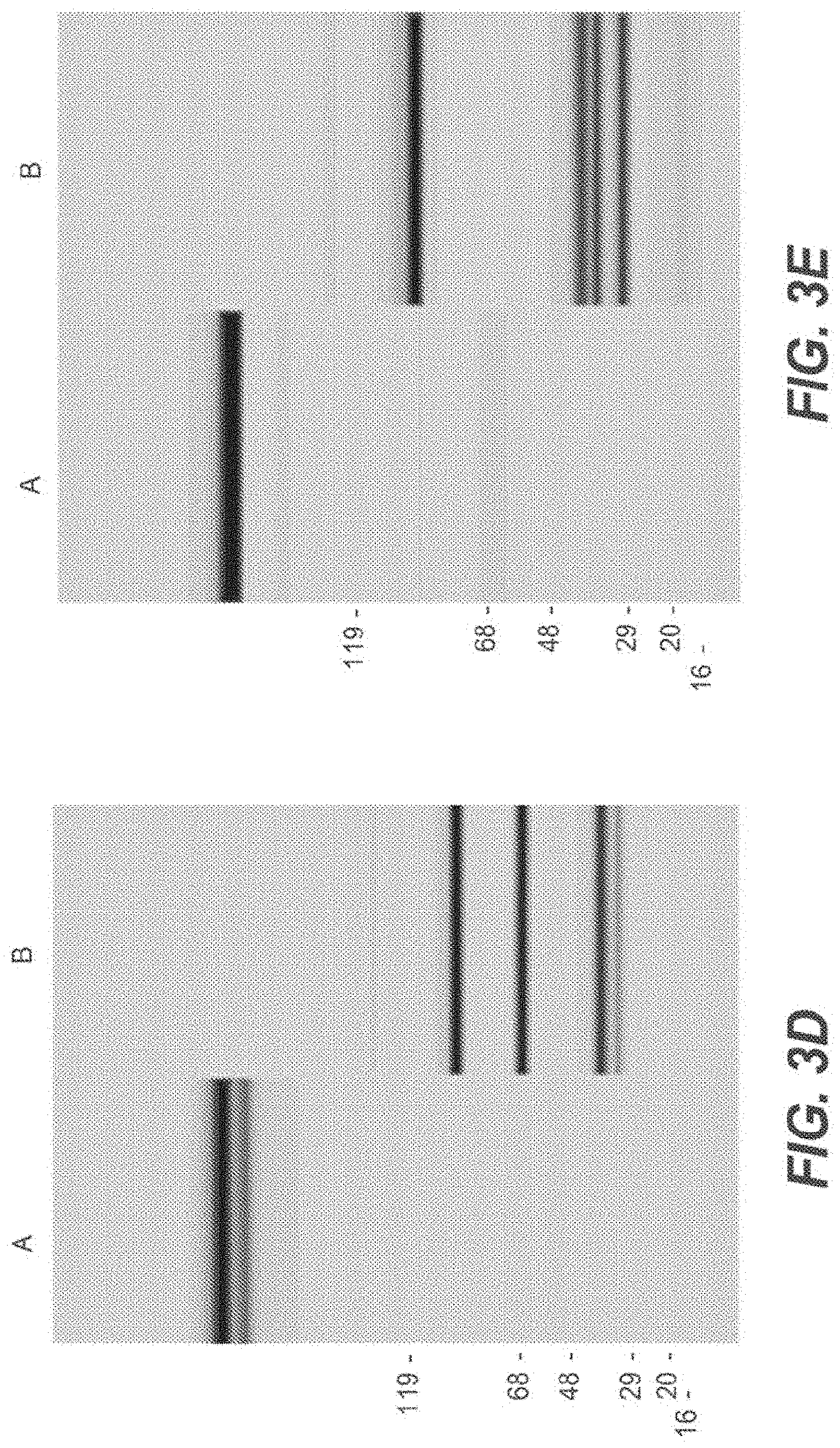
D00013
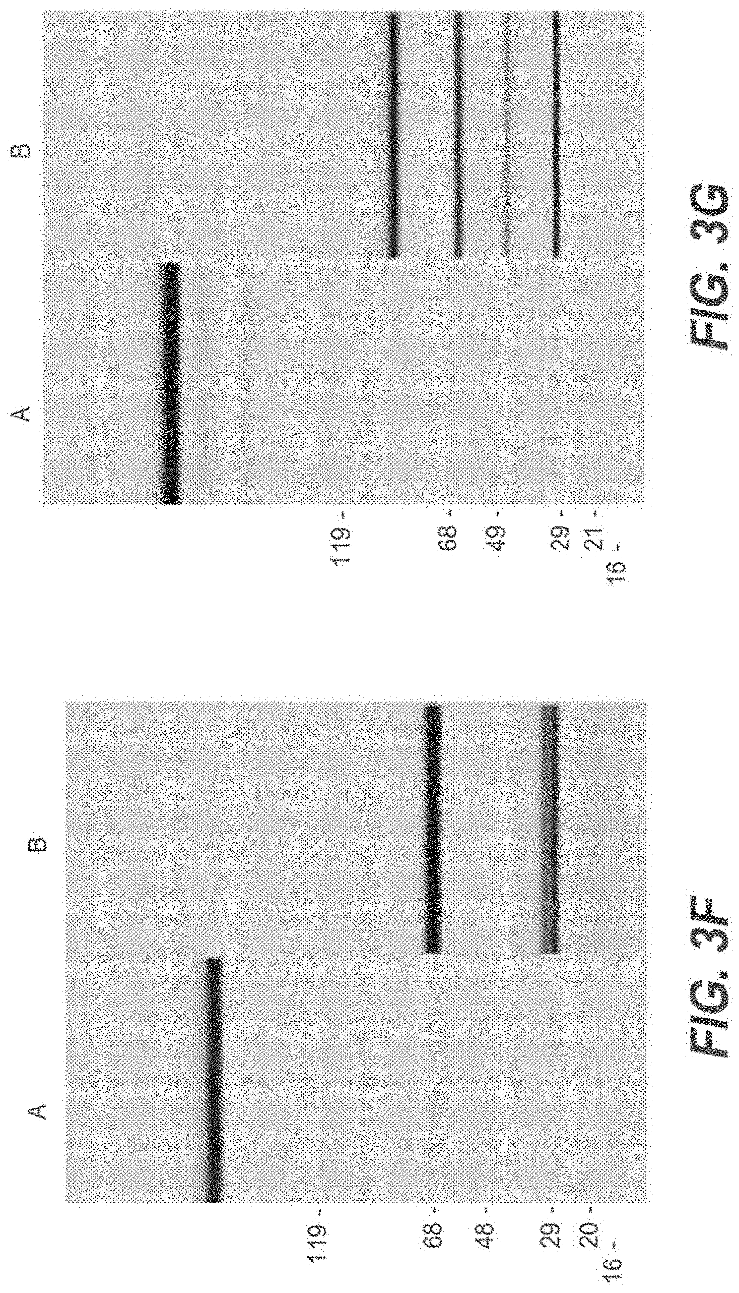
D00014

D00015

D00016

D00017

D00018

D00019
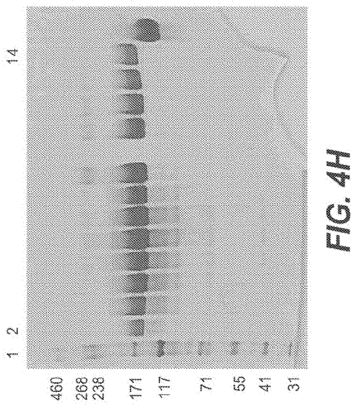
D00020

D00021

D00022

D00023
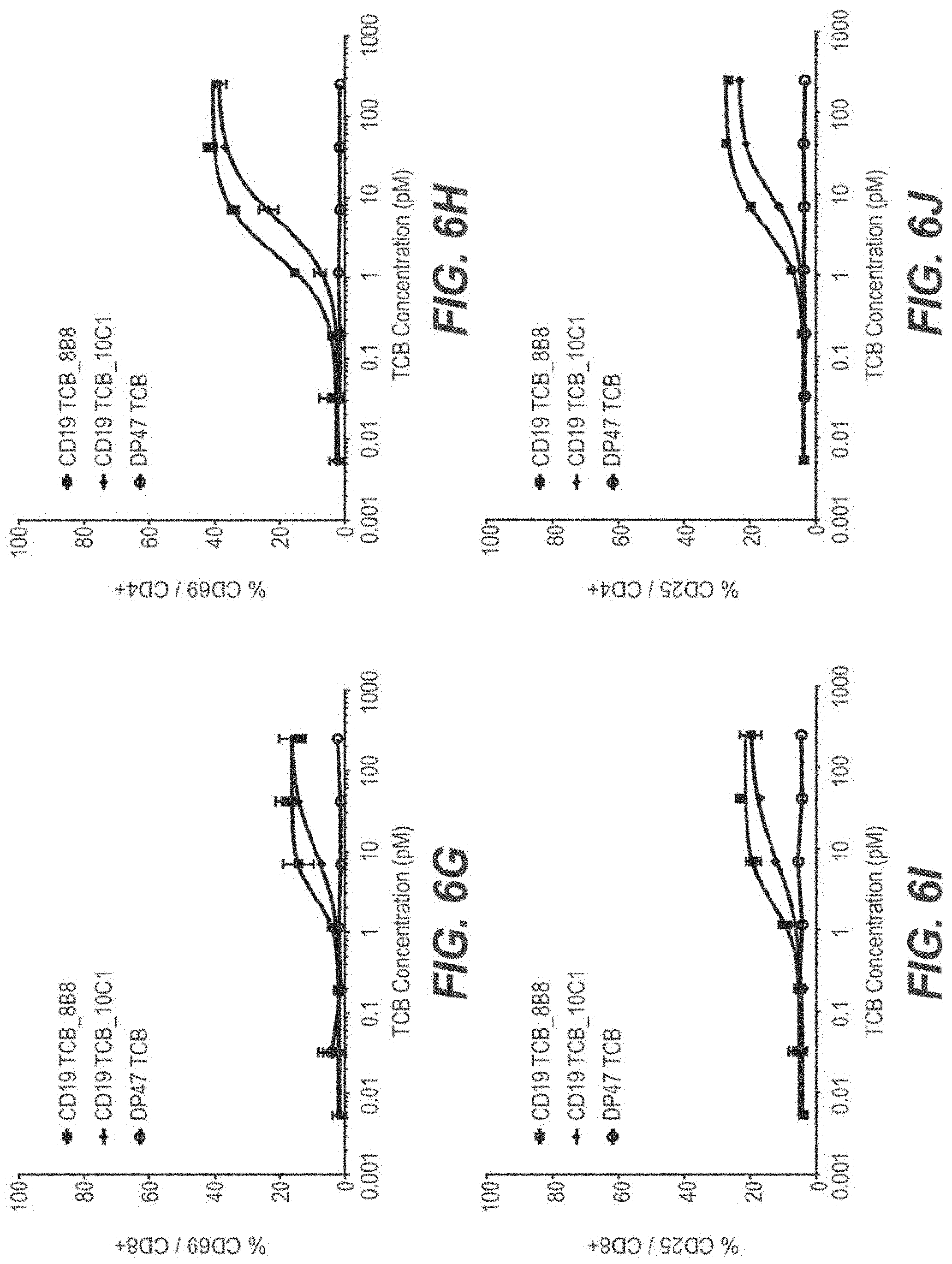
D00024
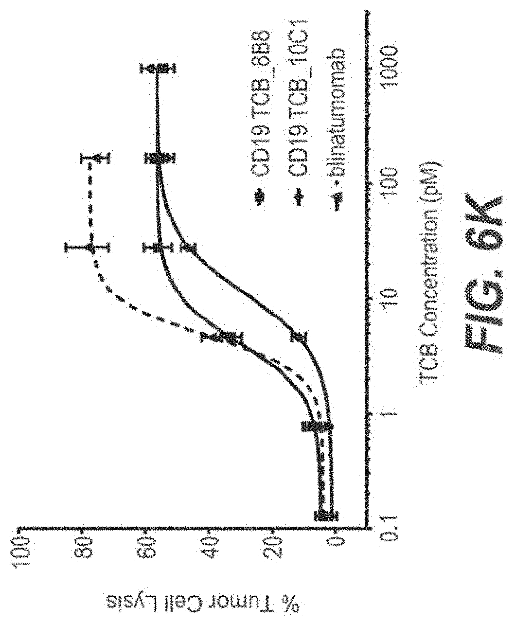
D00025
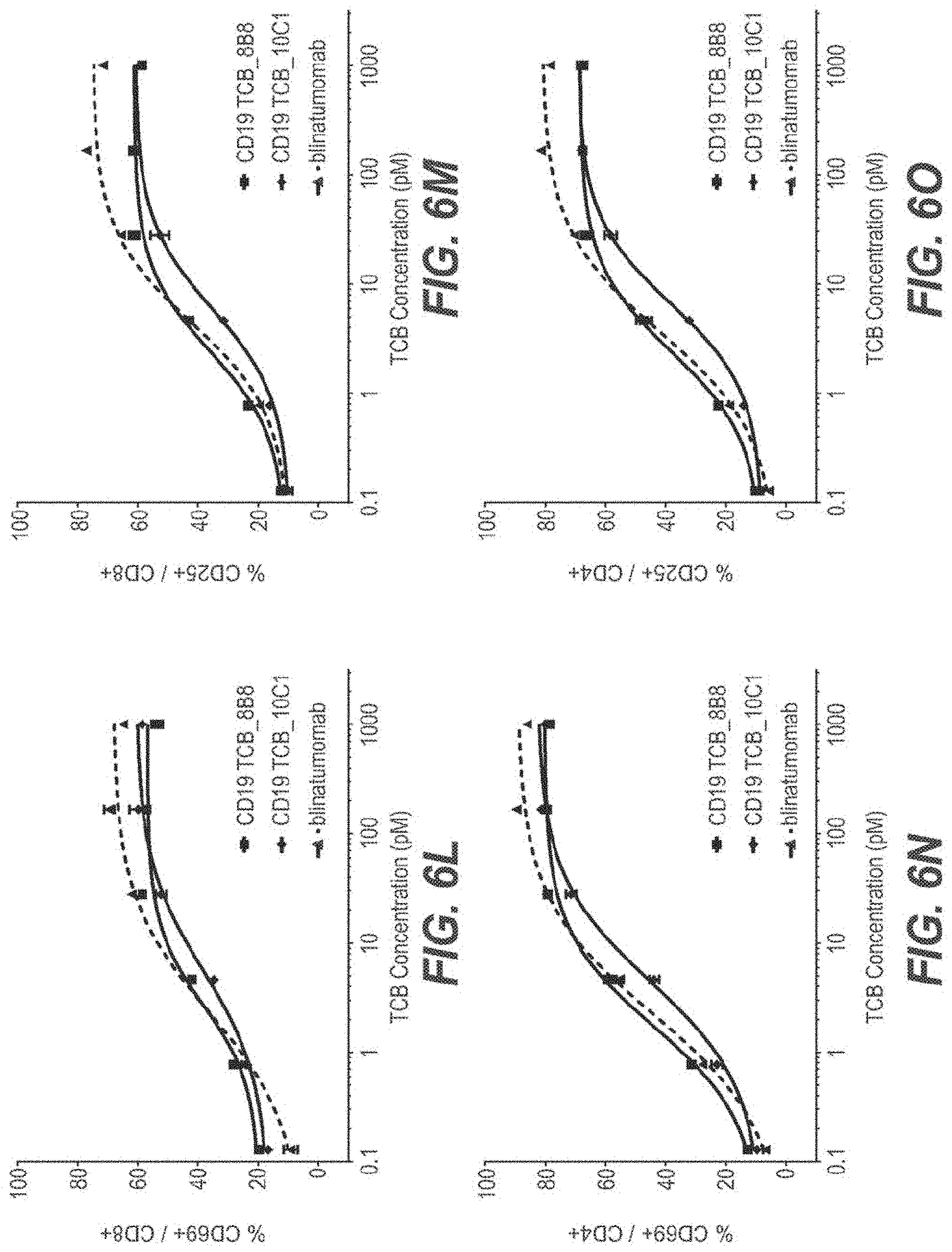
D00026
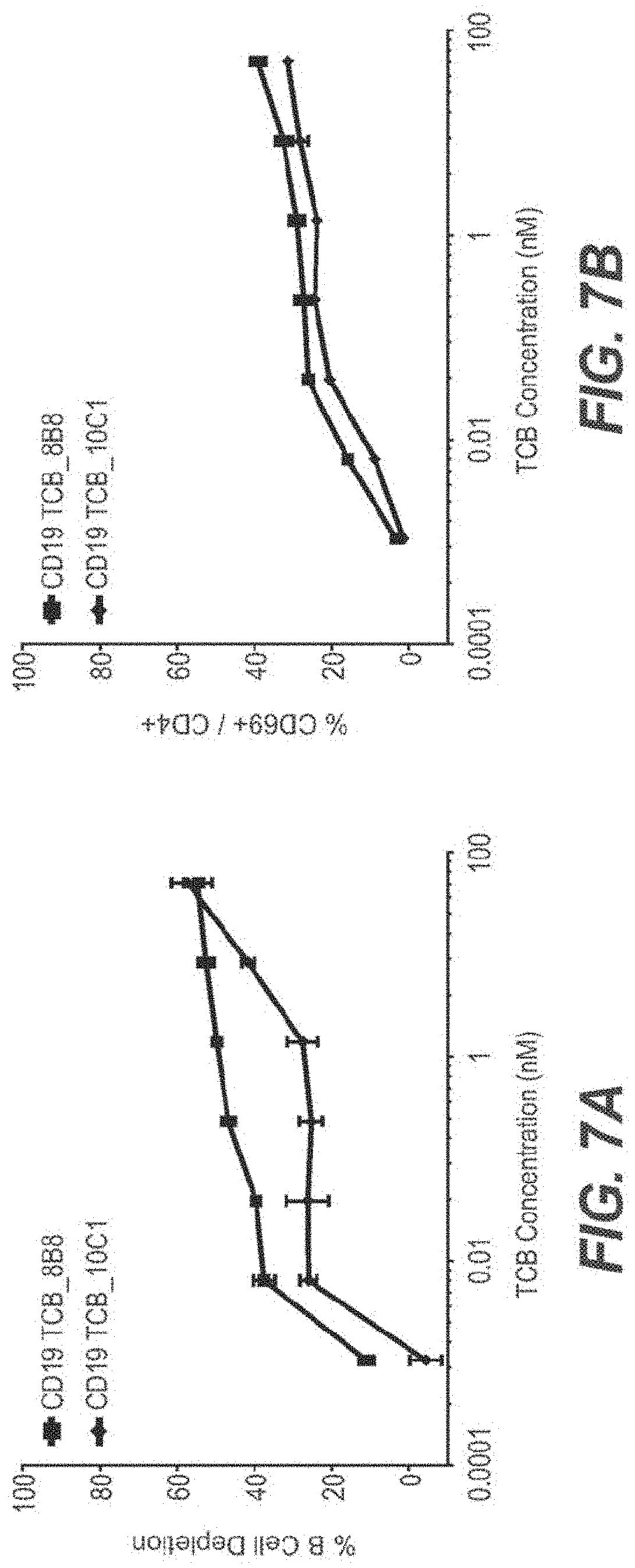
D00027
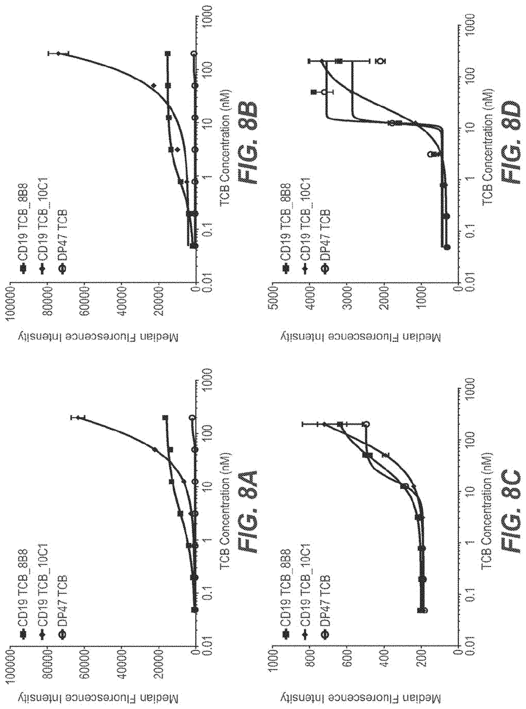
D00028

D00029

D00030
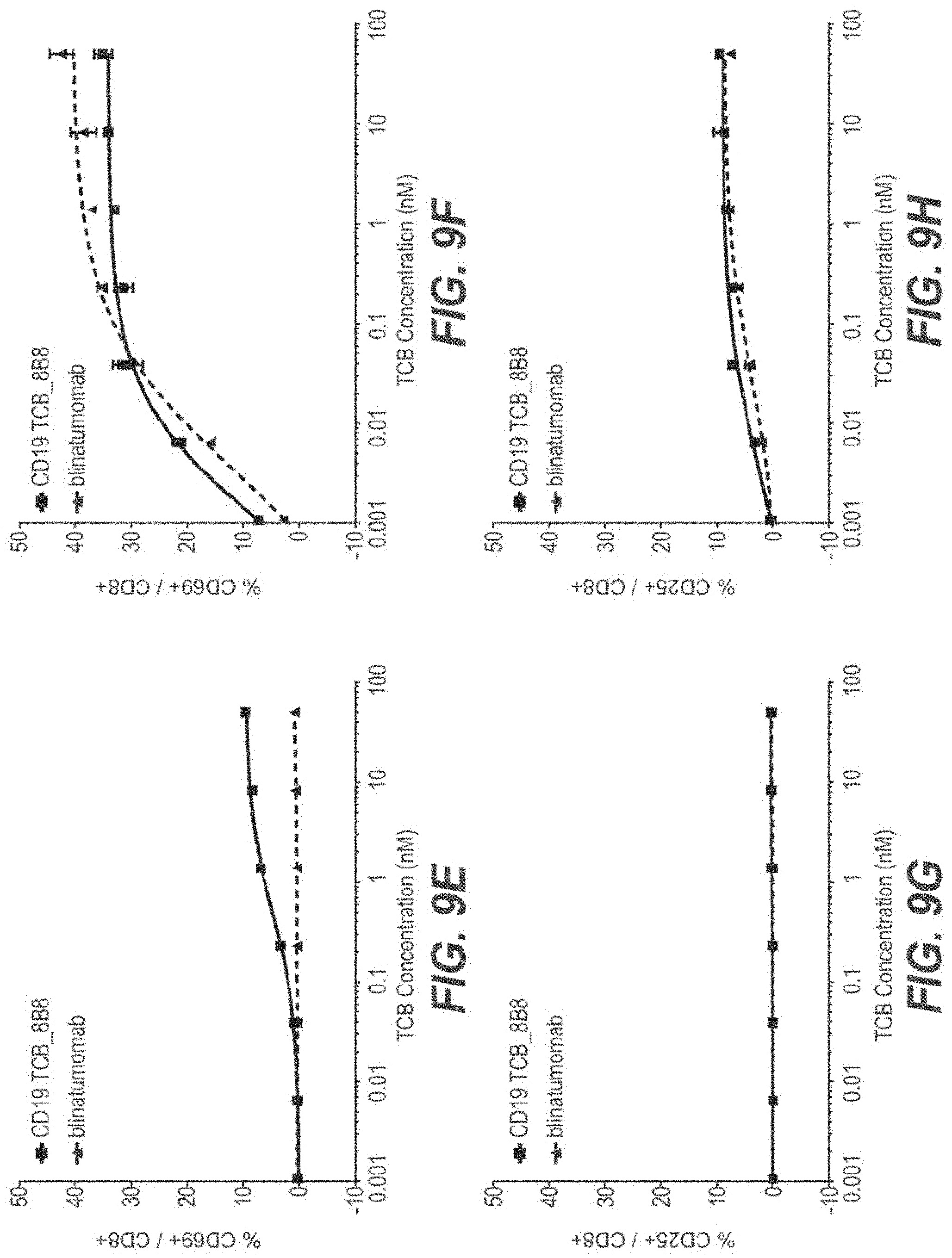
D00031

D00032

D00033

D00034

D00035

D00036

D00037

D00038
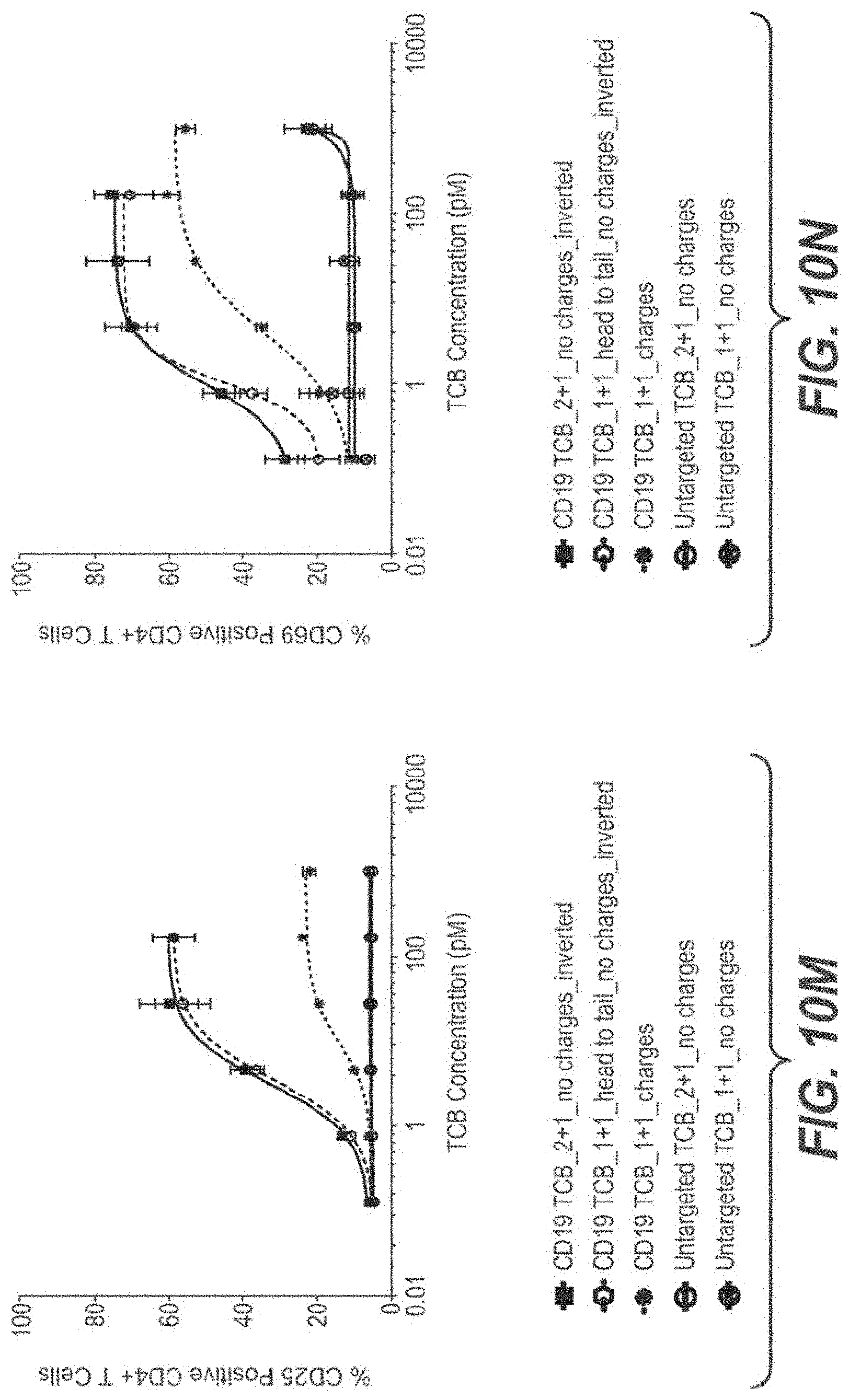
D00039
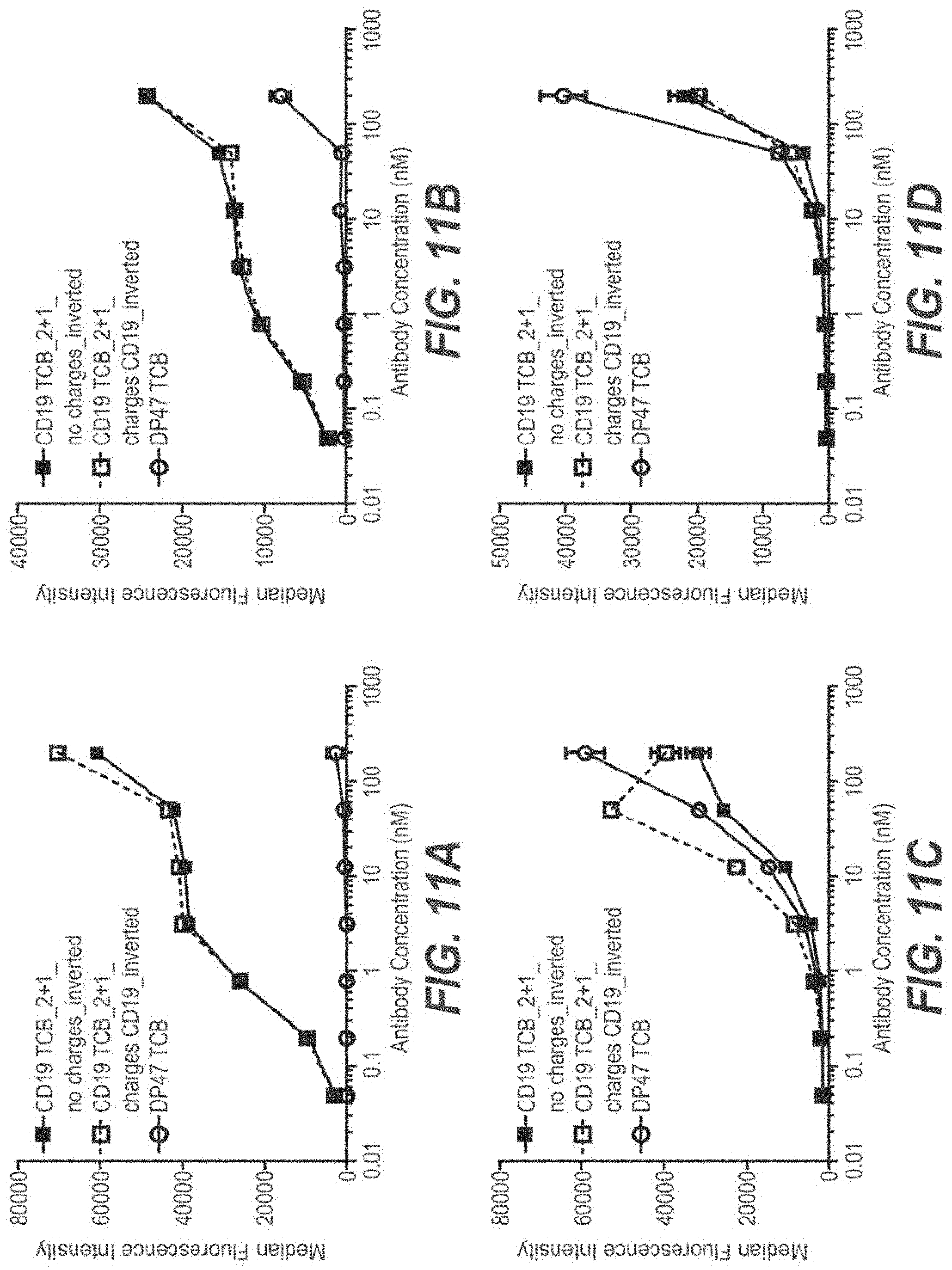
D00040
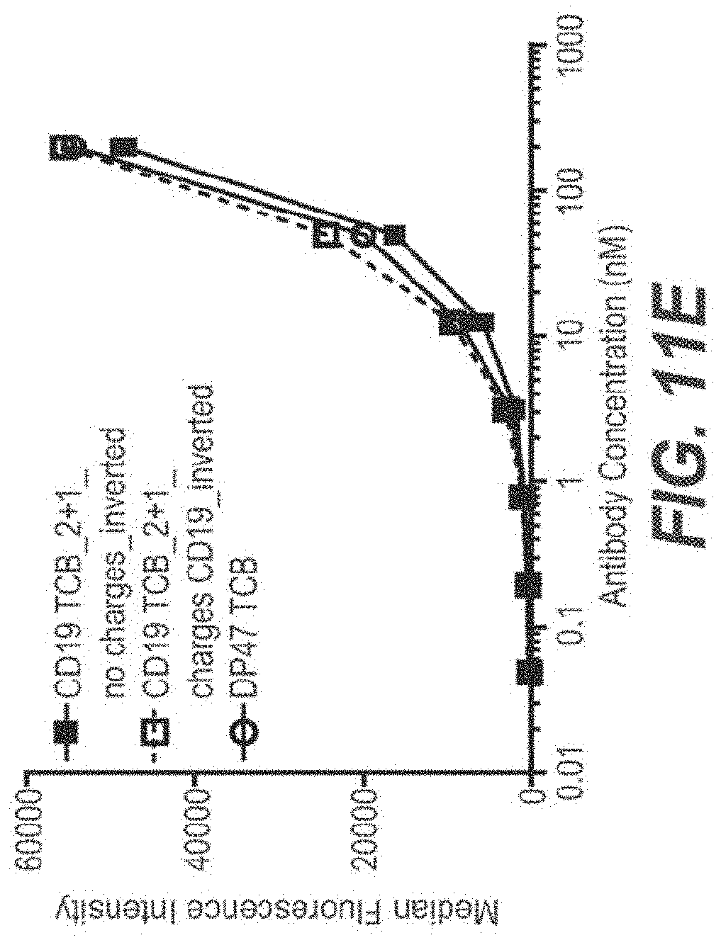
D00041
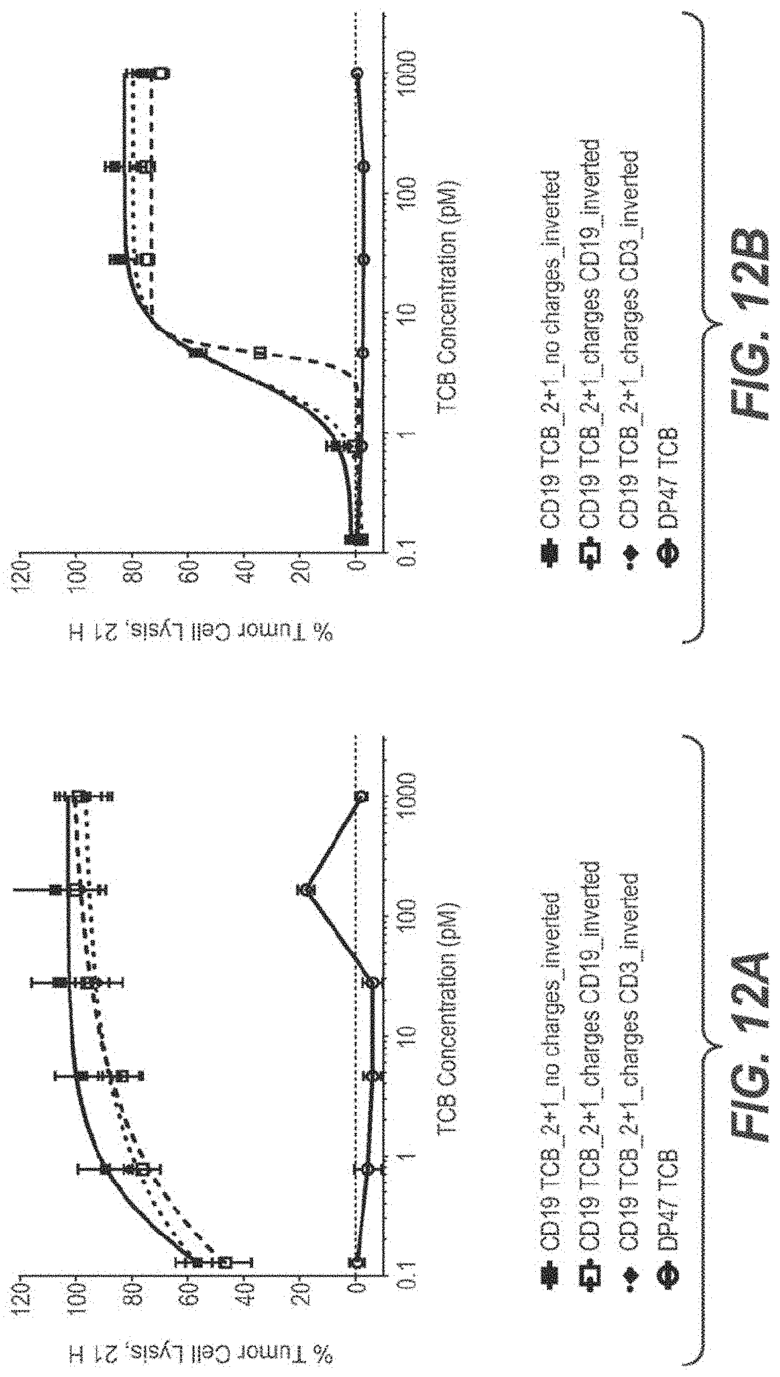
D00042

D00043
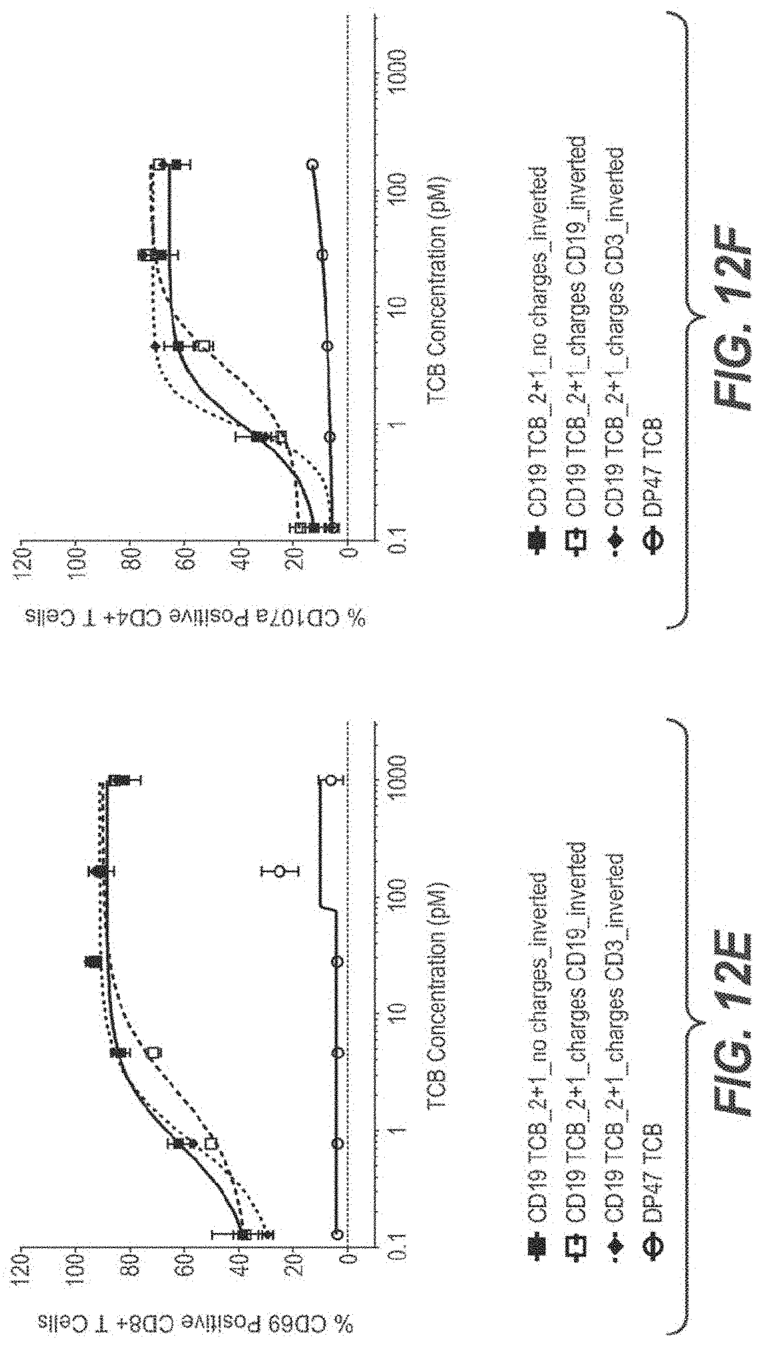
D00044
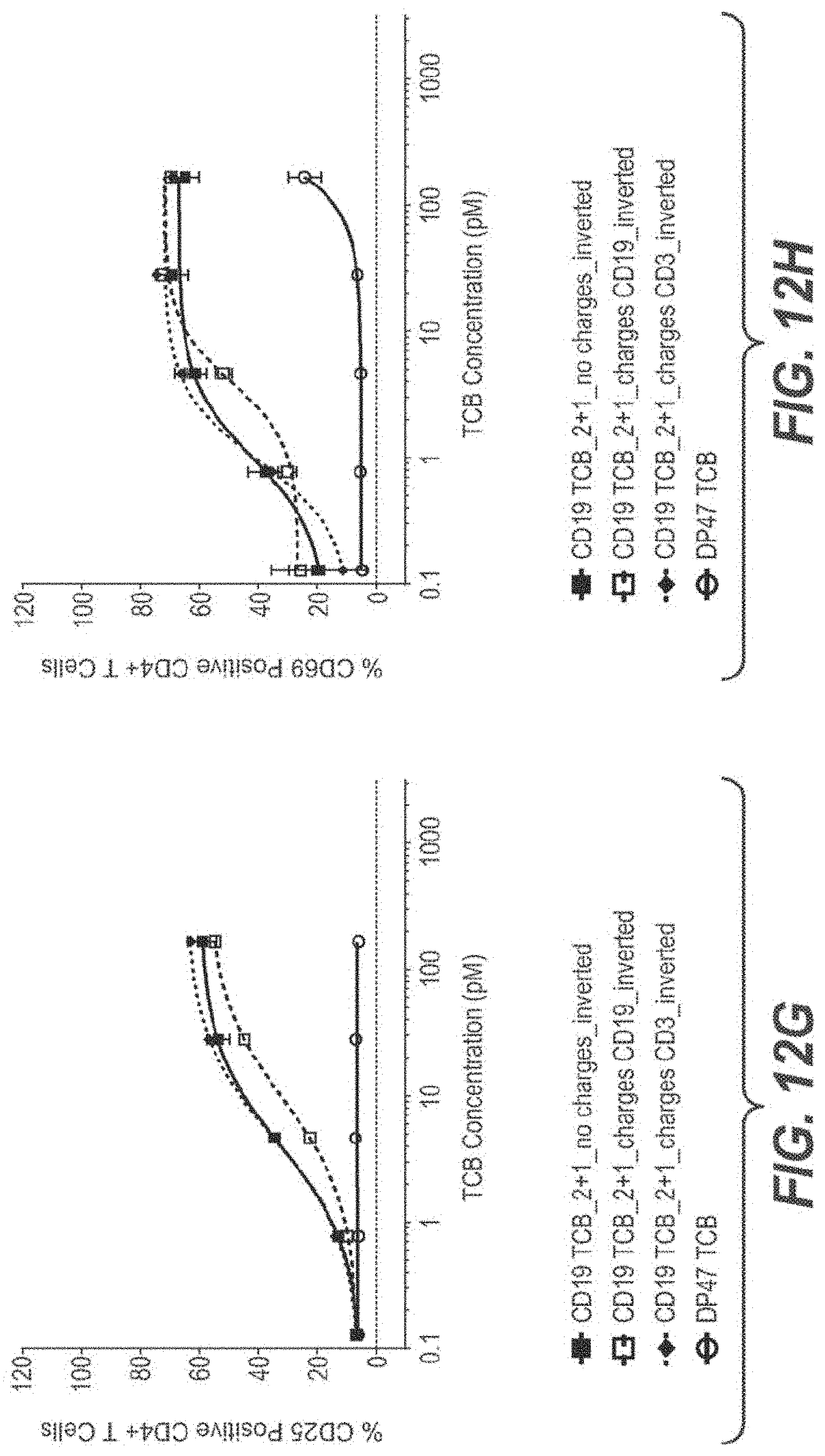
D00045

D00046

D00047
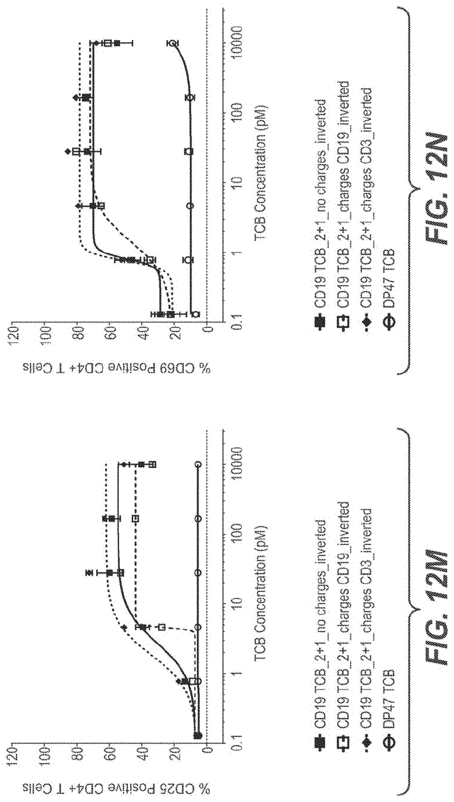
D00048

D00049

D00050
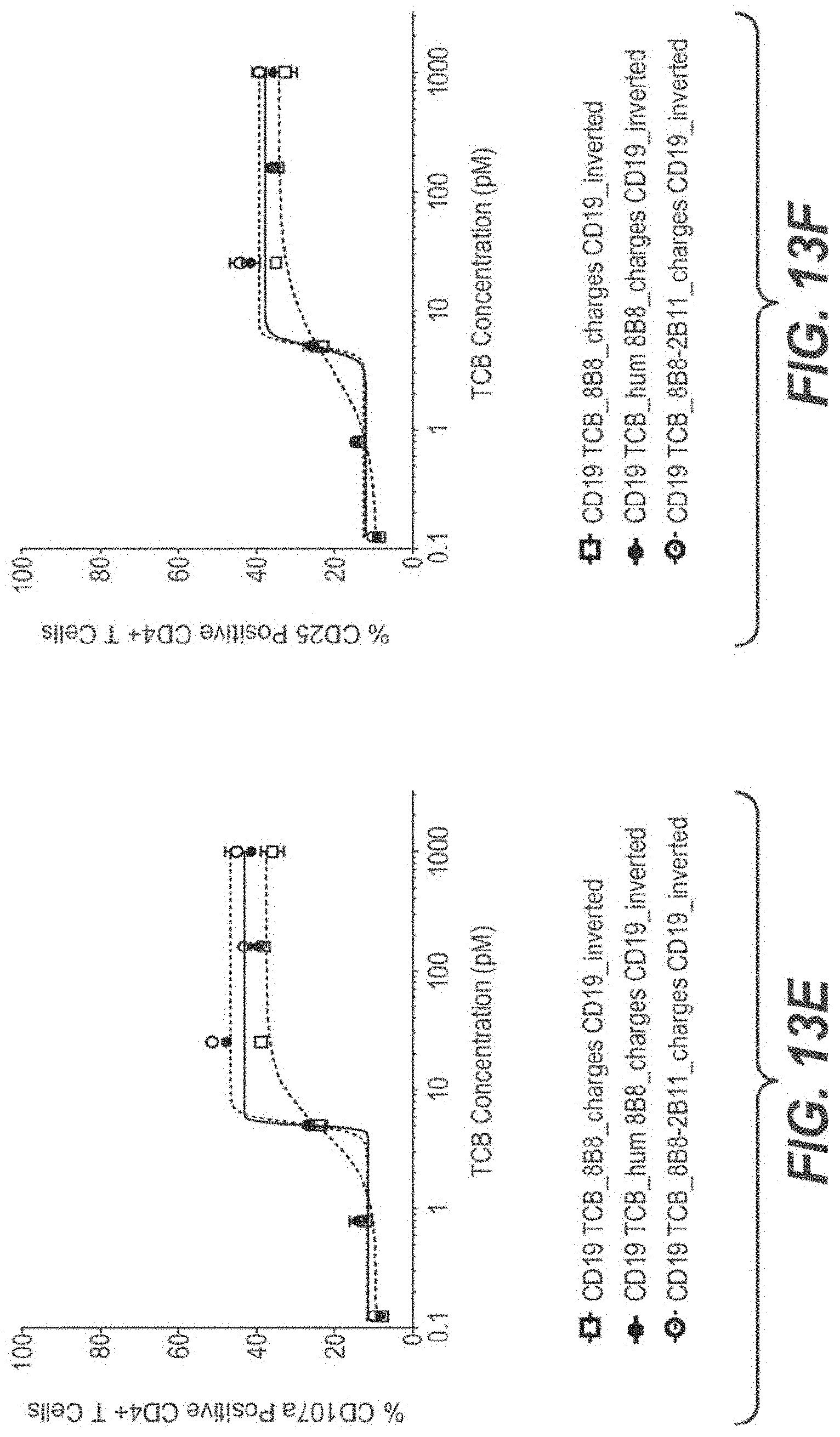
D00051

S00001
XML
uspto.report is an independent third-party trademark research tool that is not affiliated, endorsed, or sponsored by the United States Patent and Trademark Office (USPTO) or any other governmental organization. The information provided by uspto.report is based on publicly available data at the time of writing and is intended for informational purposes only.
While we strive to provide accurate and up-to-date information, we do not guarantee the accuracy, completeness, reliability, or suitability of the information displayed on this site. The use of this site is at your own risk. Any reliance you place on such information is therefore strictly at your own risk.
All official trademark data, including owner information, should be verified by visiting the official USPTO website at www.uspto.gov. This site is not intended to replace professional legal advice and should not be used as a substitute for consulting with a legal professional who is knowledgeable about trademark law.