Hla-g As A Novel Target For Car T-cell Immunotherapy
Epstein; Alan L. ; et al.
U.S. patent application number 16/841810 was filed with the patent office on 2021-03-11 for hla-g as a novel target for car t-cell immunotherapy. The applicant listed for this patent is University of Southern California. Invention is credited to Alan L. Epstein, Peisheng Hu.
| Application Number | 20210070864 16/841810 |
| Document ID | / |
| Family ID | 1000005226975 |
| Filed Date | 2021-03-11 |


| United States Patent Application | 20210070864 |
| Kind Code | A1 |
| Epstein; Alan L. ; et al. | March 11, 2021 |
HLA-G AS A NOVEL TARGET FOR CAR T-CELL IMMUNOTHERAPY
Abstract
CAR cells targeting and antibodies human HLA-G are described as a new method of cancer treatment. It is proposed that HLA-G CAR cells are safe and effective in patients and can be used to treat human tumors expressing the HLA-G.
| Inventors: | Epstein; Alan L.; (Pasadena, CA) ; Hu; Peisheng; (Covina, CA) | ||||||||||
| Applicant: |
|
||||||||||
|---|---|---|---|---|---|---|---|---|---|---|---|
| Family ID: | 1000005226975 | ||||||||||
| Appl. No.: | 16/841810 | ||||||||||
| Filed: | April 7, 2020 |
Related U.S. Patent Documents
| Application Number | Filing Date | Patent Number | ||
|---|---|---|---|---|
| 15561966 | Sep 26, 2017 | |||
| PCT/US16/24361 | Mar 25, 2016 | |||
| 16841810 | ||||
| 62139617 | Mar 27, 2015 | |||
| Current U.S. Class: | 1/1 |
| Current CPC Class: | G01N 33/57434 20130101; C07K 14/70521 20130101; C07K 16/2833 20130101; C07K 2317/622 20130101; G01N 33/57492 20130101; G01N 33/56977 20130101; A61K 39/001111 20180801; C07K 14/70575 20130101; C07K 2319/03 20130101; C12N 2510/00 20130101; C07K 14/7051 20130101; C12N 5/0638 20130101; G01N 33/57449 20130101; A61P 35/00 20180101; A61K 2039/5156 20130101; G01N 2333/70539 20130101; A61K 39/395 20130101; C07K 14/70517 20130101; C07K 2317/34 20130101; C07K 2319/02 20130101; C07K 2319/33 20130101 |
| International Class: | C07K 16/28 20060101 C07K016/28; G01N 33/569 20060101 G01N033/569; G01N 33/574 20060101 G01N033/574; A61K 39/395 20060101 A61K039/395; C12N 5/0783 20060101 C12N005/0783; A61P 35/00 20060101 A61P035/00; A61K 39/00 20060101 A61K039/00; C07K 14/725 20060101 C07K014/725; C07K 14/705 20060101 C07K014/705 |
Claims
1. An isolated antibody comprising a heavy chain (HC) immunoglobulin variable domain sequence and a light chain (LC) immunoglobulin variable domain sequence, wherein the antibody binds to an epitope of HLA-G comprising the amino acid sequence SEQ ID NO: 30, or an equivalent thereof, wherein the HC immunoglobulin variable domain sequence comprises a CDRH1 sequence comprising GFNIKDTY (SEQ ID NO: 1) or GFTFNTYA (SEQ ID NO: 2) or an equivalent of each thereof, a CDRH2 sequence comprising IDPANGNT (SEQ ID NO: 3) or IRSKSNNYAT (SEQ ID NO: 4) or an equivalent of each thereof, and a CDRH3 sequence comprising ARSYYGGFAY (SEQ ID NO: 5) or VRGGYWSFDV (SEQ ID NO: 6) or an equivalent of each thereof, wherein the LC immunoglobulin variable domain sequence comprises a CDRL1 sequence comprising KSVSTSGYSY (SEQ ID NO: 11) or KSLLHSNGNTY (SEQ ID NO: 12) or an equivalent of each thereof, a CDRL2 sequence comprising LVS (SEQ ID NO: 13) or RMS (SEQ ID NO: 14) or an equivalent of each thereof, a CDRL3 sequence comprising QHSRELPRT (SEQ ID NO: 15) or MQHLEYPYT (SEQ ID NO: 16) or an equivalent of each thereof, wherein an equivalent has at least 80% amino acid identity to the sequence, or is encoded by a polynucleotide that is at least 80% identical to a polynucleotide encoding the sequence.
2-8. (canceled)
9. The antibody of claim 1, wherein the HC immunoglobulin variable domain sequence comprises the amino acid sequence of SEQ ID NOs: 8 or 10, or an equivalent of each thereof, or wherein the LC immunoglobulin variable domain sequence comprises the amino acid sequence of SEQ ID NOs: 18 or 20, or an equivalent of each thereof, wherein an equivalent has at least 80% amino acid identity to the sequence, or is encoded by a polynucleotide that is at least 80% identical to a polynucleotide encoding the polypeptide.
10. (canceled)
11. The antibody of claim 1, wherein the HC immunoglobulin variable domain sequence comprises the amino acid sequence of SEQ ID NOs: 8 or 10, and wherein the LC immunoglobulin variable domain sequence comprises the amino acid sequence of SEQ ID NOs: 18 or 20, or an equivalent of each thereof, wherein an equivalent has at least 80% amino acid identity to the sequence, or is encoded by a polynucleotide that is at least 80% identical to a polynucleotide encoding the polypeptide.
12. (canceled)
13. An antigen binding fragment of the antibody of claim 1, wherein the antigen binding fragment is selected from the group consisting of Fab, F(ab')2, Fab', scFv, and Fv.
14. An isolated ex vivo complex comprising the antibody of claim 1 or an antigen binding fragment thereof, and optionally a detectable label.
15. (canceled)
16. A method of detecting HLA-G in a biological sample comprising contacting the sample with the antibody of claim 1 or an antigen binding fragment thereof, and detecting a complex formed by the binding of the antibody or antigen binding fragment to HLA-G.
17-20. (canceled)
21. A method of detecting a pathological cell in a sample isolated from a subject, comprising (a) detecting the level of HLA-G in a biological sample from the subject by detecting a complex formed by the antibody of claim 1 or an antigen binding fragment thereof binding to HLA-G in the sample; and (b) comparing the levels of HLA-G observed in step (a) with the levels of HLA-G observed in a control biological sample; wherein the pathological cell is detected when the level of HLA-G is elevated compared to that observed in the control biological sample and the pathological cell is not detected when the level of HLA-G is not elevated as compared to the observed in the control biological sample.
22-27. (canceled)
28. A kit for detecting HLA-G comprising an antibody of claim 1 or an antigen binding fragment thereof, and instructions for use.
29. The method of claim 16, wherein the biological sample is a tumor sample.
30. A chimeric antigen receptor (CAR) comprising: (a) an antigen binding domain of an anti-HLA-G antibody of claim 1; (b) a CD8 .alpha. hinge domain; (c) a CD8 .alpha. transmembrane domain; (d) a CD28 costimulatory signaling region and/or a 4-1BB costimulatory signaling region; and (e) a CD3 zeta signaling domain.
31-33. (canceled)
34. The CAR of claim 30, wherein the HC immunoglobulin variable domain sequence comprises the amino acid sequence of SEQ ID NOs: 8 or 10, or an equivalent of each thereof, or wherein the LC immunoglobulin variable domain sequence comprises the amino acid sequence of SEQ ID NOs: 18 or 20.
35. (canceled)
36. The CAR of claim 30, wherein the HC immunoglobulin variable domain sequence comprises the amino acid sequence of SEQ ID NOs: 8 or 10, and wherein the LC immunoglobulin variable domain sequence comprises the amino acid sequence of SEQ ID NOs: 18 or 20, or an equivalent of each thereof.
37-42. (canceled)
43. A vector comprising a nucleic acid sequence encoding the CAR of claim 30.
44-45. (canceled)
46. An isolated cell comprising one or more of: the antibody of claim 1 or an antigen binding fragment thereof; a nucleic acid encoding the antibody or the antigen binding fragment thereof, or a complement thereof; a complex comprising the antibody or the antigen binding fragment thereof; a CAR comprising: (a) the antigen binding domain of the antibody, (b) a CD8 .alpha. hinge domain, (c) a CD8 .alpha. transmembrane domain, (d) one or both of a CD28 costimulatory signaling region or a 4-1BB costimulatory signaling region, and (e) a CD3 zeta signaling domain; a nucleic acid encoding the CAR or a complement thereof, or a vector comprising one or more of: the nucleic acid encoding the antibody or the antigen binding fragment thereof, the nucleic acid encoding the CAR, or a complement of each thereof.
47. The isolated cell of claim 46, wherein the cell is a T-cell or an NK-cell.
48. (canceled)
49. An isolated nucleic acid encoding the isolated antibody of claim 1 or an antigen binding fragment thereof; or encoding a CAR comprising: (a) the antigen binding domain of the antibody, (b) a CD8 .alpha. hinge domain, (c) a CD8 .alpha. transmembrane domain, (d) one or both of a CD28 costimulatory signaling region or a 4-1BB costimulatory signaling region, and (e) a CD3 zeta signaling domain; or its complement.
50. (canceled)
51. A method of producing HLA-G CAR expressing cells comprising: (i) transducing a population of isolated cells with a nucleic acid sequence encoding the CAR of claim 30; and (ii) selecting a subpopulation of said isolated cells that have been successfully transduced with said nucleic acid sequence of step (i) thereby producing HLA-G CAR expressing cells.
52. (canceled)
53. A method of inhibiting the growth of a tumor in a subject in need thereof, comprising administering to the subject an effective amount of the isolated cell of claims 47.
54-57. (canceled)
58. A method of treating a cancer patient in need thereof, comprising administering to the subject an effective amount of the isolated cell of claim 47.
59-62. (canceled)
63. A method for determining if a patient is likely to respond or is not likely to HLA-G CAR therapy, comprising contacting a tumor sample isolated from the patient with an effective amount of an anti-HLA-G antibody and detecting the presence of any antibody bound to the tumor sample, wherein the presence of antibody bound to the tumor sample indicates that the patient is likely to respond to the HLA-G CAR therapy and the absence of antibody bound to the tumor sample indicates that the patient is not likely to respond to the HLA-G therapy.
64. (canceled)
Description
CROSS-REFERENCE TO RELATED APPLICATIONS
[0001] This application is a continuation of U.S. patent application Ser. No. 15/561,966 filed on Mar. 25, 2016, which is a national stage entry under 35 U.S.C. .sctn. 371 of International Application No. PCT/US2016/024361, filed Mar. 25, 2016, which in turn claims priority under 35 U.S.C. .sctn. 119(e) to U.S. Provisional Application No. 62/139,617, filed Mar. 27, 2015, the content of each of which is hereby incorporated by reference in its entirety.
SEQUENCE LISTING
[0002] The instant application contains a Sequence Listing which has been submitted electronically in ASCII format and is hereby incorporated by reference in its entirety. Said ASCII copy, created on Nov. 27, 2020, is named 116914-7180 SL ST25.txt and is 50,670 bytes in size.
TECHNICAL FIELD
[0003] The present disclosure relates generally to the field of human immunology, specifically cancer immunotherapy.
BACKGROUND
[0004] The following discussion of the background of the invention is merely provided to aid the reader in the understanding the invention and is not admitted to describe or constitute prior art to the present invention.
[0005] HLA-G is is a non-classical MHC class I molecule which primarily serves to suppress cytotoxic immune cell function, particularly as a ligand for the inhibitory NK cell receptors.
SUMMARY OF THE DISCLOSURE
[0006] Provided are novel anti-HLA-G antibodies and methods of their use diagnostically and therapeutically. In this regard, provide herein is an isolated antibody comprising a heavy chain (HC) immunoglobulin variable domain sequence and a light chain (LC) immunoglobulin variable domain sequence, wherein the antibody binds to an epitope of human HLA-G comprising the amino acid sequence: GSHSMRYFSA AVSRPGRGEP RFIAMGYVDD TQFVRFDSDS ACPRMEPRAP WVEQEGPEYW EEETRNTKAH AQTDRMNLQT LRGYYNQSEA SSHTLQWMIG CDLGSDGRLL RGYEQYAYDG KDYLALNEDL RSWTAADTAA QISKRKCEAA NVAEQRRAYL EGTCVEWLHR YLENGKEMLQ RADPPKTHVT HHPVFDYEAT LRCWALGFYP AEIILTWQRD GEDQTQDVEL VETRPAGDGT FQKWAAVVVP SGEEQRYTCH VQHEGLPEPL MLRWKQSSLP TIPIMGI VAGLVVLAAV VTGAAVAAVL WRKKSSD (SEQ ID NO: 30), or an equivalent thereof. In one aspect, the antibodies possess a specific binding affinity of at least 10.sup.-6 M. In certain aspects, antibodies bind with affinities of at least about 10.sup.-7 M, and preferably 10.sup.-8 M, 10.sup.-9 M, 10.sup.-10 M, 10.sup.-11 M, or 10.sup.-12 M.
[0007] In certain embodiments disclosed herein, the antibody comprises a heavy chain (HC) immunoglobulin variable domain sequence and a light chain (LC) immunoglobulin variable domain sequence, wherein the antibody binds to an epitope of human HLA-G comprising, or alternatively consisting essentially of, or yet further consisting of, an amino acid sequence wherein the HC comprises any one of the following a HC CDRH1 comprising the amino acid sequence GFNIKDTY (SEQ ID NO: 1) or GFTFNTYA (SEQ ID NO: 2) or an equivalent of each thereof; and/or a HC CDRH2 comprising the amino acid sequence IDPANGNT (SEQ ID NO: 3) or IRSKSNNYAT (SEQ ID NO: 4) or an equivalent of each thereof; and/or a HC CDRH3 comprising the amino acid sequence ARSYYGGFAY (SEQ ID NO: 5) or VRGGYWSFDV (SEQ ID NO: 6), or an equivalent of each thereof.
[0008] In certain embodiments disclosed herein, the antibody comprises a heavy chain (HC) immunoglobulin variable domain sequence and a light chain (LC) immunoglobulin variable domain sequence, wherein the antibody binds to an epitope of human HLA-G comprising, or alternatively consisting essentially of, or yet further consisting of, an amino acid sequence wherein the LC comprises a LC CDRL1 comprising the amino acid KSVSTSGYSY (SEQ ID NO: 11) or KSLLHSNGNTY (SEQ ID NO: 12) or an equivalent of each thereof; and/or a LC CDRL2 comprising the amino acid sequence LVS (SEQ ID NO: 13) or RMS (SEQ ID NO: 14) or an equivalent of each thereof; and/or a LC CDRL3 comprising the amino acid sequence QHSRELPRT (SEQ ID NO: 15) or MQHLEYPYT (SEQ ID NO: 16) or an equivalent of each thereof.
[0009] Some aspects of the disclosure relate to a chimeric antigen receptor (CAR) comprising an antigen binding domain specific to HLA-G--for example, the antigen binding domain of an anti-HLA-G antibody, nucleic acids encoding them as well as method for the production and use of them.
[0010] Aspects of the disclosure relate to a chimeric antigen receptor (CAR) comprising: (a) an antigen binding domain of an HLA-G antibody; (b) a hinge domain; (c) a transmembrane domain; and (d) an intracellular domain. Further aspects of the disclosure relate to a chimeric antigen receptor (CAR) comprising: (a) an antigen binding domain of a HLA-G antibody; (b) a hinge domain; (c) a CD28 transmembrane domain; (d) one or more costimulatory regions selected from a CD28 costimulatory signaling region, a 4-1BB costimulatory signaling region, an ICOS costimulatory signaling region, and an OX40 costimulatory region; and (e) a CD3 zeta signaling domain or an equivalent or alternative thereof.
[0011] In a further aspect, the present disclosure provides a chimeric antigen receptor (CAR) comprising: (a) an antigen binding domain of an anti-HLA-G antibody, (b) a CD8 a hinge domain; (c) a CD8 a transmembrane domain; (d) a CD28 costimulatory signaling region and/or a 4-1BB costimulatory signaling region; and (e) a CD3 zeta signaling domain, or an equivalent or alternative thereof.
[0012] In a further aspect, the present disclosure provides a chimeric antigen receptor (CAR) comprising: (a) an antigen binding domain of an anti-HLA-G antibody, (b) a CD8 a hinge domain; (c) a CD8 a transmembrane domain; (d) a 4-1BB costimulatory signaling region; and (e) a CD3 zeta signaling domain, or an equivalent or alternative thereof
[0013] Further aspects of the disclosure relate to an isolated nucleic acid sequence encoding the antibodies, vectors, and host cells containing them.
[0014] Additional aspects of the disclosure relate to an isolated cell comprising a HLA-G CAR and methods of producing such cells. Still other method aspects of the disclosure relate to methods for inhibiting the growth of a tumor and treating a cancer patient comprising administering an effective amount of said isolated cell.
[0015] Further aspects of the disclosure relate to methods and kits for determining if a patient is likely to respond or is not likely to HLA-G CAR therapy through use of either or both the HLA-G antibody and the HLA-G CAR cells.
[0016] Additional aspects of the disclosure relate to compositions comprising a carrier and one or more of the products described in the embodiments disclosed herein. In some aspects, the present disclosure provides a composition comprising a carrier and one or more of: the HLA-G antibody; and/or the HLA-G CAR; and/or the isolated nucleic acid encoding the HLA-G antibody or the HLA-G CAR; and/or the vector comprising the isolated nucleic acid sequence encoding the HLA-G antibody, or the HLA-G CAR; and/or an isolated cell comprising the HLA-G CAR.
BRIEF DESCRIPTION OF THE DRAWINGS
[0017] FIG. 1 shows flow cytometry screening data of newly generated monoclonal antibodies to human HLA-G. Subclones of positive hybridomas (3H11-12 and 4E3-1) were selected for the generation of CAR T-cells based upon these results.
[0018] FIGS. 2A-2D show immunohistochemistry of HLA-G reactivity in papillary thyroid cancer and normal thyroid tissue with HLA-ABC control staining. FIG. 2A shows low magnification of HLA-G positive papillary thyroid carcinoma section using antibody 4E3-1 (100.times.). FIG. 2B shows higher magnification of second papillary thyroid carcinoma positive for HLA-G (250.times.). FIG. 2C shows negative reactivity of normal thyroid tissues for HLA-G (250.times.), and FIG. 2D shows positive reactivity of normal thyroid tissue for HLA-ABC (100.times.).
[0019] FIG. 3 shows schematic diagram of the DNA sequence for, and the theoretical structure of third generation anti-HLA-G CAR in the plasma membrane. Linker sequence disclosed as SEQ ID NO: 47.
[0020] FIG. 4 shows additional antibody screening, as described in FIG. 1.
[0021] FIG. 5 depicts a schematic of the gene-transfer vector and the transgene. The backbone of the gene transfer vector is an HIV-based, bicistronic lentiviral vector, pLVX-IRES-ZsGreen containing HIV-1 5' and 3' long terminal repeats (LTRs), packaging signal (.PSI.), EF1.alpha. promoter, internal ribosome entry site (IRES), ZsGreen, a green fluorescent protein, woodchuck hepatitis virus post-transcriptional regulatory element (WPRE), and simian virus 40 origin (SV40). Constitutive expression of the transgene comprising of a scFV specific to HLA-G, a CD8 hinge and transmembrane region and CD28, 4-1BB and CD3.zeta. signaling domain, is insured by the presence of the EF-1.alpha. promoter. Expression of the detection protein, ZsGreen is carried out by the IRES region. Integration of the vector can be assayed by the presence of ZsGreen in the cells, via fluorescent microscopy.
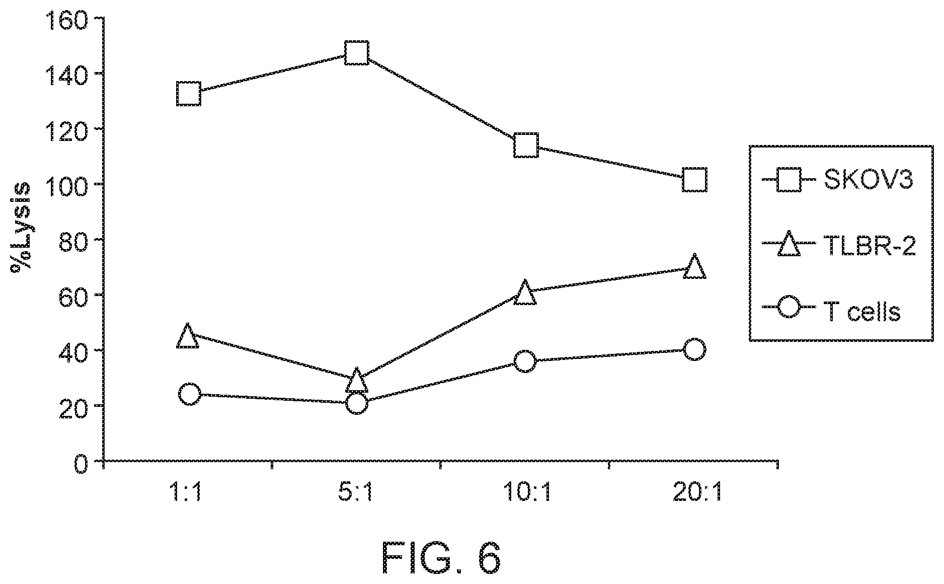
[0022] FIG. 6 shows cytotoxicity of the HLA-G CAR T-cells. Cytotoxicity of the HLA-G CAR expressing T-cells was determined using an LDH cytotoxicity kit as described in the Methods. Prior to the assay, T-cells were activated using .alpha.CD3/CD8 beads (Stem Cell Technologies, 30 ul to 2 ml of media). The activated T-cells were transduced with HLA-G lentiviral particles, following which the T cells were activated for using the aCD3/CD8 beads. Un-transduced, activated T-cells and the TLBR-2 T lymphoma cell line were used as controls. 3,000 SKOV3 or TLBR-2 cells were plated per well. HLA-G transduced T cells were added in ratios of 20:1, 10:1, 5:1 and 1:1 (60,000-3000 cells) to the wells. Each data point represents the average of triplicate measurements.
[0023] FIG. 7 shows protein expression of the HLA-G CAR. T-cells transduced with the HLA-G CAR lentiviral particles express protein for the HLA-G CAR. The estimated size of the CAR protein is 60 kDa. A CD3t antibody was used to detect the protein. Fifty .mu.g of protein was used for the western blot. .beta.-actin was used as a loading control.
DETAILED DESCRIPTION
[0024] It is to be understood that the present disclosure is not limited to particular aspects described, as such may, of course, vary. It is also to be understood that the terminology used herein is for the purpose of describing particular aspects only, and is not intended to be limiting, since the scope of the present disclosure will be limited only by the appended claims.
[0025] Unless defined otherwise, all technical and scientific terms used herein have the same meanings as commonly understood by one of ordinary skill in the art to which this technology belongs. Although any methods and materials similar or equivalent to those described herein can be used in the practice or testing of the present technology, the preferred methods, devices and materials are now described. All technical and patent publications cited herein are incorporated herein by reference in their entirety. Nothing herein is to be construed as an admission that the present technology is not entitled to antedate such disclosure by virtue of prior invention.
[0026] The practice of the present technology will employ, unless otherwise indicated, conventional techniques of tissue culture, immunology, molecular biology, microbiology, cell biology, and recombinant DNA, which are within the skill of the art. See, e.g., Sambrook and Russell eds. (2001) Molecular Cloning: A Laboratory Manual, 3rd edition; the series Ausubel et al. eds. (2007) Current Protocols in Molecular Biology; the series Methods in Enzymology (Academic Press, Inc., N.Y.); MacPherson et al. (1991) PCR 1: A Practical Approach (IRL Press at Oxford University Press); MacPherson et al. (1995) PCR 2: A Practical Approach; Harlow and Lane eds. (1999) Antibodies, A Laboratory Manual; Freshney (2005) Culture of Animal Cells: A Manual of Basic Technique, 5th edition; Gait ed. (1984) Oligonucleotide Synthesis; U.S. Pat. No. 4,683,195; Hames and Higgins eds. (1984) Nucleic Acid Hybridization; Anderson (1999) Nucleic Acid Hybridization; Hames and Higgins eds. (1984) Transcription and Translation; Immobilized Cells and Enzymes (IRL Press (1986)); Perbal (1984) A Practical Guide to Molecular Cloning; Miller and Calos eds. (1987) Gene Transfer Vectors for Mammalian Cells (Cold Spring Harbor Laboratory); Makrides ed. (2003) Gene Transfer and Expression in Mammalian Cells; Mayer and Walker eds. (1987) Immunochemical Methods in Cell and Molecular Biology (Academic Press, London); and Herzenberg et al. eds (1996) Weir's Handbook of Experimental Immunology.
[0027] All numerical designations, e.g., pH, temperature, time, concentration, and molecular weight, including ranges, are approximations which are varied (+) or (-) by increments of 1.0 or 0.1, as appropriate, or alternatively by a variation of +/-15%, or alternatively 10%, or alternatively 5%, or alternatively 2%. It is to be understood, although not always explicitly stated, that all numerical designations are preceded by the term "about". It also is to be understood, although not always explicitly stated, that the reagents described herein are merely exemplary and that equivalents of such are known in the art.
[0028] It is to be inferred without explicit recitation and unless otherwise intended, that when the present technology relates to a polypeptide, protein, polynucleotide or antibody, an equivalent or a biologically equivalent of such is intended within the scope of the present technology.
Definitions
[0029] As used in the specification and claims, the singular form "a", "an", and "the" include plural references unless the context clearly dictates otherwise. For example, the term "a cell" includes a plurality of cells, including mixtures thereof.
[0030] As used herein, the term "animal" refers to living multi-cellular vertebrate organisms, a category that includes, for example, mammals and birds. The term "mammal" includes both human and non-human mammals.
[0031] The terms "subject," "host," "individual," and "patient" are as used interchangeably herein to refer to human and veterinary subjects, for example, humans, animals, non-human primates, dogs, cats, sheep, mice, horses, and cows. In some embodiments, the subject is a human.
[0032] As used herein, the term "antibody" collectively refers to immunoglobulins or immunoglobulin-like molecules including by way of example and without limitation, IgA, IgD, IgE, IgG and IgM, combinations thereof, and similar molecules produced during an immune response in any vertebrate, for example, in mammals such as humans, goats, rabbits and mice, as well as non-mammalian species, such as shark immunoglobulins. Unless specifically noted otherwise, the term "antibody" includes intact immunoglobulins and "antibody fragments" or "antigen binding fragments" that specifically bind to a molecule of interest (or a group of highly similar molecules of interest) to the substantial exclusion of binding to other molecules (for example, antibodies and antibody fragments that have a binding constant for the molecule of interest that is at least 10.sup.3 M.sup.-1 greater, at least 10.sup.4M.sup.-1 greater or at least 10.sup.5 M.sup.-1 greater than a binding constant for other molecules in a biological sample). The term "antibody" also includes genetically engineered forms such as chimeric antibodies (for example, humanized murine antibodies), heteroconjugate antibodies (such as, bispecific antibodies). See also, Pierce Catalog and Handbook, 1994-1995 (Pierce Chemical Co., Rockford, Ill.); Kuby, J., Immunology, 3.sup.rd Ed., W.H. Freeman & Co., New York, 1997.
[0033] As used herein, the term "antigen" refers to a compound, composition, or substance that may be specifically bound by the products of specific humoral or cellular immunity, such as an antibody molecule or T-cell receptor. Antigens can be any type of molecule including, for example, haptens, simple intermediary metabolites, sugars (e.g., oligosaccharides), lipids, and hormones as well as macromolecules such as complex carbohydrates (e.g., polysaccharides), phospholipids, and proteins. Common categories of antigens include, but are not limited to, viral antigens, bacterial antigens, fungal antigens, protozoa and other parasitic antigens, tumor antigens, antigens involved in autoimmune disease, allergy and graft rejection, toxins, and other miscellaneous antigens.
[0034] In terms of antibody structure, an immunoglobulin has heavy (H) chains and light (L) chains interconnected by disulfide bonds. There are two types of light chain, lambda (.lamda.) and kappa (.kappa.). There are five main heavy chain classes (or isotypes) which determine the functional activity of an antibody molecule: IgM, IgD, IgG, IgA and IgE. Each heavy and light chain contains a constant region and a variable region, (the regions are also known as "domains"). In combination, the heavy and the light chain variable regions specifically bind the antigen. Light and heavy chain variable regions contain a "framework" region interrupted by three hypervariable regions, also called "complementarity-determining regions" or "CDRs". The extent of the framework region and CDRs have been defined (see, Kabat et al., Sequences of Proteins of Immunological Interest, U.S. Department of Health and Human Services, 1991, which is hereby incorporated by reference). The Kabat database is now maintained online. The sequences of the framework regions of different light or heavy chains are relatively conserved within a species. The framework region of an antibody, that is the combined framework regions of the constituent light and heavy chains, largely adopts a .beta.-sheet conformation and the CDRs form loops which connect, and in some cases form part of, the .beta.-sheet structure. Thus, framework regions act to form a scaffold that provides for positioning the CDRs in correct orientation by inter-chain, non-covalent interactions.
[0035] The CDRs are primarily responsible for binding to an epitope of an antigen. The CDRs of each chain are typically referred to as CDR1, CDR2, and CDR3, numbered sequentially starting from the N-terminus, and are also typically identified by the chain in which the particular CDR is located. Thus, a V.sub.H CDR3 is located in the variable domain of the heavy chain of the antibody in which it is found, whereas a V.sub.L CDR1 is the CDR1 from the variable domain of the light chain of the antibody in which it is found. An antibody that binds LHR will have a specific V.sub.H region and the V.sub.L region sequence, and thus specific CDR sequences. Antibodies with different specificities (i.e. different combining sites for different antigens) have different CDRs. Although it is the CDRs that vary from antibody to antibody, only a limited number of amino acid positions within the CDRs are directly involved in antigen binding. These positions within the CDRs are called specificity determining residues (SDRs).
[0036] As used herein, the term "antigen binding domain" refers to any protein or polypeptide domain that can specifically bind to an antigen target.
[0037] The term "chimeric antigen receptor" (CAR), as used herein, refers to a fused protein comprising an extracellular domain capable of binding to an antigen, a transmembrane domain derived from a polypeptide different from a polypeptide from which the extracellular domain is derived, and at least one intracellular domain. The "chimeric antigen receptor (CAR)" is sometimes called a "chimeric receptor", a "T-body", or a "chimeric immune receptor (CIR)." The "extracellular domain capable of binding to an antigen" means any oligopeptide or polypeptide that can bind to a certain antigen. The "intracellular domain" means any oligopeptide or polypeptide known to function as a domain that transmits a signal to cause activation or inhibition of a biological process in a cell. The "transmembrane domain" means any oligopeptide or polypeptide known to span the cell membrane and that can function to link the extracellular and signaling domains. A chimeric antigen receptor may optionally comprise a "hinge domain" which serves as a linker between the extracellular and transmembrane domains. Non-limiting exemplary polynucleotide sequences that encode for components of each domain are disclosed herein, e.g.:
TABLE-US-00001 Hinge domain: IgG1 heavy chain hinge sequence, SEQ. ID NO: 45: CTCGAGCCCAAATCTTGTGACAAAACTCACACATGCCCACCGTGCCCG Transmembrane domain: CD28 transmembran region SEQ. ID NO: 39: TTTTGGGTGCTGGTGGTGGTTGGTGGAGTCCTGGCTTGCTATAGCTTGCT AGTAACAGTGGCCTTTATTATTTTCTGGGTG Intracellular domain: 4-1BB co-stimulatory signaling region, SEQ. ID NO: 40: AAACGGGGCAGAAAGAAACTCCTGTATATATTCAAACAACCATTTATGAG ACCAGTACAAACTACTCAAGAGGAAGATGGCTGTAGCTGCCGATTTCCAG AAGAAGAAGAAGGAGGATGTGAACTG Intracellular domain: CD28 co-stimulatory signaling region, SEQ. ID NO: 41: AGGAGTAAGAGGAGCAGGCTCCTGCACAGTGACTACATGAACATGACTCC CCGCCGCCCCGGGCCCACCCGCAAGCATTACCAGCCCTATGCCCCACCAC GCGACTTCGCAGCCTATCGCTCC Intracellular domain: CD3 zeta signaling region, SEQ. ID NO: 42: AGAGTGAAGTTCAGCAGGAGCGCAGACGCCCCCGCGTACCAGCAGGGCCA GAACCAGCTCTATAACGAGCTCAATCTAGGACGAAGAGAGGAGTACGATG TTTTGGACAAGAGACGTGGCCGGGACCCTGAGATGGGGGGAAAGCCGAGA AGGAAGAACCCTCAGGAAGGCCTGTACAATGAACTGCAGAAAGATAAGAT GGCGGAGGCCTACAGTGAGATTGGGATGAAAGGCGAGCGCCGGAGGGGCA AGGGGCACGATGGCCTTTACCAGGGTCTCAGTACAGCCACCAAGGACACC TACGACGCCCTTCACATGCAGGCCCTGCCCCCTCGCTAA
[0038] Further embodiments of each exemplary domain component include other proteins that have analogous biological function that share at least 70%, or alternatively at least 80% amino acid sequence identity, preferably 90% sequence identity, more preferably at least 95% sequence identity with the proteins encoded by the above disclosed nucleic acid sequences. Further, non limiting examples of such domains are provided herein.
[0039] A "composition" typically intends a combination of the active agent, e.g., compound or composition, and a naturally-occurring or non-naturally-occurring carrier, inert (for example, a detectable agent or label) or active, such as an adjuvant, diluent, binder, stabilizer, buffers, salts, lipophilic solvents, preservative, adjuvant or the like and include pharmaceutically acceptable carriers. Carriers also include pharmaceutical excipients and additives proteins, peptides, amino acids, lipids, and carbohydrates (e.g., sugars, including monosaccharides, di-, tri-, tetra-oligosaccharides, and oligosaccharides; derivatized sugars such as alditols, aldonic acids, esterified sugars and the like; and polysaccharides or sugar polymers), which can be present singly or in combination, comprising alone or in combination 1-99.99% by weight or volume. Exemplary protein excipients include serum albumin such as human serum albumin (HSA), recombinant human albumin (rHA), gelatin, casein, and the like. Representative amino acid/antibody components, which can also function in a buffering capacity, include alanine, arginine, glycine, arginine, betaine, histidine, glutamic acid, aspartic acid, cysteine, lysine, leucine, isoleucine, valine, methionine, phenylalanine, aspartame, and the like. Carbohydrate excipients are also intended within the scope of this technology, examples of which include but are not limited to monosaccharides such as fructose, maltose, galactose, glucose, D-mannose, sorbose, and the like; disaccharides, such as lactose, sucrose, trehalose, cellobiose, and the like; polysaccharides, such as raffinose, melezitose, maltodextrins, dextrans, starches, and the like; and alditols, such as mannitol, xylitol, maltitol, lactitol, xylitol sorbitol (glucitol) and myoinositol.
[0040] The term "consensus sequence" as used herein refers to an amino acid or nucleic acid sequence that is determined by aligning a series of multiple sequences and that defines an idealized sequence that represents the predominant choice of amino acid or base at each corresponding position of the multiple sequences. Depending on the sequences of the series of multiple sequences, the consensus sequence for the series can differ from each of the sequences by zero, one, a few, or more substitutions. Also, depending on the sequences of the series of multiple sequences, more than one consensus sequence may be determined for the series. The generation of consensus sequences has been subjected to intensive mathematical analysis. Various software programs can be used to determine a consensus sequence.
[0041] As used herein, the term "HLA-G" (also known as B2 Microglobulin or MHC-G) refers to a specific molecule associated with this name and any other molecules that have analogous biological function that share at least 80% amino acid sequence identity, preferably 90% sequence identity, more preferably at least 95% sequence identity with HLA-G, including but not limited to any one of its several isoforms, including by not limited to membrane-bound isoforms (e.g., HLA-G1, HLA-G2, HLA-G3, HLA-G4), soluble isoforms (e.g., HLA-G5, HLA-G6, HLA-G7) , and soluble forms generated by proteolytic cleavage of membrane-bound isoforms (e.g. sHLA-G1). Examples of the HLA-G sequence are provided herein. In addition, the protein sequences associated with GenBan Accession Nos. are exemplary: NM_002127.5 XM_006715080.1 XM_006725041.1 XM_006725700.1 XM_006725909.1. An example is NM_002127.5 Sequence:
TABLE-US-00002 (SEQ ID NO: 49) MVVMAPRTLFLLLSGALTLTETWAGSHSMRYFSAAVSRPGRGEPRFIAMG YVDDTQFVRFDSDSACPRMEPRAPWVEQEGPEYWEEETRNTKAHAQTDRM NLQTLRGYYNQSEASSHTLQWMIGCDLGSDGRLLRGYEQYAYDGKDYLAL NEDLRSWTAADTAAQISKRKCEAANVAEQRRAYLEGTCVEWLHRYLENGK EMLQRADPPKTHVTHHPVFDYEATLRCWALGFYPAEIILTWQRDGEDQTQ DVELVETRPAGDGTFQKWAAVVVPSGEEQRYTCHVQHEGLPEPLMLRWKQ SSLPTIPIMGIVAGLVVLAAVVTGAAVAAVLWRKKSSD
[0042] The sequences associated with each of the above listed GenBank Accession Nos. are herein incorporated by reference.
[0043] As used herein, the term "CD8 .alpha. hinge domain" refers to a specific protein fragment associated with this name and any other molecules that have analogous biological function that share at least 70%, or alternatively at least 80% amino acid sequence identity, preferably 90% sequence identity, more preferably at least 95% sequence identity with the CD8 .alpha. hinge domain sequence as shown herein. The example sequences of CD8 .alpha. hinge domain for human, mouse, and other species are provided in Pinto, R.D. et al. (2006) Vet. Immunol. Immunopathol. 110:169-177. The sequences associated with the CD8 .alpha. hinge domain are provided in Pinto, R. D. et al. (2006) Vet. Immunol. Immunopathol. 110:169-177. Non-limiting examples of such include:
TABLE-US-00003 Human CD8 alpha hinge domain, SEQ. ID NO: 31: PAKPTTTPAPRPPTPAPTIASQPLSLRPEACRPAAGGAVHTRGLDFACD IY Mouse CD8 alpha hinge domain, SEQ. ID NO: 32: KVNSTTTKPVLRTPSPVHPTGTSQPQRPEDCRPRGSVKGTGLDFACDIY Cat CD8 alpha hinge domain, SEQ. ID NO: 33: PVKPTTTPAPRPPTQAPITTSQRVSLRPGTCQPSAGSTVEASGLDLSCD IY
[0044] As used herein, the term "CD8 .alpha. transmembrane domain" refers to a specific protein fragment associated with this name and any other molecules that have analogous biological function that share at least 70%, or alternatively at least 80% amino acid sequence identity, preferably 90% sequence identity, more preferably at least 95% sequence identity with the CD8 a transmembrane domain sequence as shown herein. The fragment sequences associated with the amino acid positions 183 to 203 of the human T-cell surface glycoprotein CD8 alpha chain (NCBI Reference Sequence: NP_001759.3), or the amino acid positions 197 to 217 of the mouse T-cell surface glycoprotein CD8 alpha chain (NCBI Reference Sequence: NP_001074579.1), and the amino acid positions190 to 210 of the rat T-cell surface glycoprotein CD8 alpha chain (NCBI Reference Sequence: NP_113726.1) provide additional example sequences of the CD8 .alpha. transmembrane domain. The sequences associated with each of the listed NCBI are provided as follows:
TABLE-US-00004 Human CD8 alpha transmembrane domain, SEQ. ID NO: 34: IYIWAPLAGTCGVLLLSLVIT Mouse CD8 alpha transmembrane domain, EQ. ID NO: 35: IWAPLAGICVALLLSLIITLI Rat CD8 alpha transmembrane domain, SEQ. ID NO: 36: IWAPLAGICAVLLLSLVITLI
[0045] As used herein, the term "CD28 transmembrane domain" refers to a specific protein fragment associated with this name and any other molecules that have analogous biological function that share at least 70%, or alternatively at least 80% amino acid sequence identity, at least 90% sequence identity, or alternatively at least 95% sequence identity with the CD28 transmembrane domain sequence as shown herein. The fragment sequences associated with the GenBank Accession Nos: XM_006712862.2 and XM_009444056.1 provide additional, non-limiting, example sequences of the CD28 transmembrane domain. The sequences associated with each of the listed accession numbers are provided as follows the sequence encoded by SEQ ID NO: 41.
[0046] As used herein, the term "4-1BB costimulatory signaling region" refers to a specific protein fragment associated with this name and any other molecules that have analogous biological function that share at least 70%, or alternatively at least 80% amino acid sequence identity, preferably 90% sequence identity, more preferably at least 95% sequence identity with the 4-1BB costimulatory signaling region sequence as shown herein. The example sequences of the 4-1BB costimulatory signaling region are provided in U.S. Publication 20130266551A1 (filed as U.S. application Ser. No. 13/826,258). The sequence of the 4-1BB costimulatory signaling region associated disclosed in the U.S. application Ser. No. 13/826,258 is disclosed as follows:
TABLE-US-00005 The 4-1BB costimulatory signaling region, SEQ. ID NO: 37: KRGRKKLLYIFKQPFMRPVQTTQEEDGCSCRFPEEEEGGCEL
[0047] As used herein, the term "CD28 costimulatory signaling region" refers to a specific protein fragment associated with this name and any other molecules that have analogous biological function that share at least 70%, or alternatively at least 80% amino acid sequence identity, preferably 90% sequence identity, more preferably at least 95% sequence identity with the CD28 costimulatory signaling region sequence shown herein. The example sequences CD28 costimulatory signaling domain are provided in U.S. Pat. No. 5,686,281; Geiger, T. L. et al., Blood 98: 2364-2371 (2001); Hombach, A. et al., J Immunol 167: 6123-6131 (2001); Maher, J. et al. Nat Biotechnol 20: 70-75 (2002); Haynes, N. M. et al., J Immunol 169: 5780-5786 (2002); Haynes, N. M. et al., Blood 100: 3155-3163 (2002). Non-limiting examples include residues 114-220 of the below CD28 Sequence:MLRLLLALNL FPSIQVTGNK ILVKQSPMLV AYDNAVNLSC KYSYNLFSRE FRASLHKGLDSAVEVCVVYG NYSQQLQVYS KTGFNCDGKL GNESVTFYLQ NLYVNQTDIY FCKIEVMYPPPYLDNEKSNG TIIHVKGKHL CPSPLFPGPS KPFWVLVVVG GVLACYSLLVTVAFIIFWVR SKRSRLLHSD YMNMTPRRPG PTRKHYQPYA PPRDFAAYRS (SEQ ID NO: 46), and equivalents thereof.
[0048] As used herein, the term "ICOS costimulatory signaling region" refers to a specific protein fragment associated with this name and any other molecules that have analogous biological function that share at least 70%, or alternatively at least 80% amino acid sequence identity, preferably 90% sequence identity, more preferably at least 95% sequence identity with the ICOS costimulatory signaling region sequence as shown herein. Non-limiting example sequences of the ICOS costimulatory signaling region are provided in U.S. Publication 2015/0017141A1 the exemplary polynucleotide sequence provided below.
TABLE-US-00006 ICOS costimulatory signaling region, SEQ ID NO: 43: ACAAAAAAGA AGTATTCATC CAGTGTGCAC GACCCTAACG GTGAATACAT GTTCATGAGA GCAGTGAACA CAGCCAAAAA ATCCAGACTC ACAGATGTGA CCCTA
[0049] As used herein, the term "OX40 costimulatory signaling region" refers to a specific protein fragment associated with this name and any other molecules that have analogous biological function that share at least 70%, or alternatively at least 80% amino acid sequence identity, or alternativley 90% sequence identity, or alternatively at least 95% sequence identity with the OX40 costimulatory signaling region sequence as shown herein. Non-limiting example sequences of the OX40 costimulatory signaling region are disclosed in U.S. Publication 2012/20148552 A1, and include the exemplary sequence provided below.
TABLE-US-00007 OX40 costimulatory signaling region, SEQ ID NO: 44: AGGGACCAG AGGCTGCCCC CCGATGCCCA CAAGCCCCCT GGGGGAGGCA GTTTCCGGAC CCCCATCCAA GAGGAGCAGG CCGACGCCCA CTCCACCCTG GCCAAGATC
[0050] As used herein, the term "CD3 zeta signaling domain" refers to a specific protein fragment associated with this name and any other molecules that have analogous biological function that share at least 70%, or alternatively at least 80% amino acid sequence identity, preferably 90% sequence identity, more preferably at least 95% sequence identity with the CD3 zeta signaling domain sequence as shown herein. The example sequences of the CD3 zeta signaling domain are provided in U.S. application Ser. No. 13/826,258. The sequence associated with the CD3 zeta signaling domain is listed as follows:
TABLE-US-00008 (SEQ ID NO: 38) RVKFSRSADAPAYQQGQNQLYNELNLGRREEYDVLDKRRGRDPEMGGKPR RKNPQEGLYNELQKDKMAEAYSEIGMKGERRRGKGHDGLYQGLSTATKDT YDALHMQALPPR
[0051] As used herein, the term "B cell," refers to a type of lymphocyte in the humoral immunity of the adaptive immune system. B cells principally function to make antibodies, serve as antigen presenting cells, release cytokines, and develop memory B cells after activation by antigen interaction. B cells are distinguished from other lymphocytes, such as T cells, by the presence of a B-cell receptor on the cell surface. B cells may either be isolated or obtained from a commercially available source. Non-limiting examples of commercially available B cell lines include lines AHH-1 (ATCC.RTM. CRL-8146.TM.), BC-1 (ATCC.RTM. CRL-2230.TM.), BC-2 (ATCC.RTM. CRL-2231.TM.), BC-3 (ATCC.RTM. CRL-2277.TM.), CA46 (ATCC.RTM. CRL-1648.TM.), DG-75 [D.G.-75] (ATCC.RTM. CRL-2625.TM.), DS-1 (ATCC.RTM. CRL-11102.TM.) EB-3 [EB3] (ATCC.RTM. CCL-85.TM.), Z-138 (ATCC #CRL-3001), DB (ATCC CRL-2289), Toledo (ATCC CRL-2631), Pfiffer (ATCC CRL-2632), SR (ATCC CRL-2262), JM-1 (ATCC CRL-10421), NFS-5 C-1 (ATCC CRL-1693); NFS-70 C10 (ATCC CRL-1694), NFS-25 C-3 (ATCC CRL-1695), AND SUP-B15 (ATCC CRL-1929). Further examples include but are not limited to cell lines deived from anaplastic and large cell lymphomas, e.g., DEL, DL-40, FE-PD, JB6, Karpas 299, Ki-JK, Mac-2A Plyl, SR-786, SU-DHL-1, -2, -4, -5, -6, -7, -8, -9, -10, and -16, DOHH-2, NU-DHL-1, U-937, Granda 519, USC-DHL-1, RL; Hodgkin's lymphomas, e.g., DEV, HD-70, HDLM-2, HD-MyZ, HKB-1, KM-H2, L 428, L 540, L1236, SBH-1, SUP-HD1, SU/RH-HD-1. Non-limiting exemplary sources for such commercially available cell lines include the American Type Culture Collection, or ATCC, (www.atcc.org/) and the German Collection of Microorganisms and Cell Cultures (https://www.dsmz.de/).
[0052] As used herein, the term "T cell," refers to a type of lymphocyte that matures in the thymus. T cells play an important role in cell-mediated immunity and are distinguished from other lymphocytes, such as B cells, by the presence of a T-cell receptor on the cell surface. T-cells may either be isolated or obtained from a commercially available source. "T cell" includes all types of immune cells expressing CD3 including T-helper cells (CD4+ cells), cytotoxic T-cells (CD8+ cells), natural killer T-cells, T-regulatory cells (Treg) and gamma-delta T cells. A "cytotoxic cell" includes CD8+ T cells, natural-killer (NK) cells, and neutrophils, which cells are capable of mediating cytotoxicity responses. Non-limiting examples of commercially available T-cell lines include lines BCL2 (AAA) Jurkat (ATCC.RTM. CRL-2902.TM.), BCL2 (570A) Jurkat (ATCC.RTM. CRL-2900.TM.), BCL2 (S87A) Jurkat (ATCC.RTM. CRL-2901.TM.), BCL2 Jurkat (ATCC.RTM. CRL-2899.TM.), Neo Jurkat (ATCC.RTM. CRL-2898.TM.), TALL-104 cytotoxic human T cell line (ATCC #CRL-11386). Further examples include but are not limited to mature T-cell lines, e.g., such as Deglis, EBT-8, HPB-MLp-W, HUT 78, HUT 102, Karpas 384, Ki 225, My-La, Se-Ax, SKW-3, SMZ-1 and T34; and immature T-cell lines, e.g., ALL-SIL, Be13, CCRF-CEM, CML-T1, DND-41, DU.528, EU-9, HD-Mar, HPB-ALL, H-SB2, HT-1, JK-T1, Jurkat, Karpas 45, KE-37, KOPT-K1, K-T1, L-KAW, Loucy, MAT, MOLT-1, MOLT 3, MOLT-4, MOLT 13, MOLT-16, MT-1, MT-ALL, P12/Ichikawa, Peer, PER0117, PER-255, PF-382, PFI-285, RPMI-8402, ST-4, SUP-T1 to T14, TALL-1, TALL-101, TALL-103/2, TALL-104, TALL-105, TALL-106, TALL-107, TALL-197, TK-6, TLBR-1, -2, -3, and -4, CCRF-HSB-2 (CCL-120.1), J.RT3-T3.5 (ATCC TIB-153), J45.01 (ATCC CRL-1990), J.CaM1.6 (ATCC CRL-2063), RS4;11 (ATCC CRL-1873), CCRF-CEM (ATCC CRM-CCL-119); and cutaneous T-cell lymphoma lines, e.g., HuT78 (ATCC CRM-TIB-161), MJ[G11] (ATCC CRL-8294), HuT102 (ATCC TIB-162). Null leukemia cell lines, including but not limited to REH, NALL-1, KM-3, L92-221, are a another commercially available source of immune cells, as are cell lines derived from other leukemias and lymphomas, such as K562 erythroleukemia, THP-1 monocytic leukemia, U937 lymphoma, HEL erythroleukemia, HL60 leukemia, HMC-1 leukemia, KG-1 leukemia, U266 myeloma. Non-limiting exemplary sources for such commercially available cell lines include the American Type Culture Collection, or ATCC, (http://www.atcc.org/) and the German Collection of Microorganisms and Cell Cultures (https://www.dsmz.de/).
[0053] As used herein, the term "NK cell," also known as natural killer cell, refers to a type of lymphocyte that originates in the bone marrow and play a critical role in the innate immune system. NK cells provide rapid immune responses against viral-infected cells, tumor cells or other stressed cell, even in the absence of antibodies and major histocompatibility complex on the cell surfaces. NK cells may either be isolated or obtained from a commercially available source. Non-limiting examples of commercial NK cell lines include lines NK-92 (ATCC.RTM. CRL-2407.TM.), NK-92MI (ATCC.RTM. CRL-2408.TM.). Further examples include but are not limited to NK lines HANK1, KHYG-1, NKL, NK-YS, NOI-90, and YT. Non-limiting exemplary sources for such commercially available cell lines include the American Type Culture Collection, or ATCC, (http://www.atcc.org/) and the German Collection of Microorganisms and Cell Cultures (https://www.dsmz.de/).
[0054] As used herein, the terms "nucleic acid sequence" and "polynucleotide" are used interchangeably to refer to a polymeric form of nucleotides of any length, either ribonucleotides or deoxyribonucleotides. Thus, this term includes, but is not limited to, single-, double-, or multi-stranded DNA or RNA, genomic DNA, cDNA, DNA-RNA hybrids, or a polymer comprising purine and pyrimidine bases or other natural, chemically or biochemically modified, non-natural, or derivatized nucleotide bases.
[0055] The term "encode" as it is applied to nucleic acid sequences refers to a polynucleotide which is said to "encode" a polypeptide if, in its native state or when manipulated by methods well known to those skilled in the art, can be transcribed and/or translated to produce the mRNA for the polypeptide and/or a fragment thereof. The antisense strand is the complement of such a nucleic acid, and the encoding sequence can be deduced therefrom.
[0056] As used herein, the term "vector" refers to a nucleic acid construct deigned for transfer between different hosts, including but not limited to a plasmid, a virus, a cosmid, a phage, a BAC, a YAC, etc. In some embodiments, plasmid vectors may be prepared from commercially available vectors. In other embodiments, viral vectors may be produced from baculoviruses, retroviruses, adenoviruses, AAVs, etc. according to techniques known in the art. In one embodiment, the viral vector is a lentiviral vector.
[0057] The term "promoter" as used herein refers to any sequence that regulates the expression of a coding sequence, such as a gene. Promoters may be constitutive, inducible, repressible, or tissue-specific, for example. A "promoter" is a control sequence that is a region of a polynucleotide sequence at which initiation and rate of transcription are controlled. It may contain genetic elements at which regulatory proteins and molecules may bind such as RNA polymerase and other transcription factors.
[0058] As used herein, the term "isolated cell" generally refers to a cell that is substantially separated from other cells of a tissue. "Immune cells" includes, e.g., white blood cells (leukocytes) which are derived from hematopoietic stem cells (HSC) produced in the bone marrow, lymphocytes (T cells, B cells, natural killer (NK) cells) and myeloid-derived cells (neutrophil, eosinophil, basophil, monocyte, macrophage, dendritic cells). "T cell" includes all types of immune cells expressing CD3 including T-helper cells (CD4+ cells), cytotoxic T-cells (CD8+ cells), natural killer T-cells, T-regulatory cells (Treg) and gamma-delta T cells. A "cytotoxic cell" includes CD8+ T cells, natural-killer (NK) cells, and neutrophils, which cells are capable of mediating cytotoxicity responses.
[0059] The term "transduce" or "transduction" as it is applied to the production of chimeric antigen receptor cells refers to the process whereby a foreign nucleotide sequence is introduced into a cell. In some embodiments, this transduction is done via a vector.
[0060] As used herein, the term "autologous," in reference to cells refers to cells that are isolated and infused back into the same subject (recipient or host). "Allogeneic" refers to non-autologous cells.
[0061] An "effective amount" or "efficacious amount" refers to the amount of an agent, or combined amounts of two or more agents, that, when administered for the treatment of a mammal or other subject, is sufficient to effect such treatment for the disease. The "effective amount" will vary depending on the agent(s), the disease and its severity and the age, weight, etc., of the subject to be treated.
[0062] A "solid tumor" is an abnormal mass of tissue that usually does not contain cysts or liquid areas. Solid tumors can be benign or malignant. Different types of solid tumors are named for the type of cells that form them. Examples of solid tumors include sarcomas, carcinomas, and lymphomas.
[0063] The term "ovarian cancer" refers to a type of cancer that forms in issues of the ovary, and has undergone a malignant transformation that makes the cells within the cancer pathological to the host organism with the ability to invade or spread to other parts of the body. The ovarian cancer herein comprises type I cancers of low histological grade and type II cancer of higher histological grade. Particularly, the ovarian cancer includes but is not limited to epithelial carcinoma, serous carcinoma, clear-cell carcinoma, sex cord stromal tumor, germ cell tumor, dysgerminoma, mixed tumors, secondary ovarian cancer, low malignant potential tumors.
[0064] The term "prostate cancer" refers to a type of cancer that develops in the prostate, a gland in the male reproductive system. The prostate cancer herein includes but is not limited to adenocarcinoma, sarcomas, small cell carcinomas, neuroendocrine tumors, transitional cell carcinomas.
[0065] The term "thyroid cancer" refers to a type of cancer that develops in the thyroid.
[0066] As used herein, the term "comprising" is intended to mean that the compositions and methods include the recited elements, but do not exclude others. "Consisting essentially of" when used to define compositions and methods, shall mean excluding other elements of any essential significance to the combination for the intended use. For example, a composition consisting essentially of the elements as defined herein would not exclude trace contaminants from the isolation and purification method and pharmaceutically acceptable carriers, such as phosphate buffered saline, preservatives and the like. "Consisting of" shall mean excluding more than trace elements of other ingredients and substantial method steps for administering the compositions disclosed herein. Aspects defined by each of these transition terms are within the scope of the present disclosure.
[0067] As used herein, the term "detectable marker" refers to at least one marker capable of directly or indirectly, producing a detectable signal. A non-exhaustive list of this marker includes enzymes which produce a detectable signal, for example by colorimetry, fluorescence, luminescence, such as horseradish peroxidase, alkaline phosphatase, .beta.-galactosidase, glucose-6-phosphate dehydrogenase, chromophores such as fluorescent, luminescent dyes, groups with electron density detected by electron microscopy or by their electrical property such as conductivity, amperometry, voltammetry, impedance, detectable groups, for example whose molecules are of sufficient size to induce detectable modifications in their physical and/or chemical properties, such detection may be accomplished by optical methods such as diffraction, surface plasmon resonance, surface variation , the contact angle change or physical methods such as atomic force spectroscopy, tunnel effect, or radioactive molecules such as .sup.32P, .sup.35S or .sup.125I.
[0068] As used herein, the term "purification marker" refers to at least one marker useful for purification or identification. A non-exhaustive list of this marker includes His, lacZ, GST, maltose-binding protein, NusA, BCCP, c-myc, CaM, FLAG, GFP, YFP, cherry, thioredoxin, poly(NANP), V5, Snap, HA, chitin-binding protein, Softag 1, Softag 3, Strep, or S-protein. Suitable direct or indirect fluorescence marker comprise FLAG, GFP, YFP, RFP, dTomato, cherry, Cy3, Cy 5, Cy 5.5, Cy 7, DNP, AMCA, Biotin, Digoxigenin, Tamra, Texas Red, rhodamine, Alexa fluors, FITC, TRITC or any other fluorescent dye or hapten.
[0069] As used herein, the term "expression" refers to the process by which polynucleotides are transcribed into mRNA and/or the process by which the transcribed mRNA is subsequently being translated into peptides, polypeptides, or proteins. If the polynucleotide is derived from genomic DNA, expression may include splicing of the mRNA in a eukaryotic cell. The expression level of a gene may be determined by measuring the amount of mRNA or protein in a cell or tissue sample. In one aspect, the expression level of a gene from one sample may be directly compared to the expression level of that gene from a control or reference sample. In another aspect, the expression level of a gene from one sample may be directly compared to the expression level of that gene from the same sample following administration of a compound.
[0070] As used herein, "homology" or "identical", percent "identity" or "similarity", when used in the context of two or more nucleic acids or polypeptide sequences, refers to two or more sequences or subsequences that are the same or have a specified percentage of nucleotides or amino acid residues that are the same, e.g., at least 60% identity, preferably at least 65%, 70%, 75%, 80%, 85%, 90%, 91%, 92%, 93%, 94%, 95%, 96%, 97%, 98%, 99%, or higher identity over a specified region (e.g., nucleotide sequence encoding an antibody described herein or amino acid sequence of an antibody described herein). Homology can be determined by comparing a position in each sequence which may be aligned for purposes of comparison. When a position in the compared sequence is occupied by the same base or amino acid, then the molecules are homologous at that position. A degree of homology between sequences is a function of the number of matching or homologous positions shared by the sequences. The alignment and the percent homology or sequence identity can be determined using software programs known in the art, for example those described in Current Protocols in Molecular Biology (Ausubel et al., eds. 1987) Supplement 30, section 7.7.18, Table 7.7.1. Preferably, default parameters are used for alignment. A preferred alignment program is BLAST, using default parameters. In particular, preferred programs are BLASTN and BLASTP, using the following default parameters: Genetic code=standard; filter=none; strand=both; cutoff=60; expect=10; Matrix=BLOSUM62; Descriptions=50 sequences; sort by=HIGH SCORE; Databases=non-redundant, GenBank+EMBL+DDBJ+PDB+GenBank CDS translations+SwissProtein+SPupdate+PIR. Details of these programs can be found at the following Internet address: ncbi.nlm.nih.gov/cgi-bin/BLAST. The terms "homology" or "identical", percent "identity" or "similarity" also refer to, or can be applied to, the complement of a test sequence. The terms also include sequences that have deletions and/or additions, as well as those that have substitutions. As described herein, the preferred algorithms can account for gaps and the like. Preferably, identity exists over a region that is at least about 25 amino acids or nucleotides in length, or more preferably over a region that is at least 50-100 amino acids or nucleotides in length. An "unrelated" or "non-homologous" sequence shares less than 40% identity, or alternatively less than 25% identity, with one of the sequences disclosed herein.
[0071] The phrase "first line" or "second line" or "third line" refers to the order of treatment received by a patient. First line therapy regimens are treatments given first, whereas second or third line therapy are given after the first line therapy or after the second line therapy, respectively. The National Cancer Institute defines first line therapy as "the first treatment for a disease or condition. In patients with cancer, primary treatment can be surgery, chemotherapy, radiation therapy, or a combination of these therapies. First line therapy is also referred to those skilled in the art as "primary therapy and primary treatment." See National Cancer Institute website at www.cancer.gov, last visited on May 1, 2008. Typically, a patient is given a subsequent chemotherapy regimen because the patient did not show a positive clinical or sub-clinical response to the first line therapy or the first line therapy has stopped.
[0072] In one aspect, the term "equivalent" or "biological equivalent" of an antibody means the ability of the antibody to selectively bind its epitope protein or fragment thereof as measured by ELISA or other suitable methods. Biologically equivalent antibodies include, but are not limited to, those antibodies, peptides, antibody fragments, antibody variant, antibody derivative and antibody mimetics that bind to the same epitope as the reference antibody.
[0073] It is to be inferred without explicit recitation and unless otherwise intended, that when the present disclosure relates to a polypeptide, protein, polynucleotide or antibody, an equivalent or a biologically equivalent of such is intended within the scope of this disclosure. As used herein, the term "biological equivalent thereof" is intended to be synonymous with "equivalent thereof" when referring to a reference protein, antibody, polypeptide or nucleic acid, intends those having minimal homology while still maintaining desired structure or functionality. Unless specifically recited herein, it is contemplated that any polynucleotide, polypeptide or protein mentioned herein also includes equivalents thereof. For example, an equivalent intends at least about 70% homology or identity, or at least 80% homology or identity and alternatively, or at least about 85%, or alternatively at least about 90%, or alternatively at least about 95%, or alternatively 98% percent homology or identity and exhibits substantially equivalent biological activity to the reference protein, polypeptide or nucleic acid. Alternatively, when referring to polynucleotides, an equivalent thereof is a polynucleotide that hybridizes under stringent conditions to the reference polynucleotide or its complement.
[0074] A polynucleotide or polynucleotide region (or a polypeptide or polypeptide region) having a certain percentage (for example, 80%, 85%, 90%, or 95%) of "sequence identity" to another sequence means that, when aligned, that percentage of bases (or amino acids) are the same in comparing the two sequences. The alignment and the percent homology or sequence identity can be determined using software programs known in the art, for example those described in Current Protocols in Molecular Biology (Ausubel et al., eds. 1987) Supplement 30, section 7.7.18, Table 7.7.1. Preferably, default parameters are used for alignment. A preferred alignment program is BLAST, using default parameters. In particular, preferred programs are BLASTN and BLASTP, using the following default parameters: Genetic code=standard; filter=none; strand=both; cutoff=60; expect=10; Matrix=BLOSUM62; Descriptions=50 sequences; sort by=HIGH SCORE; Databases=non-redundant, GenBank+EMBL+DDBJ+PDB+GenBank CDS translations+SwissProtein+SPupdate+PIR. Details of these programs can be found at the following Internet address: ncbi.nlm.nih.gov/cgi-bin/BLAST.
[0075] "Hybridization" refers to a reaction in which one or more polynucleotides react to form a complex that is stabilized via hydrogen bonding between the bases of the nucleotide residues. The hydrogen bonding may occur by Watson-Crick base pairing, Hoogstein binding, or in any other sequence-specific manner. The complex may comprise two strands forming a duplex structure, three or more strands forming a multi-stranded complex, a single self-hybridizing strand, or any combination of these. A hybridization reaction may constitute a step in a more extensive process, such as the initiation of a PCR reaction, or the enzymatic cleavage of a polynucleotide by a ribozyme.
[0076] Examples of stringent hybridization conditions include: incubation temperatures of about 25.degree. C. to about 37.degree. C.; hybridization buffer concentrations of about 6.times.SSC to about 10.times.SSC; formamide concentrations of about 0% to about 25%; and wash solutions from about 4.times.SSC to about 8.times.SSC. Examples of moderate hybridization conditions include: incubation temperatures of about 40.degree. C. to about 50.degree. C.; buffer concentrations of about 9.times.SSC to about 2.times.SSC; formamide concentrations of about 30% to about 50%; and wash solutions of about 5.times.SSC to about 2.times.SSC. Examples of high stringency conditions include: incubation temperatures of about 55.degree. C. to about 68.degree. C.; buffer concentrations of about 1.times.SSC to about 0.1.times.SSC; formamide concentrations of about 55% to about 75%; and wash solutions of about 1.times.SSC, 0.1.times.SSC, or deionized water. In general, hybridization incubation times are from 5 minutes to 24 hours, with 1, 2, or more washing steps, and wash incubation times are about 1, 2, or 15 minutes. SSC is 0.15 M NaCl and 15 mM citrate buffer. It is understood that equivalents of SSC using other buffer systems can be employed.
[0077] A "normal cell corresponding to the tumor tissue type" refers to a normal cell from a same tissue type as the tumor tissue. A non-limiting example is a normal lung cell from a patient having lung tumor, or a normal colon cell from a patient having colon tumor.
[0078] The term "isolated" as used herein refers to molecules or biologicals or cellular materials being substantially free from other materials. In one aspect, the term "isolated" refers to nucleic acid, such as DNA or RNA, or protein or polypeptide (e.g., an antibody or derivative thereof), or cell or cellular organelle, or tissue or organ, separated from other DNAs or RNAs, or proteins or polypeptides, or cells or cellular organelles, or tissues or organs, respectively, that are present in the natural source. The term "isolated" also refers to a nucleic acid or peptide that is substantially free of cellular material, viral material, or culture medium when produced by recombinant DNA techniques, or chemical precursors or other chemicals when chemically synthesized. Moreover, an "isolated nucleic acid" is meant to include nucleic acid fragments which are not naturally occurring as fragments and would not be found in the natural state. The term "isolated" is also used herein to refer to polypeptides which are isolated from other cellular proteins and is meant to encompass both purified and recombinant polypeptides. The term "isolated" is also used herein to refer to cells or tissues that are isolated from other cells or tissues and is meant to encompass both cultured and engineered cells or tissues.
[0079] As used herein, the term "monoclonal antibody" refers to an antibody produced by a single clone of B-lymphocytes or by a cell into which the light and heavy chain genes of a single antibody have been transfected. Monoclonal antibodies are produced by methods known to those of skill in the art, for instance by making hybrid antibody-forming cells from a fusion of myeloma cells with immune spleen cells. Monoclonal antibodies include humanized monoclonal antibodies.
[0080] The term "protein", "peptide" and "polypeptide" are used interchangeably and in their broadest sense to refer to a compound of two or more subunit amino acids, amino acid analogs or peptidomimetics. The subunits may be linked by peptide bonds. In another aspect, the subunit may be linked by other bonds, e.g., ester, ether, etc. A protein or peptide must contain at least two amino acids and no limitation is placed on the maximum number of amino acids which may comprise a protein's or peptide's sequence. As used herein the term "amino acid" refers to either natural and/or unnatural or synthetic amino acids, including glycine and both the D and L optical isomers, amino acid analogs and peptidomimetics.
[0081] The terms "polynucleotide" and "oligonucleotide" are used interchangeably and refer to a polymeric form of nucleotides of any length, either deoxyribonucleotides or ribonucleotides or analogs thereof. Polynucleotides can have any three-dimensional structure and may perform any function, known or unknown. The following are non-limiting examples of polynucleotides: a gene or gene fragment (for example, a probe, primer, EST or SAGE tag), exons, introns, messenger RNA (mRNA), transfer RNA, ribosomal RNA, RNAi, ribozymes, cDNA, recombinant polynucleotides, branched polynucleotides, plasmids, vectors, isolated DNA of any sequence, isolated RNA of any sequence, nucleic acid probes and primers. A polynucleotide can comprise modified nucleotides, such as methylated nucleotides and nucleotide analogs. If present, modifications to the nucleotide structure can be imparted before or after assembly of the polynucleotide. The sequence of nucleotides can be interrupted by non-nucleotide components. A polynucleotide can be further modified after polymerization, such as by conjugation with a labeling component. The term also refers to both double- and single-stranded molecules. Unless otherwise specified or required, any aspect of this technology that is a polynucleotide encompasses both the double-stranded form and each of two complementary single-stranded forms known or predicted to make up the double-stranded form.
[0082] As used herein, the term "purified" does not require absolute purity; rather, it is intended as a relative term. Thus, for example, a purified nucleic acid, peptide, protein, biological complexes or other active compound is one that is isolated in whole or in part from proteins or other contaminants. Generally, substantially purified peptides, proteins, biological complexes, or other active compounds for use within the disclosure comprise more than 80% of all macromolecular species present in a preparation prior to admixture or formulation of the peptide, protein, biological complex or other active compound with a pharmaceutical carrier, excipient, buffer, absorption enhancing agent, stabilizer, preservative, adjuvant or other co-ingredient in a complete pharmaceutical formulation for therapeutic administration. More typically, the peptide, protein, biological complex or other active compound is purified to represent greater than 90%, often greater than 95% of all macromolecular species present in a purified preparation prior to admixture with other formulation ingredients. In other cases, the purified preparation may be essentially homogeneous, wherein other macromolecular species are not detectable by conventional techniques.
[0083] As used herein, the term "specific binding" means the contact between an antibody and an antigen with a binding affinity of at least 10.sup.-6 M. In certain aspects, antibodies bind with affinities of at least about 10.sup.-7 M, and preferably 10.sup.-8 M, 10.sup.-9 M, 10.sup.-10 M, 10.sup.-11 M, or 10.sup.-12 M.
[0084] As used herein, the term "recombinant protein" refers to a polypeptide which is produced by recombinant DNA techniques, wherein generally, DNA encoding the polypeptide is inserted into a suitable expression vector which is in turn used to transform a host cell to produce the heterologous protein.
[0085] As used herein, "treating" or "treatment" of a disease in a subject refers to (1) preventing the symptoms or disease from occurring in a subject that is predisposed or does not yet display symptoms of the disease; (2) inhibiting the disease or arresting its development; or (3) ameliorating or causing regression of the disease or the symptoms of the disease. As understood in the art, "treatment" is an approach for obtaining beneficial or desired results, including clinical results. For the purposes of the present technology, beneficial or desired results can include one or more, but are not limited to, alleviation or amelioration of one or more symptoms, diminishment of extent of a condition (including a disease), stabilized (i.e., not worsening) state of a condition (including disease), delay or slowing of condition (including disease), progression, amelioration or palliation of the condition (including disease), states and remission (whether partial or total), whether detectable or undetectable.
[0086] As used herein, the term "overexpress" with respect to a cell, a tissue, or an organ expresses a protein to an amount that is greater than the amount that is produced in a control cell, a control issue, or an organ. A protein that is overexpressed may be endogenous to the host cell or exogenous to the host cell.
[0087] As used herein the term "linker sequence" relates to any amino acid sequence comprising from 1 to 10, or alternatively, 8 amino acids, or alternatively 6 amino acids, or alternatively 5 amino acids that may be repeated from 1 to 10, or alternatively to about 8, or alternatively to about 6, or alternatively about 5, or 4 or alternatively 3, or alternatively 2 times. For example, the linker may comprise up to 15 amino acid residues consisting of a pentapeptide repeated three times. In one aspect, the linker sequence is a (Glycine4Serine)3 flexible polypeptide linker (SEQ ID NO: 47) comprising three copies of gly-gly-gly-gly-ser (SEQ ID NO: 48).
[0088] As used herein, the term "enhancer", as used herein, denotes sequence elements that augment, improve or ameliorate transcription of a nucleic acid sequence irrespective of its location and orientation in relation to the nucleic acid sequence to be expressed. An enhancer may enhance transcription from a single promoter or simultaneously from more than one promoter. As long as this functionality of improving transcription is retained or substantially retained (e.g., at least 70%, at least 80%, at least 90% or at least 95% of wild-type activity, that is, activity of a full-length sequence), any truncated, mutated or otherwise modified variants of a wild-type enhancer sequence are also within the above definition.
[0089] As used herein, the term "WPRE" or "Woodchuck Hepatitis Virus (WHP) Post-transcriptional Regulatory Element" refers to a specific nucleotide fragment associated with this name and any other molecules that have analogous biological function that share at least 70%, or alternatively at least 80% amino acid sequence identity, preferably 90% sequence identity, more preferably at least 95% sequence identity with the WPRE sequence as shown herein. For example, WPRE refers to a region similar to the human hepatitis B virus posttranscriptional regulatory element (HBVPRE) present in the Woodchuck hepatitis virus genomic sequence (GenBank Accession No. J04514), and that the 592 nucleotides from position 1093 to 1684 of this genomic sequence correspond to the post-transcriptional regulatory region (Journal of Virology, Vol. 72, p.5085-5092, 1998). The analysis using retroviral vectors revealed that WPRE inserted into the 3'-terminal untranslated region of a gene of interest increases the amount of protein produced by 5 to 8 folds. It has also been reported that the introduction of WPRE suppresses mRNA degradation (Journal of Virology, Vol. 73, p. 2886-2892, 1999). In a broad sense, elements such as WPRE that increase the efficiency of amino acid translation by stabilizing mRNAs are also thought to be enhancers.
List of Abbreviations
[0090] CAR: chimeric antigen receptor [0091] HLA: histocompatibility lymphocyte antigen [0092] Ip: intraperitoneal [0093] IRES: internal ribosomal entry site [0094] MFI: mean fluorescence intensity [0095] MOI: multiplicity of infection [0096] PBMC: peripheral blood mononuclear cells [0097] PBS: phosphate buffered saline [0098] scFv: single chain variable fragment [0099] WPRE: woodchuck hepatitis virus post-transcriptional regulatory element
[0100] The sequences associated with each of the above listed GenBank Accession Nos., UniProt Reference Nos., and references are herein incorporated by reference.
MODES FOR CARRYING OUT THE DISCLOSURE
[0101] Due to the unprecedented results being recently obtained in B-cell lymphomas and leukemia's using autologous treatment with genetically engineered chimeric antigen receptor (CAR) T-cells (Maude, S. L. et al. (2014) New Engl. J. Med. 371:1507-1517; Porter, D. L. et al. (2011) New Engl. J. Med. 365:725-733), a number of laboratories have begun to apply this approach to solid tumors including ovarian cancer, prostate cancer, and pancreatic tumors. CAR modified T-cells combine the HLA-independent targeting specificity of a monoclonal antibody with the cytolytic activity, proliferation, and homing properties of activated T-cells, but do not respond to checkpoint suppression. Because of their ability to kill antigen expressing targets directly, CAR T-cells are highly toxic to any antigen positive cells or tissues making it a requirement to construct CARs with highly tumor specific antibodies. To date, CAR modified T-cells to human solid tumors have been constructed against the a-folate receptor, mesothelin, and MUC-CD, PSMA, and other targets but most have some off-target expression of antigen in normal tissues. These constructs have not shown the same exceptional results in patients emphasizing the need for additional studies to identify new targets and methods of CAR T-cell construction that can be used against solid tumors.
[0102] Thus, this disclosure provides antibodies specific to HLA-G (or "anti-HLA-G") and methods and compositions relating to the use and production thereof In addition, this disclosure provides as a chimeric antigen receptor (CAR) comprising an antigen binding domain specific to HLA-G, that in some aspects, is the antigen binding domain of an anti-HLA-G antibody and methods and compositions relating to the use and production thereof.
Antibodies and Uses Thereof
[0103] I. Compositions
[0104] The general structure of antibodies is known in the art and will only be briefly summarized here. An immunoglobulin monomer comprises two heavy chains and two light chains connected by disulfide bonds. Each heavy chain is paired with one of the light chains to which it is directly bound via a disulfide bond. Each heavy chain comprises a constant region (which varies depending on the isotype of the antibody) and a variable region. The variable region comprises three hypervariable regions (or complementarity determining regions) which are designated CDRH1, CDRH2 and CDRH3 and which are supported within framework regions. Each light chain comprises a constant region and a variable region, with the variable region comprising three hypervariable regions (designated CDRL1, CDRL2 and CDRL3) supported by framework regions in an analogous manner to the variable region of the heavy chain.
[0105] The hypervariable regions of each pair of heavy and light chains mutually cooperate to provide an antigen binding site that is capable of binding a target antigen. The binding specificity of a pair of heavy and light chains is defined by the sequence of CDR1, CDR2 and CDR3 of the heavy and light chains. Thus once a set of CDR sequences (i.e. the sequence of CDR1, CDR2 and CDR3 for the heavy and light chains) is determined which gives rise to a particular binding specificity, the set of CDR sequences can, in principle, be inserted into the appropriate positions within any other antibody framework regions linked with any antibody constant regions in order to provide a different antibody with the same antigen binding specificity.
[0106] In one aspect, the present disclosure provides an isolated antibody comprising a heavy chain (HC) immunoglobulin variable domain sequence and a light chain (LC) immunoglobulin variable domain sequence, wherein the heavy chain and light chain immunoglobulin variable domain sequences form an antigen binding site that binds to an epitope of human HLA-G.
[0107] In some embodiments, the heavy chain variable region comprises a CDRH1 sequence comprising, or alternatively consisting essentially of, or yet further consisting of, an amino acid sequence beginning with any one of the following sequences: (i) GFNIKDTY (SEQ ID NO: 1), (ii) GFTFNTYA (SEQ ID NO: 2), or equivalents of each thereof, followed by an additional 50 amino acids, or alternatively about 40 amino acids, or alternatively about 30 amino acids, or alternatively about 20 amino acids, or alternatively about 10 amino acids, or alternatively about 5 amino acids, or alternatively about 4, or 3, or 2 or 1 amino acids at the carboxy-terminus.
[0108] In some embodiments, the heavy chain variable region comprises a CDRH2 sequence comprising, or alternatively consisting essentially of, or yet further consisting of, an amino acid sequence beginning with any one of the following sequences: (i) IDPANGNT (SEQ ID NO: 3), (ii) IRSKSNNYAT (SEQ ID NO: 4), or equivalents of each thereof, followed by an additional 50 amino acids, or alternatively about 40 amino acids, or alternatively about 30 amino acids, or alternatively about 20 amino acids, or alternatively about 10 amino acids, or alternatively about 5 amino acids, or alternatively about 4, or 3, or 2 or 1 amino acids at the carboxy-terminus.
[0109] In some embodiments, the heavy chain variable region comprises a CDRH3 sequence comprising, or alternatively consisting essentially of, or yet further consisting of, an amino acid sequence beginning with any one of the following sequences: (i) ARSYYGGFAY (SEQ ID NO: 5), (ii) VRGGYWSFDV (SEQ ID NO: 6), or equivalents of each thereof, followed by an additional 50 amino acids, or alternatively about 40 amino acids, or alternatively about 30 amino acids, or alternatively about 20 amino acids, or alternatively about 10 amino acids, or alternatively about 5 amino acids, or alternatively about 4, or 3, or 2 or 1 amino acids at the carboxy-terminus.
[0110] In some embodiments, the heavy chain variable region comprises, or alternatively consists essentially of, or yet further consists of, the polypeptide encoded by the below noted polynucleotide sequences: CAGGTGCAGCTGCAGGAGTCAGGGGCAGAGCTTGTGAAGCCAGGGGCCTCAGTC AAGTTGTCCTGCACAGCTTCTGGCTTCAACATTAAAGACACCTATATGCACTGGG TGAAGCAGAGGCCTGAACAGGGCCTGGAGTGGATTGGAAGGATTGATCCTGCGA ATGGTAATACTAAATATGACCCGAAGTTCCAGGGCAAGGCCACTATAACAGCAG ACACATCCTCCAACACAGCCTACCTGCAGCTCAGCAGCCTGACATCTGAGGACA CTGCCGTCTATTACTGTGCTAGGAGTTACTACGGGGGGTTTGCTTACTGGGGCCA AGGGACTCTGGTCACTGTCTCTGCA (SEQ ID NO: 7) or an antigen binding fragment thereof or an equivalent of each thereof
[0111] In some embodiments, the heavy chain variable region comprises, or alternatively consists essentially of, or yet further consists of, the amino acid sequence: QVQLQESGAELVKPGASVKLSCTASGFNIKDTYMHWVKQRPEQGLEWIGRIDPANG NTKYDPKFQGKATITADTS SNTAYLQL S SLT SEDTAVYYCARSYYGGFAYWGQGTL VTVSA (SEQ ID NO: 8) or an antigen binding fragment thereof or an equivalent of each thereof.
[0112] In some embodiments, the heavy chain variable region comprises, or alternatively consists essentially of, or yet further consists of, the polypeptide encoded by the below noted polynucleotide sequences: GAGGTGCAGCTGCAGGAGTCTGGTGGAGGATTGGTGCAGCCTAAAGGATCATTG AAACTCTCATGTGCCGCCTTTGGTTTCACCTTCAATACCTATGCCATGCACTGGGT CCGCCAGGCTCCAGGAAAGGGTTTGGAATGGGTTGCTCGCATAAGAAGTAAAAG TAATAATTATGCAACATATTATGCCGATTCAGTGAAAGACAGATTCACCATCTCC AGAGATGATTCACAAAGCATGCTCTCTCTGCAAATGAACAACCTGAAAACTGAG GACACAGCCATTTATTACTGTGTGAGAGGGGGTTACTGGAGCTTCGATGTCTGGG GCGCAGGGACCACGGTCACCGTCTCCTCA (SEQ ID NO: 9) or an antigen binding fragment thereof or an equivalent of each thereof
[0113] In some embodiments, the heavy chain variable region comprises, or alternatively consists essentially of, or yet further consists of, the amino acid sequence: EVQLQESGGGLVQPKGSLKL SCAAFGFTFNTYAMHWVRQAPGKGLEWVARIRSKS NNYATYYADSVKDRFTISRDDSQ SML SLQMNNLKTEDTAIYYCVRGGYW SFDVWG AGTTVTVSS (SEQ ID NO: 10) or an antigen binding fragment thereof or an equivalent of each thereof.
[0114] In some embodiments, the light chain variable region comprises a CDRL1 sequence comprising, or alternatively consisting essentially of, or yet further consisting of, an amino acid sequence beginning with any one of the following sequences: (i) KSVSTSGYSY (SEQ ID NO: 11), (ii) KSLLHSNGNTY (SEQ ID NO: 12), or equivalents of each thereof, followed by an additional 50 amino acids, or alternatively about 40 amino acids, or alternatively about 30 amino acids, or alternatively about 20 amino acids, or alternatively about 10 amino acids, or alternatively about 5 amino acids, or alternatively about 4, or 3, or 2 or 1 amino acids at the carboxy-terminus.
[0115] In some embodiments, the light chain variable region comprises a CDRL2 sequence comprising, or alternatively consisting essentially of, or yet further consisting of, an amino acid sequence beginning with LVS (SEQ ID NO: 13), or an equivalent thereof, followed by an additional 50 amino acids, or alternatively about 40 amino acids, or alternatively about 30 amino acids, or alternatively about 20 amino acids, or alternatively about 10 amino acids, or alternatively about 5 amino acids, or alternatively about 4, or 3, or 2 or 1 amino acids at the carboxy-terminus.
[0116] In other embodiments, the light chain variable region comprises a CDRL2 sequence comprising, or alternatively consisting essentially of, or yet further consisting of, an amino acid sequence beginning with RMS (SEQ ID NO: 14) or an equivalent thereof, followed by an additional 50 amino acids, or alternatively about 40 amino acids, or alternatively about 30 amino acids, or alternatively about 20 amino acids, or alternatively about 10 amino acids, or alternatively about 5 amino acids, or alternatively about 4, or 3, or 2 or 1 amino acids at the carboxy-terminus.
[0117] In some embodiments, the light chain variable region comprises a CDRL3 sequence comprising, or alternatively consisting essentially of, or yet further consisting of, an amino acid sequence beginning with any one of the following sequences: (i) QHSRELPRT (SEQ ID NO: 15), (ii) MQHLEYPYT (SEQ ID NO: 16), or equivalent of each thereof, followed by an additional 50 amino acids, or alternatively about 40 amino acids, or alternatively about 30 amino acids, or alternatively about 20 amino acids, or alternatively about 10 amino acids, or alternatively about 5 amino acids, or alternatively about 4, or 3, or 2 or 1 amino acids at the carboxy-terminus.
[0118] In some embodiments, the light chain variable region comprises, or alternatively consists essentially of, or yet further consists of, the polypeptide encoded by the polynucleotide sequence: GATATTGTGCTCACACAGTCTCCTGCTTCCTTAGCTGTATCTCTGGGGCAGAGGG CCACCATCTCATGCAGGGCCAGCAAAAGTGTCAGTACATCTGGCTATAGTTATAT GCACTGGTACCAACAGAAACCAGGACAGCCACCCAAACTCCTCATCTATCTTGTA TCCAACCTAGAATCTGGGGTCCCTGCCAGGTTCAGTGGCAGTGGGTCTGGGACAG ACTTCACCCTCAACATCCATCCTGTGGAGGAGGAGGATGCTGCAACCTATTACTG TCAGCACAGTAGGGAGCTTCCTCGGACGTTCGGTGGAGGCACCAAGCTGGAAAT CAAA (SEQ ID NO: 17) or an antigen binding fragment thereof or an equivalent of each thereof.
[0119] In some embodiments, the light chain variable region comprises, or alternatively consists essentially of, or yet further consists of, the amino acid sequence: DIVLTQSPASLAVSLGQRATISCRASKSVSTSGYSYMHWYQQKPGQPPKLLIYLVSNL ESGVPARF SGSGSGTDFTLNIHPVEEEDAATYYCQHSRELPRTFGGGTKLEIK (SEQ ID NO: 18) or an antigen binding fragment thereof or an equivalent of each thereof.
[0120] In some embodiments, the light chain variable region comprises, or alternatively consists essentially of, or yet further consists of, the polypeptide encoded by the polynucleotide sequence: GATATTGTGATCACACAGACTACACCCTCTGTACCTGTCACTCCTGGAGAGTCAG TATCCATCTCCTGTAGGTCTAGTAAGAGTCTCCTGCATAGTAATGGCAACACTTA CTTGTATTGGTTCCTGCAGAGGCCAGGCCAGTCTCCTCAGCTCCTGATATCTCGG ATGTCCAGCCTTGCCTCAGGAGTCCCAGACAGGTTCAGTGGCAGTGGGTCAGGA ACTGCTTTCACACTGAGAATCAGTAGAGTGGAGGCTGAGGATGTGGGTGTTTATT ACTGTATGCAACATCTAGAATATCCGTATACGTTCGGAGGGGGGACCAAGCTGG AAATAAAA (SEQ ID NO: 19) or an antigen binding fragment thereof or an equivalent of each thereof.
[0121] In some embodiments, the light chain variable region comprises, or alternatively consists essentially of, or yet further consists of, the amino acid sequence: DIVITQTTPSVPVTPGESVSISCRS SKSLLHSNGNTYLYWFLQRPGQSPQLLISRMSSLA SGVPDRF SGSGSGTAFTLRISRVEAEDVGVYYCMQHLEYPYTFGGGTKLEIK (SEQ ID NO: 20) or an antigen binding fragment thereof or an equivalent of each thereof.
[0122] In another aspect of the present technology, the isolated antibody includes one or more of the following characteristics:
[0123] (a) the light chain immunoglobulin variable domain sequence comprises one or more CDRs that are at least 85% identical to a CDR of a light chain variable domain of any of the disclosed light chain sequences;
[0124] (b) the heavy chain immunoglobulin variable domain sequence comprises one or more CDRs that are at least 85% identical to a CDR of a heavy chain variable domain of any of the disclosed heavy chain sequences;
[0125] (c) the light chain immunoglobulin variable domain sequence is at least 85% identical to a light chain variable domain of any of the disclosed light chain sequences;
[0126] (d) the HC immunoglobulin variable domain sequence is at least 85% identical to a heavy chain variable domain of any of the disclosed light chain sequences; and
[0127] (e) the antibody binds an epitope that overlaps with an epitope bound by any of the disclosed sequences.
[0128] Exemplary antibodies comprising the disclosed CDR sequences and heavy and light chain variable sequences are disclosed in Table 1 and Table 2, respectively.
TABLE-US-00009 TABLE 1 ANTIBODY CDRH1 CDRH2 CDRH3 CDRL1 CDRL2 CDRL3 3H11 SEQ ID SEQ ID SEQ ID SEQ ID SEQ ID SEQ ID NO: 1 NO: 3 NO: 5 NO: 11 NO: 13 NO: 15 HLA-G 4E3 SEQ ID SEQ ID SEQ ID SEQ ID SEQ ID SEQ ID NO: 2 NO: 4 NO: 6 NO: 12 NO: 14 NO: 16
TABLE-US-00010 TABLE 2 Heavy Chain Light Chain ANTIBODY Variable Region Variable Region 3H11 SEQ ID NO: 8 SEQ ID NO: 18 HLA-G 4E3 SEQ ID NO: 10 SEQ ID NO: 20
[0129] In one aspect, the present disclosure provides an isolated antibody that is at least 85% identical to an antibody selected from the group consisting of 3H11 and HLA-G 4E3.
[0130] In one aspect, the present disclosure provides an isolated antibody comprising the CDRs of 3H11. In one aspect, the present disclosure provides an isolated antibody that is at least 85% identical to 3H11.
[0131] In one aspect, the present disclosure provides an isolated antibody comprising the CDRs of HLA-G 4E3. In one aspect, the present disclosure provides an isolated antibody that is at least 85% identical to HLA-G 4E3.
[0132] In some aspects of the antibodies provided herein, the HC variable domain sequence comprises a variable domain sequence of 3H11 and the LC variable domain sequence comprises a variable domain sequence of 3H11.
[0133] In some aspects of the antibodies provided herein, the HC variable domain sequence comprises a variable domain sequence of HLA-G 4E3 and the LC variable domain sequence comprises a variable domain sequence of HLA-G 4E3.
[0134] In some of the aspects of the antibodies provided herein, the antibody binds human HLA-G with a dissociation constant (KD) of less than 10.sup.-4 M, 10.sup.-5 M, 10.sup.-6 M, 10.sup.-7 M, 10.sup.-8 M, 10.sup.-9 M, 10.sup.-10 M, 10.sup.-11 M, or 10.sup.-12 M. In some of the aspects of the antibodies provided herein, the antigen binding site specifically binds to human HLA-G.
[0135] In some of the aspects of the antibodies provided herein, the antibody is soluble Fab.
[0136] In some of the aspects of the antibodies provided herein, the HC and LC variable domain sequences are components of the same polypeptide chain. In some of the aspects of the antibodies provided herein, the HC and LC variable domain sequences are components of different polypeptide chains.
[0137] In some of the aspects of the antibodies provided herein, the antibody is a full-length antibody.
[0138] In some of the aspects of the antibodies provided herein, the antibody is a monoclonal antibody.
[0139] In some of the aspects of the antibodies provided herein, the antibody is chimeric or humanized.
[0140] In some of the aspects of the antibodies provided herein, the antibody is selected from the group consisting of Fab, F(ab)'2, Fab', scFv, and Fv.
[0141] In some of the aspects of the antibodies provided herein, the antibody comprises an Fc domain. In some of the aspects of the antibodies provided herein, the antibody is a rabbit antibody. In some of the aspects of the antibodies provided herein, the antibody is a human or humanized antibody or is non-immunogenic in a human.
[0142] In some of the aspects of the antibodies provided herein, the antibody comprises a human antibody framework region.
[0143] In other aspects, one or more amino acid residues in a CDR of the antibodies provided herein are substituted with another amino acid. The substitution may be "conservative" in the sense of being a substitution within the same family of amino acids. The naturally occurring amino acids may be divided into the following four families and conservative substitutions will take place within those families. [0144] 1) Amino acids with basic side chains: lysine, arginine, histidine. [0145] 2) Amino acids with acidic side chains: aspartic acid, glutamic acid [0146] 3) Amino acids with uncharged polar side chains: asparagine, glutamine, serine, threonine, tyrosine. [0147] 4) Amino acids with nonpolar side chains: glycine, alanine, valine, leucine, isoleucine, proline, phenylalanine, methionine, tryptophan, cysteine.
[0148] In another aspect, one or more amino acid residues are added to or deleted from one or more CDRs of an antibody. Such additions or deletions occur at the N or C termini of the CDR or at a position within the CDR.
[0149] By varying the amino acid sequence of the CDRs of an antibody by addition, deletion or substitution of amino acids, various effects such as increased binding affinity for the target antigen may be obtained.
[0150] It is to be appreciated that antibodies of the present disclosure comprising such varied CDR sequences still bind HLA-G with similar specificity and sensitivity profiles as the disclosed antibodies. This may be tested by way of the binding assays.
[0151] The constant regions of antibodies may also be varied. For example, antibodies may be provided with Fc regions of any isotype: IgA (IgA1, IgA2), IgD, IgE, IgG IgG2, IgG3, IgG4) or IgM. Non-limiting examples of constant region sequences include:
TABLE-US-00011 Human IgD constant region, Uniprot: P01880 SEQ ID NO: 21 APTKAPDVFPIISGCRHPKDNSPVVLACLITGYHPTSVTVTWYMGTQSQP QRTFPEIQRRDSYYMTSSQLSTPLQQWRQGEYKCVVQHTASKSKKEIFRW PESPKAQASSVPTAQPQAEGSLAKATTAPATTRNTGRGGEEKKKEKEKEE QEERETKTPECPSHTQPLGVYLLTPAVQDLWLRDKATFTCFVVGSDLKDA HLTWEVAGKVPTGGVEEGLLERHSNGSQSQHSRLTLPRSLWNAGTSVTCT LNHPSLPPQRLMALREPAAQAPVKLSLNLLASSDPPEAASWLLCEVSGFS PPNILLMWLEDQREVNTSGFAPARPPPQPGSTTFWAWSVLRVPAPPSPQP ATYTCVVSHEDSRTLLNASRSLEVSYVTDHGPMK Human IgG1 constant region, Uniprot: P01857 SEQ ID NO: 22 ASTKGPSVFPLAPSSKSTSGGTAALGCLVKDYFPEPVTVSWNSGALTSGV HTFPAVLQSSGLYSLSSVVTVPSSSLGTQTYICNVNHKPSNTKVDKKVEP KSCDKTHTCPPCPAPELLGGPSVFLFPPKPKDTLMISRTPEVTCVVVDVS HEDPEVKFNWYVDGVEVHNAKTKPREEQYNSTYRVVSVLTVLHQDWLNGK EYKCKVSNKALPAPIEKTISKAKGQPREPQVYTLPPSRDELTKNQVSLTC LVKGFYPSDIAVEWESNGQPENNYKTTPPVLDSDGSFFLYSKLTVDKSRW QQGNVFSCSVMHEALHNHYTQKSLSLSPGK Human IgG2 constant region, Uniprot: P01859 SEQ ID NO: 23 ASTKGPSVFPLAPCSRSTSESTAALGCLVKDYFPEPVTVSWNSGALTSGV HTFPAVLQSSGLYSLSSVVTVPSSNFGTQTYTCNVDHKPSNTKVDKTVER KCCVECPPCPAPPVAGPSVFLFPPKPKDTLMISRTPEVTCVVVDVSHEDP EVQFNWYVDGVEVHNAKTKPREEQFNSTFRVVSVLTVVHQDWLNGKEYKC KVSNKGLPAPIEKTISKTKGQPREPQVYTLPPSREEMTKNQVSLTCLVKG FYPSDISVEWESNGQPENNYKTTPPMLDSDGSFFLYSKLTVDKSRWQQGN VFSCSVMHEALHNHYTQKSLSLSPGK Human IgG3 constant region, Uniprot: P01860 SEQ ID NO: 24 ASTKGPSVFPLAPCSRSTSGGTAALGCLVKDYFPEPVTVSWNSGALTSGV HTFPAVLQSSGLYSLSSVVTVPSSSLGTQTYTCNVNHKPSNTKVDKRVEL KTPLGDTTHTCPRCPEPKSCDTPPPCPRCPEPKSCDTPPPCPRCPEPKSC DTPPPCPRCPAPELLGGPSVFLFPPKPKDTLMISRTPEVTCVVVDVSHED PEVQFKWYVDGVEVHNAKTKPREEQYNSTFRVVSVLTVLHQDWLNGKEYK CKVSNKALPAPIEKTISKTKGQPREPQVYTLPPSREEMTKNQVSLTCLVK GFYPSDIAVEWESSGQPENNYNTTPPMLDSDGSFFLYSKLTVDKSRWQQG NIFSCSVMHEALHNRFTQKSLSLSPGK Human IgM constant region, Uniprot: P01871 SEQ ID NO: 25 GSASAPTLFPLVSCENSPSDTSSVAVGCLAQDFLPDSITLSWKYKNNSDI SSTRGFPSVLRGGKYAATSQVLLPSKDVMQGTDEHVVCKVQHPNGNKEKN VPLPVIAELPPKVSVFVPPRDGFFGNPRKSKLICQATGFSPRQIQVSWLR EGKQVGSGVTTDQVQAEAKESGPTTYKVTSTLTIKESDWLGQSMFTCRVD HRGLTFQQNASSMCVPDQDTAIRVFAIPPSFASIFLTKSTKLTCLVTDLT TYDSVTISWTRQNGEAVKTHTNISESHPNATFSAVGEASICEDDWNSGER FTCTVTHTDLPSPLKQTISRPKGVALHRPDVYLLPPAREQLNLRESATIT CLVTGFSPADVFVQWMQRGQPLSPEKYVTSAPMPEPQAPGRYFAHSILTV SEEEWNTGETYTCVAHEALPNRVTERTVDKSTGKPTLYNVSLVMSDTAGT CY Human IgG4 constant region, Uniprot: P01861 SEQ ID NO: 26 ASTKGPSVFPLAPCSRSTSESTAALGCLVKDYFPEPVTVSWNSGALTSGV HTFPAVLQSSGLYSLSSVVTVPSSSLGTKTYTCNVDHKPSNTKVDKRVES KYGPPCPSCPAPEFLGGPSVFLFPPKPKDTLMISRTPEVTCVVVDVSQED PEVQFNWYVDGVEVHNAKTKPREEQFNSTYRVVSVLTVLHQDWLNGKEYK CKVSNKGLPSSIEKTISKAKGQPREPQVYTLPPSQEEMTKNQVSLTCLVK GFYPSDIAVEWESNGQPENNYKTTPPVLDSDGSFFLYSRLTVDKSRWQEG NVFSCSVMHEALHNHYTQKSLSLSLGK Human IgA1 constant region, Uniprot: P01876 SEQ ID NO: 27 ASPTSPKVFPLSLCSTQPDGNVVIACLVQGFFPQEPLSVTWSESGQGVTA RNFPPSQDASGDLYTTSSQLTLPATQCLAGKSVTCHVKHYTNPSQDVTVP CPVPSTPPTPSPSTPPTPSPSCCHPRLSLHRPALEDLLLGSEANLTCTLT GLRDASGVTFTWTPSSGKSAVQGPPERDLCGCYSVSSVLPGCAEPWNHGK TFTCTAAYPESKTPLTATLSKSGNTFRPEVHLLPPPSEELALNELVTLTC LARGFSPKDVLVRWLQGSQELPREKYLTWASRQEPSQGTTTFAVTSILRV AAEDWKKGDTFSCMVGHEALPLAFTQKTIDRLAGKPTHVNVSVVMAEVDG TCY Human IgA2 constant region, Uniprot: P01877 SEQ ID NO: 28 ASPTSPKVFPLSLDSTPQDGNVVVACLVQGFFPQEPLSVTWSESGQNVTA RNFPPSQDASGDLYTTSSQLTLPATQCPDGKSVTCHVKHYTNPSQDVTVP CPVPPPPPCCHPRLSLHRPALEDLLLGSEANLTCTLTGLRDASGATFTWT PSSGKSAVQGPPERDLCGCYSVSSVLPGCAQPWNHGETFTCTAAHPELKT PLTANITKSGNTFRPEVHLLPPPSEELALNELVTLTCLARGFSPKDVLVR WLQGSQELPREKYLTWASRQEPSQGTTTFAVTSILRVAAEDWKKGDTFSC MVGHEALPLAFTQKTIDRMAGKPTHVNVSVVMAEVDGTCY Human Ig kappa constant region, Uniprot: P01834 SEQ ID NO: 29 TVAAPSVFIFPPSDEQLKSGTASVVCLLNNFYPREAKVQWKVDNALQSGN SQESVTEQDSKDSTYSLSSTLTLSKADYEKHKVYACEVTHQGLSSPVTKS FNRGEC
[0152] In some aspects, the antibodies comprise a heavy chain constant region that is at least 80% identical to any one of SEQ ID NOs: 7 to 10, or an equivalent thereof.
[0153] In some aspects, the antibodies comprise a light chain constant region that is at least 80% identical to any one of SEQ ID NOs: 17 to 20, or an equivalent thereof.
[0154] In some aspects of the antibodies provided herein, the antibody binds to the epitope bound by 3H11 and HLA-G 4E3 antibodies.
[0155] In some aspects of the antibodies provided herein, the HLA-G-specific antibody competes for binding to human HLA-G with 3H11 and HLA-G 4E3.
[0156] In some aspects of the antibodies provided herein, the antibody contains structural modifications to facilitate rapid binding and cell uptake and/or slow release. In some aspects, the HLA-G antibody contains a deletion in the CH2 constant heavy chain region of the antibody to facilitate rapid binding and cell uptake and/or slow release. In some aspects, a Fab fragment is used to facilitate rapid binding and cell uptake and/or slow release. In some aspects, a F(ab)'2 fragment is used to facilitate rapid binding and cell uptake and/or slow release.
[0157] The antibodies, fragments, and equivalents thereof can be combined with a carrier, e.g., a pharmaceutically acceptable carrier or other agents to provide a formulation for use and/or storage.
[0158] Further provided is an isolated polypeptide comprising, or alternatively consisting essentially of, or yet further consisting of, the amino acid sequence of HLA-G or a fragment thereof, that are useful to generate antibodies that bind to HLA-G, as well as isolated polynucleotides that encode them. In one aspect, the isolated polypeptides or polynucleotides further comprise a label and/or contiguous polypeptide sequences (e.g., keyhole limpet haemocyanin (KLH) carrier protein) or in the case of polynucleotides, polynucleotides encoding the sequence, operatively coupled to polypeptide or polynucleotide. The polypeptides or polynucleotides can be combined with various carriers, e.g., phosphate buffered saline. Further provided are host cells, e.g., prokaryotic or eukaryotic cells, e.g., bacteria, yeast, mammalian (rat, simian, hamster, or human), comprising the isolated polypeptides or polynucleotides. The host cells can be combined with a carrier.
[0159] II. Processes for Preparing Compositions
[0160] Antibodies, their manufacture and uses are well known and disclosed in, for example, Harlow, E. and Lane, D., Antibodies: A Laboratory Manual, Cold Spring Harbor Laboratory Press, Cold Spring Harbor, N.Y., 1999. The antibodies may be generated using standard methods known in the art. Examples of antibodies include (but are not limited to) monoclonal, single chain, and functional fragments of antibodies.
[0161] Antibodies may be produced in a range of hosts, for example goats, rabbits, rats, mice, humans, and others. They may be immunized by injection with a target antigen or a fragment or oligopeptide thereof which has immunogenic properties, such as a C-terminal fragment of HLA-G or an isolated polypeptide. Depending on the host species, various adjuvants may be added and used to increase an immunological response. Such adjuvants include, but are not limited to, Freund's, mineral gels such as aluminum hydroxide, and surface active substances such as lysolecithin, pluronic polyols, polyanions, peptides, oil emulsions, keyhole limpet hemocyanin, and dinitrophenol. Among adjuvants used in humans, BCG (Bacille Calmette-Guerin) and Corynebacterium parvum are particularly useful. This this disclosure also provides the isolated polypeptide and an adjuvant.
[0162] In certain aspects, the antibodies of the present disclosure are polyclonal, i.e., a mixture of plural types of anti-HLA-G antibodies having different amino acid sequences. In one aspect, the polyclonal antibody comprises a mixture of plural types of anti-HLA-G antibodies having different CDRs. As such, a mixture of cells which produce different antibodies is cultured, and an antibody purified from the resulting culture can be used (see WO 2004/061104).
[0163] Monoclonal Antibody Production. Monoclonal antibodies to HLA-G may be prepared using any technique which provides for the production of antibody molecules by continuous cell lines in culture. Such techniques include, but are not limited to, the hybridoma technique (see, e.g., Kohler & Milstein, Nature 256: 495-497 (1975)); the trioma technique; the human B-cell hybridoma technique (see, e.g., Kozbor, et al., Immunol. Today 4: 72 (1983)) and the EBV hybridoma technique to produce human monoclonal antibodies (see, e.g., Cole, et al., in: MONOCLONAL ANTIBODIES AND CANCER THERAPY, Alan R. Liss, Inc., pp. 77-96 (1985)). Human monoclonal antibodies can be utilized in the practice of the present technology and can be produced by using human hybridomas (see, e.g., Cote, et al., Proc. Natl. Acad. Sci. 80: 2026-2030 (1983)) or by transforming human B-cells with Epstein Barr Virus in vitro (see, e.g., Cole, et al., in: MONOCLONAL ANTIBODIES AND CANCER THERAPY, Alan R. Liss, Inc., pp. 77-96 (1985)). For example, a population of nucleic acids that encode regions of antibodies can be isolated. PCR utilizing primers derived from sequences encoding conserved regions of antibodies is used to amplify sequences encoding portions of antibodies from the population and then reconstruct DNAs encoding antibodies or fragments thereof, such as variable domains, from the amplified sequences. Such amplified sequences also can be fused to DNAs encoding other proteins--e.g., a bacteriophage coat, or a bacterial cell surface protein--for expression and display of the fusion polypeptides on phage or bacteria. Amplified sequences can then be expressed and further selected or isolated based, e.g., on the affinity of the expressed antibody or fragment thereof for an antigen or epitope present on the HLA-G polypeptide. Alternatively, hybridomas expressing anti-HLA-G monoclonal antibodies can be prepared by immunizing a subject, e.g., with an isolated polypeptide comprising, or alternatively consisting essentially of, or yet further consisting of, the amino acid sequence of HLA-G or a fragment thereof, and then isolating hybridomas from the subject's spleen using routine methods. See, e.g., Milstein et al., (Galfre and Milstein, Methods Enzymol 73: 3-46 (1981)). Screening the hybridomas using standard methods will produce monoclonal antibodies of varying specificity (i.e., for different epitopes) and affinity. A selected monoclonal antibody with the desired properties, e.g., HLA-G binding, can be (i) used as expressed by the hybridoma, (ii) bound to a molecule such as polyethylene glycol (PEG) to alter its properties, or (iii) a cDNA encoding the monoclonal antibody can be isolated, sequenced and manipulated in various ways. In one aspect, the anti-HLA-G monoclonal antibody is produced by a hybridoma which includes a B cell obtained from a transgenic non-human animal, e.g., a transgenic mouse, having a genome comprising a human heavy chain transgene and a light chain transgene fused to an immortalized cell. Hybridoma techniques include those known in the art and taught in Harlow et al., Antibodies: A Laboratory Manual Cold Spring Harbor Laboratory, Cold Spring Harbor, N.Y., 349 (1988); Hammerling et al., Monoclonal Antibodies And T-Cell Hybridomas, 563-681 (1981).
[0164] Phage Display Technique. As noted above, the antibodies of the present disclosure can be produced through the application of recombinant DNA and phage display technology. For example, anti-HLA-G antibodies, can be prepared using various phage display methods known in the art. In phage display methods, functional antibody domains are displayed on the surface of a phage particle which carries polynucleotide sequences encoding them. Phage with a desired binding property is selected from a repertoire or combinatorial antibody library (e.g., human or murine) by selecting directly with an antigen, typically an antigen bound or captured to a solid surface or bead. Phage used in these methods are typically filamentous phage including fd and M13 with Fab, F.sub.v or disulfide stabilized F.sub.v antibody domains are recombinantly fused to either the phage gene III or gene VIII protein. In addition, methods can be adapted for the construction of Fab expression libraries (see, e.g., Huse, et al., Science 246: 1275-1281, 1989) to allow rapid and effective identification of monoclonal Fab fragments with the desired specificity for a HLA-G polypeptide, e.g., a polypeptide or derivatives, fragments, analogs or homologs thereof. Other examples of phage display methods that can be used to make the isolated antibodies of the present disclosure include those disclosed in Huston et al., Proc. Natl. Acad. Sci. U.S.A., 85: 5879-5883 (1988); Chaudhary et al., Proc. Natl. Acad. Sci. U.S.A., 87: 1066-1070 (1990); Brinkman et al., Immunol. Methods 182: 41-50 (1995); Ames et al., J. Immunol. Methods 184: 177-186 (1995); Kettleborough et al., Eur. J. Immunol. 24: 952-958 (1994); Persic et al., Gene 187: 9-18 (1997); Burton et al., Advances in Immunology 57: 191-280 (1994); PCT/GB91/01134; WO 90/02809; WO 91/10737; WO 92/01047; WO 92/18619; WO 93/11236; WO 95/15982; WO 95/20401; WO 96/06213; WO 92/01047 (Medical Research Council et al.); WO 97/08320 (Morphosys); WO 92/01047 (CAT/MRC); WO 91/17271 (Affymax); and U.S. Pat. Nos. 5,698,426, 5,223,409, 5,403,484, 5,580,717, 5,427,908, 5,750,753, 5,821,047, 5,571,698, 5,427,908, 5,516,637, 5,780,225, 5,658,727 and 5,733,743.
[0165] Methods useful for displaying polypeptides on the surface of bacteriophage particles by attaching the polypeptides via disulfide bonds have been described by Lohning, U.S. Pat. No. 6,753,136. As described in the above references, after phage selection, the antibody coding regions from the phage can be isolated and used to generate whole antibodies, including human antibodies, or any other desired antigen binding fragment, and expressed in any desired host including mammalian cells, insect cells, plant cells, yeast, and bacteria. For example, techniques to recombinantly produce Fab, Fab' and F(ab').sub.2 fragments can also be employed using methods known in the art such as those disclosed in WO 92/22324; Mullinax et al., BioTechniques 12: 864-869 (1992); Sawai et al., AJRI 34: 26-34 (1995); and Better et al., Science 240: 1041-1043 (1988).
[0166] Generally, hybrid antibodies or hybrid antibody fragments that are cloned into a display vector can be selected against the appropriate antigen in order to identify variants that maintained good binding activity, because the antibody or antibody fragment will be present on the surface of the phage or phagemid particle. See e.g. Barbas III et al., Phage Display, A Laboratory Manual (Cold Spring Harbor Laboratory Press, Cold Spring Harbor, N.Y., 2001). However, other vector formats could be used for this process, such as cloning the antibody fragment library into a lytic phage vector (modified T7 or Lambda Zap systems) for selection and/or screening.
[0167] Alternate Methods of Antibody Production. Antibodies may also be produced by inducing in vivo production in the lymphocyte population or by screening recombinant immunoglobulin libraries or panels of highly specific binding reagents (Orlandi et al., PNAS 86: 3833-3837 (1989); Winter, G. et al., Nature, 349: 293-299 (1991)).
[0168] Alternatively, techniques for the production of single chain antibodies may be used. Single chain antibodies (scF.sub.vs) comprise a heavy chain variable region and a light chain variable region connected with a linker peptide (typically around 5 to 25 amino acids in length). In the scF.sub.v, the variable regions of the heavy chain and the light chain may be derived from the same antibody or different antibodies. scF.sub.vs may be synthesized using recombinant techniques, for example by expression of a vector encoding the scF.sub.v in a host organism such as E. coli. DNA encoding scF.sub.v can be obtained by performing amplification using a partial DNA encoding the entire or a desired amino acid sequence of a DNA selected from a DNA encoding the heavy chain or the variable region of the heavy chain of the above-mentioned antibody and a DNA encoding the light chain or the variable region of the light chain thereof as a template, by PCR using a primer pair that defines both ends thereof, and further performing amplification combining a DNA encoding a polypeptide linker portion and a primer pair that defines both ends thereof, so as to ligate both ends of the linker to the heavy chain and the light chain, respectively. An expression vector containing the DNA encoding scF.sub.v and a host transformed by the expression vector can be obtained according to conventional methods known in the art.
[0169] Antigen binding fragments may also be generated, for example the F(ab').sub.2 fragments which can be produced by pepsin digestion of the antibody molecule and the Fab fragments which can be generated by reducing the disulfide bridges of the F(ab').sub.2 fragments. Alternatively, Fab expression libraries may be constructed to allow rapid and easy identification of monoclonal Fab fragments with the desired specificity (Huse et al., Science, 256: 1275-1281 (1989)).
[0170] Antibody Modifications. The antibodies of the present disclosure may be multimerized to increase the affinity for an antigen. The antibody to be multimerized may be one type of antibody or a plurality of antibodies which recognize a plurality of epitopes of the same antigen. As a method of multimerization of the antibody, binding of the IgG CH3 domain to two scF.sub.v molecules, binding to streptavidin, introduction of a helix-turn-helix motif and the like can be exemplified.
[0171] The antibody compositions disclosed herein may be in the form of a conjugate formed between any of these antibodies and another agent (immunoconjugate). In one aspect, the antibodies disclosed herein are conjugated to radioactive material. In another aspect, the antibodies disclosed herein can be bound to various types of molecules such as polyethylene glycol (PEG).
[0172] Antibody Screening. Various immunoassays may be used for screening to identify antibodies having the desired specificity. Numerous protocols for competitive binding or immunoradiometric assays using either polyclonal or monoclonal antibodies with established specificities are well known in the art. Such immunoassays typically involve the measurement of complex formation between HLA-G, or any fragment or oligopeptide thereof and its specific antibody. A two-site, monoclonal-based immunoassay utilizing monoclonal antibodies specific to two non-interfering HLA-G epitopes may be used, but a competitive binding assay may also be employed (Maddox et al., J. Exp. Med., 158: 1211-1216 (1983)).
[0173] Antibody Purification. The antibodies disclosed herein can be purified to homogeneity. The separation and purification of the antibodies can be performed by employing conventional protein separation and purification methods.
[0174] By way of example only, the antibody can be separated and purified by appropriately selecting and combining use of chromatography columns, filters, ultrafiltration, salt precipitation, dialysis, preparative polyacrylamide gel electrophoresis, isoelectric focusing electrophoresis, and the like. Strategies for Protein Purification and Characterization: A Laboratory Course Manual, Daniel R. Marshak et al. eds., Cold Spring Harbor Laboratory Press (1996); Antibodies: A Laboratory Manual. Ed Harlow and David Lane, Cold Spring Harbor Laboratory (1988).
[0175] Examples of chromatography include affinity chromatography, ion exchange chromatography, hydrophobic chromatography, gel filtration chromatography, reverse phase chromatography, and adsorption chromatography. In one aspect, chromatography can be performed by employing liquid chromatography such as HPLC or FPLC.
[0176] In one aspect, a Protein A column or a Protein G column may be used in affinity chromatography. Other exemplary columns include a Protein A column, Hyper D, POROS, Sepharose F. F. (Pharmacia) and the like.
[0177] III. Methods of Use
[0178] General. The antibodies disclosed herein are useful in methods known in the art relating to the localization and/or quantitation of a HLA-G polypeptide (e.g., for use in measuring levels of the HLA-G polypeptide within appropriate physiological samples, for use in diagnostic methods, for use in imaging the polypeptide, and the like). The antibodies disclosed herein are useful in isolating a HLA-G polypeptide by standard techniques, such as affinity chromatography or immunoprecipitation. A HLA-G antibody disclosed herein can facilitate the purification of natural HLA-G polypeptides from biological samples, e.g., mammalian sera or cells as well as recombinantly-produced HLA-G polypeptides expressed in a host system. Moreover, HLA-G antibody can be used to detect a HLA-G polypeptide (e.g., in plasma, a cellular lysate or cell supernatant) in order to evaluate the abundance and pattern of expression of the polypeptide. The HLA-G antibodies disclosed herein can be used diagnostically to monitor HLA-G levels in tissue as part of a clinical testing procedure, e.g., to determine the efficacy of a given treatment regimen. The detection can be facilitated by coupling (i.e., physically linking) the HLA-G antibodies disclosed herein to a detectable substance.
[0179] In another aspect, provided herein is a composition comprising an antibody or antigen binding fragment as disclosed herein bound to a peptide comprising, for example, a human HLA-G protein or a fragment thereof In one aspect, the peptide is associated with a cell. For example, the composition may comprise a disaggregated cell sample labeled with an antibody or antibody fragment as disclosed herein, which composition is useful in, for example, affinity chromatography methods for isolating cells or for flow cytometry-based cellular analysis or cell sorting. As another example, the composition may comprise a fixed tissue sample or cell smear labeled with an antibody or antibody fragment as disclosed herein, which composition is useful in, for example, immunohistochemistry or cytology analysis. In another aspect, the antibody or the antibody fragment is bound to a solid support, which is useful in, for example: ELISAs; affinity chromatography or immunoprecipitation methods for isolating HLA-G proteins or fragments thereof, HLA-G-positive cells, or complexes containing HLA-G and other cellular components. In another aspect, the peptide is bound to a solid support. For example, the peptide may be bound to the solid support via a secondary antibody specific for the peptide, which is useful in, for example, sandwich ELISAs. As another example, the peptide may be bound to a chromatography column, which is useful in, for example, isolation or purification of antibodies according to the present technology. In another aspect, the peptide is disposed in a solution, such as a lysis solution or a solution containing a sub-cellular fraction of a fractionated cell, which is useful in, for example, ELISAs and affinity chromatography or immunoprecipitation methods of isolating HLA-G proteins or fragments thereof or complexes containing HLA-G and other cellular components. In another aspect, the peptide is associated with a matrix, such as, for example, a gel electrophoresis gel or a matrix commonly used for western blotting (such as membranes made of nitrocellulose or polyvinylidene difluoride), which compositions are useful for electrophoretic and/or immunoblotting techniques, such as Western blotting.
[0180] Detection of HLA-G Polypeptide. An exemplary method for detecting the level of HLA-G polypeptides in a biological sample involves obtaining a biological sample from a subject and contacting the biological sample with a HLA-G antibody disclosed herein which is capable of detecting the HLA-G polypeptides.
[0181] In one aspect, the HLA-G antibodies 3H11, or HLA-G 4E3, or fragments thereof are detectably labeled. The term "labeled", with regard to the antibody is intended to encompass direct labeling of the antibody by coupling (i.e., physically linking) a detectable substance to the antibody, as well as indirect labeling of the antibody by reactivity with another compound that is directly labeled. Non-limiting examples of indirect labeling include detection of a primary antibody using a fluorescently-labeled secondary antibody and end-labeling of a DNA probe with biotin such that it can be detected with fluorescently-labeled streptavidin.
[0182] The detection method of the present disclosure can be used to detect expression levels of HLA-G polypeptides in a biological sample in vitro as well as in vivo. In vitro techniques for detection of HLA-G polypeptides include enzyme linked immunosorbent assays (ELISAs), Western blots, flow cytometry, immunoprecipitations, radioimmunoassay, and immunofluorescence (e.g., IHC). Furthermore, in vivo techniques for detection of HLA-G polypeptides include introducing into a subject a labeled anti-HLA-G antibody. By way of example only, the antibody can be labeled with a radioactive marker whose presence and location in a subject can be detected by standard imaging techniques. In one aspect, the biological sample contains polypeptide molecules from the test subject.
[0183] Immunoassay and Imaging. A HLA-G antibody disclosed herein can be used to assay HLA-G polypeptide levels in a biological sample (e.g. human plasma) using antibody-based techniques. For example, protein expression in tissues can be studied with classical immunohistochemical (IHC) staining methods. Jalkanen, M. et al., J. Cell. Biol. 101: 976-985 (1985); Jalkanen, M. et al., J. Cell. Biol. 105: 3087-3096 (1987). Other antibody-based methods useful for detecting protein gene expression include immunoassays, such as the enzyme linked immunosorbent assay (ELISA) and the radioimmunoassay (RIA). Suitable antibody assay labels are known in the art and include enzyme labels, such as, glucose oxidase, and radioisotopes or other radioactive agents, such as iodine (.sup.125I, .sup.121I, .sup.131I), carbon (.sup.14C), sulfur (.sup.35S), tritium (.sup.3H), indium (.sup.112In), and technetium (.sup.99mTc), and fluorescent labels, such as fluorescein and rhodamine, and biotin.
[0184] In addition to assaying HLA-G polypeptide levels in a biological sample, HLA-G polypeptide levels can also be detected in vivo by imaging. Labels that can be incorporated with anti-HLA-G antibodies for in vivo imaging of HLA-G polypeptide levels include those detectable by X-radiography, NMR or ESR. For X-radiography, suitable labels include radioisotopes such as barium or cesium, which emit detectable radiation but are not overtly harmful to the subject. Suitable markers for NMR and ESR include those with a detectable characteristic spin, such as deuterium, which can be incorporated into the HLA-G antibody by labeling of nutrients for the relevant scF.sub.v clone.
[0185] A HLA-G antibody which has been labeled with an appropriate detectable imaging moiety, such as a radioisotope (e.g., .sup.131I, .sup.112In, .sup.99mTc), a radio-opaque substance, or a material detectable by nuclear magnetic resonance, is introduced (e.g., parenterally, subcutaneously, or intraperitoneally) into the subject. It will be understood in the art that the size of the subject and the imaging system used will determine the quantity of imaging moiety needed to produce diagnostic images. In the case of a radioisotope moiety, for a human subject, the quantity of radioactivity injected will normally range from about 5 to 20 millicuries of .sup.99mTc. The labeled HLA-G antibody will then preferentially accumulate at the location of cells which contain the specific target polypeptide. For example, in vivo tumor imaging is described in S. W. Burchiel et al., Tumor Imaging: The Radiochemical Detection of Cancer 13 (1982).
[0186] In some aspects, HLA-G antibodies containing structural modifications that facilitate rapid binding and cell uptake and/or slow release are useful in in vivo imaging detection methods. In some aspects, the HLA-G antibody contains a deletion in the CH2 constant heavy chain region of the antibody to facilitate rapid binding and cell uptake and/or slow release. In some aspects, a Fab fragment is used to facilitate rapid binding and cell uptake and/or slow release. In some aspects, a F(ab)'2 fragment is used to facilitate rapid binding and cell uptake and/or slow release.
[0187] Diagnostic Uses of HLA-G antibodies. The HLA-G antibody compositions disclosed herein are useful in diagnostic and prognostic methods. As such, the present disclosure provides methods for using the antibodies disclosed herein in the diagnosis of HLA-G-related medical conditions in a subject. Antibodies disclosed herein may be selected such that they have a high level of epitope binding specificity and high binding affinity to the HLA-G polypeptide. In general, the higher the binding affinity of an antibody, the more stringent wash conditions can be performed in an immunoassay to remove nonspecifically bound material without removing the target polypeptide. Accordingly, HLA-G antibodies of the present technology useful in diagnostic assays usually have binding affinities of at least 10.sup.-6, 10.sup.-7, 10.sup.-8, 10.sup.-9, 10.sup.-10, 10.sup.-11, or 10.sup.-12 M. In certain aspects, HLA-G antibodies used as diagnostic reagents have a sufficient kinetic on-rate to reach equilibrium under standard conditions in at least 12 hours, at least 5 hours, at least 1 hour, or at least 30 minutes.
[0188] Some methods of the present technology employ polyclonal preparations of anti-HLA-G antibodies and polyclonal anti-HLA-G antibody compositions as diagnostic reagents, and other methods employ monoclonal isolates. In methods employing polyclonal human anti-HLA-G antibodies prepared in accordance with the methods described above, the preparation typically contains an assortment of HLA-G antibodies, e.g., antibodies, with different epitope specificities to the target polypeptide. The monoclonal anti-HLA-G antibodies of the present disclosure are useful for detecting a single antigen in the presence or potential presence of closely related antigens.
[0189] The HLA-G antibodies of the present disclosure can be used as diagnostic reagents for any kind of biological sample. In one aspect, the HLA-G antibodies disclosed herein are useful as diagnostic reagents for human biological samples. HLA-G antibodies can be used to detect HLA-G polypeptides in a variety of standard assay formats. Such formats include immunoprecipitation, Western blotting, ELISA, radioimmunoassay, flow cytometry, IHC and immunometric assays. See Harlow & Lane, Antibodies, A Laboratory Manual (Cold Spring Harbor Publications, New York, 1988); U.S. Pat. Nos. 3,791,932; 3,839,153; 3,850,752; 3,879,262; 4,034,074, 3,791,932; 3,817,837; 3,839,153; 3,850,752; 3,850,578; 3,853,987; 3,867,517; 3,879,262; 3,901,654; 3,935,074; 3,984,533; 3,996,345; 4,034,074; and 4,098,876. Biological samples can be obtained from any tissue (including biopsies), cell or body fluid of a subject.
[0190] Prognostic Uses of HLA-G antibodies. The present disclosure also provides for prognostic (or predictive) assays for determining whether a subject is at risk of developing a medical disease or condition associated with increased HLA-G polypeptide expression or activity (e.g., detection of a precancerous cell). Such assays can be used for prognostic or predictive purpose to thereby prophylactically treat an individual prior to the onset of a medical disease or condition characterized by or associated with HLA-G polypeptide expression.
[0191] Another aspect of the present disclosure provides methods for determining HLA-G expression in a subject to thereby select appropriate therapeutic or prophylactic compounds for that subject.
[0192] Alternatively, the prognostic assays can be utilized to identify a subject having or at risk for developing for developing cancer and/or solid tumors, e.g., thyroid cancer. Thus, the present disclosure provides a method for identifying a disease or condition associated with increased HLA-G polypeptide expression levels in which a test sample is obtained from a subject and the HLA-G polypeptide detected, wherein the presence of increased levels of HLA-G polypeptides compared to a control sample is predictive for a subject having or at risk of developing a disease or condition associated with increased HLA-G polypeptide expression levels. In some aspects, the disease or condition associated with increased HLA-G polypeptide expression levels is selected from the group consisting of for developing cancer and/or solid tumors.
[0193] In another aspect, the present disclosure provides methods for determining whether a subject can be effectively treated with a compound for a disorder or condition associated with increased HLA-G polypeptide expression wherein a biological sample is obtained from the subject and the HLA-G polypeptide is detected using the HLA-G antibody. The expression level of the HLA-G polypeptide in the biological sample obtained from the subject is determined and compared with the HLA-G expression levels found in a biological sample obtained from a subject who is free of the disease. Elevated levels of the HLA-G polypeptide in the sample obtained from the subject suspected of having the disease or condition compared with the sample obtained from the healthy subject is indicative of the HLA-G-associated disease or condition in the subject being tested.
[0194] There are a number of disease states in which the elevated expression level of HLA-G polypeptides is known to be indicative of whether a subject with the disease is likely to respond to a particular type of therapy or treatment. Thus, the method of detecting a HLA-G polypeptide in a biological sample can be used as a method of prognosis, e.g., to evaluate the likelihood that the subject will respond to the therapy or treatment. The level of the HLA-G polypeptide in a suitable tissue or body fluid sample from the subject is determined and compared with a suitable control, e.g., the level in subjects with the same disease but who have responded favorably to the treatment.
[0195] In one aspect, the present disclosure provides for methods of monitoring the influence of agents (e.g., drugs, compounds, or small molecules) on the expression of HLA-G polypeptides. Such assays can be applied in basic drug screening and in clinical trials. For example, the effectiveness of an agent to decrease HLA-G polypeptide levels can be monitored in clinical trials of subjects exhibiting elevated expression of HLA-G, e.g., patients diagnosed with cancer. An agent that affects the expression of HLA-G polypeptides can be identified by administering the agent and observing a response. In this way, the expression pattern of the HLA-G polypeptide can serve as a marker, indicative of the physiological response of the subject to the agent. Accordingly, this response state may be determined before, and at various points during, treatment of the subject with the agent.
[0196] Further aspects of the present disclosure relate to methods for determining if a patient is likely to respond or is not likely to HLA-G CAR therapy. In specific embodiments, this method comprises contacting a tumor sample isolated from the patient with an effective amount of an HLA-G antibody and detecting the presence of any antibody bound to the tumor sample. In further embodiments, the presence of antibody bound to the tumor sample indicates that the patient is likely to respond to the HLA-G CAR therapy and the absence of antibody bound to the tumor sample indicates that the patient is not likely to respond to the HLA-G therapy. In some embodiments, the method comprises the additional step of administering an effective amount of the HLA-G CAR therapy to a patient that is determined likely to respond to the HLA-G CAR therapy.
Kits
[0197] As set forth herein, the present disclosure provides diagnostic methods for determining the expression level of HLA-G. In one particular aspect, the present disclosure provides kits for performing these methods as well as instructions for carrying out the methods of the present disclosure such as collecting tissue and/or performing the screen, and/or analyzing the results.
[0198] The kit comprises, or alternatively consists essentially of, or yet further consists of, a HLA-G antibody composition (e.g., monoclonal antibodies) disclosed herein, and instructions for use. The kits are useful for detecting the presence of HLA-G polypeptides in a biological sample e.g., any body fluid including, but not limited to, e.g., sputum, serum, plasma, lymph, cystic fluid, urine, stool, cerebrospinal fluid, acitic fluid or blood and including biopsy samples of body tissue. The test samples may also be a tumor cell, a normal cell adjacent to a tumor, a normal cell corresponding to the tumor tissue type, a blood cell, a peripheral blood lymphocyte, or combinations thereof. The test sample used in the above-described method will vary based on the assay format, nature of the detection method and the tissues, cells or extracts used as the sample to be assayed. Methods for preparing protein extracts or membrane extracts of cells are known in the art and can be readily adapted in order to obtain a sample which is compatible with the system utilized.
[0199] In some aspects, the kit can comprise: one or more HLA-G antibodies capable of binding a HLA-G polypeptide in a biological sample (e.g., an antibody or antigen-binding fragment thereof having the same antigen-binding specificity of HLA-G antibody 3H11 or HLA-G 4E3); means for determining the amount of the HLA-G polypeptide in the sample; and means for comparing the amount of the HLA-G polypeptide in the sample with a standard. One or more of the HLA-G antibodies may be labeled. The kit components, (e.g., reagents) can be packaged in a suitable container. The kit can further comprise instructions for using the kit to detect the HLA-G polypeptides. In certain aspects, the kit comprises a first antibody, e.g., attached to a solid support, which binds to a HLA-G polypeptide; and, optionally; 2) a second, different antibody which binds to either the HLA-G polypeptide or the first antibody and is conjugated to a detectable label.
[0200] The kit can also comprise, e.g., a buffering agent, a preservative or a protein-stabilizing agent. The kit can further comprise components necessary for detecting the detectable-label, e.g., an enzyme or a substrate. The kit can also contain a control sample or a series of control samples, which can be assayed and compared to the test sample. Each component of the kit can be enclosed within an individual container and all of the various containers can be within a single package, along with instructions for interpreting the results of the assays performed using the kit. The kits of the present disclosure may contain a written product on or in the kit container. The written product describes how to use the reagents contained in the kit.
[0201] As amenable, these suggested kit components may be packaged in a manner customary for use by those of skill in the art. For example, these suggested kit components may be provided in solution or as a liquid dispersion or the like.
[0202] IV. Carriers
[0203] The antibodies also can be bound to many different carriers. Thus, this disclosure also provides compositions containing the antibodies and another substance, active or inert. Examples of well-known carriers include glass, polystyrene, polypropylene, polyethylene, dextran, nylon, amylases, natural and modified celluloses, polyacrylamides, agaroses and magnetite. The nature of the carrier can be either soluble or insoluble for purposes of the disclosure. Those skilled in the art will know of other suitable carriers for binding antibodies, or will be able to ascertain such, using routine experimentation.
Chimeric Antigen Receptors and Uses Thereof
[0204] I. Compositions
[0205] The present disclosure provides chimeric antigen receptors (CAR) that bind to HLA-G comprising, or consisting essentially of, a cell activation moiety comprising an extracellular, transmembrane, and intracellular domain. The extracellular domain comprises a target-specific binding element otherwise referred to as the antigen binding domain. The intracellular domain or cytoplasmic domain comprises, a costimulatory signaling region and a zeta chain portion. The CAR may optionally further comprise a spacer domain of up to 300 amino acids, preferably 10 to 100 amino acids, more preferably 25 to 50 amino acids.
[0206] Antigen Binding Domain. In certain aspects, the present disclosure provides a CAR that comprises, or alternatively consists essentially thereof, or yet consists of an antigen binding domain specific to HLA-G. In some embodiments, the antigen binding domain comprises, or alternatively consists essentially thereof, or yet consists of the antigen binding domain of an anti-HLA-G antibody. In further embodiments, the heavy chain variable region and light chain variable region of an anti-HLA-G antibody comprises, or alternatively consists essentially thereof, or yet consists of the antigen binding domain the anti-HLA-G antibody.
[0207] In some embodiments, the heavy chain variable region of the antibody comprises, or consists essentially thereof, or consists of SEQ ID NOs: 7 to 10 or an equivalent of each thereof and/or comprises one or more CDR regions comprising SEQ ID NOs: 1 to 6 or an equivalent of each thereof. In some embodiments, the light chain variable region of the antibody comprises, or consists essentially thereof, or consists of SEQ ID NOs: 17 to 20 or an equivalent of each thereof and/or comprises one or more CDR regions comprising SEQ ID NOs: 11 to 16 or an equivalent of each thereof.
[0208] Transmembrane Domain. The transmembrane domain may be derived either from a natural or from a synthetic source. Where the source is natural, the domain may be derived from any membrane-bound or transmembrane protein. Transmembrane regions of particular use in this disclosure may be derived from CD8, CD28, CD3, CD45, CD4, CD5, CDS, CD9, CD 16, CD22, CD33, CD37, CD64, CD80, CD86, CD 134, CD137, CD 154, TCR. Alternatively the transmembrane domain may be synthetic, in which case it will comprise predominantly hydrophobic residues such as leucine and valine. Preferably a triplet of phenylalanine, tryptophan and valine will be found at each end of a synthetic transmembrane domain. Optionally, a short oligo- or polypeptide linker, preferably between 2 and 10 amino acids in length may form the linkage between the transmembrane domain and the cytoplasmic signaling domain of the CAR. A glycine-serine doublet provides a particularly suitable linker.
[0209] Cytoplasmic Domain. The cytoplasmic domain or intracellular signaling domain of the CAR is responsible for activation of at least one of the traditional effector functions of an immune cell in which a CAR has been placed. The intracellular signaling domain refers to a portion of a protein which transduces the effector function signal and directs the immune cell to perform its specific function. An entire signaling domain or a truncated portion thereof may be used so long as the truncated portion is sufficient to transduce the effector function signal. Cytoplasmic sequences of the TCR and co-receptors as well as derivatives or variants thereof can function as intracellular signaling domains for use in a CAR. Intracellular signaling domains of particular use in this disclosure may be derived from FcR, TCR, CD3, CDS, CD22, CD79a, CD79b, CD66d. Since signals generated through the TCR are alone insufficient for full activation of a T cell, a secondary or co-stimulatory signal may also be required. Thus, the intracellular region of a co-stimulatory signaling molecule, including but not limited CD27, CD28, 4-IBB (CD 137), OX40, CD30, CD40, PD-1, ICOS, lymphocyte function-associated antigen-1 (LFA-1), CD2, CD7, LIGHT, NKG2C, B7-H3, or a ligand that specifically binds with CD83, to may also be included in the cytoplasmic domain of the CAR.
[0210] In some embodiments, the cell activation moiety of the chimeric antigen receptor is a T-cell signaling domain comprising, or alternatively consisting essentially of, or yet further consisting of, one or more proteins or fragments thereof selected from the group consisting of CD8 protein, CD28 protein, 4-1BB protein, and CD3-zeta protein.
[0211] In specific embodiments, the CAR comprises, or alternatively consists essentially thereof, or yet consists of an antigen binding domain of an anti-HLA-G antibody, a CD8 .alpha. hinge domain, a CD8 .alpha. transmembrane domain, a costimulatory signaling region, and a CD3 zeta signaling domain. In further embodiments, the costimulatory signaling region comprises either or both a CD28 costimulatory signaling region and a 4-1BB costimulatory signaling region.
[0212] In some embodiments, the CAR can further comprise a detectable marker or purification marker.
[0213] II. Process for Preparing CARs
[0214] Also provided herein is a method of producing HLA-G CAR expressing cells comprising, or alternatively consisting essentially of, or yet further consisting of the steps: (i) transducing a population of isolated cells with a nucleic acid sequence encoding the CAR as described herein; and (ii) selecting a subpopulation of said isolated cells that have been successfully transduced with said nucleic acid sequence of step (i) thereby producing HLA-G CAR expressing cells. In one aspect, the isolated cells are selected from a group consisting of T-cells and NK-cells.
[0215] Aspects of the present disclosure relate to an isolated cell comprising a HLA-G CAR and methods of producing such cells. The cell is a prokaryotic or a eukaryotic cell. In one aspect, the cell is a T cell or an NK cell. The eukaryotic cell can be from any preferred species, e.g., an animal cell, a mammalian cell such as a human, a feline or a canine cell.
[0216] In specific embodiments, the isolated cell comprises, or alternatively consists essentially of, or yet further consists of an exogenous CAR comprising, or alternatively consisting essentially of, or yet further consisting of, an antigen binding domain of an anti-HLA-G antibody, a CD8 .alpha. hinge domain, a CD8 .alpha. transmembrane domain, a CD28 costimulatory signaling region and/or a 4-1BB costimulatory signaling region, and a CD3 zeta signaling domain. In certain embodiments, the isolated cell is a T-cell, e.g., an animal T-cell, a mammalian T-cell, a feline T-cell, a canine T-cell or a human T-cell. In certain embodiments, the isolated cell is an NK-cell, e.g., an animal NK-cell, a mammalian NK-cell, a feline NK-cell, a canine NK-cell or a human NK-cell.
[0217] In certain embodiments, methods of producing HLA-G CAR expressing cells are disclosed comprising, or alternatively consisting essentially of: (i) transducing a population of isolated cells with a nucleic acid sequence encoding a HLA-G CAR and (ii) selecting a subpopulation of cells that have been successfully transduced with said nucleic acid sequence of step (i). In some embodiments, the isolated cells are T-cells, an animal T-cell, a mammalian T-cell, a feline T-cell, a canine T-cell or a human T-cell, thereby producing HLA-G CAR T-cells. In certain embodiments, the isolated cell is an NK-cell, e.g., an animal NK-cell, a mammalian NK-cell, a feline NK-cell, a canine NK-cell or a human NK-cell, thereby producing HLA-G CAR NK-cells.
[0218] Sources of Isolated Cells. Prior to expansion and genetic modification of the cells disclosed herein, cells may be obtained from a subject--for instance, in embodiments involving autologous therapy--or a commercially available culture.
[0219] Cells can be obtained from a number of sources in a subject, including peripheral blood mononuclear cells, bone marrow, lymph node tissue, cord blood, thymus tissue, tissue from a site of infection, ascites, pleural effusion, spleen tissue, and tumors.
[0220] Methods of isolating relevant cells are well known in the art and can be readily adapted to the present application; an exemplary method is described in the examples below. Isolation methods for use in relation to this disclosure include, but are not limited to Life Technologies Dynabeads.RTM. system; STEMcell Technologies EasySep.TM., RoboSep.TM., RosetteSep.TM., SepMate.TM.; Miltenyi Biotec MACS.TM. cell separation kits, and other commercially available cell separation and isolation kits. Particular subpopulations of immune cells may be isolated through the use of beads or other binding agents available in such kits specific to unique cell surface markers. For example, MACS.TM. CD4+ and CD8+ MicroBeads may be used to isolate CD4+ and CD8+ T-cells.
[0221] Alternatively, cells may be obtained through commercially available cell cultures, including but not limited to, for T-cells, lines BCL2 (AAA) Jurkat (ATCC.RTM. CRL-2902.TM.) BCL2 (S70A) Jurkat (ATCC.RTM. CRL-2900.TM.), BCL2 (S87A) Jurkat (ATCC.RTM. CRL-2901.TM.), BCL2 Jurkat (ATCC.RTM. CRL-2899.TM.), Neo Jurkat (ATCC.RTM. CRL-2898.TM.); and, for NK cells, lines NK-92 (ATCC.RTM. CRL-2407.TM.), NK-92MI (ATCC.RTM. CRL-2408.TM.).
[0222] Vectors. CARs may be prepared using vectors. Aspects of the present disclosure relate to an isolated nucleic acid sequence encoding a HLA-G CAR and vectors comprising, or alternatively consisting essentially of, or yet further consisting of, an isolated nucleic acid sequence encoding the CAR and its complement and equivalents of each thereof
[0223] In some embodiments, the isolated nucleic acid sequence encodes for a CAR comprising, or alternatively consisting essentially of, or yet further consisting of an antigen binding domain of an anti-HLA-G antibody, a CD8 .alpha. hinge domain, a CD8 .alpha. transmembrane domain, a CD28 costimulatory signaling region and/or a 4-1BB costimulatory signaling region, and a CD3 zeta signaling domain. In specific embodiments, the isolated nucleic acid sequence comprises, or alternatively consisting essentially thereof, or yet further consisting of, sequences encoding (a) an antigen binding domain of an anti-HLA-G antibody followed by (b) a CD8 .alpha. hinge domain, (c) a CD8 .alpha. transmembrane domain followed by (d) a CD28 costimulatory signaling region and/or a 4-1BB costimulatory signaling region followed by (e) a CD3 zeta signaling domain.
[0224] In some embodiments, the isolated nucleic acid sequence comprises, or alternatively consists essentially thereof, or yet further consists of, a Kozak consensus sequence upstream of the sequence encoding the antigen binding domain of the anti-HLA-G antibody. In some embodiments, the isolated nucleic acid comprises a polynucleotide conferring antibiotic resistance.
[0225] In some embodiments, the isolated nucleic acid sequence is comprised in a vector. In certain embodiments, the vector is a plasmid. In other embodiments, the vector is a viral vector. In specific embodiments, the vector is a lentiviral vector.
[0226] The preparation of exemplary vectors and the generation of CAR expressing cells using said vectors is discussed in detail in the examples below. In summary, the expression of natural or synthetic nucleic acids encoding CARs is typically achieved by operably linking a nucleic acid encoding the CAR polypeptide or portions thereof to a promoter, and incorporating the construct into an expression vector. The vectors can be suitable for replication and integration eukaryotes. Methods for producing cells comprising vectors and/or exogenous nucleic acids are well-known in the art. See, for example, Sambrook et al. (2001, Molecular Cloning: A Laboratory Manual, Cold Spring Harbor Laboratory, New York).
[0227] In one aspect, the term "vector" intends a recombinant vector that retains the ability to infect and transduce non-dividing and/or slowly-dividing cells and integrate into the target cell's genome. In several aspects, the vector is derived from or based on a wild-type virus. In further aspects, the vector is derived from or based on a wild-type lentivirus. Examples of such, include without limitation, human immunodeficiency virus (HIV), equine infectious anemia virus (EIAV), simian immunodeficiency virus (SIV) and feline immunodeficiency virus (FIV). Alternatively, it is contemplated that other retrovirus can be used as a basis for a vector backbone such murine leukemia virus (MLV). It will be evident that a viral vector according to the disclosure need not be confined to the components of a particular virus. The viral vector may comprise components derived from two or more different viruses, and may also comprise synthetic components. Vector components can be manipulated to obtain desired characteristics, such as target cell specificity.
[0228] The recombinant vectors of this disclosure are derived from primates and non-primates. Examples of primate lentiviruses include the human immunodeficiency virus (HIV), the causative agent of human acquired immunodeficiency syndrome (AIDS), and the simian immunodeficiency virus (SIV). The non-primate lentiviral group includes the prototype "slow virus" visna/maedi virus (VMV), as well as the related caprine arthritis-encephalitis virus (CAEV), equine infectious anemia virus (EIAV) and the more recently described feline immunodeficiency virus (FIV) and bovine immunodeficiency virus (BIV). Prior art recombinant lentiviral vectors are known in the art, e.g., see U.S. Pat. Nos. 6,924,123; 7,056,699; 7,07,993; 7,419,829 and 7,442,551, incorporated herein by reference.
[0229] U.S. Pat. No. 6,924,123 discloses that certain retroviral sequence facilitate integration into the target cell genome. This patent teaches that each retroviral genome comprises genes called gag, pol and env which code for virion proteins and enzymes. These genes are flanked at both ends by regions called long terminal repeats (LTRs). The LTRs are responsible for proviral integration, and transcription. They also serve as enhancer-promoter sequences. In other words, the LTRs can control the expression of the viral genes.
[0230] Encapsidation of the retroviral RNAs occurs by virtue of a psi sequence located at the 5' end of the viral genome. The LTRs themselves are identical sequences that can be divided into three elements, which are called U3, R and U5. U3 is derived from the sequence unique to the 3' end of the RNA. R is derived from a sequence repeated at both ends of the RNA, and U5 is derived from the sequence unique to the 5'end of the RNA. The sizes of the three elements can vary considerably among different retroviruses. For the viral genome. and the site of poly (A) addition (termination) is at the boundary between R and U5 in the right hand side LTR. U3 contains most of the transcriptional control elements of the provirus, which include the promoter and multiple enhancer sequences responsive to cellular and in some cases, viral transcriptional activator proteins.
[0231] With regard to the structural genes gag, pol and env themselves, gag encodes the internal structural protein of the virus. Gag protein is proteolytically processed into the mature proteins MA (matrix), CA (capsid) and NC (nucleocapsid). The pol gene encodes the reverse transcriptase (RT), which contains DNA polymerase, associated RNase H and integrase (IN), which mediate replication of the genome.
[0232] For the production of viral vector particles, the vector RNA genome is expressed from a DNA construct encoding it, in a host cell. The components of the particles not encoded by the vector genome are provided in trans by additional nucleic acid sequences (the "packaging system", which usually includes either or both of the gag/pol and env genes) expressed in the host cell. The set of sequences required for the production of the viral vector particles may be introduced into the host cell by transient transfection, or they may be integrated into the host cell genome, or they may be provided in a mixture of ways. The techniques involved are known to those skilled in the art.
[0233] Retroviral vectors for use in this disclosure include, but are not limited to Invitrogen's pLenti series versions 4, 6, and 6.2 "ViraPower" system. Manufactured by Lentigen Corp.; pHIV-7-GFP, lab generated and used by the City of Hope Research Institute; "Lenti-X" lentiviral vector, pLVX, manufactured by Clontech; pLKO.1-puro, manufactured by Sigma-Aldrich; pLemiR, manufactured by Open Biosystems; and pLV, lab generated and used by Charite Medical School, Institute of Virology (CBF), Berlin, Germany.
[0234] Regardless of the method used to introduce exogenous nucleic acids into a host cell or otherwise expose a cell to the inhibitor of the present disclosure, in order to confirm the presence of the recombinant DNA sequence in the host cell, a variety of assays may be performed. Such assays include, for example, "molecular biological" assays well known to those of skill in the art, such as Southern and Northern blotting, RT-PCR and PCR; "biochemical" assays, such as detecting the presence or absence of a particular peptide, e.g., by immunological means (ELISAs and Western blots) or by assays described herein to identify agents falling within the scope of the disclosure.
[0235] Packaging vector and cell lines. CARs can be packaged into a retroviral packaging system by using a packaging vector and cell lines. The packaging plasmid includes, but is not limited to retroviral vector, lentiviral vector, adenoviral vector, and adeno-associated viral vector. The packaging vector contains elements and sequences that facilitate the delivery of genetic materials into cells. For example, the retroviral constructs are packaging plasmids comprising at least one retroviral helper DNA sequence derived from a replication-incompetent retroviral genome encoding in trans all virion proteins required to package a replication incompetent retroviral vector, and for producing virion proteins capable of packaging the replication-incompetent retroviral vector at high titer, without the production of replication-competent helper virus. The retroviral DNA sequence lacks the region encoding the native enhancer and/or promoter of the viral 5' LTR of the virus, and lacks both the psi function sequence responsible for packaging helper genome and the 3' LTR, but encodes a foreign polyadenylation site, for example the SV40 polyadenylation site, and a foreign enhancer and/or promoter which directs efficient transcription in a cell type where virus production is desired. The retrovirus is a leukemia virus such as a Moloney Murine Leukemia Virus (MMLV), the Human Immunodeficiency Virus (HIV), or the Gibbon Ape Leukemia virus (GALV). The foreign enhancer and promoter may be the human cytomegalovirus (HCMV) immediate early (IE) enhancer and promoter, the enhancer and promoter (U3 region) of the Moloney Murine Sarcoma Virus (MMSV), the U3 region of Rous Sarcoma Virus (RSV), the U3 region of Spleen Focus Forming Virus (SFFV), or the HCMV IE enhancer joined to the native Moloney Murine Leukemia Virus (MMLV) promoter. The retroviral packaging plasmid may consist of two retroviral helper DNA sequences encoded byplasmid based expression vectors, for example where a first helper sequence contains a cDNA encoding the gag and pol proteins of ecotropic MMLV or GALV and a second helper sequence contains a cDNA encoding the env protein. The Env gene, which determines the host range, may be derived from the genes encoding xenotropic, amphotropic, ecotropic, polytropic (mink focus forming) or 10A1 murine leukemia virus env proteins, or the Gibbon Ape Leukemia Virus (GALV env protein, the Human Immunodeficiency Virus env (gp160) protein, the Vesicular Stomatitus Virus (VSV) G protein, the Human T cell leukemia (HTLV) type I and II env gene products, chimeric envelope gene derived from combinations of one or more of the aforementioned env genes or chimeric envelope genes encoding the cytoplasmic and transmembrane of the aforementioned env gene products and a monoclonal antibody directed against a specific surface molecule on a desired target cell.
[0236] In the packaging process, the packaging plasmids and retroviral vectors expressing the LHR are transiently cotransfected into a first population of mammalian cells that are capable of producing virus, such as human embryonic kidney cells, for example 293 cells (ATCC No. CRL1573, ATCC, Rockville, Md.) to produce high titer recombinant retrovirus-containing supernatants. In another method of the invention this transiently transfected first population of cells is then cocultivated with mammalian target cells, for example human lymphocytes, to transduce the target cells with the foreign gene at high efficiencies. In yet another method of the invention the supernatants from the above described transiently transfected first population of cells are incubated with mammalian target cells, for example human lymphocytes or hematopoietic stem cells, to transduce the target cells with the foreign gene at high efficiencies.
[0237] In another aspect, the packaging plasmids are stably expressed in a first population of mammalian cells that are capable of producing virus, such as human embryonic kidney cells, for example 293 cells. Retroviral or lentiviral vectors are introduced into cells by either cotransfection with a selectable marker or infection with pseudotyped virus. In both cases, the vectors integrate. Alternatively, vectors can be introduced in an episomally maintained plasmid. High titer recombinant retrovirus-containing supernatants are produced.
[0238] Activation and Expansion of T Cells. Whether prior to or after genetic modification of the T cells to express a desirable CAR, the cells can be activated and expanded using generally known methods such as those described in U.S. Pat. Nos. 6,352,694; 6,534,055; 6,905,680; 6,692,964; 5,858,358; 6,887,466; 6,905,681; 7,144,575; 7,067,318; 7,172,869; 7,232,566; 7,175,843; 5,883,223; 6,905,874; 6,797,514; 6,867,041. Stimulation with the HLA-G antigen ex vivo can activate and expand the selected CAR expressing cell subpopulation. Alternatively, the cells may be activated in vivo by interaction with HLA-G antigen.
[0239] Methods of activating relevant cells are well known in the art and can be readily adapted to the present application; an exemplary method is described in the examples below. Isolation methods for use in relation to this disclosure include, but are not limited to Life Technologies Dynabeads.RTM. system activation and expansion kits; BD Biosciences Phosflow.TM. activation kits, Miltenyi Biotec MACS.TM. activation/expansion kits, and other commercially available cell kits specific to activation moieties of the relevant cell. Particular subpopulations of immune cells may be activated or expanded through the use of beads or other agents available in such kits. For example, .alpha.-CD.sup.3/.alpha.-CD28 Dynabeads.RTM. may be used to activate and expand a population of isolated T-cells.
[0240] III. Methods of Use
[0241] Therapeutic Application. Method aspects of the present disclosure relate to methods for inhibiting the growth of a tumor in a subject in need thereof and/or for treating a cancer patient in need thereof. In some embodiments, the tumor is a solid tumor. In some embodiments, the tumors/cancer is thyroid, breast, ovarian or prostate tumors/cancer. In some embodiments, the tumor or cancer expresses or overexpresses HLA-G. In certain embodiments, these methods comprise, or alternatively consist essentially of, or yet further consist of, administering to the subject or patient an effective amount of the isolated cell. In further embodiments, this isolated cell comprises a HLA-G CAR. In still further embodiments, the isolated cell is a T-cell or an NK cell. In some embodiments, the isolated cell is autologous to the subject or patient being treated. In a further aspect, the tumor expresses HLA-G antigen and the subject has been selected for the therapy by a diagnostic, such as the one described herein.
[0242] The CAR cells as disclosed herein may be administered either alone or in combination with diluents, known anti-cancer therapeutics, and/or with other components such as cytokines or other cell populations that are immunostimulatory. They may be administered as a first line therapy, a second line therapy, a third line therapy, or further therapy. Non-limiting examples of additional therapies include chemotherapeutics or biologics. Appropriate treatment regimens will be determined by the treating physician or veterinarian.
[0243] Pharmaceutical compositions of the present invention may be administered in a manner appropriate to the disease to be treated or prevented. The quantity and frequency of administration will be determined by such factors as the condition of the patient, and the type and severity of the patient's disease, although appropriate dosages may be determined by clinical trials.
[0244] IV. Carriers
[0245] Additional aspects of the invention relate to compositions comprising a carrier and one or more of the products--e.g., an isolated cell comprising a HLA-G CAR, an isolated nucleic acid, a vector, an isolated cell of any anti-HLA-G antibody or CAR cell, an anti-HLA-G--described in the embodiments disclosed herein.
[0246] Briefly, pharmaceutical compositions of the present invention including but not limited to any one of the claimed compositions may comprise a target cell population as described herein, in combination with one or more pharmaceutically or physiologically acceptable carriers, diluents or excipients. Such compositions may comprise buffers such as neutral buffered saline, phosphate buffered saline and the like; carbohydrates such as glucose, mannose, sucrose or dextrans, mannitol; proteins; polypeptides or amino acids such as glycine; antioxidants; chelating agents such as EDTA or glutathione; adjuvants (e.g., aluminum hydroxide); and preservatives. Compositions of the present disclosure may be formulated for oral, intravenous, topical, enteral, and/or parenteral administration. In certain embodiments, the compositions of the present disclosure are formulated for intravenous administration.
[0247] Administration of the cells or compositions can be effected in one dose, continuously or intermittently throughout the course of treatment. Methods of determining the most effective means and dosage of administration are known to those of skill in the art and will vary with the composition used for therapy, the purpose of the therapy and the subject being treated. Single or multiple administrations can be carried out with the dose level and pattern being selected by the treating physician. Suitable dosage formulations and methods of administering the agents are known in the art. In a further aspect, the cells and composition of the invention can be administered in combination with other treatments.
[0248] The cells and populations of cell are administered to the host using methods known in the art and described, for example, in PCT/US2011/064191. This administration of the cells or compositions of the invention can be done to generate an animal model of the desired disease, disorder, or condition for experimental and screening assays.
[0249] The following examples are illustrative of procedures which can be used in various instances in carrying the disclosure into effect.
EXAMPLE 1
Generation of Mouse Anti-Human HLA-G Monoclonal Antibodies Antigen
[0250] The HLA Class I Histocompatibility Antigen, alpha chain G antigen was purchased from MybioSource.com (catalogue number MBS717410). It is a recombinant protein made in bacteria and has a HIS Tag, a molecular weight of 50 KD (90% purity), and a sequence of:
TABLE-US-00012 (SEQ ID NO: 30) GSHSMRYFSA AVSRPGRGEP RFIAMGYVDD TQFVRFDSDS ACPRMEPRAP WVEQEGPEYW EEETRNTKAH AQTDRMNLQT LRGYYNQSEA SSHTLQWMIG CDLGSDGRLL RGYEQYAYDG KDYLALNEDL RSWTAADTAA QISKRKCEAA NVAEQRRAYL EGTCVEWLHR YLENGKEMLQ RADPPKTHVT HHPVFDYEAT LRCWALGFYP AEIILTWQRD GEDQTQDVEL VETRPAGDGT FQKWAAVVVP SGEEQRYTCH VQHEGLPEPL MLRWKQSSLP TIPIMGI VAGLVVLAAV VTGAAVAAVL WRKKSSD.
Immunization Procedures
[0251] Four week old female BALB/c mice purchased from Harlan Laboratories were immunized every two weeks .times.4 with 10 .mu.g of antigen emulsified with Complete Freund's Adjuvant (first and second immunization) or incomplete Freund's Adjuvant (third and fourth immunization). Mice were injected intradermally with a total of 25 .mu.g of antigen/adjuvant divided into three separate spots on the back of the mice per immunization. Ten days after the last immunization, blood samples were obtained and tittered by ELISA procedures on antigen coated plates. Mice showing the highest titers then received a fifth immunization boost intravenously without adjuvant in which 10 .mu.g were injected via the lateral tail vein in a 100 .mu.l solution of sterile Phosphate Buffered Saline.
Generation of Hybridomas
[0252] Four days later, these mice were sacrificed and the spleens removed for the hybridoma procedure. After dispersing the splenocytes in a solution of RPMI-1640 medium containing Pen/Strep antibiotics, the splenocytes were fused with murine NSO cells using PEG (Hybri MAX, mol wt 1450, Cat. No: p7181, Sigma). HAT selection was then used to enable only fused cells to grow. Supernatant from wells with growing hybridoma cells were then screened initially by ELISA against antigen coated plates and secondarily by flow cytometry on HLA-G positive and negative human tumor cell lines (JAR Trophoblastic Carcinoma). Hybridomas showing a positive and high mean fluorescent index (MFI) were selected for subcloning by limiting dilution methods. Subclones were then retested by flow cytometry, frozen in liquid nitrogen, and expanded in 2 L vessels to before antibody was purified by tandon Protein A or G and ion exchange chromatography methods. Purified antibodies were then vialed and stored at -20.degree. C. until used.
Flow Cytometry Procedures and Data
[0253] Screening methods using flow cytometry were performed on HLA-G positive (JEG-3 trhophoblastic carcinoma) and negative (K562, Jurkat) cell lines using supernatant from hybridomas found positive by ELISA to antigen coated plates. Those hybridomas producing high mean fluorescent indexes (MFI) were then subcloned and rescreened for selective positivity to HLA-G. As shown below in FIG. 1, subclones of parental hybridomas 3H11 and 4E3 continued to produce high MFI to the HLA-G expressing JEG-3 cell line. From these data, 3H11-12 and 4E3-1 were selected to generate CAR-T cells as described below.
Immunohistochemistry with Selected Antibodies
[0254] Antibody 4E3 and its subclones were found to stain HLA-G positive tissues using standard immunohistochemical procedures and antigen retrieval methods. As shown in FIGS. 2A-2D, HLA-G positivity was seen both in the cytoplasm and cell membrane of antigen positive tumors such as papillary thyroid carcinoma (FIGS. 2A, 2B) but was negative in normal thyroid tissues (FIG. 2C) which retained its HLA expression (FIG. 2D). The availability of a companion diagnostic antibody for HLA-G using immunohistochemistry will enable the identification of patients likely to benefit from HLA-G CAR T-cell therapy in upcoming clinical trials.
EXAMPLE 2
Generation of HLA-G CAR T-Cells
Construction and Synthesis Single Chain HLA-G Antibody Genes
[0255] The DNA sequences for 2 high binding anti-HLA-G antibodies generated in our laboratory (4E3-1 and 3H11-12) have been obtained from MCLAB (South San Francisco, Calif.). Both antibodies are tested to determine which one produces the most effective CAR in assays described below. As shown below, second or third (FIG. 3) generation CAR vectors are constructed consisting of the following tandem genes: a kozak consensus sequence; the CD8 signal peptide; the anti-HLA-G heavy chain variable region; a (Glycine4Serine)3 flexible polypeptide linker (SEQ ID NO: 47); the respective anti-HLA-G light chain variable region; CD8 hinge and transmembrane domains; and the CD28, 4-1BB, and CD3.zeta. intracellular co-stimulatory signaling domains. Hinge, transmembrane, and signaling domain DNA sequences are ascertained from a patent by Carl June (see US 20130287748 A1). Anti-HLA-G CAR genes are synthesized by Genewiz, Inc. (South Plainfield, N.J.) within a pUC57 vector backbone containing the bla gene, which confers ampicillin resistance to the vector host.
Subcloning of CAR Genes into Lentiviral Plasmids
[0256] NovaBlue Singles.TM. chemically-competent E. coli cells are transformed with anti-HLA-G plasmid cDNA. Following growth of the transformed E. coli cells, the CAR plasmids are purified and digested with the appropriate restriction enzymes to be inserted into an HIV-1-based lentiviral vector containing HIV-1 long terminal repeats (LTRs), packaging signal (.PSI.), EF1.alpha. promoter, internal ribosome entry site (IRES), and woodchuck hepatitis virus post-transcriptional regulatory element (WPRE) via overnight T.sub.4 DNA ligase reaction (New England Biosciences; Ipswich, Mass.). NovaBlue Singles.TM. chemically-competent E. coli cells will then be transformed with the resulting anti-HLA-G containing lentiviral plasmid.
Production of Lentiviral Particles
[0257] Prior to transfection, HEK293T cells are seeded at 4.0.times.10.sup.6 cells/100 mm tissue-culture-treated plate in 10 mL complete-Tet-DMEM and incubated overnight at 37.degree. C. in a humidified 5% CO.sub.2 incubator. Once 80-90% confluent, HEK293T cells are co-transfected with [0239] CAR-gene lentiviral plasmids and lentiviral packaging plasmids containing genes necessary to form lentiviral envelope & capsid components, in addition to a proprietary reaction buffer and polymer to facilitate the formation of plasmid-containing nanoparticles that bind HEK293T cells. After incubating transfected-HEK293T cell cultures for 4 hours at 37.degree. C., the transfection medium is replaced with 10 mL fresh complete Tet DMEM. HEK293T cells will then be incubated for an additional 48 hours, after which cell supernatants are harvested and tested for lentiviral particles via sandwich ELISA against p24, the main lentiviral capsid protein. Lentivirus-containing supernatants are aliquoted and stored at -80.degree. C. until use for transduction of target CD4.sup.+ and CD8.sup.+ T cells.
Purification, Activation, and Enrichment of Human CD4.sup.+ and CD8.sup.+ Peripheral Blood T-Cells
[0258] Peripheral blood mononuclear cells (PBMCs) enriched by density gradient centrifugation with Ficoll-Paque Plus (GE Healthcare; Little Chalfont, Buckinghamshire, UK) are recovered and washed by centrifugation with PBS containing 0.5% bovine serum albumin (BSA) and 2 mM EDTA. MACS CD4.sup.+ and CD8.sup.+ MicroBeads (Miltenyi Biotec; San Diego, Calif.) kits are used to isolate these human T-cell subsets using magnetically activated LS columns to positive select for CD4.sup.+ and CD8.sup.+ T-cells. Magnetically-bound T-cells are then removed from the magnetic MACS separator, flushed from the LS column, and washed in fresh complete medium. The purity of CD4.sup.+ and CD8.sup.+ T-cell populations are assessed by flow cytometry using Life Technologies Acoustic Attune.RTM. Cytometer, and are enriched by Fluorescence-Activated Cell Sorting performed at USC's flow cytometry core facilities if needed. CD4.sup.+ and CD8.sup.+ T-cells are maintained at a density of 1.0.times.10.sup.6 cells/mL in complete medium supplemented with 100 IU/mL IL-2 in a suitable cell culture vessel, to which .alpha.-CD.sup.3/.alpha.-CD28 Human T-cell Dynabeads (Life Technologies; Carslbad, Calif.) are added to activate cultured T cells. T-cells are incubated at 37.degree. C. in a 5% CO.sub.2 incubator for 2 days prior to transduction with CAR-lentiviral particles.
Lentiviral Transduction of CD4.sup.+CD8.sup.+ T-Cells
[0259] Activated T-cells are collected and dead cells are removed by Ficoll-Hypaque density gradient centrifugation or the use of MACS Dead Cell Removal Kit (Miltenyi Biotec; San Diego, Calif.). In a 6-well plate, activated T-cells are plated at a concentration of 1.0.times.10.sup.6 cells/mL complete medium. To various wells, HLA-G CAR-containing lentiviral particles are added to cell suspensions at varying multiplicity of infections (MOIs), such as 1, 5, 10, and 50. Polybrene, a cationic polymer that aids transduction by facilitating interaction between lentiviral particles and the target cell surface, are added at a final concentration of 4 .mu.g/mL. Plates are centrifuged at 800.times.g for 1 hr at 32.degree. C. Following centrifugation, lentivirus-containing medium are aspirated and cell pellets are resuspended in fresh complete medium with 100 IU/mL IL-2. Cells are placed in a 5% CO.sub.2 humidified incubator at 37.degree. C. overnight. Three days post-transduction, cells are pelleted and resuspended in fresh complete medium with IL-2 and 400 .mu.g/mL Geneticin (G418 sulfate) (Life Technologies; Carlsbad, Calif.). HLA-G CAR modified T-cells are assessed by flow cytometry and southern blot analysis to demonstrate successful transduction procedures. Prior to in vitro and in vivo assays, HLA-G CAR T-cells are enriched by FACS and mixed 1:1 for the in vivo studies.
In Vitro Assessment of CAR Efficacy by Calcein-Release Cytotoxicity Assays
[0260] HLA-G antigen positive and negative human cell lines are collected, washed, and resuspended in complete medium at a concentration of 1.0.times.10.sup.6 cells/mL. Calcein-acetoxymethyl (AM) are added to target cell samples at 15 .mu.M, which will then be incubated at 37.degree. C. in a 5% CO.sub.2 humidified incubator for 30 minutes. Dyed positive and negative target cells are washed twice and resuspended in complete medium by centrifugation and added to a 96-well plate at 1.0.times.10.sup.4 cells/well. HLA-G CAR T-cells are added to the plate in complete medium at effector-to-target cell ratios of 50:1, 5:1, and 1:1. Dyed-target cells suspended in complete medium and complete medium with 2% triton X-100 will serve as spontaneous and maximal release controls, respectively. The plates are centrifuged at 365.times.g and 20.degree. C. for 2 minutes before being placed back in the incubator 3 hours. The plates are then centrifuged 10 minutes and cell supernatants are aliquoted to respective wells on a black polystyrene 96-well plate and assessed for fluorescence on a Bio-Tek.RTM. Synergy.TM. HT microplate reader at excitation and emissions of 485/20 nm and 528/20 nm, respectively.
Quantification of Human Cytokines by Luminex Bioassay.
[0261] Supernatants of HLA-G CAR modified T-cells and HLA-G positive and negative tumor cell lines are measured for cytokine secretion as a measure of CAR T-cell activation using standard procedures performed routinely in the laboratory. Data are compared to medium alone and to cultures using non-activated human T-cells to identify background activity. The concentration of IL-2, IFN-g, IL-12, and other pertinent cytokines are measured over time during the incubation process.
In Vivo Assessment of CAR T-Cell Efficacy in Two Xenograft HLA-G Positive Cancer Models
[0262] HLA-G CAR T-cells are further evaluated in vivo using two different human tumor cell line xenograft tumor models. For both, solid tumors are established subcutaneously in 6-8 week old female nude mice by injection of 5.times.10.sup.6 HLA-G positive or HLA-G negative solid tumor cell lines. When the tumors reach 0.5 cm in diameter, groups of mice (n=5) are treated intravenously with 1 or 3.times.10.sup.7 human T-cells as negative controls or HLA-G CAR T-cells constructed from the most active HLA-G antibodies based upon the in vitro study results. Tumor volumes will then be measured by caliper 3.times./week and volume growth curves are generated to demonstrate the effectiveness of experimental treatments over controls.
[0263] HLA-G is found to be an outstanding target for CAR T-cell development to treat human solid tumors that lose their expression of HLA-A,B,C to avoid immune recognition. It has minimal expression in normal tissues with the exception of the placenta in pregnancy and, therefore, should have very limited off-target positivity and toxicity in patients.
EXAMPLE 3
Anti-HLA-G CAR T-Cells
Construction of the CAR Lentiviral Constructs
[0264] The CAR consists of an extracellular antigen binding moiety or scFV which binds specifically to HLA-G. The scFV is connected via a CD8 hinge region to the cytoplasmic signaling domain, comprised of the CD8 transmembrane region, and the signaling domains from CD28, 4-1BB and CD3z (FIG. 5). The scFV sequence including the signaling domains, were synthetically synthesized by Genewiz Gene Synthesis services (Piscataway, N.J.). The plasmids are purified and digested with the appropriate restriction enzymes to be inserted into an HIV-1-based bicistronic lentiviral vector (pLVX-IRES-ZsGreen, Clontech, Signal Hill, Calif.) containing HIV-1 5' and 3' long terminal repeats (LTRs), packaging signal (.PSI.), EF1.alpha. promoter, internal ribosome entry site (IRES), woodchuck hepatitis virus post-transcriptional regulatory element (WPRE) and simian virus 40 origin (SV40) via overnight T.sub.4 DNA ligase reaction (New England Biosciences; Ipswich, Mass.). NovaBlue Singles.TM. chemically-competent E. coli cells are then transformed with the resulting CAR-containing lentiviral plasmid.
Production of Lentiviral Particles
[0265] Prior to transfection, HEK 293T cells are seeded at 4.0.times.106 cells in a 150 cm2 tissue-culture-treated flask in 20 mL DMEM supplemented with 10% dialysed FCS and incubated overnight at 37.degree. C. in a humidified 5% CO2 incubator. Once 80-90% confluent, HEK 293T cells are incubated in 20 ml DMEM supplemented with 1-% dialyzed FCS without penicillin/streptamycin for two hours in at 37.degree. C. in a humidified 5% CO2 incubator. HEK293T cells are co-transfected with the pLVX-B7-H4-CAR plasmid and lentiviral packaging plasmids containing genes necessary to form the lentiviral envelope & capsid components. A proprietary reaction buffer and polymer to facilitate the formation of plasmid-containing nanoparticles that bind HEK 293T cells are also added. After incubating the transfected-HEK 293T cell cultures for 24 hours at 37.degree. C., the transfection medium is replaced with 20 mL fresh complete DMEM. Lentivirus supernatants are collected every 24 hours for three days and the supernatants are centrifuged at 1,250 rpm for 5 mins at 4.degree. C., followed by filter sterilization and centrifugation in an ultracentrifuge at 20,000 g for 2 hrs at 4.degree. C. The concentrated lentivirus is re-suspended in PBS supplemented with 7% trehalose and 1% BSA. The lentivirus is then stored in aliquots at -80.degree. C. until used for transduction of target CD4+ and CD8+ T cells. The cell supernatants harvested after 24 hours are tested for lentiviral particles via sandwich ELISA against p24, the main lentiviral cased protein. Transfection efficiency was estimated between 30%-60% as determined by the visualization of the fluorescent protein marker ZsGreen, under a fluorescent microscope.
Purification, Activation, and Enrichment of Human CD4.sup.+ and CD8.sup.+ Peripheral Blood T-Cells
[0266] Peripheral blood mononuclear cells (PBMCs) enriched by density gradient centrifugation with Ficoll-Paque Plus (GE Healthcare; Little Chalfont, Buckinghamshire, UK) are recovered and washed by centrifugation with PBS containing 0.5% bovine serum albumin (BSA) and 2 mM EDTA. T-cell enrichment kits (Stem Cell Technologies) are used to isolate these human T-cell subsets magnetically using negative selection for CD4+ and CD8+ T-cells. The purity of CD4+ and CD8+ T-cell populations are assessed by flow cytometry using Life Technologies Acoustic Attune.RTM. Cytometer, and are enriched by Fluorescence-Activated Cell Sorting. CD4+ and CD8+ T-cells mixed 1:1 are maintained at a density of 1.0.times.106 cells/mL in complete 50% Click's medium/50 RPMI-1640 medium supplemented with 100 IU/mL IL-2 in a suitable cell culture vessel, to which .alpha.-CD.sup.3/.alpha.-CD28 Human T-cell activator beads (Stem Cell Technologies) are added to activate cultured T cells. T-cells are then incubated at 37.degree. C. in a 5% CO2 incubator for 2 days prior to transduction with CAR lentiviral particles.
Lentiviral Transduction of CD4.sup.+CD8.sup.+ T-Cells
[0267] Activated T-cells are collected and dead cells are removed by Ficoll-Hypaque density gradient centrifugation or the use of MACS Dead Cell Removal Kit (Miltenyi Biotec; San Diego, Calif.). In a 6-well plate, activated T-cells are plated at a concentration of 1.0.times.10.sup.6 cells/mL in complete medium. Cells are transduced with the lentiviral particles supplemented with Lentiblast, a transfection aid (Oz Biosciences, San Diego, Calif.) to the cells. Transduced cells are then incubated for 24 hours at 37.degree. C. in a humidified 5% CO.sub.2 incubator. The cells are spun down and the media changed, followed by addition of the T-cell activator beads (Stem Cell Technologies, San Diego, Calif.).
Cell Cytotoxicity Assays.
[0268] Cytotoxicity of the CAR T-cells is determined using the lactate dehydrogenase (LDH) cytotoxicity kit (Thermo Scientific, Carlsbad, Calif.). Activated T-cells are collected and 1.times.106 cells are transduced with the HLA-G CAR lentiviral construct as described above. Cells are activated used the T-cell activator beads (Stem Cell Technologies, San Diego, Calif.) for two days prior to cytotoxicity assays. The optimal number of target cells is determined as per the manufacturer's protocol. For the assays, the appropriate target cells are plated in triplicate in a 96 well plate for 24 hours at 37.degree. C. in a 5% CO2 incubator, followed by addition of activated CAR T-cells in ratios of 20:1, 10:1, 5:1 and 1:1, and incubated for 24 hours at 37.degree. C. in a 5% CO2 incubator. Cells are lysed at 37.degree. C. for 45 mins and centrifuged at 1,250 rpm for 5 mins. The supernatants are then transferred to a fresh 96 well plate, followed by the addition of the reaction mixture for 30 mins. The reaction is stopped using the stop solution and the plate read at 450nm with an absorbance correction at 650 nm.
Western Blotting
[0269] T-cells expressing the HLA-CAR are lysed using RIPA buffer. Protein concentrations are estimated by the Bradford Method. Fifty microgram of the protein lysate are run on a 12% reducing poly-acrylamide gel, followed by transfer to a nitrocellulose membrane. The membranes are blocked for an hour in 5% non-fat milk in TBS supplemented with 0.05% Tween. The membranes are then incubated overnight using an antibody specific for CD3.zeta. (1:250) at 4.degree. C. After three washes, the membranes are incubated in secondary antibody and the bands detected using chemiluminescence. The membranes are stripped and re-probed for .beta.-actin.
In Vivo Tumor Regression Assay
[0270] Foxn1 null mice will be injected with the malignant ovarian cancer cell line, SKOV3, which expresses HLA-G. Two.times.106 SKOV3 cells in 200 ul of phosphate buffered saline (PBS) are injected into the left flank of the mice using a 0.2 mL inoculum. T-cells are activated for 2 days with the .alpha.CD3/CD28 activator complex (Stem Cell Technologies, San Diego, Calif.). The activated T-cells are then transduced with HLA-G CAR lentiviral particles, followed by activation with the .alpha.CD3/CD28 activator complex for an additional 2 days. The activated T-cells expressing the HLA-G CAR (2.5.times.106) are injected into the mice on day 7 after tumor inoculation. Tumor sizes are assessed twice a week using Vernier calipers and the volume calculated.
Cytotoxicity for HLA-GCAR T-Cells
[0271] The cytolytic activity of the HLA-G CAR T-cells was examined using SKOV3, an ovarian cell line (FIG. 6). SKOV3 expresses HLA-G, as determined by FACS analysis. HLA-G CAR T-cells were added to the SKOV3 in ratios of 20:1, 10:1, 5:1 and 1:1 of effector to target cells. At a ratio of 10:1, HLA-G CAR T-cells show increased lysis of the target SKOV3 cells with a lysis rate of 42%. In comparison, untransduced T-cells did not lyse SKOV3 cells at any of the ratios tested.
Protein Expression for HLA-G CAR
[0272] T-cells transduced with the HLA-G CAR express the protein for the CAR as shown by western blotting (FIG. 7). The estimated size of the CAR is around 60 kDA. .beta.-actin was used as a loading control. A CD3.zeta. antibody which targets the signaling domain used for the CAR was used to detect the CAR protein.
Equivalents
[0273] Unless otherwise defined, all technical and scientific terms used herein have the same meaning as commonly understood by one of ordinary skill in the art to which this technology belongs.
[0274] The present technology illustratively described herein may suitably be practiced in the absence of any element or elements, limitation or limitations, not specifically disclosed herein. Thus, for example, the terms "comprising," "including," "containing," etc. shall be read expansively and without limitation. Additionally, the terms and expressions employed herein have been used as terms of description and not of limitation, and there is no intention in the use of such terms and expressions of excluding any equivalents of the features shown and described or portions thereof, but it is recognized that various modifications are possible within the scope of the present technology claimed.
[0275] Thus, it should be understood that the materials, methods, and examples provided here are representative of preferred aspects, are exemplary, and are not intended as limitations on the scope of the present technology.
[0276] The present technology has been described broadly and generically herein. Each of the narrower species and sub-generic groupings falling within the generic disclosure also form part of the present technology. This includes the generic description of the present technology with a proviso or negative limitation removing any subject matter from the genus, regardless of whether or not the excised material is specifically recited herein.
[0277] In addition, where features or aspects of the present technology are described in terms of Markush groups, those skilled in the art will recognize that the present technology is also thereby described in terms of any individual member or subgroup of members of the Markush group.
[0278] All publications, patent applications, patents, and other references mentioned herein are expressly incorporated by reference in their entirety, to the same extent as if each were incorporated by reference individually. In case of conflict, the present specification, including definitions, will control.
[0279] Other aspects are set forth within the following claims.
TABLE-US-00013 HLA-GSEQUENCES CDRH1 (SEQ ID NO: 1) GFNIKDTY (SEQ ID NO: 2) GFTFNTYA CDRH2 (SEQ ID NO: 3) IDPANGNT (SEQ ID NO: 4) IRSKSNNYAT CDRH3 (SEQ ID NO: 5) ARSYYGGFAY (SEQ ID NO: 6) VRGGYWSFDV HC1 AGGTGCAGCTGCAGGAGTCAGGGGCAGAGCTTGTGAAGCCAGGGGCCTCA GTCAAGTTGTCCTGCACAGCTTCTGGCTTCAACATTAAAGACACCTATAT GCACTGGGTGAAGCAGAGGCCTGAACAGGGCCTGGAGTGGATTGGAAGGA TTGATCCTGCGAATGGTAATACTAAATATGACCCGAAGTTCCAGGGCAAG GCCACTATAACAGCAGACACATCCTCCAACACAGCCTACCTGCAGCTCAG CAGCCTGACATCTGAGGACACTGCCGTCTATTACTGTGCTAGGAGTTACT ACGGGGGGTTTGCTTACTGGGGCCAAGGGACTCTGGTCACTGTCTCTGCA (nucleotides 2-351 of SEQ ID NO: 7) (SEQ ID NO: 8) QVQLQESGAELVKPGASVKLSCTASGFNIKDTYMHWVKQRPEQGLEWIGR IDPANGNTKYDPKFQGKATITADTSSNTAYLQLSSLTSEDTAVYYCARSY YGGFAYWGQGTLVTVSA HC2 (SEQ ID NO: 9) GAGGTGCAGCTGCAGGAGTCTGGTGGAGGATTGGTGCAGCCTAAAGGATC ATTGAAACTCTCATGTGCCGCCTTTGGTTTCACCTTCAATACCTATGCCA TGCACTGGGTCCGCCAGGCTCCAGGAAAGGGTTTGGAATGGGTTGCTCGC ATAAGAAGTAAAAGTAATAATTATGCAACATATTATGCCGATTCAGTGAA AGACAGATTCACCATCTCCAGAGATGATTCACAAAGCATGCTCTCTCTGC AAATGAACAACCTGAAAACTGAGGACACAGCCATTTATTACTGTGTGAGA GGGGGTTACTGGAGCTTCGATGTCTGGGGCGCAGGGACCACGGTCACCGT CTCCTCA (SEQ ID NO: 10) EVQLQESGGGLVQPKGSLKLSCAAFGFTFNTYAMHWVRQAPGKGLEWVAR IRSKSNNYATYYADSVKDRFTISRDDSQSMLSLQMNNLKTEDTAIYYCVR GGYWSFDVWGAGTTVTVSS CDRL1 (SEQ ID NO: 11) KSVSTSGYSY (SEQ ID NO: 12) KSLLHSNGNTY CDRL2 (SEQ ID NO: 13) LVS (SEQ ID NO: 14) RMS CDRL3 (SEQ ID NO: 15) QHSRELPRT (SEQ ID NO: 16) MQHLEYPYT LC1 (SEQ ID NO: 17) GATATTGTGCTCACACAGTCTCCTGCTTCCTTAGCTGTATCTCTGGGGCA GAGGGCCACCATCTCATGCAGGGCCAGCAAAAGTGTCAGTACATCTGGCT ATAGTTATATGCACTGGTACCAACAGAAACCAGGACAGCCACCCAAACTC CTCATCTATCTTGTATCCAACCTAGAATCTGGGGTCCCTGCCAGGTTCAG TGGCAGTGGGTCTGGGACAGACTTCACCCTCAACATCCATCCTGTGGAGG AGGAGGATGCTGCAACCTATTACTGTCAGCACAGTAGGGAGCTTCCTCGG ACGTTCGGTGGAGGCACCAAGCTGGAAATCAAA (SEQ ID NO: 18) DIVLTQSPASLAVSLGQRATISCRASKSVSTSGYSYMHWYQQKPGQPPKW YLVSNLESGVPARFSGSGSGTDFTLNIHPVEEEDAATYYCQHSRELPRTF GGGTKLEIK LC2 (SEQ ID NO: 19) GATATTGTGATCACACAGACTACACCCTCTGTACCTGTCACTCCTGGAGA GTCAGTATCCATCTCCTGTAGGTCTAGTAAGAGTCTCCTGCATAGTAATG GCAACACTTACTTGTATTGGTTCCTGCAGAGGCCAGGCCAGTCTCCTCAG CTCCTGATATCTCGGATGTCCAGCCTTGCCTCAGGAGTCCCAGACAGGTT CAGTGGCAGTGGGTCAGGAACTGCTTTCACACTGAGAATCAGTAGAGTGG AGGCTGAGGATGTGGGTGTTTATTACTGTATGCAACATCTAGAATATCCG TATACGTTCGGAGGGGGGACCAAGCTGGAAATAAAA (SEQ ID NO: 20) DIVITQTTPSVPVTPGESVSISCRSSKSLLHSNGNTYLYWFLQRPGQSPQ LLISRMSSLASGVPDRFSGSGSGTAFTLRISRVEAEDVGVYYCMQHLEYP YTFGGGTKLEIK Ig Human IgD constant region, Uniprot: P01880 SEQ ID NO: 21 APTKAPDVFPIISGCRHPKDNSPVVLACLITGYHPTSVTVTWYMGTQSQP QRTFPEIQRRDSYYMTSSQLSTPLQQWRQGEYKCVVQHTASKSKKEIFRW PESPKAQASSVPTAQPQAEGSLAKATTAPATTRNTGRGGEEKKKEKEKEE QEERETKTPECPSHTQPLGVYLLTPAVQDLWLRDKATFTCFVVGSDLKDA HLTWEVAGKVPTGGVEEGLLERHSNGSQSQHSRLTLPRSLWNAGTSVTCT LNHPSLPPQRLMALREPAAQAPVKLSLNLLASSDPPEAASWLLCEVSGFS PPNILLMWLEDQREVNTSGFAPARPPPQPGSTTFWAWSVLRVPAPPSPQP ATYTCVVSHEDSRTLLNASRSLEVSYVTDHGPMK Human IgG1 constant region, Uniprot: P01857 SEQ ID NO: 22 ASTKGPSVFPLAPSSKSTSGGTAALGCLVKDYFPEPVTVSWNSGALTSGV HTFPAVLQSSGLYSLSSVVTVPSSSLGTQTYICNVNHKPSNTKVDKKVEP KSCDKTHTCPPCPAPELLGGPSVFLFPPKPKDTLMISRTPEVTCVVVDVS HEDPEVKFNWYVDGVEVHNAKTKPREEQYNSTYRVVSVLTVLHQDWLNGK EYKCKVSNKALPAPIEKTISKAKGQPREPQVYTLPPSRDELTKNQVSLTC LVKGFYPSDIAVEWESNGQPENNYKTTPPVLDSDGSFFLYSKLTVDKSRW QQGNVFSCSVMHEALHNHYTQKSLSLSPGK Human IgG2 constant region, Uniprot: P01859 SEQ ID NO: 23 ASTKGPSVFPLAPCSRSTSESTAALGCLVKDYFPEPVTVSWNSGALTSGV HTFPAVLQSSGLYSLSSVVTVPSSNFGTQTYTCNVDHKPSNTKVDKTVER KCCVECPPCPAPPVAGPSVFLFPPKPKDTLMISRTPEVTCVVVDVSHEDP EVQFNWYVDGVEVHNAKTKPREEQFNSTFRVVSVLTVVHQDWLNGKEYKC KVSNKGLPAPIEKTISKTKGQPREPQVYTLPPSREEMTKNQVSLTCLVKG FYPSDISVEWESNGQPENNYKTTPPMLDSDGSFFLYSKLTVDKSRWQQGN VFSCSVMHEALHNHYTQKSLSLSPGK Human IgG3 constant region, Uniprot: P01860 SEQ ID NO: 24 ASTKGPSVFPLAPCSRSTSGGTAALGCLVKDYFPEPVTVSWNSGALTSGV HTFPAVLQSSGLYSLSSVVTVPSSSLGTQTYTCNVNHKPSNTKVDKRVEL KTPLGDTTHTCPRCPEPKSCDTPPPCPRCPEPKSCDTPPPCPRCPEPKSC DTPPPCPRCPAPELLGGPSVFLFPPKPKDTLMISRTPEVTCVVVDVSHED PEVQFKWYVDGVEVHNAKTKPREEQYNSTFRVVSVLTVLHQDWLNGKEYK CKVSNKALPAPIEKTISKTKGQPREPQVYTLPPSREEMTKNQVSLTCLVK GFYPSDIAVEWESSGQPENNYNTTPPMLDSDGSFFLYSKLTVDKSRWQQG NIFSCSVMHEALHNRFTQKSLSLSPGK Human IgM constant region, Uniprot: P01871 SEQ ID NO: 25 GSASAPTLFPLVSCENSPSDTSSVAVGCLAQDFLPDSITLSWKYKNNSDI SSTRGFPSVLRGGKYAATSQVLLPSKDVMQGTDEHVVCKVQHPNGNKEKN VPLPVIAELPPKVSVFVPPRDGFFGNPRKSKLICQATGFSPRQIQVSWLR EGKQVGSGVTTDQVQAEAKESGPTTYKVTSTLTIKESDWLGQSMFTCRVD HRGLTFQQNASSMCVPDQDTAIRVFAIPPSFASIFLTKSTKLTCLVTDLT TYDSVTISWTRQNGEAVKTHTNISESHPNATFSAVGEASICEDDWNSGER FTCTVTHTDLPSPLKQTISRPKGVALHRPDVYLLPPAREQLNLRESATIT CLVTGFSPADVFVQWMQRGQPLSPEKYVTSAPMPEPQAPGRYFAHSILTV SEEEWNTGETYTCVAHEALPNRVTERTVDKSTGKPTLYNVSLVMSDTAGT CY Human IgG4 constant region, Uniprot: P01861 SEQ ID NO: 26 ASTKGPSVFPLAPCSRSTSESTAALGCLVKDYFPEPVTVSWNSGALTSGV HTFPAVLQSSGLYSLSSVVTVPSSSLGTKTYTCNVDHKPSNTKVDKRVES KYGPPCPSCPAPEFLGGPSVFLFPPKPKDTLMISRTPEVTCVVVDVSQED PEVQFNWYVDGVEVHNAKTKPREEQFNSTYRVVSVLTVLHQDWLNGKEYK CKVSNKGLPSSIEKTISKAKGQPREPQVYTLPPSQEEMTKNQVSLTCLVK GFYPSDIAVEWESNGQPENNYKTTPPVLDSDGSFFLYSRLTVDKSRWQEG NVFSCSVMHEALHNHYTQKSLSLSLGK Human IgAl constant region, Uniprot: P01876 SEQ ID NO: 27 ASPTSPKVFPLSLCSTQPDGNVVIACLVQGFFPQEPLSVTWSESGQGVTA RNFPPSQDASGDLYTTSSQLTLPATQCLAGKSVTCHVKHYTNPSQDVTVP
CPVPSTPPTPSPSTPPTPSPSCCHPRLSLHRPALEDLLLGSEANLTCTLT GLRDASGVTFTWTPSSGKSAVQGPPERDLCGCYSVSSVLPGCAEPWNHGK TFTCTAAYPESKTPLTATLSKSGNTFRPEVHLLPPPSEELALNELVTLTC LARGFSPKDVLVRWLQGSQELPREKYLTWASRQEPSQGTTTFAVTSILRV AAEDWKKGDTFSCMVGHEALPLAFTQKTIDRLAGKPTHVNVSVVMAEVDG TCY Human IgA2 constant region, Uniprot: P01877 SEQ ID NO: 28 ASPTSPKVFPLSLDSTPQDGNVVVACLVQGFFPQEPLSVTWSESGQNVTA RNFPPSQDASGDLYTTSSQLTLPATQCPDGKSVTCHVKHYTNPSQDVTVP CPVPPPPPCCHPRLSLHRPALEDLLLGSEANLTCTLTGLRDASGATFTWT PSSGKSAVQGPPERDLCGCYSVSSVLPGCAQPWNHGETFTCTAAHPELKT PLTANITKSGNTFRPEVHLLPPPSEELALNELVTLTCLARGFSPKDVLVR WLQGSQELPREKYLTWASRQEPSQGTTTFAVTSILRVAAEDWKKGDTFSC MVGHEALPLAFTQKTIDRMAGKPTHVNVSVVMAEVDGTCY Human Ig kappa constant region, Uniprot: P01834 SEQ ID NO: 29 TVAAPSVFIFPPSDEQLKSGTASVVCLLNNFYPREAKVQWKVDNALQSGN SQESVTEQDSKDSTYSLSSTLTLSKADYEKHKVYACEVTHQGLSSPVTKS FNRGEC HLA-G (SEQ ID NO: 30) GSHSMRYFSAAVSRPGRGEPRFIAMGYVDDTQFVRFDSDSACPRMEPRAP WVEQEGPEYWEEETRNTKAHAQTDRMNLQTLRGYYNQSEASSHTLQWMIG CDLGSDGRLLRGYEQYAYDGKDYLALNEDLRSWTAADTAAQISKRKCEAA NVAEQRRAYLEGTCVEWLHRYLENGKEMLQRADPPKTHVTHHPVFDYEAT LRCWALGFYPAEIILTWQRDGEDQTQDVELVETRPAGDGTFQKWAAVVVP SGEEQRYTCHVQHEGLPEPLMLRWKQSSLPTIPIMGIVAGLVVLAAV VTGAAVAAVLWRKKSSD CA RComponents Human CD8 alpha hinge domain, SEQ. ID NO: 31: PAKPTTTPAPRPPTPAPTIASQPLSLRPEACRPAAGGAVHTRGLDFACDI Y MouseCD8 alpha hinge domain, SEQ. ID NO: 32: KVNSTTTKPVLRTPSPVHPTGTSQPQRPEDCRPRGSVKGTGLDFACDIY CatCD8 alpha hinge domain, SEQ. ID NO: 33: PVKPTTTPAPRPPTQAPITTSQRVSLRPGTCQPSAGSTVEASGLDLSCDI Y Human CD8 alpha transmembrane domain, SEQ. ID NO: 34: IYIWAPLAGTCGVLLLSLVIT MouseCD8 alphatransmembrane domain, SEQ. ID NO: 35: IWAPLAGICVALLLSLIITLI RatCD8 alphatransmembrane domain, SEQ. ID NO: 36: IWAPLAGICAVLLLSLVITLI The4-1BB costimulatory signaling region, SEQ. ID NO: 37: KRGRKKLLYIFKQPFMRPVQTTQEEDGCSCRFPEEEEGGCEL The CD3 zeta signaling domain, SEQ. ID NO: 38: RVKFSRSADAPAYQQGQNQLYNELNLGRREEYDVLDKRRGRDPEMGGKPR RKNPQEGLYNELQKDKMAEAYSEIGMKGERRRGKGHDGLYQGLSTATKDT YDALHMQALPPR
Sequence CWU 1
1
4918PRTArtificial SequenceDescription of Artificial Sequence
Synthetic peptide 1Gly Phe Asn Ile Lys Asp Thr Tyr1
528PRTArtificial SequenceDescription of Artificial Sequence
Synthetic peptide 2Gly Phe Thr Phe Asn Thr Tyr Ala1
538PRTArtificial SequenceDescription of Artificial Sequence
Synthetic peptide 3Ile Asp Pro Ala Asn Gly Asn Thr1
5410PRTArtificial SequenceDescription of Artificial Sequence
Synthetic peptide 4Ile Arg Ser Lys Ser Asn Asn Tyr Ala Thr1 5
10510PRTArtificial SequenceDescription of Artificial Sequence
Synthetic peptide 5Ala Arg Ser Tyr Tyr Gly Gly Phe Ala Tyr1 5
10610PRTArtificial SequenceDescription of Artificial Sequence
Synthetic peptide 6Val Arg Gly Gly Tyr Trp Ser Phe Asp Val1 5
107351DNAArtificial SequenceDescription of Artificial Sequence
Synthetic polynucleotide 7caggtgcagc tgcaggagtc aggggcagag
cttgtgaagc caggggcctc agtcaagttg 60tcctgcacag cttctggctt caacattaaa
gacacctata tgcactgggt gaagcagagg 120cctgaacagg gcctggagtg
gattggaagg attgatcctg cgaatggtaa tactaaatat 180gacccgaagt
tccagggcaa ggccactata acagcagaca catcctccaa cacagcctac
240ctgcagctca gcagcctgac atctgaggac actgccgtct attactgtgc
taggagttac 300tacggggggt ttgcttactg gggccaaggg actctggtca
ctgtctctgc a 3518117PRTArtificial SequenceDescription of Artificial
Sequence Synthetic polypeptide 8Gln Val Gln Leu Gln Glu Ser Gly Ala
Glu Leu Val Lys Pro Gly Ala1 5 10 15Ser Val Lys Leu Ser Cys Thr Ala
Ser Gly Phe Asn Ile Lys Asp Thr 20 25 30Tyr Met His Trp Val Lys Gln
Arg Pro Glu Gln Gly Leu Glu Trp Ile 35 40 45Gly Arg Ile Asp Pro Ala
Asn Gly Asn Thr Lys Tyr Asp Pro Lys Phe 50 55 60Gln Gly Lys Ala Thr
Ile Thr Ala Asp Thr Ser Ser Asn Thr Ala Tyr65 70 75 80Leu Gln Leu
Ser Ser Leu Thr Ser Glu Asp Thr Ala Val Tyr Tyr Cys 85 90 95Ala Arg
Ser Tyr Tyr Gly Gly Phe Ala Tyr Trp Gly Gln Gly Thr Leu 100 105
110Val Thr Val Ser Ala 1159357DNAArtificial SequenceDescription of
Artificial Sequence Synthetic polynucleotide 9gaggtgcagc tgcaggagtc
tggtggagga ttggtgcagc ctaaaggatc attgaaactc 60tcatgtgccg cctttggttt
caccttcaat acctatgcca tgcactgggt ccgccaggct 120ccaggaaagg
gtttggaatg ggttgctcgc ataagaagta aaagtaataa ttatgcaaca
180tattatgccg attcagtgaa agacagattc accatctcca gagatgattc
acaaagcatg 240ctctctctgc aaatgaacaa cctgaaaact gaggacacag
ccatttatta ctgtgtgaga 300gggggttact ggagcttcga tgtctggggc
gcagggacca cggtcaccgt ctcctca 35710119PRTArtificial
SequenceDescription of Artificial Sequence Synthetic polypeptide
10Glu Val Gln Leu Gln Glu Ser Gly Gly Gly Leu Val Gln Pro Lys Gly1
5 10 15Ser Leu Lys Leu Ser Cys Ala Ala Phe Gly Phe Thr Phe Asn Thr
Tyr 20 25 30Ala Met His Trp Val Arg Gln Ala Pro Gly Lys Gly Leu Glu
Trp Val 35 40 45Ala Arg Ile Arg Ser Lys Ser Asn Asn Tyr Ala Thr Tyr
Tyr Ala Asp 50 55 60Ser Val Lys Asp Arg Phe Thr Ile Ser Arg Asp Asp
Ser Gln Ser Met65 70 75 80Leu Ser Leu Gln Met Asn Asn Leu Lys Thr
Glu Asp Thr Ala Ile Tyr 85 90 95Tyr Cys Val Arg Gly Gly Tyr Trp Ser
Phe Asp Val Trp Gly Ala Gly 100 105 110Thr Thr Val Thr Val Ser Ser
1151110PRTArtificial SequenceDescription of Artificial Sequence
Synthetic peptide 11Lys Ser Val Ser Thr Ser Gly Tyr Ser Tyr1 5
101211PRTArtificial SequenceDescription of Artificial Sequence
Synthetic peptide 12Lys Ser Leu Leu His Ser Asn Gly Asn Thr Tyr1 5
10133PRTArtificial SequenceDescription of Artificial Sequence
Synthetic peptide 13Leu Val Ser1143PRTArtificial
SequenceDescription of Artificial Sequence Synthetic peptide 14Arg
Met Ser1159PRTArtificial SequenceDescription of Artificial Sequence
Synthetic peptide 15Gln His Ser Arg Glu Leu Pro Arg Thr1
5169PRTArtificial SequenceDescription of Artificial Sequence
Synthetic peptide 16Met Gln His Leu Glu Tyr Pro Tyr Thr1
517333DNAArtificial SequenceDescription of Artificial Sequence
Synthetic polynucleotide 17gatattgtgc tcacacagtc tcctgcttcc
ttagctgtat ctctggggca gagggccacc 60atctcatgca gggccagcaa aagtgtcagt
acatctggct atagttatat gcactggtac 120caacagaaac caggacagcc
acccaaactc ctcatctatc ttgtatccaa cctagaatct 180ggggtccctg
ccaggttcag tggcagtggg tctgggacag acttcaccct caacatccat
240cctgtggagg aggaggatgc tgcaacctat tactgtcagc acagtaggga
gcttcctcgg 300acgttcggtg gaggcaccaa gctggaaatc aaa
33318111PRTArtificial SequenceDescription of Artificial Sequence
Synthetic polypeptide 18Asp Ile Val Leu Thr Gln Ser Pro Ala Ser Leu
Ala Val Ser Leu Gly1 5 10 15Gln Arg Ala Thr Ile Ser Cys Arg Ala Ser
Lys Ser Val Ser Thr Ser 20 25 30Gly Tyr Ser Tyr Met His Trp Tyr Gln
Gln Lys Pro Gly Gln Pro Pro 35 40 45Lys Leu Leu Ile Tyr Leu Val Ser
Asn Leu Glu Ser Gly Val Pro Ala 50 55 60Arg Phe Ser Gly Ser Gly Ser
Gly Thr Asp Phe Thr Leu Asn Ile His65 70 75 80Pro Val Glu Glu Glu
Asp Ala Ala Thr Tyr Tyr Cys Gln His Ser Arg 85 90 95Glu Leu Pro Arg
Thr Phe Gly Gly Gly Thr Lys Leu Glu Ile Lys 100 105
11019336DNAArtificial SequenceDescription of Artificial Sequence
Synthetic polynucleotide 19gatattgtga tcacacagac tacaccctct
gtacctgtca ctcctggaga gtcagtatcc 60atctcctgta ggtctagtaa gagtctcctg
catagtaatg gcaacactta cttgtattgg 120ttcctgcaga ggccaggcca
gtctcctcag ctcctgatat ctcggatgtc cagccttgcc 180tcaggagtcc
cagacaggtt cagtggcagt gggtcaggaa ctgctttcac actgagaatc
240agtagagtgg aggctgagga tgtgggtgtt tattactgta tgcaacatct
agaatatccg 300tatacgttcg gaggggggac caagctggaa ataaaa
33620112PRTArtificial SequenceDescription of Artificial Sequence
Synthetic polypeptide 20Asp Ile Val Ile Thr Gln Thr Thr Pro Ser Val
Pro Val Thr Pro Gly1 5 10 15Glu Ser Val Ser Ile Ser Cys Arg Ser Ser
Lys Ser Leu Leu His Ser 20 25 30Asn Gly Asn Thr Tyr Leu Tyr Trp Phe
Leu Gln Arg Pro Gly Gln Ser 35 40 45Pro Gln Leu Leu Ile Ser Arg Met
Ser Ser Leu Ala Ser Gly Val Pro 50 55 60Asp Arg Phe Ser Gly Ser Gly
Ser Gly Thr Ala Phe Thr Leu Arg Ile65 70 75 80Ser Arg Val Glu Ala
Glu Asp Val Gly Val Tyr Tyr Cys Met Gln His 85 90 95Leu Glu Tyr Pro
Tyr Thr Phe Gly Gly Gly Thr Lys Leu Glu Ile Lys 100 105
11021384PRTHomo sapiens 21Ala Pro Thr Lys Ala Pro Asp Val Phe Pro
Ile Ile Ser Gly Cys Arg1 5 10 15His Pro Lys Asp Asn Ser Pro Val Val
Leu Ala Cys Leu Ile Thr Gly 20 25 30Tyr His Pro Thr Ser Val Thr Val
Thr Trp Tyr Met Gly Thr Gln Ser 35 40 45Gln Pro Gln Arg Thr Phe Pro
Glu Ile Gln Arg Arg Asp Ser Tyr Tyr 50 55 60Met Thr Ser Ser Gln Leu
Ser Thr Pro Leu Gln Gln Trp Arg Gln Gly65 70 75 80Glu Tyr Lys Cys
Val Val Gln His Thr Ala Ser Lys Ser Lys Lys Glu 85 90 95Ile Phe Arg
Trp Pro Glu Ser Pro Lys Ala Gln Ala Ser Ser Val Pro 100 105 110Thr
Ala Gln Pro Gln Ala Glu Gly Ser Leu Ala Lys Ala Thr Thr Ala 115 120
125Pro Ala Thr Thr Arg Asn Thr Gly Arg Gly Gly Glu Glu Lys Lys Lys
130 135 140Glu Lys Glu Lys Glu Glu Gln Glu Glu Arg Glu Thr Lys Thr
Pro Glu145 150 155 160Cys Pro Ser His Thr Gln Pro Leu Gly Val Tyr
Leu Leu Thr Pro Ala 165 170 175Val Gln Asp Leu Trp Leu Arg Asp Lys
Ala Thr Phe Thr Cys Phe Val 180 185 190Val Gly Ser Asp Leu Lys Asp
Ala His Leu Thr Trp Glu Val Ala Gly 195 200 205Lys Val Pro Thr Gly
Gly Val Glu Glu Gly Leu Leu Glu Arg His Ser 210 215 220Asn Gly Ser
Gln Ser Gln His Ser Arg Leu Thr Leu Pro Arg Ser Leu225 230 235
240Trp Asn Ala Gly Thr Ser Val Thr Cys Thr Leu Asn His Pro Ser Leu
245 250 255Pro Pro Gln Arg Leu Met Ala Leu Arg Glu Pro Ala Ala Gln
Ala Pro 260 265 270Val Lys Leu Ser Leu Asn Leu Leu Ala Ser Ser Asp
Pro Pro Glu Ala 275 280 285Ala Ser Trp Leu Leu Cys Glu Val Ser Gly
Phe Ser Pro Pro Asn Ile 290 295 300Leu Leu Met Trp Leu Glu Asp Gln
Arg Glu Val Asn Thr Ser Gly Phe305 310 315 320Ala Pro Ala Arg Pro
Pro Pro Gln Pro Gly Ser Thr Thr Phe Trp Ala 325 330 335Trp Ser Val
Leu Arg Val Pro Ala Pro Pro Ser Pro Gln Pro Ala Thr 340 345 350Tyr
Thr Cys Val Val Ser His Glu Asp Ser Arg Thr Leu Leu Asn Ala 355 360
365Ser Arg Ser Leu Glu Val Ser Tyr Val Thr Asp His Gly Pro Met Lys
370 375 38022330PRTHomo sapiens 22Ala Ser Thr Lys Gly Pro Ser Val
Phe Pro Leu Ala Pro Ser Ser Lys1 5 10 15Ser Thr Ser Gly Gly Thr Ala
Ala Leu Gly Cys Leu Val Lys Asp Tyr 20 25 30Phe Pro Glu Pro Val Thr
Val Ser Trp Asn Ser Gly Ala Leu Thr Ser 35 40 45Gly Val His Thr Phe
Pro Ala Val Leu Gln Ser Ser Gly Leu Tyr Ser 50 55 60Leu Ser Ser Val
Val Thr Val Pro Ser Ser Ser Leu Gly Thr Gln Thr65 70 75 80Tyr Ile
Cys Asn Val Asn His Lys Pro Ser Asn Thr Lys Val Asp Lys 85 90 95Lys
Val Glu Pro Lys Ser Cys Asp Lys Thr His Thr Cys Pro Pro Cys 100 105
110Pro Ala Pro Glu Leu Leu Gly Gly Pro Ser Val Phe Leu Phe Pro Pro
115 120 125Lys Pro Lys Asp Thr Leu Met Ile Ser Arg Thr Pro Glu Val
Thr Cys 130 135 140Val Val Val Asp Val Ser His Glu Asp Pro Glu Val
Lys Phe Asn Trp145 150 155 160Tyr Val Asp Gly Val Glu Val His Asn
Ala Lys Thr Lys Pro Arg Glu 165 170 175Glu Gln Tyr Asn Ser Thr Tyr
Arg Val Val Ser Val Leu Thr Val Leu 180 185 190His Gln Asp Trp Leu
Asn Gly Lys Glu Tyr Lys Cys Lys Val Ser Asn 195 200 205Lys Ala Leu
Pro Ala Pro Ile Glu Lys Thr Ile Ser Lys Ala Lys Gly 210 215 220Gln
Pro Arg Glu Pro Gln Val Tyr Thr Leu Pro Pro Ser Arg Asp Glu225 230
235 240Leu Thr Lys Asn Gln Val Ser Leu Thr Cys Leu Val Lys Gly Phe
Tyr 245 250 255Pro Ser Asp Ile Ala Val Glu Trp Glu Ser Asn Gly Gln
Pro Glu Asn 260 265 270Asn Tyr Lys Thr Thr Pro Pro Val Leu Asp Ser
Asp Gly Ser Phe Phe 275 280 285Leu Tyr Ser Lys Leu Thr Val Asp Lys
Ser Arg Trp Gln Gln Gly Asn 290 295 300Val Phe Ser Cys Ser Val Met
His Glu Ala Leu His Asn His Tyr Thr305 310 315 320Gln Lys Ser Leu
Ser Leu Ser Pro Gly Lys 325 33023326PRTHomo sapiens 23Ala Ser Thr
Lys Gly Pro Ser Val Phe Pro Leu Ala Pro Cys Ser Arg1 5 10 15Ser Thr
Ser Glu Ser Thr Ala Ala Leu Gly Cys Leu Val Lys Asp Tyr 20 25 30Phe
Pro Glu Pro Val Thr Val Ser Trp Asn Ser Gly Ala Leu Thr Ser 35 40
45Gly Val His Thr Phe Pro Ala Val Leu Gln Ser Ser Gly Leu Tyr Ser
50 55 60Leu Ser Ser Val Val Thr Val Pro Ser Ser Asn Phe Gly Thr Gln
Thr65 70 75 80Tyr Thr Cys Asn Val Asp His Lys Pro Ser Asn Thr Lys
Val Asp Lys 85 90 95Thr Val Glu Arg Lys Cys Cys Val Glu Cys Pro Pro
Cys Pro Ala Pro 100 105 110Pro Val Ala Gly Pro Ser Val Phe Leu Phe
Pro Pro Lys Pro Lys Asp 115 120 125Thr Leu Met Ile Ser Arg Thr Pro
Glu Val Thr Cys Val Val Val Asp 130 135 140Val Ser His Glu Asp Pro
Glu Val Gln Phe Asn Trp Tyr Val Asp Gly145 150 155 160Val Glu Val
His Asn Ala Lys Thr Lys Pro Arg Glu Glu Gln Phe Asn 165 170 175Ser
Thr Phe Arg Val Val Ser Val Leu Thr Val Val His Gln Asp Trp 180 185
190Leu Asn Gly Lys Glu Tyr Lys Cys Lys Val Ser Asn Lys Gly Leu Pro
195 200 205Ala Pro Ile Glu Lys Thr Ile Ser Lys Thr Lys Gly Gln Pro
Arg Glu 210 215 220Pro Gln Val Tyr Thr Leu Pro Pro Ser Arg Glu Glu
Met Thr Lys Asn225 230 235 240Gln Val Ser Leu Thr Cys Leu Val Lys
Gly Phe Tyr Pro Ser Asp Ile 245 250 255Ser Val Glu Trp Glu Ser Asn
Gly Gln Pro Glu Asn Asn Tyr Lys Thr 260 265 270Thr Pro Pro Met Leu
Asp Ser Asp Gly Ser Phe Phe Leu Tyr Ser Lys 275 280 285Leu Thr Val
Asp Lys Ser Arg Trp Gln Gln Gly Asn Val Phe Ser Cys 290 295 300Ser
Val Met His Glu Ala Leu His Asn His Tyr Thr Gln Lys Ser Leu305 310
315 320Ser Leu Ser Pro Gly Lys 32524377PRTHomo sapiens 24Ala Ser
Thr Lys Gly Pro Ser Val Phe Pro Leu Ala Pro Cys Ser Arg1 5 10 15Ser
Thr Ser Gly Gly Thr Ala Ala Leu Gly Cys Leu Val Lys Asp Tyr 20 25
30Phe Pro Glu Pro Val Thr Val Ser Trp Asn Ser Gly Ala Leu Thr Ser
35 40 45Gly Val His Thr Phe Pro Ala Val Leu Gln Ser Ser Gly Leu Tyr
Ser 50 55 60Leu Ser Ser Val Val Thr Val Pro Ser Ser Ser Leu Gly Thr
Gln Thr65 70 75 80Tyr Thr Cys Asn Val Asn His Lys Pro Ser Asn Thr
Lys Val Asp Lys 85 90 95Arg Val Glu Leu Lys Thr Pro Leu Gly Asp Thr
Thr His Thr Cys Pro 100 105 110Arg Cys Pro Glu Pro Lys Ser Cys Asp
Thr Pro Pro Pro Cys Pro Arg 115 120 125Cys Pro Glu Pro Lys Ser Cys
Asp Thr Pro Pro Pro Cys Pro Arg Cys 130 135 140Pro Glu Pro Lys Ser
Cys Asp Thr Pro Pro Pro Cys Pro Arg Cys Pro145 150 155 160Ala Pro
Glu Leu Leu Gly Gly Pro Ser Val Phe Leu Phe Pro Pro Lys 165 170
175Pro Lys Asp Thr Leu Met Ile Ser Arg Thr Pro Glu Val Thr Cys Val
180 185 190Val Val Asp Val Ser His Glu Asp Pro Glu Val Gln Phe Lys
Trp Tyr 195 200 205Val Asp Gly Val Glu Val His Asn Ala Lys Thr Lys
Pro Arg Glu Glu 210 215 220Gln Tyr Asn Ser Thr Phe Arg Val Val Ser
Val Leu Thr Val Leu His225 230 235 240Gln Asp Trp Leu Asn Gly Lys
Glu Tyr Lys Cys Lys Val Ser Asn Lys 245 250 255Ala Leu Pro Ala Pro
Ile Glu Lys Thr Ile Ser Lys Thr Lys Gly Gln 260 265 270Pro Arg Glu
Pro Gln Val Tyr Thr Leu Pro Pro Ser Arg Glu Glu Met 275 280 285Thr
Lys Asn Gln Val Ser Leu Thr Cys Leu Val Lys Gly Phe Tyr Pro 290 295
300Ser Asp Ile Ala Val Glu Trp Glu Ser Ser Gly Gln Pro Glu Asn
Asn305 310 315 320Tyr Asn Thr Thr Pro Pro Met Leu Asp Ser Asp Gly
Ser Phe Phe Leu 325 330 335Tyr Ser Lys Leu Thr Val Asp Lys Ser Arg
Trp Gln Gln Gly Asn Ile 340 345 350Phe Ser Cys Ser Val Met His Glu
Ala Leu His Asn Arg Phe Thr Gln 355 360 365Lys Ser Leu Ser Leu Ser
Pro Gly Lys 370 37525452PRTHomo sapiens 25Gly Ser Ala Ser Ala Pro
Thr Leu Phe Pro Leu Val Ser Cys Glu
Asn1 5 10 15Ser Pro Ser Asp Thr Ser Ser Val Ala Val Gly Cys Leu Ala
Gln Asp 20 25 30Phe Leu Pro Asp Ser Ile Thr Leu Ser Trp Lys Tyr Lys
Asn Asn Ser 35 40 45Asp Ile Ser Ser Thr Arg Gly Phe Pro Ser Val Leu
Arg Gly Gly Lys 50 55 60Tyr Ala Ala Thr Ser Gln Val Leu Leu Pro Ser
Lys Asp Val Met Gln65 70 75 80Gly Thr Asp Glu His Val Val Cys Lys
Val Gln His Pro Asn Gly Asn 85 90 95Lys Glu Lys Asn Val Pro Leu Pro
Val Ile Ala Glu Leu Pro Pro Lys 100 105 110Val Ser Val Phe Val Pro
Pro Arg Asp Gly Phe Phe Gly Asn Pro Arg 115 120 125Lys Ser Lys Leu
Ile Cys Gln Ala Thr Gly Phe Ser Pro Arg Gln Ile 130 135 140Gln Val
Ser Trp Leu Arg Glu Gly Lys Gln Val Gly Ser Gly Val Thr145 150 155
160Thr Asp Gln Val Gln Ala Glu Ala Lys Glu Ser Gly Pro Thr Thr Tyr
165 170 175Lys Val Thr Ser Thr Leu Thr Ile Lys Glu Ser Asp Trp Leu
Gly Gln 180 185 190Ser Met Phe Thr Cys Arg Val Asp His Arg Gly Leu
Thr Phe Gln Gln 195 200 205Asn Ala Ser Ser Met Cys Val Pro Asp Gln
Asp Thr Ala Ile Arg Val 210 215 220Phe Ala Ile Pro Pro Ser Phe Ala
Ser Ile Phe Leu Thr Lys Ser Thr225 230 235 240Lys Leu Thr Cys Leu
Val Thr Asp Leu Thr Thr Tyr Asp Ser Val Thr 245 250 255Ile Ser Trp
Thr Arg Gln Asn Gly Glu Ala Val Lys Thr His Thr Asn 260 265 270Ile
Ser Glu Ser His Pro Asn Ala Thr Phe Ser Ala Val Gly Glu Ala 275 280
285Ser Ile Cys Glu Asp Asp Trp Asn Ser Gly Glu Arg Phe Thr Cys Thr
290 295 300Val Thr His Thr Asp Leu Pro Ser Pro Leu Lys Gln Thr Ile
Ser Arg305 310 315 320Pro Lys Gly Val Ala Leu His Arg Pro Asp Val
Tyr Leu Leu Pro Pro 325 330 335Ala Arg Glu Gln Leu Asn Leu Arg Glu
Ser Ala Thr Ile Thr Cys Leu 340 345 350Val Thr Gly Phe Ser Pro Ala
Asp Val Phe Val Gln Trp Met Gln Arg 355 360 365Gly Gln Pro Leu Ser
Pro Glu Lys Tyr Val Thr Ser Ala Pro Met Pro 370 375 380Glu Pro Gln
Ala Pro Gly Arg Tyr Phe Ala His Ser Ile Leu Thr Val385 390 395
400Ser Glu Glu Glu Trp Asn Thr Gly Glu Thr Tyr Thr Cys Val Ala His
405 410 415Glu Ala Leu Pro Asn Arg Val Thr Glu Arg Thr Val Asp Lys
Ser Thr 420 425 430Gly Lys Pro Thr Leu Tyr Asn Val Ser Leu Val Met
Ser Asp Thr Ala 435 440 445Gly Thr Cys Tyr 45026327PRTHomo sapiens
26Ala Ser Thr Lys Gly Pro Ser Val Phe Pro Leu Ala Pro Cys Ser Arg1
5 10 15Ser Thr Ser Glu Ser Thr Ala Ala Leu Gly Cys Leu Val Lys Asp
Tyr 20 25 30Phe Pro Glu Pro Val Thr Val Ser Trp Asn Ser Gly Ala Leu
Thr Ser 35 40 45Gly Val His Thr Phe Pro Ala Val Leu Gln Ser Ser Gly
Leu Tyr Ser 50 55 60Leu Ser Ser Val Val Thr Val Pro Ser Ser Ser Leu
Gly Thr Lys Thr65 70 75 80Tyr Thr Cys Asn Val Asp His Lys Pro Ser
Asn Thr Lys Val Asp Lys 85 90 95Arg Val Glu Ser Lys Tyr Gly Pro Pro
Cys Pro Ser Cys Pro Ala Pro 100 105 110Glu Phe Leu Gly Gly Pro Ser
Val Phe Leu Phe Pro Pro Lys Pro Lys 115 120 125Asp Thr Leu Met Ile
Ser Arg Thr Pro Glu Val Thr Cys Val Val Val 130 135 140Asp Val Ser
Gln Glu Asp Pro Glu Val Gln Phe Asn Trp Tyr Val Asp145 150 155
160Gly Val Glu Val His Asn Ala Lys Thr Lys Pro Arg Glu Glu Gln Phe
165 170 175Asn Ser Thr Tyr Arg Val Val Ser Val Leu Thr Val Leu His
Gln Asp 180 185 190Trp Leu Asn Gly Lys Glu Tyr Lys Cys Lys Val Ser
Asn Lys Gly Leu 195 200 205Pro Ser Ser Ile Glu Lys Thr Ile Ser Lys
Ala Lys Gly Gln Pro Arg 210 215 220Glu Pro Gln Val Tyr Thr Leu Pro
Pro Ser Gln Glu Glu Met Thr Lys225 230 235 240Asn Gln Val Ser Leu
Thr Cys Leu Val Lys Gly Phe Tyr Pro Ser Asp 245 250 255Ile Ala Val
Glu Trp Glu Ser Asn Gly Gln Pro Glu Asn Asn Tyr Lys 260 265 270Thr
Thr Pro Pro Val Leu Asp Ser Asp Gly Ser Phe Phe Leu Tyr Ser 275 280
285Arg Leu Thr Val Asp Lys Ser Arg Trp Gln Glu Gly Asn Val Phe Ser
290 295 300Cys Ser Val Met His Glu Ala Leu His Asn His Tyr Thr Gln
Lys Ser305 310 315 320Leu Ser Leu Ser Leu Gly Lys 32527353PRTHomo
sapiens 27Ala Ser Pro Thr Ser Pro Lys Val Phe Pro Leu Ser Leu Cys
Ser Thr1 5 10 15Gln Pro Asp Gly Asn Val Val Ile Ala Cys Leu Val Gln
Gly Phe Phe 20 25 30Pro Gln Glu Pro Leu Ser Val Thr Trp Ser Glu Ser
Gly Gln Gly Val 35 40 45Thr Ala Arg Asn Phe Pro Pro Ser Gln Asp Ala
Ser Gly Asp Leu Tyr 50 55 60Thr Thr Ser Ser Gln Leu Thr Leu Pro Ala
Thr Gln Cys Leu Ala Gly65 70 75 80Lys Ser Val Thr Cys His Val Lys
His Tyr Thr Asn Pro Ser Gln Asp 85 90 95Val Thr Val Pro Cys Pro Val
Pro Ser Thr Pro Pro Thr Pro Ser Pro 100 105 110Ser Thr Pro Pro Thr
Pro Ser Pro Ser Cys Cys His Pro Arg Leu Ser 115 120 125Leu His Arg
Pro Ala Leu Glu Asp Leu Leu Leu Gly Ser Glu Ala Asn 130 135 140Leu
Thr Cys Thr Leu Thr Gly Leu Arg Asp Ala Ser Gly Val Thr Phe145 150
155 160Thr Trp Thr Pro Ser Ser Gly Lys Ser Ala Val Gln Gly Pro Pro
Glu 165 170 175Arg Asp Leu Cys Gly Cys Tyr Ser Val Ser Ser Val Leu
Pro Gly Cys 180 185 190Ala Glu Pro Trp Asn His Gly Lys Thr Phe Thr
Cys Thr Ala Ala Tyr 195 200 205Pro Glu Ser Lys Thr Pro Leu Thr Ala
Thr Leu Ser Lys Ser Gly Asn 210 215 220Thr Phe Arg Pro Glu Val His
Leu Leu Pro Pro Pro Ser Glu Glu Leu225 230 235 240Ala Leu Asn Glu
Leu Val Thr Leu Thr Cys Leu Ala Arg Gly Phe Ser 245 250 255Pro Lys
Asp Val Leu Val Arg Trp Leu Gln Gly Ser Gln Glu Leu Pro 260 265
270Arg Glu Lys Tyr Leu Thr Trp Ala Ser Arg Gln Glu Pro Ser Gln Gly
275 280 285Thr Thr Thr Phe Ala Val Thr Ser Ile Leu Arg Val Ala Ala
Glu Asp 290 295 300Trp Lys Lys Gly Asp Thr Phe Ser Cys Met Val Gly
His Glu Ala Leu305 310 315 320Pro Leu Ala Phe Thr Gln Lys Thr Ile
Asp Arg Leu Ala Gly Lys Pro 325 330 335Thr His Val Asn Val Ser Val
Val Met Ala Glu Val Asp Gly Thr Cys 340 345 350Tyr28340PRTHomo
sapiens 28Ala Ser Pro Thr Ser Pro Lys Val Phe Pro Leu Ser Leu Asp
Ser Thr1 5 10 15Pro Gln Asp Gly Asn Val Val Val Ala Cys Leu Val Gln
Gly Phe Phe 20 25 30Pro Gln Glu Pro Leu Ser Val Thr Trp Ser Glu Ser
Gly Gln Asn Val 35 40 45Thr Ala Arg Asn Phe Pro Pro Ser Gln Asp Ala
Ser Gly Asp Leu Tyr 50 55 60Thr Thr Ser Ser Gln Leu Thr Leu Pro Ala
Thr Gln Cys Pro Asp Gly65 70 75 80Lys Ser Val Thr Cys His Val Lys
His Tyr Thr Asn Pro Ser Gln Asp 85 90 95Val Thr Val Pro Cys Pro Val
Pro Pro Pro Pro Pro Cys Cys His Pro 100 105 110Arg Leu Ser Leu His
Arg Pro Ala Leu Glu Asp Leu Leu Leu Gly Ser 115 120 125Glu Ala Asn
Leu Thr Cys Thr Leu Thr Gly Leu Arg Asp Ala Ser Gly 130 135 140Ala
Thr Phe Thr Trp Thr Pro Ser Ser Gly Lys Ser Ala Val Gln Gly145 150
155 160Pro Pro Glu Arg Asp Leu Cys Gly Cys Tyr Ser Val Ser Ser Val
Leu 165 170 175Pro Gly Cys Ala Gln Pro Trp Asn His Gly Glu Thr Phe
Thr Cys Thr 180 185 190Ala Ala His Pro Glu Leu Lys Thr Pro Leu Thr
Ala Asn Ile Thr Lys 195 200 205Ser Gly Asn Thr Phe Arg Pro Glu Val
His Leu Leu Pro Pro Pro Ser 210 215 220Glu Glu Leu Ala Leu Asn Glu
Leu Val Thr Leu Thr Cys Leu Ala Arg225 230 235 240Gly Phe Ser Pro
Lys Asp Val Leu Val Arg Trp Leu Gln Gly Ser Gln 245 250 255Glu Leu
Pro Arg Glu Lys Tyr Leu Thr Trp Ala Ser Arg Gln Glu Pro 260 265
270Ser Gln Gly Thr Thr Thr Phe Ala Val Thr Ser Ile Leu Arg Val Ala
275 280 285Ala Glu Asp Trp Lys Lys Gly Asp Thr Phe Ser Cys Met Val
Gly His 290 295 300Glu Ala Leu Pro Leu Ala Phe Thr Gln Lys Thr Ile
Asp Arg Met Ala305 310 315 320Gly Lys Pro Thr His Val Asn Val Ser
Val Val Met Ala Glu Val Asp 325 330 335Gly Thr Cys Tyr
34029106PRTHomo sapiens 29Thr Val Ala Ala Pro Ser Val Phe Ile Phe
Pro Pro Ser Asp Glu Gln1 5 10 15Leu Lys Ser Gly Thr Ala Ser Val Val
Cys Leu Leu Asn Asn Phe Tyr 20 25 30Pro Arg Glu Ala Lys Val Gln Trp
Lys Val Asp Asn Ala Leu Gln Ser 35 40 45Gly Asn Ser Gln Glu Ser Val
Thr Glu Gln Asp Ser Lys Asp Ser Thr 50 55 60Tyr Ser Leu Ser Ser Thr
Leu Thr Leu Ser Lys Ala Asp Tyr Glu Lys65 70 75 80His Lys Val Tyr
Ala Cys Glu Val Thr His Gln Gly Leu Ser Ser Pro 85 90 95Val Thr Lys
Ser Phe Asn Arg Gly Glu Cys 100 10530314PRTHomo sapiens 30Gly Ser
His Ser Met Arg Tyr Phe Ser Ala Ala Val Ser Arg Pro Gly1 5 10 15Arg
Gly Glu Pro Arg Phe Ile Ala Met Gly Tyr Val Asp Asp Thr Gln 20 25
30Phe Val Arg Phe Asp Ser Asp Ser Ala Cys Pro Arg Met Glu Pro Arg
35 40 45Ala Pro Trp Val Glu Gln Glu Gly Pro Glu Tyr Trp Glu Glu Glu
Thr 50 55 60Arg Asn Thr Lys Ala His Ala Gln Thr Asp Arg Met Asn Leu
Gln Thr65 70 75 80Leu Arg Gly Tyr Tyr Asn Gln Ser Glu Ala Ser Ser
His Thr Leu Gln 85 90 95Trp Met Ile Gly Cys Asp Leu Gly Ser Asp Gly
Arg Leu Leu Arg Gly 100 105 110Tyr Glu Gln Tyr Ala Tyr Asp Gly Lys
Asp Tyr Leu Ala Leu Asn Glu 115 120 125Asp Leu Arg Ser Trp Thr Ala
Ala Asp Thr Ala Ala Gln Ile Ser Lys 130 135 140Arg Lys Cys Glu Ala
Ala Asn Val Ala Glu Gln Arg Arg Ala Tyr Leu145 150 155 160Glu Gly
Thr Cys Val Glu Trp Leu His Arg Tyr Leu Glu Asn Gly Lys 165 170
175Glu Met Leu Gln Arg Ala Asp Pro Pro Lys Thr His Val Thr His His
180 185 190Pro Val Phe Asp Tyr Glu Ala Thr Leu Arg Cys Trp Ala Leu
Gly Phe 195 200 205Tyr Pro Ala Glu Ile Ile Leu Thr Trp Gln Arg Asp
Gly Glu Asp Gln 210 215 220Thr Gln Asp Val Glu Leu Val Glu Thr Arg
Pro Ala Gly Asp Gly Thr225 230 235 240Phe Gln Lys Trp Ala Ala Val
Val Val Pro Ser Gly Glu Glu Gln Arg 245 250 255Tyr Thr Cys His Val
Gln His Glu Gly Leu Pro Glu Pro Leu Met Leu 260 265 270Arg Trp Lys
Gln Ser Ser Leu Pro Thr Ile Pro Ile Met Gly Ile Val 275 280 285Ala
Gly Leu Val Val Leu Ala Ala Val Val Thr Gly Ala Ala Val Ala 290 295
300Ala Val Leu Trp Arg Lys Lys Ser Ser Asp305 3103151PRTHomo
sapiens 31Pro Ala Lys Pro Thr Thr Thr Pro Ala Pro Arg Pro Pro Thr
Pro Ala1 5 10 15Pro Thr Ile Ala Ser Gln Pro Leu Ser Leu Arg Pro Glu
Ala Cys Arg 20 25 30Pro Ala Ala Gly Gly Ala Val His Thr Arg Gly Leu
Asp Phe Ala Cys 35 40 45Asp Ile Tyr 503249PRTMus musculus 32Lys Val
Asn Ser Thr Thr Thr Lys Pro Val Leu Arg Thr Pro Ser Pro1 5 10 15Val
His Pro Thr Gly Thr Ser Gln Pro Gln Arg Pro Glu Asp Cys Arg 20 25
30Pro Arg Gly Ser Val Lys Gly Thr Gly Leu Asp Phe Ala Cys Asp Ile
35 40 45Tyr3351PRTFelis catus 33Pro Val Lys Pro Thr Thr Thr Pro Ala
Pro Arg Pro Pro Thr Gln Ala1 5 10 15Pro Ile Thr Thr Ser Gln Arg Val
Ser Leu Arg Pro Gly Thr Cys Gln 20 25 30Pro Ser Ala Gly Ser Thr Val
Glu Ala Ser Gly Leu Asp Leu Ser Cys 35 40 45Asp Ile Tyr
503421PRTHomo sapiens 34Ile Tyr Ile Trp Ala Pro Leu Ala Gly Thr Cys
Gly Val Leu Leu Leu1 5 10 15Ser Leu Val Ile Thr 203521PRTMus
musculus 35Ile Trp Ala Pro Leu Ala Gly Ile Cys Val Ala Leu Leu Leu
Ser Leu1 5 10 15Ile Ile Thr Leu Ile 203621PRTRattus norvegicus
36Ile Trp Ala Pro Leu Ala Gly Ile Cys Ala Val Leu Leu Leu Ser Leu1
5 10 15Val Ile Thr Leu Ile 203742PRTUnknownDescription of Unknown
4-1BB costimulatory signaling region 37Lys Arg Gly Arg Lys Lys Leu
Leu Tyr Ile Phe Lys Gln Pro Phe Met1 5 10 15Arg Pro Val Gln Thr Thr
Gln Glu Glu Asp Gly Cys Ser Cys Arg Phe 20 25 30Pro Glu Glu Glu Glu
Gly Gly Cys Glu Leu 35 4038112PRTUnknownDescription of Unknown CD3
zeta signaling domain 38Arg Val Lys Phe Ser Arg Ser Ala Asp Ala Pro
Ala Tyr Gln Gln Gly1 5 10 15Gln Asn Gln Leu Tyr Asn Glu Leu Asn Leu
Gly Arg Arg Glu Glu Tyr 20 25 30Asp Val Leu Asp Lys Arg Arg Gly Arg
Asp Pro Glu Met Gly Gly Lys 35 40 45Pro Arg Arg Lys Asn Pro Gln Glu
Gly Leu Tyr Asn Glu Leu Gln Lys 50 55 60Asp Lys Met Ala Glu Ala Tyr
Ser Glu Ile Gly Met Lys Gly Glu Arg65 70 75 80Arg Arg Gly Lys Gly
His Asp Gly Leu Tyr Gln Gly Leu Ser Thr Ala 85 90 95Thr Lys Asp Thr
Tyr Asp Ala Leu His Met Gln Ala Leu Pro Pro Arg 100 105
1103981DNAUnknownDescription of Unknown CD28 transmembrane region
39ttttgggtgc tggtggtggt tggtggagtc ctggcttgct atagcttgct agtaacagtg
60gcctttatta ttttctgggt g 8140126DNAUnknownDescription of Unknown
4-1BB co-stimulatory signaling region 40aaacggggca gaaagaaact
cctgtatata ttcaaacaac catttatgag accagtacaa 60actactcaag aggaagatgg
ctgtagctgc cgatttccag aagaagaaga aggaggatgt 120gaactg
12641123DNAUnknownDescription of Unknown CD28 co-stimulatory
signaling region 41aggagtaaga ggagcaggct cctgcacagt gactacatga
acatgactcc ccgccgcccc 60gggcccaccc gcaagcatta ccagccctat gccccaccac
gcgacttcgc agcctatcgc 120tcc 12342339DNAUnknownDescription of
Unknown CD3 zeta signaling region 42agagtgaagt tcagcaggag
cgcagacgcc cccgcgtacc agcagggcca gaaccagctc 60tataacgagc tcaatctagg
acgaagagag gagtacgatg ttttggacaa gagacgtggc 120cgggaccctg
agatgggggg aaagccgaga aggaagaacc ctcaggaagg cctgtacaat
180gaactgcaga aagataagat ggcggaggcc tacagtgaga ttgggatgaa
aggcgagcgc 240cggaggggca aggggcacga tggcctttac cagggtctca
gtacagccac caaggacacc
300tacgacgccc ttcacatgca ggccctgccc cctcgctaa
33943105DNAUnknownDescription of Unknown ICOS costimulatory
signaling region 43acaaaaaaga agtattcatc cagtgtgcac gaccctaacg
gtgaatacat gttcatgaga 60gcagtgaaca cagccaaaaa atccagactc acagatgtga
cccta 10544108DNAUnknownDescription of Unknown OX40 costimulatory
signaling region 44agggaccaga ggctgccccc cgatgcccac aagccccctg
ggggaggcag tttccggacc 60cccatccaag aggagcaggc cgacgcccac tccaccctgg
ccaagatc 1084548DNAUnknownDescription of Unknown IgG1 heavy chain
hinge sequence 45ctcgagccca aatcttgtga caaaactcac acatgcccac
cgtgcccg 4846220PRTUnknownDescription of Unknown CD28 polypeptide
46Met Leu Arg Leu Leu Leu Ala Leu Asn Leu Phe Pro Ser Ile Gln Val1
5 10 15Thr Gly Asn Lys Ile Leu Val Lys Gln Ser Pro Met Leu Val Ala
Tyr 20 25 30Asp Asn Ala Val Asn Leu Ser Cys Lys Tyr Ser Tyr Asn Leu
Phe Ser 35 40 45Arg Glu Phe Arg Ala Ser Leu His Lys Gly Leu Asp Ser
Ala Val Glu 50 55 60Val Cys Val Val Tyr Gly Asn Tyr Ser Gln Gln Leu
Gln Val Tyr Ser65 70 75 80Lys Thr Gly Phe Asn Cys Asp Gly Lys Leu
Gly Asn Glu Ser Val Thr 85 90 95Phe Tyr Leu Gln Asn Leu Tyr Val Asn
Gln Thr Asp Ile Tyr Phe Cys 100 105 110Lys Ile Glu Val Met Tyr Pro
Pro Pro Tyr Leu Asp Asn Glu Lys Ser 115 120 125Asn Gly Thr Ile Ile
His Val Lys Gly Lys His Leu Cys Pro Ser Pro 130 135 140Leu Phe Pro
Gly Pro Ser Lys Pro Phe Trp Val Leu Val Val Val Gly145 150 155
160Gly Val Leu Ala Cys Tyr Ser Leu Leu Val Thr Val Ala Phe Ile Ile
165 170 175Phe Trp Val Arg Ser Lys Arg Ser Arg Leu Leu His Ser Asp
Tyr Met 180 185 190Asn Met Thr Pro Arg Arg Pro Gly Pro Thr Arg Lys
His Tyr Gln Pro 195 200 205Tyr Ala Pro Pro Arg Asp Phe Ala Ala Tyr
Arg Ser 210 215 2204715PRTArtificial SequenceDescription of
Artificial Sequence Synthetic peptide 47Gly Gly Gly Gly Ser Gly Gly
Gly Gly Ser Gly Gly Gly Gly Ser1 5 10 15485PRTArtificial
SequenceDescription of Artificial Sequence Synthetic peptide 48Gly
Gly Gly Gly Ser1 549338PRTHomo sapiens 49Met Val Val Met Ala Pro
Arg Thr Leu Phe Leu Leu Leu Ser Gly Ala1 5 10 15Leu Thr Leu Thr Glu
Thr Trp Ala Gly Ser His Ser Met Arg Tyr Phe 20 25 30Ser Ala Ala Val
Ser Arg Pro Gly Arg Gly Glu Pro Arg Phe Ile Ala 35 40 45Met Gly Tyr
Val Asp Asp Thr Gln Phe Val Arg Phe Asp Ser Asp Ser 50 55 60Ala Cys
Pro Arg Met Glu Pro Arg Ala Pro Trp Val Glu Gln Glu Gly65 70 75
80Pro Glu Tyr Trp Glu Glu Glu Thr Arg Asn Thr Lys Ala His Ala Gln
85 90 95Thr Asp Arg Met Asn Leu Gln Thr Leu Arg Gly Tyr Tyr Asn Gln
Ser 100 105 110Glu Ala Ser Ser His Thr Leu Gln Trp Met Ile Gly Cys
Asp Leu Gly 115 120 125Ser Asp Gly Arg Leu Leu Arg Gly Tyr Glu Gln
Tyr Ala Tyr Asp Gly 130 135 140Lys Asp Tyr Leu Ala Leu Asn Glu Asp
Leu Arg Ser Trp Thr Ala Ala145 150 155 160Asp Thr Ala Ala Gln Ile
Ser Lys Arg Lys Cys Glu Ala Ala Asn Val 165 170 175Ala Glu Gln Arg
Arg Ala Tyr Leu Glu Gly Thr Cys Val Glu Trp Leu 180 185 190His Arg
Tyr Leu Glu Asn Gly Lys Glu Met Leu Gln Arg Ala Asp Pro 195 200
205Pro Lys Thr His Val Thr His His Pro Val Phe Asp Tyr Glu Ala Thr
210 215 220Leu Arg Cys Trp Ala Leu Gly Phe Tyr Pro Ala Glu Ile Ile
Leu Thr225 230 235 240Trp Gln Arg Asp Gly Glu Asp Gln Thr Gln Asp
Val Glu Leu Val Glu 245 250 255Thr Arg Pro Ala Gly Asp Gly Thr Phe
Gln Lys Trp Ala Ala Val Val 260 265 270Val Pro Ser Gly Glu Glu Gln
Arg Tyr Thr Cys His Val Gln His Glu 275 280 285Gly Leu Pro Glu Pro
Leu Met Leu Arg Trp Lys Gln Ser Ser Leu Pro 290 295 300Thr Ile Pro
Ile Met Gly Ile Val Ala Gly Leu Val Val Leu Ala Ala305 310 315
320Val Val Thr Gly Ala Ala Val Ala Ala Val Leu Trp Arg Lys Lys Ser
325 330 335Ser Asp
References
D00001
D00002
D00003
D00004
D00005
D00006
D00007
D00008
S00001
XML
uspto.report is an independent third-party trademark research tool that is not affiliated, endorsed, or sponsored by the United States Patent and Trademark Office (USPTO) or any other governmental organization. The information provided by uspto.report is based on publicly available data at the time of writing and is intended for informational purposes only.
While we strive to provide accurate and up-to-date information, we do not guarantee the accuracy, completeness, reliability, or suitability of the information displayed on this site. The use of this site is at your own risk. Any reliance you place on such information is therefore strictly at your own risk.
All official trademark data, including owner information, should be verified by visiting the official USPTO website at www.uspto.gov. This site is not intended to replace professional legal advice and should not be used as a substitute for consulting with a legal professional who is knowledgeable about trademark law.