Gene Regulation Via Conditional Nuclear Localization Of Gene Modulating Polypeptides
LIU; Pei-Qi ; et al.
U.S. patent application number 17/025259 was filed with the patent office on 2021-03-11 for gene regulation via conditional nuclear localization of gene modulating polypeptides. The applicant listed for this patent is REFUGE BIOTECHNOLOGIES, INC.. Invention is credited to Pei-Qi LIU, Jianbin WANG.
| Application Number | 20210070830 17/025259 |
| Document ID | / |
| Family ID | 1000005274215 |
| Filed Date | 2021-03-11 |


| United States Patent Application | 20210070830 |
| Kind Code | A1 |
| LIU; Pei-Qi ; et al. | March 11, 2021 |
GENE REGULATION VIA CONDITIONAL NUCLEAR LOCALIZATION OF GENE MODULATING POLYPEPTIDES
Abstract
The present disclosure provides a system for regulating expression of a target polynucleotide in a cell. The system may comprise a chimeric polypeptide comprising a gene modulating polypeptide fused in-frame with a heterologous nuclear localization domain. The heterologous nuclear localization domain may be operable to translocate the chimeric polypeptide to a cell nucleus upon activation by an active cellular signaling pathway. The cellular signaling pathway may be inducible in response to an extracellular signal. In response to the extracellular signal, the chimeric polypeptide may localize to the cell nucleus and the gene modulating polypeptide may regulate expression of a target polynucleotide in the cell nucleus.
| Inventors: | LIU; Pei-Qi; (Oakland, CA) ; WANG; Jianbin; (San Ramon, CA) | ||||||||||
| Applicant: |
|
||||||||||
|---|---|---|---|---|---|---|---|---|---|---|---|
| Family ID: | 1000005274215 | ||||||||||
| Appl. No.: | 17/025259 | ||||||||||
| Filed: | September 18, 2020 |
Related U.S. Patent Documents
| Application Number | Filing Date | Patent Number | ||
|---|---|---|---|---|
| PCT/US19/23721 | Mar 22, 2019 | |||
| 17025259 | ||||
| 62675134 | May 22, 2018 | |||
| 62647543 | Mar 23, 2018 | |||
| Current U.S. Class: | 1/1 |
| Current CPC Class: | C07K 14/705 20130101; C12N 9/22 20130101; C07K 2319/09 20130101; C12N 5/0636 20130101; C12N 2529/00 20130101 |
| International Class: | C07K 14/705 20060101 C07K014/705; C12N 5/0783 20060101 C12N005/0783; C12N 9/22 20060101 C12N009/22 |
Claims
1. A system for regulating expression of a target polynucleotide in a cell, the system comprising: a chimeric polypeptide comprising a gene modulating polypeptide fused in-frame with a heterologous nuclear localization domain, wherein said nuclear localization domain is operable to translocate said chimeric polypeptide to a cell nucleus upon activation via a cellular signaling pathway, wherein said cellular signaling pathway is induced in response to an extracellular signal, wherein in response to said extracellular signal, said chimeric polypeptide localizes to said cell nucleus and said gene modulating polypeptide regulates expression of a target polynucleotide in said cell.
2.-38. (canceled)
39. A method for regulating expression of a target polynucleotide in a cell, comprising: (a) exposing said cell to an extracellular signal to induce a cellular signaling pathway of said cell, wherein inducing said cellular signaling pathway activates a nuclear localization domain that is fused in-frame with a gene modulating polypeptide of a chimeric polypeptide; (b) translocating said chimeric polypeptide to a cell nucleus via said activated nuclear localization domain, wherein upon translocation of said chimeric polypeptide to said cell nucleus, said gene modulating polypeptide regulates expression of said target polynucleotide in said cell.
40.-180. (canceled)
181. The system of claim 1, further comprising a chimeric receptor polypeptide capable of inducing said cellular signaling pathway upon binding a ligand.
182. The system of claim 181, wherein said chimeric receptor polypeptide comprises a Notch receptor, a G-protein coupled receptor (GPCR), an integrin receptor, a cadherin receptor, a receptor tyrosine kinase, a death receptor, an immune receptor, or a chimeric antigen receptor
183. The system of claim 1, wherein said extracellular signal comprises a chemical compound capable of inducing said cellular signaling pathway.
184. The system of claim 183, wherein said chemical compound elevates intracellular calcium concentration relative to a basal level.
185. The system of claim 1, wherein said extracellular signal comprises electromagnetic radiation.
186. The system of claim 185, further comprising a heterologous intracellular protein, wherein upon exposure of said cell to said electromagnetic radiation, said heterologous intracellular protein is capable of inducing said cellular signaling pathway.
187. The system of claim 1, wherein said nuclear localization domain comprises at least one nuclear localization sequence.
188. The system of claim 187, wherein activation of said nuclear localization domain comprises a chemical modification of said nuclear localization sequence.
189. The system of claim 188, wherein said chemical modification leads to a conformational change and exposure of said nuclear localization sequence.
190. The system of claim 188, wherein said chemical modification comprises one or more members selected from the group consisting of dephosphorylation, phosphorylation, acetylation, methylation, ubiquitination, and proteolytic processing.
191. The system of claim 1, wherein said induced cellular signaling pathway activates calcineurin.
192. The system of claim 1, wherein said nuclear localization domain comprises a member of said nuclear factor of activated T-cells (NFAT) transcription factor family or a fragment thereof.
193. The system of claim 1, wherein said gene modulating polypeptide comprises an actuator moiety comprising one or more members selected from the group consisting of a Cas protein, a zinc finger nuclease (ZFN), a transcription activator-like effector nuclease (TALEN), a meganuclease, a recombinases, a flippase, a transposase, and an Argonaute (Ago) protein.
194. The system of claim 193, wherein said actuator moiety comprises a Cas protein.
195. The method of claim 39, wherein: (i) the method comprises, in (a), contacting a ligand to a chimeric receptor polypeptide of the cell, wherein said chimeric receptor polypeptide is capable of inducing said cellular signaling pathway upon binding of said ligand; (ii) said extracellular signal comprises a chemical compound capable of inducing said cellular signaling pathway; or (iii) said extracellular signal comprises electromagnetic radiation.
196. The method of claim 39, wherein said activated cellular signaling pathway activates calcineurin.
197. The method of claim 39, wherein said nuclear localization domain comprises a member of said nuclear factor of activated T-cells (NFAT) or fragment thereof.
198. The method of claim 39, wherein: (i) said chimeric receptor polypeptide comprises a Notch receptor, a G-protein coupled receptor (GPCR), an integrin receptor, a cadherin receptor, a receptor tyrosine kinase, a death receptor, an immune receptor, or a chimeric antigen receptor; (ii) said chemical compound elevates intracellular calcium concentration relative to a basal level; or (iii) said cell further comprises a heterologous intracellular protein, wherein upon exposure of the cell to the electromagnetic radiation, said heterologous intracellular protein is capable of inducing said cellular signaling pathway.
Description
CROSS REFERENCE
[0001] This application is a bypass continuation of International Patent Application No. PCT/US19/23721, filed Mar. 22, 2019, which claims the benefit of U.S. Provisional Application No. 62/647,543, filed Mar. 23, 2018, and U.S. Provisional Application No. 62/675,134, filed May 22, 2018, each of which is entirely incorporated herein by reference.
SEQUENCE LISTING
[0002] The instant application contains a Sequence Listing which has been submitted electronically in ASCII format and is hereby incorporated by reference in its entirety. Said ASCII copy, created on Mar. 22, 2019, is named 50489-711_601_SL.txt and is 137,450 bytes in size.
BACKGROUND
[0003] Regulation of cell activities can involve binding of a ligand to a membrane-bound receptor comprising an extracellular ligand binding domain and an intracellular (e.g., cytoplasmic) signaling domain. The formation of a complex between a ligand and the ligand binding domain can result in a conformational and/or chemical modification in the receptor which can result in a signal transduced within the cell. In some situations, the signal transduced within the cell results in phosphorylation of a downstream target, resulting in a change in its activity. These downstream targets can be activated and then carry out various functions within a cell.
[0004] In some situations, an extracellular domain (e.g., a ligand binding domain) of one protein can be attached to an intracellular domain of another protein involved in signal transduction (e.g., a signaling domain) to create a chimeric molecule (e.g., a chimeric receptor) that combines the ligand recognition of the former to the signal transduction of the latter.
[0005] Regulation of cell activities can involve a poplypeptide (e.g., a transmembrane or intracellular protein) that is responsive to light. Activation by light of some light responsive proteins can result in a conformational change in the polypeptide which can result in a signal transduced within the cell. In some situations, the polypeptide may interact with one or more additional agents to transduce the signal within the cell.
[0006] Such methods of regulating cell activities (e.g., via a ligand and/or light activation) can be useful for various purposes, for example for regulating immune cells in immunotherapy. Immunotherapy can involve modifying a patient's own immune cells to express a chimeric receptor in which arbitrary ligand specificity is grafted onto an immune cell signaling domain. The immune cell signaling domain can be involved in activating and/or de-activating an immune cell to respond to a disease such as cancer.
[0007] Conventional methods of immunotherapy suffer from various deficiencies. Such deficiencies include insufficient signaling from co-stimulatory receptors for persistent and/or adequate immune responses for therapeutic effects, inadequate specificity of modified immune cells for diseased cells such as cancer cells (e.g., on-target off-tumor effects and toxicities), and side-effects such as cytokine-release syndrome (CRS). Signaling in immune cells can involve various receptors, including co-stimulatory receptors. Insufficient signals from co-stimulatory receptors may result in decreased immune cell responses and reduced effectiveness of immunotherapy. Off-target effects and side-effects such as cytokine-release syndrome can result in further medical complications including inflammatory responses, organ failure, and, in extreme cases, death.
SUMMARY
[0008] Disclosed herein are systems for regulating expression of a target polynucleotide in a cell, the system comprising: a chimeric polypeptide comprising a gene modulating polypeptide fused in-frame with a heterologous nuclear localization domain, wherein the nuclear localization domain is operable to translocate the chimeric polypeptide to a cell nucleus upon activation by an active cellular signaling pathway, the cellular signaling pathway inducible in response to an extracellular signal, wherein in response to the extracellular signal, the chimeric polypeptide localizes to the cell nucleus and the gene modulating polypeptide regulates expression of a target polynucleotide in the cell nucleus.
[0009] Disclosed herein are systems for regulating expression of a target polynucleotide in a cell, the system comprising: a) a chimeric receptor polypeptide that activates a cellular signaling pathway upon binding a ligand; and b) a chimeric polypeptide comprising a gene modulating polypeptide fused in-frame with a heterologous nuclear localization domain, the heterologous nuclear localization domain operable to translocate the chimeric polypeptide to a cell nucleus upon induction by a cellular signaling pathway, wherein upon binding of the ligand to the chimeric receptor polypeptide, the chimeric polypeptide localizes to the cell nucleus via the induced heterologous nuclear localization domain and the gene modulating polypeptide regulates expression of a target polynucleotide in the cell nucleus.
[0010] Disclosed herein are systems for regulating expression of a target polynucleotide in a cell, the system comprising: a) a cellular signaling pathway activator comprising a chemical compound; and b) a chimeric polypeptide comprising a gene modulating polypeptide fused in-frame with a heterologous nuclear localization domain, the heterologous nuclear localization domain operable to translocate the chimeric polypeptide to a cell nucleus upon induction by a cellular signaling pathway, wherein upon administration of the activator to a cell, the chimeric polypeptide localizes to a cell nucleus via the activated heterologous nuclear localization domain and the gene modulating polypeptide regulates expression of a target polynucleotide in the cell nucleus.
[0011] In some embodiments, the nuclear localization domain comprises at least one nuclear localization sequence. In some embodiments of any subject system, activation of the nuclear localization domain comprises a chemical modification of the nuclear localization sequence. In some embodiments of any subject system, the chemical modification is to at least one amino acid of the nuclear localization sequence. In some embodiments, the chemical modification leads to a conformational change and exposure of the nuclear localization sequence. In some embodiments, the chemical modification comprises dephosphorylation. In some embodiments, the chemical modification comprises phosphorylation. In some embodiments, the chemical modification comprises acetylation. In some embodiments, the chemical modification comprises methylation. In some embodiments, the chemical modification comprises ubiquitination. In some embodiments, the chemical modification comprises proteolytic processing. In some embodiments of any subject system, activation of the nuclear localization domain comprises binding of a second messenger or signaling pathway protein. In some embodiments, the activated signaling pathway activates calcineurin. In some embodiments, the nuclear localization domain comprises a member of the nuclear factor of activated T-cells (NFAT) transcription factor family or a fragment thereof. In some embodiments, the gene modulating polypeptide comprises an actuator moiety. In some embodiments of any subject system, the actuator moiety comprises a Cas protein, a zinc finger nuclease (ZFN), a transcription activator-like effector nuclease (TALEN), a meganuclease, a recombinase, a flippase, a transposase, or an Argonaute (Ago) protein (e.g., prokaryotic Argonaute (pAgo), archaeal Argonaute (aAgo), and eukaryotic Argonaute (eAgo)). In some embodiments, the actuator moiety comprises a Cas protein. In some embodiments, the Cas protein is complexed with a guide RNA. In some embodiments, the Cas protein is Cas9, Cpf1, C2c1, C2c3. In some embodiments, the Cas protein is C2c2, Cas13b, Cas13c, or Cas13d. In some embodiments, the Cas protein substantially lacks DNA cleavage activity. In some embodiments, the gene modulating polypeptide further comprises a heterologous functional domain. In some embodiments of any subject system, the heterologous functional domain comprises a transcription activator. In some embodiments, the transcription activator comprises VP16, VP32, VP64, VPR, p65, or P65HSF1. In some embodiments of any subject system, the functional domain comprises a transcription repressor. In some embodiments of any subject system, the transcription repressor comprises a KRAB domain. In some embodiments of any subject system, the functional domain comprises a chromosome modification enzyme. In some embodiments of any subject system, the chromosome modification enzyme comprises a ubiquitinase, protease, methylase, demethylase, acetylase, deacetylase, deaminase, phosphorylase, or dephosphorylase. In some embodiments of any subject system, the chromosome modification enzyme modifies one or more nucleotides. In some embodiments of any subject system, the chromosome modification enzyme modifies one or more histones. In some embodiments of any subject system, the target polynucleotide is genomic DNA. In some embodiments of any subject system, the target polynucleotide is RNA. In some embodiments, the extracellular signal comprises a ligand, and wherein binding of the ligand to a transmembrane receptor activates the cellular signaling pathway. In some embodiments, the chimeric receptor polypeptide comprises a Notch receptor, a G-protein coupled receptor (GPCR), an integrin receptor, a cadherin receptor, a receptor tyrosine kinase, a death receptor, an immune receptor, or a chimeric antigen receptor. In some embodiments, the chemical compound elevates intracellular calcium concentration relative to a basal level.
[0012] Disclosed herein are methods for regulating expression of a target polynucleotide in a cell, comprising: translocating a gene modulating polypeptide from a cell cytoplasm to a cell nucleus in response to activation of a cellular signaling pathway, wherein activation of the cellular signaling pathway activates a nuclear localization domain coupled to the gene modulating polypeptide.
[0013] Disclosed herein are methods for regulating expression of a target polynucleotide in a cell, comprising: a) activating a cellular signaling pathway of a cell, wherein activating the cellular signaling pathway of the cell activates a nuclear localization domain linked to a gene modulating polypeptide; b) localizing the gene modulating polypeptide to a cell nucleus via the activated nuclear localization domain, wherein upon localizing the gene modulating polypeptide to the cell nucleus, the gene modulating polypeptide regulates expression of the target polynucleotide in the cell.
[0014] Disclosed herein are methods for regulating expression of a target polynucleotide in a cell, comprising: a) contacting a ligand to a transmembrane receptor, wherein a cellular signaling pathway is activated upon the contacting, and wherein the activated cellular signaling pathway activates a nuclear localization domain coupled to a gene modulating polypeptide; b) translocating, by the activated nuclear localization domain, the gene modulating polypeptide from a cell cytoplasm to a cell nucleus, wherein the gene modulating polypeptide regulates expression of a target polynucleotide upon translocation to the cell nucleus.
[0015] In some embodiments of any subject method, the nuclear localization domain comprises at least one nuclear localization sequence. In some embodiments of any subject method, activation of the nuclear localization domain comprises a chemical modification of the nuclear localization sequence. In some embodiments of any subject method, the chemical modification is to at least one amino acid of the nuclear localization sequence. In some embodiments of any subject method, the chemical modification leads to a conformational change and exposure of the nuclear localization sequence. In some embodiments of any subject method, the chemical modification comprises dephosphorylation. In some embodiments of any subject method, the chemical modification comprises phosphorylation. In some embodiments of any subject method, the chemical modification comprises acetylation. In some embodiments of any subject method, the chemical modification comprises methylation. In some embodiments of any subject method, the chemical modification comprises ubiquitination. In some embodiments of any subject method, the chemical modification comprises proteolytic processing. In some embodiments of any subject method, activation of the nuclear localization domain comprises binding of a second messenger or signaling pathway protein. In some embodiments of any subject method, the activated signaling pathway activates calcineurin. In some embodiments of any subject method, the nuclear localization domain comprises a member of the nuclear factor of activated T-cells (NFAT) or fragment thereof. In some embodiments of any subject method, the gene modulating polypeptide comprises an actuator moiety. In some embodiments of any subject method, the actuator moeity comprises a Cas protein, a zinc finger nuclease (ZFN), a transcription activator-like effector nuclease (TALEN), a meganuclease, a recombinase, a flippase, a transposase, or an Argonaute (Ago) protein (e.g., prokaryotic Argonaute (pAgo), archaeal Argonaute (aAgo), and eukaryotic Argonaute (eAgo)). In some embodiments of any subject method, the gene modulating polypeptide comprises a Cas protein. In some embodiments of any subject method, the Cas protein is complexed with a guide RNA. In some embodiments of any subject method, the Cas protein is Cas9, Cpf1, C2c1, or C2c3. In some embodiments of any subject method, the Cas protein is C2c2, Cas13a, Cas13b, Cas13c, or Cas13d. In some embodiments of any subject method, the Cas protein substantially lacks DNA cleavage activity. In some embodiments of any subject method, the gene modulating polypeptide comprises a heterologous functional domain. In some embodiments of any subject method, the heterologous functional domain comprises a transcription activator. In some embodiments of any subject method, the transcription activator comprises VP16, VP32, VP64, VPR, or P65HSF1. In some embodiments of any subject method, the functional domain comprises a transcription repressor. In some embodiments of any subject method, the transcription repressor comprises a KRAB domain. In some embodiments of any subject method, the functional domain comprises a chromosome modification enzyme. In some embodiments of any subject method, the chromosome modification enzyme comprises a methylase, demethylase, acetylase, deacetylase, deaminase, phosphorylase, or dephosphorylase. In some embodiments of any subject method, the chromosome modification enzyme modifies one or more nucleotides. In some embodiments of any subject method, the chromosome modification enzyme modifies one or more histones. In some embodiments of any subject method, the target polynucleotide is genomic DNA. In some embodiments of any subject method, the target polynucleotide is RNA. In some embodiments of any subject method, activating the cellular signaling pathway of the cell comprises administering a cellular signaling pathway activator to the cell, wherein the activator comprises a chemical compound. In some embodiments of any subject method, the chemical compound elevates intracellular calcium concentration relative to a basal level. In some embodiments of any subject method, the transmembrane receptor comprises a Notch receptor, a G-protein coupled receptor (GPCR), an integrin receptor, a cadherin receptor, a receptor tyrosine kinase, a death receptor, an immune receptor, or a chimeric antigen receptor.
[0016] Disclosed herein is a system for regulating expression of a target polynucleotide in a cell, the system comprising: a chimeric polypeptide comprising a gene modulating polypeptide fused in-frame with a heterologous nuclear localization domain, wherein the nuclear localization domain is operable to translocate the chimeric polypeptide to a cell nucleus upon activation of a cellular signaling pathway that is inducible by an extracellular signal, wherein the extracellular signal is electromagnetic radiation, and wherein in response to the extracellular signal, the chimeric polypeptide localizes to the cell nucleus and the gene modulating polypeptide regulates expression of a target polynucleotide in the cell nucleus.
[0017] In some embodiments, the electromagnetic radiation comprises X-ray, ultraviolet (UV) light, visible light, infrared light, microwave, or any combination thereof. In some embodiments, the subject system comprises a signaling unit that activates the cellular signaling pathway upon administration of the extracellular signal.
[0018] In some embodiments, the signaling unit comprises a transmembrane protein, wherein upon administration of the extracellular signal, the transmembrane protein induces the cellular signaling pathway. In some embodiments, the signaling unit comprises an intracellular protein, wherein upon administration of the extracellular signal, the intracellular protein induces the cellular signaling pathway.
[0019] In some embodiments, the signaling unit comprises a transmembrane protein and an intracellular protein. In some embodiments, administration of the extracellular signal activates the transmembrane protein, which in turn activates the intracellular protein to induce the cellular signaling pathway. In some embodiments, administration of the extracellular signal activates the intracellular protein, which in turn activates the transmembrane protein to induce the cellular signaling pathway. In some embodiments, the intracellular protein comprises a first portion and a second portion, and wherein administration of the extracellular signal induces a conformational change in the intracellular protein, thereby exposing an active site of at least one of the first portion and the second portion. In some embodiments, the exposed active site activates the transmembrane protein to induce the cellular signaling pathway. In some embodiments, the exposed active site binds the transmembrane protein to activate the transmembrane protein.
[0020] In some embodiments, the cellular signaling pathway comprises calcium. In some embodiments, at least one of the first portion and the second portion of the intracellular protein comprises a LOV domain. In some embodiments, the signaling unit further comprises an .alpha.-helix peptide domain positioned between the first portion and the second portion of the intracellular protein, wherein administration of the extracellular signal induces a conformational change in at least a portion of the .alpha.-helix domain. In some embodiments, at least one of the first portion and the second portion of the intracellular protein comprises a SOAR domain. In some embodiments, the transmembrane protein comprises a calcium channel. In some embodiments, the transmembrane protein comprises an ORAI1 domain.
[0021] In some embodiments, the cell is not a kidney cell or kidney cell line. In some embodiments, the cell is not cervical cancer cell or cervical cancer cell line.
[0022] In some embodiments, the extracellular signal elevates intracellular calcium concentration relative to a basal level. In some embodiments, the nuclear localization domain comprises at least one nuclear localization sequence. In some embodiments, activation of the nuclear localization domain comprises a chemical modification of the nuclear localization sequence. In some embodiments, the chemical modification is to at least one amino acid of the nuclear localization sequence. In some embodiments, the chemical modification leads to a conformational change and exposure of the nuclear localization sequence. In some embodiments, the chemical modification comprises dephosphorylation. In some embodiments, the chemical modification comprises phosphorylation. In some embodiments, the chemical modification comprises acetylation. In some embodiments, the chemical modification comprises methylation. In some embodiments, the chemical modification comprises ubiquitination. In some embodiments, the chemical modification comprises proteolytic processing. In some embodiments, activation of the nuclear localization domain comprises binding of a second messenger or signaling pathway protein. In some embodiments, the activated signaling pathway activates calcineurin. In some embodiments, the nuclear localization domain comprises a member of the nuclear factor of activated T-cells (NFAT) transcription factor family or a fragment thereof. In some embodiments, the gene modulating polypeptide comprises an actuator moiety. In some embodiments, the actuator moiety comprises a Cas protein, a zinc finger nuclease (ZFN), a transcription activator-like effector nuclease (TALEN), a meganuclease, a recombinases, a flippase, a transposase, or an Argonaute (Ago) protein (e.g., prokaryotic Argonaute (pAgo), archaeal Argonaute (aAgo), and eukaryotic Argonaute (eAgo)). In some embodiments, the actuator moiety comprises a Cas protein. In some embodiments, the Cas protein is complexed with a guide RNA. In some embodiments, the Cas protein is Cas9, Cpf1, C2c1, C2c3. In some embodiments, the Cas protein is C2c2, Cas13b, Cas13c, or Cas13d. In some embodiments, the Cas protein substantially lacks DNA cleavage activity. In some embodiments, the gene modulating polypeptide further comprises a heterologous functional domain. In some embodiments, the heterologous functional domain comprises a transcription activator. In some embodiments, the transcription activator comprises VP16, VP32, VP64, VPR, p65, or P65HSF1. In some embodiments, the functional domain comprises a transcription repressor. In some embodiments, the transcription repressor comprises a KRAB domain. In some embodiments, the functional domain comprises a chromosome modification enzyme. In some embodiments, the chromosome modification enzyme comprises a methylase, demethylase, acetylase, deacetylase, deaminase, phosphorylase, or dephosphorylase. In some embodiments, the chromosome modification enzyme modifies one or more nucleotides. In some embodiments, the chromosome modification enzyme modifies one or more histones. In some embodiments, the target polynucleotide is genomic DNA. In some embodiments, the target polynucleotide is RNA.
[0023] Disclosed herein is a method for regulating expression of a target polynucleotide in a cell, comprising: (a) administering electromagnetic radiation to the cell, wherein a cellular signaling pathway is activated by the electromagnetic radiation, and wherein the activated cellular signaling pathway activates a nuclear localization domain coupled to a gene modulating polypeptide; and (b) translocating, by the activated nuclear localization domain, the gene modulating polypeptide from a cell cytoplasm to a cell nucleus, wherein the gene modulating polypeptide regulates expression of a target polynucleotide upon translocation to the cell nucleus.
[0024] In some embodiments, the electromagnetic radiation comprises X-ray, ultraviolet (UV) light, visible light, infrared light, microwave, or any combination thereof. In some embodiments, the subject method further comprises administering the electromagnetic radiation to activate a singaling unit, wherein activating the singaling unit activates the cellular signaling pathway. In some embodiments, the electromagnetic radiation elevates intracellular calcium concentration relative to a basal level.
[0025] In some embodiments, the singaling unit comprises a transmembrane protein, wherein upon administration of the electromagnetic radiation, the transmembrane protein induces the cellular signaling pathway. In some embodiments, the singaling unit comprises an intracellular protein, wherein upon administration of the electromagnetic radiation, the intracellular protein induces the cellular signaling pathway.
[0026] In some embodiments, the singaling unit comprises a transmembrane protein and an intracellular protein. In some embodiments, administration of the electromagnetic radiation activates the transmembrane protein, which in turn activates the intracellular protein to induce the cellular signaling pathway. In some embodiments, administration of the electromagnetic radiation activates the intracellular protein, which in turn activates the transmembrane protein to induce the cellular signaling pathway. In some embodiments, intracellular protein comprises a first portion and a second portion, and wherein administration of the electromagnetic radiation induces a conformational change in the intracellular protein, thereby exposing an active site of at least one of the first portion and the second portion. In some embodiments, the exposed active site activates the transmembrane protein to induce the cellular signaling pathway. In some embodiments, the exposed active site binds the transmembrane protein to activate the transmembrane protein.
[0027] In some embodiments, the cellular signaling pathway comprises calcium. In some embodiments, at least one of the first portion and the second portion of the intracellular protein comprises a LOV domain. In some embodiments, the intracellular protein further comprises an .alpha.-helix peptide domain positioned between the first portion and the second portion of the intracellular protein, wherein administration of the electromagnetic radiation induces a conformational change in at least a portion of the .alpha.-helix domain. In some embodiments, at least one of the first portion and the second portion of the intracellular protein comprises a SOAR domain. In some embodiments, the transmembrane protein comprises a calcium channel. In some embodiments, the transmembrane protein comprises an ORAI1 domain.
[0028] In some embodiments, the subject method further comprises administering the electromagnetic radiation to the cell for a period of time, thereby providing temporal and/or spatial control over the activation of the cellular signaling pathway. In some embodiments, the method further comprises: (a) infusing the cell into an individual; and (b) directing an electromagnetic radiation source to administer the electromagnetic radiation to at least a portion of the individual, thereby activating the cellular signaling pathway in a spatially controlled manner. In some embodiments, the electromagnetic radiation source is implanted in the individual in a site of therapeutic interest. In some embodiments, the method further comprises: (a) culturing the cell in the absence of the electromagnetic radiation; (b) administering the electromagnetic radiation to the cell for a time period to activate regulation of expression of a target polynucleotide; and (c) infusing the activated cell into an individual.
[0029] In some embodiments, the cell is not a kidney cell. In some embodiments, the cell is not a cervical cancer cell.
[0030] In some embodiments, the electromagnetic radiation elevates intracellular calcium concentration relative to a basal level. In some embodiments, the nuclear localization domain comprises at least one nuclear localization sequence. In some embodiments, activation of the nuclear localization domain comprises a chemical modification of the nuclear localization sequence. In some embodiments, the chemical modification is to at least one amino acid of the nuclear localization sequence. In some embodiments, the chemical modification leads to a conformational change and exposure of the nuclear localization sequence. In some embodiments, the chemical modification comprises dephosphorylation. In some embodiments, the chemical modification comprises phosphorylation. In some embodiments, the chemical modification comprises acetylation. In some embodiments, the chemical modification comprises methylation. In some embodiments, the chemical modification comprises ubiquitination. In some embodiments, the chemical modification comprises proteolytic processing. In some embodiments, activation of the nuclear localization domain comprises binding of a second messenger or signaling pathway protein. In some embodiments, the activated signaling pathway activates calcineurin. In some embodiments, the nuclear localization domain comprises a member of the nuclear factor of activated T-cells (NFAT) or fragment thereof. In some embodiments, the gene modulating polypeptide comprises an actuator moiety. In some embodiments, the actuator moeity comprises a Cas protein, a zinc finger nuclease (ZFN), a transcription activator-like effector nuclease (TALEN), a meganuclease, a recombinases, a flippase, a transposase, or an Argonaute (Ago) protein (e.g., prokaryotic Argonaute (pAgo), archaeal Argonaute (aAgo), and eukaryotic Argonaute (eAgo)). In some embodiments, the gene modulating polypeptide comprises a Cas protein. In some embodiments, the Cas protein is complexed with a guide RNA. In some embodiments, the Cas protein is Cas9, Cpf1, C2c1, or C2c3. In some embodiments, the Cas protein is C2c2, Cas13a, Cas13b, Cas13c, or Cas13d. In some embodiments, the Cas protein substantially lacks DNA cleavage activity. In some embodiments, the gene modulating polypeptide comprises a heterologous functional domain. In some embodiments, the heterologous functional domain comprises a transcription activator. In some embodiments, the transcription activator comprises VP16, VP32, VP64, VPR, or P65HSF1. In some embodiments, the functional domain comprises a transcription repressor. In some embodiments, the transcription repressor comprises a KRAB domain. In some embodiments, the functional domain comprises a chromosome modification enzyme. In some embodiments, the chromosome modification enzyme comprises a methylase, demethylase, acetylase, deacetylase, deaminase, phosphorylase, or dephosphorylase. In some embodiments, the chromosome modification enzyme modifies one or more nucleotides. In some embodiments, the chromosome modification enzyme modifies one or more histones. In some embodiments, the target polynucleotide is genomic DNA. In some embodiments, the target polynucleotide is RNA.
INCORPORATION BY REFERENCE
[0031] All publications, patents, and patent applications mentioned in this specification are herein incorporated by reference to the same extent as if each individual publication, patent, or patent application was specifically and individually indicated to be incorporated by reference.
BRIEF DESCRIPTION OF THE DRAWINGS
[0032] The novel features of the invention are set forth with particularity in the appended claims. A better understanding of the features and advantages of the present invention will be obtained by reference to the following detailed description that sets forth illustrative embodiments, in which the principles of the invention are utilized, and the accompanying drawings of which:
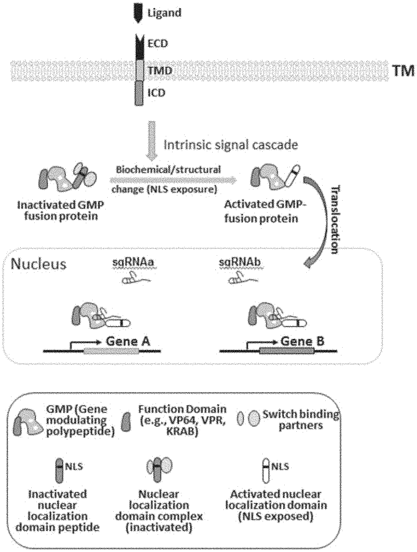
[0033] FIG. 1 depicts an example of inducible gene regulation controlled by receptor activation. In the depicted example, interaction of a ligand with its corresponding receptor, which is comprised of extracellular domain (ECD), transmembrane domain (TMD), and intracellular domain (ICD), activates intrinsic signal transduction pathways. The signal cascade leads to biochemical or structural changes of a fusion protein, which is comprised of a gene modulating polypeptide (GMP) and a heterologous nuclear localization domain, to allow the fusion protein to translocate into a nucleus to regulate the expression of target genes. In this example, the ability of the heterologous nuclear localization domain to translocate into the nucleus is controllable by the presence or absence of the ligand-receptor interaction.
[0034] FIG. 2 depicts an example of inducible gene regulation controlled by receptor activation. In the depicted example, interaction of an antigen with its corresponding CAR, which is comprised of extracellular single chain antibody variable fragment (scFv), spacer, transmembrane (TM) domain, and intracellular signal domain 1 and 2, activates intrinsic TCR signal transduction pathways. The signal cascade leads to dephosphorylation of the NFAT component of a NFAT-dCas9-VP64 fusion protein, which induces conformational changes or dissociation of inhibitory binding partners from the NFAT part. Hence, the nuclear localization signal (NLS) peptide becomes exposed to allow the fusion protein to translocate into the nucleus. Subsequently, the dCas9-VP64 portion of the fusion protein combines with target-specific single guide RNAs (sgRNAs) to regulate the expression of target genes. In this example, the ability of the NFAT protein domain to translocate into the nucleus is controlled by the CAR-activation signal.
[0035] FIGS. 3A and 3B depict example data generated using the system depicted in FIG. 2.
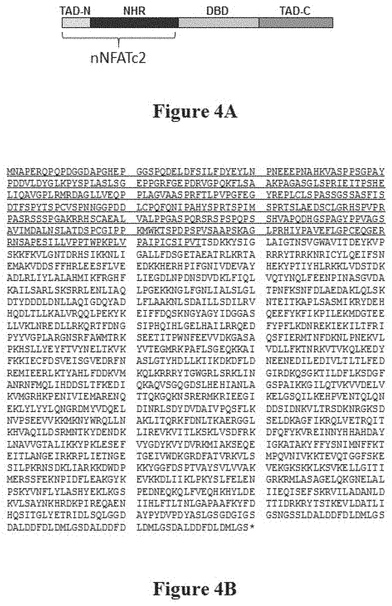
[0036] FIG. 4A depicts a diagram of functional domains of the NFATc2 protein. The NFATc2 protein comprises the following 4 functional domains: N-terminal transactivation domain (TAD-N), NFAT-homology region (NHR), DNA-binding domain (DBD), and C-terminal transactivation domain (TAD-C). The N-terminal portion of NFATc2 (nNFATc2) is used as a component in some embodiments disclosed herein.
[0037] FIG. 4B depicts the amino acid sequence of an example nNFATc2-dCas9-VP64 construct (SEQ ID NO: 1). The N-terminal portion of NFATc2, which is fused to dCas9 protein in this example, is underlined.
[0038] FIG. 5 depicts example data generated using the example system depicted in FIG. 2 for gene downregulation in which KRAB is used as effector domain instead of VP64.
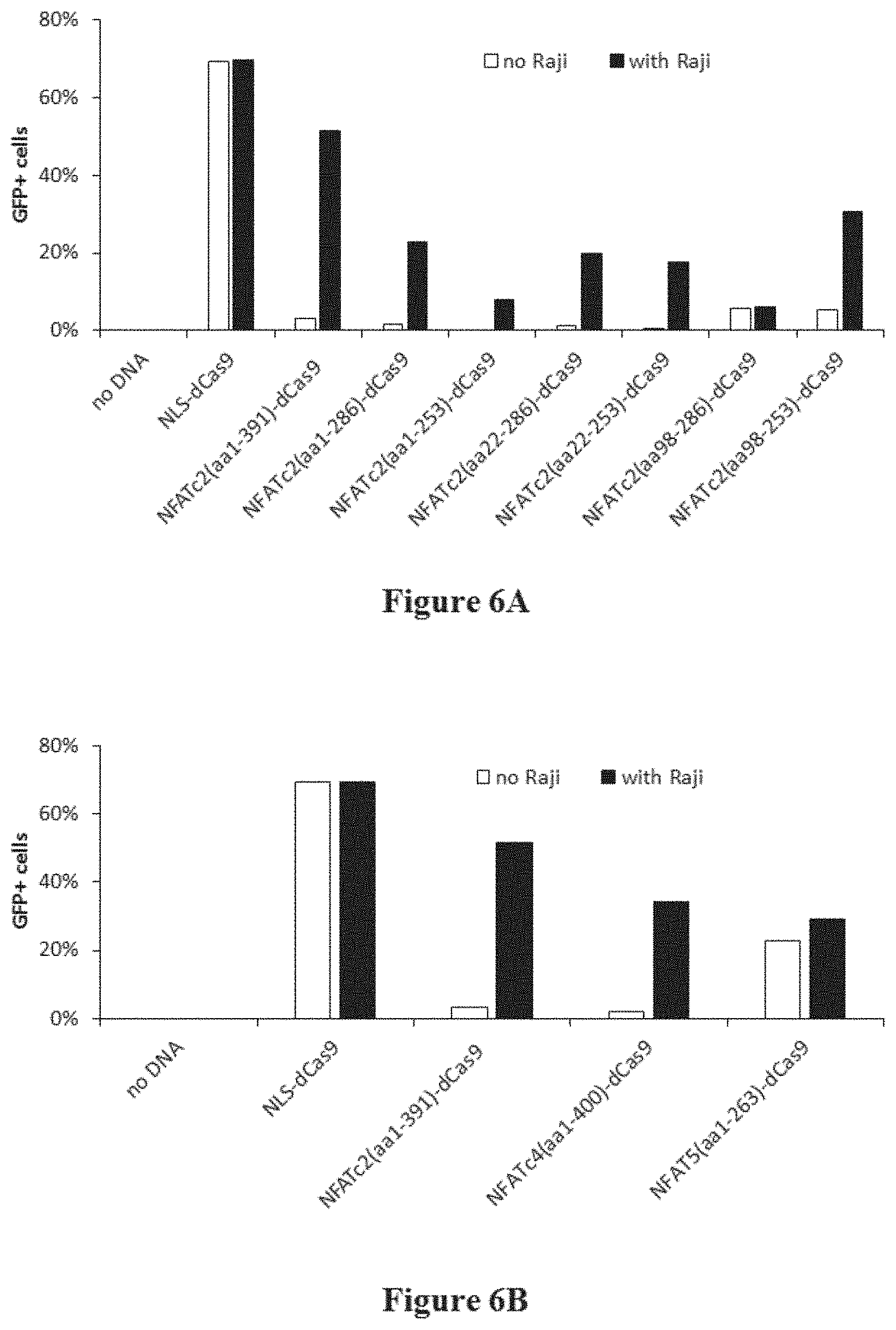
[0039] FIGS. 6A and 6B depict example data generated using dCas9 fused with either smaller NFATc2 variants or other NFAT family proteins.
[0040] FIG. 7 depicts example data generated using dCas9 fused with RelA protein instead of NFATc2.
[0041] FIG. 8 depicts an example of inducible gene regulation controlled by electromagnetic radiation. In the depicted example, the electromagnetic radiation activates a signaling unit, which is comprised of a transmembrane protein (ORAI1) and an intracellular chimeric protein (LOV2-J.alpha.-SOAR/CAD). Administration of the electromagnetic radiation induces a conformational change in the intracellular chimeric protein to expose an active site. The active site activates the transmembrane protein, which in turn induces a cellular signaling pathway (e.g., calcium dependent activation of calcineurin). The induced cellular signaling pathway leads to dephosphorylation of the NFAT componenet of an NFAT-dCas9-repressor/activator fusion protein. The nuclear localization signal (NLS) domain of the NFAT component can become exposed to allow the fusion protein to translocate into the nucleus. Subsequently, the dCas9-repressor/activator portion of the fusion protein combines with target-specific single guide RNAs (sgRNAs) to regulate the expression of target genes.
DETAILED DESCRIPTION OF THE INVENTION
[0042] The practice of some methods disclosed herein employ, unless otherwise indicated, conventional techniques of immunology, biochemistry, chemistry, molecular biology, microbiology, cell biology, genomics and recombinant DNA, which are within the skill of the art. See for example Sambrook and Green, Molecular Cloning: A Laboratory Manual, 4th Edition (2012); the series Current Protocols in Molecular Biology (F. M. Ausubel, et al. eds.); the series Methods In Enzymology (Academic Press, Inc.), PCR 2: A Practical Approach (M. J. MacPherson, B. D. Hames and G. R. Taylor eds. (1995)), Harlow and Lane, eds. (1988) Antibodies, A Laboratory Manual, and Culture of Animal Cells: A Manual of Basic Technique and Specialized Applications, 6th Edition (R.I. Freshney, ed. (2010)).
Definitions
[0043] As used in the specification and claims, the singular forms "a," "an," and "the" include plural references unless the context clearly dictates otherwise. For example, the term "a chimeric transmembrane receptor polypeptide" includes a plurality of chimeric transmembrane receptor polypeptides.
[0044] The term "about" or "approximately" means within an acceptable error range for the particular value as determined by one of ordinary skill in the art, which will depend in part on how the value is measured or determined, i.e., the limitations of the measurement system. For example, "about" can mean within 1 or more than 1 standard deviation, per the practice in the art. Alternatively, "about" can mean a range of up to 20%, up to 10%, up to 5%, or up to 1% of a given value. Alternatively, particularly with respect to biological systems or processes, the term can mean within an order of magnitude, preferably within 5-fold, and more preferably within 2-fold, of a value. Where particular values are described in the application and claims, unless otherwise stated, the term "about" meaning within an acceptable error range for the particular value should be assumed.
[0045] As used herein, a "cell" can generally refer to a biological cell. A cell can be the basic structural, functional and/or biological unit of a living organism. A cell can originate from any organism having one or more cells. Some non-limiting examples include: a prokaryotic cell, eukaryotic cell, a bacterial cell, an archaeal cell, a cell of a single-cell eukaryotic organism, a protozoa cell, a cell from a plant (e.g. cells from plant crops, fruits, vegetables, grains, soy bean, corn, maize, wheat, seeds, tomatoes, rice, cassava, sugarcane, pumpkin, hay, potatoes, cotton, cannabis, tobacco, flowering plants, conifers, gymnosperms, ferns, clubmosses, hornworts, liverworts, mosses), an algal cell, (e.g., Botryococcus braunii, Chlamydomonas reinhardtii, Nannochloropsis gaditana, Chlorella pyrenoidosa, Sargassum patens C. Agardh, and the like), seaweeds (e.g. kelp), a fungal cell (e.g., a yeast cell, a cell from a mushroom), an animal cell, a cell from an invertebrate animal (e.g. fruit fly, cnidarian, echinoderm, nematode, etc.), a cell from a vertebrate animal (e.g., fish, amphibian, reptile, bird, mammal), a cell from a mammal (e.g., a pig, a cow, a goat, a sheep, a rodent, a rat, a mouse, a non-human primate, a human, etc.), and etcetera. Sometimes a cell is not orginating from a natural organism (e.g. a cell can be a synthetically made, sometimes termed an artificial cell).
[0046] The term "antigen," as used herein, refers to a molecule or a fragment thereof capable of being bound by a selective binding agent. As an example, an antigen can be a ligand that can be bound by a selective binding agent such as a receptor. As another example, an antigen can be an antigenic molecule that can be bound by a selective binding agent such as an immunological protein (e.g., an antibody). An antigen can also refer to a molecule or fragment thereof capable of being used in an animal to produce antibodies capable of binding to that antigen.
[0047] The term "antibody," as used herein, refers to a proteinaceous binding molecule with immunoglobulin-like functions. The term antibody includes antibodies (e.g., monoclonal and polyclonal antibodies), as well as derivatives, variants, and fragments thereof. Antibodies include, but are not limited to, immunoglobulins (Ig's) of different classes (i.e. IgA, IgG, IgM, IgD and IgE) and subclasses (such as IgG1, IgG2, etc.). A derivative, variant or fragment thereof can refer to a functional derivative or fragment which retains the binding specificity (e.g., complete and/or partial) of the corresponding antibody. Antigen-binding fragments include Fab, Fab', F(ab').sub.2, variable fragment (Fv), single chain variable fragment (scFv), minibodies, diabodies, and single-domain antibodies ("sdAb" or "nanobodies" or "camelids"). The term antibody includes antibodies and antigen-binding fragments of antibodies that have been optimized, engineered or chemically conjugated. Examples of antibodies that have been optimized include affinity-matured antibodies. Examples of antibodies that have been engineered include Fc optimized antibodies (e.g., antibodies optimized in the fragment crystallizable region) and multispecific antibodies (e.g., bispecific antibodies).
[0048] The terms "Fc receptor" or "FcR," as used herein, generally refers to a receptor, or any derivative, variant or fragment thereof, that can bind to the Fc region of an antibody. In certain embodiments, the FcR is one which binds an IgG antibody (a gamma receptor, Fcgamma R) and includes receptors of the Fcgamma RI (CD64), Fcgamma RII (CD32), and Fcgamma RIII (CD16) subclasses, including allelic variants and alternatively spliced forms of these receptors. Fcgamma RII receptors include Fcgamma RIIA (an "activating receptor") and Fcgamma RIIB (an "inhibiting receptor"), which have similar amino acid sequences that differ primarily in the cytoplasmic domains thereof. The term "FcR" also includes the neonatal receptor, FcRn, which is responsible for the transfer of maternal IgGs to the fetus.
[0049] The term "nucleotide," as used herein, generally refers to a base-sugar-phosphate combination. A nucleotide can comprise a synthetic nucleotide. A nucleotide can comprise a synthetic nucleotide analog. Nucleotides can be monomeric units of a nucleic acid sequence (e.g. deoxyribonucleic acid (DNA) and ribonucleic acid (RNA)). The term nucleotide can include ribonucleoside triphosphates adenosine triphosphate (ATP), uridine triphosphate (UTP), cytosine triphosphate (CTP), guanosine triphosphate (GTP) and deoxyribonucleoside triphosphates such as dATP, dCTP, dITP, dUTP, dGTP, dTTP, or derivatives thereof. Such derivatives can include, for example, [aS]dATP, 7-deaza-dGTP and 7-deaza-dATP, and nucleotide derivatives that confer nuclease resistance on the nucleic acid molecule containing them. The term nucleotide as used herein can refer to dideoxyribonucleoside triphosphates (ddNTPs) and their derivatives. Illustrative examples of dideoxyribonucleoside triphosphates can include, but are not limited to, ddATP, ddCTP, ddGTP, ddITP, and ddTTP. A nucleotide can be unlabeled or detectably labeled by well-known techniques. Labeling can also be carried out with quantum dots. Detectable labels can include, for example, radioactive isotopes, fluorescent labels, chemiluminescent labels, bioluminescent labels and enzyme labels. Fluorescent labels of nucleotides can include but are not limited fluorescein, 5-carboxyfluorescein (FAM), 2'7'-dimethoxy-4'5-dichloro-6-carboxyfluorescein (JOE), rhodamine, 6-carboxyrhodamine (R6G), N,N,N',N'-tetramethyl-6-carboxyrhodamine (TAMRA), 6-carboxy-X-rhodamine (ROX), 4-(4'dimethylaminophenylazo) benzoic acid (DABCYL), Cascade Blue, Oregon Green, Texas Red, Cyanine and 5-(2'-aminoethyl)aminonaphthalene-1-sulfonic acid (EDANS). Specific examples of fluorescently labeled nucleotides can include [R6G]dUTP, [TAMRA]dUTP, [R110]dCTP, [R6G]dCTP, [TAMRA]dCTP, [JOE]ddATP, [R6G]ddATP, [FAM]ddCTP, [R110]ddCTP, [TAMRA]ddGTP, [ROX]ddTTP, [dR6G]ddATP, [dR110]ddCTP, [dTAMRA]ddGTP, and [dROX]ddTTP available from Perkin Elmer, Foster City, Calif.; FluoroLink DeoxyNucleotides, FluoroLink Cy3-dCTP, FluoroLink Cy5-dCTP, FluoroLink Fluor X-dCTP, FluoroLink Cy3-dUTP, and FluoroLink Cy5-dUTP available from Amersham, Arlington Heights, Ill.; Fluorescein-15-dATP, Fluorescein-12-dUTP, Tetramethyl-rodamine-6-dUTP, IR770-9-dATP, Fluorescein-12-ddUTP, Fluorescein-12-UTP, and Fluorescein-15-2'-dATP available from Boehringer Mannheim, Indianapolis, Ind.; and Chromosome Labeled Nucleotides, BODIPY-FL-14-UTP, BODIPY-FL-4-UTP, BODIPY-TMR-14-UTP, BODIPY-TMR-14-dUTP, BODIPY-TR-14-UTP, BODIPY-TR-14-dUTP, Cascade Blue-7-UTP, Cascade Blue-7-dUTP, fluorescein-12-UTP, fluorescein-12-dUTP, Oregon Green 488-5-dUTP, Rhodamine Green-5-UTP, Rhodamine Green-5-dUTP, tetramethylrhodamine-6-UTP, tetramethylrhodamine-6-dUTP, Texas Red-5-UTP, Texas Red-5-dUTP, and Texas Red-12-dUTP available from Molecular Probes, Eugene, Oreg. Nucleotides can also be labeled or marked by chemical modification. A chemically-modified single nucleotide can be biotin-dNTP. Some non-limiting examples of biotinylated dNTPs can include, biotin-dATP (e.g., bio-N6-ddATP, biotin-14-dATP), biotin-dCTP (e.g., biotin-11-dCTP, biotin-14-dCTP), and biotin-dUTP (e.g. biotin-11-dUTP, biotin-16-dUTP, biotin-20-dUTP).
[0050] The terms "polynucleotide," "oligonucleotide," and "nucleic acid" are used interchangeably to refer to a polymeric form of nucleotides of any length, either deoxyribonucleotides or ribonucleotides, or analogs thereof, either in single-, double-, or multi-stranded form. A polynucleotide can be exogenous or endogenous to a cell. A polynucleotide can exist in a cell-free environment. A polynucleotide can be a gene or fragment thereof. A polynucleotide can be DNA. A polynucleotide can be RNA. A polynucleotide can have any three dimensional structure, and can perform any function, known or unknown. A polynucleotide can comprise one or more analogs (e.g. altered backbone, sugar, or nucleobase). If present, modifications to the nucleotide structure can be imparted before or after assembly of the polymer. Some non-limiting examples of analogs include: 5-bromouracil, peptide nucleic acid, xeno nucleic acid, morpholinos, locked nucleic acids, glycol nucleic acids, threose nucleic acids, dideoxynucleotides, cordycepin, 7-deaza-GTP, fluorophores (e.g. rhodamine or fluorescein linked to the sugar), thiol containing nucleotides, biotin linked nucleotides, fluorescent base analogs, CpG islands, methyl-7-guanosine, methylated nucleotides, inosine, thiouridine, pseudourdine, dihydrouridine, queuosine, and wyosine. Non-limiting examples of polynucleotides include coding or non-coding regions of a gene or gene fragment, loci (locus) defined from linkage analysis, exons, introns, messenger RNA (mRNA), transfer RNA (tRNA), ribosomal RNA (rRNA), short interfering RNA (siRNA), short-hairpin RNA (shRNA), micro-RNA (miRNA), ribozymes, cDNA, recombinant polynucleotides, branched polynucleotides, plasmids, vectors, isolated DNA of any sequence, isolated RNA of any sequence, cell-free polynucleotides including cell-free DNA (cfDNA) and cell-free RNA (cfRNA), nucleic acid probes, and primers. The sequence of nucleotides can be interrupted by non-nucleotide components.
[0051] The term "gene," as used herein, refers to a nucleic acid (e.g., DNA such as genomic DNA and cDNA) and its corresponding nucleotide sequence that is involved in encoding an RNA transcript. The term as used herein with reference to genomic DNA includes intervening, non-coding regions as well as regulatory regions and can include 5' and 3' ends. In some uses, the term encompasses the transcribed sequences, including 5' and 3' untranslated regions (5'-UTR and 3'-UTR), exons and introns. In some genes, the transcribed region will contain "open reading frames" that encode polypeptides. In some uses of the term, a "gene" comprises only the coding sequences (e.g., an "open reading frame" or "coding region") necessary for encoding a polypeptide. In some cases, genes do not encode a polypeptide, for example, ribosomal RNA genes (rRNA) and transfer RNA (tRNA) genes. In some cases, the term "gene" includes not only the transcribed sequences, but in addition, also includes non-transcribed regions including upstream and downstream regulatory regions, enhancers and promoters. A gene can refer to an "endogenous gene" or a native gene in its natural location in the genome of an organism. A gene can refer to an "exogenous gene" or a non-native gene. A non-native gene can refer to a gene not normally found in the host organism but which is introduced into the host organism by gene transfer. A non-native gene can also refer to a gene not in its natural location in the genome of an organism. A non-native gene can also refer to a naturally occurring nucleic acid or polypeptide sequence that comprises mutations, insertions and/or deletions (e.g., non-native sequence).
[0052] The terms "target polynucleotide" and "target nucleic acid," as used herein, refer to a nucleic acid or polynucleotide which is targeted by an actuator moiety of the present disclosure. A target polynucleotide can be DNA (e.g., endogenous or exogenous). DNA can refer to template to generate mRNA transcripts and/or the various regulatory regions which regulate transcription of mRNA from a DNA template. A target polynucleotide can be a portion of a larger polynucleotide, for example a chromosome or a region of a chromosome. A target polynucleotide can refer to an extrachromosomal sequence (e.g., an episomal sequence, a minicircle sequence, a mitochondrial sequence, a chloroplast sequence, etc.) or a region of an extrachromosomal sequence. A target polynucleotide can be RNA. RNA can be, for example, mRNA which can serve as template encoding for proteins. A target polynucleotide comprising RNA can include the various regulatory regions which regulate translation of protein from an mRNA template. A target polynucleotide can encode for a gene product (e.g., DNA encoding for an RNA transcript or RNA encoding for a protein product) or comprise a regulatory sequence which regulates expression of a gene product. In general, the term "target sequence" refers to a nucleic acid sequence on a single strand of a target nucleic acid. The target sequence can be a portion of a gene, a regulatory sequence, genomic DNA, cell free nucleic acid including cfDNA and/or cfRNA, cDNA, a fusion gene, and RNA including mRNA, miRNA, rRNA, and others. A target polynucleotide, when targeted by an actuator moiety, can result in altered gene expression and/or activity. A target polynucleotide, when targeted by an actuator moiety, can result in an edited nucleic acid sequence. A target nucleic acid can comprise a nucleic acid sequence that may not be related to any other sequence in a nucleic acid sample by a single nucleotide substitution. A target nucleic acid can comprise a nucleic acid sequence that may not be related to any other sequence in a nucleic acid sample by a 2, 3, 4, 5, 6, 7, 8, 9, or 10 nucleotide substitutions. In some embodiments, the substitution may not occur within 5, 10, 15, 20, 25, 30, or 35 nucleotides of the 5' end of a target nucleic acid. In some embodiments, the substitution may not occur within 5, 10, 15, 20, 25, 30, 35 nucleotides of the 3' end of a target nucleic acid.
[0053] The term "expression" refers to one or more processes by which a polynucleotide is transcribed from a DNA template (such as into an mRNA or other RNA transcript) and/or the process by which a transcribed mRNA is subsequently translated into peptides, polypeptides, or proteins. Transcripts and encoded polypeptides can be collectively referred to as "gene product." If the polynucleotide is derived from genomic DNA, expression can include splicing of the mRNA in a eukaryotic cell. "Up-regulated," with reference to expression, generally refers to an increased expression level of a polynucleotide (e.g., RNA such as mRNA) and/or polypeptide sequence relative to its expression level in a wild-type state while "down-regulated" generally refers to a decreased expression level of a polynucleotide (e.g., RNA such as mRNA) and/or polypeptide sequence relative to its expression in a wild-type state.
[0054] The terms "complement," "complements," "complementary," and "complementarity," as used herein, generally refer to a sequence that is fully complementary to and hybridizable to the given sequence. In some cases, a sequence hybridized with a given nucleic acid is referred to as the "complement" or "reverse-complement" of the given molecule if its sequence of bases over a given region is capable of complementarily binding those of its binding partner, such that, for example, A-T, A-U, G-C, and G-U base pairs are formed. In general, a first sequence that is hybridizable to a second sequence is specifically or selectively hybridizable to the second sequence, such that hybridization to the second sequence or set of second sequences is preferred (e.g. thermodynamically more stable under a given set of conditions, such as stringent conditions commonly used in the art) to hybridization with non-target sequences during a hybridization reaction. Typically, hybridizable sequences share a degree of sequence complementarity over all or a portion of their respective lengths, such as between 25%-100% complementarity, including at least 25%, 30%, 35%, 40%, 45%, 50%, 55%, 60%, 65%, 70%, 75%, 80%, 85%, 90%, 91%, 92%, 93%, 94%, 95%, 96%, 97%, 98%, 99%, and 100% sequence complementarity. Sequence identity, such as for the purpose of assessing percent complementarity, can be measured by any suitable alignment algorithm, including but not limited to the Needleman-Wunsch algorithm (see e.g. the EMBOSS Needle aligner available at www.ebi.ac.uk/Tools/psa/emboss needle/nucleotide.html, optionally with default settings), the BLAST algorithm (see e.g. the BLAST alignment tool available at blast.ncbi.nlm.nih.gov/Blast.cgi, optionally with default settings), or the Smith-Waterman algorithm (see e.g. the EMBOSS Water aligner available at www.ebi.ac.uk/Tools/psa/emboss water/nucleotide.html, optionally with default settings). Optimal alignment can be assessed using any suitable parameters of a chosen algorithm, including default parameters.
[0055] Complementarity can be perfect or substantial/sufficient. Perfect complementarity between two nucleic acids can mean that the two nucleic acids can form a duplex in which every base in the duplex is bonded to a complementary base by Watson-Crick pairing. Substantial or sufficient complementary can mean that a sequence in one strand is not completely and/or perfectly complementary to a sequence in an opposing strand, but that sufficient bonding occurs between bases on the two strands to form a stable hybrid complex in set of hybridization conditions (e.g., salt concentration and temperature). Such conditions can be predicted by using the sequences and standard mathematical calculations to predict the Tm of hybridized strands, or by empirical determination of Tm by using routine methods.
[0056] The term "regulating" with reference to expression or activity, as used herein, refers to altering the level of expression or activity. Regulation can occur at the transcriptional level, post-transcriptional level, translational level, and/or post-translational leve.
[0057] The terms "peptide," "polypeptide," and "protein" are used interchangeably herein to refer to a polymer of at least two amino acid residues joined by peptide bond(s). This term does not connote a specific length of polymer, nor is it intended to imply or distinguish whether the peptide is produced using recombinant techniques, chemical or enzymatic synthesis, or is naturally occurring. The terms apply to naturally occurring amino acid polymers as well as amino acid polymers comprising at least one modified amino acid. In some cases, the polymer can be interrupted by non-amino acids. The terms include amino acid chains of any length, including full length proteins, and proteins with or without secondary and/or tertiary structure (e.g., domains). The terms also encompass an amino acid polymer that has been modified, for example, by disulfide bond formation, glycosylation, lipidation, acetylation, phosphorylation, oxidation, and any other manipulation such as conjugation with a labeling component. The terms "amino acid" and "amino acids," as used herein, generally refer to natural and non-natural amino acids, including, but not limited to, modified amino acids and amino acid analogues. Modified amino acids can include natural amino acids and non-natural amino acids, which have been chemically modified to include a group or a chemical moiety not naturally present on the amino acid. Amino acid analogues can refer to amino acid derivatives. The term "amino acid" includes both D-amino acids and L-amino acids.
[0058] The term "variant," when used herein with reference to a polypeptide, refers to a polypeptide related, but not identical, to a wild type polypeptide, for example either by amino acid sequence, structure (e.g., secondary and/or tertiary), activity (e.g., enzymatic activity) and/or function. Variants include polypeptides comprising one or more amino acid variations (e.g., mutations, insertions, and deletions), truncations, modifications, or combinations thereof compared to a wild type polypeptide. Variants also include derivatives of the wild type polypeptide and fragments of the wild type polypeptide.
[0059] The term "percent (%) identity," as used herein, refers to the percentage of amino acid (or nucleic acid) residues of a candidate sequence that are identical to the amino acid (or nucleic acid) residues of a reference sequence after aligning the sequences and introducing gaps, if necessary, to achieve the maximum percent identity (i.e., gaps can be introduced in one or both of the candidate and reference sequences for optimal alignment and non-homologous sequences can be disregarded for comparison purposes). Alignment, for purposes of determining percent identity, can be achieved in various ways that are within the skill in the art, for instance, using publicly available computer software such as BLAST, ALIGN, or Megalign (DNASTAR) software. Percent identity of two sequences can be calculated by aligning a test sequence with a comparison sequence using BLAST, determining the number of amino acids or nucleotides in the aligned test sequence that are identical to amino acids or nucleotides in the same position of the comparison sequence, and dividing the number of identical amino acids or nucleotides by the number of amino acids or nucleotides in the comparison sequence.
[0060] The term "gene modulating polypeptide" or "GMP," as used herein, refers to a polypeptide comprising at least an actuator moiety capable of regulating expression or activity of a gene and/or editing a nucleic acid sequence. A GMP can comprise additional peptide sequences which are not directly involved in modulating gene expression, for example linker sequences, targeting sequences, etc.
[0061] The term "actuator moiety," as used herein, refers to a moiety which can regulate expression or activity of a gene and/or edit a nucleic acid sequence, whether exogenous or endogenous. An actuator moiety can regulate expression of a gene at the transcriptional level, post-transcriptional level, translational level, and/or post-translation level. An actuator moiety can regulate gene expression at the transcription level, for example, by regulating the production of mRNA from DNA, such as chromosomal DNA or cDNA. In some embodiments, an actuator moiety recruits at least one transcription factor that binds to a specific DNA sequence, thereby controlling the rate of transcription of genetic information from DNA to mRNA. An actuator moiety can itself bind to DNA and regulate transcription by physical obstruction, for example preventing proteins such as RNA polymerase and other associated proteins from assembling on a DNA template. An actuator moiety can regulate expression of a gene at the translation level, for example, by regulating the production of protein from mRNA template. In some embodiments, an actuator moiety regulates gene expression at a post-transcriptional level by affecting the stability of an mRNA transcript. In some embodiments, an actuator moiety regulates gene expression at a post-translational level by altering the polypeptide modification, such as glycosylation of newly synthesized protein. In some embodiments, an actuator moiety regulates expression of a gene by editing a nucleic acid sequence (e.g., a region of a genome). In some embodiments, an actuator moiety regulates expression of a gene by editing an mRNA template. Editing a nucleic acid sequence can, in some cases, alter the underlying template for gene expression.
[0062] A Cas protein referred to herein can be a type of protein or polypeptide. A Cas protein can refer to a nuclease. A Cas protein can refer to an endoribonuclease. A Cas protein can refer to any modified (e.g., shortened, mutated, lengthened) polypeptide sequence or homologue of the Cas protein. A Cas protein can be codon optimized. A Cas protein can be a codon-optimized homologue of a Cas protein. A Cas protein can be enzymatically inactive, partially active, constitutively active, fully active, inducible active and/or more active, (e.g. more than the wild type homologue of the protein or polypeptide.). A Cas protein can be a Type II Cas protein. A Cas protein can be Cas9. A Cas protein can be a Type V Cas protein. A Cas protein can be Cpf1 or Cas12a. A Cas protein can be C2c1. A Cas protein can be C2c3. A Cas protein can be a Type VI Cas protein. A Cas protein can be C2c2 or Cas13a. A Cas protein can be Cas13b. A Cas protein can be Cas13c. A Cas protein can be Cas13d. A Cas protein (e.g., variant, mutated, enzymatically inactive and/or conditionally enzymatically inactive site-directed polypeptide) can bind to a target nucleic acid. A Cas protein (e.g., variant, mutated, enzymatically inactive and/or conditionally enzymatically inactive endoribonuclease) can bind to a target RNA or DNA.
[0063] The term "crRNA," as used herein, can generally refer to a nucleic acid with at least about 5%, 10%, 20%, 30%, 40%, 50%, 60%, 70%, 80%, 90%, or 100% sequence identity and/or sequence similarity to a wild type exemplary crRNA (e.g., a crRNA from S. pyogenes). crRNA can generally refer to a nucleic acid with at most about 5%, 10%, 20%, 30%, 40%, 50%, 60%, 70%, 80%, 90%, or 100% sequence identity and/or sequence similarity to a wild type exemplary crRNA (e.g., a crRNA from S. pyogenes, S. aureus, etc). crRNA can refer to a modified form of a crRNA that can comprise a nucleotide change such as a deletion, insertion, or substitution, variant, mutation, or chimera. A crRNA can be a nucleic acid having at least about 60% sequence identity to a wild type exemplary crRNA (e.g., a crRNA from S. pyogenes, S. aureus, etc) sequence over a stretch of at least 6 contiguous nucleotides. For example, a crRNA sequence can be at least about 60% identical, at least about 65% identical, at least about 70% identical, at least about 75% identical, at least about 80% identical, at least about 85% identical, at least about 90% identical, at least about 95% identical, at least about 98% identical, at least about 99% identical, or 100% identical to a wild type exemplary crRNA sequence (e.g., a crRNA from S. pyogenes, S. aureus, etc) over a stretch of at least 6 contiguous nucleotides.
[0064] The term "tracrRNA," as used herein, can generally refer to a nucleic acid with at least about 5%, 10%, 20%, 30%, 40%, 50%, 60%, 70%, 80%, 90%, or 100% sequence identity and/or sequence similarity to a wild type exemplary tracrRNA sequence (e.g., a tracrRNA from S. pyogenes). tracrRNA can refer to a nucleic acid with at most about 5%, 10%, 20%, 30%, 40%, 50%, 60%, 70%, 80%, 90%, or 100% sequence identity and/or sequence similarity to a wild type exemplary tracrRNA sequence (e.g., a tracrRNA from S. pyogenes, S. aureus, etc). tracrRNA can refer to a modified form of a tracrRNA that can comprise a nucleotide change such as a deletion, insertion, or substitution, variant, mutation, or chimera. A tracrRNA can refer to a nucleic acid that can be at least about 60% identical to a wild type exemplary tracrRNA (e.g., a tracrRNA from S. pyogenes, S. aureus, etc) sequence over a stretch of at least 6 contiguous nucleotides. For example, a tracrRNA sequence can be at least about 60% identical, at least about 65% identical, at least about 70% identical, at least about 75% identical, at least about 80% identical, at least about 85% identical, at least about 90% identical, at least about 95% identical, at least about 98% identical, at least about 99% identical, or 100% identical to a wild type exemplary tracrRNA (e.g., a tracrRNA from S. pyogenes, S. aureus, etc) sequence over a stretch of at least 6 contiguous nucleotides.
[0065] As used herein, a "guide nucleic acid" can refer to a nucleic acid that can hybridize to another nucleic acid. A guide nucleic acid can be RNA. A guide nucleic acid can be DNA. The guide nucleic acid can be programmed to bind to a sequence of nucleic acid site-specifically. The nucleic acid to be targeted, or the target nucleic acid, can comprise nucleotides. The guide nucleic acid can comprise nucleotides. A portion of the target nucleic acid can be complementary to a portion of the guide nucleic acid. The strand of a double-stranded target polynucleotide that is complementary to and hybridizes with the guide nucleic acid can be called the complementary strand. The strand of the double-stranded target polynucleotide that is complementary to the complementary strand, and therefore may not be complementary to the guide nucleic acid can be called noncomplementary strand. A guide nucleic acid can comprise a polynucleotide chain and can be called a "single guide nucleic acid." A single guide nucleic acid can comprise a crRNA. A single guide nucleic acid can comprise a crRNA and a tracrRNA. A guide nucleic acid can comprise two polynucleotide chains and can be called a "double guide nucleic acid." A double guide nucleic acid can comprise a crRNA and a tracrRNA. If not otherwise specified, the term "guide nucleic acid" can be inclusive, referring to both single guide nucleic acids and double guide nucleic acids.
[0066] A guide nucleic acid can comprise a segment that can be referred to as a "nucleic acid-targeting segment" or a "nucleic acid-targeting sequence." A nucleic acid-targeting segment can comprise a sub-segment that can be referred to as a "protein binding segment" or "protein binding sequence" or "Cas protein binding segment".
[0067] The term "targeting sequence," as used herein, refers to a nucleotide sequence and the corresponding amino acid sequence which encodes a targeting polypeptide which mediates the localization (or retention) of a protein to a sub-cellular location, e.g., plasma membrane or membrane of a given organelle, nucleus, cytosol, mitochondria, endoplasmic reticulum (ER), Golgi, chloroplast, apoplast, peroxisome or other organelle. For example, a targeting sequence can direct a protein (e.g., a receptor polypeptide or an adaptor polypeptide) to a nucleus utilizing a nuclear localization signal (NLS); outside of a nucleus of a cell, for example to the cytoplasm, utilizing a nuclear export signal (NES); mitochondria utilizing a mitochondrial targeting signal; the endoplasmic reticulum (ER) utilizing an ER-retention signal; a peroxisome utilizing a peroxisomal targeting signal; plasma membrane utilizing a membrane localization signal; or combinations thereof.
[0068] As used herein, "nuclear localization domain" can refer to a nuclear localization signal or other sequence or domain capable of traversing a nuclear membrane, thereby entering the nucleus. A nuclear localization domain can be fused in-frame with a polypeptide, in which case the nuclear localization domain can be referred to as a "heterologous nuclear localization domain." A nuclear localization domain can have an inactive state, wherein it is unable to traverse a nuclear membrane, and therefore is unable to enter the nucleus. A nuclear localization domain can have an active state, wherein it is able to traverse a nuclear membrane, and therefore is able ti enter into the nucleus. When a heterologous nuclear domain is active and enters the nucleas, a polypeptide fused to the heterologous nuclear domain enters the nucleas as well. A nuclear localization domain can switch between an inactive state and an active state in response to an extracellular or intracellular signal.
[0069] As used herein, "fusion" can refer to a protein and/or nucleic acid comprising one or more non-native sequences (e.g., moieties). A fusion can comprise one or more of the same non-native sequences. A fusion can comprise one or more of different non-native sequences. A fusion can be a chimera. A fusion can comprise a nucleic acid affinity tag. A fusion can comprise a barcode. A fusion can comprise a peptide affinity tag. A fusion can provide for subcellular localization of the site-directed polypeptide (e.g., a nuclear localization signal (NLS) for targeting to the nucleus, a mitochondrial localization signal for targeting to the mitochondria, a chloroplast localization signal for targeting to a chloroplast, an endoplasmic reticulum (ER) retention signal, and the like). A fusion can provide a non-native sequence (e.g., affinity tag) that can be used to track or purify. A fusion can be a small molecule such as biotin or a dye such as Alexa fluor dyes, Cyanine3 dye, Cyanine5 dye.
[0070] A fusion can refer to any protein with a functional effect. For example, a fusion protein can comprise methyltransferase activity, demethylase activity, dismutase activity, alkylation activity, depurination activity, oxidation activity, pyrimidine dimer forming activity, integrase activity, transposase activity, recombinase activity, polymerase activity, ligase activity, helicase activity, photolyase activity or glycosylase activity, acetyltransferase activity, deacetylase activity, kinase activity, phosphatase activity, ubiquitin ligase activity, deubiquitinating activity, adenylation activity, deadenylation activity, SUMOylating activity, deSUMOylating activity, ribosylation activity, deribosylation activity, myristoylation activity, remodelling activity, protease activity, oxidoreductase activity, transferase activity, hydrolase activity, lyase activity, isomerase activity, synthase activity, synthetase activity, or demyristoylation activity. An effector protein can modify a genomic locus. A fusion protein can be a fusion of a Cas protein and a heterologous funcational domain. A fusion protein can be a non-native sequence fused to a Cas protein.
[0071] As used herein, "heterologous functional domain" can refer to a domain within a fusion protein, said domain comprising a functional activity. A heterologous functional domain can be a transcription activator. A heterologous functional domain can be a transcription repressor. A heterologous functional domain can comprise methyltransferase activity, demethylase activity, dismutase activity, alkylation activity, depurination activity, oxidation activity, pyrimidine dimer forming activity, integrase activity, transposase activity, recombinase activity, polymerase activity, ligase activity, helicase activity, photolyase activity or glycosylase activity, acetyltransferase activity, deacetylase activity, kinase activity, phosphatase activity, ubiquitin ligase activity, deubiquitinating activity, adenylation activity, deadenylation activity, SUMOylating activity, deSUMOylating activity, ribosylation activity, deribosylation activity, myristoylation activity, remodelling activity, protease activity, oxidoreductase activity, transferase activity, hydrolase activity, lyase activity, isomerase activity, synthase activity, synthetase activity, or demyristoylation activity. A heterologous functional domain can be a chromosome modification enzyme such as a methylase, demethylase, acetylase, deacetylase, deaminase, phosphorylase, dephosphorylase, histone modifying enzyme, or nucleotide modifying enzyme. A heterologous functional domain can be a histone modifying enzyme. A heterologous functional domain can be a nucleotide modifying enzyme.
[0072] As used herein, "non-native" can refer to a nucleic acid or polypeptide sequence that is not found in a native nucleic acid or protein. Non-native can refer to affinity tags. Non-native can refer to fusions. Non-native can refer to a naturally occurring nucleic acid or polypeptide sequence that comprises mutations, insertions and/or deletions. A non-native sequence may exhibit and/or encode for an activity (e.g., enzymatic activity, methyltransferase activity, acetyltransferase activity, kinase activity, ubiquitinating activity, etc.) that can also be exhibited by the nucleic acid and/or polypeptide sequence to which the non-native sequence is fused. A non-native nucleic acid or polypeptide sequence may be linked to a naturally-occurring nucleic acid or polypeptide sequence (or a variant thereof) by genetic engineering to generate a chimeric nucleic acid and/or polypeptide sequence encoding a chimeric nucleic acid and/or polypeptide.
[0073] The terms "subject," "individual," and "patient" are used interchangeably herein to refer to a vertebrate, preferably a mammal such as a human. Mammals include, but are not limited to, murines, simians, humans, farm animals, sport animals, and pets. Tissues, cells and their progeny of a biological entity obtained in vivo or cultured in vitro are also encompassed.
[0074] The terms "treatment" and "treating," as used herein, refer to an approach for obtaining beneficial or desired results including but not limited to a therapeutic benefit and/or a prophylactic benefit. For example, a treatment can comprise administering a system or cell population disclosed herein. By therapeutic benefit is meant any therapeutically relevant improvement in or effect on one or more diseases, conditions, or symptoms under treatment. For prophylactic benefit, a composition can be administered to a subject at risk of developing a particular disease, condition, or symptom, or to a subject reporting one or more of the physiological symptoms of a disease, even though the disease, condition, or symptom may not have yet been manifested.
[0075] The term "effective amount" or "therapeutically effective amount" refers to the quantity of a composition, for example a composition comprising immune cells such as lymphocytes (e.g., T lymphocytes and/or NK cells) comprising a system of the present disclosure, that is sufficient to result in a desired activity upon administration to a subject in need thereof. Within the context of the present disclosure, the term "therapeutically effective" refers to that quantity of a composition that is sufficient to delay the manifestation, arrest the progression, relieve or alleviate at least one symptom of a disorder treated by the methods of the present disclosure.
[0076] The term "electromagnetic radiation," as used herein, refers to one or more wavelengths from the electromagnetic spectrum including, but not limited to x-rays (about 0.1 nanometers (nm) to about 10.0 nm; or about 10.sup.18 hertz (Hz) to about 10.sup.16 Hz), ultraviolet (UV) rays (about 10.0 nm to about 380 nm; or about 8.times.10.sup.16 Hz to about 10.sup.15 Hz), visible light (about 380 nm to about 750 nm; or about 8.times.10.sup.14 Hz to about 4.times.10.sup.14 Hz), infrared light (about 750 nm to about 0.1 centimeters (cm); or about 4.times.10.sup.14 Hz to about 5.times.10.sup.11 Hz), and microwaves (about 0.1 cm to about 100 cm; or about 10.sup.8 Hz to about 5.times.10.sup.11 Hz). In some cases, within the wavelength range of the UV rays, wavelengths of about 300 nm to about 380 nm may be referred to as "near" ultraviolet, wavelengths of about 200 nm to about 300 nm as "far" ultraviolet, and 10 about to about 200 nm as "extreme" ultraviolet. In some cases, within the wavelegnth range of the visible light, wavelegnths of about 380 nm to about 490 nm may be referred to as "blue" light.
[0077] The term "electromagnetic radiation source," as used herein, refers to a source that emits electromagnetic radiation. The electromagnetic radiation source may emit one or more wavlengths from the electromagnetic spectrum.
[0078] Extracellular Signal Mediated Gene Regulation
[0079] In an aspect, the present disclosure provides a system for regulating expression of a target polynucleotide in a cell, the system comprising: a chimeric polypeptide comprising a gene modulating polypeptide fused in-frame with a heterologous nuclear localization domain, wherein the nuclear localization domain is operable to translocate the chimeric polypeptide to a cell nucleus upon activation by an active cellular signaling pathway, the cellular signaling pathway activatable in response to an extracellular signal, wherein in response to the extracellular signal, the chimeric polypeptide localizes to the cell nucleus and the gene modulating polypeptide regulates expression of a target polynucleotide in the cell nucleus.
[0080] In an aspect, the present disclosure provides a system for regulating expression of a target polynucleotide in a cell, the system comprising: a) a chimeric receptor polypeptide that activates a cellular signaling pathway upon binding a ligand; and b) a chimeric polypeptide comprising a gene modulating polypeptide fused in-frame with a heterologous nuclear localization domain, the heterologous nuclear localization domain operable to translocate the chimeric polypeptide to a cell nucleus upon activation by an activated cellular signaling pathway, wherein upon binding of the ligand to the chimeric receptor polypeptide, the chimeric polypeptide localizes to the cell nucleus via the activated heterologous nuclear localization domain and the gene modulating polypeptide regulates expression of a target polynucleotide in the cell nucleus.
[0081] In an aspect, the present disclosure provides a system for regulating expression of a target polynucleotide in a cell, the system comprising: a) a cellular signaling pathway activator comprising a chemical compound; and b) a chimeric polypeptide comprising a gene modulating polypeptide fused in-frame with a heterologous nuclear localization domain, the heterologous nuclear localization domain operable to translocate the chimeric polypeptide to a cell nucleus upon activation by an activated cellular signaling pathway, wherein upon administration of the activator to a cell, the chimeric polypeptide localizes to a cell nucleus via the activated heterologous nuclear localization domain and the gene modulating polypeptide regulates expression of a target polynucleotide in the cell nucleus.
[0082] In an aspect, the present disclose provides a system for regulating expression of a target polynucleotide in a cell, the system comprising: a chimeric polypeptide comprising a gene modulating polypeptide fused in-frame with a heterologous nuclear localization domain, wherein said nuclear localization domain is operable to translocate said chimeric polypeptide to a cell nucleus upon activation by an active cellular signaling pathway, said cellular signaling pathway inducible in response to an extracellular signal, wherein in response to said extracellular signal, said chimeric polypeptide localizes to the cell nucleus and said gene modulating polypeptide regulates expression of a target polynucleotide in the cell nucleus.
[0083] In an aspect, the present disclose provides a system for regulating expression of a target polynucleotide in a cell, the system comprising: a) a chimeric receptor polypeptide that activates a cellular signaling pathway upon binding a ligand; and b) a chimeric polypeptide comprising a gene modulating polypeptide fused in-frame with a heterologous nuclear localization domain, said heterologous nuclear localization domain operable to translocate said chimeric polypeptide to a cell nucleus upon induction by a cellular signaling pathway, wherein upon binding of the ligand to the chimeric receptor polypeptide, said chimeric polypeptide localizes to the cell nucleus via the induced heterologous nuclear localization domain and said gene modulating polypeptide regulates expression of a target polynucleotide in the cell nucleus.
[0084] In an aspect, the present disclose provides a system for regulating expression of a target polynucleotide in a cell, the system comprising: a) a cellular signaling pathway activator comprising a chemical compound; and b) a chimeric polypeptide comprising a gene modulating polypeptide fused in-frame with a heterologous nuclear localization domain, said heterologous nuclear localization domain operable to translocate said chimeric polypeptide to a cell nucleus upon induction by a cellular signaling pathway, wherein upon administration of said activator to a cell, said chimeric polypeptide localizes to a cell nucleus via the activated heterologous nuclear localization domain and said gene modulating polypeptide regulates expression of a target polynucleotide in the cell nucleus.
[0085] In some embodiments, a gene modulating polypeptide is fused in-frame with a regulatable localization domain (e.g., a nuclaer localization domain). In some embodiments, a gene modulating polypeptide comprises an actuator moiety and the actuator moiety is fused in-frame with a regulatable localization domain. A regulatable localization domain can comprise a localization domain capable of being induced or activated, whereby said induction or activation allows the localization domain to localize to the intended location.
[0086] A regulatable localization domain can comprise a heterologous nuclear localization domain. A heterologous nuclear localization domain can comprise a nuclear localization signal.
[0087] A heterologous nuclear localization domain can have an active and inactive state. In an inactive state, the heterologous nuclear localization domain may be unable to enter a cell nucleus. In an active state, the heterologous nuclear localization domain may be able to enter the cell nucleus.
[0088] A heterologous nuclear localization domain can be derived from a transcription factor. The transcription factor can be a regulatable transcription factor that is only active and able to translocate into a nucleus in response to a signal or signaling pathway. The transcription factor can be a regulatable transcription factor that is primarily active and able to translocate into a nucleus in response to a signal or signaling pathway. The transcription factor can be a regulatable transcription factor that is generally active and able to translocate into a nucleus in response to a signal or signaling pathway.
[0089] A heterologous nuclear localization domain in some embodiments herein can be derived from a nuclear factor of activated T-cells (NFAT) family member. For example, the heterologous nuclear localization domain can be derived from NFATp, NFAT1, NFATc1, NFATc2, NFATc3, NFAT4, NFATx, NFATc4, NFAT3, or NFAT5.
[0090] A heterologous nuclear localization domain in some embodiments herein can be derived from nuclear factor kappa B (NF-.kappa.B), NFKB1 p50, activator protein 1 (AP-1), signal transducer and activator of transcription 1 (STAT1), STAT2, STAT3, STAT4, STATS (STAT5A and STAT5B), and STATE, Sterol Response Element-Binding Proteins (SREBPs; eg., SREBP-1 or SREBF1), or other transcription factors or signal transducers.
[0091] A heterologous nuclear localization domain in some embodiments herein can be derived from an intracellular receptor. An intrcellular receptor can be a hormone-activated receptor. A hormone-activated receptor can be an estrogen receptor, thyroid hormone receptor, steroid hormone receptor, a second messenger receptor, inositol triphosphate (IP3) receptor, intracrine peptide hormone receptor, or neurosteroid receptor.
[0092] A heterologous nuclear localization domain in some embodiments herein can be derived from a light or circadian or electromagnetic sensing protein such as cryptochromes (e.g., CRY1, CRY2), Timeless (TIM), PAS domain of PER proteins (e.g., PER1, PER2, and PER3).
[0093] A regulatable localization domain or heterologous nuclear localization domain can switch between an inactive and active state in response to a signal. The switch between an active and inactive state can be the result of a chemical modification. A chemical modification can be dephosphorylation, phosphorylation, demethylation, methylation, acetylation, deacetylation, deamination, ubiquitination, deubiquitination, proteolytic processing, or other suitable chemical modification. In some cases the chemical modification causes the switch from the inactive state to an active state. In some cases, the chemical modification causes a structural change or conformational change which causes the switch from the inactive state to the active state. In some cases, the structural or conformational change exposes part of the localization domain that was not exposed in the inactive state, thereby creating the active state.
[0094] A regulatable localization domain or heterologous nuclear localization domain can switch between an inactive and active state in response to a signal. The signal can be an induction signal or activation signal. The signal can be the result of a signal or signaling pathway. In some examples, the signaling pathway is activated or induced in a cell. The signal or signaling pathway can cause a chemical modification of the regulatable localization domain or heterologous nuclear localization domain. A chemical modification can be dephosphorylation, phosphorylation, demethylation, methylation, acetylation, deacetylation, deamination, ubiquitination, deubiquitination, proteolytic processing, or other suitable chemical modification. In some cases the chemical modification causes the switch from the inactive state to an active state. In some cases, the chemical modification causes a structural change or conformational change which causes the switch from the inactive state to the active state. In some cases, the structural or conformational change exposes part of the localization domain that was not exposed in the inactive state, thereby creating the active state.
[0095] In some embodiments, the extracellular singal (e.g., the ligand, the chemical compound, etc.), as abovementioned, can elevate intracellualr ion (e.g., sodium, potasisum, chloride, bicabonate, calcium, phosphate, etc.) concentration in a cell relative to a basal level of the intracellular ion in the cell without the prescene of the extracellular signal. In some cases, the extracellular ion can comprise calcium. In some cases, the extracellualr ion can be calcium. For certain localization domains or moieties (e.g., a nuclear localization domain, such as, for example, NFAT), regulation of intracellular ion signaling of the cell can be important to activation of the localization domains. For example, in the case of NFAT proteins, calmodulin (CaM), which can be a calcium sensor protein, can be activated by an increase in intracellular calcium level in the cell. Upon binding of one or more calcium ions to the CaM, the CaM in turn activates a serine/threonine phosphatase calcineurin (CN). Following, activated CN rapidly modifies one or more regions of the the NFAT proteins (e.g., dephosphorylate the serine-rich region (SRR) and SP-repeats in the amino termini of NFAT proteins), resulting in a conformational change that exposes a nuclear localization signal, resulting in NFAT nuclear import.
[0096] In some embodiments, the signaling pathway is activated or induced by a signal or signaling pathway. In some embodiments, the signaling pathway is activated or induced by a subject chimeric receptor or subject transmembrane receptor. In some embodiments, the signaling pathway is activated or induced by a signaling cascade which in turn is activated by a subject chimeric receptor or subject transmembrane receptor.
[0097] In some embodiments, the signaling pathway activated or induced in the cell which can switch the heterologous nuclear localization domain between an inactive and active state is the PI3K/AKT pathway. In some embodiments, the transmembrane receptor in this pathway comprises a receptor tyrosine kinase, integrin, B cell receptor, T cell receptor, cytokine receptor, or G-protein coupled receptor and the signaling pathway further involves PRKCE, ITGAM, ITGA5, IRAK1, PRKAA2, EIF2AK2, PTEN, EIF4E, PRKCZ, GRK6, MAPK1, TSC1, PLK1, AKT2, IKBKB, PIK3CA, CDK8, CDKN1B, NFKB2, BCL2, PIK3CB, PPP2R1A, MAPK8, BCL2L1, MAPK3, TSC2, ITGA1, KRAS, EIF4EBP1, RELA, PRKCD, NOS3, PRKAA1, MAPK9, CDK2, PPP2CA, PIM1, ITGB7, YWHAZ, ILK, TP53, RAF1, IKBKG, RELB, DYRK1A, CDKN1A, ITGB1, MAP2K2, JAK1, AKT1, JAK2, PIK3R1, CHUK, PDPK1, PPP2R5C, CTNNB1, MAP2K1, NFKB1, PAK3, ITGB3, CCND1, GSK3A, FRAP1, SFN, ITGA2, TTK, CSNK1A1, BRAF, GSK3B, AKT3, FOXO1, SGK, HSP90AA1, or RPS6KB1.
[0098] In some embodiments, the signaling pathway activated or induced in the cell which can switch the heterologous nuclear localization domain between an inactive and active state is the ERK/MAPK pathway. In some embodiments, the transmembrane receptor in this pathway comprises EGFR, Trk A/B, fibroblast growth factor receptor (FGFR) or platelet-derived growth factor receptor (PDGFR) and the signaling pathway further involves PRKCE, ITGAM, ITGA5, HSPB1, IRAK1, PRKAA2, EIF2AK2, RAC1, RAP1A, TLN1, EIF4E, ELK1, GRK6, MAPK1, RAC2, PLK1, AKT2, PIK3CA, CDK8, CREB1, PRKCI, PTK2, FOS, RPS6KA4, PIK3CB, PPP2R1A, PIK3C3, MAPK8, MAPK3, ITGA1, ETS1, KRAS, MYCN, EIF4EBP1, PPARG, PRKCD, PRKAA1, MAPK9, SRC, CDK2, PPP2CA, PIM1, PIK3C2A, ITGB7, YWHAZ, PPP1CC, KSR1, PXN, RAF1, FYN, DYRK1A, ITGB1, MAP2K2, PAK4, PIK3R1, STAT3, PPP2R5C, MAP2K1, PAK3, ITGB3, ESR1, ITGA2, MYC, TTK, CSNK1A1, CRKL, BRAF, ATF4, PRKCA, SRF, STAT1, or SGK.
[0099] In some embodiments, the signaling pathway activated or induced in the cell which can switch the heterologous nuclear localization domain between an inactive and active state is a glucocorticoid receptor signaling pathway. In some embodiments, the transmembrane receptor in this pathway comprises glucocorticoid receptor and the signaling pathway further invovles RAC1, TAF4B, EP300, SMAD2, TRAF6, PCAF, ELK1, MAPK1, SMAD3, AKT2, IKBKB, NCOR2, UBE2I, PIK3CA, CREB1, FOS, HSPA5, NFKB2, BCL2, MAP3K14, STAT5B, PIK3CB, PIK3C3, MAPK8, BCL2L1, MAPK3, TSC22D3, MAPK10, NRIP1, KRAS, MAPK13, RELA, STAT5A, MAPK9, NOS2A, PBX1, NR3C1, PIK3C2A, CDKN1C, TRAF2, SERPINEL NCOA3, MAPK14, TNF, RAF1, IKBKG, MAP3K7, CREBBP, CDKN1A, MAP2K2, JAK1, IL8, NCOA2, AKT1, JAK2, PIK3R1, CHUK, STAT3, MAP2K1, NFKB1, TGFBR1, ESR1, SMAD4, CEBPB, JUN, AR, AKT3, CCL2, MMP1, STAT1, IL6, or HSP90AA1.
[0100] In some embodiments, the signaling pathway activated or induced in the cell which can switch the heterologous nuclear localization domain between an inactive and active state is a B cell receptor signaling pathway. In some embodiments, the transmembrane receptor of this pathway comprises a B cell receptor and the signaling pathway further involves RAC1, PTEN, LYN, ELK1, MAPK1, RAC2, PTPN11, AKT2, IKBKB, PIK3CA, CREB1, SYK, NFKB2, CAMK2A, MAP3K14, PIK3CB, PIK3C3, MAPK8, BCL2L1, ABL1, MAPK3, ETS1, KRAS, MAPK13, RELA, PTPN6, MAPK9, EGR1, PIK3C2A, BTK, MAPK14, RAF1, IKBKG, RELB, MAP3K7, MAP2K2, AKT1, PIK3R1, CHUK, MAP2K1, NFKB1, CDC42, GSK3A, FRAP1, BCL6, BCL10, JUN, GSK3B, ATF4, AKT3, VAV3, or RPS6KB1.
[0101] In some embodiments, the signaling pathway activated or induced in the cell which can switch the heterologous nuclear localization domain between an inactive and active state is an integrin signaling pathway. In some embodiments, the transmembrane receptor of this pathway comprises an integrin or integrin subunit and the signaling pathway further involves ACTN4, ITGAM, ROCK1, ITGA5, RAC1, PTEN, RAP1A, TLN1, ARHGEF7, MAPK1, RAC2, CAPNS1, AKT2, CAPN2, PIK3CA, PTK2, PIK3CB, PIK3C3, MAPK8, CAV1, CAPN1, ABL1, MAPK3, ITGA1, KRAS, RHOA, SRC, PIK3C2A, ITGB7, PPP1CC, ILK, PXN, VASP, RAF1, FYN, ITGB1, MAP2K2, PAK4, AKT1, PIK3R1, TNK2, MAP2K1, PAK3, ITGB3, CDC42, RND3, ITGA2, CRKL, BRAF, GSK3B, or AKT3.
[0102] In some embodiments, the signaling pathway activated or induced in the cell which can switch the heterologous nuclear localization domain between an inactive and active state is an insulin receptor signaling pathway. In some embodiments, the transmembrane receptor of this pathway comprises an insulin receptor and the signaling pathway further invovles PTEN, INS, EIF4E, PTPN1, PRKCZ, MAPK1, TSC1, PTPN11, AKT2, CBL, PIK3CA, PRKCI, PIK3CB, PIK3C3, MAPK8, IRS1, MAPK3, TSC2, KRAS, EIF4, EBP1, SLC2A4, PIK3C2A, PPP1CC, INSR, RAF1, FYN, MAP2K2, JAK1, AKT1, JAK2, PIK3R1, PDPK1, MAP2K1, GSK3A, FRAP1, CRKL, GSK3B, AKT3, FOXO1, SGK, or RPS6KB1.
[0103] In some embodiments, the signaling pathway activated or induced in the cell which can switch the heterologous nuclear localization domain between an inactive and active state is a T cell receptor signaling pathway. In some embodiments, the transmembrane receptor in this pathway comprises a T cell receptor and the signaling pathway further involves RAC1, ELK1, MAPK1, IKBKB, CBL, PIK3CA, FOS, NFKB2, PIK3CB, PIK3C3, MAPK8, MAPK3, KRAS, RELA, PIK3C2A, BTK, LCK, RAF1, IKBKG, RELB, FYN, MAP2K2, PIK3R1, CHUK, MAP2K1, NFKB1, ITK, BCL10, JUN, or VAV3.
[0104] In some embodiments, the signaling pathway activated or induced in the cell which can switch the heterologous nuclear localization domain between an inactive and active state is a G-protein coupled receptor (GPCR) signaling pathway. In some embodiments, the transmembrane receptor of this pathway comprises a GPCR and the signaling pathway further invovles PRKCE, RAP1A, RGS16, MAPK1, GNAS, AKT2, IKBKB, PIK3CA, CREB1, GNAQ, NFKB2, CAMK2A, PIK3CB, PIK3C3, MAPK3, KRAS, RELA, SRC, PIK3C2A, RAF1, IKBKG, RELB, FYN, MAP2K2, AKT1, PIK3R1, CHUK, PDPK1, STAT3, MAP2K1, NFKB1, BRAF, ATF4, AKT3, or PRKCA.
[0105] A chimeric transmembrane receptor resulting from the joining of various regions, or domains, from different molecules can be different from the molecules from which the domains originated, for example structurally and functionally. However, the individual domains can, in some cases, retain the native structure and/or activity. For example, the individual domains may retain at least 5%, 10%, 15%, 20%, 25%, 30%, 35%, 40%, 45%, 50%, 55%, 60%, 65%, 70%, 75%, 80%, 85%, 90%, or 95% of the native structure and/or activity. For example, an extracellular region comprising a ligand binding domain can retain at least 5%, 10%, 15%, 20%, 25%, 30%, 35%, 40%, 45%, 50%, 55%, 60%, 65%, 70%, 75%, 80%, 85%, 90%, or 95% of the binding affinity of the molecule from which the extracellular region was derived. For further example, an intracellular region comprising a signaling domain can retain at least 5%, 10%, 15%, 20%, 25%, 30%, 35%, 40%, 45%, 50%, 55%, 60%, 65%, 70%, 75%, 80%, 85%, 90%, or 95% of the ability to activate a signaling pathway of the cell compared to the molecule from which the intracellular region was derived.
[0106] A chimeric transmembrane receptor polypeptide of an exemplary configuration can comprise (a) an extracellular region, (b) a transmembrane region, and (c) an intracellular region. In some embodiments, the extracellular region can comprise ligand interacting domain that binds a ligand, for example an antigen. In some embodiments, the intracellular region comprises a cell signaling domain. In some embodiments, the intracellular region comprises an immune cell signaling domain.
[0107] A ligand interacting domain of a chimeric transmembrane receptor polypeptide can comprise any protein or molecule that can bind to a ligand, for example an antigen. A ligand interacting domain of a chimeric transmembrane receptor polypeptide disclosed herein can be a monoclonal antibody, a polyclonal antibody, a recombinant antibody, a human antibody, a humanized antibody, or a functional derivative, variant or fragment thereof, including, but not limited to, a Fab, a Fab', a F(ab').sub.2, an Fv, a single-chain Fv (scFv), minibody, a diabody, and a single-domain antibody such as a heavy chain variable domain (VH), a light chain variable domain (VL) and a variable domain (VHH) of camelid derived Nanobody. In some embodiments, a ligand interacting domain comprises at least one of a Fab, a Fab', a F(ab').sub.2, an Fv, and a scFv. In some embodiments, a ligand interacting domain comprises an antibody mimetic. Antibody mimetics refer to molecules which can bind a target molecule with an affinity comparable to an antibody, and include single-chain binding molecules, cytochrome b562-based binding molecules, fibronectin or fibronectin-like protein scaffolds (e.g., adnectins), lipocalin scaffolds, calixarene scaffolds, A-domains and other scaffolds. In some embodiments, a ligand interacting domain comprises a transmembrane receptor, or any derivative, variant, or fragment thereof. For example, a ligand interacting domain can comprise at least a ligand binding domain of a transmembrane receptor.
[0108] In some embodiments, the ligand interacting domain comprises a humanized antibody. A humanized antibody can be produced using a variety of techniques including, but not limited to, CDR-grafting, veneering or resurfacing, chain shuffling, and other techniques. Human variable domains, including light and heavy chains, can be selected to reduce the immunogenicity of humanized antibodies. In some embodiments, the ligand interacting domain of a chimeric transmembrane receptor polypeptide comprises a fragment of a humanized antibody which binds an antigen with high affinity and possesses other favorable biological properties, such as reduced and/or minimal immunogenicity. A humanized antibody or antibody fragment can retain a similar antigenic specificity as the corresponding non-humanized antibody.
[0109] In some embodiments, the ligand interacting domain comprises a single-chain variable fragment (scFv). scFv molecules can be produced by linking the heavy chain (VH) and light chain (VL) regions of immunoglobulins together using flexible linkers, such as polypeptide linkers. scFvs can be prepared according to various methods.
[0110] In some embodiments, the ligand interacting domain is engineered to bind a specific target antigen. For example, the ligand interacting domain can be an engineered scFv. A ligand interacting domain comprising a scFv can be engineered using a variety of methods, including but not limited to display libraries such as phage display libraries, yeast display libraries, cell based display libraries (e.g., mammalian cells), protein-nucleic acid fusions, ribosome display libraries, and/or an E. coli periplasmic display libraries. In some embodiments, a ligand interacting domain which is engineered may bind to an antigen with a higher affinity than an analogous antibody or an antibody which has not undergone engineering.
[0111] In some embodiments, the ligand interacting domain binds multiple antigens, e.g., at least 2, 3, 4, 5, 6, 7, 8, 9, or 10 antigens. A ligand interacting domain can bind two related antigens, such as two subtypes of botulin toxin (e.g., botulinum neurotoxin subtype A1 and subtype A2). A ligand interacting domain can bind two unrelated proteins, such as receptor tyrosine kinase erbB-2 (also referred to as Neu, ERBB2, and HER2) and vascular endothelial growth factor (VEGF). An antigen interacting domain capable of binding two antigens can comprise an antibody engineered to bind two unrelated protein targets at distinct but overlapping sites of the antibody. In some embodiments, an antigen interacting domain which binds multiple antigens comprises a bispecific antibody molecule. A bispecific antibody molecule can have a first immunoglobulin variable domain sequence which has binding specificity for a first epitope and a second immunoglobulin variable domain sequence that has binding specificity for a second epitope. In some embodiments, the first and second epitopes are on the same antigen, e.g., the same protein (or subunit of a multimeric protein). The first and second epitopes can overlap. In some embodiments, the first and second epitopes do not overlap. In some embodiments, the first and second epitopes are on different antigens, e.g., different proteins (or different subunits of a multimeric protein). In some embodiments a bispecific antibody molecule comprises a heavy chain variable domain sequence and a light chain variable domain sequence which have binding specificity for a first epitope and a heavy chain variable domain sequence and a light chain variable domain sequence which have binding specificity for a second epitope. In some embodiments, a bispecific antibody molecule comprises a half antibody having binding specificity for a first epitope and a half antibody having binding specificity for a second epitope. In some embodiments, a bispecific antibody molecule comprises a half antibody, or fragment thereof, having binding specificity for a first epitope and a half antibody, or fragment thereof, having binding specificity for a second epitope.
[0112] In some embodiments, the extracellular region of a chimeric transmembrane receptor polypeptide comprises multiple ligand interacting domains, for example at least 2 ligand interacting domains (e.g., at least 3, 4, 5, 6, 7, 8, 9, or 10 ligand interacting domains). The multiple ligand interacting domains can exhibit binding to the same or different ligand. In some embodiments, the extracellular region comprises at least two ligand interacting domains, for example at least two scFvs linked in tandem. In some embodiments, two scFv fragments are linked by a peptide linker.
[0113] The ligand interacting domain of an extracellular region of a chimeric transmembrane receptor polypeptide can bind a membrane bound antigen, for example an antigen at the extracellular surface of a cell (e.g., a target cell). In some embodiments, the ligand interacting domain binds an antigen that is not membrane bound (e.g., non membrane-bound), for example an extracellular antigen that is secreted by a cell (e.g., a target cell) or an antigen located in the cytoplasm of a cell (e.g., a target cell). Antigens (e.g., membrane bound and non-membrane bound) can be associated with a disease such as a viral, bacterial, and/or parasitic infection; inflammatory and/or autoimmune disease; or neoplasm such as a cancer and/or tumor. Non-limiting examples of antigens which can be bound by an ligand interacting domain of a chimeric transmembrane receptor polypeptide of a subject system include, but are not limited to, 1-40-.beta.-amyloid, 4-1BB, SAC, 5T4, 707-AP, A kinase anchor protein 4 (AKAP-4), activin receptor type-2B (ACVR2B), activin receptor-like kinase 1 (ALK1), adenocarcinoma antigen, adipophilin, adrenoceptor .beta. 3 (ADRB3), AGS-22M6, .alpha. folate receptor, .alpha.-fetoprotein (AFP), AIM-2, anaplastic lymphoma kinase (ALK), androgen receptor, angiopoietin 2, angiopoietin 3, angiopoietin-binding cell surface receptor 2 (Tie 2), anthrax toxin, AOC3 (VAP-1), B cell maturation antigen (BCMA), B7-H3 (CD276), Bacillus anthracis anthrax, B-cell activating factor (BAFF), B-lymphoma cell, bone marrow stromal cell antigen 2 (BST2), Brother of the Regulator of Imprinted Sites (BORIS), C242 antigen, C5, CA-125, cancer antigen 125 (CA-125 or MUC16), Cancer/testis antigen 1 (NY-ESO-1), Cancer/testis antigen 2 (LAGE-1a), carbonic anhydrase 9 (CA-IX), Carcinoembryonic antigen (CEA), cardiac myosin, CCCTC-Binding Factor (CTCF), CCL11 (eotaxin-1), CCR4, CCR5, CD11, CD123, CD125, CD140a, CD147 (basigin), CD15, CD152, CD154 (CD40L), CD171, CD179a, CD18, CD19, CD2, CD20, CD200, CD22, CD221, CD23 (IgE receptor), CD24, CD25 (a chain of IL-2receptor), CD27, CD274, CD28, CD3, CD3 .epsilon., CD30, CD300 molecule-like family member f (CD300LF), CD319 (SLAMF7), CD33, CD37, CD38, CD4, CD40, CD40 ligand, CD41, CD44 v7, CD44 v8, CD44 v6, CD5, CD51, CD52, CD56, CD6, CD70, CD72, CD74, CD79A, CD79B, CD80, CD97, CEA-related antigen, CFD, ch4D5, chromosome X open reading frame 61 (CXORF61), claudin 18.2 (CLDN18.2), claudin 6 (CLDN6), Clostridium difficile, clumping factor A, CLCA2, colony stimulating factor 1 receptor (CSF1R), CSF2, CTLA-4, C-type lectin domain family 12 member A (CLEC12A), C-type lectin-like molecule-1 (CLL-1 or CLECL1), C-X-C chemokine receptor type 4, cyclin B1, cytochrome P4501B1 (CYP1B1), cyp-B, cytomegalovirus, cytomegalovirus glycoprotein B, dabigatran, DLL4, DPP4, DRS, E. coli shiga toxin type-1, E. coli shiga toxin type-2, ecto-ADP-ribosyltransferase 4 (ART4), EGF-like module-containing mucin-like hormone receptor-like 2 (EMR2), EGF-like-domain multiple 7 (EGFL7), elongation factor 2 mutated (ELF2M), endotoxin, Ephrin A2, Ephrin B2, ephrin type-A receptor 2, epidermal growth factor receptor (EGFR), epidermal growth factor receptor variant III (EGFRvIII), episialin, epithelial cell adhesion molecule (EpCAM), epithelial glycoprotein 2 (EGP-2), epithelial glycoprotein 40 (EGP-40), ERBB2, ERBB3, ERBB4, ERG (transmembrane protease, serine 2 (TMPRSS2) ETS fusion gene), Escherichia coli, ETS translocation-variant gene 6, located on chromosome 12p (ETV6-AML), F protein of respiratory syncytial virus, FAP, Fc fragment of IgA receptor (FCAR or CD89), Fc receptor-like 5 (FCRL5), fetal acetylcholine receptor, fibrin II .beta. chain, fibroblast activation protein a (FAP), fibronectin extra domain-B, FGF-5, Fms-Like Tyrosine Kinase 3 (FLT3), folate binding protein (FBP), folate hydrolase, folate receptor 1, folate receptor .alpha., folate receptor (3, Fos-related antigen 1, Frizzled receptor, Fucosyl GM1, G250, G protein-coupled receptor 20 (GPR20), G protein-coupled receptor class C group 5, member D (GPRC5D), ganglioside G2 (GD2), GD3 ganglioside, glycoprotein 100 (gp100), glypican-3 (GPC3), GMCSF receptor .alpha.-chain, GPNMB, GnT-V, growth differentiation factor 8, GUCY2C, heat shock protein 70-2 mutated (mut hsp70-2), hemagglutinin, Hepatitis A virus cellular receptor 1 (HAVCR1), hepatitis B surface antigen, hepatitis B virus, HER1, HER2/neu, HER3, hexasaccharide portion of globoH glycoceramide (GloboH), HGF, HHGFR, high molecular weight-melanoma-associated antigen (HMW-MAA), histone complex, HIV-1, HLA-DR, HNGF, Hsp90, HST-2 (FGF6), human papilloma virus E6 (HPV E6), human papilloma virus E7 (HPV E7), human scatter factor receptor kinase, human Telomerase reverse transcriptase (hTERT), human TNF, ICAM-1 (CD54), iCE, IFN-.alpha., IFN-.beta., IFN-.gamma., IgE, IgE Fc region, IGF-1, IGF-1 receptor, IGHE, IL-12, IL-13, IL-17, IL-17A, IL-17F, IL-10, IL-20, IL-22, IL-23, IL-31, IL-31RA, IL-4, IL-5, IL-6, IL-6 receptor, IL-9, immunoglobulin lambda-like polypeptide 1 (IGLL1), influenza A hemagglutinin, insulin-like growth factor 1 receptor (IGF-I receptor), insulin-like growth factor 2 (ILGF2), integrin .alpha.4.beta.7, integrin .beta.2, integrin .alpha.2, integrin .alpha.4, integrin .alpha.5.beta.1, integrin .alpha.7.beta.7, integrin .alpha.IIb.beta.3, integrin .alpha.v.beta.3, interferon .alpha./.beta. receptor, interferon .gamma.-induced protein, Interleukin 11 receptor .alpha. (IL-11R.alpha.), Interleukin-13 receptor subunit .alpha.-2 (IL-13Ra2 or CD213A2), intestinal carboxyl esterase, kinase domain region (KDR), KIR2D, KIT (CD117), L1-cell adhesion molecule (L1-CAM), legumain, leukocyte immunoglobulin-like receptor subfamily A member 2 (LILRA2), leukocyte-associated immunoglobulin-like receptor 1 (LAIR1), Lewis-Y antigen, LFA-1 (CD11a), LINGO-1, lipoteichoic acid, LOXL2, L-selectin (CD62L), lymphocyte antigen 6 complex, locus K 9 (LY6K), lymphocyte antigen 75 (LY75), lymphocyte-specific protein tyrosine kinase (LCK), lymphotoxin-a (LT-a) or Tumor necrosis factor-.beta. (TNF-.beta.), macrophage migration inhibitory factor (MIF or MMIF), M-CSF, mammary gland differentiation antigen (NY-BR-1), MCP-1, melanoma cancer testis antigen-1 (MAD-CT-1), melanoma cancer testis antigen-2 (MAD-CT-2), melanoma inhibitor of apoptosis (ML-IAP), melanoma-associated antigen 1 (MAGE-A1), mesothelin, mucin 1, cell surface associated (MUC1), MUC-2, mucin CanAg, myelin-associated glycoprotein, myostatin, N-Acetyl glucosaminyl-transferase V (NA17), NCA-90 (granulocyte antigen), nerve growth factor (NGF), neural apoptosis-regulated proteinase 1, neural cell adhesion molecule (NCAM), neurite outgrowth inhibitor (e.g., NOGO-A, NOGO-B, NOGO-C), neuropilin-1 (NRP1), N-glycolylneuraminic acid, NKG2D, Notch receptor, o-acetyl-GD2 ganglioside (OAcGD2), olfactory receptor 51E2 (OR51E2), oncofetal antigen (h5T4), oncogene fusion protein consisting of breakpoint cluster region (BCR) and Abelson murine leukemia viral oncogene homolog 1 (Abl) (bcr-abl), Oryctolagus cuniculus, OX-40, oxLDL, p53 mutant, paired box protein Pax-3 (PAX3), paired box protein Pax-5 (PAXS), pannexin .beta. (PANX3), phosphate-sodium co-transporter, phosphatidylserine, placenta-specific 1 (PLAC1), platelet-derived growth factor receptor .alpha. (PDGF-R a), platelet-derived growth factor receptor .beta. (PDGFR-.beta.), polysialic acid, proacrosin binding protein sp32 (OY-TES1), programmed cell death protein 1 (PD-1), proprotein convertase subtilisin/kexin type 9 (PCSK9), prostase, prostate carcinoma tumor antigen-1 (PCTA-1 or Galectin 8), melanoma antigen recognized by T cells 1 (MelanA or MART1), P15, P53, PRAME, prostate stem cell antigen (PSCA), prostate-specific membrane antigen (PSMA), prostatic acid phosphatase (PAP), prostatic carcinoma cells, prostein, Protease Serine 21 (Testisin or PRSS21), Proteasome (Prosome, Macropain) Subunit, .beta. Type, 9 (LMP2), Pseudomonas aeruginosa, rabies virus glycoprotein, RAGE, Ras Homolog Family Member C (RhoC), receptor activator of nuclear factor kappa-B ligand (RANKL), Receptor for Advanced Glycation Endproducts (RAGE-1), receptor tyrosine kinase-like orphan receptor 1 (ROR1), renal ubiquitous 1 (RU1), renal ubiquitous 2 (RU2), respiratory syncytial virus, Rh blood group D antigen, Rhesus factor, sarcoma translocation breakpoints, sclerostin (SOST), selectin P, sialyl Lewis adhesion molecule (sLe), sperm protein 17 (SPA17), sphingosine-1-phosphate, squamous cell carcinoma antigen recognized by T Cells 1, 2, and 3 (SART1, SART2, and SART3), stage-specific embryonic antigen-4 (SSEA-4), Staphylococcus aureus, STEAP1, surviving, syndecan 1 (SDC1)+A314, SOX10, survivin, surviving-2B, synovial sarcoma, X breakpoint 2 (SSX2), T-cell receptor, TCR F Alternate Reading Frame Protein (TARP), telomerase, TEM1, tenascin C, TGF-.beta. (e.g., TGF-.beta. 1, TGF-.beta. 2, TGF-.beta.3), thyroid stimulating hormone receptor (TSHR), tissue factor pathway inhibitor (TFPI), Tn antigen ((Tn Ag) or (GalNAc.alpha.-Ser/Thr)), TNF receptor family member B cell maturation (BCMA), TNF-.alpha., TRAIL-R1, TRAIL-R2, TRG, transglutaminase 5 (TGS5), tumor antigen CTAA16.88, tumor endothelial marker 1 (TEM1/CD248), tumor endothelial marker 7-related (TEM7R), tumor protein p53 (p53), tumor specific glycosylation of MUC1, tumor-associated calcium signal transducer 2, tumor-associated glycoprotein 72 (TAG72), tumor-associated glycoprotein 72 (TAG-72)+A327, TWEAK receptor, tyrosinase, tyrosinase-related protein 1 (TYRP1 or glycoprotein 75), tyrosinase-related protein 2 (TYRP2), uroplakin 2 (UPK2), vascular endothelial growth factor (e.g., VEGF-A, VEGF-B, VEGF-C, VEGF-D, PIGF), vascular endothelial growth factor receptor 1 (VEGFR1), vascular endothelial growth factor receptor 2 (VEGFR2), vimentin, v-myc avian myelocytomatosis viral oncogene neuroblastoma derived homolog (MYCN), von Willebrand factor (VWF), Wilms tumor protein (WT1), X Antigen Family, Member 1A (XAGE1), .beta.-amyloid, and .kappa.-light chain.
[0114] In some embodiments, the ligand interacting domain binds an antigen selected from the group consisting of: 707-AP, a biotinylated molecule, a-Actinin-4, abl-bcr alb-b3 (b2a2), abl-bcr alb-b4 (b3a2), adipophilin, AFP, AIM-2, Annexin II, ART-4, BAGE, b-Catenin, bcr-abl, bcr-abl p190 (el a2), bcr-abl p210 (b2a2), bcr-abl p210 (b3a2), BING-4, CAG-3, CAIX, CAMEL, Caspase-8, CD171, CD19, CD20, CD22, CD23, CD24, CD30, CD33, CD38, CD44v7/8, CDC27, CDK-4, CEA, CLCA2, Cyp-B, DAM-10, DAM-6, DEK-CAN, EGFRvIII, EGP-2, EGP-40, ELF2, Ep-CAM, EphA2, EphA3, erb-B2, erb-B3, erb-B4, ES-ESO-1a, ETV6/AML, FBP, fetal acetylcholine receptor, FGF-5, FN, G250, GAGE-1, GAGE-2, GAGE-3, GAGE-4, GAGE-5, GAGE-6, GAGE-7B, GAGE-8, GD2, GD3, GnT-V, Gp100, gp75, Her-2, HLA-A*0201-R170I, HMW-MAA, HSP70-2 M, HST-2 (FGF6), HST-2/neu, hTERT, iCE, IL-11Ra, IL-13Ra2, KDR, KIAA0205, K-RAS, L1-cell adhesion molecule, LAGE-1, LDLR/FUT, Lewis Y, MAGE-1, MAGE-10, MAGE-12, MAGE-2, MAGE-3, MAGE-4, MAGE-6, MAGE-AL MAGE-A2, MAGE-A3, MAGE-A6, MAGE-B1, MAGE-B2, Malic enzyme, Mammaglobin-A, MART-1/Melan-A, MART-2, MC1R, M-CSF, mesothelin, MUC1, MUC16, MUC2, MUM-1, MUM-2, MUM-3, Myosin, NA88-A, Neo-PAP, NKG2D, NPM/ALK, N-RAS, NY-ESO-1, OAL OGT, oncofetal antigen (h5T4), OS-9, P polypeptide, P15, P53, PRAME, PSA, PSCA, PSMA, PTPRK, RAGE, ROR1, RU1, RU2, SART-1, SART-2, SART-3, SOX10, SSX-2, Survivin, Survivin-2B, SYT/SSX, TAG-72, TEL/AML1, TGFaRII, TGFbRII, TP1, TRAG-3, TRG, TRP-1, TRP-2, TRP-2/INT2, TRP-2-6b, Tyrosinase, VEGF-R2, WT1, .alpha.-folate receptor, and .kappa.-light chain. In some embodiments, the ligand interacting domain binds to a tumor associated antigen.
[0115] In some embodiments, the transmembrane receptor comprises an endogenous receptor. Any suitable endogenous receptor can be used in a subject system. The transmembrane receptor can comprise a Notch receptor; a G-protein coupled receptor (GPCR); an integrin receptor; a cadherin receptor; a catalytic receptor, including receptors possessing enzymatic activity and receptors which, rather than possessing intrinsic enzymatic activity, act by stimulating non-covalently associated enzymes (e.g., kinases); death receptors such as members of the tumor necrosis factor receptor (TNFR) superfamily; immune receptors; or any variant thereof. In some embodiments, the transmembrane receptor of the system comprises a GPCR. In some embodiments, the transmembrane receptor of the system comprises an integrin subunit.
[0116] In some embodiments, the transmembrane receptor of a subject system comprises an exogenous receptor. In some embodiments, the exogenous receptor is a synthetic receptor. In some embodiments, the synthetic receptor is a chimeric receptor. The transmembrane receptor can comprise a chimeric antigen receptor (CAR), a synthetic integrin receptor, a synthetic Notch receptor, or a synthetic GPCR receptor.
[0117] In some embodiments, the transmembrane receptor comprises a chimeric antigen receptor (CAR). The ligand binding domain (e.g., extracellular region) of the CAR can comprise a Fab, a single-chain variable fragment (scFv), the extracellular region of an endogenous receptor (e.g., GPCR, integrin receptor, T-cell receptor, B-cell receptor, etc), or an Fc binding domain. The CAR can comprise a transmembrane domain which situates the receptor in a cell membrane (e.g., plasma membrane, organelle membrane, etc). In some embodiments, the signaling domain (e.g., intracellular region) of the CAR comprises at least one immunoreceptor tyrosine-based activation motif (ITAM). In some embodiments, the signaling domain (e.g., intracellular region) of the CAR comprises at least one immunoreceptor tyrosine-based inhibition motif (ITIM). In some embodiments, the CAR comprises both an ITAM motif and an ITIM motif. In some embodiments, the CAR comprises at least one co-stimulatory domain.
[0118] Upon binding of a ligand to the ligand binding domain of a transmembrane receptor, whether an endogenous transmembrane receptor or an exogenous transmembrane receptor (e.g., a synthetic receptor, e.g., chimeric receptor), the signaling domain of the receptor can activate at least one signaling pathway of the cell. The signaling pathway and its associated proteins can be involved in regulating (e.g., activating and/or de-activating) a cellular response such as programmed changes in gene expression via translational regulation; transcriptional regulation; and epigenetic modification including the regulation of methylation, acetylation, phosphorylation, ubiquitylation, sumoylation, ribosylation, and citrullination.
[0119] In some cases, the cellular response resulting from activation of the signaling pathway includes activation of a nuclear localization domain. The cellular response resulting from activation of the signaling pathway may remove or release an inhibitor that would otherwise keep the nuclear localization domain in an inactive state. Alternatively, the cellular response resulting from activation of the signaling pathway may inactivate a nuclear localization domain. The cellular response resulting from activation of the signaling pathway may activate or recruit an inhibitor that switches the nuclear localization domain to an inactive state or keeps the nuclear localization domain in an inactive state.
[0120] In some cases, transcriptional regulation in response to signaling pathway activation can be utilized in systems provided herein to express a gene modulating polypeptide (GMP). A nucleic acid sequence encoding a GMP, or GMP coding sequence, can be placed under control of a promoter that is responsive to the signaling pathway activated in the cell in response to ligand-receptor binding.
[0121] In various embodiments of the aspects herein, a transmembrane receptor comprises an endogenous receptor. Non-limiting examples of endogenous receptors include Notch receptors; G-protein coupled receptors (GPCRs); integrin receptors; cadherin receptors; catalytic receptors including receptors possessing enzymatic activity and receptors which, rather than possessing intrinsic enzymatic activity, act by stimulating non-covalently associated enzymes (e.g., kinases); death receptors such as members of the tumor necrosis factor receptor (TNFR) superfamily; and immune receptors.
[0122] In various embodiments of the aspects herein, a transmembrane receptor comprises an exogenous receptor. An exogenous receptor, in some cases, is a receptor of a different organism or species. An exogenous receptor, in some cases, can comprise a synthetic receptor which is not naturally found in a cell. A synthetic transmembrane receptor, in some embodiments, is a chimeric receptor constructed by joining multiple domains (e.g., extracellular, transmembrane, intracellular, etc.) from different molecules (e.g., different proteins, homologous proteins, orthologous proteins, etc).
[0123] A chimeric transmembrane of a subject system can comprise an endogenous receptor, or any variant thereof. A chimeric transmembrane receptor can bind specifically to at least one ligand, for example via a ligand binding domain. The ligand binding domain generally forms a part of the extracellular region of a transmembrane receptor and can sense extracellular ligands. In response to ligand binding, the intracellular region of the chimeric transmembrane receptor can activate a signaling pathway of the cell. In some cases, a signaling domain of the receptor activates the signaling pathway of the cell.
[0124] In some embodiments, a transmembrane receptor comprises a Notch receptor, or any variant thereof (e.g., synthetic or chimeric receptor). Notch receptors are transmembrane proteins that mediate cell-cell contact signaling and play a central role in development and other aspects of cell-to-cell communication, e.g. communication between two contacting cells (receiver cell and sending cell). Notch receptors expressed in a receiver cell recognize their ligands (the delta family of proteins), expressed on a sending cell. The engagement of notch and delta on these contacting cells leads to two-step proteolysis of the notch receptor that ultimately causes the release of the intracellular portion of the receptor from the membrane into the cytoplasm.
[0125] In some embodiments, a transmembrane receptor comprises a Notch receptor selected from Notch 1, Notch2, Notch3, and Notch4, any homolog thereof, and any variant thereof. In some embodiments, a chimeric receptor comprises at least an extracellular region (e.g., ligand binding domain) of a Notch receptor, or any variant thereof. In some embodiments, a chimeric receptor comprises at least a membrane spanning region of a Notch, or any variant thereof. In some embodiments, a chimeric receptor comprises at least an intracellular region (e.g., cytoplasmic domain) of a Notch, or any variant thereof. A chimeric receptor polypeptide comprising a Notch, or any variant thereof, can bind a Notch ligand. In some embodiments, ligand binding to a chimeric receptor comprising a Notch, or any variant thereof, results in activation of a Notch signaling pathway.
[0126] In some embodiments, a transmembrane receptor comprises a G-protein coupled receptor (GPCR), or any variant thereof (e.g., synthetic or chimeric receptor). GPCRs are generally characterized by seven membrane-spanning a helices and can be arranged in a tertiary structure resembling a barrel, with the seven transmembrane helices forming a cavity within the plasma membrane that serves as a ligand-binding domain. Ligands can also bind elsewhere to a GPCR, for example to the extracellular loops and/or the N-terminal tail. Ligand binding can activate an associated G protein, which then functions in various signaling pathways. To de-activate this signaling, a GPCR can first be chemically modified by phosphorylation. Phosphorylation can then recruit co-adaptor proteins (e.g., arrestin proteins) for additional signaling.
[0127] In some embodiments, a transmembrane receptor comprises a GPCR selected from Class A Orphans; Class B Orphans; Class C Orphans; taste receptors, type 1; taste receptors, type 2; 5-hydroxytryptamine receptors; acetylcholine receptors (muscarinic); adenosine receptors; adhesion class GPCRs; adrenoceptors; angiotensin receptors; apelin receptor; bile acid receptor; bombesin receptors; bradykinin receptors; calcitonin receptors; calcium-sensing receptors; cannabinoid receptors; chemerin receptor; chemokine receptors; cholecystokinin receptors; class Frizzled GPCRs (e.g., Wnt receptors); complement peptide receptors; corticotropin-releasing factor receptors; dopamine receptors; endothelin receptors; G protein-coupled estrogen receptor; formylpeptide receptors; free fatty acid receptors; GABAB receptors; galanin receptors; ghrelin receptor; glucagon receptor family; glycoprotein hormone receptors; gonadotrophin-releasing hormone receptors; GPR18, GPR55 and GPR119; histamine receptors; hydroxycarboxylic acid receptors; kisspeptin receptor; leukotriene receptors; lysophospholipid (LPA) receptors; lysophospholipid (S1P) receptors; melanin-concentrating hormone receptors; melanocortin receptors; melatonin receptors; metabotropic glutamate receptors; motilin receptor; neuromedin U receptors; neuropeptide FF/neuropeptide AF receptors; neuropeptide S receptor; neuropeptide W/neuropeptide B receptors; neuropeptide Y receptors; neurotensin receptors; opioid receptors; orexin receptors; oxoglutarate receptor; P2Y receptors; parathyroid hormone receptors; platelet-activating factor receptor; prokineticin receptors; prolactin-releasing peptide receptor; prostanoid receptors; proteinase-activated receptors; QRFP receptor; relaxin family peptide receptors; somatostatin receptors; succinate receptor; tachykinin receptors; thyrotropin-releasing hormone receptors; trace amine receptor; urotensin receptor; vasopressin and oxytocin receptors; VIP and PACAP receptors.
In some embodiments, a transmembrane receptor comprises a GPCR selected from the group consisting of: 5-hydroxytryptamine (serotonin) receptor 1A (HTR1A), 5-hydroxytryptamine (serotonin) receptor 1B (HTR1B), 5-hydroxytryptamine (serotonin) receptor 1D (HTR1D), 5-hydroxytryptamine (serotonin) receptor 1E (HTR1E), 5-hydroxytryptamine (serotonin) receptor 1F (HTR1F), 5-hydroxytryptamine (serotonin) receptor 2A (HTR2A), 5-hydroxytryptamine (serotonin) receptor 2B (HTR2B), 5-hydroxytryptamine (serotonin) receptor 2C (HTR2C), 5-hydroxytryptamine (serotonin) receptor 4 (HTR4), 5-hydroxytryptamine (serotonin) receptor 5A (HTR5A), 5-hydroxytryptamine (serotonin) receptor 5B (HTR5BP), 5-hydroxytryptamine (serotonin) receptor 6 (HTR6), 5-hydroxytryptamine (serotonin) receptor 7, adenylate cyclase-coupled (HTR7), cholinergic receptor, muscarinic 1 (CHRM1), cholinergic receptor, muscarinic 2 (CHRM2), cholinergic receptor, muscarinic 3 (CHRM3), cholinergic receptor, muscarinic 4 (CHRM4), cholinergic receptor, muscarinic 5 (CHRM5), adenosine A1 receptor (ADORA1), adenosine A2a receptor (ADORA2A), adenosine A2b receptor (ADORA2B), adenosine A3 receptor (ADORA3), adhesion G protein-coupled receptor A1 (ADGRA1), adhesion G protein-coupled receptor A2 (ADGRA2), adhesion G protein-coupled receptor A3 (ADGRA3), adhesion G protein-coupled receptor B1 (ADGRB1), adhesion G protein-coupled receptor B2 (ADGRB2), adhesion G protein-coupled receptor B3 (ADGRB3), cadherin EGF LAG seven-pass G-type receptor 1 (CELSR1), cadherin EGF LAG seven-pass G-type receptor 2 (CELSR2), cadherin EGF LAG seven-pass G-type receptor 3 (CELSR3), adhesion G protein-coupled receptor D1 (ADGRD1), adhesion G protein-coupled receptor D2 (ADGRD2), adhesion G protein-coupled receptor E1 (ADGRE1), adhesion G protein-coupled receptor E2 (ADGRE2), adhesion G protein-coupled receptor E3 (ADGRE3), adhesion G protein-coupled receptor E4 (ADGRE4P), adhesion G protein-coupled receptor E5 (ADGRE5), adhesion G protein-coupled receptor F1 (ADGRF1), adhesion G protein-coupled receptor F2 (ADGRF2), adhesion G protein-coupled receptor F3 (ADGRF3), adhesion G protein-coupled receptor F4 (ADGRF4), adhesion G protein-coupled receptor F5 (ADGRF5), adhesion G protein-coupled receptor G1 (ADGRG1), adhesion G protein-coupled receptor G2 (ADGRG2), adhesion G protein-coupled receptor G3 (ADGRG3), adhesion G protein-coupled receptor G4 (ADGRG4), adhesion G protein-coupled receptor G5 (ADGRG5), adhesion G protein-coupled receptor G6 (ADGRG6), adhesion G protein-coupled receptor G7 (ADGRG7), adhesion G protein-coupled receptor L1 (ADGRL1), adhesion G protein-coupled receptor L2 (ADGRL2), adhesion G protein-coupled receptor L3 (ADGRL3), adhesion G protein-coupled receptor L4 (ADGRL4), adhesion G protein-coupled receptor V1 (ADGRV1), adrenoceptor alpha 1A (ADRA1A), adrenoceptor alpha 1B (ADRA1B), adrenoceptor alpha 1D (ADRA1D), adrenoceptor alpha 2A (ADRA2A), adrenoceptor alpha 2B (ADRA2B), adrenoceptor alpha 2C (ADRA2C), adrenoceptor beta 1 (ADRB1), adrenoceptor beta 2 (ADRB2), adrenoceptor beta 3 (ADRB3), angiotensin II receptor type 1 (AGTR1), angiotensin II receptor type 2 (AGTR2), apelin receptor (APLNR), G protein-coupled bile acid receptor 1 (GPBAR1), neuromedin B receptor (NMBR), gastrin releasing peptide receptor (GRPR), bombesin like receptor 3 (BRS3), bradykinin receptor B1 (BDKRB1), bradykinin receptor B2 (BDKRB2), calcitonin receptor (CALCR), calcitonin receptor like receptor (CALCRL), calcium sensing receptor (CASR), G protein-coupled receptor, class C (GPRC6A), cannabinoid receptor 1 (brain) (CNR1), cannabinoid receptor 2 (CNR2), chemerin chemokine-like receptor 1 (CMKLR1), chemokine (C-C motif) receptor 1 (CCR1), chemokine (C-C motif) receptor 2 (CCR2), chemokine (C-C motif) receptor 3 (CCR3), chemokine (C-C motif) receptor 4 (CCR4), chemokine (C-C motif) receptor 5 (gene/pseudogene) (CCR5), chemokine (C-C motif) receptor 6 (CCR6), chemokine (C-C motif) receptor 7 (CCR7), chemokine (C-C motif) receptor 8 (CCR8), chemokine (C-C motif) receptor 9 (CCR9), chemokine (C-C motif) receptor 10 (CCR10), chemokine (C-X-C motif) receptor 1 (CXCR1), chemokine (C-X-C motif) receptor 2 (CXCR2), chemokine (C-X-C motif) receptor 3 (CXCR3), chemokine (C-X-C motif) receptor 4 (CXCR4), chemokine (C-X-C motif) receptor 5 (CXCR5), chemokine (C-X-C motif) receptor 6 (CXCR6), chemokine (C-X3-C motif) receptor 1 (CX3CR1), chemokine (C motif) receptor 1 (XCR1), atypical chemokine receptor 1 (Duffy blood group) (ACKR1), atypical chemokine receptor 2 (ACKR2), atypical chemokine receptor 3 (ACKR3), atypical chemokine receptor 4 (ACKR4), chemokine (C-C motif) receptor-like 2 (CCRL2), cholecystokinin A receptor (CCKAR), cholecystokinin B receptor (CCKBR), G protein-coupled receptor 1 (GPR1), bombesin like receptor 3 (BRS3), G protein-coupled receptor 3 (GPR3), G protein-coupled receptor 4 (GPR4), G protein-coupled receptor 6 (GPR6), G protein-coupled receptor 12 (GPR12), G protein-coupled receptor 15 (GPR15), G protein-coupled receptor 17 (GPR17), G protein-coupled receptor 18 (GPR18), G protein-coupled receptor 19 (GPR19), G protein-coupled receptor 20 (GPR20), G protein-coupled receptor 21 (GPR21), G protein-coupled receptor 22 (GPR22), G protein-coupled receptor 25 (GPR25), G protein-coupled receptor 26 (GPR26), G protein-coupled receptor 27 (GPR27), G protein-coupled receptor 31 (GPR31), G protein-coupled receptor 32 (GPR32), G protein-coupled receptor 33 (gene/pseudogene) (GPR33), G protein-coupled receptor 34 (GPR34), G protein-coupled receptor 35 (GPR35), G protein-coupled receptor 37 (endothelin receptor type B-like) (GPR37), G protein-coupled receptor 37 like 1 (GPR37L1), G protein-coupled receptor 39 (GPR39), G protein-coupled receptor 42 (gene/pseudogene) (GPR42), G protein-coupled receptor 45 (GPR45), G protein-coupled receptor 50 (GPR50), G protein-coupled receptor 52 (GPR52), G protein-coupled receptor 55 (GPR55), G protein-coupled receptor 61 (GPR61), G protein-coupled receptor 62 (GPR62), G protein-coupled receptor 63 (GPR63), G protein-coupled receptor 65 (GPR65), G protein-coupled receptor 68 (GPR68), G protein-coupled receptor 75 (GPR75), G protein-coupled receptor 78 (GPR78), G protein-coupled receptor 79 (GPR79), G protein-coupled receptor 82 (GPR82), G protein-coupled receptor 83 (GPR83), G protein-coupled receptor 84 (GPR84), G protein-coupled receptor 85 (GPR85), G protein-coupled receptor 87 (GPR87), G protein-coupled receptor 88 (GPR88), G protein-coupled receptor 101 (GPR101), G protein-coupled receptor 119 (GPR119), G protein-coupled receptor 132 (GPR132), G protein-coupled receptor 135 (GPR135), G protein-coupled receptor 139 (GPR139), G protein-coupled receptor 141 (GPR141), G protein-coupled receptor 142 (GPR142), G protein-coupled receptor 146 (GPR146), G protein-coupled receptor 148 (GPR148), G protein-coupled receptor 149 (GPR149), G protein-coupled receptor 150 (GPR150), G protein-coupled receptor 151 (GPR151), G protein-coupled receptor 152 (GPR152), G protein-coupled receptor 153 (GPR153), G protein-coupled receptor 160 (GPR160), G protein-coupled receptor 161 (GPR161), G protein-coupled receptor 162 (GPR162), G protein-coupled receptor 171 (GPR171), G protein-coupled receptor 173 (GPR173), G protein-coupled receptor 174 (GPR174), G protein-coupled receptor 176 (GPR176), G protein-coupled receptor 182 (GPR182), G protein-coupled receptor 183 (GPR183), leucine-rich repeat containing G protein-coupled receptor 4 (LGR4), leucine-rich repeat containing G protein-coupled receptor 5 (LGR5), leucine-rich repeat containing G protein-coupled receptor 6 (LGR6), MAS1 proto-oncogene (MAS 1), MAS1 proto-oncogene like (MAS1L), MAS related GPR family member D (MRGPRD), MAS related GPR family member E (MRGPRE), MAS related GPR family member F (MRGPRF), MAS related GPR family member G (MRGPRG), MAS related GPR family member X1 (MRGPRX1), MAS related GPR family member X2 (MRGPRX2), MAS related GPR family member X3 (MRGPRX3), MAS related GPR family member X4 (MRGPRX4), opsin 3 (OPN3), opsin 4 (OPN4), opsin 5 (OPN5), purinergic receptor P2Y (P2RY8), purinergic receptor P2Y (P2RY10), trace amine associated receptor 2 (TAAR2), trace amine associated receptor 3 (gene/pseudogene) (TAAR3), trace amine associated receptor 4 (TAAR4P), trace amine associated receptor 5 (TAAR5), trace amine associated receptor 6 (TAAR6), trace amine associated receptor 8 (TAAR8), trace amine associated receptor 9 (gene/pseudogene) (TAAR9), G protein-coupled receptor 156 (GPR156), G protein-coupled receptor 158 (GPR158), G protein-coupled receptor 179 (GPR179), G protein-coupled receptor, class C (GPRC5A), G protein-coupled receptor, class C (GPRC5B), G protein-coupled receptor, class C (GPRC5C), G protein-coupled receptor, class C (GPRC5D), frizzled class receptor 1 (FZD1), frizzled class receptor 2 (FZD2), frizzled class receptor 3 (FZD3), frizzled class receptor 4 (FZD4), frizzled class receptor 5 (FZD5), frizzled class receptor 6 (FZD6), frizzled class receptor 7 (FZD7), frizzled class receptor 8 (FZD8), frizzled class receptor 9 (FZD9), frizzled class receptor 10 (FZD10), smoothened, frizzled class receptor (SMO), complement component 3a receptor 1 (C3AR1), complement component 5a receptor 1 (C5AR1), complement component 5a receptor 2 (C5AR2), corticotropin releasing hormone receptor 1 (CRHR1), corticotropin releasing hormone receptor 2 (CRHR2), dopamine receptor D1 (DRD1), dopamine receptor D2 (DRD2), dopamine receptor D3 (DRD3), dopamine receptor D4 (DRD4), dopamine receptor D5 (DRD5), endothelin receptor type A (EDNRA), endothelin receptor type B (EDNRB), formyl peptide receptor 1 (FPR1), formyl peptide receptor 2 (FPR2), formyl peptide receptor 3 (FPR3), free fatty acid receptor 1 (FFAR1), free fatty acid receptor 2 (FFAR2), free fatty acid receptor 3 (FFAR3), free fatty acid receptor 4 (FFAR4), G protein-coupled receptor 42 (gene/pseudogene) (GPR42), gamma-aminobutyric acid (GABA) B receptor, 1 (GABBR1), gamma-aminobutyric acid (GABA) B receptor, 2 (GABBR2), galanin receptor 1 (GALR1), galanin receptor 2 (GALR2), galanin receptor 3 (GALR3), growth hormone secretagogue receptor (GHSR), growth hormone releasing hormone receptor (GHRHR), gastric inhibitory polypeptide receptor (GPR), glucagon like peptide 1 receptor (GLP1R), glucagon-like peptide 2 receptor (GLP2R), glucagon receptor (GCGR), secretin receptor (SCTR), follicle stimulating hormone receptor (FSHR), luteinizing hormone/choriogonadotropin receptor (LHCGR), thyroid stimulating hormone receptor (TSHR), gonadotropin releasing hormone receptor (GNRHR), gonadotropin releasing hormone receptor 2 (pseudogene) (GNRHR2), G protein-coupled receptor 18 (GPR18), G protein-coupled receptor 55 (GPR55), G protein-coupled receptor 119 (GPR119), G protein-coupled estrogen receptor 1 (GPER1), histamine receptor H1 (HRH1), histamine receptor H2 (HRH2), histamine receptor H3 (HRH3), histamine receptor H4 (HRH4), hydroxycarboxylic acid receptor 1 (HCAR1), hydroxycarboxylic acid receptor 2 (HCAR2), hydroxycarboxylic acid receptor 3 (HCAR3), KISS1 receptor (KISS1R), leukotriene B4 receptor (LTB4R), leukotriene B4 receptor 2 (LTB4R2), cysteinyl leukotriene receptor 1 (CYSLTR1), cysteinyl leukotriene receptor 2 (CYSLTR2), oxoeicosanoid (OXE) receptor 1 (OXER1), formyl peptide receptor 2 (FPR2), lysophosphatidic acid receptor 1 (LPAR1), lysophosphatidic acid receptor 2 (LPAR2), lysophosphatidic acid receptor 3 (LPAR3), lysophosphatidic acid receptor 4 (LPAR4), lysophosphatidic acid receptor 5 (LPAR5), lysophosphatidic acid receptor 6 (LPAR6), sphingosine-1-phosphate receptor 1 (S1PR1), sphingosine-1-phosphate receptor 2 (S1PR2), sphingosine-1-phosphate receptor 3 (S1PR3), sphingosine-1-phosphate receptor 4 (S1PR4), sphingosine-1-phosphate receptor 5 (S1PR5), melanin concentrating hormone receptor 1 (MCHR1), melanin concentrating hormone receptor 2 (MCHR2), melanocortin 1 receptor (alpha melanocyte stimulating hormone receptor) (MC1R), melanocortin 2 receptor (adrenocorticotropic hormone) (MC2R), melanocortin 3 receptor (MC3R), melanocortin 4 receptor (MC4R), melanocortin 5 receptor (MCSR), melatonin receptor 1A (MTNR1A), melatonin receptor 1B (MTNR1B), glutamate receptor, metabotropic 1 (GRM1), glutamate receptor, metabotropic 2 (GRM2), glutamate receptor, metabotropic 3 (GRM3), glutamate receptor, metabotropic 4 (GRM4), glutamate receptor, metabotropic 5 (GRM5), glutamate receptor, metabotropic 6 (GRM6), glutamate receptor, metabotropic 7 (GRM7), glutamate receptor, metabotropic 8 (GRM8), motilin receptor (MLNR), neuromedin U receptor 1 (NMUR1), neuromedin U receptor 2 (NMUR2), neuropeptide FF receptor 1 (NPFFR1), neuropeptide FF receptor 2 (NPFFR2), neuropeptide S receptor 1 (NPSR1), neuropeptides B/W receptor 1 (NPBWR1), neuropeptides B/W receptor 2 (NPBWR2), neuropeptide Y receptor Y1 (NPY1R), neuropeptide Y receptor Y2 (NPY2R), neuropeptide Y receptor Y4 (NPY4R), neuropeptide Y receptor Y5 (NPY5R), neuropeptide Y receptor Y6 (pseudogene) (NPY6R), neurotensin receptor 1 (high affinity) (NTSR1), neurotensin receptor 2 (NTSR2), opioid receptor, delta 1 (OPRD1), opioid receptor, kappa 1 (OPRK1), opioid receptor, mu 1 (OPRM1), opiate receptor-like 1 (OPRL1), hypocretin (orexin) receptor 1 (HCRTR1), hypocretin (orexin) receptor 2 (HCRTR2), G protein-coupled receptor 107 (GPR107), G protein-coupled receptor 137 (GPR137), olfactory receptor family 51 subfamily E member 1 (OR51E1), transmembrane protein, adipocyte associated 1 (TPRA1), G protein-coupled receptor 143 (GPR143), G protein-coupled receptor 157 (GPR157), oxoglutarate (alpha-ketoglutarate) receptor 1 (OXGR1), purinergic receptor P2Y (P2RY1), purinergic receptor P2Y (P2RY2), pyrimidinergic receptor P2Y (P2RY4), pyrimidinergic receptor P2Y (P2RY6), purinergic receptor P2Y (P2RY11), purinergic receptor P2Y (P2RY12), purinergic receptor P2Y (P2RY13), purinergic receptor P2Y (P2RY14), parathyroid hormone 1 receptor (PTH1R), parathyroid hormone 2 receptor (PTH2R), platelet-activating factor receptor (PTAFR), prokineticin receptor 1 (PROKR1), prokineticin receptor 2 (PROKR2), prolactin releasing hormone receptor (PRLHR), prostaglandin D2 receptor (DP) (PTGDR), prostaglandin D2 receptor 2 (PTGDR2), prostaglandin E receptor 1 (PTGER1), prostaglandin E receptor 2 (PTGER2), prostaglandin E receptor 3 (PTGER3), prostaglandin E receptor 4 (PTGER4), prostaglandin F receptor (PTGFR), prostaglandin 12 (prostacyclin) receptor (IP) (PTGIR), thromboxane A2 receptor (TBXA2R), coagulation factor II thrombin receptor (F2R), F2R like trypsin receptor 1 (F2RL1), coagulation factor II thrombin receptor like 2 (F2RL2), F2R like thrombin/trypsin receptor 3 (F2RL3), pyroglutamylated RFamide peptide receptor (QRFPR), relaxin/insulin-like family peptide receptor 1 (RXFP1), relaxin/insulin-like family peptide receptor 2 (RXFP2), relaxin/insulin-like family peptide receptor 3 (RXFP3), relaxin/insulin-like family peptide receptor 4 (RXFP4), somatostatin receptor 1 (SSTR1), somatostatin receptor 2 (SSTR2), somatostatin receptor 3 (SSTR3), somatostatin receptor 4 (SSTR4), somatostatin receptor 5 (SSTR5), succinate receptor 1 (SUCNR1), tachykinin receptor 1 (TACR1), tachykinin receptor 2 (TACR2), tachykinin receptor 3 (TACR3), taste 1 receptor member 1 (TAS1R1), taste 1 receptor member 2 (TAS1R2), taste 1 receptor member 3 (TAS1R3), taste 2 receptor member 1 (TAS2R1), taste 2 receptor member 3 (TAS2R3), taste 2 receptor member 4 (TAS2R4), taste 2 receptor member 5 (TAS2R5), taste 2 receptor member 7 (TAS2R7), taste 2 receptor member 8 (TAS2R8), taste 2 receptor member 9 (TAS2R9), taste 2 receptor member 10 (TAS2R10), taste 2 receptor member 13 (TAS2R13), taste 2 receptor member 14 (TAS2R14), taste 2 receptor member 16 (TAS2R16), taste 2 receptor member 19 (TAS2R19), taste 2 receptor member 20 (TAS2R20), taste 2 receptor member 30 (TAS2R30), taste 2 receptor member 31 (TAS2R31), taste 2 receptor member 38 (TAS2R38), taste 2 receptor member 39 (TAS2R39), taste 2 receptor member 40 (TAS2R40), taste 2 receptor member 41 (TAS2R41), taste 2 receptor member 42 (TAS2R42), taste 2 receptor member 43 (TAS2R43), taste 2 receptor member 45 (TAS2R45), taste 2 receptor member 46 (TAS2R46), taste 2 receptor member 50 (TAS2R50), taste 2 receptor member 60 (TAS2R60), thyrotropin-releasing hormone receptor (TRHR), trace amine associated receptor 1 (TAAR1), urotensin 2 receptor (UTS2R), arginine vasopressin receptor 1A (AVPR1A), arginine vasopressin receptor 1B (AVPR1B), arginine vasopressin receptor 2 (AVPR2), oxytocin receptor (OXTR), adenylate cyclase activating polypeptide 1 (pituitary) receptor type I (ADCYAP1R1), vasoactive intestinal peptide receptor 1 (VIPR1), vasoactive intestinal peptide receptor 2 (VIPR2), and any variant thereof.
[0129] In some embodiments, a chimeric receptor comprises a G-protein coupled receptor (GPCR), or any variant thereof. In some embodiments, a chimeric receptor comprises at least an extracellular region (e.g., ligand binding domain) of a GPCR, or any variant thereof. In some embodiments, a chimeric receptor comprises at least a membrane spanning region of a GPCR, or any variant thereof. In some embodiments, a chimeric receptor comprises at least an intracellular region (e.g., cytoplasmic domain) of a GPCR, or any variant thereof. A chimeric receptor comprising a GPCR, or any variant thereof, can bind a GPCR ligand. In some embodiments, ligand binding to a chimeric receptor comprising a GPCR, or any variant thereof, results in activation of a GPCR signaling pathway.
[0130] In some embodiments, a transmembrane receptor comprises an integrin receptor, an integrin receptor subunit, or any variant thereof (e.g., synthetic or chimeric receptor). Integrin receptors are transmembrane receptors that can function as bridges for cell-cell and cell-extracellular matrix (ECM) interactions. Integrin receptors are generally formed as heterodimers consisting of an .alpha. subunit and a .beta. subunit which associate non-covalently. There exist at least 18 .alpha. subunits and at least 8 .beta. subunits. Each subunit generally comprises an extracellular region (e.g., ligand binding domain), a region spanning a membrane, and an intracellular region (e.g., cytoplasmic domain).
[0131] In some embodiments, a transmembrane receptor comprises an integrin receptor a subunit, or any variant thereof, selected from the group consisting of: .alpha.1, .alpha.2, .alpha.3, .alpha.4, .alpha.5, .alpha.6, .alpha.7, .alpha.8, .alpha.9, .alpha.10, .alpha.11, .alpha.V, .alpha.L, .alpha.M, .alpha.X, .alpha.D, .alpha.E, and .alpha.Ilb. In some embodiments, a transmembrane receptor comprises an integrin receptor .beta. subunit, or any variant thereof, selected from the group consisting of: .beta.1, .beta.2, .beta.3, .beta.4, .beta.5, .beta.6, .beta.7, and .beta.8. A transmembrane receptor of a subject system comprising an .alpha. subunit, a .beta. subunit, or any variant thereof, can heterodimerize (e.g., a subunit dimerizing with a .beta. subunit) to form an integrin receptor, or any variant thereof. Non-limiting examples of integrin receptors include an .alpha.1.beta.1, .alpha.2.beta.1, .alpha.3.beta.1, .alpha.4.beta.1, .alpha.5.beta.1, .alpha.6.beta.1, .alpha.7.beta.1, .alpha.8.beta.1, .alpha.9.beta.1, .alpha.10.beta.1, .alpha.V.beta.1, .alpha.L.beta.1, .alpha.M.beta.1, .alpha.X.beta.1, .alpha.D.beta.1, .alpha.Ilb.beta.1, .alpha.E.beta.1, .alpha.1.beta.2, .alpha.2.beta.2, .alpha.3.beta.2, .alpha.4.beta.2, .alpha.5.beta.2, .alpha.6.beta.2, .alpha.7.beta.2, .alpha.8.beta.2, .alpha.9.beta.2, .alpha.10.beta.2, .alpha.V.beta.2, .alpha.L.beta.2, .alpha.M.beta.2, .alpha.X.beta.2, .alpha.D.beta.2, .alpha.Ilb.beta.2, .alpha.E.beta.2, .alpha.1.beta.3, .alpha.2.beta.3, .alpha.3.beta.3, .alpha.4.beta.3, .alpha.5.beta.3, .alpha.6.beta.3, .alpha.7.beta.3, .alpha.8.beta.3, .alpha.9.beta.3, .alpha.10.beta.3, .alpha.V.beta.3, .alpha.L.beta.3, .alpha.M.beta.3, .alpha.X.beta.3, .alpha.D.beta.3, .alpha.Ilb.beta.3, .alpha.E.beta.3, .alpha.1.beta.4, .alpha.2.beta.4, .alpha.3.beta.4, .alpha.4.beta.4, .alpha.5.beta.4, .alpha.6.beta.4, .alpha.7.beta.4, .alpha.8.beta.4, .alpha.9.beta.4, .alpha.10.beta.4, .alpha.V.beta.4, .alpha.L.beta.4, .alpha.M.beta.4, .alpha.X.beta.4, .alpha.D.beta.4, .alpha.IIb.beta.4, .alpha.E.beta.4, .alpha..beta.5, .alpha.2.beta.5, .alpha.3.beta.5, .alpha.4.beta.5, .alpha.5.beta.5, .alpha.6.beta.5, .alpha.7.beta.5, .alpha.8.beta.5, .alpha.9.beta.5, .alpha.10.beta.5, .alpha.V.beta.5, .alpha.L.beta.5, .alpha.M.beta.5, .alpha.X.beta.5, .alpha.D.beta.5, .alpha.IIb.beta.5, .alpha.E.beta.5, .alpha.1.beta.6, .alpha.2.beta.6, .alpha.3.beta.6, .alpha.4.beta.6, .alpha.5.beta.6, .alpha.6.beta.6, .alpha.7.beta.6, .alpha.8.beta.6, .alpha.9.beta.6, .alpha.10.beta.6, .alpha.V.beta.6, .alpha.L.beta.6, .alpha.M.beta.6, .alpha.X.beta.6, .alpha.D.beta.6, .alpha.IIb.beta.6, .alpha.E.beta.6, .alpha.1.beta.7, .alpha.2.beta.7, .alpha.3.beta.7, .alpha.4.beta.7, .alpha.5.beta.7, .alpha.6.beta.7, .alpha.7.beta.7, .alpha.8.beta.7, .alpha.9.beta.7, .alpha.10.beta.7, .alpha.V.beta.7, .alpha.L.beta.7, .alpha.M.beta.7, .alpha.X.beta.7, .alpha.D.beta.7, .alpha.IIb.beta.7, .alpha.E.beta.7, .alpha.1.beta.8, .alpha.2.beta.8, .alpha.3.beta.8, .alpha.4.beta.8, .alpha.5.beta.8, .alpha.6.beta.8, .alpha.7.beta.8, .alpha.8.beta.8, .alpha.9.beta.8, .alpha.10.beta.8, .alpha.V.beta.8, .alpha.L.beta.8, .alpha.M.beta.8, .alpha.X.beta.8, .alpha.D.beta.8, .alpha.IIb.beta.8, and .alpha.E.beta.8 receptor.
[0132] In some embodiments, a chimeric receptor comprises at least an extracellular region (e.g., ligand binding domain) of an integrin subunit (e.g., a subunit or (3 subunit), or any variant thereof. In some embodiments, a chimeric receptor comprises at least a region spanning a membrane of an integrin subunit (e.g., .alpha. subunit or .beta. subunit), or any variant thereof. In some embodiments, a chimeric receptor comprises at least an intracellular region (e.g., cytoplasmic domain) of an integrin subunit (e.g., .alpha. subunit or .beta. subunit), or any variant thereof. A chimeric receptor comprising an integrin subunit, or any variant thereof, can bind an integrin ligand. In some embodiments, ligand binding to a chimeric receptor comprising an integrin subunit, or any variant thereof, results in activation of an integrin signaling pathway.
[0133] In some embodiments, a transmembrane receptor comprises a cadherin molecule, or any variant thereof (e.g., synthetic or chimeric receptor). Cadherin molecules, which can function as both ligands and receptors, refer to certain proteins involved in mediating cell adhesion. Cadherin molecules generally consist of five tandem repeated extracellular domains, a single membrane-spanning segment and a cytoplasmic region. E-cadherin, or CDH1, for example, consists of 5 repeats in the extracellular domain, one transmembrane domain, and an intracellular domain. When E-cadherin is phosphorylated at a region of the intracellular domain, adaptor proteins such as beta-catenin and p120-catenin can bind to the receptor.
[0134] In some embodiments, a transmembrane receptor comprises a cadherin, or any variant thereof, selected from a classical cadherin, a desmosoma cadherin, a protocadherin, and an unconventional cadherin. In some embodiments, a transmembrane receptor comprises a classical cadherin, or any variant thereof, selected from CDH1 (E-cadherin, epithelial), CDH2 (N-cadherin, neural), CDH12 (cadherin 12, type 2, N-cadherin 2), and CDH3 (P-cadherin, placental). In some embodiments, a transmembrane receptor comprises a desmosoma cadherin, or any variant thereof, selected from desmoglein (DSG1, DSG2, DSG3, DSG4) and desmocollin (DSC1, DSC2, DSC3). In some embodiments, a transmembrane receptor comprises a protocadherin, or any variant thereof, selected from PCDH1, PCDH10, PCDH11X, PCDH11Y, PCDH12, PCDH15, PCDH17, PCDH18, PCDH19, PCDH20, PCDH7, PCDH8, PCDH9, PCDHA1, PCDHA10, PCDHA11, PCDHA12, PCDHA13, PCDHA2, PCDHA3, PCDHA4, PCDHA5, PCDHA6, PCDHA7, PCDHA8, PCDHA9, PCDHAC1, PCDHAC2, PCDHB1, PCDHB10, PCDHB11, PCDHB12, PCDHB13, PCDHB14, PCDHB15, PCDHB16, PCDHB17, PCDHB18, PCDHB2, PCDHB3, PCDHB4, PCDHB5, PCDHB6, PCDHB7, PCDHB8, PCDHB9, PCDHGA1, PCDHGA10, PCDHGA11, PCDHGA12, PCDHGA2, PCDHGA3, PCDHGA4, PCDHGA5, PCDHGA6, PCDHGA7, PCDHGA8, PCDHGA9, PCDHGB1, PCDHGB2, PCDHGB3, PCDHGB4, PCDHGB5, PCDHGB6, PCDHGB7, PCDHGC3, PCDHGC4, PCDHGC5, FAT, FAT2, and FAT). In some embodiments, a transmembrane receptor comprises an unconventional cadherin selected from CDH4 (R-cadherin, retinal), CDH5 (VE-cadherin, vascular endothelial), CDH6 (K-cadherin, kidney), CDH7 (cadherin 7, type 2), CDH8 (cadherin 8, type 2), CDH9 (cadherin 9, type 2, T1-cadherin), CDH10 (cadherin 10, type 2, T2-cadherin), CDH11 (OB-cadherin, osteoblast), CDH13 (T-cadherin, H-cadherin, heart), CDH15 (M-cadherin, myotubule), CDH16 (KSP-cadherin), CDH17 (LI cadherin, liver-intestine), CDH18 (cadherin 18, type 2), CDH19 (cadherin 19, type 2), CDH20 (cadherin 20, type 2), CDH23 (cadherin 23, neurosensory epithelium), CDH24, CDH26, CDH28, CELSR1, CELSR2, CELSR3, CLSTN1, CLSTN2, CLSTN3, DCHS1, DCHS2, LOC389118, PCLKC, RESDA1, and RET.
[0135] In some embodiments, a chimeric receptor comprises a cadherin molecule, or any variant thereof. In some embodiments, a chimeric receptor comprises at least an extracellular region of a cadherin, or any variant thereof. In some embodiments, a chimeric receptor comprises at least a region spanning a membrane of a cadherin, or any variant thereof. In some embodiments, a chimeric receptor comprises at least an intracellular region (e.g., cytoplasmic domain) of a cadherin, or any variant thereof. A chimeric receptor polypeptide comprising a cadherin, or any variant thereof, can bind a cadherin ligand. In some embodiments, ligand binding to a chimeric receptor comprising a cadherin, or any variant thereof, results in activation of a cadherin signaling pathway.
[0136] In some embodiments, a transmembrane receptor comprises a catalytic receptor, or any variant thereof (e.g., synthetic or chimeric receptor). Examples of catalytic receptors include, but are not limited to, receptor tyrosine kinases (RTKs) and receptor threonine/serine kinases (RTSKs). Catalytic receptors such as RTKs and RTSKs possess certain enzymatic activities. RTKs, for example, can phosphorylate substrate proteins on tyrosine residues which can then act as binding sites for adaptor proteins. RTKs generally comprise an N-terminal extracellular ligand-binding domain, a single transmembrane a helix, and a cytosolic C-terminal domain with protein-tyrosine kinase activity. Some RTKs consist of single polypeptides while some are dimers consisting of two pairs of polypeptide chains, for example the insulin receptor and some related receptors. The binding of ligands to the extracellular domains of these receptors can activate the cytosolic kinase domains, resulting in phosphorylation of both the receptors themselves and intracellular target proteins that propagate the signal initiated by ligand binding. In some RTKs, ligand binding induces receptor dimerization. Some ligands (e.g., growth factors such as PDGF and NGF) are themselves dimers consisting of two identical polypeptide chains. These growth factors can directly induce dimerization by simultaneously binding to two different receptor molecules. Other growth factors (e.g., such as EGF) are monomers but have two distinct receptor binding sites that can crosslink receptors. Ligand-induced dimerization can result in autophosphorylation of the receptor, wherein the dimerized polypeptide chains cross-phosphorylate one another. Some receptors can multimerize.
[0137] In some embodiments, a transmembrane receptor comprises a class I RTK (e.g., the epidermal growth factor (EGF) receptor family including EGFR; the ErbB family including ErbB-2, ErbB-3, and ErbB-4), a class II RTK (e.g., the insulin receptor family including INSR, IGF-1R, and IRR), a class III RTK (e.g., the platelet-derived growth factor (PDGF) receptor family including PDGFR-.alpha., PDGFR-.beta., CSF-1R, KIT/SCFR, and FLK2/FLT3), a class IV RTK (e.g., the fibroblast growth factor (FGF) receptor family including FGFR-1, FGFR-2, FGFR-3, and FGFR-4), a class V RTK (e.g., the vascular endothelial growth factor (VEGF) receptor family including VEGFR1, VEGFR2, and VEGFR3), a class VI RTK (e.g., the hepatocyte growth factor (HGF) receptor family including hepatocyte growth factor receptor (HGFR/MET) and RON), a class VII RTK (e.g., the tropomyosin receptor kinase (Trk) receptor family including TRKA, TRKB, and TRKC), a class VIII RTK (e.g., the ephrin (Eph) receptor family including EPHA1, EPHA2, EPHA3, EPHA4, EPHA5, EPHA6, EPHA7, EPHA8, EPHB1, EPHB2, EPHB3, EPHB4, EPHB5, and EPHB6), a class IX RTK (e.g., AXL receptor family such as AXL, MER, and TRYO3), a class X RTK (e.g., LTK receptor family such as LTK and ALK), a class XI RTK (e.g., TIE receptor family such as TIE and TEK), a class XII RTK (e.g., ROR receptor family ROR1 and ROR2), a class XIII RTK (e.g., the discoidin domain receptor (DDR) family such as DDR1 and DDR2), a class XIV RTK (e.g., RET receptor family such as RET), a class XV RTK (e.g., KLG receptor family including PTK7), a class XVI RTK (e.g., RYK receptor family including Ryk), a class XVII RTK (e.g., MuSK receptor family such as MuSK), or any variant thereof.
[0138] In some embodiments, a chimeric receptor comprises at least an extracellular region (e.g., ligand binding domain) of a catalytic receptor such as a RTK, or any variant thereof. In some embodiments, a chimeric receptor comprises at least a membrane spanning region of a catalytic receptor such as a RTK, or any variant thereof. In some embodiments, a chimeric receptor comprises at least an intracellular region (e.g., cytosolic domain) of a catalytic receptor such as a RTK, or any variant thereof. A chimeric receptor comprising an RTK, or any variant thereof, can bind a RTK ligand. In some embodiments, ligand binding to a chimeric receptor comprising an RTK, or any variant thereof, results in activation of a RTK signaling pathway.
[0139] In some embodiments, a chimeric receptor comprises at least an extracellular region (e.g., ligand binding domain) of a catalytic receptor such as an RTSK, or any variant thereof. In some embodiments, a chimeric receptor comprises at least a membrane spanning region of a catalytic receptor such as an RTSK, or any variant thereof. In some embodiments, a chimeric receptor comprises at least an intracellular region (e.g., cytosolic domain) of a catalytic receptor such as an RTSK, or any variant thereof. A chimeric receptor comprising an RTSK, or any variant thereof, can bind a RTSK ligand. In some embodiments, ligand binding to a chimeric receptor comprising an RTSK, or any variant thereof, results in activation of a RTSK signaling pathway.
[0140] In some embodiments, a transmembrane receptor comprising an RTSK, or any variant thereof, can phosphorylate a substrate at serine and/or threonine residues, and may select specific residues based on a consensus sequence. A transmembrane receptor can comprise a type I RTSK, type II RTSK, or any variant thereof. In some embodiments, a transmembrane receptor comprising a type I receptor serine/threonine kinase is inactive unless complexed with a type II receptor. In some embodiments, a transmembrane receptor comprising a type II receptor serine/threonine comprises a constitutively active kinase domain that can phosphorylate and activate a type I receptor when complexed with the type I receptor. A type II receptor serine/threonine kinase can phosphorylate the kinase domain of the type I partner, causing displacement of protein partners.
[0141] Displacement of protein partners can allow binding and phosphorylation of other proteins, for example certain members of the SMAD family. A transmembrane receptor can comprise a type I receptor, or any variant thereof, selected from the group consisting of: ALK1 (ACVRL1), ALK2 (ACVR1A), ALK3 (BMPR1A), ALK4 (ACVR1B), ALK5 (TGF.beta.R1), ALK6 (BMPR1B), and ALK7 (ACVR1C). A transmembrane receptor can comprise a type II receptor, or any variant thereof, selected from the group consisting of: TGF.beta.R2, BMPR2, ACVR2A, ACVR2B, and AMHR2 (AMHR).
[0142] In some embodiments, a transmembrane receptor comprises a receptor which stimulates non-covalently associated intracellular kinases, such as a Src kinase (e.g., c-Src, Yes, Fyn, Fgr, Lck, Hck, Blk, Lyn, and Frk) or a JAK kinase (e.g., JAK1, JAK2, JAK3, and TYK2) rather than possessing intrinsic enzymatic activity, or any variant thereof. These include the cytokine receptor superfamily such as receptors for cytokines and polypeptide hormones. Cytokine receptors generally contain an N-terminal extracellular ligand-binding domain, transmembrane .alpha. helices, and a C-terminal cytosolic domain. The cytosolic domains of cytokine receptors are generally devoid of any known catalytic activity. Cytokine receptors instead can function in association with non-receptor kinases (e.g., tyrosine kinases or threonine/serine kinases), which can be activated as a result of ligand binding to the receptor.
[0143] In some embodiments, a chimeric receptor comprises at least an extracellular region (e.g., ligand binding domain) of a catalytic receptor that non-covalently associates with an intracellular kinase (e.g., a cytokine receptor), or any variant thereof. In some embodiments, a chimeric receptor comprises at least a membrane spanning region of a catalytic receptor that non-covalently associates with an intracellular kinase (e.g., a cytokine receptor), or any variant thereof. In some embodiments, a chimeric receptor comprises at least an intracellular region (e.g., cytosolic domain) of a catalytic receptor that non-covalently associates with an intracellular kinase (e.g., a cytokine receptor), or any variant thereof. A chimeric receptor comprising a catalytic receptor that non-covalently associates with an intracellular kinase, or any variant thereof, can bind a ligand. In some embodiments, ligand binding to a chimeric receptor comprising a catalytic receptor that non-covalently associates with an intracellular kinase, or any variant thereof, results in activation of a signaling pathway.
[0144] Cytokine receptors generally contain an N-terminal extracellular ligand-binding domain, transmembrane .alpha. helices, and a C-terminal cytosolic domain. The cytosolic domains of cytokine receptors are generally devoid of any known catalytic activity. Cytokine receptors instead can function in association with non-receptor kinases (e.g., tyrosine kinases or threonine/serine kinases), which can be activated as a result of ligand binding to the receptor.
[0145] In some embodiments, a transmembrane receptor comprises a cytokine receptor, for example a type I cytokine receptor or a type II cytokine receptor, or any variant thereof. In some embodiments, a transmembrane receptor comprises an interleukin receptor (e.g., IL-2R, IL-3R, IL-4R, IL-5R, IL-6R, IL-7R, IL-9R, IL-11R, IL-12R, IL-13R, IL-15R, IL-21R, IL-23R, IL-27R, and IL-31R), a colony stimulating factor receptor (e.g., erythropoietin receptor, CSF-1R, CSF-2R, GM-CSFR, and G-C SFR), a hormone receptor/neuropeptide receptor (e.g., growth hormone receptor, prolactin receptor, and leptin receptor), or any variant thereof. In some embodiments, a transmembrane receptor comprises a type II cytokine receptor, or any variant thereof. In some embodiments, a transmembrane receptor comprises an interferon receptor (e.g., IFNAR1, IFNAR2, and IFNGR), an interleukin receptor (e.g., IL-10R, IL-20R, IL-22R, and IL-28R), a tissue factor receptor (also called platelet tissue factor), or any variant thereof.
[0146] In some embodiments, a transmembrane receptor comprises a death receptor, a receptor containing a death domain, or any variant thereof. Death receptors are often involved in regulating apoptosis and inflammation. Death receptors include members of the TNF receptor family such as TNFR1, Fas receptor, DR4 (also known as TRAIL receptor 1 or TRAILR1) and DR5 (also known as TRAIL receptor 2 or TRAILR2).
[0147] In some embodiments, a chimeric receptor comprises at least an extracellular region (e.g., ligand binding domain) of a death receptor, or any variant thereof. In some embodiments, a chimeric receptor comprises at least a membrane spanning region of a death receptor, or any variant thereof. In some embodiments, a chimeric receptor comprises at least an intracellular region (e.g., cytosolic) domain of a death receptor, or any variant thereof. A chimeric receptor comprising a death receptor, or any variant thereof, can undergo receptor oligomerization in response to ligand binding, which in turn can result in the recruitment of specialized adaptor proteins and activation of signaling cascades, such as caspase cascades.
[0148] In some embodiments, a transmembrane receptor comprises an immune receptor, or any variant thereof. Immune receptors include members of the immunoglobulin superfamily (IgSF) which share structural features with immunoglobulins, e.g., a domain known as an immunoglobulin domain or fold. IgSF members include, but are not limited to, cell surface antigen receptors, co-receptors and costimulatory molecules of the immune system, and molecules involved in antigen presentation to lymphocytes.
[0149] In some embodiments, the ligand interacting domain binds an antigen comprising an antibody e.g., an antibody bound to a cell surface protein or polypeptide. The protein or polypeptide on the cell surface bound by an antibody can comprise an antigen associated with a disease such as a viral, bacterial, and/or parasitic infection; inflammatory and/or autoimmune disease; or neoplasm such as a cancer and/or tumor. In some embodiments, the antibody binds a tumor associated antigen (e.g., protein or polypeptide). In some embodiments, a ligand interacting domain of a chimeric transmembrane receptor polypeptide disclosed herein can bind a monoclonal antibody, a polyclonal antibody, a recombinant antibody, a human antibody, a humanized antibody, or a functional derivative, variant or fragment thereof, including, but not limited to, a Fab, a Fab', a F(ab').sub.2, an Fc, an Fv, a scFv, minibody, a diabody, and a single-domain antibody such as a heavy chain variable domain (VH), a light chain variable domain (VL) and a variable domain (VHH) of camelid derived Nanobody. In some embodiments, a ligand interacting domain can bind at least one of a Fab, a Fab', a F(ab').sub.2, an Fc, an Fv, and a scFv. In some embodiments, the ligand interacting domain binds an Fc domain of an antibody.
[0150] In some embodiments, the ligand interacting domain binds an antibody selected from the group consisting of: 20-(74)-(74) (milatuzumab; veltuzumab), 20-2b-2b, 3F8, 74-(20)-(20) (milatuzumab; veltuzumab), 8H9, A33, AB-16B5, abagovomab, abciximab, abituzumab, ABP 494 (cetuximab biosimilar), abrilumab, ABT-700, ABT-806, Actimab-A (actinium Ac-225 lintuzumab), actoxumab, adalimumab, ADC-1013, ADCT-301, ADCT-402, adecatumumab, aducanumab, afelimomab, AFM13, afutuzumab, AGEN1884, AGS15E, AGS-16C3F, AGS67E, alacizumab pegol, ALD518, alemtuzumab, alirocumab, altumomab pentetate, amatuximab, AMG 228, AMG 820, anatumomab mafenatox, anetumab ravtansine, anifrolumab, anrukinzumab, APN301, APN311, apolizumab, APX003/SIM-BD0801 (sevacizumab), APX005M, arcitumomab, ARX788, ascrinvacumab, aselizumab, ASG-15ME, atezolizumab, atinumab, ATL101, atlizumab (also referred to as tocilizumab), atorolimumab, Avelumab, B-701, bapineuzumab, basiliximab, bavituximab, BAY1129980, BAY1187982, bectumomab, begelomab, belimumab, benralizumab, bertilimumab, besilesomab, Betalutin (177Lu-tetraxetan-tetulomab), bevacizumab, BEVZ92 (bevacizumab biosimilar), bezlotoxumab, BGB-A317, BHQ880, BI 836880, BI-505, biciromab, bimagrumab, bimekizumab, bivatuzumab mertansine, BIW-8962, blinatumomab, blosozumab, BMS-936559, BMS-986012, BMS-986016, BMS-986148, BMS-986178, BNC101, bococizumab, brentuximab vedotin, BrevaRex, briakinumab, brodalumab, brolucizumab, brontictuzumab, C2-2b-2b, canakinumab, cantuzumab mertansine, cantuzumab ravtansine, caplacizumab, capromab pendetide, carlumab, catumaxomab, CBR96-doxorubicin immunoconjugate, CBT124 (bevacizumab), CC-90002, CDX-014, CDX-1401, cedelizumab, certolizumab pegol, cetuximab, CGEN-15001T, CGEN-15022, CGEN-15029, CGEN-15049, CGEN-15052, CGEN-15092, Ch.14.18, citatuzumab bogatox, cixutumumab, clazakizumab, clenoliximab, clivatuzumab tetraxetan, CM-24, codrituzumab, coltuximab ravtansine, conatumumab, concizumab, Cotara (iodine 1-131 derlotuximab biotin), cR6261, crenezumab, DA-3111 (trastuzumab biosimilar), dacetuzumab, daclizumab, dalotuzumab, dapirolizumab pegol, daratumumab, Daratumumab Enhanze (daratumumab), Darleukin, dectrekumab, demcizumab, denintuzumab mafodotin, denosumab, Depatuxizumab, Depatuxizumab mafodotin, derlotuximab biotin, detumomab, DI-B4, dinutuximab, diridavumab, DKN-01, DMOT4039A, dorlimomab aritox, drozitumab, DS-1123, DS-8895, duligotumab, dupilumab, durvalumab, dusigitumab, ecromeximab, eculizumab, edobacomab, edrecolomab, efalizumab, efungumab, eldelumab, elgemtumab, elotuzumab, elsilimomab, emactuzumab, emibetuzumab, enavatuzumab, enfortumab vedotin, enlimomab pegol, enoblituzumab, enokizumab, enoticumab, ensituximab, epitumomab cituxetan, epratuzumab, erlizumab, ertumaxomab, etaracizumab, etrolizumab, evinacumab, evolocumab, exbivirumab, fanolesomab, faralimomab, farletuzumab, fasinumab, FBTA05, felvizumab, fezakinumab, FF-21101, FGFR2 Antibody-Drug Conjugate, Fibromun, ficlatuzumab, figitumumab, firivumab, flanvotumab, fletikumab, fontolizumab, foralumab, foravirumab, FPA144, fresolimumab, FS102, fulranumab, futuximab, galiximab, ganitumab, gantenerumab, gavilimomab, gemtuzumab ozogamicin, Gerilimzumab, gevokizumab, girentuximab, glembatumumab vedotin, GNR-006, GNR-011, golimumab, gomiliximab, GSK2849330, GSK2857916, GSK3174998, GSK3359609, guselkumab, Hu14.18K322A MAb, hu3S193, Hu8F4, HuL2G7, HuMab-5B1, ibalizumab, ibritumomab tiuxetan, icrucumab, idarucizumab, IGN002, IGN523, igovomab, IMAB362, IMAB362 (claudiximab), imalumab, IMC-CS4, IMC-D11, imciromab, imgatuzumab, IMGN529, IMMU-102 (yttrium Y-90 epratuzumab tetraxetan), IMMU-114, ImmuTune IMP701 Antagonist Antibody, INCAGN1876, inclacumab, INCSHR1210, indatuximab ravtansine, indusatumab vedotin, infliximab, inolimomab, inotuzumab ozogamicin, intetumumab, Ipafricept, IPH4102, ipilimumab, iratumumab, isatuximab, Istiratumab, itolizumab, ixekizumab, JNJ-56022473, JNJ-61610588, keliximab, KTN3379, L19IL2/L19TNF, Labetuzumab, Labetuzumab Govitecan, LAG525, lambrolizumab, lampalizumab, L-DOS47, lebrikizumab, lemalesomab, lenzilumab, lerdelimumab, Leukotuximab, lexatumumab, libivirumab, lifastuzumab vedotin, ligelizumab, lilotomab satetraxetan, lintuzumab, lirilumab, LKZ145, lodelcizumab, lokivetmab, lorvotuzumab mertansine, lucatumumab, lulizumab pegol, lumiliximab, lumretuzumab, LY3164530, mapatumumab, margetuximab, maslimomab, matuzumab, mavrilimumab, MB311, MCS-110, MEDI0562, MEDI-0639, MEDI0680, MEDI-3617, MEDI-551 (inebilizumab), MEDI-565, MEDI6469, mepolizumab, metelimumab, MGB453, MGD006/S80880, MGD007, MGD009, MGD011, milatuzumab, Milatuzumab-SN-38, minretumomab, mirvetuximab soravtansine, mitumomab, MK-4166, MM-111, MM-151, MM-302, mogamulizumab, MOR202, MOR208, MORAb-066, morolimumab, motavizumab, moxetumomab pasudotox, muromonab-CD3, nacolomab tafenatox, namilumab, naptumomab estafenatox, narnatumab, natalizumab, nebacumab, necitumumab, nemolizumab, nerelimomab, nesvacumab, nimotuzumab, nivolumab, nofetumomab merpentan, NOV-10, obiltoxaximab, obinutuzumab, ocaratuzumab, ocrelizumab, odulimomab, ofatumumab, olaratumab, olokizumab, omalizumab, OMP-131R10, OMP-305B83, onartuzumab, ontuxizumab, opicinumab, oportuzumab monatox, oregovomab, orticumab, otelixizumab, otlertuzumab, OX002/MEN1309, oxelumab, ozanezumab, ozoralizumab, pagibaximab, palivizumab, panitumumab, pankomab, PankoMab-GEX, panobacumab, parsatuzumab, pascolizumab, pasotuxizumab, pateclizumab, patritumab, PAT-SC1, PAT-SM6, pembrolizumab, pemtumomab, perakizumab, pertuzumab, pexelizumab, PF-05082566 (utomilumab), PF-06647263, PF-06671008, PF-06801591, pidilizumab, pinatuzumab vedotin, pintumomab, placulumab, polatuzumab vedotin, ponezumab, priliximab, pritoxaximab, pritumumab, PRO 140, Proxinium, PSMA ADC, quilizumab, racotumomab, radretumab, rafivirumab, ralpancizumab, ramucirumab, ranibizumab, raxibacumab, refanezumab, regavirumab, REGN1400, REGN2810/SAR439684, reslizumab, RFM-203, RG7356, RG7386, RG7802, RG7813, RG7841, RG7876, RG7888, RG7986, rilotumumab, rinucumab, rituximab, RM-1929, RO7009789, robatumumab, roledumab, romosozumab, rontalizumab, rovelizumab, ruplizumab, sacituzumab govitecan, samalizumab, SAR408701, SAR566658, sarilumab, SAT 012, satumomab pendetide, SCT200, SCT400, SEA-CD40, secukinumab, seribantumab, setoxaximab, sevirumab, SGN-CD19A, SGN-CD19B, SGN-CD33A, SGN-CD70A, SGN-LIV1A, sibrotuzumab, sifalimumab, siltuximab, simtuzumab, siplizumab, sirukumab, sofituzumab vedotin, solanezumab, solitomab, sonepcizumab, sontuzumab, stamulumab, sulesomab, suvizumab, SYD985, SYM004 (futuximab and modotuximab), Sym015, TAB08, tabalumab, tacatuzumab tetraxetan, tadocizumab, talizumab, tanezumab, Tanibirumab, taplitumomab paptox, tarextumab, TB-403, tefibazumab, Teleukin, telimomab aritox, tenatumomab, teneliximab, teplizumab, teprotumumab, tesidolumab, tetulomab, TG-1303, TGN1412, Thorium-227-Epratuzumab Conjugate, ticilimumab, tigatuzumab, tildrakizumab, Tisotumab vedotin, TNX-650, tocilizumab, toralizumab, tosatoxumab, tositumomab, tovetumab, tralokinumab, trastuzumab, trastuzumab emtansine, TRBS07, TRC105, tregalizumab, tremelimumab, trevogrumab, TRPH 011, TRX518, TSR-042, TTI-200.7, tucotuzumab celmoleukin, tuvirumab, U3-1565, U3-1784, ublituximab, ulocuplumab, urelumab, urtoxazumab, ustekinumab, Vadastuximab Talirine, vandortuzumab vedotin, vantictumab, vanucizumab, vapaliximab, varlilumab, vatelizumab, VB6-845, vedolizumab, veltuzumab, vepalimomab, vesencumab, visilizumab, volociximab, vorsetuzumab mafodotin, votumumab, YYB-101, zalutumumab, zanolimumab, zatuximab, ziralimumab, and zolimomab aritox. In certain embodiments, the ligand interacting domain binds an Fc domain of an aforementioned antibody.
[0151] In some embodiments, the ligand interacting domain binds an antibody which in turn binds an antigen selected from the group consisting of: 1-40.beta.-amyloid, 4-1BB, SAC, 5T4, activin receptor-like kinase 1, ACVR2B, adenocarcinoma antigen, AGS-22M6, alpha-fetoprotein, angiopoietin 2, angiopoietin 3, anthrax toxin, AOC3 (VAP-1), B7-H3, Bacillus anthracis anthrax, BAFF, beta-amyloid, B-lymphoma cell, C242 antigen, C5, CA-125, Canis lupus familiaris IL31, carbonic anhydrase 9 (CA-IX), cardiac myosin, CCL11 (eotaxin-1), CCR4, CCR5, CD11, CD18, CD125, CD140a, CD147 (basigin), CD15, CD152, CD154 (CD40L), CD19, CD2, CD20, CD200, CD22, CD221, CD23 (IgE receptor), CD25 (a chain of IL-2receptor), CD27, CD274, CD28, CD3, CD3 epsilon, CD30, CD33, CD37, CD38, CD4, CD40, CD40 ligand, CD41, CD44 v6, CD5, CD51, CD52, CD56, CD6, CD70, CD74, CD79B, CD80, CEA, CEA-related antigen, CFD, ch4D5, CLDN18.2, Clostridium difficile, clumping factor A, CSF1R, CSF2, CTLA-4, C-X-C chemokine receptor type 4, cytomegalovirus, cytomegalovirus glycoprotein B, dabigatran, DLL4, DPP4, DRS, E. coli shiga toxin type-1, E. coli shiga toxin type-2, EGFL7, EGFR, endotoxin, EpCAM, episialin, ERBB3, Escherichia coli, F protein of respiratory syncytial virus, FAP, fibrin II beta chain, fibronectin extra domain-B, folate hydrolase, folate receptor 1, folate receptor alpha, Frizzled receptor, ganglioside GD2, GD2, GD3 ganglioside, glypican 3, GMCSF receptor .alpha.-chain, GPNMB, growth differentiation factor 8, GUCY2C, hemagglutinin, hepatitis B surface antigen, hepatitis B virus, HER1, HER2/neu, HER3, HGF, HHGFR, histone complex, HIV-1, HLA-DR, HNGF, Hsp90, human scatter factor receptor kinase, human TNF, human beta-amyloid, ICAM-1 (CD54), IFN-.alpha., IFN-.gamma., IgE, IgE Fc region, IGF-1 receptor, IGF-1, IGHE, IL 17A, IL 17F, IL 20, IL-12, IL-13, IL-17, IL-1.beta., IL-22, IL-23, IL-31RA, IL-4, IL-5, IL-6, IL-6 receptor, IL-9, ILGF2, influenza A hemagglutinin, influenza A virus hemagglutinin, insulin-like growth factor I receptor, integrin .alpha.4.beta.7, integrin .alpha.4, integrin .alpha.5.beta.1, integrin .alpha.7 .beta.7, integrin .alpha.Ilb.beta.3, integrin .alpha.v.beta.3, interferon .alpha./.beta. receptor, interferon gamma-induced protein, ITGA2, ITGB2 (CD18), KIR2D, Lewis-Y antigen, LFA-1 (CD11a), LINGO-1, lipoteichoic acid, LOXL2, L-selectin (CD62L), LTA, MCP-1, mesothelin, MIF, MS4A1, MSLN, MUC1, mucin CanAg, myelin-associated glycoprotein, myostatin, NCA-90 (granulocyte antigen), neural apoptosis-regulated proteinase 1, NGF, N-glycolylneuraminic acid, NOGO-A, Notch receptor, NRP1, Oryctolagus cuniculus, OX-40, oxLDL, PCSK9, PD-1, PDCD1, PDGF-R .alpha., phosphate-sodium co-transporter, phosphatidylserine, platelet-derived growth factor receptor beta, prostatic carcinoma cells, Pseudomonas aeruginosa, rabies virus glycoprotein, RANKL, respiratory syncytial virus, RHD, Rhesus factor, RON, RTN4, sclerostin, SDC1, selectin P, SLAMF7, SOST, sphingosine-1-phosphate, Staphylococcus aureus, STEAP1, TAG-72, T-cell receptor, TEM1, tenascin C, TFPI, TGF-.beta. 1, TGF-.beta. 2, TGF-.beta., TNF-.alpha., TRAIL-R1, TRAIL-R2, tumor antigen CTAA16.88, tumor specific glycosylation of MUC1, tumor-associated calcium signal transducer 2, TWEAK receptor, TYRP1(glycoprotein 75), VEGFA, VEGFR1, VEGFR2, vimentin, and VWF.
[0152] In some embodiments, a ligand interacting domain can bind an antibody mimetic. Antibody mimetics, as described elsewhere herein, can bind a target molecule with an affinity comparable to an antibody. In some embodiments, the ligand interacting domain can bind a humanized antibody which is described elsewhere herein. In some embodiments, the ligand interacting domain of a chimeric transmembrane receptor polypeptide can bind a fragment of a humanized antibody. In some embodiments, the ligand interacting domain can bind a single-chain variable fragment (scFv).
[0153] In some embodiments, the ligand interacting domain binds an Fc portion of an immunoglobulin (e.g., IgG, IgA, IgM, or IgE) of a suitable mammal (e.g., human, mouse, rat, goat, sheep, or monkey). Suitable Fc binding domains may be derived from naturally occurring proteins such as mammalian Fc receptors or certain bacterial proteins (e.g., protein A and protein G). Additionally, Fc binding domains may be synthetic polypeptides engineered specifically to bind the Fc portion of any of the Ig molecules described herein with desired affinity and specificity. For example, such an Fc binding domain can be an antibody or an antigen-binding fragment thereof that specifically binds the Fc portion of an immunoglobulin. Examples include, but are not limited to, a single-chain variable fragment (scFv), a domain antibody, and a nanobody. Alternatively, an Fc binding domain can be a synthetic peptide that specifically binds the Fc portion, such as a Kunitz domain, a small modular immunopharmaceutical (SMIP), an adnectin, an avimer, an affibody, a DARPin, or an anticalin, which may be identified by screening a peptide library for binding activities to Fc.
[0154] In some embodiments, the ligand interacting domain comprises an Fc binding domain comprising an extracellular ligand-binding domain of a mammalian Fc receptor. Fc receptors are generally cell surface receptors expressed on the surface of many immune cells (including B cells, dendritic cells, natural killer (NK) cells, macrophages, neutorphils, mast cells, and eosinophils) and exhibit binding specificity to the Fc domain of an antibody. In some cases, binding of an Fc receptor to an Fc portion of the antibody can trigger antibody dependent cell-mediated cytotoxicity (ADCC) effects. The Fc receptor used for constructing a chimeric transmembrane receptor polypeptide described herein may be a naturally-occurring polymorphism variant, such as a variant which may have altered (e.g., increased or decreased) affinity to an Fc domain as compared to a wild-type counterpart. Alternatively, the Fc receptor may be a functional variant of a wild-type counterpart, carrying one or more mutations (e.g., up to 10 amino acid residue substitutions) that alters the binding affinity to the Fc portion of an Ig molecule. In some embodiments, the mutation may alter the glycosylation pattern of the Fc receptor and thus the binding affinity to an Fc domain.
[0155] Table 1 lists a number of exemplary polymorphisms in Fc receptor extracellular domains (see, e.g., Kim et al., J. Mol. Evol. 53:1-9, 2001).
TABLE-US-00001 TABLE 1 Exemplary Polymorphisms in Fe Receptors Amino Acid Number 19 48 65 89 105 130 134 141 142 158 FCR10 R S D I D G F Y T V P08637 R S D I D G F Y I F S76824 R S D I D G F Y I V J04162 R N D V D D F H I V M31936 S S N I D D F H I V M24854 S S N I E D S H I V X07934 R S N I D D F H I V X14356 (Fc.gamma.RII) N N N S E S S S I I M31932 (Fc.gamma.RI) S T N R E A F T I G X06948 (Fc.alpha..epsilon.I) R S E S Q S E S I V
[0156] Fc receptors can generally be classified based on the isotype of the antibody to which it is able to bind. For example, Fc-gamma receptors (Fc.gamma.R) generally bind to IgG antibodies (e.g., IgG1, IgG2, IgG3, and IgG4); Fe-alpha receptors (Fc.alpha.R) generally bind to IgA antibodies; and Fe-epsilon receptors (Fc.epsilon.R) generally bind to IgE antibodies. In some embodiments, the ligand interacting domain comprises an Fc.gamma. receptor or any derivative, variant or fragment thereof. In some embodiments, the ligand interacting domain comprises an Fc binding domain comprising an FcR selected from Fc.gamma.RI (CD64), Fc.gamma.RIa, Fc.gamma.RIb, Fc.gamma.RIc, Fc.gamma.RIIA (CD32) including allotypes H131 and R131, Fc.gamma.RIIB (CD32) including Fc.gamma.RIIB-1 and Fc.gamma.RIIB-2, Fc.gamma.RIIIA (CD16a) including allotypes V158 and F158, Fc.gamma.RIIIB (CD16b) including allotypes Fc.gamma.RIIIb-NA1 and Fc.gamma.RIII-NA2, any derivative thereof, any variant thereof, and any fragment thereof. An Fc.gamma.R may be from any organism, including but not limited to humans, mice, rats, rabbits, and monkeys. Mouse Fc.gamma.Rs include but are not limited to Fc.gamma.RI (CD64), Fc.gamma.RII (CD32), Fc.gamma.RIII (CD16), and Fc.gamma.RIII-2 (CD16-2). In some embodiments, the ligand interacting domain comprises an FCC receptor or any derivative, variant or fragment thereof. In some embodiments, the ligand interacting domain comprises a FcR selected from Fc.epsilon.RI, Fc.epsilon.RII (CD23), any derivative thereof, any variant thereof, and any fragment thereof. In some embodiments, the ligand interacting domain comprises an Fc.alpha. receptor or any derivative, variant or fragment thereof. In some embodiments, the ligand interacting domain comprises an FcR selected from Fc.gamma.RI (CD89), Fc.alpha./.mu.R, any derivative thereof, any variant thereof, and any fragment thereof. In some embodiments, the ligand interacting domain comprises an FcR selected from FcRn, any derivative thereof, any variant thereof, and any fragment thereof. Selection of the ligand binding domain of an Fc receptor for use in the chimeric transmembrane receptor polypeptides may depend on various factors such as the isotype of the antibody to which binding of the Fc binding domain is desired and the desired affinity of the binding interaction.
[0157] In some embodiments, the ligand interacting domain comprises the extracellular ligand-binding domain of CD16, which may incorporate a naturally occurring polymorphism that can modulate affinity for an Fc domain. In some embodiments, the ligand interacting domain comprises the extracellular ligand-binding domain of CD16 incorporating a polymorphism at position 158 (e.g., valine or phenylalanine). In some embodiments, the ligand interacting domain is produced under conditions that alter its glycosylation state and its affinity for an Fc domain. In some embodiments, the ligand interacting domain comprises the extracellular ligand-binding domain of CD16 incorporating modifications that render the chimeric transmembrane receptor polypeptide incorporating it specific for a subset of IgG antibodies.
[0158] For example, mutations that increase or decrease the affinity for an IgG subtype (e.g., IgG1) may be incorporated. In some embodiments, the ligand interacting domain comprises the extracellular ligand-binding domain of CD32, which may incorporate a naturally occurring polymorphism that may modulate affinity for an Fc domain. In some embodiments, the ligand interacting domain comprises the extracellular ligand-binding domain of CD32 incorporating modifications that render the chimeric transmembrane receptor polypeptide incorporating it specific for a subset of IgG antibodies. For example, mutations that increase or decrease the affinity for an IgG subtype (e.g., IgG1) may be incorporated.
[0159] In some embodiments, the ligand interacting domain comprises the extracellular ligand-binding domain of CD64, which may incorporate a naturally occurring polymorphism that may modulate affinity for an Fc domain. In some embodiments, the ligand interacting domain is produced under conditions that alter its glycosylation state and its affinity for an Fc domain. In some embodiments, the ligand interacting domain comprises the extracellular ligand-binding domain of CD64 incorporating modifications that render the chimeric transmembrane receptor polypeptide incorporating it specific for a subset of IgG antibodies. For example, mutations that increase or decrease the affinity for an IgG subtype (e.g., IgG1) may be incorporated.
[0160] In other embodiments, the ligand interacting domain comprises a naturally occurring bacterial protein that is capable of binding to the Fc portion of an IgG molecule, or any derivative, variant or fragment thereof (e.g., protein A, protein G). In some embodiments, the ligand interacting domain comprises protein A, or any derivative, variant or fragment thereof. Protein A refers to a 42 kDa surface protein originally found in the cell wall of the bacterium Staphylococcus aureus. It is composed of five domains that each fold into a three-helix bundle and are able to bind IgG through interactions with the Fc region of most antibodies as well as the Fab region of human VH3 family antibodies. In some embodiments, the ligand interacting domain comprises protein G, or any derivative, variant or fragment thereof. Protein G refers to an approximately 60-kDa protein expressed in group C and G Streptococcal bacteria that binds to both the Fab and Fc region of mammalian IgGs. While native protein G also binds albumin, recombinant variants have been engineered that eliminate albumin binding.
[0161] Ligand interacting domains can also be created de novo using combinatorial biology or directed evolution methods. Starting with a protein scaffold (e.g., an scFv derived from IgG, a Kunitz domain derived from a Kunitz-type protease inhibitor, an ankyrin repeat, the Z domain from protein A, a lipocalin, a fibronectin type III domain, an SH3 domain from Fyn, or others), amino acid side chains for a set of residues on the surface may be randomly substituted in order to create a large library of variant scaffolds. From large libraries, it is possible to isolate variants with affinity for a target like the Fc domain by first selecting for binding, followed by amplification by phage, ribosome or cell display. Repeated rounds of selection and amplification can be used to isolate those proteins with the highest affinity for the target. Exemplary Fc-binding peptides may comprise the amino acid sequence of ETQRCTWHMGELVWCEREHN (SEQ ID NO: 19), KEASCSYWLGELVWCVAGVE (SEQ ID NO: 20), or DCAWHLGELVWCT (SEQ ID NO: 21).
[0162] Any of the Fc binders described herein may have a suitable binding affinity for the Fc domain of an antibody. Binding affinity refers to the apparent association constant or KA. The KA is the reciprocal of the dissociation constant, K.sub.D. The extracellular ligand-binding domain of an Fc receptor domain of the chimeric transmembrane receptor polypeptides described herein may have a binding affinity K.sub.D of at least 10.sup.-5, 10.sup.-6, 10.sup.-7, 10.sup.-8, 10.sup.-9, 10.sup.-10 M or lower for the Fc portion of an antibody. In some embodiments, the ligand interacting domain which binds an Fc portion of an antibody has a high binding affinity for antibody, isotype of antibodies, or subtype(s) thereof, as compared to the binding affinity of the ligand interacting domain to another antibody, isotype of antibodies or subtypes thereof.
[0163] In some embodiments, the extracellular ligand-binding domain of an Fc receptor has specificity for an antibody, isotype of antibodies, or subtype(s) thereof, as compared to binding of the extracellular ligand-binding domain of an Fc receptor to another antibody, isotype of antibodies, or subtypes thereof. Fc.gamma. receptors with relatively high affinity binding include CD64A, CD64B, and CD64C. Fc.gamma. receptors with relatively low affinity binding include CD32A, CD32B, CD16A, and CD16B. An Fc.epsilon. receptor with relatively high affinity binding includes Fc.epsilon.RI, and an Fc.epsilon. receptor with relatively low affinity binding includes Fc.epsilon.RII/CD23.
[0164] The binding affinity or binding specificity for an Fc receptor, or any derivative, variant, or fragment thereof or for a chimeric transmembrane receptor polypeptide comprising an Fc binding domain can be determined by a variety of methods including equilibrium dialysis, equilibrium binding, gel filtration, ELISA, surface plasmon resonance, and spectroscopy.
[0165] In some embodiments, a ligand interacting domain comprising the extracellular ligand-binding domain of an Fc receptor comprises an amino acid sequence that is at least 90% (e.g., 91%, 92%, 93%, 94%, 95%, 96%, 97%, 98%, 99% or greater) identical to the amino acid sequence of the extracellular ligand-binding domain of a naturally-occurring Fc.gamma. receptor, an Fc.alpha. receptor, an Fc.epsilon. receptor, or FcRn. The"percent identity" or "% identity" of two amino acid sequences can be determined using the algorithm of Karlin and Altschul Proc. Natl. Acad. Sci. USA 87:2264-68, 1990, modified as in Karlin and Altschul Proc. Natl. Acad. Sci. USA 90:5873-77, 1993. Such an algorithm is incorporated into the NBLAST and XBLAST programs (version 2.0) of Altschul, et al. J. Mol. Biol. 215:403-10, 1990. BLAST protein searches can be performed with the XBLAST program, score=50, wordlength=3 to obtain amino acid sequences homologous to the protein molecules of the disclosure. Where gaps exist between two sequences, Gapped BLAST can be utilized as described in Altschul et al., Nucleic Acids Res. 25(17):3389-3402, 1997. When utilizing BLAST and Gapped BLAST programs, the default parameters of the respective programs (e.g., XBLAST and NBLAST) can be used.
[0166] In some embodiments, the ligand interacting domain comprises an Fc binding domain comprising a variant of an extracellular ligand-binding domain of an Fc receptor. In some embodiments, the variant extracellular ligand-binding domain of an Fc receptor may comprise up to 10 amino acid residue variations (e.g., 1, 2, 3, 4, 5, 6, 7, 8, 9 or 10) relative to the amino acid sequence of the reference extracellular ligand-binding domain. In some embodiments, the variant can be a naturally-occurring variant due to gene polymorphism. In other embodiments, the variant can be a non-naturally occurring modified molecule. For example, mutations can be introduced into the extracellular ligand-binding domain of an Fc receptor to alter its glycosylation pattern and thus its binding affinity to the corresponding Fc domain.
[0167] In some examples, the ligand interacting domain comprises a Fc binding comprising an Fc receptor selected from CD16A, CD16B, CD32A, CD32B, CD32C, CD64A, CD64B, CD64C, or a variant, fragment or derivative thereof as described herein. The extracellular ligand-binding domain of an Fc receptor may comprise up to 10 amino acid residue variations (e.g., 1, 2, 3, 4, 5, 6, 7, 8, 9 or 10) relative to the amino acid sequence of the extracellular ligand-binding domain of CD16A, CD16B, CD32A, CD32B, CD32C, CD64A, CD64B, CD64C as described herein. Mutation of amino acid residues of the extracellular ligand-binding domain of an Fc receptor may result in an increase in binding affinity for the Fc receptor domain to bind to an antibody, isotype of antibodies, or subtype(s) thereof relative to Fc receptor domains that do not comprise the mutation. For example, mutation of residue 158 of the Fc-gamma receptor CD16A may result in an increase in binding affinity of the Fc receptor to an Fc portion of an antibody. In some embodiments, the mutation is a substitution of a phenylalanine to a valine at residue 158 of the Fc.gamma. receptor CD16A. Various suitable alternative or additional mutations can be made in the extracellular ligand-binding domain of an Fc receptor that may enhance or reduce the binding affinity to an Fc portion of a molecule such as an antibody.
[0168] The extracellular region comprising a ligand interacting domain can be linked to the intracellular region, for example by a membrane spanning segment. In some embodiments, the membrane spanning segment comprises a polypeptide. The membrane spanning polypeptide linking the extracellular region and the intracellular region of the chimeric transmembrane receptor can have any suitable polypeptide sequence. In some cases, the membrane spanning polypeptide comprises a polypeptide sequence of a membrane spanning portion of an endogenous or wild-type membrane spanning protein. In some embodiments, the membrane spanning polypeptide comprises a polypeptide sequence having at least 1 (e.g., at least 2, 3, 4, 5, 6, 7, 8, 9, 10 or greater) of an amino acid substitution, deletion, and insertion compared to a membrane spanning portion of an endogenous or wild-type membrane spanning protein. In some embodiments, the membrane spanning polypeptide comprises a non-natural polypeptide sequence, such as the sequence of a polypeptide linker. The polypeptide linker may be flexible or rigid. The polypeptide linker can be structured or unstructured. In some embodiments, the membrane spanning polypeptide transmits a signal from the extracellular region to the intracellular region of the receptor, for example a signal indicating ligand-binding.
[0169] An immune cell signaling domain of an intracellular region of a chimeric transmembrane receptor polypeptide of a subject system can comprise a primary signaling domain. A primary signaling domain can be any signaling domain, or derivative, variant or fragment thereof, involved in immune cell signaling. For example, a signaling domain is involved in regulating primary activation of the TCR complex either in a stimulatory way or in an inhibitory way. An primary signaling domain can comprise a signaling domain of an Fc.gamma. receptor (Fc.gamma.R), an FCC receptor (FccR), an Fca receptor (FcaR), neonatal Fc receptor (FcRn), CD3, CD3 .zeta., CD3 .gamma., CD3 .delta., CD3 .epsilon., CD4, CDS, CD8, CD21, CD22, CD28, CD32, CD40L (CD154), CD45, CD66d, CD79a, CD79b, CD80, CD86, CD278 (also known as ICOS), CD247 .zeta., CD247 .eta., DAP10, DAP12, FYN, LAT, Lck, MAPK, MHC complex, NFAT, NF-.kappa.B, PLC-.gamma., iC3b, C3dg, C3d, and Zap70. In some embodiments, the primary signaling domain comprises an immunoreceptor tyrosine-based activation motif or ITAM. A primary signaling domain comprising an ITAM can comprise two repeats of the amino acid sequence YxxL/I separated by 6-8 amino acids, wherein each x is independently any amino acid, producing the conserved motif YxxL/Ix.sub.(6-8)YxxL/I. A primary signaling domain comprising an ITAM can be modified, for example, by phosphorylation when the ligand interacting domain is bound to an antigen. A phosphorylated ITAM can function as a docking site for other proteins, for example proteins involved in various signaling pathways. In some embodiments, the primary signaling domain comprises a modified ITAM domain, e.g., a mutated, truncated, and/or optimized ITAM domain, which has altered (e.g., increased or decreased) activity compared to the native ITAM domain.
[0170] In some embodiments, the primary signaling domain comprises an Fc.gamma.R signaling domain (e.g., ITAM). The Fc.gamma.R signaling domain can be selected from Fc.gamma.RI (CD64), Fc.gamma.RIIA (CD32), Fc.gamma.RIIB (CD32), Fc.gamma.RIIIA (CD16a), and Fc.gamma.RIIIB (CD16b). In some embodiments, the primary signaling domain comprises an Fc.epsilon.R signaling domain (e.g., ITAM). The Fc.epsilon.t signaling domain can be selected from Fc.epsilon.RI and Fc.epsilon.RII (CD23). In some embodiments, the primary signaling domain comprises an Fc.alpha.R signaling domain (e.g., ITAM). The Fc.alpha.R signaling domain can be selected from Fc.alpha.RI (CD89) and Fc.alpha./.mu.R. In some embodiments, the primary signaling domain comprises a CD3 .zeta. signaling domain. In some embodiments, the primary signaling domain comprises an ITAM of CD3 .zeta..
[0171] In some embodiments, a primary signaling domain comprises an immunoreceptor tyrosine-based inhibition motif or ITIM. A primary signaling domain comprising an ITIM can comprise a conserved sequence of amino acids (S/I/V/LxYxxI/V/L) that is found in the cytoplasmic tails of some inhibitory receptors of the immune system. A primary signaling domain comprising an ITIM can be modified, for example phosphorylated, by enzymes such as a Src kinase family member (e.g., Lck). Following phosphorylation, other proteins, including enzymes, can be recruited to the ITIM. These other proteins include, but are not limited to, enzymes such as the phosphotyrosine phosphatases SHP-1 and SHP-2, the inositol-phosphatase called SHIP, and proteins having one or more SH2 domains (e.g., ZAP70). A primary signaling domain can comprise a signaling domain (e.g., ITIM) of BTLA, CD5, CD31, CD66a, CD72, CMRF35H, DCIR, EPO-R, Fc.gamma.RIIB (CD32), Fc receptor-like protein 2 (FCRL2), Fc receptor-like protein 3 (FCRL3), Fc receptor-like protein 4 (FCRL4), Fc receptor-like protein 5 (FCRL5), Fc receptor-like protein 6 (FCRL6), protein G6b (G6B), interleukin 4 receptor (IL4R), immunoglobulin superfamily receptor translocation-associated 1(IRTA1), immunoglobulin superfamily receptor translocation-associated 2 (IRTA2), killer cell immunoglobulin-like receptor 2DL1 (KIR2DL1), killer cell immunoglobulin-like receptor 2DL2 (KIR2DL2), killer cell immunoglobulin-like receptor 2DL3 (KIR2DL3), killer cell immunoglobulin-like receptor 2DL4 (KIR2DL4), killer cell immunoglobulin-like receptor 2DL5 (KIR2DL5), killer cell immunoglobulin-like receptor 3DL1 (KIR3DL1), killer cell immunoglobulin-like receptor 3DL2 (KIR3DL2), leukocyte immunoglobulin-like receptor subfamily B member 1 (LIR1), leukocyte immunoglobulin-like receptor subfamily B member 2 (LIR2), leukocyte immunoglobulin-like receptor subfamily B member 3 (LIR3), leukocyte immunoglobulin-like receptor subfamily B member 5 (LIR5), leukocyte immunoglobulin-like receptor subfamily B member 8 (LIR8), leukocyte-associated immunoglobulin-like receptor 1 (LAIR-1), mast cell function-associated antigen (MAFA), NKG2A, natural cytotoxicity triggering receptor 2 (NKp44), NTB-A, programmed cell death protein 1 (PD-1), PILR, SIGLECL1, sialic acid binding Ig like lectin 2 (SIGLEC2 or CD22), sialic acid binding Ig like lectin 3 (SIGLEC3 or CD33), sialic acid binding Ig like lectin 5 (SIGLEC5 or CD170), sialic acid binding Ig like lectin 6 (SIGLEC6), sialic acid binding Ig like lectin 7 (SIGLEC7), sialic acid binding Ig like lectin 10 (SIGLEC10), sialic acid binding Ig like lectin 11 (SIGLEC11), sialic acid binding Ig like lectin 4 (SIGLEC4), sialic acid binding Ig like lectin 8 (SIGLEC8), sialic acid binding Ig like lectin 9 (SIGLEC9), platelet and endothelial cell adhesion molecule 1 (PECAM-1), signal regulatory protein (SIRP 2), and signaling threshold regulating transmembrane adaptor 1 (SIT). In some embodiments, the primary signaling domain comprises a modified ITIM domain, e.g., a mutated, truncated, and/or optimized ITIM domain, which has altered (e.g., increased or decreased) activity compared to the native ITIM domain.
[0172] In some embodiments, the immune cell signaling domain comprises multiple primary signaling domains. For example, the immune cell signaling domain can comprise at least 2 primary signaling domains, e.g., at least 2, 3, 4, 5, 7, 8, 9, or 10 primary signaling domains. In some embodiments, the immune cell signaling domain comprises at least 2 ITAM domains (e.g., at least 3, 4, 5, 6, 7, 8, 9, or 10 ITAM domains). In some embodiments, the immune cell signaling domain comprises at least 2 ITIM domains (e.g., at least 3, 4, 5, 6, 7, 8, 9, or 10 ITIM domains) (e.g., at least 2 primary signaling domains). In some embodiments, the immune cell signaling domain comprises both ITAM and ITIM domains.
[0173] The immune cell signaling domain of an intracellular region of a chimeric transmembrane receptor polypeptide can include a co-stimulatory domain. In some embodiments, a co-stimulatory domain, for example from co-stimulatory molecule, can provide co-stimulatory signals for immune cell signaling, such as signaling from ITAM and/or ITIM domains, e.g., for the activation and/or deactivation of immune cells. In an exemplary configuration of a chimeric transmembrane receptor can comprise an immune cell signaling domain, which comprises a primary signaling domain (signal domain 1) and at least one co-stimulatory domain (signal domain 2, etc.). In some embodiments, a costimulatory domain is operable to regulate a proliferative and/or survival signal in the immune cell. In some embodiments, a co-stimulatory signaling domain comprises a signaling domain of a MEW class I protein, MHC class II protein, TNF receptor protein, immunoglobulin-like protein, cytokine receptor, integrin, signaling lymphocytic activation molecule (SLAM protein), activating NK cell receptor, BTLA, or a Toll ligand receptor. In some embodiments, the co-stimulatory domain comprises a signaling domain of a molecule selected from the group consisting of: 2B4/CD244/SLAMF4, 4-1BB/TNFSF9/CD137, B7-1/CD80, B7-2/CD86, B7-H1/PD-L1, B7-H2, B7-H3, B7-H4, B7-H6, B7-H7, BAFF R/TNFRSF13C, BAFF/BLyS/TNFSF13B, BLAME/SLAMF8, BTLA/CD272, CD100 (SEMA4D), CD103, CD11a, CD11b, CD11c, CD11d, CD150, CD160 (BY55), CD18, CD19, CD2, CD200, CD229/SLAMF3, CD27 Ligand/TNFSF7, CD27/TNFRSF7, CD28, CD29, CD2F-10/SLAMF9, CD30 Ligand/TNFSF8, CD30/TNFRSF8, CD300a/LMIR1, CD4, CD40 Ligand/TNFSF5, CD40/TNFRSF5, CD48/SLAMF2, CD49a, CD49D, CD49f, CD53, CD58/LFA-3, CD69, CD7, CD8 .alpha., CD8 .beta., CD82/Kai-1, CD84/SLAMF5, CD90/Thy1, CD96, CDS, CEACAM1, CRACC/SLAMF7, CRTAM, CTLA-4, DAP12, Dectin-1/CLEC7A, DNAM1 (CD226), DPPIV/CD26, DR3/TNFRSF25, EphB6, GADS, Gi24/VISTA/B7-H5, GITR Ligand/TNFSF18, GITR/TNFRSF18, HLA Class I, HLA-DR, HVEM/TNFRSF14, IA4, ICAM-1, ICOS/CD278, Ikaros, IL2R .beta., IL2R .gamma., IL7R .alpha., Integrin .alpha.4/CD49d, Integrin .alpha.4.beta.1, Integrin .alpha.4.beta.7/LPAM-1, IPO-3, ITGA4, ITGA6, ITGAD, ITGAE, ITGAL, ITGAM, ITGAX, ITGB1, ITGB2, ITGB7, KIRDS2, LAG-3, LAT, LIGHT/TNFSF14, LTBR, Ly108, Ly9 (CD229), lymphocyte function associated antigen-1 (LFA-1), Lymphotoxin-.alpha./TNF-.beta., NKG2C, NKG2D, NKp30, NKp44, NKp46, NKp80 (KLRF1), NTB-A/SLAMF6, OX40 Ligand/TNFSF4, OX40/TNFRSF4, PAG/Cbp, PD-1, PDCD6, PD-L2/B7-DC, PSGL1, RELT/TNFRSF19L, SELPLG (CD162), SLAM (SLAMF1), SLAM/CD150, SLAMF4 (CD244), SLAMF6 (NTB-A), SLAMF7, SLP-76, TACI/TNFRSF13B, TCL1A, TCL1B, TIM-1/KIM-1/HAVCR, TIM-4, TL1A/TNFSF15, TNF RII/TNFRSF1B, TNF-.alpha., TRANCE/RANKL, TSLP, TSLP R, VLA1, and VLA-6. In some embodiments, the immune cell signaling domain comprises multiple co-stimulatory domains, for example at least two, e.g., at least 3, 4, or 5 co-stimulatory domains.
[0174] A transmembrane receptor comprising a GPCR, or any variant thereof (e.g., synthetic or chimeric receptor comprising at least one of a GPCR extracellular, transmembrane, and intracellular domain) can bind a ligand comprising any suitable GPCR ligand, or any variant thereof. Non-limiting examples of ligands which can be bound by a GPCR include (-)-adrenaline, (-)-noradrenaline, (lyso)phospholipid mediators, [des-Arg10]kallidin, [des-Arg9]bradykinin, [des-Gln14]ghrelin, [Hyp3]bradykinin, [Leu] enkephalin, [Met] enkephalin, 12-hydroxyheptadecatrienoic acid, 12R-HETE, 12S-HETE, 12S-HPETE, 15S-HETE, 17.beta.-estradiol, 20-hydroxy-LTB4, 2-arachidonoylglycerol, 2-oleoyl-LPA, 3-hydroxyoctanoic acid, 5-hydroxytryptamine, 5-oxo-15-HETE, 5-oxo-ETE, 5-oxo-ETrE, 5-oxo-ODE, 5S-HETE, 5S-HPETE, 7.alpha.,25-dihydroxycholesterol, acetylcholine, ACTH, adenosine diphosphate, adenosine, adrenomedullin 2/intermedin, adrenomedullin, amylin, anandamide, angiotensin II, angiotensin III, annexin I, apelin receptor early endogenous ligand, apelin-13, apelin-17, apelin-36, aspirin triggered lipoxin A4, aspirin-triggered resolvin D1, ATP, beta-defensin 4A, big dynorphin, bovine adrenal medulla peptide 8-22, bradykinin, C3a, C5a, Ca2+, calcitonin gene related peptide, calcitonin, cathepsin G, CCK-33, CCK-4, CCK-8, CCL1, CCL11, CCL13, CCL14, CCL15, CCL16, CCL17, CCL19, CCL2, CCL20, CCL21, CCL22, CCL23, CCL24, CCL25, CCL26, CCL27, CCL28, CCL3, CCL4, CCLS, CCL7, CCL8, chemerin, chenodeoxycholic acid, cholic acid, corticotrophin-releasing hormone, CST-17, CX3CL1, CXCL1, CXCL10, CXCL11, CXCL12.alpha., CXCL12.beta., CXCL13, CXCL16, CXCL2, CXCL3, CXCL5, CXCL6, CXCL7, CXCL8, CXCL9, cysteinyl-leukotrienes (CysLTs), uracil nucleotides, deoxycholic acid, dihydrosphingosine-1-phosphate, dioleoylphosphatidic acid, dopamine, dynorphin A, dynorphin A-(1-13), dynorphin A-(1-8), dynorphin B, endomorphin-1, endothelin-1, endothelin-2, endothelin-3, F2L, Free fatty acids, FSH, GABA, galanin, galanin-like peptide, gastric inhibitory polypeptide, gastrin-17, gastrin-releasing peptide, ghrelin, GHRH, glucagon, glucagon-like peptide 1-(7-36) amide, glucagon-like peptide 1-(7-37), glucagon-like peptide 2, glucagon-like peptide 2-(3-33), GnRH I, GnRH II, GRP-(18-27), hCG, histamine, humanin, INSL3, INSL5, kallidin, kisspeptin-10, kisspeptin-13, kisspeptin-14, kisspeptin-54, kynurenic acid, large neuromedin N, large neurotensin, L-glutamic acid, LH, lithocholic acid, L-lactic acid, long chain carboxylic acids, LPA, LTB4, LTC4, LTD4, LTE4, LXA4, Lys-[Hyp3]-bradykinin, lysophosphatidylinositol, lysophosphatidylserine, Medium-chain-length fatty acids, melanin-concentrating hormone, melatonin, methylcarbamyl PAF, Mg2+, motilin, N-arachidonoylglycine, neurokinin A, neurokinin B, neuromedin B, neuromedin N, neuromedin S-33, neuromedin U-25, neuronostatin, neuropeptide AF, neuropeptide B-23, neuropeptide B-29, neuropeptide FF, neuropeptide S, neuropeptide SF, neuropeptide W-23, neuropeptide W-30, neuropeptide Y, neuropeptide Y-(3-36), neurotensin, nociceptin/orphanin FQ, N-oleoylethanolamide, obestatin, octopamine, orexin-A, orexin-B, Oxysterols, oxytocin, PACAP-27, PACAP-38, PAF, pancreatic polypeptide, peptide YY, PGD2, PGE2, PGF2.alpha., PGI2, PGJ2, PHM, phosphatidylserine, PHV, prokineticin-1, prokineticin-2, prokineticin-2.beta., prosaposin, PrRP-20, PrRP-31, PTH, PTHrP, PTHrP-(1-36), QRFP43, relaxin, relaxin-1, relaxin-3, resolvin D1, resolvin E1, RFRP-1, RFRP-3, R-spondins, secretin, serine proteases, sphingosine 1-phosphate, sphingosylphosphorylcholine, SRIF-14, SRIF-28, substance P, succinic acid, thrombin, thromboxane A2, TIP39, T-kinin, TRH, TSH, tyramine, UDP-glucose, uridine diphosphate, urocortin 1, urocortin 2, urocortin 3, urotensin II-related peptide, urotensin-II, vasopressin, VIP, Wnt, Wnt-1, Wnt-10a, Wnt-10b, Wnt-11, Wnt-16, Wnt-2, Wnt-2b, Wnt-3, Wnt-3a, Wnt-4, Wnt-5a, Wnt-5b, Wnt-6, Wnt-7a, Wnt-7b, Wnt-8a, Wnt-8b, Wnt-9a, Wnt-9b, XCL1, XCL2, Zn2+, .alpha.-CGRP, .alpha.-ketoglutaric acid, .alpha.-MSH, .alpha.-neoendorphin, .beta.-alanine, .beta.-CGRP, .beta.-D-hydroxybutyric acid, .beta.-endorphin, .beta.-MSH, .beta.-neoendorphin, .beta.-phenylethylamine, and .gamma.-MSH.
[0175] A transmembrane receptor comprising an integrin subunit, or any variant thereof (e.g., a synthetic or chimeric receptor comprising at least one of an integrin extracellular, transmembrane, and intracellular domain), can bind a ligand comprising any suitable integrin ligand, or any variant thereof. Non-limiting examples of ligands which can be bound by an integrin receptor include adenovirus penton base protein, beta-glucan, bone sialoprotein (BSP), Borrelia burgdorferi, Candida albicans, collagens (CN, e.g., CNI-IV), cytotactin/tenascin-C, decorsin, denatured collagen, disintegrins, E-cadherin, echovirus 1 receptor, epiligrin, Factor X, Fc epsilon Rh (CD23), fibrin (Fb), fibrinogen (Fg), fibronectin (Fn), heparin, HIV Tat protein, iC3b, intercellular adhesion molecule (e.g., ICAM-1,2,3,4,5), invasin, L1 cell adhesion molecule (L1-CAM), laminin, lipopolysaccharide (LPS), MAdCAM-1, matrix metalloproteinase-2 (MMPe), neutrophil inhibitory factor (NIF), osteopontin (OP or OPN), plasminogen, prothrombin, sperm fertilin, thrombospondin (TSP), vascular cell adhesion molecule 1 (VCAM-1), vitronectin (VN or VTN), and von Willebrand factor (vWF).
[0176] A transmembrane receptor comprising a cadherin, or any variant thereof (e.g., a synthetic or chimeric receptor comprising at least one of a cadherin extracellular, transmembrane, and intracellular domain), can bind a ligand comprising any suitable cadherin ligand, or any variant thereof. A cadherin ligand can comprise, for example, another cadherin receptor (e.g., a cadherin receptor of a cell).
[0177] A transmembrane receptor comprising a RTK, or any variant thereof (e.g., a synthetic or chimeric receptor comprising at least one of a RTK extracellular, transmembrane, and intracellular domain), can bind a ligand comprising any suitable RTK ligand, or any variant thereof. Non limiting examples of RTK ligands include growth factors, cytokines, and hormones. Growth factors include, for example, members of the epidermal growth factor family (e.g., epidermal growth factor or EGF, heparin-binding EGF-like growth factor or HB-EGF, transforming growth factor-a or TGF-.alpha., amphiregulin or AR, epiregulin or EPR, epigen, betacellulin or BTC, neuregulin-1 or NRG1, neuregulin-2 or NRG2, neuregulin-3 or NRG3, and neuregulin-4 or NRG4), the fibroblast growth factor family (e.g., FGF1, FGF2, FGF3, FGF4, FGF5, FGF6, FGF7, FGF8, FGF9, FGF10, FGF11, FGF12, FGF13, FGF14, FGF15/19, FGF16, FGF17, FGF18, FGF20, FGF21, and FGF23), the vascular endothelial growth factor family (e.g., VEGF-A, VEGF-B, VEGF-C, VEGF-D, and PIGF), and the platelet-derived growth factor family (e.g., PDGFA, PDGFB, PDGFC, and PDGFD). Hormones include, for example, members of the insulin/IGF/relaxin family (e.g., insulin, insulin-like growth factors, relaxin family peptides including relaxin1, relaxin2, relaxin3, Leydig cell-specific insulin-like peptide (gene INSL3), early placenta insulin-like peptide (ELIP) (gene INSL4), insulin-like peptide 5 (gene INSL5), and insulin-like peptide 6).
[0178] A transmembrane receptor comprising a cytokine receptor, or any variant thereof (e.g., a synthetic or chimeric receptor comprising at least one of a cytokine receptor extracellular, transmembrane, and intracellular domain) can bind a ligand comprising any suitable cytokine receptor ligand, or any variant thereof. Non-limiting examples of cytokine receptor ligands include interleukins (e.g., IL-2, IL-3, IL-4, IL-5, IL-6, IL-7, IL-9, IL-10, IL-11, IL-12, IL-13, IL-15, IL-20, IL-21, IL-22, IL-23, IL-27, IL-28, and IL-31), interferons (e.g., IFN-.alpha., IFN-.beta., IFN-.gamma.), colony stimulating factors (e.g., erythropoietin, macrophage colony-stimulating factor, granulocyte macrophage colony-stimulating factors or GM-CSFs, and granulocyte colony-stimulating factors or G-CSFs), and hormones (e.g., prolactin and leptin).
[0179] A transmembrane receptor comprising a death receptor, or any variant thereof (e.g., a synthetic or chimeric receptor comprising at least one of a death receptor extracellular, transmembrane, and intracellular domain) can bind a ligand comprising any suitable ligand of a death receptor, or any variant thereof. Non-limiting examples of ligands bound by death receptors include TNF.alpha., Fas ligand, and TNF-related apoptosis-inducing ligand (TRAIL).
[0180] A transmembrane receptor comprising a chimeric antigen receptor can bind a ligand comprising a membrane bound ligand (e.g., antigen), for example a ligand bound to the extracellular surface of a cell (e.g., a target cell). In some embodiments, the ligand is not non-membrane bound, for example an extracellular ligand that is secreted by a cell (e.g., a target cell). Ligands (e.g., membrane bound and non-membrane bound) can be antigenic (e.g., eliciting an immune response) and associated with a disease such as a viral, bacterial, and/or parasitic infection; inflammatory and/or autoimmune disease; or neoplasm such as a cancer and/or tumor. Cancer antigens, for example, are proteins produced by tumor cells that can elicit an immune response, particularly a T-cell mediated immune response. The selection of the antigen binding portions of a chimeric receptor polypeptide can depend on the particular type of cancer antigen to be targeted. In some embodiments, the tumor antigen comprises one or more antigenic cancer epitopes associated with a malignant tumor. Malignant tumors can express a number of proteins that can serve as target antigens for an immune attack. The antigen interaction domains can bind to cell surface signals, extracellular matrix (ECM), paracrine signals, juxtacrine signals, endocrine signals, autocrine signals, signals that can trigger or control genetic programs in cells, or any combination thereof. In some embodiments, interactions between the cell signals that bind to the recombinant chimeric receptor polypeptides involve a cell-cell interaction, cell-soluble chemical interaction, and cell-matrix or microenvironment interaction.
[0181] A GMP can comprise an actuator moiety fused in-frame with a heterologous nuclear localization domain. The actuator moiety can comprise a nuclease (e.g., DNA nuclease and/or RNA nuclease), modified nuclease (e.g., DNA nuclease and/or RNA nuclease) that is nuclease-deficient or has reduced nuclease activity compared to a wild-type nuclease, a derivative thereof, a variant thereof, or a fragment thereof. The actuator moiety can regulate expression and/or activity of a gene or edit the sequence of a nucleic acid (e.g., a gene and/or gene product). In some embodiments, the actuator moiety comprises a DNA nuclease such as an engineered (e.g., programmable or targetable) DNA nuclease to induce genome editing of a target DNA sequence. In some embodiments, the actuator moiety comprises a RNA nuclease such as an engineered (e.g., programmable or targetable) RNA nuclease to induce editing of a target RNA sequence. In some embodiments, the actuator moiety has reduced or minimal nuclease activity. An actuator moiety having reduced or minimal nuclease activity can regulate expression and/or activity of a gene by physical obstruction of a target polynucleotide or recruitment of additional factors effective to suppress or enhance expression of the target polynucleotide. In some embodiments, the actuator moiety comprises a nuclease-null DNA binding protein derived from a DNA nuclease that can induce transcriptional activation or repression of a target DNA sequence. In some embodiments, the actuator moiety comprises a nuclease-null RNA binding protein derived from a RNA nuclease that can induce transcriptional activation or repression of a target RNA sequence. An actuator moiety can regulate expression or activity of a gene and/or edit a nucleic acid sequence, whether exogenous or endogenous.
[0182] Any suitable nuclease can be used in an actuator moiety. Suitable nucleases include, but are not limited to, CRISPR-associated (Cas) proteins or Cas nucleases including type I CRISPR-associated (Cas) polypeptides, type II CRISPR-associated (Cas) polypeptides (e.g., Cas9), type III CRISPR-associated (Cas) polypeptides, type IV CRISPR-associated (Cas) polypeptides, type V CRISPR-associated (Cas) polypeptides (e.g., Cpf1/Cas12a, C2c1, or c2c3), and type VI CRISPR-associated (Cas) polypeptides (e.g., C2c2/Cas13a, Cas13b, Cas13c, Cas13d); zinc finger nucleases (ZFN); transcription activator-like effector nucleases (TALEN); meganucleases; RNA-binding proteins (RBP); CRISPR-associated RNA binding proteins; recombinases; flippases; transposases; Argonaute (Ago) proteins (e.g., prokaryotic Argonaute (pAgo), archaeal Argonaute (aAgo), and eukaryotic Argonaute (eAgo)); any derivative thereof; any variant thereof; and any fragment thereof.
[0183] In some embodiments, the actuator moiety comprises a CRISPR-associated (Cas) protein or a Cas nuclease which functions in a non-naturally occurring CRISPR (Clustered Regularly Interspaced Short Palindromic Repeats)/Cas (CRISPR-associated) system. In bacteria, this system can provide adaptive immunity against foreign DNA (Barrangou, R., et al, "CRISPR provides acquired resistance against viruses in prokaryotes," Science (2007) 315: 1709-1712; Makarova, K. S., et al, "Evolution and classification of the CRISPR-Cas systems," Nat Rev Microbiol (2011) 9:467-477; Garneau, J. E., et al, "The CRISPR/Cas bacterial immune system cleaves bacteriophage and plasmid DNA," Nature (2010) 468:67-71; Sapranauskas, R., et al, "The Streptococcus thermophilus CRISPR/Cas system provides immunity in Escherichia coli," Nucleic Acids Res (2011) 39: 9275-9282).
[0184] In a wide variety of organisms including diverse mammals, animals, plants, and yeast, a CRISPR/Cas system (e.g., modified and/or unmodified) can be utilized as a genome engineering tool. A CRISPR/Cas system can comprise a guide nucleic acid such as a guide RNA (gRNA) complexed with a Cas protein for targeted regulation of gene expression and/or activity or nucleic acid editing. An RNA-guided Cas protein (e.g., a Cas nuclease such as a Cas9 nuclease) can specifically bind a target polynucleotide (e.g., DNA) in a sequence-dependent manner. The Cas protein, if possessing nuclease activity, can cleave the DNA (Gasiunas, G., et al, "Cas9-crRNA ribonucleoprotein complex mediates specific DNA cleavage for adaptive immunity in bacteria," Proc Natl Acad Sci USA (2012) 109: E2579-E2 86; Jinek, M., et al, "A programmable dual-RNA-guided DNA endonuclease in adaptive bacterial immunity," Science (2012) 337:816-821; Sternberg, S. H., et al, "DNA interrogation by the CRISPR RNA-guided endonuclease Cas9," Nature (2014) 507:62; Deltcheva, E., et al, "CRISPR RNA maturation by trans-encoded small RNA and host factor RNase III," Nature (201 1) 471:602-607), and has been widely used for programmable genome editing in a variety of organisms and model systems (Cong, L., et al, "Multiplex genome engineering using CRISPR Cas systems," Science (2013) 339:819-823; Jiang, W., et al, "RNA-guided editing of bacterial genomes using CRISPR-Cas systems," Nat. Biotechnol. (2013) 31: 233-239; Sander, J. D. & Joung, J. K, "CRISPR-Cas systems for editing, regulating and targeting genomes," Nature Biotechnol. (2014) 32:347-355).
[0185] In some cases, the Cas protein is mutated and/or modified to yield a nuclease deficient protein or a protein with decreased nuclease activity relative to a wild-type Cas protein. A nuclease deficient protein can retain the ability to bind DNA, but may lack or have reduced nucleic acid cleavage activity. An actuator moiety comprising a Cas nuclease (e.g., retaining wild-type nuclease activity, having reduced nuclease activity, and/or lacking nuclease activity) can function in a CRISPR/Cas system to regulate the level and/or activity of a target gene or protein (e.g., decrease, increase, or elimination). The Cas protein can bind to a target polynucleotide and prevent transcription by physical obstruction or edit a nucleic acid sequence to yield non-functional gene products.
[0186] In some embodiments, the actuator moiety comprises a Cas protein that forms a complex with a guide nucleic acid, such as a guide RNA (gRNA). In some embodiments, the actuator moiety comprises a Cas protein that forms a complex with a single guide nucleic acid, such as a single guide RNA (sgRNA). In some embodiments, the actuator moiety comprises a RNA-binding protein (RBP) optionally complexed with a guide nucleic acid, such as a guide RNA (e.g., sgRNA), which is able to form a complex with a Cas protein.
[0187] Any suitable CRISPR/Cas system can be used. A CRISPR/Cas system can be referred to using a variety of naming systems. Exemplary naming systems are provided in Makarova, K. S. et al, "An updated evolutionary classification of CRISPR-Cas systems," Nat Rev Microbiol (2015) 13:722-736 and Shmakov, S. et al, "Discovery and Functional Characterization of Diverse Class 2 CRISPR-Cas Systems," Mol Cell (2015) 60:1-13. A CRISPR/Cas system can be a type I, a type II, a type III, a type IV, a type V, a type VI system, or any other suitable CRISPR/Cas system. A CRISPR/Cas system as used herein can be a Class 1, Class 2, or any other suitably classified CRISPR/Cas system. Class 1 or Class 2 determination can be based upon the genes encoding the effector module. Class 1 systems generally have a multi-subunit crRNA-effector complex, whereas Class 2 systems generally have a single protein, such as Cas9, Cpf1, C2c1, C2c2, C2c3, or a crRNA-effector complex. A Class 1 CRISPR/Cas system can use a complex of multiple Cas proteins to effect regulation. A Class 1 CRISPR/Cas system can comprise, for example, type I (e.g., I, IA, IB, IC, ID, IE, IF, IU), type III (e.g., III, IIIA, IIIB, IIIC, IIID), and type IV (e.g., IV, IVA, IVB) CRISPR/Cas type. A Class 2 CRISPR/Cas system can use a single large Cas protein to effect regulation. A Class 2 CRISPR/Cas systems can comprise, for example, type II (e.g., II, IIA, IIB) and type V CRISPR/Cas type. CRISPR systems can be complementary to each other, and/or can lend functional units in trans to facilitate CRISPR locus targeting.
[0188] An actuator moiety comprising a Cas protein can be a Class 1 or a Class 2 Cas protein. A Cas protein can be a type I, type II, type III, type IV, type V, or type VI Cas protein. A Cas protein can comprise one or more domains. Non-limiting examples of domains include, guide nucleic acid recognition and/or binding domain, nuclease domains (e.g., DNase or RNase domains, RuvC, HNH), DNA binding domain, RNA binding domain, helicase domains, protein-protein interaction domains, and dimerization domains. A guide nucleic acid recognition and/or binding domain can interact with a guide nucleic acid. A nuclease domain can comprise catalytic activity for nucleic acid cleavage. A nuclease domain can lack catalytic activity to prevent nucleic acid cleavage. A Cas protein can be a chimeric Cas protein that is fused to other proteins or polypeptides. A Cas protein can be a chimera of various Cas proteins, for example, comprising domains from different Cas proteins.
[0189] Non-limiting examples of Cas proteins include c2c1, Cas13a (formerly C2c2), Cas13b, Cas13c, Cas13d, c2c3, Cas1, Cas1B, Cas2, Cas3, Cas4, Cas5, Cas5e (CasD), Cash, Cas6e, Cas6f, Cas7, Cas8a, Cas8a1, Cas8a2, Cas8b, Cas8c, Cas9 (Csn1 or Csx12), Cas10, Cas10d, Cas1O, Cas1Od, CasF, CasG, CasH, Cas12a (formerly Cpf1), Csy1, Csy2, Csy3, Cse1 (CasA), Cse2 (CasB), Cse3 (CasE), Cse4 (CasC), Csc1, Csc2, Csa5, Csn2, Csm2, Csm3, Csm4, Csm5, Csm6, Cmr1, Cmr3, Cmr4, Cmr5, Cmr6, Csb1, Csb2, Csb3, Csx17, Csx14, Csx1O, Csx16, CsaX, Csx3, Csx1, Csx15, Csf1, Csf2, Csf3, Csf4, and Cul966, and homologs or modified versions thereof.
[0190] A Cas protein can be from any suitable organism. Non-limiting examples include Streptococcus pyogenes, Streptococcus thermophilus, Streptococcus sp., Staphylococcus aureus, Nocardiopsis dassonvillei, Streptomyces pristinae spiralis, Streptomyces viridochromo genes, Streptomyces viridochromogenes, Streptosporangium roseum, Streptosporangium roseum, AlicyclobacHlus acidocaldarius, Bacillus pseudomycoides, Bacillus selenitireducens, Exiguobacterium sibiricum, Lactobacillus delbrueckii, Lactobacillus salivarius, Microscilla marina, Burkholderiales bacterium, Polaromonas naphthalenivorans, Polaromonas sp., Crocosphaera watsonii, Cyanothece sp., Microcystis aeruginosa, Pseudomonas aeruginosa, Synechococcus sp., Acetohalobium arabaticum, Ammonifex degensii, Caldicelulosiruptor becscii, Candidatus Desulforudis, Clostridium botulinum, Clostridium difficile, Finegoldia magna, Natranaerobius thermophilus, Pelotomaculum thermopropionicum, Acidithiobacillus caldus, Acidithiobacillus ferrooxidans, Allochromatium vinosum, Marinobacter sp., Nitrosococcus halophilus, Nitrosococcus watsoni, Pseudoalteromonas haloplanktis, Ktedonobacter racemifer, Methanohalobium evestigatum, Anabaena variabilis, Nodularia spumigena, Nostoc sp., Arthrospira maxima, Arthrospira platensis, Arthrospira sp., Lyngbya sp., Microcoleus chthonoplastes, Oscillatoria sp., Petrotoga mobilis, Thermosipho africanus, Acaryochloris marina, Leptotrichia shahii, Leptotrichia wadeii, Leptotrichia wadeii F0279, Rhodobacter capsulatus SB1003, Rhodobacter capsulatus R121, Rhodobacter capsulatus DE442, Lachnospiraceae bacterium NK4A179, Lachnospiraceae bacterium MA2020, Clostridium aminophilum DSM 10710, Paludibacter propionicigenes WB4, Carnobacterium gallinarum DMS4847, Carnobacterium gallinarum DSM4847, and Francisella novicida. In some aspects, the organism is Streptococcus pyogenes (S. pyogenes). In some aspects, the organism is Staphylococcus aureus (S. aureus). In some aspects, the organism is Streptococcus thermophilus (S. thermophilus).
[0191] A Cas protein can be derived from a variety of bacterial species including, but not limited to, Veillonella atypical, Fusobacterium nucleatum, Filifactor alocis, Solobacterium moorei, Coprococcus catus, Treponema denticola, Peptoniphilus duerdenii, Catenibacterium mitsuokai, Streptococcus mutans, Listeria innocua, Listeria seeligeri, Listeria weihenstephanensis FSL R90317, Listeria weihenstephanensis FSL M60635, Staphylococcus pseudintermedius, Acidaminococcus intestine, Olsenella uli, Oenococcus kitaharae, Bifidobacterium bifidum, Lactobacillus rhamnosus, Lactobacillus gasseri, Finegoldia magna, Mycoplasma mobile, Mycoplasma gallisepticum, Mycoplasma ovipneumoniae, Mycoplasma canis, Mycoplasma synoviae, Eubacterium rectale, Streptococcus thermophilus, Eubacterium dolichum, Lactobacillus coryniformis subsp. Torquens, Ilyobacter polytropus, Ruminococcus albus, Akkermansia muciniphila, Acidothermus cellulolyticus, Bifidobacterium longum, Bifidobacterium dentium, Corynebacterium diphtheria, Elusimicrobium minutum, Nitratifractor salsuginis, Sphaerochaeta globus, Fibrobacter succinogenes subsp. Succinogenes, Bacteroides fragilis, Capnocytophaga ochracea, Rhodopseudomonas palustris, Prevotella micans, Prevotella ruminicola, Flavobacterium columnare, Aminomonas paucivorans, Rhodospirillum rubrum, Candidatus Puniceispirillum marinum, Verminephrobacter eiseniae, Ralstonia syzygii, Dinoroseobacter shibae, Azospirillum, Nitrobacter hamburgensis, Bradyrhizobium, Wolinella succinogenes, Campylobacter jejuni subsp. Jejuni, Helicobacter mustelae, Bacillus cereus, Acidovorax ebreus, Clostridium perfringens, Parvibaculum lavamentivorans, Roseburia intestinalis, Neisseria meningitidis, Pasteurella multocida subsp. Multocida, Sutterella wadsworthensis, proteobacterium, Legionella pneumophila, Parasutterella excrementihominis, Wolinella succinogenes, and Francisella novicida.
[0192] A Cas protein as used herein can be a wildtype or a modified form of a Cas protein. A Cas protein can be an active variant, inactive variant, or fragment of a wild type or modified Cas protein. A Cas protein can comprise an amino acid change such as a deletion, insertion, substitution, variant, mutation, fusion, chimera, or any combination thereof relative to a wild-type version of the Cas protein. A Cas protein can be a polypeptide with at least about 5%, 10%, 20%, 30%, 40%, 50%, 60%, 70%, 80%, 90%, 91%, 92%, 93%, 94%, 95%, 96%, 97%, 98%, 99%, or 100% sequence identity or sequence similarity to a wild type exemplary Cas protein. A Cas protein can be a polypeptide with at most about 5%, 10%, 20%, 30%, 40%, 50%, 60%, 70%, 80%, 90%, 100% sequence identity and/or sequence similarity to a wild type exemplary Cas protein. Variants or fragments can comprise at least about 5%, 10%, 20%, 30%, 40%, 50%, 60%, 70%, 80%, 90%, 91%, 92%, 93%, 94%, 95%, 96%, 97%, 98%, 99%, or 100% sequence identity or sequence similarity to a wild type or modified Cas protein or a portion thereof. Variants or fragments can be targeted to a nucleic acid locus in complex with a guide nucleic acid while lacking nucleic acid cleavage activity.
[0193] A Cas protein can comprise one or more nuclease domains, such as DNase domains. For example, a Cas9 protein can comprise a RuvC-like nuclease domain and/or an HNH-like nuclease domain. The RuvC and HNH domains can each cut a different strand of double-stranded DNA to make a double-stranded break in the DNA. A Cas protein can comprise only one nuclease domain (e.g., Cpf1 comprises RuvC domain but lacks HNH domain).
[0194] A Cas protein can comprise an amino acid sequence having at least about 5%, 10%, 20%, 30%, 40%, 50%, 60%, 70%, 80%, 90%, 91%, 92%, 93%, 94%, 95%, 96%, 97%, 98%, 99%, or 100% sequence identity or sequence similarity to a nuclease domain (e.g., RuvC domain, HNH domain) of a wild-type Cas protein.
[0195] A Cas protein can be modified to optimize regulation of gene expression. A Cas protein can be modified to increase or decrease nucleic acid binding affinity, nucleic acid binding specificity, and/or enzymatic activity. Cas proteins can also be modified to change any other activity or property of the protein, such as stability. For example, one or more nuclease domains of the Cas protein can be modified, deleted, or inactivated, or a Cas protein can be truncated to remove domains that are not essential for the function of the protein or to optimize (e.g., enhance or reduce) the activity of the Cas protein for regulating gene expression.
[0196] In some embodiments, the actuator moiety comprises a nuclease-null DNA binding protein derived from a DNA nuclease that can induce transcriptional activation or repression of a target DNA sequence. In some embodiments, the actuator moiety comprises a nuclease-null RNA binding protein derived from a RNA nuclease that can induce transcriptional activation or repression of a target RNA sequence. For example, an actuator moiety can comprise a Cas protein which lacks cleavage activity.
[0197] A Cas protein can be a fusion protein. For example, a Cas protein can be fused to a heterologous functional domain. A heterologous functional domain can comprise a cleavage domain, an epigenetic modification domain, a transcriptional activation domain, or a transcriptional repressor domain. A Cas protein can also be fused to a heterologous polypeptide providing increased or decreased stability. The fused domain or heterologous polypeptide can be located at the N-terminus, the C-terminus, or internally within the Cas protein.
[0198] The regulation of genes can be of any gene of interest. It is contemplated that genetic homologues of a gene described herein are covered. For example, a gene can exhibit a certain identity and/or homology to genes disclosed herein. Therefore, it is contemplated that a gene that exhibits or exhibits about 50%, 55%, 60%, 65%, 70%, 75%, 80%, 81%, 82%, 83%, 84%, 85%, 86%, 87%, 88%, 89%, 90%, 91%, 92%, 93%, 94%, 95%, 96%, 97%, 98%, 99%, or 100% homology (at the nucleic acid or protein level) can be modified. It is also contemplated that a gene that exhibits or exhibits about 50%, 55%, 60%, 65%, 70%, 75%, 80%, 81%, 82%, 83%, 84%, 85%, 86%, 87%, 88%, 89%, 90%, 91%, 92%, 93%, 94%, 95%, 96%, 97%, 98%, 99%, or 100% identity (at the nucleic acid or protein level) can be modified.
[0199] A Cas protein can be provided in any form. For example, a Cas protein can be provided in the form of a protein, such as a Cas protein alone or complexed with a guide nucleic acid. A Cas protein can be provided in the form of a nucleic acid encoding the Cas protein, such as an RNA (e.g., messenger RNA (mRNA)) or DNA.
[0200] The nucleic acid encoding the Cas protein can be codon optimized for efficient translation into protein in a particular cell or organism.
[0201] Nucleic acids encoding Cas proteins can be stably integrated in the genome of the cell. Nucleic acids encoding Cas proteins can be operably linked to a promoter active in the cell. Nucleic acids encoding Cas proteins can be operably linked to a promoter in an expression construct. Expression constructs can include any nucleic acid constructs capable of directing expression of a gene or other nucleic acid sequence of interest (e.g., a Cas gene) and which can transfer such a nucleic acid sequence of interest to a target cell.
[0202] In some embodiments, a Cas protein is a dead Cas protein. A dead Cas protein can be a protein that lacks nucleic acid cleavage activity.
[0203] A Cas protein can comprise a modified form of a wild type Cas protein. The modified form of the wild type Cas protein can comprise an amino acid change (e.g., deletion, insertion, or substitution) that reduces the nucleic acid-cleaving activity of the Cas protein. For example, the modified form of the Cas protein can have less than 90%, less than 80%, less than 70%, less than 60%, less than 50%, less than 40%, less than 30%, less than 20%, less than 10%, less than 5%, or less than 1% of the nucleic acid-cleaving activity of the wild-type Cas protein (e.g., Cas9 from S. pyogenes). The modified form of Cas protein can have no substantial nucleic acid-cleaving activity. When a Cas protein is a modified form that has no substantial nucleic acid-cleaving activity, it can be referred to as enzymatically inactive and/or "dead" (abbreviated by "d"). A dead Cas protein (e.g., dCas, dCas9) can bind to a target polynucleotide but may not cleave the target polynucleotide. In some aspects, a dead Cas protein is a dead Cas9 protein.
[0204] A dCas9 polypeptide can associate with a single guide RNA (sgRNA) to activate or repress transcription of target DNA. sgRNAs can be introduced into cells expressing the engineered chimeric receptor polypeptide. In some cases, such cells contain one or more different sgRNAs that target the same nucleic acid. In other cases, the sgRNAs target different nucleic acids in the cell. The nucleic acids targeted by the guide RNA can be any that are expressed in a cell such as an immune cell. The nucleic acids targeted can be a gene involved in immune cell regulation. In some embodiments, the nucleic acid is associated with cancer. The nucleic acid associated with cancer can be a cell cycle gene, cell response gene, apoptosis gene, or phagocytosis gene. The recombinant guide RNA can be recognized by a CRISPR protein, a nuclease-null CRISPR protein, variants thereof, derivatives thereof, or fragments thereof.
[0205] Enzymatically inactive can refer to a polypeptide that can bind to a nucleic acid sequence in a polynucleotide in a sequence-specific manner, but may not cleave a target polynucleotide. An enzymatically inactive site-directed polypeptide can comprise an enzymatically inactive domain (e.g. nuclease domain). Enzymatically inactive can refer to no activity. Enzymatically inactive can refer to substantially no activity. Enzymatically inactive can refer to essentially no activity. Enzymatically inactive can refer to an activity less than 1%, less than 2%, less than 3%, less than 4%, less than 5%, less than 6%, less than 7%, less than 8%, less than 9%, or less than 10% activity compared to a wild-type exemplary activity (e.g., nucleic acid cleaving activity, wild-type Cas9 activity).
[0206] One or a plurality of the nuclease domains (e.g., RuvC, HNH) of a Cas protein can be deleted or mutated so that they are no longer functional or comprise reduced nuclease activity (e.g., deactivated or dead Cas, i.e. "dCas"). For example, in a Cas protein comprising at least two nuclease domains (e.g., Cas9), if one of the nuclease domains is deleted or mutated, the resulting Cas protein, known as a nickase, can generate a single-strand break at a CRISPR RNA (crRNA) recognition sequence within a double-stranded DNA but not a double-strand break. Such a nickase can cleave the complementary strand or the non-complementary strand, but may not cleave both. If all of the nuclease domains of a Cas protein (e.g., both RuvC and HNH nuclease domains in a Cas9 protein; RuvC nuclease domain in a Cpf1 protein) are deleted or mutated, the resulting Cas protein can have a reduced or no ability to cleave both strands of a double-stranded DNA. An example of a mutation that can convert a Cas9 protein into a nickase is a D10A (aspartate to alanine at position 10 of Cas9) mutation in the RuvC domain of Cas9 from S. pyogenes. H939A (histidine to alanine at amino acid position 839) or H840A (histidine to alanine at amino acid position 840) in the HNH domain of Cas9 from S. pyogenes can convert the Cas9 into a nickase. An example of a mutation that can convert a Cas9 protein into a dead Cas9 is a D10A (aspartate to alanine at position 10 of Cas9) mutation in the RuvC domain and H939A (histidine to alanine at amino acid position 839) or H840A (histidine to alanine at amino acid position 840) in the HNH domain of Cas9 from S. pyogenes.
[0207] A dead Cas protein can comprise one or more mutations relative to a wild-type version of the protein. The mutation can result in less than 90%, less than 80%, less than 70%, less than 60%, less than 50%, less than 40%, less than 30%, less than 20%, less than 10%, less than 5%, or less than 1% of the nucleic acid-cleaving activity in one or more of the plurality of nucleic acid-cleaving domains of the wild-type Cas protein. The mutation can result in one or more of the plurality of nucleic acid-cleaving domains retaining the ability to cleave the complementary strand of the target nucleic acid but reducing its ability to cleave the non-complementary strand of the target nucleic acid. The mutation can result in one or more of the plurality of nucleic acid-cleaving domains retaining the ability to cleave the non-complementary strand of the target nucleic acid but reducing its ability to cleave the complementary strand of the target nucleic acid. The mutation can result in one or more of the plurality of nucleic acid-cleaving domains lacking the ability to cleave the complementary strand and the non-complementary strand of the target nucleic acid. The residues to be mutated in a nuclease domain can correspond to one or more catalytic residues of the nuclease. For example, residues in the wild type exemplary S. pyogenes Cas9 polypeptide such as Asp10, His840, Asn854 and Asn856 can be mutated to inactivate one or more of the plurality of nucleic acid-cleaving domains (e.g., nuclease domains). The residues to be mutated in a nuclease domain of a Cas protein can correspond to residues Asp10, His840, Asn854 and Asn856 in the wild type S. pyogenes Cas9 polypeptide, for example, as determined by sequence and/or structural alignment.
[0208] As non-limiting examples, residues D10, G12, G17, E762, H840, N854, N863, H982, H983, A984, D986, and/or A987 (or the corresponding mutations of any of the Cas proteins) can be mutated. For example, e.g., D10A, G12A, G17A, E762A, H840A, N854A, N863A, H982A, H983A, A984A, and/or D986A. Mutations other than alanine substitutions can be suitable.
[0209] A D10A mutation can be combined with one or more of H840A, N854A, or N856A mutations to produce a Cas9 protein substantially lacking DNA cleavage activity (e.g., a dead Cas9 protein). A H840A mutation can be combined with one or more of D10A, N854A, or N856A mutations to produce a site-directed polypeptide substantially lacking DNA cleavage activity. A N854A mutation can be combined with one or more of H840A, D10A, or N856A mutations to produce a site-directed polypeptide substantially lacking DNA cleavage activity. A N856A mutation can be combined with one or more of H840A, N854A, or D10A mutations to produce a site-directed polypeptide substantially lacking DNA cleavage activity.
[0210] In some embodiments, a Cas protein is a Class 2 Cas protein. In some embodiments, a Cas protein is a type II Cas protein. In some embodiments, the Cas protein is a Cas9 protein, a modified version of a Cas9 protein, or derived from a Cas9 protein. For example, a Cas9 protein lacking cleavage activity. In some embodiments, the Cas9 protein is a Cas9 protein from S. pyogenes (e.g., SwissProt accession number Q99ZW2). In some embodiments, the Cas9 protein is a Cas9 from S.aureus (e.g., SwissProt accession number J7RUA5). In some embodiments, the Cas9 protein is a modified version of a Cas9 protein from S. pyogenes or S. Aureus. In some embodiments, the Cas9 protein is derived from a Cas9 protein from S. pyogenes or S. Aureus. For example, a S. pyogenes or S. Aureus Cas9 protein lacking cleavage activity.
[0211] Cas9 can generally refer to a polypeptide with at least about 5%, 10%, 20%, 30%, 40%, 50%, 60%, 70%, 80%, 90%, 100% sequence identity and/or sequence similarity to a wild type exemplary Cas9 polypeptide (e.g., Cas9 from S. pyogenes). Cas9 can refer to a polypeptide with at most about 5%, 10%, 20%, 30%, 40%, 50%, 60%, 70%, 80%, 90%, 100% sequence identity and/or sequence similarity to a wild type exemplary Cas9 polypeptide (e.g., from S. pyogenes). Cas9 can refer to the wildtype or a modified form of the Cas9 protein that can comprise an amino acid change such as a deletion, insertion, substitution, variant, mutation, fusion, chimera, or any combination thereof.
[0212] In some embodiments, the actuator moiety comprises a "zinc finger nuclease" or "ZFN." ZFNs refer to a fusion between a cleavage domain, such as a cleavage domain of FokI, and at least one zinc finger motif (e.g., at least 2, 3, 4, or 5 zinc finger motifs) which can bind polynucleotides such as DNA and RNA. The heterodimerization at a certain position in a polynucleotide of two individual ZFNs in certain orientation and spacing can lead to cleavage of the polynucleotide. For example, a ZFN binding to DNA can induce a double-strand break in the DNA. In order to allow two cleavage domains to dimerize and cleave DNA, two individual ZFNs can bind opposite strands of DNA with their C-termini at a certain distance apart. In some cases, linker sequences between the zinc finger domain and the cleavage domain can require the 5' edge of each binding site to be separated by about 5-7 base pairs. In some cases, a cleavage domain is fused to the C-terminus of each zinc finger domain. Exemplary ZFNs include, but are not limited to, those described in Urnov et al., Nature Reviews Genetics, 2010, 11:636-646; Gaj et al., Nat Methods, 2012, 9(8):805-7; U.S. Pat. Nos. 6,534,261; 6,607,882; 6,746,838; 6,794,136; 6,824,978; 6,866,997; 6,933,113; 6,979,539; 7,013,219; 7,030,215; 7,220,719; 7,241,573; 7,241,574; 7,585,849; 7,595,376; 6,903,185; 6,479,626; and U.S. Application Publication Nos. 2003/0232410 and 2009/0203140.
[0213] In some embodiments, an actuator moiety comprising a ZFN can generate a double-strand break in a target polynucleotide, such as DNA. A double-strand break in DNA can result in DNA break repair which allows for the introduction of gene modification(s) (e.g., nucleic acid editing). DNA break repair can occur via non-homologous end joining (NHEJ) or homology-directed repair (HDR). In HDR, a donor DNA repair template that contains homology arms flanking sites of the target DNA can be provided. In some embodiments, a ZFN is a zinc finger nickase which induces site-specific single-strand DNA breaks or nicks, thus resulting in HDR. Descriptions of zinc finger nickases are found, e.g., in Ramirez et al., Nucl Acids Res, 2012, 40(12):5560-8; Kim et al., Genome Res, 2012, 22(7):1327-33. In some embodiments, a ZFN binds a polynucleotide (e.g., DNA and/or RNA) but is unable to cleave the polynucleotide.
[0214] In some embodiments, the cleavage domain of an actuator moiety comprising a ZFN comprises a modified form of a wild type cleavage domain. The modified form of the cleavage domain can comprise an amino acid change (e.g., deletion, insertion, or substitution) that reduces the nucleic acid-cleaving activity of the cleavage domain. For example, the modified form of the cleavage domain can have less than 90%, less than 80%, less than 70%, less than 60%, less than 50%, less than 40%, less than 30%, less than 20%, less than 10%, less than 5%, or less than 1% of the nucleic acid-cleaving activity of the wild-type cleavage domain. The modified form of the cleavage domain can have no substantial nucleic acid-cleaving activity. In some embodiments, the cleavage domain is enzymatically inactive.
[0215] In some embodiments, an actuator moiety comprises a "TALEN" or "TAL-effector nuclease." TALENs refer to engineered transcription activator-like effector nucleases that generally contain a central domain of DNA-binding tandem repeats and a cleavage domain. TALENs can be produced by fusing a TAL effector DNA binding domain to a DNA cleavage domain. In some cases, a DNA-binding tandem repeat comprises 33-35 amino acids in length and contains two hypervariable amino acid residues at positions 12 and 13 that can recognize at least one specific DNA base pair. A transcription activator-like effector (TALE) protein can be fused to a nuclease such as a wild-type or mutated FokI endonuclease or the catalytic domain of FokI. Several mutations to FokI have been made for its use in TALENs, which, for example, improve cleavage specificity or activity. Such TALENs can be engineered to bind any desired DNA sequence. TALENs can be used to generate gene modifications (e.g., nucleic acid sequence editing) by creating a double-strand break in a target DNA sequence, which in turn, undergoes NHEJ or HDR. In some cases, a single-stranded donor DNA repair template is provided to promote HDR. Detailed descriptions of TALENs and their uses for gene editing are found, e.g., in U.S. Pat. Nos. 8,440,431; 8,440,432; 8,450,471; 8,586,363; and U.S. Pat. No. 8,697,853; Scharenberg et al., Curr Gene Ther, 2013, 13(4):291-303; Gaj et al., Nat Methods, 2012, 9(8):805-7; Beurdeley et al., Nat Commun, 2013, 4:1762; and Joung and Sander, Nat Rev Mol Cell Biol, 2013, 14(1):49-55.
[0216] In some embodiments, a TALEN is engineered for reduced nuclease activity. In some embodiments, the nuclease domain of a TALEN comprises a modified form of a wild type nuclease domain. The modified form of the nuclease domain can comprise an amino acid change (e.g., deletion, insertion, or substitution) that reduces the nucleic acid-cleaving activity of the nuclease domain. For example, the modified form of the nuclease domain can have less than 90%, less than 80%, less than 70%, less than 60%, less than 50%, less than 40%, less than 30%, less than 20%, less than 10%, less than 5%, or less than 1% of the nucleic acid-cleaving activity of the wild-type nuclease domain. The modified form of the nuclease domain can have no substantial nucleic acid-cleaving activity. In some embodiments, the nuclease domain is enzymatically inactive.
[0217] In some embodiments, the transcription activator-like effector (TALE) protein is fused to a domain that can modulate transcription and does not comprise a nuclease. In some embodiments, the transcription activator-like effector (TALE) protein is designed to function as a transcriptional activator. In some embodiments, the transcription activator-like effector (TALE) protein is designed to function as a transcriptional repressor. For example, the DNA-binding domain of the transcription activator-like effector (TALE) protein can be fused (e.g., linked) to one or more transcriptional activation domains, or to one or more transcriptional repression domains. Non-limiting examples of a transcriptional activation domain include a herpes simplex VP16 activation domain and a tetrameric repeat of the VP16 activation domain, e.g., a VP64 activation domain. Other examples include VP16, VP32, VP64, VPR, p65, or P65HSF1. A non-limiting example of a transcriptional repression domain includes a Kruppel-associated box domain.
[0218] In some embodiments, an actuator moiety comprises a meganuclease. Meganucleases generally refer to rare-cutting endonucleases or homing endonucleases that can be highly specific. Meganucleases can recognize DNA target sites ranging from at least 12 base pairs in length, e.g., from 12 to 40 base pairs, 12 to 50 base pairs, or 12 to 60 base pairs in length. Meganucleases can be modular DNA-binding nucleases such as any fusion protein comprising at least one catalytic domain of an endonuclease and at least one DNA binding domain or protein specifying a nucleic acid target sequence. The DNA-binding domain can contain at least one motif that recognizes single- or double-stranded DNA. The meganuclease can be monomeric or dimeric. In some embodiments, the meganuclease is naturally-occurring (found in nature) or wild-type, and in other instances, the meganuclease is non-natural, artificial, engineered, synthetic, rationally designed, or man-made. In some embodiments, the meganuclease of the present disclosure includes an I-CreI meganuclease, I-CeuI meganuclease, I-MsoI meganuclease, I-SceI meganuclease, variants thereof, derivatives thereof, and fragments thereof. Detailed descriptions of useful meganucleases and their application in gene editing are found, e.g., in Silva et al., Curr Gene Ther, 2011, 11(1):11-27; Zaslavoskiy et al., BMC Bioinformatics, 2014, 15:191; Takeuchi et al., Proc Natl Acad Sci USA, 2014, 111(11):4061-4066, and U.S. Pat. Nos. 7,842,489; 7,897,372; 8,021,867; 8,163,514; 8,133,697; 8,021,867; 8,119,361; 8,119,381; 8,124,36; and 8,129,134.
[0219] In some embodiments, the nuclease domain of a meganuclease comprises a modified form of a wild type nuclease domain. The modified form of the nuclease domain can comprise an amino acid change (e.g., deletion, insertion, or substitution) that reduces the nucleic acid-cleaving activity of the nuclease domain. For example, the modified form of the nuclease domain can have less than 90%, less than 80%, less than 70%, less than 60%, less than 50%, less than 40%, less than 30%, less than 20%, less than 10%, less than 5%, or less than 1% of the nucleic acid-cleaving activity of the wild-type nuclease domain. The modified form of the nuclease domain can have no substantial nucleic acid-cleaving activity. In some embodiments, the nuclease domain is enzymatically inactive. In some embodiments, a meganuclease can bind DNA but cannot cleave the DNA.
[0220] In some embodiments, the actuator moiety is fused a heterologous functional domain. A heterologous functional domain can comprise one or more transcription repressor domains, activator domains, epigenetic domains, recombinase domains, transposase domains, flippase domains, nickase domains, or any combination thereof. The activator domain can include one or more tandem activation domains located at the carboxyl terminus of the protein. In some cases, the heterologous functional domain comprises one or more tandem repressor domains located at the carboxyl terminus of the actuator moeity. Non-limiting exemplary activation domains include GAL4, herpes simplex activation domain VP16, VP64 (a tetramer of the herpes simplex activation domain VP16), VP32, VPR, p65, P65HSF1, NF-.kappa.B p65 subunit, Epstein-Barr virus R transactivator (Rta) and are described in Chavez et al., Nat Methods, 2015, 12(4):326-328 and U.S. Patent App. Publ. No. 20140068797. Non-limiting exemplary repression domains include the KRAB (Kruppel-associated box) domain of Kox1, the Mad mSIN3 interaction domain (SID), ERF repressor domain (ERD), and are described in Chavez et al., Nat Methods, 2015, 12(4):326-328 and U.S. Patent App. Publ. No. 20140068797. In some embodiments, the heterologous functional domain comprises one or more tandem repressor domains located at the amino terminus of the actuator moiety.
[0221] An actuator moiety can also be fused to a heterologous polypeptide providing increased or decreased stability. The fused domain or heterologous polypeptide can be located at the N-terminus, the C-terminus, or internally within the actuator moiety.
[0222] An actuator moiety can comprise a heterologous polypeptide for ease of tracking or purification, such as a fluorescent protein, a purification tag, or an epitope tag. Examples of fluorescent proteins include green fluorescent proteins (e.g., GFP, GFP-2, tagGFP, turboGFP, eGFP, Emerald, Azami Green, Monomeric Azami Green, CopGFP, AceGFP, ZsGreenl), yellow fluorescent proteins (e.g., YFP, eYFP, Citrine, Venus, YPet, PhiYFP, ZsYellowl), blue fluorescent proteins (e.g. eBFP, eBFP2, Azurite, mKalamal, GFPuv, Sapphire, T-sapphire), cyan fluorescent proteins (e.g. eCFP, Cerulean, CyPet, AmCyanl, Midoriishi-Cyan), red fluorescent proteins (mKate, mKate2, mPlum, DsRed monomer, mCherry, mRFP1, DsRed-Express, DsRed2, DsRed-Monomer, HcRed-Tandem, HcRedl, AsRed2, eqFP611, mRaspberry, mStrawberry, Jred), orange fluorescent proteins (mOrange, mKO, Kusabira-Orange, Monomeric Kusabira-Orange, mTangerine, tdTomato), and any other suitable fluorescent protein. Examples of tags include glutathione-S-transferase (GST), chitin binding protein (CBP), maltose binding protein, thioredoxin (TRX), poly(NANP), tandem affinity purification (TAP) tag, myc, AcV5, AU1, AU5, E, ECS, E2, FLAG, hemagglutinin (HA), nus, Softag 1, Softag 3, Strep, SBP, Glu-Glu, HSV, KT3, S, SI, T7, V5, VSV-G, histidine (His), biotin carboxyl carrier protein (BCCP), and calmodulin.
[0223] In some embodiments, a transmembrane chimeric receptor polypeptide comprises an extracellular domain having a ligand interacting domain, a transmembrane domain, and an intracellular domain comprising a cellular signaling domain (FIG. 1). In response to ligand binding, the extracellular domain can signal through the intracellular domain to activate a cellular signaling pathway. In an example, the ligand binding may result in modification (e.g., phosphorylation) of the intracellular domain, which thereby activates the cellular signaling pathway. The activated cellular signaling pathway ultimately results in activation of the heterologous nuclear localization domain. Upon activation of the fused heterologous nuclear localization domain, the actuator moiety can enter the nucleus to regulate the expression and/or activity of a target gene or edit a nucleic acid sequence. In some embodiments, the actuator moiety is also fused with a heterologous functional domain (e.g., transcription activator or repressor domain). In some embodiments, a transmembrane chimeric receptor polypeptide comprises an extracellular region having a ligand interacting domain and an intracellular region comprising an immune cell signaling domain. A GMP can comprise an actuator moiety fused to a heterologous nuclear localization domain in an inactive state. In response to antigen binding, the receptor can be modified in the intracellular region of the receptor (FIG. 2). Following receptor modification (e.g., phosphorylation) an intrinsic signaling cascade is triggered, which ultimately results in activation of the heterologous nuclear localization domain. Upon activation of the fused heterologous nuclear localization domain, the actuator moiety can enter the nucleus to regulate the expression and/or activity of a target gene or edit a nucleic acid sequence. In some embodiments, the actuator moiety is also fused with a heterologous functional domain (e.g., transcription activator or repressor domain).
[0224] In some embodiments, the gene modulating polypeptide comprises at least one targeting sequence which directs transport of the receptor to a specific region of a cell. A targeting sequence can be used to direct transport of a polypeptide to which the targeting sequence is linked to a specific region of a cell. For example, a targeting sequence can direct the receptor to a cell nucleus utilizing a nuclear localization signal (NLS), outside of the nucleus (e.g., the cytoplasm) utilizing a nuclear export signal (NES), the mitochondria, the endoplasmic reticulum (ER), the Golgi, chloroplasts, apoplasts, peroxisomes, plasma membrane, or membrane of various organelles of a cell. In some embodiments, a targeting sequence comprises a nuclear export signal (NES) and directs a polypeptide outside of a nucleus, for example to the cytoplasm of a cell. A targeting sequence can direct a polypeptide to the cytoplasm utilizing various nuclear export signals. Nuclear export signals are generally short amino acid sequences of hydrophobic residues (e.g., at least about 2, 3, 4, or 5 hydrophobic residues) that target a protein for export from the cell nucleus to the cytoplasm through the nuclear pore complex using nuclear transport. Not all NES substrates can be constitutively exported from the nucleus. In some embodiments, a targeting sequence comprises a nuclear localization signal (NLS, e.g., a SV40 NLS) and directs a polypeptide to a cell nucleus. A targeting sequence can direct a polypeptide to a cell nucleus utilizing various nuclear localization signals (NLS). An NLS can be a monopartite sequence or a bipartite sequence.
[0225] Non-limiting examples of NLSs include an NLS sequence derived from: the NLS of the SV40 virus large T-antigen, having the amino acid sequence PKKKRKV (SEQ ID NO: 2); the NLS from nucleoplasmin (e.g. the nucleoplasmin bipartite NLS with the sequence KRPAATKKAGQAKKKK (SEQ ID NO: 3)); the c-myc NLS having the amino acid sequence PAAKRVKLD (SEQ ID NO: 4) or RQRRNELKRSP (SEQ ID NO: 5); the hRNPA1 M9 NLS having the sequence NQSSNFGPMKGGNFGGRSSGPYGGGGQYFAKPRNQGGY (SEQ ID NO: 6); the sequence RMRIZFKNKGKDTAELRRRRVEVSVELRKAKKDEQILKRRNV (SEQ ID NO: 7) of the IBB domain from importin-alpha; the sequences VSRKRPRP (SEQ ID NO: 8) and PPKKARED (SEQ ID NO: 9) of the myoma T protein; the sequence PQPKKKPL (SEQ ID NO: 10) of human p53; the sequence SALIKKKKKMAP (SEQ ID NO: 11) of mouse c-abl IV; the sequences DRLRR (SEQ ID NO: 12) and PKQKKRK (SEQ ID NO: 13) of the influenza virus NS1; the sequence RKLKKKIKKL (SEQ ID NO: 14) of the Hepatitis virus delta antigen; the sequence REKKKFLKRR (SEQ ID NO: 15) of the mouse Mx1 protein; the sequence KRKGDEVDGVDEVAKKKSKK (SEQ ID NO: 16) of the human poly(ADP-ribose) polymerase; and the sequence RKCLQAGMNLEARKTKK (SEQ ID NO: 17) of the steroid hormone receptors (human) glucocorticoid.
[0226] In some embodiments, a targeting sequence comprises a membrane targeting peptide and directs a polypeptide to a plasma membrane or membrane of a cellular organelle. A membrane-targeting sequence can provide for transport of the chimeric transmembrane receptor polypeptide to a cell surface membrane or other cellular membrane. Molecules in association with cell membranes contain certain regions that facilitate membrane association, and such regions can be incorporated into a membrane targeting sequence. For example, some proteins contain sequences at the N-terminus or C-terminus that are acylated, and these acyl moieties facilitate membrane association. Such sequences can be recognized by acyltransferases and often conform to a particular sequence motif. Certain acylation motifs are capable of being modified with a single acyl moiety (often followed by several positively charged residues (e.g. human c-Src) to improve association with anionic lipid head groups) and others are capable of being modified with multiple acyl moieties. For example the N-terminal sequence of the protein tyrosine kinase Src can comprise a single myristoyl moiety. Dual acylation regions are located within the N-terminal regions of certain protein kinases, such as a subset of Src family members (e.g., Yes, Fyn, Lck) and G-protein alpha subunits. Such dual acylation regions often are located within the first eighteen amino acids of such proteins, and conform to the sequence motif Met-Gly-Cys-Xaa-Cys (SEQ ID NO: 18), where the Met is cleaved, the Gly is N-acylated and one of the Cys residues is S-acylated. The Gly often is myristoylated and a Cys can be palmitoylated. Acylation regions conforming to the sequence motif Cys-Ala-Ala-Xaa (so called "CAAX boxes"), which can modified with C15 or C10 isoprenyl moieties, from the C-terminus of G-protein gamma subunits and other proteins also can be utilized. These and other acylation motifs include, for example, those discussed in Gauthier-Campbell et al., Molecular Biology of the Cell 15: 2205-2217 (2004); Glabati et al., Biochem. J. 303: 697-700 (1994) and Zlakine et al., J. Cell Science 110: 673-679 (1997), and can be incorporated in a targeting sequence to induce membrane localization.
[0227] In certain embodiments, a native sequence from a protein containing an acylation motif is incorporated into a targeting sequence. For example, in some embodiments, an N-terminal portion of Lck, Fyn or Yes or a G-protein alpha subunit, such as the first twenty-five N-terminal amino acids or fewer from such proteins (e.g., about 5 to about 20 amino acids, about 10 to about 19 amino acids, or about 15 to about 19 amino acids of the native sequence with optional mutations), may be incorporated within the N-terminus of a chimeric polypeptide. In certain embodiments, a C-terminal sequence of about 25 amino acids or less from a G-protein gamma subunit containing a CAAX box motif sequence (e.g., about 5 to about 20 amino acids, about 10 to about 18 amino acids, or about 15 to about 18 amino acids of the native sequence with optional mutations) can be linked to the C-terminus of a chimeric polypeptide.
[0228] Any membrane-targeting sequence can be employed. In some embodiments, such sequences include, but are not limited to myristoylation-targeting sequence, palmitoylation-targeting sequence, prenylation sequences (i.e., farnesylation, geranyl-geranylation, CAAX Box), protein-protein interaction motifs or transmembrane sequences (utilizing signal peptides) from receptors. Examples include those discussed in, for example, ten Klooster, J. P. et al, Biology of the Cell (2007) 99, 1-12; Vincent, S., et al., Nature Biotechnology 21:936-40, 1098 (2003).
[0229] Additional protein domains exist that can increase protein retention at various membranes. For example, an .about.120 amino acid pleckstrin homology (PH) domain is found in over 200 human proteins that are typically involved in intracellular signaling. PH domains can bind various phosphatidylinositol (PI) lipids within membranes (e.g. PI (3,4,5)-P3, PI (3,4)-P2, PI (4,5)-P2) and thus can play a key role in recruiting proteins to different membrane or cellular compartments. Often the phosphorylation state of PI lipids is regulated, such as by PI-3 kinase or PTEN, and thus, interaction of membranes with PH domains may not be as stable as by acyl lipids.
[0230] In some embodiments, a targeting sequence directing a polypeptide to a cellular membrane can utilize a membrane anchoring signal sequence. Various membrane-anchoring sequences are available. For example, membrane anchoring signal sequences of various membrane bound proteins can be used. Sequences can include those from: 1) class I integral membrane proteins such as IL-2 receptor beta-chain and insulin receptor beta chain; 2) class II integral membrane proteins such as neutral endopeptidase; 3) type III proteins such as human cytochrome P450 NF25; and 4) type IV proteins such as human P-glycoprotein.
[0231] In some embodiments, the chimeric receptor polypeptide is linked to a polypeptide folding domain which can assist in protein folding. In some embodiments, an actuator moiety is linked to a cell-penetrating domain. For example, the cell-penetrating domain can be derived from the HIV-1 TAT protein, the TLM cell-penetrating motif from human hepatitis B virus, MPG, Pep-1, VP22, a cell penetrating peptide from Herpes simplex virus, or a polyarginine peptide sequence. The cell-penetrating domain can be located at the N-terminus, the C-terminus, or anywhere within the actuator moiety.
[0232] In some embodiments, at least two targeting sequences are linked to the actuator moiety. When an actuator moiety is fused to multiple targeting sequences, for example targeting sequences directed to different locations of a cell, the final localization of the actuator moiety can be determined by the relative strengths of the targeting sequences. For example, a receptor having both a targeting sequence comprising an NES and a targeting sequence comprising an NLS can localize to the cytoplasm if the NES is stronger than NLS. Alternatively, if the NLS is stronger than the NES, the receptor can localize to the nucleus even though both a nuclear localization signal and nuclear export signal are present on the receptor. A targeting sequence can comprise multiple copies of, for example, each a NLS and NES, to fine-tune the degree of the cellular localization.
[0233] A GMP, as described elsewhere herein, can comprise an actuator moiety. The actuator moiety can comprise a nuclease (e.g., DNA nuclease and/or RNA nuclease), modified nuclease (e.g., DNA nuclease and/or RNA nuclease) that is nuclease-deficient or has reduced nuclease activity compared to a wild-type nuclease, a variant thereof, a derivative thereof, or a fragment thereof as described elsewhere herein. The actuator moiety can regulate expression and/or activity of a gene or edit the sequence of a nucleic acid (e.g., gene and/or gene product). An actuator moiety can regulate expression or activity of a gene and/or edit a nucleic acid sequence, whether exogenous or endogenous. In some embodiments, the actuator moiety comprises a DNA nuclease such as an engineered (e.g., programmable or targetable) DNA nuclease to induce genome editing of a target DNA sequence. In some embodiments, the actuator moiety comprises a RNA nuclease such as an engineered (e.g., programmable or targetable) RNA nuclease to induce editing of a target RNA sequence. In some embodiments, the actuator moiety has reduced or minimal nuclease activity. An actuator moiety having reduced or minimal nuclease activity can regulate expression and/or activity of a gene by physical obstruction of a target polynucleotide or recruitment of additional factors effective to suppress or enhance expression of the target polynucleotide. In some embodiments, the actuator moiety comprises a nuclease-null DNA binding protein derived from a DNA nuclease that can induce transcriptional activation or repression of a target DNA sequence. In some embodiments, the actuator moiety comprises a nuclease-null RNA binding protein derived from a RNA nuclease that can induce transcriptional activation or repression of a target RNA sequence. In some embodiments, an actuator moiety comprises a Cas protein which lacks cleavage activity.
[0234] Any suitable nuclease can be used in an actuator moiety. Suitable nucleases include, but are not limited to, CRISPR-associated (Cas) proteins or Cas nucleases including type I CRISPR-associated (Cas) polypeptides, type II CRISPR-associated (Cas) polypeptides, type III CRISPR-associated (Cas) polypeptides, type IV CRISPR-associated (Cas) polypeptides, type V CRISPR-associated (Cas) polypeptides, and type VI CRISPR-associated (Cas) polypeptides; zinc finger nucleases (ZFN); transcription activator-like effector nucleases (TALEN); meganucleases; RNA-binding proteins (RBP); CRISPR-associated RNA binding proteins; recombinases; flippases; transposases; Argonaute proteins; any derivative thereof; any variant thereof; and any fragment thereof.
[0235] The actuator moiety of a subject system, upon entry into the nucleus, can bind to a target polynucleotide to regulate expression and/or activity of the target polynucleotide by physical obstruction of the target polynucleotide or recruitment of additional factors effective to suppress or enhance expression of the target polynucleotide. In some embodiments, the actuator moiety comprises a transcriptional activator effective to increase expression of the target polynucleotide. The actuator moiety can comprise a transcriptional repressor effective to decrease expression of the target polynucleotide. In some embodiments, the actuator moiety is operable to edit a nucleic acid sequence.
[0236] In some embodiments, the target polynucleotide comprises genomic DNA. In some embodiments, the target polynucleotide comprises a region of a plasmid, for example a plasmid carrying an exogenous gene. In some embodiments, the target polynucleotide comprises RNA, for example mRNA. In some embodiments, the target polynucleotide comprises an endogenous gene or gene product. The actuator moiety can include one or more copies of a nuclear localization signal that allows the actuator to translocate into a cell nucleus upon activation of the nuclear localization signal or upon release of an inhibitor of the nuclear localization signal.
[0237] In some aspects, methods for regulating expression of a target polynucleotide in a cell are disclosed, comprising: translocating a gene modulating polypeptide from a cell cytoplasm to a cell nucleus in response to activation of a cellular signaling pathway, wherein activation of the cellular signaling pathway activates a nuclear localization domain coupled to the gene modulating polypeptide.
[0238] In some aspects, methods for regulating expression of a target polynucleotide in a cell are disclosed, comprising: a) activating a cellular signaling pathway of a cell, wherein activating the cellular signaling pathway of the cell activates a nuclear localization domain linked to a gene modulating polypeptide; b) localizing the gene modulating polypeptide to a cell nucleus via the activated nuclear localization domain, wherein upon localizing the gene modulating polypeptide to the cell nucleus, the gene modulating polypeptide regulates expression of the target polynucleotide in the cell.
[0239] In some aspects, methods for regulating expression of a target polynucleotide in a cell are disclosed, comprising: a) contacting a ligand to a transmembrane receptor, wherein a cellular signaling pathway is activated upon the contacting, and wherein the activated cellular signaling pathway activates a nuclear localization domain coupled to a gene modulating polypeptide; b) translocating, by the activated nuclear localization domain, the gene modulating polypeptide from a cell cytoplasm to a cell nucleus, wherein the gene modulating polypeptide regulates expression of a target polynucleotide upon translocation to the cell nucleus.
[0240] In some aspects, methods for regulating expression of a target polynucleotide in a cell are disclosed, comprising: translocating a gene modulating polypeptide from a cell cytoplasm to a cell nucleus in response to induction of a cellular signaling pathway, wherein induction of said cellular signaling pathway induces a nuclear localization domain coupled to the gene modulating polypeptide.
[0241] In some aspects, methods for regulating expression of a target polynucleotide in a cell are disclosed, comprising: a) inducing a cellular signaling pathway of a cell, wherein inducing said cellular signaling pathway of said cell induces a nuclear localization domain linked to a gene modulating polypeptide; b) localizing said gene modulating polypeptide to a cell nucleus via said induced nuclear localization domain, wherein upon localizing said gene modulating polypeptide to said cell nucleus, said gene modulating polypeptide regulates expression of said target polynucleotide in said cell.
[0242] In some aspects, methods for regulating expression of a target polynucleotide in a cell are disclosed, comprising: a) contacting a ligand to a transmembrane receptor, wherein a cellular signaling pathway is induced upon said contacting, and wherein said induced cellular signaling pathway induces a nuclear localization domain coupled to a gene modulating polypeptide; b) translocating, by said induced nuclear localization domain, said gene modulating polypeptide from a cell cytoplasm to a cell nucleus, wherein said gene modulating polypeptide regulates expression of a target polynucleotide upon translocation to said cell nucleus.
[0243] Systems and compositions of the present disclosure are useful for a variety of applications. For example, systems and methods of the present disclosure are useful in methods of regulating gene expression and/or cellular activity. In an aspect, the systems and compositions disclosed herein are utilized in methods of regulating gene expression and/or cellular activity in an immune cell. Immune cells regulated using a subject system can be useful in a variety of applications, including, but not limited to, immunotherapy to treat diseases and disorders. Diseases and disorders that can be treated using modified immune cells of the present disclosure include inflammatory conditions, cancer, and infectious diseases. In some embodiments, immunotherapy is used to treat cancer.
[0244] A subject system can be introduced in a variety of immune cells, including any cell that is involved in an immune response. In some embodiments, immune cells comprise granulocytes such as asophils, eosinophils, and neutrophils; mast cells; monocytes which can develop into macrophages; antigen-presenting cells such as dendritic cells; and lymphocytes such as natural killer cells (NK cells), B cells, and T cells. In some embodiments, an immune cell is an immune effector cell. An immune effector cell refers to an immune cell that can perform a specific function in response to a stimulus. In some embodiments, an immune cell is an immune effector cell which can induce cell death. In some embodiments, the immune cell is a lymphocyte. In some embodiments, the lymphocyte is a NK cell. In some embodiments the lymphocyte is a T cell. In some embodiments, the T cell is an activated T cell. T cells include both naive and memory cells (e.g. central memory or T.sub.CM, effector memory or TEM and effector memory RA or T.sub.EMRA), effector cells (e.g. cytotoxic T cells or CTLs or Tc cells), helper cells (e.g. Th1, Th2, Th3, Th9, Th7, TFH), regulatory cells (e.g. Treg, and Trl cells), natural killer T cells (NKT cells), tumor infiltrating lymphocytes (TILs), lymphocyte-activated killer cells (LAKs), .alpha..beta. T cells, .gamma..delta. T cells, and similar unique classes of the T cell lineage. T cells can be divided into two broad categories: CD8+ T cells and CD4+ T cells, based on which protein is present on the cell's surface. T cells expressing a subject system can carry out multiple functions, including killing infected cells and activating or recruiting other immune cells. CD8+ T cells are referred to as cytotoxic T cells or cytotoxic T lymphocytes (CTLs). CTLs expressing a subject system can be involved in recognizing and removing virus-infected cells and cancer cells. CTLs have specialized compartments, or granules, containing cytotoxins that cause apoptosis, e.g., programmed cell death. CD4+ T cells can be subdivided into four sub-sets--Th1, Th2, Th17, and Treg, with "Th" referring to "T helper cell," although additional sub-sets may exist. Th1 cells can coordinate immune responses against intracellular microbes, especially bacteria. They can produce and secrete molecules that alert and activate other immune cells, like bacteria-ingesting macrophages. Th2 cells are involved in coordinating immune responses against extracellular pathogens, like helminths (parasitic worms), by alerting B cells, granulocytes, and mast cells. Th17 cells can produce interleukin 17 (IL-17), a signaling molecule that activates immune and non-immune cells. Th17 cells are important for recruiting neutrophils.
[0245] In some embodiments, the present disclosure provides an immune cell expressing a subject system (e.g., at least one of a receptor polypeptide, and a gene modulating polypeptide GMP, as described herein). In some embodiments, the immune cell is a lymphocyte. Subject systems, when expressed in an immune cell, can be useful for conditionally regulating certain activities of immune cells. Immune cells, such as lymphocytes, expressing a subject system can be involved in cell mediated immunity to eliminate diseased cells and/or pathogens.
[0246] In some embodiments, a lymphocyte of the present disclosure is characterized in that the actuator moiety enters the nucleus when the fused heterologous nuclear localization domain is active, which occurs after a receptor polypeptide binds to an antigen and thereby triggers an intracellular signaling cascade. When the heterologous nuclear localization domain is active, the actuator moiety is translocated into the nucleus and is then operable to complex with a target polynucleotide in the lymphocyte. Complexing of the actuator moiety with the target polynucleotide in the lymphocyte can result in up-regulated or increased expression of a target polynucleotide (e.g., gene) in the lymphocyte. In some embodiments, the actuator moiety, or a heterologous functional domain fused to the actuator moiety, regulates expression and/or activity of target polynucleotide comprising an endogenous gene or gene product. The endogenous gene or gene product can be involved in an immune response. For example, the actuator moiety, or a heterologous functional domain fused to the actuator moiety, can result in increased expression of an endogenous gene such as a cytokine. Increased expression of cytokines can contribute to an effective immune response and/or reduce negative therapeutic effects associated with an immune response.
[0247] In some embodiments, the actuator moiety regulates expression and/or activity of a cytokine. Methods of altering cytokine expression can be useful in regulating an immune cell and/or modulating an immune response, for example altering the activation of T cells, altering the level of NK cell activation, and various other immune cell activities in immunotherapy. Regulation of the expression of a cytokine can be accomplished by various mechanisms. In some embodiments, the actuator moiety regulates expression and/or activity of a cytokine from a target polynucleotide or edits a nucleic acid sequence, for example a nucleic acid sequence of genomic DNA encoding for the cytokine. In some embodiments, the actuator moiety, or a heterologous functional domain fused to the actuator moiety, regulates expression and/or activity of a cytokine receptor from a target polynucleotide or edits a nucleic acid sequence, for example a nucleic acid sequence of genomic DNA encoding for the cytokine receptor. The target polynucleotide regulated and/or edited by the actuator moiety can comprise an endogenous gene or gene product, for example an endogenous cytokine or cytokine receptor gene (e.g., DNA) or gene product (e.g., RNA). The actuator moiety, or a heterologous functional domain fused to the actuator moiety, in some embodiments, alters the expression of the cytokine or cytokine receptor (e.g., up-regulate and/or down-regulate). In some embodiments, the actuator moiety edits the nucleic acid sequence encoding the cytokine or cytokine receptor. Editing the nucleic acid sequence can generate non-functional gene products, for example protein products that are truncated and/or out of frame.
[0248] Cytokines refer to proteins (e.g., chemokines, interferons, lymphokines, interleukins, and tumor necrosis factors) released by cells which can affect cell behavior. Cytokines are produced by a broad range of cells, including immune cells such as macrophages, B lymphocytes, T lymphocytes and mast cells, as well as endothelial cells, fibroblasts, and various stromal cells. A given cytokine can be produced by more than one type of cell. Cytokines can be involved in producing systemic or local immunomodulatory effects.
[0249] Certain cytokines can function as pro-inflammatory cytokines. Pro-inflammatory cytokines refer to cytokines involved in inducing or amplifying an inflammatory reaction. Pro-inflammatory cytokines can work with various cells of the immune system, such as neutrophils and leukocytes, to generate an immune response. Certain cytokines can function as anti-inflammatory cytokines. Anti-inflammatory cytokines refer to cytokines involved in the reduction of an inflammatory reaction. Anti-inflammatory cytokines, in some cases, can regulate a pro-inflammatory cytokine response. Some cytokines can function as both pro- and anti-inflammatory cytokines.
[0250] In some embodiments, the expression of a cytokine having pro-inflammatory functions can be up-regulated in an immune cell. Up-regulating the expression of a cytokine having pro-inflammatory functions can be useful, for example, to stimulate an immune response against a target cell in immunotherapy. However, excessive amounts of pro-inflammatory cytokines can, in some cases, cause detrimental effects, such as chronic systemic elevations in the body. In some embodiments, the expression of a cytokine having pro-inflammatory functions is down-regulated. Such down-regulation can decrease and/or minimize detrimental effects.
[0251] In some embodiments, the expression of a cytokine having anti-inflammatory functions can be up-regulated. Up-regulating the expression of a cytokine having anti-inflammatory functions can be useful, for example, to reduce and/or minimize an inflammatory response if an inflammatory response is causing a detrimental effect. In some embodiments, the expression of a cytokine having anti-inflammatory functions can be down-regulated. Such down-regulation can increase and/or enhance an inflammatory response where desired.
[0252] Examples of cytokines that are regulatable by systems and compositions of the present disclosure include, but are not limited to lymphokines, monokines, and traditional polypeptide hormones. Included among the cytokines are growth hormones such as human growth hormone, N-methionyl human growth hormone, and bovine growth hormone; parathyroid hormone; thyroxine; insulin; proinsulin; relaxin; prorelaxin; glycoprotein hormones such as follicle stimulating hormone (FSH), thyroid stimulating hormone (TSH), and luteinizing hormone (LH); hepatic growth factor; fibroblast growth factor; prolactin; placental lactogen; tumor necrosis factor-alpha; mullerian-inhibiting substance; mouse gonadotropin-associated peptide; inhibin; activin; vascular endothelial growth factor; integrin; thrombopoietin (TPO); nerve growth factors such as NGF-alpha; platelet-growth factor; transforming growth factors (TGFs) such as TGF-alpha, TGF-beta, TGF-beta1, TGF-beta2, and TGF-beta3; insulin-like growth factor-I and --II; erythropoietin (EPO); Flt-3L; stem cell factor (SCF); osteoinductive factors; interferons (IFNs) such as IFN-.alpha., IFN-.beta., IFN-.gamma.; colony stimulating factors (CSFs) such as macrophage-CSF (M-CSF); granulocyte-macrophage-CSF (GM-CSF); granulocyte-CSF (G-CSF); macrophage stimulating factor (MSP); interleukins (ILs) such as IL-1, IL-1a, IL-1b, IL-1RA, IL-18, IL-2, IL-3, IL-4, IL-5, IL-6, IL-7, IL-8, IL-9, IL-10, IL-11, IL-12, IL-13, IL-14, IL-15, IL-16, IL-17, IL-20; a tumor necrosis factor such as CD154, LT-beta, TNF-alpha, TNF-beta, 4-1BBL, APRIL, CD70, CD153, CD178, GITRL, LIGHT, OX40L, TALL-1, TRAIL, TWEAK, TRANCE; and other polypeptide factors including LIF, oncostatin M (OSM) and kit ligand (KL). Cytokine receptors refer to the receptor proteins which bind cytokines. Cytokine receptors may be both membrane-bound and soluble.
[0253] In some embodiments, the actuator moiety, or a heterologous functional domain fused to the actuator moiety, regulates expression and/or activity of an interleukin (IL-1) family member (e.g., ligand), an IL-1 receptor family member, an interleukin-6 (IL-6) family member (e.g., ligand), an IL-6 receptor, an interleukin-10 (IL-10) family member (e.g., ligand), an IL-10 receptor, an interleukin-12 (IL-12) family member (e.g., ligand), an IL-12 receptor, an interleukin-17 (IL-17) family member (e.g., ligand), or an IL-17 receptor.
[0254] In some embodiments, the actuator moiety, or a heterologous functional domain fused to the actuator moiety, regulates expression and/or activity of a cytokine including, but not limited to, an interleukin-1 (IL-1) family member or related protein; a tumor necrosis factor (TNF) family member or related protein; an interferon (IFN) family member or related protein; an interleukin-6 (IL-6) family member or related protein; and a chemokine or related protein. In some embodiments, the actuator moiety regulates expression and/or activity of a cytokine selected from IL18, IL18BP, ILIA, IL1B, IL1F10, IL1F3/IL1RA, IL1F5, IL1F6, IL1F7, IL1F8, IL1RL2, IL1F9, IL33, BAFF/BLyS/TNFSF138, 4-1BBL, CD153/CD30L/TNFSF8, CD40LG, CD70, Fas Ligand/FASLG/CD95L/CD178, EDA-A1, TNFSF14/LIGHT/CD258, TNFA, LTA/TNFB/TNFSF1, LTB/TNFC, CD70/CD27L/TNFSF7, TNFSF10/TRAIL/APO-2L(CD253), RANKL/OPGL/TNFSF11(CD254), TNFSF12, TNF-alpha/TNFA, TNFSF13, TL1A/TNFSF15, OX-40L/TNFSF4/CD252, CD40L/CD154/TNFSF5, IFNA1, IFNA10, IFNA13, IFNA14, IFNA2, IFNA4, IFNA7, IFNB1, IFNE, IFNG, IFNZ, IFNA8, IFNA5/IFNaG, IFN.omega./IFNW1, CLCF1, CNTF, IL11, IL31, IL6, Leptin, LIF, OSM, CCL1/TCA3, CCL11, CCL12/MCP-5, CCL13/MCP-4, CCL14, CCL15, CCL16, CCL17/TARC, CCL18, CCL19, CCL2/MCP-1, CCL20, CCL21, CCL22/MDC, CCL23, CCL24, CCL25, CCL26, CCL27, CCL28, CCL3, CCL3L3, CCL4, CCL4L1/LAG-1, CCLS, CCL6, CCL7, CCL8, CCL9, CX3CL1, CXCL1, CXCL10, CXCL11, CXCL12, CXCL13, CXCL14, CXCL15, CXCL16, CXCL17, CXCL2/MIP-2, CXCL3, CXCL4, CXCL5, CXCL6, CXCL7/Ppbp, CXCL9, IL8/CXCL8, XCL1, XCL2, FAM19A1, FAM19A2, FAM19A3, FAM19A4, and FAM19A5.
[0255] In some embodiments, the actuator moiety, or a heterologous functional domain fused to the actuator moiety, regulates expression and/or activity of a cytokine receptor including, but not limited to, an interleukin-1 (IL-1) receptor family member or related protein; a tumor necrosis factor (TNF) receptor family member or related protein; an interferon (IFN) receptor family member or related protein; an interleukin-6 (IL-6) receptor family member or related protein; and a chemokine receptor or related protein. In some embodiments, the actuator moiety regulates expression and/or activity of a cytokine receptor selected from IL18R1, IL18RAP, IL1R1, IL1R2, IL1R3, IL1R8, IL1R9, IL1RL1, SIGIRR, 4-1BB, BAFFR, TNFRSF7, CD40, CD95, DcR3, TNFRSF21, EDA2R, EDAR, PGLYRP1, TNFRSF19L, TNFR1, TNFR2, TNFRSF11A, TNFRSF11B, TNFRSF12A, TNFRSF13B, TNFRSF14, TNFRSF17, TNFRSF18, TNFRSF19, TNFRSF25, LTBR, TNFRSF4, TNFRSF8, TRAILR1, TRAILR2, TRAILR3, TRAILR4, IFNAR1, IFNAR2, IFNGR1, IFNGR2, CNTFR, IL11RA, IL6R, LEPR, LIFR, OSMR, IL31RA, CCR1, CCR2, CCR3, CCR4, CCR5, CCR6, CCR7, CCR8, CCRL1, CXCR3, CXCR4, CXCR5, CXCR6, CXCR7, CXCR1, CXCR2, ARMCX, BCA-1/CXCL1, CCL1, CCL12/MCP-, CCL13/MCP-, CCL15/MIP-5/MIP-1 delt, CCL16/HCC-4/NCC, CCL17/TAR, CCL18/PARC/MIP-, CCL19/MIP-3, CCL2/MCP-, CCL20/MIP-3 alpha/MIP3, CCL21/6Ckin, CCL22/MD, CCL23/MIP, CCL24/Eotaxin-2/MPIF-, CCL25, CCL26/Eotaxin-, CCL27, CCL3, CCL4, CCL4L1/LAG, CCL5, CCL6, CCL8/MCP-, CXCL10/Crg, CXCL12/SDF-1, CXCL14, CXCL15, CXCL16/SR-, CXCL2/MIP-, CXCL3/GRO, CXCL4, CXCL6/GCP-, CXCL9, FAM19A4, Fractalkine, I-309/CCL1/TCA-, IL-8, MCP-3, NAP-2/PPBP, XCL2, CCR1, CCR2, CCR3, CCR4, CCR5, CCR6, CCR7, CCR8, CCRL1, CXCR3, CXCR4, CXCR5, CXCR6, CXCR7/RDC-1, IL8Ra/CXCR1, and IL8Rb/CXCR2.
[0256] In some embodiments, the actuator moiety, or a heterologous functional domain fused to the actuator moiety, regulates expression and/or activity of an activin (e.g., activin .beta.A, activin .beta.B, activin PC and activin (3E); an inhibin (e.g., inhibin-A and inhibin-B); an activin receptor (e.g., activin type 1 receptor, activin type 2 receptor); a bone morphogenetic protein (e.g., BMP1, BMP2, BMP3, BMP4, BMP5, BMP6, BMP7, BMP8a, BMP8b, BMP10, and BMP15); a BMP receptor; a growth differentiation factor (e.g., GDF1, GDF2, GDF3, GDF4, GDF5, GDF6, GDF7, GDF8, GDF9, GDF10, GDF11, and GDF15); glial cell-derived neurotrophic factor family ligand (e.g., glial cell line-derived neurotrophic factor (GDNF), neurturin (NRTN), artemin (ARTN), and persephin (PSPN)); a GDNF family receptor; and c-MPL/CD110/TPOR.
[0257] Cytokine production can be evaluated using a variety of methods. Cytokine production can be evaluated by assaying cell culture media (e.g., in vitro production) in which the modified immune cells are grown or sera (e.g., in vivo production) obtained from a subject having the modified immune cells for the presence of one or more cytokines. Cytokine levels can be quantified in various suitable units, including concentration, using any suitable assay. In some embodiments, cytokine protein is detected. In some embodiments, mRNA transcripts of cytokines are detected. Examples of cytokine assays include enzyme-linked immunosorbent assays (ELISA), immunoblot, immunofluorescence assays, radioimmunoassays, antibody arrays which allow various cytokines in a sample to be detected in parallel, bead-based arrays, quantitative PCR, microarray, etc. Other suitable methods may include proteomics approaches (2-D gels, MS analysis etc).
[0258] In some embodiments, the endogenous gene or gene product encodes for an immune regulatory protein. Immune regulatory proteins include proteins such as immune checkpoint receptors which, when bound to their cognate ligands, can enhance and/or suppress immune cell signals, including but not limited to activation signals and inhibition signals of immune cells. The actuator may, in some cases, alter the expression of the regulatory protein (e.g., up-regulate and/or down-regulate). In some embodiments, the actuator edits the nucleic acid sequence encoding for a regulatory protein. In some embodiments, the endogenous gene or gene product encodes for a molecule such as A2AR, B7.1, B7-H3/CD276, B7-H4/B7S1/B7x/Vtcn1, B7-H6, BTLA/CD272, CCR4, CD122, 4-1BB/CD137, CD27, CD28, CD40, CD47, CD70, CISH, CTLA-4/CD152, DR3, GITR, ICOS/CD278, IDO, KIR, LAG-3, OX40/CD134, PD-1/CD279, PD2, PD-L1, PD-L2, TIM-3, and VISTA/Dies1/Gi24/PD-1H (C10orf54).
[0259] In some embodiments, the target polynucleotide comprises a heterologous gene or gene product. The heterologous gene or gene product can encode for a protein such as an additional chimeric transmembrane receptor polypeptide. In some embodiments, the additional chimeric transmembrane receptor polypeptide comprises (a) an extracellular region comprising an additional ligand interacting domain that specifically binds an additional antigen; and (b) a co-stimulatory domain. The additional ligand interacting domain can bind any suitable antigen. The additional ligand interacting domain can bind an antigen previously described. The additional ligand interacting domain can bind the same antigen or a different antigen as the chimeric receptor polypeptide. The additional ligand interacting domain can comprise any suitable ligand interacting domain. The additional ligand interacting domain can be any ligand interacting domain described elsewhere herein. For example, the additional ligand interacting domain can comprise a monoclonal antibody, a polyclonal antibody, a recombinant antibody, a human antibody, a humanized antibody, a Fab, a Fab', a F(ab).sub.2, an Fv, a single chain antibody (e.g., scFv), a minibody, a diabody, a single-domain antibody ("sdAb" or "nanobodies" or "camelids"), or an Fc binding domain. In some embodiments, the additional ligand interacting domain comprises an antibody mimetic.
[0260] The additional chimeric transmembrane receptor polypeptide can comprise a co-stimulatory domain. A co-stimulatory domain can be any co-stimulatory domain previously described. A co-stimulatory domain can provide co-stimulatory signals. Such co-stimulatory signals can, in some cases, provide a proliferative and/or survival signal in an immune cell expressing a subject system. In some embodiments, both the immune cell signaling domain of the chimeric transmembrane receptor polypeptide and the additional chimeric transmembrane receptor polypeptide contain at least one co-stimulatory domain. Expression of an additional chimeric transmembrane receptor comprising a co-stimulatory domain can provide sufficient cellular signaling to yield a persistent and/or adequate immune response.
[0261] Electromagnetic Radiation Mediated Gene Regulation
[0262] Provided herein are embodiments and/or modifications of the abovementioned systems and methods for extracellular signal mediated gene regulation. Such embodiments and/or modifications herein, or any further modifications thereof, can utilize one or more components of the abovementioned systems and methods for extracellular signal mediated gene regulation.
[0263] In an aspect, the present disclosure provides a system for regulating expression of a target polynucleotide in a cell, the system comprising: a chimeric polypeptide comprising a gene modulating polypeptide fused in-frame with a heterologous nuclear localization domain, wherein the nuclear localization domain is operable to translocate the chimeric polypeptide to a cell nucleus upon activation of a cellular signaling pathway that is induced by an extracellular signal, wherein the extracellular signal is electromagnetic radiation, and wherein in response to the extracellular signal, the chimeric polypeptide localizes to the cell nucleus and the gene modulating polypeptide regulates expression of a target polynucleotide in the cell nucleus.
[0264] In an aspect, the present disclosure provides a system for regulating expression of a target polynucleotide in a cell, the system comprising: a) a chimeric receptor polypeptide that activates a cellular signaling pathway upon binding a ligand; and b) a chimeric polypeptide comprising a gene modulating polypeptide fused in-frame with a heterologous nuclear localization domain, the heterologous nuclear localization domain operable to translocate the chimeric polypeptide to a cell nucleus upon activation by electromagnetic radiation when desired, wherein the binding of the ligand to the chimeric receptor polypeptide may further cause the chimeric polypeptide to be translocated to the cell nucleus via the activated heterologous nuclear localization domain and the gene modulating polypeptide regulates expression of a target polynucleotide in the cell nucleus.
[0265] In an aspect, the present disclosure provides a system for regulating expression of a target polynucleotide in a cell, the system comprising: a) a cellular signaling pathway activator comprising an electromagnetic radiation source; and b) a chimeric polypeptide comprising a gene modulating polypeptide fused in-frame with a heterologous nuclear localization domain, the heterologous nuclear localization domain operable to translocate the chimeric polypeptide to a cell nucleus upon activation of the cellular signaling pathway, wherein upon administration of electromagnetic radaition to a cell, the chimeric polypeptide localizes to a cell nucleus via the activated heterologous nuclear localization domain and the gene modulating polypeptide regulates expression of a target polynucleotide in the cell nucleus.
[0266] In some embodiments, the electromagnetic radiation utilized in the systems may comprise one or more wavelengths from the electromagnetic spectrum including X-ray, ultraviolet (UV) light, visible light, infrared light, microwave, or any combination thereof.
[0267] In some embodiments, the electromagnetic radiation source disclosed herein is used in conjunction of the systems ex vivo or in vitro. In some other embodiments, the electromagnetic radiation source is implanted, affixed, or administered to a subject including, but not limited to, a mammal and a plant. In use, the electromagnetic radiation source may emit at least a portion of the electromagnetic spectrum to one or more specific areas in the subject's body to provide a spatial and/or temporal control in activating the cellular signaling pathway, and thereby a spatial and/or temporal control in regulating expression of the target polynucleotide in the subject's body.
[0268] In some embodiments, the electromagnetic radiation source may emit at least a portion of the electromagnetic spectrum to the cell or the subject comprising the cell for a period of time. In some cases, emitting the at least the portion of the electromagnetic spectrum for a plurality of periods of time may provide temporal control in activating the cellular signaling pathway, and thereby temportal control in regulating expression of the target polynucleotide in the cell. In some cases, the electromagnetic radiation source may emit the at least the portion of the electromagnetic spectrum for a duration of about 0.1 milliseconds (ms) to about 120 minutes (min). The electromagnetic radiation source may emit the at least the portion of the electromagnetic spectrum for a duration of at least 0.1 ms, 0.2 ms, 0.3 ms, 0.4 ms, 0.5 ms, 0.6 ms, 0.7 ms, 0.8 ms, 0.9 ms, 1 ms, 2 ms, 3 ms, 4 ms, 5 ms, 6 ms, 7 ms, 8 ms, 9 ms, 10 ms, 20 ms, 30 ms, 40 ms, 50 ms, 60 ms, 70 ms, 80 ms, 90 ms, 100 ms, 200 ms, 300 ms, 400 ms, 500 ms, 600 ms, 700 ms, 800 ms, 900 ms, 1 s, 2 s, 3 s, 4 s, 5 s, 6 s, 7 s, 8 s, 9 s, 10 s, 20 s, 30 s, 40 s, 50 s, 60 s, 70 s, 80 s, 90 s, 100 s, 200 s, 300 s, 400 s, 500 s, 600 s, 700 s, 800 s, 900 s, 1000 s, 2000 s, 3000 s, 4000 s, 5000 s, 6000 s, 7000 s, 7200 s, or more. The electromagnetic radiation source may emit the at least the portion of the electromagnetic spectrum for a duration of at most 7200 s, 7000 s, 6000 s, 5000 s, 4000 s, 3000 s, 2000 s, 1000 s, 900 s, 800 s, 700 s, 600 s, 500 s, 400 s, 300 s, 200 s, 100 s, 90 s, 80 s, 70 s, 60 s, 50 s, 40 s, 30 s, 20 s, 10 s, 9 s, 8 s, 7 s, 6 s, 5 s, 4 s, 3 s, 2 s, 1 s, 900 ms, 800 ms, 700 ms, 600 ms, 500 ms, 400 ms, 300 ms, 200 ms, 100 ms, 90 ms, 80 ms, 70 ms, 60 ms, 50 ms, 40 ms, 30 ms, 20 ms, 10 ms, 9 ms, 8 ms, 7 ms, 6 ms, 5 ms, 4 ms, 3 ms, 2 ms, 1 ms, 0.9 ms, 0.8 ms, 0.7 ms, 0.6 ms, 0.5 ms, 0.4 ms, 0.3 ms, 0.2 ms, 0.1 ms, or less.
[0269] In some embodiments, the electromagnetic radiation source may emit at least a portion of a blue light within the visible light to induce the cellular signaling pathway. The blue light may comprise wavelengths of about 380 nm to about 490 nm. The blue light may comprise wavelengths of at least about 380 nm. The blue light may comprise wavelengths of at most about 490 nm. The blue light may comprise wavelengths of about 380 nm to about 390 nm, about 380 nm to about 400 nm, about 380 nm to about 410 nm, about 380 nm to about 420 nm, about 380 nm to about 430 nm, about 380 nm to about 440 nm, about 380 nm to about 450 nm, about 380 nm to about 460 nm, about 380 nm to about 470 nm, about 380 nm to about 480 nm, about 380 nm to about 490 nm, about 390 nm to about 400 nm, about 390 nm to about 410 nm, about 390 nm to about 420 nm, about 390 nm to about 430 nm, about 390 nm to about 440 nm, about 390 nm to about 450 nm, about 390 nm to about 460 nm, about 390 nm to about 470 nm, about 390 nm to about 480 nm, about 390 nm to about 490 nm, about 400 nm to about 410 nm, about 400 nm to about 420 nm, about 400 nm to about 430 nm, about 400 nm to about 440 nm, about 400 nm to about 450 nm, about 400 nm to about 460 nm, about 400 nm to about 470 nm, about 400 nm to about 480 nm, about 400 nm to about 490 nm, about 410 nm to about 420 nm, about 410 nm to about 430 nm, about 410 nm to about 440 nm, about 410 nm to about 450 nm, about 410 nm to about 460 nm, about 410 nm to about 470 nm, about 410 nm to about 480 nm, about 410 nm to about 490 nm, about 420 nm to about 430 nm, about 420 nm to about 440 nm, about 420 nm to about 450 nm, about 420 nm to about 460 nm, about 420 nm to about 470 nm, about 420 nm to about 480 nm, about 420 nm to about 490 nm, about 430 nm to about 440 nm, about 430 nm to about 450 nm, about 430 nm to about 460 nm, about 430 nm to about 470 nm, about 430 nm to about 480 nm, about 430 nm to about 490 nm, about 440 nm to about 450 nm, about 440 nm to about 460 nm, about 440 nm to about 470 nm, about 440 nm to about 480 nm, about 440 nm to about 490 nm, about 450 nm to about 460 nm, about 450 nm to about 470 nm, about 450 nm to about 480 nm, about 450 nm to about 490 nm, about 460 nm to about 470 nm, about 460 nm to about 480 nm, about 460 nm to about 490 nm, about 470 nm to about 480 nm, about 470 nm to about 490 nm, or about 480 nm to about 490 nm. The blue light may comprise wavelengths of about 380 nm, about 390 nm, about 400 nm, about 410 nm, about 420 nm, about 430 nm, about 440 nm, about 450 nm, about 460 nm, about 470 nm, about 480 nm, or about 490 nm.
[0270] In some embodiments, the electromagnetic radiation source may emit at least a portion of the UV light. The UV light may comprise wavelengths of about 10 nm to about 380 nm. The UV light may comprise wavelengths of at least about 10 nm. The UV light may comprise wavelengths of at most about 380 nm. The UV light may comprise wavelengths of about 10 nm to about 50 nm, about 10 nm to about 100 nm, about 10 nm to about 150 nm, about 10 nm to about 200 nm, about 10 nm to about 250 nm, about 10 nm to about 300 nm, about 10 nm to about 350 nm, about 10 nm to about 380 nm, about 50 nm to about 100 nm, about 50 nm to about 150 nm, about 50 nm to about 200 nm, about 50 nm to about 250 nm, about 50 nm to about 300 nm, about 50 nm to about 350 nm, about 50 nm to about 380 nm, about 100 nm to about 150 nm, about 100 nm to about 200 nm, about 100 nm to about 250 nm, about 100 nm to about 300 nm, about 100 nm to about 350 nm, about 100 nm to about 380 nm, about 150 nm to about 200 nm, about 150 nm to about 250 nm, about 150 nm to about 300 nm, about 150 nm to about 350 nm, about 150 nm to about 380 nm, about 200 nm to about 250 nm, about 200 nm to about 300 nm, about 200 nm to about 350 nm, about 200 nm to about 380 nm, about 250 nm to about 300 nm, about 250 nm to about 350 nm, about 250 nm to about 380 nm, about 300 nm to about 350 nm, about 300 nm to about 380 nm, or about 350 nm to about 380 nm. The UV light may comprise wavelengths of about 10 nm, about 50 nm, about 100 nm, about 150 nm, about 200 nm, about 250 nm, about 300 nm, about 350 nm, or about 380 nm.
[0271] In some embodiments, the systems for regulating expression of the target polynucleotie in the cell may further comprise a singaling unit that activates the cellular signaling pathway upon administration of the extracellular signal, which is the electromagnetic radiation.
[0272] In some cases, the signaling unit may comprise a transmembrane protein. Upon administration of the electromagnetic radiation, the transmembrane protein may induce the cellular signaling pathway. The transmembrane protein may be a retinylidene protein. Examples of the retinylidene protein include light-gated ion channels (e.g., channelrhodopsin-1 (ChR1), channelrhodopsin-2 (ChR2), bacteriorhodopsin, halorhodopsin, proteorhodopsin, etc.), some G protein-coupled receptors (GPCRs) (e.g., visual opsin, melanopsin, peropsin, neuropsin, encephalopsin, etc.), and modifications thereof. In an example, the transmembrane protein may be channelrhodopsin-2 (ChR2). The ChR2 protein may have a L132C mutation to improve its permeability for calcium. When illuminated by the blue light (e.g., a wavelength of 470 nm), the ChR2 protein may activate and allow influx of calcium ions into the cell cytoplasm. The increased intracellular calcium ion concentration may in turn activate calcium and calmodulin dependent serine/threonine protein phosphatase (e.g., calcineurin) as the cellular signaling pathway. Alternatively, in some cases, the signaling unit may comprise an intracellular protein. Upon administration of the electromagnetic radiation, the intracellular protein may induce the cellular signaling pathway.
[0273] In some cases, the signaling unit may comprise a transmembrane protein and an intracellular protein. In some examples, administration of the extracellular signal (e.g., electromagnetic radiation) may activate the transmembrane protein of the signaling unit, which in turn activates the intracellular protein of the signaling unit to induce the cellular signaling pathway (e.g., calcineurin). In some examples, administration of the extracellular signal may activate the intracellular protein of the signaling unit, which in turn activates the transmembrane protein of the signaling unit to induce the cellular signaling pathway. In an example, the transmembrane protein may be an ion channel protein (e.g., calcium release-activated calcium channel protein 1 (ORAI1)). The intracellular protein may comprise a first portion and a second portion. Administration of the electromagnetic radiation may induce a conformational change in the intracellular protein, thereby exposing an active site of at least one of the first portion and the second portion. The active site may be on the first portion of the intracellular protein. The active site may be on the second portion of the intracellular protein. The active site may be on both the first portion and the second portion of the intracellular protein. Once exposed, the active site of the intracellular protein may activate the transmembrane protein to induce the cellular signaling pathway. In some cases, the active site of the intracellular protein may bind (e.g., make hydrogen bonds) the transmembrane protein to activate the transmembrane protein. In some cases, the exposed active site of the intracellular protein may activate a second messenger (e.g., an additional intracellular or transmembrane protein), which in turn activates the transmembrane protein. In some cases, the activated transmembrane protein may be an ion channel protein, and the cellular signaling pathway may comprise the ion, such as calcium.
[0274] In an example (FIG. 8), a cell comprises a chimeric polypeptide comprising a gene modulating polypeptide (dCas9) fused in frame with a heterologous nuclear localization signal (NLS) domain of the nuclear factor of activated T-cells (NFAT) protein. The chimeric polypeptide also comprises in frame a transcription activator (e.g., VP64) or a transcription repressor (e.g., a KRAB domain). The cell also comprises a plasma transmembrane calcium ion channel ORAI1 that promotes an influx of calcium ions into the cell cytoplasm once activated. As an electromagnetic radiation-induced activator of ORAI1, the cell comprises a signaling unit comprising (i) the Light-Oxygen-Voltage (LOV2) domain with the the C-terminal Ja helix from Avena sativa phototropin and (ii) an ORAI1 activating fragment from the cytoplasmic domain of SOAR (stromal interaction molecule 1 (STIM1) ORAI1 activation region)/CAD (calcium release-activatedchannel (CRAC)-activating protein) from Danio rerio. In the signalging unit, the J.alpha. helix may serve as a linker between the LOV2 domain and the SOAR/CAD domain. In some cases, the signaling unit may comprise a light-depnedent LOV2 binder polypetide (e.g., Zdark (Zdk)) in frame adjacent to the SOAR/CAD domain. The light-dependent LOV2 binder polypeptide may serve as an additional "lock" to further cage the signaling unit in a quiescent configuration, thereby reducing background activation. In dark, the Ja helix can remain in its helical conformation, and the SOAR/CAD can be caged by LOV2 to prevent the activation of ORAI1. Following a blue light exposure (e.g., a wavelength of 470 nm), a conformational change (unfolding) of the LOV2-J.alpha. helix can expose an active site of the SOAR/CAD domain. The activated signaling unit can subsequently localize towards the plasma membrane to engage and/or activate the ORAI1 calcium ion channel. Activation of the calcium ion channel can result in an influx of calcium ions into the cell cytoplasm. The influx of calcium ions can induce a cellular signaling pathway comprising a calcium ion dependent serine/threonine protein phosphatase (e.g., calcineurin) to activate (e.g., dephosphorylate) the NLS domain of NFAT in the chimeric polypeptide. The activated NLS domain can translocate the chimeric polypeptide comprising the gene modulating polypeptide and a transcription activator or repressor into the cell nucleus upon the blue light radiation. In the nucleus, the gene modulating polypeptide can regulate gene expression in the cell.
[0275] In some embodiments, a subject cell may have two mechanisms for regulating expression of a target polynucleotide. The subject cell may have a chimeric polypeptide comprising a gene modulating polypeptide (e.g., dCas9) fused in-frame with a NLS domain, where the NLS domain is operable to translocate the chimeric polypeptide to the cell nucleus upon activation of a cellular signaling pathway. Actiation of the chimeric polypeptide may be achieved by two mechanisms. The first mechanism may utilize a light responsive signaling unit that is induced by the electromagnetic radiation to activate the cellular signaling pathway. The second mechanism may utilize another chimeric polypeptide comprising an extracellular receptor that, when bound by a ligand, can activate the cellular signaling pathway.
[0276] Non-limiting examples of the LOV2-J.alpha. helix polypeptide may comprise the amino acid sequence of
TABLE-US-00002 (SEQ ID NO: 22) LATTLERIEKNFVITDPRLPDNPIIFASDSFLQLTEYSREEILGRNCRFLQ GPETDRATVRKIRDAIDNQTEVTVQLINYTKSGKKFWNLFHLQPMRDQKGD VQYFIGVQLDGTEHVRDAAEREGVMLIKKTAENIDEAAKE, (SEQ ID NO: 23) MLATTLERIEKNFVITDPRLPDNPIIFASDSFLQLTEYSREEILGRNCRFL QGPETDRATVRKIRDAIDNQTEVTVQLINYTKSGKKFWNLFHLQPMRDQKG DVQYFIGVQLDGTEHVRDAAEREGVMLIKKTAENIDEAA, or (SEQ ID NO: 24) LATTLERIEKNFVITDPRLPDNPIIFASDSFLQLTEYSREEILGRNCRFLQ GPETDRATVRKIRDAIDNQTEVTVQLINYTKSGKKFWNLFHLQPMRDQKGD VQYFIGVQLDGTEHVRDAAEREGVMLIKKTAENIDEAA.
[0277] A non-limiting example of the SOAR/CAD polypeptide may comprise the amino acid sequence of
TABLE-US-00003 (SEQ ID NO: 25) MLQKWLQLTHEVEVQYYNIKKQNAERQLQVAKEGAEKIKKKRNTLFGTFHV AHSSSLDDVDHKILAAKQALGEVTAALRERLHRWQQIELLTGFTLVHNPGL P.
[0278] Therapeutic Use(s)
[0279] Systems and compositions of the present disclosure are useful for a variety of applications. For example, systems and methods of the present disclosure are useful in methods of regulating gene expression and/or cellular activity. In an aspect, the systems and compositions disclosed herein are utilized in methods of regulating gene expression and/or cellular activity in an immune cell. Immune cells regulated using a subject system can be useful in a variety of applications, including, but not limited to, immunotherapy to treat diseases and disorders. Diseases and disorders that can be treated using modified immune cells of the present disclosure include inflammatory conditions, cancer, and infectious diseases. In some embodiments, immunotherapy is used to treat cancer.
[0280] In an aspect, the disclosure provides a method for conditional regulation of a lymphocyte. In some embodiments, the method comprises contacting or exposing a lymphocyte disclosed herein with an antigen that binds specifically to the ligand interacting domain of the receptor. The contacting effects an activation or deactivation of immune cell activity, thereby conditionally regulating the lymphocyte. In some embodiments, the immune cell activity is selected from the group consisting of: clonal expansion of the lymphocyte; cytokine release by the lymphocyte; cytotoxicity of the lymphocyte; proliferation of the lymphocyte; differentiation, dedifferentiation or transdifferentiation of the lymphocyte; movement and/or trafficking of the lymphocyte; exhaustion and/or reactivation of the lymphocyte; and release of other intercellular molecules, metabolites, chemical compounds, or combinations thereof by the lymphocyte.
[0281] In some examples, the systems and compositions of the present disclosure, when expressed in an immune, can be used for killing a target cell. In an aspect, an immune cell or population of immune cells expressing a subject system can induce death of a target cell. Killing of a target cell can be useful for a variety of applications, including, but not limited to, treating a disease or disorder in which a cell population is desired to be eliminated or its proliferation desired to be inhibited. In some embodiments, a method of inducing death of a target cell comprises exposing the target cell to an immune cell or population of immune cells expressing a system disclosed herein. In some embodiments, the immune cell is a lymphocyte, such as a T cell or NK cell. Upon exposing the target cell to the lymphocyte, the receptor expressed by the lymphocyte can bind a membrane bound antigen of the target cell or a non-membrane bound antigen of the target cell, and the exposing effects an activation of cytotoxicity of the lymphocyte, thereby inducing death of the target cell.
[0282] Lymphocytes, such as cytotoxic T cells expressing a subject system can induce apoptosis of target cells. A subject system, when expressed in an immune cell such as a T cell, can be used to regulate clonal expansion of the T cell, expression of activation markers on the cell surface, differentiation into effector cells, induction of cytotoxicity or cytokine secretion, induction of apoptosis, and combinations thereof. A subject system expressed in cytotoxic T cells can alter the (i) release of cytotoxins such as perforin, granzymes, and granulysin and/or (ii) induction of apoptosis via Fas-Fas ligand interaction between the T cells and target cells, thereby triggering the destruction of target cells. A subject system, when expressed in a natural killer (NK) cell, can mediate killing of a target cell by the NK cell. Natural killer (NK) cells, when activated, can target and kill aberrant cells, such as virally infected and tumorigenic cells. A subject system can regulate the production and/or release of cytotoxic molecules stored within secretory lysosomes of NK cells which can result in specific killing of a target cell. In some embodiments, (i) an antigen-specific cytotoxic T cell (e.g., lymphocyte) expressing a subject system can induce apoptosis in cells displaying epitopes of a foreign antigen on their surface, such as virus-infected cells, cells with intracellular bacteria, and cancer cells displaying tumor antigens; (ii) macrophages and natural killer cells (NK cells) expressing a subject system can destroy pathogens; and/or (iii) other immune cells expressing a subject system can secrete a variety of cytokines to facilitate additional immune responses.
[0283] Activation of cytotoxicity of immune cells such as T cells and NK cells refers to induced changes in the biologic state by which the cells become cytotoxic. Such changes include altered expression of activation markers, production of cytokines, and proliferation. These changes can be produced by primary stimulatory signals. Co-stimulatory signals can amplify the magnitude of the primary signals and suppress cell death following initial stimulation resulting in a more durable activation state and thus a higher cytotoxic capacity. Cytotoxicity can refer to antibody-dependent cellular cytotoxicity.
[0284] In an immune cell expressing a system disclosed herein, the receptor can undergo receptor modification in response to antigen binding. The receptor modification can comprise a conformational change and/or chemical modification. A chemical modification can comprise, for example, phosphorylation or dephosphorylation at at least one amino acid residue of the receptor. Other examples of chemical modifications include acetylation, deacetylation, methylation, demethylation, deamination, and any other suitable chemical modifications. In some embodiments, receptor modification comprises modification at multiple modification sites, and each modification is effective to bind an adaptor protein. Upon binding of the ligand interacting domain of the chimeric transmembrane receptor polypeptide on an immune cell to an antigen (either membrane bound or non-membrane bound) of the target cell, a signaling cascade is triggered which results in the activation of a nuclear localization domain fused to the actuator moiety, the actuator moiety is then translocated into the nucleus to effect the activation or deactivation of an immune cell activity, for example cytotoxicity of the lymphocyte.
[0285] The actuator moiety translocation into the nucleus can effect the activation of cytotoxicity of the lymphocyte by regulating expression of a target polynucleotide such as DNA (e.g., genomic DNA and/or cDNA) and RNA (e.g., mRNA). In some embodiments, the actuator moiety regulates expression of a target polynucleotide by physical obstruction of the target polynucleotide or recruitment of additional factors effective to suppress or enhance gene expression form the target polynucleotide. In some embodiments, the actuator moiety comprises a transcriptional activator effective to increase expression of the target polynucleotide. In some embodiments, the actuator moiety comprises a transcriptional repressor effective to decrease expression of the target polynucleotide.
[0286] In some embodiments, the target polynucleotide comprises genomic DNA, such as a region of the genome. In some embodiments, the target polynucleotide comprises a region a plasmid, for example a plasmid carrying an exogenous gene. In some embodiments, the target polynucleotide comprises RNA. The actuator moiety can include one or more copies of a nuclear localization signal sequence that allows the domain to translocate into the nucleus upon activation of the nuclear localization domain.
[0287] A variety of target cells can be killed using the systems and methods of the subject disclosure. A target cell to which this method can be applied includes a wide variety of cell types. A target cell can be in vitro. A target cell can be in vivo. A target cell can be ex vivo. A target cell can be an isolated cell. A target cell can be a cell inside of an organism. A target cell can be an organism. A target cell can be a cell in a cell culture. A target cell can be one of a collection of cells. A target cell can be a mammalian cell or derived from a mammalian cell. A target cell can be a rodent cell or derived from a rodent cell. A target cell can be a human cell or derived from a human cell. A target cell can be a prokaryotic cell or derived from a prokaryotic cell. A target cell can be a bacterial cell or can be derived from a bacterial cell. A target cell can be an archaeal cell or derived from an archaeal cell. A target cell can be a eukaryotic cell or derived from a eukaryotic cell. A target cell can be a pluripotent stem cell. A target cell can be a plant cell or derived from a plant cell. A target cell can be an animal cell or derived from an animal cell. A target cell can be an invertebrate cell or derived from an invertebrate cell. A target cell can be a vertebrate cell or derived from a vertebrate cell. A target cell can be a microbe cell or derived from a microbe cell. A target cell can be a fungi cell or derived from a fungi cell. A target cell can be from a specific organ or tissue.
[0288] A target cell can be a stem cell or progenitor cell. Target cells can include stem cells (e.g., adult stem cells, embryonic stem cells, induced pluripotent stem (iPS) cells) and progenitor cells (e.g., cardiac progenitor cells, neural progenitor cells, etc.). Target cells can include mammalian stem cells and progenitor cells, including rodent stem cells, rodent progenitor cells, human stem cells, human progenitor cells, etc. Clonal cells can comprise the progeny of a cell. A target cell can comprise a target nucleic acid. A target cell can be in a living organism. A target cell can be a genetically modified cell. A target cell can be a host cell.
[0289] A target cell can be a totipotent stem cell, however, in some embodiments of this disclosure, the term "cell" may be used but may not refer to a totipotent stem cell. A target cell can be a plant cell, but in some embodiments of this disclosure, the term "cell" may be used but may not refer to a plant cell. A target cell can be a pluripotent cell. For example, a target cell can be a pluripotent hematopoietic cell that can differentiate into other cells in the hematopoietic cell lineage but may not be able to differentiate into any other non-hematopoietic cell. A target cell may be able to develop into a whole organism. A target cell may or may not be able to develop into a whole organism. A target cell may be a whole organism.
[0290] A target cell can be a primary cell. For example, cultures of primary cells can be passaged 0 times, 1 time, 2 times, 4 times, 5 times, 10 times, 15 times or more. Cells can be unicellular organisms. Cells can be grown in culture.
[0291] A target cell can be a diseased cell. A diseased cell can have altered metabolic, gene expression, and/or morphologic features. A diseased cell can be a cancer cell, a diabetic cell, and a apoptotic cell. A diseased cell can be a cell from a diseased subject. Exemplary diseases can include blood disorders, cancers, metabolic disorders, eye disorders, organ disorders, musculoskeletal disorders, cardiac disease, and the like.
[0292] If the target cells are primary cells, they may be harvested from an individual by any method. For example, leukocytes may be harvested by apheresis, leukocytapheresis, density gradient separation, etc. Cells from tissues such as skin, muscle, bone marrow, spleen, liver, pancreas, lung, intestine, stomach, etc. can be harvested by biopsy. An appropriate solution may be used for dispersion or suspension of the harvested cells. Such solution can generally be a balanced salt solution, (e.g. normal saline, phosphate-buffered saline (PBS), Hank's balanced salt solution, etc.), conveniently supplemented with fetal calf serum or other naturally occurring factors, in conjunction with an acceptable buffer at low concentration. Buffers can include HEPES, phosphate buffers, lactate buffers, etc. Cells may be used immediately, or they may be stored (e.g., by freezing). Frozen cells can be thawed and can be capable of being reused. Cells can be frozen in a DMSO, serum, medium buffer (e.g., 10% DMSO, 50% serum, 40% buffered medium), and/or some other such common solution used to preserve cells at freezing temperatures.
[0293] Non-limiting examples of cells which can be target cells include, but are not limited to, lymphoid cells, such as B cell, T cell (Cytotoxic T cell, Natural Killer T cell, Regulatory T cell, T helper cell), Natural killer cell, cytokine induced killer (CIK) cells (see e.g. US20080241194); myeloid cells, such as granulocytes (Basophil granulocyte, Eosinophil granulocyte, Neutrophil granulocyte/Hypersegmented neutrophil), Monocyte/Macrophage, Red blood cell (Reticulocyte), Mast cell, Thrombocyte/Megakaryocyte, Dendritic cell; cells from the endocrine system, including thyroid (Thyroid epithelial cell, Parafollicular cell), parathyroid (Parathyroid chief cell, Oxyphil cell), adrenal (Chromaffin cell), pineal (Pinealocyte) cells; cells of the nervous system, including glial cells (Astrocyte, Microglia), Magnocellular neurosecretory cell, Stellate cell, Boettcher cell, and pituitary (Gonadotrope, Corticotrope, Thyrotrope, Somatotrope, Lactotroph); cells of the Respiratory system, including Pneumocyte (Type I pneumocyte, Type II pneumocyte), Clara cell, Goblet cell, Dust cell; cells of the circulatory system, including Myocardiocyte, Pericyte; cells of the digestive system, including stomach (Gastric chief cell, Parietal cell), Goblet cell, Paneth cell, G cells, D cells, ECL cells, I cells, K cells, S cells; enteroendocrine cells, including enterochromaffm cell, APUD cell, liver (Hepatocyte, Kupffer cell), Cartilage/bone/muscle; bone cells, including Osteoblast, Osteocyte, Osteoclast, teeth (Cementoblast, Ameloblast); cartilage cells, including Chondroblast, Chondrocyte; skin cells, including Trichocyte, Keratinocyte, Melanocyte (Nevus cell); muscle cells, including Myocyte; urinary system cells, including Podocyte, Juxtaglomerular cell, Intraglomerular mesangial cell/Extraglomerular mesangial cell, Kidney proximal tubule brush border cell, Macula densa cell; reproductive system cells, including Spermatozoon, Sertoli cell, Leydig cell, Ovum; and other cells, including Adipocyte, Fibroblast, Tendon cell, Epidermal keratinocyte (differentiating epidermal cell), Epidermal basal cell (stem cell), Keratinocyte of fingernails and toenails, Nail bed basal cell (stem cell), Medullary hair shaft cell, Cortical hair shaft cell, Cuticular hair shaft cell, Cuticular hair root sheath cell, Hair root sheath cell of Huxley's layer, Hair root sheath cell of Henle's layer, External hair root sheath cell, Hair matrix cell (stem cell), Wet stratified barrier epithelial cells, Surface epithelial cell of stratified squamous epithelium of cornea, tongue, oral cavity, esophagus, anal canal, distal urethra and vagina, basal cell (stem cell) of epithelia of cornea, tongue, oral cavity, esophagus, anal canal, distal urethra and vagina, Urinary epithelium cell (lining urinary bladder and urinary ducts), Exocrine secretory epithelial cells, Salivary gland mucous cell (polysaccharide-rich secretion), Salivary gland serous cell (glycoprotein enzyme-rich secretion), Von Ebner's gland cell in tongue (washes taste buds), Mammary gland cell (milk secretion), Lacrimal gland cell (tear secretion), Ceruminous gland cell in ear (wax secretion), Eccrine sweat gland dark cell (glycoprotein secretion), Eccrine sweat gland clear cell (small molecule secretion). Apocrine sweat gland cell (odoriferous secretion, sex-hormone sensitive), Gland of Moll cell in eyelid (specialized sweat gland), Sebaceous gland cell (lipid-rich sebum secretion), Bowman's gland cell in nose (washes olfactory epithelium), Brunner's gland cell in duodenum (enzymes and alkaline mucus), Seminal vesicle cell (secretes seminal fluid components, including fructose for swimming sperm), Prostate gland cell (secretes seminal fluid components), Bulbourethral gland cell (mucus secretion), Bartholin's gland cell (vaginal lubricant secretion), Gland of Littre cell (mucus secretion), Uterus endometrium cell (carbohydrate secretion), Isolated goblet cell of respiratory and digestive tracts (mucus secretion), Stomach lining mucous cell (mucus secretion), Gastric gland zymogenic cell (pepsinogen secretion), Gastric gland oxyntic cell (hydrochloric acid secretion), Pancreatic acinar cell (bicarbonate and digestive enzyme secretion), Paneth cell of small intestine (lysozyme secretion), Type II pneumocyte of lung (surfactant secretion), Clara cell of lung, Hormone secreting cells, Anterior pituitary cells, Somatotropes, Lactotropes, Thyrotropes, Gonadotropes, Corticotropes, Intermediate pituitary cell, Magnocellular neurosecretory cells, Gut and respiratory tract cells, Thyroid gland cells, thyroid epithelial cell, parafollicular cell, Parathyroid gland cells, Parathyroid chief cell, Oxyphil cell, Adrenal gland cells, chromaffin cells, Ley dig cell of testes, Theca interna cell of ovarian follicle, Corpus luteum cell of ruptured ovarian follicle, Granulosa lutein cells, Theca lutein cells, Juxtaglomerular cell (renin secretion), Macula densa cell of kidney, Metabolism and storage cells, Barrier function cells (Lung, Gut, Exocrine Glands and Urogenital Tract), Kidney, Type I pneumocyte (lining air space of lung), Pancreatic duct cell (centroacinar cell), Nonstriated duct cell (of sweat gland, salivary gland, mammary gland, etc.), Duct cell (of seminal vesicle, prostate gland, etc.), Epithelial cells lining closed internal body cavities, Ciliated cells with propulsive function, Extracellular matrix secretion cells, Contractile cells; Skeletal muscle cells, stem cell, Heart muscle cells, Blood and immune system cells, Erythrocyte (red blood cell), Megakaryocyte (platelet precursor), Monocyte, Connective tissue macrophage (various types), Epidermal Langerhans cell, Osteoclast (in bone), Dendritic cell (in lymphoid tissues), Microglial cell (in central nervous system), Neutrophil granulocyte, Eosinophil granulocyte, Basophil granulocyte, Mast cell, Helper T cell, Suppressor T cell, Cytotoxic T cell, Natural Killer T cell, B cell, Natural killer cell, Reticulocyte, Stem cells and committed progenitors for the blood and immune system (various types), Pluripotent stem cells, Totipotent stem cells, Induced pluripotent stem cells, adult stem cells, Sensory transducer cells, Autonomic neuron cells, Sense organ and peripheral neuron supporting cells, Central nervous system neurons and glial cells, Lens cells, Pigment cells, Melanocyte, Retinal pigmented epithelial cell, Germ cells, Oogonium/Oocyte, Spermatid, Spermatocyte, Spermatogonium cell (stem cell for spermatocyte), Spermatozoon, Nurse cells, Ovarian follicle cell, Sertoli cell (in testis), Thymus epithelial cell, Interstitial cells, and Interstitial kidney cells.
[0294] Of particular interest are cancer cells. In some embodiments, the target cell is a cancer cell. Non-limiting examples of cancer cells include cells of cancers including Acanthoma, Acinic cell carcinoma, Acoustic neuroma, Acral lentiginous melanoma, Acrospiroma, Acute eosinophilic leukemia, Acute lymphoblastic leukemia, Acute megakaryoblastic leukemia, Acute monocytic leukemia, Acute myeloblastic leukemia with maturation, Acute myeloid dendritic cell leukemia, Acute myeloid leukemia, Acute promyelocytic leukemia, Adamantinoma, Adenocarcinoma, Adenoid cystic carcinoma, Adenoma, Adenomatoid odontogenic tumor, Adrenocortical carcinoma, Adult T-cell leukemia, Aggressive NK-cell leukemia, AIDS-Related Cancers, AIDS-related lymphoma, Alveolar soft part sarcoma, Ameloblastic fibroma, Anal cancer, Anaplastic large cell lymphoma, Anaplastic thyroid cancer, Angioimmunoblastic T-cell lymphoma, Angiomyolipoma, Angiosarcoma, Appendix cancer, Astrocytoma, Atypical teratoid rhabdoid tumor, Basal cell carcinoma, Basal-like carcinoma, B-cell leukemia, B-cell lymphoma, Bellini duct carcinoma, Biliary tract cancer, Bladder cancer, Blastoma, Bone Cancer, Bone tumor, Brain Stem Glioma, Brain Tumor, Breast Cancer, Brenner tumor, Bronchial Tumor, Bronchioloalveolar carcinoma, Brown tumor, Burkitt's lymphoma, Cancer of Unknown Primary Site, Carcinoid Tumor, Carcinoma, Carcinoma in situ, Carcinoma of the penis, Carcinoma of Unknown Primary Site, Carcinosarcoma, Castleman's Disease, Central Nervous System Embryonal Tumor, Cerebellar Astrocytoma, Cerebral Astrocytoma, Cervical Cancer, Cholangiocarcinoma, Chondroma, Chondrosarcoma, Chordoma, Choriocarcinoma, Choroid plexus papilloma, Chronic Lymphocytic Leukemia, Chronic monocytic leukemia, Chronic myelogenous leukemia, Chronic Myeloproliferative Disorder, Chronic neutrophilic leukemia, Clear-cell tumor, Colon Cancer, Colorectal cancer, Craniopharyngioma, Cutaneous T-cell lymphoma, Degos disease, Dermatofibrosarcoma protuberans, Dermoid cyst, Desmoplastic small round cell tumor, Diffuse large B cell lymphoma, Dysembryoplastic neuroepithelial tumor, Embryonal carcinoma, Endodermal sinus tumor, Endometrial cancer, Endometrial Uterine Cancer, Endometrioid tumor, Enteropathy-associated T-cell lymphoma, Ependymoblastoma, Ependymoma, Epithelioid sarcoma, Erythrol eukemia, Esophageal cancer, Esthesioneuroblastoma, Ewing Family of Tumor, Ewing Family Sarcoma, Ewing's sarcoma, Extracranial Germ Cell Tumor, Extragonadal Germ Cell Tumor, Extrahepatic Bile Duct Cancer, Extramammary Paget's disease, Fallopian tube cancer, Fetus in fetu, Fibroma, Fibrosarcoma, Follicular lymphoma, Follicular thyroid cancer, Gallbladder Cancer, Gallbladder cancer, Ganglioglioma, Ganglioneuroma, Gastric Cancer, Gastric lymphoma, Gastrointestinal cancer, Gastrointestinal Carcinoid Tumor, Gastrointestinal Stromal Tumor, Gastrointestinal stromal tumor, Germ cell tumor, Germinoma, Gestational choriocarcinoma, Gestational Trophoblastic Tumor, Giant cell tumor of bone, Glioblastoma multiforme, Glioma, Gliomatosis cerebri, Glomus tumor, Glucagonoma, Gonadoblastoma, Granulosa cell tumor, Hairy Cell Leukemia, Hairy cell leukemia, Head and Neck Cancer, Head and neck cancer, Heart cancer, Hemangioblastoma, Hemangiopericytoma, Hemangiosarcoma, Hematological malignancy, Hepatocellular carcinoma, Hepatosplenic T-cell lymphoma, Hereditary breast-ovarian cancer syndrome, Hodgkin Lymphoma, Hodgkin's lymphoma, Hypopharyngeal Cancer, Hypothalamic Glioma, Inflammatory breast cancer, Intraocular Melanoma, Islet cell carcinoma, Islet Cell Tumor, Juvenile myelomonocytic leukemia, Kaposi Sarcoma, Kaposi's sarcoma, Kidney Cancer, Klatskin tumor, Krukenberg tumor, Laryngeal Cancer, Laryngeal cancer, Lentigo maligna melanoma, Leukemia, Leukemia, Lip and Oral Cavity Cancer, Liposarcoma, Lung cancer, Luteoma, Lymphangioma, Lymphangiosarcoma, Lymphoepithelioma, Lymphoid leukemia, Lymphoma, Macroglobulinemia, Malignant Fibrous Histiocytoma, Malignant fibrous histiocytoma, Malignant Fibrous Histiocytoma of Bone, Malignant Glioma, Malignant Mesothelioma, Malignant peripheral nerve sheath tumor, Malignant rhabdoid tumor, Malignant triton tumor, MALT lymphoma, Mantle cell lymphoma, Mast cell leukemia, Mediastinal germ cell tumor, Mediastinal tumor, Medullary thyroid cancer, Medulloblastoma, Medulloblastoma, Medulloepithelioma, Melanoma, Melanoma, Meningioma, Merkel Cell Carcinoma, Mesothelioma, Mesothelioma, Metastatic Squamous Neck Cancer with Occult Primary, Metastatic urothelial carcinoma, Mixed Mullerian tumor, Monocytic leukemia, Mouth Cancer, Mucinous tumor, Multiple Endocrine Neoplasia Syndrome, Multiple Myeloma, Multiple myeloma, Mycosis Fungoides, Mycosis fungoides, Myelodysplastic Disease, Myelodysplastic Syndromes, Myeloid leukemia, Myeloid sarcoma, Myeloproliferative Disease, Myxoma, Nasal Cavity Cancer, Nasopharyngeal Cancer, Nasopharyngeal carcinoma, Neoplasm, Neurinoma, Neuroblastoma, Neuroblastoma, Neurofibroma, Neuroma, Nodular melanoma, Non-Hodgkin Lymphoma, Non-Hodgkin lymphoma, Nonmelanoma Skin Cancer, Non-Small Cell Lung Cancer, Ocular oncology, Oligoastrocytoma, Oligodendroglioma, Oncocytoma, Optic nerve sheath meningioma, Oral Cancer, Oral cancer, Oropharyngeal Cancer, Osteosarcoma, Osteosarcoma, Ovarian Cancer, Ovarian cancer, Ovarian Epithelial Cancer, Ovarian Germ Cell Tumor, Ovarian Low Malignant Potential Tumor, Paget's disease of the breast, Pancoast tumor, Pancreatic Cancer, Pancreatic cancer, Papillary thyroid cancer, Papillomatosis, Paraganglioma, Paranasal Sinus Cancer, Parathyroid Cancer, Penile Cancer, Perivascular epithelioid cell tumor, Pharyngeal Cancer, Pheochromocytoma, Pineal Parenchymal Tumor of Intermediate Differentiation, Pineoblastoma, Pituicytoma, Pituitary adenoma, Pituitary tumor, Plasma Cell Neoplasm, Pleuropulmonary blastoma, Polyembryoma, Precursor T-lymphoblastic lymphoma, Primary central nervous system lymphoma, Primary effusion lymphoma, Primary Hepatocellular Cancer, Primary Liver Cancer, Primary peritoneal cancer, Primitive neuroectodermal tumor, Prostate cancer, Pseudomyxoma peritonei, Rectal Cancer, Renal cell carcinoma, Respiratory Tract Carcinoma Involving the NUT Gene on Chromosome 15, Retinoblastoma, Rhabdomyoma, Rhabdomyosarcoma, Richter's transformation, Sacrococcygeal teratoma, Salivary Gland Cancer, Sarcoma, Schwannomatosis, Sebaceous gland carcinoma, Secondary neoplasm, Seminoma, Serous tumor, Sertoli-Leydig cell tumor, Sex cord-stromal tumor, Sezary Syndrome, Signet ring cell carcinoma, Skin Cancer, Small blue round cell tumor, Small cell carcinoma, Small Cell Lung Cancer, Small cell lymphoma, Small intestine cancer, Soft tissue sarcoma, Somatostatinoma, Soot wart, Spinal Cord Tumor, Spinal tumor, Splenic marginal zone lymphoma, Squamous cell carcinoma, Stomach cancer, Superficial spreading melanoma, Supratentorial Primitive Neuroectodermal Tumor, Surface epithelial-stromal tumor, Synovial sarcoma, T-cell acute lymphoblastic leukemia, T-cell large granular lymphocyte leukemia, T-cell leukemia, T-cell lymphoma, T-cell prolymphocytic leukemia, Teratoma, Terminal lymphatic cancer, Testicular cancer, Thecoma, Throat Cancer, Thymic Carcinoma, Thymoma, Thyroid cancer, Transitional Cell Cancer of Renal Pelvis and Ureter, Transitional cell carcinoma, Urachal cancer, Urethral cancer, Urogenital neoplasm, Uterine sarcoma, Uveal melanoma, Vaginal Cancer, Verner Morrison syndrome, Verrucous carcinoma, Visual Pathway Glioma, Vulvar Cancer, Waldenstrom's macroglobulinemia, Warthin's tumor, Wilms' tumor, and combinations thereof. In some embodiments, the targeted cancer cell represents a subpopulation within a cancer cell population, such as a cancer stem cell. In some embodiments, the cancer is of a hematopoietic lineage, such as a lymphoma. The antigen can be a tumor associated antigen.
[0295] In some embodiments, the target cells form a tumor. A tumor treated with the methods herein can result in stabilized tumor growth (e.g., one or more tumors do not increase more than 1%, 5%, 10%, 15%, or 20% in size, and/or do not metastasize). In some embodiments, a tumor is stabilized for at least about 1, 2, 3, 4, 5, 6, 7, 8, 9, 10, 11, 12, or more weeks. In some embodiments, a tumor is stabilized for at least about 1, 2, 3, 4, 5, 6, 7, 8, 9, 10, 11, 12, or more months. In some embodiments, a tumor is stabilized for at least about 1, 2, 3, 4, 5, 6, 7, 8, 9, 10, or more years. In some embodiments, the size of a tumor or the number of tumor cells is reduced by at least about 5%, 10%, 15%, 20%, 25, 30%, 35%, 40%, 45%, 50%, 55%, 60%, 65%, 70%, 75%, 80%, 85%, 90%, 95% or more. In some embodiments, the tumor is completely eliminated, or reduced below a level of detection. In some embodiments, a subject remains tumor free (e.g. in remission) for at least about 1, 2, 3, 4, 5, 6, 7, 8, 9, 10, 11, 12, or more weeks following treatment. In some embodiments, a subject remains tumor free for at least about 1, 2, 3, 4, 5, 6, 7, 8, 9, 10, 11, 12, or more months following treatment. In some embodiments, a subject remains tumor free for at least about 1, 2, 3, 4, 5, 6, 7, 8, 9, 10, or more years after treatment.
[0296] Death of target cells can be determined by any suitable method, including, but not limited to, counting cells before and after treatment, or measuring the level of a marker associated with live or dead cells (e.g. live or dead target cells). Degree of cell death can be determined by any suitable method. In some embodiments, degree of cell death is determined with respect to a starting condition. For example, an individual can have a known starting amount of target cells, such as a starting cell mass of known size or circulating target cells at a known concentration. In such cases, degree of cell death can be expressed as a ratio of surviving cells after treatment to the starting cell population. In some embodiments, degree of cell death can be determined by a suitable cell death assay. A variety of cell death assays are available, and can utilize a variety of detection methodologies. Examples of detection methodologies include, without limitation, the use of cell staining, microscopy, flow cytometry, cell sorting, and combinations of these.
[0297] When a tumor is subject to surgical resection following completion of a therapeutic period, the efficacy of treatment in reducing tumor size can be determined by measuring the percentage of resected tissue that is necrotic (i.e., dead). In some embodiments, a treatment is therapeutically effective if the necrosis percentage of the resected tissue is greater than about 20% (e.g., at least about 30%, 40%, 50%, 60%, 70%, 80%, 90%, or 100%). In some embodiments, the necrosis percentage of the resected tissue is 100%, that is, no living tumor tissue is present or detectable.
[0298] Exposing a target cell to an immune cell or population of immune cells disclosed herein can be conducted either in vitro or in vivo. Exposing a target cell to an immune cell or population of immune cells generally refers to bringing the target cell in contact with the immune cell and/or in sufficient proximity such that an antigen of a target cell (e.g., membrane bound or non-membrane bound) can bind to the ligand interacting domain of the chimeric transmembrane receptor polypeptide expressed in the immune cell. Exposing a target cell to an immune cell or population of immune cells in vitro can be accomplished by co-culturing the target cells and the immune cells. Target cells and immune cells can be co-cultured, for example, as adherent cells or alternatively in suspension. Target cells and immune cells can be co-cultured in various suitable types of cell culture media, for example with supplements, growth factors, ions, etc. Exposing a target cell to an immune cell or population of immune cells in vivo can be accomplished, in some cases, by administering the immune cells to a subject, for example a human subject, and allowing the immune cells to localize to the target cell via the circulatory system. In some cases, an immune cell can be delivered to the immediate area where a target cell is localized, for example, by direct injection.
[0299] Exposing can be performed for any suitable length of time, for example at least 1 minute, at least 5 minutes, at least 10 minutes, at least 30 minutes, at least 1 hour, at least 2 hours, at least 3 hours, at least 4 hours, at least 5 hours, at least 6 hours, at least 7 hours, at least 8 hours, at least 12 hours, at least 16 hours, at least 20 hours, at least 24 hours, at least 2 days, at least 3 days, at least 4 days, at least 5 days, at least 6 days, at least 1 week, at least 2 weeks, at least 3 weeks, at least 1 month or longer.
[0300] In some embodiments, cells expressing a system provided herein induce death of a target cell in an in vitro cell death assay. The cells expressing a system provided herein may exhibit enhanced ability to induce death of the target cell compared to control cells not expressing a system of the present disclosure. In some cases, the enhanced ability to induce death of the target cell is at least a 1.1-fold, 1.2-fold, 1.3-fold, 1.4-fold, 1.5-fold, 1.6-fold, 1.7-fold, 1.8-fold, 1.9-fold, 2.0-fold, 2.5-fold, 3.0-fold, 3.5-fold, 4.0-fold, 5-fold, 10-fold, 100-fold, or 1000-fold increase in induced cell death. The degree of induced cell death can be determined at any suitable time point, for example, at least 4 hours, 6 hours, 8 hours, 12 hours, 16 hours, 24 hours, 36 hours, 48 hours, or 52 hours after contacting the cell to the target cell.
[0301] In various embodiments of the aspects herein, a plurality of actuator moieties are used simultaneously in the same cell. In some embodiments, an actuator moiety comprising a Cas protein can be used simultaneously with a second actuator moiety comprising a zinc finger nuclease (ZFN), transcription activator-like effector nuclease (TALEN), meganuclease, RNA-binding protein (RBP), CRISPR-associated RNA binding protein, recombinase, flippase, transposase, or Argonaute protein. In some embodiments, an actuator moiety comprising a ZFN can be used simultaneously with a second actuator moiety comprising a Cas protein, transcription activator-like effector nuclease (TALEN), meganuclease, RNA-binding protein (RBP), CRISPR-associated RNA binding protein, recombinase, flippase, transposase, or Argonaute protein. In some embodiments, an actuator moiety comprising a TALEN can be used simultaneously with a second actuator moiety comprising a Cas protein, a zinc finger nuclease (ZFN), meganuclease, RNA-binding protein (RBP), CRISPR-associated RNA binding protein, recombinase, flippase, transposase, or Argonaute protein. In some embodiments, an actuator moiety comprising a meganuclease can be used simultaneously with a second actuator moiety comprising a Cas protein, a zinc finger nuclease (ZFN), transcription activator-like effector nuclease (TALEN), RNA-binding protein (RBP), CRISPR-associated RNA binding protein, recombinase, flippase, transposase, or Argonaute protein. In some embodiments, an actuator moiety comprising a RNA-binding protein (RBP) can be used simultaneously with a second actuator moiety comprising a Cas protein, a zinc finger nuclease (ZFN), transcription activator-like effector nuclease (TALEN), meganuclease, CRISPR-associated RNA binding protein, recombinase, flippase, transposase, or Argonaute protein. In some embodiments, an actuator moiety comprising a CRISPR-associated RNA binding protein can be used simultaneously with a second actuator moiety comprising a Cas protein, a zinc finger nuclease (ZFN), transcription activator-like effector nuclease (TALEN), meganuclease, RNA-binding protein (RBP), recombinase, flippase, transposase, or Argonaute protein. In some embodiments, an actuator moiety comprising a recombinase can be used simultaneously with a second actuator moiety comprising a Cas protein, a zinc finger nuclease (ZFN), transcription activator-like effector nuclease (TALEN), meganuclease, RNA-binding protein (RBP), CRISPR-associated RNA binding protein, flippase, transposase, or Argonaute protein. In some embodiments, an actuator moiety comprising a flippase can be used simultaneously with a second actuator moiety comprising a Cas protein, a zinc finger nuclease (ZFN), transcription activator-like effector nuclease (TALEN), meganuclease, RNA-binding protein (RBP), CRISPR-associated RNA binding protein, recombinase, transposase, or Argonaute protein. In some embodiments, an actuator moiety comprising a transposase can be used simultaneously with a second actuator moiety comprising a Cas protein, a zinc finger nuclease (ZFN), transcription activator-like effector nuclease (TALEN), meganuclease, RNA-binding protein (RBP), CRISPR-associated RNA binding protein, recombinase, flippase, or Argonaute protein. In some embodiments, an actuator moiety comprising a Argonaute protein can be used simultaneously with a second actuator moiety comprising a Cas protein, a zinc finger nuclease (ZFN), transcription activator-like effector nuclease (TALEN), meganuclease, RNA-binding protein (RBP), CRISPR-associated RNA binding protein, recombinase, flippase, or transposase.
[0302] In some embodiments, a plurality of actuator moieties is used simultaneously in the same cell to simultaneously modulate transcription at different locations on the same target DNA or on different target DNAs. In some embodiments, the actuator moiety comprises a Cas nuclease. The plurality of CRISPR/Cas complexes can use a single source or type of Cas protein with a plurality of guide nucleic acids to target different nucleic acids. Alternatively, the plurality of CRISPR/Cas complexes can use orthologous Cas proteins (e.g., dead Cas9 proteins from different organisms such as S. pyogenes, S. aureus, S. thermophilus, L. innocua, and N. meningitides) to target multiple nucleic acids.
[0303] In some embodiments, a plurality of actuator moieties are used to regulate the expression and/or activity of at least two target polynucleotides or edit the nucleic acid sequence of at least two target polynucleotides. The at least two target polynucleotides may comprise the same or different gene or gene product. In some embodiments, the expression of at least two cytokines are up-regulated, down-regulated, or a combination thereof. In some embodiments, the expression of at least two immune regulatory proteins are up-regulated, down-regulated, or a combination thereof. In some embodiments, the expression of a cytokine and an immune regulatory protein are altered. For example, expression of both the cytokine and the immune regulatory protein are increased. Expression of both the cytokine and the immune regulatory protein can be decreased. The expression of the cytokine can be increased while the expression of the immune regulatory protein can be decreased, or vice versa.
[0304] In some embodiments, the expression of an endogenous gene and an exogenous gene are altered. For example, the expression of an endogenous gene such as a cytokine or immune regulatory protein can be altered in addition to altering expression of an exogenous gene comprising an additional chimeric receptor. Regulating the expression of target polynucleotides discussed herein can be multiplexed in any desired variety of combinations.
[0305] In some embodiments, a plurality of guide nucleic acids can be used simultaneously in the same cell to simultaneously modulate transcription at different locations on the same target DNA or on different target DNAs. In some embodiments, two or more guide nucleic acids target the same gene or transcript or locus. In some embodiments, two or more guide nucleic acids target different unrelated loci. In some embodiments, two or more guide nucleic acids target different, but related loci.
[0306] The two or more guide nucleic acids can be simultaneously present on the same expression vector. The two or more guide nucleic acids can be under the same transcriptional control. In some embodiments, two or more (e.g., 3 or more, 4 or more, 5 or more, 10 or more, 15 or more, 20 or more, 25 or more, 30 or more, 35 or more, 40 or more, 45 or more, or 50 or more) guide nucleic acids are simultaneously expressed in a target cell (from the same or different vectors). The expressed guide nucleic acids can be differently recognized by dead Cas proteins (e.g., dCas9 proteins from different bacteria, such as S. pyogenes, S. aureus, S. thermophilus, L. innocua, and N. meningitides).
[0307] To express multiple guide nucleic acids, an artificial guide nucleic acid processing system mediated by an endonuclease (e.g., Csy4 endoribonuclease can be used for processing guide RNAs) can be utilized. For example, multiple guide RNAs can be concatenated into a tandem array on a precursor transcript (e.g., expressed from a U6 promoter), and separated by Csy4-specific RNA sequence. Co-expressed Csy4 protein can cleave the precursor transcript into multiple guide RNAs. Since all guide RNAs are processed from a precursor transcript, their concentrations can be normalized for similar dCas9-binding.
[0308] Promoters that can be used with the methods and compositions of the disclosure include, for example, promoters active in a eukaryotic, mammalian, non-human mammalian or human cell. The promoter can be an inducible or constitutively active promoter. Alternatively or additionally, the promoter can be tissue or cell specific. The promoter can be native or composite promoter.
[0309] Non-limiting examples of suitable eukaryotic promoters (i.e. promoters functional in a eukaryotic cell) can include those from cytomegalovirus (CMV) immediate early, herpes simplex virus (HSV) thymidine kinase, early and late SV40, long terminal repeats (LTRs) from retrovirus, human elongation factor-1 promoter (EF1), ubiquitin B promoter (UB), a hybrid construct comprising the cytomegalovirus (CMV) enhancer fused to the chicken beta-active promoter (CAG), murine stem cell virus promoter (MSCV), phosphoglycerate kinase-1 locus promoter (PGK) and mouse metallothionein-I. The promoter can be cell, tissue or tumor specific, such as CD45 promoter, AFP promoter, human Albumin promoter (Alb), MUC1 promoter, COX2 promoter. The promoter can be a fungi promoter. The promoter can be a plant promoter. A database of plant promoters can be found (e.g., PlantProm). The expression vector may also contain a ribosome binding site for translation initiation and a transcription terminator. The expression vector may also include appropriate sequences for amplifying expression.
[0310] In some embodiments, a target polynucleotide can comprise one or more disease-associated genes and polynucleotides as well as signaling biochemical pathway-associated genes and polynucleotides. Examples of target polynucleotides include a sequence associated with a signaling biochemical pathway, e.g., a signaling biochemical pathway-associated gene or polynucleotide. Examples of target polynucleotides include a disease associated gene or polynucleotide. A "disease-associated" gene or polynucleotide refers to any gene or polynucleotide which is yielding transcription or translation products at an abnormal level or in an abnormal form in cells derived from a disease-affected tissue compared with tissue(s) or cells of a non-disease control. In some embodiments, it is a gene that becomes expressed at an abnormally high level. In some embodiments, it is a gene that becomes expressed at an abnormally low level. The altered expression can correlate with the occurrence and/or progression of the disease. A disease-associated gene also refers to a gene possessing mutation(s) or genetic variation that is directly responsible or is in linkage disequilibrium with a gene(s) that is response for the etiology of a disease. The transcribed or translated products may be known or unknown, and may be at a normal or abnormal level.
[0311] Examples of disease-associated genes and polynucleotides are available from McKusick-Nathans Institute of Genetic Medicine, Johns Hopkins University (Baltimore, Md.) and National Center for Biotechnology Information, National Library of Medicine (Bethesda, Md.), available on the World Wide Web. Exemplary genes associated with certain diseases and disorders are provided in Tables 4 and 5. Examples of signaling biochemical pathway-associated genes and polynucleotides are listed in Table 6.
[0312] Mutations in these genes and pathways can result in production of improper proteins or proteins in improper amounts which affect function.
TABLE-US-00004 TABLE 4 DISEASE/DISORDERS GENE(S) Neoplasia PTEN; ATM; ATR; EGFR; ERBB2; ERBB3; ERBB4; Notch1; Notch2; Notch3; Notch4; AKT; AKT2; AKT3; HIF; HIF1a; HIF3a; Met; HRG; Bcl2; PPAR alpha; PPAR gamma; WT1 (Wilms Tumor); FGF Receptor Family members (5 members: 1, 2, 3, 4, 5); CDKN2a; APC; RB (retinoblastoma); MEN1; VHL; BRCA1; BRCA2; AR (Androgen Receptor); TSG101; IGF; IGF Receptor; Igf1 (4 variants); Igf2 (3 variants); Igf 1 Receptor; Igf 2 Receptor; Bax; Bcl2; caspases family (9 members: 1, 2, 3, 4, 6, 7, 8, 9, 12); Kras; Apc Age-related Macular Abcr; Ccl2; Cc2; cp (ceruloplasmin); Timp3; cathepsinD; Degeneration Vldlr; Ccr2 Schizophrenia Neuregulin1 (Nrg1); Erb4 (receptor for Neuregulin); Complexin1 (Cplx1); Tph1 Tryptophan hydroxylase; Tph2 Tryptophan hydroxylase 2; Neurexin 1; GSK3; GSK3a; GSK3b Disorders 5-HTT (Slc6a4); COMT; DRD (Drd1a); SLC6A3; DAOA; DTNBP1; Dao (Dao1) Trinucleotide Repeat HTT (Huntington's Dx); SBMA/SMAX1/AR (Kennedy's Disorders Dx); FXN/X25 (Friedrich's Ataxia); ATX3 (Machado- Joseph's Dx); ATXN1 and ATXN2 (spinocerebellar ataxias); DNIPK (myotonic dystrophy); Atrophin-1 and Atn1 (DRPLA Dx); CBP (Creb-BP - global instability); VLDLR (Alzheimer's); Atxn7; Atxn10 Fragile X Syndrome FMR2; FXR1; FXR2; mGLUR5 Secretase Related Disorders APH-1 (alpha and beta); Presenilin (Psen1); nicastrin (Ncstn); PEN-2 Others Nos1; Parp1; Nat1; Nat2 Prion-related disorders Prp ALS SOD1; ALS2; STEX; FUS; TARDBP; VEGF (VEGF-a; VEGF-b; VEGF-c) Drug addiction Prkce (alcohol); Drd2; Drd4; ABAT (alcohol); GRIA2; Grm5; Grin1; Htr1b; Grin2a; Drd3; Pdyn; Gria1 (alcohol) Autism Mecp2; BZRAP1; MDGA2; Sema5A; Neurexin 1; Fragile X (FMR2 (AFF2); FXR1; FXR2; Mglur5) Alzheimer's Disease E1; CHIP; UCH; UBB; Tau; LRP; PICALM; Clusterin; PS1; SORL1; CR1; Vldlr; Uba1; Uba3; CHIP28 (Aqp1, Aquaporin 1); Uchl1; Uchl3; APP Inflammation IL-10; IL-1 (IL-1a; IL-1b); IL-13; IL-17 (IL-17a (CTLA8); IL- 17b; IL-17c; IL-17d; IL-17f); II-23; Cx3cr1; ptpn22; TNFa; NOD2/CARD15 for IBD; IL-6; IL-12 (IL-12a; IL-12b); CTLA4; Cx3c11 Parkinson's Disease x-Synuclein; DJ-1; LRRK2; Parkin; PINK1
TABLE-US-00005 TABLE 5 Blood and Anemia (CDAN1, CDA1, RPS19, DBA, PKLR, PK1, NT5C3, UMPH1, coagulation PSN1, RHAG, RH50A, NRAMP2, SPTB, ALAS2, ANH1, ASB, diseases and ABCB7, ABC7, ASAT); Bare lymphocyte syndrome (TAPBP, TPSN, disorders TAP2, ABCB3, PSF2, RING11, MHC2TA, C2TA, RFX5, RFXAP, RFX5), Bleeding disorders (TBXA2R, P2RX1, P2X1); Factor H and factor H-like 1 (HF1, CFH, HUS); Factor V and factor VIII (MCFD2); Factor VII deficiency (F7); Factor X deficiency (F10); Factor XI deficiency (F11); Factor XII deficiency (F12, HAF); Factor XIIIA deficiency (F13A1, F13A); Factor XIIIB deficiency (F13B); Fanconi anemia (FANCA, FACA, FA1, FA, FAA, FAAP95, FAAP90, FLJ34064, FANCB, FANCC, FACC, BRCA2, FANCD1, FANCD2, FANCD, FACD, FAD, FANCE, FACE, FANCF, XRCC9, FANCG, BRIP1, BACH1, FANCJ, PHF9, FANCL, FANCM, KIAA1596); Hemophagocytic lymphohistiocytosis disorders (PRF1, HPLH2, UNC13D, MUNC13-4, HPLH3, HLH3, FHL3); Hemophilia A (F8, F8C, HEMA); Hemophilia B (F9, HEMB), Hemorrhagic disorders (PI, ATT, F5); Leukocyde deficiencies and disorders (ITGB2, CD18, LCAMB, LAD, EIF2B1, EIF2BA, EIF2B2, EIF2B3, EIF2B5, LVWM, CACH, CLE, EIF2B4); Sickle cell anemia (HBB); Thalassemia (HBA2, HBB, HBD, LCRB, HBA1). Cell dysregulation B-cell non-Hodgkin lymphoma (BCL7A, BCL7); Leukemia (TAL1 and oncology TCL5, SCL, TAL2, FLT3, NBS1, NBS, ZNFN1A1, IK1, LYF1, diseases and HOXD4, HOX4B, BCR, CML, PHL, ALL, ARNT, KRAS2, RASK2, disorders GMPS, AF10, ARHGEF12, LARG, KIAA0382, CALM, CLTH, D9546E, CAN, CAIN, RUNX1, CBFA2, AML1, WHSC1L1, NSD3, FLT3, AF1Q, NPM1, NUMA1, ZNF145, PLZF, PML, MYL, STAT5B, AF10, CALM, CLTH, ARL11, ARLTS1, P2RX7, P2X7, BCR, CML, PHL, ALL, GRAF, NF1, VRNF, WSS, NFNS, PTPN11, PTP2C, SHP2, NS1, BCL2, CCND1, PRAD1, BCL1, TCRA, GATA1, GF1, ERYF1, NFE1, ABL1, NQO1, DIA4, NMOR1, NUP214, D9546E, CAN, CAIN). Inflammation and AIDS (KIR3DL1, NKAT3, NKB1, AMB11, KIR3D51, IFNG, CXCL12, immune related SDF1); Autoimmune lymphoproliferative syndrome (TNFRSF6, APT1, diseases and FAS, CD95, ALPS1A); Combined immunodeficiency, (IL2RG, disorders SCIDX1, SCIDX, IMD4); HIV-1 (CCL5, SCYA5, D175136E, TCP228), HIV susceptibility or infection (IL10, CSIF, CMKBR2, CCR2, CMKBR5, CCCKR5 (CCR5)); Immunodeficiencies (CD3E, CD3G, AICDA, AID, HIGM2, TNFRSF5, CD40, UNG, DGU, HIGM4, TNFSF5, CD40LG, HIGM1, IGM, FOXP3, IPEX, AIID, XPID, PIDX, TNFRSF14B, TACI); Inflammation (IL-10, IL-1 (IL-la, IL-bb), IL-13, IL-17 (IL-17a (CTLA8), IL-17b, IL-17c, IL-17d, IL-17f), II-23, Cx3cr1, ptpn22, TNFa, NOD2/CARD15 for IBD, IL-6, IL-12 (IL-12a, IL-12b), CTLA4, Cx3c11); Severe combined immunodeficiencies (SCIDs)(JAK3, JAKL, DCLRE1C, ARTEMIS, SCIDA, RAG1, RAG2, ADA, PTPRC, CD45, LCA, IL7R, CD3D, T3D, IL2RG, SCIDX1, SCIDX, IMD4). Metabolic, liver, Amyloid neuropathy (TTR, PALB); Amyloidosis (APOA1, APP, AAA, kidney and CVAP, AD1, GSN, FGA, LYZ, TTR, PALB); Cirrhosis (KRT18, KRT8, protein diseases CIRH1A, NAIC, TEX292, KIAA1988); Cystic fibrosis (CFTR, ABCC7, and disorders CF, MRP7); Glycogen storage diseases (SLC2A2, GLUT2, G6PC, G6PT, G6PT1, GAA, LAMP2, LAMPB, AGL, GDE, GBE1, GYS2, PYGL, PFKM); Hepatic adenoma, 142330 (TCF1, HNF1A, MODY3), Hepatic failure, early onset, and neurologic disorder (SCOD1, SCO1), Hepatic lipase deficiency (LIPC), Hepatoblastoma, cancer and carcinomas (CTNNB1, PDGFRL, PDGRL, PRLTS, AXIN1, AXIN, CTNNB1, TP53, P53, LFS1, IGF2R, MPRI, MET, CASP8, MCH5; Medullary cystic kidney disease (UMOD, HNFJ, FJHN, MCKD2, ADMCKD2); Phenylketonuria (PAH, PKU1, QDPR, DHPR, PTS); Polycystic kidney and hepatic disease (FCYT, PKHD1, ARPKD, PKD1, PKD2, PKD4, PKDTS, PRKCSH, G19P1, PCLD, SEC63). Muscular/Skeletal Becker muscular dystrophy (DMD, BMD, MYF6), Duchenne Muscular diseases and Dystrophy (DMD, BMD); Emery-Dreifuss muscular dystrophy (LMNA, disorders LMN1, EMD2, FPLD, CMD1A, HGPS, LGMD1B, LMNA, LMN1, EMD2, FPLD, CMD1A); Facioscapulohumeral muscular dystrophy (FSHMD1A, FSHD1A); Muscular dystrophy (FKRP, MDC1C, LGMD2I, LAMA2, LAMM, LARGE, KIAA0609, MDC1D, FCMD, TTID, MYOT, CAPN3, CANP3, DYSF, LGMD2B, SGCG, LGMD2C, DMDA1, SCG3, SGCA, ADL, DAG2, LGMD2D, DMDA2, SGCB, LGMD2E, SGCD, SGD, LGMD2F, CMD1L, TCAP, LGMD2G, CMD1N, TRIM32, HT2A, LGMD2H, FKRP, MDC1C, LGMD2I, TTN, CMD1G, TMD, LGMD2J, POMT1, CAV3, LGMD1C, SEPN1, SELN, RSMD1, PLEC1, PLTN, EB S1); Osteopetrosis (LRP5, BMND1, LRP7, LR3, OPPG, VBCH2, CLCN7, CLC7, OPTA2, OSTM1, GL, TCIRG1, TIRC7, OC116, OPTB1); Muscular atrophy (VAPB, VAPC, ALS8, SMN1, SMA1, SMA2, SMA3, SMA4, BSCL2, SPG17, GARS, SMAD1, CMT2D, HEXB, IGHMBP2, SMUBP2, CATF1, SMARD1). Neurological and ALS (SOD1, ALS2, STEX, FUS, TARDBP, VEGF (VEGF-a, VEGF-b, neuronal diseases VEGF-c); Alzheimer disease (APP, AAA, CVAP, AD1, APOE, AD2, and disorders PSEN2, AD4, STM2, APBB2, FE65L1, NOS3, PLAU, URK, ACE, DCP1, ACE1, MPO, PACIP1, PAXIP1L, PTIP, A2M, BLMH, BMH, PSEN1, AD3); Autism (Mecp2, BZRAP1, MDGA2, Sema5A, Neurexin1, GLO1, MECP2, RTT, PPMX, MRX16, MRX79, NLGN3, NLGN4, KIAA1260, AUTSX2); Fragile X Syndrome (FMR2, FXR1, FXR2, mGLUR5); Huntington's disease and disease like disorders (HD, IT15, PRNP, PRIP, JPH3, JP3, HDL2, TBP, SCA17); Parkinson disease (NR4A2, NURR1, NOT, TINUR, SNCAIP, TBP, SCA17, SNCA, NACP, PARK1, PARK4, DJ1, PARK7, LRRK2, PARK8, PINK1, PARK6, UCHL1, PARKS, SNCA, NACP, PARK1, PARK4, PRKN, PARK2, PDJ, DBH, NDUFV2); Rett syndrome (MECP2, RTT, PPMX, MRX16, MRX79, CDKL5, STK9, MECP2, RTT, PPMX, MRX16, MRX79, x-Synuclein, DJ-1); Schizophrenia (Neuregulin1 (Nrg1), Erb4 (receptor for Neuregulin), Complexin1 (Cplx1), Tph1 Tryptophan hydroxylase, Tph2, Tryptophan hydroxylase 2, Neurexin 1, GSK3, GSK3a, GSK3b, 5-HTT (Slc6a4), COMT, DRD (Drd1a), SLC6A3, DAOA, DTNBP1, Dao (Dao1)); Secretase Related Disorders (APH-1 (alpha and beta), Presenilin (Psen1), nicastrin, (Ncstn), PEN-2, Nos1, Parp1, Nat1, Nat2); Trinucleotide Repeat Disorders (HTT (Huntington's Dx), SBMA/SMAX1/AR (Kennedy's Dx), FXN/X25 (Friedrich's Ataxia), ATX3 (Machado-Joseph's Dx), ATXN1 and ATXN2 (spinocerebellar ataxias), DMPK (myotonic dystrophy), Atrophin-1 and Atn1 (DRPLA Dx), CBP (Creb-BP - global instability), VLDLR (Alzheimer's), Atxn7, Atxn10). Ocular diseases Age-related macular degeneration (Abcr, Ccl2, Cc2, cp (ceruloplasmin), and disorders Timp3, cathepsinD, Vldlr, Ccr2); Cataract (CRYAA, CRYA1, CRYBB2, CRYB2, PITX3, BFSP2, CP49, CP47, CRYAA, CRYA1, PAX6, AN2, MGDA, CRYBA1, CRYB1, CRYGC, CRYG3, CCL, LIM2, MP19, CRYGD, CRYG4, BFSP2, CP49, CP47, HSF4, CTM, HSF4, CTM, MIP, AQP0, CRYAB, CRYA2, CTPP2, CRYBB1, CRYGD, CRYG4, CRYBB2, CRYB2, CRYGC, CRYG3, CCL, CRYAA, CRYA1, GJA8, CX50, CAE1, GJA3, CX46, CZP3, CAE3, CCM1, CAM, KRIT1); Corneal clouding and dystrophy (APOA1, TGFBI, CSD2, CDGG1, CSD, BIGH3, CDG2, TACSTD2, TROP2, M1S1, VSX1, RINX, PPCD, PPD, KTCN, COL8A2, FECD, PPCD2, PIP5K3, CFD); Cornea plana congenital (KERA, CNA2); Glaucoma (MYOC, TIGR, GLC1A, JOAG, GPOA, OPTN, GLC1E, FIP2, HYPL, NRP, CYP1B1, GLC3A, OPA1, NTG, NPG, CYP1B1, GLC3A); Leber congenital amaurosis (CRB1, RP12, CRX, CORD2, CRD, RPGRIP1, LCA6, CORD9, RPE65, RP20, AIPL1, LCA4, GUCY2D, GUC2D, LCA1, CORD6, RDH12, LCA3); Macular dystrophy (ELOVL4, ADMD, STGD2, STGD3, RDS, RP7, PRPH2, PRPH, AVMD, AOFMD, VMD2).
TABLE-US-00006 TABLE 6 CELLULAR FUNCTION GENES PI3K/AKT Signaling PRKCE; ITGAM; ITGA5; IRAK1; PRKAA2; EIF2AK2; PTEN; EIF4E; PRKCZ; GRK6; MAPK1; TSC1; PLK1; AKT2; IKBKB; PIK3CA; CDK8; CDKN1B; NFKB2; BCL2; PIK3CB; PPP2R1A; MAPK8; BCL2L1; MAPK3; TSC2; ITGAl; KRAS; EIF4EBP1; RELA; PRKCD; NOS3; PRKAA1; MAPK9; CDK2; PPP2CA; PIM1; ITGB7; YWHAZ; ILK; TP53; RAF1; IKBKG; RELB; DYRK1A; CDKN1A; ITGB1; MAP2K2; JAK1; AKT1; JAK2; PIK3R1; CHUK; PDPK1; PPP2R5C; CTNNB1; MAP2K1; NFKB1; PAK3; ITGB3; CCND1; GSK3A; FRAP1; SFN; ITGA2; TTK; CSNK1A1; BRAF; GSK3B; AKT3; FOXO1; SGK; HSP90AA1; RPS6KB1 ERK/MAPK Signaling PRKCE; ITGAM; ITGA5; HSPB1; IRAK1; PRKAA2; EIF2AK2; RAC1; RAP1A; TLN1; EIF4E; ELK1; GRK6; MAPK1; RAC2; PLK1; AKT2; PIK3CA; CDK8; CREB1; PRKCI; PTK2; FOS; RPS6KA4; PIK3CB; PPP2R1A; PIK3C3; MAPK8; MAPK3; ITGA1; ETS1; KRAS; MYCN; EIF4EBP1; PPARG; PRKCD; PRKAA1; MAPK9; SRC; CDK2; PPP2CA; PIM1; PIK3C2A; ITGB7; YWHAZ; PPP1CC; KSR1; PXN; RAF1; FYN; DYRK1A; ITGB1; MAP2K2; PAK4; PIK3R1; STAT3; PPP2R5C; MAP2K1; PAK3; ITGB3; ESR1; ITGA2; MYC; TTK; CSNK1A1; CRKL; BRAF; ATF4; PRKCA; SRF; STAT1; SGK Glucocorticoid Receptor RAC1; TAF4B; EP300; SMAD2; TRAF6; PCAF; ELK1; Signaling MAPK1; SMAD3; AKT2; IKBKB; NCOR2; UBE2I; PIK3CA; CREB1; FOS; HSPA5; NFKB2; BCL2; MAP3K14; STAT5B; PIK3CB; PIK3C3; MAPK8; BCL2L1; MAPK3; TSC22D3; MAPK10; NRIP1; KRAS; MAPK13; RELA; STAT5A; MAPK9; NOS2A; PBX1; NR3C1; PIK3C2A; CDKN1C; TRAF2; SERPINE1; NCOA3; MAPK14; TNF; RAF1; IKBKG; MAP3K7; CREBBP; CDKN1A; MAP2K2; JAK1; IL8; NCOA2; AKT1; JAK2; PIK3R1; CHUK; STAT3; MAP2K1; NFKB1; TGFBR1; ESR1; SMAD4; CEBPB; JUN; AR; AKT3; CCL2; MMP1; STAT1; IL6; HSP90AA1 Axonal Guidance Signaling PRKCE; ITGAM; ROCK1; ITGA5; CXCR4; ADAM12; IGF1; RAC1; RAP1A; E1F4E; PRKCZ; NRP1; NTRK2; ARHGEF7; SMO; ROCK2; MAPK1; PGF; RAC2; PTPN11; GNAS; AKT2; PIK3CA; ERBB2; PRKCI; PTK2; CFL1; GNAQ; PIK3CB; CXCL12; PIK3C3; WNT11; PRKD1; GNB2L1; ABL1; MAPK3; ITGAl; KRAS; RHOA; PRKCD; PIK3C2A; ITGB7; GLI2; PXN; VASP; RAF1; FYN; ITGB1; MAP2K2; PAK4; ADAM17; AKT1; PIK3R1; GLI1; WNT5A; ADAM10; MAP2K1; PAK3; ITGB3; CDC42; VEGFA; ITGA2; EPHA8; CRKL; RND1; GSK3B; AKT3; PRKCA Ephrin Receptor Signaling PRKCE; ITGAM; ROCK1; ITGA5; CXCR4; IRAK1; PRKAA2; EIF2AK2; RAC1; RAP1A; GRK6; ROCK2; MAPK1; PGF; RAC2; PTPN11; GNAS; PLK1; AKT2; DOK1; CDK8; CREB1; PTK2; CFL1; GNAQ; MAP3K14; CXCL12; MAPK8; GNB2L1; ABL1; MAPK3; ITGA1; KRAS; RHOA; PRKCD; PRKAA1; MAPK9; SRC; CDK2; PIM1; ITGB7; PXN; RAF1; FYN; DYRK1A; ITGB1; MAP2K2; PAK4, AKT1; JAK2; STAT3; ADAM10; MAP2K1; PAK3; ITGB3; CDC42; VEGFA; ITGA2; EPHA8; TTK; CSNK1A1; CRKL; BRAF; PTPN13; ATF4; AKT3; SGK Actin Cytoskeleton Signaling ACTN4; PRKCE; ITGAM; ROCK1; ITGA5; IRAK1; PRKAA2; EIF2AK2; RAC1; INS; ARHGEF7; GRK6; ROCK2; MAPK1; RAC2; PLK1; AKT2; PIK3CA; CDK8; PTK2; CFL1; PIK3CB; MYH9; DIAPH1; PIK3C3; MAPK8; F2R; MAPK3; SLC9A1; ITGAl; KRAS; RHOA; PRKCD; PRKAA1; MAPK9; CDK2; PIM1; PIK3C2A; ITGB7; PPP1CC; PXN; VIL2; RAF1; GSN; DYRK1A; ITGB1; MAP2K2; PAK4; PIP5K1A; PIK3R1; MAP2K1; PAK3; ITGB3; CDC42; APC; ITGA2; TTK; CSNK1A1; CRKL; BRAF; VAV3; SGK Huntington's Disease Signaling PRKCE; IGF1; EP300; RCOR1; PRKCZ; HDAC4; TGM2; MAPK1; CAPNS1; AKT2; EGFR; NCOR2; SP1; CAPN2; PIK3CA; HDAC5; CREB1; PRKC1; HSPA5; REST; GNAQ; PIK3CB; PIK3C3; MAPK8; IGF1R; PRKD1; GNB2L1; BCL2L1; CAPN1; MAPK3; CASP8; HDAC2; HDAC7A; PRKCD; HDAC11; MAPK9; HDAC9; PIK3C2A; HDAC3; TP53; CASP9; CREBBP; AKT1; PIK3R1; PDPK1; CASP1; APAF1; FRAP1; CASP2; JUN; BAX; ATF4; AKT3; PRKCA; CLTC; SGK; HDAC6; CASP3 Apoptosis Signaling PRKCE; ROCK1; BID; IRAK1; PRKAA2; EIF2AK2; BAK1; BIRC4; GRK6; MAPK1; CAPNS1; PLK1; AKT2; IKBKB; CAPN2; CDK8; FAS; NFKB2; BCL2; MAP3K14; MAPK8; BCL2L1; CAPN1; MAPK3; CASP8; KRAS; RELA; PRKCD; PRKAA1; MAPK9; CDK2; PIM1; TP53; TNF; RAF1; IKBKG; RELB; CASP9; DYRK1A; MAP2K2; CHUK; APAF1; MAP2K1; NFKB1; PAK3; LMNA; CASP2; BIRC2; TTK; CSNK1A1; BRAF; BAX; PRKCA; SGK; CASP3; BIRC3; PARP1 B Cell Receptor Signaling RAC1; PTEN; LYN; ELK1; MAPK1; RAC2; PTPN11; AKT2; IKBKB; PIK3CA; CREB1; SYK; NFKB2; CAMK2A; MAP3K14; PIK3CB; PIK3C3; MAPK8; BCL2L1; ABL1; MAPK3; ETS1; KRAS; MAPK13; RELA; PTPN6; MAPK9; EGR1; PIK3C2A; BTK; MAPK14; RAF1; IKBKG; RELB; MAP3K7; MAP2K2; AKT1; PIK3R1; CHUK; MAP2K1; NFKB1; CDC42; GSK3A; FRAP1; BCL6; BCL10; JUN; GSK3B; ATF4; AKT3; VAV3; RPS6KB1 Leukocyte Extravasation ACTN4; CD44; PRKCE; ITGAM; ROCK1; CXCR4; CYBA; Signaling RAC1; RAP1A; PRKCZ; ROCK2; RAC2; PTPN11; MMP14; PIK3CA; PRKCI; PTK2; PIK3CB; CXCL12; PIK3C3; MAPK8; PRKD1; ABL1; MAPK10; CYBB; MAPK13; RHOA; PRKCD; MAPK9; SRC; PIK3C2A; BTK; MAPK14; NOX1; PXN; VIL2; VASP; ITGB1; MAP2K2; CTNND1; PIK3R1; CTNNB1; CLDN1; CDC42; F11R; ITK; CRKL; VAV3; CTTN; PRKCA; MMP1; MMP9 Integrin Signaling ACTN4; ITGAM; ROCK1; ITGA5; RAC1; PTEN; RAP1A; TLN1; ARHGEF7; MAPK1; RAC2; CAPNS1; AKT2; CAPN2; PIK3CA; PTK2; PIK3CB; PIK3C3; MAPK8; CAV1; CAPN1; ABL1; MAPK3; ITGA1; KRAS; RHOA; SRC; PIK3C2A; ITGB7; PPP1CC; ILK; PXN; VASP; RAF1; FYN; ITGB1; MAP2K2; PAK4; AKT1; PIK3R1; TNK2; MAP2K1; PAK3; ITGB3; CDC42; RND3; ITGA2; CRKL; BRAF; GSK3B; AKT3 Acute Phase Response IRAK1; SOD2; MYD88; TRAF6; ELK1; MAPK1; PTPN11; Signaling AKT2; IKBKB; PIK3CA; FOS; NFKB2; MAP3K14; PIK3CB; MAPK8; RIPK1; MAPK3; IL6ST; KRAS; MAPK13; IL6R; RELA; SOCS1; MAPK9; FTL; NR3C1; TRAF2; SERPINE1; MAPK14; TNF; RAF1; PDK1; IKBKG; RELB; MAP3K7; MAP2K2; AKT1; JAK2; PIK3R1; CHUK; STAT3; MAP2K1; NFKB 1; FRAP1; CEBPB; JUN; AKT3; IL1R1; IL6 PTEN Signaling ITGAM; ITGA5; RAC1; PTEN; PRKCZ; BCL2L11; MAPK1; RAC2; AKT2; EGFR; IKBKB; CBL; PIK3CA; CDKN1B; PTK2; NFKB2; BCL2; PIK3CB; BCL2L1; MAPK3; ITGA1; KRAS; ITGB7; ILK; PDGFRB; INSR; RAF1; IKBKG; CASP9; CDKN1A; ITGB1; MAP2K2; AKT1; PIK3R1; CHUK; PDGFRA; PDPK1; MAP2K1; NFKB1; ITGB3; CDC42; CCND1; GSK3A; ITGA2; GSK3B; AKT3; FOXO1; CASP3; RPS6KB1 p53 Signaling PTEN; EP300; BBC3; PCAF; FASN; BRCA1; GADD45A; BIRC5; AKT2; PIK3CA; CHEK1; TP53INP1; BCL2; PIK3CB; PIK3C3; MAPK8; THBS1; ATR; BCL2L1; E2F1; PMAIP1; CHEK2; TNFRSF10B; TP73; RB1; HDAC9; CDK2; PIK3C2A; MAPK14; TP53; LRDD; CDKN1A; HIPK2; AKT1; PIK3R1; RRM2B; APAF1; CTNNB1; SIRT1; CCND1; PRKDC; ATM; SFN; CDKN2A; JUN; SNAI2; GSK3B; BAX; AKT3 Aryl Hydrocarbon Receptor HSPB1; EP300; FASN; TGM2; RXRA; MAPK1; NQO1; Signaling NCOR2; SP1; ARNT; CDKN1B; FOS; CHEK1; SMARCA4; NFKB2; MAPK8; ALDH1A1; ATR; E2F1; MAPK3; NRIP1; CHEK2; RELA; TP73; GSTP1; RB1; SRC; CDK2; AHR; NFE2L2; NCOA3; TP53; TNF; CDKN1A; NCOA2; APAF1; NFKB1; CCND1; ATM; ESR1; CDKN2A; MYC; JUN; ESR2; BAX; IL6; CYP1B1; HSP90AA1 Xenobiotic Metabolism PRKCE; EP300; PRKCZ; RXRA; MAPK1; NQO1; Signaling NCOR2; PIK3CA; ARNT; PRKCI; NFKB2; CAMK2A; PIK3CB; PPP2R1A; PIK3C3; MAPK8; PRKD1; ALDH1A1; MAPK3; NRIP1; KRAS; MAPK13; PRKCD; GSTP1; MAPK9; NOS2A; ABCB1; AHR; PPP2CA; FTL; NFE2L2; PIK3C2A; PPARGC1A; MAPK14; TNF; RAF1; CREBBP; MAP2K2; PIK3R1; PPP2R5C; MAP2K1; NFKB1; KEAP1; PRKCA; EIF2AK3; IL6; CYP1B1; HSP90AA1 SAPK/JNK Signaling PRKCE; IRAK1; PRKAA2; EIF2AK2; RAC1; ELK1; GRK6; MAPK1; GADD45A; RAC2; PLK1; AKT2; PIK3CA; FADD; CDK8; PIK3CB; PIK3C3; MAPK8; RIPK1; GNB2L1; IRS1; MAPK3; MAPK10; DAXX; KRAS; PRKCD; PRKAA1; MAPK9; CDK2; PIM1; PIK3C2A; TRAF2; TP53; LCK; MAP3K7; DYRK1A; MAP2K2; PIK3R1; MAP2K1; PAK3; CDC42; JUN; TTK; CSNK1A1; CRKL; BRAF; SGK PPAr/RXR Signaling PRKAA2; EP300; INS; SMAD2; TRAF6; PPARA; FASN; RXRA; MAPK1; SMAD3; GNAS; IKBKB; NCOR2; ABCA1; GNAQ; NFKB2; MAP3K14; STAT5B; MAPK8; IRS1; MAPK3; KRAS; RELA; PRKAA1; PPARGC1A; NCOA3; MAPK14; INSR; RAF1; IKBKG; RELB; MAP3K7; CREBBP; MAP2K2; JAK2; CHUK; MAP2K1; NFKB1; TGFBR1; SMAD4; JUN; IL1R1; PRKCA; IL6; HSP90AA1; ADIPOQ NF-KB Signaling IRAK1; EIF2AK2; EP300; INS; MYD88; PRKCZ: TRAF6; TBK1; AKT2; EGFR; IKBKB; PIK3CA; BTRC; NFKB2; MAP3K14; PIK3CB; PIK3C3; MAPK8; RIPK1; HDAC2; KRAS; RELA; PIK3C2A; TRAF2; TLR4: PDGFRB; TNF; INSR; LCK; IKBKG; RELB; MAP3K7; CREBBP; AKT1; PIK3R1; CHUK; PDGFRA; NFKB1; TLR2; BCL10; GSK3B; AKT3; TNFAIP3; IL1R1 Neuregulin Signaling ERBB4; PRKCE; ITGAM; ITGA5: PTEN; PRKCZ; ELK1; MAPK1; PTPN11; AKT2; EGFR; ERBB2; PRKCI; CDKN1B; STAT5B; PRKD1; MAPK3; ITGA1; KRAS; PRKCD; STAT5A; SRC; ITGB7; RAF1; ITGB1; MAP2K2; ADAM17; AKT1; PIK3R1; PDPK1; MAP2K1; ITGB3; EREG; FRAP1; PSEN1; ITGA2; MYC; NRG1; CRKL; AKT3; PRKCA; HSP90AA1; RPS6KB1 Wnt & Beta catenin Signaling CD44; EP300; LRP6; DVL3; CSNK1E; GJA1; SMO; AKT2; PIN1; CDH1; BTRC; GNAQ; MARK2; PPP2R1A; WNT11; SRC; DKK1; PPP2CA; SOX6; SFRP2: ILK; LEF1; SOX9; TP53; MAP3K7; CREBBP; TCF7L2; AKT1; PPP2R5C; WNT5A; LRP5; CTNNB1; TGFBR1; CCND1; GSK3A; DVL1; APC; CDKN2A; MYC; CSNK1A1; GSK3B; AKT3; SOX2 Insulin Receptor PTEN; INS; EIF4E; PTPN1; PRKCZ; MAPK1; TSC1; PTPN11; AKT2; CBL; PIK3CA; PRKCI; PIK3CB; PIK3C3; MAPK8; IRS1; MAPK3; TSC2; KRAS; EIF4EBP1; SLC2A4; PIK3C2A; PPP1CC; INSR; RAF1; FYN; MAP2K2; JAK1; AKT1; JAK2; PIK3R1; PDPK1; MAP2K1; GSK3A; FRAP1; CRKL; GSK3B; AKT3; FOXO1; SGK; RPS6KB1 IL-6 Signaling HSPB1; TRAF6; MAPKAPK2; ELK1; MAPK1; PTPN11; IKBKB; FOS; NFKB2: MAP3K14; MAPK8; MAPK3; MAPK10; IL6ST; KRAS; MAPK13; IL6R; RELA; SOCS1; MAPK9; ABCB1; TRAF2; MAPK14; TNF; RAF1; IKBKG; RELB; MAP3K7; MAP2K2; IL8; JAK2; CHUK; STAT3; MAP2K1; NFKB1; CEBPB; JUN; IL1R1; SRF; IL6 Hepatic Cholestasis PRKCE; IRAK1; INS; MYD88; PRKCZ; TRAF6; PPARA; RXRA; IKBKB; PRKCI; NFKB2; MAP3K14; MAPK8; PRKD1; MAPK10; RELA; PRKCD; MAPK9; ABCB1; TRAF2; TLR4; TNF; INSR; IKBKG; RELB; MAP3K7; IL8; CHUK; NR1H2; TJP2; NFKB1; ESR1; SREBF1; FGFR4; JUN; IL1R1; PRKCA; IL6 IGF-1 Signaling IGF1; PRKCZ; ELK1; MAPK1; PTPN11; NEDD4; AKT2; PIK3CA; PRKCI; PTK2; FOS; PIK3CB; PIK3C3; MAPK8; IGF1R; IRS1; MAPK3; IGFBP7; KRAS; PIK3C2A; YWHAZ; PXN; RAF1; CASP9; MAP2K2; AKT1; PIK3R1; PDPK1; MAP2K1; IGFBP2; SFN; JUN; CYR61; AKT3; FOXO1; SRF; CTGF; RPS6KB1 NRF2-Mediated Oxidative PRKCE; EP300; SOD2; PRKCZ; MAPK1; SQSTM1; Stress Response NQO1; PIK3CA; PRKCI; FOS; PIK3CB; PIK3C3; MAPK8; PRKD1; MAPK3; KRAS; PRKCD; GSTP1; MAPK9; FTL; NFE2L2; PIK3C2A; MAPK14; RAF1; MAP3K7; CREBBP; MAP2K2; AKT1; PIK3R1; MAP2K1; PPM; JUN; KEAP1; GSK3B; ATF4; PRKCA; EIF2AK3; HSP90AA1 Hepatic Fibrosis/Hepatic EDN1; IGF1; KDR; FLT1; SMAD2; FGFR1; MET; PGF; Stellate Cell Activation SMAD3; EGFR; FAS; CSF1; NFKB2; BCL2; MYH9; IGF1R; IL6R; RELA; TLR4; PDGFRB; TNF; RELB; IL8; PDGFRA; NFKB1; TGFBR1; SMAD4; VEGFA; BAX; IL1R1; CCL2; HGF; MMP1; STAT1; IL6; CTGF; MMP9 PPAR Signaling EP300; INS; TRAF6; PPARA; RXRA; MAPK1; IKBKB; NCOR2; FOS; NFKB2; MAP3K14; STAT5B; MAPK3; NRIP1; KRAS; PPARG; RELA; STAT5A; TRAF2; PPARGC1A; PDGFRB; TNF; INSR; RAF1; IKBKG; RELB; MAP3K7; CREBBP; MAP2K2; CHUK; PDGFRA; MAP2K1; NFKB1; JUN; IL1R1; HSP90AA1 Fc Epsilon RI Signaling PRKCE; RAC1; PRKCZ; LYN; MAPK1; RAC2; PTPN11; AKT2; PIK3CA; SYK; PRKCI; PIK3CB; PIK3C3; MAPK8; PRKD1; MAPK3; MAPK10; KRAS; MAPK13; PRKCD; MAPK9; PIK3C2A; BTK; MAPK14; TNF; RAF1; FYN; MAP2K2; AKT1; PIK3R1; PDPK1; MAP2K1; AKT3; VAV3; PRKCA G-Protein Coupled Receptor PRKCE; RAP1A; RGS16; MAPK1; GNAS; AKT2; IKBKB; Signaling PIK3CA; CREB1; GNAQ; NFKB2; CAMK2A; PIK3CB; PIK3C3; MAPK3; KRAS; RELA; SRC; PIK3C2A; RAF1; IKBKG; RELB; FYN; MAP2K2; AKT1; PIK3R1; CHUK; PDPK1; STAT3; MAP2K1; NFKB1; BRAF; ATF4; AKT3; PRKCA Inositol Phosphate Metabolism PRKCE; IRAK1; PRKAA2; EIF2AK2; PTEN; GRK6; MAPK1; PLK1; AKT2; PIK3CA; CDK8; PIK3CB; PIK3C3; MAPK8; MAPK3; PRKCD; PRKAA1; MAPK9; CDK2; PIM1; PIK3C2A; DYRK1A; MAP2K2; PIP5K1A; PIK3R1;
MAP2K1; PAK3; ATM; TTK; CSNK1A1; BRAF; SGK PDGF Signaling EIF2AK2; ELK1; ABL2; MAPK1; PIK3CA; FOS; PIK3CB; PIK3C3; MAPK8; CAV1; ABL1; MAPK3; KRAS; SRC; PIK3C2A; PDGFRB; RAF1; MAP2K2; JAK1; JAK2; PIK3R1; PDGFRA; STAT3; SPHK1; MAP2K1; MYC; JUN; CRKL; PRKCA; SRF; STAT1; SPHK2 VEGF Signaling ACTN4; ROCK1; KDR; FLT1; ROCK2; MAPK1; PGF; AKT2; PIK3CA; ARNT; PTK2; BCL2; PIK3CB; PIK3C3; BCL2L1; MAPK3; KRAS; HIF1A; NOS3; PIK3C2A; PXN; RAF1; MAP2K2; ELAVL1; AKT1; PIK3R1; MAP2K1; SFN; VEGFA; AKT3; FOXO1; PRKCA Natural Killer Cell Signaling PRKCE; RAC1; PRKCZ; MAPK1; RAC2; PTPN11; KIR2DL3; AKT2; PIK3CA; SYK; PRKCI; PIK3CB; PIK3C3; PRKD1; MAPK3; KRAS; PRKCD; PTPN6; PIK3C2A; LCK; RAF1; FYN; MAP2K2; PAK4; AKT1; PIK3R1; MAP2K1; PAK3; AKT3; VAV3; PRKCA Cell Cycle: G1/S Checkpoint HADC4; SMAD3; SUV39H1; HDAC5; CDKN1B; BTRC; Regulation ATR; ABL1; E2F1; HDAC2; HDAC7A; RB1; HDAC11; HDAC9; CDK2; E2F2; HDAC3; TP53; CDKN1A; CCND1; E2F4; ATM; RBL2; SMAD4; CDKN2A; MYC; NRG1; GSK3B; RBL1; HDAC6 T Cell Receptor Signaling RAC1; ELK1; MAPK1; IKBKB; CBL; PIK3CA; FOS; NFKB2; PIK3CB; PIK3C3; MAPK8; MAPK3; KRAS; RELA, PIK3C2A; BTK; LCK; RAF1; IKBKG; RELB, FYN; MAP2K2; PIK3R1; CHUK; MAP2K1; NFKB1; ITK; BCL10; JUN; VAV3 Death Receptor Signaling CRADD; HSPB1; BID; BIRC4; TBK1; IKBKB; FADD; FAS; NFKB2; BCL2; MAP3K14; MAPK8; RIPK1; CASP8; DAXX; TNFRSF10B; RELA; TRAF2; TNF; IKBKG; RELB; CASP9; CHUK; APAF1; NFKB1; CASP2; BIRC2; CASP3; BIRC3 FGF Signaling RAC1; FGFR1; MET; MAPKAPK2; MAPK1; PTPN11; AKT2; PIK3CA; CREB1; PIK3CB; PIK3C3; MAPK8; MAPK3; MAPK13; PTPN6; PIK3C2A; MAPK14; RAF1; AKT1; PIK3R1; STAT3; MAP2K1; FGFR4; CRKL; ATF4; AKT3; PRKCA; HGF GM-CSF Signaling LYN; ELK1; MAPK1; PTPN11; AKT2; PIK3CA; CAMK2A; STAT5B; PIK3CB; PIK3C3; GNB2L1; BCL2L1; MAPK3; ETS1; KRAS; RUNX1; PIM1; PIK3C2A; RAF1; MAP2K2; AKT1; JAK2; PIK3R1; STAT3; MAP2K1; CCND1; AKT3; STAT1 Amyotrophic Lateral Sclerosis BID; IGF1; RAC1; BIRC4; PGF; CAPNS1; CAPN2; Signaling PIK3CA; BCL2; PIK3CB; PIK3C3; BCL2L1; CAPN1; PIK3C2A; TP53; CASP9; PIK3R1; RAB5A; CASP1; APAF1; VEGFA; BIRC2; BAX; AKT3; CASP3; BIRC3 JAK/Stat Signaling PTPN1; MAPK1; PTPN11; AKT2; PIK3CA; STAT5B; PIK3CB; PIK3C3; MAPK3; KRAS; SOCS1; STAT5A; PTPN6; PIK3C2A; RAF1; CDKN1A; MAP2K2; JAK1; AKT1; JAK2; PIK3R1; STAT3; MAP2K1; FRAP1; AKT3; STAT1 Nicotinate and Nicotinamide PRKCE; IRAK1; PRKAA2; EIF2AK2; GRK6; MAPK1; Metabolism PLK1; AKT2; CDK8; MAPK8; MAPK3; PRKCD; PRKAA1; PBEF1; MAPK9; CDK2; PIM1; DYRK1A; MAP2K2; MAP2K1; PAK3; NT5E; TTK; CSNK1A1; BRAF; SGK Chemokine Signaling CXCR4; ROCK2; MAPK1; PTK2; FOS; CFL1; GNAQ; CAMK2A; CXCL12; MAPK8; MAPK3; KRAS; MAPK13; RHOA; CCR3; SRC; PPP1CC; MAPK14; NOX1; RAF1; MAP2K2; MAP2K1; JUN; CCL2; PRKCA IL-2 Signaling ELK1; MAPK1; PTPN11; AKT2; PIK3CA; SYK; FOS; STAT5B; PIK3CB; PIK3C3; MAPK8; MAPK3; KRAS; SOCS1; STAT5A; PIK3C2A; LCK; RAF1; MAP2K2; JAK1; AKT1; PIK3R1; MAP2K1; JUN; AKT3 Synaptic Long Term PRKCE; IGF1; PRKCZ; PRDX6; LYN; MAPK1; GNAS; Depression PRKCI; GNAQ; PPP2R1A; IGF1R; PRKD1; MAPK3; KRAS; GRN; PRKCD; NOS3; NOS2A; PPP2CA; YWHAZ; RAF1; MAP2K2; PPP2R5C; MAP2K1; PRKCA Estrogen Receptor Signaling TAF4B; EP300; CARM1; PCAF; MAPK1; NCOR2; SMARCA4; MAPK3; NRIP1; KRAS; SRC; NR3C1; HDAC3; PPARGC1A; RBM9; NCOA3; RAF1; CREBBP; MAP2K2; NCOA2; MAP2K1; PRKDC; ESR1; ESR2 Protein Ubiquitination TRAF6; SMURF1; BIRC4; BRCA1; UCHL1; NEDD4; Pathway CBL; UBE2I; BTRC; HSPA5; USP7; USP10; FBW7; USP9X; STUB1; U5P22; B2M; BIRC2; PARK2; USP8; USP1; VHL; HSP90AA1; BIRC3 IL-10 Signaling TRAF6; CCR1; ELK1; IKBKB; SP1; FOS; NFKB2; MAP3K14; MAPK8; MAPK13; RELA; MAPK14; TNF; IKBKG; RELB; MAP3K7; JAK1; CHUK; STAT3; NFKB1; JUN; IL1R1; IL6 VDR/RXR Activation PRKCE; EP300; PRKCZ; RXRA; GADD45A; HES1; NCOR2; SP1; PRKC1; CDKN1B ; PRKD1; PRKCD; RUNX2; KLF4; YY1; NCOA3; CDKN1A; NCOA2; SPP1; LRP5; CEBPB; FOXO1; PRKCA TGF-beta Signaling EP300; SMAD2; SMURF1; MAPK1; SMAD3; SMAD1; FOS; MAPK8; MAPK3; KRAS; MAPK9; RUNX2; SERPINE1; RAF1; MAP3K7; CREBBP; MAP2K2; MAP2K1; TGFBR1; SMAD4; JUN; SMAD5 Toll-like Receptor Signaling IRAK1; EIF2AK2; MYD88; TRAF6; PPARA; ELK1; IKBKB; FOS; NFKB2; MAP3K14; MAPK8; MAPK13; RELA; TLR4; MAPK14; IKBKG; RELB; MAP3K7; CHUK; NFKB1; TLR2; JUN p38 MAPK Signaling HSPB1; IRAK1; TRAF6; MAPKAPK2; ELK1; FADD; FAS; CREB1; DDIT3; RPS6KA4; DAXX; MAPK13; TRAF2; MAPK14; TNF; MAP3K7; TGFBR1; MYC; ATF4; IL1R1; SRF; STAT1 Neurotrophin/TRK Signaling NTRK2; MAPK1; PTPN11; PIK3CA; CREB1; FOS; PIK3CB; PIK3C3; MAPK8; MAPK3; KRAS; PIK3C2A; RAF1; MAP2K2; AKT1; PIK3R1; PDPK1; MAP2K1; CDC42; JUN; ATF4 FXR/RXR Activation INS; PPARA; FASN; RXRA; AKT2; SDC1; MAPK8; APOB; MAPK10; PPARG; MTTP; MAPK9; PPARGC1A; TNF; CREBBP; AKT1; SREBF1; FGFR4; AKT3; FOX01 Synaptic Long Term PRKCE; RAP1A; EP300; PRKCZ; MAPK1; CREB1; Potentiation PRKCI; GNAQ; CAMK2A; PRKD1; MAPK3; KRAS; PRKCD; PPP1CC; RAF1; CREBBP; MAP2K2; MAP2K1; ATF4; PRKCA Calcium Signaling RAP1A; EP300; HDAC4; MAPK1; HDAC5; CREB1; CAMK2A; MYH9; MAPK3; HDAC2; HDAC7A; HDAC11; HDAC9; HDAC3; CREBBP; CALR; CAMKK2; ATF4; HDAC6 EGF Signaling ELK1; MAPK1; EGFR; PIK3CA; FOS; PIK3CB; PIK3C3; MAPK8; MAPK3; PIK3C2A; RAF1; JAK1; PIK3R1; STAT3; MAP2K1; JUN; PRKCA; SRF; STAT1 Hypoxia Signaling in the EDN1; PTEN; EP300; NQO1; UBE2I; CREB1; ARNT; Cardiovascular System HIF1A; SLC2A4; NOS3; TP53; LDHA; AKT1; ATM; VEGFA; JUN; ATF4; VHL; HSP90AA1 LPS/IL-1 Mediated Inhibition IRAK1; MYD88; TRAF6; PPARA; RXRA; ABCA1, of RXR Function MAPK8; ALDH1A1; GSTP1; MAPK9; ABCB1; TRAF2; TLR4; TNF; MAP3K7; NR1H2; SREBF1; JUN; IL1R1 LXR/RXR Activation FASN; RXRA; NCOR2; ABCA1; NFKB2; IRF3; RELA; NOS2A; TLR4; TNF; RELB; LDLR; NR1H2; NFKB1; SREBF1; IL1R1; CCL2; IL6; MMP9 Amyloid Processing PRKCE; CSNK1E; MAPK1; CAPNS1; AKT2; CAPN2; CAPN1; MAPK3; MAPK13; MAPT; MAPK14; AKT1; PSEN1; CSNK1A1; GSK3B; AKT3; APP IL-4 Signaling AKT2; PIK3CA; PIK3CB; PIK3C3; IRS1; KRAS; SOCS1; PTPN6; NR3C1; PIK3C2A; JAK1; AKT1; JAK2; PIK3R1; FRAP1; AKT3; RPS6KB1 Cell Cycle: G2/M DNA EP300; PCAF; BRCA1; GADD45A; PLK1; BTRC; Damage Checkpoint CHEK1; ATR; CHEK2; YWHAZ; TP53; CDKN1A; Regulation PRKDC; ATM; SFN; CDKN2A Nitric Oxide Signaling in the KDR; FLT1; PGF; AKT2; PIK3CA; PIK3CB; PIK3C3; Cardiovascular System CAV1; PRKCD; NOS3; PIK3C2A; AKT1; PIK3R1; VEGFA; AKT3; HSP90AA1 Purine Metabolism NME2; SMARCA4; MYH9; RRM2; ADAR; EIF2AK4; PKM2; ENTPD1; RAD51; RRM2B; TJP2; RAD51C; NT5E; POLD1; NME1 cAMP-mediated Signaling RAP1A; MAPK1; GNAS; CREB1; CAMK2A; MAPK3; SRC; RAF1; MAP2K2; STAT3; MAP2K1; BRAF; ATF4 Mitochondrial Dysfunction SOD2; MAPK8; CASP8; MAPK10; MAPK9; CASP9; PARK7; PSEN1; PARK2; APP; CASP3 Notch Signaling HES1; JAG1; NUMB; NOTCH4; ADAM17; NOTCH2; PSEN1; NOTCH3; NOTCH1; DLL4 Endoplasmic Reticulum Stress HSPA5; MAPK8; XBP1; TRAF2; ATF6; CASP9; ATF4; Pathway EIF2AK3; CASP3 Pyrimidine Metabolism NME2; AICDA; RRM2; EIF2AK4; ENTPD1; RRM2B; NT5E; POLD1; NME1 Parkinson's Signaling UCHL1; MAPK8; MAPK13; MAPK14; CASP9; PARK7; PARK2; CASP3 Cardiac & Beta GNAS; GNAQ; PPP2R1A; GNB2L1; PPP2CA; PPP1CC; Adrenergic Signaling PPP2R5C Glycolysis/Gluconeogenesis HK2; GCK; GPI; ALDH1A1; PKM2; LDHA; HK1 Interferon Signaling IRF1; SOCS1; JAK1; JAK2; IFITM1; STAT1; IFIT3 Sonic Hedgehog Signaling ARRB2; SMO; GLI2; DYRK1A; GLI1; GSK3B; DYRKIB Glycerophospholipid PLD1; GRN; GPAM; YWHAZ; SPHK1; SPHK2 Metabolism Phospholipid Degradation PRDX6; PLD1; GRN; YWHAZ; SPHK1; SPHK2 Tryptophan Metabolism SIAH2; PRMT5; NEDD4; ALDH1A1; CYP1B1; SIAH1 Lysine Degradation SUV39H1; EHMT2; NSD1; SETD7; PPP2R5C Nucleotide Excision Repair ERCC5; ERCC4; XPA; XPC; ERCC1 Pathway Starch and Sucrose UCHL1; HK2; GCK; GPI; HK1 Metabolism Aminosugars Metabolism NQO1; HK2; GCK; HK1 Arachidonic Acid Metabolism PRDX6; GRN; YWHAZ; CYP1B1 Circadian Rhythm Signaling CSNK1E; CREB1; ATF4; NR1D1 Coagulation System BDKRB1; F2R; SERPINE1; F3 Dopamine Receptor Signaling PPP2R1A; PPP2CA; PPP1CC; PPP2R5C Glutathione Metabolism IDH2; GSTP1; ANPEP; IDH1 Glycerolipid Metabolism ALDH1A1; GPAM; SPHK1; SPHK2 Linoleic Acid Metabolism PRDX6; GRN; YWHAZ; CYP1B1 Methionine Metabolism DNMT1; DNMT3B; AHCY; DNMT3A Pyruvate Metabolism GLO1; ALDH1A1; PKM2; LDHA Arginine and Proline ALDH1A1; NOS3; NOS2A Metabolism Eicosanoid Signaling PRDX6; GRN; YWHAZ Fructose and Mannose HK2; GCK; HK1 Metabolism Galactose Metabolism HK2; GCK; HK1 Stilbene, Coumarine and PRDX6; PRDX1; TYR Lignin Biosynthesis Antigen Presentation Pathway CALR; B2M Biosynthesis of Steroids NQO1; DHCR7 Butanoate Metabolism ALDH1A1; NLGN1 Citrate Cycle IDH2; IDH1 Fatty Acid Metabolism ALDH1A1; CYP1B1 Glycerophospholipid PRDX6; CHKA Metabolism Histidine Metabolism PRMT5; ALDH1A1 Inositol Metabolism ERO1L; APEX1 Metabolism of Xenobiotics by GSTP1; CYP1B1 Cytochrome p450 Methane Metabolism PRDX6; PRDX1 Phenylalanine Metabolism PRDX6; PRDX1 Propanoate Metabolism ALDH1A1; LDHA Selenoamino Acid Metabolism PRMT5; AHCY Sphingolipid Metabolism SPHK1; SPHK2 Aminophosphonate PRMT5 Metabolism Androgen and Estrogen PRMT5 Metabolism Ascorbate and Aldarate ALDH1A1 Metabolism Bile Acid Biosynthesis ALDH1A1 Cysteine Metabolism LDHA Fatty Acid Biosynthesis FASN Glutamate Receptor Signaling GNB2L1 NRF2-mediated Oxidative PRDX1 Stress Response Pentose Phosphate Pathway GPI Pentose and Glucuronate UCHL1 Interconversions Retinol Metabolism ALDH1A1 Riboflavin Metabolism TYR Tyrosine Metabolism PRMT5, TYR Ubiquinone Biosynthesis PRMT5 Valine, Leucine and Isoleucine ALDH1A1 Degradation Glycine, Serine and Threonine CHKA Metabolism Lysine Degradation ALDH1A1 Pain/Taste TRPM5; TRPA1 Pain TRPM7; TRPC5; TRPC6; TRPC1; Cnr1; cnr2; Grk2; Trpa1; Pomc; Cgrp; Crf; Pka; Era; Nr2b; TRPM5; Prkaca; Prkacb; Prkarla; Prkar2a Mitochondrial Function AIF; CytC; SMAC (Diablo); Aifm-1; Aifm-2 Developmental Neurology BMP-4; Chordin (Chrd); Noggin (Nog); WNT (Wnt2; Wnt2b; Wnt3a; Wnt4; Wnt5a; Wnt6; Wnt7b; Wnt8b; Wnt9a; Wnt9b; Wnt10a; Wnt10b; Wnt16); beta-catenin; Dkk-1; Frizzled related proteins; Otx-2; Gbx2; FGF-8; Reelin; Dab1; unc-86 (Pou4fl or Brn3a); Numb; Reln
[0313] A target polynucleotide of the various embodiments of the aspects herein can be DNA or RNA (e.g., mRNA). The target polynucleotide can be single-stranded or double-stranded. The target polynucleotide can be genomic DNA. The target polynucleotide can be any polynucleotide endogenous or exogenous to a cell. For example, the target polynucleotide can by a polynucleotide residing in the nucleus of a eukaryotic cell. The target polynucleotide can be a sequence coding a gene product (e.g., a protein) or a non-coding sequence (e.g., a regulatory polynucleotide).
[0314] The target polynucleotide sequence can comprise a target nucleic acid or a protospacer sequence (i.e. sequence recognized by the spacer region of a guide nucleic acid) of 20 nucleotides in length. The protospacer can be less than 20 nucleotides in length. The protospacer can be at least 5, 10, 15, 16, 17, 18, 19, 20, 21, 22, 23, 24, 25, 30 or more nucleotides in length. The protospacer sequence can be at most 5, 10, 15, 16, 17, 18, 19, 20, 21, 22, 23, 24, 25, 30 or more nucleotides in length. The protospacer sequence can be 16, 17, 18, 19, 20, 21, 22, or 23 bases immediately 5' of the first nucleotide of the PAM. The protospacer sequence can be 16, 17, 18, 19, 20, 21, 22, or 23 bases immediately 3' of the last nucleotide of the PAM sequence. The protospacer sequence can be 20 bases immediately 5' of the first nucleotide of the PAM sequence. The protospacer sequence can be 20 bases immediately 3' of the last nucleotide of the PAM. The target nucleic acid sequence can be 5' or 3' of the PAM.
[0315] A protospacer sequence can include a nucleic acid sequence present in a target polynucleotide to which a nucleic acid-targeting segment of a guide nucleic acid can bind. For example, a protospacer sequence can include a sequence to which a guide nucleic acid is designed to have complementarity. A protospacer sequence can comprise any polynucleotide, which can be located, for example, in the nucleus or cytoplasm of a cell or within an organelle of a cell, such as a mitochondrion or chloroplast. A protospacer sequence can include cleavage sites for Cas proteins. A protospacer sequence can be adjacent to cleavage sites for Cas proteins.
[0316] The Cas protein can bind the target polynucleotide at a site within or outside of the sequence to which the nucleic acid-targeting sequence of the guide nucleic acid can bind. The binding site can include the position of a nucleic acid at which a Cas protein can produce a single-strand break or a double-strand break.
[0317] Site-specific binding of a target nucleic acid by a Cas protein can occur at locations determined by base-pairing complementarity between the guide nucleic acid and the target nucleic acid. Site-specific binding of a target nucleic acid by a Cas protein can occur at locations determined by a short motif, called the protospacer adjacent motif (PAM), in the target nucleic acid. The PAM can flank the protospacer, for example at the 3' end of the protospacer sequence. For example, the binding site of Cas9 can be about 1 to about 25, or about 2 to about 5, or about 19 to about 23 base pairs (e.g., 3 base pairs) upstream or downstream of the PAM sequence. The binding site of Cas (e.g., Cas9) can be 3 base pairs upstream of the PAM sequence. The binding site of Cas (e.g., Cpf1) can be 19 bases on the (+) strand and 23 base on the (-) strand.
[0318] Different organisms can comprise different PAM sequences. Different Cas proteins can recognize different PAM sequences. For example, in S. pyogenes, the PAM can comprise the sequence 5'-XRR-3', where R can be either A or G, where X is any nucleotide and X is immediately 3' of the target nucleic acid sequence targeted by the spacer sequence. The PAM sequence of S. pyogenes Cas9 (SpyCas9) can be 5'-XGG-3', where X is any DNA nucleotide and is immediately 3' of the protospacer sequence of the non-complementary strand of the target DNA. The PAM of Cpf1 can be 5'-TTX-3', where X is any DNA nucleotide and is immediately 5' of the CRISPR recognition sequence.
[0319] The target sequence for the guide nucleic acid can be identified by bioinformatics approaches, for example, locating sequences within the target sequence adjacent to a PAM sequence. The optimal target sequence for the guide nucleic acid can be identified by experimental approaches, for example, testing a number of guide nucleic acid sequences to identify the sequence with the highest on-target activity and lowest off-target activity. The location of a target sequence can be determined by the desired experimental outcome. For example, a target protospacer can be located in a promoter in order to activate or repress a target gene. A target protospacer can be within a coding sequence, such as a 5' constitutively expressed exon or sequences encoding a known domain. A target protospacer can be a unique sequence within the genome in order to mitigate off-target effects. Many publicly available algorithms for determining and ranking potential target protospacers are known in the art and can be used.
[0320] In some aspects, systems disclosed herein can regulate the expression of at least one gene associated with a genetic disease or medical condition. A wide range of genetic diseases which are further described on the website of the National Institutes of Health under the topic subsection Genetic Disorders (website at health.nih.gov/topic/GeneticDisorders).
[0321] As will be apparent, it is envisaged that the present system can be used to target any polynucleotide sequence of interest. However, the genes exemplified are not exhaustive.
[0322] In various embodiments of the aspects herein, subject systems can be used for selectively modulating transcription (e.g., reduction or increase) of a target nucleic acid in a host cell (e.g., immune cell). Selective modulation of transcription of a target nucleic acid can reduce or increase transcription of the target nucleic acid, but may not substantially modulate transcription of a non-target nucleic acid or off-target nucleic acid, e.g., transcription of a non-target nucleic acid may be modulated by less than 1%, less than 5%, less than 10%, less than 20%, less than 30%, less than 40%, or less than 50% compared to the level of transcription of the non-target nucleic acid in the absence of an actuator moiety, such as a guide nucleic acid/enzymatically inactive or enzymatically reduced Cas protein complex. For example, selective modulation (e.g., reduction or increase) of transcription of a target nucleic acid can reduce or increase transcription of the target nucleic acid by at least about 10%, at least about 20%, at least about 30%, at least about 40%, at least about 50%, at least about 60%, at least about 70%, at least about 80%, at least about 90%, or greater than 90%, compared to the level of transcription of the target nucleic acid in the absence of an actuator moiety, such as a guide nucleic acid/enzymatically inactive or enzymatically reduced Cas protein complex.
[0323] In some embodiments, the disclosure provides methods for increasing transcription of a target nucleic acid. The transcription of a target nucleic acid can increase by at least about 1.1 fold, at least about 1.2 fold, at least about 1.3 fold, at least about 1.4 fold, at least about 1.5 fold, at least about 1.6 fold, at least about 1.7 fold, at least about 1.8 fold, at least about 1.9 fold, at least about 2 fold, at least about 2.5 fold, at least about 3 fold, at least about 3.5 fold, at least about 4 fold, at least about 4.5 fold, at least about 5 fold, at least about 6 fold, at least about 7 fold, at least about 8 fold, at least about 9 fold, at least about 10 fold, at least about 12 fold, at least about 15 fold, at least about 20-fold, at least about 50-fold, at least about 70-fold, or at least about 100-fold compared to the level of transcription of the target DNA in the absence of an actuator moiety, such as a guide nucleic acid/enzymatically inactive or enzymatically reduced Cas protein complex. Selective increase of transcription of a target nucleic acid increases transcription of the target nucleic acid, but may not substantially increase transcription of a non-target DNA, e.g., transcription of a non-target nucleic acid is increased, if at all, by less than about 5-fold, less than about 4-fold, less than about 3-fold, less than about 2-fold, less than about 1.8-fold, less than about 1.6-fold, less than about 1.4-fold, less than about 1.2-fold, or less than about 1.1-fold compared to the level of transcription of the non-targeted DNA in the absence of an actuator moiety, such as a guide nucleic acid/enzymatically inactive or enzymatically reduced Cas protein complex.
[0324] In some embodiments, the disclosure provides methods for decreasing transcription of a target nucleic acid. The transcription of a target nucleic acid can decrease by at least about 1.1 fold, at least about 1.2 fold, at least about 1.3 fold, at least about 1.4 fold, at least about 1.5 fold, at least about 1.6 fold, at least about 1.7 fold, at least about 1.8 fold, at least about 1.9 fold, at least about 2 fold, at least about 2.5 fold, at least about 3 fold, at least about 3.5 fold, at least about 4 fold, at least about 4.5 fold, at least about 5 fold, at least about 6 fold, at least about 7 fold, at least about 8 fold, at least about 9 fold, at least about 10 fold, at least about 12 fold, at least about 15 fold, at least about 20-fold, at least about 50-fold, at least about 70-fold, or at least about 100-fold compared to the level of transcription of the target DNA in the absence of an actuator moiety, such as a guide nucleic acid/enzymatically inactive or enzymatically reduced Cas protein complex. Selective decrease of transcription of a target nucleic acid decreases transcription of the target nucleic acid, but may not substantially decrease transcription of a non-target DNA, e.g., transcription of a non-target nucleic acid is decreased, if at all, by less than about 5-fold, less than about 4-fold, less than about 3-fold, less than about 2-fold, less than about 1.8-fold, less than about 1.6-fold, less than about 1.4-fold, less than about 1.2-fold, or less than about 1.1-fold compared to the level of transcription of the non-targeted DNA in the absence of an actuator moiety, such as a guide nucleic acid/enzymatically inactive or enzymatically reduced Cas protein complex.
[0325] Transcription modulation can be achieved by fusing the actuator moiety, such as an enzymatically inactive Cas protein, to a heterologous functional domain. The heterologous functional domain can be a suitable fusion partner, e.g., a polypeptide that provides an activity that indirectly increases, decreases, or otherwise modulates transcription by acting directly on the target nucleic acid or on a polypeptide (e.g., a histone or other DNA-binding protein) associated with the target nucleic acid. Non-limiting examples of suitable fusion partners include a polypeptide that provides for methyltransferase activity, demethylase activity, acetyltransferase activity, deacetylase activity, kinase activity, phosphatase activity, ubiquitin ligase activity, deubiquitinating activity, adenylation activity, deadenylation activity, SUMOylating activity, deSUMOylating activity, ribosylation activity, deribosylation activity, myristoylation activity, or demyristoylation activity.
[0326] A suitable fusion partner can include a polypeptide that directly provides for increased transcription of the target nucleic acid. For example, a transcription activator or a fragment thereof, a protein or fragment thereof that recruits a transcription activator, or a small molecule/drug-responsive transcription regulator. A suitable fusion partner can include a polypeptide that directly provides for decreased transcription of the target nucleic acid. For example, a transcription repressor or a fragment thereof, a protein or fragment thereof that recruits a transcription repressor, or a small molecule/drug-responsive transcription regulator.
[0327] The heterologous functional domain or fusion partner can be fused to the C-terminus, N-terminus, or an internal portion (i.e., a portion other than the N- or C-terminus) of the actuator moiety, for example a dead Cas protein. Non-limiting examples of fusion partners include transcription activators, transcription repressors, histone lysine methyltransferases (KMT), Histone Lysine Demethylates, Histone lysine acetyltransferases (KAT), Histone lysine deacetylase, DNA methylases (adenosine or cytosine modification), CTCF, periphery recruitment elements (e.g., Lamin A, Lamin B), and protein docking elements (e.g., FKBP/FRB).
[0328] Non-limiting examples of transcription activators include GAL4, VP16, VP64, p65 subdomain (NFkappaB), VP32, VPR, and P65HSF1.
[0329] Non-limiting examples of transcription repressors include Kruippel associated box (KRAB or SKD), the Mad mSIN3 interaction domain (SID), and the ERF repressor domain (ERD).
[0330] Non-limiting examples of histone lysine methyltransferases (KMT) include members from KMT1 family (e.g., SUV39H1, SUV39H2, G9A, ESET/SETDB1, Clr4, Su(var)3-9), KMT2 family members (e.g., hSET1A, hSET1 B, MLL 1 to 5, ASH1, and homologs (Trx, Trr, Ash1)), KMT3 family (SYMD2, NSD1), KMT4 (DOT1L and homologs), KMT5 family (Pr-SET7/8, SUV4-20H1, and homologs), KMT6 (EZH2), and KMT8 (e.g., RIZ1).
[0331] Non-limiting examples of Histone Lysine Demethylates (KDM) include members from KDM1 family (LSD1/BHC110, Splsd1/Swm1/Saf1 1 0, Su(var)3-3), KDM3 family (JHDM2a/b), KDM4 family (JMJD2A/JHDM3A, JMJD2B, JMJD2C/GASC1, JMJD2D, and homologs (Rph1)), KDM5 family (JARID1A/RBP2, JARID1 B/PLU-1, JARIDIC/SMCX, JARID1D/SMCY, and homologs (Lid, Jhn2, Jmj2)), and KDM6 family (e.g., UTX, JMJD3).
[0332] Non-limiting examples of KAT include members of KAT2 family (hGCN5, PCAF, and homologs (dGCN5/PCAF, Gcn5), KAT3 family (CBP, p300, and homologs (dCBP/NEJ)), KAT4, KATS, KATE, KAT7, KATE, and KAT13.
[0333] In some embodiments, an actuator moiety comprising a dead Cas protein or dead Cas fusion protein is targeted by a guide nucleic acid to a specific location (i.e., sequence) in the target nucleic acid and exerts locus-specific regulation such as blocking RNA polymerase binding to a promoter (e.g., which can selectively inhibit transcription activator function), and/or modifying the local chromatin status (e.g., when a fusion sequence is used that can modify the target nucleic acid or modifies a polypeptide associated with the target nucleic acid). In some cases, the changes are transient (e.g., transcription repression or activation). In some cases, the changes are inheritable (e.g., when epigenetic modifications are made to the target DNA or to proteins associated with the target DNA, e.g., nucleosomal histones).
[0334] In some embodiments, a guide nucleic acid can comprise a protein binding segment to recruit a heterologous polypeptide to a target nucleic acid to modulate transcription of a target nucleic acid. Non-limiting examples of the heterologous polypeptide include a polypeptide that provides for methyltransferase activity, demethylase activity, acetyltransferase activity, deacetylase activity, kinase activity, phosphatase activity, ubiquitin ligase activity, deubiquitinating activity, adenylation activity, deadenylation activity, SUMOylating activity, deSUMOylating activity, ribosylation activity, deribosylation activity, myristoylation activity, or demyristoylation activity. The guide nucleic acid can comprise a protein binding segment to recruit a transcriptional activator, transcriptional repressor, or fragments thereof.
[0335] In some embodiments, gene expression modulation is achieved by using a guide nucleic acid designed to target a regulatory element of a target nucleic acid, for example, transcription response element (e.g., promoters, enhancers), upstream activating sequences (UAS), and/or sequences of unknown or known function that are suspected of being able to control expression of the target DNA.
[0336] In various embodiments of the aspects herein, the disclosure provides a guide nucleic acid. A guide nucleic acid (e.g., guide RNA) can bind to a Cas protein and target the Cas protein to a specific location within a target polynucleotide. A guide nucleic acid can comprise a nucleic acid-targeting segment and a Cas protein binding segment.
[0337] A guide nucleic acid can refer to a nucleic acid that can hybridize to another nucleic acid, for example, the target polynucleotide in the genome of a cell. A guide nucleic acid can be RNA, for example, a guide RNA. A guide nucleic acid can be DNA. A guide nucleic acid can comprise DNA and RNA. A guide nucleic acid can be single stranded. A guide nucleic acid can be double-stranded. A guide nucleic acid can comprise a nucleotide analog. A guide nucleic acid can comprise a modified nucleotide. The guide nucleic acid can be programmed or designed to bind to a sequence of nucleic acid site-specifically.
[0338] A guide nucleic acid can comprise one or more modifications to provide the nucleic acid with a new or enhanced feature. A guide nucleic acid can comprise a nucleic acid affinity tag. A guide nucleic acid can comprise synthetic nucleotide, synthetic nucleotide analog, nucleotide derivatives, and/or modified nucleotides.
[0339] The guide nucleic acid can comprise a nucleic acid-targeting region (e.g., a spacer region), for example, at or near the 5' end or 3' end, that is complementary to a protospacer sequence in a target polynucleotide. The spacer of a guide nucleic acid can interact with a protospacer in a sequence-specific manner via hybridization (i.e., base pairing). The protospacer sequence can be located 5' or 3' of protospacer adjacent motif (PAM) in the target polynucleotide. The nucleotide sequence of a spacer region can vary and determines the location within the target nucleic acid with which the guide nucleic acid can interact. The spacer region of a guide nucleic acid can be designed or modified to hybridize to any desired sequence within a target nucleic acid.
[0340] A guide nucleic acid can comprise two separate nucleic acid molecules, which can be referred to as a double guide nucleic acid. A guide nucleic acid can comprise a single nucleic acid molecule, which can be referred to as a single guide nucleic acid (e.g., sgRNA). In some embodiments, the guide nucleic acid is a single guide nucleic acid comprising a fused CRISPR RNA (crRNA) and a transactivating crRNA (tracrRNA). In some embodiments, the guide nucleic acid is a single guide nucleic acid comprising a crRNA. In some embodiments, the guide nucleic acid is a single guide nucleic acid comprising a crRNA but lacking a tracrRNA. In some embodiments, the guide nucleic acid is a double guide nucleic acid comprising non-fused crRNA and tracrRNA. An exemplary double guide nucleic acid can comprise a crRNA-like molecule and a tracrRNA-like molecule. An exemplary single guide nucleic acid can comprise a crRNA-like molecule. An exemplary single guide nucleic acid can comprise a fused crRNA-like and tracrRNA-like molecules.
[0341] A crRNA can comprise the nucleic acid-targeting segment (e.g., spacer region) of the guide nucleic acid and a stretch of nucleotides that can form one half of a double-stranded duplex of the Cas protein-binding segment of the guide nucleic acid.
[0342] A tracrRNA can comprise a stretch of nucleotides that forms the other half of the double-stranded duplex of the Cas protein-binding segment of the gRNA. A stretch of nucleotides of a crRNA can be complementary to and hybridize with a stretch of nucleotides of a tracrRNA to form the double-stranded duplex of the Cas protein-binding domain of the guide nucleic acid.
[0343] The crRNA and tracrRNA can hybridize to form a guide nucleic acid. The crRNA can also provide a single-stranded nucleic acid targeting segment (e.g., a spacer region) that hybridizes to a target nucleic acid recognition sequence (e.g., protospacer). The sequence of a crRNA, including spacer region, or tracrRNA molecule can be designed to be specific to the species in which the guide nucleic acid is to be used.
[0344] In some embodiments, the nucleic acid-targeting region of a guide nucleic acid can be between 18 to 72 nucleotides in length. The nucleic acid-targeting region of a guide nucleic acid (e.g., spacer region) can have a length of from about 12 nucleotides to about 100 nucleotides. For example, the nucleic acid-targeting region of a guide nucleic acid (e.g., spacer region) can have a length of from about 12 nucleotides (nt) to about 80 nt, from about 12 nt to about 50 nt, from about 12 nt to about 40 nt, from about 12 nt to about 30 nt, from about 12 nt to about 25 nt, from about 12 nt to about 20 nt, from about 12 nt to about 19 nt, from about 12 nt to about 18 nt, from about 12 nt to about 17 nt, from about 12 nt to about 16 nt, or from about 12 nt to about 15 nt. Alternatively, the DNA-targeting segment can have a length of from about 18 nt to about 20 nt, from about 18 nt to about 25 nt, from about 18 nt to about 30 nt, from about 18 nt to about 35 nt, from about 18 nt to about 40 nt, from about 18 nt to about 45 nt, from about 18 nt to about 50 nt, from about 18 nt to about 60 nt, from about 18 nt to about 70 nt, from about 18 nt to about 80 nt, from about 18 nt to about 90 nt, from about 18 nt to about 100 nt, from about 20 nt to about 25 nt, from about 20 nt to about 30 nt, from about 20 nt to about 35 nt, from about 20 nt to about 40 nt, from about 20 nt to about 45 nt, from about 20 nt to about 50 nt, from about 20 nt to about 60 nt, from about 20 nt to about 70 nt, from about 20 nt to about 80 nt, from about 20 nt to about 90 nt, or from about 20 nt to about 100 nt. The length of the nucleic acid-targeting region can be at least 5, 10, 15, 16, 17, 18, 19, 20, 21, 22, 23, 24, 25, 30 or more nucleotides. The length of the nucleic acid-targeting region (e.g., spacer sequence) can be at most 5, 10, 15, 16, 17, 18, 19, 20, 21, 22, 23, 24, 25, 30 or more nucleotides.
[0345] In some embodiments, the nucleic acid-targeting region of a guide nucleic acid (e.g., spacer) is 20 nucleotides in length. In some embodiments, the nucleic acid-targeting region of a guide nucleic acid is 19 nucleotides in length. In some embodiments, the nucleic acid-targeting region of a guide nucleic acid is 18 nucleotides in length. In some embodiments, the nucleic acid-targeting region of a guide nucleic acid is 17 nucleotides in length. In some embodiments, the nucleic acid-targeting region of a guide nucleic acid is 16 nucleotides in length. In some embodiments, the nucleic acid-targeting region of a guide nucleic acid is 21 nucleotides in length. In some embodiments, the nucleic acid-targeting region of a guide nucleic acid is 22 nucleotides in length.
[0346] The nucleotide sequence of the guide nucleic acid that is complementary to a nucleotide sequence (target sequence) of the target nucleic acid can have a length of, for example, at least about 12 nt, at least about 15 nt, at least about 18 nt, at least about 19 nt, at least about 20 nt, at least about 25 nt, at least about 30 nt, at least about 35 nt or at least about 40 nt. The nucleotide sequence of the guide nucleic acid that is complementary to a nucleotide sequence (target sequence) of the target nucleic acid can have a length of from about 12 nucleotides (nt) to about 80 nt, from about 12 nt to about 50 nt, from about 12 nt to about 45 nt, from about 12 nt to about 40 nt, from about 12 nt to about 35 nt, from about 12 nt to about 30 nt, from about 12 nt to about 25 nt, from about 12 nt to about 20 nt, from about 12 nt to about 19 nt, from about 19 nt to about 20 nt, from about 19 nt to about 25 nt, from about 19 nt to about 30 nt, from about 19 nt to about 35 nt, from about 19 nt to about 40 nt, from about 19 nt to about 45 nt, from about 19 nt to about 50 nt, from about 19 nt to about 60 nt, from about 20 nt to about 25 nt, from about 20 nt to about 30 nt, from about 20 nt to about 35 nt, from about 20 nt to about 40 nt, from about 20 nt to about 45 nt, from about 20 nt to about 50 nt, or from about 20 nt to about 60 nt.
[0347] A protospacer sequence can be identified by identifying a PAM within a region of interest and selecting a region of a desired size upstream or downstream of the PAM as the protospacer. A corresponding spacer sequence can be designed by determining the complementary sequence of the protospacer region.
[0348] A spacer sequence can be identified using a computer program (e.g., machine readable code). The computer program can use variables such as predicted melting temperature, secondary structure formation, and predicted annealing temperature, sequence identity, genomic context, chromatin accessibility, % GC, frequency of genomic occurrence, methylation status, presence of SNPs, and the like.
[0349] The percent complementarity between the nucleic acid-targeting sequence (e.g., spacer sequence) and the target nucleic acid (e.g., protospacer) can be at least 60%, at least 70%, at least 75%, at least 80%, at least 85%, at least 90%, at least 95%, at least 97%, at least 98%, at least 99%, or 100%. The percent complementarity between the nucleic acid-targeting sequence and the target nucleic acid can be at least 60%, at least 70%, at least 75%, at least 80%, at least 85%, at least 90%, at least 95%, at least 97%, at least 98%, at least 99%, or 100% over about 20 contiguous nucleotides.
[0350] The Cas protein-binding segment of a guide nucleic acid can comprise two stretches of nucleotides (e.g., crRNA and tracrRNA) that are complementary to one another. The two stretches of nucleotides (e.g., crRNA and tracrRNA) that are complementary to one another can be covalently linked by intervening nucleotides (e.g., a linker in the case of a single guide nucleic acid). The two stretches of nucleotides (e.g., crRNA and tracrRNA) that are complementary to one another can hybridize to form a double stranded RNA duplex or hairpin of the Cas protein-binding segment, thus resulting in a stem-loop structure. The crRNA and the tracrRNA can be covalently linked via the 3' end of the crRNA and the 5' end of the tracrRNA. Alternatively, tracrRNA and the crRNA can be covalently linked via the 5' end of the tracrRNA and the 3' end of the crRNA.
[0351] The Cas protein binding segment of a guide nucleic acid can have a length of from about 10 nucleotides to about 100 nucleotides, e.g., from about 10 nucleotides (nt) to about 20 nt, from about 20 nt to about 30 nt, from about 30 nt to about 40 nt, from about 40 nt to about 50 nt, from about 50 nt to about 60 nt, from about 60 nt to about 70 nt, from about 70 nt to about 80 nt, from about 80 nt to about 90 nt, or from about 90 nt to about 100 nt. For example, the Cas protein-binding segment of a guide nucleic acid can have a length of from about 15 nucleotides (nt) to about 80 nt, from about 15 nt to about 50 nt, from about 15 nt to about 40 nt, from about 15 nt to about 30 nt or from about 15 nt to about 25 nt.
[0352] The dsRNA duplex of the Cas protein-binding segment of the guide nucleic acid can have a length from about 6 base pairs (bp) to about 50 bp. For example, the dsRNA duplex of the protein-binding segment can have a length from about 6 bp to about 40 bp, from about 6 bp to about 30 bp, from about 6 bp to about 25 bp, from about 6 bp to about 20 bp, from about 6 bp to about 15 bp, from about 8 bp to about 40 bp, from about 8 bp to about 30 bp, from about 8 bp to about 25 bp, from about 8 bp to about 20 bp or from about 8 bp to about 15 bp. For example, the dsRNA duplex of the Cas protein-binding segment can have a length from about from about 8 bp to about 10 bp, from about 10 bp to about 15 bp, from about 15 bp to about 18 bp, from about 18 bp to about 20 bp, from about 20 bp to about 25 bp, from about 25 bp to about 30 bp, from about 30 bp to about 35 bp, from about 35 bp to about 40 bp, or from about 40 bp to about 50 bp. In some embodiments, the dsRNA duplex of the Cas protein-binding segment can has a length of 36 base pairs. The percent complementarity between the nucleotide sequences that hybridize to form the dsRNA duplex of the protein-binding segment can be at least about 60%. For example, the percent complementarity between the nucleotide sequences that hybridize to form the dsRNA duplex of the protein-binding segment can be at least about 65%, at least about 70%, at least about 75%, at least about 80%, at least about 85%, at least about 90%, at least about 95%, at least about 98%, or at least about 99%. In some cases, the percent complementarity between the nucleotide sequences that hybridize to form the dsRNA duplex of the protein-binding segment is 100%.
[0353] The linker (e.g., that links a crRNA and a tracrRNA in a single guide nucleic acid) can have a length of from about 3 nucleotides to about 100 nucleotides. For example, the linker can have a length of from about 3 nucleotides (nt) to about 90 nt, from about 3 nucleotides (nt) to about 80 nt, from about 3 nucleotides (nt) to about 70 nt, from about 3 nucleotides (nt) to about 60 nt, from about 3 nucleotides (nt) to about 50 nt, from about 3 nucleotides (nt) to about 40 nt, from about 3 nucleotides (nt) to about 30 nt, from about 3 nucleotides (nt) to about 20 nt or from about 3 nucleotides (nt) to about 10 nt. For example, the linker can have a length of from about 3 nt to about 5 nt, from about 5 nt to about 10 nt, from about 10 nt to about 15 nt, from about 15 nt to about 20 nt, from about 20 nt to about 25 nt, from about 25 nt to about 30 nt, from about 30 nt to about 35 nt, from about 35 nt to about 40 nt, from about 40 nt to about 50 nt, from about 50 nt to about 60 nt, from about 60 nt to about 70 nt, from about 70 nt to about 80 nt, from about 80 nt to about 90 nt, or from about 90 nt to about 100 nt. In some embodiments, the linker of a DNA-targeting RNA is 4 nt.
[0354] Guide nucleic acids can include modifications or sequences that provide for additional desirable features (e.g., modified or regulated stability; subcellular targeting; tracking with a fluorescent label; a binding site for a protein or protein complex; and the like). Examples of such modifications include, for example, a 5' cap (e.g., a 7-methylguanylate cap (m7G)); a 3' polyadenylated tail (i.e., a 3' poly(A) tail); a riboswitch sequence (e.g., to allow for regulated stability and/or regulated accessibility by proteins and/or protein complexes); a stability control sequence; a sequence that forms a dsRNA duplex (i.e., a hairpin)); a modification or sequence that targets the RNA to a subcellular location (e.g., nucleus, mitochondria, chloroplasts, and the like); a modification or sequence that provides for tracking (e.g., direct conjugation to a fluorescent molecule, conjugation to a moiety that facilitates fluorescent detection, a sequence that allows for fluorescent detection, and so forth); a modification or sequence that provides a binding site for proteins (e.g., proteins that act on DNA, including transcriptional activators, transcriptional repressors, DNA methyl transferases, DNA demethylases, histone acetyltransferases, histone deacetylases, and combinations thereof.
[0355] A guide nucleic acid can comprise one or more modifications (e.g., a base modification, a backbone modification), to provide the nucleic acid with a new or enhanced feature (e.g., improved stability). A guide nucleic acid can comprise a nucleic acid affinity tag. A nucleoside can be a base-sugar combination. The base portion of the nucleoside can be a heterocyclic base. The two most common classes of such heterocyclic bases are the purines and the pyrimidines. Nucleotides can be nucleosides that further include a phosphate group covalently linked to the sugar portion of the nucleoside. For those nucleosides that include a pentofuranosyl sugar, the phosphate group can be linked to the 2', the 3', or the 5' hydroxyl moiety of the sugar. In forming guide nucleic acids, the phosphate groups can covalently link adjacent nucleosides to one another to form a linear polymeric compound. In turn, the respective ends of this linear polymeric compound can be further joined to form a circular compound; however, linear compounds are generally suitable. In addition, linear compounds may have internal nucleotide base complementarity and may therefore fold in a manner as to produce a fully or partially double-stranded compound. Within guide nucleic acids, the phosphate groups can commonly be referred to as forming the internucleoside backbone of the guide nucleic acid. The linkage or backbone of the guide nucleic acid can be a 3' to 5' phosphodiester linkage.
[0356] A guide nucleic acid can comprise a modified backbone and/or modified internucleoside linkages. Modified backbones can include those that retain a phosphorus atom in the backbone and those that do not have a phosphorus atom in the backbone.
[0357] Suitable modified guide nucleic acid backbones containing a phosphorus atom therein can include, for example, phosphorothioates, chiral phosphorothioates, phosphorodithioates, phosphotriesters, aminoalkylphosphotriesters, methyl and other alkyl phosphonates such as 3'-alkylene phosphonates, 5'-alkylene phosphonates, chiral phosphonates, phosphinates, phosphoramidates including 3'-amino phosphoramidate and aminoalkylphosphoramidates, phosphorodiamidates, thionophosphoramidates, thionoalkylphosphonates, thionoalkylphosphotriesters, selenophosphates, and boranophosphates having normal 3'-5' linkages, 2'-5' linked analogs, and those having inverted polarity wherein one or more internucleotide linkages is a 3' to 3', a 5' to 5' or a 2' to 2' linkage. Suitable guide nucleic acids having inverted polarity can comprise a single 3' to 3' linkage at the 3'-most internucleotide linkage (i.e. a single inverted nucleoside residue in which the nucleobase is missing or has a hydroxyl group in place thereof). Various salts (e.g., potassium chloride or sodium chloride), mixed salts, and free acid forms can also be included.
[0358] A guide nucleic acid can comprise one or more phosphorothioate and/or heteroatom internucleoside linkages, in particular --CH2-NH--O-CH2-, --CH2-N(CH3)-O-CH2-(i.e. a methylene (methylimino) or MMI backbone), --CH2-O-N(CH3)-CH2-, --CH2-N(CH3)-N(CH3)-CH2- and --O-N(CH3)-CH2-CH2- (wherein the native phosphodiester internucleotide linkage is represented as --O-P(.dbd.O)(OH)--O-CH2-).
[0359] A guide nucleic acid can comprise a morpholino backbone structure. For example, a nucleic acid can comprise a 6-membered morpholino ring in place of a ribose ring. In some of these embodiments, a phosphorodiamidate or other non-phosphodiester internucleoside linkage replaces a phosphodiester linkage.
[0360] A guide nucleic acid can comprise polynucleotide backbones that are formed by short chain alkyl or cycloalkyl internucleoside linkages, mixed heteroatom and alkyl or cycloalkyl internucleoside linkages, or one or more short chain heteroatomic or heterocyclic internucleoside linkages. These can include those having morpholino linkages (formed in part from the sugar portion of a nucleoside); siloxane backbones; sulfide, sulfoxide and sulfone backbones; formacetyl and thioformacetyl backbones; methylene formacetyl and thioformacetyl backbones; riboacetyl backbones; alkene containing backbones; sulfamate backbones; methyleneimino and methylenehydrazino backbones; sulfonate and sulfonamide backbones; amide backbones; and others having mixed N, O, S and CH2 component parts.
[0361] A guide nucleic acid can comprise a nucleic acid mimetic. The term "mimetic" can be intended to include polynucleotides wherein only the furanose ring or both the furanose ring and the internucleotide linkage are replaced with non-furanose groups, replacement of only the furanose ring can also be referred as being a sugar surrogate. The heterocyclic base moiety or a modified heterocyclic base moiety can be maintained for hybridization with an appropriate target nucleic acid. One such nucleic acid can be a peptide nucleic acid (PNA). In a PNA, the sugar-backbone of a polynucleotide can be replaced with an amide containing backbone, in particular an aminoethylglycine backbone. The nucleotides can be retained and are bound directly or indirectly to aza nitrogen atoms of the amide portion of the backbone. The backbone in PNA compounds can comprise two or more linked aminoethylglycine units which gives PNA an amide containing backbone. The heterocyclic base moieties can be bound directly or indirectly to aza nitrogen atoms of the amide portion of the backbone.
[0362] A guide nucleic acid can comprise linked morpholino units (i.e. morpholino nucleic acid) having heterocyclic bases attached to the morpholino ring. Linking groups can link the morpholino monomeric units in a morpholino nucleic acid. Non-ionic morpholino-based oligomeric compounds can have less undesired interactions with cellular proteins. Morpholino-based polynucleotides can be non-ionic mimics of guide nucleic acids. A variety of compounds within the morpholino class can be joined using different linking groups. A further class of polynucleotide mimetic can be referred to as cyclohexenyl nucleic acids (CeNA). The furanose ring normally present in a nucleic acid molecule can be replaced with a cyclohexenyl ring. CeNA DMT protected phosphoramidite monomers can be prepared and used for oligomeric compound synthesis using phosphoramidite chemistry. The incorporation of CeNA monomers into a nucleic acid chain can increase the stability of a DNA/RNA hybrid. CeNA oligoadenylates can form complexes with nucleic acid complements with similar stability to the native complexes. A further modification can include Locked Nucleic Acids (LNAs) in which the 2'-hydroxyl group is linked to the 4' carbon atom of the sugar ring thereby forming a 2'-C,4'-C-oxymethylene linkage thereby forming a bicyclic sugar moiety. The linkage can be a methylene (--CH2-), group bridging the 2' oxygen atom and the 4' carbon atom wherein n is 1 or 2. LNA and LNA analogs can display very high duplex thermal stabilities with complementary nucleic acid (Tm=+3 to +10.degree. C.), stability towards 3'-exonucleolytic degradation and good solubility properties.
[0363] A guide nucleic acid can comprise one or more substituted sugar moieties. Suitable polynucleotides can comprise a sugar substituent group selected from: OH; F; O-, S-, or N-alkyl; O-, S-, or N-alkenyl; O-, S- or N-alkynyl; or O-alkyl-O-alkyl, wherein the alkyl, alkenyl and alkynyl may be substituted or unsubstituted C1 to C10 alkyl or C2 to C10 alkenyl and alkynyl. Particularly suitable are O((CH2)nO) mCH3, O(CH2)nOCH3, O(CH2)nNH2, O(CH2)nCH3, O(CH2)nONH2, and O(CH2)nON((CH2)nCH3).sub.2, where n and m are from 1 to about 10. A sugar substituent group can be selected from: C1 to C10 lower alkyl, substituted lower alkyl, alkenyl, alkynyl, alkaryl, aralkyl, O-alkaryl or O-aralkyl, SH, SCH3, OCN, Cl, Br, CN, CF3, OCF3, SOCH3, SO2CH3, ONO2, NO2, N3, NH2, heterocycloalkyl, heterocycloalkaryl, aminoalkylamino, polyalkylamino, substituted silyl, an RNA cleaving group, a reporter group, an intercalator, a group for improving the pharmacokinetic properties of an guide nucleic acid, or a group for improving the pharmacodynamic properties of an guide nucleic acid, and other substituents having similar properties. A suitable modification can include 2'-methoxyethoxy (2'-O-CH2 CH2OCH3, also known as 2'-O-(2-methoxyethyl) or 2'-MOE i.e., an alkoxyalkoxy group). A further suitable modification can include 2'-dimethylaminooxyethoxy, (i.e., a O(CH2)2ON(CH3).sub.2 group, also known as 2'-DMAOE), and 2'-dimethylaminoethoxyethoxy (also known as 2'-O-dimethyl-amino-ethoxy-ethyl or 2'-DMAEOE), i.e., 2'-O-CH2-O-CH2-N(CH3).sub.2.
[0364] Other suitable sugar substituent groups can include methoxy (--O--CH3), aminopropoxy CH2 CH2 CH2NH2), allyl (--CH2-CH.dbd.CH2), --O-allyl CH2-CH.dbd.CH2) and fluoro (F). 2'-sugar substituent groups may be in the arabino (up) position or ribo (down) position. A suitable 2'-arabino modification is 2'-F. Similar modifications may also be made at other positions on the oligomeric compound, particularly the 3' position of the sugar on the 3' terminal nucleoside or in 2'-5' linked nucleotides and the 5' position of 5' terminal nucleotide. Oligomeric compounds may also have sugar mimetics such as cyclobutyl moieties in place of the pentofuranosyl sugar.
[0365] A guide nucleic acid may also include nucleobase (often referred to simply as "base") modifications or substitutions. As used herein, "unmodified" or "natural" nucleobases can include the purine bases, (e.g. adenine (A) and guanine (G)), and the pyrimidine bases, (e.g. thymine (T), cytosine (C) and uracil (U)). Modified nucleobases can include other synthetic and natural nucleobases such as 5-methylcytosine (5-me-C), 5-hydroxymethyl cytosine, xanthine, hypoxanthine, 2-aminoadenine, 6-methyl and other alkyl derivatives of adenine and guanine, 2-propyl and other alkyl derivatives of adenine and guanine, 2-thiouracil, 2-thiothymine and 2-thiocytosine, 5-halouracil and cytosine, 5-propynyl (--C.dbd.C-CH3) uracil and cytosine and other alkynyl derivatives of pyrimidine bases, 6-azo uracil, cytosine and thymine, 5-uracil (pseudouracil), 4-thiouracil, 8-halo, 8-amino, 8-thiol, 8-thioalkyl, 8-hydroxyl and other 8-substituted adenines and guanines, 5-halo particularly 5-bromo, 5-trifluoromethyl and other 5-substituted uracils and cytosines, 7-methylguanine and 7-methyladenine, 2-F-adenine, 2-amino-adenine, 8-azaguanine and 8-azaadenine, 7-deazaguanine and 7-deazaadenine and 3-deazaguanine and 3-deazaadenine. Modified nucleobases can include tricyclic pyrimidines such as phenoxazine cytidine(1H-pyrimido(5,4-b)(1,4)benzoxazin-2(3H)-one), phenothiazine cytidine (1H-pyrimido(5,4-b)(1,4)benzothiazin-2(3H)-one), G-clamps such as a substituted phenoxazine cytidine (e.g. 9-(2-aminoethoxy)-H-pyrimido(5,4-(b) (1,4)benzoxazin-2(3H)-one), carbazole cytidine (2H-pyrimido(4,5-b)indol-2-one), pyridoindole cytidine (H pyrido(3',2':4,5)pyrrolo(2,3-d)pyrimidin-2-one).
[0366] Heterocyclic base moieties can include those in which the purine or pyrimidine base is replaced with other heterocycles, for example 7-deaza-adenine, 7-deazaguanosine, 2-aminopyridine and 2-pyridone. Nucleobases can be useful for increasing the binding affinity of a polynucleotide compound. These can include 5-substituted pyrimidines, 6-azapyrimidines and N-2, N-6 and 0-6 substituted purines, including 2-aminopropyladenine, 5-propynyluracil and 5-propynylcytosine. 5-methylcytosine substitutions can increase nucleic acid duplex stability by 0.6-1.2.degree. C. and can be suitable base substitutions (e.g., when combined with 2'-O-methoxyethyl sugar modifications).
[0367] A modification of a guide nucleic acid can comprise chemically linking to the guide nucleic acid one or more moieties or conjugates that can enhance the activity, cellular distribution or cellular uptake of the guide nucleic acid. These moieties or conjugates can include conjugate groups covalently bound to functional groups such as primary or secondary hydroxyl groups. Conjugate groups can include, but are not limited to, intercalators, reporter molecules, polyamines, polyamides, polyethylene glycols, polyethers, groups that enhance the pharmacodynamic properties of oligomers, and groups that can enhance the pharmacokinetic properties of oligomers. Conjugate groups can include, but are not limited to, cholesterols, lipids, phospholipids, biotin, phenazine, folate, phenanthridine, anthraquinone, acridine, fluoresceins, rhodamines, coumarins, and dyes. Groups that enhance the pharmacodynamic properties include groups that improve uptake, enhance resistance to degradation, and/or strengthen sequence-specific hybridization with the target nucleic acid. Groups that can enhance the pharmacokinetic properties include groups that improve uptake, distribution, metabolism or excretion of a nucleic acid. Conjugate moieties can include but are not limited to lipid moieties such as a cholesterol moiety, cholic acid a thioether, (e.g., hexyl-S-tritylthiol), a thiocholesterol, an aliphatic chain (e.g., dodecandiol or undecyl residues), a phospholipid (e.g., di-hexadecyl-rac-glycerol or triethylammonium 1,2-di-O-hexadecyl-rac-glycero-3-H-phosphonate), a polyamine or a polyethylene glycol chain, or adamantane acetic acid, a palmityl moiety, or an octadecylamine or hexylamino-carbonyl-oxycholesterol moiety.
[0368] A modification may include a "Protein Transduction Domain" or PTD (i.e. a cell penetrating peptide (CPP)). The PTD can refer to a polypeptide, polynucleotide, carbohydrate, or organic or inorganic compound that facilitates traversing a lipid bilayer, micelle, cell membrane, organelle membrane, or vesicle membrane. A PTD can be attached to another molecule, which can range from a small polar molecule to a large macromolecule and/or a nanoparticle, and can facilitate the molecule traversing a membrane, for example going from extracellular space to intracellular space, or cytosol to within an organelle. A PTD can be covalently linked to the amino terminus of a polypeptide. A PTD can be covalently linked to the carboxyl terminus of a polypeptide. A PTD can be covalently linked to a nucleic acid. Exemplary PTDs can include, but are not limited to, a minimal peptide protein transduction domain; a polyarginine sequence comprising a number of arginines sufficient to direct entry into a cell (e.g., 3, 4, 5, 6, 7, 8, 9, 10, or 10-50 arginines), a VP22 domain, a Drosophila Antennapedia protein transduction domain, a truncated human calcitonin peptide, polylysine, and transportan, arginine homopolymer of from 3 arginine residues to 50 arginine residues (SEQ ID NO: 26). The PTD can be an activatable CPP (ACPP). ACPPs can comprise a polycationic CPP (e.g., Arg9 (SEQ ID NO: 27) or "R9" (SEQ ID NO: 27) connected via a cleavable linker to a matching polyanion (e.g., Glu9 (SEQ ID NO: 28) or "E9" (SEQ ID NO: 28)), which can reduce the net charge to nearly zero and thereby inhibits adhesion and uptake into cells. Upon cleavage of the linker, the polyanion can be released, locally unmasking the polyarginine and its inherent adhesiveness, thus "activating" the ACPP to traverse the membrane.
[0369] Guide nucleic acids can be provided in any form. For example, the guide nucleic acid can be provided in the form of RNA, either as two molecules (e.g., separate crRNA and tracrRNA) or as one molecule (e.g., sgRNA). The guide nucleic acid can be provided in the form of a complex with a Cas protein. The guide nucleic acid can also be provided in the form of DNA encoding the RNA. The DNA encoding the guide nucleic acid can encode a single guide nucleic acid (e.g., sgRNA) or separate RNA molecules (e.g., separate crRNA and tracrRNA). In the latter case, the DNA encoding the guide nucleic acid can be provided as separate DNA molecules encoding the crRNA and tracrRNA, respectively.
[0370] DNAs encoding guide nucleic acid can be stably integrated in the genome of the cell and, optionally, operably linked to a promoter active in the cell. DNAs encoding guide nucleic acids can be operably linked to a promoter in an expression construct.
[0371] Guide nucleic acids can be prepared by any suitable method. For example, guide nucleic acids can be prepared by in vitro transcription using, for example, T7 RNA polymerase. Guide nucleic acids can also be a synthetically produced molecule prepared by chemical synthesis.
[0372] A guide nucleic acid can comprise a sequence for increasing stability. For example, a guide nucleic acid can comprise a transcriptional terminator segment (i.e., a transcription termination sequence). A transcriptional terminator segment can have a total length of from about 10 nucleotides to about 100 nucleotides, e.g., from about 10 nucleotides (nt) to about 20 nt, from about 20 nt to about 30 nt, from about 30 nt to about 40 nt, from about 40 nt to about 50 nt, from about 50 nt to about 60 nt, from about 60 nt to about 70 nt, from about 70 nt to about 80 nt, from about 80 nt to about 90 nt, or from about 90 nt to about 100 nt. For example, the transcriptional terminator segment can have a length of from about 15 nucleotides (nt) to about 80 nt, from about 15 nt to about 50 nt, from about 15 nt to about 40 nt, from about 15 nt to about 30 nt or from about 15 nt to about 25 nt. The transcription termination sequence can be functional in a eukaryotic cell or a prokaryotic cell.
[0373] The various domains of chimeric receptor polypeptides and adaptor polypeptides disclosed herein (e.g., antigen interacting domains, immune cell signaling domains (e.g., primary signaling domains and co-stimulatory domains), receptor binding moiety, actuator moiety, etc) can be linked by means of chemical bond, e.g., an amide bond or a disulfide bond; a small, organic molecule (e.g., a hydrocarbon chain); an amino acid sequence such as a peptide linker (e.g., an amino acid sequence about 3-200 amino acids in length), or a combination of a small, organic molecule and peptide linker. Peptide linkers can provide desirable flexibility to permit the desired expression, activity and/or conformational positioning of the chimeric polypeptide. The peptide linker can be of any appropriate length to connect at least two domains of interest and is preferably designed to be sufficiently flexible so as to allow the proper folding and/or function and/or activity of one or both of the domains it connects. The peptide linker can have a length of at least 3, 5, 10, 15, 20, 25, 30, 35, 40, 45, 50, 55, 60, 65, 70, 75, 80, 85, 90, 95, or 100 amino acids. In some embodiments, a peptide linker has a length between about 0 and 200 amino acids, between about 10 and 190 amino acids, between about 20 and 180 amino acids, between about 30 and 170 amino acids, between about 40 and 160 amino acids, between about 50 and 150 amino acids, between about 60 and 140 amino acids, between about 70 and 130 amino acids, between about 80 and 120 amino acids, between about 90 and 110 amino acids. In some embodiments, the linker sequence can comprise an endogenous protein sequence. In some embodiments, the linker sequence comprises glycine, alanine and/or serine amino acid residues. In some embodiments, a linker can contain motifs, e.g., multiple or repeating motifs, of GS, GGS, GGGGS (SEQ ID NO: 29), GGSG (SEQ ID NO: 30), or SGGG (SEQ ID NO: 31). The linker sequence can include any naturally occurring amino acids, non-naturally occurring amino acids, or combinations thereof.
[0374] In various embodiments of the aspects herein, a subject system is expressed in a cell or cell population. Cells, for example immune cells (e.g., lymphocytes including T cells and NK cells), can be obtained from a subject. Non-limiting examples of subjects include humans, dogs, cats, mice, rats, and transgenic species thereof. Examples of samples from a subject from which cells can be derived include, without limitation, skin, heart, lung, kidney, bone marrow, breast, pancreas, liver, muscle, smooth muscle, bladder, gall bladder, colon, intestine, brain, prostate, esophagus, thyroid, serum, saliva, urine, gastric and digestive fluid, tears, stool, semen, vaginal fluid, interstitial fluids derived from tumorous tissue, ocular fluids, sweat, mucus, earwax, oil, glandular secretions, spinal fluid, hair, fingernails, plasma, nasal swab or nasopharyngeal wash, spinal fluid, cerebral spinal fluid, tissue, throat swab, biopsy, placental fluid, amniotic fluid, cord blood, emphatic fluids, cavity fluids, sputum, pus, microbiota, meconium, breast milk, and/or other excretions or body tissues.
[0375] In various embodiments of the aspects herein, an immune cell comprises a lymphocyte. In some embodiments, the lymphocyte is a natural killer cell (NK cell). In some embodiments, the lymphocyte is a T cell. T cells can be obtained from a number of sources, including peripheral blood mononuclear cells, bone marrow, lymph node tissue, spleen tissue, umbilical cord, and tumors. In some embodiments, any number of T cell lines available can be used. Immune cells such as lymphocytes (e.g., cytotoxic lymphocytes) can preferably be autologous cells, although heterologous cells can also be used. T cells can be obtained from a unit of blood collected from a subject using any number of techniques, such as Ficoll separation. Cells from the circulating blood of an individual can be obtained by apheresis or leukapheresis. The apheresis product typically contains lymphocytes, including T cells, monocytes, granulocytes, B cells, other nucleated white blood cells, red blood cells, and platelets. The cells collected by apheresis can be washed to remove the plasma fraction and to place the cells in an appropriate buffer or media, such as phosphate buffered saline (PBS), for subsequent processing steps. After washing, the cells can be resuspended in a variety of biocompatible buffers, such as Ca-free, Mg-free PBS. Alternatively, the undesirable components of the apheresis sample can be removed and the cells directly resuspended in culture media. Samples can be provided directly by the subject, or indirectly through one or more intermediaries, such as a sample collection service provider or a medical provider (e.g. a physician or nurse). In some embodiments, isolating T cells from peripheral blood leukocytes can include lysing the red blood cells and separating peripheral blood leukocytes from monocytes by, for example, centrifugation through, e.g., a PERCOL.TM. gradient.
[0376] A specific subpopulation of T cells, such as CD4+ or CD8+ T cells, can be further isolated by positive or negative selection techniques. Negative selection of a T cell population can be accomplished, for example, with a combination of antibodies directed to surface markers unique to the cells negatively selected. One suitable technique includes cell sorting via negative magnetic immunoadherence, which utilizes a cocktail of monoclonal antibodies directed to cell surface markers present on the cells negatively selected. For example, to isolate CD4+ cells, a monoclonal antibody cocktail can include antibodies to CD14, CD20, CD11b, CD16, HLA-DR, and CD8. The process of negative selection can be used to produce a desired T cell population that is primarily homogeneous. In some embodiments, a composition comprises a mixture of two or more (e.g. 2, 3, 4, 5, or more) different kind of T-cells.
[0377] In some embodiments, the immune cell is a member of an enriched population of cells. One or more desired cell types can be enriched by any suitable method, non-limiting examples of which include treating a population of cells to trigger expansion and/or differentiation to a desired cell type, treatment to stop the growth of undesired cell type(s), treatment to kill or lyse undesired cell type(s), purification of a desired cell type (e.g. purification on an affinity column to retain desired or undesired cell types on the basis of one or more cell surface markers). In some embodiments, the enriched population of cells is a population of cells enriched in cytotoxic lymphocytes selected from cytotoxic T cells (also variously known as cytotoxic T lymphocytes, CTLs, T killer cells, cytolytic T cells, CD8+ T cells, and killer T cells), natural killer (NK) cells, and lymphokine-activated killer (LAK) cells.
[0378] For isolation of a desired population of cells by positive or negative selection, the concentration of cells and surface (e.g., particles such as beads) can be varied. In certain embodiments, it can be desirable to significantly decrease the volume in which beads and cells are mixed together (i.e., increase the concentration of cells), to ensure maximum contact of cells and beads. For example, a concentration of 2 billion cells/mL can be used. In some embodiments, a concentration of 1 billion cells/mL is used. In some embodiments, greater than 100 million cells/mL are used. A concentration of cells of 10, 15, 20, 25, 30, 35, 40, 45, or 50 million cells/mL can be used. In yet another embodiment, a concentration of cells from 75, 80, 85, 90, 95, or 100 million cells/mL can be used. In further embodiments, concentrations of 125 or 150 million cells/mL can be used. Using high concentrations can result in increased cell yield, cell activation, and cell expansion.
[0379] A cell, e.g., an immune cell, can be transiently or non-transiently transfected with one or more vectors described herein. A cell can be transfected as it naturally occurs in a subject. A cell can be taken or derived from a subject and transfected. A cell can be derived from cells taken from a subject, such as a cell line. In some embodiments, a cell transfected with one or more vectors described herein is used to establish a new cell line comprising one or more vector-derived sequences. In some embodiments, a cell transiently transfected with the various components of a subject system (such as by transient transfection of one or more vectors, or transfection with RNA), and modified through the activity of a CRISPR complex, is used to establish a new cell line comprising cells containing the modification but lacking any other exogenous sequence.
[0380] A subject system introduced into a cell can be used for regulating expression of a target polynucleotide (e.g., gene expression). The GMP of various embodiments of the aspects herein are useful in regulating expression of a target gene. In an aspect, the disclosure provides methods of inducing translocation of a gene modulating polypeptide (GMP) into the nucleus of a cell. The method comprises (a) providing a cell expressing a transmembrane receptor having a ligand binding domain and a signaling domain; (b) binding a ligand to the ligand binding domain of the transmembrane receptor, wherein the binding activates a signaling pathway of the cell such that a heterologous nuclear localization domain of the GMP is in turn activated; thereby allowing translocation of the GMP into the nucleus of the cell.
[0381] Binding a ligand to the transmembrane receptor can occur in vitro and/or in vivo. Binding the ligand to the transmembrane receptor can comprise to bringing the receptor in contact with the ligand. The ligand can be a membrane-bound protein or non-membrane bound protein. The ligand is, in some cases, bound the membrane of a cell.
[0382] In some embodiments, the translocation of the GMP into the nucleus preferentially occurs when the ligand binds the transmembrane receptor. In some embodiments, the GMP is translocated into the nucleus primarily when the ligand binds the transmembrane receptor. In some embodiments, the GMP is translocated into the nucleus only when the ligand binds the transmembrane receptor.
[0383] Contacting a ligand to the transmembrane receptor can be conducted in vitro and/or in vivo. Contacting the ligand to the transmembrane receptor can comprise to bringing the receptor in contact with the ligand. The ligand can be a membrane-bound protein or non-membrane bound protein. The ligand is, in some cases, bound the membrane of a cell. The ligand is, in some cases, not bound the membrane of a cell. Contacting a cell to a ligand can be conducted in vitro by culturing the cell expressing a subject system in the presence of the ligand. For example, a cell expressing subject system can be cultured as an adherent cell or in suspension, and the ligand can be added to the cell culture media. In some cases, the ligand is expressed by a target cell, and exposing can comprise co-culturing the cell expressing a subject system and the target cell expressing the ligand. Cells can be co-cultured in various suitable types of cell culture media, for example with supplements, growth factors, ions, etc. Exposing a cell expressing a subject system to a target cell (e.g., a target cell expressing an antigen) can be accomplished in vivo, in some cases, by administering the cells to a subject, for example a human subject, and allowing the cells to localize to the target cell via the circulatory system.
[0384] Contacting can be performed for any suitable length of time, for example at least 1 minute, at least 5 minutes, at least 10 minutes, at least 30 minutes, at least 1 hour, at least 2 hours, at least 3 hours, at least 4 hours, at least 5 hours, at least 6 hours, at least 7 hours, at least 8 hours, at least 12 hours, at least 16 hours, at least 20 hours, at least 24 hours, at least 2 days, at least 3 days, at least 4 days, at least 5 days, at least 6 days, at least 1 week, at least 2 weeks, at least 3 weeks, at least 1 month or longer.
[0385] Any suitable delivery method can be used for introducing compositions and molecules (e.g., polypeptides and/or nucleic acid encoding polypeptides) of the disclosure into a host cell, such as an immune cell. The various components of a subject system can be delivered simultaneously or temporally separated. In some embodiments, an actuator moiety comprising a Cas protein and/or chimeric receptor and/or adaptor, in combination with, and optionally complexed with, a guide sequence is delivered to a cell, e.g., an immune cell. The choice of method can be dependent on the type of cell being transformed and/or the circumstances under which the transformation is taking place (e.g., in vitro, ex vivo, or in vivo).
[0386] A method of delivery can involve contacting a target polynucleotide or introducing into a cell (or a population of cells such as immune cells) one or more nucleic acids comprising nucleotide sequences encoding the compositions of the disclosure (e.g., actuator moiety such as Cas protein or Cas chimera, chimeric receptor, guide nucleic acid, etc). Suitable nucleic acids comprising nucleotide sequences encoding the compositions of the disclosure can include expression vectors, where an expression vector comprising a nucleotide sequence encoding one or more compositions of the disclosure (e.g., actuator moiety such as Cas protein or Cas chimera, chimeric receptor, guide nucleic acid, etc) is a recombinant expression vector.
[0387] Non-limiting examples of delivery methods or transformation include, for example, viral or bacteriophage infection, transfection, conjugation, protoplast fusion, lipofection, electroporation, calcium phosphate precipitation, polyethyleneimine (PEI)-mediated transfection, DEAE-dextran mediated transfection, liposome-mediated transfection, particle gun technology, calcium phosphate precipitation, direct micro injection, and nanoparticle-mediated nucleic acid delivery.
[0388] In some aspects, the present disclosure provides methods comprising delivering one or more polynucleotides, or one or more vectors as described herein, or one or more transcripts thereof, and/or one or proteins transcribed therefrom, to a host cell. In some aspects, the disclosure further provides cells produced by such methods, and organisms (such as animals, plants, or fungi) comprising or produced from such cells. In some embodiments, a Cas protein and/or chimeric receptor, in combination with, and optionally complexed with, a guide sequence is delivered to a cell.
[0389] A polynucleotide encoding any of the polypeptides disclosed herein can be codon-optimized, truncated or mutagenized. Codon optimization can entail the mutation of foreign-derived (e.g., recombinant) DNA to mimic the codon preferences of an intended host organism or cell while encoding the same protein. Thus, the codons can be changed, but the encoded protein remains unchanged. For example, if the intended target cell was a human cell, a human codon-optimized polynucleotide could be used for producing a suitable Cas protein. As another non-limiting example, if the intended host cell were a mouse cell, then a mouse codon-optimized polynucleotide encoding a Cas protein could be a suitable Cas protein. A polynucleotide encoding a polypeptide such as an actuator moiety (e.g., a Cas protein) can be codon optimized for many host cells of interest. A host cell can be a cell from any organism (e.g. a bacterial cell, an archaeal cell, a cell of a single-cell eukaryotic organism, a plant cell, an algal cell, e.g., Botryococcus braunii, Chlamydomonas reinhardtii, Nannochloropsis gaditana, Chlorella pyrenoidosa, Sargassum patens C. Agardh, and the like, a fungal cell (e.g., a yeast cell), an animal cell, a cell from an invertebrate animal (e.g. fruit fly, cnidarian, echinoderm, nematode, etc.), a cell from a vertebrate animal (e.g., fish, amphibian, reptile, bird, mammal), a cell from a mammal (e.g., a pig, a cow, a goat, a sheep, a rodent, a rat, a mouse, a non-human primate, a human, etc.), etc. In some cases, codon optimization may not be required. In some instances, codon optimization can be preferable.
[0390] Conventional viral and non-viral based gene transfer methods can be used to introduce nucleic acids in mammalian cells or target tissues. Such methods can be used to administer nucleic acids encoding compositions of the disclosure to cells in culture, or in a host organism. Non-viral vector delivery systems can include DNA plasmids, RNA (e.g. a transcript of a vector described herein), naked nucleic acid, and nucleic acid complexed with a delivery vehicle, such as a liposome. Viral vector delivery systems can include DNA and RNA viruses, which can have either episomal or integrated genomes after delivery to the cell.
[0391] Methods of non-viral delivery of nucleic acids can include lipofection, nucleofection, microinjection, biolistics, virosomes, liposomes, immunoliposomes, polycation or lipid:nucleic acid conjugates, naked DNA, RNA, artificial virions, and agent-enhanced uptake of DNA or RNA. Cationic and neutral lipids that are suitable for efficient receptor-recognition lipofection of polynucleotides can be used. Delivery can be to cells (e.g. in vitro or ex vivo administration) or target tissues (e.g. in vivo administration). The preparation of lipid:nucleic acid complexes, including targeted liposomes such as immunolipid complexes, can be used.
[0392] RNA or DNA viral based systems can be used to target specific cells in the body and trafficking the viral payload to the nucleus of the cell. Viral vectors can be administered directly (in vivo) or they can be used to treat cells in vitro, and the modified cells can optionally be administered (ex vivo). Viral based systems can include retroviral, lentivirus, adenoviral, adeno-associated and herpes simplex virus vectors for gene transfer. Integration in the host genome can occur with the retrovirus, lentivirus, adenovirus and adeno-associated virus gene transfer methods, which can result in long term expression of the inserted transgene. High transduction efficiencies can be observed in many different cell types and target tissues.
[0393] The tropism of a retrovirus can be altered by incorporating foreign envelope proteins, expanding the potential target population of target cells. Lentiviral vectors are retroviral vectors that can transduce or infect non-dividing cells and produce high viral titers. Selection of a retroviral gene transfer system can depend on the target tissue. Retroviral vectors can comprise cis-acting long terminal repeats with packaging capacity for up to 6-10 kb of foreign sequence. The minimum cis-acting LTRs can be sufficient for replication and packaging of the vectors, which can be used to integrate the therapeutic gene into the target cell to provide permanent transgene expression. Retroviral vectors can include those based upon murine leukemia virus (MuLV), gibbon ape leukemia virus (GaLV), Simian Immuno deficiency virus (SIV), human immuno deficiency virus (HIV), and combinations thereof.
[0394] Adenoviral-based systems can be used. Adenoviral-based systems can lead to transient expression of the transgene. Adenoviral based vectors can have high transduction efficiency in cells and may not require cell division. High titer and levels of expression can be obtained with adenoviral based vectors. Adeno-associated virus ("AAV") vectors can be used to transduce cells with target nucleic acids, e.g., in the in vitro production of nucleic acids and peptides, and for in vivo and ex vivo gene therapy procedures.
[0395] Packaging cells can be used to form virus particles capable of infecting a host cell. Such cells can include 293 cells, (e.g., for packaging adenovirus), and Psi2 cells or PA317 cells (e.g., for packaging retrovirus). Viral vectors can be generated by producing a cell line that packages a nucleic acid vector into a viral particle. The vectors can contain the minimal viral sequences required for packaging and subsequent integration into a host. The vectors can contain other viral sequences being replaced by an expression cassette for the polynucleotide(s) to be expressed. The missing viral functions can be supplied in trans by the packaging cell line. For example, AAV vectors can comprise ITR sequences from the AAV genome which are required for packaging and integration into the host genome. Viral DNA can be packaged in a cell line, which can contain a helper plasmid encoding the other AAV genes, namely rep and cap, while lacking ITR sequences. The cell line can also be infected with adenovirus as a helper. The helper virus can promote replication of the AAV vector and expression of AAV genes from the helper plasmid. Contamination with adenovirus can be reduced by, e.g., heat treatment to which adenovirus is more sensitive than AAV. Additional methods for the delivery of nucleic acids to cells can be used, for example, as described in US20030087817, incorporated herein by reference.
[0396] A host cell can be transiently or non-transiently transfected with one or more vectors described herein. A cell can be transfected as it naturally occurs in a subject. A cell can be taken or derived from a subject and transfected. A cell can be derived from cells taken from a subject, such as a cell line. In some embodiments, a cell transfected with one or more vectors described herein is used to establish a new cell line comprising one or more vector-derived sequences. In some embodiments, a cell transiently transfected with the compositions of the disclosure (such as by transient transfection of one or more vectors, or transfection with RNA), and modified through the activity of an actuator moiety such as a CRISPR complex, is used to establish a new cell line comprising cells containing the modification but lacking any other exogenous sequence.
[0397] Any suitable vector compatible with the host cell can be used with the methods of the disclosure. Non-limiting examples of vectors for eukaryotic host cells include pXT1, pSG5 (Stratagene.TM.), pSVK3, pBPV, pMSG, and pSVLSV40 (Pharmacia.TM.).
[0398] In some embodiments, a nucleotide sequence encoding a guide nucleic acid and/or Cas protein or chimera is operably linked to a control element, e.g., a transcriptional control element, such as a promoter. The transcriptional control element can be functional in either a eukaryotic cell, e.g., a mammalian cell, or a prokaryotic cell (e.g., bacterial or archaeal cell). In some embodiments, a nucleotide sequence encoding a guide nucleic acid and/or a Cas protein or chimera is operably linked to multiple control elements that allow expression of the nucleotide sequence encoding a guide nucleic acid and/or a Cas protein or chimera in prokaryotic and/or eukaryotic cells.
[0399] Depending on the host/vector system utilized, any of a number of suitable transcription and translation control elements, including constitutive and inducible promoters, transcription enhancer elements, transcription terminators, etc. may be used in the expression vector (e.g., U6 promoter, H1 promoter, etc.; see above) (see e.g., Bitter et al. (1987) Methods in Enzymology, 153:516-544).
[0400] In some embodiments, compositions of the disclosure (e.g., actuator moiety such as a Cas protein or Cas chimera, chimeric receptor, chimeric adaptor, guide nucleic acid, etc) can be provided as RNA. In such cases, the compositions of the disclosure (e.g., actuator moiety such as a Cas protein or Cas chimera, chimeric receptor, adaptor, guide nucleic acid, etc) can be produced by direct chemical synthesis or may be transcribed in vitro from a DNA. The compositions of the disclosure (e.g., actuator moiety such as a Cas protein or Cas chimera, chimeric receptor, adaptor, guide nucleic acid, etc) can be synthesized in vitro using an RNA polymerase enzyme (e.g., T7 polymerase, T3 polymerase, SP6 polymerase, etc.). Once synthesized, the RNA can directly contact a target DNA or can be introduced into a cell using any suitable technique for introducing nucleic acids into cells (e.g., microinjection, electroporation, transfection, etc).
[0401] Nucleotides encoding a guide nucleic acid (introduced either as DNA or RNA) and/or a Cas protein or chimera (introduced as DNA or RNA or protein) can be provided to the cells using a suitable transfection technique; see, e.g. Angel and Yanik (2010) PLoS ONE 5(7): e11756, and the commercially available TransMessenger.RTM. reagents from Qiagen, Stemfect.TM. RNA Transfection Kit from Stemgent, and TransIT.RTM.-mRNA Transfection Kit from Minis Bio LLC. See also Beumer et al. (2008) Efficient gene targeting in Drosophila by direct embryo injection with zinc-finger nucleases. PNAS 105(50):19821-19826. Nucleic acids encoding the compositions of the disclosure (e.g., actuator moiety such as a Cas protein or Cas chimera, chimeric receptor, adaptor, guide nucleic acid, etc) may be provided on DNA or RNA vectors. Many vectors, e.g. plasmids, DNA, RNA, cosmids, minicircles, phage, viruses, etc., useful for transferring nucleic acids into target cells are available. The vectors comprising the nucleic acid(s) can be maintained episomally, e.g. as plasmids, minicircle DNAs, viruses such cytomegalovirus, adenovirus, etc., or they may be integrated into the target cell genome, through homologous recombination or random integration, e.g. retrovirus-derived vectors such as MMLV, HIV-1, and ALV.
[0402] The compositions of the disclosure (e.g., actuator moiety such as a Cas protein or Cas chimera, chimeric receptor, guide nucleic acid, etc) may be fused to a polypeptide permeant domain to promote uptake by the cell. A number of permeant domains can be used in the non-integrating polypeptides of the present disclosure, including peptides, peptidomimetics, and non-peptide carriers. For example, a permeant peptide may be derived from the third alpha helix of Drosophila melanogaster transcription factor Antennapaedia, referred to as penetratin, which comprises the amino acid sequence RQIKIWFQNRRMKWKK (SEQ ID NO: 32). As another example, the permeant peptide can comprise the HIV-1 tat basic region amino acid sequence, which may include, for example, amino acids 49-57 of naturally-occurring tat protein. Other permeant domains include poly-arginine motifs, for example, the region of amino acids 34-56 of HIV-1 rev protein, nona-arginine (SEQ ID NO: 27), octa-arginine (SEQ ID NO: 33), and the like. (See, for example, Futaki et al. (2003) Curr Protein Pept Sci. 2003 April; 4(2): 87-9 and 446; and Wender et al. (2000) Proc. Natl. Acad. Sci. U.S.A 2000 Nov. 21; 97(24):13003-8; published U.S. Patent applications 20030220334; 20030083256; 20030032593; and 20030022831, herein specifically incorporated by reference for the teachings of translocation peptides and peptoids). The nona-arginine (R9) sequence (SEQ ID NO: 27) can be used. (Wender et al. 2000; Uemura et al. 2002). The site at which the fusion is made may be selected in order to optimize the biological activity, secretion or binding characteristics of the polypeptide.
[0403] The compositions of the disclosure (e.g., an actuator moiety such as a Cas protein or Cas chimera, chimeric receptor, guide nucleic acid, etc) may be produced in vitro or by eukaryotic cells or by prokaryotic cells, and it may be further processed by unfolding, e.g. heat denaturation, DTT reduction, etc. and may be further refolded.
[0404] The compositions of the disclosure (e.g., an actuator moiety such as a Cas protein or Cas chimera, chimeric receptor, guide nucleic acid, etc) may be prepared by in vitro synthesis. Various commercial synthetic apparatuses can be used, for example, automated synthesizers by Applied Biosystems, Inc., Beckman, etc. By using synthesizers, naturally occurring amino acids can be substituted with unnatural amino acids. The particular sequence and the manner of preparation can be determined by convenience, economics, purity required, and the like.
[0405] The compositions of the disclosure (e.g., an actuator moiety such as a Cas protein or Cas chimera, chimeric receptor, guide nucleic acid, etc) may also be isolated and purified in accordance with conventional methods of recombinant synthesis. A lysate may be prepared of the expression host and the lysate purified using HPLC, exclusion chromatography, gel electrophoresis, affinity chromatography, or other purification technique. The compositions can comprise, for example, at least 20% by weight of the desired product, at least about 75% by weight, at least about 95% by weight, and for therapeutic purposes, for example, at least about 99.5% by weight, in relation to contaminants related to the method of preparation of the product and its purification. The percentages can be based upon total protein.
[0406] The compositions of the disclosure (e.g., an actuator moiety such as a Cas protein or Cas chimera, chimeric receptor, guide nucleic acid, etc), whether introduced as nucleic acids or polypeptides, can be provided to the cells for about 30 minutes to about 24 hours, e.g., 1 hour, 1.5 hours, 2 hours, 2.5 hours, 3 hours, 3.5 hours 4 hours, 5 hours, 6 hours, 7 hours, 8 hours, 12 hours, 16 hours, 18 hours, 20 hours, or any other period from about 30 minutes to about 24 hours, which can be repeated with a frequency of about every day to about every 4 days, e.g., every 1.5 days, every 2 days, every 3 days, or any other frequency from about every day to about every four days. The compositions may be provided to the subject cells one or more times, e.g. one time, twice, three times, or more than three times, and the cells allowed to incubate with the agent(s) for some amount of time following each contacting event e.g. 16-24 hours, after which time the media can be replaced with fresh media and the cells can be cultured further.
[0407] In cases in which two or more different targeting complexes are provided to the cell (e.g., two different guide nucleic acids that are complementary to different sequences within the same or different target DNA), the complexes may be provided simultaneously (e.g. as two polypeptides and/or nucleic acids), or delivered simultaneously. Alternatively, they may be provided consecutively, e.g. the targeting complex being provided first, followed by the second targeting complex, etc. or vice versa.
[0408] An effective amount of the compositions of the disclosure (e.g., actuator moiety such as Cas protein or Cas chimera, chimeric receptor, guide nucleic acid, etc) can be provided to the target DNA or cells. An effective amount can be the amount to induce, for example, at least about a 2-fold change (increase or decrease) or more in the amount of target regulation observed between two homologous sequences relative to a negative control, e.g. a cell contacted with an empty vector or irrelevant polypeptide. An effective amount or dose can induce, for example, about 2-fold change, about 3-fold change, about 4-fold change, about a 7-fold, about 8-fold increase, about 10-fold, about 50-fold, about 100-fold, about 200-fold, about 500-fold, about 700-fold, about 1000-fold, about 5000-fold, or about 10.000-fold change in target gene regulation. The amount of target gene regulation may be measured by any suitable method.
[0409] Contacting the cells with a composition of the can occur in any culture media and under any culture conditions that promote the survival of the cells. For example, cells may be suspended in any appropriate nutrient medium that is convenient, such as Iscove's modified DMEM or RPMI 1640, supplemented with fetal calf serum or heat inactivated goat serum (about 5-10%), L-glutamine, a thiol, particularly 2-mercaptoethanol, and antibiotics, e.g. penicillin and streptomycin. The culture may contain growth factors to which the cells are responsive. Growth factors, as defined herein, are molecules capable of promoting survival, growth and/or differentiation of cells, either in culture or in the intact tissue, through specific effects on a transmembrane receptor. Growth factors can include polypeptides and non-polypeptide factors.
[0410] In numerous embodiments, the chosen delivery system is targeted to specific tissue or cell types. In some cases, tissue- or cell-targeting of the delivery system is achieved by binding the delivery system to tissue- or cell-specific markers, such as cell surface proteins. Viral and non-viral delivery systems can be customized to target tissue or cell-types of interest.
[0411] Pharmaceutical compositions containing molecules (e.g., polypeptides and/or nucleic acids encoding polypeptides) or immune cells described herein can be administered for prophylactic and/or therapeutic treatments. In therapeutic applications, the compositions can be administered to a subject already suffering from a disease or condition, in an amount sufficient to cure or at least partially arrest the symptoms of the disease or condition, or to cure, heal, improve, or ameliorate the condition. Amounts effective for this use can vary based on the severity and course of the disease or condition, previous therapy, the subject's health status, weight, and response to the drugs, and the judgment of the treating physician.
[0412] Multiple therapeutic agents can be administered in any order or simultaneously. If simultaneously, the multiple therapeutic agents can be provided in a single, unified form, or in multiple forms, for example, as multiple separate pills. The molecules can be packed together or separately, in a single package or in a plurality of packages. One or all of the therapeutic agents can be given in multiple doses. If not simultaneous, the timing between the multiple doses may vary to as much as about a month.
[0413] Molecules described herein can be administered before, during, or after the occurrence of a disease or condition, and the timing of administering the composition containing a compound can vary. For example, the pharmaceutical compositions can be used as a prophylactic and can be administered continuously to subjects with a propensity to conditions or diseases in order to prevent the occurrence of the disease or condition. The molecules and pharmaceutical compositions can be administered to a subject during or as soon as possible after the onset of the symptoms. The administration of the molecules can be initiated within the first 48 hours of the onset of the symptoms, within the first 24 hours of the onset of the symptoms, within the first 6 hours of the onset of the symptoms, or within 3 hours of the onset of the symptoms. The initial administration can be via any route practical, such as by any route described herein using any formulation described herein. A molecule can be administered as soon as is practicable after the onset of a disease or condition is detected or suspected, and for a length of time necessary for the treatment of the disease, such as, for example, from about 1 month to about 3 months. The length of treatment can vary for each subject.
[0414] A molecule can be packaged into a biological compartment. A biological compartment comprising the molecule can be administered to a subject. Biological compartments can include, but are not limited to, viruses (lentivirus, retrovirus, adenovirus, adeno-associated virus, herpes virus), nanospheres, liposomes, quantum dots, nanoparticles, microparticles, nanocapsules, vesicles, polyethylene glycol particles, hydrogels, and micelles.
[0415] For example, a biological compartment can comprise a liposome. A liposome can be a self-assembling structure comprising one or more lipid bilayers, each of which can comprise two monolayers containing oppositely oriented amphipathic lipid molecules. Amphipathic lipids can comprise a polar (hydrophilic) headgroup covalently linked to one or two or more non-polar (hydrophobic) acyl or alkyl chains. Energetically unfavorable contacts between the hydrophobic acyl chains and a surrounding aqueous medium induce amphipathic lipid molecules to arrange themselves such that polar headgroups can be oriented towards the bilayer's surface and acyl chains are oriented towards the interior of the bilayer, effectively shielding the acyl chains from contact with the aqueous environment.
[0416] Examples of preferred amphipathic compounds used in liposomes can include phosphoglycerides and sphingolipids, representative examples of which include phosphatidylcholine, phosphatidylethanolamine, phosphatidylserine, phosphatidylinositol, phosphatidic acid, phoasphatidylglycerol, palmitoyloleoyl phosphatidylcholine, lysophosphatidylcholine, lysophosphatidylethanolamine, dimyristoylphosphatidylcholine (DMPC), dipalmitoylphosphatidylcholine (DPPC), dioleoylphosphatidylcholine, di stearoylphosphatidylcholine (DSPC), dilinoleoylphosphatidylcholine and egg sphingomyelin, or any combination thereof.
[0417] A biological compartment can comprise a nanoparticle. A nanoparticle can comprise a diameter of from about 40 nanometers to about 1.5 micrometers, from about 50 nanometers to about 1.2 micrometers, from about 60 nanometers to about 1 micrometer, from about 70 nanometers to about 800 nanometers, from about 80 nanometers to about 600 nanometers, from about 90 nanometers to about 400 nanometers, from about 100 nanometers to about 200 nanometers.
[0418] In some instances, as the size of the nanoparticle increases, the release rate can be slowed or prolonged and as the size of the nanoparticle decreases, the release rate can be increased.
[0419] The amount of albumin in the nanoparticles can range from about 5% to about 85% albumin (v/v), from about 10% to about 80%, from about 15% to about 80%, from about 20% to about 70% albumin (v/v), from about 25% to about 60%, from about 30% to about 50%, or from about 35% to about 40%. The pharmaceutical composition can comprise up to 30, 40, 50, 60, 70 or 80% or more of the nanoparticle. In some instances, the nucleic acid molecules of the disclosure can be bound to the surface of the nanoparticle.
[0420] A biological compartment can comprise a virus. The virus can be a delivery system for the pharmaceutical compositions of the disclosure. Exemplary viruses can include lentivirus, retrovirus, adenovirus, herpes simplex virus I or II, parvovirus, reticuloendotheliosis virus, and adeno-associated virus (AAV). Pharmaceutical compositions of the disclosure can be delivered to a cell using a virus. The virus can infect and transduce the cell in vivo, ex vivo, or in vitro. In ex vivo and in vitro delivery, the transduced cells can be administered to a subject in need of therapy.
[0421] Pharmaceutical compositions can be packaged into viral delivery systems. For example, the compositions can be packaged into virions by a HSV-1 helper virus-free packaging system.
[0422] Viral delivery systems (e.g., viruses comprising the pharmaceutical compositions of the disclosure) can be administered by direct injection, stereotaxic injection, intracerebroventricularly, by minipump infusion systems, by convection, catheters, intravenous, parenteral, intraperitoneal, and/or subcutaenous injection, to a cell, tissue, or organ of a subject in need. In some instances, cells can be transduced in vitro or ex vivo with viral delivery systems. The transduced cells can be administered to a subject having a disease. For example, a stem cell can be transduced with a viral delivery system comprising a pharmaceutical composition and the stem cell can be implanted in the patient to treat a disease. In some instances, the dose of transduced cells given to a subject can be about 1.times.105 cells/kg, about 5.times.105 cells/kg, about 1.times.106 cells/kg, about 2.times.106 cells/kg, about 3.times.106 cells/kg, about 4.times.106 cells/kg, about 5.times.106 cells/kg, about 6.times.106 cells/kg, about 7.times.106 cells/kg, about 8.times.106 cells/kg, about 9.times.106 cells/kg, about 1.times.107 cells/kg, about 5.times.107 cells/kg, about 1.times.108 cells/kg, or more in one single dose.
[0423] Introduction of the biological compartments into cells can occur by viral or bacteriophage infection, transfection, conjugation, protoplast fusion, lipofection, electroporation, calcium phosphate precipitation, polyethyleneimine (PEI)-mediated transfection, DEAE-dextran mediated transfection, liposome-mediated transfection, particle gun technology, calcium phosphate precipitation, direct micro-injection, nanoparticle-mediated nucleic acid delivery, and the like.
[0424] In some embodiments, immune cells expressing a subject system are administered. Immune cells expressing a subject system can be administered before, during, or after the occurrence of a disease or condition, and the timing of administering the immune cells can vary. For example, immune cells expressing a subject system can be used as a prophylactic and can be administered continuously to subjects with a propensity to conditions or diseases in order to prevent the occurrence of the disease or condition. The immune cells can be administered to a subject during or as soon as possible after the onset of the symptoms. The administration can be initiated within the first 48 hours of the onset of the symptoms, within the first 24 hours of the onset of the symptoms, within the first 6 hours of the onset of the symptoms, or within 3 hours of the onset of the symptoms. The initial administration can be via any suitable route, such as by any route described herein using any formulation described herein. Immune cells can be administered as soon as is practicable after the onset of a disease or condition is detected or suspected, and for a length of time necessary for the treatment of the disease, such as, for example, from about 1 month to about 3 months. The length of treatment can vary for each subject.
[0425] A molecule described herein (e.g., polypeptide and/or nucleic acid) can be present in a composition in a range of from about 1 mg to about 2000 mg; from about 5 mg to about 1000 mg, from about 10 mg to about 25 mg to 500 mg, from about 50 mg to about 250 mg, from about 100 mg to about 200 mg, from about 1 mg to about 50 mg, from about 50 mg to about 100 mg, from about 100 mg to about 150 mg, from about 150 mg to about 200 mg, from about 200 mg to about 250 mg, from about 250 mg to about 300 mg, from about 300 mg to about 350 mg, from about 350 mg to about 400 mg, from about 400 mg to about 450 mg, from about 450 mg to about 500 mg, from about 500 mg to about 550 mg, from about 550 mg to about 600 mg, from about 600 mg to about 650 mg, from about 650 mg to about 700 mg, from about 700 mg to about 750 mg, from about 750 mg to about 800 mg, from about 800 mg to about 850 mg, from about 850 mg to about 900 mg, from about 900 mg to about 950 mg, or from about 950 mg to about 1000 mg.
[0426] A molecule (e.g., polypeptide and/or nucleic acid) described herein can be present in a composition in an amount of about 1 mg, about 2 mg, about 3 mg, about 4 mg, about 5 mg, about 10 mg, about 15 mg, about 20 mg, about 25 mg, about 30 mg, about 35 mg, about 40 mg, about 45 mg, about 50 mg, about 55 mg, about 60 mg, about 65 mg, about 70 mg, about 75 mg, about 80 mg, about 85 mg, about 90 mg, about 95 mg, about 100 mg, about 125 mg, about 150 mg, about 175 mg, about 200 mg, about 250 mg, about 300 mg, about 350 mg, about 400 mg, about 450 mg, about 500 mg, about 550 mg, about 600 mg, about 650 mg, about 700 mg, about 750 mg, about 800 mg, about 850 mg, about 900 mg, about 950 mg, about 1000 mg, about 1050 mg, about 1100 mg, about 1150 mg, about 1200 mg, about 1250 mg, about 1300 mg, about 1350 mg, about 1400 mg, about 1450 mg, about 1500 mg, about 1550 mg, about 1600 mg, about 1650 mg, about 1700 mg, about 1750 mg, about 1800 mg, about 1850 mg, about 1900 mg, about 1950 mg, or about 2000 mg.
[0427] A molecule (e.g., polypeptide and/or nucleic acid) described herein can be present in a composition that provides at least 0.1, 0.5, 1, 1.5, 2, 2.5 3, 3.5, 4, 4.5, 5, 5.5, 6, 6.5, 10 or more units of activity/mg molecule. The activity can be regulation of gene expression. In some embodiments, the total number of units of activity of the molecule delivered to a subject is at least 25,000, 30,000, 35,000, 40,000, 45,000, 50,000, 60,000, 70,000, 80,000, 90,000, 110,000, 120,000, 130,000, 140,000, 150,000, 160,000, 170,000, 180,000, 190,000, 200,000, 210,000, 220,000, 230,000, or 250,000 or more units. In some embodiments, the total number of units of activity of the molecule delivered to a subject is at most 25,000, 30,000, 35,000, 40,000, 45,000, 50,000, 60,000, 70,000, 80,000, 90,000, 110,000, 120,000, 130,000, 140,000, 150,000, 160,000, 170,000, 180,000, 190,000, 200,000, 210,000, 220,000, 230,000, or 250,000 or more units.
[0428] In some embodiments, at least about 10,000 units of activity is delivered to a subject, normalized per 50 kg body weight. In some embodiments, at least about 10,000, 15,000, 25,000, 30,000, 35,000, 40,000, 45,000, 50,000, 60,000, 70,000, 80,000, 90,000, 110,000, 120,000, 130,000, 140,000, 150,000, 160,000, 170,000, 180,000, 190,000, 200,000, 210,000, 220,000, 230,000, or 250,000 units or more of activity of the molecule is delivered to the subject, normalized per 50 kg body weight. In some embodiments, a therapeutically effective dose comprises at least 5.times.105, 1.times.106, 2.times.106, 3.times.106, 4, 106, 5.times.106, 6.times.106, 7.times.106, 8.times.106, 9.times.106, 1.times.107, 1.1.times.107, 1.2.times.107, 1.5.times.107, 1.6.times.107, 1.7.times.107, 1.8.times.107, 1.9.times.107, 2.times.107, 2.1.times.107, or 3.times.107 or more units of activity of the molecule. In some embodiments, a therapeutically effective dose comprises at most 5.times.105, 1.times.106, 2.times.106, 3.times.106, 4, 106, 5.times.106, 6.times.106, 7.times.106, 8.times.106, 9.times.106, 1.times.107, 1.1.times.107, 1.2.times.107, 1.5.times.107, 1.6.times.107, 1.7.times.107, 1.8.times.107, 1.9.times.107, 2.times.107, 2.1.times.107, or 3.times.107 or more units of activity of the molecule.
[0429] In some embodiments, a therapeutically effective dose is at least about 10,000, 15,000, 20,000, 22,000, 24,000, 25,000, 30,000, 40,000, 50,000, 60,000, 70,000, 80,000, 90,000, 100,000, 125,000, 150,000, 200,000, or 500,000 units/kg body weight. In some embodiments, a therapeutically effective dose is at most about 10,000, 15,000, 20,000, 22,000, 24,000, 25,000, 30,000, 40,000, 50,000, 60,000, 70,000, 80,000, 90,000, 100,000, 125,000, 150,000, 200,000, or 500,000 units/kg body weight.
[0430] In some embodiments, the activity of the molecule delivered to a subject is at least 10,000, 11,000, 12,000, 13,000, 14,000, 20,000, 21,000, 22,000, 23,000, 24,000, 25,000, 26,000, 27,000, 28,000, 30,000, 32,000, 34,000, 35,000, 36,000, 37,000, 40,000, 45,000, or 50,000 or more U/mg of molecule. In some embodiments, the activity of the molecule delivered to a subject is at most 10,000, 11,000, 12,000, 13,000, 14,000, 20,000, 21,000, 22,000, 23,000, 24,000, 25,000, 26,000, 27,000, 28,000, 30,000, 32,000, 34,000, 35,000, 36,000, 37,000, 40,000, 45,000, or 50,000 or more U/mg of molecule.
[0431] In various embodiments of the aspects herein, pharmacokinetic and pharmacodynamic data can be obtained. Various experimental techniques for obtaining such data are available. Appropriate pharmacokinetic and pharmacodynamic profile components describing a particular composition can vary due to variations in drug metabolism in human subjects. Pharmacokinetic and pharmacodynamic profiles can be based on the determination of the mean parameters of a group of subjects. The group of subjects includes any reasonable number of subjects suitable for determining a representative mean, for example, 5 subjects, 10 subjects, 15 subjects, 20 subjects, 25 subjects, 30 subjects, 35 subjects, or more. The mean can be determined by calculating the average of all subject's measurements for each parameter measured. A dose can be modulated to achieve a desired pharmacokinetic or pharmacodynamics profile, such as a desired or effective blood profile, as described herein.
[0432] The pharmacokinetic parameters can be any parameters suitable for describing a molecule. For example, the Cmax can be, for example, not less than about 25 ng/mL; not less than about 50 ng/mL; not less than about 75 ng/mL; not less than about 100 ng/mL; not less than about 200 ng/mL; not less than about 300 ng/mL; not less than about 400 ng/mL; not less than about 500 ng/mL; not less than about 600 ng/mL; not less than about 700 ng/mL; not less than about 800 ng/mL; not less than about 900 ng/mL; not less than about 1000 ng/mL; not less than about 1250 ng/mL; not less than about 1500 ng/mL; not less than about 1750 ng/mL; not less than about 2000 ng/mL; or any other Cmax appropriate for describing a pharmacokinetic profile of a molecule described herein.
[0433] The Tmax of a molecule described herein can be, for example, not greater than about 0.5 hours, not greater than about 1 hours, not greater than about 1.5 hours, not greater than about 2 hours, not greater than about 2.5 hours, not greater than about 3 hours, not greater than about 3.5 hours, not greater than about 4 hours, not greater than about 4.5 hours, not greater than about 5 hours, or any other Tmax appropriate for describing a pharmacokinetic profile of a molecule described herein.
[0434] The AUC(0-inf) of a molecule described herein can be, for example, not less than about 50 nghr/mL, not less than about 100 ng/hr/mL, not less than about 150 ng/hr/mL, not less than about 200 nghr/mL, not less than about 250 ng/hr/mL, not less than about 300 ng/hr/mL, not less than about 350 ng/hr/mL, not less than about 400 ng/hr/mL, not less than about 450 ng/hr/mL, not less than about 500 ng/hr/mL, not less than about 600 ng/hr/mL, not less than about 700 ng/hr/mL, not less than about 800 ng/hr/mL, not less than about 900 ng/hr/mL, not less than about 1000 nghr/mL, not less than about 1250 ng/hr/mL, not less than about 1500 ng/hr/mL, not less than about 1750 ng/hr/mL, not less than about 2000 ng/hr/mL, not less than about 2500 ng/hr/mL, not less than about 3000 ng/hr/mL, not less than about 3500 ng/hr/mL, not less than about 4000 ng/hr/mL, not less than about 5000 ng/hr/mL, not less than about 6000 ng/hr/mL, not less than about 7000 ng/hr/mL, not less than about 8000 ng/hr/mL, not less than about 9000 ng/hr/mL, not less than about 10,000 ng/hr/mL, or any other AUC(0-inf) appropriate for describing a pharmacokinetic profile of a molecule described herein.
[0435] The plasma concentration of a molecule described herein about one hour after administration can be, for example, not less than about 25 ng/mL, not less than about 50 ng/mL, not less than about 75 ng/mL, not less than about 100 ng/mL, not less than about 150 ng/mL, not less than about 200 ng/mL, not less than about 300 ng/mL, not less than about 400 ng/mL, not less than about 500 ng/mL, not less than about 600 ng/mL, not less than about 700 ng/mL, not less than about 800 ng/mL, not less than about 900 ng/mL, not less than about 1000 ng/mL, not less than about 1200 ng/mL, or any other plasma concentration of a molecule described herein.
[0436] The pharmacodynamic parameters can be any parameters suitable for describing pharmaceutical compositions of the disclosure. For example, the pharmacodynamic profile can exhibit decreases in factors associated with inflammation after, for example, about 2 hours, about 4 hours, about 8 hours, about 12 hours, or about 24 hours.
[0437] In various embodiments of the aspects herein, methods of the disclosure are performed in a subject. A subject can be a human. A subject can be a mammal (e.g., rat, mouse, cow, dog, pig, sheep, horse). A subject can be a vertebrate or an invertebrate. A subject can be a laboratory animal. A subject can be a patient. A subject can be suffering from a disease. A subject can display symptoms of a disease. A subject may not display symptoms of a disease, but still have a disease. A subject can be under medical care of a caregiver (e.g., the subject is hospitalized and is treated by a physician). A subject can be a plant or a crop.
[0438] In another aspect, the present disclosure provides a method for regulating expression of a target polynucleotide in a cell, comprising: administering electromagnetic radiation to the cell, wherein a cellular signaling pathway is activated by the electromagnetic radiation, and wherein the activated cellular signaling pathway activates a nuclear localization domain coupled to a gene modulating polypeptide; and (b) translocating, by the activated nuclear localization domain, the gene modulating polypeptide from a cell cytoplasm to a cell nucleus, wherein the gene modulating polypeptide regulates expression of a target polynucleotide upon translocation to the cell nucleus.
[0439] In some embodiments, the method may further comprise administering the electromagnetic radiation to activate a singaling unit, which in turn activates the cellular signaling pathway. The signlinag unit used in the method may be the same signaling unit described in the systems provided herein for regulating expression of a target polynucleotie in a cell.
[0440] In some embodiments, the method may further comprise: (a) infusing the cell into an individual; and (b) directing the electromagnetic radiation source to administer the electromagnetic radiation to at least a portion of the individual, thereby activating the cellular signaling pathway. In some cases, the individual may be a patient having a cancer. In some cases, cells expressing a subject system may be administered intravenously (IV). Timing of the administration of the cells may vary, as aforementioned. Using the electromagnetic radiation source to administer the electromagnetic radiation to the at least the portion of the individual for a period of time may provide both spatial and temporal control for regulating expression of the target polynucleotide in the individual.
[0441] In some embodiments, the electromagnetic radiation source may be an external electromagnetic radiation source, and the electromagnetic radiation (e.g., a blue light) may be administered at a particular site of interest (e.g., a tumor site), or at an open skin covering a blood vessel (e.g., an artery). In some embodiments, the electromagnetic radiation source may be implanted in the individual in a site of therapeutic interest. Examples of the site of therapeutic interest may include an existing tumor site, a site where a tumor has been removed, or a site adjacent to a blood vessel (e.g., an artery). In some cases, the implanted electromagnetic radiation source may be battery powered. In some cases, the implanted electromagnetic radiation source may be controlled wirelessly via a user device (e.g., a medical control unit, smart phone, smart watch, etc.).
[0442] In some embodiments, the method may further comprise (a) culturing the cell in the absence of the electromagnetic radiation; (b) administering the electromagnetic radiation to the cell for a time period to activate regulation of expression of the target polynucleotide; and (c) infusing the activated cell into an individual. In an example, immune cells may be transfected with a chimeric protein comprising a gene modulating polypeptide (dCas9) fused in frame with a heterologous NLS domain of NFAT and a transcription repressor (e.g., a KRAB domain). The immune cells may also contain one or more gRNAs that target the programmed cell death protein 1 (PD-1) gene. In addition, the immune cells may be transfected with the electromagnetic radiation activatable signaling unit (e.g., the ORAI1 transmembrane calcium channel and the LOV2-Ja-SOAR intracellular protein) that ultimately triggers the actvation of the NLS domain of the chimeric protein. Right before such engineered immune cells are infused into the individual, the engineered immune cells may be irradiated with the electromagnetic radiation (e.g., a blue light) to activate the cellular signaling pathway and thus the chimeric protein comprising the gene modulating polypeptide, thereby suppressing the expressiong of PD-1 in the engineered immune cells. The electromagnetic radiation-mediated temporal suppression of PD-1 expression may increase the survival rate of the immune cells once infused ino the bloodstream of the individual compared to the engineered immune cells without the PD-1 suppresion. In another example, immune cells may be transfected with a chimeric protein comprising the gene modulating polypeptide (dCas9) fused in frame with a heterologous NLS domain of NFAT and a transcription activator (e.g., VP64). The immune cells may also contain one or more gRNAs that target a gene of interest. Once such engineered immune cells are infused into the individual, the electromagnetic radiation-mediated temporal activation of the gene regulation may allow dynamic control over a range of expression levels of the target gene in vivo.
[0443] Systems and compositions of the present disclosure are useful for other varieties of applications. For example, systems and methods of the present disclosure are useful in methods of regulating gene expression and/or cellular activity critical for cell proliferation, differentiation, trans-differentiation, and/or de-differentiation during tissue (e.g., an organ) growth, repair, regeneration, regenerative medicine, and/or engineering. Examples of the tissue include epithelial, connective, nerve, muscle, organ, and other tissues. Other exemplary tissues include artery, ligament, skin, tendon, kidney, nerve, liver, pancreas, bladder, bone, lung, blood vessels, heart valve, cartilage, eyes, etc.
EXAMPLES
[0444] Various aspects of the disclosure are further illustrated by the following non-limiting examples.
Example 1
[0445] Nuclear factor of activated T-cells (NFAT) is a family of T lymphocyte activation-specific transcription factors, which consists of five members NFATc1, NFATc2, NFATc3, NFATc4, and NFAT5. The NFAT response element (recognition sequence) is a transcriptional element in the IL-2 enhancer that generally does not have a stimulatory effect on transcription in the absence of physiologic activation of the T lymphocyte through the antigen receptor or through treatment of T cells with the combination of ionomycin and PMA
[0446] In resting T cells, NFAT can reside in the cytoplasm in a phosphorylated state. In this state, the nuclear localization signals (NLSs) can be masked by phosphorylated serine residues or other inhibitory NFAT binding proteins. Upon cell activation, Calcineurin (CN) can dephosphorylate NFAT directly and, in turn, NFAT NLSs can be exposed to allow NFAT nuclear translocation due to either conformational change or the dissociation of inhibitory binding partners (Shibasaki, F. et al., 1996, Nature 382:370; Luo, C. et al., 1996, J. Exp. Med. 184:141; Timmerman, L. A. et al., 1996, Nature 383:837; Beals, C. R. et al., 1997, Genes Dev. 11:824; Rao, A. et al., 1997, Annu. Rev. Immunol. 15:707; Masuda, E. S. et al., 1999, Cell. Signaling 10:599). NFAT1-4 proteins are generally regulated by the calcium and calcineurin signaling pathways, whereas NFAT5 can be activated in response to osmotic stress. The regulatory domain is typically located in the N-terminal region whereas the DNA-binding domain is typically located in the c-terminal region of the NFAT1-4 protein (FIG. 4A and FIG. 4B). The NFATc2 protein includes the following 4 functional domains: N-terminal transactivation domain (TAD-N), NFAT-homology region (NHR), DNA-binding domain (DBD), and C-terminal transactivation domain (TAD-C) (Mognol G P, Carneiro F R, Robbs B K, Faget D V, Viola J P. 2016. Cell Death Dis. 7:e2199). The N-terminal portion of NFATc2 (nNFATc2) as indicated is used as a component to constitute the nNFATc2-dCas9-VP64 fusion protein.
Example 2: CAR Activation-Dependent GFP Protein Expression Mediated by an N-NFATc2-dCas9-VP64 Fusion Protein
[0447] A catalytically inactive (dCas9) system was used to upregulate a GFP reporter gene. When dCas9 is fused to NFATc2 N-terminal domain, and transfected to Jurkat T cells that contain sgRNA targeting GFP and a GFP reporter, T-cell activation induced activity is observed (FIG. 3A and FIG. 3B).
[0448] Two stable Jurkat derived cell lines (2sg and 2sg-CAR) were transfected with the indicated DNA constructs and a BFP expression construct, then stimulated with CD19+ Raji cells. Two days later, cells were collected for flow cytometry analysis. Raji-stimulated cells were stained with anti-CD3-APC780 and CD22-PE before sample acquisition in a flow cytometer. CD3+CD22-cells were gated as Jurkat-derived cells. BFP expression was used to gate for transfected cells for data analysis. The 2sg cell line contains both a GFP reporter and a sgRNA targeting the promoter region of GFP. The 2sg-CAR cell line contains an additional CD19-targeting CAR. N-NFATc2: N-terminal region of NFATc2. FL-NFAT: full length NFATc2. No extra nuclear localization signal (NLS) was added to the dCas9 construct. Results are shown in a bar graph (FIG. 3A) or as histograms (FIG. 3B).
[0449] A stable Jurkat reporter cell line (2sg) was generated by transduction with a lentiviral vector encoding the following 3 components: (1) a TRE3G promoter-driven GFP expression cassette (the promoter has 7 sgRNA binding sites); (2) a sgRNA targeting the TRE3G promoter; and (3) a sgRNA targeting the CXCR4 promoter. Another stable Jurkat reporter cell line (2sg-CAR) was generated by transduction the 2sg cell line with one additional lentiviral vector encoding an anti-CD19 CAR expression cassette.
[0450] As illustrated in FIG. 3A (bar graph) and FIG. 3B (histograms), dCas9-VP64 (SEQ ID NO: 34) was able to activate GFP expression in 2sg cell lines with or without the addition of Raji cells. More GFP expression was observed in 2sg-CAR cell lines in the presence of Raji in comparison to the absence of Raji, possibly due to non-specific Jurkat cell activation when mixed with Raji. Attaching a full length NFATc2 polypeptide to the N-terminus of dCas9-VP64 (FL-NFATc2-VP64) (SEQ ID NO: 35) inactivated the dCas9. No GFP activation was observed in either 2sg or 2sg-CAR cell lines. Attaching an N-terminal region of NFATc2 to the N-terminus of dCas9-VP64 (N-NFATc2-VP64) (SEQ ID NO: 1) reduced the activity of dCas9 to activate GFP expression in 2sg cell line in either the absence or presence of Raji cells, and in 2sg-CAR cell line in the absence of Raji cells. In contrast, higher levels of GFP activation was observed in 2sg-CAR cell lines with the addition of Raji cells. The results suggest that inactivated N-NFATc2 has a function similar as a nuclear export signal (NES) peptide and can sequester the fusion polypeptide in the cytosol thus keeping it nonfunctional in resting T cells. Upon activation, N-NFATc2 has a function similar to NLS in activated T cells to facilitate dCas9 translocation into nucleus. N-NFATc2 contains both NLS and NES functions depending on cell signaling. NFATc2 translocation, and thereby dCas9-VP64 translocation, into nucleus can be regulated by T cell activation.
Example 3: Downregulation of Target Genes
[0451] A catalytically inactive (dCas9) system as described in Example 2 is used to downregulate a target gene. In this example, the dCas9 is fused to a transcriptional repressor (e.g., KRAB) instead of a transcriptional activator (eg., VP64). When dCas9-KRAB is fused to NFATc2 N-terminal domain (N-NFATc2-dCas9-KRAB) (SEQ ID NO: 36), and transfected to a CD19 CAR expressing Jurkat T cell line together with sgRNA targeting PD1 gene, PD1 downregulation is observed in activated T cells (FIG. 5).
[0452] Jurkat cells and a Jurkat-derived cell line constitutively expressing CD19 CAR are transfected with the N-NFATc2-dCas9-KRAB construct and either a Gal4 control or PD1 sgRNA construct, then stimulated with CD19+ Raji cells one day later. Three days later, cells are collected for flow cytometry analysis. Raji-stimulated cells are stained with anti-CD22-APC and PD1-PE before sample acquisition in a flow cytometer. CD22-cells are gated as Jurkat-derived cells. BFP expression is used to gate for transfected cells for data analysis.
[0453] Catalytically inactive dCas9-KRAB is able to repress target gene expression. In cells transfected with N-NFATc2-dCas9-KRAB and a Gal4 control sgRNA, there is very low level of PD1 expression on Jurkat cells even after Raji stimulation. However, high level of PD1 expression is detected in Raji-stimulated CD19 CAR+ Jurkat cells, suggesting that CD19 CAR-mediated T cell activation is important for PD1 expression, which is consistent with previous studies by others. However, when CD19 CAR+ Jurkat cells are transfected with N-NFATc2-dCas9-KRAB and a PD1 sgRNA, there is a significant reduction in PD1+% cells in comparison to cells treated with the Gal4 control sgRNA (FIG. 5). In resting T cells, N-NFATc2 sequester the fusion polypeptide in the cytosol keeping dCas9 inoperatable. Upon T cell activation, N-NFATc2 in activated state has a function similar to NLS to facilitate the translocation of dCas9-KRAB fusion polypeptide into nucleus where it functions with the guide RNA to repress PD1 gene expression.
Example 4: Epigenomic Modification of Target Gene
[0454] A catalytically inactive (dCas9) system as described in Example 2 and Example 3 is used to downregulate a target gene. In this example, the dCas9 is fused to a epigenomic modifying enzyme, such as a histone deacetylases domain (eg., HDAC) instead of a transcriptional activator or repressor.
[0455] Catalytically inactive dCas9-HDAC is able to facilitate epigenomic modification by deacetylating target histones. More histone deacetylation is observed in 2sg-CAR cell lines in the presence of Raji in comparison to the absence of Raji. Minimal activation is observed in either 2sg or 2sg-CAR cell lines. Attaching N-terminal region of NFATc2 to the N-terminus of dCas9-HDAC (N-NFATc2-dCas9-HDAC) sequester dCas9 in the cytosol hence preventing dCas9 to activate target gene expression in 2sg cell line in either the absence or presence of Raji cells, and in 2sg-CAR cell line in the absence of Raji cells. In contrast, higher levels of target histone deacetylation is observed in 2sg-CAR cell lines with the addition of Raji cells. Upon activation, N-NFATc2 has a function similar to NLS in activated T cells to facilitate the dCas9 fusion translocation into nucleus. Through regulated NFATc2 translocation, nuclear translocation hence function of the dCas9-HDAC fusion polypeptide can be regulated by T cell activation.
Example 5: Gene Editing with Active Cas9
[0456] A catalytically active Cas9 system is used to control desired gene editing in response to a stimulus. The system is similar to that of catalytically inactive dCas9 (e.g., described in Example 2), except with an active Cas9 containing one or two functional nuclease domains. In this case, when Cas9 or nickase Cas9 is fused to full length NFATc2 or N-NFATc2, then Cas9 or nickase Cas9 can not enter the nucleus when NFATc2 is in an inactive state. Upon activation, such as by the addition of Raji cells as describe in Examples 2-4, then NFATc2 is able to translocate into the nucleus, thereby translocating the Cas9 or nickase Cas9 into the nuclease. Once in the nucleus, Cas9 or nickase Cas9 is able to bind to and cleave or nick the target determined by the provided sgRNA. If a repair template is provided which contain sufficient homology regions on both the 5' and 3' end, then the repair template is incorporated into the cleavage or nick site via repair mechanisms, such as homology directed repair.
Example 6: Other Fusion Proteins, Other CRISPR Enzymes, and Non-CRISPR Systems
[0457] The systems are generated and applied as described in Example 2-5 to execute target gene activation, repression, editing, or epigenomic modification. In this example, the NFATc2 is replaced with a different protein domain that contains a regulatable nuclear localization domain, or a regulatable degradation domain. These alternative regulatable domains include smaller or larger NFATc2 variants, regions from other NFAT family proteins, nuclear factor kappa B (NF-.kappa.B), activator protein 1 (AP-1), signal transducer and activator of transcription 1 (STAT1), and other transcription factors or signal transducers. In each case, the Cas9 (eg., dCas9, nickase Cas9, or fully active Cas9) is fused to the selected regulatable domain. In the inactive state of the regulatable domain, Cas9 is unable to translocate into the nucleus. Upon activation by the appropriate signal or signaling pathway, then Cas9 is able to translocate into the nucleus and bind to the target sequence targeted by the accompanying sgRNA.
[0458] As illustrated in FIG. 6A (smaller NFATc2 variants) and FIG. 6B (other NFAT family proteins), the stable Jurkat derived 2sg-CAR cell line, which contains 3 transgenes ((1) a GFP reporter, (2) a sgRNA targeting the promoter region of GFP, and (3) a CD19-targeting CAR) was transfected with the indicated DNA constructs and a BFP expression construct, then stimulated with CD19.sup.+ Raji cells. Two days later, cells were collected for flow cytometry analysis. Raji-stimulated cells were stained with anti-CD3-APC780 and CD22-PE before sample acquisition in a flow cytometer. CD3.sup.+CD22.sup.- cells were gated as Jurkat-derived cells. BFP expression was used to gate for transfected cells for data analysis. Nuclear localization signal (NLS)-containing VP64-NLS-dCas9 (SEQ ID NO: 37) construct was able to activate GFP expression in 2sg-CAR cell lines, with or without the addition of Raji cells. Removing NLS and attaching NFATc2 amino acid (aa) 1-391 to the N-terminus of dCas9-VP64 [NFATc2(aa1-391)-dCas9 (SEQ ID NO: 1)] diminished the activity of dCas9 to activate GFP expression (low GFP.sup.+% cells) in the absence of Raji cell stimulation. The dCas9 activity is recovered (a significant portion of cells became GFP.sup.+) only after Raji stimulation. The dCas9 fusion protein utilizing a few shorter NFATc2 variants spanning amino acid (aa) 1-286, aa 1-253, aa 22-286, aa 22-253, and aa 98-253 were also able to activate GFP expression after stimulation with Raji cells. In contrast, the dCas9 fusion protein with a NFATc2 variant spanning aa 98-286 failed to show inducibility under the experimental conditions tested (FIG. 6A). Similarly, the NFATc4(aa1-400)-dCas9-VP64 fusion protein (SEQ ID NO: 38) activated GFP expression in a Raji-stimulation-dependent manner, whereas NFAT5(aa1-263)-dCas9-VP64 (SEQ ID NO: 39) failed to do so (FIG. 6B). The results suggest that smaller variants of NFATc2 as well as other NFAT family proteins such as NFATc4 can also be utilized in the systems as described in FIG. 1 and FIG. 2 for conditional gene regulation.
[0459] As illustrated in FIG. 7, the experiment was set up in the same way as in FIGS. 6A and 6B to test an alternative regulatable domain derived from RelA, which is the p65 component of nuclear factor kappa B (NF-.kappa.B). NLS-containing NLS-dCas9 (SEQ ID NO: 37) construct was able to activate GFP expression in 2sg-CAR cell lines, with or without the addition of Raji cells. A first dCas9 fusion proteins utilizing a RelA variant spanning aa 1-306 (SEQ ID NO: 40), a second dCas9 fusion protein utilizing a portion of the RelA variant spanning aa 19-306, and a third dCas9 fusion proteins utilizing a different portion of the RelA variant spanning aa 186-306 were all able to activate GFP expressing (as indicated by higher GFP.sup.+% cells) in a Raji stimulation-dependent manner, suggesting that RelA can also be utilized in the systems as described in FIG. 1 and FIG. 2 for conditional gene regulation. The constructs used in FIG. 7 comprised VP64.
[0460] In further experiments, Cas9 is replaced with other another CRISPR enzyme, including Cas12a (formerly Cpf1), C2c1, C2c3, Cas13a (formerly C2c2), Cas13b, Cas13c, and Cas13d.
[0461] In further experiments, non-CRISPR enzymes are used in place of Cas9. The non-CRISPR enxymes include TALE nuclease (TALEN), Zinc Finger Nuclease (ZFN), or other targetable DNA- or RNA-binding proteins.
Example 7: Creation of Nuclear Hormone or Ligand Sensing CRISPR
[0462] Catalytically inactive dCas9 is fused to the ligand binding domain of various nuclear hormone receptors to create dCas9 that is controlled by specific hormone or ligands. In one experiment, dCas9 is fused to an estrogen receptor ligand binding domain such that dCas9 becomes active upon the binding of estrogen.
Example 8: Creation of Sterol-Sensing CRISPR
[0463] Catalytically inactive dCas9 is fused with Sterol Response Element-Binding Proteins (SREBPs) to create dCas9 that is controlled by sterol deprivation.
[0464] SREBPs are the master regulators of cholesterol, triglyceride, and fatty acid homeostasis. Sterol deprivation sensed by SREBP cleavage activating protein (SCAP) induces cells to cleave SREBPs, releasing an amino-terminal domain (nSREBP) that is translocated to the nucleus. (Horton J D, Goldstein J L, Brown M S. SREBPs: activators of the complete program of cholesterol and fatty acid synthesis in the liver. The Journal of clinical investigation. 2002; 109(9):1125-31. Epub 2002 May 8. doi:10.1172/JCI15593. PubMed PMID: 11994399; PubMed Central PMCID: PMC150968).
[0465] In one experiment, dCas9 is fused to the amino-terminal domain of SREBP such that dCas9 is translocated into the nucleus upon sterol deprivation when the amino-terminal SREBP domain is released and translocated into the nucleus.
Example 9: Creation of Caged CRISPR
[0466] Cas9 (eg., dCas9, nickase Cas9, or catalytically active Cas9) is fused to a cage molecule to achieve controlled release of CRISPR upon specific signaling. In other experiments, Cas9 is replaced with Cas12a (formerly Cpf1), C2c1, C2c3, Cas13a (formerly C2c2), Cas13b, Cas13c, Cas13d, TALEN, ZFN, or other targetable DNA-binding proteins.
Example 10: Other Regulatable Domains with Different Sensitivity and Specificity in Different Cell Types in Response to Different Signals
[0467] A catalytically inactive (dCas9) system as described in any of Examples 2-6 is generated, however NFATc2 is replaced with a different regulatable nuclear localization domain.
[0468] In some experiments, the regulatable nuclear localization domain comprises the N-terminal region of NF-ATc1 protein.
[0469] In some experiments, the regulatable nuclear localization domain comprises the N-terminal region of NF-ATc2 protein (also called NFATp, or NFAT1).
[0470] In other experiments, the regulatable nuclear localization domain comprises the N-terminal regions of NF-ATc3 protein (also called NFAT4, or NFATx).
[0471] In other experiments, the regulatable nuclear localization domain comprises the N-terminal regions of NF-ATc4 protein (also called NFAT3).
[0472] In other experiments, the regulatable nuclear localization domain comprises the N-terminal regions of NF-AT5 protein.
[0473] In other experiments, the regulatable domain comprises a portion of RelA (also called NF65, p65).
[0474] In other experiments, the regulatable domain comprises a portion of NFKB1 p50. In such cases, the fusion protein can be activated and translocate into the nucleus in response to NFkB signaling.
[0475] In other experiments, the regulatable domain comprises a portion of STAT (encompassing the N-terminal and SH2 domains and the coiled-coil domain which functions partially as a nuclear localization signal (NLS), which can be STAT1, STAT2, STAT5, STAT4, STATS (STAT5A and STAT5B), and STATE. In such cases, the fusion protein can be activated in response to interferon signalling, extracellular binding of cytokines, or growth factors.
[0476] In other experiments, the regulatable domain comprises a portion of a light or circadian or electromagnetic sensing protein such as Cryptochromes (CRY1, CRY2), Timeless (TIM), PAS domain of PER proteins (PER1, PER2, and PER3).
Example 11: Use of Regulatable System as a Therapeutic Agent and Other Uses
[0477] Any of the systems described in any of the Examples described herein are used as a therapeutic agent when abnormal signaling is present in a disease. In an example, the system is used to activate tumor suppresser genes or cell death related genes and induce cell death and kill in abnormally activated T cell or B cells in leukemia or lymphomas but not normal cells.
[0478] In other examples, the selected system is used to shut down activation of oncogenes (Ras, ROR, WT1, etc intracellular tumor antigens or oncogenes), tumor antigens or cytokine production (CAR or cancer, EGFR, PD1, Her2, BRCA, etc, or multiplex combination of any of these) or antibody production (autoimmune disease) in abnormally activated cells.
[0479] In other examples, the selected system is used to produce specific cytokine, enzyme, or antibody or other exogenousely added genes in abnormally activated leukemia or lymphoma or other cancer/tumor cells.
[0480] In other examples, the selected system is used to activate genes that will induce T cell immunity or shut down genes involved in tumor evading immune surveilance in abnormally activated leukemia or lymphoma cells or other cancer/tumor cells.
Example 12: Temporal and Spatial Control of Gene Expression by Electromagnetic Radiation
[0481] The subject systems (FIG. 8) and methods described herein can be applied to a plurality of applications including tissue repair, such as muscle regeneration. Muscle stem cells (MuSCs) are isolated from a patient suffering from a traumatic muscle injury. The MuSCs are transfected with a chimeric protein comprising a catalytically inactive dCas9 fused in frame with the NLS domain of NFAT, as well as the KRAB transcription repressor. The MuSCs are also transfected to express one or more gRNAs that target the myogenin promoter region. During myogenesis, myogenin are partially responsible for shifting the MuSCs from a proliferation state to a differentiation state. The MuSCs are also transfected with an electromagnetic radiation activatable signaling unit (the ORAI1 transmembrane calcium channel and the LOV2-Ja-SOAR intracellular protein) that ultimately triggers the actvation of the NLS domain of the chimeric protein through calcineurin. Once the engineered MuSCs are implanted to the patient's injury site, an electromagnetic radiation source is used to continually administer electromagnetic radiation to the injury site (i.e., treatment site). The electromagnetic radiation continuously activates the signaling unit in each of the implanted engineered MuSCs, to thereby continuously direct the dCas9-KRAB domain of the chimeric protein to suppress myogenin expression. A prolonged suppression of myogenin promotes increased proliferation of the engineered MuSCs and yield a large pool of MuSCs for muscle regeneration. After a defined time period, the electromagnetic radiation is removed to allow the large pool of MuSCs to express at least myogenin to trigger myogenic differentiation and muscle regeneration.
Example 13: Downregulation of Target Genes by Electromagnetic Radiation and Receptor Activation
[0482] The Jurkat T cell line capable of CD19 CAR-mediated downregulation of PD1, as described in Example 3, further includes an electromagnetic radiation-mediated mechanism to downregulate PD1 expression. The Jurkat T cell line is transfected with the signaling unit (the ORAI1 transmembrane calcium channel and the LOV2-Ja-SOAR intracellular protein) that is actvatable by electromagnetic radiation, as described in Example 2. In some cases, the CD19 CAR on the surface of the T cell can be degraded by extracellular enzymes (e.g., proteases), which can diminish the ability to downregulate PD1. In such case, the electromagnetic radiation-mediated mechanism can be utilized, by administering the electromagnetic radiation, to further downregulate PD1.
[0483] While preferred embodiments of the present invention have been shown and described herein, it will be obvious to those skilled in the art that such embodiments are provided by way of example only. Numerous variations, changes, and substitutions will now occur to those skilled in the art without departing from the invention. It should be understood that various alternatives to the embodiments of the invention described herein can be employed in practicing the invention. It is intended that the following claims define the scope of the invention and that methods and structures within the scope of these claims and their equivalents be covered thereby.
Sequence CWU 1
1
4011839PRTArtificial SequenceDescription of Artificial Sequence
Synthetic polypeptide 1Met Asn Ala Pro Glu Arg Gln Pro Gln Pro Asp
Gly Gly Asp Ala Pro1 5 10 15Gly His Glu Pro Gly Gly Ser Pro Gln Asp
Glu Leu Asp Phe Ser Ile 20 25 30Leu Phe Asp Tyr Glu Tyr Leu Asn Pro
Asn Glu Glu Glu Pro Asn Ala 35 40 45His Lys Val Ala Ser Pro Pro Ser
Gly Pro Ala Tyr Pro Asp Asp Val 50 55 60Leu Asp Tyr Gly Leu Lys Pro
Tyr Ser Pro Leu Ala Ser Leu Ser Gly65 70 75 80Glu Pro Pro Gly Arg
Phe Gly Glu Pro Asp Arg Val Gly Pro Gln Lys 85 90 95Phe Leu Ser Ala
Ala Lys Pro Ala Gly Ala Ser Gly Leu Ser Pro Arg 100 105 110Ile Glu
Ile Thr Pro Ser His Glu Leu Ile Gln Ala Val Gly Pro Leu 115 120
125Arg Met Arg Asp Ala Gly Leu Leu Val Glu Gln Pro Pro Leu Ala Gly
130 135 140Val Ala Ala Ser Pro Arg Phe Thr Leu Pro Val Pro Gly Phe
Glu Gly145 150 155 160Tyr Arg Glu Pro Leu Cys Leu Ser Pro Ala Ser
Ser Gly Ser Ser Ala 165 170 175Ser Phe Ile Ser Asp Thr Phe Ser Pro
Tyr Thr Ser Pro Cys Val Ser 180 185 190Pro Asn Asn Gly Gly Pro Asp
Asp Leu Cys Pro Gln Phe Gln Asn Ile 195 200 205Pro Ala His Tyr Ser
Pro Arg Thr Ser Pro Ile Met Ser Pro Arg Thr 210 215 220Ser Leu Ala
Glu Asp Ser Cys Leu Gly Arg His Ser Pro Val Pro Arg225 230 235
240Pro Ala Ser Arg Ser Ser Ser Pro Gly Ala Lys Arg Arg His Ser Cys
245 250 255Ala Glu Ala Leu Val Ala Leu Pro Pro Gly Ala Ser Pro Gln
Arg Ser 260 265 270Arg Ser Pro Ser Pro Gln Pro Ser Ser His Val Ala
Pro Gln Asp His 275 280 285Gly Ser Pro Ala Gly Tyr Pro Pro Val Ala
Gly Ser Ala Val Ile Met 290 295 300Asp Ala Leu Asn Ser Leu Ala Thr
Asp Ser Pro Cys Gly Ile Pro Pro305 310 315 320Lys Met Trp Lys Thr
Ser Pro Asp Pro Ser Pro Val Ser Ala Ala Pro 325 330 335Ser Lys Ala
Gly Leu Pro Arg His Ile Tyr Pro Ala Val Glu Phe Leu 340 345 350Gly
Pro Cys Glu Gln Gly Glu Arg Arg Asn Ser Ala Pro Glu Ser Ile 355 360
365Leu Leu Val Pro Pro Thr Trp Pro Lys Pro Leu Val Pro Ala Ile Pro
370 375 380Ile Cys Ser Ile Pro Val Thr Thr Ser Asp Lys Lys Tyr Ser
Ile Gly385 390 395 400Leu Ala Ile Gly Thr Asn Ser Val Gly Trp Ala
Val Ile Thr Asp Glu 405 410 415Tyr Lys Val Pro Ser Lys Lys Phe Lys
Val Leu Gly Asn Thr Asp Arg 420 425 430His Ser Ile Lys Lys Asn Leu
Ile Gly Ala Leu Leu Phe Asp Ser Gly 435 440 445Glu Thr Ala Glu Ala
Thr Arg Leu Lys Arg Thr Ala Arg Arg Arg Tyr 450 455 460Thr Arg Arg
Lys Asn Arg Ile Cys Tyr Leu Gln Glu Ile Phe Ser Asn465 470 475
480Glu Met Ala Lys Val Asp Asp Ser Phe Phe His Arg Leu Glu Glu Ser
485 490 495Phe Leu Val Glu Glu Asp Lys Lys His Glu Arg His Pro Ile
Phe Gly 500 505 510Asn Ile Val Asp Glu Val Ala Tyr His Glu Lys Tyr
Pro Thr Ile Tyr 515 520 525His Leu Arg Lys Lys Leu Val Asp Ser Thr
Asp Lys Ala Asp Leu Arg 530 535 540Leu Ile Tyr Leu Ala Leu Ala His
Met Ile Lys Phe Arg Gly His Phe545 550 555 560Leu Ile Glu Gly Asp
Leu Asn Pro Asp Asn Ser Asp Val Asp Lys Leu 565 570 575Phe Ile Gln
Leu Val Gln Thr Tyr Asn Gln Leu Phe Glu Glu Asn Pro 580 585 590Ile
Asn Ala Ser Gly Val Asp Ala Lys Ala Ile Leu Ser Ala Arg Leu 595 600
605Ser Lys Ser Arg Arg Leu Glu Asn Leu Ile Ala Gln Leu Pro Gly Glu
610 615 620Lys Lys Asn Gly Leu Phe Gly Asn Leu Ile Ala Leu Ser Leu
Gly Leu625 630 635 640Thr Pro Asn Phe Lys Ser Asn Phe Asp Leu Ala
Glu Asp Ala Lys Leu 645 650 655Gln Leu Ser Lys Asp Thr Tyr Asp Asp
Asp Leu Asp Asn Leu Leu Ala 660 665 670Gln Ile Gly Asp Gln Tyr Ala
Asp Leu Phe Leu Ala Ala Lys Asn Leu 675 680 685Ser Asp Ala Ile Leu
Leu Ser Asp Ile Leu Arg Val Asn Thr Glu Ile 690 695 700Thr Lys Ala
Pro Leu Ser Ala Ser Met Ile Lys Arg Tyr Asp Glu His705 710 715
720His Gln Asp Leu Thr Leu Leu Lys Ala Leu Val Arg Gln Gln Leu Pro
725 730 735Glu Lys Tyr Lys Glu Ile Phe Phe Asp Gln Ser Lys Asn Gly
Tyr Ala 740 745 750Gly Tyr Ile Asp Gly Gly Ala Ser Gln Glu Glu Phe
Tyr Lys Phe Ile 755 760 765Lys Pro Ile Leu Glu Lys Met Asp Gly Thr
Glu Glu Leu Leu Val Lys 770 775 780Leu Asn Arg Glu Asp Leu Leu Arg
Lys Gln Arg Thr Phe Asp Asn Gly785 790 795 800Ser Ile Pro His Gln
Ile His Leu Gly Glu Leu His Ala Ile Leu Arg 805 810 815Arg Gln Glu
Asp Phe Tyr Pro Phe Leu Lys Asp Asn Arg Glu Lys Ile 820 825 830Glu
Lys Ile Leu Thr Phe Arg Ile Pro Tyr Tyr Val Gly Pro Leu Ala 835 840
845Arg Gly Asn Ser Arg Phe Ala Trp Met Thr Arg Lys Ser Glu Glu Thr
850 855 860Ile Thr Pro Trp Asn Phe Glu Glu Val Val Asp Lys Gly Ala
Ser Ala865 870 875 880Gln Ser Phe Ile Glu Arg Met Thr Asn Phe Asp
Lys Asn Leu Pro Asn 885 890 895Glu Lys Val Leu Pro Lys His Ser Leu
Leu Tyr Glu Tyr Phe Thr Val 900 905 910Tyr Asn Glu Leu Thr Lys Val
Lys Tyr Val Thr Glu Gly Met Arg Lys 915 920 925Pro Ala Phe Leu Ser
Gly Glu Gln Lys Lys Ala Ile Val Asp Leu Leu 930 935 940Phe Lys Thr
Asn Arg Lys Val Thr Val Lys Gln Leu Lys Glu Asp Tyr945 950 955
960Phe Lys Lys Ile Glu Cys Phe Asp Ser Val Glu Ile Ser Gly Val Glu
965 970 975Asp Arg Phe Asn Ala Ser Leu Gly Thr Tyr His Asp Leu Leu
Lys Ile 980 985 990Ile Lys Asp Lys Asp Phe Leu Asp Asn Glu Glu Asn
Glu Asp Ile Leu 995 1000 1005Glu Asp Ile Val Leu Thr Leu Thr Leu
Phe Glu Asp Arg Glu Met 1010 1015 1020Ile Glu Glu Arg Leu Lys Thr
Tyr Ala His Leu Phe Asp Asp Lys 1025 1030 1035Val Met Lys Gln Leu
Lys Arg Arg Arg Tyr Thr Gly Trp Gly Arg 1040 1045 1050Leu Ser Arg
Lys Leu Ile Asn Gly Ile Arg Asp Lys Gln Ser Gly 1055 1060 1065Lys
Thr Ile Leu Asp Phe Leu Lys Ser Asp Gly Phe Ala Asn Arg 1070 1075
1080Asn Phe Met Gln Leu Ile His Asp Asp Ser Leu Thr Phe Lys Glu
1085 1090 1095Asp Ile Gln Lys Ala Gln Val Ser Gly Gln Gly Asp Ser
Leu His 1100 1105 1110Glu His Ile Ala Asn Leu Ala Gly Ser Pro Ala
Ile Lys Lys Gly 1115 1120 1125Ile Leu Gln Thr Val Lys Val Val Asp
Glu Leu Val Lys Val Met 1130 1135 1140Gly Arg His Lys Pro Glu Asn
Ile Val Ile Glu Met Ala Arg Glu 1145 1150 1155Asn Gln Thr Thr Gln
Lys Gly Gln Lys Asn Ser Arg Glu Arg Met 1160 1165 1170Lys Arg Ile
Glu Glu Gly Ile Lys Glu Leu Gly Ser Gln Ile Leu 1175 1180 1185Lys
Glu His Pro Val Glu Asn Thr Gln Leu Gln Asn Glu Lys Leu 1190 1195
1200Tyr Leu Tyr Tyr Leu Gln Asn Gly Arg Asp Met Tyr Val Asp Gln
1205 1210 1215Glu Leu Asp Ile Asn Arg Leu Ser Asp Tyr Asp Val Asp
Ala Ile 1220 1225 1230Val Pro Gln Ser Phe Leu Lys Asp Asp Ser Ile
Asp Asn Lys Val 1235 1240 1245Leu Thr Arg Ser Asp Lys Asn Arg Gly
Lys Ser Asp Asn Val Pro 1250 1255 1260Ser Glu Glu Val Val Lys Lys
Met Lys Asn Tyr Trp Arg Gln Leu 1265 1270 1275Leu Asn Ala Lys Leu
Ile Thr Gln Arg Lys Phe Asp Asn Leu Thr 1280 1285 1290Lys Ala Glu
Arg Gly Gly Leu Ser Glu Leu Asp Lys Ala Gly Phe 1295 1300 1305Ile
Lys Arg Gln Leu Val Glu Thr Arg Gln Ile Thr Lys His Val 1310 1315
1320Ala Gln Ile Leu Asp Ser Arg Met Asn Thr Lys Tyr Asp Glu Asn
1325 1330 1335Asp Lys Leu Ile Arg Glu Val Lys Val Ile Thr Leu Lys
Ser Lys 1340 1345 1350Leu Val Ser Asp Phe Arg Lys Asp Phe Gln Phe
Tyr Lys Val Arg 1355 1360 1365Glu Ile Asn Asn Tyr His His Ala His
Asp Ala Tyr Leu Asn Ala 1370 1375 1380Val Val Gly Thr Ala Leu Ile
Lys Lys Tyr Pro Lys Leu Glu Ser 1385 1390 1395Glu Phe Val Tyr Gly
Asp Tyr Lys Val Tyr Asp Val Arg Lys Met 1400 1405 1410Ile Ala Lys
Ser Glu Gln Glu Ile Gly Lys Ala Thr Ala Lys Tyr 1415 1420 1425Phe
Phe Tyr Ser Asn Ile Met Asn Phe Phe Lys Thr Glu Ile Thr 1430 1435
1440Leu Ala Asn Gly Glu Ile Arg Lys Arg Pro Leu Ile Glu Thr Asn
1445 1450 1455Gly Glu Thr Gly Glu Ile Val Trp Asp Lys Gly Arg Asp
Phe Ala 1460 1465 1470Thr Val Arg Lys Val Leu Ser Met Pro Gln Val
Asn Ile Val Lys 1475 1480 1485Lys Thr Glu Val Gln Thr Gly Gly Phe
Ser Lys Glu Ser Ile Leu 1490 1495 1500Pro Lys Arg Asn Ser Asp Lys
Leu Ile Ala Arg Lys Lys Asp Trp 1505 1510 1515Asp Pro Lys Lys Tyr
Gly Gly Phe Asp Ser Pro Thr Val Ala Tyr 1520 1525 1530Ser Val Leu
Val Val Ala Lys Val Glu Lys Gly Lys Ser Lys Lys 1535 1540 1545Leu
Lys Ser Val Lys Glu Leu Leu Gly Ile Thr Ile Met Glu Arg 1550 1555
1560Ser Ser Phe Glu Lys Asn Pro Ile Asp Phe Leu Glu Ala Lys Gly
1565 1570 1575Tyr Lys Glu Val Lys Lys Asp Leu Ile Ile Lys Leu Pro
Lys Tyr 1580 1585 1590Ser Leu Phe Glu Leu Glu Asn Gly Arg Lys Arg
Met Leu Ala Ser 1595 1600 1605Ala Gly Glu Leu Gln Lys Gly Asn Glu
Leu Ala Leu Pro Ser Lys 1610 1615 1620Tyr Val Asn Phe Leu Tyr Leu
Ala Ser His Tyr Glu Lys Leu Lys 1625 1630 1635Gly Ser Pro Glu Asp
Asn Glu Gln Lys Gln Leu Phe Val Glu Gln 1640 1645 1650His Lys His
Tyr Leu Asp Glu Ile Ile Glu Gln Ile Ser Glu Phe 1655 1660 1665Ser
Lys Arg Val Ile Leu Ala Asp Ala Asn Leu Asp Lys Val Leu 1670 1675
1680Ser Ala Tyr Asn Lys His Arg Asp Lys Pro Ile Arg Glu Gln Ala
1685 1690 1695Glu Asn Ile Ile His Leu Phe Thr Leu Thr Asn Leu Gly
Ala Pro 1700 1705 1710Ala Ala Phe Lys Tyr Phe Asp Thr Thr Ile Asp
Arg Lys Arg Tyr 1715 1720 1725Thr Ser Thr Lys Glu Val Leu Asp Ala
Thr Leu Ile His Gln Ser 1730 1735 1740Ile Thr Gly Leu Tyr Glu Thr
Arg Ile Asp Leu Ser Gln Leu Gly 1745 1750 1755Gly Asp Ala Tyr Pro
Tyr Asp Val Pro Asp Tyr Ala Ser Leu Gly 1760 1765 1770Ser Gly Asp
Gly Ile Gly Ser Gly Ser Asn Gly Ser Ser Leu Asp 1775 1780 1785Ala
Leu Asp Asp Phe Asp Leu Asp Met Leu Gly Ser Asp Ala Leu 1790 1795
1800Asp Asp Phe Asp Leu Asp Met Leu Gly Ser Asp Ala Leu Asp Asp
1805 1810 1815Phe Asp Leu Asp Met Leu Gly Ser Asp Ala Leu Asp Asp
Phe Asp 1820 1825 1830Leu Asp Met Leu Gly Ser 183527PRTSimian virus
40 2Pro Lys Lys Lys Arg Lys Val1 5316PRTUnknownDescription of
Unknown Nucleoplasmin bipartite NLS sequence 3Lys Arg Pro Ala Ala
Thr Lys Lys Ala Gly Gln Ala Lys Lys Lys Lys1 5 10
1549PRTUnknownDescription of Unknown C-myc NLS sequence 4Pro Ala
Ala Lys Arg Val Lys Leu Asp1 5511PRTUnknownDescription of Unknown
C-myc NLS sequence 5Arg Gln Arg Arg Asn Glu Leu Lys Arg Ser Pro1 5
10638PRTHomo sapiens 6Asn Gln Ser Ser Asn Phe Gly Pro Met Lys Gly
Gly Asn Phe Gly Gly1 5 10 15Arg Ser Ser Gly Pro Tyr Gly Gly Gly Gly
Gln Tyr Phe Ala Lys Pro 20 25 30Arg Asn Gln Gly Gly Tyr
35742PRTUnknownDescription of Unknown IBB domain from
importin-alpha sequence 7Arg Met Arg Ile Glx Phe Lys Asn Lys Gly
Lys Asp Thr Ala Glu Leu1 5 10 15Arg Arg Arg Arg Val Glu Val Ser Val
Glu Leu Arg Lys Ala Lys Lys 20 25 30Asp Glu Gln Ile Leu Lys Arg Arg
Asn Val 35 4088PRTUnknownDescription of Unknown Myoma T protein
sequence 8Val Ser Arg Lys Arg Pro Arg Pro1 598PRTUnknownDescription
of Unknown Myoma T protein sequence 9Pro Pro Lys Lys Ala Arg Glu
Asp1 5108PRTHomo sapiens 10Pro Gln Pro Lys Lys Lys Pro Leu1
51112PRTMus musculus 11Ser Ala Leu Ile Lys Lys Lys Lys Lys Met Ala
Pro1 5 10125PRTInfluenza virus 12Asp Arg Leu Arg Arg1
5137PRTInfluenza virus 13Pro Lys Gln Lys Lys Arg Lys1
51410PRTHepatitis delta virus 14Arg Lys Leu Lys Lys Lys Ile Lys Lys
Leu1 5 101510PRTMus musculus 15Arg Glu Lys Lys Lys Phe Leu Lys Arg
Arg1 5 101620PRTHomo sapiens 16Lys Arg Lys Gly Asp Glu Val Asp Gly
Val Asp Glu Val Ala Lys Lys1 5 10 15Lys Ser Lys Lys 201717PRTHomo
sapiens 17Arg Lys Cys Leu Gln Ala Gly Met Asn Leu Glu Ala Arg Lys
Thr Lys1 5 10 15Lys185PRTUnknownDescription of Unknown Dual
acylation region sequenceMOD_RES(4)..(4)Any amino acid 18Met Gly
Cys Xaa Cys1 51920PRTArtificial SequenceDescription of Artificial
Sequence Synthetic peptide 19Glu Thr Gln Arg Cys Thr Trp His Met
Gly Glu Leu Val Trp Cys Glu1 5 10 15Arg Glu His Asn
202020PRTArtificial SequenceDescription of Artificial Sequence
Synthetic peptide 20Lys Glu Ala Ser Cys Ser Tyr Trp Leu Gly Glu Leu
Val Trp Cys Val1 5 10 15Ala Gly Val Glu 202113PRTArtificial
SequenceDescription of Artificial Sequence Synthetic peptide 21Asp
Cys Ala Trp His Leu Gly Glu Leu Val Trp Cys Thr1 5
1022142PRTArtificial SequenceDescription of Artificial Sequence
Synthetic polypeptide 22Leu Ala Thr Thr Leu Glu Arg Ile Glu Lys Asn
Phe Val Ile Thr Asp1 5 10 15Pro Arg Leu Pro Asp Asn Pro Ile Ile Phe
Ala Ser Asp Ser Phe Leu 20 25 30Gln Leu Thr Glu Tyr Ser Arg Glu Glu
Ile Leu Gly Arg Asn Cys Arg 35 40 45Phe Leu Gln Gly Pro Glu Thr Asp
Arg Ala Thr Val Arg Lys Ile Arg 50 55 60Asp Ala Ile Asp Asn Gln Thr
Glu Val Thr Val Gln Leu Ile Asn Tyr65 70 75 80Thr Lys Ser Gly Lys
Lys Phe Trp Asn Leu Phe His Leu Gln Pro Met 85 90 95Arg Asp Gln Lys
Gly Asp Val Gln Tyr Phe Ile Gly Val Gln Leu Asp 100 105 110Gly Thr
Glu His Val Arg Asp Ala Ala Glu Arg Glu Gly Val Met Leu 115 120
125Ile Lys Lys Thr Ala Glu Asn Ile Asp Glu Ala Ala Lys Glu 130 135
14023141PRTArtificial SequenceDescription of Artificial Sequence
Synthetic polypeptide 23Met Leu Ala Thr Thr Leu Glu Arg Ile Glu Lys
Asn Phe Val Ile Thr1 5 10 15Asp Pro Arg Leu Pro Asp Asn Pro Ile Ile
Phe Ala Ser Asp Ser Phe
20 25 30Leu Gln Leu Thr Glu Tyr Ser Arg Glu Glu Ile Leu Gly Arg Asn
Cys 35 40 45Arg Phe Leu Gln Gly Pro Glu Thr Asp Arg Ala Thr Val Arg
Lys Ile 50 55 60Arg Asp Ala Ile Asp Asn Gln Thr Glu Val Thr Val Gln
Leu Ile Asn65 70 75 80Tyr Thr Lys Ser Gly Lys Lys Phe Trp Asn Leu
Phe His Leu Gln Pro 85 90 95Met Arg Asp Gln Lys Gly Asp Val Gln Tyr
Phe Ile Gly Val Gln Leu 100 105 110Asp Gly Thr Glu His Val Arg Asp
Ala Ala Glu Arg Glu Gly Val Met 115 120 125Leu Ile Lys Lys Thr Ala
Glu Asn Ile Asp Glu Ala Ala 130 135 14024140PRTArtificial
SequenceDescription of Artificial Sequence Synthetic polypeptide
24Leu Ala Thr Thr Leu Glu Arg Ile Glu Lys Asn Phe Val Ile Thr Asp1
5 10 15Pro Arg Leu Pro Asp Asn Pro Ile Ile Phe Ala Ser Asp Ser Phe
Leu 20 25 30Gln Leu Thr Glu Tyr Ser Arg Glu Glu Ile Leu Gly Arg Asn
Cys Arg 35 40 45Phe Leu Gln Gly Pro Glu Thr Asp Arg Ala Thr Val Arg
Lys Ile Arg 50 55 60Asp Ala Ile Asp Asn Gln Thr Glu Val Thr Val Gln
Leu Ile Asn Tyr65 70 75 80Thr Lys Ser Gly Lys Lys Phe Trp Asn Leu
Phe His Leu Gln Pro Met 85 90 95Arg Asp Gln Lys Gly Asp Val Gln Tyr
Phe Ile Gly Val Gln Leu Asp 100 105 110Gly Thr Glu His Val Arg Asp
Ala Ala Glu Arg Glu Gly Val Met Leu 115 120 125Ile Lys Lys Thr Ala
Glu Asn Ile Asp Glu Ala Ala 130 135 14025103PRTArtificial
SequenceDescription of Artificial Sequence Synthetic polypeptide
25Met Leu Gln Lys Trp Leu Gln Leu Thr His Glu Val Glu Val Gln Tyr1
5 10 15Tyr Asn Ile Lys Lys Gln Asn Ala Glu Arg Gln Leu Gln Val Ala
Lys 20 25 30Glu Gly Ala Glu Lys Ile Lys Lys Lys Arg Asn Thr Leu Phe
Gly Thr 35 40 45Phe His Val Ala His Ser Ser Ser Leu Asp Asp Val Asp
His Lys Ile 50 55 60Leu Ala Ala Lys Gln Ala Leu Gly Glu Val Thr Ala
Ala Leu Arg Glu65 70 75 80Arg Leu His Arg Trp Gln Gln Ile Glu Leu
Leu Thr Gly Phe Thr Leu 85 90 95Val His Asn Pro Gly Leu Pro
1002650PRTArtificial SequenceDescription of Artificial Sequence
Synthetic polypeptideMISC_FEATURE(1)..(50)This sequence may
encompass 3-50 residues 26Arg Arg Arg Arg Arg Arg Arg Arg Arg Arg
Arg Arg Arg Arg Arg Arg1 5 10 15Arg Arg Arg Arg Arg Arg Arg Arg Arg
Arg Arg Arg Arg Arg Arg Arg 20 25 30Arg Arg Arg Arg Arg Arg Arg Arg
Arg Arg Arg Arg Arg Arg Arg Arg 35 40 45Arg Arg 50279PRTArtificial
SequenceDescription of Artificial Sequence Synthetic peptide 27Arg
Arg Arg Arg Arg Arg Arg Arg Arg1 5289PRTArtificial
SequenceDescription of Artificial Sequence Synthetic peptide 28Glu
Glu Glu Glu Glu Glu Glu Glu Glu1 5295PRTArtificial
SequenceDescription of Artificial Sequence Synthetic peptide 29Gly
Gly Gly Gly Ser1 5304PRTArtificial SequenceDescription of
Artificial Sequence Synthetic peptide 30Gly Gly Ser
Gly1314PRTArtificial SequenceDescription of Artificial Sequence
Synthetic peptide 31Ser Gly Gly Gly13216PRTDrosophila melanogaster
32Arg Gln Ile Lys Ile Trp Phe Gln Asn Arg Arg Met Lys Trp Lys Lys1
5 10 15338PRTArtificial SequenceDescription of Artificial Sequence
Synthetic peptide 33Arg Arg Arg Arg Arg Arg Arg Arg1
5341447PRTArtificial SequenceDescription of Artificial Sequence
Synthetic polypeptide 34Met Asp Lys Lys Tyr Ser Ile Gly Leu Ala Ile
Gly Thr Asn Ser Val1 5 10 15Gly Trp Ala Val Ile Thr Asp Glu Tyr Lys
Val Pro Ser Lys Lys Phe 20 25 30Lys Val Leu Gly Asn Thr Asp Arg His
Ser Ile Lys Lys Asn Leu Ile 35 40 45Gly Ala Leu Leu Phe Asp Ser Gly
Glu Thr Ala Glu Ala Thr Arg Leu 50 55 60Lys Arg Thr Ala Arg Arg Arg
Tyr Thr Arg Arg Lys Asn Arg Ile Cys65 70 75 80Tyr Leu Gln Glu Ile
Phe Ser Asn Glu Met Ala Lys Val Asp Asp Ser 85 90 95Phe Phe His Arg
Leu Glu Glu Ser Phe Leu Val Glu Glu Asp Lys Lys 100 105 110His Glu
Arg His Pro Ile Phe Gly Asn Ile Val Asp Glu Val Ala Tyr 115 120
125His Glu Lys Tyr Pro Thr Ile Tyr His Leu Arg Lys Lys Leu Val Asp
130 135 140Ser Thr Asp Lys Ala Asp Leu Arg Leu Ile Tyr Leu Ala Leu
Ala His145 150 155 160Met Ile Lys Phe Arg Gly His Phe Leu Ile Glu
Gly Asp Leu Asn Pro 165 170 175Asp Asn Ser Asp Val Asp Lys Leu Phe
Ile Gln Leu Val Gln Thr Tyr 180 185 190Asn Gln Leu Phe Glu Glu Asn
Pro Ile Asn Ala Ser Gly Val Asp Ala 195 200 205Lys Ala Ile Leu Ser
Ala Arg Leu Ser Lys Ser Arg Arg Leu Glu Asn 210 215 220Leu Ile Ala
Gln Leu Pro Gly Glu Lys Lys Asn Gly Leu Phe Gly Asn225 230 235
240Leu Ile Ala Leu Ser Leu Gly Leu Thr Pro Asn Phe Lys Ser Asn Phe
245 250 255Asp Leu Ala Glu Asp Ala Lys Leu Gln Leu Ser Lys Asp Thr
Tyr Asp 260 265 270Asp Asp Leu Asp Asn Leu Leu Ala Gln Ile Gly Asp
Gln Tyr Ala Asp 275 280 285Leu Phe Leu Ala Ala Lys Asn Leu Ser Asp
Ala Ile Leu Leu Ser Asp 290 295 300Ile Leu Arg Val Asn Thr Glu Ile
Thr Lys Ala Pro Leu Ser Ala Ser305 310 315 320Met Ile Lys Arg Tyr
Asp Glu His His Gln Asp Leu Thr Leu Leu Lys 325 330 335Ala Leu Val
Arg Gln Gln Leu Pro Glu Lys Tyr Lys Glu Ile Phe Phe 340 345 350Asp
Gln Ser Lys Asn Gly Tyr Ala Gly Tyr Ile Asp Gly Gly Ala Ser 355 360
365Gln Glu Glu Phe Tyr Lys Phe Ile Lys Pro Ile Leu Glu Lys Met Asp
370 375 380Gly Thr Glu Glu Leu Leu Val Lys Leu Asn Arg Glu Asp Leu
Leu Arg385 390 395 400Lys Gln Arg Thr Phe Asp Asn Gly Ser Ile Pro
His Gln Ile His Leu 405 410 415Gly Glu Leu His Ala Ile Leu Arg Arg
Gln Glu Asp Phe Tyr Pro Phe 420 425 430Leu Lys Asp Asn Arg Glu Lys
Ile Glu Lys Ile Leu Thr Phe Arg Ile 435 440 445Pro Tyr Tyr Val Gly
Pro Leu Ala Arg Gly Asn Ser Arg Phe Ala Trp 450 455 460Met Thr Arg
Lys Ser Glu Glu Thr Ile Thr Pro Trp Asn Phe Glu Glu465 470 475
480Val Val Asp Lys Gly Ala Ser Ala Gln Ser Phe Ile Glu Arg Met Thr
485 490 495Asn Phe Asp Lys Asn Leu Pro Asn Glu Lys Val Leu Pro Lys
His Ser 500 505 510Leu Leu Tyr Glu Tyr Phe Thr Val Tyr Asn Glu Leu
Thr Lys Val Lys 515 520 525Tyr Val Thr Glu Gly Met Arg Lys Pro Ala
Phe Leu Ser Gly Glu Gln 530 535 540Lys Lys Ala Ile Val Asp Leu Leu
Phe Lys Thr Asn Arg Lys Val Thr545 550 555 560Val Lys Gln Leu Lys
Glu Asp Tyr Phe Lys Lys Ile Glu Cys Phe Asp 565 570 575Ser Val Glu
Ile Ser Gly Val Glu Asp Arg Phe Asn Ala Ser Leu Gly 580 585 590Thr
Tyr His Asp Leu Leu Lys Ile Ile Lys Asp Lys Asp Phe Leu Asp 595 600
605Asn Glu Glu Asn Glu Asp Ile Leu Glu Asp Ile Val Leu Thr Leu Thr
610 615 620Leu Phe Glu Asp Arg Glu Met Ile Glu Glu Arg Leu Lys Thr
Tyr Ala625 630 635 640His Leu Phe Asp Asp Lys Val Met Lys Gln Leu
Lys Arg Arg Arg Tyr 645 650 655Thr Gly Trp Gly Arg Leu Ser Arg Lys
Leu Ile Asn Gly Ile Arg Asp 660 665 670Lys Gln Ser Gly Lys Thr Ile
Leu Asp Phe Leu Lys Ser Asp Gly Phe 675 680 685Ala Asn Arg Asn Phe
Met Gln Leu Ile His Asp Asp Ser Leu Thr Phe 690 695 700Lys Glu Asp
Ile Gln Lys Ala Gln Val Ser Gly Gln Gly Asp Ser Leu705 710 715
720His Glu His Ile Ala Asn Leu Ala Gly Ser Pro Ala Ile Lys Lys Gly
725 730 735Ile Leu Gln Thr Val Lys Val Val Asp Glu Leu Val Lys Val
Met Gly 740 745 750Arg His Lys Pro Glu Asn Ile Val Ile Glu Met Ala
Arg Glu Asn Gln 755 760 765Thr Thr Gln Lys Gly Gln Lys Asn Ser Arg
Glu Arg Met Lys Arg Ile 770 775 780Glu Glu Gly Ile Lys Glu Leu Gly
Ser Gln Ile Leu Lys Glu His Pro785 790 795 800Val Glu Asn Thr Gln
Leu Gln Asn Glu Lys Leu Tyr Leu Tyr Tyr Leu 805 810 815Gln Asn Gly
Arg Asp Met Tyr Val Asp Gln Glu Leu Asp Ile Asn Arg 820 825 830Leu
Ser Asp Tyr Asp Val Asp Ala Ile Val Pro Gln Ser Phe Leu Lys 835 840
845Asp Asp Ser Ile Asp Asn Lys Val Leu Thr Arg Ser Asp Lys Asn Arg
850 855 860Gly Lys Ser Asp Asn Val Pro Ser Glu Glu Val Val Lys Lys
Met Lys865 870 875 880Asn Tyr Trp Arg Gln Leu Leu Asn Ala Lys Leu
Ile Thr Gln Arg Lys 885 890 895Phe Asp Asn Leu Thr Lys Ala Glu Arg
Gly Gly Leu Ser Glu Leu Asp 900 905 910Lys Ala Gly Phe Ile Lys Arg
Gln Leu Val Glu Thr Arg Gln Ile Thr 915 920 925Lys His Val Ala Gln
Ile Leu Asp Ser Arg Met Asn Thr Lys Tyr Asp 930 935 940Glu Asn Asp
Lys Leu Ile Arg Glu Val Lys Val Ile Thr Leu Lys Ser945 950 955
960Lys Leu Val Ser Asp Phe Arg Lys Asp Phe Gln Phe Tyr Lys Val Arg
965 970 975Glu Ile Asn Asn Tyr His His Ala His Asp Ala Tyr Leu Asn
Ala Val 980 985 990Val Gly Thr Ala Leu Ile Lys Lys Tyr Pro Lys Leu
Glu Ser Glu Phe 995 1000 1005Val Tyr Gly Asp Tyr Lys Val Tyr Asp
Val Arg Lys Met Ile Ala 1010 1015 1020Lys Ser Glu Gln Glu Ile Gly
Lys Ala Thr Ala Lys Tyr Phe Phe 1025 1030 1035Tyr Ser Asn Ile Met
Asn Phe Phe Lys Thr Glu Ile Thr Leu Ala 1040 1045 1050Asn Gly Glu
Ile Arg Lys Arg Pro Leu Ile Glu Thr Asn Gly Glu 1055 1060 1065Thr
Gly Glu Ile Val Trp Asp Lys Gly Arg Asp Phe Ala Thr Val 1070 1075
1080Arg Lys Val Leu Ser Met Pro Gln Val Asn Ile Val Lys Lys Thr
1085 1090 1095Glu Val Gln Thr Gly Gly Phe Ser Lys Glu Ser Ile Leu
Pro Lys 1100 1105 1110Arg Asn Ser Asp Lys Leu Ile Ala Arg Lys Lys
Asp Trp Asp Pro 1115 1120 1125Lys Lys Tyr Gly Gly Phe Asp Ser Pro
Thr Val Ala Tyr Ser Val 1130 1135 1140Leu Val Val Ala Lys Val Glu
Lys Gly Lys Ser Lys Lys Leu Lys 1145 1150 1155Ser Val Lys Glu Leu
Leu Gly Ile Thr Ile Met Glu Arg Ser Ser 1160 1165 1170Phe Glu Lys
Asn Pro Ile Asp Phe Leu Glu Ala Lys Gly Tyr Lys 1175 1180 1185Glu
Val Lys Lys Asp Leu Ile Ile Lys Leu Pro Lys Tyr Ser Leu 1190 1195
1200Phe Glu Leu Glu Asn Gly Arg Lys Arg Met Leu Ala Ser Ala Gly
1205 1210 1215Glu Leu Gln Lys Gly Asn Glu Leu Ala Leu Pro Ser Lys
Tyr Val 1220 1225 1230Asn Phe Leu Tyr Leu Ala Ser His Tyr Glu Lys
Leu Lys Gly Ser 1235 1240 1245Pro Glu Asp Asn Glu Gln Lys Gln Leu
Phe Val Glu Gln His Lys 1250 1255 1260His Tyr Leu Asp Glu Ile Ile
Glu Gln Ile Ser Glu Phe Ser Lys 1265 1270 1275Arg Val Ile Leu Ala
Asp Ala Asn Leu Asp Lys Val Leu Ser Ala 1280 1285 1290Tyr Asn Lys
His Arg Asp Lys Pro Ile Arg Glu Gln Ala Glu Asn 1295 1300 1305Ile
Ile His Leu Phe Thr Leu Thr Asn Leu Gly Ala Pro Ala Ala 1310 1315
1320Phe Lys Tyr Phe Asp Thr Thr Ile Asp Arg Lys Arg Tyr Thr Ser
1325 1330 1335Thr Lys Glu Val Leu Asp Ala Thr Leu Ile His Gln Ser
Ile Thr 1340 1345 1350Gly Leu Tyr Glu Thr Arg Ile Asp Leu Ser Gln
Leu Gly Gly Asp 1355 1360 1365Ala Tyr Pro Tyr Asp Val Pro Asp Tyr
Ala Ser Leu Gly Ser Gly 1370 1375 1380Asp Gly Ile Gly Ser Gly Ser
Asn Gly Ser Ser Leu Asp Ala Leu 1385 1390 1395Asp Asp Phe Asp Leu
Asp Met Leu Gly Ser Asp Ala Leu Asp Asp 1400 1405 1410Phe Asp Leu
Asp Met Leu Gly Ser Asp Ala Leu Asp Asp Phe Asp 1415 1420 1425Leu
Asp Met Leu Gly Ser Asp Ala Leu Asp Asp Phe Asp Leu Asp 1430 1435
1440Met Leu Gly Ser 1445352340PRTArtificial SequenceDescription of
Artificial Sequence Synthetic polypeptide 35Met Asn Ala Pro Glu Arg
Gln Pro Gln Pro Asp Gly Gly Asp Ala Pro1 5 10 15Gly His Glu Pro Gly
Gly Ser Pro Gln Asp Glu Leu Asp Phe Ser Ile 20 25 30Leu Phe Asp Tyr
Glu Tyr Leu Asn Pro Asn Glu Glu Glu Pro Asn Ala 35 40 45His Lys Val
Ala Ser Pro Pro Ser Gly Pro Ala Tyr Pro Asp Asp Val 50 55 60Leu Asp
Tyr Gly Leu Lys Pro Tyr Ser Pro Leu Ala Ser Leu Ser Gly65 70 75
80Glu Pro Pro Gly Arg Phe Gly Glu Pro Asp Arg Val Gly Pro Gln Lys
85 90 95Phe Leu Ser Ala Ala Lys Pro Ala Gly Ala Ser Gly Leu Ser Pro
Arg 100 105 110Ile Glu Ile Thr Pro Ser His Glu Leu Ile Gln Ala Val
Gly Pro Leu 115 120 125Arg Met Arg Asp Ala Gly Leu Leu Val Glu Gln
Pro Pro Leu Ala Gly 130 135 140Val Ala Ala Ser Pro Arg Phe Thr Leu
Pro Val Pro Gly Phe Glu Gly145 150 155 160Tyr Arg Glu Pro Leu Cys
Leu Ser Pro Ala Ser Ser Gly Ser Ser Ala 165 170 175Ser Phe Ile Ser
Asp Thr Phe Ser Pro Tyr Thr Ser Pro Cys Val Ser 180 185 190Pro Asn
Asn Gly Gly Pro Asp Asp Leu Cys Pro Gln Phe Gln Asn Ile 195 200
205Pro Ala His Tyr Ser Pro Arg Thr Ser Pro Ile Met Ser Pro Arg Thr
210 215 220Ser Leu Ala Glu Asp Ser Cys Leu Gly Arg His Ser Pro Val
Pro Arg225 230 235 240Pro Ala Ser Arg Ser Ser Ser Pro Gly Ala Lys
Arg Arg His Ser Cys 245 250 255Ala Glu Ala Leu Val Ala Leu Pro Pro
Gly Ala Ser Pro Gln Arg Ser 260 265 270Arg Ser Pro Ser Pro Gln Pro
Ser Ser His Val Ala Pro Gln Asp His 275 280 285Gly Ser Pro Ala Gly
Tyr Pro Pro Val Ala Gly Ser Ala Val Ile Met 290 295 300Asp Ala Leu
Asn Ser Leu Ala Thr Asp Ser Pro Cys Gly Ile Pro Pro305 310 315
320Lys Met Trp Lys Thr Ser Pro Asp Pro Ser Pro Val Ser Ala Ala Pro
325 330 335Ser Lys Ala Gly Leu Pro Arg His Ile Tyr Pro Ala Val Glu
Phe Leu 340 345 350Gly Pro Cys Glu Gln Gly Glu Arg Arg Asn Ser Ala
Pro Glu Ser Ile 355 360 365Leu Leu Val Pro Pro Thr Trp Pro Lys Pro
Leu Val Pro Ala Ile Pro 370 375 380Ile Cys Ser Ile Pro Val Thr Ala
Ser Leu Pro Pro Leu Glu Trp Pro385 390 395 400Leu Ser Ser Gln Ser
Gly Ser Tyr Glu Leu Arg Ile Glu Val Gln Pro 405 410 415Lys Pro
His
His Arg Ala His Tyr Glu Thr Glu Gly Ser Arg Gly Ala 420 425 430Val
Lys Ala Pro Thr Gly Gly His Pro Val Val Gln Leu His Gly Tyr 435 440
445Met Glu Asn Lys Pro Leu Gly Leu Gln Ile Phe Ile Gly Thr Ala Asp
450 455 460Glu Arg Ile Leu Lys Pro His Ala Phe Tyr Gln Val His Arg
Ile Thr465 470 475 480Gly Lys Thr Val Thr Thr Thr Ser Tyr Glu Lys
Ile Val Gly Asn Thr 485 490 495Lys Val Leu Glu Ile Pro Leu Glu Pro
Lys Asn Asn Met Arg Ala Thr 500 505 510Ile Asp Cys Ala Gly Ile Leu
Lys Leu Arg Asn Ala Asp Ile Glu Leu 515 520 525Arg Lys Gly Glu Thr
Asp Ile Gly Arg Lys Asn Thr Arg Val Arg Leu 530 535 540Val Phe Arg
Val His Ile Pro Glu Ser Ser Gly Arg Ile Val Ser Leu545 550 555
560Gln Thr Ala Ser Asn Pro Ile Glu Cys Ser Gln Arg Ser Ala His Glu
565 570 575Leu Pro Met Val Glu Arg Gln Asp Thr Asp Ser Cys Leu Val
Tyr Gly 580 585 590Gly Gln Gln Met Ile Leu Thr Gly Gln Asn Phe Thr
Ser Glu Ser Lys 595 600 605Val Val Phe Thr Glu Lys Thr Thr Asp Gly
Gln Gln Ile Trp Glu Met 610 615 620Glu Ala Thr Val Asp Lys Asp Lys
Ser Gln Pro Asn Met Leu Phe Val625 630 635 640Glu Ile Pro Glu Tyr
Arg Asn Lys His Ile Arg Thr Pro Val Lys Val 645 650 655Asn Phe Tyr
Val Ile Asn Gly Lys Arg Lys Arg Ser Gln Pro Gln His 660 665 670Phe
Thr Tyr His Pro Val Pro Ala Ile Lys Thr Glu Pro Thr Asp Glu 675 680
685Tyr Asp Pro Thr Leu Ile Cys Ser Pro Thr His Gly Gly Leu Gly Ser
690 695 700Gln Pro Tyr Tyr Pro Gln His Pro Met Val Ala Glu Ser Pro
Ser Cys705 710 715 720Leu Val Ala Thr Met Ala Pro Cys Gln Gln Phe
Arg Thr Gly Leu Ser 725 730 735Ser Pro Asp Ala Arg Tyr Gln Gln Gln
Asn Pro Ala Ala Val Leu Tyr 740 745 750Gln Arg Ser Lys Ser Leu Ser
Pro Ser Leu Leu Gly Tyr Gln Gln Pro 755 760 765Ala Leu Met Ala Ala
Pro Leu Ser Leu Ala Asp Ala His Arg Ser Val 770 775 780Leu Val His
Ala Gly Ser Gln Gly Gln Ser Ser Ala Leu Leu His Pro785 790 795
800Ser Pro Thr Asn Gln Gln Ala Ser Pro Val Ile His Tyr Ser Pro Thr
805 810 815Asn Gln Gln Leu Arg Cys Gly Ser His Gln Glu Phe Gln His
Ile Met 820 825 830Tyr Cys Glu Asn Phe Ala Pro Gly Thr Thr Arg Pro
Gly Pro Pro Pro 835 840 845Val Ser Gln Gly Gln Arg Leu Ser Pro Gly
Ser Tyr Pro Thr Val Ile 850 855 860Gln Gln Gln Asn Ala Thr Ser Gln
Arg Ala Ala Lys Asn Gly Pro Pro865 870 875 880Val Ser Asp Gln Lys
Glu Val Leu Pro Ala Gly Val Thr Ile Lys Gln 885 890 895Glu Gln Asn
Leu Asp Gln Thr Tyr Leu Asp Asp Val Asn Glu Ile Ile 900 905 910Arg
Lys Glu Phe Ser Gly Pro Pro Ala Arg Asn Gln Thr Thr Ser Asp 915 920
925Lys Lys Tyr Ser Ile Gly Leu Ala Ile Gly Thr Asn Ser Val Gly Trp
930 935 940Ala Val Ile Thr Asp Glu Tyr Lys Val Pro Ser Lys Lys Phe
Lys Val945 950 955 960Leu Gly Asn Thr Asp Arg His Ser Ile Lys Lys
Asn Leu Ile Gly Ala 965 970 975Leu Leu Phe Asp Ser Gly Glu Thr Ala
Glu Ala Thr Arg Leu Lys Arg 980 985 990Thr Ala Arg Arg Arg Tyr Thr
Arg Arg Lys Asn Arg Ile Cys Tyr Leu 995 1000 1005Gln Glu Ile Phe
Ser Asn Glu Met Ala Lys Val Asp Asp Ser Phe 1010 1015 1020Phe His
Arg Leu Glu Glu Ser Phe Leu Val Glu Glu Asp Lys Lys 1025 1030
1035His Glu Arg His Pro Ile Phe Gly Asn Ile Val Asp Glu Val Ala
1040 1045 1050Tyr His Glu Lys Tyr Pro Thr Ile Tyr His Leu Arg Lys
Lys Leu 1055 1060 1065Val Asp Ser Thr Asp Lys Ala Asp Leu Arg Leu
Ile Tyr Leu Ala 1070 1075 1080Leu Ala His Met Ile Lys Phe Arg Gly
His Phe Leu Ile Glu Gly 1085 1090 1095Asp Leu Asn Pro Asp Asn Ser
Asp Val Asp Lys Leu Phe Ile Gln 1100 1105 1110Leu Val Gln Thr Tyr
Asn Gln Leu Phe Glu Glu Asn Pro Ile Asn 1115 1120 1125Ala Ser Gly
Val Asp Ala Lys Ala Ile Leu Ser Ala Arg Leu Ser 1130 1135 1140Lys
Ser Arg Arg Leu Glu Asn Leu Ile Ala Gln Leu Pro Gly Glu 1145 1150
1155Lys Lys Asn Gly Leu Phe Gly Asn Leu Ile Ala Leu Ser Leu Gly
1160 1165 1170Leu Thr Pro Asn Phe Lys Ser Asn Phe Asp Leu Ala Glu
Asp Ala 1175 1180 1185Lys Leu Gln Leu Ser Lys Asp Thr Tyr Asp Asp
Asp Leu Asp Asn 1190 1195 1200Leu Leu Ala Gln Ile Gly Asp Gln Tyr
Ala Asp Leu Phe Leu Ala 1205 1210 1215Ala Lys Asn Leu Ser Asp Ala
Ile Leu Leu Ser Asp Ile Leu Arg 1220 1225 1230Val Asn Thr Glu Ile
Thr Lys Ala Pro Leu Ser Ala Ser Met Ile 1235 1240 1245Lys Arg Tyr
Asp Glu His His Gln Asp Leu Thr Leu Leu Lys Ala 1250 1255 1260Leu
Val Arg Gln Gln Leu Pro Glu Lys Tyr Lys Glu Ile Phe Phe 1265 1270
1275Asp Gln Ser Lys Asn Gly Tyr Ala Gly Tyr Ile Asp Gly Gly Ala
1280 1285 1290Ser Gln Glu Glu Phe Tyr Lys Phe Ile Lys Pro Ile Leu
Glu Lys 1295 1300 1305Met Asp Gly Thr Glu Glu Leu Leu Val Lys Leu
Asn Arg Glu Asp 1310 1315 1320Leu Leu Arg Lys Gln Arg Thr Phe Asp
Asn Gly Ser Ile Pro His 1325 1330 1335Gln Ile His Leu Gly Glu Leu
His Ala Ile Leu Arg Arg Gln Glu 1340 1345 1350Asp Phe Tyr Pro Phe
Leu Lys Asp Asn Arg Glu Lys Ile Glu Lys 1355 1360 1365Ile Leu Thr
Phe Arg Ile Pro Tyr Tyr Val Gly Pro Leu Ala Arg 1370 1375 1380Gly
Asn Ser Arg Phe Ala Trp Met Thr Arg Lys Ser Glu Glu Thr 1385 1390
1395Ile Thr Pro Trp Asn Phe Glu Glu Val Val Asp Lys Gly Ala Ser
1400 1405 1410Ala Gln Ser Phe Ile Glu Arg Met Thr Asn Phe Asp Lys
Asn Leu 1415 1420 1425Pro Asn Glu Lys Val Leu Pro Lys His Ser Leu
Leu Tyr Glu Tyr 1430 1435 1440Phe Thr Val Tyr Asn Glu Leu Thr Lys
Val Lys Tyr Val Thr Glu 1445 1450 1455Gly Met Arg Lys Pro Ala Phe
Leu Ser Gly Glu Gln Lys Lys Ala 1460 1465 1470Ile Val Asp Leu Leu
Phe Lys Thr Asn Arg Lys Val Thr Val Lys 1475 1480 1485Gln Leu Lys
Glu Asp Tyr Phe Lys Lys Ile Glu Cys Phe Asp Ser 1490 1495 1500Val
Glu Ile Ser Gly Val Glu Asp Arg Phe Asn Ala Ser Leu Gly 1505 1510
1515Thr Tyr His Asp Leu Leu Lys Ile Ile Lys Asp Lys Asp Phe Leu
1520 1525 1530Asp Asn Glu Glu Asn Glu Asp Ile Leu Glu Asp Ile Val
Leu Thr 1535 1540 1545Leu Thr Leu Phe Glu Asp Arg Glu Met Ile Glu
Glu Arg Leu Lys 1550 1555 1560Thr Tyr Ala His Leu Phe Asp Asp Lys
Val Met Lys Gln Leu Lys 1565 1570 1575Arg Arg Arg Tyr Thr Gly Trp
Gly Arg Leu Ser Arg Lys Leu Ile 1580 1585 1590Asn Gly Ile Arg Asp
Lys Gln Ser Gly Lys Thr Ile Leu Asp Phe 1595 1600 1605Leu Lys Ser
Asp Gly Phe Ala Asn Arg Asn Phe Met Gln Leu Ile 1610 1615 1620His
Asp Asp Ser Leu Thr Phe Lys Glu Asp Ile Gln Lys Ala Gln 1625 1630
1635Val Ser Gly Gln Gly Asp Ser Leu His Glu His Ile Ala Asn Leu
1640 1645 1650Ala Gly Ser Pro Ala Ile Lys Lys Gly Ile Leu Gln Thr
Val Lys 1655 1660 1665Val Val Asp Glu Leu Val Lys Val Met Gly Arg
His Lys Pro Glu 1670 1675 1680Asn Ile Val Ile Glu Met Ala Arg Glu
Asn Gln Thr Thr Gln Lys 1685 1690 1695Gly Gln Lys Asn Ser Arg Glu
Arg Met Lys Arg Ile Glu Glu Gly 1700 1705 1710Ile Lys Glu Leu Gly
Ser Gln Ile Leu Lys Glu His Pro Val Glu 1715 1720 1725Asn Thr Gln
Leu Gln Asn Glu Lys Leu Tyr Leu Tyr Tyr Leu Gln 1730 1735 1740Asn
Gly Arg Asp Met Tyr Val Asp Gln Glu Leu Asp Ile Asn Arg 1745 1750
1755Leu Ser Asp Tyr Asp Val Asp Ala Ile Val Pro Gln Ser Phe Leu
1760 1765 1770Lys Asp Asp Ser Ile Asp Asn Lys Val Leu Thr Arg Ser
Asp Lys 1775 1780 1785Asn Arg Gly Lys Ser Asp Asn Val Pro Ser Glu
Glu Val Val Lys 1790 1795 1800Lys Met Lys Asn Tyr Trp Arg Gln Leu
Leu Asn Ala Lys Leu Ile 1805 1810 1815Thr Gln Arg Lys Phe Asp Asn
Leu Thr Lys Ala Glu Arg Gly Gly 1820 1825 1830Leu Ser Glu Leu Asp
Lys Ala Gly Phe Ile Lys Arg Gln Leu Val 1835 1840 1845Glu Thr Arg
Gln Ile Thr Lys His Val Ala Gln Ile Leu Asp Ser 1850 1855 1860Arg
Met Asn Thr Lys Tyr Asp Glu Asn Asp Lys Leu Ile Arg Glu 1865 1870
1875Val Lys Val Ile Thr Leu Lys Ser Lys Leu Val Ser Asp Phe Arg
1880 1885 1890Lys Asp Phe Gln Phe Tyr Lys Val Arg Glu Ile Asn Asn
Tyr His 1895 1900 1905His Ala His Asp Ala Tyr Leu Asn Ala Val Val
Gly Thr Ala Leu 1910 1915 1920Ile Lys Lys Tyr Pro Lys Leu Glu Ser
Glu Phe Val Tyr Gly Asp 1925 1930 1935Tyr Lys Val Tyr Asp Val Arg
Lys Met Ile Ala Lys Ser Glu Gln 1940 1945 1950Glu Ile Gly Lys Ala
Thr Ala Lys Tyr Phe Phe Tyr Ser Asn Ile 1955 1960 1965Met Asn Phe
Phe Lys Thr Glu Ile Thr Leu Ala Asn Gly Glu Ile 1970 1975 1980Arg
Lys Arg Pro Leu Ile Glu Thr Asn Gly Glu Thr Gly Glu Ile 1985 1990
1995Val Trp Asp Lys Gly Arg Asp Phe Ala Thr Val Arg Lys Val Leu
2000 2005 2010Ser Met Pro Gln Val Asn Ile Val Lys Lys Thr Glu Val
Gln Thr 2015 2020 2025Gly Gly Phe Ser Lys Glu Ser Ile Leu Pro Lys
Arg Asn Ser Asp 2030 2035 2040Lys Leu Ile Ala Arg Lys Lys Asp Trp
Asp Pro Lys Lys Tyr Gly 2045 2050 2055Gly Phe Asp Ser Pro Thr Val
Ala Tyr Ser Val Leu Val Val Ala 2060 2065 2070Lys Val Glu Lys Gly
Lys Ser Lys Lys Leu Lys Ser Val Lys Glu 2075 2080 2085Leu Leu Gly
Ile Thr Ile Met Glu Arg Ser Ser Phe Glu Lys Asn 2090 2095 2100Pro
Ile Asp Phe Leu Glu Ala Lys Gly Tyr Lys Glu Val Lys Lys 2105 2110
2115Asp Leu Ile Ile Lys Leu Pro Lys Tyr Ser Leu Phe Glu Leu Glu
2120 2125 2130Asn Gly Arg Lys Arg Met Leu Ala Ser Ala Gly Glu Leu
Gln Lys 2135 2140 2145Gly Asn Glu Leu Ala Leu Pro Ser Lys Tyr Val
Asn Phe Leu Tyr 2150 2155 2160Leu Ala Ser His Tyr Glu Lys Leu Lys
Gly Ser Pro Glu Asp Asn 2165 2170 2175Glu Gln Lys Gln Leu Phe Val
Glu Gln His Lys His Tyr Leu Asp 2180 2185 2190Glu Ile Ile Glu Gln
Ile Ser Glu Phe Ser Lys Arg Val Ile Leu 2195 2200 2205Ala Asp Ala
Asn Leu Asp Lys Val Leu Ser Ala Tyr Asn Lys His 2210 2215 2220Arg
Asp Lys Pro Ile Arg Glu Gln Ala Glu Asn Ile Ile His Leu 2225 2230
2235Phe Thr Leu Thr Asn Leu Gly Ala Pro Ala Ala Phe Lys Tyr Phe
2240 2245 2250Asp Thr Thr Ile Asp Arg Lys Arg Tyr Thr Ser Thr Lys
Glu Val 2255 2260 2265Leu Asp Ala Thr Leu Ile His Gln Ser Ile Thr
Gly Leu Tyr Glu 2270 2275 2280Thr Arg Ile Asp Leu Ser Gln Leu Gly
Gly Asp Ala Tyr Pro Tyr 2285 2290 2295Asp Val Pro Asp Tyr Ala Pro
Arg Lys Asn Ser Ser Leu Glu Gly 2300 2305 2310Pro Phe Lys Pro Ala
Asp Gln Pro Arg Leu Cys Leu Leu Val Ala 2315 2320 2325Ser His Leu
Leu Phe Ala Pro Pro Pro Cys Leu Pro 2330 2335
2340361857PRTArtificial SequenceDescription of Artificial Sequence
Synthetic polypeptide 36Met Asn Ala Pro Glu Arg Gln Pro Gln Pro Asp
Gly Gly Asp Ala Pro1 5 10 15Gly His Glu Pro Gly Gly Ser Pro Gln Asp
Glu Leu Asp Phe Ser Ile 20 25 30Leu Phe Asp Tyr Glu Tyr Leu Asn Pro
Asn Glu Glu Glu Pro Asn Ala 35 40 45His Lys Val Ala Ser Pro Pro Ser
Gly Pro Ala Tyr Pro Asp Asp Val 50 55 60Leu Asp Tyr Gly Leu Lys Pro
Tyr Ser Pro Leu Ala Ser Leu Ser Gly65 70 75 80Glu Pro Pro Gly Arg
Phe Gly Glu Pro Asp Arg Val Gly Pro Gln Lys 85 90 95Phe Leu Ser Ala
Ala Lys Pro Ala Gly Ala Ser Gly Leu Ser Pro Arg 100 105 110Ile Glu
Ile Thr Pro Ser His Glu Leu Ile Gln Ala Val Gly Pro Leu 115 120
125Arg Met Arg Asp Ala Gly Leu Leu Val Glu Gln Pro Pro Leu Ala Gly
130 135 140Val Ala Ala Ser Pro Arg Phe Thr Leu Pro Val Pro Gly Phe
Glu Gly145 150 155 160Tyr Arg Glu Pro Leu Cys Leu Ser Pro Ala Ser
Ser Gly Ser Ser Ala 165 170 175Ser Phe Ile Ser Asp Thr Phe Ser Pro
Tyr Thr Ser Pro Cys Val Ser 180 185 190Pro Asn Asn Gly Gly Pro Asp
Asp Leu Cys Pro Gln Phe Gln Asn Ile 195 200 205Pro Ala His Tyr Ser
Pro Arg Thr Ser Pro Ile Met Ser Pro Arg Thr 210 215 220Ser Leu Ala
Glu Asp Ser Cys Leu Gly Arg His Ser Pro Val Pro Arg225 230 235
240Pro Ala Ser Arg Ser Ser Ser Pro Gly Ala Lys Arg Arg His Ser Cys
245 250 255Ala Glu Ala Leu Val Ala Leu Pro Pro Gly Ala Ser Pro Gln
Arg Ser 260 265 270Arg Ser Pro Ser Pro Gln Pro Ser Ser His Val Ala
Pro Gln Asp His 275 280 285Gly Ser Pro Ala Gly Tyr Pro Pro Val Ala
Gly Ser Ala Val Ile Met 290 295 300Asp Ala Leu Asn Ser Leu Ala Thr
Asp Ser Pro Cys Gly Ile Pro Pro305 310 315 320Lys Met Trp Lys Thr
Ser Pro Asp Pro Ser Pro Val Ser Ala Ala Pro 325 330 335Ser Lys Ala
Gly Leu Pro Arg His Ile Tyr Pro Ala Val Glu Phe Leu 340 345 350Gly
Pro Cys Glu Gln Gly Glu Arg Arg Asn Ser Ala Pro Glu Ser Ile 355 360
365Leu Leu Val Pro Pro Thr Trp Pro Lys Pro Leu Val Pro Ala Ile Pro
370 375 380Ile Cys Ser Ile Pro Val Thr Thr Ser Asp Lys Lys Tyr Ser
Ile Gly385 390 395 400Leu Ala Ile Gly Thr Asn Ser Val Gly Trp Ala
Val Ile Thr Asp Glu 405 410 415Tyr Lys Val Pro Ser Lys Lys Phe Lys
Val Leu Gly Asn Thr Asp Arg 420 425 430His Ser Ile Lys Lys Asn Leu
Ile Gly Ala Leu Leu Phe Asp Ser Gly 435 440 445Glu Thr Ala Glu Ala
Thr Arg Leu Lys Arg Thr Ala Arg Arg Arg Tyr 450 455 460Thr Arg Arg
Lys Asn Arg Ile Cys Tyr Leu Gln Glu Ile Phe Ser Asn465 470 475
480Glu Met Ala Lys Val Asp Asp Ser Phe Phe His Arg Leu Glu Glu Ser
485 490 495Phe Leu Val Glu Glu Asp Lys Lys His Glu Arg His Pro Ile
Phe Gly 500 505 510Asn Ile Val Asp Glu Val
Ala Tyr His Glu Lys Tyr Pro Thr Ile Tyr 515 520 525His Leu Arg Lys
Lys Leu Val Asp Ser Thr Asp Lys Ala Asp Leu Arg 530 535 540Leu Ile
Tyr Leu Ala Leu Ala His Met Ile Lys Phe Arg Gly His Phe545 550 555
560Leu Ile Glu Gly Asp Leu Asn Pro Asp Asn Ser Asp Val Asp Lys Leu
565 570 575Phe Ile Gln Leu Val Gln Thr Tyr Asn Gln Leu Phe Glu Glu
Asn Pro 580 585 590Ile Asn Ala Ser Gly Val Asp Ala Lys Ala Ile Leu
Ser Ala Arg Leu 595 600 605Ser Lys Ser Arg Arg Leu Glu Asn Leu Ile
Ala Gln Leu Pro Gly Glu 610 615 620Lys Lys Asn Gly Leu Phe Gly Asn
Leu Ile Ala Leu Ser Leu Gly Leu625 630 635 640Thr Pro Asn Phe Lys
Ser Asn Phe Asp Leu Ala Glu Asp Ala Lys Leu 645 650 655Gln Leu Ser
Lys Asp Thr Tyr Asp Asp Asp Leu Asp Asn Leu Leu Ala 660 665 670Gln
Ile Gly Asp Gln Tyr Ala Asp Leu Phe Leu Ala Ala Lys Asn Leu 675 680
685Ser Asp Ala Ile Leu Leu Ser Asp Ile Leu Arg Val Asn Thr Glu Ile
690 695 700Thr Lys Ala Pro Leu Ser Ala Ser Met Ile Lys Arg Tyr Asp
Glu His705 710 715 720His Gln Asp Leu Thr Leu Leu Lys Ala Leu Val
Arg Gln Gln Leu Pro 725 730 735Glu Lys Tyr Lys Glu Ile Phe Phe Asp
Gln Ser Lys Asn Gly Tyr Ala 740 745 750Gly Tyr Ile Asp Gly Gly Ala
Ser Gln Glu Glu Phe Tyr Lys Phe Ile 755 760 765Lys Pro Ile Leu Glu
Lys Met Asp Gly Thr Glu Glu Leu Leu Val Lys 770 775 780Leu Asn Arg
Glu Asp Leu Leu Arg Lys Gln Arg Thr Phe Asp Asn Gly785 790 795
800Ser Ile Pro His Gln Ile His Leu Gly Glu Leu His Ala Ile Leu Arg
805 810 815Arg Gln Glu Asp Phe Tyr Pro Phe Leu Lys Asp Asn Arg Glu
Lys Ile 820 825 830Glu Lys Ile Leu Thr Phe Arg Ile Pro Tyr Tyr Val
Gly Pro Leu Ala 835 840 845Arg Gly Asn Ser Arg Phe Ala Trp Met Thr
Arg Lys Ser Glu Glu Thr 850 855 860Ile Thr Pro Trp Asn Phe Glu Glu
Val Val Asp Lys Gly Ala Ser Ala865 870 875 880Gln Ser Phe Ile Glu
Arg Met Thr Asn Phe Asp Lys Asn Leu Pro Asn 885 890 895Glu Lys Val
Leu Pro Lys His Ser Leu Leu Tyr Glu Tyr Phe Thr Val 900 905 910Tyr
Asn Glu Leu Thr Lys Val Lys Tyr Val Thr Glu Gly Met Arg Lys 915 920
925Pro Ala Phe Leu Ser Gly Glu Gln Lys Lys Ala Ile Val Asp Leu Leu
930 935 940Phe Lys Thr Asn Arg Lys Val Thr Val Lys Gln Leu Lys Glu
Asp Tyr945 950 955 960Phe Lys Lys Ile Glu Cys Phe Asp Ser Val Glu
Ile Ser Gly Val Glu 965 970 975Asp Arg Phe Asn Ala Ser Leu Gly Thr
Tyr His Asp Leu Leu Lys Ile 980 985 990Ile Lys Asp Lys Asp Phe Leu
Asp Asn Glu Glu Asn Glu Asp Ile Leu 995 1000 1005Glu Asp Ile Val
Leu Thr Leu Thr Leu Phe Glu Asp Arg Glu Met 1010 1015 1020Ile Glu
Glu Arg Leu Lys Thr Tyr Ala His Leu Phe Asp Asp Lys 1025 1030
1035Val Met Lys Gln Leu Lys Arg Arg Arg Tyr Thr Gly Trp Gly Arg
1040 1045 1050Leu Ser Arg Lys Leu Ile Asn Gly Ile Arg Asp Lys Gln
Ser Gly 1055 1060 1065Lys Thr Ile Leu Asp Phe Leu Lys Ser Asp Gly
Phe Ala Asn Arg 1070 1075 1080Asn Phe Met Gln Leu Ile His Asp Asp
Ser Leu Thr Phe Lys Glu 1085 1090 1095Asp Ile Gln Lys Ala Gln Val
Ser Gly Gln Gly Asp Ser Leu His 1100 1105 1110Glu His Ile Ala Asn
Leu Ala Gly Ser Pro Ala Ile Lys Lys Gly 1115 1120 1125Ile Leu Gln
Thr Val Lys Val Val Asp Glu Leu Val Lys Val Met 1130 1135 1140Gly
Arg His Lys Pro Glu Asn Ile Val Ile Glu Met Ala Arg Glu 1145 1150
1155Asn Gln Thr Thr Gln Lys Gly Gln Lys Asn Ser Arg Glu Arg Met
1160 1165 1170Lys Arg Ile Glu Glu Gly Ile Lys Glu Leu Gly Ser Gln
Ile Leu 1175 1180 1185Lys Glu His Pro Val Glu Asn Thr Gln Leu Gln
Asn Glu Lys Leu 1190 1195 1200Tyr Leu Tyr Tyr Leu Gln Asn Gly Arg
Asp Met Tyr Val Asp Gln 1205 1210 1215Glu Leu Asp Ile Asn Arg Leu
Ser Asp Tyr Asp Val Asp Ala Ile 1220 1225 1230Val Pro Gln Ser Phe
Leu Lys Asp Asp Ser Ile Asp Asn Lys Val 1235 1240 1245Leu Thr Arg
Ser Asp Lys Asn Arg Gly Lys Ser Asp Asn Val Pro 1250 1255 1260Ser
Glu Glu Val Val Lys Lys Met Lys Asn Tyr Trp Arg Gln Leu 1265 1270
1275Leu Asn Ala Lys Leu Ile Thr Gln Arg Lys Phe Asp Asn Leu Thr
1280 1285 1290Lys Ala Glu Arg Gly Gly Leu Ser Glu Leu Asp Lys Ala
Gly Phe 1295 1300 1305Ile Lys Arg Gln Leu Val Glu Thr Arg Gln Ile
Thr Lys His Val 1310 1315 1320Ala Gln Ile Leu Asp Ser Arg Met Asn
Thr Lys Tyr Asp Glu Asn 1325 1330 1335Asp Lys Leu Ile Arg Glu Val
Lys Val Ile Thr Leu Lys Ser Lys 1340 1345 1350Leu Val Ser Asp Phe
Arg Lys Asp Phe Gln Phe Tyr Lys Val Arg 1355 1360 1365Glu Ile Asn
Asn Tyr His His Ala His Asp Ala Tyr Leu Asn Ala 1370 1375 1380Val
Val Gly Thr Ala Leu Ile Lys Lys Tyr Pro Lys Leu Glu Ser 1385 1390
1395Glu Phe Val Tyr Gly Asp Tyr Lys Val Tyr Asp Val Arg Lys Met
1400 1405 1410Ile Ala Lys Ser Glu Gln Glu Ile Gly Lys Ala Thr Ala
Lys Tyr 1415 1420 1425Phe Phe Tyr Ser Asn Ile Met Asn Phe Phe Lys
Thr Glu Ile Thr 1430 1435 1440Leu Ala Asn Gly Glu Ile Arg Lys Arg
Pro Leu Ile Glu Thr Asn 1445 1450 1455Gly Glu Thr Gly Glu Ile Val
Trp Asp Lys Gly Arg Asp Phe Ala 1460 1465 1470Thr Val Arg Lys Val
Leu Ser Met Pro Gln Val Asn Ile Val Lys 1475 1480 1485Lys Thr Glu
Val Gln Thr Gly Gly Phe Ser Lys Glu Ser Ile Leu 1490 1495 1500Pro
Lys Arg Asn Ser Asp Lys Leu Ile Ala Arg Lys Lys Asp Trp 1505 1510
1515Asp Pro Lys Lys Tyr Gly Gly Phe Asp Ser Pro Thr Val Ala Tyr
1520 1525 1530Ser Val Leu Val Val Ala Lys Val Glu Lys Gly Lys Ser
Lys Lys 1535 1540 1545Leu Lys Ser Val Lys Glu Leu Leu Gly Ile Thr
Ile Met Glu Arg 1550 1555 1560Ser Ser Phe Glu Lys Asn Pro Ile Asp
Phe Leu Glu Ala Lys Gly 1565 1570 1575Tyr Lys Glu Val Lys Lys Asp
Leu Ile Ile Lys Leu Pro Lys Tyr 1580 1585 1590Ser Leu Phe Glu Leu
Glu Asn Gly Arg Lys Arg Met Leu Ala Ser 1595 1600 1605Ala Gly Glu
Leu Gln Lys Gly Asn Glu Leu Ala Leu Pro Ser Lys 1610 1615 1620Tyr
Val Asn Phe Leu Tyr Leu Ala Ser His Tyr Glu Lys Leu Lys 1625 1630
1635Gly Ser Pro Glu Asp Asn Glu Gln Lys Gln Leu Phe Val Glu Gln
1640 1645 1650His Lys His Tyr Leu Asp Glu Ile Ile Glu Gln Ile Ser
Glu Phe 1655 1660 1665Ser Lys Arg Val Ile Leu Ala Asp Ala Asn Leu
Asp Lys Val Leu 1670 1675 1680Ser Ala Tyr Asn Lys His Arg Asp Lys
Pro Ile Arg Glu Gln Ala 1685 1690 1695Glu Asn Ile Ile His Leu Phe
Thr Leu Thr Asn Leu Gly Ala Pro 1700 1705 1710Ala Ala Phe Lys Tyr
Phe Asp Thr Thr Ile Asp Arg Lys Arg Tyr 1715 1720 1725Thr Ser Thr
Lys Glu Val Leu Asp Ala Thr Leu Ile His Gln Ser 1730 1735 1740Ile
Thr Gly Leu Tyr Glu Thr Arg Ile Asp Leu Ser Gln Leu Gly 1745 1750
1755Gly Asp Ala Tyr Pro Tyr Asp Val Pro Asp Tyr Ala Ser Leu Gly
1760 1765 1770Ser Gly Ser Gly Gly Ser Gly Gly Gly Ser Met Asp Ala
Lys Ser 1775 1780 1785Leu Thr Ala Trp Ser Arg Thr Leu Val Thr Phe
Lys Asp Val Phe 1790 1795 1800Val Asp Phe Thr Arg Glu Glu Trp Lys
Leu Leu Asp Thr Ala Gln 1805 1810 1815Gln Ile Val Tyr Arg Asn Val
Met Leu Glu Asn Tyr Lys Asn Leu 1820 1825 1830Val Ser Leu Gly Tyr
Gln Leu Thr Lys Pro Asp Val Ile Leu Arg 1835 1840 1845Leu Glu Lys
Gly Glu Glu Pro Leu Glu 1850 1855371475PRTArtificial
SequenceDescription of Artificial Sequence Synthetic polypeptide
37Met Asp Ala Leu Asp Asp Phe Asp Leu Asp Met Leu Gly Ser Asp Ala1
5 10 15Leu Asp Asp Phe Asp Leu Asp Met Leu Gly Ser Asp Ala Leu Asp
Asp 20 25 30Phe Asp Leu Asp Met Leu Gly Ser Asp Ala Leu Asp Asp Phe
Asp Leu 35 40 45Asp Met Leu Gly Ser Pro Lys Lys Lys Arg Lys Val Gly
Ser Asp Lys 50 55 60Lys Tyr Ser Ile Gly Leu Ala Ile Gly Thr Asn Ser
Val Gly Trp Ala65 70 75 80Val Ile Thr Asp Glu Tyr Lys Val Pro Ser
Lys Lys Phe Lys Val Leu 85 90 95Gly Asn Thr Asp Arg His Ser Ile Lys
Lys Asn Leu Ile Gly Ala Leu 100 105 110Leu Phe Asp Ser Gly Glu Thr
Ala Glu Ala Thr Arg Leu Lys Arg Thr 115 120 125Ala Arg Arg Arg Tyr
Thr Arg Arg Lys Asn Arg Ile Cys Tyr Leu Gln 130 135 140Glu Ile Phe
Ser Asn Glu Met Ala Lys Val Asp Asp Ser Phe Phe His145 150 155
160Arg Leu Glu Glu Ser Phe Leu Val Glu Glu Asp Lys Lys His Glu Arg
165 170 175His Pro Ile Phe Gly Asn Ile Val Asp Glu Val Ala Tyr His
Glu Lys 180 185 190Tyr Pro Thr Ile Tyr His Leu Arg Lys Lys Leu Val
Asp Ser Thr Asp 195 200 205Lys Ala Asp Leu Arg Leu Ile Tyr Leu Ala
Leu Ala His Met Ile Lys 210 215 220Phe Arg Gly His Phe Leu Ile Glu
Gly Asp Leu Asn Pro Asp Asn Ser225 230 235 240Asp Val Asp Lys Leu
Phe Ile Gln Leu Val Gln Thr Tyr Asn Gln Leu 245 250 255Phe Glu Glu
Asn Pro Ile Asn Ala Ser Gly Val Asp Ala Lys Ala Ile 260 265 270Leu
Ser Ala Arg Leu Ser Lys Ser Arg Arg Leu Glu Asn Leu Ile Ala 275 280
285Gln Leu Pro Gly Glu Lys Lys Asn Gly Leu Phe Gly Asn Leu Ile Ala
290 295 300Leu Ser Leu Gly Leu Thr Pro Asn Phe Lys Ser Asn Phe Asp
Leu Ala305 310 315 320Glu Asp Ala Lys Leu Gln Leu Ser Lys Asp Thr
Tyr Asp Asp Asp Leu 325 330 335Asp Asn Leu Leu Ala Gln Ile Gly Asp
Gln Tyr Ala Asp Leu Phe Leu 340 345 350Ala Ala Lys Asn Leu Ser Asp
Ala Ile Leu Leu Ser Asp Ile Leu Arg 355 360 365Val Asn Thr Glu Ile
Thr Lys Ala Pro Leu Ser Ala Ser Met Ile Lys 370 375 380Arg Tyr Asp
Glu His His Gln Asp Leu Thr Leu Leu Lys Ala Leu Val385 390 395
400Arg Gln Gln Leu Pro Glu Lys Tyr Lys Glu Ile Phe Phe Asp Gln Ser
405 410 415Lys Asn Gly Tyr Ala Gly Tyr Ile Asp Gly Gly Ala Ser Gln
Glu Glu 420 425 430Phe Tyr Lys Phe Ile Lys Pro Ile Leu Glu Lys Met
Asp Gly Thr Glu 435 440 445Glu Leu Leu Val Lys Leu Asn Arg Glu Asp
Leu Leu Arg Lys Gln Arg 450 455 460Thr Phe Asp Asn Gly Ser Ile Pro
His Gln Ile His Leu Gly Glu Leu465 470 475 480His Ala Ile Leu Arg
Arg Gln Glu Asp Phe Tyr Pro Phe Leu Lys Asp 485 490 495Asn Arg Glu
Lys Ile Glu Lys Ile Leu Thr Phe Arg Ile Pro Tyr Tyr 500 505 510Val
Gly Pro Leu Ala Arg Gly Asn Ser Arg Phe Ala Trp Met Thr Arg 515 520
525Lys Ser Glu Glu Thr Ile Thr Pro Trp Asn Phe Glu Glu Val Val Asp
530 535 540Lys Gly Ala Ser Ala Gln Ser Phe Ile Glu Arg Met Thr Asn
Phe Asp545 550 555 560Lys Asn Leu Pro Asn Glu Lys Val Leu Pro Lys
His Ser Leu Leu Tyr 565 570 575Glu Tyr Phe Thr Val Tyr Asn Glu Leu
Thr Lys Val Lys Tyr Val Thr 580 585 590Glu Gly Met Arg Lys Pro Ala
Phe Leu Ser Gly Glu Gln Lys Lys Ala 595 600 605Ile Val Asp Leu Leu
Phe Lys Thr Asn Arg Lys Val Thr Val Lys Gln 610 615 620Leu Lys Glu
Asp Tyr Phe Lys Lys Ile Glu Cys Phe Asp Ser Val Glu625 630 635
640Ile Ser Gly Val Glu Asp Arg Phe Asn Ala Ser Leu Gly Thr Tyr His
645 650 655Asp Leu Leu Lys Ile Ile Lys Asp Lys Asp Phe Leu Asp Asn
Glu Glu 660 665 670Asn Glu Asp Ile Leu Glu Asp Ile Val Leu Thr Leu
Thr Leu Phe Glu 675 680 685Asp Arg Glu Met Ile Glu Glu Arg Leu Lys
Thr Tyr Ala His Leu Phe 690 695 700Asp Asp Lys Val Met Lys Gln Leu
Lys Arg Arg Arg Tyr Thr Gly Trp705 710 715 720Gly Arg Leu Ser Arg
Lys Leu Ile Asn Gly Ile Arg Asp Lys Gln Ser 725 730 735Gly Lys Thr
Ile Leu Asp Phe Leu Lys Ser Asp Gly Phe Ala Asn Arg 740 745 750Asn
Phe Met Gln Leu Ile His Asp Asp Ser Leu Thr Phe Lys Glu Asp 755 760
765Ile Gln Lys Ala Gln Val Ser Gly Gln Gly Asp Ser Leu His Glu His
770 775 780Ile Ala Asn Leu Ala Gly Ser Pro Ala Ile Lys Lys Gly Ile
Leu Gln785 790 795 800Thr Val Lys Val Val Asp Glu Leu Val Lys Val
Met Gly Arg His Lys 805 810 815Pro Glu Asn Ile Val Ile Glu Met Ala
Arg Glu Asn Gln Thr Thr Gln 820 825 830Lys Gly Gln Lys Asn Ser Arg
Glu Arg Met Lys Arg Ile Glu Glu Gly 835 840 845Ile Lys Glu Leu Gly
Ser Gln Ile Leu Lys Glu His Pro Val Glu Asn 850 855 860Thr Gln Leu
Gln Asn Glu Lys Leu Tyr Leu Tyr Tyr Leu Gln Asn Gly865 870 875
880Arg Asp Met Tyr Val Asp Gln Glu Leu Asp Ile Asn Arg Leu Ser Asp
885 890 895Tyr Asp Val Asp Ala Ile Val Pro Gln Ser Phe Leu Lys Asp
Asp Ser 900 905 910Ile Asp Asn Lys Val Leu Thr Arg Ser Asp Lys Asn
Arg Gly Lys Ser 915 920 925Asp Asn Val Pro Ser Glu Glu Val Val Lys
Lys Met Lys Asn Tyr Trp 930 935 940Arg Gln Leu Leu Asn Ala Lys Leu
Ile Thr Gln Arg Lys Phe Asp Asn945 950 955 960Leu Thr Lys Ala Glu
Arg Gly Gly Leu Ser Glu Leu Asp Lys Ala Gly 965 970 975Phe Ile Lys
Arg Gln Leu Val Glu Thr Arg Gln Ile Thr Lys His Val 980 985 990Ala
Gln Ile Leu Asp Ser Arg Met Asn Thr Lys Tyr Asp Glu Asn Asp 995
1000 1005Lys Leu Ile Arg Glu Val Lys Val Ile Thr Leu Lys Ser Lys
Leu 1010 1015 1020Val Ser Asp Phe Arg Lys Asp Phe Gln Phe Tyr Lys
Val Arg Glu 1025 1030 1035Ile Asn Asn Tyr His His Ala His Asp Ala
Tyr Leu Asn Ala Val 1040 1045 1050Val Gly Thr Ala Leu Ile Lys Lys
Tyr Pro Lys Leu Glu Ser Glu 1055 1060 1065Phe Val Tyr Gly Asp Tyr
Lys Val Tyr Asp Val Arg Lys Met Ile 1070 1075 1080Ala Lys Ser Glu
Gln Glu Ile Gly Lys Ala Thr Ala Lys Tyr Phe 1085 1090 1095Phe Tyr
Ser Asn Ile Met Asn Phe Phe
Lys Thr Glu Ile Thr Leu 1100 1105 1110Ala Asn Gly Glu Ile Arg Lys
Arg Pro Leu Ile Glu Thr Asn Gly 1115 1120 1125Glu Thr Gly Glu Ile
Val Trp Asp Lys Gly Arg Asp Phe Ala Thr 1130 1135 1140Val Arg Lys
Val Leu Ser Met Pro Gln Val Asn Ile Val Lys Lys 1145 1150 1155Thr
Glu Val Gln Thr Gly Gly Phe Ser Lys Glu Ser Ile Leu Pro 1160 1165
1170Lys Arg Asn Ser Asp Lys Leu Ile Ala Arg Lys Lys Asp Trp Asp
1175 1180 1185Pro Lys Lys Tyr Gly Gly Phe Asp Ser Pro Thr Val Ala
Tyr Ser 1190 1195 1200Val Leu Val Val Ala Lys Val Glu Lys Gly Lys
Ser Lys Lys Leu 1205 1210 1215Lys Ser Val Lys Glu Leu Leu Gly Ile
Thr Ile Met Glu Arg Ser 1220 1225 1230Ser Phe Glu Lys Asn Pro Ile
Asp Phe Leu Glu Ala Lys Gly Tyr 1235 1240 1245Lys Glu Val Lys Lys
Asp Leu Ile Ile Lys Leu Pro Lys Tyr Ser 1250 1255 1260Leu Phe Glu
Leu Glu Asn Gly Arg Lys Arg Met Leu Ala Ser Ala 1265 1270 1275Gly
Glu Leu Gln Lys Gly Asn Glu Leu Ala Leu Pro Ser Lys Tyr 1280 1285
1290Val Asn Phe Leu Tyr Leu Ala Ser His Tyr Glu Lys Leu Lys Gly
1295 1300 1305Ser Pro Glu Asp Asn Glu Gln Lys Gln Leu Phe Val Glu
Gln His 1310 1315 1320Lys His Tyr Leu Asp Glu Ile Ile Glu Gln Ile
Ser Glu Phe Ser 1325 1330 1335Lys Arg Val Ile Leu Ala Asp Ala Asn
Leu Asp Lys Val Leu Ser 1340 1345 1350Ala Tyr Asn Lys His Arg Asp
Lys Pro Ile Arg Glu Gln Ala Glu 1355 1360 1365Asn Ile Ile His Leu
Phe Thr Leu Thr Asn Leu Gly Ala Pro Ala 1370 1375 1380Ala Phe Lys
Tyr Phe Asp Thr Thr Ile Asp Arg Lys Arg Tyr Thr 1385 1390 1395Ser
Thr Lys Glu Val Leu Asp Ala Thr Leu Ile His Gln Ser Ile 1400 1405
1410Thr Gly Leu Tyr Glu Thr Arg Ile Asp Leu Ser Gln Leu Gly Gly
1415 1420 1425Asp Ala Tyr Pro Tyr Asp Val Pro Asp Tyr Ala Pro Arg
Lys Asn 1430 1435 1440Ser Ser Leu Glu Gly Pro Phe Lys Pro Ala Asp
Gln Pro Arg Leu 1445 1450 1455Cys Leu Leu Val Ala Ser His Leu Leu
Phe Ala Pro Pro Pro Cys 1460 1465 1470Leu Pro
1475381848PRTArtificial SequenceDescription of Artificial Sequence
Synthetic polypeptide 38Met Gly Ala Ala Ser Cys Glu Asp Glu Glu Leu
Glu Phe Lys Leu Val1 5 10 15Phe Gly Glu Glu Lys Glu Ala Pro Pro Leu
Gly Ala Gly Gly Leu Gly 20 25 30Glu Glu Leu Asp Ser Glu Asp Ala Pro
Pro Cys Cys Arg Leu Ala Leu 35 40 45Gly Glu Pro Pro Pro Tyr Gly Ala
Ala Pro Ile Gly Ile Pro Arg Pro 50 55 60Pro Pro Pro Arg Pro Gly Met
His Ser Pro Pro Pro Arg Pro Ala Pro65 70 75 80Ser Pro Gly Thr Trp
Glu Ser Gln Pro Ala Arg Ser Val Arg Leu Gly 85 90 95Gly Pro Gly Gly
Gly Ala Gly Gly Ala Gly Gly Gly Arg Val Leu Glu 100 105 110Cys Pro
Ser Ile Arg Ile Thr Ser Ile Ser Pro Thr Pro Glu Pro Pro 115 120
125Ala Ala Leu Glu Asp Asn Pro Asp Ala Trp Gly Asp Gly Ser Pro Arg
130 135 140Asp Tyr Pro Pro Pro Glu Gly Phe Gly Gly Tyr Arg Glu Ala
Gly Gly145 150 155 160Gln Gly Gly Gly Ala Phe Phe Ser Pro Ser Pro
Gly Ser Ser Ser Leu 165 170 175Ser Ser Trp Ser Phe Phe Ser Asp Ala
Ser Asp Glu Ala Ala Leu Tyr 180 185 190Ala Ala Cys Asp Glu Val Glu
Ser Glu Leu Asn Glu Ala Ala Ser Arg 195 200 205Phe Gly Leu Gly Ser
Pro Leu Pro Ser Pro Arg Ala Ser Pro Arg Pro 210 215 220Trp Thr Pro
Glu Asp Pro Trp Ser Leu Tyr Gly Pro Ser Pro Gly Gly225 230 235
240Arg Gly Pro Glu Asp Ser Trp Leu Leu Leu Ser Ala Pro Gly Pro Thr
245 250 255Pro Ala Ser Pro Arg Pro Ala Ser Pro Cys Gly Lys Arg Arg
Tyr Ser 260 265 270Ser Ser Gly Thr Pro Ser Ser Ala Ser Pro Ala Leu
Ser Arg Arg Gly 275 280 285Ser Leu Gly Glu Glu Gly Ser Glu Pro Pro
Pro Pro Pro Pro Leu Pro 290 295 300Leu Ala Arg Asp Pro Gly Ser Pro
Gly Pro Phe Asp Tyr Val Gly Ala305 310 315 320Pro Pro Ala Glu Ser
Ile Pro Gln Lys Thr Arg Arg Thr Ser Ser Glu 325 330 335Gln Ala Val
Ala Leu Pro Arg Ser Glu Glu Pro Ala Ser Cys Asn Gly 340 345 350Lys
Leu Pro Leu Gly Ala Glu Glu Ser Val Ala Pro Pro Gly Gly Ser 355 360
365Arg Lys Glu Val Ala Gly Met Asp Tyr Leu Ala Val Pro Ser Pro Leu
370 375 380Ala Trp Ser Lys Ala Arg Ile Gly Gly His Ser Pro Ile Phe
Arg Thr385 390 395 400Thr Ser Asp Lys Lys Tyr Ser Ile Gly Leu Ala
Ile Gly Thr Asn Ser 405 410 415Val Gly Trp Ala Val Ile Thr Asp Glu
Tyr Lys Val Pro Ser Lys Lys 420 425 430Phe Lys Val Leu Gly Asn Thr
Asp Arg His Ser Ile Lys Lys Asn Leu 435 440 445Ile Gly Ala Leu Leu
Phe Asp Ser Gly Glu Thr Ala Glu Ala Thr Arg 450 455 460Leu Lys Arg
Thr Ala Arg Arg Arg Tyr Thr Arg Arg Lys Asn Arg Ile465 470 475
480Cys Tyr Leu Gln Glu Ile Phe Ser Asn Glu Met Ala Lys Val Asp Asp
485 490 495Ser Phe Phe His Arg Leu Glu Glu Ser Phe Leu Val Glu Glu
Asp Lys 500 505 510Lys His Glu Arg His Pro Ile Phe Gly Asn Ile Val
Asp Glu Val Ala 515 520 525Tyr His Glu Lys Tyr Pro Thr Ile Tyr His
Leu Arg Lys Lys Leu Val 530 535 540Asp Ser Thr Asp Lys Ala Asp Leu
Arg Leu Ile Tyr Leu Ala Leu Ala545 550 555 560His Met Ile Lys Phe
Arg Gly His Phe Leu Ile Glu Gly Asp Leu Asn 565 570 575Pro Asp Asn
Ser Asp Val Asp Lys Leu Phe Ile Gln Leu Val Gln Thr 580 585 590Tyr
Asn Gln Leu Phe Glu Glu Asn Pro Ile Asn Ala Ser Gly Val Asp 595 600
605Ala Lys Ala Ile Leu Ser Ala Arg Leu Ser Lys Ser Arg Arg Leu Glu
610 615 620Asn Leu Ile Ala Gln Leu Pro Gly Glu Lys Lys Asn Gly Leu
Phe Gly625 630 635 640Asn Leu Ile Ala Leu Ser Leu Gly Leu Thr Pro
Asn Phe Lys Ser Asn 645 650 655Phe Asp Leu Ala Glu Asp Ala Lys Leu
Gln Leu Ser Lys Asp Thr Tyr 660 665 670Asp Asp Asp Leu Asp Asn Leu
Leu Ala Gln Ile Gly Asp Gln Tyr Ala 675 680 685Asp Leu Phe Leu Ala
Ala Lys Asn Leu Ser Asp Ala Ile Leu Leu Ser 690 695 700Asp Ile Leu
Arg Val Asn Thr Glu Ile Thr Lys Ala Pro Leu Ser Ala705 710 715
720Ser Met Ile Lys Arg Tyr Asp Glu His His Gln Asp Leu Thr Leu Leu
725 730 735Lys Ala Leu Val Arg Gln Gln Leu Pro Glu Lys Tyr Lys Glu
Ile Phe 740 745 750Phe Asp Gln Ser Lys Asn Gly Tyr Ala Gly Tyr Ile
Asp Gly Gly Ala 755 760 765Ser Gln Glu Glu Phe Tyr Lys Phe Ile Lys
Pro Ile Leu Glu Lys Met 770 775 780Asp Gly Thr Glu Glu Leu Leu Val
Lys Leu Asn Arg Glu Asp Leu Leu785 790 795 800Arg Lys Gln Arg Thr
Phe Asp Asn Gly Ser Ile Pro His Gln Ile His 805 810 815Leu Gly Glu
Leu His Ala Ile Leu Arg Arg Gln Glu Asp Phe Tyr Pro 820 825 830Phe
Leu Lys Asp Asn Arg Glu Lys Ile Glu Lys Ile Leu Thr Phe Arg 835 840
845Ile Pro Tyr Tyr Val Gly Pro Leu Ala Arg Gly Asn Ser Arg Phe Ala
850 855 860Trp Met Thr Arg Lys Ser Glu Glu Thr Ile Thr Pro Trp Asn
Phe Glu865 870 875 880Glu Val Val Asp Lys Gly Ala Ser Ala Gln Ser
Phe Ile Glu Arg Met 885 890 895Thr Asn Phe Asp Lys Asn Leu Pro Asn
Glu Lys Val Leu Pro Lys His 900 905 910Ser Leu Leu Tyr Glu Tyr Phe
Thr Val Tyr Asn Glu Leu Thr Lys Val 915 920 925Lys Tyr Val Thr Glu
Gly Met Arg Lys Pro Ala Phe Leu Ser Gly Glu 930 935 940Gln Lys Lys
Ala Ile Val Asp Leu Leu Phe Lys Thr Asn Arg Lys Val945 950 955
960Thr Val Lys Gln Leu Lys Glu Asp Tyr Phe Lys Lys Ile Glu Cys Phe
965 970 975Asp Ser Val Glu Ile Ser Gly Val Glu Asp Arg Phe Asn Ala
Ser Leu 980 985 990Gly Thr Tyr His Asp Leu Leu Lys Ile Ile Lys Asp
Lys Asp Phe Leu 995 1000 1005Asp Asn Glu Glu Asn Glu Asp Ile Leu
Glu Asp Ile Val Leu Thr 1010 1015 1020Leu Thr Leu Phe Glu Asp Arg
Glu Met Ile Glu Glu Arg Leu Lys 1025 1030 1035Thr Tyr Ala His Leu
Phe Asp Asp Lys Val Met Lys Gln Leu Lys 1040 1045 1050Arg Arg Arg
Tyr Thr Gly Trp Gly Arg Leu Ser Arg Lys Leu Ile 1055 1060 1065Asn
Gly Ile Arg Asp Lys Gln Ser Gly Lys Thr Ile Leu Asp Phe 1070 1075
1080Leu Lys Ser Asp Gly Phe Ala Asn Arg Asn Phe Met Gln Leu Ile
1085 1090 1095His Asp Asp Ser Leu Thr Phe Lys Glu Asp Ile Gln Lys
Ala Gln 1100 1105 1110Val Ser Gly Gln Gly Asp Ser Leu His Glu His
Ile Ala Asn Leu 1115 1120 1125Ala Gly Ser Pro Ala Ile Lys Lys Gly
Ile Leu Gln Thr Val Lys 1130 1135 1140Val Val Asp Glu Leu Val Lys
Val Met Gly Arg His Lys Pro Glu 1145 1150 1155Asn Ile Val Ile Glu
Met Ala Arg Glu Asn Gln Thr Thr Gln Lys 1160 1165 1170Gly Gln Lys
Asn Ser Arg Glu Arg Met Lys Arg Ile Glu Glu Gly 1175 1180 1185Ile
Lys Glu Leu Gly Ser Gln Ile Leu Lys Glu His Pro Val Glu 1190 1195
1200Asn Thr Gln Leu Gln Asn Glu Lys Leu Tyr Leu Tyr Tyr Leu Gln
1205 1210 1215Asn Gly Arg Asp Met Tyr Val Asp Gln Glu Leu Asp Ile
Asn Arg 1220 1225 1230Leu Ser Asp Tyr Asp Val Asp Ala Ile Val Pro
Gln Ser Phe Leu 1235 1240 1245Lys Asp Asp Ser Ile Asp Asn Lys Val
Leu Thr Arg Ser Asp Lys 1250 1255 1260Asn Arg Gly Lys Ser Asp Asn
Val Pro Ser Glu Glu Val Val Lys 1265 1270 1275Lys Met Lys Asn Tyr
Trp Arg Gln Leu Leu Asn Ala Lys Leu Ile 1280 1285 1290Thr Gln Arg
Lys Phe Asp Asn Leu Thr Lys Ala Glu Arg Gly Gly 1295 1300 1305Leu
Ser Glu Leu Asp Lys Ala Gly Phe Ile Lys Arg Gln Leu Val 1310 1315
1320Glu Thr Arg Gln Ile Thr Lys His Val Ala Gln Ile Leu Asp Ser
1325 1330 1335Arg Met Asn Thr Lys Tyr Asp Glu Asn Asp Lys Leu Ile
Arg Glu 1340 1345 1350Val Lys Val Ile Thr Leu Lys Ser Lys Leu Val
Ser Asp Phe Arg 1355 1360 1365Lys Asp Phe Gln Phe Tyr Lys Val Arg
Glu Ile Asn Asn Tyr His 1370 1375 1380His Ala His Asp Ala Tyr Leu
Asn Ala Val Val Gly Thr Ala Leu 1385 1390 1395Ile Lys Lys Tyr Pro
Lys Leu Glu Ser Glu Phe Val Tyr Gly Asp 1400 1405 1410Tyr Lys Val
Tyr Asp Val Arg Lys Met Ile Ala Lys Ser Glu Gln 1415 1420 1425Glu
Ile Gly Lys Ala Thr Ala Lys Tyr Phe Phe Tyr Ser Asn Ile 1430 1435
1440Met Asn Phe Phe Lys Thr Glu Ile Thr Leu Ala Asn Gly Glu Ile
1445 1450 1455Arg Lys Arg Pro Leu Ile Glu Thr Asn Gly Glu Thr Gly
Glu Ile 1460 1465 1470Val Trp Asp Lys Gly Arg Asp Phe Ala Thr Val
Arg Lys Val Leu 1475 1480 1485Ser Met Pro Gln Val Asn Ile Val Lys
Lys Thr Glu Val Gln Thr 1490 1495 1500Gly Gly Phe Ser Lys Glu Ser
Ile Leu Pro Lys Arg Asn Ser Asp 1505 1510 1515Lys Leu Ile Ala Arg
Lys Lys Asp Trp Asp Pro Lys Lys Tyr Gly 1520 1525 1530Gly Phe Asp
Ser Pro Thr Val Ala Tyr Ser Val Leu Val Val Ala 1535 1540 1545Lys
Val Glu Lys Gly Lys Ser Lys Lys Leu Lys Ser Val Lys Glu 1550 1555
1560Leu Leu Gly Ile Thr Ile Met Glu Arg Ser Ser Phe Glu Lys Asn
1565 1570 1575Pro Ile Asp Phe Leu Glu Ala Lys Gly Tyr Lys Glu Val
Lys Lys 1580 1585 1590Asp Leu Ile Ile Lys Leu Pro Lys Tyr Ser Leu
Phe Glu Leu Glu 1595 1600 1605Asn Gly Arg Lys Arg Met Leu Ala Ser
Ala Gly Glu Leu Gln Lys 1610 1615 1620Gly Asn Glu Leu Ala Leu Pro
Ser Lys Tyr Val Asn Phe Leu Tyr 1625 1630 1635Leu Ala Ser His Tyr
Glu Lys Leu Lys Gly Ser Pro Glu Asp Asn 1640 1645 1650Glu Gln Lys
Gln Leu Phe Val Glu Gln His Lys His Tyr Leu Asp 1655 1660 1665Glu
Ile Ile Glu Gln Ile Ser Glu Phe Ser Lys Arg Val Ile Leu 1670 1675
1680Ala Asp Ala Asn Leu Asp Lys Val Leu Ser Ala Tyr Asn Lys His
1685 1690 1695Arg Asp Lys Pro Ile Arg Glu Gln Ala Glu Asn Ile Ile
His Leu 1700 1705 1710Phe Thr Leu Thr Asn Leu Gly Ala Pro Ala Ala
Phe Lys Tyr Phe 1715 1720 1725Asp Thr Thr Ile Asp Arg Lys Arg Tyr
Thr Ser Thr Lys Glu Val 1730 1735 1740Leu Asp Ala Thr Leu Ile His
Gln Ser Ile Thr Gly Leu Tyr Glu 1745 1750 1755Thr Arg Ile Asp Leu
Ser Gln Leu Gly Gly Asp Ala Tyr Pro Tyr 1760 1765 1770Asp Val Pro
Asp Tyr Ala Ser Leu Gly Ser Gly Asp Gly Ile Gly 1775 1780 1785Ser
Gly Ser Asn Gly Ser Ser Leu Asp Ala Leu Asp Asp Phe Asp 1790 1795
1800Leu Asp Met Leu Gly Ser Asp Ala Leu Asp Asp Phe Asp Leu Asp
1805 1810 1815Met Leu Gly Ser Asp Ala Leu Asp Asp Phe Asp Leu Asp
Met Leu 1820 1825 1830Gly Ser Asp Ala Leu Asp Asp Phe Asp Leu Asp
Met Leu Gly Ser 1835 1840 1845391711PRTArtificial
SequenceDescription of Artificial Sequence Synthetic polypeptide
39Met Pro Ser Asp Phe Ile Ser Leu Leu Ser Ala Asp Leu Asp Leu Glu1
5 10 15Ser Pro Lys Ser Leu Tyr Ser Arg Glu Ser Val Tyr Asp Leu Leu
Pro 20 25 30Lys Glu Leu Gln Leu Pro Pro Ser Arg Glu Thr Ser Val Ala
Ser Met 35 40 45Ser Gln Thr Ser Gly Gly Glu Ala Gly Ser Pro Pro Pro
Ala Val Val 50 55 60Ala Ala Asp Ala Ser Ser Ala Pro Ser Ser Ser Ser
Met Gly Gly Ala65 70 75 80Cys Ser Ser Phe Thr Thr Ser Ser Ser Pro
Thr Ile Tyr Ser Thr Ser 85 90 95Val Thr Asp Ser Lys Ala Met Gln Val
Glu Ser Cys Ser Ser Ala Val 100 105 110Gly Val Ser Asn Arg Gly Val
Ser Glu Lys Gln Leu Thr Ser Asn Thr 115 120 125Val Gln Gln His Pro
Ser Thr Pro Lys Arg His Thr Val Leu Tyr Ile 130 135 140Ser Pro Pro
Pro Glu Asp Leu Leu Asp Asn Ser Arg Met Ser Cys Gln145 150 155
160Asp Glu Gly Cys Gly Leu Glu Ser Glu Gln Ser Cys Ser Met Trp Met
165 170 175Glu Asp Ser Pro Ser Asn Phe Ser Asn Met Ser Thr Ser Ser
Tyr Asn 180 185 190Asp Asn Thr Glu Val Pro Arg Lys Ser Arg Lys Arg
Asn Pro Lys Gln 195 200 205Arg
Pro Gly Val Lys Arg Arg Asp Cys Glu Glu Ser Asn Met Asp Ile 210 215
220Phe Asp Ala Asp Ser Ala Lys Ala Pro His Tyr Val Leu Ser Gln
Leu225 230 235 240Thr Thr Asp Asn Lys Gly Asn Ser Lys Ala Gly Asn
Gly Thr Leu Glu 245 250 255Asn Gln Lys Gly Thr Gly Val Thr Ser Asp
Lys Lys Tyr Ser Ile Gly 260 265 270Leu Ala Ile Gly Thr Asn Ser Val
Gly Trp Ala Val Ile Thr Asp Glu 275 280 285Tyr Lys Val Pro Ser Lys
Lys Phe Lys Val Leu Gly Asn Thr Asp Arg 290 295 300His Ser Ile Lys
Lys Asn Leu Ile Gly Ala Leu Leu Phe Asp Ser Gly305 310 315 320Glu
Thr Ala Glu Ala Thr Arg Leu Lys Arg Thr Ala Arg Arg Arg Tyr 325 330
335Thr Arg Arg Lys Asn Arg Ile Cys Tyr Leu Gln Glu Ile Phe Ser Asn
340 345 350Glu Met Ala Lys Val Asp Asp Ser Phe Phe His Arg Leu Glu
Glu Ser 355 360 365Phe Leu Val Glu Glu Asp Lys Lys His Glu Arg His
Pro Ile Phe Gly 370 375 380Asn Ile Val Asp Glu Val Ala Tyr His Glu
Lys Tyr Pro Thr Ile Tyr385 390 395 400His Leu Arg Lys Lys Leu Val
Asp Ser Thr Asp Lys Ala Asp Leu Arg 405 410 415Leu Ile Tyr Leu Ala
Leu Ala His Met Ile Lys Phe Arg Gly His Phe 420 425 430Leu Ile Glu
Gly Asp Leu Asn Pro Asp Asn Ser Asp Val Asp Lys Leu 435 440 445Phe
Ile Gln Leu Val Gln Thr Tyr Asn Gln Leu Phe Glu Glu Asn Pro 450 455
460Ile Asn Ala Ser Gly Val Asp Ala Lys Ala Ile Leu Ser Ala Arg
Leu465 470 475 480Ser Lys Ser Arg Arg Leu Glu Asn Leu Ile Ala Gln
Leu Pro Gly Glu 485 490 495Lys Lys Asn Gly Leu Phe Gly Asn Leu Ile
Ala Leu Ser Leu Gly Leu 500 505 510Thr Pro Asn Phe Lys Ser Asn Phe
Asp Leu Ala Glu Asp Ala Lys Leu 515 520 525Gln Leu Ser Lys Asp Thr
Tyr Asp Asp Asp Leu Asp Asn Leu Leu Ala 530 535 540Gln Ile Gly Asp
Gln Tyr Ala Asp Leu Phe Leu Ala Ala Lys Asn Leu545 550 555 560Ser
Asp Ala Ile Leu Leu Ser Asp Ile Leu Arg Val Asn Thr Glu Ile 565 570
575Thr Lys Ala Pro Leu Ser Ala Ser Met Ile Lys Arg Tyr Asp Glu His
580 585 590His Gln Asp Leu Thr Leu Leu Lys Ala Leu Val Arg Gln Gln
Leu Pro 595 600 605Glu Lys Tyr Lys Glu Ile Phe Phe Asp Gln Ser Lys
Asn Gly Tyr Ala 610 615 620Gly Tyr Ile Asp Gly Gly Ala Ser Gln Glu
Glu Phe Tyr Lys Phe Ile625 630 635 640Lys Pro Ile Leu Glu Lys Met
Asp Gly Thr Glu Glu Leu Leu Val Lys 645 650 655Leu Asn Arg Glu Asp
Leu Leu Arg Lys Gln Arg Thr Phe Asp Asn Gly 660 665 670Ser Ile Pro
His Gln Ile His Leu Gly Glu Leu His Ala Ile Leu Arg 675 680 685Arg
Gln Glu Asp Phe Tyr Pro Phe Leu Lys Asp Asn Arg Glu Lys Ile 690 695
700Glu Lys Ile Leu Thr Phe Arg Ile Pro Tyr Tyr Val Gly Pro Leu
Ala705 710 715 720Arg Gly Asn Ser Arg Phe Ala Trp Met Thr Arg Lys
Ser Glu Glu Thr 725 730 735Ile Thr Pro Trp Asn Phe Glu Glu Val Val
Asp Lys Gly Ala Ser Ala 740 745 750Gln Ser Phe Ile Glu Arg Met Thr
Asn Phe Asp Lys Asn Leu Pro Asn 755 760 765Glu Lys Val Leu Pro Lys
His Ser Leu Leu Tyr Glu Tyr Phe Thr Val 770 775 780Tyr Asn Glu Leu
Thr Lys Val Lys Tyr Val Thr Glu Gly Met Arg Lys785 790 795 800Pro
Ala Phe Leu Ser Gly Glu Gln Lys Lys Ala Ile Val Asp Leu Leu 805 810
815Phe Lys Thr Asn Arg Lys Val Thr Val Lys Gln Leu Lys Glu Asp Tyr
820 825 830Phe Lys Lys Ile Glu Cys Phe Asp Ser Val Glu Ile Ser Gly
Val Glu 835 840 845Asp Arg Phe Asn Ala Ser Leu Gly Thr Tyr His Asp
Leu Leu Lys Ile 850 855 860Ile Lys Asp Lys Asp Phe Leu Asp Asn Glu
Glu Asn Glu Asp Ile Leu865 870 875 880Glu Asp Ile Val Leu Thr Leu
Thr Leu Phe Glu Asp Arg Glu Met Ile 885 890 895Glu Glu Arg Leu Lys
Thr Tyr Ala His Leu Phe Asp Asp Lys Val Met 900 905 910Lys Gln Leu
Lys Arg Arg Arg Tyr Thr Gly Trp Gly Arg Leu Ser Arg 915 920 925Lys
Leu Ile Asn Gly Ile Arg Asp Lys Gln Ser Gly Lys Thr Ile Leu 930 935
940Asp Phe Leu Lys Ser Asp Gly Phe Ala Asn Arg Asn Phe Met Gln
Leu945 950 955 960Ile His Asp Asp Ser Leu Thr Phe Lys Glu Asp Ile
Gln Lys Ala Gln 965 970 975Val Ser Gly Gln Gly Asp Ser Leu His Glu
His Ile Ala Asn Leu Ala 980 985 990Gly Ser Pro Ala Ile Lys Lys Gly
Ile Leu Gln Thr Val Lys Val Val 995 1000 1005Asp Glu Leu Val Lys
Val Met Gly Arg His Lys Pro Glu Asn Ile 1010 1015 1020Val Ile Glu
Met Ala Arg Glu Asn Gln Thr Thr Gln Lys Gly Gln 1025 1030 1035Lys
Asn Ser Arg Glu Arg Met Lys Arg Ile Glu Glu Gly Ile Lys 1040 1045
1050Glu Leu Gly Ser Gln Ile Leu Lys Glu His Pro Val Glu Asn Thr
1055 1060 1065Gln Leu Gln Asn Glu Lys Leu Tyr Leu Tyr Tyr Leu Gln
Asn Gly 1070 1075 1080Arg Asp Met Tyr Val Asp Gln Glu Leu Asp Ile
Asn Arg Leu Ser 1085 1090 1095Asp Tyr Asp Val Asp Ala Ile Val Pro
Gln Ser Phe Leu Lys Asp 1100 1105 1110Asp Ser Ile Asp Asn Lys Val
Leu Thr Arg Ser Asp Lys Asn Arg 1115 1120 1125Gly Lys Ser Asp Asn
Val Pro Ser Glu Glu Val Val Lys Lys Met 1130 1135 1140Lys Asn Tyr
Trp Arg Gln Leu Leu Asn Ala Lys Leu Ile Thr Gln 1145 1150 1155Arg
Lys Phe Asp Asn Leu Thr Lys Ala Glu Arg Gly Gly Leu Ser 1160 1165
1170Glu Leu Asp Lys Ala Gly Phe Ile Lys Arg Gln Leu Val Glu Thr
1175 1180 1185Arg Gln Ile Thr Lys His Val Ala Gln Ile Leu Asp Ser
Arg Met 1190 1195 1200Asn Thr Lys Tyr Asp Glu Asn Asp Lys Leu Ile
Arg Glu Val Lys 1205 1210 1215Val Ile Thr Leu Lys Ser Lys Leu Val
Ser Asp Phe Arg Lys Asp 1220 1225 1230Phe Gln Phe Tyr Lys Val Arg
Glu Ile Asn Asn Tyr His His Ala 1235 1240 1245His Asp Ala Tyr Leu
Asn Ala Val Val Gly Thr Ala Leu Ile Lys 1250 1255 1260Lys Tyr Pro
Lys Leu Glu Ser Glu Phe Val Tyr Gly Asp Tyr Lys 1265 1270 1275Val
Tyr Asp Val Arg Lys Met Ile Ala Lys Ser Glu Gln Glu Ile 1280 1285
1290Gly Lys Ala Thr Ala Lys Tyr Phe Phe Tyr Ser Asn Ile Met Asn
1295 1300 1305Phe Phe Lys Thr Glu Ile Thr Leu Ala Asn Gly Glu Ile
Arg Lys 1310 1315 1320Arg Pro Leu Ile Glu Thr Asn Gly Glu Thr Gly
Glu Ile Val Trp 1325 1330 1335Asp Lys Gly Arg Asp Phe Ala Thr Val
Arg Lys Val Leu Ser Met 1340 1345 1350Pro Gln Val Asn Ile Val Lys
Lys Thr Glu Val Gln Thr Gly Gly 1355 1360 1365Phe Ser Lys Glu Ser
Ile Leu Pro Lys Arg Asn Ser Asp Lys Leu 1370 1375 1380Ile Ala Arg
Lys Lys Asp Trp Asp Pro Lys Lys Tyr Gly Gly Phe 1385 1390 1395Asp
Ser Pro Thr Val Ala Tyr Ser Val Leu Val Val Ala Lys Val 1400 1405
1410Glu Lys Gly Lys Ser Lys Lys Leu Lys Ser Val Lys Glu Leu Leu
1415 1420 1425Gly Ile Thr Ile Met Glu Arg Ser Ser Phe Glu Lys Asn
Pro Ile 1430 1435 1440Asp Phe Leu Glu Ala Lys Gly Tyr Lys Glu Val
Lys Lys Asp Leu 1445 1450 1455Ile Ile Lys Leu Pro Lys Tyr Ser Leu
Phe Glu Leu Glu Asn Gly 1460 1465 1470Arg Lys Arg Met Leu Ala Ser
Ala Gly Glu Leu Gln Lys Gly Asn 1475 1480 1485Glu Leu Ala Leu Pro
Ser Lys Tyr Val Asn Phe Leu Tyr Leu Ala 1490 1495 1500Ser His Tyr
Glu Lys Leu Lys Gly Ser Pro Glu Asp Asn Glu Gln 1505 1510 1515Lys
Gln Leu Phe Val Glu Gln His Lys His Tyr Leu Asp Glu Ile 1520 1525
1530Ile Glu Gln Ile Ser Glu Phe Ser Lys Arg Val Ile Leu Ala Asp
1535 1540 1545Ala Asn Leu Asp Lys Val Leu Ser Ala Tyr Asn Lys His
Arg Asp 1550 1555 1560Lys Pro Ile Arg Glu Gln Ala Glu Asn Ile Ile
His Leu Phe Thr 1565 1570 1575Leu Thr Asn Leu Gly Ala Pro Ala Ala
Phe Lys Tyr Phe Asp Thr 1580 1585 1590Thr Ile Asp Arg Lys Arg Tyr
Thr Ser Thr Lys Glu Val Leu Asp 1595 1600 1605Ala Thr Leu Ile His
Gln Ser Ile Thr Gly Leu Tyr Glu Thr Arg 1610 1615 1620Ile Asp Leu
Ser Gln Leu Gly Gly Asp Ala Tyr Pro Tyr Asp Val 1625 1630 1635Pro
Asp Tyr Ala Ser Leu Gly Ser Gly Asp Gly Ile Gly Ser Gly 1640 1645
1650Ser Asn Gly Ser Ser Leu Asp Ala Leu Asp Asp Phe Asp Leu Asp
1655 1660 1665Met Leu Gly Ser Asp Ala Leu Asp Asp Phe Asp Leu Asp
Met Leu 1670 1675 1680Gly Ser Asp Ala Leu Asp Asp Phe Asp Leu Asp
Met Leu Gly Ser 1685 1690 1695Asp Ala Leu Asp Asp Phe Asp Leu Asp
Met Leu Gly Ser 1700 1705 1710401754PRTArtificial
SequenceDescription of Artificial Sequence Synthetic polypeptide
40Met Asp Glu Leu Phe Pro Leu Ile Phe Pro Ala Glu Pro Ala Gln Ala1
5 10 15Ser Gly Pro Tyr Val Glu Ile Ile Glu Gln Pro Lys Gln Arg Gly
Met 20 25 30Arg Phe Arg Tyr Lys Cys Glu Gly Arg Ser Ala Gly Ser Ile
Pro Gly 35 40 45Glu Arg Ser Thr Asp Thr Thr Lys Thr His Pro Thr Ile
Lys Ile Asn 50 55 60Gly Tyr Thr Gly Pro Gly Thr Val Arg Ile Ser Leu
Val Thr Lys Asp65 70 75 80Pro Pro His Arg Pro His Pro His Glu Leu
Val Gly Lys Asp Cys Arg 85 90 95Asp Gly Phe Tyr Glu Ala Glu Leu Cys
Pro Asp Arg Cys Ile His Ser 100 105 110Phe Gln Asn Leu Gly Ile Gln
Cys Val Lys Lys Arg Asp Leu Glu Gln 115 120 125Ala Ile Ser Gln Arg
Ile Gln Thr Asn Asn Asn Pro Phe Gln Val Pro 130 135 140Ile Glu Glu
Gln Arg Gly Asp Tyr Asp Leu Asn Ala Val Arg Leu Cys145 150 155
160Phe Gln Val Thr Val Arg Asp Pro Ser Gly Arg Pro Leu Arg Leu Pro
165 170 175Pro Val Leu Ser His Pro Ile Phe Asp Asn Arg Ala Pro Asn
Thr Ala 180 185 190Glu Leu Lys Ile Cys Arg Val Asn Arg Asn Ser Gly
Ser Cys Leu Gly 195 200 205Gly Asp Glu Ile Phe Leu Leu Cys Asp Lys
Val Gln Lys Glu Asp Ile 210 215 220Glu Val Tyr Phe Thr Gly Pro Gly
Trp Glu Ala Arg Gly Ser Phe Ser225 230 235 240Gln Ala Asp Val His
Arg Gln Val Ala Ile Val Phe Arg Thr Pro Pro 245 250 255Tyr Ala Asp
Pro Ser Leu Gln Ala Pro Val Arg Val Ser Met Gln Leu 260 265 270Arg
Arg Pro Ser Asp Arg Glu Leu Ser Glu Pro Met Glu Phe Gln Tyr 275 280
285Leu Pro Asp Thr Asp Asp Arg His Arg Ile Glu Glu Lys Arg Lys Arg
290 295 300Thr Tyr Thr Ser Asp Lys Lys Tyr Ser Ile Gly Leu Ala Ile
Gly Thr305 310 315 320Asn Ser Val Gly Trp Ala Val Ile Thr Asp Glu
Tyr Lys Val Pro Ser 325 330 335Lys Lys Phe Lys Val Leu Gly Asn Thr
Asp Arg His Ser Ile Lys Lys 340 345 350Asn Leu Ile Gly Ala Leu Leu
Phe Asp Ser Gly Glu Thr Ala Glu Ala 355 360 365Thr Arg Leu Lys Arg
Thr Ala Arg Arg Arg Tyr Thr Arg Arg Lys Asn 370 375 380Arg Ile Cys
Tyr Leu Gln Glu Ile Phe Ser Asn Glu Met Ala Lys Val385 390 395
400Asp Asp Ser Phe Phe His Arg Leu Glu Glu Ser Phe Leu Val Glu Glu
405 410 415Asp Lys Lys His Glu Arg His Pro Ile Phe Gly Asn Ile Val
Asp Glu 420 425 430Val Ala Tyr His Glu Lys Tyr Pro Thr Ile Tyr His
Leu Arg Lys Lys 435 440 445Leu Val Asp Ser Thr Asp Lys Ala Asp Leu
Arg Leu Ile Tyr Leu Ala 450 455 460Leu Ala His Met Ile Lys Phe Arg
Gly His Phe Leu Ile Glu Gly Asp465 470 475 480Leu Asn Pro Asp Asn
Ser Asp Val Asp Lys Leu Phe Ile Gln Leu Val 485 490 495Gln Thr Tyr
Asn Gln Leu Phe Glu Glu Asn Pro Ile Asn Ala Ser Gly 500 505 510Val
Asp Ala Lys Ala Ile Leu Ser Ala Arg Leu Ser Lys Ser Arg Arg 515 520
525Leu Glu Asn Leu Ile Ala Gln Leu Pro Gly Glu Lys Lys Asn Gly Leu
530 535 540Phe Gly Asn Leu Ile Ala Leu Ser Leu Gly Leu Thr Pro Asn
Phe Lys545 550 555 560Ser Asn Phe Asp Leu Ala Glu Asp Ala Lys Leu
Gln Leu Ser Lys Asp 565 570 575Thr Tyr Asp Asp Asp Leu Asp Asn Leu
Leu Ala Gln Ile Gly Asp Gln 580 585 590Tyr Ala Asp Leu Phe Leu Ala
Ala Lys Asn Leu Ser Asp Ala Ile Leu 595 600 605Leu Ser Asp Ile Leu
Arg Val Asn Thr Glu Ile Thr Lys Ala Pro Leu 610 615 620Ser Ala Ser
Met Ile Lys Arg Tyr Asp Glu His His Gln Asp Leu Thr625 630 635
640Leu Leu Lys Ala Leu Val Arg Gln Gln Leu Pro Glu Lys Tyr Lys Glu
645 650 655Ile Phe Phe Asp Gln Ser Lys Asn Gly Tyr Ala Gly Tyr Ile
Asp Gly 660 665 670Gly Ala Ser Gln Glu Glu Phe Tyr Lys Phe Ile Lys
Pro Ile Leu Glu 675 680 685Lys Met Asp Gly Thr Glu Glu Leu Leu Val
Lys Leu Asn Arg Glu Asp 690 695 700Leu Leu Arg Lys Gln Arg Thr Phe
Asp Asn Gly Ser Ile Pro His Gln705 710 715 720Ile His Leu Gly Glu
Leu His Ala Ile Leu Arg Arg Gln Glu Asp Phe 725 730 735Tyr Pro Phe
Leu Lys Asp Asn Arg Glu Lys Ile Glu Lys Ile Leu Thr 740 745 750Phe
Arg Ile Pro Tyr Tyr Val Gly Pro Leu Ala Arg Gly Asn Ser Arg 755 760
765Phe Ala Trp Met Thr Arg Lys Ser Glu Glu Thr Ile Thr Pro Trp Asn
770 775 780Phe Glu Glu Val Val Asp Lys Gly Ala Ser Ala Gln Ser Phe
Ile Glu785 790 795 800Arg Met Thr Asn Phe Asp Lys Asn Leu Pro Asn
Glu Lys Val Leu Pro 805 810 815Lys His Ser Leu Leu Tyr Glu Tyr Phe
Thr Val Tyr Asn Glu Leu Thr 820 825 830Lys Val Lys Tyr Val Thr Glu
Gly Met Arg Lys Pro Ala Phe Leu Ser 835 840 845Gly Glu Gln Lys Lys
Ala Ile Val Asp Leu Leu Phe Lys Thr Asn Arg 850 855 860Lys Val Thr
Val Lys Gln Leu Lys Glu Asp Tyr Phe Lys Lys Ile Glu865 870 875
880Cys Phe Asp Ser Val Glu Ile Ser Gly Val Glu Asp Arg Phe Asn Ala
885 890 895Ser Leu Gly Thr Tyr His Asp Leu Leu Lys Ile Ile Lys Asp
Lys Asp 900 905 910Phe Leu Asp Asn Glu Glu Asn Glu Asp Ile Leu Glu
Asp Ile Val Leu 915 920 925Thr Leu Thr Leu Phe Glu Asp Arg Glu Met
Ile Glu Glu Arg Leu Lys 930 935
940Thr Tyr Ala His Leu Phe Asp Asp Lys Val Met Lys Gln Leu Lys
Arg945 950 955 960Arg Arg Tyr Thr Gly Trp Gly Arg Leu Ser Arg Lys
Leu Ile Asn Gly 965 970 975Ile Arg Asp Lys Gln Ser Gly Lys Thr Ile
Leu Asp Phe Leu Lys Ser 980 985 990Asp Gly Phe Ala Asn Arg Asn Phe
Met Gln Leu Ile His Asp Asp Ser 995 1000 1005Leu Thr Phe Lys Glu
Asp Ile Gln Lys Ala Gln Val Ser Gly Gln 1010 1015 1020Gly Asp Ser
Leu His Glu His Ile Ala Asn Leu Ala Gly Ser Pro 1025 1030 1035Ala
Ile Lys Lys Gly Ile Leu Gln Thr Val Lys Val Val Asp Glu 1040 1045
1050Leu Val Lys Val Met Gly Arg His Lys Pro Glu Asn Ile Val Ile
1055 1060 1065Glu Met Ala Arg Glu Asn Gln Thr Thr Gln Lys Gly Gln
Lys Asn 1070 1075 1080Ser Arg Glu Arg Met Lys Arg Ile Glu Glu Gly
Ile Lys Glu Leu 1085 1090 1095Gly Ser Gln Ile Leu Lys Glu His Pro
Val Glu Asn Thr Gln Leu 1100 1105 1110Gln Asn Glu Lys Leu Tyr Leu
Tyr Tyr Leu Gln Asn Gly Arg Asp 1115 1120 1125Met Tyr Val Asp Gln
Glu Leu Asp Ile Asn Arg Leu Ser Asp Tyr 1130 1135 1140Asp Val Asp
Ala Ile Val Pro Gln Ser Phe Leu Lys Asp Asp Ser 1145 1150 1155Ile
Asp Asn Lys Val Leu Thr Arg Ser Asp Lys Asn Arg Gly Lys 1160 1165
1170Ser Asp Asn Val Pro Ser Glu Glu Val Val Lys Lys Met Lys Asn
1175 1180 1185Tyr Trp Arg Gln Leu Leu Asn Ala Lys Leu Ile Thr Gln
Arg Lys 1190 1195 1200Phe Asp Asn Leu Thr Lys Ala Glu Arg Gly Gly
Leu Ser Glu Leu 1205 1210 1215Asp Lys Ala Gly Phe Ile Lys Arg Gln
Leu Val Glu Thr Arg Gln 1220 1225 1230Ile Thr Lys His Val Ala Gln
Ile Leu Asp Ser Arg Met Asn Thr 1235 1240 1245Lys Tyr Asp Glu Asn
Asp Lys Leu Ile Arg Glu Val Lys Val Ile 1250 1255 1260Thr Leu Lys
Ser Lys Leu Val Ser Asp Phe Arg Lys Asp Phe Gln 1265 1270 1275Phe
Tyr Lys Val Arg Glu Ile Asn Asn Tyr His His Ala His Asp 1280 1285
1290Ala Tyr Leu Asn Ala Val Val Gly Thr Ala Leu Ile Lys Lys Tyr
1295 1300 1305Pro Lys Leu Glu Ser Glu Phe Val Tyr Gly Asp Tyr Lys
Val Tyr 1310 1315 1320Asp Val Arg Lys Met Ile Ala Lys Ser Glu Gln
Glu Ile Gly Lys 1325 1330 1335Ala Thr Ala Lys Tyr Phe Phe Tyr Ser
Asn Ile Met Asn Phe Phe 1340 1345 1350Lys Thr Glu Ile Thr Leu Ala
Asn Gly Glu Ile Arg Lys Arg Pro 1355 1360 1365Leu Ile Glu Thr Asn
Gly Glu Thr Gly Glu Ile Val Trp Asp Lys 1370 1375 1380Gly Arg Asp
Phe Ala Thr Val Arg Lys Val Leu Ser Met Pro Gln 1385 1390 1395Val
Asn Ile Val Lys Lys Thr Glu Val Gln Thr Gly Gly Phe Ser 1400 1405
1410Lys Glu Ser Ile Leu Pro Lys Arg Asn Ser Asp Lys Leu Ile Ala
1415 1420 1425Arg Lys Lys Asp Trp Asp Pro Lys Lys Tyr Gly Gly Phe
Asp Ser 1430 1435 1440Pro Thr Val Ala Tyr Ser Val Leu Val Val Ala
Lys Val Glu Lys 1445 1450 1455Gly Lys Ser Lys Lys Leu Lys Ser Val
Lys Glu Leu Leu Gly Ile 1460 1465 1470Thr Ile Met Glu Arg Ser Ser
Phe Glu Lys Asn Pro Ile Asp Phe 1475 1480 1485Leu Glu Ala Lys Gly
Tyr Lys Glu Val Lys Lys Asp Leu Ile Ile 1490 1495 1500Lys Leu Pro
Lys Tyr Ser Leu Phe Glu Leu Glu Asn Gly Arg Lys 1505 1510 1515Arg
Met Leu Ala Ser Ala Gly Glu Leu Gln Lys Gly Asn Glu Leu 1520 1525
1530Ala Leu Pro Ser Lys Tyr Val Asn Phe Leu Tyr Leu Ala Ser His
1535 1540 1545Tyr Glu Lys Leu Lys Gly Ser Pro Glu Asp Asn Glu Gln
Lys Gln 1550 1555 1560Leu Phe Val Glu Gln His Lys His Tyr Leu Asp
Glu Ile Ile Glu 1565 1570 1575Gln Ile Ser Glu Phe Ser Lys Arg Val
Ile Leu Ala Asp Ala Asn 1580 1585 1590Leu Asp Lys Val Leu Ser Ala
Tyr Asn Lys His Arg Asp Lys Pro 1595 1600 1605Ile Arg Glu Gln Ala
Glu Asn Ile Ile His Leu Phe Thr Leu Thr 1610 1615 1620Asn Leu Gly
Ala Pro Ala Ala Phe Lys Tyr Phe Asp Thr Thr Ile 1625 1630 1635Asp
Arg Lys Arg Tyr Thr Ser Thr Lys Glu Val Leu Asp Ala Thr 1640 1645
1650Leu Ile His Gln Ser Ile Thr Gly Leu Tyr Glu Thr Arg Ile Asp
1655 1660 1665Leu Ser Gln Leu Gly Gly Asp Ala Tyr Pro Tyr Asp Val
Pro Asp 1670 1675 1680Tyr Ala Ser Leu Gly Ser Gly Asp Gly Ile Gly
Ser Gly Ser Asn 1685 1690 1695Gly Ser Ser Leu Asp Ala Leu Asp Asp
Phe Asp Leu Asp Met Leu 1700 1705 1710Gly Ser Asp Ala Leu Asp Asp
Phe Asp Leu Asp Met Leu Gly Ser 1715 1720 1725Asp Ala Leu Asp Asp
Phe Asp Leu Asp Met Leu Gly Ser Asp Ala 1730 1735 1740Leu Asp Asp
Phe Asp Leu Asp Met Leu Gly Ser 1745 1750
References
D00000
D00001
D00002
D00003
D00004
D00005
D00006
D00007
D00008
D00009
S00001
XML
uspto.report is an independent third-party trademark research tool that is not affiliated, endorsed, or sponsored by the United States Patent and Trademark Office (USPTO) or any other governmental organization. The information provided by uspto.report is based on publicly available data at the time of writing and is intended for informational purposes only.
While we strive to provide accurate and up-to-date information, we do not guarantee the accuracy, completeness, reliability, or suitability of the information displayed on this site. The use of this site is at your own risk. Any reliance you place on such information is therefore strictly at your own risk.
All official trademark data, including owner information, should be verified by visiting the official USPTO website at www.uspto.gov. This site is not intended to replace professional legal advice and should not be used as a substitute for consulting with a legal professional who is knowledgeable about trademark law.