Methods For Treating Chronic Fatigue Syndrome And Myalgic Encephalomyelitis
Marshall-Gradisnik; Sonya M. ; et al.
U.S. patent application number 16/992005 was filed with the patent office on 2021-03-11 for methods for treating chronic fatigue syndrome and myalgic encephalomyelitis. The applicant listed for this patent is GRIFFITH UNIVERSITY. Invention is credited to Helene Marie Cabanas, Sonya M. Marshall-Gradisnik, Peter Kenneth Smith, Donald R. Staines.
| Application Number | 20210069180 16/992005 |
| Document ID | / |
| Family ID | 1000005264415 |
| Filed Date | 2021-03-11 |


View All Diagrams
| United States Patent Application | 20210069180 |
| Kind Code | A1 |
| Marshall-Gradisnik; Sonya M. ; et al. | March 11, 2021 |
METHODS FOR TREATING CHRONIC FATIGUE SYNDROME AND MYALGIC ENCEPHALOMYELITIS
Abstract
In one aspect the invention relates to a method of treatment selected from the group consisting of: (a) treating a symptom such as pain in a subject identified or diagnosed as having Myalgic Encephalomyelitis/Chronic Fatigue Syndrome (ME/CFS); (b) treating a symptom such as pain in a subject having dysfunctional TRPM3 ion channel activity; (c) restoring NK cell function in a subject having dysfunctional TRPM3 ion channel activity; and (d) restoring calcium homeostasis in a subject having dysfunctional TRPM3 ion channel activity. The method comprises the step of administering to the subject a therapeutically effective amount of at least one therapeutic compound selected from the group consisting of: (i) an opioid receptor antagonist; (ii) an opioid antagonist; and (iii) a therapeutic compound that restores TRPM3 ion channel activity. In some embodiments the therapeutic compound is naltrexone hydrochloride.
| Inventors: | Marshall-Gradisnik; Sonya M.; (Brisbane, Queensland, AU) ; Staines; Donald R.; (Brisbane, Queensland, AU) ; Smith; Peter Kenneth; (Brisbane, Queensland, AU) ; Cabanas; Helene Marie; (Brisbane, Queensland, AU) | ||||||||||
| Applicant: |
|
||||||||||
|---|---|---|---|---|---|---|---|---|---|---|---|
| Family ID: | 1000005264415 | ||||||||||
| Appl. No.: | 16/992005 | ||||||||||
| Filed: | August 12, 2020 |
Related U.S. Patent Documents
| Application Number | Filing Date | Patent Number | ||
|---|---|---|---|---|
| 15570628 | Oct 30, 2017 | |||
| PCT/AU2016/050313 | Apr 29, 2016 | |||
| 16992005 | ||||
| Current U.S. Class: | 1/1 |
| Current CPC Class: | A61K 31/485 20130101 |
| International Class: | A61K 31/485 20060101 A61K031/485 |
Foreign Application Data
| Date | Code | Application Number |
|---|---|---|
| May 1, 2015 | AU | 2015901567 |
| Dec 2, 2015 | AU | 2015904991 |
| Apr 20, 2016 | AU | 2016901468 |
| Aug 13, 2019 | AU | 2019902924 |
Claims
1. A method of treatment selected from the group consisting of: (a) treating a symptom such as pain in a subject identified or diagnosed as having Myalgic Encephalomyelitis/Chronic Fatigue Syndrome (ME/CFS); (b) treating a symptom such as pain in a subject having dysfunctional TRPM3 ion channel activity; (c) restoring NK cell function in a subject having dysfunctional TRPM3 ion channel activity; and (d) restoring calcium homeostasis in a subject having dysfunctional TRPM3 ion channel activity, said method comprising the step of administering to the subject a therapeutically effective amount of at least one therapeutic compound selected from the group consisting of: (i) an opioid receptor antagonist; (ii) an opioid antagonist; and (iii) a therapeutic compound that restores TRPM3 ion channel activity.
2. The method of claim 1, wherein the opioid receptor is a mu (.mu.)-opioid receptor (.mu.OR).
3. The method of claim 1, wherein the at least one therapeutic compound is a derivative of noroxymorphone.
4. The method of claim 1, wherein the at least one therapeutic compound is selected from the group consisting of: naltrexone, naloxone, nalbuphine, butorphanol, and a pharmaceutically acceptable derivative thereof.
5. The method of claim 1, wherein the at least one therapeutic compound is naltrexone or a pharmaceutically acceptable derivative thereof.
6. The method of claim 1, wherein the at least one therapeutic compound is naltrexone hydrochloride.
7. The method of claim 1, wherein the at least one therapeutic compound is administered according to an administration route selected from the group consisting of: orally; in the form of an intramuscular injection; and in the form of a sustained-release implant.
8. The method of claim 1, wherein the at least one therapeutic compound is naltrexone hydrochloride and is administered according to an administration regimen selected from the group consisting of: approximately 0.1-10 mg daily dosing; approximately 3-5 mg daily dosing; an oral dosage form with approximately 0.1-10 mg daily dosing; and, an oral dosage form with approximately 3-5 mg daily dosing.
9. The method of claim 1 comprising conducting an earlier step of identifying or diagnosing the subject as requiring treatment.
10. The method of claim 9, wherein the earlier step of identifying or diagnosing the subject as requiring treatment comprises carrying out an assay of a biological sample obtained from the subject for a property of at least one transient receptor potential (TRP) ion channel gene or at least one transient receptor potential (TRP) ion channel gene product.
11. The method of claim 10, wherein the assay comprises detecting altered expression of the at least one gene or gene product.
12. The method of claim 10, wherein the assay comprises detecting altered functionality of the at least one gene product.
13. The method of claim 10, wherein the assay comprises detecting a change in the at least one gene product's ion channel function.
14. The method of claim 13, wherein detecting the change in the at least one gene product's ion channel function involves measuring a change in cell ion metabolism.
15. The method of claim 14, wherein detecting the change in the at least one gene product's ion channel function involves measuring a change in calcium metabolism.
16. The method of claim 1, wherein the subject is identified or diagnosed as having ME/CFS.
17. A method of identifying and treating a subject with Myalgic Encephalomyelitis/Chronic Fatigue Syndrome (ME/CFS) or at risk of developing ME/CFS, said method comprising a step selected from the group consisting of: testing a biological sample that has been obtained from the subject for at least one single nucleotide polymorphism (SNP) of at least one transient receptor potential (TRP) ion channel gene that is indicative of ME/CFS; assaying a biological sample that has been obtained from the subject for a property of at least one TRP ion channel gene or gene product that is indicative of ME/CFS; and testing a biological sample that has been obtained from the subject for a change in calcium metabolism, wherein ME/CFS is attributable to a polymorphism at a genomic level, altered RNA expression, altered polypeptide or protein expression, or an altered biological function of at least one TRP ion channel gene, said method further comprising the step of administering to the subject a therapeutically effective amount of at least one therapeutic compound selected from the group consisting of: (i) an opioid receptor antagonist; (ii) an opioid antagonist; and (iii) a therapeutic compound that restores TRPM3 ion channel activity.
18. The method of claim 17, wherein the at least one therapeutic compound is a derivative of noroxymorphone.
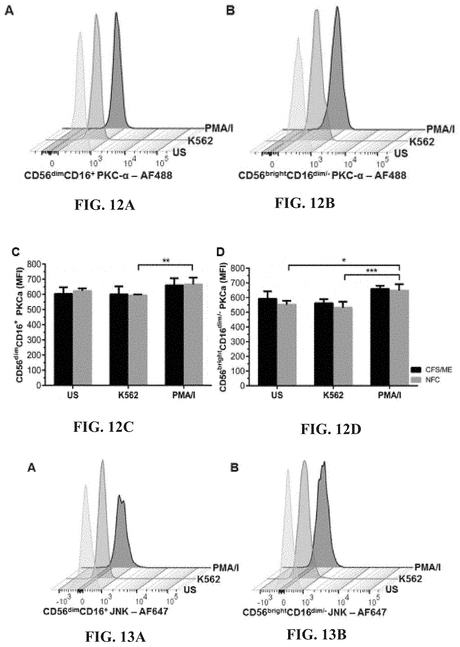
19. The method of claim 17, wherein the at least one therapeutic compound is naltrexone or a pharmaceutically acceptable derivative thereof.
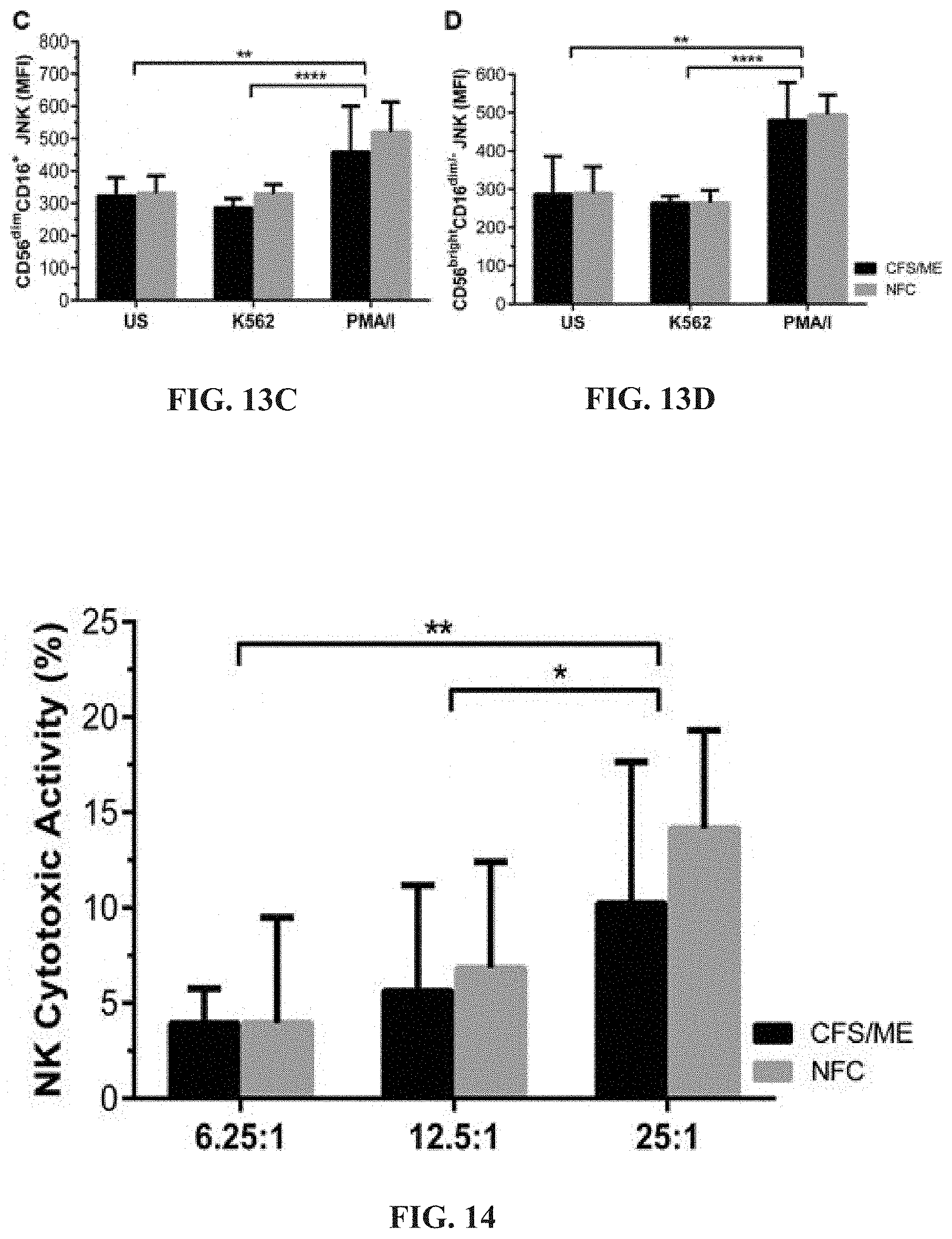
20. The method of claim 17, wherein the at least one therapeutic compound is naltrexone hydrochloride.
Description
CROSS-REFERENCE TO RELATED APPLICATIONS
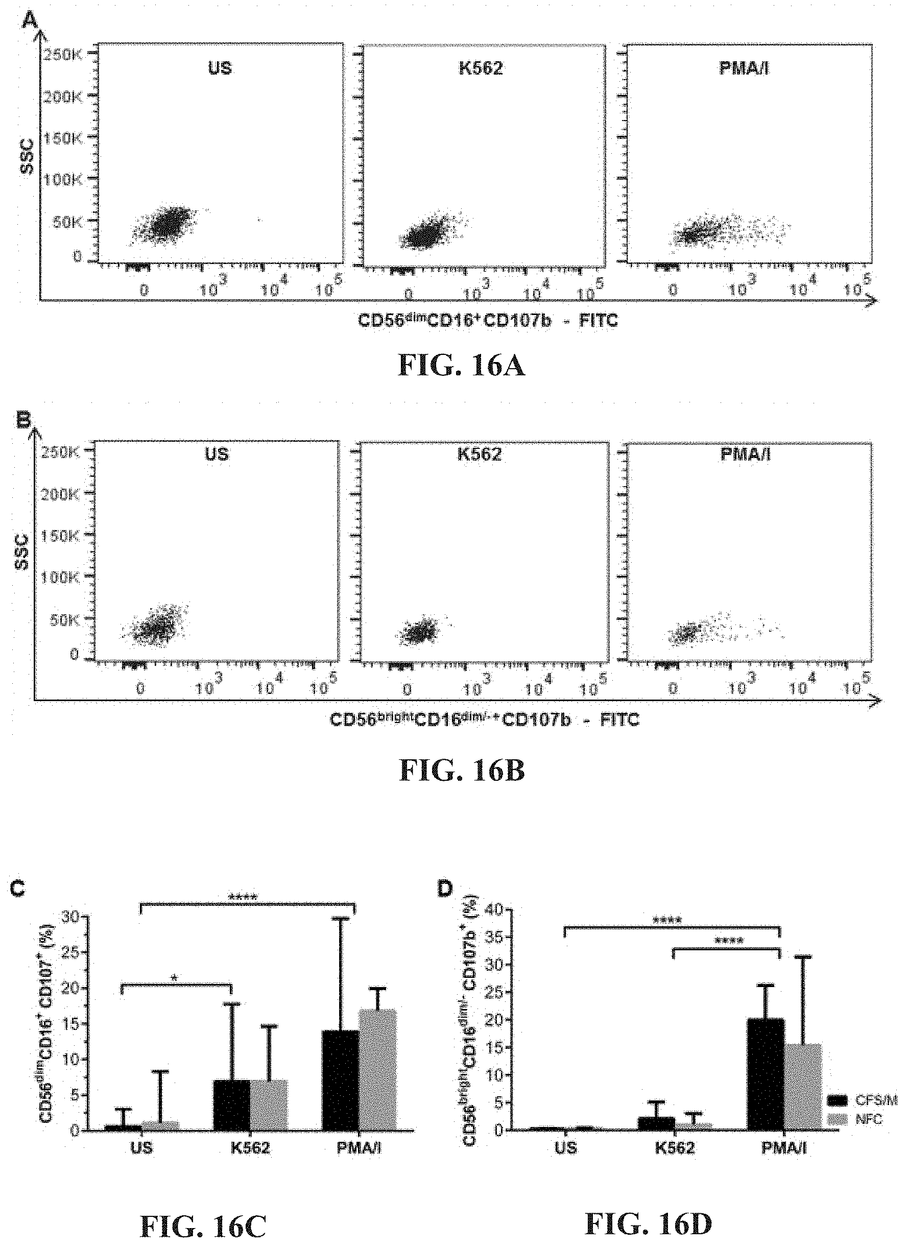
[0001] This application is a continuation-in-part of U.S. Ser. No. 15/570,628, filed Oct. 30, 2017, and claims priority of Australian Provisional Patent Application Number 2019902924, filed Aug. 13, 2019, Australian Provisional Patent Application Number 2016901468, filed Apr. 20, 2016, Australian Provisional Patent Application Number 2015904991, filed Dec. 2, 2015, and Australian Provisional Patent Application Number 2015901567, filed May 1, 2015, the entire contents of which are incorporated herein by reference.
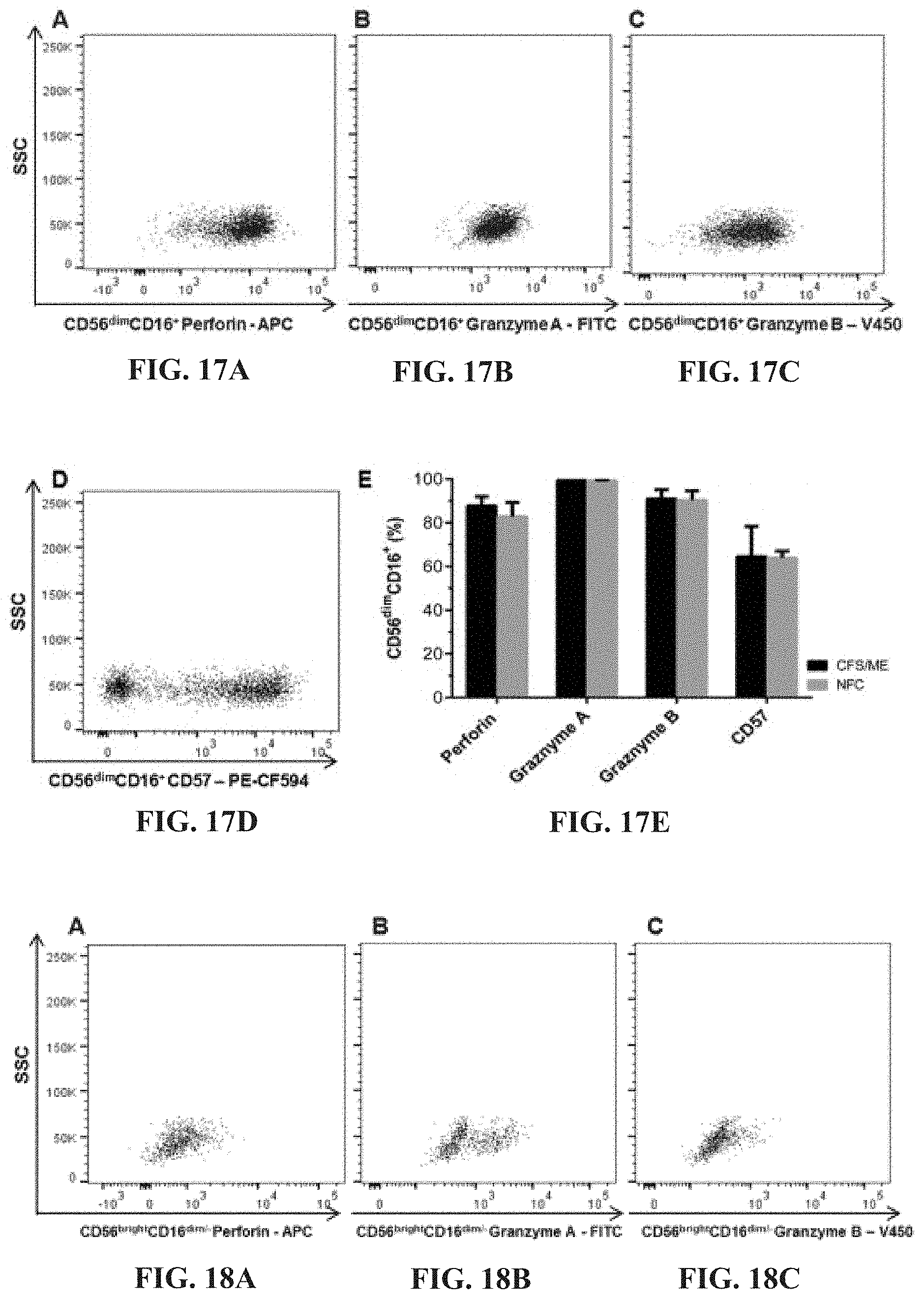
TECHNICAL FIELD
[0002] In some aspects the present invention broadly relates to the use of single nucleotide polymorphisms (SNPs) in transient receptor potential (TRP) ion channel, acetylcholine receptor (AchR) and/or adrenergic receptor (ADR) genes as probes, tools or reagents for identifying, screening, diagnosing, monitoring or managing/treating subjects with, or predisposed to, medical conditions (or symptoms thereof), such as chronic fatigue syndrome (CFS), myalgic encephalomyelitis (ME), Gulf war syndrome (GWS), irritable bowel syndrome (IBS), multiple chemical sensitivity (MCS), fibromyalgia, and migraine, as well as some medical conditions caused by dysregulation in calcium, acetylcholine, TRP and ADR, and dysregulation in the gastrointestinal, cardiovascular, neurological, genitourinary and immune systems.
[0003] In other aspects the present invention relates to the use of calcium metabolism testing for identifying, screening, diagnosing, monitoring or managing/treating a subject having, or at risk of developing, a medical condition or symptom thereof. This aspect may involve testing any suitable calcium-dependent biochemical process.
[0004] In other aspects the present invention relates to identifying or diagnosing a subject having a medical condition or symptom thereof, by testing cells obtained from the subject for dysfunctional signalling through the Mitogen-Activated Protein Kinase (MAPK) pathway, including signalling via the MAPK kinase (MAPKK/MEK1/2) and extracellular signal-regulated kinase (ERK)1/2 as well as p38.
[0005] In other aspects the present invention relates to the use of one or more differentially regulated calcium-dependent kinase genes for identifying, screening, diagnosing or monitoring a subject having, or at risk of developing, a medical condition or symptom thereof.
[0006] Other aspects concern probes, tools or reagents based on, or developed from, the various aspects of the invention described above.
[0007] Other aspects relate to methods, kits and assays for identifying, screening, diagnosing, monitoring or managing/treating subjects with one or more of those medical conditions or symptoms.
[0008] Yet other aspects relate to methods of treating a symptom such as pain in a subject diagnosed as having ME or CFS, by administering to the subject a therapeutically effective amount of at least one therapeutic compound: that is an opioid receptor antagonist; that is an opioid antagonist; or that restores TRPM3 ion channel activity.
[0009] Other aspects relate to methods of treating a symptom such as pain in a subject having dysfunctional TRPM3 ion channel activity, a method of restoring NK cell function in a subject having dysfunctional TRPM3 ion channel activity, or a method of restoring calcium homeostasis in a subject having dysfunctional TRPM3 ion channel activity. In preferred embodiments, the therapeutic compound is naltrexone or a pharmaceutically acceptable derivative thereof.
BACKGROUND
[0010] It will be clearly understood that, if a prior art publication is referred to herein, this reference does not constitute an admission that the publication forms part of the common general knowledge in the art in any country.
Background Art as Originally Described in U.S. Ser. No. 15/570,628
[0011] Chronic fatigue syndrome/myalgic encephalomyelitis (CFS/ME) is known to affect about 1-4% of individuals worldwide [1a, 2a]. CFS/ME has an unknown aetiology and there is no specific diagnostic test. Chronic fatigue syndrome (CFS) is an unexplained disorder with multiple physiological impairments. The illness is characterised by significant impairment in physical activity and debilitating fatigue accompanied by impairment in memory, cognition and concentration, enhanced experience of pain as well as dysregulation of the gastrointestinal, cardiovascular and immune systems [14a-31a]. Research to date suggests significant immune impairment. However, the mechanism of this disorder remains to be determined. CFS patients may have reactions to a number of environmental and biological factors [11a-13a]. Moreover, there is evidence to suggest that CFS may have an allergic component [14a-16a].
[0012] Gulf war syndrome (GWS) is a serious condition that affects at least a quarter of the 697,000 US veterans who served in the 1900-1991 Gulf war [1e]. GWS comprises a complex of multiple concurrent symptoms, being typified by persistent memory and concentration problems, chronic headaches, wide-spread pain, gastrointestinal problems and other chronic abnormalities, not explained but well established by diagnoses. No effective treatments have been identified for GWS and studies indicate that few veterans recover over time.
[0013] Irritable bowel syndrome (IBS) is characterised by abnormally-increased motility of the small and large intestines of unknown origins. Most patients are young adults who complain of diarrhoea and occasionally pain in the lower abdomen. No organic disease has been identified in IBS to date.
[0014] Multiple chemical sensitivity (MCS) is the most common term used to describe a condition presenting as a complex array of symptoms linked to low level chemical exposures [2e]. The underlying mode(s) of action of MCS, i.e. the biological mechanisms by which the chemical sensitivity occurs, remain uncertain. In terms of sensitivities involving chemicals, the terms "MCS" and "chemical sensitivity" (sometimes known as "chemical intolerance") are often used interchangeably. However, "chemical sensitivity" in its wider context can describe several distinct types of reactions encompassing classical adverse toxicological reactions, immunological "allergic" sensitivities, individual chemical idiosyncrasies and intolerances through to aversions to particular odours. Broadly, on the basis of Consensus Criteria, MCS is distinguished from other types of chemical sensitivities or intolerances predominantly on the basis of reactions to multiple, diverse chemical substances, the wide spectrum of non-specific symptoms reported in multiple organ systems and the extremely low levels of environmental exposures linked to responses. Symptoms include headache, fatigue, confusion, depression, shortness of breath, arthralgia, myalgia, nausea, dizziness, memory problems, gastrointestinal symptoms and respiratory symptoms. Medical conditions caused by dysregulation in calcium, (especially in respect of CFS, ME, GWS, IBS, MCS, fibromyalgia and migraine), are typified by specific symptoms or dysregulation, including: significant impairment in physical activity; debilitating fatigue accompanied by impairment in memory, cognition and concentration; enhanced experience of pain; dysregulation of the gastrointestinal, cardiovascular and immune systems; respiratory symptoms and immunological "allergic" sensitivities; headache; fatigue; confusion; depression; shortness of breath; arthralgia; myalgia; nausea; dizziness; memory problems; and gastrointestinal symptoms.
[0015] Medical conditions caused by dysregulation in acetylcholine are typified by specific symptoms or dysregulation, including: significant impairment in physical activity; debilitating fatigue accompanied by impairment in memory, cognition and concentration; enhanced experience of pain; dysregulation of the gastrointestinal, cardiovascular and immune systems; headache; fatigue; confusion; depression; shortness of breath; arthralgia; myalgia; nausea; dizziness; memory problems; gastrointestinal symptoms; respiratory symptoms; and dysregulation of the gastrointestinal, cardiovascular and immune systems.
[0016] Medical conditions caused by dysregulation in TRP are typified by specific symptoms or dysregulation, including: significant impairment in physical activity; debilitating fatigue accompanied by impairment in memory, cognition and concentration; enhanced experience of pain; dysregulation of the gastrointestinal, cardiovascular and immune systems; headache; fatigue; confusion; depression; shortness of breath; arthralgia; myalgia; nausea; dizziness; memory problems; gastrointestinal symptoms; respiratory symptoms; and dysregulation of the gastrointestinal, cardiovascular and immune systems.
[0017] Medication conditions caused by dysregulation in ADR are typified by specific symptoms such as respiratory difficulties including shortness or breath, air hunger, colds and nasalpharynx congestion, cardiovascular conditions such as hypertension, and palpitations, gastrointestinal illness, kidney disease, diabetes, and autonomic function including sweating episodes.
[0018] Medical conditions caused by dysregulation of the gastrointestinal, cardiovascular, neurological, genitourinary and immune systems are typified by specific symptoms or dysregulation, including: significant impairment in physical activity; debilitating fatigue accompanied by impairment in memory, cognition and concentration; enhanced experience of pain; headache; fatigue; confusion; depression; shortness of breath; arthralgia; myalgia; nausea; dizziness; memory problems; gastrointestinal symptoms; urinary frequency or discomfort and respiratory symptoms.
[0019] Transient receptor potential (TRP) ion channels are expressed on almost all cells and have a significant effect on physiological functions [3b]. A number of channelopathies have been associated with TRP genes as these have consequences for cellular function [4b, 18b, 19b]. Dysregulation in TRPs has been associated with pathological conditions and diseases including chronic pain, overactive bladder, diabetes, chronic obstructive pulmonary disease, cardiac hypertrophy, familial Alzheimer's disease, skin diseases, skeletal dysplasias, motor neuropathies, neuro-sensory neuropathies (including Charcot-Marie-Tooth disease (type 2C) and cancer [4b-8b]. TRP ion channels have an important role in Ca.sup.2+ signalling. TRP ion channels are activated following fluctuations or deviations in the cellular environment. Factors that may influence these changes are stressors including pathogens, temperature, pressure, chemicals, oxidation/reduction, toxins, osmolarity and pH [9b, 10b]. TRP ion channels are activated in the presence of irritants, inflammatory products, and xenobiotic toxins.
[0020] Mammalian TRPs are comprised of six main groups including the TRPA (ankyrin), TRPC (canonical), TRPM (melastatin), TRPML (mucolipin), TRPP (polycystin) and TRPV (vanilloid) [1b, 2b]. Generally, the TRPC channels are nonselective cation channels, only two are highly permeable Ca.sup.2+ channels and two are impermeable for Ca.sup.2+. Several TRPs are permeable for Mg.sup.2+ and Zn.sup.2+ [3b].
[0021] Acetylcholine is principally a neurotransmitter. The physiological functions of acetylcholine (ACh) are mediated by two membrane proteins, namely the muscarinic (mAChR) and nicotinic receptors (nAChR). Both receptor types have numerous subtypes and are located in the central and peripheral nervous system including the autonomic system. Furthermore, ACh performs non-neuronal functions, termed the non-neuronal cholinergic system (NNCS), where ACh performs endocrine functions of tissue located on smooth muscle, .beta. pancreatic cells, glial cells, lymphocytes, ocular lens cells and brain vascular endothelium [1c] as well as in the CNS [2c-6c]. The degradation of ACh into choline and acetate is catalysed by the enzymes acetylcholinesterase (AChE) [7c, 8c].
[0022] There are five main mAChR subtypes--M1, M2, M3, M4 and M5, where M2 and M4 are inhibitory receptors, and M1, M2 and M3 are excitatory receptors [7c, 8c]. mAChRs are G protein coupled receptors that regulate intracellular signalling second messengers as well as ion channel activities. Once activated each subtype has distinctive functions--M1, M3 and M5 receptors form inositol 1,4,5-triphosphate (IP3) and 1,2 diacylglycerol (DAG), resulting in increased intracellular calcium. Activated M2 and M4 receptors inhibit adenylate cyclase activity as well as mediating function of non-selective cation channels, transient receptor potential channels and potassium channels [7c-10c].
[0023] nAChRs are fast ionotropic cationic nicotinic receptor channels which allow for the influx of cations such as potassium, calcium and sodium ions into the cell. nAChRs are comprised of different subunits: .alpha. subunits (.alpha..sub.1-.alpha..sub.10), .beta. subunits (.beta..sub.1-.beta..sub.4), one .delta. subunits, one .gamma. subunit and one .epsilon. subunit [11c]. Depending upon combinational subunit binding AChRs can form either heteromers or homomers [11c].
[0024] Previous research has reported anomalies in acetylcholine signalling in CFS/ME patients. Peripheral cholinergic function is noted to be abnormal in CFS/ME patients exposed to ACh challenge whereby blood flow peaks take a longer time to return to normal. Increased sensitivity to ACh is noted in peripheral vascular endothelium [30c, 31c]. Moreover it is documented that ACh influences immune cell function [32c] and is manufactured and secreted by a wide range of immune cells including lymphocytes [33c, 34c, 32c, 35c]. The present inventors, along with others, have previously reported profound changes in immune cell and function as well as noting cardiac and neurological effects in CFS/ME patients [14c-16c, 18c-20c, 22c, 24c, 26c, 27c, 29c].
[0025] Single nucleotide polymorphisms (SNPs) occur in coding sequences of genes, non-coding regions of genes, or in the intergenic regions of genes. SNPs located within a coding sequence may or may not necessarily change the amino acid sequence of the protein that is produced. As such SNPs that do not alter the polypeptide sequence are termed synonymous (sometimes called silent variants) while SNPs that result in different polypeptide sequences are referred to as non-synonymous. Non-synonymous single nucleotide polymorphisms (nsSNPs) result in changes to protein expression that may result in aberrant signalling, such as loss or gain of function in their effect. Importantly, silent variants have been reported to affect splicing and may lead to human disease [10d, 11d]. Splicing affecting gene variants can induce exon skipping and activate alternate splice isoforms of the gene transcript, potentially resulting in altered gene transcripts and disease phenotypes.
[0026] Despite intensive research, to date, the pathophysiology of CFS/ME is not yet fully understood and clear diagnostic tools remain elusive. Therefore, there remains a need for rapid, cost-effective and reliable means for identifying, screening, diagnosing, monitoring and/or managing/treating individuals having, or at risk of developing, a medical condition such as CFS/ME.
Background Art in Respect of Current Improvements to the Invention
[0027] Since the filing of U.S. Ser. No. 15/570,628, the present inventors have made breakthroughs and improvements to the invention. Some of the work originally described in U.S. Ser. No. 15/570,628 has been published in scientific journals. The Background Art section that follows is to be understood in that context.
[0028] Currently there are no specific treatments for CFS/ME. Although the aetiology of ME/CFS remains elusive, immune dysfunction and abnormalities in natural killer (NK) cell functions are the most consistent features [2o-13o]. Differences in NK cell phenotypes and significantly reduced peripheral NK cell numbers resulting in significant reduction in NK cell cytotoxicity, have been reported in ME/CFS and implicated in disease severity [2o-13o].
[0029] Importantly, NK cells require calcium (Ca.sup.2+) for efficient stimulation and functions including NK cell cytotoxicity [14o-17o]. Intracellular Ca.sup.2+ concentration ([Ca.sup.2+].sub.i) is tightly regulated by numerous actors including selective and non-selective channels, pumps and exchangers located on the plasma membrane and/or on organelles. Disturbance of this tightly regulated homeostatic system leads to disorders of Ca.sup.2+ metabolism and immune cell functions playing a pivotal role in the pathophysiology of several diseases and immunodeficiencies [18o].
[0030] As mentioned, Transient Receptor Potential (TRP) non selective cation channels function as polymodal cellular sensors involved in the fine-tuning of many biological processes in both excitable and non-excitable cells [19o] and contribute significantly to Ca.sup.2+ signalling. The mammalian TRP melastatin member 3 (TRPM3) is a widely expressed Ca.sup.2+-permeable nonselective cation channel activated by temperature, natural chemicals and toxins or synthetic compounds, including the endogenous neurosteroid pregnenolone sulfate (PregS), and the L-type voltage-gated Ca.sup.2+ channel inhibitor, nifedipine, or by mechanical stimuli. TRPM3 channels also respond to endogenous agents and messengers produced during tissue injury and inflammation [20o]. Importantly, TRPM3 has been previously identified as a thermosensitive and nociceptor channel implicated in the detection of acute heat sensing and inflammatory heat hyperalgesia as well as in pain transmission in the central nervous system (CNS) [20o].
[0031] The loss of TRPM3 ion channel function has now been established to be associated with the ME/CFS pathomechanism [21o, 22o]. Significant reduction in TRPM3 surface expression and Ca.sup.2+ mobilisation in immune cells were subsequently reported in ME/CFS patients [23o, 24o]. Recently, novel electrophysiological investigations used whole-cell patch clamp techniques to report a significant reduction in TRPM3 ion channel activity after PregS and nifedipine stimulation in NK cells from ME/CFS patients [21o, 22o]. Moreover, ionic currents in ME/CFS patients were resistant to ononetin in the presence of PregS and nifedipine. Dysregulation of TRPM3 function in ME/CFS patients, affecting [Ca.sup.2+].sub.i and Ca.sup.2+ signalling has significant implications for NK cell regulatory machinery and functions, and to the present inventors represents a novel and attractive therapeutic target of ME/CFS pathology.
[0032] There are few treatments available for people suffering from severe or long-lasting pain characteristic of ME/CFS. Currently, substances called opioids, agonists of mu (.mu.)-opioid receptors (.mu.OR), are the strongest painkillers clinically available [25o]. Opioids mediate their effects by interacting with molecules that belong to a group of receptor proteins called G-protein coupled receptors (GPCRs). These opioid receptors are widely distributed in the CNS with the role of detecting and transmitting pain signals [25o]. It was poorly understood how activation of opioid receptors reduces the activity of pain-sensing nerve cells, however recent literature suggests that activation of GPCRs can affect TRPM3 channels and in turn decrease the flow of Ca.sup.2+ ions through the pore [250-27o]. GPCRs interact with G-proteins that, when activated by the receptor, release the G.beta..gamma. dimers from G.alpha. subunits of the G.sub.i/o subfamily. Inhibition of TRPM3 activity by stimulation of GPCRs (in particular .mu.ORs) is mediated through a direct binding of the G.sub..beta..gamma. subunit to the ion channel.
[0033] Naltrexone hydrochloride (NTX) is a long-lasting opioid antagonist used commonly in the treatment of opioid and alcohol dependence [28o]. NTX specifically inhibits .mu.ORs and, to a lesser extent, the delta (.delta.)-opioid receptors (.delta.OR), thus negating the inhibiting effects of opioid receptors agonists [29o, 30o]. A recent investigation demonstrated that naloxone, a rapid response alternative to naltrexone, did not have a direct effect on TRPM3-dependent Ca.sup.2+ signals in mouse dorsal root ganglion neurons [25o]. However, when co-applied with DAMGO, a highly selective .mu.OR agonist, naloxone prevented the action of DAMGO completely, indicating a possible role for naloxone in influencing TRPM3 signalling. Interestingly, TRPM3 activation by nifedipine and PregS was also inhibited by .mu.OR activation confirming that TRPM3 inhibition is an important consequence of peripheral .mu.OR activation.
SUMMARY
Summary of Invention as Originally Described in U.S. Ser. No. 15/570,628
[0034] The present invention, in a first aspect, broadly concerns the use of one or more single nucleotide polymorphisms (SNPs) in one or more transient receptor potential (TRP) ion channel, acetylcholine receptor (AChR) or adrenergic receptor (ADR) genes as probes, tools or reagents for identifying, screening, diagnosing, monitoring or managing/treating subjects with, or predisposed to, medical conditions or specific symptoms thereof, such as chronic fatigue syndrome (CFS), myalgic encephalomyelitis (ME), Gulf war syndrome (GWS), irritable bowel syndrome (IBS), multiple chemical sensitivity (MCS), fibromyalgia, or migraine, as well as some medical conditions caused by dysregulation in calcium, acetylcholine, TRP or ADR, and dysregulation in the gastrointestinal, cardiovascular, neurological, genitourinary or immune systems.
[0035] In a second aspect, the present invention broadly relates to the use of calcium metabolism testing for identifying, screening, diagnosing, monitoring or managing/treating a subject having, or at risk of developing, a medical condition or symptom thereof.
[0036] In a third aspect, the present invention broadly relates to identifying, screening, diagnosing, monitoring or managing/treating a subject having a medical condition or symptom thereof, by testing cells obtained from the subject for dysfunctional signalling through the Mitogen-Activated Protein Kinase (MAPK) pathway, including signalling via the MAPK kinase (MAPKK/MEK1/2) and extracellular signal-regulated kinase (ERK)1/2 as well as p38.
[0037] In a fourth aspect, the present invention broadly relates to the use of at least one differentially regulated calcium-dependent kinase gene for identifying, screening, diagnosing, monitoring or managing/treating a subject having, or at risk of developing, a medical condition or symptom thereof.
[0038] In a fifth aspect, the invention broadly concerns at least one probe, tool or reagent based on or developed from any one of the first to fourth aspects, for identifying, screening, diagnosing, monitoring or managing/treating the medical condition or symptom thereof.
[0039] In a sixth aspect, the present invention broadly concerns methods, kits or assays based on or developed from any one of the first to fifth aspects, for identifying, screening, diagnosing, monitoring or managing/treating subjects with one or more of the medical conditions or symptom thereof.
Summary of Invention in Respect of Current Improvements to the Invention
[0040] In a seventh aspect, the present invention broadly concerns a method of treating a symptom such as pain in a subject identified or diagnosed as having ME/CFS, said method comprising the step of administering to the subject a therapeutically effective amount of at least one therapeutic compound: that is an opioid receptor antagonist; that is an opioid antagonist; or that restores TRPM3 ion channel activity.
[0041] In an eighth aspect, the present invention broadly concerns a method of treating a symptom such as pain in a subject having dysfunctional TRPM3 ion channel activity, said method comprising the step of administering to the subject a therapeutically effective amount of at least one therapeutic compound: that is an opioid receptor antagonist; that is an opioid antagonist; or that restores TRPM3 ion channel activity.
[0042] In a ninth aspect, the present invention broadly concerns a method of restoring NK cell function in a subject having dysfunctional TRPM3 ion channel activity, said method comprising the step of administering to the subject a therapeutically effective amount of at least one therapeutic compound/drug: that is an opioid receptor antagonist; that is an opioid antagonist; or that restores TRPM3 ion channel activity.
[0043] In a tenth aspect, the present invention broadly concerns a method of restoring calcium homeostasis in a subject having dysfunctional TRPM3 ion channel activity, said method comprising the step of administering to the subject a therapeutically effective amount of at least one therapeutic compound/drug: that is an opioid receptor antagonist; that is an opioid antagonist; or that restores TRPM3 ion channel activity.
[0044] Preferred features, embodiments and variations of the invention may be discerned from the following Detailed Description which provides sufficient information for those skilled in the art to perform the invention. The Detailed Description is not to be regarded as limiting the scope of the preceding Summary of Invention in any way. The Detailed Description will make reference to a number of drawings as follows.
BRIEF DESCRIPTION OF THE DRAWINGS
[0045] Figures as originally described in U.S. Ser. No. 15/570,628.
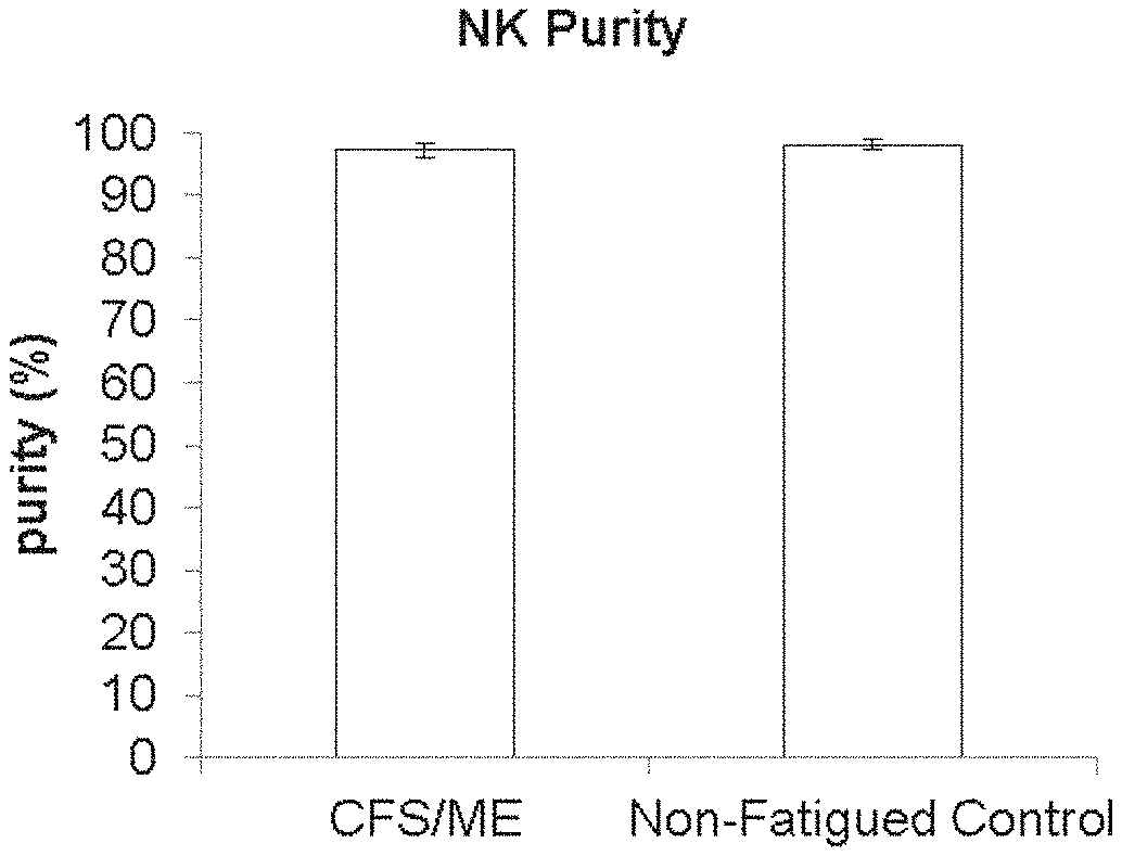
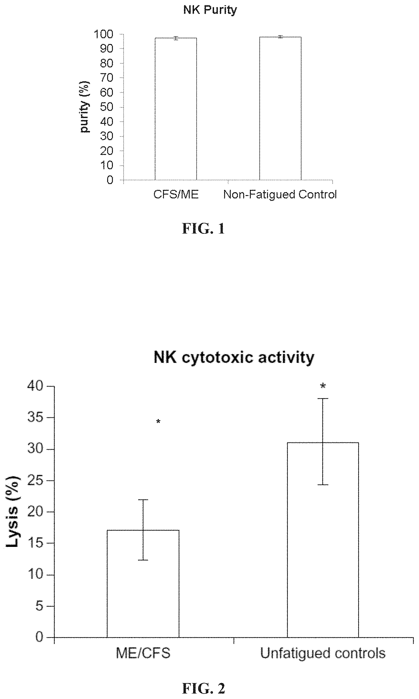
[0046] FIG. 1: Natural Killer Cell Purity. The purity of NK cells represents minimal contamination from other cells types. Data shown for ME/CFS (n=39), and non-fatigued controls (n=30), and presented as mean.+-.SEM.
[0047] FIG. 2: Reduced NK cytotoxic activity in CFS/ME. In vivo assessment of NK cytotoxic activity of tumour cell lines K562 in CFS/ME (n=39) and unfatigued controls (n=30). Lytic activity represented by percentage lysis of target cells on the y-axis. Data presented as mean.+-.SE *P<0.05.
[0048] FIGS. 3A-3B: TRPM3 expression (%) on B lymphocytes and NK cells gated from HC (n=19) and CFS/ME (n=18) peripheral mononuclear cells. (FIG. 3A) NK cells subsets were characterized as CD56.sup.Bright NK cells and CD56.sup.Dim NK cells. Identification of TRPM3 surface expression on the NK cell subsets was analyzed using indirect flow cytometry. (FIG. 3B) B cells were characterized as total B cells (CD3.sup.-CD19.sup.+) and indirect flow cytometry was employed to identify TRPM3 surface expression on B cells. Histograms report the means.+-.SEM. *Denotes p<0.05. HC: healthy controls; CFS: Chronic Fatigue Syndrome; ME: myalgic encephalomyelitis.
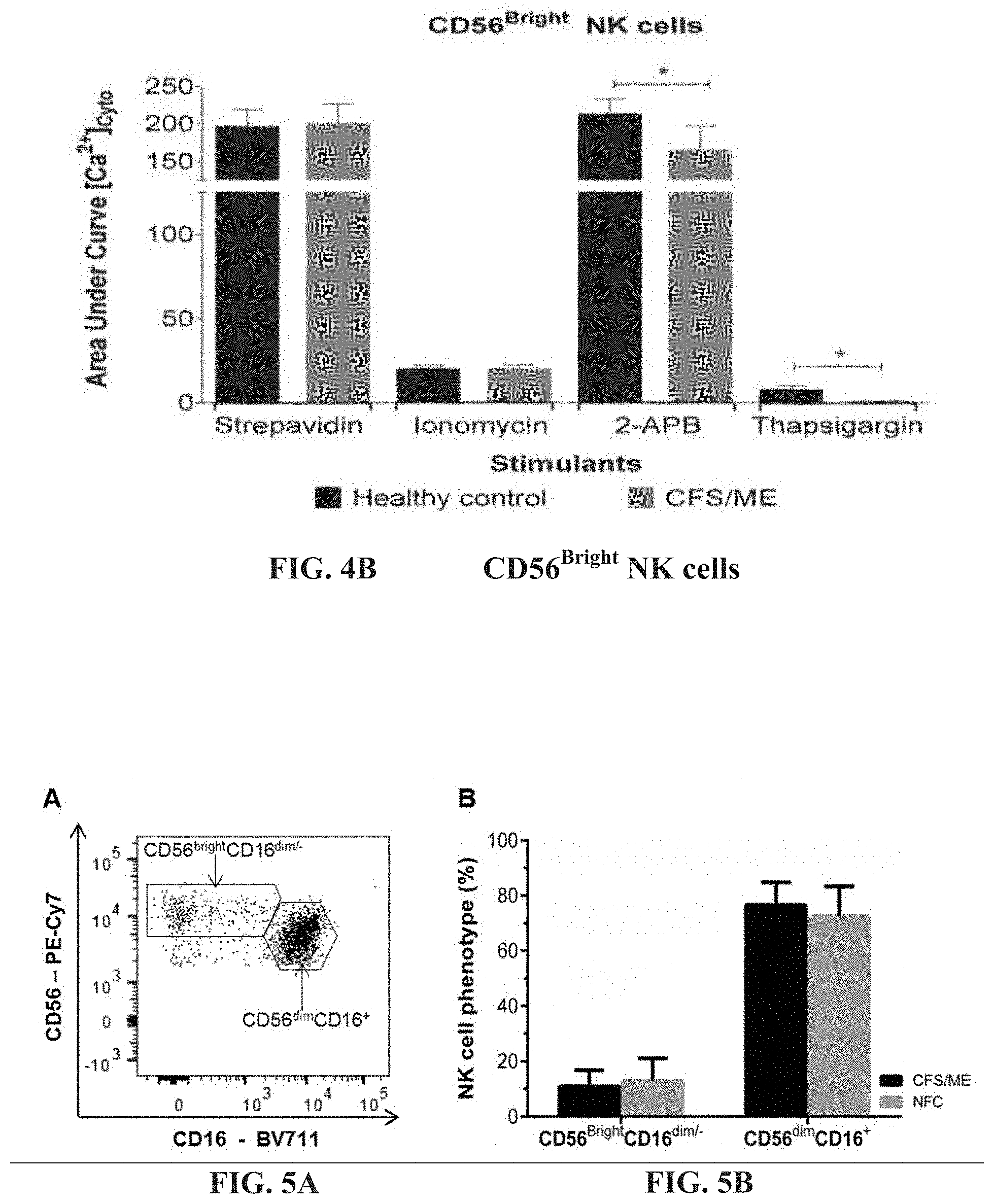
[0049] FIGS. 4A-4B: Fura-AM cytoplasmic calcium influx in CD19.sup.+B cells and CD56.sup.Bright NK cells. (FIG. 4A). CD19.sup.+B cells calcium influx response curve reported as area under the curve was measured during Anti-IgM and anti-CD21 conjugated biotins were cross-linked with streptavidin or in the presence of ionomycin, 2-APB or Thapsigargin using flow cytometry. (FIG. 4B). Fura-AM cytoplasmic calcium influx response during CD56.sup.Bright NK cell receptors, Anti-CD314 and anti-CD335 conjugated biotins were cross-linked with streptavidin or in the presence of ionomycin, 2-APB or Thapsigargin using flow cytometry. Histograms report the means.+-.SEM. *Denotes statistically significance at p<0.05.
[0050] FIGS. 5A-5B: Representative flow cytometric plot of CD56.sup.brightCD16.sup.dim/- and CD56.sup.dimCD16.sup.+ NK cell phenotypes (FIG. 5A). Comparisons of CD56.sup.brightCD16.sup.dim/- and CD56.sup.dimCD16.sup.+ NK cell phenotypes between CFS/ME and NFC revealed no significant differences (FIG. 5B). Data are presented as median percentage with interquartile range.
[0051] FIGS. 6A-6B: CD56.sup.brightCD16.sup.dim/- NK cell ERK1/2 flow cytometric plot for a representative individual (FIG. 6A). ERK1/2 in CD56.sup.brightCD16.sup.dim/- NK cells were compared between CFS/ME and NFC groups and no significant differences were observed (FIG. 6B). PMA/I stimulation caused a significant increase in ERK1/2 phosphorylation compared to US (***p<0.001) and K562 cells (****p<0.0001) in both CFS/ME and NFC. Data are presented as MFI with interquartile range.
[0052] FIGS. 7A-7B: Representative flow cytometric plot for MEK1/2 in CD56.sup.dimCD16.sup.+ NK cells (FIG. 7A). No significant differences were observed when MEK1/2 was compared between CFS/ME and NFC (FIG. 7B). In both CFS/ME and NFC, PMA/I stimulation resulted in a significant increase in phosphorylated MEK1/2 compared to US (****p<0.0001) and K562 stimulation (****p<0.0001). Data are presented as MFI with interquartile range.
[0053] FIGS. 8A-8B: p38 representative flow cytometric plot in CD56.sup.dimCD16.sup.+ NK cells (FIG. 8A). p38 was compared between CFS/ME and NFC and no significant differences were observed (FIG. 8B). Stimulation with PMA/I caused a significant increase in phosphorylated p38 when compared to US and K562 incubated cells (*p<0.05). Data are presented as MFI with interquartile range.
[0054] FIGS. 9A-9D: Representative Stat-3 flow cytometric plots in CD56.sup.dimCD16.sup.+ (FIG. 9A) and CD56.sup.brightCD16.sup.dim/- (FIG. 9B) NK cells. Comparison of Stat-3 in CD56.sup.dimCD16.sup.+ (FIG. 9C) and CD56.sup.brightCD16.sup.dim/- (FIG. 9D) NK cells between CFS/ME and NFC revealed no significant differences. In CD56.sup.dimCD16.sup.+ and CD56.sup.brightCD16.sup.dim/- NK cells, stimulation with PMA/I caused a significant increase in Stat-3 when compared to US (****p<0.0001) and K562 (****p<0.0001) in both CFS/ME and NFC.
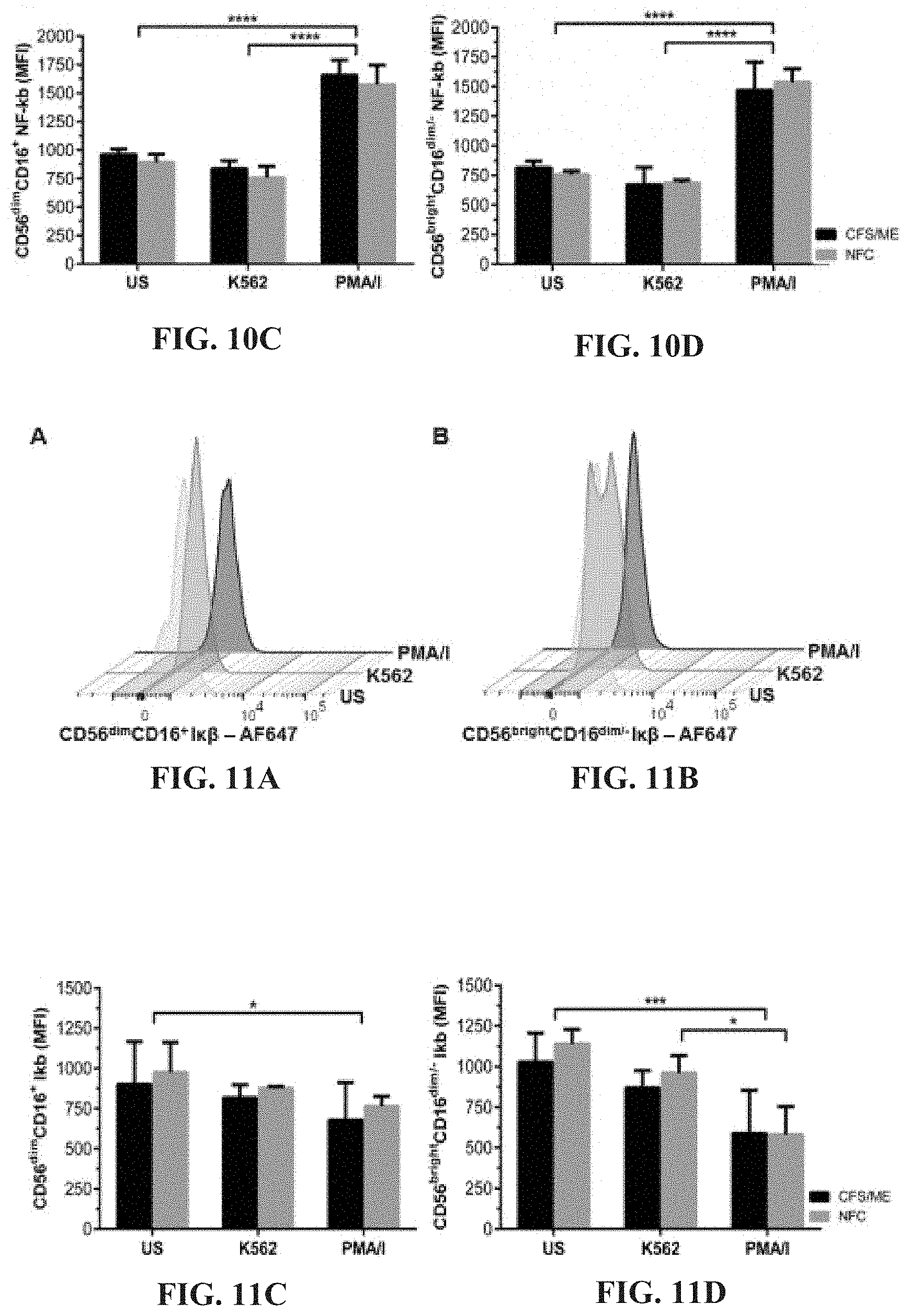
[0055] FIGS. 10A-10D: Representative flow cytometric analysis of NF-.kappa..beta. in CD56.sup.dimCD16.sup.+ (FIG. 10A) and CD56.sup.brightCD16.sup.dim/- (FIG. 10B) NK cells. No significant differences were observed when NF-.kappa..beta. was compared between CFS/ME and NFC in CD56.sup.dimCD16.sup.+ (FIG. 10C) and CD56.sup.brightCD16.sup.dim/- (FIG. 10D) NK cells. Phosphorylated NF-.kappa..beta. significantly increased after PMA/I stimulation in both CD56.sup.dimCD16.sup.+ (FIG. 10C) and CD56.sup.brightCD16.sup.dim/- (FIG. 10D) NK cells compared to US (****p<0.0001) and K562 (****p<0.0001) in CFS/ME and NFC. Data are presented as MFI with interquartile range.
[0056] FIGS. 11A-11D: I.kappa..beta. representative flow cytometric plots in CD56.sup.dimCD16.sup.+ (FIG. 11A) and CD56.sup.brightCD16.sup.dim/- (FIG. 11B) NK cells. I.kappa..beta. was compared in CD56.sup.dimCD16.sup.+ (FIG. 11C) and CD56.sup.brightCD16.sup.dim/- (FIG. 11D) NK cells from CFS/ME and NFC and no significant differences were observed. Stimulation with PMA/I caused a significant reduction in I.kappa..beta. in both CD56.sup.dimCD16.sup.+ (*p<0.05) and CD56.sup.brightCD16.sup.dim/- (***p<0.001) NK cells from CFS/ME and NFC. Incubation with PMA/I also caused a significant reduction (*p<0.05) in I.kappa..beta. in CD56.sup.brightCD16.sup.dim/- NK cells from CFS/ME patients.
[0057] FIGS. 12A-12D: Representative flow cytometric plots for the analysis of PKC-.alpha. in CD56.sup.dimCD16.sup.+ (FIG. 12A) and CD56.sup.brightCD16.sup.dim/- (FIG. 12B) NK cells. PKC-.alpha. was compared in CD56.sup.dimCD16.sup.+ (FIG. 12C) and CD56.sup.brightCD16.sup.dim/- (FIG. 12D) NK cells from CFS/ME and NFC and no significant differences were observed. In CD56.sup.dimCD16.sup.+ NK cells from NFC, stimulation with PMA/I caused a significant increase (**p<0.01) in PKC-.alpha. phosphorylation compared to K562 cells. PKC-.alpha. was significantly increased in CD56.sup.brightCD16.sup.dim/- NK cells after PMA/I stimulation when compared to US (*p<0.05) and K562 (***p<0.001) in NFC.
[0058] FIGS. 13A-13D: Flow cytometric analysis of JNK in CD56.sup.dimCD16.sup.+ (FIG. 13A) and CD56.sup.brightCD16.sup.dim/- (FIG. 13B) NK cells. No significant differences were observed when JNK was compared in CD56.sup.dimCD16.sup.+ (FIG. 13C) and CD56.sup.brightCD16.sup.dim/- (FIG. 13D) NK cells from CFS/ME and NFC. Significant increases in phosphorylated JNK were observed in both CD56.sup.dimCD16.sup.+ (FIG. 13C) and CD56.sup.brightCD16.sup.dim/- (FIG. 13D) NK cells after PMA/I stimulation when compared to US (**p<0.01) and K562 (***p<0.001) in CFS/ME and NFC.
[0059] FIG. 14: NK cell cytotoxic activity in CFS/ME and NFC at three E:T ratios.
[0060] FIGS. 15A-15D: Representative flow cytometry plots for CD107a in CD56.sup.dimCD16.sup.+ (FIG. 15A) and CD56.sup.brightCD16.sup.dim/- (FIG. 15B) NK cells. CD107a was measured in US cells and after stimulation with either K562 cells or PMA/I. Comparison of CD107a on CD56.sup.dimCD16.sup.+ (FIG. 15C) and CD56.sup.brightCD16.sup.dim/- (FIG. 15D) NK cells between CFS/ME and NFC revealed no significant differences. CD107a expression significantly increased after K562 and PMA/I (****p<0.0001) stimulation in CD56.sup.dimCD16.sup.+ NK cells from both CFS/ME and NFC. In CD56.sup.brightCD16.sup.dim/- NK cells, PMA/I stimulation significantly increased expression of CD107a when compared to K562 and US cells (****p<0.0001) from CFS/ME and NFC.
[0061] FIGS. 16A-16D: Flow cytometric analysis of CD107b on CD56.sup.dimCD16.sup.+ (FIG. 16A) and CD56.sup.brightCD16.sup.dim/- (FIG. 16B) NK cells. No significant differences were observed when CD107b expression was compared between CFS/ME and NFC on CD56.sup.dimCD16.sup.+ (FIG. 16C) and CD56.sup.brightCD16.sup.dim/- (FIG. 16D) NK cells. In CD56.sup.dimCD16.sup.+ NK cells, stimulation with K562 cells (*p<0.05) and PMA/I (****p<0.0001) caused a significant increase in CD107b expression in both CFS/ME and NFC compared to US. PMA/I stimulation significantly increased CD107b expression on CD56.sup.brightCD16.sup.dim/- NK cells from CFS/ME and NFC when compared to K562 and US (****p<0.0001).
[0062] FIGS. 17A-17E: Perforin (FIG. 17A), Granzymes A (FIG. 17B) and B (FIG. 17C) and CD57 (FIG. 17D) from CD56.sup.dimCD16.sup.+ NK cells from CFS/ME patients (comparison shown in FIG. 17E).
[0063] FIGS. 18A-18E: Perforin (FIG. 18A), Granzymes A (FIG. 18B) and B (FIG. 18C) and CD57 (FIG. 18D) from CD56.sup.brightCD16 NK cells from CFS/ME patients (comparison shown in FIG. 18E).
[0064] FIGS. 19A-19D: Representative flow cytometric plots for CD56.sup.dimCD16.sup.+ (FIG. 19A) and CD56.sup.brightCD16.sup.dim/- (FIG. 19B) NK cell production of IFN-.gamma.. Comparison of IFN-.gamma. production in CD56.sup.dimCD16.sup.+ (FIG. 19C) and CD56.sup.brightCD16.sup.dim/- (FIG. 19D) NK cells between CFS/ME and NFC revealed no significant differences. IFN-.gamma. production significantly increased after PMA/I stimulation in both CD56.sup.dimCD16.sup.+ and CD56.sup.brightCD16.sup.dim/- NK cells when compared to US and K562 (****p<0.0001).
[0065] FIGS. 20A-20D: Flow cytometric plots for TNF-.alpha. in CD56.sup.dimCD16.sup.+ (FIG. 20A) and CD56.sup.brightCD16.sup.dim/- (FIG. 20B) NK cells. Between CFS/ME and NFC cohorts, TNF-.alpha. production in CD56.sup.dimCD.sup.16+ (FIG. 20C) and CD56.sup.brightCD16.sup.dim/- (FIG. 20D) NK cells were not significantly different. In CD56.sup.dimCD16.sup.+ NK cells, PMA/I stimulation significantly increased TNF-.alpha. production when compared to US and K562 incubated cells (****p<0.0001) in both CFS/ME and NFC.
[0066] FIGS. 21A-21D: Flow cytometric analysis of GM-CSF production in CD56.sup.dimCD16.sup.+ (FIG. 21A) and CD56.sup.brightCD16.sup.dim/- (FIG. 21B) NK cells. Production of GM-CSF in CD56.sup.dimCD16.sup.+ (FIG. 21C) and CD56.sup.brightCD16.sup.dim/- (FIG. 21D) NK cells were not significantly different when compared between CFS/ME and NFC cohorts. Stimulation with PMA/I caused a significant increase in CD56.sup.dimCD16.sup.+ and CD56.sup.brightCD16.sup.dim/- GM-CSF production in both CFS/ME and NFC compared to US and K562 incubated cells (****p<0.0001, ***p<0.001).
[0067] FIG. 22. Natural Killer cell purity. NK cell purity measurements are represented as total % of CD3.sup.- CD56.sup.+ cells. Data are presented as mean.+-.SD for CFS/ME group (n=24) and control group (n=11).
[0068] FIGS. 23A-23B. Heat map of kinase gene expression showing (FIG. 23A) significantly upregulated and (FIG. 23B) significantly downregulated genes from severe CFS/ME patients compared with non-fatigued controls.
[0069] FIG. 24: Frequency of SNPs per chromosome.
[0070] FIG. 25: Manhattan plot of Fisher's exact test on 950 SNPs.
[0071] FIGS. 26A-26B: Frequency of top 10 SNPs from Fisher's exact test. Cases: CFS/ME group; Controls: Healthy control group; MAF: Minor allele.
[0072] FIG. 27: Proportion of CFS/ME patients ("Cases") and healthy control group ("Controls") being homozygous major (GG), heterozygous (AG) or homozygous minor (AA) for adrenergic .alpha.1A (ADRA1A) SNP rs2322333.
Figures in Respect of Current Improvements to the Invention
[0073] FIGS. 28A-28B: Natural Killer cell purity. FIG. 28A. Gating strategy used to identify NK cells. Representative flow cytometry plots from the PBMCs of one of the study participants. The lymphocytes were live gated during acquisition using the side and forward scatter dot plot display and then single and dead cells were excluded. Furthermore, by using the negative and positive gating strategies, CDT as well as CD56.sup.+ lymphocyte populations were identified. FIG. 28B. Bar graphs representing isolated NK cell purity for HC and ME/CFS patients. Data presented as mean.+-.SEM. HC=90.34%.+-.0.6782 and ME/CFS=90.9%.+-.1.695. Abbreviations: 7-AAD, 7-amino-actinomycin; ME/CFS, myalgic encephalomyelitis/chronic fatigue syndrome; FSC, forward scatter; HC, healthy controls; NK cell, natural killer cell; SSC, side scatter.
[0074] FIGS. 29A-29G: TRPM3 activity after successive applications of PregS. Data were obtained under whole-cell patch clamp conditions. FIG. 29A. A representative time-series of current amplitude at +100 mV and -100 mV showing the effect of successive applications of 100 .mu.M PregS on ionic currents in IL-2 stimulated NK cells from HC. FIG. 29B. I-V before and after the first PregS stimulation in a cell corresponding with FIG. 29A. FIG. 29C. I-V before and after the second PregS stimulation in a cell corresponding with FIG. 29A. FIG. 29D. A representative time-series of current amplitude at +100 mV and -100 mV showing the effect of successive applications of 100 .mu.M PregS on ionic currents in IL-2 stimulated NK cells from ME/CFS patients. FIG. 29E. I-V before and after the first PregS stimulation in a cell as shown in FIG. 29D.
[0075] FIG. 29F. I-V before and after the second PregS stimulation in a cell as shown in FIG. 29D. FIG. 29G. Bar graphs representing TRPM3 current amplitude at +100 mV after successive applications of 100 .mu.M PregS in ME/CFS patients (N=8; n=27 and n=25) compared with HC (N=8; n=31 and n=29). Data are represented as mean.+-.SEM. Abbreviations: ME/CFS, myalgic encephalomyelitis/chronic fatigue syndrome; HC, healthy controls; NK, natural killer; PregS, Pregnenolone sulfate; TRPM3, Transient Receptor Potential Melastatin 3. ***p=0.0001, ****p<0.0001.
[0076] FIGS. 30A-30L: Modulation of PregS-evoked currents with Ononetin. Data were obtained under whole-cell patch clamp conditions. FIG. 30A. A representative time-series of current amplitude at +100 mV and -100 mV showing the effect of 10 .mu.M ononetin on ionic currents in the presence of 100 .mu.M PregS in IL-2 stimulated NK cells from HC. FIG. 30B. I-V before and after the first application of ononetin in the presence of PregS in a cell as shown in FIG. 30A. FIG. 30C. I-V before and after the second application of ononetin in the presence of PregS in a cell as shown in FIG. 30A. FIG. 30D. A representative time-series of current amplitude at +100 mV and -100 mV showing the effect of 10 .mu.M ononetin on ionic currents in the presence of 100 .mu.M PregS in IL-2 stimulated NK cells ME/CFS patients. FIG. 30E. I-V before and after the first application of ononetin in the presence of PregS in a cell as shown in FIG. 30D. FIG. 30F. I-V before and after the second application of ononetin in the presence of PregS in a cell as shown in FIG. 30D. FIG. 30G and FIG. 30H. Scatter plots representing change of each current amplitude before and after the first application of ononetin in presence of PregS in all NK cells from HC and ME/CFS patients. Each cell represented as red lines had reduction in currents by ononetin. FIG. 30I and FIG. 30J. Scatter plots representing change of each current amplitude before and after the second application of ononetin in presence of PregS in all NK cells from HC and ME/CFS patients. Each cell represented as red lines had reduction in currents by ononetin. FIG. 30K. Table summarizing data for sensitive and insensitive cells to the first application of 10 .mu.M ononetin in presence of PregS in HC (N=8; n=31) compared to ME/CFS patients (N=8; n=27). FIG. 30L. Table summarizing data for sensitive and insensitive cells to the second application of 10 .mu.M ononetin in presence of PregS in HC (N=8; n=29) compared to ME/CFS patients (N=8; n=24). Data are analysed with Fisher's exact test. Abbreviations: ME/CFS, myalgic encephalomyelitis/chronic fatigue syndrome; HC, healthy controls; NK, natural killer; PregS, Pregnenolone sulfate; TRPM3, Transient Receptor Potential Melastatin 3.
[0077] FIGS. 31A-31I: Effects of treatment with NTX on TRPM3 activity after successive applications of PregS. Data were obtained under whole-cell patch clamp conditions. FIG. 31A. A representative time-series of current amplitude at +100 mV and -100 mV showing the effect of successive applications of 100 .mu.M PregS on ionic currents in IL-2 stimulated NK cells treated with 200 .mu.M NTX for 24 hours from HC. FIG. 31B. I-V before and after the first PregS stimulation in a cell corresponding with FIG. 31A. FIG. 31C. I-V before and after the second PregS stimulation in a cell corresponding with FIG. 31A. FIG. 31D. A representative time-series of current amplitude at +100 mV and -100 mV showing the effect of successive applications of 100 .mu.M PregS on ionic currents in IL-2 stimulated NK cells treated with 200 .mu.M NTX for 24 hours from ME/CFS patients. FIG. 31E. I-V before and after the first PregS stimulation in a cell as shown in FIG. 31D. FIG. 31F. I-V before and after the second PregS stimulation in a cell as shown in FIG. 31D. FIG. 31G. Bar graphs representing the effect of NTX treatment on TRPM3 current amplitude at +100 mV after successive applications of 100 .mu.M PregS in ME/CFS patients (N=8; n=22 and n=16) compared with HC (N=8; n=20 and n=19). FIG. 31H. Bar graphs representing TRPM3 current amplitude at +100 mV after successive applications of 100 .mu.M PregS in NK cells treated with 200 .mu.M NTX (N=8; n=20 and n=19) or NK cells non-treated (N=8; n=31 and n=29) from HC. The same control has been used as FIG. 29G. FIG. 31I Bar graphs representing TRPM3 current amplitude at +100 mV after successive applications of 100 .mu.M PregS in NK cells treated with 200 .mu.M NTX (N=8; n=22 and n=16) or NK cells non-treated (N=8; n=27 and n=25) from ME/CFS patients. The same control has been used as FIG. 31G. Data are represented as mean.+-.SEM. Abbreviations: ME/CFS, myalgic encephalomyelitis/chronic fatigue syndrome; HC, healthy controls; NK, natural killer; NTX, naltrexone hydrochloride; PregS, Pregnenolone sulfate; TRPM3, Transient Receptor Potential Melastatin 3. **p=0.0035, ***p<0.0001.
[0078] FIGS. 32A-32L: Modulation of PregS-evoked currents with Ononetin after treatment with NTX. Data were obtained under whole-cell patch clamp conditions. FIG. 32A. A representative time-series of current amplitude at +100 mV and -100 mV showing the effect of 10 .mu.M ononetin on ionic currents in the presence of 100 .mu.M PregS in IL-2 stimulated NK cells treated with 200 .mu.M NTX from HC. FIG. 32B. I-V before and after the first application of ononetin in the presence of PregS in a cell as shown in FIG. 32A. FIG. 32C. I-V before and after the second application of ononetin in the presence of PregS in a cell as shown in FIG. 32A. FIG. 32D. A representative time-series of current amplitude at +100 mV and -100 mV showing the effect of 10 .mu.M ononetin on ionic currents in the presence of 100 .mu.M PregS in IL-2 stimulated NK cells treated with 200 .mu.M NTX ME/CFS patients. FIG. 32E. I-V before and after the first application of ononetin in the presence of PregS in a cell as shown in FIG. 32D. FIG. 32F. I-V before and after the second application of ononetin in the presence of PregS in a cell as shown in FIG. 32D. FIG. 32G and FIG. 32H. Scatter plots representing change of each current amplitude before and after the first application of ononetin in presence of PregS in all NK cells treated with 200 .mu.M NTX from HC and ME/CFS patients. Each cell represented as red lines had reduction in currents by ononetin. FIG. 32I and FIG. 32J. Scatter plots representing change of each current amplitude before and after the second application of ononetin in presence of PregS in all NK cells treated with 200 .mu.M NTX from HC and ME/CFS patients. Each cell represented as red lines had reduction in currents by ononetin. FIG. 32K. Table summarizing data for sensitive and insensitive cells treated with 200 .mu.M NTX to the first application of 10 .mu.M ononetin in presence of PregS in HC (N=8; n=20) compared to ME/CFS patients (N=8; n=22). FIG. 32L. Table summarizing data for sensitive and insensitive cells treated with 200 .mu.M NTX to the second application of 10 .mu.M ononetin in presence of PregS in HC (N=8; n=19) compared to ME/CFS patients (N=8; n=16). Data are analysed with Fisher's exact test. Abbreviations: ME/CFS, myalgic encephalomyelitis/chronic fatigue syndrome; HC, healthy controls; NK, natural killer; NTX, naltrexone hydrochloride; PregS, Pregnenolone sulfate; TRPM3, Transient Receptor Potential Melastatin 3.
[0079] FIGS. 33A-33G: TRPM3 activity after successive applications of NTX and PregS. Data were obtained under whole-cell patch clamp conditions. FIG. 33A. A representative time-series of current amplitude at +100 mV and -100 mV showing the effect of 100 .mu.M PregS and 200 .mu.M NTX on ionic currents in IL-2 stimulated NK cells from HC. FIG. 33B. I-V before and after NTX stimulation in a cell corresponding with FIG. 33A. FIG. 33C. I-V before and after PregS stimulation in a cell corresponding with FIG. 33A. FIG. 33D. A representative time-series of current amplitude at +100 mV and -100 mV showing the effect of 100 .mu.M PregS and 200 .mu.M NTX on ionic currents in IL-2 stimulated NK cells from ME/CFS patients. e. I-V before and after NTX stimulation in a cell as shown in FIG. 33D. FIG. 33F. I-V before and after PregS stimulation in a cell as shown in FIG. 33D. FIG. 33G. Bar graphs representing TRPM3 current amplitude at +100 mV after successive applications of 100 .mu.M PregS in ME/CFS patients (N=8; n=19 and n=19) compared with HC (N=8; n=23 and n=21). Data are represented as mean.+-.SEM. Abbreviations: ME/CFS, myalgic encephalomyelitis/chronic fatigue syndrome; HC, healthy controls; NK, natural killer; PregS, Pregnenolone sulfate; TRPM3, Transient Receptor Potential Melastatin 3. ***p=0.0006.
[0080] FIGS. 34A-34L: Modulation of NTX- and PregS-evoked currents with Ononetin. Data were obtained under whole-cell patch clamp conditions. FIG. 34A. A representative time-series of current amplitude at +100 mV and -100 mV showing the effect of 10 .mu.M ononetin on ionic currents in the presence of 200 .mu.M NTX or 100 .mu.M PregS in IL-2 stimulated NK cells from HC. FIG. 34B. I-V before and after application of ononetin in the presence of NTX in a cell as shown in FIG. 34A. FIG. 34C. I-V before and after application of ononetin in the presence of PregS in a cell as shown in FIG. 34A. FIG. 34D. A representative time-series of current amplitude at +100 mV and -100 mV showing the effect of 10 .mu.M ononetin on ionic currents in the presence of 200 .mu.M NTX or 100 .mu.M PregS in IL-2 stimulated NK cells ME/CFS patients. FIG. 34E. I-V before and after application of ononetin in the presence of NTX in a cell as shown in FIG. 34D. FIG. 34F. I-V before and after application of ononetin in the presence of PregS in a cell as shown in FIG. 34D. FIG. 34G and FIG. 34H. Scatter plots representing change of each current amplitude before and after application of ononetin in presence of NTX in all NK cells from HC and ME/CFS patients. Each cell represented as red lines had reduction in currents by ononetin. FIG. 34I and FIG. 34J. Scatter plots representing change of each current amplitude before and after application of ononetin in presence of PregS in all NK cells from HC and ME/CFS patients. Each cell represented as red lines had reduction in currents by ononetin. FIG. 34K. Table summarizing data for sensitive and insensitive cells to 10 .mu.M ononetin in presence of NTX in HC (N=8; n=23) compared to ME/CFS patients (N=8; n=19).
[0081] FIG. 34L. Table summarizing data for sensitive and insensitive cells to 10 .mu.M ononetin in presence of PregS in HC (N=8; n=19) compared to ME/CFS patients (N=8; n=19). Data are analysed with Fisher's exact test. Abbreviations: ME/CFS, myalgic encephalomyelitis/chronic fatigue syndrome; HC, healthy controls; NK, natural killer; NTX, naltrexone hydrochloride; PregS, Pregnenolone sulfate; TRPM3, Transient Receptor Potential Melastatin 3.
[0082] FIGS. 35A-35G: TRPM3 activity after successive applications of PregS in NK cells from ME/CFS patients taking LDN compared to HC. Data were obtained under whole-cell patch clamp conditions. FIG. 35A. A representative time-series of current amplitude at +100 mV and -100 mV showing the effect of successive applications of 100 .mu.M PregS (grey) on ionic currents in isolated NK cells from HC. FIG. 35B. I-V before and after the first PregS stimulation in a cell corresponding with FIG. 35A. FIG. 35C. I-V before and after the second PregS stimulation in a cell corresponding with FIG. 35A. FIG. 35D. A representative time-series of current amplitude at +100 mV and -100 mV showing the effect of successive applications of 100 .mu.M PregS (grey) on ionic currents in isolated NK cells from ME/CFS patients taking LDN. FIG. 35E. I-V before and after the first PregS stimulation in a cell as shown in FIG. 35D. FIG. 35F. I-V before and after the second PregS stimulation in a cell as shown in FIG. 35D. FIG. 35G. Bar graphs representing TRPM3 current amplitude at +100 mV after successive applications of 100 PregS ME/CFS patients taking LDN (N=6; n=29) compared with HC (N=6; n=32). Data are represented as mean.+-.SEM. Abbreviations: HC, healthy controls; LDN, low dose naltrexone; ME/CFS, myalgic encephalomyelitis/chronic fatigue syndrome; NK, natural killer; PregS, Pregnenolone sulfate; TRPM3, Transient Receptor Potential Melastatin 3.
[0083] FIGS. 36A-36L: Modulation of PregS-evoked currents with Ononetin in NK cells from ME/CFS patients taking LDN compared to HC. Data were obtained under whole-cell patch clamp conditions. FIG. 36A. A representative time-series of current amplitude at +100 mV and -100 mV showing the effect of 10 .mu.M ononetin (grey) on ionic currents in the presence of 100 .mu.M PregS in NK cells from HC. FIG. 36B. I-V before and after the first application of ononetin in the presence of PregS in a cell as shown in FIG. 36A. FIG. 36C. I-V before and after the second application of ononetin in the presence of PregS in a cell as shown in FIG. 36A. FIG. 36D. A representative time-series of current amplitude at +100 mV and -100 mV showing the effect of 10 .mu.M ononetin (grey) on ionic currents in the presence of 100 .mu.M PregS in NK cells from ME/CFS patients taking LDN. FIG. 36E. I-V before and after the first application of ononetin in the presence of PregS in a cell as shown in FIG. 36D. FIG. 36F. I-V before and after the second application of ononetin in the presence of PregS in a cell as shown in FIG. 36D. FIG. 36G and FIG. 36H. Scatter plots representing change of each current amplitude before and after the first application of ononetin in presence of PregS in all NK cells from HC and ME/CFS patients taking LDN. Each cell represented as red lines had reduction in currents by ononetin. FIG. 36I and FIG. 36J. Scatter plots representing change of each current amplitude before and after the second application of ononetin in presence of PregS in all NK cells from HC and ME/CFS patients taking LDN. Each cell represented as red lines had reduction in currents by ononetin. FIG. 36K. Table summarizing data for sensitive and insensitive cells to the first application of 10 .mu.M ononetin in presence of PregS in ME/CFS patients taking LDN (N=6; n=29) compared to HC (N=6; n=32). FIG. 36L. Table summarizing data for sensitive and insensitive cells to the second application of 10 .mu.M ononetin in presence of PregS in ME/CFS patients taking LDN (N=6; n=29) compared to HC (N=6; n=32). Data are analyzed with Fisher's exact test. HC, healthy controls; LDN, low dose naltrexone; ME/CFS, myalgic encephalomyelitis/chronic fatigue syndrome; NK, natural killer; PregS, Pregnenolone sulfate; TRPM3, Transient Receptor Potential Melastatin 3.
DETAILED DESCRIPTION
Detailed Description as Originally Described in U.S. Ser. No. 15/570,628
[0084] Chronic fatigue syndrome (CFS) and myalgic encephalomyelitis (ME) are significantly debilitating medical conditions characterised by persistent fatigue and other specific symptoms that last for a minimum of six months. CFS and ME are often used interchangeably to describe the same illness, although this need not be the case. The fatigue experienced by human subjects suffering from CFS is not due to exertion or caused by other medical condition, and is not significantly relieved by rest. It is a complex disease involving dysregulation of immune and central nervous systems, dysfunction of cellular energy metabolism and ion transport, and cardiovascular abnormalities.
[0085] CFS/ME patients may further be categorised into mild, moderate, severe or very severely affected by their illness. Mild CFS/ME patients are mobile and often still employed, moderate CFS/ME patients have reduced mobility and are restricted in daily tasks, such as household chores, severe CFS/ME patients are only able to perform minimal necessary hygiene-related tasks and are wheelchair dependent while those with very severe CFS/ME are unable to carry out any daily task for themselves and are essentially bedridden [3e]. The ICC is the most recent and accurate set of criteria used for CFS/ME diagnosis and contains reference to these severity subgroups of CFS/ME patients, although it is not a necessary component of the guidelines [4e].
[0086] A number of healthcare initiatives have been undertaken to advance research into the likely cause(s), mechanism, preventive measures and potential therapeutic strategies for CFS/ME. Presently, none of these initiatives has been successful and the medical community remains baffled by the illness.
[0087] Currently there are no commercially available diagnostic tests or definitive methods for screening of CFS/ME.
[0088] The most puzzling aspect of CFS/ME is its multifactorial, multi-symptom nature and resulting difficulty in the diagnosis of CFS/ME. The current method of diagnosis is to rule out other potential causes of the symptoms presented by the patients. When symptoms are attributable to certain other conditions, the diagnosis of CFS/ME is excluded. As a result, there is a prolonged `elimination` process often including several attempted unsuccessful treatment strategies. This process can often take from 6 to 18 months. Accordingly, it is a serious financial burden to the subject and to the healthcare system and economy.
[0089] Although there is no specific treatment for CFS/ME, it can be appropriately managed once a patient is diagnosed as suffering from CFS. Additionally, there is some evidence to suggest that earlier a management regime is adopted the greater the chance of improvement, although no cure exists and improvements are largely empirically based. A diagnostic/screening test would significantly help in diagnosis/screening of CFS/ME, thereby reducing the patient suffering and healthcare costs associated with waiting for many months before being diagnosed with CFS/ME.
[0090] The present invention is described in more detail below.
[0091] The present inventors have, for the first time, identified SNPs of TRP ion channel, ACh receptor and ADR genes that correlate with CFS and ME or specific symptoms thereof. The inventors believe that the identified SNPs of TRP ion channel, ACh receptor and ADR genes also correlate with other medical conditions or symptoms thereof such as IBS, MCS, fibromyalgia, and migraine, as well as some medical conditions caused by dysregulation in calcium, acetylcholine, TRP and ADR, and dysregulation in the gastrointestinal, cardiovascular, neurological, genitourinary and immune systems.
[0092] "Medical condition" as used hereon in the specification can include (but is not limited to): CFS or specific symptoms thereof; ME or specific symptoms thereof; GWS, IBS; MCS; non-allergic rhinitis; fibromyalgia; migraine; or rheumatoid arthritis. "Medical condition" as used hereon in the specification can also include (but is not limited to) conditions or symptoms: caused by dysregulation in calcium (especially in respect of CFS, ME, GWS, IBS, MCS, fibromyalgia or migraine); caused by dysregulation in acetylcholine (especially in respect of CFS, ME, GWS, IBS, MCS, fibromyalgia or migraine); caused by dysregulation in TRP (especially in respect of CFS, ME, GWS, IBS, MCS, fibromyalgia or migraine); caused by dysregulation in ADR; caused by dysregulation of the gastrointestinal, cardiovascular, neurological, genitourinary and immune systems (especially in respect of CFS, ME, GWS, IBS, MCS, non-allergic rhinitis, fibromyalgia or migraine).
[0093] Preferably, the medical condition is CFS or ME.
[0094] Specific symptoms of CFS or ME include: neuromuscular fatigue, particularly fatigue upon exertion; memory and concentration difficulties; muscle and joint pain; altered blood pressure, particularly postural orthostatic tachycardia syndrome; headache; immunological dysregulation; sore throat; swollen lymph nodes/glands; gastrointestinal symptoms including IB, diarrhoea, constipation and abdominal pain; chemical sensitives; and intolerances to drugs and chemicals.
[0095] MCS conditions/symptoms are characterised by reactions to multiple diverse chemical substances, the wide spectrum of non-specific symptoms reported in multiple organ systems, and the extremely low levels of environmental exposures linked to responses. Symptoms include: headache; fatigue; confusion; depression; shortness of breath; arthralgia; myalgia; nausea; dizziness; memory problems; gastrointestinal symptoms; or respiratory symptoms.
[0096] Medical conditions caused by dysregulation in calcium, (especially in respect of CFS, ME, GWS, IBS, MCS, fibromyalgia or migraine), are typified by specific symptoms or dysregulation such as: significant impairment in physical activity; debilitating fatigue accompanied by impairment in memory, cognition and concentration; enhanced experience of pain; dysregulation of the gastrointestinal, cardiovascular and immune systems; headache; fatigue; confusion; depression; shortness of breath; arthralgia; myalgia; nausea; dizziness; memory problems; gastrointestinal symptoms; respiratory symptoms; and immunological "allergic" sensitivities.
[0097] Medical conditions caused by dysregulation in acetylcholine, (especially in respect of CFS, ME, GWS, IBS, MCS, fibromyalgia or migraine), are typified by specific symptoms or dysregulation such as: significant impairment in physical activity; debilitating fatigue accompanied by impairment in memory, cognition and concentration; enhanced experience of pain; dysregulation of the gastrointestinal, cardiovascular and immune systems; headache; fatigue; confusion; depression; shortness of breath; arthralgia; myalgia; nausea; dizziness; memory problems; gastrointestinal symptoms; respiratory symptoms; and dysregulation of the gastrointestinal, cardiovascular and immune systems (immunological "allergic" sensitivities).
[0098] Medical conditions caused by dysregulation in TRP are typified by specific symptoms or dysregulation, including: significant impairment in physical activity; debilitating fatigue accompanied by impairment in memory, cognition and concentration; enhanced experience of pain; dysregulation of the gastrointestinal, cardiovascular and immune systems; headache; fatigue; confusion; depression; shortness of breath; arthralgia; myalgia; nausea; dizziness; memory problems; gastrointestinal symptoms; respiratory symptoms; and dysregulation of the gastrointestinal, cardiovascular and immune systems (immunological "allergic" sensitivities).
[0099] Medication conditions caused by dysregulation in ADR are typified by specific symptoms such as respiratory difficulties including shortness or breath, air hunger, colds and nasalpharynx congestion, cardiovascular conditions such as hypertension, and palpitations, gastrointestinal illness, kidney disease, diabetes, and autonomic function including sweating episodes.
[0100] Medical conditions caused by dysregulation of the gastrointestinal, cardiovascular, neurological, genitourinary and immune systems, (especially in respect of CFS, ME, GWS, IBS, MCS, fibromyalgia or migraine), are typified by specific symptoms or dysregulation, including: significant impairment in physical activity; debilitating fatigue accompanied by impairment in memory, cognition and concentration; enhanced experience of pain; headache; fatigue; confusion; depression; shortness of breath; arthralgia; myalgia; nausea; dizziness; memory problems; gastrointestinal symptoms; urinary frequency or discomfort; respiratory symptoms; and immunological "allergic" sensitivities.
[0101] Therefore, one or more of those SNPs can be used for identifying, screening, diagnosing or monitoring subjects with, or predisposed to, those medical conditions or symptoms thereof.
[0102] Moreover, yet one or more other TRP ion channel, ACh receptor or ADR gene/allele-based or gene product-based probes, tools, reagents, methods and assays can be used for identifying, screening, diagnosing, monitoring or managing/treating subjects with, or predisposed to, those medical conditions or symptoms thereof.
[0103] The TRP ion channel can be selected from one or more of the following: TRPC4, TRPA1 (ankyrin), TRPM3 (melastatin) and TRPM4. The TRP ion channel gene can be selected from one or more of the following genes: Gene ID 80036, 7223, 101927086 and 54795. (Searchable at the ncbi.nlm.nih.gov website.)
[0104] The at least one SNP of a TRP ion channel gene can be selected from a SNP listed in one or more of the Tables, such as Tables 1, 3, 4, 7, 9, 10, 12, 13, 15, 16, 17, 26, 27, 34a and 34b.
[0105] The at least one SNP of a TRP ion channel gene can be one or more (eg. 1, 2, 3, 4, 5, 6, 7, 8, 9, 10, 11, 12 or 13) of the following SNPs: rs12682832, rs11142508, rs1160742, rs4454352, rs1328153, rs3763619, rs7865858, rs1504401 or rs10115622 of TRPM3; rs2383844 or rs4738202 of TRPA1; or rs6650469 or rs655207 of TRPC4.
[0106] The ACh receptor can be selected from one or more of the following: muscarinic acetylcholine receptor, especially mAChRM3; and nicotinic acetylcholine alpha receptors, especially nAChR.alpha.2, nAChR.alpha.5 or nAChR.alpha.10. The AChR gene can be selected from one or more of the following genes: Gene ID 1131, 417, 4928, 57053, 100873984, 1138 and 1142.
[0107] The at least one SNP of an ACh receptor gene can be selected from a SNP listed in one or more of the Tables, such as Tables 2, 5, 6, 7, 9, 10, 12, 13, 14, 16, 17, 26, 28. 34a and 34b.
[0108] The at least one SNP of an ACh receptor gene can be one or more (eg. 1, 2, 3, 4, 5, 6, 7, 8, 9, 10, 11, 12, 13, 14, 15, 16, or 17) of the following SNPs: rs4463655, rs589962, rs1072320, rs7543259, rs6661621, rs7520974, rs726169, rsrs6669810 or rsrs6429157 of mAChRM3; rs2672211, rs2672214, rs2741868, rs2741870 or rs2741862 of nACh alpha 10; rs951266 or rs7180002 of nACh alpha 5; or rs2565048 of nACh alpha 2.
[0109] The ADR may be any suitable member of the adrenergic receptor family, such as .alpha. or .beta., or any suitable subtype thereof (See Protein Sci. 1993 August; 2(8): 1198-1209, for example.)
[0110] The ADR can be adrenergic receptor .alpha.1 (ADRA1A), Gene ID. 148. (Searchable at the ncbi.nlm.nih.gov website.)
[0111] The at least one SNP of the ADRA1A gene can be rs2322333.
[0112] The at least one SNP of an ADR gene can be selected from a SNP listed in a Table, such as Table 34a or 34b.
[0113] The at least one SNP can be one or more non-synonymous SNPs. The non-synonymous SNP can be located in an intron, exon or regulatory region.
[0114] More information about the aforementioned SNPs/polymorphisms as well as other polymorphisms in the TRP ion channel, ACh receptor and ADR genes can be found in the NCBI SNP database, searchable at the ncbi.nlm.nih.gov website.
[0115] The at least one probe, tool or reagent based on or developed from a TRP ion channel, ACh receptor or ADR gene or gene product can, for example, specifically bind, detect, identify, characterise or quantify the gene or part of the gene, the RNA gene product or part of the RNA gene product (RNA transcript), the polypeptide gene product or part of the polypeptide gene product (protein).
[0116] Of course, in an embodiment, the at least one probe, tool or reagent can identify a TRP ion channel, ACh receptor or ADR gene SNP of interest.
[0117] The probe, tool or reagent can be, but is not limited to, an oligonucleotide, primer, nucleic acid, polynucleotide, DNA, cDNA, RNA, peptide or polypeptide. These can be, for example, single stranded or double stranded, naturally occurring, isolated, purified, chemically modified, recombinant or synthetic.
[0118] The probe, tool or reagent can be, but is not limited to, an antibody or other type of molecule or chemical entity capable of detecting the gene or gene product (RNA or polypeptide).
[0119] The at least one probe, tool or reagent can be any number or combination of the above, and the number and combination will depend on the desired result to be achieved--eg. detection of a polymorphism at the genomic level (genotyping), at the RNA transcription level or translation polypeptide level, or quantitative or qualitative measurement of RNA transcription or translation.
[0120] In one preferred embodiment, the at least one probe, tool or reagent is for detection of a polymorphism at the genomic level, at the transcription level or polypeptide level.
[0121] In another preferred embodiment, the at least one probe, tool or reagent is for quantitative or qualitative measurement of RNA transcription or translation.
[0122] In yet another preferred embodiment, the at least one probe, tool or reagent is for assaying TRP ion channel, or ACh receptor protein/polypeptide expression on the surface of cells, preferably blood cells such as NK, T and/or B cells.
[0123] In yet another preferred embodiment, the at least one probe, tool or reagent is for assaying ADR protein/polypeptide expression in or on cells.
[0124] In a preferred embodiment, the probe, tool or reagent is for detecting at least one polymorphism as listed in a Table, such as any one of Tables 1 to 7, 9, 10, 12 to 17, 26 to 28, 34a and 34b.
[0125] The probe, tool or reagent can be derived from or based on one or more SNPs recited in a Table, such as any one of Tables 1 to 7, 9, 10, 12 to 17, 26 to 28, 34a and 34b.
[0126] The probe, tool or reagent can be derived from or based on any relevant region or regions of the TRP ion channel, ACh receptor or ADR genes. This includes the promoter region, 5' UTR, coding region (exon), intronic region or 3' UTR.
[0127] The probe, tool or reagent (1) can have a sequence as listed in Table 35 or Table 36, or (2) can have a sequence substantially identical to that shown in Table 35 or Table 36, or (3) can have a reverse complementary sequence to (1) or (2). The at least one probe, tool or reagent can, for example, be used to specifically bind, detect, identify, amplify, characterise or quantify the gene or part of the gene, the RNA gene product or part of the RNA gene product, or any synthetic or recombinant nucleic acid based on these.
[0128] With the foregoing in view, the present invention, in a preferred first form, resides broadly in at least one SNP of a TRP ion channel, ACh receptor and/or ADR gene for use as an indicator of a medical condition or symptom thereof.
[0129] For clarity, the term "indicator" signifies that the SNP positively correlates with the medical condition or symptom thereof.
[0130] For clarity, the expression "TRP ion channel, ACh receptor and/or ADR" and like expressions as used herein mean any individual gene/protein or any combination of 2 genes/proteins, or the combination of 3 genes/proteins.
[0131] In a second form, the present invention resides broadly in at least one probe, tool or reagent based on or developed from a TRP ion channel, ACh receptor and/or ADR gene or gene product for use as an indicator of a medical condition or symptom thereof.
[0132] For clarity, the term "indicator" signifies that a result produced by the probe, tool or reagent positively correlates with the medical condition or symptom thereof.
[0133] In a third form, the present invention resides in the use of at least one SNP of a TRP ion channel, ACh receptor and/or ADR gene for identifying, screening, diagnosing or monitoring a subject having, or at risk of developing, a medical condition or symptom thereof.
[0134] In a first preferred form, the present invention resides in a method of evaluating a subject for a medical condition or symptom thereof, or predisposition to a medical condition or symptom thereof, said method comprising:
[0135] (a) genotyping said subject for at least one polymorphism in a TRP ion channel, ACh receptor and/or ADR gene to obtain a result; and
[0136] (b) employing said result to provide an evaluation of the subject for the medical condition or symptom thereof.
[0137] In another preferred form, the present invention resides in a method of evaluating a subject for a medical condition or symptom thereof, or predisposition to a medical condition or symptom thereof, said method comprising:
[0138] (a) testing said subject for a TRP ion channel, ACh receptor and/or ADR gene product to obtain a result; and
[0139] (b) employing said result to provide an evaluation of the subject for the medical condition or symptom thereof.
[0140] The TRP ion channel, ACh receptor or ADR gene product may be transcribed RNA, nascent RNA, mRNA or polypeptide. Testing may involve, for example, detecting aberrant mRNA or a difference in the level of gene expression (ie. dysregulation).
[0141] Testing may involve, for example, assaying TRP ion channel and/or ACh receptor expression on the surface of cells, preferably blood cells such as NK, T and/or B cells, whereby altered or reduced expression of TRP ion channel and/or ACh receptor is indicative of the subject having the medical condition or symptom thereof or a predisposition to the medical condition or symptom thereof.
[0142] Testing may involve, for example, assaying ADR expression in or on cells whereby altered or reduced expression of ADR is indicative of the subject having the medical condition or symptom thereof or a predisposition to the medical condition or symptom thereof.
[0143] In a fourth form, the present invention resides broadly in the use of at least one probe, tool or reagent based on or developed from a TRP ion channel, ACh receptor and/or ADR gene or gene product for identifying, screening, diagnosing, monitoring or managing/treating a subject having, or at risk of developing, a medical condition or symptom thereof.
[0144] In a fifth form, the present invention resides in at least one SNP of a TRP ion channel, ACh receptor and/or ADR gene when used as an indicator of a medical condition or symptom thereof, when used for identifying, screening, diagnosing or monitoring a subject having the medical condition or symptom thereof, or when used for identifying a subject at risk of developing a medical condition or symptom thereof.
[0145] In a sixth form, the present invention resides in at least one probe, tool or reagent based on or developed from a TRP ion channel, ACh receptor and/or ADR gene or gene product when used as an indicator of a medical condition or symptom thereof, when used in identifying, screening, diagnosing, monitoring or managing/treating a subject having a medical condition or symptom thereof, or when used for identifying a subject at risk of developing a medical condition or symptom thereof.
[0146] In a seventh form, the present invention resides in a method of identifying a subject at risk of developing, or diagnosing a subject having, a medical condition or symptom thereof, said method comprising the step of testing the subject for at least one SNP of a TRP ion channel, ACh receptor and/or ADR gene known to correlate with the medical condition or symptom thereof.
[0147] Preferably, this method comprises the step of testing a biological sample obtained from the subject for the at least one SNP of a TRP ion channel, ACh receptor and/or ADR gene known to correlate with the medical condition or symptom thereof.
[0148] In an eighth form, the present invention resides in a method of identifying a subject at risk of developing, or diagnosing a subject having, a medical condition or symptom thereof, said method comprising the step of assaying the subject for a property of a TRP ion channel, ACh receptor and/or ADR gene or gene product known to correlate with the medical condition or symptom thereof.
[0149] Preferably, this method comprises the step of testing a biological sample obtained from the subject for the property.
[0150] The property may be a polymorphism at the genomic level, at the transcription level or polypeptide level. That is, the property may relate to a polymorphism at the genomic level, or altered RNA, altered mRNA or altered polypeptide/protein expression.
[0151] The method may involve, for example, assaying TRP ion channel and/or ACh receptor expression on the surface of cells (such as blood cells), whereby altered or reduced expression of TRP ion channel and/or ACh receptor is indicative of the subject having the medical condition or symptom thereof.
[0152] The method may involve, for example, assaying ADR expression in or on cells whereby altered or reduced expression of ADR is indicative of the subject having the medical condition or symptom thereof.
[0153] In a ninth form, the present invention resides in a method of screening subjects for a prevalence of a medical condition or symptom thereof, or a method of identifying subjects at risk of developing a medical condition or symptom thereof, said method comprising the step of testing the subjects for at least one SNP of a TRP ion channel, ACh receptor and/or ADR gene known to correlate with the medical condition or symptom thereof.
[0154] Preferably, this method comprises the step of testing a biological sample obtained from each of the subjects for the at least one SNP of a TRP ion channel, ACh receptor and/or ADR gene.
[0155] In a tenth form, the present invention resides in a method of screening subjects for a prevalence of a medical condition or symptom thereof, or a method of identifying subjects at risk of developing a medical condition or symptom thereof, said method comprising the step of assaying each of the subjects for a property of a TRP ion channel, ACh receptor and/or ADR gene or gene product known to correlate with the medical condition or symptom thereof.
[0156] Preferably, this method comprises the step of testing a biological sample obtained from each of the subjects for the property.
[0157] Again, as for other forms of the invention, the property may be a polymorphism at the genomic level, at the transcription level or polypeptide level. That is, the property may relate to a polymorphism at the genomic level, or altered RNA or mRNA, or altered polypeptide/protein expression. The method may involve, for example, assaying TRP ion channel and/or ACh receptor expression on the surface of cells (such as blood cells), whereby altered or reduced expression of TRP ion channel and/or ACh receptor is indicative of the subject having the medical condition or symptom thereof. The method may involve, for example, assaying ADR expression in or on cells, whereby altered or reduced ADR is indicative of the subject having the medical condition or symptom thereof.
[0158] In view of the fact that SNPs/genes for the medical condition or symptom thereof have been discovered and characterised, this enables management/treatment of a subject that has been identified as having the medical condition or symptom thereof, and identifying whether a subject having the medical condition or symptom thereof is likely to respond to, or is responding to, management/treatment of that illness.
[0159] In an eleventh form, the present invention resides in a method of managing a subject with a medical condition or symptom thereof, or at risk of developing a medical condition or symptom thereof, said method comprising the steps of:
[0160] (1) testing the subject for at least one SNP of a TRP ion channel, ACh receptor and/or ADR gene known to correlate with the medical condition or symptom thereof and
[0161] (2) managing the subject if the subject has been found to have the at least one SNP of a TRP ion channel, ACh receptor and/or ADR gene known to correlate with the medical condition or symptom thereof.
[0162] Preferably, this method comprises the step of testing a biological sample obtained from the subject for the at least one SNP.
[0163] In a twelfth form, the present invention resides in a method of managing a subject with a medical condition or symptom thereof, or at risk of developing a medical condition or symptom thereof, said method comprising the steps of:
[0164] (1) assaying the subject for a property of a TRP ion channel, ACh receptor and/or ADR gene or gene product known to correlate with the medical condition or symptom thereof and (2) managing the subject if the subject has been found to have the property of the TRP ion channel, ACh receptor and/or ADR gene or gene product known to correlate with the medical condition or symptom thereof.
[0165] Preferably, this method comprises the step of assaying a biological sample obtained from the subject for the property.
[0166] The property may relate to a polymorphism at the genomic level, or altered mRNA or altered polypeptide/protein expression. The method may involve, for example, assaying TRP ion channel and/or ACh receptor expression on the surface of cells (such as blood cells), whereby altered or reduced expression of TRP ion channel and/or ACh receptor is indicative of the subject having the medical condition or symptom thereof. The method may involve, for example, assaying ADR expression in or on cells, whereby altered or reduced ADR is indicative of the subject having the medical condition or symptom thereof.
[0167] In a thirteenth form, the present invention resides in a method of identifying or diagnosing a subject having a medical condition or symptom thereof, or at risk of developing a medical condition or symptom thereof, said method comprising the steps of:
[0168] (a) measuring the level of expression of at least one gene marker in a biological sample obtained from the subject that is differentially expressed in the medical condition or symptom thereof; and
[0169] (b) comparing the level of expression of the at least one gene marker in the biological sample relative to a reference, wherein the at least one gene marker is a TRP ion channel, ACh receptor and/or ADR gene, and detection of an alteration in the level of gene expression of the at least one gene marker in the biological sample relative to the reference indicates that the subject has the medical condition or symptom thereof, or is at risk of developing the medical condition or symptom thereof.
[0170] In some embodiments, measuring the level of expression may involve measuring RNA, mRNA or polypeptide/protein expression. Measuring the level of expression may involve, for example, assaying TRP ion channel and/or ACh receptor expression on the surface of cells, preferably blood cells such as NK, T and/or B cells, whereby altered or reduced expression of TRP ion channel and/or ACh receptor is indicative of the subject having the medical condition or symptom thereof or a predisposition to the medical condition or symptom thereof. Measuring the level of expression may involve, for example, assaying ADR expression in or on cells, whereby altered or reduced ADR is indicative of the subject having the medical condition or symptom thereof or a predisposition to the medical condition or symptom thereof.
[0171] In some embodiments, measuring the level of expression may involve immunocytochemistry and/or flow cytometry.
[0172] In a fourteenth form, the present invention resides in a method of identifying whether a subject having a medical condition or symptom thereof ("illness") is responding to management of that illness, said method comprising the steps of:
[0173] optionally, isolating a biological sample from the subject prior to management of the illness and during and/or after management of the illness;
[0174] measuring the level of expression in the biological samples of at least one gene marker that is differentially expressed in the illness; and
[0175] comparing the level of expression of the gene marker in the biological samples before and during and/or after management of the illness, wherein the at least one gene marker is a TRP ion channel, ACh receptor and/or ADR gene, and a change in the level of expression of the gene marker identifies the subject as having responded to the management of the illness.
[0176] In some embodiments, measuring the level of expression may involve measuring RNA, mRNA or polypeptide/protein expression. In some embodiments, measuring the level of expression may involve, for example, assaying TRP ion channel and/or ACh receptor expression on the surface of cells, preferably blood cells such as NK, T and/or B cells. In some embodiments, measuring the level of expression may involve, for example, assaying ADR expression on or in cells.
[0177] In a fifteenth form, the present invention resides in a TRP ion channel, ACh receptor and/or ADR gene-based or gene-product-based probe, tool or reagent for identifying a subject having a medical condition or symptom thereof, or a TRP ion channel, ACh receptor and/or ADR gene-based or gene-product-based probe, tool or reagent for use in identifying a subject having a medical condition or symptom thereof.
[0178] In a sixteenth form, the present invention resides in a TRP ion channel, ACh receptor and/or ADR gene-based or gene-product-based probe, tool or reagent for identifying a subject at risk of developing a medical condition or symptom thereof, or a TRP ion channel, ACh receptor and/or ADR gene-based or gene-product-based probe, tool or reagent for use in identifying a subject at risk of developing a medical condition or symptom thereof.
[0179] In a seventeenth form, the present invention resides in a TRP ion channel, ACh receptor and/or ADR gene-based or gene-product-based probe, tool or reagent when used for identifying a subject having, or at risk of developing, a medical condition or symptom thereof.
[0180] In an eighteenth form, the present invention resides in a kit or assay for identifying a subject having a medical condition or symptom thereof or at risk of developing a medical condition or symptom thereof, said kit or assay comprising one or more probes, tools or reagents for assaying or characterising a TRP ion channel, ACh receptor and/or ADR gene or gene product using a biological sample derived from the subject.
[0181] In a nineteenth form, the present invention resides in a biological sample comprising at least a TRP ion channel, ACh receptor and/or ADR gene or gene product, when isolated for the purpose for testing the biological sample for a medical condition or symptom thereof.
[0182] In a twentieth form, the present invention resides in an array of oligonucleotide probes suitable for determining a TRP ion channel, ACh receptor and/or ADR gene/allele or gene product in a biological sample.
[0183] In a twenty-first form, the present invention resides in a microarray comprising oligonucleotide probes suitable for determining a TRP ion channel, ACh receptor and/or ADR gene/allele or gene product in a biological sample.
[0184] In a twenty-second form, the present invention resides in a biochip comprising a solid substrate and at least one oligonucleotide probe suitable for determining a TRP ion channel, ACh receptor and/or ADR gene/allele or gene product in a biological sample.
[0185] In a twenty-third form, the present invention resides in an article of manufacture comprising: (1) non-naturally occurring polynucleotide, recombinant polynucleotide, oligonucleotide or cDNA form of a TRP ion channel, ACh receptor and/or ADR gene or a fragment thereof; or (2) a polynucleotide or an oligonucleotide that is complementary to the gene of (1) or fragment thereof; or (3) an expression vector, recombinant cell or biological sample, tool, reagent, kit or assay comprising (1) or (2) or fragment thereof.
[0186] In a twenty-fourth form, the present invention resides in a TRP ion channel, ACh receptor and/or ADR gene SNP as shown in a Table, such as any one of Tables 1 to 7, 9, 10, 12 to 17, 26 to 28, 34a and 34b.
[0187] In a twenty-fifth form, the present invention resides in a nucleotide sequence as shown or substantially as shown in Table 35 or Table 36 (SEQ ID Nos. 1 to 64), or a complementary sequence thereof.
[0188] By "substantially as shown", the sequence has sequence identity preferably of between 80-99%, including 81, 82, 83, 84, 85, 86, 87, 88, 89, 90, 91, 92, 93, 94, 95, 96, 97, 98 and 99%.
[0189] The expression "at least one"--context allowing--means 2, 3, 4, 5, 6, 7, 8, 9, 10, 11, 12, 13, 14, 15, 16, 17, 18, 19, 20 or more.
[0190] Since the inventors have found SNPs in the TRP ion channel, ACh receptor or ADR gene to correlate with calcium metabolic changes/regulation (including calcium ion changes), it follows that those changes can also be used, for example, to identify, screen, diagnose or monitor subjects with, or predisposed to, one or more of the medical conditions or specific symptoms thereof described above.
[0191] It also follows that since the inventors have found SNPs in the TRP ion channel, ACh receptor or ADR gene to correlate with yet other changes, those changes can also be used, for example, to identify, screen, diagnose or monitor subjects with, or predisposed to, one or more of the medical conditions or specific symptoms thereof described above. For the sake of convenience, however, only calcium ion/metabolic changes/testing will be further expanded upon below. Those of skill in the art will appreciate what those other changes/tests could be and may include but not be limited to calcium metabolic co-factors or transcription factors such as calmodulin, calcineurin, nuclear factor of activated T cells (NFAT), IP3, DAG, ORAI, ATPases in all their forms or iso-types.
[0192] Accordingly, in a twenty-sixth form, the present invention resides in the use of calcium metabolism testing for identifying, screening, diagnosing or monitoring a subject having, or at risk of developing, a medical condition or symptom thereof. The medical condition or symptom thereof may be attributable to at least one SNP in a TRP ion channel, ACh receptor and/or ADR gene, but it need not be attributable to the at least one SNP in a TRP ion channel, ACh receptor and/or ADR gene.
[0193] In a twenty-seventh form, the present invention resides broadly in calcium metabolism testing when used as an indicator of a medical condition or symptom thereof, when used for identifying, screening, diagnosing or monitoring a subject having the medical condition or symptom thereof, or when used for identifying a subject at risk of developing a medical condition or symptom thereof. The medical condition or symptom thereof is preferably attributable to at least one SNP in a TRP ion channel, ACh receptor and/or ADR gene.
[0194] In a twenty-eighth form, the present invention resides in a method of identifying a subject at risk of developing, or diagnosing a subject having, a medical condition or symptom thereof, said method comprising the step of testing or assaying the subject for a change in calcium metabolism. The medical condition or symptom thereof is preferably attributable to at least one SNP in a TRP ion channel, ACh receptor and/or ADR gene.
[0195] In a twenty-ninth form, the present invention resides in a method of screening subjects for a prevalence of a medical condition or symptom thereof, or a method of identifying subjects at risk of developing a medical condition or symptom thereof, said method comprising the step of assaying each of the subjects for a change in calcium metabolism. The medical condition or symptom thereof is preferably attributable to at least one SNP in a TRP ion channel, ACh receptor and/or ADR gene.
[0196] In a thirtieth form, the present invention resides in a method of managing a subject with a medical condition or symptom thereof, or at risk of developing a medical condition or symptom thereof, said method comprising the steps of:
[0197] (1) testing the subject for a change in calcium metabolism; and
[0198] (2) managing the subject if the subject has been found to have said change in calcium metabolism. The medical condition or symptom thereof is preferably attributable to at least one SNP in a TRP ion channel, ACh receptor and/or ADR gene.
[0199] In a thirty-first form, the present invention resides in a kit or assay for identifying a subject having a medical condition or symptom thereof or at risk of developing a medical condition or symptom thereof, said kit or assay comprising one or more probes, tools or reagents for assaying calcium metabolic change in the subject. The medical condition or symptom thereof is preferably attributable to at least one SNP in a TRP ion channel, ACh receptor and/or ADR gene.
[0200] In a thirty-second form, the present invention resides in a kit or method for testing, screening or managing/treating a subject having a medical condition or symptom thereof or at risk of developing a medical condition or symptom thereof, for any calcium metabolite which may include but not be limited to calcium metabolic co-factors or transcription factors such as calmodulin, calcineurin, nuclear factor of activated T cells (NFAT), IP3, DAG, ORAI, ATPases in all their forms or iso-types. The medical condition or symptom thereof is preferably attributable to at least one SNP in a TRP ion channel, ACh receptor and/or ADR gene.
[0201] The inventors have also discovered that calcium-dependent protein kinase genes may be differentially regulated in patients having particular medical conditions or symptoms thereof compared to healthy individuals. For example, in severe CFS/ME patients, dysfunction in Ca.sup.2+ dependent protein kinase genes contribute to the pathomechanism of that illness.
[0202] In a thirty-third form, the present invention resides in at least one differentially regulated calcium-dependent kinase gene for use as an indicator of a medical condition or symptom thereof.
[0203] In a thirty-fourth form, the present invention resides in at least one probe, tool or reagent based on or developed from at least one differentially regulated calcium-dependent kinase gene for use as an indicator of a medical condition or symptom thereof.
[0204] In a thirty-fifth form, the present invention resides in the use of at least one differentially regulated calcium-dependent kinase gene for identifying, screening, diagnosing or monitoring a subject having, or at risk of developing, a medical condition or symptom thereof.
[0205] In a thirty-sixth form, the present invention resides in a method of evaluating a subject for a medical condition or symptom thereof, or predisposition to a medical condition or symptom thereof, said method comprising:
[0206] (a) testing a subject for differential regulation of at least one calcium-dependent kinase gene to obtain a result; and
[0207] (b) employing said result to provide an evaluation of the subject for the medical condition or symptom thereof.
[0208] In a thirty-seventh form, the present invention resides in at least one differentially regulated calcium-dependent kinase gene for identifying, screening, diagnosing, monitoring or managing/treating a subject having, or at risk of developing, a medical condition or symptom thereof.
[0209] In a thirty-eighth form, the present invention resides in at least one differentially regulated calcium-dependent kinase gene when used as an indicator of a medical condition or symptom thereof, when used for identifying, screening, diagnosing, monitoring or managing/treating a subject having the medical condition or symptom thereof, or when used for identifying a subject at risk of developing a medical condition or symptom thereof.
[0210] In a thirty-ninth form, the present invention resides in at least one probe, tool or reagent based on or developed from at least one differentially regulated calcium-dependent kinase gene when used as an indicator of a medical condition or symptom thereof, when used in identifying, screening, diagnosing, monitoring or managing/treating a subject having a medical condition or symptom thereof, or when used for identifying a subject at risk of developing a medical condition or symptom thereof.
[0211] In a fortieth form, the present invention resides in a method of identifying a subject at risk of developing, or diagnosing a subject having, a medical condition or symptom thereof, said method comprising the step of testing the subject for at least one differentially regulated calcium-dependent kinase gene known to correlate with the medical condition or symptom thereof.
[0212] In a forty-first form, the present invention resides in a method of screening subjects for a prevalence of a medical condition or symptom thereof, or a method of identifying subjects at risk of developing a medical condition or symptom thereof, said method comprising the step of testing the subjects for at least one differentially regulated calcium-dependent kinase gene known to correlate with the medical condition or symptom thereof.
[0213] In a forty-second form, the present invention resides in a method of managing a subject with a medical condition or symptom thereof, or at risk of developing a medical condition or symptom thereof, said method comprising the steps of:
[0214] (1) testing the subject for differential regulation of at least one calcium-dependent kinase gene known to correlate with the medical condition or symptom thereof; and
[0215] (2) managing the subject if the subject has been found to have the at least one differentially regulated calcium-dependent kinase gene known to correlate with the medical condition or symptom thereof.
[0216] In a forty-third form, the present invention resides in a method of identifying or diagnosing a subject having a medical condition or symptom thereof, or at risk of developing a medical condition or symptom thereof, said method comprising the steps of:
[0217] (a) measuring the level of expression of at least one calcium-dependent kinase gene marker in a biological sample obtained from the subject that is differentially expressed in the medical condition or symptom thereof; and
[0218] (b) comparing the level of expression of the at least one gene marker in the biological sample relative to a reference, wherein detection of an alteration in the level of gene expression of the at least one gene marker in the biological sample relative to the reference indicates that the subject has the medical condition or symptom thereof, or is at risk of developing the medical condition or symptom thereof.
[0219] In a forty-fourth form, the present invention resides in a method of identifying whether a subject having a medical condition or symptom thereof ("illness") is responding to management of that illness, said method comprising the steps of:
[0220] optionally, isolating a biological sample from the subject prior to management of the illness and during and/or after management of the illness;
[0221] measuring the level of expression in the biological samples of at least one calcium-dependent kinase gene marker that is differentially expressed in the illness; and
[0222] comparing the level of expression of the at least one gene marker in the biological samples before and during and/or after management of the illness, wherein a change in the level of expression of the at least one gene marker identifies the subject as having responded to the management of the illness.
[0223] In a forty-fifth form, the present invention resides in at least one calcium-dependent kinase gene-based or gene-product-based probe, tool or reagent for identifying a subject having a medical condition or symptom thereof, or at least one calcium-dependent kinase gene-based or gene-product-based probe, tool or reagent for use in identifying a subject having a medical condition or symptom thereof.
[0224] In a forty-sixth form, the present invention resides in at least one calcium-dependent kinase gene-based or gene-product-based probe, tool or reagent for identifying a subject at risk of developing a medical condition or symptom thereof, or at least one calcium-dependent kinase gene-based or gene-product-based probe, tool or reagent for use in identifying a subject at risk of developing a medical condition or symptom thereof.
[0225] In a forty-seventh form, the present invention resides in at least one calcium-dependent kinase gene-based or gene-product-based probe, tool or reagent when used for identifying a subject having, or at risk of developing, a medical condition or symptom thereof.
[0226] In a forty-eighth form, the present invention resides in a kit or assay for identifying a subject having a medical condition or symptom thereof or at risk of developing a medical condition or symptom thereof, said kit or assay comprising one or more probes, tools or reagents for assaying or characterizing at least one calcium-dependent kinase gene product using a biological sample derived from the subject.
[0227] In a forty-ninth form, the present invention resides in a biological sample comprising at least at least one calcium-dependent kinase gene or gene product, when isolated for the purpose for testing the biological sample for a medical condition or symptom thereof.
[0228] In a fiftieth form, the present invention resides in an array of oligonucleotide probes suitable for determining at least one calcium-dependent kinase gene product in a biological sample.
[0229] In a fifty-first form, the present invention resides in a microarray comprising oligonucleotide probes suitable for determining at least one calcium-dependent kinase gene product in a biological sample.
[0230] In a fifty-second form, the present invention resides in a biochip comprising a solid substrate and at least one oligonucleotide probe suitable for determining at least one calcium-dependent kinase gene product in a biological sample.
[0231] In a fifty-third form, the present invention resides in an article of manufacture comprising: (1) non-naturally occurring polynucleotide, recombinant polynucleotide, oligonucleotide or cDNA form of at least one calcium-dependent kinase gene or a fragment thereof; or (2) a polynucleotide or an oligonucleotide that is complementary to the gene of (1) or fragment thereof; or (3) an expression vector, recombinant cell or biological sample, tool, reagent, kit or assay comprising (1) or (2) or fragment thereof.
[0232] In a fifty-fourth form, the present invention resides in the use of calcium metabolism testing for identifying, screening, diagnosing or monitoring a subject having, or at risk of developing, a medical condition or symptom thereof, wherein said medical condition or symptom thereof is attributable to differential regulation of at least one calcium-dependent kinase gene.
[0233] In a fifty-fifth form, the present invention resides in calcium metabolism testing when used as an indicator of a medical condition or symptom thereof, when used for identifying, screening, diagnosing or monitoring a subject having the medical condition or symptom thereof, or when used for identifying a subject at risk of developing a medical condition or symptom thereof, wherein said medical condition or symptom thereof is attributable to at least one differentially regulated calcium-dependent kinase gene.
[0234] In a fifty-sixth form, the present invention resides in a method of identifying a subject at risk of developing, or diagnosing a subject having, a medical condition or symptom thereof, said method comprising the step of testing the subject for a change in calcium metabolism, wherein said medical condition or symptom thereof is attributable to at least one differentially regulated calcium-dependent kinase gene.
[0235] In a fifty-seventh form, the present invention resides in a method of screening subjects for a prevalence of a medical condition or symptom thereof, or a method of identifying subjects at risk of developing a medical condition or symptom thereof, said method comprising the step of assaying each of the subjects for a change in calcium metabolism, wherein said medical condition or symptom thereof is attributable to at least one differentially regulated calcium-dependent kinase gene.
[0236] In a fifty-eighth form, the present invention resides in a method of managing a subject with a medical condition or symptom thereof, or at risk of developing a medical condition or symptom thereof, said method comprising the steps of:
[0237] (1) testing the subject for a change in calcium metabolism, wherein said medical condition or symptom thereof is attributable to at least one differentially regulated calcium-dependent kinase gene; and
[0238] (2) managing the subject if the subject has been found to have said change in calcium metabolism.
[0239] In a fifty-ninth form, the present invention resides in a kit or assay for identifying a subject having a medical condition or symptom thereof or at risk of developing a medical condition or symptom thereof, said kit or assay comprising one or more probes, tools or reagents for assaying calcium metabolic change in the subject, wherein said medical condition or symptom thereof is attributable to at least one differentially regulated calcium-dependent kinase gene.
[0240] In a sixtieth form, the present invention resides in a kit or method for testing, screening or treating a subject having a medical condition or symptom thereof or at risk of developing a medical condition or symptom thereof, for any calcium metabolite which may include but not be limited to calcium metabolic co-factors or transcription factors wherein said medical condition or symptom thereof is attributable to at least one differentially regulated calcium-dependent kinase gene.
[0241] For the thirty-third to sixtieth forms of the invention, preferably the medical condition or symptom thereof is: CFS and/or ME; severe CFS and/or ME (subjects are only able to perform minimal necessary hygiene-related tasks and are wheelchair dependent); or very severe CFS and/or ME (subjects are unable to carry out any daily task for themselves and are essentially bedridden) or symptom thereof.
[0242] For the thirty-third to sixtieth forms of the invention, preferably the at least one differentially regulated calcium-dependent kinase gene is a gene selected from a Table, such as Table 31 or Table 32.
[0243] For the thirty-third to sixtieth forms of the invention, preferably the at least one differentially regulated calcium-dependent kinase gene is selected from 1, 2, 3, 4, 5, 6, 7, 8, 9, 10, 11, 12, 13, 14, 15, 16, 17, 18, 19, 20, 21, 22, 23, 24, 25, 26, 27, 28, 29, 30, 31, 32, 33, 34, 35, 36, 37, 38, 39, 40, 41, 42, 43, 44, 45, 46, 47, 48, 49, 50, 51, 52, 53, 54, 55, 56, 57, 58, 59, 60, 61, 62, 63, 64, 65, 66, 67, 68, 69, 70, 71, 72, 73, 74, 75, 76, 77, 78, 79, 80, 81, 82, 83, 84, 85, 86, 87, 88, 89, 90, 91 or 92 genes shown in a Table, such as Table 31 or Table 32.
[0244] For clarity, the testing of at least one differentially regulated calcium-dependent kinase gene may involve testing one gene or a group of 2, 3, 4, 5, 6, 7, 8, 9, 10, 11, 12, 13, 14, 15, 16, 17, 18, 19, 20, 21, 22, 23, 24, 25, 26, 27, 28, 29, 30, 31, 32, 33, 34, 35, 36, 37, 38, 39, 40, 41, 42, 43, 44, 45, 46, 47, 48, 49, 50, 51, 52, 53, 54, 55, 56, 57, 58, 59, 60, 61, 62, 63, 64, 65, 66, 67, 68, 69, 70, 71, 72, 73, 74, 75, 76, 77, 78, 79, 80, 81, 82, 83, 84, 85, 86, 87, 88, 89, 90, 91 or 92 genes.
[0245] For the thirty-third to sixtieth forms of the invention, preferably the at least one differentially regulated calcium-dependent kinase gene is upregulated in gene expression.
[0246] For the thirty-third to sixtieth forms of the invention, preferably the at least one differentially regulated calcium-dependent kinase gene is downregulated in expression.
[0247] For the thirty-third to sixtieth forms of the invention the calcium-dependent kinase gene may be isolated from or tested in any suitable cell type or tissue. The calcium-dependent kinase gene may be of peripheral blood mononuclear cell original, although this need not be the case. The calcium-dependent kinase gene may be of Natural Killer cell origin.
[0248] For the thirty-third to sixtieth forms of the invention, preferably the biological sample contains peripheral blood mononuclear cells, including Natural Killer cells.
[0249] For the thirty-third to sixtieth forms of the invention, in some embodiments, the testing or measuring involves testing or measuring altered transcription/mRNA expression. In other embodiments, the testing or measuring involves testing or measuring altered translation/protein expression or another property or characteristic of the gene product/RNA/protein. Of course, determining differential regulation of a calcium-dependent kinase gene can be carried out as described for the first to thirty-second forms of the invention.
[0250] The inventors have also discovered that dysfunctional signalling through the Mitogen-Activated Protein Kinase (MAPK) pathway of cells (such as NK cells), including signalling via the MAPK kinase (MAPKK/MEK1/2) and extracellular signal-regulated kinase (ERK)1/2 as well as p38, may be used as an indicator that a subject has a particular medical condition or symptom thereof.
[0251] In a sixty-first form, the present invention resides in a method of identifying or diagnosing a subject having a medical condition or symptom thereof, comprising the step of testing a biological sample obtained from the subject for dysfunctional signalling through the Mitogen-Activated Protein Kinase (MAPK) pathway, including signalling via the MAPK kinase (MAPKK/MEK1/2) and extracellular signal-regulated kinase (ERK)1/2 as well as p38, wherein dysfunctional signalling through the MAPK pathway, including signalling via the MAPK kinase (MAPKK/MEK1/2) and extracellular signal-regulated kinase (ERK)1/2 as well as p38, indicates that the subject has the medical condition or symptom thereof.
[0252] In a sixty-second form, the present invention resides in a method of identifying or diagnosing a subject having a medical condition or symptom thereof, said method comprising the steps of:
[0253] (a) obtaining at least one biological sample from the subject; and
[0254] (b) testing the biological sample for dysfunctional signalling through the Mitogen-Activated Protein Kinase (MAPK) pathway, including signalling via the MAPK kinase (MAPKK/MEK1/2) and extracellular signal-regulated kinase (ERK)1/2 as well as p38, wherein dysfunctional signalling through the MAPK pathway, including signalling via the MAPK kinase (MAPKK/MEK1/2) and extracellular signal-regulated kinase (ERK)1/2 as well as p38, indicates that the subject has the medical condition or symptom thereof.
[0255] In a sixty-third form, the present invention resides in a method of identifying whether a subject having a medical condition or symptom thereof ("illness") is responding to management of that illness, said method comprising the steps of:
[0256] (a) obtaining at least one biological sample from the subject; and
[0257] (b) testing the at least one biological sample for dysfunctional signalling through the Mitogen-Activated Protein Kinase (MAPK) pathway, including signalling via the MAPK kinase (MAPKK/MEK1/2) and extracellular signal-regulated kinase (ERK)1/2 as well as p38, wherein dysfunctional signalling through the MAPK pathway indicates that the subject has the medical condition or symptom thereof, and wherein no or less dysfunctional signalling through the MAPK pathway indicates that the subject is responding to management of the illness.
[0258] In a sixty-fourth form, the present invention resides in at least one probe, tool or reagent for identifying a subject having a medical condition or symptom thereof, said at least one probe, tool or reagent being for assaying or characterising the Mitogen-Activated Protein Kinase (MAPK) pathway, including signalling via the MAPK kinase (MAPKK/MEK1/2) and extracellular signal-regulated kinase (ERK)1/2 as well as p38, using a biological sample derived from the subject.
[0259] In a sixty-fifth form, the present invention resides in a kit or assay for identifying a subject having a medical condition or symptom thereof, said kit or assay comprising one or more probes, tools or reagents for assaying or characterising the Mitogen-Activated Protein Kinase (MAPK) pathway, including signalling via the MAPK kinase (MAPKK/MEK1/2) and extracellular signal-regulated kinase (ERK)1/2 as well as p38, using a biological sample derived from the subject.
[0260] For the sixtieth to sixty-fifth forms of the invention, the MAPK pathway of any suitable cell or tissue type may be tested/assayed. For example, in some embodiments, the biological sample may contain peripheral blood mononuclear cells. For example, in some embodiments, the biological sample may include Natural Killer cells.
[0261] Testing for dysfunctional signalling may be carried out in any suitable way. For example, dysfunctional signalling may be typified by reduced or increased phosphorylation, so phosphorylation or dephosphorylation may be tested. In other embodiments, the level of gene expression (RNA or protein) may be tested, or a property of the protein or biochemical function may be tested, as described for other forms of the invention as well as elsewhere in this specification.
[0262] For the sixtieth to sixty-fifth forms of the invention, preferably dysfunctional signalling is typified by reduced phosphorylation of ERK1/2. Preferably dysfunctional signalling is typified by increased phosphorylation of MEK1/2 and p38. Preferably dysfunctional signalling is typified by reduced phosphorylation of ERK1/2 in conjunction with increased phosphorylation of MEK1/2 and p38.
[0263] For the sixtieth to sixty-fifth forms of the invention, preferably dysfunctional signalling is typified by reduced phosphorylation of ERK1/2 in CD56.sup.dimCD16.sup.+ NK cells in conjunction with increased phosphorylation of MEK1/2 and p38 in CD56.sup.brightCD16.sup.dim/- NK cells.
[0264] For the sixtieth to sixty-fifth forms of the invention, preferably the medical condition or symptom is: chronic fatigue syndrome (CFS) or symptom thereof; or, myalgic encephalomyelitis (ME) or symptom thereof. The CFS/ME may or may not be linked to a single nucleotide polymorphism (SNP) in a transient receptor potential (TRP) ion channel, acetylcholine receptor (AchR) and/or ADR gene.
[0265] In a sixty-sixth form, the present invention resides in the use of calcium metabolism testing for identifying, screening, diagnosing or monitoring a subject having, or at risk of developing, a medical condition or symptom thereof, wherein said medical condition or symptom thereof is optionally attributable to: at least one SNP of at least one transient receptor potential (TRP) ion channel, acetylcholine receptor (AchR) and/or adrenergic receptor (ADR) gene; a polymorphism at the genomic level, altered RNA expression, altered polypeptide/protein expression, or an altered biological function of at least one transient receptor potential (TRP) ion channel, acetylcholine receptor (AchR) and/or adrenergic receptor (ADR) gene; differential regulation of at least one calcium-dependent kinase gene; and/or dysfunctional signalling through the Mitogen-Activated Protein Kinase (MAPK) pathway, including signalling via the MAPK kinase (MAPKK/MEK1/2) and extracellular signal-regulated kinase (ERK)1/2 as well as p38.
[0266] In a sixty-seventh form, the present invention resides in calcium metabolism testing when used as an indicator of a medical condition or symptom thereof, when used for identifying, screening, diagnosing or monitoring a subject having the medical condition or symptom thereof, or when used for identifying a subject at risk of developing a medical condition or symptom thereof, wherein said medical condition or symptom thereof is optionally attributable to: at least one SNP of at least one transient receptor potential (TRP) ion channel, acetylcholine receptor (AchR) and/or adrenergic receptor (ADR) gene; a polymorphism at the genomic level, altered RNA expression, altered polypeptide/protein expression, or an altered biological function of at least one transient receptor potential (TRP) ion channel, acetylcholine receptor (AchR) and/or adrenergic receptor (ADR) gene; differential regulation of at least one calcium-dependent kinase gene; and/or dysfunctional signalling through the Mitogen-Activated Protein Kinase (MAPK) pathway, including signalling via the MAPK kinase (MAPKK/MEK1/2) and extracellular signal-regulated kinase (ERK)1/2 as well as p38.
[0267] In a sixty-eighth form, the present invention resides in a method of identifying a subject at risk of developing, or diagnosing a subject having, a medical condition or symptom thereof, said method comprising the step of testing the subject for a change in calcium metabolism, wherein said medical condition or symptom thereof is optionally attributable to: at least one SNP of at least one transient receptor potential (TRP) ion channel, acetylcholine receptor (AchR) and/or adrenergic receptor (ADR) gene; a polymorphism at the genomic level, altered RNA expression, altered polypeptide/protein expression, or an altered biological function of at least one transient receptor potential (TRP) ion channel, acetylcholine receptor (AchR) and/or adrenergic receptor (ADR) gene; differential regulation of at least one calcium-dependent kinase gene; and/or dysfunctional signalling through the Mitogen-Activated Protein Kinase (MAPK) pathway, including signalling via the MAPK kinase (MAPKK/MEK1/2) and extracellular signal-regulated kinase (ERK)1/2 as well as p38.
[0268] In a sixty-ninth form, the present invention resides in a method of screening subjects for a prevalence of a medical condition or symptom thereof, or a method of identifying subjects at risk of developing a medical condition or symptom thereof, said method comprising the step of assaying each of the subjects for a change in calcium metabolism, wherein said medical condition or symptom thereof is optionally attributable to: at least one SNP of at least one transient receptor potential (TRP) ion channel, acetylcholine receptor (AchR) and/or adrenergic receptor (ADR) gene; a polymorphism at the genomic level, altered RNA expression, altered polypeptide/protein expression, or an altered biological function of at least one transient receptor potential (TRP) ion channel, acetylcholine receptor (AchR) and/or adrenergic receptor (ADR) gene; differential regulation of at least one calcium-dependent kinase gene; and/or dysfunctional signalling through the Mitogen-Activated Protein Kinase (MAPK) pathway, including signalling via the MAPK kinase (MAPKK/MEK1/2) and extracellular signal-regulated kinase (ERK)1/2 as well as p38.
[0269] In a seventieth form, the present invention resides in a method of managing a subject with a medical condition or symptom thereof, or at risk of developing a medical condition or symptom thereof, said method comprising the steps of: (1) testing the subject for a change in calcium metabolism; and (2) managing the subject if the subject has been found to have said change in calcium metabolism, wherein said medical condition or symptom thereof is optionally attributable to: at least one SNP of at least one transient receptor potential (TRP) ion channel, acetylcholine receptor (AchR) and/or adrenergic receptor (ADR) gene; a polymorphism at the genomic level, altered RNA expression, altered polypeptide/protein expression, or an altered biological function of at least one transient receptor potential (TRP) ion channel, acetylcholine receptor (AchR) and/or adrenergic receptor (ADR) gene; differential regulation of at least one calcium-dependent kinase gene; and/or dysfunctional signalling through the Mitogen-Activated Protein Kinase (MAPK) pathway, including signalling via the MAPK kinase (MAPKK/MEK1/2) and extracellular signal-regulated kinase (ERK)1/2 as well as p38.
[0270] In a seventy-first form, the present invention resides in a kit or assay for identifying a subject having a medical condition or symptom thereof or at risk of developing a medical condition or symptom thereof, said kit or assay comprising one or more probes, tools or reagents for assaying calcium metabolic change in the subject, wherein said medical condition or symptom thereof is optionally attributable to: at least one SNP of at least one transient receptor potential (TRP) ion channel, acetylcholine receptor (AchR) and/or adrenergic receptor (ADR) gene; a polymorphism at the genomic level, altered RNA expression, altered polypeptide/protein expression, or an altered biological function of at least one transient receptor potential (TRP) ion channel, acetylcholine receptor (AchR) and/or adrenergic receptor (ADR) gene; differential regulation of at least one calcium-dependent kinase gene; and/or dysfunctional signalling through the Mitogen-Activated Protein Kinase (MAPK) pathway, including signalling via the MAPK kinase (MAPKK/MEK1/2) and extracellular signal-regulated kinase (ERK)1/2 as well as p38.
[0271] In a seventy-second form, the present invention resides in a kit or method for testing, screening or treating a subject having a medical condition or symptom thereof or at risk of developing a medical condition or symptom thereof, for any calcium metabolite, wherein said medical condition or symptom thereof is optionally attributable to: at least one SNP of at least one transient receptor potential (TRP) ion channel, acetylcholine receptor (AchR) and/or adrenergic receptor (ADR) gene; a polymorphism at the genomic level, altered RNA expression, altered polypeptide/protein expression, or an altered biological function of at least one transient receptor potential (TRP) ion channel, acetylcholine receptor (AchR) and/or adrenergic receptor (ADR) gene; differential regulation of at least one calcium-dependent kinase gene; and/or dysfunctional signalling through the Mitogen-Activated Protein Kinase (MAPK) pathway, including signalling via the MAPK kinase (MAPKK/MEK1/2) and extracellular signal-regulated kinase (ERK)1/2 as well as p38.
[0272] The sixty-fifth to seventy-second forms of the invention can have features as described for the earlier forms of the invention.
[0273] Preferred forms or embodiments of the invention are described in the paragraphs below.
[0274] 1. At least one SNP of at least one transient receptor potential (TRP) ion channel, acetylcholine receptor (AchR) and/or adrenergic receptor (ADR) gene for use as an indicator of a medical condition or symptom thereof.
[0275] 2. At least one probe, tool or reagent based on or developed from at least one TRP ion channel, ACh receptor and/or ADR gene or gene product for use as an indicator of a medical condition or symptom thereof.
[0276] 3. Use of at least one SNP of at least one TRP ion channel, ACh receptor and/or ADR gene for identifying, screening, diagnosing or monitoring a subject having, or at risk of developing, a medical condition or symptom thereof.
[0277] 4. A method of evaluating a subject for a medical condition or symptom thereof, or predisposition to a medical condition or symptom thereof, said method comprising:
[0278] (a) genotyping said subject for at least one polymorphism in at least one TRP ion channel, ACh receptor and/or ADR gene to obtain a result; and
[0279] (b) employing said result to provide an evaluation of the subject for the medical condition or symptom thereof.
[0280] 5. A method of evaluating a subject for a medical condition or symptom thereof, or predisposition to a medical condition or symptom thereof, said method comprising:
[0281] (a) testing said subject for at least one TRP ion channel, ACh receptor and/or ADR gene product to obtain a result; and
[0282] (b) employing said result to provide an evaluation of the subject for the medical condition or symptom thereof.
[0283] 6. Use of at least one probe, tool or reagent based on or developed from at least one TRP ion channel, ACh receptor and/or ADR gene or gene product for identifying, screening, diagnosing, monitoring or treating a subject having, or at risk of developing, a medical condition or symptom thereof.
[0284] 7. At least one SNP of at least one TRP ion channel, ACh receptor and/or ADR gene when used as an indicator of a medical condition or symptom thereof, when used for identifying, screening, diagnosing or monitoring a subject having the medical condition or symptom thereof, or when used for identifying a subject at risk of developing a medical condition or symptom thereof.
[0285] 8. At least one probe, tool or reagent based on or developed from at least one TRP ion channel, ACh receptor and/or ADR gene or gene product when used as an indicator of a medical condition or symptom thereof, when used in identifying, screening, diagnosing, monitoring or treating a subject having a medical condition or symptom thereof, or when used for identifying a subject at risk of developing a medical condition or symptom thereof.
[0286] 9. A method of identifying a subject at risk of developing, or diagnosing a subject having, a medical condition or symptom thereof, said method comprising the step of testing the subject for at least one SNP of at least one TRP ion channel, ACh receptor and/or ADR gene known to correlate with the medical condition or symptom thereof.
[0287] 10. A method of identifying a subject at risk of developing, or diagnosing a subject having, a medical condition or symptom thereof, said method comprising the step of assaying the subject for a property of at least one TRP ion channel, ACh receptor and/or ADR gene or gene product known to correlate with the medical condition or symptom thereof.
[0288] 11. A method of screening subjects for a prevalence of a medical condition or symptom thereof, or a method of identifying subjects at risk of developing a medical condition or symptom thereof, said method comprising the step of testing the subjects for at least one SNP of at least one TRP ion channel, ACh receptor and/or ADR gene known to correlate with the medical condition or symptom thereof.
[0289] 12. A method of screening subjects for a prevalence of a medical condition or symptom thereof, or a method of identifying subjects at risk of developing a medical condition or symptom thereof, said method comprising the step of assaying each of the subjects for a property of at least one TRP ion channel, ACh receptor and/or ADR gene or gene product known to correlate with the medical condition or symptom thereof.
[0290] 13. A method of managing a subject with a medical condition or symptom thereof, or at risk of developing a medical condition or symptom thereof, said method comprising the steps of:
[0291] (1) testing the subject for at least one SNP of at least one TRP ion channel, ACh receptor and/or ADR gene known to correlate with the medical condition or symptom thereof; and
[0292] (2) managing the subject if the subject has been found to have the at least one SNP of the at least one TRP ion channel, ACh receptor and/or ADR gene known to correlate with the medical condition or symptom thereof.
[0293] 14. A method of managing a subject with a medical condition or symptom thereof, or at risk of developing a medical condition or symptom thereof, said method comprising the steps of:
[0294] (1) assaying the subject for a property of at least one TRP ion channel, ACh receptor and/or ADR gene or gene product known to correlate with the medical condition or symptom thereof; and
[0295] (2) managing the subject if the subject has been found to have the property of the at least one TRP ion channel, ACh receptor and/or ADR gene or gene product known to correlate with the medical condition or symptom thereof.
[0296] 15. A method of identifying or diagnosing a subject having a medical condition or symptom thereof, or at risk of developing a medical condition or symptom thereof, said method comprising the steps of:
[0297] (a) measuring the level of expression of at least one gene marker in a biological sample obtained from the subject that is differentially expressed in the medical condition or symptom thereof; and
[0298] (b) comparing the level of expression of the gene marker in the biological sample relative to a reference, wherein the at least one gene marker is at least one TRP ion channel, ACh receptor and/or ADR gene, and detection of an alteration in the level of gene expression of the gene marker in the biological sample relative to the reference indicates that the subject has the medical condition or symptom thereof, or is at risk of developing the medical condition or symptom thereof.
[0299] 16. A method of identifying whether a subject having a medical condition or symptom thereof ("illness") is responding to management of that illness, said method comprising the steps of:
[0300] optionally, isolating a biological sample from the subject prior to management of the illness and during and/or after management of the illness;
[0301] measuring the level of expression in the biological samples of at least one gene marker that is differentially expressed in the illness; and
[0302] comparing the level of expression of the gene marker in the biological samples before and during and/or after management of the illness, wherein the at least one gene marker is at least one TRP ion channel, ACh receptor and/or ADR gene, and a change in the level of expression of the gene marker identifies the subject as having responded to the management of the illness.
[0303] 17. At least one TRP ion channel, ACh receptor and/or ADR gene-based or gene-product-based probe, tool or reagent for identifying a subject having a medical condition or symptom thereof, or at least one TRP ion channel, ACh receptor and/or ADR gene-based or gene-product-based probe, tool or reagent for use in identifying a subject having a medical condition or symptom thereof.
[0304] 18. At least one TRP ion channel, ACh receptor and/or ADR gene-based or gene-product-based probe, tool or reagent for identifying a subject at risk of developing a medical condition or symptom thereof, or at least one TRP ion channel, ACh receptor and/or ADR gene-based or gene-product-based probe, tool or reagent for use in identifying a subject at risk of developing a medical condition or symptom thereof.
[0305] 19. At least one TRP ion channel, ACh receptor and/or ADR gene-based or gene-product-based probe, tool or reagent when used for identifying a subject having, or at risk of developing, a medical condition or symptom thereof.
[0306] 20. A kit or assay for identifying a subject having a medical condition or symptom thereof or at risk of developing a medical condition or symptom thereof, said kit or assay comprising one or more probes, tools or reagents for assaying or characterising at least one TRP ion channel, ACh receptor and/or ADR gene or gene product using a biological sample derived from the subject.
[0307] 21. A biological sample comprising at least one TRP ion channel, ACh receptor and/or ADR gene or gene product, when isolated for the purpose for testing the biological sample for a medical condition or symptom thereof.
[0308] 22. An array of oligonucleotide probes suitable for determining at least one TRP ion channel, ACh receptor and/or ADR gene/allele or gene product in a biological sample.
[0309] 23. A microarray comprising oligonucleotide probes suitable for determining at least one TRP ion channel, ACh receptor and/or ADR gene/allele or gene product in a biological sample.
[0310] 24. A biochip comprising a solid substrate and at least one oligonucleotide probe suitable for determining at least one TRP ion channel, ACh receptor and/or ADR gene/allele or gene product in a biological sample.
[0311] 25. An article of manufacture comprising: (1) non-naturally occurring polynucleotide, recombinant polynucleotide, oligonucleotide or cDNA form of at least one TRP ion channel, ACh receptor and/or ADR or a fragment thereof; or (2) a polynucleotide or an oligonucleotide that is complementary to the gene of (1) or fragment thereof; or (3) an expression vector, recombinant cell or biological sample, tool, reagent, kit or assay comprising (1) or (2) or fragment thereof.
[0312] 26. At least one TRP ion channel, ACh receptor or ADR gene SNP as shown in any one of Tables 1 to 7, 9, 10, 12 to 17, 26 to 28, 34a and 34b.
[0313] 27. A nucleotide sequence as shown or substantially as shown in Table 35 (SEQ ID Nos. 1 to 63) or Table 36 (SEQ ID No. 64), or a complementary sequence thereof.
[0314] 28. At least one differentially regulated calcium-dependent kinase gene for use as an indicator of a medical condition or symptom thereof.
[0315] 29. At least one probe, tool or reagent based on or developed from at least one differentially regulated calcium-dependent kinase gene for use as an indicator of a medical condition or symptom thereof.
[0316] 30. Use of at least one differentially regulated calcium-dependent kinase gene for identifying, screening, diagnosing or monitoring a subject having, or at risk of developing, a medical condition or symptom thereof.
[0317] 31. A method of evaluating a subject for a medical condition or symptom thereof, or predisposition to a medical condition or symptom thereof, said method comprising:
[0318] (a) testing a subject for differential regulation of at least one calcium-dependent kinase gene to obtain a result; and
[0319] (b) employing said result to provide an evaluation of the subject for the medical condition or symptom thereof.
[0320] 32. Use of at least one differentially regulated calcium-dependent kinase gene for identifying, screening, diagnosing, monitoring or treating a subject having, or at risk of developing, a medical condition or symptom thereof.
[0321] 33. At least one differentially regulated calcium-dependent kinase gene when used as an indicator of a medical condition or symptom thereof, when used for identifying, screening, diagnosing or monitoring a subject having the medical condition or symptom thereof, or when used for identifying a subject at risk of developing a medical condition or symptom thereof.
[0322] 34. At least one probe, tool or reagent based on or developed from at least one differentially regulated calcium-dependent kinase gene when used as an indicator of a medical condition or symptom thereof, when used in identifying, screening, diagnosing, monitoring or treating a subject having a medical condition or symptom thereof, or when used for identifying a subject at risk of developing a medical condition or symptom thereof.
[0323] 35. A method of identifying a subject at risk of developing, or diagnosing a subject having, a medical condition or symptom thereof, said method comprising the step of testing the subject for at least one differentially regulated calcium-dependent kinase gene known to correlate with the medical condition or symptom thereof.
[0324] 36. A method of screening subjects for a prevalence of a medical condition or symptom thereof, or a method of identifying subjects at risk of developing a medical condition or symptom thereof, said method comprising the step of testing the subjects for at least one differentially regulated calcium-dependent kinase gene known to correlate with the medical condition or symptom thereof.
[0325] 37. A method of managing a subject with a medical condition or symptom thereof, or at risk of developing a medical condition or symptom thereof, said method comprising the steps of:
[0326] (1) testing the subject for differential regulation of at least one calcium-dependent kinase gene known to correlate with the medical condition or symptom thereof; and
[0327] (2) managing the subject if the subject has been found to have the at least one differentially regulated calcium-dependent kinase gene known to correlate with the medical condition or symptom thereof.
[0328] 38. A method of identifying or diagnosing a subject having a medical condition or symptom thereof, or at risk of developing a medical condition or symptom thereof, said method comprising the steps of:
[0329] (a) measuring the level of expression of at least one calcium-dependent kinase gene marker in a biological sample obtained from the subject that is differentially expressed in the medical condition or symptom thereof; and
[0330] (b) comparing the level of expression of the at least one gene marker in the biological sample relative to a reference, wherein detection of an alteration in the level of gene expression of the at least one gene marker in the biological sample relative to the reference indicates that the subject has the medical condition or symptom thereof, or is at risk of developing the medical condition or symptom thereof.
[0331] 39. A method of identifying whether a subject having a medical condition or symptom thereof ("illness") is responding to management of that illness, said method comprising the steps of:
[0332] optionally, isolating a biological sample from the subject prior to management of the illness and during and/or after management of the illness;
[0333] measuring the level of expression in the biological samples of at least one calcium-dependent kinase gene marker that is differentially expressed in the illness; and
[0334] comparing the level of expression of the at least one gene marker in the biological samples before and during and/or after management of the illness, wherein a change in the level of expression of the at least one gene marker identifies the subject as having responded to the management of the illness.
[0335] 40. At least one calcium-dependent kinase gene-based or gene-product-based probe, tool or reagent for identifying a subject having a medical condition or symptom thereof, or at least one calcium-dependent kinase gene-based or gene-product-based probe, tool or reagent for use in identifying a subject having a medical condition or symptom thereof.
[0336] 41. At least one calcium-dependent kinase gene-based or gene-product-based probe, tool or reagent for identifying a subject at risk of developing a medical condition or symptom thereof, or at least one calcium-dependent kinase gene-based or gene-product-based probe, tool or reagent for use in identifying a subject at risk of developing a medical condition or symptom thereof.
[0337] 42. At least one calcium-dependent kinase gene-based or gene-product-based probe, tool or reagent when used for identifying a subject having, or at risk of developing, a medical condition or symptom thereof.
[0338] 43. A kit or assay for identifying a subject having a medical condition or symptom thereof or at risk of developing a medical condition or symptom thereof, said kit or assay comprising one or more probes, tools or reagents for assaying or characterizing at least one calcium-dependent kinase gene product using a biological sample derived from the subject.
[0339] 44. A biological sample comprising at least at least one calcium-dependent kinase gene or gene product, when isolated for the purpose for testing the biological sample for a medical condition or symptom thereof.
[0340] 45. An array of oligonucleotide probes suitable for determining at least one calcium-dependent kinase gene product in a biological sample.
[0341] 46. A microarray comprising oligonucleotide probes suitable for determining at least one calcium-dependent kinase gene product in a biological sample.
[0342] 47. A biochip comprising a solid substrate and at least one oligonucleotide probe suitable for determining at least one calcium-dependent kinase gene product in a biological sample.
[0343] 48. An article of manufacture comprising: (1) non-naturally occurring polynucleotide, recombinant polynucleotide, oligonucleotide or cDNA form of at least one calcium-dependent kinase gene or a fragment thereof; or (2) a polynucleotide or an oligonucleotide that is complementary to the gene of (1) or fragment thereof; or (3) an expression vector, recombinant cell or biological sample, tool, reagent, kit or assay comprising (1) or (2) or fragment thereof.
[0344] 49. A method of evaluating a subject for a medical condition or symptom thereof, or predisposition to a medical condition or symptom thereof, said method comprising:
[0345] (a) testing the subject for dysfunctional signalling through the Mitogen-Activated Protein Kinase (MAPK) pathway, including signalling via the MAPK kinase (MAPKK/MEK1/2) and extracellular signal-regulated kinase (ERK)1/2 as well as p38; and
[0346] (b) employing said result to provide an evaluation of the subject for the medical condition or symptom thereof.
[0347] 50. A method of identifying or diagnosing a subject having a medical condition or symptom thereof, comprising the step of testing the subject for dysfunctional signalling through the Mitogen-Activated Protein Kinase (MAPK) pathway, including signalling via the MAPK kinase (MAPKK/MEK1/2) and extracellular signal-regulated kinase (ERK)1/2 as well as p38, wherein dysfunctional signalling through the MAPK pathway, including signalling via the MAPK kinase (MAPKK/MEK1/2) and extracellular signal-regulated kinase (ERK)1/2 as well as p38, indicates that the subject has the medical condition or symptom thereof.
[0348] 51. A method of identifying or diagnosing a subject having a medical condition or symptom thereof, said method comprising the steps of:
[0349] (a) obtaining at least one biological sample from the subject; and
[0350] (b) testing the biological sample for dysfunctional signalling through the Mitogen-Activated Protein Kinase (MAPK) pathway, including signalling via the MAPK kinase (MAPKK/MEK1/2) and extracellular signal-regulated kinase (ERK)1/2 as well as p38,
[0351] wherein dysfunctional signalling through the MAPK pathway, including signalling via the MAPK kinase (MAPKK/MEK1/2) and extracellular signal-regulated kinase (ERK)1/2 as well as p38, indicates that the subject has the medical condition or symptom thereof.
[0352] 52. A method of screening subjects for a prevalence of a medical condition or symptom thereof, or a method of identifying subjects at risk of developing a medical condition or symptom thereof, said method comprising the step of testing the subjects for dysfunctional signalling through the Mitogen-Activated Protein Kinase (MAPK) pathway, including signalling via the MAPK kinase (MAPKK/MEK1/2) and extracellular signal-regulated kinase (ERK)1/2 as well as p38, wherein said dysfunctional signalling is known to correlate with the medical condition or symptom thereof.
[0353] 53. A method of identifying whether a subject having a medical condition or symptom thereof ("illness") is responding to management of that illness, said method comprising the steps of:
[0354] (a) obtaining at least one biological sample from the subject; and
[0355] testing the at least one biological sample for dysfunctional signalling through the Mitogen-Activated Protein Kinase (MAPK) pathway, including signalling via the MAPK kinase (MAPKK/MEK1/2) and extracellular signal-regulated kinase (ERK)1/2 as well as p38, wherein dysfunctional signalling through the MAPK pathway indicates that the subject has the medical condition or symptom thereof, and wherein no or less dysfunctional signalling through the MAPK pathway indicates that the subject is responding to management of the illness.
[0356] 54. A method of managing a subject with a medical condition or symptom thereof, or at risk of developing a medical condition or symptom thereof, said method comprising the steps of:
[0357] (1) testing the subject for dysfunctional signalling through the Mitogen-Activated Protein Kinase (MAPK) pathway, including signalling via the MAPK kinase (MAPKK/MEK1/2) and extracellular signal-regulated kinase (ERK)1/2 as well as p38, wherein said dysfunctional signalling is known to correlate with the medical condition or symptom thereof; and
[0358] (2) managing the subject if the subject has been found to have said dysfunctional signalling.
[0359] 55. At least one probe, tool or reagent for identifying a subject having a medical condition or symptom thereof, said at least one probe, tool or reagent being for assaying or characterising the Mitogen-Activated Protein Kinase (MAPK) pathway, including signalling via the MAPK kinase (MAPKK/MEK1/2) and extracellular signal-regulated kinase (ERK)1/2 as well as p38, using a biological sample derived from the subject.
[0360] 56. A kit or assay for identifying a subject having a medical condition or symptom thereof, said kit or assay comprising one or more probes, tools or reagents for assaying or characterising the Mitogen-Activated Protein Kinase (MAPK) pathway, including signalling via the MAPK kinase (MAPKK/MEK1/2) and extracellular signal-regulated kinase (ERK)1/2 as well as p38, using a biological sample derived from the subject.
[0361] 57. Use of calcium metabolism testing for identifying, screening, diagnosing or monitoring a subject having, or at risk of developing, a medical condition or symptom thereof, wherein said medical condition or symptom thereof is optionally attributable to:
[0362] (a) at least one SNP of at least one transient receptor potential (TRP) ion channel, acetylcholine receptor (AchR) and/or adrenergic receptor (ADR) gene;
[0363] (b) a polymorphism at the genomic level, altered RNA expression, altered polypeptide/protein expression, or an altered biological function of at least one transient receptor potential (TRP) ion channel, acetylcholine receptor (AchR) and/or adrenergic receptor (ADR) gene;
[0364] (c) differential regulation of at least one calcium-dependent kinase gene; and/or
[0365] (d) dysfunctional signalling through the Mitogen-Activated Protein Kinase (MAPK) pathway, including signalling via the MAPK kinase (MAPKK/MEK1/2) and extracellular signal-regulated kinase (ERK)1/2 as well as p38.
[0366] 58. Calcium metabolism testing when used as an indicator of a medical condition or symptom thereof, when used for identifying, screening, diagnosing or monitoring a subject having the medical condition or symptom thereof, or when used for identifying a subject at risk of developing a medical condition or symptom thereof, wherein said medical condition or symptom thereof is optionally attributable to:
[0367] (a) at least one SNP of at least one transient receptor potential (TRP) ion channel, acetylcholine receptor (AchR) and/or adrenergic receptor (ADR) gene;
[0368] (b) a polymorphism at the genomic level, altered RNA expression, altered polypeptide/protein expression, or an altered biological function of at least one transient receptor potential (TRP) ion channel, acetylcholine receptor (AchR) and/or adrenergic receptor (ADR) gene;
[0369] (c) differential regulation of at least one calcium-dependent kinase gene; and/or
[0370] (d) dysfunctional signalling through the Mitogen-Activated Protein Kinase (MAPK) pathway, including signalling via the MAPK kinase (MAPKK/MEK1/2) and extracellular signal-regulated kinase (ERK)1/2 as well as p38.
[0371] 59. A method of identifying a subject at risk of developing, or diagnosing a subject having, a medical condition or symptom thereof, said method comprising the step of testing the subject for a change in calcium metabolism, wherein said medical condition or symptom thereof is optionally attributable to:
[0372] (a) at least one SNP of at least one transient receptor potential (TRP) ion channel, acetylcholine receptor (AchR) and/or adrenergic receptor (ADR) gene;
[0373] (b) a polymorphism at the genomic level, altered RNA expression, altered polypeptide/protein expression, or an altered biological function of at least one transient receptor potential (TRP) ion channel, acetylcholine receptor (AchR) and/or adrenergic receptor (ADR) gene;
[0374] (c) differential regulation of at least one calcium-dependent kinase gene; and/or
[0375] (d) dysfunctional signalling through the Mitogen-Activated Protein Kinase (MAPK) pathway, including signalling via the MAPK kinase (MAPKK/MEK1/2) and extracellular signal-regulated kinase (ERK)1/2 as well as p38.
[0376] 60. A method of screening subjects for a prevalence of a medical condition or symptom thereof, or a method of identifying subjects at risk of developing a medical condition or symptom thereof, said method comprising the step of assaying each of the subjects for a change in calcium metabolism, wherein said medical condition or symptom thereof is optionally attributable to:
[0377] (a) at least one SNP of at least one transient receptor potential (TRP) ion channel, acetylcholine receptor (AchR) and/or adrenergic receptor (ADR) gene;
[0378] (b) a polymorphism at the genomic level, altered RNA expression, altered polypeptide/protein expression, or an altered biological function of at least one transient receptor potential (TRP) ion channel, acetylcholine receptor (AchR) and/or adrenergic receptor (ADR) gene;
[0379] (c) differential regulation of at least one calcium-dependent kinase gene; and/or
[0380] (d) dysfunctional signalling through the Mitogen-Activated Protein Kinase (MAPK) pathway, including signalling via the MAPK kinase (MAPKK/MEK1/2) and extracellular signal-regulated kinase (ERK)1/2 as well as p38.
[0381] 61. A method of managing a subject with a medical condition or symptom thereof, or at risk of developing a medical condition or symptom thereof, said method comprising the steps of:
[0382] (1) testing the subject for a change in calcium metabolism; and
[0383] (2) managing the subject if the subject has been found to have said change in calcium metabolism, wherein said medical condition or symptom thereof is optionally attributable to:
[0384] (a) at least one SNP of at least one transient receptor potential (TRP) ion channel, acetylcholine receptor (AchR) and/or adrenergic receptor (ADR) gene;
[0385] (b) a polymorphism at the genomic level, altered RNA expression, altered polypeptide/protein expression, or an altered biological function of at least one transient receptor potential (TRP) ion channel, acetylcholine receptor (AchR) and/or adrenergic receptor (ADR) gene;
[0386] (c) differential regulation of at least one calcium-dependent kinase gene; and/or
[0387] (d) dysfunctional signalling through the Mitogen-Activated Protein Kinase (MAPK) pathway, including signalling via the MAPK kinase (MAPKK/MEK1/2) and extracellular signal-regulated kinase (ERK)1/2 as well as p38.
[0388] 62. A kit or assay for identifying a subject having a medical condition or symptom thereof or at risk of developing a medical condition or symptom thereof, said kit or assay comprising one or more probes, tools or reagents for assaying calcium metabolic change in the subject, wherein said medical condition or symptom thereof is optionally attributable to:
[0389] (a) at least one SNP of at least one transient receptor potential (TRP) ion channel, acetylcholine receptor (AchR) and/or adrenergic receptor (ADR) gene;
[0390] (b) a polymorphism at the genomic level, altered RNA expression, altered polypeptide/protein expression, or an altered biological function of at least one transient receptor potential (TRP) ion channel, acetylcholine receptor (AchR) and/or adrenergic receptor (ADR) gene;
[0391] (c) differential regulation of at least one calcium-dependent kinase gene; and/or
[0392] (d) dysfunctional signalling through the Mitogen-Activated Protein Kinase (MAPK) pathway, including signalling via the MAPK kinase (MAPKK/MEK1/2) and extracellular signal-regulated kinase (ERK)1/2 as well as p38.
[0393] 63. A kit or method for testing, screening or treating a subject having a medical condition or symptom thereof or at risk of developing a medical condition or symptom thereof, for any calcium metabolite, wherein said medical condition or symptom thereof is optionally attributable to:
[0394] (a) at least one SNP of at least one transient receptor potential (TRP) ion channel, acetylcholine receptor (AchR) and/or adrenergic receptor (ADR) gene;
[0395] (b) a polymorphism at the genomic level, altered RNA expression, altered polypeptide/protein expression, or an altered biological function of at least one transient receptor potential (TRP) ion channel, acetylcholine receptor (AchR) and/or adrenergic receptor (ADR) gene;
[0396] (c) differential regulation of at least one calcium-dependent kinase gene; and/or
[0397] (d) dysfunctional signalling through the Mitogen-Activated Protein Kinase (MAPK) pathway, including signalling via the MAPK kinase (MAPKK/MEK1/2) and extracellular signal-regulated kinase (ERK)1/2 as well as p38.
[0398] 64. The at least one SNP of paragraph 1, the at least one probe, tool or reagent of paragraph 2, the use of paragraph 3, the method of paragraph 4 or 5, the use of paragraph 6, the at least one SNP of paragraph 7, the at least one probe, tool or reagent of paragraph 8, the method of any one of paragraphs 9 to 16, the gene-based or gene-product-based probe, tool or reagent of any one of paragraphs 17 to 19, the kit or assay of paragraph 20, or the biological sample of paragraph 21, wherein the medical condition or symptom thereof is: chronic fatigue syndrome (CFS) or symptom thereof, myalgic encephalomyelitis (ME) or symptom thereof; Gulf war syndrome (GWS) or symptom thereof, irritable bowel syndrome (IBS) or symptom thereof; multiple chemical sensitivity (MCS) or symptom thereof, fibromyalgia or symptom thereof, migraine; a medical condition caused by dysregulation in calcium, acetylcholine, TRP or ADR; or dysregulation in the gastrointestinal, cardiovascular, neurological, genitourinary or immune systems.
[0399] 65. The at least one SNP of paragraph 1, the at least one probe, tool or reagent of paragraph 2, the use of paragraph 3, the method of paragraph 4 or 5, the use of paragraph 6, the at least one SNP of paragraph 7, the at least one probe, tool or reagent of paragraph 8, the method of any one of paragraphs 9 to 16, the gene-based or gene-product-based probe, tool or reagent of any one of paragraphs 17 to 19, the kit or assay of paragraph 20, or the biological sample of paragraph 21, wherein the medical condition or symptom thereof is chronic fatigue syndrome (CFS) or symptom thereof.
[0400] 66. The at least one SNP of paragraph 1, the at least one probe, tool or reagent of paragraph 2, the use of paragraph 3, the method of paragraph 4 or 5, the use of paragraph 6, the at least one SNP of paragraph 7, the at least one probe, tool or reagent of paragraph 8, the method of any one of paragraphs 9 to 16, the gene-based or gene-product-based probe, tool or reagent of any one of paragraphs 17 to 19, the kit or assay of paragraph 20, the biological sample of paragraph 21, the array of paragraph 22, the microarray of paragraph 23, the biochip of paragraph 24, or the article of manufacture of paragraph 25.
[0401] 67. The at least one SNP of paragraph 1, the at least one probe, tool or reagent of paragraph 2, the use of paragraph 3, the method of paragraph 4 or 5, the use of paragraph 6, the at least one SNP of paragraph 7, the at least one probe, tool or reagent of paragraph 8, the method of any one of paragraphs 9 to 16, the gene-based or gene-product-based probe, tool or reagent of any one of paragraphs 17 to 19, the kit or assay of paragraph 20, the biological sample of paragraph 21, the array of paragraph 22, the microarray of paragraph 23, the biochip of paragraph 24, or the article of manufacture of paragraph 25, wherein the at least one TRP ion channel, ACh receptor an/or ADR gene has a SNP as shown in any one of Tables 1 to 7, 9, 10, 12 to 17, 26 to 28, 34a and 34b.
[0402] 68. The at least one SNP of paragraph 1, the at least one probe, tool or reagent of paragraph 2, the use of paragraph 3, the method of paragraph 4 or 5, the use of paragraph 6, the at least one SNP of paragraph 7, the at least one probe, tool or reagent of paragraph 8, the method of any one of paragraphs 9 to 16, the gene-based or gene-product-based probe, tool or reagent of any one of paragraphs 17 to 19, the kit or assay of paragraph 20, the biological sample of paragraph 21, the array of paragraph 22, the microarray of paragraph 23, the biochip of paragraph 24, or the article of manufacture of paragraph 25, wherein the TRP ion channel is selected from one or more of the following: TRPC4, TRPA1 (ankyrin), TRPM3 (melastatin) and TRPM4.
[0403] 69. The at least one SNP of paragraph 1, the at least one probe, tool or reagent of paragraph 2, the use of paragraph 3, the method of paragraph 4 or 5, the use of paragraph 6, the at least one SNP of paragraph 7, the at least one probe, tool or reagent of paragraph 8, the method of any one of paragraphs 9 to 16, the gene-based or gene-product-based probe, tool or reagent of any one of paragraphs 17 to 19, the kit or assay of paragraph 20, the biological sample of paragraph 21, the array of paragraph 22, the microarray of paragraph 23, the biochip of paragraph 24, or the article of manufacture of paragraph 25, wherein the TRP ion channel gene is selected from one or more of the following genes: Gene ID 80036, 7223, 101927086 and 54795.
[0404] 70. The at least one SNP of paragraph 1, the at least one probe, tool or reagent of paragraph 2, the use of paragraph 3, the method of paragraph 4 or 5, the use of paragraph 6, the at least one SNP of paragraph 7, the at least one probe, tool or reagent of paragraph 8, the method of any one of paragraphs 9 to 16, the gene-based or gene-product-based probe, tool or reagent of any one of paragraphs 17 to 19, the kit or assay of paragraph 20, the biological sample of paragraph 21, the array of paragraph 22, the microarray of paragraph 23, the biochip of paragraph 24, or the article of manufacture of paragraph 25, wherein the at least one SNP of a TRP ion channel gene is one or more of the following SNPs: rs12682832, rs11142508, rs1160742, rs4454352, rs1328153, rs3763619, rs7865858, rs1504401 or rs10115622 of TRPM3; rs2383844 or rs4738202 of TRPA1; or rs6650469 or rs655207 of TRPC4.
[0405] 71. The at least one SNP of paragraph 1, the at least one probe, tool or reagent of paragraph 2, the use of paragraph 3, the method of paragraph 4 or 5, the use of paragraph 6, the at least one SNP of paragraph 7, the at least one probe, tool or reagent of paragraph 8, the method of any one of paragraphs 9 to 16, the gene-based or gene-product-based probe, tool or reagent of any one of paragraphs 17 to 19, the kit or assay of paragraph 20, the biological sample of paragraph 21, the array of paragraph 22, the microarray of paragraph 23, the biochip of paragraph 24, or the article of manufacture of paragraph 25, wherein the ACh receptor is selected from one or more of the following: muscarinic acetylcholine receptor, especially mAChRM3; and nicotinic acetylcholine alpha receptors, especially nAChR.alpha.2, nAChR.alpha.5 or nAChR.alpha.10.
[0406] 72. The at least one SNP of paragraph 1, the at least one probe, tool or reagent of paragraph 2, the use of paragraph 3, the method of paragraph 4 or 5, the use of paragraph 6, the at least one SNP of paragraph 7, the at least one probe, tool or reagent of paragraph 8, the method of any one of paragraphs 9 to 16, the gene-based or gene-product-based probe, tool or reagent of any one of paragraphs 17 to 19, the kit or assay of paragraph 20, the biological sample of paragraph 21, the array of paragraph 22, the microarray of paragraph 23, the biochip of paragraph 24, or the article of manufacture of paragraph 25, wherein the AChR gene is selected from one or more of the following genes: Gene ID 1131, 417, 4928, 57053, 100873984, 1138 and 1142.
[0407] 73. The at least one SNP of paragraph 1, the at least one probe, tool or reagent of paragraph 2, the use of paragraph 3, the method of paragraph 4 or 5, the use of paragraph 6, the at least one SNP of paragraph 7, the at least one probe, tool or reagent of paragraph 8, the method of any one of paragraphs 9 to 16, the gene-based or gene-product-based probe, tool or reagent of any one of paragraphs 17 to 19, the kit or assay of paragraph 20, the biological sample of paragraph 21, the array of paragraph 22, the microarray of paragraph 23, the biochip of paragraph 24, or the article of manufacture of paragraph 25, wherein the at least one SNP of an ACh receptor gene is one or more of the following SNPs: rs4463655, rs589962, rs1072320, rs7543259, rs6661621, rs7520974, rs726169, rsrs6669810 or rsrs6429157 of mAChRM3; rs2672211, rs2672214, rs2741868, rs2741870 or rs2741862 of nACh alpha 10; rs951266 or rs7180002 of nACh alpha 5; or rs2565048 of nACh alpha 2.
[0408] 74. The at least one SNP of paragraph 1, the at least one probe, tool or reagent of paragraph 2, the use of paragraph 3, the method of paragraph 4 or 5, the use of paragraph 6, the at least one SNP of paragraph 7, the at least one probe, tool or reagent of paragraph 8, the method of any one of paragraphs 9 to 16, the gene-based or gene-product-based probe, tool or reagent of any one of paragraphs 17 to 19, the kit or assay of paragraph 20, the biological sample of paragraph 21, the array of paragraph 22, the microarray of paragraph 23, the biochip of paragraph 24, or the article of manufacture of paragraph 25, wherein the ADR is adrenergic receptor .alpha.1 (ADRA1A).
[0409] 75. The at least one SNP of paragraph 1, the at least one probe, tool or reagent of paragraph 2, the use of paragraph 3, the method of paragraph 4 or 5, the use of paragraph 6, the at least one SNP of paragraph 7, the at least one probe, tool or reagent of paragraph 8, the method of any one of paragraphs 9 to 16, the gene-based or gene-product-based probe, tool or reagent of any one of paragraphs 17 to 19, the kit or assay of paragraph 20, the biological sample of paragraph 21, the array of paragraph 22, the microarray of paragraph 23, the biochip of paragraph 24, or the article of manufacture of paragraph 25, wherein the at least one SNP of the ADRA1A gene is rs2322333.
[0410] 76. The method of paragraph 5, wherein the subject is tested for altered mRNA or altered polypeptide/protein expression.
[0411] 77. The method of any one of paragraphs 10, 12 and 14, wherein the property relates to a polymorphism at the genomic level, or altered RNA or altered polypeptide/protein expression.
[0412] 78. The method of paragraph 15 or paragraph 16, wherein measuring the level of expression involves measuring RNA or polypeptide/protein expression.
[0413] 79. The at least one differentially regulated calcium-dependent kinase gene of paragraph 28, the at least one probe, tool or reagent of paragraph 29, the use of paragraph 30, the method of paragraph 31, the use of paragraph 32, the at least one differentially regulated calcium-dependent kinase gene of paragraph 33, the at least one probe, tool or reagent of paragraph 34, the method of any one of paragraphs 35-39, the at least one calcium-dependent kinase gene-based or gene-product-based probe, tool or reagent of any one of paragraphs 40-42, the kit or assay of paragraph 43, or the biological sample of paragraph 44, wherein the medical condition or symptom thereof is: chronic fatigue syndrome (CFS) or symptom thereof; myalgic encephalomyelitis (ME) or symptom thereof; Gulf war syndrome (GWS) or symptom thereof; irritable bowel syndrome (IBS) or symptom thereof; multiple chemical sensitivity (MCS) or symptom thereof; fibromyalgia or symptom thereof; migraine; a medical condition caused by dysregulation in calcium, acetylcholine, TRP or ADR; or dysregulation in the gastrointestinal, cardiovascular, neurological, genitourinary or immune systems.
[0414] 80. The at least one differentially regulated calcium-dependent kinase gene of paragraph 28, the at least one probe, tool or reagent of paragraph 29, the use of paragraph 30, the method of paragraph 31, the use of paragraph 32, the at least one differentially regulated calcium-dependent kinase gene of paragraph 33, the at least one probe, tool or reagent of paragraph 34, the method of any one of paragraphs 35-39, the at least one calcium-dependent kinase gene-based or gene-product-based probe, tool or reagent of any one of paragraphs 40-42, the kit or assay of paragraph 43, or the biological sample of paragraph 44, wherein the medical condition or symptom thereof is: severe chronic fatigue syndrome (CFS) or symptom thereof; or, severe myalgic encephalomyelitis (ME) or symptom thereof.
[0415] 81. The at least one differentially regulated calcium-dependent kinase gene of paragraph 28, the at least one probe, tool or reagent of paragraph 29, the use of paragraph 30, the method of paragraph 31, the use of paragraph 32, the at least one differentially regulated calcium-dependent kinase gene of paragraph 33, the at least one probe, tool or reagent of paragraph 34, the method of any one of paragraphs 35-39, the at least one calcium-dependent kinase gene-based or gene-product-based probe, tool or reagent of any one of paragraphs 40-42, the kit or assay of paragraph 43, the biological sample of paragraph 44, the array of oligonucleotide probes of paragraph 45, the microarray of paragraph 46, the biochip of paragraph 47, or the article of manufacture of paragraph 48, wherein the at least one differentially regulated calcium-dependent kinase gene is at least one gene selected from Table 31 or Table 32.
[0416] 82. The at least one differentially regulated calcium-dependent kinase gene of paragraph 28, the at least one probe, tool or reagent of paragraph 29, the use of paragraph 30, the method of paragraph 31, the use of paragraph 32, the at least one differentially regulated calcium-dependent kinase gene of paragraph 33, the at least one probe, tool or reagent of paragraph 34, the method of any one of paragraphs 35-39, the at least one calcium-dependent kinase gene-based or gene-product-based probe, tool or reagent of any one of paragraphs 40-42, the kit or assay of paragraph 43, the biological sample of paragraph 44, the array of oligonucleotide probes of paragraph 45, the microarray of paragraph 46, the biochip of paragraph 47, or the article of manufacture of paragraph 48, wherein the at least one differentially regulated calcium-dependent kinase gene is selected from 1, 2, 3, 4, 5, 6, 7, 8, 9, 10, 11, 12, 13, 14, 15, 16, 17, 18, 19, 20, 21, 22, 23, 24, 25, 26, 27, 28, 29, 30, 31, 32, 33, 34, 35, 36, 37, 38, 39, 40, 41, 42, 43, 44, 45, 46, 47, 48, 49, 50, 51, 52, 53, 54, 55, 56, 57, 58, 59, 60, 61, 62, 63, 64, 65, 66, 67, 68, 69, 70, 71, 72, 73, 74, 75, 76, 77, 78, 79, 80, 81, 82, 83, 84, 85, 86, 87, 88, 89, 90, 91 or 92 genes shown in Table 31 and/or Table 32.
[0417] 83. The at least one differentially regulated calcium-dependent kinase gene of paragraph 28, the at least one probe, tool or reagent of paragraph 29, the use of paragraph 30, the method of paragraph 31, the use of paragraph 32, the at least one differentially regulated calcium-dependent kinase gene of paragraph 33, the at least one probe, tool or reagent of paragraph 34, the method of any one of paragraphs 35-39, the at least one calcium-dependent kinase gene-based or gene-product-based probe, tool or reagent of any one of paragraphs 40-42, the kit or assay of paragraph 43, or the biological sample of paragraph 44, wherein the at least one differentially regulated calcium-dependent kinase gene is upregulated in expression.
[0418] 84. The at least one differentially regulated calcium-dependent kinase gene of paragraph 28, the at least one probe, tool or reagent of paragraph 29, the use of paragraph 30, the method of paragraph 31, the use of paragraph 32, the at least one differentially regulated calcium-dependent kinase gene of paragraph 33, the at least one probe, tool or reagent of paragraph 34, the method of any one of paragraphs 35-39, the at least one calcium-dependent kinase gene-based or gene-product-based probe, tool or reagent of any one of paragraphs 40-42, the kit or assay of paragraph 43, or the biological sample of paragraph 44, wherein the at least one differentially regulated calcium-dependent kinase gene is downregulated in expression.
[0419] 85. The at least one differentially regulated calcium-dependent kinase gene of paragraph 28, the at least one probe, tool or reagent of paragraph 29, the use of paragraph 30, the method of paragraph 31, the use of paragraph 32, the at least one differentially regulated calcium-dependent kinase gene of paragraph 33, the at least one probe, tool or reagent of paragraph 34, the method of any one of paragraphs 35-39, the at least one calcium-dependent kinase gene-based or gene-product-based probe, tool or reagent of any one of paragraphs 40-42, the kit or assay of paragraph 43, or the biological sample of paragraph 44, wherein the biological sample contains peripheral blood mononuclear cells.
[0420] 86. The at least one differentially regulated calcium-dependent kinase gene of paragraph 28, the at least one probe, tool or reagent of paragraph 29, the use of paragraph 30, the method of paragraph 31, the use of paragraph 32, the at least one differentially regulated calcium-dependent kinase gene of paragraph 33, the at least one probe, tool or reagent of paragraph 34, the method of any one of paragraphs 35-39, the at least one calcium-dependent kinase gene-based or gene-product-based probe, tool or reagent of any one of paragraphs 40-42, the kit or assay of paragraph 43, or the biological sample of paragraph 44, wherein the biological sample contains Natural Killer cells.
[0421] 87. The at least one differentially regulated calcium-dependent kinase gene of paragraph 28, the at least one probe, tool or reagent of paragraph 29, the use of paragraph 30, the method of paragraph 31, the use of paragraph 32, the at least one differentially regulated calcium-dependent kinase gene of paragraph 33, the at least one probe, tool or reagent of paragraph 34, the method of any one of paragraphs 35-39, the at least one calcium-dependent kinase gene-based or gene-product-based probe, tool or reagent of any one of paragraphs 40-42, the kit or assay of paragraph 43, or the biological sample of paragraph 44, wherein said testing or measuring involves testing or measuring altered RNA expression.
[0422] 88. The method of any one of paragraphs 49-54, the at least one probe, tool or reagent of paragraph 55, or the kit or assay of paragraph 56, wherein the medical condition or symptom thereof is: chronic fatigue syndrome (CFS) or symptom thereof, myalgic encephalomyelitis (ME) or symptom thereof; Gulf war syndrome (GWS) or symptom thereof, irritable bowel syndrome (IBS) or symptom thereof, multiple chemical sensitivity (MCS) or symptom thereof, fibromyalgia or symptom thereof; migraine; a medical condition caused by dysregulation in calcium, acetylcholine, TRP or ADR; or dysregulation in the gastrointestinal, cardiovascular, neurological, genitourinary or immune systems.
[0423] 89. The method of any one of paragraphs 49-54, the at least one probe, tool or reagent of paragraph 55, or the kit or assay of paragraph 56, wherein the medical condition or symptom thereof is: severe chronic fatigue syndrome (CFS) or symptom thereof; or, severe myalgic encephalomyelitis (ME) or symptom thereof.
[0424] 90. The method of any one of paragraphs 49-54, wherein said dysfunctional signalling is typified by reduced phosphorylation of ERK1/2.
[0425] 91. The method of any one of paragraphs 49-54, wherein said dysfunctional signalling is typified by increased phosphorylation of MEK1/2 and p38.
[0426] 92. The method of any one of paragraphs 49-54, wherein said dysfunctional signalling is typified by reduced phosphorylation of ERK1/2 in conjunction with increased phosphorylation of MEK1/2 and p38 in peripheral blood mononuclear cells.
[0427] 93. The method of any one of paragraphs 49-54, wherein said dysfunctional signalling is typified by reduced phosphorylation of ERK1/2 in CD56.sup.dimCD16.sup.+ NK cells in conjunction with increased phosphorylation of MEK1/2 and p38 in CD56.sup.brightCD16.sup.dim/- NK cells.
[0428] 94. The use of paragraph 57, the testing of paragraph 58, the method of any one of paragraphs 59-61, or the kit or assay of paragraph 62 or 63, wherein the medical condition or symptom thereof is: chronic fatigue syndrome (CFS) or symptom thereof, myalgic encephalomyelitis (ME) or symptom thereof; Gulf war syndrome (GWS) or symptom thereof, irritable bowel syndrome (IBS) or symptom thereof, multiple chemical sensitivity (MCS) or symptom thereof; fibromyalgia or symptom thereof, migraine; a medical condition caused by dysregulation in calcium, acetylcholine, TRP or ADR; or dysregulation in the gastrointestinal, cardiovascular, neurological, genitourinary or immune systems.
[0429] 95. The use of paragraph 57, the testing of paragraph 58, the method of any one of paragraphs 59-61, or the kit or assay of paragraph 62 or 63, wherein the medical condition or symptom thereof is: severe chronic fatigue syndrome (CFS) or symptom thereof; or, severe myalgic encephalomyelitis (ME) or symptom thereof.
[0430] 96. The use of paragraph 57, the testing of paragraph 58, the method of any one of paragraphs 59-61, or the kit or assay of paragraph 62 or 63, wherein the at least one TRP ion channel, ACh receptor an/or ADR gene has a SNP as shown in any one of Tables 1 to 7, 9, 10, 12 to 17, 26 to 28, 34a and 34b.
[0431] 97. The use of paragraph 57, the testing of paragraph 58, the method of any one of paragraphs 59-61, or the kit or assay of paragraph 62 or 63, wherein the TRP ion channel is selected from one or more of the following: TRPC4, TRPA1 (ankyrin), TRPM3 (melastatin) and TRPM4.
[0432] 98. The use of paragraph 57, the testing of paragraph 58, the method of any one of paragraphs 59-61, or the kit or assay of paragraph 62 or 63, wherein the TRP ion channel gene is selected from one or more of the following genes: Gene ID 80036, 7223, 101927086 and 54795.
[0433] 99. The use of paragraph 57, the testing of paragraph 58, the method of any one of paragraphs 59-61, or the kit or assay of paragraph 62 or 63, wherein the at least one SNP of a TRP ion channel gene is one or more of the following SNPs: rs12682832, rs11142508, rs1160742, rs4454352, rs1328153, rs3763619, rs7865858, rs1504401 or rs10115622 of TRPM3; rs2383844 or rs4738202 of TRPA1; or rs6650469 or rs655207 of TRPC4.
[0434] 100. The use of paragraph 57, the testing of paragraph 58, the method of any one of paragraphs 59-61, or the kit or assay of paragraph 62 or 63, the ACh receptor is selected from one or more of the following: muscarinic acetylcholine receptor, especially mAChRM3; and nicotinic acetylcholine alpha receptors, especially nAChR.alpha.2, nAChR.alpha.5 or nAChR.alpha.10.
[0435] 101. The use of paragraph 57, the testing of paragraph 58, the method of any one of paragraphs 59-61, or the kit or assay of paragraph 62 or 63, wherein the AChR gene is selected from one or more of the following genes: Gene ID 1131, 417, 4928, 57053, 100873984, 1138 and 1142.
[0436] 102. The use of paragraph 57, the testing of paragraph 58, the method of any one of paragraphs 59-61, or the kit or assay of paragraph 62 or 63, wherein the at least one SNP of an ACh receptor gene is one or more of the following SNPs: rs4463655, rs589962, rs1072320, rs7543259, rs6661621, rs7520974, rs726169, rsrs6669810 or rsrs6429157 of mAChRM3; rs2672211, rs2672214, rs2741868, rs2741870 or rs2741862 of nACh alpha 10; rs951266 or rs7180002 of nACh alpha 5; or rs2565048 of nACh alpha 2.
[0437] 103. The use of paragraph 57, the testing of paragraph 58, the method of any one of paragraphs 59-61, or the kit or assay of paragraph 62 or 63, wherein the ADR is adrenergic receptor .alpha.1 (ADRA1A).
[0438] 104. The use of paragraph 57, the testing of paragraph 58, the method of any one of paragraphs 59-61, or the kit or assay of paragraph 62 or 63, wherein the at least one SNP of the ADRA1A gene is rs2322333.
[0439] 105. The use of paragraph 57, the testing of paragraph 58, the method of any one of paragraphs 59-61, or the kit or assay of paragraph 62 or 63, wherein the at least one differentially regulated calcium-dependent kinase gene is at least one gene selected from Table 31 or Table 32.
[0440] 106. The use of paragraph 57, the testing of paragraph 58, the method of any one of paragraphs 59-61, or the kit or assay of paragraph 62 or 63, wherein the at least one differentially regulated calcium-dependent kinase gene is selected from 1, 2, 3, 4, 5, 6, 7, 8, 9, 10, 11, 12, 13, 14, 15, 16, 17, 18, 19, 20, 21, 22, 23, 24, 25, 26, 27, 28, 29, 30, 31, 32, 33, 34, 35, 36, 37, 38, 39, 40, 41, 42, 43, 44, 45, 46, 47, 48, 49, 50, 51, 52, 53, 54, 55, 56, 57, 58, 59, 60, 61, 62, 63, 64, 65, 66, 67, 68, 69, 70, 71, 72, 73, 74, 75, 76, 77, 78, 79, 80, 81, 82, 83, 84, 85, 86, 87, 88, 89, 90, 91 or 92 genes shown in Table 31 and/or Table 32.
[0441] 107. The use of paragraph 57, the testing of paragraph 58, the method of any one of paragraphs 59-61, or the kit or assay of paragraph 62 or 63, wherein said dysfunctional signalling is typified by reduced phosphorylation of ERK1/2.
[0442] 108. The use of paragraph 57, the testing of paragraph 58, the method of any one of paragraphs 59-61, or the kit or assay of paragraph 62 or 63, wherein said dysfunctional signalling is typified by increased phosphorylation of MEK1/2 and p38.
[0443] 109. The use of paragraph 57, the testing of paragraph 58, the method of any one of paragraphs 59-61, or the kit or assay of paragraph 62 or 63, wherein said dysfunctional signalling is typified by reduced phosphorylation of ERK1/2 in conjunction with increased phosphorylation of MEK1/2 and p38 in peripheral blood mononuclear cells.
[0444] 110. The use of paragraph 57, the testing of paragraph 58, the method of any one of paragraphs 59-61, or the kit or assay of paragraph 62 or 63, wherein said dysfunctional signalling is typified by reduced phosphorylation of ERK1/2 in CD56.sup.dimCD16.sup.+ NK cells in conjunction with increased phosphorylation of MEK1/2 and p38 in CD56.sup.brightCD16.sup.dim/- NK cells.
[0445] 111. The method of any one of paragraphs 49-54, the at least one probe, tool or reagent of paragraph 55, or the kit or assay of paragraph 56, wherein testing or assaying of dysfunctional signalling involves testing or assaying for protein phosphorylation and/or dephosphorylation.
[0446] 112. The use of paragraph 57, the testing of paragraph 58, the method of any one of paragraphs 59-61, or the kit or assay of paragraph 62 or 63, wherein testing or assaying of dysfunctional signalling involves testing or assaying for protein phosphorylation and/or dephosphorylation.
[0447] 113. An isolated and detectably labeled probe, tool or reagent capable of discriminating between a subject that has a medical condition or symptom thereof and a subject that does not have the medical condition or symptom thereof, wherein:
[0448] (a) the probe, tool or reagent detects a property of a gene or gene product selected from the group consisting of a transient receptor potential (TRP) ion channel, acetylcholine receptor (AChR) and adrenergic receptor (ADR) known to correlate with the medical condition or symptom thereof;
[0449] (b) wherein the probe, tool or reagent detects the property selected from the group consisting of (i) a single nucleotide polymorphism (SNP) in the gene or gene product wherein said SNP correlates with the medical condition or symptom thereof, (ii) altered expression of the gene or gene product wherein said altered expression correlates with the medical condition or symptom thereof, and (iii) altered functionality of the gene product wherein said altered functionality correlates with the medical condition or symptom thereof; and
[0450] and wherein the medical condition is selected from the group consisting of chronic fatigue syndrome (CFS), myalgic encephalomyelitis (ME), Gulf war syndrome (GWS), irritable bowel syndrome (IBS), multiple chemical sensitivity (MCS), fibromyalgia, migraine, a medical condition caused by dysregulation in calcium, a medical condition caused by dysregulation in acetylcholine, a medical condition caused by dysregulation in TRP, a medical condition caused by dysregulation in ADR, dysregulation in the gastrointestinal system, dysregulation in the cardiovascular system, dysregulation in the neurological system, dysregulation in the genitourinary system, dysregulation in the immune system, and a disorder of the respiratory system.
[0451] 114. A method of identifying or diagnosing a subject having a medical condition or symptom thereof, or at risk of developing a medical condition or symptom thereof, said method comprising the steps of:
[0452] (a) obtaining a biological sample from the subject;
[0453] (b) carrying out an assay of the biological sample for a property of at least one gene or at least one gene product selected from the group consisting of a transient receptor potential (TRP) ion channel, acetylcholine receptor (AChR) and adrenergic receptor (ADR) known to correlate with the medical condition or symptom thereof; and
[0454] (c) identifying or diagnosing the subject with, or at risk of developing, the medical condition or symptom thereof when the property known to correlate with the medical condition or symptom thereof is detected using the assay, wherein:
[0455] the assay is selected from the group consisting of (i) detecting at least one single nucleotide polymorphism (SNP) in the at least one gene or gene product wherein said at least one SNP correlates with the medical condition or symptom thereof, (ii) detecting altered expression of the at least one gene or gene product wherein said altered expression correlates with the medical condition or symptom thereof, and (iii) detecting the property is indicative of altered expression or altered functionality of the gene product wherein said altered functionality correlates with the medical condition or symptom thereof; and
[0456] and wherein the medical condition is selected from the group consisting of chronic fatigue syndrome (CFS), myalgic encephalomyelitis (ME), Gulf war syndrome (GWS), irritable bowel syndrome (IBS), multiple chemical sensitivity (MCS), fibromyalgia, migraine, a medical condition caused by dysregulation in calcium, a medical condition caused by dysregulation in acetylcholine, a medical condition caused by dysregulation in TRP, a medical condition caused by dysregulation in ADR, dysregulation in the gastrointestinal system, dysregulation in the cardiovascular system, dysregulation in the neurological system, dysregulation in the genitourinary system, dysregulation in the immune system, and a disorder of the respiratory system.
[0457] 115. A method of identifying and treating a subject for a medical condition or symptom thereof, or a method of identifying and treating a subject at risk of developing a medical condition or symptom thereof, said method comprising the steps of:
[0458] (a) obtaining a biological sample from the subject;
[0459] (b) carrying out an assay of the biological sample for a property of at least one gene or at least one gene product selected from the group consisting of a transient receptor potential (TRP) ion channel, acetylcholine receptor (AChR) and adrenergic receptor (ADR) known to correlate with the medical condition or symptom thereof;
[0460] (c) identifying the subject with, or at risk of developing, the medical condition or symptom thereof when the property known to correlate with the medical condition or symptom thereof is detected using the assay; and
[0461] (d) administering an effective amount of a therapeutic compound to the subject or managing the subject by way of a treatment and lifestyle management regime, wherein:
[0462] the assay is selected from the group consisting of (i) detecting at least one single nucleotide polymorphism (SNP) in the at least one gene or gene product wherein said at least one SNP correlates with the medical condition or symptom thereof, (ii) detecting altered expression of the at least one gene or gene product wherein said altered expression correlates with the medical condition or symptom thereof, and (iii) detecting the property is indicative of altered expression or altered functionality of the gene product wherein said altered functionality correlates with the medical condition or symptom thereof, and
[0463] wherein the medical condition is selected from the group consisting of chronic fatigue syndrome (CFS), myalgic encephalomyelitis (ME), Gulf war syndrome (GWS), irritable bowel syndrome (IBS), multiple chemical sensitivity (MCS), fibromyalgia, migraine, a medical condition caused by dysregulation in calcium, a medical condition caused by dysregulation in acetylcholine, a medical condition caused by dysregulation in TRP, a medical condition caused by dysregulation in ADR, dysregulation in the gastrointestinal system, dysregulation in the cardiovascular system, dysregulation in the neurological system, dysregulation in the genitourinary system, dysregulation in the immune system, and a disorder of the respiratory system.
[0464] 116. An array comprising more than one said isolated and detectably labeled probe, tool or reagent of paragraph 113.
[0465] 117. A biochip comprising a solid substrate and at least one said isolated and detectably labeled probe, tool or reagent of paragraph 113.
[0466] 118. A kit or assay capable of discriminating between a subject that has a medical condition or symptom thereof and a subject that does not have the medical condition or symptom thereof, comprising at least one said isolated and detectably labeled probe, tool or reagent of claim 1, wherein the medical condition is selected from the group consisting of chronic fatigue syndrome (CFS), myalgic encephalomyelitis (ME), Gulf war syndrome (GWS), irritable bowel syndrome (IBS), multiple chemical sensitivity (MCS), fibromyalgia, migraine, a medical condition caused by dysregulation in calcium, a medical condition caused by dysregulation in acetylcholine, a medical condition caused by dysregulation in TRP, a medical condition caused by dysregulation in ADR, dysregulation in the gastrointestinal system, dysregulation in the cardiovascular system, dysregulation in the neurological system, dysregulation in the genitourinary system, dysregulation in the immune system, and a disorder of the respiratory system.
[0467] 119. The method of paragraph 114, wherein the TRP ion channel is selected from the group consisting of TRPC4, TRPA1 (ankyrin), TRPM3 (melastatin) and TRPM4.
[0468] 120. The method of paragraph 119, wherein the assay comprises detecting at least one single nucleotide polymorphism (SNP) of the TRP ion channel gene is selected from a SNP shown in a Table selected from the group consisting of Table 1, 3, 4, 7, 9, 10, 12, 13, 15, 16, 17, 26, 27, 34a and 34b in the at least one gene or gene product, and said at least one SNP correlates with the medical condition or symptom thereof.
[0469] 121. The method of paragraph 120, wherein the TRP ion channel SNP is selected from the group consisting of rs12682832, rs11142508, rs1160742, rs4454352, rs1328153, rs3763619, rs7865858, rs1504401, rs10115622, rs2383844, rs4738202, rs6650469 and rs655207.
[0470] 122. The method of paragraph 114, wherein the AChR is selected from the group consisting of a muscarinic acetylcholine receptor and a nicotinic acetylcholine alpha receptor.
[0471] 123. The method of paragraph 114, wherein the AChR is selected from the group consisting of nAChR.alpha.2, nAChR.alpha.5 and nAChR.alpha.10.
[0472] 124. The method of paragraph 114, wherein the SNP of the AChR gene is selected from a SNP shown in a Table selected from the group consisting of 2, 5, 6, 7, 9, 10, 12, 13, 14, 16, 17, 26, 28. 34a and 34b having a p value less than 0.08.
[0473] 125. The method of paragraph 114, wherein the AChR gene SNP is selected from the group consisting of rs4463655, rs589962, rs1072320, rs7543259, rs6661621, rs7520974, rs726169, rsrs6669810, rsrs6429157, rs2672211, rs2672214, rs2741868, rs2741870, rs2741862, rs951266, rs7180002 and rs2565048.
[0474] 126. The method of paragraph 114, wherein the ADR is adrenergic receptor .alpha.1 (ADRA1A).
[0475] 127. The method of paragraph 114, wherein the SNP of the ADR gene is selected from a SNP shown in a Table selected from the group consisting of Table 34a and 34b.
[0476] 128. The method of paragraph 114, wherein the SNP of the ADR gene is rs2322333.
[0477] 129. The isolated and detectably labeled polynucleotide of paragraph 113, wherein the SNP is selected from a SNP shown in a Table selected from the group consisting of 2, 5, 6, 7, 9, 10, 12, 13, 14, 16, 17, 26, 28. 34a and 34b, and the SNP has a p value less than 0.08.
[0478] 130. The isolated and detectably labeled polynucleotide of paragraph 113, wherein the TRP ion channel SNP is selected from the group consisting of rs12682832, rs11142508, rs1160742, rs4454352, rs1328153, rs3763619, rs7865858, rs1504401, rs10115622, rs2383844, rs4738202, rs6650469 and rs655207;
[0479] (a) the AChR gene SNP is selected from the group consisting of rs4463655, rs589962, rs1072320, rs7543259, rs6661621, rs7520974, rs726169, rsrs6669810, rsrs6429157, rs2672211, rs2672214, rs2741868, rs2741870, rs2741862, rs951266, rs7180002 and rs2565048;
[0480] and
[0481] (b) the SNP of the ADR gene is rs2322333.
[0482] 131. The method of paragraph 114, wherein the SNP is selected from a SNP shown in a Table selected from the group consisting of 2, 5, 6, 7, 9, 10, 12, 13, 14, 16, 17, 26, 28. 34a and 34b, and the SNP has a p value less than 0.08.
[0483] 132. The method of paragraph 131, wherein the medical condition is selected from the group consisting of chronic fatigue syndrome (CFS), myalgic encephalomyelitis (ME) and Gulf war syndrome (GWS).
[0484] 133. The method of paragraph 115, wherein the TRP ion channel is selected from the group consisting of TRPC4, TRPA1 (ankyrin), TRPM3 (melastatin) and TRPM4.
[0485] 134. The method of paragraph 133, wherein the assay comprises detecting at least one single nucleotide polymorphism (SNP) in the at least one gene or gene product, and said at least one SNP correlates with the medical condition or symptom thereof.
[0486] 135. The method of paragraph 134, wherein the TRP ion channel SNP is selected from the group consisting of rs12682832, rs11142508, rs1160742, rs4454352, rs1328153, rs3763619, rs7865858, rs1504401, rs10115622, rs2383844, rs4738202, rs6650469 and rs655207.
[0487] 136. The method of paragraph 119, wherein the assay comprises detecting altered expression of the at least one gene or gene product, wherein said altered expression correlates with the medical condition or symptom thereof.
[0488] 137. The method of paragraph 133, wherein the assay comprises detecting altered expression of the at least one gene or gene product, wherein said altered expression correlates with the medical condition or symptom thereof.
[0489] 138. The method of paragraph 119, wherein the assay comprises detecting altered functionality of the gene product, wherein said altered functionality correlates with the medical condition or symptom thereof.
[0490] 139. The method of paragraph 133, wherein the assay comprises detecting altered functionality of the gene product, wherein said altered functionality correlates with the medical condition or symptom thereof.
[0491] 140. The method of paragraph 138, wherein the assay comprises detecting a change in the gene product's ion channel function.
[0492] 141. The method of paragraph 139, wherein the assay comprises detecting a change in the gene product's ion channel function.
[0493] 142. The method of paragraph 140, wherein detecting the change in the gene product's ion channel function involves measuring a change in cell ion metabolism.
[0494] 143. The method of claim 141, wherein detecting the change in the gene product's ion channel function involves measuring a change in cell ion metabolism.
[0495] 144. The method of paragraph 140, wherein detecting the change in the gene product's ion channel function involves measuring a change in calcium metabolism.
[0496] 145. The method of paragraph 141, wherein detecting the change in the gene product's ion channel function involves measuring a change in calcium metabolism.
[0497] Other preferred forms or embodiments of the invention are described in the paragraphs below.
[0498] 1. A method of treating a symptom such as pain in a subject diagnosed as having Myalgic Encephalomyelitis/Chronic Fatigue Syndrome (ME/CFS), said method comprising the step of administering to the subject a therapeutically effective amount of at least one therapeutic compound: that is an opioid receptor antagonist; that is an opioid antagonist; or that restores TRPM3 ion channel activity.
[0499] 2. A method of treating a symptom such as pain in a subject having dysfunctional TRPM3 ion channel activity, said method comprising the step of administering to the subject a therapeutically effective amount of at least one therapeutic compound/drug: that is an opioid receptor antagonist; that is an opioid antagonist; or that restores TRPM3 ion channel activity.
[0500] 3. A method of restoring NK cell function in a subject having dysfunctional TRPM3 ion channel activity, said method comprising the step of administering to the subject a therapeutically effective amount of at least one therapeutic compound/drug: that is an opioid receptor antagonist; that is an opioid antagonist; or that restores TRPM3 ion channel activity.
[0501] 4. A method of restoring calcium homeostasis in a subject having dysfunctional TRPM3 ion channel activity, said method comprising the step of administering to the subject a therapeutically effective amount of at least one therapeutic compound/drug: that is an opioid receptor antagonist; that is an opioid antagonist; or that restores TRPM3 ion channel activity.
[0502] 5. Use of a therapeutically effective amount of at least one therapeutic compound in the preparation of a medicament for treating a symptom such as pain in a subject diagnosed as having Myalgic Encephalomyelitis/Chronic Fatigue Syndrome (ME/CFS), wherein said at least one therapeutic compound is an opioid receptor antagonist; is an opioid antagonist; or restores TRPM3 ion channel activity.
[0503] 6. Use of a therapeutically effective amount of at least one therapeutic compound in the preparation of a medicament for treating a symptom such as pain in a subject having dysfunctional TRPM3 ion channel activity, wherein said at least one therapeutic compound: is an opioid receptor antagonist; is an opioid antagonist; or restores TRPM3 ion channel activity.
[0504] 7. Use of a therapeutically effective amount of at least one therapeutic compound in the preparation of a medicament for restoring NK cell function in a subject having dysfunctional TRPM3 ion channel activity, wherein said at least one therapeutic compound: is an opioid receptor antagonist; is an opioid antagonist; or restores TRPM3 ion channel activity.
[0505] 8. Use of a therapeutically effective amount of at least one therapeutic compound in the preparation of a medicament for restoring calcium homeostasis in a subject having dysfunctional TRPM3 ion channel activity, wherein said at least one therapeutic compound: is an opioid receptor antagonist; is an opioid antagonist; or restores TRPM3 ion channel activity.
[0506] 9. At least one therapeutic compound for use in treating a symptom such as pain in a subject diagnosed as having Myalgic Encephalomyelitis/Chronic Fatigue Syndrome (ME/CFS), wherein said at least one therapeutic compound: is an opioid receptor antagonist; is an opioid antagonist; or restores TRPM3 ion channel activity.
[0507] 10. At least one therapeutic compound for use in treating a symptom such as pain in a subject having dysfunctional TRPM3 ion channel activity, wherein said at least one therapeutic compound: is an opioid receptor antagonist; is an opioid antagonist; or restores TRPM3 ion channel activity.
[0508] 11. At least one therapeutic compound for use in restoring Natural Killer (NK) cell function in a subject having dysfunctional TRPM3 ion channel activity, wherein said at least one therapeutic compound: is an opioid receptor antagonist; is an opioid antagonist; or restores TRPM3 ion channel activity.
[0509] 12. At least one therapeutic compound for use in restoring calcium homeostasis in a subject having dysfunctional TRPM3 ion channel activity, wherein said at least one therapeutic compound: is an opioid receptor antagonist; is an opioid antagonist; or restores TRPM3 ion channel activity.
[0510] 13. The method of any one of paragraphs 1 to 4, the use of any one of paragraphs 5 to 8, or the at least one therapeutic compound of any one of paragraphs 9 to 12, comprising conducting a step of diagnosing the subject as requiring treatment, when the step of diagnosing is optionally or preferably substantially as described in the published specification of International Patent Application Number PCT/AU2016/050313 or U.S. Ser. No. 15/570,628.
[0511] 14. The method of any one of paragraphs 1 to 4, the use of any one of paragraphs 5 to 8, or the at least one therapeutic compound of any one of paragraphs 9 to 12, wherein the opioid receptor is a mu (.mu.)-opioid receptor (.mu.OR).
[0512] 15. The method of any one of paragraphs 1 to 4, the use of any one of paragraphs 5 to 8, or the at least one therapeutic compound of any one of paragraphs 9 to 12, wherein the at least one therapeutic compound is a derivative of noroxymorphone.
[0513] 16. The method of any one of paragraphs 1 to 4, the use of any one of paragraphs 5 to 8, or the at least one therapeutic compound of any one of paragraphs 9 to 12, wherein the at least one therapeutic compound is naltrexone, naloxone, nalbuphine or butorphanol, or a pharmaceutically acceptable derivative thereof.
[0514] 17. The method of any one of paragraphs 1 to 4, the use of any one of paragraphs 5 to 8, or the at least one therapeutic compound of any one of paragraphs 9 to 12, wherein the at least one therapeutic compound is naltrexone or a pharmaceutically acceptable derivative thereof.
[0515] 18. The method of any one of paragraphs 1 to 4, the use of any one of paragraphs 5 to 8, or the at least one therapeutic compound of any one of paragraphs 9 to 12, wherein the at least one therapeutic compound is naltrexone hydrochloride.
[0516] 19. The method of any one of paragraphs 1 to 4, the use of any one of paragraphs 5 to 8, or the at least one therapeutic compound of any one of paragraphs 9 to 12, wherein the at least one therapeutic compound is administered orally, in the form of an intramuscular injection, or in the form of a sustained-release implant.
[0517] 20. The method of any one of paragraphs 1 to 4, the use of any one of paragraphs 5 to 8, or the at least one therapeutic compound of any one of paragraphs 9 to 12, wherein the at least one therapeutic compound is naltrexone hydrochloride and is administered according to an administration regimen selected from the group consisting of: approximately 0.1-10 mg daily dosing; approximately 3-5 mg daily dosing; an oral dosage form with approximately 0.1-10 mg daily dosing; and, an oral dosage form with approximately 3-5 mg daily dosing.
[0518] Other preferred forms or embodiments of the invention are described in the paragraphs below.
[0519] 1. A method of treatment selected from the group consisting of:
[0520] (a) treating a symptom such as pain in a subject identified or diagnosed as having Myalgic Encephalomyelitis/Chronic Fatigue Syndrome (ME/CFS);
[0521] (b) treating a symptom such as pain in a subject having dysfunctional TRPM3 ion channel activity;
[0522] (c) restoring NK cell function in a subject having dysfunctional TRPM3 ion channel activity; and
[0523] (d) restoring calcium homeostasis in a subject having dysfunctional TRPM3 ion channel activity,
[0524] said method comprising the step of administering to the subject a therapeutically effective amount of at least one therapeutic compound selected from the group consisting of:
[0525] (i) an opioid receptor antagonist;
[0526] (ii) an opioid antagonist; and
[0527] (iii) a therapeutic compound that restores TRPM3 ion channel activity.
[0528] 2. The method of paragraph 1, wherein the opioid receptor is a mu (.mu.)-opioid receptor (.mu.OR).
[0529] 3. The method of paragraph 1, wherein the at least one therapeutic compound is a derivative of noroxymorphone.
[0530] 4. The method of paragraph 1, wherein the at least one therapeutic compound is selected from the group consisting of: naltrexone, naloxone, nalbuphine, butorphanol, and a pharmaceutically acceptable derivative thereof.
[0531] 5. The method of paragraph 1, wherein the at least one therapeutic compound is naltrexone or a pharmaceutically acceptable derivative thereof.
[0532] 6. The method of paragraph 1, wherein the at least one therapeutic compound is naltrexone hydrochloride.
[0533] 7. The method of paragraph 1, wherein the at least one therapeutic compound is administered according to an administration route selected from the group consisting of: orally; in the form of an intramuscular injection; and in the form of a sustained-release implant.
[0534] 8. The method of paragraph 1, wherein the at least one therapeutic compound is naltrexone hydrochloride and is administered according to an administration regimen selected from the group consisting of: approximately 0.1-10 mg daily dosing; approximately 3-5 mg daily dosing; an oral dosage form with approximately 0.1-10 mg daily dosing; and, an oral dosage form with approximately 3-5 mg daily dosing.
[0535] 9. The method of paragraph 1 comprising conducting an earlier step of identifying or diagnosing the subject as requiring treatment.
[0536] 10. The method of paragraph 9, wherein the earlier step of identifying or diagnosing the subject as requiring treatment comprises carrying out an assay of a biological sample obtained from the subject for a property of at least one transient receptor potential (TRP) ion channel gene or at least one transient receptor potential (TRP) ion channel gene product.
[0537] 11. The method of paragraph 10, wherein the assay comprises detecting altered expression of the at least one gene or gene product.
[0538] 12. The method of paragraph 10, wherein the assay comprises detecting altered functionality of the at least one gene product.
[0539] 13. The method of paragraph 10, wherein the assay comprises detecting a change in the at least one gene product's ion channel function.
[0540] 14. The method of paragraph 13, wherein detecting the change in the at least one gene product's ion channel function involves measuring a change in cell ion metabolism.
[0541] 15. The method of paragraph 14, wherein detecting the change in the at least one gene product's ion channel function involves measuring a change in calcium metabolism.
[0542] 16. The method of paragraph 1, wherein the subject is identified or diagnosed as having ME/CFS.
[0543] 17. A method of identifying and treating a subject with Myalgic Encephalomyelitis/Chronic Fatigue Syndrome (ME/CFS) or at risk of developing ME/CFS, said method comprising a step selected from the group consisting of:
[0544] testing a biological sample that has been obtained from the subject for at least one single nucleotide polymorphism (SNP) of at least one transient receptor potential (TRP) ion channel gene that is indicative of ME/CFS;
[0545] assaying a biological sample that has been obtained from the subject for a property of at least one TRP ion channel gene or gene product that is indicative of ME/CFS; and
[0546] testing a biological sample that has been obtained from the subject for a change in calcium metabolism, wherein ME/CFS is attributable to a polymorphism at a genomic level, altered RNA expression, altered polypeptide or protein expression, or an altered biological function of at least one TRP ion channel gene,
[0547] said method further comprising the step of administering to the subject a therapeutically effective amount of at least one therapeutic compound selected from the group consisting of:
[0548] (i) an opioid receptor antagonist;
[0549] (ii) an opioid antagonist; and
[0550] (iii) a therapeutic compound that restores TRPM3 ion channel activity.
[0551] 18. The method of paragraph 17, wherein the at least one therapeutic compound is a derivative of noroxymorphone.
[0552] 19. The method of paragraph 17, wherein the at least one therapeutic compound is naltrexone or a pharmaceutically acceptable derivative thereof.
[0553] 20. The method of paragraph 17, wherein the at least one therapeutic compound is naltrexone hydrochloride.
[0554] Detailed Description in respect of current improvements to the invention.
[0555] The present inventors postulate that as ME/CFS is potentially a TRP ion channel disorder resulting from impaired TRPM3 ion channel function [210-24o, 31o], understanding the mechanism(s) involved in regulating and modulating ion channel function will provide potentially important information and lead to novel targets for the development of effective therapeutic interventions. In addition, doses of 3 to 5 mg per day of low dose of naltrexone (LDN) have been used to treat a variety of diseases, including ME/CFS [32o]. However, there is no literature confirming the efficacy of naltrexone (NTX) for ME/CFS patients, hence the usefulness and efficacy of this drug in this condition are yet to be determined. Additionally, the mechanisms of action for NTX in immune cells and in ME/CFS pathology require further investigation.
[0556] As TRPM3 ion channel function is impaired in NK cells from ME/CFS patients [21o, 22o], the present inventors have sought to investigate the ability of NTX to restore TRPM3 ion channel function in ME/CFS using whole cell patch-clamp techniques. Restoration of TRPM3 function is required to promote Ca.sup.2+ signal remodelling in NK cells. The present inventors' results indicate that NTX restores TRPM3-like ionic currents in interleukin-2 (IL-2) stimulated NK cells from ME/CFS patients following 24 hours incubation. On the other hand, the present inventors' findings also indicate that NTX does not affect TRPM3-dependent Ca.sup.2+ signals when directly applied on both IL-2 stimulated NK cells from non-fatigued controls and ME/CFS patients, suggesting that NTX does not act as an agonist by directly coupling on the TRPM3 ion channel gating mechanism. In conclusion, the present inventors' novel findings indicate that NTX has the potential to restore the TRPM3 channel activity in ME/CFS patients resulting in Ca.sup.2+ signal remodelling, which will in turn affect cell functions, supporting the hypothesis that NTX may have potential for use as a treatment for ME/CFS. These findings further indicate that other therapeutic compounds like NTX may have potential for use as a treatment for ME/CFS.
[0557] In a seventy-third form, the present invention resides in a method of treating a symptom such as pain in a subject diagnosed as having Myalgic Encephalomyelitis/Chronic Fatigue Syndrome (ME/CFS), said method comprising the step of administering to the subject a therapeutically effective amount of at least one therapeutic compound: that is an opioid receptor antagonist; that is an opioid antagonist; or that restores TRPM3 ion channel activity.
[0558] In a seventy-fourth form, the present invention resides in a method of treating a symptom such as pain in a subject having dysfunctional TRPM3 ion channel activity, said method comprising the step of administering to the subject a therapeutically effective amount of at least one therapeutic compound: that is an opioid receptor antagonist; that is an opioid antagonist; or that restores TRPM3 ion channel activity.
[0559] In a seventy-fifth form, the present invention resides in a method of restoring NK cell function in a subject having dysfunctional TRPM3 ion channel activity, said method comprising the step of administering to the subject a therapeutically effective amount of at least one therapeutic compound/drug: that is an opioid receptor antagonist; that is an opioid antagonist; or that restores TRPM3 ion channel activity.
[0560] In a seventy-sixth form, the present invention resides in a method of restoring calcium homeostasis in a subject having dysfunctional TRPM3 ion channel activity, said method comprising the step of administering to the subject a therapeutically effective amount of at least one therapeutic compound/drug: that is an opioid receptor antagonist; that is an opioid antagonist; or that restores TRPM3 ion channel activity.
[0561] In a seventy-seventh form, the present invention resides in use of a therapeutically effective amount of at least one therapeutic compound in the preparation of a medicament for treating a symptom such as pain in a subject diagnosed as having Myalgic Encephalomyelitis/Chronic Fatigue Syndrome (ME/CFS), wherein said at least one therapeutic compound: is an opioid receptor antagonist; is an opioid antagonist; or restores TRPM3 ion channel activity.
[0562] In a seventy-eighth form, the present invention resides in use of a therapeutically effective amount of at least one therapeutic compound in the preparation of a medicament for treating a symptom such as pain in a subject having dysfunctional TRPM3 ion channel activity, wherein said at least one therapeutic compound: is an opioid receptor antagonist; is an opioid antagonist; or restores TRPM3 ion channel activity.
[0563] In a seventy-ninth form, the present invention resides in use of a therapeutically effective amount of at least one therapeutic compound in the preparation of a medicament for restoring NK cell function in a subject having dysfunctional TRPM3 ion channel activity, wherein said at least one therapeutic compound: is an opioid receptor antagonist; is an opioid antagonist; or restores TRPM3 ion channel activity.
[0564] In an eightieth form, the present invention resides in use of a therapeutically effective amount of at least one therapeutic compound in the preparation of a medicament for restoring calcium homeostasis in a subject having dysfunctional TRPM3 ion channel activity, wherein said at least one therapeutic compound: is an opioid receptor antagonist; is an opioid antagonist; or restores TRPM3 ion channel activity.
[0565] In an eighty-first form, the present invention resides in at least one therapeutic compound for use in treating a symptom such as pain in a subject diagnosed as having Myalgic Encephalomyelitis/Chronic Fatigue Syndrome (ME/CFS), wherein said at least one therapeutic compound: is an opioid receptor antagonist; is an opioid antagonist; or restores TRPM3 ion channel activity.
[0566] In an eighty-second form, the present invention resides in at least one therapeutic compound for use in treating a symptom such as pain in a subject having dysfunctional TRPM3 ion channel activity, wherein said at least one therapeutic compound: is an opioid receptor antagonist; is an opioid antagonist; or restores TRPM3 ion channel activity.
[0567] In an eighty-third form, the present invention resides in at least one therapeutic compound for use in restoring Natural Killer (NK) cell function in a subject having dysfunctional TRPM3 ion channel activity, wherein said at least one therapeutic compound: is an opioid receptor antagonist; is an opioid antagonist; or restores TRPM3 ion channel activity.
[0568] In an eighty-fourth form, the present invention resides in at least one therapeutic compound for use in restoring calcium homeostasis in a subject having dysfunctional TRPM3 ion channel activity, wherein said at least one therapeutic compound: is an opioid receptor antagonist; is an opioid antagonist; or restores TRPM3 ion channel activity.
[0569] The present invention as described in any of its forms can comprise the method step of identifying or diagnosing the subject as having CFS/ME or other condition described above prior to treatment. The identifying or diagnosis step can be carried out as described elsewhere in this specification.
[0570] Individual method steps of identifying, screening, measuring, assaying, testing, evaluating and diagnosing etc can be carried out as described elsewhere in this specification.
[0571] The method step can comprise carrying out an assay of a biological sample from the subject for a property of at least one gene or at least one gene product known to correlate with the medical condition or symptom thereof.
[0572] The method step can comprise the step of identifying or diagnosing the subject with, or at risk of developing, the medical condition or symptom thereof when the property known to correlate with the medical condition or symptom thereof is detected using the assay.
[0573] The property can be indicative of altered expression or altered functionality (biological function) of the gene product. The property can be a change in the gene product's ion channel function or activity, such as TRPM3 ion channel activity or function. The property can be a change in ion metabolism, such as calcium metabolism. The assay can detect a change in ionic current. The assay can measure ionic current. The assay can measure or test the gene product's (eg. TRPM3) ion channel function or activity. The assay can be the patch-clamp technique, such as whole-cell patch clamp.
[0574] As mentioned, context permitting, the seventy-third to eighty-fourth forms of the invention can have features as described for the earlier forms of the invention.
[0575] As mentioned, context permitting, any of the invention forms can have features as described for any other form of the invention.
[0576] Further features of all forms of the invention, including the first to eighty-fourth forms of the invention, are explained below.
Features as Originally Described in U.S. Ser. No. 15/570,628
Definitions
[0577] The term `oligonucleotide` refers to a single-stranded sequence of ribonucleotide or deoxyribonucleotide bases, known analogues of natural nucleotides, or mixtures thereof. An oligonucleotide comprises a nucleic-acid based molecule including DNA, RNA, PNA, LNA, UNA or any combination thereof. Oligonucleotides are typically less than about 50 nucleotides in length and may be prepared by direct chemical synthesis or cloning and restriction of appropriate sequences.
[0578] The term `polynucleotide` refers to a single- or double-stranded polymer of deoxyribonucleotide, ribonucleotide bases or known analogues of natural nucleotides, or mixtures thereof. A polynucleotide comprises a nucleic-acid based molecule including DNA, RNA, PNA, LNA, UNA or any combination thereof. The term includes reference to the specified sequence as well as to the sequence complimentary thereto, unless otherwise indicated. The term `polynucleotide` includes chemically modified variants, as realised by those skilled in the art.
[0579] The term `complementary` refers to the ability of two single-stranded nucleotide sequences to base pair, typically according to the Watson-Crick base pairing rules. For two nucleotide molecules to be complementary they need not display 100% complementarity across the base pairing regions, but rather there must be sufficient complementarity to enable base pairing to occur. Thus a degree of mismatching between the sequences may be tolerated and the sequences may still be complementary.
[0580] `Nucleic acid` as used herein includes `polynucleotide`, `oligonucleotide`, and `nucleic acid molecule`, and generally means a polymer of DNA or RNA, which can be single-stranded or double-stranded, synthesized or obtained (e.g., isolated and/or purified) from natural sources, which can contain natural, non-natural or altered nucleotides, and which can contain a natural, non-natural or altered internucleotide linkage, such as a phosphoroamidate linkage or a phosphorothioate linkage, instead of the phosphodiester found between the nucleotides of an unmodified oligonucleotide.
[0581] As used herein, the term `recombinant` refers to (i) molecules that are constructed outside living cells by joining natural or synthetic nucleic acid segments to nucleic acid molecules that can replicate in a living cell, or (ii) molecules that result from the replication of those described in (i) above. For purposes herein, the replication can be in vitro replication or in vivo replication.
[0582] The terms `isolated`, `purified` and `substantially purified` as used herein mean essentially free of association with other biological components/contaminants, e.g., as a naturally occurring protein that has been separated from cellular and other contaminants by the use of antibodies or other methods or as a purification product of a recombinant host cell culture.
[0583] `Probe` as used herein may mean an oligonucleotide capable of binding to a target nucleic acid/RNA of complementary sequence through one or more types of chemical bonds, usually through complementary base pairing, usually through hydrogen bond formation. Probes may bind target sequences lacking complete complementarity with the probe sequence depending upon the stringency of the hybridization conditions. There may be any number of base pair mismatches which will interfere with hybridization between the target sequence and the single stranded nucleic acids described herein. However, if the number of mutations is so great that no hybridization can occur under even the least stringent of hybridization conditions, the sequence is not a complementary target sequence. A probe may be single stranded or partially single and partially double stranded. The strandedness of the probe is dictated by the structure, composition, and properties of the target sequence. Probes may be directly labeled or indirectly labeled such as with biotin to which a streptavidin complex may later bind. Probes may be used for screening and diagnostic methods, as described herein. The probes may be attached or immobilized to a solid substrate or apparatus, such as a biochip.
[0584] `Target` as used herein (context allowing) can mean an oligonucleotide or portions or fragments thereof, which may be bound by one or more probes under stringent hybridization conditions.
Subject
[0585] The subject can be any mammal. Mammals include humans, primates, livestock and farm animals (eg. horses, sheep and pigs), companion animals (eg. dogs and cats), and laboratory test animals (eg. rats, mice and rabbits). The subject is preferably human.
[0586] Human subjects having CFS and/or ME can be defined as per the American CDC 1994 case definition [26a] and in the following citations [75a, 76a, 77a, 78a, 79a, 80a, 81a, 82a].
[0587] Non-fatigued/healthy controls/subjects (eg. not having CFS/ME) preferably have no medical history or symptoms of persistent fatigue or illness. Human subjects also preferably exclude individuals who were smokers, pregnant/breast-feeding or immobile, or had autoimmune, thyroid or cardiac related disorders prior to the onset of CFS/ME.
General Techniques Overview
[0588] The steps/techniques of isolating a biological sample from a subject, processing a biological sample, genomic DNA extraction, RNA extraction, polypeptide extraction, DNA detection and characterisation, RNA detection and characterisation, polypeptide detection and characterisation, DNA sequencing, DNA sequence analyses, SNP genotyping studies, RNA location and identification, RNA profiling, RNA screening, RNA sequencing, RNA sequence analyses, measuring a level of expression of RNA, comparing expression levels (differential expression or dysregulation) of an RNA, polypeptide isolation, polypeptide sequencing and characterisation, measuring a level of polypeptide expression, comparing expression levels (differential expression or dysregulation) of a polypeptide, characterisation of dysfunctional signalling through the Mitogen-Activated Protein Kinase pathway of cells (such as PBMCs or NK cells), and detecting changes in calcium-dependent kinase pathways can be carried out in any suitable way.
[0589] It is to be appreciated that methodologies generally described for SNPs, such as differential expression or characterisation of RNA or protein or protein function etc, may equally apply to other forms of the invention, such as testing for changes in calcium metabolism, testing for dysfunctional signalling through the Mitogen-Activated Protein Kinase pathway or detecting changes in calcium-dependent kinase pathways.
[0590] It is also to be appreciated that methodologies generally described for any one form of the invention may equally be applicable to one or more other forms of the invention.
Biological Sample
[0591] Any biological sample that comprises nucleic acid/a polynucleotide (eg. genomic DNA or RNA) from the subject is suitable for use in the methods of the invention. The biological sample can be processed so as to isolate the nucleic acid/polynucleotide. Alternatively, whole cells or other biological samples can be used without isolation of the nucleic acid/polynucleotides contained therein.
[0592] Any biological sample that comprises polypeptide/protein from the subject is suitable for use in the methods of the invention. The biological sample can be processed so as to isolate the polypeptide/protein. Alternatively, whole cells or other biological samples can be used without isolation of the nucleic polypeptide/protein contained therein.
[0593] Some forms of the invention concern a biological sample or a step of isolating one or more biological samples from a subject. Typically, any form of the invention concerning testing of a subject etc. may involve the step of isolating one or more biological samples from the subject and testing that/those. For example, testing for differences in gene expression/gene products may involve isolating more than one biological sample, even from different tissues of that subject.
[0594] The biological sample can be any suitable sample derived from the subject--obtained either non-invasively or invasively. It can be cellular- or extracellular-derived, or both. For example: 1. Buccal (mouth) cells--obtained by swishing mouthwash in the mouth or by swabbing or brushing the inside of the cheek with a swab or brush; 2. Blood--obtained by pricking the finger and collecting the drops (dried blood spot) or by venepuncture (whole blood); 3. Skin--obtained by a (punch) biopsy; 4. Organ tissue--obtained by biopsy; 5. Plasma--obtained by blood plasma fractionation; 6. Urine--obtained by urination; 7. Faeces--obtained by stool sample; 8. Cerebrospinal fluid--obtained by spinal tap; and 9. Sputum--obtained by expectoration or nasotracheal suctioning.
[0595] Techniques for biological sample collection are well known to skilled persons.
[0596] In some embodiments, the biological sample can be a biofluid such as blood, plasma, serum, other blood isolate/component, urine, sputum, cerebrospinal fluid, milk, or ductal fluid, and can be fresh, frozen or fixed. In some preferred embodiments, for example, biofluid or biological sample comprising plasma or serum can be removed surgically and preferably by extraction, e.g. by hypodermic or other types of needles.
[0597] The biofluid typically will contain at least one SNP/gene/gene product (RNA and/or polpeptide) of interest, and will be relatively stable.
[0598] In some embodiments, plasma harvesting is employed. Plasma harvesting/extraction can be performed in any suitable way, but preferably immediately after peripheral blood collection. Plasma harvesting can involve a centrifugation step so as to separate the plasma from other blood components, and frozen storage of that plasma.
[0599] In some embodiments, different biological samples can be obtained from different tissues from one and the same subject.
Subject Management
[0600] As used herein, the term `managing` (or `treating`) a subject or `management` is such that the medical condition or at least one symptom of the medical condition is cured, healed, alleviated, relieved, altered, remedied, ameliorated, or improved. Management can include administering one or more therapeutic compounds in an amount effective to alleviate, relieve, alter, remedy, ameliorate, improve, or affect the illness or a symptom of the illness. The terms can also refer to providing the subject with a management regime which can comprise, for example, psychological counselling and/or administration of one or more therapeutic compounds by any appropriate route to achieve the desired effect. Administration can include, but is not limited to, oral, sublingual, parenteral (e.g., intravenous, subcutaneous, intracutaneous, intramuscular, intraarticular, intraarterial, intrasynovial, intrasternal, intrathecal, intralesional or intracranial injection), transdermal, topical, buccal, rectal, vaginal, nasal, ophthalmic, via inhalation, and implants. [Johnston, S., Staines, D., Brenu, E., & Marshall-Gradisnik, S. (2014). Management of Chronic Fatigue Syndrome: Current Approaches and Future Directions. In Chronic Fatigue Syndrome: Risk Factors, Management and Impacts on Daily Life (pp. 79-90). United States: Nova Science Publishers.]
[0601] In some embodiments, obtainment of the genotype from the biological sample being assayed, the genotype can be evaluated to determine if the subject is predisposed to the medical condition or symptom thereof, or to determine a treatment/management of the subject that is suffering from the medical condition or symptom thereof. In certain embodiments, the obtained genotype may be compared with a reference or control to make a diagnosis.
[0602] When comparing a subject sample to a reference or control, the reference can be any suitable control sample known in the art, such as, for example, a sample from a normal, healthy subject. In some embodiments, the reference can be a sample from the same subject prior to demonstration of illness symptoms or prior to identification with the medical condition or symptom thereof.
[0603] In some embodiments, the reference can be a `standardised` sample, such as a sample comprising material or data from several samples, preferably also from several subjects.
Detection of Polymorphism Overview
[0604] Detection of a target polymorphism (SNP) in a polynucleotide sample derived from an individual can be accomplished by any means known in the art, including, but not limited to, amplification of a sequence with specific primers; determination of the nucleotide sequence of the polynucleotide sample; hybridization analysis; single strand conformational polymorphism analysis; denaturing gradient gel electrophoresis; mismatch cleavage detection; exome sequencing and the like.
[0605] Detection of a target polymorphism can also be accomplished by detecting an alteration in the level of an RNA/mRNA transcript of the gene; aberrant modification of the corresponding gene, e.g., an aberrant methylation pattern; the presence of a non-wild-type splicing pattern of the corresponding transcript/mRNA; an alteration in the expression or translation level of the corresponding polypeptide; an alteration in the length of the corresponding polypeptide; and/or an alteration in corresponding polypeptide activity.
Polymorphism Detection Methodologies
[0606] As mentioned, detection of a target polymorphism by analyzing a polynucleotide sample can be conducted in a number of ways. A test nucleic acid sample can be amplified with primers which amplify a region known to comprise the target polymorphism(s). Genomic DNA or mRNA can be used directly. Alternatively, the region of interest can be cloned into a suitable vector and grown in sufficient quantity for analysis. The nucleic acid may be amplified by conventional techniques, such as a polymerase chain reaction (PCR), to provide sufficient amounts for analysis. The use of the polymerase chain reaction is described in a variety of publications, including, e.g., "PCR Protocols (Methods in Molecular Biology)" (2000) J. M. S. Bartlett and D. Stirling, eds, Humana Press; and "PCR Applications: Protocols for Functional Genomics" (1999) Innis, Gelfand, and Sninsky, eds., Academic Press. Once the region comprising a target polymorphism has been amplified, the target polymorphism can be detected in the PCR product by nucleotide sequencing, by Single Strand Conformation Polymorphism (SSCP) analysis, or any other method known in the art. In performing SSCP analysis, the PCR product may be digested with a restriction endonuclease that recognizes a sequence within the PCR product generated by using as a template a reference sequence, but does not recognize a corresponding PCR product generated by using as a template a variant sequence by virtue of the fact that the variant sequence no longer contains a recognition site for the restriction endonuclease.
[0607] PCR can also be used to determine whether a polymorphism is present by using a primer that is specific for the polymorphism. Such methods can comprise the steps of collecting from a subject a biological sample comprising the subject's genetic material as template, optionally isolating template nucleic acid (genomic DNA, mRNA, or both) from the biological sample, contacting the template nucleic acid sample with one or more primers that specifically hybridize with a target polymorphic nucleic acid molecule under conditions such that hybridization and amplification of the template nucleic acid molecules in the sample occurs, and detecting the presence, absence, and/or relative amount of an amplification product and comparing the length to a control sample. Observation of an amplification product of the expected size is an indication that the target polymorphism contained within the target polymorphic primer is present in the test nucleic acid sample. Parameters such as hybridization conditions, polymorphic primer length, and position of the polymorphism within the polymorphic primer can be chosen such that hybridization will not occur unless a polymorphism present in the primer(s) is also present in the sample nucleic acid. Those of ordinary skill in the art are well aware of how to select and vary such parameters. See, e.g., Saiki et al. (1986) Nature 324:163; and Saiki et al (1989) Proc. Natl. Acad. Sci USA 86:6230.
[0608] Alternatively, various methods are known in the art that utilize oligonucleotide ligation as a means of detecting polymorphisms. See, e.g., Riley et al. (1990) Nucleic Acids Res. 18:2887-2890; and Delahunty et al. (1996) Am. J. Hum. Genet. 58:1239-1246.
[0609] A detectable label may be included in an amplification reaction. Suitable labels include fluorochromes, e.g. fluorescein isothiocyanate (FITC), rhodamine, Texas Red, phycoerythrin, allophycocyanin, 6-carboxyfluorescein (6-FAM), 2',7'-dimethoxy-4',5'-dichloro-6-carboxyfluorescein (JOE), 6-carboxy-X-rhodamine (ROX), 6-carboxy-2',4',7',4,7-hexachlorofluorescein (HEX), 5-carboxyfluorescein (5-FAM) or N,N,N',N'-tetramethyl-6-carboxyrhodamine (TAMRA), radioactive labels, e.g. .sup.32P, .sup.35S, .sup.3H; etc. The label may be a two stage system, where the amplified DNA is conjugated to biotin, haptens, etc. having a high affinity binding partner, e.g. avidin, specific antibodies, etc., where the binding partner is conjugated to a detectable label. The label may be conjugated to one or both of the primers. Alternatively, the pool of nucleotides used in the amplification is labeled, so as to incorporate the label into the amplification product.
[0610] The sample nucleic acid can be sequenced by a dideoxy chain termination method or other well-known methods. Genomic DNA or mRNA may be used directly. If mRNA is used, a cDNA copy may first be made. If desired, the sample nucleic acid can be amplified using a PCR step. A variety of sequencing reactions known in the art can be used to directly sequence the relevant gene, or a portion thereof in which a specific polymorphism is known to occur, and detect polymorphisms by comparing the sequence of the sample nucleic acid with a reference polynucleotide that contains a target polymorphism. Any of a variety of automated sequencing procedures can be used. See, e.g., WO 94/16101; Cohen et al. (1996) Adv. Chromatography 36:127-162.
[0611] Hybridization with the variant sequence can also be used to determine the presence of a target polymorphism. Hybridization analysis can be carried out in a number of different ways, including, but not limited to Southern blots, Northern blots, dot blots, microarrays, etc. The hybridization pattern of a control and variant sequence to an array of oligonucleotide probes immobilized on a solid support, as described in U.S. Pat. No. 5,445,934, or in WO 95/35505, may also be used as a means of detecting the presence of variant sequences. Identification of a polymorphism in a nucleic acid sample can be performed by hybridizing a sample and control nucleic acids to high density arrays containing hundreds or thousands of oligonucleotide probes. Cronin et al. (1996) Human Mutation 7:244-255; and Kozal et al. (1996) Nature Med. 2:753-759.
[0612] Single strand conformational polymorphism (SSCP) analysis; denaturing gradient gel electrophoresis (DGGE); mismatch cleavage detection; and heteroduplex analysis in gel matrices can also be used to detect polymorphisms. Alternatively, where a polymorphism creates or destroys a recognition site for a restriction endonuclease (restriction fragment length polymorphism, RFLP), the sample is digested with that endonuclease, and the products size fractionated to determine whether the fragment was digested. Fractionation is performed by gel or capillary electrophoresis, particularly acrylamide or agarose gels. The aforementioned techniques are well known in the art. Detailed description of these techniques can be found in a variety of publications, including, e.g., "Laboratory Methods for the Detection of Mutations and Polymorphisms in DNA" (1997) G. R. Taylor, ed., CRC Press, and references cited therein.
SNP Detection
[0613] As mentioned above, various methods can be used to determine the presence or absence of a SNP in a subject/biological sample. Genotype can be determined, for example, by microarray analysis, sequencing, primer extension, ligation of allele specific oligonucleotides, mass determination of primer extension products, restriction length polymorphism analysis, single strand conformational polymorphism analysis, pyrosequencing, dHPLC or denaturing gradient gel electrophoresis (DGGE). Furthermore, having sequenced nucleic acid of a subject or sample, the sequence information can be retained and subsequently searched without recourse to the original nucleic acid itself. Thus, for example, a sequence alteration or mutation may be identified by scanning a database of sequence information using a computer or other electronic means.
[0614] In general, nucleic acid regions which contain the SNPs of interest (target regions) are preferably subjected to an amplification reaction. Any suitable technique or method may be used for amplification. In general, where multiple SNPs are to be analysed, it is preferable to simultaneously amplify all of the corresponding target regions (comprising the nucleotide variations).
[0615] Some embodiments of the invention can comprise determining the binding of an oligonucleotide probe to a genomic sample. The probe can comprise a nucleotide sequence which binds specifically to a particular SNP. Suitable oligonucleotide probes can be derived based on the SNP and nucleotide sequences of any one of Tables 1 to 7, 9, 10, 12 to 17, 26 to 28, and 34 to 36. The oligonucleotide probe may comprise a label and binding of the probe can be determined by detecting the presence of the label.
[0616] Some embodiments of the invention can comprise hybridising of one, two or more oligonucleotide probes or primers to target nucleic acid. Where the nucleic acid is double-stranded DNA, hybridisation will generally be preceded by denaturation to produce single-stranded DNA. The hybridisation can be as part of an amplification, e.g. PCR procedure, or as part of a probing procedure not involving amplification, e.g. PCR. An example procedure would be a combination of PCR and low stringency hybridisation. Any suitable screening procedure can be used to identify successful hybridisation events and isolated hybridised nucleic acid.
[0617] Binding of a probe to target nucleic acid (e.g. DNA) can be measured using any of a variety of techniques. For instance, probes may be radioactively, fluorescently or enzymatically labelled. Other methods not employing labelling of probe include examination of restriction fragment length polymorphisms, amplification using PCR, RNase cleavage and allele specific oligonucleotide probing. Probing can employ the standard Southern blotting technique. For instance, DNA can be extracted from cells and digested with different restriction enzymes. Restriction fragments can then be separated by electrophoresis on an agarose gel, before denaturation and transferred to a nitrocellulose filter. Labelled probe can be hybridised to the DNA fragments on the filter and binding determined. DNA for probing can be prepared from RNA preparations from cells. Suitable stringency for selective hybridisation, oligonucleotide length, base composition and temperature can be readily determined by the skilled addressee.
[0618] For example, suitable selective hybridisation conditions for oligonucleotides of 17 to 30 bases include hybridization overnight at 42.degree. C. in 6.times.SSC and washing in 6.times.SSC at a series of increasing temperatures from 42.degree. C. to 65.degree. C. Other suitable conditions and protocols are described in Molecular Cloning: a Laboratory Manual: 2nd edition, Sambrook et al., 1989, Cold Spring Harbor Laboratory Press and Current Protocols in Molecular Biology, Ausubel et al. eds., John Wiley & Sons, 1992.
[0619] An oligonucleotide for use in nucleic acid amplification can be about 30 or fewer nucleotides in length (e.g. 18, 20, 22, 24 or 26). Generally, specific primers are upwards of 14 nucleotides in length. Those skilled in the art are well versed in the design of primers for use in processes such as PCR. Suitable oligonucleotides can be designed based on the SNPs or sequences of any one of Tables 1 to 7, 9, 10, 12 to 17, 26 to 28, and 34 to 36. Various techniques for synthesizing oligonucleotide primers are well known in the art, including phosphotriester and phosphodiester synthesis methods. Primers and primer pairs suitable for amplification of nucleic acid regions comprising the sequences in Tables 1 to 7, 9, 10, 12 to 17, 26 to 28 and 34 can be readily developed by those of skill in the art. For examples, see Tables 35 and 36.
[0620] Nucleic acid can also be screened using a variant- or allele-specific probe. Such a probe can correspond in sequence to a region of genomic nucleic acid, or its complement, which contains one or more of the SNPs of interest. Under suitably stringent conditions, specific hybridisation of such a probe to test nucleic acid is indicative of the presence of the sequence alteration in the test nucleic acid. For efficient screening purposes, more than one probe can be used on the same test sample. Suitable probes can be designed based on the SNPs or sequences of any one of Tables 1 to 7, 9, 10, 12 to 17, 26 to 28 and 34 to 36.
[0621] Nucleic acid in a test sample, which can be a genomic sample or an amplified region thereof, can be sequenced to identify or determine the identity of a polymorphic allele. The allele of the SNP in the test nucleic acid can therefore be compared with the SNP as described herein in Tables 1 to 7, 9, 10, 12 to 17, 26 to 28 and 34 to determine whether the test nucleic acid contains one or more alleles which are associated with the medical condition or symptom thereof.
[0622] Since it will not generally be time- or labour-efficient to sequence all nucleic acid in a test sample, a specific amplification reaction such as PCR using one or more pairs of primers can be employed to amplify the region of interest in the nucleic acid, for instance the particular region in which the SNPs of interest occur. The amplified nucleic acid can then be sequenced as above, and/or tested in any other way to determine the presence or absence of a particular nucleotide. Nucleic acid for testing can be prepared from nucleic acid removed from cells or in a library using a variety of other techniques such as restriction enzyme digest and electrophoresis.
[0623] Sequencing of an amplified product can involve precipitation with isopropanol, resuspension and sequencing using a TaqFS+ Dye terminator sequencing kit. Extension products may be electrophoresed on an ABI 377 DNA sequencer and data analysed using Sequence Navigator software.
[0624] Nucleic acid in a test sample can be probed under conditions for selective hybridisation and/or subjected to a specific nucleic acid amplification reaction such as the polymerase chain reaction (PCR) (reviewed for instance in "PCR protocols; A Guide to Methods and Applications", Eds. Innis et al, 1990, Academic Press, New York, Mullis et al, Cold Spring Harbor Symp. Quant. Biol., 51:263, (1987), Ehrlich (ed), PCR technology, Stockton Press, N Y, 1989, and Ehrlich et al, Science, 252:1643-1650, (1991)). PCR comprises steps of denaturation of template nucleic acid (if double-stranded), annealing of primer to target, and polymerisation. The nucleic acid probed or used as template in the amplification reaction may be genomic DNA, cDNA or RNA.
[0625] Other specific nucleic acid amplification techniques include strand displacement activation, the QB replicase system, the repair chain reaction, the ligase chain reaction, rolling circle amplification and ligation activated transcription. Methods of the present invention may therefore comprise amplifying the region in said genomic sample containing the one or more positions of single nucleotide polymorphism of interest.
[0626] Allele-specific oligonucleotides can be used in PCR to specifically amplify particular sequences if present in a test sample. Assessment of whether a PCR band contains a gene variant may be carried out in a number of ways familiar to those skilled in the art. The PCR product may for instance be treated in a way that enables one to display the polymorphism on a denaturing polyacrylamide DNA sequencing gel, with specific bands that are linked to the gene variants being selected.
[0627] In some embodiments, the region of genomic sample comprising a polymorphism can be amplified using a pair of oligonucleotide primers, of which the first member of the pair comprises a nucleotide sequence which hybridises to a complementary sequence which is proximal to and 5' of the position of single nucleotide polymorphism, and the second member of the primer pair comprises a nucleotide sequence which hybridises to a complementary sequence which is proximal to and 3' of the position of single nucleotide polymorphism.
[0628] In other embodiments, the first member of the pair of oligonucleotide primers can comprise a nucleotide sequence which hybridises to a complementary sequence which is proximal to and 5' or 3' of the polymorphism, and the second member of the pair can comprise a nucleotide sequence which hybridises under stringent conditions to a particular allele of the polymorphism and not to other alleles, such that amplification only occurs in the presence of the particular allele.
[0629] A further aspect of the present invention provides a pair of oligonucleotide amplification primers. A suitable pair of amplification primers according to this aspect can have a first member comprising a nucleotide sequence which hybridises to a complementary sequence which is proximal to and 5' of a single nucleotide polymorphism and a second member comprising a nucleotide sequence which hybridises to a complementary sequence which is proximal to and 3' of the single nucleotide polymorphism.
[0630] The allele of the at least one polymorphism (i.e. the identity of the nucleotide at the position of single nucleotide polymorphism) can then be determined by determining the binding of an oligonucleotide probe to the amplified region of the genomic sample. A suitable oligonucleotide probe comprises a nucleotide sequence which binds specifically to a particular allele of the at least one polymorphism and does not bind specifically to other alleles of the at least one polymorphism.
[0631] Other suitable pairs of amplification primers can have a first member comprising a nucleotide sequence which hybridises to a complementary sequence which is proximal to and 5' or 3' of a single nucleotide polymorphism and a second member of the pair comprising a nucleotide sequence which hybridises under stringent conditions to a particular allele of the polymorphism and not to other alleles, such that amplification only occurs in the presence of the particular allele.
[0632] PCR primers suitable for amplification of target DNA regions comprising the SNPs in Tables 1 to 7, 9, 10, 12 to 17, 26 to 28 and 34 or sequences of Tables 35 and 36 can be readily prepared by the skilled addressee. A further aspect of the present invention provides an oligonucleotide which hybridises specifically to a nucleic acid sequence which comprises a particular allele of a polymorphism selected from the group consisting of any one of the single nucleotide polymorphisms shown in Tables 1 to 7, 9, 10, 12 to 17, 26 to 28 and 34, and does not bind specifically to other alleles of the SNP. Hybridisation may be determined under suitable selective hybridisation conditions as described herein.
[0633] Such oligonucleotides may be used in a method of screening nucleic acid.
[0634] In some preferred embodiments, oligonucleotides according to the present invention are at least about 10 nucleotides in length, more preferably at least about 15 nucleotides in length, more preferably at least about 20 nucleotides in length. Oligonucleotides may be up to about 100 nucleotides in length, more preferably up to about 50 nucleotides in length, more preferably up to about 30 nucleotides in length. The boundary value `about X nucleotides` as used above includes the boundary value `X nucleotides`.
[0635] Approaches which rely on hybridisation between a probe and test nucleic acid and subsequent detection of a mismatch may be employed. Under appropriate conditions (temperature, pH etc.), an oligonucleotide probe will hybridise with a sequence which is not entirely complementary. The degree of base-pairing between the two molecules will be sufficient for them to anneal despite a mis-match. Various approaches are well known in the art for detecting the presence of a mis-match between two annealing nucleic acid molecules. For instance, RNase A cleaves at the site of a mis-match. Cleavage can be detected by electrophoresis test nucleic acid to which the relevant probe or probe has annealed and looking for smaller molecules (i.e. molecules with higher electrophoretic mobility) than the full length probe/test hybrid.
[0636] Genotype analysis may be carried out by microarray analysis. Any suitable microarray technology may be used. Preferably the methodology reported in International Patent Application No. PCT/IB2006/00796 filed 12 Jan. 2006 (the contents of which are hereby incorporated by reference) is used. This technology uses a low-density DNA array and hybridisation to allele-specific oligonucleotide probes to screen for SNPs.
[0637] Typically in this technology, nucleic acid regions which contain the SNPs of interest (target regions) may be subjected to an amplification reaction. Any suitable technique or method may be used for amplification. In general, where multiple SNPs are to be analysed, it is preferable to simultaneously amplify all of the corresponding target regions (comprising the variations).
[0638] For example, multiplex PCR may be carried out, using appropriate pairs of oligonucleotide PCR primers. Any suitable pair of primers which allow specific amplification of a target region may be used. In one aspect, the primers allow amplification in the fewest possible number of PCR reactions.
[0639] Following amplification, the amplified nucleic acid may undergo fragmentation, e.g. by digestion with a suitable nuclease such as DNAse I. Typically the amplified (optionally fragmented) DNA is then labelled. Suitable labels are known in the art.
[0640] A microarray typically comprises a plurality of probes deposited on a solid support. In general the solid support comprises oligonucleotide probes suitable for discrimination between possible nucleotides at each SNP variable to be determined in the method. The microarray typically also comprises additional positive and/or negative controls.
[0641] Typically, for a SNP with the possible alleles A and B, there will be at least one probe which is capable of hybridising specifically to allele A (probe 1) and one probe which is capable of hybridising specifically to allele B (probe 2) under the selected hybridisation conditions. These probes form a probe pair. Typically the probes can be used to discriminate between A and B (e.g. the wildtype and mutant alleles). The probes may examine either the sense or the antisense strand. Typically, probes 1 and 2 examine the same nucleic acid strand (e.g. the sense strand or antisense strand) although in some cases the probes may examine different strands. In one aspect probes 1 and 2 have the same sequence except for the site of the genetic variation.
[0642] In one instance, the probes in a probe pair have the same length. In some aspects, where two or more pairs of probes are provided for analysis of a genetic variation, the probes may all have the same length.
[0643] Preferably more than one probe pair is provided for detection of each genetic variation. Thus, at least 2, 3, 4, 5, 6, 7, 8, 9, 10 or more probe pairs may be provided per genetic variation. In one aspect, (at least) 2 probe pairs are provided. The aim is to reduce the rate of false positives and negatives in the present methods.
[0644] For example, for a given genetic variation there may be:
[0645] Probe 1 which is capable of hybridising to genetic variation A (e.g. a normal allele)
[0646] Probe 2 which is capable of hybridising to genetic variation B (e.g. a mutant allele)
[0647] Probe 3 which is capable of hybridising to genetic variation A (e.g. a normal allele)
[0648] Probe 4 which is capable of hybridising to genetic variation B (e.g. a mutant allele).
[0649] The probes may examine the same or different strands. Thus in one embodiment, probes 3 and 4 are the complementary probes of probes 1 and 2 respectively and are designed to examine the complementary strand. In one aspect it is preferred that the probes provided for detection of each genetic variation examine both strands.
[0650] More than 2 pairs of probes may be provided for analysis of a genetic variation as above. For example, where a genetic variation exists as any one of 4 bases in the same strand (e.g. there are three mutant possibilities), at least one pair of probes may be provided to detect each possibility. Preferably, at least 2 pairs of probes are provided for each possibility.
[0651] A number of methods are known in the art for designing oligonucleotide probes suitable for use in DNA-chips. These include "standard tiling", "alternative tiling" "block tiling" and "alternative block tiling". Any one or more of these strategies may be used to design probes for the present invention. Preferably standard tiling is used, in particular with 2 pairs of probes e.g. 2 pairs of complementary probes as above. Thus it is preferable that the oligonucleotide sequence is complementary to the target DNA or sequence in the regions flanking the variable nucleotide(s). However, in some cases, one or more mismatches may be introduced. The oligonucleotide probes for use in the present invention typically present the base to be examined (the site of the genetic variation) at the centre of the oligonucleotide.
[0652] In general the probes for use in the present invention comprise or in some embodiments consist (essentially) of 17 to 27 nucleotides, for example, 19, 21, 23, or 25 nucleotides or 18, 20, 22, 24 or 26 nucleotides.
[0653] The probes provided for detection of each genetic variation (as described above) are typically capable of discriminating between genetic variants A and B (e.g. the normal and mutant alleles) under the selected hybridisation conditions. Preferably the discrimination capacity of the probes is substantially 100%. If the discrimination capacity is not 100%, the probes are preferably redesigned. Preferably the melting temperature of the probe/target complexes is in the range of 75-85.degree. C.
[0654] In general probes are provided on the support in replicate. Typically, at least 4, 6, 8, 10, 12, 14, 16, 18 or 20 replicates are provided of each probe, in particular, 6, 8 or 10 replicates. Thus for example, the support (or DNA-chip) may comprise or include 10 replicates for each of (at least) 4 probes used to detect each genetic variation (i.e. 40 probes). Alternatively the support (or DNA-chip) may comprise or include 8 replicates for each of (at least) 4 probes used to detect each genetic variation (i.e. 32 probes). Still further the support (or DNA-chip) may comprise or include 6 replicates for each of (at least) 4 probes used to detect each genetic variation (i.e. 24 probes). In general the support also comprises one or more control oligonucleotide probes which are useful as positive and/or negative controls of the hybridisation reactions. These are also provided in replicate as above.
[0655] Typically the chip or array will include positive control probes, e.g., probes known to be complementary and hybridisable to sequences in the target polynucleotide molecules, probes known to hybridise to an external control DNA, and negative control probes, e.g., probes known to not be complementary and hybridizable to sequences in the target polynucleotide molecules. The chip may have one or more controls specific for each target, for example, 2, 3, or more controls. There may also be at least one control for the array.
[0656] Positive control probes are generally designed to hybridise equally to all target DNA samples and provide a reference signal intensity against which hybridisation of the target DNA (sample) to the test probes can be compared. Negative controls comprise either "blanks" where only solvent (DMSO) has been applied to the support or control oligonucleotides that have been selected to show no, or only minimal, hybridisation to the target, e.g. human, DNA (the test DNA). The intensity of any signal detected at either blank or negative control oligonucleotide features is an indication of non-specific interactions between the sample DNA and the array and is thus a measure of the background signal against which the signal from real probe-sample interactions must be discriminated.
[0657] Desirably, the number of sequences in the array will be such that where the number of nucleic acids suitable for detection of genetic variations is n, the number of positive and negative control nucleic acids is n', where n' is typically from 0.01 to 0.4n.
[0658] One example of a DNA chip/microarray which may be used is Fibrochip.
[0659] A Fibro-chip comprises oligonucleotide probes suitable for detection of some or all of the genetic variations (SNPs) in Tables 1 to 7, 9, 10, 12 to 17, 26 to 28 and 34.
[0660] In general an array comprises a support or surface with an ordered array of binding (e.g. hybridisation) sites or probes. Each probe (i.e. each probe replicate) is located at a known predetermined position on the solid support such that the identity (i.e. the sequence) of each probe can be determined from its position in the array. Preferably, the probes deposited on the support, although they maintain a predetermined arrangement, are not grouped by genetic variation but have a random distribution. Typically they are also not grouped within the same genetic variation. If desired, this random distribution can be always the same. Probes may be arranged on the support in subarrays.
[0661] The support, on which the plurality of probes is deposited, can be any solid support to which oligonucleotides can be attached. For example, the said support can be of a non-porous material, for example, glass, silicon, plastic, or a porous material such as a membrane or filter (for example, nylon, nitrocellulose) or a gel. In one embodiment, the said support is a glass support, such as a glass slide.
[0662] Probes may be attached to the support using conventional techniques for immobilization of oligonucleotides on the surface of the supports.
[0663] In one embodiment, the support is a glass slide and in this case, the probes, in the number of established replicates (for example, 6, 8 or 10) are printed on pre-treated glass slides, for example coated with aminosilanes, using equipment for automated production of DNA-chips by deposition of the oligonucleotides on the glass slides ("micro-arrayer"). Deposition is carried out under appropriate conditions, for example, by means of crosslinking with ultraviolet radiation and heating (80.degree. C.), maintaining the humidity and controlling the temperature during the process of deposition, typically at a relative humidity of between 40-50% and typically at a temperature of 20.degree. C.
[0664] The replicate probes are distributed uniformly amongst the areas or sectors (sub-arrays), which typically constitute a DNA-chip. The number of replicas and their uniform distribution across the DNA-chip minimizes the variability arising from the printing process that can affect experimental results. Likewise, positive and negative hybridisation controls (as described herein) may be printed.
[0665] To control the quality of the manufacturing process of the DNA-chip, in terms of hybridization signal, background noise, specificity, sensitivity and reproducibility of each replica as well as differences caused by variations in the morphology of the spotted probe features after printing, a commercial DNA can be used. For example, as a quality control of the printing of the DNA-chips, hybridization may be carried out with a commercial DNA (e.g. k562 DNA High Molecular Weight, Promega)
[0666] In general, methods for using microarrays for genotyping are known in the art.
[0667] In one aspect the data from the present microarrays may be analysed and used to determine genotype according to the methods in International Patent Application No. PCT/IB2006/00796 filed 12 Jan. 2006, the contents of which are hereby incorporated by reference. Typically, following amplification of the target DNA and optional fragmentation (e.g. by digestion with DNase I), the target DNA is labelled as described herein.
[0668] The labelled DNA may then be hybridised with a microarray under suitable hybridisation conditions which may be determined by the skilled person. For example, an automatic hybridisation station may be used.
[0669] In general the microarray is then scanned and the label intensities at the specific probe positions determined in order to determine which allele is present in the target DNA hybridised to the array.
[0670] In one aspect, following hybridisation, the signal intensity of the label is detected at each probe position on the microarray to determine extent of hybridisation at each position. This may be done by any means suitable for detecting and quantifying the given label. For example, fluorescent labels may be quantified using a confocal fluorescent scanner.
[0671] This signal intensity value is typically corrected to eliminate background noise by means of controls on the array. Where a microarray includes probe pairs and probe replicates as described herein, a hybridisation signal mean can then be calculated for each probe (based on the signals from the probes replicates). The ratio of the hybridisation signal mean of the A allele to the sum of the hybridisation signal means of the A and B alleles can then be defined for each probe pair used for genotyping of each SNP (ratios 1 and 2).
[0672] The 2 ratio values corresponding to each of the 3 possible genotypes (AA, AB and BB) may be calculated using target DNA from control individuals of each genotype identified previously by, e.g. sequence analysis (at least 10 per genotype).
[0673] By comparison of test DNA results with the control ratios, a genotype may be assigned to a test individual. This may be done using the MG 1.0 software.
[0674] As mentioned above, genotyping may also be carried out using sequencing methods. Typically, nucleic acid comprising the SNPs of interest is isolated and amplified as described herein. Primers complementary to the target sequence are designed so that they are a suitable distance (e.g. 50-400 nucleotides) from the polymorphism. Sequencing is then carried out using conventional techniques. For example, primers may be designed using software that aims to select sequence(s) within an appropriate window which have suitable Tm values and do not possess secondary structure or that will hybridise to non-target sequence.
[0675] Additional references describing various protocols for detecting the presence of a target polymorphism include, but are not limited to, those described in: U.S. Pat. Nos. 6,703,228; 6,692,909; 6,670,464; 6,660,476; 6,653,079; 6,632,606; 6,573,049; the disclosures of which are herein incorporated by reference.
[0676] Exome Sequencing
[0677] SNPs can be identified and characterised using exome sequencing. Exome sequencing (also known as Whole Exome Sequencing or WES) is a technique for sequencing all the protein-coding genes in a genome (known as the exome). It consists of first selecting only the subset of DNA that encodes proteins (known as exons), and then sequencing that DNA using any high throughput DNA sequencing technology. Different target-enrichment techniques are briefly described below:
[0678] PCR--PCR is technology to amplify specific DNA sequences. It uses a single stranded piece of DNA as a start for DNA amplification. Uniplex PCR uses only one starting point (primer) for amplification and multiplex PCR uses multiple primers.
[0679] Molecular inversion probes (MIP)--Molecular inversion probe uses probes of single stranded DNA oligonucleotides flanked by target-specific ends. The gaps between the flanking sequences are filled and ligated to form a circular DNA fragment. Probes that did not undergo reaction remain linear and are removed using exonucleases.
[0680] Hybrid capture--Microarrays contain single-stranded oligonucleotides with sequences from the human genome to tile the region of interest fixed to the surface. Genomic DNA is sheared to form double-stranded fragments. The fragments undergo end-repair to produce blunt ends and adaptors with universal priming sequences are added. These fragments are hybridized to oligos on the microarray. Unhybridized fragments are washed away and the desired fragments are eluted. The fragments are then amplified using PCR.
[0681] In-solution capture--To capture genomic regions of interest using in-solution capture, a pool of custom oligonucleotides (probes) is synthesized and hybridized in solution to a fragmented genomic DNA sample. The probes (labeled with beads) selectively hybridize to the genomic regions of interest after which the beads (now including the DNA fragments of interest) can be pulled down and washed to clear excess material. The beads are then removed and the genomic fragments can be sequenced allowing for selective DNA sequencing of genomic regions (e.g., exons) of interest.
[0682] Sequencing--Sequencing platforms include the classical Sanger sequencing, the Roche 454 sequencer, the Illumina Genome Analyzer II and the Life Technologies SOLiD & Ion Torrent--all of which have been used for exome sequencing.
[0683] Sequencing types: Sanger sequencing; SNP sequencing of exome; pyrosequencing; RNA sequencing; and, protein sequencing.
Expression Level Detection Methodologies
[0684] Biochemical studies may be performed to determine whether a sequence polymorphism in a coding region or control region of interest is associated with the medical condition. Condition-associated polymorphisms may include deletion or truncation of the gene, mutations that alter expression level, that affect the activity of the polypeptide, etc.
[0685] A number of methods are available for determining the expression level of a polymorphic nucleic acid molecule, e.g., RNA/mRNA or a polymorphic polypeptide (protein) in a particular sample. Diagnosis may be performed by a number of methods to determine the absence or presence or altered amounts of normal or abnormal RNA/mRNA or polypeptide in a patient sample.
Characterisation of RNA Expression
[0686] Methods of the subject invention in which the level of (polymorphic) gene expression is of interest will typically involve comparison of the relevant nucleic acid abundance of a sample of interest with that of a control value to determine any relative differences, where the difference may be measured qualitatively and/or quantitatively, which differences are then related to the presence or absence of an abnormal gene expression pattern.
[0687] A variety of different methods for determining the nucleic acid abundance in a sample are known to those of skill in the art, where particular methods of interest include those described in: Pietu et al., Genome Res. (June 1996) 6: 492-503; Zhao et al., Gene (Apr. 24, 1995) 156: 207-213; Soares, Curr. Opin. Biotechnol. (October 1997) 8: 542-546; Raval, J. Pharmacol Toxicol Methods (November 1994) 32: 125-127; Chalifour et al., Anal. Biochem (February z, 1994) 216: 299-304; Stolz & Tuan, MoI. Biotechnol. (December 19960 6: 225-230; Hong et al., Bioscience Reports (1982) 2: 907; and McGraw, Anal. Biochem. (1984) 143: 298. Also of interest are the methods disclosed in WO 97/27317, the disclosure of which is herein incorporated by reference.
[0688] RNA manipulation techniques are described, for example, in the following references, the entire contents of which are incorporated herein:
[0689] PureLink (Invitrogen), Trizol reagent (Invitrogen), Stratagene (total and small RNA), TRI-Reagent (Sigma-Aldrich), Nucleospin (Machery-Nagel) and RNA-Bee (Tel-test). Reference [89a].
[0690] The degree to which RNA expression differs need only be large enough to quantify via standard characterization techniques such as expression arrays, RT-qPCR, Northern analysis and RNase protection.
[0691] Blotting and hybridization assays: [103a,104a].
[0692] Microarrays: [105a].
[0693] Next generation assays covering all platforms: [107a].
[0694] Different ways of assaying expression: Real time PCR, Affymetrix, Agilent, Illumina, and Nanostring.
[0695] Profiling methods: Agilent microarray, exiqon array, exiqon microarray, miRCURY LNA ncode array, LC Sciences array ABI Taqman array, affymetrix, illumine array, SOLiD ligation sequencing, Illumina HiSeq and TaqMan miR assay.
[0696] Tools or reagents for assaying for RNA differential expression: SYBR green probes and TaqMan probes.
[0697] Radiolabeled splinted ligation detection: [110a, 111a].
[0698] Preferably, for one or more methods of the present invention, the level of RNA expression or differential expression can be carried out using: Northern analysis and a probe that specifically binds to the RNA; RNase protection; or, reverse transcription-polymerase chain reaction (RT-PCR) using one or more oligonucleotides/primers that will amplify transcribed RNA. A universal primer can be used in combination with the one or more oligonucleotides/primers that will amplify transcribed RNA. Preferably, RT-qPCR is used. Preferably, for one or more methods of the present invention, the method/s can comprise the step of statistical analysis so as to identify differential expression.
[0699] In some embodiments, RNA can be extracted from plasma using a commercially available kit. The size, quantity and quality of the extracted RNA can be assessed using a small RNA chip on an Agilent 2100 Bioanalyzer (Agilent Technologies, Palo Alto, Calif.).
[0700] RNA profiling and sequencing can be carried out in any suitable way. Preferably high throughput sequencing (HTS) is utilised. RNA libraries can be constructed using the TruSeq Small RNA Sample Preparation kit (Illumina, San Diego, Calif.). RNA samples can be ligated with 5' and 3' adapters, followed by reverse transcription-polymerase chain reaction (RT-PCR) for cDNA library construction and incorporation of index tags. The cDNA library fragments can be separated and size fractioned. cDNA library samples can be pooled in equimolar amounts and used for cluster generation and sequence analysis.
[0701] Sequence data that has been generated can be analysed in any suitable way. In some embodiments, raw FASTQ sequences can be generated.
[0702] In some embodiments, reverse transcription-quantitative polymerase chain reaction (RT-qPCR) may be used for expression and comparison.
Polypeptide Characterisation
[0703] One may screen for polymorphisms at the protein level. Screening for mutations in a polymorphic polypeptide may be based on the functional or antigenic characteristics of the protein. Functional assays include cofactor binding assays, enzyme activity assays, substrate binding assays or surface expression assays. For example, protein truncation assays are useful in detecting deletions that may affect the biological activity of the protein. The activity of the encoded a polymorphic polypeptide may be determined by comparison with a reference polypeptide lacking a specific polymorphism. Alternatively, the three-dimensional structure of the protein may be assayed, for example by fluorescence polarization or circular dichroism spectroscopy, wherein the three-dimensional structure of the encoded a polymorphic polypeptide may be determined by comparison with purified protein carrying the opposing allele of the polymorphism.
[0704] Alternatively, various immunoassays designed to detect polymorphisms in polymorphic polypeptides may be used. The absence or presence of antibody binding to a polymorphic polypeptide may be determined by various methods, including flow cytometry of dissociated cells, microscopy, radiography, scintillation counting, etc. Immunocytochemistry and flow cytometry, particularly fluorescence-activated cell sorting (FACS), can be used to evaluate cell-surface expression of proteins on cells, including on the different types of blood cells.
[0705] Detailed descriptions of how to make antibodies, including antibodies that are specific for epitopes, for example, single amino acid substitutions within epitopes, can be found in a variety of publications, including, e.g. "Making and using Antibodies: A Practical Handbook" (2006) G. C. Howard and M. R. Kaser, eds. CRC Press; "Antibody Engineering: Methods and Protocols" (2004) B. K. C. Lo, ed, Humana Press; and U.S. Pat. No. 6,054,632, the disclosure of which is herein incorporated by reference.
[0706] Methods for performing protein sequencing include: Edman degradation; peptide mass fingerprinting; mass spectrometry; and, protease digests. For example, detection may utilize staining of cells or histological sections with labeled antibodies, performed in accordance with conventional methods. Cells are permeabilized to stain cytoplasmic molecules. The antibodies of interest are added to the cell sample, and incubated for a period of time sufficient to allow binding to the epitope, usually at least about 10 minutes. The antibody may be labeled with radioisotopes, enzymes, fluorescers, chemiluminescers, or other labels for direct detection. Alternatively, a second stage antibody or reagent is used to amplify the signal. Such reagents are well known in the art. For example, the primary antibody may be conjugated to biotin, with horseradish peroxidase-conjugated avidin added as a second stage reagent. Alternatively, the secondary antibody conjugated to a fluorescent compound, e.g. fluorescein, rhodamine, Texas red, etc. Final detection uses a substrate that undergoes a color change in the presence of the peroxidase.
[0707] The absence or presence of antibody binding may be determined by various methods, including flow cytometry of dissociated cells, microscopy, radiography, scintillation counting, etc. Detailed descriptions of how to make antibodies can be found in a variety of publications, including, e.g. "Making and using Antibodies: A Practical Handbook" (2006) G. C. Howard and M. R. Kaser, eds. CRC Press; "Antibody Engineering: Methods and Protocols" (2004) B. K. C. Lo, ed, Humana Press.
[0708] The techniques described above can be used to assay TRP ion channel, ACh receptor and/or ADR expression in or on the surface of cells, preferably blood cells such as NK, T and/or B cells for TRP and ACh receptor, whereby reduced expression of TRP ion channel, ACh receptor and/or ADR is typically indicative of the subject having the medical condition or symptom thereof or a predisposition to the medical condition or symptom thereof.
Calcium Testing
[0709] Testing for a change in calcium metabolism/calcium metabolic change in a subject can be achieved in any suitable way. For example, all calcium-dependent biochemical processes and genes can be assessed/tested. For example, detection of Ca++ and its signaling mechanisms in all cells and tissues of the body may be utilised. For example, the effects of Ca++ on gene expression may be assessed. For example, the effects of Ca++ on all transcription factors in all cells and tissues of the body and their associated genes can be assessed. For example, testing can include muscle biopsy, blood samples (e.g. immune cells), radiological investigations, cardiac assessments such as exercise testing which manifest Ca++ signaling or disorders of same. For example, Ca++ regulatory mechanisms including IP3, Calcineurin, Calmodulin, ORI, DAG etc, which may be affected can be assessed. For example: assessment of inter- or intra-calcium wave signaling or other Ca++ signaling mechanisms can be undertaken. [See references 1f-6f, for example, as well as other references in the Examples.]
[0710] Preferably, testing for a change in calcium metabolism involves testing for a change in Ca.sup.2+ cell signaling.
[0711] Testing for a change in calcium metabolism/calcium metabolic change in a subject may involve using calcium channel blockers (CCB), calcium channel antagonists, calcium antagonists or calcium agonists.
[0712] Examples of these include: [0713] Amlodipine (Norvasc) [0714] Diltiazem (Cardizem, Tiazac), Cardizem CD, Cardizem SR, Dilacor XR, Diltia XT [0715] Felodipine [0716] Isradipine [0717] Nicardipine (Cardene SR) [0718] Nifedipine (Procardia) [0719] Nisoldipine (Sular) [0720] Verapamil (Calan, Verelan, Covera-HS) Calan SR, Covera-HS, Isoptin, Isoptin SR, Verelan PM [0721] Cardene, Cardene SR (nicardipine) [0722] Sular (nisoldipine) [0723] Vascor (bepridil)
[0724] Examples of agonists and antagonists (inhibitors and activators), available from Sigma-Aldrich, include: [0725] A-967079 (Product #SML0085) [0726] AC-265347 (Product #SML0129) [0727] Amiloride hydrochloride hydrate (Product #A7410) [0728] Amiodarone hydrochloride (Product #A8423) [0729] Amlodipine besylate (Product #A5605) [0730] N-(p-Amylcinnamoyl)anthranilic acid (Product #A8486) [0731] AP-18 (Product #A7232) [0732] ASP7663 (Product #SML1467) [0733] Azelnidipine (Product #A7106) [0734] (S)-(-)-Bay K8644 (Product #B133) [0735] (.+-.)-Bay K8644 calcium channel agonist (Product #B112) [0736] Bepridil hydrochloride (Product #B5016) [0737] CaCCinh-A01 (Product #SML0916) [0738] Caged Ca2+ channel antagonist (Product #C235) [0739] Carboxyamidotriazole (Product #SML0408) [0740] Cilnidipine (Product #C1493) [0741] Cinnarizine (Product #C5270) [0742] Daurisoline (Product #SML0597) [0743] (+)-cis-Diltiazem hydrochloride (Product #D2521) [0744] Efonidipine hydrochloride monoethanolate (Product #E0159) [0745] EVP4593 (Product #SML0579) [0746] Felodipine (Product #F9677) [0747] Fendiline hydrochloride (Product #F7265) [0748] Flavoxate hydrochloride (Product #F8304) [0749] Flunarizine dihydrochloride (Product #F8257) [0750] Fluspirilene (Product #F100) [0751] FPL 64176 (Product #F131) [0752] Gabapentin (Product #G154) [0753] GSK2193874 (Product #SML0942) [0754] HA-1077 (Product #H139) [0755] HC-030031 (Product #H4415) [0756] Isradipine (Product #16658) [0757] KB-R7943 (Product #K4144) [0758] Kurtoxin (Product #K1514) [0759] Lacidipine (Product #SML0946) [0760] Lanthanum(III) chloride heptahydrate (Product #L4131) [0761] Lercanidipine hydrochloride (Product #L6668) [0762] Lidoflazine (Product #L9668) [0763] Lomerizine dihydrochloride (Product #L6295) [0764] Loperamide hydrochloride (Product #L4762) [0765] M8-B hydrochloride (Product #SML0893) [0766] 3-MFA (Product #SML0658) [0767] Mibefradil dihydrochloride hydrate (Product #M5441) Ro 40-5967 [0768] ML204 (Product #SML0400) [0769] ML218 (Product #SML0385) [0770] ML-SA1 (Product #SML0627) [0771] MRS 1845 (Product #M1692) [0772] Nateglinide (Product #N3538) [0773] Neomycin trisulfate (Product #N1876) [0774] Nicardipine hydrochloride (Product #N7510) [0775] Nifedipine (Product #N7634) [0776] Nifetepimine (Product #SML1372) [0777] Nilvadipine (Product #SML0945) [0778] Nimodipine (Product #N149) [0779] Nisoldipine (Product #NO165) [0780] Nitrendipine (Product #N144) [0781] NNC 55-0396 hydrate (Product #N0287) [0782] ORM-10103 (Product #SML0972) [0783] Penfluridol (Product #P3371) [0784] PF-05105679 (Product #PZ0245) [0785] Phloretin (Product #P7912) [0786] Polygodial (Product #SML0049) [0787] Pregabalin (Product #PZ0010) [0788] Protopine hydrochloride (Product #P8489) [0789] Pyr10 (Product #SML1243) [0790] Pyr3 (Product #P0032) [0791] Pyr6 (Product #SML1241) [0792] Ruthenium Red (Product #R2751) [0793] N-Salicyloyltryptamine (Product #S6444) [0794] SKA-31 (Product #S5576) [0795] SKF-96365 (Product #S7809) [0796] SNX-482 (Product #S1818) [0797] Tetracaine hydrochloride (Product #T7645) [0798] Thioridazine hydrochloride (Product #T9025) [0799] .gamma.6 TM1a trifluoroacetate salt (Product #T2955) pricing [0800] Tyrphostin A9 (Product #T182) [0801] (-)-Umbellulone (Product #SML0782) [0802] Veratridine (Product #V5754) [0803] YM-58483 (Product #Y4895)
Characterisation of Dysfunctional Signalling Through Tissue or Cell Mitogen-Activated Protein Kinase Pathway
[0804] Characterisation of dysfunctional signalling through tissue or cell Mitogen-Activated Protein Kinase pathway may be carried out in any suitable manner. For example, cell MAPK phosphorylation studies, including assaying cell cytotoxic activity, cell degranulation, cell lytic proteins and maturation markers and cell cytokines, as well as multiparametric flow cytometry analysis and statistical analysis, may be carried out as previously described [9m, 27m, 29m 38m-45 m].
[0805] Protein phosphorylation or dephosphorylation can be determined, measured, quantitiated or assayed in any suitable way, including using antibodies, phospho-specific antibodies, FACS, chemiluminescent detection, immunofluorescence, radioactive ligands and electrophoresis. For example, commercial kits for testing the relative phosphorylation of various kinases are available (eg. R&D Systems).
[0806] In other embodiments, the level of gene expression (RNA or protein) may be tested, or a property of the protein or biochemical function may be tested, as described for other forms of the invention.
General Techniques
[0807] The following general methodologies may be utilised.
[0808] 1. Recombinant technology:
[0809] a. Expression of a recombinant protein (polypeptide) detected by constructing a plasmid that encodes the desired protein, introducing the plasmid into the required host cell, growing the host cells and inducing protein expression, then lysing the cells, purifying the protein, and performing SDS-PAGE analysis to verify the presence of the protein.
[0810] b. Protein expression using an inducing agent by a raising of the incubation temperature of the medium or by the addition of an inducing chemical to the culture medium.
[0811] 2. Time-course analysis of protein expression:
[0812] a. To optimize the expression of a given protein construct, a time-course analysis by SDS-PAGE of the level of protein expression could be used. As intracellular protein content is often a balance between the amount of soluble protein in the cells, the formation of inclusion bodies and protein degradation, by checking the protein present at various times after induction, the optimal induction period can be established.
[0813] 3. Protein purification:
[0814] a. The expression and purification of recombinant proteins facilitates production and detailed characterization of virtually any protein.
[0815] b. Classical purification procedures can be employed, but in most cases recombinant DNA techniques permit the construction of fusion proteins in which specific affinity tags are added to the protein sequence of interest; the use of these affinity tags simplifies the purification of the recombinant fusion proteins by employing affinity chromatography methods.
[0816] 4. SDS PAGE:
[0817] a. SDS polyacrylamide gel electrophoresis (SDS-PAGE) involves the separation of proteins based on their size. By heating the sample under denaturing and reducing conditions, proteins become unfolded and coated with SDS detergent molecules, acquiring a high net negative charge that is proportional to the length of the polypeptide chain of interest.
[0818] b. Visualization of proteins in SDS-PAGE gels
[0819] Visualization of protein bands is carried out by incubating the gel with a staining solution, such as Coomassie and silver staining. Silver staining is a more sensitive staining method than Coomassie staining, and is able to detect 2-5 ng protein per band on a gel.
[0820] 5. Western blotting:
[0821] a. Following electrophoresis, proteins in a polyacrylamide gel can be transferred to a positively charged membrane in a buffer-tank-blotting apparatus or by semi-dry electroblotting.
[0822] b. With the semi-dry electroblotting method, the gel and membrane are sandwiched between two stacks of filter paper that have been pre-wet with transfer buffer. The membrane is placed near the anode and the gel is placed near the cathode. SDS-coated, negatively charged proteins are transferred to the membrane when an electric current is applied.
[0823] c. Additionally, a tank-blotting method could be used. This is where a blotting cassette is submerged in a tank for blotting. This can be performed over extended periods since the buffer capacity is far greater than that with semi-dry transfer systems. Results obtained with the tank-blotting method are typically better, with more efficient transfer, particularly of large proteins.
[0824] 6. Acrylamide concentration:
[0825] a. Low acrylamide concentrations are used to separate high molecular weight proteins, while high acrylamide concentrations are used to separate proteins of low molecular weight.
[0826] 7. Dot blots:
[0827] a. Dot blotting is a simple, convenient method for detection of proteins in crude lysates or solutions without the need for separation by SDS-PAGE. This method is especially useful as a simple control because it avoids problems that may be due to the western transfer process. Any components that interfere with binding or bind non-specifically, however, will not be spatially separated from the protein and will interfere with the intensity of signals.
[0828] 8. Protein detection--Specific antibody-mediated detection of proteins on a membrane/Immunodetection using a chemiluminescent method:
[0829] a. Using primary antibody applied to the membrane to bind to the target protein as well as a secondary antibody that chemically coupled to a reporter, which allows detection and visualization of the antibody and the protein of interest. Fluorescing molecules, or enzymes that produce colored or luminescent reaction products, are typically used as reporter groups.
[0830] b. Importantly a primary antibody chemically coupled to a reporter enzyme (termed a conjugate) can be used for direct detection without the use of a secondary antibody.
[0831] 9. Protein assay--ELISA:
[0832] a. Enzyme-linked immunosorbent assay (ELISA) is a method that is analogous to immunodetection of proteins on a membrane, and is used for the quantitative assay of proteins in solution. In an ELISA, proteins are immobilized on a solid support (e.g., the wells of a 96-well plate) and used as capture molecules to bind the protein that is being assayed. After a wash step to remove nonspecifically bound material, a secondary antibody--specific for the protein being assayed--is added. This secondary antibody is usually conjugated to an enzyme that allows its detection by chromogenic or chemiluminescent methods. ELISA methods can be direct or indirect for the detection of a protein.
[0833] 10. Quantifying proteins using the Bradford method or UV spectrophotometry:
[0834] a. The Bradford method or UV spectrophotometry methods are a quantitative protein assay method, based on the binding of a dye, Coomassie Brilliant Blue, to a protein sample, and comparing this binding to a standard curve generated by the reaction of known amounts of a standard protein, usually BSA.
[0835] 11. Quantification of DNA/mRNA and fragments of proteins using Spectrophotometry and fluorometry:
[0836] a. Spectrophotometry and fluorometry are commonly used to measure both genomic and plasmid DNA concentration. Spectrophotometry can be used to measure microgram quantities of pure DNA samples (i.e., DNA that is not contaminated by proteins, phenol, agarose, or RNA). Fluorometry is more sensitive, allowing measurement of nanogram quantities of DNA, and furthermore, the use of Hoechst 33258 dye allows specific analysis of DNA.
[0837] 12. Ligation of DNA methods:
[0838] a. DNA will be firstly be digested using restriction endonucleases. The individual components of the desired DNA molecule are purified and then combined and treated with DNA ligase. The products of the ligation mixture are introduced into competent E. coli cells and transformants are identified by appropriate genetic selection.
[0839] 13. Analysis of DNA by Southern blotting:
[0840] a. Southern blotting is a widely used technique that allows analysis of specific DNA sequences. DNA is usually first converted into conveniently sized fragments by restriction digestion. The DNA of interest can be identified by hybridization to radioactive or chemiluminescent probes and visualized by autoradiography or stainin.
[0841] 14. PCR, One step and Two Step Real time PCR, Long range PCR, Single Cell PCR, Fast Cycling PCR, Methylation-specific PCR and Differential display PCR methods.
[0842] 15. Multiplex PCR and RT-PCR and whole transcriptome amplification:
[0843] There are 3 main PCR-based WGA techniques. These are degenerate oligonucleotide PCR (DOP-PCR) (1), primer extension preamplification (PEP) (2) or derivatives thereof, and adaptor-ligation PCR (3). The main difference between the techniques is that PEP uses a preamplification step to add primer binding sites to small DNA fragments for later WGA by PCR, while adaptor-ligation PCR uses adaptors ligated to small DNA fragments to create PCR primer binding sites. PEP utilizes random primers and a low PCR annealing temperature. Less frequently used today, DOP-PCR uses semi-degenerate oligonucleotides and an increasing annealing temperature.
[0844] 16. RAPD: Rapid amplified polymorphic DNA and RACE: Rapid amplification of cDNA ends analysis.
[0845] 17. Ext-generation sequencing, Genotyping using microarrays, Comparative genome hybridization studies (CGH), Single nucleotide polymorphism (SNP) genotyping, Sanger sequencing, STR/microsatellite analysis,
[0846] 18. Haplotyping, Genotying
[0847] 19. NGS sequencing methods
[0848] 20. Metagenomics
[0849] 21. RNA sequencing
[0850] a. RNA sequencing (RNA-seq) is a method of investigating the transcriptome of an organism using deep-sequencing techniques. The RNA content of a sample is directly sequenced after appropriate library construction, providing a rich data set for analysis. The high level of sensitivity and resolution provided by this technique makes it a valuable tool for investigating the entire transcriptional landscape. The quantitative nature of the data and the high dynamic range of the sequencing technology enables gene expression analysis with a high sensitivity. The single-base resolution of the data provides information on single nucleotide polymorphisms (SNPs), alternative splicing, exon/intron boundaries, untranslated regions, and other elements
[0851] 22. ChIP-Seq:
[0852] a. Chromatin immunoprecipitation (ChIP) is a powerful and versatile method for understanding the mechanisms of gene regulation by transcription factors and modified histones.
[0853] b. It is used to identify chromatin regions which are bound by transcription factors, co-regulators, modified histones, chromatin remodeling proteins, or other nuclear factors from live cells.
[0854] 23. Flow cytometric analysis:
[0855] Methods using Fluorescence-activated cell sorting.
[0856] a. Fluorophores technology that label a recognised target feature on or in the cell. Use of flurophores may also be attached to a chemical entity with affinity for the cell membrane or another cellular structure.
[0857] b. Quantum dots methods to be in place of traditional fluorophores because of their narrower emission peaks.
[0858] c. Isotope labelling such as Mass cytometry
[0859] 24. UEP results (SNP results):
[0860] a. Unique-event polymorphisms (UEPs) such as SNPs represent haplogroups.
[0861] STRs represent haplotypes. The results that comprise the full Y-DNA haplotype from the Y chromosome DNA test can be divided into two parts: the results for UEPs, sometimes loosely called the SNP results as most UEPs are single-nucleotide polymorphisms, and the results for microsatellite short tandem repeat sequences (Y-STRs).
[0862] b. The UEP results represent the inheritance of events it is believed can be assumed to have happened only once in all human history. These can be used to identify the individual's Y-DNA haplogroup, his place in the "family tree" of the whole of humanity. Different Y-DNA haplogroups identify genetic populations that are often distinctly associated with particular geographic regions; their appearance in more recent populations located in different regions represents the migrations tens of thousands of years ago of the directpatrilineal ancestors of current individuals.
[0863] 25. Y-STR haplotypes:
[0864] a. Genetic results also include the Y-STR haplotype, the set of results from the Y-STR markers tested. Unlike the UEPs, the Y-STRs mutate much more easily, which allows them to be used to distinguish recent genealogy.
[0865] Using Haplotype technologies also: [0866] FAMHAP--FAMHAP is a software for single-marker analysis and, in particular, joint analysis of unphased genotype data from tightly linked markers (haplotype analysis). [0867] Fugue--EM based haplotype estimation and association tests in unrelated and nuclear families. [0868] HPlus--A software package for imputation and testing of haplotypes in association studies using a modified method that incorporates the expectation-maximization algorithm and a Bayesian method known as progressive ligation. [0869] HaploBlockFinder--A software package for analyses of haplotype block structure. [0870] Haploscribe--Reconstruction of whole-chromosome haplotypes based on all genotyped positions in a nuclear family, including rare variants. [0871] Haploview--Visualisation of linkage disequilibrium, haplotype estimation and haplotype tagging (Homepage). [0872] HelixTree--Haplotype analysis software--Haplotype Trend Regression (HTR), haplotypic association tests, and haplotype frequency estimation using both the expectation-maximization (EM) algorithm and composite haplotype method (CHM). [0873] PHASE--A software for haplotype reconstruction, and recombination rate estimation from population data. [0874] SHAPEIT--SHAPEIT2 is a program for haplotype estimation of SNP genotypes in large cohorts across whole chromosome. [0875] SNPHAP--EM based software for estimating haplotype frequencies from unphased genotypes. [0876] WHAP--haplotype based association analysis.
[0877] 26. Microfluorimetry
[0878] This is an adaption of fluorimetry for studying the biochemical and biophysical properties of cells by using microscopy to image cell components tagged with fluorescent molecules.
Kits and Assays
[0879] The kit or assay for identifying a subject having a medical condition or symptom thereof or at risk of developing a medical condition or symptom thereof can comprise one or more probes, tools or reagents, including nucleic acid oligonucleotides or primers, arrays of nucleic acid probes, antibodies to polymorphic polypeptides (e.g., immobilized on a substrate), signal producing system reagents, labelling and detection means, controls and/or other reagents such as buffers, nucleotides or enzymes e.g. polymerase, nuclease or transferase, depending on the particular protocol to be performed. Other examples of reagents include arrays that comprise probes that are specific for one or more of the genes of interest or one or more polymorphisms thereof, and antibodies to epitopes of the proteins encoded by these genes of interest, wherein the epitope may comprise a polymorphism of interest.
[0880] A kit or assay can include one or more articles and/or reagents for performance of the method, such as means for providing the test sample itself, e.g. a swab for removing cells from the buccal cavity or a syringe for removing a blood sample (such components generally being sterile).
[0881] In addition to the above components, the kits or assay can further include instructions. These instructions may be present in the subject kits in a variety of forms, one form in which these instructions may be present is as printed information on a suitable medium or substrate, e.g., a piece or pieces of paper on which the information is printed, in the packaging of the kit, in a package insert, etc. Yet another form would be a computer readable medium, e.g., diskette, CD, etc., on which the information has been recorded. Yet another form that may be present is a website address which may be used via the internet to access the information at a removed site.
[0882] It is to be appreciated that one or more components of kits and assays generally described for SNPs/polymorphism detection may also be used for one or more other forms of the invention or may include components as described elsewhere in this specification (such as in the Examples), such as testing for differential regulation of calcium-dependent kinase genes, testing for dysfunctional signalling through the Mitogen-Activated Protein Kinase pathway (eg. for determining protein phosphorylation or dephosphorylation), testing for changes in calcium metabolism, or testing for changes in calcium-dependent kinase pathways. Suitable kit components include: a probe, tool or reagent for detection of a polymorphism at the genomic level, at the transcription level or polypeptide level; a probe, tool or reagent for quantitative or qualitative measurement of RNA transcription or translation; or a probe, tool or reagent such as an antibody or other type of molecule or chemical entity capable of detecting the gene or gene product (RNA or polypeptide) or property of the protein or dysfunctional biochemical signalling or pathway.
[0883] Biochip
[0884] A biochip is also provided. The biochip is an apparatus which, in certain embodiments, comprises a solid substrate comprising an attached probe or plurality of probes/oligonucleotides. The probes may be capable of hybridizing to a target sequence under stringent hybridization conditions. The probes may be attached at spatially defined address on the substrate. More than one probe per target sequence may be used, with either overlapping probes or probes to different sections of a particular target sequence. In an embodiment, two or more probes per target sequence are used. The probes may be capable of hybridizing to different targets, such as a TRP ion channel and/or ACh receptor gene/allele or gene product.
[0885] The probes may be attached to the biochip in a wide variety of ways, as will be appreciated by those of skill in the art. The probes may either be synthesized first, with subsequent attachment to the biochip, or may be directly synthesized on the biochip.
[0886] The solid substrate may be a material that may be modified to contain discrete individual sites appropriate for the attachment or association of the probes and is amenable to at least one detection method. Representative examples of substrates include glass and modified or functionalized glass, plastics (including acrylics, polystyrene and copolymers of styrene and other materials, polypropylene, polyethylene, polybutylene, polyurethanes, Teflon, etc.), polysaccharides, nylon or nitrocellulose, resins, silica or silica-based materials including silicon and modified silicon, carbon, metals, inorganic glasses and plastics. The substrates may allow optical detection without appreciably fluorescing.
[0887] The substrate may be planar, although other configurations of substrates may be used as well. For example, probes may be placed on the inside surface of a tube, for flow-through sample analysis to minimize sample volume. Similarly, the substrate may be flexible, such as a flexible foam, including closed cell foams made of particular plastics.
[0888] The biochip and the probe may be derivatized with chemical functional groups for subsequent attachment of the two. For example, the biochip may be derivatized with a chemical functional group including, but not limited to, amino groups, carboxyl groups, oxo groups or thiol groups. Using these functional groups, the probes may be attached using functional groups on the probes either directly or indirectly using linkers. The probes may be attached to the solid support by either the 5' terminus, 3' terminus, or via an internal nucleotide.
[0889] The probe may also be attached to the solid support non-covalently. For example, biotinylated oligonucleotides can be made, which may bind to surfaces covalently coated with streptavidin, resulting in attachment. Alternatively, probes may be synthesized on the surface using techniques such as photopolymerization and photolithography.
[0890] A variety of hybridization conditions may be used, including high, moderate and low stringency conditions as outlined above. The assays may be performed under stringency conditions which allow hybridization of the probe only to the target. Stringency can be controlled by altering a step parameter that is a thermodynamic variable, including, but not limited to, temperature, formamide concentration, salt concentration, chaotropic salt concentration pH, or organic solvent concentration.
[0891] Hybridization reactions may be accomplished in a variety of ways. Components of the reaction may be added simultaneously, or sequentially, in different orders. In addition, the reaction may include a variety of other reagents. These include salts, buffers, neutral proteins, e.g., albumin, detergents, etc. which may be used to facilitate optimal hybridization and detection, and/or reduce non-specific or background interactions. Reagents that otherwise improve the efficiency of the assay, such as protease inhibitors, nuclease inhibitors and anti-microbial agents may also be used as appropriate, depending on the sample preparation methods and purity of the target.
[0892] Exemplary biochips of the present invention include an organized assortment of oligonucleotide probes described above immobilized onto an appropriate platform. Each probe selectively binds a nucleic acid target in a sample.
[0893] In accordance with another embodiment, the biochip of the present invention can also include one or more positive or negative controls. For example, oligonucleotides with randomized sequences can be used as positive controls, indicating orientation of the biochip based on where they are placed on the biochip, and providing controls for the detection time of the biochip.
[0894] Embodiments of the biochip can be made in the following manner. The oligonucleotide probes to be included in the biochip are selected and obtained. The probes can be selected, for example, based on particular SNPs of interest. The probes can be synthesized using methods and materials known to those skilled in the art, or they can be synthesized by and obtained from a commercial source, such as GeneScript USA (Piscataway, N.J.).
[0895] Each discrete probe is then attached to an appropriate platform in a discrete location, to provide an organized array of probes. Appropriate platforms include membranes and glass slides. Appropriate membranes include, for example, nylon membranes and nitrocellulose membranes. The probes are attached to the platform using methods and materials known to those skilled in the art. Briefly, the probes can be attached to the platform by synthesizing the probes directly on the platform, or probe-spotting using a contact or non-contact printing system. Probe-spotting can be accomplished using any of several commercially available systems, such as the GeneMachines.TM. OmniGrid (San Carlos, Calif.).
Further Features in Respect of Current Improvements to the Invention
[0896] The opioid receptor is preferably a mu (.mu.)-opioid receptor (.mu.OR), also referred to herein as a `mu receptor`.
[0897] The at least one therapeutic compound can be of any suitable type. In some embodiments, the at least one therapeutic compound is a derivative of noroxymorphone. Potentially suitable types of therapeutic compounds include naltrexone, naloxone, nalbuphine and butorphanol, as well as pharmaceutically acceptable derivatives thereof. Preferably the therapeutic compound is naltrexone or a pharmaceutically acceptable derivative thereof.
[0898] As used herein "pharmaceutically acceptable derivative" includes salts, coordination complexes with metal ions such as Mn.sup.2+ and Zn.sup.2+, esters such as in vivo hydrolysable esters, free acids or bases, hydrates, metabolites or pro-drugs. Therapeutic compounds having acidic groups such as phosphates or sulfates can form salts with alkaline or alkaline earth metals such as Na, K, Mg and Ca, and with organic amines such as triethylamine and Tris (2-hydroxyethyl) amine. Salts can also be formed between therapeutic compounds with basic groups, such as amines, with inorganic acids such as hydrochloric acid, phosphoric acid or sulfuric acid, or organic acids such as acetic acid, citric acid, benzoic acid, fumaric acid, or tartaric acid. Therapeutic compounds having both acidic and basic groups can form internal salts. Esters can be formed between hydroxyl or carboxylic acid groups present in the therapeutic compound and an appropriate carboxylic acid or alcohol reaction partner, using techniques that will be known to those of skill in the art.
[0899] The term `pharmaceutically acceptable salt` refers to a formulation of a compound that does not cause significant irritation to the subject to which it is administered and does not abrogate the biological activity and properties of the compound. Pharmaceutical salts of basic compounds can be obtained by reacting the compound with an acid such as hydrochloric acid, hydrobromic acid, sulfuric acid, nitric acid, phosphoric acid, methanesulfonic acid, ethanesulfonic acid, p-toluenesulfonic acid, salicylic acid and the like. For example, naltrexone hydrochloride is a pharmaceutically acceptable salt of naltrexone. Pharmaceutical salts of acidic compounds can be obtained by reacting the compound with a base to form a salt such as an ammonium salt, an alkali metal salt, such as a sodium or a potassium salt, an alkaline earth metal salt, such as a calcium or a magnesium salt, a salt of organic bases such as dicyclohexylamine, N-methyl-D-glucamine, tris(hydroxymethyl) methylamine, and salts thereof with amino acids such as arginine, lysine, and the like.
[0900] For example, if the therapeutic compound is naltrexone, then naltrexone can be administered: in its free base form; in a pharmaceutically acceptable salt form, such as an acidic salt, for example, naltrexone hydrochloride; as an active metabolite such as, for example, 6.beta.-naltrexol, or 6.alpha.-hydroxynaltrexone; or, a pro-drug form.
[0901] The therapeutic compound can have an opioid receptor binding affinity (Ki value) of less than about 3. Preferably the therapeutic compound has Ki value less than about 0.6. Buprenorphine, for example, has a mu receptor affinity of about 0.21 to 1.5, naltrexone has a mu receptor affinity of about 0.4 to 0.6, and naloxone has a mu receptor affinity of about 1 to 3.
[0902] The precise nature of the therapeutic compound or pharmaceutical composition will depend on the route of administration and the type of treatment required. See, for example, Alfonso R. Gennaro. Remington: The Science and Practice of Pharmacy, 20th Edition. Baltimore, Md.: Lippincott Williams & Wilkins, 2000, and Goodman and Gilman's The Pharmaceutical Basis of Therapeutics, Pergamon Press, New York, N.Y., the entire contents of which are incorporated herein by reference.
[0903] The therapeutic compound/composition can be administered to the subject in any suitable way, including: parenterally, topically, orally, by inhalation spray, rectally, nasally, buccally, vaginally or via an implanted reservoir. The term `parenteral` as used herein includes subcutaneous, intravenous, intramuscular, intra-articular, intra-synovial, intrasternal, intrathecal, intrahepatic, intralesional and intracranial injection or infusion techniques.
[0904] In some embodiments, the at least one therapeutic compound can be administered to the subject in the form of a pharmaceutical composition containing the at least one therapeutic compound and one or more pharmaceutically acceptable excipients, carriers or diluents and/or other types of ingredients.
[0905] In some embodiments, the pharmaceutical composition can comprise various excipients, binders, carriers, disintegrants, coatings, etc. Pharmaceutical compositions can be obtained by mixing one or more solid excipients with a therapeutic compound as described herein, optionally grinding the resulting mixture, and processing the mixture of granules, after adding suitable auxiliaries, if desired, to obtain pharmaceutical compositions suitable for use in various forms, e.g., as pills, tablets, powders, granules, dragees, capsules, liquids, sprays, gels, syrups, slurries, suspensions and the like, in bulk or unit dosage forms, for oral ingestion by a subject to be treated. Various examples of unit dosage forms are described herein; non-limiting examples include a pill, a tablet, a capsule, a gel cap, and the like.
[0906] Pharmaceutically acceptable carriers or diluents for therapeutic use are well known in the pharmaceutical art, and are described, for example, in Remington: The Science and Practice of Pharmacy (2003), which is hereby incorporated by reference in its entirety. The term `carrier` material or `excipient` herein can mean any substance, not itself a therapeutic compound, used as a carrier, diluent, adjuvant, binder, and/or vehicle for delivery of a therapeutic compound to a subject or added to a pharmaceutical composition to improve its handling or storage properties or to permit or facilitate formation of a dose unit of the composition into a discrete article such as a capsule or tablet suitable for oral administration.
[0907] In some embodiments, the pharmaceutical composition can comprise, by way of illustration and not limitation, diluents, disintegrants, binding agents, adhesives, wetting agents, polymers, lubricants, glidants, substances added to mask or counteract a disagreeable taste or odor, flavors, dyes, fragrances, and substances added to improve appearance of the composition. The glidants may be one or more of colloidal silicon dioxide, talc, corn starch, leucine, sodium lauryl sulfate, and magnesium, calcium and sodium stearates. The diluents may be one or more of lactose, starch, mannitol, sorbitol, dextrose, microcrystalline cellulose, dibasic calcium phosphate, sucrose-based diluents, confectioner's sugar, monobasic calcium sulfate monohydrate, calcium sulfate dihydrate, calcium lactate trihydrate, dextrates, inositol, hydrolyzed cereal solids, amylose, powdered cellulose, calcium carbonate, glycine, or bentonite. Acceptable excipients include lactose, sucrose, starch powder, maize starch or derivatives thereof, cellulose esters of alkanoic acids, cellulose alkyl esters, talc, stearic acid, magnesium stearate, magnesium oxide, sodium and calcium salts of phosphoric and sulfuric acids, gelatin, acacia gum, sodium alginate, polyvinyl-pyrrolidone, and/or polyvinyl alcohol, saline, dextrose, mannitol, lactose, lecithin, albumin, sodium glutamate and cysteine hydrochloride. Examples of suitable excipients for soft gelatin capsules include vegetable oils, waxes, fats, semisolid and liquid polyols. Suitable excipients for the preparation of solutions and syrups include, without limitation, water, polyols, sucrose, invert sugar and glucose.
[0908] In some embodiments, the pharmaceutical compositions can additionally include preservatives, solubilizers, stabilizers, wetting agents, emulsifiers, sweeteners, colorants, flavorings, buffers, coating agents, or antioxidants. Dissolution or suspension of the therapeutic compound in a vehicle/carrier such as water or naturally occurring vegetable oil like sesame, peanut, or cottonseed oil or a synthetic fatty vehicle/carrier like ethyl oleate or the like may be desired. Buffers, preservatives, antioxidants and the like can be incorporated according to accepted pharmaceutical practice. The composition can also be made in microencapsulated form. If desired, absorption enhancing preparations (for example, liposomes), can be utilized.
[0909] Sterile injectable forms of the composition may be aqueous or oleaginous suspension. Such forms will be known to those of skill in the art. For intravenous, cutaneous or subcutaneous injection, or injection at a site where treatment is desired, the composition may be in the form of a parenterally acceptable aqueous solution which has suitable pH, isotonicity and stability.
[0910] Orally acceptable dosage forms of the composition include capsules, tablets, pills, powders, liposomes, granules, spheres, dragees, liquids, gels, syrups, slurries, suspensions and the like. Suitable oral forms will be known to those of skill in the art. A tablet can include a solid carrier such as gelatine or an adjuvant or an inert diluent. Liquid pharmaceutical compositions generally include a liquid carrier such as water, petroleum, animal or vegetable oils, a mineral oil or a synthetic oil. Physiological saline solution, or glycols such as ethylene glycol, propylene glycol or polyethylene glycol may be included. Such compositions and preparations will generally contain at least 0.1 wt % of the compound and preferably up to about 25 wt %, depending on its solubility in the given carrier.
[0911] The composition can be delivered using a sustained-release system, such as semipermeable matrices of solid hydrophobic polymers containing the compound. Various sustained-release materials are available and well known by those skilled in the art. Sustained-release capsules may, depending on their chemical nature, release the compound for about 1 to 20 weeks.
[0912] Any suitable amount of therapeutic compound can be administered to the subject over any suitable time period. The amount administered may depend on the efficacy of the therapeutic compound, side effects of the therapeutic compound and the subject's tolerance to the therapeutic compound. In some embodiments the subject can be administered between about 0.1 mg to 250 mg of the therapeutic compound per day, including all quantities between 0.1 mg and 250 mg. In some embodiments the subject can initially be administered a higher amount of the therapeutic compound, and then administered a lower amount over subsequent days so as to reach an effective low dose, preferably low daily dose. In some embodiments the subject is administered a low dose, which typically comprises a daily dose of between approximately 0.1 mg to 10 mg of therapeutic compound, or more preferably approximately 3 mg and 5 mg of therapeutic compound. The low dose can be administered for days, weeks, months, or years.
[0913] Examples of potentially suitable naltrexone dosage forms/pharmaceutical compositions and routes of administration are shown in Table 37 below.
TABLE-US-00001 TABLE 37 Naltrexone dosage forms/pharmaceutical compositions and routes of administration. NAME DOSAGE STRENGTH ROUTE MANUFACTURER Addex-1000 Pellet, implantable 1000 mg/l Subcutaneous Advanced Pharmaceutical Technology, Inc. Naltrexone Hydrochloride Tablet 50 mg Oral Sterinova Inc Tablets USP Revia Tablet 50 mg Oral Teva Revia - Tab 50 mg Tablet 50 mg Oral Dupont Merck Pharma Inc. Vivitrol Kit 380 mg/l Intramuscular Cephalon Vivitrol 380 mg/4 mL Intramuscular Alkermes, Inc. Vivitrol Kit 380 mg/4 mL Intramuscular Alkermes, Inc.
[0914] For clarity, `naltrexone or a pharmaceutically acceptable derivative thereof` refers to a free base of naltrexone, a pharmaceutically acceptable naltrexone salt (including hydrates and anhydrous forms, e.g., naltrexone hydrochloride--naltrexone hydrochloride dihydrate and anhydrous naltrexone hydrochloride), a naltrexone metabolite, a naltrexone isomer, a naltrexone prodrug, or mixtures thereof.
[0915] Naltrexone hydrochloride is a hydrochloride which can be obtained by reaction of oxycodone with one molar equivalent of hydrochloric acid.
[0916] In some embodiments naltrexone or a pharmaceutically acceptable derivative thereof is administered in an oral dosage form, optionally in an immediate release form. Oral dosing can be accomplished, for example, by either 50 mg daily dosing or three times weekly dosing with two 100 mg doses and one 150 mg dose.
[0917] In some embodiments the subject is administered low dose naltrexone (LDN) or a pharmaceutically acceptable derivative thereof, which typically comprises a daily dose of between approximately 0.1 mg and 10 mg naltrexone.
[0918] In some embodiments the subject is administered low dose naltrexone (LDN) or a pharmaceutically acceptable derivative thereof, which typically comprises a daily dose of between approximately 3 mg and 5 mg naltrexone.
[0919] In some embodiments naltrexone or a pharmaceutically acceptable derivative thereof is administered in an oral dosage form, optionally in an immediate release form. Oral dosing can be accomplished, for example, by a daily dose of between approximately 3 mg and approximately 5 mg. In some embodiments the subject can be administered a daily dose of between approximately 3 mg and approximately 5 mg of naltrexone or a pharmaceutically acceptable derivative thereof per day for at least 4 weeks. Preferably, the subject is not taking any other opioids or any other pain killers in the preceding 3 months as well as pharmacological agents that directly or indirectly influence TRPM3 or Ca.sup.2+ signalling. Preferably, the subject also ceased taking any conflicting medications for a minimum of 14 days prior to providing a biological sample for testing/assaying.
[0920] In some embodiments, the at least one therapeutic compound is naltrexone hydrochloride and is administered according to an administration regimen selected from the group consisting of: approximately 0.1-10 mg daily dosing; approximately 3-5 mg daily dosing; an oral dosage form with approximately 0.1-10 mg daily dosing; and, an oral dosage form with approximately 3-5 mg daily dosing.
[0921] In some embodiments naltrexone or a pharmaceutically acceptable derivative thereof is administered in the form of an intramuscular injection. Naltrexone can be released from a microsphere suspension, for example.
[0922] In some embodiments naltrexone or a pharmaceutically acceptable derivative thereof is administered in the form of a sustained-release implant.
[0923] The term `immediate release`, as used herein, has its ordinary meaning as understood by those skilled in the art and thus includes, by way of non-limiting example, release of a therapeutic compound from a dosage form/pharmaceutical composition in a relatively brief period of time after administration. Examples of immediate-release dosage forms of naltrexone hydrochloride include REVIA.TM. immediate-release naltrexone hydrochloride and DEPADE.TM. immediate-release naltrexone hydrochloride. In some embodiments, immediate-release dosage forms are those that have a release rate that is up to and including 125% of the release rate for one or more of REVIA.TM. immediate-release naltrexone hydrochloride and DEPADE.TM. immediate-release naltrexone hydrochloride. The REVIA.TM. immediate-release naltrexone hydrochloride may be a 50 mg dosage form. The DEPADE.TM. immediate-release naltrexone hydrochloride may be a 25 mg, 50 mg or 100 mg dosage form. As used herein, an immediate-release formulation may refer to REVIA.TM. immediate-release naltrexone hydrochloride.
[0924] Oral dosage forms may comprise naltrexone and a sustained-release carrier. A sustained release carrier includes, by way of non-limiting example, an ingredient or ingredients that are included in a pharmaceutical composition in amounts that are effective to extend the release rate of naltrexone or pharmaceutically acceptable derivative thereof from the formulation as compared to an immediate-release formulation (e.g., REVIA.TM. immediate-release naltrexone hydrochloride). A sustained release carrier may be referred to herein as a retardant excipient. Examples of sustained release carriers include hydroxypropylmethyl cellulose, polyethylene oxide, polyacrylate, copolymer of acrylate and methacrylate, methacrylate polymer, copolymer of acrylate and methacrylate, copolymer of acrylate and methacrylate with ammonium group, copolymer of maleic anhydride and methyl vinyl ether, hydroxy propyl ethyl cellulose, hydroxy propyl cellulose, hydroxy ethyl cellulose, methyl cellulose, hydroxymethyl methacrylate, maltodextrin, natural gum and xanthan gum. In some embodiments, the sustained-release carrier composition comprises at least one of hydroxypropylmethylcellulose and polyoxyethylene. A sustained release carrier composition may contain one or more sustained release carriers, along with other suitable ingredients.
[0925] A description of representative sustained release carrier materials can be found in the Remington: The Science and Practice of Pharmacy (20.sup.th ed, Lippincott Williams & Wilkens Publishers (2003)), which is incorporated herein by reference in its entirety.
[0926] It will be understood that the specific dose level of the therapeutic compound or pharmaceutical composition/dosage form or unit described herein for any particular subject can depend upon any of a variety of factors including the genetic makeup, body weight, general health, diet, time and route of administration, combination with other drugs and the particular condition being treated, and its severity. In some embodiments, the therapeutic compound dosage is at least partially determined based upon the frequency of which the therapeutic compound is administered. Sustained-release oral dosage forms may be administered more frequently, less frequently, or at substantially the same frequency as immediate-release forms (e.g., REVIA.TM. immediate-release naltrexone hydrochloride).
[0927] Specific symptoms of CFS or ME that may potentially be treated include: neuromuscular fatigue, particularly fatigue upon exertion; memory and concentration difficulties; muscle and joint pain; altered blood pressure, particularly postural orthostatic tachycardia syndrome; headache; immunological dysregulation; sore throat; swollen lymph nodes/glands; gastrointestinal symptoms including IB, diarrhoea, constipation and abdominal pain; chemical sensitives; and intolerances to drugs and chemicals. Preferably, pain is the CFS or ME symptom that is treated.
[0928] Medical conditions caused by dysregulation in TRPM3 that may potentially be treated are typified by specific symptoms or dysregulation, including: significant impairment in physical activity; debilitating fatigue accompanied by impairment in memory, cognition and concentration; enhanced experience of pain; dysregulation of the gastrointestinal, cardiovascular and immune systems; headache; fatigue; confusion; depression; shortness of breath; arthralgia; myalgia; nausea; dizziness; memory problems; gastrointestinal symptoms; respiratory symptoms; and dysregulation of the gastrointestinal, cardiovascular and immune systems (immunological "allergic" sensitivities). Preferably, pain is the symptom that is treated.
[0929] Preferably the subject is a mammal and more preferably a human.
[0930] Particularly preferred embodiments of the invention are defined in the claims.
[0931] Any of the features described herein can be combined in any combination with any one or more of the other features described herein within the scope of the invention.
[0932] Unless defined otherwise, all technical and scientific terms used herein have the same meaning as commonly understood by one of ordinary skill in the art to which this invention belongs. Although any methods and materials similar or equivalent to those described herein can also be used in the practise or testing of the present invention.
[0933] Other forms and advantages of the invention will become apparent from a reading of this specification.
EXAMPLES
[0934] The following examples are illustrative only and should not be construed as limiting in any way the general nature of the disclosure of the description throughout this specification.
Examples as Originally Described in U.S. Ser. No. 15/570,628
Example 1: The Role of the Transient Receptor Potential (TRP) Superfamily in CFS
[0935] The transient receptor potential (TRP) superfamily in humans comprises 27 cation channels with permeability to monovalent and divalent cations. These channels are widely expressed within humans on cells and tissues, and have significant sensory and regulatory roles in most physiological functions.
Methodology
[0936] Subjects
[0937] The study comprised 115 CFS patients (age=48.68.+-.1.06 years) and 90 non-fatigued controls (age=46.48.+-.1.22 years). CFS patients were defined in accordance with the 1994 CDC criteria for CFS [20b]. 10 mL of whole blood samples were collected from all participants into EDTA tubes.
[0938] DNA Extraction
[0939] Genomic DNA was extracted from all whole blood samples using the Qiagen DNA blood mini-kit as per manufacturer's instructions (Qiagen). The Nanodrop (Nanodrop) was used to assess the quality and quantity of the DNA extracted. Approximately 2 .mu.g of genomic DNA was used in the SNP assay.
[0940] SNP Genotyping Studies
[0941] SNP analysis was performed by Geneworks using the MassARRAY iPLEX Gold Assay (Sequenom Inc.) as previously described. Customized assays were developed for 240 SNPs across the 21 TRP genes (TRPA1, TRPC1, TRPC2, TRPC3, TRPC4, TRPC6, TRPC7, TRPM1, TRPM2, TRPM3, TRPM4, TRPM5, TRPM6, TRPM7, TRPM8, TRPV1, TRPV2, TRPV3, TRPV4, TRPV5 and TRPV6). Primers and extension primers were created for each of the SNPs using the Assay Designer (Sequenom Inc.) according to the manufacturer's instructions. Briefly, DNA was amplified via PCR under the follow conditions: 94.degree. C. for 2 minutes, 94.degree. C. for 30 seconds, 56.degree. C. for 30 seconds and 72.degree. C. for 1 minute. Amplification products were then treated with shrimp alkaline phosphatase (SAP) at 37.degree. C. for 40 min, 85.degree. C. for 5 min reaction and a final incubation at 4.degree. C. Extension primers were optimized to control signal-to-noise ratio where un-extended primers (UEPs) were examined on the spectroCHIP and evaluated in Typer 4.0 to enable the division into low mass UEP, medium mass UEP and high mass UEP. To perform the iPLEX extension reaction, a mixture containing iPLEX Gold reaction was carried out using iPLEX Gold Buffer Plus, iPLEX termination mix, iPLEX enzyme and primer mix was prepared. iPLEX reaction was cycled at an initial denaturation of 94.degree. C. for 30 s, annealing at 52.degree. C. for 5 min, extension at 80.degree. C. for 5 min (5 cycles of annealing and extension were performed, however the whole reaction was performed in 40 cycles) and extension again at 72.degree. C. for 3 min. Resin beads were used to rinse all iPLEX Gold reaction products. Following iPLEX Gold reaction, MassARRAY was performed using the MassARRAY mass spectrometer, the data generated was analysed using the TyperAnalyzer software.
[0942] Statistical Analysis
[0943] The PLINK v1.07 [21b] whole genome analysis tool set was used to determine associations between the CFS patients and the non-fatigued control group. A two column .chi.2 test was used to determine significance where p value of <0.05 was determined to be significant.
Results
[0944] Participants
[0945] Of the 115 CFS patients (age=48.68.+-.1.06 years), 84 (73.04%) were females and 31 (26.96%) were males. The 90 non-fatigued controls (age=46.48.+-.1.22 years) comprised 59 (65.56%) females and 31 (34.44%) males. All participants in the patient and non-fatigued control groups were of European decent and were all residents of Australia at the time of blood collection.
[0946] SNP Association Studies
[0947] Of the 240 SNPs that were examined in the present study, 233 were successfully identified in both participants groups. Of the 233, thirteen were observed to be significantly associated with CFS (Table 1).
TABLE-US-00002 TABLE 1 Analysis of the frequency distribution and significance of TRP Single Nucleotide Polymorphisms (SNPs) in CFS patients and non-fatigued controls in rank order of significance. Gene Chromosome RefSNP ID A1 A2 Frequency_A Frequency_U .chi..sup.2 P TRPM3 9 rs12682832 A G 0.444 0.293 8.808 0.003 TRPM3 9 rs11142508 C T 0.445 0.298 8.438 0.004 TRPM3 9 rs1160742 A G 0.470 0.333 7.063 0.008 TRPM3 9 rs4454352 C T 0.240 0.137 6.232 0.013 TRPM3 9 rs1328153 C T 0.240 0.137 6.232 0.013 TRPM3 9 rs3763619 A C 0.440 0.316 5.990 0.014 TRPC4 13 rs6650469 T C 0.505 0.380 5.775 0.016 TRPC4 13 rs655207 G T 0.505 0.381 5.639 0.018 TRPA1 8 rs4738202 A G 0.369 0.253 5.591 0.018 TRPM3 9 rs7865858 A G 0.450 0.331 5.340 0.021 TRPA1 8 rs2383844 G A 0.505 0.398 4.218 0.040 TRPM3 9 rs1504401 T C 0.100 0.173 4.172 0.041 TRPM3 9 rs10115622 A C 0.335 0.435 3.837 0.050 TRPM4 19 rs10403114 G A 0.293 0.390 3.802 0.051 TRPV3 17 rs9909424 G A 0.115 0.060 3.442 0.064 TRPC4 13 rs612308 A G 0.439 0.537 3.393 0.065 TRPM3 9 rs7860377 A C 0.350 0.262 3.314 0.069 TRPC7 5 rs2673930 C A 0.200 0.280 3.218 0.073 TRPC4 13 rs603955 C T 0.445 0.536 3.008 0.083 TRPM3 9 rs11142798 C G 0.135 0.202 2.998 0.083 TRPM3 9 rs4744611 G A 0.360 0.446 2.843 0.092 TRPM2 21 rs1785452 T C 0.215 0.289 2.67 0.102 TRPM3 9 rs1566838 G T 0.460 0.375 2.669 0.102 TRPA1 8 rs1384002 T C 0.495 0.410 2.664 0.103 TRPM6 9 rs2274924 G A 0.115 0.175 2.652 0.103 TRPM3 9 rs1394309 G A 0.030 0.065 2.608 0.106 TRPC4 13 rs2985167 G A 0.340 0.422 2.577 0.108 TRPM5 11 rs2301698 G T 0.530 0.446 2.551 0.110 TRPM6 9 rs944857 C T 0.185 0.125 2.476 0.116 TRPM2 21 rs762426 G A 0.160 0.223 2.325 0.127
[0948] Nine of these SNPs were associated with TRPM3 (rs12682832; p=0.003, rs11142508; p=0.004, rs1160742; p=0.08, rs4454352; p=0.013, rs1328153; p=0.013, rs3763619; p=0.014, rs7865858; p=0.021, rs1504401; p=0041, rs10115622; p=0.050), while the remainder were associated with TRPA1 (rs2383844; p=0.040, rs4738202; p=0.018) and TRPC4 (rs6650469; p=0.016, rs655207; p=0.018).
Discussion
[0949] The purpose of this study was to determine the presence of possible SNP variations in CFS patients with a specific focus on SNPs within the coding sequences of 21 TRP ion channel genes. Out of the 240 SNPs examined, thirteen alleles were found to be significantly associated with CFS patients compared with the non-fatigued controls. These alleles were located in the gene sequence of one of the canonical TRPs ion channels (TRPC4), one ankyrin (TRPA1) and one melastatin TRP ion channel (TRPM3).
[0950] There is limited information available on the role of these SNPs, however TRPs may mediate the potential onset of CFS. TRPC4 is activated via receptor-dependent activation of the G.sub.q/11/PLC .beta./.gamma. pathway but also via G.sub..alpha.i proteins, PI(4,5)P.sub.2 proteins and also intracellular Ca.sup.2+ [22b]. It is mainly involved in vasomotor function, aggregation of platelets and smooth muscle function. Incidentally, Ca.sup.2+ is known to be required for the regulation of immune cells as Ca.sup.2+ acts as a second messenger for most cells, particularly T cells and B cells. Intracellular Ca.sup.2+ increases when lymphocytes receptors are exposed to antigens [23b]. In CFS patients, there are numerous reports on compromises to immune function although there is limited information on the role of Ca.sup.2+ in these patients. However, dysregulation in TRPCs may affect intracellular calcium concentration and incidentally lymphocyte function. Lymphocytes such as Natural Killer (NK) cells and T cells have been shown to be compromised in CFS. In NK cells, Ca.sup.2+ enhances cytotoxic activity and its depletion or excessive influx may have severe consequences on NK cells function. In CFS reduced cytotoxic activity has been consistently reported [24b-29b] and this may be related to dysregulation in Ca.sup.2+.
[0951] Dysregulation of TRPCs may affect neuronal responses in particular those associated with the stimulation of muscarinic receptors. Following activation of TRPCs by PLCs an influx of Ca.sup.2+ occurs causing an induction in muscarinic receptors, and maintained incessant neuronal firing [30b, 31b]. Hence, secretion of Ca.sup.2+ and availability of TRPCs in the neuronal environment is paramount to optimal muscarinic receptor function and overall function of the brain. Importantly, this process is essential for memory, attention, sensory acuity, emotion, pain and motor control [32b, 33b] and occurs in the amygdala, entorhinal cortex, hippocampus and prefrontal cortex [34b]. Neuronal deficits involving memory and attention have been identified in CFS [35b-37b]. Deletion or compromises to TRPC4 may also affect intestinal function. TRPC4 and TRPC6 pair with muscarinic receptors in the intestine activating smooth muscle depolarization, inflow of Ca.sup.2+ and smooth muscle contraction [38b]. Intestinal dysfunction is a component of CFS [39b], however the extent of damage to the intestinal wall or the exact role of ion channels in the intestine remains to be determined. TRPC4 may be simultaneously regulated by G protein coupled receptors (GPCRs) G.alpha.i and G.alpha.q [40b].
[0952] TRPA1 is a multiple chemical receptor that has been identified on nociceptive sensory neurons (C fibers) and has a role in the regulation of the release of neuropeptides, pain sensation and inflammation [41b]. It may be activated by both exogenous and endogenous inflammatory agents resulting in inflammation and pain [42b]. GPCRs also activate TRPA1 via PLC signalling sensitising the ion channel to various stimuli [43b]. TRPA1 may be activated and subsequently inactivated in the presence of intracellular and extracellular calcium concentrations [44b, 45b]. TRPA1 gene has been proposed to affect sensitivity to nociceptive stimuli [46b], hence CFS patients expressing SNPs in the TRPA1 gene may increase their sensitivity to nociceptive stimuli. In the CNS astrocytes express TRPA1 channels and these channels are necessary for calcium uptake and neuronal regulation in the astrocytes. Changes in the level of calcium may therefore affect the function of astrocytes and interneuron communication [45b, 47b]. Activation of TRPA1 has been shown to induce acute headache and this may occur through the calcitonin gene related peptide (CGRP) causing vasodilation in the meningeal artery [45b, 48b]. Importantly, headache is a prominent symptom of CFS. TRPA1 is also a key player in migraine, neuropathic, joint and muscle pain which is most often experienced by patients with fibromyalgia [49b, 50b]. TRPA1 forms functional heterotetramers with TRPV1 hence variations in the TRPA1 gene may suggest functional deficits to TRPV1 that may not be related to polymorphism in nucleotides [51b]. Analgesics and antinociceptive drugs target TRPV1 and TRPA1 respectively to alleviate pain sensation [52b-54b] and these drugs are routinely prescribed to CFS patients. Perhaps in CFS these drugs may not be effective due to impairments or variations in these ion channels.
[0953] TRPM channels are mostly permeable to magnesium and calcium. Only TRPM4 and TRPM5 are impermeable for divalent cations. TRPM3 is permeable for cations including Ca.sup.2+ and Zn.sup.2+. However, the permeation profile highly depends on the expressed spliced variant [55b]. No hereditary TRPM3 channelopathy has been described to date. TRPM3 has been implicated in inflammatory pain syndromes, rheumatoid arthritis, and secretion of proinflammatory cytokines. As pancreatic 13 cells also have a high proportion of TRPM3 channels [45b, 56b-58b], there is the likelihood of perturbations in insulin/glucose regulation in CFS patients. Metabolic disturbance has also long been identified as a cardinal feature of CFS. The most characterised TRPM3 in humans is in the central nervous system (CNS) and eye [55b]. TRPM3 is involved in the detection of heat and in pain transmission. TRPM3-deficient mice exhibit clear deficits in their avoidance responses to noxious heat and in the development of inflammatory heat hyperalgesia [55b]. Dysregulation in thermoregulatory responses has been reported in CFS patients [59b]. Generalised pain is a characteristic of CFS and occurs in the absence of tissue damage and this is suggestive of potential CNS impairments [60b]. As TRPM3 has a role in nociception and thermoregulation, it may have a role in the pathomechansim of CFS. Additionally, TRPM3 is activated by pregnenolone sulfate suggesting that it has neuroendocrine effects [61b, 62b] and might also be involved in the regulation of glutamatergic signalling in the brain [63b].
[0954] These findings implicate TRP ion channels (predominantly TRPM3) in the aetiology and pathomechanism of CFS. Dysregulation of TRPs, including the TRPM3 family, is likely pertinent in predisposing CFS patients to calcium metabolism perturbations and aligns with symptom presentation. Potentially, dysregulated influx of calcium ions into cells will impact a number of vital components of cell regulatory machinery. These components include calcium sensitive adenylate cyclases (ACs) and hence cAMP expression and function.
Example 2: The Role of ACh Receptor (nAChRs and mAChRs) SNPs in CFS/ME
Methodology
[0955] Subjects
[0956] The study comprised 115 CFS/ME patients (age=48.68.+-.1.06 years) and 90 non-fatigued controls (age=46.48.+-.1.22 years). CFS patients were defined in accordance with the 1994 CDC criteria for CFS [36c]. A volume of 10 mL of whole blood was collected from all participants into EDTA tubes.
[0957] DNA Extraction
[0958] Genomic DNA was extracted from all whole blood samples using the Qiagen DNA blood mini-kit as per manufacturer's instructions (Qiagen). Quality and quantity of the DNA extracted was determined by the Nanodrop (Nanodrop), where approximately 2 .mu.g of genomic DNA was used to perform the SNP assay.
[0959] SNP Genotyping Studies
[0960] A total of 464 single nucleotide polymorphisms (SNPs) for nine mammalian ACh receptor genes (M1, M2, M3, M4, M5, alpha 2, 5, 7 and 10) were examined via the Agena Biosciences iPLEX Gold assay. Geneworks completed the SNP analysis as previously defined (MassARRAY iPLEX Gold Assay) [37c]. Customized assays were developed for 464 SNPs across the 9 mammalian acetylcholine receptor genes (M1, M2, M3, M4, M5, alpha 2, 5, 7 and 10). Primers and extension primers were created for each of the SNPs using the Assay Designer [37c] according to the manufacturer's instructions. The amplification of the DNA was as previously described. Briefly, DNA was amplified via PCR under the follow conditions 94.degree. C. for 2 minutes, 94.degree. C. for 30 seconds, 56.degree. C. for 30 seconds and 72.degree. C. for 1 minute. Amplification products were then treated with shrimp alkaline phosphatase (SAP) at 37.degree. C. for 40 min, 85.degree. C. for 5 min reaction and a final incubation at 4.degree. C. Extension primers were optimized to control signal-to-noise ratio where un-extended primers (UEPs) were examined on the spectroCHIP and evaluated in Typer 4.0 to enable the division into low mass UEP, medium mass UEP and high mass UEP. To perform the iPLEX extension reaction, a mixture containing iPLEX Gold reaction was carried out using iPLEX Gold Buffer Plus, iPLEX termination mix, iPLEX enzyme and primer mix was prepared. iPLEX reaction was cycled at an initial denaturation of 94.degree. C. for 30 s, annealing at 52.degree. C. for 5 min, extension at 80.degree. C. for 5 min (5 cycles of annealing and extension were performed, however the whole reaction was performed in 40 cycles) and extension again at 72.degree. C. for 3 min. Resin beads were used to rinse all iPLEX Gold reaction products. Following iPLEX Gold reaction, MassARRAY was performed using the MassARRAY mass spectrometer, the data generated was analysed suing the TyperAnalyzer software.
[0961] Statistical Analysis
[0962] The PLINK v1.07 [39c] whole genome analysis tool set was implemented to determine associations between the CFS patients and the non-fatigued control group. A two column .chi.2 test was used where alpha level of significance was set at p value of <0.05.
Results
[0963] Participants
[0964] Of the 115 CFS patients (age=48.68.+-.1.06 years), 84 (73.04%) were females and 31 (26.96%) were males. 90 non-fatigued controls (age=46.48.+-.1.22 years) comprised 59 (65.56%) females and 31 (34.44%) males. All participants in both groups were of European decent. All were residents of Australia at the time of blood collection.
[0965] SNP Association Studies
[0966] Of the 464 SNPs that were examined in the present study, 393 were successfully identified in both participants groups. Of the 393, seventeen were observed to be significantly associated with CFS (Table 2).
TABLE-US-00003 TABLE 2 Analysis of the frequency distribution and significance of acetylcholine receptor Single Nucleotide Polymorphisms (SNPs) in CFS patients and non-fatigued controls in rank order of significance. Gene Chromosome RefSNP ID A1 Frequency_A Frequency_U A2 .chi..sup.2 P mAchM3 1 rs4463655 T 0.3077 0.4671 C 8.932 0.00 mAchM3 1 rs589962 C 0.2416 0.3919 T 8.539 0.00 mAchM3 1 rs1072320 G 0.3242 0.1842 A 8.423 0.00 mAchM3 1 rs7543259 A 0.3187 0.1842 G 7.834 0.01 mAchM3 1 rs6661621 C 0.3022 0.1711 G 7.755 0.01 nAch.alpha.10 11 rs2672211 C 0.3736 0.2434 T 6.515 0.01 nAch.alpha.10 11 rs2672214 C 0.3708 0.24 T 6.498 0.01 nAch.alpha.5 15 rs951266 T 0.3944 0.2632 C 6.382 0.01 nAch.alpha.10 11 rs2741868 T 0.3693 0.24 A 6.333 0.01 nAch.alpha.10 11 rs2741870 G 0.3708 0.2434 C 6.195 0.01 nAch.alpha.2 8 rs2565048 C 0.0989 0.1933 T 6.034 0.01 mAchM3 1 rs7520974 G 0.4205 0.5533 A 5.727 0.02 mAchM3 1 rs726169 G 0.2833 0.4013 A 5.132 0.02 mAchM3 1 rs6669810 G 0.4213 0.5467 C 5.123 0.02 nAch.alpha.10 11 rs2741862 C 0.2857 0.1842 T 4.685 0.03 nAch.alpha.5 15 rs7180002 T 0.3846 0.2763 A 4.359 0.043 mAchM3 1 rs6429157 G 0.522 0.4079 A 4.327 0.04 nAch.alpha.2 8 rs55828312 G 0.2386 0.1513 A 3.914 0.05 nAch.alpha.5 15 rs2175886 C 0.4944 0.3867 T 3.847 0.05 mAchM3 1 rs12036141 A 0.4121 0.3092 G 3.781 0.05 mAchM3 1 rs6429147 C 0.4444 0.34 G 3.728 0.05 mAchM3 1 rs1594513 G 0.2198 0.3133 T 3.722 0.05 nAch.alpha.2 8 rs16891561 T 0.2472 0.1597 C 3.696 0.05 nAch.alpha.10 11 rs2672215 A 0.4607 0.36 C 3.399 0.07 nAch.alpha.2 8 rs6474413 C 0.2308 0.1513 T 3.336 0.07 mAchM3 1 rs10926008 G 0.3722 0.277 A 3.333 0.07 nAch.alpha.2 8 rs2741343 C 0.5337 0.4324 T 3.317 0.07 nAch.alpha.5 15 rs7178270 G 0.3571 0.4539 C 3.231 0.07 nAch.alpha.5 15 rs4243084 G 0.3977 0.3026 C 3.227 0.07 nAch.alpha.5 15 rs601079 A 0.3901 0.4868 T 3.155 0.08 nAch.alpha.5 15 rs12911602 C 0.3901 0.4868 T 3.155 0.08 nAch.alpha.5 15 rs588765 T 0.3846 0.4803 C 3.095 0.08 nAch.alpha.5 15 rs680244 A 0.3846 0.4803 G 3.095 0.08 nAch.alpha.5 15 rs6495306 G 0.3895 0.4863 A 3.01 0.08 nAch.alpha.5 15 rs6495307 T 0.4111 0.5068 C 2.997 0.08 mAchM3 1 rs12093821 A 0.489 0.3947 G 2.979 0.08 mAchM3 1 rs16838637 G 0.4889 0.3947 A 2.957 0.09 nAch.alpha.2 8 rs6997909 A 0.2333 0.1579 G 2.945 0.09 nAch.alpha.10 11 rs2672216 C 0.4888 0.3947 T 2.934 0.09 mAchM3 1 rs6429165 A 0.2473 0.1711 G 2.873 0.09 nAch.alpha.2 8 rs891398 C 0.533 0.4392 T 2.872 0.09 nAch.alpha.5 15 rs4366683 G 0.3956 0.4868 A 2.802 0.09 nAch.alpha.2 8 rs6985052 C 0.2308 0.1579 T 2.774 0.10 nAch.alpha.2 8 rs4950 C 0.2308 0.1579 T 2.774 0.10
[0967] Seventeen SNPs were significantly associated with CFS/ME patients compared with the controls. Nine of these SNPs were associated with mAChRM3 (rs4463655; p=0.00, rs589962; p=0.00, rs1072320; p=0.00, rs7543259; p=0.01, rs6661621; p=0.01 rs7520974; p=0.02, rs726169; p=0.02, rsrs6669810; p=0.02, rsrs6429157; p=0.04), while the remainder were associated with nACh alpha 10 (rs2672211; p=0.01, rs2672214; p=0.01, rs2741868; p=0.01, rs2741870; p=0.01, rs2741862; p=0.03) alpha 5 (rs951266; p=0.012; rs7180002, p=0.04) and alpha 2 (rs2565048; p=0.01).
Discussion
[0968] This study revealed a number of AChR SNP variations in CFS/ME patients. Specifically, within the coding sequences of nine AChR genes out of 464 SNPs examined, 17 significant alleles associated with CFS/ME patients were found compared with the non-fatigued controls. Moreover these alleles were located in the gene sequence of one of the muscarinic acetylcholine receptors (mAChRM3) and three nicotinic acetylcholine alpha receptors (nAChR.alpha.2, nAChR.alpha.5 and nAChR.alpha.10). Interestingly, in Example 1 the inventors identified a number of SNPs in the TRP family, namely TRPC4. The significance of SNPs in mAChRM3 and TRPC4 is that the latter couples to mAChRM3 and can be activated by ACh [40c-42c].
[0969] There is limited information available on the role of these AChR SNPs, however the role of ACh in calcium (Ca.sup.2+) cell signalling suggests these AChRs may mediate, in part, the clinical expression of CFS/ME. Moreover, the inventors have shown in Example 1 significant SNPs in the TRP ion channel family, namely TRPA1, TRPM3 and TRPC4, using the same cohort of CFS/ME patients. These findings suggest the potential for significant aberrations in Ca.sup.2+ cell signalling possibly reflected in the clinical presentation of CFS/ME patients.
[0970] mAChR receptors are responsible for initiating smooth muscle contraction, such as in the gastrointestinal and genitourinary tracts, as well as effects in immune cells, epithelial, ovarian and ocular skin cells, respiratory and secretory glands [43c-46c, 33c, 34c, 32c, 47c-52c, 35c, 5c]. nAChRs are also reported on T and B lymphocytes [53c, 54c]. Human T lymphocytes express the .alpha.3, .alpha.4, .alpha.7, .beta.2 and .beta.4 receptor subunits [55c] while in the mouse and human thymus mAChR expression has been found to play a role in T lymphocyte development and proliferation [53c, 56c-58c]. The .alpha.4 or .alpha.7 subunits have also been reported on B lymphocytes and found to stimulate proliferation, while decreasing antibody production [59c]. Such findings provide possible insight regarding the SNPs characterised in this Example noting that previous investigations have reported compromise to immune function in CFS/ME patients. Significantly, changes in numbers and function of lymphocytes such as Natural Killer (NK) lymphocytes, T and B lymphocytes in these studies suggests increased influx of Ca.sup.2+.
[0971] The mAChRM3 receptors are located in the gastrointestinal tract and are controlled in part by the parasympathetic nervous system, through the vagus nerve [60c]. Where nerve fibres make synapse within the gut wall, the main neurotransmitter, acetylcholine, usually stimulates GI motility. Moreover, clinical data reports nAChRs are involved in inflammatory bowel disease [61c]. Dysregulation of Ca.sup.2+ mediated channels such as influx or reduction of Ca.sup.2+ flow could cause significant changes in GI motility. CFS/ME patients often exhibit gastrointestinal associated issues, such as irritable bowel syndrome and constipation [12c, 28c].
[0972] Dysregulation of mAChRM3 receptors may affect metabolic and cardiac responses. In normal pancreas, mAChRM3 receptors play a role in regulating insulin and glucagon secretion [62c, 63c]. Muscarinic acetylcholine receptors expressed by pancreatic .beta.-cells have been reported to play a significant role in maintaining proper insulin release and in maintaining whole body glucose homeostasis [62c]. Changes in Ca.sup.2+ mediated channels may result in adverse glucose metabolic outcomes as implied in CFS/ME patients [64c]. AChR SNPs in CFS patients will likely affect Ca.sup.2+ modulation in intracellular pathways through the influx of Ca.sup.2+ ions. Pancreatic .beta.-cells rely on a transient decrease in Ca.sup.2+ to initiate the complex sequence of events resulting in insulin secretion following glucose exposure. Hellman et al. [65c] report that elevation of glucose induces transient inhibition of insulin release by lowering cytoplasmic Ca.sup.2+ below baseline in pancreatic .beta.-cells. This period was found to coincide with increased glucagon release and hence was asserted to be the starting point for anti-synchronous pulses of insulin and glucagon. They conclude that the period of initial decrease of cytoplasmic calcium ion concentration regulates the subsequent .beta.-cell response to glucose. Thus it may be argued that aberrant elevated intracellular Ca.sup.2+ concentrations through permissive TRP and AChR activity will impede the usual and necessary sequence of events required to initiate insulin response to glucose in CFS patients.
[0973] Cardiac mAChRM3 receptors perform an array of pathological and physiological functions. mAChM2 is not the only muscarinic receptor involved in cardiac function, rather mAChRM3 parasympathetic control of cardiac function is well established [66c]. A report by van Borren et al. [67c] shows the effect of muscarinic AChR stimulation on Ca.sup.2+ transients, cAMP production and pacemaker frequency in sinoatrial (SA) nodes of the rabbit. They found that the pacemaker slowing effects of muscarinic agonists are augmented by Ca.sup.2+ transient inhibition, suggesting a negative chronotropic effect of muscarinic agonists is, in part, obtained by Ca.sup.2+ transient inhibition and subsequent reduction in cAMP. These findings imply that muscarinic agonism will have an effect on SA node function exacerbating disturbances of proper cardio-regulatory mechanisms, particularly in an environment where Ca.sup.2+ intracellular concentrations are likely to be altered due to direct effects of receptor activity. Clinical consequences such as altered orthostatic cardiovascular responses could be predicted and could align with symptom presentation in CFS/ME [13c, 21c, 25c, 27c, 29c].
[0974] In the vascular system, the endothelium contains nAChRs, including .alpha.3, .alpha.5, .alpha.7, .alpha.10, .beta.2, .beta. and .beta.4 [68c, 48c, 69c]. Depending upon the type of smooth muscle a specific subtype of nAChR is present; .alpha.3 and .alpha.5 are found in arteries, while .alpha.7 is widespread, although not present in the renal circulatory system. nAChR .alpha.5, .alpha.7, .beta.2 and .beta.3 have been found in brain endothelial cells [70c] and are an important component of the blood-brain barrier (BBB). nAChR receptor assembly is important for ion permeability and desensitisation. nAChR .alpha.7 subunits are known to desensitise rapidly as well as have a high Ca.sup.2+:Na.sup.+ permeability. A combination of .alpha.7 with .alpha.5 nAChR subunits results in receptors with distinct desensitisation properties and ion permeability relative to the homomeric .alpha.7 nAChR [71c, 72c]. More dramatic changes in nAChR channel kinetics are observed when the .alpha.5 nAChR subunit incorporates into receptors with the .alpha.3 and .beta.4 nAChR subunits, suggesting subunit conformations may impact on functional properties [73c, 74c] of these receptors. This current Example identified SNPs in .alpha.5 and .alpha.3 nAChR subunits, implying anomalies of signal transduction in the inventor's patient cohort. nAChRs are reported to be involved in arousal, sleep and fatigue as well as those functions that are responsible for processing of pain, memory and cognition [75c-77c].
[0975] Voltage-gated Ca.sup.2+-selective channels (CaVs) and intracellular Ca.sup.2+ Signalling Networks and nonselective ion channels are known to play a significant role in cell integrity, function and cell cycle. The results in this current Example suggest there is an intrinsic role between SNPs of both TRP and ACh receptors that may underpin CFS/ME pathology.
[0976] Adenylate cyclases (AC) are critical in producing cAMP from ATP through a non-redundant mechanism. Ca.sup.2+ promotes cAMP production via the Ca.sup.2+ sensitive AC1 in the guinea pig sinoatrial (SA) node, although the role of the other Ca.sup.2+-stimulated AC subtype (AC8), in the guinea pig SA node is uncertain [78c]. The five muscarinic ACh receptors (M(1)-M(5)) are differentially expressed in the brain. M(2) and M(4) are coupled to inhibition of stimulated adenylyl cyclase, while M(1), M(3) and M(5) are mainly coupled to the phosphoinositide pathway [79c]. However as ACh is largely mediated through Ca.sup.2+ the question is raised as to whether permissive influx of Ca.sup.2+ occurs through TRP and AChR SNPs and whether this combination of factors may result in dysregulation of AC activity and cAMP/Ca.sup.2+ interactions. Support for this argument is highlighted where TRPC4 couples to mAChRM3 and is activated by ACh [40c-42c].
[0977] A key component of AC regulation and cAMP production is achieved through two AC stimulating vasoactive neuropeptides, namely vasoactive intestinal peptide (VIP) and the pituitary adenylate cyclase activating polypeptide (PACAP). In cardiac neurons which express TRPC transcripts, PACAP activates calcium-permeable non-selective cationic channels, which are likely members of the TRPC family [80c]. Inhibition of intracellular calcium increases by the application of calcium channel blockers indicates that PACAP acts on calcium influx [81c]. Notably it is calcium ion influx, not release from calcium ion stores, which is required for PACAP-induced increase in excitability in guinea pig intra-cardiac neurons. Importantly, the expression of PACAP genes is controlled by calcium and cAMP signals in neurons, suggesting that dysregulated calcium influx into cells will have effects on PACAP expression. The activity-dependent gene expression is jointly controlled by Ca.sup.2+ and cAMP signals not only at the transcriptional level but also at the post-translational level for the cumulative mRNA expression in neurons [82c]. Earlier research has shown in isolated NK lymphocytes a significant increase in VPAC1R numbers for CFS/ME patients compared with controls [15c]. An increase in VPAC1R numbers found on these lymphocytes may have occurred to compensate for impaired AC and cAMP signalling.
[0978] In conclusion, the inventors report for the first time the presence of SNPs in receptors for ACh (predominantly M3 and CFS) and in association with TRP SNPs in patients with CFS/ME. Many detrimental consequences for physiological homeostasis are possible through aberrant ACh and TRP function in these patients. These scenarios conceivably are associated with CFS/ME pathomechanisms and symptomatology and require further investigation.
Example 3--Non-Synonymous Single Nucleotide Polymorphisms in AChR and TRP in Myalgic Encephalomyelitis/Chronic Fatigue Syndrome
[0979] In Examples 1 and 2 the inventors identified single nucleotide polymorphisms (SNPs) in genes for transient receptor potential (TRP) ion channels and acetylcholine receptors (AChRs), which have important roles in calcium (Ca.sup.2+) and acetylcholine (ACh) signalling. Non-synonymous single nucleotide polymorphisms (nsSNPs) are those SNPs resulting in changes to protein expression of these receptors which may be responsible for aberrant signalling and hence potentially change of function.
[0980] In this Example the inventors determine that nsSNPs are present in those SNPs previously identified in TRP ion channel and AChR genes in CFS/ME patients.
Method
[0981] Subjects
[0982] CFS patients were defined in accordance with the 1994 CDC criteria for CFS [32d]. 115 CFS/ME patients (age=48.68.+-.1.06 years) and 90 non-fatigued controls (age=46.48.+-.1.22 years) were examined for nsSNPs in genes for TRP ion channels and AChRs.
[0983] Blood Collection and DNA Extraction
[0984] A volume of 10 mL of whole blood was collected from all participants into EDTA tubes. Genomic DNA was extracted from all whole blood samples using the Qiagen DNA blood mini-kit as per manufacturer's instructions (Qiagen). SNP genotyping studies were performed as previously described.
[0985] nsSNP Analysis
[0986] A total of 81 SNPs were examined in the present study: 53 nsSNPs for four AChR genes (M3, and alpha 2, 5 and 10) and 28 nsSNPs for TRP ion channel genes (TRPA1, TRPC4, TRPM3 and TRPM4).
[0987] nsSNP Statistical Analysis
[0988] All 81 SNPs resulting from the PLINK analysis with p values of <0.1, were taken and used as input into the Variant Effect Predictor, to determine the effect of the variants. The resulting variants set at an alpha level of p<0.05 and their consequences can be found in Table 3-4 and 5-6 for TRP and AChR, respectively. Analyses were performed at the Australian Genome Research Facility Ltd, The Walter and Eliza Hall Institute, Parkville, Victoria, Australia.
TABLE-US-00004 TABLE 3 Frequency distribution and significance of Transient Receptor Potential (TRP) nsSNPs in CFS/ME patients and non-fatigued controls in rank order of significance. CHR SNP BP Al F_A F_U A2 P Location Allele Consequence IMPACT 9 rs12682832 70605775 A 0.4444 0.2927 G 0.002999 9:70605775-70605775 G intron_variant MODIFIER 9:70605775-70605775 G intron_variant,non_coding_tran MODIFIER 9:70605775-70605775 G intron_variant MODIFIER 9:70605775-70605775 G intron_variant MODIFIER 9:70605775-70605775 G downstream_gene_variant MODIFIER 9:70605775-70605775 G intron_variant MODIFIER 9:70605775-70605775 G intron_variant MODIFIER 9:70605775-70605775 G intron_variant MODIFIER 9:70605775-70605775 G intron_variant MODIFIER 9:70605775-70605775 G intron_variant MODIFIER 9 rs11142508 70616746 C 0.445 0.2976 T 0.003675 9:70616746-70616746 T intron_variant MODIFIER 9:70616746-70616746 T intron_variant MODIFIER 9:70616746-70616746 T intron_variant MODIFIER 9:70616746-70616746 T intron_variant MODIFIER 9:70616746-70616746 T intron_variant MODIFIER 9:70616746-70616746 T intron_variant MODIFIER 9:70616746-70616746 T intron_variant MODIFIER 9:70616746-70616746 T intron_variant MODIFIER 9 rs1160742 70699095 A 0.47 0.3333 G 0.007871 9:70699095-70699095 G intron_variant MODIFIER 9:70699095-70699095 G intron_variant MODIFIER 9:70699095-70699095 G intron_variant MODIFIER 9:70699095-70699095 G intron_variant MODIFIER 9:70699095-70699095 G intron_variant MODIFIER 9:70699095-70699095 G intron_variant MODIFIER 9:70699095-70699095 G intron_variant MODIFIER 9:70699095-70699095 G intron_variant MODIFIER 9 rs4454352 70795494 C 0.24 0.1369 T 0.01254 9:70795494-70795494 T intron_variant MODIFIER 9:70795494-70795494 T intron_variant MODIFIER 9:70795494-70795494 T intron_variant MODIFIER 9:70795494-70795494 T intron_variant MODIFIER 9:70795494-70795494 T intron_variant MODIFIER 9:70795494-70795494 T intron_variant MODIFIER 9:70795494-70795494 T intron_variant MODIFIER 9:70795494-70795494 T intron_variant MODIFIER 9:70795494-70795494 T intron_variant MODIFIER 9:70795494-70795494 T intron_variant MODIFIER CHR Gene Feature_type Feature BIOTYPE HGVSc 9 80036 Transcript NM020952.4 protein_coding NM_020952.4:c.2173-2305N>C 101927086 Transcript XR_428546.1 IncRNA XR_428546.1:n.1265-1283N>G 80036 Transcript NM_001007471.2 protein_coding NM_001007471.2:c.2632-2305N>C 80036 Transcript NM_206946.3 protein_coding NM_206946.3:c.2248-2305N>C 101927086 Transcript XR_242612.2 IncRNA -- 80036 Transcript NM_206944.3 protein_coding NM_206944.3:c.2143-2305N>C 80036 Transcript NM_024971.5 protein_coding NM_024971.5:c.2209-2305N>C 80036 Transcript NM_206947.3 protein_coding NM_206947.3:c.2218-2305N>C 80036 Transcript XM_005252218.2 protein_coding XM_005252218.2:c.2713-2305N>C 80036 Transcript NM_206945.3 protein_coding NM_206945.3:c.2179-2305N>C 9 80036 Transcript NM_020952.4 protein_coding NM_020952.4:c.1864-671N>A 80036 Transcript NM_001007471.2 protein_coding NM_001007471.2:c.2323-671N>A 80036 Transcript NM_206946.3 protein_coding NM_206946.3:c.1939-671N>A 80036 Transcript NM_206944.3 protein_coding NM_206944.3:c.1834-671N>A 80036 Transcript NM_024971.5 protein_coding NM_024971.5:c.1900-671N>A 80036 Transcript NM_206947.3 protein_coding NM_206947.3:c.1909-671N>A 80036 Transcript XM_005252218.2 protein_coding XM_005252218.2:c.2404-671N>A 80036 Transcript NM_206945.3 protein_coding NM_206945.3:c.1870-671N>A 9 80036 Transcript NM_020952.4 protein_coding NM_020952.4:c.814-17517N>C 80036 Transcript NM_001007471.2 protein_coding NM_001007471.2:c.1273-17517N>C 80036 Transcript NM_206946.3 protein_coding NM_206946.3:c.889-17517N>C 80036 Transcript NM_206944.3 protein_coding NM_206944.3:c.814-17517N>C 80036 Transcript NM_024971.5 protein_coding NM_024971.5:c.814-17517N>C 80036 Transcript NM_206947.3 protein_coding NM_206947.3:c.889-17517N>C 80036 Transcript XM_005252218.2 protein_coding XM_005252218.2:c.1354-17517N>C 80036 Transcript NM_206945.3 protein_coding NM_206945.3:c.814-17517N>C 9 80036 Transcript NM_020952.4 protein_coding NM_020952.4:c.515-11215N>A 80036 Transcript NM_001007470.1 protein_coding NM_001007470.1c.590-11215N>A 80036 Transcript NM_001007471.2 protein_coding NM_001007471.2:c.974-11215N>A 80036 Transcript NM_206948.2 protein_coding NM_206948.2:c.515-11215N>A 80036 Transcript NM_206946.3 protein_coding NM_206946.3:c.590-11215N>A 80036 Transcript NM_206944.3 protein_coding NM_206944.3:c.515-11215N>A 80036 Transcript NM_024971.5 protein_coding NM_024971.5:c.515-11215N>A 80036 Transcript NM_206947.3 protein_coding NM_206947.3c.590-11215N>A 80036 Transcript NM_206945.3 protein_coding NM_206945.3:c.515-11215N>A 80036 Transcript XM_005252218.2 protein_coding NM_005252218.2:c.1055-11215N>A
TABLE-US-00005 TABLE 4 Frequency distribution and significance of Transient Receptor Potential (TRP) nsSNPs in CFS/ME patients and non-fatigued controls in rank order of significance. CHR SNP BP Al F_A F_U A2 P Location Allele 13 rs6650469 37793812 T 0.505 0.3795 C 0.01625 13:7793812-37793812 T 13:37793812-37793812 T 13:37793812-37793812 T 13:37793812-37793812 T 13:37793812-37793812 T 13:37793812-37793812 T 13 rs655207 37793875 G 0.5051 0.381 T 0.01757 13:37793875-37793875 T 13:37793875-37793875 T 13:37793875-37793875 T 13:37793875-37793875 T 13:37793875-37793875 T 13:37793875-37793875 T 8 rs4738202 72028626 A 0.3687 0.253 G 0.01806 8:72028626-72028626 G 8:72028626-72028626 G 8:72028626-72028626 G 9 rs7865858 70589515 A 0.45 0.3313 G 0.02084 9:70589515-70589515 G 9:70589515-70589515 G 9:70589515-70589515 G 9:70589515-70589515 G 9:70589515-70589515 G 9:70589515-70589515 G 9:70589515-70589515 G 9:70589515-70589515 G 9:70589515-70589515 G 8 rs2383844 72049017 G 0.505 0.3976 A 0.03999 8:72049017-72049017 A 8:72049017-72049017 A 8:72049017-72049017 A 9 rs1504401 71302037 T 0.1 0.1726 C 0.04111 9:71302037-71302037 C 9 rs10115622 70691635 A 0.335 0.4345 C 0.05014 9:70691635-70691635 A 9:70691635-70691635 A 9:70691635-70691635 A 9:70691635-70691635 A 9:70691635-70691635 A 9:70691635-70691635 A 9:70691635-70691635 A 9:70691635-70691635 A 19 rs10403114 49200507 G 0.2929 0.3902 A 0.05119 19:49200507-49200507 G 19:49200507-49200507 G 19:49200507-49200507 G 19:49200507-49200507 G 19:49200507-49200507 G CHR SNP Consequence IMPACT Gene Feature_type 13 rs6650469 intron_variant MODIFIER 7223 Transcript intron_variant MODIFIER 7223 Transcript intron_variant MODIFIER 7223 Transcript intron_variant MODIFIER 7223 Transcript intron_variant MODIFIER 7223 Transcript intron_variant MODIFIER 7223 Transcript 13 rs655207 intron_variant MODIFIER 7223 Transcript intron_variant MODIFIER 7223 Transcript intron_variant MODIFIER 7223 Transcript intron_variant MODIFIER 7223 Transcript intron_variant MODIFIER 7223 Transcript intron_variant MODIFIER 7223 Transcript 8 rs4738202 intron_variant,non_coding_tran MODIFIER 100132891 Transcript intron_variant,non_coding_tran MODIFIER 100132891 Transcript intron_variant MODIFIER 8989 Transcript 9 rs7865858 intron_variant MODIFIER 80036 Transcript intron_variant MODIFIER 80036 Transcript intron_variant MODIFIER 80036 Transcript intron_variant MODIFIER 80036 Transcript intron_variant MODIFIER 80036 Transcript intron_variant MODIFIER 80036 Transcript intron_variant MODIFIER 80036 Transcript intron_variant MODIFIER 80036 Transcript regulatory_region_variant MODIFIER -- RegulatoryFeature 8 rs2383844 intron_variant,non_coding_tran MODIFIER 100132891 Transcript intron_variant,non_coding_tran MODIFIER 100132891 Transcript intron_variant MODIFIER 8989 Transcript 9 rs1504401 intron_variant MODIFIER 80036 Transcript 9 rs10115622 intron_variant MODIFIER 80036 Transcript intron_variant MODIFIER 80036 Transcript intron_variant MODIFIER 80036 Transcript intron_variant MODIFIER 80036 Transcript intron_variant MODIFIER 80036 Transcript intron_variant MODIFIER 80036 Transcript intron_variant MODIFIER 80036 Transcript intron_variant MODIFIER 80036 Transcript 19 rs10403114 intron_variant MODIFIER 54795 Transcript intron_variant MODIFIER 54795 Transcript intron_variant MODIFIER 54795 Transcript intron_variant MODIFIER 54795 Transcript intron_variant MODIFIER 54795 Transcript CHR SNP Feature BIOTYPE HGVSc 13 rs6650469 NM_016179.2 protein_coding NM_016179.2:c.-27-10452N>A NM_003306.1 protein_coding NM_003306.1: c.-27-10452N>A NM_001135958.1 protein_coding NM_001135958.1:c.-27-10452N>A NM_001135955.1 protein_coding NM_001135955.1:c.-27-10452N>A NM_001135956.1 protein_coding NM_001135956.1:c.-27-10452N>A NM_001135957.1 protein_coding NM_001135957.1:c.-27-10452N>A 13 rs655207 NM_016179.2 protein_coding NM_016179.2:c.-27-10515N>A NM_003306.1 protein_coding NM_003306.1: c.-27-10515N>A NM_001135958.1 protein_coding NM_001135958.1:c.-27-10515N>A NM_001135955.1 protein_coding NM_001135955.1:c.-27-10515N>A NM_001135956.1 protein_coding NM_001135956.1:c.-27-10515N>A NM_001135957.1 protein_coding NM_001135957.1:c.-27-10515N>A 8 rs4738202 NR_033652.1 IncRNA NR_033652.1: n.1029-23913N>G NR_033651.1 IncRNA NR_033651.1: n.434-23913N>G NM_007332.2 protein_coding NM_007332.2:c.2937+1275N>C 9 rs7865858 NM_020952.4 protein_coding NM_020952.4:c.2728+1516N>C NM_001007471.2 protein_coding NM_001007471.2:c.3187+1516N>C NM_206946.3 protein_coding NM_206946.3:c.2803+1516N>C NM_206944.3 protein_coding NM_206944.3:c.2698+1516N>C NM_024971.5 protein_coding NM_024971.5:c.2764+1516N>C NM_206947.3 protein_coding NM_206947.3:c.2773+1516N>C XM_005252218.2 protein_coding XM_005252218.2:c.3268+1516N>C NM_206945.3 protein_coding NM_206945.3:c.2734+1516N>C ENSR00001471087 promoter_flanking_region -- 8 rs2383844 NR_033652.1 IncRNA NR_033652.1: n.1029-3522N>A NR_033651.1 IncRNA NR_033651.1: n.434-3522N>A NM_007332.2 protein_coding NM_007332.2:c.1905+1761N>T 9 rs1504401 XM_005252218.2 protein_coding XM_005252218.2:c.183+144616N>G 9 rs10115622 NM_020952.4 protein_coding NM_020952.4:c.814-10057N>T NM_001007471.2 protein_coding NM_001007471.2:c.1273-10057N>T NM_206946.3 protein_coding NM_206946.3:c.889-10057N>T NM_206944.3 protein_coding NM_206944.3:c.814-10057N>T NM_024971.5 protein_coding NM_024971.5:c.814-10057N>T NM_206947.3 protein_coding NM_206947.3:c.889-10057N>T XM_005252218.2 protein_coding XM_005252218.2:c.1354-10057N>T NM_206945.3 protein_coding NM_206945.3:c.814-10057N>T 19 rs10403114 XM_005259017.1 protein_coding XM_005259017.1:c.1491+75N>G NM_001195227.1 protein_coding NM_001195227.1:c.2343+75N>G XM_005259018.1 protein_coding XM_005259018.1:c.1170+75N>G XM_006723249.1 protein_coding XM_006723249.1:c.2523+75N>G NM_017636.3 protein_coding NM_017636.3:c.2778+75N>G
TABLE-US-00006 TABLE 5 Frequency distribution and significance of acetylcholine receptor (AChR) nsSNPs in CFS/ME patients and non-fatigued controls in rank order of significance. CHR SNP BP Al F_A F_U A2 P Location Allele 3 rs4463655 239820994 T 0.3077 0.4671 C 0.002803 1:239820994-239820994 C 1:239820994-239820994 C 1:239820994-239820994 C 1:239820994-239820994 C 1:239826664-239826664 C 3 rs589962 239826664 C 0.2416 0.3919 T 0.003476 1:239826664-239826664 C 1:239826664-239826664 C 1:239826664-239826664 C 1:239826664-239826664 C 1:239819076-239819076 G 3 rs1072320 239819076 G 0.3242 0.1842 A 0.003704 1:239819076-239819076 G 1:239819076-239819076 G 1:239819076-239819076 G 1:239819076-239819076 G 1:239815886-239815886 A 3 rs7543259 239815886 A 0.3187 0.1842 G 0.005128 1:239815886-239815886 A 1:239815886-239815886 A 1:239815886-239815886 A 1:239815886-239815886 A 1:239821503-239821503 C 3 rs6661621 239821503 C 0.3022 0.1711 G 0.005358 1:239821503-239821503 C 1:239821503-239821503 C 1:239821503-239821503 C 1:239905329-239905329 C 11:3669048-3669048 T 11 rs2672211 3669048 C 0.3736 0.2434 T 0.0107 11:3669048-3669048 T 11:3669048-3669048 T 11:3669048-3669048 T 11:3669048-3669048 T 11:3670282-3670282 T 11 rs2672214 3670282 C 0.3708 0.24 T 0.0108 11:3670282-3670282 T 11:3670282-3670282 T 11:3670282-3670282 T 11:3670282-3670282 T 11:3670282-3670282 T 11:3670282-3670282 T 11:3670282-3670282 T 11:3668953-3668953 T 11 rs2741868 3668953 T 0.3693 0.24 A 0.01185 11:3668953-3668953 T 11:3668953-3668953 T 11:3668953-3668953 T 11:3668953-3668953 T 11:3668879-3668879 G 11 rs2741870 3668879 G 0.3708 0.2434 C 0.01281 11:3668879-3668879 G 11:3668879-3668879 G 11:3668879-3668879 G 11:3668879-3668879 G CHR SNP Consequence IMPACT Gene Feature_type 3 rs4463655 intron_variant MODIFIER 1131 Transcript intron_variant MODIFIER 1131 Transcript intron_variant MODIFIER 1131 Transcript intron_variant MODIFIER 1131 Transcript intron_variant MODIFIER 1131 Transcript 3 rs589962 intron_variant MODIFIER 1131 Transcript upstream_gene_variant MODIFIER 1131 Transcript intron_variant MODIFIER 1131 Transcript intron_variant MODIFIER 1131 Transcript intron_variant MODIFIER 1131 Transcript 3 rs1072320 intron_variant MODIFIER 1131 Transcript intron_variant MODIFIER 1131 Transcript intron_variant MODIFIER 1131 Transcript regulatory_region_variant MODIFIER -- RegulatoryFeature intron_variant MODIFIER 1131 Transcript 3 rs7543259 intron_variant MODIFIER 1131 Transcript intron_variant MODIFIER 1131 Transcript intron_variant MODIFIER 1131 Transcript regulatory_region_variant MODIFIER -- RegulatoryFeature intron_variant MODIFIER 1131 Transcript 3 rs6661621 intron_variant MODIFIER 1131 Transcript intron_variant MODIFIER 1131 Transcript intron_variant MODIFIER 1131 Transcript intron_variant MODIFIER 1131 Transcript downstream_gene_varian tMODIFIER 417 Transcript 11 rs2672211 downstream_gene_variant MODIFIER 417 Transcript intron_variant MODIFIER 57053 Transcript intron_variant MODIFIER 417 Transcript downstream_gene_variant MODIFIER 417 Transcript downstream_gene_variant MODIFIER 4928 Transcript 11 rs2672214 downstream_gene_variant MODIFIER 4928 Transcript downstream_gene_variant MODIFIER 4928 Transcript downstream_gene_variant MODIFIER 4928 Transcript intron_variant MODIFIER 417 Transcript intron_variant MODIFIER 57053 Transcript downstream_gene_variant MODIFIER 4928 Transcript downstream_gene_variant MODIFIER 4928 Transcript downstream_gene_variant MODIFIER 417 Transcript 11 rs2741868 downstream_gene_variant MODIFIER 417 Transcript intron_variant MODIFIER 57053 Transcript intron_variant MODIFIER 417 Transcript downstream_gene_variant MODIFIER 417 Transcript downstream_gene_variant MODIFIER 417 Transcript 11 rs2741870 downstream_gene_variant MODIFIER 417 Transcript intron_variant MODIFIER 57053 Transcript intron_variant MODIFIER 417 Transcript downstream_gene_variant MODIFIER 417 Transcript CHR SNP Feature BIOTYPE HGVSc 3 rs4463655 XM_005273033.1 protein_coding XM_005273033.1:c.-146-6258T>C XM_005273032.1 protein_coding XM_005273032.1c.-146-6258T>C XM_006711732.1 protein_coding XM_006711732.1:c.-19-86439T>C NM_000740.2 protein_coding NM_000740.2:c.-146-6258T>C XM_005273033.1 protein_coding XM_005273033.1:c.-146-588T>C 3 rs589962 XM_005273032.1 protein_coding XM_005273032.1c.-146-588T>C XM_005273034.1 protein_coding -- XM_006711732.1 protein_coding XM_006711732.1:c.-19-80769T>C NM_000740.2 protein_coding NM_000740.2:c.-146-588T>C XM_005273033.1 protein_coding XM_005273033.1:c.-146-8176A>G 3 rs1072320 XM_005273032.1 protein_coding XM_005273032.1:c.-146-8176A>G XM_006711732.1 protein_coding XM_006711732.1c.-19-88357A>G NM_000740.2 protein_coding NM_000740.2:c.-146-8176A>G ENSR00000555822 CTCF_binding_site -- XM_005273033.1 protein_coding XM_005273033.1c.-146-11366G>A 3 rs7543259 XA4_005273032.1 protein_coding XM_005273032.1:c.-146-11366G>A XM_006711732.1 protein_coding XM_006711732.1c.-19-91547G>A NM_000740.2 protein_coding NM_000740.2:c.-146-11366G>A ENSR00000555821 promoter_flanking_region -- XM_005273033.1 protein_coding XM_005273033.1c.-146-5749G>C 3 rs6661621 XM_005273032.1 protein_coding XM_005273032.1c.-146-5749G>C XM_006711732.1 protein_coding XM_006711732.1c.-19-85930G>C NM_000740.2 protein_coding NM_000740.2:c.-146-5749G>C XM_005273033.1 protein_coding XM_005273033.1c.-19-2104G>C XM_005252933.2 protein_coding -- 11 rs2672211 NM_004314.2 protein_coding -- NM_020402.2 protein_coding NM_020402.2:c.362+148G>A XM_006718236.1 protein_coding XM_006718236.1:c.886+7635C>T XM_006718237.1 protein_coding -- XM_006718241.1 protein_coding -- 11 rs2672214 XM_006718242.1 protein_coding -- NM_016320.4 protein_coding -- XM_006718240.1 protein_coding -- XM_006718236.1 protein_coding XM_006718236.1:c.886+8869C>T NM_020402.2 protein_coding NM_020402.2:c.62-341G>A XM_005252950.1 protein_coding -- NM_139132.3 protein_coding -- XM_005252933.2 protein_coding -- 11 rs2741868 NM_004314.2 protein_coding -- NM_020402.2 protein_coding NM_020402.2:c.362+243T>A XM_006718236.1 protein_coding XM_006718236.1:c.886+7540A>T XM_006718237.1 protein_coding -- XM_005252933.2 protein_coding -- 11 rs2741870 NM_004314.2 protein_coding -- NM_020402.2 protein_coding NM_020402.2:c.362+317G>C XM_006718236.1 protein_coding XM_006718236.1c.886+7466C>G XM_006718237.1 protein_coding --
TABLE-US-00007 TABLE 6 Frequency distribution and significance of acetylcholine receptor (AChR) nsSNPs in CFS/ME patients and non-fatigued controls in rank order of significance. CHR SNP BP A1 F_A F_U A2 P Location Allele 3 rs7520974 239903960 G 0.4205 0.5533 A 0.0167 1:239903960-239903960 A 1:239903960-239903960 A 1:239903960-239903960 A 1:239903960-239903960 A 1:239903960-239903960 A 1:239903960-239903960 A 3 rs6669810 239905329 G 0.4213 0.5467 C 0.02361 1:239905329-239905329 C 1:239905329-239905329 C 1:239905329-239905329 C 1:239905329-239905329 C 3 rs7180002 78581651 T 0.3846 0.2763 A 0.03682 15:78581651-78581651 T 15:78581651-78581651 T 3 rs6429157 239818343 G 0.522 0.4079 A 0.0375 1:239818343-239818343 G 1:239818343-239818343 G 1:239818343-239818343 G 1:239818343-239818343 G 8 rs55828312 42734459 G 0.2386 0.1513 A 0.04789 8:42734459-42734459 G 3 rs12036141 239902696 A 0.4121 0.3092 G 0.05184 1:239902696-239902696 A 1:239902696-239902696 A 1:239902696-239902696 A 1:239902696-239902696 A 1:239902696-239902696 A 1:239902696-239902696 A 3 rs6429147 239631494 C 0.4444 0.34 G 0.05349 1:239631494-239631494 G 1:239631494-239631494 G 1:239631494-239631494 G 1:239631494-239631494 G 8 rs16891561 42724596 T 0.2472 0.1597 C 0.05454 8:42724596-42724596 C CHR SNP Consequence IMPACT Gene Feature_type 3 rs7520974 intron_variant MODIFIER 1131 Transcript intron_variant MODIFIER 1131 Transcript intron_variant MODIFIER 1131 Transcript intron_variant MODIFIER 1131 Transcript upstream_gene_variant MODIFIER 100873984 Transcript intron_variant MODIFIER 1131 Transcript 3 rs6669810 intron_variant MODIFIER 1131 Transcript intron_variant MODIFIER 1131 Transcript intron_variant MODIFIER 1131 Transcript intron_variant MODIFIER 1131 Transcript 3 rs7180002 intron_variant MODIFIER 1138 Transcript intron_variant MODIFIER 1138 Transcript 3 rs6429157 intron_variant MODIFIER 1131 Transcript intron_variant MODIFIER 1131 Transcript intron_variant MODIFIER 1131 Transcript intron_variant MODIFIER 1131 Transcript 8 rs55828312 intron_variant MODIFIER 1142 Transcript 3 rs12036141 intron_variant MODIFIER 1131 Transcript intron_variant MODIFIER 1131 Transcript intron_variant MODIFIER 1131 Transcript intron_variant MODIFIER 1131 Transcript upstream_gene_variant MODIFIER 100873984 Transcript intron_variant MODIFIER 1131 Transcript 3 rs6429147 intron_variant MODIFIER 1131 Transcript intron_variant MODIFIER 1131 Transcript intron_variant MODIFIER 1131 Transcript intron_variant MODIFIER 1131 Transcript 8 rs16891561 intron_variant MODIFIER 1142 Transcript CHR SNP Feature BIOTYPE HGVSc 3 rs7520974 XM_005273033.1 protein_coding XM_005273033.1:c.-19-3473G>A XM_005273032.1 protein_coding XM_005273032.1:c.-19-3473G>A XM_005273034.1 protein_coding XM_005273034.1:c.-19-3473G>A XM_006711732.1 protein_coding XM_006711732.1c.-19-3473G>A NR_046582.1 IncRNA -- NM_000740.2 protein_coding NM_000740.2:c.-19-3473G>A 3 rs6669810 XM_005273032.1 protein_coding XM_005273032.1:c.-19-2104G>C XM_005273034.1 protein_coding XM_005273034.1c.-19-2104G>C XM_006711732.1 protein_coding XM_006711732.1c.-19-2104G>C NM_000740.2 protein_coding NM_000740.2:c.-19-2104G>C 3 rs7180002 NM_000745.3 protein_coding NM_000745.3:c.258+689A>T XM_005254142.1 protein_coding XM_005254142.1:c.258+689A>T 3 rs6429157 XM_005273033.1 protein_coding XM_005273033.1:c-146-8909A>G XM_005273032.1 protein_coding XM_005273032.1c-146-8909A>G XM_006711732.1 protein_coding XM_006711732.1c-19-89090A>G NM_000740.2 protein_coding NM_000740.2:c-146-8909A>G 8 rs55828312 NM_000749.3 protein_coding NM_000749.3:c.1242+1910A>G 3 rs12036141 XM_005273033.1 protein_coding XM_005273033.1:c.-19-4737G>A XM_005273032.1 protein_coding XM_005273032.1:c.-19-4737G>A XM_005273034.1 protein_coding XM_005273034.1:c.-19-4737G>A XM_006711732.1 protein_coding XM_006711732.1:c.-19-4737G>A NR_046582.1 IncRNA -- NM_000740.2 protein_coding NM_000740.2:c.-19-4737G>A 3 rs6429147 XM_005273033.1 protein_coding XM_005273033.1:c.-249-46692C>G XM_005273032.1 protein_coding XM_005273032.1:c.-312-730C>G XM_006711732.1 protein_coding XM_006711732.1:c.-185-730C>G NM_000740.2 protein_coding NM_000740.2:c.-312-730C>G 8 rs16891561 NM_000749.3 protein_coding NM_000749.3:c.250-5998T>C
Results
[0989] Participants
[0990] There were 115 CFS patients (age=48.68.+-.1.06 years), of which 84 (73.04%) were females and 31 (26.96%) were males. There were 90 non-fatigued controls (age=46.48.+-.1.22 years) comprising 59 (65.56%) females and 31 (34.44%) males. All participants in both groups were of European decent and were residents of Australia at the time of blood collection.
[0991] Of 81 SNPs identified in TRP ion channel and AChR genes, 29 nsSNPs were located at intron variants, as well as regulatory region variants, and up-stream and down-stream variants. A total of 12 nsSNPs for TRP ion channel genes (TRPA1, TRPC4 and TRPM3 and TRPM4) were identified in the CFS/ME group. Specifically, 7 nsSNPs featured for TRPM3, 2 nsSNPs for TRPC4, 2 nsSNPs for TRPA1 and 1 nsSNP for TRPM4. A total of 17 nsSNPs for AChR were found, where 10 nsSNPs were identified for mAChM3, 4 nsSNPs for nACh.alpha.10, 1 nsSNP for nACh.alpha.5 and 2 nsSNPs for nACh.alpha.2. Tables 3-4 and Tables 5-6 represent the nsSNPs for TRP ion channel and AChR genes, respectively.
[0992] The predominant gene where these nsSNPs for TRP ion channels were reported was gene 80036 as it had 51 significant reportable events (66%) from a total of 77 events. The remaining nsSNPs for TRP ion channels were found in genes 7223, 101927086 and 54795 where each reported 12 (15%), 2 (2%) and 5 (6%) events, respectively.
[0993] Analysis of the nsSNPs for AChRs found the gene 1131 had 44 reportable (59%) events from a total number of 74 events. The remaining nsSNPs for AChR genes were found in genes 417, 4928, 57053, 100873984, 1138 and 1142 where each reported 12 (16%), 6 (8%), 4 (5%), 2 (3%), 2 (3%) and 2 (3%) events, respectively.
Discussion
[0994] This is the first study to report the presence of nsSNP variations in TRP ion channel genes and AChR genes in CFS/ME patients. Collectively, 29 nsSNPs were identified in genes for TRP ion channels and AChRs. A total of 12 nsSNPs were identified for TRP ion channel genes (TRPA1, TRPC4, TRPM3 and TRPM4) and 17 nsSNPs were identified for AChR genes (10 nsSNPs for mAChM3, 4 nsSNPs for nAch.alpha.10, 1 nsSNP for nAch.alpha.5 and 2 nsSNPs for nAch.alpha.2).
[0995] There is limited information available on the role of nsSNPs in these AChR and TRP ion channels in disease. The inventors now report nsSNPs located in intron variants, regulatory region variants and up-stream and down-stream variants of the TRP ion channel and AChR genes in their patient cohort. These variants are likely to be critical in contributing to perturbations of TRP ion channel and AChR function mediated through altered calcium and ACh signalling and manifested as physiological system compromise. Therefore, the critical role of AChRs and TRP ion channels in Ca.sup.2+ cell signalling suggests these nsSNPs may contribute to the clinical manifestation of CFS/ME.
[0996] Identification of the genes containing these nsSNPs for both TRP ion channels and AChRs revealed important roles in calcium cell signalling as well as acetylcholine function with additional roles in adenylate cyclase inhibition respectively. Importantly, genes 80036 and 1131 which accounted for the majority of nsSNPs influence these functions. For example, gene 80036 is associated with calcium signalling mechanisms and calcium store depletion via different isoforms which have been identified through alternative splicing [33d]. Gene 1131 codes for muscarinic cholinergic receptors which demonstrate features including binding of acetylcholine as well as adenylate cyclase inhibition, phosphoinositide degeneration, and potassium channel mediation. As noted above muscarinic receptors mediate acetylcholine activity in the central and peripheral nervous systems. The muscarinic cholinergic receptor 3 (mAChRM3), controls smooth muscle contraction and glandular secretion [34d].
[0997] The significance of the inventors' findings is supported by others who suggest that alternate splicing in the coding and also in the non-coding sequences may have significant unexpected outcomes on the splicing mechanism of the gene transcripts [10d, 11d]. Splicing genetic variants found in the exons and deep intronic variants, as well as down and up-stream variants have a role in alternative splicing mechanisms resulting in diverse protein isoforms. Such altered protein isoform expression may be an important contributing factor affecting changes in protein function. Incidentally, the human gene has the largest average number of mRNA isoforms per gene [35d] with an average of seven mRNA isoforms per gene [36d, 37d]. Furthermore, the regulatory elements in the intron sequences as well as the assembly of the spliceosome add a significant level of complexity to the splicing mechanism for the correct coding of a protein sequence. Enhancers and silencers that are located either in the exons or introns are integral in recognition of the correct exon sequence [38d]. Additionally, others have shown introns are able to generate active spliceosomes, giving rise to alternative splicing events [39d, 40d]. Importantly, the inventors' data show that the greatest proportion of intron variants as well as regulatory region variants occur in the nsSNPs in TRPM3 and mAChM3 genes and may alter the gene transcripts.
[0998] Research to date highlights the importance of such variants in affecting gene transcripts by causing alternative splicing resulting in anomalies in mRNA and translation products. Alternative splicing in TRPM3 and mAChM3 genes may result in aberrant Ca.sup.2+ signalling because of the known secondary pathways involving Ca.sup.2+ which mediate effects for both TRPM ion channels and AChRs. Changes in AChRs and TRP ion channel signalling may have important physiological implications for CFS/ME patients as these TRP ion channels and AChRs are located on nearly all cells in the body. The predominance of CNS symptoms in CFS/ME may result in part from TRPM3 being substantially distributed in the CNS [41d]. Calcium metabolism and signalling in the context of TRPC ion channel as well as muscarinic receptor function is vital for the function of the CNS. Memory, attention, sensory acuity, emotion, pain and motor control [42d, 43d] are critical functions localised throughout a number of regions in the brain [44d]. These CNS functions have been reported to be significantly impaired in CFS/ME patients [45d-47d]. TRPM3 ion channels also function in the roles of heat detection, nociception and transmission of pain [48d, 49d]. Dysregulation in thermoregulatory responses as well as central and peripheral pain have also been reported in CFS/ME patients [50d], suggesting the nsSNPs reported in this study may contribute to the potential CNS impairments in these patients.
[0999] Interestingly, TRPM3 is the only TRP ion channel discovered so far to have a second embedded channel or Omega pore [51d]. This pore is characterised to have features distinguishing it from the TRPM3 main channel, such as activation and current flow characteristics and permeability to Na.sup.+ and K.sup.+ rather than Ca.sup.2+, which may be relevant in signalling. For example, it appears the Omega channel acts to potentiate the signal mediated via the main TRPM3 channel, thus giving TRPM3 unique qualities of magnified signalling, particularly nociception and pain transmission. As there have been a number of previous findings reporting significant changes in inflammatory cytokines from CFS/ME patients [52d-54d] the question is asked if an inflammatory mediator may act on the Omega pore to exert an effect on pore opening and promulgation of a nociceptive signal [55d]. The possibility therefore exists that the reportable nsSNPs for TRPM3 in conjunction with this omega pore may potentiate and amplify pathological signalling of TRPM3 when stimulated by inflammatory or other agents.
[1000] mAChM3 receptors have been documented in the gastrointestinal tract and are controlled in part by the parasympathetic nervous system, through the vagus nerve [56d]. ACh has been shown to mediate gut motility via the nerve fibres that make synapses within the gut wall. Ca.sup.2+ mediated channel perturbations through excessive influx or reduction of Ca.sup.2+ flow could cause significant changes in GI motility. It is plausible that nsSNPs' alternative splicing in intron and regulatory regions of mAChM3 genes and TRPC4 genes may cause irregular gastrointestinal motility through activating smooth muscle depolarization [57d]. Additionally, TRPC4 couples to mAChRM3 in the intestine, activating smooth muscle depolarization, inflow of Ca.sup.2+ and smooth muscle contraction [57d]. TRPC4 may be simultaneously regulated by G protein-coupled receptors (GPCRs) [58d]. Enhanced cholinergic-mediated increase in the pro-inflammatory cytokines IL-6 and IL-8 has also been reported in patients with irritable bowel syndrome [59d]. CFS/ME patients report intestinal dysfunction or irritable bowel syndrome including diarrhoea [14d, 30d], while other researchers have reported elevated IL-6 and IL-8 in this patient group [60d].
[1001] nsSNPs of intron or regulatory regions of mAChRM3 receptors may affect metabolic and cardiac responses. mAChRM3 receptors, along with TRPM3 ion channels, play a role in regulating insulin and glucagon secretion [61d, 62d]. Muscarinic acetylcholine receptors (mAChRs) expressed by pancreatic .beta.-cells function to maintain homeostasis of whole body glucose [61d]. nsSNPs documented in mAChM3R genes may mediate changes in Ca.sup.2+ channels thus influencing pancreatic .beta.-cell function and impact glucose metabolism in CFS/ME patients [63d]. Cardiac function via mAChRM3 parasympathetic control is well established [37d] and pacemaker slowing effects of muscarinic agonists are augmented by Ca.sup.2+ transient inhibition, resulting in altered cardio-regulatory mechanisms. Importantly the nsSNPs (intron variants or regulatory regions) found in mAChM3 may alter intracellular Ca.sup.2+ concentrations, resulting in changes in insulin response to glucose or other stimuli as well as contributing to orthostatic cardiovascular effects. Both these physiological disturbances are reported in CFS/ME patients [15d, 23d, 27d, 29d, 31d].
[1002] TRPA1 ion channels are reported on astrocytes of the CNS and contribute to calcium uptake and regulation of astrocytes [64d-67d]. TRPA1 ion channels also initiate acute headache as well as mediating pain and migraine in fibromyalgia patients [68d]. Both symptoms are identified in CFS/ME patients, suggesting nsSNPs for TRPA1 may play a role in the pathology of this illness.
Conclusion
[1003] This Example shows a high proportion of nsSNPs (i.e. non-synonymous SNPs) in intronic variants and regulatory variants for TRP ion channels and AChR genes in the inventors' CFS/ME patient cohort. Silent alternative splicing has been suggested to be involved in disease phenotypes, e.g. through exon skipping, alternative splice isoforms of the gene transcript or alternate spliceosomes. The inventors' results suggest such gene variants may result in phenotype anomalies in TRP ion channel expression and AChR expression leading to altered calcium and acetylcholine regulation in CFS/ME and provide a possible rationale for the development of, or predisposition to this debilitating illness.
Example 4: Genotype Frequencies of TRPM3 Ion Channels and mAChM3 Receptors Gene Polymorphisms in CFS/ME Patients
[1004] In the Examples above the inventors describe SNPs in genes for TRP ion channels and AChRs, which have important roles in calcium (Ca.sup.2+) and acetylcholine (ACh) signalling. The inventors now report from this same cohort of patients additional data showing the prevalence of both melastatin TRP (TRPM3) ion channel and muscarinic acetylcholine receptor (mAChM3R) SNP genotypes in CFS/ME patients.
[1005] Genomic DNA extraction and SNP genotyping studies were performed as previously described. The PLINK v1.07 whole genome analysis toolset and IBM.RTM. SPSS.RTM. Statistics (version 21) was used to determine the genotype frequency between the CFS patients and the nonfatigued controls. A two column .chi.2 test was used, where the alpha level of significance was set at a p<0.05 and their consequences can be found in Table 7 for TRPM3 and mAChM3, respectively. Analysis of SNP genotype frequencies in TRPM3 family (rs12682832; rs11142508; rs3763619) and mAChM3R (rs12036141; rs589962; rs1072320; rs7543259; rs7520974; rs726169; rsrs6669810; rsrs6429157) demonstrated high prevalence in this cohort of CFS/ME patients as compared to non-fatigued controls (Table 7).
TABLE-US-00008 TABLE 7 Genotype frequencies of TRPM3 and mAChM3 gene polymorphisms in CFS patients and nonfatigued controls. Non fatigued Gene Chromosome RefSNPID Genotype CFS (%) controls (%) .chi.2 P-VALUE OR mAchM3 1 rs589962 TT 52 (65%) 28 (35%) 6.839 0.009 2.286 mAchM3 1 rs1072320 AG 47 (66.2%) 24 (33.8%) 6.825 0.009 2.314 mAchM3 1 rs7543259 AG 46 (65.7%) 24 (34.3%) 6.122 0.013 2.215 mAchM3 1 rs7520974 AA 30 (68.2%) 14 (31.8%) 4.515 0.034 2.178 mAchM3 1 rs726169 AA 49 (67.1%) 24 (32.9%) 8.345 0.004 2.528 mAchM3 1 rs6669810 CC 29 (67.4%) 14 (32.6%) 3.917 0.048 2.071 mAchM3 1 rs6429157 GG 25 (71.4%) 10 (28.6%) 5.123 0.024 2.500 mAchM3 1 rs12036141 AA 15 (75%) 5 (25%) 3.854 0.050 2.803 TRPM3 9 rs12682832 AA 24 (75%) 8 (25%) 5.501 0.019 2.703 TRPM3 9 rs11142508 CC 25 (73.5%) 9 (26.5%) 5.029 0.025 2.500 TRPM3 9 rs3763619 AA 25 (71.4%) 10 (28.6%) 4.028 0.045 2.222 Notes: Data are presented for gene TRPM3 (100 CFS/ME patients and 90 controls) and muscarinic M3 (91 CFS patients and 76 controls), chromosome location (CHR), reference SNP identification (Ref SNP ID), genotype, number and percentage of CFS patients and non-fatigued controls with a genotype, Pearson Chi-Square test was used for genotype frequency (1 df), and p-value for this test set at a significance of p < 0.05, odds ratio (OR).
[1006] mAChRs are involved in autonomic function, particularly parasympathetic and exocrine function, such as in pancreas, exocrine glands and inotropic and chronotropic cardiac regulation. Given AChRs are distributed differentially around the body it is axiomatic that tissues expressing a predominance of AChRs will be affected differentially by SNPs in muscarinic vs nicotinic ACh receptors. Similarly, TRPs are distributed differentially around the body in all tissues. Adding to the complexity is the relative lack of knowledge about interactions between TRP and AChRs in humans. Interestingly, certain muscarinic ACh receptors are antagonists of TRPM3 via e.g. phospholipase C-coupled mAChM1R [21h, 22h]. Given this developing research regarding the interdependence of mAChRs and TRP families, the inventors question whether mAChM3R and TRPM3 SNP genotype combinations in CFS/ME patients contribute to the pathomechanism and phenotypes of this illness.
[1007] Even though the distribution of these receptors varies in peripheral blood mononuclear cells, SNP genotypes such as those identified in this patient cohort are likely to contribute to perturbations of TRP ion channel and AChR function mediated through altered calcium and ACh signalling and manifest as physiological system compromise. The critical role of AChRs and TRP ion channels in Ca.sup.2+ cell signalling suggests further characterisation of TRPM3 and mAChM3R may elucidate perturbations of second messenger signalling in CFS/ME. Moreover changes in structure of these receptors may contribute to potential autoimmune responses. A recent publication by Loebel et al [23h] suggests a possible autoimmune mechanism in a subgroup of CFS patients affecting muscarinic acetylcholine receptors (mAChR) and 13 adrenergic receptors (.beta.AdR). However the evidence for an autoimmune pathology is modest as only a minority (29.5%) of patients expressed antibodies against these receptors. Despite multiple Rituximab infusions only 15 of 25 patients responded. However the possibility of some autoimmune mechanisms contributing to pathomechanisms of CFS/ME could be a response to altered structure of SNP affected receptors or ion channels.
Example 5: Natural Killer Cytotoxicity and SNPs in TRP Ion Channel and AChR Genes of Isolated Natural Killer Cells in ME/CFS Patients
[1008] NK cells are granular lymphocytes found in peripheral blood, bone marrow, spleen and lymph nodes [1j-4j]. In peripheral blood, NK cells comprise 15% of lymphocytes and can be grouped into four subtypes according to the surface expression and density of CD56 (neural cell adhesion molecule) and CD16 [Fc.gamma. III receptor, the low-affinity receptor for immunoglobulin G (IgG)] [1j-3j, 5j, 6j]. These phenotypes include CD56.sup.brightCD16.sup.-/dim, CD56.sup.dimCD16.sup.bright, CD56.sup.dimCD16.sup.-, CD56.sup.-CD16.sup.bright [1j-3j]. Approximately 90% of NK cells in peripheral blood are CD56.sup.dimCD16.sup.bright and CD56.sup.bright comprise approximately 10% [2j-4j, 7j]. NK cell cytotoxic activity requires a number of regulated processes to ensure apoptosis of the target cell [8j].
[1009] Though little is known about calcium signaling in NK cells, it has been observed that the granule-dependent pathway of apoptosis is calcium dependent whereas the death-receptor pathway is not [9j, 10j]. In this instance lytic protein transport, exocytosis and fusion have clearly shown calcium dependence [11j-13j]. Calcium is also required for the reorientation of microtubules and actin skeleton as well as activation of cytokine gene transcription [13j]. Moreover, studies have demonstrated the relationship between calcium mobilisation and the abrogation of degranulation in NK phospholipase C (PLC)-.gamma.2-deficient cells [13j-15j].
[1010] Transient receptor potential (TRP) ion channels are expressed on almost all cells and have a significant effect on physiological functions [16j]. Dysregulation in TRPs has been associated with pathological conditions and diseases [17j-21j]. TRP ion channels are activated in the presence of irritants, inflammatory products, and xenobiotic toxins. TRP ion channels have an important role in Ca.sup.2+ signaling.
[1011] Acetylcholine (ACh) binds to two membrane proteins, namely the muscarinic (mAChR) and nicotinic receptors (nAChR) of which there are multiple isoforms. ACh performs non-neuronal functions, termed the non-neuronal cholinergic system (NNCS), where ACh performs endocrine and paracrine functions of tissue located on smooth muscle, .beta. pancreatic cells, glial cells, lymphocytes, ocular lens cells and brain vascular endothelium [17j-26j] that is mediated through Ca.sup.2+ signaling. Acetylcholine receptors (AChRs) transmit activation signals in a variety of human tissues including skeletal and smooth muscle, all preganglionic autonomic nerve fibers, post ganglionic autonomic parasympathetic nerves as well as in many locations throughout the central nervous system (CNS) [27j-29j].
[1012] CFS/ME is characterized by significant impairment in physical activity and debilitating fatigue accompanied by impairment in memory, cognition and concentration, enhanced experience of pain as well as dysregulation of the gastrointestinal, cardiovascular and immune systems [30j-42j]. Importantly, NK cell dysfunction, in particular reduced NK cell cytotoxic activity is a consistent finding in CFS/ME patients [32j-36j, 39j, 43j]. The inventors have described above SNPs in TRP ion channel genes and AChR genes, namely for TRP ion channels TRPM3, TRPA1, TRPC4, the muscarinic receptor mAChRM3 and the nicotinic alpha receptors nAChR alpha 10, alpha 5 and alpha 2 in peripheral blood mononuclear cells from CFS/ME patients. These SNP anomalies in genes for TRP ion channels and AChRs may produce altered receptor proteins, potentially changing TRP ion channel and AChR structures and also functions.
[1013] The aim of the present study was to determine NK cytotoxic activity as well as whether SNPs and their genotypes were present in TRP ion channel and AChR genes in isolated NK cells from CFS/ME patients.
Method
[1014] Subjects
[1015] CFS patients were defined in accordance with the 1994 CDC criteria for CFS [45j]. A total of 39 CFS/ME patients and 30 non-fatigued controls were recruited for this study with no medical history or symptoms of prolonged fatigue or illness of any kind [45j].
[1016] Sample Preparation and Measurements
[1017] A volume of 80 ml of blood was collected from the antecubital vein of participants into lithium heparinized and EDTA collection tubes between 9 am and 11 am. Routine blood samples were analyzed within 6 hours of collection and analyzed for red blood cell counts, lymphocytes, granulocytes and monocytes using an automated cell counter (ACT Differential
[1018] Analyzer, Beckman Coulter, Miami, Fla.). Refer to Table 8.
TABLE-US-00009 TABLE 8 Participant Characteristics for Chronic Fatigue Syndrome and Non Fatigued Controls Non-fatigued Variable CFS n = 39 controls n = 30 p-value Gender (% F) 71.80% 23.70% 0.228 Mean Age (years) 51.69 .+-. 2.00 47.60 .+-. 2.39 0.191 Hemoglobin (g/L) 136.05 .+-. 2.07 138.80 .+-. 2.24 0.375 Hematocrit (%) 0.41 .+-. 0.01 0.41 .+-. 0.01 0.702 Red Cell Count (.times.10.sup.12/L) 4.54 .+-. 0.07 4.58 .+-. 0.08 0.697 Mean Corpuscular Volume 89.97 .+-. 0.55 90.07 .+-. 0.70 0.917 (fL) White Cell Count (.times.10.sup.9/L) 5.95 .+-. 0.26 6.38 .+-. 0.31 0.747 Neutrophils .times.10.sup.9/L) 3.53 .+-. 0.19 3.96 .+-. 0.26 0.173 Lymphocytes (.times.10.sup.9/L) 1.91 .+-. 0.10 1.97 .+-. 0.08 0.64 Monocytes (.times.10.sup.9/L) 0.34 .+-. 0.02 0.32 .+-. 0.02 0.41 Eosinophils (.times.10.sup.9/L) 0.33 .+-. 0.18 0.37 .+-. 0.23 0.892 Basophils (.times.10.sup.9/L) 0.20 .+-. 0.18 0.02 .+-. 0.00 0.385 Platelets (.times.10.sup.9/L) 262.56 .+-. 8.41 256.79 .+-. 9.58 0.653
[1019] NK Cell Isolation
[1020] Peripheral blood mononuclear cells were isolated from 20 mL of whole blood for NK cells using Ficoll-Hypaque (GE Healthcare, Uppsala, Sweden). Enrichment of NK was performed using NK Isolation Kit (Miltenyi Biotech, Bergisch Gladbach, Germany) according to the manufacturer's instructions. Enriched NK purity was examined on the FACS Calibur flow cytometer (BD Bioscience, San Diego, Calif.) after staining with CD16/CD56 as previously described [35j] (BD Bioscience, San Diego, Calif.). Flow cytometry and hemocytometer assessment were used to determine the purity of the NK cells isolated. The recovery of isolated cells was calculated based on the observation that NK represent 2% of peripheral blood lymphocytes respectively [46j]. Recovery was expressed as the ratio of percentage of the total number of NK cells isolated to the percentage of cells present in the volume of blood collected. Enriched cells were snap frozen in liquid nitrogen and stored at -80.degree. C. until further assessment.
[1021] NK Cell Cytotoxicity
[1022] NK cytotoxic activity was conducted as previously described [36j, 39j]. Briefly, following NK lymphocytes isolation using density gradient centrifugation and labelled with 0.4% PKH-26 (Sigma, St Louis, Mo.), NK cells were incubated with K562 cells, for 4 hours at 37.degree. C. in 95% air, 5% CO.sub.2 at an effector to target ratio of 25 (NK cells):1 (K562). An E:T ratio of 25:1 has been previously been shown by the inventors and other researchers to be the most optimal ratio for assessing cytotoxic activity [36j, 39j]. NK cell lysis was determined following four hours of NK cells with K562 cells, NK lysis was calculated to determine induced tumor cell death or apoptosis [47j]. Fortessa X-20 flow cytometry (BD Bioscience, San Jose, Calif.), using Annexin V-FITC and 7-AAD reagents (BD Pharmingen, San Diego, Calif.) was employed. NK cytotoxic activity was performed within 2-4 hours upon receipt of all blood samples.
[1023] DNA Extraction
[1024] A volume of 40 mL was collected into EDTA tubes for SNP analysis. Genomic DNA was extracted from all whole blood samples using the Qiagen DNA blood mini-kit as per manufacturer's instructions (Qiagen). SNP genotyping studies were performed as previously described.
[1025] SNP Analysis
[1026] A total of 678 SNPs from isolated NK cells were examined for twenty-one mammalian TRP ion channel genes (TRPA1, TRPC1, TRPC2, TRPC3, TRPC4, TRPC6, TRPC7, TRPM1, TRPM2, TRPM3, TRPM4, TRPM5, TRPM6, TRPM7, TRPM8, TRPV1, TRPV2, TRPV3, TRPV4, TRPV5 and TRPV6) and for nine mammalian ACh receptor genes (muscarinic M1, M2, M3, M4, M5, nicotinic alpha 2, 3, 5, 7, 10 and epsilon) and were examined using MassARRAY iPLEX Gold Assay (Sequenom Inc.). Quality and quantity of the DNA extracted was determined by the Nanodrop (Nanodrop), where approximately 2 .mu.g of genomic DNA was used to perform the SNP analysis. SNP analysis was performed as previously described. Briefly, MassARRAY (MALTI-TOF mass spectrometry platform) was employed to discriminate alleles based on single-base extension of an extension primer of known mass that is designed to attach directly next to the SNP site of interest. Custom multiplexed wells were designed in silico using Agena's Assay Design Suite. The designed multiplexes were then built using custom synthesized oligonucleotides that are pooled together for sample processing. The iPLEX Gold chemistry utilized two multiplexed oligo pools for each genotyping well. These were pooled and balanced prior to running against DNA samples. First a multiplexed PCR pool was utilized to generate short amplicons that include all the genomic markers of interest in that particular well. After PCR and clean-up steps were undertaken, a secondary PCR `extension` step was undertaken utilizing pools of extension primers that were designed to attached directly next to the SNP sites of interest. A termination mix was added to the extension phase which allowed these extension primers to be extended by a single base only. As the molecular weight of the extension primer is known, discrimination of the allele was able to be measured using the peak heights of the unextended primer and this primer plus the possible single-base extension possibilities for the SNP.
[1027] TRP Ion Channel and AChR SNP Assays
[1028] Primers and extension primers were created for each of the SNPs using the Assay Designer (Sequenom Inc.) according to the manufacturer's instructions. DNA was amplified via polymerase chain reaction (PCR) under the following conditions: 94.degree. C. for 2 minutes, 94.degree. C. for 30 seconds, 56.degree. C. for 30 seconds, and 72.degree. C. for 1 minute. Amplification products were then treated with shrimp alkaline phosphatase at 37.degree. C. for 40 minutes, 85.degree. C. for 5 minutes reaction, and a final incubation at 4.degree. C. Extension primers are optimized to control the signal-to-noise ratio where unextended primers (UEPs) are examined on the spectroCHIP and evaluated in Typer 4.0 to enable the division into low-mass UEP, medium-mass UEP, and high-mass UEP. To perform the iPLEX extension reaction, a mixture containing iPLEX Gold reaction was prepared using iPLEX Gold Buffer Plus, iPLEX termination mix, iPLEX enzyme, and primer mix. The iPLEX reaction was cycled at an initial denaturation of 94.degree. C. for 30 seconds, annealing at 52.degree. C. for 5 minutes, extension at 80.degree. C. for 5 minutes (five cycles of annealing and extension were performed, but the whole reaction was performed in 40 cycles) and extension again at 72.degree. C. for 3 minutes. Resin beads were used to rinse all iPLEX Gold reaction products. Following the iPLEX Gold reaction, MassARRAY was performed using the MassARRAY mass spectrometer, and the data generated were analyzed using the TyperAnalyzer software.
[1029] Statistical Analysis
[1030] Statistical analysis was performed using SPSS software version 22 [IBM Corp]. The experimental data represented in this study are reported as means plus/minus standard error of the mean (.+-.SEM) while all the clinical data are reported as means plus/minus standard deviation (.+-.SD). Comparative assessments among participants (CFS/ME and non-fatigued controls) were performed with the analysis of variance test (ANOVA) and the criterion for significance was set at p<0.05.
[1031] The PLINK v1.07 (http://pngu.mgh.harvard.edu/purcell/plink/) whole genome analysis tool set was used to determine associations between the CFS patients and the non-fatigued control group. A two column .chi.2 test was used to examine differences where p value of <0.05 was determined to be significant and the resulting variants and their consequences can be found in Table 9 for TRP and AChR, respectively. Further genotype analysis for differences between CFS and the non-fatigued group was also completed according to a two column .chi.2 test with significance of p<0.05 and results are presented in Table 10. Analyses were performed at the Australian Genome Research Facility Ltd, The Walter and Eliza Hall Institute, Parkville, Victoria, Australia.
TABLE-US-00010 TABLE 9 Analysis of the frequency, distribution and significance of SNPs in genes for TRP ion channels and AChRs in CFS/ME patients (n = 39_) and non-fatigued controls (n = 30) in rank order of significance Gene CHR SNP BP A1 F_A F_U A2 CHISQ OR p-value TRPM8 2 rs17865678 2.34E+08 A 0.4595 0.1667 G 12.88 4.25 0.000332 TRPM8 2 rs11563204 2.34E+08 A 0.3553 0.1167 G 10.18 4.172 0.00142 nAChR.beta.4 15 rs12441088 78635922 G 0.1795 0.3793 T 6.824 0.358 0.008993 TRPC4 13 rs2985167 37656405 G 0.2821 0.5 A 6.742 0.3929 0.009418 TRPM3 9 rs6560200 71365306 T 0.3974 0.6207 C 6.633 0.4031 0.01001 TRPM3 9 rs1106948 71402258 C 0.3974 0.6167 T 6.521 0.41 0.01066 TRPM8 2 rs6758653 2.34E+08 A 0.2436 0.45 G 6.502 0.3936 0.01078 nAChR.alpha.3 15 rs12914385 78606381 T 0.4872 0.2833 C 5.879 2.403 0.01532 nAChR.alpha.2 8 rs891398 27467305 C 0.5526 0.3448 T 5.714 2.347 0.01683 TRPM3 9 rs12350232 71417232 G 0.3974 0.6 T 5.571 0.4397 0.01826 nAChR.alpha.2 8 rs2741343 27468610 C 0.5526 0.35 T 5.537 2.294 0.01862 TRPM3 9 rs11142822 71427327 T 0.03846 0.15 G 5.314 0.2267 0.02115 nAChR.alpha.3 15 rs2869546 78615003 C 0.2763 0.4667 T 5.271 0.4364 0.02168 TRPM3 9 rs1891301 71403580 T 0.5769 0.3833 C 5.085 2.194 0.02414 nAChR.alpha.3 15 rs951266 78586199 T 0.4359 0.2586 C 4.536 2.215 0.03319 TRPC2 11 rs7108612 3628856 T 0.1923 0.06667 G 4.509 3.333 0.03372 TRPC2 11 rs6578398 3616831 A 0.3462 0.1833 G 4.506 2.358 0.03378 mAChRM1 11 rs6578398 62920797 A 0.2436 0.1034 G 4.354 2.791 0.03691 mAChRM3 1 rs4620530 2.4E+08 T 0.4744 0.3 G 4.301 2.106 0.03809 mAChRM1 11 rs11823728 62909330 T 0.05263 0.1607 C 4.242 0.2901 0.03943 nAChR.alpha.3 15 rs4243084 78619330 G 0.4359 0.2667 C 4.204 2.125 0.04034 nAChR.alpha.3 15 rs3743075 78617110 A 0.2821 0.45 G 4.177 0.4802 0.04097 nAChR.alpha.3 15 rs3743074 78617138 C 0.2821 0.45 T 4.177 0.4802 0.04097 nAChR.epsilon. 17 rs33970119 4901607 A 0.03846 0.1333 G 4.161 0.26 0.04136 nAChR.alpha.5 15 rs7180002 78581651 T 0.4342 0.2667 A 4.084 2.11 0.0433 SNPs of 39 CFS/ME patients and 30 non-fatigued controls. Data presented are included for p < 0.05. Data are presented for gene (TRPM3, TRPM8, TRPC2, TRPC4, AChRM1, M3, alpha 2, 3, 5, 10 and epsilon), chromosome location (CHR), reference SNP identification (RefSNPID), base pair (BP) location of SNP, alleles (A1 and A2), allelic frequency A (Frequency_A) of this allele in CFS cases, frequency U (Frequency_U) of this allele in controls, chi-square (.chi.2) for basic allelic test (1 df), odds ratio (OR) and (*) P-value for this test set at a significance of <0.05.
TABLE-US-00011 TABLE 10 Analysis of the genotype, odds ratio and significance of SNPs in genes for TRP ion channels and AChRs in CFS/ME patients (n = 39) and non-fatigued controls (n = 30) in rank order of significance Non-Fatigued Gene CHR SNP Genotype CFS (n%) Control (n%) .chi.2 OR p-value TRPM8 2 rs11563204 GA 23 (82.1%) 5 (17.9%) 12.59 7.19 0 nAChR.alpha.2 8 rs891398 CC 11 (91.7%) 1 (8.3%) 7.31 11.39 0.007 nAChR.alpha.2 8 rs2741343 CC 11 (91.7%) 1 (8.3%) 7.3 11.39 0.007 TRPC4 13 rs2985167 AA 20 (76.9%) 6 (23.1%) 7.07 4.21 0.008 TRPM3 9 rs6560200 CC 15 (83.3%) 3 (16.7%) 7.12 5.63 0.008 TRPC4 13 rs1570612 GG 30 (68.2%) 14 (31.8%) 6.72 3.81 0.01 nAChR.beta.4 15 rs12441088 TT 25 (71.4%) 10 (28.6%) 6.42 3.57 0.011 TRPM8 2 rs17865678 AG 22 (73.3%) 8 (26.7%) 6.1 3.56 0.013 TRPC4 13 rs655207 GG 12 (85.7%) 2 (14.3%) 6.09 6.22 0.014 nAChR.alpha.3 15 rs12914385 TT 12 (85.7%) 2 (14.3%) 6.09 6.22 0.014 TRPM3 9 rs11142822 GG 36 (63.2%) 21 (36.8%) 5.87 5.14 0.015 TRPM3 9 rs1106948 TT 15 (78.9%) 4 (21.1%) 5.37 4.06 0.021 TRPC2 11 rs7108612 GT 15 (78.9%) 4 (21.1%) 5.37 4.06 0.021 nAChR.epsilon. 17 rs33970119 GG 36 (62.1%) 22 (37.9%) 4.56 4.36 0.033 TRPM3 9 rs1891301 TT 14 (77.8%) 4 (22.2%) 4.48 3.64 0.034 TRPM3 9 rs12350232 TT 15 (75%) 5 (25%) 3.91 3.13 0.048 Genotype with 39 CFS/ME patients and 30 non-fatigued controls. Data presented are included for p < 0.05. Data are presented for gene (TRPM3, TRPM8, TRPC2, TRPC4, AChRM3, alpha 2, 3, and epsilon), chromosome location (CHR), reference SNP identification (RefSNPID), genotype percentage of CFS patients with genotype (%), percentage of non-fatigued controls (5), chi-square (.chi.2) for basic allelic test (1 df), odds ratio (OR) and (*) P-value for this test set at a significance of <0.05.
Results
[1032] Participants
[1033] There were 39 CFS patients (age=51.69.+-.2.00 years), of which 72% were females and 18% were males. There were 30 non-fatigued controls (age=47.60.+-.2.39 years) comprising 24% females and 76% males. All participants in both groups were of European decent and were residents of Australia at the time of blood collection. There were no significant changes in white blood cell counts between CFS/ME patients and the non-fatigued control group. Table 8 outlines participants' characteristics.
[1034] NK Cell Purity
[1035] There was no significant difference between groups for levels of NK purity. FIG. 1 outlined the high levels of purity (>93%) of NK cells following isolation and enrichment.
[1036] NK Cell Cytotoxic Activity
[1037] There was a significant difference for NK cytotoxic activity between groups at the E:T ratio of 25:1. CFS/ME patients had a significant reduction in NK % lysis (17.+-.4.68) compared with the control group (31.+-.6.78) (FIG. 2).
[1038] SNP Analysis
[1039] Of 678 SNPs identified in TRP ion channel and AChR genes from isolated NK cells there were 11 SNPs for TRP ion channel genes (TRPC4, TRPC2, TRPM3 and TRPM8) significantly associated in the CFS/ME group. Five of these SNPs were associated with TRPM3 (rs rs6560200; p=0.010, rs1106948; p=0.010, rs12350232; p=0.018, rs11142822; p=0.021, rs1891301; p=0.024) while the remainder were associated with TRPM8 (rs17865678; p=0.000, rs1156320; p=0.001), TRPC2 (rs7108612; p=0.034, rs6578398; p=0.0334) and TRPC4 (rs2985167; p=0.001, rs655207; p=0.018).
[1040] Fourteen SNPs were associated with nicotinic and muscarinic acetylcholine receptor genes, where six were nAChR alpha 3 (rs12914385; p=0.015, rs2869546; p=0.021, rs951266; p=0.033, rs4243084; p=0.040, rs3743075; p=0.041, rs3743074; p=0.041), while the remainder were associated with nAChR alpha 2 (rs891398; p=0.017, rs2741343; v0.019), nAChR beta 4 (rs12441088; p=0.009), nAChR alpha 5 (rs7180002; p=0.043) and nAChR epsilon (rs33970119; p=0.041). Table 9 represents the SNPs for TRP ion channel and AChR genes isolated from NK cells, respectively.
[1041] Genotype Analysis
[1042] There were sixteen genotypes identified from SNPs that were reported significant for TRPM3 (n=5), TRPM8 (n=2), TRPC4 (n=3), TRPC2 (n=1), nAChR epsilon (n=1), nAChR alpha 2 (n=2), nAChR alpha 3 (n=1) and nAChR beta 4 (n=1). Table 10 represents the genotypes for SNPs in TRP and AChR genes from isolated NK cells that were reported as statistically significant between groups. The odds ratio for specific genotypes for SNPs in TRP and AChR genes from isolated NK cells ranged between 3.13-11.39 for CFS/ME compared with the non-fatigued control group.
Discussion
[1043] Reduced NK cell cytotoxic activity has previously been reported in CFS/ME and the current investigation supports those findings. The current investigation reports novel findings for a number of SNPs in genes for AChR and TRP variants and genotypes from isolated NK cells from CFS/ME patients. A further novel finding from this investigation is the identification of SNPs in TRPM3 and TRPM8 from isolated NK cells, suggesting TRPM3 and TRPM8 receptors are located on NK cells.
[1044] This investigation reports a significant reduction in NK lysis in CFS/ME patients compared with the non-fatigued controls. TRP ion channels have an important role in Ca.sup.2+ signaling and immune cells have been documented to express TRPC and TRPM subfamilies, mainly TRPC-1, 3, 5 and TRPM-2, 4, 7 [49j]. These channels are non-selective and permeable to calcium. In NK cells Ca.sup.2+ plays a key role in lytic granule fusion [11j, 50j, 51j] as well as ensuring lytic granules mobilize to the immune synapse to release perforin and granzymes to kill target cells [11j, 50j, 51j]. Rho-GTPase Miro, provides a link between the mitochondria and the microtubules, where it mediates the Ca.sup.2+ dependent arrest of mitochondrial motility [52j]. As Rho GTPase Miro modifies mitochondrial polarization, it also may alter lytic granule transport to the immune synapse as well as lytic function due to modulation by cytosolic Ca.sup.2+ concentration through TRPM and AChR genotypes. Clearly mitochondria play a key role in NK cell function. A recent discovery that mitochondria express a range of AChR subtypes including nicotinic alpha 3, although differentially expressed according to tissue type [53j] suggests that nAChR may impact mitochondrial function and regulate oxidant stress. Interestingly the inventors have previously reported a significant decrease in respiratory bust function of neutrophils from CFS/ME patients [34j].
[1045] TRPM2 and TRPM3 mobilize Ca.sup.2+, where the latter has been shown to mediate Ca.sup.2+ signaling for cytolytic granule polarization and degranulation [54j]. ADPR targets TRPM2 channels on cytolytic granules resulting in TRPM2-mediated Ca.sup.2+ signaling, subsequently inducing cytolytic granule polarization and degranulation, which results in antitumor activity. Further, NK cells treated with ADPR antagonist had reduced tumor-induced granule polarization, degranulation, granzyme B secretion, and cytotoxicity of NK cells. Interestingly similar findings for NK cell functions have been reported from previous CFS/ME research [32j-36j], potentially suggesting the genotype changes reported in this present study for TRPM3 may also play a similar role for cytolytic granule polarization and degranulation.
[1046] Out of the 678 SNPs examined, eleven variants for TRP ion channels and fourteen variants for AChRs were found to be significantly associated with CFS/ME patients compared with the non-fatigued controls. The variant TRP SNPs were located in the gene sequence of two of the canonical TRP ion channels (TRPC2 and TRPC4) and two melastatin TRP ion channels (TRPM3 and TRPM8). The inventors also report variant SNPs on genes for two of the muscarinic acetylcholine three receptors (mAChRM3), two muscarinic acetylcholine one receptors (mAChRM1), six nicotinic acetylcholine alpha three receptors (nAChR.alpha.3), three nicotinic acetylcholine alpha two receptors (nAChR.alpha.2), one nicotinic acetylcholine alpha five receptor (nAChR.alpha.5) as well as one nicotinic acetylcholine beta four receptor (nAChR.beta.4) and one nicotinic acetylcholine epsilon receptor (nAChR.epsilon.).
[1047] The inventors' current research reports significant SNP associations of genotypes for AChRs in isolated NK cells from CFS/ME patients. Lymphocytes express both muscarinic and nicotinic acetylcholine (ACh) receptors, where T and B cells and monocytes express all five subtypes of mAChRs (M(1)-M(5)), while nAChR are found for 2-6, 2-4, and 9/10 subunits [55j-58j]. Lymphocytes constitute a cholinergic system that is independent of cholinergic nerves, resulting in the regulation of immune function [55j, 56j]. AChR agonists have been shown to enhance lymphocyte cytotoxicity, increase their intracellular cGMP and inositol-1,4,5-triphosphate (IP3) [55j-59j], suggesting the lymphocytic cholinergic system is involved in the regulation of immune function via AChRs coupled to phospholipase-C(PLC) via changes in [Ca.sup.2+] [60j-65j]. Previous research has highlighted the importance of variants in affecting gene transcripts by causing alternative splicing resulting in anomalies in mRNA and translation products [66j]. The inventors have also identified SNPs and genotype in nAChR.epsilon. in CFS/ME patients. Interestingly, this SNP is located in the 3' untranslated region (3'-UTR), an important coding region that often contains regulatory regions that post-transcriptionally influence gene expression. 3'-UTR is a binding site for regulatory proteins as well as microRNAs (miRNAs) [67j]. Binding to specific sites within the 3'-UTR, miRNAs can decrease gene expression of various mRNAs by either inhibiting translation or directly causing degradation of the transcript. The inventors' previous research has found significant differences in NK cytotoxic activity as well as miRNAs from isolated NK cells from CFS/ME patients [32j].
[1048] Previous investigators suggest that alternate splicing in the coding and also in the non-coding sequences may have significant unexpected outcomes on the splicing mechanism of the gene transcripts [68j, 69j]. Splicing genetic variants located in the exons, introns, as well as the assembly of the spliceosome all contribute to the splicing mechanism for the correct coding of a protein sequence. Moreover, silencers and enhancers located either in the exons or introns are integral in recognition of the correct exon sequence [70j]. Importantly introns are able to generate active spliceosomes, giving rise to alternative splicing events [71j, 72j]. Gene 80036 (TRPM3) is associated with calcium entry and calcium store depletion via different isoforms which have been identified through alternative splicing [Fruhwald, Julia, et al. "Alternative splicing of a protein domain indispensable for function of transient receptor potential melastatin 3 (TRPM3) ion channels." Journal of Biological Chemistry 287.44 (2012): 36663-36672]. The `indispensable for channel function` (ICF) is an 18 amino acid residue region whose absence renders the channels functionally unable to mediate calcium entry, and is found devoid in a TRPM3 variant [73j]. Co-expression of these TRPM3 ICF variants with functional TRPM3 ion channels additionally show impaired calcium mobilization [73j]. As TRPM3 ICF variants show ubiquitous expression in many tissues and cell types and constitute 15% of all TRPM3 isoforms, expression on NK cells may provide a potential explanation for reduced cytotoxic activity in CFS/ME patients. Additionally, ion selectivity occurs through the selective splicing of exon 24 and results in two variants, TRPM3.alpha.1 and TRPM3.alpha.2 [73j, 74j]. The significance of these two isoforms is highlighted as TRPM3.alpha.1 preferentially mediates monovalent cation conduction, while TRPM3.alpha.2 shows high and specific permeability towards divalent cations, particularly calcium [73j, 74j]. This alteration in function may be attributed to the introduction of positively charged amino acid residues to the pore region [74j], resulting in increases in electrostatic repulsion of divalent cations, thus promoting increases in monovalent selectivity [74j]. Therefore, particular splice variants such as TRPM3.alpha.1 may potentially be favoured, culminating in a diminished NK cell cytotoxic response as well as heat detection including dysregulation of thermoregulatory responses, nociception and transmission of pain such as central and peripheral pain perception. Moreover, TRPM8 has also been identified to be activated by cold and noxious stimuli [75j-77j], suggesting the genotype changes reported in this investigation align to the clinical presentation of thermoregulatory responses, nociception and transmission of central and peripheral pain perception seen in CFS/ME patients [78j].
[1049] The inventors' results suggest SNP variants and genotypes reported in NK cells may not be exclusive to this immune cell type. Acetylcholine receptors and TRP ion channel receptors are located ubiquitously on multiple cell types and control other functions in body systems. Ca.sup.2+ signaling in the context of TRP ion channels as well as AChR function is vital for the function of the CNS and there is wide variety in nicotinic receptors expressed in animal and human immune cells [58j]. Inferences regarding differential effects on function between these systems should note limitations depending on sub-types respectively expressed. The endothelium contains nicotinic receptors; nAChR.alpha.3, .alpha.5 and .beta.4..alpha.3 and .alpha.5 are found in arteries [79j-81j] and nAChR .alpha.5, .alpha.7, .beta.2, and .beta.3 are found in brain endothelial cells [82j], which are important components of the blood-brain barrier. Others have reported various nAChR receptors located on mitochondria, and depending upon tissues, mitochondria express several nicotinic receptor subtypes in a tissue-specific manner; brain and liver mitochondria contain .alpha.7.beta.2, .alpha.4.beta.2 and less .alpha.3.beta.2 nicotinic receptors, while mitochondria from the lung express preferentially .alpha.3.beta.4 receptor subtype [53j]. Interestingly this epsilon sub-type has been identified in thymomas from patients with myasthenia gravis [83j]. Of note, nAChRs are reported to be involved in arousal, sleep, and fatigue as well as those functions that are responsible for processing of pain, memory, and cognition all of which are clinical symptoms reported in CFS/ME patients [84j-86j].
Conclusion
[1050] In this study the inventors identified, for the first time, SNPs in genes for TRPM3 and TRPM8 ion channels on isolated NK cells. The inventors also identified numerous SNPs of nAChRs along with other TRP channels on isolated NK cells, indicating the non-neuronal acetylcholine system has an important role in NK cell function. Anomalies in genotypes for TRP ion channels and AChRs suggest altered calcium would be an important functional consequence not only for NK cells but also depending upon tissue type, susceptibility or predisposition to CFS/ME.
Example 6: SNPs and Genotypes in TRP Ion Channel and AChR Genes from Isolated B Lymphocytes in ME/CFS Patients
[1051] The pathomechanism of CFS/ME is unknown. However, a small subgroup of patients has shown muscarinic antibodies and reduced symptom presentation following anti-CD20 intervention. Given the important roles in calcium (Ca.sup.2+) and acetylcholine (ACh) signaling in B cell activation and potential antibody development, the inventors' aim in this Example was to determine SNPs and their genotypes from isolated B cells from CFS/ME patients.
[1052] Acetylcholine (Ach) is a neuronal cholinergic neurotransmitter where it performs a vital role through transmitting activation signals to receptors located in the central nervous system (CNS) as well as in skeletal and smooth muscle, all preganglionic autonomic nerve fibers and post ganglionic autonomic parasympathetic nerves as well immune cells and other tissues through the non-neuronal cholinergic system [1x-4x].
[1053] There are two types of membrane proteins that bind ACh known as muscarinic receptors (mAChRs) and nicotinic receptors (nAChRs). Importantly, both receptor proteins (mAChR and nAChR) have multiple isoforms. While muscarinic receptors are metabotropic receptors classified M1-M5, nicotinic receptors are ion channels and, with the exception of homomeric nicotinic alpha 7, are heteromers with various combinations of usually two sub-types (selected from 9 alpha and 3 beta) [5x]. The ratio of subtypes affects signal conducting speed through the receptor [6x]. Importantly one receptor subtype may impact receptor function of the other linked subtype.
[1054] ACh also functions within the non-neuronal cholinergic system (NNCS) where ACh binds AChRs that have been found on immune and other cell types. ACh is produced by lymphocytes where nAChRs have been shown to influence B lymphocyte function including development in the bone marrow as well as regulating B lymphocyte activation and autoantibody response [7x-9x]. ACh also performs endocrine and paracrine functions on tissues such as smooth muscle, beta pancreatic cells, glial cells, lymphocytes, ocular lens cells and brain vascular endothelium [10x-14x]. Calcium signaling is highly important for the activation of cell surface receptors on immune cells. Moreover, these ACh functions are mediated through Ca.sup.2+ signaling.
[1055] Interestingly, muscarinic acetylcholine receptors have been found to be inhibited by another calcium channel [15x]. Mammalian Transient receptor potential (TRP) ion channels are Ca.sup.2+ permeable cation channels that when open act as an excitatory signal to induce depolarisation of the cell and cause Ca.sup.2+ influx which plays a role in intracellular signalling pathways. (TRPs) are comprised of six main groups including the TRPA (ankyrin), TRPC (canonical), TRPM (melastatin), TRPML (mucolipin), TRPP (polycystin) and TRPV (vanilloid) [16x]. TRPs are present on almost all cells and dysregulation in TRPs has been associated with pathological conditions and diseases [17x-22x].
[1056] The inventors have previously described single nucleotide polymorphisms (SNPs) in genes for receptors where Ca.sup.2+ calcium is an important key component in their function. Additionally, the inventors have shown changes in Ca.sup.2+ mobilization intracellularly for TRPM3 from NK cells and B lymphocytes. Hence, these SNPs and their genotypes for TRP ion channels and AChRs may produce altered receptor proteins, potentially changing TRP ion channel and AChR structures and functions. A recent study reported a subgroup of CFS/ME patients had muscarinic antibodies and a modest positive response occurred with reduced symptom presentation following anti-CD20 intervention [39x]. Given the important roles in Ca.sup.2+ and acetylcholine (ACh) signaling in B cell activation as well as the potential for antibody development, the aim of this investigation was to determine SNPs and their genotypes for TRP and AChRs from isolated B cells from CFS/ME patients.
Method
[1057] Subjects
[1058] CFS/ME patients were defined in accordance with the 1994 CDC criteria for CFS/ME [40x]. A total of 11 CFS/ME patients and 11 non-fatigued controls were recruited for this study with no medical history or symptoms of prolonged fatigue or illness of any kind [40x].
[1059] Sample Preparation and Measurements
[1060] A volume of 40 ml of blood was collected from the antecubital vein of participants into lithium heparinized and EDTA collection tubes between 9 am and 11 am. Routine blood samples were analyzed within 6 hours of collection and analyzed for red blood cell counts, lymphocytes, granulocytes and monocytes using an automated cell counter (ACT Differential Analyzer, Beckman Coulter, Miami, Fla.). Refer to Table 11.
TABLE-US-00012 TABLE 11 Participant Characteristics for CFS/ME and Non Fatigued Controls. Descriptive CFS/ME n = 11 Controls n = 11 P-VALUE Gender (% F) 8 (72.7%) 7 (63.6%) 0.497 Mean Age (Years) 31.82 (5.50) 33.91 (5.06) 0.783 Haemoglobin (g/L) 133 .+-. 2.70 134.70 .+-. 3.85 0.728 Haematocrit (%) 0.36 .+-. 0.02 0.30 .+-. 0.02 0.967 Red Cell Count (.times.10.sup.12/L) 4.40 .+-. 0.13 4.50 .+-. 0.11 0.591 Mean Corposcular 89.56 .+-. 1.54 88.20 .+-. 0.61 0.406 Volume (fL) White Cell Count (.times.10.sup.9/L) 7.09 .+-. 0.69 5.80 .+-. 0.32 0.097 Neutrophils (.times.10.sup.9/L) 4.15 .+-. 0.51 3.21 .+-. 0.21 0.096 Lymphocytes (.times.10.sup.9/L) 2.35 .+-. 0.23 2.13 .+-. 0.24 0.549 Monocytes (.times.10.sup.9/L) 0.36 .+-. 0.02 0.30 .+-. 0.02 0.043 Eosinophils (.times.10.sup.9/L) 0.19 .+-. 0.04 0.14 .+-. 0.03 0.275 Basophils (.times.10.sup.9/L) 0.03 .+-. 0.00 0.03 .+-. 0.01 0.752 Platelets (.times.10.sup.9/L) 241.56 .+-. 19.55 248.10 .+-. 18.35 0.810
[1061] B Cell Isolation
[1062] A volume of 40 ml of blood was collected from the antecubital vein of participants into EDTA blood collection tubes between 8 am and 11 am. Routine blood samples were analyzed within 6 hours of collection and analyzed for red blood cell counts, lymphocytes, granulocytes and monocytes using an automated cell counter (ACT Differential Analyzer, Beckman Coulter, Miami, Fla.). Refer to Table 11.
[1063] Peripheral blood mononuclear (PBMCs) cells were isolated from 40 mL of whole blood for B cell isolation using method previously described Jamies et al. (2004) [69x]. Briefly, PBMCs were isolated by density gradient with Ficoll-Paque (GE Healthcare, Uppsala, Sweden). Subsequently, cells were then washed twice with phosphate-buffered saline (PBS) (Gibco-BRL, Gaithersburg, Md.).
[1064] Cells were then resuspended in autoMACs separation buffer, which contains PBS containing bovine serum albumin, EDTA and 0.09% azide (Miltenyi Biotec, Auburn, Calif.). Immunomagnetic negative selection of B cells was performed with a B-cell isolation kit II (Miltenyi Biotec, Auburn, Calif.), according to the manufacturer's instructions. Briefly, non-B cells, such as T cells, NK cells, dendritic cells, monocytes, granulocytes, and erythroid cells, are indirectly magnetically labeled by using a cocktail of biotin conjugated antibodies against CD2, CD14, CD16, CD36, CD43 and CD235a (Glycophorin A). Consequently, isolation of B cell populations is achieved by depletion of magnetically labeled cells.
[1065] Untouched B-cells were measured with LSR Fortessa X-20 flow cytometry where cells were fluorescently stained with anti-CD19-BV421 and anti-CD3-PerCP. Cell debris and dead cells were excluded from the analysis based on scatter signals. Mean purity was 85.66%.+-.9.6% for non-fatigued controls and 76.5%.+-.13.1% for CFS/ME patients, where there was no significant difference between groups for levels of B lymphocytes.
[1066] DNA Extraction
[1067] A volume of 40 mL was collected into EDTA tubes for SNP analysis. Genomic DNA was extracted from all whole blood samples using the Qiagen DNA blood mini-kit as per manufacturer's instructions (Qiagen). SNP genotyping studies were performed as previously described.
[1068] SNP Analysis
[1069] A total of 661 SNPs from B cells were examined for twenty-one mammalian TRP ion channel genes (TRPA1, TRPC1, TRPC2, TRPC3, TRPC4, TRPC6, TRPC7, TRPM1, TRPM2, TRPM3, TRPM4, TRPM5, TRPM6, TRPM7, TRPM8, TRPV1, TRPV2, TRPV3, TRPV4, TRPV5 and TRPV6) and for nine mammalian ACh receptor genes (muscarinic M1, M2, M3, M4, M5, nicotinic alpha 2, 3, 5, 7, 9, 10, beta 1, 4 and epsilon) and were examined using MassARRAY iPLEX Gold Assay (Sequenom Inc.).
[1070] Quality and quantity of the DNA extracted was conducted as previously described [44x, 48x]. Briefly a Nanodrop (Nanodrop) was used to quantify genomic DNA where approximately 2 .mu.g of genomic DNA was used to perform the SNP analysis. MassARRAY (MALTI-TOF mass spectrometry platform) was employed to discriminate alleles based on single-base extension of an extension primer of known mass that is designed to attach directly next to the SNP site of interest. Custom multiplexed wells were designed in silico using Agena's Assay Design Suite. The designed multiplexes were then built using custom synthesized oligonucleotides that are pooled together for sample processing. The iPLEX Gold chemistry utilized two multiplexed oligo pools for each genotyping well. A multiplexed PCR pool was utilized to generate short amplicons that include all the genomic markers of interest in that particular well. Following PCR and clean-up steps, a secondary PCR `extension` step was undertaken utilizing pools of extension primers that were designed to attach directly next to the SNP sites of interest. During the extension phase a termination mix was added that enabled these extension primers to be extended by a single base only. Given the molecular weight of the extension primer is known, discrimination of the allele was able to be measured using the peak heights of the unextended primer and this primer plus the possible single-base extension possibilities for the SNP.
[1071] TRP Ion Channel and AChR SNP Assays
[1072] Primers and extension primers were created for each of the SNPs using the Assay Designer (Sequenom Inc.) according to the manufacturer's instructions and previously described [44x, 45x]. Briefly, DNA was amplified via polymerase chain reaction (PCR) under the following conditions: 94.degree. C. for 2 minutes, 94.degree. C. for 30 seconds, 56.degree. C. for 30 seconds, and 72.degree. C. for 1 minute, where the amplification products were then treated with shrimp alkaline phosphatase at 37.degree. C. for 40 minutes, 85.degree. C. for 5 minutes reaction, and a final incubation at 4.degree. C. Extension primers are optimized to control the signal-to-noise ratio where unextended primers (UEPs) are examined on the spectroCHIP and evaluated in Typer 4.0 to enable the division into low-mass UEP, medium-mass UEP, and high-mass UEP. A mixture containing iPLEX Gold reaction was prepared using iPLEX Gold Buffer Plus, iPLEX termination mix, iPLEX enzyme, and primer mix to perform the iPLEX extension reaction. This reaction consisted of cycling at an initial denaturation of 94.degree. C. for 30 seconds, annealing at 52.degree. C. for 5 minutes, extension at 80.degree. C. for 5 minutes (five cycles of annealing and extension were performed, but the whole reaction was performed in 40 cycles) and extension again at 72.degree. C. for 3 minutes. Resin beads were used to rinse all iPLEX Gold reaction products. Following the iPLEX Gold reaction, MassARRAY was performed using the MassARRAY mass spectrometer, and the data generated were analyzed using the TyperAnalyzer software.
[1073] Statistical Analysis
[1074] Statistical analysis was performed using SPSS software version 22 [IBM Corp]. The experimental data represented in this study are reported as means plus/minus standard error of the mean (.+-.SEM) while all the clinical data are reported as means plus/minus standard deviation (.+-.SD). Comparative assessments among participants (CFS/ME and non-fatigued controls) were performed with the analysis of variance test (ANOVA) and the criterion for significance was set at p<0.05.
[1075] The PLINK v1.07 (http://pngu.mgh.harvard.edu/purcell/plink/) whole genome analysis tool set was used to determine associations between the CFS/ME patients and the non-fatigued control group. A two column .chi.2 test was used to examine differences where p value of <0.05 was determined to be significant and the resulting variants and their consequences can be found in Table 12 for TRP and AChR, respectively.
TABLE-US-00013 TABLE 12 Analysis of the frequency, distribution and significance of SNPs in B cells for TRP ion channels and AChRs in Chronic Fatigue Syndrome/Myalgic Encephalomyelitis patients and non-fatigued controls in rank order of significance Gene CHR SNP BP A1 F_A F_U A2 CHISQ P OR CHRNA4 20 rs11698563 63360932 A 0.2885 0.7083 C 11.88 0 0.1669 CHRND 2 rs11674608 2.33E+08 G 0.34 0.7778 C 10.23 0 0.1472 CHRNA9 4 rs10009228 40354404 A 0.1786 0.5 G 8.706 0 0.2174 CHRM3 1 rs1867264 2.40E+08 A 0.28 0.6364 T 8.164 0 0.2222 CHRNA9 4 rs4861323 40353797 G 0.1786 0.4583 A 6.792 0.01 0.2569 CHRNA2 8 rs2741341 27472768 C 0.5179 0.2083 T 6.586 0.01 4.081 TRPC6 11 rs11224816 1.02E+08 T 0.5192 0.2083 C 6.511 0.01 4.104 CHRND 2 rs12463989 2.33E+08 C 0.3571 0.6667 T 6.503 0.01 0.2778 CHRND 2 rs2767 2.33E+08 C 0.3571 0.6667 T 6.503 0.01 0.2778 CHRND 2 rs112001880 2.33E+08 D 0.3571 0.6667 I 6.503 0.01 0.2778 CHRNB1 17 rs4151134 7443803 C 0.3214 0.625 T 6.389 0.01 0.2842 CHRM3 1 rs1899616 2.40E+08 A 0.3269 0.6667 G 6.361 0.01 0.2429 CHRNB4 15 rs12440298 78635246 G 0.01786 0.1667 T 6.349 0.01 0.09091 TRPV3 17 rs4790519 3553440 C 0.5556 0.25 T 6.242 0.01 3.75 TRPM3 9 rs1317103 70580786 C 0.3519 0.08333 T 6.089 0.01 5.971 CHRND 2 rs67583510 2.33E+08 A 0.1852 0.4545 G 5.849 0.02 0.2727 CHRM3 1 rs12093821 2.40E+08 A 0.2963 0.5833 G 5.784 0.02 0.3008 CHRM3 1 rs10802802 2.40E+08 A 0.375 0.6667 G 5.749 0.02 0.3 CHRNA9 4 rs4861065 40342377 C 0.3929 0.125 T 5.61 0.02 4.529 CHRNA9 4 rs7669882 40348633 A 0.3929 0.125 G 5.61 0.02 4.529 CHRM3 1 rs6684622 2.40E+08 C 0.38 0.6818 G 5.584 0.02 0.286 CHRM3 1 rs1134 2.40E+08 T 0.3462 0.625 C 5.197 0.02 0.3176 CHRND 2 rs3762529 2.33E+08 C 0.3462 0.625 T 5.197 0.02 0.3176 CHRND 2 rs12466358 2.33E+08 G 0.1731 0.4167 T 5.197 0.02 0.293 CHRND 2 rs3828246 2.33E+08 T 0.1731 0.4167 C 5.197 0.02 0.293 CHRM3 1 rs11585281 2.40E+08 T 0.3889 0.6667 C 5.142 0.02 0.3182 CHRM3 1 rs12029701 2.40E+08 C 0.3889 0.6667 T 5.142 0.02 0.3182 CHRND 2 rs13026409 2.33E+08 T 0.1786 0.4167 C 5.079 0.02 0.3043 CHRNG 2 rs13018423 2.33E+08 T 0.1786 0.4167 C 5.079 0.02 0.3043 CHRM3 1 rs619214 2.40E+08 G 0.3 0.6111 T 5.021 0.03 0.2727 CHRM3 1 rs2165872 2.40E+08 T 0.3148 0.5833 C 5.003 0.03 0.3282 CHRM3 1 rs2083817 2.40E+08 A 0.3148 0.5833 T 5.003 0.03 0.3282 CHRND 2 rs4973537 2.33E+08 G 0.3571 0.625 A 4.898 0.03 0.3333 CHRND 2 rs3791729 2.33E+08 T 0.3571 0.625 C 4.898 0.03 0.3333 TRPV2 17 rs35400274 4900415 A 0.07143 0.25 G 4.898 0.03 0.2308 CHRM3 1 rs16838637 2.40E+08 G 0.3214 0.5833 A 4.802 0.03 0.3383 CHRM3 1 rs1867265 2.40E+08 A 0.3214 0.5833 G 4.802 0.03 0.3383 CHRM3 1 rs7551001 2.40E+08 G 0.3214 0.5833 A 4.802 0.03 0.3383 CHRM5 15 rs603152 34002435 A 0.4643 0.2083 C 4.637 0.03 3.293 CHRM3 1 rs1155612 2.40E+08 G 0.4 0.6667 A 4.616 0.03 0.3333 CHRM2 7 rs1424569 1.37E+08 G 0.4 0.6667 A 4.616 0.03 0.3333 TRPV2 17 rs3514 4898298 C 0.07407 0.25 G 4.601 0.03 0.24 TRPV2 17 rs12942540 4900777 C 0.07407 0.25 G 4.601 0.03 0.24 TRPM4 19 rs11083963 49162082 G 0.2826 0.5417 A 4.534 0.03 0.3333 CHRM2 7 rs1364403 1.37E+08 T 0.4107 0.1667 C 4.475 0.03 3.485 TRPM3 9 rs4620343 71121726 T 0.4107 0.1667 C 4.475 0.03 3.485 CHRM3 1 rs12743042 2.40E+08 C 0.3704 0.6364 T 4.473 0.03 0.3361 CHRM3 1 rs6688537 2.40E+08 A 0.4074 0.6667 C 4.47 0.03 0.3438 CHRM5 15 rs646950 33999458 T 0.4615 0.2083 C 4.461 0.03 3.257 CHRM3 1 rs2163546 2.40E+08 G 0.5385 0.2727 A 4.396 0.04 3.111 CHRM3 1 rs1544170 2.40E+08 A 0.3704 0.625 G 4.355 0.04 0.3529 TRPM3 9 rs3812532 70868677 A 0.3704 0.625 C 4.355 0.04 0.3529 CHRND 2 rs2853457 2.33E+08 A 0.5 0.25 G 4.297 0.04 3 CHRM3 1 rs6429147 2.40E+08 C 0.2963 0.5417 G 4.283 0.04 0.3563 CHRM3 1 rs6700643 2.40E+08 C 0.2963 0.5417 T 4.283 0.04 0.3563 CHRM3 1 rs10925941 2.40E+08 A 0.2963 0.5417 G 4.283 0.04 0.3563 CHRM3 1 rs576386 2.40E+08 C 0.5192 0.25 G 4.24 0.04 3.24 CHRNA9 4 rs10015231 40335548 T 0.1964 0.4167 C 4.209 0.04 0.3422 TRPV2 17 rs33970119 4901606 A 0.03571 0.1667 G 4.153 0.04 0.1852 CHRM3 1 rs1867263 2.40E+08 A 0.3036 0.5417 G 4.063 0.04 0.3688 CHRM5 15 rs511422 33990780 C 0.4464 0.2083 T 4.063 0.04 3.065 TRPM3 9 rs10780950 70578511 T 0.2885 0.08333 C 3.979 0.05 4.459 TRPV2 17 rs2075763 4899389 T 0.03704 0.1667 C 3.932 0.05 0.1923 CHRM3 1 rs685550 2.40E+08 C 0.2222 0.04167 T 3.9 0.05 6.571 CHRM3 1 rs6694220 2.40E+08 G 0.4231 0.6667 A 3.897 0.05 0.3667 TRPV2 17 rs12602006 16433973 G 0.2692 0.5 A 3.885 0.05 0.3684 TRPV2 17 rs7222754 16426430 T 0.4423 0.2083 C 3.863 0.05 3.014 CHRNB 1 17 rs3829603 7443722 A 0.2593 0.5 C 3.86 0.05 0.35 CHRM3 1 rs10754677 2.40E+08 G 0.3846 0.625 A 3.819 0.05 0.375 CHRM3 1 rs7513746 2.40E+08 G 0.3889 0.625 A 3.727 0.05 0.3818 CHRM3 1 rs10802795 2.40E+08 C 0.3889 0.625 T 3.727 0.05 0.3818 CHRM3 1 rs3738436 2.40E+08 A 0.3889 0.625 C 3.727 0.05 0.3818 CHRM3 1 rs7511970 2.40E+08 A 0.3889 0.625 G 3.727 0.05 0.3818 CHRM3 1 rs1155611 2.40E+08 T 0.3889 0.625 C 3.727 0.05 0.3818 CHRM3 1 rs1019882 2.40E+08 G 0.3889 0.625 A 3.727 0.05 0.3818 CHRM3 1 rs1416789 2.40E+08 G 0.3889 0.625 A 3.727 0.05 0.3818 CHRM3 1 rs10925964 2.40E+08 A 0.3889 0.625 T 3.727 0.05 0.3818 CHRNB 1 17 rs2302767 7447224 C 0.2778 0.5 T 3.725 0.05 0.3846
[1076] Further genotype analysis for differences between CFS/ME and the non-fatigued group was also completed according to a two column .chi.2 test with significance of p<0.05 and results are presented in Table 13. Analyses were performed at the Australian Genome Research Facility Ltd, The Walter and Eliza Hall Institute, Parkville, Victoria, Australia.
TABLE-US-00014 TABLE 13 Analysis of the genotype, odds ratio and significance of SNPs in B cell genes for TRP ion channels and AChRs in Chronic Fatigue Syndrome/Myalgic Encephalomyelitis patients and non-fatigued controls in rank order of significance. Non Fatigued Gene CHRM Ref SNP Genotype CFS (%) Controls (%) .chi.2 OR P-VALUE CHRNB1 17 rs3829603 CC 8 (72.7%) 1 (9.1%) 9.21 26.67 0.002 CHRNB1 17 rs4151134 TT 7 (63.6%) 1 (9.1%) 7.07 17.50 0.008 CHRNB1 17 rs2302767 TT 7 (63.6%) 1 (9.1%) 7.07 17.50 0.008 CHRNA4 20 rs11698563 CC 6 (54.5%) 1 (9.1%) 5.24 12.00 0.022 CHRNB1 17 rs7210231 CA 7 (63.6%) 2 (18.2%) 4.70 7.88 0.030 TRPM3 9 rs7038646 AG 9 (81.8%) 4 (36%) 4.70 7.88 0.030 TRPC6 11 rs10791504 GG 7 (63.6%) 2 (18.2%) 4.70 7.88 0.030 CHRM3 1 rs1867264 TA 8 (72.7%) 3 (27.3%) 4.55 7.11 0.033 CHRM3 1 rs6688537 CA 8 (72.7%) 3 (27.3%) 4.55 7.11 0.033
Results
[1077] Participants
[1078] There were 11 CFS/ME patients (age=31.82.+-.5.50 years) of which 72.7% were females. There were 11 non-fatigued controls (age=33.91.+-.5.06 years), comprising 63.6% females. All participants in both groups were of European decent and were residents of Australia at the time of blood collection. There were no significant changes in white blood cell counts between CFS/ME patients and the non-fatigued control group. Table 11 outlines participants' characteristics.
[1079] SNP Analysis
[1080] Of 661 SNPs identified in TRP ion channel and AChR genes from B cells a total of seventy-seven SNPs were associated with nicotinic and muscarinic acetylcholine receptor genes in CFS/ME patients. A total of thirty-five SNPs for mAChM3 featured, while the remaining predominate SNPs were identified for nAChR delta (n=12), nAChR alpha 9 (n=5), TRPV2 (n=7), TRPM3 (n=4), TRPM4 (n=1), mAChRM2 (n=2) and mAChRM5 (n=3). Table 12 represents the SNPs for TRP ion channel and AChR genes in B lymphocytes.
[1081] Genotype Analysis
[1082] Nine genotypes were identified from SNPs that reported significant for TRPM3 (n=1), TRPC6 (n=1), mAChRM3 (n=2), nAChR alpha 4 (n=1) and nAChR beta 1 (n=4). Table 13 represents the genotypes for SNPs in TRP and AChR genes from B lymphocytes that were reported as statistically significant between groups. The odds ratio for specific genotypes for SNPs in TRP and AChR genes from B lymphocytes ranged between 7.11-26.67 for CFS/ME compared with the non-fatigued control group.
[1083] Genotype with 11 CFS/ME patients and 11 non-fatigued controls. Data presented are included for p<0.05. Data are presented for gene (TRPM3, TRPC6, AChRM3, alpha 3,4, 7 and beta 1), chromosome location (CHR), reference SNP identification (RefSNPID), genotype percentage of CFS/ME patients with genotype (%), percentage of non-fatigued controls (5), chi-square (.times.2) for basic allelic test (1 df), odds ratio (OR) and (*) P-value for this test set at a significance of <0.05.
Discussion
[1084] The current investigation reports novel findings for a number of SNPs in genes for AChR and TRP variants and genotypes from B cells from CFS/ME patients. These data are consistent the inventors' findings above in PBMCs and NK cells, showing B cells of high SNP prevalence and genotypes in TRP and AChR genes in CFS/ME patients.
[1085] Intracellular Ca.sup.2+ levels are substantially modulated by receptor induced alterations and are critical for lymphocyte differentiation and function. Ca.sup.2+ regulates antigen receptors, co-receptors, signal transduction, mitochondrial function, transcriptional factors and gene expression [42x-45x]. For example Ca.sup.2+ entry is regulated by plasma membrane channels, intracellular receptor channels, non-selective cation channels, specific membrane transporters and cell membrane potential [20x, 45x, 46x].
[1086] The immune system is dependent on cholinergic signaling as B and T cells express cholinergic receptors and regulate cytokines in inflammatory responses [47x, 48x] and immune function [49x]. Cholinergic signaling influences both B cell [9x] and T cell [50x] responses and has been found to initiate B cell autoimmunity [51x]. In cholinergic receptor SNPs, mAChM3R featured significantly (45%) which is consistent with the inventors' findings of SNPs and their genotype in NK cells. In this current investigation there were two SNP genotypes reported for mAChM3R. However, given the small sample number as well as noting the inventors' previous results of SNP genotypes from isolated NK cells and PBMCs, other genotypes for this receptor may be present in CFS/ME patients. A recent study has reported a subgroup of CFS/ME patients who had muscarinic antibodies (mAChM3R) and a modest positive response occurred with reduced symptom presentation following anti-CD20 intervention [39x]. As this finding was only reported in a small group of patients and genotype SNPs were not reported, the inventors' current findings, along with their previous SNP genotype findings in isolated NK cells from a larger cohort, suggest these SNP genotype changes and their combinations may play a role in B cell function. Moreover, the ubiquitous distribution of cholinergic receptors throughout the body suggests that anomalies in SNP genotypes and their heterodimer configuration and pattern may contribute to the various clinical symptoms of CFS/ME.
[1087] The inventors have identified SNPs in muscarinic and nicotinic receptors from diverse blood cells, such as PBMC and isolated natural killer cells in larger cohorts of CFS/ME patients, suggesting cholinergic signaling may be impeded in this disorder. Muscarinic signaling has a role in gastrointestinal function [52x] as antibodies to mAChM3Rs have been found to inhibit gastrointestinal motility and cholinergic neurotransmission [53x]. The mAChM3Rs are widely distributed in the heart, where they regulate intracellular phosphoinositide hydrolysis to improve cardiac contraction, haemodynamic function [54x] and provide a protective effect against ischaemia [55x]. The mAChM3Rs are located in the pancreas where they mediate acetylcholine control over insulin secretion and have other important regulatory functions [56x-58x].
[1088] Nicotinic signaling via nAChRs is widely distributed in organisms demonstrating the universal character of cholinergic signaling. Muscle-type nAChRs, such as .beta.1, are similar in all parts of the body [7x]. In the inventors' data, there is high demonstration of SNPs and genotypes in nAChRs, suggesting the extent of SNP genotypes in cholinergic receptors may play a role in B cell function, as acetylcholine functions as a paracrine/autocrine regulator of immune and other physiological functions [59x]. The present data highlights the SNP genotypes for nAChR beta 1 where SNPs rs3829603 (C/C) and rs4151134 (T/T) are located in the 3' untranslated region and demonstrate significant odds ratio for these genotypes that range between 17.50-26.67 for the CFS/ME group. This location is a regulatory region that post-transcriptionally influences gene expression: 3'-UTR is a binding site for regulatory proteins [60x]. Binding to specific sites within the 3'-UTR may decrease gene expression of various mRNAs by either inhibiting translation or directly causing degradation of the transcript. Additionally, the agonist-binding site of nAChRs is located at the interface between adjacent subunits. Binding of the agonist that is located at the a subunit (.alpha.1, .alpha.2, .alpha.3, .alpha.4, .alpha.6, .alpha.7, or .alpha.9), and the binding of the negative agonist-binding site is composed by .alpha.10, .beta.2, .beta.4, .delta., .gamma., or .epsilon. subunit. Importantly .alpha.5, .beta.1, and .beta.3 subunits assemble in the receptor complex assumes the fifth subunit position, where they do not directly participate in the formation of the agonist-binding site, however, they form an integral configuration for the binding agonists and ligand selectivity [61x]. Given the number of SNP genotypes for nAChR .beta.1 that were located at the 3'UTR, the fifth subunit may alter ligand selectivity. Moreover, various subunit combinations have been shown to result in different nAChR subtypes that vary in the kinetic parameters and selectivity of the ion channels, as well as ligand specificity, signaling pathways and functions that are performed in different tissues [62x]. The density of distribution of AChRs throughout the body means that many tissues are likely to be affected where AChR expression occurs, suggesting a potential loss of function of neuronal and non-neuronal cholinergic signaling pathways in virtually all body tissues. Interestingly, the inventors and others have previously reported changes in B cell phenotypes from CFS/ME patients [26x, 63x] and in a study above the inventors reported a reduction in calcium mobilisation into B cells via TRPM3 where this receptor was identified to have 3'UTR SNP genotypes.
[1089] Cholinergic signaling in the brain is primarily focused on two main loci, the basal forebrain and the pedunculo-pontine area of the hindbrain [64x]. Acute vasoconstriction occurs after removal of the cholinergic parasympathetic input to forebrain cerebral arteries [65x], indicating the critical importance of intact cholinergic signaling in the brain. Both nicotinic and muscarinic cholinergic signaling influence hippocampal synaptic plasticity and processing cholinergic-dependent higher cognitive functions [66x]. Cholinergic and glutamatergic signaling demonstrate interdependence in cortical glial cell function in sleep/wake studies [67x]. Key CNS functions such as memory formation are associated with long term potentiation (LTP) in hippocampal synapses. This memory mechanism is Ca.sup.2+ dependent through its association with cholinergic signaling [68x].
Conclusion
[1090] These findings of SNP genotypes in cholinergic and TRP receptor genes in B cells, and previously in PBMCs and isolated NK cells, suggest a potential contribution to widespread pathology across all organ systems of the body including immune, CNS, heart, gastrointestinal and hormonal systems. The effects of these SNP genotypes on cholinergic signaling are likely to be particularly important in the central nervous system, peripheral nervous system, autonomic nervous system as well as other organ systems. Taken together, the functional effects of these SNP genotypes and their combinations suggest they may be contributing factors in the aetiology and clinical phenotypes of CFS/ME.
Example 7--Reduction in TRPM3 Cell Surface Expression in NK Cells and B Lymphocytes from CFS/ME Patients as Well as Decreased Intracellular Calcium
[1091] The inventors in the Examples above identify SNPs in TRP ion channels, namely from the TRPM3 family (rs12682832; rs11142508; rs1160742; rs4454352; rs1328153; rs3763619; rs7865858; rs1504401; rs10115622), as well as TRPA1 (rs2383844; rs4738202) and TRPC4 (rs6650469; rs655207) in CFS/ME patients, as well as SNPs in ACh receptors, mainly muscarinic M3 receptors (mAChRm3), (rs4463655; rs589962; rs1072320; rs7543259; rs6661621; rs7520974; rs726169; rsrs6669810; rsrs6429157), as well as nicotinic ACh receptors (nAChR) alpha 10 (rs2672211; rs2672214; rs2741868; rs2741870; rs2741862), alpha 5 (rs951266; rs7180002), and alpha 2 (rs2565048; P=0.01403. These ion channels and receptors are widely expressed in cells and tissues throughout the body and are strongly associated with the symptomatology often reported in CFS/ME. The inventors demonstrated that these are exhibited in 99-100% of n=115 CFS/ME patients compared to 0-1% in healthy controls of n=90 (see Table 7).
[1092] Data presented for gene (TRPM3 and mAChR3), chromosome location (CHR), reference SNP identification (Ref SNP ID), base pair (BP) location of SNP, alleles (A1 and A2), chi-square (.chi.2) for basic allelic test (1 df), p-value for this test set at a significance of p<0.05, odds ratio (OR), percentage of CFS patients with SNP and percentage of non-fatigue controls with SNPs.
[1093] Recently others have reported muscarinic acetylcholine receptors (mAChR3) have been found to inhibit TRPM3 via the action of phospholipase C [41y]. Given the present inventors found a significant association with SNPs in TRPM3 and mAChR3 in CFS/ME patients and both these receptors mediate calcium mobilization intracellularly for cell function, such as NK lysis, the present inventors investigated TRPM3 surface expression on NK cells and B lymphocytes and determined this phenotype in CFS/ME patients compared to healthy controls.
[1094] In this Example the inventors describe, for the first time, significant reduction in TRPM3 cell surface expression in NK cells and B lymphocytes from CFS/ME patients as well as decreased intracellular calcium.
Methods
[1095] Sample preparation and other steps were carried out largely as described in Example 5.
[1096] TRPM3 Immunophenotyping Assay
[1097] PBMCs were incubated in 20 .mu.l of FCR blocking reagent (Miltenyi Biotech) for 10 minutes at room temperature and washed with phosphate buffer saline (PBS) and centrifuged at 400 g for 5 minutes. Supernatant was removed and incubated with primary fluorochrome labelled antibodies (CD19-BV421, CD3-PerCP, CD56-BV421 and CD16-APC Cy7, BD Bioscience) for 30 minutes at room temperature in the dark. Labelled cells were washed and incubated with 10 .mu.g final concentration of goat anti-human TRPM3 antibody for 30 minutes, followed by a wash and resuspended in a final concentration of 5% (v/v) of Bovine Serum Albumin (Sigma) for 30 minutes. Cells were washed again and incubated with 5 .mu.g final concentration of donkey anti-goat IgG FITC (Santa Cruz) for 30 minutes. Cells were washed cells and resuspended in 200 .mu.l of staining buffer (BD Bioscience) and acquired at 50,000 events using LSRFortessa X-20 (BD Bioscience). Lymphocyte populations were identified using forward scatter and side scatter (FSC, SSC) dot plots. Exclusions were CD3.sup.+ cells and only CD3.sup.- lymphocytes were further used to characterize B lymphocytes and NK cell subset populations using CD19, CD56 and CD16. Total B cells were identified as CD19.sup.+, whereas NK cell subsets were characterized using the expression of CD56.sup.brightCD16.sup.Dim- NK cells, CD56.sup.DimCD16.sup.bright+ NK cells and CD56.sup.-CD16.sup.+ NK cells. NK lysis, degranulation and lytic proteins were conducted as previously described [42y].
[1098] LSRFotessa X-20 Flow cytometry was utilized for sequential determination of cytoplasmic calcium [Ca.sup.2+ ]C and mitochondrial [Ca.sup.2+ ]M, to help compare cytoplasmic or mitochondrial Ca.sup.2+ influx kinetics in B lymphocytes and NK cells. Characterizing kinetic measurements using median florescence of Fura-AM or Rhod-2 AM dye were used and smoothing curve method was applied to measure the area under the curve (AUC).
[1099] Cytoplasmic Calcium Influx Assay
[1100] Following phenotypic staining, the cells were incubated with 0.5 ml staining buffer that contained 0.02% Pluronic.RTM. F-127 and 1 .mu.M Fura-red AM or Rhod-2 AM for 30 minutes in the incubator at 37.degree. C. Stained cells were washed with DPBS without calcium and magnesium. Fura AM stained cells were stimulated after 30 seconds of flow cytometric acquisition in the presence of either a final concentration of 1.4 .mu.g streptavidin, 714 ng ionomycin, 50 .mu.g 2-APB or 14 .mu.g Thapsigargin. Data was recorded over 4 minutes. Rhod-2 AM stain cells were incubated for a further 12 hours, prior to acquisition. Thapsigargin is a potent inhibitor for Calcium-ATPases receptors and raises cytoplasmic calcium concentration by inhibiting the ability for the cells to pump calcium into the endoplasmic reticulum (ER). 50 .mu.g 2-aminoethoxydiphenyl borate (2-APB) was used given its inhibition of ER and IP3R. NK receptors (NG2DA and NKp46) were identified for cross-linking for calcium influx for activation, co-activation, and co-stimulation of resting human NK cells, whereas, CD19 and complement receptor CR2 (CD21) responsible for signal transduction and activation of Immunoglobulin M (IgM) were identified for cross-linking for induced calcium influx to enhanced activation of CD19+B cells.
[1101] Statistical Analysis
[1102] Statistical analysis was performed using IBM SPSS Statistics version 22 software (SPSS, Chicago, USA). Significance was tested by MANOVA (p<0.05 for significance) between healthy and CFS/ME groups using parameters including TRPM3, intracellular and calcium influx in B lymphocytes and NK cells. Flowjo was employed to analyze FCS files extracted from FACSDiva 8 software (BD Bioscience). Post Hoc test was performed to determine specifically where the significance was between groups (Control and CFS/ME). Levene test was used to analyze homogeneity of variance between groups.
Discussion
[1103] The inventors have identified, for the first time, TRPM3 on NK cells and B lymphocytes, and also report a significant reduction of TRPM3 surface expression on B lymphocytes and NK cells in CFS/ME patients compared with healthy controls (see FIG. 3A and FIG. 3B).
[1104] The inventors also report, for the first time, a significant reduction in cytoplasmic calcium ion concentration in CD19.sup.+ B lymphocytes during cross-linking between CD21 and IgM following treatment with stepadividin or thapsigargin in CFS/ME patients (FIG. 4A) as well as CD56.sup.bright NK cells also had a significant decrease in cytoplasmic calcium in the presence of 2-APB and thapsigargin in CFS/ME patients (FIG. 4B). Collectively, these findings suggest TRPM3 play a role in impaired calcium cytoplasmic influx in B lymphocytes and NK cells from CFS/ME patients.
Example 8--Other SNPs and Genotypes in TRP Ion Channel and AChR Genes from Peripheral Blood Mononuclear Cells (PBMCs), Isolated B Lymphocytes and NK Cells in CFS/ME Patients
[1105] Examples above describe SNPs of TRP ion channel and AChR genes from PBMCs, isolated B lymphocytes and NK cells that scored significantly in a cohort of 115 CFS/ME patients. Using larger cohorts, the inventors believe that other identified SNPs will also score significantly, thus also being useful as probes, tools or reagents for identifying, screening, diagnosing, monitoring or treating subjects with, or predisposed to, medical conditions (or symptoms thereof), such as chronic fatigue syndrome (CFS), myalgic encephalomyelitis (ME), Gulf war syndrome (GWS), irritable bowel syndrome (IBS), multiple chemical sensitivity (MCS), fibromyalgia, and migraine, as well as some medical conditions caused by dysregulation in calcium, acetylcholine and TRP, and dysregulation in the gastrointestinal, cardiovascular, neurological, genitourinary and immune systems.
[1106] Identified SNPs that have p values of 0.05 to 0.1, which the inventors believe may score significantly in a larger cohort of patients, are listed in the tables below (Tables 14 to 17).
TABLE-US-00015 TABLE 14 Analysis of the frequency distribution and significance of AChR gene SNPs in PBMCs in CFS/ME patients and non-fatigued controls that were not significant in n = 115, in rank order of significance. Gene Chromosome RefSNP ID A1 Frequency_A Frequency_U A2 .chi.2 P nAch.alpha.10 11 rs2672215 A 0.4607 0.36 C 3.399 0.07 nAch.alpha.2 8 rs6474413 C 0.2308 0.1513 T 3.336 0.07 mAchM3 3 rs10926008 G 0.3722 0.277 A 3.333 0.07 nAch.alpha.2 8 rs2741343 C 0.5337 0.4324 T 3.317 0.07 nAch.alpha.5 15 rs7178270 G 0.3571 0.4539 C 3.231 0.07 nAch.alpha.5 15 rs4243084 G 0.3977 0.3026 C 3.227 0.07 nAch.alpha.5 15 rs601079 A 0.3901 0.4868 T 3.155 0.08 nAch.alpha.5 15 rs12911602 C 0.3901 0.4868 T 3.155 0.08 nAch.alpha.5 15 rs588765 T 0.3846 0.4803 C 3.095 0.08 nAch.alpha.5 15 rs680244 A 0.3846 0.4803 G 3.095 0.08 nAch.alpha.5 15 rs6495306 G 0.3895 0.4863 A 3.01 0.08 nAch.alpha.5 15 rs6495307 T 0.4111 0.5068 C 2.997 0.08 mAchM3 3 rs12093821 A 0.489 0.3947 G 2.979 0.08 mAchM3 3 rs16838637 G 0.4889 0.3947 A 2.957 0.09 nAch.alpha.2 8 rs6997909 A 0.2333 0.1579 G 2.945 0.09 nAch.alpha.10 11 rs2672216 C 0.4888 0.3947 T 2.934 0.09 mAchM3 3 rs6429165 A 0.2473 0.1711 G 2.873 0.09 nAch.alpha.2 8 rs891398 C 0.533 0.4392 T 2.872 0.09 nAch.alpha.5 15 rs4366683 G 0.3956 0.4868 A 2.802 0.09 nAch.alpha.2 8 rs6985052 C 0.2308 0.1579 T 2.774 0.10 nAch.alpha.2 8 rs4950 C 0.2308 0.1579 T 2.774 0.10
TABLE-US-00016 TABLE 15 Analysis of the frequency distribution and significance of TRP receptor gene SNPs in PBMCs in CFS/ME patients and non-fatigued controls that were not significant in n = 115, in rank order of significance. Gene Chromosome RefSNP ID A1 Frequency_A Frequency_U A2 .chi.2 P TRPM4 19 rs10403114 G A 0.293 0.390 3.802 0.051 TRPV3 17 rs9909424 G A 0.115 0.060 3.442 0.064 TRPC4 13 rs612308 A G 0.439 0.537 3.393 0.065 TRPM3 9 rs7860377 A C 0.350 0.262 3.314 0.069 TRPC7 5 rs2673930 C A 0.200 0.280 3.218 0.073 TRPC4 13 rs603955 C T 0.445 0.536 3.008 0.083 TRPM3 9 rs11142798 C G 0.135 0.202 2.998 0.083 TRPM3 9 rs4744611 G A 0.360 0.446 2.843 0.092 TRPM2 21 rs1785452 T C 0.215 0.289 2.67 0.102 TRPM3 9 rs1566838 G T 0.460 0.375 2.669 0.102 TRPA1 8 rs1384002 T C 0.495 0.410 2.664 0.103 TRPM6 9 rs2274924 G A 0.115 0.175 2.652 0.103 TRPM3 9 rs1394309 G A 0.030 0.065 2.608 0.106 TRPC4 13 rs2985167 G A 0.340 0.422 2.577 0.108 TRPM5 11 rs2301698 G T 0.530 0.446 2.551 0.110 TRPM6 9 rs944857 C T 0.185 0.125 2.476 0.116 TRPM2 21 rs762426 G A 0.160 0.223 2.325 0.127
TABLE-US-00017 TABLE 16 Analysis of the frequency distribution and significance of AChR and TRP gene SNPs in isolated NK cells in CFS/ME patients (n = 39) and non-fatigued controls (n = 30) that were not significant, in rank order of significance. Gene CHR SNP BP MAF A1 F_A F_U A2 CHISQ P OR CHRM5 15 rs623941 34060377 0.362221 C 0.4211 0.25 A 3.76 0.05249 2.182 CHRNA3 15 rs615470 78593646 0.291134 T 0.2895 0.45 C 3.749 0.05285 0.4979 CHRNA3 15 rs7182583 78606868 0.271565 C 0.2895 0.45 G 3.749 0.05285 0.4979 CHRM3 1 rs536071 2.4E+08 0.352236 C 0.4615 0.3 T 3.715 0.05391 2 CHRM3 1 rs693948 2.4E+08 0.446486 G 0.4605 0.3 A 3.633 0.05665 1.992 TRPM3 9 rs4620343 71121727 0.428115 T 0.3718 0.5345 C 3.571 0.05879 0.5155 CHRNA5 15 rs495956 78577588 0.308307 G 0.2949 0.45 A 3.532 0.06019 0.5111 CHRNA5 15 rs692780 78584163 0.5 G 0.2949 0.45 C 3.532 0.06019 0.5111 CHRNA5 15 rs11637635 78584808 0.254593 A 0.2949 0.45 G 3.532 0.06019 0.5111 CHRNA3 15 rs17408276 78589276 0.208866 C 0.2949 0.45 T 3.532 0.06019 0.5111 CHRNA3 15 rs660652 78595490 0.256989 A 0.2949 0.45 G 3.532 0.06019 0.5111 CHRNA3 15 rs472054 78595652 0.256989 T 0.2949 0.45 C 3.532 0.06019 0.5111 TRPC4 13 rs6650469 37793812 0.399561 T 0.5256 0.3667 C 3.454 0.06308 1.914 CHRNB4 15 rs1316971 78638168 0.442492 A 0.141 0.2667 G 3.402 0.06513 0.4515 CHRNA3 15 rs4887070 78623845 0.339457 C 0.2692 0.4167 T 3.317 0.06855 0.5158 CHRM5 15 rs8035849 34058132 0.34385 A 0.359 0.2167 C 3.289 0.06976 2.025 CHRM3 1 rs606709 2.4E+08 0.347444 T 0.4342 0.2833 C 3.283 0.07 1.941 TRPC2 11 rs2898934 3623827 0.14996 C 0.1795 0.07143 A 3.273 0.07042 2.844 TRPM8 2 rs10170647 2.34E+08 0.207867 G 0.1053 0.2167 T 3.187 0.07423 0.4253 CHRNA7 15 rs2337980 32151995 0.375 T 0.3974 0.55 C 3.174 0.07482 0.5397 CHRNA3 15 rs514743 78591885 0.241214 T 0.3026 0.45 A 3.132 0.07676 0.5304 CHRND 2 rs2853446 2.33E+08 0.480232 C 0.5526 0.4 T 3.127 0.077 1.853 CHRND 2 rs2245601 2.33E+08 0.483427 T 0.5513 0.4 C 3.107 0.07795 1.843 CHRM3 1 rs6701181 2.4E+08 0.491014 T 0.3846 0.5333 C 3.031 0.08167 0.5469 TRPV4 12 rs3825394 1.1E+08 0.245607 A 0.3846 0.5333 C 3.031 0.08167 0.5469 TRPV4 12 rs1861809 1.1E+08 0.239617 T 0.3846 0.5333 C 3.031 0.08167 0.5469 CHRND 2 rs2278478 2.33E+08 0.272564 C 0.1974 0.3276 T 2.946 0.0861 0.5047 TRPC4 13 rs655207 37793875 0.388179 G 0.5128 0.3667 T 2.928 0.08707 1.818 CHRNE 17 rs2075763 4899390 0.10623 T 0.05128 0.1333 C 2.876 0.08993 0.3514 TRPM3 9 rs10123815 71068915 0.1248 G 0 0.03571 A 2.828 0.09264 0 TRPC6 11 rs6578397 3614380 0.427516 T 0.3846 0.25 A 2.797 0.09447 1.875 CHRM3 1 rs12036141 2.4E+08 0.473442 A 0.3553 0.5 G 2.779 0.09551 0.551 TRPV4 12 rs10850783 1.1E+08 0.273163 A 0.3947 0.5333 C 2.596 0.1071 0.5707
TABLE-US-00018 TABLE 17 Analysis of the frequency distribution and significance of AChR and TRP gene SNPs in isolated B lymphocytes in CFS/ME patients (n = 11) and non-fatigued controls (n = 11) that were not significant, in rank order of significance. Gene CHR SNP BP A1 F_A F_U A2 CHISQ P OR CHRM3 1 rs10754677 239669799 G 0.3846 0.625 A 3.819 0.05066 0.375 CHRM3 1 rs7513746 239699110 G 0.3889 0.625 A 3.727 0.05353 0.3818 CHRM3 1 rs10802795 239707474 C 0.3889 0.625 T 3.727 0.05353 0.3818 CHRM3 1 rs3738436 239709192 A 0.3889 0.625 C 3.727 0.05353 0.3818 CHRM3 1 rs7511970 239719954 A 0.3889 0.625 G 3.727 0.05353 0.3818 CHRM3 1 rs1155611 239734526 T 0.3889 0.625 C 3.727 0.05353 0.3818 CHRM3 1 rs1019882 239735555 G 0.3889 0.625 A 3.727 0.05353 0.3818 CHRM3 1 rs1416789 239738344 G 0.3889 0.625 A 3.727 0.05353 0.3818 CHRM3 1 rs10925964 239739213 A 0.3889 0.625 T 3.727 0.05353 0.3818 TRPV2 17 rs8079010 16425640 C 0.4815 0.25 T 3.68 0.05508 2.786 CHRM3 1 rs6429154 239713965 G 0.3929 0.625 A 3.642 0.05634 0.3882 CHRNB1 17 rs2302767 7447224 C 0.2778 0.5 T 3.625 0.05691 0.3846 TRPM3 9 rs7038646 70822907 A 0.4808 0.25 G 3.621 0.05706 2.778 CHRNA2 8 rs2741342 27472578 A 0.1786 0.375 G 3.579 0.0585 0.3623 CHRND 2 rs4973536 232527170 C 0.3571 0.5909 G 3.536 0.06004 0.3846 Cl7orf107 17 rs33978919 4899033 A 0.07407 0.2273 G 3.514 0.06085 0.272 TRPM3 9 rs1891301 71403579 T 0.5577 0.3333 C 3.309 0.06892 2.522 CHRM3 1 rs12406493 239689804 C 0.5556 0.3333 A 3.284 0.06995 2.5 CHRM3 1 rs6429152 239690836 G 0.5556 0.3333 A 3.284 0.06995 2.5 CHRM3 1 rs2355237 239694223 A 0.5556 0.3333 G 3.284 0.06995 2.5 CHRM3 1 rs988231 239696189 C 0.5556 0.3333 T 3.284 0.06995 2.5 CHRM3 1 rs717227 239719298 C 0.5556 0.3333 T 3.284 0.06995 2.5 TRPM3 9 rs1106948 71402257 T 0.5536 0.3333 C 3.262 0.07092 2.48 CHRNA2 8 rs2565048 27472614 C 0.125 0.2917 T 3.232 0.0722 0.3469 CHRNB1 17 rs2302762 7455541 T 0.2778 0.5 C 3.222 0.07267 0.3846 CHRM3 1 rs1431719 239717902 G 0.4038 0.625 A 3.221 0.07268 0.4065 AVEN 15 rs2702282 34023860 G 0.4643 0.25 T 3.214 0.073 2.6 CHRM3 1 rs6693851 239678896 C 0.3182 0.5455 T 3.173 0.07486 0.3889 CHRM3 1 rs2278642 239703842 T 0.4074 0.625 G 3.155 0.07569 0.4125 CHRM3 1 rs12751235 239706520 T 0.4074 0.625 C 3.155 0.07569 0.4125 CHRM3 1 rs6663632 239714420 A 0.4074 0.625 C 3.155 0.07569 0.4125 CHRM3 1 rs665159 239798701 C 0.4074 0.625 T 3.155 0.07569 0.4125 TRPM4 19 rs12461216 49160966 C 0.07143 0.2083 G 3.154 0.07575 0.2923 CHRM3 1 rs714803 239631122 T 0.3704 0.5833 A 3.065 0.08001 0.4202 CHRM3 1 rs2120241 239645190 T 0.2885 0.5 A 3.035 0.08146 0.4054 AC009264.1 7 rs1455858 136946955 A 0.5 0.2917 G 2.963 0.08519 2.429 AC009264.1 7 rs1378646 136950253 G 0.5 0.2917 A 2.963 0.08519 2.429 AC009264.1 7 rs1158586 136952388 G 0.5 0.2917 A 2.963 0.08519 2.429 AC009264.1 7 rs1455857 136955193 A 0.5 0.2917 G 2.963 0.08519 2.429 CHRM3 1 rs685548 239831605 T 0.5 0.2917 G 2.933 0.0868 2.429 CHRM3 1 rs16839070 239900649 T 0.3571 0.1667 A 2.902 0.08844 2.778 CHRNG 2 rs2697782 232542805 C 0.3571 0.1667 G 2.902 0.08844 2.778 CHRNB2 1 rs2072660 154576244 T 0.1964 0.375 C 2.857 0.09097 0.4074 TRPM7 15 rs4775894 50611402 T 0.4375 0.2273 C 2.856 0.09105 2.644 CHRM3 1 rs6657343 239728210 T 0.537 0.3333 A 2.765 0.09634 2.32 CHRM3 1 rs891700 239718625 A 0.5357 0.3333 G 2.759 0.09669 2.308 AC018890.6 2 rs2600685 174762319 G 0.4464 0.25 A 2.731 0.09841 2.419 AVEN 15 rs1685119 33984851 G 0.4464 0.25 A 2.731 0.09841 2.419 AVEN 15 rs489832 33985705 A 0.4464 0.25 G 2.731 0.09841 2.419 AC009264.1 7 rs2113550 136881785 G 0.2963 0.5 A 2.657 0.1031 0.4211 AC009264.1 7 rs6944132 136885528 T 0.4444 0.25 A 2.654 0.1033 2.4 CHRNA10 11 rs2672214 3670281 C 0.4259 0.2273 T 2.651 0.1035 2.523 CHRM3 1 rs658842 239785334 T 0.4259 0.625 A 2.636 0.1045 0.4452 TRPV2 17 rs8121 16422653 C 0.375 0.1667 T 2.619 0.1056 3
Example 9--Exome Sequencing for Determining SNPs and Genotypes in TRP Ion Channel and AChR Genes from Isolated B Lymphocytes in CFS/ME Patients
[1107] Example 6 above describes SNPs and genotypes in TRP ion channel and AChR genes from isolated B lymphocytes in ME/CFS patients. In this Example the inventors utilise exome sequencing to characterise SNPs and genotypes in TRP ion channel and AChR genes from isolated B lymphocytes in ME/CFS patients.
Methods
[1108] For details of the subjects and sample preparation, see Example 6.
[1109] DNA Extraction
[1110] Genomic DNA was extracted from all whole blood samples using the Qiagen DNA blood mini-kit as per manufacturer's instructions (Qiagen). The Nanodrop (Nanodrop) was used to assess the quality and quantity of the DNA extracted. Approximately 2 .mu.g of genomic DNA was used in the SNP assay.
[1111] DNA Quantification and Qualification
[1112] DNA degradation and contamination was monitored on 1% agarose gels.
[1113] (1) DNA purity was checked using the NanoPhotometer.RTM. spectrophotometer (IMPLEN, CA, USA).
[1114] (2) DNA concentration was measured using Qubit.RTM. DNA Assay Kit in Qubit.RTM. 2.0 Flurometer (Life Technologies, CA, USA).
[1115] (3) Fragment distribution of DNA library was measured using the DNA Nano 6000 Assay Kit of Agilent Bioanalyzer 2100 system (Agilent Technologies, CA, USA).
[1116] Library Preparation for Sequencing
[1117] A total amount of 1 .mu.g genomic DNA per sample was used as input material for the DNA sample preparation. Sequencing libraries were generated using Agilent SureSelect Human All ExonV5 kit (Agilent Technologies, CA, USA) following manufacturer's recommendations and x index codes were added to attribute sequences to each sample. Briefly, fragmentation was carried out by hydrodynamic shearing system (Covaris, Massachusetts, USA) to generate 180-280 bp fragments. Remaining overhangs were converted into blunt ends via exonuclease/polymerase activities and enzymes were removed. After adenylation of 3' ends of DNA fragments, adapter oligonucleotides were ligated. DNA fragments with ligated adapter molecules on both ends were selectively enriched in a PCR reaction. After PCR reaction, the library was hybridized with Liquid phase with biotin labeled probe, then magnetic beads with streptomycin were used to capture the 334,378 exons in 20, 965 genes. Captured libraries were enriched in a PCR reaction to add index tags to prepare for hybridization. Products were purified using AMPure XP system (Beckman Coulter, Beverly, USA) and quantified using the Agilent high sensitivity DNA assay on the Agilent Bioanalyzer 2100 system.
[1118] Clustering and Sequencing
[1119] If library qualifies, the clustering of the index-coded samples was performed on a cBot Cluster Generation System using TruSeq PE Cluster Kit v4-cBot-HS (Illumia, San Diego, USA) according to the manufacturer's instructions. After cluster generation, the library preparations were sequenced on an Illumina platform and 125 bp paired-end reads were generated.
[1120] Analysis Result
[1121] Raw Data
[1122] The original raw data obtained from high throughput sequencing platforms (e.g. illumina platform) was transformed to sequenced reads by base calling and recorded in FASTQ file (which contains sequence information (reads) and corresponding sequencing quality information) as explained in Section 4.1 of Novogene Bioinformatics Technology Co., Ltd's document entitled "Novogene--Cancer Project Report (2014, 11), which is accessible at www.filgen.jp/Product/Bioscience5-seq/Cancer_WGS_report_V1.1.pdf.
[1123] Quality Control
[1124] Sequencing Data Filtration
[1125] The steps of data processing undertaken were as explained in Section 4.2.1 of Novogene Bioinformatics Technology Co., Ltd's document entitled "Novogene--Cancer Project Report (2014, 11), which is accessible at www.filgen.jp/Product/Bioscience5-seq/Cancer_WGS_report_V1.1.pdf.
[1126] Sequencing Error Rate Distribution
[1127] A Phred score of a base (Phred score, Qphred) was calculated as explained in Section 4.2.2 of Novogene Bioinformatics Technology Co., Ltd's document entitled "Novogene--Cancer Project Report (2014, 11), which is accessible at www.filgen.jp/Product/Bioscience5-seq/Cancer_WGS_report_V1.1.pdf.
[1128] Sequencing Quality Distribution
[1129] Sequence quality distribution was carried out as explained in Section 4.2.4 of Novogene Bioinformatics Technology Co., Ltd's document entitled "Novogene--Cancer Project Report (2014, 11), which is accessible at www.filgen.jp/Product/Bioscience5-seq/Cancer_WGS_report_V1.1.pdf. To ensure downstream analysis, most base quality is required to be greater than Q20. According to sequencing feature, base quality in sequence end is usually lower than that in sequence beginning.
[1130] Statistics Summary of Sequencing Quality
[1131] According to the illumina platform sequencing feature, for PE data the average percentage of Q20 was required to be above 90%, Q30 was required to be above 80%, average error rate was required to be below 0.1%. (See Section 4.2.5 of Novogene Bioinformatics Technology Co., Ltd's document entitled "Novogene--Cancer Project Report (2014, 11), which is accessible at www.filgen.jp/Product/Bioscience5-seq/Cancer_WGS_report_V1.1.pdf.
TABLE-US-00019 TABLE 18 Overview of data production quality Raw Raw Raw Effective Error Q20 Q30 GC Sample name Library Lane reads data(G) depth(x) (%) (%) (%) (%) (%) SEV1114047 DHE01587 HCWL3CCXX_L6 25814640 7.74 153.6 98.92 0.04 94.23 86.82 48.55 SEV1114046 DHE01595 HF5KJCCXX_L5 27668541 8.3 164.71 98.96 0.03 96.29 90.78 48.45 SEV1114065 DHE01585 HCWL3CCXX_L3 21195532 6.36 126.21 98.59 0.04 94.82 88.25 48.64 SEV1114064 DHE01594 HF5KJCCXX_L5 21146920 6.34 125.82 98.93 0.03 96.25 90.69 48.89 SEV1114001 DHE01605 HF5KJCCXX_L4 27647015 8.29 164.51 98.87 0.03 96.36 90.97 48.77 SEV1114066 DHE01602 HF5KJCCXX_L7 22154417 6.65 131.97 98.71 0.03 95.08 88.41 49.27 SEV1114006 DHE01584 HCWL3CCXX_L6 20859072 6.26 124.23 98.80 0.04 94.28 86.84 48.28 SEV1114049 DHE01582 HCWL3CCXX_L4 23731273 7.12 141.3 98.98 0.04 95.06 88.68 48.86 SEV1114020 DHE01601 HF5KJCCXX_L5 25066003 7.52 149.23 98.37 0.03 96.44 91.08 49.22 SEV1114022 DHE01598 HF5KJCCXX_L5 20484764 6.15 122.05 98.87 0.03 96.26 90.68 48.52 SEV1114025 DHE01591 HF5KJCCXX_L5 25343055 7.6 150.82 98.87 0.03 96.3 90.8 49.0 SEV1114026 DHE01593 HF5KJCCXX_L5 24210778 7.26 144.07 99.06 0.03 96.18 90.57 48.98 SEV1114029 DHE01604 HF5KJCCXX_L4 22825090 6.85 135.94 98.77 0.03 96.22 90.69 47.81 SEV1114052 DHE01592 HF5KJCCXX_L5 22862429 6.86 136.14 98.98 0.03 96.13 90.41 48.09 SEV1114054 DHE01586 HCWL3CCXX_L6 18507970 6.76 134.15 98.85 0.05 94.03 86.41 48.08 SEV1114054 DHE01586 H3LLTIBBXX_L2 4044682 98.82 0.03 95.16 89.02 47.95 SEV1114055 DHE01603 HF5KJCCXX_L4 28499810 8.55 169.67 98.80 0.03 96.43 91.09 49.28 SEV1114056 DHE01590 HCWL3CCXX_L3 22338448 6.7 132.96 98.92 0.04 93.91 86.43 48.62 SEV1114033 DHE01588 HCWL3CCXX_L6 24351060 7.31 145.07 98.82 0.07 92.06 83.24 48.48 SEV1114017 DHE01580 HCWL3CCXX_L3 19811908 6.79 134.75 98.65 0.04 94.84 88.22 48.42 SEV1114017 DHE01580 H3LLTIBBXX_L2 2841615 98.65 0.03 95.22 89.11 48.33 SEV1114036 DHE01599 HF5KJCCXX_L5 29249682 8.77 174.04 98.76 0.03 96.5 91.19 49.01 SEV1114013 DHE01597 HF5KJCCXX_L5 20799394 6.24 123.83 98.85 0.03 96.31 90.81 48.97 SEV1114038 DHE01583 HCWL3CCXX_L6 22737075 6.82 135.34 98.35 0.04 94.27 86.93 44.61 SEV1114035 DHE01600 HF5KJCCXX_L5 23864253 7.16 142.09 98.86 0.03 95.94 90.01 49.42 SEV1114018 DHE01596 HF5KJCCXX_L5 26046285 7.81 154.99 98.89 0.03 96.34 90.87 48.28 SEV1114019 DHE01581 HCWL3CCXX_L6 22847621 6.85 135.94 98.75 0.04 94.37 87.01 48.7 SEV1114067 DHE01589 HCWL3CCXX_L6 29636240 8.89 176.42 99.09 0.05 94.06 86.52 48.18 Note: Sample name: Sample name Library: Library name Lane: The flowcell ID and lane number of the sequencing machine Raw reads: The number of sequencing reads pairs; According to the format of FASTQ, four lines will be considered as one unit Raw data: The original sequence data Raw depth: The original sequence depth Effective: The percentage of clean reads in all raw reads Error: The average error rate of all bases on read1 and read2; the error rate of a base is obtained from equation No. 1 Q20, Q30: Percentage of reads with average quality>Q20 and percentage of reads with average quality>Q30 GC: Percentage of G and C in the total bases
[1132] Sequence Alignment
[1133] Sequence alignment was carried out as explained in Section 4.3 of Novogene Bioinformatics Technology Co., Ltd's document entitled "Novogene--Cancer Project Report (2014, 11), which is accessible at www.filgen.jp/Product/Bioscience5-seq/Cancer_WGS_report_V1.1.pdf.
[1134] Sequencing Depth, Coverage Distribution
[1135] Sequence depth, coverage and distribution was carried out as explained in Section 4.3.1 of Novogene Bioinformatics Technology Co., Ltd's document entitled "Novogene--Cancer Project Report (2014, 11), which is accessible at www.filgen.jp/Product/Bioscience5-seq/Cancer_WGS_report_V1.1.pdf.
[1136] Statistics of coverage
TABLE-US-00020 TABLE 19 Mapping rate and coverage: Sample: SEV1114001 SEV1114006 SEV1114013 SEV1114017 Total:.sup.1 54670194 41215750 41119412 44696626 (100%) (100%) (100%) (100%) Duplicate:.sup.2 4135812 3511317 2941076 3498605 (7.57%) (8.55%) (7.16%) (7.84%) Mapped:.sup.3 54602797 41065284 41086392 44649355 (99.88%) (99.63%) (99.92%) (99.89%) Properly mapped:.sup.4 54360518 40813320 40857002 44367034 (99.43%) (99.02%) (99.36%) (99.26%) PE mapped:.sup.5 54563548 41040922 41058450 44619616 (99.80%) (99.58%) (99.85%) (99.83%) SE mapped:.sup.6 78498 48724 55884 59478 (0.14%) (0.12%) (0.14%) (0.13%) With mate mapped to a different chr:.sup.7 142924 149738 154072 170022 (0.26%) (0.36%) (0.37%) (0.38%) With mate mapped to a different chr ((mapQ>=5)):.sup.8 101328 112180 117932 124944 (0.19%) (0.27%) (0.29%) (0.28%) Initial_bases_on_target:.sup.9 50390601 50390601 50390601 50390601 Initial_bases_on_target:.sup.10 73902222 73902222 73902222 73902222 Initial_bases_on_or_near_target:.sup.11 124292823 124292823 124292823 124292823 Total_effective_reads:.sup.12 54724346 41165446 41184155 44768806 Total_effective_yield (Mb):.sup.13 8135.35 6105.14 6122.14 6634.39 Effective_sequences_on_target(Mb):.sup.14 5037.78 3704.62 3854.15 4072.38 Effective_sequences_near_target(Mb):.sup.15 2000.96 1390.06 1452.36 1471.79 Effective_sequences_on_or_near_target(Mb):.sup.16 7038.73 5094.68 5306.51 5544.17 Fraction_of_effective_bases_on_target:.sup.17 61.9% 60.7% 63.0% 61.4% Fraction_of_effective_bases_on_or_near_target:.sup.18 86.5% 83.4% 86.7% 83.6% Average_sequencing_depth_on_target:.sup.19 99.97 73.52 76.49 80.82 Average_sequencing_depth_near_target:.sup.20 27.08 18.81 19.65 19.92 Mismatch_rate_in_target_region:.sup.21 0.42% 0.71% 0.42% 0.66% Mismatch_rate_in_all_effective_sequence:.sup.22 0.36% 0.60% 0.37% 0.56% Base_covered_on_target:.sup.23 50339057 50272295 50289269 50280756 Coverage_of_target_region:.sup.24 99.9% 99.8% 99.8% 99.8% Base_covered_near_target:.sup.25 72376506 70567761 70990893 70387626 Coverage_of_flanking_regon:.sup.26 97.9% 95.5% 96.1% 95.2% Fraction_of_target_covered_with_at_least_20x:.sup.27 97.9% 95.5% 96.0% 96.5% Fraction_of_target_covered_with_at_least_10x:.sup.28 99.4% 98.8% 99.0% 99.1% Fraction_of_target_covered_with_at_least_4x:.sup.29 99.8% 99.6% 99.6% 99.6% Fraction_of_flanking_region_covered_with_at_least_20x:.sup.30 46.1% 33.5% 35.3% 34.9% Fraction_of_flanking_region_covered_with_at_least_10x:.sup.31 67.4% 54.7% 56.7% 55.0% Fraction_of_flanking_region_covered_with_at_least_4x:.sup.32 87.7% 78.6% 80.3% 78.0% Sample: SEV1114018 SEV1114019 SEV1114020 SEV1114022 Total:.sup.1 51514900 45124448 49314848 40507428 (100%) (100%) (100%) (100%) Duplicate:.sup.2 3940121 4269190 4144231 2635177 (7.65%) (9.48%) (8.41%) (6.51%) Mapped:.sup.3 51477839 45047006 49279459 40477086 (99.93%) (99.83%) (99.93%) (99.93%) Properly mapped:.sup.4 51194750 44803432 49026366 40233188 (99.38%) (99.29%) (99.42%) (99.32%) PE mapped:.sup.5 51447768 45022618 49250278 40450560 (99.87%) (99.77%) (99.87%) (99.86%) SE mapped:.sup.6 60142 48776 58362 35052 (0.12%) (0.11%) (0.12%) (0.13%) With mate mapped to a different chr:.sup.7 183192 162664 162730 172036 (0.36%) (0.36%) (0.33%) (0.42%) With mate mapped to a different chr ((mapQ>=5)):.sup.8 137490 118673 119873 135940 (0.27%) (0.26%) (0.24%) (0.34%) Initial_bases_on_target:.sup.9 50390601 50390601 50390601 50390601 Initial_bases_on_target:.sup.10 73902222 73902222 73902222 73902222 Initial_bases_on_or_near_target:.sup.11 124292823 124292823 124292823 124292823 Total_effective_reads:.sup.12 51605973 45163755 49397515 40573617 Total_effective_yield (Mb):.sup.13 7670.35 6697.31 7342.94 6031.60 Effective_sequences_on_target(Mb):.sup.14 4711.59 4101.86 4687.35 3798.41 Effective_sequences_near_target(Mb):.sup.15 1798.55 1497.86 1721.88 1427.86 Effective_sequences_on_or_near_target(Mb):.sup.16 6510.14 5599.71 6409.23 5226.27 Fraction_of_effective_bases_on_target:.sup.17 61.4% 61.2% 63.8% 63.0% Fraction_of_effective_bases_on_or_near_target:.sup.18 84.9% 83.6% 87.3% 86.6% Average_sequencing_depth_on_target:.sup.19 93.50 81.40 93.02 75.38 Average_sequencing_depth_near_target:.sup.20 24.34 20.27 23.30 19.32 Mismatch_rate_in_target_region:.sup.21 0.42% 0.70% 0.41% 0.43% Mismatch_rate_in_all_effective_sequence:.sup.22 0.36% 0.58% 0.36% 0.37% Base_covered_on_target:.sup.23 50288070 50282958 50293685 50278328 Coverage_of_target_region:.sup.24 99.8% 99.8% 99.8% 99.8% Base_covered_near_target:.sup.25 71749373 70491462 71357622 70771233 Coverage_of_flanking_regon:.sup.26 97.1% 95.4% 96.6% 95.8% Fraction_of_target_covered_with_at_least_20x:.sup.27 97.6% 96.8% 97.5% 95.8% Fraction_of_target_covered_with_at_least_10x:.sup.28 99.3% 99.2% 99.3% 98.9% Fraction_of_target_covered_with_at_least_4x:.sup.29 99.7% 99.7% 99.7% 99.6% Fraction_of_flanking_region_covered_with_at_least_20x:.sup.30 41.8% 35.8% 40.2% 34.6% Fraction_of_flanking_region_covered_with_at_least_10x:.sup.31 62.5% 56.0% 60.7% 55.9% Fraction_of_flanking_region_covered_with_at_least_4x:.sup.32 84.2% 78.7% 82.6% 79.4% Sample: SEV1114025 SEV1114026 SEV1114029 SEV1114033 Total:.sup.1 50114094 47968266 45088696 48126088 (100%) (100%) (100%) (100%) Duplicate:.sup.2 4614914 3701115 3291977 4227445 (9.22%) (7.72%) (7.31%) (8.81%) Mapped:.sup.3 50074792 47924335 45031444 47995758 (99.92%) (99.91%) (99.87%) (99.73%) Properly mapped:.sup.4 49824458 47650122 44810918 47632054 (99.42%) (99.34%) (99.38%) (98.97%) PE mapped:.sup.5 50043672 47890928 44996712 47934994 (99.86%) (99.84%) (99.80%) (99.60%) SE mapped:.sup.6 62240 66814 69464 121528 (0.12%) (0.14%) (0.15%) (0.25%) With mate mapped to a different chr:.sup.7 154142 174070 134364 165888 (0.31%) (0.36%) (0.30%) (0.34%) With mate mapped to a different chr ((mapQ>=5)):.sup.8 107675 129625 94740 119379 (0.21%) (0.27%) (0.21%) (0.25%) Initial_bases_on_target:.sup.9 50390601 50390601 50390601 50390601 Initial_bases_on_target:.sup.10 73902222 73902222 73902222 73902222 Initial_bases_on_or_near_target:.sup.11 124292823 124292823 124292823 124292823 Total_effective_reads:.sup.12 50203411 48047147 45141593 48111810 Total_effective_yield (Mb):.sup.13 7459.36 7137.71 6707.70 7110.19 Effective_sequences_on_target(Mb):.sup.14 4620.48 4367.11 4140.25 4290.55 Effective_sequences_near_target(Mb):.sup.15 1747.06 1731.17 1648.62 1653.01 Effective_sequences_on_or_near_target(Mb):.sup.16 6367.54 6098.28 5788.87 5943.57 Fraction_of_effective_bases_on_target:.sup.17 61.9% 61.2% 61.7% 60.3% Fraction_of_effective_bases_on_or_near_target:.sup.18 85.4% 85.4% 86.3% 83.6% Average_sequencing_depth_on_target:.sup.19 91.69 86.67 82.16 85.15 Average_sequencing_depth_near_target:.sup.20 23.64 23.43 22.31 22.37 Mismatch_rate_in_target_region:.sup.21 0.43% 0.44% 0.43% 1.01% Mismatch_rate_in_all_effective_sequence:.sup.22 0.37% 0.38% 0.37% 0.83% Base_covered_on_target:.sup.23 50339739 50283155 50276905 50280817 Coverage_of_target_region:.sup.24 99.9% 99.8% 99.8% 99.8% Base_covered_near_target:.sup.25 71684433 72093897 71759095 71547879 Coverage_of_flanking_regon:.sup.26 97.0% 97.6% 97.1% 96.8% Fraction_of_target_covered_with_at_least_20x:.sup.27 97.5% 97.3% 96.6% 96.7% Fraction_of_target_covered_with_at_least_10x:.sup.28 99.4% 99.3% 99.1% 99.0% Fraction_of_target_covered_with_at_least_4x:.sup.29 99.8% 99.7% 99.6% 99.6% Fraction_of_flanking_region_covered_with_at_least_20x:.sup.30 41.0% 41.6% 39.4% 39.2% Fraction_of_flanking_region_covered_with_at_least_10x:.sup.31 61.8% 63.6% 61.3% 60.7% Fraction_of_flanking_region_covered_with_at_least_4x:.sup.32 83.6% 85.6% 84.0% 83.4% Sample: SEV1114035 SEV1114036 SEV1114038 SEV1114046 Total:.sup.1 47184402 57776330 44722048 54763244 (100%) (100%) (100%) (100%) Duplicate:.sup.2 3728944 5746777 4541856 4641632 (7.91%) (9.95%) (10.17%) (8.48%) Mapped:.sup.3 47143556 57736193 44647068 54713303 (99.91%) (99.93%) (99.83%) (99.91%) Properly mapped:.sup.4 46854308 57379832 44425184 54394398 (99.30%) (99.31%) (99.34%) (99.33%) PE mapped:.sup.5 47107362 57703192 44622126 54675638 (99.84%) (99.87%) (99.78%) (99.84%) SE mapped:.sup.6 72388 66002 49884 75330 (0.15%) (0.11%) (0.11%) (0.14%) With mate mapped to a different chr:.sup.7 192974 260964 140022 194068 (0.41%) (0.45%) (0.31%) (0.35%) With mate mapped to a different chr ((mapQ>=5)):.sup.8 153442 207140 103050 147351 (0.33%) (0.36%) (0.23%) (0.27%) Initial_bases_on_target:.sup.9 50390601 50390601 50390601 50390601 Initial_bases_on_target:.sup.10 73902222 73902222 73902222 73902222 Initial_bases_on_or_near_target:.sup.11 124292823 124292823 124292823 124292823 Total_effective_reads:.sup.12 47249031 57879227 44754448 54844566 Total_effective_yield (Mb):.sup.13 7023.35 8603.59 6637.76 8152.46 Effective_sequences_on_target(Mb):.sup.14 4442.67 5487.55 3958.17 5002.31 Effective_sequences_near_target(Mb):.sup.15 1657.83 1976.27 1526.55 1924.60 Effective_sequences_on_or_near_target(Mb):.sup.16 6100.49 7463.82 5484.72 6926.91 Fraction_of_effective_bases_on_target:.sup.17 63.3% 63.8% 59.6% 61.4% Fraction_of_effective_bases_on_or_near_target:.sup.18 86.9% 86.8% 82.6% 85.0% Average_sequencing_depth_on_target:.sup.19 88.16 108.90 78.55 99.27 Average_sequencing_depth_near_target:.sup.20 22.43 26.74 20.66 26.04 Mismatch_rate_in_target_region:.sup.21 0.46% 0.40% 0.73% 0.42% Mismatch_rate_in_all_effective_sequence:.sup.22 0.40% 0.35% 0.60% 0.37% Base_covered_on_target:.sup.23 50277032 50287129 49862620 50286241 Coverage_of_target_region:.sup.24 99.8% 99.8% 99.0% 99.8% Base_covered_near_target:.sup.25 71433493 71409886 67544245 72114359 Coverage_of_flanking_regon:.sup.26 96.7% 96.6% 91.4% 97.6% Fraction_of_target_covered_with_at_least_20x:.sup.27 97.1% 98.3% 81.7% 98.0% Fraction_of_target_covered_with_at_least_10x:.sup.28 99.2% 99.4% 89.0% 99.3% Fraction_of_target_covered_with_at_least_4x:.sup.29 99.7% 99.7% 95.7% 99.7% Fraction_of_flanking_region_covered_with_at_least_20x:.sup.30 39.4% 43.9% 33.7% 44.3% Fraction_of_flanking_region_covered_with_at_least_10x:.sup.31 60.4% 63.1% 51.2% 65.1% Fraction_of_flanking_region_covered_with_at_least_4x:.sup.32 82.7% 83.4% 72.5% 86.2% Sample: SEV1114047 SEV1114049 SEV1114052 SEV1114054 Total:.sup.1 51073268 46978130 45256624 44582698 (100%) (100%) (100%) (100%) Duplicate:.sup.2 5291500 4883669 3251478 3336346 (10.38%) (10.43%) (7.19%) (7.50%) Mapped:.sup.3 50986163 46839975 45219271 44501053 (99.83%) (99.71%) (99.92%) (99.82%) Properly mapped:.sup.4 50684634 46541488 44983062 44216864
(99.24%) (99.07%) (99.40%) (99.18%) PE mapped:.sup.5 50956130 46808118 45187752 44470850 (99.77%) (99.64%) (99.85%) (99.75%) SE mapped:.sup.6 60066 63714 63038 60406 (0.12%) (0.14%) (0.14%) (0.14%) With mate mapped to a different chr:.sup.7 192980 176660 139118 193716 (0.38%) (0.38%) (0.31%) (0.43%) With mate mapped to a different chr ((mapQ>=5)):.sup.8 142995 125435 100054 148414 (0.28%) (0.27%) (0.22%) (0.33%) Initial_bases_on_target:.sup.9 50390601 50390601 50390601 50390601 Initial_bases_on_target:.sup.10 73902222 73902222 73902222 73902222 Initial_bases_on_or_near_target:.sup.11 124292823 124292823 124292823 124292823 Total_effective_reads:.sup.12 51115002 46965450 45330042 44616103 Total_effective_yield (Mb):.sup.13 7579.03 6959.51 6736.17 6607.26 Effective_sequences_on_target(Mb):.sup.14 4621.23 4166.87 4125.72 4003.35 Effective_sequences_near_target(Mb):.sup.15 1733.32 1599.32 1616.63 1519.48 Effective_sequences_on_or_near_target(Mb):.sup.16 6354.55 5766.19 5742.35 5522.82 Fraction_of_effective_bases_on_target:.sup.17 61.0% 59.9% 61.2% 60.6% Fraction_of_effective_bases_on_or_near_target:.sup.18 83.8% 82.9% 85.2% 83.6% Average_sequencing_depth_on_target:.sup.19 91.71 82.69 81.87 79.45 Average_sequencing_depth_near_target:.sup.20 23.45 21.64 21.88 20.56 Mismatch_rate_in_target_region:.sup.21 0.72% 0.64% 0.44% 0.73% Mismatch_rate_in_all_effective_sequence:.sup.22 0.60% 0.54% 0.38% 0.61% Base_covered_on_target:.sup.23 50283219 50280322 50284517 50275077 Coverage_of_target_region:.sup.24 99.8% 99.8% 99.8% 99.8% Base_covered_near_target:.sup.25 71330539 71494431 71811777 70938898 Coverage_of_flanking_regon:.sup.26 96.5% 96.7% 97.2% 96.0% Fraction_of_target_covered_with_at_least_20x:.sup.27 97.4% 96.9% 96.8% 96.3% Fraction_of_target_covered_with_at_least_10x:.sup.28 99.2% 99.2% 99.1% 99.0% Fraction_of_target_covered_with_at_least_4x:.sup.29 99.7% 99.7% 99.6% 99.6% Fraction_of_flanking_region_covered_with_at_least_20x:.sup.30 40.4% 38.4% 38.9% 36.4% Fraction_of_flanking_region_covered_with_at_least_10x:.sup.31 60.8% 59.8% 60.9% 57.3% Fraction_of_flanking_region_covered_with_at_least_4x:.sup.32 82.6% 82.7% 83.9% 80.3% Sample: SEV1114055 SEV1114056 SEV1114064 SEV1114065 Total:.sup.1 56318006 44192564 41841878 41792296 (100%) (100%) (100%) (100%) Duplicate:.sup.2 5175130 3396993 3021750 3260347 (9.20%) (7.69%) (7.23%) (7.81%) Mapped:.sup.3 56251245 44152752 41809015 41758803 (99.88%) (99.91%) (99.92%) (99.92%) Properly mapped:.sup.4 55974440 43938622 41562658 41509140 (99.39%) (99.43%) (99.33%) (99.32%) PE mapped:.sup.5 56212394 44121356 41781042 41732106 (99.81%) (99.84%) (99.85%) (99.86%) SE mapped:.sup.6 77702 62792 55946 53394 (0.14%) (0.14%) (0.13%) (0.13%) With mate mapped to a different chr:.sup.7 163956 132692 159952 164858 (0.29%) (0.30%) (0.38%) (0.39%) With mate mapped to a different chr ((mapQ>=5)):.sup.8 116431 87472 119588 125123 (0.21%) (0.20%) (0.29%) (0.30%) Initial_bases_on_target:.sup.9 50390601 50390601 50390601 50390601 Initial_bases_on_target:.sup.10 73902222 73902222 73902222 73902222 Initial_bases_on_or_near_target:.sup.11 124292823 124292823 124292823 124292823 Total_effective_reads:.sup.12 56386708 44266981 41917520 41865966 Total_effective_yield (Mb):.sup.13 8380.18 6561.25 6228.01 6210.75 Effective_sequences_on_target(Mb):.sup.14 5238.93 4071.61 3840.98 3757.03 Effective_sequences_near_target(Mb):.sup.15 1985.45 1547.48 1467.33 1428.00 Effective_sequences_on_or_near_target(Mb):.sup.16 7224.38 5619.09 5308.31 5185.03 Fraction_of_effective_bases_on_target:.sup.17 62.5% 62.1% 61.7% 60.5% Fraction_of_effective_bases_on_or_near_target:.sup.18 86.2% 85.6% 85.2% 83.5% Average_sequencing_depth_on_target:.sup.19 103.97 80.80 76.22 74.56 Average_sequencing_depth_near_target:.sup.20 26.87 20.94 19.85 19.32 Mismatch_rate_in_target_region:.sup.21 0.41% 0.79% 0.43% 0.67% Mismatch_rate_in_all_effective_sequence:.sup.22 0.36% 0.65% 0.37% 0.56% Base_covered_on_target:.sup.23 50282839 50281452 50330219 50294971 Coverage_of_target_region:.sup.24 99.8% 99.8% 99.9% 99.8% Base_covered_near_target:.sup.25 72124165 71323056 71252228 71068079 Coverage_of_flanking_regon:.sup.26 97.6% 96.5% 96.4% 96.2% Fraction_of_target_covered_with_at_least_20x:.sup.27 98.2% 96.8% 96.1% 95.9% Fraction_of_target_covered_with_at_least_10x:.sup.28 99.4% 99.1% 99.1% 99.0% Fraction_of_target_covered_with_at_least_4x:.sup.29 99.7% 99.7% 99.7% 99.6% Fraction_of_flanking_region_covered_with_at_least_20x:.sup.30 45.2% 37.4% 35.7% 34.7% Fraction_of_flanking_region_covered_with_at_least_10x:.sup.31 65.9% 58.6% 57.4% 56.3% Fraction_of_flanking_region_covered_with_at_least_4x:.sup.32 86.5% 81.5% 81.0% 80.2% Sample: SEV1114066 SEV1114067 Total:.sup.1 43735354 58732910 (100%) (100%) Duplicate:.sup.2 2849828 5586885 (6.53%) (9.53%) Mapped:.sup.3 43667750 58631891 (99.85%) (99.83%) Properly mapped:.sup.4 43428192 58260638 (99.30%) (99.20%) PE mapped:.sup.5 43628176 58594770 (99.75%) (99.76%) SE mapped:.sup.6 79148 74242 (0.18%) (0.13%) With mate mapped to a different chr:.sup.7 142734 234308 (0.33%) (0.40%) With mate mapped to a different chr ((mapQ>=5)):.sup.8 101128 174256 (0.23%) (0.30%) Initial_bases_on_target:.sup.9 50390601 50390601 Initial_bases_on_target:.sup.10 73902222 73902222 Initial_bases_on_or_near_target:.sup.11 124292823 124292823 Total_effective_reads:.sup.12 43777539 58787066 Total_effective_yield (Mb):.sup.13 6489.11 8712.50 Effective_sequences_on_target(Mb):.sup.14 4060.00 5206.35 Effective_sequences_near_target(Mb):.sup.15 1561.14 2090.16 Effective_sequences_on_or_near_target(Mb):.sup.16 5621.14 7296.50 Fraction_of_effective_bases_on_target:.sup.17 62.6% 59.8% Fraction_of_effective_bases_on_or_near_target:.sup.18 86.6% 83.7% Average_sequencing_depth_on_target:.sup.19 80.57 103.32 Average_sequencing_depth_near_target:.sup.20 21.12 28.28 Mismatch_rate_in_target_region:.sup.21 0.54% 0.75% Mismatch_rate_in_all_effective_sequence:.sup.22 0.46% 0.62% Base_covered_on_target:.sup.23 50336402 50282437 Coverage_of_target_region:.sup.24 99.9% 99.8% Base_covered_near_target:.sup.25 71738889 72373869 Coverage_of_flanking_regon:.sup.26 97.1% 97.9% Fraction_of_target_covered_with_at_least_20x:.sup.27 96.3% 98.0% Fraction_of_target_covered_with_at_least_10x:.sup.28 99.2% 99.3% Fraction_of_target_covered_with_at_least_4x:.sup.29 99.8% 99.7% Fraction_of_flanking_region_covered_with_at_least_20x:.sup.30 37.7% 47.2% Fraction_of_flanking_region_covered_with_at_least_10x:.sup.31 59.8% 68.0% Fraction_of_flanking_region_covered_with_at_least_4x:.sup.32 83.2% 88.0% (1) Total: The number of total clean reads (2) Duplicate: The number of duplication reads (3) Mapped: The number of total reads that mapped to the reference genome (percentage) (4) Properly mapped: The number of reads that mapped to the reference genome and the direction is right (5) PE mapped: The number of pair-end reads that mapped to the reference genome (percentage) (6) SE mapped: The number of single-end reads that mapped to the reference genome (7) With mate mapped to a different chr: The number of mate reads that mapped to the different chromosomes (8) With mate mapped to a different chr (mapQ>=5): The number of mate reads that mapped to the different chromosomes and the MAQ >5 (9) Initial_bases_on_target: Total bases mapped to the target region(exonic region we capture) (10) Initial_bases_near_target: Total based mapped to the flanking region(The region nearby target upstream and downstream 200 bp) (11) Initial_bases_on_or_near_target: Total length of target region and flanking region (12) Total_effective_reads: The number of valid reads that mapped to the reference genome (13) Total_effective_yield(Mb): Total effective yield (14) Effective_sequences_on_target(Mb): Total reads that mapped to the reference genome target region (15) Effective_sequences_near_target(Mb): Total reads that mapped to the reference genome flanking region (16) Effective_sequences_on_or_near_target(Mb): Total reads that mapped to the reference genome target region and flanking region (17) Fraction_of_effective_bases_on_target: The percentage of the mapped reads in target region to the reads in reference genome (18) Fraction_of_effective_bases_on_or_near_target: The percentage of the mapped reads in target region and flanking region to the reads in reference genome (19) Average_sequencing_depth_on_target: The average sequencing depth that mapped to the reference genome target region (20) Average_sequencing_depth_near_target: The average sequencing depth that mapped to the reference genome flanking region (21) Mismatch_rate_in_target_region: The percentage of mismatch reads in reference genome target region (22) Mismatch_rate_in_all_effective_sequence: The percentage of mismatch reads in reference genome (23) Base_covered_on_target: The coverage length of target region (24) Coverage_of_target_region: The percentage of target region coveraged (25) Base_covered_near_target: The coverage length of flanking region (26) Coverage_of_flanking_region: The percentage of flanking region coveraged (27) Fraction_of_target_covered_with_at_least_20x: The percentage of bases with depth >20X in target region (28) Fraction_of_target_covered_with_at_least_10x: The percentage of bases with depth >10X in target region (29) Fraction_of_target_covered_with_at_least_4x: The percentage of bases with depth >4X in target region (30) Fraction_of_flanking_region_covered_with_at_least_20x: The percentage of bases with depth >20X in flanking region (31) Fraction_of_flanking_region_covered_with_at_least_10x: The percentage of bases with depth >10X in flanking region (32) Fraction_of_flanking_region_covered_with_at_least_4x: The percentage of bases with depth >4X in flanking region
[1137] (Source: Novogene Bioinformatics Technology Co., Ltd--www.filgen.jp/Product/Bioscience5-seq/Cancer_WGS_report_V1.1.pdf)
[1138] Variation Detection Result
[1139] SNV Detection Result
[1140] SNV Statistical Result
[1141] Generally, the whole genome of human has about 3.6M SNV. Most (above 95%) SNVs with high frequency (the allele frequency in population is above 5%) have records in dbSNP (Sherry S T, Ward M H, Kholodov M, et al. dbSNP: the NCBI database of genetic variation[J]. Nucleic acids research, 2001, 29(1): 308-311.(dbSNP)). The ration of Ts/Tv can reflect the accuracy of sequencing. Generally, the ratio in genome is about 2.2 and in coding region is about 3.2.
[1142] GATK was used to detect SNV, and the statistics of SNVs are as follows:
TABLE-US-00021 TABLE 20 The number of SNV in different genomic region inter- ncRNA_ ncRNA_ up- down- ncRNA_ ncRNA_ ncRNA_ Sample exonic intronic UTR3 UTR5 genic exonic intronic stream stream splicing UTR3 UTR5 splicing SEV1114055 22693 108836 5125 3010 62930 2703 7604 4137 1766 2499 103 44 100 SEV1114029 22152 96990 4598 2716 49336 2425 6679 3335 1342 2438 109 47 90 SEV1114001 21959 107581 4943 2928 61021 2619 7373 3865 1674 2431 103 43 92 SEV1114025 22104 103872 4819 2855 59388 2490 7256 3721 1600 2457 94 46 79 SEV1114052 21397 98878 4689 2713 51860 2476 6558 3347 1462 2382 105 48 90 SEV1114026 21834 105544 4910 2943 59627 2576 7014 3867 1628 2417 107 35 84 SEV1114064 21987 94806 4651 2745 49648 2520 6280 3401 1437 2340 97 35 72 SEV1114046 21913 110289 5051 2900 67019 2473 7607 3834 1758 2400 111 59 73 SEV1114018 22132 106056 4890 2852 64134 2555 7639 3749 1621 2475 123 41 104 SEV1114013 22038 90748 4549 2652 44070 2419 5745 3054 1233 2491 106 42 92 SEV1114022 23040 91171 4521 2698 44429 2502 5940 3002 1349 2507 107 46 87 SEV1114036 21900 103186 4820 2839 61614 2609 7300 3630 1564 2427 100 43 87 SEV1114035 22105 96007 4666 2790 49280 2455 6280 3428 1477 2448 108 42 77 SEV1114020 22306 98238 4682 2902 52087 2621 6606 3514 1460 2436 113 44 101 SEV1114006 21961 92358 4428 2630 50616 2495 6364 3136 1503 2393 88 43 84 SEV1114019 22256 97165 4501 2662 56449 2494 6485 3359 1458 2443 98 41 91 SEV1114033 21903 100337 4680 2726 56302 2445 6731 3484 1538 2351 107 46 85 SEV1114038 19932 88855 3826 1478 55433 1984 6182 1913 1322 2197 83 18 79 SEV1114047 22493 104441 4784 2836 62484 2606 7244 3553 1709 2460 100 45 82 SEV1114049 22347 103536 4741 2881 61941 2452 7110 3724 1665 2397 102 50 86 SEV1114056 22015 92777 4662 2743 47933 2498 6141 3273 1397 2440 99 41 91 SEV1114065 22324 95924 4985 2729 52800 2535 6526 3306 1458 2415 133 43 85 SEV1114067 22192 117546 5170 2929 75835 2611 8118 4029 1901 2484 117 42 93 SEV1114066 21987 95449 4521 2779 46796 2429 5993 3388 1281 2388 111 45 77 SEV1114017 22504 97498 5357 2752 58929 2584 6815 3291 1630 2432 181 46 94 SEV1114054 22357 98672 5001 2702 58379 2684 6810 3221 1464 2416 118 35 84 Note: Sample: Sample name exonic: The number of SNV in exonic region intronic: The number of SNV in intronic region UTR3: The number of SNV in 3'UTR region UTR5: The number of SNV in 5'UTR region intergnic: The number of SNV in intergenic region ncRNA_exonic: The number of SNV in non-coding RNA exonic region ncRNA_intronic: The number of SNV in non-coding RNA intronic region upstream: The number of SNV in the 1 kb upstream region of transcription start site downstream: The number of SNV in the 1 kb downstream region of transcription ending site splicing: The number of SNV in 4 bp splicing junction region ncRNA_UTR3: The number of SNV in 3'UTR of non-coding RNA ncRNA_UTR5: The number of SNV in 5'UTR of non-coding RNA ncRNA_splicing: The number of SNV in 4 bp splicing junction of non-coding RNA
TABLE-US-00022 TABLE 21 The number of SNV of different types in coding region synonymous_ missense_ Sample SNV SNV stopgain stoploss unknown SEV1114055 11508 10703 87 12 383 SEV1114029 11321 10376 71 9 375 SEV1114001 11228 10281 74 9 367 SEV1114025 11277 10402 77 13 335 SEV1114052 11076 9883 74 12 352 SEV1114026 11164 10185 64 10 411 SEV1114064 11267 10285 67 11 357 SEV1114046 11145 10315 78 12 363 SEV1114018 11353 10329 73 14 363 SEV1114013 11349 10251 70 14 354 SEV1114022 11776 10774 84 8 398 SEV1114036 11200 10259 76 8 357 SEV1114035 11280 10358 72 7 388 SEV1114020 11415 10422 70 9 390 SEV1114006 11330 10205 71 11 344 SEV1114019 11471 10294 67 12 412 SEV1114033 11298 10185 64 10 346 SEV1114038 10235 9282 70 10 335 SEV1114047 11492 10555 76 10 360 SEV1114049 11425 10451 81 12 378 SEV1114056 11364 10207 70 10 364 SEV1114065 11436 10387 75 12 414 SEV1114067 11357 10378 76 8 373 SEV1114066 11248 10272 77 15 375 SEV1114017 11769 10275 79 11 370 SEV1114054 11398 10524 69 15 351 Note: Sample: Sample name synonymous_SNV: A single nucleotide change that does not cause an amino acid change missense_SNV: A single nucleotide change that cause an amino acid change stopgain: A nonsynonymous SNV that lead to the immediate creation of stop codon at the variant site stoploss: A nonsynonymous SNV that lead to the immediate elimination of stop codon at the variant site unknown: Unknown function (due to various errors in the gene structure definition in the database file)
[1143] InDel Detection Result
[1144] Indel Statistical Result
[1145] See Section 4.4.2 of Novogene Bioinformatics Technology Co., Ltd's document entitled "Novogene--Cancer Project Report (2014, 11), which is accessible at www.filgen.jp/Product/Bioscience5-seq/Cancer_WGS_report_V1.1.pdf. Generally, the genome of human has about 350K InDel (insertion and deletion, less than 50 bp insertion and deletion).
[1146] The InDel in coding region or splicing site may change the protein translation. Frameshift mutation, in which the number of inserted or deleted bases is not an integral multiple of three, may lead to the change of the whole reading frame. Compared to non-frameshift mutation, frameshift mutation is more limited by selective pressure.
[1147] GATK was used to detect Indel, and obtained InDel result is as follows:
TABLE-US-00023 TABLE 22 The number of InDel in different genomic regions inter- ncRNA_ ncRNA_ up- down- ncRNA_ ncRNA_ ncRNA_ Sample exonic intronic UTR3 UTR5 genic exonic intronic stream stream splicing UTR3 UTR5 splicing SEV1114055 672 17873 794 492 10953 299 1306 780 288 558 9 9 13 SEV1114029 683 15858 733 434 9117 269 1156 601 223 557 13 11 23 SEV1114001 647 17626 813 459 10382 293 1235 685 282 528 20 9 11 SEV1114025 691 16993 792 499 10461 230 1274 672 266 527 12 13 14 SEV1114052 641 16193 724 435 9494 273 1111 612 258 525 14 13 14 SEV1114026 674 16902 815 502 10158 285 1153 689 275 520 10 13 12 SEV1114064 684 15395 712 433 8833 291 1039 621 237 516 14 11 13 SEV1114046 669 17948 816 475 11903 287 1307 700 302 537 11 14 20 SEV1114018 714 17488 764 465 11739 295 1318 636 289 523 15 9 19 SEV1114013 655 14728 702 437 8107 283 982 592 202 546 9 12 12 SEV1114022 701 14623 682 416 8114 270 1022 539 235 542 13 8 16 SEV1114036 710 17213 773 458 11080 280 1308 661 268 480 11 10 15 SEV1114035 694 15320 703 451 8775 276 1056 601 229 509 16 11 12 SEV1114020 688 16166 747 470 9501 305 1124 675 278 531 14 12 10 SEV1114006 681 14416 665 403 8968 265 1014 549 235 506 7 9 20 SEV1114019 632 15739 669 445 10080 288 1130 614 242 535 12 10 9 SEV1114033 646 14906 670 415 9164 262 1074 586 231 479 14 12 17 SEV1114038 525 14784 614 184 10648 216 1120 306 239 475 16 3 17 SEV1114047 667 16312 727 447 10515 293 1206 632 281 524 9 12 17 SEV1114049 653 16413 716 451 10636 274 1142 621 230 491 10 14 15 SEV1114056 642 14451 748 432 8209 277 1002 578 228 501 13 12 14 SEV1114065 680 14902 798 427 9262 253 1051 590 255 521 14 11 13 SEV1114067 662 18057 772 454 11955 276 1297 705 296 535 12 10 15 SEV1114066 654 14929 728 495 8035 266 969 618 225 531 10 14 10 SEV1114017 680 15847 932 432 10707 269 1167 565 297 530 38 14 15 SEV1114054 661 15406 766 440 10098 289 1121 561 247 495 11 7 11 Note: Sample: Sample name exonic: The number of InDel in exonic region intronic: The number of InDel in intronic region UTR3: The number of InDel in 3'UTR region UTR5: The number of InDel in 5'UTR region intergnic: The number of InDel in intergenic region ncRNA_exonic: The number of InDel in non-coding RNA exonic region ncRNA_intronic: The number of InDel in non-coding RNA intronic region upstream: The number of InDel in the 1 kb upstream region of transcription start site downstream: The number of InDel in the 1 kb downstream region of transcription ending site splicing: The number of InDel in 4 bp splicing junction region ncRNA_UTR3: The number of InDel in 3'UTR of non-coding RNA ncRNA_UTR5: The number of InDel in 5'UTR of non-coding RNA ncRNA_splicing: The number of InDel in 4 bp splicing junction of non-coding RNA
TABLE-US-00024 TABLE 23 The number of different type InDel in coding regions Sample frameshift_deletion frameshift_insertion nonframeshift_deletion nonframeshift_insertion stoploss stopgain unknown SEV1114055 106 104 203 158 1 6 94 SEV1114029 131 101 196 155 1 7 92 SEV1114001 110 90 199 152 0 5 91 SEV1114025 123 100 203 168 0 11 86 SEV1114052 107 90 190 156 1 7 90 SEV1114026 119 84 204 169 2 5 91 SEV1114064 135 100 183 170 0 6 90 SEV1114046 117 94 192 168 0 10 88 SEV1114018 135 105 201 174 0 9 90 SEV1114013 123 87 192 157 1 6 89 SEV1114022 134 95 200 176 1 6 89 SEV1114036 125 110 207 166 0 10 91 SEV1114035 134 99 189 172 0 9 91 SEV1114020 116 107 206 157 1 9 92 SEV1114006 120 103 200 157 1 10 90 SEV1114019 102 86 174 175 0 4 91 SEV1114033 113 84 193 161 1 7 87 SEV1114038 99 90 139 113 0 7 76 SEV1114047 126 89 196 158 0 6 92 SEV1114049 109 93 200 157 1 5 88 SEV1114056 114 89 183 160 1 7 87 SEV1114065 124 94 192 169 0 7 94 SEV1114067 115 88 196 166 0 7 90 SEV1114066 112 89 199 161 1 6 86 SEV1114017 119 97 200 167 1 7 89 SEV1114054 123 85 187 168 1 7 89 Note: Sample: Sample name frameshift_deletion: A deletion of one or more nucleotides that cause frameshift changes in protein coding sequence. the deletion length is not multiple of 3 frameshift_insertion: An insertion of one or more nucleotides that cause frameshift changes in protein coding sequence. the insertion length is not multiple of 3 nonframeshift_deletion: Non-frameshift deletion, does not change coding protein frame deletion, the deletion length is multiple of 3 nonframeshift_insertion: Non-frameshift insertion, does not change coding protein frame insertion: the insertion length is multiple of 3 stopgain: Frameshift insertion/deletion, nonframeshift insertion/deletion or block substitution that lead to the immediate creation of stop codon at the variant site stoploss: Frameshift insertion/deletion, nonframeshift insertion/deletion or block substitution that lead to the immediate elimination of stop codon at the variant site unknown: Unknown function (due to various errors in the gene structure definition in the database file)
TABLE-US-00025 TABLE 24 InDel and genotype distribution Sample all genotype.Het genotype.Hom novel novel_proportion SEV1114055 34046 11817 22229 7444 0.218645362 SEV1114029 29678 10651 19027 6444 0.217130534 SEV1114001 32990 11595 21395 7354 0.222916035 SEV1114025 32444 11106 21338 7158 0.220626310 SEV1114052 30307 10586 19721 6403 0.211271323 SEV1114026 32008 10399 21609 6806 0.212634341 SEV1114064 28799 9604 19195 6237 0.216570020 SEV1114046 34989 11712 23277 7625 0.217925634 SEV1114018 34274 11635 22639 7404 0.216023808 SEV1114013 27267 9951 17316 5958 0.218505886 SEV1114022 27181 10092 17089 5743 0.211287296 SEV1114036 33267 11523 21744 7358 0.221180148 SEV1114035 28653 10361 18292 6293 0.219627962 SEV1114020 30521 10454 20067 6567 0.215163330 SEV1114006 27738 8998 18740 5688 0.205061648 SEV1114019 30405 9881 20524 6407 0.210721921 SEV1114033 28476 9479 18997 5693 0.199922742 SEV1114038 29147 8841 20306 6029 0.206848046 SEV1114047 31642 10710 20932 6516 0.205928829 SEV1114049 31666 10356 21310 6603 0.208520179 SEV1114056 27107 9451 17656 5797 0.213856200 SEV1114065 28777 9653 19124 6094 0.211766341 SEV1114067 35046 12015 23031 7438 0.212235348 SEV1114066 27484 9915 17569 5826 0.211977878 SEV1114017 31493 10055 21438 7195 0.228463468 SEV1114054 30113 9919 20194 6486 0.215388703 Note: Sample: Sample name all: The total number of InDel genotype.Het: The genotype of heterozygote genotype.Hom: The genotype of homozygote novel: InDel not in dbSNP novel_proportion: Is calculated as novel Indel/total number of Indel
[1148] Software Used for Analysis
[1149] The softwares which were applied in the bioinformatic analysis are listed as below:
TABLE-US-00026 TABLE 25 The list of exome analysis software Analytical content Software Comments Version Quality in-house Quality control 1.0 control Alignment BWA Map the sequencing reads to 0.7.8-r455 the reference genome and the BAM file was obtained SAMtools Sort barn 1.0 Picard Merge the barn file from the 1.111 same sample and mark the duplicate reads SNP/INDEL GATK Detect and filter SNP, InDel v3.1 detection Functional ANNOVAR Annotate variation site 2013 annotation Aug. 23
[1150] Analysis was conducted using association software such as PLINK.
[1151] Statistical Analysis
[1152] The PLINK v1.0721 whole genome analysis tool set was used to determine associations between the CFS patients and the non-fatigued control group. A two column .chi.2 test was used to determine significance where p value of <0.05 was determined to be significant. Data analysis was performed by the Australian Genome Research Facility.
Results
[1153] Exome sequencing identified SNP variants in the TRP channel and AChR genes TRPV1, TRPC6, TRPV4, TRPC1, TRPM8, TRPC4, TRPV2, TRPV5, TRPC5, TRPM6, TRPC7, TRPM5, TRPC3, PKD1, TRPV6, PKD2, TRPA1, TRPM7, TRPM2, TRPM4, TRPM3, TRPV3, and CHRNA7, CHRM3, CHRNA4, CHRNA3, CHRNB4, CHRNB2 and CHRNE. These, together with their annotated consequences, are described in Tables 26, 27 and 28 below
TABLE-US-00027 TABLE 26 SNP variants of TRP channel or ACh receptors (TRP family, TRPV1, TRPC6, TRPV4, TRPC1, TRPM8, TRPC4, TRPV2, TRPV5, TRPC5, TRPM6, TRPC7, TRPM5, TRPC3, PKD1, TRPV6, PKD2, TRPA1, TRPM7, TRPM2, TRPM4, TRPM3, TRPV3, and CHRNA7, CHRM3, CHRNA4, CHRNA3, CHRNB4, CHRNB2 and CHRNE) annotated with their consequences. Reference Alternative Consequences Chromosome Location allele allele (intronic or exonic) 15 32322929 G A ExonicFunc = synonymous_SNV 1 240070784 T C ExonicFunc = synonymous_SNV 1 240070944 G A ExonicFunc = missense_SNV 20 61981104 C T ExonicFunc = synonymous_SNV 20 61981134 G A ExonicFunc = synonymous_SNV 20 61981253 C T ExonicFunc = missense_SNV 20 61981362 G A ExonicFunc = synonymous_SNV 20 61981411 G A ExonicFunc = missense_SNV 20 61981536 A G ExonicFunc = synonymous_SNV 20 61981554 C A ExonicFunc = synonymous_SNV 20 61982085 A G ExonicFunc = synonymous_SNV 20 61982124 A G ExonicFunc = synonymous_SNV 20 61990939 G A ExonicFunc = synonymous_SNV 20 61992467 C T ExonicFunc = synonymous_SNV 20 61992509 T C ExonicFunc = synonymous_SNV 15 78880752 G A ExonicFunc = missense_SNV 15 78882925 G A ExonicFunc = missense_SNV 15 78885574 T A ExonicFunc = missense_SNV 15 78894339 G A ExonicFunc = synonymous_SNV 15 78894357 G T ExonicFunc = synonymous_SNV 15 78909452 T C ExonicFunc = synonymous_SNV 15 78911181 T C ExonicFunc = synonymous_SNV 15 78911230 C T ExonicFunc = missense_SNV 15 78913131 G A ExonicFunc = synonymous_SNV 15 78917399 A G ExonicFunc = synonymous_SNV 15 78921762 G A ExonicFunc = synonymous_SNV 15 78922194 G A ExonicFunc = synonymous_SNV 15 78922229 T C ExonicFunc = missense_SNV 15 78922240 C T ExonicFunc = missense_SNV 15 78923505 G A ExonicFunc = missense_SNV 17 4796274 T C ExonicFunc = missense_SNV 17 4796286 C T ExonicFunc = missense_SNV 17 4797305 G A ExonicFunc = missense_SNV 17 4797910 G A ExonicFunc = missense_SNV 17 4802317 T C ExonicFunc = synonymous_SNV 17 4802329 G A ExonicFunc = synonymous_SNV 17 4802829 G A ExonicFunc = synonymous_SNV 17 4803711 G A ExonicFunc = stopgain 17 4804902 G A ExonicFunc = synonymous_SNV 17 4805777 C G ExonicFunc = missense_SNV 17 4806052 C A ExonicFunc = missense_SNV 17 3475490 C T ExonicFunc = synonymous_SNV 17 3476990 G A ExonicFunc = synonymous_SNV 17 3480433 G C ExonicFunc = missense_SNV 17 3480447 T C ExonicFunc = missense_SNV 17 3480910 A G ExonicFunc = synonymous_SNV 17 3486702 G A ExonicFunc = missense_SNV 17 3493200 C G ExonicFunc = missense_SNV 17 3494361 G A ExonicFunc = synonymous_SNV 17 3495374 G A ExonicFunc = missense_SNV 17 3495465 C T ExonicFunc = synonymous_SNV 11 101323770 C T ExonicFunc = synonymous_SNV 11 101325788 G A ExonicFunc = synonymous_SNV 11 101342958 G A ExonicFunc = synonymous_SNV 11 101347093 A G ExonicFunc = synonymous_SNV 11 101359750 G A ExonicFunc = missense_SNV 11 101454192 G A ExonicFunc = missense_SNV 12 110222146 C G ExonicFunc = synonymous_SNV 12 110226379 G A ExonicFunc = synonymous_SNV 12 110230597 C T ExonicFunc = missense_SNV 12 110238481 G A ExonicFunc = synonymous_SNV 12 110238487 A G ExonicFunc = synonymous_SNV 12 110240838 T G ExonicFunc = synonymous_SNV 12 110240848 G A ExonicFunc = synonymous_SNV 12 110252547 G A ExonicFunc = missense_SNV 3 142443441 G A ExonicFunc = missense_SNV 3 142503605 G A ExonicFunc = synonymous_SNV 3 142523349 G A ExonicFunc = synonymous_SNV 3 142524858 G A ExonicFunc = synonymous_SNV 2 234854540 G C ExonicFunc = missense_SNV 2 234854547 A T ExonicFunc = synonymous_SNV 2 234854550 G C ExonicFunc = synonymous_SNV 2 234854552 A G ExonicFunc = missense_SNV 2 234858645 C T ExonicFunc = missense_SNV 2 234863788 G A ExonicFunc = missense_SNV 2 234875354 G A ExonicFunc = synonymous_SNV 2 234905078 C T ExonicFunc = synonymous_SNV 2 234915540 C G ExonicFunc = synonymous_SNV 13 38211105 T C ExonicFunc = missense_SNV 13 38211313 T C ExonicFunc = synonymous_SNV 13 38237564 A G ExonicFunc = synonymous_SNV 13 38357384 G A ExonicFunc = synonymous_SNV 20 33585437 C T ExonicFunc = synonymous_SNV 20 33586193 C T ExonicFunc = synonymous_SNV 20 33587198 G C ExonicFunc = missense_SNV 20 33587596 G A ExonicFunc = synonymous_SNV 20 33589107 G A ExonicFunc = synonymous_SNV 20 33657126 G A ExonicFunc = synonymous_SNV 20 33665969 C T ExonicFunc = synonymous_SNV 17 16320994 C T ExonicFunc = synonymous_SNV 17 16321032 G C ExonicFunc = missense_SNV 17 16325968 A G ExonicFunc = synonymous_SNV 17 16326005 A C ExonicFunc = synonymous_SNV 17 16326990 C G ExonicFunc = missense_SNV 17 16336992 C G ExonicFunc = synonymous_SNV 7 142609749 C T ExonicFunc = missense_SNV 7 142622714 G A ExonicFunc = synonymous_SNV 7 142625249 T C ExonicFunc = synonymous_SNV 7 142625258 G A ExonicFunc = synonymous_SNV 7 142625882 G A ExonicFunc = synonymous_SNV 7 142625933 G A ExonicFunc = synonymous_SNV 7 142626549 C T ExonicFunc = missense_SNV 7 142626656 C T ExonicFunc = synonymous_SNV 7 142630534 G A ExonicFunc = missense_SNV X 111078236 G C ExonicFunc = synonymous_SNV 9 77376633 A G ExonicFunc = synonymous_SNV 9 77376647 T C ExonicFunc = missense_SNV 9 77376652 A C ExonicFunc = missense_SNV 9 77377410 C T ExonicFunc = missense_SNV 9 77407636 C T ExonicFunc = synonymous_SNV 9 77415284 A C ExonicFunc = synonymous_SNV 9 77416972 C T ExonicFunc = synonymous_SNV 9 77436641 G A ExonicFunc = synonymous_SNV 9 77448950 A G ExonicFunc = synonymous_SNV 9 77502160 G A ExonicFunc = missense_SNV 5 135692575 G A ExonicFunc = synonymous_SNV 5 135692743 C A ExonicFunc = synonymous_SNV 11 2423913 A C ExonicFunc = missense_SNV 11 2424105 A G ExonicFunc = missense_SNV 11 2424541 C G ExonicFunc = missense_SNV 11 2424684 A C ExonicFunc = missense_SNV 11 2427291 A C ExonicFunc = synonymous_SNV 11 2432666 C T ExonicFunc = missense_SNV 11 2432964 T C ExonicFunc = synonymous_SNV 11 2434402 C T ExonicFunc = synonymous_SNV 11 2435946 A G ExonicFunc = synonymous_SNV 11 2435956 C T ExonicFunc = missense_SNV 11 2436464 C T ExonicFunc = missense_SNV 11 2438963 C A ExonicFunc = missense_SNV 11 2439542 A G ExonicFunc = missense_SNV 11 2439767 T C ExonicFunc = missense_SNV 11 2442364 G A ExonicFunc = synonymous_SNV 11 2444188 C T ExonicFunc = missense_SNV 4 122800987 T C ExonicFunc = synonymous_SNV 4 122824052 C T ExonicFunc = synonymous_SNV 4 122854116 G C ExonicFunc = synonymous_SNV 4 122872719 G A ExonicFunc = synonymous_SNV 7 47835027 A G ExonicFunc = missense_SNV 7 47840310 C G ExonicFunc = missense_SNV 7 47840387 C T ExonicFunc = missense_SNV 7 47851578 G A ExonicFunc = missense_SNV 7 47851623 C T ExonicFunc = missense_SNV 7 47852837 C T ExonicFunc = missense_SNV 7 47854956 C T ExonicFunc = synonymous_SNV 7 47869038 T C ExonicFunc = synonymous_SNV 7 47872845 A G ExonicFunc = synonymous_SNV 7 47874630 G A ExonicFunc = missense_SNV 7 47876567 G A ExonicFunc = synonymous_SNV 7 47879049 G A ExonicFunc = missense_SNV 7 47892745 A G ExonicFunc = missense_SNV 7 47913560 G T ExonicFunc = missense_SNV 7 47913579 T C ExonicFunc = missense_SNV 7 47913580 G A ExonicFunc = synonymous_SNV 7 47917087 C T ExonicFunc = synonymous_SNV 7 47917126 T C ExonicFunc = synonymous_SNV 7 47920345 G A ExonicFunc = synonymous_SNV 7 47921682 A T ExonicFunc = synonymous_SNV 7 47925331 C G ExonicFunc = missense_SNV 7 47927744 C T ExonicFunc = missense_SNV 7 47930148 C T ExonicFunc = synonymous_SNV 7 47930280 C T ExonicFunc = synonymous_SNV 7 47968927 C A ExonicFunc = missense_SNV 7 47970707 G A ExonicFunc = missense_SNV 7 47971575 A G ExonicFunc = synonymous_SNV 7 47971626 G A ExonicFunc = synonymous_SNV 16 2138269 T C ExonicFunc = synonymous_SNV 16 2138584 G C ExonicFunc = synonymous_SNV 16 2139814 G A ExonicFunc = missense_SNV 16 2139935 G A ExonicFunc = synonymous_SNV 16 2140010 A G ExonicFunc = synonymous_SNV 16 2140321 G A ExonicFunc = synonymous_SNV 16 2140454 T C ExonicFunc = synonymous_SNV 16 2140554 G A ExonicFunc = missense_SNV 16 2140680 T C ExonicFunc = missense_SNV 16 2140912 G C ExonicFunc = synonymous_SNV 16 2141454 G A ExonicFunc = synonymous_SNV 16 2144176 G A ExonicFunc = missense_SNV 16 2144182 G A ExonicFunc = missense_SNV 16 2147421 C T ExonicFunc = missense_SNV 16 2152387 A G ExonicFunc = missense_SNV 16 2152388 C G ExonicFunc = synonymous_SNV 16 2156021 A G ExonicFunc = synonymous_SNV 16 2158871 C A ExonicFunc = synonymous_SNV 16 2159405 C T ExonicFunc = synonymous_SNV 16 2159522 C T ExonicFunc = synonymous_SNV 16 2159750 G A ExonicFunc = synonymous_SNV 16 2159996 G A ExonicFunc = synonymous_SNV 16 2160494 C T ExonicFunc = synonymous_SNV 16 2160503 T G ExonicFunc = synonymous_SNV 16 2160973 A G ExonicFunc = missense_SNV 16 2161113 C T ExonicFunc = missense_SNV 16 2161150 G A ExonicFunc = missense_SNV 16 2161489 C A ExonicFunc = missense_SNV 16 2161793 G A ExonicFunc = synonymous_SNV 16 2161796 G A ExonicFunc = synonymous_SNV 16 2162955 A G ExonicFunc = missense_SNV 16 2164808 C T ExonicFunc = missense_SNV 16 2167970 G A ExonicFunc = synonymous_SNV 16 71967886 G A ExonicFunc = unknown 16 71967927 C T ExonicFunc = unknown 16 71983772 G C ExonicFunc = unknown 16 71986946 A G ExonicFunc = unknown 16 71988106 C T ExonicFunc = unknown 16 72001110 G A ExonicFunc = unknown 16 72001136 G A ExonicFunc = unknown 16 72003952 G C ExonicFunc = unknown 16 72007232 G A ExonicFunc = unknown 16 72007399 C T ExonicFunc = unknown 16 72011162 G C ExonicFunc = unknown 16 72011181 G T ExonicFunc = unknown 16 72011193 A C ExonicFunc = unknown 16 72011261 A G ExonicFunc = unknown 16 72012239 C G ExonicFunc = unknown 16 72013797 G C ExonicFunc = unknown 16 72020134 T C ExonicFunc = unknown 16 72020294 G A ExonicFunc = unknown 16 72020323 A G ExonicFunc = unknown 16 72027191 T A ExonicFunc = unknown 16 72032221 G A ExonicFunc = unknown 16 72032231 T A ExonicFunc = unknown 16 72033801 G T ExonicFunc = unknown 16 81129822 G A ExonicFunc = missense_SNV 16 81134860 C G ExonicFunc = unknown 16 81142257 T C ExonicFunc = unknown 16 81145807 C G ExonicFunc = unknown 16 81145976 C T ExonicFunc = unknown 16 81151122 A C ExonicFunc = unknown 16 81151123 C G ExonicFunc = unknown 16 81157324 G A ExonicFunc = unknown 16 81157353 G A ExonicFunc = unknown 16 81157385 G T ExonicFunc = unknown 16 81161552 C T ExonicFunc = unknown 16 81161569 T G ExonicFunc = unknown 16 81161571 T C ExonicFunc = unknown 16 81161578 G A ExonicFunc = unknown 16 81161608 T C ExonicFunc = unknown 16 81161635 C A ExonicFunc = unknown 16 81173136 T C ExonicFunc = unknown
16 81173193 C T ExonicFunc = unknown 16 81174978 A G ExonicFunc = unknown 16 81174992 G T ExonicFunc = unknown 16 81174999 A G ExonicFunc = unknown 16 81175103 G A ExonicFunc = unknown 16 81180988 C G ExonicFunc = unknown 16 81180995 T C ExonicFunc = unknown 16 81181066 G A ExonicFunc = unknown 16 81181097 G T ExonicFunc = unknown 16 81181783 T C ExonicFunc = unknown 16 81181821 T C ExonicFunc = unknown 16 81181869 T C ExonicFunc = unknown 16 81183325 T A ExonicFunc = unknown 16 81183492 T G ExonicFunc = unknown 16 81185412 C T ExonicFunc = unknown 16 81185416 A G ExonicFunc = unknown 16 81185419 G C ExonicFunc = unknown 16 81187685 G A ExonicFunc = unknown 16 81190598 T C ExonicFunc = unknown 16 81190601 T C,A ExonicFunc = unknown 16 81190613 A G ExonicFunc = unknown 16 81193321 C T ExonicFunc = unknown 16 81193358 C G ExonicFunc = unknown 16 81194382 T C,A ExonicFunc = unknown 16 81197218 G A ExonicFunc = unknown 16 81198306 C A ExonicFunc = unknown 16 81199468 G C ExonicFunc = unknown 16 81199520 T C ExonicFunc = unknown 16 81199538 T C ExonicFunc = unknown 16 81199544 G A ExonicFunc = unknown 16 81199554 C T ExonicFunc = unknown 16 81199555 A G ExonicFunc = unknown 16 81201620 C A ExonicFunc = unknown 16 81201625 G A ExonicFunc = unknown 16 81204396 G A ExonicFunc = synonymous_SNV 16 81204635 G C ExonicFunc = synonymous_SNV 16 81208515 G A ExonicFunc = missense_SNV 16 81209234 C T ExonicFunc = synonymous_SNV 16 81211496 C A ExonicFunc = missense_SNV 16 81211548 G A ExonicFunc = synonymous_SNV 16 81211587 T C ExonicFunc = synonymous_SNV 16 81213378 A G ExonicFunc = missense_SNV 16 81213381 A C ExonicFunc = missense_SNV 16 81219187 C T ExonicFunc = missense_SNV 16 81232275 G A ExonicFunc = missense_SNV 16 81232294 T C ExonicFunc = missense_SNV 16 81232336 T C ExonicFunc = missense_SNV 16 81232564 T G ExonicFunc = missense_SNV 16 81241098 C T ExonicFunc = synonymous_SNV 16 81241100 G C ExonicFunc = missense_SNV 16 81242102 G A ExonicFunc = missense_SNV 16 81242107 T C ExonicFunc = missense_SNV 16 81242151 T C ExonicFunc = synonymous_SNV 16 81242194 T C ExonicFunc = missense_SNV 16 81242198 G A ExonicFunc = stopgain 16 81248716 C T ExonicFunc = missense_SNV 16 81248745 A G ExonicFunc = missense_SNV 16 81249927 C T ExonicFunc = missense_SNV 16 81249954 T A ExonicFunc = missense_SNV 16 81253745 C G ExonicFunc = missense_SNV 16 81253759 A G ExonicFunc = missense_SNV 16 81253917 A G ExonicFunc = missense_SNV 7 142565385 G A ExonicFunc = synonymous_SNV 7 142565776 G A ExonicFunc = synonymous_SNV 7 142568070 G A ExonicFunc = missense_SNV 7 142569556 A G ExonicFunc = synonymous_SNV 7 142569596 A G ExonicFunc = missense_SNV 7 142569701 C T ExonicFunc = missense_SNV 7 142570142 T C ExonicFunc = synonymous_SNV 7 142570217 C T ExonicFunc = synonymous_SNV 7 142572304 G A ExonicFunc = synonymous_SNV 7 142572908 T C ExonicFunc = missense_SNV 7 142573263 C T ExonicFunc = synonymous_SNV 7 142573614 G A ExonicFunc = missense_SNV 7 142573644 A T ExonicFunc = missense_SNV 7 142574913 A G ExonicFunc = missense_SNV 4 88928968 G C ExonicFunc = missense_SNV 4 88929305 G A ExonicFunc = synonymous_SNV 4 88929453 G A ExonicFunc = missense_SNV 4 88964586 C T ExonicFunc = synonymous_SNV 5 137244517 G A ExonicFunc = missense_SNV 5 137259179 T C ExonicFunc = missense_SNV 5 137278682 T C ExonicFunc = missense_SNV 10 102046380 T G ExonicFunc = missense_SNV 10 102048208 G T ExonicFunc = missense_SNV 10 102050242 C A ExonicFunc = missense_SNV 10 102056745 C T ExonicFunc = missense_SNV 10 102089663 C T ExonicFunc = missense_SNV 8 72936145 T C ExonicFunc = missense_SNV 8 72948588 C T ExonicFunc = synonymous_SNV 8 72951118 T C ExonicFunc = synonymous_SNV 8 72964965 G A ExonicFunc = synonymous_SNV 8 72966002 G A ExonicFunc = synonymous_SNV 8 72975801 T G ExonicFunc = missense_SNV 8 72977703 C T ExonicFunc = missense_SNV 8 72981318 G A ExonicFunc = synonymous_SNV 8 72981327 A G ExonicFunc = synonymous_SNV 8 72984041 C G ExonicFunc = missense_SNV 8 72987638 G A ExonicFunc = missense_SNV 15 50867082 G A ExonicFunc = synonymous_SNV 15 50867142 C T ExonicFunc = synonymous_SNV 15 50878630 G A ExonicFunc = missense_SNV 15 50888568 A G ExonicFunc = synonymous_SNV 15 50897114 A G ExonicFunc = synonymous_SNV 21 45811343 T G ExonicFunc = missense_SNV 21 45820196 C T ExonicFunc = missense_SNV 21 45825799 C T ExonicFunc = missense_SNV 21 45833864 C T ExonicFunc = missense_SNV 21 45844751 A G ExonicFunc = missense_SNV 21 45855100 G T ExonicFunc = missense_SNV 19 49657613 G T ExonicFunc = missense_SNV 19 49658084 G A ExonicFunc = synonymous_SNV 19 49658209 A C ExonicFunc = missense_SNV 19 49658367 C T ExonicFunc = missense_SNV 19 49658390 T C ExonicFunc = synonymous_SNV 19 49671214 A G ExonicFunc = missense_SNV 19 49671281 G A ExonicFunc = synonymous_SNV 19 49675017 G T ExonicFunc = synonymous_SNV 19 49699866 C T ExonicFunc = synonymous_SNV 9 73150873 T G ExonicFunc = missense_SNV 9 73150918 C T ExonicFunc = missense_SNV 9 73150984 C T ExonicFunc = missense_SNV 9 73151715 C T ExonicFunc = synonymous_SNV 9 73151970 C T ExonicFunc = synonymous_SNV 9 73240431 T G ExonicFunc = synonymous_SNV 9 73255554 G A ExonicFunc = synonymous_SNV 9 73461337 T A ExonicFunc = synonymous_SNV 17 3417253 A G ExonicFunc = synonymous_SNV 17 3422032 G A ExonicFunc = synonymous_SNV 17 3422073 C T ExonicFunc = missense_SNV 17 3422077 G A ExonicFunc = synonymous_SNV 17 3436080 C T ExonicFunc = synonymous_SNV 17 3436209 T C ExonicFunc = synonymous_SNV 17 3445901 T G ExonicFunc = synonymous_SNV 17 3446885 T C ExonicFunc = missense_SNV 17 3447914 C T ExonicFunc = synonymous_SNV 17 3458072 T C ExonicFunc = missense_SNV
TABLE-US-00028 TABLE 27 Frequency distribution and significance of Transient Receptor Potential (TRP) SNPs in CFS/ME patients (n = 14) and non-fatigued controls (n = 11) from isolated B cells in rank order of significance. Chr Position Al F_A F_U A2 CHISQ P OR ExonicFunc Gene 4 122,872,719 G 0.1 0.45 A 6.144 0.01318 0.1358 synonymous_SNV Gene = NM_001130698 16 81,253,759 A 0.0625 0.3333 G 3.8 0.05124 0.1333 missense_SNV Gene = NM_001076780, NM_052892 16 81,253,917 A 0.0625 0.3333 G 3.8 0.05124 0.1333 missense_SNV Gene = NM_001076780, NM_052892 9 73,151,715 C 0.2308 0.04545 T 3.285 0.0699 6.3 synonymous_SNV Gene = NM_001007471, NM_020952, NM_024971, NM_206944, NM_206945, NM_206946, NM_206947 11 2,439,542 A 0.3846 0.15 G 3.069 0.07979 3.542 missense_SNV Gene = NM_014555 11 2,435,946 A 0.2917 0.1111 G 1.992 0.1582 3.294 synonymous_SNV Gene = NM_014555 17 3,493,200 C 0.3077 0.1364 G 1.98 0.1594 2.815 missense_SNV Gene = NM_018727, NM_080704, NM_080705, NM_080706 12 110,226,379 G 0.5 0 A 1.714 0.1904 NA synonymous_SNV Gene = NM_001177428, NM_001177431, NM_001177433, NM_021625, NM_147204 17 16,325,968 A 0.25 0.5 G 1.699 0.1924 0.3333 synonymous_SNV Gene = NM_016113 17 16,326,005 A 0.25 0.5 C 1.699 0.1924 0.3333 synonymous_SNV Gene = NM_016113 7 47,913,579 T 0.2308 0.09091 C 1.678 0.1951 3 missense_SNV Gene = NM_138295 16 81,249,954 T 0.125 0.3125 A 1.646 0.1995 0.3143 missense_SNV Gene = NM_001076780, NM_052892 17 3,446,885 T 0.5 0.2857 C 1.429 0.232 2.5 missense_SNV Gene = NM_001258205, NM_145068 8 72,975,801 T 0.3636 0.2 G 1.375 0.241 2.286 missense_SNV Gene = NM_007332 8 72,981,327 A 0.3636 0.2 G 1.375 0.241 2.286 synonymous_SNV Gene = NM_007332 9 73,150,873 T 0.5 0.25 G 1.25 0.2636 3 missense_SNV Gene = NM_001007471, NM_020952, NM_024971, NM_206944, NM_206945, NM_206946, NM_206947 9 73,150,918 C 0.5 0.25 T 1.25 0.2636 3 missense_SNV Gene = NM_001007471, NM_020952, NM_024971, NM_206944, NM_206945, NM_206946, NM_206947 8 72,966,002 G 0.3889 0.2143 A 1.117 0.2905 2.333 synonymous_SNV Gene = NM_007332 16 81,241,098 C 0.08333 0.1818 T 0.9816 0.3218 0.4091 synonymous_SNV Gene = NM_001076780, NM_052892 16 81,242,194 T 0.08333 0.1818 C 0.9816 0.3218 0.4091 missense_SNV Gene = NM_001076780, NM_052892 17 16,336,992 C 0.3125 0.5 G 0.9141 0.339 0.4545 synonymous_SNV Gene = NM_016113 17 3,480,447 T 0.35 0.5 C 0.765 0.3818 0.5385 missense_SNV Gene = NM_018727, NM_080704, NM_080705, NM_080706 11 101,347,093 A 0.5 0.3 G 0.7481 0.3871 2.333 synonymous_SNV Gene = NM_004621 17 3,422,032 G 0.25 0.3889 A 0.7466 0.3876 0.5238 synonymous_SNV Gene = NM_001258205, NM_145068 11 2,432,964 T 0.2778 0.4 C 0.6288 0.4278 0.5769 synonymous_SNV Gene = NM_014555 15 50,878,630 G 0.25 0.5 A 0.625 0.4292 0.3333 missense_SNV Gene = NM_017672 16 2,160,973 A 0.25 0.5 G 0.5333 0.4652 0.3333 missense_SNV Gene = NM_000296, NM_001009944 17 3,436,080 C 0.4375 0.3125 T 0.5333 0.4652 1.711 synonymous_SNV Gene = NM_001258205, NM_145068 17 3,494,361 G 0.25 0.5 A 0.5333 0.4652 0.3333 synonymous_SNV Gene = NM_018727, NM_080704, NM_080705, NM_080706 7 47,872,845 A 0.2083 0.3 G 0.4889 0.4844 0.614 synonymous_SNV Gene = NM_138295 11 2,427,291 A 0.1667 0.25 C 0.4656 0.495 0.6 synonymous_SNV Gene = NM_014555 16 81,211,496 C 0.1667 0.3333 A 0.4444 0.505 0.4 missense_SNV Gene = NM_001076780, NM_001278423, NM_001278425, NM_052892 16 81,211,548 G 0.1667 0.3333 A 0.4444 0.505 0.4 synonymous_SNV Gene = NM_001076780, NM_001278423, NM_001278425, NM_052892 3 142,443,441 G 0.25 0.5 A 0.375 0.5403 0.3333 missense_SNV Gene = NM_001251845, NM_003304 7 47,913,580 G 0.2 0.3333 A 0.3556 0.551 0.5 synonymous_SNV Gene = NM_138295 7 47,920,345 G 0.2 0.3333 A 0.3556 0.551 0.5 synonymous_SNV Gene = NM_138295 8 72,987,638 G 0.3333 0.5 A 0.3429 0.5582 0.5 missense_SNV Gene = NM_007332 15 50,888,568 A 0.3333 0.5 G 0.3429 0.5582 0.5 synonymous_SNV Gene = NM_017672 17 3,486,702 G 0.375 0.5 A 0.3429 0.5582 0.6 missense_SNV Gene = NM_018727, NM_080704, NM_080705, NM_080706 16 81,232,275 G 0.08333 0.1364 A 0.3332 0.5638 0.5758 missense_SNV Gene = NM_001076780, NM_052892 16 81,232,564 T 0.08333 0.1364 G 0.3332 0.5638 0.5758 missense_SNV Gene = NM_001076780, NM_052892 12 110,240,838 T 0.3 0.3889 G 0.3326 0.5641 0.6735 synonymous_SNV Gene = NM_001177428, NM_001177431, NM_001177433, NM_021625, NM_147204 17 3,422,077 G 0.375 0.5 A 0.3208 0.5711 0.6 synonymous_SNV Gene = NM_001258205, NM_145068 5 137,244,517 G 0.25 0.1818 A 0.3136 0.5755 1.5 missense_SNV Gene = NM_001258448, NM_014386 3 142,523,349 G 0.375 0.25 A 0.2909 0.5896 1.8 synonymous_SNV Gene = NM_001251845, NM_003304 3 142,524,858 G 0.375 0.25 A 0.2909 0.5896 1.8 synonymous_SNV Gene = NM_001251845, NM_003304 16 81,208,515 G 0.4 0.3 A 0.2871 0.5921 1.556 missense_SNV Gene = NM_001076780, NM_001278423, NM_001278425, NM_052892 9 77,415,284 A 0.3889 0.5 C 0.2801 0.5966 0.6364 synonymous_SNV Gene = NM_001177310, NM_001177311, NM_017662 2 234,854,550 G 0.375 0.5 C 0.254 0.6143 0.6 synonymous_SNV Gene = NM_024080 16 81,242,102 G 0.3182 0.25 A 0.2386 0.6252 1.4 missense_SNV Gene = NM_001076780, NM_052892 9 77,502,160 G 0.3889 0.3 A 0.2212 0.6381 1.485 missense_SNV Gene = NM_001177311 2 234,905,078 C 0.5 0.4 T 0.22 0.639 1.5 synonymous_SNV Gene = NM_024080 3 142,503,605 G 0.4 0.3 A 0.2198 0.6392 1.556 synonymous_SNV Gene = NM_001251845, NM_003304 7 47,876,567 G 0.5 0.3333 A 0.1778 0.6733 2 synonymous_SNV Gene = NM_138295 9 73,150,984 C 0.2222 0.1667 T 0.1773 0.6737 1.429 missense_SNV Gene = NM_001007471, NM_020952, NM_024971, NM_206944, NM_206945, NM_206946, NM_206947 2 234,915,540 C 0.375 0.5 G 0.1714 0.6788 0.6 synonymous_SNV Gene = NM_024080 11 2,438,963 C 0.375 0.5 A 0.1714 0.6788 0.6 missense_SNV Gene = NM_014555 4 88,928,968 G 0.25 0.3333 C 0.1587 0.6903 0.6667 missense_SNV Gene = NM_000297 7 47,925,331 C 0.35 0.2857 G 0.1555 0.6933 1.346 missense_SNV Gene = NM_138295 8 72,936,145 T 0.5 0.4 C 0.1524 0.6963 1.5 missense_SNV Gene = NM_007332 9 73,151,970 C 0.2222 0.2778 T 0.1481 0.7003 0.7429 synonymous_SNV Gene = NM_001007471, NM_020952, NM_024971, NM_206944, NM_206945, NM_206946, NM_206947 9 77,436,641 G 0.4 0.3333 A 0.1422 0.7061 1.333 synonymous_SNV Gene = NM_001177310, NM_001177311, NM_017662 11 2,439,767 T 0.4286 0.5 C 0.1327 0.7157 0.75 missense_SNV Gene = NM_014555 4 122,854,116 G 0.5 0.4 C 0.1167 0.7327 1.5 synonymous_SNV Gene = NM_001130698, NM_003305 16 81,242,198 G 0.4167 0.5 A 0.1125 0.7373 0.7143 stopgain Gene = NM_001076780, NM_052892 9 73,461,337 T 0.3333 0.2857 A 0.0928 0.7607 1.25 synonymous_SNV Gene = NM_001007470, NM_001007471, NM_020952, NM_024971, NM_206944, NM_206945, NM_206946 NM_206947, NM_206948 17 3,445,901 T 0.2692 0.3125 G 0.09087 0.7631 0.8105 synonymous_SNV Gene = NM_001258205, NM_145068 16 81,241,100 G 0.3182 0.2778 C 0.07696 0.7815 1.213 missense_SNV Gene = NM_001076780, NM_052892 16 81,242,151 T 0.3182 0.2778 C 0.07696 0.7815 1.213 synonymous_SNV Gene = NM_001076780, NM_052892 7 47,971,575 A 0.2727 0.3125 G 0.07124 0.7895 0.825 synonymous_SNV Gene = NM_138295 16 81,253,745 C 0.2857 0.3333 G 0.06878 0.7931 0.8 missense_SNV Gene = NM_001076780, NM_052892 16 2,140,010 A 0.375 0.4286 G 0.06044 0.8058 0.8 synonymous_SNV Gene = NM_000296, NM_001009944 16 2,160,503 T 0.375 0.4286 G 0.06044 0.8058 0.8 synonymous_SNV Gene = NM_000296, NM_001009944 15 50,867,082 G 0.3182 0.3571 A 0.05844 0.809 0.84 synonymous_SNV Gene = NM_017672 4 122,824,052 C 0.4 0.4375 T 0.05143 0.8206 0.8571 synonymous_SNV Gene = NM_001130698, NM_003305 16 81,213,378 A 0.08333 0.1 G 0.03667 0.8481 0.8182 missense_SNV Gene = NM_001076780, NM_001278423, NM_001278425, NM_052892 16 2,159,996 G 0.375 0.4167 A 0.03472 0.8522 0.84 synonymous_SNV Gene = NM_000296, NM_001009944 16 81,248,716 C 0.3333 0.3 T 0.02794 0.8673 1.167 missense_SNV Gene = NM_001076780, NM_052892 11 2,435,956 C 0.4444 0.4167 T 0.02262 0.8804 1.12 missense_SNV Gene = NM_014555 17 3,447,914 C 0.2308 0.25 T 0.02019 0.887 0.9 synonymous_SNV Gene = NM_001258205, NM_145068 16 2,140,454 T 0.4 0.4286 C 0.01959 0.8887 0.8889 synonymous_SNV Gene = NM_000296, NM_001009944 16 2,140,680 T 0.4 0.4286 C 0.01959 0.8887 0.8889 missense_SNV Gene = NM_000296, NM_001009944 7 47,921,682 A 0.375 0.4 T 0.01625 0.8986 0.9 synonymous_SNV Gene = NM_138295 16 81,248,745 A 0.3571 0.3333 G 0.01618 0.8988 1.111 missense_SNV Gene = NM_001076780, NM_052892 7 142,626,549 C 0.3571 0.375 T 0.007015 0.9332 0.9259 missense_SNV Gene = NM_019841 7 47,968,927 C 0.3182 0.3125 A 0.001384 0.9703 1.027 missense_SNV Gene = NM_138295 2 234,854,540 G 0.5 0.5 C 0 1 1 missense_SNV Gene = NM_024080 2 234,854,552 A 0.5 0.5 G 0 1 1 missense_SNV Gene = NM_024080 2 234,863,788 G 0.5 0.5 A 0 1 1 missense_SNV Gene = NM_024080 4 88,929,305 G 0.5 0.5 A 0 1 1 synonymous_SNV Gene = NM_000297 5 135,692,575 G 0.5 0.5 A 0 1 1 synonymous_SNV Gene = NM_001167576, NM_001167577, NM_020389 7 47,840,387 C 0.5 0.5 T 0 1 1 missense_SNV Gene = NM_138295 7 47,851,623 C 0.5 0.5 T 0 1 1 missense_SNV Gene = NM_138295 7 47,852,837 C 0.5 0.5 T 0 1 1 missense_SNV Gene = NM_138295 7 47,854,956 C 0.5 0.5 T 0 1 1 synonymous_SNV Gene = NM_138295 7 47,869,038 T 0.5 0.5 C 0 1 1 synonymous_SNV Gene = NM_138295 7 47,874,630 G 0.5 0.5 A 0 1 1 missense_SNV Gene = NM_138295 7 47,879,049 G 0.5 0.5 A 0 1 1 missense_SNV Gene = NM_138295 7 47,913,560 G 0.3333 0.3333 T 0 1 1 missense_SNV Gene = NM_138295 7 47,917,087 C 0.5 0.5 T 0 1 1 synonymous_SNV Gene = NM_138295 7 47,927,744 C 0.5 0.5 T 0 1 1 missense_SNV Gene = NM_138295 7 47,930,148 C 0.5 0.5 T 0 1 1 synonymous_SNV Gene = NM_138295 7 47,971,626 G 0.5 0.5 A 0 1 1 synonymous_SNV Gene = NM_138295 7 142,569,596 A 0.5 0.5 G 0 1 1 missense_SNV Gene = NM_018646 7 142,570,142 T 0.5 0.5 C 0 1 1 synonymous_SNV Gene = NM_018646 7 142,572,304 G 0.5 0.5 A 0 1 1 synonymous_SNV Gene = NM_018646 7 142,572,908 T 0.5 0.5 C 0 1 1 missense_SNV Gene = NM_018646 7 142,573,263 C 0.5 0.5 T 0 1 1 synonymous_SNV Gene = NM_018646 7 142,574,913 A 0.5 0.5 G 0 1 1 missense_SNV Gene = NM_018646 7 142,622,714 G 0.5 0.5 A 0 1 1 synonymous_SNV Gene = NM_019841 7 142,625,249 T 0.5 0.5 C 0 1 1 synonymous_SNV Gene = NM_019841 7 142,625,258 G 0.5 0.5 A 0 1 1 synonymous_SNV Gene = NM_019841 7 142,625,882 G 0.5 0.5 A 0 1 1 synonymous_SNV Gene = NM_019841 7 142,625,933 G 0.5 0.5 A 0 1 1 synonymous_SNV Gene = NM_019841 7 142,626,656 C 0.5 0.5 T 0 1 1 synonymous_SNV Gene = NM_019841 8 72,977,703 C 0.5 0.5 T 0 1 1 missense_SNV Gene = NM_007332
8 72,981,318 G 0.5 0.5 A 0 1 1 synonymous_SNV Gene = NM_007332 8 72,984,041 C 0.5 0.5 G 0 1 1 missense_SNV Gene = NM_007332 9 73,240,431 T 0.5 0.5 G 0 1 1 synonymous_SNV Gene = NM_024971, NM_206945 9 77,376,633 A 0.5 0.5 G 0 1 1 synonymous_SNV Gene = NM_001177310, NM_001177311, NM_017662 9 77,376,647 T 0.5 0.5 C 0 1 1 missense_SNV Gene = NM_001177310, NM_001177311, NM_017662 9 77,377,410 C 0.5 0.5 T 0 1 1 missense_SNV Gene = NM_001177310, NM_001177311, NM_017662 9 77,407,636 C 0.5 0.5 T 0 1 1 synonymous_SNV Gene = NM_001177310, NM_001177311, NM_017662 9 77,416,972 C 0.5 0.5 T 0 1 1 synonymous_SNV Gene = NM_001177310, NM_001177311, NM_017662 10 102,048,208 G 0.5 0.5 T 0 1 1 missense_SNV Gene = NM_001253837, NM_016112 10 102,050,242 C 0.5 0.5 A 0 1 1 missense_SNV Gene = NM_001253837, NM_016112 10 102,056,745 C 0.5 0.5 T 0 1 1 missense_SNV Gene = NM_001253837, NM_016112 10 102,089,663 C 0.5 0.5 T 0 1 1 missense_SNV Gene = NM_001253837, NM_016112 11 2,432,666 C 0.5 0.5 T 0 1 1 missense_SNV Gene = NM_014555 11 101,323,770 C 0.5 0.5 T 0 1 1 synonymous_SNV Gene = NM_004621 11 101,359,750 G 0.5 0.5 A 0 1 1 missense_SNV Gene = NM_004621 12 110,238,487 A 0.5 0.5 G 0 1 1 synonymous_SNV Gene = NM_001177431, NM_021625, NM_147204 13 38,211,105 T 0.5 0.5 C 0 1 1 missense_SNV Gene = NM_001135955, NM_001135956, NM_001135957, NM_001135958, NM_003306, NM_016179 13 38,357,384 G 0.5 0.5 A 0 1 1 synonymous_SNV Gene = NM_001135955, NM_001135956, NM_001135957, NM_001135958, NM_003306, NM_016179 16 2,140,321 G 0.5 0.5 A 0 1 1 synonymous_SNV Gene = NM_000296, NM_001009944 16 2,140,554 G 0.5 0.5 A 0 1 1 missense_SNV Gene = NM_000296, NM_001009944 16 2,144,176 G 0.5 0.5 A 0 1 1 missense_SNV Gene = NM_000296, NM_001009944 16 2,144,182 G 0.5 0.5 A 0 1 1 missense_SNV Gene = NM_000296, NM_001009944 16 2,159,405 C 0.5 0.5 T 0 1 1 synonymous_SNV Gene = NM_000296, NM_001009944 16 81,219,187 C 0.3333 0.3333 T 0 1 1 missense_SNV Gene = NM_001076780, NM_052892 16 81,232,336 T 0.5 0.5 C 0 1 1 missense_SNV Gene = NM_001076780, NM_052892 16 81,249,927 C 0.5 0.5 T 0 1 1 missense_SNV Gene = NM_001076780, NM_052892 17 3,475,490 C 0.5 0.5 T 0 1 1 synonymous_SNV Gene = NM_018727, NM_080704, NM_080705, NM_080706 17 3,476,990 G 0.5 0.5 A 0 1 1 synonymous_SNV Gene = NM_018727, NM_080704, NM_080705, NM_080706 17 3,480,910 A 0.5 0.5 G 0 1 1 synonymous_SNV Gene = NM_018727, NM_080704, NM_080705, NM_080706 17 3,495,374 G 0.5 0.5 A 0 1 1 missense_SNV Gene = NM_018727, NM_080704, NM_080705, NM_080706 17 16,321,032 G 0.5 0.5 C 0 1 1 missense_SNV Gene = NM_016113 19 49,671,281 G 0.5 0.5 A 0 1 1 synonymous_SNV Gene = NM_001195227, NM_017636 19 49,675,017 G 0.5 0.5 T 0 1 1 synonymous_SNV Gene = NM_001195227, NM_017636 19 49,699,866 C 0.5 0.5 T 0 1 1 synonymous_SNV Gene = NM_017636 20 33,657,126 G 0.25 0.25 A 0 1 1 synonymous_SNV Gene = NM_015638, NM_199368 20 33,665,969 C 0.5 0.5 T 0 1 1 synonymous_SNV Gene = NM_015638, NM_199368 21 45,811,343 T 0.5 0.5 G 0 1 1 missense_SNV Gene = NM_003307 21 45,820,196 C 0.5 0.5 T 0 1 1 missense_SNV Gene = NM_003307 2 234,854,547 A 0.5 NA T NA NA NA synonymous_SNV Gene = NM_024080 2 234,858,645 C 0.5 NA T NA NA NA missense_SNV Gene = NM_024080 2 234,875,354 G NA 0.5 A NA NA NA synonymous_SNV Gene = NM_024080 4 88,929,453 0 NA 0 A NA NA NA missense_SNV Gene = NM_000297 4 88,964,586 C NA 0.5 T NA NA NA synonymous_SNV Gene = NM_000297 4 122,800,987 T 0.5 NA C NA NA NA synonymous_SNV Gene = NM_001130698, NM_003305 5 135,692,743 C 0.5 NA A NA NA NA synonymous_SNV Gene = NM_001167576, NM_001167577, NM_020389 5 137,259,179 0 0 0 C NA NA NA missense_SNV Gene = NM_001258448, NM_001258449, NM_014386 7 47,840,310 C 0.5 NA G NA NA NA missense_SNV Gene = NM_138295 7 47,851,578 G 0.5 NA A NA NA NA missense_SNV Gene = NM_138295 7 47,892,745 A 0.5 NA G NA NA NA missense_SNV Gene = NM_138295 7 47,917,126 T NA 0.5 C NA NA NA synonymous_SNV Gene = NM_138295 7 47,930,280 C 0.5 NA T NA NA NA synonymous_SNV Gene = NM_138295 7 47,970,707 G 0.5 NA A NA NA NA missense_SNV Gene = NM_138295 7 142,569,556 A 0.5 NA G NA NA NA synonymous_SNV Gene = NM_018646 7 142,569,701 C NA 0.5 T NA NA NA missense_SNV Gene = NM_018646 7 142,570,217 C 0.5 NA T NA NA NA synonymous_SNV Gene = NM_018646 7 142,573,614 G NA 0.5 A NA NA NA missense_SNV Gene = NM_018646 7 142,573,644 A NA 0.5 T NA NA NA missense_SNV Gene = NM_018646 7 142,609,749 C 0.5 NA T NA NA NA missense_SNV Gene = NM_019841 7 142,630,534 G 0.5 NA A NA NA NA missense_SNV Gene = NM_019841 8 72,948,588 C 0.5 NA T NA NA NA synonymous_SNV Gene = NM_007332 8 72,951,118 T NA 0.5 C NA NA NA synonymous_SNV Gene = NM_007332 8 72,964,965 G 0.5 NA A NA NA NA synonymous_SNV Gene = NM_007332 9 73,255,554 G 0.5 NA A NA NA NA synonymous_SNV Gene = NM_001007471, NM_020952, NM_024971, NM_206944, NM_206945, NM_206946, NM_206947 9 77,376,652 A NA 0.5 C NA NA NA missense_SNV Gene = NM_001177310, NM_001177311, NM_017662 9 77,448,950 A 0.5 NA G NA NA NA synonymous_SNV Gene = NM_001177310, NM_001177311, NM_017662 11 2,434,402 C 0.5 NA T NA NA NA synonymous_SNV Gene = NM_014555 11 2,436,464 C 0.5 NA T NA NA NA missense_SNV Gene = NM_014555 11 2,442,364 G 0.5 NA A NA NA NA synonymous_SNV Gene = NM_014555 11 2,444,188 C 0.5 NA T NA NA NA missense_SNV Gene = NM_014555 11 101,325,788 G NA 0.5 A NA NA NA synonymous_SNV Gene = NM_004621 11 101,342,958 G NA 0.5 A NA NA NA synonymous_SNV Gene = NM_004621 11 101,454,192 G NA 0.5 A NA NA NA missense_SNV Gene = NM_004621 12 110,222,146 C 0.5 NA G NA NA NA synonymous_SNV Gene = NM_001177428, NM_001177431, NM_001177433, NM_021625, NM_147204 12 110,230,597 C 0.5 NA T NA NA NA missense_SNV Gene = NM_001177428, NM_001177431, NM_001177433, NM_021625, NM_147204 12 110,238,481 G NA 0.5 A NA NA NA synonymous_SNV Gene = NM_001177431, NM_021625, NM_147204 12 110,240,848 G NA 0.5 A NA NA NA synonymous_SNV Gene = NM_001177428, NM_001177431, NM_001177433, NM_021625, NM_147204 12 110,252,547 G NA 0.5 A NA NA NA missense_SNV Gene = NM_001177428, NM_001177431, NM_001177433, NM_021625, NM_147204 13 38,211,313 T 0.5 NA C NA NA NA synonymous_SNV Gene = NM_001135955, NM_001135956, NM_001135957, NM_001135958, NM_003306, NM_016179 13 38,237,564 A NA 0.5 G NA NA NA synonymous_SNV Gene = NM_001135955, NM_001135956, NM_001135957, NM_001135958, NM_003306, NM_016179 15 50,867,142 C 0.5 NA T NA NA NA synonymous_SNV Gene = NM_017672 15 50,897,114 A 0.5 NA G NA NA NA synonymous_SNV Gene = NM_017672 16 2,139,814 G NA 0.5 A NA NA NA missense_SNV Gene = NM_000296, NM_001009944 16 2,139,935 G 0.5 NA A NA NA NA synonymous_SNV Gene = NM_000296, NM_001009944 16 2,140,912 G 0.5 NA C NA NA NA synonymous_SNV Gene = NM_000296, NM_001009944 16 2,141,454 G NA 0.5 A NA NA NA synonymous_SNV Gene = NM_000296, NM_001009944 16 2,152,387 0 NA 0 G NA NA NA missense_SNV Gene = NM_000296, NM_001009944 16 2,152,388 0 NA 0 G NA NA NA synonymous_SNV Gene = NM_000296, NM_001009944 16 2,156,021 A 0.5 NA G NA NA NA synonymous_SNV Gene = NM_000296, NM_001009944 16 2,158,871 C NA 0.5 A NA NA NA synonymous_SNV Gene = NM_000296, NM_001009944 16 2,159,522 C 0.5 NA T NA NA NA synonymous_SNV Gene = NM_000296, NM_001009944 16 2,159,750 G 0.5 NA A NA NA NA synonymous_SNV Gene = NM_000296, NM_001009944 16 2,160,494 C 0.5 NA T NA NA NA synonymous_SNV Gene = NM_000296, NM_001009944 16 2,161,113 C 0.5 NA T NA NA NA missense_SNV Gene = NM_000296, NM_001009944 16 2,161,150 G NA 0.5 A NA NA NA missense_SNV Gene = NM_000296, NM_001009944 16 2,161,489 C NA 0.5 A NA NA NA missense_SNV Gene = NM_000296, NM_001009944 16 2,161,793 G NA 0.5 A NA NA NA synonymous_SNV Gene = NM_000296, NM_001009944 16 2,161,796 G NA 0.5 A NA NA NA synonymous_SNV Gene = NM_000296, NM_001009944 16 2,162,955 0 0 NA G NA NA NA missense_SNV Gene = NM_000296, NM_001009944 16 2,164,808 C 0.5 NA T NA NA NA missense_SNV Gene = NM_000296, NM_001009944 16 2,167,970 G 0.5 NA A NA NA NA synonymous_SNV Gene = NM_000296, NM_001009944 16 81,204,396 G 0.5 NA A NA NA NA synonymous_SNV Gene = NM_001076780, NM_001278423 16 81,204,635 G 0.5 NA C NA NA NA synonymous_SNV Gene = NM_001076780, NM_001278423, NM_001278425, NM_052892 16 81,209,234 C NA 0.5 T NA NA NA synonymous_SNV Gene = NM_001076780, NM_001278423, NM_001278425, NM_052892 16 81,211,587 T 0.5 NA C NA NA NA synonymous_SNV Gene = NM_001076780, NM_001278423, NM_001278425, NM_052892 16 81,213,381 A NA 0.5 C NA NA NA missense_SNV Gene = NM_001076780, NM_001278423, NM_001278425, NM_052892 16 81,232,294 T 0.5 NA C NA NA NA missense_SNV Gene = NM_001076780, NM_052892 16 81,242,107 T 0.5 NA C NA NA NA missense_SNV Gene = NM_001076780, NM_052892 17 3,417,253 A NA 0.5 G NA NA NA synonymous_SNV Gene = NM_001258205, NM_145068 17 3,422,073 C NA 0.5 T NA NA NA missense_SNV Gene = NM_001258205, NM_145068 17 3,436,209 T NA 0.5 C NA NA NA synonymous_SNV Gene = NM_001258205, NM_145068 17 3,458,072 0 0 0 C NA NA NA missense_SNV Gene = NM_001258205, NM_145068 17 3,480,433 G NA 0.5 C NA NA NA missense_SNV Gene = NM_018727, NM_080704, NM_080705, NM_080706 17 3,495,465 C NA 0.5 T NA NA NA synonymous_SNV Gene = NM_018727, NM_080704, NM_080705, NM_080706 17 16,320,994 C NA 0.5 T NA NA NA synonymous_SNV Gene = NM_016113 17 16,326,990 C 0.5 NA G NA NA NA missense_SNV Gene = NM_016113 19 49,671,214 A NA 0.5 G NA NA NA missense_SNV Gene = NM_001195227, NM_017636 21 45,825,799 C NA 0.5 T NA NA NA missense_SNV Gene = NM_003307 21 45,833,864 C NA 0.5 T NA NA NA missense_SNV Gene = NM_003307 21 45,844,751 0 0 0 G NA NA NA missense_SNV Gene = NM_003307 21 45,855,100 G NA 0.5 T NA NA NA missense_SNV Gene = NM_003307
TABLE-US-00029 TABLE 28 Frequency distribution and significance of AChR SNPs in CFS/ME (n = 14) patients and non-fatigued controls (n = 11) from isolated B cells in rank order of significance. Chr Position A1 F_A F_U A2 CHISQ P OR ExonicFunc Gene 20 61981554 C 0.04167 0.1818 A 2.327 0.1271 0.1957 synonymous_SNV Gene = NM_000744, NM_001256573 20 61982124 A 0.04167 0.1818 G 2.327 0.1271 0.1957 synonymous_SNV Gene = NM_000744, NM_001256573 20 61981134 G 0.2727 0.4 A 0.7636 0.3822 0.5625 synonymous_SNV Gene = NM_000744, NM_001256573 15 78909452 T 0.2083 0.3182 C 0.7183 0.3967 0.5639 synonymous_SNV Gene = NM_000743, NM_001166694 20 61981104 C 0.2917 0.4 T 0.5698 0.4503 0.6176 synonymous_SNV Gene = NM_000744, NM_001256573 15 78894339 G 0.3889 0.3 A 0.2212 0.6381 1.485 synonymous_SNV Gene = NM_000743, NM_001166694 15 78911181 T 0.4 0.3333 C 0.181 0.6706 1.333 synonymous_SNV Gene = NM_000743, NM_001166694 20 61982085 A 0.07692 0.09091 G 0.03051 0.8613 0.8333 synonymous_SNV Gene = NM_000744, NM_001256573 20 61981536 A 0.03846 0.04545 G 0.01459 0.9038 0.84 synonymous_SNV Gene = NM_000744, NM_001256573 15 78917399 A 0.2083 0.2222 G 0.01178 0.9136 0.9211 synonymous_SNV Gene = NM_001256567 15 32322929 G 0.5 0.5 A 0 1 1 synonymous_SNV Gene = NM_001190455 15 78911230 C 0.5 0.5 T 0 1 1 missense_SNV Gene = NM_000743, NM_001166694 15 78923505 G 0.5 0.5 A 0 1 1 missense_SNV Gene = NM_000750, NM_001256567 17 4802329 G 0.5 0.5 A 0 1 1 synonymous_SNV Gene = NM_000080 17 4806052 C 0.5 0.5 A 0 1 1 missense_SNV Gene = NM_000080 20 61990939 G 0.5 0.5 A 0 1 1 synonymous_SNV Gene = NM_000744 1 240070784 T 0.5 NA C NA NA NA synonymous_SNV Gene = NM_000740 1 240070944 G 0.5 NA A NA NA NA missense_SNV Gene = NM_000740 15 78894357 G 0.5 NA T NA NA NA synonymous_SNV Gene = NM_000743, NM_001166694 15 78913131 G NA 0.5 A NA NA NA synonymous_SNV Gene = NM_000743, NM_001166694 15 78921762 G NA 0.5 A NA NA NA synonymous_SNV Gene = NM_000750 15 78922194 0 0 NA A NA NA NA synonymous_SNV Gene = NM_000750 15 78922229 T NA 0.5 C NA NA NA missense_SNV Gene = NM_000750 15 78922240 C 0.5 NA T NA NA NA missense_SNV Gene = NM_000750 17 4802317 T NA 0.5 C NA NA NA synonymous_SNV Gene = NM_000080 17 4802829 G 0.5 NA A NA NA NA synonymous_SNV Gene = NM_000080 17 4804902 G NA 0.5 A NA NA NA synonymous_SNV Gene = NM_000080 17 4805777 C NA 0.5 G NA NA NA missense_SNV Gene = NM_000080 20 61981253 C NA 0.5 T NA NA NA missense_SNV Gene = NM_000744, NM_001256573 20 61981362 G 0.5 NA A NA NA NA synonymous_SNV Gene = NM_000744, NM_001256573 20 61981411 G 0.5 NA A NA NA NA missense_SNV Gene = NM_000744, NM_001256573 20 61992467 C 0.5 NA T NA NA NA synonymous_SNV Gene = NM_000744 20 61992509 T 0.5 NA C NA NA NA synonymous_SNV Gene = NM_000744
[1154] Further information on these SNPs can be found at http://www.ncbi.nlm.nih.gov/projects/SNP/.
[1155] The SNPs and genotypes of Tables 26, 27 and 28 are consistent with those identified for isolated B lymphocytes in Example 6.
[1156] PLINK analysis highlighted missense, synonomous and genes for SNPs of the TRP families and PKD1L2.
[1157] 81,253,759 and 81,253,917 SNPs were identified as missense SNPs for the exon sequence for PKD1L2:NM_001076780:exon1:c.T217C:p.W73R and PKD1L2:NM 001076780: exon1:c.T59C:p.V20A. SNP 122,872,719, a TRPC3 receptor, was also found to be significantly associated with CSF/ME patients compared to controls from isolated B cells.
[1158] These finds are significant as TRPC3 has shown a direct association with PKCbeta that is required for downstream activation in B cells [43y]. Additionally, TRPP subunits can be divided into two subcategories depending on structural similarity. The first group, polycystic kidney disease 1 (PKD1)-like, contains polycystin-1 (Previously known as TRPP1), PKDREJ, PKD1L1, PKD1L2, and PKD1L3.
Example 10--ERK1/2, MEK1/2 and p38 Downstream Signalling Molecules Impaired in CD56.sup.dimCD16.sup.+ and CD56.sup.brightCD16.sup.dim/- Natural Killer Cells in Chronic Fatigue Syndrome/Myalgic Encephalomyelitis Patients
[1159] Natural Killer (NK) cells are innate immune cells which comprise approximately 10-15% of lymphocytes circulating in the peripheral blood [1m]. Two predominant NK cell phenotypes identified by the surface expression of cluster of differentiation (CD) 56 and CD16 and an absence of CD3 provide host immunity through the production of immunoregulatory cytokines and the cytotoxic lysis of target cells [2m-4m].
[1160] Ten percent of peripheral NK cells are CD56.sup.brightCD16.sup.dim/- NK cells which constitutively express receptors for monocyte derived cytokines (monokines) [5m, 6m]. Monokine receptor ligation rapidly stimulates CD56.sup.brightCD16.sup.dim/- NK cells to produce cytokines including interferon gamma (IFN-.gamma.), tumour necrosis factor alpha and beta (TNF-.alpha. and .beta.), granulocyte-macrophage colony-stimulating factor (GM-CSF), interleukin (IL)-10 and IL-13 [5m, 6m]. CD56.sup.brightCD16.sup.dim/- NK cell cytokine production provides an early source of cytokines which augments NK cell cytotoxic activity and regulates the function of other lymphocytes [2m, 5m, 4m]. Approximately 90% of peripheral NK cells are cytotoxic CD56.sup.dimCD16.sup.+ NK cells [7m, 5m]. Cytotoxic NK cells contain high numbers of secretory granules which constitutively express apoptotic inducing lytic proteins perforin, Granzyme A and Granzyme B [3m, 8m].
[1161] Following CD56.sup.dimCD16.sup.+ NK cell recognition of a target cell, the lytic proteins are released by a process known as degranulation to induce cytotoxic lysis and subsequent removal of target cells infected with viruses, bacteria or cells which have been malignantly transformed [9m, 10m].
[1162] Unlike T and B lymphocytes, the effector function of NK cells is governed by a myriad of surface receptors which integrate activating or inhibiting signals into intracellular signalling cascades [11m-13m]. After NK cell receptor ligation, intracellular activation signals are propagated through protein phosphorylation cascades by mitogen-activated protein kinases (MAPKs) [14m-16m]. Three main subgroups of MAPKs include extracellular signal-regulated kinases (ERK) 1/2, p38 MAPK (p38) and the c-Jun N-terminal kinase (JNK) [14m-16m]. In response to extra cellular stimuli, the MAPK signalling pathways transduce signals to specific intracellular targets to mediate cellular responses including gene expression, mitosis, motility, cell survival, apoptosis and differentiation [17m]. Within the NK cells, phosphorylation of MEK1/2 and p38 regulate cytokine production and ERK1/2 phosphorylation polarises the secretory granule towards the immune synapse for degranulation [16m, 18m]. In addition to MAPK signalling for normal cellular responses, impairments in MAPK signalling have been suggested to contribute to the pathology of disease processes relating to leukaemia, diabetes, Alzheimer's and Parkinson's disease, atherosclerosis, arthritis and airway inflammation [19m-25m].
[1163] Longitudinal reports of significantly reduced NK cell cytotoxic activity in Chronic Fatigue Syndrome/Myalgic Encephalomyelitis (CFS/ME) patients suggests the presence of an NK cell functional deficiency which may contribute to the illness pathogenesis [26m-34m]. Current investigations into NK cell phenotypes, receptors and lytic proteins in CFS/ME have reported equivocal findings and importantly, intracellular signalling by MAPKs in NK cells remains to be examined [27m, 35m, 36m]. Therefore, the purpose of the present study was to investigate NK cell phosphorylation of the MAPK signalling cascade, including signalling via the MAPK kinase (MAPKK/MEK1/2) and extracellular signal-regulated kinase (ERK)1/2 as well as p38, cytotoxic activity, degranulation, lytic proteins and cytokine production in CD56.sup.dimCD16.sup.+ and CD56.sup.brightCD16.sup.dim/- NK cells from CFS/ME patients.
Methods
[1164] Participant Recruitment and Inclusion Criteria
[1165] CFS/ME patients and non-fatigued controls (NFC) were recruited from a participant database at the National Centre for Neuroimmunology and Emerging Diseases, Menzies Health Institute Queensland. All participants completed an online questionnaire based on the 1994 Fukuda definition for fatigue and symptom presentation to determine suitability for study inclusion [37m]. From the questionnaire responses, CFS/ME patients meeting the 1994 Fukuda definition and NFC were included. All participants were screened for exclusionary conditions such as epilepsy, thyroid conditions, psychosis, diabetes, cardiac disorders, smoking, pregnant or breastfeeding and immunological, inflammatory or autoimmune diseases.
[1166] Blood Collection and Cell Isolation
[1167] Forty millilitres of sodium heparin blood was collected by venepuncture from the antecubital vein of each participant. To avoid the influence of circadian variation, all blood samples were collected in the morning between 7:30-10 am. Laboratory analysis commenced within four hours of blood collection to maintain cell viability. Routine blood parameters including a full blood count, erythrocyte sedimentation rate, electrolytes and high sensitivity C-reactive protein were assessed on each participant sample by Queensland Pathology. The whole blood samples were diluted with unsupplemented Roswell Park Memorial Institute medium (RPMI) 1640 media (Life Technologies, Carlsbad, USA) and peripheral blood mononuclear cells (PBMCs) were isolated by density gradient centrifugation with Ficoll-Hypaque (GE Health Care, Uppsala, UP).
[1168] NK Cell MAPK Phosphorylation
[1169] Phosphorylation of signalling proteins in the MAPK pathway including signalling via the MAPK kinase (MAPKK/MEK1/2) and extracellular signal-regulated kinase (ERK)1/2 as well as p38, was examined under two stimulatory conditions using phospho-specific antibodies as previously described [38m-40m]. Following isolation, the PBMCs in RPMI 1640 media supplemented with ten percent FBS were incubated for a minimum of two hours at 37.degree. C. with five percent CO.sub.2 to reduce background phosphorylation. After resting, the PBMCs were stained with mAbs for CD56-APC (Miltenyi Biotech, Cologne, BG) or CD56-phycoerythrin-cyanine (PE-Cy)7, CD16- brilliant violet (BV)711 and CD3-BV510 (BD Biosciences, San Diego, USA) for 25 minutes and subsequently washed. The PBMCs were stimulated with either K562 cells (E:T of 25:1) or PMA (50 ng/ml) plus ionomycin (I, 0.5 .mu.g/ml) as a positive control for 15 minutes in a water bath at 37.degree. C. A parallel sample of unstimulated (US) cells in RPMI media alone was used to determine basal levels of phosphorylation. BD Phosflow fix buffer 1 (San Diego, USA) containing 4.2% formaldehyde was pre-warmed to 37.degree. C. and added to the PBMCs, incubated for ten minutes at 37.degree. C. and subsequently washed off. The cells were then incubated in BD perm/wash buffer 1 (San Diego, USA) containing FBS and saponin for ten minutes which was followed by staining with phosospecific mAbs including signal transducer and activator of transcription (Stat)-3 (pS727)-alexa fluor (AF) 488, MEK1 (pS218)/MEK2 (pS222)-AF488, p38 (pT180/pY182)-PerCP Cy5.5, ERK1/2 (pT202/pY204)-BV421, nuclear factor kappa beta (NF-143, pS529)-AF488, inhibitory kappa beta (I.kappa..beta.)-AF647, PKC.alpha. (pT497)-AF488 and JNK (pT183/pY185)-AF647 for 30 minutes and subsequent flow cytometry analysis.
[1170] NK Cell Cytotoxic Activity
[1171] Flow cytometry was used to measure NK cell cytotoxic activity against the human chronic myelogenous leukaemia K562 cell line as previously described [41m, 29m]. Briefly, K562 cells (Sigma-Aldrich, St Louis, USA) were cultured in RPMI 1640 media (Life Technologies, Carlsbad, USA) supplemented with ten percent fetal bovine serum (FBS) (Life Technologies, Carlsbad, USA). Following isolation, the PBMCs were stained with Paul Karl Horan-26 fluorescent cell linker dye (Sigma-Aldrich, St Louis, USA) and washed with RPMI supplemented with ten percent FBS. The concentrations of the PBMCs and K562 cells were adjusted to 2.5.times.10.sup.6 cells/m1 and 1.times.10.sup.5 cells/m1 respectively and combined at three effector to target (E:T) ratios including 25:1, 12.5:1 and 6.25:1. A control sample of only K562 cells was also included to determine K562 cells undergoing apoptosis not induced by NK cell cytotoxic activity. The PBMCs and K562 cells were incubated for four hours at 37'C with five percent CO.sub.2 and then stained with fluorescein isothiocyanate (FITC) annexin V and 7-aminoactinomycin (Becton Dickinson [BD] Pharminogen, San Diego, USA) for flow cytometric analysis on a BD Calibur (BD Biosciences, San Diego, USA) dual laser four colour flow cytometer. NK cytotoxic activity was calculated as percent specific death of the K562 cells for the three E:T ratios as previously described [41m].
[1172] NK Cell Degranulation
[1173] NK cell surface expression of CD107a and CD107b was measured as a marker for NK cell degranulation as previously reported [9m]. PBMCs in the presence of mAbs for CD107a-PE PE and CD107b-FITC (BD Biosciences, San Diego, USA) were stimulated with either K562 cells (E:T of 25:1) or PMA (50 ng/ml) plus ionomycin (0.5 .mu.g/ml) for one hour at 37'C with five percent CO.sub.2. Monensin (BD Biosciences, San Diego, USA) was added to the PBMCs and the cells were then incubated for an additional three hours. An unstimulated control sample included PBMCs incubated in only RPMI 1640 media. Post four hours incubation, the cells were washed and incubated with mAbs against CD56-APC, CD16-BV711 and CD3-BV510 (BD Biosciences, San Diego, USA) for 25 minutes which was followed by flow cytometric analysis.
[1174] NK Cell Lytic Proteins and Maturation Marker
[1175] Intracellular staining was used to measure the lytic proteins perforin, granzyme A and granzyme B contained within the secretory granules of NK cells [27m, 42m]. Surface expression of CD57 was measured as a marker for NK cell maturation [43m]. The PBMCs were incubated with mAbs for CD56-PE-Cy7, CD16-BV711, CD3-BV510 and CD57-PE-cyanin-based fluorescent dye (CF)594 for 25 minutes. The PBMCs were then permeabilised with BD fixation/permeabilisation solution for 20 minutes, washed in BD perm/wash buffer and then incubated with mAbs including perforin-APC (Miltenyi Biotec, Cologne, BG), granzyme A-FITC and granzyme B-V450 (BD Biosciences, San Diego, USA) for 30 minutes which was followed by flow cytometric analysis.
[1176] NK Cell Cytokines
[1177] NK cell production of the cytokines IFN-.gamma., TNF-.alpha. and GM-CSF was determined by intracellular staining under two stimulatory conditions as described previously [9m, 44m]. After isolation, PBMCs were incubated in the presence of either K562 cells (E:T of 25:1) or phorbol-12-myristate-13-acetate (PMA, 50 ng/ml) (Sigma-Aldrich, St Louis, USA) plus ionomycin (I, 0.5 .mu.g/ml) (Sigma-Aldrich, St Louis, USA) for one hour at 37'C with five percent CO.sub.2. Brefeldin A (BD Biosciences, San Diego, USA) was added to prevent cytokine secretion during stimulation and the cells were incubated for an additional five hours [9m, 44m]. PBMCs incubated in RPMI 1640 media alone served as the unstimulated control sample. Following six hours incubation, the PBMCs were washed and incubated with monoclonal antibodies (mAbs) for CD56-PE-Cy7, CD16-BV711 and CD3-BV510 (BD Biosciences, San Diego, USA) for 25 minutes. The PBMCs were subsequently washed, incubated in BD fixation/permeabilisation solution (BD Biosciences, San Diego, USA) for 20 minutes, washed in BD perm/wash buffer (BD Biosciences, San Diego, USA) and then incubated for 30 minutes with mAbs against IFN-.gamma.-allophycocyanin (APC), TNF-.alpha.-peridinin chlorophyll protein-cyanine (PerCP-Cy)-5.5 (BD Biosciences, San Diego, USA) and GM-CSF-PE (Biolegend, San Diego, USA) for flow cytometric detection of intracellular cytokines.
[1178] Multiparametric Flow Cytometry Analysis
[1179] Data were collected on a 14-parameter LSR-Fortessa X20 flow cytometer (BD Biosciences, San Diego, USA). Cell signalling technology beads (BD Biosciences, San Diego, USA) were run on a daily basis to ensure optimal flow cytometry performance and application settings were employed to standardise target values for the duration of the experiments. A total of 2500 to 5000 CD56 positive events were acquired. Data generated for NK cell cytokines, degranulation, lytic proteins and cell maturation was analysed on FlowJo (version 10.0.8) and phosphorylation data were analysed on Cytobank (version 5.0) [45m]. NK cell analysis was performed on cells which fell within the lymphocyte population according to forward and side scatter properties. CD56.sup.+CD3.sup.- NK cells were gated to determine total NK cells which was extrapolated to a plot of CD56 and CD16 to identify CD56.sup.brightCD16.sup.dim/- and CD56.sup.dimCD16.sup.+ NK cells for the analysis of each marker for cytokines, degranulation, phosphorylation, lytic proteins and cell maturation. A combination of appropriate fluorescence minus one controls, isotype controls matched to antibody concentrations and unstimulated samples were used to determine NK cell gating for each analysis.
[1180] Statistical Analysis
[1181] Statistical analysis of the data was performed on the Statistical Package for the Social Sciences (version 22) and GraphPad Prism (version 6). All data sets were tested for normality using the Shapiro-Wilk test. The independent Mann-Whitney U-test was used to identify any significant differences in the NK cell parameters between the CFS/ME and NFC groups. A Kruskal-Wallis multiple comparisons test was used to identify significant differences in NK cell parameters before and after stimulation within the CFS/ME and NFC cohorts. Significance was set at p<0.05 and the data is presented as median.+-.interquartile range unless otherwise stated.
[1182] Abbreviations
[1183] APC: allophycocyanin, AF: alexa fluor, BD: Becton Dickinson, BV: brilliant violet, CD: cluster of differentiation, CF: cyanin-based fluorescent dye, ERK: extracellular signal-regulated kinases, E:T: effector to target, FBS: fetal bovine serum, FITC: fluorescein isothiocyanate, GM-CSF: granulocyte-macrophage colony-stimulating factor, I.kappa.3: inhibitory kappa beta, I: ionomycin, IFN-.gamma.: interferon gamma, IL: interleukin, JNK: Jun N-terminal kinase, mABs: monoclonal antibodies, MAPK: mitogen-activated protein kinase, MFI: median fluorescence intensity, NK: Natural killer, NFC: non-fatigued control, NF-.kappa..beta.: nuclear factor kappa beta, PBMCs: peripheral, blood mononuclear cells, PE: phycoerythrin, PE-Cy: phycoerythrin-cyanine, PerCP-Cy: peridinin chlorophyll protein-cyanine, PMA: phorbol-12-myristate-13-acetate, p38: p38 mitogen-activated protein kinase, RPMI: Roswell Park Memorial Institute, Stat: signal transducer and activator of transcription, TNF: tumour necrosis factor, US: unstimulated.
Results
[1184] Participant Inclusion, Blood Parameters and NK Cell Phenotypes
[1185] 14 CFS/ME patients meeting the 1994 Fukuda definition (mean age [years].+-.standard error of the mean (SEM)=53.5.+-.2.17) and 11 NFC (mean age [years].+-.SEM=48.82.+-.3.46) were included in this study. Comparison of the group ages and blood parameters including erythrocyte sedimentation rate, high sensitivity C-reactive protein and full blood counts of white and red blood cells between CFS/ME and the NFC revealed no significant differences (Table 29). Total NK cells were compared according to two phenotype populations, which were CD56.sup.dimCD16.sup.+ and CD56.sup.brightCD16.sup.dim/---between CFS/ME and NFC cohorts and no significant differences were observed (See FIGS. 5A-5B).
TABLE-US-00030 TABLE 29 CFS/ME and NFC blood parameters. CFS/ME NFC (n = 14) (n = 11) P value ESR (mm/Hr) 7.85 .+-. 0.77 8.45 .+-. 1.44 0.700 High sensitivity 0.99 .+-. 0.30 0.91 .+-. 0.41 0.873 C-reactive protein (mg/L) White and White blood 5.16 .+-. 0.38 5.26 .+-. 0.41 0.860 red blood cells (10.sup.9/L) cells Lymphocytes 1.67 .+-. 0.15 1.67 .+-. 0.13 1.000 (10.sup.9/L) Monocytes 0.32 .+-. 0.03 0.27 .+-. 0.03 0.258 (10.sup.9/L) Neutrophils 2.96 .+-. 0.24 3.15 .+-. 0.28 0.610 (10.sup.9/L) Eosinophils 0.17 .+-. 0.03 0.15 .+-. 0.03 0.647 (10.sup.9/L) Basophils 0.03 .+-. 0.001 0.03 .+-. 0.001 1.000 (10.sup.9/L) Platelets (10.sup.9/L) 238.54 .+-. 15.50 248.00 .+-. 18.01 0.693 Red blood cells 4.55 .+-. 0.12 4.61 .+-. 0.15 0.755 (10.sup.12/L) Haemoglobin 138.85 .+-. 3.82 138.82 .+-. 4.26 0.996 (g/L) Haematocrit 0.42 .+-. 0.01 0.41 .+-. 0.01 0.493 Mean cell 91.62 .+-. 0.94 89.27 .+-. 0.93 0.094 volume (fL) Electrolytes Sodium 137.92 .+-. 0.46 137.09 .+-. 0.53 0.249 (mmol/L) Potassium 4.10 .+-. 0.10 4.16 .+-. 0.12 0.702 (mmol/L) Chloride 100.69 .+-. 0.61 101.64 .+-. 0.65 0.301 (mmol/L) Bicarbonate 28.62 .+-. 0.63 27.27 .+-. 0.45 0.112 (mmol/L) Anion gap 8.54 .+-. 0.63 8.36 .+-. 0.64 0.845 (mmol/L)
[1186] ERK1/2 Significantly Reduced in CD56.sup.dimCD16.sup.+ NK Cells from CFS/ME Patients
[1187] After incubation with K562 cells at an E:T ratio of 25:1, ERK1/2 was significantly reduced in CD56.sup.dimCD16.sup.+ NK cells from CFS/ME patients when compared to NFC. (See FIGS. 5A-5B.) PMA/I induced a significant increase in ERK1/2 phosphorylation in CD56.sup.dimCD16.sup.+ NK cells compared to the US and K562 stimulated cells from CFS/ME and NFC participants. Comparison of ERK1/2 in CD56.sup.brightCD16.sup.dim/- NK cells revealed no significant differences between CFS/ME and NFCs. (See FIGS. 6A-6B.)
[1188] MEK1/2 and p38 Significantly Increased CD56.sup.brightCD16.sup.dim/- NK Cells from CFS/ME Patients
[1189] In CFS/ME patients, phosphorylation of MEK1/2 and p38 was significantly increased in CD56.sup.brightCD16.sup.dim/- NK cells following incubation with K562 cells at an E:T ratio of 25:1 compared to the NFC. (See FIGS. 6A-6B.) Stimulation with PMA/I induced a significant increase in MEK1/2 and p38 compared to US and K562 stimulated cells in both CFS/ME and NFC cohorts. Comparison of MEK1/2 and p38 in CD56.sup.dimCD16.sup.+ NK cells from CFS/ME and NFC revealed no significant differences. (See FIGS. 7A-7B and 8A-8B.) Measurement of additional MAPK proteins including Stat-3, NF-.kappa..beta., I.kappa..beta., protein kinase c-.alpha. and JNK revealed no significant differences between CFS/ME and the NFC cohorts. (See FIGS. 9A-9D to 13A-13D.)
[1190] NK Cell Cytotoxic Activity Reduced in CFS/ME
[1191] In both CFS/ME patients and NFC, NK cell cytotoxic activity at 25:1 was significantly increased compared to 12.5:1 and 6.25:1 ratios. Compared to NFC, CFS/ME was reduced at 25:1 and 12.5 ratios, although this was not statistically significant. (See FIG. 14.)
[1192] CD107a and CD107b Increased on CD56.sup.dimCD16.sup.+ NK Cells after Stimulation
[1193] Surface expression of CD107a and CD107b on CD56.sup.dimCD16.sup.+ and CD56.sup.brightCD16.sup.dim/- NK cells was significantly increased following stimulation with PMA/I and K562 cells in both CFS/ME and NFC. (See FIGS. 15A-15D and 16A-16D.) Comparison of CD107a and CD107b expression between CFS/ME and the NFC under each stimulatory condition revealed no significant differences. CD56.sup.dimCD16.sup.+ NK cells from CFS/ME patients displayed increased CD107a following K562 stimulation, although this increase was not significant.
[1194] No Significant Differences in NK Cell Lytic Proteins from CFS/ME Patients
[1195] NK cell lytic proteins perforin, granyzme A and granzyme B were measured in CD56.sup.dimCD16.sup.+ and CD56.sup.brightCD16.sup.dim/- NK cells from CFS/ME patients and NFC. Comparison between the two groups revealed no significant differences. Surface expression of CD57 was measured as a marker for NK cell maturation on CD56.sup.dimCD16.sup.+ and CD56.sup.brightCD16.sup.dim/- NK cells and no significant differences were observed between the CFS/ME patients and the NFC. (See FIGS. 17A-17E and 18A-18E.)
[1196] CD56.sup.dimCD16.sup.+ and CD56.sup.brightCD16.sup.dim/- NK Cell Cytokine Production Increased after PMA/I Stimulation
[1197] CD56.sup.dimCD16.sup.+ and CD56.sup.brightCD16.sup.dim/- NK cell cytokine production was measured under two stimulatory conditions with PMA/I or K562 cells. INF-.gamma., TNF-.alpha. and GM-CSF production in CD56.sup.dimCD16.sup.+ and CD56.sup.brightCD16.sup.dim/- NK cells increased following stimulation with PMA/I in both the NFC and CFS/ME patients. (See FIGS. 19A-19D to 21A-21D.) Comparisons of CD56.sup.dimCD16.sup.+ and CD56.sup.brightCD16.sup.dim/- NK cell cytokine production between CFS/ME patients and the NFC under the different stimulatory conditions revealed no significant differences between groups.
Discussion
[1198] This is the first study to investigate ERK1/2 and MEK1/2 MAPK intracellular signalling in CD56.sup.dimCD16.sup.+ and CD56.sup.brightCD16.sup.dim/- NK cell phenotypes in CFS/ME. The inventors report novel and significant findings of reduced ERK1/2 in CD56.sup.dimCD16.sup.+ NK cells in conjunction with increased MEK1/2 and p38 in CD56.sup.brightCD16.sup.dim/- NK cells. Further investigation of other extracellular signal regulated kinases will contribute to the understanding of the role of dysregulated MAPK signalling and reduced cytotoxic function of NK cells in CFS/ME. The synergistic functions of both CD56.sup.dimCD16.sup.+ and CD56.sup.brightCD16.sup.dim/- NK cells are required for clearance of target cells and dysfunctional signalling through the MAPK pathway in CFS/ME patients may compromise efficient removal of target cells.
[1199] CD56.sup.dimCD16.sup.+ NK cells from CFS/ME patients had a significant decrease in ERK1/2 which has been identified as an important component for cytotoxic activity due to substrate targeting of paxillin, a cytoskeletal protein kinase [46m, 47m]. Downstream activation of ERK1/2 is the result of intracellular signalling networks propagating activating signals through phosphorylation cascades [48m, 49m]. Sequential phosphorylation of MAPK kinase (MAPKKK) and MAPK kinase (MAPKK/MEK1/2) activates ERK1/2 through dual phosphorylation of threonine and tyrosine residues [48m, 49m]. Phosphorylation of ERK1/2 induces a significant conformational change which is required for NK cell cytotoxic activity as it increases substrate accessibility to phosphorylate paxillin [50m, 51m]. Paxillin is an adaptor protein which provides a docking site for regulatory proteins such as ERK1/2 and structural proteins including microtubules and actin cytoskeleton [50m, 51m]. Colocalisation of phosphorylated ERK2 and paxillin to the microtubules and the microtubule organising centre (MTOC) facilitates polarisation of the secretory granules towards the immune synapse [14m, 15m, 46m, 51m, 52m]. In CFS/ME patients, abnormal signalling through ERK1/2 may interfere with and delay release of the lytic proteins to induce cytotoxic lysis of target cells
[1200] NK cell cytotoxic activity was reduced in the CFS/ME cohort compared to the NFC. The significant reduction of ERK1/2 in CD56.sup.dimCD16.sup.+ NK cells may disrupt intracellular signalling required for secretory granule polarisation through the MAPK pathway. As the MAPK cascade integrates signals received from the cell surface, the pathway is subject to complex regulatory and feedback mechanisms which may contribute to the reduction observed in ERK1/2 from CFS/ME patients [46m, 53m]. ERK1/2 is under constant regulation which also functions to determine specificity of ERK1/2 to target the secretory granules in cytotoxic NK cells [14m, 15m, 46m, 53m]. Regulatory mechanisms of ERK1/2 include phosphatases MKP3 and MKPX which dephosphorylate protein tyrosine kinases to inhibit activation [46m, 53m]. Receptor desensitisation and dissociation of the receptor-ligand interaction changes the strength and duration of activation signals [46m, 53m]. Scaffold proteins and subcellular localisation of the cascade regulate phosphorylation by directing ERK1/2 to target substrates in the cytoplasm or nucleus [46m, 53m]. The integration and crosstalk of ERK1/2 with other signalling pathways also acts as a feedback mechanism to regulate phosphorylation levels [46m, 53m]. As ERK1/2 is subject to a number of distinct mechanisms of regulation, further investigations in CD56.sup.dimCD16.sup.+ NK cells from CFS/ME patients are required to determine if these regulatory mechanisms contribute to reduce ERK1/2 phosphorylation.
[1201] Degranulation of cytotoxic NK cells was measured to investigate if potential impairments in intracellular signalling through ERK1/2 contribute to reduced cytotoxic activity in CFS/ME patients. Whilst no significant differences were observed in NK cell surface expression of CD107a and CD107b, CD56.sup.dimCD16.sup.+ NK cells from CFS/ME patients displayed increased CD107a following K562 stimulation. In support of this current finding, the inventors previously reported a significant increase in CD107a on NK cells following K562 stimulation in a larger cohort of CFS/ME patients [27m]. This finding suggests that the reduction in ERK1/2 may delay movement of the secretory granule and MTOC towards the immune synapse but does not prevent degranulation [14m, 15m]. Increased degranulation of CD56.sup.dimCD16.sup.+ NK cells from CFS/ME patients suggests the cells may be under a continuum of activation due to an inability to induce cytotoxic lysis and subsequent removal of the target cells [27m].
[1202] Continual activation of NK cells in CFS/ME patients may be the result of prolonged contact with target cells. Kinetic priming facilitated by sustained NK cell contact with target cells retains convergence of the secretory granules and the MTOC at the plasma membrane [54m]. This mechanism is known as `serial killing` as subsequent lysis of target cells is more rapid due to pre-docking of the secretory granules, bypassing the need for ERK1/2 to initiate polarisation of the secretory granules towards the immune synapse for degranulation [55m]. Further investigations are required to determine if the secretory granule completely fuses with the NK cell membrane to release the entire lytic protein content or if deficiencies in the lytic proteins may contribute to reduced target cell lysis in CFS/ME patients [27m, 36m, 56m]. Reduced perforin and granzyme B has been reported in NK cells from CFS/ME patients which may be a consequence of `serial killing` [27m, 36m]. Whilst it has been identified that NK cells from CFS/ME patients are degranulating, the inability of NK cells to eliminate target cells by cytotoxic activity suggests that the NK cells may be highly activated through a potential mechanism of inefficient `serial killing`.
[1203] NK cell production of cytokines including IFN-.gamma. and TNF-.alpha. has been identified as an integral part of NK cell cytotoxic activity and increased production of IFN-.gamma. has previously been reported in CFS/ME [27m, 29m, 57m]. NK cells differentiate and mature from CD56.sup.brightCD16.sup.dim/- to CD56.sup.dimCD16.sup.+ NK cells with predominant cytokine or cytotoxic effector function [6m, 58m-60m]. This differentiation process suggests that together CD56.sup.brightCD16.sup.dim/- and CD56.sup.dimCD16.sup.+ NK cells function to optimise an efficient NK cell response which may be impaired in CFS/ME patients [6m, 58m-60m]. NK cell production of IFN-.gamma. has been reported to augment cytotoxic activity by up-regulating expression of the adhesion molecule ICAM-1 on tumour target cells through the NF-4 pathway which improves conjugate formation and adherence with cytotoxic NK cells [60m]. Conversely, it has also been reported that IFN-.gamma. treatment of tumour cells with high basal levels of ICAM-1, such as K562 cells, up-regulates major histocompatibility class I which acts as a ligand for inhibitory receptors on NK cells and reduces NK cytotoxic activity [61m, 62m, 60m]. In CFS/ME patients, further investigations are required to determine if increased IFN-.gamma. may contribute to the proposed inefficient mechanism of `serial killing` resulting in increased degranulation or if IFN-.gamma. desensitises K562 cells to NK cell mediated cytotoxic activity.
[1204] Phosphorylation of MEK1/2 and p38 has been implicated in the pathogenesis of many chronic inflammatory diseases and increased production of IFN-.gamma. may be a result of increased MEK1/2 and p38 in CD56.sup.brightCD16.sup.dim/- NK cells from CFS/ME patients [27m, 46m, 63m]. Receptor ligation through environmental stress or innate proinflammatory cytokines including IL-12 and IL-18 initiate MAPK intracellular signalling cascades [17m, 64m-67m]. Similar to ERK1/2 activation, phosphorylation of MEK1/2 and p38 is the result of a tiered protein phosphorylation cascade [17m, 64m-67m]. Activated MEK1/2 in turn phosphorylates ERK1/2, resulting in the formation of ERK2-MEK1 chimera [46m, 66m, 67m]. This chimera is released from its cytoplasmic anchors to undergo a cyto-nuclear shift to initiate IFN-.gamma. production in the nucleus [46m, 66m, 67m]. The phosphorylated ERK2-MEK1 chimera activates c-Fos transcription factor and the activating protein (AP)-1 heterodimer which regulates the IFN-.gamma. gene promoter and subsequent cytokine production [66m, 67m]. Increased phosphorylation of MEK1/2 may therefore result in increased production of IFN-.gamma. from NK cells in CFS/ME patients as we have previously reported [27m, 29m, 57m]. In contrast to targeting IFN-.gamma. transcription factors, phosphorylated p38 translocates into the nucleus to mediate cytokine production by regulating the half-life of adenylate/uridylate (AU)-rich IFN-.gamma. gene which stabilises and prevents degradation of IFN-.gamma. mRNA [64m, 65m]. In CD56.sup.brightCD16.sup.dim/- NK cells from CFS/ME patients, an increase in p38 may prolong transcription and translation of IFN-.gamma. [27m, 65m, 64m].
[1205] Cytokine synthesis by MEK1/2 and p38 is tightly controlled and each tier of the MAPK signalling cascade is subject to regulation which may be impaired in CFS/ME patients [46m, 53m]. Phosphatase MKP1 is located in the nucleus and downregulates MEK1/2 and p38 activity by dephosphorylating threonine and tyrosine residues, attenuating cytokine production [53m, 46m]. Further investigations into the regulation of MEK1/2 and p38 in CD56.sup.brightCD16.sup.dim/- NK cells from CFS/ME patients are required to determine if a regulatory mechanism such as MPK1 may contribute to increased MEK1/2 and p38 activity and IFN-.gamma. cytokine production.
[1206] Investigations into the MAPK intracellular signalling pathway in NK cells from CFS/ME patients has revealed novel findings which may explain previous reports of reduced NK cell cytotoxic activity and increased cytokine production. To the inventors' knowledge, this is the first study to report significant differences in CD56.sup.dimCD16.sup.+ NK cell ERK1/2 from CFS/ME patients. CD56.sup.brightCD16.sup.+ NK cell cytotoxic activity is dependent on synergistic action of CD56.sup.brightCD16.sup.dim/- NK cell cytokine production. Consequently, increased MEK1/2 and p38 may increase IFN-.gamma. production which in turn may desensitise K562 cells against NK cell cytotoxic activity in CFS/ME patients. The novel, preliminary findings of this study provide a rationale for further investigations into a larger cohort and into particular clinical subgroups of CFS/ME including severity to elucidate the cause of reduced NK cytotoxic activity.
Conclusions
[1207] The results from this study highlight the importance of intracellular signalling through the MAPK pathway for synergistic function of CD56.sup.dimCD16.sup.+ and CD56.sup.brightCD16.sup.dim/- NK cells to ensure efficient clearance of target cells in CFS/ME patients. Importantly, this intracellular signalling through the MAPK pathway is likely to be a mechanism operating in other cell/tissue types, including peripheral blood mononuclear cells.
Example 11--Dysregulation of Ca.sup.2+ Dependent Protein Kinase Gene Expression in NK Cells from Chronic Fatigue Syndrome/Myalgic Encephalomyelitis Patients
[1208] In this Example, mRNA expression of 528 Ca.sup.2+ dependent protein kinase genes in isolated NK cells was analysed from moderate and severe CFS/ME patients. The expression of 92 Ca.sup.2+ dependent protein kinase genes was significantly different in the severe CFS/ME group compared with non-fatigued controls. Among these, 37 Ca.sup.2+ dependent protein kinase genes were significantly upregulated and 55 Ca.sup.2+ dependent protein kinase genes were significantly downregulated in severe CFS/ME patients compared to non-fatigued controls. In severe CFS/ME patients, dysfunction in Ca.sup.2+ dependent protein kinase genes may contribute to impairments in NK cell intracellular signalling and effector function. Similar changes in Ca.sup.2+ dependent protein kinase genes may be present in other cells, potentially contributing to the pathomechanism of this illness.
Introduction
[1209] Calcium (CO ions play an integral role in intracellular signalling. Calcium controls a diverse range of cellular processes, such as gene transcription, muscle contraction and cell proliferation [1u]. The effects of Ca.sup.2+ ions are mediated by the Ca.sup.2+ binding protein calmodulin, which activates a number of different protein kinases. Ca.sup.2+/calmodulin-dependent protein kinase includes myosin light chain kinase which signals muscle contraction and members of the CaM kinase family which phosphorylate a number of different proteins, including metabolic enzymes, ion channels, and transcription factors [2u].
[1210] In natural killer (NK) cells, Ca.sup.2+ signaling plays an important role in the granule dependent pathway of apoptosis [3u]. Ca.sup.2+ is required for inducing cytolytic granule polarization, cytokine gene transcription and degranulation in NK cells [4u, 5u]. Ca.sup.2+ also regulates lytic granule fusion [5u-7u] as well as lytic granule mobilization to the immune synapse to release perforin and granzymes to kill target cells [3u, 8u, 9u]. Furthermore, the downstream intracellular signals that occur in NK cells upon target cell ligation to trigger target lysis are governed by the Ca.sup.2+ regulated mitogen-activated protein kinase (MAPK) pathway [10u]. Intracellular Ca.sup.2+ concentrations can either stimulate or inhibit the MAPK cascade, and thereby play an important role in the regulation of MAPK dependent cellular process. The effector functions of NK cells are regulated by three specific MAPK subgroups [11u-13u] which include the p38 MAPK (p38) and the C-Jun terminal kinase (JNK) which regulate cytokine production and extracellular signal regulated kinases (ERK1/2). These kinases regulate the mobilization and redistribution of cytoplasmic perforin and granzyme B towards the contact zone with target cells [14u]. Interestingly, the inventors' earlier findings (described in Example 10) demonstrate impairments in MAPK signalling as well as decreased intracellular Ca.sup.2+ concentration in NK cells as well as isolated B cells from CFS/ME patients. Collectively, these anomalies may contribute to NK cell dysfunction in particular the reduced NK cell cytotoxic activity which is consistently reported in CFS/ME patients [17u-25u].
[1211] Given the importance of Ca.sup.2+ signalling in regulating NK cell function, the present study aimed to examine the role of Ca.sup.2+ dependent protein kinase genes in isolated NK cells from CFS/ME patients. This investigation explores the association of functionally important NK intracellular signalling alterations with CFS/ME.
Methodology
[1212] Participants
[1213] Participants were recruited from the National Centre for Neuroimmunology and Emerging Diseases (NCNED) research database for CFS/ME. All participants completed an online questionnaire regarding their medical history and symptoms based on the 2011 International Consensus Criteria (ICC) to determine suitability for the study [26u]. This requires the presence of debilitating post-exertional fatigue, accompanied further by neurological, immune, and autonomic symptoms. CFS/ME patients meeting the 2011 ICC symptoms and non-fatigued healthy controls were included in this study. Severity of CFS/ME was defined according to the Dr Bell's diability scale that ranges from 100% (no symptoms) to 0% (severe symptoms) [27u]. Patients categorized as moderate CFS/ME scored >30%. Severe CFS/ME scored <30% and were considered housebound or bedridden. Patients disability was further characterized by self-reporting scales which included the Fatigue Severity Scale (FSS), and the SF-36 [28u, 29u]. Participants were excluded if they were previously diagnosed or had a history of any alternative disease that would explain symptoms including autoimmune disorder, multiple sclerosis, psychosis, major depression, heart disease or thyroid-related disorders or if they were pregnant, breast feeding, smokers.
[1214] Blood Collection
[1215] Forty millilitres of EDTA blood was collected from the antecubital vein of participants into EDTA blood collection tubes. All the laboratory analysis was performed within six hours of blood collection to maintain the cell viability. Routine pathology testing parameters including full blood count, erythrocyte sedimentation rate, electrolytes and high sensitivity C reactive protein were also assessed on each participant sample by Queensland pathology.
[1216] NK Cell Isolation
[1217] NK cells were isolated from 40 mL of whole blood using negative selection with RosetteSep Human Natural Killer Cell Enrichment Cocktail (STEMCELL Technologies Australia Pty. Ltd, Victoria, Australia) according to manufacturer instructions. The isolated NK cells were incubated in unsupplemented Rosewell Park Memorial Institute (RPMI)-1640 culture media (Life technologies, Carlsbad, USA) and were counted using Trypan blue. Briefly following NK cell isolation (FIG. 22), the NK cell purity was also assessed on the LSR-Fortessa X-20 flow cytometer (BD Biosciences, San Diego, USA) after labelling with CD3 and CD56 as previously described (BD Biosciences, San Diego, USA). The isolated NK cells were lysed at a concentration 10,000 cells/.mu.l of RLT buffer (Cat No: 79216, QIAGEN, Australia) and were snap-frozen in liquid nitrogen and stored at -80.degree. C. until further Ca.sup.2+ dependent protein kinase genes assessment.
[1218] Ca.sup.2+ Dependent Protein Kinase Gene Expression
[1219] Gene expression was directly measured via counts of corresponding messenger RNA (mRNA) in each sample using an nCounter (Nanostring, Seattle, Wash.) GX human kinase kit v2 (XT), which is a multiplex assay for 528 genes known to be differentially expressed in the human kinome [31u]. The nCounter system allows for direct detection and counting of nucleic acid via reporter probes appended with multiple fluorophore barcodes and biotinylated capture-probes that attach to microscopic beads, which are then affixed to lanes in a translucent cartridge and read in an optical scanner. Batches of 12 separate samples at one time were prepared as per manufacturer's instructions, with NK cell lysate hybridized with probes at 65.degree. C. for 16-18 hours before being placed into the automated nCounter Prep Station (Nanostring) in which samples were affixed to cartridges. Cartridges were then immediately placed into the nCounter Digital Analyzer (Nanostring) optical scanner and read at a goal resolution of 550 Fields of View (FOV), which is the maximum resolution for this instrument.
[1220] Statistical Analysis
[1221] Statistical analysis was performed using SPSS software version 22 and GraphPad Prism version 6. Data were compared among the three participant groups (control, moderate CFS/ME and severe CFS/ME) with statistical analysis performed based on the distribution. Shapiro-Wilk normality tests were performed on all the data sets to test for Gaussian distribution. ANOVA was used to examine parametric data and the Kruskal Wallis test of independent was performed for non-parametric data when appropriate, with statistical significance set at an alpha criterion at p<0.05. Gene expression was directly measured via counts of corresponding messenger RNA (mRNA) in each sample using DESeq R package software [32u], where alpha level of significance was set at a p value of <0.05.
Results
[1222] Participants
[1223] Table 30 summarises participant clinical characteristics. The study included 11 moderate CFS/ME (age 54.9.+-.10.3; 83.3% female) and 12 severe CFS/ME (age 47.5.+-.8.0; 75.0% female), and 11 non-fatigued controls (age 50.0.+-.12.3 years; 72.5% female). There was no significant difference between mean age and sex between groups. All participants in the study were of European decent and were residents of Australia at the time of blood collection. Fatigue severity was highest among severe CFS/ME compared with moderate CFS/ME and non-fatigued controls (p<0.05). Furthermore, severe CFS/ME reported significantly greater impairment across all SF-36 scales compared with moderate CFS/ME and non-fatigued controls (p<0.05), except for general mental health (p=0.11). Comparison of the group age and blood parameters including erythrocyte sedimentation rate, high sensitivity C-reactive protein and full blood counts for white and red blood cells between participant groups showed no significant difference. Table 30 outlines participant characteristics.
TABLE-US-00031 TABLE 30 Clinical characteristics between non-fatigued controls, moderate CFS/ME and severe CFS/ME Moderate Severe CFS/ME CFS/ME Control Variable n = 11 n = 12 n = 11 P-value Mean age (years) 54.9 .+-. 10.3 47.5 .+-. 8.0 50.0 .+-. 12.3 0.286 Sex (% Female) 83.3 75 72.5 0.813 FSS 5.2 .+-. 1.3 7.0 .+-. 0.3 2.1 .+-. 1.4 <0.05 SF-36 Physical 47.5 .+-. 27.8 15.3 .+-. 12.3 98.2 .+-. 4.8 <0.05 functioning Physical role 15.9 .+-. 29.4 3.3 .+-. 8.8 98.7 .+-. 5.7 <0.05 Bodily pain 48.3 .+-. 28.1 36.0 .+-. 22.6 94.2 .+-. 13.0 <0.05 General health 31.1 .+-. 20.5 19.7 .+-. 10.8 83.9 .+-. 4.5 <0.05 Vitality 25.2 .+-. 10.6 7.2 .+-. 6.3 80.2 .+-. 4.3 <0.05 Emotional role 96.3 .+-. 5.4 92.3 .+-. 6.4 100 .+-. 0 <0.05 Social 47.5 .+-. 24.8 25.0 .+-. 17.6 96.9 .+-. 5.4 <0.05 functioning Mental health 85.6 .+-. 22.0 72.8 .+-. 18.6 87.0 .+-. 6.8 0.11 Pathology White Cell Count 5.8 .+-. 1.9 5.3 .+-. 1.2 6.2 .+-. 0.9 0.362 (.times.10{circumflex over ( )}9/L) Neutrophils 3.4 .+-. 1.5 3.2 .+-. 1.0 3.7 .+-. 0.8 0.738 (.times.10{circumflex over ( )}9/L) Lymphocytes 2.0 .+-. 0.5 1.7 .+-. 0.4 2.0 .+-. 0.5 0.147 (.times.10{circumflex over ( )}9/L) Monocytes 0.3 .+-. 0.1 0.3 .+-. 0.6 0.3 .+-. 0.1 0.926 (.times.10{circumflex over ( )}9/L) Eosinophils 0.1 .+-. 0.14 0.1 .+-. 0.1 0.2 .+-. 0.2 0.375 (.times.10{circumflex over ( )}9/L) Basophils 0.03 .+-. 0.01 0.02 .+-. 0.01 0.02 .+-. 0.01 0.707 (.times.10{circumflex over ( )}9/L) Platelets 262.0 .+-. 67.5 242.6 .+-. 50.0 257.5 .+-. 43.0 0.537 (.times.10{circumflex over ( )}9/L) Haemoglobin 136.3 .+-. 9.6 135.6 .+-. 13.1 137.6 .+-. 16.2 0.975 (g/L) Haematocrit ( 0.4 .+-. 0.02 0.8 .+-. 1.2 0.4 .+-. 0.03 0.844 Red Cell Count 4.3 .+-. 1.1 4.6 .+-. 0.5 4.7 .+-. 0.5 0.775 (.times.10{circumflex over ( )}12/L) MCV (fL) 90.1 .+-. 4.3 88.5 .+-. 3.4 87.3 .+-. 2.8 0.446 Sodium 137.3 .+-. 3.04 138.0 .+-. 2.1 137.6 .+-. 1.3 0.769 (mmol/L) Potassium 4.2 .+-. 0.4 3.9 .+-. 0.2 4.1 .+-. 0.1 0.357 (mmol/L) Chloride 102.4 .+-. 3.3 102.9 .+-. 2.5 102.2 .+-. 2.7 0.730 (mmol/L) Bicarbonate 27.4 .+-. 2.2 26.5 .+-. 1.9 27.5 .+-. 2.4 0.303 (mmol/L) Anion Gap 7.3 .+-. 1.4 7.3 .+-. 1.4 7.8 .+-. 1.4 0.546 (mmol/L) ESR (mm/Hr) 19.0 .+-. 17.1 14.5 .+-. 15.2 11.1 .+-. 4.2 0.341 C-Reactive 3.8 .+-. 4.5 2.8 .+-. 6.4 1.0 .+-. 0.6 0.188 Protein (mg/L) Data represented as mean .+-. standard deviation. CFS, Chronic fatigue syndrome; ME, myalgic encephalomyelitis; FSS, Fatigue severity scale; SF-36, Short form 36 item health survey; WHODAS, World health organisation disability adjustment schedule.
[1224] Ca.sup.2+ Dependent Protein Kinase Gene Expression
[1225] Microarray analysis of the 528 kinase genes revealed there were 92 genes which were significantly associated with severe CFS/ME patients compared with non-fatigued controls. Of the 92 genes, 37 genes were significantly upregulated (Table 31) and 55 genes were significantly downregulated (Table 32) in severe CFS/ME patients compared to non-fatigued controls. A heat map of gene expression with clustering using spearman correlation in severe CFS/ME patients and non-fatigued controls is shown in FIGS. 23A-23B. There was no significant alteration in the expression of kinase genes in moderate CFS/ME patients compared with non-fatigued controls.
TABLE-US-00032 TABLE 31 List of calcium-dependent kinase genes significantly upregulated in severe CFS/ME group compared with non-fatigued controls. Gene name Log2 (fold change) P value CDK9 0.418505235 1.44786E-09 MAPKAPK2 0.458988465 3.17148E-08 CSNK1G3 0.354351365 1.68531E-06 CAMK1D 0.322970895 1.65639E-05 MST4 0.281702517 2.2541E-05 PRKACA 0.294878003 3.20405E-05 STK39 0.316411907 6.97134E-05 ADRBK1 0.233462724 7.80645E-05 MAP4K5 0.325434189 0.000148478 EIF2AK3 0.548932354 0.000156029 YES1 0.586095547 0.00019027 PRPF4B 0.330804207 0.00024688 CAMKK2 0.459516611 0.000307664 TAOK3 0.207997286 0.000476049 CDK7 0.500189253 0.000494901 RIPK2 0.484439203 0.0006405 RIOK3 0.550401811 0.000630622 STK4 0.261572944 0.000663088 CLK3 0.257415278 0.000730866 CLK4 0.249497083 0.000975821 ADRBK2 0.75043569 0.000997031 MAP3K8 0.474587427 0.001133225 LMTK2 0.414585483 0.001318789 BRD4 0.161675211 0.001674596 RPS6KA5 0.300257315 0.001883647 HPRT1 0.235063603 0.002985669 ABL1 0.473436245 0.00506836 INSR 0.625855881 0.005335724 SNRK 0.249400839 0.005940401 ERN1 0.414439855 0.006022066 MAP3K3 0.191248184 0.006262361 PDK1 0.281715195 0.006628024 C21orf7 0.611100452 0.006700917 SIK1 0.593133707 0.006828076 RIPK1 0.235813317 0.00781539 STK32C 0.571056394 0.010416118
TABLE-US-00033 TABLE 32 List of calcium-dependent kinase genes significantly downregulated in severe CFS/ME group compared with non-fatigued controls. Gene name Log2 (fold change) P value TNK2 -0.5929763 7.8935E-17 TGFBR1 -0.951662 4.4321E-15 CSNK1G2 -0.5948012 2.5963E-13 STK11 -0.6196713 8.9479E-12 CSK -0.5855852 5.6434E-10 STK10 -0.3506819 5.5896E-09 CSNK2A1 -0.5130831 2.1444E-08 PRKD2 -0.5883312 5.7048E-08 DYRK1B -0.6287082 8.4794E-08 SBK1 -0.7100798 1.2219E-07 FES -0.9457073 1.4352E-07 STK25 -0.4783125 1.4354E-07 SDHA -0.5258195 2.5036E-07 ADCK4 -0.571751 8.0054E-07 TUBB -0.4055129 1.0585E-06 NEK9 -0.3498812 2.1641E-06 STK35 -0.6132598 3.9862E-06 IKBKE -0.6077292 5.3859E-06 EIF2AK1 -0.4001172 6.3883E-06 VRK3 -0.32799 2.4521E-05 RIOK1 -0.558951 2.8792E-05 MTOR -0.389873 3.1103E-05 NEK6 -0.5891655 5.3333E-05 RPS6KA4 -0.4077403 6.6127E-05 STRADA -0.3801652 6.9352E-05 SCYL2 -0.2888867 7.9998E-05 CDK5 -0.6222848 8.9234E-05 MATK -0.4030913 0.00012599 MKNK2 -0.3089365 0.00018668 IRAK4 -0.2873769 0.00024716 MAP4K1 -0.3408634 0.00023847 FGR -0.4531449 0.0002525 STRADB -0.4698047 0.00041397 PDK2 -0.3431478 0.00074842 STK38 -0.3263961 0.00076823 CSNK1G1 -0.3344949 0.00075652 LCK -0.3682158 0.00084175 RPS6KB2 -0.3343033 0.00087291 ADCK1 -0.5383807 0.00103763 PSKH1 -0.3536634 0.00137302 DMPK -0.6344797 0.00173864 ZAP70 -0.2369642 0.00217501 CASK -0.613404 0.00231632 SCYL1 -0.2825376 0.00258513 CSNK1E -0.2937711 0.00270961 SRPK1 -0.2377855 0.0037189 GSK3A -0.2383325 0.00476715 MAPK3 -0.3200085 0.0048246 MAP2K7 -0.2578818 0.00538244 FLJ25006 -0.4910571 0.00567251 NUAK2 -0.4855813 0.0061345 MAP2K2 -0.1521624 0.00689828 TESK1 -0.3332313 0.00752948 TLK2 -0.1859512 0.00846469 MAST3 -0.3216447 0.00968208
[1226] Ninety-two genes associated with severe CFS/ME patients were analysed using MetaCore pathway analysis. The gene signatures were associated with seventy-seven significant process networks including NK cytotoxicity, IFN-.gamma., IL-17 of immune cells, immune cell function, physiological processes, signal transduction and translation in CFS/ME patients (Table 33).
TABLE-US-00034 TABLE 33 Significant calcium-dependent kinase gene networks associated with the severe CFS/ME patients compared to non-fatigued controls. no. of Network p-value genes gene name Translation_Regulation of initiation 2.247E-16 18 Insulin receptor, MEK2, p70 S6 kinase2, Casein kinase II, alpha chain (CSNK2A1), MSK1/2 (RPS6KA5/4), GSK3 alpha, ERK1 (MAPK3), MSK1, GSK3 alpha, MNK2(GPRK7), mTOR, MEK2(MAP2K2), TGF-beta receptor type I Signal transduction_WNT signaling 2.025E-08 13 PKA-cat (cAMP-dependent), Casein kinase II, alpha chain (CSNK2A1), Casein kinase I epsilon, ERK1 (MAPK3), PKC-alpha, MKK7 (MAP2K7), Casein kinase I gamma-3, TGF-beta receptor type I Cell cycle_G1-S Growth factor 6.448E-08 13 MEK2, GSK3 alpha, ERK1 (MAPK3), PKC-alpha, c-Fes, MKK7 (MAP2K7), regulation MKK7, MATK, MEK2(MAP2K2), TGF-beta receptor type I Reproduction_Spermatogenesis, motility 4.017E-07 13 Insulin receptor, Casein kinase II, alpha chains, PKA-cat (cAMP-dependent), ERK1 and copulation (MAPK3), MSK1, TESK1, STK39, TGF-beta receptor type I, GRK2 Cell adhesion_Cell junctions 6.161E-07 11 Casein kinase II, alpha chains, YES, Casein kinase II, alpha chain (CSNK2A1), ERK1 (MAPK3), Tubulin beta, ZAP70 Reproduction_Male sex differentiation 8.338E-07 13 Insulin receptor PKA-cat (cAMP-dependent), ERK1 (MAPK3), MSK1, TLK2, STK39, Casein kinase II, TGF-beta receptor type I, GRK2 Inflammation_Inflammasome 2.704E-06 9 eIF2AK3, IRAK4, RIPK2, eIF2AK1, IKK-epsilon, ERK1 (MAPK3), RIPK1 Translation_Translation initiation 8.046E-06 10 eIF2AK3, eIF2AK1, Casein kinase II, alpha chain (CSNK2A1), Casein kinase I epsilon, Casein kinase I gamma 2 Development_Hemopoiesis, 8.741E-06 9 MEK2, ERK1 (MAPK3), MSK1, PKC-alpha, c-Fes, MEK2(MAP2K2) Erythropoietin pathway Immune response_TCR signaling 9.388E-06 10 Csk, MEK2, Lck, ERK1 (MAPK3), ZAP70, MKK7 (MAP2K7), MKK7, MEK2(MAP2K2), TPL2(MAP3K8) Signal Transduction_Cholecystokinin 1.093E-05 8 MAPKAPK2, PKA-cat (cAMP-dependent), MEK2, MSK1/2 (RPS6KA5/4), MSK1, signaling PKC-alpha, MEK2(MAP2K2), ERK1 Inflammation_MIF signaling 1.107E-05 9 Casein kinase II, alpha chains, PKA-cat (cAMP-dependent), Casein kinase II, alpha chain (CSNK2A1), ERK1 (MAPK3), CDK5 Apoptosis_Anti-Apoptosis mediated by 1.206E-05 10 PKA-cat (cAMP-dependent), MSK1, PKC-alpha, MKK7 (MAP2K7), CDK5, external signals via MAPK and MEK2(MAP2K2), ERK1 JAK/STAT Cell adhesion_Cadherins 1.266E-05 10 c-Abl, Casein kinase II, alpha chains, YES, Casein kinase II, alpha chain (CSNK2A1), Casein kinase I epsilon, PKC-alpha Neurophysiological process_Long-term 1.798E-05 7 PKA-cat (cAMP-dependent), MEK2, ERK1 (MAPK3), PKC-alpha, mTOR potentiation Inflammation_IL-4 signaling 1.992E-05 8 c-Abl, MEK2, ERK1 (MAPK3), c-Fes, MEK2(MAP2K2) Reproduction_FSH-beta signaling 3.234E-05 9 PKA-cat (cAMP-dependent), MEK2, p70 S6 kinase2, ERK1 (MAPK3), mTOR, pathway MEK2(MAP2K2), TGF-beta receptor type I, GRK2 Immune response_BCR pathway 7.036E-05 8 MEK2, Casein kinase II, alpha chain (CSNK2A1), GSK3 alpha, ERK1 (MAPK3), mTOR, MEK2(MAP2K2) Development_Neuromuscular junction 1.156E-04 8 c-Abl, MEK2, ERK1 (MAPK3), PKC-alpha, MKK7 (MAP2K7), MEK2(MAP2K2) Signal transduction_NOTCH signaling 1.281E-04 10 MEK2, p70 S6 kinase2, Lck, ERK1 (MAPK3), MKK7 (MAP2K7), mTOR, MEK2(MAP2K2), TGF-beta receptor type I Cell adhesion_Amyloid proteins 1.504E-04 9 c-Abl, Casein kinase II, alpha chains, CASK, ERK1 (MAPK3), PKC-alpha Proliferation_Lymphocyte proliferation 2.536E-04 9 p70 S6 kinase2, eIF2AK1, ERK1 (MAPK3), PKC-alpha, ZAP70, MKK7 (MAP2K7), mTOR, MEK2(MAP2K2) Reproduction_Feeding and 2.723E-04 9 Casein kinase II, alpha chains, PKA-cat (cAMP-dependent), p70 S6 kinase2, ERK1 Neurohormone signaling (MAPK3), PKC-alpha, CDK5, mTOR, TGF-beta receptor type I Neurophysiological 2.820E-04 9 PKA-cat (cAMP-dependent), MEK2, CASK, ERK1 (MAPK3), PKC-alpha, process_Transmission of nerve impulse MEK2(MAP2K2), PKA-cat alpha Development_Keratinocyte 2.923E-04 5 PKC-alpha, MKK7 (MAP2K7), MEK2(MAP2K2), ERK1, TGF-beta receptor type I differentiation Cell adhesion_Integrin-mediated cell- 3.024E-04 9 MEK2, ERK1 (MAPK3), Tubulin beta, PKC-alpha, MEK2(MAP2K2) matrix adhesion Cell cycle_G1-S Interleukin regulation 3.078E-04 7 GSK3 alpha, ERK1 (MAPK3), c-Fes, MKK7 (MAP2K7), MEK2(MAP2K2) Signal transduction_ESR1-membrane 3.111E-04 6 PKA-cat (cAMP-dependent), GSK3 alpha, GSK3 alpha, MEK2(MAP2K2) pathway Signal transduction_Insulin signaling 3.409E-04 8 Insulin receptor, PKA-cat (cAMP-dependent), p70 S6 kinase2, GSK3 alpha, mTOR, MEK2(MAP2K2), ERK1 Proliferation_Positive regulation cell 3.837E-04 9 PKA-cat (cAMP-dependent), MEK2, ERK1 (MAPK3), c-Fes, MATK, proliferation MEK2(MAP2K2) Development_Regulation of 4.100E-04 9 MAPKAPK2, ERK1 (MAPK3), RIPK1, PKC-alpha, MEK2(MAP2K2), TGF-beta angiogenesis receptor type I Signal transduction_CREM pathway 4.645E-04 6 PKA-cat (cAMP-dependent), MEK2, ERK1 (MAPK3), MEK2(MAP2K2) Cytoskeleton_Regulation of 5.170E-04 8 ACK1, Tubulin beta 1, ERK1 (MAPK3), Tubulin beta, GRK2 cytoskeleton rearrangement Inflammation_IL-2 signaling 6.380E-04 6 MEK2, Lck, ERK1 (MAPK3), PKC-alpha, MEK2(MAP2K2) Reproduction_Gonadotropin regulation 8.987E-04 8 PKA-cat (cAMP-dependent), MEK2, ERK1 (MAPK3), PKC-alpha, MKK7 (MAP2K7), MEK2(MAP2K2), GRK2 Signal transduction_ERBB-family 9.624E-04 5 ERK1 (MAPK3), MKK7 (MAP2K7), MEK2(MAP2K2) signaling Neurophysiological process_Circadian 1.022E-03 5 PKA-cat (cAMP-dependent), Casein kinase I epsilon, MEK2(MAP2K2) rhythm Cell adhesion_Leucocyte chemotaxis 1.090E-03 8 Lck, ERK1 (MAPK3), Tubulin beta, ZAP70 Cell cycle_G2-M 1.125E-03 8 MAPKAPK2, PKA-cat (cAMP-dependent), MEK2, ERK1 (MAPK3), CDK7, MEK2(MAP2K2) PKA-cat alpha Inflammation_NK cell cytotoxicity 1.352E-03 7 MEK1/2, Lck, ERK1 (MAPK3), PKC-alpha, ZAP70, MEK2(MAP2K2) Reproduction_GnRH signaling pathway 1.451E-03 7 PKA-cat (cAMP-dependent), MEK1/2, ERK1 (MAPK3), PKC-alpha, MEK2(MAP2K2), GRK2 Neurophysiological 1.505E-03 4 PKA-cat (cAMP-dependent), PKC-alpha, GRK3 process_Corticoliberin signaling Signal transduction_Oxytocin signaling 1.780E-03 5 MEK2(MAP2K2), ERK1 Inflammation_IgE signaling 2.652E-03 6 MEK2, ERK1 (MAPK3), MKK7 (MAP2K7), MKK7, MEK2(MAP2K2) Inflammation_Jak-STAT Pathway 2.775E-03 7 Insulin receptor, MEK2, ERK1 (MAPK3), MEK2(MAP2K2) Inflammation_TREM1 signaling 3.519E-03 6 MEK2, ERK1 (MAPK3), ZAP70, MEK2(MAP2K2) Signal transduction_Leptin signaling 4.606E-03 5 PKA-cat (cAMP-dependent), GSK3 alpha, LKB1, ERK1 Signal transduction_Neuropeptide 4.880E-03 6 PKA-cat (cAMP-dependent), MEK2, ERK1 (MAPK3), PKC-alpha, signaling pathways MEK2(MAP2K2) Inflammation_Interferon signaling 5.179E-03 5 MEK2, ERK1 (MAPK3), MEK2(MAP2K2) Cell adhesion_Glycoconjugates 6.040E-03 6 ERK1 (MAPK3), PKC-alpha, ZAP70 Signal transduction_Androgen receptor 6.125E-03 4 PKA-cat (cAMP-dependent), mTOR, ERK1, TGF-beta receptor type I signaling cross-talk Immune response_IL-5 signalling 6.691E-03 3 MEK2(MAP2K2), ERK1 Signal transduction_ESR2 pathway 7.755E-03 4 Insulin receptor, MNK2(GPRK7), MEK2(MAP2K2), ERK1 Development_EMT_Regulation of 7.831E-03 7 c-Abl, MKK7 (MAP2K7), mTOR, MEK2(MAP2K2), ERK1, TGF-beta receptor epithelial-to-mesenchymal transition type I DNA damage_Checkpoint 8.536E-03 5 c-Abl, ERK1 (MAPK3), TLK2, LKB1 Cell adhesion_Attractive and repulsive 8.714E-03 6 c-Abl, PKA-cat (cAMP-dependent), ERK1 (MAPK3), c-Fes, CDK5 receptors Development_Neurogenesis_Axonal 8.791E-03 7 c-Abl, PKA-cat (cAMP-dependent), c-Fes, CDK5, MEK2(MAP2K2), ERK1 guidance Immune response_Innate immune 1.092E-02 4 IRAK4, IKK-epsilon, RIPK1 response to RNA viral infection Signal transduction_Nitric oxide 1.229E-02 4 MKK7 (MAP2K7), MEK2(MAP2K2), ERK1 signaling Apoptosis_Endoplasmic reticulum stress 1.277E-02 4 IRE1, JIK, eIF2AK3 pathway Immune response_T helper cell 1.395E-02 5 IRAK4, Lck, ZAP70, MEK2(MAP2K2), ERK1 differentiation Development_Melanocyte development 1.425E-02 3 PKA-cat (cAMP-dependent), ERK1 and pigmentation Development_Hedgehog signaling 1.464E-02 7 PKA-cat (cAMP-dependent), Casein kinase I epsilon, ERK1 (MAPK3), MEK2(MAP2K2), PKA-cat alpha, GRK2 Apoptosis_Apoptosis stimulation by 1.559E-02 5 RIPK1, MKK7 (MAP2K7), MEK2(MAP2K2), ERK1, TGF-beta receptor type I external signals Apoptosis_Anti-Apoptosis mediated by 1.591E-02 4 PKC-alpha, MEK2(MAP2K2), ERK1 external signals by Estrogen Cardiac development_Wnt_beta-catenin, 1.830E-02 5 Casein kinase II, alpha chains, PKA-cat (cAMP-dependent), GSK3 alpha, PKC- Notch, VEGF, IP3 and integrin signaling alpha, GRK2 Autophagy_Autophagy 1.839E-02 3 Insulin receptor, mTOR Inflammation_Histamine signaling 2.127E-02 6 PKA-cat (cAMP-dependent), ERK1 (MAPK3), PKC-alpha, MEK2(MAP2K2)PKA- cat alpha Development_ERK5 in cell proliferation 2.461E-02 2 Lck, MAP3K3 and neuronal survival Cell adhesion_Integrin priming 2.575E-02 4 MEK2, ERK1 (MAPK3), MEK2(MAP2K2) Development_Blood vessel 2.854E-02 6 MAPKAPK2, ERK1 (MAPK3), PKC-alpha, MEK2(MAP2K2) morphogenesis DNA damage_DBS repair 3.052E-02 4 Casein kinase II, alpha chain (CSNK2A1) Cardiac 3.136E-02 4 Casein kinase II, alpha chains, GSK3 alpha, PKC-alpha, TGF-beta receptor type I development_BMP_TGF_beta_signaling Inflammation_Amphoterin signaling 3.222E-02 4 MEK2, ERK1 (MAPK3), MEK2(MAP2K2) Inflammation_IL-6 signaling 3.309E-02 4 MEK2, ERK1 (MAPK3), MEK2(MAP2K2), Regulation of metabolism_Bile acid 3.571E-02 3 Insulin receptor, GSK3 alpha regulation of lipid metabolism and negative FXR-dependent regulation of bile acids concentration Cytoskeleton_Intermediate filaments 4.963E-02 3 Tubulin beta, CDK5
Discussion
[1227] This study reports, for the first time, the differential expression of Ca.sup.2+ dependent protein kinase genes from isolated NK cells in severe CFS/ME patients compared with non-fatigued controls. Thirty seven Ca.sup.2+ dependent protein kinase genes were significantly upregulated and 55 Ca.sup.2+ dependent protein kinase genes were significantly downregulated in severe CFS/ME patients compared to non-fatigued controls. As this current investigation was undertaken in isolated NK cells, the Ca.sup.2+ dependent protein kinase genes that are reported will be discussed in the context of intracellular pathways involved in JNK, STAT and NFkappa beta (NF-.kappa..beta.) activity and NK cell lysis.
[1228] The results from this current investigation highlight significant down regulation of Ca.sup.2+ dependent protein kinases, namely Lck and ZAP70, between the severe CFS/ME patients compared to non-fatigued controls. NK cells contain a zeta chain, associated to the Fc receptor CD16 (FcgRIIIA), where Zap-70 phosphorylates as well as the associated transducing gamma chain [33u]. The cytoplasmic tails of adhesion molecules and activating receptors of the NK cells recruit Src family of kinases to phosphorylate ITAMs or ITSMs [34u-37u]. Subsequently the signalling molecules including Lck, Zap70, linker activation for T cells (LAT) and SH2 domain-containing Leukocyte Protein of 76 kDa (SLP-76) are phosphorylated which continue to phosphorylate and mobilize multiple downstream signalling proteins which results in the activation of NK cells and the initiation of granule dependent exocytosis [38u-40u]. The significant reduction in ZAP70 and Lck expression may affect the phosphorylation of NK cell activating receptors that contain immunoreceptor tyrosine-based activation motifs (ITAMs) to phosphorylate and mobilize multiple downstream signalling proteins which results in the activation of NK cells and the initiation of granule dependent exocytosis. Given the significant reduction of both ZAP70 and Lck, these being Ca.sup.2+ dependent protein kinases, the intracellular downstream effect may be significant for effector functions of NK cells. The present inventors and others have previously described significant reduction in NK lysis, changes in cytokine production and mobilization and redistribution of cytoplasmic perforin and granzyme B towards the contact zone with target cells [14u, 20u, 41u-45u].
[1229] The significant reduction in Ca.sup.2+ dependent protein kinases ERK1/2 and MEK1/2 reported in this current study aligns to the inventors' previous investigation that reported a significant decrease in ERK1/2 in CD56.sup.dimCD16.sup.+ NK cells compared to the non-fatigued controls [see the Example above]. A significant reduction in MEK1/2 suggests further compromises in the effector NK cell functions. Following the activation of triggers, a signalling cascade via sequential phosphorylation of MAPK, MEK and ERK results in the lytic granule polarization mediated by TUBB [47u] which regulates the reorientation of the microtubule and microtubule organizing centre (MTOC) towards the target cells to release perforin and granzymes. Activation of ERK1/2 facilitates polarisation of cytotoxic granules towards the microtubule organising centre (MTOC) [48u, 49u]. MAPK intracellular signals activate reorganisation and polarisation of the actin cytoskeleton which facilitates movement of the cytotoxic granules along the MTOC microtubules towards the immune synapse [48u, 49u]. As a critical threshold of signalling of MAPK, MEK and ERK are required for NK cells to mount an effector cell response. A significant reduction in the expression of MEK2, ERK1 and TUBB, as reported in this current investigation, may disrupt these distal events that lead ultimately to reduced NK cytotoxicity. This reduced NK cytotoxicity may be due to a reduction in the ERK1/2 phosphorylation, reducing the polarisation of the secretory granule towards the immune synapse for degranulation 13u, 50u] in severe CFS/ME patients. Importantly, the inventors have reported reduced ERK1/2 from isolated NK cells. Also, the inventors and other researchers have reported a significant reduction in lytic granules, such as granzyme B from CFS/ME patients [14u, 20u, 41u-45u].
[1230] Binding of NK cells to target cells triggers phosphatidylinositol (PI)-3 kinase (PI3K) to be rapidly activated by Src-family tyrosine kinases (SETKs) and/or SYK leading to calcium influx [51u] and protein kinase C (PKC) activation. In this present investigation the inventors report significant increases in PKC alpha in the severe CFS/ME group compared with the non-fatigued control group. Importantly, PKC-alpha, a member of protein kinase C (PKC) family of Ca.sup.2+ and/or lipid-activated serine/threonine kinases, functions downstream of many membrane-associated signal transduction pathways [52u]. The activation of PKC alpha triggers a signalling cascade via sequential phosphorylation of MAPK, MEK, ERK and JNK pathways. Calcium ions, magnesium ions, and diacylglycerols (DAGs) are the most important molecules for regulating PKC-.alpha. activity as low concentrations of these molecules increase the PKC-alpha activity. Hence the present study highlights the importance of Ca.sup.2+ transport ion channels in this context.
[1231] As described in earlier Examples, the inventors investigated the role of transient receptor potential melastatin 3 (TRPM3) cation channels and intracellular calcium levels in isolated NK and B cells and found significant reductions in intracellular calcium from each of the cell types as well as significant reduction in cell surface TRPM3 receptors. These findings suggest that the significant reduction in intracellular calcium from these cell types may result in significant increases in calcium-dependent kinase PKC-alpha. Consequently, the downstream effect of this increased gene expression suggest increased p38 and subsequently NF-.kappa..beta. activation and the production of inflammatory mediators [54u]. Interestingly, the inventors have reported in an earlier Example a significant increase in isolated NK cells, of MAPK (p38) from CFS/ME patients. Other researchers have reported significantly increased NF-.kappa..beta. production as well as increased pro-inflammatory factors, such as IL-6, IFN gamma, and anti-inflammatory IL10 products from CFS/ME patients [56u-62u].
[1232] Moreover, activation of C-Jun terminal kinase (JNK) is activated by PKC-alpha, where JNK modifies the activity of numerous proteins located in the mitochondria or activates inflammation and pro-inflammatory cytokines such as IL-2, IL-6 and TNF-.alpha.. Increased activation of PKC alpha may provide possible explanation for the increase in JNK along p38, resulting in proinflammatory cytokine production such as IFN.gamma., TNF alpha, IL-2 and IL-6, from NK cells [63u]. The significant increase in PKC-alpha may suggest a shift towards a Th1/pro-inflammatory immune response. Previous researchers report significant increases in IFN gamma, IL-2, TNF alpha and IL-6 in CFS/ME patients [56u-62u]. Moreover, anti-inflammatory IL-10 exerts inhibitory effects on cytokine secretion and impedes pro-inflammatory cytokine secretion by multiple cells including NK cells (IFN-.gamma. and TNF-.alpha.) [63u]. A decrease in IL-10 favours an increase in pro-inflammatory responses and this may increase the prevalence of Th1-like cytokines. Importantly the inventors and others have reported significant reductions in IL-10 from CFS/ME patients [64u, 65u].
[1233] During inflammation, NK cells are recruited to lymph nodes where they are activated by trans-presentation of IL-15 by IL-15R.alpha. expressed on dendritic cells [66u]. Engagement of IL-15R on NK cells causes auto-phosphorylation and activation of Janus kinases (JAK1 and JAK3). Subsequently this induces Ras-Raf-MEK, PI3K-AKT-mTOR, and signal transduction and activation of transcription (STAT) 5 pathways [67u, 68u]. Studies have shown that IL-15 activates NK cells to become equipped with cytotoxic granules and sensitize them to secondary stimuli. Furthermore, previous researchers have reported mTOR pathway is central to the IL-15-induced activation of vital NK cell functions. Hence a significant reduction in mTOR reported in this investigation suggests reduced NK effector function of the production of lytic granules and reduced cell lysis as previously described in CFS/ME patients [14u, 20u, 41u-45u]. Stat5 proteins are activated by a wide variety of cytokines and growth factors, including IL-2, IL-3, IL-5, IL-7, IL-9, IL-15 and granulocyte-macrophage colony-stimulating factors. Importantly previous investigations found IL-2, IL-4 and IL-15 were tightly associated in CFS/ME and less centred about any individual cytokine. Importantly the authors also highlight IL-2, 4 and 15 belong to a family of cytokines that also includes IL-7, IL-9 and are initiated by STATS [69u].
[1234] The kinase genes identified in this study control a large number of process networks within cells affecting synaptic function, signal transduction, inflammation pathways, apoptosis, muscle contraction, microtubule cytoskeleton spindle assembly, circadian rhythm, calcium transport and nitric oxide signalling. Metabolic effects, predominantly insulin gene expression pathways were identified. Protein phosphorylation and protein modification pathways predominated in gene association analysis. Thus this study revealed multiple gene, metabolic and signalling pathway perturbations manifest in calcium-sensitive kinase genes. Kinase pathways control or regulate numerous physiologies including cardiovascular, urogenital, gastrointestinal, neurological, and respiratory systems. Kinase perturbations suggest the likely demonstration of an inflammatory profile along with other dysregulated physiological mechanisms, adding to widespread inflammatory mechanism dysregulation in virtually all cells [70u]. Furthermore, Ca.sup.2+ dysregulation is an important consequence of altered membrane receptor signalling and likely to have effects in neuronal function, such as impulse transmission [71u], as well as muscle contraction [72u]. Impaired neurological and motor control are common symptoms associated with CFS/ME [26u]. Therefore, it is suggested that Ca.sup.2+ and kinase signalling dysregulation be further investigated in the central nervous system given the high dependence on Ca.sup.2+ signalling for glial and neuronal cell functioning and their potential role in the pathomechanism of CFS/ME.
Conclusion
[1235] This study identifies, for the first time, 92 calcium-dependent kinase genes differentially regulated in NK cells of CFS/ME patients compared with healthy non-fatigued controls. Specifically 37 genes were upregulated and 55 genes were downregulated that are involved in numerous cell signalling and metabolic pathways including inflammation. While primarily indicating functional impairment in NK cytotoxic activity and immunological dysfunction, kinases are located throughout cells in the body and may be associated with other clinical manifestations reported in CFS/ME.
Example 12--a Targeted Genome Association Study Examining Transient Receptor Potential (TRP) Ion Channels, Acetylcholine Receptors (AChRs), and Adrenergic Receptors (ADRs) in Chronic Fatigue Syndrome/Myalgic Encephalomyelitis (CFS/ME)
[1236] In this Example the inventors identify and characterise a SNP in adrenergic receptor .alpha.1 (ADRA1A) that is a potential cellular marker for CFS/ME.
Introduction
[1237] Biological processes responsible for the varied symptoms reported for CFS/ME may involve several ion channels and receptors that are located on cells throughout the body. Transient receptor potential ion channels (TRPs) are widely expressed on tissues and cells and are activated and regulated by various stimuli in the cellular environment such as pain, temperature, taste, pressure, and vision [5p]. There are six subfamilies including TRPA (ankyrin), TRPC (canonical), TRPM (melastatin), TRPML (mucolipin), TRPP (polycystin), and TRPV (vanilloid) [6p]. Most consist of non-selective channels permeable to cations such as calcium (Ca.sup.2+), sodium (Na.sup.+), and magnesium (Mg.sup.+). This has an important role in maintaining homeostasis for a number of physiological requirements. Accordingly, dysregulation of these channels has been found to have a role in pathological conditions such as chronic pain, overactive bladder, diabetes, chronic obstructive pulmonary disease, cardiac hypertrophy, familial Alzheimer's disease, skin diseases, skeletal dysplasia's, neuropathy, and cancer [7p-12p].
[1238] In addition to TRPs, acetylcholine receptors (AChRs) are of particular interest due to their role in neurological and neuromuscular transmission [13p,14p]. Their function may have a role in difficulties processing information and short term memory loss reported in CFS/ME [1p.15p]. AChRs consist of two types that bind with acetylcholine and transmit its signal. Nicotinics (nAChRs) are ligand-gated ion channels and are involved in fast synaptic interactions of neurotransmitters [16p]. Muscarinics (mAChRs) consist of 17 different subunits and are G-protein coupled receptors that facilitate slow metabolic responses through secondary messenger cascades [17p].
[1239] Moreover, adrenergic receptors (ADRs) are another class of G-protein coupled receptors which have catecholamine ligands [18p]. This binding is associated with stimulation of the sympathetic nervous system, commonly known for the fight or flight response in which energy is mobilised and blood flow is diverted from non-essential organs to skeletal muscle. There are 3 types of receptor; .alpha.1, which is primarily involved in intracellular Ca.sup.2+ and subsequent smooth muscle contractions [19p]. The .alpha.2 receptors have a role in inhibition of neurotransmitters, decreased cAMP and decreased smooth muscle contraction. Beta receptors have 3 subtypes and alternatively increase cAMP activity resulting in heart muscle contractions, smooth muscle relaxation and glycogenolysis [20p,21p].
[1240] In the earlier Examples the inventors identified significant SNPs and genotypes in TRPs and AChRs in peripheral blood mononuclear cells in CFS/ME patients compared with healthy controls. Specifically, 13 significant SNPs in TRPs and 17 significant SNPs in AChRs were identified (9 mAChRs; 8 nAChRs). CFS/ME is largely characterised as a heterogeneous illness. The above ion channels and receptors were chosen as targets in a genome-wide association study due to their wide expression in cells and their involvement in numerous physiological processes. Hence, the purpose of this investigation was to identify whether association between SNPs for TRPs, AChRs, and ADRs are observed in patients with CFS/ME compared with healthy controls.
Methodology
[1241] Participants
[1242] Participants were from the National Centre for Neuroimmunology and Emerging Diseases (NCNED) research database for CFS/ME. Participants aged between 18 and 65 years were recruited from community support networks in the South East Queensland and Northern New South Wales region of Australia. All participants completed a screening questionnaire reporting their sociodemographic details, medical history, and symptoms. CFS/ME patients were classified according to Fukuda criteria (1). This required the presence of fatigue that significantly impacts with daily activities for at least 6 months. This should not be due to ongoing exertion or other medical conditions and accompanied by at least four of the following symptoms: post-exertional malaise, unrefreshing sleep, impairment of short-term memory or concentration, muscle pain, joint pain, headaches, tender lymph nodes, and/or sore throat. Healthy controls reported no evidence of disease. Exclusions were participants not meeting the above criteria or with other medical diagnoses that would exclude CFS/ME for example autoimmune disorder, multiple sclerosis, psychosis, major depression, cardiovascular disease. Participants were also excluded if they were pregnant, breast feeding, smokers or had a history of substance abuse.
[1243] DNA Extraction
[1244] Peripheral blood mononuclear cells were collected into ethylenadiaminetetraacetic acid tubes. Routine pathology was performed for screening of any abnormal parameters including full blood count, erythrocyte sedimentation rate, and high sensitivity C reactive protein by Pathology Queensland. The Qiagen DNA blood mini-kit was used to extract approximately 2 .mu.g of genomic DNA as per manufacturer instructions. To assess the quality and quantity of DNA, the nCounter Digital Analyzer (Nanostring, United States of America) optical scanner was used. Whole genome genotyping was performed using the HumanOmniExpress BeadChip array (Illumina, South Korea).
[1245] Statistical Analysis
[1246] Statistical analysis was performed using PLINK v1.07 (http://pngu.mgh.harvard.edu/purcell/plink/) whole genome analysis software [Purcell, 2007p] to identify the frequency of SNPs. For quality control, a major allele frequency filter of <1% was applied. Further, SNPs with a variance lower than 2% were removed. Sample heterozygosity was also applied as a quality control measure and calculated as the proportion of heterozygous genotypes in relation to all genotypes at the SNP and sample levels. Data were compared between CFS/ME patients and healthy controls using R (R Core Team, 2013). Fisher's exact probability test was used to examine significant genotype association for each individual SNP, and a Bonferroni correction for multiple test correction was applied as post hoc analysis (p<0.05).
Results
[1247] Demographic Characteristics
[1248] The majority of participants in this study were of Caucasian descent (97.8%). Of the 172 participants, 95 met criteria for CFS/ME and 77 met criteria for healthy controls, and the mean age and proportion female was 45.8.+-.8.9 (69% female) and 42.3.+-.10.3 (63% female) respectively. Potential confounding factors for analysis such as age, sex and ethnicity were analysed for interaction with genes of interest and no outliers were identified, hence no adjustments were required.
[1249] SNP Association Study
[1250] A total of 950 SNPs were included for analysis after quality control measures were applied. These are listed in Table 34a below.
TABLE-US-00035 TABLE 34a 26: SNP variants of TRP channel, ACh receptors or ADR annotated with their consequence. SNP Location Consequence Gene rs2072660 1:154576245-154576245 3_prime_UTR_variant CHRNB2 rs3811450 1:154578556-154578556 3_prime_UTR_variant CHRNB2 rs726168 1:239631064-239631064 intron_variant CHRM3 rs12037424 1:239635112-239635112 intron_variant CHRM3 rs1867263 1:239644620-239644620 intron_variant CHRM3 rs16832152 1:239648409-239648409 intron_variant CHRM3 rs6691263 1:239648803-239648803 intron_variant CHRM3 rs10925941 1:239649238-239649238 intron_variant CHRM3 rs12090480 1:239650653-239650653 intron_variant CHRM3 rs4659550 1:239656203-239656203 intron_variant CHRM3 rs12021900 1:239661754-239661754 intron_variant CHRM3 rs10802789 1:239669380-239669380 intron_variant CHRM3 rs10754677 1:239669800-239669800 intron_variant CHRM3 rs1867266 1:239676005-239676005 intron_variant CHRM3 rs6692711 1:239683080-239683080 intron_variant CHRM3 rs12406493 1:239689805-239689805 intron_variant CHRM3 rs4145784 1:239694692-239694692 intron_variant CHRM3 rs2278642 1:239703843-239703843 intron_variant CHRM3 rs10802794 1:239707321-239707321 intron_variant CHRM3 rs6663632 1:239714421-239714421 intron_variant CHRM3 rs1431718 1:239716253-239716253 intron_variant CHRM3 rs12143018 1:239726189-239726189 intron_variant CHRM3 rs12124903 1:239752715-239752715 intron_variant CHRM3 rs12126146 1:239754487-239754487 intron_variant CHRM3 rs685475 1:239761043-239761043 intron_variant CHRM3 rs685550 1:239761108-239761108 intron_variant CHRM3 rs685960 1:239761186-239761186 intron_variant CHRM3 rs843030 1:239761505-239761505 intron_variant CHRM3 rs6703930 1:239761570-239761570 intron_variant CHRM3 rs7533134 1:239761809-239761809 intron_variant CHRM3 rs17657156 1:239763709-239763709 intron_variant CHRM3 rs532718 1:239768318-239768318 intron_variant CHRM3 rs2841037 1:239771241-239771241 intron_variant CHRM3 rs663927 1:239772051-239772051 intron_variant CHRM3 rs481036 1:239773282-239773282 intron_variant CHRM3 rs534615 1:239781857-239781857 intron_variant CHRM3 rs626694 1:239782776-239782776 intron_variant CHRM3 rs693948 1:239792376-239792376 intron_variant CHRM3 rs665159 1:239798702-239798702 intron_variant CHRM3 rs2790336 1:239799386-239799386 intron_variant CHRM3 rs12059546 1:239806797-239806797 intron_variant CHRM3 rs558438 1:239808619-239808619 intron_variant CHRM3 rs6690809 1:239810706-239810706 intron_variant CHRM3 rs7543259 1:239815886-239815886 intron_variant CHRM3 rs6429157 1:239818343-239818343 intron_variant CHRM3 rs1578180 1:239819338-239819338 intron_variant CHRM3 rs4523505 1:239820613-239820613 intron_variant CHRM3 rs10802807 1:239820751-239820751 intron_variant CHRM3 rs1934349 1:239821625-239821625 intron_variant CHRM3 rs12072181 1:239822576-239822576 intron_variant CHRM3 rs589962 1:239826664-239826664 intron_variant CHRM3 rs621060 1:239828986-239828986 intron_variant CHRM3 rs685548 1:239831606-239831606 intron_variant CHRM3 rs1304352 1:239839119-239839119 intron_variant CHRM3 rs602117 1:239843485-239843485 intron_variant CHRM3 rs1594513 1:239848453-239848453 intron_variant CHRM3 rs10925994 1:239852008-239852008 intron_variant CHRM3 rs497576 1:239862677-239862677 intron_variant CHRM3 rs682355 1:239867099-239867099 intron_variant CHRM3 rs536477 1:239882608-239882608 intron_variant CHRM3 rs2217533 1:239884998-239884998 intron_variant CHRM3 rs10495447 1:239888040-239888040 intron_variant CHRM3 rs16839034 1:239897028-239897028 intron_variant CHRM3 rs16839045 1:239898428-239898428 intron_variant CHRM3 rs10926008 1:239898823-239898823 intron_variant CHRM3 rs16839051 1:239900066-239900066 intron_variant CHRM3 rs10926009 1:239900399-239900399 intron_variant CHRM3 rs4620530 1:239900521-239900521 intron_variant CHRM3 rs10399860 1:239901238-239901238 intron_variant CHRM3 rs12036109 1:239902578-239902578 intron_variant CHRM3 rs7520974 1:239903960-239903960 intron_variant CHRM3 rs6701181 1:239906887-239906887 intron_variant CHRM3 rs11195419 10:111079610-111079610 3_prime_UTR_variant ADRA2A rs553668 10:111079821-111079821 3_prime_UTR_variant ADRA2A rs800345 11:2408503-2408503 intron_variant TRPM5 rs2074234 11:2411734-2411734 synonymous_variant TRPM5 rs2301698 11:2416195-2416195 intron_variant TRPM5 rs886277 11:2418537-2418537 missense_variant TRPM5 rs757091 11:2419759-2419759 intron_variant TRPM5 rs2271581 11:3626837-3626837 splice_region_variant, intron_variant, TRPC2 non_coding_transcript_variant rs11028621 11:3627501-3627501 intron_variant, non_coding_transcript_variant TRPC2 rs2271584 11:3635291-3635291 downstream_gene_variant ART5 rs1514690 11:3635817-3635817 downstream_gene_variant ART5 rs1514691 11:3636206-3636206 downstream_gene_variant ART5 rs2672215 11:3670419-3670419 intron_variant CHRNA10 rs11823728 11:62909330-62909330 3_prime_UTR_variant CHRM1 rs2067477 11:62910834-62910834 synonymous_variant CHRM1 rs544978 11:62917758-62917758 intron_variant CHRM1 rs2075748 11:62920797-62920797 intron_variant CHRM1 rs11822237 11:101459359-101459359 intron_variant TRPC6 rs10895111 11:101462443-101462443 intron_variant TRPC6 rs7935581 11:101470508-101470508 intron_variant TRPC6 rs7948300 11:101473968-101473968 intron_variant TRPC6 rs11224783 11:101479107-101479107 intron_variant TRPC6 rs12791865 11:101480818-101480818 intron_variant TRPC6 rs17673079 11:101481460-101481460 intron_variant TRPC6 rs10895115 11:101481696-101481696 intron_variant TRPC6 rs12361641 11:101482750-101482750 intron_variant TRPC6 rs7924551 11:101484237-101484237 intron_variant TRPC6 rs7942339 11:101484395-101484395 intron_variant TRPC6 rs10895118 11:101493494-101493494 intron_variant TRPC6 rs10501979 11:101496481-101496481 intron_variant TRPC6 rs10501986 11:101504818-101504818 intron_variant TRPC6 rs10501982 11:101517985-101517985 intron_variant TRPC6 rs4272759 11:101523810-101523810 intron_variant TRPC6 rs4481994 11:101524579-101524579 intron_variant TRPC6 rs11224816 11:101525555-101525555 intron_variant TRPC6 rs7106968 11:101530713-101530713 intron_variant TRPC6 rs7106085 11:101535964-101535964 intron_variant TRPC6 rs10895131 11:101537138-101537138 intron_variant TRPC6 rs7118839 11:101538747-101538747 intron_variant TRPC6 rs11224827 11:101539171-101539171 intron_variant TRPC6 rs7112255 11:101540477-101540477 intron_variant TRPC6 rs11224829 11:101542144-101542144 intron_variant TRPC6 rs4492784 11:101545871-101545871 intron_variant TRPC6 rs4237603 11:101554211-101554211 intron_variant TRPC6 rs11224855 11:101559088-101559088 intron_variant TRPC6 rs10219300 11:101560391-101560391 intron_variant TRPC6 rs9326314 11:101562430-101562430 intron_variant TRPC6 rs4394815 11:101577066-101577066 intron_variant TRPC6 rs4326755 11:101578627-101578627 intron_variant TRPC6 rs3742037 12:109788574-109788574 synonymous_variant TRPV4 rs10735104 12:109790160-109790160 intron_variant TRPV4 rs1861812 12:109790878-109790878 intron_variant TRPV4 rs3742035 12:109796853-109796853 intron_variant TRPV4 rs3825396 12:109796893-109796893 intron_variant TRPV4 rs12579553 12:109797827-109797827 intron_variant TRPV4 rs3825394 12:109803033-109803033 missense_variant TRPV4 rs10850783 12:109805128-109805128 intron_variant TRPV4 rs1861809 12:109807783-109807783 intron_variant TRPV4 rs11147662 13:37639679-37639679 intron_variant TRPC4 rs9547994 13:37645311-37645311 intron_variant TRPC4 rs9566245 13:37650994-37650994 intron_variant TRPC4 rs7332871 13:37656850-37656850 intron_variant TRPC4 rs2025407 13:37661974-37661974 intron_variant TRPC4 rs1570612 13:37668344-37668344 intron_variant TRPC4 rs9548010 13:37670760-37670760 intron_variant TRPC4 rs2147124 13:37672492-37672492 intron_variant TRPC4 rs1924303 13:37673369-37673369 intron_variant TRPC4 rs1924304 13:37674518-37674518 intron_variant TRPC4 rs7329459 13:37677502-37677502 intron_variant TRPC4 rs9532095 13:37677552-37677552 intron_variant TRPC4 rs9532096 13:37677661-37677661 intron_variant TRPC4 rs9576336 13:37680202-37680202 intron_variant TRPC4 rs9532099 13:37681051-37681051 intron_variant TRPC4 rs9576338 13:37682477-37682477 intron_variant TRPC4 rs17056448 13:37691055-37691055 intron_variant TRPC4 rs17056451 13:37691402-37691402 intron_variant TRPC4 rs1413005 13:37694300-37694300 intron_variant TRPC4 rs17056462 13:37695783-37695783 intron_variant TRPC4 rs7332772 13:37696027-37696027 intron_variant TRPC4 rs1413002 13:37699627-37699627 intron_variant TRPC4 rs7319926 13:37706293-37706293 intron_variant TRPC4 rs9548026 13:37708333-37708333 intron_variant TRPC4 rs9532107 13:37715824-37715824 intron_variant TRPC4 rs1360623 13:37716203-37716203 intron_variant TRPC4 rs1360624 13:37716537-37716537 intron_variant TRPC4 rs2991010 13:37716874-37716874 intron_variant TRPC4 rs17056501 13:37720342-37720342 intron_variant TRPC4 rs11147666 13:37722946-37722946 intron_variant TRPC4 rs1360625 13:37729884-37729884 intron_variant TRPC4 rs1556541 13:37731277-37731277 intron_variant TRPC4 rs17203175 13:37731368-37731368 intron_variant TRPC4 rs2025402 13:37743884-37743884 intron_variant TRPC4 rs9603254 13:37752578-37752578 intron_variant TRPC4 rs4399429 13:37761202-37761202 intron_variant TRPC4 rs11147670 13:37767832-37767832 intron_variant TRPC4 rs17056604 13:37769649-37769649 intron_variant TRPC4 rs11147671 13:37771058-37771058 intron_variant TRPC4 rs7327037 13:37774169-37774169 intron_variant TRPC4 rs9315512 13:37778933-37778933 intron_variant TRPC4 rs4943538 13:37781807-37781807 intron_variant TRPC4 rs12869943 13:37782110-37782110 intron_variant TRPC4 rs12875527 13:37783247-37783247 synonymous_variant TRPC4 rs3904512 13:37783334-37783334 upstream_gene_variant TRPC4 rs12583681 13:37783357-37783357 upstream_gene_variant TRPC4 rs9594231 13:37783463-37783463 upstream_gene_variant TRPC4 rs9576354 13:37788657-37788657 intron_variant TRPC4 rs9532117 13:37788910-37788910 intron_variant TRPC4 rs9548050 13:37789313-37789313 intron_variant TRPC4 rs6650469 13:37793812-37793812 intron_variant TRPC4 rs655207 13:37793875-37793875 intron_variant TRPC4 rs7337719 13:37796561-37796561 intron_variant TRPC4 rs9566255 13:37804227-37804227 intron_variant TRPC4 rs9566257 13:37807531-37807531 intron_variant TRPC4 rs2184129 13:37818198-37818198 intron_variant TRPC4 rs612701 13:37833682-37833682 intron_variant TRPC4 rs2093812 13:37834053-37834053 intron_variant TRPC4 rs9548066 13:37838874-37838874 intron_variant TRPC4 rs9576386 13:37842343-37842343 intron_variant TRPC4 rs9548074 13:37843946-37843946 intron_variant TRPC4 rs9548075 13:37844095-37844095 intron_variant TRPC4 rs9548078 13:37848698-37848698 intron_variant TRPC4 rs861005 13:37849947-37849947 downstream_gene_variant RNA5SP26 rs651451 13:37854139-37854139 downstream_gene_variant RNA5SP26 rs1415601 13:37866431-37866431 intron_variant TRPC4 rs17273171 13:37866516-37866516 intron_variant TRPC4 rs4144140 13:37868903-37868903 intron_variant TRPC4 rs1924379 13:37869575-37869575 intron_variant TRPC4 rs17227989 15:31001571-31001571 3_prime_UTR_variant TRPM1 rs3784588 15:31002451-31002451 missense_variant TRPM1 rs17227996 15:31002948-31002948 missense_variant TRPM1 rs10152819 15:31003839-31003839 intron_variant TRPM1 rs7182547 15:31005469-31005469 intron_variant TRPM1 rs2113946 15:31009544-31009544 intron_variant TRPM1 rs964925 15:31013776-31013776 intron_variant TRPM1 rs16956447 15:31015464-31015464 intron_variant TRPM1 rs12915504 15:31020416-31020416 intron_variant TRPM1 rs7161812 15:31025182-31025182 intron_variant TRPM1 rs13380246 15:31026364-31026364 intron_variant TRPM1 rs10519726 15:31029672-31029672 intron_variant TRPM1 rs16955797 15:31030362-31030362 intron_variant TRPM1 rs12904035 15:31034586-31034586 intron_variant TRPM1 rs12914747 15:31036292-31036292 intron_variant TRPM1 rs2911853 15:31036325-31036325 intron_variant TRPM1 rs12911350 15:31037741-31037741 synonymous_variant TRPM1 rs2288242 15:31038077-31038077 synonymous_variant TRPM1 rs12913672 15:31038110-31038110 stop_gained TRPM1 rs17815774 15:31042159-31042159 missense_variant TRPM1 rs2338834 15:31045522-31045522 intron_variant TRPM1 rs3743234 15:31047470-31047470 intron_variant TRPM1 rs3784594 15:31049870-31049870 intron_variant TRPM1 rs1035705 15:31050541-31050541 synonymous_variant TRPM1 rs4779809 15:31051828-31051828 intron_variant TRPM1 rs11070767 15:31057918-31057918 intron_variant TRPM1 rs12902840 15:31060780-31060780 intron_variant TRPM1 rs2278133 15:31061185-31061185 intron_variant TRPM1 rs4779814 15:31064222-31064222 intron_variant TRPM1 rs919001 15:31064935-31064935 intron_variant TRPM1 rs2241493 15:31070149-31070149 missense_variant TRPM1 rs17815804 15:31070451-31070451 intron_variant TRPM1 rs2241494 15:31076401-31076401 intron_variant TRPM1 rs2241495 15:31076469-31076469 intron_variant TRPM1 rs4779816 15:31076920-31076920 missense_variant TRPM1 rs7180591 15:31077295-31077295 intron_variant TRPM1 rs4779503 15:31080131-31080131 intron_variant TRPM1 rs9944230 15:31086256-31086256 intron_variant TRPM1 rs6493454 15:31101742-31101742 intron_variant TRPM1 rs3809579 15:31102119-31102119 intron_variant TRPM1 rs3809578 15:31102334-31102334 intron_variant TRPM1 rs4779824 15:31112091-31112091 intron_variant TRPM1 rs6493462 15:31112651-31112651 intron_variant TRPM1
rs11070816 15:31114132-31114132 intron_variant TRPM1 rs10467996 15:31118568-31118568 intron_variant TRPM1 rs10467997 15:31118640-31118640 intron_variant TRPM1 rs4779829 15:31126563-31126563 intron_variant TRPM1 rs783024 15:31128582-31128582 intron_variant TRPM1 rs16956564 15:31129126-31129126 intron_variant TRPM1 rs783026 15:31129473-31129473 intron_variant TRPM1 rs7178742 15:31129667-31129667 intron_variant TRPM1 rs2077321 15:31131274-31131274 intron_variant TRPM1 rs783033 15:31132571-31132571 intron_variant TRPM1 rs8028220 15:31135189-31135189 intron_variant TRPM1 rs803534 15:31136053-31136053 intron_variant TRPM1 rs8033503 15:31144120-31144120 intron_variant TRPM1 rs12148879 15:31145728-31145728 intron_variant TRPM1 rs12148567 15:31145764-31145764 intron_variant TRPM1 rs813299 15:31146100-31146100 intron_variant TRPM1 rs1672407 15:31147601-31147601 intron_variant TRPM1 rs1672408 15:31148494-31148494 intron_variant TRPM1 rs1580141 15:31152567-31152567 intron_variant TRPM1 rs8035624 15:31153995-31153995 intron_variant TRPM1 rs8025178 15:31157425-31157425 intron_variant TRPM1 rs12913209 15:31159295-31159295 intron_variant TRPM1 rs11635209 15:32041794-32041794 intron_variant CHRNA7 rs11071503 15:32042753-32042753 intron_variant CHRNA7 rs11637923 15:32058572-32058572 intron_variant CHRNA7 rs2175886 15:32063744-32063744 intron_variant CHRNA7 rs8033518 15:32089406-32089406 intron_variant CHRNA7 rs6494212 15:32092916-32092916 intron_variant CHRNA7 rs8036104 15:32097159-32097159 intron_variant CHRNA7 rs4779565 15:32097867-32097867 intron_variant CHRNA7 rs8035668 15:32099143-32099143 intron_variant CHRNA7 rs12440480 15:32099188-32099188 intron_variant CHRNA7 rs6494223 15:32104256-32104256 intron_variant CHRNA7 rs8028396 15:32104520-32104520 intron_variant CHRNA7 rs10438342 15:32109845-32109845 intron_variant CHRNA7 rs11858834 15:32110720-32110720 intron_variant CHRNA7 rs13329490 15:32116030-32116030 intron_variant CHRNA7 rs904951 15:32125837-32125837 intron_variant CHRNA7 rs1909884 15:32147097-32147097 intron_variant CHRNA7 rs2611605 15:32149432-32149432 intron_variant CHRNA7 rs7178176 15:32151612-32151612 intron_variant CHRNA7 rs480616 15:33977774-33977774 intron_variant AVEN rs558160 15:33979037-33979037 intron_variant AVEN rs557225 15:33980113-33980113 intron_variant AVEN rs527834 15:33981076-33981076 intron_variant AVEN rs8038713 15:33984788-33984788 intron_variant AVEN rs8042524 15:33989422-33989422 intron_variant AVEN rs12050692 15:33997997-33997997 intron_variant AVEN rs603152 15:34002436-34002436 intron_variant AVEN rs602302 15:34002591-34002591 intron_variant AVEN rs6495442 15:34004223-34004223 intron_variant AVEN rs9806373 15:34015756-34015756 intron_variant AVEN rs6495459 15:34022365-34022365 intron_variant AVEN rs12903907 15:34029780-34029780 intron_variant AVEN rs2339352 15:34035620-34035620 intron_variant AVEN rs8035849 15:34058132-34058132 intron_variant CHRM5 rs623941 15:34060377-34060377 intron_variant CHRM5 rs2630 15:50557202-50557202 3_prime_UTR_variant TRPM7 rs11070795 15:50561175-50561175 3_prime_UTR_variant TRPM7 rs616256 15:50561374-50561374 3_prime_UTR_variant TRPM7 rs3105591 15:50576845-50576845 intron_variant TRPM7 rs1060599 15:50582435-50582435 intron_variant TRPM7 rs8042919 15:50586433-50586433 missense_variant TRPM7 rs543821 15:50596371-50596371 synonymous_variant TRPM7 rs11634859 15:50600748-50600748 intron_variant TRPM7 rs615835 15:50604141-50604141 intron_variant TRPM7 rs4775894 15:50611403-50611403 intron_variant TRPM7 rs11635045 15:50629095-50629095 intron_variant TRPM7 rs8023644 15:50644926-50644926 intron_variant TRPM7 rs9806676 15:50652392-50652392 intron_variant TRPM7 rs11636576 15:50654564-50654564 intron_variant TRPM7 rs16963844 15:50660713-50660713 intron_variant TRPM7 rs667282 15:78571130-78571130 intron_variant CHRNA5 rs680244 15:78578946-78578946 intron_variant CHRNA5 rs11637635 15:78584808-78584808 intron_variant CHRNA5 rs951266 15:78586199-78586199 intron_variant CHRNA5 rs16969968 15:78590583-78590583 missense_variant CHRNA5 rs615470 15:78593646-78593646 3_prime_UTR_variant CHRNA5 rs660652 15:78595490-78595490 3_prime_UTR_variant CHRNA3 rs578776 15:78596058-78596058 3_prime_UTR_variant CHRNA3 rs6495307 15:78597979-78597979 intron_variant CHRNA3 rs1051730 15:78601997-78601997 synonymous_variant CHRNA3 rs3743077 15:78602554-78602554 intron_variant CHRNA3 rs12914385 15:78606381-78606381 intron_variant CHRNA3 rs6495308 15:78615314-78615314 intron_variant CHRNA3 rs3743074 15:78617138-78617138 splice_region_variant, intron_variant CHRNA3 rs8040868 15:78618839-78618839 synonymous_variant CHRNA3 rs8192475 15:78618888-78618888 missense_variant CHRNA3 rs1948 15:78625057-78625057 3_prime_UTR_variant CHRNB4 rs950776 15:78633676-78633676 intron_variant CHRNB4 rs1316971 15:78638168-78638168 intron_variant CHRNB4 rs7208811 17:3513261-3513261 3_prime_UTR_variant TRPV3 rs7219780 17:3515260-3515260 intron_variant TRPV3 rs9909424 17:3515304-3515304 intron_variant TRPV3 rs8081785 17:3516395-3516395 intron_variant TRPV3 rs7217270 17:3518181-3518181 intron_variant TRPV3 rs17763099 17:3520145-3520145 intron_variant TRPV3 rs7212403 17:3526009-3526009 intron_variant TRPV3 rs4790145 17:3528392-3528392 intron_variant TRPV3 rs395357 17:3532786-3532786 synonymous_variant TRPV3 rs401643 17:3536160-3536160 intron_variant TRPV3 rs322942 17:3540949-3540949 intron_variant TRPV3 rs11078458 17:3542607-3542607 synonymous_variant TRPV3 rs1039519 17:3544620-3544620 synonymous_variant TRPV3 rs9911213 17:3545140-3545140 intron_variant TRPV3 rs12453105 17:3548453-3548453 intron_variant TRPV3 rs1699138 17:3549024-3549024 intron_variant TRPV3 rs322962 17:3555838-3555838 intron_variant TRPV3 rs4790522 17:3566559-3566559 3_prime_UTR_variant TRPV1 rs16953163 17:3567945-3567945 intron_variant TRPV1 rs224546 17:3569577-3569577 intron_variant TRPV1 rs11655540 17:3570651-3570651 intron_variant TRPV1 rs877610 17:3572196-3572196 synonymous_variant TRPV1 rs9902581 17:3579005-3579005 intron_variant TRPV1 rs150908 17:3581074-3581074 intron_variant TRPV1 rs224534 17:3583408-3583408 missense_variant TRPV1 rs17706630 17:3583848-3583848 intron_variant TRPV1 rs222745 17:3585577-3585577 intron_variant TRPV1 rs150846 17:3591574-3591574 intron_variant TRPV1 rs222749 17:3592080-3592080 missense_variant TRPV1 rs7217945 17:3594276-3594276 intron_variant TRPV1 rs2277675 17:3597216-3597216 intron_variant TRPV1 rs17707155 17:3601939-3601939 intron_variant TRPV1 rs161373 17:3602749-3602749 intron_variant TRPV1 rs222741 17:3605586-3605586 intron_variant TRPV1 rs460716 17:3608514-3608514 5_prime_UTR_variant TRPV1 rs1053754 17:4897993-4897993 3_prime_UTR_variant CHRNE rs4790235 17:4902757-4902757 missense_variant CHRNE rs2302767 17:7447225-7447225 intron_variant CHRNB1 rs2302765 17:7447656-7447656 splice_region_variant, intron_variant CHRNB1 rs12452047 17:7448517-7448517 intron_variant CHRNB1 rs2302761 17:7455201-7455201 intron_variant CHRNB1 rs2302763 17:7455958-7455958 intron_variant CHRNB1 rs2302764 17:7456791-7456791 3_prime_UTR_variant CHRNB1 rs3855924 17:7457004-7457004 3_prime_UTR_variant CHRNB1 rs3813769 17:16415618-16415618 5_prime_UTR_variant TRPV2 rs8079271 17:16419050-16419050 intron_variant TRPV2 rs8121 17:16422654-16422654 synonymous_variant TRPV2 rs4792742 17:16428506-16428506 intron_variant TRPV2 rs12602006 17:16433974-16433974 intron_variant TRPV2 rs12979689 19:49160532-49160532 intron_variant TRPM4 rs3760666 19:49164094-49164094 intron_variant TRPM4 rs1477363 19:49171238-49171238 intron_variant TRPM4 rs2287923 19:49171976-49171976 intron_variant TRPM4 rs909010 19:49188565-49188565 intron_variant TRPM4 rs1175803 19:49193800-49193800 intron_variant TRPM4 rs1716274 19:49203479-49203479 intron_variant TRPM4 rs2229169 2:96114968-96114968 synonymous_variant ADRA2B rs2646165 2:174756754-174756754 intron_variant, non_coding_transcript_variant AC018890.6 rs1376865 2:174759869-174759869 intron_variant, non_coding_transcript_variant AC018890.6 rs2245601 2:232526227-232526227 synonymous_variant CHRND rs2767 2:232535364-232535364 3_prime_UTR_variant CHRND rs2289080 2:232541468-232541468 missense_variant CHRNG rs1881492 2:232542288-232542288 intron_variant CHRNG rs2853462 2:232542410-232542410 intron_variant CHRNG rs2099489 2:232545584-232545584 upstream_gene_variant EIF4E2 rs17862921 2:233920475-233920475 intron_variant TRPM8 rs1003757 2:233921224-233921224 intron_variant TRPM8 rs1003756 2:233921255-233921255 intron_variant TRPM8 rs6431648 2:233924812-233924812 intron_variant TRPM8 rs10803665 2:233925989-233925989 intron_variant TRPM8 rs11563220 2:233926525-233926525 splice_region_variant, intron_variant TRPM8 rs11563219 2:233926795-233926795 intron_variant TRPM8 rs735552 2:233928621-233928621 intron_variant TRPM8 rs758277 2:233929092-233929092 intron_variant TRPM8 rs12473889 2:233936301-233936301 intron_variant TRPM8 rs7577157 2:233940259-233940259 intron_variant TRPM8 rs17868387 2:233945908-233945908 missense_variant TRPM8 rs12466401 2:233948374-233948374 intron_variant TRPM8 rs10169266 2:233952992-233952992 intron_variant TRPM8 rs4663990 2:233953396-233953396 intron_variant TRPM8 rs10490013 2:233954593-233954593 intron_variant TRPM8 rs7593557 2:233955144-233955144 missense_variant TRPM8 rs917435 2:233958869-233958869 intron_variant TRPM8 rs10929320 2:233959741-233959741 intron_variant TRPM8 rs11563212 2:233960714-233960714 intron_variant TRPM8 rs28948671 2:233963105-233963105 intron_variant TRPM8 rs12185625 2:233966789-233966789 intron_variant TRPM8 rs13401339 2:233967393-233967393 intron_variant TRPM8 rs28902187 2:233970784-233970784 intron_variant TRPM8 rs6719311 2:233974736-233974736 intron_variant TRPM8 rs13414162 2:233975300-233975300 intron_variant TRPM8 rs11685673 2:233976386-233976386 intron_variant TRPM8 rs10803667 2:233977209-233977209 intron_variant TRPM8 rs17864755 2:233977719-233977719 intron_variant TRPM8 rs10207672 2:233979517-233979517 intron_variant TRPM8 rs4663992 2:233980533-233980533 intron_variant TRPM8 rs6708995 2:233981747-233981747 intron_variant TRPM8 rs4663995 2:233983483-233983483 intron_variant TRPM8 rs1016062 2:233986249-233986249 intron_variant TRPM8 rs12692252 2:233988700-233988700 intron_variant TRPM8 rs17864768 2:233989074-233989074 intron_variant TRPM8 rs11563057 2:233989825-233989825 intron_variant TRPM8 rs28901881 2:233990523-233990523 intron_variant TRPM8 rs11695247 2:233990629-233990629 intron_variant TRPM8 rs2362294 2:233991869-233991869 intron_variant TRPM8 rs11563056 2:233992526-233992526 intron_variant TRPM8 rs28948673 2:233992780-233992780 intron_variant TRPM8 rs11563208 2:233996434-233996434 synonymous_variant TRPM8 rs2362295 2:233997882-233997882 intron_variant TRPM8 rs6723922 2:233999852-233999852 intron_variant TRPM8 rs6746331 2:234001155-234001155 intron_variant TRPM8 rs11682848 2:234002284-234002284 intron_variant TRPM8 rs7560562 2:234003571-234003571 intron_variant TRPM8 rs6721761 2:234004017-234004017 intron_variant TRPM8 rs6712962 2:234004389-234004389 intron_variant TRPM8 rs11563204 2:234008733-234008733 intron_variant TRPM8 rs10490018 2:234009088-234009088 intron_variant TRPM8 rs17864777 2:234010474-234010474 intron_variant TRPM8 rs17865678 2:234010670-234010670 intron_variant TRPM8 rs17865679 2:234010944-234010944 intron_variant TRPM8 rs3732214 2:234014480-234014480 intron_variant TRPM8 rs11562973 2:234015590-234015590 intron_variant TRPM8 rs17865681 2:234018106-234018106 3_prime_UTR_variant TRPM8 rs709024 20:4221337-4221337 3_prime_UTR_variant ADRA1D rs3787441 20:4224413-4224413 intron_variant ADRA1D rs6084664 20:4227283-4227283 intron_variant ADRA1D rs8183794 20:4229801-4229801 intron_variant ADRA1D rs6116268 20:4230793-4230793 intron_variant ADRA1D rs946188 20:4234669-4234669 intron_variant ADRA1D rs4815670 20:4236217-4236217 intron_variant ADRA1D rs4815675 20:4242807-4242807 intron_variant ADRA1D rs6052456 20:4244926-4244926 intron_variant ADRA1D rs946189 20:4246877-4246877 intron_variant ADRA1D rs1058003 20:35002614-35002614 downstream_gene_variant MYH7B rs6060151 20:35006423-35006423 downstream_gene_variant MYH7B rs3736802 20:35016239-35016239 intron_variant TRPC4AP rs6579211 20:35018054-35018054 intron_variant TRPC4AP rs6088678 20:35019748-35019748 intron_variant TRPC4AP rs6142280 20:35034439-35034439 intron_variant TRPC4AP rs13042358 20:35046676-35046676 intron_variant TRPC4AP rs8117847 20:35054677-35054677 intron_variant TRPC4AP rs1998233 20:35069323-35069323 synonymous_variant TRPC4AP rs6090378 20:63344026-63344026 3_prime_UTR_variant CHRNA4 rs1044394 20:63350733-63350733 synonymous_variant CHRNA4 rs762426 21:44367367-44367367 intron_variant TRPM2 rs1556314 21:44391460-44391460 missense_variant TRPM2 rs1785469 21:44398726-44398726 intron_variant TRPM2 rs9974831 21:44401646-44401646 intron_variant TRPM2 rs2238722 21:44420722-44420722 intron_variant TRPM2 rs2003775 21:44434503-44434503 intron_variant TRPM2 rs1403725 3:142725291-142725291 intron_variant TRPC1 rs13094259 3:142725783-142725783 intron_variant TRPC1 rs953239 3:142727363-142727363 intron_variant TRPC1 rs2177398 3:142768058-142768058 intron_variant TRPC1 rs9836269 3:142772883-142772883 intron_variant TRPC1 rs13086677 3:142784353-142784353 intron_variant TRPC1
rs7621642 3:142784763-142784763 synonymous_variant TRPC1 rs16852615 3:142797984-142797984 intron_variant TRPC1 rs3821647 3:142804507-142804507 synonymous_variant TRPC1 rs4627 3:142807752-142807752 downstream_gene_variant TRPC1 rs10022491 4:40335891-40335891 synonymous_variant CHRNA9 rs10021263 4:40340601-40340601 intron_variant CHRNA9 rs4861065 4:40342378-40342378 intron_variant CHRNA9 rs4861307 4:40343222-40343222 intron_variant CHRNA9 rs10029313 4:40348130-40348130 intron_variant CHRNA9 rs7669882 4:40348634-40348634 intron_variant CHRNA9 rs10029872 4:40348746-40348746 intron_variant CHRNA9 rs10009228 4:40354405-40354405 missense_variant CHRNA9 rs10518290 4:121884717-121884717 intron_variant TRPC3 rs11726196 4:121885073-121885073 intron_variant TRPC3 rs12502635 4:121892304-121892304 intron_variant TRPC3 rs2135976 4:121895026-121895026 intron_variant TRPC3 rs11732666 4:121902897-121902897 synonymous_variant TRPC3 rs17517624 4:121903436-121903436 intron_variant TRPC3 rs3762839 4:121904209-121904209 intron_variant TRPC3 rs884701 4:121909251-121909251 intron_variant TRPC3 rs13127488 4:121909645-121909645 intron_variant TRPC3 rs906496 4:121912159-121912159 intron_variant TRPC3 rs4292355 4:121921112-121921112 intron_variant TRPC3 rs6841843 4:121936413-121936413 intron_variant TRPC3 rs950574 4:121943086-121943086 intron_variant TRPC3 rs970349 4:121950477-121950477 intron_variant TRPC3 rs6596299 5:136234879-136234879 intron_variant TRPC7 rs10463951 5:136238423-136238423 intron_variant TRPC7 rs13157486 5:136243734-136243734 intron_variant TRPC7 rs11740657 5:136245971-136245971 intron_variant TRPC7 rs2042243 5:136248224-136248224 intron_variant TRPC7 rs7724982 5:136254004-136254004 intron_variant TRPC7 rs2546651 5:136269662-136269662 intron_variant TRPC7 rs6868895 5:136275754-136275754 intron_variant TRPC7 rs17762209 5:136278044-136278044 intron_variant TRPC7 rs1909544 5:136287330-136287330 intron_variant TRPC7 rs1392171 5:136295761-136295761 intron_variant TRPC7 rs1392172 5:136295900-136295900 intron_variant TRPC7 rs6894680 5:136298053-136298053 intron_variant TRPC7 rs4976485 5:136314610-136314610 intron_variant TRPC7 rs2277052 5:136315555-136315555 intron_variant TRPC7 rs2881486 5:136327904-136327904 intron_variant TRPC7 rs10053931 5:136332836-136332836 intron_variant TRPC7 rs4976489 5:136343269-136343269 intron_variant TRPC7 rs950715 5:136344551-136344551 intron_variant TRPC7 rs2673930 5:136355990-136355990 intron_variant TRPC7 rs2546661 5:136356887-136356887 synonymous_variant TRPC7 rs2649693 5:136364165-136364165 intron_variant TRPC7 rs1042713 5:148826877-148826877 missense_variant ADRB2 rs1042718 5:148827354-148827354 synonymous_variant ADRB2 rs3729604 5:159917454-159917454 synonymous_variant ADRA1B rs2030373 5:159920474-159920474 intron_variant ADRA1B rs6884105 5:159921436-159921436 intron_variant ADRA1B rs17455628 5:159921891-159921891 intron_variant ADRA1B rs756275 5:159924453-159924453 intron_variant ADRA1B rs11952941 5:159927768-159927768 intron_variant ADRA1B rs6892282 5:159933478-159933478 intron_variant ADRA1B rs10515805 5:159938560-159938560 intron_variant ADRA1B rs4921242 5:159939691-159939691 intron_variant ADRA1B rs6888306 5:159940107-159940107 intron_variant ADRA1B rs7718362 5:159940858-159940858 intron_variant ADRA1B rs11743425 5:159941279-159941279 intron_variant ADRA1B rs7737796 5:159942422-159942422 intron_variant ADRA1B rs34467921 5:159950714-159950714 intron_variant ADRA1B rs13171967 5:159951152-159951152 intron_variant ADRA1B rs3896275 5:159954519-159954519 intron_variant ADRA1B rs12653825 5:159956264-159956264 intron_variant ADRA1B rs952037 5:159963870-159963870 intron_variant ADRA1B rs7636 7:100892456-100892456 upstream_gene_variant UFSP1 rs1799805 7:100893176-100893176 upstream_gene_variant UFSP1 rs3735028 7:136873411-136873411 intron_variant CHRM2 rs10954565 7:136875501-136875501 intron_variant CHRM2 rs11771119 7:136879066-136879066 intron_variant CHRM2 rs2113550 7:136881786-136881786 intron_variant CHRM2 rs1424569 7:136884669-136884669 intron_variant CHRM2 rs17494846 7:136885565-136885565 intron_variant CHRM2 rs10242108 7:136886960-136886960 intron_variant CHRM2 rs4475425 7:136890452-136890452 intron_variant CHRM2 rs889934 7:136892602-136892602 intron_variant CHRM2 rs12537962 7:136894261-136894261 intron_variant CHRM2 rs1424386 7:136895738-136895738 intron_variant CHRM2 rs10228048 7:136897646-136897646 intron_variant CHRM2 rs12535371 7:136899328-136899328 intron_variant CHRM2 rs6963819 7:136903362-136903362 intron_variant CHRM2 rs1364403 7:136904080-136904080 intron_variant CHRM2 rs7810473 7:136911710-136911710 intron_variant CHRM2 rs10267949 7:136912518-136912518 intron_variant CHRM2 rs10488600 7:136920711-136920711 intron_variant CHRM2 rs1364405 7:136923412-136923412 intron_variant CHRM2 rs1364407 7:136925178-136925178 intron_variant CHRM2 rs10256854 7:136927567-136927567 intron_variant CHRM2 rs7806357 7:136935405-136935405 intron_variant CHRM2 rs7800170 7:136939573-136939573 intron_variant CHRM2 rs10271552 7:136939912-136939912 intron_variant CHRM2 rs10225215 7:136946065-136946065 intron_variant CHRM2 rs1455858 7:136946956-136946956 intron_variant CHRM2 rs1378646 7:136950254-136950254 intron_variant CHRM2 rs1455857 7:136955194-136955194 intron_variant CHRM2 rs1824024 7:136958947-136958947 intron_variant CHRM2 rs324576 7:136963375-136963375 intron_variant CHRM2 rs324588 7:136967391-136967391 intron_variant CHRM2 rs324594 7:136970576-136970576 intron_variant CHRM2 rs2113545 7:136977994-136977994 intron_variant CHRM2 rs12707339 7:136986594-136986594 intron_variant CHRM2 rs2350786 7:136991823-136991823 intron_variant CHRM2 rs17414604 7:136997441-136997441 intron_variant CHRM2 rs10242859 7:137001224-137001224 intron_variant CHRM2 rs420817 7:137002656-137002656 intron_variant CHRM2 rs324640 7:137004249-137004249 intron_variant CHRM2 rs10488602 7:137005756-137005756 intron_variant CHRM2 rs4987682 7:142871843-142871843 downstream_gene_variant EPHB6 rs4987668 7:142874896-142874896 downstream_gene_variant EPHB6 rs4987667 7:142875155-142875155 downstream_gene_variant EPHB6 rs4987665 7:142875510-142875510 downstream_gene_variant EPHB6 rs4252499 7:142912583-142912583 missense_variant TRPV5 rs4252460 7:142920130-142920130 intron_variant TRPV5 rs4252448 7:142922613-142922613 intron_variant TRPV5 rs4252435 7:142925619-142925619 synonymous_variant TRPV5 rs4252424 7:142927238-142927238 intron_variant TRPV5 rs4252417 7:142928163-142928163 synonymous_variant TRPV5 rs4252416 7:142928322-142928322 intron_variant TRPV5 rs4252407 7:142929561-142929561 synonymous_variant TRPV5 rs4252402 7:142930043-142930043 intron_variant TRPV5 rs4252381 7:142932278-142932278 intron_variant TRPV5 rs1442341 8:26752910-26752910 intron_variant ADRA1A rs4732853 8:26753134-26753134 intron_variant ADRA1A rs17055923 8:26754329-26754329 intron_variant ADRA1A rs2036109 8:26759267-26759267 intron_variant ADRA1A rs12674917 8:26759612-26759612 intron_variant ADRA1A rs4236678 8:26760849-26760849 intron_variant ADRA1A rs10110905 8:26761908-26761908 intron_variant ADRA1A rs3802241 8:26765867-26765867 intron_variant ADRA1A rs17333700 8:26765884-26765884 intron_variant ADRA1A rs17055954 8:26766351-26766351 intron_variant ADRA1A rs4236679 8:26769040-26769040 intron_variant ADRA1A rs3739216 8:26769732-26769732 intron_variant ADRA1A rs1048101 8:26770511-26770511 missense_variant ADRA1A rs13261597 8:26770801-26770801 intron_variant ADRA1A rs13277287 8:26771248-26771248 intron_variant ADRA1A rs7842829 8:26771819-26771819 intron_variant ADRA1A rs12543356 8:26776280-26776280 intron_variant ADRA1A rs6557946 8:26777072-26777072 intron_variant ADRA1A rs10086077 8:26778227-26778227 intron_variant ADRA1A rs11135955 8:26781393-26781393 intron_variant ADRA1A rs2055195 8:26783508-26783508 intron_variant ADRA1A rs13248896 8:26785674-26785674 intron_variant ADRA1A rs4732874 8:26788095-26788095 intron_variant ADRA1A rs11135957 8:26794493-26794493 intron_variant ADRA1A rs7835853 8:26804729-26804729 intron_variant ADRA1A rs12547707 8:26805409-26805409 intron_variant ADRA1A rs2036108 8:26805583-26805583 intron_variant ADRA1A rs7816340 8:26808070-26808070 intron_variant ADRA1A rs13261054 8:26809528-26809528 intron_variant ADRA1A rs7817265 8:26809549-26809549 intron_variant ADRA1A rs4732897 8:26813278-26813278 intron_variant ADRA1A rs12681695 8:26813612-26813612 intron_variant ADRA1A rs7017961 8:26815412-26815412 intron_variant ADRA1A rs13257637 8:26816385-26816385 intron_variant ADRA1A rs11779546 8:26817419-26817419 intron_variant ADRA1A rs13282836 8:26820689-26820689 intron_variant ADRA1A rs4732902 8:26822061-26822061 intron_variant ADRA1A rs11781115 8:26823933-26823933 intron_variant ADRA1A rs7820633 8:26826275-26826275 intron_variant ADRA1A rs17334323 8:26831309-26831309 intron_variant ADRA1A rs526302 8:26833178-26833178 intron_variant ADRA1A rs2322333 8:26837738-26837738 intron_variant ADRA1A rs10503800 8:26838100-26838100 intron_variant ADRA1A rs574647 8:26839540-26839540 intron_variant ADRA1A rs577366 8:26839848-26839848 intron_variant ADRA1A rs1079078 8:26840530-26840530 intron_variant ADRA1A rs556793 8:26841550-26841550 intron_variant ADRA1A rs3102087 8:26842420-26842420 intron_variant ADRA1A rs2036107 8:26843927-26843927 intron_variant ADRA1A rs13274679 8:26844301-26844301 intron_variant ADRA1A rs498194 8:26845704-26845704 intron_variant ADRA1A rs10093667 8:26847358-26847358 intron_variant ADRA1A rs558455 8:26848064-26848064 intron_variant ADRA1A rs12541572 8:26851469-26851469 intron_variant ADRA1A rs2046186 8:26853697-26853697 intron_variant ADRA1A rs544104 8:26854473-26854473 intron_variant ADRA1A rs544215 8:26854511-26854511 intron_variant ADRA1A rs11782159 8:26855464-26855464 intron_variant ADRA1A rs13278849 8:26857357-26857357 intron_variant ADRA1A rs489790 8:26859105-26859105 intron_variant ADRA1A rs17426222 8:26860300-26860300 intron_variant ADRA1A rs10503801 8:26862932-26862932 intron_variant ADRA1A rs580644 8:26862973-26862973 intron_variant ADRA1A rs17056112 8:26862998-26862998 intron_variant ADRA1A rs573514 8:26863764-26863764 intron_variant ADRA1A rs2280375 8:27459820-27459820 downstream_gene_variant PTK2B rs2292974 8:27460874-27460874 downstream_gene_variant PTK2B rs735421 8:27461775-27461775 downstream_gene_variant PTK2B rs9314347 8:27462288-27462288 downstream_gene_variant PTK2B rs11778371 8:27462388-27462388 downstream_gene_variant PTK2B rs2163177 8:27466343-27466343 intron_variant CHRNA2 rs891398 8:27467305-27467305 missense_variant CHRNA2 rs747111 8:27467956-27467956 intron_variant CHRNA2 rs2741343 8:27468610-27468610 intron_variant CHRNA2 rs2565065 8:27470504-27470504 intron_variant CHRNA2 rs2472553 8:27470994-27470994 missense_variant CHRNA2 rs2741342 8:27472579-27472579 intron_variant CHRNA2 rs7819756 8:27473420-27473420 intron_variant CHRNA2 rs2565067 8:27473602-27473602 intron_variant CHRNA2 rs2741339 8:27477452-27477452 intron_variant CHRNA2 rs4998 8:37963968-37963968 3_prime_UTR_variant ADRB3 rs4994 8:37966280-37966280 missense_variant ADRB3 rs4950 8:42697490-42697490 5_prime_UTR_variant CHRNB3 rs1530848 8:42697765-42697765 intron_variant CHRNB3 rs7815274 8:42727440-42727440 intron_variant CHRNB3 rs4952 8:42731922-42731922 synonymous_variant CHRNB3 rs2196128 8:42763143-42763143 intron_variant CHRNA6 rs10109429 8:42763247-42763247 intron_variant CHRNA6 rs16891604 8:42763570-42763570 intron_variant CHRNA6 rs6996413 8:72021286-72021286 3_prime_UTR_variant TRPA1 rs6996723 8:72021397-72021397 3_prime_UTR_variant TRPA1 rs7827617 8:72021797-72021797 3_prime_UTR_variant TRPA1 rs959974 8:72023604-72023604 intron_variant TRPA1 rs959976 8:72023910-72023910 missense_variant, splice_region_variant TRPA1 rs4738202 8:72028626-72028626 intron_variant TRPA1 rs10100108 8:72031276-72031276 intron_variant TRPA1 rs13259803 8:72032802-72032802 intron_variant TRPA1 rs12545839 8:72035077-72035077 intron_variant TRPA1 rs13280644 8:72036353-72036353 synonymous_variant TRPA1 rs1025926 8:72040923-72040923 intron_variant TRPA1 rs6982184 8:72046917-72046917 intron_variant TRPA1 rs2383844 8:72049017-72049017 intron_variant TRPA1 rs1025927 8:72050900-72050900 intron_variant TRPA1 rs1025928 8:72051023-72051023 intron_variant TRPA1 rs1025929 8:72051061-72051061 intron_variant TRPA1 rs3735942 8:72053738-72053738 intron_variant TRPA1 rs3735943 8:72053767-72053767 synonymous_variant TRPA1 rs7825042 8:72057642-72057642 intron_variant TRPA1 rs10101155 8:72059782-72059782 intron_variant TRPA1 rs10109581 8:72062094-72062094 intron_variant TRPA1 rs16937961 8:72064762-72064762 intron_variant TRPA1 rs920829 8:72065468-72065468 missense_variant TRPA1 rs10091093 8:72069264-72069264 intron_variant TRPA1 rs13268757 8:72075403-72075403 missense_variant TRPA1 rs17535963 9:70535957-70535957 missense_variant TRPM3 rs7033976 9:70536799-70536799 synonymous_variant TRPM3 rs3739776 9:70537054-70537054 synonymous_variant TRPM3 rs12338410 9:70546638-70546638 intron_variant TRPM3 rs1414850 9:70547320-70547320 intron_variant TRPM3 rs1889915 9:70549796-70549796 intron_variant TRPM3 rs10780947 9:70550162-70550162 intron_variant TRPM3 rs6560143 9:70553764-70553764 intron_variant TRPM3 rs10511984 9:70555379-70555379 intron_variant TRPM3 rs10746847 9:70555466-70555466 intron_variant TRPM3 rs4352910 9:70557885-70557885 intron_variant TRPM3 rs10746850 9:70568365-70568365 intron_variant TRPM3
rs13290576 9:70576813-70576813 intron_variant TRPM3 rs10868854 9:70578455-70578455 intron_variant TRPM3 rs10780950 9:70578512-70578512 intron_variant TRPM3 rs1317103 9:70580787-70580787 intron_variant TRPM3 rs17458750 9:70581750-70581750 intron_variant TRPM3 rs4744604 9:70584567-70584567 intron_variant TRPM3 rs11790957 9:70585991-70585991 intron_variant TRPM3 rs10735599 9:70588696-70588696 intron_variant TRPM3 rs7865858 9:70589515-70589515 intron_variant TRPM3 rs11142498 9:70595386-70595386 intron_variant TRPM3 rs4744607 9:70598819-70598819 intron_variant TRPM3 rs4615645 9:70603740-70603740 intron_variant TRPM3 rs10114679 9:70609644-70609644 intron_variant TRPM3 rs3763619 9:70610886-70610886 intron_variant TRPM3 rs11142508 9:70616746-70616746 intron_variant TRPM3 rs11142515 9:70624146-70624146 intron_variant TRPM3 rs10868861 9:70624689-70624689 intron_variant TRPM3 rs11142518 9:70630284-70630284 intron_variant TRPM3 rs11142521 9:70633607-70633607 intron_variant TRPM3 rs4322073 9:70635121-70635121 intron_variant TRPM3 rs7849603 9:70646944-70646944 intron_variant TRPM3 rs7027906 9:70666442-70666442 intron_variant TRPM3 rs7854748 9:70669274-70669274 intron_variant TRPM3 rs4465028 9:70672160-70672160 intron_variant TRPM3 rs1011308 9:70673340-70673340 intron_variant TRPM3 rs1934474 9:70678583-70678583 intron_variant TRPM3 rs12345213 9:70679994-70679994 intron_variant TRPM3 rs4617221 9:70690807-70690807 intron_variant TRPM3 rs10780959 9:70692574-70692574 intron_variant TRPM3 rs7023662 9:70694951-70694951 intron_variant TRPM3 rs7860377 9:70697213-70697213 intron_variant TRPM3 rs11142556 9:70700856-70700856 intron_variant TRPM3 rs11142561 9:70742751-70742751 intron_variant TRPM3 rs12335434 9:70754585-70754585 intron_variant TRPM3 rs7849151 9:70754771-70754771 intron_variant TRPM3 rs4745035 9:70760089-70760089 intron_variant TRPM3 rs10868882 9:70763974-70763974 intron_variant TRPM3 rs12003443 9:70774003-70774003 intron_variant TRPM3 rs879857 9:70775018-70775018 intron_variant TRPM3 rs10868885 9:70776168-70776168 intron_variant TRPM3 rs1328148 9:70778067-70778067 intron_variant TRPM3 rs10435960 9:70783532-70783532 intron_variant TRPM3 rs1831143 9:70784834-70784834 intron_variant TRPM3 rs7040905 9:70787285-70787285 intron_variant TRPM3 rs11142594 9:70790328-70790328 intron_variant TRPM3 rs10118380 9:70790948-70790948 intron_variant TRPM3 rs1028879 9:70791635-70791635 intron_variant TRPM3 rs10868890 9:70793602-70793602 intron_variant TRPM3 rs11142598 9:70793734-70793734 intron_variant TRPM3 rs7048454 9:70794420-70794420 intron_variant TRPM3 rs17055833 9:70797246-70797246 intron_variant TRPM3 rs1328153 9:70801146-70801146 intron_variant TRPM3 rs17055851 9:70801357-70801357 intron_variant TRPM3 rs1410373 9:70801881-70801881 intron_variant TRPM3 rs13285335 9:70802759-70802759 intron_variant TRPM3 rs7863403 9:70806255-70806255 intron_variant TRPM3 rs995903 9:70808009-70808009 intron_variant TRPM3 rs10868894 9:70812317-70812317 intron_variant TRPM3 rs17554439 9:70813150-70813150 intron_variant TRPM3 rs7022747 9:70820112-70820112 intron_variant TRPM3 rs4526420 9:70822055-70822055 intron_variant TRPM3 rs7038646 9:70822908-70822908 intron_variant TRPM3 rs7863095 9:70825789-70825789 intron_variant TRPM3 rs7862322 9:70825897-70825897 intron_variant TRPM3 rs10117842 9:70826521-70826521 intron_variant TRPM3 rs1034538 9:70827808-70827808 intron_variant TRPM3 rs17470402 9:70830004-70830004 intron_variant TRPM3 rs10081686 9:70831457-70831457 intron_variant TRPM3 rs1890017 9:70838129-70838129 intron_variant TRPM3 rs7022926 9:70847868-70847868 intron_variant TRPM3 rs11142623 9:70855695-70855695 intron_variant TRPM3 rs1337026 9:70855840-70855840 intron_variant TRPM3 rs10868916 9:70857444-70857444 intron_variant TRPM3 rs1415225 9:70857902-70857902 intron_variant TRPM3 rs17555916 9:70858680-70858680 intron_variant TRPM3 rs11142627 9:70859717-70859717 intron_variant TRPM3 rs10511988 9:70861581-70861581 intron_variant TRPM3 rs17556165 9:70862225-70862225 intron_variant TRPM3 rs12553375 9:70863297-70863297 intron_variant TRPM3 rs12003687 9:70865324-70865324 intron_variant TRPM3 rs1337024 9:70865710-70865710 intron_variant TRPM3 rs1415221 9:70865802-70865802 intron_variant TRPM3 rs12378024 9:70867524-70867524 intron_variant TRPM3 rs3812530 9:70870132-70870132 intron_variant TRPM3 rs7856482 9:70870530-70870530 intron_variant TRPM3 rs13293998 9:70873648-70873648 intron_variant TRPM3 rs9792446 9:70875255-70875255 intron_variant TRPM3 rs9792690 9:70875368-70875368 intron_variant TRPM3 rs4532663 9:70877591-70877591 intron_variant TRPM3 rs1890016 9:70878812-70878812 intron_variant TRPM3 rs11142635 9:70879522-70879522 intron_variant TRPM3 rs13283806 9:70880023-70880023 intron_variant TRPM3 rs17471974 9:70882051-70882051 intron_variant TRPM3 rs7021176 9:70882602-70882602 intron_variant TRPM3 rs11142636 9:70885565-70885565 intron_variant TRPM3 rs7851915 9:70885941-70885941 intron_variant TRPM3 rs10780982 9:70887508-70887508 intron_variant TRPM3 rs1361028 9:70887720-70887720 intron_variant TRPM3 rs13285568 9:70889229-70889229 intron_variant TRPM3 rs1337033 9:70892141-70892141 intron_variant TRPM3 rs10511992 9:70892180-70892180 intron_variant TRPM3 rs11142639 9:70893581-70893581 intron_variant TRPM3 rs7046928 9:70893636-70893636 intron_variant TRPM3 rs13287493 9:70893777-70893777 intron_variant TRPM3 rs17472220 9:70894761-70894761 intron_variant TRPM3 rs2993013 9:70895964-70895964 intron_variant TRPM3 rs7868945 9:70900042-70900042 intron_variant TRPM3 rs10868926 9:70900855-70900855 intron_variant TRPM3 rs4143736 9:70902189-70902189 intron_variant TRPM3 rs10868928 9:70910599-70910599 intron_variant TRPM3 rs1337036 9:70912542-70912542 intron_variant TRPM3 rs9696174 9:70917613-70917613 intron_variant TRPM3 rs3010419 9:70921500-70921500 intron_variant TRPM3 rs7849064 9:70923238-70923238 intron_variant TRPM3 rs3010421 9:70924449-70924449 intron_variant TRPM3 rs1337009 9:70925353-70925353 intron_variant TRPM3 rs1415219 9:70932192-70932192 intron_variant TRPM3 rs1337013 9:70932857-70932857 intron_variant TRPM3 rs12347867 9:70942253-70942253 intron_variant TRPM3 rs1981161 9:70948181-70948181 intron_variant TRPM3 rs12377705 9:70949480-70949480 intron_variant TRPM3 rs2993000 9:70952818-70952818 intron_variant TRPM3 rs2993001 9:70952866-70952866 intron_variant TRPM3 rs945688 9:70959944-70959944 intron_variant TRPM3 rs1108226 9:70960746-70960746 intron_variant TRPM3 rs2993003 9:70962410-70962410 intron_variant TRPM3 rs7863158 9:70968623-70968623 intron_variant TRPM3 rs2993008 9:70971440-70971440 intron_variant TRPM3 rs3010434 9:70971643-70971643 intron_variant TRPM3 rs7857794 9:70972604-70972604 intron_variant TRPM3 rs12351733 9:70975207-70975207 intron_variant TRPM3 rs10868934 9:70975460-70975460 intron_variant TRPM3 rs3010438 9:70978950-70978950 intron_variant TRPM3 rs1558924 9:70983392-70983392 intron_variant TRPM3 rs10868936 9:70983512-70983512 intron_variant TRPM3 rs1558926 9:70983549-70983549 intron_variant TRPM3 rs10868937 9:70984034-70984034 intron_variant TRPM3 rs7857162 9:70984585-70984585 intron_variant TRPM3 rs719788 9:70985514-70985514 intron_variant TRPM3 rs1558928 9:70987712-70987712 intron_variant TRPM3 rs12554003 9:70995372-70995372 intron_variant TRPM3 rs11142667 9:70999821-70999821 intron_variant TRPM3 rs2909292 9:71000458-71000458 intron_variant TRPM3 rs13298352 9:71006206-71006206 intron_variant TRPM3 rs978790 9:71017328-71017328 intron_variant TRPM3 rs495259 9:71017548-71017548 intron_variant TRPM3 rs12551768 9:71018489-71018489 intron_variant TRPM3 rs11142672 9:71029900-71029900 intron_variant TRPM3 rs1411164 9:71040156-71040156 intron_variant TRPM3 rs6560173 9:71041161-71041161 intron_variant TRPM3 rs672801 9:71059440-71059440 intron_variant TRPM3 rs523734 9:71065317-71065317 intron_variant TRPM3 rs11142684 9:71076267-71076267 intron_variant TRPM3 rs552849 9:71080844-71080844 intron_variant TRPM3 rs13285838 9:71082970-71082970 intron_variant TRPM3 rs656875 9:71088032-71088032 intron_variant TRPM3 rs667136 9:71094663-71094663 intron_variant TRPM3 rs7026563 9:71096739-71096739 intron_variant TRPM3 rs1329748 9:71103891-71103891 intron_variant TRPM3 rs972386 9:71107200-71107200 intron_variant TRPM3 rs17056295 9:71113442-71113442 intron_variant TRPM3 rs1759831 9:74734929-74734929 intron_variant TRPM6 rs877809 9:74743052-74743052 intron_variant TRPM6 rs2254229 9:74743514-74743514 intron_variant TRPM6 rs476673 9:74746468-74746468 intron_variant TRPM6 rs11787707 9:74751283-74751283 intron_variant TRPM6 rs12002738 9:74758213-74758213 intron_variant TRPM6 rs2274925 9:74761717-74761717 synonymous_variant TRPM6 rs2274924 9:74761731-74761731 missense_variant TRPM6 rs3750425 9:74762494-74762494 missense_variant TRPM6 rs11144082 9:74778773-74778773 intron_variant TRPM6 rs6560408 9:74781739-74781739 intron_variant TRPM6 rs11144083 9:74788287-74788287 intron_variant TRPM6 rs11144085 9:74789519-74789519 intron_variant TRPM6 rs2151424 9:74791365-74791365 intron_variant TRPM6 rs4145894 9:74792720-74792720 synonymous_variant TRPM6 rs7859201 9:74800368-74800368 synonymous_variant TRPM6 rs11144089 9:74802056-74802056 synonymous_variant TRPM6 rs7848706 9:74802573-74802573 intron_variant TRPM6 rs17060535 9:74808814-74808814 intron_variant TRPM6 rs12551151 9:74810041-74810041 intron_variant TRPM6 rs4745361 9:74826221-74826221 intron_variant TRPM6 rs7045949 9:74827700-74827700 intron_variant TRPM6 rs17060568 9:74828508-74828508 intron_variant TRPM6 rs7867868 9:74831958-74831958 intron_variant TRPM6 rs1475717 9:74839224-74839224 intron_variant TRPM6 rs12378991 9:74857150-74857150 intron_variant TRPM6 rs6560417 9:74860672-74860672 intron_variant TRPM6 rs2184118 9:74861470-74861470 intron_variant TRPM6 rs9650770 9:74871543-74871543 intron_variant TRPM6 rs7858012 9:74879240-74879240 intron_variant TRPM6 rs1333343 9:74887406-74887406 intron_variant TRPM6 rs3027744 X:111822103-111822103 intron_variant TRPC5 rs10521536 X:111894324-111894324 intron_variant TRPC5 rs7050529 X:111912005-111912005 intron_variant TRPC5 rs4893416 X:111913443-111913443 intron_variant TRPC5 rs2238999 X:111916179-111916179 intron_variant TRPC5 rs5985655 X:111917973-111917973 intron_variant TRPC5 rs5943223 X:111922316-111922316 intron_variant TRPC5 rs767034 X:111925601-111925601 intron_variant TRPC5 rs5943226 X:111935514-111935514 intron_variant TRPC5 rs7876872 X:111943013-111943013 intron_variant TRPC5 rs17222629 X:111972464-111972464 intron_variant TRPC5 rs7063059 X:111972495-111972495 intron_variant TRPC5 rs1009560 X:111991013-111991013 intron_variant TRPC5 rs16986729 X:112005891-112005891 intron_variant TRPC5 rs6642976 X:112025020-112025020 intron_variant TRPC5 rs16986741 X:112027326-112027326 intron_variant TRPC5 rs16986742 X:112030800-112030800 intron_variant TRPC5 rs7060180 X:112042274-112042274 intron_variant TRPC5 rs16986746 X:112042431-112042431 intron_variant TRPC5
[1251] The distribution of these SNPs per chromosome is summarised in FIG. 24. Accordingly, the majority of SNPs were observed on chromosome 9 (204 SNPs).
[1252] FIG. 25 demonstrates a Manhattan plot of results of Fisher's exact test. Blue line (ie. the lower line between 1 and 2 on the log axis) corresponds to the significant threshold without any adjustment (raw p-values). Prior to Bonferroni correction, 60 significant SNPs were associated with CFS/ME compared with healthy controls. The red line (ie. the upper line between four and five on the log axis) corresponds to the significant threshold after Bonferroni correction.
[1253] The raw p-values of the top 10 SNPs identified are summarised in Table 34b. The corresponding frequencies in CFS/ME compared with healthy controls are shown in FIGS. 26A-26B.
TABLE-US-00036 TABLE 34b Results of Fisher's exact test for top 10 SNPs prior to Bonferroni corrections SNP name raw p-value padj FDR padj Bonferroni "rs2322333" "6.2e-05" "0.059" "0.059" "rs4779824" "0.002" "0.788" "1" "rs11787707" "0.004" "0.788" "1" "rs10467996" "0.005" "0.788" "1" "rs10118380" "0.01" "0.788" "1" "rs7022747" "0.011" "0.788" "1" "rs1316971" "0.013" "0.788" "1" "rs526302" "0.013" "0.788" "1" "rs6719311" "0.013" "0.788" "1" "rs11782159" "0.016" "0.788" "1"
[1254] Following adjustment using Bonferroni correction, the association with adrenergic .alpha.1A (ADRA1A) SNP rs2322333 located on chromosome 8 was almost significant (p=0.058) (FIG. 27). The proportion of CFS/ME patients being homozygous major (GG) for this SNP was higher compared with healthy controls. Moreover, the genotype class that was homozygous minor (AA) was much lower in CFS/ME patients compared with healthy controls (4.2% vs. 24.7) (FIG. 27).
Discussion
[1255] This study is the first to identify ADRA1A as a novel candidate gene for CFS/ME according to whole genome analysis. After stringent corrections for multiple testing were applied, the ADRA1A SNP remained predominant. Moreover, the proportion of patients that were homozygous minor, AA was much lower in CFS/ME compared with healthy controls. These results specifically suggest that patients exhibiting this allele marker may have a decreased risk of development of CFS/ME.
[1256] The specific physiological implications of ADRA1A are mainly involved in smooth muscle contraction [20p]. This is required for vasoconstriction of blood vessels throughout the body including the skin, gastrointestinal system, genitourinary system, kidney and brain. It is also involved in the glyogenolysis and gluconeogenesis of adipose tissue in the liver, in addition to secretions from sweat glands [24p, 25p, 26p]. These above processes have been commonly reported in the symptomatology of CFS/ME [3p,4p]. Hence, the differential expression of ADRA1A may explain particular clinical phenotypes of CFS/ME.
[1257] ADRA1A are members of the superfamily for G protein-coupled receptors [27p]. When activated, heterotrimeric G protein (G.sub.g) in turn activates phospholipase (PLC). PLC cleaves phosphatidylinositol 4,5-biphosphate (PIP2), which leads to an increase in inositol triphosphate (IP3) and diacyglycerol (DAG) (REFS). IP3 acts as a secondary messenger and is a soluble molecule that is able to diffuse through the cytoplasm to the endoplasmic reticulum of cells (or sarcoplasmic reticulum in muscle cells) to stimulate Ca.sup.2+ influx. This involves the binding of IP3 ligand to IP3 sensitive Ca.sup.2+ channels that result in the release of Ca.sup.2+ into the cytoplasm [28p,29p]. This contributes to a number of cellular processes, including a slow after depolarizing current (sADP) in neurons [30p].
[1258] As described in an earlier Example, the inventors investigated the dysregulation of Ca.sup.2+ dependent kinase genes in isolated Natural Killer (NK) cells from CFS/ME patients [Chacko et al. 2016p]. Compared with healthy controls, reduced NK cytotoxic activity is consistently reported in CFS/ME patients [31p-39p]. In NK cells, Ca.sup.2+ signaling has a vital role in the granule dependent pathway of apoptosis [40p]. Ca.sup.2+ is required for inducing cytolytic granule polarisation, cytokine gene transcription and degranulation in NK cells [41p,42p]. The inventors found that 92 significant Ca.sup.2+ dependent protein kinase genes were differentially expressed in a clinically severe (housebound or bedridden) CFS/ME group compared with non-fatigued controls. These may contribute downstream to impairments in intracellular signalling networks and effector function. Accordingly, the inventors have also demonstrated significant impairments in the MAPK signalling pathway, as well as observed decreased intracellular Ca.sup.2+ concentration in NK cells as well as isolated B cells from CFS/ME patients.
[1259] In addition to adrenergic receptors, this study selected genes for TRPs, AChRs, and acetylcholinesterase due their role in neurological, sensory and motor function that feature as symptoms of CFS/ME. Although these did not remain significant following post-hoc analysis, additional genes that were observed at a higher frequency in CFS/ME patients included TRPCJ, TRPMJ, TRPM3, TRPM6, TRPM8 and CHRNB4. Previously in the Examples above, the inventors examined 678 SNPs in isolated NK cells in CFS/ME patients and identified 11 significant TRP ion channel genes for TRPC4, TRPC2, TRPM3, and TRPM8, as well as 14 significant AChR genes including CHRNA2, CHRNA2, CHRNB4, CHRNA5, and CHRNE (p<0.05). Importantly the present study examined an additional 950 SNP in which there were only 80 overlapping with the previous studies in the earlier Examples. TRPM3 in particular is known to have a vital role in Ca.sup.2+ signalling and was prominent across the inventors' analyses. Hence, the inventors have also previously investigated and reported a significantly decreased surface expression of TRPM3 on NK and B cells.
[1260] It is not known whether the associations observed in this study may be involved in the underlying biological mechanism of CFS/ME. Of particular interest is if the functional role of the SNP rs2322333 identified in this study is involved in the regulation of further genes. This SNP is located within the intron of ADRAJA, some GWAS studies have indicated that intronic genes may regulate the transcription of a nearby gene by specific chromatin looping [47p]. Furthermore, the results of this study are indicative that a larger cohort should be examined to determine if being homozygous minor for various allele markers have a protective effect from CFS/ME.
[1261] This study is the first genome-wide association study conducted on an Australian cohort with CFS/ME. A particular strength of this study was a considerable association with ADRA1A being detected among a preliminary cohort of patients, when strict statistical considerations were applied.
Conclusion
[1262] In conclusion, this study demonstrated that ADRA1A is a potential cellular marker for CFS/ME. It is recommended that future studies examine their functional role in the variation of further genes to further elucidate whether these allele markers have a potential protective role against CFS/ME.
Example 13--AchR, TRP and ADR Gene and Gene Product-Based Probes, Tools and Reagents as Well as Other Types of Tools and Reagents
[1263] The Examples above explain how TRP, AchR and ADR SNPs can be used as `tools` for identifying subjects with, or predisposed to, CFS/ME as well as other medical conditions or symptoms thereof. This key SNP finding enables the inventors to develop TRP ion channel, ACh receptor or ADR gene/allele-based and gene product-based probes, tools, reagents, methods and assays for identifying, screening, diagnosing, monitoring and/or treating subjects with, or predisposed to, those medical conditions/symptoms.
[1264] One of skill in the art could readily design, produce or manufacture a wide range of TRP ion channel, ACh receptor or ADR gene/allele-based and gene product-based probes, tools, reagents, methods and assays based on the information of Tables 1 to 7, 9, 10, 12 to 17, 26 to 28, and 34.
[1265] Generally speaking, such TRP ion channel, ACh receptor or ADR gene/allele-based or gene product-based probes, tools, reagents, methods and assays can be used for identifying, screening, diagnosing, monitoring or treating/managing subjects with, or predisposed to, those medical conditions.
[1266] Generally speaking, such probes, tools or reagents based on or developed from a TRP ion channel, ACh receptor or ADR gene or gene product can, for example, specifically bind, detect, identify, characterise or quantify the gene or part of the gene, the RNA gene product or part of the RNA gene product, the polypeptide gene product or part of the polypeptide gene product.
[1267] Generally speaking, such probe, tool or reagent can be for detection of a polymorphism at the genomic level, at the transcription level or polypeptide level.
[1268] Generally speaking, such probe, tool or reagent can be for quantitative or qualitative measurement of RNA transcription or translation.
[1269] Generally speaking, such probe, tool or reagent can also be an antibody or other type of molecule or chemical entity capable of detecting the gene or gene product (RNA or polypeptide).
[1270] More specifically, probes, tools and reagents of particular interest include, but are not limited to, the following:
[1271] 1. An isolated, purified, synthetic or recombinant form of TRP, AchR or ADR, or a fragment thereof, including a fragment containing a SNP of interest--single stranded or double stranded.
[1272] 2. A non-naturally occurring polynucleotide, recombinant polynucleotide, oligonucleotide or cDNA form of TRP, AchR or ADR, or a fragment thereof, including a fragment containing a SNP of interest--single stranded or double stranded.
[1273] 3. An expression product (mRNA) of TRP, AchR or ADR, or a fragment thereof, including a fragment containing a SNP of interest. Depending on the SNP, the mRNA may differ from an expression product in a healthy individual. The expression product may be unlabelled or labelled with a detectable moiety.
[1274] 4. A polynucleotide, oligonucleotide, probe or primer (unlabelled or labelled with a detectable moiety) for specifically binding to, annealing to, detecting, isolating or amplifying (eg. by PCR) TRP, AchR or ADR, or a fragment thereof, including a SNP of interest.
[1275] 5. A polynucleotide, oligonucleotide, probe or primer (unlabelled or labelled with a detectable moiety) for specifically binding to, annealing to, detecting, isolating or amplifying (eg. by PCR) the expression product of 3.
[1276] 6. An expression vector, recombinant cell or biological sample comprising the nucleic acid or polynucleotide of 1, 2, 3, 4 or 5.
[1277] 7. An expression product (polypeptide/protein) of TRP, AchR or ADR, or a fragment thereof, including a fragment containing a SNP of interest. Depending on the SNP, the polypeptide may differ from a polypeptide in a healthy individual. The polypeptide may be unlabelled or labelled with a detectable moiety or for isolation (eg. tagged at the C- or N-terminus).
[1278] 8. A monoclonal or polyclonal antibody capable of binding to the expression product of 7.
[1279] Yet other probes, tools and reagents are described in the specification section entitled "Detailed Description".
[1280] The key SNP finding also enables the inventors to develop kits, assays, microarrays, biochips and methods for identifying, screening, diagnosing, monitoring and/or treating subjects with, or predisposed to, the medical conditions/symptoms described in this specification.
[1281] Generally speaking, the kit, assay, microarray, biochip or method for identifying, screening, diagnosing, monitoring and/or treating subjects with, or predisposed to, the medical conditions/symptoms, can comprise one or more materials of any one of 1-8. This may be, for example, for genotyping, or identifying or measuring gene product expression or lack of expression.
[1282] Yet other kits, assays, microarrays, biochips and methods are described in the specification section entitled "Detailed Description".
[1283] Examples of preferred polynucleotides, oligonucleotides, probes or primers for specifically binding to, annealing to, detecting, isolating or amplifying (eg. by PCR) the SNPs of TRP or AchR are shown in Table 35. An example of a preferred polynucleotide, oligonucleotide, probe or primer for specifically binding to, annealing to, detecting, isolating or amplifying (eg. by PCR) a SNP of ADR is shown in Table 36.
TABLE-US-00037 TABLE 35 Preferred polynucleotides, oligonucleotides, probes or primers for detecting the SNPs of TRP or AChR. Forward Forward Reverse Reverse Extended Extended Gene RefSNP ID SNP ID Primer ID Primer Sequence Primer ID Primer Sequence Primer ID Primer Sequence TRPM3 rs12682832 rs12682832_W3 rs12682832_W3_F ACGTTGGATGAGCCT rs12682832_W3_R ACGTTGGATGCATTT rs12682832_W3_E cGATGGAATTTGACC CCTTCTGACTTGAAC CACCTACAAGTGATG CAAC (SEQ ID No. 1) (SEQ ID No. 2) (SEQ ID No. 3) TRPM3 rs11142508 rs11142508_W9 rs11142508_W9_F ACGTTGGATGGCTCC rs11142508_W9_R ACGTTGGATGAGAAA rs11142508_W9_E aGGGGCTTGTGTGTA GTATGTGCTGAGAG TACAGCGCTGGCTTC A (SEQ ID No. 4) (SEQ ID No. 5) (SEQ ID No. 6) TRPM3 rs1160742 rs1160742_W6 rs1160742_W6_F ACGTTGGATGTTCTC rs1160742_W6_R ACGTTGGATGGCTGC rs1160742_W6_E TACATGGGGATTTAC ACAGTTAAGGCCTTG TAATGATAGAGGCTG ATAGACTA (SEQ ID No. 7) (SEQ ID No. 8) (SEQ ID No. 9) TRPM3 rs4454352 rs1160742_W6 rs1160742_W6_F ACGTTGGATGTTCTC rs1160742_W6_R ACGTTGGATGGCTGC rs1160742_W6_E TACATGGGGATTTAC ACAGTTAAGGCCTTG TAATGATAGAGGCTG ATAGACTA (SEQ ID No. 10) (SEQ ID No. 11) (SEQ ID No. 12) TRPM3 rs1328153 rs1160742_W6 rs1160742_W6_F ACGTTGGATGTTCTC rs1160742_W6_R ACGTTGGATGGCTGC rs1160742_W6_E TACATGGGGATTTAC ACAGTTAAGGCCTTG TAATGATAGAGGCTG ATAGACTA (SEQ ID No. 13) (SEQ ID No. 14) (SEQ ID No. 15) TRPM3 rs3763619 rs3763619_W9 rs3763619_W9_F ACGTTGGATGCTCAG rs3763619_W9_R ACGTTGGATGAGAAC rs3763619_W9_E gggaAGAGATTTAGA GCAAAGGGTATTCAC CTAAGAACCCAAGGC GGTTGTACC (SEQ ID No. 16) (SEQ ID No. 17) (SEQ ID No. 18) TRPC4 rs6650469 rs6650469_W4 rs6650469_W4_F ACGTTGGATGTTGCT rs6650469_W4_R ACGTTGGATGCTAGG rs6650469_W4_E ggggACCTTTCAAAA GGTGGTGGCTTAAAC GTGAACAACTTGAAC GAGTGATAC (SEQ ID No. 19) (SEQ ID No. 20) (SEQ ID No. 21) TRPC4 rs655207 rs655207_W3 rs655207_W3_F ACGTTGGATGAAGGT rs655207_W3_R ACGTTGGATGTTACC rs655207_W3_E cCCTCCTTCCAGGAA TCAAGTTGTTCACCC TGCCTTTTACCACAC CTTAC (SEQ ID No. 22) (SEQ ID No. 23) (SEQ ID No. 24) TRPA1 rs4738202 rs4738202_W8 rs4738202_W8_F ACGTTGGATGAGTGT rs4738202_W8_R ACGTTGGATGAATCA rs4738202_W8_E cttcTAATATACAGC TCCAATCGCTCTGTG ACTGAGAACCATTC CATGTCATAGA (SEQ ID No. 25) (SEQ ID No. 26) (SEQ ID No. 27) TRPM3 rs7865858 rs7865858_W7 rs7865858_W7_F ACGTTGGATGGGAAA rs7865858_W7_R ACGTTGGATGCCCAC rs7865858_W7_E GACCATTTTCCTCAG AACAATTTCTTGGGG CTATGACCATTTTCC AGA (SEQ ID No. 28) (SEQ ID No. 29) (SEQ ID No. 30) TRPA1 rs2383844 rs2383844_W7 rs2383844_W7_F ACGTTGGATGCATCA rs2383844_W7_R ACGTTGGATGCCTAC rs2383844_W7_E ggTACAGAATAAGAA AGACAGATTTCAAC ATCTCATCAAAGGAC AGTTTGAGATTA (SEQ ID No. 31) (SEQ ID No. 32) (SEQ ID No. 33) TRPM3 rs1504401 rs1504401_W6 rs1504401_W6_F ACGTTGGATGCGTTT rs1504401_W6_R ACGTTGGATGGGAGT rs1504401_W6_E ggggcACCATTACAG GTGTTTATGCCCCTC TTGCTATATTATTCC GTAATTTCCA (SEQ ID No. 34) C (SEQ ID No. 36) (SEQ ID No. 35) TRPM3 rs10115622 rs10115622_W4 rs10115622_W4_F ACGTTGGATGTTTTC rs10115622_W4_R ACGTTGGATGACCTC rs10115622_W4_E GGAGGAGAAACAAAC CCTTATTCCTCCCAC TAGCCTCTGAATTGC TCCAG (SEQ ID No. 37) (SEQ ID No. 38) (SEQ ID No. 39) TRPM4 rs10403114 rs10403114_W7 rs10403114_W7_F ACGTTGGATGAAAGT rs10403114_W7_R ACGTTGGATGAAAAA rs10403114_W7_E AAGTCACGCCCCTTC GGGCGGGGACATAG CACGCCCCATTGCTC (SEQ ID No. 42) (SEQ ID No. 40) (SEQ ID No. 41) TRPV3 rs9909424 rs9909424_W6 rs9909424_W6_F ACGTTGGATGGAAAT rs9909424_W6_R ACGTTGGATGAACTG rs9909424_W6_E ctgcaTGAGCCTACA GATGCTTTCCACGGG CCTGAGCCTACAGAC GACCACCTTCT (SEQ ID No. 43) (SEQ ID No. 44) (SEQ ID No. 45) TRPC4 rs612308 rs612308_W8 rs612308_W8_F ACGTTGGATGGAGGC rs612308_W8_R ACGTTGGATGGATAA rs612308_W8_E gacacTGTCTTTCAT TTTTAATCAACTCCC TTTTTCTGTGACAGA TTGACTTGT (SEQ ID No. 46) C (SEQ ID No. 48) (SEQ ID No. 47) TRPM3 rs7860377 rs7860377_W9 rs7860377_W9_F ACGTTGGATGCTGGT rs7860377_W9_R ACGTTGGATGGGCTA rs7860377_W9_E gggGTCATGTTTTTC GGGAGAATGCAAGTC ATAGTCCCTTTTACC CATTGTCA (SEQ ID No. 49) (SEQ ID No. 50) (SEQ ID No. 51) TRPC7 rs2673930 rs2673930_W6 rs2673930_W6_F ACGTTGGATGTGTCA rs2673930_W6_R ACGTTGGATGTGGAG rs2673930_W6_E AGGCGAAAGCTCTAA ACCTAGTAGACGAGC ATGCATCCTCTAGGC TT (SEQ ID No. 52) (SEQ ID No. 53) (SEQ ID No. 54) TRPC4 rs603955 rs603955_W9 rs603955_W9_F ACGTTGGATGACCAT rs603955_W9_R ACGTTGGATGCTTTT rs603955_W9_E ccccgCTCTTCCTTC CTGCAGGACTTTAGG GGGGCTGAGTTTAAG AAAACTATCTTG (SEQ ID No. 55) (SEQ ID No. 56) (SEQ ID No. 57) TRPM3 rs11142798 rs11142798_W9 rs11142798_W9_F ACGTTGGATGGGGTA rs11142798_W9_R ACGTTGGATGTGCCT rs11142798_W9_E TATGCAATAGAATCA AAAGAATTACACAAG GAATTATGCAATAG CTTGGT (SEQ ID No. 58) (SEQ ID No. 59) (SEQ ID No. 60) TRPM3 rs4744611 rs4744611_W9 rs4744611_W9_F ACGTTGGATGTCTTC rs4744611_W9_R ACGTTGGATGCCCAA rs4744611_W9_E AGGCTACAGAGCTGA TCCAGTGTCTAAGGG TGTTACATGGCTTCC (SEQ ID No. 63) (SEQ ID No. 61) (SEQ ID No. 62)
TABLE-US-00038 TABLE 36 A preferred polynucleotide, oligonucleotide, probe or primer for detecting a SNP of ADR. Gene: ADRA1A Sequence: CTCATCCTGTCTTTGCAGGAGATTCTGG GTATATAGTTCCTCCAGAGACA (SEQ ID No. 64)
[1284] One or more Examples above explain how calcium metabolism testing can be used for identifying, screening, diagnosing or monitoring a subject having, or at risk of developing, a medical condition or symptom thereof--particularly CFS/ME. This key finding by the inventors allows one of skill in the art to develop probes, tools, reagents, methods and assays for calcium metabolism testing, as also described elsewhere.
[1285] Example 11 above explains how a differentially regulated calcium-dependent kinase gene can be used as an indicator of a medical condition or symptom thereof--particularly severe CFS/ME. This key finding by the inventors allows one of skill in the art to develop probes, tools, reagents, methods and assays for detecting the differentially regulated calcium-dependent kinase gene, as also described elsewhere.
[1286] Example 10 above explains how Natural Killer (NK) cells (and other cell types or tissues) can be tested in a subject for dysfunctional signalling through the Mitogen-Activated Protein Kinase (MAPK) pathway, including signalling via the MAPK kinase (MAPKK/MEK1/2) and extracellular signal-regulated kinase (ERK)1/2 as well as p38, whereby dysfunctional signalling indicates that the subject has the medical condition or symptom thereof--particularly CFS/ME. This key finding by the inventors allows one of skill in the art to develop probes, tools, reagents, methods and assays for assaying or characterising the cell Mitogen-Activated Protein Kinase pathway, as also described elsewhere.
Example 14--AchR, TRP and ADR SNPs; Differentially Regulated Calcium-Dependent Kinase Genes; and Dysfunctional Signalling Through the MAPK Pathway, as Indicators of Medical Conditions
[1287] Based on Examples 1 to 9 and 12, the skilled person will appreciate that the SNPs listed in the earlier Tables, such as Tables 1 to 7, 9, 10, 12 to 17, 26 to 28 and 34, can be used for identifying, screening, diagnosing, monitoring or treating/managing subjects with, or predisposed to, CFS or specific symptoms thereof as well as ME or specific symptoms thereof.
[1288] The skilled person will also appreciate that the SNPs listed in the earlier Tables, such as Tables 1 to 7, 9, 10, 12 to 17, 26 to 28 and 34, can be used for identifying, screening, diagnosing, monitoring or treating/managing subjects with, or predisposed to, other medical conditions or specific symptoms thereof, such as: IBS; MCS; non-allergic rhinitis; fibromyalgia; migraine; rheumatoid arthritis.
[1289] The skilled person will also appreciate that the SNPs listed in the earlier Tables, such as Tables 1 to 7, 9, 10, 12 to 17, 26 to 28 and 34, can be used for identifying, screening, diagnosing, monitoring or treating/managing subjects with, or predisposed to, other medical conditions or specific symptoms thereof: caused by dysregulation in calcium (especially in respect of CFS, ME, GWS, IBS, MCS, fibromyalgia or migraine); caused by dysregulation in acetylcholine (especially in respect of CFS, ME, GWS, IBS, MCS, fibromyalgia or migraine); caused by dysregulation in TRP (especially in respect of CFS, ME, GWS, IBS, MCS, fibromyalgia or migraine); caused by dysregulation in ADR; caused by dysregulation of the gastrointestinal, cardiovascular, neurological and immune systems (especially in respect of CFS, ME, GWS, IBS, MCS, non-allergic rhinitis, fibromyalgia or migraine).
[1290] Specific symptoms of CFS or ME include: neuromuscular fatigue, particularly fatigue upon exertion; memory and concentration difficulties; muscle and joint pain; altered blood pressure, particularly postural orthorstatic tachycardia syndrome; headache; immunological dysregulation; sore throat; swollen lymph nodes/glands; gastrointestinal symptoms including IB, diarrhoea, constipation and abdominal pain; chemical sensitives; and intolerances to drugs and chemicals.
[1291] MCS conditions/symptoms include: headache; fatigue; confusion; depression; shortness of breath; arthralgia; myalgia; nausea; dizziness; memory problems; gastrointestinal symptoms; or respiratory symptoms.
[1292] Medical conditions caused by dysregulation in calcium, (especially in respect of CFS, ME, GWS, IBS, MCS, fibromyalgia or migraine), are typified by specific symptoms or dysregulation such as: significant impairment in physical activity; debilitating fatigue accompanied by impairment in memory, cognition and concentration; enhanced experience of pain; dysregulation of the gastrointestinal, cardiovascular and immune systems; headache; fatigue; confusion; depression; shortness of breath; arthralgia; myalgia; nausea; dizziness; memory problems; gastrointestinal symptoms; respiratory symptoms; and immunological "allergic" sensitivities.
[1293] Medical conditions caused by dysregulation in acetylcholine, (especially in respect of CFS, ME, GWS, IBS, MCS, fibromyalgia or migraine), are typified by specific symptoms or dysregulation such as: significant impairment in physical activity; debilitating fatigue accompanied by impairment in memory, cognition and concentration; enhanced experience of pain; dysregulation of the gastrointestinal, cardiovascular and immune systems; headache; fatigue; confusion; depression; shortness of breath; arthralgia; myalgia; nausea; dizziness; memory problems; gastrointestinal symptoms; respiratory symptoms; and dysregulation of the gastrointestinal, cardiovascular and immune systems (immunological "allergic" sensitivities).
[1294] Medical conditions caused by dysregulation in TRP are typified by specific symptoms or dysregulation, including: significant impairment in physical activity; debilitating fatigue accompanied by impairment in memory, cognition and concentration; enhanced experience of pain; dysregulation of the gastrointestinal, cardiovascular and immune systems; headache; fatigue; confusion; depression; shortness of breath; arthralgia; myalgia; nausea; dizziness; memory problems; gastrointestinal symptoms; respiratory symptoms; and dysregulation of the gastrointestinal, cardiovascular and immune systems (immunological "allergic" sensitivities).
[1295] Medication conditions caused by dysregulation in ADR are typified by specific symptoms such as respiratory difficulties including shortness or breath, air hunger, colds and nasalpharynx congestion, cardiovascular conditions such as hypertension, and palpitations, gastrointestinal illness, kidney disease, diabetes, and autonomic function including sweating episodes.
[1296] Medical conditions caused by dysregulation of the gastrointestinal, cardiovascular and immune systems, (especially in respect of CFS, ME, GWS, IBS, MCS, fibromyalgia or migraine), are typified by specific symptoms or dysregulation, including: significant impairment in physical activity; debilitating fatigue accompanied by impairment in memory, cognition and concentration; enhanced experience of pain; headache; fatigue; confusion; depression; shortness of breath; arthralgia; myalgia; nausea; dizziness; memory problems; gastrointestinal symptoms; respiratory symptoms; and immunological "allergic" sensitivities.
[1297] The inventors note that up to 45% of patients with CFS have IBS. M3 muscarinic drugs are being used to target IBS. The inventors have identified abnormalities in the Ach receptors in patients with CFS.
[1298] The inventors also note that many patients with CFS have headache and chemical smell sensitivity. The TRPV1 receptor has been reported to be increased in this condition.
[1299] Based on Example 10, the skilled person will appreciate that testing cells (such as NK cells) for dysfunctional signalling through the Mitogen-Activated Protein Kinase (MAPK) pathway, including signalling via the MAPK kinase (MAPKK/MEK1/2) and extracellular signal-regulated kinase (ERK)1/2 as well as p38, can be used for identifying, screening, diagnosing, monitoring or treating/managing subjects with, or predisposed to a medical condition described above.
[1300] Based on Example 11, the skilled person will appreciate that one or more differentially regulated calcium-dependent kinase genes as listed in Tables 31 and 32 can be used for identifying, screening, diagnosing, monitoring or treating/managing subjects with, or predisposed to a medical condition described above.
Examples in Respect of Current Improvements to the Invention
Materials and Methods
Participant Recruitment
[1301] Eight ME/CFS patients and eight age- and sex-matched healthy controls (HC) were recruited using the National Centre for Neuroimmunology and Emerging Diseases (NCNED) research database between April and June 2019. Participants were aged between 18 and 60 years. All ME/CFS patients had previously received a confirmed medical diagnosis and were screened using a comprehensive questionnaire corresponding with the Centers for Disease Control and Prevention (CDC), CCC and ICC case definitions. All eight ME/CFS patients were defined by the CCC. HC reported no incidence of fatigue and were in good health without evidence of illness. Participants were excluded from this study if they were pregnant or breastfeeding, or reported a previous history of smoking, alcohol abuse or chronic illness (for example, autoimmune diseases, cardiac diseases and primary psychological disorders) or were morbidly obese (BMI.gtoreq.30). No participants reported use of opioids or any other pain killers in the preceding 3 months as well as pharmacological agents that directly or indirectly influence TRPM3 or Ca.sup.2+ signalling. This investigation was approved by the Griffith University Human Research Ethics Committee (HREC/15/QGC/63).
Peripheral Blood Mononuclear Cell Isolation and Natural Killer Cell Isolation
[1302] A total of 85 ml of whole blood was collected in ethylendiaminetetraacetic acid (EDTA) tubes between 8:30 am and 10:30 am on the Gold Coast, Queensland, Australia. Routine full blood analysis was performed within four hours of collection for red blood cell count, white blood cell count and granulocyte cell count.
[1303] Peripheral blood mononuclear cells (PBMCs) were isolated from 80 ml of whole blood by centrifugation over a density gradient medium (Ficoll-Paque Premium; GE Healthcare, Uppsala, Sweden) as previously described [2o, 33o]. PBMCs were stained with trypan blue (Invitrogen, Carlsband, Calif., USA) to determine cell count and cell viability. PBMCs were adjusted to a final concentration of 5.times.10.sup.7 cells/ml for NK cell isolation.
[1304] NK cells were isolated by immunomagnetic selection using an EasySep Negative Human NK Cell Isolation Kit (Stem Cell Technologies, Vancouver, BC, Canada). NK Cell purification was determined using flow cytometry. NK cells were incubated for 20 minutes at room temperature in the presence of CD56 FITC (0.25 .mu.g/5 .mu.l) and CD3 PE Cy7 (0.25 .mu.g/20 .mu.l) monoclonal antibodies (BD Bioscience, San Jose, Calif., USA) as previously described [240]. 7-amino-actinomycin (7-AAD) (2.5 .mu.l/test) (BD Bioscience, San Jose, Calif., USA) was used to determine cell viability. Cells were washed and resuspended in 200 p1 of stain buffer (BD Bioscience, New Jersey, USA) and acquired at 10,000 events using the LSRFortessa X-20 (BD Biosciences, San Diego, Calif., USA). Using forward and side scatter, the lymphocyte population was gated while acquiring the sample. The NK cell population was then identified as CD3.sup.-CD56.sup.+ cells.
Interleukin-2 Stimulation and Drug Treatment
[1305] Freshly isolated NK cells were stimulated with 20 IU/ml of recombinant human IL-2 (specific activity 5.times.10.sup.6 IU/mg) (Miltenyi Biotech, BG, Germany) in combination or not with 200 .mu.M NTX (Sigma-Aldrich, St. Louise, Mo., USA) as previously described [28o], at a concentration of 2.times.10.sup.5 cells/ml and incubated for 24 hours at 37.degree. C. with 5% CO.sub.2 in Roswell Park Memorial Institute medium (RPMI)-1640 (Invitrogen Life Technologies, Carlsbad, Calif., USA) supplemented with 10% fetal bovine serum (FBS) (Invitrogen Life Technologies, Carlsbad, Calif., USA). NTX was freshly resuspended in endotoxin-free water before each experiment.
Whole Cell Electrophysiology Recording
[1306] Electrophysiological recordings were performed with borosilicate glass capillary electrodes with an outside diameter of 1.5 mm and inside diameter of 0.86 mm (Harvard Apparatus, Holliston, Mass., USA). Pipette resistance when filled with pipette solution was 8-12 M.OMEGA.. The pipettes were mounted on a CV203BU head-stage (Molecular Devices, Sunnyvale, Calif., USA) connected to a 3-way coarse manipulator and a micro-manipulator (Narishige, Tokyo, Japan). Electrical signals were amplified and recorded using an Axopatch 200B amplifier and PClamp 10.7 software (Molecular Devices, Sunnyvale, Calif., USA). Data were filtered at 5 kHz and sampled digitally at 10 kHz via a Digidata 1440A analogue to digital converter (Molecular Devices, Sunnyvale, Calif., USA). The voltage-ramp protocol was a step from a holding potential of +10 mV to -90 mV, followed by a 0.1 s ramp to +110 mV, before returning to +10 mV (repeated every 10 s). The liquid junction potential between the pipette and bath solutions (-10 mV) was corrected. A leak current component was not subtracted from the recorded currents. Electrode was filled with the intracellular pipette solution containing 30 mM CsCl, 2 mM MgCl.sub.2, 110 mM L-Aspartic acid, 1 mM EGTA, 10 mM HEPES, 4 mM ATP, 0.1 mM GTP, adjusted pH to 7.2 with CsOH and osmolality of 290 mOsm/L with D-mannitol. The pipette solution was filtered using a 0.22 .mu.m membrane filter (Sigma-Aldrich, St. Louise, Mo., USA), divided into aliquots and stored at -20.degree. C. Bath solution contained: 130 mM NaCl, 10 mM CsCl, 1 mM MgCl.sub.2, 1.5 mM CaCl.sub.22H.sub.2O, 10 mM HEPES, adjusted pH to 7.4 with NaOH and osmolality 300 mOsm/L with D-glucose. All reagents were purchased from Sigma-Aldrich, except for ATP and GTP that were purchased from Sapphire Bioscience. TRPM3 currents were stimulated by adding 100 .mu.M PregS (Tocris Bioscience, Bristol, UK) or 200 .mu.M natrexone hydrochloride (Sigma-Aldrich, St. Louise, Mo., USA) to the bath solution, whereas PregS- and naltrexone-induced TRPM3 currents were blocked by adding 10 .mu.M ononetin (Tocris Bioscience, Bristol, UK). All measurements were performed at room temperature. The inventors removed the possibility of chloride current involvement in TRPM3 assessment by using L-Aspartic acid in the intracellular pipette solution. Cells which have unstable currents were also excluded from the analysis.
Statistical Analysis
[1307] Lymphocyte populations were identified using forward and side scatter dot plots. Exclusions were CD3.sup.+ cells and only CD3.sup.- lymphocytes were further used to characterise NK cell subset populations using CD56 and CD16. NK cell subsets were characterised using the surface expression of CD56.sup.BrightCD16.sup.Dim/- NK cells and CD56.sup.DimCD16.sup.Bright/+ NK cells. Cytometry data was exported from FacsDiva v8.1 and analysed using SPSS v24 (IBM Corp, Version 24, Armonk, N.Y., USA) and GraphPad Prism v7 (GraphPad Software Inc., Version 7, La Jolla, Calif., USA). Electrophysiological data were analysed using pCLAMP 10.7 software (Molecular Devices, Sunnyvale, Calif., USA). Origin 2018 (OriginLab Corporation, Northampton, Mass., USA) and GraphPad Prism v7 (GraphPad Software Inc., Version 7, La Jolla, Calif., USA) were used for statistical analysis and data presentation. Shapiro-Wilk normality tests were conducted to determine the distribution of data, in addition to skewness and kurtosis tests to determine data normality. Statistical comparison was performed using the independent Mann-Whitney U test (Table 1, FIGS. 28A-28B, FIGS. 29A-29G, FIGS. 31A-31I and FIGS. 33A-33G), and Fishers exact test (FIGS. 30A-30L, FIGS. 32A-32L and FIGS. 34A-34L), to determine any significant differences. Significance was set at p<0.05 and the data are presented as mean.+-.SEM unless otherwise stated.
Results
Participant Characteristics and Blood Parameters
[1308] Sixteen age- and sex-matched participants were included for this investigation. Demographic and clinical data for patients are summarised in Table 38.
TABLE-US-00039 TABLE 38 Blood parameters and patient demographic. SF-36 scores were analysed using participant questionnaire responses. Results from routine full blood were analysed in ME/CFS patients and HC. HC ME/CFS P Value Age (years) 34.75 .+-. 3.94 40.25 .+-. 4.66 0.442 Gender (%) F (100%) F (100%) 1.000 BMI (kg/m.sup.2) 23.39 .+-. 1.17 23.78 .+-. 1.42 1.000 WHODAS 2.96 .+-. 1.05 24.88 .+-. 4.31 0.000 SF-36 General Health (%) 78.64 .+-. 6.07 24.16 .+-. 5.31 0.000 Physical Functioning (%) 92.50 .+-. 5.43 34.38 .+-. 5.63 0.000 Role Physical (%) 79.38 .+-. 13.58 28.13 .+-. 11.21 0.038 Role Emotional (%) 96.88 .+-. 3.13 79.16 .+-. 8.33 0.083 Social Functioning (%) 92.19 .+-. 6.22 26.69 .+-. 4.05 0.003 Body Pain (%) 87.81 .+-. 5.42 50.94 .+-. 7.90 0.000 Pathology White Cell Count (.times.10.sup.9/L) 5.51 .+-. 0.31 6.24 .+-. 0.30 0.130 Lymphocytes (.times.10.sup.9/L) 1.69 .+-. 0.13 2.03 .+-. 0.15 0.130 Neutrophils (.times.10.sup.9/L) 3.19 .+-. 3.15 3.60 .+-. 0.27 0.195 Monocytes (.times.10.sup.9/L) 0.4 .+-. 0.03 0.38 .+-. 0.03 0.645 Eosinophils (.times.10.sup.9/L) 0.16 .+-. 0.04 0.19 .+-. 0.05 0.645 Basophils (.times.10.sup.9/L) 0.05 .+-. 0.01 0.05 .+-. 0.01 0.645 Platelets (.times.10.sup.9/L) 239.25 .+-. 13.54 252.00 .+-. 16.27 0.505 Red Cell Count (.times.10.sup.12/L) 4.57 .+-. 0.06 4.36 .+-. 0.12 0.161 Haematocrit 0.41 .+-. 0.01 0.40 .+-. 0.01 0.505 Haemoglobin (g/L) 135.75 .+-. 2.45 133.50 .+-. 3.35 0.721 Data presented as mean .+-. SEM. Abbreviations: ME/CFS, myalgic encephalomyelitis/chronic fatigue syndrome; HC, healthy controls; BMI, body mass index; WHODAS: World Health Organization Disability Assessment Schedule.
[1309] There was no significant difference in age or gender between patients and HC. The 36-Item Short Form Survey (SF-36) and World Health Organization Disability Assessment Schedule (WHODAS) were used to determine participant health-related-quality of life [34o, 35o]. As expected, there was a significant difference in SF-36 and WHODAS scores between ME/CFS patients and HC. Moreover, full blood count parameters were measured for each healthy participant. All participant results were within the specified reference ranges for each parameter. There were no significant differences between healthy participants and ME/CFS patients for these reporting parameters as provided by the Gold Coast University Hospital Pathology Unit, NATA accredited laboratory.
Natural Killer cell Purity
[1310] NK cell purity (CD3.sup.-/CD56.sup.+) was 90.34%.+-.0.6782 for HC and 90.9%.+-.1.695 for ME/CFS patients as determined by flow cytometry (FIG. 28A). There was no significant difference in NK cell purity in ME/CFS patients compared with HC (FIG. 28B).
Effects of Successive Applications of PregS on TRPM3 Activity
[1311] Electrophysiology recordings were obtained from isolated human NK cells incubated for 24 hours with IL-2 using whole-cell patch-clamp technique. Endogenous TRPM3 channel activity was stimulated by two successive applications of 100 .mu.M PregS enabling measurement of a small outwardly rectifying current under voltage-clamp conditions and observation of a typical shape of the TRPM3 current-voltage relationship (I-V) (FIG. 29A). Indeed, we measured a small ionic current with a typical TRPM3-like outward rectification in NK cells isolated from HC after both successive additions of PregS (FIGS. 29B and 29C). In contrast and as previously reported [21o, 22o], the outward ionic current amplitudes decreased dramatically and significantly after successive PregS stimulations in NK cells from ME/CFS patients (FIGS. 29E, 29F and 29G.) (p<0.0001 and p=0.0001). These results validate the previous findings and report that TRPM3 channel activity is impaired after successive PregS applications on IL-2 stimulated NK cells from ME/CFS patients.
Modulation of PregS-Evoked Currents with Ononetin
[1312] Ononetin effectively inhibits PregS-induced Ca.sup.2+-influx and ionic currents through TRPM3 ion channels [36o]. Therefore, to confirm that TRPM3 activity is involved in ionic currents evoked by PregS in NK cells, we next used 10 .mu.M ononetin to modulate the TRPM3 ion channels (FIGS. 30A-30L). As previously shown [21o, 22o], the ionic currents evoked by both successive applications of PregS were effectively inhibited by simultaneous application of ononetin in isolated NK cells from HC (FIGS. 30G, 30I, 30K and 30L). Moreover, the I-V of ononetin sensitive currents was outwardly-rectified and typical for TRPM3. (FIGS. 30B, 30C.) In contrast, ionic currents evoked by both successive applications of PregS were mostly resistant to ononetin in isolated NK cells from ME/CFS patients (FIGS. 30E, 30F, 30H and 30J) in comparison with HC (FIGS. 30K and 30L) (p=0.0030 and p=0.0035). Collectively, these results confirm the significant loss of the TRPM3 channel activity in NK cells from ME/CFS patients after stimulation with IL-2 for 24 hours.
Effects of Successive Applications of PregS on TRPM3 Activity after Treatment with NTX
[1313] Stimulated IL-2 NK cells were co-treated with 200 .mu.M NTX for 24 hours as described previously [28o]. Indeed, at this concentration and incubation time, NTX has been reported to not affect cell viability or induce apoptosis of immune cells. As NTX acts as an antagonist to the .mu.OR thus negating the inhibitory function of this opioid receptor on TRPM3, we tested whether the amplitudes of the PregS-induced currents were modified in NK cells treated with NTX from both HC and ME/CFS patients. Successive PregS applications (100 .mu.M) evoked small ionic currents (FIGS. 31B and 31C) with a typical shape of the TRPM3 current-voltage relationship (I-V) in NK cells isolated from HC; however, no significant difference was reported within the HC group between NK cells treated or not with NTX (FIG. 31H). In contrast, PregS applications in NTX-treated NK cells from ME/CFS patients mimicked the PregS-induced increase in NK cells from HC (FIGS. 31E, 31F, 31G and 31I). Indeed, successive PregS stimulations induced a significant increase of the outwardly rectified current amplitudes in NTX-treated NK cells from ME/CFS patients in comparison with non-treated NK cells from ME/CFS patients and to the same level as treated and non-treated NK cells from HC (FIGS. 31G, 31H and 31I) (p=0.0001 and p=030035). Moreover, the PregS-evoked currents present a typical shape of the TRPM3 current-voltage relationship (I-V) (FIGS. 31E and 31F). The data suggest that NTX restores TRPM3 channel activity in ME/CFS patients.
Modulation of PregS-Evoked Currents with Ononetin after Treatment with NTX
[1314] To confirm that TRPM3 activity is involved in ionic currents evoked by PregS in NTX-treated NK cells from ME/CFS patients, we next used 10 .mu.M ononetin to modulate the TRPM3 ion channels (FIGS. 32A-32L). As NTX did not modify the outwardly rectified current amplitudes in NTX-treated NK cells from HC, no difference was shown in HC and the ionic currents evoked by both successive applications of PregS were effectively inhibited by simultaneous application of ononetin (FIGS. 32G, 32I, 32K and 32L). In contrast, the ionic currents evoked by successive PregS applications were effectively inhibited by simultaneous application of 10 .mu.M ononetin in isolated NTX-treated NK cells from ME/CFS patients (FIGS. 32E, 32F, 32H, 32J, 32K and 32L). Indeed, the ionic current initially resistant to ononetin in isolated NK cells from ME/CFS patients became sensitive to ononetin after incubation with NTX for 24 hours (FIG. 32L). Moreover, the I-V of ononetin sensitive currents was outwardly-rectified and typical for TRPM3 (FIGS. 32E and 32F). Collectively, these results confirm the restoration of TRPM3 channel activity in NK cells from ME/CFS patients after treatment with NTX for 24 hours.
Effects of Successive Applications of NTX and PregS on TRPM3 Activity
[1315] Opioid antagonists, such as NTX, have been previously reported to not affect TRPM3-dependent Ca.sup.2+ signals when directly applied in perfusion [25o]. Therefore, to verify whether NTX can directly regulate TRPM3 channel activity, we stimulated TRPM3 in NK cells using only the opioid antagonist NTX (FIGS. 33A-33G). No TRPM3-like ionic currents were induced by NTX (200 .mu.M). Indeed, we did not measure any outwardly rectifying currents under voltage-clamp conditions with a typical shape of the TRPM3 current-voltage relationship (I-V) in IL-2 stimulated NK cells from either HC (FIGS. 33B and 33G) or CFS/ME patients (FIGS. 33E and 33G) after addition of NTX. However, as PregS is currently the most potent TRPM3 agonist described in the literature [37o] and to confirm that TRPM3 ion channel activity could be stimulated, we then applied 100 .mu.M PregS to the NK cells from both HC and ME/CFS patients (FIGS. 33A and 33B). Application of PregS induced TRPM3-like currents in both HC (FIGS. 33C and 33G) and ME/CFS patients (FIGS. 33F and 33G). The I-V of the PregS-evoked currents was outwardly rectified (FIGS. 33C and 33F), which is standard for TRPM3. However, the outward current amplitudes decreased dramatically and significantly after PregS stimulation in NK cells from ME/CFS patients (FIG. 33G) (p=0.0006) as previously shown (FIG. 31G). The data suggest that TRPM3 channel activity is not directly stimulated by NTX in either HC or ME/CFS patients. Therefore, NTX is not a direct agonist of TRPM3 ion channel.
Modulation of NTX- and PregS-Evoked Currents with Ononetin
[1316] As no TRPM3-like ionic currents were induced after stimulation with NTX, no difference was reported with simultaneous application of 10 .mu.M ononetin in either IL-2 stimulated NK cells from HC (FIGS. 34B, 34G and 34K) or ME/CFS patients (FIGS. 34E, 34H and 34K). In contrast, the ionic currents evoked by PregS were effectively inhibited by simultaneous application of 10 .mu.M ononetin in isolated NK cells from HC (FIGS. 34C, 34I and 34L). In addition, ionic currents in the presence of PregS were mostly resistant to ononetin in isolated NK cells from ME/CFS patients (FIGS. 34F, 34J and 34L), in comparison with HC, confirming that TRPM3 channel activity is not directly modulated by NTX compared with PregS.
Discussion
[1317] Loss of TRPM3 ion channel function is associated with the pathomechanism of ME/CFS [21o, 22o]. In the present study, we used whole-cell patch-clamp technique as a method to measure endogenous TRPM3 activity in IL-2 stimulated NK cells from HC and ME/CFS patients, enabling the ion channel current recordings under voltage-clamp conditions and observation of the shape of the TRPM3 current-voltage relationship (I-V). This novel study confirms the significant loss of the TRPM3 channel activity after modulation with PregS and ononetin on a new cohort of ME/CFS participants (FIGS. 29A-29G and 30A-30L). We also report, for the first time, that TRPM3 channel activity was restored in IL-2 stimulated NK cells isolated from ME/CFS patients after incubation with 200 .mu.M NTX for 24 hours (FIGS. 31A-31I and 32A-32L).
[1318] A similar effect was reported after a second modulation with PregS and ononetin on IL-2 stimulated NK cells from ME/CFS patients. Indeed, successive application of PregS enabled the measurement of small ionic currents with a typical TRPM3-like outward rectification in IL-2 stimulated NK cells isolated from HC. In contrast, the amplitude of outward ionic currents decreased dramatically and significantly after successive PregS-stimulation in IL-2 stimulated NK cells from ME/CFS patients, confirming impaired TRPM3 channel activity in ME/CFS patients. In addition, PregS-evoked ionic currents through TRPM3 ion channels were significantly modulated by ononetin in IL-2 stimulated NK cells from HC compared with ME/CFS patients. Indeed, ionic currents in the presence of PregS were mostly resistant to ononetin in IL-2 stimulated NK cells from ME/CFS patients.
[1319] Importantly, we now report, for the first time, that TRPM3 channel activity was restored in IL-2 stimulated NK cells isolated from ME/CFS patients after incubation with 200 .mu.M NTX for 24 hours (FIGS. 31A-31I and 32A-32L). We demonstrated that the treatment with NTX for 24 hours had no effect on the PregS-evoked TRPM3-like currents in HC, whereas the outward current amplitudes increased significantly after successive PregS stimulations in ME/CFS patients. In addition, treatment with NTX also restored the sensitivity of NK cells from ME/CFS patients to ononetin, confirming that TRPM3 activity was involved in ionic currents evoked by PregS in NTX-treated NK cells from ME/CFS patients. Finally, we report that direct application of NTX does not affect TRPM3-dependent Ca.sup.2+ signals when tested before PregS stimulation on both IL-2 stimulated NK cells from HC and ME/CFS patients (FIGS. 33A-33G and 34A-34L), suggesting that NTX does not act as an agonist by directly coupling on the TRPM3 ion channel gating but may involve an upstream lasting regulatory mechanism.
[1320] TRPM3 ion channels act as integrators of several stimuli and signalling pathways. TRPM3, expressed abundantly in sensory neurons, are defined as thermosensitive and nociceptor channels that help organisms to sense heat and make them more sensitive to pain during inflammation [20o]. Dysregulation of thermoregulatory responses has been reported in ME/CFS patients and generalised pain as well as brain inflammation are characteristics of ME/CFS [38o]. Physiologically, endogenous opioids are synthesised in vivo to modulate pain mechanisms and inflammatory pathways. Endogenous and exogenous opioids mediate analgesia in response to painful stimuli binding to opioid receptors, members of the family of GPCRs, on neuronal cells [30o]. Interestingly, molecules that bind to and activate opioid receptors significantly reduce TRPM3-dependent pain. Recent reports suggest that the activation of TRPM3 channels is greatly reduced if the channels are also stimulated by any of a variety of GPCRs, and particularly by the .mu.OR responsible for analgesic, rewarding and unwanted effects of opioids [25o, 26o]. Under activation by opioids--whether produced by the body (endogenously) or taken as a drug--.mu.OR can mediate a spectrum of acute signalling and longer-term regulatory behaviours [39o]. Such functional versatility cannot be explained by a simple `on-off` switch model of receptor activation and is more compatible with dynamic and flexible receptor structures. Indeed, .mu.OR interact with, and activate heterotrimeric G proteins to amplify the signal and regulate downstream effectors [40o].
[1321] Recent studies have reported that TRPM3 inhibition requires the heterotrimeric G protein to dissociate in order to prevent the inhibitory Ga; subunit from interacting with the activated .mu.OR, locking the complex in the resting trimeric state [25o-27o]. On the other hand, it has been demonstrated that TRPM3 activity is strongly inhibited by the direct interaction with G.sub..beta..gamma. as the flow of ions through TRPM3 channels is strongly and reversibly inhibited when the cell membrane is flushed with purified G.sub..beta..gamma. [25o-27o]. TRPM3 channels require Phosphatidylinositol 4,5-bisphosphate (PIP.sub.2) in the membrane in order to open, and some other G.alpha. subunits enhance the inhibition of TRPM3, by acting on the localized depletion of PIP.sub.2 [27o]. Therefore, specific assembly of .mu.OR--G proteins complexes regulate the opening and closing of the TRPM3 ion channel pore and in turn impact on the Ca.sup.2+ signalling cascade and biological responses.
[1322] Opioid receptors are widely distributed on tissues and organ systems outside the CNS, such as the cells of the immune system, indicating that opioids are capable of exerting additional effects in the periphery, such as immunomodulatory and immunosuppressive effects [30o]. Evidence now exists showing that the analgesic effects of opioids are not exclusively mediated by opioid receptors in the brain but can also be mediated via the activation of receptors in immune cells. In turn this mechanism results in different side effects including immune suppressive functions and exacerbation of certain disease states [300].
[1323] Although opioids modulate both innate and adaptive immune systems, defects in innate immunity appear to have broader consequences. For example, opioids act as immunosuppressors on migration and functional activity of innate immune responders by modulating leukocyte recruitment, cytokine secretion and bacterial clearance leading to impairment of the hosts' ability to eradicate pathogens [30o]. Immune-derived opioid peptides also play a substantial role in the modulation of inflammatory pain. For example, (.beta.)-endorphin (.beta.EP), an endogenous opioid neuropeptide and peptide hormone, has been reported to be produced by T- and B-lymphocytes as well as in monocytes, in inflamed rat paw [41o]. .beta.EP acts primarily on .mu.OR and .delta.OR and modulates the functions of lymphocytes and other cells involved in host defence and immunity [42o]. Recently, NTX has been shown to promote .delta.OR activity and enhance NK cell cytolytic activity in response to .beta.EP in vitro on rat splenocytes, suggesting the potential use of NTX in the treatment of immune deficiency [43o]. The .delta.OR agonistic activity of NTX on splenocytes appears to be due to its potent .mu.OR antagonistic function as .delta.OR expression is tightly controlled by a negative feedback regulation of .mu.OR. NTX disrupts this feedback control by reducing .mu.OR function, thereby upregulating .delta.OR binding that results in enhanced NK cell cytolytic response to the ligands [43o]. Interestingly, an investigation by Conti and colleagues reported significantly reduced .beta.EP in ME/CFS patients reflecting the condition of chronic immune activation [44o].
[1324] Importantly, NTX acts as an antagonist to the .mu.OR thus negating the inhibitory function of this opioid receptor on TRPM3 [29o, 30o]. Indeed, opioids binding to .mu.OR block Ca.sup.2+ influx through inhibition of TRPM3 channels. The disinhibiting effect of NTX restores the TRPM3 ion channel activity in ME/CFS patients and in turn appears to re-establish the appropriate Ca.sup.2+ signalling. Each cell type, including NK cells, has a unique signalling phenotype capable of delivering Ca.sup.2+ signals with the spatial and temporal properties necessary to regulate its particular function [18o]. Phenotypic stability seems to be maintained by Ca.sup.2+--dependent feedback mechanisms that adjust the expression levels of individual toolkit components that contribute to these cell-specific signalling systems. These homoeostatic systems are highly plastic and can undergo a process of phenotypic remodelling, resulting in the Ca.sup.2+ signals being set either too high or too low. Such subtle dysregulation of Ca.sup.2+ signals may highly impact cell functions and result in diseases [18o, 45o]. Release of Ca.sup.2+ by TRPM3 seems to play a central role in NK cells' activation and function as dysregulation of TRPM3 function in ME/CFS patients has been shown to affect Ca.sup.2+ signalling, leading to impaired NK cell regulatory machinery and functions. Restoration of TRPM3 channel activity by NTX leads to Ca.sup.2+ signal remodelling that will in turn contribute to cells' activation, effector functions, gene expression or differentiation. Thus, restoring Ca.sup.2+ homeostasis in NK cells will help to reactivate and rebalance the different Ca.sup.2+ dependent mechanisms including ongoing transcriptional processes, modulation of TRPM3 surface expression, or constitutive and regulated vesicular trafficking of the ion channel itself or of accessory and regulating proteins. Hence restoration of Ca.sup.2+ homeostasis results also in the restoration of the integrity and stability of these NK cell-specific signalling systems.
[1325] In conclusion, ME/CFS is a long-term illness with a wide range of symptoms, including chronic pain, impaired memory and concentration, and inflammation [1o]. The widely expressed Ca.sup.2+ permeable nonselective cation channel, TRPM3, functions as a chemo- and thermo-sensor enabling the detection of painful and innocuous thermal stimuli [20o]. TRPM3 dysfunction has been reported to contribute to the pathological state of ME/CFS [21o-24o, 31o]. In the present study, we confirmed impaired TRPM3 activity in ME/CFS patients through electrophysiological investigations in IL-2 stimulated NK cells after both successive activation with PregS and inhibition with ononetin. Importantly, we demonstrated that TRPM3 channel activity was restored in IL-2 stimulated NK cells isolated from ME/CFS patients after incubation with NTX for 24 hours. Indeed, the opioid antagonist NTX negates the inhibitory function of opioid receptors on TRPM3 in NK cells from ME/CFS patients. This study provides further understanding of the aetiology and pathomechanism of ME/CFS. Restoration of TRPM3 channel activity suggests an immune-modulating action of NTX by remodelling the TRPM3-dependent Ca.sup.2+ signals. This restoration of activity will in turn affect NK cell functions. These findings support that NTX (and like therapeutic compounds) have use as a clinical treatment for the benefit of patients with ME/CFS.
List of Abbreviations
[1326] 7-AAD 7-amino-actinomycin [1327] .beta.EP Beta endorphins [1328] .delta.OR Delta opioid receptor [1329] .mu.OR Mu opioid receptor [1330] BMI Body Mass Index [1331] Ca.sup.2+ Calcium [1332] [Ca.sup.2+].sub.i Intracellular Ca.sup.2+ concentration [1333] CCC Canadian Consensus Criteria [1334] CDC Centers for Disease Control and Prevention [1335] CNS Central nervous system [1336] EDTA Ethylendiaminetetraacetic acid [1337] FBS Fetal bovine serum [1338] HC Healthy controls [1339] ICC International Consensus Criteria [1340] IL-2 Interleukin-2 [1341] IL-12 Interleukin-12 [1342] I-V Current-voltage relationship [1343] ME/CFS Myalgic Encephalomyelitis/Chronic Fatigue Syndrome [1344] NCNED National Centre for Neuroimmunology and Emerging Diseases [1345] NK Natural killer [1346] NTX Naltrexone hydrochloride [1347] PBMCs Peripheral blood mononuclear cells [1348] PIP.sub.2 Phosphatidylinositol 4,5-bisphosphate [1349] PregS Pregnenolone sulfate [1350] RPMI Roswell Park Memorial Institute medium [1351] SF-36 36-Item Short Form Survey [1352] SNPs Single nucleotide polymorphisms [1353] TRP Transient receptor potential
Potential Therapeutic Benefit of Low Dose Naltrexone in Myalgic Encephalomyelitis/Chronic Fatigue Syndrome, a Transient Receptor Potential Melastin 3 Ion Channel Disorder
[1354] Myalgic Encephalomyelitis/Chronic Fatigue Syndrome (ME/CFS) may be reported as a channelopathy. Specifically, ME/CFS is potentially developed because of defects in Transient Receptor Potential Melastatin 3 (TRPM3) non-selective cation channels caused by either genetic or acquired factors. Indeed, we have previously described a loss of TRPM3 ion channel activity in NK cells isolated from three independent cohorts of ME/CFS patients [1n-3n]. The loss of TRPM3 ion channel function may be responsible for changes to calcium signalling pathways resulting in impaired immune function, characteristic of ME/CFS [4n].
[1355] Low Dose Naltrexone (LDN) has been suggested to act as an immunomodulator and provide therapeutic benefits for inflammatory conditions including ME/CFS [5n-7n]. The predominant daily dosage published is 3-5 mg [8n, 9n]. Naltrexone (NTX) binds to the mu-opioid receptor, where this receptor is an antagonist of TRPM3 [10n-12n]. NTX negates the inhibitory function of the mu-opioid receptor on TRPM3, hence indicating a pharmacological pathway for potential treatment. We have reported that in vitro treatment with NTX restores impaired TRPM3 ion channel function in isolated NK cells from ME/CFS patients [3n]. Restoration of the TRPM3 ion channel activity is required to promote calcium signal remodelling, which will in turn affect cell functions and body systems including the immune system.
[1356] We now report data showing the potential therapeutic effect of LDN in ME/CFS patients. NK cells isolated from ME/CFS patients taking LDN do not exhibit any impairments in TRPM3 ion channel function in comparison to NK cells isolated from HC. Treatment with LDN allow to restore TRPM3 ion channel function in ME/CFS patients. These new data provide potential evidence for targeted pharmacotherapeutic treatments of ME/CFS with LDN.
Methodology
Participant Recruitment
[1357] Six ME/CFS patients and six age- and sex-matched healthy controls (HC) were recruited using the National Centre for Neuroimmunology and Emerging Diseases (NCNED) patient database between September 2019 and August 2020. Participants were aged between 18 and 60 years. All ME/CFS patients had previously received a confirmed medical diagnosis and were screened using a comprehensive online questionnaire corresponding with the Centers for Disease Control and Prevention (CDC), CCC and ICC case definitions. All six ME/CFS patients were defined by the CCC. All six ME/CFS patients have previously started LDN treatment (daily dose of 3-5 mg) for at least 4 weeks after prescription by their general practitioner. HC reported no incidence of fatigue and were in good health without evidence of illness. Participants were excluded from this study if they were pregnant or breastfeeding, or reported a previous history of smoking, alcohol abuse or chronic illness (for example, autoimmune diseases, cardiovascular disease, diabetes, metabolic syndrome, thyroid disease, malignancies, insomnia, chronic fatigue and primary psychological disorders) or were morbidly obese (BMI.gtoreq.30). No participants reported use of opioids or any other pain killers in the preceding 3 months as well as pharmacological agents that directly or indirectly influence TRPM3 or Ca.sup.2+ signalling. Participants were provided with the option to cease any conflicting medications for a minimum of 14 days prior to blood donation with the approval of their physician.
Peripheral Blood Mononuclear Cell Isolation and Natural Killer Cell Isolation
[1358] A total of 85 ml of whole blood was collected in ethylendiaminetetraacetic acid (EDTA) tubes via venepuncture by a qualified phlebotomist from each participant between 8:30 am and 10:30 am at collection locations including Griffith University, Royal Brisbane and Women's Hospital, Robina Hospital, Royal Melbourne Hospital and Canberra Hospital, Australia. Routine full blood analysis was performed within four hours of collection for red blood cell count, white blood cell count and granulocyte cell count for each participant at Gold Cost University Hospital, Australia.
[1359] Samples were provided to the laboratory de-identified using a unique code by an independent blood collector. Peripheral blood mononuclear cells (PBMCs) were isolated from 80 ml of whole blood by centrifugation over a density gradient medium (Ficoll-Paque Premium; GE Healthcare, Uppsala, Sweden) as previously described [13n, 14n]. PBMCs were stained with trypan blue (Invitrogen, Carlsband, Calif., USA) to determine cell count and cell viability. PBMCs were adjusted to a final concentration of 5.times.10.sup.7 cells/ml for NK cell isolation.
[1360] NK cells were isolated by immunomagnetic selection using an EasySep Negative Human NK Cell Isolation Kit (Stem Cell Technologies, Vancouver, BC, Canada). NK Cell purification was determined using flow cytometry. NK cells were incubated for 20 minutes at room temperature in the presence of CD56 FITC (0.25 .mu.g/5 .mu.l) and CD3 PE Cy7 (0.25 .mu.g/20 .mu.l) monoclonal antibodies (BD Bioscience, San Jose, Calif., USA) as previously described [15n]. 7-amino-actinomycin (7-AAD) (2.5 .mu.l/test) (BD Bioscience, San Jose, Calif., USA) was used to determine cell viability. Cells were washed and resuspended in 200 .mu.l of stain buffer (BD Bioscience, New Jersey, USA) and acquired at 10,000 events using the LSRFortessa X-20 (BD Biosciences, San Diego, Calif., USA). Using forward and side scatter, the lymphocyte population was gated while acquiring the sample. The NK cell population was then identified as CD3.sup.-CD56.sup.+ cells.
Whole Cell Electrophysiology Recording
[1361] Electrophysiological recordings were performed with borosilicate glass capillary electrodes with an outside diameter of 1.5 mm and inside diameter of 0.86 mm (Harvard Apparatus, Holliston, Mass., USA). Pipette resistance when filled with pipette solution was 8-12 M.OMEGA.. The pipettes were mounted on a CV203BU head-stage (Molecular Devices, Sunnyvale, Calif., USA) connected to a 3-way coarse manipulator and a micro-manipulator (Narishige, Tokyo, Japan). Electrical signals were amplified and recorded using an Axopatch 200B amplifier and PClamp 10.7 software (Molecular Devices, Sunnyvale, Calif., USA). Data were filtered at 5 kHz and sampled digitally at 10 kHz via a Digidata 1440A analogue to digital converter (Molecular Devices, Sunnyvale, Calif., USA). The voltage-ramp protocol was a step from a holding potential of +10 mV to -90 mV, followed by a 0.1 s ramp to +110 mV, before returning to +10 mV (repeated every 10 s). The liquid junction potential between the pipette and bath solutions (-10 mV) was corrected. A leak current component was not subtracted from the recorded currents. Electrode was filled with the intracellular pipette solution containing 30 mM CsCl, 2 mM MgCl.sub.2, 110 mM L-Aspartic acid, 1 mM EGTA, 10 mM HEPES, 4 mM ATP, 0.1 mM GTP, adjusted pH to 7.2 with CsOH and osmolality of 290 mOsm/L with D-mannitol. The pipette solution was filtered using a 0.22 .mu.m membrane filter (Sigma-Aldrich, St. Louise, Mo., USA), divided into aliquots and stored at -20.degree. C. Bath solution contained: 130 mM NaCl, 10 mM CsCl, 1 mM MgCl.sub.2, 1.5 mM CaCl.sub.22H.sub.2O, 10 mM HEPES, adjusted pH to 7.4 with NaOH and osmolality 300 mOsm/L with D-glucose. All reagents were purchased from Sigma-Aldrich, except for ATP and GTP that were purchased from Sapphire Bioscience. TRPM3 ionic currents on NK cells were stimulated by adding 100 .mu.M PregS (Tocris Bioscience, Bristol, UK) to the bath solution, whereas PregS-induced TRPM3 currents were blocked by adding 10 .mu.M ononetin (Tocris Bioscience, Bristol, UK). For each participant, 10 different recordings were made. Hence, N refers to number of participants in each group (ME/CFS patients or HC) and n, to number of readings or sample size. All measurements were performed at room temperature. The inventors removed the possibility of chloride current involvement in TRPM3 assessment by using L-Aspartic acid in the intracellular pipette solution. Cells which have unstable currents were also excluded from the analysis.
Statistical Analysis
[1362] Lymphocyte populations were identified using forward and side scatter dot plots. Exclusions were CD3.sup.+ cells and only CD3.sup.- lymphocytes were further used to characterise NK cell subset populations using CD56 and CD16. NK cell subsets were characterised using the surface expression of CD56.sup.BrightCD16.sup.Dim/- NK cells and CD56.sup.DimCD16.sup.Bright/+ NK cells. Cytometry data was exported from FacsDiva v8.1 and analysed using SPSS v24 (IBM Corp, Version 24, Armonk, N.Y., USA) and GraphPad Prism v7 (GraphPad Software Inc., Version 7, La Jolla, Calif., USA). Electrophysiological data were analysed using pCLAMP 10.7 software (Molecular Devices, Sunnyvale, Calif., USA). Origin 2018 (OriginLab Corporation, Northampton, Mass., USA) and GraphPad Prism v7 (GraphPad Software Inc., Version 7, La Jolla, Calif., USA) were used for statistical analysis and data presentation. Shapiro-Wilk normality tests were conducted to determine the distribution of data, in addition to skewness and kurtosis tests to determine data normality. Statistical comparison was performed using the independent nonparametric Mann-Whitney U test (FIGS. 35A-35G), and Fishers exact test (FIGS. 36A-36L), to determine any significant differences. Significance was set at p<0.05 and the data are presented as mean.+-.SEM.
Results
Effects of Successive Applications of PregS on TRPM3 Ion Channel Activity
[1363] Electrophysiology recordings were obtained from isolated human NK cells from ME/CFS patients taking LDN (daily dose of 3-5 mg) and compared to isolated human NK cells from age- and sex-matched HC, using whole-cell patch-clamp technique. Endogenous TRPM3 channel activity was stimulated by two successive applications of 100 .mu.M PregS enabling measurement of a small outwardly rectifying current under voltage-clamp conditions and observation of a typical shape of the TRPM3 current-voltage relationship (I-V) (FIGS. 35A-35G). Indeed, we measured a small ionic current with a typical TRPM3-like outward rectification in NK cells isolated from HC after both successive additions of PregS (FIGS. 35A to 35C). Interestingly, PregS applications in NK cells from ME/CFS patients taking LDN mimicked the PregS-induced increase in NK cells from HC (FIGS. 35D to 35G). Indeed, successive PregS stimulations induced an increase of the outwardly rectified current amplitudes in NK cells from ME/CFS patients taking LDN and to the same level as NK cells from HC (FIG. 35G). Moreover, the PregS-evoked currents present a typical shape of the TRPM3 current-voltage relationship (I-V) (FIGS. 35E and 35F). The data suggest that ME/CFS patients taking LDN do not longer have an impaired TRPM3 channel activity, and therefore LDN may have a potential therapeutic benefit for the pathology.
Modulation of PregS-Evoked Currents with Ononetin
[1364] Ononetin effectively inhibits PregS-induced Ca.sup.2+-influx and ionic currents through TRPM3 ion channels [16n]. Therefore, to confirm that TRPM3 activity is involved in ionic currents evoked by PregS in NK cells, we next used 10 .mu.M ononetin to modulate the TRPM3 ion channels (FIGS. 36A-36L). As previously shown [1n-3n], the ionic currents evoked by both successive applications of PregS were effectively inhibited by simultaneous application of ononetin in isolated NK cells from HC (FIGS. 36A to 36C, 36G, 36I, 36K and 36L). Moreover, the I-V of ononetin sensitive currents was outwardly-rectified and typical for TRPM3 (FIGS. 36B. and 36C). Interestingly, the ionic currents evoked by successive PregS applications were also effectively inhibited by simultaneous application of 10 .mu.M ononetin in isolated NK cells from ME/CFS patients taking LDN (FIGS. 36D to 36F, 36H and 36J to 36L). Indeed, TRPM3-dependent ionic current has been previously reported to be resistant to ononetin in isolated NK cells from ME/CFS patients. However, NK cells from ME/CFS patients taking LDN are sensitive to ononetin (FIGS. 36K and 36L). Moreover, the I-V of ononetin sensitive currents was outwardly-rectified and typical for TRPM3 (FIG. 36F). Collectively, these results confirm that ME/CFS patients taking LDN do not have an impaired TRPM3 channel activity, which is reported to be characteristic of the disease. In conclusion, LDN allow to restore TRPM3 ion channel function in ME/CFS patients and has therefore a potential therapeutic effect.
[1365] In the present specification and claims, the word `comprising` and its derivatives including `comprises` and `comprise` include each of the stated integers but do not exclude the inclusion of one or more further integers.
[1366] Reference throughout this specification to `one embodiment` or `an embodiment` means that a particular feature, structure, or characteristic described in connection with the embodiment is included in at least one embodiment of the present invention. Thus, the appearance of the phrases `in one embodiment` or `in an embodiment` in various places throughout this specification are not necessarily all referring to the same embodiment. Furthermore, the particular features, structures, or characteristics may be combined in any suitable manner in one or more combinations.
[1367] The articles `a` and `an` are used herein to refer to one or to more than one of the article.
[1368] The term `about` is to be understood as referring to a range of numbers that a person of skill in the art would consider equivalent to the recited value in the context of achieving the same function or result.
[1369] In compliance with the statute, the invention has been described in language more or less specific to structural or methodical features. It is to be understood that the invention is not limited to specific features shown or described since the means herein described comprises preferred forms of putting the invention into effect. The invention is, therefore, claimed in any of its forms or modifications within the proper scope of the appended claims appropriately interpreted by those skilled in the art.
CITATION LIST
[1370] [The entire contents of which are incorporated herein by way of reference.] [1371] 1a. Prins J B, van der Meer J W, Bleijenberg G (2006) Chronic fatigue syndrome. Lancet 367: 346-355. [1372] 2a. Johnston S, Brenu E W, Staines D, Marshall-Gradisnik S (2013) The prevalence of chronic fatigue syndrome/myalgic encephalomyelitis: a meta-analysis. Clin Epidemiol 5: 105-110. [1373] 3a. Fukuda K, Straus S E, Hickie I, Sharpe M C, Dobbins J G, et al. (1994) The chronic fatigue syndrome: a comprehensive approach to its definition and study. International Chronic Fatigue Syndrome Study Group. Ann Intern Med 121: 953-959. [1374] 4a. Brenu E W, Ashton K J, van Driel M, Staines D R, Peterson D, et al. (2012) Cytotoxic lymphocyte microRNAs as prospective biomarkers for Chronic Fatigue Syndrome/Myalgic Encephalomyelitis. J Affect Disord 141: 261-269. [1375] 5a. Brenu E W, van Driel M L, Staines D R, Ashton K J, Hardcastle S L, et al. (2012) Longitudinal investigation of natural killer cells and cytokines in chronic fatigue syndrome/myalgic encephalomyelitis. J Transl Med 10: 88. [1376] 6a. Brenu E W, Staines D R, Baskurt O K, Ashton K J, Ramos S B, et al. (2010) Immune and hemorheological changes in chronic fatigue syndrome. J Transl Med 8: 1. [1377] 7a. Kaushik N, Fear D, Richards S C, McDermott C R, Nuwaysir E F, et al. (2005) Gene expression in peripheral blood mononuclear cells from patients with chronic fatigue syndrome. J Clin Pathol 58: 826-832. [1378] 8a. Frampton D, Kerr J, Harrison T J, Kellam P (2011) Assessment of a 44 gene classifier for the evaluation of chronic fatigue syndrome from peripheral blood mononuclear cell gene expression. PLoS One 6: e16872. [1379] 9a. Kerr J R (2008) Gene profiling of patients with chronic fatigue syndrome/myalgic encephalomyelitis. Curr Rheumatol Rep 10: 482-491. [1380] 10a. Kerr J R, Petty R, Burke B, Gough J, Fear D, et al. (2008) Gene expression subtypes in patients with chronic fatigue syndrome/myalgic encephalomyelitis. J Infect Dis 197: 1171-1184. [1381] 11a. Kerr J R, Burke B, Petty R, Gough J, Fear D, et al. (2008) Seven genomic subtypes of chronic fatigue syndrome/myalgic encephalomyelitis: a detailed analysis of gene networks and clinical phenotypes. J Clin Pathol 61: 730-739. [1382] 12a. Brenu E W, van Driel M L, Staines D R, Ashton K J, Ramos S B, et al. (2011) Immunological abnormalities as potential biomarkers in Chronic Fatigue Syndrome/Myalgic Encephalomyelitis. J Transl Med 9: 81. [1383] 13a. Sun W, Julie Li Y S, Huang H D, Shyy J Y, Chien S (2010) microRNA: a master regulator of cellular processes for bioengineering systems. Annu Rev Biomed Eng 12: 1-27. [1384] 14a. Xiao C, Rajewsky K (2009) MicroRNA control in the immune system: basic principles. Cell 136: 26-36. [1385] 15a. Chen C Z, Schaffert S, Fragoso R, Loh C (2013) Regulation of immune responses and tolerance: the microRNA perspective. Immunol Rev 253: 112-128. [1386] 16a. Long J M, Lahiri D K (2012) Advances in microRNA experimental approaches to study physiological regulation of gene products implicated in CNS disorders. Exp Neurol 235: 402-418. [1387] 17a. Nakamoto M, Jin P, O'Donnell W T, Warren S T (2005) Physiological identification of human transcripts translationally regulated by a specific microRNA. Hum Mol Genet 14: 3813-3821. [1388] 18a. Jansson M D, Lund A H (2012) MicroRNA and cancer. Mol Oncol 6: 590-610. [1389] 19a. Mo Y Y (2012) MicroRNA regulatory networks and human disease. Cell Mol Life Sci 69: 3529-3531. [1390] 20a. Shafi G, Aliya N, Munshi A (2010) MicroRNA signatures in neurological disorders. Can J Neurol Sci 37: 177-185. [1391] 21a. Qiu C, Chen G, Cui Q (2012) Towards the understanding of microRNA and environmental factor interactions and their relationships to human diseases. Sci Rep 2: 318. [1392] 26a. Adachi T, Nakanishi M, Otsuka Y, Nishimura K, Hirokawa G, et al. (2010) Plasma microRNA 499 as a biomarker of acute myocardial infarction. Clin Chem 56: 1183-1185. [1393] 27a. Stocks M B, Moxon S, Mapleson D, Woolfenden H C, Mohorianu I, et al. (2012) The UEA sRNA workbench: a suite of tools for analysing and visualizing next generation sequencing microRNA and small RNA datasets. Bioinformatics 28: 2059-2061. [1394] 28a. Hackenberg M, Sturm M, Langenberger D, Falcon-Perez J M, Aransay A M (2009) miRanalyzer: a microRNA detection and analysis tool for next-generation sequencing experiments. Nucleic Acids Res 37: W68-76. [1395] 29a. Anders S, Huber W (2010) Differential expression analysis for sequence count data. Genome Biol 11: R106. [1396] 30a. Carruthers B M, van de Sande M I, De Meirleir K L, Klimas N G, Broderick G, et al. (2011) Myalgic encephalomyelitis: International Consensus Criteria. J Intern Med 270: 327-338. [1397] 31a. Pritchard C C, Cheng H H, Tewari M (2012) MicroRNA profiling: approaches and considerations. Nat Rev Genet 13: 358-369. [1398] 32a. Duttagupta R, Jiang R, Gollub J, Getts R C, Jones K W (2011) Impact of cellular miRNAs on circulating miRNA biomarker signatures. PLoS One 6: e20769. [1399] 33a. Turchinovich A, Weiz L, Langheinz A, Burwinkel B (2011) Characterization of extracellular circulating microRNA. Nucleic Acids Res 39: 7223-7233. [1400] 34a. D'Alessandra Y, Devanna P, Limana F, Straino S, Di Carlo A, et al. (2010) Circulating microRNAs are new and sensitive biomarkers of myocardial infarction. Eur Heart J 31: 2765-2773. [1401] 35a. Robertus J L, Harms G, Blokzijl T, Booman M, de Jong D, et al. (2009) Specific expression of miR-17-5p and miR-127 in testicular and central nervous system diffuse large B-cell lymphoma. Mod Pathol 22: 547-555. [1402] 36a. Zhang Y, Wang Z, Chen M, Peng L, Wang X, et al. (2012) MicroRNA-143 targets MACC1 to inhibit cell invasion and migration in colorectal cancer. Mol Cancer 11: 23. [1403] 37a. Ansel K M (2013) RNA regulation of the immune system. Immunol Rev 253: 5-11. [1404] 38a. Mas V R, Dumur C I, Scian M J, Gehrau R C, Maluf D G (2013) MicroRNAs as biomarkers in solid organ transplantation. Am J Transplant 13: 11-19. [1405] 39a. Saito Y, Suzuki H, Tsugawa H, Imaeda H, Matsuzaki J, et al. (2012) Overexpression of miR-142-5p and miR-155 in gastric mucosa-associated lymphoid tissue (MALT) lymphoma resistant to Helicobacter pylori eradication. PLoS One 7: e47396. [1406] 40a. Merkerova M, Belickova M, Bruchova H (2008) Differential expression of microRNAs in hematopoietic cell lineages. Eur J Haematol 81: 304-310. [1407] 41a. Ding S, Liang Y, Zhao M, Liang G, Long H, et al. (2012) Decreased microRNA-142-3p/5p expression causes CD4+ T cell activation and B cell hyperstimulation in systemic lupus erythematosus. Arthritis Rheum 64: 2953-2963. [1408] 42a. Cannons J L, Qi H, Lu K T, Dutta M, Gomez-Rodriguez J, et al. (2010) Optimal germinal center responses require a multistage T cell:B cell adhesion process involving integrins, SLAM-associated protein, and CD84. Immunity 32: 253-265. [1409] 43a. Slaby O, Svoboda M, Fabian P, Smerdova T, Knoflickova D, et al. (2007) Altered expression of miR-21, miR-31, miR-143 and miR-145 is related to clinicopathologic features of colorectal cancer. Oncology 72: 397-402. [1410] 44a. Chen X, Guo X, Zhang H, Xiang Y, Chen J, et al. (2009) Role of miR-143 targeting KRAS in colorectal tumorigenesis. Oncogene 28: 1385-1392. [1411] 45a. Peschiaroli A, Giacobbe A, Formosa A, Markert E K, Bongiorno-Borbone L, et al. (2013) miR-143 regulates hexokinase 2 expression in cancer cells. Oncogene 32: 797-802. [1412] 46a. Liu L, Yu X, Guo X, Tian Z, Su M, et al. (2012) miR-143 is downregulated in cervical cancer and promotes apoptosis and inhibits tumor formation by targeting Bcl-2. Mol Med Rep 5: 753-760. [1413] 47a. Slezak S, Jin P, Caruccio L, Ren J, Bennett M, et al. (2009) Gene and microRNA analysis of neutrophils from patients with polycythemia vera and essential thrombocytosis: down-regulation of micro RNA-1 and -133a. J Transl Med 7: 39. [1414] 48a. Allantaz F, Cheng D T, Bergauer T, Ravindran P, Rossier M F, et al. (2012) Expression profiling of human immune cell subsets identifies miRNA-mRNA regulatory relationships correlated with cell type specific expression. PLoS One 7: e29979. [1415] 49a. Kennedy G, Spence V, Underwood C, Belch J J (2004) Increased neutrophil apoptosis in chronic fatigue syndrome. J Clin Pathol 57: 891-893. [1416] 50a. See D M, Cimoch P, Chou S, Chang J, Tilles J (1998) The in vitro immunomodulatory effects of glyconutrients on peripheral blood mononuclear cells of patients with chronic fatigue syndrome. Integr Physiol Behav Sci 33: 280-287. [1417] 51a. Vojdani A, Mordechai E, Brautbar N (1997) Abnormal apoptosis and cell cycle progression in humans exposed to methyl tertiary-butyl ether and benzene contaminating water. Hum Exp Toxicol 16: 485-494. [1418] 52a. Mosakhani N, Sarhadi V K, Borze I, Karjalainen-Lindsberg M L, Sundstrom J, et al. (2012) MicroRNA profiling differentiates colorectal cancer according to KRAS status. Genes Chromosomes Cancer 51: 1-9. [1419] 53a. Parpart S, Wang X W (2013) microRNA Regulation and Its Consequences in Cancer. Curr Pathobiol Rep 1: 71-79. [1420] 54a. Saito Y, Liang G, Egger G, Friedman J M, Chuang J C, et al. (2006) Specific activation of microRNA-127 with downregulation of the proto-oncogene BCL6 by chromatin-modifying drugs in human cancer cells. Cancer Cell 9: 435-443. [1421] 55a. Crotty S, Johnston R J, Schoenberger S P (2010) Effectors and memories: Bcl-6 and Blimp-1 in T and B lymphocyte differentiation. Nat Immunol 11: 114-120. [1422] 56a. Nurieva R I, Chung Y, Martinez G J, Yang X O, Tanaka S, et al. (2009) Bc16 mediates the development of T follicular helper cells. Science 325: 1001-1005. [1423] 57a. Johnston R J, Poholek A C, DiToro D, Yusuf I, Eto D, et al. (2009) Bc16 and Blimp-1 are reciprocal and antagonistic regulators of T follicular helper cell differentiation. Science 325: 1006-1010. [1424] 58a. Klein U, Dalla-Favera R (2008) Germinal centres: role in B-cell physiology and malignancy. Nat Rev Immunol 8: 22-33. [1425] 59a. Lerner M R, Boyle J A, Hardin J A, Steitz J A (1981) Two novel classes of small ribonucleoproteins detected by antibodies associated with lupus erythematosus. Science 211: 400-402. [1426] 60a. Verhagen A P, Pruijn G J (2011) Are the Ro RNP-associated Y RNAs concealing microRNAs? Y RNA-derived miRNAs may be involved in autoimmunity. Bioessays 33: 674-682. [1427] 61a. O'Neil D, Glowatz H, Schlumpberger M (2013) Ribosomal RNA Depletion for Efficient Use of RNA-Seq Capacity. Curr Protoc Mol Biol Chapter 4: Unit4 19. [1428] 62a. Kirschner M B, Edelman J J, Kao S C, Vallely M P, van Zandwijk N, et al. (2013) The Impact of Hemolysis on Cell-Free microRNA Biomarkers. Front Genet 4: 94. [1429] 63a. Kirschner M B, van Zandwijk N, Reid G (2013) Cell-free microRNAs: potential biomarkers in need of standardized reporting. Front Genet 4: 56. [1430] 64a. Cheng H H, Yi H S, Kim Y, Kroh E M, Chien J W, et al. (2013) Plasma Processing Conditions Substantially Influence Circulating microRNA Biomarker Levels. PLoS One 8: e64795. [1431] 65a. McAlexander M A, Phillips M J, Witwer K W (2013) Comparison of Methods for miRNA Extraction from Plasma and Quantitative Recovery of RNA from Cerebrospinal Fluid. Front Genet 4: 83. [1432] 66a. Pritchard C C, Kroh E, Wood B, Arroyo J D, Dougherty K J, et al. (2012) Blood cell origin of circulating microRNAs: a cautionary note for cancer biomarker studies. Cancer Prev Res (Phila) 5: 492-497. [1433] 67a. Boeri M, Verri C, Conte D, Roz L, Modena P, et al. (2011) MicroRNA signatures in tissues and plasma predict development and prognosis of computed tomography detected lung cancer. Proc Natl Acad Sci USA 108: 3713-3718. [1434] 68a. Duttagupta R, Jones K W (2013) The curious case of miRNAs in circulation: potential diagnostic biomarkers? Wiley Interdiscip Rev RNA 4: 129-138. [1435] 69a. De Guire V, Robitaille R, Tetreault N, Guerin R, Menard C, et al. (2013) Circulating miRNAs as sensitive and specific biomarkers for the diagnosis and monitoring of human diseases: promises and challenges. Clin Biochem 46: 846-860. [1436] 70a. Chen X, Ba Y, Ma L, Cai X, Yin Y, et al. (2008) Characterization of microRNAs in serum: a novel class of biomarkers for diagnosis of cancer and other diseases. Cell Res 18: 997-1006. [1437] 75a. 1988 Centres for Disease Control and Prevention: Holmes G P, Kaplan J E, Gantz N M, Komaroff A L, Schonberger L B, Straus S E, et al. Chronic fatigue syndrome: a working case definition. Annals of internal medicine. 1988; 108(3):387-9. [1438] 76a. Australian definition: Lloyd A R, Hickie I, Boughton C R, Spencer O, Wakefield D. Prevalence of chronic fatigue syndrome in an Australian population. The Medical journal of Australia. 1990; 153 (9):522-8. [1439] 77a. HO-yen definition: Ho-Yen D O. Patient management of post-viral fatigue syndrome. The British journal of general practice: the journal of the Royal College of General Practitioners. 1990; 40(330):37-9. [1440] 78a. 1991 Oxford definition: Sharpe M C, Archard L C, Banatvala J E, Borysiewicz L K, Clare A W, David A, et al. A report--chronic fatigue syndrome: guidelines for research. Journal of the Royal Society of Medicine. 1991; 84(2):118-21. [1441] 79a. 1994 CDC: Fukuda K, Straus S E, Hickie I, Sharpe M C, Dobbins J G, Komaroff A. The chronic fatigue syndrome: a comprehensive approach to its definition and study. International Chronic Fatigue Syndrome Study Group. Annals of internal medicine. 1994; 121(12):953-9. [1442] 80a. CDC empirical definition: Reeves W C, Wagner D, Nisenbaum R, Jones J F, Gurbaxani B, Solomon L, et al. Chronic fatigue syndrome--a clinically empirical approach to its definition and study. BMC medicine. 2005; 3:19. [1443] 81a. Canadian Consensus Criteria: Carruthers B M, Jain A K, de Meirleir K, Paterson D L, Klimas N, Lerner A M, et al. Myalgic encephalomyelitis/chronic fatigue syndrome: Clinical working case definition, diagnostic and treatment protocols. Journal of Chronic Fatigue Syndrome. 2003; 11(1):7-36. [1444] 82a. International Consensus Criteria: Carruthers B M, van de Sande M I, De Meirleir K L, Klimas N G, Broderick G, Mitchell T, et al. Myalgic encephalomyelitis: International Consensus Criteria. Journal of internal medicine. 2011; 270(4):327-38. [1445] 89a. Chiang V S-C. Post-harvest consideration factors for microRNA research in cellular, tissue, serum and plasma samples. 2014. Cell Biology International. [1446] 90a. Friedlander M R, Chen W, Adamidi C, Maaskola J, Einspanier R, Knespel S, Rajewsky N. Discovering microRNAs from deep sequencing data using miRDeep. Nat. Biotechnol. 2008; 26:407-415. [1447] 91a. Friedlander M R, Mackowiak S D, Li N, Chen W, Rajewsky N. miRDeep2 accurately identifies known and hundreds of novel microRNA genes in seven animal clades. Nucleic Acids Res. 2012; 40:37-52. [1448] 92a. An J, Lai J, Lehman M L, Nelson C C. miRDeep*: an integrated application tool for miRNA identification from RNA sequencing data. Nucleic Acids Res. 2013; 41:727-737. [1449] 93a. Mathelier A, Carbone A. MIReNA: finding microRNAs with high accuracy and no learning at genome scale and from deep sequencing data. Bioinformatics. 2010; 26:2226-2234].
[1450] 94a. Hackenberg M, Rodriguez-Ezpeleta N, Aransay A M. miRanalyzer: an update on the detection and analysis of microRNAs in high-throughput sequencing experiments. Nucleic Acids Res. 2011; 39:W132-W138. [1451] 95a. Hackenberg M, Sturm M, Langenberger D, Falcon-Perez J M, Aransay A M. miRanalyzer: a microRNA detection and analysis tool for next-generation sequencing experiments. Nucleic Acids Res. 2009; 37:W68-W76. [1452] 96a. Jones-Rhoades M W, Bartel D P. Computational identification of plant microRNAs and their targets, including a stress-induced miRNA. Mol. Cell. 2004; 14:787-799. [1453] 97a. Wang X, Zhang J, Li F, Gu J, He T, Zhang X, Li Y. MicroRNA identification based on sequence and structure alignment. Bioinformatics. 2005; 21:3610-3614]. [1454] 98a. Nam J W, Shin K R, Han J J, Lee Y, Kim V N, Zhang B T. Human microRNA prediction through a probabilistic co-learning model of sequence and structure. Nucleic Acids Res. 2005; 33:3570-3581. [1455] 99a. Kirtzou K, Tsamardinos I, Tsakalides P, Poirazi P. MatureBayes: a probabilistic algorithm for identifying the mature miRNA within novel precursors. PLoS One. 2010; 5:e11843. [1456] 100a. Xuan P, Guo M Z, Huang Y C, Li W B, Huang Y F. MaturePred: efficient identification of microRNAs within novel plant pre-miRNAs. PLoS One. 2011; 6:e27422. [1457] 101a. He C, Li Y X, Zhang G, Gu Z, Yang R, Li J, Lu Z J, Zhou Z H, Zhang C, Wang J. MiRmat: mature microRNA sequence prediction. PLoS One. 2012; 7:e51673. [1458] 102a. Leclercq M, Diallo A B, Blanchette. Computational prediction of the localization of microRNAs within their pre-miRNA. Nucleric Acids Res. 2013; 41(15):7200-7211. [1459] 103a. Pall G S, Codony-Servat C, Byrne J, Ritchie L, Hamilton A (2007) Carbodiimide-mediated cross-linking of RNA to nylon membranes improves the detection of siRNA, miRNA and piRNA by northern blot. Nucleic Acids Res 35:e60. [1460] 104a. Varallyay E, Burgyan J, Havelda Z (2007) Detection of microRNAs by Northern blot analyses using LNA probes. Methods 43:140-145. [1461] 105a. Git A, Dvinge H, Salmon-Divon M, Osborne M, Kutter C, Hadfield J, Bertone P, Caldas C (2010) Systematic comparison of microarray profiling, real-time PCR, and next-generation sequencing technologies for measuring differential microRNA expression. RNA 16:991-1006. [1462] 106a. Ambros V, Lee R C (2004) Identification of microRNAs and other tiny noncoding RNAs by cDNA cloning. Methods Mol Biol 265:131-138. [1463] 107a. Friedlander M R, Chen W, Adamidi C, Maaskola J, Einspanier R, Knespel S, Rajewsky N (2008) Discovering microRNAs from deep sequencing data using miRDeep. Nat Biotechnol 26:407-415. [1464] 110a. Maroney P A, Chamnongpol S, Souret F, Nilsen T W (2008) Direct detection of small RNAs using splinted ligation. Nat Protoc 3:279-287; [1465] 111a. Maroney P A, Chamnongpol S, Souret F, Nilsen T W (2007) A rapid, quantitative assay for direct detection of microRNAs and other small RNAs using splinted ligation. RNA 13:930-936. [1466] 1b. Clapham D E. TRP channels as cellular sensors. Nature. 2003; 426(6966):517-24. doi: 10.1038/nature02196. PubMed PMID: 14654832. [1467] 2b. Nilius B, Owsianik G, Voets T, Peters J A. Transient receptor potential cation channels in disease. Physiological reviews. 2007; 87(1):165-217. doi: 10.1152/physrev.00021.2006. PubMed PMID: 17237345. [1468] 3b. Nilius B, Owsianik G. The transient receptor potential family of ion channels. Genome biology. 2011; 12(3):218. doi: 10.1186/gb-2011-12-3-218. PubMed PMID: 21401968; PubMed Central PMCID: PMC3129667. [1469] 4b. Nilius B, Szallasi A. Transient receptor potential channels as drug targets: from the science of basic research to the art of medicine. Pharmacological reviews. 2014; 66(3):676-814. doi: 10.1124/pr.113.008268. PubMed PMID: 24951385. [1470] 5b. Nilius B, Biro T, Owsianik G. TRPV3: time to decipher a poorly understood family member! The Journal of physiology. 2014; 592(Pt 2):295-304. doi: 10.1113/jphysiol.2013.255968. PubMed PMID: 23836684; PubMed Central PMCID: PMC3922494. [1471] 6b. Nilius B, Biro T. TRPV3: a `more than skinny` channel. Experimental dermatology. 2013; 22(7):447-52. doi: 10.1111/exd.12163. PubMed PMID: 23800054. [1472] 7b. Nilius B, Voets T. The puzzle of TRPV4 channelopathies. EMBO reports. 2013; 14(2):152-63. doi: 10.1038/embor.2012.219. PubMed PMID: 23306656; PubMed Central PMCID: PMC3566843. [1473] 8b. Vennekens R, Menigoz A, Nilius B. TRPs in the Brain. Reviews of physiology, biochemistry and pharmacology. 2012; 163:27-64. doi: 10.1007/112_2012_8. PubMed PMID: 23184016. [1474] 9b. Moran M M, McAlexander M A, Biro T, Szallasi A. Transient receptor potential channels as therapeutic targets. Nature reviews Drug discovery. 2011; 10(8):601-20. doi: 10.1038/nrd3456. PubMed PMID: 21804597. [1475] 10b. Nieto-Posadas A, Jara-Oseguera A, Rosenbaum T. TRP channel gating physiology. Current topics in medicinal chemistry. 2011; 11(17):2131-50. PubMed PMID: 21671880. [1476] 11b. Fernandez-Sola J, Lluis Padierna M, Nogue Xarau S, Munne Mas P. [Chronic fatigue syndrome and multiple chemical hypersensitivity after insecticide exposure]. Medicina clinica. 2005; 124(12):451-3. PubMed PMID: 15826581. [1477] 12b. Lavergne M R, Cole D C, Kerr K, Marshall L M. Functional impairment in chronic fatigue syndrome, fibromyalgia, and multiple chemical sensitivity. Canadian family physician Medecin de famille canadien. 2010; 56(2):e57-65. PubMed PMID: 20154232; PubMed Central PMCID: PMC2821254. [1478] 13b. Brown M M, Jason L A. Functioning in individuals with chronic fatigue syndrome: increased impairment with co-occurring multiple chemical sensitivity and fibromyalgia. Dynamic medicine: D M. 2007; 6:6. doi: 10.1186/1476-5918-6-6. PubMed PMID: 17540028; PubMed Central PMCID: PMC1890280. [1479] 14b. Lind R, Berstad A, Hatlebakk J, Valeur J. Chronic fatigue in patients with unexplained self-reported food hypersensitivity and irritable bowel syndrome: validation of a Norwegian translation of the Fatigue Impact Scale. Clinical and experimental gastroenterology. 2013; 6:101-7. doi: 10.2147/CEG.S45760. PubMed PMID: 23869173; PubMed Central PMCID: PMC3706251. [1480] 15b. Aboudiab T, Leke L, Skonieczny M, Chouraki J P. [Are IgE-independent food hypersensitivity and chronic fatigue syndrome related?]. Archives de pediatrie: organe officiel de la Societe francaise de pediatrie. 2004; 11(8):975-7. doi: 10.1016/j.arcped.2004.05.012. PubMed PMID: 15288095. [1481] 16b. Brunet J L, Fatoohi F, Liaudet A P, Cozon G J. [Role of pathological delayed-type hypersensitivity in chronic fatigue syndrome: importance of the evaluation of lymphocyte activation by flow cytometry and the measurement of urinary neopterin]. Allergie et immunologie. 2002; 34(2):38-44. PubMed PMID: 11933752. [1482] 17b. Light A R, Bateman L, Jo D, Hughen R W, Vanhaitsma T A, White A T, et al. Gene expression alterations at baseline and following moderate exercise in patients with Chronic Fatigue Syndrome and Fibromyalgia Syndrome. Journal of internal medicine. 2012; 271(1):64-81. doi: 10.1111/j.1365-2796.2011.02405.x. PubMed PMID: 21615807; PubMed Central PMCID: PMC3175315. [1483] 18b. Gees M, Owsianik G, Nilius B, Voets T. TRP channels. Comprehensive Physiology. 2012; 2(1):563-608. doi: 10.1002/cphy.c110026. PubMed PMID: 23728980. [1484] 19b. Nilius B, Owsianik G. Transient receptor potential channelopathies. Pflugers Archiv: European journal of physiology. 2010; 460(2):437-50. doi: 10.1007/s00424-010-0788-2. PubMed PMID: 20127491. [1485] 20b. Fukuda K, Straus S E, Hickie I, Sharpe M C, Dobbins J G, Komaroff A. The chronic fatigue syndrome: a comprehensive approach to its definition and study. International Chronic Fatigue Syndrome Study Group. Ann Intern Med. 1994; 121(12):953-9. Epub 1994/12/15. PubMed PMID: 7978722. [1486] 21b. Harvard. PLINK Whole genome association analysis toolset 2014. Available from: http://pngu.mgh.harvard.edu/purcell/plink/. [1487] 22b. Freichel M, Tsvilovskyy V, Camacho-Londono J E. TRPC4- and TRPC4-containing channels. Handbook of experimental pharmacology. 2014; 222:85-128. doi: 10.1007/978-3-642-54215-2_5. PubMed PMID: 24756704. [1488] 23b. Lewis R S. Calcium signaling mechanisms in T lymphocytes. Annual review of immunology. 2001; 19:497-521. doi: 10.1146/annurev.immunol.19.1.497. PubMed PMID: 11244045. [1489] 24b. Brenu E W, Huth T K, Hardcastle S L, Fuller K, Kaur M, Johnston S, et al. Role of adaptive and innate immune cells in chronic fatigue syndrome/myalgic encephalomyelitis. International immunology. 2014; 26(4):233-42. doi: 10.1093/intimm/dxt068. PubMed PMID: 24343819. [1490] 25b. Brenu E W, Ashton K J, van Driel M, Staines D R, Peterson D, Atkinson G M, et al. Cytotoxic lymphocyte microRNAs as prospective biomarkers for Chronic Fatigue Syndrome/Myalgic Encephalomyelitis. Journal of affective disorders. 2012; 141(2-3):261-9. doi: 10.1016/j.jad.2012.03.037. PubMed PMID: 22572093. [1491] 26b. Brenu E W, van Driel M L, Staines D R, Ashton K J, Hardcastle S L, Keane J, et al. Longitudinal investigation of natural killer cells and cytokines in chronic fatigue syndrome/myalgic encephalomyelitis. Journal of translational medicine. 2012; 10:88. doi: 10.1186/1479-5876-10-88. PubMed PMID: 22571715; PubMed Central PMCID: PMC3464733. [1492] 27b. Brenu E W, van Driel M L, Staines D R, Ashton K J, Ramos S B, Keane J, et al.
[1493] Immunological abnormalities as potential biomarkers in Chronic Fatigue Syndrome/Myalgic Encephalomyelitis. Journal of translational medicine. 2011; 9:81. doi: 10.1186/1479-5876-9-81. PubMed PMID: 21619669; PubMed Central PMCID: PMC3120691. [1494] 28b. Brenu E W, Staines D R, Baskurt O K, Ashton K J, Ramos S B, Christy R M, et al. Immune and hemorheological changes in chronic fatigue syndrome. Journal of translational medicine. 2010; 8:1. doi: 10.1186/1479-5876-8-1. PubMed PMID: 20064266; PubMed Central PMCID: PMC2829521. [1495] 29b. Barker E, Fujimura S F, Fadem M B, Landay A L, Levy J A. Immunologic abnormalities associated with chronic fatigue syndrome. Clinical infectious diseases: an official publication of the Infectious Diseases Society of America. 1994; 18 Suppl 1:S136-41. PubMed PMID: 8148441. [1496] 30b. Zhang Z, Seguela P. Metabotropic induction of persistent activity in layers II/III of anterior cingulate cortex. Cerebral cortex. 2010; 20(12):2948-57. doi: 10.1093/cercor/bhq043. PubMed PMID: 20348157. [1497] 31b. Zhang Z, Reboreda A, Alonso A, Barker P A, Seguela P. TRPC channels underlie cholinergic plateau potentials and persistent activity in entorhinal cortex. Hippocampus. 2011; 21(4):386-97. doi: 10.1002/hipo.20755. PubMed PMID: 20082292. [1498] 32b. Rainville P. Brain mechanisms of pain affect and pain modulation. Current opinion in neurobiology. 2002; 12(2):195-204. PubMed PMID: 12015237. [1499] 33b. Sewards T V, Sewards M A. The medial pain system: neural representations of the motivational aspect of pain. Brain research bulletin. 2002; 59(3):163-80. PubMed PMID: 12431746. [1500] 34b. Yan H D, Villalobos C, Andrade R. TRPC Channels Mediate a Muscarinic Receptor-Induced Afterdepolarization in Cerebral Cortex. The Journal of neuroscience: the official journal of the Society for Neuroscience. 2009; 29(32):10038-46. doi: 10.1523/JNEUROSCI.1042-09.2009. PubMed PMID: 19675237; PubMed Central PMCID: PMC2747319. [1501] 35b. Caseras X, Mataix-Cols D, Giampietro V, Rimes K A, Brammer M, Zelaya F, et al. Probing the working memory system in chronic fatigue syndrome: a functional magnetic resonance imaging study using the n-back task. Psychosomatic medicine. 2006; 68(6):947-55. doi: 10.1097/01.psy.0000242770.50979.5f. PubMed PMID: 17079703. [1502] 36b. Caseras X, Mataix-Cols D, Rimes K A, Giampietro V, Brammer M, Zelaya F, et al. The neural correlates of fatigue: an exploratory imaginal fatigue provocation study in chronic fatigue syndrome. Psychological medicine. 2008; 38(7):941-51. doi: 10.1017/S0033291708003450. PubMed PMID: 18447963. [1503] 37b. Nakatomi Y, Mizuno K, Ishii A, Wada Y, Tanaka M, Tazawa S, et al. Neuroinflammation in Patients with Chronic Fatigue Syndrome/Myalgic Encephalomyelitis: An 11C-(R)-PK11195 PET Study. Journal of nuclear medicine: official publication, Society of Nuclear Medicine. 2014; 55(6):945-50. doi: 10.2967/jnumed.113.131045. PubMed PMID: 24665088. [1504] 38b. Tsvilovskyy V V, Zholos A V, Aberle T, Philipp S E, Dietrich A, Zhu M X, et al. Deletion of TRPC4 and TRPC6 in mice impairs smooth muscle contraction and intestinal motility in vivo. Gastroenterology. 2009; 137(4):1415-24. doi: 10.1053/j.gastro.2009.06.046. PubMed PMID: 19549525; PubMed Central PMCID: PMC2757464. [1505] 39b. Lakhan S E, Kirchgessner A. Gut inflammation in chronic fatigue syndrome. Nutrition & metabolism. 2010; 7:79. doi: 10.1186/1743-7075-7-79. PubMed PMID: 20939923; PubMed Central PMCID: PMC2964729. [1506] 40b. Kim H, Kim J, Jeon J P, Myeong J, Wie J, Hong C, et al. The roles of G proteins in the activation of TRPC4 and TRPC5 transient receptor potential channels. Channels. 2012; 6(5):333-43. doi: 10.4161/chan.21198. PubMed PMID: 22878724; PubMed Central PMCID: PMC3508772. [1507] 41b. Bautista D M, Pellegrino M, Tsunozaki M. TRPA1: A gatekeeper for inflammation. Annual review of physiology. 2013; 75:181-200. doi: 10.1146/annurev-physiol-030212-183811. PubMed PMID: 23020579; PubMed Central PMCID: PMC4041114. [1508] 42b. Wang S, Dai Y, Fukuoka T, Yamanaka H, Kobayashi K, Obata K, et al. Phospholipase C and protein kinase A mediate bradykinin sensitization of TRPA1: a molecular mechanism of inflammatory pain. Brain: a journal of neurology. 2008; 131(Pt 5):1241-51. doi: 10.1093/brain/awn060. PubMed PMID: 18356188. [1509] 43b. Wilson S R, Gerhold K A, Bifolck-Fisher A, Liu Q, Patel K N, Dong X, et al. TRPA1 is required for histamine-independent, Mas-related G protein-coupled receptor-mediated itch. Nature neuroscience. 2011; 14(5):595-602. doi: 10.1038/nn.2789. PubMed PMID: 21460831; PubMed Central PMCID: PMC3181150. [1510] 44b. Nilius B, Prenen J, Owsianik G. Irritating channels: the case of TRPA1. The Journal of physiology. 2011; 589(Pt 7):1543-9. doi: 10.1113/jphysio1.2010.200717. PubMed PMID: 21078588; PubMed Central PMCID: PMC3099014. [1511] 45b. Wagner T F, Loch S, Lambert S, Straub I, Mannebach S, Mathar I, et al. Transient receptor potential M3 channels are ionotropic steroid receptors in pancreatic beta cells. Nature cell biology. 2008; 10(12):1421-30. doi: 10.1038/ncb1801. PubMed PMID: 18978782. [1512] 46b. Schutz M, Oertel B G, Heimann D, Doehring A, Walter C, Dimova V, et al. Consequences of a human TRPA1 genetic variant on the perception of nociceptive and olfactory stimuli. PloS one. 2014; 9(4):e95592. doi: 10.1371/journal.pone.0095592. PubMed PMID: 24752136; PubMed Central PMCID: PMC4005389. [1513] 47b. Shigetomi E, Tong X, Kwan K Y, Corey D P, Khakh B S. TRPA1 channels regulate astrocyte resting calcium and inhibitory synapse efficacy through GAT-3. Nature neuroscience. 2012; 15(1):70-80. doi: 10.1038/nn.3000. PubMed PMID: 22158513; PubMed Central PMCID: PMC3282183. [1514] 48b. Nassini R, Materazzi S, Vriens J, Prenen J, Benemei S, De Siena G, et al. The `headache tree` via umbellulone and TRPA1 activates the trigeminovascular system. Brain: a journal of neurology. 2012; 135(Pt 2):376-90. doi: 10.1093/brain/awr272. PubMed PMID: 22036959. [1515] 49b. Garrison S R, Stucky C L. The dynamic TRPA1 channel: a suitable pharmacological pain target? Current pharmaceutical biotechnology. 2011; 12(10):1689-97. PubMed PMID: 21466445; PubMed Central PMCID: PMC3884818. [1516] 50b. Garrison S R, Stucky C L. Contribution of transient receptor potential ankyrin 1 to chronic pain in aged mice with complete Freund's adjuvant-induced arthritis. Arthritis & rheumatology. 2014; 66(9):2380-90. doi: 10.1002/art.38724. PubMed PMID: 24891324; PubMed Central PMCID: PMC4149259. [1517] 51b. Fischer M J, Balasuriya D, Jeggle P, Goetze T A, McNaughton P A, Reeh P W, et al. Direct evidence for functional TRPV1/TRPA1 heteromers. Pflugers Archiv: European journal of physiology. 2014; 466(12):2229-41. doi: 10.1007/s00424-014-1497-z. PubMed PMID: 24643480. [1518] 52b. Marincsak R, Toth B I, Czifra G, Szabo T, Kovacs L, Biro T. The analgesic drug, tramadol, acts as an agonist of the transient receptor potential vanilloid-1. Anesthesia and analgesia. 2008; 106(6):1890-6. doi: 10.1213/ane.0b013e318172fefc. PubMed PMID: 18499628. [1519] 53b. Andersson D A, Gentry C, Alenmyr L, Killander D, Lewis S E, Andersson A, et al. TRPA1 mediates spinal antinociception induced by acetaminophen and the cannabinoid Delta(9)-tetrahydrocannabiorcol. Nature communications. 2011; 2:551. doi: 10.1038/ncomms1559. PubMed PMID: 22109525. [1520] 54b. De Petrocellis L, Vellani V, Schiano-Moriello A, Marini P, Magherini P C, Orlando P, et al. Plant-derived cannabinoids modulate the activity of transient receptor potential channels of ankyrin type-1 and melastatin type-8. The Journal of pharmacology and experimental therapeutics. 2008; 325(3):1007-15. doi: 10.1124/jpet.107.134809. PubMed PMID: 18354058. [1521] 55b. Oberwinkler J, Philipp S E. Trpm3. Handbook of experimental pharmacology. 2014; 222:427-59. doi: 10.1007/978-3-642-54215-2_17. PubMed PMID: 24756716. [1522] 56b. Thiel G, Muller I, Rossler O G. Signal transduction via TRPM3 channels in pancreatic beta-cells. Journal of molecular endocrinology. 2013; 50(3):R75-83. doi: 10.1530/JME-12-0237. PubMed PMID: 23511953. [1523] 57b. Colsoul B, Vennekens R, Nilius B. Transient receptor potential cation channels in pancreatic beta cells. Reviews of physiology, biochemistry and pharmacology. 2011; 161:87-110. doi: 10.1007/112_2011_2. PubMed PMID: 21744203. [1524] 58b. Wagner T F, Drews A, Loch S, Mohr F, Philipp S E, Lambert S, et al. TRPM3 channels provide a regulated influx pathway for zinc in pancreatic beta cells. Pflugers Archiv: European journal of physiology. 2010; 460(4):755-65. doi: 10.1007/s00424-010-0838-9. PubMed PMID: 20401728. [1525] 59b. Wyller V B, Godang K, Morkrid L, Saul J P, Thaulow E, Walloe L. Abnormal thermoregulatory responses in adolescents with chronic fatigue syndrome: relation to clinical symptoms. Pediatrics. 2007; 120(1):e129-37. doi: 10.1542/peds.2006-2759. PubMed PMID: 17606539. [1526] 60b. Meeus M, Nijs J. Central sensitization: a biopsychosocial explanation for chronic widespread pain in patients with fibromyalgia and chronic fatigue syndrome. Clinical rheumatology. 2007; 26(4):465-73. doi: 10.1007/s10067-006-0433-9. PubMed PMID: 17115100; PubMed Central PMCID: PMC1820749. [1527] 61b. Nilius B, Voets T. A TRP channel-steroid marriage. Nature cell biology. 2008; 10(12):1383-4. doi: 10.1038/ncb1208-1383. PubMed PMID: 19043430. [1528] 62b. Drews A, Mohr F, Rizun O, Wagner T F, Dembla S, Rudolph S, et al. Structural requirements of steroidal agonists of transient receptor potential melastatin 3 (TRPM3) cation channels. British journal of pharmacology. 2014; 171(4):1019-32. doi: 10.1111/bph.12521. PubMed PMID: 24251620; PubMed Central PMCID: PMC3925040. [1529] 63b. Zamudio-Bulcock P A, Everett J, Harteneck C, Valenzuela C F. Activation of steroid-sensitive TRPM3 channels potentiates glutamatergic transmission at cerebellar Purkinje neurons from developing rats. Journal of neurochemistry. 2011; 119(3):474-85. doi: 10.1111/j.1471-4159.2011.07441.x. PubMed PMID: 21955047; PubMed Central PMCID: PMC3192925. [1530] 1c. Beckmann J, Lips K S. The Non-Neuronal Cholinergic System in Health and Disease. Pharmacology. 2013; 92(5-6):286-302. doi:Doi 10.1159/000355835. [1531] 2c. Elhusseiny A, Hamel E. Muscarinic--but not nicotinic--acetylcholine receptors mediate a nitric oxide-dependent dilation in brain cortical arterioles: a possible role for the M5 receptor subtype. Journal of cerebral blood flow and metabolism: official journal of the International Society of Cerebral Blood Flow and Metabolism. 2000; 20(2):298-305. doi:10.1097/00004647-200002000-00011. [1532] 3c. Felder C C, Bymaster F P, Ward J, DeLapp N. Therapeutic opportunities for muscarinic receptors in the central nervous system. Journal of medicinal chemistry. 2000; 43(23):4333-53. [1533] 4c. Oki T, Takagi Y, Inagaki S, Taketo M M, Manabe T, Matsui M et al. Quantitative analysis of binding parameters of [3H]N-methylscopolamine in central nervous system of muscarinic acetylcholine receptor knockout mice. Brain research Molecular brain research. 2005; 133(1):6-11. doi:10.1016/j.molbrainres.2004.09.012. [1534] 5c. Sharma G, Vijayaraghavan S. Nicotinic cholinergic signaling in hippocampal astrocytes involves calcium-induced calcium release from intracellular stores. Proceedings of the National Academy of Sciences of the United States of America. 2001; 98(7):4148-53. doi:10.1073/pnas.071540198. [1535] 6c. Wess J, Duttaroy A, Zhang W, Gomeza J, Cui Y, Miyakawa T et al. M1-M5 muscarinic receptor knockout mice as novel tools to study the physiological roles of the muscarinic cholinergic system. Receptors & channels. 2003; 9(4):279-90. [1536] 7c. Lanzafame A A, Christopoulos A, Mitchelson F. Cellular signaling mechanisms for muscarinic acetylcholine receptors. Receptors & channels. 2003; 9(4):241-60. [1537] 8c. Wess J. Molecular biology of muscarinic acetylcholine receptors. Critical reviews in neurobiology. 1996; 10(1):69-99. [1538] 9c. Veldhuis N A, Poole D P, Grace M, McIntyre P, Bunnett N W. The G protein-coupled receptor-transient receptor potential channel axis: molecular insights for targeting disorders of sensation and inflammation. Pharmacological reviews. 2015; 67(1):36-73. doi:10.1124/pr.114.009555. [1539] 10c. Nilius B, Szallasi A. Transient receptor potential channels as drug targets: from the science of basic research to the art of medicine. Pharmacological reviews. 2014; 66(3):676-814. doi:10.1124/pr.113.008268. [1540] 11c. Hurst R, Rollema H, Bertrand D. Nicotinic acetylcholine receptors: from basic science to therapeutics. Pharmacology & therapeutics. 2013; 137(1):22-54. doi: 10.1016/j.pharmthera.2012.08.012. [1541] 12c. Aaron L A, Burke M M, Buchwald D. Overlapping conditions among patients with chronic fatigue syndrome, fibromyalgia, and temporomandibular disorder. Archives of internal medicine. 2000; 160(2):221-7. [1542] 13c. Allen J, Murray A, Di Maria C, Newton J L. Chronic fatigue syndrome and impaired peripheral pulse characteristics on orthostasis--a new potential diagnostic biomarker. Physiological measurement. 2012; 33(2):231-41. doi:10.1088/0967-3334/33/2/231. [1543] 14c. Brenu E W, Ashton K J, van Driel M, Staines D R, Peterson D, Atkinson G M et al. Cytotoxic lymphocyte microRNAs as prospective biomarkers for Chronic Fatigue Syndrome/Myalgic Encephalomyelitis. Journal of affective disorders. 2012; 141(2-3):261-9. doi:10.1016/j.jad.2012.03.037. [1544] 15c. Brenu E W, Huth T K, Hardcastle S L, Fuller K, Kaur M, Johnston S et al. Role of adaptive and innate immune cells in chronic fatigue syndrome/myalgic encephalomyelitis. International immunology. 2014; 26(4):233-42. doi:10.1093/intimm/dxt068. [1545] 16c. Brenu E W, Staines D R, Baskurt O K, Ashton K J, Ramos S B, Christy R M et al. Immune and hemorheological changes in chronic fatigue syndrome. Journal of translational medicine. 2010; 8:1. doi:10.1186/1479-5876-8-1. [1546] 17c. Brenu E W, van Driel M L, Staines D R, Ashton K J, Hardcastle S L, Keane J et al. Longitudinal investigation of natural killer cells and cytokines in chronic fatigue syndrome/myalgic encephalomyelitis. Journal of translational medicine. 2012; 10:88. doi: 10.1186/1479-5876-10-88. [1547] 18c. Brenu E W, van Driel M L, Staines D R, Ashton K J, Ramos S B, Keane J et al. Immunological abnormalities as potential biomarkers in Chronic Fatigue Syndrome/Myalgic Encephalomyelitis. Journal of translational medicine. 2011; 9:81. doi:10.1186/1479-5876-9-81. [1548] 19c. DeLuca J, Johnson S K, Beldowicz D, Natelson B H. Neuropsychological impairments in chronic fatigue syndrome, multiple sclerosis, and depression. Journal of neurology, neurosurgery, and psychiatry. 1995; 58(1):38-43. [1549] 20c. Grafman J, Schwartz V, Dale J K, Scheffers M, Houser C, Straus S E. Analysis of neuropsychological functioning in patients with chronic fatigue syndrome. Journal of neurology, neurosurgery, and psychiatry. 1993; 56(6):684-9. [1550] 21c. Hoad A, Spickett G, Elliott J, Newton J. Postural orthostatic tachycardia syndrome is an under-recognized condition in chronic fatigue syndrome. QJM: monthly journal of the Association of Physicians. 2008; 101(12):961-5. doi:10.1093/qjmed/hcn123.
[1551] 22c. Joyce E, Blumenthal S, Wessely S. Memory, attention, and executive function in chronic fatigue syndrome. Journal of neurology, neurosurgery, and psychiatry. 1996; 60(5):495-503. [1552] 23c. Klimas N G, Salvato F R, Morgan R, Fletcher M A. Immunologic abnormalities in chronic fatigue syndrome. Journal of clinical microbiology. 1990; 28(6):1403-10. [1553] 24c. Krupp L B, Sliwinski M, Masur D M, Friedberg F, Coyle P K. Cognitive functioning and depression in patients with chronic fatigue syndrome and multiple sclerosis. Archives of neurology. 1994; 51 (7): 705-10. [1554] 25c. Lewis I, Pairman J, Spickett G, Newton J L. Clinical characteristics of a novel subgroup of chronic fatigue syndrome patients with postural orthostatic tachycardia syndrome. Journal of internal medicine. 2013; 273(5):501-10. doi:10.1111/joim.12022. [1555] 26c. McDonald E, Cope H, David A. Cognitive impairment in patients with chronic fatigue: a preliminary study. Journal of neurology, neurosurgery, and psychiatry. 1993; 56(7):812-5. [1556] 27c. Medow M S, Stewart J M. The postural tachycardia syndrome. Cardiology in review. 2007; 15(2):67-75. doi:10.1097/01.crd.0000233768.68421.40. [1557] 28c. Nisenbaum R, Reyes M, Mawle A C, Reeves W C. Factor analysis of unexplained severe fatigue and interrelated symptoms: overlap with criteria for chronic fatigue syndrome. American journal of epidemiology. 1998; 148(1):72-7. [1558] 29c. Ocon A J, Medow M S, Taneja I, Clarke D, Stewart J M. Decreased upright cerebral blood flow and cerebral autoregulation in normocapnic postural tachycardia syndrome. American journal of physiology Heart and circulatory physiology. 2009; 297(2):H664-73. doi:10.1152/ajpheart.00138.2009. [1559] 30c. Khan F, Kennedy G, Spence V A, Newton D J, Belch J J. Peripheral cholinergic function in humans with chronic fatigue syndrome, Gulf War syndrome and with illness following organophosphate exposure. Clinical science. 2004; 106(2):183-9. doi:10.1042/CS20030246. [1560] 31c. Spence V A, Khan F, Kennedy G, Abbot N C, Belch J J. Acetylcholine mediated vasodilatation in the microcirculation of patients with chronic fatigue syndrome. Prostaglandins, leukotrienes, and essential fatty acids. 2004; 70(4):403-7. doi:10.1016/j.plefa.2003.12.016. [1561] 32c. Kawashima K, Fujii T, Moriwaki Y, Misawa H. Critical roles of acetylcholine and the muscarinic and nicotinic acetylcholine receptors in the regulation of immune function. Life sciences. 2012; 91 (21-22): 1027-32. doi: 10.1016/j.lfs.2012.05.006. [1562] 33c. Kawashima K, Fujii T. Extraneuronal cholinergic system in lymphocytes. Pharmacology & therapeutics. 2000; 86(1):29-48. doi:Doi 10.1016/S0163-7258(99)00071-6. [1563] 34c. Kawashima K, Fujii T. The lymphocytic cholinergic system and its biological function. Life sciences. 2003; 72(18-19):2101-9. [1564] 35c. Sato K Z, Fujii T, Watanabe Y, Yamada S, Ando T, Kazuko F et al. Diversity of mRNA expression for muscarinic acetylcholine receptor subtypes and neuronal nicotinic acetylcholine receptor subunits in human mononuclear leukocytes and leukemic cell lines. Neuroscience letters. 1999; 266(1): 17-20. [1565] 36c. Fukuda K, Straus S E, Hickie I, Sharpe M C, Dobbins J G, Komaroff A. The chronic fatigue syndrome: a comprehensive approach to its definition and study. International Chronic Fatigue Syndrome Study Group. Ann Intern Med. 1994; 121(12):953-9. [1566] 37c. Oeth P, Beaulieu M, Park C, Kosman D, del Mistro G, van den Boom D et al. iPLEX.TM. assay: increased plexing efficiency and flexibility for MassARRAY.RTM. system through single base primer extension with mass-modified terminators. Sequenom application note. 2005:8876-006. [1567] 39c. Harvard. PLINK whole genome association analysis toolset. 2014. http://pngu.mgh.harvard.edu/purcell/pink/. [1568] 40c. Phelan K D, Shwe U T, Abramowitz J, Wu H, Rhee S W, Howell M D et al. Canonical transient receptor channel 5 (TRPC5) and TRPC1/4 contribute to seizure and excitotoxicity by distinct cellular mechanisms. Molecular pharmacology. 2013; 83(2):429-38. doi:10.1124/mol.112.082271. [1569] 41c. Phelan K D, Mock M M, Kretz O, Shwe U T, Kozhemyakin M, Greenfield L J et al. Heteromeric canonical transient receptor potential 1 and 4 channels play a critical role in epileptiform burst firing and seizure-induced neurodegeneration. Molecular pharmacology. 2012; 81(3):384-92. doi:10.1124/mol.111.075341. [1570] 42c. von Spiczak S, Muhle H, Helbig I, de Kovel C G, Hampe J, Gaus V et al. Association study of TRPC4 as a candidate gene for generalized epilepsy with photosensitivity. Neuromolecular medicine. 2010; 12(3):292-9. doi:10.1007/s12017-010-8122-x. [1571] 43c. Conti-Fine B M, Navaneetham D, Lei S, Maus A D. Neuronal nicotinic receptors in non-neuronal cells: new mediators of tobacco toxicity? European journal of pharmacology. 2000; 393(1-3):279-94. [1572] 44c. Elwary SMA, Hasse S, Schallreuter K U. M2 muscarinic acetylcholine receptor (mAchR) subtype is present in human epidermal keratinocytes in situ and in vitro. Journal of Investigative Dermatology. 2004; 123(6):1206-7. doi:DOI 10.1111/j.0022-202X.2004.23493.x. [1573] 45c. Gahring L C, Rogers S W. Neuronal nicotinic acetylcholine receptor expression and function on nonneuronal cells. The AAPS journal. 2005; 7(4):E885-94. doi:10.1208/aapsj070486. [1574] 46c. Gupta V, Thompson E B, Stock-Novack D, Salmon S E, Pierce H I, Bonnet J D et al. Efficacy of prednisone in refractory multiple myeloma and measurement of glucocorticoid receptors. A Southwest Oncology Group study. Investigational new drugs. 1994; 12(2):121-8. [1575] 47c. Kurzen H, Wessler I, Kirkpatrick C J, Kawashima K, Grando S A. The non-neuronal cholinergic system of human skin. Hormone and metabolic research=Hormon- and Stoffwechselforschung=Hormones et metabolisme. 2007; 39(2):125-35. doi:10.1055/s-2007-961816. [1576] 48c. Macklin K D, Maus A D, Pereira E F, Albuquerque E X, Conti-Fine B M. Human vascular endothelial cells express functional nicotinic acetylcholine receptors. The Journal of pharmacology and experimental therapeutics. 1998; 287(1):435-9. [1577] 49c. Maus A D, Pereira E F, Karachunski P I, Horton R M, Navaneetham D, Macklin K et al. Human and rodent bronchial epithelial cells express functional nicotinic acetylcholine receptors. Molecular pharmacology. 1998; 54(5):779-88. [1578] 50c. Mayerhofer A, Fritz S. Ovarian acetylcholine and muscarinic receptors: hints of a novel intrinsic ovarian regulatory system. Microscopy research and technique. 2002; 59(6):503-8. doi: 10.1002/jemt.10228. [1579] 51c. Ndoye A, Buchli R, Greenberg B, Nguyen V T, Zia S, Rodriguez J G et al. Identification and mapping of keratinocyte muscarinic acetylcholine receptor subtypes in human epidermis. The Journal of investigative dermatology. 1998; 111(3):410-6. doi:10.1046/j.1523-1747.1998.00299.x. [1580] 52c. Parnavelas J G, Mione M C, Lavdas A. The cell lineage of neuronal subtypes in the mammalian cerebral cortex. Ciba Foundation symposium. 1995; 193:41-58; discussion 9-70. [1581] 53c. Fujii T, Takada-Takatori Y, Kawashima K. Regulatory mechanisms of acetylcholine synthesis and release by T cells. Life sciences. 2012; 91(21-22):981-5. doi:10.1016/j.lfs.2012.04.031. [1582] 54c. Paldiharis P, Szelenyi J G, Nguyen T H, Hollan S R. Changes in the Expression of the Cholinergic Structures of Human Lymphocytes-T Due to Maturation and Stimulation. Thymus. 1990; 16(2):119-22. [1583] 55c. Benhammou K, Lee M, Strook M, Sullivan B, Logel J, Raschen K et al. [H-3]nicotine binding in peripheral blood cells of smokers is correlated with the number of cigarettes smoked per day. Neuropharmacology. 2000; 39(13):2818-29. doi:Doi 10.1016/S0028-3908(00)00153-2. [1584] 56c. Kuo Y P, Lucero L, Michaels J, DeLuca D, Lukas R J. Differential expression of nicotinic acetylcholine receptor subunits in fetal and neonatal mouse thymus. Journal of neuroimmunology. 2002; 130(1-2):140-54. doi:Pii 50165-5728(02)00220-5 [1585] Doi 10.1016/S0165-5728(02)00220-5. [1586] 57c. Middlebrook A J, Martina C, Chang Y, Lukas R J, DeLuca D. Effects of nicotine exposure on T cell development in fetal thymus organ culture: Arrest of T cell maturation. Journal of immunology. 2002; 169(6):2915-24. [1587] 58c. Richman D P, Amason B G W. Nicotinic Acetylcholine-Receptor--Evidence for a Functionally Distinct Receptor on Human-Lymphocytes. Proceedings of the National Academy of Sciences of the United States of America. 1979; 76(9):4632-5. doi:DOI 10.1073/pnas.76.9.4632. [1588] 59c. Skok M V, Kalashnik E N, Koval L N, Tsetlin V I, Utkin Y N, Changeux J P et al. Functional nicotinic acetylcholine receptors are expressed in B lymphocyte-derived cell lines. Molecular pharmacology. 2003; 64(4):885-9. doi:Doi 10.1124/Mo1.64.4.885. [1589] 60c. Matteoli G, Boeckxstaens G E. The vagal innervation of the gut and immune homeostasis. Gut. 2013; 62(8):1214-22. doi:DOI 10.1136/gutjnl-2012-302550. [1590] 61c. Richardson C E, Morgan J M, Jasani B, Green J T, Rhodes J, Williams G T et al. Effect of smoking and transdermal nicotine on colonic nicotinic acetylcholine receptors in ulcerative colitis. Qjm-Int J Med. 2003; 96(1):57-65. doi:Doi 10.1093/Qjmed/Hcg007. [1591] 62c. Gautam D, Han S J, Hamdan F F, Jeon J, Li B, Li J H et al. A critical role for beta cell M3 muscarinic acetylcholine receptors in regulating insulin release and blood glucose homeostasis in vivo. Cell metabolism. 2006; 3(6):449-61. doi:10.1016/j.cmet.2006.04.009. [1592] 63c. Gromada J, Hughes T E. Ringing the dinner bell for insulin: muscarinic M3 receptor activity in the control of pancreatic beta cell function. Cell metabolism. 2006; 3(6):390-2. doi: 10.1016/j.cmet.2006.05.004. [1593] 64c. Morris G, Maes M. Mitochondrial dysfunctions in Myalgic Encephalomyelitis/chronic fatigue syndrome explained by activated immuno-inflammatory, oxidative and nitrosative stress pathways. Metab Brain Dis. 2014; 29(1):19-36. doi:DOI 10.1007/s11011-013-9435-x. [1594] 65c. Hellman B, Dansk H, Grapengiesser E. Activation of alpha adrenergic and muscarinic receptors modifies early glucose suppression of cytoplasmic Ca(2+) in pancreatic beta-cells. Biochemical and biophysical research communications. 2014; 445(3):629-32. doi:10.1016/j.bbrc.2014.02.056. [1595] 66c. Wang H, Lu Y, Wang Z. Function of cardiac M3 receptors. Autonomic & autacoid pharmacology. 2007; 27(1):1-11. doi:10.1111/j.1474-8673.2006.00381.x. [1596] 67c. van Borren M M, Verkerk A O, Wilders R, Hajji N, Zegers J G, Bourier J et al. Effects of muscarinic receptor stimulation on Ca2+ transient, cAMP production and pacemaker frequency of rabbit sinoatrial node cells. Basic research in cardiology. 2010; 105(1):73-87. doi:10.1007/s00395-009-0048-9. [1597] 68c. Bruggmann D, Lips K S, Pfeil U, Haberberger R V, Kummer W. Rat arteries contain multiple nicotinic acetylcholine receptor alpha-subunits. Life sciences. 2003; 72(18-19):2095-9. [1598] 69c. Wang Y, Pereira E F, Maus A D, Ostlie N S, Navaneetham D, Lei S et al. Human bronchial epithelial and endothelial cells express alpha7 nicotinic acetylcholine receptors. Molecular pharmacology. 2001; 60(6):1201-9. [1599] 70c. Abbruscato T J, Lopez S P, Mark K S, Hawkins B T, Davis T P. Nicotine and cotinine modulate cerebral microvascular permeability and protein expression of ZO-1 through nicotinic acetylcholine receptors expressed on brain endothelial cells. Journal of pharmaceutical sciences. 2002; 91 (12): 2525-38. doi: 10.1002/jps.10256. [1600] 71c. Girod R, Crabtree G, Ernstrom G, Ramirez-Latorre J, McGehee D, Turner J et al. Heteromeric complexes of alpha 5 and/or alpha 7 subunits. Effects of calcium and potential role in nicotine-induced presynaptic facilitation. Annals of the New York Academy of Sciences. 1999; 868:578-90. [1601] 72c. Yu C R, Role L W. Functional contribution of the alpha5 subunit to neuronal nicotinic channels expressed by chick sympathetic ganglion neurones. The Journal of physiology. 1998; 509 (Pt 3):667-81. [1602] 73c. Wang N, Orr-Urtreger A, Chapman J, Ergun Y, Rabinowitz R, Korczyn A D. Hidden function of neuronal nicotinic acetylcholine receptor beta2 subunits in ganglionic transmission: comparison to alpha5 and beta4 subunits. Journal of the neurological sciences. 2005; 228(2):167-77. doi:10.1016/j.jns.2004.11.050. [1603] 74c. Wang Z Z, Hardy S F, Hall Z W. Assembly of the nicotinic acetylcholine receptor. The first transmembrane domains of truncated alpha and delta subunits are required for heterodimer formation in vivo. The Journal of biological chemistry. 1996; 271(44):27575-84. [1604] 75c. Changeux J, Edelstein S J. Allosteric mechanisms in normal and pathological nicotinic acetylcholine receptors. Current opinion in neurobiology. 2001; 11(3):369-77. [1605] 76c. Gotti C, Moretti M, Maggi R, Longhi R, Hanke W, Klinke N et al. Alpha7 and alpha8 nicotinic receptor subtypes immunopurified from chick retina have different immunological, pharmacological and functional properties. The European journal of neuroscience. 1997; 9(6):1201-11. [1606] 77c. Hogg R C, Raggenbass M, Bertrand D. Nicotinic acetylcholine receptors: from structure to brain function. Reviews of physiology, biochemistry and pharmacology. 2003; 147:1-46. doi:10.1007/s10254-003-0005-1. [1607] 78c. Mattick P, Parrington J, Odia E, Simpson A, Collins T, Terrar D. Ca2+-stimulated adenylyl cyclase isoform AC1 is preferentially expressed in guinea-pig sino-atrial node cells and modulates the I(f) pacemaker current. The Journal of physiology. 2007; 582(Pt 3):1195-203. doi:10.1113/jphysio1.2007.133439. [1608] 79c. Sanchez G, Colettis N, Vazquez P, Cervenansky C, Aguirre A, Quillfeldt J A et al. Muscarinic inhibition of hippocampal and striatal adenylyl cyclase is mainly due to the M(4) receptor. Neurochemical research. 2009; 34(8):1363-71. doi:10.1007/s11064-009-9916-9. [1609] 80c. Merriam L A, Roman C W, Baran C N, Girard B M, May V, Parsons R L. Pretreatment with nonselective cationic channel inhibitors blunts the PACAP-induced increase in guinea pig cardiac neuron excitability. Journal of molecular neuroscience: M N. 2012; 48(3):721-9. doi:10.1007/s12031-012-9763-z. [1610] 81c. Harfi I, Corazza F, D'Hondt S, Sariban E. Differential calcium regulation of proinflammatory activities in human neutrophils exposed to the neuropeptide pituitary adenylate cyclase-activating protein. Journal of immunology. 2005; 175(6):4091-102. [1611] 82c. Fukuchi M, Tabuchi A, Tsuda M. Transcriptional regulation of neuronal genes and its effect on neural functions: cumulative mRNA expression of PACAP and BDNF genes controlled by calcium and cAMP signals in neurons. Journal of pharmacological sciences. 2005; 98(3):212-8. [1612] 1 d. Nilius B, Owsianik G. The transient receptor potential family of ion channels. Genome biology. 2011; 12(3):218. doi:10.1186/gb-2011-12-3-218. [1613] 2d. Nilius B, Szallasi A. Transient receptor potential channels as drug targets: from the science of basic research to the art of medicine. Pharmacological reviews. 2014; 66(3):676-814. doi:10.1124/pr.113.008268.
[1614] 3d. Nilius B, Biro T, Owsianik G. TRPV3: time to decipher a poorly understood family member! The Journal of physiology. 2014; 592(Pt 2):295-304. doi:10.1113/jphysio1.2013.255968. [1615] 4d. Nilius B, Biro T. TRPV3: a `more than skinny` channel. Experimental dermatology. 2013; 22(7):447-52. doi:10.1111/exd.12163. [1616] 5d. Nilius B, Voets T. The puzzle of TRPV4 channelopathies. EMBO reports. 2013; 14(2):152-63. doi:10.1038/embor.2012.219. [1617] 6d. Vennekens R, Menigoz A, Nilius B. TRPs in the Brain. Reviews of physiology, biochemistry and pharmacology. 2012; 163:27-64. doi:10.1007/112_2012_8. [1618] 7d. Beckmann J, Lips K S. The non-neuronal cholinergic system in health and disease. Pharmacology. 2013; 92(5-6):286-302. doi:10.1159/000355835. [1619] 8d. Lanzafame A A, Christopoulos A, Mitchelson F. Cellular signaling mechanisms for muscarinic acetylcholine receptors. Receptors & channels. 2003; 9(4):241-60. [1620] 9d. Wess J. Molecular biology of muscarinic acetylcholine receptors. Critical reviews in neurobiology. 1996; 10(1):69-99. [1621] 10d. Cartegni L, Krainer A R. Correction of disease-associated exon skipping by synthetic exon-specific activators. Nature structural biology. 2003; 10(2):120-5. doi:10.1038/nsb887. [1622] 11d. Pagani F, Baralle F E. Genomic variants in exons and introns: identifying the splicing spoilers. Nature reviews Genetics. 2004; 5(5):389-96. doi:10.1038/nrg1327. [1623] 14d. Aaron L A, Burke M M, Buchwald D. Overlapping conditions among patients with chronic fatigue syndrome, fibromyalgia, and temporomandibular disorder. Archives of internal medicine. 2000; 160(2):221-7. [1624] 15d. Allen J, Murray A, Di Maria C, Newton J L. Chronic fatigue syndrome and impaired peripheral pulse characteristics on orthostasis--a new potential diagnostic biomarker. Physiological measurement. 2012; 33(2):231-41. doi:10.1088/0967-3334/33/2/231. [1625] 16d. Brenu E W, Ashton K J, van Driel M, Staines D R, Peterson D, Atkinson G M et al. Cytotoxic lymphocyte microRNAs as prospective biomarkers for Chronic Fatigue Syndrome/Myalgic Encephalomyelitis. Journal of affective disorders. 2012; 141(2-3):261-9. doi:10.1016/j.jad.2012.03.037. [1626] 17d. Brenu E W, Huth T K, Hardcastle S L, Fuller K, Kaur M, Johnston S et al. Role of adaptive and innate immune cells in chronic fatigue syndrome/myalgic encephalomyelitis. International immunology. 2014; 26(4):233-42. doi:10.1093/intimm/dxt068. [1627] 18d. Brenu E W, Staines D R, Baskurt O K, Ashton K J, Ramos S B, Christy R M et al. Immune and hemorheological changes in chronic fatigue syndrome. Journal of translational medicine. 2010; 8:1. doi:10.1186/1479-5876-8-1. [1628] 19d. Brenu E W, van Driel M L, Staines D R, Ashton K J, Hardcastle S L, Keane J et al. Longitudinal investigation of natural killer cells and cytokines in chronic fatigue syndrome/myalgic encephalomyelitis. Journal of translational medicine. 2012; 10:88. doi: 10.1186/1479-5876-10-88. [1629] 20d. Brenu E W, van Driel M L, Staines D R, Ashton K J, Ramos S B, Keane J et al. Immunological abnormalities as potential biomarkers in Chronic Fatigue Syndrome/Myalgic Encephalomyelitis. Journal of translational medicine. 2011; 9:81. doi:10.1186/1479-5876-9-81. [1630] 21d. DeLuca J, Johnson S K, Beldowicz D, Natelson B H. Neuropsychological impairments in chronic fatigue syndrome, multiple sclerosis, and depression. Journal of neurology, neurosurgery, and psychiatry. 1995; 58(1):38-43. [1631] 22d. Grafman J, Schwartz V, Dale J K, Scheffers M, Houser C, Straus S E. Analysis of neuropsychological functioning in patients with chronic fatigue syndrome. Journal of neurology, neurosurgery, and psychiatry. 1993; 56(6):684-9. [1632] 23d. Hoad A, Spickett G, Elliott J, Newton J. Postural orthostatic tachycardia syndrome is an under-recognized condition in chronic fatigue syndrome. QJM: monthly journal of the Association of Physicians. 2008; 101(12):961-5. doi:10.1093/qjmed/hcn123. [1633] 24d. Joyce E, Blumenthal S, Wessely S. Memory, attention, and executive function in chronic fatigue syndrome. Journal of neurology, neurosurgery, and psychiatry. 1996; 60(5):495-503. [1634] 25d. Klimas N G, Salvato F R, Morgan R, Fletcher M A. Immunologic abnormalities in chronic fatigue syndrome. Journal of clinical microbiology. 1990; 28(6):1403-10. [1635] 26d. Krupp L B, Sliwinski M, Masur D M, Friedberg F, Coyle P K. Cognitive functioning and depression in patients with chronic fatigue syndrome and multiple sclerosis. Archives of neurology. 1994; 51 (7): 705-10. [1636] 27d. Lewis I, Pairman J, Spickett G, Newton J L. Clinical characteristics of a novel subgroup of chronic fatigue syndrome patients with postural orthostatic tachycardia syndrome. Journal of internal medicine. 2013; 273(5):501-10. doi:10.1111/joim.12022. [1637] 28d. McDonald E, Cope H, David A. Cognitive impairment in patients with chronic fatigue: a preliminary study. Journal of neurology, neurosurgery, and psychiatry. 1993; 56(7):812-5. [1638] 29d. Medow M S, Stewart J M. The postural tachycardia syndrome. Cardiology in review. 2007; 15(2):67-75. doi:10.1097/01.crd.0000233768.68421.40. [1639] 30d. Nisenbaum R, Reyes M, Mawle A C, Reeves W C. Factor analysis of unexplained severe fatigue and interrelated symptoms: overlap with criteria for chronic fatigue syndrome. American journal of epidemiology. 1998; 148(1):72-7. [1640] 31d. Ocon A J, Medow M S, Taneja I, Clarke D, Stewart J M. Decreased upright cerebral blood flow and cerebral autoregulation in normocapnic postural tachycardia syndrome. American journal of physiology Heart and circulatory physiology. 2009; 297(2):H664-73. doi:10.1152/ajpheart.00138.2009. [1641] 32d. Fukuda K, Straus S E, Hickie I, Sharpe M C, Dobbins J G, Komaroff A. The chronic fatigue syndrome: a comprehensive approach to its definition and study. International Chronic Fatigue Syndrome Study Group. Ann Intern Med. 1994; 121(12):953-9. [1642] 33d. NCBI. TRPM3 transient receptor potential catial channel, subfamily M, member 3. 2015. http://www.ncbi.nlm.nih.gov/gene/?term=80036. [1643] 34d. NCBI. CHRM3 cholinergic receptor, muscarinic 3. 2015. http://www.ncbi.nlm.nih.gov/gene/?term=1131. Accessed Apr. 17, 2015. [1644] 35d. Ensembl. Human assembly and gene annotation. 2015. http://uswest.ensembl.org/Homo sapiens/Info/Annotation. Accessed 17 Apr. 2015. [1645] 36d. Pan Q, Shai O, Lee L J, Frey B J, Blencowe B J. Deep surveying of alternative splicing complexity in the human transcriptome by high-throughput sequencing. Nature genetics. 2008; 40(12):1413-5. doi:10.1038/ng.259. [1646] 37d. Wang H, Lu Y, Wang Z. Function of cardiac M3 receptors. Autonomic & autacoid pharmacology. 2007; 27(1):1-11. doi:10.1111/j.1474-8673.2006.00381.x. [1647] 38d. Fedor M J. Alternative splicing minireview series: combinatorial control facilitates splicing regulation of gene expression and enhances genome diversity. The Journal of biological chemistry. 2008; 283(3):1209-10. doi:10.1074/jbc.R700046200. [1648] 39d. Crawford D J, Hoskins A A, Friedman L J, Gelles J, Moore M J. Single-molecule colocalization FRET evidence that spliceosome activation precedes stable approach of 5' splice site and branch site. Proceedings of the National Academy of Sciences of the United States of America. 2013; 110(17):6783-8. doi:10.1073/pnas.1219305110. [1649] 40d. Shcherbakova I, Hoskins A A, Friedman L J, Serebrov V, Correa I R, Jr., Xu M Q et al. Alternative spliceosome assembly pathways revealed by single-molecule fluorescence microscopy. Cell reports. 2013; 5(1):151-65. doi:10.1016/j.celrep.2013.08.026. [1650] 41d. Oberwinkler J, Philipp S E. Trpm3. Handbook of experimental pharmacology. 2014; 222:427-59. doi:10.1007/978-3-642-54215-2_17. [1651] 42d. Rainville P. Brain mechanisms of pain affect and pain modulation. Current opinion in neurobiology. 2002; 12(2):195-204. [1652] 43d. Sewards T V, Sewards M A. The medial pain system: neural representations of the motivational aspect of pain. Brain research bulletin. 2002; 59(3):163-80. [1653] 44d. Yan H D, Villalobos C, Andrade R. TRPC Channels Mediate a Muscarinic Receptor-Induced Afterdepolarization in Cerebral Cortex. The Journal of neuroscience: the official journal of the Society for Neuroscience. 2009; 29(32):10038-46. doi:10.1523/JNEUROSCI.1042-09.2009. [1654] 45d. Caseras X, Mataix-Cols D, Giampietro V, Rimes K A, Brammer M, Zelaya F et al. Probing the working memory system in chronic fatigue syndrome: a functional magnetic resonance imaging study using the n-back task. Psychosomatic medicine. 2006; 68(6):947-55. doi:10.1097/01.psy.0000242770.50979.5f [1655] 46d. Caseras X, Mataix-Cols D, Rimes K A, Giampietro V, Brammer M, Zelaya F et al. The neural correlates of fatigue: an exploratory imaginal fatigue provocation study in chronic fatigue syndrome. Psychological medicine. 2008; 38(7):941-51. doi:10.1017/S0033291708003450. [1656] 47d. Nakatomi Y, Mizuno K, Ishii A, Wada Y, Tanaka M, Tazawa S et al. Neuroinflammation in Patients with Chronic Fatigue Syndrome/Myalgic Encephalomyelitis: An 11C-(R)-PK11195 PET Study. Journal of nuclear medicine: official publication, Society of Nuclear Medicine. 2014; 55(6):945-50. doi:10.2967/jnumed.113.131045. [1657] 48d. Held K, Kichko T, De Clercq K, Klaassen H, Van Bree R, Vanherck J C et al. Activation of TRPM3 by a potent synthetic ligand reveals a role in peptide release. Proceedings of the National Academy of Sciences of the United States of America. 2015; 112(11):E1363-72. doi:10.1073/pnas.1419845112. [1658] 49d. Vriens J, Owsianik G, Hofmann T, Philipp S E, Stab J, Chen X et al. TRPM3 is a nociceptor channel involved in the detection of noxious heat. Neuron. 2011; 70(3):482-94. doi:10.1016/j.neuron.2011.02.051. [1659] 50d. Wyller V B, Godang K, Morkrid L, Saul J P, Thaulow E, Walloe L. Abnormal thermoregulatory responses in adolescents with chronic fatigue syndrome: relation to clinical symptoms. Pediatrics. 2007; 120(1):e129-37. doi:10.1542/peds.2006-2759. [1660] 51d. Vriens J, Held K, Janssens A, Toth B I, Kerselaers S, Nilius B et al. Opening of an alternative ion permeation pathway in a nociceptor TRP channel. Nature chemical biology. 2014; 10(3):188-95. doi:10.1038/nchembio.1428. [1661] 52d. Broderick G, Fuite J, Kreitz A, Vernon S D, Klimas N, Fletcher M A. A formal analysis of cytokine networks in chronic fatigue syndrome. Brain, behavior, and immunity. 2010; 24(7):1209-17. doi:10.1016/j.bbi.2010.04.012. [1662] 53d. Maes M, Twisk F N, Kubera M, Ringel K. Evidence for inflammation and activation of cell-mediated immunity in Myalgic Encephalomyelitis/Chronic Fatigue Syndrome (ME/CFS): increased interleukin-1, tumor necrosis factor-alpha, PMN-elastase, lysozyme and neopterin. Journal of affective disorders. 2012; 136(3):933-9. doi:10.1016/j.jad.2011.09.004. [1663] 54d. Maher K J, Klimas N G, Fletcher M A. Chronic fatigue syndrome is associated with diminished intracellular perforin. Clinical and experimental immunology. 2005; 142(3):505-11. doi:10.1111/j.1365-2249.2005.02935.x. [1664] 55d. Liman E R. TRP Channels: Pain enters through the side door. Nature chemical biology. 2014; 10(3):171-2. doi:10.1038/nchembio.1470. [1665] 56d. Matteoli G, Boeckxstaens G E. The vagal innervation of the gut and immune homeostasis. Gut. 2013; 62(8):1214-22. doi:DOI 10.1136/gutjnl-2012-302550. [1666] 57d. Tsvilovskyy V V, Zholos A V, Aberle T, Philipp S E, Dietrich A, Zhu M X et al. Deletion of TRPC4 and TRPC6 in mice impairs smooth muscle contraction and intestinal motility in vivo. Gastroenterology. 2009; 137(4):1415-24. doi:10.1053/j.gastro.2009.06.046. [1667] 58d. Kim H, Kim J, Jeon J P, Myeong J, Wie J, Hong C et al. The roles of G proteins in the activation of TRPC4 and TRPC5 transient receptor potential channels. Channels. 2012; 6(5):333-43. doi:10.4161/chan.21198. [1668] 59d. Dinan T G, Clarke G, Quigley E M, Scott L V, Shanahan F, Cryan J et al. Enhanced cholinergic-mediated increase in the pro-inflammatory cytokine IL-6 in irritable bowel syndrome: role of muscarinic receptors. The American journal of gastroenterology. 2008; 103(10):2570-6. doi:10.1111/j.1572-0241.2008.01871.x. [1669] 60d. Scully P, McKernan D P, Keohane J, Groeger D, Shanahan F, Dinan T G et al. Plasma cytokine profiles in females with irritable bowel syndrome and extra-intestinal co-morbidity. The American journal of gastroenterology. 2010; 105(10):2235-43. doi:10.1038/ajg.2010.159. [1670] 61d. Gautam D, Han S J, Hamdan F F, Jeon J, Li B, Li J H et al. A critical role for beta cell M3 muscarinic acetylcholine receptors in regulating insulin release and blood glucose homeostasis in vivo. Cell metabolism. 2006; 3(6):449-61. doi:10.1016/j.cmet.2006.04.009. [1671] 62d. Gromada J, Hughes T E. Ringing the dinner bell for insulin: muscarinic M3 receptor activity in the control of pancreatic beta cell function. Cell metabolism. 2006; 3(6):390-2. doi: 10.1016/j.cmet.2006.05.004. [1672] 63d. Morris G, Maes M. Mitochondrial dysfunctions in Myalgic Encephalomyelitis/chronic fatigue syndrome explained by activated immuno-inflammatory, oxidative and nitrosative stress pathways. Metab Brain Dis. 2014; 29(1):19-36. doi:DOI 10.1007/s11011-013-9435-x. [1673] 64d. Kimura H. Physiological role of hydrogen sulfide and polysulfide in the central nervous system. Neurochemistry international. 2013; 63(5):492-7. doi:10.1016/j.neuint.2013.09.003. [1674] 65d. Nilius B, Appendino G, Owsianik G. The transient receptor potential channel TRPA1: from gene to pathophysiology. Pflugers Archiv: European journal of physiology. 2012; 464(5):425-58. doi:10.1007/s00424-012-1158-z. [1675] 66d. Shigetomi E, Jackson-Weaver O, Huckstepp R T, O'Dell T J, Khakh B S. TRPA1 channels are regulators of astrocyte basal calcium levels and long-term potentiation via constitutive D-serine release. The Journal of neuroscience: the official journal of the Society for Neuroscience. 2013; 33(24): 10143-53. doi:10.1523/JNEUROSCI.5779-12.2013. [1676] 67d. Shigetomi E, Tong X, Kwan K Y, Corey D P, Khakh B S. TRPA1 channels regulate astrocyte resting calcium and inhibitory synapse efficacy through GAT-3. Nature neuroscience. 2012; 15(1):70-80. doi:10.1038/nn.3000. [1677] 68d. Nassini R, Materazzi S, Vriens J, Prenen J, Benemei S, De Siena G et al. The `headache tree` via umbellulone and TRPA1 activates the trigeminovascular system. Brain: a journal of neurology. 2012; 135(Pt 2):376-90. doi:10.1093/brain/awr272. [1678] 1e. Gulf War Illness and the Gulf War Veterans, Scientific findings and recommendations, Research Advisory Committee on Gulf War Illness, USA, Washington D.C., 2008. [1679] 2e. A SCIENTIFIC REVIEW O F MULTIPLE CHEMICAL SENSITIVITY: IDENTIFYING KEY RESEARCH NEEDS Report prepared by the National Industrial Chemicals Notification and Assessment Scheme (NICNAS) and the Office of Chemical Safety and Environmental Health (OCSEH) November 2010. [1680] 3e. Straus, S. E. (1992). Defining the chronic fatigue syndrome. Archives of internal medicine, 152(8), 1569. [1681] 4e. Carruthers, B. M., van de Sande, M. I., De Meirleir, K. L., Klimas, N. G., Broderick, G., Mitchell, T., Staines, D., Powles, A. P., Marshall-Gradisnik, S., Speight, N.,
& Vallings, R. (2011). Myalgic encephalomyelitis: international consensus criteria. Journal of internal medicine, 270(4), 327-33 [1682] 1f. Barbado M, Fablet K, Ronjat M, De Waard M. Gene regulation by voltage-dependent calcium channels.Biochim Biophys Acta. 2009 June; 1793(6):1096-104. doi: 10.1016/j.bbamcr.2009.02.004. Epub 2009 Feb. 27. Review. [1683] 2f Feske 5.sup.1, Wulff H, Skolnik E Y. Ion channels in innate and adaptive immunity. Annu Rev Immunol. 2015 Mar. 21; 33:291-353. doi: 10.1146/annurev-immunol-032414-112212. [1684] 3f Strehler E E. Plasma membrane calcium ATPases as novel candidates for therapeutic agent development. J Pharm Sci. 2013; 16(2):190-206. Review. [1685] 4f ADRIANO SENATORE, J. DAVID SPAFFORD Calcium Channels: Regulation of GeneTranscription Title: Encyclopedia of Neuroscience. [1686] 5f. Macian F. NFAT proteins: key regulators of T-cell development and function. Nat Rev Immunol. 2005 June; 5(6):472-84. Review. [1687] 6f Naranjo J R, Mellstrom B. Ca2+-dependent transcriptional control of Ca2+ homeostasis. J Biol Chem. 2012 Sep. 14; 287(38):31674-80. doi: 10.1074/jbc.R112.384982. Epub 2012 Jul. 20. Review. [1688] 1h. Aaron L A, Burke M M, Buchwald D. Overlapping conditions among patients with chronic fatigue syndrome, fibromyalgia, and temporomandibular disorder. Archives of internal medicine. 2000; 160(2):221-227. [1689] 2h. Allen J, Murray A, Di Maria C, Newton J L. Chronic fatigue syndrome and impaired peripheral pulse characteristics on orthostasis--a new potential diagnostic biomarker. Physiological measurement. 2012; 33 (2): 231-241. [1690] 3h. Brenu E W, Ashton K J, van Driel M, et al. Cytotoxic lymphocyte microRNAs as prospective biomarkers for Chronic Fatigue Syndrome/Myalgic Encephalomyelitis. Journal of affective disorders. 2012; 141(2-3):261-269. [1691] 4h. Brenu E W, Huth T K, Hardcastle S L, et al. Role of adaptive and innate immune cells in chronic fatigue syndrome/myalgic encephalomyelitis. International immunology. 2014; 26(4):233-242. [1692] 5h. Brenu E W, Staines D R, Baskurt O K, et al. Immune and hemorheological changes in chronic fatigue syndrome. Journal of translational medicine. 2010; 8:1. [1693] 6h. Brenu E W, van Driel M L, Staines D R, et al. Longitudinal investigation of natural killer cells and cytokines in chronic fatigue syndrome/myalgic encephalomyelitis. Journal of translational medicine. 2012; 10:88. [1694] 7h. Brenu E W, van Driel M L, Staines D R, et al. Immunological abnormalities as potential biomarkers in Chronic Fatigue Syndrome/Myalgic Encephalomyelitis. Journal of translational medicine. 2011; 9:81. [1695] 8h. DeLuca J, Johnson S K, Beldowicz D, Natelson B H. Neuropsychological impairments in chronic fatigue syndrome, multiple sclerosis, and depression. Journal of neurology, neurosurgery, and psychiatry. 1995; 58 (1): 38-43. [1696] 9h. Grafman J, Schwartz V, Dale J K, Scheffers M, Houser C, Straus S E. Analysis of neuropsychological functioning in patients with chronic fatigue syndrome. Journal of neurology, neurosurgery, and psychiatry. 1993; 56(6):684-689. [1697] 10h. Hoad A, Spickett G, Elliott J, Newton J. Postural orthostatic tachycardia syndrome is an under-recognized condition in chronic fatigue syndrome. QJM: monthly journal of the Association of Physicians. 2008; 101(12):961-965. [1698] 11h. Joyce E, Blumenthal S, Wessely S. Memory, attention, and executive function in chronic fatigue syndrome. Journal of neurology, neurosurgery, and psychiatry. 1996; 60(5):495-503. [1699] 12h. Klimas N G, Salvato F R, Morgan R, Fletcher M A. Immunologic abnormalities in chronic fatigue syndrome. Journal of clinical microbiology. 1990; 28(6):1403-1410. [1700] 13h. Krupp L B, Sliwinski M, Masur D M, Friedberg F, Coyle P K. Cognitive functioning and depression in patients with chronic fatigue syndrome and multiple sclerosis. Archives of neurology. 1994; 51(7):705-710. [1701] 14h. Lewis I, Pairman J, Spickett G, Newton J L. Clinical characteristics of a novel subgroup of chronic fatigue syndrome patients with postural orthostatic tachycardia syndrome. Journal of internal medicine. 2013; 273(5):501-510. [1702] 15h. McDonald E, Cope H, David A. Cognitive impairment in patients with chronic fatigue: a preliminary study. Journal of neurology, neurosurgery, and psychiatry. 1993; 56(7):812-815. [1703] 16h. Medow M S, Stewart J M. The postural tachycardia syndrome. Cardiology in review. 2007; 15(2):67-75. [1704] 17h. Nisenbaum R, Reyes M, Mawle A C, Reeves W C. Factor analysis of unexplained severe fatigue and interrelated symptoms: overlap with criteria for chronic fatigue syndrome. American journal of epidemiology. 1998; 148(1):72-77. [1705] 18h. Ocon A J, Medow M S, Taneja I, Clarke D, Stewart J M. Decreased upright cerebral blood flow and cerebral autoregulation in normocapnic postural tachycardia syndrome. American journal of physiology. Heart and circulatory physiology. 2009; 297(2):H664-673. [1706] 21h. Toth B I, Konrad M, Ghosh D, et al. Regulation of the transient receptor potential channel TRPM3 by phosphoinositides. The Journal of general physiology. 2015; 146(1):51-63. [1707] 22h. Badheka D, Borbiro I, Rohacs T. Transient receptor potential melastatin 3 is a phosphoinositide-dependent ion channel. The Journal of general physiology. 2015; 146(1):65-77. [1708] 23h. Loebel M, Grabowski P, Heidecke H, et al. Antibodies to beta adrenergic and muscarinic cholinergic receptors in patients with Chronic Fatigue Syndrome. Brain, behavior, and immunity. 2015. [1709] 1j. Caligiuri M A. Human natural killer cells. Blood. 2008; 112(3):461-469. [1710] 2j. Cooper M A, Fehniger T A, Caligiuri M A. The biology of human natural killer-cell subsets. Vol 22. England: Elsevier Ltd; 2001:633-640. [1711] 3j. Sun J C, Lanier L L. NK cell development, homeostasis and function: parallels with CD8+ T cells. Nature Reviews Immunology. 2011; 11(10):645-657. [1712] 4j. Moller M J, Kammerer R, von Kleist S. A distinct distribution of natural killer cell subgroups in human tissues and blood. International Journal of Cancer. 1998; 78(5):533-538. [1713] 5j. Bryceson Y T, March M E, Ljunggren H-G, Long E O. Activation, coactivation, and costimulation of resting human natural killer cells. Immunological reviews. 2006; 214(1):73-91. [1714] 6j. Fan Y-y, Yang B-y, Wu C-y. Phenotypically and functionally distinct subsets of natural killer cells in human PBMCs. Cell Biology International. 2008; 32(2):188-197. [1715] 7j. Cooper M A, Fehniger T A, Turner S C, et al. Human natural killer cells: a unique innate immunoregulatory role for the CD56bright subset. Vol 972001. [1716] 8j. Bryceson Y T, March M E, Ljunggren H G, Long E O. Synergy among receptors on resting NK cells for the activation of natural cytotoxicity and cytokine secretion. Blood. Jan. 1 2006; 107(1): 159-166. [1717] 9j. Dustin M L, Long E O. Cytotoxic immunological synapses. Immunological reviews. 2010; 235(1):24-34. [1718] 10j. Rouvier E, Luciani M, Golstein P. Fas involvement in Ca (2+)-independent T cell-mediated cytotoxicity. The Journal of experimental medicine. 1993; 177(1):195-200. [1719] 11j. Maul-Pavicic A, Chiang S C, Rensing-Ehl A, et al. ORAI1-mediated calcium influx is required for human cytotoxic lymphocyte degranulation and target cell lysis. Proceedings of the National Academy of Sciences. 2011; 108(8):3324-3329. [1720] 12j. Pores-Fernando A T, Zweifach A. Calcium influx and signaling in cytotoxic T-lymphocyte lytic granule exocytosis. Immunological reviews. 2009; 231(1):160-173. [1721] 13j. Tassi I, Presti R, Kim S, Yokoyama W M, Gilfillan S, Colonna M. Phospholipase C-.gamma.2 is a critical signaling mediator for murine NK cell activating receptors. Journal of Immunology. 2005; 175(2): 749-754. [1722] 14j. Caraux A, Kim N, Bell S E, et al. Phospholipase C-{gamma}2 is essential for NK cell cytotoxicity and innate immunity to malignant and virally infected cells. Blood. 2006; 107(3):994-1002. [1723] 15j. Zanovello P, Rosato A, Bronte V, et al. Interaction of lymphokine-activated killer cells with susceptible targets does not induce second messenger generation and cytolytic granule exocytosis. The Journal of Experimental Medicine. Sep. 1, 1989 1989; 170(3):665-677. [1724] 16j. Nilius B, Owsianik G. The transient receptor potential family of ion channels. Genome biology. 2011; 12(3):218. [1725] 17j. Nilius B, Biro T. TRPV3: a `more than skinny` channel. Experimental dermatology. July 2013; 22(7):447-452. [1726] 18j. Nilius B, Biro T, Owsianik G. TRPV3: time to decipher a poorly understood family member! The Journal of physiology. Jan. 15, 2014; 592(Pt 2):295-304. [1727] 19j. Nilius B, Szallasi A. Transient receptor potential channels as drug targets: from the science of basic research to the art of medicine. Pharmacological reviews. July 2014; 66(3):676-814. [1728] 20j. Nilius B, Voets T. The puzzle of TRPV4 channelopathies. EMBO reports. February 2013; 14(2):152-163. [1729] 21j. Vennekens R, Menigoz A, Nilius B. TRPs in the Brain. Reviews of physiology, biochemistry and pharmacology. 2012; 163:27-64. [1730] 22j. Elhusseiny A, Hamel E. Muscarinic--but not nicotinic--acetylcholine receptors mediate a nitric oxide-dependent dilation in brain cortical arterioles: a possible role for the M5 receptor subtype. Journal of cerebral blood flow and metabolism: official journal of the International Society of Cerebral Blood Flow and Metabolism. February 2000; 20(2):298-305. [1731] 23j. Felder C C, Bymaster F P, Ward J, DeLapp N. Therapeutic opportunities for muscarinic receptors in the central nervous system. Journal of medicinal chemistry. Nov. 16 2000; 43(23):4333-4353. [1732] 24j. Oki T, Takagi Y, Inagaki S, et al. Quantitative analysis of binding parameters of [3H]N-methylscopolamine in central nervous system of muscarinic acetylcholine receptor knockout mice. Brain research. Molecular brain research. Jan. 5, 2005; 133(1):6-11. [1733] 25j. Sharma G, Vijayaraghavan S. Nicotinic cholinergic signaling in hippocampal astrocytes involves calcium-induced calcium release from intracellular stores. Proceedings of the National Academy of Sciences of the United States of America. Mar. 27, 2001; 98(7):4148-4153. [1734] 26j. Wess J, Duttaroy A, Zhang W, et al. M1-M5 muscarinic receptor knockout mice as novel tools to study the physiological roles of the muscarinic cholinergic system. Receptors & channels. 2003; 9(4):279-290. [1735] 27j. Beckmann J, Lips K S. The Non-Neuronal Cholinergic System in Health and Disease.
[1736] Pharmacology. 2013; 92(5-6):286-302. [1737] 28j. Lanzafame A A, Christopoulos A, Mitchelson F. Cellular signaling mechanisms for muscarinic acetylcholine receptors. Receptors & channels. 2003; 9(4):241-260. [1738] 29j. Wess J. Molecular biology of muscarinic acetylcholine receptors. Critical reviews in neurobiology. 1996; 10(1):69-99. [1739] 30j. Aaron L A, Burke M M, Buchwald D. Overlapping conditions among patients with chronic fatigue syndrome, fibromyalgia, and temporomandibular disorder. Archives of internal medicine. Jan. 24, 2000; 160(2):221-227. [1740] 31j. Allen J, Murray A, Di Maria C, Newton J L. Chronic fatigue syndrome and impaired peripheral pulse characteristics on orthostasis--a new potential diagnostic biomarker. Physiological measurement. February 2012; 33(2):231-241. [1741] 32j. Brenu E W, Ashton K J, van Driel M, et al. Cytotoxic lymphocyte microRNAs as prospective biomarkers for Chronic Fatigue Syndrome/Myalgic Encephalomyelitis. Journal of affective disorders. Dec. 10, 2012; 141(2-3):261-269. [1742] 33j. Brenu E W, Huth T K, Hardcastle S L, et al. Role of adaptive and innate immune cells in chronic fatigue syndrome/myalgic encephalomyelitis. International immunology. April 2014; 26(4):233-242. [1743] 34j. Brenu E W, Staines D R, Baskurt O K, et al. Immune and hemorheological changes in chronic fatigue syndrome. Journal of translational medicine. 2010; 8:1. [1744] 35j. Brenu E W, van Driel M L, Staines D R, et al. Longitudinal investigation of natural killer cells and cytokines in chronic fatigue syndrome/myalgic encephalomyelitis. Journal of translational medicine. 2012; 10:88. [1745] 36j. Brenu E W, van Driel M L, Staines D R, et al. Immunological abnormalities as potential biomarkers in Chronic Fatigue Syndrome/Myalgic Encephalomyelitis. Journal of translational medicine. 2011; 9:81. [1746] 37j. DeLuca J, Johnson S K, Beldowicz D, Natelson B H. Neuropsychological impairments in chronic fatigue syndrome, multiple sclerosis, and depression. Journal of neurology, neurosurgery, and psychiatry. January 1995; 58(1):38-43. [1747] 38j. Hoad A, Spickett G, Elliott J, Newton J. Postural orthostatic tachycardia syndrome is an under-recognized condition in chronic fatigue syndrome. QJM: monthly journal of the Association of Physicians. December 2008; 101(12):961-965. [1748] 39j. Klimas N G, Salvato F R, Morgan R, Fletcher M A. Immunologic abnormalities in chronic fatigue syndrome. Journal of clinical microbiology. June 1990; 28(6):1403-1410. [1749] 40j. Lewis I, Pairman J, Spickett G, Newton J L. Clinical characteristics of a novel subgroup of chronic fatigue syndrome patients with postural orthostatic tachycardia syndrome. Journal of internal medicine. May 2013; 273(5):501-510. [1750] 41j. Medow M S, Stewart J M. The postural tachycardia syndrome. Cardiology in review. March-April 2007; 15(2):67-75. [1751] 42j. Nisenbaum R, Reyes M, Mawle A C, Reeves W C. Factor analysis of unexplained severe fatigue and interrelated symptoms: overlap with criteria for chronic fatigue syndrome. American journal of epidemiology. Jul. 1, 1998; 148(1):72-77. [1752] 43j. Maher K J, Klimas N G, Fletcher M A. Chronic fatigue syndrome is associated with diminished intracellular perforin. Clinical and experimental immunology. December 2005; 142(3):505-511. [1753] 45j. Fukuda K, Straus S E, Hickie I, Sharpe M C, Dobbins J G, Komaroff A. The chronic fatigue syndrome: a comprehensive approach to its definition and study. International Chronic Fatigue Syndrome Study Group. Ann Intern Med. Dec. 15, 1994; 121(12):953-959. [1754] 46j. Banerjee K, Biswas P S, Rouse B T. Elucidating the protective and pathologic T cell species in the virus-induced corneal immunoinflammatory condition herpetic stromal keratitis. Journal of leukocyte biology. January 2005; 77(1):24-32. [1755] 47j. Caligiuri M A. Human natural killer cells. Blood. Aug. 1, 2008; 112(3):461-469. [1756] 49j. Nilius B, Owsianik G, Voets T, Peters J A. Transient receptor potential cation channels in disease. Physiological reviews. January 2007; 87(1):165-217. [1757] 50j. Pores-Fernando A T, Zweifach A. Calcium influx and signaling in cytotoxic T-lymphocyte lytic granule exocytosis. Immunological reviews. September 2009; 231(1):160-173. [1758] 51j Lyubchenko T A, Wurth G A, Zweifach A. Role of calcium influx in cytotoxic T lymphocyte lytic granule exocytosis during target cell killing. Immunity. November 2001; 15(5):847-859. [1759] 52j. Wang X, Schwarz T L. The mechanism of Ca2+-dependent regulation of kinesin-mediated mitochondrial motility. Cell. Jan. 9, 2009; 136(1):163-174. [1760] 53j. Lykhmus O, Gergalova G, Koval L, Zhmak M, Komisarenko S, Skok M. Mitochondria express several nicotinic acetylcholine receptor subtypes to control various pathways of apoptosis induction. The international journal of biochemistry & cell biology. August 2014; 53:246-252. [1761] 54j. Rah S Y, Kwak J Y, Chung Y J, Kim U H. ADP-ribose/TRPM2-mediated Ca2+ signaling is essential for cytolytic degranulation and antitumor activity of natural killer cells. Scientific reports. 2015; 5:9482. [1762] 55j. Kawashima K, Fujii T. Extraneuronal cholinergic system in lymphocytes. Pharmacology & therapeutics. April 2000; 86(1):29-48. [1763] 56j. Kawashima K, Fujii T. The lymphocytic cholinergic system and its biological function. Life sciences. Mar. 28, 2003; 72(18-19):2101-2109. [1764] 57j. Kawashima K, Fujii T. Basic and clinical aspects of non-neuronal acetylcholine: overview of non-neuronal cholinergic systems and their biological significance. Journal of pharmacological sciences. February 2008; 106(2):167-173. [1765] 58j. Kawashima K, Fujii T, Moriwaki Y, Misawa H. Critical roles of acetylcholine and the muscarinic and nicotinic acetylcholine receptors in the regulation of immune function. Life sciences. Nov. 27, 2012; 91(21-22):1027-1032. [1766] 59j. Maslinski W. Cholinergic receptors of lymphocytes. Brain, behavior, and immunity. March 1989; 3(1):1-14. [1767] 60j. Fujii T, Kawashima K. Calcium signaling and c-Fos gene expression via M3 muscarinic acetylcholine receptors in human T- and B-cells. Japanese journal of pharmacology. October 2000; 84(2):124-132. [1768] 61j. Fujii T, Kawashima K. YM905, a novel M3 antagonist, inhibits Ca2+ signaling and c-fos gene expression mediated via muscarinic receptors in human T cells. General pharmacology. August 2000; 35(2):71-75. [1769] 62j. Fujii T, Harada H, Koyama T, Nakajima Y, Kawashima K. Effects of physostigmine and calcium on acetylcholine efflux from the hippocampus of freely moving rats as determined by in vivo microdialysis and a radioimmunoassay. Neuroscience letters. Aug. 11 2000; 289(3): 181-184. [1770] 63j. Fujii T, Kawashima K. An independent non-neuronal cholinergic system in lymphocytes. Japanese journal of pharmacology. January 2001; 85(1): 11-15. [1771] 64j. Kimura R, Ushiyama N, Fujii T, Kawashima K. Nicotine-induced Ca2+ signaling and down-regulation of nicotinic acetylcholine receptor subunit expression in the CEM human leukemic T-cell line. Life sciences. Mar. 28, 2003; 72(18-19):2155-2158. [1772] 65j. Kawashima K, Fujii T. Expression of non-neuronal acetylcholine in lymphocytes and its contribution to the regulation of immune function. Front Biosci. 2004; 9(2):063. [1773] 66j. Pagani F, Baralle F E. Genomic variants in exons and introns: identifying the splicing spoilers. Nature Reviews Genetics. 2004; 5(5):389-396. [1774] 67j. Kuersten S, Goodwin E B. The power of the 3' UTR: translational control and development. Nature reviews. Genetics. August 2003; 4(8):626-637. [1775] 68j. Cartegni L, Krainer A R. Correction of disease-associated exon skipping by synthetic exon-specific activators. Nature structural biology. February 2003; 10(2):120-125. [1776] 69j. Pagani F, Baralle F E. Genomic variants in exons and introns: identifying the splicing spoilers. Nature reviews. Genetics. May 2004; 5(5):389-396. [1777] 70j. Fedor M J. Alternative splicing minireview series: combinatorial control facilitates splicing regulation of gene expression and enhances genome diversity. Journal of Biological Chemistry. 2008; 283(3):1209-1210. [1778] 71j. Crawford M H, Banerjee P, Demarchi D A, et al. Applications of pooled DNA samples to the assessment of population affinities: short tandem repeats. Human biology. December 2005; 77(6):723-733. [1779] 72j. Shcherbakova I, Hoskins A A, Friedman L J, et al. Alternative spliceosome assembly pathways revealed by single-molecule fluorescence microscopy. Cell reports. Oct. 17 2013; 5(1):151-165. [1780] 73j. Frithwald J, Camacho Londono J, Dembla S, et al. Alternative splicing of a protein domain indispensable for function of transient receptor potential melastatin 3 (TRPM3) ion channels. The Journal of biological chemistry. 2012; 287(44):36663-36672. [1781] 74j. Oberwinkler J, Lis A, Giehl K M, Flockerzi V, Philipp S E. Alternative splicing switches the divalent cation selectivity of TRPM3 channels. Journal of Biological Chemistry. 2005; 280(23):22540-22548. [1782] 75j. Liu B, Qin F. Functional control of cold- and menthol-sensitive TRPM8 ion channels by phosphatidylinositol 4,5-bisphosphate. The Journal of neuroscience: the official journal of the Society for Neuroscience. Feb. 16, 2005; 25(7):1674-1681. [1783] 76j. McKemy D D. How cold is it? TRPM8 and TRPA1 in the molecular logic of cold sensation. Molecular pain. 2005; 1:16. [1784] 77j. Park C-K, Kim M S, Fang Z, et al. Functional Expression of Thermo-transient Receptor Potential Channels in Dental Primary Afferent Neurons IMPLICATION FOR TOOTH PAIN. Journal of Biological Chemistry. 2006; 281 (25): 17304-17311. [1785] 78j. Meeus M, Nijs J. Central sensitization: a biopsychosocial explanation for chronic widespread pain in patients with fibromyalgia and chronic fatigue syndrome. Clinical rheumatology. April 2007; 26(4):465-473. [1786] 79j. Bruggmann D, Lips K S, Pfeil U, Haberberger R V, Kummer W. Rat arteries contain multiple nicotinic acetylcholine receptor alpha-subunits. Life sciences. Mar. 28 2003; 72(18-19):2095-2099. [1787] 80j. Macklin K D, Maus A D, Pereira E F, Albuquerque E X, Conti-Fine B M. Human vascular endothelial cells express functional nicotinic acetylcholine receptors. The Journal of pharmacology and experimental therapeutics. October 1998; 287(1):435-439. [1788] 81j. Wang Y, Pereira E F, Maus A D, et al. Human bronchial epithelial and endothelial cells express alpha7 nicotinic acetylcholine receptors. Molecular pharmacology. December 2001; 60(6):1201-1209. [1789] 82j. Abbruscato T J, Lopez S P, Mark K S, Hawkins B T, Davis T P. Nicotine and cotinine modulate cerebral microvascular permeability and protein expression of ZO-1 through nicotinic acetylcholine receptors expressed on brain endothelial cells. Journal of pharmaceutical sciences. December 2002; 91(12):2525-2538. [1790] 83j. Maclennan C A, Vincent A, Marx A, et al. Preferential expression of AChR epsilon-subunit in thymomas from patients with myasthenia gravis. J Neuroimmunol. Sep. 15, 2008; 201-202:28-32. [1791] 84j. Caseras X, Mataix-Cols D, Giampietro V, et al. Probing the working memory system in chronic fatigue syndrome: a functional magnetic resonance imaging study using the n-back task. Psychosomatic medicine. November-December 2006; 68(6):947-955. [1792] 85j. Caseras X, Mataix-Cols D, Rimes K A, et al. The neural correlates of fatigue: an exploratory imaginal fatigue provocation study in chronic fatigue syndrome. Psychological medicine. July 2008; 38(7):941-951. [1793] 86j. Nakatomi Y, Mizuno K, Ishii A, et al. Neuroinflammation in Patients with Chronic Fatigue Syndrome/Myalgic Encephalomyelitis: An 11C-(R)-PK11195 PET Study. Journal of nuclear medicine: official publication, Society of Nuclear Medicine. Mar. 24 2014; 55(6):945-950. [1794] 1x. Lendvai B and Vizi E S. Nonsynaptic chemical transmission through nicotinic acetylcholine receptors. Physiological reviews. 2008; 88: 333-49. [1795] 2x. Court J A, Martin-Ruiz C, Graham A and Perry E. Nicotinic receptors in human brain: topography and pathology. Journal of chemical neuroanatomy. 2000; 20: 281-98. [1796] 3x. Felder C C. Muscarinic acetylcholine receptors: signal transduction through multiple effectors. FASEB journal: official publication of the Federation of American Societies for Experimental Biology. 1995; 9: 619-25. [1797] 4x. Brann M R, Ellis J, Jorgensen H, Hill-Eubanks D and Jones S P. Muscarinic acetylcholine receptor subtypes: localization and structure/function. Progress in brain research. 1993; 98: 121-. [1798] 5x. Shen J X and Yakel J L. Nicotinic acetylcholine receptor-mediated calcium signaling in the nervous system. Acta pharmacologica Sinica. 2009; 30: 673-80. [1799] 6x. d'Incamps B L and Ascher P. High affinity and low affinity heteromeric nicotinic acetylcholine receptors at central synapses. The Journal of physiology. 2014; 592: 4131-6. [1800] 7x. Skok M, Grailhe R and Changeux J P. Nicotinic receptors regulate B lymphocyte activation and immune response. European journal of pharmacology. 2005; 517: 246-51. [1801] 8x. Fujii Y X, Fujigaya H, Moriwaki Y, et al. Enhanced serum antigen-specific IgG1 and proinflammatory cytokine production in nicotinic acetylcholine receptor alpha7 subunit gene knockout mice. J Neuroimmunol. 2007; 189: 69-74. [1802] 9x. Koval L, Lykhmus O, Zhmak M, et al. Differential involvement of alpha4beta2, alpha7 and alpha9alpha10 nicotinic acetylcholine receptors in B lymphocyte activation in vitro. The international journal of biochemistry & cell biology. 2011; 43: 516-24. [1803] 10x. Beckmann J and Lips K S. The Non-Neuronal Cholinergic System in Health and Disease. Pharmacology. 2013; 92: 286-302. [1804] 11x. Elhusseiny A and Hamel E. Muscarinic--but not nicotinic--acetylcholine receptors mediate a nitric oxide-dependent dilation in brain cortical arterioles: a possible role for the M5 receptor subtype. Journal of cerebral blood flow and metabolism: official journal of the International Society of Cerebral Blood Flow and Metabolism. 2000; 20: 298-305. [1805] 12x. Felder C C, Bymaster F P, Ward J and DeLapp N. Therapeutic opportunities for muscarinic receptors in the central nervous system. Journal of medicinal chemistry. 2000; 43: 4333-53. [1806] 13x. Sharma G and Vijayaraghavan S. Nicotinic cholinergic signaling in hippocampal astrocytes involves calcium-induced calcium release from intracellular stores. Proceedings of the National Academy of Sciences of the United States of America. 2001; 98: 4148-53. [1807] 14x. Wess J, Duttaroy A, Zhang W, et al. M1-M5 muscarinic receptor knockout mice as novel tools to study the physiological roles of the muscarinic cholinergic system. Receptors & channels. 2003; 9: 279-90. [1808] 15x. Badheka D, Borbiro I and Rohacs T. Transient receptor potential melastatin 3 is a phosphoinositide-dependent ion channel. The Journal of General Physiology. 2015; 146: 65-77. [1809] 16x. Nilius B, Owsianik G, Voets T and Peters J A. Transient receptor potential cation channels in disease.
Physiological reviews. 2007; 87: 165-217. [1810] 17x. Nilius B and Biro T. TRPV3: a `more than skinny` channel. Experimental dermatology. 2013; 22: 447-52. [1811] 18x. Nilius B, Biro T and Owsianik G. TRPV3: time to decipher a poorly understood family member! The Journal of physiology. 2014; 592: 295-304. [1812] 19x. Nilius B and Owsianik G. The transient receptor potential family of ion channels. Genome biology. 2011; 12: 218. [1813] 20x. Nilius B and Szallasi A. Transient receptor potential channels as drug targets: from the science of basic research to the art of medicine. Pharmacological reviews. 2014; 66: 676-814. [1814] 21x. Nilius B and Voets T. The puzzle of TRPV4 channelopathies. EMBO reports. 2013; 14: 152-63. [1815] 22x. Vennekens R, Menigoz A and Nilius B. TRPs in the Brain. Reviews of physiology, biochemistry and pharmacology. 2012; 163: 27-64. [1816] 23x. Aaron L A, Burke M M and Buchwald D. Overlapping conditions among patients with chronic fatigue syndrome, fibromyalgia, and temporomandibular disorder. Archives of internal medicine. 2000; 160: 221-7. [1817] 24x. Allen J, Murray A, Di Maria C and Newton J L. Chronic fatigue syndrome and impaired peripheral pulse characteristics on orthostasis--a new potential diagnostic biomarker. Physiological measurement. 2012; 33: 231-41. [1818] 25x. Brenu E W, Ashton K J, van Driel M, et al. Cytotoxic lymphocyte microRNAs as prospective biomarkers for Chronic Fatigue Syndrome/Myalgic Encephalomyelitis. Journal of affective disorders. 2012; 141: 261-9. [1819] 26x. Brenu E W, Huth T K, Hardcastle S L, et al. Role of adaptive and innate immune cells in chronic fatigue syndrome/myalgic encephalomyelitis. International immunology. 2014; 26: 233-42. [1820] 27x. Brenu E W, Staines D R, Baskurt O K, et al. Immune and hemorheological changes in chronic fatigue syndrome. Journal of translational medicine. 2010; 8: 1. [1821] 28x. Brenu E W, van Driel M L, Staines D R, et al. Longitudinal investigation of natural killer cells and cytokines in chronic fatigue syndrome/myalgic encephalomyelitis. Journal of translational medicine. 2012; 10: 88. [1822] 29x. Brenu E W, van Driel M L, Staines D R, et al. Immunological abnormalities as potential biomarkers in Chronic Fatigue Syndrome/Myalgic Encephalomyelitis. Journal of translational medicine. 2011; 9: 81. [1823] 30x. DeLuca J, Johnson S K, Beldowicz D and Natelson B H. Neuropsychological impairments in chronic fatigue syndrome, multiple sclerosis, and depression. Journal of neurology, neurosurgery, and psychiatry. 1995; 58: 38-43. [1824] 31x. Hoad A, Spickett G, Elliott J and Newton J. Postural orthostatic tachycardia syndrome is an under-recognized condition in chronic fatigue syndrome. QJM: monthly journal of the Association of Physicians. 2008; 101: 961-5. [1825] 32x. Klimas N G, Salvato F R, Morgan R and Fletcher M A. Immunologic abnormalities in chronic fatigue syndrome. Journal of clinical microbiology. 1990; 28: 1403-10. [1826] 33x. Lewis I, Pairman J, Spickett G and Newton J L. Clinical characteristics of a novel subgroup of chronic fatigue syndrome patients with postural orthostatic tachycardia syndrome. Journal of internal medicine. 2013; 273: 501-10. [1827] 34x. Medow M S and Stewart J M. The postural tachycardia syndrome. Cardiology in review. 2007; 15: 67-75. [1828] 35x. Nisenbaum R, Reyes M, Mawle A C and Reeves W C. Factor analysis of unexplained severe fatigue and interrelated symptoms: overlap with criteria for chronic fatigue syndrome. American journal of epidemiology. 1998; 148: 72-7. [1829] 39x. Loebel M, Grabowski P, Heidecke H, et al. Antibodies to B adrenergic and muscarinic cholinergic receptors in patients with Chronic Fatigue Syndrome. Brain, behavior, and immunity. 2015. [1830] 40x. Fukuda K, Straus S E, Hickie I, Sharpe M C, Dobbins J G and Komaroff A. The chronic fatigue syndrome: a comprehensive approach to its definition and study. International Chronic Fatigue Syndrome Study Group. Ann Intern Med. 1994; 121: 953-9. [1831] 42x. Kang S W, Wahl M I, Chu J, et al. PKCbeta modulates antigen receptor signaling via regulation of Btk membrane localization. The EMBO journal. 2001; 20: 5692-702. [1832] 43x. Su T T, Guo B, Kawakami Y, et al. PKC-beta controls I kappa B kinase lipid raft recruitment and activation in response to BCR signaling. Nature immunology. 2002; 3: 780-6. [1833] 44x. Li W, Llopis J, Whitney M, Zlokarnik G and Tsien R Y. Cell-permeant caged InsP3 ester shows that Ca2+ spike frequency can optimize gene expression. Nature. 1998; 392: 936-41. [1834] 45x. Dolmetsch R E, Xu K and Lewis R S. Calcium oscillations increase the efficiency and specificity of gene expression. Nature. 1998; 392: 933-6. [1835] 46x. Negulescu P A, Shastri N and Cahalan M D. Intracellular calcium dependence of gene expression in single T lymphocytes. Proceedings of the National Academy of Sciences of the United States of America. 1994; 91: 2873-7. [1836] 47x. Kawashima K, Fujii T, Moriwaki Y, Misawa H and Horiguchi K. Reconciling neuronally and nonneuronally derived acetylcholine in the regulation of immune function. Annals of the New York Academy of Sciences. 2012; 1261: 7-17. [1837] 48x. Kawashima K, Fujii T, Moriwaki Y and Misawa H. Critical roles of acetylcholine and the muscarinic and nicotinic acetylcholine receptors in the regulation of immune function. Life sciences. 2012; 91: 1027-32. [1838] 49x. Wessler I K and Kirkpatrick C J. Activation of muscarinic receptors by non-neuronal acetylcholine. Handbook of experimental pharmacology. 2012: 469-91. [1839] 50x. Fujii T, Takada-Takatori Y and Kawashima K. Regulatory mechanisms of acetylcholine synthesis and release by T cells. Life sciences. 2012; 91: 981-5. [1840] 51x. Huijbers M G, Lipka A F, Plomp J J, Niks E H, van der Maarel S M and Verschuuren J J. Pathogenic immune mechanisms at the neuromuscular synapse: the role of specific antibody-binding epitopes in myasthenia gravis. Journal of internal medicine. 2014; 275: 12-26. [1841] 52x. Tobin G, Giglio D and Lundgren O. Muscarinic receptor subtypes in the alimentary tract. Journal of physiology and pharmacology: an official journal of the Polish Physiological Society. 2009; 60: 3-21. [1842] 53x. Park K, Haberberger R V, Gordon T P and Jackson M W. Antibodies interfering with the type 3 muscarinic receptor pathway inhibit gastrointestinal motility and cholinergic neurotransmission in Sjogren's syndrome. Arthritis and rheumatism. 2011; 63: 1426-34. [1843] 54x. Wang H, Lu Y and Wang Z. Function of cardiac M3 receptors. Autonomic & autacoid pharmacology. 2007; 27: 1-11. [1844] 55x. Wang S, Han H M, Jiang Y N, et al. Activation of cardiac M3 muscarinic acetylcholine receptors has cardioprotective effects against ischaemia-induced arrhythmias. Clinical and experimental pharmacology & physiology. 2012; 39: 343-9. [1845] 56x. Molina J, Rodriguez-Diaz R, Fachado A, Jacques-Silva M C, Berggren P O and Caicedo A. Control of insulin secretion by cholinergic signaling in the human pancreatic islet. Diabetes. 2014; 63: 2714-26. [1846] 57x. de Azua I R, Gautam D, Jain S, Guettier J-M and Wess J. Critical metabolic roles of .beta.-cell M 3 muscarinic acetylcholine receptors. Life sciences. 2012; 91: 986-91. [1847] 58x. Gautam D, Han S J, Hamdan F F, et al. A critical role for beta cell M3 muscarinic acetylcholine receptors in regulating insulin release and blood glucose homeostasis in vivo. Cell metabolism. 2006; 3: 449-61. [1848] 59x. Racke K, Juergens U R and Matthiesen S. Control by cholinergic mechanisms. European journal of pharmacology. 2006; 533: 57-68. [1849] 60x. Kuersten S and Goodwin E B. The power of the 3' UTR: translational control and development. Nature reviews Genetics. 2003; 4: 626-37. [1850] 61x. Albuquerque E X, Pereira E F, Alkondon M and Rogers S W. Mammalian nicotinic acetylcholine receptors: from structure to function. Physiological reviews. 2009; 89: 73-120. [1851] 62x. Changeux J P. The nicotinic acetylcholine receptor: the founding father of the pentameric ligand-gated ion channel superfamily. The Journal of biological chemistry. 2012; 287: 40207-15. [1852] 63x. Hardcastle S L, Brenu E, Johnston S, et al. Analysis of the relationship between immune dysfunction and symptom severity in patients with Chronic Fatigue Syndrome/Myalgic Encephalomyelitis (CFS/ME). J Clin Cell Immunol. 2014; 5: 4172. [1853] 64x. Vijayaraghavan S and Sharma G. Editorial: Brain cholinergic mechanisms. Frontiers in synaptic neuroscience. 2015; 7: 14. [1854] 65x. Boysen N C, Dragon D N and Talman W T. Parasympathetic tonic dilatory influences on cerebral vessels. Autonomic neuroscience: basic & clinical. 2009; 147: 101-4. [1855] 66x. Gu Z and Yakel J L. Timing-dependent septal cholinergic induction of dynamic hippocampal synaptic plasticity. Neuron. 2011; 71: 155-65. [1856] 67x. Seigneur J, Kroeger D, Nita D A and Amzica F. Cholinergic action on cortical glial cells in vivo. Cerebral cortex. 2006; 16: 655-68. [1857] 68x. Navarrete M, Perea G, de Sevilla D F, et al. Astrocytes mediate in vivo cholinergic-induced synaptic plasticity. PLoS-Biology. 2012; 10: 402. [1858] 69x. Jaimes M et al (2004) Maturation and Trafficking Markers on Rotavirus-Specific B Cells during Acute Infection and Convalescence in Children, J. Virol. 78: 10967-10976. [1859] 41y. Badheka D, Borbiro I, Rohacs T (2015) Phosphoinositides as Co-Factors for the Ion Channel TRPM3. Biophysical Journal 108: 283a. [1860] 42y. Brenu E W, van Driel M L, Staines D R, Ashton K J, Hardcastle S L, et al. (2012) Longitudinal investigation of natural killer cells and cytokines in chronic fatigue syndrome/myalgic encephalomyelitis. J Transl Med 10: 88. [1861] 43y. Numaga Ti, Nishida M, Kiyonaka S, Kato K, Katano M, Mori E, Kurosaki T, Inoue R, Hikida M, Putney J W Jr, Mori Y. Ca2+ influx and protein scaffolding via TRPC3 sustain PKCbeta and ERK activation in B cells. J Cell Sci. 2010 Mar. 15; 123(Pt 6):927-38. doi: 10.1242/jcs.061051. Epub 2010 Feb. 23. Article I. [1862] 1u. Colomer J, Means A R. Physiological roles of the Ca2+/CaM-dependent protein kinase cascade in health and disease. Sub-cellular biochemistry. 2007; 45:169-214. [1863] 2u. Nairn A C, Picciotto M R. Calcium/calmodulin-dependent protein kinases. Seminars in cancer biology. August 1994; 5(4):295-303. [1864] 3u. Dustin M L, Long E O. Cytotoxic immunological synapses. Immunological reviews. May 2010; 235(1):24-34. [1865] 4u. Maul-Pavicic A, Chiang S C, Rensing-Ehl A, et al. ORAI1-mediated calcium influx is required for human cytotoxic lymphocyte degranulation and target cell lysis. Proceedings of the National Academy of Sciences of the United States of America. Feb. 22 2011; 108(8):3324-3329. [1866] 5u. Pores-Fernando A T, Zweifach A. Calcium influx and signaling in cytotoxic T-lymphocyte lytic granule exocytosis. Immunological reviews. September 2009; 231(1):160-173. [1867] 6u. Maul-Pavicic A, Chiang S C, Rensing-Ehl A, et al. ORAI1-mediated calcium influx is required for human cytotoxic lymphocyte degranulation and target cell lysis. Proceedings of the National Academy of Sciences. 2011; 108(8):3324-3329. [1868] 7u. Lyubchenko T A, Wurth G A, Zweifach A. Role of calcium influx in cytotoxic T lymphocyte lytic granule exocytosis during target cell killing. Immunity. November 2001; 15(5):847-859. [1869] 8u. Rouvier E, Luciani M F, Golstein P. Fas involvement in Ca(2+)-independent T cell-mediated cytotoxicity. The Journal of experimental medicine. Jan. 1, 1993; 177(1):195-200. [1870] 9u. Tassi I, Presti R, Kim S, Yokoyama W M, Gilfillan S, Colonna M. Phospholipase C-gamma 2 is a critical signaling mediator for murine NK cell activating receptors. Journal of immunology (Baltimore, Md.: 1950). Jul. 15, 2005; 175(2):749-754. [1871] 10u. Chuderland D, Seger R. Calcium regulates ERK signaling by modulating its protein-protein interactions. Communicative & Integrative Biology. 2008; 1(1):4-5. [1872] 11u. Chen X, Trivedi P P, Ge B, Krzewski K, Strominger J L. Many NK cell receptors activate ERK2 and JNK1 to trigger microtubule organizing center and granule polarization and cytotoxicity. Proceedings of the National Academy of Sciences of the United States of America. Apr. 10, 2007; 104(15):6329-6334. [1873] 12u. Li C, Ge B, Nicotra M, et al. JNK MAP kinase activation is required for MTOC and granule polarization in NKG2D-mediated NK cell cytotoxicity. Proceedings of the National Academy of Sciences of the United States of America. Feb. 26, 2008; 105(8):3017-3022. [1874] 13u. Trona R, Fettucciari K, Azzoni L, et al. Differential role of p38 and c-Jun N-terminal kinase 1 mitogen-activated protein kinases in NK cell cytotoxicity. Journal of immunology (Baltimore, Md.: 1950). Aug. 15, 2000; 165(4):1782-1789. [1875] 14u. Wei S, Gamero A M, Liu JI-1, et al. Control of Lytic Function by Mitogen-activated Protein Kinase/Extracellular Regulatory Kinase 2 (ERK2) in a Human Natural Killer Cell Line: Identification of Perforin and Granzyme B Mobilization by Functional ERK2. The Journal of experimental medicine. Jun. 1, 1998 1998; 187(11):1753-1765. [1876] 17u. Brenu E W, Hardcastle S L, Atkinson G M, et al. Natural killer cells in patients with severe chronic fatigue syndrome. Autoimmunity Highlights. 2013; 4(3):69-80. [1877] 18u. Brenu E W, Huth T K, Hardcastle S L, et al. The Role of Adaptive and Innate Immune Cells in Chronic Fatigue Syndrome/Myalgic Encephalomyelitis. International immunology. Dec. 16, 2013. [1878] 19u. Brenu E W, Staines D R, Baskurt O K, et al. Immune and hemorheological changes in chronic fatigue syndrome. Journal of translational medicine. 2010; 8:1. [1879] 20u. Brenu E W, van Driel M L, Staines D R, et al. Longitudinal investigation of natural killer cells and cytokines in chronic fatigue syndrome/myalgic encephalomyelitis. Journal of translational medicine. 2012; 10:88. [1880] 21u. Brenu E W, van Driel M L, Staines D R, et al. Immunological abnormalities as potential biomarkers in Chronic Fatigue Syndrome/Myalgic Encephalomyelitis. Journal of translational medicine. 2011; 9:81. [1881] 22u. Levine P H, Whiteside T L, Friberg D, Bryant J, Colclough G, Herberman R B. Dysfunction of natural killer activity in a family with chronic fatigue syndrome. Clinical immunology and immunopathology. July 1998; 88(1):96-104. [1882] 23u. Ojo-Amaize E A, Conley E J, Peter J B. Decreased natural killer cell activity is associated with severity of chronic fatigue immune dysfunction syndrome. Clinical infectious diseases: an official publication of the Infectious Diseases Society of America. Jan. 1994; 18 Suppl 1:S157-159. [1883] 24u. Ornstein B W, Hill E B, Geurs T L, French A R. Natural Killer Cell Functional Defects in Pediatric Patients With Severe and Recurrent Herpesvirus Infections. The Journal of Infectious Diseases. 2013; 207(3):458-468. [1884] 25u. Whiteside T L, Friberg D. Natural killer cells and natural killer cell activity in chronic fatigue syndrome. The American journal of medicine. Sep. 28, 1998; 105(3a):27s-34s.
[1885] 26u. Carruthers B M, van de Sande M I, De Meirleir K L, et al. Myalgic encephalomyelitis: International Consensus Criteria. Journal of internal medicine. October 2011; 270(4):327-338. [1886] 27u. Bell D S. The doctor's guide to chronic fatigue syndrome: understanding, treating, and living with CFIDS. Da Capo Press; 1995. [1887] 28u. Ustun T. Measuring health and disability: Manual for WHO disability assessment schedule WHODAS 2.0. In: World Health Organization. 2010. [1888] 29u. Ware Jr J E, C D. S. The MOS 36-item short-form health survey (SF-36): I. Conceptual framework and item selection. In: Medical care. 1992. p. 473-83. [1889] 31u. Manning G, Whyte D B, Martinez R, Hunter T, Sudarsanam S. The protein kinase complement of the human genome. Science (New York, N Y.). Dec. 6 2002; 298(5600):1912-1934. [1890] 32u. Love M I, Huber W, Anders S. Moderated estimation of fold change and dispersion for RNA-seq data with DES eq2. Genome Biology. 2014; 15(12):1-21. [1891] 33u. Sanchez-Mejorada G, Rosales C. Signal transduction by immunoglobulin Fc receptors. Journal of leukocyte biology. May 1998; 63(5):521-533. [1892] 34u. Yu T K, Caudell E G, Smid C, Grimm E A. IL-2 activation of NK cells: involvement of MKK1/2/ERK but not p38 kinase pathway. Journal of immunology (Baltimore, Md.: 1950). Jun. 15, 2000; 164(12):6244-6251. [1893] 35u. Zheng X, Wang Y, Wei H, Sun R, Tian Z. LFA-1 and CD2 synergize for the Erk1/2 activation in the Natural Killer (NK) cell immunological synapse. J Biol Chem. Aug. 7 2009; 284(32):21280-21287. [1894] 36u. Mocsai A, Abram C L, Jakus Z, Hu Y, Lanier L L, Lowell C A. Integrin signaling in neutrophils and macrophages uses adaptors containing immunoreceptor tyrosine-based activation motifs. Nature immunology. December 2006; 7(12):1326-1333. [1895] 37u. March M E, Long E O. beta2 integrin induces TCRzeta-Syk-phospholipase C-gamma phosphorylation and paxillin-dependent granule polarization in human NK cells. Journal of immunology (Baltimore, Md.: 1950). Mar. 1, 2011; 186(5):2998-3005. [1896] 38u. Barber D F, Faure M, Long E O. LFA-1 contributes an early signal for NK cell cytotoxicity. Journal of immunology (Baltimore, Md.: 1950). Sep. 15, 2004; 173(6):3653-3659. [1897] 39u. Lanier L L. NK cell recognition. Annual review of immunology. 2005; 23:225-274. [1898] 40u. Lanier L L. Up on the tightrope: natural killer cell activation and inhibition. Nature immunology. May 2008; 9(5):495-502. [1899] 41u. Hardcastle S L, Brenu E W, Johnston S, et al. Longitudinal analysis of immune abnormalities in varying seventies of Chronic Fatigue Syndrome/Myalgic Encephalomyelitis patients. Journal of translational medicine. 2015; 13:299. [1900] 42u. Hardcastle S L, Brenu E W, Johnston S, et al. Characterisation of cell functions and receptors in Chronic Fatigue Syndrome/Myalgic Encephalomyelitis (CFS/ME). BMC Immunol. 2015; 16:35. [1901] 43u. Brenu E W, Hardcastle S L, Atkinson G M, et al. Natural killer cells in patients with severe chronic fatigue syndrome. Auto-immunity highlights. December 2013; 4(3):69-80. [1902] 44u. Huth T, Brenu E W, Nguyen T, et al. Characterization of natural killer cell phenotypes in chronic fatigue syndrome/myalgic encephalomyelitis. J Clin Cell Immunol. 2014; 5(3). [1903] 45u. Maher K J, Klimas N G, Fletcher M A. Chronic fatigue syndrome is associated with diminished intracellular perforin. Clinical & Experimental Immunology. 2005; 142(3):505-511. [1904] 47u. Oakley B R, Paolillo V, Zheng Y. gamma-Tubulin complexes in microtubule nucleation and beyond. Molecular biology of the cell. Sep. 1, 2015; 26(17):2957-2962. [1905] 48u. Shaul Y D, Seger R. The MEK/ERK cascade: from signaling specificity to diverse functions. Biochimica et biophysica acta. August 2007; 1773(8):1213-1226. [1906] 49u. Zhang M, March M E, Lane W S, Long E O. A signaling network stimulated by beta2 integrin promotes the polarization of lytic granules in cytotoxic cells. Science signaling. Oct. 7, 2014; 7(346):ra96. [1907] 50u. Wei S, Gamero A M, Liu J I-I, et al. Control of lytic function by mitogen-activated protein kinase/extracellular regulatory kinase 2 (ERK2) in a human natural killer cell line: identification of perforin and granzyme B mobilization by functional ERK2. The Journal of experimental medicine. Jun. 1, 1998; 187(11): 1753-1765. [1908] 51u. Rah S-Y, Kwak J-Y, Chung Y-J, Kim U-H. ADP-ribose/TRPM2-mediated Ca2+ signaling is essential for cytolytic degranulation and antitumor activity of natural killer cells. Scientific Reports. 03/25/online 2015; 5:9482. [1909] 52u. Dorn G W, 2nd, Force T. Protein kinase cascades in the regulation of cardiac hypertrophy. The Journal of clinical investigation. March 2005; 115(3):527-537. [1910] 54u. Olson C M, Hedrick M N, Izadi H, Bates T C, Olivera E R, Anguita J. p38 mitogen-activated protein kinase controls NF-kappaB transcriptional activation and tumor necrosis factor alpha production through RelA phosphorylation mediated by mitogen- and stress-activated protein kinase 1 in response to Borrelia burgdorferi antigens. Infection and immunity. January 2007; 75(1):270-277. [1911] 56u. Blundell S, Ray K K, Buckland M, White P D. Chronic fatigue syndrome and circulating cytokines: A systematic review. Brain, behavior, and immunity. November 2015; 50:186-195. [1912] 57u. Khaiboullina S F, DeMeirleir K L, Rawat S, et al. Cytokine expression provides clues to the pathophysiology of Gulf War illness and myalgic encephalomyelitis. Cytokine. March 2015; 72(1):1-8. [1913] 58u. Neu D, Mairesse O, Montana X, et al. Dimensions of pure chronic fatigue: psychophysical, cognitive and biological correlates in the chronic fatigue syndrome. European journal of applied physiology. September 2014; 114(9):1841-1851. [1914] 59u. Lattie E G, Antoni MEI, Fletcher M A, et al. Stress management skills, neuroimmune processes and fatigue levels in persons with chronic fatigue syndrome. Brain, behavior, and immunity. August 2012; 26(6):849-858. [1915] 60u. Maes M, Twisk F N, Ringel K. Inflammatory and cell-mediated immune biomarkers in myalgic encephalomyelitis/chronic fatigue syndrome and depression: inflammatory markers are higher in myalgic encephalomyelitis/chronic fatigue syndrome than in depression. Psychotherapy and psychosomatics. 2012; 81 (5): 286-295. [1916] 61u. Maes M, Twisk F N, Kubera M, Ringel K. Evidence for inflammation and activation of cell-mediated immunity in Myalgic Encephalomyelitis/Chronic Fatigue Syndrome (ME/CFS): increased interleukin-1, tumor necrosis factor-alpha, PMN-elastase, lysozyme and neopterin. Journal of affective disorders. February 2012; 136(3):933-939. [1917] 62u. Fletcher M A, Zeng X R, Barnes Z, Levis S, Klimas N G. Plasma cytokines in women with chronic fatigue syndrome. Journal of translational medicine. 2009; 7:96. [1918] 63u. Commins S P, Borish L, Steinke J W. Immunologic messenger molecules: cytokines, interferons, and chemokines. The Journal of allergy and clinical immunology. February 2010; 125(2 Suppl 2):553-72. [1919] 64u. Hornig M, Gottschalk G, Peterson D L, et al. Cytokine network analysis of cerebrospinal fluid in myalgic encephalomyelitis/chronic fatigue syndrome. Molecular psychiatry. February 2016; 21(2):261-269. [1920] 65u. Peterson D, Brenu E W, Gottschalk G, et al. Cytokines in the cerebrospinal fluids of patients with chronic fatigue syndrome/myalgic encephalomyelitis. Mediators of inflammation. 2015; 2015:929720. [1921] 66u. Lucas M, Schachterle W, Oberle K, Aichele P, Diefenbach A. Dendritic cells prime natural killer cells by trans-presenting interleukin 15. Immunity. April 2007; 26(4):503-517. [1922] 67u. Ma A, Koka R, Burkett P. Diverse functions of IL-2, IL-15, and IL-7 in lymphoid homeostasis. Annual review of immunology. 2006; 24:657-679. [1923] 68u. Kovanen P E, Leonard W J. Cytokines and immunodeficiency diseases: critical roles of the gamma(c)-dependent cytokines interleukins 2, 4, 7, 9, 15, and 21, and their signaling pathways. Immunological reviews. December 2004; 202:67-83. [1924] 69u. Broderick G, Fuite J, Kreitz A, Vernon S D, Klimas N, Fletcher M A. A formal analysis of cytokine networks in chronic fatigue syndrome. Brain, behavior, and immunity. October 2010; 24(7):1209-1217. [1925] 70u. Griendling K K, Sorescu D, Lassegue B, Ushio-Fukai M. Modulation of protein kinase activity and gene expression by reactive oxygen species and their role in vascular physiology and pathophysiology. Arteriosclerosis, thrombosis, and vascular biology. 2000; 20(10):2175-2183. [1926] 71u. Nedergaard M, Verkhratsky A. Calcium dyshomeostasis and pathological calcium signalling in neurological diseases. Cell calcium. 2010; 47(2):101. [1927] 72u. Berridge M J. Elementary and global aspects of calcium signalling. The Journal of physiology. 1997; 499(2):291-306. [1928] 1p. Fukuda K, Straus S E, Hickie I, Sharpe M C, Dobbins J G, Komaroff A. The chronic fatigue syndrome: a comprehensive approach to its definition and study. International Chronic Fatigue Syndrome Study Group. Annals of internal medicine. 1994 Dec. 15; 121(12):953-9. PubMed PMID: 7978722. [1929] 2p. Carruthers B M. Definitions and aetiology of myalgic encephalomyelitis: how the Canadian consensus clinical definition of myalgic encephalomyelitis works. Journal of clinical pathology. 2007 February; 60(2):117-9. PubMed PMID: 16935963. Pubmed Central PMCID: 1860613. [1930] 3p. Carruthers B M, van de Sande M I, De Meirleir K L, Klimas N G, Broderick G, Mitchell T, et al. Myalgic encephalomyelitis: International Consensus Criteria. Journal of internal medicine. 2011 October; 270(4):327-38. PubMed PMID: 21777306. Pubmed Central PMCID: 3427890. [1931] 4p. Johnston S, Staines D, Marshall-Gradisnik S. Epidemiological characteristics of Chronic Fatigue Syndrome/Myalgic Encephalomyelitis in Australian patients Clinical epidemiology. 2016; (Accepted). [1932] 5p. Nilius B, Flockerzi V. Mammalian transient receptor potential (TRP) cation channels. Preface. Handbook of experimental pharmacology. 2014; 223:v-vi. PubMed PMID: 25296415. [1933] 6p. Nilius B, Owsianik G. The transient receptor potential family of ion channels. Genome Biol. 2011; 12(3):218. PubMed PMID: 21401968. Pubmed Central PMCID: 3129667. [1934] 7p. Nilius B, Owsianik G, Voets T, Peters J A. Transient receptor potential cation channels in disease. Physiological reviews. 2007 January; 87(1):165-217. PubMed PMID: 17237345. [1935] 8p. Nilius B, Appendino G, Owsianik G. The transient receptor potential channel TRPA1: from gene to pathophysiology. Pflugers Arch. 2012 November; 464(5):425-58. PubMed PMID: 23001121. [1936] 9p. Li Q, Li L, Wang F, Chen J, Zhao Y, Wang P, et al. Dietary capsaicin prevents nonalcoholic fatty liver disease through transient receptor potential vanilloid 1-mediated peroxisome proliferator-activated receptor delta activation. Pflugers Arch. 2013 September; 465(9):1303-16. PubMed PMID: 23605066. [1937] 10p. Colsoul B, Vennekens R, Nilius B. Transient receptor potential cation channels in pancreatic beta cells. Rev Physiol Biochem Pharmacol. 2011; 161:87-110. PubMed PMID: 21744203. [1938] 11p. Colsoul B, Nilius B, Vennekens R. Transient receptor potential (TRP) cation channels in diabetes. Curr Top Med Chem. 2013; 13(3):258-69. PubMed PMID: 23432059. [1939] 12p. Colsoul B, Nilius B, Vennekens R. On the putative role of transient receptor potential cation channels in asthma. Clinical and experimental allergy: journal of the British Society for Allergy and Clinical Immunology. 2009 October; 39(10):1456-66. PubMed PMID: 19624522. [1940] 13p. Hasselmo M E. The role of acetylcholine in learning and memory. Current opinion in neurobiology. 2006; 16(6):710-5. [1941] 14p. McGehee D S, Role L W. Physiological diversity of nicotinic acetylcholine receptors expressed by vertebrate neurons. Annual review of physiology. 1995; 57(1):521-46. [1942] 15p. Lindstrom J M. Acetylcholine receptors and myasthenia. Muscle & nerve. 2000; 23(4):453-77. [1943] 16p. Lukas R J, Changeux J-P, le Novere N, Albuquerque E X, Balfour D J, Berg D K, et al. International Union of Pharmacology. X X. Current status of the nomenclature for nicotinic acetylcholine receptors and their subunits. Pharmacol Rev. 1999; 51(2):397-401. [1944] 17p. Ishii M, Kurachi Y. Muscarinic acetylcholine receptors. Current pharmaceutical design. 2006; 12(28):3573-81. [1945] 18p. Furchgott R F. The classification of adrenoceptors (adrenergic receptors). An evaluation from the standpoint of receptor theory. Catecholamines: Springer; 1972. p. 283-335. [1946] 19p. Minneman K P. Alpha 1-adrenergic receptor subtypes, inositol phosphates, and sources of cell Ca2+. Pharmacol Rev. 1988; 40(2):87-119. [1947] 20p. Berthelsen S, Pettinger W A. A functional basis for classification of .alpha.-adrenergic receptors. Life sciences. 1977; 21(5):595-606. [1948] 21p. Chen-Izu Y, Xiao R-P, Izu L T, Cheng H, Kuschel M, Spurgeon H, et al. G i-dependent localization of .beta. 2-adrenergic receptor signaling to L-type Ca 2+ channels. Biophysical journal. 2000; 79(5):2547-56. [1949] 24p. Tanoue A, Koshimizu T-a, Shibata K, Nasa Y, Takeo S, Tsujimoto G. Insights into a 1 adrenoceptor function in health and disease from transgenic animal studies. Trends in Endocrinology & Metabolism. 2003; 14(3):107-13. [1950] 25p. MINNEMAN K P. Recent progress in .alpha.1-adrenergic receptor research. Acta pharmacologica Sinica. 2005; 26(11):1281-7. [1951] 26p. Price D T, Lefkowitz R J, Caron M G, Berkowitz D, Schwinn D A. Localization of mRNA for three distinct alpha 1-adrenergic receptor subtypes in human tissues: implications for human alpha-adrenergic physiology. Molecular pharmacology. 1994; 45(2):171-5. [1952] 27p. Lomasney J W, Cotecchia S, Lorenz W, Leung W Y, Schwinn D A, Yang-Feng T L, et al. Molecular cloning and expression of the cDNA for the alpha 1A-adrenergic receptor. The gene for which is located on human chromosome 5. Journal of Biological Chemistry. 1991; 266(10):6365-9. [1953] 28p. Berridge M J, Bootman M D, Roderick H L. Calcium signalling: dynamics, homeostasis and remodelling. Nature reviews Molecular cell biology. 2003; 4(7):517-29. [1954] 29p. Knowlton K U, Michel M, Itani M, Shubeita H, Ishihara K, Brown J, et al. The alpha 1A-adrenergic receptor subtype mediates biochemical, molecular, and morphologic features of cultured myocardial cell hypertrophy. Journal of Biological Chemistry. 1993; 268(21):15374-80. [1955] 30p. Smith R S, Weitz C J, Araneda R C. Excitatory actions of noradrenaline and metabotropic glutamate receptor activation in granule cells of the accessory olfactory bulb. Journal of neurophysiology. 2009; 102(2):1103-14. [1956] 31p. Brenu E W, Hardcastle S L, Atkinson G M, Driel M L, Kreijkamp-Kaspers S, Ashton K J, et al. Natural killer cells in patients with severe chronic fatigue syndrome. Autoimmunity Highlights. 2013; 4(3):69-80. [1957] 32p. Brenu E W, Huth T K, Hardcastle S L, Fuller K, Kaur M, Johnston S, et al. The Role of Adaptive and Innate Immune Cells in Chronic Fatigue Syndrome/Myalgic Encephalomyelitis. International immunology. 2013 Dec. 16. PubMed PMID: 24343819.
[1958] 33p. Brenu E W, Staines D R, Baskurt O K, Ashton K J, Ramos S B, Christy R M, et al. Immune and hemorheological changes in chronic fatigue syndrome. Journal of translational medicine. 2010; 8:1. PubMed PMID: 20064266. Pubmed Central PMCID: 2829521. [1959] 34p. Brenu E W, van Driel M L, Staines D R, Ashton K J, Hardcastle S L, Keane J, et al. Longitudinal investigation of natural killer cells and cytokines in chronic fatigue syndrome/myalgic encephalomyelitis. Journal of translational medicine. 2012; 10:88. PubMed PMID: 22571715. Pubmed Central PMCID: 3464733. [1960] 35p. Brenu E W, van Driel M L, Staines D R, Ashton K J, Ramos S B, Keane J, et al. Immunological abnormalities as potential biomarkers in Chronic Fatigue Syndrome/Myalgic Encephalomyelitis. Journal of translational medicine. 2011; 9:81. PubMed PMID: 21619669. Pubmed Central PMCID: 3120691. [1961] 36p. Levine P H, Whiteside T L, Friberg D, Bryant J, Colclough G, Herberman R B. Dysfunction of natural killer activity in a family with chronic fatigue syndrome. Clinical immunology and immunopathology. 1998 July; 88(1):96-104. PubMed PMID: 9683556. Epub 1998/07/31. eng. [1962] 37p. Ojo-Amaize E A, Conley E J, Peter J B. Decreased natural killer cell activity is associated with severity of chronic fatigue immune dysfunction syndrome. Clinical infectious diseases: an official publication of the Infectious Diseases Society of America. 1994 January; 18 Suppl 1:S157-9. PubMed PMID: 8148445. Epub 1994/01/01. eng. [1963] 38p. Ornstein B W, Hill E B, Geurs T L, French A R. Natural Killer Cell Functional Defects in Pediatric Patients With Severe and Recurrent Herpesvirus Infections. The Journal of Infectious Diseases. 2013; 207(3):458-68. PubMed PMID: PMC3693586. [1964] 39p. Whiteside T L, Friberg D. Natural killer cells and natural killer cell activity in chronic fatigue syndrome. The American journal of medicine. 1998 Sep. 28; 105(3a):27s-34s. PubMed PMID: 9790479. Epub 1998/10/28. eng. [1965] 40p. Dustin M L, Long E O. Cytotoxic immunological synapses. Immunological reviews. 2010 May; 235(1):24-34. PubMed PMID: 20536553. Pubmed Central PMCID: Pmc2950621. Epub 2010/06/12. eng. [1966] 41p. Maul-Pavicic A, Chiang S C, Rensing-Ehl A, Jessen B, Fauriat C, Wood S M, et al. ORAI1-mediated calcium influx is required for human cytotoxic lymphocyte degranulation and target cell lysis. Proceedings of the National Academy of Sciences of the United States of America. 2011 Feb. 22; 108(8):3324-9. PubMed PMID: 21300876. Pubmed Central PMCID: Pmc3044412. Epub 2011/02/09. eng. [1967] 42p. Pores-Fernando A T, Zweifach A. Calcium influx and signaling in cytotoxic T-lymphocyte lytic granule exocytosis. Immunological reviews. 2009 September; 231(1):160-73. PubMed PMID: 19754896. Epub 2009/09/17. eng. [1968] 47p. Visser M, Kayser M, Palstra R J. HERC2 rs12913832 modulates human pigmentation by attenuating chromatin-loop formation between a long-range enhancer and the OCA2 promoter. Genome research. 2012 March; 22(3):446-55. PubMed PMID: 22234890. Pubmed Central PMCID: 3290780. [1969] 1m. Freud A G, Caligiuri M A. Human Natural Killer Cell Development. Immunological Reviews. 2006; 214(1):56-72. [1970] 2m. Cooper M A, Fehniger T A, Turner S C, Chen K S, Ghaheri B A, Ghayur T et al. Human natural killer cells: a unique innate immunoregulatory role for the CD56(bright) subselt. Blood. 2001; 97(10):3146-51. [1971] 3m. Lieberman J. Anatomy of a Murder: How Cytotoxic T cells and NK Cells are Activated, Develop, and Eliminate their Targets. Immunol Rev. 2010; 235(1):5-9. doi:10.1111/j.0105-2896.2010.00914.x. [1972] 4m. Vivier E, Tomasello E, Baratin M, Walzer T, Ugolini S. Functions of Natural Killer Cells. Nat Immunol. 2008; 9(5):503-10. doi:10.1038/ni1582. [1973] 5m. Cooper M A, Fehniger T A, Caligiuri M A. The Biology of Human Natural Killer-Cell Subsets. Trends Immunol. 2001; 22(11):633-40. doi:S1471-4906(01)02060-9 [pii]. [1974] 6m. Poli A, Michel T, Theresine M, Andres E, Hentges F, Zimmer J. CD56bright natural killer (NK) cells: an important NK cell subset. Immunology. 2009; 126(4):458-65. doi:10.1111/j.1365-2567.2008.03027.x. [1975] 7m. Caligiuri M A. Human Natural Killer Cells. Blood. 2008; 112(3):461-9. [1976] 8m. Trapani J A. Granzymes: A Family of Lymphocyte Granule Serine Proteases. Genome Biol. 2001; 2(12):3014.1-0.7. [1977] 9m. Alter G, Malenfant J M, Altfeld M. CD107a as a functional marker for the identification of natural killer cell activity. Journal of Immunological Methods. 2004; 294(1-2):15-22. doi:10.1016/j.jim.2004.08.008. [1978] 10m. Bryceson Y T, March M E, Barber D F, Ljunggren H G, Long E O. Cytolytic granule polarization and degranulation controlled by different receptors in resting NK cells. The Journal of Experimental Medicine. 2005; 202(7): 1001-12. doi: 10.1084/jem.20051143. [1979] 11m. Bryceson Y T, Chiang S C, Darmanin S, Fauriat C, Schlums H, Theorell J et al. Molecular mechanisms of natural killer cell activation. J Innate Immun. 2011; 3(3):216-26. doi: 10.1159/000325265. [1980] 12m. Lanier L L. Up on the Tightrope: Natural Killer Cell Activation and Inhibition. Nat Immunol. 2008; 9(5):495-502. doi:10.1038/ni1581. [1981] 13m. Moretta A, Bottino C, Vitale M, Pende D, Cantoni C, Mingari M C et al. Activating receptors and coreceptors involved in human natural killer cell-mediated cytolysis. Annu Rev Immunol. 2001; 19:197-223. doi:10.1146/annurev.immunol.19.1.197. [1982] 14m. Chen X, Trivedi P P, Ge B, Krzewski K, Strominger J L. Many NK cell receptors activate ERK2 and JNK1 to trigger microtubule organizing center and granule polarization and cytotoxicity. Proc Natl Acad Sci USA. 2007; 104(15):6329-34. doi:10.1073/pnas.0611655104. [1983] 15m. Li C, Ge B, Nicotra M, Stern J N, Kopcow H D, Chen X et al. JNK MAP kinase activation is required for MTOC and granule polarization in NKG2D-mediated NK cell cytotoxicity. Proc Natl Acad Sci USA. 2008; 105(8):3017-22. doi:10.1073/pnas.0712310105. [1984] 16m. Trotta R, Fettucciari K, Azzoni L, Abebe B, Puorro K A, Eisenlohr L C et al. Differential role of p38 and c-Jun N-terminal kinase 1 mitogen-activated protein kinases in NK cell cytotoxicity. Journal of Immunology. 2000; 165(4):1782-9. [1985] 17m. Roux P P, Blenis J. ERK and p38 MAPK-activated protein kinases: a family of protein kinases with diverse biological functions. Microbiol Mol Biol Rev. 2004; 68(2):320-44. doi: 10.1128/MMBR. 68.2.320-344.2004. [1986] 18m. Wei S, Gamero A M, Liu J H, Daulton A A, Valkov N I, Trapani J A et al. Control of lytic function by mitogen-activated protein kinase/extracellular regulatory kinase 2 (ERK2) in a human natural killer cell line: identification of perforin and granzyme B mobilization by functional ERK2. The Journal of Experimental Medicine. 1998; 187(11):1753-65. [1987] 19m. Jha S K, Jha N K, Kar R, Ambasta R K, Kumar P. p38 MAPK and PI3K/AKT Signalling Cascades in Parkinson's Disease. International journal of molecular and cellular medicine. 2015; 4(2): 67-86. [1988] 20m. Correa S A, Eales K L. The Role of p38 MAPK and Its Substrates in Neuronal Plasticity and Neurodegenerative Disease. Journal of signal transduction. 2012; 2012:649079. doi: 10.1155/2012/649079. [1989] 21m. Jones C L, Gearheart C M, Fosmire S, Delgado-Martin C, Evensen N A, Bride K et al. MAPK signaling cascades mediate distinct glucocorticoid resistance mechanisms in pediatric leukemia. Blood. 2015. doi:10.1182/blood-2015-04-639138. [1990] 22m. Hirosumi J, Tuncman G, Chang L, Gorgun C Z, Uysal K T, Maeda K et al. A central role for JNK in obesity and insulin resistance. Nature. 2002; 420(6913):333-6. doi:10.1038/nature01137. [1991] 23m. Ricci R, Sumara G, Sumara I, Rozenberg I, Kurrer M, Akhmedov A et al. Requirement of JNK2 for scavenger receptor A-mediated foam cell formation in atherogenesis. Science. 2004; 306(5701):1558-61. doi:10.1126/science.1101909. [1992] 24m. Han Z, Boyle D L, Chang L, Bennett B, Karin M, Yang L et al. c-Jun N-terminal kinase is required for metalloproteinase expression and joint destruction in inflammatory arthritis. The Journal of clinical investigation. 2001; 108(1):73-81. doi:10.1172/JCI12466. [1993] 25m. Gu W, Song L, Li X M, Wang D, Guo X J, Xu W G. Mesenchymal stem cells alleviate airway inflammation and emphysema in COPD through down-regulation of cyclooxygenase-2 via p38 and ERK MAPK pathways. Scientific reports. 2015; 5:8733. doi:10.1038/srep08733. [1994] 26m. Brenu E W, Hardcastle S L, Atkinson G M, Driel M L, Kreijkamp-Kaspers S, Ashton K J et al. Natural killer cells in patients with severe chronic fatigue syndrome. Autoimmun Highlights. 2013:1-12. doi:10.1007/s13317-013-0051-x. [1995] 27m. Brenu E W, Huth T K, Hardcastle S L, Fuller K, Kaur M, Johnston S et al. Role of adaptive and innate immune cells in chronic fatigue syndrome/myalgic encephalomyelitis. International immunology. 2014. doi:10.1093/intimm/dxt068. [1996] 28m. Brenu E W, Staines D R, Baskurt O K, Ashton K J, Ramos S B, Christy R M et al. Immune and hemorheological changes in chronic fatigue syndrome. Journal of translational medicine. 2010; 8(1):1-10. doi:10.1186/1479-5876-8-1. [1997] 29m. Brenu E W, van Driel M L, Staines D R, Ashton K J, Hardcastle S L, Keane J et al. Longitudinal investigation of natural killer cells and cytokines in chronic fatigue syndrome/myalgic encephalomyelitis. Journal of translational medicine. 2012; 10:88. doi: 10.1186/1479-5876-10-88. [1998] 30m. Brenu E W, van Driel M L, Staines D R, Ashton K J, Ramos S B, Keane J et al. Immunological abnormalities as potential biomarkers in Chronic Fatigue Syndrome/Myalgic Encephalomyelitis. Journal of translational medicine. 2011; 9:81. doi:10.1186/1479-5876-9-81. [1999] 31m. Levine P H, Whiteside T L, Friberg D, Bryant J, Colclough G, Herberman R B. Dysfunction of natural killer activity in a family with chronic fatigue syndrome. Clinical immunology and immunopathology. 1998; 88(1):96-104. [2000] 32m. Ojo-Amaize E A, Conley E J, Peter J B. Decreased natural killer cell activity is associated with severity of chronic fatigue immune dysfunction syndrome. Clinical infectious diseases: an official publication of the Infectious Diseases Society of America. 1994; 18 Sunni 1:S157-9. [2001] 33m. Ornstein B W, Hill E B, Geurs T L, French A R. Natural killer cell functional defects in pediatric patients with severe and recurrent herpesvirus infections. The Journal of infectious diseases. 2013; 207(3):458-68. doi:10.1093/infdis/jis701. [2002] 34m. Whiteside T L, Friberg D. Natural killer cells and natural killer cell activity in chronic fatigue syndrome. The American journal of medicine. 1998; 105(3A):27S-34S. [2003] 35m. Curriu M, Carrillo J, Massanella M, Rigau J, Alegre J, Puig J et al. Screening NK-, B- and T-cell phenotype and function in patients suffering from Chronic Fatigue Syndrome. Journal of translational medicine. 2013; 11:68. doi:10.1186/1479-5876-11-68. [2004] 36m. Maher K J, Klimas N G, Fletcher M A. Chronic fatigue syndrome is associated with diminished intracellular perforin. Clinical and experimental immunology. 2005; 142(3):505-11. doi:10.1111/j.1365-2249.2005.02935.x. [2005] 37m. Fukuda K, Straus S E, Hickie I, Sharpe M C, Dobbins J G, Komaroff A. The chronic fatigue syndrome: a comprehensive approach to its definition and study. International Chronic Fatigue Syndrome Study Group. Annals of internal medicine. 1994; 121(12):953-9. [2006] 38m. Krutzik P O, Irish J M, Nolan G P, Perez O D. Analysis of protein phosphorylation and cellular signaling events by flow cytometry: techniques and clinical applications. Clin Immunol. 2004; 110(3):206-21. doi:10.1016/j.clim.2003.11.009. [2007] 39m. Montag D T, Lotze M T. Successful simultaneous measurement of cell membrane and cytokine induced phosphorylation pathways [CIPP] in human peripheral blood mononuclear cells. J Immunol Methods. 2006; 313(1-2):48-60. doi:10.1016/j.jim.2006.03.014. [2008] 40m. Wu S, Jin L, Vence L, Radvanyi L G. Development and application of `phosphoflow` as a tool for immunomonitoring. Expert Rev Vaccines. 2010; 9(6):631-43. doi:10.1586/erv.10.59. [2009] 41m. Aubry J P, Blaecke A, Lecoanet-Henchoz S, Jeannin P, Herbault N, Caron G et al. Annexin V used for measuring apoptosis in the early events of cellular cytotoxicity. Cytometry. 1999; 37(3): 197-204. [2010] 42m. Grossman W J, Verbsky J W, Tollefsen B L, Kemper C, Atkinson J P, Ley T J. Differential expression of granzymes A and B in human cytotoxic lymphocyte subsets and T regulatory cells. Blood. 2004; 104(9):2840-8. doi:10.1182/blood-2004-03-0859. [2011] 43m. Chattopadhyay P K, Betts M R, Price D A, Gostick E, Horton H, Roederer M et al. The cytolytic enzymes granyzme A, granzyme B, and perforin: expression patterns, cell distribution, and their relationship to cell maturity and bright CD57 expression. Journal of leukocyte biology. 2009; 85(1):88-97. doi:10.1189/jlb.0208107. [2012] 44m. Reefman E, Kay J G, Wood S M, Offenhauser C, Brown D L, Roy S et al. Cytokine secretion is distinct from secretion of cytotoxic granules in NK cells. J Immunol. 2010; 184(9):4852-62. doi:10.4049/jimmunol.0803954. [2013] 45m. Kotecha N, Krutzik P O, Irish J M. Web-based analysis and publication of flow cytometry experiments. Current protocols in cytometry/editorial board, J Paul Robinson, managing editor [et al]. 2010; Chapter 10: Unit10 7. doi:10.1002/0471142956.cy1017s53. [2014] 46m. Shaul Y D, Seger R. The MEK/ERK cascade: from signaling specificity to diverse functions. Biochimica et biophysica acta. 2007; 1773(8):1213-26. doi:10.1016/j.bbamcr.2006.10.005. [2015] 47m. Zhang M, March M E, Lane W S, Long E O. A signaling network stimulated by beta2 integrin promotes the polarization of lytic granules in cytotoxic cells. Sci Signal. 2014; 7(346):ra96. doi:10.1126/scisignal.2005629. [2016] 48m. Manna P R, Stocco D M. The role of specific mitogen-activated protein kinase signaling cascades in the regulation of steroidogenesis. Journal of signal transduction. 2011; 2011:821615. doi:10.1155/2011/821615. [2017] 49m. Sacks D B. The role of scaffold proteins in MEK/ERK signalling. Biochemical Society transactions. 2006; 34(Pt 5):833-6. doi:10.1042/BST0340833. [2018] 50m. Herreros L, Rodriguez-Fernandez J L, Brown M C, Alonso-Lebrero J L, Cabanas C, Sanchez-Madrid F et al. Paxillin localizes to the lymphocyte microtubule organizing center and associates with the microtubule cytoskeleton. The Journal of biological chemistry. 2000; 275(34):26436-40. doi: 10.1074/jbc.M003970200. [2019] 51m. Robertson L K, Ostergaard H L. Paxillin associates with the microtubule cytoskeleton and the immunological synapse of CTL through its leucine-aspartic acid domains and contributes to microtubule organizing center reorientation. J Immunol. 2011; 187(11):5824-33. doi:10.4049/jimmunol.1003690. [2020] 52m. Mace E M, Dongre P, Hsu H T, Sinha P, James A M, Mann S S et al. Cell biological steps and checkpoints in accessing NK cell cytotoxicity. Immunol Cell Biol. 2014; 92(3):245-55. doi:10.1038/icb.2013.96.
[2021] 53m. Liu Y, Shepherd E G, Nelin L D. MAPK phosphatases-regulating the immune response. Nature reviews Immunology. 2007; 7(3):202-12. doi:10.1038/nri2035. [2022] 54m. Mentlik A N, Sanborn K B, Holzbaur E L, Orange J S. Rapid lytic granule convergence to the MTOC in natural killer cells is dependent on dynein but not cytolytic commitment. Molecular biology of the cell. 2010; 21(13):2241-56. doi:10.1091/mbc.E09-11-0930. [2023] 55m. Liu D, Xu L, Yang F, Li D, Gong F, Xu T. Rapid biogenesis and sensitization of secretory lysosomes in NK cells mediated by target-cell recognition. Proc Natl Acad Sci USA. 2005; 102(1):123-7. doi:10.1073/pnas.0405737102. [2024] 56m. Liu D, Martina J A, Wu X S, Hammer J A, 3rd, Long E O. Two modes of lytic granule fusion during degranulation by natural killer cells. Immunol Cell Biol. 2011; 89(6):728-38. doi:10.1038/icb.2010.167. [2025] 57m. Hardcastle S L, Brenu E, Wong N, Johnston S, Nguyen T, Huth T et al. Serum cytokines in patients with moderate and severe Chronic Fatigue Syndrome/Myalgic Encephalomyelitis (CFS/ME). Cytokine. 2014; 70(1):45. doi:10.1016/j.cyto.2014.07.081. [2026] 58m. Chan A, Hong D L, Atzberger A, Kollnberger S, Filer A D, Buckley C D et al. CD56bright human NK cells differentiate into CD56dim cells: role of contact with peripheral fibroblasts. J Immunol. 2007; 179(1):89-94. [2027] 59m. Domaica C I, Fuertes M B, Uriarte I, Girart M V, Sardanons J, Comas D I et al. Human natural killer cell maturation defect supports in vivo CD56(bright) to CD56(dim) lineage development. PloS one. 2012; 7(12):e51677. doi:10.1371/journal.pone.0051677. [2028] 60m. Wang R, Jaw J J, Stutzman N C, Zou Z, Sun P D. Natural killer cell-produced IFN-gamma and TNF-alpha induce target cell cytolysis through up-regulation of ICAM-1. Journal of leukocyte biology. 2012; 91(2):299-309. doi:10.1189/jlb.0611308. [2029] 61m. Gronberg A, Ferm M T, Ng J, Reynolds C W, Ortaldo J R. IFN-gamma treatment of K562 cells inhibits natural killer cell triggering and decreases the susceptibility to lysis by cytoplasmic granules from large granular lymphocytes. J Immunol. 1988; 140(12):4397-402. [2030] 62m. Reiter Z. Interferon--a major regulator of natural killer cell-mediated cytotoxicity. Journal of interferon research. 1993; 13(4):247-57. [2031] 63m. Cuenda A, Rousseau S. p38 MAP-kinases pathway regulation, function and role in human diseases. Biochimica et biophysica acta. 2007; 1773(8):1358-75. doi:10.1016/j.bbamcr.2007.03.010. [2032] 64m. Frevel M A, Bakheet T, Silva A M, Hissong J G, Khabar K S, Williams B R. p38 Mitogen-activated protein kinase-dependent and -independent signaling of mRNA stability of A U-rich element-containing transcripts. Mol Cell Biol. 2003; 23(2):425-36. [2033] 65m. Mavropoulos A, Sully G, Cope A P, Clark A R. Stabilization of IFN-gamma mRNA by MAPK p38 in IL-12- and IL-18-stimulated human NK cells. Blood. 2005; 105(1):282-8. doi:10.1182/blood-2004-07-2782. [2034] 66m. Kalina U, Kauschat D, Koyama N, Nuernberger H, Ballas K, Koschmieder S et al. IL-18 activates STAT3 in the natural killer cell line 92, augments cytotoxic activity, and mediates IFN-gamma production by the stress kinase p38 and by the extracellular regulated kinases p44erk-1 and p42erk-21. J Immunol. 2000; 165(3):1307-13. [2035] 67m. Mainiero F, Gismondi A, Soriani A, Cippitelli M, Palmieri G, Jacobelli J et al. Integrin-mediated ras-extracellular regulated kinase (ERK) signaling regulates interferon gamma production in human natural killer cells. The Journal of Experimental Medicine. 1998; 188(7): 1267-75. [2036] 1o. Fukuda, K. et al. The chronic fatigue syndrome: a comprehensive approach to its definition and study. International Chronic Fatigue Syndrome Study Group. Ann. Intern. Med. 121, 953-959 (1994). [2037] 2o. Brenu, E. W. et al. Immunological abnormalities as potential biomarkers in Chronic Fatigue Syndrome/Myalgic Encephalomyelitis. Journal of Translational Medicine 9, 81 (2011). [2038] 3o. Brenu, E. W. et al. Longitudinal investigation of natural killer cells and cytokines in chronic fatigue syndrome/myalgic encephalomyelitis. J Transl Med 10, 88 (2012). [2039] 4o. Curriu, M. et al. Screening NK-, B- and T-cell phenotype and function in patients suffering from Chronic Fatigue Syndrome. Journal of Translational Medicine 11, 68 (2013). [2040] 5o. Hardcastle, S. L. et al. Characterisation of cell functions and receptors in Chronic Fatigue Syndrome/Myalgic Encephalomyelitis (CFS/ME). BMC Immunol 16, (2015). [2041] 6o. Huth, T. K. et al. Pilot Study of Natural Killer Cells in Chronic Fatigue Syndrome/Myalgic Encephalomyelitis and Multiple Sclerosis. Scand. J. Immunol. 83, 44-51 (2016). [2042] 7o. Klimas, N. G., Salvato, F. R., Morgan, R. & Fletcher, M. A. Immunologic abnormalities in chronic fatigue syndrome. J Clin Microbiol 28, 1403-1410 (1990). [2043] 8o. Maher, K. J., Klimas, N. G. & Fletcher, M. A. Chronic fatigue syndrome is associated with diminished intracellular perforin. Clin Exp Immunol 142, 505-511 (2005). [2044] 9o. Natelson, B. H., Haghighi, M. H. & Ponzio, N. M. Evidence for the Presence of Immune Dysfunction in Chronic Fatigue Syndrome. Clin Diagn Lab Immunol 9, 747-752 (2002). [2045] 10o. Nijs, J. & Fremont, M. Intracellular immune dysfunction in myalgic encephalomyelitis/chronic fatigue syndrome: state of the art and therapeutic implications. Expert Opin. Ther. Targets 12, 281-289 (2008). [2046] 11o. Sharpe, M. C. et al. A report--chronic fatigue syndrome: guidelines for research. J R Soc Med 84, 118-121 (1991). [2047] 12o. Siegel, S. D. et al. Impaired natural immunity, cognitive dysfunction, and physical symptoms in patients with chronic fatigue syndrome: preliminary evidence for a subgroup? J Psychosom Res 60, 559-566 (2006). [2048] 13o. Stanietsky, N. & Mandelboim, O. Paired NK cell receptors controlling NK cytotoxicity. FEBS Lett 584, 4895-4900 (2010). [2049] 14o. Anasetti, C. et al. Induction of calcium flux and enhancement of cytolytic activity in natural killer cells by cross-linking of the sheep erythrocyte binding protein (CD2) and the Fc-receptor (CD16). J. Immunol. 139, 1772-1779 (1987). [2050] 15o. Henkart, P. A. Mechanism of lymphocyte-mediated cytotoxicity. Annu. Rev. Immunol. 3, 31-58 (1985). [2051] 16o. Kass, G. E. & Orrenius, S. Calcium signaling and cytotoxicity. Environ Health Perspect 107, 25-35 (1999). [2052] 17o. Schwarz, E. C., Qu, B. & Hoth, M. Calcium, cancer and killing: the role of calcium in killing cancer cells by cytotoxic T lymphocytes and natural killer cells. Biochim Biophys Acta 1833, (2013). [2053] 18o. Berridge, M. J. Calcium signalling remodelling and disease. Biochem. Soc. Trans. 40, 297-309 (2012). [2054] 19o. Gees, M., Colsoul, B. & Nilius, B. The Role of Transient Receptor Potential Cation Channels in Ca2+ Signaling. Cold Spring Harb Perspect Biol 2, (2010). [2055] 20o. Vriens, J. et al. TRPM3 is a nociceptor channel involved in the detection of noxious heat. Neuron 70, 482-494 (2011). [2056] 21o. Cabanas, H. et al. Loss of Transient Receptor Potential Melastatin 3 ion channel function in natural killer cells from Chronic Fatigue Syndrome/Myalgic Encephalomyelitis patients. Molecular Medicine 24, 44 (2018). [2057] 22o. Cabanas, H. et al. Validation of impaired Transient Receptor Potential Melastatin 3 ion channel activity in natural killer cells from Chronic Fatigue Syndrome/Myalgic Encephalomyelitis patients. Mol Med 25, (2019). [2058] 23o. Nguyen, T., Staines, D., Nilius, B., Smith, P. & Marshall-Gradisnik, S. Novel identification and characterisation of Transient receptor potential melastatin 3 ion channels on Natural Killer cells and B lymphocytes: effects on cell signalling in Chronic fatigue syndrome/Myalgic encephalomyelitis patients. Biol. Res. 49, 27 (2016). [2059] 24o. Nguyen, T. et al. Impaired calcium mobilization in natural killer cells from chronic fatigue syndrome/myalgic encephalomyelitis patients is associated with transient receptor potential melastatin 3 ion channels. Clin Exp Immunol 187, 284-293 (2017). [2060] 25o. Dembla, S. et al. Anti-nociceptive action of peripheral mu-opioid receptors by G-beta-gamma protein-mediated inhibition of TRPM3 channels. Elife 6, (2017). [2061] 26o. Quallo, T., Alkhatib, O., Gentry, C., Andersson, D. A. & Bevan, S. G protein .beta..gamma. subunits inhibit TRPM3 ion channels in sensory neurons. Elife 6, (2017). [2062] 27o. Badheka, D. et al. Inhibition of Transient Receptor Potential Melastatin 3 ion channels by G-protein .beta..gamma. subunits. Elife 6, (2017). [2063] 280. Cant, R., Dalgleish, A. G. & Allen, R. L. Naltrexone Inhibits IL-6 and TNF.alpha. Production in Human Immune Cell Subsets following Stimulation with Ligands for Intracellular Toll-Like Receptors. Front Immunol 8, (2017). [2064] 29o. Weerts, E. M. et al. Differences in delta- and mu-opioid receptor blockade measured by positron emission tomography in naltrexone-treated recently abstinent alcohol-dependent subjects. Neuropsychopharmacology 33, 653-665 (2008). [2065] 30o. Ninkovi , J. & Roy, S. Role of the mu-opioid receptor in opioid modulation of immune function. Amino Acids 45, 9-24 (2013). [2066] 31o. Marshall-Gradisnik, S. et al. Natural killer cells and single nucleotide polymorphisms of specific ion channels and receptor genes in myalgic encephalomyelitis/chronic fatigue syndrome. Appl Chn Genet 9, 39-47 (2016). [2067] 32o. Ringerike, T., Pike, E., Nevjar, J. & Klemp, M. The Use of Naltrexone in Low Doses Beyond the Approved Indication. [Internet]. Oslo, Norway: Knowledge Centre for the Health Services at The Norwegian Institute of Public Health (NIPH); 2015 April Report from Norwegian Knowledge Centre for the Health Services (NOKC) No. 8-2015. Available from: https://www.ncbi.nlm.nih.gov/books/NBK390569/ [2068] 33o. Munoz, N. M. & Leff, A. R. Highly purified selective isolation of eosinophils from human peripheral blood by negative immunomagnetic selection. Nat Protoc 1, 2613-2620 (2006). [2069] 34o. Andrews, G., Kemp, A., Sunderland, M., Korff, M. V. & Ustun, T. B. Normative Data for the 12 Item WHO Disability Assessment Schedule 2.0. PLOS ONE 4, e8343 (2009). [2070] 35o. SF-36 interim norms for Australian data, Summary. Australian Institute of Health and Welfare Available at: https://www.aihw.gov.au/reports/corporate-publications/sf-36-interim-norm- s-for-australian-data/contents/summary. (Accessed: 22 Feb. 2019) [2071] 36o. Straub, I. et al. Citrus fruit and fabacea secondary metabolites potently and selectively block TRPM3. Br J Pharmacol 168, 1835-1850 (2013). [2072] 37o. Held, K., Voets, T. & Vriens, J. TRPM3 in temperature sensing and beyond. Temperature (Austin) 2, 201-213 (2015). [2073] 38o. Wyller, V. B. et al. Abnormal thermoregulatory responses in adolescents with chronic fatigue syndrome: relation to clinical symptoms. Pediatrics 120, e129-137 (2007). [2074] 39o. Thompson, G. L. et al. Biased Agonism of Endogenous Opioid Peptides at the .mu.-Opioid Receptor. Mol. Pharmacol. 88, 335-346 (2015). [2075] 40o. Jastrzebska, B. GPCR: G protein complexes--the fundamental signaling assembly. Amino Acids 45, 1303-1314 (2013). [2076] 41o. Cabot, P. J. et al. Immune cell-derived beta-endorphin. Production, release, and control of inflammatory pain in rats. J. Clin. Invest. 100, 142-148 (1997). [2077] 42o. Bidlack, J. M. et al. Opioid receptors and signaling on cells from the immune system. J Neuroimmune Pharmacol 1, 260-269 (2006). [2078] 43o. Boyadjieva, N. I. & Sarkar, D. K. Opioid-Like Activity of Naltrexone on Natural Killer Cell Cytolytic Activity and Cytokine Production in Splenocytes: Effects of Alcohol. J Interferon Cytokine Res 30, 15-21 (2010). [2079] 44o. Conti, F. et al. Decreased immunoreactive beta-endorphin in mononuclear leucocytes from patients with chronic fatigue syndrome. Clin. Exp. Rheumatol. 16, 729-732 (1998). [2080] 45o. Feske, S. Calcium signalling in lymphocyte activation and disease. Nat. Rev. Immunol. 7, 690-702 (2007). [2081] 1n. Cabanas H, Muraki K, Eaton N, Balinas C, Staines D, Marshall-Gradisnik S. Loss of Transient Receptor Potential Melastatin 3 ion channel function in natural killer cells from Chronic Fatigue Syndrome/Myalgic Encephalomyelitis patients. Molecular Medicine. 2018; 24:44. [2082] 2n. Cabanas H, Muraki K, Balinas C, Eaton-Fitch N, Staines D, Marshall-Gradisnik S. Validation of impaired Transient Receptor Potential Melastatin 3 ion channel activity in natural killer cells from Chronic Fatigue Syndrome/Myalgic Encephalomyelitis patients. Mol Med. 2019; 25. doi:10.1186/s10020-019-0083-4. [2083] 3n. Cabanas H, Muraki K, Staines D, Marshall-Gradisnik S. Naltrexone Restores Impaired Transient Receptor Potential Melastatin 3 Ion Channel Function in Natural Killer Cells From Myalgic Encephalomyelitis/Chronic Fatigue Syndrome Patients. Front Immunol. 2019; 10. doi:10.3389/fimmu.2019.02545. [2084] 4n. Eaton-Fitch N, du Preez S, Cabanas H, Staines D, Marshall-Gradisnik S. A systematic review of natural killer cells profile and cytotoxic function in myalgic encephalomyelitis/chronic fatigue syndrome. Syst Rev. 2019; 8:279. [2085] 5n. Smith J P, Bingaman S I, Ruggiero F, Mauger D T, Mukherjee A, McGovern C O, et al. Therapy with the Opioid Antagonist Naltrexone Promotes Mucosal Healing in Active Crohn's Disease: A Randomized Placebo-Controlled Trial. Dig Dis Sci. 2011; 56:2088-97. [2086] 6n. Cree B A C, Kornyeyeva E, Goodin D S. Pilot trial of low-dose naltrexone and quality of life in multiple sclerosis. Ann Neurol. 2010; 68:145-50. [2087] 7n. Younger J W, Zautra A J, Cummins E T. Effects of Naltrexone on Pain Sensitivity and Mood in Fibromyalgia: No Evidence for Endogenous Opioid Pathophysiology. PLoS One. 2009; 4. doi:10.1371/journal.pone.0005180. [2088] 8n. Toljan K, Vrooman B. Low-Dose Naltrexone (LDN)-Review of Therapeutic Utilization. Med Sci (Basel). 2018; 6. [2089] 9n. Ringerike T, Pike E, Nevjar J, Klemp M. The Use of Naltrexone in Low Doses Beyond the Approved Indication. [2090] 10n. Dembla S, Behrendt M, Mohr F, Goecke C, Sondermann J, Schneider F M, et al. Anti-nociceptive action of peripheral mu-opioid receptors by G-beta-gamma protein-mediated inhibition of TRPM3 channels. Elife. 2017; 6. [2091] 11n. Quallo T, Alkhatib O, Gentry C, Andersson D A, Bevan S. G protein .beta..gamma. subunits inhibit TRPM3 ion channels in sensory neurons. Elife. 2017; 6. [2092] 12n. Badheka D, Yudin Y, Borbiro I, Hartle C M, Yazici A, Mirshahi T, et al. Inhibition of Transient Receptor Potential Melastatin 3 ion channels by G-protein .beta..gamma. subunits. Elife. 2017; 6. [2093] 13n. Brenu E W, van Driel M L, Staines D R, Ashton K J, Ramos S B, Keane J, et al. Immunological abnormalities as potential biomarkers in Chronic Fatigue Syndrome/Myalgic Encephalomyelitis. Journal of Translational Medicine. 2011; 9:81. [2094] 14n. Munoz N M, Leff A R. Highly purified selective isolation of eosinophils from human peripheral blood by negative immunomagnetic selection. Nat Protoc. 2006; 1:2613-20. [2095] 15n. Nguyen T, Johnston S, Clarke L, Smith P, Staines D, Marshall-Gradisnik S. Impaired calcium mobilization in natural killer cells from chronic fatigue syndrome/myalgic encephalomyelitis patients is associated with transient receptor potential melastatin 3 ion channels. Clin Exp Immunol. 2017; 187:284-93.
[2096] 16n. Straub I, Mohr F, Stab J, Konrad M, Philipp S, Oberwinkler J, et al. Citrus fruit and fabacea secondary metabolites potently and selectively block TRPM3. Br J Pharmacol. 2013; 168:1835-50.
Sequence CWU 1
1
64130DNAArtificial SequenceSingle stranded oligonucleotide
1acgttggatg agcctccttc tgacttgaac 30230DNAArtificial
SequenceOligonucleotide for detecting SNP 2acgttggatg catttcacct
acaagtgatg 30319DNAArtificial SequenceOligonucleotide for detecting
SNP 3cgatggaatt tgacccaac 19429DNAArtificial
SequenceOligonucleotide for detecting SNP 4acgttggatg gctccgtatg
tgctgagag 29530DNAArtificial SequenceOligonucleotide for detecting
SNP 5acgttggatg agaaatacag cgctggcttc 30616DNAArtificial
SequenceOligonucleotide for detecting SNP 6aggggcttgt gtgtaa
16730DNAArtificial SequenceOligonucleotide for detecting SNP
7acgttggatg ttctcacagt taaggccttg 30830DNAArtificial
SequenceOligonucleotide for detecting SNP 8acgttggatg gctgctaatg
atagaggctg 30923DNAArtificial SequenceOligonucleotide for detecting
SNP 9tacatgggga tttacataga cta 231030DNAArtificial
SequenceOligonucleotide for detecting SNP 10acgttggatg ttctcacagt
taaggccttg 301130DNAArtificial SequenceOligonucleotide for
detecting SNP 11acgttggatg gctgctaatg atagaggctg
301223DNAArtificial SequenceOligonucleotide for detecting SNP
12tacatgggga tttacataga cta 231330DNAArtificial
SequenceOligonucleotide for detecting SNP 13acgttggatg ttctcacagt
taaggccttg 301430DNAArtificial SequenceOligonucleotide for
detecting SNP 14acgttggatg gctgctaatg atagaggctg
301523DNAArtificial SequenceOligonucleotide for detecting SNP
15tacatgggga tttacataga cta 231630DNAArtificial
SequenceOligonucleotide for detecting SNP 16acgttggatg ctcaggcaaa
gggtattcac 301730DNAArtificial SequenceOligonucleotide for
detecting SNP 17acgttggatg agaacctaag aacccaaggc
301824DNAArtificial SequenceOligonucleotide for detecting SNP
18gggaagagat ttagaggttg tacc 241930DNAArtificial
SequenceOligonucleotide for detecting SNP 19acgttggatg ttgctggtgg
tggcttaaac 302030DNAArtificial SequenceOligonucleotide for
detecting SNP 20acgttggatg ctagggtgaa caacttgaac
302124DNAArtificial SequenceOligonucleotide for detecting SNP
21ggggaccttt caaaagagtg atac 242230DNAArtificial
SequenceOligonucleotide for detecting SNP 22acgttggatg aaggttcaag
ttgttcaccc 302330DNAArtificial SequenceOligonucleotide for
detecting SNP 23acgttggatg ttacctgcct tttaccacac
302420DNAArtificial SequenceOligonucleotide for detecting SNP
24ccctccttcc aggaacttac 202530DNAArtificial SequenceOligonucleotide
for detecting SNP 25acgttggatg agtgttccaa tcgctctgtg
302629DNAArtificial SequenceOligonucleotide for detecting SNP
26acgttggatg aatcaactga gaaccattc 292726DNAArtificial
SequenceOligonucleotide for detecting SNP 27cttctaatat acagccatgt
cataga 262830DNAArtificial SequenceOligonucleotide for detecting
SNP 28acgttggatg ggaaaaacaa tttcttgggg 302930DNAArtificial
SequenceOligonucleotide for detecting SNP 29acgttggatg cccacctatg
accattttcc 303018DNAArtificial SequenceOligonucleotide for
detecting SNP 30gaccattttc ctcagaga 183129DNAArtificial
SequenceOligonucleotide for detecting SNP 31acgttggatg catcaagaca
gatttcaac 293230DNAArtificial SequenceOligonucleotide for detecting
SNP 32acgttggatg cctacatctc atcaaaggac 303327DNAArtificial
SequenceOligonucleotide for detecting SNP 33ggtacagaat aagaaagttt
gagatta 273430DNAArtificial SequenceOligonucleotide for detecting
SNP 34acgttggatg cgtttgtgtt tatgcccctc 303531DNAArtificial
SequenceOligonucleotide for detecting SNP 35acgttggatg ggagtttgct
atattattcc c 313625DNAArtificial SequenceOligonucleotide for
detecting SNP 36ggggcaccat tacaggtaat ttcca 253730DNAArtificial
SequenceOligonucleotide for detecting SNP 37acgttggatg ttttccctta
ttcctcccac 303830DNAArtificial SequenceOligonucleotide for
detecting SNP 38acgttggatg acctctagcc tctgaattgc
303920DNAArtificial SequenceOligonucleotide for detecting SNP
39ggaggagaaa caaactccag 204029DNAArtificial SequenceOligonucleotide
for detecting SNP 40acgttggatg aaagtgggcg gggacatag
294130DNAArtificial SequenceOligonucleotide for detecting SNP
41acgttggatg aaaaacacgc cccattgctc 304215DNAArtificial
SequenceOligonucleotide for detecting SNP 42aagtcacgcc ccttc
154330DNAArtificial SequenceOligonucleotide for detecting SNP
43acgttggatg gaaatgatgc tttccacggg 304430DNAArtificial
SequenceOligonucleotide for detecting SNP 44acgttggatg aactgcctga
gcctacagac 304526DNAArtificial SequenceOligonucleotide for
detecting SNP 45ctgcatgagc ctacagacca ccttct 264630DNAArtificial
SequenceOligonucleotide for detecting SNP 46acgttggatg gaggctttta
atcaactccc 304731DNAArtificial SequenceOligonucleotide for
detecting SNP 47acgttggatg gataattttt ctgtgacaga c
314824DNAArtificial SequenceOligonucleotide for detecting SNP
48gacactgtct ttcatttgac ttgt 244930DNAArtificial
SequenceOligonucleotide for detecting SNP 49acgttggatg ctggtgggag
aatgcaagtc 305030DNAArtificial SequenceOligonucleotide for
detecting SNP 50acgttggatg ggctaatagt cccttttacc
305123DNAArtificial SequenceOligonucleotide for detecting SNP
51ggggtcatgt ttttccattg tca 235230DNAArtificial
SequenceOligonucleotide for detecting SNP 52acgttggatg tgtcaaccta
gtagacgagc 305330DNAArtificial SequenceOligonucleotide for
detecting SNP 53acgttggatg tggagatgca tcctctaggc
305417DNAArtificial SequenceOligonucleotide for detecting SNP
54aggcgaaagc tctaatt 175530DNAArtificial SequenceOligonucleotide
for detecting SNP 55acgttggatg accatctgca ggactttagg
305630DNAArtificial SequenceOligonucleotide for detecting SNP
56acgttggatg cttttggggc tgagtttaag 305727DNAArtificial
SequenceOligonucleotide for detecting SNP 57ccccgctctt ccttcaaaac
tatcttg 275830DNAArtificial SequenceOligonucleotide for detecting
SNP 58acgttggatg gggtaaaaga attacacaag 305929DNAArtificial
SequenceOligonucleotide for detecting SNP 59acgttggatg tgcctgaatt
atgcaatag 296021DNAArtificial SequenceOligonucleotide for detecting
SNP 60tatgcaatag aatcacttgg t 216130DNAArtificial
SequenceOligonucleotide for detecting SNP 61acgttggatg tcttctccag
tgtctaaggg 306230DNAArtificial SequenceOligonucleotide for
detecting SNP 62acgttggatg cccaatgtta catggcttcc
306315DNAArtificial SequenceOligonucleotide for detecting SNP
63aggctacaga gctga 156450DNAArtificial SequenceSingle standed
polynucleotide 64ctcatcctgt ctttgcagga gattctgggt atatagttcc
tccagagaca 50
References
-
pngu.mgh.harvard.edu/purcell/plink
-
filgen.jp/Product/Bioscience5-seq/Cancer_WGS_report_V1.1.pdf
-
ncbi.nlm.nih.gov/projects/SNP
-
-
-
-
uswest.ensembl.org/Homosapiens/Info/Annotation
-
-
aihw.gov.au/reports/corporate-publications/sf-36-interim-norms-for-australian-data/contents/summary
D00000
D00001
D00002
D00003
D00004
D00005
D00006
D00007
D00008
D00009
D00010
D00011
D00012
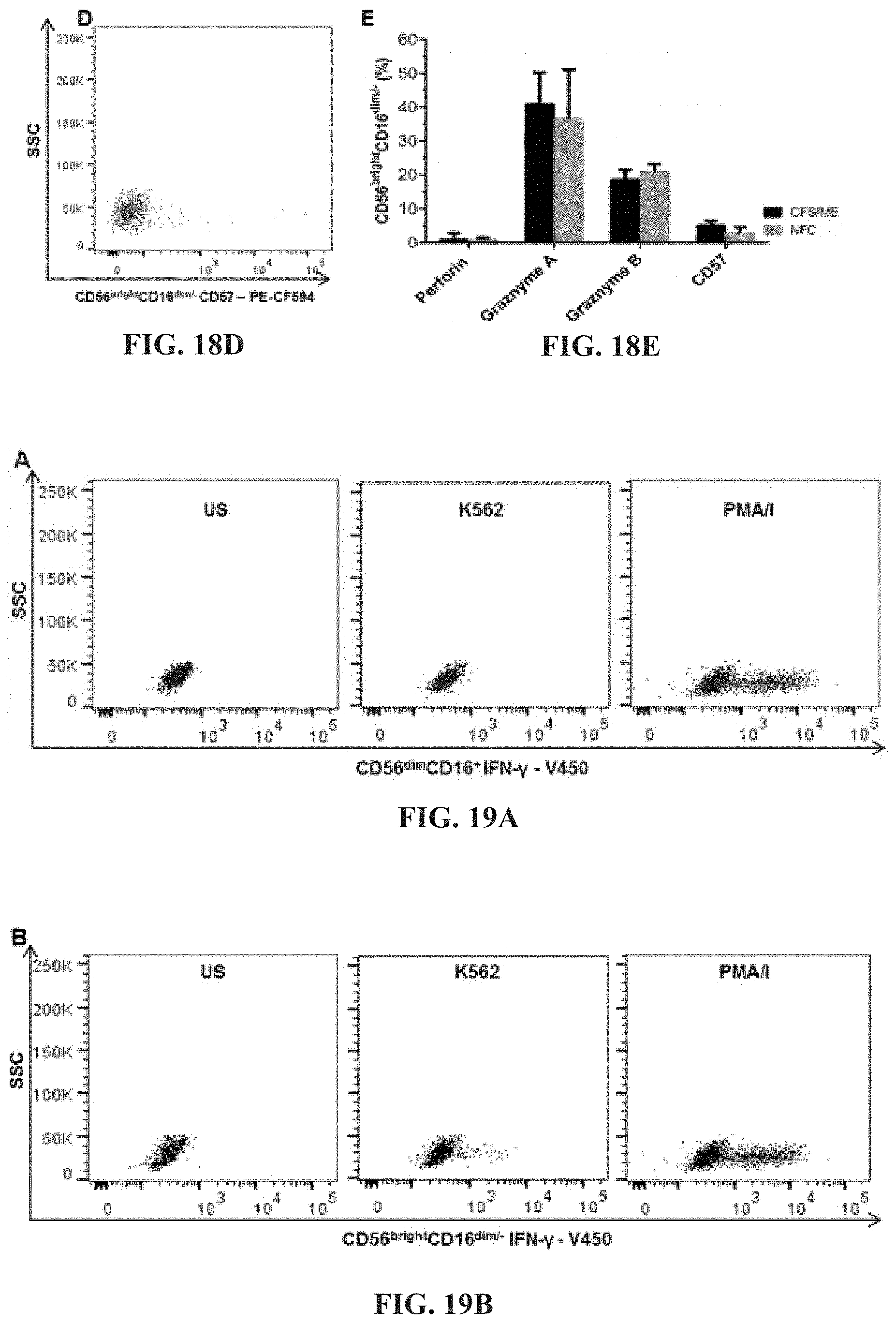
D00013
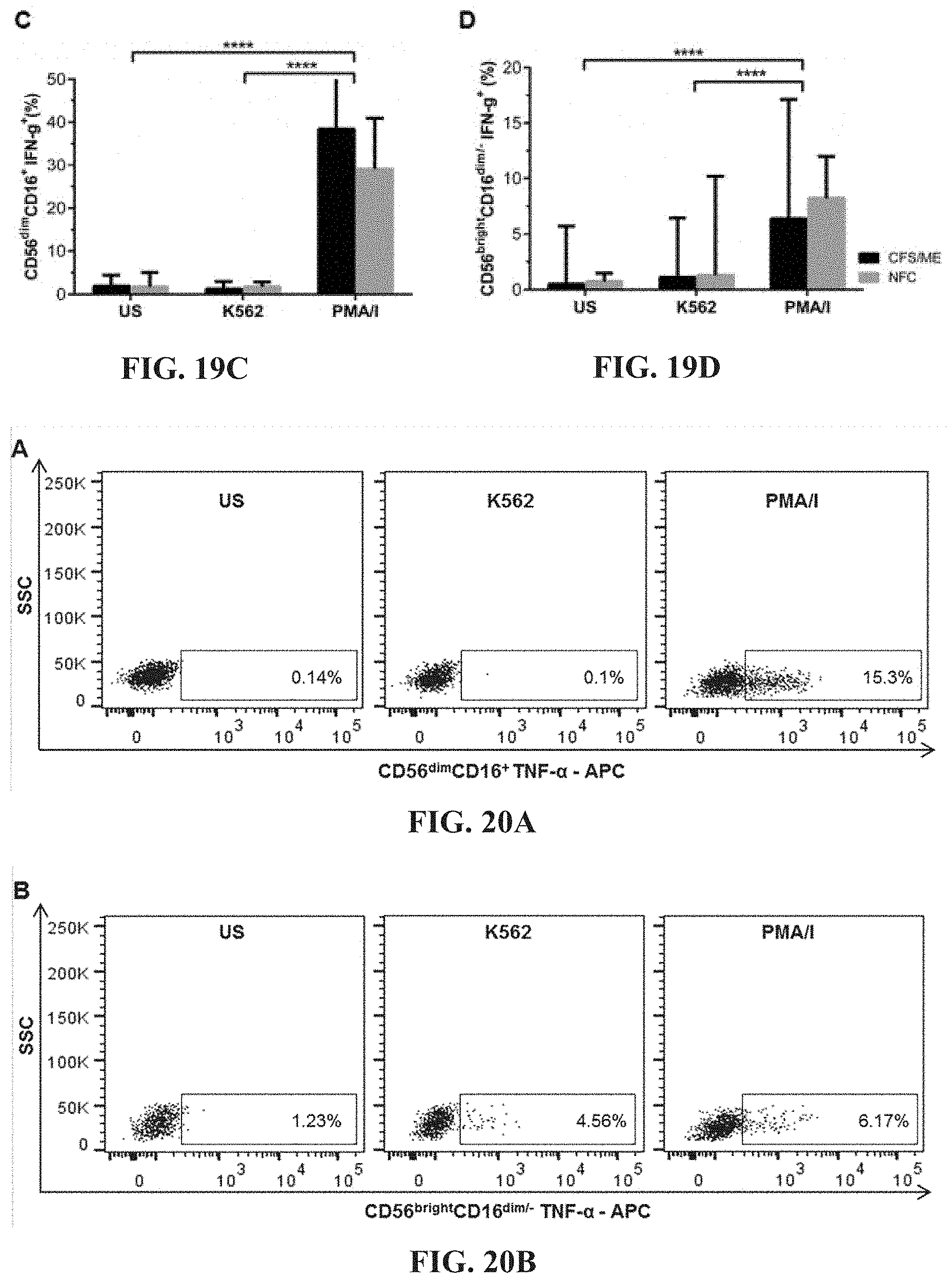
D00014
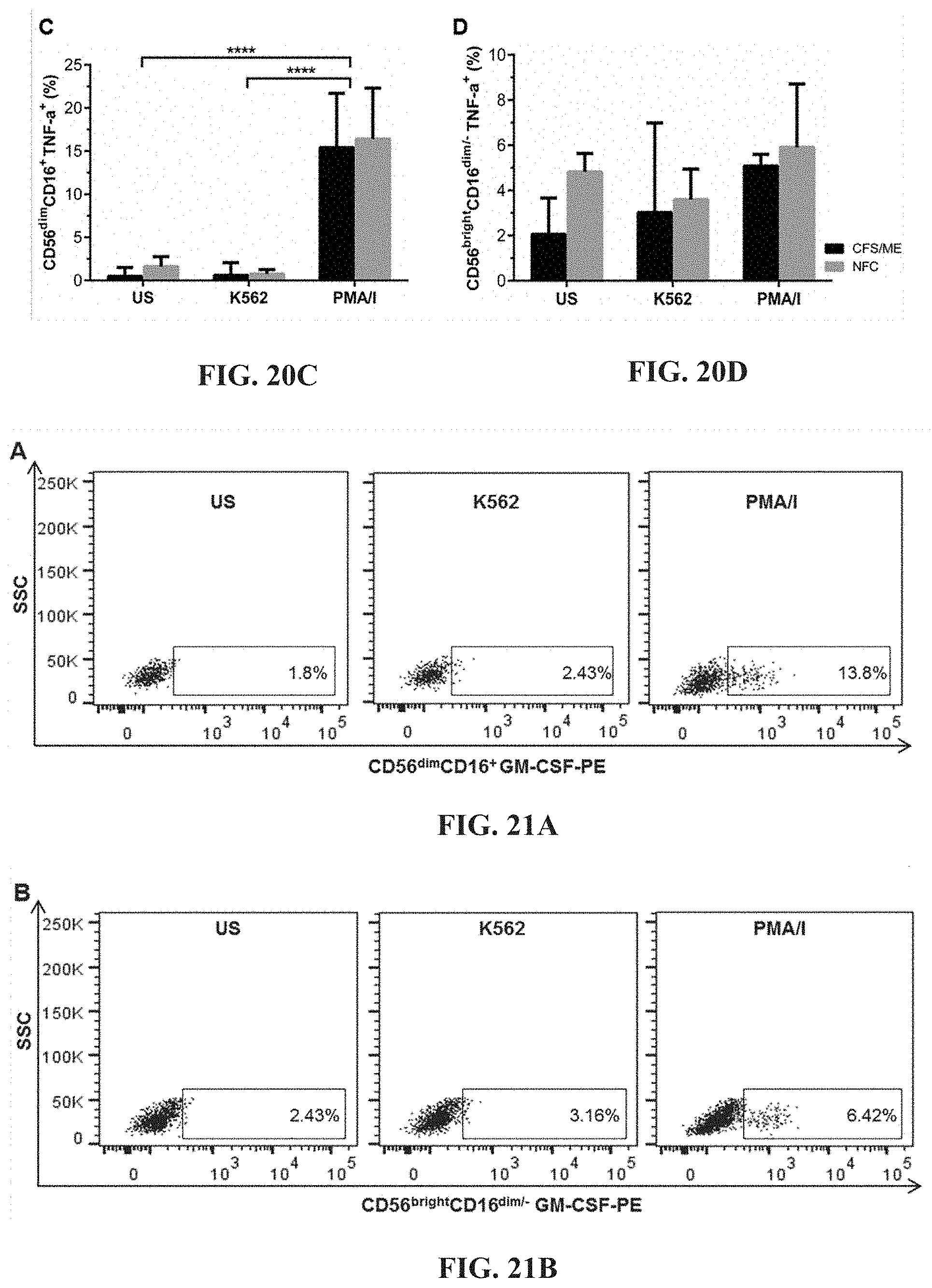
D00015

D00016

D00017
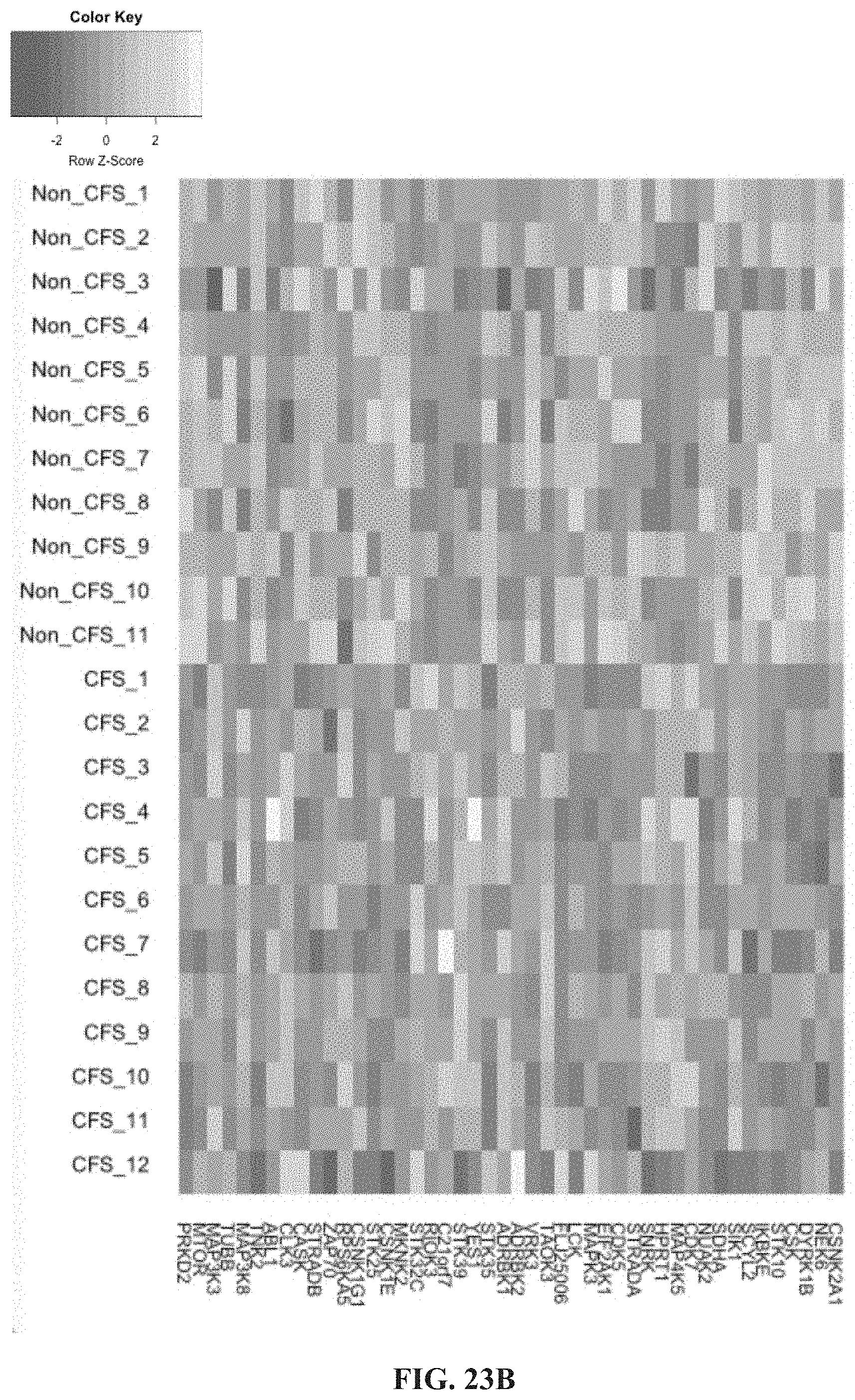
D00018

D00019

D00020
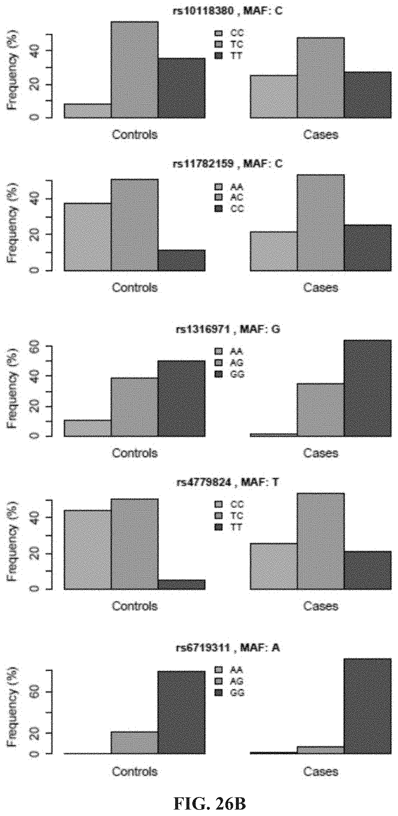
D00021
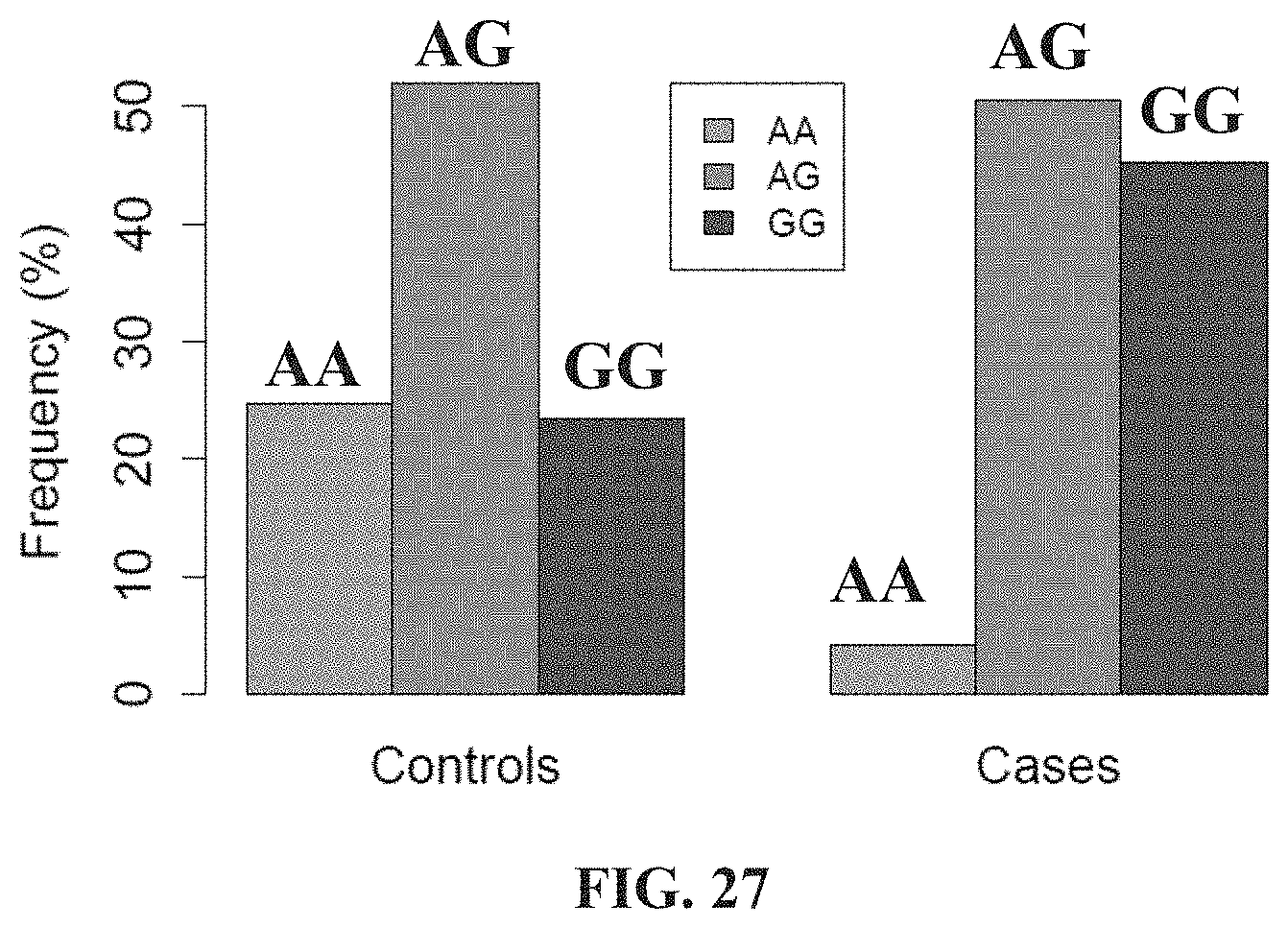
D00022
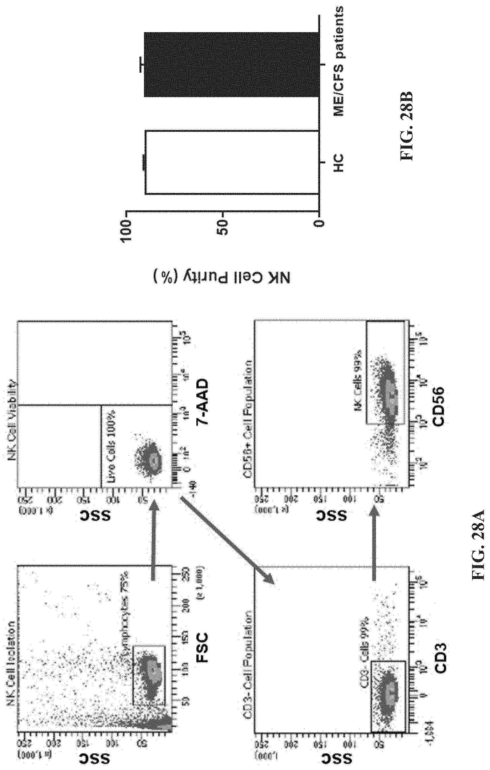
D00023

D00024
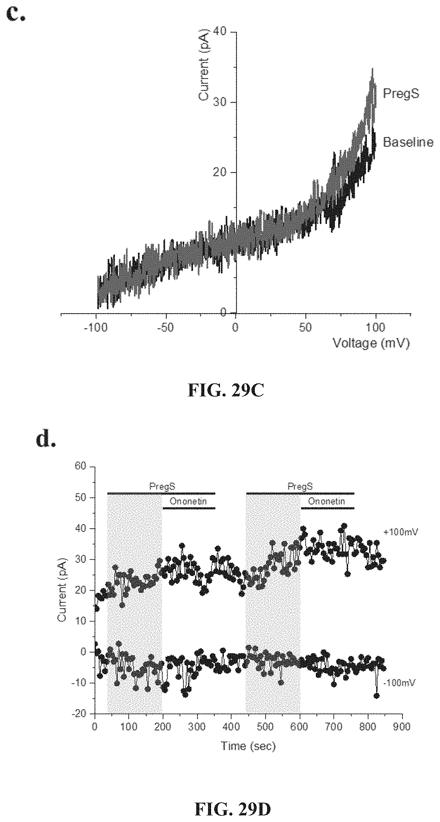
D00025
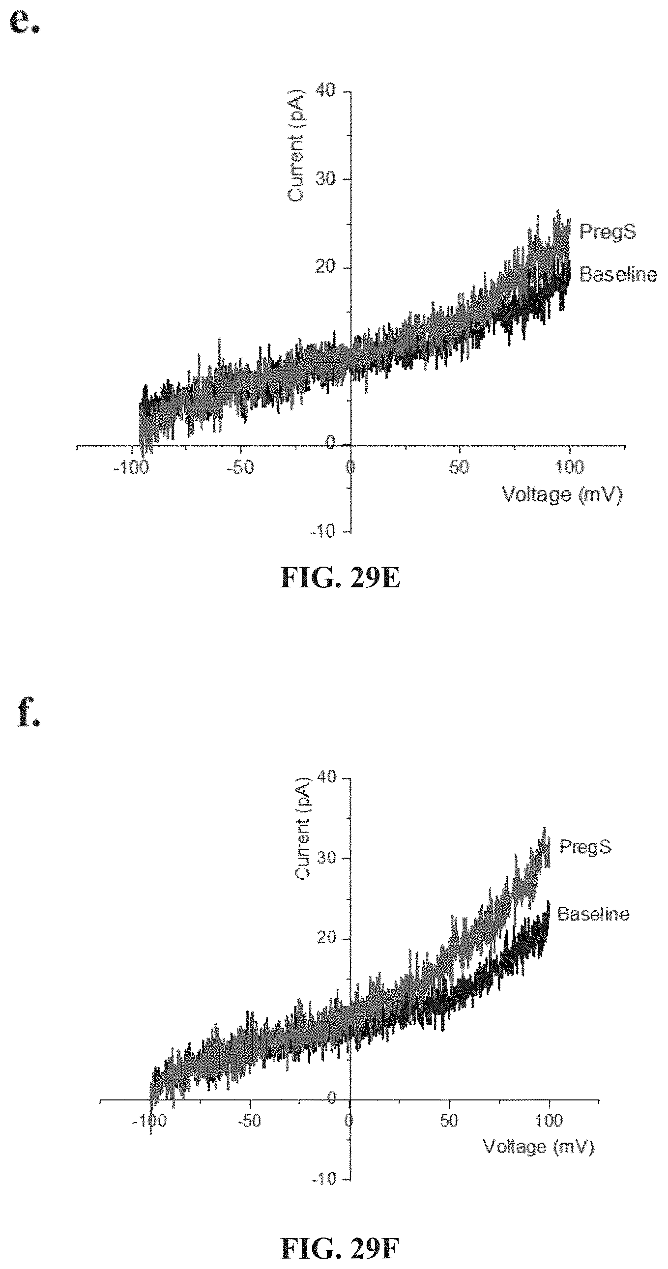
D00026

D00027
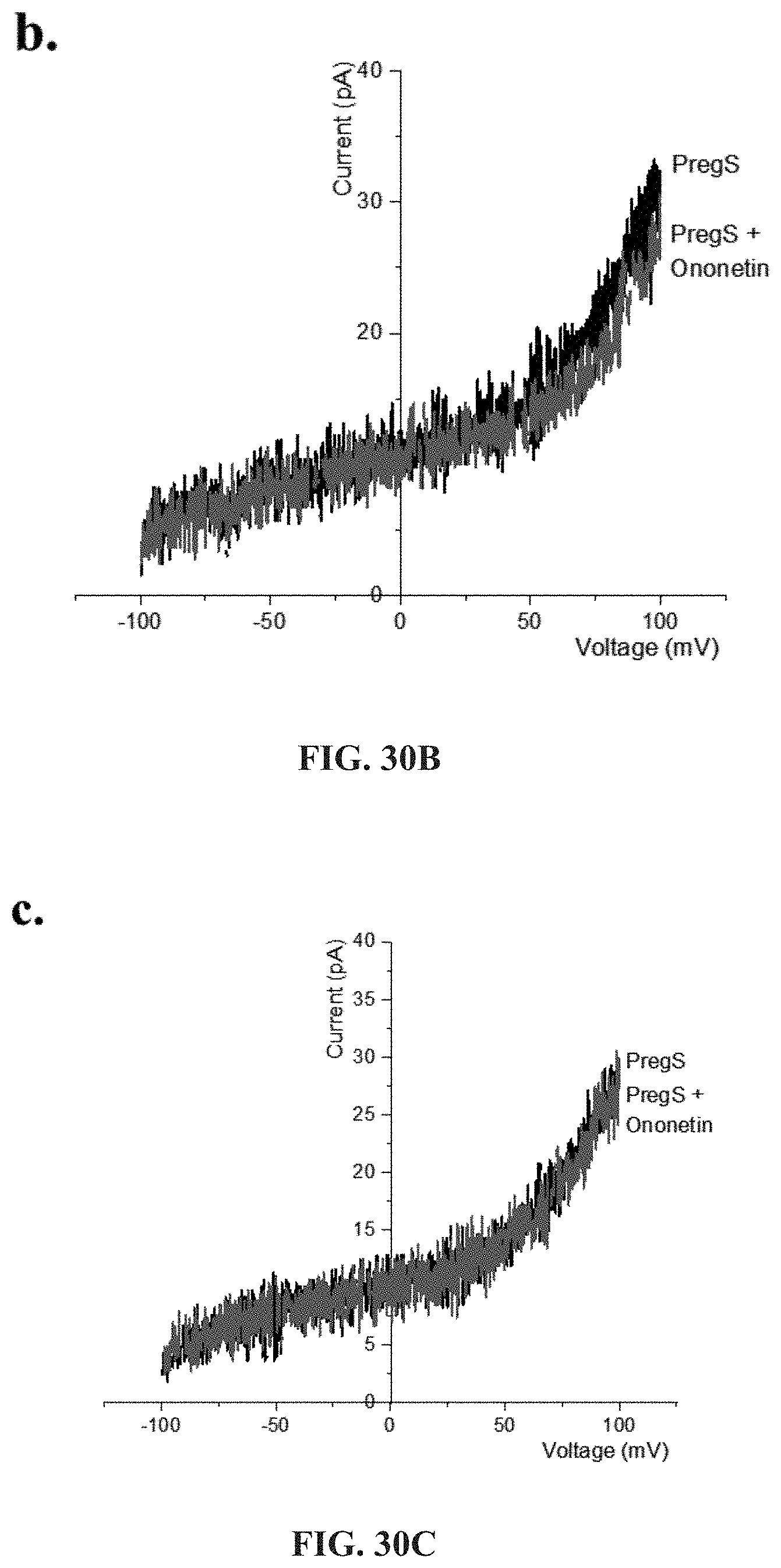
D00028
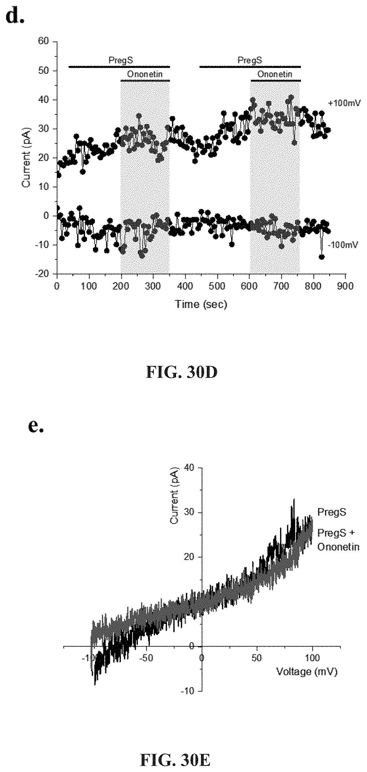
D00029
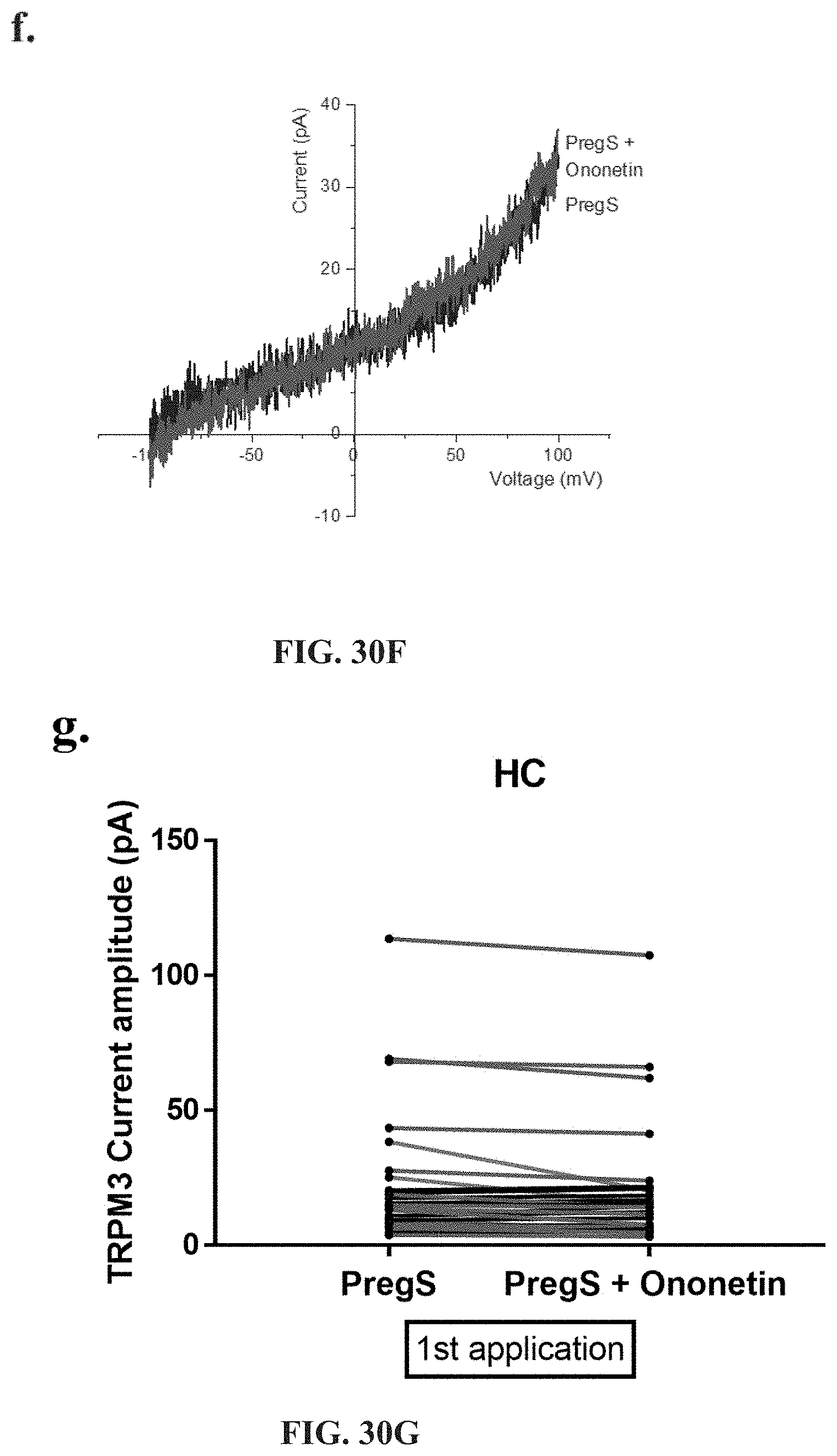
D00030
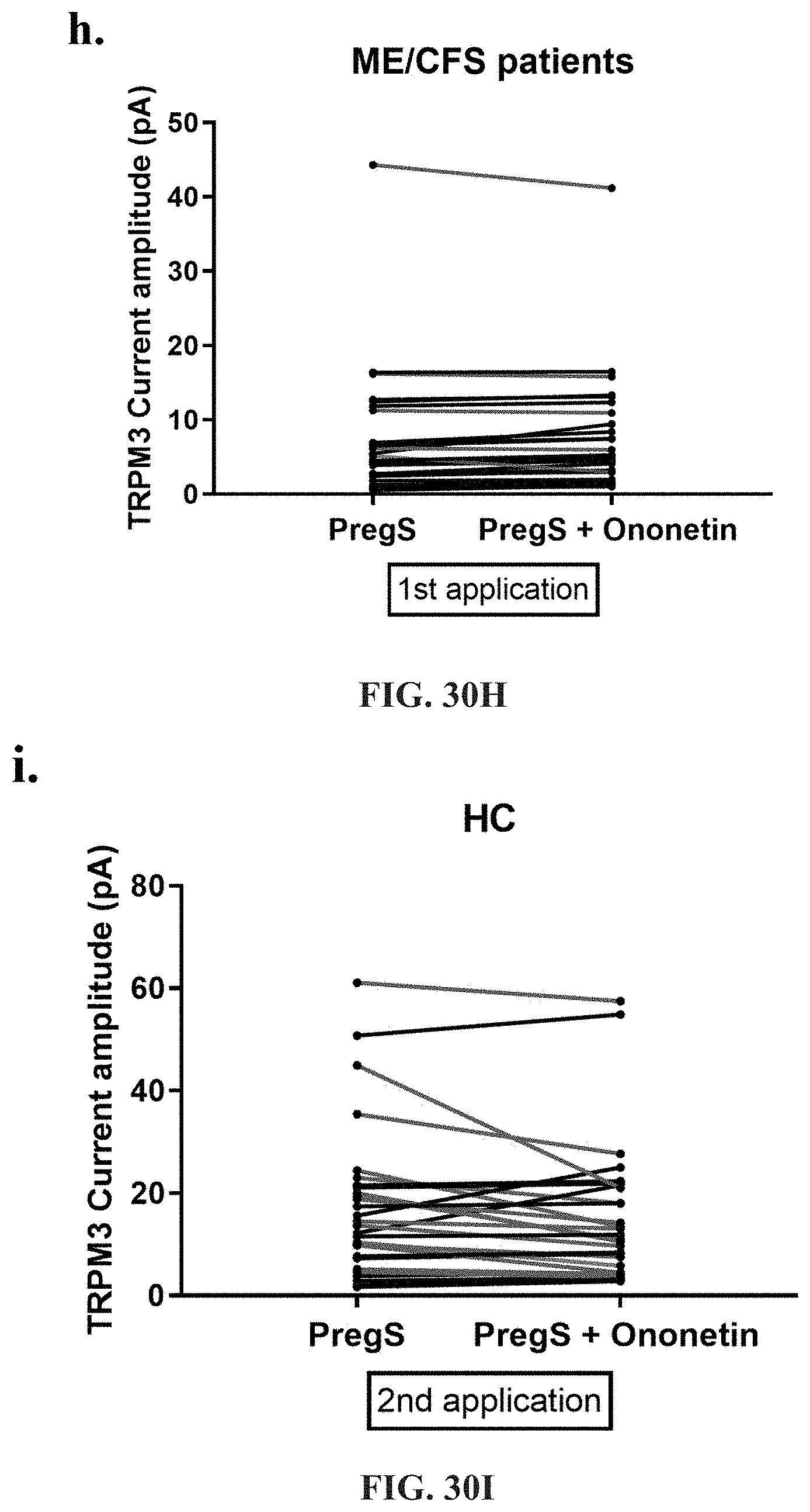
D00031

D00032
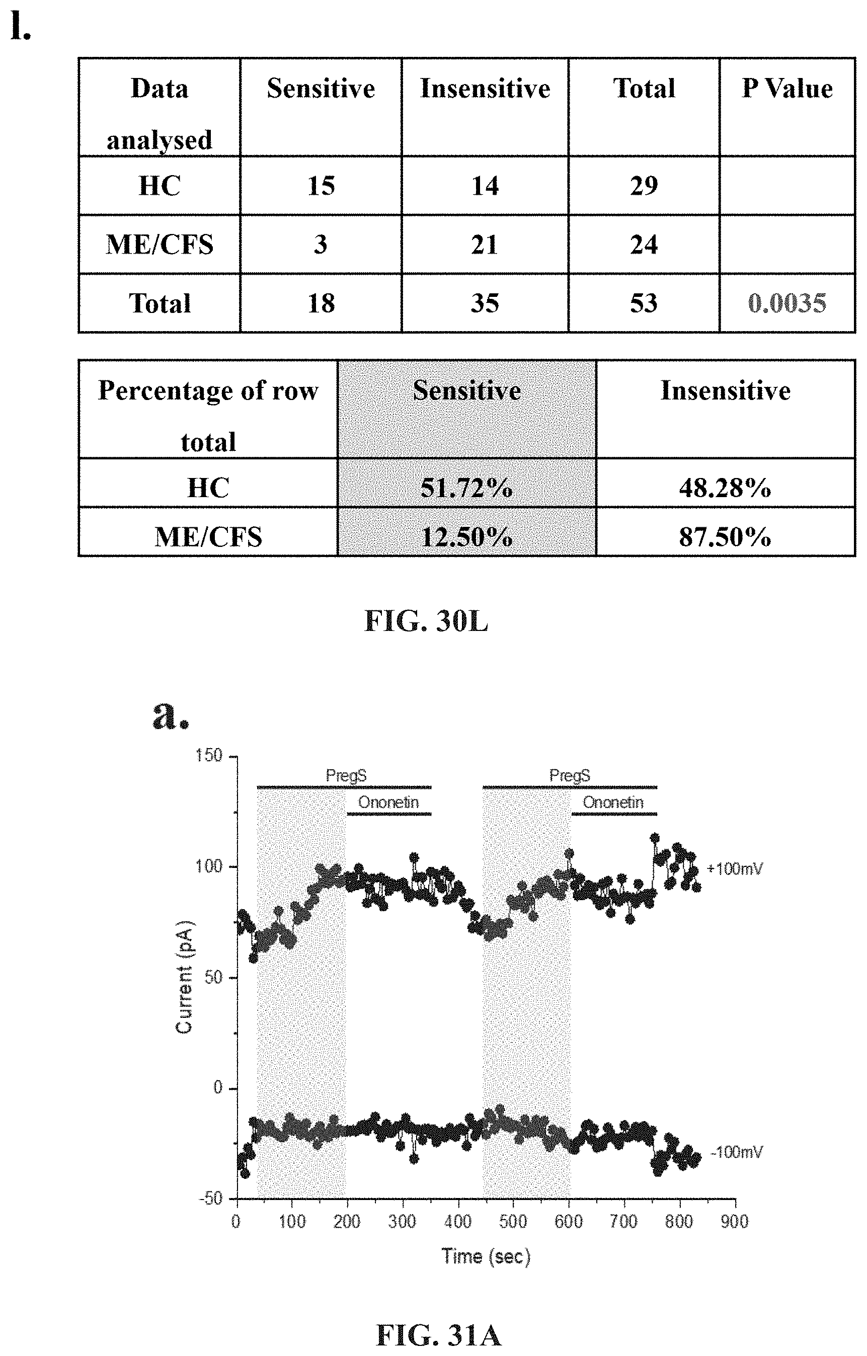
D00033
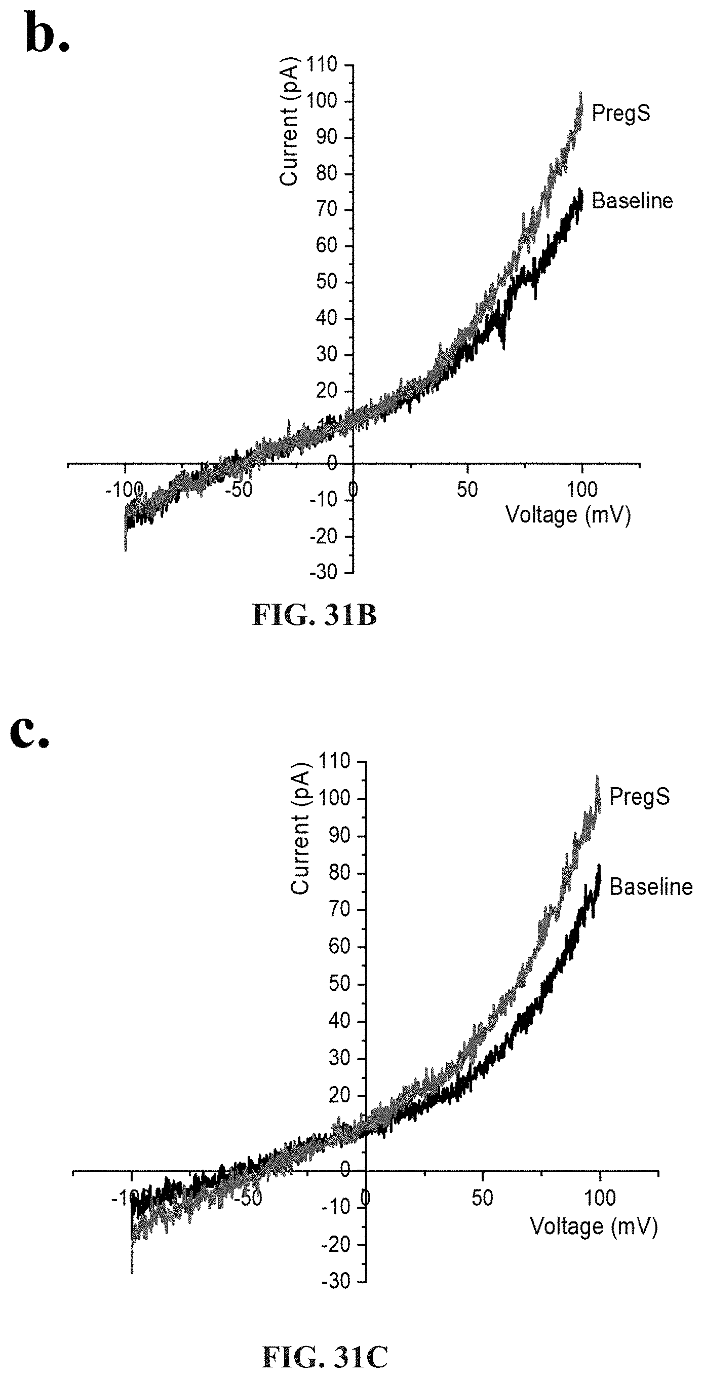
D00034
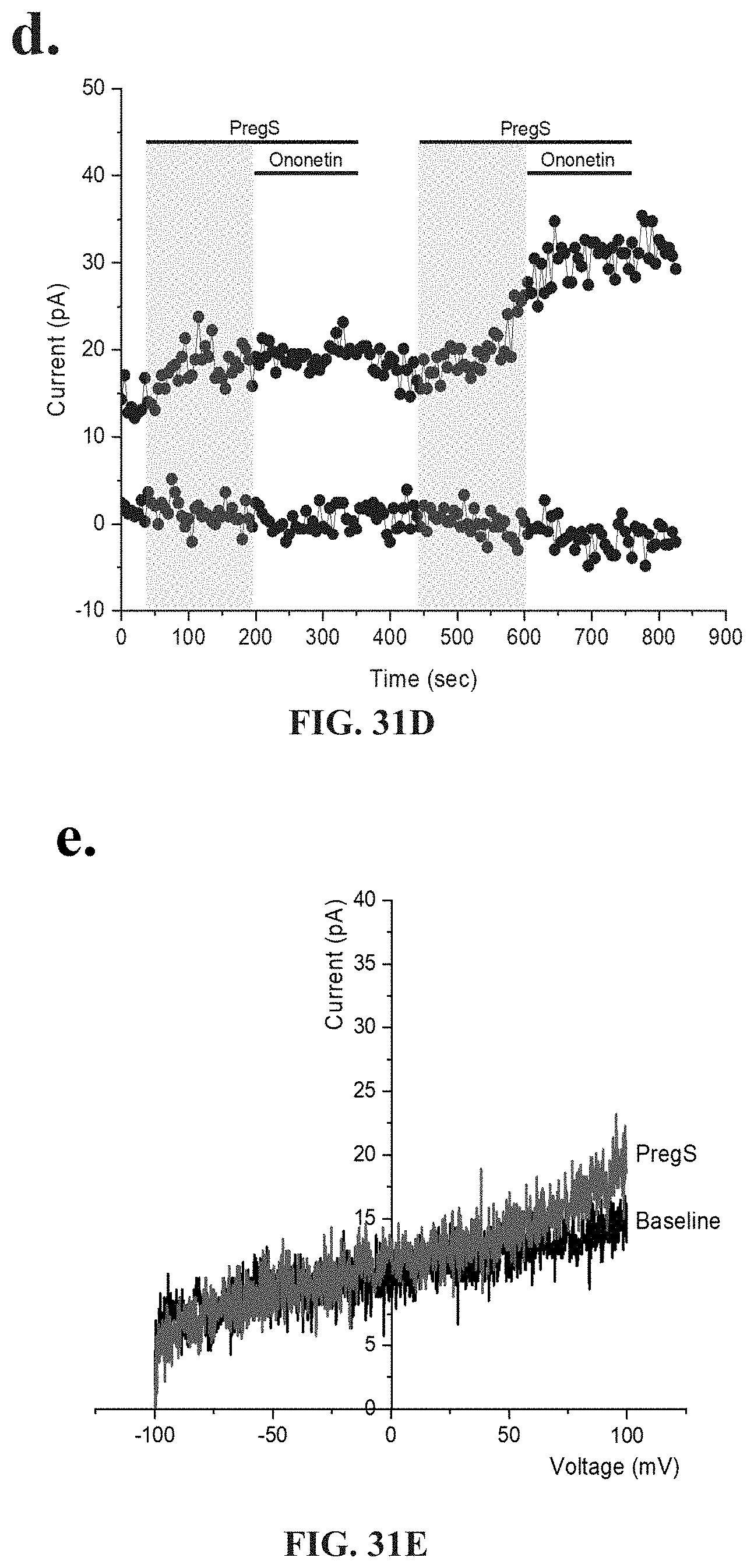
D00035
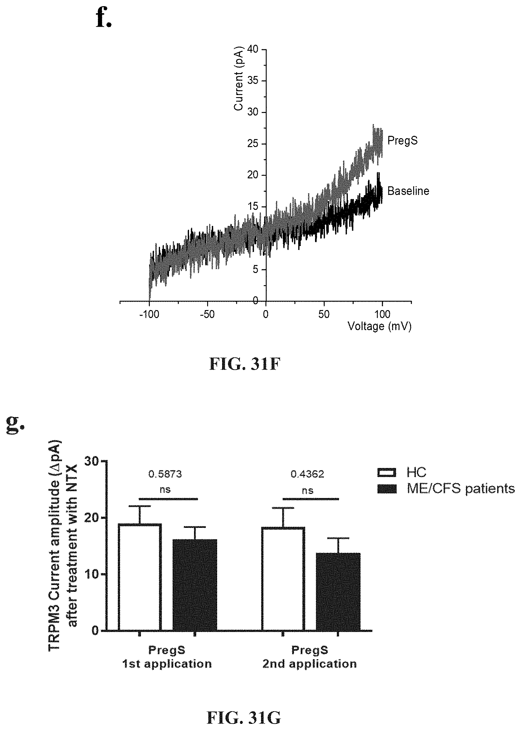
D00036
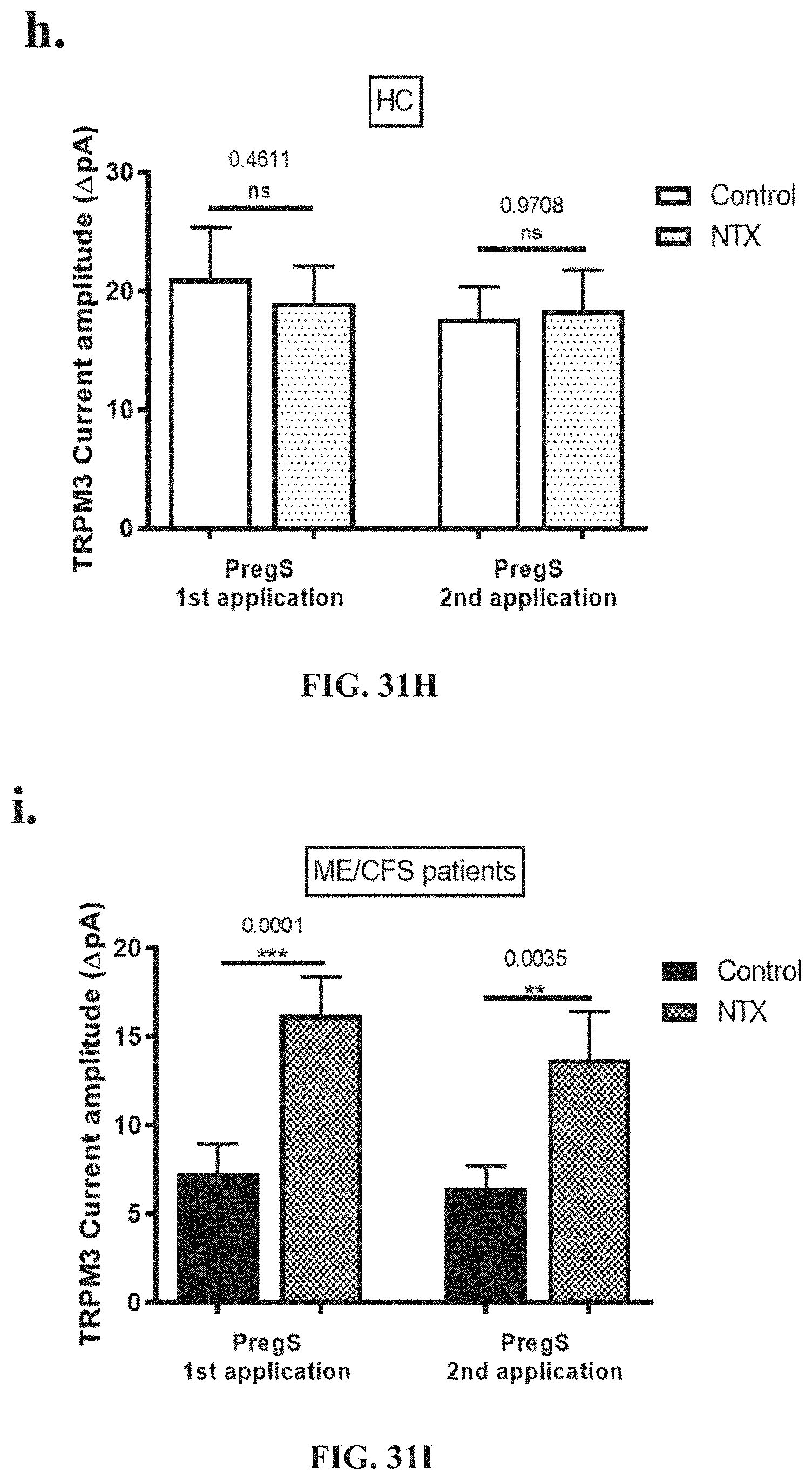
D00037
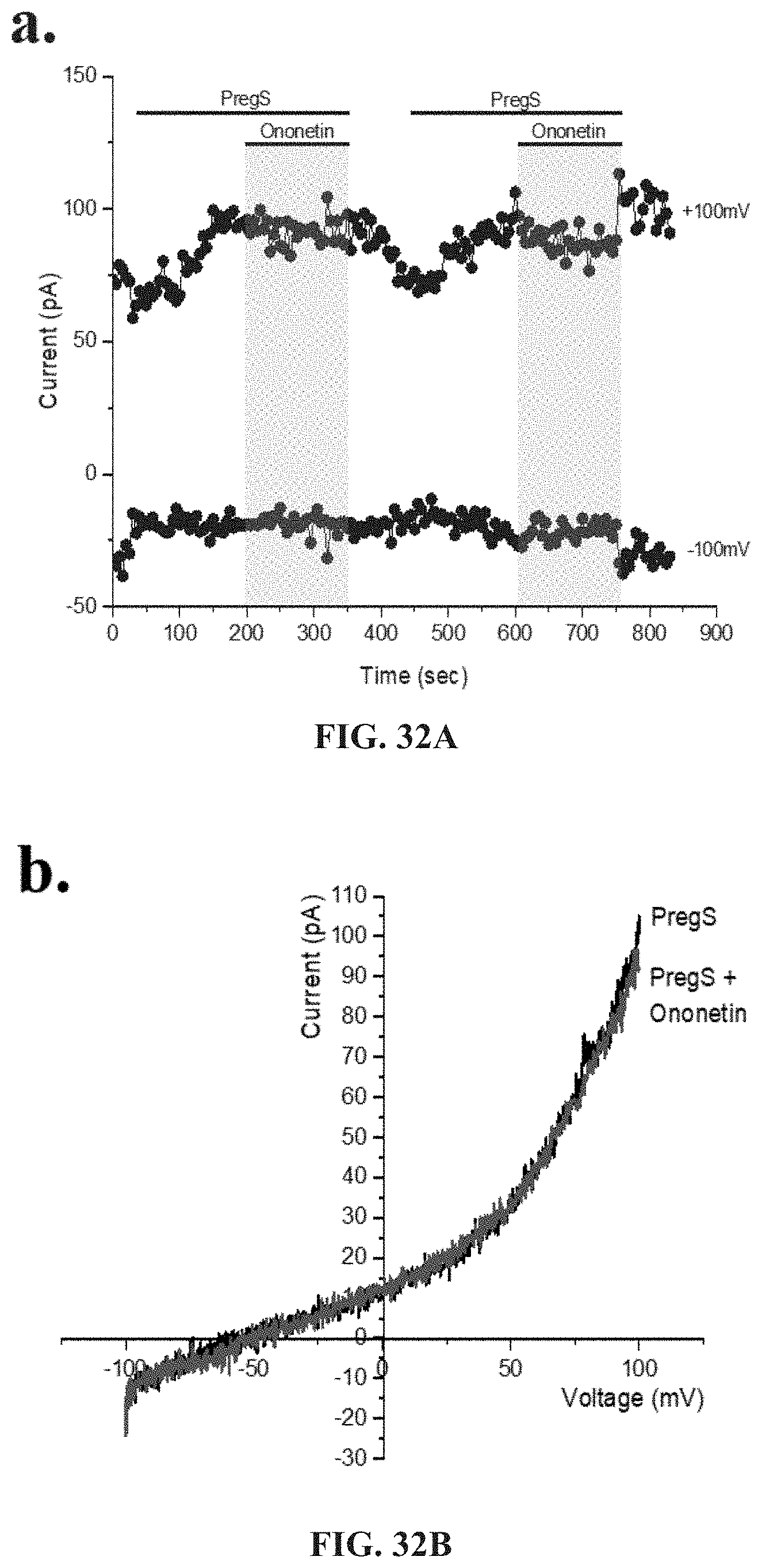
D00038
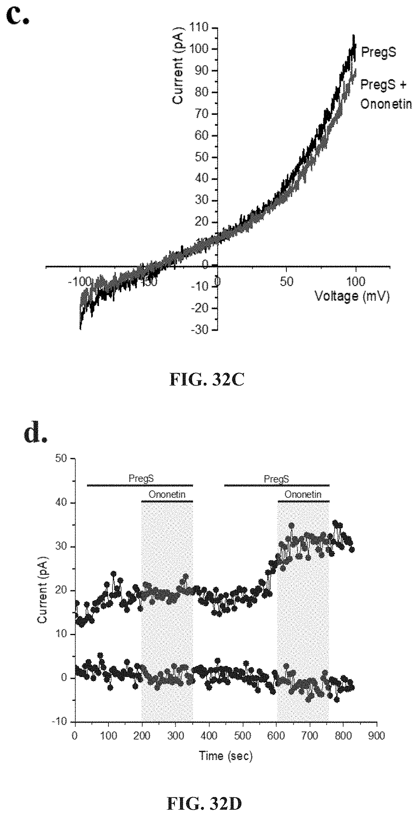
D00039

D00040

D00041

D00042

D00043
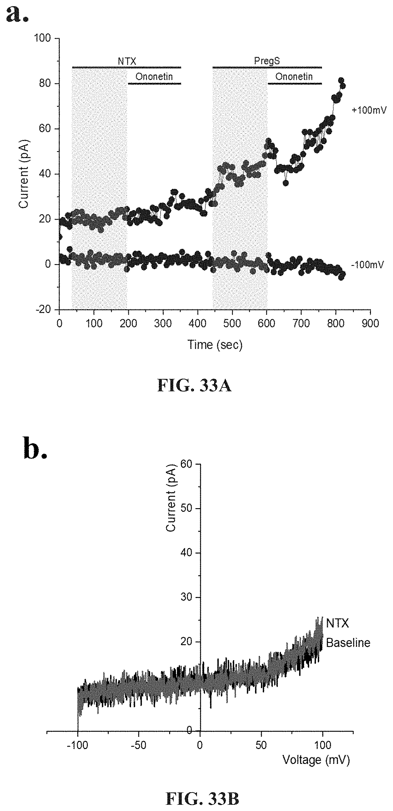
D00044

D00045
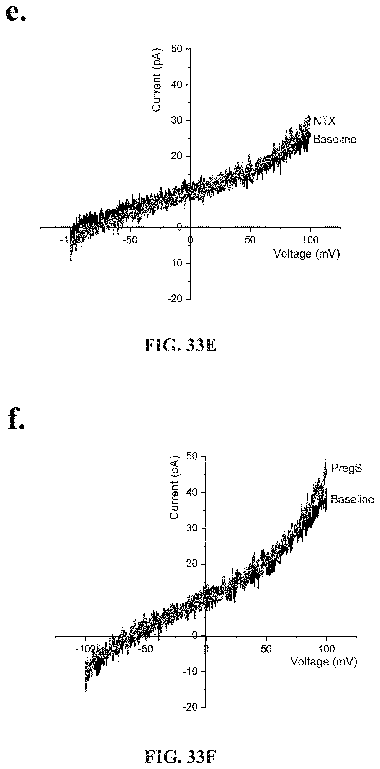
D00046

D00047

D00048
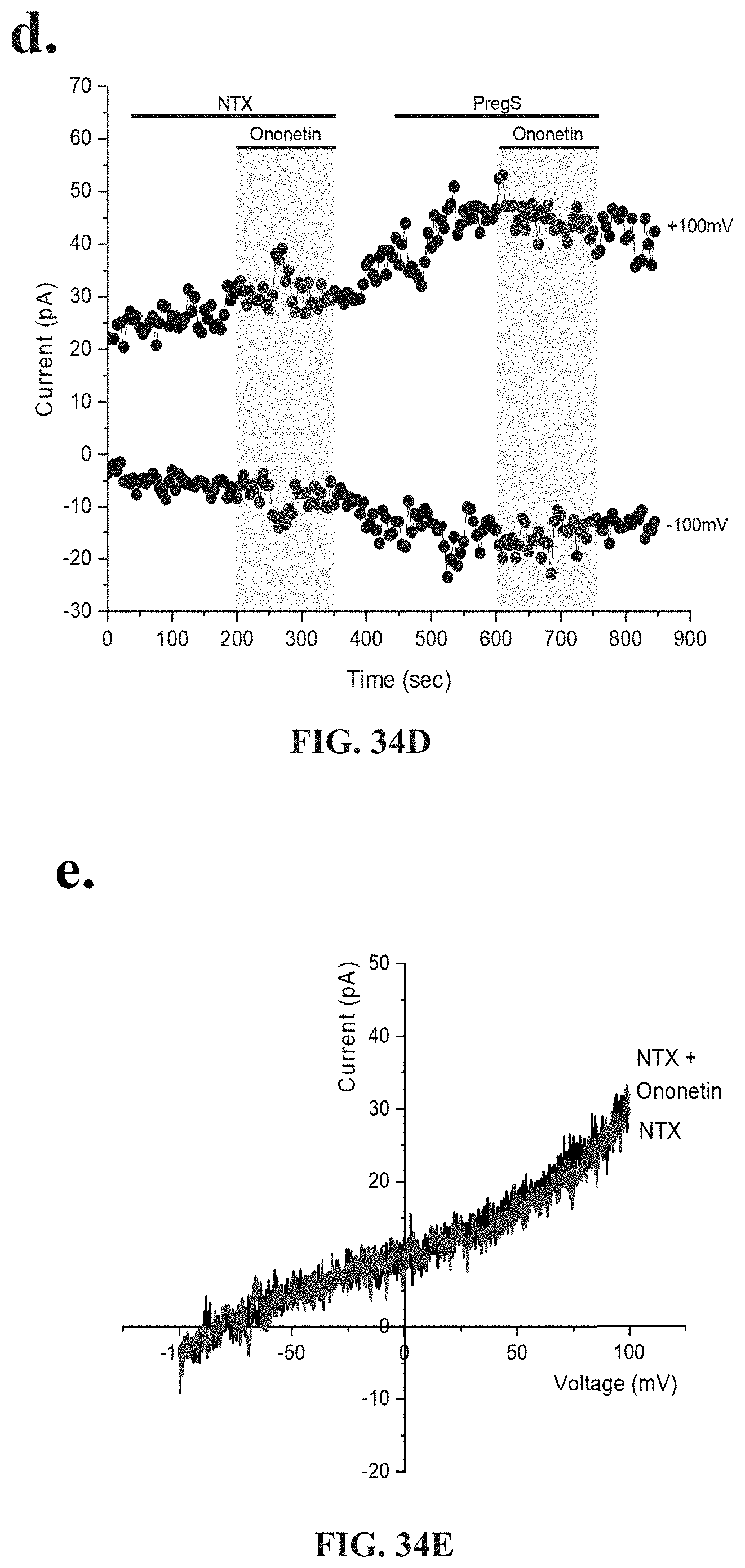
D00049

D00050
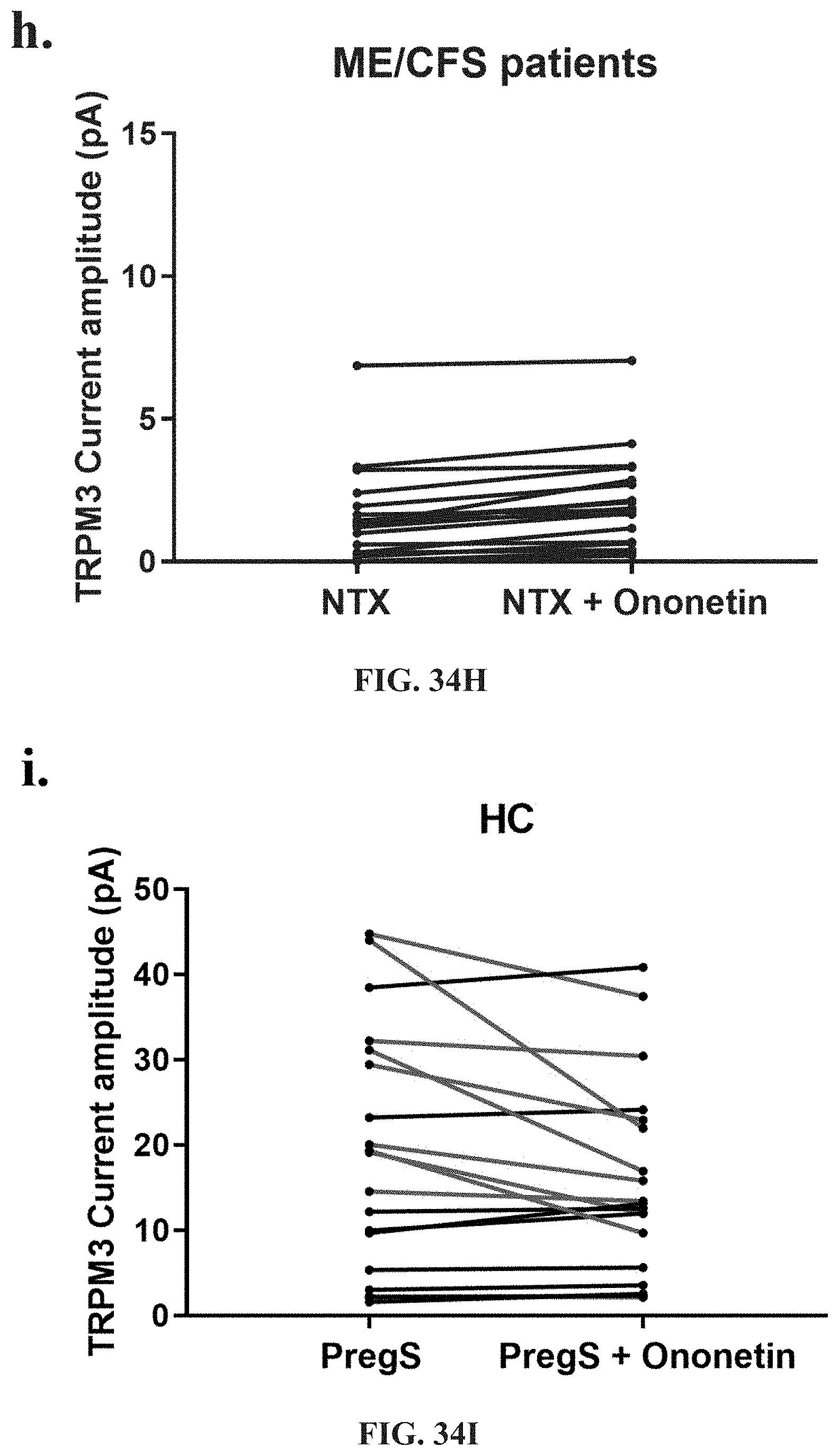
D00051
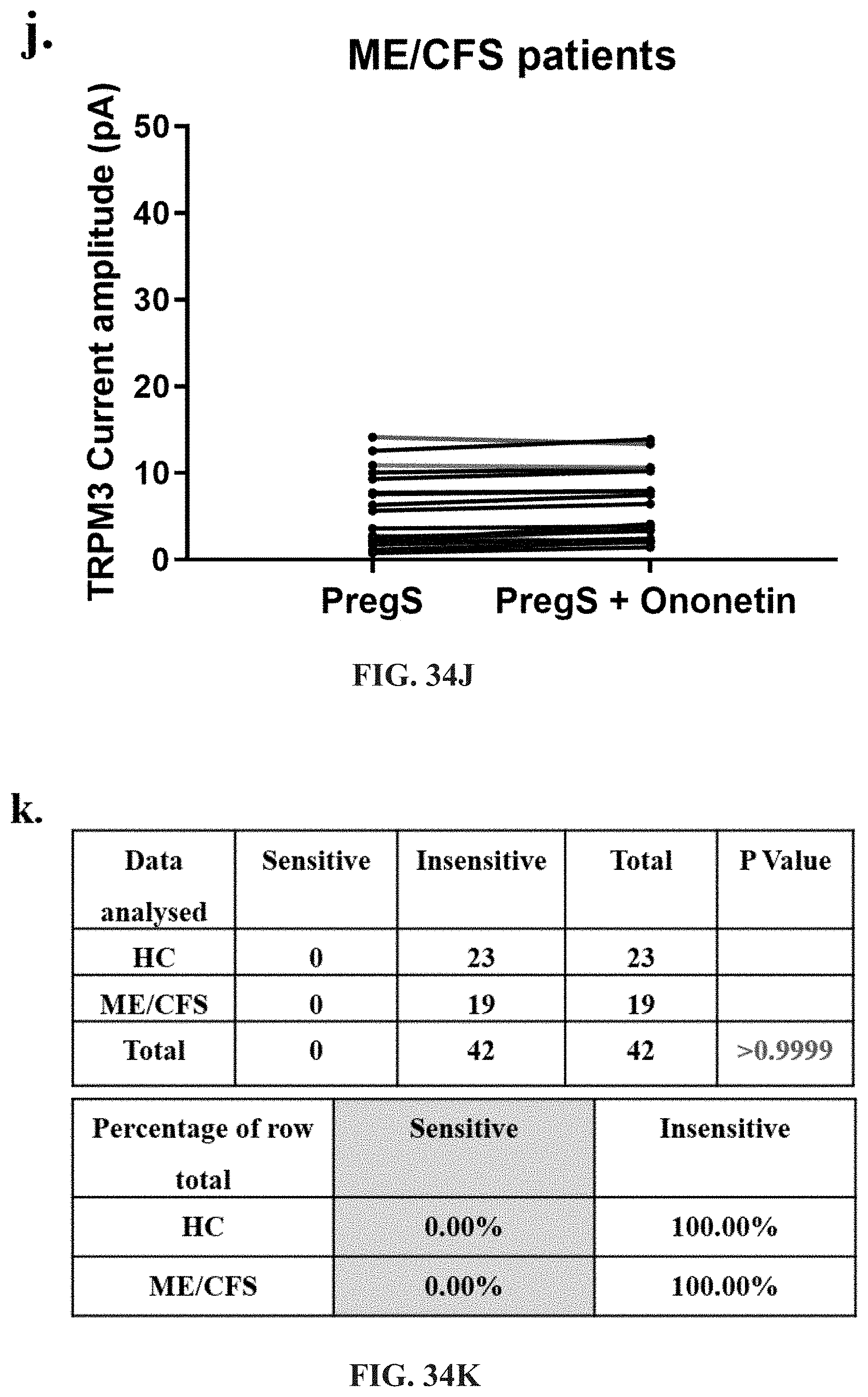
D00052
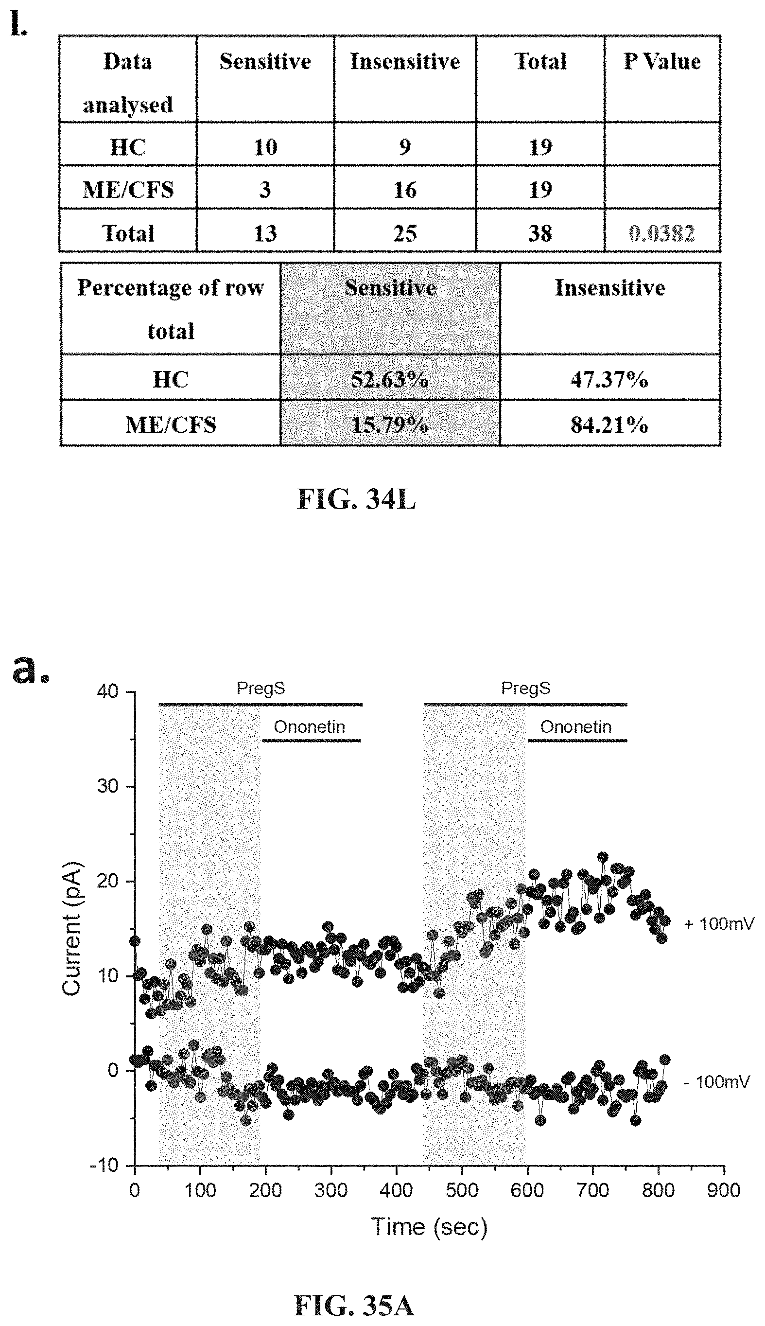
D00053
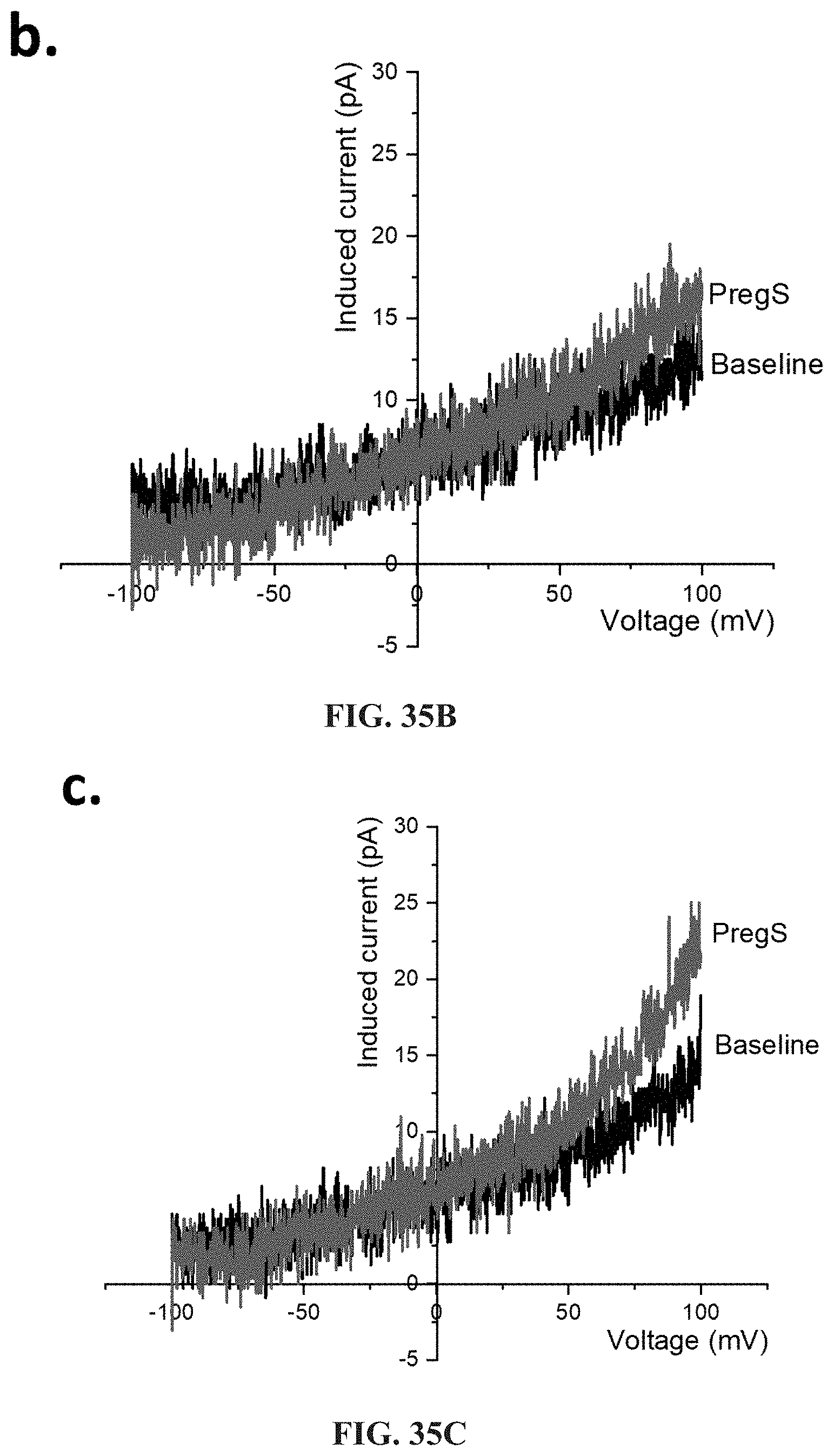
D00054
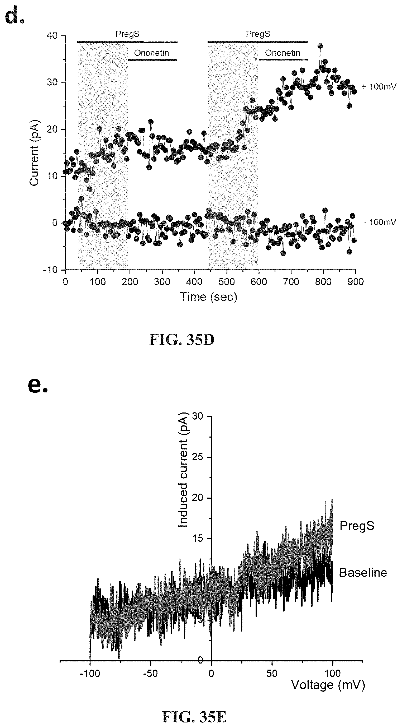
D00055

D00056
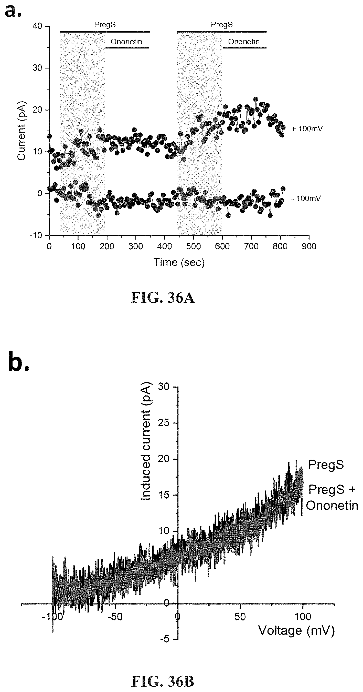
D00057
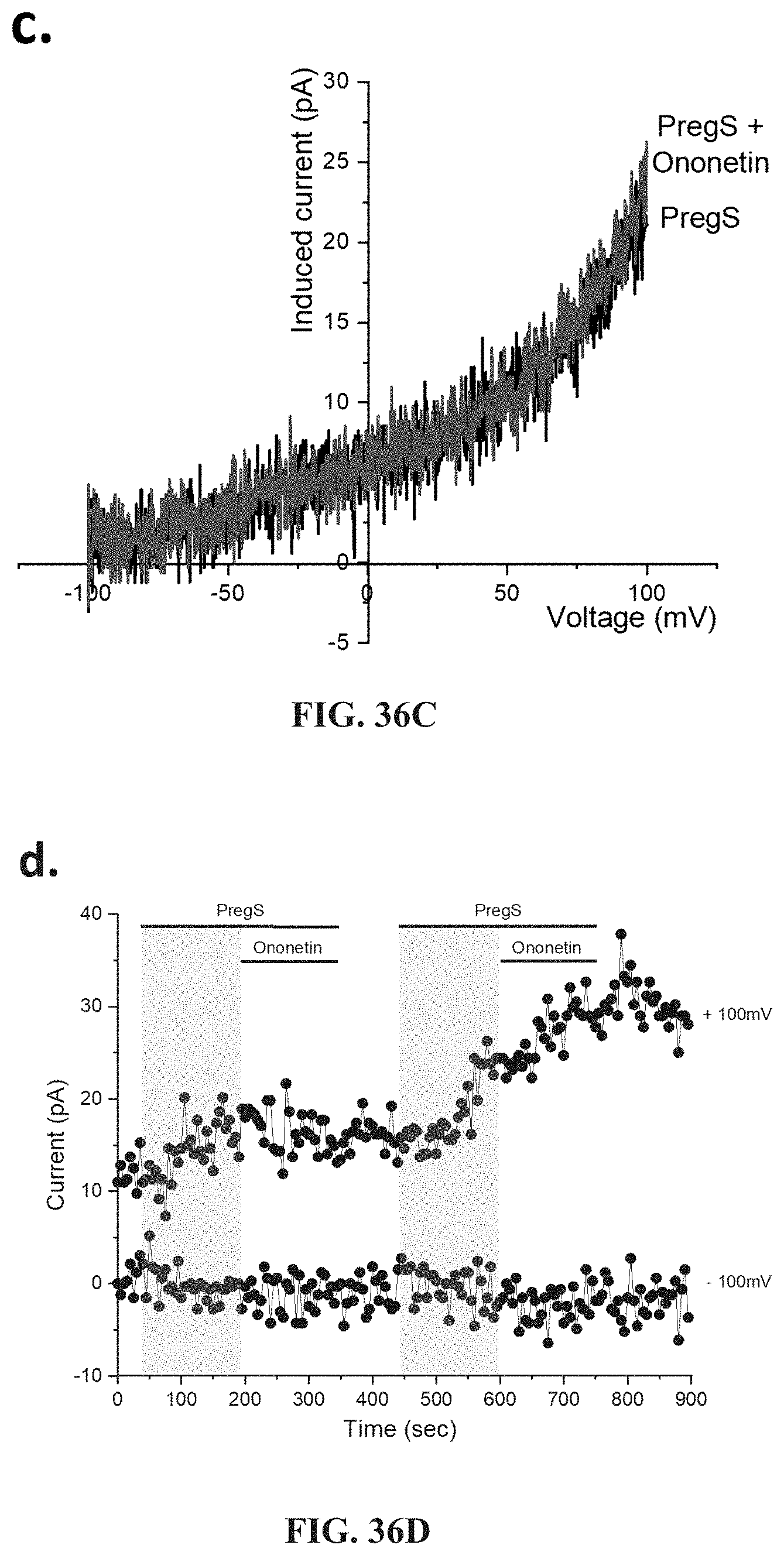
D00058

D00059

D00060
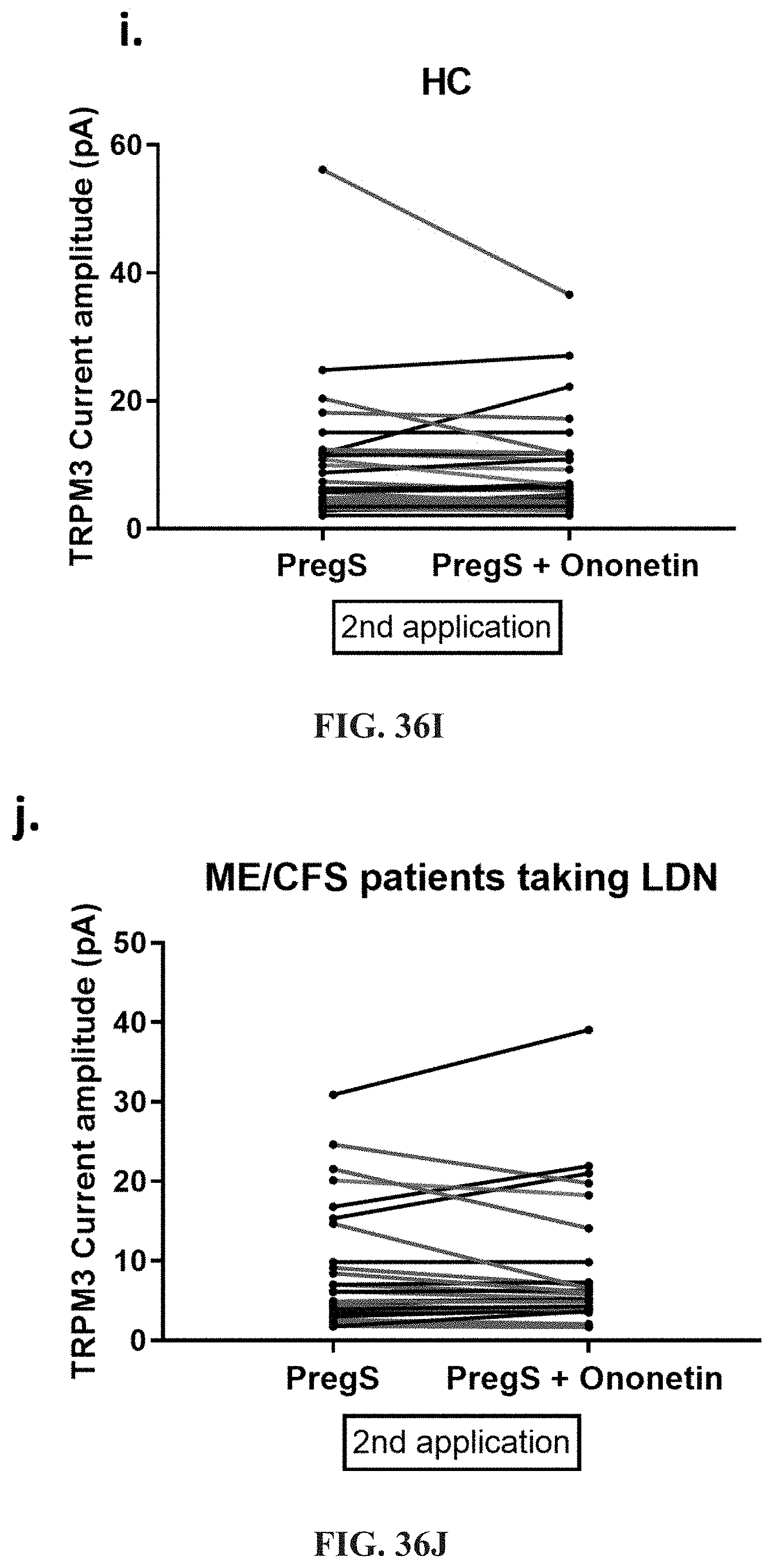
D00061

S00001
XML
uspto.report is an independent third-party trademark research tool that is not affiliated, endorsed, or sponsored by the United States Patent and Trademark Office (USPTO) or any other governmental organization. The information provided by uspto.report is based on publicly available data at the time of writing and is intended for informational purposes only.
While we strive to provide accurate and up-to-date information, we do not guarantee the accuracy, completeness, reliability, or suitability of the information displayed on this site. The use of this site is at your own risk. Any reliance you place on such information is therefore strictly at your own risk.
All official trademark data, including owner information, should be verified by visiting the official USPTO website at www.uspto.gov. This site is not intended to replace professional legal advice and should not be used as a substitute for consulting with a legal professional who is knowledgeable about trademark law.