Albumin Binding Peptide-drug (aibiped) Conjugates And Methods Of Making And Using Same
Chilkoti; Ashutosh ; et al.
U.S. patent application number 16/964832 was filed with the patent office on 2021-03-04 for albumin binding peptide-drug (aibiped) conjugates and methods of making and using same. The applicant listed for this patent is Duke University. Invention is credited to Ashutosh Chilkoti, Parisa Yousefpour.
| Application Number | 20210060171 16/964832 |
| Document ID | / |
| Family ID | 1000005239674 |
| Filed Date | 2021-03-04 |


View All Diagrams
| United States Patent Application | 20210060171 |
| Kind Code | A1 |
| Chilkoti; Ashutosh ; et al. | March 4, 2021 |
ALBUMIN BINDING PEPTIDE-DRUG (AIBIPED) CONJUGATES AND METHODS OF MAKING AND USING SAME
Abstract
Described herein are albumin binding peptide drug (AlBiPeD) conjugates comprising a small molecule linked to an albumin binding domain (ABD) via a pH-sensitive linker and methods of purifying and using the same.
| Inventors: | Chilkoti; Ashutosh; (Durham, NC) ; Yousefpour; Parisa; (Durham, NC) | ||||||||||
| Applicant: |
|
||||||||||
|---|---|---|---|---|---|---|---|---|---|---|---|
| Family ID: | 1000005239674 | ||||||||||
| Appl. No.: | 16/964832 | ||||||||||
| Filed: | January 25, 2019 | ||||||||||
| PCT Filed: | January 25, 2019 | ||||||||||
| PCT NO: | PCT/US2019/015176 | ||||||||||
| 371 Date: | July 24, 2020 |
Related U.S. Patent Documents
| Application Number | Filing Date | Patent Number | ||
|---|---|---|---|---|
| 62622249 | Jan 26, 2018 | |||
| Current U.S. Class: | 1/1 |
| Current CPC Class: | A61P 35/00 20180101; A61K 47/65 20170801; A61K 47/64 20170801; A61K 31/704 20130101 |
| International Class: | A61K 47/64 20060101 A61K047/64; A61K 31/704 20060101 A61K031/704; A61K 47/65 20060101 A61K047/65; A61P 35/00 20060101 A61P035/00 |
Goverment Interests
STATEMENT REGARDING FEDERALLY-SPONSORED RESEARCH OR DEVELOPMENT
[0002] This invention was made with government support under grant numbers R01EB000188 and R01EB007205 awarded by the National Institutes of Health. The government has certain rights in the invention.
Claims
1. A composition comprising: an albumin binding domain (ABD); a linker coupled to the ABD; and at least one molecule coupled to the linker.
2. The composition of claim 1, wherein the ABD comprises SEQ ID NO:1
3. The composition of claim 1, wherein the linker comprises a cysteine, and the at least one molecule is coupled to the cysteine.
4. The composition of claim 1, wherein the linker comprises an amino acid sequence of (CGG).sub.z (SEQ ID NO:3), wherein z is greater than 1 and wherein the linker is conjugated to the N or C terminus of the ABD.
5. The composition of claim 4, wherein z is 4 or 8.
6. The composition of claim 1, wherein the at least one molecule is attached to the linker through a pH labile bond.
7. The composition of claim 1, wherein the at least one molecule is attached to the linker through a hydrazone bond.
8. The composition of claim 1, wherein the at least one molecule is a chemotherapeutic or an imaging agent.
9. The composition of claim 1, wherein the at least one molecule is doxorubicin.
10. The composition of claim 1, wherein the at least one molecule has an octanol-water distribution coefficient (log D) of greater than or equal to 1.5 at a pH of 7.4 when the molecule is not attached to the ABD.
11. The composition of claim 1, wherein the at least one molecule comprises one or two of the molecules.
12. A method of killing cancer cells comprising contacting cancer cells with an effective amount of the composition of claim 1.
13. A method of treating a disease or disorder in a subject comprising administering to the subject a therapeutically effective amount of the composition of claim 1.
14. The method of claim 13, wherein the disease or disorder is cancer.
15. A method of purifying the composition of claim 1, comprising forming a conjugate comprising, an elastin-like polypeptide having a transition temperature (T.sub.t) above 50.degree. C.; and the composition of claim 1, wherein the composition is conjugated to a first end of the elastin-like polypeptide by an amino acid sequence amenable to cleavage; treating the conjugate with an enzyme, chemical, or combination thereof capable of cleaving the amino acid sequence amenable to cleavage; and separating the composition from the elastin-like polypeptide.
16. The method of claim 15, further comprising a destabilizing peptide conjugated to a second end to an elastin-like polypeptide.
17. The method of claim 16, wherein the destabilizing peptide comprises SEQ ID NO: 5.
18. The method of claim 15, wherein the amino acid sequence is amenable to cleavage by an enzyme, and wherein the enzyme is Sortase A.
19. The method of claim 15, wherein the amino acid sequence amenable to cleavage comprises SEQ ID NO: 6.
20. The method of claim 15, wherein the elastin-like polypeptide comprises an amino acid sequence of (VPGXaaG).sub.p(SEQ ID NO: 4), wherein Xaa is any amino acid except proline and p is 1 to 500.
21. The method of claim 20, wherein Xaa is alanine and p is 160.
Description
CROSS-REFERENCE TO RELATED APPLICATIONS
[0001] This application claims priority to U.S. Provisional Patent Application No. 62/622,249, filed Jan. 26, 2018, which is incorporated herein by reference in its entirety.
SEQUENCE LISTING
[0003] The instant application contains a Sequence Listing which has been submitted electronically and is hereby incorporated by reference in its entirety. The sequence listing text filed, created on Jan. 23, 2019, is named "028193-9315-WO01 As Filed Sequence Listing" and is 2,629 bytes in size.
TECHNICAL FIELD
[0004] The present disclosure relates to a novel drug delivery system.
BACKGROUND OF THE INVENTION
[0005] Delivery and therapeutic efficacy of small molecule imaging agents and chemotherapeutics are hampered by their short half-life, low solubility, non-selectivity to cancer cells, and toxic side effects. Small molecule chemotherapeutics, although in routine use for cancer treatment, suffer from a short circulation half-life and indiscriminate accumulation in healthy tissues that result in systemic toxicities and hence limit their maximum dose. These limitations inhibit accumulation of chemotherapeutics in tumors at therapeutic levels and limit their clinical application. Efforts in past decades have been focused on developing macromolecular and nanoparticulate drug formulations that prevent first-pass elimination in kidneys and allow for selective accumulation in tumors via the enhanced permeation and retention (EPR) effect. However, the interaction of these macromolecule and nanoparticle carriers with serum proteins and components of the immune system is not well understood and is affected by several factors such as their interfacial chemistry, size, shape and stability which makes their optimization difficult. Therefore, there remains a need for new delivery systems that can overcome these disadvantages yet provide efficacy for delivery of small molecule imaging agents and chemotherapeutics.
BRIEF SUMMARY OF THE INVENTION
[0006] The present disclosure is directed to compositions comprising an albumin binding domain (ABD), a linker coupled to the ABD, and at least one molecule coupled to the linker.
[0007] The present disclosure is directed to methods of killing cancer cells comprising contacting cancer cells with an effective amount of the compositions disclosed herein.
[0008] The present disclosure is also directed to methods of treating a disease or disorder in a subject comprising administering to the subject a therapeutically effective amount of the compositions disclosed herein. The disease or disorder may be cancer.
[0009] The present disclosure is further directed to methods of purifying the compositions disclosed herein. The methods may comprise forming a conjugate comprising an elastin-like polypeptide having a transition temperature (T.sub.t) above 50.degree. C.; and the compositions disclosed herein, wherein the composition is conjugated to a first end of the elastin-like polypeptide by an amino acid sequence amenable to cleavage, treating the conjugate with an enzyme, chemical or combination thereof capable of cleaving the amino acid sequence amenable to cleavage, and separating the composition from the elastin-like polypeptide.
[0010] Other aspects of the invention will become apparent by consideration of the detailed description and accompanying drawings.
BRIEF DESCRIPTIONS OF THE DRAWINGS
[0011] FIG. 1 is a schematic of the conjugate of doxorubicin with an albumin binding domain that binds to and co-opts albumin in vivo for drug delivery to tumors.
[0012] FIG. 2A, FIG. 2B, and FIG. 2C show ABD-Dox synthesis. FIG. 2A is a schematic showing the design and synthesis steps. ELP was used as a purification tag for ABD purification from bacteria and was removed following drug conjugation using sortase A. The KEKE (SEQ ID NO: 5) peptide was included at the N-terminus to disrupt micelle self-assembly upon Dox conjugation, and to enable the subsequent sortase A cleavage of the ELP from the ABD-Dox conjugate. FIG. 2B is an SDS-PAGE analysis confirming successful purification of KEKE-ELP.sub.160-srt-ABD-(GGC).sub.4 and (His).sub.6-sortase A-ELP.sub.40 with theoretical molecular weights of 69.4 kDa and 34.4 KDa, respectively. FIG. 2C is a graph of the hydrodynamic radius of ELP.sub.160-srt-ABD-Dox conjugates as measured with DLS. ELP.sub.160-srt-ABD-Dox conjugates without the N-terminal KEKE segment (SEQ ID NO: 5) self-assembled into micelles with Rh of 40-50 nm, whereas conjugates containing the N-terminal KEKE segment (SEQ ID NO: 5) were mostly (.about.90%) unimers with Rh<10 nm.
[0013] FIG. 3A, FIG. 3B, FIG. 3C, FIG. 3D, FIG. 3E, and FIG. 3F show in vitro characterization of ABD-Dox. FIG. 3A is a plot showing the HPLC chromatograms of the KEKE-ELP160-srt-ABD-Dox, sortase reaction solution after 24 h incubation, and SEC-purified ABD-Dox. FIG. 3B is a plot showing the MALDI-MS spectra of ABD-Dox and vertical bars showing the mass of conjugates with 1 and 2 drug molecules. FIG. 3C is a plot showing the in vitro drug release from ABD-Dox conjugate as a function of time at pH 5.0 and pH 7.0. Dox was released at pH 5.0 corresponding to the pH in late endosomes, and showed a low, basal level of release from the conjugate at pH 7.4, corresponding to the pH in vascular and extracellular space of normal tissues. Data are presented as mean.+-.SEM, n=3. FIG. 3D is a native-PAGE of the interaction of ABD-Dox with human serum albumin (HSA) and mouse serum albumin (MSA). For lanes 3 and 5, ABD-Dox was mixed at a 1:1 molar ratio with HSA and MSA, respectively. FIG. 3E is a plot showing isothermal titration calorimetry thermograms of the interaction of ABD-Dox with HSA and MSA. The enthalpy data were fit to an independent binding site model, and the thermodynamic parameters (n, KD, .DELTA.H, and .DELTA.S) were calculated as shown in Table 1. FIG. 3F shows in vitro cytotoxicity of ABD-Dox and free Dox against C26 and MIA PaCa-2 cancer cells after 72 h incubation. The IC.sub.50 of ABD-Dox and free Dox were computed as 6.43 and 0.51 .mu.M, respectively, for C26 and as 1.41 and 0.04 .mu.M, respectively, for MIA PaCa-2 cells. Data are presented as mean.+-.SEM, n=3.
[0014] FIG. 4A and FIG. 4B are plots showing the pharmacokinetics of ABD-Dox. Plasma Dox concentration was measured over 96 h (FIG. 3A, 0-30 min view; FIG. 3B, 0-96 h view) after administration of ABD-Dox via tail vein to BALB/c mice and fit to a two-compartment model from which the elimination half-life and the plasma drug exposure (area under the curve) were estimated, and are reported in Table 2. Data are presented as mean.+-.SEM, n=5-6, one-way ANOVA and Tukey's test, * P<0.05, ** P<0.01, *** P<0.001.
[0015] FIG. 5A and FIG. 5B are bar charts showing tissue biodistribution of ABD-Dox. FIG. 5A shows the concentration of ABD-Dox and free Dox in tumor at 2, 24, and 72 h post-administration. ABD-Dox and free Dox were injected into C26-tumor bearing mice, and Dox concentration was measured in tumor and normal tissues at 2 h, 24 h, and 72 h post-administration. FIG. 5B shows the total ABD-Dox exposure in different tissues. Total drug exposure was assessed by calculating the area under the curve (AUC) of the drug concentration-time graph from 2 to 72 h post administration in different tissues. Data are presented as mean.+-.SEM, n=5-7, Student's t-test, *** P<0.001.
[0016] FIG. 6A, FIG. 6B, FIG. 6C, FIG. 6D, FIG. 6E, and FIG. 6F are bar charts showing the biodistribution of ABD-Dox in different tissues. C26 tumor-bearing mice were treated with ABD-Dox at 10 mg Dox Equiv.kg.sup.-1 BW and free Dox at 10 mgkg.sup.-1 BW. The Dox concentration was measured in at 2 h, 24 h, and 74 h post-administration in tumor and normal tissues including heart (FIG. 6A), lung (FIG. 6B), liver (FIG. 6C), spleen (FIG. 6D), kidney (FIG. 6E) and muscle (FIG. 6F). Data are presented as mean.+-.SEM, n=5-7, Student's t-test, * P<0.05, ** P<0.01, *** P<0.001.
[0017] FIG. 7 is a plot showing a dose-escalation study of ABD-Dox. Increasing doses of ABD-Dox were injected i.v. in healthy BALB/c mice and body weight loss was monitored for 2 weeks. Data are presented as mean.+-.SEM, n=4-5.
[0018] FIG. 8 is a plot showing a dose escalation study of free Dox. Increasing doses of free Dox were injected i.v. in healthy BALB/c mice and body weight loss was monitored for 2 weeks. Data are presented as mean.+-.SEM, n=5.
[0019] FIG. 9A and FIG. 9B are plots showing the cytotoxicity of ABD-Dox and free Dox against AsPC-1 (FIG. 9A) and BxPC-3 (FIG. 9B) pancreatic cancer cells after 72 h incubation. The IC.sub.50 of ABD-Dox and free Dox were calculated as 15.2 and 2.5 .mu.M, respectively, for AsPC-1 and as 8.9 and 0.4 .mu.M, respectively, for BxPC-3 cells. Data are presented as mean.+-.SEM, n=3.
[0020] FIG. 10A, FIG. 10B, FIG. 10C, FIG. 10D, and FIG. 10E show the anti-tumor activity of ABD-Dox. BALB/c and nude BALB/c mice bearing syngeneic C26 (FIG. 10A and FIG. 10B) and xenograft MIA PaCa-2 (FIG. 10C, FIG. 10D, and FIG. 10E) tumors, respectively, were treated on day 0 with ABD-Dox (10 and 20 mg Dox Equiv.kg-1 BW), AlDox (20 mg Dox Equiv.kg-1 BW) and free Dox (10 mgkg-1 BW). FIG. 10A and FIG. 10C show tumor volume over 100 days after treatment. FIG. 10B and FIG. 10D show Kaplan-Meier cumulative survival of mice over 100 days post treatment. FIG. 10E shows tumor-free time during the 100-day span after treatment of mice with MIA PaCa-2 xenografts. Data are presented as mean.+-.SEM, n=7-9, FIG. 10A and FIG. 10C: paired one-way ANOVA and Tukey's test, FIG. 10B and FIG. 10D: log-rank (Mantel-Cox) test, FIG. 10E: one-way ANOVA and Tukey's test, * for P<0.05, ** for P<0.01, *** for P<0.001.
[0021] FIG. 11 is a plot showing HPLC chromatograms of ABDN-Dox (M4PL4T3-Dox) and ABDH-Dox (M4PL6T3-Dox) purified by two methods (PostAKTA and PostAmicon).
DETAILED DESCRIPTION OF THE INVENTION
[0022] Disclosed herein are compositions for a novel delivery system. The compositions comprise an albumin binding domain, a linker coupled to the albumin binding domain and at least one molecule coupled to the linker. The compositions facilitate binding to albumin upon administration and resulted in improved the pharmacokinetics, biodistribution, and therapeutic efficacy of molecule being delivered. The albumin binding ABD-Dox conjugate exhibited nanomolar affinity for human and mouse serum albumin, and upon in vivo administration in a mouse animal model, had a plasma half-life of 29.4 h, that is close to that of mouse albumin. In addition, 2 h after administration, ABD-Dox had an approximately 4-fold higher concentration in the tumor than free Dox. Free Dox cleared quickly from the tumor, while ABD-Dox maintained a steady concentration in the tumor for at least 72 h, so that the relative accumulation of ABD-Dox at 72 h was .about.120-fold greater than that of free Dox. The improved pharmacokinetic and biodistribution profiles of ABD-Dox resulted in enhanced therapeutic efficacy in syngeneic C26 colon carcinoma in BALB/c mice, and in MIA-PaCa2 pancreatic tumor xenografts in nude mice, when compared with free Dox and aldoxorubicin, an albumin-reactive Dox prodrug currently in clinical development, demonstrating its superiority over Dox and aldoxorubicin as a monotherapy agent for cancer therapy.
1. Definitions
[0023] Unless otherwise defined, all technical and scientific terms used herein have the same meaning as commonly understood by one of ordinary skill in the art. In case of conflict, the present document, including definitions, will control. Preferred methods and materials are described below, although methods and materials similar or equivalent to those described herein can be used in practice or testing of the present invention. All publications, patent applications, patents and other references mentioned herein are incorporated by reference in their entirety. The materials, methods, and examples disclosed herein are illustrative only and not intended to be limiting.
[0024] The terms "comprise(s)," "include(s)," "having," "has," "can," "contain(s)," and variants thereof, as used herein, are intended to be open-ended transitional phrases, terms, or words that do not preclude the possibility of additional acts or structures. The singular forms "a," "an" and "the" include plural references unless the context clearly dictates otherwise. The present disclosure also contemplates other embodiments "comprising," "consisting of" and "consisting essentially of," the embodiments or elements presented herein, whether explicitly set forth or not.
[0025] The modifier "about" used in connection with a quantity is inclusive of the stated value and has the meaning dictated by the context (for example, it includes at least the degree of error associated with the measurement of the particular quantity). The modifier "about" should also be considered as disclosing the range defined by the absolute values of the two endpoints. For example, the expression "from about 2 to about 4" also discloses the range "from 2 to 4." The term "about" may refer to plus or minus 1.sup.0% of the indicated number. For example, "about 10%" may indicate a range of 9% to 11%, and "about 1" may mean from 0.9-1.1. Other meanings of "about" may be apparent from the context, such as rounding off, so, for example "about 1" may also mean from 0.5 to 1.4.
[0026] For the recitation of numeric ranges herein, each intervening number there between with the same degree of precision is explicitly contemplated. For example, for the range of 6-9, the numbers 7 and 8 are contemplated in addition to 6 and 9, and for the range 6.0-7.0, the number 6.0, 6.1, 6.2, 6.3, 6.4, 6.5, 6.6, 6.7, 6.8, 6.9, and 7.0 are explicitly contemplated.
[0027] As used herein, the terms "administering," "providing" and "introducing" are used interchangeably herein and refer to the placement of the compositions of the disclosure into a subject by a method or route which results in at least partial localization of the composition to a desired site. The compositions can be administered by any appropriate route which results in delivery to a desired location in the subject.
[0028] "Amino acid" as used herein refers to naturally occurring and non-natural synthetic amino acids, as well as amino acid analogs and amino acid mimetics that function in a manner similar to the naturally occurring amino acids. Naturally occurring amino acids are those encoded by the genetic code. Amino acids can be referred to herein by either their commonly known three-letter symbols or by the one-letter symbols recommended by the IUPAC-IUB Biochemical Nomenclature Commission. Amino acids include the side chain and polypeptide backbone portions.
[0029] As used herein, the term "chemotherapeutic" or "anti-cancer drug" includes any small molecule or other drug used in cancer treatment. Chemotherapeutics include, but are not limited to, cyclophosphamide, methotrexate, 5-fluorouracil, doxorubicin, cyclophosphamide, docetaxel, doxorubicin, daunorubicin, bleomycin, vinblastine, dacarbazine, cisplatin, paclitaxel and docetaxel.
[0030] The terms "effective amount" or "therapeutically effective amount," as used herein, refer to a sufficient amount of an agent or a composition or combination of compositions being administered which will relieve to some extent one or more of the symptoms of the disease or condition being treated. The result can be reduction and/or alleviation of the signs, symptoms, or causes of a disease, or any other desired alteration of a biological system. For example, an "effective amount" for therapeutic uses is the amount of the composition comprising a compound as disclosed herein required to provide a clinically significant decrease in disease symptoms. An appropriate "effective" amount in any individual case may be determined using techniques, such as a dose escalation study. The dose could be administered in one or more administrations. However, the precise determination of what would be considered an effective dose may be based on factors individual to each patient, including, but not limited to, the patient's age, size, type or extent of disease, stage of the disease, route of administration of the regenerative cells, the type or extent of supplemental therapy used, ongoing disease process and type of treatment desired (e.g., aggressive vs. conventional treatment).
[0031] The term "imaging agent," as used herein, refers to a molecule or compound that can be detected directly or after applying a stimulus. Imaging agents may include luminescent labels which emit radiation on exposure to an external source of radiation or other stimulus, radioactive labels, NMR-active labels, or heavy atoms.
[0032] As used herein, the term "octanol-water distribution coefficient" refers to a measure of the degree of hydrophilicity or hydrophobicity of a chemical substance, for example a drug. The measurement is the ratio of the sum of the concentrations of all forms of the compound (ionized plus un-ionized) in each of two immiscible phases, one hydrophobic and one hydrophilic phase, at equilibrium. The measurement is pH dependent, and the aqueous phase is usually buffered to a specific value. The larger the octanol-water distribution coefficient; the more hydrophobic the chemical substance becomes. The small the octanol-water distribution coefficient, the more hydrophilic.
[0033] A "peptide" or "polypeptide" is a linked sequence of two or more amino acids linked by peptide bonds. The polypeptide can be natural, synthetic, or a modification or combination of natural and synthetic. Domains are portions of a polypeptide or protein that form a compact unit and are typically 15 to 350 amino acids long.
[0034] As used herein, the term "preventing" refers to partially or completely delaying onset of an infection, disease, disorder and/or condition; partially or completely delaying onset of one or more symptoms, features, or clinical manifestations of a particular infection, disease, disorder, and/or condition; partially or completely delaying onset of one or more symptoms, features, or manifestations of a particular infection, disease, disorder, and/or condition; partially or completely delaying progression from an infection, a particular disease, disorder and/or condition; and/or decreasing the risk of developing pathology associated with the infection, the disease, disorder, and/or condition.
[0035] A "subject" or "patient" may be human or non-human and may include, for example, animal strains or species used as "model systems" for research purposes, such a mouse model as described herein. Likewise, patient may include either adults or juveniles (e.g., children). Moreover, patient may mean any living organism, preferably a mammal (e.g., human or non-human) that may benefit from the administration of compositions contemplated herein. Examples of mammals include, but are not limited to, any member of the Mammalian class: humans, non-human primates such as chimpanzees, and other apes and monkey species; farm animals such as cattle, horses, sheep, goats, swine; domestic animals such as rabbits, dogs, and cats; laboratory animals including rodents, such as rats, mice and guinea pigs, and the like. Examples of non-mammals include, but are not limited to, birds, fish and the like. In one embodiment of the methods and compositions provided herein, the mammal is a human.
[0036] As used herein, the term "transition temperature" or "Tt" refers to the temperature at which the material changes from one state to another, for example, soluble to insoluble. For example, below the T.sub.t the conjugate may be highly soluble. Upon heating above the transition temperature, for example, the conjugate may aggregate, forming a separate phase.
[0037] As used herein, "treat," "treating" and the like mean a slowing, stopping or reversing of progression of a disease or disorder when provided a composition described herein to an appropriate control subject. The terms also mean a reversing of the progression of such a disease or disorder to a point of eliminating or greatly reducing the cell proliferation. As such, "treating" means an application or administration of the compositions described herein to a subject, where the subject has a disease or a symptom of a disease, where the purpose is to cure, heal, alleviate, relieve, alter, remedy, ameliorate, improve or affect the disease or symptoms of the disease.
2. Albumin Binding Peptide Composition
[0038] Provided herein are compositions an albumin binding domain, a linker coupled to the albumin binding domain and at least one molecule coupled to the linker.
[0039] a) Albumin Binding Domain
[0040] The composition may include an albumin binding domain (ABD). The albumin binding domain binds albumin in vivo or in vitro. The albumin binding domain may be from any animal species, including but not limited to, human and rodent. In some embodiments, the albumin binding domain (ABD) may be a 46 amino acid polypeptide derived from bacterial protein G (ABDN). In some exemplary embodiments, the ABD comprises SEQ ID NO: 1.
[0041] In some embodiments, the albumin binding domain (ABD) may be an engineered variant of ABDN. In some embodiments, the albumin binding domain (ABD) may be an engineered variant of ABDN that exhibits different affinity for albumin compared to ABDN. In some embodiments, the albumin binding domain (ABD) may be an engineered variant of ABDN that exhibits higher affinity for albumin compared to ABDN. In some exemplary embodiments, the ABD comprises SEQ ID NO: 2. In some embodiments, the albumin binding domain (ABD) may be a deimmunized ABD variant.
[0042] b) Linker
[0043] The composition may include a linker. The linker may comprise a cysteine. In some embodiments, the linker comprises an amino acid sequence of (CGG).sub.z (SEQ ID NO:3), in which z is greater than 1. In some embodiments, z is an integer from 1 to 40, from 1 to 30, from 1 to 20, from 1 to 10, or from 1-5. In some embodiments, z is 4 or 8.
[0044] The linker may be attached to the C-terminus or the N-terminus of the ABD. In some embodiments, the linker is attached to the N-terminus of the ABD. In some embodiments, the linker is attached to the C-terminus of the ABD.
[0045] c) Molecule
[0046] The composition may include a molecule. The molecule may be a chemotherapeutic. Such chemotherapeutic agents are well known in the art and may include, for example, doxorubicin, paclitaxel, gemcitabine, docetaxel, taxol, SN-38, irinotecan and letrozole. In some embodiments, the chemotherapeutic is doxorubicin.
[0047] The molecule may be an imaging agent. Imaging agents include luminescent labels which emit radiation on exposure to an external source of radiation or other stimulus, e.g. fluorescent materials or fluorophores, chemiluminescent materials, electroluminescent materials, phosphorescent materials, quantum dots and thermoluminescent materials. Examples of fluorophores include fluoresceins, xanthenes, cyanines, naphthalenes, coumarins, oxadiazoles, pyrenes, oxazines, acridines, arylmethines, Alexa Fluors and tetrapyrroles.
[0048] Other imaging agents include radioactive labels, including positron emitting nuclei such as .sup.18F, .sup.64Cu or .sup.124I which can be detected by imaging techniques such as positron emission topography (PET). Other radioactive labels such as .sup.14C, .sup.3H, or iodine isotopes such as .sup.123I and .sup.131I, which can be detected using autoradiographic analysis or scintillation detection for example, can also be used. Imaging agents may include those which can be detected by magnetic resonance techniques, for example magnetic resonance imaging (MRI) or nuclear magnetic resonance (NMR) detectors, the agents typically comprising one or more NMR-active nuclei that are not generally found in concentrated form elsewhere in the organism or biological sample, for example, .sup.13C, .sup.2H (deuterium) or .sup.19F. Further imaging agents include those which are effective contrast agents for X-ray photographic techniques or computed tomography (CT) imaging techniques generally comprising atoms with large nuclei, for example atoms with atomic number of 35 or more, preferably 40 or more and even more preferably 50 or more, for example iodine or barium.
[0049] The molecule may be relatively hydrophobic or hydrophilic. In some embodiments, the molecule may have an octanol-water distribution coefficient (log D) of greater than or equal to 1.5 at a pH of 7.4 when the molecule is not attached to the ABD.
[0050] The composition may include varying amounts of the molecule. In some embodiments, the composition may include at least one molecule. In some embodiments, the composition may include at least two molecules. In some embodiments, the composition includes one or two molecules.
[0051] In some embodiments, the molecule is coupled to a cysteine in the linker. In some embodiments, the molecule is coupled to the linker through a thiol group. The thiol group may react with many chemical groups known in the art including, but not limited to, haloacetyls, maleimides, hydrazides, aziridines and acryloyls. In some embodiments, the molecule is coupled to a cysteine in the linker with a hydrazide.
[0052] In some embodiments, the molecule is attached to the linker through a pH labile bond. The bond may be acid labile, such that the bond is broken at lower pH values. The bond may be base labile, such that the bond is broken at higher pH values. In some embodiments, the molecule is attached to the linker through a hydrazone bond.
3. Method of Purifying the Albumin Binding Peptide Composition
[0053] Provided herein are methods of purifying the albumin binding peptide compositions described herein. The method may include forming a conjugate comprising an elastin-like polypeptide having a transition temperature (T.sub.t) above 50.degree. C. and the albumin binding peptide composition described herein, wherein the composition is conjugated to a first end of the elastin-like polypeptide by an amino acid sequence amenable to cleavage, treating the conjugate with an enzyme, chemical or combination thereof capable of cleaving the amino acid sequence amenable to cleavage, and separating the composition from the elastin-like polypeptide.
[0054] a) Forming a Conjugate
[0055] The method may include forming a conjugate comprising an elastin-like polypeptide having a transition temperature (T.sub.t) above 50.degree. C. and the composition described herein ("albumin binding peptide composition"), wherein the composition is conjugated to a first end of the elastin-like polypeptide by an amino acid sequence amenable to cleavage. The composition may be conjugated to the N-terminus or the C-terminus of the elastin-like polypeptide.
[0056] i. Elastin-Like Polypeptide
[0057] Elastin-like polypeptides (ELP) are one example of thermally responsive polypeptides. Other thermally responsive polypeptides may be equally useful in this method. In some embodiments, the elastin-like polypeptide comprises an amino acid sequence of (VPGXaaG).sub.p(SEQ ID NO:4), wherein Xaa is any amino acid except proline and p is an integer from 1 to 500. In some embodiments, Xaa is alanine. P may be from 1 to 100, from 1 to 200, from 1 to 300, from 1 to 400, from 100 to 200, from 100 to 300, from 100 to 400, from 100 to 500, from 200 to 300, from 200 to 400, from 200 to 500, from 300 to 400, from 300 to 500, or from 400 to 500. In some embodiments, p is 160.
[0058] ii. Albumin Binding Peptide Composition
[0059] The composition may include an albumin binding domain, a linker coupled to the albumin binding domain and at least one molecule coupled to the linker.
The albumin binding domain may be from any animal species, including but not limited to, human and rodent. In some embodiments, the albumin binding domain (ABD) may be a 46 amino acid polypeptide derived from bacterial protein G (ABDN). In some exemplary embodiments, the ABD comprises SEQ ID NO: 1.
[0060] In some embodiments, the albumin binding domain (ABD) may be an engineered variant of ABDN. In some embodiments, the albumin binding domain (ABD) may be an engineered variant of ABDN that exhibits different affinity for albumin compared to ABDN. In some embodiments, the albumin binding domain (ABD) may be an engineered variant of ABDN that exhibits higher affinity for albumin compared to ABDN. In some exemplary embodiments, the ABD comprises SEQ ID NO: 2.
[0061] The composition may include a linker. The linker may comprise a cysteine. In some embodiments, the linker comprises an amino acid sequence of (CGG).sub.z (SEQ ID NO:3), in which z is greater than 1. In some embodiments, z is an integer from 1 to 40, from 1 to 30, from 1 to 20, from 1 to 10, or from 1-5. In some embodiments, z is 4 or 8.
[0062] The linker may be attached to the C-terminus or the N-terminus of the ABD. In some embodiments, the linker is attached to the N-terminus of the ABD. In some embodiments, the linker is attached to the C-terminus of the ABD.
[0063] The composition may include a molecule. The molecule may be a chemotherapeutic. Such chemotherapeutic agents are well known in the art and may include, for example, doxorubicin, paclitaxel, gemcitabine, docetaxel, taxol, SN-38, irinotecan and letrozole. In some embodiments, the chemotherapeutic is doxorubicin.
[0064] The molecule may be an imaging agent. Imaging agents include luminescent labels which emit radiation on exposure to an external source of radiation or other stimulus, e.g. fluorescent materials or fluorophores, chemiluminescent materials, electroluminescent materials, phosphorescent materials, quantum dots and thermoluminescent materials. Examples of fluorophores include fluoresceins, xanthenes, cyanines, naphthalenes, coumarins, oxadiazoles, pyrenes, oxazines, acridines, arylmethines, Alexa Fluors and tetrapyrroles.
[0065] Other imaging agents include radioactive labels, including positron emitting nuclei such as .sup.18F, .sup.64Cu or .sup.124I which can be detected by imaging techniques such as positron emission topography (PET). Other radioactive labels such as .sup.14C, .sup.3H, or iodine isotopes such as .sup.123I and .sup.131I, which can be detected using autoradiographic analysis or scintillation detection for example, can also be used. Imaging agents may include those which can be detected by magnetic resonance techniques, for example magnetic resonance imaging (MRI) or nuclear magnetic resonance (NMR) detectors, the agents typically comprising one or more NMR-active nuclei that are not generally found in concentrated form elsewhere in the organism or biological sample, for example, .sup.13C, .sup.2H (deuterium) or .sup.19F. Further imaging agents include those which are effective contrast agents for X-ray photographic techniques or computed tomography (CT) imaging techniques generally comprising atoms with large nuclei, for example atoms with atomic number of 35 or more, preferably 40 or more and even more preferably 50 or more, for example iodine or barium.
[0066] The molecule may be relatively hydrophobic or hydrophilic. In some embodiments, the molecule may have an octanol-water distribution coefficient (log D) of greater than or equal to 1.5 at a pH of 7.4 when the molecule is not attached to the ABD.
[0067] The albumin binding peptide composition may include varying amounts of the molecule. In some embodiments, the composition may include at least ne molecule. In some embodiments, the composition may include at least two molecules. In some embodiments, the composition includes one or two molecules.
[0068] In some embodiments, the molecule is coupled to a cysteine in the linker. In some embodiments, the molecule is coupled to the linker through a thiol group. The thiol group may react with many chemical groups known in the art including, but not limited to, haloacetyls, maleimides, hydrazides, aziridines and acryloyls. In some embodiments, the molecule is coupled to a cysteine in the linker with a hydrazide.
[0069] In some embodiments, the molecule is attached to the linker through a pH labile bond. The bond may be acid labile, such that the bond is broken at lower pH values. The bond may be base labile, such that the bond is broken at higher pH values. In some embodiments, the molecule is attached to the linker through a hydrazone bond.
[0070] iii. Destabilizing Peptide
[0071] A destabilizing peptide may be conjugated to a second end of the elastin-like polypeptide. The destabilizing peptide may be conjugated to the N-terminus or the C-terminus of the elastin-like polypeptide. The destabilizing peptide may be conjugated on the opposite end of elastin-like polypeptide from the composition.
[0072] The destabilizing peptide functions to alter the ability of the elastin-like polypeptide to form a micellar arrangement. Without a destabilizing sequence, the ELP forms micelles and hampers enzymatic step in the method. However, with a destabilizing sequence that prevents micelle formation of the conjugate, the enzymatic step may be much more efficient, thereby increasing purification yield.
[0073] In some embodiments, the destabilizing peptide comprises a tetrapeptide with an amino acid sequence of KEKE (SEQ ID NO: 5).
[0074] b) Treating the Conjugate
[0075] The method may include treating the conjugate with an enzyme, chemical, or combination thereof capable of cleaving the amino acid sequence amenable to cleavage.
[0076] In some embodiments, the amino acid sequence is amenable to cleavage by an enzyme. Any of the amino acid sequence/enzyme or amino acid sequence/chemical pairs known in the art to be capable of site directed cleavage of a peptide strand may be used. The enzyme may be an endoprotease, including, for example, the enzymes enterokinase, Factor Xa, HRV3C protease, TEV Protease and Thrombin with their respective known peptide cleavage sequences.
[0077] In some embodiments, the amino acid sequence is amenable to cleavage by a chemical. The chemicals may include hydroxylamine, and cyanogen bromide with their respective known peptide cleavage sequences, for example, those which contain an asparagine-glycine peptide bonds and those with a peptide bond at the C-terminus of methionine residues, respectively.
[0078] The amino acid sequence may be cleaved by a Sortase enzyme. Sortase enzymes generally cleave sequences represented by the pentapeptide LPX.sup.1TG (SEQ ID NO: 8), wherein X.sup.1 is any amino acid. In such sequences, the enzyme cleaves between the T and the G. In some embodiments, the amino acid sequence is amenable to cleavage by an enzyme wherein the enzyme is Sortase A. In some embodiments, the amino acid sequence amenable to enzymatic cleavage includes a pentapeptide with an amino acid sequences of LPETG (SEQ ID NO: 6).
[0079] c) Separating the Composition from the Elastin-Like Polypeptide
[0080] The method may include separating the composition from the elastin-like polypeptide. The composition may be separated from the elastin-like polypeptide use methods known in the art including, for example, affinity, filtration, and chromatographic techniques. These techniques may be based on differences in chemical and physical properties between the composition and the elastin-like polypeptide, including charge, affinity to a binding partner, including exogenous affinity tags, size, or relative hydrophobicity. In some embodiments, the separation includes size-exclusion chromatography, filtration or ultracentrifugation.
4. Methods of Use
[0081] a) Method of Killing Cancer Cells
[0082] The present disclosure also provides a method of killing multiple cancer cells. The method may include contacting multiple cancer cells with the composition as detailed herein to the subject. The cancer cells may be in an in vitro environment or an in vivo environment. In some embodiments, the cancer cells are in a subject. Many different types of cancer cells may be killed by chemotherapeutics. The compositions as detailed herein may be used to deliver chemotherapeutics to any cancer cell type.
[0083] b) Method of Treating a Disease or Disorder
[0084] The present disclosure also provides methods of treating a disease or disorder. The methods comprise administering an effective amount of the composition as detailed herein to the subject.
[0085] In some embodiments, the disease or disorder is cancer. Many different cancer types and subtypes may be treated by chemotherapeutics. The compositions as detailed herein may be used to deliver chemotherapeutics to any cancer type or subtype. In some embodiments, the cancer may be a carcinoma, sarcoma, lymphoma, leukemia, melanoma, mesothelioma, multiple myeloma, or seminoma. The cancer may be a cancer of the bladder, blood, bone, brain, breast, cervix, colon/rectum, endometrium, head and neck, kidney, liver, lung, muscle tissue, ovary, pancreas, prostate, skin, spleen, stomach, testicle, thyroid or uterus.
[0086] The disease or disorder may be a cancer comprising solid tumors. Examples of cancers that comprise solid tumors include, but are not limited to, pancreatic, bladder, non-small cell lung cancer (NSCLC), breast and ovarian cancers.
5. Administration
[0087] The disclosed compositions may be incorporated into pharmaceutical compositions suitable for administration to a subject (such as a patient, which may be a human or non-human) well known to those skilled in the pharmaceutical art. The pharmaceutical composition may be prepared for administration to a subject. Such pharmaceutical compositions can be administered in dosages and by techniques well known to those skilled in the medical arts taking into consideration such factors as the age, sex, weight, and condition of the particular subject, and the route of administration.
[0088] The pharmaceutical compositions may include pharmaceutically acceptable carriers. The term "pharmaceutically acceptable carrier," as used herein, means a non-toxic, inert solid, semi-solid or liquid filler, diluent, encapsulating material or formulation auxiliary of any type. Some examples of materials which can serve as pharmaceutically acceptable carriers are sugars such as, but not limited to, lactose, glucose and sucrose; starches such as, but not limited to, corn starch and potato starch; cellulose and its derivatives such as, but not limited to, sodium carboxymethyl cellulose, ethyl cellulose and cellulose acetate; powdered tragacanth; malt; gelatin; talc; excipients such as, but not limited to, cocoa butter and suppository waxes; oils such as, but not limited to, peanut oil, cottonseed oil, safflower oil, sesame oil, olive oil, corn oil and soybean oil; glycols; such as propylene glycol; esters such as, but not limited to, ethyl oleate and ethyl laurate; agar; buffering agents such as, but not limited to, magnesium hydroxide and aluminum hydroxide; alginic acid; pyrogen-free water; isotonic saline; Ringer's solution; ethyl alcohol, and phosphate buffer solutions, as well as other non-toxic compatible lubricants such as, but not limited to, sodium lauryl sulfate and magnesium stearate, as well as coloring agents, releasing agents, coating agents, sweetening, flavoring and perfuming agents, preservatives and antioxidants can also be present in the composition, according to the judgment of the formulator. The route by which the composition is administered and the form of the composition will dictate the type of carrier to be used.
[0089] The composition can be administered prophylactically or therapeutically. In prophylactic administration, the composition can be administered in an amount sufficient to induce a response. In therapeutic applications, the composition is administered to a subject in need thereof in an amount sufficient to elicit a therapeutic effect. An amount adequate to accomplish this is defined as "therapeutically effective dose." Amounts effective for this use will depend on, e.g., the particular composition of the conjugate regimen administered, the manner of administration, the stage and severity of the disease, the general state of health of the patient, and the judgment of the prescribing physician.
[0090] The compositions can be administered by methods well known in the art as described in Donnelly et al. (Ann. Rev. Inmunol. 1997, 15, 617-648); Felgner et al. (U.S. Pat. No. 5,580,859, issued Dec. 3, 1996); Feigner (U.S. Pat. No. 5,703,055, issued Dec. 30, 1997); and Carson et al. (U.S. Pat. No. 5,679,647, issued Oct. 21, 1997). One skilled in the art would know that the choice of a pharmaceutically acceptable carrier, including a physiologically acceptable compound, depends, for example, on the route of administration.
[0091] The compositions can be delivered via a variety of routes. Typical delivery routes include parenteral administration, e.g., intradermal, intramuscular or subcutaneous delivery. Other routes include oral administration, intranasal, intravaginal, transdermal, intravenous, intraarterial, intratumoral, intraperitoneal, and epidermal routes. In some embodiments, the conjugate is administered intravenously, intraarterially, or intraperitoneally to the subject.
[0092] The composition may conveniently be presented in a single dose or as divided doses administered at appropriate intervals, for example, as two, three, four or more sub-doses per day. The sub-dose itself may be further divided, e.g., into a number of discrete loosely spaced administrations.
[0093] As will be readily apparent to one skilled in the art, the useful in vivo dosage to be administered and the particular mode of administration will vary depending upon the age, weight, the severity of the affliction, and mammalian species treated, the particular compounds employed, and the specific use for which these compounds are employed. The determination of effective dosage levels, that is the dosage levels necessary to achieve the desired result, can be accomplished by one skilled in the art using routine methods, for example, human clinical trials, in vivo studies and in vitro studies.
[0094] Dosage amount and interval may be adjusted individually to provide plasma levels of the molecule which are sufficient to maintain the modulating effects, or minimal effective concentration (MEC). The MEC will vary for each molecule but can be estimated from in vivo and/or in vitro data. Dosages necessary to achieve the MEC will depend on individual characteristics and route of administration. However, assays well known to those in the art can be used to determine plasma concentrations. Dosage intervals can also be determined using MEC value. Compositions should be administered using a regimen which maintains plasma levels above the MEC for 10-90% of the time, preferably between 30-90% and most preferably between 50-90%. In cases of local administration or selective uptake, the effective local concentration of the drug may not be related to plasma concentration.
[0095] It should be noted that the attending physician would know how to and when to terminate, interrupt, or adjust administration due to toxicity or organ dysfunctions. Conversely, the attending physician would also know to adjust treatment to higher levels if the clinical response were not adequate (precluding toxicity). The magnitude of an administrated dose in the management of the disorder of interest will vary with the severity of the symptoms to be treated and the route of administration. Further, the dose, and perhaps dose frequency, will also vary according to the age, body weight, and response of the individual patient. A program comparable to that discussed above may be used in veterinary medicine.
[0096] A therapeutically effective amount of the composition disclosed herein may be administered alone or in combination with a therapeutically effective amount of at least one additional therapeutic agents. In some embodiments, effective combination therapy is achieved with a single composition or pharmacological formulation that includes both agents, or with two distinct compositions or formulations, administered at the same time, wherein one composition includes a compound of this invention, and the other includes the second agent(s). Alternatively, in other embodiments, the therapy precedes or follows the other agent treatment by intervals ranging from minutes to months.
[0097] A wide range of second therapies may be used in conjunction with the compounds of the present disclosure. The second therapy may be a combination of a second therapeutic agent or may be a second therapy not connected to administration of another agent. Such second therapies include, but are not limited to, surgery, immunotherapy, radiotherapy, or a second chemotherapeutic agent.
6. Examples
[0098] It will be readily apparent to those skilled in the art that other suitable modifications and adaptations of the present disclosure described herein are readily applicable and appreciable, and may be made using suitable equivalents without departing from the scope of the present disclosure or the aspects and embodiments disclosed herein. Having now described the present disclosure in detail, the same will be more clearly understood by reference to the following examples, which are merely intended only to illustrate some aspects and embodiments of the disclosure, and should not be viewed as limiting to the scope of the disclosure. The disclosures of all journal references, U.S. patents, and publications referred to herein are hereby incorporated by reference in their entireties.
Example 1: Materials and Methods
[0099] pET24+ plasmid was purchased from Novagen (Madison, Wis.). Oligodeoxynucleotides and gBlocks encoding sequences of interest were purchased from Integrated DNA Technologies (Coralville, Iowa). BL21(DE3) and EB5a chemically competent E. coli cells, and all molecular biology enzymes were purchased from New England Biolabs (Ipswich, Mass.). DNA extraction kits, DNA gel purification kits, and PCR purification kits were purchased from Qiagen (Germantown, Md.). Phosphate buffered saline (PBS) tablets were purchased from EMD Millipore (Billerica, Mass.). All E. coli cultures were grown in 2.times.YT media, comprised of 16 gL-1 of tryptone (Becton, Dickinson and Co., Franklin Lakes, N.J.), 10 gL-1 of yeast extract (Becton, Dickinson and Co., Franklin Lakes, N.J.), and 5 gL-1 of NaCl (Alfa Aesar, Ward Hill, Mass.), with 1 .mu.LmL-1 kanamycin sulfate (EMD Millipore, Billerica, Mass.). Isopropyl-beta-d-thiogalactoside (IPTG) was purchased from Gold Biotechnology (St. Louis, Mo.). Doxorubicin.HCl was purchased from Carbosynth LLC (San Diego, Calif.). Aldoxorubicin was purchased from MedKoo Biosciences (Morrisville, N.C.). Protein molecular weight marker (Precision Plus Protein unstained standards) and AnykD.TM. TGX Stain-free gels, and Laemmli's sample buffer were purchased from Bio-Rad Laboratories (Hercules, Calif.). SimplyBlue stain solution was purchased from Invitrogen (Carlsbad, Calif.). Tris(2-carboxyethyl)phosphine (TCEP) hydrochloride and HisPur cobalt spin columns were purchased from Thermo Fisher Scientific (Waltham, Mass.). N-(.epsilon.-maleimidocaproic acid) hydrazide, trifluoroacetic acid salt (EMCH), anhydrous methanol, Amicon Ultra-15 Amicon ultrafiltration spin columns (3 kDa and 10 kDa MWCO), mouse and human serum albumin (MSA/HSA) were purchased from Sigma-Aldrich (St. Louis, Mo.). All salts used for the preparation of sortase reaction buffer (50 mM Tris, 150 mM NaCl, 10 mM CaCl2, pH 7.5), conjugation buffer (0.1 M Na--PO4, 1 mM EDTA, 3 mM TCEP-HC, pH 7.0), SDS-PAGE running buffer (25 mM Tris, 192 mM glycine, 0.1% SDS, pH 8.3), and native-PAGE running buffer (25 mM Tris, 192 mM glycine, 0.1% SDS, pH 8.3), were purchased from Alfa Aesar (Ward Hill, Mass.). CellTiter-96.RTM. aqueous cell proliferation assay containing 3-(4,5-dimethythizol-2-yl)-5-(3-carboxymeth oxyphenyl)-2-(4-sulfophenyl)-2H-tetrazolium (MTS) reagent was purchased from Promega (Madison Wis.). 96-well plates and 75- and 150-cm2 tissue culture flasks were purchased from Corning Inc. (Corning, N.Y.). The C26 cell line was provided by Dr. Francis C. Szoka (University of California, San Francisco) and the BxPC-3, AsPC-1, and MIA PaCa-2 cell lines were purchased from the Duke University Cell Culture Facility (Durham, N.C.). C26 cells were cultured in RPMI-1640 supplemented with 10% fetal bovine serum, 4.5 g L-1 D-glucose (all from Sigma, St. Louis, Mo.), 10 mM HEPES, 1 mM sodium pyruvate (both from Invitrogen, Carlsbad, Calif.), and 100 UmL-1 penicillin-streptomycin (Gibco, Grand Island, N.Y.). MIA PaCa-2 cells were cultured in Dulbecco's Modified Eagle's Medium supplemented with 10% fetal bovine serum, 2.5% horse serum (Sigma, St. Louis, Mo.), and 100 UmL-1 penicillin-streptomycin. BALB/c mice (5-6 weeks, female) and BALB/c athymic nude mice (nu/nu, 5-6 week, male) were purchased from Charles River Laboratories (Wilmington, Mass.) and Duke University Cancer Center Isolation facility (Durham, N.C.), respectively.
[0100] Gene synthesis and expression EB5.alpha. competent cells were used for cloning and gene assembly. Genes encoding KEKE-ELP.sub.160-srt-ABD-(CGG).sub.4/8 and (His).sub.6-sortase A-ELP.sub.40 fusions were assembled together by recursive directional ligation by plasmid reconstruction (PRe-RDL).sup.81 and inserted into a modified pET24+ plasmid, as described elsewhere. For protein expression, plasmids encoding the genes of interest were transformed into BL21(DE3) competent cells and were expressed in 2.times.YT media. 50 mL of the media was inoculated with frozen DMSO bacterial stock and were grown overnight. Cultures were then inoculated in 1 L 2.times.YT media and were incubated for 6-8 h at 37.degree. C. and 200 rpm, following which IPTG was added at 1 mM final concentration and the culture was incubated for an additional 10 h. Cells were harvested by centrifugation at 4.degree. C. and 3400 rcf for 15 min, and were resuspended in PBS.
[0101] Two different ABD were used; ABDN and ABDH. The amino acid sequence of ABDN is LAEAKVLANRELDKYGVSDYYKNLINNAKTVEGVKALIDEILAALP (SEQ ID NO:1). The amino acid sequence of ABDH is LAEAKVLANRELDKYGVSDFYKRLINKAKTVEGVEALKLHILAALP (SEQ ID NO:2).
[0102] Protein purification KEKE-ELP.sub.160-srt-ABD-(CGG).sub.4, KEKE-ELP.sub.160-srt-ABD-(CGG).sub.8 and (His).sub.6-sortase A-ELP.sub.40 fusions were purified by inverse transition cycling (ITC) and immobilized metal affinity chromatography (IMAC), respectively. Cells were lysed by sonication (Q500 sonicator; QSonica, Newtown, Conn.) with 10 s on and 40 s off pulse for a total of 3 min. Nucleic acids and other negatively charged cellular debris were precipitated by addition of 2 mL of 20% v/v polyethyleneimine (PEI) per liter culture and removed by centrifugation at 24,000 rcf for 15 min at 4.degree. C. The KEKE-ELP.sub.160-srt-ABD-(CGG).sub.4/8 constructs were then purified from the supernatant using two cycles of ITC, as described elsewhere with slight modifications. Briefly, ELP.sub.160-ABD went through multiple "bakeout" cycles consisting of 10 min incubation at 60.degree. C. to transition the ELP.sub.160-ABD fusion and precipitate contaminant proteins, 10 min incubation on ice to resolubilize the KEKE-ELP.sub.160-srt-ABD-(CGG).sub.4/8 constructs, and 10-min centrifugation at 24,000 rcf and 4.degree. C. ("cold spin") to remove the insoluble contaminants. The KEKE-ELP.sub.160-srt-ABD-(CGG).sub.4/8 were then aggregated by adding NaCl to a final concentration of 2.5 M and centrifugation at 24,000 rcf for 10 min at 35.degree. C. ("hot spin"). The pellet was resuspended in 30 mM TCEP, pH 7 followed by one more cold spin to complete the first ITC cycle. The second ITC cycle was carried out similarly to the first one but using NaCl at 0.5 M final concentration for the hot spin. (His).sub.6-sortase A-ELP.sub.40 was purified from the clear supernatant, after PEI precipitation of DNA and centrifugation, using HisPur cobalt IMAC spin columns according to the manufacturer's instructions. Briefly, supernatant was added to the columns and was incubated for 1 h at 4.degree. C. Impurities were removed by washing the resin multiple times with PBS. (His).sub.6-sortase A-ELP.sub.40 was then eluted by addition of PBS with 150 mM imidazole and dialyzed against sortase reaction buffer. SDS-PAGE analysis was performed to confirm the expression and purity of the fusions. 30 .mu.g of proteins were diluted in Laemmli's buffer, heated for 10 min at 95.degree. C., chilled on ice, loaded onto a stain-free gel and run at 200V using SDS-PAGE buffer following which the gel was UV-activated and imaged using the Gel Doc imaging system (Bio-Rad, Hercules, Calif.).
[0103] ABD-Dox synthesis Doxorubicin (Dox) was conjugated to cysteine residues at the C-terminus of the KEKE-ELP.sub.160-srt-ABD-(CGG).sub.4 and KEKE-ELP.sub.160-srt-ABD-(CGG).sub.8 via a pH-sensitive hydrazone bond in a two-step reaction. In the first step, Dox (4.25 M) was reacted with EMCH (3.4 M) in anhydrous methanol supplemented with 0.1% (v/v) TFA with stirring at room temperature for 16 h. The activated Dox-EMCH conjugate was concentrated to 17 M by rotary evaporation and was added dropwise to the ABD-ELP fusion (1.7 M) in conjugation buffer with a methanol: buffer volume ratio of 2:1. The reaction mixture was stirred for 24 h at room temperature. Unreacted Dox was removed using 10 kDa MWCO Amicon ultrafiltration spin columns and with PBS: acetonitrile (70:30 v/v) as the eluent. The purified ELP.sub.160-ABD-Dox was then buffer exchanged using 10 kDa MWCO Amicon ultrafiltration spin columns into sortase reaction buffer in preparation for sortase-catalyzed cleavage of the ELP.sub.160 tag. To calculate the conjugation efficiency, a small fraction of KEKE-ELP.sub.160-srt-ABD-Dox was buffer exchanged into Milli-Q water, lyophilized, weighed and dissolved in PBS. The conjugation efficiency was defined as the number of Dox molecules per KEKE-ELP.sub.160-srt-ABD molecule, and was determined by measuring the concentration of the KEKE-ELP.sub.160-srt-ABD by gravimetry of the lyophilized conjugate, and that of Dox by spectrophotometry. Following drug conjugation, the ELP.sub.160 tag was removed by cleavage with the (His).sub.6-sortase A-ELP.sub.40 fusion. KEKE-ELP.sub.160-srt-ABD-Dox was mixed with (His).sub.6-sortase A-ELP.sub.4 in the presence of triglycine at a KEKE-ELP.sub.160-srt-ABD-Dox:(His).sub.6-sortase A-ELP.sub.40:GGG molar ratio of 1:5:250, and final KEKE-ELP.sub.160-srt-ABD-Dox concentration of 500 .mu.M. The reaction solution was incubated for 24 h at room temperature in the dark, following which the ABD-Dox product was purified from (His).sub.6-sortase A-ELP.sub.40, cleaved ELP.sub.160 tag, and unreacted KEKE-ELP.sub.160-srt-ABD-Dox by size exclusion chromatography using a Superdex 75 column (HiLoad 16/60 Superdex 75 prep grade) at 4.degree. C. on a AKTA Purifier (both from GE Healthcare, Waukesha, Wis.) equipped with a photodiode detector set at 280 nm and 488 nm and using H.sub.2O:PBS:acetonitrile (30:35:35 v/v) as the mobile phase.
[0104] Characterization of Dox conjugate The hydrodynamic radius (Rh) of the ELP.sub.160-srt-ABD-Dox and KEKE-ELP.sub.160-srt-ABD-Dox conjugates were determined by dynamic light scattering (DLS) (DynaPro; Wyatt Technology, Santa Barbara, Calif.) at 25.degree. C. using a single detector at 90.degree.. Samples were prepared in sortase reaction buffer at 25 .mu.M, filtered through 0.22 .mu.m Millex-GV filters (Sigma-Aldrich, St. Louis, Mo.) and measured using a Dynapro.TM. plate reader (Wyatt Technology, Santa Barbara, Calif.). The data were analyzed with a regularization fit of the autocorrelation function using a Rayleigh sphere model. The purity of the drug conjugates (KEKE-ELP.sub.160-ABD-Dox and ABD-Dox) and efficiency of the sortase enzymatic reaction was assessed by size exclusion HPLC. Samples were injected into a LC10 HPLC (Shimadzu Scientific Instruments; Columbia, Md.) with a Shodex OHPak SB-804 column (New York, N.Y.) and PBS: acetonitrile (70:30 v/v) as the mobile phase at an isocratic flow rate of 0.3 mLmin.sup.-1. Eluting peaks were detected with a UV-visible detector set at 488 nm. Mass spectrometry analysis of the ABD-Dox conjugate was performed on a Bruker Autoflex Speed MALD-TOF MS (Bruker Daltonics, Billerica, Mass.) using succinic acid matrix and porcine insulin as the internal standard.
[0105] Drug release Dox release from the ABD-Dox conjugate was measured at pH 7.4 and 5.0, corresponding to the physiological pH and late endosomal pH, respectively. ABD-Dox in PBS (400 .mu.M Dox Equiv.) was diluted with an equal volume of either 0.1 M Na--PO.sub.4, pH 7.4 buffer, 0.1 M Na-acetate, pH 5.0 buffer, incubated at 37.degree. C. for 0, 0.25, 0.5, 1, 2, 4, 6, and 24 h, and neutralized using an equal volume of 0.1 M Na--PO.sub.4, pH 7.4 buffer. Samples were then analyzed by size exclusion HPLC using the same chromatography conditions as described above for the characterization of Dox conjugates and the percentage of released Dox was calculated by integrating the area under the peak corresponding to unbound Dox. The cumulative percent released Dox (F %, released) versus time was fit to a first order release model: F %, released=a[1-exp(-ln(2)t/t.sub.1/2)] where t is the time (h) after incubation, tia is the half-maximal release time (h), and a (%) is the maximum extent of drug release, using GraphPad Prism software (GraphPad, San Diego, Calif.).
[0106] Interaction of ABD-DOX with human and mouse albumin The affinity of ABD-Dox for mouse serum albumin (MSA) and human serum albumin (HSA) was studied qualitatively by native-PAGE electrophoresis and quantitatively by isothermal titration calorimetry. For native-PAGE, ABD-Dox was mixed with an equal number of moles of MSA and HSA, and incubated for 30 min at room temperature, prior to carrying out native PAGE. ABD-Dox, HSA, MSA, and mixtures of ABD-Dox with HSA and MSA were mixed with native-PAGE buffer, loaded onto stain-free gels and run at 180 V for 45 min using native-PAGE running buffer. The gel was stained with SimplyBlue stain according to the manufacturer's instructions and imaged using the Gel Doc imaging system (Bio-Rad, Hercules, Calif.). Isothermal titration calorimetry was done using a standard volume Nano ITC from TA instruments (Lindon, Utah). 250 .mu.L of HSA or MSA at a 500 .mu.M concentration in PBS was injected as 10 .mu.L aliquots into the calorimeter cell containing 1 mL of 45 .mu.M ABD-Dox in PBS at 37.degree. C. with stirring. The heat of dilution was measured by injecting HSA or MSA in PBS into the PBS buffer without ABD-Dox under the same titration conditions and was subtracted from the heat of titration of ABD-Dox with HSA or MSA. The calorimetric data were analyzed by NanoAnalyze software (TA instruments, Lindon, Utah) and were fit to an independent binding site model to compute the thermodynamic parameters including dissociation constant (KD), binding stoichiometry (n), enthalpy (.DELTA.H), and entropy (.DELTA.S).
[0107] Pharmacokinetics and biodistribution Pharmacokinetics and biodistribution experiments were performed to study the concentration of ABD-Dox in blood and tissues over time following intravenous (i.v.) administration. For the pharmacokinetic study, ABD-Dox and Dox in PBS and AlDox in 10 mM sodium phosphate, 5% D-(+)-glucose, pH 5.8 were injected at 5 mg Dox Equiv. per kg of body weight (BW) into healthy BALB/c mice. 10 .mu.L blood samples were collected at 40 s, 15 min and 30 min, and 2, 4, 8, 24, 48, 72 and 96 h post injection and were diluted into heparinized PBS (1 kUmL.sup.-1), and centrifuged at 4,000 rcf, 10 min, 4.degree. C. to collect the plasma. 10 .mu.L of the plasma samples were then diluted in 490 .mu.L of acidified isopropanol (0.075 M HCl, 10% water) and incubated overnight at 4.degree. C. For the biodistribution study, ABD-Dox and Dox in PBS were injected at 5 mg Dox Equiv.kg.sup.-1 BW into C26 tumor-bearing BALB/c mice. At 2 h, 24 h, and 72 h post-injection, mice were sacrificed and tumor, heart, lung, liver, spleen, kidney and muscle tissues were dissected, weighed, homogenized in acidified isopropanol using a Mini-Beadbeater (Biospec, Bartlesville, Okla.) with 2 mm diameter zirconia beads for 1-2 min, and were incubated overnight at 4.degree. C. Following overnight incubation in acidified isopropanol, plasma and tissue samples were centrifuged at 16,000 rcf, 4.degree. C. for 10 min and the supernatants were loaded onto black 96-well plates in duplicate and the Dox fluorescence was measured using on a Victor3 microplate reader (Perkin Elmer, Waltham, Mass.) with fluorescence excitation at 485 nm and emission at 590 nm. Tissues were also dissected from three untreated mice, processed in the same manner, serially diluted, and loaded on the plates to prepare a standard curve of fluorescence intensity versus the weight for each tissue from which the background tissue fluorescence was calculated and subtracted for each sample. Serial dilution of free Dox in acidified isopropanol was used as standard to quantify the Dox concentration as pmoles per liter of plasma (.mu.M) and as the percent of the injected dose per gram (% ID g.sup.-1) in the tumor and other tissues. Plasma concentrations of Dox Equiv. of ABD-Dox versus time were fit to a two-compartment model using SAAM II software (SAAM Institute, Seattle, Wash.) and the elimination half-life and the area-under-curve were computed. The tissue concentration of Dox. Equiv. of ABD-Dox and free Dox at each time point were compared in GraphPad Prism software (GraphPad, San Diego, Calif.) by Student's t-test with P<0.05 considered statistically significant.
[0108] In vitro cytotoxicity C26 murine colon carcinoma cells and three different pancreatic cancer cell lines, BxPC-3, AsPC-1, and MIA PaCa-2, were used to study the in vitro cytotoxicity of ABD-DOX and Dox. Cells were cultured and grown as monolayers at 37.degree. C. with C02 in a humidified incubator and were passaged at .about.80% confluency using 0.05% trypsin-EDTA (Invitrogen, Carlsbad, Calif.). C26, BxPC-3, AsPC-1, and MIA PaCa-2 cells were seeded in 96-well plates (5,000 cells/well) and incubated overnight. Three-fold serial dilutions of drug were then added to the wells in triplicate and also to control wells (media with no cells) to correct for the background Dox absorbance. After 72 h treatment, 20 .mu.L MTS reagent was added to each well and was incubated for 3 h at 37.degree. C., and the absorbance at 490 nm was measured using a Victor3 plate reader (Perkin Elmer, Boston, Mass.). Cell viability (%) was defined as the percentage of the absorbance of the drug-treated cells relative to buffer-treated cells and IC.sub.50 values were determined using logistic non-linear regression analysis with GraphPad Prism software. The IC.sub.50 was defined as the Dox Equiv. concentration of formulations resulting in 50% growth inhibition compared with a negative control, buffer-treated cells.
[0109] In vivo tumor regression For in vivo tumor implantation, C26 cells at 5.times.10.sup.5 cells in 30 .mu.L Minimum Essential Medium (MEM) (Invitrogen, Carlsbad, Calif.) and MIA PaCa-2 cells at 2.times.10.sup.6 cells in 50 .mu.L 10:90 v/v MEM: Matrigel matrix (Corning Inc, Corning, N.Y.) were inoculated in the right flank of normal and athymic nude BALB/c mice, respectively. Tumor cells were allowed to grow until the tumor volume, calculated as .pi./6.times.length.times.width.sup.2, reached 75-100 mm.sup.3 for C26 tumors and 100-125 mm.sup.3 for MIA PaCa-2 tumors. Tumor bearing mice were then treated with ABD-Dox at 10 and 20 mg Dox Equiv.kg.sup.-1 BW, AlDox at 20 mg Dox Equiv.kg.sup.-1 BW, and free Dox at 10 mgkg.sup.-1 BW. Tumor volumes and body weights were monitored for 100 days after treatment and mice displaying excessive tumor volume (>2000 mm), body weight loss (>15%), or ill health were euthanized. Tumor growth and survival data were analyzed by GraphPad Prism software. Survival curves were generated with the Kaplan-Meier method and compared with the log-rank (Mantel-Cox) test. Tumor growth data were compared with a paired one-way ANOVA followed by Tukey-Kramer (Tukey's) post-hoc test. P<0.05 was considered as statistically significant.
Example 2: AlBiPeDs Synthesis and Purification
[0110] The ABD used here was a 46 amino acid, three-helix protein domain from streptococcal protein G. It shows a high stability with respect to both temperature (Tm.about.72.degree. C.) and pH, has high aqueous solubility and can be easily produced recombinantly at a high level by overexpression in E. coli. In addition, it binds to albumin from different species allowing for a plethora of animal models to be used for preclinical studies. All of these properties make the ABD desirable for pharmaceutical formulations. In addition, ABD does not contain any cysteine residues and therefore does not interfere with the C-terminal cysteine-containing drug conjugation site used in our synthesis scheme.
[0111] The design of the construct from the N- to C-terminus is as follows: (1) a peptide with the sequence KEKE (SEQ ID NO: 5); (2) an elastin-like polypeptide (ELP); (3) a sortase A cleavage site with the amino acid sequence, LPETG (SEQ ID NO:6), that is named srt; (4) the ABD; and (5) a (GGC).sub.4 or (GGC).sub.8 (SEQ ID NO:3) peptide for site-specific conjugation of Dox (FIG. 2A). ELPs are recombinant polypeptides composed of repeats of the pentapeptide (Val-Pro-Gly-Xaa-Gly).sub.p (SEQ ID NO:4) where Xaa is the guest residue and can be any amino acid except proline, and p is the number of pentapeptide repeats. ELPs are thermally responsive polypeptides and undergo a lower critical solution temperature (LCST) phase transition. ELPs retain this behavior when fused to other peptides and proteins. An ELP was chosen because it can promote high levels of soluble protein expression, and importantly an ELP can be used as a purification tag to isolate recombinant ELP-fused peptides and proteins from cell contaminants by cycling the soluble fraction of the cell lysate through the insoluble and soluble phases by a chromatography-free method known as inverse transition cycling. The ELP used here consists of 160 repeats of the VPGAG (SEQ ID NO: 7) pentapeptide (denoted here as ELP.sub.160).
[0112] Because the desired final product was an ABD-Dox conjugate, the srt sequence was added between ELP.sub.160 and ABD to remove the ELP.sub.160 tag by cleavage with sortase A. Two drug conjugation segments downstream of the ABD, (CGG).sub.4 and (CGG).sub.8, were chosen to provide four or eight cysteine residues as Dox attachment sites. Early studies found that attachment of Dox to the ELP-ABD fusions led to the formation of micelles (FIG. 2C), which was undesirable because micelle formation buries the sortase cleavage site within the micelle, making cleavage with sortase A inefficient. The alternative of first cleaving the ELP from the fusion, prior to Dox conjugation, was problematic as the size and physicochemical differences between ABD-Dox and Dox makes their separation by centrifugal ultrafiltration or chromatography difficult.
[0113] To prevent the self-assembly of ELP-ABD-Dox conjugates into micelles, a zwitterionic KEKE (SEQ ID NO: 5) peptide was incorporated at the N-terminus of the construct, as it was previously shown to be a potent destabilizer of ELP micelles. Following gene synthesis, the plasmid encoding KEKE-ELP.sub.160-srt-ABD-(CGG).sub.4/8 was transformed into E. coli BL21(DE3) cells. The cells were allowed to grow for 6-8 h at 37.degree. C. following which the fusion was expressed by addition of 1 mM isopropyl-.beta.-d-thiogalactopyranoside (IPTG). The fusion was purified from the soluble fraction of the E. coli lysate by two rounds of inverse transition cycling (FIG. 2B) at a yield of 75-100 mg protein per liter culture.
[0114] To remove the ELP tag from ELP.sub.160-ABD, sortase A was fused to an N-terminal tag comprised of 6 histidine residues (His-tag) and a C-terminal ELP tag comprised of 40 repeats of a VPGAG (SEQ ID NO: 4) pentapeptide (ELP.sub.40) (FIG. 2A). The ELP was appended at the C-terminus of sortase A to increase its molecular weight from 18 kDa to 34 kDa to facilitate its separation from the ABD-Dox product that has a molecular weight (MW) of 8 kDa, following the sortase cleavage reaction by size exclusion chromatography (SEC). The (His).sub.6-sortase A-ELP.sub.40 fusion was transformed into and expressed in E. coli BL21(DE3) cells and was successfully isolated from cell contaminants by immobilized metal affinity chromatography with a yield of .about.100 mg per liter of culture.
[0115] For drug conjugation, Dox was conjugated with 3,3'-N-(.epsilon.-maleimidocaproic acid) hydrazide (EMCH) and the resulting Dox-EMCH conjugate was reacted with the free thiols of the Cys residues of the (CGG).sub.4/8 segment at the C-terminus of KEKE-ELP.sub.160-srt-ABD-(CGG).sub.4/8. Free Dox was removed by ultracentrifugation (FIG. 3A). Dynamic light scattering (DLS) confirmed that the KEKE-ELP.sub.160-srt-ABD-Dox conjugates were mostly unimers (Rh<10 nm) so that the sortase cleavage site should be accessible to sortase A, whereas ELP.sub.160-srt-ABD-Dox without the N-terminal KEKE segment self-assembled into micelles with a Rh of 40-50 nm (FIG. 2C) with the ABD-Dox and the srt sortase cleavage site presumably buried in the core of the micelle.
[0116] To remove the ELP.sub.160 tag, the KEKE-ELP.sub.160-srt-ABD-Dox conjugate was buffer exchanged into sortase reaction buffer, and was reacted with (His).sub.6-sortase A-ELP.sub.40 in the presence of a 50 fold molar excess of triglycine (FIG. 2A). The efficiency of the sortase reaction, defined as the percent conversion of ELP-ABD-Dox substrate to ABD-Dox, was determined by size exclusion HPLC and was calculated to be .gtoreq.85% (FIG. 3A). The ABD-Dox product was then separated from (His).sub.6-sortase A-ELP.sub.40, the cleaved KEKE-ELP.sub.160 tag, and unreacted KEKE-ELP.sub.160-srt-ABD-Dox in the sortase reaction solution by preparative size exclusion chromatography (SEC). The number of Dox molecules conjugated per ABD (Dox/ABD molar ratio) was measured before the sortase reaction by dissolving the lyophilized KEKE-ELP.sub.160-srt-ABD-Dox conjugate in PBS and calculating the moles of KEKE-ELP.sub.160-srt-ABD-Dox and Dox gravimetrically and spectrophotometrically at 488 nm, respectively. 3-4 Dox molecules were attached to each KEKE-ELP.sub.160-srt-ABD-(CGG).sub.8. However, following removing the ELP.sub.160 tag, the resulting ABD-Dox conjugate was too hydrophobic and insoluble in pH 7.4 PBS because of the high number of conjugated Dox molecules. In contrast, only 1-2 Dox molecules were attached to KEKE-ELP.sub.160-srt-ABD-(CGG).sub.4, and this ABD-Dox conjugate showed aqueous solubility up to 4 mg Dox Equiv.mL.sup.-1 in pH 7.4 PBS and was hence used for further studies. The site-specific ABD-Dox conjugate had a better solubility profile compared with AlDox, which shows poor solubility at physiological pH and requires an acidic pH for solubility in aqueous solutions at concentrations as low as 0.5 mg Dox Equiv.mL.sup.-1. Formulation of AlDox in acidic pH, however, is undesirable as it can trigger the premature, in vitro degradation of the hydrazone bond between Dox and EMCH, converting AlDox back to free Dox.
[0117] The attachment of 1-2 Dox molecules to ABD-(CGG).sub.4 was confirmed by MALDI-TOFMS that gave a broad peak centered around 7813 and 8563 m/z that corresponds to the theoretical MW of ABD-(CGG).sub.4-Dox with 1 and 2 conjugated Dox molecules ("ABD-Dox"), respectively.
[0118] The interaction of ABD-Dox with albumin was confirmed qualitatively by native-PAGE and quantitatively by isothermal titration calorimetry. Human serum albumin (HSA) and mouse serum albumin (MSA) are negatively charged and migrate toward the cathode in native-PAGE. ABD-Dox however did not migrate on the native PAGE gel, and when mixed with HSA and MSA, hindered their mobility resulting in higher and smeared bands compared with free HSA and MSA bands (FIG. 3D). This mobility shift confirmed binding of ABD-Dox to HSA and to MSA. The thermodynamics of the interaction was studied by isothermal titration calorimetry, and the binding isotherms were fit using an independent binding site model and yielded an equilibrium dissociation constant (K.sub.D) of 125 nM and 73.8 nM, and a stoichiometry (n) of 1.03 and 0.94 for HSA and MSA, respectively (FIG. 3E, Table 1). These nanomolar dissociation constants indicated that ABD-Dox has a high affinity for both HSA and MSA. Given the high concentration of albumin in plasma (.about.0.6 mM), these results suggested that the ABD-Dox conjugates will largely exist as an albumin bound complex in murine or human circulation.
TABLE-US-00001 TABLE 1 Thermohydnamic parameters of the binding of ABD-Dox to human serum albumin (HAS) and mouse serum albumin (MSA) K.sub.D .DELTA.H .DELTA.S n (nM) (kJ mol.sup.-1) (Jmol.sup.-1K.sup.-1) ABD-Dox:HSA 1.0 125 -28.1 41.5 ABD-Dox:MSA 0.9 73.8 -18.6 76.6
Example 3: ABD-Dox Release Dox at Acidic pH
[0119] The release of Dox from the ABD-Dox conjugate was studied at the physiological pH of 7.4 and the late endosomal pH of 5.05. Dox release was negligible at pH 7.4, whereas at pH 5.0, about 70% of the drug was released over 24 h (FIG. 3C), which was attributed to the pH-sensitive hydrolysis of the hydrazone bond between Dox and EMCH linker. The release data at pH 5.0 was fit to a first-order release model, from which 50% of the total release (t.sub.1/2) was calculated to occur within 2.8 h at pH 5.0. The pH-dependent release of Dox from ABD-Dox prevents systemic toxicity of ABD-Dox as it prevents premature release of Dox in the blood and the extracellular space of healthy tissues where the pH is 7.4.
Example 4: Albumin Binding Prolongs the Circulation Time of AlBiPeDs
[0120] The intravenous (i.v.) pharmacokinetics of ABD-Dox was compared with that of AlDox and free drug. ABD-Dox, AlDox and Dox were injected i.v. at a dose of 5 mg Dox Equiv. per kg of body weight (BW) into healthy BALB/c mice and the Dox fluorescence was measured from blood samples collected at different time points following i.v. injection to quantify the Dox Equiv. concentration in plasma as a function of time. Free Dox has an in vivo elimination half-life on the order of minutes, with a systemic clearance that was so fast that 15 min after i.v. injection, the concentration of Dox in blood was less than 2% of the initial concentration (FIG. 4A). In contrast, ABD-Dox had a much longer elimination half-life of 29.4.+-.0.8 h that was close to the previously reported 35 h half-life of MSA53, resulting in the drug being detectable in plasma even 96 h after injection (FIG. 4B). The elimination half-life of AlDox was 21.3.+-.0.6 h, shorter than the 29.4.+-.0.8 h half-life of ABD-Dox (P<0.001, Student's t-test). The impact of this difference in the half-life on pharmacokinetics is relevant, as 15 min after i.v. injection, the concentration of AlDox was .about.2-fold lower than that of ABD-Dox (P<0.05, one-way ANOVA and Tukey's test) (FIG. 4A), which could be the result of a reaction of AlDox with thiols and lysines on blood cells leading to some loss from the plasma compartment of blood. Finally, total plasma exposure, as defined by the area under the curve, of ABD-Dox was 2263.6.+-.44.7 .mu.Mh, which was 3-fold higher than that of AlDox (801.2.+-.23.7 .mu.Mh) and was statistically different (P<0.001, Student's t-test). These results demonstrated that the use of a peptide that binds albumin via molecular recognition is superior to a Dox derivative, Aldoxorubicin, that piggybacks on to albumin by virtue of its preferential chemical reactivity with albumin.
TABLE-US-00002 TABLE 2 Pharmacokinetic parameters of ABD- Dox. Values are shown as mean .+-. SD. Pharmacokinetic Parameter ABD-Dox AIDox Elimination half-life (h) 29.4 .+-. 0.8 21.3 .+-. 0.6 Area under curve (.mu.M h) 2263.6 .+-. 44.7 801.2 .+-. 23.7
Example 5: Albumin Binding Improves Targeting and Biodistribution Profile
[0121] To study the biodistribution and tumor localization of ABD-Dox, tumor and major tissues were harvested at 2 h, 24 h, and 72 h post injection and the concentration of the Dox Equiv. at each time point was measured and compared with animals injected with the same dose of free Dox. Within 2 h after administration, the concentration of ABD-Dox was 6.2% ID.g.sup.-1 of Dox equivalent in the tumor, which is 4-fold greater than the accumulation of free Dox (P<0.001, Student's t-test). Longer term, at 24 h and 72 h, the tumor accumulation of ABD-Dox reached .about.8% ID.g.sup.-1. In contrast to ABD-Dox, which showed steady levels DOX accumulation over 72 h, the accumulation of free Dox decreased over 72 h, with a maximum of 1.6% ID.g.sup.-1 at 2 h and a minimum of 0.07% ID.g.sup.-1, indicating that much of the initial accumulation of free drug is transient. This level of accumulation of ABD-Dox, especially at 72 h was notable, as it was .about.120-fold greater than that of the free drug (FIG. 5A).
[0122] At 2 h post-administration, ABD-Dox accumulated at a lower concentration in liver (P<0.05, Student's t-test), kidneys (P<0.05, Student's t-test), spleen (P<0.001, Student's t-test), and muscle (P<0.01, Student's t-test) (FIG. 6). Furthermore, compared with the tumor, where ABD-Dox continued to accumulate for at least 72 h (FIG. 5A), the concentration in other tissues, with the exception of spleen and muscle, decreased over time. At 24 h after administration, compared to free Dox, ABD-Dox had a lower concentration in spleen (P<0.001, Student's t-test), but higher concentration in heart (P<0.05, Student's t-test), lungs (P<0.01, Student's t-test), liver (P<0.01, Student's t-test), and muscle (P<0.001, Student's t-test) (FIG. 6). At 72 h, the accumulation of ABD-DOX was higher in lungs (P<0.001, Student's t-test), liver (P<0.05, Student's t-test), kidneys (P<0.05, Student's t-test), and muscle (P<0.05, Student's t-test) (FIG. 6).
[0123] As the biodistribution data for ABD-Dox and free Dox (control) was collected at three time points spanning 2 to 72 h post administration, it allowed the total exposure of different tissues over time to be determined by calculating the area under the curve (AUC) of the Dox Equiv. concentration for each formulation. ABD-Dox showed significantly higher, 16-fold, greater AUC in the tumor and .about.50% lower AUC in the spleen compared to free Dox. No significant difference was observed between the AUC of ABD-Dox and free Dox in other tissues (FIG. 5A). The lower AUC of ABD-Dox than free Dox in the spleen was consistent with the ability of albumin to reduce opsonization, which reduces subsequent uptake by macrophages that are present at a high level in the spleen, while also directing cellular uptake of albumin and its conjugates via FcRn receptors in the liver and spleen that then recycle albumin in these organs back into systemic circulation.
[0124] The significantly greater drug exposure solely in the tumor afforded by ABD-Dox compared with free Dox was of clinical significance as it showed that that the long circulating ABD-Dox conjugate targeted the tumor to a far higher level than the free drug, and suggested that this formulation may achieve therapeutic levels of drug in the tumor. The biodistribution results also suggested that this formulation should not intensify cardiotoxicity, hepatotoxicity, and nephrotoxicity, which are the major side effects associated with free Dox.
Example 6: Binding to Albumin Enhances the Tumoricidal Effect
[0125] With these encouraging biodistribution results in hand, tumor regression in vivo by ABD-Dox was investigated. The maximum tolerated dose (MTD) of ABD-Dox was established in healthy mice. A range of doses of ABD-Dox were injected into healthy BALB/c mice, and body weight loss and mortality of the mice were monitored over a two-week period to assess systemic toxicity. The MTD was defined as the highest dose that did not cause mortality or body weight loss greater than 15% in any of the mice. Using these criteria, the MTD was determined to be 20 mg Dox Equiv.kg.sup.-1 for ABD-Dox (FIG. 7) and 10 mgkg.sup.-1 BW MTD of free Dox (FIG. 8).
[0126] Next, tumor regression studies were performed in a syngeneic model of murine colon carcinoma (C26) and in a xenograft model of human pancreatic adenocarcinoma (MIA PaCa-2). These tumor lines were chosen for several reasons: First, inclusion of a human-derived cancer cell line was desired for clinical relance, but this required use of immunocompromised mice for tumor implantation, and one mouse-derived cancer cell line that allowed use of immunocompetent mice was chosen as previous studies showed that host immunity can contribute to the therapeutic efficacy of doxorubicin formulations. Second, both C26 and MIA PaCa-2 respond poorly to doxorubicin treatment in vivo, so they were a stringent test of the utility of the ABD-Dox formulation in enhancing the efficacy of the drug. Third, in an in vitro scouting study to identify the most promising pancreatic cancer for treatment by ABD-Dox, of the three human pancreatic cell lines, MIA Paca-2, AsPC-1 and BxPC-3, treated with ABD-Dox, MIA Paca-2 was the most sensitive to ABD-Dox (FIG. 3F and FIG. 9) and was therefore selected as a candidate for in vivo studies.
[0127] Prior to in vivo regression studies, the efficacy of ABD-Dox against C26 and MIA PaCa-2 tumor cells was first investigated in vitro. The half-maximal inhibitory concentration (IC.sub.50) of ABD-Dox was 6.4 .mu.M and 1.4 .mu.M for C26 and MIA PaCa-2 cells, respectively. In contrast the IC.sub.50 of free Dox was 0.5 .mu.M and 0.04 .mu.M for C26 and MIA PaCa-2 cells, respectively (FIG. 3F). Although the in vitro cytotoxicity of ABD-DOX was 10-30 fold lower than that of free Dox, based on previous experience with Dox loaded into nanoparticles, this level of cytotoxicity was significant and acceptable, as the extended half-life and greater tumor accumulation of ABD-Dox compared with free Dox was hypothesized to more than compensate for its lower cytotoxicity, in vivo.
[0128] For in vivo tumor regression studies, a single dose of ABD-Dox was injected via tail vein at 10 mg Dox Equiv.kg.sup.-1 BW (MTD of free Dox) and 20 mg Dox Equiv.kg.sup.-1 BW (MTD of ABD-Dox) in separate cohorts of BALB/c mice with s.c. C26 tumors or nude mice with s.c. MIA Paca-2 tumors. Free Dox was injected at its MTD of 10 mgkg-1 BW, and AlDox was injected at 20 mg Dox Equiv.kg.sup.-1 BW, a dose that was reported to be safe in previous studies. All animals were injected when the tumors were 75-100 mm; in size for C26 tumors and 100-125 mm.sup.3 for MIA PaCa-2 tumors. Tumor growth was monitored over 100 days after treatment, with predetermined endpoints for euthanasia, when the tumor volume exceeded 2000 m.sup.3, body weight loss exceeded 15%, or if animals showed signs of morbidity including hunching, limb paralysis, and fur ruffling, as examined and determined by the veterinary staff at the Duke Cancer Center Isolation Facility.
[0129] In the colon carcinoma C26 tumor model, ABD-Dox at both 10 and 20 mg Dox Equiv.kg.sup.-1 BW doses showed superior efficacy to free Dox (dose of 10 mgkg.sup.-1 BW) with respect to both tumor regression (P<0.001, paired one-way ANOVA and Tukey's test) (FIG. 10A) and survival (P<0.001, log-rank (Mantel-Cox) test) (FIG. 10B). At 20 mg Dox Equiv.kg.sup.1 BW, both ABD-Dox and AlDox treatments resulted incomplete eradication of C26 tumors, without recurrence of the primary tumor in any mice. At 10 mg Dox Equiv.kg.sup.-1 BW, ABD-Dox showed complete tumor disappearance within 2 weeks after injection, but tumor recurrence was observed in 33% of mice with eradicated tumors. 75% and 25% of the mice in the ABD-Dox and AlDox arms (both at 20 mg Dox Equiv.kg.sup.-1 BW), respectively, did not survive over the 100-day course of study (FIG. 10B) due to either poor body conditions or body weight loss exceeding 15%, presumably caused by metastasis of the primary tumor. Therefore, the therapeutic efficacy of ABD-Dox was equivalent to AlDox with respect to tumor regression, but was higher in terms of overall survival (P<0.05, log-rank (Mantel-Cox) test).
[0130] In the pancreatic MIA PaCa-2 tumor model, ABD-Dox treatment at 20 mg Dox Equiv.kg.sup.-1 BW resulted in significantly longer survival compared with treatment with free Dox (P<0.05, log-rank (Mantel-Cox) test). No significant difference was found between ABD-Dox and free Dox at 10 mg Dox Equiv.kg.sup.-1 BW dose and between ABD-Dox and AlDox arms at 20 mg Dox Equiv.kg.sup.-1 BW dose (FIG. OD). ABD-Dox, however, showed superior tumor regression efficacy (P<0.05, paired one-way ANOVA and Tukey's test) (FIG. 10C) and a longer tumor-free period (P<0.001, one-way ANOVA and Tukey's test) (FIG. 1E) compared with AlDox at the same dose 20 mg Dox Equiv.kg-1 BW dose. By day 15 after treatment, all of ABD-Dox-treated mice, but only 50% for AlDox-treated mice showed tumor disappearance. Furthermore, by day 50, tumors recurred in all of the tumor-eradicated mice in the AlDox arm, while in the 20 mg Dox Equiv.kg.sup.-1 BW ABD-Dox treatment group, tumors reoccurred in only 25% of the tumor-eradicated mice throughout the 100 day course of study.
[0131] In aggregate, these results show that ABD-Dox has superior therapeutic efficacy to AlDox in the syngeneic C26 colon carcinoma model in BALB/c mice and in the MIA PaCa-2 pancreatic adenocarcinoma xenograft model in nude mice, and that both formulations are superior to free Dox. This is the first report showing that a conjugate of doxorubicin with an albumin binding protein domain provides a 16-fold greater drug exposure in tumor than the free drug formulation, a 2-fold higher maximum tolerated dose, and results in complete tumor eradication and prolonged survival with a single injection. There was one published report of a somewhat different system that consists of doxorubicin conjugated to an albumin binding domain fused to tumor-penetrating sequences, but despite the apparent sophistication of the design, it failed to exhibit any tumor regression, highlighting the fact that careful design of a delivery system is critical to success.
[0132] In addition, these highlight that in vitro tumor cytotoxicity does not predict in vivo therapeutic efficacy, as it does not account for the important role of physiological transport barriers in vivo that control tumor exposure, and hence efficacy. In fact, despite a 10-30 fold lower anti-tumor cytotoxicity compared with free Dox in vitro, ABD-Dox showed far superior tumoricidal efficacy in vivo. In addition, ABD-Dox was more efficacious in the C26 model that showed less sensitivity to ABD-Dox and free Dox in vitro than MIA PaCa-2 and resulted in significant tumor regression and a higher survival rate compared with free Dox in this tumor model even at the lower dose of 10 mg Dox Equiv.kg.sup.-1 BW. The higher therapeutic efficacy of ABD-Dox in the C26 allografts compared with MIA PaCa-2 xenografts was likely due to a cellular immune response triggered by treatment with ABD-Dox in this immunocompetent model, resulting in immunogenic cell death in addition to direct tumor cytotoxicity caused by the drug.
Example 7: Comparison of Albumin Binding Domains
[0133] To compare the effects of different albumin binding domains on the characteristics of the AlBiPeD conjugates, two conjugates were made; one comprising ABDN and one comprising ABDH. Dox was loaded on both conjugates at <1 drug/ABD.
[0134] ABDN-Dox and ABDH-Dox were purified by two methods; size exclusion chromatography and ultrafiltration (PostAKTA and PostAmicon in FIG. 11, respectively). With both purification methods, ABDH-Dox (FIG. 11, blue and green chromatograms) showed multiple peaks instead of a single peak. ABDH-Dox was interacting with column resin and not eluting normally as most one main peak. ABDN-Dox (FIG. 11, black and yellow chromatograms) eluted out of column normally and showed a clean sharp peak at approximately 32 minutes. The second smaller peak corresponded to any remaining uncleaved ELP-ABD-Dox.
[0135] These results indicated that ABDH-Dox had a higher hydrophobicity than ABDN-Dox. Thus, these results provided evidence that ABDN-Dox is more soluble and has a decreased propensity to form aggregates than ABDH. Therefore, in a conjugate with Dox, ABDN is the preferred form of ABD.
[0136] To address any potential immunogenicity concerns, a deimmunized ABD variant (ABD094) was generated by mutagenesis of the ABD sequence by substitution of the T-cell epitope residues located within the ABD. In T-cell proliferation assays, this variant showed a very week immunogenic response (Affibody AB). Of note, in preclinical studies, ABD used in previous experiments were well-tolerated in repeated administrations and ABD fusions have even been shown to reduce the immunogenicity of a fused protein.
[0137] The albumin binding strategy developed in this work could be readily applied to other small molecule anticancer drugs such as gemcitabine, paclitaxel, and fluorouracil whose clinical application is limited by their poor pharmacokinetic or pharmacodynamic profiles. In addition, small active targeting protein domains such as affibodies or Fn3 domains that are readily overexpressed in E. coli may be recombinantly fused to the ABD to yield actively targeted, long-circulating drug conjugates to further enhance tumor localization and therapeutic efficacy. In conclusion, the system demonstrated here allowed site-specific conjugation of chemotherapeutics to an ABD that is readily overexpressed in E. coli and purified with high yield and purity without the need for chromatography. This system may also enable other functional elements, such as targeting protein domains, to be easily incorporated at the gene level to yield a modular and flexible system for delivery of small molecule drugs to tumors for cancer therapy.
[0138] For reasons of completeness, various aspects of the invention are set out in the following numbered clauses:
[0139] Clause 1. A composition comprising: an albumin binding domain (ABD); a linker coupled to the ABD; and at least one molecule coupled to the linker.
[0140] Clause 2. The composition of clause 1, wherein the ABD comprises SEQ ID NO:1
[0141] Clause 3. The composition of clause 1 or clause 2, wherein the linker comprises a cysteine, and the at least one molecule is coupled to the cysteine.
[0142] Clause 4. The composition of any one of clauses 1-3, wherein the linker comprises an amino acid sequence of (CGG).sub.z (SEQ ID NO:3), wherein z is greater than 1 and wherein the linker is conjugated to the N or C terminus of the ABD.
[0143] Clause 5. The composition of clause 4, wherein z is 4 or 8.
[0144] Clause 6. The composition of any one of clauses 1-5, wherein the at least one molecule is attached to the linker through a pH labile bond.
[0145] Clause 7. The composition of any one of clauses 1-6, wherein the at least one molecule is attached to the linker through a hydrazone bond.
[0146] Clause 8. The composition of any one of clauses 1-7, wherein the at least one molecule is a chemotherapeutic or an imaging agent.
[0147] Clause 9. The composition of any one of clauses 1-8, wherein the at least one molecule is doxorubicin.
[0148] Clause 10. The composition of any one of clauses 1-9, wherein the at least one molecule has an octanol-water distribution coefficient (log D) of greater than or equal to 1.5 at a pH of 7.4 when the molecule is not attached to the ABD.
[0149] Clause 11. The composition of any one of clauses 1-10, wherein the at least one molecule comprises one or two of the molecules.
[0150] Clause 12. A method of killing cancer cells comprising contacting cancer cells with an effective amount of the composition of any of clauses 1-11.
[0151] Clause 13. A method of treating a disease or disorder in a subject comprising administering to the subject a therapeutically effective amount of the composition of any of clauses 1-11.
[0152] Clause 14. The method of clause 13, wherein the disease or disorder is cancer.
[0153] Clause 15. A method of purifying the composition of clause 1, comprising forming a conjugate comprising, an elastin-like polypeptide having a transition temperature (T.sub.t) above 50.degree. C.; and the composition of claim 1, wherein the composition is conjugated to a first end of the elastin-like polypeptide by an amino acid sequence amenable to cleavage; treating the conjugate with an enzyme, chemical, or combination thereof capable of cleaving the amino acid sequence amenable to cleavage; and separating the composition from the elastin-like polypeptide.
[0154] Clause 16. The method of clause 15, further comprising a destabilizing peptide conjugated to a second end to an elastin-like polypeptide.
[0155] Clause 17. The method of clause 16, wherein the destabilizing peptide comprises SEQ ID NO: 5.
[0156] Clause 18. The method of any of clauses 15-17, wherein the amino acid sequence is amenable to cleavage by an enzyme, wherein the enzyme is Sortase A.
[0157] Clause 19. The method of any of clauses 15-18, wherein the amino acid sequence amenable to cleavage comprises SEQ ID NO: 6.
[0158] Clause 20. The method of any of clauses 15-19, wherein the elastin-like polypeptide comprises an amino acid sequence of (VPGXaaG).sub.p(SEQ ID NO: 4), wherein Xaa is any amino acid except proline and p is 1 to 500.
[0159] Clause 21. The method of clause 20, wherein Xaa is alanine and p is 160.
[0160] Various features and advantages of the invention are set forth in the following claims.
Sequence CWU 1
1
8146PRTArtificial SequenceSynthetic 1Leu Ala Glu Ala Lys Val Leu
Ala Asn Arg Glu Leu Asp Lys Tyr Gly1 5 10 15Val Ser Asp Tyr Tyr Lys
Asn Leu Ile Asn Asn Ala Lys Thr Val Glu 20 25 30Gly Val Lys Ala Leu
Ile Asp Glu Ile Leu Ala Ala Leu Pro 35 40 45246PRTArtificial
SequenceSynthetic 2Leu Ala Glu Ala Lys Val Leu Ala Asn Arg Glu Leu
Asp Lys Tyr Gly1 5 10 15Val Ser Asp Phe Tyr Lys Arg Leu Ile Asn Lys
Ala Lys Thr Val Glu 20 25 30Gly Val Glu Ala Leu Lys Leu His Ile Leu
Ala Ala Leu Pro 35 40 4533PRTArtificial
SequenceSyntheticmisc_feature(1)..(3)the sequence Cys Gly Gly at
positions 1 through 3 repeats two or more times 3Cys Gly
Gly145PRTArtificial SequenceSyntheticmisc_feature(1)..(5)the
sequence Val Pro Gly Xaa Gly at positions 1 through 5 may repeat
from 1 to 500 timesXaa(4)..(4)Xaa at position 4 is any amino acid
except proline 4Val Pro Gly Xaa Gly1 554PRTArtificial
SequenceSynthetic 5Lys Glu Lys Glu165PRTArtificial
SequenceSynthetic 6Leu Pro Glu Thr Gly1 575PRTArtificial
SequenceSynthetic 7Val Pro Gly Ala Gly1 585PRTArtificial
SequenceSyntheticXaa(3)..(3)Xaa at position 3 is any amino acid
8Leu Pro Xaa Thr Gly1 5
D00000
D00001
D00002
D00003
D00004
D00005
D00006
D00007
D00008
D00009
D00010
D00011
D00012
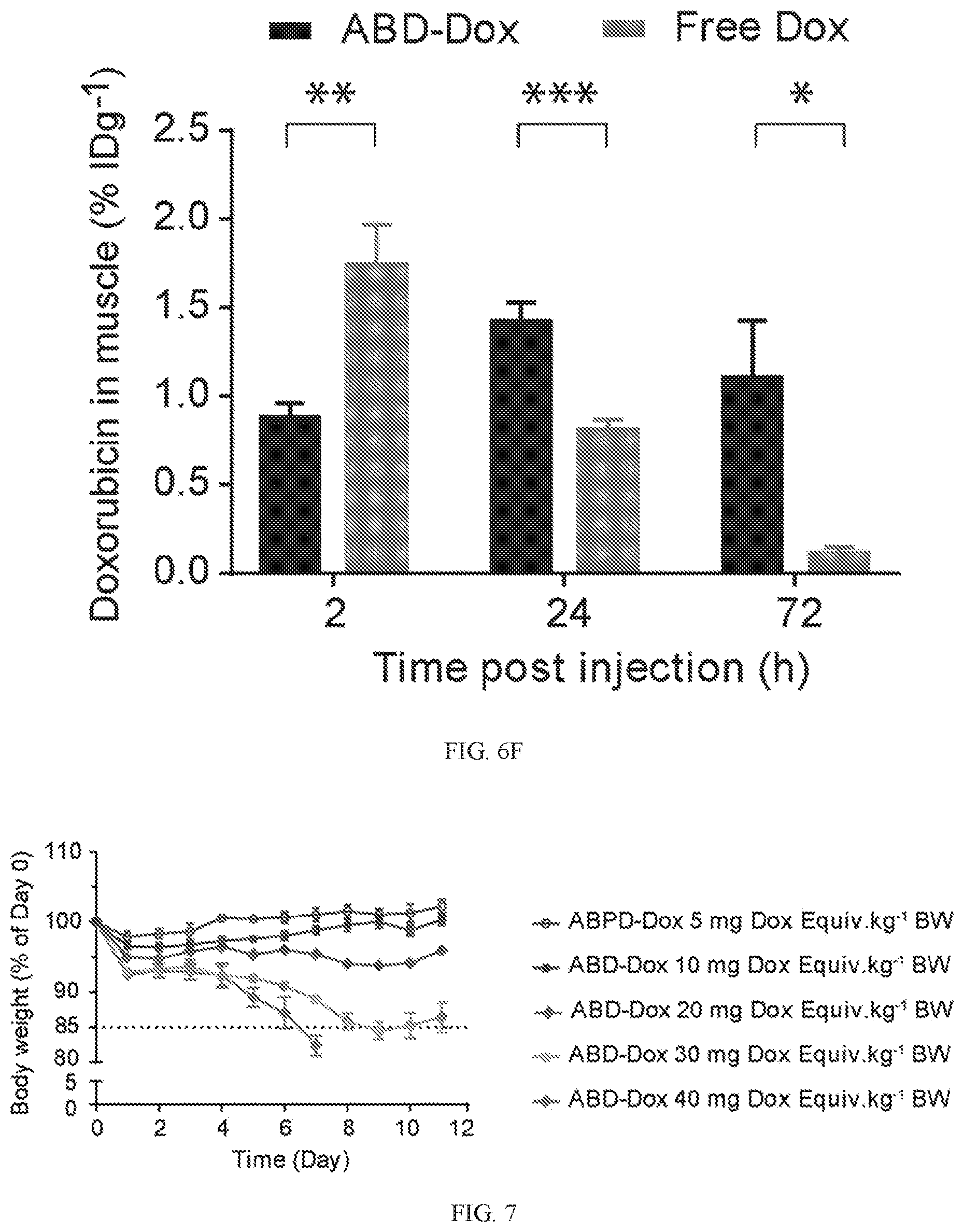
D00013

D00014

D00015

D00016

D00017
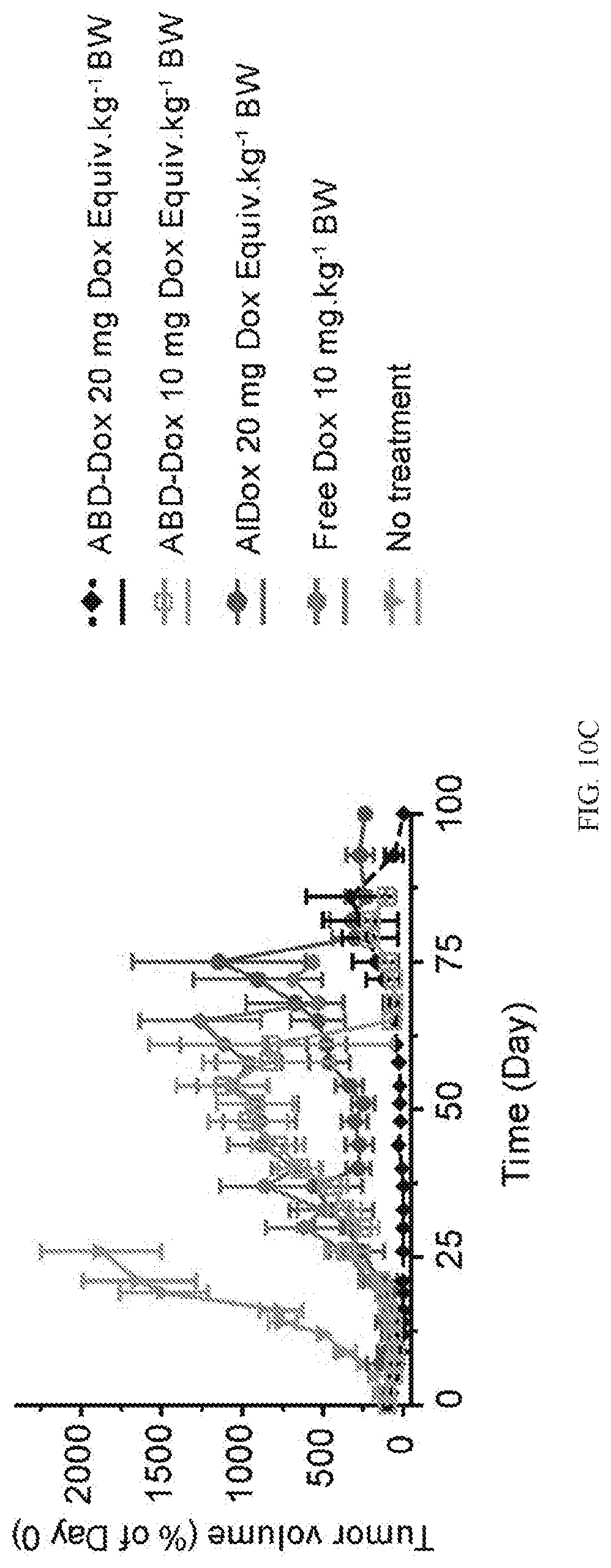
D00018

D00019

D00020

S00001
XML
uspto.report is an independent third-party trademark research tool that is not affiliated, endorsed, or sponsored by the United States Patent and Trademark Office (USPTO) or any other governmental organization. The information provided by uspto.report is based on publicly available data at the time of writing and is intended for informational purposes only.
While we strive to provide accurate and up-to-date information, we do not guarantee the accuracy, completeness, reliability, or suitability of the information displayed on this site. The use of this site is at your own risk. Any reliance you place on such information is therefore strictly at your own risk.
All official trademark data, including owner information, should be verified by visiting the official USPTO website at www.uspto.gov. This site is not intended to replace professional legal advice and should not be used as a substitute for consulting with a legal professional who is knowledgeable about trademark law.