Marker Sequences For Diagnosing And Stratifying Sle Patients
LUKING; Angelika ; et al.
U.S. patent application number 16/920949 was filed with the patent office on 2021-02-25 for marker sequences for diagnosing and stratifying sle patients. This patent application is currently assigned to Oncimmune Germany GmbH. The applicant listed for this patent is Oncimmune Germany GmbH. Invention is credited to Petra BUDDE, Angelika LUKING, Peter SCHULZ-KNAPPE, Anna TELAAR, Carmen THEEK.
| Application Number | 20210055296 16/920949 |
| Document ID | / |
| Family ID | 1000005207016 |
| Filed Date | 2021-02-25 |


View All Diagrams
| United States Patent Application | 20210055296 |
| Kind Code | A1 |
| LUKING; Angelika ; et al. | February 25, 2021 |
MARKER SEQUENCES FOR DIAGNOSING AND STRATIFYING SLE PATIENTS
Abstract
The present invention relates to methods for identifying markers for systemic lupus erythematosus (SLE) and to the markers identified with the aid of this method, which can differentiate between SLE and other autoimmune diseases on the one hand and between different SLE subgroups on the other hand. The invention also relates to panels, diagnostic devices and test kits which comprise these markers, and to the use and application thereof, for example for the diagnosis, prognosis and therapy control in SLE. The invention also relates to methods for screening and validating active substances for application in SLE subgroups.
| Inventors: | LUKING; Angelika; (Bochum, DE) ; SCHULZ-KNAPPE; Peter; (Hemmingen, DE) ; THEEK; Carmen; (Herdecke, DE) ; BUDDE; Petra; (Dortmund, DE) ; TELAAR; Anna; (Dortmund, DE) | ||||||||||
| Applicant: |
|
||||||||||
|---|---|---|---|---|---|---|---|---|---|---|---|
| Assignee: | Oncimmune Germany GmbH Dortmund DE |
||||||||||
| Family ID: | 1000005207016 | ||||||||||
| Appl. No.: | 16/920949 | ||||||||||
| Filed: | July 6, 2020 |
Related U.S. Patent Documents
| Application Number | Filing Date | Patent Number | ||
|---|---|---|---|---|
| 15117508 | Aug 9, 2016 | 10746735 | ||
| PCT/EP2015/052805 | Feb 10, 2015 | |||
| 16920949 | ||||
| Current U.S. Class: | 1/1 |
| Current CPC Class: | G01N 2500/04 20130101; G01N 33/564 20130101; G01N 2800/104 20130101; G01N 2800/52 20130101 |
| International Class: | G01N 33/564 20060101 G01N033/564 |
Foreign Application Data
| Date | Code | Application Number |
|---|---|---|
| Feb 10, 2014 | EP | 14154557.4 |
| Jul 22, 2014 | EP | 14178090.8 |
Claims
1-16. (canceled)
17. A method for detection, diagnosis, differential diagnosis, prognosis, therapy control, or aftercare of systemic lupus erythematosus (SLE), comprising: a) contacting a panel of markers comprising at least one sequence selected from SEQ ID NO: 1-11, 13, 15, 16, 18, 19, 20-24, 28, 29, 31, 46, 61, 95, 126, 128, 134, 136, 143, 152, 163, 169, 171, 173, 188, 191, 213, 214, 241, 258, 270, 302, 348, 349, 367-370, 372-375, 378-391, 403, 406, 408, 415, and 423-433 with a bodily fluid or a tissue sample from an individual to be tested; b) detecting an interaction of the bodily fluid or tissue sample with at least one of the marker from step a); and c) identifying a SLE patient for stratification and administering at least one therapeutic agent to the SLE patient for treatment or monitoring the SLE patient for therapy control.
18. The method of claim 17, wherein identifying a SLE patient for stratification comprises new or established SLE subgroups.
19. The method of claim 17, wherein identifying a SLE patient comprises distinguishing SLE from other rheumatic diseases or other autoimmune disorders.
20. The method of claim 17, wherein identifying a SLE patient comprises diagnosing ENA-4-negative SLE patients.
21. The method of claim 17, wherein identifying a SLE patient comprises diagnosing lupus nephritis in SLE patients.
22. The method of claim 17, wherein the marker sequence comprises a detection signal.
23. The method of claim 17, wherein the detection is made by way of an antibody.
24. The method of claim 17, wherein the body fluid is blood, blood plasma, serum, urine, cerebrospinal fluid, and/or synovial fluid.
25. A diagnostic device or test kit comprising at least one marker for systemic lupus erythematosus selected from sequences SEQ ID NO: 1-11, 13, 15, 16, 18, 19, 20-24, 28, 29, 31, 46, 61, 95, 126, 128, 134, 136, 143, 152, 163, 169, 171, 173, 188, 191, 213, 214, 241, 258, 270, 302, 348, 349, 367-370, 372-375, 378-391, 403, 406, 408, 415, and 423-433.
26. A panel of markers for diagnosis of systemic lupus erythematosus (SLE), comprising at least two different markers selected independently of one another from markers having the sequences of SEQ ID NO: 1-11, 13, 15, 16, 18, 19, 20-24, 28, 29, 31, 46, 61, 95, 126, 128, 134, 136, 143, 152, 163, 169, 171, 173, 188, 191, 213, 214, 241, 258, 270, 302, 348, 349, 367-370, 372-375, 378-391, 403, 406, 408, 415, and 423-433.
27. The panel of markers for the diagnosis of SLE of claim 23, comprising at least five markers selected from the sequences of SEQ ID NO: 1-11, 13, 15, 16, 18, 19, 20-24, 28, 29, 31, 46, 61, 95, 126, 128, 134, 136, 143, 152, 163, 169, 171, 173, 188, 191, 213, 214, 241, 258, 270, 302, 348, 349, 367-370, 372-375, 378-391, 403, 406, 408, 415, and 423-433.
28. A method for screening active substances for systemic lupus erythematosus (SLE), comprising: a) contacting a panel of markers comprising at least one sequence selected from SEQ ID NO: 1-11, 13, 15, 16, 18, 19, 20-24, 28, 29, 31, 46, 61, 95, 126, 128, 134, 136, 143, 152, 163, 169, 171, 173, 188, 191, 213, 214, 241, 258, 270, 302, 348, 349, 367-370, 372-375, 378-391, 403, 406, 408, 415, and 423-433 with a bodily fluid or a tissue sample from an individual to be tested; and b) detecting an interaction of the bodily fluid or tissue sample with at least one marker from step a).
Description
[0001] The present application is being filed along with a Sequence Listing in electronic format. The Sequence Listing is provided as a file entitled 16920949_SequenceListing, created Sep. 30, 2020, which is 6,549,888 bytes in size. The information in the electronic format of the sequence listing is incorporated herein by reference in its entirety.
[0002] The present invention relates to methods for identifying markers for systemic lupus erythematosus (SLE) and to the markers identified with the aid of this method, which can differentiate between SLE and other autoimmune diseases on the one hand and between different SLE subgroups on the other hand. The invention also relates to panels of markers for SLE, diagnostic devices and test kits for SLE which comprise these markers, and to the use and application thereof, for example for the diagnosis, prognosis and therapy control of SLE. The invention also relates to methods for screening and for validating active substances for application in SLE subgroups.
[0003] Systemic lupus erythematosus (SLE) is a rare autoimmune disease. In the case of lupus erythematosus the body's own immune system is disregulated. It not only attacks bacteria, viruses and cancer cells, but also healthy body cells. Organs and organ systems, for example the skin, are damaged as a result.
[0004] In clinical practice, SLE is diagnosed on the basis of a combination of clinical and immunological parameters. Here, antinuclear autoantibodies (ANAs) and anti-double-stranded DNA (anti-dsDNA) autoantibodies play a key role. However, the ANA test is not specific for SLE, since other autoimmune diseases and up to 20% of healthy individuals are also positively tested. The autoreactivity against extractable nuclear antigens (EMAs) as recombinant or purified individual antigens is therefore increasingly tested, for example against Sm-protein, U1-RNP, Rho52/SS-A and Ro60/SS-B. These antigens and associated autoantibodies, however, are not sufficient for diagnosing all SLE patients without doubt, in particular in an early phase of the disease. By way of example, anti-dsDNA antibodies are indeed highly specific for SLE and can be detected in approximately 70% of patients. However, the titre of the anti-dsDNA antibodies correlates with the disease activity in some patients, but not in all patients. As a result, SLE is often only diagnosed months or years after the occurrence of the first symptoms. A further problem of the currently used diagnostic methods is that the suitability of the previously tested autoantigens for the diagnosis of organ involvement and complications is disputed, and partly conflicting data has been published.
[0005] There is thus a great need to provide new markers for the diagnosis and differential diagnosis of SLE.
[0006] Marker sequences for the diagnosis of SLE are disclosed in WO 2012/0-19225 A2. These marker sequences were discovered by a method in which serum samples of SLE patients and those of healthy individuals were examined by comparison and the results were statistically evaluated. The marker sequences described in WO 2012/049225 A2, however, are not sufficiently suitable for the diagnosis of SLE with regard to a distinction from other autoimmune diseases and the identification of SLE subgroups.
[0007] There is therefore still a need for markers for SLE, in particular for the distinction of SLE from other autoimmune diseases.
[0008] This object has been achieved in accordance with the invention in that a differential method comprising a multiplicity of steps has been developed, in which serum samples of healthy individuals and patients with various autoimmune diseases were examined by comparison with, regard to their reactivity with a multiplicity of potential antigens and these results were statistically evaluated. The selection of the serum samples and the sequence of the steps surprisingly made it possible to identify highly specific markers for SLE which are also suitable for identifying SLE subgroups and complications such as lupus nephritis and for providing a differential diagnosis in respect of other autoimmune diseases, such as rheumatoid arthritis (RA), systemic sclerosis (SSc), ankylosing spondylitis or Bekhterev's disease (SPA), and also in respect of individuals who have early RA, i.e. have been suffering with the disease for less than two years ("patients with early RA").
[0009] The present invention relates to a method for identifying markers for systemic lupus erythematosus (SLE) comprising the following steps [0010] a) bringing serum samples of SLE patients into contact with more than 5000 antigens coupled to (Luminex) heads, measuring the binding of the individual antigens to proteins, in particular autoantibodies, in the serum of the SLE patients by means of immunofluorescence assay, and determining the median fluorescence intensity (MFI) for each individual antigen; [0011] b) bringing serum samples of patients with rheumatoid arthritis (RA) into contact `with the same antigens coupled to (Luminex) beads, measuring the binding of the individual antigens to proteins, in particular autoantibodies, in the serum, of the RA patients by means of immunofluorescence assay, and determining from this the median fluorescence intensity (MFI) for each individual antigen; [0012] c) bringing serum samples of healthy individuals into contact with the same antigens coupled to (Luminex) beads, measuring the binding of the individual antigens to proteins, in particular autoantibodies, in the serum of the healthy individuals by means of immunofluorescence assay, and determining from this the median, fluorescence intensity (MFI) for each individual antigen; [0013] d) statistically evaluating the MFI data from a), b) and c) by means of univariate analysis and thus identifying marker candidate antigens with which SLE patients can be differentiated from RA patients and from healthy individuals; [0014] e) bringing serum samples of patients with early RA. into contact, with the marker candidate antigens identified in d) coupled to (Luminex) beads, measuring the binding of marker candidate antigens to proteins, in particular autoantibodies, in the serum of patients with early RA by means of immunofluorescence assay, and determining from this the median fluorescence intensity (MFI) for each marker candidate antigen; [0015] f) bringing serum samples of patients with systemic sclerosis (SSc patients) into contact with the marker candidate antigens identified in d) coupled to (Luminex) beads, measuring the binding of marker candidate antigens to proteins, in particular autoantibodies, in the serum of SSc patients by immunofluorescence assay, and determining from this the median fluorescence intensity (MFI) for each marker candidate; [0016] g) bringing serum samples of patients with ankylosing spondylitis or Bekhterev's disease (SPA patients) into contact with the marker candidate antigens identified in d) coupled to (Luminex) beads, measuring the binding of marker candidate antigens to proteins, in particular autoantibodies, in the serum of SPA patients by means of immunofluorescence assay, and determining from this the median fluorescence intensify (MFI) for each marker candidate antigen; [0017] h) statistically evaluating the MFI data from e), f) and g) by means of univariate analysis and, when a threshold value of 3 standard deviations above the mean value of the healthy samples is not reached, identifying a specific marker for SLE, wherein the markers are selected from sequences SEQ ID No. 1 to 1587, homologues of sequences SEQ ID No. 1 to 1587 with at least 95% homology, subsequences of SEQ ID No. 1 to 1587, subsequences of homologues of SEQ ID No. 1 to 1587 with at least 95% homology.
[0018] The term "systemic lupus erythematosus (SLE) relates to a systemic autoimmune disease from the group of collagenoses. What is known as the butterfly rash is particularly characteristic for SLE (systemic lupus erythematosus). The diagnosis criteria for SLE are:
[0019] 1. butterfly rash, 2. discoid skin changes, 3. sensitivity to light, 4. mucous membrane ulcers (generally painless), 5. arthritis in at least two joints, 6. serositis (pleurisy or pericarditis), 7. kidney involvement (proteinuria>0,5 g/d or cylinder), 8. CNS involvement (cramps or psychosis), 9. haematological findings (haemolytic anaemia, leucopenia or thrombopenia), 10. immunological findings (anti-dsDNA antibodies, anti-Sm antibodies, anticardiolipin antibodies), 11. antinuclear antibodies without taking lupus erythematosus-triggering medication.
[0020] Evaluation: With four (three) positive findings, the diagnosis is considered reliable (likely) (definition for example according to Pschyrembei, de Gruyter, 261st edition (2007), Berlin),
[0021] One embodiment of the invention relates to methods for identifying markers for SLE which are suitable for the diagnosis and differential diagnosis of SLE, in. particular for distinction from other autoimmune diseases, preferably for distinction from, other rheumatic diseases, particularly preferably for distinction from RA, SSc, and SPA. These markers are also suitable for distinction from patients with early RA, These markers for SLE according to the invention are the subject of group 1 of antigens in Table 2, which can be used for the diagnosis of SLE. For the generation of these markers, marker candidate antigens which have an adjusted p-value for the non-parametric mean value comparison between groups of <0.05 and at the same time a fold change of >1.5 and additionally an AUC resulting from the ROC analysis of >0.75 are selected on the basis of the univariate results. In addition, the ENA-4 antigens are selected. For this pool of selected marker candidate antigens, an L1-penalised logistic regression model is preferably also established within the scope of a nested cross validation. Marker candidate antigens which are not considered within the scope of the creation of the model are removed from the further consideration. The markers for SLE are thus obtained, selected from the sequences (group 1)
[0022] SEQ ID No. 1 to 24, 134, 168, 214, 368 to 370 SEQ ID No. 530 to 553, 663, 697, 743, 897 to 899 and SEQ ID No. 1059 to 1082, 1192, 1226, 1272, 1426 to 1428, homologues of SEQ ID No. 1 to 24, 134, 168, 214, 368 to 370 SEQ ID No. 530 to 553, 663, 697, 743, 897 to 899 and SEQ ID No. 1059 to 1082, 1192, 1226, 1272, 1426 to 1428 with at least 95% homology, subsequences of SEQ ID No. 1 to 24, 134, 168, 214, 368 to 370 SEQ ID No. 530 to 553, 663, 697, 743, 897 to 899 and SEQ ID No. 1059 to 1082, 1192, 1226, 1272, 1426 to 1428 and subsequences of homologues of SEQ ID No. 1 to 24, 134, 168, 214, 368 to 370 SEQ ID No. 530 to 553, 663, 697, 743, 897 to 899 and SEQ ID No. 1059 to 1082, 1192, 1226, 1272, 1426 to 1428 with at least 95% homology.
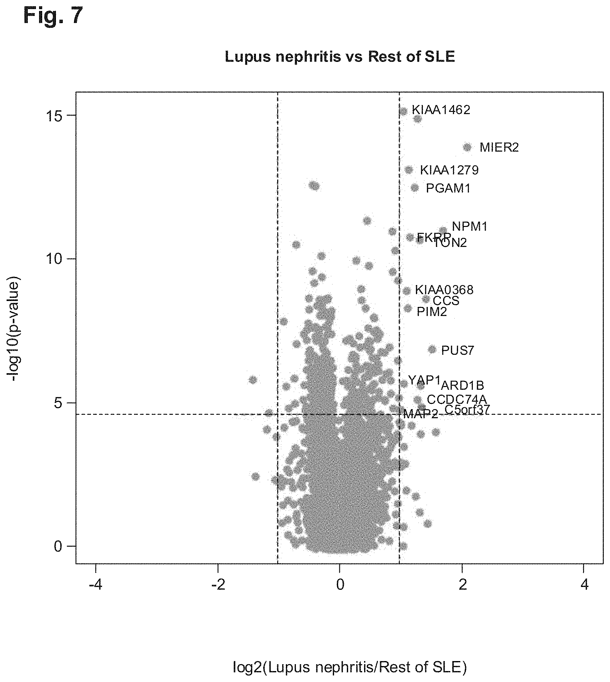
[0023] Another embodiment relates to methods for identifying markers for the subgroup of SLE patients with the complication lupus nephritis, comprising the comparison of the autoantibody profiles of SLE patients `with lupus nephritis with those of SLE patients without lupus nephritis. Markers which are found by means of this embodiment of the method are specified for example in Table 2, in group 2 and group 5. These are methods for example in which the markers for the subgroup of the SLE patients with the complication lupus nephritis are selected from the sequences
[0024] SEQ ID No. 25 to 54, 215, 216, 211, 218, 228, 233, 241, 245, 247, 249, 258, 288, 289, 301, 309, 315, 316, 324, 330, 331, 337, 339, 348, 350, 359, 362, 363,
[0025] SEQ ID No. 554 to 583, 744, 745, 746, 747, 757, 762, 770, 774, 776, 778, 787, 817, 818, 830, 838, 844, 845, 853, 859, 860, 366, 868, 877, 879, 888, 891, 892 and
[0026] SEQ ID No. 1083 to 1112, 1273, 1274, 1275, 1276, 1286, 1291, 1299, 1303, 1305, 1307, 1316, 1346, 1347, 1359, 1367, 1373, 1374, 1382, 1388, 1389, 1395, 1397, 1406, 1408, 1417, 1420, 1421,
[0027] homologues of SEQ ID No. SEQ ID No. 25 to 54, 215, 216, 217, 218, 228, 233, 241, 245, 247, 249, 258, 288, 289, 301, 309, 315, 316, 324, 330, 331, 337, 339, 348, 350, 359, 362, 363, SEQ ID No. 554 to 583, 744, 745, 746, 747, 757, 762, 770, 774, 776, 778, 787, 817, 818, 830, 838, 844, 845, 853, 859, 860, 866, 868, 877, 879, 888, 891, 892 and SEQ ID No. 1083 to 1112, 1273, 1274, 1275, 1276, 1286, 1291, 1299, 1303, 1305, 1307, 1316, 1346, 1347, 1359, 1367, 1373, 1374, 1382, 1388, 1389, 1395, 1397, 1406, 1408, 1417, 1420, 1421 with at least 95% homology, subsequences of SEQ ID No. 25 to 54, 215, 216, 217, 218, 228, 233, 241, 245, 247, 249, 258, 288, 289, 301, 309, 315, 316, 324, 330, 331, 337, 339, 348, 350, 359, 362, 363, SEQ ID No. 554 to 583, 744, 745, 746, 747, 757, 762, 770, 774, 776, 778, 787, 817, 818, 830, 838, 844, 845, 853, 859, 860, 866, 868, 877, 879, 888, 891, 892 and SEQ ID No. 1083 to 1112, 1273, 1274, 1275, 1276, 1286, 1291, 1299, 1303, 1305, 1307, 1316, 1346, 1347, 1359, 1367, 1373, 1374, 1382, 1388, 1389, 1395, 1397, 1406, 1408, 1417, 1420, 1421 and subsequences of homologues of SEQ ID No. SEQ ID No. 25 to 54, 215, 216, 217, 218, 228, 233, 241, 245, 247, 249, 258, 288, 289, 301, 309, 315, 316, 324, 330, 331, 337, 339, 348, 350, 359, 362, 363, SEQ ID No. 554 to 533, 744, 745, 746, 747, 757, 762, 770, 774, 776 778, 787, 817, 813, 830, 833, 844, 845, 353, 859, 860, 866, 868, 877, 879, 888, 891, 892 and SEQ ID No. 1083 to 1112, 1273, 1274, 1275, 1276, 1286, 1291, 1299, 1303, 1305, 1307, 1316, 1346, 1347, 1359, 1367, 1373, 1374, 1382, 1388, 1389, 1395 1397, 1406, 1408, 1417, 1420, 1421 with at least 95% homology.
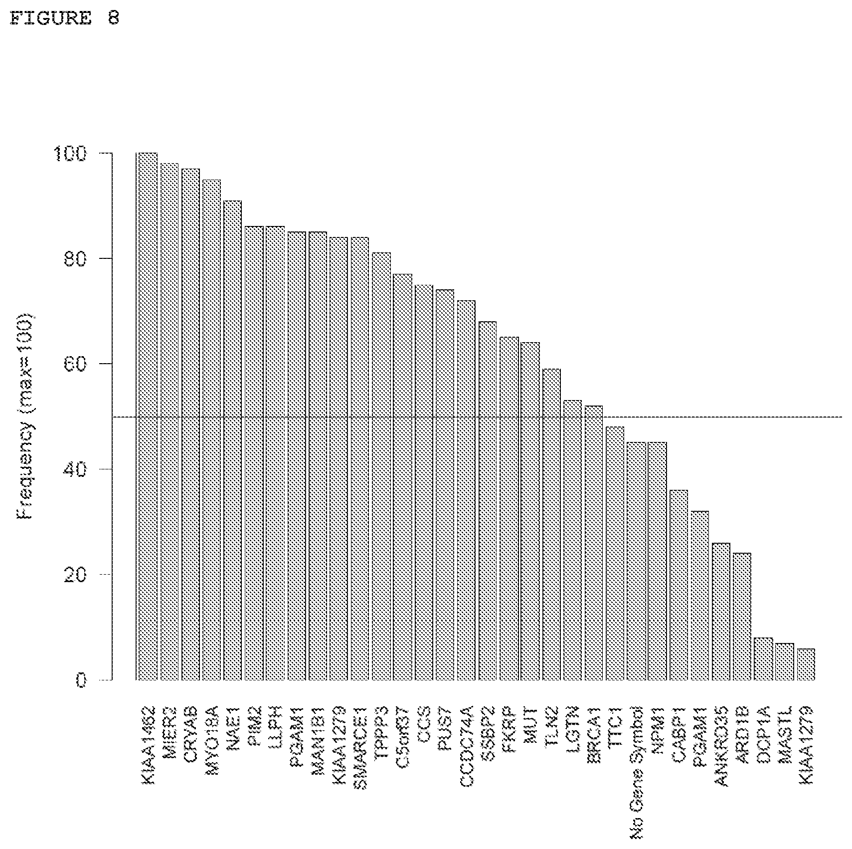
[0028] A further embodiment relates to methods which comprise the statistical evaluation by means of an L1-penalised logistic regression model with five-fold cross validation and twenty times repetition and selection of the markers which occur at a frequency of 50% or more. Markers which can be identified by means of this embodiment of the method are specified for example in Table 2, group 2. These are methods for example in--which the markers for the subgroup of the SLE patients with the complication lupus nephritis are selected from the sequences SEQ ID ho. 25 to 54, SEQ ID No. 554 to 583 and SEQ ID No. 1083 to 1112,
[0029] homologues of 25 to 54, SEQ ID No. 554 to 583 and SEQ ID No. 1083 to 1112 with at least 95% homology, subsequences of SEQ ID No. 25 to 54, SEQ ID No. 554 to 583 and SEQ ID No. 1083 to 1112 and subsequences of homologues of SEQ ID No. 225 to 54, SEQ ID No. 554 to 583 and SEQ ID No. 1083 to 1112 with at least 95% homology.
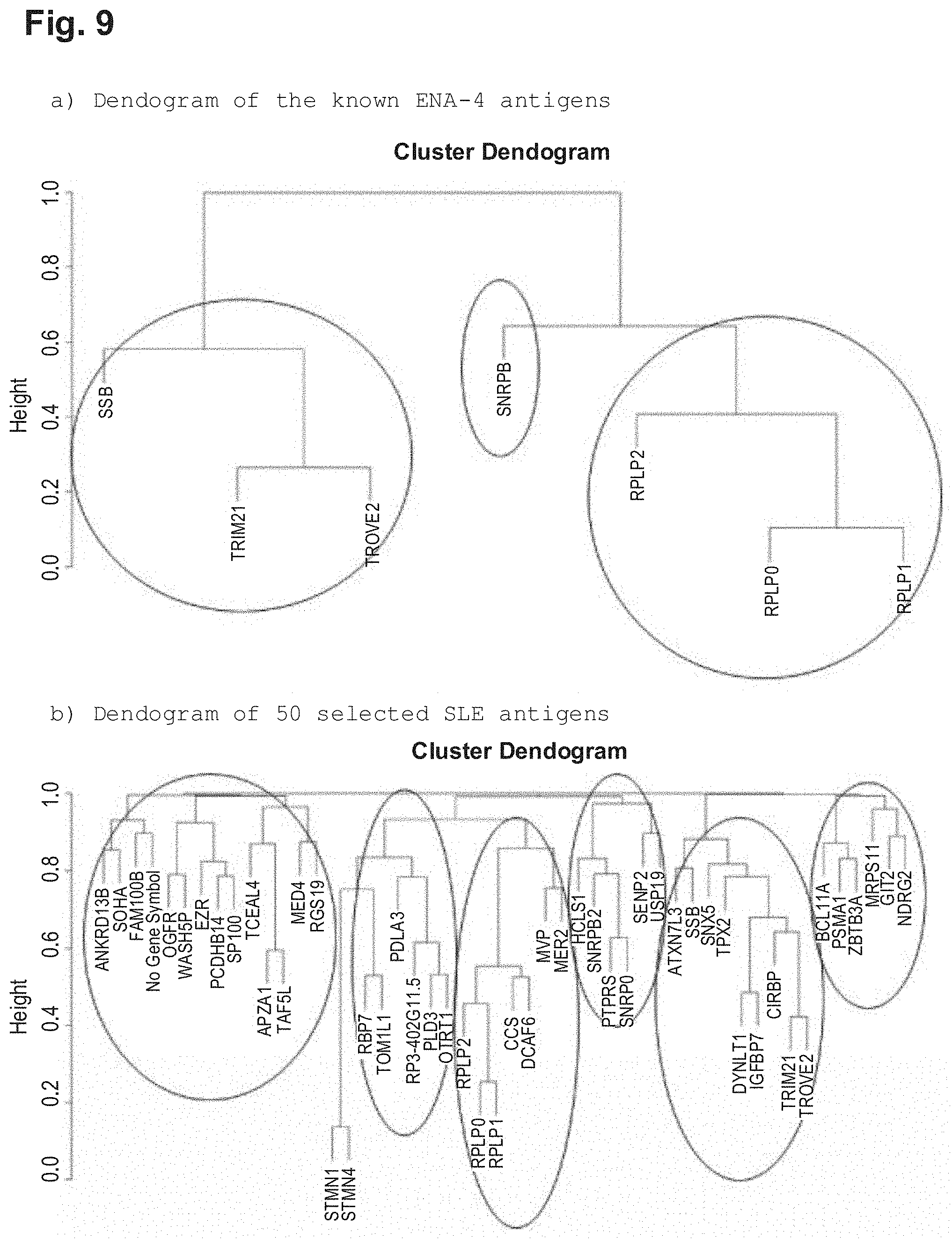
[0030] In a further embodiment of the method, markers for defined subgroups of SLE patients are identified in that the sequences SEQ ID No. 1 to 529 (clone sequences) are correlated with one of the sequences SEQ ID No. 1 to 529 by calculation of the Spearman's rank correlation coefficient for the particular marker. In this way, the markers of groups 1, 2 and 3 in Table 2 can be identified with the method according to the invention, for example. These are methods for example in which the markers which demonstrate a correlation with one another of the reactivities in SLE patients are selected from the sequences (group 3)
[0031] SEQ ID No. 55 to 111, SEQ ID No. 584 to 1007 and SEQ ID No. 1113 to 1169,
[0032] homologues of SEQ ID No. 55 to 111, SEQ ID No. 584 to 1007 and SEQ ID No. 1113 to 1169 with at least 95% homology, subsequences of SEQ ID SEQ ID No. 55 to 111, SEQ ID No. 584 to 1007 and SEQ ID No. 1113 to 1169 and subsequences of homologues of SEQ ID No. 55 to 111, SEQ ID No. 584 to 1007 and SEQ ID No. 1113 to 1169 with at least 95% homology and from the sequences (group 2)
[0033] SEQ ID No. 25 to 54, SEQ ID No. 554 to 583 and SEQ ID No. 1083 to 1112,
[0034] homologues of SEQ ID No. 25 to 54, SEQ ID No. 554 to 583 and SEQ ID No. 1083 to 1112 with at least 95% homology, subsequences of SEQ ID No. 25 to 54, SEQ ID No. 554 to 583 and SEQ ID No. 1083 to 1112 and subsequences of homologues of SEQ ID No. 25 to 54, SEQ ID No. 554 to 533 and SEQ ID No. 1083 to 1112 with at least 95% homology and from the sequences (group 1)
[0035] SEQ ID No. 1 to 24, 134, 168, 214, 368 to 370 SEQ ID No. 530 to 553, 663, 697, 743, 897 to 899 and SEQ ID No. 1059 to 1082, 1192, 1226, 1272, 1426 to 1428,
[0036] homologues of SEQ ID No. 1 to 24, 134, 168, 214, 368 to 370 SEQ ID No. 530 to 553, 663, 697, 743, 897 to 899 and SEQ ID No. 1059 to 1082, 1192, 1226, 1272, 1426 to 1428 with at least 95% homology, subsequences of SEQ ID No. 1 to 24, 134, 168, 214, 368 to 370 SEQ ID No. 530 to 553, 663, 697, 743, 897 to 899 and SEQ ID No. 1059 to 1082, 1192, 1226, 1272, 1426 to 1428 and subsequences of homologues of SEQ ID No. 1 to 24, 134, 168, 214, 368 to 370 SEQ ID No. 530 to 553, 663, 697, 743, 897 to 899 and SEQ ID No. 1059 to 1082, 1192, 1226, 1272, 1426 to 1428 with at least 95% homology.
[0037] One embodiment of the invention relates to methods for identifying markers for the subgroup of ENA-4-negative SLE, patients. This embodiment of the method fox` example comprises the testing of the serum samples of SLE patients for the absence of autoantibodies against the extractable nuclear antigens Sm-protein, U1-RNP, Rho52/SS-A and Ro60/SS-B. By way of example, the markers of group 4, Table 2 can thus be identified. These are methods for example in which, the markers for ENA-4-negative SLE patients are selected from the; sequences (group 4)
[0038] SEQ ID No. 112 to 214 and 279, SEQ ID No. 641 to 743 and 808 and SEQ ID No. 1170 to 1272 and 1337,
[0039] homologues of SEQ ID No. 112 to 214 and 279, SEQ ID No. 641 to 743 and 808 and SEQ ID No. 1170 to 1272 and 1337 with at least 95% homology, subsequences of SEQ ID No. 112 to 214 and 279, SEQ ID No. 641 to 743 and 808 and SEQ ID No. 1170 to 1272 and 1337 and subsequences of homologues of SEQ ID No. 112 to 214 and 279, SEQ ID No. 641 to 743 and 808 and SEQ ID No. 1170 to 1272 and 1337 with at least 95% homology.
[0040] One embodiment of the invention relates to methods comprising the selection of markers which have an adjusted p-value for the non-parametric mean value comparison between groups of less than 0.05, and at the same time a fold change of greater than 1.5 and an AUC resulting from, the ROC analysis of greater than 0.75. By way of example, the markers of groups 1, 4 and 6 can thus be identified. The corresponding calculations for panels of markers are specified in Table 5, in which the corresponding marker composition in the panels (arrangements) can be inferred from Table 4. These are methods for example in which the markers are selected from the sequences (group 6)
[0041] SEQ ID No. 219 to 227, 229 to 232, 234 to 240, 242, 243, 244, 246, 248, 250 to 257, 259 to 278, 280 to 287, 290 to 300, 302 to 308, 310 to 314, 317 to 323, 325 to 329, 332 to 336, 338, 340 to 347, 349, 351 to 358, 360, 361, 364 to 367,
[0042] SEQ ID No. 748 to 756, 758 to 761, 763 to 769, 771, 772, 773, 775, 777, 779 to 786, 788 to 807, 809 to 816, 819 to 829, 831 to 837, 839 to 843, 846 to 852, 853 to 857, 861 to 865, 867, 869 to 876, 878, 880 to 887, 889, 890, 893 to 896 and
[0043] SEQ ID No. 1277 to 1285, 1287 to 1290, 1292 to 1298, 1300, 1301, 1302, 1304, 1306, 1308 to 1315, 1317 to 1336, 1338 to 1345, 1348 to 1358, 1360 to 1366, 1368 to 1372, 1375 to 1381, 1382 to 1386, 1390 to 1394, 1396, 1398 to 1405, 1407, 1409 to 1416, 1418, 1420, 1422 to 1425,
[0044] homologues of SEQ ID No. 219 to 227, 229 to 232, 234 to 240, 242, 243, 244, 246, 248, 250 to 257, 259 to 278, 280 to 287, 290 to 300, 302 to 308, 310 to 314, 317 to 323, 325 to 329, 332 to 336, 338, 340 to 347, 349, 351 to 358, 360, 361, 364 to 367, SEQ ID No. 748 to 756, 758 to 761, 763 to 769, 771, 772, 773, 775, 777, 779 to 786, 788 to 807, 809 to 816, 819 to 829, 831 to 837, 839 to 843, 846 to 852, 853 to 857, 861 to 865, 867, 869 to 876, 878, 880 to 887, 889, 890, 893 to 896, SEQ ID No. 1277 to 1285, 1287 to 1290, 1292 to 1298, 1300, 1301, 1302, 1304, 1306, 1308 to 1315, 1317 to 1336, 1338 to 1345, 1348 to 1358, 1360 to 1366, 1368 to 1372, 1375 to 1381, 1382 to 1386, 1390 to 1394, 1396, 1398 to 1405, 1407, 1409 to 1416, 1418, 1420, 1422 to 1425 with at least 95% homology, subsequences of SEQ ID No. 219 to 227, 229 to 232, 234 to 240, 242, 243, 244, 246, 248, 250 to 257, 259 to 278, 280 to 287, 290 to 300, 302 to 308, 310 to 314, 317 to 323, 325 to 329, 332 to 336, 338, 340 to 347, 349, 351 to 358, 360, 361, 364 to 367, SEQ ID No. 748 to 756, 758 to 761, 763 to 769, 771, 772, 773, 775, 777, 779 to 786, 788 to 807, 809 to 816, 819 to 829, 831 to 837, 839 to 843, 846 to 852, 853 to 857, 861 to 865, 867, 869 to 876, 878, 880 to 887, 889, 890, 893 to 896, SEQ ID No. 1277 to 1285, 1287 to 1290, 1292 to 1298, 1300, 1301, 1302, 1304, 1306, 1308 to 1315, 1317 to 1336, 1338 to 1345, 1348 to 1358, 1360 to 1366, 1368 to 1372, 1375 to 1381, 1382 to 1386, 1390 to 1394, 1396, 1398 to 1405, 1407, 1409 to 1416, 1418, 1420, 1422 to 1425 and subsequences of homologues of SEQ ID No. 219 to 227, 229 to 232, 234 to 240, 242, 243, 244, 246, 248, 250 to 257, 259 to 278, 280 to 287, 290 to 300, 302 to 308, 310 to 314, 317 to 323, 325 to 329, 332 to 336, 338, 340 to 347, 349, 351 to 358, 360, 361, 364 to 367, SEQ ID No. 748 to 755, 753 to 761, 763 to 769, 771, 772, 773, 775, 777, 779 to 786, 788 to 807, 809 to 816, 819 to 829, 831 to 837, 839 to 843, 846 to 852, 853 to 857, 861 to 865, 867, 369 to 876, 878, 880 to 887, 889, 890, 893 to 896, SEQ ID No. 1277 to 1285, 1287 to 1290, 1292 to 1238, 1300, 1301, 1302, 1304, 1306, 1308 to 1315, 1317 to 1336, 1338 to 1345, 1348 to 1358, 1360 to 1366, 1368 to 1372, 1375 to 1381, 1382 to 1386, 1390 to 1394, 1396, 1398 to 1405, 1407, 1409 to 1416, 1418, 1420, 1422 to 1425 with at least 95% homology.
[0045] Group 7 in table 2 contains a further 85 statistically significant antigens from the methods according to the invention; these are markers selected from the sequences SEQ ID No. 368 to 452, SEQ ID No. 897 to 981, SEQ ID No. 1426 to 1510, homologues of SEQ ID No. 368 to 452, SEQ ID No. 897 to 981, SEQ ID No. 1426 to 1510 with at least 95% homology, subsequences of SEQ ID No. 368 to 452, SEQ ID No. 897 to 981, SEQ ID No. 1426 to 1510 and subsequences of homologues of SEQ ID No. 368 to 452, SEQ ID No. 897 to 981, SEQ ID No. 1426 to 1510 with at least. 95% homology, which can be used for the diagnosis and differential diagnosis of SLE compared with healthy individuals and other autoimmune diseases. Antigens from group 7 were also used for the calculation of biomarker combinations.
[0046] Group 8 consists of further statistically significant antigens from the methods according to the invention; markers selected from the sequences SEQ ID No. 453 to 529, SEQ ID No. 982 to 1058, SEQ ID No. 1511 to 1587, homologues of SEQ ID No. 453 to 529, SEQ ID No. 982 to 1058, SEQ ID No. 1511 to 1587 with at least 95% homology, subsequences of SEQ ID No. 453 to 529, SEQ ID No. 982 to 1058, SEQ ID No. 1511 to 1587 and subsequences of homologues of SEQ ID No. 453 to 529, SEQ ID No. 982 to 1058, SEQ ID No. 1511 to 1587 with at least 95% homology were detected and identified for the autoantibodies in SLE patients.
[0047] The invention also relates to the individual markers for SLE identified with the method according to the invention. The method concerns markers for SLE selected from the sequences SEQ ID No. 1 to 1587, homologues of sequences SEQ ID No. 1 to 1587 with at least 95% homology, subsequences of SEQ ID No. 1 to 1587 and subsequences of homologues of SEQ ID No. 1 to 1587 with at least 95% homology. The method concerns the markers of groups 1, 2, 3, 4, 5, 6, 7, 8 in Table 2, wherein, the respective groups comprise the markers of the clone sequences specified in Table 2, the corresponding RNA sequences, the corresponding protein, sequences, the relevant homologues with a homology of at least 95%, and the relevant subsequences. The invention relates to a marker for SLE selected from the sequences SEQ ID No. 530 to 1058 (RNA sequences), SEQ ID No. 1059 to 1587 (protein sequences). The markers according to the invention and the associated nucleic acid sequences are presented in Table 2 (SEQ ID No. of the relevant clone sequences is specified) and can be unambiguously identified by their cited database entry, for example at http;//www.ncbi.nlm.nih, gov/, by means of their Gene ID (Table 2). The sequences SEQ ID No. 1-1587 are specified in the accompanying sequence protocol, wherein SEQ ID No. 1-529 are clone sequences (cDNA), SEQ ID No. 530-1058 are RNA sequences, and SEQ ID No. 1059-1587 are protein sequences.
[0048] The invention also relates to the proteins coded, by sequences SEQ ID No. 1 to 1058, the proteins coded by homologues of the sequences SEQ ID No. 1 to 1058 with at least 95% homology to the sequences SEQ ID No. 1 to 1058, the proteins coded by subsequences of SEQ ID No. 1 to 1058, the proteins coded by homologues of the subsequences of SEQ ID No. 1 to 1058 with at least 95% homology in. the subsequences. In a preferred embodiment these are the proteins SEQ ID No. 1059 to 1587, homologues of the proteins with the sequences SEQ ID No. SEQ ID No. 1059 to 1587 with at least 95% homology, subsequences of SEQ ID No. 1059 to 1587, homologues of the subsequences of SEQ ID No. SEQ ID No. 1059 to 1587 with at least 95% homology.
[0049] The invention also relates to a panel of markers (also referred to as an arrangement of markers), comprising at least two different markers for SLE which are selected independently of one another from the sequences SEQ ID No. 1 to 1587, homologues of sequences SEQ ID No. 1 to 1587 with at least 95% homology, subsequences of SEQ ID No. 1 to 1587 and subsequences of homologues of SEQ ID No. 1 to 1587 with, at least 95% homology. A panel of markers for SLE can comprise 2 to 20 or more, for example 3, 4, 5, 6, 7, 8, 9, 10, 11, 12, 13, 14, 15, 16, 17, 18, 19, 20, 25, 30, 40, 50, 100 or more different markers for SLE and optionally further markers, wherein the markers of SLE are selected independently of one another from the sequences SEQ ID No. 1 to 1587, homologues of sequences SEQ ID No. 1 to 1587 with at least 95% homology, subsequences of SEQ ID No. 1 to 1587 and subsequences of homologues of SEQ ID No. 1 to 1587 with at least 95% homology, and the proteins coded by the sequences.
[0050] On account of the high clinical and serological heterogeneity of the SLE disease, it is difficult, to diagnose SLE unambiguously using just one biomarker. It is therefore often necessary to combine (where possible) uncorrelated autoantigens to form what are known as panels of markers (biomarker panels for SLE). By way of example, within the scope of individualised medicine, corresponding panels of markers for SLE can be composed individually for the relevant SLE subtype (subgroup) for individual patients or patient groups. It is therefore also necessary to have available a multiplicity of potential markers for SLE in order to select, suitable subgroups or subtypes of pecific markers for SLE for the individual case in question. A orresponding panel can be embodied for example in the form of an arrangement, an array, or also one or more beads, preferably Luminex beads. The invention thus relates to an arrangement comprising one or more markers according to the invention, a protein array comprising one or more markers according to the invention, a bead (small ball or platelet) comprising one or more markers according to the invention. Examples of SLE panels (SLE arrangements) are given in Table 4.
[0051] The invention also relates to a diagnostic device or a test kit comprising at least one marker for SLE selected from the sequences SEQ ID No. 1 to 1587, homologues of sequences SEQ ID No. 1 to 1587 with at least 95% homology, subsequences of SEQ ID No. 1 to 1587 and subsequences of homologues of SEQ ID No. 1 to 1587 with at least 95% homology, and the proteins coded by the sequences. A corresponding diagnostic device or a corresponding test kit can also comprise a panel of markers for SLE and optionally further auxiliaries and additives.
[0052] The invention also relates to the use of one or more markers for SLE selected from sequences SEQ ID No. 1 to 1587, homologues of sequences SEQ ID No. 1 to 1587 with at least 95% homology, subsequences of SEQ ID No. 1 to 1587 and subsequences of homologues of SEQ ID No. 1 to 1587 with at least 95% homology, and the proteins coded by the sequences, a marker panel for SLE, or a diagnostic device or test kit for identifying subgroups of SLE patients, for diagnosing SLE, for differential diagnosis (i.e. for distinction from other autoimmune diseases or other rheumatic diseases), for prognosis in the case of SLE, for therapy control in the case of SLE, for active substance selection in the case of SLE, for therapy monitoring in the case of SLE, or for aftercare in the case of SLE.
[0053] The invention also relates to the use of one or more of the markers for SLE selected from the sequences SEQ ID No. 1 to 1587, homologues of sequences SEQ ID No. 1 to 1587 with at least 95% homology, subsequences of SEQ ID No. 1 to 1587 and subsequences of homologues of SEQ ID No. 1 to 1587 with at least 95% homology, and the proteins coded by the sequences for the differentiation of SLE from RA and/or other autoimmune diseases, for example SSc and/or SPA and/or RA and/or early RA.
[0054] The Invention also relates to the use of one or more markers for SLE selected from the sequences SEQ ID No. 112 to 214 and 279, SEQ ID No. 641 to 743 and 808 and SEQ ID No. 1170 to 1272 and 1337, homologues of SEQ ID No. 112 to 211 and 279, SEQ ID No. 641 to 743 and 808 and SEQ ID No. 1170 to 127.2 and 1337 with at least 95% homology, subsequences of SEQ ID No. 112 to 214 and 279, SEQ ID No. 641 to 743 and 808 and SEQ ID No. 1170 to 1272 and 1337 and subsequences of homologues of SEQ ID No. 112 to 214 and 279, SEQ ID No. 641 to 743 and 808 and SEQ ID No. 1170 to 1272 and 1337 with at least 95% homology, and the proteins coded by the sequences for the diagnosis of SLE in ENA-4-negative SLE patients.
[0055] The invention also relates to the use of one or more markers for SLE selected from the sequences SEQ ID No. 25 to 54, SEQ ID No. 554 to 583 and SEQ ID No. 1083 to 1112, homologues of SEQ ID No. 25 to 54, SEQ ID No. 554 to 583 and SEQ ID No. 1083 to 1112 with at least 95% homology, subsequences of SEQ ID No. 25 to 54, SEQ ID No. 554 to 583 and SEQ ID No. 1083 to 1112 and subsequences of homologues of SEQ ID No. 25 to 54, SEQ ID No. 554 to 583 and SEQ ID No. 1083 to 1112 with at least 95% homology, and the proteins coded by the sequences for the diagnosis and differential diagnosis of lupus nephritis in SLE patients. Lupus nephritis is a common and serious complication of SLE. In the case of complete failure of the kidney function, therapy with dialysis is necessary. In order to avoid long-term damage, it is therefore important to identify and treat any kidney involvement early on. This is also of particular importance for the development of active substances for SLE in general, i.e. for the development of active substances for patients with lupus nephritis. Previously, there were still no biomarkers available able to diagnose lupus nephritis in all patients.
[0056] The invention also relates to markers for SLE and lupus nephritis selected from the sequences SEQ ID No. 25 to 54, SEQ ID No. 554 to 583 and SEQ ID No. 1083 to 1112, homologues of SEQ ID No. 25 to 54, SEQ ID No. 554 to 583 and SEQ ID No. 1083 to 1112 with at least 95% homology, subsequences of SEQ ID No. 25 to 54, SEQ ID No. 554 to 583 and SEQ ID No. 1083 to 1112 and subsequences of homologues of SEQ ID No. 25 to 54, SEQ ID No. 554 to 583 and SEQ ID No. 1083 to 1112 with at least S5% homology, and the proteins coded by the sequences.
[0057] The autoantibody profiles of SLE patients with lupus nephritis were therefore compared with those without lupus nephritis. Following univariate statistical evaluation, a threshold value of p<0.05 and a 1.5 times modified reactivity compared with the control group were applied.
[0058] The invention also relates to a method for the early detection, diagnosis, differential diagnosis, prognosis, therapy control and/or after-care of SLE, in which [0059] a. at least one of the markers for SLE selected from the sequences SEQ ID No. 1 to 1587, the homologues of sequences SEQ ID No. 1 to 1587 with at least S5% homology, the subsequences of SEQ ID No. 1 to 1587 or the subsequences of homologues of SEQ ID No. I to 1587 with at least 95% homology, and the proteins coded by the sequences [0060] b. is brought into contact with bodily fluid or a tissue sample from an individual to be tested, and [0061] c. an interaction of the bodily fluid or of the tissue sample with the one this or more markers from a, is detected.
[0062] The invention also relates to a target tor the therapy of SLE selected from the sequences SEQ ID No. 1 to 1587, the homologues of sequences SEQ ID No. 1 to 1587 with at least 95% homology, the subsequences of SEQ ID No. 1 to 1587 and the subsequences of homologues of SEQ ID No. 1 to 1587 with at least 95% homology, and the proteins coded by the sequences.
[0063] The invention also relates to a composition, in particular a pharmaceutical composition, comprising at least one of the sequences SEQ ID No. 1 to 1587, the homologues of sequences SEQ ID No. 1 to 1587 with at least 95% homology, the subsequences of SEQ ID No. 1 to 1587 or the subsequences of the homologues of SEQ ID No. 3. to 1587 with at least 95% homology, and the proteins coded, by the sequences.
[0064] The invention also relates to a method for screening active substances for SLE, in which [0065] a. at least cone of the markers for SLE selected from the sequences SEQ ID No. 1 to 1587, the homologues of sequences SEQ ID No. 1 to 1587 with at least 95% homology, the subsequences of SEQ ID No. 1 to 1587 or the subsequences of homologues of SEQ ID No. 1 to 1587 with at least 95% homology, and the proteins coded by the sequences [0066] b. is brought into contact with a substance to be tested, and [0067] c. an interaction of the substance with the one or more markers from a. is detected.
[0068] The large clinical heterogeneity of SLE currently constitutes a big problem both for diagnosis and for active substance development.
[0069] The identification of specific antibody signatures in SLE patient subgroups therefore constitutes an important step for the improved definition of patient groups in clinical studies. By way of example, as presented under Example 9, specific autoantibodies for lupus nephritis could be used to recruit this subgroup for drug studies.
[0070] A large number of new active substances and therapeutic antibodies are currently undergoing clinical development: inter alia, therapeutic antibodies against cell-surface receptors of immune cells, such as anti-CD20, anti-CD22, or against pro-inflammatory cytokines, such as anti-IL6, are being developed, It is therefore now possible, due to the identification of serologically defined subgroups of SLE, to link this to a target-specific response to a drug.
[0071] The invention also relates to the use of one or more markers for SLE according to the invention, of an arrangement according to the invention (panel of markers for SLE), of a protein array according to the invention, of a bead according to the invention, of a diagnostic device according to the invention, or of a. test kit according to the invention for the individualised diagnosis and/or therapy in individual patients, patient groups, cohorts, population groups, variants of SLE, or stages of SLE.
[0072] The invention also relates to the use of one or more markers according to the invention for SLE, of an arrangement according to the invention (panel of markers for SLE), of a protein array according to the invention, of a bead according to the invention, of a diagnostic device according to the invention, or of a test kit according to the invention for the detection and/or determination of the amount of one or more autoantibodies associated with SLE, for example in bodily fluids such as serum, tissue or tissue samples of the patient. The invention also relates to the use of one or more markers according to the invention, of an arrangement according to the invention, of a protein array according to the invention, of a bead according to the invention, of a diagnostic device according to the invention, or of a test kit according to the invention for the analysis of autoantibody profiles of patients, in particular for the qualitative and/or quantitative analysis of autoantibodies and/or for the monitoring of changes of autoantibody profiles associated with SLE, for example in bodily fluids such as serum, tissue or tissue samples of the patient.
[0073] A particular embodiment of the invention relates to methods for the early identification and diagnosis of SLE, in which the detection of an interaction of the bodily fluid or the tissue sample with the one or more markers indicates an SLE-associated autoantibody profile of the patient or of a cohort or of a population group or of a certain course of disease (prognosis) or of a certain response to a therapy/drug.
[0074] The invention therefore includes the use of at least one marker for SLE selected from the sequences SEQ ID No. 1 to 1587, the homologues of sequences SEQ ID No. 1 to 1587 with at least 95% homology, the subsequences of SEQ ID No. 1 to 1587 or the subsequences of homologues of SEQ ID No. 1 to 1587 with at least 95% homology, and the proteins coded by the sequences for the analysis of autoantibody profiles of patients, in particular for the quantitative analysis and/or for the monitoring of changes of autoantibody profiles of SLE patients.
[0075] An interaction of the bodily fluid or the tissue sample with the one or more SLE markers can be detected for example by a probe, in particular by an antibody.
[0076] In a preferred embodiment at least 2, for example 3, 4, 5, 6, 7, 8, 9, 10, preferably 15 to 20 markers for SLE or 30 to 50 or 100 or more markers are used together or in combination, either simultaneously or in succession, wherein the markers for SLE are selected independently of one another from the sequences SEQ ID No. 1 to 1587, the homologues of sequences SEQ ID No. 1 to 1587 with at least 95% homology, the subsequences of SEQ ID No. 1 to 1587 or the subsequences of homologues of SEQ ID No. 1 to 1587 with at least 95% homology, and the proteins coded by the sequences.
[0077] A particular embodiment of the invention relates to a method according to the invention, wherein the stratification or therapy control includes decisions relating to the treatment and therapy of the patient, in particular hospitalisation of the patient, use, efficacy and/or dosage of one or more drugs, a therapeutic measure, or the monitoring of the course of the disease and course of therapy, aetiology, or classification of a disease inclusive of prognosis. The invention also relates to a. method for stratification, in particular for risk stratification and/or therapy control of a patient with SLE.
[0078] The stratification of the patient with SLE into new or established SLE subgroups as well as the expedient selection of patient croups for the clinical development of new therapeutic agents is also included. The term therapy control likewise includes the division of patients into responders and non-responders with regard to a therapy or course thereof.
[0079] The invention in particular also relates to the detection and determination of the amount of at least two different autoantibodies in a patient by means of the SLE markers according to the invention, wherein at least two different SLE markers are preferably used. The invention also relates to a use according to the invention of one or more SLE markers, wherein at least 2, for example 3 to 5 or 10, preferably 30 to 50, or 50 to 100 or more SLE markers or the relevant autoantibodies on or from a patient to be tested are determined.
[0080] The invention comprises the SLE markers on a solid substrate, for example a filter, a membrane, a small platelet or ball, for example a magnetic or fluorophore-labelled ball, a silicon wafer, a bead, a chip, a mass spectrometry target, or a matrix, or the like. Different materials are suitable as substrates and are known to a person skilled in the art, for example glass, metal, plastic, filter, PVDF, nitrocellulose, or nylon (for example Immobilon P Millipore, Protran Whatman, Hybond N+Amersham).
[0081] The substrate tor example can correspond to a grid with the dimensions of a microtitre plate (8-12 well strips, 96 wells, 384 wells or more), of a silicon wafer, of a chip, of a mass spectrometry target, or of a matrix.
[0082] In one embodiment of the invention markers for SLE are present in the form of clone sequences or clone(s).
[0083] The markers according to the invention can be combined, supplemented or extended with known biomarkers for SLE or biomarkers for other diseases. With a combination of this type, a proportion of markers for SLE according to the invention of preferably at least 50%, preferably 60%, and particularly preferably 70% or more is comprised.
[0084] In a preferred embodiment the use of the SLE markers and the methods according to the invention are implemented outside the human or animal body, for example the diagnosis is performed ex vivo/in vitro, preferably by means of an assay, as detailed below,
[0085] In the sense of this invention, the term "diagnosis" means the positive determination of SLE with the aid of the markers according to the invention and the assignment of the patients or symptoms thereof to the disease SLE. The term "diagnosis" includes the medical diagnosis and tests in this respect, in particular in vitro diagnosis and laboratory diagnosis, and also proteomics and nucleic acid blots. Further tests may be necessary for assurance and in order to rule out other diseases. The term "diagnosis" therefore includes in particular the differential diagnosis of SLE by means of the markers according to the invention.
[0086] In the sense of this invention, "stratification or therapy control" means that, for example, the methods according to the invention allow decisions for the treatment and therapy of the patient, whether it is the hospitalisation of the patient, the use, efficacy and/or dosage of one or more drugs, a therapeutic measure or the monitoring of the course of a disease and the course of therapy or aetiology or classification of a disease, for example into a new or existing sub-type, or the differentiation of diseases and patients thereof. In a further embodiment of the invention, the term "stratification" in particular includes the risk, stratification with the prognosis of an "outcome" of a negative health event,
[0087] "Prognosis" means the prediction of the course of a disease.
[0088] In accordance with the invention, "therapy control" means, for example, the prediction and monitoring of the response to a drug or a therapy as `well as aftercare.
[0089] Within the scope of this invention, the term "patient" is understood to mean any test subject, any individual (human or mammal), with the provision that the test subject or individual is tested for SLE.
[0090] The term "marker for SLE" in the sense of this invention means that the nucleic acid, for example DNA, in particular cDNA or RNA or the coded amino acid sequence or the polypeptide or protein are significant (specific) for SLE and/or the autoantibody profiles associated with SLE, Markers according to the invention are nucleic acid sequences and/or amino acid sequences according to the definition in the appended sequence protocol (SEQ ID No. 1 to SEQ ID No. 1587), homologues and subsequences thereof, wherein modified nucleic acid and amino acid sequences are also included. Here, marker` for SLE means, for example, that the cDNA or RNA or the polypeptide or protein obtainable therefrom interacts with substances from the bodily fluid or tissue sample from a patient with SLE (for example antigen (epitope)/antibody (paratope) interaction). In a particularly preferred, embodiment of the invention the marker for SLE is an antigen or part of an antigen or codes for an antigen or for part of an antigen.
[0091] The substances from the bodily fluid or tissue sample occur either only in an amplified manner or at least in an amplified manner in the case of SLE or are expressed, whereas these substances are not present in patients without SLE or healthy individuals, or at least are present to a lesser extent (smaller amount, lower concentration). Markers for SLE can also be characterised in that they interact with substances from the bodily fluid or tissue sample from patients with SLE, because these substances no longer occur or are no longer expressed or occur or are expressed at least in a much lower amount/concentration in the case of SLE, whereas these substances are present or are at least present to a much higher extent in patients without SLE. Markers for SLE can also be present in healthy test subjects, however the amount (concentration) thereof changes for example with the development, establishment and therapy of SLE. One or more markers can in this way map a profile of substances from bodily fluid and tissue sample, for example an SLE-asscciated autoantibody profile of the patient in question. Markers according to the invention are biomarkers for SLE.
[0092] Autoantibody profiles comprise the amount of one or more autoantibodies of which the occurrence/expression accompanies the development and/or establishment of SLE, Autoantibody profiles therefore include on the one hand the composition, i.e. one or more autoantibodies is/are expressed only in the case of SLE for example, and also the amount/concentration of individual autoantibodies, i.e. the amount/concentration of individual autoantibodies changes with the development and establishment of SLE. These changes can be detected with the aid of the marker sequences according to the invention.
[0093] In a particularly preferred embodiment the SLE marker identifies/binds to autoantibodies which are present (intensified) or are present to a lower extent (or no longer) during the course of the development, establishment and therapy of SLE. Autoantibodies are formed by the body against, endogenous antigens which are formed for example in the case of SLE, Autoantibodies are formed by the body against different substances and pathogens, Within the scope of the present invention, the autoantibodies which are formed with the occurrence and during the course of the development of SLE and/or of which the expression is up-regulated or down-regulated are detected in particular. These autoantibodies can be detected with the aid of the methods and markers according to the invention, and the detection and monitoring (for example of the amount) thereof can be used for the early identification, diagnosis and/or therapy monitoring/therapy control and the prognosis and prediction of the risk of the re-occurrence of SLE within the scope of the after-care.
[0094] The autoantibody profiles can be sufficiently characterized with use of just a single SLE marker. In other cases, two or more SLE markers are necessary in order to map an autoantibody profile which is specific for SLE.
[0095] In one embodiment of the invention autoantibodies `which derive from another individual and which for example originate from a commercial cDNA bank can be detected using SLE markers.
[0096] In another embodiment of the invention these autoantibodies can be detected using SLE markers `which derive from the same individual and which for example originate from a cDNA bank produced individually for the patient or a group of patients for example within the scope of individualised medicine, By way of example, homologues of the specified SLE markers with the sequences SEQ ID. No. 1 to 1587 or subsequences thereof can be used.
[0097] Autoantibodies can be formed by the patient already many years prior to the occurrence of the first symptoms of disease. An early identification, diagnosis and also prognosis and preventative treatment or lifestyle change and other possibilities for prevention might therefore be possible even years prior to the visible outbreak of the disease. The devices, means and methods according to the invention enable a very early intervention compared with known methods, which significantly improves the prevention, treatment possibilities and effects of SLE.
[0098] Since the SLE-associated autoantibody profiles change daring the establishment and treatment/therapy of SLE, the invention also enables the detection and monitoring of SLE at any stage of the development and treatment and also monitoring within the scope of SLE after-care. The means according to the invention, for example a corresponding diagnostic device or a test kit, also allow simple handling at home by the patient and an economical routine precautionary measure for early identification.
[0099] In particular due to the use of antigens as specific markers for SLE which derive from sequences already known, for example from commercial cDNA banks, test subjects can be tested and any present SLE-associated autoantibodies can be detected in these test subjects, even if the corresponding autoantigens are not (yet) known in these test subjects.
[0100] Different patients can have different SLE-associated autoantibody profiles, for example different cohorts or population groups can differ from one another. Here, any patient can form one or more different SLE-associated autoantibodies during the course of the development of SLE and the progression of the SLE disease, that is to say even different autoantibody profiles. In addition, the composition and/or the amount of formed autoantibodies can change during the course of the SLE development and progression of the disease, such that a quantitative evaluation is necessary. The therapy/treatment, of SLE leads to changes in the composition and/or the amount of SLE-associated autoantibodies. The large selection of SLE markers according to the invention which are provided with this invention enables the individual compilation of SLE markers in an arrangement, i.e. a panel, for individual patients, groups of patients, certain cohorts, population groups and the like. In one individual case, the use of one SLE marker may therefore be sufficient, `whereas in other cases at least two or more SLE markers must be used, together or in combination in order to create a conclusive autoantibody profile.
[0101] Compared with other biomarkers, the detection of SLE-associated autoantibodies for example in the serum or plasma of patients has the advantage of high stability and storage capability and good detectability, The presence of autoantibodies also is not subject to a circadian rhythm, and therefore the sampling is independent of the time of day, food intake, and the like.
[0102] In addition, the SLE-associated autoantibodies can be detected with the aid of the corresponding antigens/autoantigens in known assays, such as ELISA or Western Blot, and the results can be checked in this way.
[0103] In the sense of the invention, an interaction between the SLE marker and the serum in question, for example an autoantibody of the patient, is detected. Such an interaction is, for example, a bond, in particular a binding substance on at least one SLE-specific marker, or, In the case that the SLE-specific marker is a nucleic acid, for example a cDNA, the hybridization with a suitable substance under selected conditions, in particular stringent, conditions (for example as defined conventionally in J, Sambrook, E, F. Fritsch, T. Maniatis (1989), Molecular cloning; A laboratory manual, 2nd Edition, Cold Spring Habor Laboratory Press, Cold Spring Habor, USA or Ausubel, "Current Protocols in Molecular Biology", Green Publishing Associates and Wiley Interscience, N.Y. (1989)). One example of stringent hybridisation conditions is: hybridization in 4.times.SSC sit 65.degree. C. (alternatively in 50% formamide and 4.times.SSC at 42.degree. C.), followed by a number of washing steps in 0.1.times.SSC at 65.degree. C. for a total of approximately one hour. An example of less stringent hybridisation conditions is hybridisation in 4.times.SSC at 37 *C, followed by a number of washing steps in 1.times.SSC at room temperature. The interaction between the bodily fluid or tissue sample from a patient and the markers for SLE is preferably a protein-protein interaction.
[0104] In accordance with the invention, such substances, for example antigens, autoantigens and SLE-assedated autoantibodies.sub.f are part of a bodily fluid, in particular blood, whole blood, blood plasma, blood serum, patient serum, urine, cerebrospinal fluid, synovial fluid or a tissue sample from the patient. The invention in particular relates to the use of these bodily fluids and tissue samples for early detection, diagnosis, prognosis, therapy control and aftercare.
[0105] The SLE-specific markers, in a further embodiment of the invention, have a recognition signal that is addressed to the substance to be bound (for example antibody, nucleic acid). In accordance with the invention, the recognition signal for a protein is preferably an epitope and/or paratope and/or hapten, and for a cDNA is preferably a hybridisation or binding region.
[0106] Homologues of the markers according to the invention SEQ ID No. 1 to 1587, as presented in the claims for example are also included. Within the sense of the invention, homologues are those with homology of the amino or nucleic acid sequence and those in which the corresponding sequence is modified, for example the protein variants, which indeed have the same amino acid sequence, but differ with regard to the modification, in particular` the post-translational modification.
[0107] In accordance with the invention, modifications of the nucleic acid sequence and of the amino acid sequence, for example citrullination, acetylation, phosphorylation, glycosylation, ethylation, or polyA strand extensions and further modifications known as appropriate to a person skilled in the art are included.
[0108] Homologues also include sequence homologues of the markers and subsequences thereof. Sequence homologues are, for example, nucleic acid sequences and/or protein sequences that have an identity with the SLE markers of the sequences SEQ ID No. 1 to 1587 of at least 70% or 80%, preferably 90% or 95%, particularly preferably 96% or 97% or more, for example 98% or 99%. In a particularly preferred embodiment of the invention, for the case in which the SLE markers are antigens, the homology in the sequence range in which the antigen-antibody or antigen-autoantibody interaction takes place, is at least 95%, preferably at least 97%, particularly preferably at least 99%, For example, mutations such as base exchange mutations, frameshift mutations, base insertion mutations, base loss mutations, point mutations and insertion mutations, are included in accordance with the invention.
[0109] The invention also relates to subsequences of the SLE markers with the sequence SEQ ID No. 1 to 1587, Subsequences also include nucleic acid or amino acid sequences that are shortened compared with the entire nucleic acid or the entire protein/peptide. Here, the deletion may occur at the end or the ends and/or within the sequence, For example, subsequences and/or fragments that have 50 to 100 nucleotides or 70-120 nucleotides of the sequence SEQ ID No. 1 to 1587 are included. Homologues of subsequences are also included in accordance with the invention. In a particular embodiment, the SLE markers are shortened compared with the sequences SEQ ID No. 1 to 1587 to such an extent that, they still consist only of the binding point(s) for the SLE-associated autoantibody in question. In accordance with the invention, SLE markers are also included that differ from the sequences SEQ ID No. 1 to 1587 in that they contain one or more insertions, wherein the insertions for example are 1 to 100 or more nucleotide/amino acids long, preferably 5 to 50, particularly preferably 10 to 20 nucleotides/amino acids long and the sequences are otherwise identical however or homologous to sequences SEQ ID No. 1 to 1587. Subsequences that have at least 90%, preferably at least 95%, particularly preferably 97% or 98%, of the length of the SLE markers according to the Invention `with sequences SEQ ID No. 1 to 1587 are particularly preferred.
[0110] In a further embodiment, the respective SLE marker can be represented in different quantities in one or more regions in the arrangement, or on the substrate or in a panel. This allows a variation of the sensitivity. The regions may each have a totality of SLE markers, that is to say a sufficient number of different SLE markers, in particular 2, 3, 4, 5, 6, 7, 8, 9 or 10 or more different SLE markers. By way of example, 20 to 50 (numerically) or more, preferably more than 100, particularly preferably 150 or more, for example 25,000 or 5000 or 10000 different or same SLE marker sequences and where applicable further nucleic acids and/or proteins, in particular other biomarkers can be represented on the substrate or in the panel.
[0111] One or more panels as presented in the examples and selected from the sequences, preferably protein sequences, consisting of at least two markers, five markers or 10 markers or more, selected from:
[0112] panel I (PI)
[0113] SEQ ID No. 1, 2, 3, 5, 7, 8, 10, 12, 13, 15, 17, IS, 19, 20, 24,
[0114] SEQ ID No. 530, 531, 532, 534, 536, 537, 539, 541, 542, 544, 546, 547, 548, 549, 553, preferably
[0115] SEQ ID No. 1059, 1060, 1061, 1063, 1065, 1066, 1068, 1070, 1071, 1073, 1075, 1076, 1077, 1078, 1082, and/or
[0116] panel II (P2)
[0117] SEQ ID No. 1, 3, 4, 5, 7, 12, 13, 14, 15, 17, 18, 19, 20, 24
[0118] SEQ ID No. 530, 532, 533, 534, 536, 541, 542, 543, 544, 546, 547, 548, 549, 553, preferably
[0119] SEQ ID No. 1059, 1061, 1062, 1063, 1065, 1070, 1071, 1072, 1073, 1075, 1Q 7 6, 1077, 1078, 1082, and/or
[0120] panel III (P3)
[0121] SEQ ID No. 1, 2, 3, 5, 6, 7, 8, 9, 10, 15, 16, 18-21, 23
[0122] SEQ ID No. 530, 531, 532, 534, 535, 536, 537, 538, 539, 544, 545, 547, 550, 552, preferably
[0123] SEQ ID No. 1059, 1060, 1061, 1063, 1064, 1065, 1066, 1067, 1068, 1073, 1074, 1076, 1079, 1081, and/or
[0124] panel IV (P4)
[0125] SEQ ID No. 1, 2, 3, 4, 5, 8, 9, 10, 12, 13, 14, 15, 17, 19, 20
[0126] SEQ ID No. 530, 531, 532, 533, 534, 537, 538, 539, 541, 542, 543, 544, 546, 548, 549, preferably
[0127] SEQ ID No. 1059, 1060, 1061, 1062, 1063, 1066, 1067, 1068, 1070, 1071, 1072, 1073, 1075, 1077, 1078, and/or
[0128] panel V
[0129] SEQ ID No. 1, 2, 3, 4, 5, 6, 7, 1.1, 15, 16, 18, 21, 22, 23, 24
[0130] SEQ ID No. 530, 531, 532, 533, 534, 535, 53 6, 540, 54 4, 545, 547, 550, 551, 552, 553, preferably
[0131] SEQ ID No. 1059, 1060, 1061, 1062, 1063, 1064, 1065, 1069, 1073, 1074, 1076, 1079, 1080, 1081, 1082, and/or
[0132] panel VI
[0133] SEQ ID No. 2, 5, 6, 7, 8, 10, 13, 18, 19, 22, 168
[0134] SEQ ID No. 531, 534, 535, 536, 537, 539, 542, 547, 548, 551, 697, preferably
[0135] SEQ ID No. 1060, 1063, 1064, 1065, 1066, 1068, 1071, 1076, 1077, 1080, 1226, and/or
[0136] panel VII
[0137] SEQ ID No. 1, 2, 5, 6, 7, 8, 9, 10, 13, 15, 19, 22, 24, 134, 168, 214, 368, 369, 370
[0138] SEQ ID No. 530, 531, 534, 535, 536, 537, 538, 539, 542, 544, 548, 551, 553, 663, 697, 743, 897, 898, 899, preferably
[0139] SEQ ID No. 1059, 1060, 1063, 1064, 1065, 1066, 1067, 1068, 1071, 1073, 1077, 1080, 1082, 1192, 1226, 1272, 1426, 1427, 1428, and/or
[0140] panel VIII (P8)
[0141] SEQ ID No. 1, 2, 4, 5, 6, 7, 8, 9, 10, 12, 13, 15, 17, 18, 19, 20, 21, 22, 23, 24, 29, 31, 46, 95, 128, 134, 136, 143, 163, 168, 169, 171, 188, 214, 349, 368, 369, 370, 371-392, 424-434
[0142] SEQ ID No. 530, 531, 533, 534, 535, 536, 537, 538, 539, 541, 542, 544, 546, 547, 548, 549, 550, 551, 552, 553, 558. 560, 575, 624, 657, 663, 665, 692, 697, 698, 700, 717, 743, 878, 897, 898, 899, 900-921, 953-963, preferably
[0143] SEQ ID No, 1059, 1060, 1062, 1063, 1064, 1065, 1066, 1067, 1068, 1070, 1071, 1073, 10075, 1076, 1077, 1078, 1079, 1080, 1081, 1082, 1087, 1089, 1104, 1153, 1186, 1192, 1194, 1221, 1222, 1226, 1227, 1229, 1246, 1272, 1407, 1426, 1427, 1428, 1429-1450, 1482-1492, and/or
[0144] panel IX (P9)
[0145] SEQ ID No. 1, 2, 3, 4, 5, 6, 7, 8, 9, 10, 11, 12, 13, 14, 15, 16, 17, 18, 19, 20, 21, 22, 23, 24, 29, 31, 33, 41, 46, 48, 74, 95, 105, 108, 114, 115, 116, 128, 132, 134, 136, 143, 163, 168, 169, 171, 188, 214, 349, 368, 369, 370, 37-39, 42-434
[0146] SEQ ID No. 530, 531, 532, 533, 534, 535, 536, 537, 538, 539, 540, 541, 542, 543, 544, 545, 546, 547, 548, 549, 550, 551, 552, 553, 558, 560, 562, 570 575, 577, 603, 624, 634, 637, 643, 644, 645, 657, 661, 663, 6665, 672, 692, 697, 698, 700, 717, 743, 878, 897, 898, 899, 900-921, 953-963, preferably
[0147] SEQ ID No. 1059, 1060, 1061, 1062, 1063, 1064, 1065, 1066, 1067, 1068, 1069, 1070, 1071, 1072, 1073, 1074, 1075, 1076, 1077, 1078, 1079, 1080, 1081, 1082, 1087, 1089, 1091, 1099, 1104, 1106, 1132, 1153, 1163, 1166, 1172, 1173, 1174, 1186, 1190, 1192, 1194, 1201, 1221, 1226, 1227, 1229, 1246, 1272, 1407, 1426, 1427, 1428, 1429-1450, 1482-1492 or respective homologues or subsequences thereof, as mentioned previously with regard to the individual marker sequences, is/are very particularly preferred.
[0148] These aforementioned panels particularly advantageously allow the execution of the method according to the invention; see the examples.
[0149] Within the scope of this invention, "arrangement" is synonymous with "array", and, if this "array" is used to identify substances on SLE markers, this is to be understood preferably to be an "assay" or a bead or a diagnostic device or a screening assay. In a preferred embodiment, the arrangement is designed such that the markers represented on the arrangement are present in the form of a grid on a substrate. Furthermore, those arrangements are preferred that permit a high-density arrangement of SLE markers. The markers are preferably spotted. Such high-density spotted arrangements are disclosed for example in WO 99/57311 and WO 99/57312 and can be used advantageously in a robot-supported automated high-throughput method.
[0150] Within the scope of this invention, however, the term "assay" or diagnostic device likewise comprises those embodiments such as ELISA, bead-based assay, line assay, Western Blot, and immunochromatographic methods (for example what are known as lateral flow immunoassays) or similar immunological single or multiplex detection methods.
[0151] A "protein array" in the sense of this invention is the systematic arrangement of SLE markers on. a solid substrate, wherein the substrate can have any shape and/or size, and wherein the substrate is preferably a solid substrate.
[0152] The SLE markers of the arrangement/panel are fixed on the substrate, preferably spotted or immobilized, printed on or the like, in particular in a reproducible manner. One or more SLE markers can be present multiple times in the totality of all SLE markers and may be present in different quantities based on a spot. Furthermore, the SLE markers can be standardised on the substrate (for example by means of serial dilution series of, for example, human globulins as internal calibrators for data normalisation and quantitative evaluation), A standard (for example a gold standard) can also be applied to the substrate where necessary.
[0153] In a further embodiment, the SLE markers are present as clones. Such clones can be obtained for example by means of a cDNA expression library according to the invention. In a preferred embodiment, such expression libraries are obtained using expression vectors from a cDNA expression library comprising the cDNAs of the SLE-specific marker sequences. These expression vectors preferably contain inducible promoters. The induction of the expression can be carried out for example by means of an inducer, such as IPTG. Suitable expression vectors are described in Terpe et al, (Terpe T Appl Microbiol Biotechnol. 2003 January; 60(5):523-33).
[0154] Expression libraries are known to a person skilled in the art; they can be produced in accordance with standard works, such as Sambrook et ai, "Molecular Cloning, A laboratory handbook, 2.sup.nd edition (1989), CSH press, Cold Spring Harbor, Mew York. Expression libraries that are tissue-specific (for example human tissue, in particular human organs) are furthermore preferable. Further, expression libraries that can be obtained by means of exon trapping are also included in accordance with the invention.
[0155] Protein arrays or corresponding expression libraries that do not exhibit any redundancy (what is known as a Uniclone.RTM. library) and that can be produced for example in accordance with the teaching of WO 39/57311 and WO 99/57312 are furthermore preferred--These preferred Uniclone.RTM. libraries have a high proportion of non-defective fully expressed proteins of a cDNA expression library.
[0156] Within the scope of this invention, the clones can also be, but are not limited to, transformed bacteria, recombinant phages or transformed cells of mammals, insects, fungi, yeasts or plants.
[0157] The clones are fixed, spotted or immobilised on a solid substrate. The invention therefore relates to an arrangement/use, wherein the SLE-specific markers are present as clones.
[0158] In addition, the SLE markers can be present in the respective form in the form of a fusion protein, which for example contains at least one affinity epitope or "tag", wherein the tag is selected for example from c-myc, his tag, arg tag, FLAG, alkaline phosphatase, V5 tag, T7 tag or strep tag, HAT tag, NusA, S tag, SBP tag, thioredoxin, DsbA, or the fusion protein has one or more additional domains for example, such as a cellulose-binding domain, green fluorescent, protein, maltose-binding protein, calmodulin-binding protein, glutathione 3-transferase or lacZ.
[0159] In a further embodiment the invention relates to an assay, for example a multiplex assay, a bead-based assay, or protein array for identifying and characterising a substance, for example a hit, a lead substance, or an active substance for SLE, Here, a substance to be tested is used, This can be any native or non-native biomolecule, a (synthetic) chemical molecule, a natural substance, a mixture or a substance library. Once the substance to be tested has contacted an SLE marker, the binding success is evaluated, for example with use of commercially available image-analysis software (GenePix Pro (Axon Laboratories), Aida (Raytest), ScanArray (Packard Bioscience).
[0160] Binding according to the invention, binding success, interactions, for example protein-protein interactions (for example protein to SLE marker, such as antigen/antibody) or corresponding "means for detecting the binding success" can be visualised for example by means of fluorescence labelling, biotinylation, radio-isotope labelling or colloid gold or latex particle labelling in the conventional manner. Bound antibodies are detected with the aid of secondary antibodies, "which are labelled using commercially available reporter molecules (for example Cy, Alexa, Dyomics, FITC or similar fluorescent dyes, colloidal gold or latex particles), or with reporter enzymes, such as alkaline phosphatase, horseradish peroxidase, etc. and the corresponding colorimetric, fluorescent or chemoluminescent substrates. A readout is performed for example by means of a microarray laser scanner, a. CCD camera or visually.
[0161] In. a further embodiment, the invention relates to a drug or an active substance or prodrug for SLE, developed and obtainable by the use of an SLE marker according to the invention.
[0162] The invention also relates to the use of an SLE marker selected from, sequences SEQ ID No. 1 to 1587 and subsequences of SEQ ID No. 1 to 1587 with at least 90%, preferably at least 95% of the length of SEQ ID No. 1 to 1587 and homologues of SEQ ID No. 1 to 1587 and subsequences thereof with an identity of at least 95%, preferably at least 98% or more, to the corresponding sequences and proteins/peptides coded by the sequences SEQ ID No. 1 to 1058, coded by the subsequences thereof and homologues as affinity material for carrying out an apheresis or blood washing for patients with SLE, i.e. apheresis of SLE autoantibodies, The invention thus relates to the use of the markers according to the invention, preferably in the form of an arrangement, as affinity material for carrying out an apheresis or a blood washing in the broader sense, wherein substances from bodily fluids from a patient with SLE, such as blood or plasma, bind to the markers according to the invention and consequently can be removed selectively from the bodily fluid. The application in blood washing is a special case of use of the SLE markers as a target. Devices for carrying out a blood washing, in particular immunapheresis, are known to a person skilled in the art and can be carried out for example by means of dialysis.
[0163] The following examples and drawings explain the invention, but do not limit the invention to the examples.
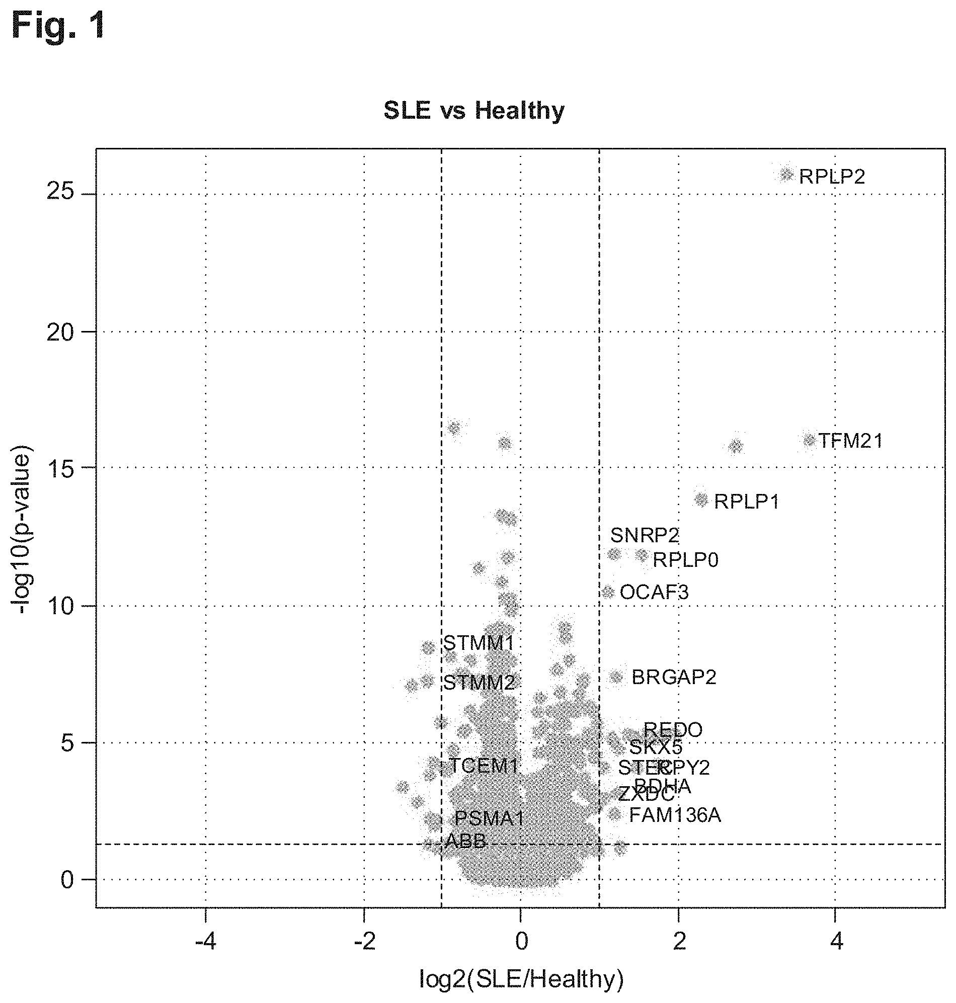
[0164] FIG. 1 shows a volcano plot of the relative antigen reactivities of the SLE patients compared to healthy controls.
[0165] FIG. 2 shows a volcano plot of the relative antigen reactivities of the SLE patients compared to RA patients.
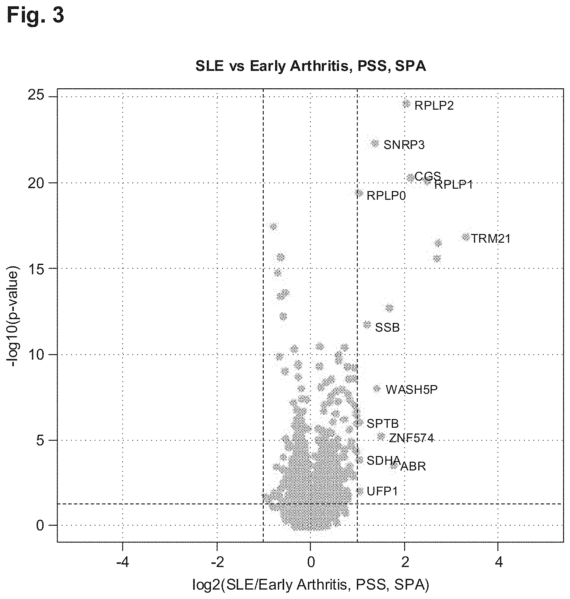
[0166] FIG. 3: volcano plot of the antigen reactivities of SLE patients compared with a combined group of patients with various autoimmune diseases, such as SSc (PSS), SPA, early rheumatoid arthritis and SPA,
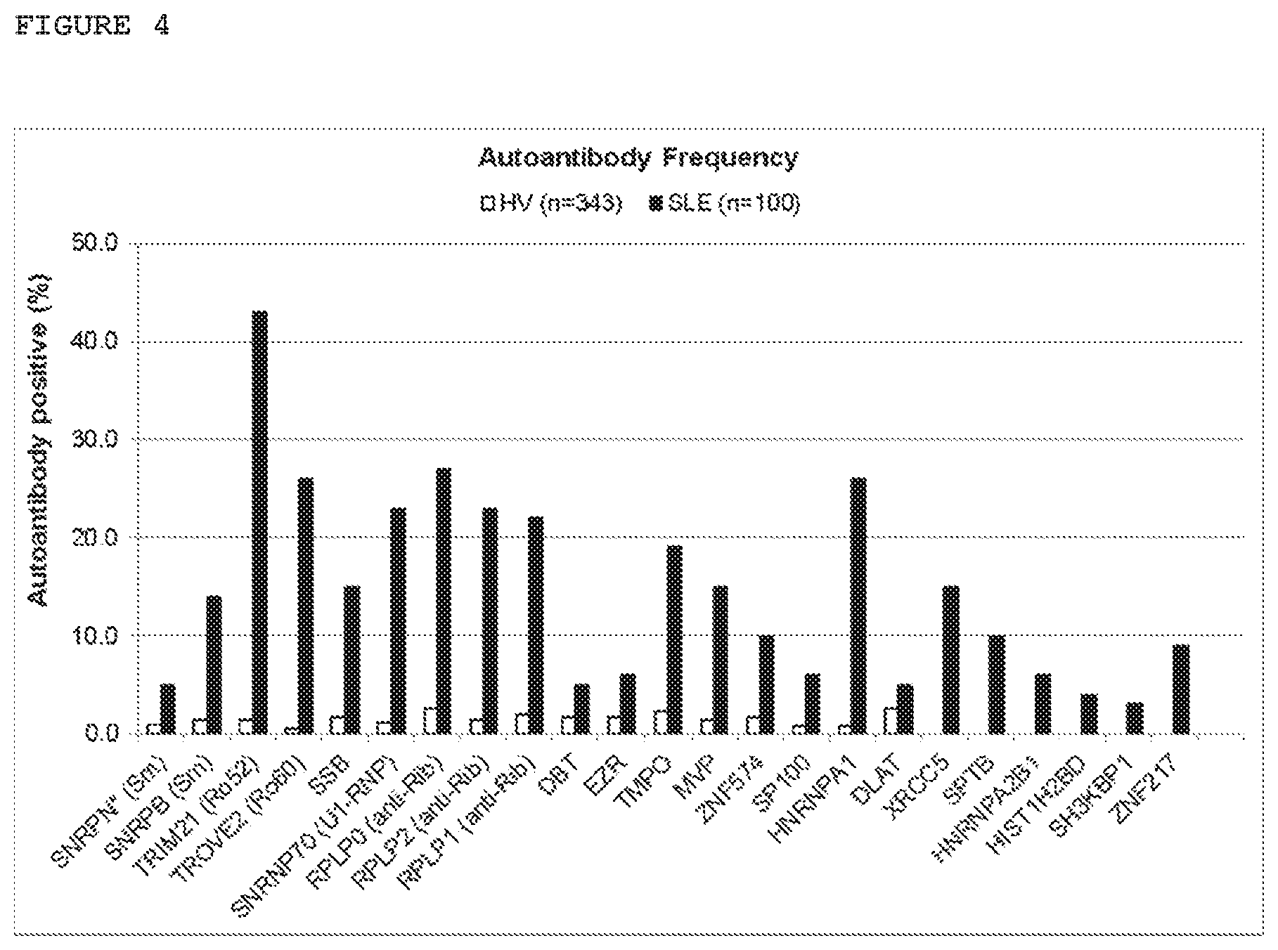
[0167] FIG. 4 frequency of the autoantibody reactivities of selected antigens in SLE patients and healthy test subjects, A threshold value of 3 SO deviations above the mean value of the healthy test, subject was applied. The threshold value for the antigen SNRNP was set to 2SD.
[0168] FIG. 5: volcano plot of the autoantibody reactivities of ENA-4-negative SLE patients compared with healthy controls.
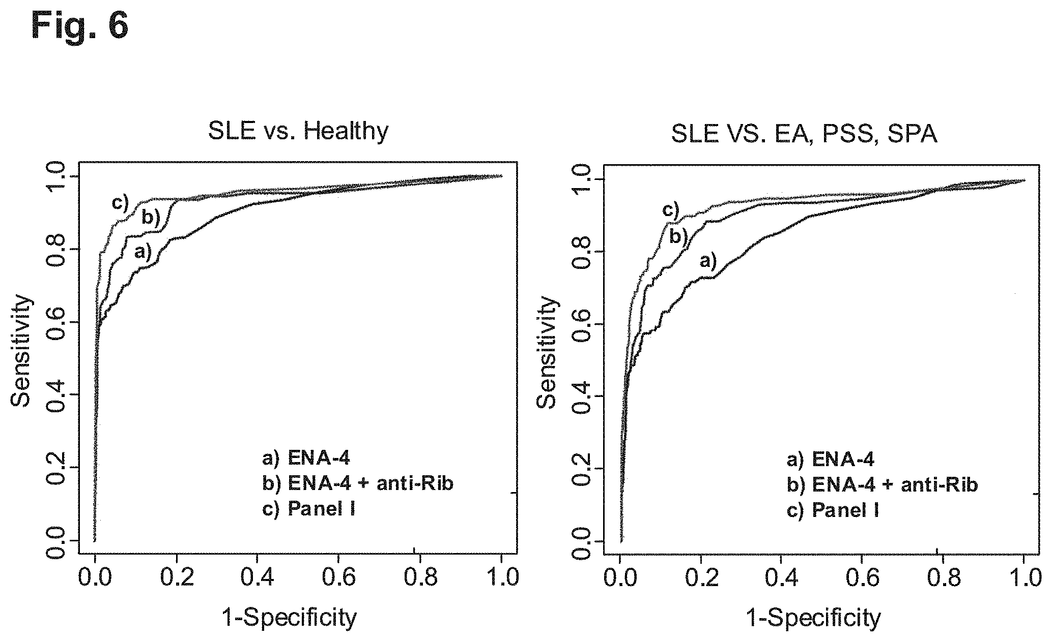
[0169] FIG. 6: receiver operating characteristic curves (ROCs) for the diagnosis of SLS compared to healthy test subjects and AID samples
[0170] FIG. 7: volcano plot for SLE lupus nephritis compared with SLE without lupus nephritis
[0171] FIG. 8: frequency of the lupus nephritis antigens in a model with nested cross validation.
[0172] FIG. 9: dendogram of the SLE antigens following calculation of Spearman/s rank correlation coefficient a) dendogram of the known ENA-4 antigens and b) dendogram of 50 selected SLE antigens.
[0173] FIG. 10A: PPLS-DA biplot of the SLE patients and healthy controls with use of the ENA-4 and ribosomal antigens.
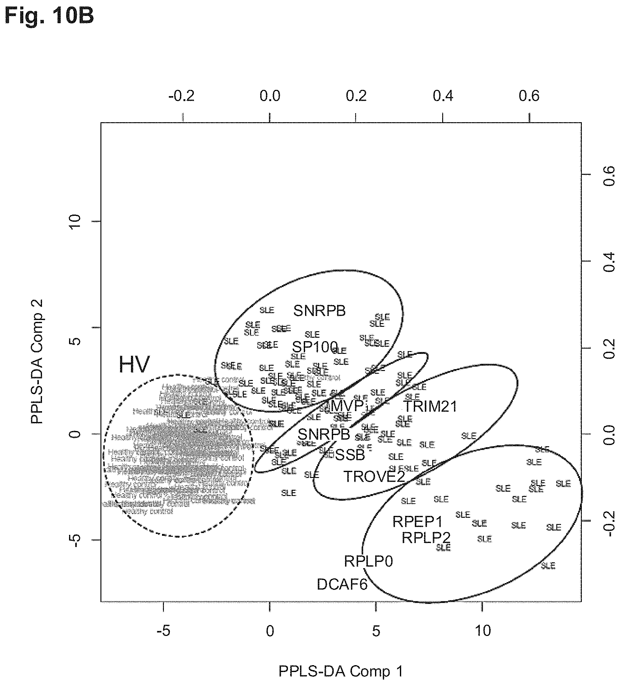
[0174] FIG. 10B: PPLS-DA biplot of the SLE patients and healthy controls based on 50 SLE antigens.
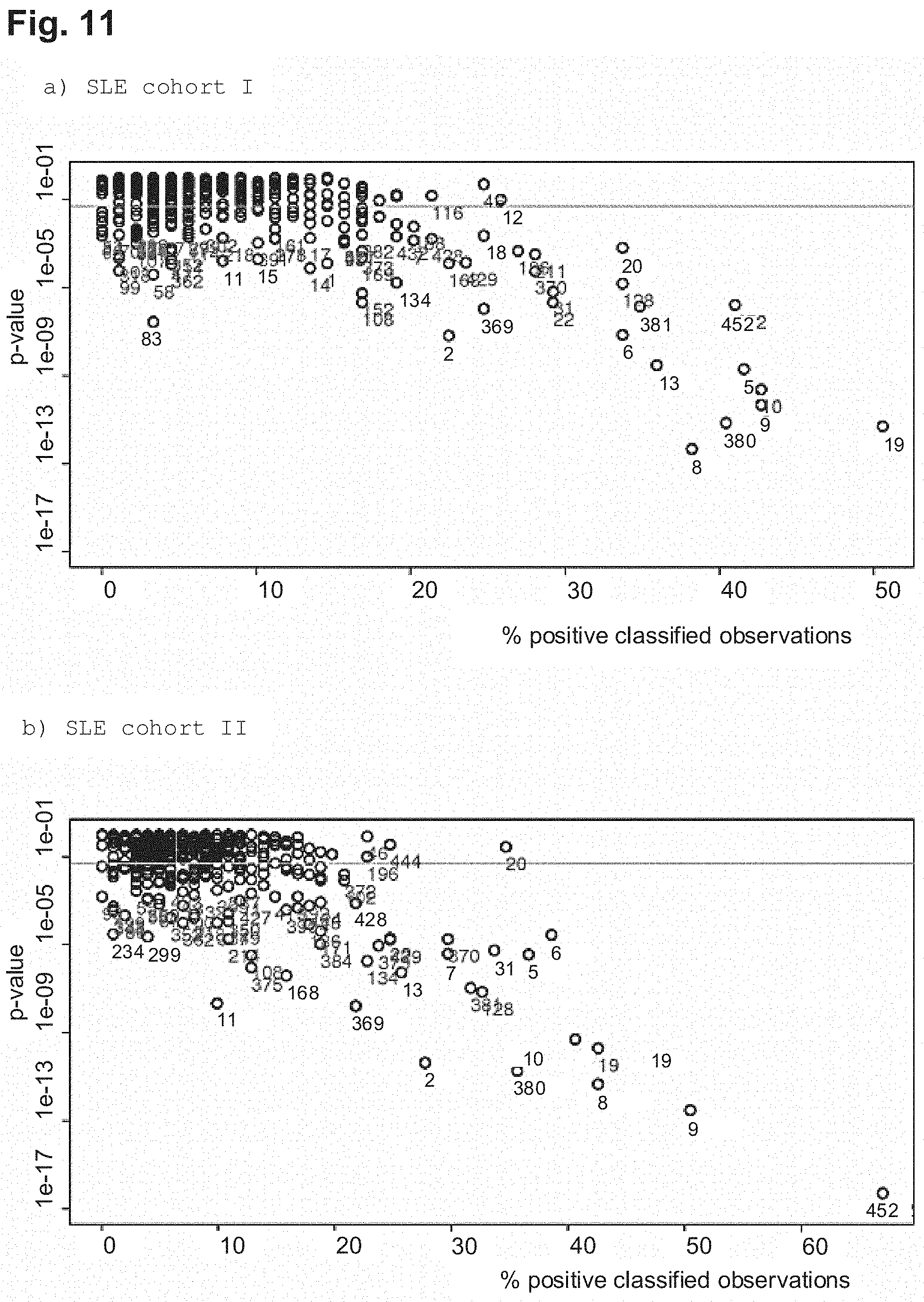
[0175] FIG. 11: calculated p-values of the antigens from Table 2 and also the frequency of patients from a) SLE cohort I, b) SLE cohort II, and c) SLE cohort III who were classified as autoantibody-positive for this antigen.
EXAMPLES
Example 1: Selection of the SLE Patients and Test Subjects
[0176] Selection of the patient groups to be tested: Blood samples were analysed from 129 SLE patients, 100 patients with systemic sclerosis (SSc, PSS), 75 patients with rheumatoid arthritis (RA), 537 patients with early RA (period of disease less than 6 months) and 75 patients with ankylosing spondylitis (SPA)/Bekhterev's disease (SPA). 343 blood samples from the Bavarian Red Cross (RFC) were used as control group. An informed consent of the Ethics Commission of the clinical partners and of the biobank of the BRC was received from all test subjects.
TABLE-US-00001 TABLE 1 Patient samples and clinical data (test cohort. I) 2. Screen 1. Screen SSc (PSS) Early RA SLE RA Healthy SLE Total Subtype (<6 months) SPA Healthy Number 129 75 123 100 100 537 82 343 Age 39 +/- 56.6 +/- 41.3 +/- 39.8 +/- 56.9 +/- Limited 56.8 +/- 43.7 +/- 47.7 +/- (years) 12 13.2 11 11.9 13.4 n - 50 14.3 10.1 11.7 % female 86.1 72 86.2 83 87 Diffuse 62.2 15.9 58.3 n = 32 % ANA 77.5 N.D. N.D. 100 95 Overlap N.D. N.D. N.D. n = 9 SLAM 7.7 +/- 7.7 +/- 5.1 5.1 SLICC 1.45 +/- 1.45 +/- 1.8 1.8 ANA % ENA-4 37 48 positive % U1-RNP(% 13 13 of ENA-4 pos.) SM (% of 8 8 ENA-4 pos.) SS-A/Ro52 35 35 (% of ENA- 4 pos.) SS0B/Ro60 10 10 (% of ENA- 4 pos.) Kidney 26.4 34 involvement %
Example 2: Antigen Production
[0177] Five cDNA libraries that had been produced from different human, tissues (foetal brain, intestine, lung, liver and T-cells) were used for the production, of the recombinant antigens. All cDNAs were expressed in E. coli under the transcriptional control of the lactose-inducible promoter. The resultant proteins carry, at their amino terminus, an additional sequence for a hexahistidine purification tag (His6 tag), Target, antigens which were not present, in the cDNA library were produced by chemical synthesis (Life Technologies) and cloned into the expression vector pQE30-NST, which already codes an amino-terminal His6 tag.
[0178] Following recombinant expression of the proteins, these were isolated in denaturising conditions and purified by means of metal affinity chromatography (IMAC). The proteins were lyophilised and stored set -20.degree. C. until further use (http://www.lifesciences.sourceboioscience.com).
Example 3: Production of the BBAs
[0179] The production of BBAs was adapted to a microtitre plate format, such that 384 coupling reactions could be assessed in parallel using automated pipette systems (Starlet, Hamilton Robotics, Evo Freedom 130, Tecan), For the use of automated pipette systems, the individual bead regions were transferred into coupling pates (96 well Greiner) and the antigens were transferred into 2D barcode vessels (Thermo Scientific). For each coupling reaction, 0.6 to 2.5 million beads and, depending on the antigen, 1 to 100 .mu.g protein were used.
[0180] All washing and pipetting steps of the coupling reaction were carried out in coupling plates which were fixed on magnets. The beads were washed twice with 100 .mu.l LxAP buffer (100 roM NaH2PO4, pH 6.2) and then, received in 120 .mu.l LxAP buffer. For the activation, 15 .mu.l 1-ethyl-3-(3-dimethylaminopropyl)carbodiimide (EDC; 50 mg/ml) and 15 .mu.l N-hydroxysulfosuccinimide (sulfo-NHS; 50 mg/ml) were added by pipette to form a bead suspension, and these suspensions were then incubated for 20 minutes in the shaker (RT, 900 rpm, protected against light). The beads--were then washed 3.times. with 150 .mu.l LxKPT buffer and then the protein solution was added. Following an incubation period of two hours in the shaker (RT, 300 rpm, protected against light), the beads were then washed three times with 150 .mu.l LxKPT buffer. To block free binding points, 100 ul LxCBSP buffer (PBS, 1% BSA, 0.05% ProClin300) were added, and these, mixtures were then incubated for 20 min in the shaker (RT, 900 rpm, protected against, light). This was followed by incubation over night at 4-8.degree. C. The BBA was produced by the combination of beads coupled to antigens and was stored at 4-8.degree. C., protected against light, until use.
Example 4: Quality Control of the BBAs
[0181] In order to check the immobilisation of the proteins at the respective bead regions, a coupling control, was carried out. Here, different amounts of beads were used (250, 500 and 750 beads per bead region). For a reaction mixture, 500 beads for example per bead region were diluted in LxCBS buffer (PBS, 1% BSA) and transferred into an assay plate (96 well half area microplate, Greiner).
[0182] Before each washing step, the assay plate with the beads was placed for 2 minutes on a magnet, and the supernatant was then removed. After three washing steps, the beads were incorporated with 100 .mu.l LxWPT buffer (PBS, 0,05% Tween-20), and 10 .mu.l/ml penta-his antibodies (Qiagen) or LxCBS buffer (PBS, 1% BSA) were added by pipette. Following incubation for 45 minutes in the shaker (RT, 900 rpm, protected against light), the supernatant was removed and the beads were washed in two steps, 5 .mu.l/ml goat, anti-mouse IgG-PE (Phycoerythrin) or goat anti-human IgG-PE (Dianova) were then added as secondary antibody to the reaction mixture and incubated for 30 minutes. Following two washing steps, 100 .mu.l of carrier liquid (Luminex) was added to the beads. The fluorescence signal of the beads was detected with the aid of the FlexMAPSD instrument. Here, the bead count on the one hand and the median of the fluorescence intensity (MFI value) on the other hand were measured.
Example 5: Application of BBAs
[0183] For application, BBAs were incubated with sera and all IgG-based autoantibodies bonded to antigens were detected with the aid of a secondary antibody. In order to enable a high throughput of measurements, the application of BBAs was adapted to a microtiter plate format so that either an 8-channel (Starlet, Hamilton Robotics) or a 96-channel (Evo Freedom. 150, Tecan) automated pipetting system could be used. The sera to be examined were transferred into 2D barcode vessels and then diluted 1:100 with assay buffer (PBS, 0,5% BSA, 10% E. coli lysate, 50% low-cross buffer (Candor Technologies)). In order to neutralise human antibodies directed against E. coli, a pre-incubation of the sera dilutions was performed for 20 min. In this time, 500 beads per bead region were distributed in the assay plate. 50 .mu.l of diluted serum were added to the beads in the coupling plate, and the reaction mixtures were incubated for 18-22 h in the shaker (4-8 *C, 900 rpm, protected against light). After three washing steps in each case with 100 .mu.l LxWPT buffer, 5 .mu.l/ml of the detection antibody goat anti-human IgG-PE (Dianova) were added to the reaction mixtures and incubated for 1 h in the shaker (RT, 900 rpm). The beads were then washed three times with 100 pi LxWPT and incorporated in 100 .mu.l carrier liquid (Luminex). The fluorescence signal of the beads was detected with the aid of the FiexMAF3D instrument, Here, the bead count on the one hand and the MFI value (median fluorescence intensity) on the other hand were measured.
Example 6: Biostatistical Analysis
[0184] The biostatistical analysis comprised univariate and multivariate methods for describing the statistical properties of individual antigens and of groups of antigens. In order to discover interesting candidates for panels, the key property was a good separation between the groups of samples based on the MFI values. In order to find antigen candidates for panel generation, univariate testing, receiver operating characteristic (ROC) analyses, correlation profiles, powered partial least squares discriminant analysis (PPLS-DA) and random forests were used as methods. Biostatistical analyses were subject to expert assessment, in order to define final antigen panels.
[0185] Before the statistical analysis, the MFI values were log 2-transformed in order to reduce the skew in the distributions. If more than 20% of the values `were missing, antigens were excluded from the analysis. Missing values--were replaced by median imputation. A quant lie normalisation was carried out under consideration of the reference sera in order to normalise, per BBA set, all measured samples on individual plates.
[0186] Besides descriptive standardisation for MFI values, non-parametric tests were also carried out with the aid of the two-sided Mann-Whitney-U test in order to uncover differences in the median values of the groups. The test level for multiple testing was corrected in accordance with the Bonferroni-Holm procedure. In addition, the Benjamin-Hochberg procedure inclusive of the determination of the False Discovery Rate (FDR, q-value) was applied. In addition, fold-change and effect size were determined. In order to assess the classification quality, an ROC analysis was carried out, within the scope of which sensitivity, specificity and the area under the ROC curve (AUC) were calculated, in each case inclusive of the 95% confidence interval on the basis of the bootstrap method. Boxplots and volcano plots were used, for graphical representation. A scoring system was implemented on the basis of the univariate results.
[0187] By means of the application of a PPLS-DA, it was attempted to maximise the correlation between the component of the response matrix. A linear discriminant analysis with the latent component as predictors was used for the final classification, A random forest was applied, in which binary decision trees are combined. The decision trees were formed on the basis of a number of bootstrap samples of a training sample and by random selection of a subgroup of explaining variables at each node. The number of input variables, which was selected randomly with each division step, was determined as the square root of the total number of variables, and the number of trees in the random forest was set to 1000. A cross validation with 500 times throughput was imp I. erne n t ed for both multi-variant approaches.
Example 7: Autoantibodies/Antigen Reactivities Differentiate SLE from Healthy Controls, Rheumatoid Arthritis and Other Autoimmune Diseases
[0188] In a first screening the antigen reactivities of 129 SLE patients, 75 RA patients, and 134 healthy controls categorized in accordance with age and sex were differentially tested. For this purpose, the autoantibody reactivities of these blood samples were tested on 5857 antigens coupled to Luminex beads. In order to identify antigens with which the group of all SLE patients can be distinguished from different control croups consisting of healthy samples and patients with RA, univariate statistical tests were carried out, The result, of the statistical test is illustrated as a volcano plot for all 5857 antigens. In the volcano plot, the x-axis shows the relative change of the antigen reactivity in SLE patients compared with healthy controls (FIG. 1) and RA patients (FIG. 2). The y-axis presents the p-value of the statistical tests. FIGS. 1 and 2 show that specific autoantibody reactivities were found which are increased in the group of ail SLE and which can distinguish both from healthy donors and from RA patients.
Example 8: Autoantibodies/Antigen Reactivities Differentiate SLE from Healthy Controls, Early Rheumatoid Arthritis and Other Autoimmune Diseases
[0189] In a second screening with 6088 antigens, the antigens which differentiate between healthy controls and donors with rheumatoid arthritis were tested on patients with early rheumatoid arthritis, SSc and SPA., This is of importance in particular since patients with collagenoses and mixed collagenoses have an overlapping autoantibody profile and therefore are difficult to diagnose, particularly in the early phase
[0190] FIG. 3 shows a volcano plot of the antigen reactivities of SLE patients against a combined group of patients with various autoimmune diseases, such as SSc, SPA, early rheumatoid arthritis, and SPA.
[0191] Following univariate statistical evaluation, a threshold value of p<0.05 and a 1,5 times modified reactivity compared with the control group were applied. A final list of antigen reactivities over both screens was established (Table 2).
[0192] In order to analyse the frequency of the newly identified antigens in comparison with, known antigens, a threshold value of 3 standard deviations (SD) above the mean value of the healthy samples was defined.
[0193] Astonishingly, at least 4 additional antigens were identified of which the frequency in SLE patients lies above 15%. These include TMPO (19%) (SEQ ID No. 13), HNRNPA1 (26%) (SEQ ID No. 5), XRCC5 (15%) (SEQ ID No. 22) and MVP (15%) (SEQ ID No. 7).
[0194] FIG. 4 shows the frequency of 23 antigens in comparison to the healthy controls.
[0195] Table 2 summarises the identified antigen reactivities and different group comparisons.
TABLE-US-00002 TABLE 2 List of all antigen reactivities Statistical Test SEQ Gene Gene Gene Panel SLE ENA-4 neg L. Nephr. SLE ID No. ID Symbol Name Group SLE L. Nephr. Cluster vs HV vs SLE vs control 1 1629 DBT dihydrolipoamide 1 x x SLE branched chain vs transacylase E2 AID 2 1737 DLAT dihydrolipoamide S- 1 x SLE acetyltransferase vs AID 3 7430 EZR ezrin 1 x x x SLE vs AID 4 3017 HIST1H2BD histone cluster 1, 1 x x SLE H2bd vs AID 5 3178 HNRNPA1 heterogeneous 1 x x SLE nuclear vs ribonucleoprotein A1 AID 6 3181 HNRNPA2B1 heterogeneous 1 x x SLE nuclear vs ribonucleoprotein AID A2/B1 7 9961 MVP major vault protein 1 x x x x SLE vs AID 8 6175 RPLP0 ribosomal protein, 1 x x x SLE large, P0 vs AID 9 6176 RPLP1 ribosomal protein, 1 x x x x SLE large, P1 vs AID 10 6181 RPLP2 ribosomal protein, 1 x x x SLE large, P2 vs AID 11 30011 SH3KBP1 SH3-domain kinase 1 x x SLE binding protein 1 vs AID 12 6625 SNRNP70 small nuclear 1 x SLE ribonucleoprotein vs 70 kDa (U1) AID 13 6628 SNRPB small nuclear 1 x x x SLE ribonucleoprotein vs polypeptides B and AID B1 14 6638 SNRPN small nuclear 1 x SLE ribonucleoprotein vs polypeptide N AID 15 6672 SP100 SP100 nuclear 1 x x SLE antigen vs AID 16 6710 SPTB spectrin, beta, 1 x x SLE erythrocytic vs AID 17 6741 SSB Sjogren syndrome 1 x x SLE antigen B vs (autoantigen La) AID 18 7112 TMPO thymopoietin 1 x x x SLE vs AID 19 6737 TRIM21 tripartite motif- 1 x x x SLE containing 21 vs AID 20 6738 TROVE2 TROVE domain family, 1 x x SLE member 2 vs RA 21 7431 VIM vimentin 1 x x SLE vs AID 22 7520 XRCC5 X-ray repair 1 x x SLE complementing vs defective repair in AID Chinese hamster cells 5 (double- strand-break re joining) 23 7764 ZNF217 zinc finger protein 1 x x SLE 217 vs AID 24 64763 ZNF574 zinc finger protein 1 x x SLE 574 vs AID 25 148741 ANKRD35 ankyrin repeat 2 x x SLE domain 35 vs HV 26 84779 ARD1B ARD1 homolog B 2 x x SLE (S. cerevisiae) vs AID 27 672 BRCA1 breast cancer 1, 2 x x SLE early onset vs HV 28 134359 C5orf37 chromosome 5 open 2 x x x SLE reading frame 37 vs HV 29 9478 CABP1 calcium binding 2 x x SLE protein 1 vs HV 30 90557 CCDC74A coiled-coil domain 2 x x SLE containing 74A vs HV 31 9973 CCS copper chaperone for 2 x x x x SLE superoxide dismutase vs AID 32 1410 CRYAB crystallin, alpha B 2 x x SLE vs HV 33 55802 DCP1A DCP1 decapping 2 x x SLE enzyme homolog A vs (S. cerevisiae) HV 34 79147 FKRP fukutin related 2 x SLE protein vs HV 35 26128 KIAA1279 KIAA1279 2 x x SLE vs HV 36 57608 KIAA1462 KIAA1462 2 x x SLE vs HV 37 1939 LGTN ligatin 2 x x SLE vs HV 38 84298 LLPH LLP homolog, long- 2 x x SLE term synaptic vs facilitation HV (Aplysia) 39 11253 MAN1B1 mannosidase, alpha, 2 x x SLE class 1B, member 1 vs HV 40 84930 MASTL microtubule 2 x x SLE associated vs serine/threonine HV kinase-like 41 54531 MIER2 mesorm induction 2 x x x x SLE early response 1, vs family member 2 RA 42 4594 MUT methylmalonyl 2 x x SLE Coenzyme A mutase vs HV 43 399687 myO18A myosin XVIIIA 2 x x SLE vs HV 44 8883 NAE1 NEDD8 activating 2 x x SLE enzyme E1 and vs subunit 1 HV 45 10458 BAIAP2 BAI1-associated 2 x x SLE protein 2 vs HV 46 4869 NPM1 nucleophosim 2 x x SLE (nucleolar vs phosphoprotein B23, HV numatrin) 47 5223 PGAM1 phosphoglycerate 2 x x SLE mutase 1 (brain) vs HV 48 11040 PIM2 pim-2 oncogene 2 x x SLE vs HV 49 54517 PUS7 pseudouridylate 2 x x SLE synthase 7 homolog vs (S. cerevisiae) HV 50 6605 SMARCE1 SWI/snf related, 2 x x SLE matrix associated, vs actin dependent AID regulator of chromatin, subfamily e, member 1 51 23635 SSBP2 single-stranded DNA 2 x x x SLE binding protein 2 vs HV 52 83660 TLN2 talin 2 2 x x SLE vs HV 53 51673 TPPP3 tubulin 2 x x SLE polymerization- vs promoting protein HV family member 3 54 7265 TTC1 tetratricopeptide 2 x x SLE repeat domain 1 vs HV 55 124930 ANKRD13B ankyrin repeat 3 x SLE domain 13B vs HV 56 160 AP2A1 adaptor-related 3 x SLE protein complex 2, vs alpha 1 subunit HV 57 53335 BCL11A B-cell CLL/lymphoma 3 x x 11A (zinc finger protein) 58 79959 CEP76 centrosomal protein 3 x 76 kDA 59 1153 CIRBP cold inducible RNA 3 x SLE binding protein vs HV 60 51084 CRYL1 crystallin, lambda 1 3 x 61 55827 DCAF6 DDB1 and CUL4 3 x x x SLE associated factor 6 vs AID 62 6993 DYNLT1 dynein, light chain, 3 x SLE Tctex-type 1 vs HV 63 283991 FAM100B family with sequence 3 x SLE similarity 100, vs member B HV 64 9815 GIT2 G protein-coupled 3 x SLE receptor kinase vs interacting ArfGAP 2 HV 65 84706 GPT2 glutamic pyruvate 3 x transaminase (alanine aminotransferase) 2 66 3059 HCLS1 hematopoieti cell- 3 x x SLE specific Lyn vs substrate 1 AID 67 3329 HSPD1 heat shock 60 kDa 3 x protein 1 (chaperonin) 68 3490 IGFBP7 insulin-like growth 3 x SLE factor binding vs protein 7 HV 69 23392 KIAA0368 KIAA0368 3 x 70 84695 LOXL3 lysyl oxidase-like 3 x 3 71 4133 MAP2 microtubule- 3 x SLE associated protein vs 2 HV 72 6837 MED22 mediator complex 3 x subunit 22 73 29079 MED4 mediator complex 3 x x subunit 4 74 10933 MORF4L1 mortality factor 4 3 x like 1 75 64963 MRPS11 mitochondrial 3 x x SLE ribosomal protein vs HV 76 81565 NDEL1 nudE nuclear 3 x distribution gene E homolog (A. nidulans)-like 1 77 57447 NDRG2 NDRG family member 3 x SLE 2 vs HV 78 4744 NEFH neurofilament, heavy 3 x polypeptide 79 153478 PLEKHG4B pleckstrin homology 3 x domain containing, family G (with
RhoGef domain) member 4B [homo sapiena (human)] 80 11054 OGFR opioid growth factor 3 x x SLE receptor vs AID 81 56122 PCDHB14 protocadherin beta 3 x SLE 14 vs HV 82 2923 PDIA3 protein disulfide 3 x SLE isomerase family A, vs member 3 HV 83 23646 PLD3 phospholipase D 3 x SLE family, member 3 vs HV 84 23759 PPIL2 peptdylprolyl 3 x x isomerase (cyclophilin)-like 2 85 5557 PRIM1 primase, DNA, 3 x SLE polypeptide 1 vs (49 kDa) HV 86 5682 PSMA1 proteasome (prosome, 3 x SLE macropain) subunit, vs alpha type, 1 HV 87 5802 PTPRS protein tyrosine 3 x SLE phosphatase, vs receptor type, S HV 88 81890 QTRT1 queuine tRNA- 3 x SLE ribosyltransferase 1 vs HV 89 116362 RBP7 retinol binding 3 x SLE protein 7, cellular vs HV 90 10287 RGS19 regulator of G- 3 x x protein signaling 19 91 83642 RP3-402G11.5 selenoprotein O 3 x SLE vs HV 92 6389 SDHA succinate 3 x x SLE dehydrogenase vs complex, subunit A, AID flavoprotein (Fp) 93 54437 SEMA5B sema domain, seven 3 x thrombospondin repeats (type 1 and type 1-like), transmembrane domain (TM) and short cytoplasmic domain, (semaphorin) 5B 94 59343 SENP2 SUMO1/sentrin/SMT3 3 x SLE specific peptidase vs 2 HV 95 6629 SNRPB2 small nuclear 3 x SLE ribonucleoprotein vs polypeptide B'' AID 96 27131 SNX5 sorting nexin 5 3 x SLE vs HV 97 9021 SOCS3 suppressor of 3 x x SLE cytokine signaling vs 3 HV 98 3925 STMN1 stathmin 1 3 x SLE vs HV 99 81551 STMN4 stathmin-like 4 3 x SLE vs HV 100 27097 TAFSL TAF5-like RNA 3 x SLE polymerase II, vs p300/CBP-associated HV factor (PCAF)- associated factor, 65 kDa 101 79521 TCEAL4 transcription 3 x SLE elongation factor A vs (SII)-like 4 HV 102 10040 TOM1L1 target of mybl 3 x SLE (chicken)-like 1 vs HV 103 22974 TPX2 TPX2, microtubule- 3 x SLE associated, homolog vs (Xenopus laevis) HV 104 51567 TTRAP TRAF and TNF 3 x receptor associated protein 105 8615 USO1 US01 homolog, 3 x x vescicle docking protein (yeast) 106 10869 USP19 ubiquitin specific 3 x SLE peptidase 19 vs RA 107 29761 USP25 ubiquitin specific 3 x peptidase 25 108 375690 WASH5P WAS protein family 3 x x SLE homolog 5 vs pseudogene HV 109 10413 YAP1 Yes-associated 3 x protein 1, 65 kDa 110 653121 ZBTB8A zinc finger and BTB 3 x x SLE domain containing vs BA HV 111 55311 ZNF444 zinc finger protein 3 x 444 112 29 ABR active BCR-related 4 x SLE gene vs AID 113 118 ADD1 adducin 1 (alpha) 4 x SLE vs AID 114 55256 ADI1 acireductone 4 x SLE dioxygenase 1 vs HV 115 9255 AIMP1 aminoacyl tRNA 4 x synthetase complex- interacting multifunctional protein 1 116 54522 ANKRD16 ankyrin repeat 4 x SLE domain 16 vs HV 117 348 APOE apolipoprotein E 4 x SLE vs HV 118 64333 ARHGAP9 Rho GTPase 4 x SLE activating protein vs 9 HV 119 22994 AZI1 5-azacytidine 4 SLE induced 1 vs HV 120 55971 BAIAP2L1 BAI1-associated 4 x protein 2-like 1 121 7919 BAT1 HLA-B associated 4 x SLE transcript 1 vs RA 122 6046 BRD2 bromodomain 4 x containing 2 123 56912 C11orf60 chromosome 11 open 4 x reading frame 60 124 79415 C17orf62 chromosome 17 open 4 x reading frame 62 125 51300 C3orf1 chromosome 3 open 4 x SLE reading frame 1 vs RA 126 128866 CHMP4B chromatin modifying 4 x SLE protein 4B vs AID 127 23122 CIC capicua homolog 4 x SLE (Drosophila) vs AID 128 10970 CKAP4 cytoskeleton- 4 x SLE associated protein vs 4 HV 129 23122 CLASP2 cytoplasmic linker 4 x associated protein 2 130 1311 COMP cartilage 4 x oligomeric matrix protein 131 7812 CSDE1 cold shock domain 4 x SLE containing E1, RNA- vs binding HV 132 8642 DCHS1 dachsous 1 4 x SLE (Drosophila) vs AID 133 9909 DENND4B DENN/MADD domain 4 x x containing 4B 134 1743 DLST dihydrolipoamide S- 4 x succinyltransferase (E2 component of 2- oxo-glutarate complex) 135 84444 DOT1L DOT1-like, histone 4 x H3 methyltransferase (S. cerevisiae) 136 51143 DYNC1LI1 dynein, cytoplasmic 4 x SLE 1, light vs intermediate chain 1 HV 137 51011 FAHD2A fumarylacetoacetate 4 x hydrolase domain containing 2A 138 92689 FAM114A1 family with sequence 4 x similarity 114, member A1 139 54463 FAM134B family with sequence 4 x similarity 134, member B 140 100129583 FAM47E family with sequence 4 x SLE similarity 47, vs member E HV 141 93611 FBXO44 F-box protein 44 4 x 142 60681 FKBP10 FK506 binding 4 x SLE protein 10, 65 kDa vs AID 143 23360 FNBP4 formin binding 4 x protein 4 144 2300 FOXL1 forkhead box L1 4 x SLE vs HV 145 64689 GORASP1 golgi reassembly 4 x SLE stacking protein 1, vs 65 kDa AID 146 2934 GSN gelsolin 4 x SLE (amyloidosis, vs Finnish type) HV 147 3039 HBA1 hemoglobin, alpha 4 x 148 3040 HBA2 hemoglobin, alpha 2 4 x 149 38858 HES5 hairy and enhancer 4 x of split 5 (Drosophilia) 150 10525 HYOU1 hypoxia up-regulated 4 x 1 151 3608 ILF2 interleukin enhancer 4 x SLE binding factor 2, vs 45 kDa RA 152 23135 KDM6B lysine (K)-specific 4 x SLE demethylasae 6B vs AID 153 56243 KIAA1217 KIAA1217 4 x SLE vs HV 154 57662 KIAA1543 KIAA1543 4 x 155 57498 KIDINS220 kinase D-interacting 4 x substrate, 220 kDA 156 3855 KRT7 keratin 7 4 x SLE vs HV 157 729970 LOC729970 similar to 4 x hCG2028352 158 9935 MAFB v-maf 4 x musculoaponeurotic fibrosarcoma oncogene homolog B (avian) 159 23764 MAFF v-maf 4 x SLE musculoaponeurotic vs fibrosarcoma HV oncogene homolog F (avian) 160 22924 MAPRE3 microtubule- 4 x associated protein, RP/EB family, member 3 161 8079 MLF2 myeloid leukemia 4 x factor 2 162 4676 NAP1L4 nucleosome assembly 4 x protein 1-like 4 163 4688 NCF2 neutrophil cytosolic 4 x SLE factor 2 vs
HV 164 4780 NFE2L2 nuclear factor 4 x (erythroid-derived 2)-like 2 165 79840 NHEJ1 nonhomologous end- 4 x x joining factor 1 166 22861 NLRP1 NLR family, pyrin 4 x SLE domain containing 1 vs HV 167 65009 NDRG4 NDRG family member 4 4 x SLE vs HV 168 4841 NONO non-POU domain 4 x SLE containing, octamer- vs binding AID 169 29982 NRBF2 nuclear receptor 4 x SLE binding factor 2 vs AID 170 8439 NSMAF neutral 4 x SLE sphingomyelinase (N- vs SMase)activation HV associated factor 171 4926 NUMA1 nuclear mitotic 4 x SLE apparatus protein 1 vs RA 172 84759 PCGF1 polycomb group ring 4 x finger 1 173 84306 PDCD2L programmed cell 4 x SLE death 2-like vs HV 174 5195 PEX14 peroxisomal 4 x SLE biogenesis factor 14 vs HV 175 9091 PIGQ phosphatidylinositol 4 x SLE glycan anchor vs biosynthesis, class RA Q 176 100137049 PLA2G4B phospholipase A2, 4 x SLE group IVB vs (cytosolic) RA 177 10226 PLIN3 perilipin 3 4 x 178 5373 PMM2 phosphomannomutase 2 4 x 179 10450 PPIE peptidylprolyl 4 x isomerase E (cyclophilin E) 180 5694 PSMB6 proteasome (prosome, 4 x macropain) subunit, beta type, 6 181 22913 RALY RNA binding protein, 4 x SLE autoantigenic vs (hnRNP-associated HV with lethal yellow homolog (mouse)) 182 8241 RBM10 RNA binding motif 4 x protein 10 183 9904 RBM19 RNA binding motif 4 x SLE protein 19 vs HV 184 9743 RICS Rho GTPase- 4 x activating protein 185 8780 RIOK3 RIO kinase 3 (yeast) 4 x 186 8578 SCARF1 scavenger receptor 4 x SLE class 4, member 1 vs AID 187 23513 SCRIB scribbled homolog 4 x SLE (Drosophila) vs HV 188 644096 SDHAF1 succinate 4 x SLE dehydrogenase vs complex assembly RA factor 1 189 57794 SF4 splicing factor 4 4 x SLE vs RA 190 9814 SFI1 Sfi1 homolog, 4 x spindle assembly associated (yeast) 191 6421 SFPQ splicing factor 4 x SLE proline/glutamine- vs rich (polypyrimidine AID tract binding protein associated) 192 83442 SH3BGRL3 SH3 domain binding 4 x glutamic acid-rich protein like 3 193 6461 SHB Src homology 2 4 x SLE domain containing vs adaptor protein B AID 194 23381 SMG5 Smg-5 homolog, 4 x SLE nonsense mediated vs mRNA decay factor HV (C. elegans) 195 112574 SNX18 sorting nexin 18 4 x SLE vs HV 196 84501 SPIRE2 spire homolog 2 4 x SLE (Drosophila) vs HV 197 54961 SSH3 slingshot homolog 3 4 x SLE (Drosophila) vs AID 198 9263 STK17A serine/threonine 4 x kinase 17a 199 51111 SUV420H1 suppressor of 4 x variegation 4-20 homolog 1 (Drosophila) 200 6902 TBCA tubulin folding 4 x cofactor A 201 7024 TFCP2 transcription factor 4 x SLE CP2 vs HV 202 7030 TFE3 transcription factor 4 x SLE binding to IGHM vs enhancer 3 HV 203 90326 THAP3 THAP domain 4 x SLE containing, vs apoptosis associated AID protein 3 204 10043 TOM1 target of myb1 4 x (chicken) 205 7168 TPM1 tropomyosin 1 4 x SLE (alpha) vs HV 206 54952 TRNAU1AP tRNA selenocysteine 4 x 1 associated protein 1 207 26140 TTLL3 tubulin tyrosine 4 x ligase-like family, member 3 208 7371 UCK2 uridine-cytidine 4 x SLE kinase 2 vs HV 209 9277 WDR46 WD repeat domain 46 4 x SLE vs HV 210 55100 WDR70 WD repeat domain 70 4 x SLE vs AID 211 23038 WDTC1 WD and 4 x SLE tetratricopeptide vs repeats 1 HV 212 9831 ZNF623 zinc finger protein 4 x 623 213 79364 ZXDC ZXD family zinc 4 x x SLE finger C vs AID 214 7791 ZYX zyxin 4 x SLE vs AID 215 55964 SEPT3 septin 3 5 X 216 5413 SEPT5 septin 5 5 x 217 26574 AATF apoptosis 5 x antagonizing transcription factor 218 91703 ACY3 aspartoacylase 5 x (aminocyclase) 3 219 9509 ADAMTS2 ADAM 6 SLE metallopeptidase vs with thrombospondin RA type 1 motif, 2 220 10939 AFG3L2 AFG3 ATPase family 6 SLE gene 3-like 2 vs (yeast) HV 221 1646 AKR1C2 aldo-keto reductase 6 SLE family 1, member C2 vs (dihydrodiol RA dehydrogenase 2; bile acid binding protein; 3-alpha hydroxysteroid dehydrogenase, type III) 222 267 AMFR autocrine motility 6 SLE factor receptor vs RA 223 10777 ARPP-21 cyclic AMP-regulated 6 SLE phosphoprotein, 21 vs kD RA 224 421 ARVCF armadillo repeat 6 SLE gene deletes in vs velocardiofacial RA syndrome 225 80150 ASRGL1 asparaginase like 1 6 SLE vs RA 226 539 ATP50 ATP synthase, H+ 6 SLE transporting, vs mitochondrial F1 RA complex, O subunit 227 79870 BAALC brain and acute 6 SLE leukemia, vs cytoplasmic RA 228 9531 BAG3 BCL2-associated 5 x athanogene 3 229 9275 BCL7B B-cell CLL/lymphoma 6 SLE 7B vs HV 230 55108 BSDC1 BSD domain 6 SLE containing 1 vs AID 231 54934 C12orf41 chromosome 12 open 6 SLE reading frame 41 vs RA 232 55049 C19orf60 chromosome 19 open 6 SLE reading frame 60 vs RA 233 388799 C20orf107 chromosome 20 open 5 x reading frame 107 234 149840 C20orf196 chromosome 20 open 6 SLE reading frame 196 vs RA 235 51507 C20orf43 chromosome 20 open 6 SLE reading frame 43 vs RA 236 55684 C9orf86 chromosome 9 open 6 SLE reading frame 86 vs HV 237 23523 CABIN1 calcineurin binding 6 SLE protein 1 vs RA 238 157922 CAMSAP1 calmodulin regulated 6 SLE spectrin-associated vs protein 1 RA 239 23624 CBLC Cas-Br-M (murine) 6 SLE ecotropic retroviral vs transforming HV sequence c 240 124808 CCDC43 coiled-coil domain 6 SLE containing 43 vs RA 241 100133941 CD24 CD24 molecule 5 x 242 11140 CDC37 cell division cycle 6 SLE 37 homolog vs (S. cerevisiae) RA 243 10153 CEBPZ CCAAT/enhancer 6 SLE binding protein vs (C/EBP), zeta RA 244 51510 CHMP5 chromatin modifying 6 SLE protein 5 vs RA 245 63922 CHTF18 CTF18, chromosome 5 x transmission fidelity factor 18 homolog (S. cerevisiae) 246 51727 CMPK1 cytidine 6 SLE monophosphate (UMP- vs CMP) kinase 1, AID cytosolic 247 64708 COPS7B COP9 constitutive 5 x
photomorphogenic homolog subunit 7B (Arabidopsis) 248 51117 COQ4 coenzyme Q4 homolog 6 SLE (S. cerevisiae) vs RA 249 27254 CSDC2 cold shock domain 5 x containing C2, RNA binding 250 162989 DEDD2 death effector 6 SLE domain containing 2 vs RA 251 9704 DHX34 DEAH (Asp-Glu-Ala- 6 SLE His) box polypeptide vs 34 RA 252 55837 EAPP E2F-associated 6 SLE phosphoprotein vs RA 253 1915 EEF1A1 eukaryotic 6 SLE translation vs elongation factor 1 RA alpha 1 254 1936 EEF1D eukaryotic 6 SLE translation vs elongation factor 1 RA delta (guanine nucleotide exchange protein) 255 8669 EIF3J eukaryotic 6 SLE translation vs initiation factor 3, RA subunit J 256 55740 ENAH enabled homolog 6 SLE (Drosophila) vs HV 257 2023 ENO1 enolase 1, (alpha) 6 SLE vs HV 258 11124 FAF1 Fas (TNFRSF6) 5 x associated factor 1 259 11170 FAM107A family with sequence 6 SLE similarity 107, vs member A HV 260 84908 FAM136A family with sequence 6 SLE similarity 136, vs member A RA 261 10144 FAM13A family with sequence 6 SLE similarity 13, vs member A RA 262 26017 FAM32A family with sequence 6 SLE similarity 32, vs member A HV 263 64762 FAM59A family with sequence 6 SLE similarity 59, vs member A RA 264 150946 FAM59B family with sequence 6 SLE similarity 59, vs member B HV 265 83706 FERMT3 fermitin family 6 SLE homolog 3 vs (Drosophila) RA 266 23307 FKBP15 FK506 binding 6 SLE protein 15, 133 kDa vs HV 267 2670 GFAP glial fibrillary 6 SLE acidic protein vs RA 268 51031 GLOD4 glyoxalase domain 6 SLE containing 4 vs AID 269 81488 GRINL1A glutamate receptor, 6 SLE ionotropic, N-methyl vs D-aspartate-like 1A RA 270 2922 GRP gastrin-releasing 6 SLE peptide vs RA 271 2935 GSPT1 G1 to S phase 6 SLE transition 1 vs RA 272 93323 HAUS8 HAUS augmin-like 6 SLE complex, subunit 8 vs HV 273 3054 HCFC1 host cell factor C1 6 SLE (VP16-accessory vs protein) AID 274 3069 HDLBP high density 6 SLE lipoprotein binding vs protein RA 275 3184 HNRNPD heterogenous nuclear 6 SLE ribonucleoprotein D vs (AU-rich element RNA HV binding protein 1, 37 kDa) 276 3320 HSP90AA1 heat shock protein 6 SLE 90 kDa alpha vs (cytosolic), class A RA member 1 277 7184 HSP90B1 heat shock protein 6 SLE 90 kDa beta (Grp94), vs member 1 RA 278 3304 HSPA1B heat shock 70 kDa 6 SLE protein 1B vs RA 279 3315 HSPB1 heat shock 27 kDa 4 x x SLE protein 1 vs RA 280 5654 HTRA1 HtrA serine 6 SLE peptidase 1 vs RA 281 3382 ICA1 islet cell 6 SLE autoantigen 1, 69 kDa vs RA 282 3550 IK IK cytokine, down- 6 SLE regulator of HLA II vs HV 283 80895 ILKAP integrin-linked 6 SLE kinase-associated vs serine/threonine RA phosphatase 2C 284 84162 KIAA1109 KIAA1109 6 SLE vs AID 285 3856 KRT8 keratin 8 6 SLE vs RA 286 23367 LARP1 La ribonucleoprotein 6 SLE domain family, vs member 1 AID 287 4001 LMNB1 lamin B1 6 SLE vs RA 288 79888 LPCAT1 lysophosphatidylcholine 5 x SLE acyltransferase vs 1 HV 289 10916 MAGED2 melanoma antigen 5 x family D, 2 290 55700 MAP7D1 MAP7 domain 6 SLE containing 1 vs RA 291 5602 MAPK10 mitogen-activated 6 SLE protein kinase 10 vs HV 292 22919 MAPRE1 microtubule- 6 SLE associated protein, vs RP/EB family, member AID 1 293 4137 MAPT microtubule- 6 SLE associated protein, vs tau RA 294 23139 MAST2 microtubule 6 SLE associated vs serine/threonine RA kinase 2 295 53615 MBD3 methyl-CpG binding 6 SLE domain protein 3 vs RA 296 56922 MCCC1 methylcrotonoyl- 6 SLE Coenzyme A vs carboxylase 1 HV (alpha) 297 1953 MEGF6 multiple EGF-like- 6 SLE domains 6 vs RA 298 4302 MLLT6 myeloid/lymphoid or 6 SLE mixed-lineage vs leukemia (trithorax RA homolog, Drosophila); translocated to, 6 299 10200 MPHOSPH6 M-phase 6 SLE phosphoprotein 6 vs RA 300 10240 MRPS31 mitochondrial 6 SLE ribosomal protein vs S31 HV 301 84939 MUM1 melanoma associated 5 x antigen (mutated) 1 302 4599 MX1 myxovirus (influenza 6 SLE virus) resistance 1, vs interferon-inducible RA protein p78 (mouse) 303 4716 NDUFB10 NADH dehydrogenase 6 SLE (ubiquinone) 1 beta vs subcomplex, 10, RA 22 kDa 304 4796 NFKBIL2 nuclear factor of 6 SLE kappa light vs polypeptide gene HV enhancer in B-cells inhibitor-like 2 305 11188 NISCH nischarin 6 SLE vs RA 306 10381 TUBB3 tubulin, beta 3 6 SLE class III vs RA 307 8602 NOP14 NOP14 nucleolar 6 SLE protein homolog vs (yeast) RA 308 9722 NOS1AP nitric oxice 6 synthase 1 (neuronal) adaptor protein 309 29959 NRBP1 nuclear receptor 5 x binding protein 1 310 142 PARP1 poly (ADP-ribose) 6 SLE polymerase 1 vs RA 311 5091 PC pyruvate 6 SLE carboxylase vs RA 312 23024 PDZRN3 PDZ domain 6 SLE containing ring vs finger 3 RA 313 8682 PEA15 phosphoprotein 6 SLE enriched in vs astrocytes 15 RA 314 5187 PER1 period homolog 1 6 SLE (Drosophila) vs HV 315 57649 PHF12 PHD finger protein 5 x 12 316 26227 PHGDH phosphoglycerate 5 x dehydrogenase 317 1263 PLK3 polo-like kinase 3 6 SLE (Drosophila) vs RA 318 23654 PLXNB2 plexin B2 6 SLE vs RA 319 56902 PNO1 partner of NOB1 6 SLE homolog vs (S. cerevisiae) RA 320 5479 PPIB peptidylprolyl 6 SLE isomerase B vs (cyclophilin B) HV 321 56978 PRDM8 PR domain 6 SLE containing 8 vs HV 322 55119 PRPF38B PRP38 pre-mRNA 6 SLE processing factor vs 38 (yeast) domain RA containing B 323 5764 PTN pleiotrophin 6 SLE vs HV 324 5819 PVRL2 poliovirus 5 x receptor-related 2 (herpesvirus entry mediator B) 325 5831 PYCR1 pyrroline-5- 6 SLE
carboxylate vs reductase 1 RA 326 65997 RASL11B RAS-like, family 6 SLE 11, member B vs RA 327 55658 RNF126 ring finger protein 6 SLE 126 vs AID 328 115992 RNF166 ring finger protein 6 SLE 166 vs HV 329 9025 RNF8 ring finger protein 6 SLE 8 vs HV 330 6092 ROBO2 roundabout, axon 5 x guidance receptor, homolog 2 (Drosophila) 331 64221 ROBO3 roundabout, axon x guidance receptor, homolog 3 (Drosophila) 332 4736 RPL10A ribosomal protein 6 SLE L10a vs RA 333 6152 RPL24 robosomal protein 6 SLE L24 vs RA 334 148418 SAMD13 sterile alpha motif 6 SLE domain containing vs 13 HV 335 57147 SCYL3 SCY1-like 3 6 SLE (S. cerevisiae) vs AID 336 6382 SDC1 syndecan 1 6 SLE vs RA 337 91461 SGK493 protein kinase-like 5 x protein SgK493 338 6449 SGTA small glutamine- 6 SLE rich vs tetratricopeptide HV repeat (TPR)- containing, alpha 339 9627 SNCAIP synuclein, alpha 5 x interacting protein 340 9552 SPAG7 sperm associated 6 SLE antigen 7 vs RA 341 57522 SRGAP1 SLIT-ROBO Rho 6 SLE GTPase activating vs protein 1 RA 342 6744 SSFA2 sperm specific 6 SLE antigen 2 vs RA 343 6487 ST3GAL3 ST3 beta- 6 SLE galactoside alpa- vs 2,3- RA sialyltransferase 3 344 23345 SYNE1 spectrin repeat 6 SLE containing, nuclear vs envelope 1 AID 345 6879 TAF7 TAF7 RNA polymerase 6 SLE II, TATA box vs binding protein HV (TBF)-associated factor, 55 kDa 346 6895 TARBP2 TAR (HIV-1) RNA 6 SLE binding protein 2 vs RA 347 6949 TCOF1 Treacher Collins- 6 SLE Franceschetti vs syndrome 1 RA 348 7980 TFPI2 tissue factor 5 x pathway inhibitor 2 349 56674 TMEM9B TMEM9 domain 6 SLE family, member B vs RA 350 11189 TNRC4 trinucleotide 5 x repeat containing 4 351 10155 TRIM28 tripartite motif- 6 SLE containing 28 vs HV 352 7204 TRIO triple functional 6 SLE domain (PTPRF vs interacting) RA 353 203068 TUBB tubulin, beta 6 SLE vs RA 354 7280 TUBB2A tubulin, beta 2A SLE vs RA 355 27229 TUBGCP4 tubulin, gamma 6 SLE complex associated vs protein 4 RA 356 10422 UBAC1 UBA domain 6 SLE containing 1 vs RA 357 7316 UBC ubiquitin C 6 SLE vs RA 358 55585 UBE2Q1 ubiquitin- 6 SLE conjugating enzyme vs E2Q familiy member 1 HV 359 65109 UPF3B UPF3 regulator of 5 x nonsense transcripts homolog B (yeast) 360 7378 UPP1 uridine 6 SLE phosphorylase 1 vs AID 361 64856 VWA1 von Willebrand 6 SLE factor A domain vs containing 1 RA 362 55884 WSB2 WD repeat and SOCS 5 x box-containing 2 363 9877 ZC3H11A zinc finger CCCH- 5 x type containing 11A 364 55854 ZC3H15 zinc finger CCCH- 6 SLE type containing 15 vs HV 365 7592 ZNF41 zinc finger protein 6 SLE 41 vs RA 366 170959 ZNF431 zinc finger protein 6 SLE 431 vs RA 367 146542 ZNF688 zinc finger protein 6 SLE 688 vs RA 368 4670 HNRNPM heterogeneous 7 SLE nuclear vs ribonucleoprotein M HV 369 10540 DCTN2 dynactin 2 (p50) 7 SLE vs HV 370 10938 EHD1 EH-domain 7 SLE containing 1 vs HV 371 38 ACAT1 Acetyl-Coenzyme A 7 SLE acetyltransferase 1 vs (acetoacetyl HV Coenzyme A thiolase) 372 684 BST2 bone marrow stromal 7 SLE cell antigen 2 vs HV 373 1058 CENPA centromere protein A 7 SLE vs HV 374 1665 DHX15 DEAH (Asp-Glu-Ala- 7 SLE His) box polypeptide vs 15 HV 375 3092 HIP1 Huntingtin 7 SLE interacting protein vs 1 HV 376 3336 HSPE1 heating shock 10 kDa 7 SLE protein 1 vs (chaperonin 10) HV 377 5455 POU3F3 POU class 3 homeobox 7 SLE 3 vs HV 378 5918 RARRES1 retinoic acid 7 SLE receptor responder vs (tazarotene induced) HV 1 379 6136 RPL12 ribosomalprotein L12 7 SLE vs HV 380 6626 SNRPA small nuclear 7 SLE ribonucleoprotein vs polypeptide A HV 381 6631 SNRPC small nuclear 7 SLE ribonucleoprotein vs polypeptide C HV 382 6757 SSX2 synovial sarcoma, X 7 SLE breakpoint 2 vs HV 383 9788 MTSS1 metastasis 7 SLE suppressor 1 vs HV 384 10134 BCAP31 B-cell receptor- 7 SLE associated protein vs 31 HV 385 10522 DEAFl deformed epidermal 7 SLE autoregulatory vs factor 1 HV (Drosophila) 386 10633 RASLl0A RAS-like, family 10, 7 SLE member A vs HV 387 54795 TRPM4 transient receptor 7 SLE potential cation vs channel, subfamily HV M, member 4 388 54913 RPP25 ribonuclease P/MRP 7 SLE 25 kDa subunit vs HV 389 54994 C20orf11 chromosome 20 open 7 SLE reading frame 11 vs HV 390 55727 BTBD7 BTB (POZ) domain 7 SLE containing 7 vs HV 391 79140 CCDC28B coiled-coil domain 7 SLE containing 28B vs HV 392 79613 TMCO7 transmembrane and 7 SLE coiled-coil domains vs 7 HV 393 5504 PPP1R2 protein phosphatase 7 SLE 1, regulatory vs (inhibitor subunit 2 HV 394 8349 HIST2H2BE histone cluster 2, 7 SLE H2be vs HV 395 11168 PSIPl PC4 and SFRS1 7 SLE interacting protein vs 1 HV 396 149986 LSM14B LSM14B, SCD6 homolog 7 SLE B (S. cerevisiae) vs HV 397 655 BMP7 Bone morphogenetic 7 SLE protein 7 vs (osteogenic protein HV 1) 398 1676 DFFA DNA fragmentation 7 SLE factor, 45 kDa, alpha vs polypeptide HV 399 3071 NCKAPlL NCK-associated 7 SLE protein 1-like vs HV 400 3727 JUND jun D proto-oncogene 7 SLE vs HV 401 3960 LGALS4 lectin, galactoside- 7 SLE binding, soluble, 4 vs HV 402 4920 ROR2 Receptor tyrosine 7 SLE kinase-like orphan vs receptor 2 HV 403 7424 VEGFC vascular endothelial 7 SLE growth factor C vs HV 404 8906 AP1G2 adaptor-related 7 SLE protein complex 1, vs gamma 2 subunit HV 405 10297 APC2 adenomatosis 7 SLE polyposis coli 2 vs HV 406 10841 FTCD Formiminotransferase 7 SLE
cyclodeaminase vs HV 407 11066 SNRNP35 small nuclear 7 SLE ribonucleoprotein vs 35 kDa (Ull/U12) HV 408 11345 GABARAPL2 GABA(A)receptor- 7 SLE associated protein- vs like 2 HV 409 25854 FAM149A family with sequence 7 SLE similarity 149, vs member A HV 410 26065 LSM14A LSM14A, SCD6 homolog 7 SLE A (S. cerevisiae) vs HV 411 28998 MRPL13 mitochondrial 7 SLE ribosomal protein vs L13 HV 412 51520 LARS leucyl-tRNA 7 SLE systhetase vs HV 413 55747 FAM21B family with sequence 7 SLE similarity 21, vs member B HV 414 64841 GNPNAT1 glucosamine- 7 SLE phosphate N- vs acetyltransferase 1 HV 415 83483 PLVAP Plasmalemma vesicle 7 SLE associated protein vs HV 416 84968 PNMA6A paraneoplastic 7 SLE antigen like 6A vs HV 417 118430 MUCLl Mucin-like 1 7 SLE vs HV 418 122830 NAT12 N-acetyltransferase 7 SLE 12 vs HV 419 221092 HNRNPUL2 heterogeneous 7 SLE nuclear vs ribonucleoprotein U- HV like 2 420 388962 BOLA3 bolA homolog 3 7 SLE (E. coli) vs HV 421 729230 FLJ78302 Similar to c-c 7 SLE chemokine receptor vs type 2 (C-C CKR-2) HV (CC-CKR-2) (CCR-2) (CCR2) (Monocyte chemoattractant protein 1 receptor) (MCP-1-R) (CD192 antigen) 422 729447 GAGE2A G antigen 2A 7 SLE vs HV 423 1152 CKB No Gene Name; 7 SLE creatine kinase, vs brain HV 424 972 CD74 CD74 molecule, major 7 SLE histocompatibility vs complex, class II HV invariant chain 425 1397 CRIP2 cysteine-rich 7 SLE protein 2 vs HV 426 2040 STOM stomatin 7 SLE vs HV 427 2316 FLNA filamin A, alpha 7 SLE vs HV 428 4000 LMNA lamin A/C 7 SLE vs HV 429 4582 MUCl mucin 1, cell 7 SLE surface associated vs HV 430 5230 PGKl Phosphoglycerate 7 SLE kinase 1 vs HV 431 5340 PLG plasminogen 7 SLE vs HV 432 6525 SMTN smoothelin 7 SLE vs HV 433 8936 WASFl WAS protein family, 7 SLE member 1 vs HV 434 23647 ARFIP2 ADP-ribosylation 7 SLE factor interacting vs protein 2 HV 435 6712 SPTBN2 spectrin, beta, non- 7 SLE erythrocytic 2 vs HV 436 6729 SRP54 signal recognition 7 SLE particle 54 kDa vs HV 437 9987 HNRPDL heterogeneous 7 SLE nuclear vs ribonucleoprotein D- HV like 438 337 APOA4 Apolipoprotein A-IV 7 SLE vs HV 439 950 SCARB2 scavenger receptor class B, member 2 440 3183 HNRNPC heterogeneous 7 SLE nuclear vs ribonucleoprotein C HV (Cl/C2) 441 3185 HNRPF Heterogeneous 7 SLE nuclear vs ribonucleoprotein F HV 442 3313 HSPA9 heat shock 70 kDa 7 SLE protein 9 (mortalin) vs HV 443 3467 IFNWl Interferon, omega 1 7 SLE vs HV 444 3799 KIF5B kinesin family 7 SLE member 5B vs HV 445 7918 BAT4 HLA-B associated 7 SLE transcript 4 vs HV 446 8337 HIST2H2AA3 histone cluster 2, 7 SLE H2aa3 vs HV 447 10195 ALG3 asparagine-linked 7 SLE glycosylation 3, vs alpha-1,3- HV mannosyltransferase homolog (S. cerevisiae) 448 23299 BICD2 bicaudal D homolog 2 7 SLE (Drosophila) vs HV 449 80184 CEP290 centrosomal protein 7 SLE 290 kDa vs HV 450 90861 HNlL hematological and 7 SLE neurological vs expressed 1-like HV 451 349136 WDR86 WD repeat domain 86 7 SLE vs HV 452 no Gene ID dsDNA dsDNA 7 SLE vs HV 453 60 ACTB actin, beta 8 SLE vs HV 454 498 ATP4A1 ATP synthase, H+ 8 SLE transporting, vs mitochondrial Fl HV complex, alpha subunit 1, cardiac muscle 455 506 ATP5B ATP synthase, H+ 8 SLE transporting, vs mitochondrial Fl HV complex, beta polypeptide 456 563 AZGPl alpha-2- 8 SLE glycoprotein 1, vs zinc-binding HV 457 602 BCL3 B-cell CLL/lymphoma 8 SLE 3 vs HV 458 1729 DIAPHl diaphanous-related 8 SLE formin 1 vs HV 459 1937 EEFlG eukaryotic 8 SLE translation vs elongation factor 1 HV gamma 460 1973 EIF4Al eukaryotic 8 SLE translation vs initiation factor HV 4A1 461 2280 FKBPlA FK506 binding 8 SLE protein lA, 12 kDa vs HV 462 2495 FTHl ferritin, heavy 8 SLE polypeptide 1 vs HV 463 2597 GAPDH glyceraldehyde-3- 8 SLE phosphate vs dehydrogenase HV 464 2819 GPDl glycerol-3- 8 SLE phosphate vs dehydrogenase 1 HV (soluble) 465 3295 HSD17B4 hydroxysteroid 17- 8 SLE beta) dehydrogenase vs 4 HV 466 3305 HSPAlL heat shock 70 kDa 8 SLE protein 1-like vs HV 467 3312 HSPA8 heat shock 70 kDa 8 SLE protein 8 vs HV 468 4174 MCM5 minichromosome 8 SLE maintenance complex vs component 5 HV 469 4215 MAP3K3 mitogen-activated 8 SLE protein kinase vs kinase kinase 3 HV 470 4591 TRIM37 tripartite motif 8 SLE containing 37 vs HV 471 4691 NCL nucleolin 8 SLE vs HV 472 4898 NRDl nardilysin (N- 8 SLE arginine dibasic vs convertase) HV 473 4904 YBXl Y box binding 8 SLE protein 1 vs HV 474 5037 PEBPl phosphatidylethanol 8 SLE amine binding vs protein 1 HV 475 5315 PKM2 pyruvate kinase, 8 SLE muscle vs HV 476 5481 PPID peptidylprolyl 8 SLE isomerase D vs HV 477 5684 PSMA3 proteasome 8 SLE (prosome, vs macropain)subunit, HV alpha type, 3 478 6128 RPL6 ribosomal protein L6 8 SLE vs HV 479 6129 RPL7 ribosomal protein L7 8 SLE vs HV 480 6130 RPL7A ribosomal protein 8 SLE L7a vs HV 481 6132 RPL8 ribosomal protein LB 8 SLE vs HV 482 6187 RPS2 ribosomal protein S2 8 SLE vs HV
483 6189 RPS3A ribosomal protein 8 SLE S3A vs HV 484 6249 CLIPl CAP-GLY domain 8 SLE containing linker vs protein 1 HV 485 6793 STKl0 serine/threonine 8 SLE kinase 10 vs HV 486 6880 TAF9 TAF9 RNA polymerase 8 SLE II, TATA box binding vs protein (TBP)- HV associated factor, 32 kDa 487 7001 PRDX2 peroxiredoxin 2 8 SLE vs HV 488 7552 ZNF711 zinc finger protein 8 SLE 711 vs HV 489 8260 ARDlA N(alpha)- 8 SLE acetyltransferase vs 10, NatA catalytic HV subunit 490 8317 CDC7 cell division cycle 8 SLE 7 vs HV 491 8667 EIF3H eukaryotic 8 SLE translation vs initiation factor HV subunit H 492 9223 MAGil membrane associated 8 SLE guanylate kinase, WW vs and PDZ domain HV containing 1 493 9230 RABllB RABllB, member RAS 8 SLE oncogene family vs HV 494 9425 CDYL chromodomain 8 SLE protein, Y-like vs HV 495 9694 EMC2 ER membrane protein 8 SLE complex subunit 2 vs HV 496 10075 HUWEl HECT, UBA and WWE 8 SLE domain containing 1, vs E3 ubiquitin protein HV ligase 497 10109 ARPC2 actin related 8 SLE protein 2/3 complex, vs subunit 2, 34 kDa HV 498 10180 RBM6 RNA binding motif 8 SLE protein 6 vs HV 499 10273 STUBl STIPl homology and 8 SLE U-box containing vs protein 1, E3 HV ubiquitin protein ligase 500 10432 RBM14 RNA binding motif 8 SLE protein 14 vs HV 501 10539 GLRX3 glutaredoxin 3 8 SLE vs HV 502 10806 SDCCAG8 serologically 8 SLE defined colon cancer vs antigen 8 HV 503 11108 PRDM4 PR domain containing 8 SLE 4 vs HV 504 23002 DAAMl dishevelled 8 SLE associated activator vs of morphogenesis 1 HV 505 23351 KHNYN KH and NYN domain 8 SLE containing vs HV 506 23589 CARHSP1 calcium regulated 8 SLE heat stable protein vs 1, 24 kDa HV 507 26986 PABPCl poly (A) binding 8 SLE protein, cytoplasmic vs 1 HV 508 27072 VPS41 vacuolar protein 8 SLE sorting 41 homolog vs (S. cerevisiae) HV 509 30836 DNTTIP2 deoxynucleotidyltransferase, 8 SLE terminal, vs interacting protein HV 2 510 51028 VPS36 vacuolar protein 8 SLE sorting 36 homolog vs (S. cerevisiae) HV 511 51082 POLRlD polymerase (RNA) I 8 SLE polypeptide D, 16 kDa vs HV 512 51138 COPS4 COP9 signalosome 8 SLE subunit 4 vs HV 513 51466 EVL Enah/Vasp-like 8 SLE vs HV 514 54869 EPS8Ll EPS8-like 1 8 SLE vs HV 515 54903 MKSl Meckel syndrome, 8 SLE type 1 vs HV 516 57017 COQ9 coenzyme Q9 8 SLE vs HV 517 57026 PDXP pyridoxal 8 SLE (pyridoxine, vitamin vs B6) phosphatase HV 518 57221 ARFGEF3 ARFGEF family 8 SLE member 3 vs HV 519 64753 CCDC136 coiled-coil domain 8 SLE containing 136 vs HV 520 80208 SPGll spastic paraplegia 8 SLE 11 (autosomal vs recessive) HV 521 83858 ATAD3B ATPase family, AAA 8 SLE domain containing 3B vs HV 522 84893 FBXO18 F-box protein, 8 SLE helicase, 18 vs HV 523 129563 DIS3L2 DIS3 like 3'-5' 8 SLE exoribonuclease 2 vs HV 524 144097 Cllorf84 chromosome 11 open 8 SLE reading frame 84 vs HV 525 256364 EML3 echinorm microtubule 8 SLE associated protein vs like 3 HV 526 347733 TUBB2B tubulin, beta 2B 8 SLE class IIb vs HV 527 3303 HSPAlA heat shock 70 kDa 8 SLE protein 1A vs HV 528 5163 PDKl pyruvate 8 SLE dehydrogenase vs kinase, isozyme 1 HV 529 1001 CDH3 cadherin 3, type 1, 8 SLE P-cadherin vs (placental) HV
Example 9: Identification of Autoantibody Reactivities in ENA-4-Negative SLE Patients
[0196] In order to identify new SLE-specific autoantigens, the autoantibody profiles of new SLE-specific autoantigens were the autoantibody profiles of the group of SLE patients seropositive for the autoantigens Sm-protein, U1-RNP, Rho52/SS-A and Ro60/SS-B, with which the seronegative was compared. The result of the statistical test is summarised in Table 2.
[0197] Group 4 comprises additional antigens suitable for the identification of ENA-4-negative SLE patients.
[0198] FIG. 5 shows the volcano plot of the autoantibody reactivities of ENA-4-positives compared to ENA-4-negative SLE patients.
Example 10: Calculation of Antigen Panels for Improved Diagnosis of SLE
[0199] Due to the high clinical and serological heterogeneity of the SLE disease, it is not possible to diagnose this disease using just one biomarker. It is therefore necessary to combine (where possible) uncorrelated biomarker panels to form what are known as biomarker panels.
[0200] Group 1 of the antigens in Table 2 comprises the most important 24 antigens used for the calculation of biomarker panels for the diagnosis of SLE.
[0201] Table 4 shows different combinations of antigens which were used for the calculation of the biomarker panels (ENA-4, ENA-4+anti-rib, PI, PII, PIII, PVI, PV).
[0202] FIG. 6 shows the sensitivity and specificity and also the area under the curve (AUC) for the known 4 antigens compared with antigen panels that were calculated using a combination of the antigens from Table 2. Due to the inclusion of the 3 ribosomal antigens anti-rib) RPLP0, RPLP1 and RPLP2, the sensitivity could be increased already by 10% compared with the known 4 ENA antigens from 0.63 to 0.72. However, only a freely selected combination of known and new antigens could increase the sensitivity by 20% compared with the ENA-4 test to 0.8.
[0203] Antigens which have an adjusted p-value for the non-parametric mean value comparison between groups of <0.05, alongside a fold change of >1.5 and additionally an AUC resulting from the ROC analysis of >0.75 were selected on the basis of the univariate results for the generation of panels. In addition, the ENA-4 antigens were selected. For this pool of selected candidates, an L1-penalised logistic regression model was established within the scope of a nested cross validation. Antigens which were not taken into consideration within the scope of the model formation were removed from the further consideration. Within the remaining pools, panel contents were defined, for example in accordance with established markers and new markers.
[0204] Group 4 in Table 2 contains further statistically significant antigens which can be used for the identification of ENA-4-negative patients and for the definition of biomarker panels.
[0205] Group 6 in Table 2 contains further statistically significant antigens which can be used for the diagnosis and differential diagnosis of SLE compared with healthy controls and other autoimmune diseases.
TABLE-US-00003 TABLE 4 Composition of the diagnostic SLE Panel Panels ENA-4 + Gene Symbol Gene Name Antigen ENA-4 anti-rib PI PII PIII PVI PV SNRPN small nuclear Sm X X X X SEQ ID ribonucleoprotein protein D NO. 14 polypeptide N TRIM 21 tripartite motif- SSA/RO X X X X X SEQ ID containing 21 NO. 19 TROVE2 TROVE domain SSA/Ro60 X X X X X SEQ ID family, member 2 NO. 20 SSB Sjogren sindrome SSB/La X X X X X SEQ ID antigen B NO. 17 (autoantigen La) SNRNP70 small nuclear Ul-RNP X X X X X SEQ ID ribonucleoprotein NO. 12 70 kDa (Ul) SNRPB small nuclear Sm X X X X X SEQ ID ribonucleoprotein protein NO. 13 polypeptides B B/B' and Bl RPLP0 ribosomal anti-rib X X X X SEQ ID protein, large, NO. 8 PO RPLP2 ribosomal anti-rib X X X SEQ ID protein, large, NO. 10 P2 RPLPl ribosomal anti-rib X X X SEQ ID protein, large, NO. 9 Pl XRCCS X-ray repair Ku80 X SEQ ID complementing NO. 22 defective repair in Chinese hamster cells 5 (double-strand- break rejoining) VIM vimentin X X SEQ ID NO. 21 SPTB spectrin, beta, X X SEQ ID erythrocytic NO. 16 DBT dihydrolipoamide X X X X X SEQ ID branched chain NO. 1 transacylase E2 EZR ezrin X X X X X SEQ ID NO. 3 HNRNPA2B1 heterogeneous X X SEQ ID nuclear NO. 6 ribonucleoprotein A2/B1 TMPO thymopoietin X X X X SEQ ID NO. 18 MVP major vault X X X X SEQ ID protein NO. 7 ZNF574 zinc finger X X X SEQ ID protein 574 NO. 24 HIST1H2BD histone cluster anti- X X SEQ ID 1, H2bd histone NO. 4 SH3KBP1 SH3-domain kinase X SEQ ID binding protein 1 NO. 11 ZNF217 zinc finger X X SEQ ID protein 217 NO. 23 SPl00 SPl00 nuclear X X X X X SEQ ID antigen NO. 15 HNRNPAl heterogeneous X X X X X SEQ ID nuclear NO. 5 ribonucleoprotein Al DLAT dihydrolipoamide PDC-E2, M2 X X X X SEQ ID S-acetyltransferase antigen NO. 2
TABLE-US-00004 TABLE 5 AUC, sensitivity and specificity of the SLE panels SLE vs PSS AUC Cl (AUC) Sans. Cl (Sens.) Spec. Cl (Spec) a) SLE versus healthy controls Panel PI 0.99 [0.94, 0.98] 0.83 [0.77, 0.9] 0.98 [0.96, 1.0] Panel PII 0.90 [0.84, 0.95] 0.63 [0.53, 0.73] 0.94 [0.91, 0.98] Panel PIII 0.91 [0.87, 0.95] 0.61 [0.5, 0.72] 0.95 [0.92, 0.98] Panel PIV 0.90 [0.86, 0.94] 0.57 [0.44, 0.71] 0.95 [0.91, 0.99] Panel PV 0.91 [0.87, 0.96] 0.64 [0.52, 0.76] 0.95 [0.91, 0.99] ENA-4 0.89 [0.84, 0.94] 0.63 [0.49, 0.77] 0.96 [0.9, 0.98] ENA-4 + 0.93 [0.88, 0.98] 0.72 [0.6, 0.84] 0.97 [0.95, 0.99] anti-rib b) SLE versus SSc (PSS) Panel PI 0.9 [0.85, 0.95] 0.78 [0.73, 0.83] 0.83 [0.71, 0.94] Panel PII 0.83 [0.78, 0.88] 0.72 [0.61, 0.83] 0.76 [0.98, 0.85] Panel PIII 0.81 [0.74, 0.88] 0.68 [0.56, 0.79] 0.75 [0.6, 0.89] Panel PIV 0.83 [0.78, 0.88] 0.69 [0.58, 0.81] 0.77 [0.67, 0.88] Panel PV 0.84 [0.79, 0.9] 0.71 [0.62, 0.8] 0.76 [0.65, 0.87] ENA-4 0.75 [0.66, 0.85] 0.6 [0.5, 0.7] 0.8 [0.71, 0.88] ENA-4 + 0.82 [0.74, 0.9] 0.63 [0.51, 0.75] 0.83 [0.74, 0.93] anti-rib c) SLE versus all AID (early RA, SSc, SPA) SLE vs Pool (EA, PSS, SPA) AUC Cl (AUC) Sans. Cl (Sens.) Spec. Cl (Spec) Panel PI 0.94 [0.91, 0.96] 0.6 [0.51, 0.69] 0.98 [0.98, 0.99] Panel PII 0.83 [0.78, 0.89] 0.26 [0.16, 0.35] 0.98 [0.97, 0.99] Panel PIII 0.83 [0.74, 0.92] 0.27 [0.16, 0.37] 0.99 [0.98, 1] Panel PIV 0.83 [0.79, 0.87] 0.19 [0.1, 0.28] 0.98 [0.97, 0.99] Panel PV 0.85 [0.78, 0.91] 0.34 [0.22, 0.46] 0.99 [0.98, 0.99] ENA-4 0.84 [0.79, 0.9] 0.35 [0.22, 0.47] 0.99 [0.98, 0.99] ENA-4 + 0.91 [0.88, 0.93] 0.49 [0.41, 0.58] 0.98 [0.97, 0.99] anti-rib
Example 11: Identification of lupus nephritis patients
[0206] The autoantibody profiles of SLE patients with lupus nephritis were compared with those of SLE patients without lupus nephritis. Following univariate statistical evaluation, a threshold value of p<0.05 and a 1.5 times modified reactivity compared with the control group were applied. 85 antigens met these criteria and are detailed in Table 2.
[0207] FIG. 7 shows the volcano plot of the sera compared with selected lupus nephritis antigens.
[0208] Group 2 in Table 2 contains 30 additional and important antigens which can be used for the generation of lupus nephritis biomarker panels.
[0209] An L1-penalised logistic regression model with five-fold cross validation and twenty times repetition was computed for the selection of the best candidates. The antigens selected most frequently in this model computation with a frequency of more than 50% constituted the best candidates for the diagnosis of lupus nephritis.
[0210] FIG. 8 shows the frequency distribution of the lupus nephritis antigens.
[0211] Group 5 comprises further statistically significant antigens suitable for the diagnosis of lupus nephritis.
Example 12: Identification of SLE subforms and subgroups
[0212] The large clinical heterogeneity of SLE constitutes a big problem both for diagnosis and active substance development.
[0213] The identification of specific antibody signatures in SLE patient subgroups thus constitutes a key step for the improved definition of patient groups in clinical studies. By way of example, as presented under Example 9, specific antibodies for lupus nephritis could be used to recruit this subgroup for drug studies.
[0214] A large number of new active substances and therapeutic antibodies are currently undergoing clinical development: inter alia, therapeutic antibodies against cell-surface receptors of immune cells, such as anti-CD20, anti-CD22, or against pro-inflammatory cytokines, such as anti-IL6, are being developed. It is therefore now possible, due to the identification of serologically-defined subgroups of SLE, to link this to a target-specific response to a drug.
[0215] It was first examined whether, on the basis of the typical ENA antigens and ribosomal antigens, different autoantibody signatures can already be identified in SLE patients and thus patient subgroups.
[0216] FIGS. 9a and b show a dendogram of the SLE antigens after calculation of Spearman's rank correlation coefficient
[0217] FIG. 9a shows a dendogram for the antigens Sm, SS-B, Ro-52/SS-A, Ro60-SS-B and three ribosomal proteins.
[0218] 3 antigen clusters can be already be defined on the basis of these 7 antigens.
[0219] For an improved definition of SLE subgroups, however, a larger number of antigens are necessary. 50 antigens from Table 2 were therefore selected, and the correlation thereof in SLE patients was examined by calculation of Spearman's rank correlation coefficient.
[0220] Group 3 contains 37 of the most important antigens necessary for the characterisation of SLE subgroups. Further antigens have already been defined in group 1 and group 2.
[0221] The presentation of the antigens as a dendogram shows groups of antigens of which the reactivities in SLE patients are correlated with one another.
[0222] As illustrated in FIG. 9b, at least 6 groups of correlated antigens can be identified as a result.
[0223] Interestingly, one of the clusters includes the antigens MVP, MIER2, CCS, DCAF6, which were identified in the table as biomarkers for lupus nephritis.
[0224] Due to the calculation of a PPLS-DA-based regression model, it is possible to visualise how well the selected antigens contribute to the discrimination of the SLE patients from healthy controls.
[0225] FIGS. 10a and b show the PPLS-DA biplot of the SLE patients and healthy controls with use of the SLE antigens.
[0226] FIG. 10a shows a PPLS-DA biplot for the selected ENA antigens and ribosomal proteins and measured values thereof in the SLE patients. FIG. 10a shows that the separation of healthy and SLE is not complete and that some SLE patients coincide with the group of healthy samples. However, there is already a division of the SLE patients into 2 clusters with just few antigens.
[0227] FIG. 10b shows a PPLS-DA biplot for 50 antigens which are contained in Table 2. The selection of further antigens results in a practically perfect separation of the SLE patients and healthy samples.
[0228] A further subdivision of the SLE patients into possible subgroups is provided by 50 antigens. These subgroups can be defined by specific antigens, some of which have been highlighted by way of example.
Example 13: Validation of SLE Antigens in an Independent Test Cohort II
[0229] For validation of the SLE-associated autoantigens specified in
[0230] Table 2, the autoantibody reactivity in serum samples of a further independent cohort of 101 SLE patients, 105 healthy controls and 89 samples of the SLE cohort from Example 6 was measured. For this purpose the 529 human proteins specified in Table 2 (SEQ ID No. 1057 to 1584), and double-stranded DNA (dsDNA) thereof, were coupled to Luminex beads, and the antigen-coupled beads were measured in a multiplex assay with the patient samples. The binding of autoantibodies was measured by means of a PE-conjugated autoantibody in a Luminex instrument.
[0231] Following univariate statistical evaluation, a threshold value of p<0.05 (Wilcoxon rank-sum test) compared with the control group was applied.
[0232] A list of the significance values (p-values) for autoantibodies against 50 antigens in the SLE cohort II is shown in Table 6. Of the 50 antigens, 43 antigens in cohort I and cohort II achieved a p-value<0.05.
[0233] The frequency (in %) of autoantibodies against 50 antigens in the three SLE cohorts is shown in Table 7.
[0234] FIG. 11: shows the calculated p-value of the antigens from Table 2 and also the frequency of SLE patients who were classified as autoantibody-positive for this antigen.
Example 14: Validation of SLE Autoantigens in a Third Independent Test Cohort III
[0235] For validation of the SLE-associated autoantigens specified in Table 2, the autoantibody reactivity in serum samples of an independent cohort of 183 SLE patients and 109 healthy controls was measured. For this purpose, 6,912 human proteins were coupled to Luminex beads and the protein-coupled beads were measured in a multiplex assay with the patient samples. The binding of autoantibodies was measured by means of a PE-conjugated autoantibody in a Luminex instrument.
[0236] After univariate statistical evaluation, a threshold value of p<0.05 and a Cohen's d effect size of greater than 0.3 compared to the control group was provided.
[0237] A list of the significance values is shown in Table 6. The table contains selected markers which are part of the panels ENA+anti-rib, panel I, panel VI, panel VII and panel VIII. The table contains further markers which achieved a p-value of <0.05 in all three cohorts.
TABLE-US-00005 TABLE 6 significance values (p-values) of 50 antigens in 3 SLE cohorts Gene SLE SLE SLE Seq. Nr GenID Symbol cohort I cohort II cohort III Panel 1 1629 DBT 1.28E-04 2.16E-03 3.82E-03 Panel I; II; III; IV; V; VII 2 1737 DLAT 6.33E-08 4.12E-11 1.31E-04 Panel I; III; IV; VI; VII 3 7430 EZR 1.44E-03 2.68E-01 9.79E-02 panel I; II; III; IV 4 3017 HIST1H2BD 9.43E-03 4.08E-01 4.00E-02 Panel II, V 5 3178 HNRNPA1 1.99E-09 3.60E-06 2.81E-04 Panel I; II, III; IV; V; VI; VII 6 3181 HNRNPA2B1 7.31E-08 2.81E-05 1.70E-05 Panel III; V; VI; VII 7 9961 MVP 1.40E-03 3.90E-06 1.56E-04 Panel I; II; III; V; VI; VII 8 6175 RPLP0 4.59E-13 4.75E-12 3.51E-08 ENA-4 + anti-rib, Panel I; III; VI; VII 9 6176 RPLP1 4.61E-11 3.05E-13 7.05E-07 ENA-4 + anti-rib; Pane lIII; IV; VII 10 6181 RPLP2 2.38E-10 5.18E-10 2.94E-09 ENA-4 + anti-rib; Penal I; VI; VII 11 30011 SH3KBP1 1.68E-04 2.22E-08 2.64E-02 Panel V 12 6625 SNRNP70 4.87E-02 9.02E-02 1.22E-08 ENA-4 + anti-rib; Panel I; II; IV 13 6628 SNRPB 2.95E-09 5.57E-07 2.49E-09 ENA-4 + anti-rib; Panel I; II; IV; VI; VII 14 6638 SNRPN 7.44E-05 2.61E-02 3.64E-02 EN-4 + anti-rib, Panel II; Panel IV 15 6672 SP100 1.98E-04 2.47E-04 3.04E-07 Panel I; II; III; IV; V; VII 16 6710 SPTB 7.76E-06 5.13E-01 8.37E-01 Panel III; V 17 6741 SSB 1.75E-03 3.20E-02 7.82E-02 ENA-4 + anti-rib; Panel I; II; IV 18 7112 TMPO 2.20E-03 3.15E-02 1.15E-05 Panel I; II; III; IV; VI 19 6737 TRIM21 5.07E-12 1.96E-10 6.35E-10 Panel I; II; iV; VI; VII 20 6738 TROVE2 6.11E-04 2.59E-01 9.27E-05 ENA-4 + anti-rib; Panel I; II; IV 21 7431 VIM 2.73E-01 2.25E-04 1.06E-03 Panel III; V 22 7520 XRCC5 2.06E-06 2.01E-05 7.83E-04 Panel V; VI; VII 23 7764 ZNF217 3.28E-01 1.55E-01 5.04E-05 Panel III; V 24 64763 ZNF574 1.02E-02 8.36E-04 1.33E-03 Panel I; II; V; VII 31 9973 CCS 6.65E-06 5.28E-06 6.62E-09 Panel VII 95 6629 SNRPB2 1.06E-03 8.68E-03 3.66E-06 Panel VIII 128 10970 CKAP4 1.52E-05 7.26E-08 4.60E-05 Panel VIII 134 1743 DLST 1.63E-05 1.78E-06 4.95E-05 Panel VII 168 4841 NONO 1.27E-04 3.76E-07 5.17E-04 Panel VI; VII 169 29982 NRBF2 2.22E-04 2.44E-02 7.44-03 Panel VIII 171 4926 NUMA1 1.62E-03 4.28E-05 4.31-03 Panel VIII 214 7791 ZYX 4.54E-04 1.77E-05 2.57E-03 Panel VII 368 4670 HNRNPM 7.61E-03 8.98E-03 2.34E-05 Panel VII 369 10540 DCTN2 1.11E-06 1.69E-08 2.49E-05 Panel VII 370 10938 EHD1 5.60E-05 1.73E-05 1.01E-03 Panel VII 372 684 BST2 251E-02 1.44E-02 1.13E-08 Panel VIII 373 1058 CENPA 4.45E-04 8.70E-06 2.51E-05 Panel VIII 375 3092 HIP1 4.42E-02 8.73E-07 2.03E-04 Panel VIII 376 3336 HSPE1 2.15E-02 1.42E-02 1.73E-03 Panel VIII 377 5455 POU3F3 1.31E-03 1.02E-04 6.18E-03 Panel VIII 380 6626 SNRPA 7.16E-12 1.85E-11 2.60E-16 Panel VIII 381 6631 SNRPC 1.42E-06 1.06E-07 3.73E-15 Panel VIII 382 6757 SSX2 2.40E-03 1.78E-02 2.09E-04 Panel VIII 384 10134 BCAP31 4.10E-02 1.04E-05 4.34E-08 Panel VIII 390 55727 BTBD7 3.33E-02 3.68E-04 1.79E-04 Panel VIII 428 4000 LMNA 1.59E-03 7.49E-04 3.41E-04 Panel VIII 429 4582 MUC1 1.35E-04 1.66E-05 6.29E-04 Panel VIII 431 5340 PLG 1.22E-03 1.64E-02 5.61E-03 Panel VIII 432 6525 SMTN 2.05E-03 1.53E-02 9.78E-03 Panel VIII 452 dsDNA dsDNA 1.65E-06 5.35E-17 NA dsDNA
TABLE-US-00006 TABLE 7 List of the frequency of SLE patients positively tested for autoantibodies in 3 independent cohorts. The frequency in % of individuals positively tested for autoantibodies from Table 6 was calculated by means of the 95% quantile of healthy controls. Proportion of SLE patients positively tested for autoantibodies (%) based on the 95% quantile of the control group Seq. Gene SLE SLE SLE Nr. GeneID Symbol cohort I cohort II cohort III Panel 1 1629 DBT 1.28E-04 2.16E-03 3.82E-03 Panel I: II; III; IV; V; VII 2 1737 DLAT 6.33E-08 4.12E-11 1.31E-04 Panel I; III; IV; VI; VII 3 7430 EZR 1.44E-03 2.68E-01 9.79E-02 panel I; II; III; IV 4 3017 HIST1H2BD 9.43E-01 4.08E-01 4.00E-01 Panel II, V 5 3178 HNRNPA1 1.99E-09 3.60E-06 2.81E-04 Panel I; II, III; IV; V; VI; VII 6 3181 HNRNPA2B1 7.31E-08 2.81E-05 1.70E-05 Panel III; V; VI; VII 7 9961 MVP 1.40E-03 3.90E-06 1.56E-04 Panel I; II; III; V; VI; VII 8 6175 RPLP0 4.59E-13 4.75E-12 3.51E-08 ENA-4 + anti-rib, Panel I; III; VI; VII 9 6176 RPLP1 4.61E-11 3.05E-13 7.50E-07 ENA-4 + anti-rib; Pane 1III; IV; VII 10 6181 RPLP2 2.38E-10 5.18E-10 2.94E-09 ENA-4 + anti-rib; Panel I; VI; VII 11 30011 SH3KBP1 1.68E-04 2.22E-08 2.64E-02 Panel V 12 6625 SNRNP70 4.87E-02 9.02E-02 1.22E-08 ENA-4 + anti-rib; Panel I; II; IV 13 6628 SNRPB 2.95E-09 5.57E-07 2.49E-09 ENA-4 + anti-rib; Panel I; II; IV; VI; VII 14 6638 SNRPN 7.44E-05 2.61E-02 3.64E-02 ENA-4 + anti-rib, Panel II; Panel IV 15 6672 SP100 1.98E-04 2.47E-04 3.04E-07 Panel I; II; III; IV; V; VII 16 6710 SPTB 7.76E-06 5.13E-01 8.37E-01 Panel III; V 17 6741 SSB 1.75E-03 3.20E-02 7.82E-02 ENA-4 + anti-rib; Panel I; II; IV 18 7112 TMPO 2.20E-03 3.15E-02 1.15E-05 Panel I; II; III; IV; VI 19 6737 TRIM21 5.07E-12 1.96E-10 6.35E-10 Panel I; II; iV; VI; VII 20 6738 TROVE2 6.11E-04 2.59E-01 9.27E-05 ENA-4 + anti-rib; Panel I; II; IV 21 7431 VIM 2.37E-01 2.25E-04 1.06E-03 Panel III; V 22 7520 XRCC5 2.06E-06 2.01E-05 7.83E-04 Panel V; VI; VII 23 7764 ZNF217 3.28E-01 1.55E-01 5.04E-05 Panel III; V 24 64763 ZNF574 1.02E-02 8.36E-04 1.33E-03 Panel I; II; V; VII 31 9973 COS 6.65E-06 5.28E-06 6.62E-09 Panel VII 95 6629 SNRPB2 1.06E-03 8.68E-03 3.66E-06 Panel VIII 128 10970 CKAP4 1.52E-05 7.26E-08 4.60E-05 Panel VIII 134 1743 DLST 1.63E-05 1.78E-06 4.95E-05 Panel VII 168 4841 NONO 1.27E-04 3.76E-07 5.17E-04 Panel VI; VII 169 29982 NRBF2 2.22E-04 2.44E-02 7.44E-03 Panel VIII 171 4926 NUMA1 1.62E-03 4.28E-05 4.31E-03 Panel VIII 214 7791 ZYX 4.54E-04 1.77E-05 2.57E-03 Panel VII 368 4670 HNRNPM 7.61E-03 8.98E-03 2.34E-05 Panel VII 369 10540 DCTN2 1.11E-06 1.69E-08 2.49E-05 Panel VII 370 10938 EHD1 5.60E-05 1.73E-05 1.01E-03 Panel VII 372 684 BST2 2.51E-02 1.44E-02 1.13E-08 Panel VIII 373 1058 CENPA 4.45E-04 8.70E-06 2.51E-05 Panel VIII 375 3092 HIP1 4.42E-02 8.73E-07 2.03E-04 Panel VIII 376 3336 HSPE1 2.15e-02 1.42E-02 1.73E-03 Panel VIII 377 5455 POU3F3 1.31E-03 1.02E-04 6.18E-03 Panel VIII 380 6626 SNRPA 7.16E-12 1.85E-11 2.60E-16 Panel VIII 381 6631 SNRPC 1.42E-06 1.06E-07 3.73E-15 Panel VIII 382 6757 SSX2 2.40E-03 1.78E-02 2.09E-04 Panel VIII 384 10134 BCAP31 4.10E-02 1.04E-05 4.34E-08 Panel VIII 390 55727 BTBD7 3.33E-02 3.68E-04 1.79E-04 Panel VIII 428 4000 LMNA 1.59E-03 7.49E-04 3.41E-04 Panel VIII 429 4582 MUC1 1.35E-04 1.66E-05 6.29E-04 Panel VIII 431 5340 PLG 1.22E-03 1.64E-02 5.61E-03 Panel VIII 432 6525 SMTN 2.05E-03 1.53E-02 9.78E-03 Panel VIII 452 dsDNA dsDNA 1.65E-06 5.35E-17 NA dsDNA
Example: Calculation of Biomarker Panels
[0238] As shown in Table 7, only at most approximately 60% of the SLE patients had antibodies for a specific autoantigen. In order to therefore increase the sensitivity of the diagnostic autoantibodies, such as anti-dsDNA, SSA-Ro (TRIM21/TROVE2) and U1-RNP (SNRNP70, SNRPNA, SNRNPC), new methods with which autoantibodies can be combined to form what are known as biomarker panels were tested.
[0239] For this pool of selected candidates, a logistic regression was carried out for panels PI to PVII. An L1-penalised logistic regression model was established within the scope of a nested cross validation for panels PVIII to PXI. Antigens which were not considered within the scope of the model formation were removed from the further consideration. The content of panels was defined within the remaining pool, for example in accordance with established markers and new markers.
[0240] The antigens specified in Table 2 were used for the calculation of biomarker panels for the diagnosis of SLE.
[0241] Table 4 shows different combinations of antigens which were used for the calculation of the biomarker panels (ENA-4, ENA-4+anti-rib, PI, PII, PIII, PVI, PV).
[0242] Table 8 shows further different combinations of antigens which were used for the calculation of panels and which were selected on account of their significance and reactivity in three SLE cohorts.
TABLE-US-00007 TABLE 8 Combinations of antigens from Table 2: Seq. ID Gene Panel +anti Panel Panel Panel Panel Panel Panel Panel Panel Panel Panel Panel Nr GeneID Symbol ENA-4 rib PI PII III PIV V VI VII VIII IX X * XI * 1 1629 DBT x x x x x x x x x x 2 1737 DLAT x x x x x x x x x x 5 3178 HNRNPAl x x x x x x x x x x x 6 3181 HNRNPA2Bl x x x x x x x x 7 9961 MVP x x x x x x x x x x 8 6175 RPLP0 x x x x x x x x x x 9 6176 RPLPl x x x x x x x x 10 6181 RPLP2 x x x x x x x x x x 13 6628 SNRPB x x x x x x x x x x x 15 6672 SP100 x x x x x x x x x x 19 6737 TRIM21 x x x x x x x x x x x 22 7520 XRCC5 x x x x x x x 24 64763 ZNF574 x x x x x x x x 134 1743 DLST x x x x x 168 4841 NONO x x x x x x 214 7791 ZYX x x x x x 368 4670 HNRNPM x x x x x 369 10540 DCTN2 x x x x x 370 10938 EHD1 x x x x x 4 3017 HIST1H2BD x x x x x x 12 6625 SNRNP70 x x x x x x x x x 17 6741 SSB x x x x x x x x x 18 7112 TMPO x x x x x x x x x x 20 6738 TROVE2 x x x x x x x x x 21 7431 VIM x x x x x x 23 7764 2NF217 x x x x x 29 9478 CABP1 x x x x 31 9973 CCS x x x x 46 4869 NPM1 x x x x 95 6629 SNRPB2 x x x x 128 10970 CKAP4 x x x x 136 51143 DYNC1LI1 x x x x 143 23360 FNBP4 x x x x 163 4688 NCF2 x x x x 169 29982 NRBF2 x x x x 171 4926 NUMA1 x x x x 188 644096 SDHAF1 x x x x 349 56674 TMEM9B x x x x 371 38 ACAT1 x x x x 372 684 BST2 x x x x 373 1058 CENPA x x x x 374 1665 DHX15 x x x x 375 3092 HIP1 x x x x 376 3336 HSPE1 x x x x 377 5455 POU3F3 x x x x 378 5913 RARRES1 x x x x 379 6136 RFL12 x x x x 380 6626 SNRPA x x x x 381 6631 SNRPC x x x x 382 6757 SSX2 x x x x 383 9788 MTSS1 x x x x 384 10134 BCAP31 x x x x 385 10522 DEAF1 x x x x 386 10633 RASL10A x x x x 387 54795 TRPM4 x x x x 388 54913 RPP25 x x x x 389 54994 C20orf11 x x x x 390 55727 BTBD7 x x x x 391 79140 CCDC28B x x x x 392 79613 TMCO7 x x x x 424 972 CD74 x x x x 425 1397 CRPI2 x x x x 426 2040 STOM x x x x 427 2316 FLNA x x x x 428 4000 LMNA x x x x 429 4582 MUC1 x x x x 430 5230 PGK1 x x x x 431 5340 PLG x x x x 432 6525 SMTN x x x x 433 8936 WASF1 x x x x 434 23647 ARFIP2 x x x x 3 7430 EXR x x x x x x x x 11 30011 SH3KBP1 x x x x 14 6638 SNRPN x x x x x x x 16 6710 SPTB x x x x x 33 55802 DCP1A x x x 41 54531 MIER2 x x x 48 11040 PIM2 x x x 74 10933 MORF4L1 x x x 105 8615 USO1 x x x 108 375690 WASH5P x x x 114 55256 ADI1 x x x 115 9255 AIMP1 x x x 116 54522 ANKRD16 x x x 132 8642 DCHS1 x x x 140 100129583 FAM47E x x x 145 64689 GORASP1 x x x 152 23135 KDM6B x x x 166 22861 NLRP1 x x x 170 8439 NSMAF x x x 174 5195 PEX14 x x x 186 8578 SCARF1 x x x 191 6421 SFPQ x x x 197 54961 SSH3 x x x 203 90326 THAP3 x x x 256 55740 ENAH x x x 264 150946 FAM59B x x x 278 3304 HSPA1B x x x 293 4137 MAPT x x x 332 4736 RPL10A x x x 344 23345 SYNE1 x x x 351 10155 TRIM28 x x x 359 65109 UPF3B x x x 393 5504 PPP1R2 x x x 394 8349 HIST2H2BE x x x 395 11168 PSIP1 x x x 396 149986 LSM14B x x x 435 6712 SPTBN2 x x x 436 6729 SRP54 x x x 437 9987 HNRPDL x x x * The panels X and XI can be supplemented by 20 or more markers from the other available 1587 markers, in particular proteins. Panel VI comprises 11 antigens which were measured in all three SLE cohorts with a p-value < 0.05. Panel VII comprises 19 antigens which were measured in the three SLE cohorts with a p-value < 0.05. Panel VIII comprises panel VII and a further 52 antigens which were found in cohort 3 and at least one of the other SLE cohorts with a p-value < 0.05 for the comparison of SLE against healthy controls. Panel IX comprises panel VII, panel VIII and a further 110 antigens which, in one or two SLE cohorts for the comparison of SLE against healthy controls, achieved a p-value of 0.05. Panel X comprises panel VII, panel VIII, panel IX and a further 227 antigens which, as specified in Table 2, originate from different comparisons and achieved a p-value < 0.05 in at least one SLE cohort.
[0243] Tables 9a, 9c and 9e show the area under the curve (AUC) confidence interval, sensitivity and specificity of different biomarker combinations in the three different SLE cohorts.
[0244] Tables 9b and 9d show the area under the curve (AUC), confidence interval, sensitivity and specificity of the different panels in the three SLE cohorts in combination with anti-dsDNA autoantibodies.
TABLE-US-00008 TABLE 9a Area under the curve (AUC), sensitivity and specificity of the different panels in the SLE cohort I. AUC Sensitivity Specificity Cohort I lower upper lower upper lower upper Panel mean CI CI mean CI CI mean CI CI PI 0.86 0.84 0.87 0.79 0.76 0.81 0.84 0.82 0.86 PII 0.88 0.87 0.90 0.81 0.79 0.84 0.82 0.80 0.85 PIII 0.84 0.83 0.86 0.74 0.71 0.76 0.80 0.77 0.83 PIV 0.85 0.84 0.87 0.80 0.77 0.82 0.83 0.81 0.85 PV 0.81 0.79 0.83 0.73 0.70 0.75 0.76 0.73 0.79 PVI 0.87 0.86 0.89 0.79 0.77 0.82 0.83 0.81 0.85 Panel. 0.87 0.85 0.88 0.73 0.71 0.76 0.86 0.84 0.89 ENA Panel. 0.87 0.85 0.88 0.78 0.75 0.80 0.83 0.81 0.85 ENA + antiRib PVII 0.79 0.77 0.81 0.75 0.72 0.78 0.77 0.74 0.79 PVIII 0.85 0.83 0.86 0.75 0.72 0.78 0.82 0.79 0.84 PIX 0.83 0.81 0.84 0.73 0.71 0.76 0.81 0.78 0.83 PX 0.83 0.81 0.84 0.73 0.70 0.76 0.78 0.76 0.81 PXI 0.83 0.81 0.84 0.74 0.71 0.76 0.79 0.76 0.81
TABLE-US-00009 TABLE 9b Area under the curve (AUC), upper and lower confidence interval (CI), sensitivity and specificity of the biomarker panels in SLE cohort I in combination with anti-dsDNA autoantibodies. Cohort I Panel AUC Sensitivity Specificity plus lower upper lower upper lower upper dsDNA mean CI CI mean CI CI mean CI CI PI 0.86 0.84 0.87 0.79 0.77 0.81 0.83 0.81 0.85 PII 0.87 0.85 0.88 0.80 0.77 0.82 0.81 0.79 0.83 PIII 0.83 0.81 0.85 0.74 0.71 0.77 0.78 0.76 0.81 PIV 0.86 0.84 0.87 0.79 0.76 0.82 0.83 0.81 0.85 PV 0.80 0.78 0.82 0.72 0.70 0.75 0.76 0.73 0.78 PVI 0.86 0.85 0.88 0.79 0.77 0.82 0.82 0.80 0.85 Panel. 0.86 0.85 0.88 0.73 0.71 0.76 0.86 0.84 0.89 ENA Panel. 0.86 0.85 0.88 0.76 0.74 0.78 0.83 0.80 0.85 ENA + antiRib PVII 0.79 0.77 0.81 0.74 0.71 0.77 0.76 0.73 0.78 PVIII 0.89 0.88 0.90 0.80 0.78 0.83 0.85 0.83 0.87 PIX 0.90 0.89 0.92 0.81 0.79 0.83 0.85 0.83 0.87 PX 0.84 0.82 0.86 0.73 0.70 0.75 0.81 0.79 0.84 PXI 0.89 0.88 0.90 0.81 0.78 0.83 0.84 0.82 0.86
TABLE-US-00010 TABLE 9c Area under the curve (AUC), shows the sensitivity and specificity of the different panels in the SLE cohort II. Cohort AUC Sensitivity Specificity II lower upper lower upper lower upper Panel mean CI CI mean CI CI mean CI CI PI 0.84 0.83 0.86 0.74 0.72 0.76 0.79 0.77 0.81 PII 0.78 0.77 0.80 0.68 0.65 0.70 0.72 0.70 0.75 PIII 0.83 0.81 0.84 0.73 0.70 0.75 0.78 0.76 0.81 PIV 0.86 0.84 0.87 0.76 0.74 0.78 0.81 0.79 0.84 PV 0.77 0.75 0.78 0.67 0.65 0.69 0.74 0.72 0.76 PVI 0.87 0.86 0.88 0.77 0.74 0.79 0.82 0.80 0.84 Panel. 0.76 0.74 0.77 0.59 0.56 0.61 78 0.75 0.80 ENA Panel. 0.84 0.83 0.86 0.73 0.71 0.76 0.83 0.82 0.85 ENA + antiRib PVII 0.84 0.82 0.86 0.76 0.74 0.78 0.80 0.77 0.82 PVIII 0.85 0.83 0.86 0.76 0.74 0.78 0.80 0.78 0.82 PIX 0.84 0.83 0.86 0.76 0.74 0.78 0.79 0.77 0.81 PX 0.83 0.81 0.85 0.76 0.73 0.78 0.78 0.76 0.80 PXI 0.82 0.81 0.84 0.74 0.71 0.76 0.79 0.76 0.81
TABLE-US-00011 TABLE 9d Area under the curve (AUC), sensitivity and specificity of the different panels in SLE cohort II in Cohort II Panel AUC Sensitivity Specificity plus lower upper lower upper lower upper dsDNA mean CI CI mean CI CI mean CI CI PI 0.84 0.82 0.85 0.73 0.71 0.76 0.78 0.76 0.81 PII 0.78 0.76 0.80 0.67 0.65 0.70 0.73 0.70 0.75 PIII 0.82 0.81 0.84 0.73 0.71 0.75 0.76 0.74 0.79 PIV 0.85 0.84 0.87 0.76 0.73 0.78 0.80 0.78 0.83 PV 0.77 0.75 0.78 0.67 0.65 0.69 0.71 0.69 0.74 PVI 0.87 0.85 0.88 0.77 0.75 0.79 0.82 0.80 0.84 Panel. 0.77 0.76 0.79 0.60 0.58 0.63 0.77 0.75 0.80 ENA Panel. 0.85 0.84 0.86 0.73 0.71 0.76 0.83 0.81 0.85 ENA + antiRib PVII 0.84 0.82 0.85 0.75 0.73 0.78 0.79 0.77 0.82 PVIII 0.85 0.83 0.86 0.72 0.70 0.75 0.83 0.80 0.85 PIX 0.78 0.77 0.80 0.64 0.62 0.67 0.78 0.75 0.80 PX 0.84 0.83 0.85 0.72 0.70 0.75 0.83 0.81 0.85 PXI 0.87 0.85 0.88 0.75 0.73 0.77 0.84 0.82 0.86
TABLE-US-00012 TABLE 9e Area under curve (AUC), sensitivity and specificity of the different panels in the SLE cohort III. Cohort II Panel AUC Sensitivity Specificity plus lower upper lower upper lower upper dsDNA mean CI CI mean CI CI mean CI CI PI 0.83 0.82 0.84 0.71 0.69 0.73 0.80 0.78 0.81 PII 0.82 0.81 0.84 0.71 0.69 0.72 0.80 0.79 0.81 PIII 0.79 0.78 0.80 0.65 0.63 0.67 0.76 0.74 0.78 PIV 0.82 0.81 0.83 0.71 0.69 0.73 0.79 0.78 0.81 PV 0.77 0.76 0.78 0.66 0.64 0.67 0.76 0.74 0.78 PVI 0.84 0.83 0.85 0.71 0.70 0.73 0.81 0.79 0.82 Panel. 0.78 0.77 0.80 0.65 0.63 0.67 0.82 0.81 0.84 ENA Panel. 0.79 0.78 0.80 0.67 0.66 0.69 0.83 0.82 0.85 ENA + antiRib PVII 0.83 0.82 0.84 0.72 0.71 0.74 0.79 0.77 0.81 PVIII 0.83 0.82 0.84 0.73 0.72 0.75 0.77 0.76 0.79 PIX 0.81 0.79 0.82 0.72 0.70 0.74 0.76 0.74 0.77 PX 0.79 0.78 0.81 0.73 0.71 0.75 0.76 0.74 0.78 PXI 0.78 0.77 0.80 0.70 0.69 0.72 0.75 0.73 0.77
[0245] FIG. 11: The figure shows the comparison of the calculated p-values and autoantibody frequencies (% positive classified observations) for the antigens from Table 2 in the three SLE cohorts. The antigens are illustrated as circles with the consecutive number. The horizontal line marks the threshold value of p<0.05 for the comparison of SLE compared with healthy controls.
LITERATURE
[0246] Li P H, Wong W H, Lee T L, Lau C S, Chan T M, Leung A M, Tong K L, Tse N K, Mok C C, Wong S N, Lee K W, Ho M H, Lee P P, Chong C Y, Wong R W, Mok M Y, Ying S K, Fung S K, Lai W M, Yang W, Lau Y L. Relationship between autoantibody clustering and clinical subsets in SLE: cluster and association analyses in Hong Kong Chinese. Rheumatology (Oxford). 2013 February; 52(2):337-45. doi: 10.1093/rheumatology/kes261.Epub 2012 Oct. 4. PubMed PMID: 23038697. [0247] Liu C C, Kao A H, Manzi S, Ahearn J M. Biomarkers in systemic lupus erythematosus: challenges and prospects for the future. Ther Adv Musculoskelet Dis. 2013 August; 5(4):210-33. [0248] Ching K H, Burbelo P D, Tipton C, Wei C, Petri M, Sanz I, Iadarola M J. Two major autoantibody clusters in systemic lupus erythematosus. PLoS One. 2012; 7(2):e32001. doi: 10.1371/journal.pone.0032001. Epub 2012 Feb. 21. PubMed PMID: 22363785; PubMed Central PMCID: PMC3283706. [0249] Stohl W. Future prospects in biologic therapy for systemic lupus erythematosus. Nat Rev Rheumatol. 2013 Sep. 10. doi: 10.1038/nrrheum.2013.136. [Epub ahead of print] PubMed PMID: 24018550. [0250] Thanou A, Merrill J T. Treatment of systemic lupus erythematosus: new therapeutic avenues and blind alleys. Nat Rev Rheumatol. 2013 Oct. 8. doi:10.1038/nrrheum.2013.145. [Epub ahead of print] PubMed PMID: 24100460. [0251] Sherer Y, Gorstein A, Fritzler M J, Shoenfeld Y. Autoantibody explosion in systemic lupus erythematosus: more than 100 different antibodies found in SLE patients. Semin Arthritis Rheum. 2004 October; 34(2):501-37. Review. PubMed PMID: 15505768.
Sequence CWU 0 SQTB SEQUENCE LISTING The patent application
contains a lengthy "Sequence Listing" section. A copy of the
"Sequence Listing" is available in electronic form from the USPTO
web site
(https://seqdata.uspto.gov/?pageRequest=docDetail&DocID=US20210055296A1).
An electronic copy of the "Sequence Listing" will also be available
from the USPTO upon request and payment of the fee set forth in 37
CFR 1.19(b)(3).
0 SQTB SEQUENCE LISTING The patent application contains a lengthy
"Sequence Listing" section. A copy of the "Sequence Listing" is
available in electronic form from the USPTO web site
(https://seqdata.uspto.gov/?pageRequest=docDetail&DocID=US20210055296A1).
An electronic copy of the "Sequence Listing" will also be available
from the USPTO upon request and payment of the fee set forth in 37
CFR 1.19(b)(3).
References
D00001
D00002
D00003
D00004
D00005
D00006
D00007
D00008
D00009
D00010
D00011
D00012
D00013

XML
uspto.report is an independent third-party trademark research tool that is not affiliated, endorsed, or sponsored by the United States Patent and Trademark Office (USPTO) or any other governmental organization. The information provided by uspto.report is based on publicly available data at the time of writing and is intended for informational purposes only.
While we strive to provide accurate and up-to-date information, we do not guarantee the accuracy, completeness, reliability, or suitability of the information displayed on this site. The use of this site is at your own risk. Any reliance you place on such information is therefore strictly at your own risk.
All official trademark data, including owner information, should be verified by visiting the official USPTO website at www.uspto.gov. This site is not intended to replace professional legal advice and should not be used as a substitute for consulting with a legal professional who is knowledgeable about trademark law.