Next-generation Biomarkers To Detect Sun Damage And Predict Skin Cancer Risk
LIU; Liang ; et al.
U.S. patent application number 16/072825 was filed with the patent office on 2021-02-25 for next-generation biomarkers to detect sun damage and predict skin cancer risk. This patent application is currently assigned to The Trustees of Columbia University in the City of New York. The applicant listed for this patent is THE TRUSTEES OF COLUMBIA UNIVERSITY IN THE CITY OF NEW YORK. Invention is credited to Liang LIU, Yao SHEN.
| Application Number | 20210054462 16/072825 |
| Document ID | / |
| Family ID | 1000005223913 |
| Filed Date | 2021-02-25 |


View All Diagrams
| United States Patent Application | 20210054462 |
| Kind Code | A1 |
| LIU; Liang ; et al. | February 25, 2021 |
NEXT-GENERATION BIOMARKERS TO DETECT SUN DAMAGE AND PREDICT SKIN CANCER RISK
Abstract
The present invention provides methods of detecting ultraviolet radiation (UVR)-induced skin damage in a subject. The method includes the steps of: a) obtaining a skin sample from the subject; b) analyzing expression levels in the skin sample of UVR-induced differentially expressed genes (DEGs) listed in Table 8 or a subset thereof; and c) comparing the expression levels of the UVR-induced DEGs to a control skin sample; wherein, when the expression levels of the UVR-induced DEGs in the skin sample is above or below the level of each of the UVR-induced DEGs in the control sample, the subject is identified as likely being afflicted with UVR-induced skin damage. Also provided are methods for measuring the effectiveness of a test agent in reducing ultraviolet radiation (UVR)-induced damage.
| Inventors: | LIU; Liang; (Flushing, NY) ; SHEN; Yao; (New York, NY) | ||||||||||
| Applicant: |
|
||||||||||
|---|---|---|---|---|---|---|---|---|---|---|---|
| Assignee: | The Trustees of Columbia University
in the City of New York New York NY |
||||||||||
| Family ID: | 1000005223913 | ||||||||||
| Appl. No.: | 16/072825 | ||||||||||
| Filed: | March 16, 2017 | ||||||||||
| PCT Filed: | March 16, 2017 | ||||||||||
| PCT NO: | PCT/US2017/022848 | ||||||||||
| 371 Date: | July 25, 2018 |
Related U.S. Patent Documents
| Application Number | Filing Date | Patent Number | ||
|---|---|---|---|---|
| 62458535 | Feb 13, 2017 | |||
| 62313425 | Mar 25, 2016 | |||
| Current U.S. Class: | 1/1 |
| Current CPC Class: | C12Q 1/6886 20130101; C12Q 2600/158 20130101 |
| International Class: | C12Q 1/6886 20060101 C12Q001/6886 |
Goverment Interests
GOVERNMENT FUNDING
[0002] This invention was made with government support under ES009089 and AR064315 awarded by National Institutes of Health. The government has certain rights in the invention.
Claims
1. A method of detecting ultraviolet radiation (UVR)-induced skin damage in a subject, said method comprising the steps of: a) obtaining a skin sample from the subject; b) analyzing expression levels in the skin sample of UVR-induced differentially expressed genes (DEGs) listed in Table 8 or a subset thereof; and c) comparing the expression levels of the UVR-induced DEGs to a control skin sample; wherein, when the expression levels of the UVR-induced DEGs in the skin sample is above or below the level of each of the UVR-induced DEGs in the control sample, the subject is identified as likely being afflicted with UVR-induced skin damage.
2. The method of claim 1, wherein the analyzing step comprises carrying out next-generation sequencing of an RNA sample from the subject to identify genes from Table 8, or a subset thereof, that have a different expression profile compared to controls.
3. The method of claim 1, wherein a first subset of the UVR-induced DEGs from Table 8 is selected from the group consisting of: IL6, PTGS2, IL1B, CDKN1A, BCL2L1, ICAM1, HMOX1, VAV1, PLA2G16, MMP1, HIST1H4H, CYP4F3, and CD8A.
4. The method of claim 1, wherein a second subset of the UVR-induced DEGs from Table 8 is selected from the group consisting of: SLPI, KLK7, KRT13, NHLH2, GPRC5A, HIST1H2BK, IGFBP3, SPOCD1, IFI27, KLK11, and TNFSF4.
5. The method of claim 1, wherein the subject is human.
6. A method of identifying or monitoring skin cancer in a test subject, comprising: a) analyzing expression levels in a biological sample obtained from the subject of UVR-induced differentially expressed genes (DEGs) listed in Table 8, or a subset thereof; b) comparing the expression levels of the UVR-induced DEGs in the biological sample with a predetermined reference standard for the genes; and c) identifying or monitoring skin cancer in the test subject based on the comparison in b).
7. The method of claim 6, wherein expression levels of the UVR-induced DEGs above or below the predetermined reference standard is indicative of skin cancer in the subject.
8. The method of claim 6, wherein the analyzing step comprises carrying out next-generation sequencing of RNA in the biological sample obtained from the subject to identify genes from Table 8, or a subset thereof, that have a different expression profile compared to controls.
9. The method of claim 6, wherein the biological sample is a skin sample.
10. The method of claim 6, wherein a first subset of the UVR-induced DEGs from Table 8 is selected from the group consisting of: IL6, PTGS2, IL1B, CDKN1A, BCL2L1, ICAM1, HMOX1, VAV1, PLA2G16, MMP1, HIST1H4H, CYP4F3, and CD8A.
11. The method of claim 6, wherein a second subset of the UVR-induced DEGs from Table 8 is selected from the group consisting of: SLPI, KLK7, KRT13, NHLH2, GPRC5A, HIST1H2BK, IGFBP3, SPOCD1, IFI27, KLK11, and TNFSF4.
12. The method of claim 6, wherein the test subject is human.
13. A kit for detecting ultraviolet radiation (UVR)-induced skin damage in a subject, comprising: a set of primers or probes that specifically bind to UVR-induced differentially expressed genes (DEGs) listed in Table 8 or a subset thereof, packaged together with instructions for its use.
14. The kit of claim 13, wherein a first subset of the UVR-induced DEGs from Table 8 is selected from the group consisting of: IL6, PTGS2, IL1B, CDKN1A, BCL2L1, ICAM1, HMOX1, VAV1, PLA2G16, MMP1, HIST1H4H, CYP4F3, and CD8A.
15. The kit of claim 13, wherein a second subset of the UVR-induced DEGs from Table 8 is selected from the group consisting of: SLPI, KLK7, KRT13, NHLH2, GPRC5A, HIST1H2BK, IGFBP3, SPOCD1, IFI27, KLK11, and TNFSF4.
16. The kit of claim 13, wherein the subject is human.
17. A kit for identifying or monitoring skin cancer in a subject, comprising: a set of primers or probes that specifically bind to UVR-induced differentially expressed genes (DEGs) listed in Table 8 or a subset thereof, packaged together with instructions for its use.
18. The kit of claim 17, wherein a first subset of the UVR-induced DEGs from Table 8 is selected from the group consisting of: IL6, PTGS2, IL1B, CDKN1A, BCL2L1, ICAM1, HMOX1, VAV1, PLA2G16, MMP1, HIST1H4H, CYP4F3, and CD8A.
19. The kit of claim 17, wherein a second subset of the UVR-induced DEGs from Table 8 is selected from the group consisting of: SLPI, KLK7, KRT13, NHLH2, GPRC5A, HIST1H2BK, IGFBP3, SPOCD1, IFI27, KLK11, and TNFSF4.
20. The kit of claim 17, wherein the subject is human.
21. A method for measuring the effectiveness of a test agent in reducing ultraviolet radiation (UVR)-induced damage, the method comprising: a) irradiating a test skin sample, to which the test agent has been applied, with UV radiation; b) obtaining an expression profile of the UVR-induced differentially expressed genes (DEGs) listed in Table 8, or a subset thereof, in the test skin sample; and c) comparing the expression profile of the UVR-induced DEGs, or a subset thereof, from the test skin sample, with an expression profile of the same genes in a reference skin sample and a control skin sample, wherein the reference skin sample is irradiated in the absence of the test agent, and the normal, control skin sample is not irradiated; wherein if the gene expression profile of the test skin sample is the same or substantially similar to the gene expression profile of the normal, control skin sample, the test agent is effective at reducing UVR-induced damage, whereas if the gene expression profile of the test skin sample is the same or substantially similar to the gene expression profile of the reference skin sample, the test agent is not effective at reducing UVR-induced damage.
22. A method for diagnosing UVR-induced skin damage in a subject by analyzing a sample from the subject for an expression profile of UVR-induced DEGs listed in Table 8 or a subset thereof that is different from an expression profile of the same genes in a normal, control sample, wherein the subject is diagnosed with UVR-induced skin damage if the expression profile of the subject differs from the expression profile from the normal, control sample.
23. A method for diagnosing skin cancer in a subject by analyzing a sample from the subject for the presence or absence of squamous cell carcinoma or pre-cancerous skin lesion cells by analyzing a sample from the subject for an expression profile of UVR-induced DEGs listed in Table 8 or a subset thereof that is different from an expression profile of the same genes in a normal, control sample, wherein the subject is diagnosed with skin cancer if squamous cell carcinoma or pre-cancerous skin lesion cells are detected.
24. A method for diagnosing and treating UVR-induced skin damage in a subject comprising: analyzing a sample from the subject for an expression profile of UVR-induced DEGs listed in Table 8 or a subset thereof that is different from an expression profile of the same genes in a normal, control sample, wherein the patient is diagnosed with UVR-induced skin damage if the expression profile of the subject differs from the expression profile from the normal, control sample; and administering a treatment for UVR-induced skin damage to the diagnosed subject.
25. A method for treating skin cancer in a subject comprising: requesting a test providing the results of an analysis of whether the subject has an expression profile of UVR-induced DEGs listed in Table 8 or a subset thereof that is different from an expression profile of the same genes in a normal, control sample; and administering a treatment for skin cancer to the subject if the expression profile of the subject differs from the expression profile from the normal, control sample.
26. An assay for evaluating the effect of ultraviolet radiation (UVR) on a tissue sample, the assay comprising: a system to evaluate expression of a plurality of UVR-responsive biomarker genes in the tissue sample, wherein expression of one or more of the plurality of UVR-responsive biomarker genes is associated with exposure of the tissue sample to ultraviolet radiation.
27. The assay according to claim 26, wherein the system is a gene array system to evaluate expression of the plurality of UVR-responsive biomarker genes.
28. The assay according to claim 26, wherein the assay is a high-capacity screening assay configured to evaluate the expression of the plurality of UVR-responsive biomarker genes in a plurality of tissue samples.
29. The assay according to claim 26, wherein the plurality of UVR-responsive biomarker genes are those associated with at least one of skin damage due to UV exposure, cancer risk and cancer progression.
30. The assay according to claim 26, wherein the plurality of UVR-responsive biomarker genes are those that are involved at least one of inflammation, cell growth and proliferation, DNA repair, and cancer pathogenesis.
31. The assay according to claim 26, wherein the plurality of UVR-responsive biomarker genes are those selected from the group consisting of CYP24A1, GJA5, SLAMF7 and ETV1.
32. The assay according to claim 26, wherein the tissue sample is a mammalian tissue sample.
33. The assay according to claim 26, wherein the tissue sample is a human tissue sample.
34. The assay according to claim 26, wherein the tissue sample comprises human keratinocytes.
35. The assay according to claim 26, further comprising a gene expression profile correlation system to correlate the expression of each of the UVR-responsive biomarker genes with at least one of UV damage to the tissue sample and/or a disease state.
36. A method of evaluating ultraviolet damage to tissue, the method comprising: evaluating the expression of a plurality of UVR-responsive biomarker genes in a sample of the tissue; and determining whether the expression of one or more of the plurality of UVR-responsive biomarker genes is indicative of ultraviolet damage.
37. The method according to claim 36, wherein the plurality of UVR-responsive biomarker genes are those associated with at least one of skin damage due to UV exposure, cancer risk and cancer progression.
38. The method according to claim 36, wherein the plurality of UVR-responsive biomarker genes are those selected from the group consisting of CYP24A1, GJA5, SLAMF7 and ETV1.
39. The method according to claim 36, wherein the expression of the plurality of UVR-responsive biomarker genes is evaluated via a high-capacity gene array screening system.
40. The method according to claim 36, wherein the tissue that is evaluated for UV damage is mammalian tissue.
41. The method according to claim 36, wherein the tissue that is evaluated for UV damage is human tissue.
42. A method of diagnosing skin cancer or predicting skin cancer risk in a subject, the method comprising: evaluating the expression of a plurality of UVR-responsive biomarker genes in a sample of the tissue; and determining whether the expression is indicative of skin cancer or skin cancer risk.
43. The method according to claim 42, wherein the plurality of UVR-responsive biomarker genes are those selected from the group consisting of CYP24A1, GJA5, SLAMF7 and ETV1.
44. The method according to claim 42, wherein the subject is a mammal.
45. The method according to claim 42, wherein the subject is a human.
46. A method of evaluating a sunscreen formulation, comprising: applying the sunscreen formulation to a tissue sample; irradiating the tissue sample with ultraviolet radiation; evaluating the expression of a plurality of UVR-responsive biomarker genes in the tissue sample; and determining whether the expression of the plurality of UVR-responsive biomarker genes is indicative of efficacy of sunscreen formulation in providing a UV protective effect to the tissue sample.
Description
CROSS-REFERENCE TO RELATED APPLICATIONS
[0001] The present application is the National Stage of International Application No. PCT/US17/22848 filed Mar. 16, 2017, which claims benefit to U.S. Provisional Application No. 62/458,535 filed Feb. 13, 2017, and U.S. Provisional Application No. 62/313,425 filed Mar. 25, 2016. The entire contents of the aforementioned applications are incorporated by reference as if recited in full herein.
BACKGROUND OF THE INVENTION
[0003] Skin cancer is the most prevalent cancer worldwide. (Guy 2015; Rogers 2015) Every year in the United States, nearly 5 million people are treated for skin cancer, at an estimated cost of $8.1 billion. (The surgeon General 2014) Solar ultraviolet radiation (UVR), especially the UVB spectrum of sunlight, is widely recognized as the major carcinogen that promotes skin cancer development; and interplay with genetic factors is also involved. (Wu 2014; Robinson 2005; Pleasance 2010) Most skin cancer cases are preventable through proper protection against harmful UVR exposure, and sunscreen is one of the commonly used sun protection strategies especially in skin-cancer susceptible populations. (Lautenschlger 2007) However, there are controversies surrounding the efficacy of sunscreen products. (Osterwalder 2009; Bens 2014; Dennis 2003; Hacker 2013)
[0004] Despite recent efforts to address risk factors, skin cancer rates continue to rise, mainly due to unprotected UV exposure. More importantly, there are no sensitive biomarkers available for monitoring solar UVR damage and predicting skin cancer risk. The current method for monitoring sun damage relies on the use of minimal erythema dose (MED), which refers to the amount of UVR that produces visible skin redness within 24 hours following exposure. As an indicator of sun damage, MED is both insensitive and inadequate because significant molecular and cellular damage occurs at sub-erythema doses lower than one MED. (Seite 2010; Heckman 2013) The lack of sensitive biomarkers for accurate assessment of sun damage and to test the ability of sunscreens in preventing sun damage and reducing skin cancer risk remains the greatest unmet clinical need in skin cancer research.
[0005] Numerous studies in the past have attempted to identify UVR biomarkers focusing on UVR-induced changes in the activity of individual genes as biomarkers to detect skin damage and cancer risk. (Dawes 2014; da le Fuente 2009; Yang 2006; Rieger 2004; Dazard 2003; Takao 2002) Such individual markers are simple and easy to characterize, but it is difficult for them to produce consistent and reliable information on UVR damage and skin cancer risk due to the complex effects of UVR on multiple biological pathways leading to skin neoplastic growth in addition to the variations in skin type-dependent UVR sensitivity.
[0006] In view of the foregoing, there exists an ongoing need to provide new and improved methods for detecting sun damage and predicting skin cancer risk. The present disclosure is directed towards solving this and other needs.
SUMMARY OF THE INVENTION
[0007] Numerous studies in the past have attempted to identify UVR biomarkers focusing on UVR-induced changes in the activity of individual genes as biomarkers to detect skin damage and cancer risk. Such individual markers are simple and easy to characterize, but it is difficult for them to produce consistent and reliable information on UVR damage and skin cancer risk due to the complex effects of UVR on multiple biological pathways leading to skin neoplastic growth in addition to the variations in skin type-dependent UVR sensitivity. To obtain UVR biomarkers with better reliability and accuracy, a panel of UVR-responsive genes has been identified through comprehensive transcriptomic profiling studies. Functions of these carefully selected UVR biomarker genes span several biological pathways including inflammation, cell growth and proliferation, DNA repair, and cancer pathogenesis. This panel of genes has been subjected to rigorous validation by both bioinformatics and experimental approaches to confirm that their mRNA expressions are consistently responsive to UVR among different skin types. Furthermore, the UVR-induced mRNA expression changes in the biomarker genes persist long after UVR, highlighting their potential as reliable UVR biomarkers.
[0008] The UVR biomarker panel can serve to set a new industrial standard in testing UVR-protective effects of sunscreen products to prevent cancer-inducing sun damage. Such a panel may also be used in clinical diagnosis to assist health care providers with a sensitive tool in assessing excessive sun exposure and skin cancer risk. To facilitate its future industrial and clinical applications, a gene array system is being designed in a 384-well plate format to allow simultaneous detection of the expression of the UVR biomarker genes from multiple samples. Ultimately, we anticipate that our UVR biomarker panel together with the high capacity screening assay system will revolutionize how we assess sun damage and predict skin cancer risk to achieve effective prevention and reduction of skin cancer-related illness, death, and health care costs.
[0009] The present invention provides methods of detecting ultraviolet radiation (UVR)-induced skin damage in a subject. In some embodiments, this method comprises the steps of: a) obtaining a skin sample from the subject; b) analyzing expression levels in the skin sample of UVR-induced differentially expressed genes (DEGs) listed in Table 8 or a subset thereof; and c) comparing the expression levels of the UVR-induced DEGs to a control skin sample; wherein, when the expression levels of the UVR-induced DEGs in the skin sample is above or below the level of each of the UVR-induced DEGs in the control sample, the subject is identified as likely being afflicted with UVR-induced skin damage.
[0010] The present invention also provides a method of identifying or monitoring skin cancer in a test subject. In some embodiments, this method comprises the steps of: a) analyzing expression levels in a biological sample obtained from the subject of UVR-induced differentially expressed genes (DEGs) listed in Table 8, or a subset thereof; b) comparing the expression levels of the UVR-induced DEGs in the biological sample with a predetermined reference standard for the genes; and c) identifying or monitoring skin cancer in the test subject based on the comparison in b).
[0011] The present invention also provides a kit for detecting ultraviolet radiation (UVR)-induced skin damage in a subject. In some embodiments, this kit comprises: a set of primers or probes that specifically bind to UVR-induced differentially expressed genes (DEGs) listed in Table 8 or a subset thereof, packaged together with instructions for its use.
[0012] The present invention also provides a kit for identifying or monitoring skin cancer in a subject. In some embodiments, this kit comprises: a set of primers or probes that specifically bind to UVR-induced differentially expressed genes (DEGs) listed in Table 8 or a subset thereof, packaged together with instructions for its use.
[0013] The present invention also provides a method for measuring the effectiveness of a test agent in reducing ultraviolet radiation (UVR)-induced damage. In some embodiments, this method comprises the steps of: a) irradiating a test skin sample, to which the test agent has been applied, with UV radiation; b) obtaining an expression profile of the UVR-induced differentially expressed genes (DEGs) listed in Table 8, or a subset thereof, in the test skin sample; and c) comparing the expression profile of the UVR-induced DEGs, or a subset thereof, from the test skin sample, with an expression profile of the same genes in a reference skin sample and a control skin sample, wherein the reference skin sample is irradiated in the absence of the test agent, and the normal, control skin sample is not irradiated; wherein if the gene expression profile of the test skin sample is the same or substantially similar to the gene expression profile of the normal, control skin sample, the test agent is effective at reducing UVR-induced damage, whereas if the gene expression profile of the test skin sample is the same or substantially similar to the gene expression profile of the reference skin sample, the test agent is not effective at reducing UVR-induced damage.
[0014] The present invention also provides a method for diagnosing UVR-induced skin damage in a subject by analyzing a sample from the subject for an expression profile of UVR-induced DEGs listed in Table or a subset thereof that is different from an expression profile of the same genes in a normal, control sample, wherein the subject is diagnosed with UVR-induced skin damage if the expression profile of the subject differs from the expression profile from the normal, control sample.
[0015] The present invention also provides a method for diagnosing skin cancer in a subject by analyzing a sample from the subject for the presence or absence of squamous cell carcinoma or pre-cancerous skin lesion cells by analyzing a sample from the subject for an expression profile of UVR-induced DEGs listed in Table 8 or a subset thereof that is different from an expression profile of the same genes in a normal, control sample, wherein the subject is diagnosed with skin cancer if squamous cell carcinoma or pre-cancerous skin lesion cells are detected.
[0016] The present invention also provides a method for diagnosing and treating UVR-induced skin damage in a subject comprising: analyzing a sample from the subject for an expression profile of UVR-induced DEGs listed in Table 8 or a subset thereof that is different from an expression profile of the same genes in a normal, control sample, wherein the patient is diagnosed with UVR-induced skin damage if the expression profile of the subject differs from the expression profile from the normal, control sample; and administering a treatment for UVR-induced skin damage to the diagnosed subject.
[0017] The present invention also provides a method for treating skin cancer in a subject comprising: requesting a test providing the results of an analysis of whether the subject has an expression profile of UVR-induced DEGs listed in Table 8 or a subset thereof that is different from an expression profile of the same genes in a normal, control sample; and administering a treatment for skin cancer to the subject if the expression profile of the subject differs from the expression profile from the normal, control sample.
BRIEF DESCRIPTION OF THE DRAWINGS
[0018] The patent or application file contains at least one drawing executed in color. Copies of this patent or patent application publication with color drawing(s) will be provided by the Office upon request and payment of the necessary fee.
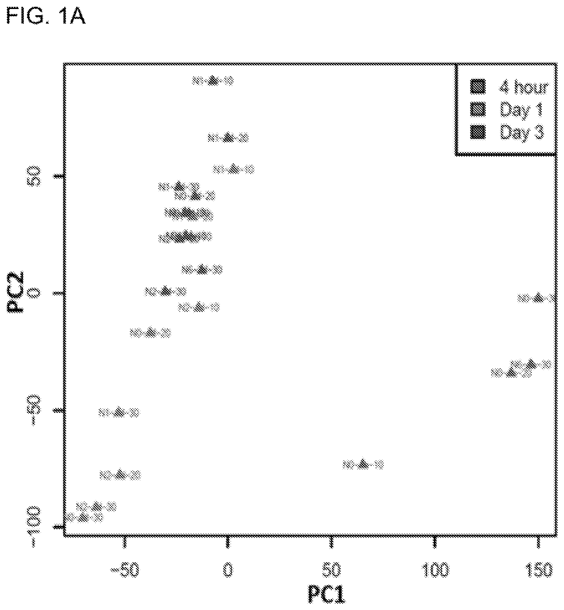
[0019] FIG. 1A shows PCA analysis demonstrating time-dependent clustering of UVR-responsive transcriptomic profiles in human keratinocytes. FIG. 1B shows functional annotation of differentially expressed genes by DAVID pathway analysis. The size of the pie chart is proportional to the number of genes in each pathway.
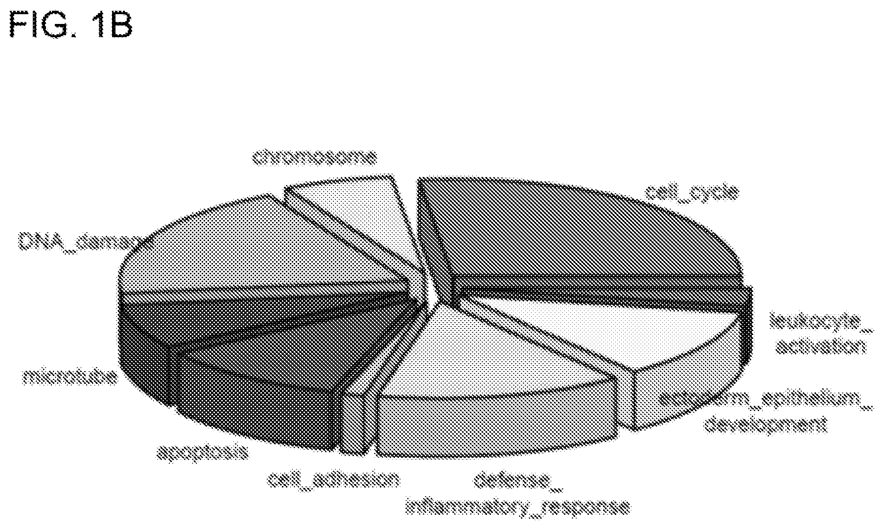
[0020] FIG. 2 shows graphs showing the time-dependent pattern of differential gene signatures by comparing Day 3 (yellow) and Day 1 (red). The y-axis shows the log 2 fold change of gene expression between irradiated and non-irradiated control cells. The x-axis indicates the sample names. ADAMTSL4 and CST6 demonstrated time-dependent up-regulation, while UHRF1 and TRIP13 displayed time-dependent down-regulation in response to UVR.
[0021] FIG. 3 shows plots of dose-dependent down-regulation (upper two panels) and up-regulation (lower two panels) of UVR-induced differentially expressed genes. Each point represents a sample at the corresponding UVR dose. X-axis represents three different UVR doses; Y-axis represents the log 2 fold change of gene expression between irradiated and non-irradiated control cells. N0-1d, N0-3d, N1-1d, N1-3d, N2-1d, and N2-3d were colored in red, orange, yellow, green, blue and cyan, respectively.
[0022] FIG. 4 shows protein-protein interaction network map illustrating hub genes as well as their interacting partners among UVR signature genes. Each vertice represents a gene and each edge indicates an interaction between the two genes. Genes belong to different clusters are colored in different colors respectively. The sizes of the vertices are proportionally to their degrees (number of interacting genes).
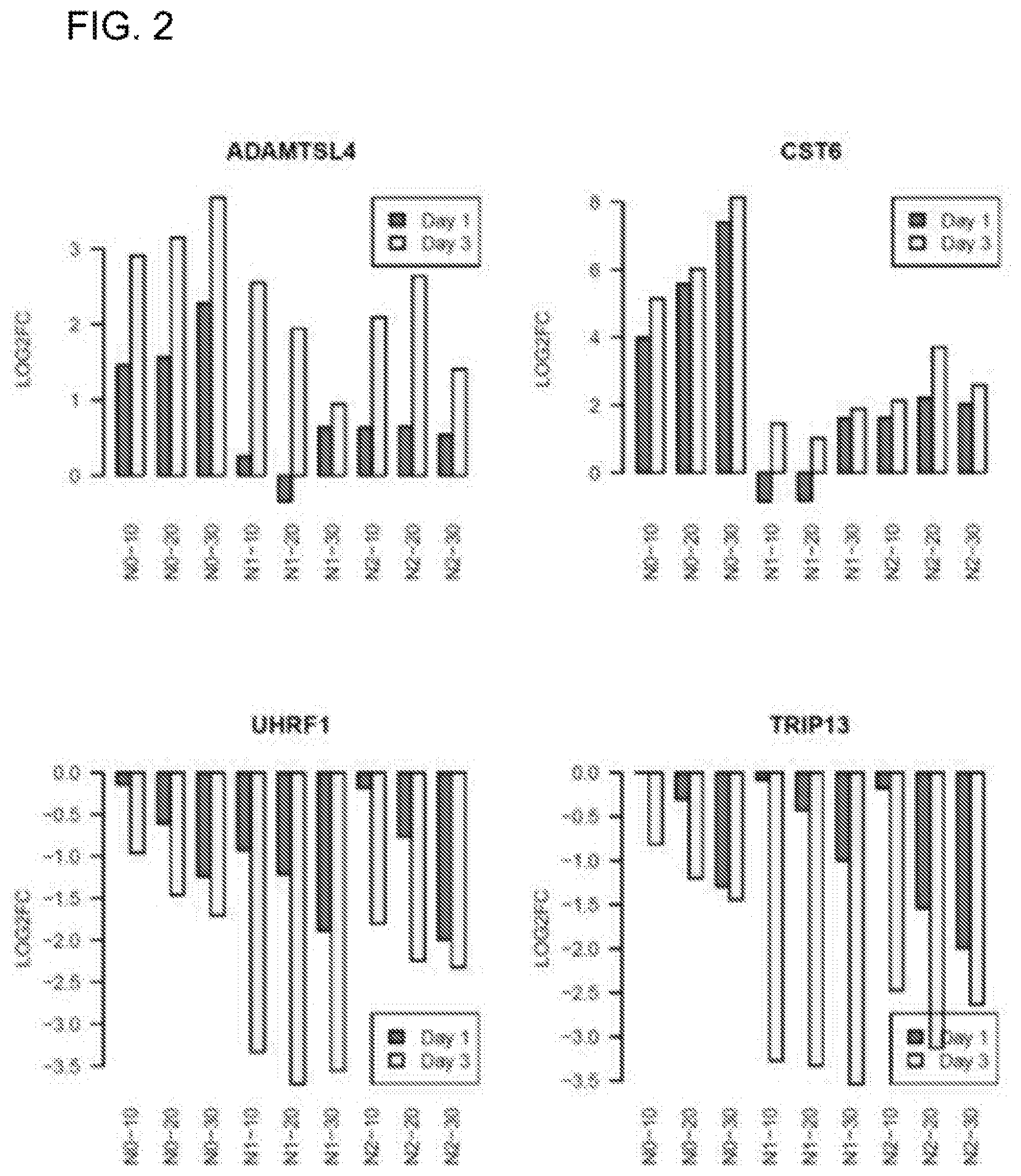
[0023] FIG. 5A shows a gene set enrichment analysis of UVR signatures (red bars) against the gene set dysregulated in human SCCs. UVR transcriptomic signature genes were sorted from the highest (left) to the lowest (right) based on their UVR-induced fold change. The normalized enrichment score (NES) and p values are indicated; FIG. 5B shows gene set enrichment analysis of the human SCC signatures (red bars) against the UVR transcriptomic signature. SCC signature genes were sorted from the highest (left) to the lowest (right) based on the fold change between SCC and normal control tissues; FIG. 5C shows Venn diagram showing the overlapping genes between UVR transcriptomic signatures and DEGs at 21 days after UVR; FIG. 5D and FIG. 5E show Venn diagram illustrating the overlapping genes between UVR transcriptomic signature and DEGs in two different human SCC cases.
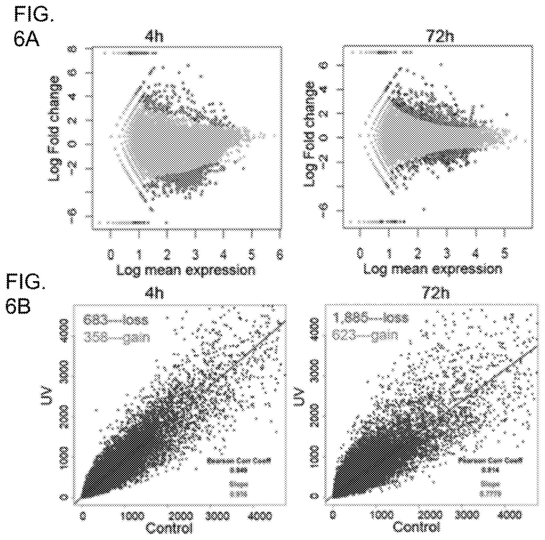
[0024] FIG. 6A shows differential gene expression plots demonstrating transcriptomic changes in human keratinocytes following UVR. Each red dot represents a DGE 4 h or 72 h following UVR. Each blue dot represents a DGE that also displays differential H3K27 acetylation following UVR; FIG. 6B shows UV induced progressive losses of H3K27ac in human keratinocytes at 4 h and 72 h after UVR. x/y-values are tag numbers in merged peak regions. Slope value <1 indicates a net loss of H3K27ac; FIG. 6C shows Venn diagram showing that 75 SNVs are common between the 4 h and 72 h SNV sets; FIG. 6D shows a schematic illustration of genomic distributions of UV-induced SNVs at 4 h and 72 h after UVR; FIG. 6E shows GSEA analysis showing that genes containing intron mutations are significantly enriched in the DGE gene set (left panel) or DHA gene set (right panel) as highlighted by the red dotted rectangles. GSEA was based on the Kolmogorov-Smirnov test. The p-values were estimated from permutation tests by randomly shuffing genes.
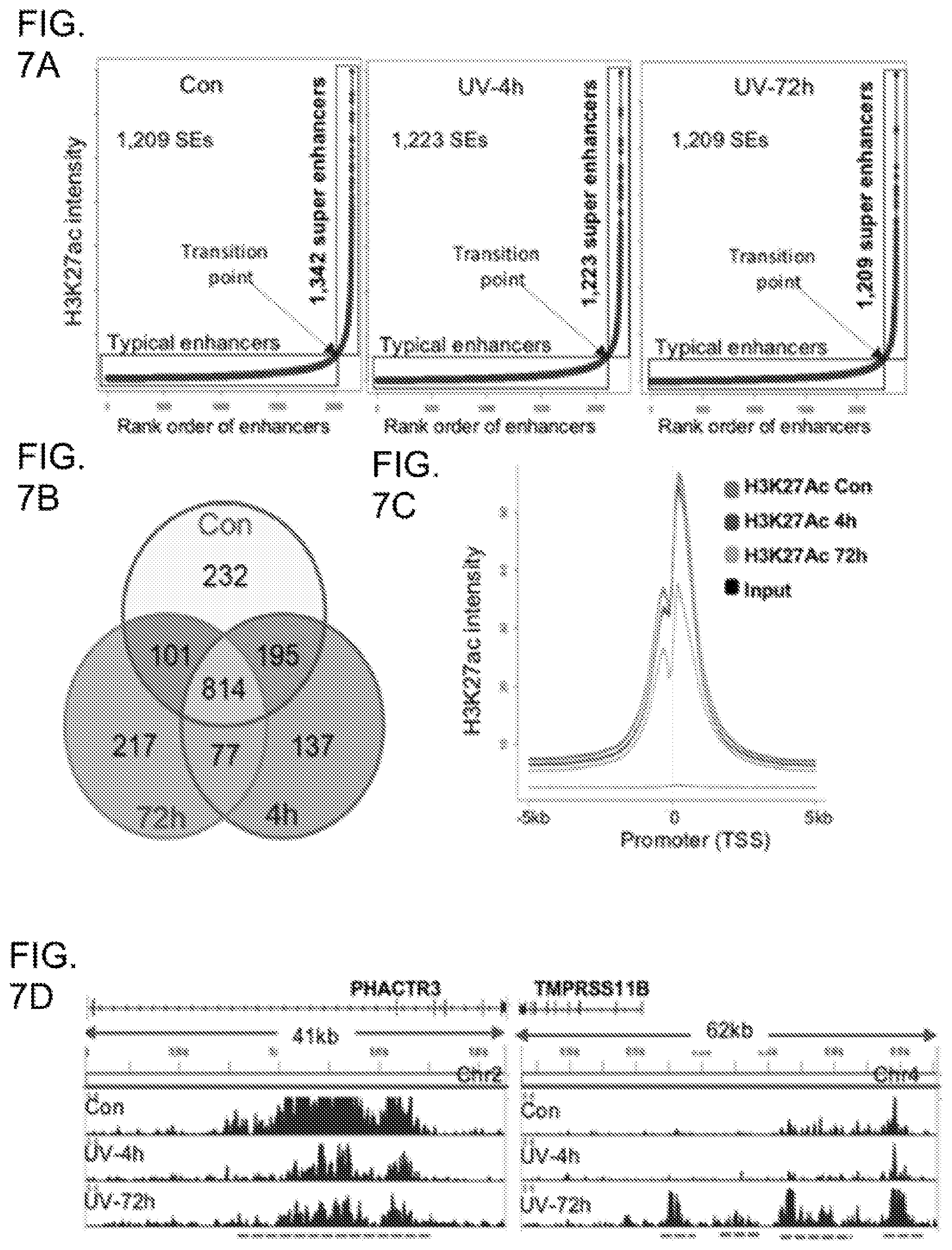
[0025] FIG. 7A shows SE profiles in control and UV-irradiated keratinocytes showing that UV decreased the total number of SEs marked by H3K27ac; FIG. 7B shows Venn diagram showing the number of common and distinctive Ses among control, UV-4h, and UV-72h; FIG. 7C shows Genome-wide H3K27ac signals in promoter regions showing a pronounced loss of 72 h following UVR; FIG. 7E shows gene tracks of H3K27ac ChIP-seq exemplifying that UVR increased H3K27ac at the PHACTR3 gene locus but reduced H3K27ac at the TMPRSS11B gene locus. PHACTR3: phosphatase and actin regulator 3; TMPRSS11B: transmembrane protease, serine 11B (HATL5).
[0026] FIG. 8A shows integrative analyses of the DGE and H3K27ac DHA gene sets at 4 h or 72 h after UVR. Correlations between gene expression and H3K27ac are considered significant if p<0.05. P-values were obtained using Student's t-test by comparing the log 2FC of the expression values of the genes from the three DHA groups; FIG. 8B shows representative genes showing concordant changes in gene expression and H3K27ac following UVR. Cutoff is set at Log 2FC>1 or <-1 for both DGE and DHA; FIG. 8C shows a summary of the overall correlations between DGE and DHA changes among UV-responsive genes at 4 h or 72 h after UVR. Pink highlights positive correlations; green highlights inverse correlations between DGE and DHA; FIG. 8D shows parallel analysis of H3K27 DHA status of the DGEs that are enriched in top UV-responsive biological pathways.
[0027] FIG. 9A is a motif analysis showing a significant enrichment of multiple TF motifs in UV-induced DHA regions in keratinocytes following UVR; FIG. 9B provides the RNA-seq results showing mRNA expression changes of the TFs identified in FIG. 9A between UV-irradiated and control keratinocytes; FIG. 9C shows that loss of function of selected UV-responsive TFs is significantly more detrimental to skin cancer cells than non-skin cancer cells; FIG. 9D shows that loss of function of selected UV target genes in FIG. 8B (more than 2-fold increases in both DGE and DHA) is significantly more detrimental to skin cancer cells than non-skin cancer cells. P-values were obtained using the Wilcoxon test by comparing the gene depletion scores between the skin cancer cells versus the non-skin cancer cells; FIG. 9E is a box plot illustrating the Log 2FC in the expression of the genes shown in FIG. 9C and FIG. 9D among 5 pairs of SCC and normal skin tissues. SLAMF7, ARNTL, ETV1, and GPR115 show more consistent upregulation in SCCs.
[0028] FIG. 10A is a box plot illustrating the Log 2FC in the expression of selected UV target genes between the 5 matched pairs of SCC and normal skin tissues; FIG. 10B provides the gene tracks of H3K27ac profiles showing that UVR increased H3K27ac levels at CPY24A1, PTGS2, GJA5, and SLAMF7 chromatin regions 72 h after UVR, which are highlighted by red dotted lines under each gene track; FIG. 10C is the immunofluorescence staining showing protein expression of selected UV target genes in UV-irradiated keratinocytes; FIG. 10D is the immunofluorescence staining showing protein expression of selected UV target genes in matched human SCC tumors and adjacent normal skin tissues. Blue: DAPI staining. Basement membrane in the normal skin is highlighted by the white dotted line. The stratum corneum is separated by the yellow line. Scale bar=20 .mu.m.
DETAILED DESCRIPTION OF THE INVENTION
[0029] The present invention provides methods of detecting ultraviolet radiation (UVR)-induced skin damage in a subject. In some embodiments, this method comprises the steps of: a) obtaining a skin sample from the subject; b) analyzing expression levels in the skin sample of UVR-induced differentially expressed genes (DEGs) listed in Table 8 or a subset thereof; and c) comparing the expression levels of the UVR-induced DEGs to a control skin sample; wherein, when the expression levels of the UVR-induced DEGs in the skin sample is above or below the level of each of the UVR-induced DEGs in the control sample, the subject is identified as likely being afflicted with UVR-induced skin damage.
[0030] As used herein, "ultraviolet radiation (UVR)-induced skin damage" is any damage to the skin caused by exposure to UV radiation and includes, for example, photocarcinogenesis (e.g., melanoma), photoaging (e.g., wrinkles, loss of elasticity), immunosuppression, and oxidative stress. In some embodiments, the radiation is solar UV, comprising UVA, UVB, and/or UVC. In other embodiments, the UV radiation is generated by a lamp.
[0031] As used herein, a "subject" is a mammal, preferably, a human. In addition to humans, categories of mammals within the scope of the present invention include, for example, agricultural animals, domestic animals, laboratory animals, etc.
[0032] The phrase "skin sample" or "biological sample" as used herein, is intended to mean any sample comprising a skin cell or skin tissue in which expression of a gene or gene product can be detected. For example, skin cells or skin tissue may be taken from the dermis or epidermis, or a combination of both. The skin sample can be used either directly as obtained from the source or following a pre-treatment to modify the character of the sample. The sample may be obtained by a variety of methods including, but not limited to, punch biopsy, surgical excision, and non-invasive or minimally invasive skin sampling methods such as a wet swabbing, tapelift, cotton tip swabbing, scraping of skin using a sterile surgical blade, scraping of skin using a wooden scraper, sticky surface of an adhesive pad (CapSure.TM. Clean-up Pad, Arcturus), film from LCM MacroCap.TM. (Arcturus), heated film from LCM MacroCap.TM. (Arcturus) and employing a small gauge needle (for example, 28 gauge), to collect micro-cores of skin tissue. These methods are well known in the art.
[0033] Alternatively, a skin sample may be a skin equivalent or a human or non-human cultured cell, for example, a keratinocyte, a melanocyte, a dermal fibroblast, a mast cell, an endothelial cell, a sebocyte, a hair papilla, or a matrix cell.
[0034] A "control" refers to a sample or standard used for comparison with an experimental sample, such as a skin sample obtained from a test subject exposed to UVR. In some embodiments, the control is a sample that has not been exposed to UVR or a non-UVR exposed sample obtained from the test subject. In some embodiments, the control is a skin sample whose exposure to UVR has been blocked or attenuated. In some embodiments, the control is a historical control or standard reference value or range of values (i.e. a previously tested control sample, such as a group of skin samples that were not exposed to UVR, or group of samples that represent baseline or normal values, such as the level of gene expression in non-UVR exposed tissue).
[0035] The differentially-expressed genes (DEGs) listed in Table 8 are genes or gene products that are modulated in skin in response to UVR exposure. Accordingly, using an assay to measure the level of the expression, function, or activity of DEGs in skin is diagnostic and prognostic of UVR-induced skin damage, photoaging, or photocarcinogenesis. A DEG may be detected at either the nucleic acid or protein level. The expression level of a given gene measured at the nucleotide level refers to the amount of RNA transcribed from the gene measured on a relevant or absolute quantitative scale, and in general refers to the relative abundance of the accumulated mRNA transcript. The expression level of a given gene measured at the protein level refers to the amount of protein translated from the transcribed RNA measured on a relevant or absolute quantitative scale.
[0036] Differential expression, as used herein, means that the expression levels of certain genes, as measured at the RNA or protein level, are different between biological samples in different states, tissues, or type of cells. Differential expression may also be observed relative to a reference standard. Such standard may be determined based on the context of the expression experiments, the biological properties of the genes under study, and/or statistical significance criteria.
[0037] In some embodiments, comparing the expression levels of the UVR-induced DEGs to a control skin sample may require a quantitative or semi-quantitative determination. Other embodiments may involve a relative determination (e.g. a ratio relative to another marker, or a measurement relative to the same marker in a control sample), and other embodiments may involve a threshold determination (e.g. a yes/no determination whether a level is above or below a threshold).
[0038] In some embodiments, the analyzing step comprises carrying out next-generation sequencing of an RNA sample from the subject to identify genes from Table 8, or a subset thereof, that have a different expression profile compared to controls.
[0039] Preferably, the next-generation sequencing is whole transcriptome shotgun sequencing (RNA-Seq). Other methods of analyzing expression levels are well known in the art, and may include microarrays, ChIP sequencing, SAGE (serial analysis of gene expression), tiling arrays, nucleic acid hybridization techniques, nucleic acid reverse transcription methods, nucleic acid amplification methods, western blots, northern blots, southern blots, ELISA, immunoprecipitation, immunofluorescence, flow cytometry, and immunocytochemistry.
[0040] The present invention also provides methods of identifying or monitoring skin cancer in a test subject. In some embodiments, the method comprises: a) analyzing expression levels in a biological sample obtained from the subject of UVR-induced differentially expressed genes (DEGs) listed in Table 8, or a subset thereof; b) comparing the expression levels of the UVR-induced DEGs in the biological sample with a predetermined reference standard for the genes; and c) identifying or monitoring skin cancer in the test subject based on the comparison in b).
[0041] A "predetermined reference standard" as used herein may be determined empirically or historically from a single or multiple control samples. For monitoring a test subject, the predetermined reference standard may be a prior level of expression from the same test subject, a control subject or subjects, or a previously established range of normal, control values.
[0042] The present invention also provides kits for detecting ultraviolet radiation (UVR)-induced skin damage in a subject. In some embodiments, the kit comprises: a set of primers or probes that specifically bind to UVR-induced differentially expressed genes (DEGs) listed in Table 8 or a subset thereof, packaged together with instructions for its use.
[0043] The phrase "specifically bind" and the like refers to a binding reaction between two molecules that is at least two times the background and more typically more than 10 to 100 times background molecular associations under physiological conditions.
[0044] The present invention also provides methods for measuring the effectiveness of a test agent in reducing ultraviolet radiation (UVR)-induced damage. In some embodiments, the method comprises: a) irradiating a test skin sample, to which the test agent has been applied, with UV radiation; b) obtaining an expression profile of the UVR-induced differentially expressed genes (DEGs) listed in 8 Table 8, or a subset thereof, in the test skin sample; and c) comparing the expression profile of the UVR-induced DEGs, or a subset thereof, from the test skin sample, with an expression profile of the same genes in a reference skin sample and a control skin sample, wherein the reference skin sample is irradiated in the absence of the test agent, and the normal, control skin sample is not irradiated; wherein if the gene expression profile of the test skin sample is the same or substantially similar to the gene expression profile of the normal, control skin sample, the test agent is effective at reducing UVR-induced damage, whereas if the gene expression profile of the test skin sample is the same or substantially similar to the gene expression profile of the reference skin sample, the test agent is not effective at reducing UVR-induced damage.
[0045] As used herein, the phrase "same or substantially similar to" refers to statistically no significant difference in the expression level between the test skin sample and the control skin sample. Conversely, the phrase "different from" and the like refers to a statistically significant difference in expression.
[0046] The present invention also provides methods for diagnosing and treating UVR-induced skin damage in a subject. In some embodiments, the method comprises: analyzing a sample from the subject for an expression profile of UVR-induced DEGs listed in Table 8 or a subset thereof that is different from an expression profile of the same genes in a normal, control sample, wherein the subject is diagnosed with UVR-induced skin damage if the expression profile of the subject differs from the expression profile from the normal, control sample; and administering a treatment for UVR-induced skin damage to the diagnosed subject.
[0047] As used herein, the terms "treat," "treating," "treatment" and grammatical variations thereof mean subjecting an individual subject to a protocol, regimen, process or remedy, in which it is desired to obtain a physiologic response or outcome in that subject, e.g., a patient. In particular, the methods of the present invention may be used to slow the development of symptoms or delay the onset of the disease or condition, or halt the progression of disease development. However, because every treated subject may not respond to a particular treatment protocol, regimen, process or remedy, treating does not require that the desired physiologic response or outcome be achieved in each and every subject or subject population, e.g., patient population. Accordingly, a given subject or subject population, e.g., patient population may fail to respond or respond inadequately to treatment.
[0048] The embodiments described in this disclosure can be combined in various ways. Any aspect or feature that is described for one embodiment can be incorporated into any other embodiment mentioned in this disclosure. While various novel features of the inventive principles have been shown, described and pointed out as applied to particular embodiments thereof, it should be understood that various omissions and substitutions and changes may be made by those skilled in the art without departing from the spirit of this disclosure. Those skilled in the art will appreciate that the inventive principles can be practiced in other than the described embodiments, which are presented for purposes of illustration and not limitation.
[0049] Skin cancer is the most common cancer in the United States. According to Skin Cancer Foundation statistics, one in every five Americans will develop skin cancer in their lifetime. Skin cancer greatly affects quality of life and creates substantial health care costs for individuals, families and the nation. Despite the fact that most skin cancer cases are preventable, its rates continue to rise mainly due to unnecessary UV radiation exposure and a lack of reliable biomarkers that can effectively monitor UV damage to help evaluate and predict skin cancer risk. Accordingly, embodiments of the disclosure relate to a UV radiation biomarker panel that can serve as sensitive tool for UV damage assessment and risk prediction to facilitate skin cancer prevention and reduce skin cancer-related illness, death and health care costs.
[0050] In one embodiment, an assay is provided for evaluating the effect of ultraviolet radiation (UVR) on a tissue sample. The assay comprises a system to evaluate expression of a plurality of UVR-responsive biomarker genes in the tissue sample, wherein expression of one or more of the plurality of UVR-responsive biomarker genes is associated with exposure of the tissue sample to ultraviolet radiation. In one embodiment, the system is a gene array system to evaluate expression of the plurality of UVR-responsive biomarker genes. In another embodiment, the assay is a high-capacity screening assay configured to evaluate the expression of the plurality of UVR-responsive biomarker genes in a plurality of tissue samples.
[0051] In one embodiment, the plurality of UVR-responsive biomarker genes are those associated with at least one of skin damage due to UV exposure, cancer risk and cancer progression. In yet another embodiment, the plurality of UVR-responsive biomarker genes are those that are involved at least one of inflammation, cell growth and proliferation, DNA repair, and cancer pathogenesis. In yet a further embodiment, the plurality of UVR-responsive biomarker genes are those selected from the group consisting of CYP24A1, GJA5, SLAMF7 and ETV1.
[0052] In one embodiment, the tissue sample that is evaluated by the assay is a mammalian tissue sample. For example, in one embodiment, the tissue sample is a human tissue sample. As yet another example, in one embodiment, the tissue sample comprises human keratinocytes.
[0053] In one embodiment, the assay is capable of correlating the expression of each of the UVR-responsive biomarker genes with at least one of UV damage to the tissue sample and/or a disease state, such as via a gene expression profile correlation system to correlate.
[0054] In one embodiment, a method of evaluating ultraviolet damage to tissue comprises evaluating the expression of a plurality of UVR-responsive biomarker genes in a sample of the tissue, and determining whether the expression of one or more of the plurality of UVR-responsive biomarker genes is indicative of ultraviolet damage. For example, in one embodiment, the plurality of UVR-responsive biomarker genes are those associated with at least one of skin damage due to UV exposure, cancer risk and cancer progression. In yet another embodiment, the plurality of UVR-responsive biomarker genes are those selected from the group consisting of CYP24A1, GJA5, SLAMF7 and ETV1. In one embodiment, the expression of the plurality of UVR-responsive biomarker genes is evaluated via a high-capacity gene array screening system.
[0055] Accordingly to one embodiment, the tissue that is evaluated for UV damage is mammalian tissue. According to yet another embodiment, the tissue that is evaluated for UV damage is human tissue.
[0056] In one embodiment, a method of diagnosing skin cancer or predicting skin cancer risk in a subject comprises evaluating the expression of a plurality of UVR-responsive biomarker genes in a sample of the tissue, and determining whether the expression is indicative of skin cancer or skin cancer risk. For example, in one embodiment, the plurality of UVR-responsive biomarker genes are those selected from the group consisting of CYP24A1, GJA5, SLAMF7 and ETV1. In one embodiment, the subject is a mammalian subject. In yet another embodiment, the subject is a human subject.
[0057] In one embodiment, a method of evaluating a sunscreen formulation comprises applying the sunscreen formulation to a tissue sample, irradiating the tissue sample with ultraviolet radiation, evaluating the expression of a plurality of UVR-responsive biomarker genes in the tissue sample, and determining whether the expression of the plurality of UVR-responsive biomarker genes is indicative of efficacy of sunscreen formulation in providing a UV protective effect to the tissue sample.
[0058] One embodiment of the present disclosure is directed to providing UVR biomarkers having better reliability and accuracy. Accordingly, to obtain UVR biomarkers with better reliability and accuracy, a panel of UVR-responsive genes have been identified through comprehensive transcriptomic profiling studies. Functions of these carefully selected UVR biomarker genes span several biological pathways including inflammation, cell growth and proliferation, DNA repair, and cancer pathogenesis. The panel of genes has been subjected to rigorous validations by both bioinformatics and experimental approaches to confirm that their mRNA expressions are consistently responsive to UVR among different skin types. Furthermore, the UVR-induced mRNA expression changes in the biomarker genes persist long after UVR, highlighting their potential as reliable UVR biomarkers.
[0059] According to one embodiment, the UV biomarker panel can serve to set a new industrial standard in testing UVR-protective effects of sunscreen products in preventing cancer-inducing dose of sun damage. According to yet another embodiment, it can be used in clinical diagnosis to assist health care providers with a sensitive tool in assessing excessive sun exposure and skin cancer risk. In yet another embodiment, to facilitate industrial and clinical applications, a gene array system in a 96-well plate format is designed to allow simultaneous detections of the expression of the UVR biomarker genes from multiple samples. In one embodiment, the UVR biomarker panel together with the high capacity screening assay system may be capable of revolutionizing the assessment of sun damage and skin cancer risk predication to allow for early prevention and effective reduction of skin cancer-related illness, death, and health care costs.
[0060] Embodiments of the disclosure may involve (1) validation and optimization of the selection of biomarker genes for gene-array preparation; (2) development and optimization of a compact gene array system that can process multiple samples on the same array to achieve high screening capacity; and (2) development of algorithms to enable autonomous processing of gene expression data.
[0061] In one embodiment, a UVR biomarker panel is provided for monitoring sun damage and predicting skin cancer risk with a high level of sensitivity and accuracy. Associated analytical regents, test kits and diagnostic models for sun damage detection and cancer risk prediction can also be provided.
[0062] In one embodiment, the UVR biomarker panel can be applied in the sunscreen industry to evaluate the efficacy of sunscreen products in UVR protection and reducing sun exposure damage of the skin.
[0063] In one embodiment, the tissue and/or subject being evaluated is mammalian, such as preferably human. In other aspects of this embodiment, the tissue and or subject is that of a laboratory animal. In addition to humans, categories of mammals within the scope of aspects of the present disclosure include, for example, agricultural animals, veterinary animals, laboratory animals, etc. Some examples of agricultural animals include cows, pigs, horses, goats, etc. Some examples of veterinary animals include dogs, cats, etc. Some examples of laboratory animals include rats, mice, rabbits, guinea pigs, etc.
[0064] In one embodiment, methods and/or steps in methods described herein may be carried out in vitro. In other embodiments, the methods and/or steps in the methods described herein are carried out in vivo or ex vivo.
[0065] As used herein, in vitro refers to a process performed in an artificial environment created outside a living multicellular organism (e.g., a test tube or culture plate) used in experimental research to study a disease or process. As used herein, in vitro includes processes performed in intact cells growing in culture.
[0066] As used herein, in vivo means that which takes place inside an organism and more specifically to a process performed in or on the living tissue of a whole, living multicellular organism (animal), such as a mammal, as opposed to a partial or dead one.
[0067] As used herein, ex vivo refers to a process performed in an artificial environment outside the organism on living cells or tissue which are removed from an organism and subsequently returned to an organism.
[0068] An Appendix is attached hereto which provides additional details regarding the inventive principles described in this disclosure. The Appendix is explicitly incorporated herein by reference in its entirety. In the event of a conflict between the teachings of this application and those of the incorporated document, the teachings of this application control.
[0069] The embodiments described in this disclosure can be combined in various ways. Any aspect or feature that is described for one embodiment can be incorporated into any other embodiment mentioned in this disclosure. While various novel features of the inventive principles have been shown, described and pointed out as applied to particular embodiments thereof, it should be understood that various omissions and substitutions and changes may be made by those skilled in the art without departing from the spirit of this disclosure. Those skilled in the art will appreciate that the inventive principles can be practiced in other than the described embodiments, which are presented for purposes of illustration and not limitation.
EXAMPLES
First Series of Experiments
Example 1
Materials and Methods
Human Keratinocyte Cultures, Human SCC and Normal Skin Tissues
[0070] Primary human keratinocytes were established from neonatal foreskins through the Columbia University Skin Disease Research Center tissue culture core facility. The protocol was exempt by our Institutional Review Board. Keratinocytes were isolated from separate individual neonatal foreskins (N0, N1, N2, and N6), and cells from each individual were maintained and analyzed separately for assessing individual variations. Keratinocytes were cultured in 154CF medium supplemented with human keratinocyte growth supplement (Life Technologies, Grand Island, N.Y.). Human SCC tumor tissues and matched normal skin tissues from two patients were obtained from the Molecular Pathology Shared Resource/Tissue Bank of the Herbert Irving Comprehensive Cancer Center of Columbia University under CUMC IRB protocol AAAB2667.
UVB Radiation
[0071] Keratinocytes were rinsed once with PBS and irradiated with UVB supplied by four FS20T12/UVB tubes (National Biological Corp., Beachwood, Ohio). The intensity of UVB lights was measured using an IL1400 radiometer connected to a SEL240/UVB-1/TD detector (International Light, Newburyport, Mass.). Cells were irradiated with a total dose of 10, 20, and 30 mJ/cm.sup.2, respectively. Cells were collected at different times points after exposure including four hours or one, three, or 21 days as indicated.
RNA Isolation and RNA-Seq Analysis
[0072] Total RNA was isolated from cultured keratinocytes and human tissues using the RNeasy Kit (QIAGEN, Gaithersburg, Md.) and treated with DNase I (Life Technologies, Grand Island, N.Y.) according to the manufacturers' protocols. All RNA samples were subsequently analyzed using an RNA 6000 nano chip (Agilent Technologies, Wilmington, Del.) to confirm that the RNA integrity index was 8.0 or above. For RAN-Seq, 500 ng of total RNA from each sample was subjected to poly-A pull-down to enrich mRNAs for library preparation by using Illumina TruSeq RNA prep kit (Illumina, San Diego, Calif.). The resulting libraries were sequenced using Illumina HiSeq2000 at Columbia Genome Center. Samples were multiplexed in each lane, which yielded targeted number of paired-end 100 bp reads for each sample, as a fraction of 180 million reads for the whole lane. We used RTA (Illumina) for base calling and bcl2fastq (version 1.8.4, Illumina) for converting BCL to fastq format, coupled with adaptor trimming. The reads were mapped to the human reference genome (NCBI/build37.2) using Tophat (version 2.0.4). Relative gene expression levels were calculated using Cufflinks (version 2.0.2) with default settings. Differentially expressed genes (DEGs) under various UVR conditions were determined using the DESeq software package (Anders 2010), with a fold change cutoff set at >2 or <0.5 between irradiation and non-irradiated keratinocytes. Genes with FPKM values <10 were subjected to higher FC cutoffs to be selected in the final DEG list (details available upon request). A False Discovery rate (FDR)<0.05 was used to control for false discoveries.
Bioinformatics and Statistical Analyses
[0073] DEG lists were used in principal component analysis (PCA) to characterize the variations in transcriptomic responses to different UVR conditions among the keratinocyte lines. To uncover pathways that were most significantly affected by UVR, we performed pathway analysis using DAVID to identify which biological pathways the differentially expressed genes were enriched in. Gene enrichment analysis (GSEA) was performed to determine the overlap between UVR signature genes and gene sets that were dysregulated in different human malignancies. Paired t-test was used to identify genes displaying time-dependent UVR responses from Day 1 to Day 3 following exposure. To identifying genes manifesting dose-dependent changes in response to UVR, we constructed a linear regression model using UVR dosage as an independent variable and gene expression as a dependent variable for each gene in the same keratinocyte line and at the same time point. We then performed the same analysis for all three keratinocyte lines (N0, N1, and N2) and at both time points (Day 1 and 3), which generated six expression models for each gene. In each model, a low coefficient p-value (p<0.05) indicated a significant association between UVR dosage and gene expression. To evaluate the overall effects of the various UVR dosage on the expression of a specific gene, we integrated the multiple p-values from every regression analysis for that gene using Fisher's Method. P-values from the above analyses were FDR-corrected. To obtain cancer-specific gene signatures for various human malignancies, we retrieved and selected RNA-Seq data sets from the TCGA database that were available for both primary tumor cases and matched normal control tissues from same patient for each tumor type. We used DESeq package to normalize the raw counts and determine genes that were differentially expressed between each primary tumor and matched normal control tissue to obtain dysregulated gene sets for each tumor type. To identify genes that are critical to skin cancer cell proliferation or survival, we queried the Achilles database with 67 of the UVR signature genes that were upregulated by UVR. (Cowley 2014) Genes were considered essential to skin cancer cell survival if their corresponding shRNAs became depleted after 40 days or 16 population doublings following shRNA infection. Normalized shRNA depletion scores were downloaded from the "cBOTv8_sbsv3_allreps_log.gct2" file in Achilles database. For multiple shRNAs targeting the same gene, we selected the ones whose depletion scores were consistent across all cancer cell lines and then took the median value as the final depletion score for each shRNA. All statistical analyses were performed using R.
Example 2
Transcriptomic Responses to Different UVR Conditions
[0074] In addition to its mutagenic effect, UVR has been shown to cause transcriptomic instability affecting thousands of genes. To fully characterize UVR-induced transcriptomic changes, we took advantage of the recent advances in RNA-Seq to profile UVR-induced kinetic changes in human primary keratinocytes exposed to different UVR conditions (Table 1). We used keratinocytes isolated from four individual neonatal foreskins to generate UVR-induced differentially expressed gene (DEG) lists in response to each of the UVR conditions (Table 1). Together with four DEG lists representing transcriptomic profiles at four hours after exposure, we performed principle component analysis (PCA) to differentiate the DEG profiles under various UVR conditions. As shown in the PCA plot (FIG. 1A), DEG profiles from Day 1 and 3 groups, but not the 4 hour group, demonstrated great similarities with each other in the first principle component (PC1). Along the second principle component (PC2) axis, however, the range of differences within the Day 3 DEG group appeared smaller than that of the Day 1 DEG group, demonstrating a clear time-dependent transcriptomic effect of UVR that became less differentiated among different UVR conditions 3 days after exposure.
[0075] To uncover the biological pathways that were mostly affected by UVR, we took the average of the fold change (FC) of each gene between irradiated and non-irradiated cells from the 19 DEG lists (Table 1). Using a FC cutoff of 2, we obtained a total of 531 genes that were up-regulated (FC>2) and 610 genes that were down-regulated (FC<0.5) in response to different UVR conditions (Table 2 and Table 3). We performed DAVID pathway analysis to categorize the functions of the up-regulated genes and down-regulated genes, respectively, which revealed multiple pathways that were significantly modulated by UVR. The down-regulated genes were significantly enriched in the following top four biological pathways: cell cycle regulation (83 genes), chromosome structure (19 genes), DNA damage response (59 genes) and microtubule organization (23 genes); whereas the up-regulated genes were largely enriched in pathways such as apoptosis (33 genes), defense inflammatory response (43 genes), ectoderm epithelium development (36 genes), cell adhesion (4 genes) and leukocyte activation (9 genes) (FIG. 1B).
TABLE-US-00001 TABLE 1 Keratinocyte lines and experimental UVR conditions UVR conditions 10 20 30 10 20 30 mJ/cm.sup.2 mJ/cm.sup.2 mJ/cm.sup.2 mJ/cm.sup.2 mJ/cm.sup.2 mJ/cm.sup.2 24 h 24 h 24 h 72 h 72 h 72 h Kerati- N0 1 2 3 4 5 6 nocyte N1 7 8 9 10 11 12 lines N2 13 14 15 16 17 18 N3 19
TABLE-US-00002 TABLE 2 Genes up-regulated by UVR Gene ID Log2FC A2ML1 1.25542 A4GALT 1.217168 ABCA12 1.377908 ABCD1 1.252331 ABHD4 1.15769 ABLIM3 1.065562 ACAP1 1.359902 ACBD4 1.075352 ACER2 2.006333 ACTA2 1.169663 ADAMTS13 1.111708 ADAMTS7 1.803072 ADAMTSL4 1.579834 ADCK3 1.407755 ADH6 1.037455 ADHFE1 1.308802 ADSSL1 1.567365 AIFM3 1.051368 AIM1L 1.309328 AKR1B10 1.680432 AKR1C1 1.734541 AKR1C2 1.63357 AKR1C3 1.442466 ALDH3B2 1.885289 ALOX12B 1.627145 ALOX15B 1.148359 ANKRD22 1.505822 ANKRD29 1.004008 APOE 1.10496 ARHGAP30 2.227763 ARNT2 2.347534 ARRDC4 1.377681 ASPRV1 2.393802 ATF3 2.600626 ATP12A 1.440183 AVPI1 1.283781 B3GALT4 1.300773 B3GNT3 1.049928 BBC3 1.612703 BCL2L1 1.077692 BCL6 1.022801 BIK 1.899355 BIRC3 1.411982 BLNK 2.166166 BMF 2.369338 BNIP3L 1.036609 BST2 1.416924 BTBD19 1.135608 BTG1 1.148244 BTG2 1.403243 C10orf99 2.924011 C11orf35 1.369906 C11orf9 1.682084 C16orf5 1.006965 C17orf103 1.565166 C18orf56 1.11554 C19orf46 1.466501 C1orf126 1.008004 C1orf38 1.298954 C1orf51 1.552331 C1orf74 1.732929 C1orf88 1.324512 C20orf46 1.04507 C5orf41 1.546868 C6orf138 1.094879 C7orf10 1.941839 C7orf53 1.249614 C9orf7 1.060832 CA2 1.243123 CALML3 1.255207 CALML5 2.024878 CAPNS2 1.210126 CARD14 1.278924 CARD18 2.472437 CASP9 1.091866 CBX7 1.169991 CCDC11 1.24824 CCDC64B 1.433373 CCK 1.062617 CCL20 1.568576 CD55 1.028517 CD68 1.471241 CD74 1.002605 CDKN1A 1.898547 CDKN1C 1.344886 CDKN2B 1.337026 CDKN2D 1.103006 CDSN 2.193489 CEACAM1 1.521198 CEBPA 1.068886 CEL 1.833995 CES3 1.495162 CES4A 1.145926 CFB 1.525458 CGN 1.243352 CHI3L2 2.22442 CHST2 1.153737 CITED2 1.362391 CLCF1 1.746694 CLDN1 2.164861 CLDN23 1.949484 CLDN4 3.387876 CLDN7 1.756154 CLEC2B 1.823128 CLIC3 1.327201 CLU 1.438944 CNFN 2.155499 COX6B2 1.34408 CPT1C 1.277061 CRB3 1.066881 CRCT1 3.196832 CRISPLD2 2.63943 CRYAB 2.080253 CSF1 1.150493 CSF3 1.528244 CST6 3.096074 CTSS 1.384918 CUL9 1.146301 CYFIP2 2.012293 CYGB 1.278952 CYP2S1 1.111609 CYP3A5 2.134077 DAPK1 2.489445 DBNDD1 1.697917 DBP 1.548785 DCN 1.917867 DEFB1 4.538491 DENND1C 1.052741 DGAT2 1.066686 DHDH 1.977426 DHRS3 1.150886 DKFZp434J0226 1.581589 DPP4 1.635675 DQX1 1.282541 DUSP10 2.217671 DYRK1B 1.133527 EDA2R 1.650437 EGR3 1.055599 ELFN2 1.454605 ENO2 1.176432 ENTPD3 1.13432 EPHB2 1.0814 EPHB3 1.28144 EPPK1 1.03793 ERBB3 1.176234 ESPN 1.898346 ETV7 1.992978 FAM131C 1.195041 FAM13C 1.660195 FAM198B 1.546092 FAM43A 2.281964 FAM46A 1.142838 FAM84A 1.217675 FAM86HP 1.398758 FBXO32 1.762987 FDXR 1.123815 FGF11 1.057493 FLJ32255 1.396227 FLJ35776 1.045777 FLJ43663 1.505095 FLJ45831 1.688516 FLNC 1.840758 FN3K 1.042252 FOLR3 1.453421 FTH1 1.02617 FTL 1.293257 FUT2 1.130622 FUT3 1.400397 FXYD3 1.235707 G0S2 1.507978 GABARAPL1 1.12635 GALK1 1.028271 GAMT 1.499196 GBP2 2.981328 GDA 1.668244 GDF15 4.934651 GGT1 1.76415 GGT6 2.012062 GIPR 1.443944 GJA5 1.128834 GJB4 1.442723 GLRX 1.411782 GLS2 1.635732 GPNMB 1.547934 GPR172B 2.315739 GPR37 1.273549 GPRASP1 1.081898 GPRC5A 3.036593 GRB7 1.812811 GREB1 3.918492 GRHL1 1.549886 GRHL3 2.137244 GRIN3B 1.04264 GRIP2 1.741655 GSDMA 1.531598 GTF2IP1 1.31404 GUCA1B 1.103181 H1F0 1.009822 H1FX-AS1 1.000064 HAP1 2.66275 HAPLN3 1.002183 HBEGF 1.037813 HBP1 1.070321 HCAR2 2.227049 HCAR3 2.061033 HCP5 1.022373 HDAC5 1.191311 HDAC9 1.014098 HEPHL1 1.084285 HES2 1.815118 HIST1H1C 2.570588 HIST1H2AC 1.751416 HIST1H2BD 2.723667 HIST1H2BK 1.243929 HIST2H2BE 1.850329 HIST3H2A 1.005484 HLA-G 2.460471 HMOX1 2.320076 HSD17B14 1.463513 HSD17B2 1.779269 HSD3B7 1.356109 HSH2D 1.030704 HSPB8 2.738695 ICAM1 2.087607 ICAM4 1.355522 ID2 1.7539 IDUA 1.182603 IFI27 1.601052 IFIT2 1.077206 IGFBP3 1.96108 IGFL3 1.932189 IL1B 1.223292 IL1R2 2.283708 IL1RN 1.278581 IL23A 1.378554 IL32 1.122006 IL33 1.109746 IL36RN 2.089587 IL8 1.751882 INPP5D 1.925148 INPP5J 1.016762 IRAK2 1.116631 IRF5 1.327517 IRF6 1.395184 ISG20 1.350179
ISYNA1 1.381921 ITIH4 1.029668 ITPKC 1.126402 IVL 2.375886 KCNN4 1.802118 KCTD11 1.081291 KIAA1257 1.865952 KIAA1370 1.146223 KLHDC9 1.432303 KLHL24 1.889937 KLK10 1.414561 KLK11 1.551882 KLK5 1.255347 KLK7 1.405181 KLRG2 1.233281 KRT13 4.582385 KRT15 2.227083 KRT16 1.445009 KRT19 1.292513 KRT23 2.654857 KRT34 2.056054 KRT37 2.786469 KRT42P 1.383759 KRT6B 1.218162 KRT7 1.20472 KRT75 1.204212 KRT80 2.425273 KRTDAP 1.404801 KYNU 1.234901 LACC1 1.348361 LBH 1.964427 LCE1B 2.863596 LCE1C 1.5978 LCN2 1.410005 LIF 2.020803 LINC00086 2.475568 LMO7 1.453974 LOC100049716 1.166231 LOC100129781 1.088847 LOC100131096 1.015925 LOC100131564 1.109901 LOC100132909 1.081642 LOC100133190 1.059472 LOC100287177 1.060091 LOC100505623 1.290074 LOC100505974 1.072712 LOC100506119 1.199105 LOC100506377 1.931858 LOC100506538 1.582621 LOC100506746 1.093062 LOC100507429 1.577794 LOC100507452 1.034674 LOC100507656 1.387791 LOC151475 2.864213 LOC151534 1.798543 LOC284080 1.116795 LOC284440 1.232691 LOC284837 2.507131 LOC441869 1.405387 LOC554223 1.05486 LOC646471 1.010308 LOC728975 1.150382 LOC730755 1.127336 LY6D 2.375439 LY6G6C 1.478579 LYNX1 1.569974 LYPD3 1.202858 LYPD5 1.938955 MAFB 1.976484 MAP1LC3A 1.524844 MAP3K8 1.266091 MAPK8IP2 1.267615 MCHR1 2.376182 MDM2 1.857866 MEG3 1.680548 METRNL 1.850228 MEX3B 1.293473 MIR21 1.056403 MLPH 1.048951 MME 1.279362 MNT 1.093418 MUC1 1.323749 MXD1 1.221816 MXD4 1.119169 MXI1 1.012704 MYBPHL 2.469553 MYH16 1.669449 MYO15B 1.769726 N4BP2L1 1.302932 NCCRP1 2.25184 NCF2 1.956338 NDRG4 1.724837 NEAT1 1.17292 NFATC4 1.144891 NFKBIA 1.085284 NFKBIZ 1.055574 NHLH2 1.783285 NINJ1 1.360009 NIPAL4 1.035101 NLRP10 2.908381 NOD2 1.045928 NOTCH3 2.10179 NR1D1 1.830782 NR1D2 1.034866 NR4A1 1.502722 NR4A2 1.09004 NUPR1 1.436536 OCLN 1.834442 OVGP1 1.381053 OVOL1 2.085033 P4HTM 1.543172 PADI1 1.763089 PAPL 1.978976 PCDH1 2.520425 PDE6B 1.230008 PGPEP1 1.267001 PHLDB3 1.019073 PHYHIP 1.46292 PI3 2.404414 PIDD 1.246663 PIK3IP1 1.63279 PKIB 1.88435 PLA2G4C 1.366493 PLA2G4D 1.768802 PLA2G4E 1.233771 PLAC2 1.196007 PLAUR 1.160952 PLEKHG1 1.058571 PLEKHG6 1.612271 PLIN4 2.008771 PNLIPRP3 1.06203 PNMAL1 1.138815 PNRC1 1.363591 POU2F3 1.906488 POU3F1 1.472181 PPL 1.079871 PPP1R15A 1.250187 PPP1R3B 1.200822 PRDM1 1.492528 PRICKLE4 1.141436 PRODH 1.239592 PROM2 1.239982 ProSAPiP1 1.008596 PRSS22 3.207114 PRSS8 1.194038 PSORS1C1 1.553438 PTGES 1.511422 PTGS2 1.301506 PVRL4 3.274905 QPCT 1.121274 RAB11FIP1 1.050886 RAET1G 1.355114 RASSF5 1.541771 REEP6 1.077737 RET 1.243314 RGAG4 1.507274 RGS16 2.736892 RGS2 1.778959 RHBDL1 1.204067 RHCG 2.079265 RHPN1 1.365829 RIBC1 1.098573 RINL 1.033176 RNASE7 1.870734 RND2 1.440745 RNF208 1.265198 RORA 1.140093 RRAD 3.850695 RRM2B 1.324419 RSAD2 1.549139 RUNDC3A 1.48569 RYR1 1.065997 S100A4 1.430196 S100A6 1.054154 S100A8 2.129619 SAA1 1.335856 SALL4 2.057745 SAMD10 1.014837 SBK1 2.558557 SBSN 1.878689 SCNN1A 1.524209 SDPR 1.22298 SELPLG 1.687573 SEMA3B 2.411019 SEMA3G 1.572456 SERPINB1 1.159593 SERPINB2 1.49504 SERPINB3 1.529827 SERPINB7 1.343932 SERTAD1 1.03668 SESN1 1.171176 SGPP2 1.075454 SIK1 1.048194 SIRPB2 1.315647 SLAMF7 2.122482 SLC28A3 1.767182 SLC2A12 1.185052 SLC44A3 1.096887 SLC46A1 1.177566 SLC7A4 1.156152 SLPI 2.205451 SMOC1 1.596494 SNCG 1.231876 SORT1 1.427147 SPATA18 1.077252 SPINK6 1.288906 SPNS2 1.783244 SPOCD1 1.028304 SPON2 3.073135 SPRR1A 1.79158 SPRR1B 1.458382 SPRR2A 1.281665 SPRR2D 1.021259 SPRR2E 1.628479 SPRR3 2.813864 SQSTM1 1.526918 STEAP4 4.22635 SULT1A1 1.332854 SULT2B1 1.729219 SYNGR3 1.134232 SYTL2 1.420285 TACSTD2 1.167671 TCP11L2 1.288639 TGFB2 1.111166 TGM1 1.556176 THBD 1.570713 TIMP2 1.624976 TLCD2 1.203551 TLR2 1.209706 TM7SF2 1.094312 TMEM125 1.769122 TMEM184A 1.403734 TMEM27 2.26327 TMEM38A 1.428279 TMEM45B 1.329869 TMEM61 1.417963 TMEM86A 1.280883 TMEM91 1.277979 TMPRSS11D 1.097904 TMPRSS13 2.538018 TMPRSS4 1.673327 TNF 1.380011 TNFAIP2 2.133214 TNFAIP8L3 1.078872 TNFRSF10C 4.084906 TNFRSF14 1.154539 TNFSF4 1.328395 TOB1 1.129999 TP53INP1 1.898305 TP53INP2 1.709985 TPPP 1.126332
TRAF1 1.270284 TRAF3IP3 1.885624 TREM2 1.406591 TRIM17 1.097686 TRIM22 1.107816 TSPAN1 1.043978 TSPAN10 1.145679 TTC39A 1.513684 TTC9 1.775748 TTLL3 1.07677 TXNIP 1.324999 UCA1 3.144775 ULBP1 1.718077 ULK1 1.155636 UNC13D 1.284158 VAMP5 1.10333 VASN 1.070314 VGLL3 1.526916 VNN1 1.585207 VWCE 2.339601 WDR63 1.100921 WFDC5 1.623134 YPEL2 1.093046 YPEL3 1.333902 YPEL4 2.050039 ZFHX2 1.14507 ZFYVE1 1.004115 ZNF185 1.058878 ZNF425 1.493712 ZNF432 1.264009 ZNF610 1.214798 ZNF702P 1.114955 ZNF750 1.98419 ZNF763 1.068978 ZNF812 1.677638
TABLE-US-00003 TABLE 3 Genes down-regulated by UVR Gene ID Log2FC ABCC4 -1.348205778 ABI3BP -1.592845354 ADAMTSL1 -3.057818112 AGTPBP1 -1.059225614 AKAP6 -1.065270643 AKAP7 -1.191007938 AKT3 -1.063141722 ALDH1L2 -1.71329721 ALG14 -1.390158117 ALMS1 -1.39725398 ANK2 -2.852547236 ANKRD44 -3.201509809 ANLN -2.074462024 ANO1 -1.455452126 ANXA6 -1.214603625 APBA1 -1.03839454 APCDD1 -1.068852998 APLN -1.995875214 ARHGAP11A -1.932775855 ARHGAP11B -1.97626487 ARHGAP18 -1.090237869 ARHGAP19 -1.34032156 ARHGAP33 -1.316757909 ARSB -1.490168103 ASF1B -1.507991707 ASNS -1.325921219 ASPM -2.485501797 ATAD2 -1.092034263 ATAD5 -1.198810998 ATG10 -1.012583207 AURKA -1.605879285 AURKB -2.266234804 B3GALTL -1.098025255 BARD1 -1.245055616 BBS9 -1.716979874 BCAS3 -1.500580847 BCAT1 -1.262972667 BCL2 -1.345519919 BEND6 -1.554214214 BIRC5 -2.468776552 BLM -1.833991479 BORA -1.11351262 BRCA1 -1.433568205 BRCA2 -1.738870791 BRIP1 -1.365404961 BUB1 -2.232101679 BUB1B -2.463047443 C11orf82 -1.734657367 C12orf26 -1.02533073 C12orf48 -1.731440454 C12orf55 -1.155331766 C14orf49 -1.046892915 C14orf80 -1.158411353 C15orf42 -1.43469073 C16orf59 -1.251575133 C1orf112 -1.026418432 C21orf58 -1.347413053 C3orf26 -1.633777304 C4orf21 -1.638019979 C5 -1.896229619 C9orf100 -1.416508411 C9orf140 -1.512229 C9orf93 -3.645734034 CADPS2 -2.749447365 CAMKMT -1.560519775 CASC2 -1.210566387 CASC5 -2.254998502 CBS -1.047490252 CCBE1 -1.49186818 CCDC109B -1.008132116 CCDC150 -1.777877107 CCDC152 -1.563498921 CCDC18 -1.539219671 CCDC3 -2.188282435 CCNA2 -2.040850548 CCNB1 -1.992477017 CCNB2 -2.023197257 CCNE2 -1.166174946 CCNF -1.319421617 CDC20 -2.788887669 CDC25A -1.345444577 CDC25C -1.941854577 CDC45 -1.66938146 CDC6 -1.746559274 CDC7 -1.022925655 CDCA2 -1.900385914 CDCA3 -2.659044568 CDCA5 -1.747593013 CDCA7 -1.343204821 CDCA8 -1.959093261 CDH4 -2.389925028 CDK1 -1.949360908 CDK14 -1.182597074 CDKAL1 -1.326316704 CDKN2C -1.321626434 CDKN3 -2.415696295 CDON -1.278358612 CDT1 -1.295120529 CENPA -2.730714266 CENPE -1.89935139 CENPF -2.568066958 CENPH -1.270674947 CENPI -1.767622737 CENPJ -1.102370738 CENPK -1.271103299 CENPM -1.567908583 CENPN -1.330259855 CENPO -1.394874643 CENPW -1.348316188 CEP112 -1.785492351 CEP128 -1.931644843 CEP170P1 -3.902232714 CEP55 -2.535959714 CHAF1A -1.060713699 CHEK1 -1.204412215 CHRNA5 -1.310278551 CHSY3 -2.63674892 CIT -2.093026219 CKAP2L -2.259036313 CKS1B -1.089031731 CLDN11 -1.745869165 CLMP -2.630409593 CLSPN -1.755066048 CMTM1 -1.092242876 CNTLN -1.002235788 CNTN1 -1.717727031 CNTNAP3 -1.50024013 COL12A1 -1.225388115 COL18A1 -1.039211617 COL24A1 -1.275030048 COL4A1 -1.469924365 COL4A2 -1.669276381 COL4A4 -1.048165114 COL5A1 -1.270708967 COL8A1 -1.928088788 COMMD1 -1.133528646 COMMD10 -1.332882925 CPS1 -1.70577865 CREB5 -1.013904928 CRELD2 -1.101683675 CSRNP3 -1.459413081 CTNNAL1 -1.404403365 CYP39A1 -1.023008589 DBF4 -1.254245876 DBF4B -1.335142658 DCDC2 -1.495206045 DCHS1 -1.786705555 DDX12P -1.054548291 DENND1A -1.056858676 DEPDC1 -2.480768329 DEPDC1B -1.167668186 DERL3 -2.51482921 DHFR -1.297735908 DIAPH2 -1.696949962 DIAPH3 -2.033597387 DLEU1 -1.121028106 DLEU2 -2.107581325 DLGAP5 -2.608879588 DLL1 -1.015920318 DMC1 -3.630542564 DNAH5 -1.37639111 DOCK10 -1.918039738 DOCK11 -1.395693649 DOCK4 -1.143979182 DPY19L2 -1.056133483 DPYD -3.389239421 DPYSL3 -1.926485306 DRP2 -2.641906925 DSCAM -1.906670101 DSCC1 -1.403031606 DTL -1.948891786 DTWD2 -1.523560971 DUSP9 -1.171136132 DYNC2H1 -1.083013559 DZIP3 -1.080806672 E2F1 -1.637960738 E2F2 -1.587033082 E2F8 -1.073325681 EDA -1.827607348 EDNRA -1.659792405 EFCAB11 -1.584503864 EFCAB2 -1.527976301 EFHC2 -1.933530011 ELAVL2 -1.125514972 ELOVL6 -1.188535907 ELP4 -1.033572654 EME1 -1.532128786 ENOX1 -2.429551313 EPB41L2 -1.070473848 ERCC6L -1.837917859 ESCO2 -1.704405316 ESPL1 -1.810339548 ETV1 -1.384595787 EXO1 -2.035852802 EXOC4 -1.386101999 EXTL2 -1.239584151 FAAH2 -1.104705621 FAF1 -1.009982162 FAM111B -2.120125967 FAM132B -1.187212606 FAM151B -1.125962764 FAM167A -1.03677525 FAM172A -1.639239582 FAM54A -1.308473296 FAM64A -2.300111494 FAM72A -2.02193515 FAM72B -2.376888348 FAM72D -2.569570317 FAM83D -1.9457666 FANCA -1.30500965 FANCB -1.681934488 FANCC -1.226225604 FANCD2 -1.605305539 FANCI -1.236564911 FAR2 -1.708513767 FARS2 -1.344824882 FBN2 -1.282186621 FBXL17 -1.373090742 FBXL7 -2.52127296 FBXO43 -2.669021178 FBXO5 -1.501898367 FEN1 -1.059637367 FGFBP1 -1.649445744 FGGY -1.201858562 FHIT -2.966326239 FIGN -1.270062012 FKBP11 -1.262078361 FOXD2 -1.119795421 FOXM1 -2.05641668 FOXP2 -1.900599816 FRAS1 -1.154333269 FUT4 -1.003805317 FUT8 -1.067042768 GALNT10 -1.185419938 GALNTL4 -1.125279056 GAS2L3 -2.204825541 GHR -1.168281527 GINS1 -1.262554478 GINS2 -1.836989185 GINS4 -1.304293072 GIPC2 -1.366213219 GJB2 -1.157268298 GLI1 -1.607809436 GLT8D2 -1.021420114 GMDS -1.894237011
GNB3 -1.024028547 GNG11 -1.308698968 GNG2 -1.267535939 GPC6 -3.413041607 GPHN -1.639685734 GPR113 -1.006074874 GPR39 -1.182345041 GPR63 -1.082603164 GPSM2 -1.114318389 GRB14 -1.016641502 GREB1L -1.343457163 GRIA1 -3.531086354 GRIP1 -2.0507268 GSG2 -1.568691237 GTDC1 -1.532315383 GTSE1 -2.383747263 H2AFX -1.343555024 HAUS8 -1.092972486 HELLS -1.088366116 HIST1H2BH -1.024808608 HJURP -2.4592233 HMCN1 -2.204217467 HMGB2 -1.263166086 HMGCS1 -1.182070599 HMMR -2.404579608 HNRNPA3P1 -1.036139918 HPDL -1.042909973 HS6ST2 -1.493151016 HSP90B1 -1.544477225 HSP90B3P -1.317944538 HSPA5 -1.420064786 HYOU1 -1.068804209 IL7R -1.477457922 IMMP2L -1.066160828 INCENP -1.107627737 IQCK -1.064764335 IQGAP3 -2.061688996 ISPD -1.879226849 ITGA1 -1.353730434 ITGA4 -1.756671957 ITPR1 -1.290522642 ITPR2 -1.233472544 KCNK10 -2.68829958 KCNQ5 -2.008072711 KHDRBS3 -1.145560989 KIAA0101 -1.461639006 KIAA0825 -1.622878808 KIAA1524 -1.392420329 KIAA1644 -1.799956723 KIF11 -1.878590906 KIF14 -2.53528912 KIF15 -2.216792744 KIF18A -2.041415274 KIF18B -2.456303841 KIF20A -2.8739185 KIF20B -1.335643536 KIF23 -1.80247136 KIF24 -1.392867165 KIF26B -1.838433497 KIF2C -2.287148765 KIF4A -2.522518287 KIF4B -2.278006561 KIFC1 -2.018332665 KLHL13 -1.355922156 KNTC1 -1.005796394 KPNA2 -1.290854042 L3MBTL4 -1.577144676 LARGE -2.256213738 LBR -1.031056323 LEF1 -1.277296868 LEPR -1.166781312 LFNG -1.599869881 LHFP -1.064934578 LIMCH1 -1.292609661 LINC00341 -1.013052052 LMCD1 -1.009878102 LMNB1 -2.526637995 LNP1 -1.303478188 LOC100128191 -1.134938013 LOC100128881 -1.090189815 LOC100129961 -1.331714119 LOC100288637 -1.889382704 LOC100506711 -1.459828788 LOC100506844 -1.238994387 LOC100506994 -1.030046919 LOC100507552 -3.443851009 LOC100652789 -1.082283866 LOC285141 -1.573604306 LOC642846 -1.033203026 LOC647946 -2.25998556 LRBA -1.087162349 LRIG1 -1.024924857 LRP8 -1.42572786 LRRC6 -1.488389369 LRRIQ1 -1.751414291 LTBP1 -1.813158937 LZTS1 -1.66064357 MAD2L1 -1.591320994 MAGI3 -1.089745698 MAP6 -1.031260549 MAPK10 -1.33357193 MATN3 -1.028109037 MBOAT1 -1.196601248 MCM10 -2.303622809 MCM3 -1.173047908 MCM5 -1.300923302 MCM6 -1.364315385 MCM7 -1.074498906 MEF2C -2.000689675 MELK -1.057551244 METAP1D -1.067957808 MGC16121 -1.04238246 MKI67 -2.74797697 MMP2 -1.17402456 MMS22L -1.149026385 MND1 -1.741478459 MOXD1 -1.460995365 MPHOSPH9 -1.15786143 MSH5-C6orf26 -1.040129645 MSRA -2.240412801 MTBP -1.01902842 MYBL1 -1.698796151 MYBL2 -2.022262666 MYH15 -2.192048185 MYLK4 -1.05279565 NAV3 -1.110567684 NCALD -1.199097373 NCAPD2 -1.224817576 NCAPG -2.247458098 NCAPG2 -1.585127369 NCAPH -2.170881812 NCKAP5 -1.342371268 NCOA1 -1.154674374 NDC80 -2.165240026 NEIL3 -2.29503331 NEK11 -1.026289133 NEK2 -2.208099443 NOS1 -1.310961641 NRGN -1.689312634 NTM -2.048027621 NTNG1 -1.415593761 NUBPL -1.096121588 NUCB2 -1.074994468 NUF2 -2.143706843 NUSAP1 -1.570361409 ODC1 -1.192092225 ODZ3 -1.844793468 ODZ4 -1.318047355 OIP5 -1.387288305 ORC1 -1.957553549 ORC6 -1.113974953 OSBPL6 -1.004909427 OXCT1 -1.088317263 P4HA3 -3.230028297 PALM2 -1.258674299 PALMD -1.231475809 PARD3B -2.825199287 PBK -2.22936512 PCDH18 -1.489762025 PCDHAC2 -1.484449311 PCLO -1.276919308 PCSK5 -1.300996825 PDE4D -1.179913183 PDGFC -1.025319775 PDIA2 -1.385271 PDIA4 -1.259521447 PEG10 -1.227483825 PFAS -1.051620468 PHGDH -1.227080584 PID1 -1.453305805 PIF1 -2.616987868 PIK3C2G -2.501961882 PKI55 -1.083926432 PKMYT1 -1.844074552 PLCB4 -1.013542456 PLK1 -2.434416727 PLK4 -1.534332316 PLXDC2 -1.404520551 PLXNC1 -1.085076724 PLXND1 -1.348170161 POLA1 -1.343856772 POLE2 -1.769737957 POLN -1.486625284 POLQ -2.072375564 POLR3G -1.005696232 PRC1 -1.947640952 PRDM5 -1.585370558 PRICKLE1 -1.498603086 PRIM1 -1.160571677 PRIM2 -1.004231581 PRKCA -1.601780357 PRR11 -1.833215683 PRTFDC1 -1.228474421 PRUNE2 -3.224761055 PSAT1 -1.204185638 PSMC3IP -1.085977037 PSRC1 -2.121773024 PTGS1 -1.085302707 PTPRG -1.65358899 PTPRZ1 -1.507226274 PYCR1 -1.057369077 RABGAP1L -1.289087491 RACGAP1 -1.234902292 RAD51 -1.480782503 RAD51AP1 -1.372089728 RAD51B -1.42302095 RAD54B -1.21691423 RAD54L -1.432880045 RANBP17 -1.427466251 RAPGEF4 -1.594354405 RBL1 -1.430172588 RBMS3 -1.312042071 RECQL4 -1.184607991 RFC3 -1.107780232 RFX3 -1.411661589 RGMB -1.153194674 RGPD5 -1.371696038 RGS5 -1.453690846 RMI1 -1.121325391 RNLS -1.262777743 ROBO1 -1.187476219 ROR1 -1.51612717 RPL22L1 -1.338137604 RPS6KA2 -1.461033057 RRM2 -1.991311534 RSRC1 -1.178787776 RUNDC2A -1.265189228 RUNDC3B -2.323290022 RYR3 -1.010652199 S1PR1 -1.018299255 SAMD3 -1.661703151 SCAPER -1.144478129 SCFD2 -1.922440965 SCLT1 -1.080112469 SCMH1 -1.052116405 SCN9A -1.398160949 SDF2L1 -1.284992898 SDK1 -2.13313528 SEMA3D -2.01252048 SEMA3E -2.516404159 SEMA5A -2.929174239 SERGEF -1.093861415 SFTA1P -1.191338272 SFXN2 -1.409000972 SGOL1 -2.176977698 SGOL2 -1.985641318 SHCBP1 -2.176358987 SIRPB1 -1.073654141 SKA1 -2.292700912 SKA3 -2.130705608 SKP2 -1.100554338
SLC16A9 -1.437744237 SLC2A13 -1.400132755 SLC43A1 -1.088907499 SLC43A3 -1.115380732 SLC7A11 -1.101550321 SLC7A2 -1.316982859 SLC7A5 -1.092962509 SLC8A1 -3.890468578 SLC9A9 -1.772424605 SLFN11 -1.344303011 SLFN13 -1.001484977 SLIT2 -1.635335628 SLIT3 -2.106888295 SMC2 -1.07784523 SMC4 -1.372671433 SMYD3 -1.914279342 SNORA51 -1.176748219 SNORD101 -1.349751243 SNORD12 -1.264153814 SNORD14C -1.373015383 SNORD14E -1.065302198 SNORD17 -1.364493668 SNORD28 -1.221337171 SNORD88B -1.055543084 SNX10 -1.079060213 SNX29 -1.410602348 SOX11 -1.223375096 SOX6 -1.032166298 SPAG17 -1.659735287 SPAG5 -1.65235008 SPATA13 -1.075647439 SPATA17 -2.430679132 SPATA5 -1.14726314 SPATA6 -1.379798818 SPC24 -1.942352943 SPC25 -2.181086004 SPEF2 -1.218600348 SSBP2 -1.270375221 ST8SIA4 -1.171785056 STAG1 -1.211513978 STAG3L1 -1.346197566 STAMBPL1 -1.060856801 STAR -1.769078887 STIL -1.496402119 STK32B -2.069785601 STK33 -2.30665232 STS -1.532500152 STX8 -1.127540559 STXBP4 -1.351053971 SULT1E1 -1.560657669 SUPT3H -1.559626944 SUV39H1 -1.055427461 SVEP1 -1.172034978 SYT1 -1.850488964 TACC3 -1.807917227 TBC1D3P1-DHX40P1 -1.718629676 TBC1D5 -1.034717598 TBX1 -1.002975218 TCF19 -1.331891307 TDRD9 -1.056032287 TGM4 -1.037262874 THBS1 -1.017293581 THBS2 -1.724598457 TIMELESS -1.088272778 TK1 -1.51351299 TLL1 -1.746038163 TLR6 -1.761305528 TMEM117 -1.208085002 TMEM97 -1.212401251 TMTC2 -1.815467237 TNC -1.600248019 TNS1 -1.554340131 TOP2A -2.386015457 TPK1 -2.246074973 TPX2 -1.813159472 TRAIP -1.18054426 TRAPPC9 -1.031296849 TRIM59 -1.075976395 TRIP13 -1.57140204 TROAP -2.358691763 TTC26 -1.066910781 TTC28 -1.43380084 TTK -2.183953926 TUBA1B -1.549605554 TUBA1C -1.06766931 TXNDC5 -1.014330286 TYW1B -1.055561674 UBE2C -2.550014168 UBE2S -1.293876674 UCHL1 -1.000277324 UHRF1 -1.654077016 USP13 -1.211411674 UTP20 -1.159131427 WDPCP -1.986624455 WDR17 -1.469613807 WDR4 -1.122037801 WDR62 -1.327456068 WDR65 -1.366210497 WDR7 -1.053132054 WDR76 -1.760139175 WHSC1 -1.36218294 WNT10B -1.302096355 WWOX -2.010534417 XRCC2 -2.075258306 XRCC4 -1.285559504 XYLT1 -1.538493127 ZNF367 -1.804784888 ZNF492 -1.476335538 ZNF546 -1.044266261 ZNF724P -1.23386724 ZNF730 -1.15988286 ZRANB3 -1.691812182 ZWINT -1.120333991
Example 3
Time-Dependent Transcriptomic Changes in Response to UVR
[0076] Our PCA analysis in FIG. 1A revealed time-dependent variations in UVR-responsiveness. To identify genes exhibiting time-dependent cumulative UVR responsiveness, we performed paired t-tests to compare the gene expression signatures of Day 3 versus those of Day 1 for each keratinocyte cell line (N0, N1 and N2) under the same UVR dose. 164 out of the 531 up-regulated genes showed higher expressions at Day 3 than at Day 1 (FDR-corrected p-value <0.05); while 239 out of the 610 down-regulated genes were more repressed at Day 3 than at Day 1 at the same p-value threshold (Table 4). Two examples of time-dependent up-regulation include ADAMTSL4, encoding a disintegrin and metalloproteinase; and CST6, encoding a cystatin superfamily protein. Examples of time-dependent down-regulation include UHRF1, encoding a member of a subfamily of RING-finger type E3 ubiquitin ligases; and TRIP13, which encodes a protein that interacts with thyroid hormone receptors (FIG. 2).
TABLE-US-00004 TABLE 4 Genes displaying time-dependent changes in mRNA expression following UVR Down-regulated ANLN ARHGAP11A ARHGAP11B ASF1B ASPM ATAD2 ATAD5 AURKA AURKB BARD1 BIRC5 BLM BORA BRCA1 BRCA2 BRIP1 BUB1 BUB1B C11orf82 C14orf80 C15orf42 C16orf59 C1orf112 C9orf100 CASC5 CCDC150 CCNA2 CCNB1 CCNB2 CCNE2 CCNF CDC20 CDC25A CDC25C CDC45 CDC6 CDC7 CDCA2 CDCA3 CDCA5 CDCA8 CDK1 CDKN2C CDKN3 CDT1 CENPA CENPE CENPF CENPH CENPI CENPJ CENPK CENPM CENPN CENPO CENPW CEP55 CHAF1A CHEK1 CHRNA5 CKAP2L CKS1B CLSPN DBF4 DBF4B DDX12P DEPDC1 DHFR DLEU1 DLGAP5 DSCC1 DTL DUSP9 E2F1 E2F8 EDNRA EME1 ERCC6L ESCO2 ESPL1 EXO1 FAM111B FAM167A FAM54A FAM64A FAM72A FAM72B FAM83D FANCA FANCB FANCD2 FANCI FBXO5 FEN1 FKBP11 FOXM1 GINS1 GINS2 GINS4 GLT8D2 GPR63 GSG2 GTSE1 H2AFX HAUS8 HELLS HIST1H2BH HJURP HMGB2 HMMR HPDL HYOU1 IL7R INCENP IQGAP3 KIAA0101 KIAA1524 KIF11 KIF14 KIF15 KIF18A KIF18B KIF20A KIF20B KIF23 KIF24 KIF2C KIF4A KIF4B KIFC1 KPNA2 LBR LMNB1 LOC100128191 LOC100506711 MAD2L1 MCM10 MCM3 MCM5 MCM7 MELK MKI67 MMP2 MMS22L MND1 MTBP MYBL1 MYBL2 MYH15 NCAPG NCAPG2 NCAPH NDC80 NEIL3 NEK2 NRGN NUF2 NUSAP1 OIP5 ORC1 ORC6 PALMD PBK PCDH18 PCDHAC2 PEG10 PFAS PKMYT1 PLK1 PLK4 POLE2 POLQ POLR3G PRC1 PRIM1 PSMC3IP PTGS1 RACGAP1 RAD51 RAD51AP1 RAD54B RAD54L RBL1 RECQL4 RFC3 RGMB RMI1 RRM2 S1PR1 SEMA3D SFTA1P SFXN2 SGOL1 SGOL2 SHCBP1 SKA1 SKA3 SKP2 SLC43A3 SLFN13 SMC2 SMC4 SNORD17 SNORD28 SOX11 SPAG5 SPC24 SPC25 STAMBPL1 STIL SULT1E1 SUV39H1 TACC3 TCF19 TGM4 THBS1 TIMELESS TK1 TMEM97 TOP2A TPX2 TRAIP TRIM59 TRIP13 TROAP TTK UBE2C UBE2S UHRF1 UTP20 WDR4 WDR62 WDR65 WDR76 XRCC2 ZNF367 ZNF492 ZNF724P ZWINT Up-regulated ABCA12 ABLIM3 ACBD4
ACTA2 ADAMTS13 ADAMTS7 ADAMTSL4 ADHFE1 ADSSL1 AIFM3 ALDH3B2 ALOX15B ANKRD29 APOE ATP12A B3GALT4 BLNK BNIP3L BTBD19 C11orf35 C19orf46 C1orf126 C1orf38 C1orf88 C5orf41 C6orf138 C7orf10 C9orf7 CARD14 CBX7 CCDC64B CCL20 CD68 CLDN7 COX6B2 CRCT1 CST6 CTSS CUL9 CYGB DKFZp434J0226 DPP4 DQX1 DYRK1B EPHB2 ESPN FBXO32 FGF11 FN3K FOLR3 FTH1 FTL FXYD3 G0S2 GABARAPL1 GAMT GDA GGT1 GIPR GPNMB GRIN3B HBEGF HIST1H2AC HLA-G HSD17B14 HSD17B2 ICAM1 ICAM4 IDUA IL32 IL33 IRAK2 IRF5 ITIH4 ITPKC KCTD11 KIAA1370 KLHDC9 KLHL24 KLK11 KRT15 KRT19 KRT23 KRT37 KYNU LCN2 LINC00086 LOC100049716 LOC100129781 LOC100131096 LOC100131564 LOC100505623 LOC100507452 LOC284080 LOC284440 LOC646471 LOC728975 LYNX1 MEG3 MIR21 MLPH MME MNT MUC1 MXD4 MXI1 MYO15B N4BP2L1 NDRG4 NFATC4 NOTCH3 NUPR1 PAPL PIK3IP1 PLA2G4C PLEKHG1 PNLIPRP3 ProSAPiP1 PRSS22 PRSS8 PSORS1C1 QPCT REEP6 RET RHBDL1 RNF208 RUNDC3A RYR1 S100A4 S100A6 SAA1 SGPP2 SIRPB2 SLC28A3 SLPI SORT1 SPNS2 SULT1A1 TCP11L2 TIMP2 TLCD2 TLR2 TM7SF2 TMEM38A TMEM61 TMEM91 TMPRSS4 TNFAIP8L3 TNFRSF14 TREM2 TSPAN10 TTC39A TTC9 TTLL3 TXNIP VAMP5 VNN1 WFDC5 YPEL2 YPEL3 YPEL4 ZFHX2 ZNF185 ZNF610
Example 4
Dose-Dependent Transcriptomic Changes in Response to UVR
[0077] In addition to the time-dependent UVR-responsiveness described above, we were also interested in identifying genes that may display dose-dependent changes in response to UVR. To do so, we fitted linear regression models for each of the differentially expressed genes using UVR doses (10, 20 and 30 mJ/cm.sup.2) as independent variables and gene expression as the dependent variable for each keratinocyte cell line (N0, N1, N2) at the same time point (Day 1 or 3). For each gene, we constructed six models representing the following six conditions: N0-1d, N0-3d, N1-1d, N1-3d, N2-1d and N2-3d, We then integrated the six coefficient p-values from the six models using Fisher's method. We found that 285 out of the 531 up-regulated genes showed dose-dependent up-regulation with FDR-corrected p-value <0.05; and 452 out of the 610 down-regulated genes demonstrated significant dose-dependent decreases in gene expression at the same FDR threshold (Table 5). Dose-dependent changes in four representative genes from each group were illustrated in FIG. 3.
TABLE-US-00005 TABLE 5 Genes displaying dose-dependent changes in mRNA expression following UVR Down-regulated ABCC4 ABI3BP ADAMTSL1 AGTPBP1 AKAP6 AKAP7 ALDH1L2 ALG14 ALMS1 ANKRD44 ANLN ANXA6 APLN ARHGAP11A ARHGAP11B ARHGAP19 ARHGAP33 ARSB ASF1B ASNS ASPM ATAD2 ATAD5 AURKA AURKB B3GALTL BARD1 BBS9 BCAT1 BCL2 BEND6 BIRC5 BLM BORA BRCA1 BRCA2 BRIP1 BUB1 BUB1B C11orf82 C12orf26 C12orf48 C14orf80 C15orf42 C16orf59 C21orf58 C3orf26 C4orf21 C5 C9orf100 C9orf93 CADPS2 CAMKMT CASC2 CASC5 CBS CCDC150 CCDC152 CCDC18 CCDC3 CCNA2 CCNB1 CCNB2 CCNF CDC20 CDC25C CDC45 CDC6 CDC7 CDCA2 CDCA3 CDCA5 CDCA7 CDCA8 CDH4 CDK1 CDKAL1 CDKN3 CDON CDT1 CENPA CENPE CENPF CENPH CENPI CENPJ CENPM CENPN CENPO CENPW CEP112 CEP128 CEP55 CHAF1A CHEK1 CHRNA5 CHSY3 CIT CKAP2L CKS1B CLMP CLSPN CNTLN CNTN1 COL12A1 COL18A1 COL4A1 COL4A2 COL8A1 COMMD1 COMMD10 CPS1 CREB5 CTNNAL1 DBF4 DBF4B DCHS1 DDX12P DEPDC1 DEPDC1B DHFR DIAPH2 DIAPH3 DLEU1 DLEU2 DLGAP5 DLL1 DMC1 DNAH5 DOCK10 DPYD DPYSL3 DRP2 DSCC1 DTL DTWD2 DYNC2H1 DZIP3 E2F1 E2F8 EDA EFCAB11 EFCAB2 EFHC2 ELAVL2 ELOVL6 ELP4 EME1 ENOX1 EPB41L2 ERCC6L ESPL1 EXO1 EXTL2 FAF1 FAM111B FAM167A FAM172A FAM54A FAM64A FAM72A FAM72B FAM72D FAM83D FANCA FANCB FANCC FANCD2 FANCI FAR2 FARS2 FBN2 FBXL17 FBXL7 FBXO43 FBXO5 FGFBP1 FGGY FHIT FIGN FKBP11 FOXM1 GALNT10 GALNTL4 GINS1 GINS2 GINS4 GIPC2 GLI1 GLT8D2 GMDS GNB3 GPC6 GPHN GPR39 GPR63 GPSM2 GRB14 GRIA1 GRIP1 GSG2 GTDC1 GTSE1 H2AFX HAUS8 HELLS HJURP HMCN1 HMGB2 HMGCS1 HMMR HS6ST2 INCENP IQCK IQGAP3 ISPD ITGA1 ITGA4 ITPR1 ITPR2 KCNK10 KCNQ5 KHDRBS3 KIAA0825 KIAA1524 KIF11 KIF14 KIF15 KIF18A KIF18B KIF20A KIF20B KIF23 KIF24 KIF26B KIF2C KIF4A KIF4B KIFC1 KLHL13 KNTC1 KPNA2 L3MBTL4
LARGE LBR LEF1 LFNG LHFP LMCD1 LMNB1 LNP1 LOC100128191 LOC100288637 LOC100506711 LOC100506994 LOC100507552 LOC100652789 LOC642846 LOC647946 LRIG1 LRP8 LRRC6 LRRIQ1 LTBP1 LZTS1 MAD2L1 MAGI3 MAP6 MATN3 MBOAT1 MCM10 MCM3 MCM5 MCM6 MCM7 MELK METAP1D MGC16121 MKI67 MMP2 MMS22L MND1 MOXD1 MSRA MTBP MYBL2 NCAPD2 NCAPG NCAPG2 NCAPH NCKAP5 NCOA1 NDC80 NEIL3 NEK2 NTM NUF2 NUSAP1 ODZ3 ORC1 ORC6 OSBPL6 OXCT1 P4HA3 PALM2 PBK PCDHAC2 PDE4D PDGFC PEG10 PFAS PHGDH PID1 PIF1 PIK3C2G PKI55 PKMYT1 PLK1 PLK4 PLXNC1 PLXND1 POLA1 POLE2 POLQ POLR3G PRC1 PRDM5 PRICKLE1 PRIM1 PRKCA PRR11 PRTFDC1 PRUNE2 PSAT1 PSMC3IP PSRC1 PTGS1 PTPRG PTPRZ1 PYCR1 RACGAP1 RAD51 RAD51AP1 RAD51B RAD54B RAD54L RANBP17 RAPGEF4 RBL1 RECQL4 RFC3 RMI1 RNLS ROBO1 ROR1 RPL22L1 RPS6KA2 RRM2 RSRC1 RUNDC2A ZNF730 RUNDC3B RYR3 SCAPER SCFD2 SCLT1 SCMH1 SCN9A SDF2L1 SDK1 SEMA3D SEMA3E SEMA5A SERGEF SFXN2 SGOL1 SGOL2 SHCBP1 SKA1 SKA3 SKP2 SLC16A9 SLC2A13 SLC43A1 SLC7A5 SLC8A1 SLFN13 SLIT3 SMC2 SMC4 SMYD3 SNORA51 SNX29 SPAG17 SPAG5 SPATA13 SPATA17 SPATA5 SPATA6 SPC24 SPC25 SPEF2 STAG1 STIL STK33 STS STXBP4 SUPT3H SUV39H1 SYT1 TACC3 TBX1 TCF19 TDRD9 THBS1 THBS2 TIMELESS TK1 TLL1 TLR6 TMEM97 TMTC2 TNC TNS1 TOP2A TPK1 TPX2 TRAIP TRAPPC9 TRIM59 TRIP13 TROAP TTC26 TTK TUBA1C TYW1B UBE2C UHRF1 USP13 UTP20 WDPCP WDR4 WDR62 WDR65 WDR7 WDR76 WHSC1 WWOX XRCC2 XRCC4 XYLT1 ZNF367 Up-regulated A4GALT ABCD1 ABHD4 ABLIM3 ACAP1 ACER2 ADCK3 AIM1L AKR1B10 AKR1C1 AKR1C2 ARHGAP30 ARNT2 ATF3 AVPI1 B3GNT3 BCL2L1 BCL6 BIK BIRC3 BMF BNIP3L BTBD19 BTG1 C11orf35 C11orf9 C16orf5 C17orf103 C1orf51 C1orf74 C5orf41 C9orf7 CARD18 CASP9 CCDC11 CCK CD55 CD68 CD74 CDKN1A CDKN2B
CDKN2D CDSN CGN CHST2 CITED2 CLCF1 CLDN1 CLDN23 CLDN4 CLDN7 CLEC2B CLU CNFN CRB3 CRCT1 CRISPLD2 CRYAB CSF1 CST6 CTSS CYP2S1 DAPK1 DBNDD1 DEFB1 DENND1C DHDH DHRS3 DKFZp434J0226 DPP4 DUSP10 DYRK1B EDA2R ENO2 ENTPD3 ERBB3 FAM131C FAM43A FAM46A FAM84A FAM86HP FDXR FLJ32255 FLJ43663 FLNC FN3K FOLR3 FTH1 FTL FUT2 FUT3 G0S2 GDA GDF15 GGT1 GGT6 GIPR GJB4 GLRX GLS2 GPR172B GPR37 GPRASP1 GPRC5A GRB7 GREB1 GRHL3 GSDMA H1F0 HAP1 HAPLN3 HBEGF HBP1 HCAR2 HCAR3 HDAC5 HDAC9 HEPHL1 HES2 HIST1H1C HIST1H2AC HIST1H2BD HIST1H2BK HIST2H2BE HIST3H2A HLA-G HMOX1 HSD17B14 HSD3B7 HSPB8 ICAM1 ID2 IL1B IL1RN IL23A IL36RN IL8 INPP5J IRAK2 IRF5 IRF6 ISG20 ISYNA1 ITPKC KCNN4 KCTD11 KIAA1257 KIAA1370 KLHL24 KLK10 KLK11 KLRG2 KRT13 KRT15 KRT19 KRT34 KRT37 KRT7 KRT80 LACC1 LBH LCE1B LCE1C LCN2 LIF LOC100133190 LOC100505974 LOC100506377 LOC100506746 LOC100507429 LOC100507452 LOC151475 LOC441869 LOC728975 LYPD5 MAP1LC3A MCHR1 MDM2 MEG3 MUC1 MXD1 MXD4 MYBPHL MYH16 NCF2 NDRG4 NEAT1 NFKBIA NFKBIZ NIPAL4 NLRP10 NR1D1 NR4A1 OCLN P4HTM PAPL PCDH1 PDE6B PGPEP1 PHLDB3 PI3 PIDD PLAUR PLEKHG6 PNLIPRP3 PNMAL1 PNRC1 PPP1R15A PPP1R3B PRDM1 PRICKLE4 ProSAPiP1 PRSS22 PRSS8 PTGS2 PVRL4 QPCT RAB11FIP1 RASSF5 REEP6 RET RGAG4 RGS16 RGS2 RHCG RHPN1 RNASE7 RND2 RORA RRAD RRM2B RUNDC3A S100A4 SALL4 SAMD10 SBK1 SCNN1A SELPLG SEMA3B SERPINB1 SERPINB2 SERTAD1 SESN1 SLAMF7 SLC46A1 SLPI SMOC1 SPRR1B SPRR3 SQSTM1 SYNGR3 TCP11L2 THBD TLR2 TM7SF2 TMEM125 TMEM184A TMEM27 TMEM61 TMPRSS13 TNFAIP2 TNFRSF10C TNFRSF14 TOB1 TP53INP1 TP53INP2 TPPP TRAF1 TRAF3IP3 TRIM17 TSPAN10 TTC9 UCA1 ULBP1 ULK1 VAMP5 VNN1 VWCE YPEL3 YPEL4 ZFYVE1 ZNF425 ZNF432 ZNF610 ZNF702P
Example 5
Identification of Conserved UVR Transcriptomic Signature Genes
[0078] UVR is a potent regulator of the transcriptome, but its effect on the majority of genes is often transient and diminishes with time after exposure. The time-dependent kinetic changes illustrated in FIG. 2, nevertheless, suggested that UVR might exert persistent effects on a subset of genes that might serve as UVR transcriptomic signature. We speculated that such UVR signature may persist in the progeny cells of UVR-exposed cells and may have important biomarker values in assessing UVR-induced molecular damages. To characterize the genes consisting the UVR transcriptomic signature, we focused on DEG lists derived from 30 mJ/cm.sup.2 UVR exposure to identify UVR-induced DEGs that were common among different keratinocyte lines (N0, N1, N2, and N6) at Days 1 and 3 after exposure. Through rigorous bioinformatics and statistical analyses, we identified 401 conserved UVR-induced DEGs that we designated as UVR transcriptomic signature (Table 6). To test whether protein-protein interactions (PPIs) exist among these UVR signature gene products, we performed network analysis using the Pajek software (version 3.1) (Batagelj 2004) based on the known and predicted protein interactions available in the STRING database (version 10). (Szlarczyk 2015) A STRING cutoff score at 0.7 was used to select PPIs with high confidence. Altogether, we found 54 vertices (genes) and 106 edges (interactions) among the UVR signature gene products (FIG. 4). Clustering analysis using the VOS algorithm (van Eck 2010) to maximize modularity within each cluster further revealed eleven modules that were all connected among each other except the histone protein cluster (FIG. 4). Among the UVR signature genes, 13 of them showed more than five interacting neighbors (degree), also known as the hubs on the PPI network, including IL6, PTGS2, IL1B, CDKN1A, BCL2L1, ICAM1, HMOX1, VAV1, PLA2G16, MMP1, HIST1H4H, CYP4F3, and CD8A, highlighting the potentially central roles of these genes in mediating UVR responses.
TABLE-US-00006 TABLE 6 Conserved UVR signature genes in response to 30 mJ/cm.sup.2 UVR among different keratinocyte lines ABCD1 ABLIM3 ADAMTS14 AGAP11 ALG1L ALOX5 ANGPTL4 ANKRD20A5P ANKRD29 ANKRD33 ANKRD56 APOBEC3H ARC ARHGAP30 ARNT2 ASPRV1 ATG9B B3GNT3 BCAN BCL2L1 BMF BMP7 C10orf10 C14orf34 C15orf48 C15orf52 C17orf28 C17orf67 C1orf228 C1orf68 C20orf195 C2orf54 C3orf25 C6orf15 C7orf10 CAMP CAPN12 CARD18 CASKIN1 CATSPERG CCDC110 CCDC62 CCIN CD68 CD70 CD8A CDH16 CDKN1A CDKN2D CDSN CEACAM1 CEACAM6 CELF5 CHRNA9 CLCF1 CLDN17 CLDN23 CLDN4 CLDN7 CLDN9 CLEC18B CLEC3B CLGN CLIC6 CNFN CRCT1 CRISPLD2 CRYM CSF3 CST6 CT62 CTSL3 CYGB CYP24A1 CYP4F3 CYTH4 DAPK1 DEFB1 DHRS9 DKK4 DPP4 DUSP13 ELF3 ENKUR ENTPD3 EPHB2 ESM1 FA2H FAM110C FAM115C FAM167B FAM182B FAM25A FAM46C FAM65C FAM83E FBP1 FER1L4 FLJ34208 FLJ43663 FLNC FOXA1 FTL FUT2 FUT3 GABBR2 GAD1 GAS7 GAST GCKR GDA GDF15 GDNF GEM GGT1 GJB4 GLRX GOLT1A GPR172B GPRC5A GREB1 GRIN3B GRIP2 HAP1 HCAR3 HIST1H1C HIST1H2AC HIST1H2AE HIST1H2BC HIST1H2BD HIST1H2BG HIST1H3D HIST1H4H HIST2H2BE HIST2H2BF HLA-G HMOX1 HPGD HRASLS HSD17B14 HSD17B2 HSPB8 ICAM1 IGFL1 IGFN1 IGSF22 IL13RA2 IL1B IL1RL1 IL23A IL36B IL6 IL8 ILDR1 INSC KC6 KCNG1 KCNN4 KHDC1L KIAA1239 KIAA1683 KIF26A KISS1 KLHL34 KLK10 KLK14 KLK6 KPNA7 KPRP KRT13 KRT19 KRT23 KRT34 KRT37 KRT38 KRT4 KRT7 KRT78 KRT80 KRT81 KRTAP19-1 KYNU LBH LCE1A LCE1B LCE1D LCE1E LCE1F LCE2A LCE3A LCE3D LCE3E LCE6A LCN2 LDB3 LEMD1 LGI2 LIF LINC00086 LINC00303 LOC100049716 LOC100128342 LOC100129617 LOC100130331 LOC100287036 LOC100287082 LOC100289251 LOC100505623 LOC100505639 LOC100505710 LOC100505974 LOC100505994 LOC100506328 LOC100506377 LOC100506411 LOC100506801 LOC100506810 LOC100507025 LOC100507065 LOC100507140 LOC100507145 LOC100507452 LOC100653024 LOC145757 LOC151475 LOC152225 LOC284080 LOC284804 LOC285095 LOC388282 LOC440993 LOC643401 LOC646329 LOC692247 LOC728741 LOC728975 LRRC4 LYPD5 MAP1LC3A MARCO MCHR1 MEOX1 MESP1 MIR23A
MIR29A MIR614 MME MMP1 MMP3 MSH4 MSX1 MUC20 MUC3A MUM1L1 MYBPHL MYH16 MYO7A MYPN NCCRP1 NCF4 NDRG4 NFE2 NKAIN4 NKD2 NLGN3 OCLN OXER1 PADI1 PAPL PCDH1 PCDHAC1 PCDHGB8P PDE4C PDE9A PDGFRA PIK3R5 PKD2L2 PLA2G10 PLA2G16 PLA2G2F PLA2G4C PLAC8L1 PLEKHB1 PNLIPRP3 PNMAL1 POLD4 POSTN POU4F1 PRPS1L1 PRR9 PRSS22 PRSS27 PSCA PSG2 PSG6 PSG7 PTCH2 PTGS2 PTPN22 PVRL4 RAB6B RASSF5 REN RET RGS16 RNASE7 RNF182 RNF222 RNF223 RNF224 RPLP0P2 RPTN RRAD RRAGD RUNDC3A S100A12 S100A5 S100A6 S100A7 S100P SALL4 SCARF1 SCARNA16 SCG2 SCN3B SCNN1B SCNN1D SCNN1G SEMA3B SERPINB1 SERPINB2 SHBG SHC2 SHC4 SIGLEC15 SLAMF7 SLC22A14 SLC25A41 SLC25A45 SLC40A1 SLC44A4 SLC6A14 SLC6A20 SLC6A9 SLC7A11 SLCO2A1 SLPI SMOC1 SNORD119 SNX32 SOD3 SPNS2 SPP1 SPRR2B SPRR2G SPRR3 SPRR4 STC2 STRC STX16-NPEPL1 SULT1A1 SULT1A2 SYNPO2L SYT5 TCTEX1D4 TIMP2 TJP3 TLCD2 TM4SF19 TMEM125 TMEM22 TMEM38A TMEM40 TMEM88 TMIE TMPRSS11B TMPRSS11E TMPRSS13 TNFAIP2 TNFRSF10C TNXB TP53INP2 TREML1 TRIM63 TRPV3 TSPAN1 TTC9 UCA1 UPK2 USP2 USP44 VAV1 VNN1 VWCE ZEB2 ZMYND15 ZNF425 ZP4 ZPLD1 ZSCAN1 ZSCAN4
Example 6
Similarities Between UVR Transcriptomic Signature and Human SCC Signature
[0079] Compelling evidence supports that UVR is the main etiological factor in SCC pathogenesis. (Fartasch 2012) To test whether the identified UVR signature genes were dysregulated in human SCCs, we performed similar RNA-Seq analyses to generate DEGs in human SCC tumor tissues compared to matched normal skin tissues from patients with SCC tumors in the upper back or facial areas. We then performed gene set enrichment analysis (GSEA) (Subramanian 2005) between the UVR signature gene set and the SCC DEG set to determine the enrichment of the UVR signature in the SCC signature, or vice versa. As shown in FIG. 4A and FIG. 4B, GSEA analyses revealed a significant mutual enrichment between the UVB signature and the SCC signature (p=0.006 and 0.02, respectively). When we used a SCC signature discovered by microarray-based analyses (Hudson 2010), we observed a significant enrichment between the SCC signature and our UVR signature as well (p=5.19e-05 by fisher exact test analysis), reinforcing the molecular similarities between UVR signature and SCC signature.
[0080] To test whether the identified UVR signature is specific for skin cancer, we performed additional GSEA analyses to compare the UVR signature with gene sets dysregulated in 14 other human cancer types (obtained from the TGCA RNA-Seq database). Each cancer type contained at least six pairs of matched primary tumor and normal control tissues (Table 7). Using paired t-test, we generate DEG sets specific for each cancer type using RNA-Seq data from matched primary tumor and normal tissues. Each of resulting cancer DEG set was then used in GSEA analyses to assess the mutual enrichment between the UVR signature and the respective cancer DEG set. As summarized in Table 7, there was no significant enrichment between the UVR signature and DEG sets of other cancers (p>0.05) except for thyroid cancer (p=0.0222, Table 7). The similarity between the UVR signature and thyroid cancer-specific gene set might be related to the fact that ionizing radiation is a significant risk factor for thyroid cancer (Boice 2005) and that UVR and other radiations may share common gene signatures involved in pathways such as DNA damage and inflammation. A recent prospective study also found a non-linear association between UVR and thyroid cancer (Lin 2012). Further studies are warranted to determine whether UVR may truly increase thyroid cancer risk.
TABLE-US-00007 TABLE 7 Summary of GSEA results between UVR signature genes and gene sets dysregulated in different human cancer derived from the TCGA database. NES of tumor NES of UVR # of matched gene set on signature tumor/normal UVR signature genes on Average p-value Cancer tissue origin samples genes tumor gene set NES (lower T = F) Bladder 19 -2.96 -1.93 -2.445 0.993 Breast 110 -3.11 -1.43 -2.27 0.988 Colon 41 -1.47 -0.762 -1.116 0.868 Head & Neck 40 -1.87 -1.46 -1.665 0.952 Kidney (renal) 72 1.64 1.47 1.555 0.06 Kidney (papillary) 32 -0.999 -1.22 -1.1095 0.866 Liver 50 -3.19 -2.17 -2.68 0.996 Lung (adeno) 57 -2.54 -1.41 -1.975 0.976 Lung (Squamous) 50 -3.29 -1.96 -2.625 0.996 Prostate 52 -1.42 -1.44 -1.43 0.924 Rectal 8 -0.944 -1.08 -1.012 0.844 Stomach 29 -1.85 0.356 -0.747 0.772 Thyroid ** 59 2.17 1.85 2.01 0.0222 Uterine 23 -3.35 -1.1 -2.225 0.987 NES: normalized enrichmetn score
[0081] To test the stability of the UVR transcriptomic signature over an extended period after exposure, we performed RNA-Seq on keratinocytes exposed to 30 mJ/cm.sup.2 of UVR to generate a UVR-induced DEG list at Day 21 after exposure. Cross comparison of the UVR signature with the D21 DEG list revealed an overlap of 144 genes (FIG. 4C and Table 8) (p<2.2e-16 per Fisher's exact test), suggesting that a significant portion of the UVR signature genes maintained their initial UVR responsiveness long after exposure. Similar analyses revealed that the UVR transcriptomic signature gene set were significantly enriched in two SCC-specific DEGs sets (p<2.2e-16 per Fisher's exact test) (FIG. 4D and FIG. 4E), highlighting their potential as biomarkers in UVR damage assessment and skin cancer risk prediction.
TABLE-US-00008 TABLE 8 Overlapping genes between UVR transcriptomic gene set and the DEG set from 21 days after UVR exposure ADAMTS14 ANGPTL4 ANKRD20A5P ANKRD56 ARHGAP30 ATG9B BCAN C10orf10 C14orf34 C15orf52 C1orf68 C20orf195 C7orf10 CAPN12 CCDC62 CD8A CEACAM1 CLDN4 CLDN7 CLEC18B CLEC3B CRCT1 CRYM CSF3 CST6 CT62 CYGB DEFB1 DPP4 ENTPD3 FER1L4 FLJ43663 FLNC FUT3 GAD1 GDA GLRX GOLT1A GPR172B GRIP2 HCAR3 LOC152225 LOC646329 HIST1H2AC HIST1H2AE HIST1H2BC HIST1H2BD HIST1H2BG HIST1H3D HIST1H4H HIST2H2BE HIST2H2BF HMOX1 HSD17B2 ICAM1 IL13RA2 IL1RL1 IL23A IL6 IL8 KIAA1239 KLK10 KPRP KRT13 KRT19 KRT23 KRT7 KRTAP19-1 KYNU LBH LCE1F LCE3D LCN2 LEMD1 LGI2 LIF LINC00086 LOC100129617 LOC100130331 LOC100287036 LOC100506377 LOC100506801 LOC100507025 LOC100507452 MCHR1 MIR23A MME MMP1 MMP3 MYPN OCLN PADI1 PCDHAC1 PKD2L2 PLAC8L1 POSTN PRSS22 PSCA PTCH2 PTGS2 PVRL4 RGS16 RPLP0P2 RRAD S100A12 S100A7 SCARF1 SCARNA16 SCNN1D SHC2 SHC4 SLAMF7 SLC22A14 SLC44A4 SPNS2 SPP1 STX16-NPEPL1 SULT1A2 TM4SF19 TMEM88 TMPRSS11E TMPRSS13 TNXB TREML1 VNN1
Example 7
Role of UVR Signature Genes in Skin Cancer Cell Proliferation and Viability
[0082] Project Achilles leverages both biological and computational analyses to identify genes that affect cancer cell survival and/or proliferation using a genome-wide shRNA library screening in over 200 cancer cell lines. (Cowley 2014) Based on the degree of depletion of a specific shRNA following infection into cancer cells, a depletion score is assigned to each shRNA. The depletion score is therefore inversely correlated with the role of its target gene in cancer cell survival based on the assumption that loss of a key cancer survival gene (as a result of RNAi triggered by its targeting shRNA) is detrimental to the infected cells. (Cowley 2104) Given that Achilles data were derived from loss-of-function analysis, we focused on 67 UVR signature genes that were up-regulated in both SCC cases and by UVR (FC>2). 25 of the 67 genes have been validated in the Achilles database in multiple cancer cells lines. We queried the Achilles database with these 25 genes to determine which genes may play a role in skin cancer cell proliferation and/or survival. By Wilcox test, we determined that 11 out of the 25 genes had significantly lower depletion scores in skin cancer cell lines compared to other non-skin cancer lines (p<0.05, Table 9), indicating that this subset of UVR signature genes may play key roles in skin carcinogenesis. The depletion scores of the shRNAs targeting these 11 genes in five skin cancer cell lines, together with the median depletion scores of the same shRNAs in non-skin cancer lines, and the p-values from Wilcox tests were summarized in Table 9. These analyses highlighted the potential of these UVR signature genes as molecular targets in future skin cancer prevention and therapeutic development.
TABLE-US-00009 TABLE 9 Summary of UVR signature genes critical for skin cancer cell survival Skin cancer vs. other cancer lines Other cancer lines Wilcox. test Skin Cancer Cell Lines (depletion score) (depletion score) Genes P-value A2058 C32 COLO741 HS944T SKMEL5 Median SLPI 0.00141 -1.4 -1.33 -1.07 -0.875 -1.68 -0.599 KLK7 0.00468 -0.311 -0.375 -0.962 -0.706 -0.93 0.0862 KRT13 0.00621 -0.439 -0.159 -1.18 0.0826 -0.877 0.54 NHLH2 0.00933 -0.882 -1.78 -0.864 -0.98 -1.63 -0.449 GPRC5A 0.0106 -1.53 -1.78 -2.63 -1.29 -1.63 -0.963 HIST1H2BK 0.0167 -1.3 -0.579 -1.02 -1.44 -0.65 -0.361 IGFBP3 0.017 0.277 -1.22 0.332 0.677 0.286 0.765 SPOCD1 0.022 -0.833 -1.45 -0.861 -0.639 -1.21 -0.342 IFI27 0.0273 -0.0243 -2.92 -1.26 -1.13 -2.22 -0.528 KLK11 0.0286 -1.14 -1.63 -0.566 -0.914 -1.07 -0.577 TNFSF4 0.0374 -1.35 -2.3 -1.87 -1.33 -1.3 -1.17
Discussion
[0083] UVR is a potent environmental carcinogen that can cause dysregulations of thousands of genes in skin cells exposed to sub-erythema UVR doses. Despite decades of research, there is no consensus panel of molecular biomarkers available for accurate assessment of UVR damage and prediction of skin cancer risk after exposure. Gene transcription is a dynamic process, which allows cells to respond and adapt promptly to environmental or physiological cues. mRNA transcripts have been successfully used as molecular biomarkers to offer early and more accurate prediction and diagnosis of disease and disease progression and to identify individuals at risk. To address the currently unmet clinical need of sensitive methods for assessing UVR damage and skin cancer risk, we employed RNA-Seq to identify UVR-induced transcriptomic signatures to establish transcriptome-based next-generation UVR biomarker panel. By means of rigorous bioinformatics and statistical analyses, we obtained a UVR biomarker panel consisting of 401 genes whose UVR-responsiveness was conserved among different keratinocyte lines. We further demonstrate that alterations in the mRNA expression of the UVR signature genes persisted 21 days after exposure, underscoring the stability and reliability of the identified UVR biomarker panel in future clinical applications. The UVR dose-dependent response among some of the UVR signatures genes also suggests that this novel UVR biomarker panel may offer quantitative assessments of UVR damage and stratification of individual's risk of developing skin cancer.
[0084] Different UVR target genes have been reported in previous studies. (Dawes 2014; da le Fuente 2009; Yang 2006; Rieger 2004; Dazard 2003; Takao 2002) Our UVR biomarker panel contains both previously identified UVR-responsive genes and many new UVR target genes, owing to the comprehensive coverage of the entire transcriptome by RNA-Seq compared to previous microarray-based analyses. Our comprehensive UVR experimental designs also provide detailed characterization of the UVR-responsive kinetics in the keratinocyte transcriptome (FIG. 2 and FIG. 3). An important application of the identified UVR biomarker panel is in sun screen testing, where it is expected provide better sensitivity and accuracy at the molecular level to replace the MED-based standard in determining the UVR-protective efficacy of sunscreen products to enhance preventative efforts to reduce risky UVR exposures. In addition, the significant similarity between the UVR signatures and SCC signatures suggests that the UVR biomarker panel may also facilitate clinical diagnosis and risk assessment of skin cancer in individuals following repeated sunburns or subjected to regular occupational UVR exposure. (Fartasch 2012)
[0085] Our transcriptome-based UVR biomarker panel consists of significantly more genes (401) than other biomarker panels currently used in clinical diagnosis of various diseases. (You 2015; Zanotti 2014; Gyorffy 2015) Due to the steep decreases in RNA-Seq run times and costs, profiling an individual's transcriptome has also become a feasible clinical undertaking within a reasonable time frame. A larger biomarker panel will undoubtedly offer better coverage and accuracy in assessing UVR impact and cancer risk. To facilitate future clinical and industrial applications of the UVR biomarker panel, computational algorithms can be developed to automate transcriptomic data analysis to quantify UVR damage and generate risk scores. With more transcriptomic data being generated and incorporated into the UVR transcriptomic data sets, continuous improvement and perfection of the algorithm can be achieved to produce more accurate risk reports. In addition, skin type-specific algorithms can be developed to generate more precise UVR sensitivity and risk report. We anticipate that the UVR transcriptomic signature panel together with the ever-improving RNA-Seq and bioinformatics tools will offer sensitive and reliable next-generation diagnostic tools to help enforce effective skin cancer prevention, pinpoint individual's susceptibility to UVR, identify skin cancer early, and monitor health status and therapy success to reduce skin cancer-related illness and healthcare costs.
Second Series of Experiments
Example 8
Introduction
[0086] Gene and environment interactions play pivotal roles in human disease pathogenesis and etiology. Skin serves as the major barrier structure between the body and the environment to protect the body from environmental stressors. Skin has also been shown to function as a peripheral neuroendocrine organ that regulates both local and global homeostasis through its melatoninergic system1,2, steroidogenic system3, and a peripheral equivalent of the hypothalamus-pituitary-adrenal (HPA) axis4. The epidermis of the skin interfaces directly with the outside environment. This strategic location makes the epidermis an ideal in vivo model organ for studying the mechanisms underlying gene and environment interactions in development and human diseases. Frequent exposure of the epidermis to environmental carcinogens greatly increases the risk and incidence of skin cancers, including both melanoma and non-melanoma skin cancers. In fact, skin cancers are the most common cancer in the United States, affecting more people than all other cancers combined5,6, which underscores the adverse effects of direct exposure to environmental carcinogens in human health and cancer susceptibility.
[0087] Solar UV radiation (UVR) is an established environmental carcinogen in skin tumorigenesis. Excessive exposure to solar UVR, particularly its UVB component, can cause a variety of harmful effects on human skin including sunburn, photoaging, immune suppression, and increased susceptibility to cancers7,8. The skin pigmentary system serves as the primary defense against the harmful effects of UVR9. The secosteroids produced by epidermal keratinocytes can also protect against the DNA damaging effects of UVB radiation10. Furthermore, UVR may alter whole-body homeostasis via activation of the skin HPA axis to increase serum levels of corticosterone11. At the molecular level, UV can exert its harmful effects via DNA damage, epigenetic lesions, and dysregulated gene expression. While each of these events may arise independently, they may also impinge on each other in response to UVR. The mutagenic effects of UV have been studied extensively and the mechanisms are relatively well characterized12-14. In contrast, the impact of UV on the epigenome and its contribution to transcriptome regulation remain poorly understood. Recent DNA methylomics studies have provided some preliminary but interesting insights into how chronic solar UVR may contribute to skin photoaging via aberrant DNA methylation15,16. However, repeated exposures of normal human skin cells to low doses of UVR have no recognizable effects on global DNA methylation17. Additional studies are needed to further elucidate the role of epigenetic mechanisms underlying the pathophysiological impact of UVR in the skin.
[0088] We and others have reported previously that acute UV exposures can cause substantial transcriptomic instability affecting thousands of genes18-20. Our recent RNA-seq studies have generated a large cohort of UV-responsive transcriptomic data using keratinocytes from different genetic background21. Furthermore, meta-analysis of the transcriptomic cohorts reveals that UV-induced changes in the transcription of a subset of genes are highly conserved and persistent over time21. These findings prompt us to test whether UV may induce genetic and/or epigenetic changes to cause persistent target gene dysregulation.
[0089] In this study, we performed concurrent RNA-seq, exome-seq, and H3K27ac (histone 3 lysine 27 acetylation) ChIP-seq studies to simultaneously characterize UV-induced genetic, epigenetic, and transcriptional changes in isogenic human keratinocytes under identical UVR experimental settings. We then performed bioinformatics and statistical analyses on the resulting omics data to decipher the interactions among the genome, epigenome and transcriptome following UVR. These analyses provide new molecular insights into the complex interactions between UV and skin cells. Furthermore, comparison of the UV gene expression signature with a human squamous cell carcinoma (SCC) signature identifies several novel UV target genes for developing targeted prevention and therapy of UV-induced skin cancers.
Example 9
Materials and Methods
Human Keratinocytes, SCC Tissues and Adjacent Normal Skin Tissues.
[0090] Primary human keratinocytes from a neonatal foreskin (Caucasian donor) were obtained through the Columbia University Skin Disease Research Center (SDRC) Tissue Culture Core facility as described previously20. The SDRC routinely collects neonatal foreskins from healthy newborns through the Children's Hospital at Columbia University Medical Center (CUMC) under an IRB protocol (#AAAD6866) that was approved by the CUMC Institutional Review Board. All foreskin samples were de-identified prior to being received by researchers and designated as non-human subject research under 45 CFR Part 46. UV radiation was supplied by 4 FS20T12/UVB tubes (National Biological Corp., Beachwood, Ohio), which emit UV rays between 290 and 340 nm with 75% emission in the UVB, and 25% emission in the UVA spectra, with an emission peak at 313 nm wavelength20,60. The UVR dose was measured using an IL1700 radiometer and a SED240 UVB detector (International Light, Newburyport, Mass.) at a distance of 27 cm from the UV source to the cell culture dishes. Cells were irradiated with 30 mJ/cm2 UVR, and then collected at 4 h or 72 h after exposure. Five pairs of primary human SCC tumors with matched adjacent normal skin tissues were collected through the Molecular Pathology Shared Resource/Tissue Bank of the Herbert Irving Comprehensive Cancer Center at CUMC under IRB protocol AAAB2667. The age, gender, and race of the patients along with information on tumor stages and surgical sites of the SCC and control skin are summarized in Supplemental Table 6.
RNA Isolation and RNA-Seq Analysis.
[0091] Total RNA was isolated from cultured keratinocytes, primary SCC tumors or adjacent normal skin tissues using the RNeasy Kit (QIAGEN, Gaithersburg, Md.). All RNA samples were subsequently analyzed using an RNA 6000 nano chip (Agilent Technologies, Wilmington, Del.) to confirm that the RNA integrity index was 8.0 or above. Total RNA (500 ng) from each sample was subjected to poly-A pull-down to enrich mRNAs for library preparation by using Illumina TruSeq RNA prep kit (Illumina, San Diego, Calif.). The resulting libraries were sequenced using Illumina HiSeq2000 at Columbia Genome Center. Sequencing reads were mapped to the human reference genome (NCBI/build37.2) using Tophat (version 2.0.4). Differentially gene expression (DGE) between irradiated and non-irradiated keratinocytes were determined using the DESeq software package61, with a fold change (FC) cutoff set at >2 or <0.5.
H3K27ac ChIP-Seq Analysis.
[0092] For ChIP-seq studies, cells were fixed with 1% (final concentration) freshly prepared formaldehyde at 37.degree. C. for 15 min. The fixation was stopped by incubation in 125 mM (final concentration) glycine solution for 5 min at RT. Cells were washed with PBS containing proteinase inhibitor cocktail (1.times. final concentration), scraped and collected as cell pellets in Eppendorf tubes. Subsequent ChIP assays and sequencing were performed by Active Motif using the H3K27ac HistonePath.TM. Kit following standard protocols (Active Motif, Carlsbad, Calif.). The 75-nt sequence reads generated by Illumina sequencing were mapped to the human reference genome hg19 using the BWA algorithm with default settings. Duplicate reads were removed, and the number of aligned reads ("tags") was adjusted to 24.2 million for each sample (by down sampling the larger data sets). These normalized tag files were used in all downstream analysis. ChIP-seq tags were extended at their 3'-ends to 200 bp. We used the model-based analysis of ChIP-seq (MACS) algorithm for peak calling to identify chromatin regions with H3K27ac tags compared to the input control33. Using a p-value cutoff at 1 e-7, approximately 38,000 to 40,500 peaks were identified for each sample. Genes were annotated if the distance between peak-interval and gene body-interval was within 10 kb. MACS peaks (excluding promoter peaks) were used as "constituent enhancers" input into the ROSE (Rank Ordering of Super Enhancers) software to identify super enhancers (SEs). Default settings were used for the stitching (12.5 kb distance). Genes were annotated to be associated with SEs if they were within 25 kb upstream or downstream of a SE. To identify UV-induced enrichment of transcription factor (TF) motifs, we used the HOMER software for motif analysis by comparing the enhancer regions from the irradiated sample with those from the control sample.
Whole Exome-Seq (WES) Analysis.
[0093] Genomic DNA was isolated from UV-irradiated and control samples using the Wizard Genomic DNA Purification Kit (Promega). WES was performed at the Columbia Genome Center following standard Illumina TruSeq multiplexing protocol to generate targeted number of reads with more than 85% coverage of the targeted regions by=15 reads and 90% covered by=10 reads. The resulting reads were mapped to the human reference genome hg19 using the BWA algorithm with default settings. Mapped reads were sorted and indexed using the Samtools program. Duplicate reads were marked using Picard-tools. UV-induced somatic mutations between the paired UV-4h vs. control or UV-72h vs. control were called using Samtools mpileup and bcftools with default settings. Variants with fewer than 10 reads depth were discarded from the analysis.
Identification of UV Target Genes in Skin Carcinogenesis in the Achilles Database.
[0094] To identify UV target genes that are critical to skin cancer cell proliferation or survival, we queried the Achilles database with genes that were upregulated by UV. A gene was considered essential to skin cancer cell survival if their corresponding shRNAs became depleted after 40 days or 16 population doublings following shRNA infection40. We downloaded the raw normalized shRNA depletion score (DS) (Normalized shRNA value=log 2 [(Raw read value for shRNA)/(Total raw read value for Replicate).times.1e6]+1) from the Achilles database. We normalized each shRNA DS by subtracting the median DS of the negative control shRNAs, including luciferase, GFP, RFP, and LacZ in the same sample. We then performed Wilcoxon tests to compare the distribution of DS among the shRNAs targeting the same gene to the distribution of the pairwise DS of all shRNAs (the null model). If the DS of shRNAs targeting the same gene was significantly similar when compared to that of the null model (p<0.1), we took the median DS of these shRNAs in the replicate samples as the gene-level DS for every cell line. Finally, we used the Wilcoxon test to identify genes whose DS was significantly lower in skin cancer cells than non-skin cancer cells (p<0.05), which were considered as skin cancer-specific cancer genes. All statistical analyses were performed using the R software package.
Immunofluorescence Staining.
[0095] Primary antibodies were purchased from Abcam (SLAMF7, ab202840) or One World Lab (PTGS2, TA805307_OWL; CYP24A1: 52761_OWL; GJA5: 5361_OWL). Immunofluorescence staining was performed as we previously reported62. Briefly, cultured cells on glass coverslips or frozen tissue sections (8 .mu.M thickness) were fixed in 4% paraformaldehyde for 10 min or in cold acetone for 20 min. Fixed cells or tissue sections were then washed 3 times with PBS and then incubated with blocking buffer (0.1% A Triton X-100 and 10% normal serum in PBS) for 1 h before being incubated with primary antibodies overnight at 4.degree. C. in a humidified chamber. After 3 consecutive 5-min washes with PBS, cells or tissue sections were incubated with secondary antibodies for 1 h before being washed with PBS and mounted with gelvatol mounting media containing 4,6-diamidino-2-phenylindole dihydrochloride (DAPI). Images were acquired using a fluorescence confocal microscope (Zeiss, Thornwood, N.Y., USA).
Statistics.
[0096] Statistical analysis of each omics data set between UV-irradiated and non-irradiated keratinocytes was performed using methods included in each software package as described above. A false discovery rate <0.05 was used to control for false discoveries. The gene depletion scores between skin cancer cells and non-skin cancer cells were compared using Wilcoxon tests (R software package) and p<0.05 was considered significant.
Example 10
Multi-Omics Analysis of UV-Induced Molecular Abnormalities
[0097] The mutagenic and transcriptional effects of UV have been studied extensively in the past, but relatively few studies have investigated the impact of UV on the epigenome. H3K27ac is an epigenetic mark that is frequently present at promoters or enhancers, which also separates active enhancers from poised enhancers22-24. To test whether UV-induced differential gene expression (DGE) may be functionally linked with differential H3K27 acetylation (DHA), we performed parallel RNAseq and ChIP-seq studies to profile global DGE and DHA in UV-irradiated human keratinocytes. As shown in FIG. 6A, UV induced substantial transcriptomic changes as highlighted in the DGE plots by red or blue dots (representing significant DGEs, p<0.05). Similarly, ChIP-seq analysis revealed that UV caused a genome-wide loss of H3K27ac with regional gains in H3K27ac levels (FIG. 6B, slope value <1). To isolate genes associated with DHA, we calculated the FC between the average peak value of H3K27ac peaks assigned to a specific gene (within 10 kb of the start or end of a nearby gene) in the UV-irradiated sample and that in the control sample. DHA was defined using a FC cutoff at 2. Altogether, we obtained 1,041 DHA genes at 4 h and 2,508 DHA genes at 72 h following UVR, suggesting a progressive genome-wide redistribution of H3K27ac marks. Genes with significant changes in both mRNA expression (DGE) and H3K27ac (DHA) are highlighted in blue in the DGE plots in FIG. 6A.
[0098] In addition to DGE and DHA analyses, we performed concurrent WES studies using cells from the same experiment. Mutation calling using the Samtools program identified 463 and 417 single nucleotide variations (SNVs) at 4 h and 72 h (FIG. 6C, and Supplementary Tables 1 and 2), respectively, revealing a relatively moderate mutagenic effect compared to the substantial changes in global gene expression and H3K27ac in response to UVR. There were 75 common SNVs between the 4 h and 72 h mutation profiles, with 54 of them mapped within or near genes (26 in introns, 15 in exons, 2 in the 3'-UTR, 9 in the 5'-UTR, 2 in 1 kb upstream, Supplementary Table 3), and 21 in intergenic regions. Genomic distribution of UV-induced SNVs is schematically illustrated in FIG. 6D. Overall, SNVs mostly occurred in introns and intergenic regions, followed by exons, 5'-UTR, 3'-UTR, and 1 kb upstream or downstream of the genes.
[0099] Accumulating evidence supports the role of introns in regulating gene expression through cis-acting elements25-27. The predominant distribution of SNVs in introns and intergenic regions indicated that UV-induced mutations might alter gene activities transcriptionally. Indeed, GSEA analysis revealed that genes with intronic mutations were significantly enriched in the DGE list at 72 h after exposure (p=0.001, FIG. 6E, left panel). Among them, CYP24A1 was dramatically upregulated by UVR (Log 2FC=7). CYP24A1 is an enzyme that can metabolize vitamin D3 to generate biologically active hydroxyderivatives with efficient anti-tumorigenic activities on melanoma cells. Elevated levels of CYP24A1 are associated with increased aggressiveness and proliferative potential of colorectal and prostate tumors28,29. Besides the effect of intronic mutation on gene expression, GSEA also revealed a significant overlap between genes with intronic mutations and genes showing reduced H3K27ac marks (p=2.6e-06, FIG. 6E, right panel), consistent with the accumulating evidence supporting the role of chromatin conformation in modulating DNA repair activity during UV-induced mutagenesis14,30.
Example 11
UV Induced Dynamic Reorganization of Super Enhancers (SEs).
[0100] SEs are large clusters of enhancers that regulate the activity of key genes during development and disease pathogenesis31,32. H3K27ac is one of the best characterized epigenetic marks for mapping genome-wide SE structures33,34. To test whether UVR may alter SEs to modulate its target gene activities, we used the ROSE algorithm to map SEs in both control and UV-irradiated keratinocytes. We sorted the enhancer regions based on their H3K27ac signals from the lowest to the highest. Enhancers whose signals were higher than the transition point of the curve (FIG. 7A) were designated as SEs. A total of 1,342 SEs were identified in control keratinocytes. Following UV irradiation, the total number of SEs decreased to 1,223, and 1,209 SEs at 4 h and 72 h after exposure, respectively (FIG. 7A), revealing a net loss of SEs following UVR. Venn diagram in FIG. 7B illustrates that UV induced 214 unique SEs at 4 h, and 294 unique SEs at 72 h after UV exposure, with 77 UV-specific SEs conserved between the 4 h and 72 h SE sets. The majority of the SEs in non-irradiated cells (814 out of 1,342), however, remained intact after UVR. Separate analyses further revealed that UVR also decreased global H3K27ac signals at promoter regions (FIG. 7C).
[0101] Next, we isolated genes associated with either the common SEs or UV-induced SEs as indicated in FIG. 7B. We used the ToppGene Suite program to identify top biological pathways in which each group of SE-associated genes were enriched. As summarized in Table 10, many of the SE-associated genes play important roles in tumorigenesis. The common SE-associated genes were enriched in integrin-dependent signaling pathways, which are essential in epidermal development and homeostasis35,36. In contrast, genes associated with UV-induced SEs were enriched in cancer-, DNA damage-, and endocytosis-related pathways (Table 10). Examples of UV-induced changes in SEs are shown in Table 10, where UV reduced H3K27ac signal of the SE associated with PHACTR3 but increased H3K27ac signal of the SE associated with TMPRSS11B. DNA hypermethylation of PHACTR3 is frequently observed in HPV-induced immortalization of keratinocytes and in human cancers37,38, highlighting the importance of epigenetic regulation of its activity in human diseases.
TABLE-US-00010 TABLE 10 Top biological pathways and relevant disease pathways in which the conserved SE-associated genes or UV-induced SE-associated genes are enriched. P-values were obtained using the hypergeometric distribution test to examine the overlap between the identified gene sets and the known pathways. Bonferroni correction was used to have adjusted p-values. p-Value Bonferroni Conserved SE-associated genes Biological pathway .alpha.6.beta.1 integrin signaling 3.868E-10 8.598E-7 Integrin signaling pathway 6.761E-10 1.503E-6 .alpha.6.beta.4 integrin signaling pathway 8.588E-10 1.909E-6 Regulation of actin cytoskeleton 7.360E-9 1.636E-5 Focal adhesion 2.159E-7 4.800E-4 Disease relevance Tumor Progression 8.248E-21 5.306E-17 Mammary Neoplasms 9.842E-17 6.333E-13 Malignant neoplasm of lymph node 3.622E-15 2.331E-11 Non-Small Cell Lung Carcinoma 4.468E-15 2.874E-11 Ovarian Carcinoma 6.162E-15 3.965E-11 UV-induced SE-associated genes Biological pathway Pathways in cancer 7.319E-7 1.273E-3 Androgen receptor signaling pathway 1.323E-5 2.301E-2 FOXM1 transcription factor network 1.816E-5 3.158E-2 DNA damage response (only ATM 9.396E-5 1.634E-1 dependent) Endocytosis 1.522E-4 2.647E-1 Disease relevance Leukemia 1.023E-12 4.460E-9 Tumor Progression 4.283E-12 1.867E-8 Glioblastoma 8.107E-10 3.534E-6 Malignant neoplasm of pancreas 5.226E-9 2.278E-5 Pancreatic carcinoma 1.252E-8 5.458E-5
Example 12
Functional Associations Between Global H3K27ac and Gene Expression Regulation.
[0102] To test the impact of H3K27ac redistribution on transcriptome dysregulation following UVR, we divided DHA gene set and DGE gene set into three groups based on their respective Log 2FC values, including Log 2FC>1, Log 2FC<-1, or -1<Log 2FC<1 (which was considered less or non-responsive to UVR). We plotted UV-induced DGE set against DHA set at 4 h or 72 h using the R software package. As shown in FIG. 8A, we found significant correlations between genes showing increased H3K27ac (Log 2FC>1) and upregulated expression at both 4 h and 72 h after UVR. In contrast, significant correlations existed between decreased H3K27ac (Log 2FC<-1) and reduced gene expression only at 72 h but not 4 h after UVR, suggesting a time-dependent effect on H3K27ac change on gene expression regulation. Representative genes with concordant changes in gene expression and H3K27ac are shown in FIG. 8B. Genome-wide associations between H3K27ac and gene expression of UV target genes are summarized in FIG. 8C, where positive correlations are highlighted in pink and inverse correlations are highlighted in green. The majority of the UV-responsive genes displayed discordant changes in H3K27ac and expression regulation. DAVID Pathway analysis of the UV target genes using the DAVID program identified top-ranked UV-responsive pathways including keratinocyte differentiation, epithelial cell differentiation, calcium-independent cell-cell adhesion, and epidermal development (FIG. 8D). A parallel H3K27ac analysis of the genes involved in these pathways demonstrated, however, the regulation of their gene expression was largely independent of H3K27ac changes, suggesting that other transcription regulatory mechanisms were involved to alter UV target gene expression.
Example 13
UV-Responsive TF Motifs and Target Genes in Skin Cancer Cell Growth and Survival.
[0103] Previous chromatin accessibility analysis shows that UV can induce genome-wide chromatin compaction39, which coincides with the global loss of H3K27ac after UVR. To test whether UVR-induced changes in chromatin accessibility may occur at TF binding sites, we performed TF motif analysis focusing on H3K27 DHA regions using the HOMER algorithm. We found a significant enrichment of multiple TF motifs occurred at UV-induced DHA regions (FIG. 9A), suggesting that binding of these TFs was modulated by UVR. The majority of the identified UV-responsive TFs, such as JUN, TP53 and FOSIL1, showed moderate changes in their mRNA levels (FIG. 9B). They may contribute to the differential expression of UV target genes through chromatin accessibility changes after UVR.
[0104] Project Achilles focuses on identifying genetic vulnerabilities and generating high quality gene essentiality datasets and rigorous analytical tools. The Achilles database consists of experimental data on the function of selected genes in cancer cell growth and/or survival based on genome-wide shRNA screenings studies40. To test the role of UV-responsive TFs in skin carcinogenesis, we queried the Achilles database for experimental evidence on which TFs are critical to skin cancer cell growth and survival. As shown in FIG. 9C, shRNA-mediated knockdown of 6 UV-responsive TFs were significantly more toxic for cutaneous melanoma cells (A2058, C32, HS944T, SKMEL5) than other types of cancer cells (p<0.05). Similarly, we queried the Achilles database to test the role of UV target genes in skin cancer growth and survival. We found multiple UV target genes to be critical to the survival of skin cancer cells, including CD200, GJA5, GPR115, KLK7, SLAMF7 and SLP1 (FIG. 9D). Given the lack of RNA-seq data on cutaneous SCCs in The Cancer Genome Atlas (TCGA), we performed RNA-seq studies on 5 pairs of cutaneous SCC tumors and matched adjacent normal skins to generate a SCC-specific DGE cohort containing genes that were dysregulated in SCCs. We then queried this SCC DGE cohort to determine the expression of the UV-responsive TF genes and UV target genes shown in FIG. 9C and FIG. 9D. As illustrated in FIG. 9E, many of these TF genes and UV target genes displayed individual variations in their DGE status among the SCC patients. SLAMF7, ARNTL, ETV1, and GPR115 were consistently upregulated in SCCs and in response to UVR, whereas GJA5 was frequently down-regulated in SCCs but upregulated by UVR in keratinocytes.
Example 14
Validation of Selected UV Target Genes in Human SCCs.
[0105] Comparison of the UV gene expression signature derived in keratinocytes with the SCC signature revealed numerous UV target genes to be consistently dysregulated in human SCCs. mRNA expression changes of selected UV target genes in SCCs relative to matched normal tissues are shown in FIG. 10A. ChIP-seq profiles at these selected gene loci demonstrated that UV induced pronounced increases in H3K27ac 72 h after UVR (FIG. 10B), consistent with the upregulation of their mRNA expression by UVR. By immunofluorescence staining, we confirmed that protein expression of SLAMF7, GJA5, CYP24A1 and PTGS2 were all elevated in UV-irradiated keratinocytes (FIG. 10C). PTGS2 is a well-characterized UV target gene that is frequently upregulated in skin carcinogenesis41,42. Next, we performed immunofluorescence staining to compare the protein expression of the UV target genes between SCC tumors and normal skins. We found that PTGS2, SLAMF7, and CYP24A1 protein levels were elevated in human SCC tissues, but GJA5 was decreased in SCCs (FIG. 10D). SLAMF7 is an established therapeutic target for multiple myeloma, and a monoclonal antibody (elotuzumab) targeting SLAMF7 can activate natural killer cells to selectively kill myeloma cells43. The GJA5 protein is a component of gap junctions. The biological significance of its inverse regulation by UVR and in SCCs awaits further investigations. CYP24A1 mRNA expression is elevated in multiple malignancies. In addition to the UV-induced mutation in CYP24A1 intron, increased H3K27ac may also contribute to its aberrant upregulation in skin SCC.
Discussion
[0106] Elucidating the complex molecular mechanisms underlying UV-gene interaction will offer new insights into how UVR modulates skin homeostasis and disease pathogenesis to help improve the prevention of UV-induced skin diseases. Our study represents the first concurrent multi-omics analysis of UV interactions with the genome, epigenome and transcriptome using isogenic cells from the same UV experiment, which minimizes genetic and experimental variations. While our analysis reveals a positive functional correlation between DHA and DGE among a subset of UV target genes, the majority of the UV target genes display discordant changes or, in some cases, inverse correlations between DHA and DGE after UVR, suggesting that H3K27ac alone is insufficient to predict gene expression. UV may cause other epigenetic changes such as DNA methylation and differential histone modifications to dynamically modulate its target gene activity. In this study, we focused on H3K27ac mainly because it is one of the best-characterized epigenetic marks associated with active enhancer and promoter regions22,44. The open chromatin regions marked by H3K27ac may be indicative of frequent binding of transcription factors. The ultimate outcome of gene expression regulation may be co-determined by a combination of other histone modifications including acetylation of H3K9 and H3K18 45,46, or methylation of H3K4me1/3, H3K9me3, H3K27me3 that are linked with either active or poised enhancers and promoters23,47,48. The diverse repertoire of histone modifications together with their interacting regulatory proteins underscore the importance and need of systematic omics-based studies to better understand the mechanisms underpinning UV-gene interactions in skin disease pathogenesis.
[0107] UV irradiation is a primary risk factor for both melanoma and non-melanoma skin cancers49,50. Excessive exposure to solar UVR can cause cumulative genetic and epigenetic damages that disrupt gene expression preceding malignant transformation in sun-exposed skin areas. We have validated that some of the novel UV target genes discovered by our RNA-seq studies are dysregulated in human SCCs, which may also have important implications for melanomagenesis. CYP24A1, for example, is an enzyme that can metabolize vitamin D3 to generate biologically active hydroxyderivatives of 20(OH)D3, which possesses efficient anti-tumorigenic activities on melanoma cells51. Paradoxically, elevated levels of CYP24A1 have been reported in melanocytic nevi and early stage melanomas, highlighting the complex role of CYP24A1 in skin tumorigenesis52. SLAMF7 is a receptor present on immune cells including natural killer (NK) cells that mediates inhibition of NK cells in the absence of EAT-2. Elotuzumab, a monoclonal antibody targeting SLAMF7, has been approved recently as an immunotherapy agent for treating multiple myeloma43. SLAMF7 expression is undetectable in normal skin. SLAMF7 mRNA and protein levels are elevated in a subset of human melanoma tissues (data from The Cancer Genome Atlas and The Human Protein Atlas), making SLAMF7 an attractive immunotherapeutic target in for treating SLAMF7-positive melanoma patients. UV-induced epigenetic effects via H3K27ac may persist in UV-irradiated cells and contribute to the malignant transformation of UV-damaged cells over time. While regional gains of H3K27ac occur following UVR, UV induces progressive global losses of H3K27ac that are especially pronounced at 72 h after exposure. The genomewide loss of H3K27ac may be due to suppressed HATs activities53, while the regional gain in H3K27ac may occur due to the binding of UV-responsive TFs such as JUN/FOS or TP53 that in turn recruits HATs to their target regions. A survey of mRNA expression of 17 histone acetyltransferases (HATs) and 18 histone deacetylases (HDACs) based on the RNA-seq results reveals an initial downregulation of HAT members (CLOCK, KATE, KAT7 and NCOAs) and HDAC members (HDAC4, HDAC7, HDAC9, SIRT1) at 4 h after UVR (Supplemental Table 2). By 72 h, however, there are no pronounced changes in mRNA levels of either HATs or HDACs except a 2.9-fold increase in SIRT4 (Supplemental Table 4). SEs are crucial regions of the genome consisting of clusters of enhancer elements that are enriched in H3K27ac and TFs. Despite the dynamic H3K27ac redistribution, the amount of SEs defined by H3K27ac signal peaks following UVR remains relatively stable. Pathway analyses of genes associated with common SEs in control and UV-irradiated keratinocytes reveal a significant enrichment of genes in epidermal development and function. In contrast, genes associated with UV-induced SEs are enriched in pathways of DNA damage response (CDKN1B, TP73, CDCl.sub.42), consistent with the proposed function of SEs in the regulation of cell identity and state54.
[0108] Our concurrent omics analyses also show that the mutagenic effect of UV is relatively moderate compared to the extensive epigenomic and transcriptomic changes affecting thousands of genes. While WES is primarily used to identify mutations in coding regions, WES also generates high-quality sequence reads from noncoding regions including introns, UTRs, and intergenic regions55,56. Our study reveals that approximately 13% of UV-induced SNVs are located in exons, whereas the rest are found in introns or intergenic regions. While mutations in protein-coding regions have been the primary focus in disease research, there are growing interests in understanding the role of non-coding mutations after multiple studies demonstrating that the overwhelming majority of mutations, both somatic and germline, occur in non-coding portions of the genome. Our GSEA analysis identifies a significant correlation between UV-induced intron mutations with both DGE and H3K27ac DHA, indicating that intron mutations may interact with the epigenetic machinery in gene regulation. The C to G mutation at the Chr20:52789743 site in the CYP24A1 intron is within a region containing the binding sites of multiple chromatin modifiers such as EZH2, RBBP5, and USF1, highlighting the potential role of this CYP24A1 mutation in its expression regulation.
[0109] Our WES analysis demonstrates that C>T/G>A are the most common UV-induced SNVs (Supplemental Table 5), consistent with the UV signature mutation as seen in skin cancers57-59. The percentage of C>T mutations identified in our WES analysis, however, is lower than the percentage observed in skin cancers. The discrepancy may be due to that the mutation profile discovered in our study represents the effect of one single UV exposure event, whereas the mutation profiles in skin tumors reflect long-term cumulative effects of UV exposures. In support of this possibility, the UV-induced mutation profile in our study is highly similar to the one observed in mouse melanomas that are induced by one single neonatal UV exposure57.
[0110] In summary, our concurrent multi-omics studies provide new insights into the complex molecular mechanisms underlying UV photobiological effects, which have important implications in understanding its impact on skin homeostasis and disease pathogenesis. Our analysis also identified several new UV target genes, including CYP24A1 and SLAMF7, which are aberrantly expressed in human SCCs. The new UV target genes and UV-responsive TFs that we have identified have important clinical implications in skin carcinogenesis, making them attractive targets for developing novel approaches for skin cancer prevention and treatment.
REFERENCES
[0111] Afaq, F., Adhami, V. M. & Mukhtar, H. Photochemoprevention of ultraviolet B signaling and photocarcinogenesis. Mutation research 571, 153-173, doi: 10.1016/j.mrfmmm.2004.07.019 (2005). [0112] Anders, S. & Huber, W. Differential expression analysis for sequence count data. Genome biology 11, R106, doi: 10.1186/gb-2010-11-10-r106 (2010). [0113] Anders, S., and Huber, W. (2010) Differential expression analysis for sequence count data. Genome biology 11, R106 [0114] Aubin, F. Mechanisms involved in ultraviolet light-induced immunosuppression. European journal of dermatology: EJD 13, 515-523 (2003). [0115] Batagelj, V., and Mrvar, A. (2004) Pajek--Analysis and visualization of large networks. Math Visual, 77-103 [0116] Bens, G. (2014) Sunscreens. Advances in experimental medicine and biology 810, 429-463 [0117] Besaratinia, A. et al. Wavelength dependence of ultraviolet radiation-induced DNA damage as determined by laser irradiation suggests that cyclobutane pyrimidine dimers are the principal DNA lesions produced by terrestrial sunlight. FASEB journal: official publication of the Federation of American Societies for Experimental Biology 25, 3079-3091, doi: 10.1096/fj.11-187336 (2011). [0118] Boice, J. D., Jr. (2005) Radiation-induced thyroid cancer--what's new? Journal of the National Cancer Institute 97, 703-705 [0119] Bonn, S. et al. Tissue-specific analysis of chromatin state identifies temporal signatures of enhancer activity during embryonic development. Nature genetics 44, 148-156, doi: 10.1038/ng.1064 (2012). [0120] Brash, D. E. UV signature mutations. Photochemistry and photobiology 91, 15-26, doi: 10.1111/php.12377 (2015). [0121] Brozyna, A. A. et al. CYP24A1 expression inversely correlates with melanoma progression: clinic-pathological studies. International journal of molecular sciences 15, 19000-19017, doi: 10.3390/ijms151019000 (2014). [0122] Calo, E. & Wysocka, J. Modification of enhancer chromatin: what, how, and why? Molecular cell 49, 825-837, doi: 10.1016/j.molcel.2013.01.038 (2013). [0123] Cowley, G. S. et al. Parallel genome-scale loss of function screens in 216 cancer cell lines for the identification of context-specific genetic dependencies. Scientific data 1, 140035, doi: 10.1038/sdata.2014.35 (2014). [0124] Cowley, G. S., Weir, B. A., Vazquez, F., Tamayo, P., Scott, J. A., Rusin, S., East-Seletsky, A., Ali, L. D., Gerath, W. F., Pantel, S. E., Lizotte, P. H., Jiang, G., Hsiao, J., Tsherniak, A., Dwinell, E., Aoyama, S., Okamoto, M., Harrington, W., Gelfand, E., Green, T. M., Tomko, M. J., Gopal, S., Wong, T. C., Li, H., Howell, S., Stransky, N., Liefeld, T., Jang, D., Bistline, J., Hill Meyers, B., Armstrong, S. A., Anderson, K. C., Stegmaier, K., Reich, M., Pellman, D., Boehm, J. S., Mesirov, J. P., Golub, T. R., Root, D. E., and Hahn, W. C. (2014) Parallel genome-scale loss of function screens in 216 cancer cell lines for the identification of context-specific genetic dependencies. Scientific data 1, 140035 [0125] Creyghton, M. P. et al. Histone H3K27ac separates active from poised enhancers and predicts developmental state. Proceedings of the National Academy of Sciences of the United States of America 107, 21931-21936, doi: 10.1073/pnas.1016071107 (2010). [0126] Dawes, J. M. et al. Genome-wide transcriptional profiling of skin and dorsal root ganglia after ultraviolet-B-induced inflammation. PloS one 9, e93338, doi: 10.1371/journal.pone.0093338 (2014). [0127] Dawes, J. M., Antunes-Martins, A., Perkins, J. R., Paterson, K. J., Sisignano, M., Schmid, R., Rust, W., Hildebrandt, T., Geisslinger, G., Orengo, C., Bennett, D. L., and McMahon, S. B. (2014) Genome-wide transcriptional profiling of skin and dorsal root ganglia after ultraviolet-B-induced inflammation. PloS one 9, e93338 [0128] Dazard, J. E., Gal, H., Amariglio, N., Rechavi, G., Domany, E., and Givol, D. (2003) Genome-wide comparison of human keratinocyte and squamous cell carcinoma responses to UVB irradiation: implications for skin and epithelial cancer. Oncogene 22, 2993-3006 [0129] de la Fuente, H., Lamana, A., Mittelbrunn, M., Perez-Gala, S., Gonzalez, S., Garcia-Diez, A., Vega, M., and Sanchez-Madrid, F. (2009) Identification of genes responsive to solar simulated UV radiation in human monocyte-derived dendritic cells. PloS one 4, e6735 [0130] Dennis, L. K., Beane Freeman, L. E., and VanBeek, M. J. (2003) Sunscreen use and the risk for melanoma: a quantitative review. Annals of internal medicine 139, 966-978 [0131] Djebali, S. et al. Landscape of transcription in human cells. Nature 489, 101-108, doi: 10.1038/nature11233 (2012). [0132] Elbediwy, A. et al. Integrin signalling regulates YAP and TAZ to control skin homeostasis. Development 143, 1674-1687, doi: 10.1242/dev.133728 (2016). [0133] Ernst, J. et al. Mapping and analysis of chromatin state dynamics in nine human cell types. Nature 473, 43-49, doi: 10.1038/nature09906 (2011). [0134] Fartasch, M., Diepgen, T. L., Schmitt, J., and Drexler, H. (2012) The Relationship Between Occupational Sun Exposure and Non-Melanoma Skin Cancer. Dtsch Arztebl Int 109, 715-U714 [0135] Gronniger, E. et al. Aging and chronic sun exposure cause distinct epigenetic changes in human skin. PLoS genetics 6, e1000971, doi: 10.1371/journal.pgen.1000971 (2010). [0136] Guo, Y. et al. Exome sequencing generates high quality data in non-target regions. BMC genomics 13, 194, doi: 10.1186/1471-2164-13-194 (2012). [0137] Guy, G. P., Jr., Machlin, S. R., Ekwueme, D. U., and Yabroff, K. R. (2015) Prevalence and costs of skin cancer treatment in the U.S., 2002-2006 and 2007-2011. American journal of preventive medicine 48, 183-187 [0138] Gyorffy, B., Hatzis, C., Sanft, T., Hofstatter, E., Aktas, B., and Pusztai, L. (2015) Multigene prognostic tests in breast cancer: past, present, future. Breast cancer research: BCR 17, 11 [0139] Hacker, E., Boyce, Z., Kimlin, M. G., Wockner, L., Pollak, T., Vaartjes, S. A., Hayward, N. K., and Whiteman, D. C. (2013) The effect of MC1R variants and sunscreen on the response of human melanocytes in vivo to ultraviolet radiation and implications for melanoma. Pigment cell & melanoma research 26, 835-844 [0140] Heckman, C. J., Chandler, R., Kloss, J. D., Benson, A., Rooney, D., Munshi, T., Darlow, S. D., Perlis, C., Manne, S. L., and Oslin, D. W. (2013) Minimal Erythema Dose (MED) testing. Journal of visualized experiments: JoVE, e50175 [0141] Heintzman, N. D. et al. Distinct and predictive chromatin signatures of transcriptional promoters and enhancers in the human genome. Nature genetics 39, 311-318, doi: 10.1038/ng1966 (2007). [0142] Hnisz, D. et al. Super-enhancers in the control of cell identity and disease. Cell 155, 934-947, doi: 10.1016/j.cell.2013.09.053 (2013). [0143] Hobaus, J. et al. Impact of CYP24A1 overexpression on growth of colorectal tumour xenografts in mice fed with vitamin D and soy. International journal of cancer. Journal international du cancer 138, 440-450, doi: 10.1002/ijc.29717 (2016). [0144] Hubers, A. J. et al. DNA hypermethylation analysis in sputum for the diagnosis of lung cancer: training validation set approach. British journal of cancer 112, 1105-1113, doi: 10.1038/bjc.2014.636 (2015). [0145] Hudson, L. G., Gale, J. M., Padilla, R. S., Pickett, G., Alexander, B. E., Wang, J., and Kusewitt, D. F. (2010) Microarray analysis of cutaneous squamous cell carcinomas reveals enhanced expression of epidermal differentiation complex genes. Molecular carcinogenesis 49, 619-629 [0146] Kadekaro, A. L. et al. Melanocortin 1 receptor genotype: an important determinant of the damage response of melanocytes to ultraviolet radiation. FASEB journal: official publication of the Federation of American Societies for Experimental Biology 24, 3850-3860, doi: 10.1096/fj.10-158485 (2010). [0147] Khurana, E. et al. Role of non-coding sequence variants in cancer. Nature reviews. Genetics 17, 93-108, doi: 10.1038/nrg.2015.17 (2016). [0148] Konger, R. L., Martel, K. C., Jernigan, D., Zhang, Q. & Travers, J. B. The peroxisome proliferator-activated receptor gamma system regulates ultraviolet B-induced prostaglandin e(2) production in human epidermal keratinocytes. PPAR research 2010, 467053, doi: 10.1155/2010/467053 (2010). [0149] Lahtz, C. et al. UVB irradiation does not directly induce detectable changes of DNA methylation in human keratinocytes. F1000Research 2, 45, doi: 10.12688/f1000research.2-45.v1 (2013). [0150] Lautenschlager, S., Wulf, H. C., and Pittelkow, M. R. (2007) Photoprotection. Lancet 370, 528-537 [0151] Lin, S. W., Wheeler, D. C., Park, Y., Cahoon, E. K., Hollenbeck, A. R., Freedman, D. M., and Abnet, C. C. (2012) Prospective study of ultraviolet radiation exposure and risk of cancer in the United States. International journal of cancer. Journal international du cancer 131, E1015-1023 [0152] Liu, L. et al. Hairless is a histone H3K9 demethylase. FASEB journal: official publication of the Federation of American Societies for Experimental Biology 28, 1534-1542, doi: 10.1096/fj.13-237677 (2014). [0153] Loven, J. et al. Selective inhibition of tumor oncogenes by disruption of super-enhancers. Cell 153, 320-334, doi: 10.1016/j.cell.2013.03.036 (2013). [0154] Mao, P., Smerdon, M. J., Roberts, S. A. & Wyrick, J. J. Chromosomal landscape of UV damage formation and repair at single nucleotide resolution. Proceedings of the National Academy of Sciences of the United States of America 113, 9057-9062, doi: 10.1073/pnas.1606667113 (2016). [0155] Mukhopadhyay, P., Ferguson, B., Muller, H. K., Handoko, H. Y. & Walker, G. J. Murine melanomas accelerated by a single UVR exposure carry photoproduct footprints but lack UV signature C>T mutations in critical genes. Oncogene 35, 3342-3350, doi: 10.1038/onc.2015.386 (2016). [0156] Niederriter, A. R., Varshney, A., Parker, S. C. & Martin, D. M. Super Enhancers in Cancers, Complex Disease, and Developmental Disorders. Genes 6, 1183-1200, doi: 10.3390/genes6041183 (2015). [0157] Osterwalder, U., and Herzog, B. (2009) Sun protection factors: world wide confusion. The British journal of dermatology 161 Suppl 3, 13-24 [0158] Palumbo, A. & Sonneveld, P. Preclinical and clinical evaluation of elotuzumab, a SLAMF7-targeted humanized monoclonal antibody in development for multiple myeloma. Expert review of hematology 8, 481-491, doi: 10.1586/17474086.2015.1053866 (2015). [0159] Pentland, A. P., Schoggins, J. W., Scott, G. A., Khan, K. N. & Han, R. Reduction of UV-induced skin tumors in hairless mice by selective COX-2 inhibition. Carcinogenesis 20, 1939-1944 (1999). [0160] Perera, D. et al. Differential DNA repair underlies mutation hotspots at active promoters in cancer genomes. Nature 532, 259-263, doi: 10.1038/nature17437 (2016). [0161] Pfeifer, G. P., You, Y. H. & Besaratinia, A. Mutations induced by ultraviolet light. Mutation research 571, 19-31, doi: 10.1016/j.mrfmmm.2004.06.057 (2005). [0162] Pickering, C. R. et al. Mutational landscape of aggressive cutaneous squamous cell carcinoma. Clinical cancer research: an official journal of the American Association for Cancer Research 20, 6582-6592, doi: 10.1158/1078-0432.CCR-14-1768 (2014). [0163] Pleasance, E. D., Cheetham, R. K., Stephens, P. J., McBride, D. J., Humphray, S. J., Greenman, C. D., Varela, I., Lin, M. L., Ordonez, G. R., Bignell, G. R., Ye, K., Alipaz, J., Bauer, M. J., Beare, D., Butler, A., Carter, R. J., Chen, L., Cox, A. J., Edkins, S., Kokko-Gonzales, P. I., Gormley, N. A., Grocock, R. J., Haudenschild, C. D., Hims, M. M., James, T., Jia, M., Kingsbury, Z., Leroy, C., Marshall, J., Menzies, A., Mudie, L. J., Ning, Z., Royce, T., Schulz-Trieglaff, O. B., Spiridou, A., Stebbings, L. A., Szajkowski, L., Teague, J., Williamson, D., Chin, L., Ross, M. T., Campbell, P. J., Bentley, D. R., Futreal, P. A., and Stratton, M. R. (2010) A comprehensive catalogue of somatic mutations from a human cancer genome. Nature 463, 191-196 [0164] Rada-Iglesias, A. et al. A unique chromatin signature uncovers early developmental enhancers in humans. Nature 470, 279-283, doi:10.1038/nature09692 (2011). [0165] Rieger, K. E., and Chu, G. (2004) Portrait of transcriptional responses to ultraviolet and ionizing radiation in human cells. Nucleic acids research 32, 4786-4803 [0166] Rippa, A. L., Vorotelyak, E. A., Vasiliev, A. V. & Terskikh, V. V. The role of integrins in the development and homeostasis of the epidermis and skin appendages. Acta naturae 5, 22-33 (2013). [0167] Robinson, J. K. (2005) Sun exposure, sun protection, and vitamin D. Jama 294, 1541-1543 [0168] Rogers, H. W., Weinstock, M. A., Feldman, S. R. & Coldiron, B. M. Incidence Estimate of Nonmelanoma Skin Cancer (Keratinocyte Carcinomas) in the U.S. Population, 2012. JAMA dermatology 151, 1081-1086, doi: 10.1001/jamadermatol.2015.1187 (2015). [0169] Rogers, H. W., Weinstock, M. A., Feldman, S. R., and Coldiron, B. M. (2015) Incidence Estimate of Nonmelanoma Skin Cancer (Keratinocyte Carcinomas) in the US Population, 2012. JAMA dermatology 151, 1081-1086 [0170] Schick, S. et al. Dynamics of chromatin accessibility and epigenetic state in response to UV damage. Journal of cell science 128, 4380-4394, doi: 10.1242/jcs.173633 (2015). [0171] Schutze, D. M. et al. Longitudinal assessment of DNA methylation changes during HPVE6E7-induced immortalization of primary keratinocytes. Epigenetics 10, 73-81, doi: doi: 10.4161/15592294.2014.990787 (2015). [0172] Seite, S., Fourtanier, A., Moyal, D., and Young, A. R. (2010) Photodamage to human skin by suberythemal exposure to solar ultraviolet radiation can be attenuated by sunscreens: a review. The British journal of dermatology 163, 903-914 [0173] Shain, A. H. et al. The Genetic Evolution of Melanoma from Precursor Lesions. The New England journal of medicine 373, 1926-1936, doi: 10.1056/NEJMoa1502583 (2015). [0174] Shen, Y., Kim, A. L., Du, R. & Liu, L. Transcriptome Analysis Identifies the Dysregulation of Ultraviolet Target Genes in Human Skin Cancers. PloS one 11, e0163054, doi: 10.1371/journal.pone.0163054 (2016). [0175] Skobowiat, C. & Slominski, A. T. UVB Activates Hypothalamic-Pituitary-Adrenal Axis in C57BL/6 Mice. The Journal of investigative dermatology 135, 1638-1648, doi: 10.1038/jid.2014.450 (2015). [0176] Slominski, A. et al. Steroidogenesis in the skin: implications for local immune functions. The Journal of steroid biochemistry and molecular biology 137, 107-123, doi: 10.1016/j.jsbmb.2013.02.006 (2013). [0177] Slominski, A. T. et al. Key role of CRF in the skin stress response system. Endocrine reviews 34, 827-884, doi: 10.1210/er.2012-1092 (2013). [0178] Slominski, A. T. et al. Local melatoninergic system as the protector of skin integrity. International journal of molecular sciences 15, 17705-17732, doi: 10.3390/ijms151017705 (2014). [0179] Slominski, A. T. et al. Novel non-calcemic secosteroids that are produced by human epidermal keratinocytes protect against solar radiation. The Journal of steroid biochemistry and molecular biology 148, 52-63, doi: 10.1016/j.jsbmb.2015.01.014 (2015).
[0180] Slominski, A. T. et al. Sensing the environment: regulation of local and global homeostasis by the skin's neuroendocrine system. Advances in anatomy, embryology, and cell biology 212, v, vii, 1-115 (2012). [0181] Slominski, A., Tobin, D. J., Shibahara, S. & Wortsman, J. Melanin pigmentation in mammalian skin and its hormonal regulation. Physiological reviews 84, 1155-1228, doi: 10.1152/physrev.00044.2003 (2004). [0182] Stern, R. S. Prevalence of a history of skin cancer in 2007: results of an incidence-based model. Archives of dermatology 146, 279-282, doi: 10.1001/archdermatol.2010.4 (2010). [0183] Subramanian, A., Tamayo, P., Mootha, V. K., Mukherjee, S., Ebert, B. L., Gillette, M. A., Paulovich, A., Pomeroy, S. L., Golub, T. R., Lander, E. S., and Mesirov, J. P. (2005) Gene set enrichment analysis: a knowledge-based approach for interpreting genome-wide expression profiles. Proceedings of the National Academy of Sciences of the United States of America 102, 15545-15550 [0184] Sun, X., Kim, A., Nakatani, M., Shen, Y. & Liu, L. Distinctive molecular responses to ultraviolet radiation between keratinocytes and melanocytes. Experimental dermatology, doi: 10.1111/exd.13057 (2016). [0185] Szklarczyk, D., Franceschini, A., Wyder, S., Forslund, K., Heller, D., Huerta-Cepas, J., Simonovic, M., Roth, A., Santos, A., Tsafou, K. P., Kuhn, M., Bork, P., Jensen, L. J., and von Mering, C. (2015) STRING v10: protein-protein interaction networks, integrated over the tree of life. Nucleic acids research 43, D447-452 [0186] Takao, J., Ariizumi, K., Dougherty, I I, and Cruz, P. D., Jr. (2002) Genomic scale analysis of the human keratinocyte response to broad-band ultraviolet-B irradiation. Photodermatology, photoimmunology & photomedicine 18, 5-13 [0187] Tannour-Louet, M. et al. Increased expression of CYP24A1 correlates with advanced stages of prostate cancer and can cause resistance to vitamin D3-based therapies. FASEB journal: official publication of the Federation of American Societies for Experimental Biology 28, 364-372, doi: 10.1096/fj.13-236109 (2014). [0188] The Surgeon General's Call to Action to Prevent Skin Cancer, Washington, D.C. (2014) [0189] Thurman, R. E. et al. The accessible chromatin landscape of the human genome. Nature 489, 75-82, doi: 10.1038/nature11232 (2012). [0190] Tieu, E. W. et al. Rat CYP24A1 acts on 20-hydroxyvitamin D(3) producing hydroxylated products with increased biological activity. Biochemical pharmacology 84, 1696-1704, doi: 10.1016/j.bcp.2012.09.032 (2012). [0191] van Eck, N. J., Waltman, L., Dekker, R., and van den Berg, J. (2010) A Comparison of Two Techniques for Bibliometric Mapping: Multidimensional Scaling and VOS. J Am Soc Inf Sci Tec 61, 2405-2416 [0192] Vandiver, A. R. et al. Age and sun exposure-related widespread genomic blocks of hypomethylation in nonmalignant skin. Genome biology 16, 80, doi: 10.1186/s13059-015-0644-y (2015). [0193] Wang, Y. et al. A complex network of factors with overlapping affinities represses splicing through intronic elements. Nature structural & molecular biology 20, 36-45, doi: 10.1038/nsmb.2459 (2013). [0194] Warr, A. et al. Exome Sequencing: Current and Future Perspectives. G3 5, 1543-1550, doi: 10.1534/g3.115.018564 (2015). [0195] Wei, Y. et al. SEA: a super-enhancer archive. Nucleic acids research 44, D172-179, doi: 10.1093/nar/gkv1243 (2016). [0196] Whyte, W. A. et al. Master transcription factors and mediator establish super-enhancers at key cell identity genes. Cell 153, 307-319, doi: 10.1016/j.cell.2013.03.035 (2013). [0197] Wu, S. et al. History of Severe Sunburn and Risk of Skin Cancer Among Women and Men in 2 Prospective Cohort Studies. American journal of epidemiology 183, 824-833, doi: 10.1093/aje/kwv282 (2016). [0198] Wu, S., Han, J., Laden, F., and Qureshi, A. A. (2014) Long-term ultraviolet flux, other potential risk factors, and skin cancer risk: a cohort study. Cancer epidemiology, biomarkers & prevention: a publication of the American Association for Cancer Research, cosponsored by the American Society of Preventive Oncology 23, 1080-1089 [0199] Yang, G., Zhang, G., Pittelkow, M. R., Ramoni, M. & Tsao, H. Expression profiling of UVB response in melanocytes identifies a set of p53-target genes. The Journal of investigative dermatology 126, 2490-2506, doi: 10.1038/sj.jid.5700470 (2006). [0200] Yang, G., Zhang, G., Pittelkow, M. R., Ramoni, M., and Tsao, H. (2006) Expression profiling of UVB response in melanocytes identifies a set of p53-target genes. The Journal of investigative dermatology 126, 2490-2506 [0201] You, Y. N., Rustin, R. B., and Sullivan, J. D. (2015) Oncotype DX((R)) colon cancer assay for prediction of recurrence risk in patients with stage II and III colon cancer: A review of the evidence. Surgical oncology 24, 61-66 [0202] Zanotti, L., Bottini, A., Rossi, C., Generali, D., and Cappelletti, M. R. (2014) Diagnostic tests based on gene expression profile in breast cancer: from background to clinical use. Tumour biology: the journal of the International Society for Oncodevelopmental Biology and Medicine 35, 8461-8470 [0203] Zentner, G. E., Tesar, P. J. & Scacheri, P. C. Epigenetic signatures distinguish multiple classes of enhancers with distinct cellular functions. Genome research 21, 1273-1283, doi: 10.1101/gr.122382.111 (2011). [0204] Zhang, X. et al. Solar Simulated Ultraviolet Radiation Induces Global Histone Hypoacetylation in Human Keratinocytes. PloS one 11, e0150175, doi: 10.1371/journal.pone.0150175 (2016).
* * * * *
D00000
D00001
D00002
D00003
D00004
D00005
D00006
D00007
D00008
D00009
D00010
D00011
D00012

D00013

D00014

D00015
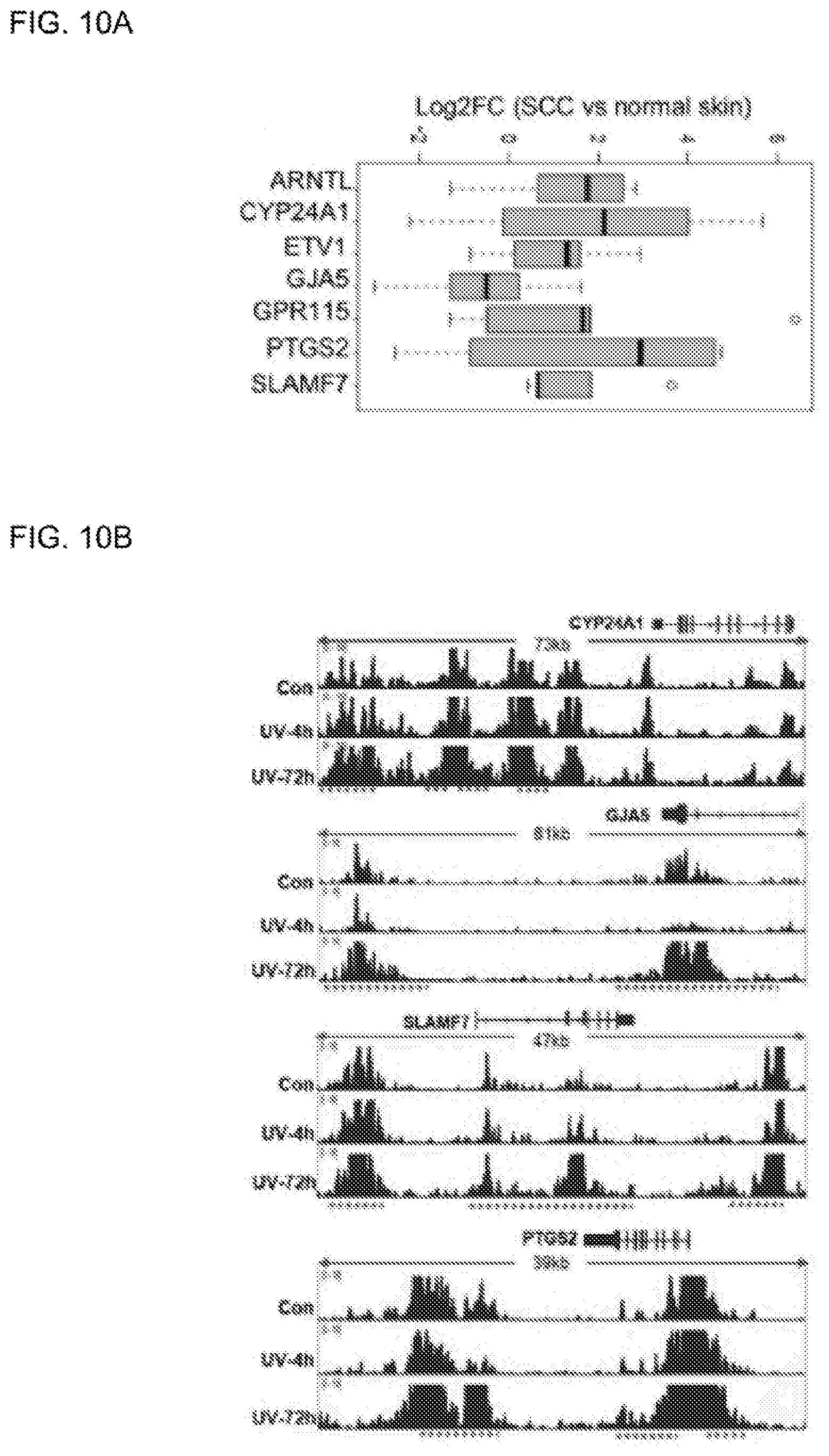
D00016

XML
uspto.report is an independent third-party trademark research tool that is not affiliated, endorsed, or sponsored by the United States Patent and Trademark Office (USPTO) or any other governmental organization. The information provided by uspto.report is based on publicly available data at the time of writing and is intended for informational purposes only.
While we strive to provide accurate and up-to-date information, we do not guarantee the accuracy, completeness, reliability, or suitability of the information displayed on this site. The use of this site is at your own risk. Any reliance you place on such information is therefore strictly at your own risk.
All official trademark data, including owner information, should be verified by visiting the official USPTO website at www.uspto.gov. This site is not intended to replace professional legal advice and should not be used as a substitute for consulting with a legal professional who is knowledgeable about trademark law.