Methods For Treating Cancer With Anti Pd-1 Antibodies And Anti Ctla4 Antibodies
Lala; Mallika ; et al.
U.S. patent application number 16/966988 was filed with the patent office on 2021-02-18 for methods for treating cancer with anti pd-1 antibodies and anti ctla4 antibodies. This patent application is currently assigned to Merck Sharp & Dohme Corp.. The applicant listed for this patent is Rachel Allison ALTURA, Lokesh JAIN, Mallika LALA, Mengyao LI, Merck Sharp & Dohme Corp., Archie Ngai-chiu TSE. Invention is credited to Rachel Allison Altura, Lokesh Jain, Mallika Lala, Mengyao Li, Archie Ngai-chiu Tse.
| Application Number | 20210047409 16/966988 |
| Document ID | / |
| Family ID | 1000005198841 |
| Filed Date | 2021-02-18 |

| United States Patent Application | 20210047409 |
| Kind Code | A1 |
| Lala; Mallika ; et al. | February 18, 2021 |
METHODS FOR TREATING CANCER WITH ANTI PD-1 ANTIBODIES AND ANTI CTLA4 ANTIBODIES
Abstract
The present invention relates to methods for treating cancer in a patient comprising administering an anti-PD-1 antibody or antigen binding fragment thereof in specific amounts to the patient about every six weeks, in combination with administering an anti-CTLA4 antibody to the patient about every six weeks. In certain embodiments, the PD-1 antagonist is pembrolizumab, or an antigen binding fragment thereof. Also provided are compositions comprising a dosage of an anti-PD-1 antibody, or antigen-binding fragment thereof, and a dosage of an anti-CTLA4 antibody or antigen-binding fragment thereof, and uses thereof for treating cancer.
| Inventors: | Lala; Mallika; (West New York, NJ) ; Jain; Lokesh; (Edison, NJ) ; Li; Mengyao; (Springfield, NJ) ; Altura; Rachel Allison; (Belle Mead, NJ) ; Tse; Archie Ngai-chiu; (Long Island City, NY) | ||||||||||
| Applicant: |
|
||||||||||
|---|---|---|---|---|---|---|---|---|---|---|---|
| Assignee: | Merck Sharp & Dohme
Corp. Rahway NJ |
||||||||||
| Family ID: | 1000005198841 | ||||||||||
| Appl. No.: | 16/966988 | ||||||||||
| Filed: | February 8, 2019 | ||||||||||
| PCT Filed: | February 8, 2019 | ||||||||||
| PCT NO: | PCT/US19/17188 | ||||||||||
| 371 Date: | August 3, 2020 |
Related U.S. Patent Documents
| Application Number | Filing Date | Patent Number | ||
|---|---|---|---|---|
| 62630038 | Feb 13, 2018 | |||
| 62732828 | Sep 18, 2018 | |||
| 62740741 | Oct 3, 2018 | |||
| Current U.S. Class: | 1/1 |
| Current CPC Class: | A61P 35/00 20180101; A61K 39/39541 20130101; A61K 2039/545 20130101; C07K 16/2818 20130101; A61K 2039/507 20130101 |
| International Class: | C07K 16/28 20060101 C07K016/28; A61K 39/395 20060101 A61K039/395; A61P 35/00 20060101 A61P035/00 |
Claims
1-31. (canceled)
32. A method of treating cancer in a human patient comprising administering about 400 mg of an anti-PD-1 antibody or antigen binding fragment thereof to the patient approximately every six weeks and about 25 mg, about 50 mg, about 75 mg, or about 100 mg of an anti-CTLA4 antibody or antigen binding fragment thereof to the patient approximately every six weeks, wherein the anti-PD-1 antibody or antigen-binding fragment thereof comprises: (a) light chain complementarity determining regions (CDRs) comprising a sequence of amino acids as set forth in SEQ ID NOs: 1, 2 and 3 and heavy chain CDRs comprising a sequence of amino acids as set forth in SEQ ID NOs: 6, 7 and 8; or (b) light chain CDRs comprising a sequence of amino acids as set forth in SEQ ID NOs: 11, 12 and 13 and heavy chain CDRs comprising a sequence of amino acids as set forth in SEQ ID NOs: 14, 15 and 16; and wherein the anti-CTLA4 antibody or antigen binding fragment thereof comprises: (a) light chain CDRs comprising a sequence of amino acids as set forth in SEQ ID NOs: 39, 40 and 41 and heavy chain CDRs comprising a sequence of amino acids as set forth in SEQ ID NOs: 36, 37, and 38; (b) light chain CDRs comprising a sequence of amino acids as set forth in SEQ ID NOs: 39, 40 and 42 and heavy chain CDRs comprising a sequence of amino acids as set forth in SEQ ID NOs: 36, 37, and 38; or (c) light chain CDRs comprising a sequence of amino acids as set forth in SEQ ID NOs: 39, 40 and 43 and heavy chain CDRs comprising a sequence of amino acids as set forth in SEQ ID NOs: 36, 37, and 38.
33. The method of claim 32, wherein the anti-PD-1 antibody or antigen-binding fragment thereof comprises: (a) a heavy chain variable region comprising a sequence of amino acids as set forth in SEQ ID NO:9, or a variant of SEQ ID NO:9, and (b) a light chain variable region comprising: (i) a sequence of amino acids as set forth in SEQ ID NO:4, or a variant of SEQ ID NO:4, (ii) a sequence of amino acids as set forth in SEQ ID NO:22, or a variant of SEQ ID NO:22, or (iii) a sequence of amino acids as set forth in SEQ ID NO:23, or a variant of SEQ ID NO:23.
34. The method of claim 33, wherein the anti-PD-1 antibody or antigen-binding fragment thereof comprises a heavy chain variable region comprising a sequence of amino acids as set forth in SEQ ID NO:9 and a light chain variable region comprising a sequence of amino acids as set forth in SEQ ID NO:4.
35. The method of claim 32, wherein the anti-PD-1 antibody or antigen-binding fragment thereof is a monoclonal antibody comprising: (a) a heavy chain comprising a sequence of amino acids as set forth in SEQ ID NO:10, or a variant of SEQ ID NO:10, and (b) a light chain comprising a sequence of amino acids as set forth in SEQ ID NO:5, a variant of SEQ ID NO:5, SEQ ID NO:24, a variant of SEQ ID NO:24, SEQ ID NO:25, or a variant of SEQ ID NO:25.
36. The method of claim 35, wherein the anti-PD-1 antibody or antigen-binding fragment thereof is a monoclonal antibody comprising a heavy chain comprising a sequence of amino acids as set forth in SEQ ID NO:10 and a light chain comprising a sequence of amino acids as set forth in SEQ ID NO:5.
37. The method of claim 32, wherein the anti-CTLA4 antibody or antigen binding fragment thereof is comprising (a) a heavy chain variable region comprising a sequence of amino acids as set forth in SEQ ID NO:44 and a light chain variable region comprising a sequence of amino acids as set forth in SEQ ID NO:45; (b) a heavy chain variable region comprising a sequence of amino acids as set forth in SEQ ID NO:46 and a light chain variable region comprising a sequence of amino acids as set forth in SEQ ID NO:47; (c) a heavy chain variable region comprising a sequence of amino acids as set forth in SEQ ID NO:48 and a light chain variable region comprising a sequence of amino acids as set forth in SEQ ID NO:49; (d) a heavy chain variable region comprising a sequence of amino acids as set forth in SEQ ID NO:50 and a light chain variable region comprising a sequence of amino acids as set forth in SEQ ID NO:49; (e) a heavy chain variable region comprising a sequence of amino acids as set forth in SEQ ID NO:51 and a light chain variable region comprising a sequence of amino acids as set forth in SEQ ID NO:52; (f) a heavy chain variable region comprising a sequence of amino acids as set forth in SEQ ID NO:53 and a light chain variable region comprising a sequence of amino acids as set forth in SEQ ID NO:54; or (g) a heavy chain variable region comprising a sequence of amino acids as set forth in SEQ ID NO:55 and a light chain variable region comprising a sequence of amino acids as set forth in SEQ ID NO:56.
38. The method of claim 37, wherein the anti-CTLA4 antibody or antigen binding fragment thereof comprises a heavy chain variable region comprising a sequence of amino acids as set forth in SEQ ID NO:50 and a light chain variable region comprising a sequence of amino acids as set forth in SEQ ID NO:49.
39. The method of claim 37, wherein the anti-CTLA4 antibody or antigen binding fragment thereof comprises a heavy chain comprising a sequence of amino acids as set forth in SEQ ID NO:57 and a light chain comprising a sequence of amino acids as set forth in SEQ ID NO:58.
40. A method of treating cancer in a human patient comprising administering about 400 mg of an anti-PD-1 antibody and about 25 mg, about 50 mg, about 75 mg, or about 100 mg of an anti-CTLA4 antibody, each to the patient approximately every six weeks, wherein the anti-PD-1 antibody comprises (i) a heavy chain comprising a sequence of amino acids as set forth in SEQ ID NO:10 and (ii) a light chain comprising a sequence of amino acids as set forth in SEQ ID NO:5, and wherein the anti-CTLA4 antibody comprises (iii) a heavy chain comprising a sequence of amino acids as set forth in SEQ ID NO:57 and (iv) a light chain comprising a sequence of amino acids as set forth in SEQ ID NO:58.
41. The method of claim 32, wherein the cancer is PD-1/PD-L1 refractory melanoma.
42. The method claim 32, wherein the cancer is selected from the group consisting of: melanoma, non-small cell lung cancer, head and neck cancer, urothelial cancer, breast cancer, gastrointestinal cancer, multiple myeloma, hepatocellular cancer, non-Hodgkin lymphoma, renal cancer, Hodgkin lymphoma, mesothelioma, ovarian cancer, small cell lung cancer, esophageal cancer, anal cancer, biliary tract cancer, colorectal cancer, cervical cancer, thyroid cancer, salivary cancer, pancreatic cancer, a tumor of the brain, glioblastoma, sarcoma, a tumor of the bone, or Merkel cell carcinoma.
43. The method of claim 32, wherein the anti-PD-1 antibody or antigen-binding fragment thereof is administered to the patient by intravenous or subcutaneous administration.
44. The method of claim 32, wherein the anti-PD-1 antibody or antigen-binding fragment thereof is pembrolizumab.
45. The method of claim 32, wherein the anti-PD-1 antibody and the anti-CTLA4 antibody are co-administered.
46. The method of claim 32, wherein the anti-PD-1 antibody and the anti-CTLA4 antibody are co-formulated.
47. The method of claim 32, comprising administering 25 mg of the anti-CTLA4 antibody or antigen binding fragment thereof.
48. A kit for treating a patient with cancer, the kit comprising: (a) about 400 mg of an anti-PD-1 antibody or antigen binding fragment thereof, (b) about 25 mg, 50 mg, 75 mg, or 100 mg of an anti-CTLA4 antibody or antigen binding fragment thereof; and (c) instructions for using the anti-PD-1 antibody or antigen binding fragment thereof and the anti-CTLA4 antibody or antigen binding fragment thereof, wherein the anti-PD-1 antibody or antigen-binding fragment thereof comprises: i. light chain complementarity determining regions (CDRs) comprising a sequence of amino acids as set forth in SEQ ID NOs: 1, 2 and 3 and heavy chain CDRs comprising a sequence of amino acids as set forth in SEQ ID NOs: 6, 7 and 8; or ii. light chain CDRs comprising a sequence of amino acids as set forth in SEQ ID NOs: 11, 12 and 13 and heavy chain CDRs comprising a sequence of amino acids as set forth in SEQ ID NOs: 14, 15 and 16; and wherein the anti-CTLA4 antibody or antigen binding fragment thereof comprises: i. light chain CDRs comprising a sequence of amino acids as set forth in SEQ ID NOs: 39, 40 and 41 and heavy chain CDRs comprising a sequence of amino acids as set forth in SEQ ID NOs: 36, 37, and 38; ii. light chain CDRs comprising a sequence of amino acids as set forth in SEQ ID NOs: 39, 40 and 42 and heavy chain CDRs comprising a sequence of amino acids as set forth in SEQ ID NOs: 36, 37, and 38; or iii. light chain CDRs comprising a sequence of amino acids as set forth in SEQ ID NOs: 39, 40 and 43 and heavy chain CDRs comprising a sequence of amino acids as set forth in SEQ ID NOs: 36, 37, and 38.
49. The kit of claim 48, wherein the anti-PD-1 antibody is pembrolizumab.
50. The kit of claim 48, wherein the anti-CTLA4 antibody is a monoclonal antibody comprising light chain CDRs comprising a sequence of amino acids as set forth in SEQ ID NOs: 39, 40, and 41, and heavy chain CDRs comprising a sequence of amino acids as set forth in SEQ ID NOs: 36, 37, and 38.
51. The kit of claim 48, wherein the anti-CTLA4 antibody is a monoclonal antibody comprising a heavy chain variable region comprising a sequence of amino acids as set forth in SEQ ID NO:50 and a light chain variable region comprising a sequence of amino acids as set forth in SEQ ID NO:49.
52. The kit of claim 48, wherein the anti-CTLA4 antibody is a monoclonal antibody comprising a heavy chain comprising a sequence of amino acids as set forth in SEQ ID NO:57 and a light chain comprising a sequence of amino acids as set forth in SEQ ID NO:58.
53. The kit of claim 48, comprising about 25 mg of the anti-CTLA4 antibody or antigen binding fragment thereof.
Description
FIELD OF THE INVENTION
[0001] The present invention relates to therapies useful for the treatment of cancer. In particular, the invention relates to a method for treating cancer which comprises administering to a patient in need thereof an anti-PD-1 antibody, or antigen binding fragment thereof in combination with an anti-CTLA4 antibody or antigen binding fragment thereof using the dosage regimens specified herein.
CROSS-REFERENCE TO RELATED APPLICATIONS
[0002] This application claims the benefit of U.S. provisional application No. 62/630,038, filed Feb. 13, 2018, U.S. provisional application No. 62/732,838, filed Sep. 18, 2018, and U.S. provisional application No. 62/740,741, filed Oct. 3, 2018, the contents of each of which are hereby incorporated by reference in their entirety.
REFERENCE TO SEQUENCE LISTING SUBMITTED ELECTRONICALLY
[0003] The sequence listing of the present application is submitted electronically via EFS-Web as an ASCII formatted sequence listing with a file name "24695WOPCT-SEQLIST-06FEB2019.TXT", creation date of Feb. 6, 2019, and a size of 56.0 kb. This sequence listing submitted via EFS-Web is part of the specification and is herein incorporated by reference in its entirety.
BACKGROUND OF THE INVENTION
[0004] PD-1 is recognized as an important player in immune regulation and the maintenance of peripheral tolerance. PD-1 is moderately expressed on naive T, B and NKT cells and up-regulated by T/B cell receptor signaling on lymphocytes, monocytes and myeloid cells (Sharpe et al., The function of programmed cell death 1 and its ligands in regulating autoimmunity and infection. Nature Immunology (2007); 8:239-245).
[0005] Two known ligands for PD-1, PD-L1 (B7-H1) and PD-L2 (B7-DC), are expressed in human cancers arising in various tissues. In large sample sets of e.g. ovarian, renal, colorectal, pancreatic, liver cancers and melanoma, it was shown that PD-L1 expression correlated with poor prognosis and reduced overall survival irrespective of subsequent treatment (Dong et al., Nat Med. 8(8):793-800 (2002); Yang et al. Invest Ophthalmol Vis Sci. 49: 2518-2525 (2008); Ghebeh et al. Neoplasia 8:190-198 (2006); Hamanishi et al., Proc. Natl. Acad. Sci. USA 104: 3360-3365 (2007); Thompson et al., Cancer 5: 206-211 (2006); Nomi et al., Clin. Cancer Research 13:2151-2157 (2007); Ohigashi et al., Clin. Cancer Research 11: 2947-2953 (2005); Inman et al., Cancer 109: 1499-1505 (2007); Shimauchi et al. Int. J. Cancer 121:2585-2590 (2007); Gao et al. Clin. Cancer Research 15: 971-979 (2009); Nakanishi J. Cancer Immunol Immunother. 56: 1173-1182 (2007); and Hino et al., Cancer 00: 1-9 (2010)).
[0006] Similarly, PD-1 expression on tumor infiltrating lymphocytes was found to mark dysfunctional T cells in breast cancer and melanoma (Ghebeh et al, BMC Cancer. 2008 8:5714-15 (2008); Ahmadzadeh et al., Blood 114: 1537-1544 (2009)) and to correlate with poor prognosis in renal cancer (Thompson et al., Clinical Cancer Research 15: 1757-1761(2007)). Thus, it has been proposed that PD-L1 expressing tumor cells interact with PD-1 expressing T cells to attenuate T cell activation and evasion of immune surveillance, thereby contributing to an impaired immune response against the tumor.
[0007] Immune checkpoint therapies targeting the PD-1 axis have resulted in groundbreaking improvements in clinical response in multiple human cancers (Brahmer et al., N Engl J Med 2012, 366: 2455-65; Garon et al. N Engl J Med 2015, 372: 2018-28; Hamid et al., N Engl J Med 2013, 369: 134-44; Robert et al., Lancet 2014, 384: 1109-17; Robert et al., N Engl J Med 2015, 372: 2521-32; Robert et al., N Engl J Med 2015, 372: 320-30; Topalian et al., N Engl J Med 2012, 366: 2443-54; Topalian et al., J Clin Oncol 2014, 32: 1020-30; Wolchok et al., N Engl J Med 2013, 369: 122-33). Immune therapies targeting the PD-1 axis include monoclonal antibodies directed to the PD-1 receptor (KEYTRUDA.TM. (pembrolizumab), Merck and Co., Inc., Kenilworth, N.J., USA and OPDIVO.TM. (nivolumab), Bristol-Myers Squibb Company, Princeton, N.J., USA) and also those that bind to the PD-L1 ligand (MPDL3280A; TECENTRIQ.TM. (atezolizumab), Genentech, San Francisco, Calif., USA; IMFINZI.TM. (durvalumab), AstraZeneca Pharmaceuticals LP, Wilmington, Del.; BAVENCIO.TM. (avelumab), Merck KGaA, Darmstadt, Germany). Both therapeutic approaches have demonstrated anti-tumor effects in numerous cancer types.
[0008] It has been proposed that the efficacy of such antibodies might be enhanced if administered in combination with other approved or experimental cancer therapies, e.g., radiation, surgery, chemotherapeutic agents, targeted therapies, agents that inhibit other signaling pathways that are disregulated in tumors, and other immune enhancing agents. One such agent that has been tested in combination with antagonists of PD-1 is an antagonist of cytotoxic T lymphocyte associated antigen 4 (abbreviated CTLA4).
[0009] CTLA4 has a very close relationship with the CD28 molecule in gene structure, chromosome location, sequence homology and gene expression. Both are receptors for the co-stimulative molecule B7, mainly expressed on the surface of activated T cells. After binding to B7, CTLA4 can inhibit the activation of mouse and human T cells, playing a negative regulating role in the activation of T cells.
[0010] CTLA4 mAbs or CTLA4 ligands can prevent CTLA4 from binding to its native ligands, thereby blocking the transduction of the T cell negative regulating signal by CTLA4 and enhancing the responsiveness of T cells to various antigens. In this aspect, results from in vivo and in vitro studies are substantially in concert. At present, there are some CTLA4 mAbs being tested in clinical trials for treating prostate cancer, bladder cancer, colorectal cancer, cancer of gastrointestinal tract, liver cancer, malignant melanoma, etc. (Grosso et al., CTLA-4 blockade in tumor models: an overview of preclinical and translational research. Cancer Immun. 13:5 (2013)).
[0011] As important factors affecting the function of T cells, CTLA4 and CTLA4 mAbs can produce specific therapeutic effects on diseases by interfering with the immune microenvironment in the body. They have high efficacy and remedy the deficiency of traditional medication, opening a novel pathway of gene therapy. CTLA4 and CTLA4 mAbs are being tested in experiments and various stages of clinical trials. For example, in autoimmune diseases, they have been shown to effectively inhibit airway hyperresponsiveness in an animal model of asthma, prevent the development of rheumatic diseases, mediate immune tolerance to an allograft in the body, and the like. On the other hand, although biological gene therapy has not shown any adverse effect in short term clinical trials, attention should be paid to the potential effect after long term application. For example, excessive blockade of CTLA4-B7 signaling by CTLA4 mAbs may result in the development of autoimmune diseases. As antibodies can specifically bind to their antigens and induce the lysis of target cells or block the progress of pathology, development and utilization of drugs based on antibodies, especially humanized antibodies have important significance in the clinical treatment of malignant tumors and other immune diseases in humans.
[0012] It would be beneficial to develop additional dosing schedules that allow for the administration of a safe and effective dose of an anti-PD-1 antibody alone, or in combination with an anti-CTLA4 antibody, that is more convenient for patients. It would be beneficial to develop methods of treating PD-1/PD-L1 refractory cancers by administering an anti-PD-1 antibody and an anti-CTLA4 antibody using the dosing schedules provided herein.
SUMMARY OF THE INVENTION
[0013] The present invention provides alternative, less frequent, dosing regimens for treating a cancer patient with an anti-PD-1 antibody, or antigen-binding fragment thereof, wherein the dosing schedule is expected to provide a safe and effective dose of the anti-PD-1 antibody, or antigen-binding fragment thereof. It also provides alternative, less frequent, dosing regiments for treating a cancer patient with a combination of an anti-PD-1 antibody, or an antigen binding fragment thereof, in combination with an anti-CTLA4 antibody or antigen binding fragment thereof. Specifically, the invention provides a method of treating cancer in a human patient comprising administering about 400 mg of an anti-PD-1 antibody or antigen binding fragment thereof to the patient every six weeks, wherein the anti-PD-1 antibody or antigen-binding fragment thereof comprises (a) light chain complementarity determining regions (CDRs) comprising a sequence of amino acids as set forth in SEQ ID NOs: 1, 2 and 3 and heavy chain CDRs comprising a sequence of amino acids as set forth in SEQ ID NOs: 6, 7 and 8; or (b) light chain CDRs comprising a sequence of amino acids as set forth in SEQ ID NOs: 11, 12 and 13 and heavy chain CDRs comprising a sequence of amino acids as set forth in SEQ ID NOs: 14, 15 and 16. In preferred embodiments of the invention, the antibody or antigen-binding fragment is pembrolizumab.
[0014] Also provided is a method of treating cancer in a human patient comprising administering about 400 mg of an anti-PD-1 antibody or antigen binding fragment thereof to the patient and about 25 mg, 50 mg, 75 mg or 100 mg of an anti-CTLA4 antibody or antigen binding fragment thereof, wherein each of the anti-PD-1 antibody and the anti-CTLA4 antibody, or antigen binding fragments thereof, are administered to the patient every six weeks, wherein the anti-PD-1 antibody or antigen-binding fragment thereof comprises (a) light chain complementarity determining regions (CDRs) comprising a sequence of amino acids as set forth in SEQ ID NOs: 1, 2 and 3 and heavy chain CDRs comprising a sequence of amino acids as set forth in SEQ ID NOs: 6, 7 and 8; or (b) light chain CDRs comprising a sequence of amino acids as set forth in SEQ ID NOs: 11, 12 and 13 and heavy chain CDRs comprising a sequence of amino acids as set forth in SEQ ID NOs: 14, 15 and 16; and wherein the anti-CTLA4 antibody or antigen binding fragment thereof comprises light chain CDRs comprising a sequence of amino acids as set forth in SEQ ID NOs: 39, 40 and 41 and heavy chain CDRs comprising a sequence of amino acids as set forth in SEQ ID NOs: 36, 37 and 38.
[0015] In one embodiment, the anti-PD-1 antibody or antigen-binding fragment thereof comprises light chain CDRs comprising a sequence of amino acids as set forth in SEQ ID NOs: 1, 2 and 3 and heavy chain CDRs comprising a sequence of amino acids as set forth in SEQ ID NOs: 6, 7 and 8; and the anti-CTLA4 antibody or antigen binding fragment thereof comprises light chain CDRs comprising a sequence of amino acids as set forth in SEQ ID NOs: 39, 40 and 41 and heavy chain CDRs comprising a sequence of amino acids as set forth in SEQ ID NOs: 36, 37 and 38.
[0016] In another embodiment, the anti PD-1 antibody or antigen binding fragment thereof comprises a heavy chain variable region comprising a sequence of amino acids as set forth in SEQ ID NO: 9 and a light chain variable region comprising a sequence of amino acids as set forth in SEQ ID NO:4; and the anti-CTLA4 antibody comprises a heavy chain variable region comprising a sequence of amino acids as set forth in SEQ ID NO: 50 and a light chain variable region comprising a sequence of amino acids as set forth in SEQ ID NO: 49.
[0017] In all of the above embodiments, the amount of the anti-PD-1 antibody or antigen binding fragment thereof administered to the patient is from about 350 mg to about 450 mg. In further embodiments, the amount of the anti-PD-1 antibody or antigen binding fragment is about 400 mg. In further embodiments, the amount of the anti-PD-1 antibody or antigen binding fragment is 400 mg.
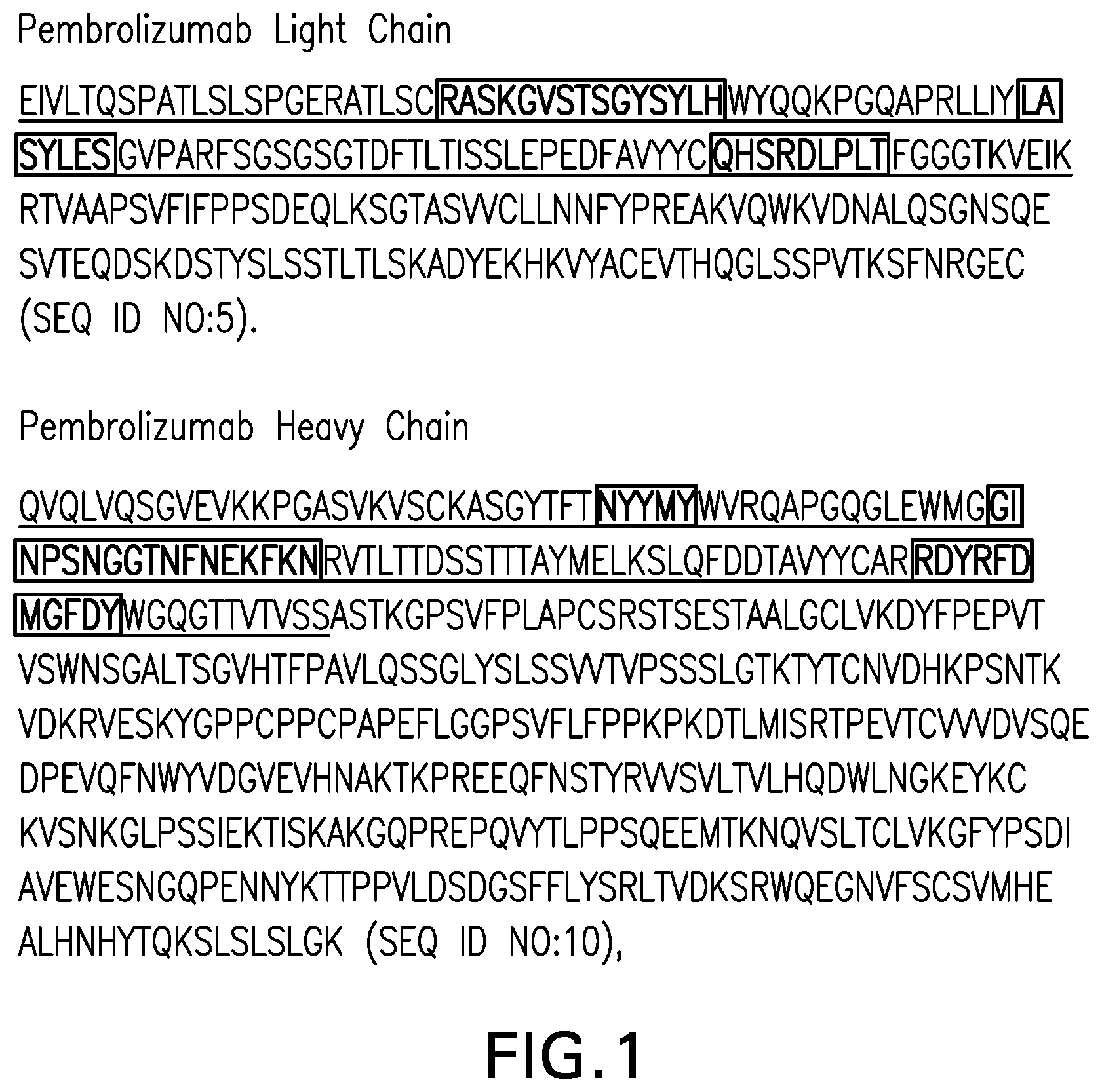
[0018] In all of the above treatment methods, compositions and uses herein, the PD-1 antibody or antigen-binding fragment inhibits the binding of PD-L1 to PD-1, and preferably also inhibits the binding of PD-L2 to PD-1. In some preferred embodiments of the treatment methods, compositions and uses of the invention, the PD-1 antibody or antigen-binding fragment is a monoclonal antibody, which specifically binds to PD-1 and blocks the binding of PD-L1 to PD-1. In one particular embodiment, the anti-PD-1 antibody comprises a heavy chain and a light chain, and wherein the heavy and light chains comprise the amino acid sequences shown in FIG. 1 (SEQ ID NO:5 and SEQ ID NO:10). In another embodiment, the anti-PD-1 antibody comprises a heavy chain and a light chain, and wherein the heavy and light chains comprise the amino acid sequences shown in FIG. 1 (SEQ ID NO:5 and SEQ ID NO:10) and the anti-CTLA4 antibody comprises a heavy chain and a light chain, wherein the heavy and light chains comprise the amino acid sequences set forth in SEQ ID NOs: 57 and 58.
[0019] In some preferred embodiments of the treatment methods, composition and uses of the invention, the anti-CTLA4 antibody or antigen binding fragment is a monoclonal antibody, which specifically binds to CTLA4. In one embodiment, the anti-CTLA4 antibody or antigen binding fragment thereof comprises light chain CDRs comprising a sequence of amino acids as set forth in SEQ ID NOs: 39, 40 and 41 and heavy chain CDRs comprising a sequence of amino acids as set forth in SEQ ID NOs: 36, 37 and 38. In one particular embodiment, the anti-CTLA4 antibody comprises a heavy chain variable region and a light chain variable region, wherein the heavy and light chain variable regions comprise the amino acid sequences set forth SEQ ID NO: 50 and 49, respectively. In another embodiment, the anti-CTLA antibody is a monoclonal antibody comprising a heavy chain and a light chain, wherein the heavy chain comprises the amino acid sequence set forth in SEQ ID NO: 57 and the light chain comprises the amino acid sequence set forth in SEQ ID NO: 58.
[0020] In some embodiments of any of the above treatment methods, compositions and uses, the cancer expresses one or both of PD-L1 and PD-L2. In some embodiments, PD-L1 expression is elevated in the cancer. In a further embodiment, the cancer is PD-1/PD-L1 refractory (e.g., it is a cancer that has not been responsive to previous treatment with an anti-PD-1 or anti-PD-L1 agent). In a further embodiment, the cancer is PD-1/PD-L1 refractory melanoma.
BRIEF DESCRIPTION OF THE DRAWINGS
[0021] FIG. 1 shows amino acid sequences of the light chain and heavy chain for an exemplary anti-PD-1 monoclonal antibody useful in the present invention (SEQ ID NOs:5 and 10, respectively). Light chain and heavy chain variable regions are underlined (SEQ ID NO's 4 and 9) and CDRs bold and are boxed
[0022] FIG. 2 shows that pembrolizumab Cmax at steady state for 400 mg Q6W lies within the range from 2 mg/kg Q3W and 200 mg Q3W to 10 mg/kg Q2W.
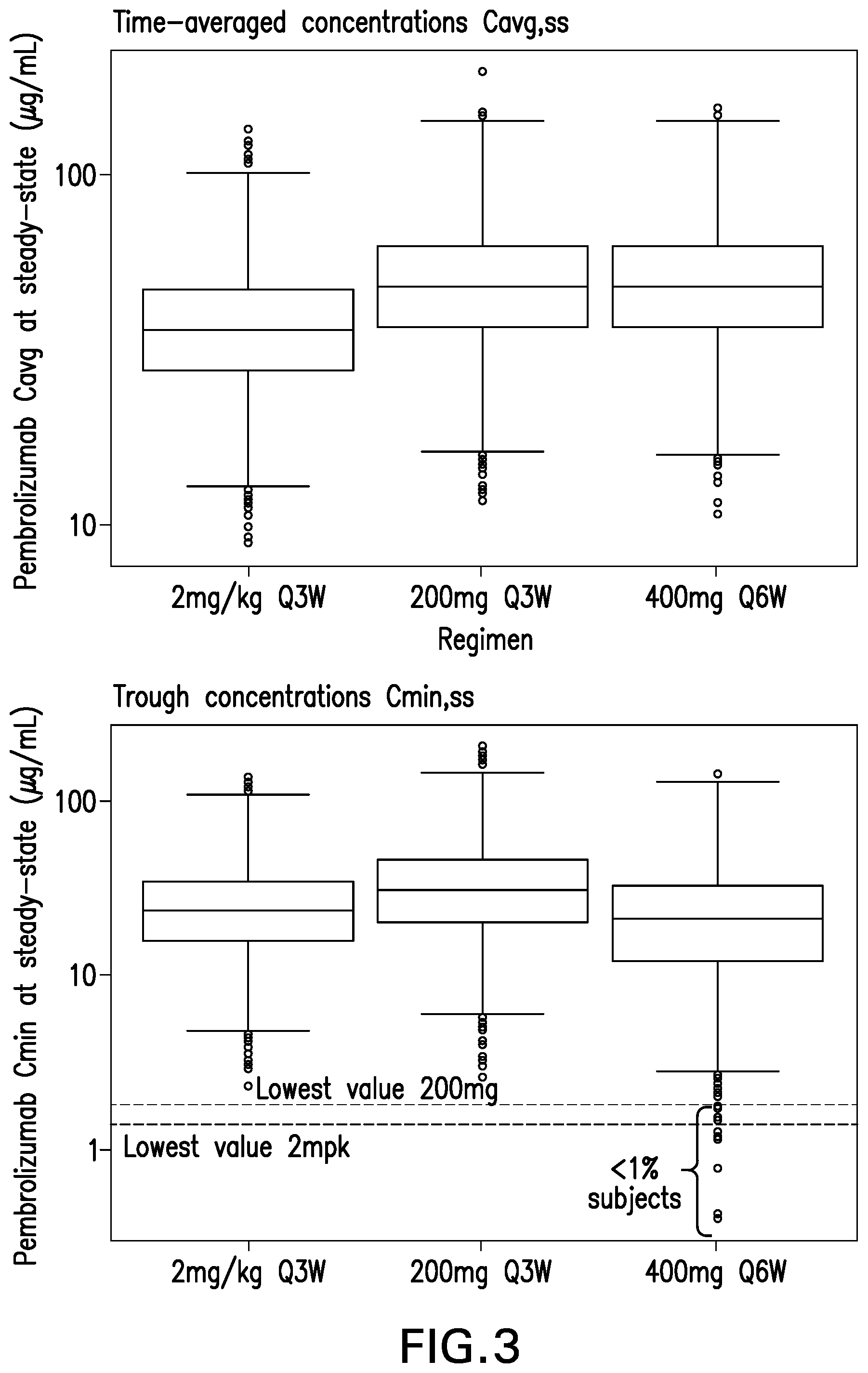
[0023] FIG. 3 shows that pembrolizumab exposures (Cavg and Cmin) at steady state are similar for 400 mg Q6W relative to 2 mg/kg Q3W and 200 mg Q3W.
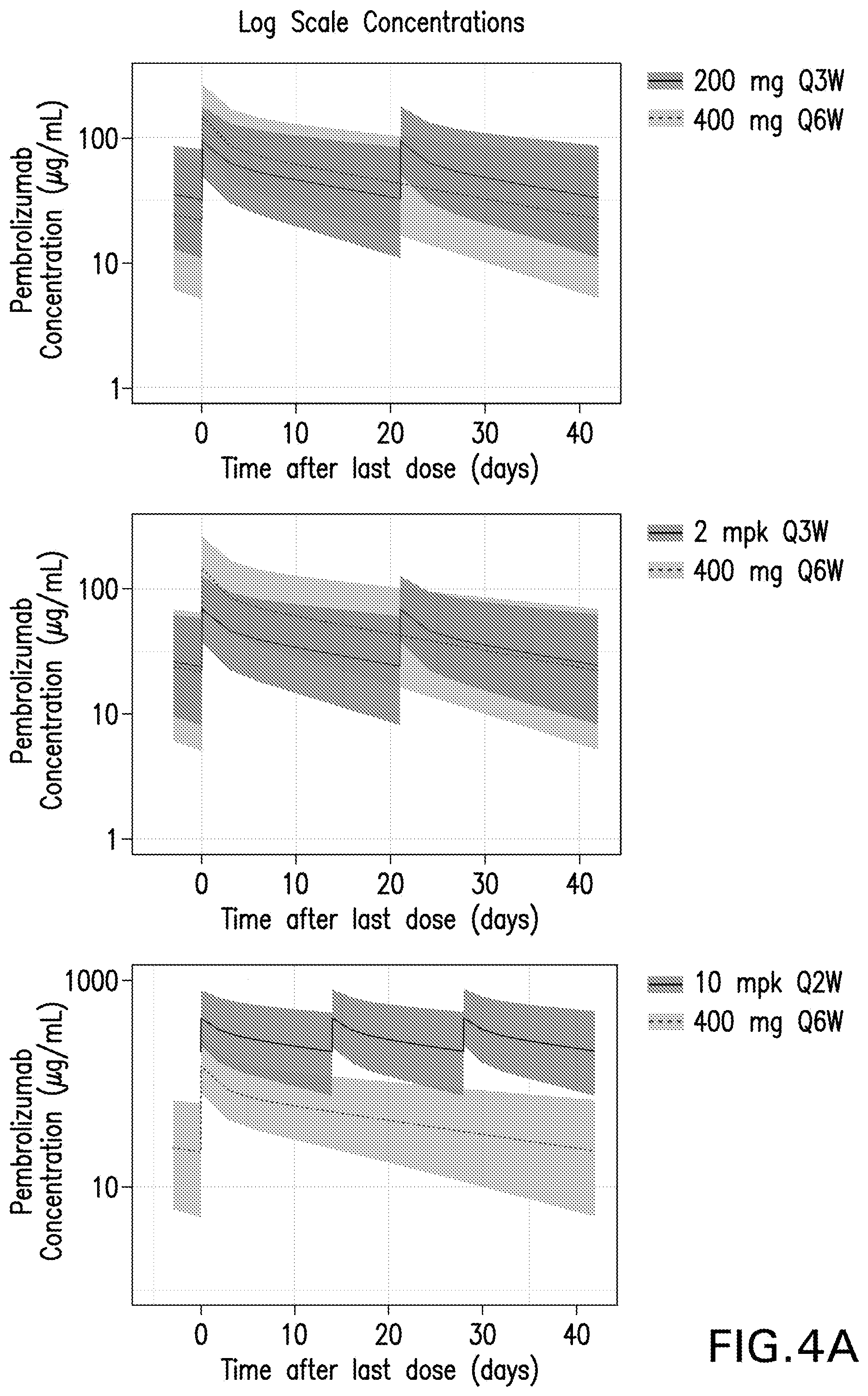
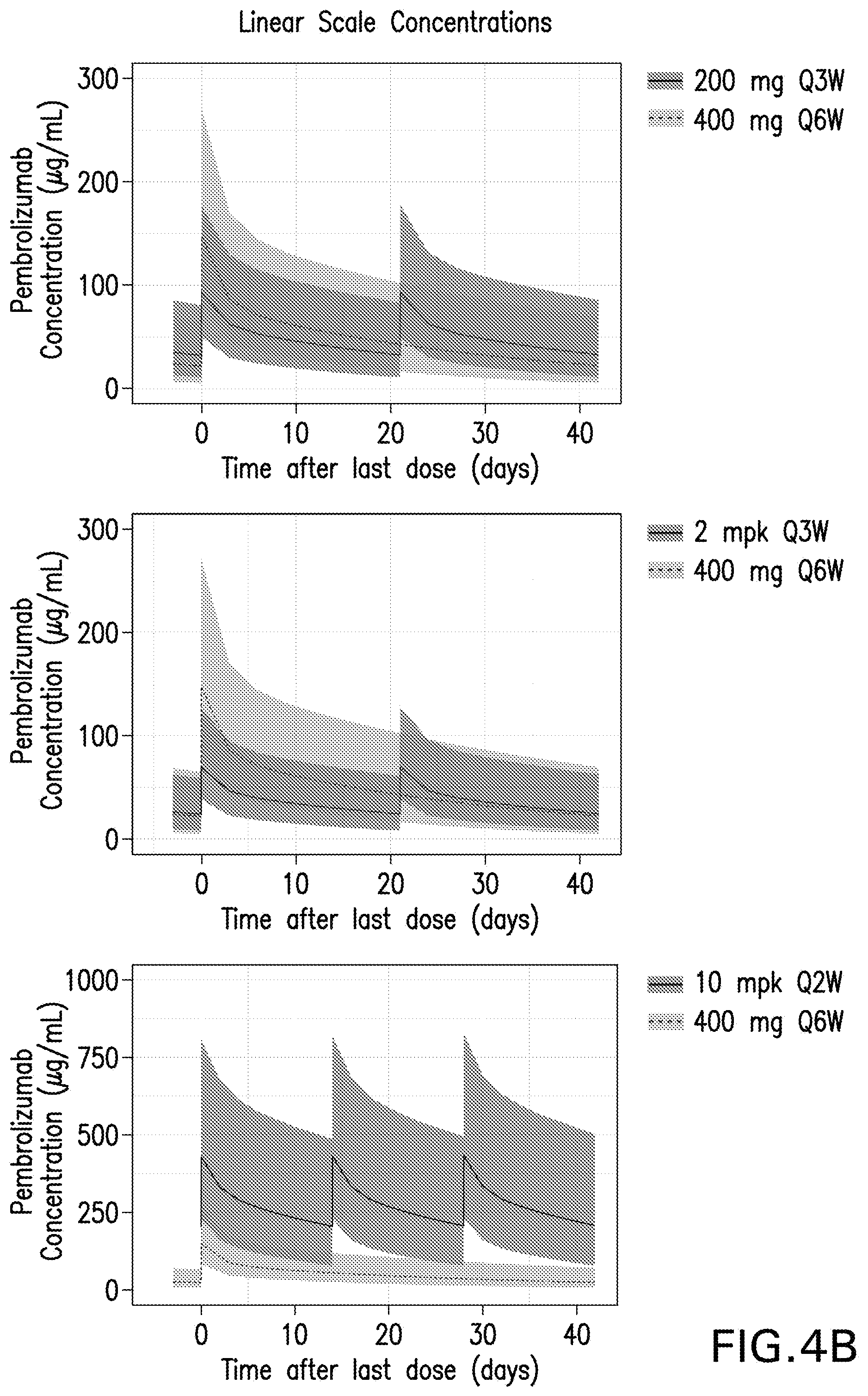
[0024] FIGS. 4A and 4B show the pembrolizumab pharmacokinetic profiles at steady state for the 400 mg Q6W dosing regimen compared to the 200 mg flat dosing regimen (top) and the Q3W, 2 mg/kg weight-based dosing regimen bottom). Results are provided for log scale concentrations (FIG. 4A) and linear scale concentrations (FIG. 4B).
DETAILED DESCRIPTION OF THE INVENTION
I. Definitions and Abbreviations
[0025] As used throughout the specification and appended claims, the following abbreviations apply: [0026] AE adverse event [0027] AUCss area under the concentration-time curve at steady state [0028] BICR blinded independent central review [0029] Cavg,ss time averaged concentration at steady state [0030] CDR complementarity determining region [0031] CI confidence interval [0032] Cmax,ss peak concentrations at steady state [0033] Cmin,ss trough concentrations at steady state [0034] CPS combined positive score [0035] CTLA4 cytotoxic T lymphocyte associated antigen 4 [0036] DOR duration of response [0037] ECG electrocardiogram [0038] ECOG Eastern Cooperative Oncology Group [0039] E-R exposure (concentration) response [0040] FFPE formalin-fixed paraffin-embedded [0041] FR framework region [0042] GM geometric mean [0043] HCC hepatocellular carcinoma [0044] HNSCC head and neck squamous cell cancer [0045] HL Hodgkin lymphoma [0046] IgG immunoglobulin G [0047] IHC immunohistochemistry or immunohistochemical [0048] IV intravenous [0049] LPS lymphoma proportion score [0050] mAb monoclonal antibody [0051] MCC Merkel cell carcinoma [0052] MEL melanoma [0053] MMR mismatch repair [0054] MPS modified proportion score [0055] MRI magnetic resonance imaging [0056] MSI-H microsatellite instability-high [0057] NCI CTCAE National Cancer Institute--Common Terminology Criteria for Adverse Events [0058] NSCLC non-small cell lung cancer [0059] ORR objective response rate [0060] OS overall survival [0061] PD progressive disease [0062] PD-1 programmed death 1 (a.k.a. programmed cell death-1 and programmed death receptor 1) [0063] PD-L1 programmed cell death 1 ligand 1 [0064] PD-L2 programmed cell death 1 ligand 2 [0065] PFS progression free survival [0066] PK pharmacokinetic [0067] Q2W one dose every two weeks [0068] Q3W one dose every three weeks [0069] Q6W one dose every six weeks [0070] RCC renal cell carcinoma [0071] SAE serious adverse event [0072] SC subcutaneous [0073] TPS tumor proportion score [0074] V.sub.H immunoglobulin heavy chain variable region [0075] V.sub.L immunoglobulin light chain variable region
[0076] So that the invention may be more readily understood, certain technical and scientific terms are specifically defined below. Unless specifically defined elsewhere in this document, all other technical and scientific terms used herein have the meaning commonly understood by one of ordinary skill in the art to which this invention belongs.
[0077] Reference to "or" indicates either or both possibilities unless the context clearly dictates one of the indicated possibilities. In some cases, "and/or" was employed to highlight either or both possibilities.
[0078] As used herein, including the appended claims, the singular forms of words such as "a," "an," and "the," include their corresponding plural references unless the context clearly dictates otherwise.
[0079] The term "about", when modifying the quantity (e.g., mg) of a substance or composition, or the value of a parameter characterizing a step in a method, or the like, refers to variation in the numerical quantity that can occur, for example, through typical measuring, handling and sampling procedures involved in the preparation, characterization and/or use of the substance or composition; through inadvertent error in these procedures; through differences in the manufacture, source, or purity of the ingredients employed to make or use the compositions or carry out the procedures; and the like. In certain embodiments, "about" can mean a variation off 0.1%, .+-.0.5%, .+-.1%, .+-.2%, .+-.3%, .+-.4%, .+-.5%, .+-.6%, .+-.7%, .+-.8%, .+-.9% or .+-.10%. When referring to the dosage of "about 400 mg," the dosage can be from 360 mg to 440 mg, from 370 mg to 430 mg, from 380 mg to 420 mg, from 390 mg to 410 mg, from 395 mg to 405 mg, from 400 mg to 440 mg, or from 390 mg to 440 mg. It alternative embodiments, the dosage can be 360 mg, 365 mg, 370 mg, 375 mg, 380 mg, 385 mg, 390 mg, 395 mg, 400 mg, 405 mg, 410 mg, 415 mg, 420 mg, 425 mg, 430 mg, 435 mg, or 440 mg. When referring to the amount of time between administrations in a therapeutic treatment regimen (i.e., amount of time between administrations of the anti-PD-1 antibody or antigen binding fragment thereof and the anti-CTLA4 antibody or antigen binding fragment thereof, e.g. "about 6 weeks," which is used interchangeably herein with "approximately every six weeks"), "about" refers to the stated time.+-.a variation that can occur due to patient/clinician scheduling and availability around the 6-week target date. For example, "about 6 weeks" can refer to 6 weeks.+-.5 days, 6 weeks.+-.4 days, 6 weeks.+-.3 days, 6 weeks.+-.2 days or 6 weeks.+-.1 day, or may refer to 5 weeks, 2 days through 6 weeks, 5 days.
[0080] Pharmacokinetic "steady state" is a period of time during which any accumulation of drug concentrations owing to multiple doses has been maximized and systemic drug exposure is considered uniform after each subsequent dose administered; in the specific case of pembrolizumab, steady state is achieved at and after .about.16 weeks of administration.
[0081] AUCss, Cavg,ss and Cmin,ss are pharmacokinetic measures of the systemic exposure to the drug (e.g. pembrolizumab) in humans after its administration, and are typically considered drivers of drug efficacy. AUCss and Cavg,ss represent the average exposure over a dosing interval, but differ in terms of units. "Cmin,ss" represents the minimum or lowest (trough) drug concentration observed at the end of a dosing interval, just before the next dose is administered.
[0082] "Cmax,ss" is the maximum or highest (peak) drug concentration observed soon after its administration. In the specific case of pembrolizumab, which is administered as intravenous infusion, the peak concentration occurs immediately after end of infusion. Cmax,ss is a metric that is typically considered a driver of driver safety.
[0083] "Administration" and "treatment," as it applies to an animal, human, experimental subject, cell, tissue, organ, or biological fluid, refers to contact of an exogenous pharmaceutical, therapeutic, diagnostic agent, or composition to the animal, human, subject, cell, tissue, organ, or biological fluid. "Treat" or "treating" a cancer, as used herein, means to administer an anti-PD-1 antibody, or antigen-binding fragment, alone or in combination with an anti-CTLA4 antibody, or antigen binding fragment thereof, to a subject having a cancer, or diagnosed with a cancer, to achieve at least one positive therapeutic effect, such as for example, reduced number of cancer cells, reduced tumor size, reduced rate of cancer cell infiltration into peripheral organs, or reduced rate of tumor metastasis or tumor growth. "Treatment" may include one or more of the following: inducing/increasing an antitumor immune response, decreasing the number of one or more tumor markers, halting or delaying the growth of a tumor or blood cancer or progression of disease associated with PD-1 binding to its ligands PD-L1 and/or PD-L2 ("PD-1-related disease") such as cancer, stabilization of PD-1-related disease, inhibiting the growth or survival of tumor cells, eliminating or reducing the size of one or more cancerous lesions or tumors, decreasing the level of one or more tumor markers, ameliorating or abrogating the clinical manifestations of PD-1-related disease, reducing the severity or duration of the clinical symptoms of PD-1-related disease such as cancer, prolonging the survival of a patient relative to the expected survival in a similar untreated patient, and inducing complete or partial remission of a cancerous condition or other PD-1 related disease.
[0084] Positive therapeutic effects in cancer can be measured in a number of ways (See, W. A. Weber, J. Nucl. Med. 50:1S-10S (2009)). For example, with respect to tumor growth inhibition, according to NCI standards, a T/C.ltoreq.42% is the minimum level of anti-tumor activity. A T/C<10% is considered a high anti-tumor activity level, with T/C (%)=Median tumor volume of the treated/Median tumor volume of the control.times.100. In some embodiments, the treatment achieved by a therapeutically effective amount is any of progression free survival (PFS), disease free survival (DFS) or overall survival (OS). PFS, also referred to as "Time to Tumor Progression" indicates the length of time during and after treatment that the cancer does not grow, and includes the amount of time patients have experienced a complete response or a partial response, as well as the amount of time patients have experienced stable disease. DFS refers to the length of time during and after treatment that the patient remains free of disease. OS refers to a prolongation in life expectancy as compared to naive or untreated individuals or patients. While an embodiment of the treatment methods, compositions and uses of the present invention may not be effective in achieving a positive therapeutic effect in every patient, it should do so in a statistically significant number of subjects as determined by any statistical test known in the art such as the Student's t-test, the chi.sup.2-test, the U-test according to Mann and Whitney, the Kruskal-Wallis test (H-test), Jonckheere-Terpstra-test and the Wilcoxon-test.
[0085] The term "patient" (alternatively referred to as "subject" or "individual" herein) refers to a mammal (e.g., rat, mouse, dog, cat, rabbit) capable of being treated with the methods and compositions of the invention, most preferably a human. In some embodiments, the patient is an adult patient. In other embodiments, the patient is a pediatric patient.
[0086] The term "antibody" refers to any form of antibody that exhibits the desired biological or binding activity. Thus, it is used in the broadest sense and specifically covers, but is not limited to, monoclonal antibodies (including full length monoclonal antibodies), polyclonal antibodies, humanized, fully human antibodies, and chimeric antibodies. "Parental antibodies" are antibodies obtained by exposure of an immune system to an antigen prior to modification of the antibodies for an intended use, such as humanization of an antibody for use as a human therapeutic.
[0087] In general, the basic antibody structural unit comprises a tetramer. Each tetramer includes two identical pairs of polypeptide chains, each pair having one "light" (about 25 kDa) and one "heavy" chain (about 50-70 kDa). The amino-terminal portion of each chain includes a variable region of about 100 to 110 or more amino acids primarily responsible for antigen recognition. The carboxy-terminal portion of the heavy chain may define a constant region primarily responsible for effector function. Typically, human light chains are classified as kappa and lambda light chains. Furthermore, human heavy chains are typically classified as mu, delta, gamma, alpha, or epsilon, and define the antibody's isotype as IgM, IgD, IgG, IgA, and IgE, respectively. Within light and heavy chains, the variable and constant regions are joined by a "J" region of about 12 or more amino acids, with the heavy chain also including a "D" region of about 10 more amino acids. See generally, Fundamental Immunology Ch. 7 (Paul, W., ed., 2nd ed. Raven Press, N.Y. (1989).
[0088] The variable regions of each light/heavy chain pair form the antibody binding site. Thus, in general, an intact antibody has two binding sites. Except in bifunctional or bispecific antibodies, the two binding sites are, in general, the same.
[0089] Typically, the variable domains of both the heavy and light chains comprise three hypervariable regions, also called complementarity determining regions (CDRs), which are located within relatively conserved framework regions (FR). The CDRs are usually aligned by the framework regions, enabling binding to a specific epitope. In general, from N-terminal to C-terminal, both light and heavy chains variable domains comprise FR1, CDR1, FR2, CDR2, FR3, CDR3 and FR4. The assignment of amino acids to each domain is, generally, in accordance with the definitions of Sequences of Proteins of Immunological Interest, Kabat, et al.; National Institutes of Health, Bethesda, Md.; 5.sup.th ed.; NIH Publ. No. 91-3242 (1991); Kabat (1978) Adv. Prot. Chem. 32:1-75; Kabat, et al., (1977) J Biol. Chem. 252:6609-6616; Chothia, et al., (1987) J Mol. Biol. 196:901-917 or Chothia, et al., (1989) Nature 342:878-883.
[0090] The term "hypervariable region" refers to the amino acid residues of an antibody that are responsible for antigen-binding. The hypervariable region comprises amino acid residues from a "complementarity determining region" or "CDR" (i.e. CDRL1, CDRL2 and CDRL3 in the light chain variable domain and CDRH1, CDRH2 and CDRH3 in the heavy chain variable domain). See Kabat et al. (1991) Sequences of Proteins of Immunological Interest, 5th Ed. Public Health Service, National Institutes of Health, Bethesda, Md. (defining the CDR regions of an antibody by sequence); see also Chothia and Lesk (1987) J Mol. Biol. 196: 901-917 (defining the CDR regions of an antibody by structure). The term "framework" or "FR" residues refers to those variable domain residues other than the hypervariable region residues defined herein as CDR residues.
[0091] Unless otherwise indicated, an "antibody fragment" or "antigen binding fragment" refers to antigen binding fragments of antibodies, i.e. antibody fragments that retain the ability to specifically bind to the antigen bound by the full-length antibody, e.g. fragments that retain one or more CDR regions. Examples of antibody binding fragments include, but are not limited to, Fab, Fab', F(ab').sub.2, and Fv fragments.
[0092] An antibody that "specifically binds to" a specified target protein is an antibody that exhibits preferential binding to that target as compared to other proteins, but this specificity does not require absolute binding specificity. An antibody is considered "specific" for its intended target if its binding is determinative of the presence of the target protein in a sample, e.g. without producing undesired results such as false positives. Antibodies, or binding fragments thereof, useful in the present invention will bind to the target protein with an affinity that is at least two fold greater, preferably at least ten times greater, more preferably at least 20-times greater, and most preferably at least 100-times greater than the affinity with non-target proteins. As used herein, an antibody is said to bind specifically to a polypeptide comprising a given amino acid sequence, e.g. the amino acid sequence of a mature human PD-1 or human PD-L1 molecule, if it binds to polypeptides comprising that sequence but does not bind to proteins lacking that sequence.
[0093] "Chimeric antibody" refers to an antibody in which a portion of the heavy and/or light chain is identical with or homologous to corresponding sequences in an antibody derived from a particular species (e.g., human) or belonging to a particular antibody class or subclass, while the remainder of the chain(s) is identical with or homologous to corresponding sequences in an antibody derived from another species (e.g., mouse) or belonging to another antibody class or subclass, as well as fragments of such antibodies, so long as they exhibit the desired biological activity.
[0094] "Human antibody" refers to an antibody that comprises human immunoglobulin protein sequences only. A human antibody may contain murine carbohydrate chains if produced in a mouse, in a mouse cell, or in a hybridoma derived from a mouse cell. Similarly, "mouse antibody" or "rat antibody" refer to an antibody that comprises only mouse or rat immunoglobulin sequences, respectively.
[0095] "Humanized antibody" refers to forms of antibodies that contain sequences from non-human (e.g., murine) antibodies as well as human antibodies. Such antibodies contain minimal sequence derived from non-human immunoglobulin. In general, the humanized antibody will comprise substantially all of at least one, and typically two, variable domains, in which all or substantially all of the hypervariable loops correspond to those of a non-human immunoglobulin and all or substantially all of the FR regions are those of a human immunoglobulin sequence. The humanized antibody optionally also will comprise at least a portion of an immunoglobulin constant region (Fc), typically that of a human immunoglobulin. The prefix "hum", "hu" or "h" is added to antibody clone designations when necessary to distinguish humanized antibodies from parental rodent antibodies. The humanized forms of rodent antibodies will generally comprise the same CDR sequences of the parental rodent antibodies, although certain amino acid substitutions may be included to increase affinity, increase stability of the humanized antibody, or for other reasons.
[0096] The terms "cancer", "cancerous", or "malignant" refer to or describe the physiological condition in mammals that is typically characterized by unregulated cell growth. Examples of cancer include but are not limited to, carcinoma, lymphoma, leukemia, blastoma, and sarcoma. More particular examples of such cancers include, but are not limited to, squamous cell carcinoma, myeloma, small-cell lung cancer, non-small cell lung cancer, glioma, Hodgkin lymphoma, non-hodgkin's lymphoma, acute myeloid leukemia (AML), multiple myeloma, gastrointestinal (tract) cancer, renal cancer, ovarian cancer, liver cancer, lymphoblastic leukemia, lymphocytic leukemia, colorectal cancer, endometrial cancer, kidney cancer, prostate cancer, thyroid cancer, melanoma, chondrosarcoma, neuroblastoma, pancreatic cancer, glioblastoma multiforme, cervical cancer, brain cancer, stomach cancer, bladder cancer, hepatoma, breast cancer, colon carcinoma, and head and neck cancer. Additional cancers that may be treated in accordance with the present invention include those characterized by elevated expression of one or both of PD-L1 and PD-L2 in tested tissue samples.
[0097] "Biotherapeutic agent" means a biological molecule, such as an antibody or fusion protein, that blocks ligand/receptor signaling in any biological pathway that supports tumor maintenance and/or growth or suppresses the anti-tumor immune response.
[0098] "CDR" or "CDRs" means complementarity determining region(s) in an immunoglobulin variable region, generally defined using the Kabat numbering system.
[0099] "Platinum-containing chemotherapy" (also known as platins) refers to the use of chemotherapeutic agent(s) used to treat cancer that are coordination complexes of platinum. Platinum-containing chemotherapeutic agents are alkylating, agents that crosslink DNA, resulting in ineffective DNA mismatch repair and generally leading to apoptosis. Examples of platins include cisplatin, carboplatin, and oxaliplatin.
[0100] "Chemotherapeutic agent" is a chemical compound useful in the treatment of cancer. Classes of chemotherapeutic agents include, but are not limited to: alkylating agents, antimetabolites, kinase inhibitors, spindle poison plant alkaloids, cytotoxic/antitumor antibiotics, topisomerase inhibitors, photosensitizers, anti-estrogens and selective estrogen receptor modulators (SERMs), anti-progesterones, estrogen receptor down-regulators (ERDs), estrogen receptor antagonists, leutinizing hormone-releasing hormone agonists, anti-androgens, aromatase inhibitors, EGFR inhibitors, VEGF inhibitors, anti-sense oligonucleotides that that inhibit expression of genes implicated in abnormal cell proliferation or tumor growth. Chemotherapeutic agents useful in the treatment methods of the present invention include cytostatic and/or cytotoxic agents.
[0101] "Chothia" means an antibody numbering system described in Al-Lazikani et al., JMB 273:927-948 (1997).
[0102] "Conservatively modified variants" or "conservative substitution" refers to substitutions of amino acids in a protein with other amino acids having similar characteristics (e.g. charge, side-chain size, hydrophobicity/hydrophilicity, backbone conformation and rigidity, etc.), such that the changes can frequently be made without altering the biological activity or other desired property of the protein, such as antigen affinity and/or specificity. Those of skill in the art recognize that, in general, single amino acid substitutions in non-essential regions of a polypeptide do not substantially alter biological activity (see, e.g., Watson et al. (1987) Molecular Biology of the Gene, The Benjamin/Cummings Pub. Co., p. 224 (4th Ed.)). In addition, substitutions of structurally or functionally similar amino acids are less likely to disrupt biological activity. Exemplary conservative substitutions are set forth in Table 1.
TABLE-US-00001 TABLE 1 Exemplary Conservative Amino Acid Substitutions Original residue Conservative substitution Ala (A) Gly; Ser Arg (R) Lys; His Asn (N) Gln; His Asp (D) Glu; Asn Cys (C) Ser; Ala Gln (Q) Asn Glu (E) Asp; Gln Gly (G) Ala His (H) Asn; Gln Ile (I) Leu; Val Leu (L) Ile; Val Lys (K) Arg; His Met (M) Leu; Ile; Tyr Phe (F) Tyr; Met; Leu Pro (P) Ala Ser (S) Thr Thr (T) Ser Trp (W) Tyr; Phe Tyr (Y) Trp; Phe Val (V) Ile; Leu
[0103] "Consists essentially of," and variations such as "consist essentially of" or "consisting essentially of," as used throughout the specification and claims, indicate the inclusion of any recited elements or group of elements, and the optional inclusion of other elements, of similar or different nature than the recited elements, that do not materially change the basic or novel properties of the specified dosage regimen, method, or composition. As a non-limiting example, a PD-1 antigen binding fragment that consists essentially of a recited amino acid sequence may also include one or more amino acids, including substitutions of one or more amino acid residues, which do not materially affect the properties of the binding compound.
[0104] "Comprising" or variations such as "comprise", "comprises" or "comprised of" are used throughout the specification and claims in an inclusive sense, i.e., to specify the presence of the stated features but not to preclude the presence or addition of further features that may materially enhance the operation or utility of any of the embodiments of the invention, unless the context requires otherwise due to express language or necessary implication.
[0105] "Co-formulated" or "co-formulation" or "coformulation" or "coformulated" as used herein refers to at least two different antibodies or antigen binding fragments thereof which are formulated together and stored as a combined product in a single vial or vessel (for example, an injection device) rather than being formulated and stored individually and then mixed before administration or separately administered. In one embodiment, a co-formulation contains an anti-PD-1 antibody and an anti-CTLA4 antibody.
[0106] "Diagnostic anti-PD-L monoclonal antibody" means a mAb which specifically binds to the mature form of the designated PD-L (PD-L1 or PD-L2) that is expressed on the surface of certain mammalian cells. A mature PD-L lacks the presecretory leader sequence, also referred to as leader peptide The terms "PD-L" and "mature PD-L" are used interchangeably herein, and shall be understood to mean the same molecule unless otherwise indicated or readily apparent from the context.
[0107] As used herein, a diagnostic anti-human PD-L1 mAb or an anti-hPD-L1 mAb refers to a monoclonal antibody that specifically binds to mature human PD-L1. A mature human PD-L1 molecule consists of amino acids 19-290 of the following sequence:
TABLE-US-00002 (SEQ ID NO: 17) MRIFAVFIFMTYWHLLNAFTVTVPKDLYVVEYGSNMTIECKFPVEKQLDL AALIVYWEMEDKNIIQFVHGEEDLKVQHSSYRQRARLLKDQLSLGNAALQ ITDVKLQDAGVYRCMISYGGADYKRITVKVNAPYNKINQRILVVDPVTSE HELTCQAEGYPKAEVIWTSSDHQVLSGKTTTTNSKREEKLFNVTSTLRIN TTTNEIFYCTFRRLDPEENHTAELVIPELPLAHPPNERTHLVILGAILLC LGVALTFIFRLRKGRMMDVKKCGIQDTNSKKQSDTHLEET.
[0108] Specific examples of diagnostic anti-human PD-L1 mAbs useful as diagnostic mAbs for immunohistochemistry (IHC) detection of PD-L1 expression in formalin-fixed, paraffin-embedded (FFPE) tumor tissue sections are antibody 20C3 and antibody 22C3, which are described in WO 2014/100079. These antibodies comprise the light chain and heavy chain variable region amino acid sequences shown in Table 2 below:
TABLE-US-00003 TABLE 2 Monoclonal Antibodies 20C3 and 22C3 20C3 Light Chain Mature Variable Region DIVMSQSPSSLAVSAGEKVTMSCKSSQSLLNSRTRKNYLAWYQQ SEQ ID KPGQSPKLLIYWASTRESGVPDRFTGSGSGTDFTLTISSVQAED NO: 18 LAVYYCQQSYDVVTFGAGTKLELK 20C3 Heavy Chain Mature Variable Region QVQVQQSGAELAEPGASVKMSCKASGYIFTSYWMHWLKQRPGQ SEQ ID GLEWIGYINPSSDYNEYSEKFMDKATLTADKASTTAYMQLISL NO: 19 TSEDSAVYYCARSGWLVHGDYYFDYWGQGTTLTVSS 22C3 Light Chain Mature Variable Region DIVMSQSPSSLAVSAGEKVTMTCKSSQSLLHTSTRKNYLAWYQQ SEQ ID KPGQSPKLLIYWASTRESGVPDRFTGSGSGTDFTLTISSVQAED NO: 20 LAVYYCKQSYDVVTFGAGTKLELK 22C3 Heavy Chain Mature Variable Region QVHLQQSGAELAKPGASVKMSCKASGYTFTSYWIHWIKQRPGQG SEQ ID LEWIGYINPSSGYHEYNQKFIDKATLTADRSSSTAYMHLTSLTS NO: 21 EDSAVYYCARSGWLIHGDYYFDFWGQGTTLTVSS
[0109] Another anti-human PD-L1 mAb that has been reported to be useful for IHC detection of PD-L1 expression in FFPE tissue sections (Chen, B. J. et al., Clin Cancer Res 19: 3462-3473 (2013)) is a rabbit anti-human PD-L1 mAb publicly available from Sino Biological, Inc. (Beijing, P.R. China; Catalog number 10084-R015).
[0110] "Framework region" or "FR" as used herein means the immunoglobulin variable regions excluding the CDR regions.
[0111] "Isolated antibody" and "isolated antibody fragment" refers to the purification status and in such context means the named molecule is substantially free of other biological molecules such as nucleic acids, proteins, lipids, carbohydrates, or other material such as cellular debris and growth media. Generally, the term "isolated" is not intended to refer to a complete absence of such material or to an absence of water, buffers, or salts, unless they are present in amounts that substantially interfere with experimental or therapeutic use of the binding compound as described herein.
[0112] "Kabat," as used herein, means an immunoglobulin alignment and numbering system pioneered by Elvin A. Kabat ((1991) Sequences of Proteins of Immunological Interest, 5th Ed. Public Health Service, National Institutes of Health, Bethesda, Md.).
[0113] "Monoclonal antibody" or "mAb" or "Mab", as used herein, refers to a population of substantially homogeneous antibodies, i.e., the antibody molecules comprising the population are identical in amino acid sequence except for possible naturally occurring mutations that may be present in minor amounts. In contrast, conventional (polyclonal) antibody preparations typically include a multitude of different antibodies having different amino acid sequences in their variable domains, particularly their CDRs, which are often specific for different epitopes. The modifier "monoclonal" indicates the character of the antibody as being obtained from a substantially homogeneous population of antibodies, and is not to be construed as requiring production of the antibody by any particular method. For example, the monoclonal antibodies to be used in accordance with the present invention may be made by the hybridoma method first described by Kohler et al. (1975) Nature 256: 495, or may be made by recombinant DNA methods (see, e.g., U.S. Pat. No. 4,816,567). The "monoclonal antibodies" may also be isolated from phage antibody libraries using the techniques described in Clackson et al. (1991) Nature 352: 624-628 and Marks et al. (1991) J. Mol. Biol. 222: 581-597, for example. See also Presta (2005) J. Allergy Clin. Immunol. 116:731.
[0114] An "anti-CTLA4 antibody" useful in any of the treatment methods, compositions, kits, and uses of the present invention include monoclonal antibodies (mAb), or antigen binding fragments thereof, which specifically bind to human CTLA4 and block the interaction of CTLA4 with its ligands, CD80 (B7.1) and CD 86 (B7.2). An anti-CTLA4 antibody may be a human antibody, a humanized antibody or a chimeric antibody, and may include a human constant region. In some embodiments the human constant region is selected from the group consisting of IgG1, IgG2, IgG3 and IgG4 constant regions, and in preferred embodiments, the human constant region is an IgG1 or IgG4 constant region. In some embodiments, the antigen binding fragment is selected from the group consisting of Fab, Fab'-SH, F(ab').sub.2, scFv and Fv fragments.
[0115] An "anti-PD-1 antibody" useful in any of the treatment methods, compositions, kits, and uses of the present invention include monoclonal antibodies (mAb), or antigen binding fragments thereof, which specifically bind to human PD-1. Alternative names or synonyms for PD-1 and its ligands include: PDCD1, PD1, CD279 and SLEB2 for PD-1; PDCD1L1, PDL1, B7H1, B7-4, CD274 and B7-H for PD-L1; and PDCD1L2, PDL2, B7-DC, Btdc and CD273 for PD-L2. In any of the treatment methods, compositions and uses of the present invention in which a human individual is being treated, the PD-1 antibody or antigen binding fragment thereof is a PD-1 antagonist that blocks binding of human PD-L1 to human PD-1, or blocks binding of both human PD-L1 and PD-L2 to human PD-1. Human PD-1 amino acid sequences can be found in NCBI Locus No.: NP_005009. Human PD-L1 and PD-L2 amino acid sequences can be found in NCBI Locus No.: NP_054862 and NP_079515, respectively. An anti-PD-1 antibody may be a human antibody, a humanized antibody or a chimeric antibody, and may include a human constant region. In some embodiments the human constant region is selected from the group consisting of IgG1, IgG2, IgG3 and IgG4 constant regions, and in preferred embodiments, the human constant region is an IgG1 or IgG4 constant region. In some embodiments, the antigen binding fragment is selected from the group consisting of Fab, Fab'-SH, F(ab').sub.2, scFv and Fv fragments.
[0116] "PD-L1" or "PD-L2" expression means any detectable level of expression of the designated PD-L protein on the cell surface or of the designated PD-L mRNA within a cell or tissue, unless otherwise defined. PD-L protein expression may be detected with a diagnostic PD-L antibody in an IHC assay of a tumor tissue section or by flow cytometry. Alternatively, PD-L protein expression by tumor cells may be detected by PET imaging, using a binding agent (e.g., antibody fragment, affibody and the like) that specifically binds to the desired PD-L target, e.g., PD-L1 or PD-L2. Techniques for detecting and measuring PD-L mRNA expression include RT-PCR and real-time quantitative RT-PCR.
[0117] Several approaches have been described for quantifying PD-L1 protein expression in IHC assays of tumor tissue sections. See, e.g., Thompson et al., PNAS 101 (49): 17174-17179 (2004); Thompson et al., Cancer Res. 66:3381-3385 (2006); Gadiot et al., Cancer 117:2192-2201 (2011); Taube et al., Sci Transl Med 4, 127ra37 (2012); and Toplian et al., New Eng. J Med 366 (26): 2443-2454 (2012).
[0118] One approach employs a simple binary end-point of positive or negative for PD-L1 expression, with a positive result defined in terms of the percentage of tumor cells that exhibit histologic evidence of cell-surface membrane staining. A tumor tissue section is counted as positive for PD-L1 expression is at least 1%, and preferably 5% of total tumor cells.
[0119] In another approach, PD-L1 expression in the tumor tissue section is quantified in the tumor cells as well as in infiltrating immune cells, which predominantly comprise lymphocytes. The percentage of tumor cells and infiltrating immune cells that exhibit membrane staining are separately quantified as <5%, 5 to 9%, and then in 10% increments up to 100%. For tumor cells, PD-L1 expression is counted as negative if the score is <5% score and positive if the score is .gtoreq.5%. PD-L1 expression in the immune infiltrate is reported as a semi-quantitative measurement called the adjusted inflammation score (AIS), which is determined by multiplying the percent of membrane staining cells by the intensity of the infiltrate, which is graded as none (0), mild (score of 1, rare lymphocytes), moderate (score of 2, focal infiltration of tumor by lymphohistiocytic aggregates), or severe (score of 3, diffuse infiltration). A tumor tissue section is counted as positive for PD-L1 expression by immune infiltrates if the AIS is .gtoreq.5.
[0120] A tissue section from a tumor that has been stained by IHC with a diagnostic PD-L1 antibody may also be scored for PD-L1 protein expression by assessing PD-L1 expression in both the tumor cells and infiltrating immune cells in the tissue section using a scoring process. See WO 2014/165422. One PD-L1 scoring process comprises examining each tumor nest in the tissue section for staining, and assigning to the tissue section one or both of a modified H score (MHS) and a modified proportion score (MPS). To assign the MHS, four separate percentages are estimated across all of the viable tumor cells and stained mononuclear inflammatory cells in all of the examined tumor nests: (a) cells that have no staining (intensity=0), (b) weak staining (intensity=1+), (c) moderate staining (intensity=2+) and (d) strong staining (intensity=3+). A cell must have at least partial membrane staining to be included in the weak, moderate or strong staining percentages. The estimated percentages, the sum of which is 100%, are then input into the formula of 1.times.(percent of weak staining cells)+2.times.(percent of moderate staining cells)+3.times.(percent of strong staining cells), and the result is assigned to the tissue section as the MHS. The MPS is assigned by estimating, across all of the viable tumor cells and stained mononuclear inflammatory cells in all of the examined tumor nests, the percentage of cells that have at least partial membrane staining of any intensity, and the resulting percentage is assigned to the tissue section as the MPS. In some embodiments, the tumor is designated as positive for PD-L1 expression if the MHS or the MPS is positive.
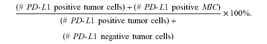
[0121] Another method for scoring/quantifying PD-L1 expression in a tumor is the "combined positive score" or "CPS," which refers to an algorithm for determining a PD-L1 expression score from a tumor sample of a patient. The CPS is useful in selecting patients for treatment with particular treatment regimens including methods of treatment comprising administration of an anti-PD-1 antibody in which expression of PD-L1 is associated with a higher response rate in a particular patient population relative to same patient population that does not express PD-L1. The CPS is determined by determining the number of viable PD-L1 positive tumor cells, the number of viable PD-L1 negative tumor cells, and the number of viable PD-L1 positive mononuclear inflammatory cells (MIC) in a tumor tissue from a patient having a tumor and calculating the CPS using the following formula:
( # PD - L 1 positive tumor cells ) + ( # PD - L 1 positive MIC ) ( # PD - L 1 positive tumor cells ) + ( # PD - L 1 negative tumor cells ) .times. 100 % . ##EQU00001##
[0122] In particular embodiments, the PD-L1 expression scoring method used is the "lymphoma proportion score." Lymphoma is characterized by a homogeneous population of confluent cells which efface the architecture of the lymph node or the architecture of metastatic site. The "LPS" or "lymphoma proportion score" is the percentage of this population of cells which express PD-L1. When determining the LPS, no attempt is made to distinguish the truly neoplastic cells from the reactive cells. PD-L1 expression is characterized by partial or complete membrane staining intensity.
[0123] Yet another scoring method for PD-L1 expression is the "TPS" or "tumor proportion score," which is the percentage of tumor cells expressing PD-L1 on the cell membrane. TPS typically includes the percentage of neoplastic cells expressing PD-L1 at any intensity (weak, moderate, or strong), which can be determining using an immunohistochemical assay using a diagnostic anti-human PD-L1 mAb, e.g. antibody 20C3 and antibody 22C3, described, supra. Cells are considered to express PD-L1 if membrane staining is present, including cells with partial membrane staining.
[0124] The level of PD-L mRNA expression may be compared to the mRNA expression levels of one or more reference genes that are frequently used in quantitative RT-PCR, such as ubiquitin C.
[0125] In some embodiments, a level of PD-L1 expression (protein and/or mRNA) by malignant cells and/or by infiltrating immune cells within a tumor is determined to be "overexpressed" or "elevated" based on comparison with the level of PD-L1 expression (protein and/or mRNA) by an appropriate control. For example, a control PD-L1 protein or mRNA expression level may be the level quantified in nonmalignant cells of the same type or in a section from a matched normal tissue. In some preferred embodiments, PD-L1 expression in a tumor sample is determined to be elevated if PD-L1 protein (and/or PD-L1 mRNA) in the sample is at least 10%, 20%, or 30% greater than in the control.
[0126] "Tissue section" refers to a single part or piece of a tissue sample, e.g., a thin slice of tissue cut from a sample of a normal tissue or of a tumor.
[0127] "Tumor" as it applies to a subject diagnosed with, or suspected of having, a cancer refers to a malignant or potentially malignant neoplasm or tissue mass of any size, and includes primary tumors and secondary neoplasms. A solid tumor is an abnormal growth or mass of tissue that usually does not contain cysts or liquid areas. Different types of solid tumors are named for the type of cells that form them. Examples of solid tumors are sarcomas, carcinomas, and lymphomas. Leukemias (cancers of the blood) generally do not form solid tumors (National Cancer Institute, Dictionary of Cancer Terms).
[0128] "Variable regions" or "V region" as used herein means the segment of IgG chains which is variable in sequence between different antibodies. It extends to Kabat residue 109 in the light chain and 113 in the heavy chain.
[0129] "RECIST 1.1 Response Criteria" as used herein means the definitions set forth in Eisenhauer, E. A. et al., Eur. J. Cancer 45:228-247 (2009) for target lesions or non-target lesions, as appropriate based on the context in which response is being measured.
II. PD-1 Antibodies and Antigen Binding Fragments Useful in the Invention
[0130] Examples of mAbs that bind to human PD-1, useful in the treatment methods, compositions, and uses of the invention, are described in U.S. Pat. Nos. 7,521,051, 8,008,449, and 8,354,509. Specific anti-human PD-1 mAbs useful as the PD-1 antagonist in the treatment methods, compositions, and uses of the present invention include: pembrolizumab (formerly known as MK-3475, SCH 900475 and lambrolizumab), a humanized IgG4 mAb with the structure described in WHO Drug Information, Vol. 27, No. 2, pages 161-162 (2013) and which comprises the heavy and light chain amino acid sequences shown in FIG. 1, and the humanized antibodies h409A11, h409A16 and h409A17, which are described in WO 2008/156712 and in Table 3.
[0131] In some embodiments of the treatment methods, compositions, kits, and uses of the present invention, the anti-PD-1 antibody, or antigen binding fragment thereof, comprises: (a) light chain CDRs comprising a sequence of amino acids as set forth in SEQ ID NOs: 1, 2 and 3 and heavy chain CDRs comprising a sequence of amino acids as set forth in SEQ ID NOs: 6, 7 and 8; or (b) light chain CDRs comprising a sequence of amino acids as set forth in SEQ ID NOs: 11, 12 and 13 and heavy chain CDRs comprising a sequence of amino acids as set forth in SEQ ID NOs: 14, 15 and 16. In some embodiments of the invention, the anti-PD-1 antibody or antigen binding fragment thereof is a human antibody. In other embodiments, the anti-PD-1 antibody or antigen binding fragment thereof is a humanized antibody. In other embodiments, the anti-PD-1 antibody or antigen binding fragment thereof is a chimeric antibody. In specific embodiments, the anti-PD-1 antibody or antigen binding fragment thereof is a monoclonal antibody.
[0132] In other embodiments of the treatment methods, compositions, kits and uses of the present invention, the PD-1 antibody, or antigen binding fragment thereof, specifically binds to human PD-1 and comprises (a) a heavy chain variable region comprising an amino acid sequence as set forth in SEQ ID NO:9, or a variant thereof, and (b) a light chain variable region comprising an amino acid sequence selected from the group consisting of SEQ ID NO:4 or a variant thereof; SEQ ID NO:22 or a variant thereof; and SEQ ID NO:23 or a variant thereof.
[0133] A variant of a heavy chain variable region sequence or full-length heavy chain sequence is identical to the reference sequence except having up to 17 conservative amino acid substitutions in the framework region (i.e., outside of the CDRs), and preferably has less than ten, nine, eight, seven, six or five conservative amino acid substitutions in the framework region. A variant of a light chain variable region sequence or full-length light chain sequence is identical to the reference sequence except having up to five conservative amino acid substitutions in the framework region (i.e., outside of the CDRs), and preferably has less than four, three or two conservative amino acid substitution in the framework region.
[0134] In another embodiment of the treatment methods, compositions, kits, and uses of the present invention, the PD-1 antibody or antigen-binding fragment thereof is a monoclonal antibody which specifically binds to human PD-1 and comprises (a) a heavy chain comprising or consisting of a sequence of amino acids as set forth in SEQ ID NO: 10, or a variant thereof, and (b) a light chain comprising or consisting of a sequence of amino acids as set forth in SEQ ID NO:5, or a variant thereof; SEQ ID NO:24, or a variant thereof, or SEQ ID NO:25, or a variant thereof.
[0135] In yet another embodiment of the treatment methods, compositions and uses of the invention, the PD-1 antibody or antigen-binding fragment thereof is a monoclonal antibody which specifically binds to human PD-1 and comprises (a) a heavy chain comprising or consisting of a sequence of amino acids as set forth in SEQ ID NO: 10 and (b) a light chain comprising or consisting of a sequence of amino acids as set forth in SEQ ID NO:5.
[0136] Table 3 below provides a list of the amino acid sequences of exemplary anti-PD-1 mAbs for use in the treatment methods, compositions, kits and uses of the present invention.
TABLE-US-00004 TABLE 3 Exemplary anti-human PD-1 antibodies A. Comprises light and heavy chain CDRs of hPD-1.09A in WO2008/156712 (light and heavy chain CDRs of pembrolizumab) CDRL1 SEQ ID NO: 1 CDRL2 SEQ ID NO: 2 CDRL3 SEQ ID NO: 3 CDRH1 SEQ ID NO: 6 CDRH2 SEQ ID NO: 7 CDRH3 SEQ ID NO: 8 B. Comprises light and heavy chain CDRs of hPD-1.08A in WO2008/156712 CDRL1 SEQ ID NO: 11 CDRL2 SEQ ID NO: 12 CDRL3 SEQ ID NO: 13 CDRH1 SEQ ID NO: 14 CDRH2 SEQ ID NO: 15 CDRH3 SEQ ID NO: 16 C. Comprises the mature h109A heavy chain variable region (V.sub.H) and one of the mature K09A light chain variable (V.sub.L) regions in WO 2008/156712 Heavy chain V.sub.H SEQ ID NO: 9 (V.sub.H of pembrolizumab) Light chain V.sub.L SEQ ID NO: 4 (V.sub.L of pembrolizumab) or SEQ ID NO: 22 or SEQ ID NO: 23 D. Comprises the mature 409 heavy chain and one of the mature K09A light chains in WO 2008/156712 Heavy chain SEQ ID NO: 10 (heavy chain of pembrolizumab) Light chain SEQ ID NO: 5 (light chain of pembrolizumab) or SEQ ID NO: 24 or SEQ ID NO: 25
III. Anti-CTLA4 Antibodies and Antigen Binding Fragments Useful in the Invention
[0137] In one embodiment of the treatment methods, compositions, kits and uses of the invention, the anti-CTLA-4 antibody is the human monoclonal antibody 10D1, now known as ipilimumab, and marketed as Yervoy.TM., which is disclosed in U.S. Pat. No. 6,984,720 and WHO Drug Information 19(4): 61 (2005). In another embodiment, the anti-CTLA-4 antibody is tremelimumab, also known as CP-675,206, which is an IgG2 monoclonal antibody which is described in U.S. Patent Application Publication No. 2012/263677, or PCT International Application Publication Nos. WO 2012/122444 or WO 2007/113648 A2.
[0138] In further embodiments of the treatment methods, compositions, kits, and uses of the present invention, the anti-CTLA4 antibody, or antigen binding fragment thereof, comprises: light chain CDRs comprising a sequence of amino acids as set forth in SEQ ID NOs: 26, 27 and 28 and heavy chain CDRs comprising a sequence of amino acids as set forth in SEQ ID NOs: 29, 30 and 31.
[0139] In other embodiments of the treatment methods, compositions, kits, and uses of the present invention, the anti-CTLA4 antibody is a monoclonal antibody, or antigen binding fragment thereof, which binds to human CTLA4 and comprises (a) a heavy chain variable region comprising an amino acid sequence as set forth in SEQ ID NO: 32 and (b) a light chain variable region comprising an amino acid sequence as set forth in SEQ ID NO: 33.
TABLE-US-00005 Exemplary anti-human CTLA4 antibodies A. Comprises light and heavy chain CDRs of ipilimumab CDRL1 RASQSVGSSYLA (SEQ ID NO: 26) CDRL2 GAFSRAT (SEQ ID NO: 27) CDRL3 QQYGSSPWT (SEQ ID NO: 28) CDRH1 SYTMH (SEQ ID NO: 29) CDRH2 FISYDGNNKYYADSVKG (SEQ ID NO: 30) CDRH3 TGWLGPFDY (SEQ ID NO: 31) C. Comprises the mature heavy chain variable region and the mature light chain variable region of ipilimumab Heavy QVQLVESGGGVVQPGRSLRLSCAASGFTFSSYTMHWVRQA chain PGKGLEWVTFISYDGNNKYYADSVKGRFTISRDNSKNTLY VR LQMNSLRAEDTAIYYCARTGWLGPFDYWGQGTLVTVSS (SEQ ID NO: 32) Light EIVLTQSPGT LSLSPGERATLSCRASQSVGSSYLAWYQQK chain PGQAPRLLIYGAFSRATGIPDRFSGSGSGTDFTLTISRLE VR PEDFAVYYCQQYGSSPWTFGQGTKVEIK (SEQ ID NO: 33) D. Comprises the mature heavy chain and the mature light chain of ipilimumab Heavy SEQ ID NO: 34 chain Light SEQ ID NO: 35 chain
[0140] In one embodiment of the treatment methods, compositions, kits and uses of the invention, the anti-CTLA-4 antibody is a monoclonal antibody that comprises a heavy chain having the amino acid sequence set forth in SEQ ID NO:34 and a light chain comprising the amino acid sequence set forth in SEQ ID NO:35. In some embodiments, the anti-CTLA4 antibody is an antigen binding fragment of SEQ ID NO:34 and/or SEQ ID NO:35, wherein the antigen binding fragment specifically binds to CTLA4.
[0141] In one embodiment of the treatment methods, compositions, kits and uses of the invention, the anti-CTLA-4 antibody is any of the anti-CTLA-4 antibodies, or antigen binding fragments thereof, disclosed in International Application Publication No. WO 2016/015675 A1. In one embodiment, the anti-CTLA4 antibody is a monoclonal antibody which comprises the following CDR's:
TABLE-US-00006 CDRH1 comprising the amino acid sequence (SEQ ID NO: 36) GFTFSDNW; CDRH2 comprising the amino acid sequence (SEQ ID NO: 37) IRNKPYNYET; CDRH3 comprising the amino acid sequence (SEQ ID NO: 38) TAQFAY; and/or CDRL1 comprising the amino acid sequence (SEQ ID NO: 39) ENIYGG; CDRL2 comprising the amino acid sequence (SEQ ID NO: 40) GAT; and CDRL3 comprising an amino acid sequence selected from: (SEQ ID NO: 41) QNVLRSPFT; (SEQ ID NO: 42) QNVLSRHPG; or (SEQ ID NO: 43) QNVLSSRPG.
[0142] In one embodiment of the treatment methods, compositions, kits and uses of the invention, the anti-CTLA4 antibody is 8D2/8D2 (RE) or a variant thereof, 8D2H1L1 or a variant thereof, 8D2H2L2 or a variant thereof, 8D3H3L3 or a variant thereof, 8D2H2L15 or a variant thereof, or 8D2H2117 or a variant thereof.
TABLE-US-00007 Antibody V.sub.H V.sub.L 8D2/8D2 EVKLDETGGGLVQPGRPMKLSCVAS DIQMTQSPASLSASVGETVTITCGT (RE) GFTFSDNWMNWVRQSPEKGLEWLA SENIYGGLNWYQRKQGKSPQLLIF QIRNKPYNYETYYSDSVKGRFTISRD GATNLADGMSSRFSGSGSGRQYSL DSKSSVYLQMNNLRGEDMGIYYCTA KISSLHPDDVATYYCQNVLRSPFTF QFAYWGQGTLVTVSA (SEQ ID NO: 44) GSGTKLEI (SEQ ID NO: 45) 8D2H1L1 EVQLVESGGGLVQPGGSMRLSCAAS DIQMTQSPSSLSASVGDRVTITCRT GFTFSDNWMNWVRQAPGKGLEWLA SENIYGGLNWYQRKQGKSPKLLIY QIRNKPYNYETYYSDSVKGRFTISRD GATNLASGMSSRFSGSGSGTDYTL DSKNSVYLQMNSLKTEDTGVYYCTA KISSLHPDDVATYYCQNVLRSPFTF QFAYWGQGTLVTVSS (SEQ ID NO: 46) GSGTKLEIK (SEQ ID NO: 47) 8D2H2L2 EVQLVESGGGLVQPGGSMRLSCAAS DIQMTQSPSSLSASVGDRVTITCRT GFTFSDNWMNWVRQAPGKGLEWLA SENIYGGLNWYQRKPGKSPKLLIY QIRNKPYNYETYYSASVKGRFTISRD GATNLASGVSSRFSGSGSGTDYTL DSKNSVYLQMNSLKTEDTGVYYCTA TISSLQPEDVATYYCQNVLRSPFTF QFAYWGQGTLVTVSS (SEQ ID NO: 48) GSGTKLEIK (SEQ ID NO: 49) 8D2H2L2 EVQLVESGGGLVQPGGSLRLSCAASG DIQMTQSPSSLSASVGDRVTITCRT VARIANT FTFSDNWMNWVRQAPGKGLEWLAQ SENIYGGLNWYQRKPGKSPKLLIY 1 IRNKPYNYETYYSASVKGRFTISRDD GATNLASGVSSRFSGSGSGTDYTL SKNSVYLQMNSLKTEDTGVYYCTAQ TISSLQPEDVATYYCQNVLRSPFTF FAYWGQGTLVTVSS (SEQ ID NO: 50) GSGTKLEIK (SEQ ID NO: 49) 8D3H3L3 EVQLVESGGGLVQPGGSLRLSCAAS DIQMTQSPSSLSASVGDRVTITCRA GFTFSDNWMNWVRQAPGKGLEWVA SENIYGGLNWYQQKPGKAPKLLIY QIRNKPYNYETEYAASVKGRFTISRD GATSLASGVPSRFSGSGSGTDYTLT DSKNSAYLQMNSLKTEDTAVYYCTA ISSLQPEDFATYYCQNVLRSPFTFG QFAYWGQGTLVTVSS (SEQ ID NO: 51) SGTKLEIK (SEQ ID NO: 52) 8D2H2L15 EVQLVESGGGLVQPGGSMRLSCAAS DIQMTQSPSSLSASVGDRVTITCRT GFTFSDNWMNWVRQAPGKGLEWLA SENIYGGLNWYQRKPGKSPKLLIY QIRNKPYNYETYYSASVKGRFTISRD GATNLASGVSSRFSGSGSGTDYTL DSKNSVYLQMNSLKTEDTGVYYCTA TISSLQPEDVATYYCQNVLSRHPGF QFAYWGQGTLVTVSS (SEQ ID NO: 53) GSGTKLEIK (SEQ ID NO: 54) 8D2H2L17 EVQLVESGGGLVQPGGSMRLSCAAS DIQMTQSPSSLSASVGDRVTITCRT GFTFSDNWMNWVRQAPGKGLEWLA SENIYGGLNWYQRKPGKSPKLLIY QIRNKPYNYETYYSASVKGRFTISRD GATNLASGVSSRFSGSGSGTDYTL DSKNSVYLQMNSLKTEDTGVYYCTA TISSLQPEDVATYYCQNVLSSRPGF QFAYWGQGTLVTVSS (SEQ ID NO: 55) GSGTKLEIK (SEQ ID NO: 56)
[0143] In another embodiment of the treatment methods, compositions, kits and uses of the invention, the anti-CTLA4 antibody is a variant of 8D2/8D2 (RE), a variant of 8D2H1L1, a variant of 8D2H2L2, a variant of 8D2H2L15, or a variant of 8D2H2117, wherein the methionine (Met) at position 18 in the VH chain amino acid sequence is independently substituted with an amino acid selected from: Leucine (Leu), Valine (Val), Isoleucine (Ile) or Alanine (Ala). In embodiments of the invention, the anti-CTLA4 antibody comprises the sequence of the 8D2H2L2 Variant 1 as set forth in the table above.
[0144] In another embodiment of the treatment methods, compositions, kits and uses of the invention, the anti-CTLA4 antibody is 8D2H2L2 Variant 1, having the full heavy chain amino acid sequence set forth in SEQ ID NO: 57 and the full light chain sequence set forth in SEQ ID NO: 58.
TABLE-US-00008 Full Antibody Full Heavy Chain Light Chain 8D2H2L2 EVQLVESGGGLVQPGGSLRLSCAAS DIQMTQSPSSLSAS VARIANT GFTFSDNWMNWVRQAPGKGLEWLA VGDRVTITCRTSEN 1 QIRNKPYNYETYYSASVKGRFTISRD IYGGLNWYQRKPGK DSKNSVYLQMNSLKTEDTGVYYCTA SPKLLIYGATNLAS QFAYWGQGTLVTVSSASTKGPSVFPL GVSSRFSGSGSGTD APSSKSTSGGTAALGCLVKDYFPEPV YTLTISSLQPEDVA TVSWNSGALTSGVHTFPAVLQSSGL TYYCQNVLRSPFTF YSLSSVVTVPSSSLGTQTYICNVNHK GSGTKLEIKRTVAA PSNTKVDKKVEPKSCDKTHTCPPCPA PSVFIFPPSDEQLK PELLGGPSVFLFPPKPKDTLMISRTPE SGTASVVCLLNNFY VTCVVVDVSHEDPEVKFNWYVDGV PREAKVQWKVDNAL EVHNAKTKPREEQYNSTYRVVSVLT QSGNSQESVTEQDS VLHQDWLNGKEYKCKVSNKALPAPI KDSTYSLSSTLTLS EKTISKAKGQPREPQVYTLPPSRDELT KADYEKHKVYACEV KNQVSLTCLVKGFYPSDIAVEWESN THQGLSSPVTKSFN GQPENNYKTTPPVLDSDGSFFLYSKL RGEC (SEQ ID TVDKSRWQQGNVFSCSVMHEALHN NO: 58) HYTQKSLSLSPGK (SEQ ID NO: 57)
[0145] In one embodiment of the treatment methods, compositions, kits and uses of the invention, the anti-CTLA4 antibody is any of the anti-CTLA4 antibodies, or antigen binding fragments thereof, described as disclosed in International Application Publication No. WO 2018/035710 A1, published Mar. 1, 2018.
IV. Methods and Uses of the Invention
[0146] The invention provides a method of treating cancer in a human patient comprising administering about 400 mg of an anti-PD-1 antibody, or antigen-binding fragment thereof, to the patient once every about six weeks, wherein the anti-PD-1 antibody or antigen binding fragment thereof comprises: (a) light chain complementarity determining regions (CDRs) comprising a sequence of amino acids as set forth in SEQ ID NOs: 1, 2 and 3 and heavy chain CDRs comprising a sequence of amino acids as set forth in SEQ ID NOs: 6, 7 and 8; or (b) light chain CDRs comprising a sequence of amino acids as set forth in SEQ ID NOs: 11, 12 and 13 and heavy chain CDRs comprising a sequence of amino acids as set forth in SEQ ID NOs: 14, 15 and 16. In particular embodiments, the anti-PD-1 antibody, or antigen binding fragment thereof, is pembrolizumab.
[0147] Also provided is a method of treating cancer in a human patient comprising administering about 400 mg of an anti-PD-1 antibody, or antigen-binding fragment thereof, to the patient once every six weeks, wherein the anti-PD-1 antibody or antigen binding fragment thereof comprises: (a) light chain complementarity determining regions (CDRs) comprising a sequence of amino acids as set forth in SEQ ID NOs: 1, 2 and 3 and heavy chain CDRs comprising a sequence of amino acids as set forth in SEQ ID NOs: 6, 7 and 8; or (b) light chain CDRs comprising a sequence of amino acids as set forth in SEQ ID NOs: 11, 12 and 13 and heavy chain CDRs comprising a sequence of amino acids as set forth in SEQ ID NOs: 14, 15 and 16; and wherein the anti-CTLA4 antibody or antigen binding fragment thereof comprises: (c) light chain CDRs comprising a sequence of amino acids as set forth in SEQ ID NOs: 39, 40 and 41 and heavy chain CDRs comprising a sequence of amino acids as set forth in SEQ ID NOs: 36, 37, and 38; (d) light chain CDRs comprising a sequence of amino acids as set forth in SEQ ID NOs: 39, 40 and 42 and heavy chain CDRs comprising a sequence of amino acids as set forth in SEQ ID NOs: 36, 37, and 38; or (e) light chain CDRs comprising a sequence of amino acids as set forth in SEQ ID NOs: 39, 40 and 43 and heavy chain CDRs comprising a sequence of amino acids as set forth in SEQ ID NOs: 36, 37, and 38.
[0148] In some embodiments, the anti-PD-1 antibody, or antigen binding fragment thereof, comprises light chain CDRs comprising a sequence of amino acids as set forth in SEQ ID NOs: 1, 2, and 3 and heavy chain CDRs comprising a sequence of amino acids as set forth in SEQ ID NOs: 6, 7, and 8; and the anti-CTLA4 antibody, or antigen binding fragment thereof, comprises light chain CDRs comprising a sequence of amino acids as set forth in SEQ ID NOs: 39, 40, and 43 and heavy chain CDRs comprising a sequence of amino acids as set forth in SEQ ID NOs: 36, 37, and 38.
[0149] In some embodiments of the invention, the anti-PD-1 antibody, or antigen binding fragment thereof, and the anti-CTLA4 antibody, or antigen binding fragment thereof, are administered to the patient once every approximately six weeks for 12 weeks or more. In other embodiments, the anti-PD-1 antibody, or antigen binding fragment and the anti-CTLA4 antibody, or antigen binding fragment thereof, are administered to the patient once every six weeks for 18 weeks or more, 24 weeks or more, 30 weeks or more, 36 weeks or more, 42 weeks or more, 48 weeks or more, 54 weeks or more, 60 weeks or more, 66 weeks or more, 72 weeks or more, 78 weeks or more, 84 weeks or more, or 90 weeks or more. In one embodiment, the administration occurs on the same day. In a sub-embodiment, the anti-PD-1 antibody, or antigen binding fragment thereof, and the anti-CTLA4 antibody, or antigen binding fragment thereof, are administered on the same day simultaneously (e.g., in a single formulation or concurrently as separate formulations). In an alternative embodiment, the anti-PD-1 antibody or antigen binding fragment thereof and the anti-CTLA4 antibody or antigen binding fragment thereof are administered sequentially on the same day (e.g., as separate formulations), in either order. In one embodiment of same day sequential administration, the anti-PD-1 antibody or antigen binding fragment thereof is administered first. In another embodiment of same day sequential administration, the anti-CTLA4 antibody or antigen binding fragment thereof is administered first.
[0150] In a first embodiment (Embodiment E1), the invention comprises a method of treating cancer in a human patient comprising administering 400 mg of an anti-PD-1 antibody (e.g., pembrolizumab), or antigen binding fragment thereof, to the patient once every approximately six weeks. In a further embodiment of (Embodiment 1), the invention further comprises administering 25 mg, 50 mg, 75 mg, or 100 mg of an anti-CTLA4 antibody, or antigen binding fragment thereof, to the patient once every approximately six weeks. In one embodiment, 25 mg of the anti-CTLA4 antibody is administered once every approximately six weeks. In one embodiment, 50 mg of the anti-CTLA4 antibody is administered once every six approximately weeks. In another embodiment, 75 mg of the anti-CTLA4 antibody is administered once every approximately six weeks. In a further embodiment, 100 mg of the anti-CTLA4 antibody is administered once every approximately six weeks. In further embodiment, the cancer is PD-1/PD-L1 refractory (e.g., it is a cancer that has not been responsive to previous treatment with an anti-PD-1 or an anti-PD-L1 agent). In a further embodiment, the cancer is PD-1/PD-L1 refractory melanoma. In another embodiment, the cancer is melanoma, non-small cell lung cancer, head and neck cancer, urothelial cancer, breast cancer, gastrointestinal cancer, multiple myeloma, hepatocellular cancer, non-Hodgkin lymphoma, renal cancer, Hodgkin lymphoma, mesothelioma, ovarian cancer, small cell lung cancer, esophageal cancer, anal cancer, biliary tract cancer, colorectal cancer, cervical cancer, thyroid cancer, salivary cancer, pancreatic cancer, a tumor of the brain, glioblastoma, a sarcoma, a tumor of the bone, or Merkel cell carcinoma.
[0151] In a second embodiment (Embodiment E2), the invention comprises a method of treating unresectable or metastatic melanoma in a human patient comprising administering 400 mg of an anti-PD-1 antibody (e.g., pembrolizumab), or antigen binding fragment thereof, to the patient once every approximately six weeks.
[0152] In a third embodiment (Embodiment E3), the invention comprises a method of treating metastatic non-small cell lung cancer (NSCLC) in a human patient comprising administering 400 mg of an anti-PD-1 antibody (e.g., pembrolizumab), or antigen binding fragment thereof, to the patient once every approximately six weeks.
[0153] In a sub-embodiment of Embodiment E3 (Embodiment E3-A), the patient has a tumor with high PD-L1 expression [(Tumor Proportion Score (TPS).gtoreq.50%)] and was not previously treated with platinum-containing chemotherapy.
[0154] In a further sub-embodiment of Embodiment E3 (Embodiment E3-B), the patient has a tumor with PD-L1 expression (TPS.gtoreq.1%) and was previously treated with platinum-containing chemotherapy. In specific embodiments of Embodiment E3-B, the patient had disease progression on or after receiving platinum-containing chemotherapy.
[0155] In another sub-embodiment of Embodiment E3 (Embodiment E3-C), the patient has a tumor with PD-L1 expression (TPS.gtoreq.1%) and was not previously treated with platinum-containing chemotherapy.
[0156] In yet another sub-embodiment of Embodiment E3 (Embodiment E3-D), the patient's tumor is not tested for PD-L1 expression. In this embodiment, the patient is treated with the anti-PD-1 antibody, or antigen binding fragment thereof, regardless of PD-L1 expression. In specific embodiments, the patient was not previously treated with platinum-containing chemotherapy.
[0157] In certain embodiments of Embodiment E3 (including Embodiment E3-A, E3-B, and E3-C), the PD-L1 TPS is determined by an FDA-approved test.
[0158] In certain embodiments of Embodiment E3 (including Embodiment E3-A, E3-B, E3-C, and E3-D), the patient's tumor has no EGFR or ALK genomic aberrations.
[0159] In certain embodiments of Embodiment E3 (including Embodiment E3-A, E3-B, E3-C, and E3-D), the patient's tumor has an EGFR or ALK genomic aberration and had disease progression on or after receiving treatment for the EGFR or ALK aberration(s) prior to receiving the anti-PD-1 antibody, or antigen binding fragment thereof.
[0160] In a fourth embodiment (Embodiment E4), the invention comprises a method of treating metastatic non-small cell lung cancer (NSCLC) in a human patient comprising: (1) administering 400 mg of an anti-PD-1 antibody (e.g., pembrolizumab), or antigen binding fragment thereof, to the patient once every approximately six weeks, and (2) administering pemetrexed and carboplatin to the patient. In sub-embodiments of Embodiment E4, the patient was not previously treated with an anti-cancer therapeutic prior to starting the combination treatment regimen with the anti-PD-1 antibody, or antigen binding fragment thereof, pemetrexed and carboplatin.
[0161] In certain embodiments of Embodiment E3 and E4 (including sub-embodiments thereof), the patient has nonsquamous non-small cell lung cancer.
[0162] In sub-embodiments of Embodiment E4, pemetrexed is administered to the patient in an amount of 500 mg/m.sup.2.
[0163] In sub-embodiments of Embodiment E4, pemetrexed is administered to the patient via intravenous infusion every 21 days. In specific embodiments, the infusion time is about 10 minutes.
[0164] In sub-embodiments of Embodiment E4 (Embodiment E4-A), the invention further comprises administering about 400 .mu.g to about 1000 .mu.g of folic acid to the patient once per day, beginning about 7 days prior to administering pemetrexed to the patient and continuing until about 21 days after the patient is administered the last dose of pemetrexed. In certain embodiments the folic acid is administered orally.
[0165] In sub-embodiments of Embodiments E4 and E4-A (Embodiment E4-B), the invention further comprises administering about 1 mg of vitamin B.sub.12 to the patient about 1 week prior to the first administration of pemetrexed and about every three cycles of pemetrexed administration (i.e., approximately every 9 weeks). In certain embodiments the vitamin B.sub.12 is administered intramuscularly.
[0166] In sub-embodiments of Embodiments E4, E4-A and E4-B (Embodiment E4-C), the invention further comprises administering about 4 mg of dexamethasone to the patient twice a day on the day before, the day of, and the day after pemetrexed administration. In certain embodiments the dexamethasone is administered orally.
[0167] In a fifth embodiment (Embodiment E5), the invention comprises a method of treating recurrent or metastatic head and neck squamous cell cancer (HNSCC) in a human patient comprising administering 400 mg of an anti-PD-1 antibody (e.g., pmebrolizumab), or antigen binding fragment thereof, to the patient once every approximately six weeks.
[0168] In sub-embodiments of Embodiment E5, the patient was previously treated with platinum-containing chemotherapy. In certain embodiments, the patient had disease progression on or after platinum-containing chemotherapy.
[0169] In a sixth embodiment (Embodiment E6), the invention comprises a method of treating refractory classical Hodgkin lymphoma (cHL) in a human patient comprising administering 400 mg of an anti-PD-1 antibody (e.g., pembrolizumab), or antigen binding fragment thereof, to the patient once every approximately six weeks.
[0170] In a seventh embodiment (Embodiment E7), the invention comprises a method of treating classical Hodgkin lymphoma (cHL) in a human patient comprising administering 400 mg of an anti-PD-1 antibody (e.g., pembrolizumab), or antigen binding fragment thereof, to the patient once every approximately six weeks, wherein the patient has relapsed after (a) one or more lines of therapy for cHL, (b) 2 or more lines of therapy for cHL, or (c) 3 or more lines of therapy for cHL.
[0171] In sub-embodiments of Embodiments E6 and E7, the patient is an adult patient.
[0172] In alternative sub-embodiments of Embodiments E6 and E7, the patient is a pediatric patient.
[0173] In an eighth embodiment (Embodiment E8), the invention comprises a method of treating locally advanced or metastatic urothelial carcinoma in a human patient comprising administering 400 mg of an anti-PD-1 antibody (e.g., pembrolizumab), or antigen binding fragment thereof, to the patient once every approximately six weeks.
[0174] In sub-embodiments of Embodiment E8, the patient is not eligible for cisplatin-containing chemotherapy.
[0175] In sub-embodiments of Embodiment E8, the patient had disease progression during or following platinum-containing chemotherapy or within 12 months of neoadjuvant or adjuvant treatment with platinum-containing chemotherapy.
[0176] In sub-embodiments of Embodiment E8, the patient's tumor expresses PD-L1 (CPS.gtoreq.10).
[0177] In a ninth embodiment (Embodiment E9), the invention comprises a method of treating unresectable or metastatic, microsatellite instability-high (MSI-H) or mismatch repair (MMR) deficient solid tumors in a human patient comprising administering 400 mg of an anti-PD-1 antibody (e.g., pembrolizumab), or antigen binding fragment thereof, to the patient once every approximately six weeks.
[0178] In a sub-embodiment of Embodiment E9, the patient had disease progression following prior anti-cancer treatment.
[0179] In a tenth embodiment (Embodiment E10), the invention comprises a method of treating unresectable or metastatic, MSI-H or MMR deficient colorectal cancer in a human patient comprising administering 400 mg of an anti-PD-1 antibody (e.g., pembrolizumab), or antigen binding fragment thereof, to the patient once every approximately six weeks.
[0180] In a sub-embodiment of Embodiment E10, the patient had disease progression following prior treatment with a fluoropyrimidine, oxaliplatin, and irinotecan.
[0181] In an eleventh embodiment (Embodiment E11), the invention comprises a method of treating recurrent locally advanced or metastatic gastric cancer in a human patient comprising administering 400 mg of an anti-PD-1 antibody (e.g., pembrolizumab), or antigen binding fragment thereof, to the patient once every approximately six weeks.
[0182] In a twelfth embodiment (Embodiment E12), the invention comprises a method of treating recurrent locally advanced or metastatic gastroesophageal junction adenocarcinoma in a human patient comprising administering 400 mg of an anti-PD-1 antibody (e.g., pembrolizumab), or antigen binding fragment thereof, to the patient once every approximately six weeks.
[0183] In sub-embodiments of Embodiments E11 and E12, the patient's tumor expresses PD-L1 [Combined Positive Score (CPS).gtoreq.1].
[0184] In sub-embodiments of Embodiments E11 and E12, the patient had disease progression on or after one or more prior lines of therapy. In specific embodiments, the prior lines of therapy include fluoropyrimidine- and platinum-containing chemotherapy.
[0185] In sub-embodiments of Embodiments E11 and E12, the patient had disease progression on or after two or more prior lines of therapy including fluoropyrimidine- and platinum-containing chemotherapy.
[0186] In sub-embodiments of Embodiments E11 and E12, the patient had disease progression on or after one or more prior lines of therapy including HER2/neu-targeted therapy.
[0187] In sub-embodiments of Embodiments E11 and E12, the patient had disease progression on or after two or more prior lines of therapy including HER2/neu-targeted therapy.
[0188] In a thirteenth embodiment (Embodiment E13), the invention comprises a method of treating cancer in a human patient comprising administering 400 mg of an anti-PD-1 antibody (e.g., pembrolizumab), or antigen binding fragment thereof, to the patient once every approximately six weeks, wherein the patient has a cancer selected from the group consisting of: melanoma, lung cancer, head and neck cancer, bladder cancer, breast cancer, gastrointestinal cancer, multiple myeloma, hepatocellular cancer, lymphoma, renal cancer, mesothelioma, ovarian cancer, esophageal cancer, anal cancer, biliary tract cancer, colorectal cancer, cervical cancer, thyroid cancer, and salivary cancer.
[0189] In a fourteenth embodiment (Embodiment E14), the invention comprises a method of treating cancer in a human patient comprising administering 400 mg of an anti-PD-1 antibody (e.g., pembrolizumab), or antigen binding fragment thereof, to the patient once every approximately six weeks, wherein the patient has small-cell lung cancer.
[0190] In a fifteenth embodiment (Embodiment E15), the invention comprises a method of treating non-Hodgkin lymphoma in a human patient comprising administering 400 mg of an anti-PD-1 antibody (e.g., pembrolizumab), or antigen binding fragment thereof, to the patient once every approximately six weeks.
[0191] In a sub-embodiment of Embodiment E15, the non-Hodgkin lymphoma is primary mediastinal large B-cell lymphoma (PMBCL). In some embodiments where the patient has PMBCL, the patient has refractory PMBCL. In some embodiments, the patient has relapsed after one or more prior lines of therapy. In some embodiments, the patient has relapsed after two or more prior lines of therapy. In some embodiments, the patient was not previously treated with another line of therapy.
[0192] In a sixteenth embodiment (Embodiment E16), the invention comprises a method of treating metastatic squamous NSCLC in a human patient comprising: (1) administering 400 mg of an anti-PD-1 antibody (e.g., pembrolizumab), or antigen binding fragment thereof, to the patient once every approximately six weeks, and (2) administering (i) carboplatin and paclitaxel or (ii) carboplatin and nab-paclitaxel to the patient.
[0193] In a seventeenth embodiment (Embodiment E17), the invention comprises a method of treating Merkel cell carcinoma (MCC) in a human patient comprising administering 400 mg of an anti-PD-1 antibody (e.g., pembrolizumab), or antigen binding fragment thereof, to the patient once every approximately six weeks. In particular sub-embodiments of Embodiment E17, the cancer is recurrent, locally advanced MCC. In particular sub-embodiments of Embodiment E17, the cancer is metastatic MCC.
[0194] In sub-embodiments of Embodiment E17, the patient is an adult patient. In alternative sub-embodiments of Embodiment E17, the patient is a pediatric patient.
[0195] In a eighteenth embodiment (Embodiment E18), the invention comprises a method for adjuvant therapy of melanoma in a human patient comprising administering 400 mg of an anti-PD-1 antibody (e.g., pembrolizumab), or antigen binding fragment thereof, to a patient once every approximately six weeks, wherein the patient has previously had one or more melanoma lesions resected. In sub-embodiments of Embodiment E18, the method comprises treating resected high-risk stage III melanoma.
[0196] In a nineteenth embodiment (Embodiment E19), the invention comprises a method of treating hepatocellular carcinoma (HCC) in a human patient comprising administering 400 mg of an anti-PD-1 antibody (e.g., pembrolizumab), or antigen binding fragment thereof, to the patient once every approximately six weeks. In some embodiments of embodiment E19, the patient was previously treated with sorafenib.
[0197] In a twentieth embodiment (Embodiment E20), the invention comprises a method of treating renal cell carcinoma (RCC) in a human patient comprising administering 400 mg of an anti-PD-1 antibody (e.g., pembrolizumab), or antigen binding fragment thereof, to the patient once every approximately six weeks.
[0198] In sub-embodiments, of Embodiment E20, the cancer is advanced clear cell RCC.
[0199] In sub-embodiments of Embodiment E20, the patient has advanced or metastatic renal cell carcinoma (RCC).
[0200] In sub-embodiments, of Embodiment E20 (Embodiment E20A), the patient is further treated with axitinib. In sub-embodiments of the invention, axitinib is taken orally.
[0201] In particular embodiments of Embodiment E20A, 5 mg axitinib is taken by the patient approximately every 12 hours or twice a day.
[0202] In alternative embodiments of Embodiment E20A, the axitinib dosage is 2.5 mg, 3 mg, 7 mg, or 10 mg twice daily.
[0203] In a twenty-first embodiment (Embodiment E21), the invention comprises a method of treating breast cancer in a human patient comprising administering 400 mg of an anti-PD-1 antibody (e.g., pembrolizumab), or antigen binding fragment thereof, to the patient once every approximately six weeks.
[0204] In a sub-embodiment of Embodiment E21, the breast cancer is triple negative breast cancer.
[0205] In a sub-embodiment of Embodiment E21, the breast cancer is ER+/HER2- breast cancer.
[0206] In a twenty-second embodiment (Embodiment E22), the invention comprises a method of treating nasopharyngeal cancer in a human patient comprising administering 400 mg of an anti-PD-1 antibody (e.g., pembrolizumab), or antigen binding fragment thereof, to the patient once every approximately six weeks.
[0207] In a twenty-third embodiment (Embodiment E23), the invention comprises a method of treating thyroid cancer in a human patient comprising administering 400 mg of an anti-PD-1 antibody (e.g., pembrolizumab), or antigen binding fragment thereof, to the patient once every approximately six weeks.
[0208] In a twenty-fourth embodiment (Embodiment E24), the invention comprises a method of treating salivary cancer in a human patient comprising administering 400 mg of an anti-PD-1 antibody (e.g., pembrolizumab), or antigen binding fragment thereof, to the patient once every approximately six weeks.
[0209] In a twenty-fifth embodiment (Embodiment E25), the invention comprises a method of treating cancer in a human patient comprising administering 400 mg of an anti-PD-1 antibody (e.g., pembrolizumab), or antigen binding fragment thereof, to the patient once every approximately six weeks, wherein the cancer is selected from the group consisting of: melanoma, non-small cell lung cancer, relapsed or refractory classical Hodgkin lymphoma, primary mediastinal large B-cell lymphoma, head and neck squamous cell cancer, urothelial carcinoma, esophageal cancer, gastric cancer, cervical cancer, PMBCL, MSI-H cancer, hepatocellular carcinoma, and Merkel cell carcinoma.
[0210] In a twenty-sixth embodiment (Embodiment E26), the invention comprises a method of treating cancer in a human patient comprising administering 400 mg of an anti-PD-1 antibody (e.g., pembrolizumab), or antigen binding fragment thereof, to the patient once every approximately six weeks, wherein the cancer is a Heme malignancy.
[0211] In a sub-embodiment of Embodiment E26, the heme malignancy is selected from the group consisting of: acute lymphoblastic leukemia (ALL), acute myeloid leukemia (AML), chronic lymphocytic leukemia (CLL), chronic myeloid leukemia (CML), diffuse large B-cell lymphoma (DLBCL), EBV-positive DLBCL, primary mediastinal large B-cell lymphoma, T-cell/histiocyte-rich large B-cell lymphoma, follicular lymphoma, Hodgkin's lymphoma (HL), mantle cell lymphoma (MCL), multiple myeloma (MM), myeloid cell leukemia-1 protein (MCL-1), myelodysplastic syndrome (MDS), non-Hodgkin lymphoma (NHL), and small lymphocytic lymphoma (SLL).
[0212] In a twenty-seventh embodiment (Embodiment E27), the invention comprises a method of treating cancer in a human patient comprising administering 400 mg of an anti-PD-1 antibody (e.g., pembrolizumab), or antigen binding fragment thereof, to the patient once every approximately six weeks, wherein the patient has a tumor with a high mutational burden.
[0213] In specific embodiments, a high mutational burden is at least about 10 mutations per megabase of genome examined, at least about 11 mutations per megabase of genome examined, at least about 12 mutations per megabase of genome examined, or at least about 13 mutations per megabase of genome examined.
[0214] In a twenty-eighth embodiment (Embodiment E28), the invention comprises a method of treating esophageal cancer in a human patient comprising administering 400 mg of an anti-PD-1 antibody (e.g., pembrolizumab), or antigen binding fragment thereof, to the patient once every approximately six weeks.
[0215] In sub-embodiments of Embodiment E28, the patient progressed with one previous line of standard therapy prior to receiving the anti-PD-1 antibody, or antigen binding fragment thereof. In a further embodiment, the patient progressed with one or more lines of standard therapy prior to receiving the anti-PD-1 antibody, or antigen binding fragment thereof. In another embodiment, the patient progressed with two or more lines of standard therapy prior to receiving the anti-PD-1 antibody, or antigen binding fragment thereof. In particular embodiments, the standard therapy includes one or more of: paclitaxel, docetaxel, or irinotecan.
[0216] In sub-embodiments of Embodiment E28, the patient has advanced or metastatic adenocarcinoma or squamous cell carcinoma of the esophagus.
[0217] In sub-embodiments of Embodiment E28, the patient has advanced or metastatic Siewert type I adenocarcinoma of the esophagogastric junction.
[0218] In sub-embodiments of Embodiment E28, the patient's tumor expresses PD-1.1 (Combined Positive Score [CPS].gtoreq.10).
[0219] In a twenty-ninth embodiment (Embodiment E29), the invention comprises a method of treating high-risk non-muscle invasive bladder cancer (NMIBC) in a human patient comprising administering 400 mg of an anti-PD-1 antibody (e.g., pembrolizumab), or antigen binding fragment thereof, to the patient once every approximately six weeks. In some embodiments, the patient has NMIBC with carcinoma in situ (CIS) or CIS plus papillary disease.
[0220] In a sub-embodiment of Embodiment E29, the patient was previously treated with standard therapy prior to being treated with the anti-PD-1 antibody, or antigen binding fragment thereof. In some embodiments, the prior therapy is Bacillus Calmette-Guerin (BCG) therapy. In particular embodiments, the patient did not respond to BCG therapy. In some embodiments, the patient was ineligible for radical cystectomy or chose not to undergo radical cystectomy.
[0221] In any of the methods of the invention described above (including Embodiments E1-E29), the PD-1 antibody or antigen binding fragment is any of the antibodies or antigen-binding fragments described in Section II of the Detailed Description of the Invention "PD-1 Antibodies and Antigen Binding Fragments Useful in the Invention" herein.
[0222] Embodiments of any of the methods of the invention described above (including Embodiments E1-E29) may further comprise administering about 25 mg, 50 mg, 75 mg, or 100 mg of an anti-CTLA4 antibody or antigen binding fragment thereof once every approximately six weeks. In one embodiment, 25 mg of the anti-CTLA4 antibody is administered once every approximately six weeks. In one embodiment, 50 mg of the anti-CTLA4 antibody is administered once every approximately six weeks. In another embodiment, 75 mg of the anti-CTLA4 antibody is administered once every approximately six weeks. In a further embodiment, 100 mg of the anti-CTLA4 antibody is administered once every approximately six weeks. In one embodiment, 25 mg of the anti-CTLA4 antibody is administered once every six weeks. In one embodiment, 50 mg of the anti-CTLA4 antibody is administered once every six weeks. In another embodiment, 75 mg of the anti-CTLA4 antibody is administered once every six weeks. In a further embodiment, 100 mg of the anti-CTLA4 antibody is administered once every six weeks. In some embodiments of any of the above methods, the anti-CTLA4 antibody and the anti-PD-1 antibody are co-administered together. In other embodiments, the anti-CTLA4 antibody and the anti-PD-1 antibody are co-formulated. In any of the methods of the invention described above, the anti-CTLA4 antibody or antigen binding fragment is any of the antibodies or antigen-binding fragments described in Section III of the Detailed Description of the Invention "Anti-CLTA4 Antibodies and Antigen Binding Fragments Useful in the Invention" herein.
[0223] In some embodiments, the anti-PD-1 antibody is pembrolizumab or an antigen-binding fragment thereof, or an antibody which cross competes with pembrolizumab. In some embodiments, the anti-PD-1 antibody is a variant of pembrolizumab; i.e. an antibody or antigen-binding fragment having light chain CDRs comprising a sequence of amino acids as set forth in SEQ ID NOs: 1, 2 and 3 and heavy chain CDRs comprising a sequence of amino acids as set forth in SEQ ID NOs: 6, 7 and 8.
[0224] In any of the methods described above, the anti-PD-1 antibody or antigen binding fragment thereof and the anti-CTLA4 antibody or antigen binding fragment thereof are administered to the patient once every approximately six weeks. In particular embodiments, the anti-PD1 antibody or antigen binding fragment thereof and the anti-CTLA4 antibody or antigen binding fragment thereof are administered to the patient every six weeks, every six weeks.+-.5 days, .+-.4 days, .+-.3 days, .+-.2 days or .+-.1 day.
[0225] In embodiments of any of the methods herein, a patient is administered an intravenous (IV) infusion of a medicament comprising any of the anti-PD-1 antibodies or antigen-binding fragments thereof, or any of the anti-CTLA4 antibodies or antigen binding fragments thereof, each as described herein. In one embodiment, the anti-PD-1 antibody or antigen binding fragment thereof and the anti-CTLA4 antibody or antigen binding fragment thereof are administered by IV infusion simultaneously (e.g., in a single formulation or concurrently as separate formulations).
[0226] In another embodiment, the anti-PD-1 antibody or antigen binding fragment thereof and the anti-CTLA4 antibody or antigen binding fragment thereof are administered by IV infusion sequentially on the same day (e.g., as separate formulations), in either order. In one sub-embodiment, the anti-PD-1 antibody or antigen binding fragment thereof is administered first. In another embodiment, the anti-CTLA4 antibody or antigen binding fragment thereof is administered first.
[0227] In alternative embodiments, the patient is administered (e.g., by a clinician) or administers any of the anti-PD-1 antibodies or antigen-binding fragments thereof, or any of the anti-CTLA4 antibodies or antigen-binding fragments thereof, each described herein, subcutaneously.
[0228] In any of the methods described herein, including Embodiment E1-E29, and sub-embodiments thereof, the method may further comprise one or more "additional therapeutic agents" (as used herein, "additional therapeutic agent" refers to an additional agent relative to the PD-1 antagonist and the anti-CTLA4 antibody or antigen binding fragment thereof). The additional therapeutic agent may be, e.g., a chemotherapeutic other than an anti-PD-1 antibody or an anti-CTLA4 antibody, a biotherapeutic agent (including but not limited to antibodies to VEGF, EGFR, Her2/neu, VEGF receptors, other growth factor receptors, CD20, CD40, CD-40L, OX-40, 4-1BB, and ICOS), an immunogenic agent (for example, attenuated cancerous cells, tumor antigens, antigen presenting cells such as dendritic cells pulsed with tumor derived antigen or nucleic acids, immune stimulating cytokines (for example, IL-2, IFN.alpha.2, GM-CSF), and cells transfected with genes encoding immune stimulating cytokines such as but not limited to GM-CSF).
[0229] As noted above, in some embodiments of the methods of the invention, the method further comprises administering an additional therapeutic agent. In particular embodiments, the additional therapeutic agent is an anti-LAG3 antibody or antigen binding fragment thereof, an anti-GITR antibody, or antigen binding fragment thereof, an anti-TIGIT antibody, or antigen binding fragment thereof, an anti-CD27 antibody or antigen binding fragment thereof. In one embodiment, the additional therapeutic agent is a Newcastle disease viral vector expressing IL-12. In a further embodiment, the additional therapeutic agent is dinaciclib.
[0230] Examples of chemotherapeutic agents include alkylating agents such as thiotepa and cyclosphosphamide; alkyl sulfonates such as busulfan, improsulfan and piposulfan; aziridines such as benzodopa, carboquone, meturedopa, and uredopa; ethylenimines and methylamelamines including altretamine, triethylenemelamine, trietylenephosphoramide, triethylenethiophosphoramide and trimethylolomelamine; acetogenins (especially bullatacin and bullatacinone); a camptothecin (including the synthetic analogue topotecan); bryostatin; callystatin; CC-1065 (including its adozelesin, carzelesin and bizelesin synthetic analogues); cryptophycins (particularly cryptophycin 1 and cryptophycin 8); dolastatin; duocarmycin (including the synthetic analogues, KW-2189 and CBI-TMI); eleutherobin; pancratistatin; a sarcodictyin; spongistatin; nitrogen mustards such as chlorambucil, chlornaphazine, cholophosphamide, estramustine, ifosfamide, mechlorethamine, mechlorethamine oxide hydrochloride, melphalan, novembichin, phenesterine, prednimustine, trofosfamide, uracil mustard; nitrosureas such as carmustine, chlorozotocin, fotemustine, lomustine, nimustine, ranimustine; antibiotics such as the enediyne antibiotics (e.g. calicheamicin, especially calicheamicin gamma1I and calicheamicin phiI1, see, e.g., Agnew, Chem. Intl. Ed. Engl., 33:183-186 (1994); dynemicin, including dynemicin A; bisphosphonates, such as clodronate; an esperamicin; as well as neocarzinostatin chromophore and related chromoprotein enediyne antibiotic chromomophores), aclacinomysins, actinomycin, authramycin, azaserine, bleomycins, cactinomycin, carabicin, caminomycin, carzinophilin, chromomycins, dactinomycin, daunorubicin, detorubicin, 6-diazo-5-oxo-L-norleucine, doxorubicin (including morpholino-doxorubicin, cyanomorpholino-doxorubicin, 2-pyrrolino-doxorubicin and deoxydoxorubicin), epirubicin, esorubicin, idarubicin, marcellomycin, mitomycins such as mitomycin C, mycophenolic acid, nogalamycin, olivomycins, peplomycin, potfiromycin, puromycin, quelamycin, rodorubicin, streptonigrin, streptozocin, tubercidin, ubenimex, zinostatin, zorubicin; anti-metabolites such as methotrexate and 5-fluorouracil (5-FU); folic acid analogues such as denopterin, methotrexate, pteropterin, trimetrexate; purine analogs such as fludarabine, 6-mercaptopurine, thiamiprine, thioguanine; pyrimidine analogs such as ancitabine, azacitidine, 6-azauridine, carmofur, cytarabine, dideoxyuridine, doxifluridine, enocitabine, floxuridine; androgens such as calusterone, dromostanolone propionate, epitiostanol, mepitiostane, testolactone; anti-adrenals such as aminoglutethimide, mitotane, trilostane; folic acid replenisher such as frolinic acid; aceglatone; aldophosphamide glycoside; aminolevulinic acid; eniluracil; amsacrine; bestrabucil; bisantrene; edatraxate; defofamine; demecolcine; diaziquone; elformithine; elliptinium acetate; an epothilone; etoglucid; gallium nitrate; hydroxyurea; lentinan; lonidamine; maytansinoids such as maytansine and ansamitocins; mitoguazone; mitoxantrone; mopidamol; nitracrine; pentostatin; phenamet; pirarubicin; losoxantrone; podophyllinic acid; 2-ethylhydrazide; procarbazine; razoxane; rhizoxin; sizofuran; spirogermanium; tenuazonic acid; triaziquone; 2,2',2''-trichlorotriethylamine; trichothecenes (especially T-2 toxin, verracurin A, roridin A and anguidine); urethan; vindesine; dacarbazine; mannomustine; mitobronitol; mitolactol; pipobroman; gacytosine; arabinoside ("Ara-C"); cyclophosphamide; thiotepa; taxoids, e.g. paclitaxel and doxetaxel; chlorambucil; gemcitabine; 6-thioguanine; mercaptopurine; methotrexate; platinum analogs such as cisplatin and carboplatin; vinblastine; platinum; etoposide (VP-16); ifosfamide; mitoxantrone; vincristine; vinorelbine; novantrone; teniposide; edatrexate; daunomycin; aminopterin; xeloda; ibandronate; CPT-11; topoisomerase inhibitor RFS 2000; difluoromethylornithine (DMFO); retinoids such as retinoic acid; capecitabine; and pharmaceutically acceptable salts, acids or derivatives of any of the above. Also included are anti-hormonal agents that act to regulate or inhibit hormone action on tumors such as anti-estrogens and selective estrogen receptor modulators (SERMs), including, for example, tamoxifen, raloxifene, droloxifene, 4-hydroxytamoxifen, trioxifene, keoxifene, LY117018, onapristone, and toremifene (Fareston); aromatase inhibitors that inhibit the enzyme aromatase, which regulates estrogen production in the adrenal glands, such as, for example, 4(5)-imidazoles, aminoglutethimide, megestrol acetate, exemestane, formestane, fadrozole, vorozole, letrozole, and anastrozole; and anti-androgens such as flutamide, nilutamide, bicalutamide, leuprolide, and goserelin; and pharmaceutically acceptable salts, acids or derivatives of any of the above.
[0231] In some embodiments which comprise a step of administering an additional therapeutic agent (i.e., in addition to the anti-PD-1 antibody (e.g., pembrolizumab) or antigen binding fragment thereof and the anti-CTLA4 antibody or antigen binding fragment thereof), the additional therapeutic agent in the combination therapy may be administered using the same dosage regimen (dose, frequency and duration of treatment) that is typically employed when the agent is used as monotherapy for treating the same cancer. In other embodiments, the patient receives a lower total amount of the additional therapeutic agent in the combination therapy than when that agent is used as monotherapy, e.g., smaller doses, less frequent doses, and/or shorter treatment duration.
[0232] The additional therapeutic agent in a combination therapy can be administered orally, intratumorally, or parenterally, including the intravenous, intramuscular, intraperitoneal, subcutaneous, rectal, topical, and transdermal routes of administration. For example, the combination treatment may comprise an anti-PD-1 antibody or antigen binding fragment thereof, and an anti-CTLA4 antibody or antigen binding fragment thereof, both of which may be administered intravenously or subcutaneously, as well as a chemotherapeutic agent, which may be administered orally.
[0233] A combination therapy of the invention may be used prior to or following surgery to remove a tumor and may be used prior to, during or after radiation therapy. A combination therapy of the invention may also be used when a patient's tumor is non-resectable.
[0234] In some embodiments, a combination therapy of the invention is administered to a patient who has not been previously treated with a biotherapeutic or chemotherapeutic agent, i.e., is treatment-naive. In other embodiments, the combination therapy is administered to a patient who failed to achieve a sustained response after prior therapy with a biotherapeutic or chemotherapeutic agent, i.e., is treatment-experienced.
[0235] A combination therapy of the invention may be used to treat a tumor that is large enough to be found by palpation or by imaging techniques well known in the art, such as MRI, ultrasound, or CAT scan. In some embodiments, a combination therapy of the invention is used to treat an advanced stage tumor having dimensions of at least about 200 mm.sup.3, 300 mm.sup.3, 400 mm.sup.3, 500 mm.sup.3, 750 mm.sup.3, or up to 1000 mm.sup.3.
[0236] In some embodiments, a combination therapy of the invention is administered to a human patient who has a cancer that expresses PD-L1. In some embodiments, PD-L1 expression is detected using a diagnostic anti-human PD-L1 antibody, or antigen binding fragment thereof, in an IHC assay on an FFPE or frozen tissue section of a tumor sample removed from the patient. A patient's physician may order a diagnostic test to determine PD-L1 expression in a tumor tissue sample removed from the patient prior to initiation of treatment with the anti-PD-1 antibody, or antigen-binding fragment thereof, but it is envisioned that the physician could order the first or subsequent diagnostic tests at any time after initiation of treatment, such as for example after completion of a treatment cycle.
[0237] Selecting a dosage of the additional therapeutic agent depends on several factors, including the serum or tissue turnover rate of the entity, the level of symptoms, the immunogenicity of the entity, and the accessibility of the target cells, tissue or organ in the individual being treated. The dosage of the additional therapeutic agent should be an amount that provides an acceptable level of side effects. Accordingly, the dose amount and dosing frequency of each additional therapeutic agent (e.g. biotherapeutic or chemotherapeutic agent) will depend in part on the particular therapeutic agent, the severity of the cancer being treated, and patient characteristics. Guidance in selecting appropriate doses of antibodies, cytokines, and small molecules are available. See, e.g., Wawrzynczak (1996) Antibody Therapy, Bios Scientific Pub. Ltd, Oxfordshire, UK; Kresina (ed.) (1991) Monoclonal Antibodies, Cytokines and Arthritis, Marcel Dekker, New York, N.Y.; Bach (ed.) (1993) Monoclonal Antibodies and Peptide Therapy in Autoimmune Diseases, Marcel Dekker, New York, N.Y.; Baert et al. (2003) New Engl. J. Med. 348:601-608; Milgrom et al. (1999) New Engl. J. Med. 341:1966-1973; Slamon et al. (2001) New Engl. J. Med. 344:783-792; Beniaminovitz et al. (2000) New Engl. J. Med. 342:613-619; Ghosh et al. (2003) New Engl. J. Med. 348:24-32; Lipsky et al. (2000) New Engl. J. Med. 343:1594-1602; Physicians' Desk Reference 2003 (Physicians' Desk Reference, 57th Ed); Medical Economics Company; ISBN: 1563634457; 57th edition (November 2002). Determination of the appropriate dosage regimen may be made by the clinician, e.g., using parameters or factors known or suspected in the art to affect treatment or predicted to affect treatment, and will depend, for example, the patient's clinical history (e.g., previous therapy), the type and stage of the cancer to be treated and biomarkers of response to one or more of the therapeutic agents in the combination therapy.
V. Compositions and Kits
[0238] The invention also relates to compositions comprising a dosage of an anti-PD-1 antibody (e.g., pembrolizumab) or antigen binding fragment thereof and a pharmaceutically acceptable carrier or excipient wherein the dosage is about 400 mg. The anti-PD-1 antibody may be produced, for example, in CHO cells using conventional cell culture and recovery/purification technologies.
[0239] In embodiments of the invention, the composition further comprises histidine buffer at about pH 5.0 to pH 6.0. In particular embodiments, the histidine is present in a concentration of about 10 mM.
[0240] In embodiments of the invention, the composition further comprises sucrose. In particular embodiments, the sucrose is present in a concentration of about 70 mg/mL.
[0241] In embodiments of the invention, the composition further comprises polysorbate 80. In particular embodiments, the polysorbate 80 is present in a concentration of about 0.2 mg/mL.
[0242] In some embodiments, the composition comprises 10 mM histidine, pH 5.5, 7% sucrose, 0.02% polysorbate 80, and 400 mg of the anti-PD-1 antibody or antigen-binding fragment thereof.
[0243] In embodiments of the invention, the composition is liquid.
[0244] In alternative embodiments, the composition is lyophilized.
[0245] In the compositions of the invention, the anti-PD-1 antibody or antigen binding fragment thereof can be any of the antibodies and antigen binding fragments described herein, i.e. described in Section II of the Detailed Description of the Invention "PD-1 Antibodies and Antigen Binding Fragments Useful in the Invention" (e.g., pembrolizumab).
[0246] In some embodiments, a composition comprising an anti-PD-1 antibody as the PD-1 antagonist may be provided as a liquid formulation or prepared by reconstituting a lyophilized powder with sterile water for injection prior to use. WO 2012/135408 describes the preparation of liquid and lyophilized medicaments comprising pembrolizumab that are suitable for use in the present invention.
[0247] In some embodiments, the anti-CTLA4 antibody is formulated as described in WO 2018/204343 (PCT/US2018/030420). In some embodiments, the anti-CTLA4 antibody and the anti-PD-1 antibody are co-formulated as described in WO 2018/204343.
[0248] The invention also relates to a kit for treating a patient with cancer, the kit comprising: (a) 400 mg of an anti-PD-1 antibody or antigen binding fragment thereof, and (b) instructions for using the anti-PD-1 antibody or antigen binding fragment thereof in any of the methods for treating cancer described herein.
[0249] The invention also relates to a kit for treating a patient with cancer, the kit comprising: (a) about 400 mg of an anti-PD-1 antibody or antigen binding fragment thereof, (b) about 25 mg, 50 mg, 75 mg, or 100 mg of an anti-CTLA4 antibody or antigen binding fragment thereof and (c) instructions for using the anti-PD-1 antibody or antigen binding fragment thereof and the anti-CTLA antibody or antigen-binding fragment thereof in any of the methods for treating cancer described herein. In one embodiment, the kit comprises 25 mg of the anti-CTLA4 antibody. In one embodiment, the kit comprises 50 mg of the anti-CTLA4 antibody. In another embodiment, the kit comprises 75 mg of the anti-CTLA4 antibody. In a further embodiment, the kit comprises 100 mg of the anti-CTLA4 antibody.
[0250] In any of the kits of the invention, the PD-1 antibody or antigen binding fragment can be any of the antibodies or antigen-binding fragments described in Section II of the Detailed Description of the Invention "PD-1 Antibodies and Antigen Binding Fragments Useful in the Invention". Further, in any of the kits of the invention, the CTLA4 antibody or antigen binding fragment can be any of the antibodies or antigen binding fragments described in Section III of the Detailed Description of the Invention entitled "Anti-CTLA4 Antibodies and Antigen Binding Fragments Useful in the Invention".
[0251] The kits of the invention may provide the anti-PD-1 antibody or antigen-binding fragments thereof and the anti-CTLA4 or antigen-binding fragments thereof in separate containers together with a package insert. The kits of the invention may provide the anti-PD-1 antibody or antigen binding fragment thereof and the anti-CTLA4 antibody or antigen binding fragment thereof together in the same formulation (e.g., as a co-formulation). The container(s) of the kits contain at least one dose (i.e. about 400 mg) of a medicament comprising an anti-PD-1 antibody, or antigen binding fragment thereof and at least one dose (e.g., about 25 mg, about 50 mg, about 75 mg, about 100 mg) of a medicament comprising an anti-CTLA4 antibody, or antigen-binding fragment thereof and the package insert, or label, which comprises instructions for treating a patient with cancer using the medicament(s) contained therein. The container may be comprised of the same or different shape (e.g., vials, syringes and bottles) and/or material (e.g., plastic or glass). The kit may further comprise other materials that may be useful in administering the medicaments, such as diluents, filters, IV bags and lines, needles and syringes. In some preferred embodiments of the kit, the instructions state that the medicament is intended for use in treating a patient having a tumor, wherein the tumor expresses PD-L1 by, e.g. an IHC assay. In some embodiments, the tumor has a tumor proportion score (TPS) of .gtoreq.1% PD-L1. In another embodiment, the tumor has a TPS of .gtoreq.50% PD-L1. A PD-L1 TPS is the number of tumor cells in a sample expressing PD-L1. In further embodiments, the tumor has a TPS of .gtoreq.5% PD-L1, .gtoreq.10 PD-L1, .gtoreq.15% PD-L1, .gtoreq.20% PD-L1, .gtoreq.25% PD-L1, .gtoreq.30% PD-L1, .gtoreq.35% PD-L1, .gtoreq.40% PD-L1, or .gtoreq.45% PD-L1. In another embodiment, the patient's tumor expresses PD-L1 with a CPS of .gtoreq.10%. In another embodiment, the patient's tumor expresses PD-L1 with a CPS of .gtoreq.5%. In another embodiment, the patient's tumor expresses PD-L1 with a CPS of .gtoreq.1%.
[0252] These and other aspects of the invention, including the exemplary specific embodiments listed below, will be apparent from the teachings contained herein.
General Methods
[0253] Standard methods in molecular biology are described Sambrook, Fritsch and Maniatis (1982 & 1989 2.sup.nd Edition, 2001 3.sup.rd Edition) Molecular Cloning, A Laboratory Manual, Cold Spring Harbor Laboratory Press, Cold Spring Harbor, N.Y.; Sambrook and Russell (2001) Molecular Cloning, 3.sup.rd ed., Cold Spring Harbor Laboratory Press, Cold Spring Harbor, N.Y.; Wu (1993) Recombinant DNA, Vol. 217, Academic Press, San Diego, Calif.). Standard methods also appear in Ausbel, et al. (2001) Current Protocols in Molecular Biology, Vols. 1-4, John Wiley and Sons, Inc. New York, N.Y., which describes cloning in bacterial cells and DNA mutagenesis (Vol. 1), cloning in mammalian cells and yeast (Vol. 2), glycoconjugates and protein expression (Vol. 3), and bioinformatics (Vol. 4).
[0254] Methods for protein purification including immunoprecipitation, chromatography, electrophoresis, centrifugation, and crystallization are described (Coligan, et al. (2000) Current Protocols in Protein Science, Vol. 1, John Wiley and Sons, Inc., New York). Chemical analysis, chemical modification, post-translational modification, production of fusion proteins, glycosylation of proteins are described (see, e.g., Coligan, et al. (2000) Current Protocols in Protein Science, Vol. 2, John Wiley and Sons, Inc., New York; Ausubel, et al. (2001) Current Protocols in Molecular Biology, Vol. 3, John Wiley and Sons, Inc., NY, N.Y., pp. 16.0.5-16.22.17; Sigma-Aldrich, Co. (2001) Products for Life Science Research, St. Louis, Mo.; pp. 45-89; Amersham Pharmacia Biotech (2001) BioDirectory, Piscataway, N.J., pp. 384-391). Production, purification, and fragmentation of polyclonal and monoclonal antibodies are described (Coligan, et al. (2001) Current Protocols in Immunology, Vol. 1, John Wiley and Sons, Inc., New York; Harlow and Lane (1999) Using Antibodies, Cold Spring Harbor Laboratory Press, Cold Spring Harbor, N.Y.; Harlow and Lane, supra). Standard techniques for characterizing ligand/receptor interactions are available (see, e.g., Coligan, et al. (2001) Current Protocols in Immunology, Vol. 4, John Wiley, Inc., New York).
[0255] Monoclonal, polyclonal, and humanized antibodies can be prepared (see, e.g., Sheperd and Dean (eds.) (2000) Monoclonal Antibodies, Oxford Univ. Press, New York, N.Y.; Kontermann and Dubel (eds.) (2001) Antibody Engineering, Springer-Verlag, New York; Harlow and Lane (1988) Antibodies A Laboratory Manual, Cold Spring Harbor Laboratory Press, Cold Spring Harbor, N.Y., pp. 139-243; Carpenter, et al. (2000) J. Immunol. 165:6205; He, et al. (1998) J. Immunol. 160:1029; Tang et al. (1999) J. Biol. Chem. 274:27371-27378; Baca et al. (1997) J Biol. Chem. 272:10678-10684; Chothia et al. (1989) Nature 342:877-883; Foote and Winter (1992) J. Mol. Biol. 224:487-499; U.S. Pat. No. 6,329,511).
[0256] An alternative to humanization is to use human antibody libraries displayed on phage or human antibody libraries in transgenic mice (Vaughan et al. (1996) Nature Biotechnol. 14:309-314; Barbas (1995) Nature Medicine 1:837-839; Mendez et al. (1997) Nature Genetics 15:146-156; Hoogenboom and Chames (2000) Immunol. Today 21:371-377; Barbas et al. (2001) Phage Display: A Laboratory Manual, Cold Spring Harbor Laboratory Press, Cold Spring Harbor, N.Y.; Kay et al. (1996) Phage Display of Peptides and Proteins: A Laboratory Manual, Academic Press, San Diego, Calif.; de Bruin et al. (1999) Nature Biotechnol. 17:397-399).
[0257] Purification of antigen is not necessary for the generation of antibodies. Animals can be immunized with cells bearing the antigen of interest. Splenocytes can then be isolated from the immunized animals, and the splenocytes can fused with a myeloma cell line to produce a hybridoma (see, e.g., Meyaard et al. (1997) Immunity 7:283-290; Wright et al. (2000) Immunity 13:233-242; Preston et al., supra; Kaithamana et al. (1999) J. Immunol. 163:5157-5164).
[0258] Antibodies can be conjugated, e.g., to small drug molecules, enzymes, liposomes, polyethylene glycol (PEG). Antibodies are useful for therapeutic, diagnostic, kit or other purposes, and include antibodies coupled, e.g., to dyes, radioisotopes, enzymes, or metals, e.g., colloidal gold (see, e.g., Le Doussal et al. (1991) J. Immunol. 146:169-175; Gibellini et al. (1998) J. Immunol. 160:3891-3898; Hsing and Bishop (1999) J. Immunol. 162:2804-2811; Everts et al. (2002) J. Immunol. 168:883-889).
[0259] Methods for flow cytometry, including fluorescence activated cell sorting (FACS), are available (see, e.g., Owens, et al. (1994) Flow Cytometry Principles for Clinical Laboratory Practice, John Wiley and Sons, Hoboken, N.J.; Givan (2001) Flow Cytometry, 2.sup.nd ed.; Wiley-Liss, Hoboken, N.J.; Shapiro (2003) Practical Flow Cytometry, John Wiley and Sons, Hoboken, N.J.). Fluorescent reagents suitable for modifying nucleic acids, including nucleic acid primers and probes, polypeptides, and antibodies, for use, e.g., as diagnostic reagents, are available (Molecular Probesy (2003) Catalogue, Molecular Probes, Inc., Eugene, Oreg.; Sigma-Aldrich (2003) Catalogue, St. Louis, Mo.).
[0260] Standard methods of histology of the immune system are described (see, e.g., Muller-Harmelink (ed.) (1986) Human Thymus: Histopathology and Pathology, Springer Verlag, New York, N.Y.; Hiatt, et al. (2000) Color Atlas of Histology, Lippincott, Williams, and Wilkins, Phila, Pa.; Louis, et al. (2002) Basic Histology: Text and Atlas, McGraw-Hill, New York, N.Y.). Software packages and databases for determining, e.g., antigenic fragments, leader sequences, protein folding, functional domains, glycosylation sites, and sequence alignments, are available (see, e.g., GenBank, Vector NTI.RTM. Suite (Informax, Inc, Bethesda, Md.); GCG Wisconsin Package (Accelrys, Inc., San Diego, Calif.); DeCypher.RTM. (TimeLogic Corp., Crystal Bay, Nev.); Menne, et al. (2000) Bioinformatics 16: 741-742; Menne, et al. (2000) Bioinformatics Applications Note 16:741-742; Wren, et al. (2002) Comput. Methods Programs Biomed. 68:177-181; von Heijne (1983) Eur. J. Biochem. 133:17-21; von Heijne (1986) Nucleic Acids Res. 14:4683-4690).
[0261] All publications mentioned herein are incorporated by reference for the purpose of describing and disclosing methodologies and materials that might be used in connection with the present invention.
[0262] Having described different embodiments of the invention herein with reference to the accompanying drawings, it is to be understood that the invention is not limited to those precise embodiments, and that various changes and modifications may be effected therein by one skilled in the art without departing from the scope or spirit of the invention as defined in the appended claims.
Example 1
[0263] A six-weekly (Q6W) dosing schedule for pembrolizumab across multiple tumor types based on an evaluation using modeling and simulation
[0264] Pembrolizumab, an anti-PD-1 checkpoint inhibitor currently approved for use in multiple cancer indications, has demonstrated safety and efficacy when administered at a dose of either 200 mg or 2 mg/kg Q3W. An alternative extended dosing regimen would provide the benefits of convenience and flexibility to both patients and prescribers. The robust characterization of pembrolizumab pharmacokinetics (PK) and exposure (concentration)-response (E-R) relationships for both efficacy and safety allow the use of model-based approaches to support alternative dosing regimens for pembrolizumab.
[0265] The dose for a Q6W schedule of pembrolizumab was selected by matching exposures with the approved Q3W (200 mg and 2 mg/kg) regimens after PK steady state is achieved; the efficacy and safety between regimens were bridged based on knowledge of E-R. PK exposures were simulated up to 24 weeks of dosing, to ensure steady state in all subjects, using the established population PK model (with time dependent elimination) of pembrolizumab that adequately described PK across multiple tumor types. Efficacy was bridged using exposure metrics at steady state, AUCss or time-averaged concentration (Cavg,ss) and trough concentrations (Cmin,ss), which were compared between regimens. The safety profile of pembrolizumab at the Q6W schedule was bridged by ensuring that the predicted peak concentrations at steady state (Cmax,ss) are below those of the maximum clinically administered and well-tolerated dose of 10 mg/kg Q2W.
[0266] The PK of pembrolizumab after administration of 400 mg Q6W is predicted to follow a similar profile as the PK at the approved 200 mg Q3W and 2 mg/kg Q3W dosing regimens (see FIG. 4). The exposure metrics as compared between regimens are summarized in Table 4. The 400 mg Q6W dosing regimen of pembrolizumab was selected based on similar predicted exposures (Cavg,ss or AUCss, geometric mean (GM) .about.1% higher) compared with those achieved at 200 mg Q3W (see FIG. 3). Less than 1% subjects were predicted to have Cmin,ss that are lower in comparison with those at 200 mg Q3W and 2 mg/kg Q3W (FIG. 3). The predicted Cmax,ss for 400 mg Q6W are well below (GM .about.65% lower) that achieved with 10 mg/kg Q2W, which has been shown to have acceptable safety across multiple tumor types (see FIG. 2). Given the similar exposure profiles and the established, flat E-R relationships for pembrolizumab at clinically tested doses, the clinical outcomes achieved with 400 mg Q6W are expected to be similar to those with 200 mg Q3W across tumor types.
[0267] Based on the modeling and simulation approach used herein, it is expected that a 400 mg Q6W dosing regimen for pembrolizumab would lead to PK exposures that are similar to the approved 200 mg Q3W and 2 mg/kg dosing regimens. PK simulations demonstrate that in terms of pembrolizumab exposures--Average concentration over the dosing interval (Cavg) (or area under the curve [AUC]) at 400 mg Q6W was similar to that at the approved 200 mg Q3W dose, thus bridging efficacy between dosing regimens. Trough concentrations (Cmin) at 400 mg Q6W were generally within the range of those achieved with 2 mg/kg or 200 mg Q3W in the majority (>99%) of patients. Peak concentrations (Cmax) at 400 mg Q6W were well below the Cmax for the highest clinically tested dose of 10 mg/kg Q2W, supporting that the safety profile for 400 mg Q6W should be comparable to the established safety profile of pembrolizumab. Exposure-response (E-R) for pembrolizumab was demonstrated to be flat across indications, and OS predictions in melanoma and NSCLC demonstrate that efficacy at 400 mg Q6W is expected to be similar to that at 200 mg or 2 mg/kg Q3W, given the similar exposures; thus 400 mg Q6W is expected to be efficacious across indications.
TABLE-US-00009 TABLE 4 Summary of Pembrolizumab PK Exposure Metrics for the 400 mg Q6W Dosing Regimen Based on Simulations Alternative Dosing Regimen Q6W 400 mg Cavg, ss Relative to 200 mg Q3W, 0.7% % difference in GM at steady state Cmin, ss Relative to 2 mpk Q3W, -12.6% % difference in GM at steady state % of patients below lower limit of <1% range for 200 mg and 2 mpk Q3W at steady state Cmax, ss Relative to 10 mpk Q2W, -65.6% % difference in GM at steady state
Example 2
[0268] A Phase 1 Randomized Clinical Study of Pembrolizumab to Evaluate the Safety and Tolerability of Intravenous Infusion of 400 mg Pembrolizumab Q6W in Participants with Advanced Melanoma
[0269] This study is designed to assess the pharmacokinetics (PK), safety and tolerability of pembrolizumab when administered every 6 weeks (Q6W). A cohort of 100 participants is given 400 mg pembrolizumab Q6W. PK, efficacy, and safety data are collected from this cohort of participants. Male/female participants of at least 18 years of age with advanced melanoma are enrolled in the study. No stratification based on age, sex, or other characteristics is used in this study.
[0270] Participants receive IV infusion of 400 mg pembrolizumab Q6W from cycles 1 to 18. PK, efficacy, and safety data are collected from these participants. Results provide preliminary PK, efficacy, and safety data of pembrolizumab when administered Q6W. Based on the robust understanding of pembrolizumab clinical pharmacology and its well-established E-R profiles, such a dosing schedule change is expected to produce similar efficacy and safety in all treatment settings where 200 mg Q3W pembrolizumab is approved (including monotherapy and in combination with other agents). Thus, a 400 mg Q6W regimen would have a similar benefit-risk profile to 200 mg Q3W, as a less frequent dosing regimen in the clinical use of pembrolizumab based on modeling and simulation analyses (see EXAMPLE 1).
Study Design
[0271] The study, which is a randomized, cross-over, multicenter, open-label, safety study of pembrolizumab in participants with advanced melanoma, is conducted in conformance with Good Clinical Practices (GCP). This Phase 1 study is conducted in participants with unresectable or metastatic melanoma. The treatment period continues every 42 days for up to 18 cycles (approximately 2 years). Treatment will continue as long as participants are receiving benefit from treatment and have not had disease progression or met any criteria for study withdrawal. In greater detail, the study consists of: (1) A screening period of up to a 28-day duration to ensure that the participant is eligible for the study and (2) an intervention period of approximately 104 weeks of treatment with pembrolizumab. Participants receive pembrolizumab via IV infusion over 30 minutes Q6W for up to 18 cycles, and (3) a follow-up period during which participants are monitored for AEs for 30 days and serious adverse events (SAEs) for 90 days (30 days if the participant initiates new anticancer therapy). Participants with an ongoing AE at the time of treatment discontinuation are followed until resolution, stabilization, the event is otherwise explained, or the participant is lost to follow-up.
[0272] Participants who discontinue for reasons other than radiographic disease progression have post-treatment follow-up imaging for disease status until disease progression is documented radiographically per RECIST 1.1 and, when clinically appropriate, confirmed by the site per iRECIST, initiating a non-study cancer treatment, withdrawing consent, becoming lost to follow-up or the end of the study. All participants are followed by telephone for overall survival in the Survival follow-up period until death, participant withdrawal of consent, becoming lost to follow-up or the end of the study.
[0273] All participants enrolled into this study will have a diagnosis of advanced melanoma. The results of this study will contribute to an understanding of the PK characteristics of pembrolizumab when administered in a Q6W dosing regimen. Safety parameters commonly used for evaluating investigational systemic anticancer treatments are included as safety endpoints including, but not limited to, the incidence of, causality, and outcome of adverse events (AEs)/serious adverse events (SAEs); and changes in vital signs and laboratory values. AEs will be assessed as defined by National Cancer Institute Common Terminology Criteria for Adverse Events [NCI CTCAE] Version 4.0).
[0274] An objective of this trial is to characterize the PK profile of pembrolizumab following administration as an IV infusion Q6W. PK data is analyzed after all participants complete Cycle 5. PK parameters include AUC, Cmax, and Cmin. Formation of Antidrug Antibodies (ADA) can potentially confound drug exposures at therapeutic doses and prime for subsequent infusion-related toxicity. Antidrug antibody response to pembrolizumab at the beginning of each of Cycles 1, 2, 4, and 5 are determined. Any impact of presence of ADAs on exposure of pembrolizumab is explored.
[0275] This study uses ORR based on RECIST 1.1 criteria as assessed by blinded independent central review (BICR) as the primary endpoint. Objective response rate is an acceptable measure of clinical benefit for a late stage study that demonstrates superiority of a new antineoplastic therapy, especially if the magnitude of the effect is large and the therapy has an acceptable risk/benefit profile. Images are submitted to an imaging CRO (iCRO) and read by independent central review blinded to treatment assignment to minimize bias in the response assessments.
[0276] Overall survival (OS) is a secondary endpoint and has been recognized as the gold standard for the demonstration of superiority of a new antineoplastic therapy in randomized clinical studies. RECIST 1.1 is used by the BICR when assessing images for efficacy measures and by the local site when determining eligibility. Modified RECIST 1.1 for immune-based therapeutics (iRECIST) assessment has been developed and published by the RECIST Working Group, with input from leading experts from industry and academia, along with participation from the US Food and Drug Administration and the European Medicines Agency. The unidimensional measurement of target lesions, qualitative assessment of non-target lesions, and response categories are identical to RECIST 1.1, until progression is seen by RECIST 1.1. However, if a participant is clinically stable, additional imaging may be performed to confirm radiographic progression. iRECIST is used by investigators to assess tumor response and progression and to make treatment decisions as well as for exploratory efficacy analyses where specified.
Inclusion Criteria
[0277] Participants are eligible to be included in the study only if all of the following criteria apply: [0278] Participant has histologically or cytologically confirmed diagnosis of advanced melanoma [0279] Participant has unresectable Stage III or Stage IV melanoma, as per American Joint Committee on Cancer (AJCC) staging system not amenable to local therapy. [0280] Participant is untreated for advanced or metastatic disease except as follows: BRAF V600 mutant melanoma may have received standard of care targeted therapy (e.g., BRAF/MEK inhibitor, alone or in combination) and be eligible for this study [0281] Prior adjuvant or neoadjuvant melanoma therapy is permitted if it was completed at least 4 weeks before randomization and all related AEs have either returned to baseline or stabilized (resolution of toxic effect(s) of the most recent prior therapy to Grade 1 or less [except alopecia]). If subject received major surgery or radiation therapy of .gtoreq.30 Gy, they must have recovered from the toxicity and/or complications from the intervention.
[0282] A female participant is eligible to participate if she is not pregnant, not breastfeeding, and agrees to follow specific contraceptive guidance during the treatment period and for at least 120 days or provides informed consent.
[0283] A participant should have an Eastern Cooperative Oncology Group (ECOG) performance status 0 (fully active, able to carry on all pre-disease performance without restriction) or 1 (restricted in physically strenuous activity but ambulatory and able to carry out work of a light or sedentary nature, e.g., light house work, office work) and should have adequate organ function as defined in Table 5. Specimens are collected within 72 hours prior to the start of study intervention.
TABLE-US-00010 TABLE 5 Adequate Organ Function Laboratory Values System Laboratory Value Hematological Absolute neutrophil count (ANC) .gtoreq.1500/.mu.L Platelets .gtoreq.100 000/.mu.L Hemoglobin .gtoreq.9.0 g/dL or .gtoreq.5.6 mmol/L.sup.1 Renal Creatinine OR .ltoreq.1.5 .times. ULN OR Measured or calculated.sup.2 creatinine .gtoreq.30 mL/min for participant with creatinine clearance levels >1.5 .times. institutional ULN (GFR can also be used in place of creatinine or CrCl) Hepatic Total bilirubin .ltoreq.1.5 .times. ULN OR direct bilirubin .ltoreq. ULN for participants with total bilirubin levels >1.5 .times. ULN AST (SGOT) and ALT (SGPT) .ltoreq.2.5 .times. ULN (.ltoreq.5 .times. ULN for participants with liver metastases) Coagulation International normalized ratio (INR) OR .ltoreq.1.5 .times. ULN unless participant is receiving prothrombin time (PT) anticoagulant therapy as long as PT or PTT is Activated partial thromboplastin time within therapeutic range of intended use of (aPTT) anticoagulants .sup.1Criteria must be met without erythropoietin dependency and without packed red blood cell (pRBC) transfusion within last 2 weeks. .sup.2Creatinine clearance (CrCl) should be calculated per institutional standard. ALT (SGPT) = alanine aminotransferase (serum glutamic pyruvic transaminase); AST (SGOT) = aspartate aminotransferase (serum glutamic oxaloacetic transaminase); GFR = glomerular filtration rate; ULN = upper limit of normal.
Exclusion Criteria
[0284] Participants are excluded from the study if any of the following criteria apply: [0285] The participant is a woman of child-bearing potential (WOCBP) who has a positive urine pregnancy test within 72 hours prior to randomization or treatment allocation. If the urine test is positive or cannot be confirmed as negative, a serum pregnancy test is required. [0286] The participant has received prior systemic treatment for unresectable or metastatic melanoma (except as noted in inclusion criteria described above). [0287] The participant has received prior therapy with an anti-PD-1, anti-PD-L1, or anti-PD-L2 or with an agent directed to another stimulatory or co-inhibitory T-cell receptor (e.g., OX-40 and CD137) or any other antibody or drug specifically targeting checkpoint pathways other than anti-CTLA-4 which is permitted in the adjuvant setting. [0288] The participant has received prior radiotherapy within 2 weeks of start of study treatment. Participants must have recovered from all radiation-related toxicities, not require corticosteroids, and not have had radiation pneumonitis. [0289] The participant has received a live vaccine within 30 days prior to the first dose of study drug. Examples of live vaccines include, but are not limited to, the following: measles, mumps, rubella, varicella/zoster (chicken pox), yellow fever, rabies, Bacillus Calmette-Guerin (BCG), and typhoid vaccine. Seasonal influenza vaccines for injection are generally killed virus vaccines and are allowed; however, intranasal influenza vaccines (e.g., FluMist.RTM.) are live attenuated vaccines and are not allowed. [0290] The participant is currently participating in or has participated in a study of an investigational agent or has used an investigational device within 4 weeks prior to the first dose of study intervention. [0291] The participant has a diagnosis of immunodeficiency or is receiving chronic systemic steroid therapy (in dosing exceeding 10 mg daily of prednisone equivalent) or any other form of immunosuppressive therapy within 7 days prior the first dose of study drug. [0292] The participant has a known additional malignancy that is progressing or has required active treatment within the past 2 years. Note: Participants with basal cell carcinoma of the skin, squamous cell carcinoma of the skin, or carcinoma in situ (e.g., breast carcinoma, cervical cancer in situ) that have undergone potentially curative therapy are not excluded. [0293] The participant has known active CNS metastases and/or carcinomatous meningitis. Participants with previously treated brain metastases may participate provided they are radiologically stable, (i.e., without evidence of progression) for at least 4 weeks by repeat imaging (note that the repeat imaging should be performed during study screening), clinically stable and without requirement of steroid treatment for at least 14 days prior to first dose of study intervention. [0294] The participant has severe hypersensitivity (.gtoreq.Grade 3) to pembrolizumab and/or any of its excipients. [0295] The participant has ocular melanoma. [0296] The participant has an active autoimmune disease that has required systemic treatment in past 2 years (i.e., with use of disease modifying agents, corticosteroids or immunosuppressive drugs). Replacement therapy (e.g., thyroxine, insulin, or physiologic corticosteroid replacement therapy for adrenal or pituitary insufficiency) is not considered a form of systemic treatment and is allowed. [0297] The participant has a history of (non-infectious) pneumonitis that required steroids or has current pneumonitis. [0298] The participant has an active infection requiring systemic therapy. [0299] The participant has a known history of human immunodeficiency virus (HIV) infection. [0300] The participant has a known history of Hepatitis B (defined as Hepatitis B surface antigen [HBsAg] reactive) or known active Hepatitis C virus (defined as HCV RNA [qualitative] is detected) infection. [0301] The participant has a history or current evidence of any condition, therapy, or laboratory abnormality that might confound the results of the study, interfere with the participant's participation for the full duration of the study, or is not in the best interest of the participant to participate, in the opinion of the treating investigator. [0302] The participant has a known psychiatric or substance abuse disorder that would interfere with cooperating with the requirements of the study. [0303] The participant is pregnant or breastfeeding or expecting to conceive or father children within the projected duration of the study, starting with the screening visit through 120 days after the last dose of study intervention.
Discontinuation of Study Intervention and Participant Withdrawal
[0304] Discontinuation of study intervention does not represent withdrawal from the study. As certain data on clinical events beyond study intervention discontinuation may be important to the study, they must be collected through the participant's last scheduled follow-up, even if the participant has discontinued study intervention. Therefore, all participants who discontinue study intervention prior to completion of the protocol-specified treatment period will still continue to participate in the study.
[0305] Participants may discontinue study intervention at any time for any reason or be dropped from the study intervention at the discretion of the investigator should any untoward effect occur. In addition, a participant may be discontinued from study intervention by the investigator if study intervention is inappropriate, the study plan is violated, or for administrative and/or other safety reasons.
[0306] A participant must be discontinued from study intervention but continue to be monitored in the study for any of the following reasons: [0307] The participant or participant's legally acceptable representative requests to discontinue study intervention. [0308] The participant interrupts study intervention administration for more than 12 consecutive weeks or has 3 cumulative missed doses. [0309] The participant has a medical condition or personal circumstance which, in the opinion of the investigator, placed the participant at unnecessary risk from continued administration of study intervention. [0310] The participant has a confirmed positive serum pregnancy test. [0311] The participant has confirmed radiographic disease progression [0312] The participant has any progression or recurrence of any malignancy, or any occurrence of another malignancy that requires active treatment [0313] The participant has unacceptable adverse experiences. [0314] The participant has intercurrent illness other than another malignancy as noted above that prevents further administration of treatment. [0315] Investigator decides to discontinue treatment. [0316] The participant has recurrent Grade 2 pneumonitis [0317] The participant has completed 35 treatments (approximately 2 years) with pembrolizumab
[0318] A participant is withdrawn from the study if the participant or participant's legally acceptable representative withdraws consent from the study. If a participant withdraws from the study, they will no longer receive study treatment or be followed at scheduled protocol visits.
Informed Consent
[0319] The investigator or medically qualified designee obtains documented consent from each potential participant or each participant's legally acceptable representative prior to participating in a clinical study. If there are changes to the participant's status during the study (e.g., health or age of majority requirements), the investigator or medically qualified designee ensures the appropriate consent is in place.
Efficacy/Assessments
[0320] Tumor assessments include all known or suspected disease sites. Imaging may include chest, abdomen, and pelvis computed tomography (CT) or magnetic resonance imaging (MRI) at baseline and when disease progression or brain metastases is suspected. Tumor imaging is strongly preferred to be acquired by CT. For chest, abdomen and pelvis, contrast-enhanced MRI may be used when CT with iodinated contrast is contraindicated, or when mandated by local practice. For the brain, MRI is the strongly preferred imaging modality.
[0321] The same imaging modality technique (ideally the same scanner, and consistent use of contrast) is used in a participant throughout the study. Consistent use of imaging techniques will help to optimize the reproducibility of the assessment of existing and new tumor burden, and to improve the accuracy of the assessment of response or progression. All scheduled images for all study participants are reviewed by the investigator for disease progression. In addition, images (including those obtained via other modalities) that are obtained at an unscheduled time point to determine disease progression (as well as imaging obtained for other reasons, but that capture radiologic progression based on investigator assessment), are also be filed at the study site.
[0322] Confirmation of measurable disease based on RECIST 1.1 by BICR at screening will be used to determine participant eligibility. Confirmation by the BICR that the participant's imaging shows at least 1 lesion that is appropriate for selection as a target lesion per RECIST 1.1 is required prior to participant allocation.
Initial Tumor Imaging
[0323] Initial tumor imaging at screening is performed within 28 days prior to the date of first dose. Any imaging obtained after Cycle 1 Day 1 of treatment is not included in the screening assessment. The site study team reviews screening images to confirm the participant has measurable disease per RECIST 1.1. If brain imaging is performed to document the stability of existing metastases, MRI is used if possible. If MRI is medically contraindicated, CT with contrast is an acceptable alternative.
Tumor Imaging During the Study
[0324] The first on-study imaging assessment is performed at 12 weeks (84 days.+-.7 days]) from the date of first dose. Subsequent tumor imaging is performed every 9 weeks (63 days.+-.7 days) or more frequently if clinically indicated. After 52 weeks (365 days.+-.7 days), participants who remain on treatment will have imaging performed every 12 weeks (84 days.+-.7 days).
[0325] Objective response is confirmed by a repeat imaging assessment. Tumor imaging to confirm PR or CR is performed at least 4 weeks after the first indication of a response is observed. Participants will then return to regular scheduled imaging, starting with the next scheduled imaging time point. Participants who receive additional imaging for confirmation do not need to undergo the next scheduled tumor imaging if it is less than 4 weeks later; tumor imaging may resume at the subsequent scheduled imaging time point.
[0326] Per modified iRECIST, disease progression is confirmed by the site 4 to 8 weeks after first radiologic evidence of progressive disease (PD) in clinically stable participants. Participants who have unconfirmed disease progression may continue on treatment at the discretion of the investigator until progression is confirmed by the site. Participants who receive confirmatory imaging do not need to undergo the next scheduled tumor imaging if it is less than 4 weeks later; tumor imaging may resume at the subsequent scheduled imaging time point, if clinically stable. Participants who have confirmed disease progression by iRECIST, as assessed by the site, will discontinue study treatment.
End-of-Treatment and Follow-Up Tumor Imaging
[0327] For participants who discontinue study intervention, tumor imaging is performed at the time of treatment discontinuation (.+-.4 week window). If previous imaging was obtained within 4 weeks prior to the date of discontinuation, then imaging at treatment discontinuation is not mandatory. For participants who discontinue study intervention due to documented disease progression, this is the final required tumor imaging if the investigator elects not to implement iRECIST.
[0328] For participants who discontinue study intervention without documented disease progression, every effort should be made to continue monitoring disease status by tumor imaging using the same imaging schedule used while on treatment every 12 weeks (.+-.7 days) until the start of a new anticancer treatment, disease progression, pregnancy, death, withdrawal of consent, or the end of the study, whichever occurs first.
RECIST 1.1 Assessment of Disease
[0329] RECIST 1.1 is used as the primary measure for assessment of tumor response, date of disease progression, and as a basis for all protocol guidelines related to disease status (e.g., discontinuation of study intervention). Although RECIST 1.1 references a maximum of 5 target lesions in total and 2 per organ, this protocol allows a maximum of 10 target lesions in total and 5 per organ, if clinically relevant to enable a broader sampling of tumor burden.
iRECIST Assessment of Disease
[0330] iRECIST is based on RECIST 1.1, but adapted to account for the unique tumor response seen with immunotherapeutic drugs. iRECIST will be used by the investigator to assess tumor response and progression, and make treatment decisions. When clinically stable, participants are not discontinued until progression is confirmed by the investigator, working with local radiology. This allowance to continue treatment despite initial radiologic PD takes into account the observation that some participants can have a transient tumor flare in the first few months after the start of immunotherapy, and then experience subsequent disease response.
[0331] Any participant deemed clinically unstable is discontinued from study intervention at the time when site-assessed first radiologic evidence of PD, and is not required to have repeat tumor imaging for confirmation of PD by iRECIST. If the investigator decides to continue treatment, the participant may continue to receive study intervention and the tumor assessment should be repeated 4 to 8 weeks later to confirm PD by iRECIST, per investigator assessment. If repeat imaging does not confirm PD per iRECIST, as assessed by the investigator, and the participant continues to be clinically stable, study intervention continues and follows the regular imaging schedule. If PD is confirmed, participants are discontinued from study intervention.
[0332] If a participant has confirmed radiographic progression (iCPD), study intervention is discontinued; however, if the participant is achieving a clinically meaningful benefit, an exception to continue study intervention is considered. In this case, if study intervention is continued, tumor imaging continues to be performed. A summary of imaging and treatment requirements after first radiologic evidence of progression is provided in Table 6.
TABLE-US-00011 TABLE 6 Imaging and Treatment after First Radiologic Evidence of Progressive Disease Clinically Stable Clinically Unstable Imaging Treatment Imaging Treatment First radiologic Repeat May continue Repeat imaging Discontinue evidence of PD by imaging at 4 study treatment at 4 to 8 weeks treatment RECIST 1.1 per to 8 weeks to at the to confirm PD investigator confirm PD assessment of per assessment the investigator investigator's and after the discretion only. participant's consent First radiologic Repeat May continue Repeat imaging Discontinue evidence of PD by imaging at 4 study at 4 to 8 weeks treatment RECIST 1.1 to 8 weeks to intervention at to confirm PD confirm PD. the per investigator's investigator's discretion while discretion only. awaiting confirmatory tumor imaging by site by iRECIST. Repeat tumor No additional Discontinue No additional Not applicable imaging confirms imaging treatment. imaging PD (iCPD) by required. required. iRECIST per investigator assessment. Repeat tumor Repeat Continue study Repeat imaging Discontinue imaging shows imaging at 4 intervention at at 4 to 8 weeks treatment iUPD by iRECIST to 8 weeks to the to confirm PD per investigator confirm PD. investigator's per assessment. May occur at discretion. investigator's next regularly discretion only. scheduled imaging visit. Repeat tumor Continue Continue study Continue May restart imaging shows regularly intervention at regularly study iSD, iPR, or iCR by scheduled the scheduled intervention if iRECIST per imaging investigator's imaging condition has investigator assessments. discretion. assessments. improved assessment. and/or clinically stable per investigator's discretion. Next tumor imaging should occur according to the regular imaging schedule. Abbreviations: iCPD = iRECIST confirmed progressive disease; iCR = iRECIST complete response; iPR = iRECIST confirmed partial response; iRECIST = modified Response Evaluation Criteria in Solid Tumors 1.1 for immune-based therapeutics; iSD = iRECIST stable disease; iUPD = iRECIST unconfirmed progressive disease; PD = progressive disease; RECIST 1.1 = Response Evaluation Criteria in Solid Tumors 1.1; VOP = verification of progression
Safety Assessments
[0333] Safety assessments include the collection of AEs and SAEs, monitoring of vital signs and laboratory assessments (including pregnancy tests), performance of electrocardiograms (ECGs) and physical examinations, and verification of concurrent medications.
Adverse Events
[0334] The investigator or qualified designee assesses each subject to evaluate for potential new or worsening AEs and more frequently if clinically indicated. Assessment of AEs includes, but is not limited to, the type, incidence, severity (graded by the National Cancer Institute Common Terminology Criteria for Adverse Events [NCI CTCAE] Version 4.0), timing, seriousness, and relatedness to study drug. Adverse events that occur during the study, including baseline signs and symptoms, are recorded.
Full Physical Examination
[0335] The investigator or qualified designee performs a complete physical exam during the Screening period. Clinically significant abnormal findings are recorded as medical history. After the first dose of study intervention, new clinically significant abnormal findings are recorded as AEs.
Directed Physical Examination
[0336] For cycles that do not require a full physical exam, the investigator or qualified designee performs a directed physical exam as clinically indicated prior to the administration of the study intervention. New clinically significant abnormal findings are recorded as AEs.
Vital Signs
[0337] Vital signs are measured in a semi-supine position after 5 minutes rest and include temperature, systolic and diastolic blood pressure, respiratory rate, pulse rate, and weight. Height is collected at screening only.
Electrocardiograms
[0338] A standard 12-lead ECG is performed using local standard procedures. Clinically significant abnormal findings at Screening are recorded as medical history. Additional ECG(s) are performed on study when clinically necessary. Clinically significant findings seen on the follow-up ECGs are recorded as AEs.
Clinical Safety Laboratory Assessments
[0339] The tests detailed in Table Tare performed by a local laboratory. Additional tests may be performed at any time during the study as determined necessary by the investigator.
TABLE-US-00012 TABLE 7 Protocol-Required Safety Laboratory Assessments Laboratory Assessments Parameters Hematology Platelet Count RBC Indices: WBC count with RBC Count MCV Differential: Hemoglobin MCH Neutrophils Hematocrit % Reticulocytes Lymphocytes Monocytes Eosinophils Basophils Chemistry Blood Urea Potassium Aspartate Total bilirubin Nitrogen (BUN) Aminotransferase (and direct (AST)/Serum bilirubin, if total Glutamic- bilirubin is Oxaloacetic elevated above Transaminase the upper limit of (SGOT) normal) Albumin Bicarbonate Chloride Phosphorous Creatinine Sodium Alanine Total Protein Aminotransferase (ALT)/Serum Glutamic- Pyruvic Transaminase (SGPT) Glucose Calcium Alkaline TSH phosphatase Total T3 (or free T3) Total T4 (or free T4)a Routine Specific gravity Urinalysis pH, glucose, protein, blood, ketones, [bilirubin, urobilinogen, nitrite, leukocyte esterase] by dipstick Microscopic examination (if blood or protein is abnormal) Other Follicle-stimulating hormone and estradiol (as needed in women of non- Screening childbearing potential only) Tests [Serum or urine] [alcohol and drug screen (to include at minimum: amphetamines, barbiturates, cocaine, opiates, cannabinoids and benzodiazepines) if applicable] [Serum or urine] .beta.-human chorionic gonadotropin (.beta.-hCG) pregnancy test (as needed for WOCBP) [Serology [(HIV antibody, hepatitis B surface antigen [HBsAg], and hepatitis C virus antibody)] [or specify other tests] [if applicable] NOTES: aT3 and T4 are preferred; if not available, free T3 and free T4 may be tested. Abbreviations: .beta.-hCG = .beta.-human chorionic gonadotropin; ALT = alanine transaminase; AST = aspartate transaminase; BUN = blood urea nitrogen; HBsAg = hepatitis B surface antigen; HIV = human immunodeficiency virus; MCH = mean corpuscular hemoglobin; MCV = mean corpuscular volume; RBC = red blood cell; SGOT = serum glutamic oxaloacetic transaminase; SGPT = serum glutamic pyruvic transaminase; TSH = thyroid stimulating hormone; WBC = white blood cell; WOCBP = woman/women of childbearing potential.
Time Period and Frequency for Collecting AE, SAE, and Other Reportable Safety Event Information
[0340] All AEs, SAES, and other reportable safety events that occur after the consent form is signed but before treatment allocation/randomization must be reported by the investigator if the participant is receiving placebo run-in or other run-in treatment, if the event cause the participant to be excluded from the study, or is the result of a protocol-specified intervention, including but not limited to washout or discontinuation of usual therapy, diet, or a procedure. All AEs from the time of treatment allocation/randomization through 30 days following cessation of study intervention must be reported by the investigator.
[0341] All AEs meeting serious criteria, from the time of treatment allocation/randomization through 90 days following cessation of study intervention or 30 days following cessation of study intervention if the participant initiates new anticancer therapy, whichever is earlier, must be reported by the investigator. Additionally, any SAE brought to the attention of an investigator at any time outside of the time period specified above is reported immediately if the event is considered drug-related.
Statistical Methods for Efficacy Analyses
[0342] Objective Response Rate (ORR)--ORR is calculated as the ratio of the number of participants reported to have achieved a confirmed CR or PR verified by BICR, divided by the number of participants included in APaT population. Participants in the APaT analysis population without ORR assessments will be counted as non-responders. A 95% exact binomial CI (based on method Clopper and Pearson, 1934) is calculated for the true ORR.
[0343] Progression-Free Survival (PFS)--The non-parametric Kaplan-Meier method is used to estimate the PFS distribution. 95% CIs for the median PFS and PFS point estimates at various follow-up times from first day of study treatment will be calculated. Since disease progression is assessed periodically, PD can occur any time in the time interval between the last assessment where PD was not documented and the assessment when PD is documented. The true date of PD will be approximated by the date of the first assessment at which PD is objectively documented based on RECIST 1.1 by BICR. Death is always considered as a PFS event. Participants who do not experience a PFS event will be censored at the last disease assessment. For the analysis of PFS, if the events (PD or death) are immediately after more than one missed disease assessment, the data are censored at the last disease assessment prior to missing visits. Also, data after new anticancer therapy are censored at the last disease assessment prior to the initiation of new anticancer therapy. If a participant meets multiple criteria for censoring, the censoring criterion that occurs earliest will be applied.
[0344] Overall Survival (OS)--The non-parametric Kaplan-Meier method is used to estimate the OS distribution. 95% CIs for the median OS and OS point estimates at various follow-up times from first day of study treatment is calculated.
[0345] Duration of Response (DOR)--DOR is summarized descriptively using the non-parametric Kaplan-Meier method. Only the subset of participants who show a CR or PR are included in this analysis.
Analysis Strategy for Key Efficacy Endpoint
[0346] Table 8 summarizes the primary analysis approach for key efficacy endpoints.
TABLE-US-00013 TABLE 8 Analysis Strategy for Key Efficacy Endpoints Analysis Missing Data Endpoint Statistical Method Population Approach Primary Endpoints ORR per RECIST 1.1 Exact method based APaT Participants without by BICR on binomial assessments are distribution considered (Clopper-Pearson non-responders and method) conservatively included in the denominator Key Secondary Endpoint PFS per RECIST 1.1 Summary statistics APaT Primary censoring by BICR using Kaplan-Meier rule method OS Summary statistics APaT Censored at the last using Kaplan-Meier known alive date method DOR per RECIST 1.1 Summary statistics APaT Non-responders are by BICR using Kaplan-Meier excluded from method analysis. Responders are censored according to the censoring rules a Statistical models are described in further detail in the text. Abbreviations: APaT = All Participants as Treated; BICR = blinded independent central review; DOR = duration of response; ORR = objective response rate; OS = overall survival; PFS = progression-free survival; RECIST = Response Evaluation Criteria in Solid Tumors
Statistical Methods for Safety Analyses
[0347] Safety and tolerability are assessed by clinical review of all relevant parameters including adverse experiences and laboratory parameters. The broad AE categories consisting of the percentage of participants with any AE, a drug-related AE, a serious AE, an AE which is both drug-related and serious, and who discontinued due to an AE are summarized via point estimates with 95% CIs (Table 9).
TABLE-US-00014 TABLE 9 Analysis Strategy for Safety Parameters Within Group Descriptive Safety Endpoint 95% CI Statistics Any AE X X Any Serious AE X X Any Drug-related AE X X Any Serious and Drug-related AE X X Discontinuation due to AE X X Specific AEs, SOCs, or PDLCs X Change from Baseline Results X (Labs, Vital Signs) Note: 95% CIs will be calculated using the Clopper Pearson method X = results are provided Abbreviations: SOC = System Organ Class; PDLC = Pre-Defined Limit of Change
[0348] An AE is any untoward medical occurrence in a clinical study participant, temporally associated with the use of study intervention, whether or not considered related to the study intervention. An AE can therefore be any unfavorable and unintended sign (including an abnormal laboratory finding), symptom, or disease (new or exacerbated) temporally associated with the use of the drug. The following are included as AEs: [0349] Any abnormal laboratory test results (hematology, clinical chemistry, or urinalysis) or other safety assessments (e.g., ECG, radiological scans, vital signs measurements), including those that worsen from baseline, or are considered clinically significant in the medical and scientific judgment of the investigator. [0350] Exacerbation of a chronic or intermittent pre-existing condition including either an increase in frequency and/or intensity of the condition. [0351] New conditions detected or diagnosed after study intervention administration even though it may have been present before the start of the study. [0352] Signs, symptoms, or the clinical sequelae of a suspected drug-drug interaction. [0353] Signs, symptoms, or the clinical sequelae of a suspected overdose of either study intervention or a concomitant medication. [0354] Worsening of signs and symptoms of malignancy during the study is reported as an AE. Disease progression assessed by measurement of malignant lesions on radiographs or other methods are not be reported as an AE, unless the event results in hospitalization or death. The following events do not meet the AE definition for purposes of this study: [0355] Medical or surgical procedure (e.g., endoscopy, appendectomy): the condition that leads to the procedure is the AE. [0356] Situations in which an untoward medical occurrence did not occur (social and/or convenience admission to a hospital). [0357] Anticipated day-to-day fluctuations of pre-existing disease(s) or condition(s) present or detected at the start of the study that do not worsen. [0358] Surgery planned prior to informed consent to treat a pre-existing condition that has not worsened.
[0359] If an event is not an AE per definition above, then it cannot be an SAE even if serious conditions are met. An SAE is defined as any untoward medical occurrence that, at any dose: [0360] Results in death [0361] Is life-threatening. The term "life-threatening" in the definition of "serious" refers to an event in which the participant was at risk of death at the time of the event. It does not refer to an event, which hypothetically might have caused death, if it were more severe. [0362] Requires inpatient hospitalization or prolongation of existing hospitalization. Hospitalization is defined as an inpatient admission, regardless of length of stay, even if the hospitalization is a precautionary measure for continued observation. Hospitalization for an elective procedure to treat a pre-existing condition that has not worsened is not an SAE. A pre-existing condition is a clinical condition that is diagnosed prior to the use of an MSD product and is documented in the participant's medical history. [0363] Results in persistent or significant disability/incapacity. The term disability means a substantial disruption of a person's ability to conduct normal life functions. This definition is not intended to include experiences of relatively minor medical significance such as uncomplicated headache, nausea, vomiting, diarrhea, influenza, and accidental trauma (e.g., sprained ankle) that may interfere with or prevent everyday life functions but do not constitute a substantial disruption. [0364] Is a congenital anomaly/birth defect in offspring of participant taking the product regardless of time to diagnosis.
[0365] Medical or scientific judgment is exercised in deciding whether SAE reporting is appropriate in other situations such as important medical events that may not be immediately life-threatening or result in death or hospitalization but may jeopardize the participant or may require medical or surgical intervention to prevent one of the other outcomes listed in the above definition. These events are usually considered serious. Examples of such events include invasive or malignant cancers, intensive treatment in an emergency room or at home for allergic bronchospasm, blood dyscrasias or convulsions that do not result in hospitalization, or development of drug dependency or drug abuse.
Demographics and Baseline Characteristics
[0366] The number and percentage of subjects screened, allocated, the primary reasons for screening failure, and the primary reasons for discontinuation are displayed. Demographic variables (e.g., age, gender), baseline characteristics, primary and secondary diagnoses, and prior and concomitant therapies is summarized either by descriptive statistics or categorical tables for all enrolled subjects.
Subgroup Analyses
[0367] To determine whether the response rate is consistent across various subgroups, the estimate of the response rate (with a nominal 95% CI) for the primary endpoint is estimated within each category of the following classification variables: [0368] Age category (<65 vs. .gtoreq.65 years) [0369] Sex (female vs. male) [0370] Race (white vs. non-white) [0371] Disease stage (III vs. IVM1a vs. IVM1b vs IVM1c) [0372] Brain metastasis (yes vs. no) [0373] ECOG status (0 vs. 1) [0374] PD-L1 status (positive vs. negative) [0375] BRAF wild type versus BRAF mutant (no prior treatment) versus BRAF mutant (prior treatment) A Forest plot is produced, which provides the estimated point estimates and CIs for the treatment effect across the categories of subgroups listed above. Any specified subgroups that have less than 10 participants are excluded from analysis.
Example 3
Administration of 400 mg Q6W Pembrolizumab in Combination with an Anti-CTLA4 Antibody in Patients with PD-1 Refractory Melanoma
[0376] Study Groups 1 and 2 will explore the anti-tumor activity of an anti-CTLA4 antibody (e.g., antibody 8D2H2L2 Variant 1) with or without pembrolizumab in participants with advanced melanoma that is refractory to anti-PD1/L1.
[0377] Group I: On Cycle 1, Day 1 and for all subsequent cycles, participants will receive 25 mg of an anti-CTLA4 antibody (e.g., antibody 8D2H2L2 Variant 1) in combination with pembrolizumab at 400 mg. Both the anti-CTLA4 antibody and pembrolizumab will be given on a Q6W schedule continuously for up to 2 years.
[0378] A safety interim analysis will be conducted when the first 6 DLT evaluable participants have completed their DLT evaluation. If the observed DLT rate is higher than 25%, the 400 mg Q6W pembrolizumab dosing may be replaced with pembrolizumab 200 mg Q3W in the newly enrolled participants.
[0379] Group II (n=up to 40) On Cycle 1, Day 1 and for all subsequent cycles, participants will receive 25 mg of an anti-CTLA4 antibody (e.g., antibody 8D2H2L2 Variant 1) as monotherapy on a Q6W schedule continuously for up to 2 years.
[0380] All references cited herein are incorporated by reference to the same extent as if each individual publication, database entry (e.g. Genbank sequences or GeneID entries), patent application, or patent, was specifically and individually indicated to be incorporated by reference. This statement of incorporation by reference is intended by Applicants, pursuant to 37 C.F.R. .sctn. 1.57(b)(1), to relate to each and every individual publication, database entry (e.g. Genbank sequences or GeneID entries), patent application, or patent, each of which is clearly identified in compliance with 37 C.F.R. .sctn. 1.57(b)(2), even if such citation is not immediately adjacent to a dedicated statement of incorporation by reference. Citation of the references herein is not intended as an admission that the reference is pertinent prior art, nor does it constitute any admission as to the contents or date of these publications or documents.
Sequence CWU 1
1
58115PRTArtificial SequencePembrolizumab- Light chain CDR1 1Arg Ala
Ser Lys Gly Val Ser Thr Ser Gly Tyr Ser Tyr Leu His1 5 10
1527PRTArtificial SequencePembrolizumab-Light chain CDR2 2Leu Ala
Ser Tyr Leu Glu Ser1 539PRTArtificial SequencePembrolizumab-Light
chain CDR3 3Gln His Ser Arg Asp Leu Pro Leu Thr1 54111PRTArtificial
SequencePembrolizumab-Light chain variable region 4Glu Ile Val Leu
Thr Gln Ser Pro Ala Thr Leu Ser Leu Ser Pro Gly1 5 10 15Glu Arg Ala
Thr Leu Ser Cys Arg Ala Ser Lys Gly Val Ser Thr Ser 20 25 30Gly Tyr
Ser Tyr Leu His Trp Tyr Gln Gln Lys Pro Gly Gln Ala Pro 35 40 45Arg
Leu Leu Ile Tyr Leu Ala Ser Tyr Leu Glu Ser Gly Val Pro Ala 50 55
60Arg Phe Ser Gly Ser Gly Ser Gly Thr Asp Phe Thr Leu Thr Ile Ser65
70 75 80Ser Leu Glu Pro Glu Asp Phe Ala Val Tyr Tyr Cys Gln His Ser
Arg 85 90 95Asp Leu Pro Leu Thr Phe Gly Gly Gly Thr Lys Val Glu Ile
Lys 100 105 1105218PRTArtificial SequencePembrolizumab-Light chain
5Glu Ile Val Leu Thr Gln Ser Pro Ala Thr Leu Ser Leu Ser Pro Gly1 5
10 15Glu Arg Ala Thr Leu Ser Cys Arg Ala Ser Lys Gly Val Ser Thr
Ser 20 25 30Gly Tyr Ser Tyr Leu His Trp Tyr Gln Gln Lys Pro Gly Gln
Ala Pro 35 40 45Arg Leu Leu Ile Tyr Leu Ala Ser Tyr Leu Glu Ser Gly
Val Pro Ala 50 55 60Arg Phe Ser Gly Ser Gly Ser Gly Thr Asp Phe Thr
Leu Thr Ile Ser65 70 75 80Ser Leu Glu Pro Glu Asp Phe Ala Val Tyr
Tyr Cys Gln His Ser Arg 85 90 95Asp Leu Pro Leu Thr Phe Gly Gly Gly
Thr Lys Val Glu Ile Lys Arg 100 105 110Thr Val Ala Ala Pro Ser Val
Phe Ile Phe Pro Pro Ser Asp Glu Gln 115 120 125Leu Lys Ser Gly Thr
Ala Ser Val Val Cys Leu Leu Asn Asn Phe Tyr 130 135 140Pro Arg Glu
Ala Lys Val Gln Trp Lys Val Asp Asn Ala Leu Gln Ser145 150 155
160Gly Asn Ser Gln Glu Ser Val Thr Glu Gln Asp Ser Lys Asp Ser Thr
165 170 175Tyr Ser Leu Ser Ser Thr Leu Thr Leu Ser Lys Ala Asp Tyr
Glu Lys 180 185 190His Lys Val Tyr Ala Cys Glu Val Thr His Gln Gly
Leu Ser Ser Pro 195 200 205Val Thr Lys Ser Phe Asn Arg Gly Glu Cys
210 21565PRTArtificial SequencePembrolizumab-Heavy chain CDR1 6Asn
Tyr Tyr Met Tyr1 5717PRTArtificial SequencePembrolizumab-Heavy
chain CDR2 7Gly Ile Asn Pro Ser Asn Gly Gly Thr Asn Phe Asn Glu Lys
Phe Lys1 5 10 15Asn811PRTArtificial SequencePembrolizumab-Heavy
chain CDR3 8Arg Asp Tyr Arg Phe Asp Met Gly Phe Asp Tyr1 5
109120PRTArtificial SequencePembrolizumab-Heavy chain variable
region 9Gln Val Gln Leu Val Gln Ser Gly Val Glu Val Lys Lys Pro Gly
Ala1 5 10 15Ser Val Lys Val Ser Cys Lys Ala Ser Gly Tyr Thr Phe Thr
Asn Tyr 20 25 30Tyr Met Tyr Trp Val Arg Gln Ala Pro Gly Gln Gly Leu
Glu Trp Met 35 40 45Gly Gly Ile Asn Pro Ser Asn Gly Gly Thr Asn Phe
Asn Glu Lys Phe 50 55 60Lys Asn Arg Val Thr Leu Thr Thr Asp Ser Ser
Thr Thr Thr Ala Tyr65 70 75 80Met Glu Leu Lys Ser Leu Gln Phe Asp
Asp Thr Ala Val Tyr Tyr Cys 85 90 95Ala Arg Arg Asp Tyr Arg Phe Asp
Met Gly Phe Asp Tyr Trp Gly Gln 100 105 110Gly Thr Thr Val Thr Val
Ser Ser 115 12010447PRTArtificial SequencePembrolizumab- Heavy
chain 10Gln Val Gln Leu Val Gln Ser Gly Val Glu Val Lys Lys Pro Gly
Ala1 5 10 15Ser Val Lys Val Ser Cys Lys Ala Ser Gly Tyr Thr Phe Thr
Asn Tyr 20 25 30Tyr Met Tyr Trp Val Arg Gln Ala Pro Gly Gln Gly Leu
Glu Trp Met 35 40 45Gly Gly Ile Asn Pro Ser Asn Gly Gly Thr Asn Phe
Asn Glu Lys Phe 50 55 60Lys Asn Arg Val Thr Leu Thr Thr Asp Ser Ser
Thr Thr Thr Ala Tyr65 70 75 80Met Glu Leu Lys Ser Leu Gln Phe Asp
Asp Thr Ala Val Tyr Tyr Cys 85 90 95Ala Arg Arg Asp Tyr Arg Phe Asp
Met Gly Phe Asp Tyr Trp Gly Gln 100 105 110Gly Thr Thr Val Thr Val
Ser Ser Ala Ser Thr Lys Gly Pro Ser Val 115 120 125Phe Pro Leu Ala
Pro Cys Ser Arg Ser Thr Ser Glu Ser Thr Ala Ala 130 135 140Leu Gly
Cys Leu Val Lys Asp Tyr Phe Pro Glu Pro Val Thr Val Ser145 150 155
160Trp Asn Ser Gly Ala Leu Thr Ser Gly Val His Thr Phe Pro Ala Val
165 170 175Leu Gln Ser Ser Gly Leu Tyr Ser Leu Ser Ser Val Val Thr
Val Pro 180 185 190Ser Ser Ser Leu Gly Thr Lys Thr Tyr Thr Cys Asn
Val Asp His Lys 195 200 205Pro Ser Asn Thr Lys Val Asp Lys Arg Val
Glu Ser Lys Tyr Gly Pro 210 215 220Pro Cys Pro Pro Cys Pro Ala Pro
Glu Phe Leu Gly Gly Pro Ser Val225 230 235 240Phe Leu Phe Pro Pro
Lys Pro Lys Asp Thr Leu Met Ile Ser Arg Thr 245 250 255Pro Glu Val
Thr Cys Val Val Val Asp Val Ser Gln Glu Asp Pro Glu 260 265 270Val
Gln Phe Asn Trp Tyr Val Asp Gly Val Glu Val His Asn Ala Lys 275 280
285Thr Lys Pro Arg Glu Glu Gln Phe Asn Ser Thr Tyr Arg Val Val Ser
290 295 300Val Leu Thr Val Leu His Gln Asp Trp Leu Asn Gly Lys Glu
Tyr Lys305 310 315 320Cys Lys Val Ser Asn Lys Gly Leu Pro Ser Ser
Ile Glu Lys Thr Ile 325 330 335Ser Lys Ala Lys Gly Gln Pro Arg Glu
Pro Gln Val Tyr Thr Leu Pro 340 345 350Pro Ser Gln Glu Glu Met Thr
Lys Asn Gln Val Ser Leu Thr Cys Leu 355 360 365Val Lys Gly Phe Tyr
Pro Ser Asp Ile Ala Val Glu Trp Glu Ser Asn 370 375 380Gly Gln Pro
Glu Asn Asn Tyr Lys Thr Thr Pro Pro Val Leu Asp Ser385 390 395
400Asp Gly Ser Phe Phe Leu Tyr Ser Arg Leu Thr Val Asp Lys Ser Arg
405 410 415Trp Gln Glu Gly Asn Val Phe Ser Cys Ser Val Met His Glu
Ala Leu 420 425 430His Asn His Tyr Thr Gln Lys Ser Leu Ser Leu Ser
Leu Gly Lys 435 440 4451115PRTArtificial SequencehPD-1.08A Light
Chain CDR1 11Arg Ala Ser Lys Ser Val Ser Thr Ser Gly Phe Ser Tyr
Leu His1 5 10 15127PRTArtificial SequencehPD-1.08A Light Chain CDR2
12Leu Ala Ser Asn Leu Glu Ser1 5139PRTArtificial SequencehPD-1.08A
Light Chain CDR3 13Gln His Ser Trp Glu Leu Pro Leu Thr1
5145PRTArtificial SequencehPD-1.08A Heavy Chain CDR1 14Ser Tyr Tyr
Leu Tyr1 51517PRTArtificial SequencehPD-1.08A Heavy Chain CDR2
15Gly Val Asn Pro Ser Asn Gly Gly Thr Asn Phe Ser Glu Lys Phe Lys1
5 10 15Ser1611PRTArtificial SequencehPD-1.08A Heavy Chain CDR3
16Arg Asp Ser Asn Tyr Asp Gly Gly Phe Asp Tyr1 5 1017290PRTHomo
sapiens 17Met Arg Ile Phe Ala Val Phe Ile Phe Met Thr Tyr Trp His
Leu Leu1 5 10 15Asn Ala Phe Thr Val Thr Val Pro Lys Asp Leu Tyr Val
Val Glu Tyr 20 25 30Gly Ser Asn Met Thr Ile Glu Cys Lys Phe Pro Val
Glu Lys Gln Leu 35 40 45Asp Leu Ala Ala Leu Ile Val Tyr Trp Glu Met
Glu Asp Lys Asn Ile 50 55 60Ile Gln Phe Val His Gly Glu Glu Asp Leu
Lys Val Gln His Ser Ser65 70 75 80Tyr Arg Gln Arg Ala Arg Leu Leu
Lys Asp Gln Leu Ser Leu Gly Asn 85 90 95Ala Ala Leu Gln Ile Thr Asp
Val Lys Leu Gln Asp Ala Gly Val Tyr 100 105 110Arg Cys Met Ile Ser
Tyr Gly Gly Ala Asp Tyr Lys Arg Ile Thr Val 115 120 125Lys Val Asn
Ala Pro Tyr Asn Lys Ile Asn Gln Arg Ile Leu Val Val 130 135 140Asp
Pro Val Thr Ser Glu His Glu Leu Thr Cys Gln Ala Glu Gly Tyr145 150
155 160Pro Lys Ala Glu Val Ile Trp Thr Ser Ser Asp His Gln Val Leu
Ser 165 170 175Gly Lys Thr Thr Thr Thr Asn Ser Lys Arg Glu Glu Lys
Leu Phe Asn 180 185 190Val Thr Ser Thr Leu Arg Ile Asn Thr Thr Thr
Asn Glu Ile Phe Tyr 195 200 205Cys Thr Phe Arg Arg Leu Asp Pro Glu
Glu Asn His Thr Ala Glu Leu 210 215 220Val Ile Pro Glu Leu Pro Leu
Ala His Pro Pro Asn Glu Arg Thr His225 230 235 240Leu Val Ile Leu
Gly Ala Ile Leu Leu Cys Leu Gly Val Ala Leu Thr 245 250 255Phe Ile
Phe Arg Leu Arg Lys Gly Arg Met Met Asp Val Lys Lys Cys 260 265
270Gly Ile Gln Asp Thr Asn Ser Lys Lys Gln Ser Asp Thr His Leu Glu
275 280 285Glu Thr 29018112PRTArtificial Sequence20C3 Light Chain
Mature Variable Region 18Asp Ile Val Met Ser Gln Ser Pro Ser Ser
Leu Ala Val Ser Ala Gly1 5 10 15Glu Lys Val Thr Met Ser Cys Lys Ser
Ser Gln Ser Leu Leu Asn Ser 20 25 30Arg Thr Arg Lys Asn Tyr Leu Ala
Trp Tyr Gln Gln Lys Pro Gly Gln 35 40 45Ser Pro Lys Leu Leu Ile Tyr
Trp Ala Ser Thr Arg Glu Ser Gly Val 50 55 60Pro Asp Arg Phe Thr Gly
Ser Gly Ser Gly Thr Asp Phe Thr Leu Thr65 70 75 80Ile Ser Ser Val
Gln Ala Glu Asp Leu Ala Val Tyr Tyr Cys Gln Gln 85 90 95Ser Tyr Asp
Val Val Thr Phe Gly Ala Gly Thr Lys Leu Glu Leu Lys 100 105
11019122PRTArtificial Sequence20C3 Heavy Chain Mature Variable
Region 19Gln Val Gln Val Gln Gln Ser Gly Ala Glu Leu Ala Glu Pro
Gly Ala1 5 10 15Ser Val Lys Met Ser Cys Lys Ala Ser Gly Tyr Ile Phe
Thr Ser Tyr 20 25 30Trp Met His Trp Leu Lys Gln Arg Pro Gly Gln Gly
Leu Glu Trp Ile 35 40 45Gly Tyr Ile Asn Pro Ser Ser Asp Tyr Asn Glu
Tyr Ser Glu Lys Phe 50 55 60Met Asp Lys Ala Thr Leu Thr Ala Asp Lys
Ala Ser Thr Thr Ala Tyr65 70 75 80Met Gln Leu Ile Ser Leu Thr Ser
Glu Asp Ser Ala Val Tyr Tyr Cys 85 90 95Ala Arg Ser Gly Trp Leu Val
His Gly Asp Tyr Tyr Phe Asp Tyr Trp 100 105 110Gly Gln Gly Thr Thr
Leu Thr Val Ser Ser 115 12020112PRTArtificial Sequence22C3 Light
Chain Mature Variable Region 20Asp Ile Val Met Ser Gln Ser Pro Ser
Ser Leu Ala Val Ser Ala Gly1 5 10 15Glu Lys Val Thr Met Thr Cys Lys
Ser Ser Gln Ser Leu Leu His Thr 20 25 30Ser Thr Arg Lys Asn Tyr Leu
Ala Trp Tyr Gln Gln Lys Pro Gly Gln 35 40 45Ser Pro Lys Leu Leu Ile
Tyr Trp Ala Ser Thr Arg Glu Ser Gly Val 50 55 60Pro Asp Arg Phe Thr
Gly Ser Gly Ser Gly Thr Asp Phe Thr Leu Thr65 70 75 80Ile Ser Ser
Val Gln Ala Glu Asp Leu Ala Val Tyr Tyr Cys Lys Gln 85 90 95Ser Tyr
Asp Val Val Thr Phe Gly Ala Gly Thr Lys Leu Glu Leu Lys 100 105
11021122PRTArtificial Sequence22C3 Heavy Chain Mature Variable
Region 21Gln Val His Leu Gln Gln Ser Gly Ala Glu Leu Ala Lys Pro
Gly Ala1 5 10 15Ser Val Lys Met Ser Cys Lys Ala Ser Gly Tyr Thr Phe
Thr Ser Tyr 20 25 30Trp Ile His Trp Ile Lys Gln Arg Pro Gly Gln Gly
Leu Glu Trp Ile 35 40 45Gly Tyr Ile Asn Pro Ser Ser Gly Tyr His Glu
Tyr Asn Gln Lys Phe 50 55 60Ile Asp Lys Ala Thr Leu Thr Ala Asp Arg
Ser Ser Ser Thr Ala Tyr65 70 75 80Met His Leu Thr Ser Leu Thr Ser
Glu Asp Ser Ala Val Tyr Tyr Cys 85 90 95Ala Arg Ser Gly Trp Leu Ile
His Gly Asp Tyr Tyr Phe Asp Phe Trp 100 105 110Gly Gln Gly Thr Thr
Leu Thr Val Ser Ser 115 12022111PRTArtificial SequenceK09A-L-16
light chain variable region 22Glu Ile Val Leu Thr Gln Ser Pro Leu
Ser Leu Pro Val Thr Pro Gly1 5 10 15Glu Pro Ala Ser Ile Ser Cys Arg
Ala Ser Lys Gly Val Ser Thr Ser 20 25 30Gly Tyr Ser Tyr Leu His Trp
Tyr Leu Gln Lys Pro Gly Gln Ser Pro 35 40 45Gln Leu Leu Ile Tyr Leu
Ala Ser Tyr Leu Glu Ser Gly Val Pro Asp 50 55 60Arg Phe Ser Gly Ser
Gly Ser Gly Thr Asp Phe Thr Leu Lys Ile Ser65 70 75 80Arg Val Glu
Ala Glu Asp Val Gly Val Tyr Tyr Cys Gln His Ser Arg 85 90 95Asp Leu
Pro Leu Thr Phe Gly Gln Gly Thr Lys Leu Glu Ile Lys 100 105
11023111PRTArtificial SequenceK09A-L-17 light chain variable region
23Asp Ile Val Met Thr Gln Thr Pro Leu Ser Leu Pro Val Thr Pro Gly1
5 10 15Glu Pro Ala Ser Ile Ser Cys Arg Ala Ser Lys Gly Val Ser Thr
Ser 20 25 30Gly Tyr Ser Tyr Leu His Trp Tyr Leu Gln Lys Pro Gly Gln
Ser Pro 35 40 45Gln Leu Leu Ile Tyr Leu Ala Ser Tyr Leu Glu Ser Gly
Val Pro Asp 50 55 60Arg Phe Ser Gly Ser Gly Ser Gly Thr Ala Phe Thr
Leu Lys Ile Ser65 70 75 80Arg Val Glu Ala Glu Asp Val Gly Leu Tyr
Tyr Cys Gln His Ser Arg 85 90 95Asp Leu Pro Leu Thr Phe Gly Gln Gly
Thr Lys Leu Glu Ile Lys 100 105 11024218PRTArtificial
SequenceK09A-L-16 light chain full length 24Glu Ile Val Leu Thr Gln
Ser Pro Leu Ser Leu Pro Val Thr Pro Gly1 5 10 15Glu Pro Ala Ser Ile
Ser Cys Arg Ala Ser Lys Gly Val Ser Thr Ser 20 25 30Gly Tyr Ser Tyr
Leu His Trp Tyr Leu Gln Lys Pro Gly Gln Ser Pro 35 40 45Gln Leu Leu
Ile Tyr Leu Ala Ser Tyr Leu Glu Ser Gly Val Pro Asp 50 55 60Arg Phe
Ser Gly Ser Gly Ser Gly Thr Asp Phe Thr Leu Lys Ile Ser65 70 75
80Arg Val Glu Ala Glu Asp Val Gly Val Tyr Tyr Cys Gln His Ser Arg
85 90 95Asp Leu Pro Leu Thr Phe Gly Gln Gly Thr Lys Leu Glu Ile Lys
Arg 100 105 110Thr Val Ala Ala Pro Ser Val Phe Ile Phe Pro Pro Ser
Asp Glu Gln 115 120 125Leu Lys Ser Gly Thr Ala Ser Val Val Cys Leu
Leu Asn Asn Phe Tyr 130 135 140Pro Arg Glu Ala Lys Val Gln Trp Lys
Val Asp Asn Ala Leu Gln Ser145 150 155 160Gly Asn Ser Gln Glu Ser
Val Thr Glu Gln Asp Ser Lys Asp Ser Thr 165 170 175Tyr Ser Leu Ser
Ser Thr Leu Thr Leu Ser Lys Ala Asp Tyr Glu Lys 180 185 190His Lys
Val Tyr Ala Cys Glu Val Thr His Gln Gly Leu Ser Ser Pro 195 200
205Val Thr Lys Ser Phe Asn Arg Gly Glu Cys 210
21525218PRTArtificial SequenceK09A-L-17 light chain full length
25Asp Ile Val Met Thr Gln Thr Pro Leu Ser Leu Pro Val Thr Pro Gly1
5 10 15Glu Pro Ala Ser Ile Ser Cys Arg Ala Ser Lys Gly Val Ser Thr
Ser 20 25 30Gly Tyr Ser Tyr Leu His Trp Tyr Leu Gln Lys Pro Gly Gln
Ser Pro 35 40 45Gln Leu Leu
Ile Tyr Leu Ala Ser Tyr Leu Glu Ser Gly Val Pro Asp 50 55 60Arg Phe
Ser Gly Ser Gly Ser Gly Thr Ala Phe Thr Leu Lys Ile Ser65 70 75
80Arg Val Glu Ala Glu Asp Val Gly Leu Tyr Tyr Cys Gln His Ser Arg
85 90 95Asp Leu Pro Leu Thr Phe Gly Gln Gly Thr Lys Leu Glu Ile Lys
Arg 100 105 110Thr Val Ala Ala Pro Ser Val Phe Ile Phe Pro Pro Ser
Asp Glu Gln 115 120 125Leu Lys Ser Gly Thr Ala Ser Val Val Cys Leu
Leu Asn Asn Phe Tyr 130 135 140Pro Arg Glu Ala Lys Val Gln Trp Lys
Val Asp Asn Ala Leu Gln Ser145 150 155 160Gly Asn Ser Gln Glu Ser
Val Thr Glu Gln Asp Ser Lys Asp Ser Thr 165 170 175Tyr Ser Leu Ser
Ser Thr Leu Thr Leu Ser Lys Ala Asp Tyr Glu Lys 180 185 190His Lys
Val Tyr Ala Cys Glu Val Thr His Gln Gly Leu Ser Ser Pro 195 200
205Val Thr Lys Ser Phe Asn Arg Gly Glu Cys 210 2152612PRTArtificial
SequenceLight and heavy chain Ipilimumab - CDRL1 26Arg Ala Ser Gln
Ser Val Gly Ser Ser Tyr Leu Ala1 5 10277PRTArtificial SequenceLight
and heavy chain Ipilimumab - CDRL2 27Gly Ala Phe Ser Arg Ala Thr1
5289PRTArtificial SequenceLight and heavy chain Ipilimumab - CDRL3
28Gln Gln Tyr Gly Ser Ser Pro Trp Thr1 5295PRTArtificial
SequenceLight and heavy chain Ipilimumab - CDRH1 29Ser Tyr Thr Met
His1 53017PRTArtificial SequenceLight and heavy chain Ipilimumab -
CDRH2 30Phe Ile Ser Tyr Asp Gly Asn Asn Lys Tyr Tyr Ala Asp Ser Val
Lys1 5 10 15Gly319PRTArtificial SequenceLight and heavy chain
Ipilimumab - CDRH3 31Thr Gly Trp Leu Gly Pro Phe Asp Tyr1
532118PRTArtificial SequenceHeavy chain VR - Ipilimumab 32Gln Val
Gln Leu Val Glu Ser Gly Gly Gly Val Val Gln Pro Gly Arg1 5 10 15Ser
Leu Arg Leu Ser Cys Ala Ala Ser Gly Phe Thr Phe Ser Ser Tyr 20 25
30Thr Met His Trp Val Arg Gln Ala Pro Gly Lys Gly Leu Glu Trp Val
35 40 45Thr Phe Ile Ser Tyr Asp Gly Asn Asn Lys Tyr Tyr Ala Asp Ser
Val 50 55 60Lys Gly Arg Phe Thr Ile Ser Arg Asp Asn Ser Lys Asn Thr
Leu Tyr65 70 75 80Leu Gln Met Asn Ser Leu Arg Ala Glu Asp Thr Ala
Ile Tyr Tyr Cys 85 90 95Ala Arg Thr Gly Trp Leu Gly Pro Phe Asp Tyr
Trp Gly Gln Gly Thr 100 105 110Leu Val Thr Val Ser Ser
11533108PRTArtificial SequenceLight chain VR - Ipilimumab 33Glu Ile
Val Leu Thr Gln Ser Pro Gly Thr Leu Ser Leu Ser Pro Gly1 5 10 15Glu
Arg Ala Thr Leu Ser Cys Arg Ala Ser Gln Ser Val Gly Ser Ser 20 25
30Tyr Leu Ala Trp Tyr Gln Gln Lys Pro Gly Gln Ala Pro Arg Leu Leu
35 40 45Ile Tyr Gly Ala Phe Ser Arg Ala Thr Gly Ile Pro Asp Arg Phe
Ser 50 55 60Gly Ser Gly Ser Gly Thr Asp Phe Thr Leu Thr Ile Ser Arg
Leu Glu65 70 75 80Pro Glu Asp Phe Ala Val Tyr Tyr Cys Gln Gln Tyr
Gly Ser Ser Pro 85 90 95Trp Thr Phe Gly Gln Gly Thr Lys Val Glu Ile
Lys 100 10534448PRTArtificial SequenceMature heavy chain and the
mature light chain of Ipilimumab 34Gln Val Gln Leu Val Glu Ser Gly
Gly Gly Val Val Gln Pro Gly Arg1 5 10 15Ser Leu Arg Leu Ser Cys Ala
Ala Ser Gly Phe Thr Phe Ser Ser Tyr 20 25 30Thr Met His Trp Val Arg
Gln Ala Pro Gly Lys Gly Leu Glu Trp Val 35 40 45Thr Phe Ile Ser Tyr
Asp Gly Asn Asn Lys Tyr Tyr Ala Asp Ser Val 50 55 60Lys Gly Arg Phe
Thr Ile Ser Arg Asp Asn Ser Lys Asn Thr Leu Tyr65 70 75 80Leu Gln
Met Asn Ser Leu Arg Ala Glu Asp Thr Ala Ile Tyr Tyr Cys 85 90 95Ala
Arg Thr Gly Trp Leu Gly Pro Phe Asp Tyr Trp Gly Gln Gly Thr 100 105
110Leu Val Thr Val Ser Ser Ala Ser Thr Lys Gly Pro Ser Val Phe Pro
115 120 125Leu Ala Pro Ser Ser Lys Ser Thr Ser Gly Gly Thr Ala Ala
Leu Gly 130 135 140Cys Leu Val Lys Asp Tyr Phe Pro Glu Pro Val Thr
Val Ser Trp Asn145 150 155 160Ser Gly Ala Leu Thr Ser Gly Val His
Thr Phe Pro Ala Val Leu Gln 165 170 175Ser Ser Gly Leu Tyr Ser Leu
Ser Ser Val Val Thr Val Pro Ser Ser 180 185 190Ser Leu Gly Thr Gln
Thr Tyr Ile Cys Asn Val Asn His Lys Pro Ser 195 200 205Asn Thr Lys
Val Asp Lys Arg Val Glu Pro Lys Ser Cys Asp Lys Thr 210 215 220His
Thr Cys Pro Pro Cys Pro Ala Pro Glu Leu Leu Gly Gly Pro Ser225 230
235 240Val Phe Leu Phe Pro Pro Lys Pro Lys Asp Thr Leu Met Ile Ser
Arg 245 250 255Thr Pro Glu Val Thr Cys Val Val Val Asp Val Ser His
Glu Asp Pro 260 265 270Glu Val Lys Phe Asn Trp Tyr Val Asp Gly Val
Glu Val His Asn Ala 275 280 285Lys Thr Lys Pro Arg Glu Glu Gln Tyr
Asn Ser Thr Tyr Arg Val Val 290 295 300Ser Val Leu Thr Val Leu His
Gln Asp Trp Leu Asn Gly Lys Glu Tyr305 310 315 320Lys Cys Lys Val
Ser Asn Lys Ala Leu Pro Ala Pro Ile Glu Lys Thr 325 330 335Ile Ser
Lys Ala Lys Gly Gln Pro Arg Glu Pro Gln Val Tyr Thr Leu 340 345
350Pro Pro Ser Arg Asp Glu Leu Thr Lys Asn Gln Val Ser Leu Thr Cys
355 360 365Leu Val Lys Gly Phe Tyr Pro Ser Asp Ile Ala Val Glu Trp
Glu Ser 370 375 380Asn Gly Gln Pro Glu Asn Asn Tyr Lys Thr Thr Pro
Pro Val Leu Asp385 390 395 400Ser Asp Gly Ser Phe Phe Leu Tyr Ser
Lys Leu Thr Val Asp Lys Ser 405 410 415Arg Trp Gln Gln Gly Asn Val
Phe Ser Cys Ser Val Met His Glu Ala 420 425 430Leu His Asn His Tyr
Thr Gln Lys Ser Leu Ser Leu Ser Pro Gly Lys 435 440
44535215PRTArtificial SequenceMature heavy chain and the mature
light chain of Ipilimumab 35Glu Ile Val Leu Thr Gln Ser Pro Gly Thr
Leu Ser Leu Ser Pro Gly1 5 10 15Glu Arg Ala Thr Leu Ser Cys Arg Ala
Ser Gln Ser Val Gly Ser Ser 20 25 30Tyr Leu Ala Trp Tyr Gln Gln Lys
Pro Gly Gln Ala Pro Arg Leu Leu 35 40 45Ile Tyr Gly Ala Phe Ser Arg
Ala Thr Gly Ile Pro Asp Arg Phe Ser 50 55 60Gly Ser Gly Ser Gly Thr
Asp Phe Thr Leu Thr Ile Ser Arg Leu Glu65 70 75 80Pro Glu Asp Phe
Ala Val Tyr Tyr Cys Gln Gln Tyr Gly Ser Ser Pro 85 90 95Trp Thr Phe
Gly Gln Gly Thr Lys Val Glu Ile Lys Arg Thr Val Ala 100 105 110Ala
Pro Ser Val Phe Ile Phe Pro Pro Ser Asp Glu Gln Leu Lys Ser 115 120
125Gly Thr Ala Ser Val Val Cys Leu Leu Asn Asn Phe Tyr Pro Arg Glu
130 135 140Ala Lys Val Gln Trp Lys Val Asp Asn Ala Leu Gln Ser Gly
Asn Ser145 150 155 160Gln Glu Ser Val Thr Glu Gln Asp Ser Lys Asp
Ser Thr Tyr Ser Leu 165 170 175Ser Ser Thr Leu Thr Leu Ser Lys Ala
Asp Tyr Glu Lys His Lys Val 180 185 190Tyr Ala Cys Glu Val Thr His
Gln Gly Leu Ser Ser Pro Val Thr Lys 195 200 205Ser Phe Asn Arg Gly
Glu Cys 210 215368PRTArtificial SequenceMonoclonal antibody CDRH1
36Gly Phe Thr Phe Ser Asp Asn Trp1 53710PRTArtificial
SequenceMonoclonal antibody CDRH2 37Ile Arg Asn Lys Pro Tyr Asn Tyr
Glu Thr1 5 10386PRTArtificial SequenceMonoclonal antibody CDRH3
38Thr Ala Gln Phe Ala Tyr1 5396PRTArtificial SequenceMonoclonal
antibody CDRL1 39Glu Asn Ile Tyr Gly Gly1 5403PRTArtificial
SequenceMonoclonal antibody CDRL2 40Gly Ala Thr1419PRTArtificial
SequenceMonoclonal antibody CDRL3 41Gln Asn Val Leu Arg Ser Pro Phe
Thr1 5429PRTArtificial SequenceMonoclonal antibody CDRL3 42Gln Asn
Val Leu Ser Arg His Pro Gly1 5439PRTArtificial SequenceMonoclonal
antibody CDRL3 43Gln Asn Val Leu Ser Ser Arg Pro Gly1
544115PRTArtificial Sequence8D2/8D2 (RE) VH 44Glu Val Lys Leu Asp
Glu Thr Gly Gly Gly Leu Val Gln Pro Gly Arg1 5 10 15Pro Met Lys Leu
Ser Cys Val Ala Ser Gly Phe Thr Phe Ser Asp Asn 20 25 30Trp Met Asn
Trp Val Arg Gln Ser Pro Glu Lys Gly Leu Glu Trp Leu 35 40 45Ala Gln
Ile Arg Asn Lys Pro Tyr Asn Tyr Glu Thr Tyr Tyr Ser Asp 50 55 60Ser
Val Lys Gly Arg Phe Thr Ile Ser Arg Asp Asp Ser Lys Ser Ser65 70 75
80Val Tyr Leu Gln Met Asn Asn Leu Arg Gly Glu Asp Met Gly Ile Tyr
85 90 95Tyr Cys Thr Ala Gln Phe Ala Tyr Trp Gly Gln Gly Thr Leu Val
Thr 100 105 110Val Ser Ala 11545106PRTArtificial Sequence8D2/8D2
(RE) VL 45Asp Ile Gln Met Thr Gln Ser Pro Ala Ser Leu Ser Ala Ser
Val Gly1 5 10 15Glu Thr Val Thr Ile Thr Cys Gly Thr Ser Glu Asn Ile
Tyr Gly Gly 20 25 30Leu Asn Trp Tyr Gln Arg Lys Gln Gly Lys Ser Pro
Gln Leu Leu Ile 35 40 45Phe Gly Ala Thr Asn Leu Ala Asp Gly Met Ser
Ser Arg Phe Ser Gly 50 55 60Ser Gly Ser Gly Arg Gln Tyr Ser Leu Lys
Ile Ser Ser Leu His Pro65 70 75 80Asp Asp Val Ala Thr Tyr Tyr Cys
Gln Asn Val Leu Arg Ser Pro Phe 85 90 95Thr Phe Gly Ser Gly Thr Lys
Leu Glu Ile 100 10546115PRTArtificial Sequence8D2H1L1 VH 46Glu Val
Gln Leu Val Glu Ser Gly Gly Gly Leu Val Gln Pro Gly Gly1 5 10 15Ser
Met Arg Leu Ser Cys Ala Ala Ser Gly Phe Thr Phe Ser Asp Asn 20 25
30Trp Met Asn Trp Val Arg Gln Ala Pro Gly Lys Gly Leu Glu Trp Leu
35 40 45Ala Gln Ile Arg Asn Lys Pro Tyr Asn Tyr Glu Thr Tyr Tyr Ser
Asp 50 55 60Ser Val Lys Gly Arg Phe Thr Ile Ser Arg Asp Asp Ser Lys
Asn Ser65 70 75 80Val Tyr Leu Gln Met Asn Ser Leu Lys Thr Glu Asp
Thr Gly Val Tyr 85 90 95Tyr Cys Thr Ala Gln Phe Ala Tyr Trp Gly Gln
Gly Thr Leu Val Thr 100 105 110Val Ser Ser 11547107PRTArtificial
Sequence8D2H1L1 VL 47Asp Ile Gln Met Thr Gln Ser Pro Ser Ser Leu
Ser Ala Ser Val Gly1 5 10 15Asp Arg Val Thr Ile Thr Cys Arg Thr Ser
Glu Asn Ile Tyr Gly Gly 20 25 30Leu Asn Trp Tyr Gln Arg Lys Gln Gly
Lys Ser Pro Lys Leu Leu Ile 35 40 45Tyr Gly Ala Thr Asn Leu Ala Ser
Gly Met Ser Ser Arg Phe Ser Gly 50 55 60Ser Gly Ser Gly Thr Asp Tyr
Thr Leu Lys Ile Ser Ser Leu His Pro65 70 75 80Asp Asp Val Ala Thr
Tyr Tyr Cys Gln Asn Val Leu Arg Ser Pro Phe 85 90 95Thr Phe Gly Ser
Gly Thr Lys Leu Glu Ile Lys 100 10548115PRTArtificial
Sequence8D2H2L2 VH 48Glu Val Gln Leu Val Glu Ser Gly Gly Gly Leu
Val Gln Pro Gly Gly1 5 10 15Ser Met Arg Leu Ser Cys Ala Ala Ser Gly
Phe Thr Phe Ser Asp Asn 20 25 30Trp Met Asn Trp Val Arg Gln Ala Pro
Gly Lys Gly Leu Glu Trp Leu 35 40 45Ala Gln Ile Arg Asn Lys Pro Tyr
Asn Tyr Glu Thr Tyr Tyr Ser Ala 50 55 60Ser Val Lys Gly Arg Phe Thr
Ile Ser Arg Asp Asp Ser Lys Asn Ser65 70 75 80Val Tyr Leu Gln Met
Asn Ser Leu Lys Thr Glu Asp Thr Gly Val Tyr 85 90 95Tyr Cys Thr Ala
Gln Phe Ala Tyr Trp Gly Gln Gly Thr Leu Val Thr 100 105 110Val Ser
Ser 11549107PRTArtificial Sequence8D2H2L2 VL 49Asp Ile Gln Met Thr
Gln Ser Pro Ser Ser Leu Ser Ala Ser Val Gly1 5 10 15Asp Arg Val Thr
Ile Thr Cys Arg Thr Ser Glu Asn Ile Tyr Gly Gly 20 25 30Leu Asn Trp
Tyr Gln Arg Lys Pro Gly Lys Ser Pro Lys Leu Leu Ile 35 40 45Tyr Gly
Ala Thr Asn Leu Ala Ser Gly Val Ser Ser Arg Phe Ser Gly 50 55 60Ser
Gly Ser Gly Thr Asp Tyr Thr Leu Thr Ile Ser Ser Leu Gln Pro65 70 75
80Glu Asp Val Ala Thr Tyr Tyr Cys Gln Asn Val Leu Arg Ser Pro Phe
85 90 95Thr Phe Gly Ser Gly Thr Lys Leu Glu Ile Lys 100
10550115PRTArtificial Sequence8D2H2L2 VH - VARIANT 1 50Glu Val Gln
Leu Val Glu Ser Gly Gly Gly Leu Val Gln Pro Gly Gly1 5 10 15Ser Leu
Arg Leu Ser Cys Ala Ala Ser Gly Phe Thr Phe Ser Asp Asn 20 25 30Trp
Met Asn Trp Val Arg Gln Ala Pro Gly Lys Gly Leu Glu Trp Leu 35 40
45Ala Gln Ile Arg Asn Lys Pro Tyr Asn Tyr Glu Thr Tyr Tyr Ser Ala
50 55 60Ser Val Lys Gly Arg Phe Thr Ile Ser Arg Asp Asp Ser Lys Asn
Ser65 70 75 80Val Tyr Leu Gln Met Asn Ser Leu Lys Thr Glu Asp Thr
Gly Val Tyr 85 90 95Tyr Cys Thr Ala Gln Phe Ala Tyr Trp Gly Gln Gly
Thr Leu Val Thr 100 105 110Val Ser Ser 11551115PRTArtificial
Sequence8D3H3L3 VL 51Glu Val Gln Leu Val Glu Ser Gly Gly Gly Leu
Val Gln Pro Gly Gly1 5 10 15Ser Leu Arg Leu Ser Cys Ala Ala Ser Gly
Phe Thr Phe Ser Asp Asn 20 25 30Trp Met Asn Trp Val Arg Gln Ala Pro
Gly Lys Gly Leu Glu Trp Val 35 40 45Ala Gln Ile Arg Asn Lys Pro Tyr
Asn Tyr Glu Thr Glu Tyr Ala Ala 50 55 60Ser Val Lys Gly Arg Phe Thr
Ile Ser Arg Asp Asp Ser Lys Asn Ser65 70 75 80Ala Tyr Leu Gln Met
Asn Ser Leu Lys Thr Glu Asp Thr Ala Val Tyr 85 90 95Tyr Cys Thr Ala
Gln Phe Ala Tyr Trp Gly Gln Gly Thr Leu Val Thr 100 105 110Val Ser
Ser 11552107PRTArtificial Sequence8D3H3L3 VL 52Asp Ile Gln Met Thr
Gln Ser Pro Ser Ser Leu Ser Ala Ser Val Gly1 5 10 15Asp Arg Val Thr
Ile Thr Cys Arg Ala Ser Glu Asn Ile Tyr Gly Gly 20 25 30Leu Asn Trp
Tyr Gln Gln Lys Pro Gly Lys Ala Pro Lys Leu Leu Ile 35 40 45Tyr Gly
Ala Thr Ser Leu Ala Ser Gly Val Pro Ser Arg Phe Ser Gly 50 55 60Ser
Gly Ser Gly Thr Asp Tyr Thr Leu Thr Ile Ser Ser Leu Gln Pro65 70 75
80Glu Asp Phe Ala Thr Tyr Tyr Cys Gln Asn Val Leu Arg Ser Pro Phe
85 90 95Thr Phe Gly Ser Gly Thr Lys Leu Glu Ile Lys 100
10553115PRTArtificial Sequence8D2H2L15 VH 53Glu Val Gln Leu Val Glu
Ser Gly Gly Gly Leu Val Gln Pro Gly Gly1 5 10 15Ser Met Arg Leu Ser
Cys Ala Ala Ser Gly Phe Thr Phe Ser Asp Asn 20 25 30Trp Met Asn Trp
Val Arg Gln Ala Pro Gly Lys Gly Leu Glu Trp Leu 35 40 45Ala Gln Ile
Arg Asn Lys Pro Tyr Asn Tyr Glu Thr Tyr Tyr Ser Ala 50 55 60Ser Val
Lys Gly Arg Phe Thr Ile Ser Arg Asp Asp Ser Lys Asn Ser65 70 75
80Val Tyr Leu Gln Met Asn Ser Leu Lys Thr Glu Asp Thr Gly Val Tyr
85 90 95Tyr Cys Thr Ala Gln Phe
Ala Tyr Trp Gly Gln Gly Thr Leu Val Thr 100 105 110Val Ser Ser
11554107PRTArtificial Sequence8D2H2L15 VL 54Asp Ile Gln Met Thr Gln
Ser Pro Ser Ser Leu Ser Ala Ser Val Gly1 5 10 15Asp Arg Val Thr Ile
Thr Cys Arg Thr Ser Glu Asn Ile Tyr Gly Gly 20 25 30Leu Asn Trp Tyr
Gln Arg Lys Pro Gly Lys Ser Pro Lys Leu Leu Ile 35 40 45Tyr Gly Ala
Thr Asn Leu Ala Ser Gly Val Ser Ser Arg Phe Ser Gly 50 55 60Ser Gly
Ser Gly Thr Asp Tyr Thr Leu Thr Ile Ser Ser Leu Gln Pro65 70 75
80Glu Asp Val Ala Thr Tyr Tyr Cys Gln Asn Val Leu Ser Arg His Pro
85 90 95Gly Phe Gly Ser Gly Thr Lys Leu Glu Ile Lys 100
10555115PRTArtificial Sequence8D2H2L17 VH 55Glu Val Gln Leu Val Glu
Ser Gly Gly Gly Leu Val Gln Pro Gly Gly1 5 10 15Ser Met Arg Leu Ser
Cys Ala Ala Ser Gly Phe Thr Phe Ser Asp Asn 20 25 30Trp Met Asn Trp
Val Arg Gln Ala Pro Gly Lys Gly Leu Glu Trp Leu 35 40 45Ala Gln Ile
Arg Asn Lys Pro Tyr Asn Tyr Glu Thr Tyr Tyr Ser Ala 50 55 60Ser Val
Lys Gly Arg Phe Thr Ile Ser Arg Asp Asp Ser Lys Asn Ser65 70 75
80Val Tyr Leu Gln Met Asn Ser Leu Lys Thr Glu Asp Thr Gly Val Tyr
85 90 95Tyr Cys Thr Ala Gln Phe Ala Tyr Trp Gly Gln Gly Thr Leu Val
Thr 100 105 110Val Ser Ser 11556107PRTArtificial Sequence8D2H2L17
VL 56Asp Ile Gln Met Thr Gln Ser Pro Ser Ser Leu Ser Ala Ser Val
Gly1 5 10 15Asp Arg Val Thr Ile Thr Cys Arg Thr Ser Glu Asn Ile Tyr
Gly Gly 20 25 30Leu Asn Trp Tyr Gln Arg Lys Pro Gly Lys Ser Pro Lys
Leu Leu Ile 35 40 45Tyr Gly Ala Thr Asn Leu Ala Ser Gly Val Ser Ser
Arg Phe Ser Gly 50 55 60Ser Gly Ser Gly Thr Asp Tyr Thr Leu Thr Ile
Ser Ser Leu Gln Pro65 70 75 80Glu Asp Val Ala Thr Tyr Tyr Cys Gln
Asn Val Leu Ser Ser Arg Pro 85 90 95Gly Phe Gly Ser Gly Thr Lys Leu
Glu Ile Lys 100 10557445PRTArtificial Sequence8D2H2L2 Full Heavy
Chain - VARIANT 1 57Glu Val Gln Leu Val Glu Ser Gly Gly Gly Leu Val
Gln Pro Gly Gly1 5 10 15Ser Leu Arg Leu Ser Cys Ala Ala Ser Gly Phe
Thr Phe Ser Asp Asn 20 25 30Trp Met Asn Trp Val Arg Gln Ala Pro Gly
Lys Gly Leu Glu Trp Leu 35 40 45Ala Gln Ile Arg Asn Lys Pro Tyr Asn
Tyr Glu Thr Tyr Tyr Ser Ala 50 55 60Ser Val Lys Gly Arg Phe Thr Ile
Ser Arg Asp Asp Ser Lys Asn Ser65 70 75 80Val Tyr Leu Gln Met Asn
Ser Leu Lys Thr Glu Asp Thr Gly Val Tyr 85 90 95Tyr Cys Thr Ala Gln
Phe Ala Tyr Trp Gly Gln Gly Thr Leu Val Thr 100 105 110Val Ser Ser
Ala Ser Thr Lys Gly Pro Ser Val Phe Pro Leu Ala Pro 115 120 125Ser
Ser Lys Ser Thr Ser Gly Gly Thr Ala Ala Leu Gly Cys Leu Val 130 135
140Lys Asp Tyr Phe Pro Glu Pro Val Thr Val Ser Trp Asn Ser Gly
Ala145 150 155 160Leu Thr Ser Gly Val His Thr Phe Pro Ala Val Leu
Gln Ser Ser Gly 165 170 175Leu Tyr Ser Leu Ser Ser Val Val Thr Val
Pro Ser Ser Ser Leu Gly 180 185 190Thr Gln Thr Tyr Ile Cys Asn Val
Asn His Lys Pro Ser Asn Thr Lys 195 200 205Val Asp Lys Lys Val Glu
Pro Lys Ser Cys Asp Lys Thr His Thr Cys 210 215 220Pro Pro Cys Pro
Ala Pro Glu Leu Leu Gly Gly Pro Ser Val Phe Leu225 230 235 240Phe
Pro Pro Lys Pro Lys Asp Thr Leu Met Ile Ser Arg Thr Pro Glu 245 250
255Val Thr Cys Val Val Val Asp Val Ser His Glu Asp Pro Glu Val Lys
260 265 270Phe Asn Trp Tyr Val Asp Gly Val Glu Val His Asn Ala Lys
Thr Lys 275 280 285Pro Arg Glu Glu Gln Tyr Asn Ser Thr Tyr Arg Val
Val Ser Val Leu 290 295 300Thr Val Leu His Gln Asp Trp Leu Asn Gly
Lys Glu Tyr Lys Cys Lys305 310 315 320Val Ser Asn Lys Ala Leu Pro
Ala Pro Ile Glu Lys Thr Ile Ser Lys 325 330 335Ala Lys Gly Gln Pro
Arg Glu Pro Gln Val Tyr Thr Leu Pro Pro Ser 340 345 350Arg Asp Glu
Leu Thr Lys Asn Gln Val Ser Leu Thr Cys Leu Val Lys 355 360 365Gly
Phe Tyr Pro Ser Asp Ile Ala Val Glu Trp Glu Ser Asn Gly Gln 370 375
380Pro Glu Asn Asn Tyr Lys Thr Thr Pro Pro Val Leu Asp Ser Asp
Gly385 390 395 400Ser Phe Phe Leu Tyr Ser Lys Leu Thr Val Asp Lys
Ser Arg Trp Gln 405 410 415Gln Gly Asn Val Phe Ser Cys Ser Val Met
His Glu Ala Leu His Asn 420 425 430His Tyr Thr Gln Lys Ser Leu Ser
Leu Ser Pro Gly Lys 435 440 44558214PRTArtificial Sequence8D2H2L2
Full Light Chain - VARIANT 1 58Asp Ile Gln Met Thr Gln Ser Pro Ser
Ser Leu Ser Ala Ser Val Gly1 5 10 15Asp Arg Val Thr Ile Thr Cys Arg
Thr Ser Glu Asn Ile Tyr Gly Gly 20 25 30Leu Asn Trp Tyr Gln Arg Lys
Pro Gly Lys Ser Pro Lys Leu Leu Ile 35 40 45Tyr Gly Ala Thr Asn Leu
Ala Ser Gly Val Ser Ser Arg Phe Ser Gly 50 55 60Ser Gly Ser Gly Thr
Asp Tyr Thr Leu Thr Ile Ser Ser Leu Gln Pro65 70 75 80Glu Asp Val
Ala Thr Tyr Tyr Cys Gln Asn Val Leu Arg Ser Pro Phe 85 90 95Thr Phe
Gly Ser Gly Thr Lys Leu Glu Ile Lys Arg Thr Val Ala Ala 100 105
110Pro Ser Val Phe Ile Phe Pro Pro Ser Asp Glu Gln Leu Lys Ser Gly
115 120 125Thr Ala Ser Val Val Cys Leu Leu Asn Asn Phe Tyr Pro Arg
Glu Ala 130 135 140Lys Val Gln Trp Lys Val Asp Asn Ala Leu Gln Ser
Gly Asn Ser Gln145 150 155 160Glu Ser Val Thr Glu Gln Asp Ser Lys
Asp Ser Thr Tyr Ser Leu Ser 165 170 175Ser Thr Leu Thr Leu Ser Lys
Ala Asp Tyr Glu Lys His Lys Val Tyr 180 185 190Ala Cys Glu Val Thr
His Gln Gly Leu Ser Ser Pro Val Thr Lys Ser 195 200 205Phe Asn Arg
Gly Glu Cys 210
uspto.report is an independent third-party trademark research tool that is not affiliated, endorsed, or sponsored by the United States Patent and Trademark Office (USPTO) or any other governmental organization. The information provided by uspto.report is based on publicly available data at the time of writing and is intended for informational purposes only.
While we strive to provide accurate and up-to-date information, we do not guarantee the accuracy, completeness, reliability, or suitability of the information displayed on this site. The use of this site is at your own risk. Any reliance you place on such information is therefore strictly at your own risk.
All official trademark data, including owner information, should be verified by visiting the official USPTO website at www.uspto.gov. This site is not intended to replace professional legal advice and should not be used as a substitute for consulting with a legal professional who is knowledgeable about trademark law.