Molecules That Selectively Activate Regulatory T Cells For The Treatment Of Autoimmune Diseases
Greve; Jeffrey
U.S. patent application number 16/841815 was filed with the patent office on 2021-02-18 for molecules that selectively activate regulatory t cells for the treatment of autoimmune diseases. This patent application is currently assigned to Delinia, Inc.. The applicant listed for this patent is Delinia, Inc.. Invention is credited to Jeffrey Greve.
| Application Number | 20210047382 16/841815 |
| Document ID | / |
| Family ID | 1000005190509 |
| Filed Date | 2021-02-18 |

View All Diagrams
| United States Patent Application | 20210047382 |
| Kind Code | A1 |
| Greve; Jeffrey | February 18, 2021 |
MOLECULES THAT SELECTIVELY ACTIVATE REGULATORY T CELLS FOR THE TREATMENT OF AUTOIMMUNE DISEASES
Abstract
This invention provides for a fusion protein between an IL2.alpha..beta..gamma. Selective Agonist protein (IL2 Selective Agonist) and a IgG Fc protein. The IL2 Selective Agonist moiety provides a therapeutic activity by selectively activating the IL2.alpha..beta..gamma. form of the receptor, thus selectively stimulating Tregs. The Fc moiety provides a prolonged circulating half-life compared to the circulating half-life of IL-2 or an IL2SA protein.
| Inventors: | Greve; Jeffrey; (Berkeley, CA) | ||||||||||
| Applicant: |
|
||||||||||
|---|---|---|---|---|---|---|---|---|---|---|---|
| Assignee: | Delinia, Inc. Emeryville CA |
||||||||||
| Family ID: | 1000005190509 | ||||||||||
| Appl. No.: | 16/841815 | ||||||||||
| Filed: | April 7, 2020 |
Related U.S. Patent Documents
| Application Number | Filing Date | Patent Number | ||
|---|---|---|---|---|
| 16250255 | Jan 17, 2019 | |||
| 16841815 | ||||
| 15662633 | Jul 28, 2017 | |||
| 16250255 | ||||
| 15265770 | Sep 14, 2016 | |||
| 15662633 | ||||
| PCT/US2015/041177 | Jul 20, 2015 | |||
| 15265770 | ||||
| 61999241 | Jul 21, 2014 | |||
| Current U.S. Class: | 1/1 |
| Current CPC Class: | C07K 14/55 20130101; C07K 16/00 20130101; A61K 38/00 20130101; C07K 2319/30 20130101 |
| International Class: | C07K 14/55 20060101 C07K014/55; C07K 16/00 20060101 C07K016/00 |
Claims
1. A fusion protein comprising: a. a human IL-2 variant protein domain; b. a peptide linker domain; and c. an IgG Fc protein domain, wherein each domain has an amino-terminus (N-terminus) and a carboxy terminus (C-terminus); and wherein the fusion protein is configured so that the C-terminus of the human IL-2 variant protein domain is fused through a peptide bond to the N-terminus of the peptide linker domain, and the N-terminus of the IgG Fc protein domain is fused through a peptide bond to the C-terminus of the peptide linker domain.
2. The fusion protein of claim 1, wherein: a. the human IL-2 variant protein domain comprises human IL-2 with a substitution selected from the group consisting of: N88R, N88G, D2OH, C125S, Q126L, and Q126F, relative to the amino acid sequence of SEQ ID NO: 1; b. the peptide linker domain comprises an amino acid sequence selected from the group consisting of SEQ ID NO: 15, SEQ ID NO: 16, SEQ ID NO: 17, SEQ ID NO: 18 and SEQ ID NO: 19; and c. the IgG Fc protein domain comprises an amino acid sequence selected from the group consisting of the human IgG1 Fc variant of SEQ ID NO: 2, and the human IgG2 Fc of SEQ ID NO: 3.
3. The fusion protein of claim 1, wherein the human IL-2 variant protein domain comprises the amino acid sequence of SEQ ID NO: 1.
4. The fusion protein of claim 1, wherein the IgG Fc protein domain comprises an IgG1 Fc protein comprising an N297A mutation relative to the amino acid sequence of SEQ ID NO: 2.
5. The fusion protein of claim 1, wherein the fusion protein comprises the amino acid sequence of SEQ ID NO: 9.
6. A nucleic acid encoding the fusion protein of claim 1.
7. A dimeric protein comprising two identical chains, wherein each chain comprises the fusion protein of claim 1.
8. The dimeric protein of claim 7, wherein: i) the human IL-2 variant protein domain comprises at least one mutation selected from the group consisting of: N88R, N88G, D2OH, C125S, Q126L, and Q126F relative to the amino acid sequence of SEQ ID NO: 1; ii) the IgG Fc protein domain a. comprises an amino acid sequence selected from the group consisting of the human IgG1 Fc variant of SEQ ID NO: 2, and the human IgG2 Fc of SEQ ID NO: 3; and b. comprises cysteine residues, and iii) the two identical chains are linked to each other through the cysteine residues of the IgG Fc protein domain.
9. The dimeric protein of claim 7, wherein each chain is SEQ ID NO: 9.
10. A pharmaceutical composition comprising the fusion protein of claim 1.
11. The pharmaceutical composition of claim 10, wherein: a. the human IL-2 variant protein domain comprises human IL-2 with a substitution selected from the group consisting of: N88R, N88G, D2OH, C125S, Q126L, and Q126F, relative to the amino acid sequence of SEQ ID NO: 1; b. the peptide linker domain comprises an amino acid sequence selected from the group consisting of SEQ ID NO: 15, SEQ ID NO: 16, SEQ ID NO: 17, SEQ ID NO: 18 and SEQ ID NO: 19; and c. the IgG Fc protein domain comprises an amino acid sequence selected from the group consisting of the human IgG1 Fc variant of SEQ ID NO: 2, and the human IgG2 Fc of SEQ ID NO: 3.
12. The pharmaceutical composition of claim 10, wherein the human IL-2 variant protein domain comprises the amino acid sequence of SEQ ID NO: 1.
13. The pharmaceutical composition of claim 10, wherein the IgG Fc protein domain comprises an IgG1 immunoglobulin Fc protein comprising an N297A mutation relative to the amino acid sequence of SEQ ID NO: 2.
14. The pharmaceutical composition of claim 10, wherein the fusion protein comprises the amino acid sequence of SEQ ID NO: 9.
15. A method for treating an autoimmune disease, the method comprising administering to a subject in need thereof a therapeutically-effective amount of a pharmaceutical composition comprising a fusion protein comprising: a. a human IL-2 variant protein domain; b. a peptide linker domain; and c. an IgG Fc protein domain, wherein each domain has an amino-terminus (N-terminus) and a carboxy terminus (C-terminus); and wherein the fusion protein is configured so that the C-terminus of the human IL-2 variant protein domain is fused through a peptide bond to the N-terminus of the peptide linker domain, and the N-terminus of the IgG Fc protein domain is fused through a peptide bond to the C-terminus of the peptide linker domain.
16. The method of claim 15, wherein the autoimmune disease is selected from the group consisting of: Type 1 diabetes, systemic lupus erythematosus, graft-versus-host disease, and autoimmune vasculitis.
17. The method of claim 15, wherein the fusion protein comprises SEQ ID NO: 9.
18. The method of claim 15, wherein the pharmaceutical composition comprises a dimeric protein comprising two identical chains, wherein each chain comprises a fusion protein comprising: a. a human IL-2 variant protein domain; b. a peptide linker domain; and c. an IgG Fc protein domain, wherein each domain has an amino-terminus (N-terminus) and a carboxy terminus (C-terminus); and wherein the fusion protein is configured so that the C-terminus of the human IL-2 variant protein domain is fused through a peptide bond to the N-terminus of the peptide linker domain, and the N-terminus of the IgG Fc protein domain is fused through a peptide bond to the C-terminus of the peptide linker domain.
19. The method of claim 18, wherein each chain comprises the amino acid sequence of SEQ ID NO: 9.
20. A method of selectively activating human regulatory T cells, the method comprising administering a pharmaceutical composition comprising a fusion protein comprising: a. a human IL-2 variant protein domain; b. a peptide linker domain; and c. an IgG Fc protein domain, wherein each domain has an amino-terminus (N-terminus) and a carboxy terminus (C-terminus); and wherein the fusion protein is configured so that the C-terminus of the human IL-2 variant protein domain is fused through a peptide bond to the N-terminus of the peptide linker domain, and the N-terminus of the IgG Fc protein domain is fused through a peptide bond to the C-terminus of the peptide linker domain, wherein said pharmaceutical composition is administered at a therapeutically effective dose until human regulatory T cell concentrations reach desired levels.
21. A method of measuring the number of regulatory T cells in a human blood sample comprising contacting human blood cells with the fusion protein of claim 1 at a concentration of between 1 nM and 0.01 nM, and then detecting cells that bind to the protein by flow cytometry.
Description
CROSS-REFERENCE TO RELATED APPLICATIONS
[0001] This application is a continuation of U.S. patent application Ser. No. 16/250,255, filed Jan. 7, 2019, which is a continuation of U.S. application Ser. No. 15/662,663, filed Jul. 28, 2017, which is a continuation of U.S. application Ser. No. 15/265,770, filed Sep. 14, 2016, which is a continuation of International Application No. PCT/US2015/041177, filed Jul. 20, 2015, which claims the benefit of U.S. Provisional Patent Application No. 61/999,241, filed Jul. 21, 2014, each of which is incorporated herein by reference in its entirety.
SUBMISSION OF SEQUENCE LISTING
[0002] The Sequence Listing associated with this application is filed in electronic format via EFS-Web and hereby incorporated by reference into the specification in its entirety. The name of the text file containing the Sequence Listing is 127754_00106_Sequence_Listing. The size of the text file is 42 KB, and the text file was created on Apr. 7, 2020.
BACKGROUND OF THE INVENTION
[0003] The immune system must be able to discriminate between self and non-self. When self/non-self discrimination fails, the immune system destroys cells and tissues of the body and as a result causes autoimmune diseases. Regulatory T cells actively suppress activation of the immune system and prevent pathological self-reactivity and consequent autoimmune disease. Developing drugs and methods to selectively activate regulatory T cells for the treatment of autoimmune disease is the subject of intense research and, until the development of the present invention, has been largely unsuccessful.
[0004] Regulatory T cells (Treg) are a class of CD4+CD25+ T cells that suppress the activity of other immune cells. Treg are central to immune system homeostasis, and play a major role in maintaining tolerance to self-antigens and in modulating the immune response to foreign antigens. Multiple autoimmune and inflammatory diseases, including Type 1 Diabetes (T1D), Systemic Lupus Erythematosus (SLE), and Graft-versus-Host Disease (GVHD) have been shown to have a deficiency of Treg cell numbers or Treg function. Consequently, there is great interest in the development of therapies that boost the numbers and/or function of Treg cells.
[0005] One treatment approach for autoimmune diseases being investigated is the transplantation of autologous, ex vivo-expanded Treg cells (Tang, Q., et al, 2013, Cold Spring Harb. Perspect. Med., 3:1-15). While this approach has shown promise in treating animal models of disease and in several early stage human clinical trials, it requires personalized treatment with the patient's own T cells, is invasive, and is technically complex. Another approach is treatment with low dose Interleukin-2 (IL-2). Treg cells characteristically express high constitutive levels of the high affinity IL-2 receptor, IL2R.alpha..beta..gamma., which is composed of the subunits IL2RA (CD25), IL2RB (CD122), and IL2RG (CD132), and Treg cell growth has been shown to be dependent on IL-2 (Malek, T. R., et al., 2010, Immunity, 33:153-65). Clinical trials of low-dose IL-2 treatment of chronic GVHD (Koreth, J., et at., 2011, N Engl J Med., 365:2055-66) and HCV-associated autoimmune vasculitis patients (Saadoum, D., et al., 2011, N Engl J Med., 365:2067-77) have demonstrated increased Treg levels and signs of clinical efficacy. New clinical trials investigating the efficacy of IL-2 in multiple other autoimmune and inflammatory diseases have been initiated.
[0006] Proleukin (marketed by Prometheus Laboratories, San Diego, Calif.), the recombinant form of IL-2 used in these trials, is associated with high toxicity. Proleukin, is approved for the treatment of Metastatic Melanoma and Metastatic Renal Cancer, but its side effects are so severe that its use is only recommended in a hospital setting with access to intensive care (http://www.proleukin-.com/assets/pdf/proleukin.pdf). Until the more recent characterization of of Treg cells, IL-2 was considered to be immune system stimulator, activating T cells and other immune cells to eliminate cancer cells. The clinical trials of IL-2 in autoimmune diseases have employed lower doses of IL-2 in order to target Treg cells, because Treg cells respond to lower concentrations of IL-2 than many other immune cell types because of their expression of IL2R.alpha..beta..gamma. (Klatzmann D, 2015 Nat Rev Immunol. 15:283-94). However, even these lower doses resulted in safety and tolerability issues, and the treatments used have employed daily subcutaneous injections, either chronically or in intermittent 5 day treatment courses. Therefore, there is need for an autoimmune disease therapy that potentiates Treg cell numbers and function, that targets Treg cells more specifically than IL-2, that is safer and more tolerable, and that is administered less frequently.
[0007] One approach to improving the therapeutic index of IL-2-based therapy is to use variants of IL-2 that are selective for Treg cells relative to other immune cells. IL-2 receptors are expressed on a variety of different immune cell types, including T cells, NK cells, eosinophils, and monocytes, and this broad expression pattern likely contributes to its pleiotropic effect on the immune system and high systemic toxicity. The IL-2 receptor exists in three forms: (1) the low affinity receptor, IL2RA, which does not signal; (2) the intermediate affinity receptor (IL2R.beta..gamma.), composed of IL2RB and IL2RG, which is broadly expressed on conventional T cells (Tcons), NK cells, eosinophils, and monocytes; and (3) the high affinity receptor (IL2R.alpha..beta..gamma.), composed of IL2RA, IL2RB, and IL2RG, which is expressed transiently on activated T cells and constitutively on Treg cells. IL-2 variants have been developed that are selective for IL2R.alpha..beta..gamma. relative to IL2R.beta..gamma. (Shanafelt, A. B., et al., 2000, Nat Biotechnol. 18:1197-202; Cassell, D. J., et. al., 2002, Curr Pharm Des., 8:2171-83). These variants have amino acid substitutions which reduce their affinity for IL2RB. Because IL-2 has undetectable affinity for IL2RG, these variants consequently have reduced affinity for the IL2R.beta..gamma. receptor complex and reduced ability to activate IL2R.beta..gamma.-expressing cells, but retain the ability to bind IL2RA and the ability to bind and activate the IL2R.alpha..beta..gamma. receptor complex. One of these variants, IL2/N88R (Bay 50-4798), was clinically tested as a low-toxicity version of IL-2 as an immune system stimulator, based on the hypothesis that IL2R.beta..gamma.-expressing NK cells are a major contributor to toxicity. Bay 50-4798 was shown to selectively stimulate the proliferation of activated T cells relative to NK cells, and was evaluated in phase I/II clinical trials in cancer patients (Margolin, K., et. al., 2007, Clin Cancer Res., 13:3312-9) and HIV patients (Davey, R. T., et. al., 2008, J Interferon Cylokine Res., 28:89-100). These trials showed that Bay 50-4798 was considerably safer and more tolerable than Proleukin, and also showed that it increased the levels of CD4+ T cells and CD4+CD25+ T cells in patients. However, the increase in CD4+ T cells and CD4+CD25+ T cells were not indicative of an increase in Treg cells, because identification of Tregs requires additional markers in addition to CD4 and CD25, and because Treg cells are a minor fraction of CD4+CD25+ cells. Subsequent to these trials, research in the field more fully established the identity of Treg cells and demonstrated that Treg cells selectively express IL2R.alpha..beta..gamma. (reviewed in
[0008] Malek, T. R., et al., 2010, Immunity, 33:153-65). Based on this new research, it can now be understood that IL2R.alpha..beta..gamma. selective agonists should be selective for Treg cells.
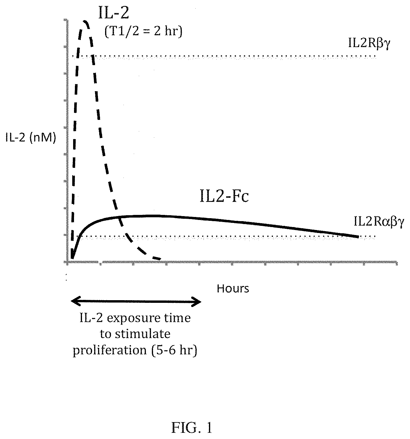
[0009] A second approach to improving the therapeutic index of an IL-2 based therapy is to optimize the pharmacokinetics of the molecule to maximally stimulate Treg cells. Early studies of IL-2 action demonstrated that IL-2 stimulation of human T cell proliferation in vitro required a minimum of 5-6 hours exposure to effective concentrations of IL-2 (Cantrell, D. A., et. al., 1984, Science, 224: 1312-1316). When administered to human patients, IL-2 has a very short plasma half-life of 85 minutes for intravenous administration and 3.3 hours subcutaneous administration (Kirchner, G. I., et al., 1998, Br J Clin Pharmacol. 46:5-10). Because of its short half-life, maintaining circulating IL-2 at or above the level necessary to stimulate T cell proliferation for the necessary duration necessitates high doses that result in peak IL-2 levels significantly above the EC50 for Treg cells or will require frequent administration (FIG. 1). These high IL-2 peak levels can activate IL2R.beta..gamma. receptors and have other unintended or adverse effects. An IL-2 analog with a longer circulating half-life than IL-2 can achieve a target drug concentration for a specified period of time at a lower dose than IL-2, and with lower peak levels. Such an IL-2 analog will therefore require either lower doses or less frequent administration than IL-2 to effectively stimulate Treg cells. Indeed, in cynomolgus monkeys dosed with an IgG-IL2 fusion protein with a circulating half-life of 14 hours stimulated a much more robust increase in Tregs compared to an equimolar dose of IL-2 (Bell, et al., 2015, J Autoimmun. 56:66-80). Less frequent subcutaneous administration of an IL-2 drug will also be more tolerable for patients. A therapeutic with these characteristics will translate clinically into improved pharmacological efficacy, reduced toxicity, and improved patient compliance with therapy.
[0010] One approach to extending the half-life of therapeutic proteins is to fuse the therapeutically active portion of the molecule to another protein, such as the Fc region of IgG, to increase the circulating half-life. Fusion of therapeutic proteins with IgG Fc accomplishes this by increasing the hydrodynamic radius of the protein, thus reducing renal clearance, and through Neonatal Fc Receptor (FcRn)-mediated recycling of the fusion protein, thus prolonging the circulating half-life.
[0011] The fusion of therapeutic proteins to albumin (Sleep, D., et. al., 2013, Biochcm Biophys Acta., 1830:5526-34) and nonimmunogenic amino acid polymer proteins (Schlapschy, M., et. al., 2007, Protein Eng Des Sel. 20:273-84; Schellenberger, V., et at., 2009, Nat Biotechnol. 27:1186-90) have also been employed to increase circulating half-life. However, construction of such fusion proteins in a manner that ensures robust biological activity of the IL2 Selective Agonist fusion partner can be unpredictable, especially in the case of an IL-2 Selective Agonist, which is a small protein that is defective in binding to one of the receptor subunits and that must assemble a complex of three receptor subunits in order to activate the receptor (Wang, X., et al., 2005, Science 310:1159-63).
[0012] Other researchers have created various IL-2 fusion proteins, using wild-type IL-2 or IL-2 with a C125S substitution to promote stability. Morrison and colleagues (Penichet, M. L., et., al., 1997, Hum Antibodies. 8:106-18) created a fusion protein with IgG fused to wild-type IL-2 to both increase the circulating half-life of IL-2 and to target IL-2 to specific antigens for the purpose of potentiating the immune response to the antigen. This fusion protein consisted of an intact antibody molecule, composed of heavy (H) and light (L) chains, wherein the N-terminal H chain moiety was fused to a C-terminal IL-2 protein moiety. This IgG-IL-2 fusion protein possessed Fc effector functions. Key effector functions of IgG Fc proteins are Complement-dependent cytotoxicity (CDC) and antibody-dependent cellular cytotoxicity (ADCC:). The IgG-IL-2 fusion protein was highly active in an IL-2 bioassay and was shown to possess CDC activity. Thus, Penichct et. al. taught the use of antibody-IL2 fusion proteins to target IL-2 activity to antigens recognized by the antibody, for the purpose of potentiating humoral and cell-mediated immune responses to the antigen. In a similar manner, Gillies and colleagues have constructed a number of IgG-IL-2 fusion proteins for cancer immunotherapy, utilizing the antibody portion of the fusion protein to target tumor antigens, and the IL-2 portion to stimulate the immune response to tumor cells (reviewed in Sondel, P. M., et. al., 2012, Antibodies, 1:149-71). These teachings are quite distinct from the present inventive technology, wherein an IL-2 selective agonist, which promotes the growth and activity of immunosuppressive Treg cells, is fused with an effector function-deficient Fc protein moiety for the purpose increasing systemic exposure.
[0013] Strom and his colleagues have constructed fusion proteins with IL-2 fused to the N terminus of an Fc protein for the purpose of eliminating activating T cells expressing the high-affinity IL-2 receptor (Zheng, X. X., et al., 1999, J. Immunol. 1999, 163:4041-8). This fusion protein was shown to inhibit the development of autoimmune diabetes in a T cell transfer mouse model of T1D. The IL2-Fc fusion protein was shown to inhibit the function of disease-promoting T cells from T1D-susceptible female NOD mice when transplanted into less disease-susceptible male NOD mice. They also demonstrated that the IL-2-Fc fusion protein could kill cells expressing the high-affinity IL-2 receptor in vitro. These investigators further compared IL2-Fc fusion proteins constructed from an Fc derived from an effector function-competent 1gG2b Fc and a mutated effector function-deficient IgG2b Fc. Only the IL2-Fc fusion protein containing the effector function-competent Fc was efficacious in preventing disease onset. Thus, these investigators teach that an IL2-Fc fusion protein with effector functions can eliminate disease-causing activated T cells, and that Fc effector functions are necessary for its therapeutic activity. These teachings are quite distinct from the present inventive technology, wherein an IL-2 selective agonist, which promotes the growth and activity of immunosuppressive Treg cells, is fused with an effector function-deficient Fc protein moiety for the purpose increasing systemic exposure and optimizing Treg expansion. Other work from Strom and colleagues teaches the use of a IL2-Fc fusion protein in promoting transplant tolerance (Zheng, X. X., et al., 2003, Immunity, 19:503-14). In this work, an IL2-Fc fusion protein is used in a "triple therapy" in which it is combined with an IL15-Fc receptor antagonist and rapamycin. Again, these investigators teach that the IL2-Fc fusion protein must have Fc effector functions to be efficacious, and further teach that this IL-2-Fc fusion protein must be combined with two other molecules in order to be efficacious.
[0014] This invention provides for a novel therapeutic agent, an IL2 Selective Agonist-Fc fusion protein, that combines the high cellular selectivity of a IL2 Selective Agonist for Treg cells with a long circulating half-life. In the course of developing this molecule, there were surprising and unexpected findings that revealed structural elements and design features of the protein that are essential for bioactivity, and that led to the discovery of several novel proteins that fulfill the desired therapeutic characteristics.
BRIEF SUMMARY OF THE INVENTION
[0015] This invention provides for a fusion protein between an IL2.alpha..beta..gamma. Selective Agonist protein (IL2 Selective Agonist) and a IgG Fc protein. The IL2 Selective Agonist moiety provides a therapeutic activity by selectively activating the IL2.alpha..beta..gamma. form of the receptor, thus selectively stimulating Tregs. The Fc moiety provides a prolonged circulating half-life compared to the circulating half-life of IL-2 or an IL2 Selective Agonist protein. The Fc moiety increases circulating half-life by increasing the molecular size of the fusion protein to greater than 60,000 daltons, which is the approximate cutoff for glomerular filtration of macromolecules by the kidney, and by recycling the fusion protein through the Neonatal Fc Receptor (FcRn) protein, the receptor that binds and recycles IgG, thus prolonging its circulating half-life. The Fc moiety will also be deficient in Fc effector functions, such as Complement-Dependent Cytotoxicity (CDC) and Antibody-Dependent Cellular Cytotoxicity (ADCC), enabling the fusion protein to selectively activate Tregs to potentiate Treg function and to expand Treg numbers. The two protein moieties are fused in a manner that maintains robust bioactivity of the IL2 Selective Agonist moiety and enables the Fc moiety to promote a prolonged circulating half-life and thus efficiently potentiate Tregs function and numbers. This potentiation of Tregs will suppress over-exuberant autoimmune or inflammatory responses, and will be of benefit in treating autoimmune and inflammatory diseases. The proteins of this invention may be monomeric or dimeric forming dimers through cysteine residues in the Fc moieties or domains.
[0016] More specifically, this invention provides for a fusion protein, comprising: a N-terminal human IL-2 variant protein moiety, and a C-terminal IgG Fc protein moiety, wherein said IL-2 fusion protein has the ability to selectively activate the high affinity IL-2 receptor and thus selectively activate human regulatory T cells. The variants of IL-2 include those with substitutions selected from the group consisting of: N88R, N88I, N88G, D2OH, Q126L, and Q126F relative to human IL2 protein (SEQ ID NO:1). In addition the, IL-2 variant protein optionally comprises human IL-2 with the substitution C125S. It is preferred that the proteins of this invention are fused wherein both the IL-2 variant protein and the IgG Fc protein have an N-terminus and a C-terminus and said human IL-2 variant protein is fused at its C-terminus to the N-terminus of the IgG Fc protein. It is further preferred that the IL-2 variant domain and the Fc domain are joined or fused through a linker peptide positioned between the IL-2 variant protein and the IgG Fc protein moieties. The IgG Fc protein moiety or domain will preferably be deficient in Fc effector functions or contain one or more amino acid substitutions that reduce the effector functions of the Fc portion of the fusion protein.
[0017] An example of this invention is a protein, comprising: a IL-2 variant protein having amino acid substitutions N88R and C125S relative to human IL-2 (SEQ ID NO:1), a linker peptide as set forth in SEQ ID NO:15, and a human IgG1 Fc protein as set forth in SEQ ID NO:2, wherein said fusion protein has the ability to selectively activate the high affinity IL-2 receptor and thus selectively activate human regulatory T cells. Alternative proteins of this invention include: a IL-2 variant protein having amino acid substitutions N88R and C125S relative to human IL-2 (SEQ ID NO:1), a linker peptide as set forth in SEQ ID NO:15, and a human IgG2 Fc protein as set forth in SEQ ID NO:3.
[0018] A more specific embodiment of this invention is a dimeric protein, comprising two identical chains, where each chain comprises a N-terminal human IL-2 variant protein moiety and a C-terminal IgG Fc protein moiety wherein: the N-terminal human IL-2 variant protein moiety has a N-terminus and a C-terminus varies from the human IL-2 wildtype as in SEQ ID NO:1 by at least one of the substitutions selected from the group consisting of N88R, N88I, N88G, D2OH, Q126L, and Q126F, has at least a 97% sequence identify to Sequence ID No. 1; and, has the ability to activate Treg cells by binding to a IL2R.alpha..beta..gamma. on those cells; the N-terminal human IL-2 variant protein is joined at its C-terminal to a N-terminus of an amino acid linker of between 6 to 20 amino acid residues where said linker also has a C-terminus; and, the C-terminus of the amino acid linker is joined to the N-terminus of IgG Fc protein moiety having 97% sequence identify to for example SEQ ID NO:3 (IgG2) or SEQ ID No. 2 (IgG1N297A) and containing cysteine residues; and where the two chains are linked to each other through the cysteine residues that form the interchain disulfide bonds of the IgG Fc protein moiety. The dimers of this invention may be further substituted at C125S of the IL-2 moiety. The proteins of this invention preferably include amino acid linkers consisting a group of glycine residues, serine residues, and a mix of glycine and serine residues. The linkers may comprise a mix of between 12 and 17 serine and glycine residues preferably with a ratio of glycine residues to serine residues in a range of 3:1-5:1, e.g, a 4:1 ratio.
[0019] This invention further provides for the compositions above in a pharmaceutical composition comprising a pharmaceutically acceptable carrier.
[0020] This invention further provides for nucleic acids encoding the proteins described herein. The nucleic acids or preferably operably linked to expression cassettes that can be either designed for recombination with a host cell genome or introduced on an independently replicating plasmid or extrachromosomal nucleic acid.
[0021] This invention further provides for methods of selectively activating human regulatory T cells in a patient in need thereof, the method comprising administering a pharmaceutical composition comprising the compositions described administered at therapeutically effective doses until human regulatory T cell concentrations reach desired levels.
[0022] A method of measuring the numbers of Treg cells in a human blood sample by contacting human blood cells with the fusion protein of claim 1 at a concentration of between 1 nM and 0.01 nM, and then detecting cells that bind to the protein by flow cytometry.
BRIEF DESCRIPTION OF THE DRAWINGS
[0023] FIG. 1 is a diagrammatic illustration of the relationship between circulating half-life, peak drug level, the biological effective concentration, and the duration necessary to stimulate Treg cell proliferation after a single dose of IL-2 or an IL2-Fc fission protein with increased half-life. The dashed line represents the blood level over time of IL-2 following a subcutaneous injection, and the solid line represents the blood level over time of an IL2-Fc fusion protein. The horizontal dotted lines indicate the concentrations (EC50 values) necessary to activate cells expressing IL2R.alpha..beta..gamma. and
[0024] IL2.beta..gamma., respectively) are indicated. The double-headed arrow indicates the duration of exposure (5-6 hr) to IL-2 at the EC50 necessary to stimulate cell proliferation.
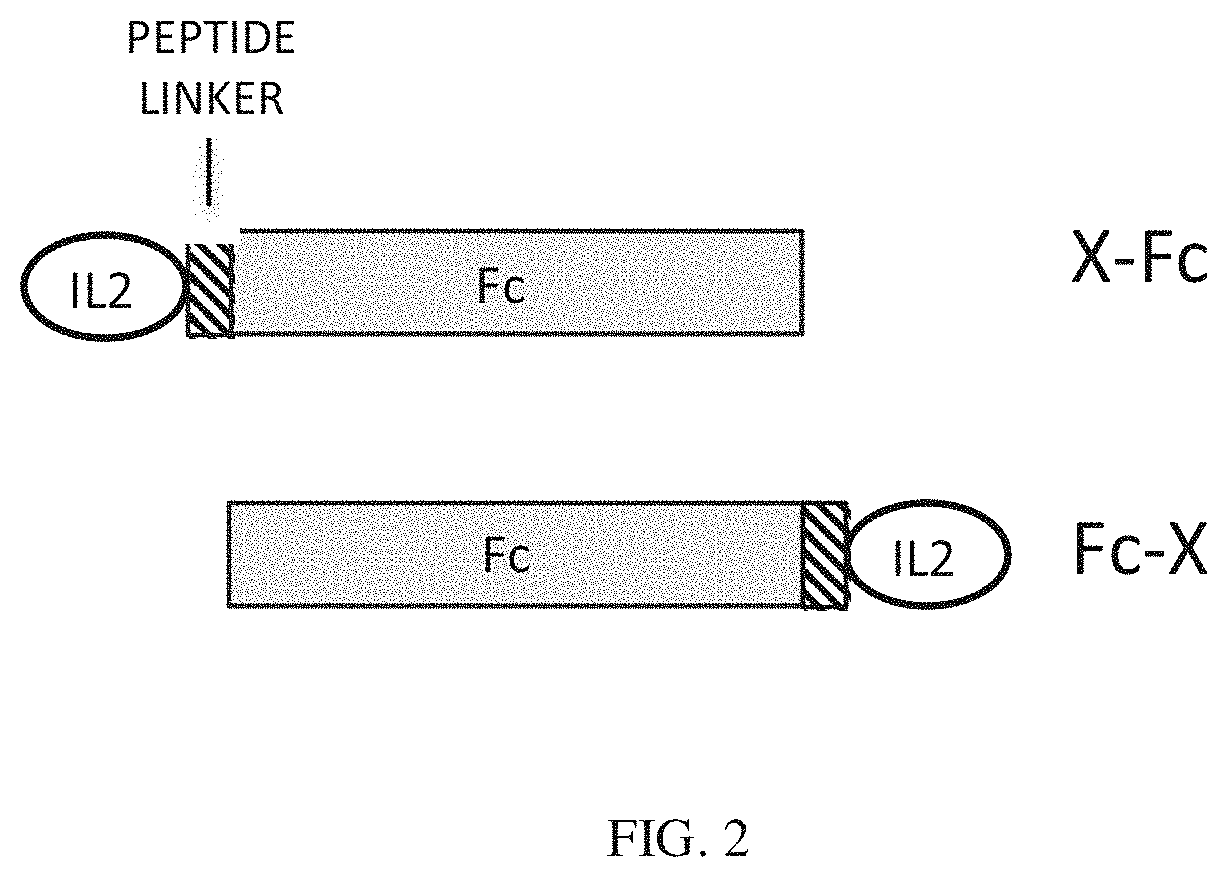
[0025] FIG. 2 shows the design configurations for Fc fusion proteins. The fusion partner (X), can be fused at the N terminus (X-Fc) or the C-terminus (Fc-X) of the Fc protein. Linker peptides can be inserted between X and the Fc.
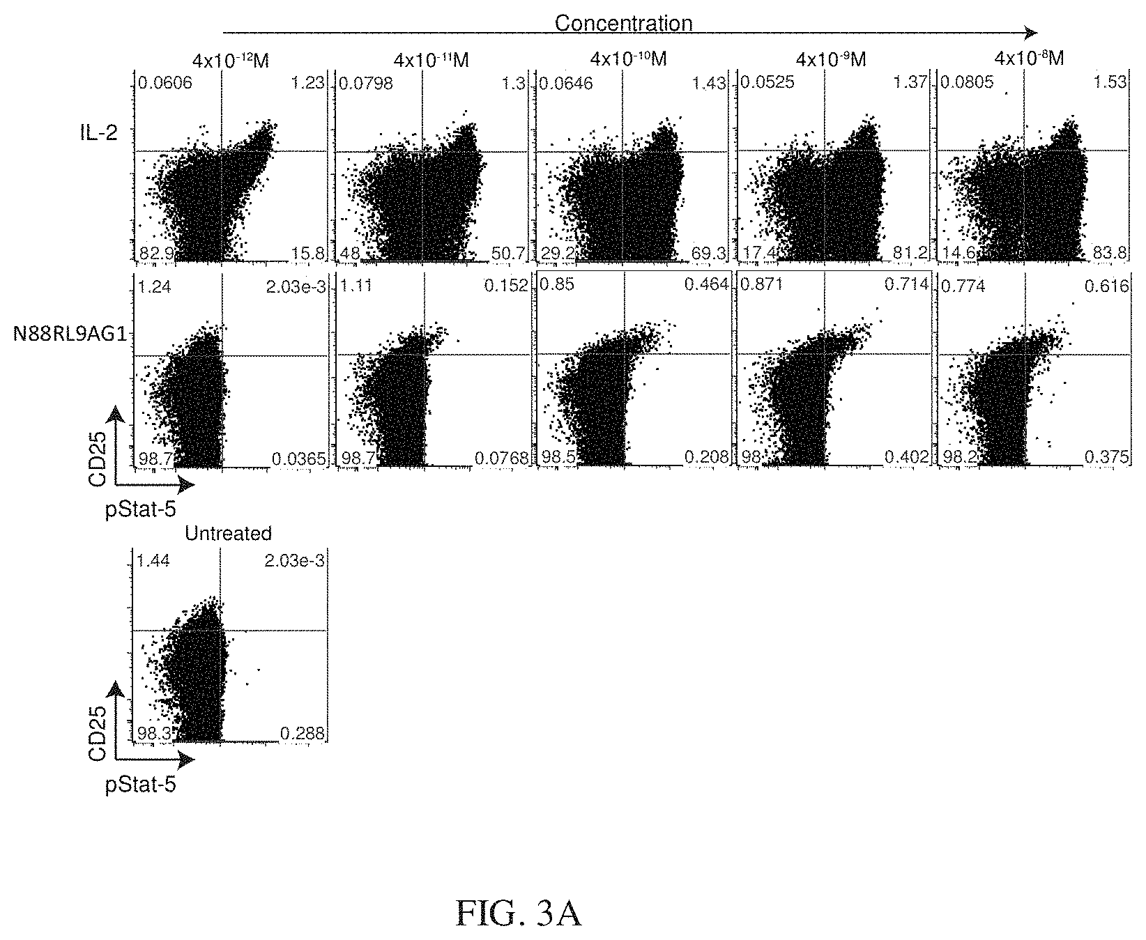
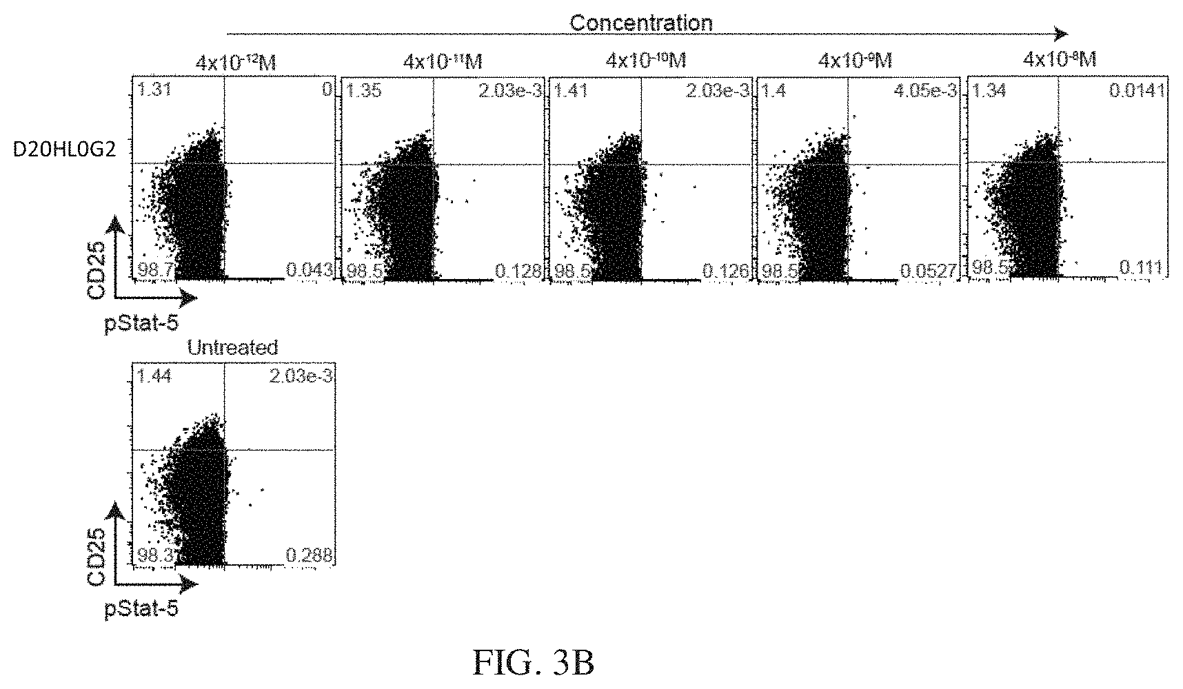
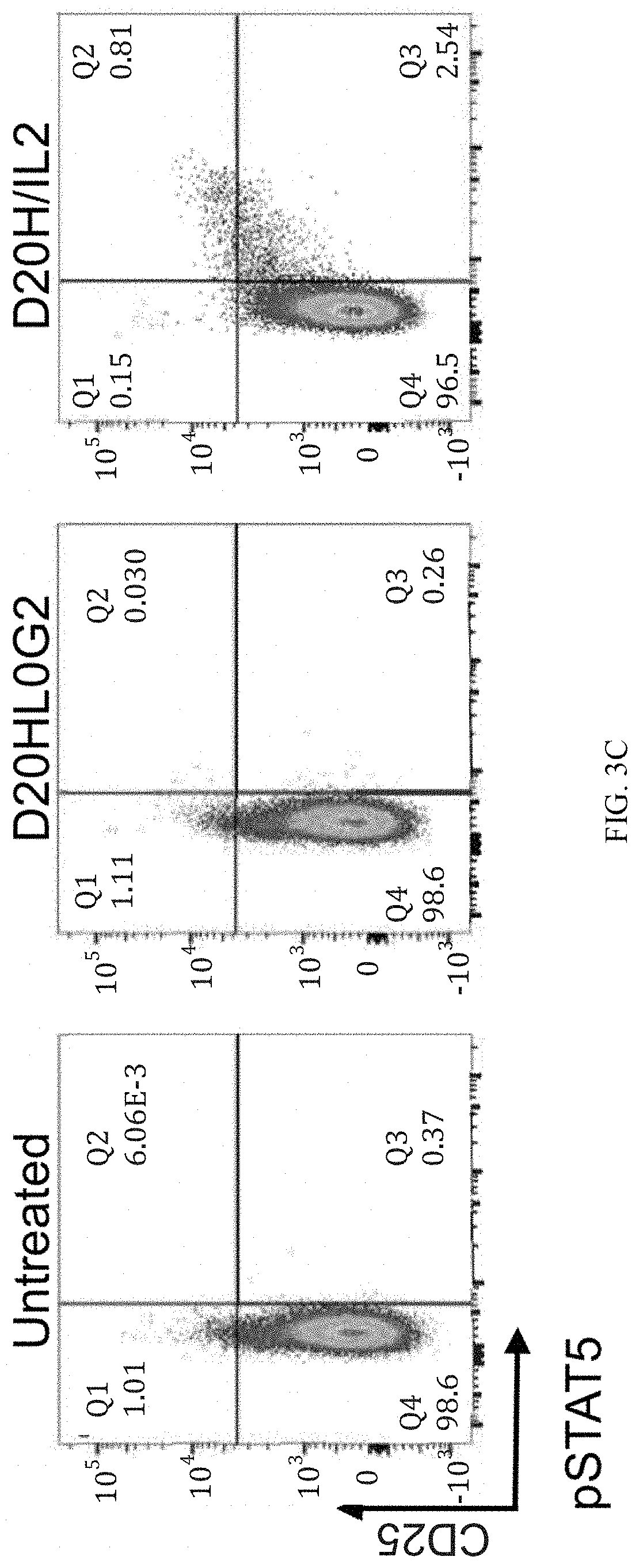
[0026] FIGS. 3A, 3B, and 3C show a dose-response of IL-2 and N88RL9AG1 stimulated STAT5 phosphorylation in CD4+ T cells as measured by flow cytometry. Cells were treated with the IL-2 or N88RFc at the concentrations indicated on the top for 10 minutes at 37 C, fixed, permeabilized, stained with antibodies, and then subjected to flow cytometry analysis as described in Example 3. Cells gated as CD4+ are shown, and cells further gated with respect to CD25 and pSTAT5 as shown in each of the 4 quadrants. The numbers in each quadrant indicate the percentage of CD4+ cells in each gate. Cells in the upper quadrants represent the highest 1-2% of CD25 expressing cells, a population enriched for Treg cells, and cells in the right-hand quadrants are pSTAT5+. FIG. 3A shows that N88RL9AG1 stimulates CD25.sup.high cells with high selectivity, while IL-2 massively stimulates both CD25.sup.-/low and CD25.sup.high cells down to picomolar concentrations. FIG. 3B shows a dose-response of D20HL0G2 stimulated STAT5 phosphorylation in CD4+ T cells as measured by flow cytometry. D20HL0G2 has no pSTAT5 stimulating activity. No pSTAT5 activation was observed in two independent experiments. FIG. 3C is a control showing that D2OH/IL2 stimulates pSTAT5 in CD25.sup.high cells while D20HL0G2 does not. Plots are displayed in the pseudocolor mode. Both proteins were tested at a concentration of 10.sup.-8 M.
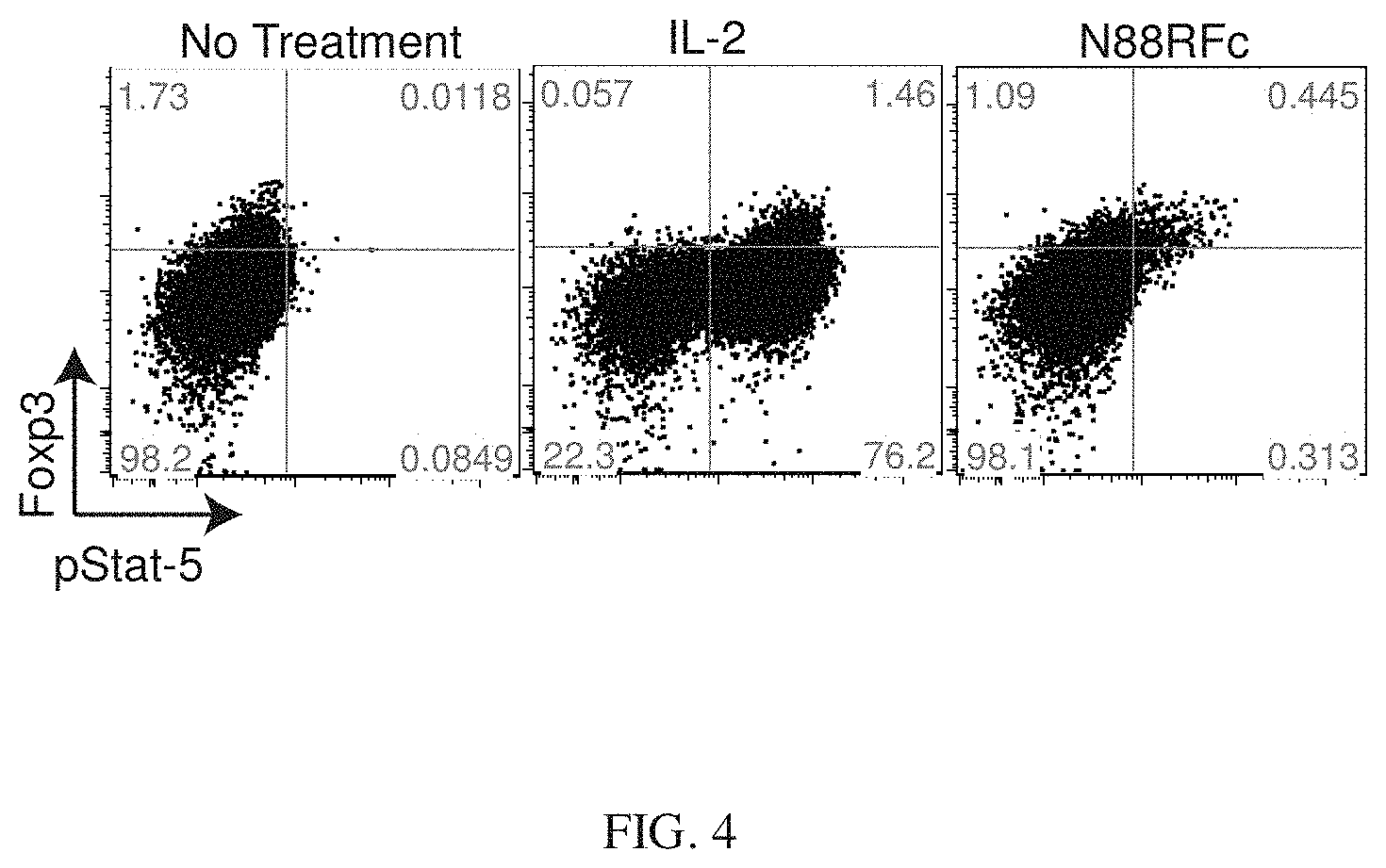
[0027] FIG. 4 shows that CD4+ T cells treated with N88RL9AG1 exhibited stimulation of pSTAT5 levels in cells expressing high levels of FOXP3. Cells were treated with 4.times.10.sup.-9 M IL-2 or N88RL9AG1 and then analyzed as described in Example 3. The majority of pSTAT5+ cells treated with N88RL9AG1 were also FOXP3+, whereas pSTAT5+ cells treated with IL-2 were both FOXP3- and FOXP3+, with the majority being FOXP3-.
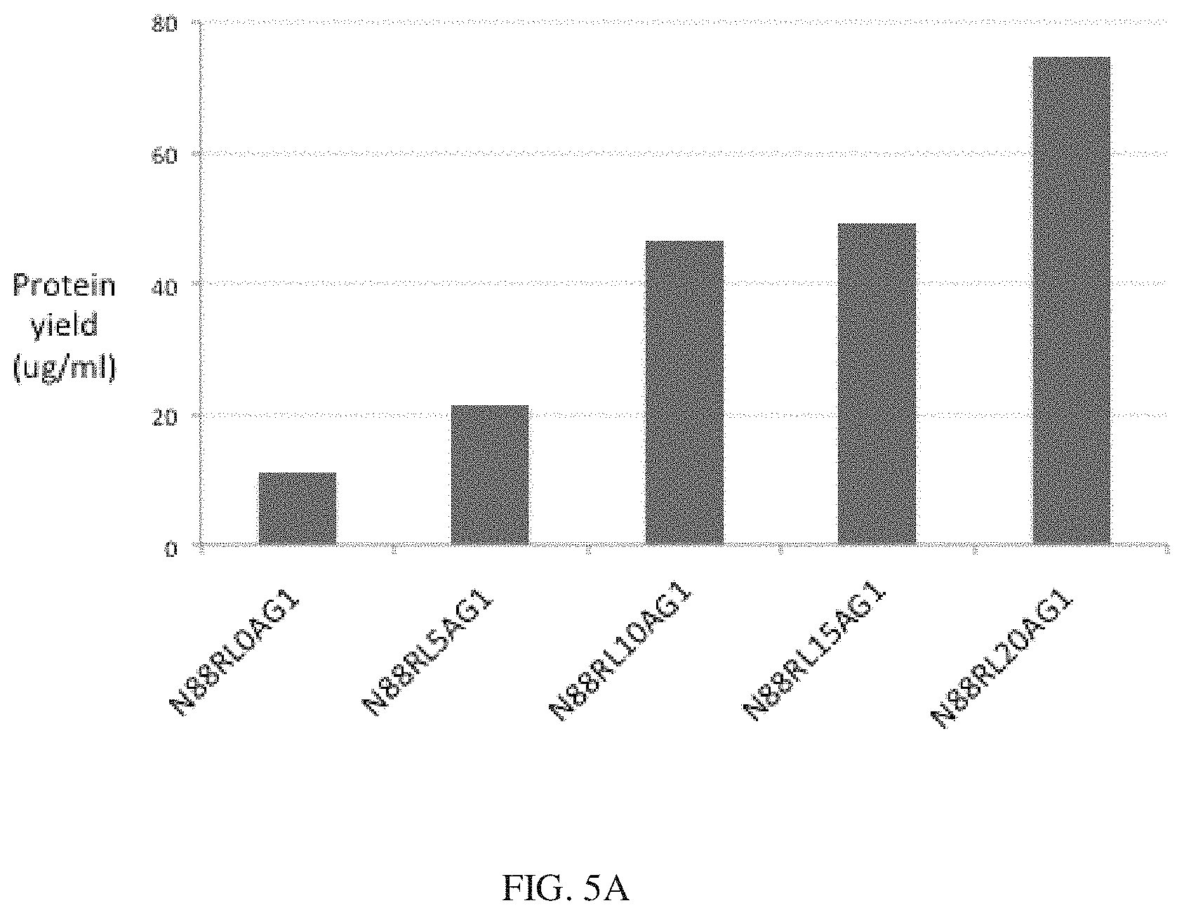
[0028] FIGS. 5A and 5B show the protein yields of different Fc fusion constructs in HEK293 cells. Proteins were expressed in parallel in an optimized transient expression system and purified as described in Example 1. Results are expressed as the final yield of purified protein from 30 ml cultures. FIG. 5A shows that the protein yields of N88R/IL2-Fc fusion proteins increase with increasing peptide linker length. FIG. 5B shows that the yields of wt IL2-Fc fusion proteins are only slightly enhanced with a 15 residue peptide linker. Higher yields of D2OH/IL2-Fc fusion proteins were obtained in the X-Fc rather than the Fc-X configuration.
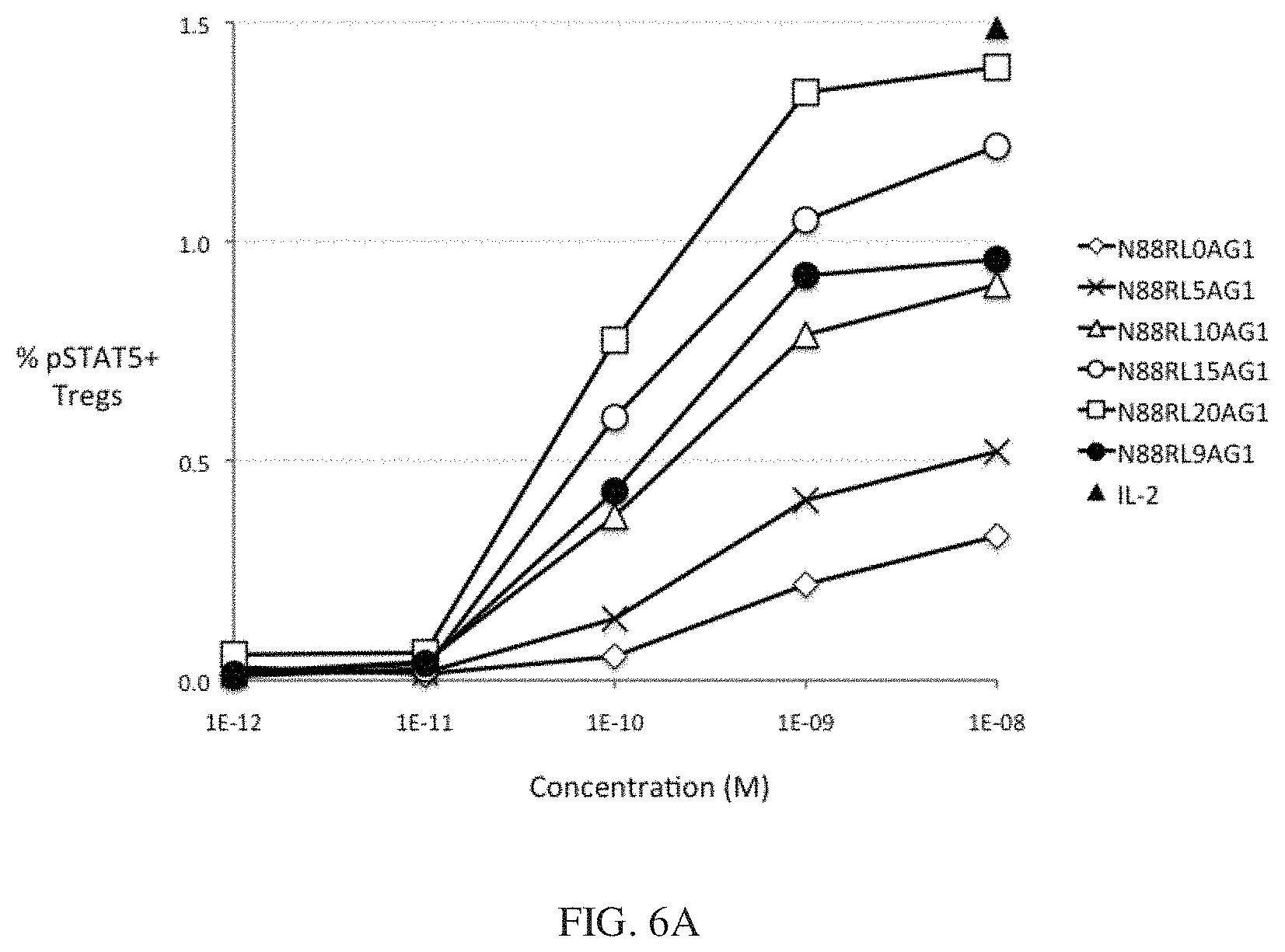
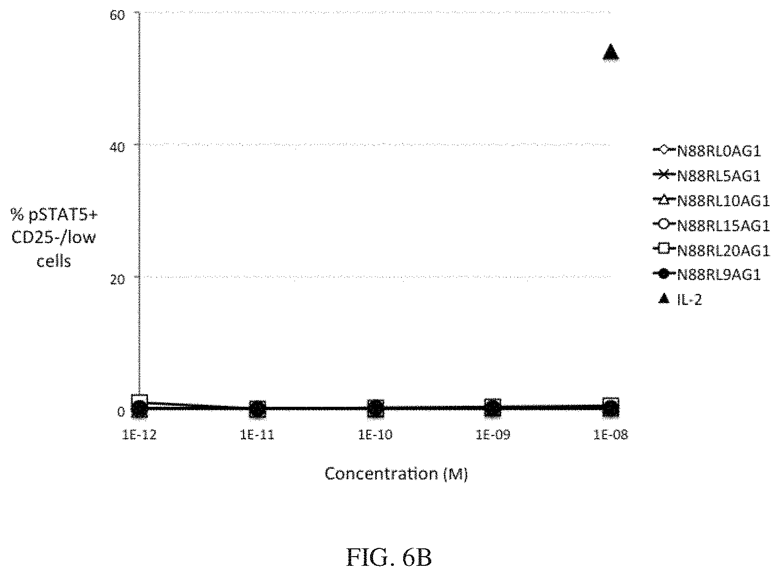
[0029] FIGS. 6A and 6B show the dependence of IL-2 bioactivity on peptide linker length in N88R/IL2-Fc fusion proteins. FIG. 6A shows that pSTAT5 signals in CD25.sup.high CD4 T cells Tregs increase with increasing peptide linker length. FIG. 6B shows no significant pSTAT5 signal with any of N88R/IL2-Fc proteins was observed in CD25.sup.-/low cells. The pSTAT5 signal of the 10.sup.-8 M IL-2 internal control is indicated in both panels by the black triangle.
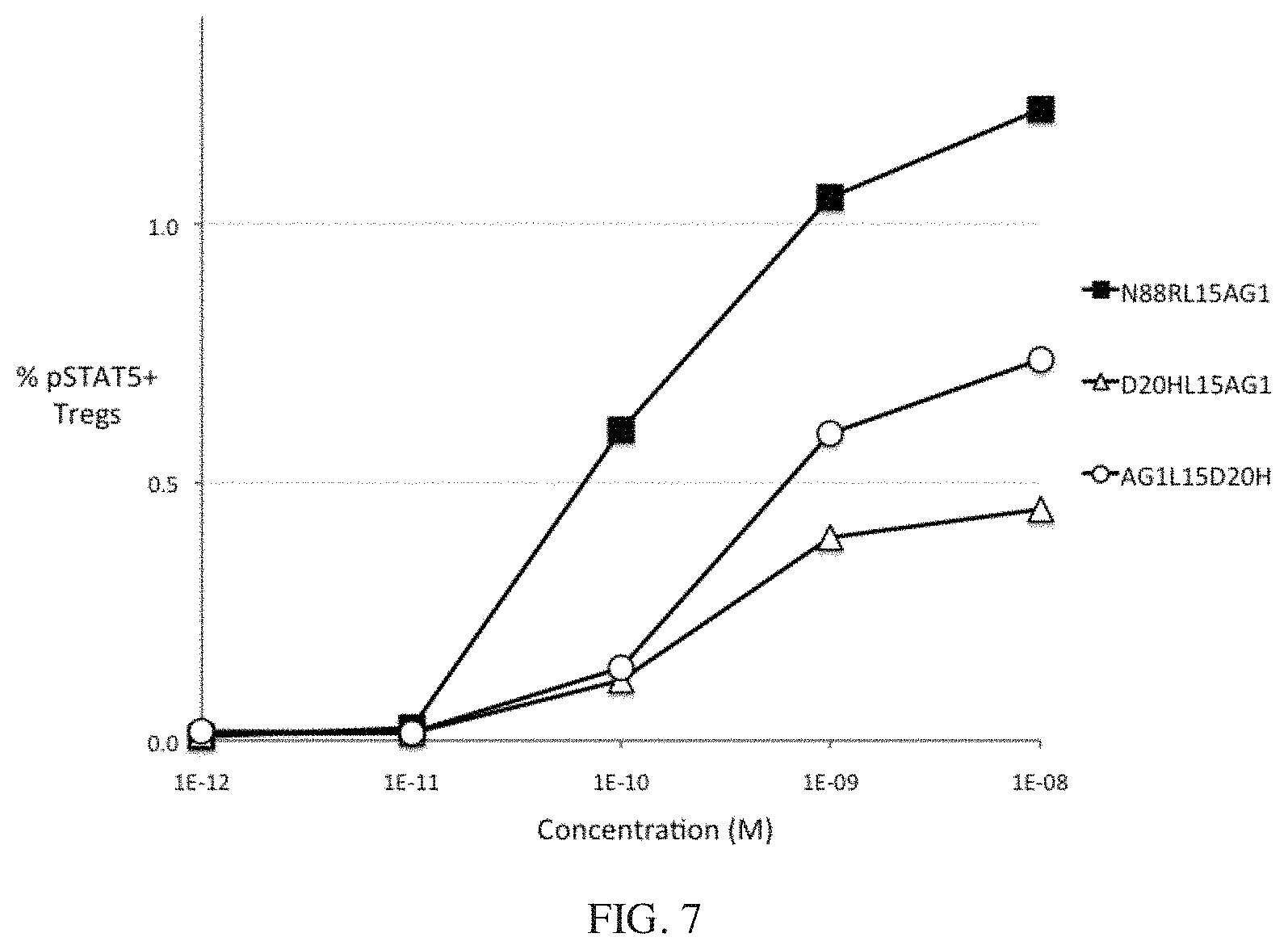
[0030] FIG. 7 shows the bioactivity of D2OH/IL2-Fc fusion proteins in human Tregs. The potency of D20HL15AG1 is substantially less than that of N88RL15AG1, and D20HL15AG1 (X-Fc configuration) and AG1LI5D2OH (Fc-X configuration) have similar potencies. All 3 proteins have a 15 residue peptide linker.
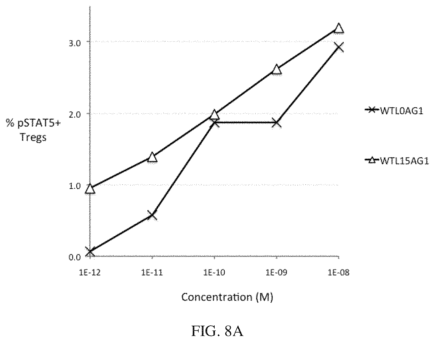
[0031] FIGS. 8A and 8B show the bioactivity of wt IL-2-Fc pSTAT5 activity with and without a 15 residue peptide linker. FIG. 8A shows that IL-2 bioactivity is only modestly enhanced by a 15 residue peptide linker in Tregs. FIG. 8B shows that IL-2 bioactivity is only modestly enhanced by a 15 residue peptide linker in CD25.sup.-/low cells.
[0032] FIGS. 9A and 9B show the selectivity of IL-2 and IL-2 Selective Agonist proteins on 7 different immune cell types in human PBMC. N88RL15AG1 is highly selective for Tregs compared to wt IL-2 and WTL15AG1, and shows greater selectivity in multiple cell types than N88R/IL2. FIG. 9A shows the selectivity of IL-2 and IL-2 Selective Agonist proteins on Treg cells, activated CD4 Teff cells, and CD4 Teff cells. FIG. 9B shows the selectivity of IL-2 and IL-2 Selective Agonist proteins on CD8 Teff cells, NK T cells, NK cells, and B cells.
DETAILED DESCRIPTION OF THE INVENTION
Introduction
[0033] This invention is a novel therapeutic fusion protein that comprises three key protein elements: (1) an engineered IL-2 cytokine that has been modified to be highly selective for Treg cells, (2) an effector function deficient Fc protein that will increase the circulating half-life of the protein, and (3) a linker peptide between the two moieties that is necessary for high biological activity of the fusion protein. The fusion proteins which constitute this invention were discovered through initial unanticipated findings that went against teachings in the prior art of IL-2 fusion proteins, and the research that led to these molecules has defined key structure-activity relationships important for their biological and therapeutic activity. The molecules defined by this invention will enable the safe and effective treatment of autoimmune diseases by the novel mechanism of stimulating the production of a small subpopulation of T cells that suppress autoimmune and inflammatory pathology. This paradigm-breaking therapeutic can potentially treat a number of different autoimmune diseases.
Definitions
[0034] "At least a percent (eg. 97%) sequence identify to Sequence ID No. 1" as used herein refers to the extent to which the sequence of two or more nucleic acids or polypeptides is the same. The percent identity between a sequence of interest and a second sequence over a window of evaluation, e.g., over the length of the sequence of interest, may be computed by aligning the sequences, determining the number of residues (nucleotides or amino acids) within the window of evaluation that are opposite an identical residue allowing the introduction of gaps to maximize identity, dividing by the total number of residues of the sequence of interest or the second sequence (whichever is greater) that fall within the window, and multiplying by 100. When computing the number of identical residues needed to achieve a particular percent identity, fractions are to be rounded to the nearest whole number. Percent identity can be calculated with the use of a variety of computer programs. For example, computer programs such as BLAST2, BLASTN, BLASTP, Gapped BLAST, etc., generate alignments and provide percent identity between sequences of interest. The algorithm of Karlin and Altschul (Karlin and Altschul, Proc. Natl. Acad. Sci. USA 87:22264-2268, 1990) modified as in Karlin and Altschul, Proc. Natl. Acad. Sci. USA 90:5873-5877, 1993 is incorporated into the NBLAST and XBLAST programs of Altschul et al. (Altschul, et al., J. Mol. Biol. 215:403-410, 1990). To obtain gapped alignments for comparison purposes, Gapped BLAST is utilized as described in Altschul et al. (Altschul, et al. Nucleic Acids Res. 25:3389-3402, 1997). When utilizing BLAST and Gapped BLAST programs, the default parameters of the respective programs may be used. A PAM250 or BLOSUM62 matrix may be used. Software for performing BLAST analyses is publicly available through the National Center for Biotechnology Information (NCBI). See the Web site having URL world-wide web address of: "ncbi.nlm.nih.gov" for these programs. In a specific embodiment, percent identity is calculated using BLAST2 with default parameters as provided by the NCBI.
[0035] "N-terminus" refers to the end of a peptide or polypeptide that bears an amino group in contrast to the carboxyl end bearing a carboxyl acid group.
[0036] "C-terminus" refers to the end of a peptide or polypeptide that bears a carboxcylic acid group in contrast to the amino terminus bearing an amino group.
[0037] "C-terminal IgG Fc protein moiety" refers to a portion of a fusion protein that derives from two identical protein fragments, each having a hinge region, a second constant domain, and a third constant domains of the IgG molecule's two heavy chains, and consisting of the carboxy-terminal heavy chains disulphide bonded to each other through the hinge region. It is functionally defined as that part of the IgG molecule that interacts with the complement protein C1q and the IgG-Fc receptors (Fc.gamma.R), mediating Complement-dependent cytotoxicity (CDC) and antibody-dependent cellular cytotoxicity (ADCC) effector functions. The sequence can be modified to decrease effector functions, to increase circulating half-life, and to eliminate glycoslylation sites.
IL2 Variants
[0038] IL-2 variant proteins of this invention are IL-2.alpha..beta..gamma. Selective Agonists. Functionally they selectively activate the IL2R.alpha..beta..gamma. receptor complex relative to the IL2R.beta..gamma. receptor complex. It is derived from a wild type IL-2 protein structurally defined as having at least a 95% sequence identity to the wild type IL-2 of Sequence ID No. 1 and functionally defined by the ability to preferentially activate Treg cells. The protein can also be functionally defined by its ability to selectively activate IL-2 receptor signaling in Tregs, as measured by the levels of phosphorylated STAT5 protein in Treg cells compared to CD4+CD25-/low T cells or NK cells, or by the selective activation of Phytohemagglutinin-stimulated T cells versus NK cells.
[0039] "N-terminal human IL-2 variant protein moiety" refers to a N-terminal domain of a fusion protein that is derived from a wild type IL-2 protein structurally and functionally defines above.
[0040] "C-terminus" refers to the end of a peptide or polypeptide that bears a carboxcylic acid group in contrast to the amino terminus bearing an amino group.
Tregs
[0041] "Tregs" or "Treg cells" refer to Regulatory T cells. Regulatory T cells are a class of T cells that suppress the activity of other immune cells, and are defined using flow cytometry by the cell marker phenotype CD4+CD25+FOXP3+. Because FOXP3 is an intracellular protein and requires cell fixation and permeablization for staining, the cell surface phenotype CD4+CD25+CD127- can be used for defining live Tregs. Tregs also include various Treg subclasses, such as tTregs (thymus-derived) and pTregs (peripherally-derived, differentiated from naive T cells in the periphery). All Tregs express the IL2R.alpha..beta..gamma. receptor, do not produce their own IL-2 and are dependent on IL-2 for growth, and someone skilled in the art will recognize that both classes will be selectively activated by a IL2R.alpha..beta..gamma. selective agonist.
Peptide Linkers
[0042] "Peptide linker" is defined as an amino acid sequence located between the two proteins comprising a fusion protein, such that the linker peptide sequence is not derived from either partner protein. Peptide linkers are incorporated into fusion proteins as spacers in order to promote proper protein folding and stability of the component protein moieties, to improve protein expression, or to enable better bioactivity of the two fusion partners (Chen, et al., 2013, Adv Drug Deliv Rev. 65(10):1357-69). Peptide linkers can be divided into the categories of unstructured flexible peptides or rigid structured peptides.
Fc Fusion Proteins
[0043] An "Fc fusion protein" is a protein made by recombinant DNA technology in which the translational reading frame of the Fc domain of a mammalian IgG protein is fused to that of another protein ("Fc fusion partner") to produce a novel single recombinant polypeptide. Fc fusion proteins are typically produced as disulfide-linked dimers, joined together by disulfide bonds located in the hinge region.
Functional Activation
[0044] "Bioactivity" refers to the measurement of biological activity in a quantitative cell-based in vitro assay.
[0045] "Functional activation of Treg cells" is defined an IL-2-mediated response in Tregs. Assay readouts for functional activation of Treg cells includes stimulation of pSTAT5, Treg cell proliferation, and stimulation of the levels of Treg effector proteins.
Design and Construction
[0046] There are multiple options for the design and construction of an Fc fusion protein, and the choices among these design options are presented below to permit the generation of a molecule with the desired biological activity and pharmaceutical characteristics. Key design options are: (1) the nature of the IL2 Selective Agonist, (2) the choice of the Fc protein moiety, (3) the configuration of fusion partners in the fusion protein, and (4) the amino acid sequence at the junction between the Fc and the fusion partner protein.
General Methods
[0047] In general, preparation of the fusion proteins of the invention can be accomplished by procedures disclosed herein and by recognized recombinant DNA techniques involving, e.g., polymearase chain amplification reactions (PCR), preparation of plasmid DNA, cleavage of DNA with restriction enzymes, preparation of oligonucleotides, ligation of DNA, isolation of mRNA, introduction of the DNA into a suitable cell, transformation or transfection of a host, culturing of the host. Additionally, the fusion molecules can be isolated and purified using chaotropic agents and well known electrophoretic, centrifugation and chromatographic methods. See generally, Sambrook et al., Molecular Cloning: A Laboratory Manual (2nd ed. (1989); and Ausubel et al., Current Protocols in Molecular Biology, John Wiley & Sons, New York (1989) for disclosure relating to these methods.
[0048] The genes encoding the fusion proteins of this invention involve restriction enzyme digestion and ligation as the basic steps employed to yield DNA encoding the desired fusions. The ends of the DNA fragment may require modification prior to ligation, and this may be accomplished by filling in overhangs, deleting terminal portions of the fragment(s) with nucleases (e.g., ExoIII), site directed mutagenesis, or by adding new base pairs by PCR. Polylinkers and adaptors may be employed to facilitate joining of selected fragments. The expression construct is typically assembled in stages employing rounds of restriction, ligation, and transformation of E. coli. Numerous cloning vectors suitable for construction of the expression construct are known in the art (lambda.ZAP and pBLUESCRIPT SK-1, Stratagene, LaJolla, Calif., pET, Novagen Inc., Madison, Wis.--cited in Ausubel et al., 1999) and the particular choice is not critical to the invention. The selection of cloning vector will be influenced by the gene transfer system selected for introduction of the expression construct into the host cell. At the end of each stage, the resulting construct may be analyzed by restriction, DNA sequence, hybridization and PCR analyses.
[0049] Site-directed mutagenesis is typically used to introduce specific mutations into the genes encoding the fusion proteins of this invention by methods known in the art. See, for example, U.S. Patent Application Publication 2004/0171154; Storici et al., 2001, Nature Biotechnology 19: 773-776; Kren et al., 1998, Nat. Med. 4: 285-290; and Calissano and Macino, 1996, Fungal Genet. Newslett. 43: 15-16. Any site-directed mutagenesis procedure can be used in the present invention. There are many commercial kits available that can be used to prepare the variants of this invention.
[0050] Various promoters (transcriptional initiation regulatory region) may be used according to the invention. The selection of the appropriate promoter is dependent upon the proposed expression host. Promoters from heterologous sources may be used as long as they are functional in the chosen host.
[0051] Various signal sequences may be used to facilitate expression of the proteins described herein. Signal sequence are selected or designed for efficient secretion and processing in the expression host may also be used. A signal sequence which is homologous to the TCR coding sequence or the mouse IL-2 coding sequence may be used for mammalian cells. Other suitable signal sequence/host cell pairs include the B. subtilis sacB signal sequence for secretion in B. subtilis, and the Saccharontyces cerevisiae .alpha.-mating factor or P. pastoris acid phosphatase phoI signal sequences for P. pastoris secretion. The signal sequence may be joined directly through the sequence encoding the signal peptidase cleavage site to the protein coding sequence, or through a short nucleotide bridge.
[0052] Elements for enhancing transcription and translation have been identified for eukaryotic protein expression systems. For example, positioning the cauliflower mosaic virus (CaMV) promoter 1000 bp on either side of a heterologous promoter may elevate transcriptional levels by 10- to 400-fold in plant cells. The expression construct should also include the appropriate translational initiation sequences. Modification of the expression construct to include a Kozak consensus sequence for proper translational initiation may increase the level of translation by 10 fold.
[0053] The expression cassettes are joined to appropriate vectors compatible with the host that is being employed. The vector must be able to accommodate the DNA sequence coding for the fusion proteins to be expressed. Suitable host cells include eukaryotic and prokaryotic cells, preferably those cells that can be easily transformed and exhibit rapid growth in culture medium. Specifically preferred hosts cells include prokaryotes such as E. coli, Bacillus subtillus, etc. and eukaryotes such as animal cells and yeast strains, e.g., S. cerevisiae. Mammalian cells are generally preferred, particularly HEK, J558, NSO, SP2-O or CHO. Other suitable hosts include, e.g., insect cells such as Sf9. Conventional culturing conditions are employed. See Sambrook, supra. Stable transformed or transfected cell lines can then be selected. In vitro transcription-translation systems can also be employed as an expression system.
[0054] Nucleic acid encoding a desired fusion protein can be introduced into a host cell by standard techniques for transfecting cells. The term "transfecting" or "transfection" is intended to encompass all conventional techniques for introducing nucleic acid into host cells, including calcium phosphate co-precipitation, DEAE-dextran-mediated transfection, lipofection, electroporation, microinjection, viral transduction and/or integration. Suitable methods for transfecting host cells can be found in Sambrook et al. supra, and other laboratory textbooks.
[0055] Alternatively, one can use synthetic gene construction for all or part of the construction of the proteins described herein. This entails in vitro synthesis of a designed polynucleotide molecule to encode a polypeptide molecule of interest. Gene synthesis can be performed utilizing a number of techniques, such as the multiplex microchip-based technology described by Tian, et. al., (Tian, et. al., Nature 432:1050-1054) and similar technologies wherein oligonucleotides are synthesized and assembled upon photo-programmable microfluidic chips.
[0056] The fusion proteins of this invention are isolated from harvested host cells or from the culture medium. Standard protein purification techniques are used to isolate the proteins of interest from the medium or from the harvested cells. In particular, the purification techniques can be used to express and purify a desired fusion protein on a large-scale (i.e. in at least milligram quantities) from a variety of approaches including roller bottles, spinner flasks, tissue culture plates, bioreactor, or a fermentor.
The IL2 Selective Agonist Moiety
[0057] IL-2 with the substitution N88R is an exemplary case of an IL2 Selective Agonist for the IL2R.alpha..beta..gamma. receptor (Shanafelt, A. B., et al., 2000, Nat Biotechnol. 18:1197-202). IL2/N88R is deficient in binding to the IL2R.beta. receptor subunit and the IL2R.beta..gamma. receptor complex, but is able to bind to the IL2R.alpha..beta..gamma. receptor complex and stimulate the proliferation of IL2R.alpha..beta..gamma.-expressing PHA-activated T cells as effectively as wt IL-2, while exhibiting a 3,000 fold reduced ability to stimulate the proliferation of IL2R.beta..gamma.-expressing NK cells. Other IL2R.alpha..beta..gamma. selective agonists with similar activity profiles include IL-2 with the substitutions N88I, N88G, and D2OH, and other IL2 variants with the substitutions QI26L and Q126F (contact residues with the IL2RG subunit) also possess IL2R.alpha..beta..gamma.-selective agonist activity (Cassell, D. J., et. al., 2002, Curr Phami Des., 8:2171-83). A practitioner skilled in the art would recognize that any of these IL2 Selective Agonist molecules can be substituted for the IL2/N88R moiety with the expectation that an Fc fusion protein will have similar activity. All of the aforementioned mutations can be made on the background of wt IL-2, or wt IL-2 with the substitution C125S, which is a substitution that promotes IL-2 stability by eliminating an unpaired cysteine residue. This invention can also be used with other mutations or truncations that improve production or stability without significantly impacting IL-2 receptor activating activity.
[0058] The variants of this invention optionally include conservatively substituted variants that apply to both amino acid and nucleic acid sequences. With respect to particular nucleic acid sequences, conservatively modified variants refer to those nucleic acids which encode identical or essentially identical amino acid sequences, or where the nucleic acid does not encode an amino acid sequence, to essentially identical sequences. Specifically, degenerate codon substitutions may be achieved by generating sequences in which the third position of one or more selected (or all) codons is substituted with mixed base and/or deoxyinosine residues (Batzer et al., Nucleic Acid Res. 19:5081 (1991); Ohtsuka et al., J. Biol. Chem. 260:2605-2608 (1985); Rossolini et al., Mol. Cell. Probes 8:91-98 (1994)). Because of the degeneracy of the genetic code, a large number of functionally identical nucleic acids encode any given protein. For instance, the codons GCA, GCC, GCG and GCU all encode the amino acid alanine. Thus, at every position where an alanine is specified by a codon, the codon can be altered to any of the corresponding codons described without altering the encoded polypeptide. Such nucleic acid variations arc silent variations, which are one species of conservatively modified variations. Every nucleic acid sequence herein which encodes a polypeptide also describes every possible silent variation of the nucleic acid. One of skill will recognize that each codon in a nucleic acid (except AUG, which is ordinarily the only codon for methionine, and TGG, which is ordinarily the only codon for tryptophan) can be modified to yield a functionally identical molecule. Accordingly, each silent variation of a nucleic acid which encodes a polypeptide is implicit in each described sequence.
[0059] With regard to conservative substitution of amino acid sequences, one of skill will recognize that individual substitutions, deletions or additions to a nucleic acid, peptide, polypeptide, or protein sequence which alters, adds or deletes a single amino acid or a small percentage of amino acids in the encoded sequence is a conservatively modified variant where the alteration results in the substitution of an amino acid with a chemically similar amino acid. Conservative substitution tables providing functionally similar amino acids are well known in the art. Such conservatively modified variants are in addition to and do not exclude polymorphic variants, interspecies homologs, and alleles of the invention.
[0060] The following groups each contain amino acids that are conservative substitutions for one another: [0061] 1) Alanine (A), Glycine (G); [0062] 2) Serine (S), Threonine (T); [0063] 3) Aspartic acid (I)), Glut:at/lie acid (E); [0064] 4) Asparagine (N), Glutamine (Q); [0065] 5) Cysteine (C), Methionine (M); [0066] 5) Arginine (R), Lysine (K), Histidine (H); [0067] 6) Isolcucinc (I), Lcucinc (L), Valinc (V): and [0068] 7) Phenylalanine (F), Tyrosine (Y), Tryptophan (W).
The Fc Protein Moiety
[0069] A key design choice is the nature of the Fc protein moiety. The main therapeutic applications of Fc fusion proteins are (1) endowing the fusion partner protein with immunoglobulin Fc effector functions; or (2) increasing the circulating half-life of the fusion partner protein (Czajkowsky, et al., 2012, EMBO Mol Med. 4:1015-28). The primary effector functions of IgG proteins are Complement-Dependent Cytotoxicity (CDC) and Antibody-Dependent Cellular Cytotoxicity (ADCC), functions mediated by Fc binding to complement protein C1q and to IgG-Fc receptors (Fc.gamma.R), respectively. These effector functions are important when the therapeutic protein is used to direct or enhance the immune response to a particular antigen target or cell. The fusion protein of this invention is designed solely to increase the circulating half-life of the IL2 Selective Agonist moiety, and effector functions are not needed and can even be toxic, and thus expressly not desired. For instance, an IL2 Selective Agonist-Fc fusion protein with an effector function-competent Fc can potentially kill the Treg cells that the fusion protein of this invention is seeking to activate and expand, exactly the opposite of the therapeutic goal for autoimmune diseases. There are four human IgG subclasses which differ in effector functions (CDC, ADCC), circulating half-life, and stability (Salfekl, J. G., 2007, Nature Biotechnology 25:1369-72). IgG1 possesses Fc effector functions, is the most abundant IgG subclass, and is the most commonly used subclass in US FDA-approved therapeutic proteins. IgG2 is deficient in Fc effector functions, but is subject to dimerization with other IgG2 molecules, and is also subject to instability due to scrambling of disulfide bonds in the hinge region. IgG3 possesses Fc effector functions, and has an extremely long, rigid hinge region. IgG4 is deficient in Fc effector functions, has a shorter circulating half-life than the other subclasses, and the IgG4 dimer is biochemically unstable due to only a single disulfide bond in the hinge region leading to the exchange of H chains between different IgG4 molecules. A skilled artisan would recognize that Fc protein moieties from IgG2 and IgG4 do not possess effector functions and can be used in this invention. The skilled artisan would also recognize that Fc sequence modifications have been described in the art that such that the hinge region of IgG2 Fc can be modified to prevent aggregation, or that the hinge region of IgG4 Fc can be modified to stabilize dimers. Alternatively, effector function-deficient variants of IgG1 have been generated. One such variant has an amino acid substitution at position N297, the location of an N-linked glycosylation site. Substitution of this asparagine residue removes the glycosylation site and significantly reduces ADCC and CDC activity (Tao, M. H., et al., 1989, J Immunol. 143:2595-2601). This variant is used as an exemplary case in the invention herein. Another effector function deficient IgG1 variant is IgG1 (L234F/L235E/P331S) (Oganesyan, et al., 2008, Acta Crystallogr D Biol Crystallogr. 64:700-4), which mutates amino acids in the C1q and Fe.gamma.R binding sites, and one skilled in the art would consider using these or similar Fc variants to generate effector-deficient and stable IL2SA-Fc fusion proteins. A skilled artisan would also recognize that forms of Fc protein moieties engineered to be stable monomers rather than dimers (Dumont, J. A., et., al., 2006, BioDrugs 20:151-60; Liu Z, et al., J Biol Chem. 2015 20; 290:7535-62) can also be combined with the IL-2 selective agonist of this invention. In addition, a skilled artisan would recognize that a functionally monomeric heterodimer composed of an IL-2-Fc H chain polypeptide combined with an Fc H chain polypeptide and assembled using bispecific antibody technology (Zhu Z, et al., 1997 Protein Sci. 6:781-8) can also be combined with the IL-2 Selective Agonist of this invention. Some IL-2 Fc fusion proteins have been made with intact IgG antibody molecules, either with (Penichet, M. L., et., al., 1997, Hum Antibodies. 8:106-18) or without (Bell, et al., 2015, J Autoimmun. 56:66-80) antigen specificity in the IgG moiety. In addition, a skilled artisan will recognize that Fc variants that lack some or all of the hinge region can be used with this invention.
[0070] Fc fusion proteins can be made in two configurations, indicated here as X-Fc and Fc-X, where X, the fusion partner protein, is at the N-terminus and Fc is at the C-terminus, and Fc-X, where the Fc is at the N-terminus, and fusion partner protein is at the C-terminus (FIG. 2). There are examples in the literature showing that different fusion partners can have distinct preferences for N- or C-terminal Fc fusions. For instance, FGF21 has been shown to have a strong preference for the Fc-X configuration. Fc-FGF21 has receptor-activating bioactivity essentially the same as FGF21 itself, while FGF21-Fc has 1000-fold reduced bioactivity (Hecht, et al., 2012, PLoS One. 7(11):c49345).
[0071] A number of IL-2 Fc fusion proteins have been made for various applications, and these have been reported to have good IL-2 bioactivity when directly fused to Fc in both the Fc-X (Gillies, et al., 1992, Proc Natl Acad Sci, 89:1428-32; Bell, et al., 2015, J Autoimmun. 56:66-80) and X-Fc (Zheng, X. X., et al., 1999, J Immunol. 163:4041-8) configurations. Gavin, et al. (US 20140286898 A1) describes Fc fusion proteins containing IL-2 and certain IL-2 variants in the in the Fc-X configuration that have bioactivity similar to that of the free IL-2 cytokine, but in contrast to the results of Zheng et al. (Zheng, X. X., et al., 1999, J Immunol. 1999, 163:4041-8) found that IL-2 variant fusion proteins in the X-Fc configuration have reduced or no bioactivity. Thus, Gavin, et al. generally teaches away from N-terminal IL-2 Fc fusion proteins. Another factor that influences the choice of fusion protein configuration is the impact on circulating half-life. A recurring finding in the literature is that IL-2 fusion proteins in the Fc-X configuration have relatively low circulating half-lives, much less than the 21 day half-life of human IgG1 in humans or the half-lives of current FDA-approved Fc fusion proteins (TABLE I). IgG-IL2 fusion proteins in the Fc-X configuration have been reported to have relatively short circulating half-lives on the order of hours in mice (Gillies S. D., 2002 Clin Cancer Res., 8:210-6; Gillies, S. D., US 2007/0036752 A2; Bell C. J., 2015 J Autoimmun. 56:66-80) and on the order of 3.3 hours (Ribas A., J 2009 Transl Med. 7:68) and 3.7 hours (King D. M., 2004 J Clin Oncol., 22:4463-73) in humans, and Fc-IL2 fusion proteins have been reported to have circulating half-lives of 12.5 hours in mice (Zhu E. F., Cancer Cell. 2015, 13; 27(4):489-501). Proteolysis between the C-terminus of the Fc moiety and the IL-2 moiety contributes to the short circulating half-lives (Gillies S. D., 2002 Clin Cancer Res., 8:210-6; Zhu E. F., 2015 Cancer Cell. 27:489-501). Because of these relatively short half-lives, we have focused on IL2 Selective Agonist Fc fusion proteins in the X-Fc configuration.
Linker
[0072] The amino acid sequence at the junction between the Fc and the fusion partner protein can be either (1) a direct fusion of the two protein sequences or (2) a fusion with an intervening linker peptide. Of the 10 Fc fusion proteins that are presently approved by the US FDA for clinical use (TABLE I), 8 are direct fusions of the fusion partner protein with Fc, while 2 possess linker peptides, so many Fc fusion proteins can be functional without linker peptides. Linker peptides are included as spacers between the two protein moieties. Linker peptides can promote proper protein folding and stability of the component protein moieties, improve protein expression, and enable better bioactivity of the component protein moieties (Chen, et al., 2013, Adv Drug Deliv Rev. 65:1357-69). Peptide linkers used in many fusion proteins are designed to be unstructured flexible peptides. A study of the length, sequence, and conformation of linkers peptides between independent structural domains in natural proteins has provided a theoretical basis for the design of flexible peptide linkers (Argos, 1990, J Mol Biol. 211:943-58). Argos provided the guidance that long flexible linker peptides be composed of small nonpolar residues like Glycine and small polar resides like Serine and Threonine, with multiple Glycine residues enabling a highly flexible conformation and Serine or Threonine providing polar surface area to limit hydrophobic interaction within the peptide or with the component fusion protein moieties. Many peptide linkers described in the literature are rich in glycine and serine, such as repeats of the sequence GGGGS, although an artisan skilled in the art will recognize that other sequences following the general recommendations of Argos (Argos, 1990, J Mel Biol. 20; 21 1(4):943-58) can also be used. For instance, one of the proteins described herein is contains a linker peptide composed of Glycine and Alanine (SEQ ID NO 15). A flexible linker peptide with a fully extended beta-strand conformation will have an end-to-end length of approximately 3.5 .ANG. per residue. Thus, a linker peptide of 5, 10, 15, or 10 residues will have a maximum fully extended length of 17.5 .ANG., 35 .ANG., 52.5 .ANG., or 70 .ANG., respectively. The maximal end-to-end length of the peptide linker can also be a guide for defining the characteristics of a peptide linker in this invention. The goal of a linker peptide within the current invention is to enable attainment of an appropriate conformation and orientation of the individual fusion protein moieties to allow the engagement of the IL-2 Selective Agonist moiety with its cognate receptor and allow the binding of the Fc moiety to the FcRn to enable fusion protein recycling and a prolonged circulating half-life. Since the factors influencing these interactions are difficult to predict, the requirement for and the proper length of a linker peptide must be empirically tested and determined. Many Fc fusion proteins do not require linker peptides, as evidenced by the 8 out of 10 US FDA-approved Fc fusion proteins lacking such peptides listed in Table I. In contrast, Dulaglutide, a fusion of GLP-1 and Fc, contains a 15 residue peptide linker which has a strong influence on bioactivity (Glaesner, U.S. Pat. No. 7,452,966 B2). Prior work in the art on IL-2-Fc fusion proteins indicates that linker peptides are not necessary for bioactivity. IL-2 fusion proteins containing wt IL-2 or IL-2 with the substitution C125S in the Fc-X orientation have been reported to have IL-2 bioactivity similar to that of the free IL-2 cytokine without (Gillies, et al., 1992, Proc Natl Acad Sci, 89:1428-32; Gavin, et al., US Patent Application 20140286898 A1) or with (Bell, et al., 2015, J Autoimmun. 56:66-80) peptide linkers. In the X-Fc orientation, Zheng et al. reported IL-2 bioactivity of an IL-2 fusion protein in the X-Fc configuration that was essentially indistinguishable from that of IL-2 itself (Zheng, X. X., et al., 1999, J Immunol. 1999, 163:4041-8). This extensive prior art teaches that fusion of an IL-2 protein with Fc will not require a linker peptide in order to have high IL-2 bioactivity. However, Gavin et al. reported that Fc fusion proteins in the X-Fc configuration containing certain IL-2 variants with altered receptor selectivity have reduced or no bioactivity either without a peptide linker or with a 5 residue peptide linker (Gavin, et al., US Patent Application 20140286898 A1).
Bioassays
[0073] Robust and quantitative bioassays are necessary for the characterization of the biological activity of candidate proteins. These assays should measure the activation of the IL2 receptor, measure the downstream functional consequences of activation in Tregs, and measure therapeutically-relevant outcomes and functions of the activated Tregs. These assays can be used the measure the therapeutic activity and potency of IL2 Selective Agonist molecules, and can also be used for measurement of the pharmacodynamics of an IL2 Selective Agonist in animals or in humans. One assay measures the phosphorylation of the signal transduction protein STAT5, measured flow cytometry with an antibody specific for the phosphorylated protein (pSTAT5). Phosphorylation of STAT5 is an essential step in the IL-2 signal transduction pathway. STAT5 is essential for Treg development, and a constitutively activated form of STAT5 expressed in CD4+CD25+ cells is sufficient for the production of Treg cells in the absence of IL-2 (Mahmud, S. A., et al., 2013, JAKSTAT 2:e23154). Therefore, measurement of phosphorylated STAT5 (pSTAT5) in Treg cells will be recognized by someone skilled in the art as reflective of IL-2 activation in these cells, and will be predictive of other biological outcomes of IL-2 treatment given appropriate exposure time and conditions. Another assay for functional activation measures IL-2-stimulated proliferation of Treg cells. Someone skilled in the art will recognize that Treg proliferation can be measured by tritiated thymidine incorporation into purified Treg cells, by an increase in Treg cell numbers in a mixed population of cells measured by flow cytometry and the frequencies of CD4+CD25+ FOXP3+ or the CD4+CD25+CD127- marker phenotypes, by increased expression in Treg cells of proliferation-associated cell cycle proteins, such as Ki-67, or by measurement of the cell division-associated dilution of a vital fluorescent dye such as carboxyfluorescein succinimidyl ester (CFSE) by flow cytometry in Treg cells. Another assay for functional activation of Tregs with IL-2 is the increased stability of Tregs. pTreg cells are thought by some to be unstable, and have the potential to differentiate into Th1 and Th17 effector T cells. IL-2 activation of Tregs can stabilize Tregs and prevent this differentiation (Chen, Q., et al., 2011, J Immunol 186:6329-37). Another outcome of IL-2 stimulation of Tregs is the stimulation of the level of Treg functional effector molecules, such as CTLA4, GITR, LAG3, TIGIT, IL-10, CD39, and CD73, which contribute to the immunosuppressive activity of Tregs.
[0074] To develop an IL2 Selective Agonist Fc protein, we initially focused on proteins in the X-Fc configuration because of the short circulating half-lives that have been reported for IL-2 fusion proteins in the Fc-X configuration. The first two proteins produced and tested, one with and one without a linker peptide, unexpectedly showed that the protein with the peptide linker had IL-2 bioactivity and that the protein without the peptide linker had no detectable bioactivity. Both proteins exhibited high binding affinity for IL2RA, indicating that both proteins were properly folded. These results suggested that a linker peptide was necessary for IL-2 receptor activation and bioactivity. A series of additional analogs was then produced to eliminate other variables and to test this hypothesis. The results from these studies led to the discovery of key structure-activity relationships for this therapeutic protein and created novel molecules with the desired activity and pharmaceutical attributes.
Formulation
[0075] Pharmaceutical compositions of the fusion proteins of the present invention are defined as formulated for parenteral (particularly intravenous or subcutaneous) delivery according to conventional methods. In general, pharmaceutical formulations will include fusion proteins of the present invention in combination with a pharmaceutically acceptable vehicle, such as saline, buffered saline, 5% dextrose in water, or the like. Formulations may further include one or more excipients, preservatives, solubilizers, buffering agents, albumin to prevent protein loss on vial surfaces, etc. Methods of formulation are well known in the art and are disclosed, for example, in Remington: The Science and Practice of Pharmacy, Gennaro, ed. Mack Publishing Co., Easton, Pa., 19.sup.th ed., 1995.
[0076] As an illustration, pharmaceutical formulations may be supplied as a kit comprising a container that comprises fusion proteins of the present invention. Therapeutic proteins can be provided in the form of an injectable solution for single or multiple doses, as a sterile powder that will be reconstituted before injection, or as a prefilled syringe. Such a kit may further comprise written information on indications and usage of the pharmaceutical composition. Moreover, such information may include a statement that the fusion proteins of the present invention is contraindicated in patients with known hypersensitivity to fusion proteins of the present invention.
[0077] The IL-2 selective agonist fusion proteins of this invention can be incorporated into compositions, including pharmaceutical compositions. Such compositions typically include the protein and a pharmaceutically acceptable carrier. As used herein, the term "pharmaceutically acceptable carrier" includes, but is not limited to, saline, solvents, dispersion media, coatings, antibacterial and antifungal agents, isotonic and absorption delaying agents, and the like, compatible with pharmaceutical administration. Supplementary active compounds (e.g., antibiotics) can also be incorporated into the compositions.
[0078] A pharmaceutical composition is formulated to be compatible with its intended route of administration. The IL-2 selective agonist fusion proteins of the invention is likely that to be administered through a parenteral route. Examples of parenteral routes of administration include, for example, intravenous, intradermal, and subcutaneous. Solutions or suspensions used for parenteral application can include the following components: a sterile diluent such as water for injection, saline solution, polyethylene glycols, glycerine, propylene glycol or other synthetic solvents; antibacterial agents such as benzyl alcohol or methyl parabens; antioxidants such as ascorbic acid or sodium bisulfate; chelating agents such as ethylenediaminetetraacetic acid; buffers such as acetates, citrates or phosphates and agents for the adjustment of tonicity such as sodium chloride or dextrose pH can be adjusted with acids or bases, such as mono- and/or di-basic sodium phosphate, hydrochloric acid or sodium hydroxide (e.g., to a pH of about 7.2-7.8, e.g., 7.5). The parenteral preparation can be enclosed in ampoules, disposable syringes or multiple dose vials made of glass or plastic. Pharmaceutical compositions suitable for injectable use include sterile aqueous solutions or dispersions and sterile powders for the extemporaneous preparation of sterile injectable solutions or dispersion. For intravenous administration, suitable carriers include physiological saline, bacteriostatic water, or phosphate buffered saline (PBS). In all cases, the composition should be sterile and should be fluid to the extent that easy syringability exists. It should be stable under the conditions of manufacture and storage and must be preserved against the contaminating action of microorganisms such as bacteria and fungi. The carrier can be a solvent or dispersion medium containing, for example, water, ethanol, polyol (for example, glycerol, propylene glycol, and liquid polyethylene glycol, and the like), and suitable mixtures thereof. The maintenance of the required particle size in the case of dispersion may be facilitated by the use of surfactants, e.g., Polysorbate or Tween. Prevention of the action of microorganisms can be achieved by various antibacterial and antifungal agents, for example, parabens, chlorobutanol, phenol, ascorbic acid, thimerosal, and the like. In many cases, it will be preferable to include isotonic agents, for example, sugars, polyalcohols such as mannitol, sorbitol, sodium chloride in the composition.
[0079] Sterile injectable solutions can be prepared by incorporating the active compound in the required amount in an appropriate solvent with one or a combination of ingredients enumerated above, as required, followed by filtered sterilization. Generally, dispersions are prepared by incorporating the active compound into a sterile vehicle, which contains a basic dispersion medium and the required other ingredients from those enumerated above. In the case of sterile powders for the preparation of sterile injectable solutions, the preferred methods of preparation are vacuum drying and freeze-drying which yields a powder of the active ingredient plus any additional desired ingredient from a previously sterile-filtered solution thereof.
[0080] In one embodiment, the IL-2 selective agonist fusion protein is prepared with carriers that will protect the IL-2 selective agonist fusion protein against rapid elimination from the body, such as a controlled release formulation, including implants and microencapsulated delivery systems. Biodegradable, biocompatible polymers can be used, such as ethylene vinyl acetate, polyanhydrides, polyglycolic acid, collagen, polyorthoesters, and polylactic acid. Such formulations can be prepared using standard techniques.
[0081] The pharmaceutical compositions can be included in a container, pack, or dispenser together with instructions for administration.
Administration
[0082] Fusion proteins of the present invention will preferably be administered by the parenteral route. The subcutaneous route is the preferred route, but intravenous, intramuscular, and subdermal administration can also be used. For subcutaneous or intramuscular routes, depots and depot formulations can be used. For certain diseases specialized routes of administration can be used. For instance, for inflammatory eye diseases intraocular injection can be used. Fusion proteins can be used in a concentration of about 0.1 to 10 mcg/ml of total volume, although concentrations in the range of 0.01 mcg/ml to 100 mcg/ml may be used.
[0083] Determination of dose is within the level of ordinary skill in the art. Dosing is daily or weekly over the period of treatment, or may be at another intermittent frequency. Intravenous administration will be by bolus injection or infusion over a typical period of one to several hours. Sustained release formulations can also be employed. In general, a therapeutically effective amount of fusion proteins of the present invention is an amount sufficient to produce a clinically significant change in the treated condition, such as a clinically significant change in circulating Treg cells, a clinically significant change in Treg cells present within a diseased tissue, or a clinically significant change in a disease symptom.
[0084] The data obtained from the cell culture assays and animal studies can be used in formulating a range of dosage for use in humans. The dosage of such compounds lies preferably within a range of circulating concentrations that include the half maximal effective concentration (EC50; i.e., the concentration of the test compound which achieves a half-maximal stimulation of Treg cells) with little or no toxicity. The dosage may vary within this range depending upon the dosage form employed and the route of administration utilized. For any compound used in the method of the invention, the therapeutically effective dose can be estimated initially from cell culture assays. A dose may be formulated in animal models to achieve a circulating plasma concentration range that includes the EC50 as determined in cell culture. Such information can be used to more accurately determine useful doses in humans. Levels in plasma may be measured, for example, by enzyme-linked immunosorbent assays.
[0085] As defined herein, a therapeutically effective amount of a IL-2 selective agonist fusion protein (i.e., an effective dosage) depends on the polypeptide selected and the dose frequency. For instance, single dose amounts in the range of approximately 0.001 to 0.1 mg/kg of patient body weight can be administered; in some embodiments, about 0.005, 0.01, 0.05 mg/kg may be administered. The compositions can be administered from one time per day to one or more times per week, or one or more times per month; including once every other day. The skilled artisan will appreciate that certain factors may influence the dosage and timing required to effectively treat a subject, including but not limited to the severity of the disease or disorder, previous treatments, the general health and/or age of the subject, the level of Treg cells present in the patient, and other diseases present. Moreover, treatment of a subject with a therapeutically effective amount of the IL-2 selective agonist fusion protein of the invention is likely to be a series of treatments.
Autoimmune Diseases
[0086] Some of the diseases that can benefit from the therapy of this invention have been noted. However, the role of Treg cells in autoimmune diseases is a very active area of research, and additional diseases will likely be identified as treatable by this invention. Autoimmune diseases are defined as human diseases in which the immune system attacks its own proteins, cells, and tissues. A comprehensive listing and review of autoimmune diseases can be found in The Autoimmune Diseases (Rose and Mackay, 2014, Academic Press).
Other Fusion Proteins
[0087] Because the purpose of the Fc protein moiety in this invention is solely to increase circulating half-life, one skilled in the art will recognize that the IL-2 selective agonist moiety could be fused to the N-terminus of other proteins to achieve the same goal of increasing molecular size and reducing the rate of renal clearance, using the structure-activity relationships discovered in this invention. The IL2 selective agonist could be fused to the N-terminus of serum albumin (Sleep, D., et al., 2013, Biochim Biophys Acta. 1830:5526-34), which both increases the hydrodynamic radius of the fusion protein relative to the IL-2 moiety and is also recycled by the FcRN. A skilled artisan would also recognize that the IL2 selective agonist moiety of this invention could also be fused to the N-terminus of recombinant non-immunogenic amino acid polymers. Two examples of non-immunogenic amino acid polymers developed for this purpose are XTEN polymers, chains of A, E, G, P, S, and T amino acids (Schellenberger, V., et al., 2009, Nat Biotechnol. 27:1186-90)), and PAS polymers, chains of P, A, and S amino acid residues (Schlapschy, M., et. al., 2007, Protein Eng Des Sel. 20:273-84).
[0088] All publications and patent applications cited in this specification are herein incorporated by reference as if each individual publication or patent application were specifically and individually indicated to be incorporated by reference.
[0089] Although the foregoing invention has been described in some detail by way of illustration and example for purposes of clarity of understanding, it will be readily apparent to those of ordinary skill in the art in light of the teachings of this invention that certain changes and modifications may be made thereto without departing from the spirit or scope of the appended claims.
EXAMPLES
[0090] The following examples are provided by way of illustration only and not by way of limitation. Those of skill will readily recognize a variety of noncritical parameters which could be changed or modified to yield essentially similar results.
Example 1. Cloning, Expression, and Purification of IL-2 Selective Agonist-IgG Fc Fusion Proteins
[0091] A cDNA encoding N88RL9AG1 (SEQ ID NO 4) was constructed by DNA synthesis and PCR assembly. The N88RL9AG1 construct was composed of the mouse IgG1 signal sequence, the mature human IL-2 (SEQ ID NO 1) sequence with the substitutions N88R and C125S, a 9 amino acid linker peptide sequence (SEQ ID NO 15), and the Fc region of human IgG1 containing the substitution N297A (SEQ ID NO 2). N88R/IL2 is an IL2 selective agonist with reduced binding to IL2RB and selective agonist activity on IL2R.alpha..beta..gamma. receptor-expressing cells (Shanafelt, A. B., et al., 2000, Nat Biotechnol. 18:1197-202). Elimination of the N-linked glycosylation site at N297 on IgG1 Fc reduces Fc effector functions (Tao, M. H., et al., 1989, J Immunol. 143:2595-2601). D20HL0G2 was composed of the mouse IgG1 signal sequence, IL-2 (SEQ ID NO 1) with the substitutions D2OH and C125S, and an Fc protein moiety derived from human IgG2 (SEQ ID NO 3). The D2OH IL-2 variant has been reported to possess selective agonist activity similar to N88R (Cassell, D. J., et. al., 2002, Curr Pharm Des., 8:2171-83).
[0092] These eDNAs were cloned into pcDNA3.1(+) (Life Technologies, Carlsbad, Calif.) using the restriction sites HindIII and NotI. Purified expression vector plasmid containing the construct was transiently transfected into HEK293 cells. HEK293 cells were seeded into a shake flask 24 hours before transfection, and were grown using serum-free chemically defined media. The DNA expression constructs were transiently transfected into 0.1 liter of suspension HEK293 cells. After 24 hours, cells were counted to obtain the viability and viable cell count. The cultures were harvested at day 5 and the conditioned media supernatant was clarified by centrifugation at 3000.times.g for 15 minutes. The protein was purified by running the supernatant over a Protein A column (GE Healthcare), eluting with 0.25% acetic acid (pH 3.5), neutralizing the eluted protein with 1M Tris (pH 8.0), and dialyzing against 30 mM HEPES, pH 7, 150 mM NaCl. The samples were then sterile filtered through a 0.2 .mu.m membrane filter and analyzed by SDS PAGE under reducing and nonreducing conditions. The proteins migrated as a disulfide-linked dimer. Protein concentration determined by absorbance using the calculated extinction coefficient of 1.11 mg/ml cm.sup.-1, and aliquots stored at -80 C.
[0093] The cytokines N88R/IL2 and D2OH/IL2 are variants of SEQ ID NO I and were produced in E coli essentially as described in U.S. Pat. No. 6,955,807 B1, except for the addition of the additional mutation C125S for improved stability.
Example 2. Determination of Receptor-Binding Activity of N88RL9AG1 and D20HL0G2
[0094] To determine if N88RL9AG1 and D20HIL0G2 were properly folded, their affinity to the IL-2 receptor subunits IL2RA and IL2RB was determined by surface plasmon resonance (SPR) using a Biacore T-200 instrument (GE Healthcare). IL2RA and IL2RB extracellular domain proteins and IL-2 protein (R&D Systems, Minneapolis, Minn.) were immobilized on CM-5 Biacore chips by NHS/EDC coupling to final RU (resonance units) values of 30 and 484, respectively. The kinetics of binding to IL2RA was measured at five concentrations of IL2 and N88RL9AG1 ranging from 0.6 nM to 45 nM at a flow rate of 50 ul/minute. The kinetics of binding to IL2RB was measured at five concentrations ranging from 16.7 nM to 450 nM for IL2 and from 14 nM to 372 nM for the Fc fusion proteins at a flow rate of 10 ul/minute. The dissociation constants (Kd) were calculated from the kinetic constants using the Biacore evaluation software version 2.0, assuming 1:1 fit for IL-2 and the bivalent fit for N88RL9AG1 and D20HL0G2. Equilibrium Kd values were calculated by the Biacore evaluation software using steady-state binding values.
[0095] Binding to IL2RA was detected for both IL-2 and N88RL9AG1. The Rmax value for N88RL9AG1, 14.43, was 5.5 fold higher than that of IL2, 2.62, consistent with the fact that N88RL9AG1 (82,916 g/M) has a greater molecular weight than IL-2 (15,444 g/M). The kon, koff, and Kd values for IL-2 were in the range expected from published SPR values (Table II). The affinity of N88RL9AG1 was approximately 2-fold greater than that of IL2 as determined by both the kinetic and equilibrium methods. Binding of IL2 to IL2RB was detected with an Rmax of 6.19. The values determined for kon, koff, and Kd are within the range reported in the literature. Reported values are 3.1.times.10.sup.-8M (IL2RA) and 5.0.times.10.sup.-7M (IL2RB) (Myszka, D. G., et al., 1996, Protein Sci. 5:2468-78); 5.4.times.10.sup.-8M (IL2RA) and 4.5.times.10.sup.-7 (IL2RB) (Shanafelt, A. B., et al., 2000, Nat Biotechnol. 18:1197-202); and 6.6.times.10.sup.-9M (IL2RA) and 2.8.times..sup.10-7 M (IL2RB) (Ring, A. M., et al., 2012, Nat Immunol. 13:1187-95). Essentially no binding of N88RL9AG1 to IL2RB was detected, with a slight binding detected at the highest concentration tested (Rmax=0.06), far below that expected based on the molecular weight difference between IL2 and N88RL9AG1 and based on the IL2RA binding results. The D20HL0G2 protein was also tested for binding to IL2RA, and was found to have a Kd of 8.3.times.I0.sup.-9 M, similar to that of N88RL9AG1. Thus, SPR binding studies indicated that both N88RL9AG1 and D20HL0G2 proteins bind to IL2RA, indicating that the proteins are properly folded.
Example 3. Bioactivity of N88RL9AGI and D20HL0G2 on T Cells
[0096] The bioactivity of N88RL9AG1 and D20HL0G2 on T cells was determined by measuring phosphorylated STAT5 (pSTAT5) levels in specific T cell subsets. Levels of pSTAT5 were measured by flow cytometry in fixed and permeabilized cells using an antibody to a phosphorylated STAT5 peptide. Treg cells constitutively express CD25, and cells that are in the top 1% of CD25 expression levels are highly enriched for Treg cells (Jailwala, P., et al., 2009, PLoS One. 2009; 4:e6527; Long, S. A., et al., 2010, Diabetes 59:407-15). Therefore, the flow cytometry data was gated into CD25.sup.high (the top 1-2% of CD25 expressing cells) and CD25.sup.-/low groups for the Treg and CD4 effector T cell subsets, respectively.
[0097] Cryopreserved CD4+ T cells (Astarte Biologics, Seattle, Wash.) were defrosted, washed in X-VIVO 15 (Lonza, Allendale, N.J.) media containing 1% human AB serum (Mediatech, Manassas, Va.) and allowed to recover for 2 hours at 37 C. Cells were then distributed in 0.1 ml into 15.times.75 mm tubes at a concentration of 5.times.10.sup.6 cells/ml. Cells were treated with varying concentrations of IL-2 or Fc fusion proteins for 10 minutes at 37 C. Cells were then fixed with Cytofix Fixation Buffer at 37 C for 10 minutes, permeabilized with Perm Buffer III (BD Biosciences, Santa Clara, Calif.) for 30 minutes on ice, and then washed. Cells were then stained with a mixture of anti-CD4-Pacific Blue (BD Biosciences, Santa Clara, Calif.), anti-CD25-AF488 (eBioscience, San Diego, Calif.), and anti-pSTAT5-AF547 (BD Biosciences) antibodies at concentrations recommended by the manufacturer for 30 minutes at 20 C, washed, and flow cytometry data acquired on an LSRII instrument (BD Biosciences). Data was analyzed using Flowjo analysis software (Flowjo, Ashland, Oreg.).
[0098] The results with N88RL9AG1 in this assay indicated that compared to IL-2 N88RL9AG1 had remarkable selectivity for the Treg population (FIG. 3A). N88RL9AG1 activated less than 1% of CD4+ cells, with very strong selectivity for CD25.sup.high cells. In contrast, IL-2 activated over 80% of CD4+ T cells at a concentration of 40 nM, with a high proportion of the pSTAT5+ cells expressing low levels or no CD25. Even at 4 pM, the lowest concentration tested, IL-2 still stimulated significant pSTAT5 levels in both CD25.sup.-/low cells and CD25.sup.high cells.
[0099] D20HL0G2 was then tested for activity in the CD4+ T cell pSTAT5 assay. Unexpectedly, D20HL0G2 had no activity in this assay (FIG. 3B). An additional control with 10.sup.-8 M D2OH/IL2 cytokine (the variant IL-2 cytokine not fused to Fc) showed robust and selective pSTAT5 activation of CD25.sup.high cells (FIG. 3C). The lack of activity with D20HL0G2 was especially surprising given that D20HL0G2 bound to IL2RA with a Kd similar to that of IL-2 and N88RL9AG1, indicating it was properly folded.
[0100] To confirm that the CD25.sup.high cells selectively activated by N88RL9AG1 were Tregs, activated cells were co-stained for both pSTAT5 and FOXP3, another molecular marker for Treg cells. CD4+ cells were treated with 4 nM IL-2 or N88RL9AG1, fixed, and permeabilized as described above for pSTAT5 staining, and then were subsequently treated with 1 ml FOXP3 Perm Buffer (BioLegend, San Diego, Calif.) for 30 min) at room temperature, and then washed and resuspended in FOXP3 Perm Buffer. Permeabilized cells were stained with a mixture of anti-FOXP3-eFluor450, anti-CD25-AF488 (eBioscience, San Diego, Calif.), and anti-pSTAT5-AF547 (BD Biosciences) antibodies for 30 minutes at 20 C, washed, and analyzed by flow cytometry. The results of this experiment indicated that a high proportion of N88RL9AG1-treated cells with activated STAT5 (pSTAT5+ cells) were also expressing high levels of FOXP3. This result provides further evidence that the activated cells are highly enriched for Treg cells. In contrast, IL-2 treated pSTAT5+ cells were both FOXP3+ and FOXP3-, with the majority being FOXP3- cells.
Example 4. Determination of Structure-Activity Relationships Important for Bioactivity
[0101] The unexpected results described in Example 3 suggested that the IL2 bioactivity detected with N88RL9AG1 but not with D20HL0G2 was due to the presence of a linker peptide. To verify this finding and to eliminate the contribution of other variables, such as the isotype of the Fc moiety and the selectivity mutation in the IL-2 moiety, a panel of analogs, all using the IgG1 N297A Fc, were designed and produced (TABLE III).
[0102] cDNAs were constructed and proteins expressed and purified as described in Example 1, except that the C-terminal Lysine residue of the Fc was deleted in all constructs and that the production cell cultures were in a volume of 30 ml instead of 100 ml. All proteins were recovered in good yield. In fact, comparison of the yields of the N88R/IL2 series of molecules indicated a clear trend of increasing protein yield with increasing peptide linker length, with N88RL20AG1 (with the longest peptide linker) recovery 6.8 fold higher than N88RL0AG1 (with no peptide linker) (FIG. 5A.). The basis for the increased yields of linker peptide-containing proteins is not yet clear, but could be due to increased expression level, increased secretion rate, increased protein stability, or increased purification efficiency. Interestingly, the yield of WTL15AG1 was only marginally higher (1.8 fold) than that of WTL0AG1, compared to a 4.5 fold higher yield of N88RLI5AG1 compared to N88RL0AG1. D20HLI5AG1 yield was similar to N88RL15AG1 yield, indicating the IL-2 selectivity mutation has no significant effect on yield, and both of these proteins had significantly higher yields (4.3 fold and 3.4 fold, respectively) than AG1L15020H (FIG. 5B). Collectively, these results indicated that increasing peptide linker length was associated with higher protein yield of N88R/IL2 containing Fc fusion proteins, that the yield of Fc fusion proteins containing wt IL-2 was much less sensitive to the presence of a linker peptide, and IL-2-Fc fusion proteins in the X-Fc configuration arc produced
[0103] These purified proteins were tested in a human T cell pSTAT5 bioassay essentially as described in Example 3, except that human CD3+ T cells (negatively selected) were used instead of CD4+ cells, and the cells were incubated with test proteins for 20 min rather than 10 min.
[0104] The results from the N88R/IL2 series of molecules showed that bioactivity in the Treg-enriched population was dramatically influenced by peptide linker length (FIG. 6A). The pSTAT5 signal (% pSTAT5+ cells) in the Treg population increased progressively with increasing peptide linker length. This increased bioactivity was reflected both in the maximal pSTAT5 signal at 10.sup.-8 M test protein and by the EC50 values (TABLE IV). N88RL20AG1, the protein with the longest peptide linker, showed a 4.2 fold increase in the maximal pSTAT5 signal over N88RL0AG1. Because the N88RL0AG1 pSTAT5 signal did not reach 50% of IL-2 activation at its highest concentration (10.sup.-8 M), it was not possible to determine fold improvement in EC50 of the proteins containing linker peptides over N88RL0AG1. However, based on N88RL20AG1 EC50 and the highest concentration of N88RL0AG1 tested, it can be estimated that N88RL20AG1 will exhibit a >100 fold lower EC50 than N88RL0AG1.
[0105] As expected, there was essentially no detectable activity of any of the N88R/IL2 molecules on the CD25.sup.-/low population, while 10.sup.-8 M IL-2 stimulated pSTAT5 activity in 54.2% of the CD25.sup.-/low cells (FIG. 6B).
[0106] The comparison of WTL0AG1 and WTL15AG1 showed that linker peptides have a much less significant effect on wt IL-2-Fc fusion proteins than N88R/IL2-Fc fusion proteins (FIGS. 8A and 8B). In the Treg subpopulation, both WTL0AG1 and WTL15AG1 had significant bioactivity, and in fact stimulated an approximately 2-fold higher maximum level of pSTAT5 phosphorylation than IL-2. However, WTL0AG1 and WTL15AG1 also stimulated large pSTAT5 signals in CD25.sup.-/low cells at an approximately 10 fold higher concentration. WTL15AG1 and WTL0AG1 exhibited an approximately 10 fold difference in EC50 values in both the Treg and the CD25.sup.-/low cell populations.
[0107] The maximum pSTAT5 signal of D20HL15AG1 in Tregs was significantly less than that of N88RL15AG1 (FIG. 7). This suggests that the lack of any detectable activity in Example 3 with D20HL0G2 was due in part to a lower activity of the D2OH/IL2 moiety in the context of an Fc fusion protein compared to the N88R/IL2 moiety. The activity of AG1L15D2OH was slightly higher than that of D20HL15AG1, indicating that the configuration of the IL-2 moiety in the Fc fusion protein (i.e., X-Fc vs Fc-X) did not have a major effect on Treg bioactivity.
[0108] Collectively, these results define key features of N88R/IL2-Fc fusion proteins necessary for optimal bioactivity. N88R/IL2-Fc proteins require a linker peptide for optimal Treg bioactivity, with a trend of increasing bioactivity with increasing linker peptide length. Second, in line with the work of others, linker peptides have a more modest effect on the bioactivity of Fc fusion proteins containing wt IL-2. These differing requirements for a linker peptide may a consequence of the fact that N88R/IL2 is deficient in binding to IL2RB, which could possibly result in more stringent requirements for receptor engagement and increasing the sensitivity to steric hinderance from the Fc fusion protein partner. These results also define the most potent IL2 Selective Agonist-Fc fusion proteins.
Example 5. Selectivity of IL2 Selective Agonist-Fc Fusion Proteins in Human PBMC
[0109] To determine the selectivity of N88R/IL2-Fc fusion proteins in a broader biological context, an assay was developed to measure STAT5 activation across all key immune cell types in crude unfractionated human PBMC. Human PBMC were isolated by Ficoll-Hypaque centrifugation from a normal volunteer. 10.sup.6 PBMC were suspended in X-VIVO15 media with glucose (Lonza) and 10% FBS (Omega), and were treated with 10.sup.-8 M test proteins for 20 min at 37.degree. C. Cells were then treated with Foxp3/Transcription Factor Staining Buffer Set (EBIO) according to the manufacturers instructions. Cells were then fixed with Cytofix buffer and permeabilized with Perm Buffer III as described in Example 3. Fixed and permeabilized cells were then washed with 1% FBS/PBS and stained with antibody mixture for 60 minutes at room temperature in the dark. Stained cells were then washed in 1% FBS/PBS, resuspended in PBS, and analyzed on a Fortessa flow cytometer (BD Biosciences). The antibody mix consisted of: anti-CD4-PerCP-Cy5.5 (BD, #560650), anti-pSTAT5-AF-488 (BD, #612598), anti-CD25-PE (BD, #560989), anti-CD56-PE-CF594 (BD, #562328), anti-FOXP3-AF647 (BD, #560889), anti-CD3-V450 (BD, 560366), and anti-CD8-BV650 (Biolegend, #301041). This staining procedure enabled monitoring of pSTAT5 levels in 7 key immune cells types.
[0110] Cell phenotypes were defined as follows: Treg cells: C.D3+, CD4+, Foxp3+, CD25.sup.high, CD8-, CD56-; activated CD4 Teff cells: CD3+, CD4+, Foxp3-, CD25.sup.high, CD8-, CD56-; CD4 Teff cells: CD3+, CD4+, Foxp3-, CD25.sup.low, CD8-, CD56-; NKT cells: CD3+, CD4-, Foxp3-, CD25.sup.low, CD8-, CD56+; NK cells: CD3-, CD4-, Foxp3-, CD25.sup.low, CD8-, CD56+; B Cells: CD3-, CD4-, Foxp3-, CD25.sup.low, CD8-, CD56-.
[0111] Proteins were tested in this assay at a concentration of 10.sup.-8 M. The results, shown in FIG. 9 and summarized in TABLE V, show that N88RL15AG1 exhibited remarkable selectivity compared to wt IL2 and WTL15AG1, both of which activated pSTAT5 in large fractions of all the cell populations. N88RL15AG1 stimulated pSTAT5 signal in the Treg population at close to the level of wt IL-2, with insignificant activation of the other cell types with the exception of NK cells. Additional analysis (not shown) showed that the pSTAT5+NK cells were CD25.sup.high, which is characteristic of NK-CD56.sup.bright cells, an NK cell subpopulation which also has immunoregulatory activity (Poli, A, et al., 2009 Immunology. 126(4):458-65). Several cell types that had low-level pSTAT5 signals with N88R/IL2 (activated CD4 Teff cells, CD4 Teff cells, NK, T cells, and NK cells) exhibited no or lower pSTAT5 signals with N88RL15AG1. These results demonstrate the activity and high selectivity of N88RL15AG1 for Tregs in a complex biological milieu.
[0112] While preferred embodiments of the present invention have been shown and described herein, it will be obvious to those skilled in the art that such embodiments are provided by way of example only. Numerous variations, changes, and substitutions will now occur to those skilled in the art without departing from the invention. It should be understood that various alternatives to the embodiments of the invention described herein may be employed in practicing the invention. It is intended that the following claims define the scope of the invention and that methods and structures within the scope of these claims and their equivalents be covered thereby.
Tables
TABLE-US-00001 [0113] TABLE I TABLE I. US FDA-approved Fe fusion proteins and their characteristics N vs C Linker Half-life DRUG Fc isotype Fusion Partner fusion Peptide (days) Romiplostim G1 TPO-R peptide C Y 3.5 Etanercept G1 P75 TNFa-R N N 4.3 Alefacept G1 LFA3 N N 10.1 Rilonacept G1 IL1-R N N 8.6 Abatacept G1 CTLA4 N N 16.7 Belatacept G1 CTLA4 (mut) N N 9.8 Aflibercept G1 VEGF R1 + R2 N N n/a Dulaglutide C4 (mut) GLP1 N Y 3.7 Eloctate G1 FVIII N N 0.8 Alprolix G1 FIX N N 3.6
TABLE-US-00002 TABLE II Table II. Affinity of TL-2 Fc fusion proteins for TL2RA and TL2RB subunits Ligand Analyte Method k.sub.on k.sub.off K.sub.d (M) IL2RA IL-2 Kinetic 5.85 .times. 10.sup.6 8.4 .times. 10.sup.-2 1.44 .times. 10.sup.-8 N88RL9AG1 Kinetic 1.78 .times. 10.sup.6 1.0 .times. 10.sup.-2 5.63 .times. 10.sup.-9 D20HL0G2 Kinetic 1.66 .times. 10.sup.7 0.137 8.30 .times. 10.sup.-9 IL-2 Equilibrium -- -- 1.47 .times. 10.sup.-8 N88RL9AG1 Equilibrium -- -- 9.36 .times. 10.sup.-9 IL2RB IL-2 Kinetic 5.10 .times. 10.sup.5 3.0 .times. 10.sup.-3 5.87 .times. 10.sup.-7 N88RL9AG1 Kinetic nd nd -- IL-2 Equilibrium -- -- 2.53 .times. 10.sup.-7 N88RL9AG I Equilibrium -- -- 7.60 .times. 10.sup.-2 nd: binding not detected
TABLE-US-00003 TABLE III Protein IL2 Peptide Linker Configuration SEQ ID No N88RL0AG1 N88R 0 X-Fc 6 N88RL5AG1 N88R 5 X-Fc 7 N88RL10AG1 N88R 10 X-Fc 8 N88RL15AG1 N88R 15 X-Fc 9 N88RL20AG1 N88R 20 X-Fc 10 WTL0AG1 wt 0 X-Fc 11 WTL15AG1 wt 15 X-Fc 12 D20HL15AG1 D20H 15 X-Fc 13 AG1L15D20H D20H 15 Fc-X 14
TABLE-US-00004 TABLE IV Told increase in Maximal pSTAT5 maximal pSTAT5 Protein EC50 response at 10-8M response N88RL0AG1 >10.sup.-8 0.33 1.0 N88RL5AG1 >10.sup.-8 0.52 1.6 N88RL9AG1 7 .times. 10.sup.10 0.96 2.9 N88RL10AG1 9 .times. 10.sup.10 0.90 2.7 N88RL15AG1 4 .times. 10.sup.10 1.22 3.7 N88RL20AG1 1 .times. 10.sup.10 1.40 4.2
TABLE-US-00005 TABLE V Control IL-2 WTLI5AG I N88R/IL2 N88RLI5AG1 Treg cells 0.8 99.9 99.8 99.9 75.1 Activated 0.1 70.5 65.2 3.7 0.1 CD4 Teff cells CD4 0.2 60.9 40.0 2.4 0.5 Teff cells CD8 0.1 90.2 35.4 2.3 0.1 Teff cells NKT cells 0.5 74.9 60.5 20.5 5.2 NK cells 0.3 96.8 96.1 99.9 19.3 B cells 0.1 20.9 10.6 0.2 0.1 Percentage of pSTAT5+ cells in 7 immune cells types in human PBMC. Cells were treated with proteins indicated in the column headings and analyzed as described in Example 6.
TABLE-US-00006 SEQUENCE LISTINGS SEQ ID NO. 1 >human IL-2 APTSSSTKKTQLQLEHLLLDLQMILNGINNYKNPKLTRMLTFKFYMPKKATELKHLQCLEEELKPLEEVLNLAQ- S KNFHLRPRDLISRINVIVLELKGSETTFMCEYADETATIVEFLNRWITFCQSIISTLT SEQ ID NO. 2 >human IgG1 (N297A) Fc DKTHTCPPCPAPELLGGPSVFLFPPKPKDTLMISRTPEVTCVVVDVSHEDPEVKFNWYVDGVEVKNAKTKPREE- Q YASTYRVVSVLTVLHQDWLNGKEYKCKVSNKALPAPIEKTISKAKGQPREPQVYTLPPSRDELTKNQVSLTCLV- K GFYPSDIAVEWESNGQPENNYKTTPPVLDSDGSFFLYSKLTVDKSRWQQGNVFSCSVMHEALHNHYTQKSLSLS- P GK SEQ ID NO. 3 >human IgG2 Fc VECPPCPAPPVAGPSVFLFPPKPKDTLMISRTPEVTCVVVDVSHEDPEVQFNWYVDGVEVHNAKTKPREEQFNS- T FRVVSVLTVVHQDWLNGKEYKCKVSNKGLPAPIEKTISKTKGQPREPQVYTLPPSREEMTKNQVSLTCLVKGFY- P SDIAVEWESNGQPENNYKTTPPMLDSDGSFFLYSKLTVDKSRWQQGNVFSCSVMHEALHNHYTQKSLSLSPGK* SEQ ID NO. 4 >N88RL9AG1 APTSSSTKKTQLQLEHLLLDLQMILNGINNYKNPKLTRMLTFKFYMPKKATELKHLQCLEEELKPLEEVLNLAQ- S KNFHLRPRDLISRINVIVLELKGSETTFMCEYADETATIVEFLNRWITFSQSIISTLTGGGGAGGGGDKTHTCP- P CPAPELLGGPSVFLFPPKPKDTLMISRTPEVTCVVVDVSHEDPEVKFNWYVDGVEVHNAKTKPREEQYASTYRV- V SVLTVLHQDWLNGKEYKCKVSNKALPAPIEKTISKAKGQPREPQVYTLPPSRDELTKNQVSLTCLVKGFYPSDI- A VEWESNGQPENNYKTTPPVLDSDGSFFLYSKLTVDKSRWQQGNVFSCSVMHEALHNHYTQKSLSLSPGK* SEQ ID NO. 5 >D20HL0G2 APTSSSTKKTQLQLEHLLLHLQMILNGINNYKNPKLTRMLTFKFYMPKKATELKHLQCLEEELKPLEEVLNLAQ- S KNFHLRPRDLISNINVIVLELKGSETTFMCEYADETATIVEFLNRWITFSQSIISTLTVECPPCPAPPVAGPSV- F LFPPKPKDTLMISRTPEVTCVVVDVSHEDPEVQFNWYVDGVEVHNAKTKPREEQFNSTFRVVSVLTVVHQDWLN- G KEYKCKVSNKGLPAPIEKTISKTKGQPREPQVYTLPPSREEMTKNQVSLTCLVKGFYPSDIAVEWESNGQPENN- Y KTTPPMLDSDGSFFLYSKLTVDKSRWQQGNVFSCSVMHEALHNHYTQKSLSLSPGK* SEQ ID NO. 6 >N88RL0AG1 APTSSSTKKTQLQLEHLLLDLQMILNGINNYKNPKLTRMLTFKFYMPKKATELKHLQCLEEELKPLEEVLNLAQ- S KNFHLRPRDLISRINVIVLELKGSETTFMCEYADETATIVEFLNRWITFSQSIISTLTDKTHTCPPCPAPELLG- G PSVFLFPPKPKDTLMISRTPEVTCVVVDVSHEDPEVKFNWYVDGVEVHNAKTKPREEQYASTYRVVSVLTVLHQ- D WLNGKEYKCKVSNKALPAPIEKTISKAKGQPREPQVYTLPPSRDELTKNQVSLTCLVKGFYPSDIAVEWESNGQ- P ENNYKTTPPVLDSDGSFFLYSKLTVDKSRWQQGNVFSCSVMHEALHNHYTQKSLSLSPG* SEQ ID NO. 7 >N88RL5AG1 APTSSSTKKTQLQLEHLLLDLQMILNGINNYKNPKLTRMLTFKFYMPKKATELKHLQCLEEELKPLEEVLNLAQ- S KNFHLRPRDLISRINVIVLELKGSETTFMCEYADETATIVEFLNRWITFSQSIISTLTGGGGSDKTHTCPPCPA- P ELLGGPSVFLFPPKPKDTLMISRTPEVTCVVVDVSHEDPEVKFNWYVDGVEVHNAKTKPREEQYASTYRVVSVL- T VLHQDWLNGKEYKCKVSNKALPAPIEKTISKAKGQPREPQVYTLPPSRDELTKNQVSLTCLVKGFYPSDIAVEW- E SNGQPENNYKTTPPVLDSDGSFFLYSKLTVDKSRWQQGNVFSCSVMHEALHNHYTQKSLSLSPG* SEQ ID NO. 8 >N88RL10AG1 APTSSSTKKTQLQLEHLLLDLQMILNGINNYKNPKLTRMLTFKFYMPKKATELKHLQCLEEELKPLEEVLNLAQ- S KNFHLRPRDLISRINVIVLELKGSETTFMCEYADETATIVEFLNRWITFSQSIISTLTGGGGSGGGGSDKTHTC- P PCPAPELLGGPSVFLFPPKPKDTLMISRTPEVTCVVVDVSHEDPEVKFNWYVDGVEVHNAKTKPREEQYASTYR- V VSVLTVLHQDWLNGKEYKCKVSNKALPAPIEKTISKAKGQPREPQVYTLPPSRDELTKNQVSLTCLVKGFYPSD- I AVEWESNGQPENNYKTTPPVLDSDGSFFLYSKLTVDKSRWQQGNVFSCSVMHEALHNHYTQKSLSLSPG* SEQ ID NO. 9 >N88RL15AG1 APTSSSTKKTQLQLEHLLLDLQMILNGINNYKNPKLTRMLTFKFYMPKKATELKHLQCLEEELKPLEEVLNLAQ- S KNFHLRPRDLISRINVIVLELKGSETTFMCEYADETATIVEFLNRWITFSQSIISTLTGGGGSGGGGSGGGGSD- K THTCPPCPAPELLGGPSVFLFPPKPKDTLMISRTPEVTCVVVDVSHEDPEVKFNWYVDGVEVHNAKTKPREEQY- A STYRVVSVLTVLHQDWLNGKEYKCKVSNKALPAPIEKTISKAKGQPREPQVYTLPPSRDELTKNQVSLTCLVKG- F YPSDIAVEWESNGQPENNYKTTPPVLDSDGSFFLYSKLTVDKSRWQQGNVFSCSVMHEALHNYHTQKSLSLSPG- * SEQ ID NO. 10 >N88RL20AG1 APTSSSTKKTQLQLEHLLLDLQMILNGINNYKNPKLTRMLTFKFYMPKKATELKHLQCLEEELKPLEEVLNLAQ- S KNFHLRPRDLISRINVIVLELKGSETTFMCEYADETATIVEFLNRWITFSQSIISTLTGGGGSGGGGSGGGGSG- G GGSDKTHTCPPCPAPELLGGPSVFLFPPKPKDTLMISRTPEVTCVVVDVSHEDPEVKFNWYVDGVEVHNAKTKP- R EEQYASTYRVVSVLTVLHQDWLNGKEYKCKVSNKALPAPIEKTISKAKGQPREPQVYTLPPSRDELTKNQVSLT- C LVKGFYPSDIAVEWESNGQPENNYKTTPPVLDSDGSFFLYSKLTVDKSRWQQGNVFSCSVMHEALHNHYTQKSL- S LSPG* SEQ ID NO. 11 >WTL0AG1 APTSSSTKKTQLQLEHLLLDLQMILNGINNYKNPKLTRMLTFKFYMPKKATELKHLQCLEEELKPLEEVLNLAQ- S KNFHLRPRDLISNINVIVLELKGSETTFMCEYADETATIVEFLNRWITFSQSIISTLTDKTHTCPPCPAPELLG- G PSVFLFPPKPKDTLMISRTPEVTCVVVDVSHEDPEVKFNWYVDGVEVHNAKTKPREEQYASTYRVVSVLTVLHQ- D WLNGKEYKCKVSNKALPAPIEKTISKAKGQPREPQVYTLPPSRDELTKNQVSLTCLVKGFYPSDIAVEWESNGQ- P ENNYKTTPPVLDSDGSFFLYSKLTVDKSRWQQGNVFSCSVMHEALHNHYTQKSLSLSPG* SEQ ID NO. 12 >WTL15AG1 APTSSSTKKTQLQLEHLLLDLQMILNGINNYKNPKLTRMLTFKFYMPKKATELKHLQCLEEELKPLEEVLNLAQ- S KNFHLRPRDLISNINVIVLELKGSETTFMCEYADETATIVEFLNRWITFSQSIISTLTGGGGSGGGGSGGGGSD- K THTCPPCPAPELLGGPSVFLFPPKPKDTLMISRTPEVTCVVVDVSHEDPEVKFNWTVDGVEVHNAKTKPREEQY- A STYRVVSVLTVLHQDWLNGKEYKCKVSNKALPAPIEKTISKAKGQPREPQVYTLPPSRDELTKNQVSLTCLVKG- F YPSDIAVEWESNGQPENNYKTTPPVLDSDGSFFLYSKLTVDKSRWQQGNVFSCSVMHEALHNHYTQKSLSLSPG- * SEQ ID NO. 13 >D20HL15AG1 APTSSSTKKTQLQLEHLLLHLQMILNGINNYKNPKLTRMLTFKFYMPKKATELKHLQCLEEELKPLEEVLNLAQ- S KNFHLRPRDLISNINVIVLELKGSETTFMCEYADETATIVEFLNRWITFSQSIISTLTGGGGSGGGGSGGGGSD- K THTCPPCPAPELLGGPSVFLFPPKPKDTLMISRTPEVTCVVVDVSHEDPEVKFNWYVDGVEVHNAKTKPREEQY- A STYPVVSVLTVLHQDWLNGKEYKCKVSNKALPAPIEKTISKAKGQPREPQVYTLPPSRDELTKNQVSLTCLVKG- F YPSDIAVEWESNGQPENNYKTTPPVLDSDGSFFLYSKLTVDKSRWQQGNVFSCSVMHEALHNHYTQKSLSLSPG- * SEQ ID NO. 14 >AG1L15D20H DKTHTCPPCPAPELLGGPSVFLFPPKPKDTLMISRTPEVTCVVVDVSHEDPEVKFNWYVDGVEVHNAKTKPREE- Q YASTYRVVSVLTVLHQDWLNGKEYKCKVSNKALPAPIEKTISKAKGQPREPQVYTLPPSRDELTKNQVSLTCLV- K GFYPSDIAVEWESNGQPENNYKTTPPVLDSDGSFFLYSKLTVDKSRWQQGNVFSCSVMHEALMNHYTQKSLSLS- P GGGGGSGGGGSGGGGSAPTSSSTKKTQLQLEHLLLHLQMILNGINNYKNPKLTRMLTFKFYMPKKATELKHLQC- L EEELKPLEEVLNLAQSKNFHLRPRDLISNINVIVLELKGSETTFMCEYADETATIVEFLNRWITFSQSIISTLT- * SEQ ID NO. 15 >L9 GGGGAGGGG SEQ ID NO. 16 >L5 GGGGS SEQ ID NO. 17 >L10 GGGGSGGGGS SEQ ID NO. 18 >L15 GGGGSGGGGSGGGGS SEQ ID NO. 19 >L20 GGGGSGGGGSGGGGSGGGGS
Sequence CWU 1
1
191133PRTHomo sapiens 1Ala Pro Thr Ser Ser Ser Thr Lys Lys Thr Gln
Leu Gln Leu Glu His1 5 10 15Leu Leu Leu Asp Leu Gln Met Ile Leu Asn
Gly Ile Asn Asn Tyr Lys 20 25 30Asn Pro Lys Leu Thr Arg Met Leu Thr
Phe Lys Phe Tyr Met Pro Lys 35 40 45Lys Ala Thr Glu Leu Lys His Leu
Gln Cys Leu Glu Glu Glu Leu Lys 50 55 60Pro Leu Glu Glu Val Leu Asn
Leu Ala Gln Ser Lys Asn Phe His Leu65 70 75 80Arg Pro Arg Asp Leu
Ile Ser Arg Ile Asn Val Ile Val Leu Glu Leu 85 90 95Lys Gly Ser Glu
Thr Thr Phe Met Cys Glu Tyr Ala Asp Glu Thr Ala 100 105 110Thr Ile
Val Glu Phe Leu Asn Arg Trp Ile Thr Phe Cys Gln Ser Ile 115 120
125Ile Ser Thr Leu Thr 1302227PRTHomo sapiens 2Asp Lys Thr His Thr
Cys Pro Pro Cys Pro Ala Pro Glu Leu Leu Gly1 5 10 15Gly Pro Ser Val
Phe Leu Phe Pro Pro Lys Pro Lys Asp Thr Leu Met 20 25 30Ile Ser Arg
Thr Pro Glu Val Thr Cys Val Val Val Asp Val Ser His 35 40 45Glu Asp
Pro Glu Val Lys Phe Asn Trp Tyr Val Asp Gly Val Glu Val 50 55 60His
Asn Ala Lys Thr Lys Pro Arg Glu Glu Gln Tyr Ala Ser Thr Tyr65 70 75
80Arg Val Val Ser Val Leu Thr Val Leu His Gln Asp Trp Leu Asn Gly
85 90 95Lys Glu Tyr Lys Cys Lys Val Ser Asn Lys Ala Leu Pro Ala Pro
Ile 100 105 110Glu Lys Thr Ile Ser Lys Ala Lys Gly Gln Pro Arg Glu
Pro Gln Val 115 120 125Tyr Thr Leu Pro Pro Ser Arg Asp Glu Leu Thr
Lys Asn Gln Val Ser 130 135 140Leu Thr Cys Leu Val Lys Gly Phe Tyr
Pro Ser Asp Ile Ala Val Glu145 150 155 160Trp Glu Ser Asn Gly Gln
Pro Glu Asn Asn Tyr Lys Thr Thr Pro Pro 165 170 175Val Leu Asp Ser
Asp Gly Ser Phe Phe Leu Tyr Ser Lys Leu Thr Val 180 185 190Asp Lys
Ser Arg Trp Gln Gln Gly Asn Val Phe Ser Cys Ser Val Met 195 200
205His Glu Ala Leu His Asn His Tyr Thr Gln Lys Ser Leu Ser Leu Ser
210 215 220Pro Gly Lys2253223PRTHomo sapiens 3Val Glu Cys Pro Pro
Cys Pro Ala Pro Pro Val Ala Gly Pro Ser Val1 5 10 15Phe Leu Phe Pro
Pro Lys Pro Lys Asp Thr Leu Met Ile Ser Arg Thr 20 25 30Pro Glu Val
Thr Cys Val Val Val Asp Val Ser His Glu Asp Pro Glu 35 40 45Val Gln
Phe Asn Trp Tyr Val Asp Gly Val Glu Val His Asn Ala Lys 50 55 60Thr
Lys Pro Arg Glu Glu Gln Phe Asn Ser Thr Phe Arg Val Val Ser65 70 75
80Val Leu Thr Val Val His Gln Asp Trp Leu Asn Gly Lys Glu Tyr Lys
85 90 95Cys Lys Val Ser Asn Lys Gly Leu Pro Ala Pro Ile Glu Lys Thr
Ile 100 105 110Ser Lys Thr Lys Gly Gln Pro Arg Glu Pro Gln Val Tyr
Thr Leu Pro 115 120 125Pro Ser Arg Glu Glu Met Thr Lys Asn Gln Val
Ser Leu Thr Cys Leu 130 135 140Val Lys Gly Phe Tyr Pro Ser Asp Ile
Ala Val Glu Trp Glu Ser Asn145 150 155 160Gly Gln Pro Glu Asn Asn
Tyr Lys Thr Thr Pro Pro Met Leu Asp Ser 165 170 175Asp Gly Ser Phe
Phe Leu Tyr Ser Lys Leu Thr Val Asp Lys Ser Arg 180 185 190Trp Gln
Gln Gly Asn Val Phe Ser Cys Ser Val Met His Glu Ala Leu 195 200
205His Asn His Tyr Thr Gln Lys Ser Leu Ser Leu Ser Pro Gly Lys 210
215 2204369PRTArtificial Sequencesynthetic construct 4Ala Pro Thr
Ser Ser Ser Thr Lys Lys Thr Gln Leu Gln Leu Glu His1 5 10 15Leu Leu
Leu Asp Leu Gln Met Ile Leu Asn Gly Ile Asn Asn Tyr Lys 20 25 30Asn
Pro Lys Leu Thr Arg Met Leu Thr Phe Lys Phe Tyr Met Pro Lys 35 40
45Lys Ala Thr Glu Leu Lys His Leu Gln Cys Leu Glu Glu Glu Leu Lys
50 55 60Pro Leu Glu Glu Val Leu Asn Leu Ala Gln Ser Lys Asn Phe His
Leu65 70 75 80Arg Pro Arg Asp Leu Ile Ser Arg Ile Asn Val Ile Val
Leu Glu Leu 85 90 95Lys Gly Ser Glu Thr Thr Phe Met Cys Glu Tyr Ala
Asp Glu Thr Ala 100 105 110Thr Ile Val Glu Phe Leu Asn Arg Trp Ile
Thr Phe Ser Gln Ser Ile 115 120 125Ile Ser Thr Leu Thr Gly Gly Gly
Gly Ala Gly Gly Gly Gly Asp Lys 130 135 140Thr His Thr Cys Pro Pro
Cys Pro Ala Pro Glu Leu Leu Gly Gly Pro145 150 155 160Ser Val Phe
Leu Phe Pro Pro Lys Pro Lys Asp Thr Leu Met Ile Ser 165 170 175Arg
Thr Pro Glu Val Thr Cys Val Val Val Asp Val Ser His Glu Asp 180 185
190Pro Glu Val Lys Phe Asn Trp Tyr Val Asp Gly Val Glu Val His Asn
195 200 205Ala Lys Thr Lys Pro Arg Glu Glu Gln Tyr Ala Ser Thr Tyr
Arg Val 210 215 220Val Ser Val Leu Thr Val Leu His Gln Asp Trp Leu
Asn Gly Lys Glu225 230 235 240Tyr Lys Cys Lys Val Ser Asn Lys Ala
Leu Pro Ala Pro Ile Glu Lys 245 250 255Thr Ile Ser Lys Ala Lys Gly
Gln Pro Arg Glu Pro Gln Val Tyr Thr 260 265 270Leu Pro Pro Ser Arg
Asp Glu Leu Thr Lys Asn Gln Val Ser Leu Thr 275 280 285Cys Leu Val
Lys Gly Phe Tyr Pro Ser Asp Ile Ala Val Glu Trp Glu 290 295 300Ser
Asn Gly Gln Pro Glu Asn Asn Tyr Lys Thr Thr Pro Pro Val Leu305 310
315 320Asp Ser Asp Gly Ser Phe Phe Leu Tyr Ser Lys Leu Thr Val Asp
Lys 325 330 335Ser Arg Trp Gln Gln Gly Asn Val Phe Ser Cys Ser Val
Met His Glu 340 345 350Ala Leu His Asn His Tyr Thr Gln Lys Ser Leu
Ser Leu Ser Pro Gly 355 360 365Lys5356PRTArtificial
Sequencesynthetic construct 5Ala Pro Thr Ser Ser Ser Thr Lys Lys
Thr Gln Leu Gln Leu Glu His1 5 10 15Leu Leu Leu His Leu Gln Met Ile
Leu Asn Gly Ile Asn Asn Tyr Lys 20 25 30Asn Pro Lys Leu Thr Arg Met
Leu Thr Phe Lys Phe Tyr Met Pro Lys 35 40 45Lys Ala Thr Glu Leu Lys
His Leu Gln Cys Leu Glu Glu Glu Leu Lys 50 55 60Pro Leu Glu Glu Val
Leu Asn Leu Ala Gln Ser Lys Asn Phe His Leu65 70 75 80Arg Pro Arg
Asp Leu Ile Ser Asn Ile Asn Val Ile Val Leu Glu Leu 85 90 95Lys Gly
Ser Glu Thr Thr Phe Met Cys Glu Tyr Ala Asp Glu Thr Ala 100 105
110Thr Ile Val Glu Phe Leu Asn Arg Trp Ile Thr Phe Ser Gln Ser Ile
115 120 125Ile Ser Thr Leu Thr Val Glu Cys Pro Pro Cys Pro Ala Pro
Pro Val 130 135 140Ala Gly Pro Ser Val Phe Leu Phe Pro Pro Lys Pro
Lys Asp Thr Leu145 150 155 160Met Ile Ser Arg Thr Pro Glu Val Thr
Cys Val Val Val Asp Val Ser 165 170 175His Glu Asp Pro Glu Val Gln
Phe Asn Trp Tyr Val Asp Gly Val Glu 180 185 190Val His Asn Ala Lys
Thr Lys Pro Arg Glu Glu Gln Phe Asn Ser Thr 195 200 205Phe Arg Val
Val Ser Val Leu Thr Val Val His Gln Asp Trp Leu Asn 210 215 220Gly
Lys Glu Tyr Lys Cys Lys Val Ser Asn Lys Gly Leu Pro Ala Pro225 230
235 240Ile Glu Lys Thr Ile Ser Lys Thr Lys Gly Gln Pro Arg Glu Pro
Gln 245 250 255Val Tyr Thr Leu Pro Pro Ser Arg Glu Glu Met Thr Lys
Asn Gln Val 260 265 270Ser Leu Thr Cys Leu Val Lys Gly Phe Tyr Pro
Ser Asp Ile Ala Val 275 280 285Glu Trp Glu Ser Asn Gly Gln Pro Glu
Asn Asn Tyr Lys Thr Thr Pro 290 295 300Pro Met Leu Asp Ser Asp Gly
Ser Phe Phe Leu Tyr Ser Lys Leu Thr305 310 315 320Val Asp Lys Ser
Arg Trp Gln Gln Gly Asn Val Phe Ser Cys Ser Val 325 330 335Met His
Glu Ala Leu His Asn His Tyr Thr Gln Lys Ser Leu Ser Leu 340 345
350Ser Pro Gly Lys 3556359PRTArtificial Sequencesynthetic construct
6Ala Pro Thr Ser Ser Ser Thr Lys Lys Thr Gln Leu Gln Leu Glu His1 5
10 15Leu Leu Leu Asp Leu Gln Met Ile Leu Asn Gly Ile Asn Asn Tyr
Lys 20 25 30Asn Pro Lys Leu Thr Arg Met Leu Thr Phe Lys Phe Tyr Met
Pro Lys 35 40 45Lys Ala Thr Glu Leu Lys His Leu Gln Cys Leu Glu Glu
Glu Leu Lys 50 55 60Pro Leu Glu Glu Val Leu Asn Leu Ala Gln Ser Lys
Asn Phe His Leu65 70 75 80Arg Pro Arg Asp Leu Ile Ser Arg Ile Asn
Val Ile Val Leu Glu Leu 85 90 95Lys Gly Ser Glu Thr Thr Phe Met Cys
Glu Tyr Ala Asp Glu Thr Ala 100 105 110Thr Ile Val Glu Phe Leu Asn
Arg Trp Ile Thr Phe Ser Gln Ser Ile 115 120 125Ile Ser Thr Leu Thr
Asp Lys Thr His Thr Cys Pro Pro Cys Pro Ala 130 135 140Pro Glu Leu
Leu Gly Gly Pro Ser Val Phe Leu Phe Pro Pro Lys Pro145 150 155
160Lys Asp Thr Leu Met Ile Ser Arg Thr Pro Glu Val Thr Cys Val Val
165 170 175Val Asp Val Ser His Glu Asp Pro Glu Val Lys Phe Asn Trp
Tyr Val 180 185 190Asp Gly Val Glu Val His Asn Ala Lys Thr Lys Pro
Arg Glu Glu Gln 195 200 205Tyr Ala Ser Thr Tyr Arg Val Val Ser Val
Leu Thr Val Leu His Gln 210 215 220Asp Trp Leu Asn Gly Lys Glu Tyr
Lys Cys Lys Val Ser Asn Lys Ala225 230 235 240Leu Pro Ala Pro Ile
Glu Lys Thr Ile Ser Lys Ala Lys Gly Gln Pro 245 250 255Arg Glu Pro
Gln Val Tyr Thr Leu Pro Pro Ser Arg Asp Glu Leu Thr 260 265 270Lys
Asn Gln Val Ser Leu Thr Cys Leu Val Lys Gly Phe Tyr Pro Ser 275 280
285Asp Ile Ala Val Glu Trp Glu Ser Asn Gly Gln Pro Glu Asn Asn Tyr
290 295 300Lys Thr Thr Pro Pro Val Leu Asp Ser Asp Gly Ser Phe Phe
Leu Tyr305 310 315 320Ser Lys Leu Thr Val Asp Lys Ser Arg Trp Gln
Gln Gly Asn Val Phe 325 330 335Ser Cys Ser Val Met His Glu Ala Leu
His Asn His Tyr Thr Gln Lys 340 345 350Ser Leu Ser Leu Ser Pro Gly
3557364PRTArtificial Sequencesynthetic construct 7Ala Pro Thr Ser
Ser Ser Thr Lys Lys Thr Gln Leu Gln Leu Glu His1 5 10 15Leu Leu Leu
Asp Leu Gln Met Ile Leu Asn Gly Ile Asn Asn Tyr Lys 20 25 30Asn Pro
Lys Leu Thr Arg Met Leu Thr Phe Lys Phe Tyr Met Pro Lys 35 40 45Lys
Ala Thr Glu Leu Lys His Leu Gln Cys Leu Glu Glu Glu Leu Lys 50 55
60Pro Leu Glu Glu Val Leu Asn Leu Ala Gln Ser Lys Asn Phe His Leu65
70 75 80Arg Pro Arg Asp Leu Ile Ser Arg Ile Asn Val Ile Val Leu Glu
Leu 85 90 95Lys Gly Ser Glu Thr Thr Phe Met Cys Glu Tyr Ala Asp Glu
Thr Ala 100 105 110Thr Ile Val Glu Phe Leu Asn Arg Trp Ile Thr Phe
Ser Gln Ser Ile 115 120 125Ile Ser Thr Leu Thr Gly Gly Gly Gly Ser
Asp Lys Thr His Thr Cys 130 135 140Pro Pro Cys Pro Ala Pro Glu Leu
Leu Gly Gly Pro Ser Val Phe Leu145 150 155 160Phe Pro Pro Lys Pro
Lys Asp Thr Leu Met Ile Ser Arg Thr Pro Glu 165 170 175Val Thr Cys
Val Val Val Asp Val Ser His Glu Asp Pro Glu Val Lys 180 185 190Phe
Asn Trp Tyr Val Asp Gly Val Glu Val His Asn Ala Lys Thr Lys 195 200
205Pro Arg Glu Glu Gln Tyr Ala Ser Thr Tyr Arg Val Val Ser Val Leu
210 215 220Thr Val Leu His Gln Asp Trp Leu Asn Gly Lys Glu Tyr Lys
Cys Lys225 230 235 240Val Ser Asn Lys Ala Leu Pro Ala Pro Ile Glu
Lys Thr Ile Ser Lys 245 250 255Ala Lys Gly Gln Pro Arg Glu Pro Gln
Val Tyr Thr Leu Pro Pro Ser 260 265 270Arg Asp Glu Leu Thr Lys Asn
Gln Val Ser Leu Thr Cys Leu Val Lys 275 280 285Gly Phe Tyr Pro Ser
Asp Ile Ala Val Glu Trp Glu Ser Asn Gly Gln 290 295 300Pro Glu Asn
Asn Tyr Lys Thr Thr Pro Pro Val Leu Asp Ser Asp Gly305 310 315
320Ser Phe Phe Leu Tyr Ser Lys Leu Thr Val Asp Lys Ser Arg Trp Gln
325 330 335Gln Gly Asn Val Phe Ser Cys Ser Val Met His Glu Ala Leu
His Asn 340 345 350His Tyr Thr Gln Lys Ser Leu Ser Leu Ser Pro Gly
355 3608369PRTArtificial Sequencesynthetic construct 8Ala Pro Thr
Ser Ser Ser Thr Lys Lys Thr Gln Leu Gln Leu Glu His1 5 10 15Leu Leu
Leu Asp Leu Gln Met Ile Leu Asn Gly Ile Asn Asn Tyr Lys 20 25 30Asn
Pro Lys Leu Thr Arg Met Leu Thr Phe Lys Phe Tyr Met Pro Lys 35 40
45Lys Ala Thr Glu Leu Lys His Leu Gln Cys Leu Glu Glu Glu Leu Lys
50 55 60Pro Leu Glu Glu Val Leu Asn Leu Ala Gln Ser Lys Asn Phe His
Leu65 70 75 80Arg Pro Arg Asp Leu Ile Ser Arg Ile Asn Val Ile Val
Leu Glu Leu 85 90 95Lys Gly Ser Glu Thr Thr Phe Met Cys Glu Tyr Ala
Asp Glu Thr Ala 100 105 110Thr Ile Val Glu Phe Leu Asn Arg Trp Ile
Thr Phe Ser Gln Ser Ile 115 120 125Ile Ser Thr Leu Thr Gly Gly Gly
Gly Ser Gly Gly Gly Gly Ser Asp 130 135 140Lys Thr His Thr Cys Pro
Pro Cys Pro Ala Pro Glu Leu Leu Gly Gly145 150 155 160Pro Ser Val
Phe Leu Phe Pro Pro Lys Pro Lys Asp Thr Leu Met Ile 165 170 175Ser
Arg Thr Pro Glu Val Thr Cys Val Val Val Asp Val Ser His Glu 180 185
190Asp Pro Glu Val Lys Phe Asn Trp Tyr Val Asp Gly Val Glu Val His
195 200 205Asn Ala Lys Thr Lys Pro Arg Glu Glu Gln Tyr Ala Ser Thr
Tyr Arg 210 215 220Val Val Ser Val Leu Thr Val Leu His Gln Asp Trp
Leu Asn Gly Lys225 230 235 240Glu Tyr Lys Cys Lys Val Ser Asn Lys
Ala Leu Pro Ala Pro Ile Glu 245 250 255Lys Thr Ile Ser Lys Ala Lys
Gly Gln Pro Arg Glu Pro Gln Val Tyr 260 265 270Thr Leu Pro Pro Ser
Arg Asp Glu Leu Thr Lys Asn Gln Val Ser Leu 275 280 285Thr Cys Leu
Val Lys Gly Phe Tyr Pro Ser Asp Ile Ala Val Glu Trp 290 295 300Glu
Ser Asn Gly Gln Pro Glu Asn Asn Tyr Lys Thr Thr Pro Pro Val305 310
315 320Leu Asp Ser Asp Gly Ser Phe Phe Leu Tyr Ser Lys Leu Thr Val
Asp 325 330 335Lys Ser Arg Trp Gln Gln Gly Asn Val Phe Ser Cys Ser
Val Met His 340 345 350Glu Ala Leu His Asn His Tyr Thr Gln Lys Ser
Leu Ser Leu Ser Pro 355 360 365Gly9374PRTArtificial
Sequencesynthetic construct 9Ala Pro Thr Ser Ser Ser Thr Lys Lys
Thr Gln Leu Gln Leu Glu His1 5 10 15Leu Leu Leu Asp Leu Gln Met Ile
Leu Asn Gly Ile Asn Asn Tyr Lys 20 25 30Asn Pro Lys Leu Thr Arg Met
Leu Thr Phe Lys Phe Tyr Met Pro Lys
35 40 45Lys Ala Thr Glu Leu Lys His Leu Gln Cys Leu Glu Glu Glu Leu
Lys 50 55 60Pro Leu Glu Glu Val Leu Asn Leu Ala Gln Ser Lys Asn Phe
His Leu65 70 75 80Arg Pro Arg Asp Leu Ile Ser Arg Ile Asn Val Ile
Val Leu Glu Leu 85 90 95Lys Gly Ser Glu Thr Thr Phe Met Cys Glu Tyr
Ala Asp Glu Thr Ala 100 105 110Thr Ile Val Glu Phe Leu Asn Arg Trp
Ile Thr Phe Ser Gln Ser Ile 115 120 125Ile Ser Thr Leu Thr Gly Gly
Gly Gly Ser Gly Gly Gly Gly Ser Gly 130 135 140Gly Gly Gly Ser Asp
Lys Thr His Thr Cys Pro Pro Cys Pro Ala Pro145 150 155 160Glu Leu
Leu Gly Gly Pro Ser Val Phe Leu Phe Pro Pro Lys Pro Lys 165 170
175Asp Thr Leu Met Ile Ser Arg Thr Pro Glu Val Thr Cys Val Val Val
180 185 190Asp Val Ser His Glu Asp Pro Glu Val Lys Phe Asn Trp Tyr
Val Asp 195 200 205Gly Val Glu Val His Asn Ala Lys Thr Lys Pro Arg
Glu Glu Gln Tyr 210 215 220Ala Ser Thr Tyr Arg Val Val Ser Val Leu
Thr Val Leu His Gln Asp225 230 235 240Trp Leu Asn Gly Lys Glu Tyr
Lys Cys Lys Val Ser Asn Lys Ala Leu 245 250 255Pro Ala Pro Ile Glu
Lys Thr Ile Ser Lys Ala Lys Gly Gln Pro Arg 260 265 270Glu Pro Gln
Val Tyr Thr Leu Pro Pro Ser Arg Asp Glu Leu Thr Lys 275 280 285Asn
Gln Val Ser Leu Thr Cys Leu Val Lys Gly Phe Tyr Pro Ser Asp 290 295
300Ile Ala Val Glu Trp Glu Ser Asn Gly Gln Pro Glu Asn Asn Tyr
Lys305 310 315 320Thr Thr Pro Pro Val Leu Asp Ser Asp Gly Ser Phe
Phe Leu Tyr Ser 325 330 335Lys Leu Thr Val Asp Lys Ser Arg Trp Gln
Gln Gly Asn Val Phe Ser 340 345 350Cys Ser Val Met His Glu Ala Leu
His Asn His Tyr Thr Gln Lys Ser 355 360 365Leu Ser Leu Ser Pro Gly
37010379PRTArtificial Sequencesynthetic construct 10Ala Pro Thr Ser
Ser Ser Thr Lys Lys Thr Gln Leu Gln Leu Glu His1 5 10 15Leu Leu Leu
Asp Leu Gln Met Ile Leu Asn Gly Ile Asn Asn Tyr Lys 20 25 30Asn Pro
Lys Leu Thr Arg Met Leu Thr Phe Lys Phe Tyr Met Pro Lys 35 40 45Lys
Ala Thr Glu Leu Lys His Leu Gln Cys Leu Glu Glu Glu Leu Lys 50 55
60Pro Leu Glu Glu Val Leu Asn Leu Ala Gln Ser Lys Asn Phe His Leu65
70 75 80Arg Pro Arg Asp Leu Ile Ser Arg Ile Asn Val Ile Val Leu Glu
Leu 85 90 95Lys Gly Ser Glu Thr Thr Phe Met Cys Glu Tyr Ala Asp Glu
Thr Ala 100 105 110Thr Ile Val Glu Phe Leu Asn Arg Trp Ile Thr Phe
Ser Gln Ser Ile 115 120 125Ile Ser Thr Leu Thr Gly Gly Gly Gly Ser
Gly Gly Gly Gly Ser Gly 130 135 140Gly Gly Gly Ser Gly Gly Gly Gly
Ser Asp Lys Thr His Thr Cys Pro145 150 155 160Pro Cys Pro Ala Pro
Glu Leu Leu Gly Gly Pro Ser Val Phe Leu Phe 165 170 175Pro Pro Lys
Pro Lys Asp Thr Leu Met Ile Ser Arg Thr Pro Glu Val 180 185 190Thr
Cys Val Val Val Asp Val Ser His Glu Asp Pro Glu Val Lys Phe 195 200
205Asn Trp Tyr Val Asp Gly Val Glu Val His Asn Ala Lys Thr Lys Pro
210 215 220Arg Glu Glu Gln Tyr Ala Ser Thr Tyr Arg Val Val Ser Val
Leu Thr225 230 235 240Val Leu His Gln Asp Trp Leu Asn Gly Lys Glu
Tyr Lys Cys Lys Val 245 250 255Ser Asn Lys Ala Leu Pro Ala Pro Ile
Glu Lys Thr Ile Ser Lys Ala 260 265 270Lys Gly Gln Pro Arg Glu Pro
Gln Val Tyr Thr Leu Pro Pro Ser Arg 275 280 285Asp Glu Leu Thr Lys
Asn Gln Val Ser Leu Thr Cys Leu Val Lys Gly 290 295 300Phe Tyr Pro
Ser Asp Ile Ala Val Glu Trp Glu Ser Asn Gly Gln Pro305 310 315
320Glu Asn Asn Tyr Lys Thr Thr Pro Pro Val Leu Asp Ser Asp Gly Ser
325 330 335Phe Phe Leu Tyr Ser Lys Leu Thr Val Asp Lys Ser Arg Trp
Gln Gln 340 345 350Gly Asn Val Phe Ser Cys Ser Val Met His Glu Ala
Leu His Asn His 355 360 365Tyr Thr Gln Lys Ser Leu Ser Leu Ser Pro
Gly 370 37511359PRTArtificial Sequencesynthetic construct 11Ala Pro
Thr Ser Ser Ser Thr Lys Lys Thr Gln Leu Gln Leu Glu His1 5 10 15Leu
Leu Leu Asp Leu Gln Met Ile Leu Asn Gly Ile Asn Asn Tyr Lys 20 25
30Asn Pro Lys Leu Thr Arg Met Leu Thr Phe Lys Phe Tyr Met Pro Lys
35 40 45Lys Ala Thr Glu Leu Lys His Leu Gln Cys Leu Glu Glu Glu Leu
Lys 50 55 60Pro Leu Glu Glu Val Leu Asn Leu Ala Gln Ser Lys Asn Phe
His Leu65 70 75 80Arg Pro Arg Asp Leu Ile Ser Asn Ile Asn Val Ile
Val Leu Glu Leu 85 90 95Lys Gly Ser Glu Thr Thr Phe Met Cys Glu Tyr
Ala Asp Glu Thr Ala 100 105 110Thr Ile Val Glu Phe Leu Asn Arg Trp
Ile Thr Phe Ser Gln Ser Ile 115 120 125Ile Ser Thr Leu Thr Asp Lys
Thr His Thr Cys Pro Pro Cys Pro Ala 130 135 140Pro Glu Leu Leu Gly
Gly Pro Ser Val Phe Leu Phe Pro Pro Lys Pro145 150 155 160Lys Asp
Thr Leu Met Ile Ser Arg Thr Pro Glu Val Thr Cys Val Val 165 170
175Val Asp Val Ser His Glu Asp Pro Glu Val Lys Phe Asn Trp Tyr Val
180 185 190Asp Gly Val Glu Val His Asn Ala Lys Thr Lys Pro Arg Glu
Glu Gln 195 200 205Tyr Ala Ser Thr Tyr Arg Val Val Ser Val Leu Thr
Val Leu His Gln 210 215 220Asp Trp Leu Asn Gly Lys Glu Tyr Lys Cys
Lys Val Ser Asn Lys Ala225 230 235 240Leu Pro Ala Pro Ile Glu Lys
Thr Ile Ser Lys Ala Lys Gly Gln Pro 245 250 255Arg Glu Pro Gln Val
Tyr Thr Leu Pro Pro Ser Arg Asp Glu Leu Thr 260 265 270Lys Asn Gln
Val Ser Leu Thr Cys Leu Val Lys Gly Phe Tyr Pro Ser 275 280 285Asp
Ile Ala Val Glu Trp Glu Ser Asn Gly Gln Pro Glu Asn Asn Tyr 290 295
300Lys Thr Thr Pro Pro Val Leu Asp Ser Asp Gly Ser Phe Phe Leu
Tyr305 310 315 320Ser Lys Leu Thr Val Asp Lys Ser Arg Trp Gln Gln
Gly Asn Val Phe 325 330 335Ser Cys Ser Val Met His Glu Ala Leu His
Asn His Tyr Thr Gln Lys 340 345 350Ser Leu Ser Leu Ser Pro Gly
35512374PRTArtificial Sequencesynthetic construct 12Ala Pro Thr Ser
Ser Ser Thr Lys Lys Thr Gln Leu Gln Leu Glu His1 5 10 15Leu Leu Leu
Asp Leu Gln Met Ile Leu Asn Gly Ile Asn Asn Tyr Lys 20 25 30Asn Pro
Lys Leu Thr Arg Met Leu Thr Phe Lys Phe Tyr Met Pro Lys 35 40 45Lys
Ala Thr Glu Leu Lys His Leu Gln Cys Leu Glu Glu Glu Leu Lys 50 55
60Pro Leu Glu Glu Val Leu Asn Leu Ala Gln Ser Lys Asn Phe His Leu65
70 75 80Arg Pro Arg Asp Leu Ile Ser Asn Ile Asn Val Ile Val Leu Glu
Leu 85 90 95Lys Gly Ser Glu Thr Thr Phe Met Cys Glu Tyr Ala Asp Glu
Thr Ala 100 105 110Thr Ile Val Glu Phe Leu Asn Arg Trp Ile Thr Phe
Ser Gln Ser Ile 115 120 125Ile Ser Thr Leu Thr Gly Gly Gly Gly Ser
Gly Gly Gly Gly Ser Gly 130 135 140Gly Gly Gly Ser Asp Lys Thr His
Thr Cys Pro Pro Cys Pro Ala Pro145 150 155 160Glu Leu Leu Gly Gly
Pro Ser Val Phe Leu Phe Pro Pro Lys Pro Lys 165 170 175Asp Thr Leu
Met Ile Ser Arg Thr Pro Glu Val Thr Cys Val Val Val 180 185 190Asp
Val Ser His Glu Asp Pro Glu Val Lys Phe Asn Trp Tyr Val Asp 195 200
205Gly Val Glu Val His Asn Ala Lys Thr Lys Pro Arg Glu Glu Gln Tyr
210 215 220Ala Ser Thr Tyr Arg Val Val Ser Val Leu Thr Val Leu His
Gln Asp225 230 235 240Trp Leu Asn Gly Lys Glu Tyr Lys Cys Lys Val
Ser Asn Lys Ala Leu 245 250 255Pro Ala Pro Ile Glu Lys Thr Ile Ser
Lys Ala Lys Gly Gln Pro Arg 260 265 270Glu Pro Gln Val Tyr Thr Leu
Pro Pro Ser Arg Asp Glu Leu Thr Lys 275 280 285Asn Gln Val Ser Leu
Thr Cys Leu Val Lys Gly Phe Tyr Pro Ser Asp 290 295 300Ile Ala Val
Glu Trp Glu Ser Asn Gly Gln Pro Glu Asn Asn Tyr Lys305 310 315
320Thr Thr Pro Pro Val Leu Asp Ser Asp Gly Ser Phe Phe Leu Tyr Ser
325 330 335Lys Leu Thr Val Asp Lys Ser Arg Trp Gln Gln Gly Asn Val
Phe Ser 340 345 350Cys Ser Val Met His Glu Ala Leu His Asn His Tyr
Thr Gln Lys Ser 355 360 365Leu Ser Leu Ser Pro Gly
37013374PRTArtificial Sequencesynthetic construct 13Ala Pro Thr Ser
Ser Ser Thr Lys Lys Thr Gln Leu Gln Leu Glu His1 5 10 15Leu Leu Leu
His Leu Gln Met Ile Leu Asn Gly Ile Asn Asn Tyr Lys 20 25 30Asn Pro
Lys Leu Thr Arg Met Leu Thr Phe Lys Phe Tyr Met Pro Lys 35 40 45Lys
Ala Thr Glu Leu Lys His Leu Gln Cys Leu Glu Glu Glu Leu Lys 50 55
60Pro Leu Glu Glu Val Leu Asn Leu Ala Gln Ser Lys Asn Phe His Leu65
70 75 80Arg Pro Arg Asp Leu Ile Ser Asn Ile Asn Val Ile Val Leu Glu
Leu 85 90 95Lys Gly Ser Glu Thr Thr Phe Met Cys Glu Tyr Ala Asp Glu
Thr Ala 100 105 110Thr Ile Val Glu Phe Leu Asn Arg Trp Ile Thr Phe
Ser Gln Ser Ile 115 120 125Ile Ser Thr Leu Thr Gly Gly Gly Gly Ser
Gly Gly Gly Gly Ser Gly 130 135 140Gly Gly Gly Ser Asp Lys Thr His
Thr Cys Pro Pro Cys Pro Ala Pro145 150 155 160Glu Leu Leu Gly Gly
Pro Ser Val Phe Leu Phe Pro Pro Lys Pro Lys 165 170 175Asp Thr Leu
Met Ile Ser Arg Thr Pro Glu Val Thr Cys Val Val Val 180 185 190Asp
Val Ser His Glu Asp Pro Glu Val Lys Phe Asn Trp Tyr Val Asp 195 200
205Gly Val Glu Val His Asn Ala Lys Thr Lys Pro Arg Glu Glu Gln Tyr
210 215 220Ala Ser Thr Tyr Arg Val Val Ser Val Leu Thr Val Leu His
Gln Asp225 230 235 240Trp Leu Asn Gly Lys Glu Tyr Lys Cys Lys Val
Ser Asn Lys Ala Leu 245 250 255Pro Ala Pro Ile Glu Lys Thr Ile Ser
Lys Ala Lys Gly Gln Pro Arg 260 265 270Glu Pro Gln Val Tyr Thr Leu
Pro Pro Ser Arg Asp Glu Leu Thr Lys 275 280 285Asn Gln Val Ser Leu
Thr Cys Leu Val Lys Gly Phe Tyr Pro Ser Asp 290 295 300Ile Ala Val
Glu Trp Glu Ser Asn Gly Gln Pro Glu Asn Asn Tyr Lys305 310 315
320Thr Thr Pro Pro Val Leu Asp Ser Asp Gly Ser Phe Phe Leu Tyr Ser
325 330 335Lys Leu Thr Val Asp Lys Ser Arg Trp Gln Gln Gly Asn Val
Phe Ser 340 345 350Cys Ser Val Met His Glu Ala Leu His Asn His Tyr
Thr Gln Lys Ser 355 360 365Leu Ser Leu Ser Pro Gly
37014374PRTArtificial Sequencesynthetic construct 14Asp Lys Thr His
Thr Cys Pro Pro Cys Pro Ala Pro Glu Leu Leu Gly1 5 10 15Gly Pro Ser
Val Phe Leu Phe Pro Pro Lys Pro Lys Asp Thr Leu Met 20 25 30Ile Ser
Arg Thr Pro Glu Val Thr Cys Val Val Val Asp Val Ser His 35 40 45Glu
Asp Pro Glu Val Lys Phe Asn Trp Tyr Val Asp Gly Val Glu Val 50 55
60His Asn Ala Lys Thr Lys Pro Arg Glu Glu Gln Tyr Ala Ser Thr Tyr65
70 75 80Arg Val Val Ser Val Leu Thr Val Leu His Gln Asp Trp Leu Asn
Gly 85 90 95Lys Glu Tyr Lys Cys Lys Val Ser Asn Lys Ala Leu Pro Ala
Pro Ile 100 105 110Glu Lys Thr Ile Ser Lys Ala Lys Gly Gln Pro Arg
Glu Pro Gln Val 115 120 125Tyr Thr Leu Pro Pro Ser Arg Asp Glu Leu
Thr Lys Asn Gln Val Ser 130 135 140Leu Thr Cys Leu Val Lys Gly Phe
Tyr Pro Ser Asp Ile Ala Val Glu145 150 155 160Trp Glu Ser Asn Gly
Gln Pro Glu Asn Asn Tyr Lys Thr Thr Pro Pro 165 170 175Val Leu Asp
Ser Asp Gly Ser Phe Phe Leu Tyr Ser Lys Leu Thr Val 180 185 190Asp
Lys Ser Arg Trp Gln Gln Gly Asn Val Phe Ser Cys Ser Val Met 195 200
205His Glu Ala Leu His Asn His Tyr Thr Gln Lys Ser Leu Ser Leu Ser
210 215 220Pro Gly Gly Gly Gly Gly Ser Gly Gly Gly Gly Ser Gly Gly
Gly Gly225 230 235 240Ser Ala Pro Thr Ser Ser Ser Thr Lys Lys Thr
Gln Leu Gln Leu Glu 245 250 255His Leu Leu Leu His Leu Gln Met Ile
Leu Asn Gly Ile Asn Asn Tyr 260 265 270Lys Asn Pro Lys Leu Thr Arg
Met Leu Thr Phe Lys Phe Tyr Met Pro 275 280 285Lys Lys Ala Thr Glu
Leu Lys His Leu Gln Cys Leu Glu Glu Glu Leu 290 295 300Lys Pro Leu
Glu Glu Val Leu Asn Leu Ala Gln Ser Lys Asn Phe His305 310 315
320Leu Arg Pro Arg Asp Leu Ile Ser Asn Ile Asn Val Ile Val Leu Glu
325 330 335Leu Lys Gly Ser Glu Thr Thr Phe Met Cys Glu Tyr Ala Asp
Glu Thr 340 345 350Ala Thr Ile Val Glu Phe Leu Asn Arg Trp Ile Thr
Phe Ser Gln Ser 355 360 365Ile Ile Ser Thr Leu Thr
370159PRTArtificial Sequencesynthetic peptide linker 15Gly Gly Gly
Gly Ala Gly Gly Gly Gly1 5165PRTArtificial Sequencesynthetic
peptide linker 16Gly Gly Gly Gly Ser1 51710PRTArtificial
Sequencesynthetic peptide linker 17Gly Gly Gly Gly Ser Gly Gly Gly
Gly Ser1 5 101815PRTArtificial Sequencesynthetic peptide linker
18Gly Gly Gly Gly Ser Gly Gly Gly Gly Ser Gly Gly Gly Gly Ser1 5 10
151920PRTArtificial Sequencesynthetic peptide linker 19Gly Gly Gly
Gly Ser Gly Gly Gly Gly Ser Gly Gly Gly Gly Ser Gly1 5 10 15Gly Gly
Gly Ser 20
References
D00001
D00002
D00003
D00004
D00005
D00006
D00007
D00008
D00009
D00010
D00011
D00012
D00013
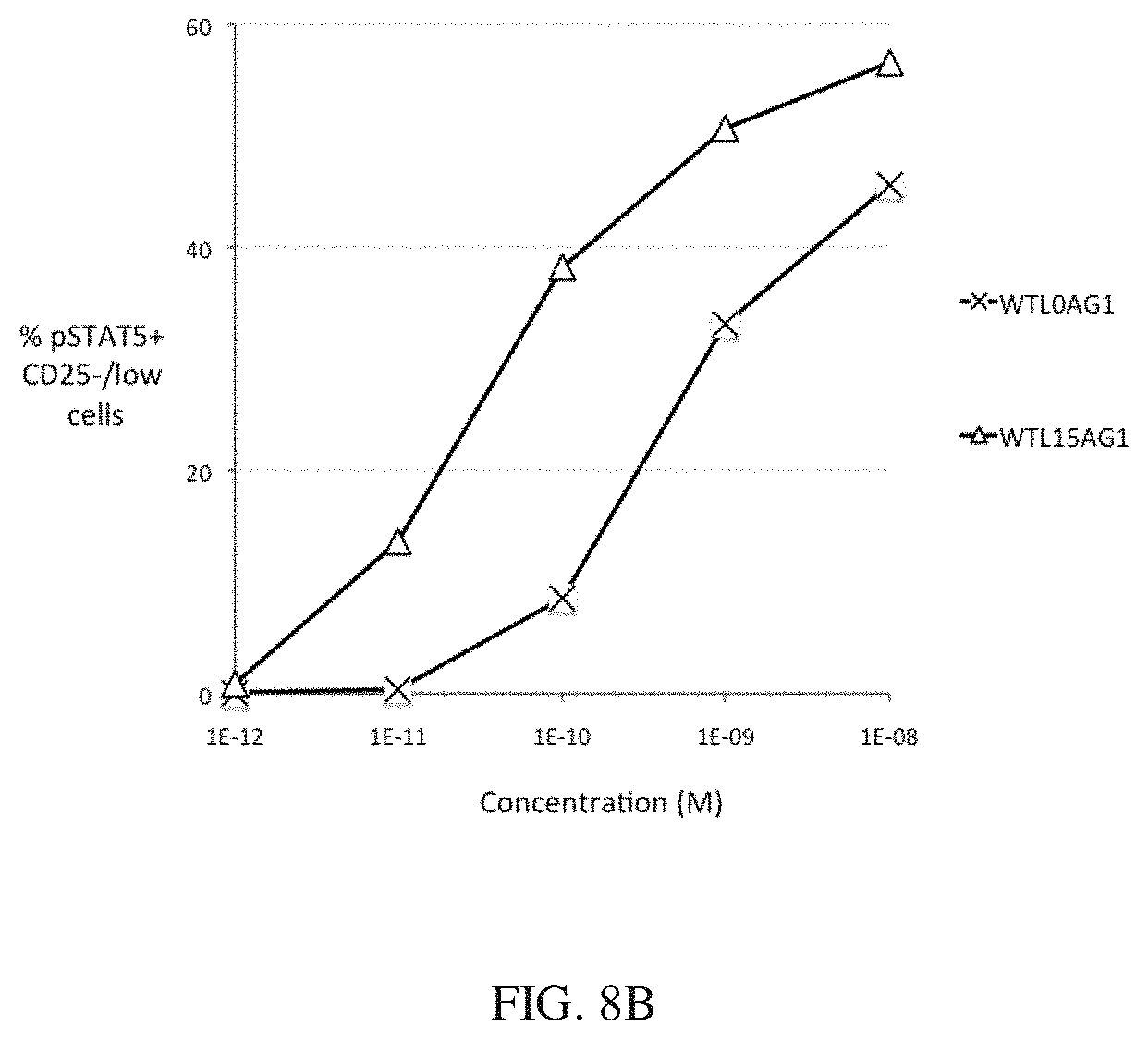
D00014
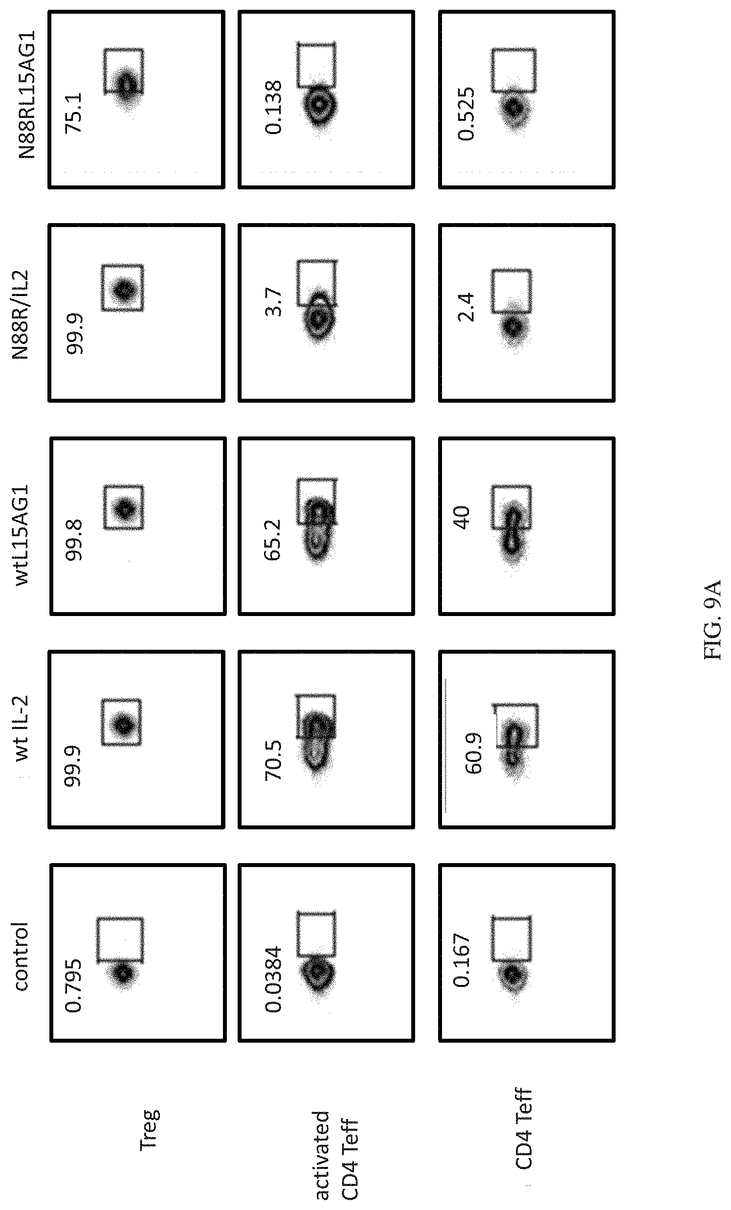
D00015
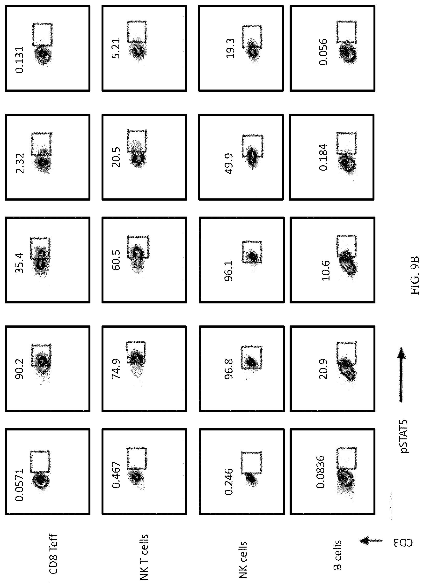
S00001
XML
uspto.report is an independent third-party trademark research tool that is not affiliated, endorsed, or sponsored by the United States Patent and Trademark Office (USPTO) or any other governmental organization. The information provided by uspto.report is based on publicly available data at the time of writing and is intended for informational purposes only.
While we strive to provide accurate and up-to-date information, we do not guarantee the accuracy, completeness, reliability, or suitability of the information displayed on this site. The use of this site is at your own risk. Any reliance you place on such information is therefore strictly at your own risk.
All official trademark data, including owner information, should be verified by visiting the official USPTO website at www.uspto.gov. This site is not intended to replace professional legal advice and should not be used as a substitute for consulting with a legal professional who is knowledgeable about trademark law.