Surfaces Having Reduced Non-specific Binding And Antigenicity
Hucknall; Angus ; et al.
U.S. patent application number 17/015315 was filed with the patent office on 2021-02-18 for surfaces having reduced non-specific binding and antigenicity. The applicant listed for this patent is Duke University. Invention is credited to Ashutosh Chilkoti, Nancy J. Ganson, Michael S. Hershfield, Angus Hucknall, Daniel Joh, Yizhi Qi.
| Application Number | 20210046188 17/015315 |
| Document ID | / |
| Family ID | 1000005249954 |
| Filed Date | 2021-02-18 |


View All Diagrams
| United States Patent Application | 20210046188 |
| Kind Code | A1 |
| Hucknall; Angus ; et al. | February 18, 2021 |
SURFACES HAVING REDUCED NON-SPECIFIC BINDING AND ANTIGENICITY
Abstract
Disclosed herein are compositions and methods for reducing the antigenicity of molecules. The antigenicity of a molecule may be reduced or eliminated by conjugating at least one branched polymer to the molecule to form a molecule-polymer conjugate. The branched polymer may include a backbone and a plurality of side chains, each side chain covalently attached to the backbone.
| Inventors: | Hucknall; Angus; (Durham, NC) ; Chilkoti; Ashutosh; (Durham, NC) ; Joh; Daniel; (Durham, NC) ; Ganson; Nancy J.; (Durham, NC) ; Qi; Yizhi; (Durham, NC) ; Hershfield; Michael S.; (Durham, NC) | ||||||||||
| Applicant: |
|
||||||||||
|---|---|---|---|---|---|---|---|---|---|---|---|
| Family ID: | 1000005249954 | ||||||||||
| Appl. No.: | 17/015315 | ||||||||||
| Filed: | September 9, 2020 |
Related U.S. Patent Documents
| Application Number | Filing Date | Patent Number | ||
|---|---|---|---|---|
| 16064424 | Jun 20, 2018 | |||
| PCT/US16/68141 | Dec 21, 2016 | |||
| 17015315 | ||||
| 62899353 | Sep 12, 2019 | |||
| 62270401 | Dec 21, 2015 | |||
| 62310534 | Mar 18, 2016 | |||
| 62329800 | Apr 29, 2016 | |||
| 62407403 | Oct 12, 2016 | |||
| Current U.S. Class: | 1/1 |
| Current CPC Class: | A61K 47/641 20170801; C08L 2203/02 20130101; C08L 71/08 20130101; A61K 47/58 20170801; A61K 47/59 20170801 |
| International Class: | A61K 47/58 20060101 A61K047/58; A61K 47/59 20060101 A61K047/59; A61K 47/64 20060101 A61K047/64; C08L 71/08 20060101 C08L071/08 |
Goverment Interests
STATEMENT REGARDING FEDERALLY SPONSORED RESEARCH
[0002] This invention was made with government support under National Institutes of Health Grant Numbers: R01-DK092665, R01-GM061232, 5T32-GM008487, R01-GM061232, and R01-A146611; National Cancer Institute Grant Number 1UG3CA211232-01; United States Department of Defense Special Operations Command Grant Number W81XWH-16-C-0219; and United States Department of Defense Combat Casualty Care Research Program Grant Number W81XWH-17-2-0045 National Institutes of Health. The government has certain rights in the invention.
Claims
1. A surface having reduced antigenicity and non-specific binding comprising: a surface comprising a plurality of poly[oligo(ethylene glycol) methyl ether methacrylate (POEGMA) polymers comprising a poly(methyl methacrylate) backbone and a plurality of side chains covalently attached to the backbone, each side chain comprising 1 to 9 ethylene glycol monomers repeated in tandem and terminating with an alkoxy moeity.
2. The surface of claim 1, wherein the surface is not reactive with anti-PEG antibodies in a subject.
3. The surface of claim 1, wherein the surface does not bind proteins, lipids, or carbohydrates non-specifically.
4. The surface of claim 1, wherein the is non-antigenic and does not induce an immune response.
5. The surface of claim 1, wherein the side chain comprises 1 to 5 ethylene glycol monomers repeated in tandem.
6. The surface of claim 1, wherein the side chain comprises 1 to 3 ethylene glycol monomers repeated in tandem.
7. The surface of claim 1, wherein the side chain comprises 2 to 3 ethylene glycol monomers repeated in tandem.
8. The surface of claim 1, wherein the side chain comprises 2 ethylene glycol monomers repeated in tandem.
9. The surface of claim 1, wherein the side chain comprises 3 ethylene glycol monomers repeated in tandem.
10. The surface of claim 1, wherein the alkoxy is methoxy, ethoxy, or propoxy.
11. The surface of claim 1, wherein the alkoxy is methoxy.
12. The surface of claim 1, wherein the POEGMA comprises monomers of ethylene glycol methyl ether methacrylate (EG-1-OMe).
13. The surface of claim 1, wherein the POEGMA comprises monomers of di(ethylene glycol) methyl ether methacrylate (EG-3-OMe).
14. The surface of claim 1, wherein the POEGMA comprises monomers of tri(ethylene glycol) methyl ether methacrylate (EG-3-OMe).
15. The surface of claim 1, wherein the POEGMA comprises monomers of penta(ethylene glycol) methyl ether methacrylate (EG-5-OMe).
16. The surface of claim 1, wherein the POEGMA comprises monomers of nona(ethylene glycol) methyl ether methacrylate (EG-9-OMe).
17. The surface of claim 1, wherein the surface is a material or a biomolecule.
18. The surface of claim 1, wherein the surface is a protein or a protein complex.
19. The surface of claim 1, wherein the surface is a material comprising glass, plastic, metal, ceramic, textile, or paper.
20. A method for reducing antigenicity of and non-specific binding to a surface, the method comprising: affixing to a surface a plurality of poly[oligo(ethylene glycol) methyl ether methacrylate (POEGMA) polymers comprising a poly(methyl methacrylate) backbone and a plurality of side chains covalently attached to the backbone, each side chain comprising 1 to 9 ethylene glycol monomers repeated in tandem and terminating with an alkoxy moeity.
21. The method of any one of clauses 20-, wherein the plurality of POEGMA polymers are affixed to the surface by contacting the surface with an initiator agent to form a macroinitiator; and incubating the macroinitiator with a monomer under conditions that permit free-radical polymerization and formation of a branched polymer to occur from the initiator agent to form the molecule-polymer conjugate.
22. The method of any one of clauses 20-, wherein the side chain comprises 1 to 5 ethylene glycol monomers repeated in tandem.
23. The method of any one of clauses 20-, wherein the side chain comprises 1 to 3 ethylene glycol monomers repeated in tandem.
24. The method of any one of clauses 20-, wherein the side chain comprises 2 to 3 ethylene glycol monomers repeated in tandem.
25. The method of any one of clauses 20-, wherein the side chain comprises 2 ethylene glycol monomers repeated in tandem.
26. The method of any one of clauses 20-, wherein the side chain comprises 3 ethylene glycol monomers repeated in tandem.
27. The method of any one of clauses 20-, wherein the alkoxy is methoxy, ethoxy, or propoxy.
28. The method of any one of clauses 20-, wherein the alkoxy is methoxy.
29. The method of any one of clauses 20-, wherein the POEGMA comprises monomers of ethylene glycol methyl ether methacrylate (EG-1-OMe).
30. The method of any one of clauses 20-, wherein the POEGMA comprises monomers of di(ethylene glycol) methyl ether methacrylate (EG-3-OMe).
31. The method of any one of clauses 20-, wherein the POEGMA comprises monomers of tri(ethylene glycol) methyl ether methacrylate (EG-3-OMe).
32. The method of any one of clauses 20-, wherein the POEGMA comprises monomers of penta(ethylene glycol) methyl ether methacrylate (EG-5-OMe).
33. The method of any one of clauses 20-, wherein the POEGMA comprises monomers of nona(ethylene glycol) methyl ether methacrylate (EG-9-OMe).
34. The method of any one of clauses 20-, wherein the surface is a material or a biomolecule.
35. The method of any one of clauses 20-, wherein the surface is a protein or a protein complex.
36. The method of any one of clauses 20-, wherein the surface is a protein and one or more POEGMA polymers is affixed to the polypeptide at the C-terminus, the N-terminus, or an internal amino acid of the polypeptide.
37. The method of any one of clauses 20-, wherein the surface is a material comprising glass, plastic, metal, ceramic, textile, or paper.
38. A surface coated with a plurality of POEGMA polymers by the method of any one of clauses 20-.
39. The surface of claim 38, wherein the surface is a biomolecule or a material.
40. The surface of claim 38, wherein the surface does not non-specifically bind proteins, carbohydrates, or lipids; does not induce an immune response, and is not reactive with anti-PEG antibodies.
Description
CROSS-REFERENCE TO RELATED APPLICATIONS
[0001] This application claims priority to U.S. Provisional Patent Application No. 62/899,353, filed on Sep. 12, 2019, and is a continuation-in-part of U.S. patent application Ser. No. 16/064,424, filed Jun. 20, 2018, which is a national phase application of International Patent Application No. PCT/US2016/068141, filed Dec. 21, 2016, which claims priority to U.S. Provisional Patent Application No. 62/270,401, filed Dec. 21, 2015; U.S. Provisional Patent Application No. 62/310,534, filed Mar. 18, 2016; U.S. Provisional Patent Application No. 62/329,800, filed Apr. 29, 2016; and U.S. Provisional Patent Application No. 62/407,403, filed Oct. 12, 2016, each of which is incorporated herein by reference in its entirety.
REFERENCE TO SEQUENCE LISTING
[0003] This application is filed with a Computer Readable Form of a Sequence Listing in accord with 37 C.F.R. .sctn. 1.821(c). The text file submitted by EFS, "028193-9357-US02_sequence_listing_9 Sep. 2018_ST25.txt," was created on Sep. 9, 2020, contains 8 sequences, has a file size of 4.39 Kbytes, and is hereby incorporated by reference in its entirety.
TECHNICAL FIELD
[0004] Described herein are coatings for surfaces comprising poly(oligoethylene glycol) methacrylate (POEGMA) "bottlebrushes" with sidechain lengths of 1-, 2- or 3-ethylene glycol (EG) repeats as the optimal polymer architecture to minimize binding of anti-PEG antibodies. These polymer coatings minimize nonspecific binding by proteins and cells and have reduced or eliminated antigenicity.
BACKGROUND
[0005] With more than a hundred peptides and proteins approved by the FDA to treat various diseases and many more in clinical and pre-clinical development, therapeutic peptides and proteins are an important class of drugs today. However, the clinical use of peptides and proteins is often challenged by their short plasma half-life, which can necessitate frequent injections and cause an undesirable peak-to-valley fluctuation of the drug concentration in vivo as well as reduce patient compliance and increase treatment cost. Other limitations of peptide and protein therapeutics may include poor stability, low solubility, and immunogenicity. To address these limitations, various delivery strategies have been developed for sustained delivery of peptide and protein therapeutics, ranging from particulate systems, depots, to chemical conjugation with long circulating polymers such as poly(ethylene glycol) (PEG), or recombinant fusions with long circulating proteins such as albumin or the Fc domain of antibodies.
[0006] PEGylation, or the covalent conjugation of therapeutics with the "stealth" polymer PEG, is one of the most widely used approaches to increase the circulation half-life and stability and to reduce the immunogenicity of biomolecule therapeutics such as polypeptides and polynucleotides. However, after nearly four decades of research and over two decades of clinical use, the drawbacks of PEGylation have begun to emerge. Conventional methods for the synthesis of PEGylated conjugates have significant limitations: (1) conjugation involves the reaction between protein-repulsive PEG chains and biomacromolecules, so that even with a large excess of polymer, steric hindrance still results in a low yield of conjugate, typically in the 10-20% range; (2) the presence of a large excess of unreacted polymer makes product purification non-trivial; and (3) conjugation typically involves reacting the chain-ends of the polymer with reactive side-groups on lysine and cysteine residues, which are often promiscuously distributed on the biomolecule, thus yielding chemically heterogeneous products that can significantly compromise the bioactivity of the drug and greatly complicate regulatory approval.
[0007] Furthermore, the immunogenicity of PEG has recently attracted much attention. Anti-PEG antibodies have been induced in patients treated with some PEGylated enzymes, and in clinical trials of PEG-uricase and PEG-asparaginase, these anti-PEG antibodies have markedly accelerated blood clearance, abrogated clinical efficacy, and increased the risk and severity of infusion reactions. Circulating anti-PEG antibodies have also been found in individuals naive to PEGylated materials, possibly induced by chronic exposure to free PEGs present in commonly used consumer products. High levels of such pre-existing anti-PEG antibodies have recently been linked to serious first-exposure allergic reactions to a PEGylated RNA aptamer, which led to early termination of a clinical trial.
[0008] The immune responses that the widely-used polymer, poly(ethylene glycol) (PEG), can trigger is of growing concern. Previously considered non-immunogenic, linear PEG modification ("PEGylation") has become the most popular synthetic strategy to confer materials with "stealth" properties to eliminate protein adsorption and cell adhesion on surfaces, improve the biocompatibility of implanted biomaterials, and when conjugated to "biologics"-typically peptide and protein drugs and more recently aptamers--enhance their blood circulation and reduce their recognition by the immune system.
[0009] Products containing linear PEG constitute an estimated multi-billion-dollar market. However, evidence supporting the existence and clinical relevance of anti-PEG immunity, namely in the form of anti-PEG antibodies (APAs), is mounting. PEGylated therapeutics are now known to induce APAs in both animals and humans. Further, APAs are known to be present in much of the general population, presumably from chronic exposure to PEG from consumer products, with approximately 37% showing moderate (100 ng/mL) and 8% showing high levels (500 ng/mL) of APAs. This potentially complicates the development of drugs, devices, or diagnostics modified with PEG (or PEG-derivatives), given the possibility of unwanted interference by APAs. In fact, clinical experience with PEGylated therapeutics has already indicated that APAs can not only cause increased clearance rates and loss of efficacy, but also can lead to serious anaphylactic or hypersensitivity reactions. Notably, the issues posed by APAs has now been recognized by the Food and Drug Administration (FDA), which currently requests testing patients for APAs before treatment with experimental PEGylated compounds.
[0010] Several studies have proposed the use of alternative--non-PEG derived--stealth polymers such as polyzwitterions, poly(2-ethyl 2-oxazoline), and polyglycerol to circumvent this issue. However, transitioning to such polymers might offer an incomplete or temporary solution, for two reasons. First, antibodies (Abs) against other natural and synthetic polymers have been reported previously, suggesting that other (non-PEG derived) stealth polymers might be capable of inducing an immune response after repeated administration over time. Second, both animal and human APAs were discovered to cross-react with other synthetic polymers; this "polypharmacy" nature of polymer-reactive Abs likely adds further design constraints on candidate materials proposed as alternatives to PEG.
[0011] The large investments already made in PEGylation and its mainstream status underscore a pressing need to thoroughly investigate methods to tackle the emerging problem of `PEG antigenicity` directly, without replacing PEG itself. Ideally, this would be accomplished without the need for potentially aggressive interventions such as preemptive immunosuppression, pre-injection of large quantities of free PEG to saturate APAs or removing PEG altogether in select situations. While these proposed strategies are intriguing, their downsides include potential additional risk to patients, reduction in therapeutic efficacy, suboptimal assay or device performance, and overall inconvenience.
[0012] What is needed are non-fouling coatings for biointerfacial applications that have reduced antigenicity and do not induce anti-PEG antibodies.
SUMMARY
[0013] One embodiment described herein is a surface having reduced antigenicity and non-specific biomolecule binding comprising: a surface comprising a plurality of poly[oligo(ethylene glycol) methyl ether methacrylate (POEGMA) polymers comprising a poly(methyl methacrylate) backbone and a plurality of side chains covalently attached to the backbone, each side chain comprising 1 to 9 ethylene glycol monomers repeated in tandem and terminating with an alkoxy moeity. In one aspect, the surface has reduced antigenicity and reduced immunogenicity. In another aspect, the surface is not reactive with anti-PEG antibodies in a subject. In another aspect, the surface does not bind proteins, lipids, or carbohydrates non-specifically. In another aspect, the side chain comprises 1 to 5 ethylene glycol monomers repeated in tandem. In another aspect, the side chain comprises 1 to 3 ethylene glycol monomers repeated in tandem. In another aspect, the side chain comprises 2 to 3 ethylene glycol monomers repeated in tandem. In another aspect, the side chain comprises 2 ethylene glycol monomers repeated in tandem. In another aspect, the side chain comprises 3 ethylene glycol monomers repeated in tandem. In another aspect, the alkoxy is methoxy, ethoxy, or propoxy. In another aspect, the alkoxy is methoxy. In another aspect, the POEGMA comprises monomers of ethylene glycol methyl ether methacrylate (EG-1-OMe). In another aspect, the POEGMA comprises monomers of di(ethylene glycol) methyl ether methacrylate (EG-3-OMe). In another aspect, the POEGMA comprises monomers of tri(ethylene glycol) methyl ether methacrylate (EG-3-OMe). In another aspect, the POEGMA comprises monomers of penta(ethylene glycol) methyl ether methacrylate (EG-5-OMe). In another aspect, the POEGMA comprises monomers of nona(ethylene glycol) methyl ether methacrylate (EG-9-OMe). In another aspect, the surface is a material or a biomolecule. In another aspect, the surface is a protein or a protein complex. In another aspect, the surface is a material comprising glass, plastic, metal, ceramic, textile, or paper.
[0014] Another embodiment described herein is a method for reducing antigenicity of and non-specific biomolecule binding to a surface, the method comprising: affixing to a surface a plurality of poly[oligo(ethylene glycol) methyl ether methacrylate (POEGMA) polymers comprising a poly(methyl methacrylate) backbone and a plurality of side chains covalently attached to the backbone, each side chain comprising 1 to 9 ethylene glycol monomers repeated in tandem and terminating with an alkoxy moeity. In one aspect, the plurality of POEGMA polymers are affixed to the surface by contacting the surface with an initiator agent to form a macroinitiator; and incubating the macroinitiator with a monomer under conditions that permit free-radical polymerization and formation of a branched polymer to occur from the initiator agent to form the molecule-polymer conjugate. In another aspect, the side chain comprises 1 to 5 ethylene glycol monomers repeated in tandem. In another aspect, the side chain comprises 1 to 3 ethylene glycol monomers repeated in tandem. In another aspect, the side chain comprises 2 to 3 ethylene glycol monomers repeated in tandem. In another aspect, the side chain comprises 2 ethylene glycol monomers repeated in tandem. In another aspect, the side chain comprises 3 ethylene glycol monomers repeated in tandem. In another aspect, the alkoxy is methoxy, ethoxy, or propoxy. In another aspect, the alkoxy is methoxy. In another aspect, the POEGMA comprises monomers of ethylene glycol methyl ether methacrylate (EG-1-OMe). In another aspect, the POEGMA comprises monomers of di(ethylene glycol) methyl ether methacrylate (EG-3-OMe). In another aspect, the POEGMA comprises monomers of tri(ethylene glycol) methyl ether methacrylate (EG-3-OMe). In another aspect, the POEGMA comprises monomers of penta(ethylene glycol) methyl ether methacrylate (EG-5-OMe). In another aspect, the POEGMA comprises monomers of nona(ethylene glycol) methyl ether methacrylate (EG-9-OMe). In another aspect, the surface is a material or a biomolecule. In another aspect, the surface is a protein or a protein complex. In another aspect, the surface is a protein and one or more POEGMA polymers is affixed to the polypeptide at the C-terminus, the N-terminus, or an internal amino acid of the polypeptide. In another aspect, the surface is a material comprising glass, plastic, metal, ceramic, textile, or paper.
[0015] Another embodiment described herein is a surface coated with a plurality of POEGMA polymers by a method described herein. In one aspect, the surface is a biomolecule or a material. In another aspect, the surface has reduced antigenicity and reduced immunogenicity; is not reactive with anti-PEG antibodies in a subject; and does not bind proteins, lipids, or carbohydrates non-specifically.
[0016] In another embodiment, the disclosure relates to methods of reducing the antigenicity of a molecule. The methods may include conjugating at least one branched polymer to a molecule to form a molecule-polymer conjugate, wherein the molecule comprises a polypeptide, a polynucleotide, a small molecule, or a combination thereof, wherein the branched polymer comprises a backbone and a plurality of side chains, each side chain is covalently attached to the backbone, wherein the backbone comprises at least one of an acrylate, methacrylate, acrylamide, methacrylamide, carbonate, phosphoester, oxazoline, or a combination thereof, and wherein the molecule-polymer conjugate has reduced or eliminated antigenicity compared to a control. In some embodiments, the molecule is conjugated to the backbone of the branched polymer. In some embodiments, the molecule is conjugated to the backbone of the branched polymer via a linker. In some embodiments, each side chain has a first terminal end and a second terminal end, wherein the first terminal end is covalently attached to the backbone, and wherein the second terminal end independently comprises an alkyl, ester, amine, amide, or carboxyl group. In some embodiments, each side chain has a first terminal end and a second terminal end, wherein the first terminal end is covalently attached to the backbone, and wherein the second terminal end does not include a hydroxyl group. In some embodiments, each side chain is a linear polymer. In some embodiments, at least one side chain comprises 1 monomer. In some embodiments, each side chain comprises at least 2 monomers repeated in tandem. In some embodiments, each side chain comprises less than 25 monomers repeated in tandem. In some embodiments, each side chain comprises 3 to 9 monomers repeated in tandem. In some embodiments, each side chain comprises 3 monomers repeated in tandem. In some embodiments, the monomer of each side chain is independently selected from betaine, phosphorylcholine, phosphorylethanolamine, sarcosine, ethylene glycol, or a combination thereof. In some embodiments, the betaine comprises carboxybetaine, sulfobetaine, or a combination thereof. In some embodiments, the monomer of at least one side chain comprises ethylene glycol. In some embodiments, the monomer of each side chain comprises ethylene glycol. In some embodiments, more than one branched polymer is conjugated to the molecule, each branched polymer conjugated to a different site of the molecule. In some embodiments, the molecule comprises a polypeptide, and wherein one branched polymer is conjugated to the polypeptide at a site selected from the C-terminus, the N-terminus, and an internal amino acid of the polypeptide. In some embodiments, the molecule comprises a polypeptide, and wherein more than one branched polymer is conjugated to the polypeptide, each branched polymer conjugated to a different site of the polypeptide selected from the C-terminus, the N-terminus, an internal amino acid, or a combination thereof.
[0017] In some embodiments, the molecule comprises a polypeptide comprising a sortase A recognition site, and wherein the branched polymer and the polypeptide are incubated with sortase A under conditions to conjugate the branched polymer to the sortase recognition site of the polypeptide. In some embodiments, the molecule comprises a polypeptide comprising a sortase A recognition site, and wherein the conjugating comprises: a) contacting the molecule with a sortase A and an initiator agent under conditions that permit attachment of the initiator agent to the sortase A recognition site to form a macroinitiator; and b) incubating the macroinitiator with a monomer under conditions that permit free-radical polymerization and formation of a branched polymer to occur from the initiator agent to form the molecule-polymer conjugate. In some embodiments, the sortase A recognition site comprises LPXTG (SEQ ID NO: 1), wherein X is any amino acid. In some embodiments, the macroinitiator and monomer are incubated with a catalyst in step (b). In some embodiments, the monomer in step (b) comprises at least one of an acrylate, methacrylate, acrylamide, and methacrylamide. In some embodiments, the method further includes separating the molecule-polymer conjugate formed in step (b) from the unreacted macroinitiator. In some embodiments, the branched polymer is synthesized and subsequently grafted to the molecule to form the molecule-polymer conjugate. In some embodiments, the conjugating comprises attaching an initiator agent to the molecule to form a macroinitiator; and incubating the macroinitiator with a monomer under conditions that permit free-radical polymerization and formation of a branched polymer to occur from the initiator agent to form the molecule-polymer conjugate. In some embodiments, the branched polymer is synthesized using free-radical polymerization. In some embodiments, the branched polymer is synthesized using at least one method selected from ionic ring-opening polymerization (ionic ROP), ring opening metathesis polymerization, ionic polymerization, condensation polymerization, and coordination polymerization.
[0018] In a further aspect, the disclosure relates to methods of making a molecule-polymer conjugate having reduced or eliminated antigenicity compared to a control, from a molecule comprising a polypeptide having a sortase A recognition site. The methods may include a) contacting the molecule with a sortase A and an initiator agent under conditions that permit attachment of the initiator agent to the sortase A recognition site to form a macroinitiator; and b) incubating the macroinitiator with a monomer under conditions that permit free-radical polymerization and formation of a branched polymer to occur from the initiator agent to form the molecule-polymer conjugate, wherein the branched polymer comprises a backbone and a plurality of side chains, each side chain covalently attached to the backbone. In some embodiments, the sortase A recognition site comprises LPXTG (SEQ ID NO: 1), wherein X is any amino acid. In some embodiments, the macroinitiator and monomer are incubated with a catalyst in step (b). In some embodiments, the monomer in step (b) comprises at least one of an acrylate, methacrylate, acrylamide, and methacrylamide. In some embodiments, the method further includes separating the molecule-polymer conjugate formed in step (b) from the unreacted macroinitiator, wherein the yield of molecule-polymer conjugate is at least about 50% of the total conjugates and macroinitiators which are separated. In some embodiments, the molecule-polymer conjugate is separated by chromatography. In some embodiments, the chromatography comprises size-exclusion chromatography, ion exchange chromatography, affinity chromatography, or hydrophobic interaction chromatography, or a combination thereof. In some embodiments, the chromatography comprises size-exclusion chromatography. In some embodiments, the free-radical polymerization comprises at least one of atom transfer radical polymerization (ATRP), reversible addition-fragmentation chain transfer (RAFT), radical ring-opening polymerization (radical ROP), nitroxide-mediated radical polymerization (NMP), iniferter polymerization, free radical polymerization, cobalt-mediated radical polymerization, telluride-mediated polymerization, and stibine-mediated polymerization. In some embodiments, the molecule comprises a polypeptide, a polynucleotide, a small molecule, or a combination thereof.
[0019] Another aspect of the disclosure provides a molecule-polymer conjugate having reduced or eliminated antigenicity compared to a control. The molecule-polymer conjugates may include a branched polymer comprising a backbone and a plurality of side chains, each side chain covalently attached to the backbone; and a molecule conjugated to the backbone of the branched polymer, wherein the molecule comprises a polypeptide, a polynucleotide, a small molecule, or a combination thereof, wherein each side chain is a linear polymer, wherein the backbone comprises at least one of an acrylate, methacrylate, acrylamide, methacrylamide, carbonate, phosphoester, oxazoline, or a combination thereof. In some embodiments, the molecule is conjugated to the backbone of the branched polymer via a linker. In some embodiments, each side chain has a first terminal end and a second terminal end, wherein the first terminal end is covalently attached to the backbone, and wherein the second terminal end independently comprises an alkyl, ester, amine, amide, or carboxyl group. In some embodiments, each side chain has a first terminal end and a second terminal end, wherein the first terminal end is covalently attached to the backbone, and wherein the second terminal end does not include a hydroxyl group. In some embodiment the second terminal end includes an alkoxy group.
[0020] In some embodiments, at least one side chain comprises at least 1 monomer. In some embodiments, each side chain comprises at least 2 monomers repeated in tandem. In some embodiments, each side chain comprises less than 25 monomers repeated in tandem. In some embodiments, each side chain comprises less than 10 monomers repeated in tandem. In some embodiments, each side chain comprises less than 5 monomers repeated in tandem. In some embodiments, each side chain comprises less than 4 monomers repeated in tandem. In some embodiments, each side chain comprises less than 3 monomers repeated in tandem. In some embodiments, each side chain comprises 1 to 9 monomers repeated in tandem. In some embodiments, each side chain comprises 3 to 9 monomers repeated in tandem. In some embodiments, each side chain comprises 1 to 3 monomers repeated in tandem. In some embodiments, each side chain comprises 2 to 3 monomers repeated in tandem. In some embodiments, each side chain comprises 3 monomers repeated in tandem. In some embodiments, each side chain comprises 2 monomers repeated in tandem.
[0021] In some embodiments, the monomer of each side chain is independently selected from betaine, phosphorylcholine, phosphorylethanolamine, sarcosine, ethylene glycol, or a combination thereof. In some embodiments, the betaine comprises carboxybetaine, sulfobetaine, or a combination thereof. In some embodiments, the monomer of at least one side chain comprises ethylene glycol. In some embodiments, the monomer of each side chain comprises ethylene glycol. In some embodiments, more than one branched polymer is conjugated to the molecule, each branched polymer conjugated to a different site of the molecule. In some embodiments, the molecule comprises a polypeptide, and wherein one branched polymer is conjugated to the polypeptide at a site selected from the C-terminus, the N-terminus, and an internal amino acid of the polypeptide. In some embodiments, the molecule comprises a polypeptide, and wherein more than one branched polymer is conjugated to the polypeptide, each branched polymer conjugated to a different site of the polypeptide selected from the C-terminus, the N-terminus, an internal amino acid, or a combination thereof.
[0022] In some embodiments, the branched polymer comprises poly[oligo(ethylene glycol) methyl ether methacrylate] (POEGMA), and wherein the POEGMA comprises: a backbone comprising poly(methyl methacrylate); and a plurality of side chains covalently attached to the backbone, each side chain comprising at least 1 monomer of ethylene glycol (EG) repeated in tandem. In some embodiments, at least one side chain comprises at least 1 monomer of ethylene glycol (EG). In some embodiments, each side chain comprises at least 2 monomers of ethylene glycol (EG) repeated in tandem. In some embodiments, each side chain comprises at least 3 monomers of ethylene glycol (EG) repeated in tandem. In some embodiments, each side chain comprises at least 10 monomers of ethylene glycol (EG) repeated in tandem. In some embodiments, each side chain comprises less than 25 monomers of ethylene glycol (EG) repeated in tandem. In some embodiments, each side chain comprises 1 monomer of ethylene glycol (EG). In some embodiments, each side chain comprises 2 monomers of ethylene glycol (EG) repeated in tandem. In some embodiments, each side chain comprises 3 monomers of ethylene glycol (EG) repeated in tandem. In some embodiments, each side chain comprises 5 monomers of ethylene glycol (EG) repeated in tandem. In some embodiments, each side chain comprises 6 monomers of ethylene glycol (EG) repeated in tandem. In some embodiments, each side chain comprises 9 monomers of ethylene glycol (EG) repeated in tandem. In some embodiments, each side chain comprises 1 to 9 monomers of ethylene glycol (EG) repeated in tandem. In some embodiments, each side chain comprises 3 to 9 monomers of ethylene glycol (EG) repeated in tandem. In some embodiments, each side chain comprises 2 to 3 monomers of ethylene glycol (EG) repeated in tandem. In some embodiments, the molecule-POEGMA conjugate or surface-POEGMA is not reactive with pre-existing anti-PEG antibodies in a subject and does not bind to proteins non-specifically.
[0023] In some embodiments, the molecule comprises one or more peptides or protein therapeutic agents selected from a monoclonal antibody, blood factor, betatrophin, exendin, enzyme, asparaginase, glutamase, arginase, arginine deaminase, adenosine deaminase (ADA), ADA-2, ribonuclease, cytosine deaminase, trypsin, chymotrypsin, papain, growth factor, epidermal growth factor (EGF), insulin, insulin-like growth factor (IGF), transforming growth factor (TGF), nerve growth factor (NGF), platelet-derived growth factor (PDGF), bone morphogenic protein (BMP), fibroblast growth factor (FGF), somatostatin, somatotropin, somatropin, somatrem, calcitonin, parathyroid hormone, colony stimulating factors (CSF), clotting factors, tumor necrosis factors (TNF), gastrointestinal peptides, vasoactive intestinal peptide (VIP), cholecystokinin (CCK), gastrin, secretin, erythropoietins, growth hormone, GRF, vasopressins, octreotide, pancreatic enzymes, superoxide dismutase, thyrotropin releasing hormone (TRH), thyroid stimulating hormone, luteinizing hormone, luteinizing hormone-releasing hormone (LHRH), growth hormone releasing hormone (GHRH), tissue plasminogen activators, interleukins, interleukin-1, interleukin-15, interleukin-2, interleukin-10, colony stimulating factor, granulocyte macrophage colony-stimulating factor (GM-CSF), interleukin-1 receptor antagonist (IL-1RA), glucagon-like peptide-1 (GLP-1), exenatide, GLP-1 R multi-agonist, GLP-1 R antagonist, GLP-2, TNF-related apoptosis-inducing ligand (TRAIL), leptin, ghrelin, granulocyte monocyte colony stimulating factor (GM-CSF), interferons, interferon-.alpha., interferon-gamma, human growth hormone (hGH) and antagonist, macrophage activator, chorionic gonadotropin, heparin, atrial natriuretic peptide, hemoglobin, relaxin, cyclosporine, oxytocin, vaccines, monoclonal antibodies, single chain antibodies, ankyrin repeat proteins, affibodies, activin receptor 2A extracellular domain, alpha-2 macroglobulin, alpha-melanocyte, apelin, bradykinin B2 receptor antagonist, cytotoxic T-lymphocyte-associated protein (CTLA-4), elafin, Factor IX, Factor Vlla, Factor VIII, hepcidin, infestin-4, kallikrein inhibitor, L4F peptide, lacritin, parathyroid hormone (PTH), peptide YY (PYY), thioredoxin, thymosin B4, urate oxidase, urodilatin, aptamers, silencing RNA, microRNA, long non-coding RNA, ribozymes, analogs and derivatives thereof, and combinations thereof. In some embodiments, the molecule comprises a polypeptide, and wherein the polypeptide comprises a His-tag, a stimulus-responsive polypeptide, or a combination thereof. In some embodiments, the stimulus-responsive polypeptide is selected from an elastin-like polypeptide, a polypeptide comprising a repeated motif, and a resilin-like polypeptide. In some embodiments, the molecule-polymer conjugate has: an in vivo half-life that is at least 25% greater compared with the in vivo half-life of the molecule itself; or an in vivo biodistribution to a tissue, organ, or disease site that is at least 25% greater than the in vivo biodistribution of the molecule itself; or a reduced binding to anti-PEG antibodies compared to a control; or a reduced immune response compared to a control; or a combination thereof. In some embodiments, the molecule-polymer conjugates have an in vivo half-life that is at least 80% greater than the in vivo half-life of the molecule itself. In some embodiments, the control comprises the molecule conjugated to a polymer that is not branched. In some embodiments, the control comprises the molecule by itself. In some embodiments, the control comprises the molecule conjugated to a linear polymer. In some embodiments, the control comprises the molecule conjugated to unbranched PEG. In some embodiments, the molecule comprises a polypeptide, and wherein at least about 20% of the polypeptides have a conjugated branched polymer solely at the C-terminus. In some embodiments, at least about 75% of the polypeptides have a conjugated branched polymer solely at the C-terminus. In some embodiments, at least about 90% of the polypeptides have a conjugated branched polymer solely at the C-terminus. In some embodiments, the yield of molecule-polymer conjugate is at least about 75%. In some embodiments, the yield of molecule-polymer conjugate is at least about 85%.
[0024] The disclosure provides for other aspects and embodiments that will be apparent in light of the following detailed description and accompanying figures.
BRIEF DESCRIPTION OF THE DRAWINGS
[0025] The patent or application file contains at least one drawing executed in color. Copies of this patent or patent application publication with color drawing(s) will be provided by the Office upon request and payment of the necessary fee.
[0026] FIG. 1A-C. Synthetic scheme of exendin-C-POEGMA. FIG. 1A shows Recombinant expression of the sortase A substrate, exendin-srt-His6-ELP, and purification by ITC. FIG. 1B shows sortase-catalyzed site-specific attachment of the ATRP initiator AEBMP to the C-terminus of exendin to generate exendin-C--Br. FIG. 1C shows in situ ATRP of OEGMA from exendin-C--Br yielding exendin-C-POEGMA. ITC: inverse transition cycling, ELP: elastin-like polypeptide, srt: sortase A recognition sequence "LPETG" (SEQ ID NO: 2), AEBMP: N-(2-(2-(2-(2-aminoacetamido)acet-amido)acetamido) ethyl)-2-bromo-2-methylpropanamide. Images from the RCSB PDB (www.rcsb.org) of: PDB ID 1T2P (sortase A); PDB ID 1JRJ (exendin-4).
[0027] FIG. 2A-C. Characterization of exendin-C--Br macroinitiator and EG9 exendin-C-POEGMA conjugates. FIG. 2A shows Coomassie-stained SDS-PAGE analysis of initiator attachment on exendin by sortase A. Lane 1: MW marker, lane 2: sortase reaction mixture after 18 h of reaction, lane 3: purified exendin-C--Br macroinitiator. FIG. 2B shows SEC traces of ATRP reaction mixtures of grafting EG9 POEGMA from exendin-C--Br carried out for 0.5 h, 1 h, 1.25 h, 2 h and 3 h, detected by UV-vis absorbance at 280 nm. FIG. 2C shows cyclic adenosine monophosphate (cAMP) response of native exendin and EG9 exendin-C-POEGMA conjugates with Mns of 25.4 kDa, 54.6 kDa, 66.2 kDa, 97.2 kDa and 155.0 kDa in baby hamster kidney (BHK) cells expressing the GLP-1R. Results are plotted as mean.+-.standard error of the mean (SEM), n=3. Half-maximal effective concentration (EC.sub.50) values are summarized in Table 3.
[0028] FIG. 3A-F. Assessment of MW-dependent in vivo efficacy of EG9 exendin-C-POEGMA conjugates. Blood glucose levels in fed mice were measured before and after a single s.c. injection of unmodified exendin (FIG. 3A), or 25.4 kDa (FIG. 3B), 54.6 kDa (FIG. 3C), 97.2 kDa (FIG. 3D), and 155.0 kDa (FIG. 3E) EG9 exendin-C-POEGMA conjugates, compared to PBS control. The peptide and conjugates were administered at 25 nmol/kg and PBS was injected at equivalent volume at t=0 h. Blood glucose levels were normalized to the average glucose levels measured 24 h and immediately before injection. Data were analyzed by repeated measures two-way analysis of variance (ANOVA), followed by post hoc Dunnett's multiple comparison test. FIG. 3F shows the area under the curve (AUC) of blood glucose profiles (0 h to 144 h, with respect to 0% baseline) as a function of conjugate Mn. AUCs were compared using one-way ANOVA followed by post hoc Tukey's multiple comparison test. In all panels, results are plotted as mean.+-.SEM, n=6, *P<0.05, **P<0.01, ***P<0.001 and ****P<0.0001.
[0029] FIG. 4A-D. Intraperitoneal glucose tolerance test (IPGTT) of an EG9 exendin-POEGMA in mice. Mouse blood glucose levels measured in an IPGTT performed at 24 h and 72 h after a single s.c. injection of the 54.6 kDa EG9 exendin-POEGMA conjugate (24 h, FIG. 4A; 72 h, FIG. 4B) or unmodified exendin at 25 nmol/kg (24 h, FIG. 4C or 72 hr, FIG. 4D), compared to PBS of equivalent volume. Mice were fasted for 6 h prior to glucose challenge by an intraperitoneal (i.p.) injection of 1.5 g/kg of glucose. Results are plotted as mean.+-.SEM, n=5 in FIG. 4A-B, n=3 in FIG. 4C-D. AUCs of treatment and PBS were compared using an unpaired parametric two-tailed t test (**P<0.01, and ****P<0.0001). Exendin was not significant at either time point.
[0030] FIG. 5A-D. Assessment of reactivity of exendin-C-POEGMA conjugates toward anti-PEG antibodies in patient plasma samples. FIG. 5A shows a direct ELISA probing 54.6 kDa EG9 exendin-C-POEGMA conjugate, native exendin, adenosine deaminase (ADA), bovine serum albumin (BSA), Krystexxa.RTM. (PEG-uricase) and Adagen.RTM. (PEG-ADA) with diluent (1% BSA in PBS), an anti-PEG negative patient plasma sample, or one of two anti-PEG positive plasma samples. FIG. 5B shows a competitive ELISA, where various amounts of exendin, 54.6 kDa EG9 exendin-C-POEMGA, ADA and Adagen.RTM. were allowed to compete with Krystexxa.RTM. for binding with anti-PEG antibodies in a positive plasma sample. FIG. 5C shows direct and FIG. 5D shows competitive assays described in FIG. 5A and FIG. 5B, respectively, performed with a 55.6 kDa EG3 exendin-C-POEGMA conjugate. In all assays, the same unmodified peptide/protein content or similar PEG/OEG content in the case of polymer-modified samples per well were compared. See Methods section for details. Results are plotted as mean.+-.SEM, n=3 in FIG. 5A-B, n=5 in FIG. 5C-D. Data were analyzed by two-way ANOVA, followed by post hoc Dunnett's multiple comparison test (**P<0.01, and ****P<0.0001).
[0031] FIG. 6A-D. Assessment of in vivo efficacy and pharmacokinetics of exendin-C-POEGMA conjugates. Blood glucose levels in fed mice measured before and after a single s.c. injection of 55.6 kDa (FIG. 6A) and 71.6 kDa (FIG. 6B) EG3 exendin-C-POEGMA conjugates at 25 nmol/kg or PBS at equivalent volume administered at t=0 h. Blood glucose levels were normalized to the average glucose levels measured 24 h and immediately before injection. Data were analyzed by repeated measures two-way ANOVA, followed by post hoc Dunnett's multiple comparison test (n=5, *P<0.05, **P<0.01, and ****P<0.0001). Exendin (FIG. 6C) and exendin-C-POEGMA conjugates (54.6 kDa EG9, 55.6 kDa EG3 and 71.6 kDa EG3) (FIG. 6D) were fluorescently labeled with Alexa Fluor.RTM. 488 and injected into mice (n=3) s.c. at 75 nmol/kg (45 nmol/kg fluorophore). Blood samples were collected via tail vein at various time points for fluorescence quantification. Data were analyzed using a non-compartmental fit (solid lines) to derive the pharmacokinetic parameters shown in Table 6. Results in all panels are plotted as mean.+-.SEM.
[0032] FIG. 7A-B. FIG. 7A shows a CuCl.sub.2-stained SDS-PAGE analysis of exendin-srt-His6-ELP purification by inverse transition cycling (ITC). Lane 1: marker, lane 2: E. coli lysate, lanes 3 and 4: soluble protein after one and two ITC cycles (yield: .about.60 mg/L of culture). ELP: elastin-like polypeptide. FIG. 7B shows His6-sortase A purification by immobilized metal affinity chromatography (IMAC). Lane 1: marker, lane 2: E. coli lysate, lanes 3 and 4: first and second elution washes with imidazole (yield: .about.400 mg/L of culture). His6: hexahistidine.
[0033] FIG. 8A-B. Liquid chromatography electrospray ionization mass spectrometry (LC/ESI-MS) characterization of OEGMA monomer with (FIG. 8A) an average mass of .about.500 Da or .about.9 side-chain ethylene glycol repeats (EG9), and (FIG. 8B) a mass of 232 Da or 3 side-chain EG repeats (EG3). Peaks were detected as [M+Na]+.
[0034] FIG. 9A-B. Physical characterization of EG9 exendin-C-POEGMA conjugates. FIG. 9A shows SEC traces of ATRP reaction mixtures of grafting EG9 POEGMA from the exendin-C--Br macroinitiator carried out for various times with RI detection. Due to its small size and low concentration, the signal from the residual exendin-C--Br was too low to be observed by RI detection. FIG. 9B shows Coomassie-stained SDS-PAGE analysis of EG9 exendin-C-POEGMA conjugates purified by a single round of preparative SEC. Lane 1: marker, from left to right in lanes 2-6: purified 155.0 kDa, 97.2 kDa, 66.2 kDa, 54.6 kDa and 25.4 kDa EG9 conjugates.
[0035] FIG. 10A-B. Assessment of in vivo dose-dependent efficacy of EG9 exendin-C-POEGMA. Overlaid normalized (FIG. 10A) and un-normalized (FIG. 10B) blood glucose levels of 6-wk-old male C57BL/6J mice (n=3) maintained on a 60 kCal % diet measured before and after a single s.c. injection of a 66.2 kDa EG9 exendin-C-POEGMA conjugate at 25, 50, 80 nmol/kg or phosphate buffered saline (PBS) control of equivalent volume administered at t=0 h. Blood glucose levels in panel a were normalized to the average glucose levels measured 24 h prior to and immediately before injection. FIG. 10C shows overlaid weight profiles for all treatment and control groups. Weights are reported as % change from 0 h time point. Results in all panels are plotted as mean.+-.SEM.
[0036] FIG. 11A-B. Assessment of in vivo efficacy of unmodified exendin. Normalized (FIG. 11A) and un-normalized (FIG. 11B) blood glucose profiles of fed mice (n=6) that received a single s.c. injection of unmodified exendin administered at 25 nmol/kg, compared to PBS control at equivalent volume injected at t=0 h. Blood glucose levels in panel a were normalized to the average glucose levels measured 24 h prior to and immediately before injection. Results are plotted as mean.+-.SEM.
[0037] FIG. 12A-C. Assessment of in vivo MW-dependent efficacy of EG9 exendin-C-POEGMA conjugates. Overlaid normalized (FIG. 12A) and un-normalized (FIG. 12B) fed blood glucose levels in mice (n=6) measured before and after receiving a single s.c. injection of 25.4 kDa, 54.6 kDa, 97.2 kDa, 155.0 kDa EG9 exendin-POEGMA conjugates at 25 nmol/kg compared to PBS control at equivalent volume injected at t=0 h. Blood glucose levels in panel a were normalized to the average glucose levels measured 24 h prior to and immediately before injection. FIG. 12C shows overlaid weight profiles for all treatment and control groups. Weights are reported as % change from 0 h time point. Weights were not measured for the exendin group at t=144 h. Results in all panels are plotted as mean.+-.SEM.
[0038] FIG. 13. Cyclic adenosine monophosphate (cAMP) response of native exendin and EG3 exendin-C-POEGMA conjugates with Mns of 26.3 kDa, 55.6 kDa and 71.6 kDa in baby hamster kidney (BHK) cells expressing the glucagon-like peptide-1 receptor (GLP-1R). Results are plotted as mean.+-.SEM. Half-maximal effective concentration (EC.sub.50) values are summarized in Table 3.
[0039] FIG. 14A-D. Assessment of in vivo efficacy of an EG3 exendin-C-POEGMA conjugate. Un-normalized blood glucose levels in fed mice (n=3) measured before and after receiving a single s.c. injection of 55.6 kDa (FIG. 14A) and 71.6 kDa (FIG. 14B) EG3 exendin-POEGMA conjugate at 25 nmol/kg compared to PBS control at equivalent volume administered at t=0 h. Weight profiles for c) 55.6 kDa (FIG. 14C) and 71.6 kDa (FIG. 14D) EG3 exendin-C-POEGMA and PBS control groups. Weights are reported as % change from 0 h time point. Results in all panels are plotted as mean.+-.SEM.
[0040] FIG. 15. MALDI-MS spectrum of exendin-C--Br macroinitiator. Major peak at 5,132.55 Da agrees well with theoretical mass of 5,131.44 Da corresponding to a single AEBMP initiator attached to exendin.
[0041] FIG. 16A-B. LC/MS-MS analysis of exendin-C--Br. FIG. 16A shows isotopic distribution of C-terminal peptide [NGGPSSGAPPPSLPET-"AEBMP", SEQ ID NO: 8]2+ detected by LC/MS-MS after trypsin digestion of exendin-C--Br. FIG. 16B shows theoretical isotopic distribution of C-terminal peptide of exendin-C--Br after trypsin digestion generated by Molecular Mass Calculator software (Pacific Northwest National Laboratory).
[0042] FIG. 17A-C. Physical characterization of EG3 exendin-C-POEGMA conjugates. SEC traces of EG3 exendin-C-POEGMA conjugates synthesized by in situ ATRP carried out for 2.5 h, 5.5 h, and 8 h, detected by UV-vis absorbance at 280 nm (FIG. 17A) and RI (FIG. 17B). The signal from the residual exendin-C--Br was too low to be observed by RI detection due to its small size and low concentration. FIG. 17C shows Coomassie-stained SDS-PAGE analysis of EG3 exendin-C-POEGMA conjugates purified by a single round of preparative SEC. Lane 1: marker, from left to right in lanes 2-4: purified 26.3, 55.6 and 71.6 kDa EG3 conjugates.
[0043] FIG. 18A-D. Synthesis of POEGMA bottlebrushes with variable sidechain lengths by surface-initiated atom transfer radical polymerization (SI-ATRP) from planar glass substrates. FIG. 18A shows a stepwise illustration of POEGMA growth strategy. Glass surfaces are first functionalized ("activated") with a brominated ATRP initiator (see Methods). POEGMA bottlebrushes are then "grafted from" surfaces by SI-ATRP of PEG-methacrylate monomers. This study sought to identify polymer brush coatings that not only resist nonspecific binding by proteins and cells, but also recognition by anti-PEG antibodies (APAs) upon exposure to biological fluid. FIG. 18B shows characteristics of PEG-methacrylate monomers, which were all methoxy-terminated except for the EG6 moiety, which was hydroxy-terminated. (MW=molecular weight, EG #=number of ethylene glycol units, t=polymer overlayer thickness in nm). FIG. 18C and FIG. 18D show the contact angle measurement of bottlebrush coatings. Experimental whole water droplet profiles (FIG. 18C) and measured contact angles (FIG. 18D) for each surface. Results are plotted as mean.+-.95% confidence interval. There was a statistically significant difference between groups as determined by one-way ANOVA (F(6, 37)=136.0, p<0.0001). Bars marked with a different letter indicates significant differences (Tukey post hoc test, p.ltoreq.0.05).
[0044] FIG. 19A-H. X-ray photoelectron spectroscopy (XPS) analysis of polymer bottlebrush coatings. FIG. 19A, FIG. 19B, FIG. 19C, FIG. 19D, FIG. 19E, FIG. 19F, and FIG. 19G show survey spectra (left) along with high-resolution O1s (middle) and C1s (right) spectra for each surface are shown. Deconvolution of high-resolution C1s spectrum into individual CHx, COR, and COOR peaks is also shown. FIG. 19H shows a table of predicted and experimental atomic concentrations (%). Total carbon (C) and oxygen (O) content were calculated from survey spectra, and individual carbon species were calculated by curve-fitting deconvolved high-resolution C1s spectra. The following peak positions were used: O1s (530.6 eV); C1s (.about.284.5 eV); CHx (284.5 eV), COR (286.0 eV), COOR (288.5 eV); Si 2s (154.8 eV), Si 2p (103.5 eV). Results are displayed as mean.+-.s.d. of three separate spectra.
[0045] FIG. 20A-C. Screening polymer bottlebrush surfaces for immune reactivity toward polyclonal APAs (pAPA1). FIG. 20A shows a schematic of pAPA1 fluoroimmunoassay. Surfaces were incubated with a solution of rabbit-derived pAPA1 spiked into calf serum, rinsed, and then labeled with Cy5-donkey-.alpha.-rabbit dAbs, and then read with a scanner. CHAPS/PBS rinsing buffer was used instead of PEG detergent-containing buffers (e.g., Tween20@/PBS) to prevent potential assay interference. FIG. 20B and FIG. 20C show representative Cy5 channel fluorescence images (FIG. 20B) and quantitation of mean fluorescence intensities (FIG. 20C). Vehicle-only and rabbit-IgG (not specific to PEG) controls are also included to show baseline values. Treating surfaces with pAPA1 leads to considerable fluorescence signal (APA binding) by EG5-OMe, EG6-OH, and EG9-OMe surfaces, but not for bare, EG1-OMe, EG2-OM2, nor EG3-OMe surfaces. Results are plotted as mean.+-.95% confidence interval. There was a statistically significant difference between pAPA1-treated groups, as determined by one-way ANOVA (F(6, 33)=50.05, p<0.0001). Control groups are plotted for comparison. Bars marked with different letters indicate significant differences within the pAPA1-treated groups (Tukey post hoc test, p.ltoreq.0.05).
[0046] FIG. 21. Fluorescence intensity of APA surface fluoroimmunoassay scales with PEG content. The intensity of the fluorescence readout scaled with the amount of surface PEG available to bind Cy5-labeled APAs. Microspots with adjusted PEG content were inkjet printed onto surfaces using biological "inks" having varied ratios of PEG20K-BSA to free BSA (from 0% to 100% PEG20K-BSA, with total protein content of the ink kept at 1 mg/mL). PEG-protein conjugates were used here (rather than free PEG) as the protein component improves immobilization and retention of the PEG to the surface. These surface-immobilized microspots were then treated as described in the Methods section titled "Surface fluoroimmunoassays for anti-PEG reactivity in simulated samples" using pAPA1. Data plotted as mean.+-.s.d. in triplicate. Comparing assay behavior when the surface is bare glass versus polymer brush film (EG2-OMe shown here), the major difference observed was that the polymer brush-coated surface showed a lower limit-of-detection as a result of a reduction in background noise.
[0047] FIG. 22A-F. Screening POEGMA brush surfaces for protein adsorption and cell adhesion. FIG. 22A shows a schematic of surface fluorescence assay used to evaluate protein fouling. Cy5-labeled BSA was incubated on surfaces, rinsed, and then read with a scanner for residual fluorescence. FIG. 22B and FIG. 22C show representative Cy5 channel fluorescence images (FIG. 22B) and quantitation of mean.+-.95% CI fluorescence intensities (FIG. 22C) (n=3 for bare glass, and n.gtoreq.6 for others). Vehicle groups are plotted for comparison. Bars marked with different letters indicate significant differences within the Cy5-BSA-treated groups by multiple comparison testing in one-way ANOVA (Tukey post hoc test, p.ltoreq.0.05). FIG. 22D shows a schematic of in vitro cell adhesion assay. NIH 3T3 cells expressing GFP (3T3-GFP) were incubated on surfaces in complete medium, washed, and then imaged for residual fluorescence on GFP channel by epifluorescence imaging. FIG. 4E and FIG. 4F show representative epifluorescence images of cells (FIG. 22E) and quantitation of cell adhesion to surfaces (FIG. 22F) expressed as mean % FOV.+-.95% CI (n=6). Bars marked with different letters indicate significantly different groups by multiple comparison testing in one-way ANOVA (Tukey post hoc test, p.ltoreq.0.05).
[0048] FIG. 23A-F. Direct comparison of reactivity of APAs toward POEGMA brushes with different EG sidechain lengths versus linear PEG (MW=20 K). FIG. 23A shows a schematic of printing microspots of PEG20K-BSA Ag onto a background of POEGMA brush surfaces. Surfaces were incubated with a dilution series of rabbit-derived pAPA1 in serum, labeled with Cy5-anti-rabbit dAb, and then read by a fluorescence scanner. The inset shows spatial intensity plots of Cy5 fluorescence from EG2-OMe polymer brush surfaces functionalized by PEG20K-BSA Ag microspots (outlined by white dashes). Shown are 330.times.330 .mu.m regions corresponding to surfaces (containing a single Ag microspot) exposed to serum alone (left) versus serum spiked with 2 .mu.g/mL pAPA1 (right). FIG. 23B, FIG. 23D, and FIG. 23F show concentration curves of pAPA1 binding measured by fluorescence intensity from PEG20K-BSA Ag microspots (black squares) versus that from EG2-OMe POEGMA background (open circles). Data represent mean.+-.s.d. (n=3). LODs determined from PEG20K-BSA microspots versus polymer background (LODAg vs. LODbkg, respectively) are displayed adjacent to each curve. FIG. 23C and FIG. 23E show spatial intensity plots and concentration curves for EG3-Ome (FIG. 5C) and EG5-OMe (FIG. 23E).
[0049] FIG. 24A shows residual binding of control rabbit IgG toward glass with various wash buffers. Bare glass surfaces were exposed to 0.01 mg/mL rabbit IgG in calf serum were incubated for 1 hour, and then washed with buffers comprised of 0.1% Tween20/PBS, 0.1% CHAPS/PBS, 0.5% CHAPS/PBS and PBS alone. Cy5-donkey-anti-rabbit dAbs (1 .mu.g/mL in PBS) were used to label residual rabbit IgG bound to the surface for 30 minutes. After a final wash step, surfaces were imaged and quantified with a fluorescent scanner. Bar graphs represent mean.+-.s.d. (n=3) fluorescence intensity values versus wash method. Representative intensity values for blanks without rabbit IgG is indicated by "control." Washing the surface with detergent-containing PBS effectively removes loosely-bound rabbit IgG from the surface, while washing with PBS alone does not. FIG. 24B shows surface binding of pAPA1 (rabbit-derived) against EG2-OMe, EG3-OMe, and EG5-OMe performed in rabbit plasma. Given the commercially obtained polyclonal APAs (e.g. pAPA1) were rabbit-derived, the surface APA binding assays were also performed in rabbit plasma (RP) to match the species of the APAs and assay vehicle. Results are plotted as mean fluorescence intensity.+-.95% confidence interval for at least n=4. The overall binding patterns of pAPA1 against EG2-OMe, EG3-OMe, and EG5-OMe in RP versus fetal bovine serum (FBS) are similar. A statistically significant difference was observed, as determined by one-way ANOVA (F(4, 43)=52.77, p<0.0001). Bars marked with different letters indicate significant differences by multiple comparison testing for each surface (Tukey post hoc test, p.ltoreq.0.05); "ns" indicates difference is not significant. We observed that the absolute intensity of pAPA1 binding signal was greater in FBS than in RP due to matrix effect.
[0050] FIG. 25. Testing of IgM APA reactivity against EG2-OMe, EG3-OMe, and EG5-OMe surfaces. Rabbit-derived IgM APAs (backbone-selective) were tested against POEGMA surfaces following the standard procedure for surface fluoroimmunoassay for APA reactivity. Assays were performed in both rabbit plasma and calf serum. Results plotted as mean fluorescence intensity.+-.s.d. (n=6). There was a statistically significant difference between groups, as determined by two-way ANOVA (F (6, 60)=34.15, p<0.0001). Bars marked with different letters indicate significant differences within each treatment condition by multiple comparison testing (Tukey post hoc test, p s 0.05).
[0051] FIG. 26A-F. Reactivity of backbone-selective versus endgroup selective APAs toward EG2-OMe, EG3-OMe, and EG5-OMe POEGMA brushes. FIG. 26A shows a schematic of backbone-selective (blue) versus endgroup-selective (tan) APA binding to PEG backbone and methoxy terminus of a bottlebrush, respectively. FIG. 26B shows a schematic of surface fluoroimmunoassay for APA binding. Surfaces were incubated with a solution of APA-spiked calf serum, then labeled with Cy5-conjugated dAbs, and then read with a scanner. FIG. 26C, FIG. 26D, FIG. 26E, and FIG. 26F show reactivity of polyclonal APAs toward bottlebrush surfaces with known selectivity for PEG endgroups (pAPA1) versus backbone (pAPA2) (FIG. 26C and FIG. 26D), and similar plots shown for endgroup-selective (e-mAPA) versus backbone-selective (b-mAPA) monoclonal APAs (FIG. 26E and FIG. 26F) as assessed by surface fluoroimmunoassays. Data are plotted as mean fluorescence intensities.+-.s.d. (n=9). Bars marked with different letters indicate significant differences by multiple comparison testing in one-way ANOVA (Tukey post hoc test, p.ltoreq.0.05).
[0052] FIG. 27A-B. Representative image data of APA binding for FIG. 27. Shown are raw fluorescence images of pAPA1, pAPA2 (FIG. 27A), e-mAPA, and mAPA (FIG. 27B) binding to EG2, EG3, and EG5 POEGMA surfaces
[0053] FIG. 28. Reactivity of EG2-OMe, EG3-OMe, EG5-OMe POEGMA brushes towards APAs in patient plasma. Four known APA-positive and one known APA-negative plasma samples were assessed by indirect ELISA against Adagen-coated plates for detecting bound IgG (right axis; solid black bars), and also by surface fluoroimmunoassay toward EG2-OMe, EG3-OMe, and EG5-OMe bottlebrush surfaces (left axis; empty, striped, and checkered bars, respectively). Results are shown as mean.+-.s.d. (n=5 replicates for ELISA, n=4 replicates for EG2-OMe and EG5-OMe, and n=2 for EG3-OMe).
[0054] FIG. 29A-F. Evaluating interference from APA reactivity in indirect sandwich immunoassays (ISIAs) for antibody detection ("serology") fabricated on EG1-Ome, EG2-OMe, EG3-OMe, EG5-OMe, or EG6-OH POEGMA bottlebrushes. FIG. 29A shows a schematic of serological antibody ISIA. ISIAs comprised of p24 Ag spotted onto bottlebrush overlayers were incubated with a dilution series of rabbit anti-HIV p24 polyclonal Ab, either with (bottom pathway) or without (top pathway) the presence of APA interferent (pAPA1). Surfaces were labeled with Cy5-donkey-anti-rabbit dAb, and then read by a scanner. FIG. 29B, FIG. 29C, FIG. 29D, FIG. 29E, and FIG. 29F show concentration binding curves for detecting polyclonal anti-p24 Ab (analyte) on polymer brush-based ISIAs, either with or without 100 ng/mL of APA interferent (black squares and open circles, respectively). LODs for each curve are provided in ng/mL, except for the EG5-OMe curve run with APA interferent, which was not calculated due to high background noise. Each data point represents mean.+-.s.d. from duplicate runs.
[0055] FIG. 30. Microarray images for indirect sandwich immunoassays (ISIAs) against anti-HIV p24 Abs on POEGMA shown in FIG. 29. Microspots of recombinant p24 Ag are printed on EG1-OMe, EG2-OMe, EG3-OMe, EG5-OMe, and EG6-OH surfaces (dashed white circle) and incubated with serum containing rabbit anti-HIV p24 polyclonal Ab, (.+-.pAPA1 interferent), and then labeled with Cy5-donkey-anti-rabbit dAb. Shown here are microspots exposed to vehicle or 2 .mu.g/mL of analyte with and without 100 ng/mL pAPA1 interference.
DETAILED DESCRIPTION
[0056] Described herein are methods of reducing or eliminating the antigenicity of a molecule by conjugating a branched polymer thereto to form a molecule-polymer conjugate. The branched polymer may be conjugated to the molecule by a variety of ways. As detailed herein, sortase-catalyzed polymer conjugation may be used to generate a molecule-polymer conjugate. This strategy exploits the C-terminal native peptide ligation mechanism of the enzyme sortase A. Breaking up and appending PEG as short oligomeric side-chains of optimized length on the conjugated POEGMA not only retains the long circulation of the POEGMA conjugates, but also eliminates their reactivity toward patient-derived PEG antibodies. These results demonstrate that the architecture of PEG appended to a molecule plays a role in modulating its antigenicity. The compositions and methods detailed here may be used to deliver molecules with reduced or eliminated antigenicity, and thereby address the growing prevalence of pre-existing anti-PEG antibodies in the general population that is increasingly undermining the safety and efficacy of PEGylated therapeutics.
Definitions
[0057] Unless otherwise defined, all technical and scientific terms used herein have the same meaning as commonly understood by one of ordinary skill in the art. In case of conflict, the present document, including definitions, will control. Preferred methods and materials are described below, although methods and materials similar or equivalent to those described herein can be used in practice or testing of the present invention. All publications, patent applications, patents and other references mentioned herein are incorporated by reference in their entirety. The materials, methods, and examples disclosed herein are illustrative only and not intended to be limiting.
[0058] The terms "comprise(s)," "include(s)," "having," "has," "can," "contain(s)," and variants thereof, as used herein, are intended to be open-ended transitional phrases, terms, or words that do not preclude the possibility of additional acts or structures. The singular forms "a," "and" and "the" include plural references unless the context clearly dictates otherwise. The present disclosure also contemplates other embodiments "comprising," "consisting of" and "consisting essentially of," the embodiments or elements presented herein, whether explicitly set forth or not.
[0059] As used herein, the term "or" can be conjunctive or disjunctive.
[0060] All ranges disclosed include both end points as discrete values as well as all integers and fractions specified within the ranges with the same degree of precision is explicitly contemplated. For example, a range of 0.1-2.0 includes 0.1, 0.2, 0.3, 0.4 . . . 2.0. If the end points are modified by the term "about," the range specified is expanded by a variation of up to .+-.10% of any value within the range, including the end points.
[0061] The term "about" or "approximately" as applied to one or more values of interest, refers to a value that is similar to a stated reference value, or within an acceptable error range for the particular value as determined by one of ordinary skill in the art, which will depend in part on how the value is measured or determined, such as the limitations of the measurement system. The term "about" as used herein refers to any values, including both integers and fractional components that are within a variation of up to .+-.10% of the value modified by the term "about." In certain aspects, the term "about" refers to a range of values that fall within 20%, 19%, 18%, 17%, 16%, 15%, 14%, 13%, 12%, 11%, 10%, 9%, 8%, 7%, 6%, 5%, 4%, 3%, 2%, 1%, or less in either direction (greater than or less than) of the stated reference value unless otherwise stated or otherwise evident from the context (except where such number would exceed 100% of a possible value). Alternatively, "about" can mean within 3 or more than 3 standard deviations, per the practice in the art. Alternatively, such as with respect to biological systems or processes, the term "about" can mean within an order of magnitude, in some embodiments within 5-fold, and in some embodiments within 2-fold, of a value. As used herein the symbol "-" preceeding any value means "about."
[0062] The term "substantially" as used herein means to a great or significant extent, but not completely.
[0063] "Amino acid" as used herein refers to naturally occurring and non-natural synthetic amino acids, as well as amino acid analogs and amino acid mimetics that function in a manner similar to the naturally occurring amino acids. Naturally occurring amino acids are those encoded by the genetic code. Amino acids can be referred to herein by either their commonly known three-letter symbols or by the one-letter symbols recommended by the IUPAC-IUB Biochemical Nomenclature Commission. Amino acids include the side chain and polypeptide backbone portions.
[0064] "Antigen" refers to a molecule capable of being bound by an antibody or a T cell receptor. The term "antigen," as used herein, also encompasses T-cell epitopes. An antigen is additionally capable of being recognized by the immune system and/or being capable of inducing a humoral immune response and/or cellular immune response leading to the activation of B-lymphocytes and/or T-lymphocytes. In some embodiments, the antigen contains or is linked to a Th cell epitope. An antigen can have one or more epitopes (B-epitopes and T-epitopes). Antigens may include polypeptides, polynucleotides, carbohydrates, lipids, small molecules, and combinations thereof. Antigens may also be mixtures of several individual antigens.
[0065] "Antigenicity" refers to the ability of an antigen to specifically bind to a T cell receptor or antibody and includes the reactivity of an antigen toward pre-existing antibodies in a subject.
[0066] "Immunogenicity" refers to the ability of any antigen to induce an immune response and includes the intrinsic ability of an antigen to generate antibodies in a subject. As used herein, the terms "antigenicity" and "immunogenicity" are different and not interchangeable.
[0067] The terms "control," "reference level," and "reference" are used herein interchangeably. The reference level may be a predetermined value or range, which is employed as a benchmark against which to assess the measured result. "Control group" as used herein refers to a group of control subjects. The predetermined level may be a cutoff value from a control group. The predetermined level may be an average from a control group. Cutoff values (or predetermined cutoff values) may be determined by Adaptive Index Model (AIM) methodology. Cutoff values (or predetermined cutoff values) may be determined by a receiver operating curve (ROC) analysis from biological samples of the patient group. ROC analysis, as generally known in the biological arts, is a determination of the ability of a test to discriminate one condition from another, e.g., to determine the performance of each marker in identifying a patient having CRC. A description of ROC analysis is provided in P. J. Heagerty et al. (Biometrics 2000, 56, 337-44), the disclosure of which is hereby incorporated by reference in its entirety. Alternatively, cutoff values may be determined by a quartile analysis of biological samples of a patient group. For example, a cutoff value may be determined by selecting a value that corresponds to any value in the 25th-75th percentile range, preferably a value that corresponds to the 25th percentile, the 50th percentile or the 75th percentile, and more preferably the 75th percentile. Such statistical analyses may be performed using any method known in the art and can be implemented through any number of commercially available software packages (e.g., from Analyse-it Software Ltd., Leeds, UK; StataCorp LP, College Station, Tex.; SAS Institute Inc., Cary, N.C.). The healthy or normal levels or ranges for a target or for a protein activity may be defined in accordance with standard practice. A control may be a molecule, or sample comprising a molecule, without having a branched polymer conjugated thereto. A control may be a molecule, or sample comprising a molecule, with a polymer, that is different from a branched polymer as detailed herein, conjugated thereto. A control may be a subject, or a sample therefrom, whose disease state is known. The subject, or sample therefrom, may be healthy, diseased, diseased prior to treatment, diseased during treatment, or diseased after treatment, or a combination thereof. The control may include, for example, the molecule alone or by itself, the molecule conjugated to a different polymer, the molecule conjugated to a non-branched polymer or to a polymer that is not branched, the molecule conjugated to PEG, the molecule conjugated to unbranched PEG, the molecule directly conjugated to a linear polymer, or the molecule conjugated to a side chain directly (without a branched polymer).
[0068] The term "expression vector" indicates a plasmid, a virus or another medium, known in the art, into which a nucleic acid sequence for encoding a desired protein can be inserted or introduced.
[0069] The term "host cell" is a cell that is susceptible to transformation, transfection, transduction, conjugation, and the like with a nucleic acid construct or expression vector. Host cells can be derived from plants, bacteria, yeast, fungi, insects, animals, etc. In some embodiments, the host cell includes Escherichia coli.
[0070] "Opsonization" refers to the molecular mechanism whereby molecules, microbes, or apoptotic cells are chemically modified to have stronger interactions with cell surface receptors on phagocytes and natural killer (NK) cells. An antigen on the molecules, microbes, or apoptotic cell is coated in opsonins. The opsonins enhance binding to immune cells such as macrophages and neutrophils. Opsonization also mediates phagocytosis via signal cascades from cell surface receptors.
[0071] "Polymer" or "synthetic polymer" refers to a polymer which is produced from at least one monomer by a chemical process. A synthetic polymer is not produced directly by a living organism. Synthetic polymers include a homopolymer, heteropolymer, block polymer, co-polymer, ter-polymer, etc., and blends, combinations, and mixtures thereof. Examples of synthetic polymers include, but are not limited to, functionalized polymers, such as a polymer comprising 5-vinyltetrazole monomer units and having a molecular weight distribution less than 2.0. A synthetic polymer may be or contain one or more of a star block copolymer, a linear polymer, a branched polymer, a hyperbranched polymer, a dendritic polymer, a comb polymer, a graft polymer, a brush polymer, a bottle-brush copolymer and a crosslinked structure, such as a block copolymer comprising a block of 5-vinyltetrazole monomer units. Synthetic polymers include, without limitation, polyesters, poly(meth)acrylamides, poly(meth)acrylates, polyethers, polystyrenes, polynorbornenes and monomers that have unsaturated bonds. For example, amphiphilic comb polymers are described in U.S. Patent Application Publication No. 2007/0087114 and in U.S. Pat. No. 6,207,749 to Mayes et al., the disclosure of each of which is herein incorporated by reference in its entirety. The amphiphilic comb-type polymers may be present in the form of copolymers, containing a backbone formed of a hydrophobic, water-insoluble polymer and side chains formed of short, hydrophilic non-cell binding polymers. Examples of other synthetic polymers include, but are not limited to, polyalkylenes such as polyethylene and polypropylene and polyethyleneglycol (PEG); polychloroprene; polyvinyl ethers; such as poly(vinyl acetate); polyvinyl halides such as poly(vinyl chloride); polysiloxanes; polystyrenes; polyurethanes; polyacrylates; such as poly(methyl (meth)acrylate), poly(ethyl (meth)acrylate), poly(n-butyl(meth)acrylate), poly(isobutyl (meth)acrylate), poly(tert-butyl (meth)acrylate), poly(hexyl(meth)acrylate), poly(isodecyl (meth)acrylate), poly(lauryl (meth)acrylate), poly(phenyl (meth)acrylate), poly(methyl acrylate), poly(isopropyl acrylate), poly(isobutyl acrylate), and poly(octadecyl acrylate); polyacrylamides such as poly(acrylamide), poly(methacrylamide), poly(ethyl acrylamide), poly(ethyl methacrylamide), poly(N-isopropyl acrylamide), poly(n, iso, and tert-butyl acrylamide); and copolymers and mixtures thereof. These synthetic polymers may include useful derivatives, including synthetic polymers having substitutions, additions of chemical groups, for example, alkyl groups, alkylene groups, hydroxylations, oxidations, and other modifications routinely made by those skilled in the art. The synthetic polymers may include zwitterionic polymers such as, for example, polyphosphorycholine, polycarboxybetaine, and polysulfobetaine. The synthetic polymers may have side chains of betaine, carboxybetaine, sulfobetaine, oligoethylene glycol (OEG), sarcosine or polyethyleneglycol (PEG).
[0072] "Polynucleotide" as used herein can be single stranded or double stranded or can contain portions of both double stranded and single stranded sequence. The polynucleotide can be nucleic acid, natural or synthetic, DNA, genomic DNA, cDNA, RNA, or a hybrid, where the polynucleotide can contain combinations of deoxyribo- and ribo-nucleotides, and combinations of bases including uracil, adenine, thymine, cytosine, guanine, inosine, xanthine hypoxanthine, isocytosine, and isoguanine. Polynucleotides can be obtained by chemical synthesis methods or by recombinant methods.
[0073] A "peptide" or "polypeptide" is a linked sequence of two or more amino acids linked by peptide bonds. The polypeptide can be natural, synthetic, or a modification or combination of natural and synthetic. Peptides and polypeptides include proteins such as binding proteins, receptors, and antibodies. The terms "polypeptide," "protein," and "peptide" are used interchangeably herein. "Primary structure" refers to the amino acid sequence of a particular peptide. "Secondary structure" refers to locally ordered, three dimensional structures within a polypeptide. These structures are commonly known as domains, e.g., enzymatic domains, extracellular domains, transmembrane domains, pore domains, and cytoplasmic tail domains. Domains are portions of a polypeptide that form a compact unit of the polypeptide and are typically 15 to 350 amino acids long. Exemplary domains include domains with enzymatic activity or ligand binding activity. Typical domains are made up of sections of lesser organization such as stretches of beta-sheet and alpha-helices. "Tertiary structure" refers to the complete three-dimensional structure of a polypeptide monomer. "Quaternary structure" refers to the three-dimensional structure formed by the noncovalent association of independent tertiary units. A "motif" is a portion of a polypeptide sequence and includes at least two amino acids. A motif may be 2 to 20, 2 to 15, or 2 to 10 amino acids in length. In some embodiments, a motif includes 3, 4, 5, 6, or 7 sequential amino acids.
[0074] "Pharmacokinetics" as used herein refers the circulation of a drug or molecule in the body and its bioavailability, distribution, and excretion.
[0075] "Recombinant" when used with reference, e.g., to a cell, or nucleic acid, protein, or vector, indicates that the cell, nucleic acid, protein, or vector, has been modified by the introduction of a heterologous nucleic acid or protein or the alteration of a native nucleic acid or protein, or that the cell is derived from a cell so modified. Thus, for example, recombinant cells express genes that are not found within the native (non-recombinant) form of the cell or express native genes that are otherwise abnormally expressed, under expressed, or not expressed at all.
[0076] "Sample" or "test sample" as used herein can mean any sample in which the presence and/or level of a target is to be detected or determined or any sample comprising a molecule or conjugate as described herein. Samples may include liquids, solutions, emulsions, or suspensions. Samples may include a medical sample. Samples may include any biological fluid or tissue, such as blood, whole blood, fractions of blood such as plasma and serum, muscle, interstitial fluid, sweat, saliva, urine, tears, synovial fluid, bone marrow, cerebrospinal fluid, nasal secretions, sputum, amniotic fluid, bronchoalveolar lavage fluid, gastric lavage, emesis, fecal matter, lung tissue, peripheral blood mononuclear cells, total white blood cells, lymph node cells, spleen cells, tonsil cells, cancer cells, tumor cells, bile, digestive fluid, skin, or combinations thereof. In some embodiments, the sample comprises an aliquot. In other embodiments, the sample comprises a biological fluid. Samples can be obtained by any means known in the art. The sample can be used directly as obtained from a patient or can be pre-treated, such as by filtration, distillation, extraction, concentration, centrifugation, inactivation of interfering components, addition of reagents, and the like, to modify the character of the sample in some manner as discussed herein or otherwise as is known in the art.
[0077] "Sortase" refers to a polypeptide that recognizes a sortase recognition site in a protein and cleaves a peptide bond therein, forming a stable intermediate that joins the catalytic thiol of sortase to the carboxyl group of an amino acid within the recognition site via a thioester bond. This intermediate undergoes nucleophilic attack by the .alpha.-amino group of an oligoglycine branch in the peptidoglycan, generating a native peptide bond that anchors the substrate protein to the cell wall. Sortase A (SrtA) may recognize a sortase A recognition site, such as an amino acid sequence consisting of LPXZG (SEQ ID NO: 3, where X and Z are independently any amino acid) and cleave the peptide bond between the Z amino acid and the glycine of LPXZG and form a thioester bond between the catalytic thiol in SrtA and the carboxyl group of the Z amino acid. The thioester bond between the catalytic thiol in SrtA and the carboxyl group of the Z amino acid forms an intermediate, and the intermediate undergoes nucleophilic attack by the .epsilon.-amino group of the lysine of first polypeptide to form an isopeptide bond between the .epsilon.-amino group of the lysine and the Z amino acid of LPXZG. In some embodiments, SrtA forms an isopeptide bond between the .epsilon.-amino group of any solvent-accessible, nucleophilic lysine of the first polypeptide and the Z amino acid of LPXZG. In some embodiments, the sortase A recognition site includes LPXTG (SEQ ID NO: 1, where X is any amino acid). The SrtA may be any SrtA, such as Staphylococcus aureus SrtA. SrtA may be from a Gram-positive bacterium, such as, for example, bacteria in a genus selected from Staphylococcus, Streptococcus, Enterococcus, Bacillus, Corynebacterium, Nocardia, Clostridium, Actinobacteria, and Listeria. In some embodiments, SrtA is from S. aureus. The SrtA may be wild-type SrtA or a variant thereof. Sortase is further detailed in International Patent Application No. PCT/US2015/017601, filed Feb. 25, 2015, published as WO 2015/130846, and International Patent Application No. PCT/US2014/040319, filed May 30, 2014, published as WO 2014/194244, which are incorporated herein by reference.
[0078] As used herein, the terms "stealth," "stealth polymer," or "stealth functionality" refers to a polymer that can remain undetected by immune cells and undegraded in the bloodstream, at a bio-interface, or in a biological medium for a prolonged period of time. Stealth polymers are at least partially resistant to enzymatic degradation polymer, such as by proteases, and opsonization, which is a common method used by immune system to recognize foreign particles. Accordingly, stealth polymers may have one or more of reduced antigenicity, reduced immunogenicity, increased stability, increased half-life, and increased bioavailability relative to other polymers or non-stealth polymers. The ability to delay, reduce, or prevent opsonization, recognition by the immune system, or clearance of a polymer from the body is referred to herein as a "stealth property" or "stealth functionality."
[0079] As used herein, the term "surface" refers to the exterior or outer portion of a one-, two-, or three-dimensional composition of matter. The surface may be the surface of a molecule, macromolecule, or object. In one aspect the surface is a polypeptide, protein, or protein complex. In another aspect, the surface is a bio-interface, i.e., is in contact with a biological system or biological fluid or is in fluid communication with a biological fluid, medium, or solution containing biological molecules, cells, or tissues.
[0080] "Subject" as used herein can mean a mammal that wants or is in need of the herein described conjugates. The subject may be a patient. The subject may be a human or a non-human animal. The subject may be a mammal. The mammal may be a primate or a non-primate. The mammal can be a primate such as a human; a non-primate such as, for example, dog, cat, horse, cow, pig, mouse, rat, camel, llama, goat, rabbit, sheep, hamster, and guinea pig; or non-human primate such as, for example, monkey, chimpanzee, gorilla, orangutan, and gibbon. The subject may be of any age or stage of development, such as, for example, an adult, an adolescent, or an infant.
[0081] "Target" as used herein can refer to an entity that a molecule binds. A target may include, for example, a small molecule, a protein, a polypeptide, a polynucleotide, a carbohydrate, or a combination thereof.
[0082] "Treatment" or "treating," when referring to protection of a subject from a disease, means preventing, suppressing, repressing, ameliorating, or completely eliminating the disease. Preventing the disease involves administering a composition of the present invention to a subject prior to onset of the disease. Suppressing the disease involves administering a composition of the present invention to a subject after induction of the disease but before its clinical appearance. Repressing or ameliorating the disease involves administering a composition of the present invention to a subject after clinical appearance of the disease.
[0083] "Variant" as used herein with respect to a polynucleotide means (i) a portion or fragment of a referenced nucleotide sequence; (ii) the complement of a referenced nucleotide sequence or portion thereof; (iii) a polynucleotide that is substantially identical to a referenced polynucleotide or the complement thereof; or (iv) a polynucleotide that hybridizes under stringent conditions to the referenced polynucleotide, complement thereof, or a sequences substantially identical thereto.
[0084] A "variant" can further be defined as a peptide or polypeptide that differs in amino acid sequence by the insertion, deletion, or conservative substitution of amino acids, but retain at least one biological activity. Representative examples of "biological activity" include the ability to be bound by a specific antibody or polypeptide or to promote an immune response. Variant can mean a substantially identical sequence. Variant can mean a functional fragment thereof. Variant can also mean multiple copies of a polypeptide. The multiple copies can be in tandem or separated by a linker. Variant can also mean a polypeptide with an amino acid sequence that is substantially identical to a referenced polypeptide with an amino acid sequence that retains at least one biological activity. A conservative substitution of an amino acid, i.e., replacing an amino acid with a different amino acid of similar properties (e.g., hydrophilicity, degree, and distribution of charged regions) is recognized in the art as typically involving a minor change. These minor changes can be identified, in part, by considering the hydropathic index of amino acids. See Kyte et al., J. Mol. Biol. 1982, 157, 105-132. The hydropathic index of an amino acid is based on a consideration of its hydrophobicity and charge. It is known in the art that amino acids of similar hydropathic indexes can be substituted and still retain protein function. In one aspect, amino acids having hydropathic indices of .+-.2 are substituted. The hydrophobicity of amino acids can also be used to reveal substitutions that would result in polypeptides retaining biological function. A consideration of the hydrophilicity of amino acids in the context of a polypeptide permits calculation of the greatest local average hydrophilicity of that polypeptide, a useful measure that has been reported to correlate well with antigenicity and immunogenicity, as discussed in U.S. Pat. No. 4,554,101, which is fully incorporated herein by reference. Substitution of amino acids having similar hydrophilicity values can result in polypeptides retaining biological activity, for example immunogenicity, as is understood in the art. Substitutions can be performed with amino acids having hydrophilicity values within .+-.2 of each other. Both the hydrophobicity index and the hydrophilicity value of amino acids are influenced by the particular side chain of that amino acid. Consistent with that observation, amino acid substitutions that are compatible with biological function are understood to depend on the relative similarity of the amino acids, and particularly the side chains of those amino acids, as revealed by the hydrophobicity, hydrophilicity, charge, size, and other properties.
[0085] A variant can be a polynucleotide sequence that is substantially identical over the full length of the full gene sequence or a fragment thereof. The polynucleotide sequence can be 80%, 81%, 82%, 83%, 84%, 85%, 86%, 87%, 88%, 89%, 90%, 91%, 92%, 93%, 94%, 95%, 96%, 97%, 98%, 99%, or 100% identical over the full length of the gene sequence or a fragment thereof. A variant can be an amino acid sequence that is substantially identical over the full length of the amino acid sequence or fragment thereof. The amino acid sequence can be 80%, 81%, 82%, 83%, 84%, 85%, 86%, 87%, 88%, 89%, 90%, 91%, 92%, 93%, 94%, 95%, 96%, 97%, 98%, 99%, or 100% identical over the full length of the amino acid sequence or a fragment thereof.
[0086] Definitions of specific functional groups and chemical terms are described in more detail herein. The chemical elements are identified in accordance with the Periodic Table of the Elements, CAS version, Handbook of Chemistry and Physics, 75.sup.th ed., inside cover, and specific functional groups are generally defined as described therein. Additionally, general principles of organic chemistry, as well as specific functional moieties and reactivity, are described in Thomas Sorrell, Organic Chemistry, University Science Books, Sausalito, 1999; Smith and March, March's Advanced Organic Chemistry, 5.sup.th ed, John Wiley & Sons, Inc., New York, 2001; Larock, Comprehensive Organic Transformations, VCH Publishers, Inc., New York, 1989; and Carruthers, Some Modern Methods of Organic Synthesis, 3.sup.rd ed, Cambridge University Press, Cambridge, 1987.
[0087] The term "acyl" or "carbonyl" refers to the group --C(O)R wherein R is selected from the group consisting of hydrogen, alkyl, alkenyl, alkynyl, aryl, cycloalkyl, heterocyclyl, heteroaryl, arylalkyl, cycloalkylalkyl, heteroarylalkyl, and heterocyclylalkyl, any of which may be optionally substituted, e.g., with one or more substituents. For example, when R is alkyl, such a group may be referred to as an alkylcarbonyl group.
[0088] The term "alkoxy" refers to the group --O--R wherein R is alkyl, alkenyl, alkynyl, cycloalkyl or heterocyclyl, any of which may be optionally substituted, e.g., with one or more substituents.
[0089] The term "alkyl" refers to a straight or branched hydrocarbon chain, containing the indicated number of carbon atoms. For example, C1-C.sub.12 alkyl indicates that the alkyl group may have from 1 to 12 (inclusive) carbon atoms, and C1-C4 alkyl indicates that the alkyl group may have from 1 to 4 (inclusive) carbon atoms. An alkyl group may be optionally substituted. Examples of C1-C4 alkyl groups include methyl, ethyl, n-propyl, isopropyl, n-butyl, sec-butyl, and tert-butyl.
[0090] The term "alkenyl" refers to a straight or branched hydrocarbon chain having one or more double bonds. Examples of alkenyl groups include, but are not limited to, allyl, propenyl, 2-butenyl, 3-hexenyl and 3-octenyl groups. One of the double bond carbons may optionally be the point of attachment of the alkenyl substituent. An alkenyl group may be optionally substituted.
[0091] The term "alkynyl" refers to a straight or branched hydrocarbon chain having one or more triple bonds. Examples of alkynyl groups include, but are not limited to, ethynyl, propargyl, and 3-hexynyl. One of the triple bond carbons may optionally be the point of attachment of the alkynyl substituent. An alkynyl group may be optionally substituted.
[0092] The term "aryl" refers to an aromatic monocyclic, bicyclic, or tricyclic hydrocarbon ring system, wherein any ring atom capable of substitution can be substituted (e.g., with one or more substituents). Examples of aryl moieties include, but are not limited to, phenyl, naphthyl, and anthracenyl. An aromatic amine is an aryl group substituted with one or more amino groups. An aromatic alcohol is an aryl group substituted with one or more hydroxyl groups. Both aromatic amines and aromatic alcohols may be further substituted with other substitutents.
[0093] The term "arylalkyl" refers to an alkyl moiety in which an alkyl hydrogen atom is replaced with an aryl group. Arylalkyl includes groups in which more than one hydrogen atom has been replaced with an aryl group. Examples of arylalkyl groups include benzyl, 2-phenylethyl, 3-phenylpropyl, 9-fluorenyl, benzhydryl, and trityl groups.
[0094] The term "carboxyl" refers to the group --C(.dbd.O)OR, wherein R is selected from the group consisting of hydrogen, alkyl, alkenyl, alkynyl, aryl, cycloalkyl, heterocyclyl, heteroaryl, arylalkyl, cycloalkylalkyl, heteroarylalkyl and heterocyclylalkyl any of which may be optionally substituted, e.g., with one or more substituents.
[0095] The term "carboxylate" refers to the group --C(.dbd.O)O--.
[0096] The term "carbonylamino" or "amido" refers to the group --C(O)NR'R'' wherein R' and R'' are independently selected from the group consisting of hydrogen, alkyl, alkenyl, alkynyl, aryl, cycloalkyl, heterocyclyl, heteroaryl, arylalkyl, cycloalkylalkyl, heteroarylalkyl, and heterocyclylalkyl, or R' and R'' together with the nitrogen to which they are attached, may form a ring. The groups R' and R'' may be optionally substituted, e.g., with one or more substituents, or when R' and R'' together with the nitrogen to which they are attached form a ring, the ring may be optionally substituted, e.g., with one or more substituents.
[0097] The term "amide" refers to the group --C(O)NR wherein R is selected from the group consisting of hydrogen, alkyl, alkenyl, alkynyl, aryl, cycloalkyl, heterocyclyl, heteroaryl, arylalkyl, cycloalkylalkyl, heteroarylalkyl and heterocyclylalkyl, any of which may be optionally substituted, e.g., with one or more substituents.
[0098] The term "amine" refers to the group --NH.sub.2.
[0099] The term "cycloalkyl" as used herein refers to nonaromatic, saturated, or partially unsaturated cyclic, bicyclic, tricyclic or polycyclic hydrocarbon groups having 3 to 12 carbons (e.g., 3, 4, 5, 6, or 7 carbon atoms). Any ring atom can be substituted (e.g., with one or more substituents). Cycloalkyl groups can contain fused rings. Fused rings are rings that share one or more common carbon atoms. Examples of cycloalkyl groups include, but are not limited to, cyclopropyl, cyclobutyl, cyclopentyl, cyclohexyl, cyclohexenyl, cyclohexadienyl, methylcyclohexyl, adamantyl, norbornyl and norbornenyl.
[0100] The term "ester" refers to the group --C(O)OR wherein R is selected from the group consisting of hydrogen, alkyl, alkenyl, alkynyl, aryl, cycloalkyl, heterocyclyl, heteroaryl, arylalkyl, cycloalkylalkyl, heteroarylalkyl, and heterocyclylalkyl, any of which may be optionally substituted, e.g., with one or more substituents.
[0101] The term "halo" or "halogen" as used herein refers to any radical of fluorine, chlorine, bromine, or iodine.
[0102] The term "heteroaryl" as used herein refers to an aromatic 5-8 membered monocyclic, 8-12 membered bicyclic, or 11-14 membered tricyclic ring system having 1-3 heteroatoms if monocyclic, 1-6 heteroatoms if bicyclic, or 1-9 heteroatoms if tricyclic, said heteroatoms independently selected from O, N, S, P and Si (e.g., carbon atoms and 1-3, 1-6, or 1-9 heteroatoms independently selected from O, N, S, P and Si if monocyclic, bicyclic, or tricyclic, respectively). Any ring atom can be substituted (e.g., with one or more substituents). Heteroaryl groups can contain fused rings, which are rings that share one or more common atoms. Examples of heteroaryl groups include, but are not limited to, radicals of pyridine, pyrimidine, pyrazine, pyridazine, pyrrole, imidazole, pyrazole, oxazole, isoxazole, furan, thiazole, isothiazole, thiophene, quinoline, isoquinoline, quinoxaline, quinazoline, cinnoline, indole, isoindole, indolizine, indazole, benzimidazole, phthalazine, pteridine, carbazole, carboline, phenanthridine, acridine, phenanthroline, phenazine, naphthyridines and purines.
[0103] The term "heterocyclyl" as used herein refers to a nonaromatic, saturated or partially unsaturated 3-10 membered monocyclic, 8-12 membered bicyclic, or 11-14 membered tricyclic ring system having 1-3 heteroatoms if monocyclic, 1-6 heteroatoms if bicyclic, or 1-9 heteroatoms if tricyclic, said heteroatoms selected from O, N, S, Si and P (e.g., carbon atoms and 1-3, 1-6, or 1-9 heteroatoms of O, N, S, Si and P if monocyclic, bicyclic, or tricyclic, respectively). Any ring atom can be substituted (e.g., with one or more substituents). Heterocyclyl groups can contain fused rings, which are rings that share one or more common atoms. Examples of heterocyclyl groups include, but are not limited to, radicals of tetrahydrofuran, tetrahydrothiophene, tetrahydropyran, piperidine, piperazine, morpholine, pyrroline, pyrimidine, pyrrolidine, indoline, tetrahydropyridine, dihydropyran, thianthrene, pyran, benzopyran, xanthene, phenoxathiin, phenothiazine, furazan, lactones, lactams such as azetidinones and pyrrolidinones, sultams, sultones, and the like.
[0104] The term "hydroxy" refers to an --OH radical. The term "alkoxy" refers to an --O-alkyl radical. The term "aryloxy" refers to an --O-aryl radical. The term "haloalkoxy" refers to an --O-- haloalkyl radical.
[0105] Where chemical groups are specified by their conventional chemical formulae, written from left to right, they optionally encompass substituents resulting from writing the structure from right to left, e.g., --CH.sub.2O-- optionally also recites --OCH.sub.2--.
Conjugate
[0106] Provided herein is a molecule-polymer conjugate. The molecule-polymer conjugate includes a branched polymer and a molecule covalently attached thereto. The molecule-polymer may include more than one branched polymer conjugated to the molecule. In some embodiments, more than one branched polymer is conjugated to the molecule, each branched polymer conjugated to a different site of the molecule.
Branched Polymer
[0107] The branched polymer includes a backbone and a plurality of side chains. Each side chain is covalently attached to the backbone. The branched polymer may include any branched, non-linear structure with a backbone and side chains. For example, the branched polymer includes structures such as a brush polymer, a comb polymer, a star polymer, a dendrimer, or a hyperbranched polymer. Brush polymers may have four-way branch points where the backbone and side chain connect. Comb polymers may have three-way branch points where the backbone and side chain connect. The backbone may be a single point for star polymers, hyperbranched polymers, and dendrimers. Star polymers may have a single point (backbone) to which multiple side chains are connected. Hyperbranched polymers and dendrimers are both repetitively branched polymers, wherein the side chains originate from a single point. Dendrimers may be symmetrical with the same side chains at each branch, whereas hyperbranched polymers may have side chains of random and/or irregular lengths and sizes one or more branches. In some embodiments, the branched polymer comprises poly[oligo(ethylene glycol) methyl ether methacrylate](POEGMA).
Backbone
[0108] The backbone comprises any suitable polymer. In some embodiments, the backbone comprises a linear polymer. In some embodiments, the backbone comprises at least one of an acrylate, methacrylate, acrylamide, methacrylamide, carbonate, phosphoester, oxazoline, or a combination thereof. In some embodiments, the backbone comprises poly(methyl methacrylate). The molecule may be conjugated to the backbone of the branched polymer.
Side Chains
[0109] The side chains are polymers, each side chain covalently attached to the backbone. In some embodiments, the side chain is a linear polymer. In some embodiments, the side chain is a linear oligomer. In some embodiments, an oligomer is a polymer comprising 25 monomers or less. In some embodiments, each side chain is a linear polymer. In some embodiments, each side chain is an oligomer. In some embodiments, a side chain is a block copolymer comprising two or more oligomers in tandem, wherein the monomers of each oligomer are the same. Each side chain includes at least 1 monomer. The monomers of a single side chain may be the same. The monomers of a single side chain may be different from each other. The monomer of each side chain may be independently selected from at least one of a betaine, phosphorylcholine, phosphorylethanolamine, sarcosine, ethylene glycol, or a combination thereof. The betaine may be any betaine in the art. For example, the betaine may comprise carboxybetaine, sulfobetaine, or a combination thereof.
[0110] Each side chain may include about 1 to 20 monomers, about 2 to 20 monomers, about 3 to 20 monomers, about 3 to 9 monomers, about 4 to 20 monomers, about 5 to 20 monomers, about 8 to 20 monomers, about 3 to 10 monomers, about 3 to 9 monomers, or about 3 to 5 monomers repeated in tandem. Each side chain may include at least 3 monomers, at least 4 monomers, at least 5 monomers, at least 6 monomers, at least 7 monomers, at least 8 monomers, at least 9 monomers, or at least 10 monomers repeated in tandem. Each side chain may include less than 25 monomers, less than 20 monomers, less than 15 monomers, less than 10 monomers, less than 9 monomers, less than 8 monomers, less than 7 monomers, less than 6 monomers, less than 5 monomers, less than 4 monomers, or less than 3 monomers repeated in tandem. In some embodiments, each side chain comprises at least 2 monomers repeated in tandem. In some embodiments, each side chain comprises less than 25 monomers repeated in tandem. In some embodiments, each side chain comprises at least 3 monomers repeated in tandem. In some embodiments, each side chain comprises 3 monomers repeated in tandem.
[0111] In some embodiments, the monomer of at least one side chain comprises ethylene glycol. In some embodiments, the monomer of each side chain comprises ethylene glycol. In some embodiments, each side chain includes at least 2 monomers of ethylene glycol (EG) repeated in tandem. In some embodiments, each side chain comprises at least 10 monomers of ethylene glycol (EG) repeated in tandem. In some embodiments, each side chain comprises less than 25 monomers of ethylene glycol (EG) repeated in tandem. In some embodiments, each side chain comprises 3 monomers of ethylene glycol (EG) repeated in tandem. In some embodiments, each side chain comprises 3 to 9 monomers of ethylene glycol (EG) repeated in tandem. Adjacent side chains may be the same. Adjacent side chains may be different from each other.
[0112] Each side chain has a first terminal end and a second terminal end. The first terminal end is covalently attached to the backbone. The second terminal end is free. In some embodiments, the second terminal end independently comprises an alkyl, ester, amine, amide, or carboxyl group. In some embodiments, the second terminal end of each side chain does not include a hydroxyl group.
[0113] In some embodiments, the terminal end of each side chain individually comprises an ester, amine, amide, alkyl, or carboxyl. In some embodiments, the terminal end of each side chain does not include a hydroxyl group. The terminal end may be modified. The terminal end may be natural or unmodified. The terminal end of each side chain may be the same or different from the terminal end of an adjacent side chain. In some embodiments, the terminal end of each side chain is the same as the terminal end of an adjacent side chain. In some embodiments, the terminal end of each side chain is different from the terminal end of an adjacent side chain.
Surface Coatings
[0114] In one embodiment, the surface coating comprises a non-fouling polymer that can decrease non-specific binding and/or adsorption of non-target analytes to the surface and simultaneously not react with or induce anti-PEG antibodies. Non-fouling, as used herein with respect to the surface, relates to the inhibition (e.g., reduction or prevention) of growth of an organism as well as to non-specific or adventitious binding interactions between the surface and a biomolecule or organism (e.g., protein, nucleic acid, carbohydrate, lipid, cell, etc.). In a particular aspect, the non-fouling polymer does not non-specifically bind with proteins in general (such as serum proteins, for example) and is non-antigenic and does not bind with or induce antibodies that recognize polyethylene glycol moieties.
[0115] The non-fouling property of the polymer layer is due in part to the inclusion of polyoligo(ethylene glycol) methyl methacrylate (POEGMA). POEGMA can instill a non-fouling characteristic to the surface due to its composition and structure. For example, POEGMA can form a "bottlebrush"-like structure on the surface. The bottlebrush-like structure of POEGMA can be achieved through suitable polymerization conditions of growing the polymer on the surface.
[0116] POEGMA can have different terminal functional groups. In some embodiments, POEGMA may be alkoxy terminated (e.g., methoxy, ethoxy, propoxy, etc.). In some embodiments, POEGMA can be hydroxy terminated, methoxy terminated, or ethoxy terminated homopolymers. In other embodiments, POEGMA can be a copolymer of alkoxy-terminated POEGMA and hydroxy-terminated POEGMA. In some embodiments, the POEGMA can be a combination of the different homo- and copolymers of POEGMA. In some embodiments, POEGMA can be the only polymer.
[0117] The POEGMA may have varying ethylene glycol repeat units within its polymer chains. For example, POEGMA may have an ethylene glycol repeat unit of from about 1 to about 20, such as from about 1 to about 9, from about 1 to about 8, from about 1 to about 7, from about 1 to about 6, from about 1 to about 5, from about 1 to about 4, about 1 to about 3, from about 1 to about 2, about 2 to about 3, or about 1 ethylene glycol units. In some embodiments, POEGMA can have an ethylene glycol repeat unit of greater or equal to 1, greater than or equal to 2, greater than or equal to 3, greater than or equal to 4, greater than or equal to 5, greater than or equal to 6, greater than or equal to 7, greater than or equal to 8, or greater than or equal to 9.
[0118] In specific embodiments, the POEGMA comprises side chains comprising 1 to 9, 1 to 8, 1 to 7, 1 to 6, 1 to 5, 1 to 3, 2 to 3, or 3, 2 or 1 ethylene glycol tandem repeats (or repeat units). In one embodiment, the POEGMA comprises monomers of ethylene glycol methyl ether methacrylate (EG1-OMe). In another embodiment, the POEGMA comprises monomers of di(ethylene glycol) methyl ether methacrylate (EG2-OMe). In another embodiment, the POEGMA comprises monomers of tri(ethylene glycol) methyl ether methacrylate (EG3-OMe). In another embodiment, the POEGMA comprises monomers of poly(ethylene glycol) methyl ether methacrylate with 5 ethylene glycol repeat units (EG5-OMe). In another embodiment, the POEGMA comprises monomers of poly(ethylene glycol) methyl ether methacrylate with 6 ethylene glycol repeat units (EG6-OMe). In another embodiment, the POEGMA comprises monomers of poly(ethylene glycol) methyl ether methacrylate with 9 ethylene glycol repeat units (EG9-OMe). In one embodiment, the POEGMA comprises monomers shown in Table 1.
TABLE-US-00001 TABLE 1 POEGMA Monomers ##STR00001## Monomer n --R Abbreviation Ethylene glycol methyl ether methacrylate 1 --OMe EG1-OMe Di(ethylene glycol) methyl ether methacrylate 2 --OMe EG2-OMe Tri(ethylene glycol) methyl ether methacrylate 3 --OMe EG3-OMe Poly(ethylene glycol) methyl ether methacrylate 5 --OMe EG5-OMe Poly(ethylene glycol) methyl ether methacrylate 9 --OMe EG9-OMe Poly(ethylene glycol) methacrylate 6 --OH EG6-OH
[0119] POEGMA is a potential alternative to linear PEG for biomedical applications. POEGMA is a derivative of PEG with a "bottlebrush" architecture. POEGMA brushes show excellent "nonfouling"-protein- and cell-resistant-properties that make it a "stealth" polymer, like PEG. This is because POEGMA's three-dimensional hyperbranched structure presents a high density of oligoethylene glycol (EG) moieties. Studies have shown that drug-POEGMA conjugates having an average polymer sidechain length of nine EG units ("EG9") demonstrated significantly reduced anti-PEG antigenicity in patient plasma compared to two FDA-approved PEGylated protein drugs (Krystexxa.RTM. and Adagen.RTM.). It was observed that shortening the POEGMA side-chain length from EG9 to EG3 virtually eliminated the reactivity of the drug-POEGMA conjugates to APAs in patient plasma samples and did so without substantially compromising in vivo pharmacokinetics in animal models. It was hypothesized that replacing long, linear PEG structures (typically .gtoreq.EG100) used in most PEGylated products with the shorter, hyperbranched structure of POEGMA (sidechains typically s EG9) could offer a successful route towards mitigating antigenicity while simultaneously retaining acceptable stealth behavior. Realization of such a strategy would be a significant advancement, as it might potentially render POEGMA more efficacious than linear PEG for clinical applications. Further, the use of POEGMA--rather than transitioning to non-PEG derived polymers--may be logistically favorable given PEG's long history of use in humans and its pervasive role in commercial, research, and clinical settings.
[0120] These considerations motivated the systematically investigation of the length of EG sidechains on POEGMA as a design parameter that could be tuned to mitigate POEGMA's reactivity toward APAs, while retaining its resistance to nonspecific binding. The effects on POEGMA brushes grown as thin films from solid surfaces (FIG. 18A) were used as a model system, as opposed to solution studies of drug-polymer conjugates, for several reasons. First, planar polymer coatings are easy to synthesize and characterize with a variety of tools (e.g., X-ray spectroscopy, ellipsometry, contact angle goniometry). Second, this approach permits straightforward investigation of APA binding and nonspecific binding of proteins and cells directly on a POEGMA-grafted surface. Third, in situ growth of POEGMA from surfaces by surface-initiated atom transfer radical polymerization (SI-ATRP) is reliable and high-throughput. Fourth, this approach also enables investigation of how APAs might interfere in clinical devices and in vitro diagnostics (IVDs).
[0121] Described herein is the synthesis and characterization of POEGMA surface coatings grown from glass substrates by SI-ATRP, encompassing a range of polymer brush sidechain lengths (EG1 to EG9). These POEGMA brush surfaces were subjected to a battery of screening tests and subsequent down-selection processes aimed at identifying candidates that resist reactivity to APAs, protein adsorption--specifically, bovine serum albumin (BSA)--and adhesion of cultured cells. Next, the structural characteristics of bottlebrushes and their contributions to mitigating binding by polymer endgroup-selective or backbone-selective APAs were examined. Combined, these experiments revealed that POEGMA coatings with EG2--and to a lesser extent, EG3--sidechains exhibit the most favorable performance in minimizing both APA recognition and nonspecific adsorption. As proof-of-concept, these findings were validated by measuring the binding of APAs to the POEGMA brush surfaces from plasma samples from patients previously treated with a PEGylated drug (Krystexxa.RTM.) and found to have induced or pre-existing APAs. Given the relevance of POEGMA surfaces to immunodiagnostic applications, the response of protein microarray immunoassays fabricated onto POEGMA surfaces with different EG side-chain lengths were examined in the presence of APAs.
Molecule
[0122] The molecule may include any suitable molecule whose antigenicity is to be reduced or eliminated. The molecule may be selected from a nucleotide, polynucleotide, protein, peptide, polypeptide, carbohydrate, lipid, small molecule, or a combination thereof. In some embodiments, the molecule comprises a polypeptide, a polynucleotide, a small molecule, or a combination thereof. In some embodiments, the molecule comprises one or more peptides or protein therapeutic agents. In some embodiments, the molecule comprises a polypeptide. In some embodiments, the molecule comprises a small molecule. In some embodiments, the molecule comprises a protein. In some embodiments, the molecule comprises a drug. In some embodiments, the molecule comprises a therapeutic. In some embodiments, the molecule comprises a cancer therapeutic. In some embodiments, the molecule comprises an antibody. In some embodiments, the molecule comprises exendin.
[0123] The molecule may include, for example, a monoclonal antibody, blood factor, betatrophin, exendin, enzyme, asparaginase, glutamase, arginase, arginine deaminase, adenosine deaminase (ADA), ADA-2, ribonuclease, cytosine deaminase, trypsin, chymotrypsin, papain, growth factor, epidermal growth factor (EGF), insulin, insulin-like growth factor (IGF), transforming growth factor (TGF), nerve growth factor (NGF), platelet-derived growth factor (PDGF), bone morphogenic protein (BMP), fibroblast growth factor (FGF), somatostatin, somatotropin, somatropin, somatrem, calcitonin, parathyroid hormone, colony stimulating factors (CSF), clotting factors, tumor necrosis factors (TNF), gastrointestinal peptides, vasoactive intestinal peptide (VIP), cholecystokinin (CCK), gastrin, secretin, erythropoietins, growth hormone, GRF, vasopressins, octreotide, pancreatic enzymes, superoxide dismutase, thyrotropin releasing hormone (TRH), thyroid stimulating hormone, luteinizing hormone, luteinizing hormone-releasing hormone (LHRH), growth hormone releasing hormone (GHRH), tissue plasminogen activators, interleukins, interleukin-1, interleukin-15, interleukin-2, interleukin-10, colony stimulating factor, granulocyte macrophage colony-stimulating factor (GM-CSF), interleukin-1 receptor antagonist (IL-1RA), glucagon-like peptide-1 (GLP-1), exenatide, GLP-1 R multi-agonist, GLP-1 R antagonist, GLP-2, TNF-related apoptosis-inducing ligand (TRAIL), leptin, ghrelin, granulocyte monocyte colony stimulating factor (GM-CSF), interferons, interferon-.alpha., interferon-gamma, human growth hormone (hGH) and antagonist, macrophage activator, chorionic gonadotropin, heparin, atrial natriuretic peptide, hemoglobin, relaxin, cyclosporine, oxytocin, vaccines, monoclonal antibodies, single chain antibodies, ankyrin repeat proteins, affibodies, activin receptor 2A extracellular domain, alpha-2 macroglobulin, alpha-melanocyte, apelin, bradykinin B2 receptor antagonist, cytotoxic T-lymphocyte-associated protein (CTLA-4), elafin, Factor IX, Factor Vlla, Factor VIII, hepcidin, infestin-4, kallikrein inhibitor, L4F peptide, lacritin, parathyroid hormone (PTH), peptide YY (PYY), thioredoxin, thymosin B4, urate oxidase, urodilatin, aptamers, silencing RNA, microRNA, long non-coding RNA, ribozymes, analogs and derivatives thereof, and combinations thereof.
[0124] The molecule may include a sortase A recognition site, a His-tag, stimulus-responsive polypeptide, or a combination thereof. Stimulus-responsive polypeptides may include environmentally responsive polypeptides. The stimulus-responsive polypeptide may include, for example, an elastin-like polypeptide, a polypeptide comprising a repeated motif (as disclosed in, for example, US 2015/0112022, filed Dec. 16, 2014, and incorporated herein by reference), or a resilin-like polypeptide, or a combination thereof. In some embodiments, the molecule comprises a polypeptide comprising a sortase A recognition site. In some embodiments, the sortase A recognition site comprises LPXTG (SEQ ID NO: 1), wherein X is any amino acid.
[0125] The branched polymer may be conjugated to any site anywhere on the molecule. For example, when the molecule comprises a polypeptide, the branched polymer may be conjugated to the polypeptide at the C-terminus, the N-terminus, or an internal amino acid, or a combination thereof. In some embodiments, the molecule comprises a polypeptide with the branched polymer conjugated to the C-terminus of the polypeptide. One branched polymer may be conjugated to the molecule. More than one branched polymer may be conjugated to the molecule, each branched polymer conjugated to a different site of the molecule. In some embodiments, the molecule comprises a polypeptide, and wherein more than one branched polymer is conjugated to the polypeptide, each branched polymer conjugated to a different site of the polypeptide selected from the C-terminus, the N-terminus, an internal amino acid, or a combination thereof. At least about 50%, at least about 55%, at least about 60%, at least about 65%, at least about 70%, at least about 75%, at least about 80%, at least about 85%, at least about 90%, or at least about 95% of the polypeptides have a conjugated polymer initiated solely from the C-terminus. In some embodiments, at least about 50% of the polypeptides have a conjugated polymer initiated solely from the C-terminus. In some embodiments, at least about 75% of the polypeptides have a conjugated polymer initiated solely from the C-terminus. In some embodiments, at least about 90% of the polypeptides have a conjugated polymer initiated solely from the C-terminus.
Conjugate Properties
[0126] The conjugates may have an altered pharmacological property compared to a control. The property may include, for example, reduced antigenicity, eliminated antigenicity, reduced opsonization of the molecule, reduced binding to anti-PEG antibodies, a reduced immune response, lack of reactivity with pre-existing anti-PEG antibodies in a subject, an in vivo half-life that is at least 25% greater, or an in vivo biodistribution to a tissue, organ, or disease site that is at least 25% greater, compared to a control.
[0127] In some embodiments, the conjugates have an in vivo half-life that is at least 25% greater compared with the in vivo half-life of the molecule alone or other molecule-polymer conjugates; or an in vivo biodistribution to a tissue, organ, or disease site that is at least 25% greater than the in vivo biodistribution of the molecule alone or other molecule-polymer conjugates. In some embodiments, the antigenicity of the conjugate is reduced compared to the molecule alone or to the molecule conjugated to the linear polymer. In some embodiments, the conjugates have reduced antigenicity compared to other molecule-polymer conjugates. In some embodiments, the conjugates have reduced binding to anti-PEG antibodies compared to other molecule-polymer conjugates or molecules. In some embodiments, the conjugates induce a reduced immune response compared to other molecule-polymer conjugates or molecules. In some embodiments, the conjugate is not reactive with pre-existing anti-PEG antibodies in a subject. In some embodiments, the conjugates have an in vivo half-life that is at least 25% greater compared with the in vivo half-life of the molecule. In some embodiments, the conjugates have an in vivo biodistribution to a tissue, organ, or disease site that is at least 25% greater than the in vivo biodistribution of the molecule. In some embodiments, the conjugates have an in vivo half-life that is at least 80% greater than the in vivo half-life of the molecule. In some embodiments, the branched polymer comprises POEGMA, and the molecule-polymer conjugate is not reactive with pre-existing anti-PEG antibodies in a subject.
[0128] The methods detailed herein may enable control over site and stoichiometry of conjugation of the branched polymer to the molecule. The methods detailed herein may enable a high degree of molecular weight tunability and low dispersity of the branched polymer conjugated to the molecule, which may translate to a more predictable therapeutic performance relative to other polymer conjugates of therapeutic biomolecules. The molecule-polymer conjugates detailed herein may be more homogenous than conventional PEGylated molecules, in terms of the conjugation site, the molecular weight of the branched polymer, or a combination thereof.
[0129] The molecule-polymer conjugates detailed herein may facilitate less frequent administration, prevent an undesirable peak-to-valley fluctuation of the drug concentration in vivo, increase patient compliance, and reduced treatment cost, or a combination thereof.
Synthesis of the Conjugate
[0130] Methods of making the conjugate may include, for example, those detailed in International Patent Application No. PCT/US2014/040319, filed May 30, 2014, published as WO 2014/194244, which is incorporated herein by reference.
[0131] In some embodiments, the molecule is conjugated to the backbone of the branched polymer via a linker. The molecule may be conjugated to the backbone of the branched polymer via more than one linker. The molecule may be conjugated to the backbone of the branched polymer via at least 1, at least 2, at least 3, at least 4, at least 5, at least 6, at least 7, at least 8, at least 9, or at least 10 linkers. The molecule may be conjugated to the backbone of the branched polymer via less than 20, less than 15, less than 10, or less than 5 linkers. The molecule may be conjugated to the backbone of the branched polymer via between 1 and 20, between 5 and 15, or between 1 and 5 linkers. The linker may be a polypeptide of any amino acid sequence and length. The linker may act as a spacer peptide. In some embodiments, the linker comprises charged amino acids. In some embodiments, the linker comprises uncharged amino acids. In some embodiments, the linker is flexible. In some embodiments, the linker comprises one or more cysteines. In some embodiments, the linker comprises an amino acid sequence selected from SEQ ID NO: 4 (GGC), SEQ ID NO: 5 ((GGC)8), SEQ ID NO: 6 ((G4S)3), and SEQ ID NO: 7 ((VPGXG)16 wherein X is valine or cysteine present in a ratio of 1:1). The linker may serve as an attachment site for the molecule to the branched polymer. The molecule may attach to the linker by any suitable means known in the art. The molecule may attach to the linker through a thiol reactive linking group. In some embodiments, the molecule is attached to one or more branched polymers via the linker. In some embodiments, the molecule is attached to the branched polymer through a thiol reactive group in the linker.
[0132] The conjugate may be made by joining or conjugating a branched polymer to a polypeptide with a sortase. In some embodiments, the molecule comprises a polypeptide that includes a sortase A recognition site, and the branched polymer and the polypeptide are incubated with Sortase A under conditions to conjugate the branched polymer to the sortase A recognition site of the polypeptide. In some embodiments, the conjugating includes contacting the molecule with a sortase A and an initiator agent under conditions that permit attachment of the initiator agent to the sortase A recognition site to form a macroinitiator, and incubating the macroinitiator with a monomer under conditions that permit free-radical polymerization and formation of a branched polymer to occur from the initiator agent to form the molecule-polymer conjugate. In some embodiments, the macroinitiator and monomer are incubated with a catalyst. The monomer may include at least one of an acrylate, methacrylate, acrylamide, and methacrylamide.
[0133] In some embodiments, the branched polymer is synthesized and subsequently grafted to the molecule to form the molecule-polymer conjugate. In some embodiments, the branched polymer is synthesized using free-radical polymerization. In some embodiments, the branched polymer is synthesized using at least one method selected from ionic ring-opening polymerization (ionic ROP), ring opening metathesis polymerization, ionic polymerization, condensation polymerization, and coordination polymerization.
[0134] In some embodiments, the free-radical polymerization comprises at least one of atom transfer radical polymerization (ATRP), reversible addition-fragmentation chain transfer (RAFT), radical ring-opening polymerization (radical ROP), nitroxide-mediated radical polymerization (NMP), iniferter polymerization, free radical polymerization, cobalt-mediated radical polymerization, telluride-mediated polymerization, and stibine-mediated polymerization.
[0135] The yield of molecule-polymer conjugates may be at least about 50%, at least about 55%, at least about 60%, at least about 65%, at least about 70%, at least about 75%, at least about 80%, at least about 85%, at least about 90%, or at least about 95%. In some embodiments, the yield of molecule-polymer conjugates is at least about 75%. In some embodiments, the yield of molecule-polymer conjugates is at least about 85%.
[0136] In some embodiments wherein the molecule comprises a polypeptide, at least about 20% of the polypeptides have a conjugated branched polymer solely at the C-terminus. At least about 20%, at least about 25%, at least about 30%, at least about 35%, at least about 40%, at least about 45%, at least about 50%, at least about 55%, at least about 60%, at least about 65%, at least about 70%, at least about 75%, at least about 80%, at least about 85%, at least about 90%, or at least about 95% of the polypeptides have a conjugated branched polymer solely at the C-terminus.
[0137] In some embodiments, the molecule-polymer conjugates are separated by chromatography, such as size-exclusion chromatography, ion exchange chromatography, affinity chromatography, or hydrophobic interaction chromatography, or a combination thereof. In some embodiments, the chromatography comprises size-exclusion chromatography.
Administration
[0138] A composition may comprise the conjugate. The conjugates as detailed above can be formulated into a composition in accordance with standard techniques well known to those skilled in the pharmaceutical art. The composition may be prepared for administration to a subject. Such compositions comprising a conjugate can be administered in dosages and by techniques well known to those skilled in the medical arts taking into consideration such factors as the age, sex, weight, and condition of the particular subject, and the route of administration.
[0139] The conjugate can be administered prophylactically or therapeutically. In prophylactic administration, the conjugate can be administered in an amount sufficient to induce a response. In therapeutic applications, the conjugates are administered to a subject in need thereof in an amount sufficient to elicit a therapeutic effect. An amount adequate to accomplish this is defined as "therapeutically effective dose." Amounts effective for this use will depend on, e.g., the particular composition of the conjugate regimen administered, the manner of administration, the stage and severity of the disease, the general state of health of the patient, and the judgment of the prescribing physician.
[0140] The conjugate can be administered by methods well known in the art as described in Donnelly et al. (Ann. Rev. Immunol. 1997, 15, 617-648); Felgner et al. (U.S. Pat. No. 5,580,859, issued Dec. 3, 1996); Felgner (U.S. Pat. No. 5,703,055, issued Dec. 30, 1997); and Carson et al. (U.S. Pat. No. 5,679,647, issued Oct. 21, 1997), the contents of all of which are incorporated herein by reference in their entirety. The conjugate can be complexed to particles or beads that can be administered to an individual, for example, using a vaccine gun. One skilled in the art would know that the choice of a pharmaceutically acceptable carrier, including a physiologically acceptable compound, depends, for example, on the route of administration.
[0141] The conjugates can be delivered via a variety of routes. Typical delivery routes include parenteral administration, e.g., intradermal, intramuscular, or subcutaneous delivery. Other routes include oral administration, intranasal, intravaginal, transdermal, intravenous, intraarterial, intratumoral, intraperitoneal, and epidermal routes. In some embodiments, the conjugate is administered intravenously, intraarterially, or intraperitoneally to the subject.
[0142] The conjugate can be a liquid preparation such as a suspension, syrup, or elixir. The conjugate can be incorporated into liposomes, microspheres, or other polymer matrices (such as by a method described in Felgner et al., U.S. Pat. No. 5,703,055; Gregoriadis, Liposome Technology, Vols. I to III (2nd ed. 1993), the contents of which are incorporated herein by reference in their entirety). Liposomes can consist of phospholipids or other lipids, and can be nontoxic, physiologically acceptable and metabolizable carriers that are relatively simple to make and administer.
[0143] The conjugate may be used as a vaccine. The vaccine can be administered via electroporation, such as by a method described in U.S. Pat. No. 7,664,545, the contents of which are incorporated herein by reference. The electroporation can be by a method and/or apparatus described in U.S. Pat. Nos. 6,302,874; 5,676,646; 6,241,701; 6,233,482; 6,216,034; 6,208,893; 6,192,270; 6,181,964; 6,150,148; 6,120,493; 6,096,020; 6,068,650; and 5,702,359, the contents of which are incorporated herein by reference in their entirety. The electroporation can be carried out via a minimally invasive device.
[0144] In some embodiments, the conjugate is administered in a controlled release formulation. The conjugate may be released into the circulation or a tumor, for example. In some embodiments, the conjugate may be released over a period of at least about 1 day, at least about 2 days, at least about 3 days, at least about 4 days, at least about 5 days, at least about 6 days, at least about 7 days, at least about 1 week, at least about 1.5 weeks, at least about 2 weeks, at least about 2.5 weeks, at least about 3.5 weeks, at least about 4 weeks, or at least about 1 month.
Methods
Methods of Reducing the Antigenicity of a Molecule
[0145] Provided herein are methods of reducing the antigenicity of a molecule. The methods may include conjugating at least one branched polymer to a molecule to form a molecule-polymer conjugate, as detailed herein.
[0146] In some embodiments, the molecule comprises a polypeptide comprising a sortase A recognition site, and the branched polymer and the polypeptide are incubated with sortase A under conditions to conjugate the branched polymer to the sortase A recognition site of the polypeptide. In some embodiments, the molecule comprises a polypeptide comprising a sortase A recognition site, and the conjugating includes (a) contacting the molecule with a sortase A and an initiator agent under conditions that permit attachment of the initiator agent to the sortase A recognition site to form a macroinitiator; and (b) incubating the macroinitiator with a monomer under conditions that permit free-radical polymerization and formation of a branched polymer to occur from the initiator agent to form the molecule-polymer conjugate. In some embodiments, the sortase A recognition site comprises LPXTG (SEQ ID NO: 1), wherein X is any amino acid. In some embodiments, the macroinitiator and monomer are incubated with a catalyst in step (b). In some embodiments, the monomer in step (b) comprises at least one of an acrylate, methacrylate, acrylamide, and methacrylamide. In some embodiments, the method further includes separating the molecule-polymer conjugate formed in step (b) from the unreacted macroinitiator.
[0147] The methods may further include separating the molecule-polymer conjugate formed in step (b) from the unreacted macroinitiator. In some embodiments, the yield of molecule-polymer conjugate is at least about 50% of the total conjugates and macroinitiators which are separated. In some embodiments, the molecule-polymer conjugate is separated by chromatography. In some embodiments, chromatography comprises size-exclusion chromatography, ion exchange chromatography, affinity chromatography, or hydrophobic interaction chromatography, or a combination thereof. In some embodiments, the chromatography comprises size-exclusion chromatography.
[0148] In some embodiments, the branched polymer is synthesized and subsequently grafted to the molecule to form the molecule-polymer conjugate. In some embodiments, the branched polymer is synthesized using free-radical polymerization. In some embodiments, the branched polymer is synthesized using at least one method selected from ionic ring-opening polymerization (ionic ROP), ring opening metathesis polymerization, ionic polymerization, condensation polymerization, and coordination polymerization.
[0149] In some embodiments, conjugating at least one branched polymer to a molecule to form a molecule-polymer conjugate comprises attaching an initiator agent to the molecule to form a macroinitiator; and incubating the macroinitiator with a monomer under conditions that permit free-radical polymerization and formation of a branched polymer to occur from the initiator agent to form the molecule-polymer conjugate. In some embodiments, the macroinitiator and monomer are incubated with a catalyst. In some embodiments, the monomer comprises at least one of an acrylate, methacrylate, acrylamide, and methacrylamide. In some embodiments, the method further includes separating the molecule-polymer conjugate from the unreacted macroinitiator.
[0150] In some embodiments, the free-radical polymerization comprises at least one of atom transfer radical polymerization (ATRP), reversible addition-fragmentation chain transfer (RAFT), radical ring-opening polymerization (radical ROP), nitroxide-mediated radical polymerization (NMP), iniferter polymerization, free radical polymerization, cobalt-mediated radical polymerization, telluride-mediated polymerization, and stibine-mediated polymerization.
Methods of Making Molecule-Polymer Conjugates
[0151] Provided herein are methods of making molecule-polymer conjugates having reduced or eliminated antigenicity compared to a control. The molecule may include a polypeptide having a sortase A recognition site. The method may include (a) contacting the molecule with a sortase A and an initiator agent under conditions that permit attachment of the initiator agent to the sortase A recognition site to form a macroinitiator; and (b) incubating the macroinitiator with a monomer under conditions that permit free-radical polymerization to occur from the initiator agent to form the molecule-polymer conjugate.
[0152] In some embodiments, the sortase A recognition site comprises LPXTG (SEQ ID NO: 1), wherein X is any amino acid. In some embodiments, the macroinitiator and monomer are incubated with a catalyst in step (b). In some embodiments, the monomer in step (b) comprises at least one of an acrylate, methacrylate, acrylamide, and methacrylamide. In some embodiments, the free-radical polymerization comprises at least one of atom transfer radical polymerization (ATRP), reversible addition-fragmentation chain transfer (RAFT), radical ring-opening polymerization (radical ROP), nitroxide-mediated radical polymerization (NMP), iniferter polymerization, free radical polymerization, cobalt-mediated radical polymerization, telluride-mediated polymerization, and stibine-mediated polymerization. In some embodiments, the free-radical polymerization comprises at least one of ionic ring-opening polymerization (ionic ROP), ring opening metathesis polymerization, ionic polymerization, condensation polymerization, and coordination polymerization.
[0153] In some embodiments, the free-radical polymerization comprises at least one of atom transfer radical polymerization (ATRP), reversible addition-fragmentation chain transfer (RAFT), radical ring-opening polymerization (radical ROP), nitroxide-mediated radical polymerization (NMP), iniferter polymerization, free radical polymerization, cobalt-mediated radical polymerization, telluride-mediated polymerization, and stibine-mediated polymerization.
[0154] The methods may further include separating the molecule-polymer conjugate formed in step (b) from the unreacted macroinitiator. In some embodiments, the yield of molecule-polymer conjugate is at least about 50% of the total conjugates and macroinitiators which are separated. In some embodiments, the molecule-polymer conjugate is separated by chromatography. In some embodiments, chromatography comprises size-exclusion chromatography, ion exchange chromatography, affinity chromatography, or hydrophobic interaction chromatography, or a combination thereof. In some embodiments, the chromatography comprises size-exclusion chromatography.
[0155] It will be apparent to one of ordinary skill in the relevant art that suitable modifications and adaptations to the compositions, formulations, methods, processes, and applications described herein can be made without departing from the scope of any embodiments or aspects thereof. The compositions and methods provided are exemplary and are not intended to limit the scope of any of the specified embodiments. All the various embodiments, aspects, and options disclosed herein can be combined in any variations or iterations. The scope of the compositions, formulations, methods, and processes described herein include all actual or potential combinations of embodiments, aspects, options, examples, and preferences herein described. The compositions, formulations, or methods described herein may omit any component or step, substitute any component or step disclosed herein, or include any component or step disclosed elsewhere herein. The ratios of the mass of any component of any of the compositions or formulations disclosed herein to the mass of any other component in the formulation or to the total mass of the other components in the formulation are hereby disclosed as if they were expressly disclosed. Should the meaning of any terms in any of the patents or publications incorporated by reference conflict with the meaning of the terms used in this disclosure, the meanings of the terms or phrases in this disclosure are controlling. Furthermore, the specification discloses and describes merely exemplary embodiments. All patents and publications cited herein are incorporated by reference herein for the specific teachings thereof.
EXAMPLES
Example 1
Materials and Methods
[0156] Experimental Design. All in vitro and in vivo experiments include suitable controls; where applicable, PBS served as a negative control and unmodified exendin served as a positive control. The sample sizes for in vivo studies were chosen based on similar studies conducted previously (Amiram, M., et al. Proc. Natl. Acad. Sci. 2013, 110, 2792-2797; Schellenberger, V., et al. Nat. Biotechnol. 2009, 27, 1186-1188). See Animal studies section below for details on the animal model used. Mice were randomly grouped before initiation of each experiment. The investigator was not blinded to group allocation. For the in vivo fed glucose measurement studies, mouse blood glucose levels were measured until all experimental groups no longer showed statistical significance in glucose reduction compared to the PBS control group. All collected data points were included in data analysis.
[0157] Cloning, expression, and purification. All molecular biology reagents were purchased from New England Biolabs unless otherwise specified. The gene encoding exendin in a pMA-T vector was codon optimized and synthesized by Life Technologies. The first methionine residue encoding the translational start codon in proteins recombinantly expressed in E. coli needs to be cleaved post-translationally for proper function and stability of the protein. However, the first amino acid of exendin is a histidine, and our past experience and reports in the literature both suggest that having histidine as the residue immediately following methionine prevents proper methionine cleavage. Thus, a di-alanine leader was incorporated at the N-terminus of the peptide to facilitate methionine cleavage. Once in vivo, the di-alanine leader can be cleaved by dipeptidyl peptidase 4 (DPP4), an exopeptidase that cleaves N-terminal dipeptides containing proline or alanine as the second residue, to reveal the N-terminus of exendin for GLP-1R binding. The exendin gene was amplified by polymerase chain reaction (PCR), using forward and reverse primers containing NdeI overhangs and with the sequence for the sortase A recognition motif "LPETG" (named "srt" for brevity) followed by a His6-tag incorporated in the reverse primer. The amplified "exendin-srt-His6" fragment was inserted into a modified pET-24a+ vector at a NdeI restriction site immediately upstream of an ELP with the sequence (VPGVG)60, to yield "exendin-srt-His6-ELP."
[0158] Expression and purification of the quaternary fusion protein followed previously described procedures with minor changes (Qi, Y., et al. Macromol. Rapid Commun. 2013, 34, 1256-1260). Briefly, cells were cultured in Terrific Broth (TB, Mo Bio Laboratories, Inc.) supplemented with 45 .mu.g/mL of kanamycin at 25.degree. C. Once the optical density at 600 nm (OD600) of the culture reached 0.6, temperature was lowered to 16.degree. C. and isopropyl .beta.-D-1-thiogalactopyranoside (IPTG, AMRESCO) was added to a final concentration of 0.1 mM to induce protein expression. Cells were harvested 15 h post induction by centrifugation at 700.times.g for 10 min and were lysed by sonication on a Misonex Ultrasonic Liquid Processer (Qsonica, LLC.) at amplitude 85 for 3 min with 10 sec on and 40 sec off cycles. Nucleic acids were removed from the crude extract by addition of 1 vol % polyethyleneimine (PEI, Acros) followed by centrifugation at 4.degree. C. and 21,000.times.g for 10 min. The ELP tag enables purification of the fusion protein by ITC, a nonchromatographic method that we have previously developed for the purification of ELP fusion proteins that takes advantage of the inverse phase transition behavior imparted by the ELP (Meyer, D. E. & Chilkoti, A. Nat. Biotechnol. 2009, 14, 1112-1115). After triggering the inverse phase transition of the fusion by addition of 0.1 M ammonium sulfate, the aggregated proteins were collected by centrifugation at .about.30.degree. C. and 21,000.times.g for 10 min. The pellet was then resolubilized in cold PBS and the resulting solution was centrifuged at 4.degree. C. and 21,000.times.g for 10 min to remove any remaining insoluble material. The last two steps were typically repeated one more time to obtain homogeneous protein, as verified by SDS-PAGE. In the final step, the protein was resolubilized in sortase buffer (50 mM Tris, 150 mM NaCl, 10 mM CaCl.sub.2, pH adjusted to 7.5) in preparation for sortase-catalyzed initiator attachment.
[0159] The gene for sortase A with a 59 N-terminal amino acid truncation (previously shown to not affect its transpeptidase activity) and an N-terminal His6-tag in a pET15b vector was available from a previous study. Expression and purification of His6-sortase A were carried out as previously described (Qi, Y., et al. Macromol. Rapid Commun. 2013, 34, 1256-1260).
[0160] Sortase-catalyzed initiator attachment and macroinitiator purification. The exendin-C--Br macroinitiator was synthesized and purified following procedures described previously with minor changes (Qi, Y., et al. Macromol. Rapid Commun. 2013, 34, 1256-1260). Briefly, a reaction mixture consisting of exendin-srt-His6-ELP, His6-sortase A, and AEBMP at a 2:1:60 ratio in sortase buffer was incubated at 20.degree. C. for 18 h. After reaction, a reverse His-tag purification was used to isolate the exendin-C--Br macroinitiator, by exploiting the fact that it is the only species in the mixture without a His6-tag. Purification was performed on an AKTA Purifier (GE Healthcare) equipped with a photodiode detector set at 280 nm and a HisTrap HP column. Elution through the column with PBS yielded pure exendin-C--Br in the eluent while leaving all other unwanted species bound to the resin. The collected exendin-C--Br was dialyzed overnight in PBS (pH 7.4) to remove residual free initiator.
[0161] Macroinitiator characterization. MALDI-MS was performed on a Voyager-DE Pro mass spectrometer (Life Technologies). Samples at .about.25 .mu.M in PBS were diluted 1:10 with 10 mg/mL sinapinic acid in 90:10 water/acetonitrile with 0.1 vol % trifluoroacetic acid (TFA) as the ionization matrix. The instrument was operated in linear mode with positive ions generated using a N.sub.2 laser. Ubiquitin was used as a molecular weight standard to calibrate the instrument.
[0162] For LC/MS-MS analysis to confirm site-specificity of initiator attachment, 100 .mu.L of .about.8 .mu.M exendin-C--Br in PBS was solvent exchanged into 50 mM ammonium bicarbonate (pH 8.0) on a ZebaSpin desalting column (Thermo Fisher Scientific) followed by trypsin (sequencing grade, Promega) digestion at 37.degree. C. for 18 h directly in the column. The digestion mixture was collected by centrifugation, dried by vacuum centrifugation, and was then resuspended in 20 .mu.L 2% acetonitrile and 0.1% formic acid in water. 1 .mu.L of the sample was separated on a NanoAquity ultra performance liquid chromatography (UPLC, Waters) system equipped with a BEH130 C18 reversed phase column (Waters) using a mobile phase consisting of (A) 0.1% formic acid in water and (B) 0.1% formic acid in acetonitrile. A linear gradient of 5% B to 40% B was performed over 60 min at 400 nL/min and the separated peptides were ionized by electrospray ionization (ESI) followed by MS analysis on a Synapt G2 HDMS QToF mass spectrometer (Waters). The top four most abundant ions were selected for MS/MS. Mass spectra were processed with Mascot Distiller (Matrix Science) and were then submitted to Mascot searches (Matrix Science) against a SwissProt_Ecoli database appended with the custom exendin-C--Br sequence. Search results were imported into Scaffold (v4.0, Proteome Software) and scoring thresholds were set to yield a minimum of 99% protein confidence for protein identification. Extracted ion chromatograms were performed in MassLynx (v4.1). Experimental isotope distributions of the brominated C-terminal tryptic peptide were compared to a theoretical isotope distribution modeled in Molecular Weight Calculator (v. 6.49, Pacific Northwest National Laboratory, ncrr.pnl.gov/software).
[0163] In situ ARGET-ATRP. All chemical reagents were purchased from Sigma Aldrich and used as received, unless otherwise specified. EG9 OEGMA monomer (Mn .about.500 Da or .about.9 side-chain EG repeats on average, Sigma Aldrich, #447943) and EG3 OEGMA monomer (triethylene glycol methyl ether methacrylate, 232 Da, Sigma Aldrich, #729841) were passed through a column of basic alumina to remove the inhibitors.
[0164] In a typical reaction, 216 .mu.mol of OEGMA and 21.6 .mu.L of a stock solution of 200 mM CuBr2 and 1.6 M tris(2-pyridylmethyl)amine (TPMA) pre-complexed in MilliQ water with 5% dimethylformamide (DMF) were mixed with 1 mL of 500 .mu.M exendin-C--Br in PBS in a Schlenk flask. A 3.2 mM solution of ascorbic acid in MilliQ water was prepared in a separate flask. The two solutions were degassed by bubbling with argon for 30 min, after which Activator-Regenerated Electron Transfer (ARGET) ATRP was initiated and maintained by continuously injecting the ascorbic acid solution into the reaction medium using a syringe pump at a rate of 1.6 nmol/min. Polymerization was allowed to proceed for a specified time at 20.degree. C. under argon and was quenched by bubbling with air. Reactions of the EG3 OEGMA were done with 443 .mu.mol of the monomer in 20 v/v % methanol in PBS while all other conditions remained the same. At the end of the reaction, the reaction mixture was dialyzed against PBS overnight to remove residual small molecule reagents in preparation for downstream characterization and purification.
[0165] Characterization of OEGMA monomers. Monomers diluted 1:20,000 in methanol were separated on an Agilent 1100 LC system equipped with a Zorbax Eclipse Plus C18 column (Agilent) using a mobile phase consisting of (A) 0.3% formic acid in water and (B) 0.3% formic acid in acetonitrile. A linear gradient of 50% B to 95% B was performed over 10 min at 50.degree. C. Separated samples were ionized by ESI followed by MS analysis on an Agilent MSD ion trap mass spectrometer.
[0166] Physical characterization of exendin-C-POEGMA conjugates. Analytical SEC was performed on a Shimadzu high performance liquid chromatography (HPLC) system equipped with a UV-vis detector (SPD-10A VP) operating at 280 nm. 50 .mu.L of samples at .about.2 mg/mL were separated on a Protein KW-803 column (Shodex) using 0.1 M Tris-HCl (pH 7.4) as mobile phase at 25.degree. C. with a flow rate of 0.5 mL/min. Conjugation efficiency of in situ ATRP from exendin was calculated by quantifying AUC of peaks detected at 280 nm. Sum of the AUC's of the two peaks corresponding to the unreacted macroinitiator and the conjugate in each chromatogram was regarded as 100% and % fraction of the conjugate peak was calculated as the conjugation efficiency of that particular polymerization reaction.
[0167] The fluid line of the analytical HPLC system was connected downstream in series to a DAWN HELEOS II MALS detector followed by an Optilab T-rEX refractometer (both from Wyatt Technology) for conducting SEC-MALS analysis. The system was calibrated with toluene and normalized with 2.0 mg/mL bovine serum albumin (BSA, Pierce). Samples were passed through 0.1 .mu.m filters before injection. dn/dc values of the conjugates were determined on an Anton Paar Abbemat 500 refractometer (Anton Paar). Data were analyzed in ASTRA (v. 6.0, Wyatt Technology) to compute Mw, Mn and D of the conjugates.
[0168] Conjugates were purified by a single round of preparative SEC on an AKTA Purifier equipped with a photodiode detector set at 280 nm and a HiLoad 26/600 Superdex 200 PG column using PBS as mobile phase at 4.degree. C. and a flow rate of 2.0 mL/min.
[0169] DLS was performed on a DynaPro Plate Reader (Wyatt Technology). Samples were prepared at 25 .mu.M and filtered with 0.1 .mu.m filters before analysis. The instrument was operating at a laser wavelength of 831.95 nm, a scattering angle of 90.degree. and at 25.degree. C. Data were analyzed in Dynals mode using Dynamics 6.12.0.3.
[0170] General biochemical analysis. Concentrations of fusion proteins were measured on a ND-1000 Nanodrop spectrophotometer (Thermo Scientific) by UV-vis absorption spectroscopy. Concentration of exendin and conjugates for in vitro assays and in vivo studies was assessed using a Bicinchoninic Acid (BCA, Pierce) assay following manufacturer's instructions. SDS-PAGE analysis of sortase A was performed using precast 4-20% Tris-HCl gels (Bio-Rad). SDS-PAGE analyses of all exendin derivatives were performed using precast Tris/Tricine gels (Bio-Rad). Quantification of sortase reaction conversion was done by gel densitometry analysis using a built-in function in Image Lab (v. 4.0.1, Bio-Rad).
[0171] In vitro cAMP ELISA. Activity of native exendin and conjugates was assessed in vitro by quantifying intracellular cAMP release as a result of GLP-1R activation in BHK cells stably transfected with rat GLP-1R (a generous gift of Drucker group, University of Toronto, Toronto, Canada)(Drucker, D. J. & Nauck, M. A. Lancet 2006, 368, 1696-1705). Cells were allowed to reach 70-80% confluence in 24-well plates. Prior to the assay, .about.20 .mu.g of peptide or equivalent of conjugates were treated with 0.5 .mu.g DPP4 (ProSpect) overnight to remove the di-alanine leader. On the day of the assay, cells were incubated with 3-isobutyl-1-methylxanthineto (IBMX, EMD Millipore) for 1 h to prevent cAMP degradation, followed by incubation with varying concentrations (0.001-1000 nM in log-scale increments) of exendin (Genscript) or conjugates for 10 min to trigger GLP-1R activation. 0.1 M HCl was then added to disrupt the cells and release intracellular cAMP. cAMP concentration was measured by a competitive cAMP ELISA according to the manufacturer's protocol (Enzo Life Sciences). Each sample was assayed in triplicate and data were analyzed in Igor Pro (v. 6.2, Wavemetrics) using a Hill equation fit to determine the EC50 of each construct (Goutelle, S. et al. Fundam. Clin. Pharmacol. 2008, 22, 633-648).
[0172] Animal studies. In vivo experiments were performed with 6-week-old male C57BL/6J mice (stock no. 000664) purchased from Jackson Laboratories. Upon arrival, mice were initiated on a 60 kCal % fat diet (#D12492, Research Diets Inc.) to induce a diabetic phenotype. Previous studies have established high fat-fed C57BL/6J mice as an adequate model for type 2 diabetes, as after one week on a high-fat diet, mice exhibit elevated blood glucose, progressively increasing insulin level, and severely compromised insulin response and glucose tolerance (Winzell, M. S. & Ahren, B. Diabetes 2004, 53, S215-S219; Surwit, R. S., et al. Diabetes 1988, 37, 1163-1167). Mice were housed under controlled light on a 12 h light/12 h dark cycle with free access to food and water. All mice were allowed to acclimate to the high-fat diet and the facility for 10 d before initiation of experiments. Mice used for fed glucose measurement study of EG3 conjugates were maintained on the high-fat diet for 3 weeks and used at the age of 8 weeks. All animal care and experimental procedures were approved by the Duke Institutional Animal Care and Use Committee.
[0173] In vivo fed glucose measurements. The effect of native exendin and the conjugates on fed blood glucose levels was measured following a single s.c. injection of each sample. Before blood glucose measurement, the tail was wiped with a sterilizing alcohol solution and wiped dry. A tiny incision was made on the mouse tail vein using a disposable lancet, and the first 1 .mu.L drop of blood was wiped off. The second 1-2 .mu.L blood drop was used for glucose measurement using a hand-held glucometer (AlphaTrack, Abbott). Blood glucose levels were measured 1 d before the experiment. On the day of injection, weights and blood glucose were measured, and a sample solution or PBS control of equivalent volume was injected s.c. Immediately following injection, mice were placed back in the cage with free access to food and water, and blood glucose was measured at 1, 4, 6 (exendin only), 8, 24, 48, 72, 96, 120 and 144 h post-injection. Weights were monitored daily. In the EG9 dose-dependent study, a 66.2 kDa EG9 exendin-C-POEGMA conjugate was injected into mice (n=3) at 25, 50, and 85 nmol/kg mouse body weight. In the EG9 MW-dependent study, EG9 conjugates of 25.4, 54.6, 97.2 and 155.0 kDa Mns were injected into mice (n=6) at 25 nmol/kg. In the EG3 fed glucose study, 55.6 kDa and 71.6 kDa EG3 exendin-C-POEGMA conjugates were injected into mice (n=5) at 25 nmol/kg. Blood glucose levels were normalized by the average glucose levels measured 24 h and immediately before injection to reflect the percent change in blood glucose and to correct for transient variations in glucose.
[0174] In vivo IPGTT. Mice were randomly divided into groups (n=5 in FIG. 4A and FIG. 4B, n=3 in FIG. 4C and FIG. 4D). On day one, every two groups of mice received a s.c. injection of either 54.6 kDa EG9 exendin-C-POEGMA conjugate, exendin as positive control, or PBS at equivalent volume as negative control. Exendin and the conjugate were injected at 25 nmol/kg. 18 h after injection, one group of mice in each category were fasted by removal of food for 6 h. At the end of the fast period (24 h following injection), mice were given 1.5 g/kg glucose (10 w/v % sterile glucose solution, Sigma) via i.p. injection. Blood glucose levels were monitored by nicking the tail vein and measuring the glucose level in the blood using a glucometer at 0, 20, 40, 60, 90, and 120 min after glucose administration. 66 h after injection, the remaining groups of mice were subjected to the same protocol and an IPGTT was similarly performed 72 h following injection.
[0175] In vivo pharmacokinetics. Exendin, 54.6 kDa EG9, 55.6 kDa EG3 and 71.6 kDa EG3 exendin-C-POEGMA conjugates were fluorescently labeled with Alexa Fluor.RTM. 488 NHS ester (Thermo Fisher Scientific) via their solvent accessible primary amines on lysine residues and the N-terminus, according to manufacturer's protocol. Unreacted free fluorophore was removed using a ZebaSpin desalting column (Thermo Fisher Scientific). Mice were randomly divided into four groups (n=3). Animals were weighed before injection. Each group of mice received a single s.c. injection of one of the labeled samples at 75 nmol/kg (45 nmol/kg fluorophore). 10 .mu.L of blood samples were collected from the tail vein into 100 .mu.L of a heparin solution (1 kU/ml in PBS, Sigma Aldrich) at 40 s, 40 min, 2.5 h, 4.5 h, 8 h, 24 h, 48 h, 72 h, 96 h and 120 h after injection. Blood samples were centrifuged at 4.degree. C. and 20,000 xg for 10 min to extract the plasma for fluorescence reading at excitation 485 nm and emission 535 nm on a Victor multilabel plate reader (Perkin Elmer). Plasma concentrations of constructs as a function of time were fitted using a non-compartmental analysis (PK Solutions 2.0, Summit Research Services) that characterizes the absorption and elimination phases of the profiles to derive the pharmacokinetic parameters.
[0176] In vitro anti-PEG ELISA. In the direct ELISA, columns of a 96-well microtiter plate (CoStar) were coated with Krystexxa.RTM. (Crealta Pharmaceuticals), ADA (Sigma-Tau Pharmaceuticals), Adagen.RTM. (Sigma-Tau Pharmaceuticals), exendin (Genscript), a 54.6 kDa EG9 exendin-C-POEGMA conjugate, a 55.6 kDa EG3 exendin-C-POEGMA conjugate or BSA (Sigma Aldrich). The antigen solutions for plate coating were prepared in PBS to yield .about.2 .mu.g of unmodified peptide/protein or .about.5 .mu.g of PEG/OEG in the case of polymer-modified antigens per well upon adding 50 .mu.L to each well. The PEG/OEG contents of the polymer-modified antigens were calculated as follows: Krystexxa.RTM. consists of the tetrameric uricase enzyme (125 kDa total) with 10-11 lysine side-chain amino groups on each of its four subunits reacted with 10 kDa PEG p-nitrophenyl carbonate ester, giving a PEG content of .about.76%. Adagen.RTM. consists of ADA (40.8 kDa) with 11-17 of its side-chain amino groups on solvent-accessible lysines functionalized with 5 kDa monomethoxy succinyl PEG according to the manufacturer's specifications (Sigma-Tau Pharmaceuticals). For our calculation, we assumed 14 PEG chains per Adagen.RTM. conjugate on average, giving .about.60% PEG content. In the case of the exendin-C-POEGMA conjugates, subtracting the poly(methyl methacrylate) backbone (.about.17% for EG9 POEGMA and .about.37% for EG3 POEGMA) gives an OEG content of .about.75% for the 54.6 kDa EG9 conjugate and .about.58% for the 55.6 kDa EG3 conjugate. After overnight incubation of the coated plate at 4.degree. C., it was washed with PBS and all wells were blocked with 1% BSA in PBS. One patient plasma sample previously tested negative for PEG antibody and two that were tested positive were diluted 1:400 v/v in 1% BSA in PBS. The two positive patient plasma samples were from two different individuals that developed anti-PEG antibodies during a Phase II clinical trial of Krystexxa.RTM.. Following another round of PBS washing, 100 .mu.L of each diluted plasma sample and 1% BSA in PBS were added to replicate wells of each antigen. The plate was then incubated at room temperature for 2 h. Wells were again washed with PBS and 100 .mu.L of alkaline phosphatase-conjugated goat anti-human IgG (Sigma) diluted 1:5250 with 1% BSA in PBS was added to each well. After 1 h incubation at room temperature, wells were washed with PBS followed by Tris-buffered saline. Bound alkaline phosphatase was detected by incubating with p-nitrophenyl phosphate (Sigma) in accordance with the directions of the supplier. The phosphatase reaction was stopped by adding 50 .mu.L/well of 10% NaOH, and the absorbance at 405 nm was measured on a plate reader (Tecan Infinite M200 Pro, Tecan Austria).
[0177] In the competitive ELISA, a microtiter plate was coated with 50 .mu.L of 100 .mu.g/mL Krystexxa.RTM. per well by overnight incubation at 4.degree. C. Various amounts of ADA, Adagen.RTM., exendin, a 54.6 kDa EG9 exendin-C-POEGMA conjugate, and a 55.6 kDa EG3 exendin-C-POEGMA conjugate were diluted with PBS to yield 0, 0.5, 2, 5, 10, and 20 .mu.g of competing antigen per well upon adding 50 .mu.L to each well. Dilutions of Adagen.RTM. and the exendin-C-POEGMA conjugates were prepared such that at each competing antigen concentration, similar PEG/OEG contents were compared as shown in Table 2. The diluted competing antigens were mixed with equal volume of a patient plasma sample that tested positive for PEG antibody (diluted 1:200 v/v in 1% BSA in PBS) and incubated at 4.degree. C. overnight. The following morning, after washing with PBS, all wells were blocked with 1% BSA in PBS. Wells were washed with PBS after blocking, and 100 .mu.L of each concentration of the competing antigen-plasma mixtures was added in replicate wells. After incubation at room temperature for 2 h, alkaline phosphatase-conjugated IgG was added for colorimetric readout at 405 nm as described above.
TABLE-US-00002 TABLE 2 Variable amounts of Adagen .RTM. and exendin-C-POEGMA conjugates and their corresponding PEG/OEG contents loaded as competing antigens per well in the competitive ELISA. PEG content of Adagen .RTM. was approximated by assuming 14 PEG chains per Adagen .RTM. conjugate, while OEG content of the exendin-C-POEGMA conjugates was directly calculated by subtracting the poly(methyl methacrylate) backbone. 54.6 kDa EG9 55.6 kDa EG3 Adagen .RTM. exendin-C-POEGMA exendin-C-POEGMA Nominal Conjugate PEG Conjugate OEG Conjugate OEG (.mu.g/well) (.mu.g/well) (.mu.g/well) (.mu.g/well) (.mu.g/well) (.mu.g/well) (.mu.g/well) 0.5 0.6 0.4 0.5 0.4 0.7 0.4 2 2.6 1.6 2.0 1.5 2.8 1.6 5 6.4 3.8 5.0 3.8 6.9 4 10 12.8 7.7 10.0 7.5 13.8 8 20 25.6 15.4 20.0 15.0 27.6 16 Assays in FIG. 5A and FIG. 5B were performed with n = 3, while those in FIG. 5C and FIG. 5D were performed with n = 5.
[0178] Statistical analysis. Data are presented as means.+-.standard errors (SEs). Blood glucose levels in fed glucose measurement studies (n=6) were normalized by the average glucose levels measured 24 h and immediately before injection. Treatment effects on fed glucose levels were analyzed using repeated measures two-way ANOVA, followed by post hoc Dunnett's multiple comparison test to evaluate individual differences between a treatment and PBS control at each time point. AUCs of fed glucose profiles were compared using one-way ANOVA followed by post hoc Tukey's multiple comparison test (n=6). For evaluating AUC of IPGTT (n=5), treatment and PBS were compared using an unpaired parametric two-tailed t test. Both direct and competitive anti-PEG ELISAs (n=3) were analyzed using two-way ANOVA, followed by post hoc Dunnett's multiple comparison test to evaluate individual differences between exendin-C-POEGMA and the other groups for each plasma sample (direct) or antigen concentration (competitive). A test was considered significant if the P value was less than 0.05. Statistical analyses were performed using Prism 6 (GraphPad software Inc.).
Example 2
Sortase-Catalyzed C-Terminal Initiator Attachment to Exendin
[0179] We exploited the C-terminal native peptide ligation mechanism of sortase A to site-specifically attach the ATRP initiator N-(2-(2-(2-(2-aminoacetamido)acet-amido)acetamido) ethyl)-2-bromo-2-methylpropanamide (AEBMP) to the C-terminus of exendin (FIG. 1). A quaternary fusion protein, abbreviated as "exendin-srt-His6-ELP", was recombinantly expressed to serve as the sortase A substrate (FIG. 1A). As explained in an earlier study, "srt" stands for the native sortase A recognition sequence "LPETG" and ELP refers to a stimulus-responsive elastin-like polypeptide that was incorporated to enable easy purification of the fusion protein by inverse transition cycling (ITC, FIG. 7A), a nonchromatographic protein purification method that we previously developed (Meyer, D. E. & Chilkoti, A. Nat. Biotechnol. 1999, 14, 1112-1115). The recognition sequence was deliberately located between the protein and the ELP, so that transpeptidation by sortase A not only attaches the initiator to exendin, but also conveniently liberates the purification tag. Sortase A with an N-terminal hexahistidine tag (His6-tag) was recombinantly expressed from a plasmid constructed in the earlier study and was purified by immobilized metal affinity chromatography (IMAC, FIG. 7B). The ATRP initiator AEBMP (FIG. 1) was chemically synthesized with an N-terminal (Gly).sub.3 motif serving as the nucleophile, as maximum reaction rates for sortase-catalyzed C-terminal ligation have been reported with two or more glycines (Mao, H., et al., J. Am. Chem. Soc. 2004, 126).
[0180] Successful sortase-catalyzed initiator attachment (FIG. 1B) resulted in cleavage of exendin-LPETG-His6-ELP into exendin-LPET and G-His6-ELP, followed by attachment of AEBMP to exendin-LPET to generate the macroinitiator product (exendin-C--Br). Sodium dodecyl sulfate-polyacrylamide gel electrophoresis (SDS-PAGE) analysis of the reaction mixture (FIG. 2A) showed >90% conversion to exendin-C--Br, as assessed by gel densitometry. Similar to the previous study, a His6-tag was intentionally inserted between "srt" and ELP on the exendin-srt-His6-ELP fusion, such that upon transpeptidation by His6-sortase A, all the residual reactants, enzyme and side-products except the desired product-exendin-C--Br-carried a His6-tag. Consequently, elution through an IMAC column yielded pure exendin-C--Br (FIG. 2A) in the eluent while leaving all other unwanted species bound to the resin.
Example 3
Synthesis and Characterization of Exendin-C-POEGMA Conjugates
[0181] Next, in situ Activator Regenerated by Electron Transfer (ARGET) ATRP (Jakubowski, W. & Matyjaszewski, K. Angew. Chem. Int. Ed. 2006, 45, 4482-4486) was carried out to graft POEGMA from exendin-C--Br (FIG. 1C). An OEGMA monomer with an average mass of .about.500 Da or .about.9 side-chain EG repeats (EG9) was used, as shown by liquid chromatography electrospray ionization mass spectrometry (LC/ESI-MS) analysis (FIG. 8A). The reaction time was varied to produce EG9 exendin-C-POEGMA conjugates with a range of MWs. Size exclusion chromatography (SEC) analysis of exendin-C--Br before polymerization detected by UV-vis absorbance at 280 nm (FIG. 2B) showed a single peak eluting at 23.7 min. After polymerization, the intensity of the macroinitiator peak greatly diminished, and was accompanied by the appearance of peaks at 21.3, 19.5, 17.8, 16.5, and 15.0 min, corresponding to EG9 exendin-C-POEGMA conjugates with increasing MWs as the reaction time was increased. The results from UV-vis detection were in agreement with those from refractive index (RI) detection (FIG. 9A). Integration of peak areas in the UV-vis chromatograms showed that the average conjugation yield was .about.80%. As shown in Table 3, the synthesized conjugates had Mns that ranged from 25.4 to 155.0 kDa and all conjugates had very narrow dispersities ( .ltoreq.1.15). The conjugates could be easily and completely purified by a single round of preparative SEC (FIG. 9B).
TABLE-US-00003 TABLE 3 Physical properties and biological activity of exendin and exendin-C-POEGMA conjugates. MWs and s were determined by size exclusion chromatography multi-angle light scattering (SEC-MALS). Rhs were measured by dynamic light scattering (DLS). EC50 values of EG9 and EG3 conjugates were derived from cAMP response curves in FIG. 2C and FIG. 13, respectively. Rh and EC50 values are reported as mean .+-. SEM, n = 10 for Rh and n = 3 for EC50. Reaction Mw Mn Rh EC50 Species time (h) (Da) (Da) (Mw/Mn) (nm) (nM) exendin -- -- 4,186.6a 1.00b 2.2 .+-. 0.1 0.08 .+-. 0.01 EG9 0.5 26,400 25,400 1.04 4.5 .+-. 0.4 0.84 .+-. 0.09 EG9 1 56,800 54,600 1.04 5.6 .+-. 0.5 1.91 .+-. 0.35 EG9 1.25 72,200 66,200 1.09 5.9 .+-. 0.5 2.10 .+-. 0.08 EG9 2 100,000 97,200 1.03 6.8 .+-. 0.7 6.67 .+-. 0.21 EG9 3 178,000 155,000 1.15 7.6 .+-. 0.5 7.69 .+-. 0.04 EG3 3 27,400 26,300 1.04 3.8 .+-. 0.4 3.29 .+-. 0.27 EG3 5.5 60,600 55,600 1.09 4.8 .+-. 0.5 4.17 .+-. 0.13 EG3 8 82,700 71,600 1.16 5.4 .+-. 0.6 5.11 .+-. 0.23 Mw: weight-average MW, Mn: number-average MW, : dispersity, Rh: hydrodynamic radius, EC50: half-maximal effective concentration. aCalculated from amino acid sequence. bDefault value due to unimolecular nature of the peptide
[0182] Exendin acts by binding and activating the G protein-coupled GLP-1 receptor (GLP-1R), which results in the release of cyclic adenosine monophosphate (cAMP) as a second messenger in a downstream signaling cascade, ultimately leading to secretion of insulin to regulate blood glucose. The potency of native exendin and the EG9 exendin-C-POEGMA conjugates were next assessed by quantifying intracellular cAMP release as a result of GLP-1R activation in baby hamster kidney (BHK) cells that were stably transfected with rat GLP-1R. As shown in FIG. 2C and Table 3, grafting EG9 POEGMA from exendin increases the EC50 of the peptide in an overall MW-dependent manner, which indicates decreased receptor binding with increasing polymer MW as a result of the steric hindrance imposed by the appended POEGMA chain.
Example 4
In Vivo Therapeutic Efficacy of EG9 Exendin-C-POEGMA
[0183] The in vivo efficacy of EG9 exendin-C-POEGMA conjugates was assessed in male C57BL/6J mice that were maintained on a 60 kCal % fat diet, so as to develop a diabetic phenotype (Winzell, M. S. & Ahren, B. Diabetes 2004, 53, S215-S219; Surwit, R. S., et al. Diabetes 1988, 37, 1163-1167). A dose-dependent study was first performed to determine an adequate dose. A 66.2 kDa EG9 exendin-C-POEGMA conjugate was administered into mice via a single s.c. injection at 25, 50 and 85 nmol/kg mouse body weight of the conjugate. Fed blood glucose levels measured at various time points post-injection revealed an overall slight increase in the duration of glucose reduction with increasing dose of the conjugate compared to phosphate buffered saline (PBS) control (FIG. 10A and FIG. 10B, Table 4). A similar trend was observed in the mouse body weights, where the mice treated with the highest dose showed considerably more weight loss than the two lower doses (FIG. 10C). While the weight-lowering benefit of exendin has been well established, overdosing can cause nausea, which can lead to acute weight loss in rodents. The excessive weight loss seen in mice that received the highest dose suggests the possibility of nausea, and all subsequent studies were hence carried out with a dose of 25 nmol/kg.
TABLE-US-00004 TABLE 4 Summary of statistical significance levels of dose-dependent fed blood glucose measurements of EG9 exendin-C-POEGMA shown in FIG. 10A compared to PBS control. Dosage (nmol/kg) Time (h) 25 50 85 1 4 **** **** **** 8 ** ** *** 24 **** **** **** 48 *** *** *** 72 ** **** 96 * Data were analyzed by repeated measures two-way analysis of variance (ANOVA), followed by post hoc Dunnett's test to evaluate individual differences between a treatment and PBS control at each time point (n = 3, *P < 0.05, **P < 0.01, ***P < 0.001 and ****P < 0.0001).
[0184] To investigate the effect of MW on the glucose regulatory effect of EG9 exendin-C-POEGMA conjugates, native exendin and conjugates of four different MWs (Mn=25.4, 54.6, 97.2 and 155.0 kDa) were tested at a single s.c. injection at 25 nmol/kg mouse body weight. While unmodified exendin was only able to lower blood glucose for 6 h relative to PBS control (FIG. 3A, full glucose profiles in FIG. 11), modification with EG9 POEGMA significantly extended the glucose-lowering effect of exendin for up to 120 h, with a MW-dependence on the onset, magnitude and duration of the effect (FIG. 3B-E3, Table 5). As is evident from the overlaid glucose profiles in FIG. 12A (overlaid un-normalized glucose profiles in FIG. 12B), an increase in MW delays the onset but prolongs the duration of glucose reduction, and the two higher MW conjugates showed an overall smaller magnitude of glucose reduction. This trend is mirrored by the weight profiles of treated animals as well (FIG. 12C). The two higher MW conjugates also showed much more flat and steady glucose profiles. The glucose profile of the 155.0 kDa conjugate in particular resembled that of a sustained release depot, with no peak-to-valley effect that can cause undesirable side effects.
TABLE-US-00005 TABLE 5 Summary of statistical significance levels of MW-dependent fed blood glucose measurements of EG9 exendin-C-POEGMA conjugates shown in FIG. 3 compared to PBS control. EG9 exendin-POEGMA Time (h) exendin 25.4 kDa 54.6 kDa 97.2 kDa 155.0 kDa 1 *** **** 4 **** **** *** * 6 **** --- --- --- --- 8 **** **** ** * 24 **** **** **** **** 48 * **** **** **** 72 ** **** **** **** 96 *** *** **** 120 ** Data were analyzed by repeated measures two-way ANOVA, followed by post hoc Dunnett's multiple comparison test to evaluate individual differences between a treatment and PBS control at each time point (n = 6, *P < 0.05, **P < 0.01, ***P < 0.001 and ****P < 0.0001). --- Groups treated with conjugates were not measured at t = 6 h.
[0185] The in vitro cAMP results and the in vivo MW-dependent fed glucose measurements collectively show that an increase in MW of the conjugated polymer decreases the potency but increases the circulation duration of the EG9 exendin-POEGMA conjugate. Therefore, we hypothesize that there exists an optimal MW of the conjugate that best balances these two opposing effects. The area under the curve (AUC) of the fed glucose profiles with respect to 0% baseline signifies total glucose exposure, which accounts for both the magnitude and duration of glucose reduction and is therefore a manifestation of the combined effect of the two opposing factors. Plotting the AUC of fed glucose levels as a function of conjugate Mn indeed yielded a roughly inverted bell-shaped distribution with a minimum at 54.6 kDa (FIG. 3F). This suggests that the 54.6 kDa conjugate is the optimal among the tested EG9 conjugates in terms of balancing receptor activation potency and sustained duration of action. We thus investigated the 54.6 kDa EG9 conjugate further in subsequent experiments.
[0186] To validate the results from the fed glucose measurements and to obtain further evidence of the efficacy of EG9 exendin-C-POEGMA conjugates, an intraperitoneal glucose tolerance test (IPGTT) was performed 24 h and 72 h after a single s.c. injection of the 54.6 kDa EG9 conjugate or unmodified exendin at 25 nmol/kg. IPGTT confirmed the prolonged presence of the conjugate in circulation and its significant effect on glycemic control: at 24 h post-injection, the AUC of blood glucose level over 2 h after glucose challenge is reduced by 68% (P<0.0001, FIG. 4A), and at 72 h post-injection, the AUC is reduced by 48% for conjugate-treated mice compared with PBS controls (P<0.01, FIG. 4B). This is in stark contrast to the unmodified exendin group, which was insignificant at both time points (FIG. 4C and FIG. 4D).
Example 5
Antigenicity of EG9 Exendin-C-POEGMA Conjugates
[0187] We tested the reactivity of the 54.6 kDa EG9 exendin-C-POEGMA conjugate to anti-PEG antibodies in plasma samples of patients previously treated with PEGylated proteins using enzyme-linked immunosorbant assay (ELISA). In a direct ELISA, the 54.6 kDa EG9 exendin-C-POEGMA conjugate and various controls, including two FDA-approved drugs, Adagen.RTM.--a PEGylated adenosine deaminase for treating severe combined immunodeficiency disease (SCID) and Krystexxa.RTM.--a PEGylated uricase for treating chronic refractory gout, were directly coated on a plate and probed with diluent, an anti-PEG negative patient plasma sample or one of two anti-PEG positive patient plasma samples. As shown in FIG. 5A, while the EG9 exendin-C-POGEMA conjugate did show a small amount of binding to anti-PEG antibodies in the positive plasma samples, the extent of binding is significantly less than those of the two PEGylated positive controls. This result was confirmed by a competitive ELISA, where Krystexxa.RTM. was coated on wells, and different amounts of 54.6 kDa EG9 exendin-C-POEGMA and controls were added in solution to compete for binding to anti-PEG antibodies in an anti-PEG positive plasma sample. As can be seen in FIG. 5B, at all tested competing antigen amounts, 54.6 kDa EG9 exendin-C-POEGMA showed significantly reduced antibody binding compared to the positive control, Adagen.RTM..
Example 6
[0188] Exendin-C-POEGMA with Shorter Side-Chain Length
[0189] These results led us to hypothesize that the reduced PEG antigenicity of the EG9 exendin-C-POEGMA conjugate is due to both the branched architecture and the short side-chain length of the conjugated POEGMA. As a minimum length of PEG is presumably needed for antibody recognition and binding, we hypothesized that optimizing the side-chain OEG length may further reduce or possibly eliminate the antigenicity of POEGMA conjugates to anti-PEG antibodies. To test this hypothesis, we next synthesized exendin-C-POEGMA conjugates using OEGMA monomer with precisely 3 EG side-chain repeats as seen by LC/ESI-MS (FIG. 8B), as evidence in the literature suggests that the antigenic determinant of PEG may be .about.6-7 EG repeats. Three different EG3 exendin-C-POEGMA conjugates with Mns of 26.3, 55.6, and 71.6 kDa (Table 3) were synthesized. Assessment of conjugate potency by intracellular cAMP ELISA (FIG. 13) showed that similar to the EG9 conjugates, conjugation of EG3 POEGMA to the C-terminus of exendin caused an increase in the EC.sub.50 (Table 3), indicating a decrease in the receptor activation of the conjugates, though with a less pronounced MW-dependence.
Example 7
Antigenicity and Efficacy of EG3 Exendin-C-POEGMA Conjugates
[0190] We next tested the reactivity of a 55.6 kDa EG3 exendin-C-POEGMA conjugate to anti-PEG antibodies in patient plasma samples. The 54.6 kDa EG9 conjugate was included as a control to confirm the repeatability of the assays. Remarkably, both direct and competitive anti-PEG ELISAs (FIG. 5C and FIG. 5D) showed that reducing the side-chain length of the conjugated POEGMA down to 3 EG repeats completely eliminated the reactivity of the conjugate toward anti-PEG antibodies present in the patient plasma samples.
[0191] As the OEG side-chains on POEGMA are largely responsible for the "stealth" behavior of the polymer and its conjugates, alteration on the side-chain length can thus have an impact on the in vivo behavior of POEGMA conjugates. Therefore, we next investigated the in vivo efficacy of EG3 exendin-C-POEGMA. The 55.6 kDa and 71.6 kDa EG3 exendin-C-POEGMA conjugates were administered into fed mice via a single s.c. injection at 25 nmol/kg mouse body weight. As can be seen from the post-injection glucose profiles in FIG. 6A and FIG. 6B (unnormalized glucose profiles and weight profiles in FIG. 14), both conjugates significantly reduced mouse blood glucose for up to 96 h compared to PBS control. The EG3 conjugates appear to have slightly lower magnitudes of glucose reduction and more flat glucose profiles compared to their EG9 counterparts.
Example 8
Pharmacokinetics of Exendin-C-POEGMA Conjugates
[0192] To further confirm the prolonged circulation of exendin-C-POEGMA conjugates and to seek some answers to the difference between the glucose profiles of EG9 and EG3 conjugates, a pharmacokinetics study was performed with fluorescently labeled exendin, the 54.6 kDa EG9, 55.6 kDa EG3 and 71.6 kDa EG3 conjugates. Two MWs of the EG3 conjugate were tested, as the EG3 and EG9 conjugates have different Rhs at the same MW. The MWs were chosen such that the 54.6 kDa EG9 conjugate (Rh=5.4.+-.0.6 nm) has similar MW as the 55.6 kDa EG3 conjugate and similar Rh as the 71.6 kDa EG3 conjugate (Rh=5.6.+-.0.5 nm). The plasma concentration-time courses (FIG. 6C and FIG. 6D) were analyzed using a non-compartmental fit characterizing the absorption and elimination phases of the pharmacokinetic profiles, to approximate the parameters shown in Table 6.
TABLE-US-00006 TABLE 6 Pharmacokinetic parameters of exendin and exendin-C-POEGMA conjugates injected s.c. derived from data analyzed with a non-compartmental fit in FIG. 6C and FIG. 6D. T.sub.1/2 a (h) t.sub.1/2 el (h) C.sub.max (nM).sup.a t.sub.max (nM).sup.a AUC (h*nM).sup.b Exendin 0.7 .+-. 0.1 1.7 .+-. 0.2 37.1 .+-. 3.8 1.78 .+-. 0.1 217.5 .+-. 36.5 54.6 kDa EG9 6.2 .+-. 0.5 42.4 .+-. 2.9 56.4 .+-. 3.9 20.1 .+-. 0.4 4,795.5 .+-. 440.7 55.6 kDa EG3 7.6 .+-. 0.7 61.2 .+-. 5.0 44.0 .+-. 2.7 28.5 .+-. 2.3 4,775.0 .+-. 482.9 71.6 kDa EG3 9.0 .+-. 1.7 61.5 .+-. 3.2 37.7 .+-. 5.0 32.4 .+-. 3.9 4,411.2 .+-. 499.6 Values are reported as mean .+-. SEM. T.sub.1/2 a: absorption half-life, t.sub.1/2 el: elimination half-life, C.sub.max: maximum plasma concentration, t.sub.max: time to attain C.sub.max. .sup.aDerived from curve fitting. .sup.bCalculated from t = 0 to .infin. from curve fitting.
[0193] After s.c. injection, unmodified exendin had a very short residence time in circulation, with a rapid absorption phase (t.sub.1/2a=0.7.+-.0.1 h) and a short terminal elimination phase (t.sub.1/2 el=1.7.+-.0.2 h). In contrast, the exendin-C-POEGMA conjugates tested increased the absorption time by .about.9 to 13-fold, with the two EG3 conjugates taking longer than the EG9 conjugate to absorb into circulation. Similarly, the 54.6 kDa EG9 conjugate prolonged the elimination phase of exendin by .about.25-fold, while the two EG3 conjugates afforded a bigger increase of .about.36-fold. These differences in the pharmacokinetics resulted in .about.20-fold increase in AUC for the conjugates compared to unmodified exendin, indicating that conjugation of POEGMA to the C-terminus of exendin significantly enhanced the cumulative exposure of the peptide in circulation. While the Cmax of the two EG3 conjugates were considerably lower than that of the EG9 conjugate, consistent with the lower magnitude of glucose reduction seen for the EG3 conjugates in the fed blood glucose studies (FIG. 6a and FIG. 6b), the AUC of the three tested conjugates were comparable given the longer absorption and elimination half-lives of the EG3 conjugates.
Example 9
Macroinitiator Characterization
[0194] The purified exendin-C--Br macroinitiator was characterized by matrix assisted laser desorption ionization-mass spectrometry (MALDI-MS) to confirm initiator attachment (FIG. 15). A major peak was detected at 5,132.55 Da, which closely agrees with the theoretical mass of 5,131.44 Da corresponding to a single N-(2-(2-(2-(2-aminoacetamido)acet-amido)acetamido) ethyl)-2-bromo-2-methylpropanamide (AEBMP) initiator molecule attached to exendin. To verify the site-specificity of initiator attachment, exendin-C--Br was subjected to trypsin digestion and the peptide fragments were analyzed by liquid chromatography/tandem mass spectrometry (LC-MS/MS). Only the C-terminal peptide fragment was detected as a singly brominated cation and its experimental isotope distribution (FIG. 16A) showed nearly perfect overlap with its theoretical distribution (FIG. 16B), proving that a single initiator molecule was attached exclusively to the C-terminus of exendin.
Example 10
Characterization of EG3 Exendin-C-POEGMA Conjugates
[0195] Three EG3 exendin-C-POEGMA conjugates of different molecular weights (MWs) were synthesized by varying Atom Transfer Radical Polymerization (ATRP) reaction times. The different MWs of the conjugates are evident from the Size Exclusion Chromatography (SEC) peaks eluting at 17.2, 18.2 and 20.3 min, detected by UV-vis absorbance at 280 nm (FIG. 17A) and refractive index (RI, FIG. 17B). Integration of peak areas in the UV-vis chromatograms showed that the conjugates constituted .about.65% of the polymerization products on average. The relatively lower conjugation efficiency of the EG3 conjugates compared to their EG9 counterparts is speculated to be due to the considerably lower water solubility of the EG3 OEGMA monomer, though such a yield is still well above the yield that is typically achieved with conventional PEGylation. The conjugates were purified by a single round of preparative SEC (FIG. 17C).
Example 11
General Methods
Surface-Initiated Atom Transfer Radical Polymerization
[0196] Surface functionalization with APTES and installation of bromide initiator: Unless otherwise stated, steps were performed under ambient conditions. Glass slides (Nexterion Glass B, Schott AG, Mainz, Germany) were immersed in a 10% solution of 3-aminopropyltriethoxysilane (APTES) (Gelest, Inc.; Morrisville, Pa.) in ethanol overnight, and subsequently rinsed with fresh ethanol and then with deionized water. Chips were spun dry at 150 rcf for 5 minutes and then cured in an oven at 110.degree. C. for 2 h. Next, the chips were cooled to room temperature then placed in a dichloromethane solution containing 1% .alpha.-bromoisobutyryl bromide (BIB) and 1% triethylamine (TEA) (Sigma Aldrich; St. Louis, Mo.) for 45 min, followed by rinsing in fresh dichloromethane, then ethanol, and then in deionized water. The chips were spun dry 150 rcf for 5 minutes and then stored under ambient conditions.
Preparation of Polymerization Solutions
[0197] Degassed polymerization solutions were prepared as described Table 7 and then transferred into an inert (Ar environment) glovebox.
TABLE-US-00007 TABLE 7 Si-ATRP Polymerization Conditions for Solution-based Synthesis of POEGMA Bottlebrush Surfaces on Glass Sigma-Aldrich Polymerization Monomer Cat. No. Polymerization Solution Conditions EG1-OMe 415332 50 mL monomer, 300 mL methanol, 1000 mg sodium 100 mL ddH.sub.2O, 347 .mu.L HMTETA, ascorbate, 46 min 100 mg Cu(II)Br EG3-OMe 447927 50 mL monomer, 300 mL methanol, 1000 mg sodium 100 mL ddH.sub.2O, 347 .mu.L HMTETA, ascorbate, 2 h 100 mg Cu(II)Br EG3-OMe 729841 50 mL monomer, 300 mL methanol, 1000 mg sodium 100 mL ddH.sub.2O, 347 .mu.L HMTETA, ascorbate, 2 h 100 mg Cu(II)Br EG5-OMe 447935 75 mL monomer, 350 mL ddH.sub.2O, 800 mg sodium 50 .mu.L HMTETA, 25 mg Cu(II)Br ascorbate, 3 hr EG9-OMe 447643 50 mL monomer, 350 mL ddH.sub.2O, 800 mg sodium 50 .mu.L HMTETA, 25 mg Cu(II)Br ascorbate, 3 hr EG6-OH 409537 75 mL monomer, 350 mL ddH.sub.2O, 800 mg sodium 50 .mu.L HMTETA, 25 mg Cu(II)Br ascorbate, 3.5 hr HMTETA: 1,1,4,7,10,10-hexamethyltriethylenetetramine
[0198] Surface-initiated atom-transfer radical polymerization was conducted by adding sodium ascorbate (Sigma Aldrich; St. Louis, Mo.) to the polymerization solution described in Table 7 in an Ar environment and gently stirred for 1 min (specific amounts of sodium ascorbate for each monomer is listed in Table 7), at which point the solution changed color from blue to violet. Initiator-functionalized glass slides were then placed in this solution for polymerization (without stirring). After allowing polymerization to proceed for the desired time points (Table 7), the slides were rinsed three times with deionized water, then centrifuged at 150 rcf for 6 minutes and allowed to dry under ambient conditions. The thickness of polymer brushes was determined by reflective-mode ellipsometry, as described below.
Reflective Mode Ellipsometry
[0199] The thickness of thin films was measured using an M-88 spectroscopic ellipsometer (J.A. Woollam Co) at angles of 65, 70, and 75 degrees at wavelengths of 400 to 800 nm. Polymer film thicknesses were then determined using a Cauchy layer algorithm. For all ellipsometric measurements, the thickness is the mean standard error between the predicted response from the model and the experimental response from the sample reached a global minimum. Only those data that yielded good fitting results (mean square error s 0.9) were used to determine film thicknesses.
X-Ray Photoelectron Spectroscopy
[0200] All XPS experiments were performed on an AXIS Ultra photoelectron spectrometer (Kratos Analytical, NY) operating at 15 kV and 10 mA using monochromatic K.alpha.1 X-rays. The X-ray spot size was 400 .mu.m (full-width at half maximum). Survey scans and high-resolution core-level spectra were recorded with the following pass energy, energy step, dwell time, and number of sweeps: survey spectra--160 eV, 1 eV, 200 msec, and 10 sweeps; high-resolution core-level spectra--20 eV, 0.1 eV, 269.7 msec, and 20 sweeps. The operating pressure of the instrument was .about.1.times.10.sup.-8 torr. The spectral data were analyzed using CasaXPS software.
Surface Fluoroimmunoassays for Anti-PEG Reactivity in Simulated Samples
[0201] The commercial APAs used in these studies were the following: pAPA1--polyclonal rabbit-anti-PEG Abs (ThermoFisher PA5-32247); pAPA2--polyclonal rabbit-anti-PEG (Life Diagnostics PEGPAB-O1); e-mAPA--monoclonal mouse anti-PEG IgG Ab (Life Diagnostics 5D6-3); b-mAPA--monoclonal mouse anti-PEG IgG Ab (Life Diagnostics 1D9-6); and a monoclonal rabbit-anti-PEG IgM Ab (abcam AB133471). Surfaces were first exposed to a 2 .mu.g/mL solution of APA-spiked calf serum and incubated for 1 h, and then washed 3 times with wash buffer (0.5% CHAPS in PBS). Next, the surfaces were exposed to a 2 .mu.g/mL solution of Cy5 labeled dAb in PBS for 30 mins (donkey-anti-rabbit dAb or goat-anti-mouse dAb, R & D Systems, Inc.). Surfaces were then rinsed again to remove any loosely-bound proteins with 0.1% CHAPS in PBS, centrifuged at 4800 rpm for 15 s to wick away excess liquid, then allowed to dry under ambient conditions. Fluorescence imaging of all samples was performed using an Axon Genepix 4400 tabletop scanner (Molecular Devices, LLC; Sunnyvale, Calif.) under identical imaging conditions with an exciting wavelength of 635 nm; fluorescence intensity analysis was performed using ImageJ Fiji.
Surface Adsorption of Proteins
[0202] Surfaces were exposed to a 1 mg/mL solution of Cy5-BSA (ThermoFisher) in 1.times.PBS buffer (phosphate buffered saline: 137 mM NaCl, 2.7 mM KCl, 10 mM dibasic sodium phosphate; 1.8 mM monobasic potassium phosphate, pH 7.4) for 2 hours. Substrates were then rinsed with a PBS solution containing 0.1% Tween20 to remove any loosely-bound proteins, centrifuged at 4800 rpm for 15 seconds to wick away excess liquid, then allowed to dry under ambient conditions. Fluorescence imaging and analysis of all samples was performed using an Axon Genepix 4400 tabletop scanner and ImageJ Fiji as described above.
Surface Binding of Cells
[0203] NIH 3T3 cells stably expressing GFP (3T3-GFP) were acquired from Cell BioLabs Inc. (AKR-214). Cells were cultured in Dulbecco's Modified Eagle Medium (DMEM) supplemented with 10% fetal bovine serum and kept in a tissue culture incubator at 37.degree. C. and 5% CO.sub.2. Cells were harvested by trypsinization, counted, and then 3.times.10.sup.6 cells were re-seeded onto the polymer brush-coated slides placed in quadriPERM.RTM. slide cell culture chambers (Sarstedt AG & Co). After a 24-hour incubation period, the surfaces were gently rinsed 3 times and placed in culture dishes containing fresh growth medium. Epifluorescence imaging under the GFP channel was performed using a Nikon TE2000 inverted microscope. Images were analyzed with ImageJ Fiji via intensity thresholding to calculate the percentage of pixels in the field of view showing positive signal in the GFP channel (% FOV) as a metric for surface coverage by 3T3-GFP cells.
Microarray-Based Indirect Sandwich Immunoassays (ISIAs) Against Anti-HIV p24 Abs on POEGMA
Anti-APA ISIA
[0204] Microarrays of PEGylated BSA (Life Diagnostics PBSA-00) were prepared by printing onto surfaces with a noncontact microarray printer (Scienion sciFIEXARRAYER S11) at a concentration of 1 mg/mL as microspots and then placed in a vacuum dessicator overnight. Next, these assays were run against serial dilutions of pAPA1 (ThermoFisher PA5-32247) spiked into calf serum and incubated for 1 h. The surfaces were then washed 3 times and then exposed to a 1 .mu.g/mL solution of Cy5-donkey-anti-rabbit antibody (R&D Systems) for 30 min. Surfaces were then rinsed again to remove any unbound proteins, centrifuged at 4800 rpm for 15 s to wick away excess liquid, then allowed to dry under ambient conditions. Fluorescence imaging of all samples was performed using an Axon Genepix 4400 tabletop scanner as described above.
Anti-HIV p24 ISIA
[0205] Recombinant HIV p24 Ag (Advanced Biotechnologies Inc. #14-101-050) was printed onto surfaces with a noncontact microarray printer as described above. Next, serial dilutions of rabbit-anti-p24 Abs (Sigma Aldrich SAB3500946) spiked into calf serum were applied to printed microarrays and incubated for 1 h. Simultaneously, a similar set of dilution series was run in parallel, except here the samples also contained 100 ng/mL of rabbit-derived pAPA1 (ThermoFisher PA5-32247) acting as an interferent. Samples were subsequently processed and imaged as described above.
Assessment of Anti-PEG Reactivity in Patient Samples
[0206] The patients from whom these samples were obtained were in a clinical trial conducted under Duke University IRB Protocol #577-04-4. Informed signed consent was obtained from either the patient or from next of kin. (A) ELISA detection of APAs: Plasma samples diluted 1:21 in 1% BSA in PBS were tested by ELISA for IgG Ab to PEG, but using Adagen.RTM. (Sigma Tau Pharmaceuticals, Gaithersburg, Md.) rather than Krystexxa.RTM. as the antigen. (B) Surface fluoroimmunoassay against patient APAs: Polymer-coated surfaces were incubated with patient plasma samples diluted 1:3 in PBS for 1 h, and then washed 3 times with wash buffer (0.5% CHAPS in PBS). Next, the surfaces were exposed to a 1 .mu.g/mL solution of Cy5-goat-anti-human IgG detection Ab (R & D Systems, Inc.) for 30 min. Surfaces were then rinsed again to remove any loosely-bound proteins, centrifuged at 4800 rpm for 15 s to wick away excess liquid, then allowed to dry under ambient conditions. Fluorescence imaging and analysis of all samples was performed using an Axon Genepix 4400 tabletop scanner and ImageJ Fiji as described above.
Surface Fluoroimmunoassay Against Patient APAs
[0207] Polymer-coated surfaces were incubated with patient plasma samples diluted 1:3 in PBS for 1 hour, and then washed 3 times with wash buffer (0.5% CHAPS in PBS). Next, the surfaces were exposed to a 1 .mu.g/mL solution of Cy5-goat-anti-human IgG detection Ab (R & D Systems, Inc.) for 30 minutes. Surfaces were then rinsed again to remove any loosely-bound proteins, centrifuged at 4800 rpm for 15 seconds to wick away excess liquid, then allowed to dry under ambient conditions. Fluorescence imaging and analysis of all samples was performed using an Axon Genepix 4400 tabletop scanner and ImageJ Fiji as described above.
Statistical Analysis.
[0208] Statistical analyses were performed by GraphPad Prism 6 (San Diego, Calif.). Results are plotted as mean.+-.95% CI (FIG. 18D, 20C, 22C, 22F, 24B) or as mean.+-.s.d. (FIGS. 21, 23C, 23E, 23G, 24A, 25, 26C-26F, and 28). Sample sizes are included in figure captions. Differences between groups were analyzed by one-way or two-way ANOVA analysis (as indicated in caption) followed by Tukey's post hoc test. For all statistical analyses, p<0.05 was used as the threshold for significance.
Example 12
[0209] Growth and characterization of POEGMA brushes with varying sidechain lengths. The strategy to coat planar surfaces with POEGMA bottlebrushes by SI-ATRP is illustrated in FIG. 18A. Glass surfaces were first functionalized with a brominated ATRP initiator, followed by SI-ATRP of PEG methacrylate monomers via activators regenerated by electron transfer (ARGET) to achieve the desired polymer surface coatings. This synthesis strategy has been previously shown to result in uniform POEGMA coatings with controllable film thicknesses under relatively mild experimental conditions. To systematically tune the sidechain length of POEGMA bottlebrushes, surface polymerizations were carried out using different commercially-available PEG methacrylate monomers with varying number of EG repeat units. The characteristics of each monomer are shown in Table 1 and in FIG. 18B, which on average ranged from 1 to 9 EG repeats. The specific SI-ATRP reaction conditions used to synthesize each of these polymer brush surfaces can be found in Table 7. Each was terminated by methoxy (--OMe) endgroups, except the .about.360 Da monomer which had a hydroxy (--OH) endgroup. Each surface will be referenced herein by both their average EG repeat lengths and endgroup (i.e., EG2-OMe indicates di(ethylene glycol) methyl ether methacrylate).
[0210] Following growth of the polymer overlayers by SI-ATRP, characterization of the POEGMA bottlebrushes was performed in three ways. First, the thicknesses of the POEGMA overlayers were investigated by reflective mode spectroscopic ellipsometry. Polymer coatings with film thicknesses greater than .about.9.5 nm, were examined because this was thought to be the minimum thickness required to achieve ample surface coverage and consistent nonfouling behavior. As indicated in FIG. 18C, the experimental thicknesses were consistently .about.25 nm or greater, and thus well above this minimum reported value. Second, the wettability of each surface was assessed by contact angle goniometry (FIG. 18D). There was a statistically significant difference between groups, as determined by one-way ANOVA (F(6, 37)=136.0, p<0.0001). Bars marked with a different letter indicates significant differences (Tukey post hoc test, p s 0.05). A progressive increase in wettability of each polymer surface (evidenced by decreasing contact angles) was observed with increasing number of EG repeats. This result is consistent with the expectation that monomers with longer EG repeats would present greater densities of oligo(ethylene glycol) functional groups at the solid/water interface.
[0211] Third, the molecular composition of the films was investigated by X-ray photoelectron spectroscopy (XPS) (FIG. 19). In all cases, the survey spectra of the polymer-coated samples demonstrated the absence of Si peaks (from the underlying glass) after polymerization (FIG. 19A). This observation is consistent with film thicknesses being greater than the sampling depth of the XPS--typically up to .about.10 nm for Al K.alpha. radiation--resulting in the vast majority of detected photoelectrons originating from polymer overlayers (instead of underlying SiO.sub.2). The observed carbon and oxygen atomic concentrations obtained from survey spectra for each POEGMA surface agreed with those expected from their stoichiometry (FIG. 19B). Furthermore, analysis of the high-resolution O1s and C1s photoemission spectra in FIG. 19B indicated the presence of a sole oxygen species singly-bonded to an aliphatic carbon (532.8 eV), and three distinct carbon moieties: CHx (284.5 eV), COR (286.7 eV), COOR (289.1 eV). Deconvolution of the C1s envelopes to each of these carbon species also showed agreement between experimental and predicted values (FIG. 19H).
[0212] Combined, these experiments confirmed that the fabrication methods produced POEGMA coatings with adequate thicknesses, tunable responses to surface hydration, and appropriate chemical compositions. The next series of experiments focused on screening and downselecting POEGMA surfaces in favor of those that minimize both (1) reactivity to APAs and (2) nonspecific binding of proteins and cells.
[0213] Preliminary screening reveals EG2-OMe and EG3-OMe bottlebrushes as those most favorable for minimizing both APA reactivity and biofouling. The reactivity of the polymer coatings toward a rabbit-derived polyclonal APA ("pAPA1") was examined using a surface fluoroimmunoassay approach as shown in FIG. 20A.
[0214] Each polymer coating was first incubated with 2 .mu.g/mL pAPA1 spiked into calf serum (to simulate circulating APAs), which was applied directly to the surface, and then rinsed with a standard PEG-free wash buffer (0.5% 3-[(3-Cholamidopropyl)-dimethylammonio]-1-propane sulfonate (CHAPS) detergent in phosphate-buffered saline (PBS)) to remove loosely bound pAPA1. Surfaces were then labeled with a Cy-5-anti(.alpha.)-rabbit detection Ab (dAb), and fluorescence intensities were quantified by a tabletop fluorescence scanner to quantify the Cy5 signal from the surface-bound dAb, whose intensities are expected to scale with the surface concentration of APA bound to the polymer surface (FIG. S2). Vehicle controls (serum only) established baseline fluorescence values, and negative controls comprised of control rabbit IgG in serum (in lieu of rabbit-derived pAPA1) showed that adventitious binding of rabbit IgG did not noticeably contribute to nonspecific background noise under these experimental conditions (top row, FIG. 30B and FIG. 30C). This was true even for bare glass; detergent-containing PBS wash buffer was able to remove loosely bound rabbit IgG from the surface (but PBS alone was not, FIG. 24A).
[0215] Subsequently, when exposing surfaces to pAPA1 and labeling, large responses in Cy5 fluorescence were observed for EG5-OMe, EG6-OH, and EG9-OMe surfaces, but near-baseline response for EG1-OMe, EG2-OMe, and EG3-OMe surfaces (FIG. 20B-C). One-way ANOVA indicated that there was a statistically significant difference between pAPA1-treated groups (F(6, 33)=50.05, p<0.0001). Bars marked with different letters in FIG. 20C indicate significant differences within the pAPA1-treated groups (Tukey post hoc test, p s 0.05) indicating that reactivity to pAPA1 is significantly lower for POEGMA brush surfaces with EG3 and smaller sidechains. This suggests that reactivity to pAPA1 is considerably mitigated for POEGMA bearing EG3 and smaller sidechains, generally consistent with previous observations with linear PEG.
[0216] A decreasing trend in fluorescence was observed between the smaller sidechain moieties, namely EG3-OMe (276 a.u.)>EG2-OMe (157 a.u.)>EG1-OMe (63 a.u.); while notable, the difference between these groups was not statistically significant by one-way ANOVA analysis. Additionally, while the hydroxy-terminated EG6-OH surface was clearly reactive to pAPA1, we observed a lower response when compared to that of methoxy-terminated EG5-OMe and EG9-OMe. This finding is consistent with previous studies showing that hydroxy-terminated PEG is less antigenic than methoxy-terminated PEG against APAs derived from methoxy-PEG immunogens. This is indeed the case for pAPA1, which is a polyclonal Ab (pAb) generated by immunization with methoxy-terminated linear PEG with 24 EG repeats that shows considerable reactivity to PEG end-groups.
[0217] Next, the stealth functionality of the polymer films was examined by evaluating their ability to prevent nonspecific binding of proteins and cells onto surfaces (FIG. 22). Protein adsorption was assessed using bovine serum albumin (BSA), a hydrophobic protein well-known for its tendency to nonspecifically "stick" to surfaces. Surfaces were incubated with Cy5-labeled BSA (Cy5-BSA), rinsed with wash buffer (in this case, 0.1% Tween20 in PBS) to remove any loosely-bound proteins, and then imaged with a fluorescence scanner to detect residual Cy5-BSA avidly bound to the surface (FIG. 22A). The raw fluorescence intensity for each surface is shown in FIG. 3b and are quantified in FIG. 3c as mean.+-.95% confidence interval (CI). There was a statistically significant difference between Cy5-BSA-treated groups in FIG. 22C, as determined by one-way ANOVA (F(5, 31)=390.2, p<0.0001). Bars marked with different letters indicate significant differences (Tukey post hoc test, p s 0.05). The uncoated glass surface showed the highest level of nonspecific adsorption of Cy5-BSA (FIG. 22B-C). Next, while EG1-OMe coatings reduced overall binding to some extent, we observed considerable fluorescence response from residually-bound Cy5-BSA on the EG1-OMe brush. In contrast, POEGMA brushes with average sidechain lengths of EG2 or greater showed considerably lower nonspecific BSA adsorption.
[0218] A similar trend was observed when investigating the adhesion of cultured fibroblast cells to the surface (FIG. 22D). Here, each surface was incubated with a solution of complete growth medium containing immortalized fibroblast cells expressing GFP (3T3-GFP) for 24 h, followed by rinsing and medium exchange, and epifluorescent imaging of GFP was then carried out to identify adherent cells. The fluorescence intensity was quantified by calculating percentage of pixels in the field of view showing positive signal in the GFP channel to represent surface coverage by cells ('% FOV). Results are plotted as mean of the % FOV.+-.95% CI for at least 6 images per group. There was a statistically significant difference between groups, as determined by one-way ANOVA F(6, 36)=122.2, p<0.0001). Bars marked with different letters indicate significantly different groups (Tukey post hoc test, p.ltoreq.0.05). Consistent with our BSA adsorption experiments, considerable adhesion of 3T3-GFP cells was observed for both uncoated and EG1-OMe surfaces but was virtually eliminated on POEGMA brushes with EG2 or longer sidechains (FIG. 22E-F). Taken together, these initial downselection experiments indicated that EG2-OMe and EG3-OMe POEGMA surfaces were the most viable candidates for meeting the established constraints of minimizing APA reactivity, BSA adsorption, and cell adhesion.
[0219] Building upon these initial downselection experiments, we next sought to directly compare the binding of pAPA1 to POEGMA brushes versus that of linear PEG. Specifically, we used inkjet-printing to immobilize microspots of linear PEG-protein conjugates (PEG20K-BSA) known to be APA-reactive onto EG2-OMe, EG3-OMe and EG5-OMe POEGMA brushes (schema shown in FIG. 23A). The surfaces were exposed to a dilution series of rabbit-derived pAPA1 in serum and then labeled with Cy5-donkey-.alpha.-rabbit dAbs and subsequently imaged on a fluorescent scanner. On EG2-OMe surfaces, we observed highly asymmetric APA reactivity in areas functionalized by PEG20K-BSA. EG2-OMe POEGMA background shows minimal signal, while an intense and spatially well-defined fluorescence response is observed in the circular feature defined by the printed PEG20K-BSA microspot (FIG. 23A). In quantitating the Cy5 signal in the surrounding EG2-OMe polymer brush to assess APA binding, baseline values of fluorescence were observed across all pAPA1 concentrations up to 2 .mu.g/mL (open circles, FIG. 23B). In contrast, the microspots of PEG20K-BSA showed a dose-dependent Cy5 signal that scaled with increasing concentrations of pAPA1 analyte (black squares, FIG. 23B). Using the concentration curve derived from the PEG20K-BSA Ag spots, we calculated an LOD ("LODAg") of 1.4 ng/mL for pAPA1 fabricated on EG2-OMe POEGMA surfaces. An LOD for the polymer brush background ("LODbkg") could not be calculated in this case given the lack of dose-dependent behavior for the EG2-OMe POEGMA background, consistent with the fact that pAPA1 lacks affinity for EG2-OMe POEGMA.
[0220] Similar behavior was observed for EG3-OMe surfaces (FIG. 23C-D), and a similar LODAg of 0.86 ng/mL was determined for pAPA1 binding to PEG20K-BSA. The Cy5 signal from the EG3-OMe POEGMA brush background remained at baseline values for the majority of pAPA1 concentrations (FIG. 23D). We did, however, note a gradual rise in background values at much higher pAPA1 concentrations; this behavior is consistent with the low level of binding of pAPA1 to EG3-OMe POEGMA observed in FIG. 20B. This allowed us to estimate a LODbkg of 249.5 ng/mL. In contrast, we observed that the sharply asymmetric reactivity to pAPA1 is lost for EG5-OMe POEGMA surfaces (FIG. 23E-F), as evidenced by the highly robust response to pAPA1 in both the Ag (BSA-PEG20K) spots and the POEGMA brush background. The calculated LODAg and LODbkg for these surfaces were similar at 1.4 and 1.5 ng/mL, respectively (FIG. 23F).
Example 13
EG2-OMe and EG3-OMe Bottlebrush Reactivity Toward Endgroup-Selective Versus Backbone-Selective APAs
[0221] We next sought to better understand how APAs targeting different structural features of PEG (methoxy endgroup vs. backbone) might uniquely interact--if at all--with EG2-OME and EG3-OMe POEGMA brush surfaces. We began our investigation by measuring the surface reactivity of EG2-OMe, EG3-OMe, and EG5-OMe surfaces with pAPA1 versus another rabbit-derived pAb--pAPA2--that is selective for the PEG backbone rather than the mPEG endgroup (as is the case for pAPA1) (FIG. 26A). Each surface was incubated with pAPA1- or pAPA2-spiked serum at 2 .mu.g/mL and then labeled with a Cy5-dAb (FIG. 26B). This APA concentration was chosen based on previous results that individuals in the general population documented to have "high levels" of APA (500 ng/mL) have values reaching up to 2-6 .mu.g/mL.
[0222] The raw fluorescence images for binding of pAPA1 and pAPA2 are shown in FIG. 24A. The quantified data, shown in FIG. 26C for pAPA1 and FIG. 26D for pAPA2, indicate a statistically significant difference between groups for each polyclonal APA as determined by one-way ANOVA (F(2, 24)=268.1, p<0.0001 for pAPA1, and F(2, 24)=62.3, p<0.0001 for pAPA2). As observed previously, EG2-OMe and EG3-OMe were resistant to pAPA1 binding, compared to EG5-OMe (FIG. 27A (top row) and FIG. 26C). However, after exposure to 2 .mu.g/mL pAPA2, we observed considerable pAPA2 binding to both EG3-OMe and EG5-OMe surfaces, whereas EG2-OMe surfaces remained resistant (FIG. 27A (bottom row) and FIG. 26D). These results suggest that EG3-OMe POEGMA brushes might be selectively resistant to endgroup-reactive APAs but not backbone-reactive APAs, whereas EG2-OMe may be resistant to both.
[0223] To explore this finding further, we then exposed the same surfaces to endgroup- vs. backbone-selective monoclonal APAs (e-mAPA vs. b-mAPA, respectively) under similar conditions as in the previous experiment. We observed robust binding of e-mAPA by the EG5-OMe POEGMA brush, but not by EG2-OMe or EG3-OMe brush at 2 .mu.g/mL (similar to pAPA1) (FIG. 27B, top row). In contrast, b-mAPA was found to react with both EG3-OMe and EG5-OMe surfaces but not with EG2-OMe (FIG. 27B, bottom row). The quantified fluorescence intensity of the images and are shown in FIG. 26E for e-mAPA and in FIG. 26F for b-mAPA. The data show a statistically significant difference between groups for each APA, as determined by one-way ANOVA (F (2,24)=319.5, p<0.0001 for e-mAPA, and F (2,24)=222.0, p<0.0001 for b-mAPA). As shown in FIG. 26E-F, the binding behavior of these monoclonal APAs effectively recapitulated that of pAPA1 and pAPA2 (FIG. 26C-D). In addition to these studies using monoclonal APAs having IgG subtype, we also performed similar experiments using backbone-selective IgM subtype APAs (FIG. 25), and as expected their behavior overall paralleled that of b-mAPA and pAPA2 (FIG. 26F).
[0224] From these data, the following deductions can be made: (1) Sufficiently shortening the sidechains of POEGMA bottlebrushes to EG3 and shorter eliminates reactivity to endgroup-selective APA clones. Notably, this is achieved without needing to replace the more commonly-used (and stable) methoxy termini with a more reactive hydroxy endgroup. (2) Avoiding reactivity to backbone-specific APAs, however, requires a further reduction in sidechain length from EG3 to EG2; this effect is likely related to reducing the epitope length to shorter than the previously reported minimum of 3 EG units required for APA recognition.
Assessing the Antigenicity of EG2-OMe, EG3-OMe, and EG5-OMe Bottlebrushes Toward APA-Positive Patient Plasma
[0225] We next investigated the reactivity of EG2-OMe, EG3-OMe, and EG5-OMe POEGMA brush coatings against plasma samples of patients who were previously treated with a PEGylated drug, Krystexxa.RTM.. We tested plasma samples from 4 different patients from this cohort (samples P1-P4) and one patient known to be APA-negative (sample N1) using protocols described previously. We performed an indirect ELISA on these patient plasma samples to quantify the level of IgG binding to Adagen (PEGylated adenosine deaminase). Sample N1 exhibited baseline values compared to pooled APA-negative reference standards, while P1-P4 tested positive for reactivity toward Adagen (rank order P4>P3.apprxeq.P2>P1), as expected (Table 8 and FIG. 28). Next, these samples were applied to EG2-OMe, EG3-OMe, and EG5-OMe and assessed for surface reactivity using a Cy5-goat-.alpha.-human IgG Ab, using a procedure similar to the approach shown in FIG. 20. The highest level of binding in this group is observed from EG5-OMe surfaces, as seen by the highest fluorescence intensities, which scaled according to the rank-ordering of Adagen reactivity by ELISA (FIG. 28). This was followed by EG3-OMe, which behaved in a similar manner but exhibited noticeably more resistance to APA binding than EG5-OMe. Finally, EG2-OMe surfaces showed the lowest levels of reactivity within the group toward APA-positive samples. Interestingly, sample P2 elicited a modest increase in fluorescence on EG2-OMe compared to other positive samples tested on this surface (reaching similar intensity levels as EG3-OMe, but much less than EG5-OMe). By and large, results from these experiments obtained from human plasma generally parallel our findings observed above in simulated specimens, wherein antigenicity of PEG-derived bottlebrushes is correlated with EG sidechain length.
TABLE-US-00008 TABLE 8 Indirect ELISA of Patient Specimens shown in FIG. 28. Patient Specimens Internal OD 405 nm Pt# APA Sample# Rep1 Rep2 Rep3 Rep4 Rep5 Avg s.d. 1 neg 929-237-01 0.149 0.141 0.164 0.143 0.150 0.149 0.01 2 pos 929-219-07 0.905 0.636 0.712 0.647 0.671 0.714 0.11 3 pos 929-237-12 1.412 1.244 1.209 1.222 1.130 1.243 0.10 4 pos 929-241-07 1.685 1.762 1.673 1.691 1.583 1.679 0.06 5 pos 929-218-21 3.219 3.077 2.916 3.142 2.941 3.059 0.13 Quality Control (QC) QC, blk Avg OD405 Blank 0.102 Neg 0.205 Low 0.581 High 1.606 *Ag: ADAGEN; Secondary Ab: goat-anti-human IgG (Fab). Data for 5 replicate measurements (Rep#) for each patient specimen were used to calculate average (Avg) .+-. standard deviation (s.d.) values. Positive quality control (QC) were prepared from immune plasma of different individuals who had developed IgG antibodies with specificity toward PEG after receiving pegylated drug. Negative QC consisted of pooled nonimmune plasma from individuals in the same trial.
Application Toward Indirect Sandwich Immunoassay IVDs Fabricated on POEGMA
[0226] This study investigated settings other than drug delivery, under which the lack of PEG antigenicity of the EG2 and EG3 POEGMA coatings might have practical or translational relevance. The use of conformal POEGMA films as novel "zero-background" passivating surfaces to enhance the overall performance of next-generation immunoassays was tested. Immunoassays on POEGMA have several advantages over traditional formats, namely the enzyme linked immunosorbent assay (ELISA). Biological reagents can be directly printed onto the polymer brush surface without the need for covalent coupling, and the brush stabilizes printed reagents so that they remain active for prolonged periods without refrigeration. Further, POEGMA films minimize nonspecific binding of cells and proteins on the assay surface; this permits high signal-to-noise ratios in the assay, even in complex biological samples (e.g., whole blood) without needing to perform sample preprocessing or additional surface blocking steps.
[0227] The indirect sandwich immunoassay (ISIA), a popular serology assay format, in which host-derived anti-antigen (Ag) Abs in circulation first bind to pathogen Ags immobilized on a POEGMA-coated assay surface was investigated. These bound Ab-Ag complexes are subsequently labeled with dAbs that bind host-derived Abs. Recent reports estimating APA levels in the general population led to the hypothesis of whether so-called `moderate` levels of APA (.gtoreq.100 ng/mL) might be problematic for the performance of serology ISIAs fabricated on nonfouling POEGMA coatings. In this case, host APAs in circulation that recognize and bind to PEG epitopes across the polymer surface would subsequently get labeled (along with the desired pathogen-specific host Abs), thereby introducing background noise and impairing the sensitivity of the assay.
[0228] As proof-of-concept, ISIA conditions were simulated using POEGMA for the detection of circulating anti-HIV p24 Abs (FIG. 29A). ISIAs were fabricated by noncovalently printing HIV p24 Ag as microspots onto POEGMA-coated surfaces. The printed surfaces were then incubated with a dilution series of analyte-rabbit-.alpha.-p24 pAbs-spiked into undiluted serum, either with or without 100 ng/mL of rabbit-derived pAPA1 added as an interferent; hence, the Abs were species-matched to recapitulate host-derived APA interference. This was followed by rinsing and then labeling with Cy5-donkey-.alpha.-rabbit dAbs (the microspot images are shown in FIG. 30). The performance of ISIAs fabricated on EG1-OMe, EG2-OMe, and EG3-OMe surfaces were virtually unaffected by the presence of pAPA1 interference as seen by the overlap of concentration curves against anti-p24 pAb analyte and overall similarities in their calculated limits-of-detection (LODs) (FIG. 29B-D). However, for surfaces with longer EG sidechains (EG5-OMe and EG6-OH) that showed considerable APA reactivity in earlier screening experiments (FIG. 20). Significant interference by pAPA1 and loss of assay sensitivity were observed due to marked elevation in background noise from surface-bound APAs (FIG. 29E-F). The presence of pAPA1 led to a 40-fold increase in LOD for assays fabricated on EG6-OH polymer brush surfaces. Further, the introduction of pAPA1 to EG5-OMe based assays produced so much interference that an LOD was not calculated in this case.
Example 14
[0229] Conformal POEGMA brush coatings were synthesized by solution-based batch processing of glass substrates (via SI-ATRP), and the number EG repeats as a design parameter were systematically investigated to minimize: (1) APA antigenicity and (2) BSA adsorption and fibroblast adhesion. These studies identified EG2-OMe--and to a lesser extent, EG3-OMe-POEGMA brush surfaces as having the optimal architecture to minimize both attributes.
[0230] Drug-POEGMA conjugates with EG3-OMe sidechains virtually eliminated PEG antigenicity when tested against patient plasma samples and assayed in conventional ELISA experiments involving Adagen.RTM. and Krystexxa.RTM.. As with those studies, the present work on planar surface coatings revealed that EG3-OMe POEGMA brushes show minimal recognition by endgroup-selective APAs; however, two new surprising findings were made. First, EG3-OMe bottlebrush surfaces exhibit some, albeit low level of binding to backbone-selective APAs (FIG. 26). The tendency for EG3-OMe bottlebrushes to bind backbone-selective but not endgroup-selective APAs is somewhat counterintuitive, as methoxy endgroups in POEGMA sidechains are more `exposed` than backbones. It is possible that bottlebrushes with longer EG repeats might have greater sidechain mobility to accommodate binding of endgroup-selective APAs to 3 terminal EG units (the minimum required for antigenicity) in a sterically favorable manner. Conversely, the reduced mobility of shorter sidechains in EG3-OMe bottlebrushes, combined with the requirement of binding to 3 EGs at sidechain termini in the appropriate orientation, might render binding of endgroup-selective APAs toward EG3-OMe brushes unfavorable, yet more studies are necessary to elucidate this phenomenon. Second, consistent with the first observation, surface-grown EG3-OMe exhibits some antigenicity toward APAs in human plasma, which was not observed in previous work. These differences are attributable, at least in part, to several factors. Patient plasma is expected to contain populations of both endgroup- and backbone-selective APAs, with larger titers of the former based on its greater immunogenicity. Hence, based on our current data, we speculate that the EG3-OMe drug conjugates in our earlier work avoided recognition by mostly endgroup-selective clones. However, measurement of the remaining backbone-selective clones that may have successfully bound to EG3-OMe drug conjugates likely fell below the detection limit of the ELISA used in that work. In contrast, using a more sensitive readout in the present work (the surface fluoroimmunoassay on the POEGMA brush) made it possible to detect low levels of backbone-selective APA binding on EG3-OMe surfaces. From a practical standpoint, most current PEGylated therapeutics (immunogens) are mPEG-modified, which are known to stimulate a fairly robust population of endgroup-directed clones when an immune response is elicited. This biological tendency, combined with (i) our prior observations with drug-POEGMA conjugates and (ii) the noticeable reduction in antigenicity in both simulated and human specimens by EG3-OMe shown herein, suggest that drugs conjugated to EG3-OMe brushes might be sufficiently evasive toward APAs in clinical practice.
[0231] In addition to studies on protein-polymer conjugates supporting the claim that the hyperbranched architecture of POEGMA leads to a reduction in antigenicity compared to long-chain linear PEG, similar comparisons were also made in a recent investigation on planar surface-grafted polymers by Zhang et al., Anal. Chem. 2017 89(16):8217-22. The authors fabricated surface plasmon resonance sensors (SPRSs) functionalized with linear PEG versus POEGMA overlayers and compared their response to APAs. Consistent with previous work, long-chain linear 5 KDa PEG ("PEG5K") grafted to gold surfaces on SPRSs were more reactive to APAs than EG9-OMe bottlebrushes grown by SI-ATRP. Next, although replacing linear PEG5K with linear EG4 self-assembled monolayers (SAMs) reduced APA binding below the detection limit of their measurements, the linear EG4 SAM surfaces noticeably suffered from nonspecific binding of serum proteins. While the extent of nonfouling behavior exhibited by EG3- to EG6-SAMs is debated in the literature, results from the present study show that assembling even shorter (EG2 to EG3) PEG moieties into a hyperbranched bottlebrush on a surface resists APA binding (EG2 more effectively than EG3) and also effectively minimizes adsorption by BSA and fibroblast cells. Additionally, PEG5K-coated SPRSs used in an ISIA format for APA sensing (chosen over EG9-OMe polymer brush SPRSs given greater APA reactivity) showed LODs of 10 to 50 ng/mL against APAs spiked into saline buffer. In contrast, the POEGMA-based anti-PEG assays described in the present work (FIG. 23)--whether measuring the local response of BSA-PEG20K microspots printed on EG2-OMe/EG3-OMe surfaces (FIG. 23B, D), or the total response of an entire EG5-OMe surface (FIG. 23F)--exhibited a far lower LOD of .about.1-2 ng/mL in detecting APAs in undiluted serum. Hence, surface fluorescence measurements on PEG-derived bottlebrush coatings as described herein may offer a straightforward and highly sensitive assay for APAs in clinical samples compared to SPR based assays and ELISA.
[0232] Although the focus described herein is on surface-based screening of APAs and biofouling with application in immunodiagnostics, the importance of PEGylation in drug delivery is recognized and this study qualitatively assessed how results from surface screens might translate to drug-POEGMA conjugates. These results suggest that surface-based screening for APA binding agrees with results obtained for soluble drug-POEGMA conjugates, and that anti-biofouling behavior on POEGMA brush surfaces might be a good proxy for favorable-long circulation-pharmacokinetics of POEGMA conjugates in solution.
[0233] Concerns surrounding anti-PEG immunity, and that its relevance to human patients are still in the infant stage. There is still debate as to why some patients mount a robust immune response to PEG while others do not, and clarification is needed as to why administering PEG-modified products only sometimes generates clinically-observable outcomes. While thus far clinically-observable reaction to PEGylated drugs (e.g., accelerated clearance or hypersensitivity) have mostly been limited to patients with high titers of APAs, there is evidence that even low-to-medium titers of anti-drug Abs against other agents have been reported to alter their pharmacological behavior in vivo, justifying more thorough investigation of the APA response in patients. We also emphasize that results from the present study must be interpreted with caution since they focused on the antigenicity of POEGMA against APAs and did not investigate its immunogenicity. More studies are necessary to determine whether exposure to POEGMA conjugates can generate a robust, POEGMA-specific humoral response.
[0234] Nevertheless, given increasing reported cases of PEG-related complications in clinical settings, the prevalence of APAs in the general population, and the already considerable (and growing) investment into PEG-modified products, the results of our study, we believe, are broadly relevant to diagnostics and implants that might benefit from using POEGMA bottlebrushes as next-generation bioinert coatings and as a potential alternative to linear PEG for drug conjugation.
[0235] The foregoing description of the specific aspects will so fully reveal the general nature of the invention that others can, by applying knowledge within the skill of the art, readily modify and/or adapt for various applications such specific aspects, without undue experimentation, without departing from the general concept of the present disclosure. Therefore, such adaptations and modifications are intended to be within the meaning and range of equivalents of the disclosed aspects, based on the teaching and guidance presented herein. It is to be understood that the phraseology or terminology herein is for the purpose of description and not of limitation, such that the terminology or phraseology of the present specification is to be interpreted by the skilled artisan in light of the teachings and guidance.
[0236] The breadth and scope of the present disclosure should not be limited by any of the above-described exemplary aspects but should be defined only in accordance with the following claims and their equivalents.
[0237] All publications, patents, patent applications, and/or other documents cited in this application are incorporated by reference in their entirety for all purposes to the same extent as if each individual publication, patent, patent application, and/or other document were individually indicated to be incorporated by reference for all purposes.
[0238] For reasons of completeness, various aspects of the invention are set out in the following numbered clauses: [0239] Clause 1. A surface having reduced antigenicity and non-specific binding comprising: a surface comprising a plurality of poly[oligo(ethylene glycol) methyl ether methacrylate (POEGMA) polymers comprising a poly(methyl methacrylate) backbone and a plurality of side chains covalently attached to the backbone, each side chain comprising 1 to 9 ethylene glycol monomers repeated in tandem and terminating with an alkoxy moeity. [0240] Clause 2. The surface of clause 1, wherein the surface is not reactive with anti-PEG antibodies in a subject. [0241] Clause 3. The surface of clause 1 or 2, wherein the surface does not bind proteins, lipids, or carbohydrates non-specifically. [0242] Clause 4. The surface of any one of clauses 1-3, wherein the is non-antigenic and does not induce an immune response. [0243] Clause 5. The surface of any one of clauses 1-4, wherein the side chain comprises 1 to 5 ethylene glycol monomers repeated in tandem. [0244] Clause 6. The surface of any one of clauses 1-5, wherein the side chain comprises 1 to 3 ethylene glycol monomers repeated in tandem. [0245] Clause 7. The surface of any one of clauses 1-6, wherein the side chain comprises 2 to 3 ethylene glycol monomers repeated in tandem. [0246] Clause 8. The surface of any one of clauses 1-6, wherein the side chain comprises 2 ethylene glycol monomers repeated in tandem. [0247] Clause 9. The surface of any one of clauses 1-6, wherein the side chain comprises 3 ethylene glycol monomers repeated in tandem. [0248] Clause 10. The surface of any one of clauses 1-9, wherein the alkoxy is methoxy, ethoxy, or propoxy. [0249] Clause 11. The surface of any one of clauses 1-10, wherein the alkoxy is methoxy. [0250] Clause 12. The surface of any one of clauses 1-3, wherein the POEGMA comprises monomers of ethylene glycol methyl ether methacrylate (EG-1-OMe). [0251] Clause 13. The surface of any one of clauses 1-3, wherein the POEGMA comprises monomers of di(ethylene glycol) methyl ether methacrylate (EG-3-OMe). [0252] Clause 14. The surface of any one of clauses 1-3, wherein the POEGMA comprises monomers of tri(ethylene glycol) methyl ether methacrylate (EG-3-OMe). [0253] Clause 15. The surface of any one of clauses 1-3, wherein the POEGMA comprises monomers of penta(ethylene glycol) methyl ether methacrylate (EG-5-OMe). [0254] Clause 16. The surface of any one of clauses 1-3, wherein the POEGMA comprises monomers of nona(ethylene glycol) methyl ether methacrylate (EG-9-OMe). [0255] Clause 17. The surface of any one of clauses 1-16, wherein the surface is a material or a biomolecule. [0256] Clause 18. The surface of any one of clauses 1-17, wherein the surface is a protein or a protein complex. [0257] Clause 19. The surface of any one of clauses 1-16, wherein the surface is a material comprising glass, plastic, metal, ceramic, textile, or paper. [0258] Clause 20. A method for reducing antigenicity of and non-specific binding to a surface, the method comprising: affixing to a surface a plurality of poly[oligo(ethylene glycol) methyl ether methacrylate (POEGMA) polymers comprising a poly(methyl methacrylate) backbone and a plurality of side chains covalently attached to the backbone, each side chain comprising 1 to 9 ethylene glycol monomers repeated in tandem and terminating with an alkoxy moeity. [0259] Clause 21. The method of clause 20, wherein the plurality of POEGMA polymers are affixed to the surface by contacting the surface with an initiator agent to form a macroinitiator; and incubating the macroinitiator with a monomer under conditions that permit free-radical polymerization and formation of a branched polymer to occur from the initiator agent to form the molecule-polymer conjugate. [0260] Clause 22. The method of clause 20 or 21, wherein the side chain comprises 1 to 5 ethylene glycol monomers repeated in tandem. [0261] Clause 23. The method of any one of clauses 20-22, wherein the side chain comprises 1 to 3 ethylene glycol monomers repeated in tandem. [0262] Clause 24. The method of any one of clauses 20-24, wherein the side chain comprises 2 to 3 ethylene glycol monomers repeated in tandem. [0263] Clause 25. The method of any one of clauses 20-24, wherein the side chain comprises 2 ethylene glycol monomers repeated in tandem. [0264] Clause 26. The method of any one of clauses 20-24, wherein the side chain comprises 3 ethylene glycol monomers repeated in tandem. [0265] Clause 27. The method of any one of clauses 20-26, wherein the alkoxy is methoxy, ethoxy, or propoxy. [0266] Clause 28. The method of any one of clauses 20-27, wherein the alkoxy is methoxy. [0267] Clause 29. The method of any one of clauses 20-21, wherein the POEGMA comprises monomers of ethylene glycol methyl ether methacrylate (EG-1-OMe). [0268] Clause 30. The method of any one of clauses 20-21, wherein the POEGMA comprises monomers of di(ethylene glycol) methyl ether methacrylate (EG-3-OMe). [0269] Clause 31. The method of any one of clauses 20-21, wherein the POEGMA comprises monomers of tri(ethylene glycol) methyl ether methacrylate (EG-3-OMe). [0270] Clause 32. The method of any one of clauses 20-21, wherein the POEGMA comprises monomers of penta(ethylene glycol) methyl ether methacrylate (EG-5-OMe). [0271] Clause 33. The method of any one of clauses 20-21, wherein the POEGMA comprises monomers of nona(ethylene glycol) methyl ether methacrylate (EG-9-OMe). [0272] Clause 34. The method of any one of clauses 20-33, wherein the surface is a material or a biomolecule. [0273] Clause 35. The method of any one of clauses 20-34, wherein the surface is a protein or a protein complex. [0274] Clause 36. The method of any one of clauses 20-35, wherein the surface is a protein and one or more POEGMA polymers is affixed to the polypeptide at the C-terminus, the N-terminus, or an internal amino acid of the polypeptide. [0275] Clause 37. The method of any one of clauses 20-33, wherein the surface is a material comprising glass, plastic, metal, ceramic, textile, or paper. [0276] Clause 38. A surface coated with a plurality of POEGMA polymers by the method of any one of clauses 20-37. [0277] Clause 39. The surface of claim 38, wherein the surface is a biomolecule or a material. [0278] Clause 40. The surface of claim 38, wherein the surface does not non-specifically bind proteins, carbohydrates, or lipids; does not induce an immune response, and is not reactive with anti-PEG antibodies. [0279] Clause 41. A method of reducing the antigenicity of a molecule, the method comprising conjugating at least one branched polymer to a molecule to form a molecule-polymer conjugate, wherein the molecule comprises a polypeptide, a polynucleotide, a small molecule, or a combination thereof, wherein the branched polymer comprises a backbone and a plurality of side chains, each side chain is covalently attached to the backbone, wherein the backbone comprises at least one of an acrylate, methacrylate, acrylamide, methacrylamide, carbonate, phosphoester, oxazoline, or a combination thereof, and wherein the molecule-polymer conjugate has reduced or eliminated antigenicity compared to a control. [0280] Clause 42. The method of clause 41, wherein the molecule is conjugated to the backbone of the branched polymer. [0281] Clause 43. The method of clause 41, wherein the molecule is conjugated to the backbone of the branched polymer via a linker. [0282] Clause 44. The method of clause 41, wherein each side chain has a first terminal end and a second terminal end, wherein the first terminal end is covalently attached to the backbone, and wherein the second terminal end independently comprises an alkyl, ester, amine, amide, or carboxyl group. [0283] Clause 45. The method of clause 41, wherein each side chain has a first terminal end and a second terminal end, wherein the first terminal end is covalently attached to the backbone, and wherein the second terminal end does not include a hydroxyl group. [0284] Clause 46. The method of clause 41, wherein each side chain is a linear polymer. [0285] Clause 47. The method of any one of the previous clauses, wherein at least one side chain comprises 1 monomer. [0286] Clause 48. The method of any one of clauses 41-46, wherein each side chain comprises at least 2 monomers repeated in tandem. [0287] Clause 49. The method of any one of clauses 41-46, wherein each side chain comprises less than 25 monomers repeated in tandem. [0288] Clause 50. The method of any one of clauses 41-46, wherein each side chain comprises 3 to 9 monomers repeated in tandem. [0289] Clause 51. The method of any one of clauses 41-46, wherein each side chain comprises 3 monomers repeated in tandem. [0290] Clause 52. The method of any one of the previous clauses, wherein the monomer of each side chain is independently selected from betaine, phosphorylcholine, phosphorylethanolamine, sarcosine, ethylene glycol, or a combination thereof. [0291] Clause 53. The method of clause 52, wherein the betaine comprises carboxybetaine, sulfobetaine, or a combination thereof. [0292] Clause 54. The method of any one of the previous clauses, wherein the monomer of at least one side chain comprises ethylene glycol. [0293] Clause 55. The method of any one of the previous clauses, wherein the monomer of each side chain comprises ethylene glycol. [0294] Clause 56. The method of any one of the above clauses, wherein more than one branched polymer is conjugated to the molecule, each branched polymer conjugated to a different site of the molecule. [0295] Clause 57. The method of any of one the above clauses, wherein the molecule comprises a polypeptide, and wherein one branched polymer is conjugated to the polypeptide at a site selected from the C-terminus, the N-terminus, and an internal amino acid of the polypeptide. [0296] Clause 58. The method of any of one the above clauses, wherein the molecule comprises a polypeptide, and wherein more than one branched polymer is conjugated to the polypeptide, each branched polymer conjugated to a different site of the polypeptide selected from the C-terminus, the N-terminus, an internal amino acid, or a combination thereof. [0297] Clause 59. The method of any one of clauses 41-58, wherein the molecule comprises a polypeptide comprising a sortase A recognition site, and wherein the branched polymer and the polypeptide are incubated with sortase A under conditions to conjugate the branched polymer to the sortase recognition site of the polypeptide. [0298] Clause 60. The method of any one of clauses 41-58, wherein the molecule comprises a polypeptide comprising a sortase A recognition site, and wherein the conjugating comprises: a) contacting the molecule with a sortase A and an initiator agent under conditions that permit attachment of the initiator agent to the sortase A recognition site to form a macroinitiator; and b) incubating the macroinitiator with a monomer under conditions that permit free-radical polymerization and formation of a branched polymer to occur from the initiator agent to form the molecule-polymer conjugate. [0299] Clause 61. The method of clause 59 or 60, wherein the sortase A recognition site comprises LPXTG (SEQ ID NO: 1), wherein X is any amino acid. [0300] Clause 62. The method of clause 60 or 61, wherein the macroinitiator and monomer are incubated with a catalyst in step (b). [0301] Clause 63. The method of any one of clauses 60 to 62, wherein the monomer in step (b) comprises at least one of an acrylate, methacrylate, acrylamide, and methacrylamide. [0302] Clause 64. The method of any one of clauses 59 to 63, further comprising separating the molecule-polymer conjugate formed in step (b) from the unreacted macroinitiator. [0303] Clause 65. The method of any one of clauses 41-58, wherein the branched polymer is synthesized and subsequently grafted to the moleculetoformthemolecule-polymer conjugate. [0304] Clause 66. The method of any one of clauses 41-58, wherein the conjugating comprises attaching an initiator agent to the molecule to form a macroinitiator; and incubating the macroinitiator with a monomer under conditions that permit free-radical polymerization and formation of a branched polymer to occur from the initiator agent to form the molecule-polymer conjugate. [0305] Clause 67. The method of clause 65 or 66, wherein the branched polymer is synthesized using free-radical polymerization. [0306] Clause 68. The method of clause 65 or 66, wherein the branched polymer is synthesized using at least one method selected from ionic ring-opening polymerization (ionic ROP), ring opening metathesis polymerization, ionic polymerization, condensation polymerization, and coordination polymerization. [0307] Clause 69. A method of making a molecule-polymer conjugate having reduced or eliminated antigenicity compared to a control, from a molecule comprising a polypeptide having a sortase A recognition site, the method comprising: a) contacting the molecule with a sortase A and an initiator agent under conditions that permit attachment of the initiator agent to the sortase A recognition site to form a macroinitiator; and b) incubating the macroinitiator with a monomer under conditions that permit free-radical polymerization and formation of a branched polymer to occur from the initiator agent to form the molecule-polymer conjugate, wherein the branched polymer comprises a backbone and a plurality of side chains, each side chain covalently attached to the backbone. [0308] Clause 70. The method of clause 69, wherein the sortase A recognition site comprises LPXTG (SEQ ID NO: 1), wherein X is any amino acid. [0309] Clause 71. The method of clause 69 or 70, wherein the macroinitiator and monomer are incubated with a catalyst in step (b). [0310] Clause 72. The method of any one of clauses 69 to 71, wherein the monomer in step (b) comprises at least one of an acrylate, methacrylate, acrylamide, and methacrylamide. [0311] Clause 73. The method of any one of clauses 69 to 72, further comprising separating the molecule-polymer conjugate formed in step (b) from the unreacted macroinitiator, wherein the yield of molecule-polymer conjugate is at least about 50% of the total conjugates and macroinitiators which are separated. [0312] Clause 74. The method of clause 73 or 64, wherein the molecule-polymer conjugate is separated by chromatography. [0313] Clause 75. The method of clause 74, wherein the chromatography comprises size-exclusion chromatography, ion exchange chromatography, affinity chromatography, or hydrophobic interaction chromatography, or a combination thereof. [0314] Clause 76. The method of clause 75, wherein the chromatography comprises size-exclusion chromatography. [0315] Clause 77. The method of any one of clauses 60-64, 67, and 69-76, wherein the free-radical polymerization comprises at least one of atom transfer radical polymerization (ATRP), reversible addition-fragmentation chain transfer (RAFT), radical ring-opening polymerization (radical ROP), nitroxide-mediated radical polymerization (NMP), iniferter polymerization, free radical polymerization, cobalt-mediated radical polymerization, telluride-mediated polymerization, and stibine-mediated polymerization.
[0316] Clause 78. The method of any one of clauses 69 to 77, wherein the molecule comprises a polypeptide, a polynucleotide, a small molecule, or a combination thereof. [0317] Clause 79. A molecule-polymer conjugate having reduced or eliminated antigenicity compared to a control, the molecule-polymer conjugate comprising: a branched polymer comprising a backbone and a plurality of side chains, each side chain covalently attached to the backbone; and a molecule conjugated to the backbone of the branched polymer, wherein the molecule comprises a polypeptide, a polynucleotide, a small molecule, or a combination thereof, wherein each side chain is a linear polymer, wherein the backbone comprises at least one of an acrylate, methacrylate, acrylamide, methacrylamide, carbonate, phosphoester, oxazoline, or a combination thereof. [0318] Clause 80. The conjugate of clause 79, wherein the molecule is conjugated to the backbone of the branched polymer via a linker. [0319] Clause 81. The conjugate of any one of clauses 79-40, wherein each side chain has a first terminal end and a second terminal end, wherein the first terminal end is covalently attached to the backbone, and wherein the second terminal end independently comprises an alkyl, ester, amine, amide, or carboxyl group. [0320] Clause 82. The conjugate of any one of clauses 79-81, wherein each side chain has a first terminal end and a second terminal end, wherein the first terminal end is covalently attached to the backbone, and wherein the second terminal end does not include a hydroxyl group. [0321] Clause 83. The conjugate of any one of clauses 79-82, wherein at least one side chain comprises 1 monomer. [0322] Clause 84. The conjugate of any one of clauses 79-82, wherein each side chain comprises at least 2 monomers repeated in tandem. [0323] Clause 85. The conjugate of any one of clauses 79-82, wherein each side chain comprises less than 25 monomers repeated in tandem. [0324] Clause 86. The conjugate of any one of clauses 79-82, wherein each side chain comprises 3 to 9 monomers repeated in tandem. [0325] Clause 87. The conjugate of any one of clauses 79-82, wherein each side chain comprises 3 monomers repeated in tandem. [0326] Clause 88. The conjugate of any one of clauses 79-87, wherein the monomer of each side chain is independently selected from betaine, phosphorylcholine, phosphorylethanolamine, sarcosine, ethylene glycol, or a combination thereof. [0327] Clause 89. The conjugate of clause 88, wherein the betaine comprises carboxybetaine, sulfobetaine, or a combination thereof. [0328] Clause 90. The conjugate of any one of clauses 79-89, wherein the monomer of at least one side chain comprises ethylene glycol. [0329] Clause 91. The conjugate of any one of clauses 79-89, wherein the monomer of each side chain comprises ethylene glycol. [0330] Clause 92. The conjugate of any one of clauses 79-91, wherein more than one branched polymer is conjugated to the molecule, each branched polymer conjugated to a different site of the molecule. [0331] Clause 93. The conjugate of any one of clauses 79-91, wherein the molecule comprises a polypeptide, and wherein one branched polymer is conjugated to the polypeptide at a site selected from the C-terminus, the N-terminus, and an internal amino acid of the polypeptide. [0332] Clause 94. The conjugate of any one of clauses 79-91, wherein the molecule comprises a polypeptide, and wherein more than one branched polymer is conjugated to the polypeptide, each branched polymer conjugated to a different site of the polypeptide selected from the C-terminus, the N-terminus, an internal amino acid, or a combination thereof. [0333] Clause 95. The method or conjugate of any one of clauses 41-94, wherein the branched polymer comprises poly[oligo(ethylene glycol) methyl ether methacrylate] (POEGMA), and wherein the POEGMA comprises: a backbone comprising poly(methyl methacrylate); and a plurality of side chains covalently attached to the backbone, each side chain comprising at least 1 monomer of ethylene glycol (EG) repeated in tandem. [0334] Clause 96. The method or conjugate of clause 95, wherein at least one side chain comprises 1 monomer of ethylene glycol (EG). [0335] Clause 97. The method or conjugate of clause 95, wherein each side chain comprises at least 2 monomers of ethylene glycol (EG) repeated in tandem. [0336] Clause 98. The method or conjugate of clause 95, wherein each side chain comprises at least 10 monomers of ethylene glycol (EG) repeated in tandem. [0337] Clause 99. The method or conjugate of clause 95, wherein each side chain comprises less than 25 monomers of ethylene glycol (EG) repeated in tandem. [0338] Clause 100. The method or conjugate of clause 95, wherein each side chain comprises 3 monomers of ethylene glycol (EG) repeated in tandem. [0339] Clause 101. The method or conjugate of clause 95, wherein each side chain comprises 3 to 9 monomers of ethylene glycol (EG) repeated in tandem. [0340] Clause 102. The method or conjugate of any one of clauses 95-101, wherein the molecule-POEGMA conjugate is not reactive with pre-existing anti-PEG antibodies in a subject. [0341] Clause 103. The method or conjugate of any one of the preceding clauses, wherein the molecule comprises one or more peptides or protein therapeutic agents selected from a monoclonal antibody, blood factor, betatrophin, exendin, enzyme, asparaginase, glutamase, arginase, arginine deaminase, adenosine deaminase (ADA), ADA-2, ribonuclease, cytosine deaminase, trypsin, chymotrypsin, papain, growth factor, epidermal growth factor (EGF), insulin, insulin-like growth factor (IGF), transforming growth factor (TGF), nerve growth factor (NGF), platelet-derived growth factor (PDGF), bone morphogenic protein (BMP), fibroblast growth factor (FGF), somatostatin, somatotropin, somatropin, somatrem, calcitonin, parathyroid hormone, colony stimulating factors (CSF), clotting factors, tumor necrosis factors (TNF), gastrointestinal peptides, vasoactive intestinal peptide (VIP), cholecystokinin (CCK), gastrin, secretin, erythropoietins, growth hormone, GRF, vasopressins, octreotide, pancreatic enzymes, superoxide dismutase, thyrotropin releasing hormone (TRH), thyroid stimulating hormone, luteinizing hormone, luteinizing hormone-releasing hormone (LHRH), growth hormone releasing hormone (GHRH), tissue plasminogen activators, interleukins, interleukin-1, interleukin-15, interleukin-2, interleukin-10, colony stimulating factor, granulocyte macrophage colony-stimulating factor (GM-CSF), interleukin-1 receptor antagonist (IL-1RA), glucagon-like peptide-1 (GLP-1), exenatide, GLP-1 R multi-agonist, GLP-1 R antagonist, GLP-2, TNF-related apoptosis-inducing ligand (TRAIL), leptin, ghrelin, granulocyte monocyte colony stimulating factor (GM-CSF), interferons, interferon-.alpha., interferon-gamma, human growth hormone (hGH) and antagonist, macrophage activator, chorionic gonadotropin, heparin, atrial natriuretic peptide, hemoglobin, relaxin, cyclosporine, oxytocin, vaccines, monoclonal antibodies, single chain antibodies, ankyrin repeat proteins, affibodies, activin receptor 2A extracellular domain, alpha-2 macroglobulin, alpha-melanocyte, apelin, bradykinin B2 receptor antagonist, cytotoxic T-lymphocyte-associated protein (CTLA-4), elafin, Factor IX, Factor Vlla, Factor VIII, hepcidin, infestin-4, kallikrein inhibitor, L4F peptide, lacritin, parathyroid hormone (PTH), peptide YY (PYY), thioredoxin, thymosin B4, urate oxidase, urodilatin, aptamers, silencing RNA, microRNA, long non-coding RNA, ribozymes, analogs and derivatives thereof, and combinations thereof. [0342] Clause 104. The method or conjugate of any of one the above clauses, wherein the molecule comprises a polypeptide, and wherein the polypeptide comprises a His-tag, a stimulus-responsive polypeptide, or a combination thereof. [0343] Clause 105. The method or conjugate of clause 104, wherein the stimulus-responsive polypeptide is selected from an elastin-like polypeptide, a polypeptide comprising a repeated motif, and a resilin-like polypeptide. [0344] Clause 106. The method or conjugate of any one of the preceding clauses, wherein the molecule-polymer conjugate has: an in vivo half-life that is at least 25% greater compared with the in vivo half-life of the molecule itself; or an in vivo biodistribution to a tissue, organ, or disease site that is at least 25% greater than the in vivo biodistribution of the molecule itself; or a reduced binding to anti-PEG antibodies compared to a control; or a reduced immune response compared to a control; or a combination thereof. [0345] Clause 107. The method or conjugate of clause 106, wherein the molecule-polymer conjugates have an in vivo half-life that is at least 80% greater than the in vivo half-life of the molecule itself. [0346] Clause 108. The method or conjugate of any one of clauses 41-107, wherein the control comprises the molecule conjugated to a polymer that is not branched. [0347] Clause 109. The method or conjugate of any one of clauses 41-107, wherein the control comprises the molecule by itself. [0348] Clause 110. The method or conjugate of any one of clauses 41-107, wherein the control comprises the molecule conjugated to a linear polymer. [0349] Clause 111. The method or conjugate of any one of clauses 41-107, wherein the control comprises the molecule conjugated to unbranched PEG. [0350] Clause 112. The method or conjugate of any one of the preceding clauses, wherein the molecule comprises a polypeptide, and wherein at least about 20% of the polypeptides have a conjugated branched polymer solely at the C-terminus. [0351] Clause 113. The method or conjugate of clause 112, wherein at least about 75% of the polypeptides have a conjugated branched polymer solely at the C-terminus. [0352] Clause 114. The method or conjugate of clause 112, wherein at least about 90% of the polypeptides have a conjugated branched polymer solely at the C-terminus. [0353] Clause 115. The method or conjugate of any one of the preceding clauses, wherein the yield of molecule-polymer conjugate is at least about 75%. [0354] Clause 116. The method or conjugate of any one of the preceding clauses, wherein the yield of molecule-polymer conjugate is at least about 85%.
Sequence CWU 1
1
815PRTArtificial sequenceSyntheticXaa(3)..(3)any amino acid 1Leu
Pro Xaa Thr Gly1 525PRTArtificial sequenceSynthetic 2Leu Pro Glu
Thr Gly1 535PRTArtificial sequenceSyntheticXaa(3)..(3)any amino
acid independent of Xaa located at position 4Xaa(4)..(4)any amino
acid independent of Xaa located at position 3 3Leu Pro Xaa Xaa Gly1
543PRTArtificial sequenceSynthetic 4Gly Gly Cys1524PRTArtificial
sequenceSynthetic 5Gly Gly Cys Gly Gly Cys Gly Gly Cys Gly Gly Cys
Gly Gly Cys Gly1 5 10 15Gly Cys Gly Gly Cys Gly Gly Cys
20615PRTArtificial sequenceSynthetic 6Gly Gly Gly Gly Ser Gly Gly
Gly Gly Ser Gly Gly Gly Gly Ser1 5 10 15780PRTArtificial
sequenceSyntheticXaa(4)..(4)valine or cysteine present in a ratio
of 11Xaa(9)..(9)valine or cysteine present in a ratio of
11Xaa(14)..(14)valine or cysteine present in a ratio of
11Xaa(19)..(19)valine or cysteine present in a ratio of
11Xaa(24)..(24)valine or cysteine present in a ratio of
11Xaa(29)..(29)valine or cysteine present in a ratio of
11Xaa(34)..(34)valine or cysteine present in a ratio of
11Xaa(39)..(39)valine or cysteine present in a ratio of
11Xaa(44)..(44)valine or cysteine present in a ratio of
11Xaa(49)..(49)valine or cysteine present in a ratio of
11Xaa(54)..(54)valine or cysteine present in a ratio of
11Xaa(59)..(59)valine or cysteine present in a ratio of
11Xaa(64)..(64)valine or cysteine present in a ratio of
11Xaa(69)..(69)valine or cysteine present in a ratio of
11Xaa(74)..(74)valine or cysteine present in a ratio of
11Xaa(79)..(79)valine or cysteine present in a ratio of 11 7Val Pro
Gly Xaa Gly Val Pro Gly Xaa Gly Val Pro Gly Xaa Gly Val1 5 10 15Pro
Gly Xaa Gly Val Pro Gly Xaa Gly Val Pro Gly Xaa Gly Val Pro 20 25
30Gly Xaa Gly Val Pro Gly Xaa Gly Val Pro Gly Xaa Gly Val Pro Gly
35 40 45Xaa Gly Val Pro Gly Xaa Gly Val Pro Gly Xaa Gly Val Pro Gly
Xaa 50 55 60Gly Val Pro Gly Xaa Gly Val Pro Gly Xaa Gly Val Pro Gly
Xaa Gly65 70 75 80816PRTArtificial sequenceSynthetic 8Asn Gly Gly
Pro Ser Ser Gly Ala Pro Pro Pro Ser Leu Pro Glu Thr1 5 10 15
D00000
D00001
D00002
D00003
D00004
D00005
D00006
D00007
D00008
D00009
D00010
D00011

D00012

D00013

D00014

D00015

D00016

D00017

D00018

D00019

D00020

D00021

D00022

D00023

D00024

D00025

D00026

D00027
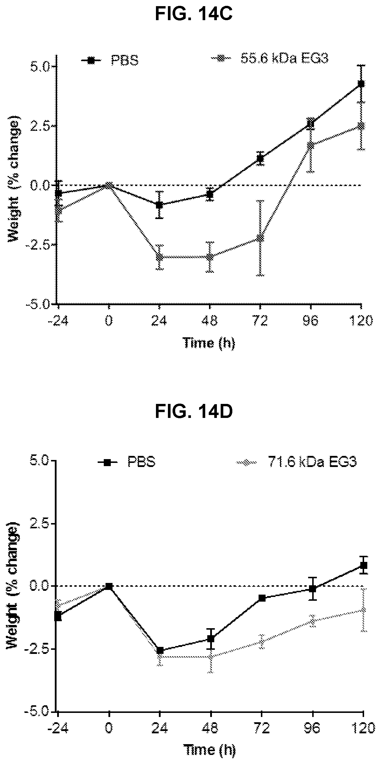
D00028

D00029

D00030

D00031

D00032

D00033

D00034

D00035

D00036

D00037

D00038

D00039

D00040

D00041

D00042

D00043

D00044

D00045

D00046
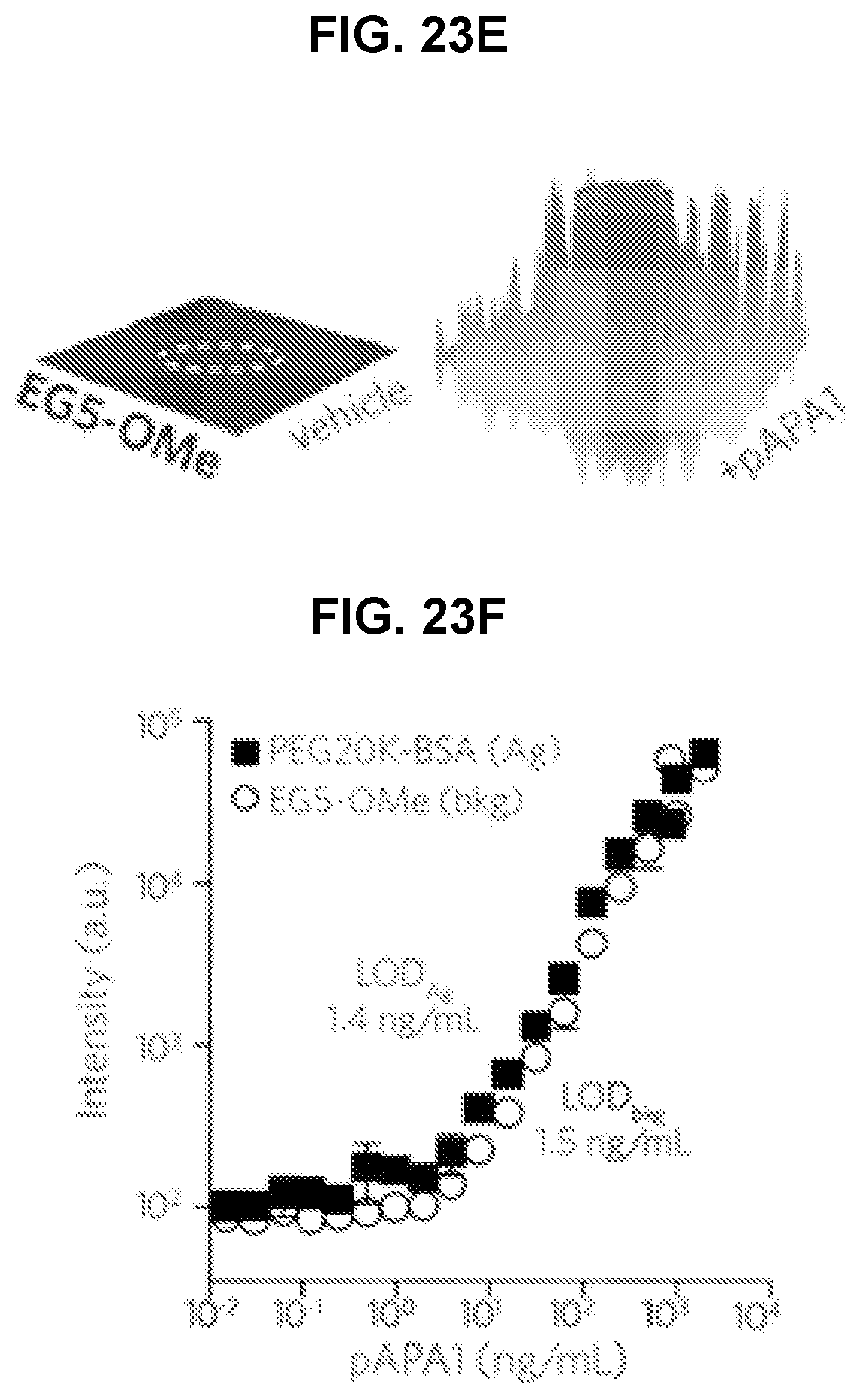
D00047

D00048

D00049

D00050

D00051

D00052

D00053

D00054

D00055

D00056

S00001
XML
uspto.report is an independent third-party trademark research tool that is not affiliated, endorsed, or sponsored by the United States Patent and Trademark Office (USPTO) or any other governmental organization. The information provided by uspto.report is based on publicly available data at the time of writing and is intended for informational purposes only.
While we strive to provide accurate and up-to-date information, we do not guarantee the accuracy, completeness, reliability, or suitability of the information displayed on this site. The use of this site is at your own risk. Any reliance you place on such information is therefore strictly at your own risk.
All official trademark data, including owner information, should be verified by visiting the official USPTO website at www.uspto.gov. This site is not intended to replace professional legal advice and should not be used as a substitute for consulting with a legal professional who is knowledgeable about trademark law.