Diagnostic And Drug Screening For Molecular Signatures Of Early Onset Sporadic Parkinson's Disease
Laperle; Alexander ; et al.
U.S. patent application number 17/043272 was filed with the patent office on 2021-02-04 for diagnostic and drug screening for molecular signatures of early onset sporadic parkinson's disease. This patent application is currently assigned to CEDARS-SINAI MEDICAL CENTER. The applicant listed for this patent is CEDARS-SINAI MEDICAL CENTER. Invention is credited to Alexander Laperle, Samuel Sances, Clive N. Svendsen, Nur Yucer.
| Application Number | 20210033628 17/043272 |
| Document ID | / |
| Family ID | 1000005218965 |
| Filed Date | 2021-02-04 |


View All Diagrams
| United States Patent Application | 20210033628 |
| Kind Code | A1 |
| Laperle; Alexander ; et al. | February 4, 2021 |
DIAGNOSTIC AND DRUG SCREENING FOR MOLECULAR SIGNATURES OF EARLY ONSET SPORADIC PARKINSON'S DISEASE
Abstract
Induced Pluripotent Stem Cell (Ipsc) technology enables the generation and study of living brain tissue relevant to Parkinson's disease (PD) ex vivo. Utilizing cell lines from PD patients presents a powerful discovery system that links cellular phenotypes observed in vitro with real clinical data. Differentiating patient-derived iPSCs towards a dopaminergic (DA) neural fate revealed that these cells exhibit molecular and functional properties of DA neurons in vitro that are observed to significantly degenerate in the substantia nigra of PD patients. Clinical symptoms that drive the generation of other relevant cell types may also yield novel PD-specific phenotypes in vitro that have the potential to lead to new therapeutic avenues for patients with PD. Due to their early onset and non-familial origin, differentiated nervous tissue from these patients offer a key opportunity to discover neuron subtype-specific pathological mechanisms and importantly interrogate the contribution of their genetic background in susceptibility to PD.
| Inventors: | Laperle; Alexander; (North Hollywood, CA) ; Sances; Samuel; (Santa Monica, CA) ; Yucer; Nur; (Los Angeles, CA) ; Svendsen; Clive N.; (Pacific Palisades, CA) | ||||||||||
| Applicant: |
|
||||||||||
|---|---|---|---|---|---|---|---|---|---|---|---|
| Assignee: | CEDARS-SINAI MEDICAL CENTER Los Angeles CA |
||||||||||
| Family ID: | 1000005218965 | ||||||||||
| Appl. No.: | 17/043272 | ||||||||||
| Filed: | April 5, 2019 | ||||||||||
| PCT Filed: | April 5, 2019 | ||||||||||
| PCT NO: | PCT/US2019/026195 | ||||||||||
| 371 Date: | September 29, 2020 |
Related U.S. Patent Documents
| Application Number | Filing Date | Patent Number | ||
|---|---|---|---|---|
| 62664888 | Apr 30, 2018 | |||
| 62664827 | Apr 30, 2018 | |||
| 62664942 | May 1, 2018 | |||
| 62755365 | Nov 2, 2018 | |||
| 62816795 | Mar 11, 2019 | |||
| Current U.S. Class: | 1/1 |
| Current CPC Class: | C12N 5/10 20130101; C12N 5/0696 20130101; G01N 33/5008 20130101; G01N 2333/4704 20130101; G01N 33/6896 20130101; A61K 35/545 20130101 |
| International Class: | G01N 33/68 20060101 G01N033/68; G01N 33/50 20060101 G01N033/50; C12N 5/10 20060101 C12N005/10; C12N 5/074 20060101 C12N005/074; A61K 35/545 20060101 A61K035/545 |
Goverment Interests
STATEMENT REGARDING FEDERALLY SPONSORED RESEARCH
[0001] This invention was made with government support under NS105703 awarded by the National Institutes of Health. The government has certain rights in the invention.
Claims
1. A method of compound screening, comprising: contacting a quantity of neurons with one or more test compounds; measuring one or more parameters; and selecting one or more test compounds based on the measured one or more parameters, wherein the neurons are differentiated from blood cell-derived induced pluripotent stem cells (iPSCs).
2. The method of claim 1, wherein the neurons are midbrain neurons.
3. The method of claim 2, wherein the midbrain neurons are dopaminergic neurons.
4. The method of claim 1, wherein the one or more parameters comprise PKC activation.
5. The method of claim 1, wherein the one or more parameters comprise at least one of TFEB activation and ZKSCAN3 inactivation.
6. The method of claim 1, wherein the one or more parameters comprise .alpha.-synuclein protein levels.
7. The method of claim 1, wherein the one or more parameters comprise at least one of: LAMP1, GCase, tyrosine hydroxylase (TH), and dopaminergic activity.
8. The method of claim 1, wherein the one or more parameters comprise at least one of: LAMP, GCase, tyrosine hydroxylase (TH) and dopamine expression levels.
9. The method of claim 1, wherein the blood cell-derived iPSCs are made by a method comprising: contacting a quantity of blood cells with one or more oriP/EBNA1 vectors encoding a reprogramming factor; and delivering a quantity of reprogramming factors into the blood cells; culturing the blood cells in a reprogramming media wherein delivering the reprogramming factors, and culturing in a reprogramming media generates blood cell derived iPSCs.
10. The method of claim 9, wherein the quantity of blood cells is obtained from a human subject afflicted with a neurodegenerative disease.
11. The method of claim 10, wherein the neurodegenerative disease is Parkinson's Disease.
12. The method of claim 11, wherein the Parkinson's Disease is early onset Parkinson's Disease.
13. The method of claim 1, wherein the neurons are differentiated from blood cell-derived iPSCs by a method comprising: providing a quantity of blood cell-derived induced pluripotent stem cells (iPSCs); culturing the iPSCs in the presence of a transforming growth factor (TGF)-beta inhibitor and an activin receptor-like kinase (ALK) inhibitor; further culturing in the presence of a Smoothened agonist, a RHO Kinase (ROCK) inhibitor and at least two growth factors; additionally culturing in the presence of retinoic acid; and continuing to culture in the presence of at least three additional growth factors.
14. The method of claim 1, wherein the transforming growth factor (TGF)-beta inhibitor comprises LDN-193189 and the activin receptor-like kinase (ALK) inhibitor comprises SB431542, the Smoothened agonist comprises Purmorphamine, the at least two growth factors comprise sonic hedgehog, and fibroblast growth factor 8, the ROCK inhibitor comprises CHIR99012, and the at least three additional growth factors comprise brain derived neurotrophic factor, glial derived neurotrophic factor, and TGF-Beta 3.
15. A method of diagnosis or prognosis, comprising: assaying one or more biomarkers in a subject suspected of being afflicted with Parkinson's Disease; comparing the expression levels of the one or more hiomarkers from the subject to a baseline of the one or more biomarkers derived from healthy subjects without Parkinson's disease; and diagnosing Parkinson's Disease in the subject or prognosing Parkinson's Disease in the subject.
15. (canceled)
17. A method of selecting a therapeutic regimen, comprising: assaying one or more biomarkers in a subject suspected of being afflicted with Parkinson's Disease; comparing the expression levels of the one or more hiomarkers from the subject to a baseline of the one or more biomarkers derived from healthy subjects without Parkinson's disease; and selecting a therapeutic regimen for the subject.
Description
FIELD OF THE INVENTION
[0002] Described herein are methods and compositions related to production of midbrain neurons, including those related to Parkinson's Disease.
BACKGROUND
[0003] Parkinson's Disease (PD) is the second most commonly diagnosed neurodegenerative disorder and represents a substantial economic burden among current aging populations. The classically associated pathology in PD is characterized by the progressive loss of dopaminergic neurons (DaNs) in the substantia nigra pars compacta and the presence of cytoplasmic inclusions known as Lewy bodies and Lewy neurites. These inclusions are composed mainly of the protein .alpha.-synuclein. Mutations or triplication of the gene encoding .alpha.-synuclein (SNCA) are causal in these specific familial PD cases. In its native state, .alpha.-synuclein is found in the presynaptic terminal of neurons throughout the human brain and functions in vesicle trafficking, neurotransmitter release and reuptake.
[0004] While many genes and proteins, such as .alpha.-synuclein, have been linked to PD, the inability to extract live neurons from patients and the lack of effective PD models leaves unanswered questions regarding the initiation and progression of the disease. Reprogramming patient-derived cells into iPSCs enables the observation of disease progression and pathological phenotypes at a molecular level. Interestingly, previous iPSC studies on the larger non-familial (sporadic) population do not show overt differences when compared those derived from control individuals. Thus, there is a great need in the art for iPSC disease models that represent the complex biological background underpinning Parkinson's disease pathology.
[0005] Described herein are compositions and methods for modeling and treating Parkinson's Disease. Importantly, generation of midbrain neurons, floorplate induction in a manner faithfully mirroring development allows for identification of cellular cues leading to neurodegeneration, this includes the complex etiology behind sporadic PD cases that have not yet been fully utilized in iPSC models. Establishing such models, the Inventors herein identified hereto unknown role of .alpha.-synuclein and lysosomal degradation dysfunction, as mediated in-party by PKC. Targeting PKC via an agonist improved measurable outcomes, thereby suggesting new therapeutic avenues for Parkinson's Disease.
SUMMARY OF THE INVENTION
[0006] Described herein is a method of compound screening, including contacting a quantity of neurons with one or more test compounds, measuring one or more parameters, and selecting one or more test compounds based on the measured one or more parameters, wherein the neurons are differentiated from blood cell-derived induced pluripotent stern cells (iPSCs). In other embodiments, the neurons are midbrain neurons, in other embodiments, the midbrain neurons are dopaminergic neurons. In other embodiments, the one or more parameters comprise PKC activation. In other embodiments, the one or more parameters comprise at least one of TFEB activation and ZKSCAN3 inactivation. in other embodiments, the one or more parameters comprise .alpha.-synuclein protein levels. in other embodiments, the one or more parameters comprise at least one of: LAMP1, GCase, tyrosine hydroxylase (TH), and dopaminergic activity. In other embodiments, the one or more parameters comprise at least one of: LAMP, GCase, tyrosine hydroxylase (TH) and dopamine expression levels. In other embodiments, the blood cell-derived iPSCs are made by a method including contacting a quantity of blood cells with one or more oriP/EBNA1 vectors encoding a reprogramming factor, and delivering a quantity of reprogramming factors into the blood cells, culturing the blood cells in a reprogramming media wherein delivering the reprogramming factors, and culturing in a reprogramming media generates blood cell derived iPSCs. In other embodiments, the quantity of blood cells is obtained from a human subject afflicted with a neurodegenerative disease. In other embodiments, the neurodegenerative disease is Parkinson's Disease. In other embodiments, the Parkinson's Disease is early onset Parkinson's Disease. In other embodiments, the neurons are differentiated from blood cell-derived iPSCs by a method including providing a quantity of blood cell-derived induced pluripotent stem cells (iPSCs), culturing the iPSCs in the presence of a transforming growth factor (TGF)-beta inhibitor and an activin receptor-like kinase (ALK) inhibitor, further culturing in the presence of a Smoothened agonist, a RHO Kinase (ROCK) inhibitor and at least two growth factors, additionally culturing in the presence of retinoic acid, and continuing to culture in the presence of at least three additional growth factors. In other embodiments, the transforming growth factor (TGF)-beta inhibitor includes LDN-193189 and the activin receptor-like kinase (ALK) inhibitor includes S13431542, the Smoothened agonist comprises Purmorphamine, the at least two growth factors comprise sonic hedgehog, and fibroblast growth factor 8, the ROCK inhibitor includes CHIR99012, and the at least three additional growth factors comprise brain derived neurotrophic factor, glial derived neurotrophic factor, and TGF-Beta 3.
[0007] Also described herein is a method of diagnosis, including assaying one or more biomarkers in a subject suspected of being afflicted with Parkinson's Disease, comparing the expression levels of the one or more biomarkers from the subject to a baseline of the one or more biomarkers derived from healthy subjects without Parkinson's disease, and diagnosing Parkinson's Disease in the subject.
[0008] Also described herein is a method of prognosis, including assaying one or more biomarkers in a subject suspected of being afflicted with Parkinson's Disease, comparing the expression levels of the one or more biomarkers from the subject to a baseline of the one or more biomarkers derived from healthy subjects without Parkinson's disease, and prognosing Parkinson's Disease in the subject.
[0009] Further described herein is a method of selecting a therapeutic regimen, including assaying one or more biomarkers in a subject suspected of being afflicted with Parkinson's Disease, comparing the expression levels of the one or more biomarkers from the subject to a baseline of the one or more biomarkers derived from healthy subjects without Parkinson's disease, and selecting a therapeutic regimen for the subject.
BRIEF DESCRIPTION OF THE FIGURES
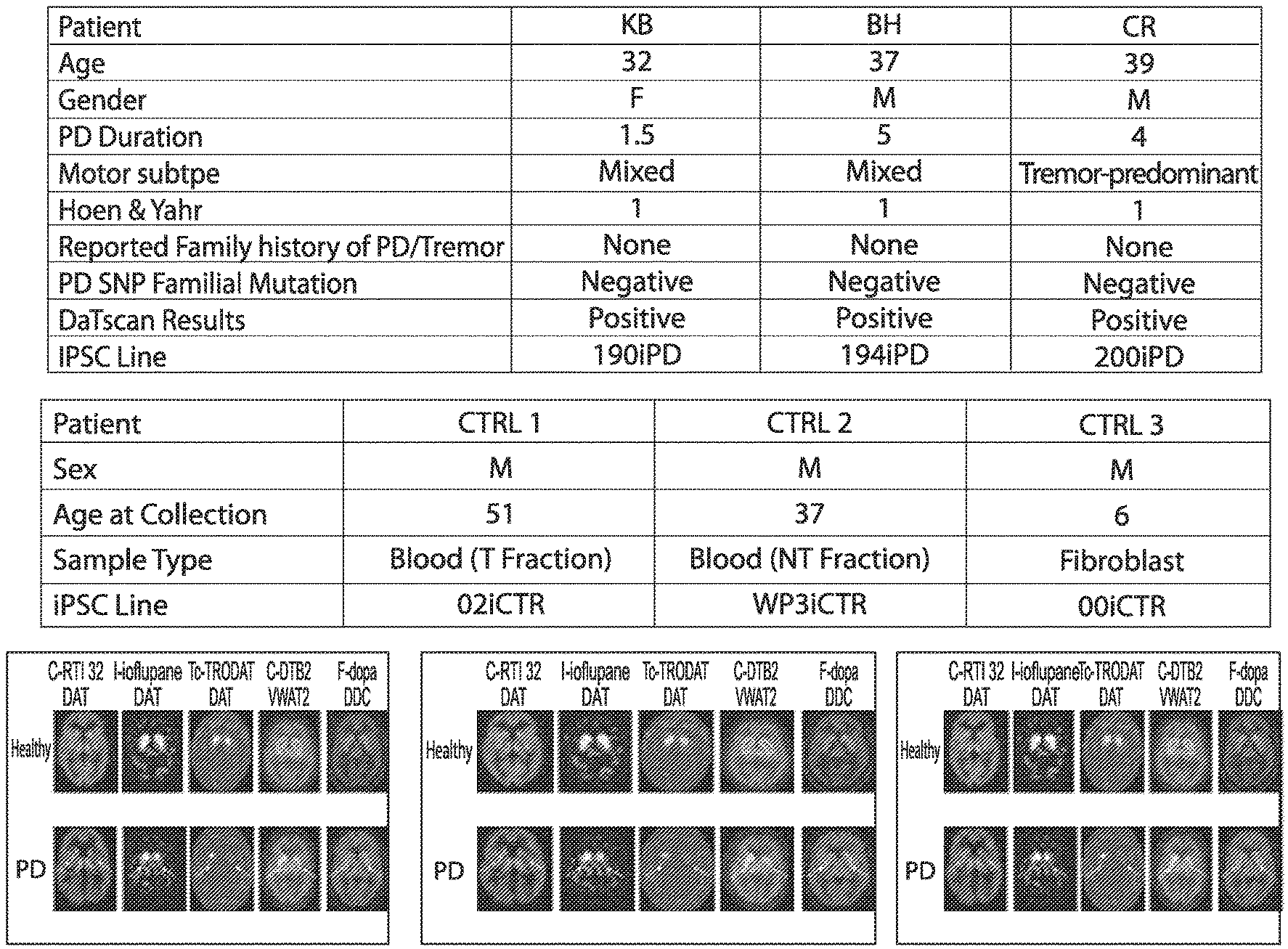
[0010] FIG. 1: PD patient data
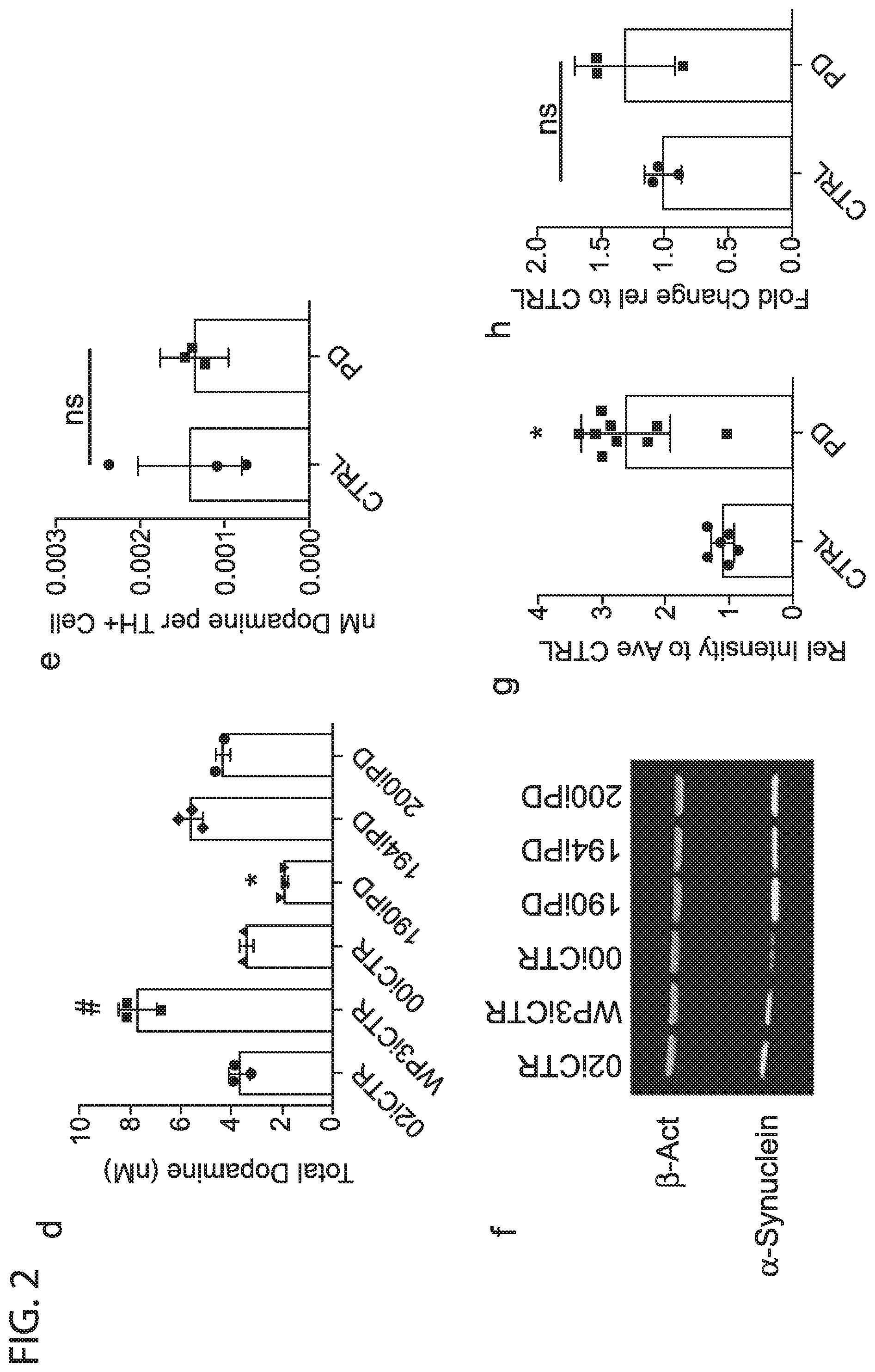
[0011] FIG. 2: Differentiation schematic (a). representative images showing TH expression and morphology (b) Flow cytometry data showing differentiation efficiency, each point represents an average of 3 separate wells of an independent differentiation (c). HLPC detection of total dopamine content (d) and dopamine content normalized to differentiation efficiency by line (e). Western blot showing bACT (housekeeping) and synuclein expression in 30 day old DaNs (f). Relative intensities from multiple western blots, each point is a band from a separate differentiation, colors indicate ipsc lines, data were normalized to 02iCTR for each differentiation (g). SNCA expression by qPCR in 30 day old DaNS (h).
[0012] FIG. 3: Combined detection of overlapping transcripts and proteins from paired RNA-Seq and Proteomics (a). PCA plots of matched transcriptomic and proteomic data (b) GSEA analyses of PCI components upregulated in PD (c) GSEA analysis of PCI components downregulated in PD (d)
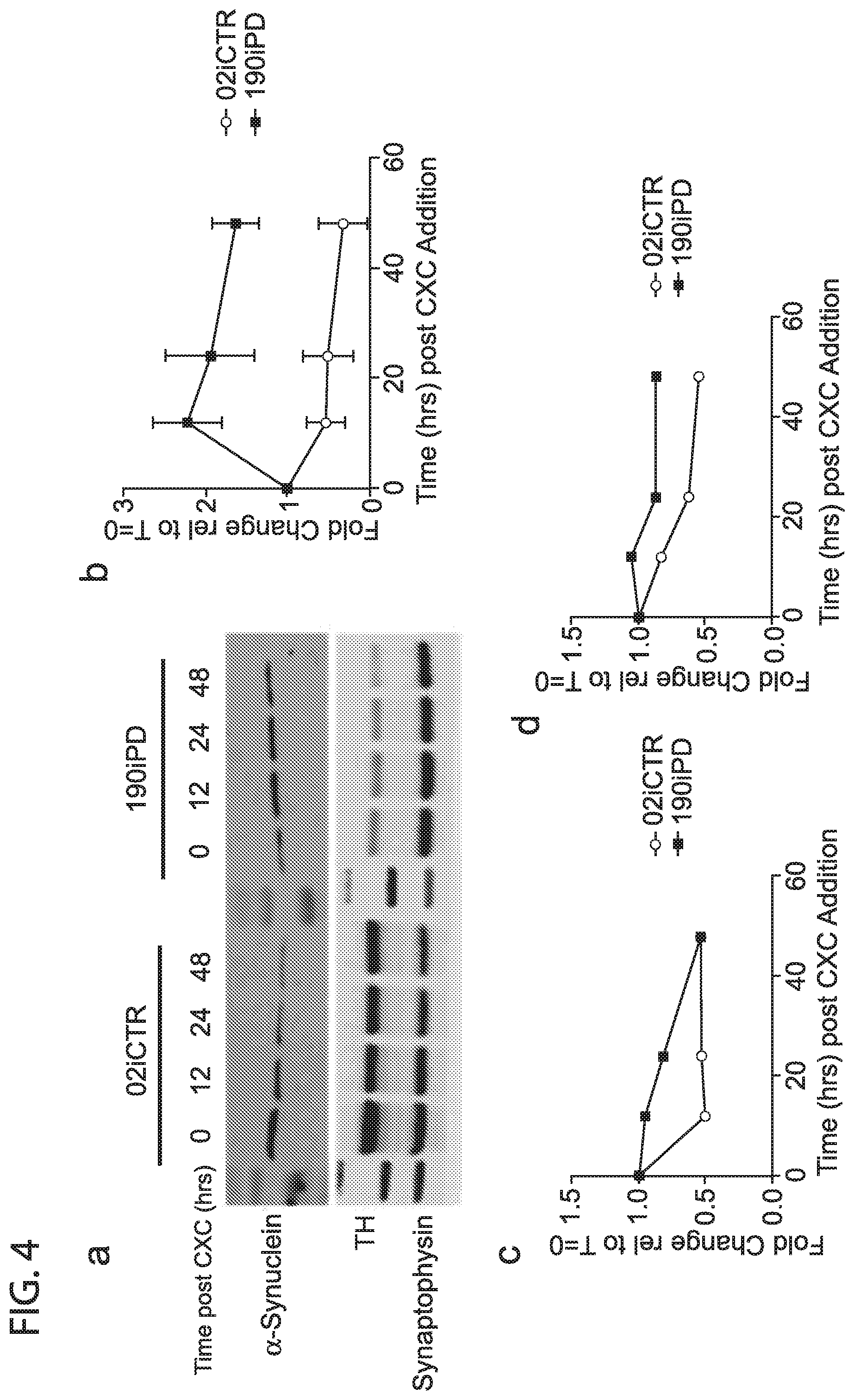
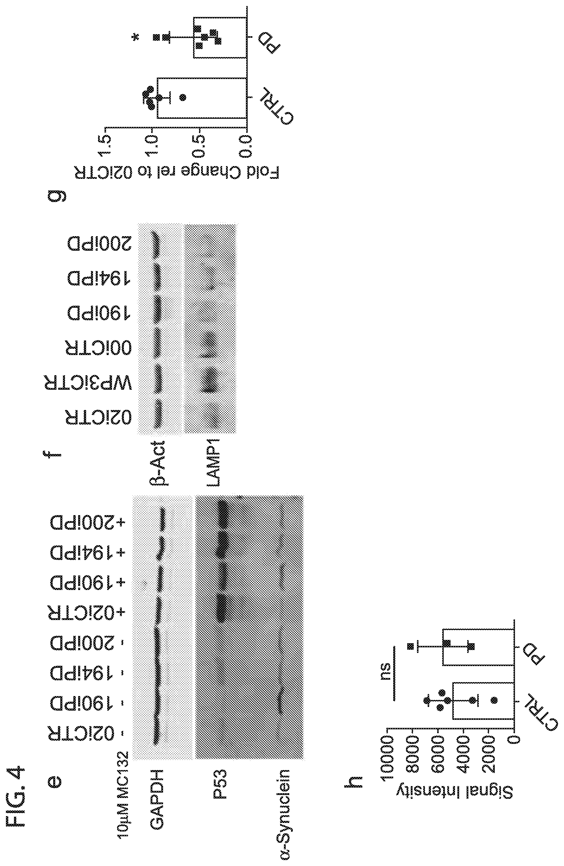
[0013] FIG. 4: representative western blot showing synuclein degradation under cycloheximide inhibition (a). average intensities of 3 separate differentiations and western blots from 02iCTR and 190iPD cells, presented as a fold change to time=0 synuclein (b), Synaptophysin (c), and TH (d). Western blot showing synuclein degradation under 24 hrs of MG132 proteosomal inhibitor (e). Western blot showing reduced LAMP1 protein in PD DaNs (f). GCase activity, each point is an average of 3 separate wells from a single differentiation. Data were normalized to 02iCTR for each differentiation and presented as a fold change (g). NIRF detection of oxidized dopamine from D30 DaN lysates (h)
[0014] FIG. 5: Treatment with lysosomal agonists and Elevated p-PKCa in PD lines. Western blot and relative band quantifications of d30 DaNs treated with indicated compounds for 72 hours (a). Baseline levels of p-PKCa in d30 DaNs (b). Day 30 DaNs treated with PEP from multiple PD and control lines(c). Timecourse of PEP treatment and synuclein levels (d) Timecourse of PEP treatment SNCA (e) and TH (f) gene expression. Confirmation of elevated synuclein and p-PKCa in addition control and PD lines (g)
[0015] FIG. 6: Differentiation Protocol, including 4 stage time course (a) and maturation (b). FIG. 7: Three patient volunteers exhibiting early onset of Parkinson's symptoms (confirmed by DaTscan) were evaluated at Cedars Sinai Medical Center. None of the patient's report family history of Parkinson's disease, indicating a sporadic disease origin. Detailed clinical evaluation and patient history data were recorded, and will continue to provide vital clinical information as disease modeling studies in vitro are carried out into the future. All three patients exhibit 1 on Hoehn and Yahr scale, indicating unilateral involvement and minimal functional disability.
[0016] Peripheral blood mononuclear cells (PBMCs) were reprogrammed using episomal nucleofection technique involving four plasmids containing Yamanaka reprogramming transcription factors OCT4, KLF, SOX2, MYC, and Lin28. EBNA1 was also included to enhance transfection efficiency. Cells were seeded on gelatin coated tissue culture plates containing a bed of mouse embryonic fibroblasts (MEFs) for 18-26 days. iPSC colony formation occurred in all three lines, multiple clones were collected and expanded over multiple passages. Alkaline Phosphatase (AP) staining (20.times.) reveals elevated levels in cell membranes of patient lines indicative of undifferentiated tissue. (B) Immunocytochemistry (20.times.) showing pluripotency gene Oct4 expression. Human specific cell surface antigen marker SSEA4 is also expressed indicating molecular profile consistent with pluripotent cells.
[0017] Patient-derived iPSCs can be differentiated to dopaminergic (DA) neurons. Images show 40 day old cultures that express tyrosine hydroxylase (TH) and display typical neuronal morphology. These cells endogenously express .alpha.-synuclein, also found in Lewy bodys in patients with Parkinson's disease. Images taken at 20.times. magnification.
[0018] FIG. 8. DANs successfully cryo-banked (02iCTR).
DETAILED DESCRIPTION OF THE INVENTION
[0019] All references cited herein are incorporated by reference in their entirety as though fully set forth. Unless defined otherwise, technical and scientific terms used herein have the same meaning as commonly understood by one of ordinary skill in the art to which this invention belongs. Singleton et al., Dictionary of Microbiology and Molecular Biology 3.sup.rd ed., Revised, J. Wiley & Sons (New York, N.Y. 2006); and Sambrook and Russel, Molecular Cloning: A Laboratory Manual 4.sup.th ed., Cold Spring Harbor Laboratory Press (Cold Spring Harbor, N.Y. 2012), provide one skilled in the art with a general guide to many of the terms used in the present application.
[0020] One skilled in the art will recognize many methods and materials similar or equivalent to those described herein, which could be used in the practice of the present invention. Indeed, the present invention is in no way limited to the methods and materials described.
[0021] As described, reprogramming patient-derived cells into iPSCs enables the observation of disease progression and pathological phenotypes at a molecular level. The iPSCs, which are genetically identical to the donor, can be differentiated into DaNs providing a tissue-specific model of Parkinson's Disease in vitro that harbor genetic backgrounds known to relate to clinical presentation in vivo. Recently, an intense effort has been made to elucidate the role of .alpha.-synuclein in the origin and progression of PD using similar iPSC modeling techniques. Several studies have employed iPSCs derived from PD patients with monogenic mutations including a triplication of SNCA, as well as mutations in the LRRK2 and GBA1 genes. While DA neurons derived from these iPSC lines display some phenotypic abnormalities and demonstrate accumulation of .alpha.-synuclein, familial monogenic mutations are present only in a small minority of PD patients and pathophysiology of these cases are not easily related to the PD population at large. Interestingly, previous iPSC studies on the larger non-familial (sporadic) population do not show overt differences when compared those derived from control individuals.
[0022] Here, the Inventors generate iPSC lines from a cohort of early onset sporadic PD (EOSPD) patients. The Inventors hypothesized that these lines represent a promising opportunity to better understand sporadic PD, as early onset sporadic patients could have unknown genetic risk factors that may influence a more aggressive form of the disease. The Inventors show that by comparing differentiated DaNs from either EOSPD patient and non-diseased control lines, aberrant accumulation of .alpha.-synuclein protein is indeed specifically reproduced in the PD patient cohort. Molecular and physiological profiling of these tissues including proteomic, whole transcriptomic and enzyme activity assays find dysregulated degradation pathways and implicate a previously unreported upregulation of phosphorylated PKC-.alpha. in EOSPD cultures. Finally, by targeting this pathway, the Inventors observe reversal of .alpha.-synuclein accumulation after treatment with a small molecule PEP005 both in vitro and in vivo. The iPSC-based model described here provides evidence of the genetic origin of sporadic PD that contributes PD and provides a platform for potential clinical diagnostics and development of new therapeutic targets for EOSPD patients.
[0023] Described herein is a method of compound screening, including contacting a quantity of neurons with one or more test compounds, measuring one or more parameters, and selecting one or more test compounds based on the measured one or more parameters, wherein the neurons are differentiated from blood cell-derived induced pluripotent stem cells (iPSCs). In other embodiments, the neurons are midbrain neurons. In other embodiments, the midbrain neurons are dopaminergic neurons. In other embodiments, the one or more parameters comprise PKC activation. In other embodiments, the one or more parameters comprise at least one of TFEB activation and ZKSCAN3 inactivation. In other embodiments, the one or more parameters comprise .alpha.-synuclein protein levels. In other embodiments, the one or more parameters comprise at least one of: LAMP1, GCase, tyrosine hydroxylase (TH), and dopaminergic activity. In other embodiments, the one or more parameters comprise at least one of: LAMP, GCase, tyrosine hydroxylase (TH) and dopamine expression levels. In other embodiments, the one or more parameters include permeability of the test compound across a quantity of vascular cells, alterations in electrophysiological properties of the cells, alterations in metabolic profile of the cells, including for example, neurotransmitter production and release. In other embodiments, the iPSCs are made by a method including contacting a quantity of blood cells with one or more oriP/EBNA1 vectors encoding a reprogramming factor and delivering a quantity of reprogramming factors into the blood cells culturing the blood cells in a reprogramming media, wherein the quantity of blood cells are obtained from a human subject afflicted with a neurodegenerative disease, and further wherein delivering the reprogramming factors, and culturing in a reprogramming media generates blood cell derived iPSCs.
[0024] In other embodiments, the blood cell-derived iPSCs are made by a method including contacting a quantity of blood cells with one or more oriP/EBNA1 vectors encoding a reprogramming factor, and delivering a quantity of reprogramming factors into the blood cells, culturing the blood cells in a reprogramming media wherein delivering the reprogramming factors, and culturing in a reprogramming media generates blood cell derived iPSCs. In other embodiments, the quantity of blood cells is obtained from a human subject afflicted with a neurodegenerative disease. In other embodiments, the neurodegenerative disease is Parkinson's Disease. In other embodiments, the neurodegenerative disease is Parkinson's Disease (PD), including familial, sporadic and early onset PD. In other embodiments, the Parkinson's Disease is early onset Parkinson's Disease. Further information on iPSC reprogramming is found in Barrett, R. et al. Reliable Generation of Induced Pluripotent Stem Cells from Human Lymphoblastoid Cell Lines. Stem Cells Transl Med. 2014 December; 3(12):1429-34, which is fully incorporated by reference herein.
[0025] In other embodiments, the neurons are differentiated from blood cell-derived iPSCs by a method including providing a quantity of blood cell-derived induced pluripotent stem cells (iPSCs), culturing the iPSCs in the presence of a transforming growth factor (TGF)-beta inhibitor and an activin receptor-like kinase (ALK) inhibitor, further culturing in the presence of a Smoothened agonist, a RHO Kinase (ROCK) inhibitor and at least two growth factors, additionally culturing in the presence of retinoic acid, and continuing to culture in the presence of at least three additional growth factors. In other embodiments, the transforming growth factor (TGF)-beta inhibitor includes LDN-193189 and the activin receptor-like kinase (ALK) inhibitor includes SB431542, the Smoothened agonist comprises Purmorphamine, the at least two growth factors comprise sonic hedgehog, and fibroblast growth factor 8, the ROCK inhibitor includes CHIR99012, and the at least three additional growth factors comprise brain derived neurotrophic factor, glial derived neurotrophic factor, and TGF-Beta 3. In various embodiments, the concentrations of the aforementioned agents are as described in Table 1. In other embodiments, the iPSCS are made by a process including contacting a quantity of blood cells with one or more vectors encoding a reprogramming factor, and delivering a quantity of reprogramming factors into the blood cells, culturing the blood cells in a reprogramming media. In other embodiments, the one or more vectors are odiP/EBNA1 vectors. In other embodiments, culturing. iPSCs is for about 3 days. In other embodiments, further culturing is for about 4 days. In other embodiments, additionally culturing is for about 4 days. In other embodiments, continuing to culture is for at least 3 days. In other embodiments, the differentiation schedule, including feeding and media changes is according to Table 1 and FIG. 6. Additional information can be found in U.S. Prov, App. No. 62/653,697 and 62/755,282, U.S. Prov. App. No. 62/653,697, U.S. Prov. App. No. 62/755,282, U.S. Prov. App. No. 62/816,785, U.S. Prov. App. No. 62/664,888, U.S. Prov. App. No. 62/664,827, U.S. Prov. App. No. 62/816,795, U.S. Prov. App. No. 62/664,942, U.S. Prov. App. No. 62/755,365, which are fully incorporated by reference herein.
[0026] Also described herein is a method of diagnosis, including assaying one or more biomarkers in a subject suspected of being afflicted with Parkinson's Disease, comparing the expression levels of the one or more biomarkers from the subject to a baseline of the one or more biomarkers derived from healthy subjects without Parkinson's disease, and diagnosing Parkinson's Disease in the subject.
[0027] Also described herein is a method of prognosis, including assaying one or more biomarkers in a subject suspected of being afflicted with Parkinson's Disease, comparing the expression levels of the one or more biomarkers from the subject to a baseline of the one or more biomarkers derived from healthy subjects without Parkinson's disease, and prognosing Parkinson's Disease in the subject.
[0028] Further described herein is a method of selecting a therapeutic regimen, including assaying one or more biomarkers in a subject suspected of being afflicted with Parkinson's Disease, comparing the expression levels of the one or more biomarkers from the subject to a baseline of the one or more biomarkers derived from healthy subjects without Parkinson's disease, and selecting a therapeutic regimen for the subject.
[0029] In various methods for prognosis, diagnosis, or aiding therapeutic selection, the biomarker is a metabolic enzyme, including neurotransmitters such as dopamine. In various embodiments, biomarkers include .alpha.-synuclein expression levels, including transcript and protein levels. In various embodiments, biomarker include PKC activation. In other embodiments, the biomarkers include at least one of TFEB activation and ZKSCAN3 inactivation. In other embodiments, the one or more parameters comprise .alpha.-synuclein protein levels. In other embodiments, the biomarkers include at least one of: LAMP1, GCase, tyrosine hydroxylase (TH), and dopaminergic activity. In other embodiments, the biomarkers include at least one of: LAMP, GCase, tyrosine hydroxylase (TH) and dopamine expression levels. In various embdoiments, the biomarkers include markers of aberrant protein degradation, such as lysosomal dysfunction. In various embodiments, the biomakers include nestin, Tuj1, MAP2, GFAP, S100B, CD11B, PU.1, GLUTA-1, ZO-1, SMI31/Isl1, TH/PITX3, Phosopho-TDP, FAS ligand, and SOD1, among others.
[0030] In various embodiments of the aforementioned, neurodegenerative disease derived induced pluripotent stem cells (iPSCs) and differentiated cells thereof possess a molecular signature different from iPSCs derived from healthy controls. In various embodiments, the molecular signature includes difference in metabolic pathways and metabolites. For example, this includes metabolites such as, for example, enrichment in one or more of L-Kynurenine, trans-aconitic acid, adenine, inosine, and 1-tyrosine.
[0031] Described herein is a method of cryopreservation of differentiated iPSCs. In various embodiments, the differentiated iPSCs are treated with a culture media, treated with a proteolytic and collagenolytic agent, placing the differentiated iPSCs in a cryoprotective agent, exposing the differentiated iPSCs to an initiation temperature, cooling the differentiated iPSCs, supercooling the differentiated iPSCs to a solid phase, heating the differentiated iPSCs, and reducing the temperature of the differentiated iPSCs solid phase. In other embodiments, the cryoprotective agent includes serum. In other embodiments, the initiation temperature is about -4.degree. to about 40.degree. C. In other embodiments, the initiation temperature is about 2.degree. to about 20.degree. C. In other embodiments, the initiation temperature is about -1.degree. to about 15.degree. C. In other embodiments, the initiation temperature is about 3.degree. to about 7.degree. C. In other embodiments, cooling the differentiated iPSCs includes reaching a temperature of about -5 to -15.degree. C. In other embodiments, cooling the differentiated iPSCs includes reaching a temperature of about -3 to -7.degree. C. In other embodiments, cooling the differentiated iPSCs includes reaching a temperature of about -5.degree. C. In other embodiments, supercooling the differentiated iPSCs includes reaching a temperature of about -20 to -90.degree. C. In other embodiments, supercooling the BMECs includes reaching a temperature of about -40 to -75.degree. C. In other embodiments, supercooling the differentiated iPSCs includes reaching a temperature of about -58.degree. C. In other embodiments, supercooling is at a rate of about -20 TO -60.degree. C./minute. In other embodiments, supercooling is at a rate of about -45.degree. C./minute. In other embodiments, heating the differentiated iPSCs includes reaching a temperature of about -23.degree. C. In other embodiments, heating the differentiated iPSCs is at a rate of about +10.degree. C./minute to about -26.degree. C. and/or +3.degree. C./minute to about -23.degree. C. In other embodiments, reducing the temperature of the differentiated iPSCs solid phase includes reaching a temperature of about -30.degree. C. to about -50.degree. C. In other embodiments, reducing the temperature of the differentiated iPSCs solid phase includes reaching a temperature of about -40.degree. C. In other embodiments, reducing the temperature of the differentiated iPSCs solid phase is at a rate of about -3 to -0.05.degree. C./minute. In other embodiments, reducing the temperature of the differentiated iPSCs solid phase is at a rate of about -0.8.degree. C./minute. In other embodiments, rapid cooling of the reduced temperature differentiated iPSCs solid phase at a rate of about -10.degree. C./minute to about -100.degree. C. and/or about -35.degree. C./minute to about -160.degree. C. In other embodiments, the method includes transfer of the differentiated iPSCs to liquid nitrogen. In various embodiments, the culture media is neuron maturation media. In various embodiments, the culture media includes a ROCK inhibitor. In various embodiments, the proteolytic and collagenolytic agent is accutase.
[0032] In various embodiments, the differentiated iPSCs are midbrain neurons. In various embodiments, the differentiated iPSCs are floorplate cells. In various embodiments, the differentiated iPSCs produce tyrosine hydroxylase and/or or dopamine. In various embodiments, the differentiated iPSCs express higher levels of .alpha.-Synuclein compared to controls derived from healthy subjects. In various embodiments, the differentiated. iPSCs exhibit abnormal protein degradation, including lysosomal dysfunction. In various embodiments, the differentiated cells exhibit abnormal levels or activity of LAMP1 and/or Gcase. Described herein is a cryropreserved solution of differentiated cells prepared by the aforementioned methods.
EXAMPLE 1
[0033] Generation of iPSCs from Early Onset Sporadic Parkinson's Disease Patients (EO-sPD)
[0034] Three early onset sporadic Parkinson's patients between the ages of 30-39 with no reported family history of PD were selected for iPSC production (FIG. 1). Based on analysis with the NeuroX platform, no monogenic mutations in EIFG1, PARK2, LRRK2, GBA, SNCA, PINK1, PARK7, VSP35, ATP13A2 or multiplications of the SNCA locus were detected in the patient lines. All 3 patients demonstrated reduced DAT (phenyltropane) signature in the striatum consistent with their PD diagnosis (FIG. 1). For comparison, 3 control lines were generated from normal individuals with no neurological disease at time of collection.
[0035] Peripheral blood mononuclear cells (PBMCs) were collected and subsequently reprogramed to iPSCs using non-integrating episomal techniques (FIG. 1). All iPSC lines were karyotypically normal, and expressed canonical pluripotency markers.
EXAMPLE 2
[0036] Efficient Differentiation of EOSPD iPSCs to DANs A defining hallmark of PD is the specific loss of dopaminergic neurons in the substantia nigra and it is therefore of interest to differentiate iPSC along this lineage. iPSC lines from both PD and control patients were differentiated to dopaminergic neurons using the protocol described in Table 1, FIG. 2A and FIG. 6.
[0037] Briefly, iPSC lines were subjected to a modified dual SMAD-inhibition based floor plate induction protocol. Exposure to LDN/SB, followed by SHH/Purmorphamine/FGF8 and CHIR99021, thereafter including SB withdrawal and retinoic acid addition,support midbrain FP and DA neuron yield (see FIG. 1d). Further maturation was carried out in Neurobasal/B27 medium supplemented with AA, BDNF, GDNF, TGF.beta.3 and dbcAMP. The inclusion of retinoic acid, exclusion of retionoic acid from the early steps of differentiation are unlike any other known techniques. Remarkably, whereas reported protocols may take 80 to 130 days to produce a dopamine producing cells, the aforementioned techniques allow generation in as little as 30 days.
TABLE-US-00001 TABLE 1 Differentiation Protocol - Media Stage 1 Media: for x volume Working Dilution x= 140 mL DMEM/F12 50% DMEM/F12 70 mL Neurobasal 50% Neurobasal 70 mL N2 1:100 N2 1.4 mL B27 - Vitamin A 1:50 Stock: Working: B27 - Vitamin A 2.8 mL LDN 1:10000 10 mM 1 uM LDN 14 uL SB 1:5000 10 mM 2 uM SB 28 uL Stage 2 Media: for x volume Working Dilution x= 220 mL DMEM/F12 50% DMEM/F12 110 mL Neurobasal 50% Neurobasal 110 mL N2 1:100 N2 2.2 mL B27 - Vitamin A 1:50 B27 - Vitamin A 4.4 mL LDN 1:10000 LDN 22 uL SB 1:5000 Stock: Working: SB 44 uL PMN 1:5000 10 mM 2 uM PMN 44 uL Shh 1:1000 100 ug/mL Shh 220 uL CHIR 1:6670 15 mM 2.25 uM CHIR 33.00 uL FGF8 1:5000 50 ug/mL FGF8 44 uL Stage 3 Media: for x volume x= 50 mL DMEM/F12 50% DMEM/F12 25 mL Neurobasal 50% Neurobasal 25 mL N2 1:100 N2 0.5 mL B27 - Vitamin A 1:50 B27 - Vitamin A 1 mL LDN 1:10000 LDN 5 uL CHIR 1:6670 Stock: Working: CHIR 7.50 uL ATRA 1:2000 10 mM 5 uM ATRA 25 uL Stage 4 Media: for x volume x= 60 mL DMEM/F12 50% DMEM/F12 30 mL Neurobasal 50% Neurobasal 30 mL N2 1:100 N2 0.6 mL B27 1:50 Stock: Working: B27 1.2 mL AA 1:1000 500 ug/mL AA 60.00 uL BDNF 1:500 10 ug/mL 20 ng/mL BDNF 120.00 uL GDNF 1:500 10 ug/mL 20 ng/mL GDNF 120.00 uL dbCAMP 1:500 102 mM .2 mM dbCAMP 120.00 uL TGF-B3 1:10000 10 ug/mL 1 ng/mL TGF-B3 6.00 uL DAPT 1:4000 10 mM 2.5 uM DAPT 15.00 uL CHIR 1:6670 CHIR 9.00 uL Maturation Media: for x volume x= 100 mL DMEM/F12 50% DMEM/F12 50 mL Neurobasal 50% Neurobasal 50 mL N2 1:200 N2 0.5 mL B27 1:100 B27 1 mL AA 1:1000 AA 100.00 uL BDNF 1:500 BDNF 200.00 uL GDNF 1:500 GDNF 200.00 uL dbCAMP 1:500 dbCAMP 200.00 uL TGF-B3 1:10000 TGF-B3 10.00 uL DAPT 1:4000 DAPT 25.00 uL
[0038] At day 30, differentiated cells expressed markers of dopamine neurons including TH, Nurr1, and GRIK2 with roughly 15% of the cells expressing TH (Supplemental FIG. 2a) (FIG. 2b,c). Overall differentiation efficiency was compared across all 6 lines by counting the number of TH expressing cells using flow cytometry (FIG. 2c). Two of the PD lines showed similar numbers of DA neurons to those found in controls. However, differentiation of the 190iPD line yielded fewer TH positive neurons and these cells expressed less of the floorplate progenitor markers FOXA2 and LMX1A but more of the mature neural markers GRIK2 and NEFH.
[0039] To determine whether TH enzyme resulted in altered levels of dopamine in the developing neurons, 30 day old DANs were lysed and analyzed for dopamine production by HPLC. Differences in total dopamine were present by line with the 190iPD line again producing less dopamine and the WP3iCTR line producing more. However, when normalized to the number of TH expressing neurons, all lines produced dopamine at similar levels (FIG. 2d,e). To determine the electrophysiological function and potential disease signature of the developing neurons, multi-electrode array recordings were conducted over time in culture. Spontaneous activity was observed day 20 of differentiation and by day 30, both PD and control cells produce coordinated bursts of activity. When activity was quantified across all lines, similar levels of spontaneous spikes were observed between disease and control DaN cultures. Together, these data indicate that iPSCs derived from EOSPD patients differentiated efficiently into functional dopaminergic neurons that possessed similar neural activity to non-diseased patient lines.
EXAMPLE 3
.alpha.-Synuclein Accumulates Specifically in EOSPD DANs
[0040] The protein .alpha.-Synuclein abnormally accumulates within Lewy bodies in all forms of Parkinson's disease, and accumulation through duplication or triplication of the SNCA gene is known to lead to PD. However, it's exact role in sporadic PD remains uncertain and previous studies have not shown consistent differences in adult onset sporadic PD. To determine if .alpha.-Synuclein protein accumulated the cultures of early onset sporadic PD origin, the 6 lines were differentiated for 30 days and probed for soluble .alpha.-Synuclein by western blot.
[0041] Strikingly, all 3 EOSPD DAN lysates exhibited increased levels of .alpha.-Synuclein protein when compared to controls (FIG. 2f,g). For verification of .alpha.-Synuclein accumulation, an ELISA was conducted on both media supernatant and cell lysates. The supernatant concentration of .alpha.-Synuclein was below detection limits, and cell lysates confirmed a significant increase in .alpha.-Synuclein protein in the diseased lines. Protein lysates from the lines at the iPSC stage did not exhibit increased .alpha.-Synuclein indicating accumulation was specific to the differentiated cultures.
[0042] To determine if the increased protein could be attributed to increased transcription of the SNCA gene, QPCR was conducted on DAN cultures at day 30 (FIG. 2h). These data indicate that two of the EOSPD lines, 190iPD and 200iPD, exhibit increased SNCA expression compared to the control lines but the third, 194iPD, does not suggesting that increased transcription alone was not the sole cause of .alpha.-Synuclein accumulation.
EXAMPLE 4
Lysosomal Proteins are Dysregulated in EOSPD DANs
[0043] Since increased transcription of the SNCA gene could not fully explain EOSPD specific .alpha.-Synuclein protein accumulation, the Inventors next sought to determine other factors that may contribute to this effect through both RNA sequencing and proteomics on a paired sample set derived from the same culture wells. Whole transcriptomic RNA sequencing (RNA-Seq) detected 27384 unique transcripts while proteomic analysis yielded 2478 proteins that met reproducibility thresholds. Pearson correlation coefficients showed high consistency among sample replicates.
[0044] Combinatorial analysis of proteins and transcripts common to both proteomics and RNA-Seq datasets yielded 2437 matched genes between the two analysis modes (FIG. 3a). Unsupervised principal component analysis (PCA) of the matched gene set revealed a clear delineation between the PD cells and control along PC1 from both transcriptomic and proteomic data sets (FIG. 3b). Analysis of the entire RNA-Seq dataset yielded similar PCA. To determine significant pathways that contributed to this separation, all matching genes were ranked by PC1 gene weighting from both the mRNA or proteomic PCA analysis. Separate GSEA analyses of each ranked list were then merged to reveal common pathways significantly dysregulated between the PD and control cells (FIG. 3c). .alpha.-Synuclein and other synaptic vesicle genes related to dopamine release such as Synapsin (SYP), synaptic vesicle 2 A (SV2A), and SNAP25 were significantly enriched in the term as well as terms related to general synaptic machinery and function such as GO_EXOCYTIC_VESICLE (FIG. 3c). Metabolic genes contained in KEGG_OXIDATIVE_PHOSPHORYLATION were also significantly upregulated in ESOPD lines. In addition, terms related to neurodegenerative disease such as PD, Alzheimer's, and Huntington's disease were significantly upregulated in PD DANs suggesting that important aspects of neurodegeneration had been captured in the culture system (FIG. 3c). Significantly downregulated terms GO_LYSOSOMAL_LUMEN and GO_ENDOPLASMIC_RETICULUM_LUMEN indicated deficiencies in proteogenesis and lysosomal protein degradation compared to non-diseased controls (FIG. 3f).
EXAMPLE 5
Degradation of .alpha.-Synuclein is Impaired in PD DANs
[0045] Reduction in lysosomal proteins in EOSPD DANs led us to determine if accumulated .alpha.-Synuclein was the result of reduced degradation function. To test overall degradation rates, global transcriptional function was inhibited in DANs for 48 hours via cycloheximide treatment and .alpha.-Synuclein protein was quantified over time (FIG. 4a,b). In a control line, 02iCTR, .alpha.-Synuclein degraded over the course of the 48 hr treatment with an observed half-life of approximately 10 hours (FIG. 4b). However, in the most severe EOSPD line (190iPD) .alpha.-Synuclein instead accumulated over the duration of this treatment. This sharp dichotomy suggested fundamental deficiency in the specific degradation of .alpha.-Synuclein. This is supported by similar degradation profiles between control and PD cells of other proteins such as TH (FIG. 4a,c) and Synaptophysin (FIG. 4a,d).
[0046] Protein degradation can be largely divided into protosomal and autophagy/lysosomal degradation pathways. To determine proteosomal degradation was responsible for .alpha.-Synuclein proteolysis, DaN cultures were treated with the proteasome inhibitor MG132 for 24 hrs which resulted in accumulation of P53, a protein canonically degraded via proteosomal means, but no substantial change in .alpha.-Synuclein levels (FIG. 4e). This result indicates proteasome degradation was not a significant contributor to .alpha.-Synuclein degradation in DAN cultures.
[0047] To determine lysosomal involvement in .alpha.-Synuclein degradation, the Inventors probed for glucocerebrosidase or GCase activity and total LAMP1 protein. The Inventors observe a reduction in the amount of LAMP1 in all 3 EOSPD lines consistent with the proteomics analysis (FIG. 4f). GCase is a class of lysosomal hydrolases that have been reported as having reduced activity in peripheral blood of some PD patients. In 30 day old DaNs from EOSPD patients, significantly reduced GCase activity was observed compared to controls (FIG. 4f). Others have found that reduced GCase activity in iPSC derived DaNs was caused by an increase in oxidized dopamine. However, a similar increase in oxidized dopamine was not seen in our 30 day old PD DaNs (FIG. 4h). Taken with the significant downregulation of lysosomal pathway proteins, these results provided evidence of dysfunctional lysosomal degradation as the putative cause of .alpha.-Synuclein accumulation in EOSPD DANs.
EXAMPLE 6
Modulation of PKC Signaling Rescues EOSPD Phenotypes
[0048] To test if the Inventors could reduce synuclein levels in our EOSPD DANs through activation of lysosomal specific pathways, the Inventors selected 3 lysosomal agonists. The compounds the Inventors selected were: PEP005, a PKC agonist and structural analogue of the HEP14 drug, SMER28, a small molecule TFEB agonist shown to reduce Huntington and .alpha.-Synuclein aggregates in a PC12 cell model, and Trehalose, another biological compound shown to promote clearance of .alpha.-Synuclein. Starting at day 27, DaNs were treated for 3 days with the above lysosomal agonists. Treatment with both PEP005 and SMER28, but not Trehalose significantly reduced the amount of .alpha.-Synuclein protein in DaNs from control lines (FIG. 5a). However, in ESOPD DaNs, only the PKC agonist PEP005 substantially reduced synuclein levels. Interestingly in both control and PD DaNs, PEP005 treatment also resulted in an increased amount of TH enzyme present (FIG. 5a).
[0049] The interesting combined effects of lowering synuclein levels in both control and PD DANs while simultaneously increasing TH expression in response to PEP005, led us to investigate the mechanism of action of the drug. PEP005 is an established PKC delta agonist that results in a short burst of PKC phosphorylation followed by a strong reduction in phosphorylated PKC over longer times. At endpoint in this study, the Inventors observed increased basal levels of PKC alpha phosphorylation in untreated 190iPD DaNs (FIG. 5a) with PEP005 treatment completely ablating this signal in both control and PD DaNs (FIG. 5a, c).
[0050] Having observed increases in phospho-PKCa at baseline in the 190iPD line, the Inventors checked all additional DANs to see if this observation was validated across multiple lines. The Inventors found higher levels of p-PKCa in 30 day DANs from all 3 EOSPD lines (FIG. 5b). The Inventors also checked 3 additional newly derived EOSPD lines (172iPD, 183iPD, 192iPD), 3 additional controls (0771iCTR, 1034iCTR, 1185iCTR), and a normal onset PD line (78iPD, age 67 @ onset, family history of PD) for both .alpha.-Synuclein accumulation and increased p-PKCa (FIG. 5g).
[0051] The elevated phosphorylation of PKCa was absent in the undifferentiated iPSCs and no clear pattern was evident in peripheral blood from the individual patients indicating specificity to the differentiated DaNs. Elevated phosphorylation of PKCa is clearly ablated by the addition of luM PEP005 for 3 days in DaNs from all iPSC lines (FIG. 5c). While this ablation does correlate with reduced synuclein in all treated lines it appears that neither LAMP1 nor LC3 respond to PEP treatment in PD cells (FIG. 5c) indicating that the mechanism of action in the PD cells may be different from a canonical upregulation of lysosomal proteins. A time-course of PEP treatment in control and PD DaNs shows that both p-PKCa and .alpha.-Synuclein are degraded in response to drug treatment within about 24 hrs (FIG. 5d). This same timecourse also shows a marked decrease in cleaved caspase 3 (CC3) present in the PD cells. Gene expression data from paired samples along this same timecourse indicates that SNCA is downregulated 4 hours after PEP treatment (FIG. 5e) and TH is upregulated roughly 8 hours after initial exposure (FIG. 5f).
EXAMPLE 7
In Vivo Reduction of .alpha.-Synuclein in WT Mice
[0052] In vivo, PEP stimulates synuclein degradation. Dosage studies of 0.3, 3, and 30 uM PEP was injected into the ventricles of WT mice. Reduction of synuclein and increase in TH in mouse striatum after 1 and 5 days post injection.
EXAMPLE 8
Discussion
[0053] The Inventors began this study looking for a signature of parkinsonism in dopaminergic neurons differentiated from early onset sporadic PD patient iPSCs. In a random selection of patients with an early onset and no family history of PD, the Inventors reprogrammed PBMCs from 3 individuals. The resulting iPSC lines were genetically normal and lacked many of the known monogenic PD mutations. The genomic chip assay used to asses this covers .about.260,000 known SNPs associated with neurodegenerative disorders. It is possible, if highly unlikely, that the 3 idiopathic individuals used to generate the PD iPSCs all have as yet unknown monogenic mutations that were missed by the NeuroX screen. Regardless, the complex background genetics of these EOSPD iPSCs resulted in the accumulation of .alpha.-Synuclein in DaNs at only 30 days of age. This is the first identified phenotype in iPSCs derived from sporadic Parkinson's patients.
[0054] The Inventors then moved to complete an in depth analysis of these differentiated cells using both transcriptomic and proteomic techniques. Transcriptomic analysis revealed increased expression of many synaptic and exocytic transcripts in the PD cells. These increased transcripts also directly translated to elevated protein levels in the PD DaNs indicating an overabundance of synaptic machinery. However, despite the presence of more synaptic machinery, neither MEA recordings or live calcium imaging demonstrated a difference in activity between the PD and control DaNs. Conversely, the proteomics data indicate a reduction in the amount of lysosomal lumen proteins in PD DaNs. This decrease was not reflected in the RNA of the same cells which indicates a disconnect in this signaling pathway. There is less protein but the cells are not responding to make more. This reduction in lysosomal proteins is further confirmed by the reduced in GCase activity in PD DaNs, reduced LAMP1 protein by western blot, and the accumulation of .alpha.-Synuclein under cycloheximide inhibition, all of which point to some deficit in protein degradation in the PD DaNs. This deficit also seems to be specific to lysosomal degradation pathways as inhibition of proteosomal degradation did not result in any change in .alpha.-Synuclein levels.
[0055] The Inventors next selected a series of lysosomal agonists to attempt to correct this observed deficiency. Of the 3 tested agonists, only the PEP005 small molecule reduced .alpha.-Synuclein levels in both control and PD DaNs. Interestingly, PEP treatment also resulted in an increase in the amount of TH present in the treated cultures of both control and PD DaNs. The dual effects of reducing intracellular .alpha.-Synuclein levels and increasing TH observed here make PEP005 a very attractive candidate as a potential therapeutic agent.
[0056] PEP005 (ingenol-3-angelate) is an FDA approved drug for topical treatment of actinic keratinosis that also has anti leukemic activity and may play a role in reactivating latent HIV. Also known as ingenol-3-angelate and ingenol mebutate, it is the most studied ingenol derivative initially extracted from the sap of the plant Euphorbia peplus. This small molecule binds to the PKC C1 domains with subnanomolar affinity and shows no selectivity for individual PKC isoforms in vitro, although patterns of PKC isoform translocation and down-regulation induced by PEP005 can differ, sometimes in a cell line-dependent manner. It was selected in this study as a structural analogue derived from the same Euphorbia peplus plant as the HEP14 (5.beta.-O-angelate-20-deoxyingenol) compound identified by Li and colleagues which acts as a TFEB agonist, independent of the MTOR pathway.
[0057] In control cells treated with PEP005, the Inventors observed an increase in the lysosomal protein LAMP1 consistent with activation of the lysosomal master regulator TFEB, but this increase does not appear to be replicated in the PD DaNs treated with the drug. PEP is described as both an activator of the pro-apoptotic PKC6 and an inhibitor of PKC.alpha.. PEP005 has been described as inhibiting proliferation of various cancer cell lines and primary acute myeloid leukaemia (AML) cells. In leukemic cell lines and primary AML cells, it induces apoptosis by activating PKC.delta. and by subsequently inducing sustained activation of ERK1/2.
[0058] In our DaN cultures, the Inventors did not observe a strong PKC6 signal nor do the Inventors see an increase in LDH on drug treatment as might be expected if the Inventors were inducing cell death. In fact, the Inventors observed a decrease in the amount of active caspase 3 on drug treatment, although this effect is most easily observed in the PD DaNs which have higher levels of cleaved caspase 3 to begin with. It is likely that the toxicity of PEP is more specific to highly proliferative cells whereas our differentiated neurons are largely post mitotoic.
[0059] In investigating the mechanism of action of the PEP005 small molecule in our DaNs, the Inventors observed increased levels of phosphorylated PKCa in the PD DaNs. It has been suggested that synuclein not only binds to and shares homology with the canonical 14-3-3 proteins involved with TFEB activation, but also binds PKCa, suggesting a link between our observed synuclein accumulation, lysosomal biogenesis, and the PKC agonist PEP005. PKC couples activation of the TFEB transcription factor with inactivation of the ZKSCAN3 transcriptional repressor through two parallel signalling cascades. Activated PKC inactivates GSK3, leading to reduced phosphorylation, nuclear translocation and activation of TFEB, while PKC phosphorylate ZKSCAN3, leading to its inactivation by translocation out of the nucleus. PKC activation may therefore mediate lysosomal adaptation to many extracellular cues, including clearance of aggregated proteins, thereby providing viable treatment options for disease and disorders with a lysosome nexus, such as the Parkinson's mechanism outlined here.
[0060] .alpha.-Synuclein degradation has been controversial, but it appears that the bulk of degradation of at least monomeric WT .alpha.-synuclein in neuronal cell systems occurs through the lysosomal pathways of chaperone-mediated autophagy (CMA) and macroautophagy. Dysfunction of these degradation pathways may be a contributing factor to PD pathogenesis Here, targeting of PKC demonstrates the viability of strategies directed toward promoting endogenous degradation systems to enhance clearance of excess .alpha.-synuclein, and can have the advantage that they could also alleviate the aberrant effects of .alpha.-synuclein on their function
[0061] This work is the first to identify a molecular signature of sporadic Parkinson's disease in iPSCs from early onset patients. The Inventors find that these cells accumulate .alpha.-Synuclein, have dysregulated lysosomal biogenesis and function, and also display more heavily phosphorylated PKC alpha. Taken together these three biomarkers give us a platform to screen for new therapeutic agents that may impact the underlying mechanisms in PD. The Inventors went on to identify a novel drug in PD that eliminates this signature and reduces intracellular .alpha.-Synuclein in both control and PD cells. These findings implicate a specific and novel drugable pathway that presents an opportunity to finally treat some of the underlying mechanisms of PD.
EXAMPLE 9
Freezing Protocol
[0062] The Inventors developed a cryopreservation technique for dopaminergic neuron cells. Such technique allows preparation of cells for the aforementioned screening techniques and other applications.
Freezing Protocol:
[0063] Day 14 of DA neuron (midbrain culture) protocol: [0064] 1. Treat cultures with DA neuron maturation media (DAN MAT)+ROCKi (1:1000) for 30 minutes. [0065] 2. While treating, thaw accutase at RT, place on ice. [0066] 3. Replace ROCKi DAN MAT with accutase and incubate at 37C for 20-30 mins (until cells lift). [0067] 4. Add 3 ml of stage DAN MAT to each well and remove cells, place into 15 ml conical tube. [0068] 5. Spin down cells at 1060RPM (300g) for 5 minutes. [0069] 6. Remove supernatant and replace with Cold CryoStor storage media. [0070] 7. Use control rate freezer using standard supercooling protocol.
Thawing Protocol:
[0070] [0071] 1. Remove vial from LN2 and store on dry ice until thawing. [0072] 2. Place into water bath and move in FIG. 8 until almost completely thawed. [0073] 3. Using P1000, Remove all cells from vial and place into 15 ml conical. No more than two vials per conical. [0074] a. slowly triturate about 300 ul back out ONCE and recollect to dislodge any cells stuck to bottom of vial. [0075] 4. Once all vials are in conical, add 1:1 vol:vol DAN maturation media (DAN MAT) to all conicals and shake together to insure mixing. [0076] a. Example: if 1 vial added at 500 ul, then add 500 ul DAN MAT. If 2 vials totaling lml cells, add lml DAN MAT. [0077] 5. Return to each conical and add 1:2 vol:vol, repeat mixing, be careful that the tip of the concial is mixed, but this should not take longer than 15 seconds. [0078] a. Example: if 1 vial, now add lml. [0079] 6. Finally, add 1:2 vol:vol of DAN MAT. Replace all caps and invert ONCE. Place in centrifuge and spin down 1060RPM for 5 mins. [0080] a. Example: if 1 vial, now totaling 2 ml, add 4 ml for a total of 6 ml. [0081] 7. Remove supernatant and resuspend with desired volume of DAN MAT +ROCKi (1:2000). [0082] 8. Seeding density must be high (300,000 for 96 well plate, or 12E6/ml for Chip)
[0083] The various methods and techniques described above provide a number of ways to carry out the invention. Of course, it is to be understood that not necessarily all objectives or advantages described may be achieved in accordance with any particular embodiment described herein. Thus, for example, those skilled in the art will recognize that the methods can be performed in a manner that achieves or optimizes one advantage or group of advantages as taught herein without necessarily achieving other objectives or advantages as may be taught or suggested herein. A variety of advantageous and disadvantageous alternatives are mentioned herein. It is to be understood that some preferred embodiments specifically include one, another, or several advantageous features, while others specifically exclude one, another, or several disadvantageous features, while still others specifically mitigate a present disadvantageous feature by inclusion of one, another, or several advantageous features.
[0084] Furthermore, the skilled artisan will recognize the applicability of various features from different embodiments. Similarly, the various elements, features and steps discussed above, as well as other known equivalents for each such element, feature or step, can be mixed and matched by one of ordinary skill in this art to perform methods in accordance with principles described herein. Among the various elements, features, and steps some will be specifically included and others specifically excluded in diverse embodiments.
[0085] Although the invention has been disclosed in the context of certain embodiments and examples, it will be understood by those skilled in the art that the embodiments of the invention extend beyond the specifically disclosed embodiments to other alternative embodiments and/or uses and modifications and equivalents thereof.
[0086] Many variations and alternative elements have been disclosed in embodiments of the present invention. Still further variations and alternate elements will be apparent to one of skill in the art. Among these variations, without limitation, are the compositions and methods related to induced pluripotent stem cells (iPSCs), differentiated iPSCs including midbrain neurons, floorplate neurons, dopaminergic neurons, methods and compositions related to use of the aforementioned compositions, techniques and composition and use of solutions used therein, and the particular use of the products created through the teachings of the invention. Various embodiments of the invention can specifically include or exclude any of these variations or elements.
[0087] In some embodiments, the numbers expressing quantities of ingredients, properties such as concentration, reaction conditions, and so forth, used to describe and claim certain embodiments of the invention are to be understood as being modified in some instances by the term "about." Accordingly, in some embodiments, the numerical parameters set forth in the written description and attached claims are approximations that can vary depending upon the desired properties sought to be obtained by a particular embodiment. In some embodiments, the numerical parameters should be construed in light of the number of reported significant digits and by applying ordinary rounding techniques. Notwithstanding that the numerical ranges and parameters setting forth the broad scope of some embodiments of the invention are approximations, the numerical values set forth in the specific examples are reported as precisely as practicable. The numerical values presented in some embodiments of the invention may contain certain errors necessarily resulting from the standard deviation found in their respective testing measurements.
[0088] In some embodiments, the terms "a" and "an" and "the" and similar references used in the context of describing a particular embodiment of the invention (especially in the context of certain of the following claims) can be construed to cover both the singular and the plural. The recitation of ranges of values herein is merely intended to serve as a shorthand method of referring individually to each separate value falling within the range. Unless otherwise indicated herein, each individual value is incorporated into the specification as if it were individually recited herein. All methods described herein can be performed in any suitable order unless otherwise indicated herein or otherwise clearly contradicted by context. The use of any and all examples, or exemplary language (e.g. "such as") provided with respect to certain embodiments herein is intended merely to better illuminate the invention and does not pose a limitation on the scope of the invention otherwise claimed. No language in the specification should be construed as indicating any non-claimed element essential to the practice of the invention.
[0089] Groupings of alternative elements or embodiments of the invention disclosed herein are not to be construed as limitations. Each group member can be referred to and claimed individually or in any combination with other members of the group or other elements found herein. One or more members of a group can be included in, or deleted from, a group for reasons of convenience and/or patentability. When any such inclusion or deletion occurs, the specification is herein deemed to contain the group as modified thus fulfilling the written description of all Markush groups used in the appended claims.
[0090] Preferred embodiments of this invention are described herein, including the best mode known to the inventor for carrying out the invention. Variations on those preferred embodiments will become apparent to those of ordinary skill in the art upon reading the foregoing description. It is contemplated that skilled artisans can employ such variations as appropriate, and the invention can be practiced otherwise than specifically described herein. Accordingly, many embodiments of this invention include all modifications and equivalents of the subject matter recited in the claims appended hereto as permitted by applicable law. Moreover, any combination of the above-described elements in all possible variations thereof is encompassed by the invention unless otherwise indicated herein or otherwise clearly contradicted by context.
[0091] Furthermore, numerous references have been made to patents and printed publications throughout this specification. Each of the above cited references and printed publications are herein individually incorporated by reference in their entirety.
[0092] In closing, it is to be understood that the embodiments of the invention disclosed herein are illustrative of the principles of the present invention. Other modifications that can be employed can be within the scope of the invention. Thus, by way of example, but not of limitation, alternative configurations of the present invention can be utilized in accordance with the teachings herein. Accordingly, embodiments of the present invention are not limited to that precisely as shown and described.
* * * * *
D00000
D00001
D00002
D00003
D00004
D00005
D00006
D00007
D00008
D00009
D00010
D00011
D00012

D00013

D00014

D00015

XML
uspto.report is an independent third-party trademark research tool that is not affiliated, endorsed, or sponsored by the United States Patent and Trademark Office (USPTO) or any other governmental organization. The information provided by uspto.report is based on publicly available data at the time of writing and is intended for informational purposes only.
While we strive to provide accurate and up-to-date information, we do not guarantee the accuracy, completeness, reliability, or suitability of the information displayed on this site. The use of this site is at your own risk. Any reliance you place on such information is therefore strictly at your own risk.
All official trademark data, including owner information, should be verified by visiting the official USPTO website at www.uspto.gov. This site is not intended to replace professional legal advice and should not be used as a substitute for consulting with a legal professional who is knowledgeable about trademark law.