Tm Mapping Method
NIIMI; Hideki ; et al.
U.S. patent application number 16/906115 was filed with the patent office on 2021-02-04 for tm mapping method. The applicant listed for this patent is National University Corporation University of Toyama. Invention is credited to Isao KITAJIMA, Hideki NIIMI, Shinya OTSUKI.
| Application Number | 20210032686 16/906115 |
| Document ID | / |
| Family ID | 1000005208043 |
| Filed Date | 2021-02-04 |


View All Diagrams
| United States Patent Application | 20210032686 |
| Kind Code | A1 |
| NIIMI; Hideki ; et al. | February 4, 2021 |
TM MAPPING METHOD
Abstract
The improved Tm mapping method using imperfect-match linear long quenching probes can accurately distinguish among and identify microorganisms at least at the genus level and often at the species level even in a real-time PCR instrument having measurement errors of Tm values between PCR tubes within .+-.0.5.degree. C. Therefore, the Tm mapping method can be performed in almost all real-time PCR instruments and can identify unspecified infection-causing pathogenic microorganisms in about 4 hours after sample collection.
| Inventors: | NIIMI; Hideki; (Toyama, JP) ; OTSUKI; Shinya; (Toyama, JP) ; KITAJIMA; Isao; (Toyama, JP) | ||||||||||
| Applicant: |
|
||||||||||
|---|---|---|---|---|---|---|---|---|---|---|---|
| Family ID: | 1000005208043 | ||||||||||
| Appl. No.: | 16/906115 | ||||||||||
| Filed: | June 19, 2020 |
Related U.S. Patent Documents
| Application Number | Filing Date | Patent Number | ||
|---|---|---|---|---|
| PCT/JP2018/023382 | Jun 20, 2018 | |||
| 16906115 | ||||
| Current U.S. Class: | 1/1 |
| Current CPC Class: | C12Q 1/689 20130101; G16B 30/00 20190201 |
| International Class: | C12Q 1/689 20060101 C12Q001/689; G16B 30/00 20060101 G16B030/00 |
Foreign Application Data
| Date | Code | Application Number |
|---|---|---|
| Dec 20, 2017 | JP | 2017-244461 |
Claims
1. A method of identifying an infection-causing pathogenic microorganism, comprising the following steps (1) to (5): (1) extracting a microbial DNA from a sample such as blood; (2) performing nested PCR using the extracted microbial DNA, as a template, and a plurality of bacterial universal primers; (3) adding imperfect-match linear long probes (IMLL probes) to the gene amplified products to analyze melting temperature (Tm) values of the IMLL probes; (4) two-dimensionally mapping the Tm values of the plurality of IMLL probes; and (5) checking the two-dimensionally mapped "shape" against a database.
2. The method of identifying an infection-causing pathogenic microorganism according to claim 1, wherein the performing nested PCR using the plurality of bacterial universal primers in the step (2) consists of a plurality of steps.
3. The method of identifying an infection-causing pathogenic microorganism according to claim 1, wherein the imperfect-match linear long probe in the step (3) consists of 35 to 50 bases, and a difference in Tm values of the probe that reflects a difference in bacterial species, due to the number and position of mismatches is larger than a difference in Tm values of the gene amplified products.
4. The method of identifying an infection-causing pathogenic microorganism according to claim 2, wherein the imperfect-match linear long probe in the step (3) consists of 35 to 50 bases, and a difference in Tm values of the probe that reflects a difference in bacterial species, due to the number and position of mismatches is larger than a difference in Tm values of the gene amplified products.
5. The method of identifying an infection-causing pathogenic microorganism according to claim 3, wherein the difference in Tm values that reflects the difference in the bacterial species is up to 20.degree. C. or more among bacterial species.
6. The method of identifying an infection-causing pathogenic microorganism according to claim 4, wherein the difference in Tm values that reflects the difference in the bacterial species is up to 20.degree. C. or more among bacterial species.
7. An imperfect-match linear long probe having 35 to 50 bases and a largely different Tm value for each bacterial species depending on the number and position of mismatches, the largely different Tm value having a difference up to 20.degree. C. or more among bacterial species.
8. An imperfect-match linear long probe having any base sequence selected from SEQ ID NOS: 1 to 8 or SEQ ID NOS: 21 to 34.
Description
CROSS REFERENCE TO RELATED APPLICATION
[0001] This application is a continuation of International Patent Application No. PCT/JP2018/023382, having an international filing date of Jun. 20, 2018, which designated the United States, the entirety of which is incorporated herein by reference. Japanese Patent Application No. 2017-244461 filed on Dec. 20, 2017 is also incorporated herein by reference in its entirety.
BACKGROUND ART
[0002] The present disclosure relates to a Tm mapping method using Imperfect-Match Linear Long (IMLL) probes.
[0003] To save lives of patients with severe sepsis by performing an appropriate antimicrobial therapy, it is important to detect and identify pathogenic microorganisms present in patient samples as rapidly as possible.
[0004] However, it typically takes 2 to 3 days from the sample collection to the pathogenic microorganism identification in current common examinations.
[0005] Accordingly, there are still significant risks in the initial infection-treatment, including emergence of multidrug-resistant microbes caused by using broad-spectrum antimicrobial agents and risk of life-threatening conditions in patients with severe infections due to incorrect choice of antimicrobial agents.
[0006] To solve the problem, the Tm mapping method (Melting Temperature Mapping Method), which allows identification of unspecified pathogenic microorganisms within 3 hours after sample collection, has been suggested (WO 2007/097323, WO 2010/082640, and Antibiotics & chemotherapy, Vol. 31, S-1, 2015).
[0007] The Tm mapping method identifies a microorganism by performing PCR with bacterial universal primers designed to target base sequence regions shared by all bacteria, measuring Tm values (temperatures at which 50% of double-stranded DNA dissociates into single-stranded DNAs) of the resultant seven PCR amplified products, and searching a database using the combination of the seven Tm values, which reflects differences in base sequences among bacterial species, as a finger print of the microorganism. This method is a genetic testing that identifies unknown pathogenic microorganisms without blood culture in about 3 hours after blood collection.
[0008] In the Tm mapping method, differences in base sequences among microorganisms are identified using differences in Tm values of PCR amplified products, and there will be 200 or more microorganisms within the scope of difference as slight as about 5.degree. C. (.times.7 combinations).
[0009] As a result, high measurement accuracy, which is a measurement error within .+-.0.1.degree. C. of Tm values between PCR tubes, is required, but there are only a few commercial real-time PCR instruments that fulfill the requirement.
[0010] This has stood in the way of the widespread adoption of the Tm mapping method.
BRIEF DESCRIPTION OF THE DRAWINGS
[0011] FIG. 1 illustrates an overview of a conventional Tm mapping method. The graphs shown in the overview are conceptual illustrations and display no numerical values.
[0012] FIG. 2 illustrates an overview of an improved Tm mapping method of the disclosure. The graphs shown in the overview are conceptual illustrations and display no numerical values.
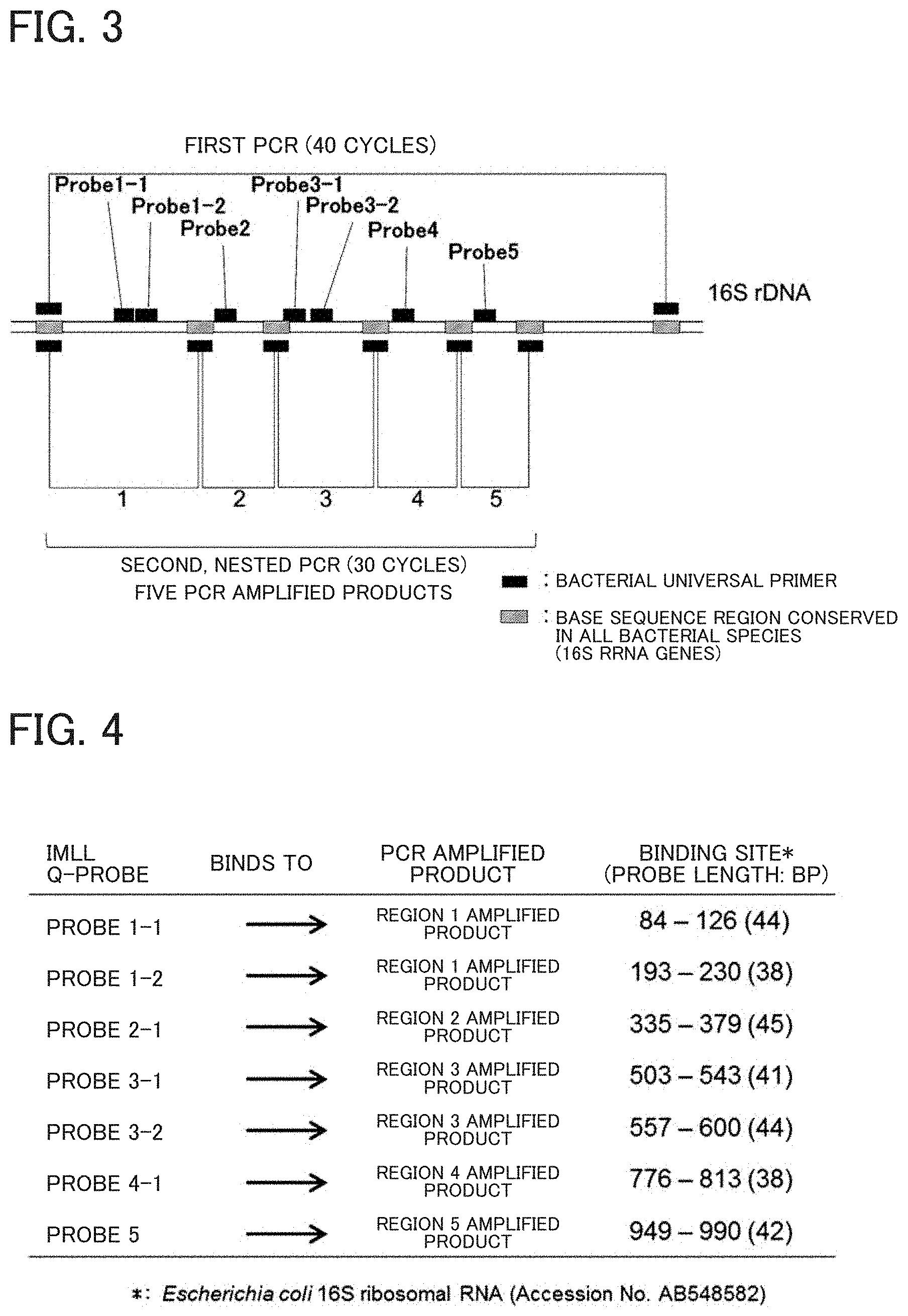
[0013] FIG. 3 illustrates a strategy of designing primers and probes. Nested PCR is performed using five bacterial universal primer sets, and then seven IMLL probes are additionally used for the PCR amplicons to obtain seven Tm values.
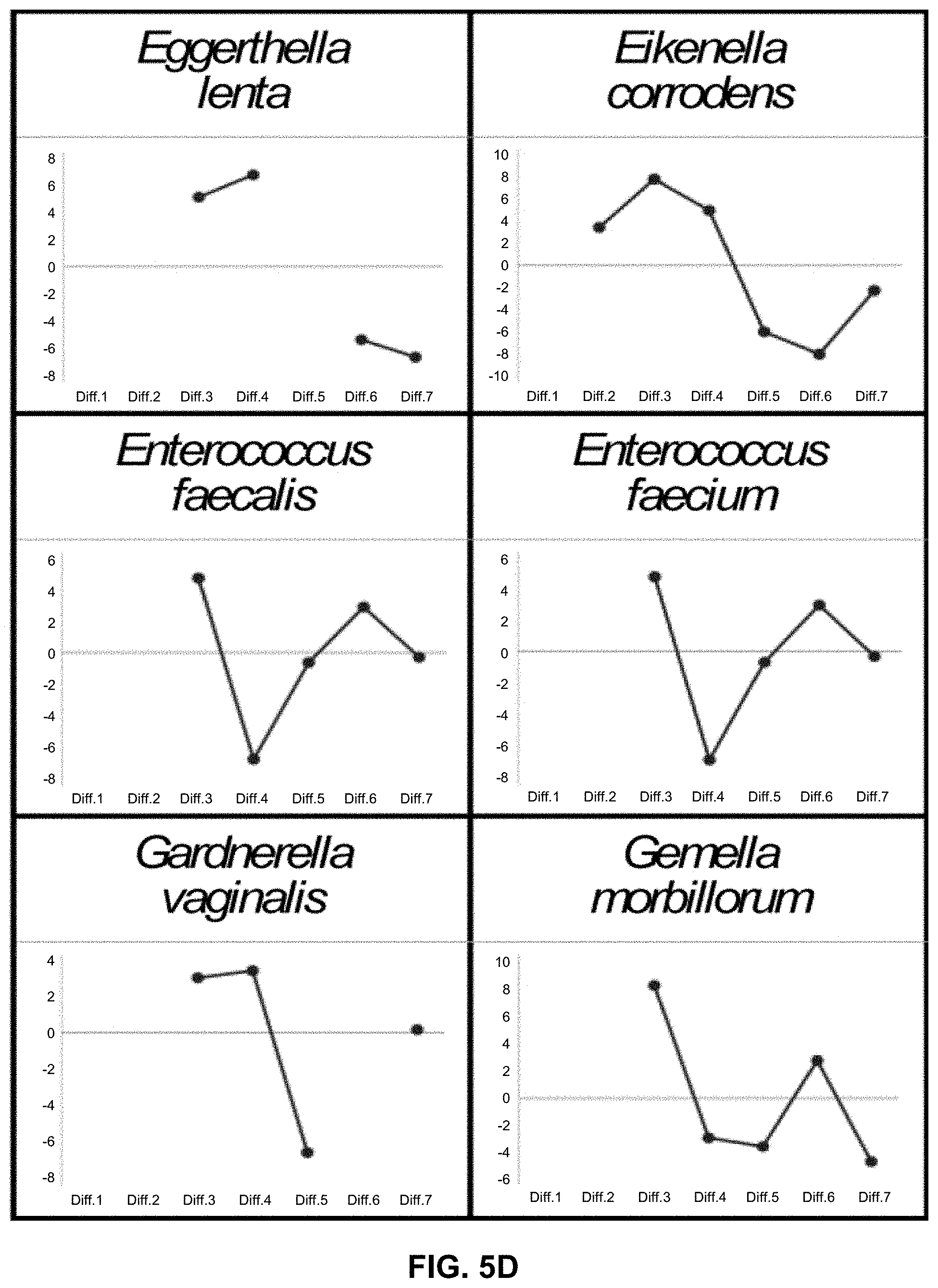
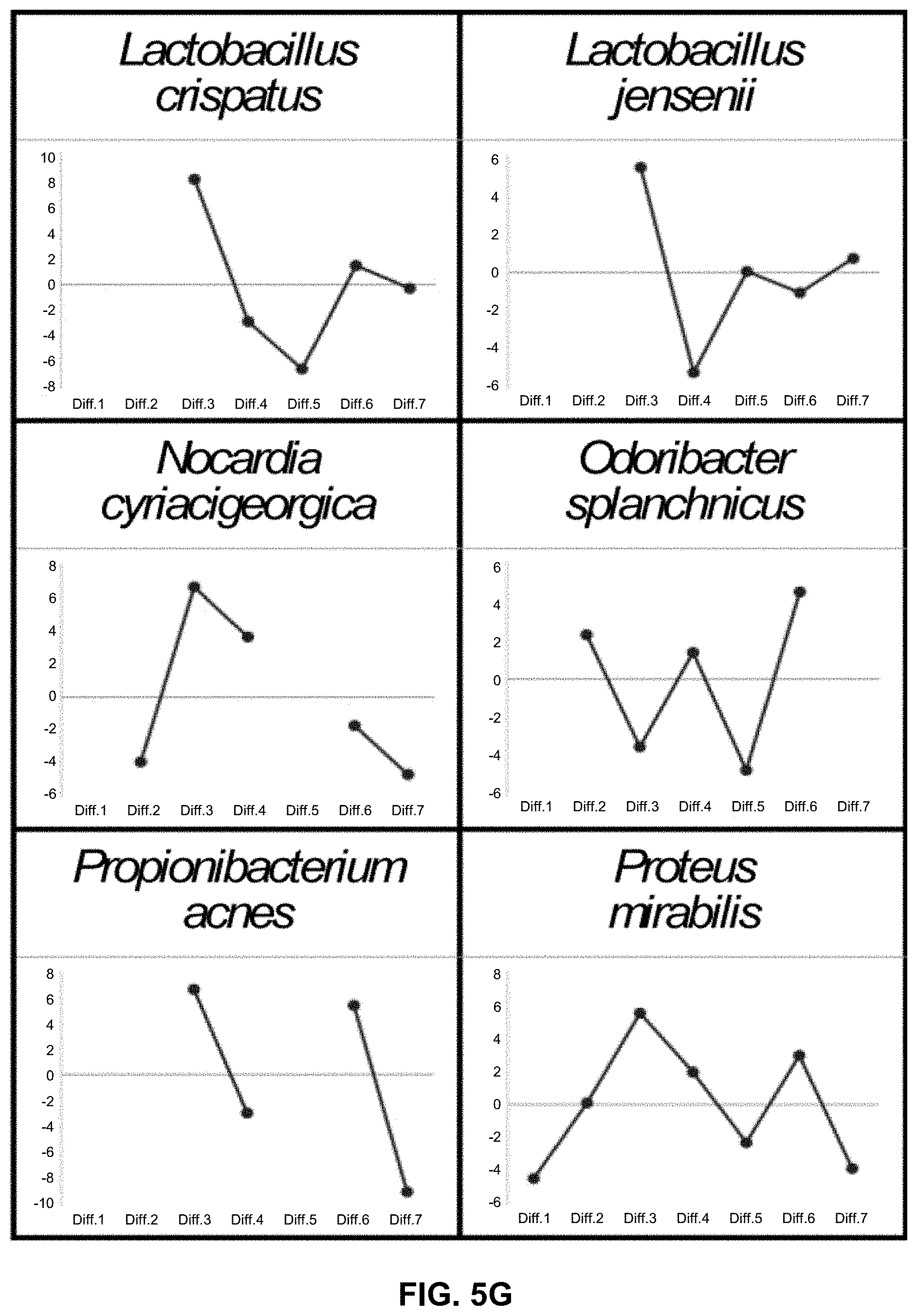
[0014] FIG. 4 illustrates binding sites of IMLL probes.
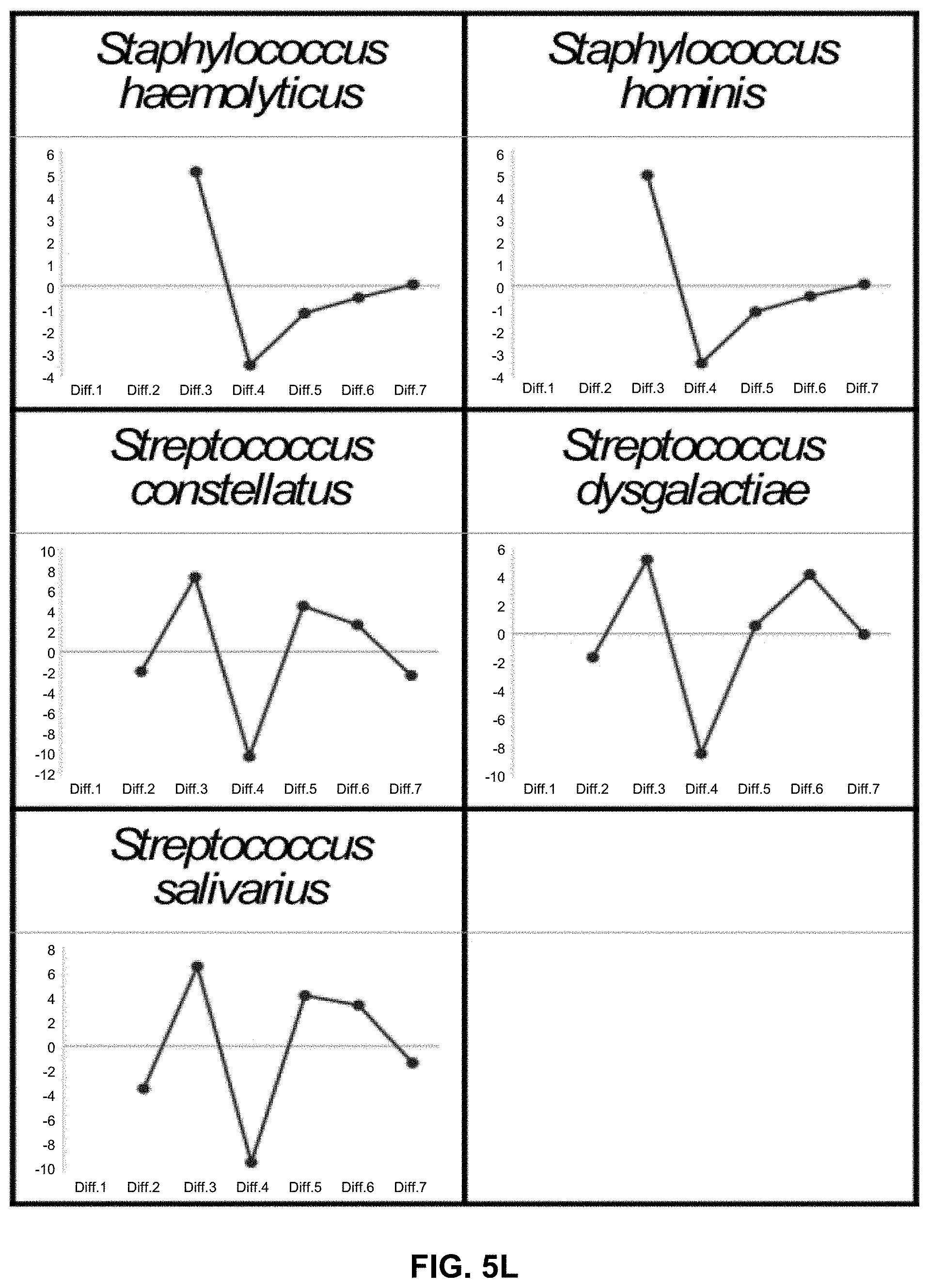
[0015] FIGS. 5A-5L illustrate Tm mapping shapes (IMLL probes) of 71 bacterial species registered in a database. It should be noted that FIGS. 5A-5L are conceptual illustrations of Tm mapping shapes, and thus the X axis shows the average of seven Tm values with specific numerical values omitted.
[0016] FIG. 6 illustrates an equation calculating the Difference Value. The Difference Value indicates the similarity of a Tm mapping shape to that of the species included in the database.
[0017] FIG. 7 illustrates differences between the variation range of Tm values from the conventional Tm mapping method and the variation range of Tm values from the improved Tm mapping method.
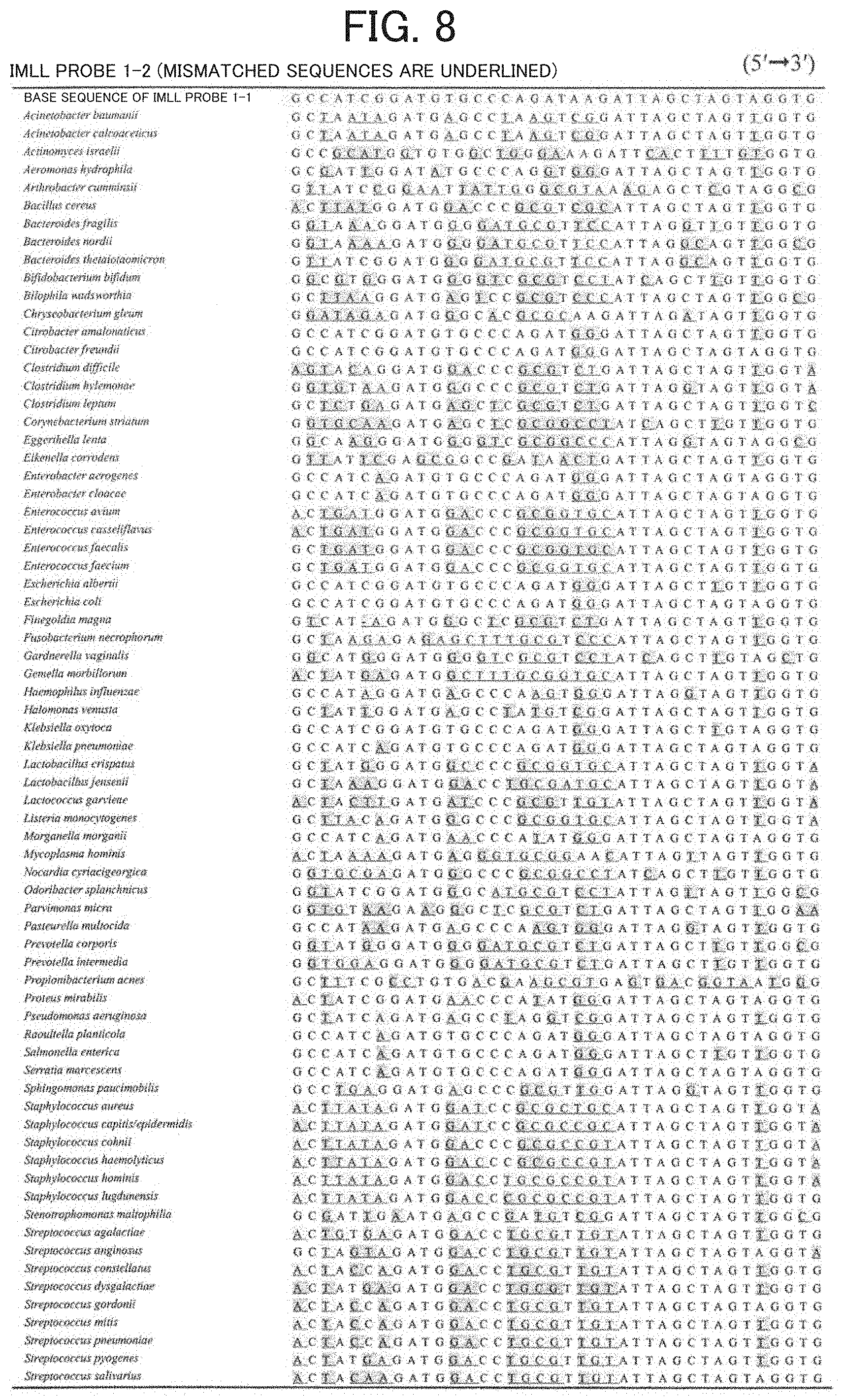
[0018] FIG. 8 illustrates the mismatches between the IMLL probe 1-2 and the target sequence in each bacterial species (71 bacterial species).
[0019] FIG. 9 illustrates similarity (Table 1) of Tm mapping shapes among 71 bacterial species in the database.
[0020] FIG. 10 illustrates results (Table 2) of individual identification performed by using a database constructed to include 71 bacterial species and a real-time PCR instrument with a thermal variation of .+-.0.1.degree. C. and starting from whole blood samples.
[0021] FIG. 11 illustrates results (Table 3-1) of an individual blind test performed by using a database constructed to include 145 bacterial species and a real-time PCR instrument with a thermal variation of .+-.0.4.degree. C.
[0022] FIG. 12 illustrates Table 3-2, a continuation of FIG. 11.
[0023] FIG. 13 illustrates results (Table 4) of individual identification performed by using a database constructed to include 145 bacterial species and a real-time PCR instrument with a thermal variation of .+-.0.4.degree. C. and starting from whole blood samples.
DESCRIPTION OF EMBODIMENTS
[0024] The following disclosure provides many different embodiments, or examples, for implementing different features of the provided subject matter. These are, of course, merely examples and are not intended to be limiting. In addition, the disclosure may repeat reference numerals and/or letters in the various examples. This repetition is for the purpose of simplicity and clarity and does not in itself dictate a relationship between the various embodiments and/or configurations discussed. Further, when a first element is described as being "connected" or "coupled" to a second element, such description includes embodiments in which the first and second elements are directly connected or coupled to each other, and also includes embodiments in which the first and second elements are indirectly connected or coupled to each other with one or more other intervening elements in between.
[0025] The inventors investigated a novel Tm mapping method using long probes rather than PCR amplified products, leading to the disclosure.
[0026] According to the disclosure, the Tm mapping method can be performed in almost all commercial real-time PCR instruments.
[0027] As a long probe, for example, a quenching probe was designed to have an incompletely matched sequence with a very long length of about 40 bases. This led to the finding that the probes can bind to most microorganism sequences and can give wide Tm values.
[0028] Long probes, even with many mismatches, can bind to imperfect-match sequences because the long length of the probes results in increased hydrogen bonding force.
[0029] On the other hand, long probes have a strong tendency to form secondary structures and self-quench. Thus, optimum probes were selected by conducting research such as repeated trial production of long probes that form no secondary structures using Delta-G and by confirming operation, leading to completion of the present invention.
[0030] Depending on the number and position of mismatches, the probes allowed acquisition of wide differences in Tm values for each bacterial species, that is, differences up to 20.degree. C. or more among bacterial species, as compared with the differences in Tm values of conventional PCR amplified products among bacterial species.
[0031] The inventors have designated such probes Imperfect-Match Linear Long (IMLL) probes.
[0032] Seven IMLL probes were actually used to construct first a preliminary database including the Tm mapping shapes for 71 bacterial species.
[0033] As a result, it is demonstrated that real-time PCR instruments whose measurement error of Tm values between PCR tubes is within .+-.0.5.degree. C. theoretically enable correct distinction among or identification of microorganisms at least at the genus level and often at the species level.
[0034] The measurement error between PCR tubes of current commercial real-time PCR instruments is almost within .+-.0.3.degree. C. Therefore, IMLL probes can be used to conduct the Tm mapping method in almost all real-time PCR instruments.
[0035] On the other hand, it is nearly impossible to distinguish between, for example, Enterococcus faecalis and Enterococcus faecium due to the limited ability of the designed probes, but at least the genus is correctly identified.
[0036] Also, because of sequence similarity among species belonging to the genus Staphylococcus, it is also difficult to distinguish them at the species level. However, an additional step was performed for only 10 minutes by using a short probe specifically recognizing Staphylococcus aureus, which allowed discrimination between Staphylococcus aureus and other species except Staphylococcus aureus belonging to the genus Staphylococcus.
[0037] The invention will be described below in detail.
[0038] The conventional Tm mapping method refers to a method of rapidly identifying infection-causing pathogenic microorganisms performed in the steps described below that represent a flow of steps as shown in FIG. 1.
[0039] It should be noted that FIG. 1 illustrates a flow of steps and the graphs are only conceptual illustrations.
[0040] A method of identifying infection-causing pathogenic microorganisms by [0041] (1) extracting a microbial DNA from a sample such as blood; [0042] (2) performing nested PCR using the extracted microbial DNA, as a template, and a plurality of universal primers; [0043] (3a) melting and analyzing a plurality of gene amplified products (PCR amplicons) obtained in the above-referenced step; [0044] (4) two-dimensionally mapping a plurality of melting temperature (Tm) values; and [0045] (5) checking the two-dimensionally mapped "shape" against a database.
[0046] In the improved Tm mapping method of the disclosure, the conventional step (3a) is substituted with step (3) as shown in a flow of steps in FIG. 2. In step (3), Imperfect-Match Linear Long (IMLL) probes are added to the gene amplified products (PCR amplicons) obtained in the step (2) described above to analyze Tm values of IMLL probes rather than Tm values of the gene amplified products. The subsequent steps (4) and (5) are the same as in the conventional Tm mapping method.
[0047] Step (1): extracting a microbial DNA from a sample such as blood.
[0048] Bacterial DNA is extracted directly from a clinical sample (e.g., 2 mL of a whole blood sample) to utilize it as a template for PCR.
[0049] The extraction method is not limited to particular methods, and clinical sample collection, DNA extraction, and the like may be performed using any known method.
[0050] Step (2): Performing nested PCR using the extracted microbial DNA, as a template, and a plurality of bacterial universal primers (primers that detect almost all bacteria).
[0051] This step includes nested PCR using 5 to 7 bacterial universal primer sets (one primer set per tube in the second PCR).
[0052] These primers can detect almost all bacterial species.
[0053] To improve the accuracy of this step, it is preferable to use a thermostable DNA polymerase that is recombinantly produced using a eukaryote as a host cell, without bacterial DNA contamination.
[0054] The use of the polymerase enables highly sensitive and reliable detection of bacteria without false positive results in bacterial universal PCR. Therefore, the PCR allows direct identification of infection-causing pathogenic microorganisms from patient samples.
[0055] In this step, nested PCR is performed to obtain a plurality of, for example, five PCR amplified products (amplicons) (FIG. 3).
[0056] Step (3): Analyzing Tm values of IMLL probes.
[0057] For example, two probe targets are located in each of the PCR amplicons of Regions 1 and 3, and a total of 7 (5 types of) PCR amplicons are mixed with seven IMLL probes (FIG. 3 and FIG. 4).
[0058] Seven Tm values can be obtained by analyzing the seven IMLL probes.
[0059] Step (4): Two-dimensionally mapping a plurality of Tm values.
[0060] For example, seven Tm values are two-dimensionally mapped. The shape of plot (Tm mapping shape) indicates a shape specific to a species or genus as shown in FIGS. 5A-5L.
[0061] This is not High Resolution Melting-curve (HRM) analysis, but only Tm values are measured.
[0062] Step (5): Checking the two-dimensionally mapped "shape" against a database.
[0063] The infection-causing pathogenic microorganisms can be rapidly identified by comparing the Tm mapping shape with shapes in the database.
[0064] The degree of similarity with the shapes in the database is expressed quantitatively with the Difference Value calculation equation as shown in FIG. 6.
[0065] As the Difference Value is closer to 0, it means that the shapes are more consistent.
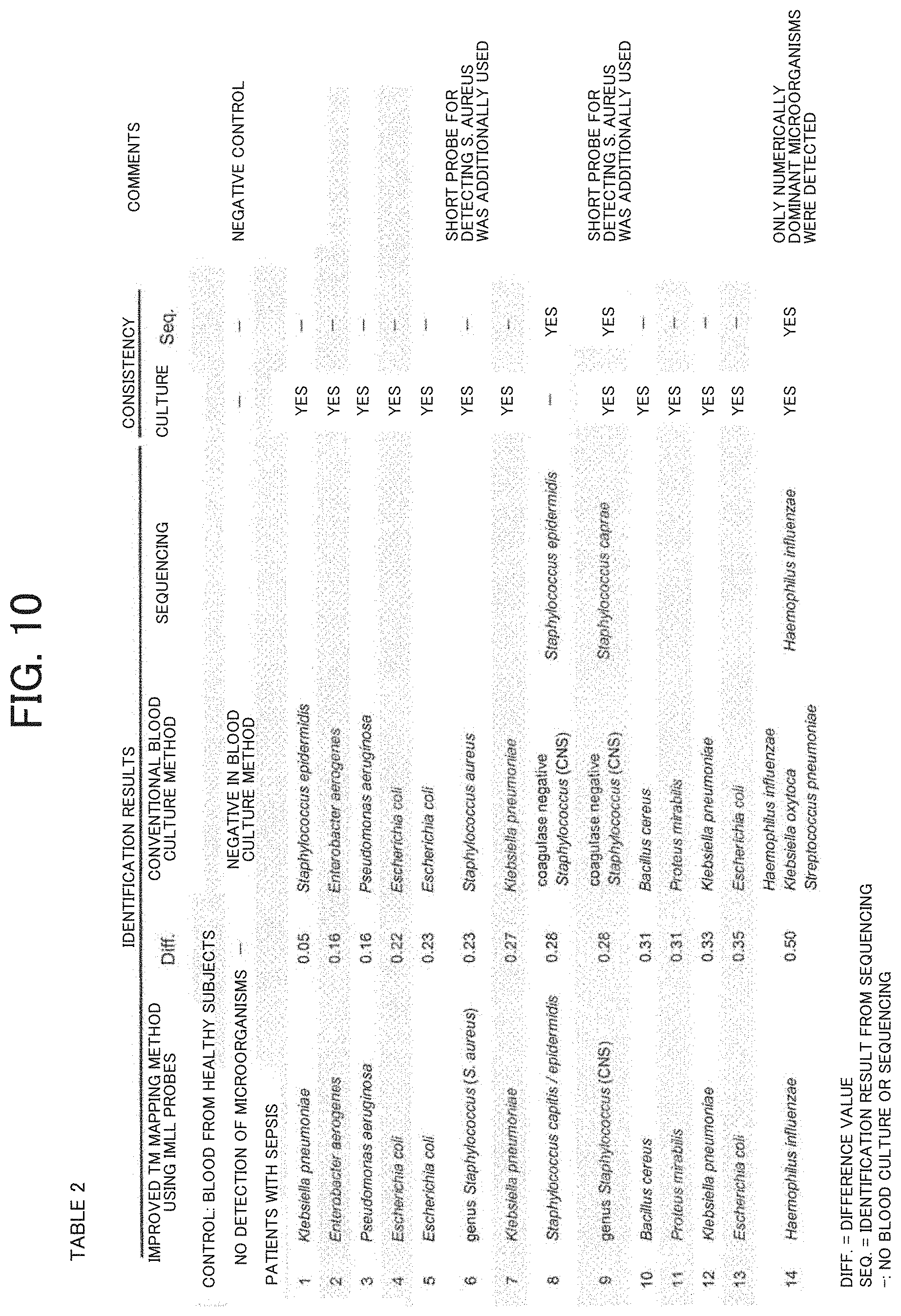
[0066] The "long probe" of Imperfect-Match Linear Long (IMLL) probe used in the improved Tm mapping method means an oligonucleotide with 35 to 50 bases, preferably 35 to 48 bases, and more preferably 38 to 45 bases.
[0067] The difference in Tm values due to the difference in bacterial species of the IMLL probes is up to 20.degree. C. or more.
[0068] On the other hand, the difference in Tm values in the conventional Tm mapping method due to the difference in bacterial species is at most about 5.degree. C.
[0069] FIG. 7 illustrates the actual difference between the Tm value variation obtained in the conventional Tm mapping method and the Tm value variation obtained by using IMLL probes in the improved Tm mapping method, in the preliminary database for 71 bacterial species.
[0070] Furthermore, an IMLL probe is a probe having a length capable of binding to a site having a mismatch with the target sequence.
[0071] By way of example, FIG. 8 illustrates the mismatches between IMLL probe 1-2 produced below and each of 71 bacterial species included in the preliminary database.
[0072] The IMLL probes of the disclosure can bind to target sequences in many bacterial species having different base sequences.
[0073] The IMLL probes do not reduce binding to their target sequences because the probes themselves do not form secondary structures despite their long length.
[0074] Each of the IMLL probes is designed to have a plurality of sites that bind to, for example, Escherichia coli 16S ribosomal RNA (Accession No. AB548582) using a multiple alignment software program (ClustalX) and is chemically synthesized.
[0075] For example, the following probes were prepared as specific IMLL probes.
TABLE-US-00001 IMLL probe 1-1 SEQ ID NO: 1 5'-GTTATCCCACTCTAATAAGCAGGTTACCTACGTATTACTCAC CC-3': [probe size: 44 bp, Binding site: positions 84-126 of Escherichia coli 16S rRNA (Accession No. AB548582)] IMLL probe 1-2 SEQ ID NO: 2 5'-CACCTACTAGCTAATCTTATCTGGGCACATCCGATGGC-3': [probe size: 38 bp, Binding site: positions 193-230 of Escherichia coli 16S rRNA] IMLL probe 2-1 SEQ ID NO: 3 5'-CACGCGGCATGGCTCCATCAGGCTTTCCCCCATTGTCGAAGA TTC-3': [probe size: 45 bp, Bindings ite: positions 335-379 of Escherichia coli 16S rRNA] IMLL probe 3-1 SEQ ID NO: 4 5'-CGCCCTGTAATTCCGAATAACGCTAGCTCCCACCGTATTAC- 3': [probe size: 41 bp, Binding site: positions 503-543 of Escherichia coli 16S rRNA] IMLL probe 3-2 SEQ ID NO: 5 5'-CCAAGTTGAGCCCGGGCCTTTCACTACTGACTTAACAAACCG CC-3': [probe size: 44 bp, Binding site: positions 557-600 of Escherichia coli 16S rRNA] IMLL probe 4-1 SEQ ID NO: 6 5'-GGCACAACCTCTTAATACTCATCGTTTACAGCGTGGAC-3': [probe size: 38 bp, positions 776-813 of Escherichia coli 16S rRNA] IMLL probe 5 SEQ ID NO: 7 5'-ATCTCTGCAAAGTTCTAAGGATGTCAAGATTAGGTAAGG TTC-3': [probe size: 42 bp, Binding site: positions 949-990 of Escherichia coli 16S rRNA] Short probe for detecting S. aureus SEQ ID NO: 8 5'-TATCTAATGCAGCGCGGATC-3': [probe size: 20 bp, Binding site: positions 211-230 of Staphylococcus aureus 16S rRNA (Accession No. AB681291)]
[0076] The IMLL probes were designed using a multiple alignment software program. The following IMLL probes may be also used depending on target binding sites.
TABLE-US-00002 IMLL probe 1-3 SEQ ID NO: 21 5'-CAGACCAGCTAACGATCGTCGCCTTAGTAAGCCGTTACCC-3': [probe size: 40 bp, Binding site: positions 231-270 of Escherichia coli 16S rRNA] IMLL probe 1-4 SEQ ID NO: 22 5'-CTCAGACCAGCTAACGATCGTTGCCTTAGTAAGCCGTTACCT-3': [probe size: 42 bp, Binding site: positions 231-272 of Escherichia coli 16S rRNA] IMLL probe 2-2 SEQ ID NO: 23 5'-GTACTTTACAACCCGAAGGCCTTCTTCATACACGCGGCATGGC-3': [probe size: 43 bp, Binding site: positions 367-409 of Escherichia coli 16S rRNA] IMLL probe 3-3 SEQ ID NO: 24 5'-CCTTATCGCCTTCCTCCCCGCTGAAAGTGCTTTAC-3': [probe size: 35 bp, Binding site: positions 401-435 of Escherichia coli 16S rRNA] IMLL probe 3-4 SEQ ID NO: 25 5'-CGCCCAGTAATTCCGATTAACGCTTGCACCCTCCGTATTAC-3': [probe size: 41 bp, Binding site: positions 503-543 of Escherichia coli 16S rRNA] IMLL probe 3-5 SEQ ID NO: 26 5'-GGCCGACTAATTCCGATTAACGCTTCCACGCTCCGTATTAC-3': [probe size: 41 bp, Binding site: positions 503-543 of Escherichia coli 16S rRNA] IMLL probe 3-6 SEQ ID NO: 27 5'-CTTAACAAACCGCCTACGCACCCTTTAAGCCCAATAATTCCGATTAA CGC-3': [probe size: 50 bp, Binding site: positions 521-570 of Escherichia coli 16S rRNA] IMLL probe 3-7 SEQ ID NO: 28 5'-CTCAAGTTTGCCAGTTTCCGATGAAGTTCCCAGGTTGAGC-3': [probe size: 40 bp, Binding site: positions 590-629 of Escherichia coli 16S rRNA] IMLL probe 3-8 SEQ ID NO: 29 5'-CTCAAGTTTGCCAGTTTCGGATGCAGTTTCCAGGTTGAGC-3': [probe size: 40 bp, Binding site: positions 590-629 of Escherichia coli 16S rRNA] IMLL probe 4-2 SEQ ID NO: 30 5'-CCACCTCTATGCAGACATCGTTTACGGCGTGGACTACCAGGG-3': [probe size: 42 bp, Binding site: positions 768-809 of Escherichia coli 16S rRNA] IMLL probe 6-1 SEQ ID NO: 31 5'-CCTCCAGTTTGTCATCGGCAGTCTACATTGAGTTCCCAAC-3': [probe size: 40 bp, Binding site: positions 1112-1151 of Escherichia coli 16S rRNA] IMLL probe 6-2 SEQ ID NO: 32 5'-CCTCCAGTTTGTCACCGGCAGTCTACATTGAGTTCCCAAC-3': [probe size: 40 bp, Binding site: positions 1112-1151 of Escherichia coli 16S rRNA] IMLL probe 7-1 SEQ ID NO: 33 5'-CTTCATGTAATCAGGTTGCAGACTCCAATCCGGACTAAGACGC-3': [probe size: 43 bp, Binding site: positions 1265-1307 of Escherichia coli 16S rRNA] IMLL probe 7-2 SEQ ID NO: 34 5'-CTAGCGATTCCGACTTCATGAATACGAGTTGCAGCCTACAAT-3': [probe size: 42 bp, Binding site: positions 1279-1320 of Escherichia coli 16S rRNA]
[0077] The universal primers used in step (2) in the improved Tm mapping method of the disclosure may be designed to target a bacterial conserved region so as to universally amplify, for example, seven regions of bacterial 16S ribosomal RNA gene.
[0078] Specifically, the universal primers are designed using a multiple alignment software program (Clustal X) and chemically synthesized.
[0079] Specific primers are as follows:
TABLE-US-00003 Primers for first PCR Forward: SEQ ID NO: 9 5'-AGAGTTTGATCATGGCTCAG-3': Reverse: SEQ ID NO: 10 5'-CCGGGAACGTATTCACC-3': Region 1 primers for second PCR (nested PCR) Forward: SEQ ID NO: 11 5'-AGAGTTTGATCATGGCTCAG-3': Reverse: SEQ ID NO: 12 5'-CGTAGGAGTCTGGACCGT-3': Region 2 primers for second PCR (nested PCR) Forward: SEQ ID NO: 13 5'-GACTCCTACGGGAGGCA-3': Reverse: SEQ ID NO: 14 5'-TATTACCGCGGCTGCTG-3': Region 3 primers for second PCR (nested PCR) Forward: SEQ ID NO: 15 5'-AGCAGCCGCGGTAATA-3': Reverse: SEQ ID NO: 16 5'-GGACTACCAGGGTATCTAATCCT-3': Region 4 primers for second PCR (nested PCR) Forward: SEQ ID NO: 17 5'-AACAGGATTAGATACCCTGGTAG-3': Reverse: SEQ ID NO: 18 5'-AATTAAACCACATGCTCCACC-3': Region 5 primers for second PCR (nested PCR) Forward: SEQ ID NO: 19 5'-TGGTTTAATTCGATGCAACGC-3': Reverse: SEQ ID NO: 20 5'-GAGCTGACGACAGCCAT-3':
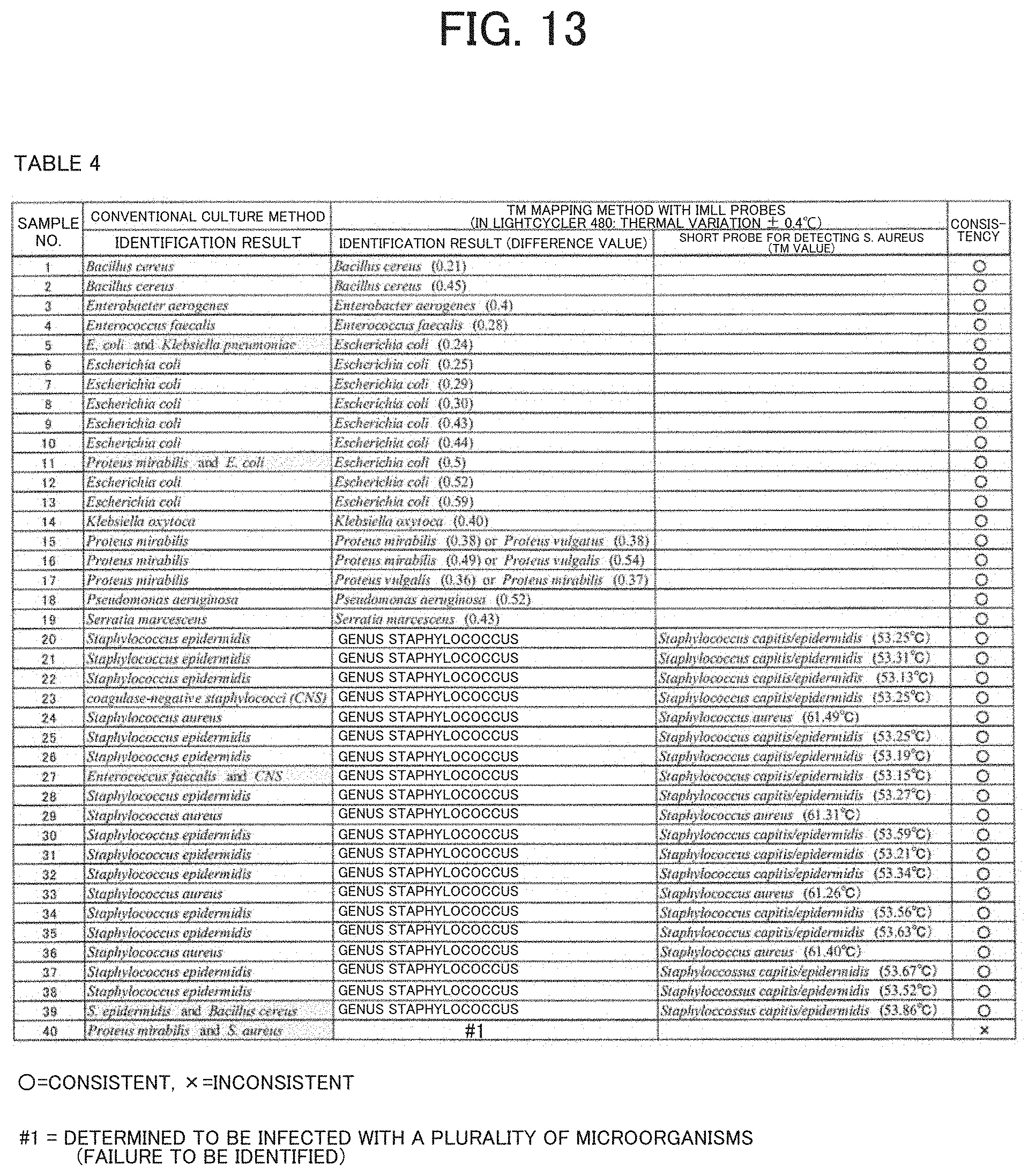
[0080] Construction of Tm mapping database using IMLL probes
[0081] A preliminary database including Tm mapping shapes of 71 bacterial species was first constructed by employing the average of Tm values measured multiple times (e.g., 3 times) in a RotorGeneQ from QIAGEN (thermal variation: .+-.0.1.degree. C.) (FIGS. 5A-5L).
[0082] Bacteria were obtained from clinical samples, and then their base sequences were sequenced. Identification at the species level was achieved.
[0083] The amount of data in the database can be changed, and the data can be easily changed and updated.
[0084] Each Tm mapping shape in the database reflects the number and position of probe-target mismatches in the hybridization of the IMLL Q probe with a target sequence in each bacterial species.
[0085] That is to say, each Tm mapping shape reflects the specific base sequence of each bacterial species and illustrates forms a unique shape.
[0086] Some Tm mapping shapes have no data points.
[0087] This is due to the fact that some IMLL probes do not bind to their target regions and thus the Tm value at the region cannot be obtained.
[0088] To identify pathogenic microorganisms, an identification software program calculates the Difference Value indicating the homology of the Tm mapping shapes and searches for similar patterns of the presence or absence of IMLL probe binding, thereby narrowing down the search range for bacteria in the database.
[0089] In other words, the pattern of IMLL probe binding is also utilized as a feature for distinguishing bacterial species.
[0090] Evaluation of the accuracy of the Tm mapping method using the IMLL probes (71 bacterial species)
[0091] To evaluate the accuracy of the Tm mapping method using the IMLL probes, a blind test was performed using bacterial DNAs of the same 71 bacterial species registered in the preliminary database in a RotorGeneQ from QIAGEN (thermal variation: .+-.0.1.degree. C.).
[0092] That is to say, the bacterial DNAs were identified with the bacterial names hidden. The tests of 71 bacterial species failed to narrow down the Tm mapping results at the species level (Staphylococcus aureus or Staphylococcus hemolyticus or Staphylococcus hominis or Staphylococcus lugdunensis) in the genus Staphylococcus.
[0093] Similarly, it was impossible to distinguish between Enterococcus faecalis and Enterococcus faecium and identify them.
[0094] The results from Tm mapping shapes for the remaining 65 bacterial species were consistent with the results from sequenced bacterial DNAs in the database.
[0095] The Difference Values averaged 0.242 and ranged from 0.09 to 0.30 (standard deviation=0.08).
[0096] Evaluation of the similarity of Tm mapping shapes of 71 bacterial species in the preliminary database
[0097] The evaluation is shown in Table 1 (FIG. 9).
[0098] The defined Difference Value reflects the difference between each bacterial species registered in the database of Tm mapping shapes and other bacterial species.
[0099] As the Difference Value is closer to zero, the Tm mapping shapes are more similar to the shapes for bacterial species registered in the database.
[0100] If a measurement error of Tm values between PCR tubes in a measuring instrument (=thermal variation of the measuring instrument) is within .+-.0.1.degree. C., the measurement error range of the Difference Values will be within 0.28.
[0101] Similarly, if a measurement error of Tm values between PCR tubes in a measuring instrument is within .+-.0.2.degree. C., .+-.0.3.degree. C., .+-.0.4.degree. C., .+-.0.5.degree. C., and .+-.0.6.degree. C., the measurement error range of the Difference Values will be within 0.53, 0.80, 1.06, 1.33, and 1.59, respectively.
[0102] The similarity of bacterial species to each other was analyzed by using the Difference Values, which demonstrates that it is impossible to distinguish between Enterococcus faecalis and Enterococcus faecium.
[0103] It is also revealed that identification at the species level is difficult in the genus Staphylococcus.
[0104] Since Difference Values of the other 65 bacterial species to each other have 1.33 or more, it is found that use of a measuring instrument with a thermal variation (measurement error of Tm values between PCR tubes) within .+-.0.5.degree. C. allows identification accurate at least at the genus level and often at the species level.
[0105] That is to say, although the conventional Tm mapping method allows only a measuring instrument having measurement errors of Tm values between PCR tubes within .+-.0.1.degree. C., the disclosure significantly expands the scope of instruments capable of performing the Tm mapping method.
[0106] For example, seven IMLL probes (SEQ ID NOS: 1 to 7) were used to first construct a preliminary database of Tm mapping shapes for 71 bacterial species. As a result, it was shown that microorganisms can be accurately distinguished and identified at least at the genus level and often at the species level even in a real-time PCR instrument having measurement errors of Tm values between PCR tubes within .+-.0.5.degree. C.
[0107] Since most of the current commercial real-time PCR instruments have a measurement error between PCR tubes within .+-.0.3.degree. C., the Tm mapping method can be performed in almost all instruments when the IMLL probes are used.
[0108] By using 14 whole blood samples (in 2 mL EDTA blood collection tubes) collected from patients with sepsis, the accuracy of the Tm mapping method using the IMLL probe (SEQ ID NOS: 1 to 7) was compared with that of the conventional culture method to perform evaluation.
[0109] A RotorGeneQ (thermal variation: .+-.0.1.degree. C.) from QIAGEN was used as a real-time PCR instrument, and a preliminary database including 71 bacterial species was used as a database of pathogenic microorganisms.
[0110] Table 2 (FIG. 10) shows a comparison between the individual identification results of pathogenic microorganisms using the Tm mapping method with the IMLL probes and the identification results from the culture method or the sequencing method.
[0111] When the identification results of pathogenic microorganisms from the Tm mapping method were inconsistent with the results from the culture method, the sequencing method was used to identify the bacterial species again. The Tm mapping method was used to identify pathogenic microorganisms in a total of 14 samples, and the obtained results were consistent with the results from the culture method or sequencing method in all of the 14 samples (14/14).
[0112] However, when pathogenic microorganisms are identified to belong to the genus Staphylococcus in the method using IMLL probes, an additional test is performed using a short probe for detecting S. aureus to determine whether the pathogenic microorganisms are S. aureus or other species belonging to the genus Staphylococcus (=CNS) (this additional test can be completed in only about 10 minutes).
[0113] Seven IMLL probes of the disclosure (SEQ ID NOS: 1 to 7) were then used to construct an extensive database including Tm mapping shapes of 146 bacterial species by employing the average of Tm values measured multiple times (e.g., 3 times) in a RotorGeneQ from QIAGEN (thermal variation: .+-.0.1.degree. C.).
[0114] The registered 145 bacterial species (65 bacterial genera) are as follows: Achromobacter xylosoxidans, Acinetobacter baumanii, Acinetobacter calcoaceticus, Aeromonas hydrophila, Alistipes onderdonkii, Anaerococcus vaginalis, Arthrobacter cumminsii, Bacillus cereus, Bacillus coagulans, Bacillus licheniformis, Bacillus megaterium, Bacillus pumilus, Bacillus subtilis subsp. Subtilis, Bacteroides dorei, Bacteroides finegoldii, Bacteroides fragilis, Bacteroides nordii, Bacteroides salyersiae, Bacteroides thetaiotaomicron, Bacteroides uniformis, Bacteroides vulgatus, Bartonella henselae, Bifidobacterium bifidum, Bilophila wadsworthia, Bordetella pertussis, Borrelia burgdorferi, Brevibacillus laterosporus, Campylobacter coli, Campylobacter jejuni subsp.jejuni, Campylobacter rectus, Capnocytophaga gingivalis, Capnocytophaga granulosa, Capnocytophaga haemolytica, Capnocytophaga ochracea, Capnocytophaga sputigena, Chryseobacterium gleum, Citrobacter amalonaticus, Citrobacter freundii, Clostridium difficile, Clostridium histolyticum, Clostridium hylemonae, Clostridium paraputrificum, Clostridium perfringus, Clostridium sporogenes, Clostridium subterminal, Clostridium tertium, Corynebacterium amycolatum, Corynebacterium macginleyi, Corynebacterium striatum, Corynebacterium xerosis, Eggerthella lenta, Eikenella corrodens, Empedobacter brevis, Enterobacter aerogenes, Enterobacter cloacae subsp.cloacae, Enterococcus avium, Enterococcus casseliflavus, Enterococcus durans, Enterococcus faecalis, Enterococcus faecium, Enterococcus gallinarum, Enterococcus raffinosus, Escherichia albertii, Escherichia coli, Eubacterium limosum, Finegoldia magna, Fusobacterium necrophorum, Fusobacterium periodonticum, Fusobacterium varium, Gardnerella vaginalis, Gemella morbillorum, Geobacillus stearothermophilus, Haemophilus influenzae, Halmonas venusta, Klebsiella oxytoca, Klebsiella pneumoniae, Kocuria rosea, Lactobacillus acidophilus, Lactobacillus crispatus, Lactobacillus fermentum, Lactobacillus jensenii, Lactococcus garvieae, Legionella pneumophila subsp.pneumophila, Leptospira interrogans serovar Copenhageni, Listeria monocytogenes, Micrococcus luteus, Morganella morganii, Nocardia cyriacigeorgica, Odoribacter splanchinicus, Pantoea agglomerans, Parvimonas micra, Pasteurella multocida, Peptoniphilus asaccharolyticus, Peptoniphilus gorbachii, Peptostreptococcus anaerobius, Plesiomonas shigelloides, Porphyromonas gingivalis, Prevotella corporis, Prevotella intermedia, Prevotella melaninogenica, Prevotella timonensis, Prevotella veronalis, Propionibacterium acnes, Propionibacterium granulosum, Proteus mirabilis, Proteus vulgaris, Pseudomonas aeruginosa, Pseudomonas fluorescens, Pseudomonas putida, Salmonella enterica, Serratia marcescens, Serratia plymuthia, Staphylococcus aureus, Staphylococcus capitis/epidermidis, Staphylococcus cohnii, Staphylococcus haemolyticus, Staphylococcus hominis, Staphylococcus intermedius, Staphylococcus lugdunensis, Staphylococcus saprophyticus subsp. saprophyticus, Staphylococcus schleiferi subsp.coagulans, Staphylococcus simulans, Staphylococcus warneri, Stenotrophomonas maltophilia, Streptobacillus moniliformis, Streptococcus agalactiae, Streptococcus anginosus, Streptococcus bovis, Streptococcus constellatus, Streptococcus dysgalactiae, Streptococcus gallolyticus subsp. pasteurianus, Streptococcus gordonii, Streptococcus intermedius, Streptococcus mitis, Streptococcus orali, Streptococcus pneumoniae, Streptococcus pyogenes, Streptococcus salivarius, Streptococcus sanguinis, Tannerella forsythus, Treponema denticola, Vibrio fluvialis, Vibrio vulnificus, Yersinia enterocolitica subsp. enterocolitica, Yersinia pseudotuberculosis
[0115] Evaluation of the accuracy of the Tm mapping method using IMLL probes (145 bacterial species)
[0116] Next, the accuracy of the Tm mapping method using IMLL probes is evaluated in more detail. To achieve this, an extensive database was first constructed using bacterial DNAs from 145 species (65 bacterial genera). The same bacterial DNAs registered in the database including 145 bacterial species was then used to perform a blind test in a Light Cycler.RTM. 480 (thermal variation: .+-.0.4.degree. C.) from Roche Life Science.
[0117] That is to say, the bacterial DNAs were identified using the Tm mapping method with IMLL probes, with the bacterial names hidden.
[0118] The results are shown in Table 3-1 (FIG. 11) and Table 3-2 (FIG. 12).
[0119] In the blind test results, 115 of 145 bacterial species were correctly identified at the species level, and all of 65 bacterial genera were correctly identified at the genus level. It was known that at the time of constructing the database, 30 bacterial species incorrectly identified at the species level were unable to be distinguished among the bacteria belonging to the same genus in an instrument with thermal variation of .+-.0.4.degree. C.
[0120] In other words, the results were as expected.
[0121] The microorganisms that cannot be correctly identified at the species level in an instrument with thermal variation of .+-.0.4.degree. C. were 3 of 6 bacterial species in the genus Bacillus, 2 of 8 bacterial species in the genus Bacteroides, 2 of 3 species in the genus Campylobacter, 2 of 4 bacterial species in the genus Corynebacterium, 5 of 7 bacterial species in the genus Enterococcus, 2 of 2 bacterial species in the genus Proteus, 9 of 11 bacterial species in the genus Staphylococcus, and 5 of 14 bacterial species in the genus Streptococcus.
[0122] The above results demonstrated that even when an instrument with thermal variation of .+-.0.4.degree. C. was used, 65 bacterial genera were able to be correctly identified at the genus level, and 115 of 145 species were able to be correctly identified at the species level.
[0123] Antimicrobial agents can be selected in most cases if the pathogenic microorganisms can be rapidly identified at least at the genus level. When identification at the species level is required, a species-specific short probe may be used.
[0124] After the genus is known, an additional test of about 10 minutes can be performed using a selected short probe to identify bacterial species at the species level.
[0125] Finally, by using 40 whole blood samples (in 2 mL EDTA blood collection tubes) (9 bacterial species) collected from patients with sepsis, the accuracy of the Tm mapping method using the IMLL probe (SEQ ID NOS: 1 to 7) was compared with that of the conventional culture method to perform evaluation.
[0126] A Light Cycler.RTM. 480 (thermal variation: .+-.0.4.degree. C.) from Roche Life Science was used as a real-time PCR instrument, and a database including 145 bacterial species was used as a pathogenic microorganism database.
[0127] Table 4 (FIG. 13) illustrates a comparison between the individual identification results of pathogenic microorganisms using Tm mapping methods with the IMLL probes and those using the culture method or the sequencing method.
[0128] When the identification results of pathogenic microorganisms using the Tm mapping method merely narrowed down the pathogenic microorganisms to the genus Staphylococcus, a short probe for detecting S. aureus was additionally used to determine whether the microorganisms are S. aureus or other species except S. aureus belonging to the genus Staphylococcus (CNS).
[0129] The Tm mapping method was used to identify pathogenic microorganisms in a total of 40 samples, and the obtained results were consistent with the results from the conventional culture method in 39 samples (39/40).
[0130] However, when a plurality of microorganisms is detected in the culture method, only numerically dominant microorganisms are identified in the Tm mapping method.
[0131] When the plurality of microorganisms was almost equally present (sample No. 40), the Tm mapping method failed to identify them, and the result was considered inconsistent.
[0132] The above results demonstrated that even when an instrument with thermal variation of .+-.0.4.degree. C. was used, 39 of 40 samples (9 bacterial species) from patients with sepsis were able to be correctly identified.
[0133] Antimicrobial agents can be selected in most cases if the pathogenic microorganisms can be rapidly identified at least at the genus level. However, when identification at the species level is required, a species-specific short probe such as a short probe for detecting S. aureus in the above case may be used.
[0134] After the genus is known, an additional test of about 10 minutes can be performed using a selected short probe to identify bacterial species at the species level.
[0135] Next, the improved Tm mapping method using linear long probes of imperfect-match sequences of the disclosure will be described in the Examples.
[0136] It should be noted that probes according to the disclosure are expressed as Imperfect-Match Linear Long (IMLL) probes.
[0137] The overall flow (workflow) is shown in FIG. 2.
[0138] The blood samples were whole blood collected from patients with suspected sepsis at Toyama University Hospital and Nagaresugi Hospital. All procedures in the Examples described below were performed under approval from the Ethics Committee at the University of Toyama and with written informed consent obtained from all patients. The methods performed in the Examples were carried out in accordance with the approved guidelines.
EXAMPLES
Example 1
[0139] Isolation of Bacterial Genomic DNA from Whole Blood
[0140] For example, blood is collected from vein into an EDTA blood collection tube and gently centrifuged (100.times.g) to separate blood cells and obtain a supernatant. The supernatant is transferred to a tube and strongly centrifuged (20,000.times.g) to obtain a bacterial pellet.
[0141] Specifically, 2 mL of venous blood was collected in an EDTA tube (EDTA-2K vacuum blood collection tube, NIPRO CORPORATION).
[0142] The blood sample was then centrifuged at 100.times.g for 5 minutes to spin down blood cells, and the resulting supernatant fraction (1 mL, with buffy coat) was used.
[0143] The supernatants were centrifuged again at 20,000.times.g for 10 minutes, and the resulting supernatant fractions were carefully removed so as not to disturb the pellets.
[0144] Next, 1 mL of molecular-grade distilled water (water deionized and sterilized for molecular biology, hereinafter referred to as sterile water, NACALAI TESQUE, INC.) was added, and the mixture was gently turned upside down and subsequently centrifuged at 20,000.times.g for 5 minutes.
[0145] Then, the supernatant fractions were again carefully removed so that the pellets were not resuspended before using a DNA extraction kit.
[0146] Next, DNA was isolated from the pellets using a DNA extraction kit (QlAamp UCP Pathogen Mini Kit, Qiagen, Germany) in accordance with the supplier's protocols.
[0147] Finally, bacterial DNA was eluted with 100 .mu.L of elution buffer.
[0148] Isolation of bacterial genomic DNA from bacterial colonies
[0149] Bacterial colonies were picked up with a sterile inoculating loop and suspended in 1 mL of sterile water.
[0150] The suspensions were subsequently centrifuged at 20,000.times.g for 10 minutes followed by removal of the supernatants to obtain pellets.
[0151] DNA was isolated from the resulting pellets using a DNA extraction kit (QlAamp UCP Pathogen Mini Kit, Qiagen) in accordance with the supplier's protocols. Finally, bacterial DNA was eluted with 100 .mu.L of elution buffer.
[0152] PCR Assays
[0153] A Veriti.TM. Thermal Cycler (Applied Biosystems) was used for amplification, and a LightCycler Nano (Roche Applied Science) was used for Tm value analysis of IMLL probes.
[0154] When using a LightCycler Nano that has two independent thermal blocks, the same thermal block is preferably used for all seven PCR tubes to identify pathogenic microorganisms using the Tm mapping method.
[0155] All PCR assays were performed as single-tube assays (no multiplex PCR).
[0156] RNase- and DNase-free PCR tubes (Eppendorf, Germany), 0.2 mL PCR tubes (Qiagen) were used for the first PCR [PCR (one tube)], and 0.1 mL Strip Tubes and Caps (Qiagen) were used for the nested PCR [Nested PCR (five tubes)].
[0157] All oligonucleotide primers were designed using a multiple alignment software program (Clustal X) and were synthesized by Life Technologies Japan Ltd. (Tokyo, Japan).
[0158] Quenching probes (Q-probes) were adopted as IMLL probes in the Example.
[0159] All quenching probes were designed using a multiple alignment software (Clustal X) and synthesized by NIPPON STEEL Eco-Tech Corporation (Tsukuba, Japan).
[0160] Bacterial universal primers were designed to universally amplify seven regions of bacterial 16S ribosomal RNA gene (FIG. 3).
TABLE-US-00004 First PCR primers Forward: 5'-AGAGTTTGATCATGGCTCAG-3' Reverse: 5'-CCGGGAACGTATTCACC-3' (Amplicon size: 1378 bp) Region 1 primers for second PCR (nested PCR) Forward: 5'-AGAGTTTGATCATGGCTCAG-3' Reverse: 5'-CGTAGGAGTCTGGACCGT-3' (Amplicon size: 338 bp) Region 2 primers for second PCR (nested PCR) Forward: 5'-GACTCCTACGGGAGGCA-3' Reverse: 5'-TATTACCGCGGCTGCTG-3' (Amplicon size: 199 bp) Region 3 primers for second PCR (nested PCR) Forward: 5'-AGCAGCCGCGGTAATA-3' Reverse: 5'-GGACTACCAGGGTATCTAATCCT-3' (Amplicon size: 287 bp) Region 4 primers for second PCR (nested PCR) Forward: 5'-AACAGGATTAGATACCCTGGTAG-3' Reverse: 5'-AATTAAACCACATGCTCCACC-3' (Amplicon size: 181 bp) Region 5 primers for second PCR (nested PCR) Forward: 5'-TGGTTTAATTCGATGCAACGC-3' Reverse: 5'-GAGCTGACGACAGCCAT-3' (Amplicon size: 120 bp)
[0161] Seven Q-probes described below that bind to each of five amplicons obtained from the nested PCR using the bacterial universal primers described above were designed (FIG. 3 and FIG. 4).
TABLE-US-00005 IMLL probe 1-1 5'-GTTATCCCACTCTAATAAGCAGGTTACCTACGTATTACTCACCC-3' [probe size: 44 bp, Binding site: positions 84-126 of Escherichia coli 16S rRNA (Accession No. AB548582)] IMLL probe 1-2 5'-CACCTACTAGCTAATCTTATCTGGGCACATCCGATGGC-3' [probe size: 38 bp, Binding site: positions 193-230 of Escherichia coli 16S rRNA] IMLL probe 2-1 5'-CACGCGGCATGGCTCCATCAGGCTTTCCCCCATTGTCGAAGA TTC-3' [probe size: 45 bp, Binding site: positions 335-379 of Escherichia coli 16S rRNA] IMLL probe 3-1 5'-CGCCCTGTAATTCCGAATAACGCTAGCTCCCACCGTATTAC-3' [probe size: 41 bp, Binding site: positions 503-543 of Escherichia coli 16S rRNA] IMLL probe 3-2 5'-CCAAGTTGAGCCCGGGCCTTTCACTACTGACTTAACAAACCGCC-3' [probe size: 44 bp, Binding site: positions 557-600 of Escherichia coli 16S rRNA] IMLL probe 4-1 5'-GGCACAACCTCTTAATACTCATCGTTTACAGCGTGGAC-3' [probe size: 38 bp, positions 776-813 of Escherichia coli 16S rRNA] IMLL probe 5 5'-ATCTCTGCAAAGTTCTAAGGATGTCAAGATTAGGTAAGGTTC-3' [probe size: 42 bp, Binding site: positions 949-990 of Escherichia coli 16S rRNA] Short probe for detecting S. aureus SEQ ID NO: 8 5'-TATCTAATGCAGCGCGGATC-3': [probe size: 20 bp, Binding site: positions 211-230 of Staphylococcus aureus 16S rRNA (Accession No. AB681291)]
[0162] Procedure of the First PCR
[0163] A composition of a PCR reaction mixture (20 .mu.L) was as follows: 200 .mu.M of each Hot Start dNTP (CleanAmp.TM. Hot Start dNTP Mix, Sigma-Aldrich, USA: filtered through an Amicon Ultra 50 K centrifugal filter (Merck Millipore, Germany) before use), 2 .mu.L of DNA template, 50 mM potassium chloride, 2.25 mM magnesium chloride, 10 mM Tris-HCl buffer (pH 8.3), 0.3 .mu.M each primer, and 1.0 unit (0.5 .mu.L) of "a thermostable DNA polymerase prepared using a eukaryote as a host cell" and dissolved in a stock buffer.
[0164] This PCR reaction mixture (20 .mu.L) was used to perform the first PCR.
[0165] Generation of "the thermostable DNA polymerase prepared using a eukaryote as a host cell" using Saccharomyces cerevisiae was performed according to a known method.
[0166] In place of 2 .mu.L of a DNA template, 2 .mu.L (8.0 ng/.mu.L) of DNA extracted from Escherichia coli (ATCC 25922) was used as a positive control, or 2 .mu.L of sterile water (NACALAI TESQUE, INC.) was used as a negative control.
[0167] Each sample was incubated for 5 minutes at 95.degree. C. to activate the Hot Start dNTPs followed by denaturation at 94.degree. C. for 10 seconds, annealing at 57.degree. C. for 10 seconds, and 40 cycles of extension at 72.degree. C. for 30 seconds.
[0168] The resulting PCR products were diluted 100-fold with sterile water (NACALAI TESQUE, INC) and then used as a template for the second (nested) PCR.
[0169] Procedure of the Second (nested) PCR
[0170] A composition of a PCR reaction mixture (20 .mu.L) was as follows: 200 .mu.M of each Hot Start dNTP (CleanAmp.TM. Hot Start dNTP Mix, Sigma-Aldrich, USA: filtered through an Amicon Ultra 50 K centrifugal filter (Merck Millipore, Germany) before use), 2 .mu.L of a DNA template that was a 100-fold diluted 1st PCR product, 50 mM potassium chloride, 2.25 mM magnesium chloride, 10 mM Tris-HCl buffer (pH 8.3), 0.75 .mu.M each forward primer, 0.25 .mu.M each reverse primer, and 1.0 unit (0.5 .mu.L) of "a thermostable DNA polymerase prepared using a eukaryote as a host cell" and dissolved in a stock buffer.
[0171] Each of seven PCR reaction mixtures used for the amplification of Regions 1 to 5 (20 .mu.L) was incubated for 5 minutes at 95.degree. C. to activate the Hot Start dNTPs followed by denaturation at 94.degree. C. for 10 seconds, annealing at 57.degree. C. for 10 seconds, and extension at 72.degree. C. for 10 seconds.
[0172] Thirty cycles were performed.
[0173] Tm Value Analysis
[0174] Eight .mu.L was taken from 20 .mu.L of the amplified product from the second PCR, and mixed with 0.12 .mu.M IMLL Q-probe (a total of 10 .mu.L).
[0175] As a previous step for a Tm value analysis, the resulting seven mixtures were heated at 95.degree. C. for 5 minutes, gradually cooled at 4.degree. C./second, and then kept at 40.degree. C. for 1 minute.
[0176] A Tm value analysis was performed at temperatures ranging from 40.degree. C. to 80.degree. C. while the temperature was increased at 0.1.degree. C./step.
[0177] The data profile was subsequently analyzed using a LightCycler.RTM. Nano software program to determine Tm values.
[0178] Determination of the Sensitivity of Pathogenic Microorganism Identification
[0179] Limits of identification and detection of microorganisms performed by using the Tm mapping method with IMLL probes were measured as described below. A known count (CFU) of Escherichia coli (E.coli) was suspended in phosphate-physiological saline and serially diluted (log.sup.2-fold). In each diluted suspension, identification was then performed using the Tm mapping method with IMLL Q probes.
[0180] The limit of identification was determined under the definition in which "the limit is the lowest log.sup.2 dilution in the dilution series where identification results from the Tm mapping method (Difference Value is 0.5 or less) are correct."
[0181] The limit of detection (LOD) was determined under the definition in which "the limit is the final log.sup.2 dilution of DNA template where at least one of seven Tm values is measured."
[0182] Bacterial Species Identification Based on Bacterial Genomic DNA Sequences Using the Sequencing Method
[0183] An amplicon from samples used in the first PCR procedure was purified (QIAquick PCR Purification Kit; QIAGEN) and then sequenced (3500 Genetic Analyzer; Applied Biosystems) using Region 1 forward primer and Region 5 reverse primer to determine the base sequence.
[0184] Bacterial species identification based on bacterial genomic DNA sequences was performed through an online homology search for bacterial species using the BLAST nucleotide database tool of the DNA Data Bank of Japan (http://www.ddbj.nig.ac.jp/index-j.html).
[0185] Biochemical Identification of Bacteria Using the Conventional Culture Method
[0186] Whole blood samples for culture (one aerobic blood culture bottle and one anaerobic blood culture bottle) were collected simultaneously with blood samples for Tm value analysis from the same puncture site.
[0187] In the biochemical identification of bacteria, the whole blood samples were analyzed according to standard methods used by the Department of Clinical Laboratory (certified ISO15189) at Toyama University Hospital.
[0188] The blood culture procedures were performed using a BacT/ALERT 3D system (bioMerieux, Inc., Mercy-l'Etoile, France).
[0189] Positive blood in blood culture bottles was subcultured in an appropriate medium and incubated aerobically or anaerobically until sufficient growth was achieved (usually 18 to 24 hours).
[0190] Aerobic microorganisms were mainly identified using a MicroScan WalkAway system (Siemens Healthcare Diagnostics, IL, USA) and a RapID ANA II (Thermo Fisher SCIENTICIC, UK) while anaerobic bacteria were mainly identified using various latex agglutination and biochemical spot tests.
[0191] However, specific identification methods were performed in some bacterial species.
INDUSTRIAL APPLICABILITY
[0192] The improved Tm mapping method using seven IMLL probes of the disclosure can accurately distinguish among and identify microorganisms at least at the genus level and often at the species level even in a real-time PCR instrument having measurement errors of Tm values between PCR tubes within .+-.0.5.degree. C.
[0193] Therefore, since the Tm mapping method can be performed in almost all real-time PCR instruments, it is expected that the present test method that can identify unspecified infection-causing pathogenic microorganisms in about 4 hours after sample collection will be widely spread.
SEQUENCE LISTING
PCT-TU29-113.txt
[0194] Although only some embodiments of the present disclosure have been described in detail above, those skilled in the art will readily appreciate that many modifications are possible in the embodiments without materially departing from the novel teachings and advantages of this disclosure. Accordingly, all such modifications are intended to be included within scope of this disclosure.
Sequence CWU 1
1
20138DNAArtificial Sequenceprobe 1cacctactag ctaatcttat ctgggcacat
ccgatggc 38242DNAArtificial Sequenceprobe 2ctcagaccag ctaacgatcg
ttgccttagt aagccgttac ct 42345DNAArtificial Sequenceprobe
3cacgcggcat ggctccatca ggctttcccc cattgtcgaa gattc
45441DNAArtificial Sequenceprobe 4cgccctgtaa ttccgaataa cgctagctcc
caccgtatta c 41544DNAArtificial Sequenceprobe 5ccaagttgag
cccgggcctt tcactactga cttaacaaac cgcc 44638DNAArtificial
Sequenceprobe 6ggcacaacct cttaatactc atcgtttaca gcgtggac
38742DNAArtificial Sequenceprobe 7atctctgcaa agttctaagg atgtcaagat
taggtaaggt tc 42820DNAArtificial Sequenceprobe 8gatccgcgct
gcattagata 20920DNAArtificial Sequenceprimer 9agagtttgat catggctcag
201017DNAArtificial Sequenceprimer 10ccgggaacgt attcacc
171121DNAArtificial Sequenceprimer 11tagagtttga tcatggctca g
211218DNAArtificial Sequenceprimer 12cgtaggagtc tggaccgt
181317DNAArtificial Sequenceprimer 13gactcctacg ggaggca
171417DNAArtificial Sequenceprimer 14tattaccgcg gctgctg
171516DNAArtificial Sequenceprimer 15agcagccgcg gtaata
161623DNAArtificial Sequenceprimer 16ggactaccag ggtatctaat cct
231723DNAArtificial Sequenceprimer 17aacaggatta gataccctgg tag
231821DNAArtificial Sequenceprimer 18aattaaacca catgctccac c
211921DNAArtificial Sequenceprimer 19tggtttaatt cgatgcaacg c
212017DNAArtificial Sequenceprimer 20gagctgacga cagccat 17
References
D00001
D00002
D00003
D00004
D00005
D00006
D00007
D00008
D00009
D00010
D00011
D00012
D00013

D00014

D00015
D00016

D00017

D00018
D00019

D00020
D00021

D00022

D00023
S00001
XML
uspto.report is an independent third-party trademark research tool that is not affiliated, endorsed, or sponsored by the United States Patent and Trademark Office (USPTO) or any other governmental organization. The information provided by uspto.report is based on publicly available data at the time of writing and is intended for informational purposes only.
While we strive to provide accurate and up-to-date information, we do not guarantee the accuracy, completeness, reliability, or suitability of the information displayed on this site. The use of this site is at your own risk. Any reliance you place on such information is therefore strictly at your own risk.
All official trademark data, including owner information, should be verified by visiting the official USPTO website at www.uspto.gov. This site is not intended to replace professional legal advice and should not be used as a substitute for consulting with a legal professional who is knowledgeable about trademark law.