Soluble Fibroblast Growth Factor Receptor 3 (fgr3) Polypeptide For Use In The Prevention Or Treatment Of Skeletal Growth Retardation Disorders
Gouze; Elvire
U.S. patent application number 16/906046 was filed with the patent office on 2021-02-04 for soluble fibroblast growth factor receptor 3 (fgr3) polypeptide for use in the prevention or treatment of skeletal growth retardation disorders. This patent application is currently assigned to Institut National de la Sante et de la Recherche Medicale. The applicant listed for this patent is Institut National de la Sante et de la Recherche Medicale, Universite Paul Sabatier Toulouse III. Invention is credited to Elvire Gouze.
| Application Number | 20210032608 16/906046 |
| Document ID | / |
| Family ID | 1000005164041 |
| Filed Date | 2021-02-04 |

| United States Patent Application | 20210032608 |
| Kind Code | A1 |
| Gouze; Elvire | February 4, 2021 |
SOLUBLE FIBROBLAST GROWTH FACTOR RECEPTOR 3 (FGR3) POLYPEPTIDE FOR USE IN THE PREVENTION OR TREATMENT OF SKELETAL GROWTH RETARDATION DISORDERS
Abstract
The present invention relates to the prevention or treatment of skeletal growth retardation disorders, in particular skeletal diseases, developed by patients that display abnormal increased activation of the fibroblast growth factor receptor 3 (FG-FR3), in particular by expression of a prolonged activated mutant of FGFR3. More particularly, the present invention relates to a soluble FGFR3 for use in the prevention or treatment of achondroplasia.
| Inventors: | Gouze; Elvire; (Vallauris, FR) | ||||||||||
| Applicant: |
|
||||||||||
|---|---|---|---|---|---|---|---|---|---|---|---|
| Assignee: | Institut National de la Sante et de
la Recherche Medicale Paris FR Universite Paul Sabatier Toulouse III Toulouse FR |
||||||||||
| Family ID: | 1000005164041 | ||||||||||
| Appl. No.: | 16/906046 | ||||||||||
| Filed: | June 19, 2020 |
Related U.S. Patent Documents
| Application Number | Filing Date | Patent Number | ||
|---|---|---|---|---|
| 14759535 | Jul 7, 2015 | 10724014 | ||
| PCT/EP2014/050800 | Jan 16, 2014 | |||
| 16906046 | ||||
| Current U.S. Class: | 1/1 |
| Current CPC Class: | A61K 38/00 20130101; C07K 14/71 20130101; C12Y 207/10001 20130101; C12N 9/12 20130101; A61K 38/179 20130101 |
| International Class: | C12N 9/12 20060101 C12N009/12; C07K 14/71 20060101 C07K014/71; A61K 38/17 20060101 A61K038/17 |
Foreign Application Data
| Date | Code | Application Number |
|---|---|---|
| Jan 16, 2013 | IB | PCT/IB2013/001480 |
Claims
1-17. (canceled)
18. A method for preventing or treating a skeletal growth retardation disorder in a subject in need thereof, the method comprising administering to the subject a soluble Fibroblast Growth Factor Receptor 3 (sFGFR3) polypeptide.
19. The method of claim 18, wherein the sFGFR3 polypeptide comprises an amino acid sequence at least 90% identical to SEQ ID NO: 1.
20. The method of claim 18, wherein the sFGFR3 polypeptide comprises an amino acid sequence of SEQ ID NO: 1.
21. The method of claim 18, wherein the sFGFR3 polypeptide is fused to a heterologous polypeptide.
22. The method of claim 21, wherein the heterologous polypeptide comprises an Fc region.
23. The method of claim 22, wherein the Fc region is a constant domain of an immunoglobulin.
24. The method of claim 23, wherein the immunoglobulin is IgG-1, IgG-2, or IgG-3.
25. The method of claim 18, wherein the skeletal growth retardation disorder is a FGFR3-related skeletal disease.
26. The method of claim 25, wherein the FGFR3-related skeletal disease is thanatophoric dysplasia type I (TD1), thanatophoric dysplasia type II (TDM), severe achondroplasia with developmental delay and acanthosis nigricans (SADDAN), hypochondroplasia, achondroplasia, Muenke syndrome, or Crouzon syndrome with acanthosis nigricans.
27. The method of claim 26, wherein the FGFR3-related skeletal disease is achondroplasia.
28. The method of claim 26, wherein the FGFR3-related skeletal disease is hypochondroplasia.
29. The method of claim 18, wherein the subject is a human.
30. The method of claim 18, wherein the skeletal growth retardation disorder is caused by expression in the subject of an FGFR3 variant that exhibits ligand-dependent overactivation.
31. The method of claim 30, wherein the FGFR3 variant comprises an amino acid substitution of a glycine residue at position 380 of wild-type FGFR3 with an arginine residue (G380R).
32. The method of claim 18, wherein the subject is administered 0.0002 mg/kg/day to 20 mg/kg/day of the sFGFR3 polypeptide.
33. The method of claim 18, wherein the subject is administered 0.001 mg/kg/day to 7 mg/kg/day of the sFGFR3 polypeptide.
34. The method of claim 18, wherein the sFGFR3 polypeptide is administered subcutaneously.
35. The method of claim 18, wherein the sFGFR3 polypeptide is administered intravenously.
Description
RELATED APPLICATIONS
[0001] This application is a continuation of U.S. application Ser. No. 14/759,535, filed Jul. 7, 2015, which is a national stage filing under 35 U.S.C. .sctn. 371 of International Application No. PCT/EP2014/050800, filed Jan. 16, 2014, which claims priority to International Application No. PCT/IB2013/001480, filed Jan. 16, 2013, each of which are incorporated by reference herein in their entireties.
FIELD OF THE INVENTION
[0002] The present invention relates to the prevention or treatment of skeletal growth retardation disorders, in particular skeletal diseases and craniosynostosis, developed by patients that display abnormal increased activation of the fibroblast growth factor receptor 3 (FGFR3), in particular by expression of a constitutively activated mutant of FGFR3.
BACKGROUND OF THE INVENTION
[0003] Skeletal development in humans is regulated by numerous growth factors.
[0004] Among them Fibroblast Growth Factor Receptor 3 (FGFR3) has been described as a negative regulator of endochondral ossification. Mutations in the gene encoding for the FGFR3 have been shown to be responsible for the phenotype of numerous skeletal chondrodysplasias (1), including the thanatophoric dysplasias (TDI and TDII) (2) and achondroplasia (3), the most common form of short limb dwarfism. Children affected by achondroplasia suffer from deformations of the skull and vertebrae and abnormal long bone development, resulting in short stature and severe neurological and orthopedic complications (4, 5). Existing treatments are only designed to alleviate some of the complications, and are invasive and extreme (6, 7).
[0005] Achondroplasia is an autosomal dominant disorder caused by a point mutation in the gene for FGFR3 (Fgfr3ach) (8). In -97% of affected patients, achondroplasia is caused by a G380R substitution in the transmembrane domain of the receptor (9, 10). This mutation in FGFR3 results in a gain of function (11), which prolongs activation of the tyrosine kinase activity of the receptor (12, 13). The G380R mutant FGFR3 remains ligand dependent for its dimerization and activation (12, 14): however, the presence of the mutation stabilizes the ligand/receptor complex (IS) and slows down receptor internalization (12), thus extending subsequent intracellular Ras/MAPK pathway signaling (12). The resultant FGFR3 signaling is prolonged and steadily inhibits chondrocyte proliferation and differentiation in the growth plate (16). Cells expressing the mutant receptor do not mature and are not replaced by mineralized bone matrix, impairing lengthening of all bones formed by endochondral ossification (17, 18). These include the long bones of the appendicular skeleton, as well as the vertebrae, sternum, cranial base, and some bones in the skull where bone growth occurs at synchondroses, which are cartilaginous structures consisting of two opposed growth plates with a common zone of resting chondrocytes. As with endochondral growth plates in the long bones, synchondroses also become replaced by bone.
[0006] Despite an increased number of studies deciphering the mechanisms responsible for bone growth disturbances, there is still no cure available. Several therapeutic strategies are considered targeting mutant FGFR3 and its downstream signaling (16). Recently, Jin et al. have tested a novel peptide inhibiting FGFR3 signaling in a murine model of TDII (19). This study shows reversion of the neonatal lethality of TDII mice following in utero treatment and demonstrates the proof-of-concept that targeting FGFR3 in the extracellular compartment may be an effective strategy to treat FGFR3-related skeletal dysplasias.
[0007] Current therapies of achondroplasia include orthopedic surgeries such as leg lengthening and growth hormone therapy. However, leg lengthening inflicts a great pain on patients, and growth hormone therapy increases body height by means of periodic growth hormone injections starting from childhood. Further, growth ceases when injections are stopped. Consequently, it is desirable to develop a new achondroplasia therapy, as well as other skeletal growth retardation disorders including FGFR3-related skeletal diseases.
SUMMARY OF THE INVENTION
[0008] In a first aspect, the present invention relates to an isolated soluble Fibroblast Growth Factor Receptor 3 (sFGFR3) polypeptide or a functional equivalent thereof for use in the prevention or treatment of a skeletal growth retardation disorder.
[0009] In a second aspect, the present invention also relates to a pharmaceutical composition comprising an isolated sFGFR3 polypeptide or a functional equivalent thereof and a pharmaceutically acceptable carrier.
[0010] In a third aspect, the present invention further relates to a pharmaceutical composition for use in the prevention or treatment of a skeletal growth retardation disorderFGFR3-related skeletal disease comprising an isolated sFGFR3 polypeptide or a functional equivalent thereof and a pharmaceutically acceptable carrier.
[0011] In another aspect, the present invention relates to a method for preventing or treating a skeletal growth retardation disorder FGFR3-related skeletal disease comprising the step of administering a therapeutically effective amount of a sFGFR3 polypeptide or a pharmaceutical composition comprising such polypeptide to a subject in need thereof.
[0012] In still another aspect, the present invention further relates to an isolated sFGFR3 immunoadhesin as such as well as to its use as drug.
DETAILED DESCRIPTION OF THE INVENTION
[0013] The inventors have designed an effective therapeutic approach for achondroplasia by restoring bone growth. As shown herein, post-natal administration of recombinant soluble fibroblast growth factor receptor 3 (sFGFR3) acting as a decoy receptor to Fgfr3.sup.ach/1 mice (a murine model of achondroplasia displaying a phenotype essentially identical to the human pathology, with shortening of all bones formed by endochondral ossification) results in normal skeletal growth preventing onset of achondroplasia symptoms and complications.
[0014] As disclosed herein, repeated subcutaneous injections of recombinant sFGFR3 throughout the growth period, normal skeletal growth can be restored in transgenic Fgfr3.sup.ach/+ mice, resulting in normal body length and significant decrease in associated complications. Effective maturation of growth plate chondrocytes was restored in bones of treated mice, resulting in a dose-dependent enhancement of skeletal growth in Fgfr3.sup.ach/+ mice. This resulted in normal stature associated with significant decrease in number and intensity of complications, without any evidence of toxicity. These results validate the use of soluble FGFR3 to restore bone growth and indicate its potential use as a promising therapy for achondroplasia and related skeletal disorders.
Therapeutic Methods and Uses
[0015] The present invention provides methods and compositions (such as pharmaceutical compositions) for preventing or treating a skeletal growth retardation disorder.
[0016] The present invention relates thus to an isolated soluble Fibroblast Growth Factor Receptor 3 (sFGFR3) polypeptide or a functional equivalent thereof for use in the prevention or treatment of a skeletal growth retardation disorder.
[0017] In one embodiment, the skeletal growth retardation disorder is an idiopathic skeletal growth retardation disorder.
[0018] In another embodiment, the skeletal growth retardation disorder is a FGFR3-related skeletal disease.
[0019] The terms "Fibroblast Growth Factor Receptor 3" ("FGFR3") or "FGFR3 receptor", as used herein, refer to any native or variant FGFR3 polypeptide. The FGFR3 gene, which is located on the distal short arm of chromosome 4, encodes a 806 amino acid protein precursor (fibroblast growth factor receptor 3 isoform 1 precursor). The FGFR3 receptor comprises an extracellular domain, a transmembrane domain and an intracellular domain. The naturally occurring human FGFR3 gene has a nucleotide sequence as shown in Genbank Accession number NM_000142.4 and the naturally occurring human FGFR3 protein has an aminoacid sequence as shown in Genbank Accession number NP_000133 and represented by SEQ ID NO: 3 below):
TABLE-US-00001 MGAPACALALCVAVAIVAGASSESLGTEQRVVGRAAEVPGPEPGQQEQLVF GSGDAVELSCPPPGGGPMGPTVWVKDGTGLVPSERVLVGPQRLQVLNASHE DSGAYSCRQRLTQRVLCHFSVRVTDAPSSGDDEDGEDEAEDTGVDTGAPYW TRPERMDKKLLAVPAANTVRFRCPAAGNPTPSISWLKNGREFRGEHRIGGI KLRHQQWSLVMESVVPSDRGNYTCVVENKFGSIRQTYTLDVLERSPHRPIL QAGLPANQTAVLGSDVEFHCKVYSDAQPHIQWLKHVEVNGSKVGPDGTPYV TVLKTAGANTTDKELEVLSLHNVTFEDAGEYTCLAGNSIGFSHHSAWLVVL PAEEELVEADEAGSVYAGILSYGVGFFFILVVAAVTLCRLRSPPKKGLGSP TVHKISFPLKQVSLESNASMSSNTPLVRIARLSSGEGPTLANVSELELPAD PKWELSRARLTLGKPLGEGCFGQVVMAEAIGIDKDRAAKPVTVAVKMLKDD ATDKDLSDLVSEMEMMKMIGKHKNIINLLGACTQGGPLYVLVEYAAKGNLR EFLRARRPPGLDYSFDTCKPPEEQLTFKDLVSCAYQVARGMEYLASQKCIH RDLAARNVLVTEDNVMKIADFGLARDVHNLDYYKKTTNGRLPVKWMAPEAL FDRVYTHQSDVWSFGVLLWEIFTLGGSPYPGIPVEELFKLLKEGHRMDKPA NCTHDLYMIMRECWHAAPSQRPTFKQLVEDLDRVLTVTSDEYLDLSAPFEQ YSPGGQDTPSSSSSGDDSVFAHDLLPPAPSSGGSRT
[0020] As used herein, the terms "extracellular domain of FGFR3" or "extracellular domain of FGFR3" refer to a polypeptide consisting of the amino acid sequence ranging from positions 1 to 375 of SEQ ID NO: 3 (sequence which is underlined above).
[0021] The term "polypeptide" means herein a polymer of amino acids having no specific length. Thus, peptides, oligopeptides and proteins are included in the definition of "polypeptide" and these terms are used interchangeably throughout the specification, as well as in the claims. The term "polypeptide" does not exclude post-translational modifications that include but are not limited to phosphorylation, acetylation, glycosylation and the like.
[0022] By an "isolated" polypeptide, it is intended that the polypeptide is not present within a living organism, e.g. within human body. However, the isolated polypeptide may be part of a composition or a kit. The isolated polypeptide is preferably purified.
[0023] As used herein, the term "soluble" refers to a polypeptide that is not bound to the cell membrane. Usually, a receptor is in soluble form when its amino acid sequence lacks the transmembrane domain. In this context, a form will be soluble if using conventional assays known to one of skill in the art most of this form can be detected in fractions that are not associated with the membrane, e.g., in cellular supernatants or serum.
[0024] A "native sequence" polypeptide refers to a polypeptide having the same amino acid sequence as a polypeptide derived from nature. Thus, a native sequence polypeptide can have the amino acid sequence of naturally-occurring polypeptide from any mammal (including human. Such native sequence polypeptide can be isolated from nature or can be produced by recombinant or synthetic means. The term "native sequence" polypeptide specifically encompasses naturally-occurring truncated or secreted forms of the polypeptide (e. g., an extracellular domain sequence), naturally-occurring variant forms (e. g., alternatively spliced forms) and naturally-occurring allelic variants of the polypeptide.
[0025] A polypeptide "variant" refers to a biologically active polypeptide having at least about 80% amino acid sequence identity with the native sequence polypeptide. Such variants include, for instance, polypeptides wherein one or more amino acid residues are added, or deleted, at the N- or C-terminus of the polypeptide. Ordinarily, a variant will have at least about 80% amino acid sequence identity, more preferably at least about 90% amino acid sequence identity, and even more preferably at least about 95% amino acid sequence identity with the native sequence polypeptide.
[0026] By a polypeptide having an amino acid sequence at least, for example, 95% "identical" to a query amino acid sequence of the present invention, it is intended that the amino acid sequence of the subject polypeptide is identical to the query sequence except that the subject polypeptide sequence may include up to five amino acid alterations per each 100 amino acids of the query amino acid sequence. In other words, to obtain a polypeptide having an amino acid sequence at least 95% identical to a query amino acid sequence, up to 5% (5 of 100) of the amino acid residues in the subject sequence may be inserted, deleted, or substituted with another amino acid.
[0027] In the frame of the present application, the percentage of identity is calculated using a global alignment (i.e., the two sequences are compared over their entire length). Methods for comparing the identity and homology of two or more sequences are well known in the art. The "needle" program, which uses the Needleman-Wunsch global alignment algorithm (Needleman and Wunsch, 1970 J. Mol. Biol. 48:443-453) to find the optimum alignment (including gaps) of two sequences when considering their entire length, may for example be used. The needle program is for example available on the ebi.ac.uk world wide web site. The percentage of identity in accordance with the invention is preferably calculated using the EMBOSS::needle (global) program with a "Gap Open" parameter equal to 10.0, a "Gap Extend" parameter equal to 0.5, and a Blosum62 matrix.
[0028] Polypeptides consisting of an amino acid sequence "at least 80%, 85%, 90%, 95%, 96%, 97%, 98% or 99% identical" to a reference sequence may comprise mutations such as deletions, insertions and/or substitutions compared to the reference sequence. The polypeptide consisting of an amino acid sequence at least 80%, 85%, 90%, 95%, 96%, 97%, 98% or 99% identical to a reference sequence may correspond to an allelic variant of the reference sequence. It may for example only comprise substitutions compared to the reference sequence. The substitutions preferably correspond to conservative substitutions as indicated in the table below.
TABLE-US-00002 Conservative substitutions Type of Amino Acid Ala, Val, Leu, Ile, Met, Pro, Phe, Trp Amino acids with aliphatic hydrophobic side chains Ser, Tyr, Asn, Gln, Cys Amino acids with uncharged but polar side chains Asp, Glu Amino acids with acidic side chains Lys, Arg, His Amino acids with basic side chains Gly Neutral side chain
[0029] A soluble FGFR3 polypeptide exerts an inhibitory effect on the biological activity of the FGFs proteins by binding to these proteins, thereby preventing them from binding to FGFR3 present on the surface of target cells. It is undesirable for a soluble FGFR3 polypeptide to become associated with the cell membrane.
[0030] In a preferred embodiment, the soluble FGFR3 polypeptide lacks any amino acid sequences corresponding to the transmembrane and/or intracellular domains from the FGFR3 polypeptide from which it is derived.
[0031] The terms "soluble FGFR3 polypeptide" or "sFGFR3", as used herein, refer to a polypeptide comprising or consisting of the extracellular region of the FGFR3 receptor, or a variant or a fragment thereof. For example, sFGFR3 may include all the extracellular domain of human FGFR3 except the second half of the third Ig-like domain (Ig IIIb) and the transmembrane domain of FGFR3 (i.e. a polypeptide comprising or consisting of a 694 amino acid sequence derived from the human FGFR3 receptor, as shown by SEQ ID NO: 1 below):
TABLE-US-00003 MGAPACALALCVAVAIVAGASSESLGTEQRVVGRAAEVPGPEPGQQEQLVF GSGDAVELSCPPPGGGPMGPTVWVKDGTGLVPSERVLVGPQRLQVLNASHE DSGAYSCRQRLTQRVLCHFSVRVTDAPSSGDDEDGEDEAEDTGVDTGAPYW TRPERMDKKLLAVPAANTVRFRCPAAGNPTPSISWLKNGREFRGEHRIGGI KLRHQQWSLVMESVVPSDRGNYTGVVENKFGSIRQTYTLDVLERSPHRPIL QAGLPANQTAVLGSDVEFHCKVYSDAQPHIQWLKHVEVNGSKVGPDGTPYV TVLKVSLESNASMSSNTPLVRIARLSSGEGPTLANVSELELPADPKWELSR ARLTLGKPLGEGCFGQVVMAEAIGIDKDRAAKPVTVAVKMLKDDATDKDLS DLVSEMEMMKMIGKHKNIINLLGACTQGGPLYVLVEYAAKGNLREFLRARR PPGLDYSFDTCKPPEEQLTFKDLVSCAYQVARGMEYLASQKCIHRDLAARN VLVTEDNVMKIADFGLARDVHNLDYYKKTTNGRLPVKWMAPEALFDRVYTH QSDVWSFGVLLWEIFTLGGSPYPGIPVEELFKLLKEGHRMDKPANCTHDLY MIMRECWHAAPSQRPTFKQLVEDLDRVLTVTSTDEYLDLSAPFEQYSPGGQ DTPSSSSSGDDSVFAHDLLPPAPPSSGGSRT
[0032] In one particular embodiment, the sFGFR3 polypeptide is encoded by a nucleic acid sequence defined by SEQ ID NO: 2 (below).
TABLE-US-00004 ATGGGCGCCCCTGCCTGCGCCCTCGCGCTCTGCGTGGCCGTGGCCATCGTG GCCGGCGCCTCCTCGGAGTCCTTGGGGACGGAGCAGCGCGTCGTGGGGCGA GCGGCAGAAGTCCCGGGCCCAGAGCCCGGCCAGCAGGAGCAGTTGGTCTTC GGCAGCGGGGATGCTGTGGAGCTGAGCTGTCCCCCGCCCGGGGGTGGTCCC ATGGGGCCCACTGTCTGGGTCAAGGATGGCACAGGGCTGGTGCCCTCGGAG CGTGTCCTGGTGGGGCCCCAGCGGCTGCAGGTGCTGAATGCCTCCCACGAG GACTCCGGGGCCTACAGCTGCCGGCAGCGGCTCACGCAGCGCGTACTGTGC CACTTCAGTGTGCGGGTGACAGACGCTCCATCCTCGGGAGATGACGAAGAC GGGGAGGACGAGGCTGAGGACACAGGTGTGGACACAGGGGCCCCTTACTGG ACACGGCCCGAGCGGATGGACAAGAAGCTGCTGGCCGTGCCGGCCGCCAAC ACCGTCCGCTTCCGCTGCCCAGCCGCTGGCAACCCCACTCCCTCCATCTCC TGGCTGAAGAACGGCAGGGAGTTCCGCGGCGAGCACCGCATTGGAGGCATC AAGCTGCGGCATCAGCAGTGGAGCCTGGTCATGGAAAGCGTGGTGCCCTCG GACCGCGGCAACTACACCTGCGTCGTGGAGAACAAGTTTGGCAGCATCCGG CAGACGTACACGCTGGACGTGCTGGAGCGCTCCCCGCACCGGCCCATCCTG CAGGCGGGGCTGCCGGCCAACCAGACGGCGGTGCTGGGCAGCGACGTGGAG TTCCACTGCAAGGTGTACAGTGACGCACAGCCCCACATCCAGTGGCTCAAG CACGTGGAGGTGAATGGCAGCAAGGTGGGCCCGGACGGCACACCCTACGTT ACCGTGCTCAAGGTGTCCCTGGAGTCCAACGCGTCCATGAGCTCCAACACA CCACTGGTGCGCATCGCAAGGCTGTCCTCAGGGGAGGGCCCCACGCTGGCC AATGTCTCCGAGCTCGAGCTGCCTGCCGACCCCAAATGGGAGCTGTCTCGG GCCCGGCTGACCCTGGGCAAGCCCCTTGGGGAGGGCTGCTTCGGCCAGGTG GTCATGGCGGAGGCCATCGGCATTGACAAGGACCGGGCCGCCAAGCCTGTC ACCGTAGCCGTGAAGATGCTGAAAGACGATGCCACTGACAAGGACCTGTCG GACCTGGTGTCTGAGATGGAGATGATGAAGATGATCGGGAAACACAAAAAC ATCATCAACCTGCTGGGCGCCTGCACGCAGGGCGGGCCCCTGTACGTGCTG GTGGAGTACGCGGCCAAGGGTAACCTGCGGGAGTTTCTGCGGGCGCGGCGG CCCCCGGGCCTGGACTACTCCTTCGACACCTGCAAGCCGCCCGAGGAGCAG CTCACCTTCAAGGACCTGGTGTCCTGTGCCTACCAGGTGGCCCGGGGCATG GAGTACTTGGCCTCCCAGAAGTGCATCCACAGGGACCTGGCTGCCCGCAAT GTGCTGGTGACCGAGGACAACGTGATGAAGATCGCAGACTTCGGGCTGGCC CGGGACGTGCACAACCTCGACTACTACAAGAAGACAACCAACGGCCGGCTG CCCGTGAAGTGGATGGCGCCTGAGGCCTTGTTTGACCGAGTCTACACTCAC CAGAGTGACGTCTGGTCCTTTGGGGTCCTGCTCTGGGAGATCTTCACGCTG GGGGGCTCCCCGTACCCCGGCATCCCTGTGGAGGAGCTCTTCAAGCTGCTG AAGGAGGGCCACCGCATGGACAAGCCCGCCAACTGCACACACGACCTGTAC ATGATCATGCGGGAGTGCTGGCATGCCGCGCCCTCCCAGAGGCCCACCTTC AAGCAGCTGGTGGAGGACCTGGACCGTGTCCTTACCGTGACGTCCACCGAC GAGTACCTGGACCTGTCGGCGCCTTTCGAGCAGTACTCCCCGGGTGGCCAG GACACCCCCAGCTCCAGCTCCTCAGGGGACGACTCCGTGTTTGCCCACGAC CTGCTGCCCCCGGCCCCACCCAGCAGTGGGGGCTCGCGGACG
[0033] Such nucleic acid sequence has been optimized to decrease GC content (while encoding for the native polypeptide sequence) in order to prolong mRNA half life as well as to facilitate sub-cloning.
[0034] A "functional equivalent" is a molecule (e.g. a recombinant polypeptide) that retains the biological activity and the specificity of the parent polypeptide. Therefore, the term "functional equivalent of sFGFR3" includes variants and fragments of the polypeptide to which it refers (i.e. the sFGFR3 polypeptide) provided that the functional equivalents exhibit at least one, preferably all, of the biological activities of the reference polypeptide, for instance retains the capacity of binding to the FGFs proteins. As used herein, "binding specifically" means that the biologically active fragment has high affinity for FGFs but not for control proteins. Specific binding may be measured by a number of techniques such as ELISA, flow cytometry, western blotting, or immunoprecipitation. Preferably, the functionally equivalent specifically binds to FGFs at nanomolar or picomolar levels.
[0035] Thus, the polypeptide according to the invention encompasses polypeptides comprising or consisting of fragments of the extracellular region of the FGFR3, provided the fragments are biologically active. In the frame of the invention, the biologically active fragment may for example comprise at least 300, 325, 350, consecutive amino acids of the extracellular region of the FGFR3 receptor.
[0036] By "biological activity" of a functional equivalent of the extracellular region of the FGFR3 receptor is meant (i) the capacity to bind to FGFs; and/or (ii) the capacity to reduce FGF intracellular signaling (e.g. Erk phosphorylation following FGFR3 receptor activation by its binding with FGFs); and/or (iii) the capacity to restore bone growth in vivo (e.g. in Fgfr3.sup.ach/+ mice).
[0037] The skilled in the art can easily determine whether a functional equivalent of the extracellular region of the FGFR3 is biologically active. To check whether the newly generated polypeptides bind to FGFs and/or reduce FGF intracellular signaling in the same way than the initially characterized polypeptide sFGFR3 (a polypeptide consisting of the sequence depicted in SEQ ID NO: 1) a binding assay, a FGF activity assay or an ERK Activation Assay (see in Example) may be performed with each polypeptide. Additionally, a time-course and a dose-response performed in vitro or in vivo (e.g. by using Fgfr3.sup.ach/+ mice as described in the Examples section) will determine the optimal conditions for each polypeptide.
[0038] Moreover, it should be further noted that functional activation of the FGFR3 receptor may be readily assessed by the one skilled in the art according to known methods. Indeed, since activated FGFR3 receptor is phosphorylated on tyrosine residues located towards the cytoplasmic domain, i.e. on Tyr.sup.648 and Tyr.sup.647, functional activation of the FGFR3 receptor may for example be assessed by measuring its phosphorylation.
[0039] For instance, analysis of ligand-induced phosphorylation of the FGFR3 receptor can be performed as described in Le Corre et al. (Org. Biomol. Chem., 8: 2164-2173, 2010).
[0040] Alternatively, receptor phosphorylation in cells can be readily detected by immunocytochemistry, immunohistochemistry and/or flow cytometry using antibodies which specifically recognize this modification. For instance phosphorylation of FGFR3 on the Tyr.sup.648 and Tyr.sup.647 residues can be detected by immunocytochemistry, immunohistochemistry and/or flow cytometry using monoclonal or polyclonal antibodies directed against phosphorylated Tyr.sup.648 and Tyr.sup.647-FGFR3.
[0041] Further, FGFR3, when associated with its ligand, mediates signaling by activating the ERK and p38 MAP kinase pathways, and the STAT pathway. Therefore activation of FGFR3 receptor can also be assessed by determining the activation of these specific pathways as described by Horton et al. (Lancet, 370: 162-172, 2007)
[0042] In one embodiment, the polypeptides of the invention may comprise a tag. A tag is an epitope-containing sequence which can be useful for the purification of the polypeptides. It is attached to by a variety of techniques such as affinity chromatography, for the localization of said peptide or polypeptide within a cell or a tissue sample using immunolabeling techniques, the detection of said polypeptide by immunoblotting etc. Examples of tags commonly employed in the art are the GST (glutathion-S-transferase)-tag, the FLAG.TM.-tag, the Strep-Tag.TM., V5 tag, myc tag, His tag (which typically consists of six histidine residues), etc.
[0043] In another embodiment, the polypeptides of the invention may comprise chemical modifications improving their stability and/or their biodisponibility. Such chemical modifications aim at obtaining polypeptides with increased protection of the polypeptides against enzymatic degradation in vivo, and/or increased capacity to cross membrane barriers, thus increasing its half-life and maintaining or improving its biological activity. Any chemical modification known in the art can be employed according to the present invention. Such chemical modifications include but are not limited to: [0044] replacement(s) of an amino acid with a modified and/or unusual amino acid, e.g. a replacement of an amino acid with an unusual amino acid like Ne, Nva or Orn; and/or [0045] modifications to the N-terminal and/or C-terminal ends of the peptides such as e.g. N-terminal acylation (preferably acetylation) or desamination, or modification of the C-terminal carboxyl group into an amide or an alcohol group; [0046] modifications at the amide bond between two amino acids: acylation (preferably acetylation) or alkylation (preferably methylation) at the nitrogen atom or the alpha carbon of the amide bond linking two amino acids; [0047] modifications at the alpha carbon of the amide bond linking two amino acids such as e.g. acylation (preferably acetylation) or alkylation (preferably methylation) at the alpha carbon of the amide bond linking two amino acids. [0048] chirality changes such as e.g. replacement of one or more naturally occurring amino acids (L enantiomer) with the corresponding D-enantiomers; [0049] retro-inversions in which one or more naturally-occurring amino acids (L-enantiomer) are replaced with the corresponding D-enantiomers, together with an inversion of the amino acid chain (from the C-terminal end to the N-terminal end); [0050] azapeptides, in which one or more alpha carbons are replaced with nitrogen atoms; and/or [0051] betapeptides, in which the amino group of one or more amino acid is bonded to the .beta. carbon rather than the .alpha. carbon.
[0052] In another embodiment, adding dipeptides can improve the penetration of a circulating agent in the eye through the blood retinal barrier by using endogenous transporters.
[0053] Another strategy for improving drug viability is the utilization of water-soluble polymers. Various water-soluble polymers have been shown to modify biodistribution, improve the mode of cellular uptake, change the permeability through physiological barriers; and modify the rate of clearance from the body. To achieve either a targeting or sustained-release effect, water-soluble polymers have been synthesized that contain drug moieties as terminal groups, as part of the backbone, or as pendent groups on the polymer chain.
[0054] Polyethylene glycol (PEG) has been widely used as a drug carrier, given its high degree of biocompatibility and ease of modification. Attachment to various drugs, proteins, and liposomes has been shown to improve residence time and decrease toxicity. PEG can be coupled to active agents through the hydroxyl groups at the ends of the chain and via other chemical methods; however, PEG itself is limited to at most two active agents per molecule. In a different approach, copolymers of PEG and amino acids were explored as novel biomaterials which would retain the biocompatibility properties of PEG, but which would have the added advantage of numerous attachment points per molecule (providing greater drug loading), and which could be synthetically designed to suit a variety of applications.
[0055] Those of skill in the art are aware of PEGylation techniques for the effective modification of drugs. For example, drug delivery polymers that consist of alternating polymers of PEG and tri-functional monomers such as lysine have been used by VectraMed (Plainsboro, N.J.). The PEG chains (typically 2000 daltons or less) are linked to the a- and e-amino groups of lysine through stable urethane linkages. Such copolymers retain the desirable properties of PEG, while providing reactive pendent groups (the carboxylic acid groups of lysinc) at strictly controlled and predetermined intervals along the polymer chain. The reactive pendent groups can be used for derivatization, cross-linking, or conjugation with other molecules. These polymers are useful in producing stable, long-circulating pro-drugs by varying the molecular weight of the polymer, the molecular weight of the PEG segments, and the cleavable linkage between the drug and the polymer. The molecular weight of the PEG segments affects the spacing of the drug/linking group complex and the amount of drug per molecular weight of conjugate (smaller PEG segments provides greater drug loading). In general, increasing the overall molecular weight of the block co-polymer conjugate will increase the circulatory half-life of the conjugate. Nevertheless, the conjugate must either be readily degradable or have a molecular weight below the threshold-limiting glomular filtration (e.g., less than 60 kDa).
[0056] In addition, to the polymer backbone being important in maintaining circulatory half-life, and biodistribution, linkers may be used to maintain the therapeutic agent in a pro-drug form until released from the backbone polymer by a specific trigger, typically enzyme activity in the targeted tissue. For example, this type of tissue activated drug delivery is particularly useful where delivery to a specific site of biodistribution is required and the therapeutic agent is released at or near the site of pathology. Linking group libraries for use in activated drug delivery are known to those of skill in the art and may be based on enzyme kinetics, prevalence of active enzyme, and cleavage specificity of the selected disease-specific enzymes. Such linkers may be used in modifying the protein or fragment of the protein described herein for therapeutic delivery.
[0057] In still another embodiment, the polypeptides of the invention may be fused to a heterologous polypeptide (i.e. polypeptide derived from an unrelated protein, for example, from an immunoglobulin protein).
[0058] As used herein, the terms "fused" and "fusion" are used interchangeably. These terms refer to the joining together of two more elements or components, by whatever means including chemical conjugation or recombinant means. An "in-frame fusion" refers to the joining of two or more polynucleotide open reading frames (ORFs) to form a continuous longer ORF, in a manner that maintains the correct translational reading frame of the original ORFs. For instance, a recombinant fusion protein may be a single protein containing two or more segments that correspond to polypeptides encoded by the original ORFs (which segments are not normally so joined in nature). Although the reading frame is thus made continuous throughout the fused segments, the segments may be physically or spatially separated by, for example, in-frame linker sequence.
[0059] As used herein, the term "sFGFR3 fusion protein" refers to a polypeptide comprising the FGFR3 polypeptide or a functional equivalent thereof fused to heterologous polypeptide. The FGFR3 fusion protein will generally share at least one biological property in common with the FGFR3 polypeptide (as described above).
[0060] An example of a sFGFR3 fusion protein is a sFGFR3 immunoadhesin.
[0061] It should be further noted that a further aspect of the invention relates to an isolated sFGFR3 immunoadhesin as such as well as to its use as drug.
[0062] As used herein, the term "immunoadhesin" designates antibody-like molecules which combine the binding specificity of a heterologous protein (an "adhesin") with the effector functions of immunoglobulin constant domains. Structurally, the immunoadhesins comprise a fusion of an amino acid sequence with the desired binding specificity which is other than the antigen recognition and binding site of an antibody (i.e., is "heterologous"), and an immunoglobulin constant domain sequence. The adhesin part of an immunoadhesin molecule typically is a contiguous amino acid sequence comprising at least the binding site of a receptor or a ligand. The immunoglobulin constant domain sequence in the immunoadhesin may be obtained from any immunoglobulin, such as IgG-1, IgG-2, IgG-3, or IgG-4 subtypes, IgA (including IgA-1 and IgA-2), IgE, IgD or IgM.
[0063] The immunoglobulin sequence preferably, but not necessarily, is an immunoglobulin constant domain (Fc region). Immunoadhesins can possess many of the valuable chemical and biological properties of human antibodies. Since immunoadhesins can be constructed from a human protein sequence with a desired specificity linked to an appropriate human immunoglobulin hinge and constant domain (Fc) sequence, the binding specificity of interest can be achieved using entirely human components. Such immunoadhesins are minimally immunogenic to the patient, and are safe for chronic or repeated use. In one embodiment, the Fc region is a native sequence Fc region. In another embodiment, the Fc region is a variant Fc region. In still another embodiment, the Fc region is a functional Fc region. The sFGFR3 portion and the immunoglobulin sequence portion of the sFGFR3 immunoadhesin may be linked by a minimal linker. The immunoglobulin sequence preferably, but not necessarily, is an immunoglobulin constant domain. The immunoglobulin moiety in the chimeras of the present invention may be obtained from IgG1, IgG2, IgG3 or IgG4 subtypes, IgA, IgE, IgD or IgM, but preferably IgG1 or IgG3.
[0064] As used herein, the term "Fc region" is used to define a C-terminal region of an immunoglobulin heavy chain, including native sequence Fc regions and variant Fc regions. Although the boundaries of the Fc region of an immunoglobulin heavy chain might vary, the human IgG heavy chain Fc region is usually defined to stretch from an amino acid residue at position Cys226, or from Pro230, to the carboxyl-terminus thereof.
[0065] Another example of a sFGFR3 fusion protein is a fusion of the sFGFR3 polypeptide with human serum albumin-binding domain antibodies (AlbudAbs) according to the AlbudAb.TM. Technology Platform as described in Konterman et al. 2012 AlbudAb.TM. Technology Platform-Versatile Albumin Binding Domains for the Development of Therapeutics with Tunable Half-Lives
[0066] The polypeptides of the invention may be produced by any suitable means, as will be apparent to those of skill in the art. In order to produce sufficient amounts of a sFGFR3 or functional equivalents thereof, or a sFGFR3 fusion protein such as a sFGFR3 immunoadhesin for use in accordance with the invention, expression may conveniently be achieved by culturing under appropriate conditions recombinant host cells containing the polypeptide of the invention. Preferably, the polypeptide is produced by recombinant means, by expression from an encoding nucleic acid molecule. Systems for cloning and expression of a polypeptide in a variety of different host cells are well known.
[0067] When expressed in recombinant form, the polypeptide is preferably generated by expression from an encoding nucleic acid in a host cell. Any host cell may be used, depending upon the individual requirements of a particular system. Suitable host cells include bacteria mammalian cells, plant cells, yeast and baculovirus systems. Mammalian cell lines available in the art for expression of a heterologous polypeptide include Chinese hamster ovary cells. HeLa cells, baby hamster kidney cells and many others (e.g. HEK 293 cells). Bacteria are also preferred hosts for the production of recombinant protein, due to the ease with which bacteria may be manipulated and grown. A common, preferred bacterial host is E co/i.
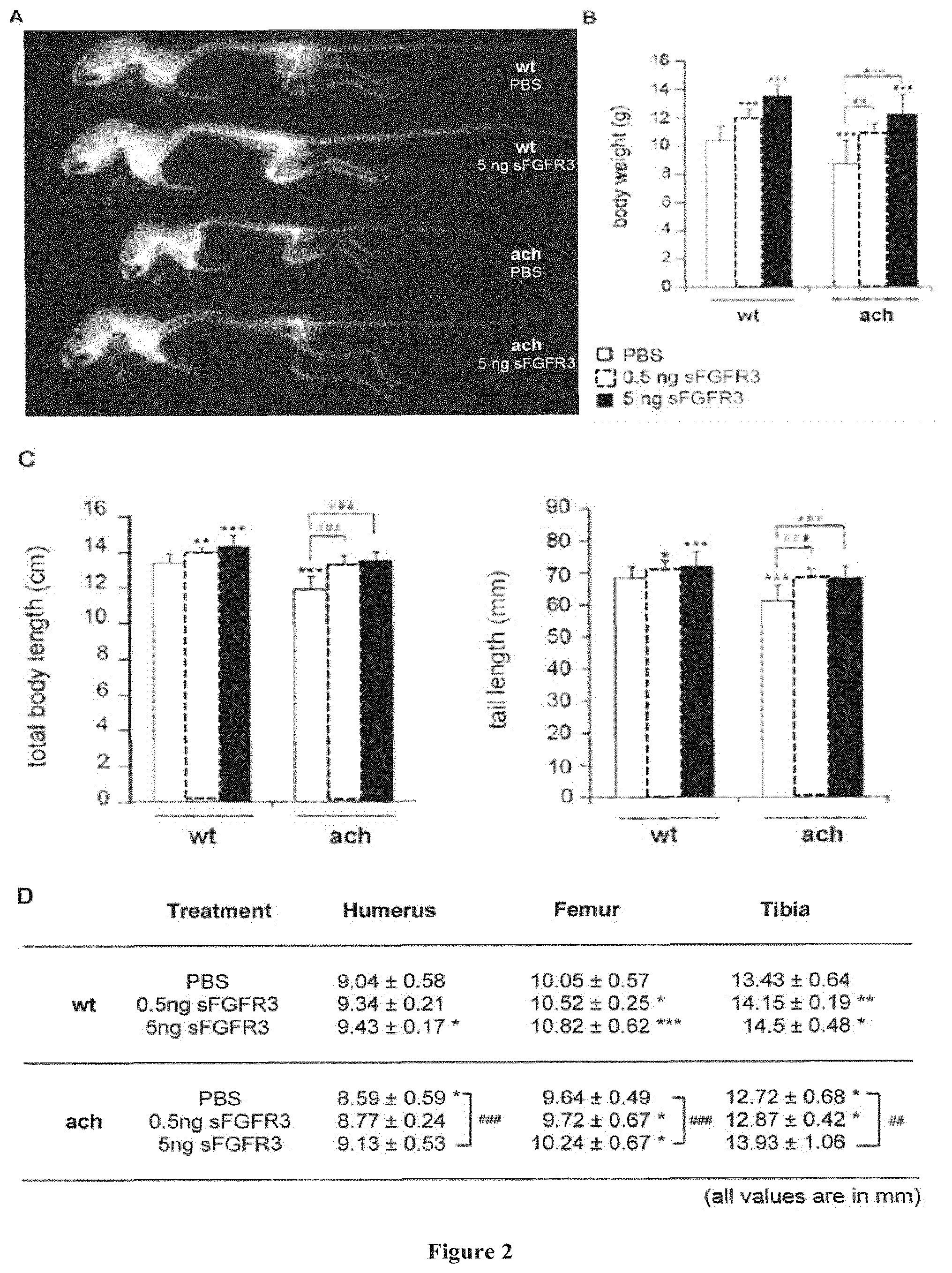
[0068] Moreover, it should be noted that the majority of protein-based biopharmaceuticals bare some form of post-translational modification which can profoundly affect protein properties relevant to their therapeutic application. Protein glycosylation represents the most common modification (about 50% of human proteins are glycosylated). Glycosylation can introduce considerable heterogeneity into a protein composition through the generation of different glycan structures on the proteins within the composition. Such glycan structures are made by the action of diverse enzymes of the glycosylation machinery as the glycoprotein transits the Endoplasmatic Reticulum (ER) and the Golgi-Complex (glycosylation cascade). The nature of the glycan structure(s) of a protein has impact on the protein's folding, stability, life time, trafficking, pharmaco-dynamics, pharmacokinetics and immunogenicity. The glycan structure has great impact on the protein's primary functional activity. Glycosylation can affect local protein structure and may help to direct the folding of the polypeptide chain. One important kind of glycan structures are the so called N-glycans. They are generated by covalent linkage of an oligosaccharide to the amino (N)-group of asparagin residues in the consensus sequence NXS/T of the nascent polypeptide chain. N-glycans may further participate in the sorting or directing of a protein to its final target: the N-glycan of an antibody, for example, may interact with complement components. N-glycans also serve to stabilize a glycoprotein, for example, by enhancing its solubility, shielding hydrophobic patches on its surface, protecting from proteolysis, and directing intra-chain stabilizing interactions. Glycosylation may regulate protein half-life, for example, in humans the presence of terminal sialic acids in N-glycans may increase the half-life of proteins, circulating in the blood stream.
[0069] As used herein, the term "glycoprotein" refers to any protein having one or more N-glycans attached thereto. Thus, the term refers both to proteins that are generally recognized in the art as a glycoprotein and to proteins which have been genetically engineered to contain one or more N-linked glycosylation sites. As used herein, the terms "N-glycan" and "glycoform" are used interchangeably and refer to an N-linked oligosaccharide, for example, one that is attached by an asparagine-N-acetylglucosamine linkage to an asparagine residue of a polypeptide. N-linked glycoproteins contain an N-acetylglucosamine residue linked to the amide nitrogen of an asparagine residue in the protein. The predominant sugars found on glycoproteins are glucose, galactose, mannose, fucose, N-acetylgalactosamine (GalNAc), N-acetylglucosamine (GlcNAc) and sialic acid (e.g., N-acetyl-neuraminic acid (NANA)). The processing of the sugar groups occurs co-translationally in the lumen of the ER and continues post-translationally in the Golgi apparatus for N-linked glycoproteins.
[0070] A number of yeasts, for example, Pichia pastoris, Yarrowia lipolytica and Saccharomyces cerevisiae are recently under development to use the advantages of such systems but to eliminate the disadvantages in respect to glycosylation. Several strains are under genetical development to produce defined, human-like glycan structures on a protein. Methods for genetically engineering yeast to produce human-like N-glycans are described in U.S. Pat. Nos. 7,029,872 and 7,449,308 along with methods described in U.S. Published Application Nos. 20040230042, 20050208617, 20040171826, 20050208617, and 20060286637. These methods have been used to construct recombinant yeast that can produce therapeutic glycoproteins that have predominantly human-like complex or hybrid N-glycans thereon instead of yeast type N-glycans. As previously described, human-like glycosylation is primarily characterized by "complex" N-glycan structures containing N-acetylglusosamine, galactose, fucose and/or N-acetylneuraminic acid. Thus, several strains of yeasts have been genetically engineered to produce glycoproteins comprising one or more human-like complex or human-like hybrid N-glycans such as GlcNAcMan3GlcNAc2.
[0071] As used herein, the term "skeletal growth retardation disorder" refers to a skeletal disease characterize by deformities and/or malformations of the bones.
[0072] These disorders include, but are not limiting to, skeletal growth retardation disorders caused by growth plate (physeal) fractures, idiopathic skeletal growth retardation disorders and FGFR3-related skeletal diseases.
[0073] As used herein, the term "idiopathic skeletal growth retardation disorder" refers to a skeletal disease whose the cause is unknown and for which treatment with exogenous growth hormone (GH), e.g. recombinant human GH (rhGH), for instance has been shown to be ineffective.
[0074] In the context of the present invention, the term "FGFR3-related skeletal disease" is intended to mean a skeletal disease that is caused by an abnormal increased activation of FGFR3, in particular by expression of a gain-of-function mutant of the FGFR3 receptor As used herein, the expressions "gain-of-function FGFR3 receptor variant". "gain-of-function mutant of the FGFR3" or "mutant FGFR3 displaying a prolonged activity" are used interchangeably and refer to a mutant of said receptor exhibiting a biological activity (i.e. triggering downstream signaling) which is higher than the biological activity of the corresponding wild-type receptor in the presence of FGF ligand.
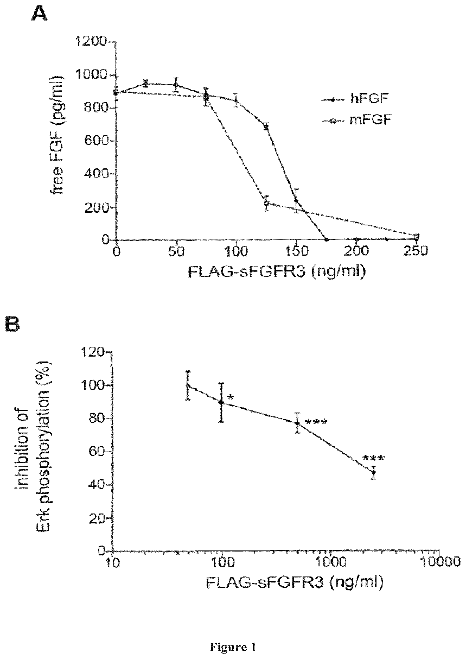
[0075] The FGFR3-related skeletal diseases are preferably FGFR3-related skeletal dysplasias and FGFR3-related craniosynostosis.
[0076] The FGFR3-related skeletal dysplasias according to the invention may correspond to an inherited or to a sporadic disease.
[0077] As used herein, the term "FGFR3-related skeletal dysplasias" includes but is not limited to thanatophoric dysplasia type I, thanatophoric dysplasia type II, hypochondroplasia, achondroplasia and SADDAN (severe achondroplasia with developmental delay and acanthosis nigricans).
[0078] In a preferred embodiment, the FGFR3-related skeletal dysplasia is caused by expression in the subject of a gain-of-function FGFR3 receptor variant such as defined above.
[0079] In a preferred embodiment, the FGFR3-related skeletal dysplasia is an achondroplasia caused by expression of the G380R gain-of-function mutant of the FGFR3 receptor.
[0080] Alternatively, the FGFR3-related skeletal dysplasia is an achondroplasia caused by expression of the G375C, G346E or S279C of the FGFR3 receptor.
[0081] It should be further noted that achondroplasia caused by another mutant of the FGFR3 receptor which would be identified in the future is also encompassed.
[0082] In a preferred embodiment, the FGFR3-related skeletal dysplasia is a hypochondroplasia caused by expression of the N540K, K650N, K650Q, S84L, R200C, N262H, G268C, Y278C, V381E, gain-of-function mutant of the FGFR3 receptor.
[0083] In a preferred embodiment, the FGFR3-related skeletal dysplasia is a thanatophoric dysplasia type I caused by expression of a gain-of-function mutant of the FGFR3 receptor chosen from the group consisting of R248C, S248C, G370C, S371C; Y373C, X807R, X807C, X807G, X807S, X807W and K650M FGFR3 receptors.
[0084] In a preferred embodiment, the FGFR3-related skeletal dysplasia is a thanatophoric dysplasia type II caused by expression of the K650E gain-of-function mutant of the FGFR3 receptor.
[0085] In a preferred embodiment, the FGFR3-related skeletal dysplasia is a severe achondroplasia with developmental delay and acanthosis nigricans caused by expression of the K650M gain-of-function mutant of the FGFR3 receptor.
[0086] The present invention also provides a method for preventing or treating a skeletal growth retardation disorder comprising the step of administering a therapeutically effective amount of a soluble FGFR3 (sFGFR3) polypeptide to a subject in need thereof.
[0087] By a "therapeutically effective amount" of a sFGFR3 as above described is meant a sufficient amount of the antagonist to prevent or treat a FGFR3-related skeletal disease (e.g. achondroplasia). It will be understood, however, that the total daily usage of the compounds and compositions of the present invention will be decided by the attending physician within the scope of sound medical judgment. The specific therapeutically effective dose level for any particular subject will depend upon a variety of factors including the disorder being treated and the severity of the disorder; activity of the specific compound employed; the specific composition employed, the age, body weight, general health, sex and diet of the subject; the time of administration, route of administration, and rate of excretion of the specific compound employed; the duration of the treatment; drugs used in combination or coincidential with the specific polypeptide employed; and like factors well known in the medical arts. For example, it is well within the skill of the art to start doses of the compound at levels lower than those required to achieve the desired therapeutic effect and to gradually increase the dosage until the desired effect is achieved. However, the daily dosage of the products may be varied over a wide range from 0.01 to 1_000 mg per adult per day. Preferably, the compositions contain 0.01, 0.05, 0.1, 0.5, 1.0, 2.5, 5.0, 10.0, 15.0, 25.0, 50.0, 100, 250 and 500 mg of the active ingredient for the symptomatic adjustment of the dosage to the subject to be treated. A medicament typically contains from about 0.01 mg to about 500 mg of the active ingredient, preferably from 1 mg to about 100 mg of the active ingredient. An effective amount of the drug is ordinarily supplied at a dosage level from 0.0002 mg/kg to about 20 mg/kg of body weight per day, especially from about 0.001 mg/kg to 7 mg/kg of body weight per day.
[0088] As used herein, the term "subject" denotes a human or non-human mammal, such as a rodent, a feline, a canine, an equine, or a primate. Preferably, the subject is a human being, more preferably a child (i.e. a child who is growing up).
[0089] It should be reminded that the cartilaginous matrix of the growth plate is less dense in a newborn or in a child than in an adult. Therefore, without wishing to be bound by the theory, one can expect that the polypeptide of the invention will better penetrate said cartilaginous matrix in a newborn or a child.
[0090] In one embodiment, the subject has been diagnosed as suffering from a FGFR3-related skeletal disease. As previously described, the FGFR3-related skeletal disease is caused by expression in the subject of a constitutively active FGFR3 receptor variant such as the G380R constitutively active mutant.
[0091] In the context of the invention, the term "treating" is used herein to characterize a therapeutic method or process that is aimed at (1) slowing down or stopping the progression, aggravation, or deterioration of the symptoms of the disease state or condition to which such term applies; (2) alleviating or bringing about ameliorations of the symptoms of the disease state or condition to which such term applies; and/or (3) reversing or curing the disease state or condition to which such term applies.
[0092] As used herein, the term "preventing" intends characterizing a prophylactic method or process that is aimed at delaying or preventing the onset of a disorder or condition to which such term applies.
Pharmaceutical Compositions of the Invention
[0093] The isolated soluble FGFR3 polypeptide (sFGFR3) as described above may be combined with pharmaceutically acceptable excipients, and optionally sustained-release matrices, such as biodegradable polymers, to form therapeutic compositions.
[0094] Accordingly, the present invention also relates to a pharmaceutical composition comprising an isolated sFGFR3 polypeptide according to the invention and a pharmaceutically acceptable carrier.
[0095] The present invention further relates to a pharmaceutical composition for use in the prevention or treatment of a skeletal growth retardation disorder comprising a sFGFR3 according to the invention and a pharmaceutically acceptable carrier.
[0096] In one embodiment, the skeletal growth retardation disorder is an idiopathic growth retardation disorder.
[0097] In another embodiment, the skeletal growth retardation disorder is a FGFR3-related skeletal disease.
[0098] "Pharmaceutically" or "pharmaceutically acceptable" refers to molecular entities and compositions that do not produce an adverse, allergic or other untoward reaction when administered to a mammal, especially a human, as appropriate. A pharmaceutically acceptable carrier or excipient refers to a non-toxic solid, semi-solid or liquid filler, diluent, encapsulating material or formulation auxiliary of any type.
[0099] The form of the pharmaceutical compositions, the route of administration, the dosage and the regimen naturally depend upon the condition to be treated, the severity of the illness, the age, weight, and sex of the patient, etc. The pharmaceutical compositions of the invention can be formulated for a topical, oral, intranasal, intraocular, intravenous, intramuscular or subcutaneous administration and the like.
[0100] Preferably, the pharmaceutical compositions contain vehicles which are pharmaceutically acceptable for a formulation capable of being injected. These may be in particular isotonic, sterile, saline solutions (monosodium or disodium phosphate, sodium, potassium, calcium or magnesium chloride and the like or mixtures of such salts), or dry, especially freeze-dried compositions which upon addition, depending on the case, of sterilized water or physiological saline, permit the constitution of injectable solutions.
[0101] The doses used for the administration can be adapted as a function of various parameters, and in particular as a function of the mode of administration used, of the relevant pathology, or alternatively of the desired duration of treatment. For example, it is well within the skill of the art to start doses of the compound at levels lower than those required to achieve the desired therapeutic effect and to gradually increase the dosage until the desired effect is achieved. However, the daily dosage of the products may be varied over a wide range from 0.01 to 1,000 mg per adult per day. Preferably, the compositions contain 0.01, 0.05, 0.1, 0.5, 1.0, 2.5, 5.0, 10.0, 15.0, 25.0, 50.0, 100, 250 and 500 mg of the active ingredient for the symptomatic adjustment of the dosage to the subject to be treated. A medicament typically contains from about 0.01 mg to about 500 mg of the active ingredient, preferably from 1 mg to about 100 mg of the active ingredient. An effective amount of the drug is ordinarily supplied at a dosage level from 0.0002 mg/kg to about 20 mg/kg of body weight per day, especially from about 0.001 mg/kg to 7 mg/kg of body weight per day.
[0102] To prepare pharmaceutical compositions, an effective amount of a polypeptide according to the invention may be dissolved or dispersed in a pharmaceutically acceptable carrier or aqueous medium.
[0103] The pharmaceutical forms suitable for injectable use include sterile aqueous solutions or dispersions; formulations including sesame oil, peanut oil or aqueous propylene glycol; and sterile powders for the extemporaneous preparation of sterile injectable solutions or dispersions. In all cases, the form must be sterile and must be fluid to the extent that easy syringability exists. It must be stable under the conditions of manufacture and storage and must be preserved against the contaminating action of microorganisms, such as bacteria and fungi. Solutions of the active compounds as free base or pharmacologically acceptable salts can be prepared in water suitably mixed with a surfactant, such as hydroxypropylcellulose. Dispersions can also be prepared in glycerol, liquid polyethylene glycols, mixtures thereof and in oils. Under ordinary conditions of storage and use, these preparations contain a preservative to prevent the growth of microorganisms.
[0104] The polypeptides according to the invention can be formulated into a composition in a neutral or salt form. Pharmaceutically acceptable salts include the acid addition salts (formed with the free amino groups of the protein) and which are formed with inorganic acids such as, for example, hydrochloric or phosphoric acids, or such organic acids as acetic, oxalic, tartaric, mandelic, and the like. Salts formed with the free carboxyl groups can also be derived from inorganic bases such as, for example, sodium, potassium, ammonium, calcium, or ferric hydroxides, and such organic bases as isopropylamine, trimethylamine, histidine, procaine and the like.
[0105] The carrier can also be a solvent or dispersion medium containing, for example, water, ethanol, polyol (for example, glycerol, propylene glycol, and liquid polyethylene glycol, and the like), suitable mixtures thereof, and vegetables oils. The proper fluidity can be maintained, for example, by the use of a coating, such as lecithin, by the maintenance of the required particle size in the case of dispersion and by the use of surfactants. The prevention of the action of microorganisms can be brought about by various antibacterial and antifungal agents, for example, parabens, chlorobutanol, phenol, sorbic acid, thimerosal, and the like. In many cases, it will be preferable to include isotonic agents, for example, sugars or sodium chloride. Prolonged absorption of the injectable compositions can be brought about by the use in the compositions of agents delaying absorption, for example, aluminium monostearate and gelatin.
[0106] Sterile injectable solutions are prepared by incorporating the active compounds in the required amount in the appropriate solvent with several of the other ingredients enumerated above, as required, followed by filtered sterilization. Generally, dispersions are prepared by incorporating the various sterilized active ingredients into a sterile vehicle which contains the basic dispersion medium and the required other ingredients from those enumerated above. In the case of sterile powders for the preparation of sterile injectable solutions, the preferred methods of preparation are vacuum-drying and freeze-drying techniques which yield a powder of the active ingredient plus any additional desired ingredient from a previously sterile-filtered solution thereof.
[0107] The preparation of more, or highly concentrated solutions for direct injection is also contemplated, where the use of DMSO as solvent is envisioned to result in extremely rapid penetration, delivering high concentrations of the active agents to a small tumor area.
[0108] Upon formulation, solutions will be administered in a manner compatible with the dosage formulation and in such amount as is therapeutically effective. The formulations are easily administered in a variety of dosage forms, such as the type of injectable solutions described above, but drug release capsules and the like can also be employed.
[0109] For parenteral administration in an aqueous solution, for example, the solution may be suitably buffered and the liquid diluent first rendered isotonic with sufficient saline or glucose. These particular aqueous solutions are especially suitable for intravenous, intramuscular, subcutaneous and intraperitoneal administration. In this connection, sterile aqeuous media which can be employed will be known to those of skill in the art in light of the present disclosure. For example, one dosage could be dissolved in 1 ml of isotonic NaCl solution and either added to 1000 ml of hypodermoclysis fluid or injected at the proposed site of infusion, (see for example, "Remington's Pharmaceutical Sciences" 15th Edition, pages 1035-1038 and 1570-1580). Some variation in dosage will necessarily occur depending on the condition of the subject being treated. The person responsible for administration will, in any event, determine the appropriate dose for the subject.
[0110] Another aspect of the present invention is a pharmaceutical composition for use in the prevention or treatment of a skeletal growth retardation disorder comprising an isolated sFGFR3 polypeptide or a functional equivalent thereof according to the invention and a pharmaceutically acceptable carrier.
[0111] In one embodiment, the skeletal growth retardation disorder is an idiopathic growth retardation disorder.
[0112] In another embodiment, the skeletal growth retardation disorder is a FGFR3-related skeletal disease.
[0113] The present invention also provides a method for preventing or treating a skeletal growth retardation disorder comprising a step of administering a pharmaceutical composition comprising a therapeutically effective amount of a sFGFR3 polypeptide or a functional equivalent thereof and a pharmaceutically acceptable carrier to a subject in need thereof.
[0114] The invention will be further illustrated by the following figures and examples. However, these examples and figures should not be interpreted in any way as limiting the scope of the present invention.
FIGURES
[0115] FIG. 1: Effective FGF binding and decreased Erk phosphorylation in ATDC5 cells in presence of FLAG-sFGFR3. (A) Fixed amounts of human or murine basic FGF (100 ng) were incubated with increasing concentrations of FLAG-sFGFR3. After 2 h, remaining unbound FGFs were detected by ELISA. Linear regression analysis showed no statistical differences between the two slopes, hFGF, human FGFb; mFGF, mouse FGFb. Experiment was performed in triplicate and repeated five times. (B) Erk phosphorylation was evaluated by immunoblotting on ATDC5 cells following incubation with increasing doses of FLAG-sFGFR3. The graph represents the phosphorylation variations in percentage compared to phosphorylation levels in untreated cells. Experiments were repeated six times. Following verification of normality, statistical comparisons were performed using a one way ANOVA. *p<0.05, ***p<0.001. Values represent mean.+-.SD.
[0116] FIG. 2: FLAG-sFGFR3 treatment effect on overall skeletal growth. (A) X-ray radiographics illustrating treatment effect on skeletal growth. Showed skeletons are representative of wt and Fgfr3.sup.ach/+ mice that received subcutaneous injection of PBS or 5 ng FLAG-sFGFR3. Growth was characterized by body weight (B), body and tail lengths (C), and long bone measurements (D). (Data followed normal distribution; a Student's t test was used to compare data to measurements obtained on untreated mice. n per group are shown in Table 1; *p<0.05; **p<0.01; ***p<0.001 versus untreated wt; ##p<:0.01; ###p<0.001 versus untreated Fgfr3.sup.ach/+ mice. wt: wildtype mice; ach: Fgfr3.sup.ach/+ mice
[0117] FIG. 3: Effect of FLAG-sFGFR3 treatment on vertebrae maturation. (A) The kyphosis index (KI) was measured from radiographs of mice positioned in right lateral decumbency. As defined by Laws et al. (28), line AB is the length of a line drawn from posterior edge of C7 to the posterior edge of L6. Line CD is the distance from line AB to the dorsal border of the vertebral body farthest from that line. Clinically, a kyphosis is characterized with KI<4. (B) Photographs of representative vertebrae from untreated wt, untreated Fgfr3.sup.ach/+ mice and transgenic mice receiving 5 ng FLAG-sFGFR3. In the table are indicated the percentage of animals in the different treatment groups with immature C7. T11 and lumbar vertebrae. .sctn. Lumbar compressions were characterized by paraplegia or locomotion deficiency. Data followed normal distribution; a Student's t test was used to compare data to measurements obtained on untreated mice. n per group are shown in Table 1; *p<0.05; ***p<0.001 versus untreated wt; .sup.##p<0.01; .sup.###p<0.001 versus untreated Fgfr3.sup.ach/+ mice. wt: wildtype mice; ach: Fgfr3.sup.ach/+ mice.
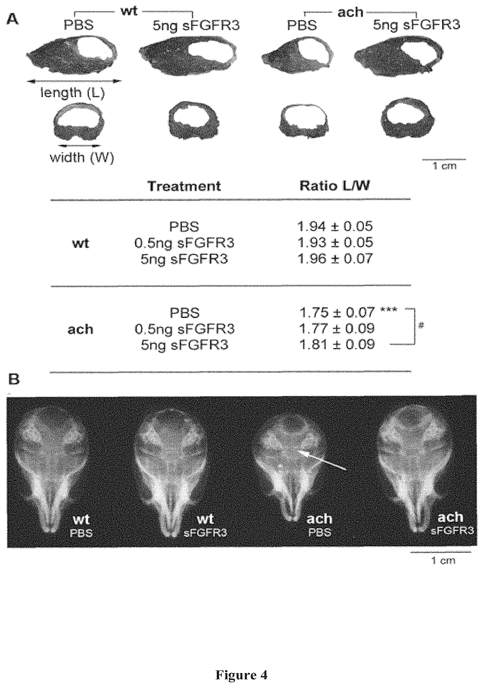
[0118] FIG. 4: FLAG-sFGFR3 treatment effects on skull development. (A) Skull length (L) and width (W) were measured and the ratio L/W calculated. Statistical analysis was performed using a Student's t test following verification of normal variance and distribution. n per group are shown in Table 1; p<0.001 versus untreated wt; .sup.#p<0.05 versus untreated Fgfr3.sup.ach/+ mice. (B) Representative X-rays of skulls from wt and Fgfr3.sup.ach/+ mice that received either PBS or 5 ng FLAG-sFGFR3. They show treatment prevention of premature closure of cranial synchondrose typically observed on Fgfr3.sup.ach/+ mice. This is indicated by the arrowhead. wt: wildtype mice; ach: Fgfr3.sup.ach/+ mice.
EXAMPLE
[0119] Material & Methods
[0120] sFGFR3 subcloning and recombinant protein production: To facilitate sub-cloning, full-length cDNA sequence encoding the FGFR3 ATM (2.1 kb) (35), a generous gift from Dr. Kurokawa-Seo, Kyoto Sangyo University, Japan, was optimized to decrease GC content while encoding for the original protein sequence (GeneOptimizer.RTM. process, GeneArt). The synthesized fragment was subcloned into pFLAG-CMV3.G727 (Sigma Aldrich) using HindIII and KpnI cloning sites. Plasmid DNA was purified from transformed bacteria and concentration determined by UV spectroscopy. The final construct was verified by sequencing. The sequence homology within the used restriction sites was 100%.
[0121] Recombinant FLAG-sFGFR3 protein was produced by transient transfection using GeneJuice transfection reagent (Merck Millipore) in HEK 293 cells allowing all necessary post-translational modifications. Each transfection was performed in a cell factory (High flask T600, Merck Millipore) with 80% confluent HEK 293 in 100 ml DMEM without phenol red (Gibco, Life Technologies) supplemented with glutamine 2 mM (Gibco, Life Technologies) and 1% antibiotics (Gibco, Life Technologies). 600 .mu.l GeneJuice and 240 .mu.g pFLAG-sFGFR3 were resuspended in 30 ml OptiMEM (Gibco, Life Technologies), incubated 30 min at room temperature, and then incubated for 4 h onto the cells at 37.degree. C. in 5% CO.sub.2. Medium was then replaced by 120 ml DMEM without phenol red, supplemented with glutamine 2 mM and 1% antibiotics. After 72 h, production medium was filtrated using 0.22 .mu.m filters and concentrated on Amicon Ultra-15 60 kDa (Merck Millipore). Recombinant protein was then purified on an affinity column (ANTI-FLAG M2 Affinity Gel, Sigma Aldrich) according to the manufacturer's instructions. FLAG-sFGFR3 amounts were measured by specific ELISA (R&D Systems) according to the manufacturer's instructions. FLAG-sFGFR3 was then stored at a concentration of 0.5 .mu.g/ml in 50% glycerol solution.
[0122] FLAG-sFGFR3 incubation with FGF: Fixed amounts of human or murine FGFs (100 .mu.g) (R&D Systems) were incubated for 2 h at 37.degree. C. with increasing doses of FLAG-sFGFR3 (0 to 250 ng/ml in PBS 1% BSA. Specific commercial ELISA kits (R&D Systems) were used to quantify remaining unbound FGFs. All experiments were performed in triplicates and repeated five times.
[0123] Half-life of sFGFR3: To determine the half-life of sFGFR3, 8 week old WT mice received an intravenous bolus of 50 mg/kg of FLAG-sFGFR3. At 15 min, 1 h, 3 h, 6 h, and 24 b, blood was harvested by retro-orbital puncture using heparin catheter (n=4). Concentration of FLAG-sFGFR3 was measured by anti FLAG ELISA (Sigma). The half-life of the terminal phase was calculated using the following pharmacokinetic equation t.sub.1/2=0.693/.lamda..sub.z, where 0.693 is the natural logarithm of 2 and .lamda..sub.z, the slope of the terminal phase.
[0124] Immunoblotting analysis: Immunoblotting was performed following incubation of several doses of FLAG-sFGFR3 on ATDC5 cells. For this, ATDC5 cells were plated at a density of 2.times.10.sub.6 in 6 well plates and, following adhesion, cultured for 48 h in 0.5% BSA in DMEM-F12 (Gibco, Life Technologies) containing 1% antibiotics. Cells were then cultured for 10 min with 100 pg/ml murine FGF pre-incubated for 2 h at 37.degree. C. with increasing doses of FLAG-sFGFR3 (0, 12.5, 125, 1250, 12500 pg/ml). At the end of the incubation period, remaining unbound FGFs were measured by specific ELISA (R&D Systems). Cells were then solubilized in lysis buffer (20 mM Tris, pH 7.4, 150 mM NaCl, 10 mM EDTA, 150 mM NaF, 2 mM sodium orthovanadate, 10 mM pyrophosphate, proteases inhibitors, and 1% Triton X-100) for 45 min at 4.RTM. C. Lysates were cleared (14 000 rpm, 10 min) and proteins were separated by SDS-PAGE and immunoblotted as previously described (36). The proteins were probed with anti-phospho p42/44 MAPK (4370S, Cell Signaling), anti-total p42/44 MAPK (4696S, Cell Signaling) and anti-hsp60 (sc1722, Santa Cruz Biotechnology) antibodies (1 sg/ml). All experiments were performed six times.
[0125] Immunohistochemistry of FLAG-sFGFR3: Immunohistochemistry of FLAG-sFGFR3 was performed on tibiae of 3 day old Fgfr3.sup.ach/+ mice and their wildtype littermates. For this, following decapitation of newborn mice, tibiae were carefully harvested and incubated in 24 well plates in presence of 5 ng FLAG-sFGFR3 for 24 h at 37.degree. C. in 5% CO.sub.2. Tibiae were then rinsed in PBS and fixed in 10% formalin for 24 h. Following decalcification in EDTA for 2 days, bones were paraffin embedded and 5 m sections were incubated with 5 .mu.g/ml anti-FLAG M2-FITC monoclonal antibody (Sigma Aldrich). Sections were counterstained with Hoechst solution and visualized under fluorescent microscopy. An anti-IgG antibody was used as negative control.
[0126] Animals and treatments: The Principles of Laboratory Animal Care (NIH publication no. 85-23, revised 1985; grants1.nih.gov/grants/olaw/references/phspol.htm) and the European commission guidelines for the protection of animals used for scientific purposes (cc.europa.cu/environment/chemicals/lab_animals/legislation_en.h- tm) were followed at all times. All procedures were approved by the Institutional Ethic Committee for the use of Laboratory Animals (CIEPAL Azur) (approval #NCE-2012-52).
[0127] Experiments were performed on transgenic Fgfr3ach/+ animals in which expression of the mutant FGFR3 is driven by the Col2a1 promoter/enhancer (22). Mice were exposed to a 12 h light/dark cycle and had free access to standard laboratory food and water. All measurements and analyses were performed blinded and genotypes were analyzed after all analyses were done by PCR of genomic DNA which amplify 360 bp of the FGFR3 transgene (22). Two doses of FLAG-sFGFR3 (0.5 ng and 5 ng in 10 .mu.l PBS with 50% glycerol) were tested. At day 3, all newborn mice from a single litter received the same dose. Control litters received 10 .mu.l of PBS containing 50% glycerol. Subcutaneous injections were thereafter done twice a week for three weeks, alternatively on the left and right sides of the back. Mice were observed daily with particular attention to locomotion and urination alterations. At day 22, all animals but two litters per group were sacrificed by C02 asphyxation; genus and genotypes were determined. Body weights were measured. Blood was harvested by cardiac puncture and mixed with 50 .mu.l 0.5M EDTA; half of the samples were centrifuged for a biochemical assessment using a Beckman AU 27 Analyzer (electrolytes (Na+, K+, Cr), lactate dehydrogenase (LDH), cholesterol, creatinin, creatinin kinase (CK), aspartate aminotransferase (AST), alanine aminostransferase (ALT), amylase, total bilirubin (BLT)); the other half was analyzed without centrifugation for blood numeration (Hemavet 950FS, Mascot Hematology). Cadavers were carefully skinned and eviscerated and skeletal measurements (body and tail lengths) were obtained using an electronic digital caliper (Fisher Scientific). Total body length was measured from the nose to the end of the last caudal vertebra; tail was measured starting at the first caudal vertebra. Organs (heart, lungs, liver, kidneys, spleen) were harvested, weight and stored in 10% formalin for further histological analysis using standard paraffin-embedded techniques. X-rays of all skeletons were taken using a Faxitron X-ray machine (Edimex). Using an established method (28), kyphotic index were measured for each animals on the X-rays. Cleared skeletons were then stained simultaneously with alcian blue and alizarin red using standard procedures and stored in glycerol prior to analysis. Stained long bones (tibiae, femurs, humerus) were dissected and measured using an electronic digital caliper; vertebrae and skulls were also dissected and analyzed.
[0128] Breeding was set up to theoretically generate litters with half wildtype and half heterozygous Fgfr3.sup.ach/+ mice. To avoid bias due to variations of phenotype penetrance, experiments were performed on at least 2 litters (one treated and one control) arising from the same breeders. A total of 15, 9 and 11 litters representing a total of 312 pups were treated with PBS, 0.5 ng or 5 ng FLAG-sFGFR3, respectively. The n per group is presented in Table 1.
[0129] Effect of FLAG-sFGFR3 on the fertility of treated animals: Animals from the litters that were not used for skeletal measurements were kept until breeding age was reached. At age 8 week, they were then mated with 8 week old FVB/N mice from Charles River. Newborn mice were counted at birth for each treated and control male and female and compared with fertility statistics of the previous generation. At age 22, offspring were euthanized and growth was evaluated as described above.
[0130] Proliferation and differentiation assays: sFGFR3 effect on proliferation was determined on ATDC5 cells. For this, ATDC5 cells were plated at a density of 5.times.10.sup.4 in 24 well plates and cultured for 48 h in DMEM-F12 0.5% BSA (Life Technologies). Cells were then challenged for 72 h with 100 pg/ml of FGF2, FGF9 or FGF18 in presence of 0 or 20 ng/ml of FLAG-sFGFR3. Proliferation was evaluated using the MTT (3-(4,5-dimethylthiazol-2-yl)-2,5-diphenyltetrazolium bromide) proliferation assay by measurement of absorbance at 540 nm.
[0131] To determine treatment effect on chondrocyte differentiation, sub-confluent ATDC5 cells were incubated in 24 well plates for 7 days in chondrocyte differentiation medium (37.5 .mu.g/ml L-ascorbic acid; 1 mM sodium pyruvate; 1% Insulin-transferrin-sodium selenite; 100 nM dexamethasone in DMEM F12) in the presence of 100 pg/ml of FGF and 0 or 20 ng/ml FLAG-sFGFR3. After 7 days in culture, half of the wells were stained with Alcian blue, pH 2.5, in 3% acetic acid. In the remaining wells, total RNA was extracted using an RNeasy Mini Kit (Qiagen). Total RNA (1 .mu.g) was reverse-transcribed, and real-time PCR was performed (ABI PRISM 7500). The TaqMan gene expression assays were purchased from Applied Biosystems: Col10a1 (Mm00487041_ml), Col2a1 (Mm01309565_m1), Sox9 (Mm00448840_m1), Fgfr3 (Mm00433294_m1), RPLP0 (Ribosomal Phosphoprotein Large P0, Mm00725448_s1). Gene expression values were normalized to the expression value of the housekeeping gene RPLP0 and calculated based on the comparative cycle threshold Ct method (2.sup.-.DELTA.Ct) as described previously.
[0132] Statistical analysis: All experiments and data measurements were performed by blind experimenters at all times. Statistical analyses were performed with GraphPad Prism 6.0 software. To determine the statistical tests to be used, necessary assumptions were verified. To verify normality and equal variance, an Agostino and Pearson omnibus normality test and a Brown-Forsythe test were performed, respectively. Because all skeletal measurements data sets fulfilled normality and equal variance requirements, two-tailed Student's t test for comparisons of two independent groups were used in the different statistical analyses. Comparison of mortality data between treated and control groups was done using a Kruskal-Wallis test. Comparison of FLAG-sFGFR3 binding to human and murine FGFs was done by linear regression. Immunoblotting data distribution followed normality and were thus analyzed using a one-way ANOVA using a Holm-Sidak's multiple comparisons test. For organ weight correlation analyses, Pearson or Spearman tests were used when data sets followed or not normal distribution, respectively. All statistical tests were considered significant at a p<0.05 level of error. In all figures, values of p are shown as follows: *p<0.05; **p<0.01; *p<0.001. Data are presented as means SD.
[0133] Results
[0134] FLAG-sFGFR3 effectively binds FGFs and decreases MAPK signaling in ATDC5 cells: A soluble form of FGFR3 was produced by transient transfection of a plasmid encoding the FGFR3 ATM sequence. In order to detect recombinant soluble FGFR3 in vivo, the inventors used a soluble form of FGFR3 labeled with a FLAG tag. This tag was used because of the availability of reagents for its purification and its detection (23, 24). It has also already been used in vivo without inducing premature elimination of the tagged protein by the immune system (25, 26). In the present experiments, recombinant FLAG-sFGFR3 was produced by transient transfection in human embryonic kidney (HEK) 293 cells, allowing for all necessary post-translational modifications, purified using affinity column and stored at a concentration of 0.5 .mu.g/ml in 50% glycerol. In mice, the half-life of sFGFR3 is 16 hours.
[0135] To verify that FLAG-sFGFR3 effectively bound free FGFs, fixed amounts of human FGFs were incubated with increasing quantities of FLAG-sFGFR3. As seen in FIG. 1A, FLAG-sFGFR3 effectively bound hFGF in a dose-dependent manner. The FGFR3 ATM sequence used is of human origin, the inventors verified that it could also bind murine FGF. Similar results were obtained and FLAG-sFGFR3 was able to bind similar amounts of murine FGFs. This was expected since there is a 90% sequence homology between murine and human FGFR3, sFGFR3 competed for hFGF binding with endogenous FGFR3 on murine chrondrocytes (ATDC5 cells), demonstrating the decoy receptor mechanism.
[0136] The inventors then verified that the complexation of FGF with FLAG-sFGFR3 resulted in a decreased intracellular FGF signaling on Erk phosphorylation. ATDC5 cells were used as a murine chondrocytic cell line to study chondrocyte biology (27). As seen FIG. 1B, significant decrease in Erk phosphorylation was seen in relation with the dose of FLAG-sFGFR3. This was correlated with a decrease of free FGFs in the conditioned medium, similar to that observed in FIG. 1A. These results demonstrate that FLAG-sFGFR3 effectively binds FGFs of human and murine origin thus decreasing FGF intracellular signaling. sFGFR3 treatment also restored proliferation and differentiation of mouse ATDC5 cells and significantly increased collagen type 2, collagen type 10 and Sox 9 gene expression relative to FGF alone (P<0.05, Student's t test). No effect on proliferation, differentiation, or gene expression was observed in presence of the control protein.
[0137] FGF-independent mechanisms have also been explored. Stat1 was not phosphorylated in Fgfr3.sup.ach/+ chondrocytes treated with sFGFR3. Moreover, fgfr3 gene expression was unchanged from FGF-only controls by sFGFR3 treatment in chondrocytes isolated from Fgfr3.sup.ach/+ animals. Together, these data suggest that sFGFR3 effects are mediated only through FGF-dependent pathways.
[0138] Soluble FGFR3 increases survival of Fgfr3.sup.ach/+ mice and effectively restores bone growth in Fgfr3.sup.ach/+ mice: Prior to test FLAG-sFGFR3 treatment effect in vivo, the inventors verified that it could penetrate the dense cartilaginous matrix of the growth plate and reach target chondrocytes. Long bones isolated from three day old Fgfr3.sup.ach/+ mice and their wildtype (wt) littermates were incubated for 24 h in presence of 5 ng FLAG-sFGFR3. As seen in FIG. 2, the recombinant protein was detected within the matrix, near the chondrocytes of the tibial growth plate of wt and Fgfr3.sup.ach/+ mice, showing that the protein efficiently penetrates the dense cartilaginous matrix of the growth plate and reach target chondrocytes.
[0139] To evaluate the biological effects of FLAG-sFGFR3 treatment on skeletal bone growth in Fgfr3.sup.ach/+ mice, all newborn mice from one litter received the same treatment without knowing their phenotype. They received a subcutaneous injection of 0.5 or 5 ng FLAG-sFGFR3, or PBS in control groups, twice a week during 3 weeks. The first observation was the significant reduction in mortality in treated compared to untreated litters. At the end of the treatment period, control groups contained about a third of transgenic animals alive while in both treated groups, there were approximately 50% wt mice and 50% Fgfr3.sup.ach/+ (Table 1). Moreover, in the control litters, 31% of animals died prior to the end of the experiments compared to 11.8% and 6.7% in the 0.5 ng and 5 ng FLAG-sFGFR3 treated litters, respectively. This reduction in group size was due to premature death or euthanasia of paraplegic animals. When it was possible, autopsy was performed and confirmed death due to respiratory failure as seen by the presence of blood within the lungs. Two animals died consequently to bowel obstruction. All of these animals were Fgfr3.sup.ach/+, as confirmed by genotyping. No wild-type animal died prematurely. It is noteworthy to emphasize that in the control group, the majority of affected Fgfr3.sup.ach/+ mice died from respiratory failure, while in the 5 ng FLAG-sFGFR3 treatment group, they mostly suffered from paraplegia and only few had respiratory distress (Table 1). Altogether these data indicate that with treatment fewer animals died, and those who died had a less severe phenotype.
TABLE-US-00005 TABLE 1 Number of pups in the different treatment groups at day 3 and day 22. Number of litters Number of pups % dead animals before day 22 per group Day 3 Day 22 (cause of death) PBS 15 132 91 31% (23 by respiratory failure, (wt: 67; ach: 24) 2 by bowel occlusion, 16 by paraplegia) 0.5 ng SFGFR3 9 76 67 11.8% ** (4 by respiratory (wt: 31; ach: 36) failure, 5 by paraplegia) 5 ng sFGFR3 11 104 97 6.7% ** (1 by respiratory failure, (wt: 47; ach: 50) 6 by paraplegia) Litters were considered as single entities and all newborn mice from the same cage received the same treatment. Dead and alive animals were counted daily. Autopsy revealed death by respiratory failure and bowel occlusion for 2 animals. Animals with paraplegia were euthanized upon discovery and recorded in the dead animal group. All dead animals were Fgfr3.sup.ach/+. Statistical comparison versus control group was done using the Kruskal-Wallis test. ** p < 0.01. wt: wildtype mice; ach: Fgfr3.sup.ach/+ mice.
[0140] At day 22, time of weaning, animals were sacrificed and their growth was evaluated. The inventors first confirmed that there was no statistical difference between males and females (Table Si) and regrouped them for all subsequent analyses. As illustrated in FIG. 2A, FLAG-sFGFR3 treatment had an effect on overall skeletal growth. While Fgfr3.sup.ach/1 mice were in average 20% lighter than their wt littermates, animals treated with FLAG-sFGFR3 displayed a dose dependent increase in their body weight, reaching up to 33% of the weight of untreated transgenic mice (FIG. 2B). A dose dependent treatment effect was also observed on the weight of wt animals. As seen in FIG. 2C, treatment induced a dose dependent increase in body and tail lengths of both Fgfr3.sup.ach/+ and wt animals. Treated transgenic mice had a stature that was not significantly different from that of untreated wt controls, reaching up to 10% of the lengths of untreated Fgfr3.sup.ach/+ animals at the high dose correcting the initial discrepancy between transgenic and wt mice.
[0141] Similar results were obtained on long bone lengths. Humerus, femurs and tibiae from treated Fgfr3.sup.ach/+ mice were longer than those of untreated transgenic mice and were statistically identical to the lengths of wt bones (FIG. 2D). FLAG-sFGFR3 treatment also had a dose dependent effect on the growth of long bones from wt mice. Histology confirmed treatment effect on chondrocyte maturation. Treated Fgfr3.sup.ach/+ mice exhibited organized and hypertrophic chondrocytes in their growth plates similarly to wt mice.
[0142] sFGFR3 treatment also corrected rib cage development in Fgfr3.sup.ach/+ mice. Altogether, these results show that foil owing chronic subcutaneous administration of FLAG-sFGFR3 to neonate Fgfr3.sup.ach/+ mice, normal bone growth was restored and that it was also effective on skeletal growth of animals that do not trigger an FGFR3 activating mutation. Indeed, Fgfr3.sup.ach/+ and WT mice treated with sFGFR3 showed greater sternum ossification and lengths compared with vehicle-treated animals in their respective groups.
TABLE-US-00006 TABLE S1 Statistical comparison of body measurements between male and female in the different treatment groups. Body weight Body length Tail length male vs n (g) (mm) (mm) female PBS wt male 41 11.09 .+-. 1.26 132.43 .+-. 5.45 70.49 .+-. 3.65 ns female 26 10.67 .+-. 1.99 134.04 .+-. 6.76 71.76 .+-. 4.60 ach male 13 9.18 .+-. 1.82 120.56 .+-. 5.71 64.36 .+-. 2.52 ns female 11 8.14 .+-. 1.55 113.95 .+-. 8.22 60.03 .+-. 3.79 0.5 ng wt male 12 12.31 .+-. 1.27 134.55 .+-. 7.98 73.94 .+-. 6.35 ns sFGFR3 female 19 12.03 .+-. 1.18 134.59 .+-. 6.46 72.25 .+-. 4.51 ach male 19 10.50 .+-. 1.19 126.78 .+-. 9.3 69.48 .+-. 6.64 ns female 17 9.34 .+-. 1.47 121.49 .+-. 10.01 65.33 .+-. 7.96 5 ng wt male 23 13.29 .+-. 1.48 141.47 .+-. 6.77 74.76 .+-. 5.42 ns sFGFR3 female 24 12.13 .+-. 1.85 138.73 .+-. 8.21 74.01 .+-. 6.01 ach male 13 11.41 .+-. 3.14 130.43 .+-. 13.11 69.15 .+-. 9.07 ns female 37 10.72 .+-. 2.19 127.59 .+-. 11.58 67.43 .+-. 7.95 Following 3 weeks of treatments (PBS, 0.5 ng or 5 ng FLAG-sFGFR3), animals were sacrificed at age 22 days. Body weight, body length and tail length were measured. Genus and genotypes were determined. Following verification of normality variation in each data set, measurements were compared between males and females within the same treatment and genotype group. Data followed normal distribution; a Student's t test was used to compare data to measurements obtained on untreated mice. No statistical difference was found in any group. ns = non significant, wt: wildtype mice; ach: Fgfr3.sup.ach/+ mice.
[0143] FLAG-sFGFR3 treatment decreases spinal and skull deformities associated with achandroplasia in Fgfr3.sup.ach/+ mice: In Fgfr3.sup.ach/+ mice, spinal abnormalities are recognized in particular by the presence of a kyphosis that can be characterized by the calculation of a kyphotic index (KI). In this scoring system, established by Laws et al. (28), mice with a KI<4.0 present a kyphosis (for more details, please see legend of FIG. 3). In the present study, while nowt animals presented a spinal deformity, 80% of untreated Fgfr3.sup.ach/+ mice displayed cervical kyphosis with an average KI of 3.46.+-.0.65 (FIG. 3A). With FLAG-sFGFR3 treatment, this percentage significantly decreased to 17% and 6% in the 0.5 ng and 5ng groups, respectively.
[0144] To further characterize vertebral maturation, the inventors analyzed ossification of C7 and T11. As seen FIG. 3, on untreated Fgfr3.sup.ach/+ mice, the 7.sup.th cervical and the 11.sup.th thoracic were not fused at the midline in 88.9% and 70.1%, respectively. Following treatment, maturation was restored as seen by the decrease in the number of immature vertebrae. No wt animals in any groups presented immature vertebrae.
[0145] Similar to achondroplasia patients that typically have enlarged heads, Fgfr3.sup.ach/+ mice suffer from skull deformities. While cranium width (W) is not statistically different between transgenic and wt mice (10.35.+-.0.28 mm vs 10.17.+-.0.32 mm, respectively), the length (L) is significantly shorter in Fgfr3.sup.ach/+ mice (18.11.+-.0.75 mm vs 20.05.+-.0.51 mm in wt mice, respectively). This leads to a L/W ratio equal to 1.75.+-.0.77 in untreated Fgfr3.sup.ach/+ mice and equal to 1.94.+-.0.05 in control wt mice (FIG. 4A). FLAG-sFGFR3 treatment induced a dose-dependent correction of the cranium length, and the L/W ratio was not significantly different from that of untreated wt at the highest dose of FLAG-sFGFR3.
[0146] As seen in FIG. 4B, treatment also prevented the premature closure of cranial synchondrose typically observed in Fgfr3.sup.ach/+ mice.
[0147] Altogether, these results show that FLAG-sFGFR3 treatment is effective at preventing the development of skeleton deformities associated with achondroplasia.
[0148] No toxicological effects are detected in treated animals: Because the inventors are using a systemic approach to deliver recombinant soluble FGFR3, they paid a particular attention to possible unwanted side effects. They analyzed organs of all 255 animals, performed biochemical and numeration blood tests and verified fertility of some treated animals (two litters per group) including normality of their offspring.
[0149] Potential treatment side effects were first evaluated on several organs (liver, lung, heart, spleen, kidneys) at the time of sacrifice. Organs were observed macroscopically, weighted and randomly analyzed microscopically by histology. None of the 255 animals that received chronic subcutaneous injections of FLAG-sFGFR3 or PBS presented macroscopic abnormalities. Histology was performed on randomly selected organs in all groups and data were analyzed blindly by an anatomopathologist. No signs of toxicity were observed on any histological slides. In all control groups, organ weights were correlated with the mouse body weight (Table 2). In the treated groups, organs increased with enhanced bone growth. As an example, the lungs of untreated Fgfr3.sup.ach/+ mice were 156.0.+-.8.7 mg. They increased to 172.5.+-.67.5 mg in the 5 ng treatment group, reaching the weight of lungs in untreated wt mice (170.5.+-.36.3 mg). Similar results were found for all organs and this weight augmentation was statistically correlated with body weight increase in all groups (Table 2). To evaluate organ functions, they performed biochemical blood tests including electrolytes titration, liver, kidney and spleen enzymes assays. All tests proved to be statistically identical between Fgfr3.sup.ach/+ and wt animals in the treated and control groups (Table S2). Blood counts were also analyzed and similarly, no differences between blood formulations were noticed between treated and control groups (Table S3).
TABLE-US-00007 TABLE 2 Coefficient correlation (r) between organ and body weight in the different treatment groups. Treatment Liver Heart Lung Spleen Kidney PBS 0.892 *** 0.748 *** 0.857 *** 0.655 *** 0.877 *** 0.5 ng sFGFR3 0.883 ** 0.777 ** 0.731 * 0.774 * 0.777 ** 5 ng sFGFR3 0.881 *** 0.794 *** 0.720 *** 0.584 ** 0.531 * PBS 0.941 *** 0.943 *** 0.886 ** 0.726 ** 0.848 *** 0.5 ng sFGFR3 0.921 *** 0.758 * 0.709 ** 0.650 * 0.828 *** 5 ng sFGFR3 0.957 *** 0.983 *** 0.885 *** 0.883 *** 0.850 *** Pearson or Spearman tests were used for statistical analysis of organ/body weights correlations in each treatment group; * p < 0.05; ** p < 0.01; *** p < 0.001. wt: wildtype mice; ach: Fgfr3.sup.ach/+ mice.
TABLE-US-00008 TABLE S2 Blood biochemical parameters were not modified by FLAG-sFGFR3 treatment. Na K Cl LDH Cholesterol Treatment (mmol/L) (mmol/L) (mmol/L) (UI/L) (mmol/L) wt PBS 810.8 .+-. 80.8 9.26 .+-. 0.51 75.20 .+-. 3.81 1672 .+-. 124 0.99 .+-. 0.15 5 ng sFGFR3 818.3 .+-. 69.4 8.75 .+-. 0.59 72.83 .+-. 1.14 2059 .+-. 478 0.90 .+-. 0.16 ach PBS 709.7 .+-. 90.5 8.97 .+-. 0.98 81.79 .+-. 3.79 1675 .+-. 228 1.06 .+-. 0.11 5 ng sFGFR3 706.2 .+-. 57.0 9.20 .+-. 0.77 76.33 .+-. 0.88 1738 .+-. 402 1.02 .+-. 0.08 Creatinine Total Creatinine kinase AST ALT Amylase bilirubine Treatment (.mu.mol/L) (UI/L) (UI/L) (UI/L) (UI/L) (.mu.mol/L) wt PBS 9.00 .+-. 1.26 1389 .+-. 679 155.1 .+-. 27.sup. 65.5 .+-. 16.5 138.1 .+-. 4.7 22.8 .+-. 1.7 5 ng sFGFR3 10.20 .+-. 0.95 655 .+-. 180 137.0 .+-. 24.6 42.3 .+-. 4.2 112.0 .+-. 8.0 20.8 .+-. 0.4 ach PBS 9.18 .+-. 1.60 551 .+-. 127 140.1 .+-. 22.9 48.2 .+-. 14.6 129.8 .+-. 21.4 21.8 .+-. 1.1 5 ng sFGFR3 12.33 .+-. 1.43 943 .+-. 261 196.4 .+-. 51.2 77.7 .+-. 14.5 192.0 .+-. 33.3 20.3 .+-. 0.6 To evaluate treatment toxicity, at time of sacrifice, plasma from the PBS and 5 ng FLAG-sFGFR3 groups were analyzed using a Beckman AU 2700 Analyzer. Overall health was evaluated by measurements of electrolytes (Na.sup.+, K.sup.+, Cl.sup.-), lactate dehydrogenase (LDH) and cholesterol. Kidney function was assessed by creatinin and creatinin kinase (CK) assays. Liver and pancreas functions were assessed by aspartate aminotransferase (AST), alanine aminostransferase (ALT), total bilirubin (BLT) and amylase, respectively. Statistical comparisons were performed using a one way ANOVA. No statistical difference was found in any group, wt: wildtype mice; ach: Fgfr3.sup.ach/+ mice.
TABLE-US-00009 TABLE S3 Blood counts were not modified by FLAG-sFGFR3 treatment. Treatment Hb (g/dL) Ht (%) WBC (K/.mu.l) RBC (K/.mu.l) PLT (K/.mu.l) wt PBS 8.11 .+-. 0.11 16.82 .+-. 1.72 4.31 .+-. 0.39 5.63 .+-. 0.02 471.1 .+-. 26.9 5 ng sFGFR3 8.56 .+-. 0.06 23.21 .+-. 1.33 5.28 .+-. 0.54 5.81 .+-. 0.01 447.0 .+-. 32.2 ach PBS 8.32 .+-. 0.26 22.5 .+-. 4.74 3.37 .+-. 0.49 5.47 .+-. 0.18 472.0 .+-. 112.8 5 ng sFGFR3 8.25 .+-. 0.15 17.44 .+-. 1.12 5.86 .+-. 0.81 5.67 .+-. 0.04 620.0 .+-. 28.8 Treatment NE (%) LY (%) MO (%) EO (%) BA (%) wt PBS 30.76 .+-. 1.86 53.56 .+-. 2.49 7.52 .+-. 0.59 5.89 .+-. 0.44 2.27 .+-. 0.27 5 ng sFGFR3 25.54 .+-. 2.37 56.90 .+-. 3.15 9.78 .+-. 0.67 5.75 .+-. 0.54 1.83 .+-. 0.24 ach PBS 24.25 .+-. 6.08 60.02 .+-. 8.86 6.56 .+-. 1.48 6.59 .+-. 1.72 2.56 .+-. 0.59 5 ng sFGFR3 29.25 .+-. 1.47 51.88 .+-. 2.39 10.16 .+-. 0.82 6.99 .+-. 1.20 1.85 .+-. 0.33 The effects of FLAG-sFGFR3 treatment on blood counts were evaluated on plasma samples at the time of sacrifice. Analysis included hemoglobin (Hb), hematocrit (Ht), white blood cells (WBC), red blood cells (RBC) and platelets (PLT) counts. The percentages of the different leukocyte populations were evaluated (NE: neutrophil; LY: lymphocyte, MO: monocyte, EO: eosinophil; BA: basophil). Statistical comparisons were performed using a one way ANOVA. No statistical difference was found in any group, wt: wildtype mice; ach: Fgfr3.sup.ach/+ mice.
[0150] To evaluate unwanted side effects on fecundity, after the three week treatment, animals were weaned and were mated at age 8 weeks with wt FVB males or females from Charles River. As seen in Table 3, all treated animals were fertile and their offspring were of normal size with an approximate 50% wt 50% Fgfr3.sup.ach/+ descendants. While Fgfr3.sup.ach/+ females usually have a first litter slightly reduced in size compared to that of wt females, it is interesting to note that primiparous treated transgenic females had litters that were identical in size to wt primiparous females, confirming enlargement of their pelvis following treatment (Table 3).
TABLE-US-00010 TABLE 3 FLAG-sFGFR3 treatment did not affect fertility of the treated mice. % of wt and Body weight Body length Litter size ach at day 22 (g) (mm) Control ach .times. wt 9.2 .+-. 0.9 74% wt, 26% wt, 10.88 .+-. wt, 133.24 .+-. genitors ach 1.62 6.11 wt .times. ach 7.7 .+-. 0.7 *** ach, 8.66 .+-. ach, 117.25 .+-. 1.68 *** 6.96 *** sFGFR3 treated ach .times. wt 10.2 .+-. 1.9 % wt, % ach treated treated wt .times. wt 9.7 .+-. 0.9 N/A wt .times. treated ach 10.6 .+-. 1.5 % wt, % ach wt .times. treated wt 9.8 .+-. 1.9 N/A Pups from two litters per group were weaned after the three week treatment. They were mated at age 8 weeks with wt animals from Charles River. The number of pups of the first litters was counted for each primiparous female. At age 22 days, animals were euthanized. Their body growth was evaluated as previously. A Student's t test was used to compare data to measurements of untreated wt animals generated from control genitors. *** p < 0.001 versus untreated wt. N/A: not applicable, wt: wildtype mice; ach: Fgfr3.sup.ach/+ mice.
[0151] Altogether these experiments did not highlight any complications from the FLAG-sFGFR3 treatment itself neither on blood formulation, organ function and development nor fertility of the treated animals, suggesting that the use of a soluble form of FGFR3 may be a viable treatment approach for clinical applications.
Discussion
[0152] The present study validates the proof of concept that a therapeutic strategy based on the use of a soluble form of FGFR3 can prevent abnormal bone growth in mice carrying the achondroplasia mutation. Treatment was administered twice a week by subcutaneous injections to the animals throughout the growth period. Following this three week treatment period, ensuing endochondral bone growth led to normal, harmonious stature. Importantly, these effects were dose-dependent; the dose of 0.5 ng FLAG-sFGFR3 was sufficient to induce body weight and length that were identical to that of untreated wt mice and at the dose of 5 ng, treated dwarf mice were even heavier and had longer long bones than untreated wt animals. Foremost, the present results emphasize the notion that the achondroplasia mutation requires ligand binding to be activated. Indeed, it has been demonstrated that in the case of the G380R mutation, FGFR3 activation is ligand-dependent (12), but there is still no clear consensus in the literature (29-31), suggesting not one but multiple mechanisms leading to prolonged intracellular signaling.
[0153] While it was essential that long bone growth be restored for the treatment to be effective, it was critical to also significantly impact the onset of complications, due to skeletal deformities. For the inventors, this is indispensable if one wants to develop a treatment for the clinic. Restoration of vertebra maturation and normal closure of cranial synchondroses in treated Fgfr3.sup.ach/+ mice had numerous effects on these animals. The first consequence was the reduced mortality among the transgenic population. As stated in the result section, autopsy revealed respiratory failures in the majority of the cases. Based on the anatomical characteristics of the skull and vertebrae of the dwarf mice, the inventors believe that these were consequent to brainstem compression, similar to what can be observed in achondroplasia patients.
[0154] A second outcome of treatment effect was a shift in the penetrance of the phenotype. While measurements could appear as though treatment effects were not totally dose-dependent and more importantly not as effective as could be expected based on body weights, the inventors believe that at the highest dose, treatment saved the smallest Fgfr3.sup.ach/+ mice from brainstem compression and respiratory failure. Untreated, these animals would not have survived past week 1 or 2. In this treatment group, the inventors hypothesize that these animals are those that remain very small even though they have less severe complications.
[0155] Despite the increasing number of reports studying the mechanisms underlying achondroplasia and related skeletal dysplasia, only three studies have been published showing therapeutic strategies effectively tested in mice with chondrodysplasia. Xie et al. recently published a report that intermittent PTH treatment partially rescues bone growth in mice with achondroplasia (32). In their study, PTH was administered subcutaneously at the dose of 100 .mu.g/kg body weight per day for 4 weeks after birth. Although the mechanism by which PTH affects FGFR3 intracellular signaling is not clearly established, bone growth was partially rescued in treated transgenic mice; PTH treated transgenic mice were still smaller than their wt littermates. In this study, only very little information was mentioned related to achondroplasia complications, except for the partial rescue of cranial synchondrose. The lethal phenotype of TDII mice was also rescued with chronic PTH treatment of pregnant females. Another potential therapeutic antagonist of FGFR3 signaling is the C-natriuretic peptide (CNP) (33). In this paper, the authors treated transgenic mice with achondroplasia by continuous intravenous infusion of synthetic CNP for 3 weeks starting at age 4 week. It is believe that CNP increases the width of the growth plate accelerating growth plate activity. The major obstacle for use in human is the very short half-life of CNP, estimated to be 2.6 min in plasma (34).
[0156] A study has been very recently published describing the use of a new FGFR3-binding peptide that rescues the lethal phenotype and partially restores the structural distortion of growth plates in TDII mice, observed on 12 pups (19). In this study, effects on MAPK signaling and bone growth correction were only partial and daily administration was required probably due to the short half-life of the peptide. Here, the effects of sFGFR3 were of higher magnitude, with complete restoration of normal stature. The inventors believe that the half-life of the soluble form of FGFR3 containing IgG like domains is significantly prolonged. Indeed, only 6 injections (between birth and weaning) were necessary to completely restore bone growth in the 86 treated Fgfr3ach/+ mice. They can imagine that only a few injections would be necessary to treat achondroplasia children as out-patients, starting during the first year until puberty. This would substantially impact occurrence of injection side effects, typically found with daily injection regimens. The use of a soluble recombinant protein also allows for rapid termination during treatment if safety issues are raised and at puberty when bone growth ceases. If necessary, it is also possible to alternate between treatment and resting periods. By preventing the complications, sFGFR3 treatment would avoid the necessity of surgical interventions and also reduce any stress due to hospitalization. Furthermore, our study did not reveal any toxicological effect on blood or fertility and offspring of treated animals. Current studies are ongoing to evaluate if the three week sFGFR3 treatment had an effect on the long-term health of the treated mice. As of today, treated mice are 6 month old and no apparent side effects are visible and blood tests are normal.
[0157] In conclusion, the present study demonstrates the viability of targeting FGFR3 in the extracellular compartment as an effective treatment to restore growth plate maturation and induce normal bone growth in achondroplasia. The absence of unwanted side effects validates its use as a promising therapy for this and related chondrodysplasia caused by activating mutation in FGF receptors. Furthermore, in the present study, the inventors also report a positive effect of sFGFR3 treatment on the growth of wt animals. This is of importance suggesting its possible interest for the treatment of idiopathic growth retardations, or to prevent severe complications in other rare diseases such as hypophosphotasia.
REFERENCES
[0158] Throughout this application, various references describe the state of the art to which this invention pertains. The disclosures of these references are hereby incorporated by reference into the present disclosure. [0159] 1. D. M. Ornitz, FGF signaling in the developing endochondral skeleton. Cytokine Growth Factor Rev 16, 205 (April, 2005). [0160] 2. M. L. Martinez-Frias, C. A. de Frutos, E. Bermejo, M. A. Nieto, Review of the recently defined molecular mechanisms underlying thanatophoric dysplasia and their potential therapeutic implications for achondroplasia. Am J Med Genet A 152A, 245 (January, 2010). [0161] 3. W. A. Horton, G. P. Lunstrum, Fibroblast growth factor receptor 3 mutations in achondroplasia and related forms of dwarfism. Rev Endocr Metab Disord 3, 381 (December, 2002). [0162] 4. M. J. Wright, M. D. Irving, Clinical management of achondroplasia. Arch Dis Child 97, 129 (February, 2012). [0163] 5. W. A. Horton, J. G. Hall. J. T. Hecht, Achondroplasia. Lancet 370, 162 (Jul. 14, 2007). [0164] 6. E. D. Shirley, M. C. Ain, Achondroplasia: manifestations and treatment. J Am Acad Orthop Surg 17, 231 (April, 2009). [0165] 7. P. Richette, T. Bardin, C. Stheneur, Achondroplasia: from genotype to phenotype. Joint Bone Spine 75, 125 (March, 2008). [0166] 8. F. Rousseau et al., Mutations in the gene encoding fibroblast growth factor receptor-3 in achondroplasia. Nature 371, 252 (Sep. 15, 1994). [0167] 9. G. A. Bellus et al., Achondroplasia is defined by recurrent G380R mutations of FGFR3. Am J Hum Genet 56, 368 (February, 1995). [0168] 10. R. Shiang et al., Mutations in the transmembrane domain of FGFR3 cause the most common genetic form of dwarfism, achondroplasia. Cell 78, 335 (Jul. 29, 1994). [0169] 11. F. Rousseau et al., Mutations of the fibroblast growth factor receptor-3 gene in achondroplasia. Horm Res 45, 108 (1996). [0170] 12. E. Monsonego-Ornan, R. Adar, T. Feferman, O. Segev, A. Yayon, The transmembrane mutation G380R in fibroblast growth factor receptor 3 uncouples ligand-mediated receptor activation from down-regulation. Mol Cell Biol 20, 516 (January 2000). [0171] 13. D. Harada et al., Sustained phosphorylation of mutated FGFR3 is a crucial feature of genetic dwarfism and induces apoptosis in the ATDC5 chondrogenic cell line via PLCgamma-activated STAT1. Bone 41, 273 (August, 2007). [0172] 14. E. Monsonego-Ornan, R. Adar, E. Rom, A. Yayon, FGF receptors ubiquitylation: dependence on tyrosine kinase activity and role in downregulation. FEBS Lett 528, 83 (Sep. 25, 2002). [0173] 15. M. K. Webster, D. J. Donoghue, Constitutive activation of fibroblast growth factor receptor 3 by the transmembrane domain point mutation found in achondroplasia. Embo J 15, 520 (Feb. 1, 1996). [0174] 16. M. B. Laederich, W. A. Horton, FGFR3 targeting strategies for achondroplasia. Expert Rev Mol Med 14, el 1 (2012). [0175] 17. C. Deng, A. Wynshaw-Boris, F. Zhou, A. Kuo, P. Leder, Fibroblast growth factor receptor 3 is a negative regulator of bone growth. Cell 84, 911 (Mar. 22, 1996). [0176] 18. Z. Vajo, C. A. Francomano, D. J. Wilkin, The molecular and genetic basis of fibroblast growth factor receptor 3 disorders: the achondroplasia family of skeletal dysplasias, Mucnke craniosynostosis, and Crouzon syndrome with acanthosis nigricans. Endocr Rev 21, 23 (February, 2000). [0177] 19. M. Jin et al., A novel FGFR3-binding peptide inhibits FGFR3 signaling and reverses the lethal phenotype of mice mimicking human thanatophoric dysplasia. Hum Mol Genet, (Oct. 9, 2012). [0178] 20. S. Garofalo et al., Skeletal dysplasia and defective chondrocyte differentiation by targeted overexpression of fibroblast growth factor 9 in transgenic mice. J Bone Miner Res 14, 1909 (November, 1999). [0179] 21. D. Davidson et al., Fibroblast growth factor (FGF) 18 signals through FGF receptor 3 to promote chondrogenesis. J Biol Chem 280, 20509 (May 27, 2005). [0180] 22. M. C. Naski, J. S. Colvin, J. D. Coffin, D. M. Ornitz, Repression of hedgehog signaling and BMP4 expression in growth plate cartilage by fibroblast growth factor receptor 3. Development 125, 4977 (December, 1998). [0181] 23. T. D. Papakostas et al., Development of an efficiently cleaved, bioactive, highly pure FLAG-tagged recombinant human Mullerian Inhibiting Substance. Protein Expr Purif 70, 32 (March, 2010). [0182] 24. K. Terpe, Overview of tag protein fusions: from molecular and biochemical fundamentals to commercial systems. Appl Microbiol Biotechnol 60, 523 (January 2003). [0183] 25. J. P. Louboutin, B. A. Reyes, L. Agrawal, E. Van Bockstaele, D. S. Strayer, Strategies for CNS-directed gene delivery: in vivo gene transfer to the brain using SV40-derived vectors. Gene Ther 14, 939 (June, 2007). [0184] 26. L. R. Rodino-Klapac et al., Persistent expression of FLAG-tagged micro dystrophin in nonhuman primates following intramuscular and vascular delivery. Mol Ther 18, 109 (January, 2010). [0185] 27. F. M. Altaf, T. M. Hering, N. H. Kazmi, J. U. Yoo, B. Johnstone, Ascorbate-enhanced chondrogenesis of ATDC5 cells. Eur Cell Mater 12, 64 (2006). [0186] 28. N. Laws, A. Hoey, Progression of kyphosis in mdx mice. J Appl Physiol 97, 1970 (November, 2004). [0187] 29. J. Placone, K. Hristova, Direct Assessment of the Effect of the Gly380Arg Achondroplasia Mutation on FGFR3 Dimerization Using Quantitative Imaging FRET. PLoS One 7, e46678 (2012). [0188] 30. L. He, W. Horton, K. Hristova, Physical basis behind achondroplasia, the most common form of human dwarfism. J Biol Chem 285, 30103 (Sep. 24, 2010). [0189] 31. L. He, N. Shobnam, W. C. Wimley, K. Hristova, FGFR3 heterodimerization in achondroplasia, the most common form of human dwarfism. J Biol Chem 286, 13272 (Apr. 15, 2011). [0190] 32. Y. Xie et al., Intermittent PTH (1-34) injection rescues the retarded skeletal development and postnatal lethality of mice mimicking human achondroplasia and thanatophoric dysplasia. Hum Mol Genet 21, 3941 (Sep. 15, 2012). [0191] 33. A. Yasoda et al., Systemic administration of C-type natriurtic peptide as a novel therapeutic strategy for skeletal dysplasias. Endocrinology 150, 3138 (July, 2009). [0192] 34. P. J. Hunt, A. M. Richards, E. A. Espiner, M. G. Nicholls, T. G. Yandle, Bioactivity and metabolism of C-type natriuretic peptide in normal man. J Clin Endocrinol Metab 78, 1428 (June, 1994). [0193] 35. M. Terada et al., Fibroblast growth factor receptor 3 lacking the Ig IIIb and transmembrane domains secreted from human squamous cell carcinoma DJM-1 binds to FGFs. Mol Cell Biol Res Commun 4,365 (November, 2001). [0194] 36. A. Bertola et al., Hepatocyte growth factor induces glucose uptake in 3T3-L1 adipocytes through A Gab1/phosphatidylinositol 3-kinase/Glut4 pathway. J Biol Chem 282, 10325 (April, 2007).
Sequence CWU 1
1
31694PRTArtificial SequenceHuman soluble FGFR3 polypeptide (sFGFR3)
1Met Gly Ala Pro Ala Cys Ala Leu Ala Leu Cys Val Ala Val Ala Ile1 5
10 15Val Ala Gly Ala Ser Ser Glu Ser Leu Gly Thr Glu Gln Arg Val
Val 20 25 30Gly Arg Ala Ala Glu Val Pro Gly Pro Glu Pro Gly Gln Gln
Glu Gln 35 40 45Leu Val Phe Gly Ser Gly Asp Ala Val Glu Leu Ser Cys
Pro Pro Pro 50 55 60Gly Gly Gly Pro Met Gly Pro Thr Val Trp Val Lys
Asp Gly Thr Gly65 70 75 80Leu Val Pro Ser Glu Arg Val Leu Val Gly
Pro Gln Arg Leu Gln Val 85 90 95Leu Asn Ala Ser His Glu Asp Ser Gly
Ala Tyr Ser Cys Arg Gln Arg 100 105 110Leu Thr Gln Arg Val Leu Cys
His Phe Ser Val Arg Val Thr Asp Ala 115 120 125Pro Ser Ser Gly Asp
Asp Glu Asp Gly Glu Asp Glu Ala Glu Asp Thr 130 135 140Gly Val Asp
Thr Gly Ala Pro Tyr Trp Thr Arg Pro Glu Arg Met Asp145 150 155
160Lys Lys Leu Leu Ala Val Pro Ala Ala Asn Thr Val Arg Phe Arg Cys
165 170 175Pro Ala Ala Gly Asn Pro Thr Pro Ser Ile Ser Trp Leu Lys
Asn Gly 180 185 190Arg Glu Phe Arg Gly Glu His Arg Ile Gly Gly Ile
Lys Leu Arg His 195 200 205Gln Gln Trp Ser Leu Val Met Glu Ser Val
Val Pro Ser Asp Arg Gly 210 215 220Asn Tyr Thr Cys Val Val Glu Asn
Lys Phe Gly Ser Ile Arg Gln Thr225 230 235 240Tyr Thr Leu Asp Val
Leu Glu Arg Ser Pro His Arg Pro Ile Leu Gln 245 250 255Ala Gly Leu
Pro Ala Asn Gln Thr Ala Val Leu Gly Ser Asp Val Glu 260 265 270Phe
His Cys Lys Val Tyr Ser Asp Ala Gln Pro His Ile Gln Trp Leu 275 280
285Lys His Val Glu Val Asn Gly Ser Lys Val Gly Pro Asp Gly Thr Pro
290 295 300Tyr Val Thr Val Leu Lys Val Ser Leu Glu Ser Asn Ala Ser
Met Ser305 310 315 320Ser Asn Thr Pro Leu Val Arg Ile Ala Arg Leu
Ser Ser Gly Glu Gly 325 330 335Pro Thr Leu Ala Asn Val Ser Glu Leu
Glu Leu Pro Ala Asp Pro Lys 340 345 350Trp Glu Leu Ser Arg Ala Arg
Leu Thr Leu Gly Lys Pro Leu Gly Glu 355 360 365Gly Cys Phe Gly Gln
Val Val Met Ala Glu Ala Ile Gly Ile Asp Lys 370 375 380Asp Arg Ala
Ala Lys Pro Val Thr Val Ala Val Lys Met Leu Lys Asp385 390 395
400Asp Ala Thr Asp Lys Asp Leu Ser Asp Leu Val Ser Glu Met Glu Met
405 410 415Met Lys Met Ile Gly Lys His Lys Asn Ile Ile Asn Leu Leu
Gly Ala 420 425 430Cys Thr Gln Gly Gly Pro Leu Tyr Val Leu Val Glu
Tyr Ala Ala Lys 435 440 445Gly Asn Leu Arg Glu Phe Leu Arg Ala Arg
Arg Pro Pro Gly Leu Asp 450 455 460Tyr Ser Phe Asp Thr Cys Lys Pro
Pro Glu Glu Gln Leu Thr Phe Lys465 470 475 480Asp Leu Val Ser Cys
Ala Tyr Gln Val Ala Arg Gly Met Glu Tyr Leu 485 490 495Ala Ser Gln
Lys Cys Ile His Arg Asp Leu Ala Ala Arg Asn Val Leu 500 505 510Val
Thr Glu Asp Asn Val Met Lys Ile Ala Asp Phe Gly Leu Ala Arg 515 520
525Asp Val His Asn Leu Asp Tyr Tyr Lys Lys Thr Thr Asn Gly Arg Leu
530 535 540Pro Val Lys Trp Met Ala Pro Glu Ala Leu Phe Asp Arg Val
Tyr Thr545 550 555 560His Gln Ser Asp Val Trp Ser Phe Gly Val Leu
Leu Trp Glu Ile Phe 565 570 575Thr Leu Gly Gly Ser Pro Tyr Pro Gly
Ile Pro Val Glu Glu Leu Phe 580 585 590Lys Leu Leu Lys Glu Gly His
Arg Met Asp Lys Pro Ala Asn Cys Thr 595 600 605His Asp Leu Tyr Met
Ile Met Arg Glu Cys Trp His Ala Ala Pro Ser 610 615 620Gln Arg Pro
Thr Phe Lys Gln Leu Val Glu Asp Leu Asp Arg Val Leu625 630 635
640Thr Val Thr Ser Thr Asp Glu Tyr Leu Asp Leu Ser Ala Pro Phe Glu
645 650 655Gln Tyr Ser Pro Gly Gly Gln Asp Thr Pro Ser Ser Ser Ser
Ser Gly 660 665 670Asp Asp Ser Val Phe Ala His Asp Leu Leu Pro Pro
Ala Pro Pro Ser 675 680 685Ser Gly Gly Ser Arg Thr
69022082DNAArtificial SequenceHuman soluble FGFR3 DNA sequence
optimized to decrease GC content 2atgggcgccc ctgcctgcgc cctcgcgctc
tgcgtggccg tggccatcgt ggccggcgcc 60tcctcggagt ccttggggac ggagcagcgc
gtcgtggggc gagcggcaga agtcccgggc 120ccagagcccg gccagcagga
gcagttggtc ttcggcagcg gggatgctgt ggagctgagc 180tgtcccccgc
ccgggggtgg tcccatgggg cccactgtct gggtcaagga tggcacaggg
240ctggtgccct cggagcgtgt cctggtgggg ccccagcggc tgcaggtgct
gaatgcctcc 300cacgaggact ccggggccta cagctgccgg cagcggctca
cgcagcgcgt actgtgccac 360ttcagtgtgc gggtgacaga cgctccatcc
tcgggagatg acgaagacgg ggaggacgag 420gctgaggaca caggtgtgga
cacaggggcc ccttactgga cacggcccga gcggatggac 480aagaagctgc
tggccgtgcc ggccgccaac accgtccgct tccgctgccc agccgctggc
540aaccccactc cctccatctc ctggctgaag aacggcaggg agttccgcgg
cgagcaccgc 600attggaggca tcaagctgcg gcatcagcag tggagcctgg
tcatggaaag cgtggtgccc 660tcggaccgcg gcaactacac ctgcgtcgtg
gagaacaagt ttggcagcat ccggcagacg 720tacacgctgg acgtgctgga
gcgctccccg caccggccca tcctgcaggc ggggctgccg 780gccaaccaga
cggcggtgct gggcagcgac gtggagttcc actgcaaggt gtacagtgac
840gcacagcccc acatccagtg gctcaagcac gtggaggtga atggcagcaa
ggtgggcccg 900gacggcacac cctacgttac cgtgctcaag gtgtccctgg
agtccaacgc gtccatgagc 960tccaacacac cactggtgcg catcgcaagg
ctgtcctcag gggagggccc cacgctggcc 1020aatgtctccg agctcgagct
gcctgccgac cccaaatggg agctgtctcg ggcccggctg 1080accctgggca
agccccttgg ggagggctgc ttcggccagg tggtcatggc ggaggccatc
1140ggcattgaca aggaccgggc cgccaagcct gtcaccgtag ccgtgaagat
gctgaaagac 1200gatgccactg acaaggacct gtcggacctg gtgtctgaga
tggagatgat gaagatgatc 1260gggaaacaca aaaacatcat caacctgctg
ggcgcctgca cgcagggcgg gcccctgtac 1320gtgctggtgg agtacgcggc
caagggtaac ctgcgggagt ttctgcgggc gcggcggccc 1380ccgggcctgg
actactcctt cgacacctgc aagccgcccg aggagcagct caccttcaag
1440gacctggtgt cctgtgccta ccaggtggcc cggggcatgg agtacttggc
ctcccagaag 1500tgcatccaca gggacctggc tgcccgcaat gtgctggtga
ccgaggacaa cgtgatgaag 1560atcgcagact tcgggctggc ccgggacgtg
cacaacctcg actactacaa gaagacaacc 1620aacggccggc tgcccgtgaa
gtggatggcg cctgaggcct tgtttgaccg agtctacact 1680caccagagtg
acgtctggtc ctttggggtc ctgctctggg agatcttcac gctggggggc
1740tccccgtacc ccggcatccc tgtggaggag ctcttcaagc tgctgaagga
gggccaccgc 1800atggacaagc ccgccaactg cacacacgac ctgtacatga
tcatgcggga gtgctggcat 1860gccgcgccct cccagaggcc caccttcaag
cagctggtgg aggacctgga ccgtgtcctt 1920accgtgacgt ccaccgacga
gtacctggac ctgtcggcgc ctttcgagca gtactccccg 1980ggtggccagg
acacccccag ctccagctcc tcaggggacg actccgtgtt tgcccacgac
2040ctgctgcccc cggccccacc cagcagtggg ggctcgcgga cg 20823806PRTHomo
sapiens 3Met Gly Ala Pro Ala Cys Ala Leu Ala Leu Cys Val Ala Val
Ala Ile1 5 10 15Val Ala Gly Ala Ser Ser Glu Ser Leu Gly Thr Glu Gln
Arg Val Val 20 25 30Gly Arg Ala Ala Glu Val Pro Gly Pro Glu Pro Gly
Gln Gln Glu Gln 35 40 45Leu Val Phe Gly Ser Gly Asp Ala Val Glu Leu
Ser Cys Pro Pro Pro 50 55 60Gly Gly Gly Pro Met Gly Pro Thr Val Trp
Val Lys Asp Gly Thr Gly65 70 75 80Leu Val Pro Ser Glu Arg Val Leu
Val Gly Pro Gln Arg Leu Gln Val 85 90 95Leu Asn Ala Ser His Glu Asp
Ser Gly Ala Tyr Ser Cys Arg Gln Arg 100 105 110Leu Thr Gln Arg Val
Leu Cys His Phe Ser Val Arg Val Thr Asp Ala 115 120 125Pro Ser Ser
Gly Asp Asp Glu Asp Gly Glu Asp Glu Ala Glu Asp Thr 130 135 140Gly
Val Asp Thr Gly Ala Pro Tyr Trp Thr Arg Pro Glu Arg Met Asp145 150
155 160Lys Lys Leu Leu Ala Val Pro Ala Ala Asn Thr Val Arg Phe Arg
Cys 165 170 175Pro Ala Ala Gly Asn Pro Thr Pro Ser Ile Ser Trp Leu
Lys Asn Gly 180 185 190Arg Glu Phe Arg Gly Glu His Arg Ile Gly Gly
Ile Lys Leu Arg His 195 200 205Gln Gln Trp Ser Leu Val Met Glu Ser
Val Val Pro Ser Asp Arg Gly 210 215 220Asn Tyr Thr Cys Val Val Glu
Asn Lys Phe Gly Ser Ile Arg Gln Thr225 230 235 240Tyr Thr Leu Asp
Val Leu Glu Arg Ser Pro His Arg Pro Ile Leu Gln 245 250 255Ala Gly
Leu Pro Ala Asn Gln Thr Ala Val Leu Gly Ser Asp Val Glu 260 265
270Phe His Cys Lys Val Tyr Ser Asp Ala Gln Pro His Ile Gln Trp Leu
275 280 285Lys His Val Glu Val Asn Gly Ser Lys Val Gly Pro Asp Gly
Thr Pro 290 295 300Tyr Val Thr Val Leu Lys Thr Ala Gly Ala Asn Thr
Thr Asp Lys Glu305 310 315 320Leu Glu Val Leu Ser Leu His Asn Val
Thr Phe Glu Asp Ala Gly Glu 325 330 335Tyr Thr Cys Leu Ala Gly Asn
Ser Ile Gly Phe Ser His His Ser Ala 340 345 350Trp Leu Val Val Leu
Pro Ala Glu Glu Glu Leu Val Glu Ala Asp Glu 355 360 365Ala Gly Ser
Val Tyr Ala Gly Ile Leu Ser Tyr Gly Val Gly Phe Phe 370 375 380Leu
Phe Ile Leu Val Val Ala Ala Val Thr Leu Cys Arg Leu Arg Ser385 390
395 400Pro Pro Lys Lys Gly Leu Gly Ser Pro Thr Val His Lys Ile Ser
Arg 405 410 415Phe Pro Leu Lys Arg Gln Val Ser Leu Glu Ser Asn Ala
Ser Met Ser 420 425 430Ser Asn Thr Pro Leu Val Arg Ile Ala Arg Leu
Ser Ser Gly Glu Gly 435 440 445Pro Thr Leu Ala Asn Val Ser Glu Leu
Glu Leu Pro Ala Asp Pro Lys 450 455 460Trp Glu Leu Ser Arg Ala Arg
Leu Thr Leu Gly Lys Pro Leu Gly Glu465 470 475 480Gly Cys Phe Gly
Gln Val Val Met Ala Glu Ala Ile Gly Ile Asp Lys 485 490 495Asp Arg
Ala Ala Lys Pro Val Thr Val Ala Val Lys Met Leu Lys Asp 500 505
510Asp Ala Thr Asp Lys Asp Leu Ser Asp Leu Val Ser Glu Met Glu Met
515 520 525Met Lys Met Ile Gly Lys His Lys Asn Ile Ile Asn Leu Leu
Gly Ala 530 535 540Cys Thr Gln Gly Gly Pro Leu Tyr Val Leu Val Glu
Tyr Ala Ala Lys545 550 555 560Gly Asn Leu Arg Glu Phe Leu Arg Ala
Arg Arg Pro Pro Gly Leu Asp 565 570 575Tyr Ser Phe Asp Thr Cys Lys
Pro Pro Glu Glu Gln Leu Thr Phe Lys 580 585 590Asp Leu Val Ser Cys
Ala Tyr Gln Val Ala Arg Gly Met Glu Tyr Leu 595 600 605Ala Ser Gln
Lys Cys Ile His Arg Asp Leu Ala Ala Arg Asn Val Leu 610 615 620Val
Thr Glu Asp Asn Val Met Lys Ile Ala Asp Phe Gly Leu Ala Arg625 630
635 640Asp Val His Asn Leu Asp Tyr Tyr Lys Lys Thr Thr Asn Gly Arg
Leu 645 650 655Pro Val Lys Trp Met Ala Pro Glu Ala Leu Phe Asp Arg
Val Tyr Thr 660 665 670His Gln Ser Asp Val Trp Ser Phe Gly Val Leu
Leu Trp Glu Ile Phe 675 680 685Thr Leu Gly Gly Ser Pro Tyr Pro Gly
Ile Pro Val Glu Glu Leu Phe 690 695 700Lys Leu Leu Lys Glu Gly His
Arg Met Asp Lys Pro Ala Asn Cys Thr705 710 715 720His Asp Leu Tyr
Met Ile Met Arg Glu Cys Trp His Ala Ala Pro Ser 725 730 735Gln Arg
Pro Thr Phe Lys Gln Leu Val Glu Asp Leu Asp Arg Val Leu 740 745
750Thr Val Thr Ser Thr Asp Glu Tyr Leu Asp Leu Ser Ala Pro Phe Glu
755 760 765Gln Tyr Ser Pro Gly Gly Gln Asp Thr Pro Ser Ser Ser Ser
Ser Gly 770 775 780Asp Asp Ser Val Phe Ala His Asp Leu Leu Pro Pro
Ala Pro Pro Ser785 790 795 800Ser Gly Gly Ser Arg Thr 805
D00001
D00002
D00003
D00004
S00001
XML
uspto.report is an independent third-party trademark research tool that is not affiliated, endorsed, or sponsored by the United States Patent and Trademark Office (USPTO) or any other governmental organization. The information provided by uspto.report is based on publicly available data at the time of writing and is intended for informational purposes only.
While we strive to provide accurate and up-to-date information, we do not guarantee the accuracy, completeness, reliability, or suitability of the information displayed on this site. The use of this site is at your own risk. Any reliance you place on such information is therefore strictly at your own risk.
All official trademark data, including owner information, should be verified by visiting the official USPTO website at www.uspto.gov. This site is not intended to replace professional legal advice and should not be used as a substitute for consulting with a legal professional who is knowledgeable about trademark law.