Anti-pd-1/anti-her2 Natural Antibody Structural Heterodimeric Bispecific Antibody And Method Of Preparing Same
LIU; Jiawang ; et al.
U.S. patent application number 16/968241 was filed with the patent office on 2021-02-04 for anti-pd-1/anti-her2 natural antibody structural heterodimeric bispecific antibody and method of preparing same. This patent application is currently assigned to BEIJING HANMI PHARMACEUTICAL CO., LTD.. The applicant listed for this patent is BEIJING HANMI PHARMACEUTICAL CO., LTD., INNOVENT BIOLOGICS (SUZHOU) CO., LTD.. Invention is credited to Maeng Sup KIM, Jiawang LIU, Nanmeng SONG, Yao YAN, Yaping YANG, Qingqing YIN.
| Application Number | 20210032343 16/968241 |
| Document ID | / |
| Family ID | 1000005182714 |
| Filed Date | 2021-02-04 |


View All Diagrams
| United States Patent Application | 20210032343 |
| Kind Code | A1 |
| LIU; Jiawang ; et al. | February 4, 2021 |
ANTI-PD-1/ANTI-HER2 NATURAL ANTIBODY STRUCTURAL HETERODIMERIC BISPECIFIC ANTIBODY AND METHOD OF PREPARING SAME
Abstract
Provided are an anti-PD-1/anti-HER2 natural antibody structural heterodimeric bispecific antibody and a method of preparing the same. More particularly, provided are a highly stable heterodimeric anti-PD-1/anti-HER2 bispecific antibody having natural IgG characteristics without mismatch between a heavy chain and a light chain, and a method of preparing the same. The bispecific antibody may bind to two target molecules simultaneously and has excellent effects in treatment of a complex disease.
| Inventors: | LIU; Jiawang; (Beijing, CN) ; SONG; Nanmeng; (Beijing, CN) ; YANG; Yaping; (Beijing, CN) ; KIM; Maeng Sup; (Beijing, CN) ; YAN; Yao; (Suzhou, Jiangsu, CN) ; YIN; Qingqing; (Suzhou, Jiangsu, CN) | ||||||||||
| Applicant: |
|
||||||||||
|---|---|---|---|---|---|---|---|---|---|---|---|
| Assignee: | BEIJING HANMI PHARMACEUTICAL CO.,
LTD. Beijing CN INNOVENT BIOLOGICS (SUZHOU) CO., LTD. Suzhou, Jiangsu CN |
||||||||||
| Family ID: | 1000005182714 | ||||||||||
| Appl. No.: | 16/968241 | ||||||||||
| Filed: | February 8, 2019 | ||||||||||
| PCT Filed: | February 8, 2019 | ||||||||||
| PCT NO: | PCT/IB2019/051008 | ||||||||||
| 371 Date: | August 7, 2020 |
| Current U.S. Class: | 1/1 |
| Current CPC Class: | C07K 2317/90 20130101; C07K 16/32 20130101; C07K 2317/622 20130101; A61K 2039/505 20130101; C07K 2317/55 20130101; C07K 2317/31 20130101; A61P 35/00 20180101; C07K 2317/52 20130101; C07K 16/2818 20130101; C07K 2317/76 20130101 |
| International Class: | C07K 16/28 20060101 C07K016/28; C07K 16/32 20060101 C07K016/32; A61P 35/00 20060101 A61P035/00 |
Foreign Application Data
| Date | Code | Application Number |
|---|---|---|
| Feb 8, 2018 | CN | PCT/CN2018/075851 |
Claims
1. A heterodimeric bispecific antibody comprising: a first antigen-binding site capable of specifically binding to PD-1 and a second antigen-binding site capable of specifically binding to HER2, wherein the bispecific antibody comprises a first Fc chain and a second Fc chain that are chain-linked to each other via at least one disulfide bond, wherein the first Fc chain and the second Fc chain are each linked to a PD-1 antigen-binding site and a HER2 antigen-binding site, respectively, via a covalent bond or a linking group, or the first Fc chain and the second Fc chain are each linked to a HER2 antigen-binding site and a PD-1 antigen-binding site, respectively, via a covalent bond or a linking group, and an amino acid sequence of an immunoglobulin light chain variable region in the PD-1 antigen-binding site is the SEQ ID NO:10, an amino acid sequence of an immunoglobulin heavy chain variable region in the PD-1 antigen-binding site is the SEQ ID NO: 12, and the first Fc chain and the second Fc chain comprise five substitutions of amino acids at the following positions: the first Fc chain comprises substitutions of amino acids of a 366.sup.th amino acid and a 399.sup.th amino acid; and the second Fc chain comprises substitutions of amino acids of a 351.sup.st amino acid, a 407.sup.th amino acid, and a 409.sup.th amino acid, wherein the first Fc chain and the second Fc chain each comprising the substitutions of amino acids each have a tendency to together form a heterodimer rather than having a tendency to form a homodimer, wherein amino acid positions are numbered according to the Kabat EU Index Numbering System.
2. The heterodimeric bispecific antibody of claim 1, wherein the substitutions of amino acids of the first Fc chain and the second Fc chain are as follows: a) L351G, L351Y, L351V, L351P, L351D, L351E, L351K, or L351W; b) T366L, T366P, T366W, or T366V; c) D399C, D399N, D399I, D399G, D399R, D399T, or D399A; d) Y407L, Y407A, Y407P, Y407F, Y407T, or Y407H; and e) K409C, K409P, K409S, K409F, K409V, K409Q, or K409R.
3. The heterodimeric bispecific antibody of claim 1, wherein the substitutions of amino acids comprise a) T366L and D399R substitutions of the first Fc chain and L351E, Y407L, and K409V substitutions of the second Fc chain; b) T366L and D399C substitutions of the first Fc chain and L351G, Y407L, and K409C substitutions of the second Fc chain; c) T366L and D399C substitutions of the first Fc chain and L351Y, Y407A, and K409P substitutions of the second Fc chain; d) T366P and D399N substitutions of the first Fc chain and L351V, Y407P, and K409S substitutions of the second Fc chain; e) T366W and D399G substitutions of the first Fc chain and L351D, Y407P, and K409S substitutions of the second Fc chain; f) T366P and D399I substitutions of the first Fc chain and L351P, Y407F, and K409F substitutions of the second Fc chain; g) T366V and D399T substitutions of the first Fc chain and L351K, Y407T, and K409Q substitutions of the second Fc chain; or h) T366L and D399A substitutions of the first Fc chain and L351W, Y407H, and K409R substitutions of the second Fc chain.
4. The heterodimeric bispecific antibody of claim 1, wherein amino acids of the first Fc chain are substituted with T366L and D399R, and amino acids of the second Fc chain are substituted with L351E, Y407L, and K409V.
5. The heterodimeric bispecific antibody of claim 1, wherein the Fc chains are derived from immunoglobulin G (IgG).
6. The heterodimeric bispecific antibody of claim 1, wherein the PD-1 antigen-binding site and the HER2 antigen-binding site are each a Fab fragment or an scFv fragment.
7. The heterodimeric bispecific antibody of claim 1, wherein the PD-1 antigen-binding site and the HER2 antigen-binding site are each a Fab fragment, or one selected from the PD-1 antigen-binding site and the HER2 antigen-binding site is a Fab fragment, and the other is an scFv fragment.
8. The heterodimeric bispecific antibody of claim 6, wherein the Fab fragment comprises different first and second heavy chain variable regions and different first and second light chain variable regions.
9. The heterodimeric bispecific antibody of claim 1, wherein, when each of the first Fc chain covalently bonded to the PD-1 antigen binding region and the second Fc chain covalently bonded to the HER2 antigen binding region, or each of the first Fc chain covalently bonded to the HER2 antigen binding region and the second Fc chain covalently bonded to the PD-1 antigen binding region, is present alone in the presence of a reducing agent, the weight ratio of the constituent homodimers are smaller than 50%.
10. The heterodimeric bispecific antibody of claim 1, an amino acid sequence of the bispecific antibody is selected from the SEQ ID NOs. 2, 4, 6, 8, 10, 12, and 14.
11. An isolated polynucleotide encoding the heterodimeric bispecific antibody of claim 1.
12. The isolated polynucleotide of claim 11, wherein a sequence of the isolated polynucleotide is selected from the SEQ ID NOs. 1, 3, 5, 7, 9, 11, and 13.
13. A recombinant expression vector comprising the isolated polynucleotide of claim 11.
14. The recombinant expression vector of claim 13, wherein the recombinant expression vector is a plasmid vector X0GC modified from pcDNA.
15. A host cell comprising the polynucleotide of claim 11.
16. The host cell of claim 15, wherein the host cell is selected from a human embryonic kidney cell HEK293, or HEK293T, HEK293E, or HEK293F modified from a HEK293 cell; and a hamster ovary cell CHO, or CHO-S, CHO-dhfr.sup.-, CHO/DG44, or ExpiCHO modified from a CHO cell.
17. A composition comprising the heterodimeric bispecific antibody of claim 1, further comprises a pharmaceutically acceptable carrier.
18. A method of producing the heterodimeric bispecific antibody of claim 1, the method comprising: 1) expressing an isolated polynucleotide encoding the heterodimeric bispecific antibody in a host cell; 2) reducing each expressed protein in the host cell; and 3) mixing the reduced protein and oxidizing the mixture.
19. The method of claim 18, wherein the host cell comprises a human embryonic kidney cell HEK293, or HEK293T, HEK293E, or HEK293F modified from a HEK293 cell; or a hamster ovary cell CHO, or CHO-S, CHO-dhfr.sup.-, CHO/DG44, or ExpiCHO modified from a CHO cell.
20. The method of claim 18, wherein the reducing comprises: 1) performing a reduction using the reducing agent comprising 2-mercaptoethylamine, dithiothreitol, tris(2-carboxyethyl)phosphine, or a chemical derivative or combination thereof; and 2) removing the reducing agent.
21. The method of claim 18, wherein the oxidizing is performed in air and comprises oxidation performed in the presence of an oxidizing agent, the oxidizing agent being selected from L-dehydroascorbic acid and another chemical derivative.
22. The method of claim 18, the method further comprising isolation and purification.
23. A method of preventing and/or treating a disease, the method comprising: administering the heterodimeric bispecific antibody of claim 1 to a subject in need thereof.
24. The method of claim 23, wherein the subject is a mammal, preferably a human.
25. The method of claim 23, wherein the disease is selected from diseases comprising leukemia, lymphoma, myeloma, brain tumors, head and neck squamous cell carcinoma, non-small cell lung cancer, nasopharyngeal cancer, esophageal cancer, stomach cancer, pancreas cancer, gallbladder cancer, liver cancer, colorectal cancer, breast cancer, ovarian cancer, cervical cancer, endometrial cancer, uterine sarcoma, prostate cancer, bladder cancer, renal cell carcinoma, and melanoma.
Description
TECHNICAL FIELD
[0001] The present disclosure relates to an anti-PD-1/anti-HER2 natural antibody structural heterodimeric bispecific antibody and a method of preparing the same, and more particularly, to a highly stable heterodimeric anti-PD-1/anti-HER2 bispecific antibody having natural IgG characteristics without mismatch between a heavy chain and a light chain, and a method of preparing the same.
BACKGROUND ART
[0002] Monoclonal antibodies are highly specific antibodies that act only on a single antigenic epitope and have been widely used in the treatment of many diseases, such as cancer, inflammatory diseases, autoimmune diseases, and infectious diseases. However, when such a therapeutic molecule is used alone, the therapeutic molecule is not capable of exhibiting sufficient efficacy. This may result from the complexity of a disease. For example, cancer or inflammatory diseases typically involve a variety of disease-mediated molecular pathways of and interphase action between signal pathways. In these cases, a molecule that targets a single target may provide an optimal therapeutic effect. By simultaneously blocking multiple targets or blocking molecules at multiple sites of a target, a therapeutic effect may be improved. As a multispecific molecule, e.g., a bispecific molecule, is a single molecule, the multispecific molecule may enable a dual-targeted therapy and simplify a development process of a new drug. Using a multispecific molecule is more convenient for both patients and medical service providers than using a combination of multiple monospecific molecules.
[0003] Many different types of bispecific antibodies or bifunctional molecules have been reported in the art. The first bispecific antibody was prepared by using a chemical method using a bifunctional coupling reagent for joining an IgG molecule to a Fab' or (Fab') 2 fragment. However, such a chemically coupled bispecific antibody may have a number of limitations, such as labor intensity of production, purification of heterologous conjugates or homologous conjugates, and complexities in removal of an original monoclonal antibody or a fragment thereof; and a low yield.
[0004] Another method of generating a bispecific antibody is to use hybrid-hybridoma (or four-source hybridoma) technology, which employs somatic cell fusion of two hybridoma cell lines that secrete different antibodies. Due to random pairing of immunoglobulin heavy and light chains, only 1/10 of the antibody is the desired functional bispecific antibody, which thus may complicate the purification process and reduce a production yield.
[0005] WO 2013/060867 discloses a large-scale production method of a heterodimeric bispecific antibody. In the method, first, two mixed homodimeric antibodies are reduced. Then, an asymmetric amino acid mutation is introduced into CH3 regions of the two homodimeric antibodies to facilitate the Fab-arm exchange between different antibodies. Finally, a stable bispecific antibody is formed by oxidization of an inter-chain disulfide bond in a hinge region.
[0006] WO 2009/089004 discloses a method for making a heterodimeric protein. In the method, an amino acid at a CH3-CH3 interface is mutated into a charged amino acid to promote formation of a heterodimer by electrostatic action. However, this method is unfavorable for formation of a homodimer.
[0007] U.S. Pat. No. 5,731,168 discloses a method of preparing a heterodimeric IgG using a "protuberance-cavity" strategy. In the method, "protuberances" are constructed by replacing small amino acid side chains from the interface of the CH3 domain of the first polypeptide with larger amino acid side chains, and compensatory "cavities" are created in the interface of the CH3 domain of the second polypeptide by replacing large amino acid side chains with smaller amino acid side chains. The interaction between protuberances and cavities facilitates formation of heterodimeric IgG and is not effective in formation of homodimers.
[0008] WO 2012/058758 discloses a method of preparing a highly specific stable heterodimeric IgG. The method combines both negative and positive design strategies along with structural and computational modeling guided protein engineering techniques and allows novel combinations of mutations in the IgG1 CH3 domain to be designed, thereby forming a stable heterodimeric IgG with a small amount of homodimer impurities.
[0009] The programmed death receptor-1 (PD-1) has recently received attention as an immune checkpoint that is involved in the regulation of T cell activation and regulates the strength and duration of immune responses. Under normal circumstances, PD-1 may mediate and maintain the autoimmune tolerance of tissues of organisms and prevent excessive activation of an immune system during an inflammatory reaction and damage of its own tissues, thus providing a positive effect. However, under pathologic circumstances, PD-1 is involved in the occurrence and development of various tumors and autoimmune diseases (Anticancer Agents Med Chem. 2015;15(3):307-13. Hematol Oncol Stem Cell Ther. 2014 Mar;7(1):1-17. Trends Mol Med. 2015 Jan;21(1):24-33. Immunity. 2013 Jul 25;39(1):61-73. J Clin Oncol. 2015 Jun 10;33(17):1974-82.).
[0010] PD-1 belongs to the CD28 family, but unlike other members of the CD28 family, e.g., CTLA4, which may form a covalent dimer with a disulfide bond, PD-1 exists as a monomer. The structure of PD-1 mainly includes an extracellular immunoglobulin variable region-like domain, a hydrophobic transmembrane domain, and an intracellular domain, and the intracellular domain contains two independent phosphorylation sites, which are an immunoreceptor tyrosine-based inhibition motif (ITIM) and an immunoreceptor tyrosine-based switch motif (ITSM), respectively. PD-1 is mainly induced to be expressed on a surface of activated T cells, and is also expressed in B cells, NK cells, monocytes, and DC cells. The ligands of PD-1 includes programmed death ligand 1 (PD-L1) and programmed death ligand 2 (PD-L2), and the ligands belong to the B7 family, in which PD-L1 is induced to be expressed on surfaces of various immune cells including T cells, B cells, monocytes, macrophages, DC cells, endothelial cells, epidermal cells, and the like, while PD-L2 is only induced to be expressed in some immune cells, including macrophages, DC cells, B cells, and the like (Autoimmun Rev, 2013, 12(11):1091-1100. Front Immunol, 2013, 4:481. Nat Rev Cancer, 2012, 12(4): 252-264. Trends Mol Med. 2015 Jan; 21(1): 24-33.).
[0011] In the 1980s, Denis Slamon first discovered that the HER2 (human epidermal growth factor receptor 2) gene was excessively amplified in 30% of 189 cases of primary breast cancer, and that HER2 is closely related to overall viability and recurrence time (Salman DJ et al, Science, 235:177-182, 1985). The current study shows that HER2 is overexpressed in about 25 percent (%) to about 30% of breast cancer patients (Revillion F et al, Eur J Cancer, 34:791-808, 1998), and these studies are associated with a degree of malignant growth of tumors (Wright C et al, Cancer Res, 49: 2087-2090, 1989).
[0012] Trastuzumab is an anti-HER2 extracellular domain of a humanized monoclonal antibody (Carter P et al, PNAS, 89(10):4285-4289, 1992). However, the anti-cancer effects of Trastuzumab in clinical applications are not as great as in preclinical experiments. Thus, Trastuzumab is often used in a drug combination with chemotherapy drugs and the like (Slamon DJ et al, N Engl J Med, 344:783-792, 2001).
[0013] Designing a bifunctional antibody that recruits effector cells is effective in improving antibody performance. So far, the greatest amount of research has been done on the use of the function of a CD3 molecule. The CD3 molecule may effectively remove the target tumor by the activation of killer T cells (Haas C et al, Immunobiology, 214:441-453, 2009). The recombinant bispecific T cell engager (BiTE) developed by Micormet, has good prospects; however, the biggest problem is that the plasma half-life is very short, i.e., only 1 hour of half-life in a human body (Loffler A et al, Blood, 95:2098-2103). This is caused by the structure of BiTE itself, which consists of two single-chain antibody fragments with a molecular weight of only 60 kiloDaltons (kDa) and lacks an Fc fragment that is important for prolonging the half-life in an antibody molecule.
[0014] Catumaxomab, which is another promising multi-functional antibody, is a hetero Ig molecule that targets CD3 and EpCAM. Catumaxomab has been approved for the treatment of ascites carcinoma (Jager M et al, Cancer Res, 72:24-32, 2012). Another multi-functional antibody in clinical phase II is Ertumaxomab, which targets CD3 and HER2. A branch of heavy and light chains of the hetero antibody is derived from a rat IgG and targets CD3; and another branch of heavy and light chains thereof is derived from a mouse IgG and targets HER2. The problem of Ertumaxomab is that production thereof is very difficult. The reason for this is that, to obtain a clone expressing the bifunctional Ertumaxomab, a diploid hybridoma expressing a CD3 specific antibody and a diploid hybridoma expressing a HER2-specific antibody are first obtained, and then, the two hybridomas are hybridized again to obtain a bifunctional tetraploid hybridoma which may express anti-CD3 and anti-HER2 bispecificity. The production of a common single target antibody requires only one diploid hybridoma. In contrast, the production of a bifunctional antibody is more complicated, and the production of a tetraploid hybridoma is more difficult since it is sourced from a rat, which may result in high immunogenicity.
[0015] In addition, the most apparent side reaction of anti-CD3 antibodies is the increase of cytokines in the body within a short time, also known as cytokine storms. There is therefore a need to develop a new bifunctional antibody that recruits immune cells to tumor cells.
[0016] Combined administration requires sequential injection of two or more antibodies or formation of the antibodies in the same dosage form. However, on one hand, sequential injection of antibodies reduces treatment cooperativeness of patients and increases pain. On the other hand, due to the differences in the physicochemical properties of different antibodies, it is difficult or almost impossible to formulate different antibodies into the same dosage form.
[0017] In this view, it is still necessary to study a novel therapeutic drug that blocks both PD-1 and HER2 signaling pathways.
DESCRIPTION OF EMBODIMENTS
Technical Problem
[0018] The present disclosure relates to a highly stable heterodimeric bifunctional antibody having natural IgG characteristics without mismatch between a heavy chain and a light chain and capable of preventing PD-1 and HER2 simultaneously and a method of preparing the bifunctional antibody. The bifunctional antibody may tend to selectively bind to tumor cells that simultaneously express PD-1 and HER2, thereby exerting a highly effective and specific killing effect with less toxic side effects.
[0019] A first aspect of the present disclosure relates to a heterodimeric bispecific antibody. The bispecific antibody may include: a first antigen-binding site capable of specifically binding to PD-1 and a second antigen-binding site capable of specifically binding to HER2, wherein the bispecific antibody may include a first Fc chain and a second Fc chain that may be chain-linked to each other via at least one disulfide bond, wherein the first Fc chain and the second Fc chain may each be linked to a PD-1 antigen-binding site and a HER2 antigen-binding site, respectively, via a covalent bond or a linking group, or the first Fc chain and the second Fc chain may each be linked to a HER2 antigen-binding site and a PD-1 antigen-binding site, respectively, via a covalent bond or a linking group, and an amino acid sequence of an immunoglobulin light chain variable region in the PD-1 antigen-binding site may be the SEQ ID NO:10, an amino acid sequence of an immunoglobulin heavy chain variable region in the PD-1 antigen-binding site is the SEQ ID NO: 12, and the first Fc chain and the second Fc chain may include five substitutions of amino acids at the following positions:
[0020] the first Fc chain may include substitutions of amino acids of a 366th amino acid and a 399th amino acid; and the second Fc chain may include substitutions of amino acids of a 351st amino acid, a 407th amino acid, and a 409th amino acid,
[0021] wherein the first Fc chain and the second Fc chain each including the substitutions of amino acids may have a tendency to together form a heterodimer rather than having a tendency to form a homodimer,
[0022] wherein amino acid positions may be numbered according to the Kabat EU Index Numbering System.
[0023] In some embodiments, the first Fc chain and the second Fc chain substitutions of amino acids may be as follows:
[0024] a) L351G, L351Y, L351V, L351P, L351D, L351E, L351K, or L351W;
[0025] b) T366L, T366P, T366W, or T366V;
[0026] c) D399C, D399N, D399I, D399G, D399R, D399T, or D399A;
[0027] d) Y407L, Y407A, Y407P, Y407F, Y407T, or Y407H; and
[0028] e) K409C, K409P, K409S, K409F, K409V, K409Q, or K409R.
[0029] In some embodiments, the substitutions of amino acids may include:
[0030] a) T366L and D399R substitutions of the first Fc chain and L351E, Y407L, and K409V substitutions of the second Fc chain;
[0031] b) T366L and D399C substitutions of the first Fc chain and L351G, Y407L, and K409C substitutions of the second Fc chain;
[0032] c) T366L and D399C substitutions of the first Fc chain and L351Y, Y407A, and K409P substitutions of the second Fc chain;
[0033] d) T366P and D399N substitutions of the first Fc chain and L351V, Y407P, and K409S substitutions of the second Fc chain;
[0034] e) T366W and D399G substitutions of the first Fc chain and L351D, Y407P, and K409S substitutions of the second Fc chain;
[0035] f) T366P and D399I substitutions of the first Fc chain and L351P, Y407F, and K409F substitutions of the second Fc chain;
[0036] g) T366V and D399T substitutions of the first Fc chain and L351K, Y407T, and K409Q substitutions of the second Fc chain; or
[0037] h) T366L and D399A substitutions of the first Fc chain and L351W, Y407H, and K409R substitutions of the second Fc chain.
[0038] In some embodiments, amino acids of the first Fc chain are substituted with T366L and D399R, and amino acids of the second Fc chain are substituted with L351E, Y407L, and K409V.
[0039] In some embodiments, the Fc chains may be derived from IgG.
[0040] In some embodiments, the PD-1 antigen-binding site and the HER2 antigen-binding site may each be a Fab fragment or an scFv fragment.
[0041] In some embodiments, the PD-1 antigen-binding site and the HER2 antigen-binding site may each be a Fab fragment.
[0042] In some embodiments, one selected from the PD-1 antigen-binding site and the HER2 antigen-binding site may be a Fab fragment, and the other may be an scFv fragment.
[0043] In some embodiments, the Fab fragment may include different first and second heavy chain variable regions and different first and second light chain variable regions.
[0044] In some embodiments, when each of the first Fc chain covalently bonded to the PD-1 antigen binding region and the second Fc chain covalently bonded to the HER2 antigen binding region, or each of the first Fc chain covalently bonded to the HER2 antigen binding region and the second Fc chain covalently bonded to the PD-1 antigen binding region, is present alone in the presence of a reducing agent, the weight ratio of the constituent homodimers are smaller than 50%.
[0045] In some embodiments, an amino acid sequence of the bispecific antibody is selected from the SEQ ID NOs. 2, 4, 6, 8, 10, 12, and 14. In some embodiments, an amino acid sequence of the bispecific antibody is selected from a corresponding combination of the SEQ ID NOs. 2, 4, 6, 8, 10, 12, and 14.
[0046] A second aspect of the present disclosure relates to an isolated polynucleotide encoding the heterodimeric bispecific antibody of the first aspect.
[0047] In some embodiments, a sequence of the isolated polynucleotide is selected from the SEQ ID NOs. 1, 3, 5, 7, 9, 11, and 13. In some embodiments, a sequence of the isolated polynucleotide is selected from a corresponding combination of the SEQ ID NOs. 1, 3, 5, 7, 9, 11, and 13.
[0048] A third aspect of the present disclosure relates to a recombinant plasmid including the isolated polynucleotide of the second aspect.
[0049] In some embodiments, an expression vector may be a plasmid vector X0GC modified from pcDNA.
[0050] A fourth aspect of the present disclosure relates to a host cell including the isolated polynucleotide of the second aspect or the recombinant expression vector of the third aspect.
[0051] In some embodiments, the host cell may be a human embryonic kidney cell HEK293, or HEK293T, HEK293E, or HEK293F modified from a HEK293 cell; or a hamster ovary cell CHO, or CHO-S, CHO-dhfr-, CHO/DG44, or ExpiCHO modified from a CHO cell.
[0052] A fifth aspect of the present disclosure relates to a composition including the heterodimeric bispecific antibody of the first aspect, the isolated polynucleotide of the second aspect, the recombinant expression vector of the third aspect, or the host cell of the fourth aspect, and a pharmaceutically acceptable carrier.
[0053] A sixth aspect of the present disclosure relates to a method of preparing the heterodimeric bispecific antibody of the first aspect, the method including:
[0054] 1) expressing the isolated polynucleotide of the second aspect or the recombinant expression vector of the third aspect in a host cell;
[0055] 2) reducing each expressed protein in the host cell; and
[0056] 3) mixing the reduced protein and oxidizing the mixture.
[0057] In some embodiments, the host cell may be selected from a human embryonic kidney cell HEK293, or HEK293T, HEK293E, or HEK293F modified from a HEK293 cell; and a hamster ovary cell CHO, or CHO-S, CHO-dhfr-, CHO/DG44, or ExpiCHO modified from a CHO cell.
[0058] In some embodiments, the reducing may include: 1) performing a reduction using a reducing agent including 2-mercaptoethylamine, dithiothreitol, tris(2-carboxyethyl)phosphine, or a chemical derivative or combination thereof; and 2) removing the reducing agent.
[0059] In some embodiments, the oxidizing may be performed in air and include oxidation performed in the presence of an oxidizing agent, the oxidizing agent being selected from L-dehydroascorbic acid and another chemical derivative.
[0060] In some embodiments, the method may further include isolation and purification.
[0061] A seventh aspect of the present disclosure relates to use of including the heterodimeric bispecific antibody of the first aspect, the isolated polynucleotide of the second aspect, the recombinant expression vector of the third aspect, the host cell of the fourth aspect, and/or the composition of the fifth aspect, in a drug for preventing and/or treating a disease of a subject.
[0062] An eighth aspect of the present disclosure relates to the heterodimeric bispecific antibody of the first aspect, the isolated polynucleotide of the second aspect, the recombinant expression vector of the third aspect, the host cell of the fourth aspect, and/or the composition of the fifth aspect used in preventing and/or treating a disease of a subject.
[0063] A ninth aspect of the present disclosure relates to a method of preventing and/or treating a disease, the method including: administering the heterodimeric bispecific antibody of the first aspect, the isolated polynucleotide of the second aspect, the recombinant expression vector of the third aspect, the host cell of the fourth aspect, and/or the composition of the fifth aspect to a subject in need thereof.
[0064] In some embodiments, the subject may be a mammal, preferably a human.
[0065] In some embodiments, the disease is selected from diseases including leukemia, lymphoma, myeloma, brain tumors, head and neck squamous cell carcinoma, non-small cell lung cancer, nasopharyngeal cancer, esophageal cancer, stomach cancer, pancreas cancer, gallbladder cancer, liver cancer, colorectal cancer, breast cancer, ovarian cancer, cervical cancer, endometrial cancer, uterine sarcoma, prostate cancer, bladder cancer, renal cell carcinoma, and melanoma.
[0066] The present disclosure relates to an anti-PD-1/anti-HER2 natural antibody structural heterodimeric bispecific antibody, wherein the anti-PD-1/anti-HER2 structural heterodimeric bispecific antibody is highly stable and has natural IgG characteristics without mismatch between a heavy chain and a light chain. The bispecific antibody may be capable of binding to target molecules PD-1 and HER2 simultaneously, and is more effective in treating complex diseases.
BRIEF DESCRIPTION OF DRAWINGS
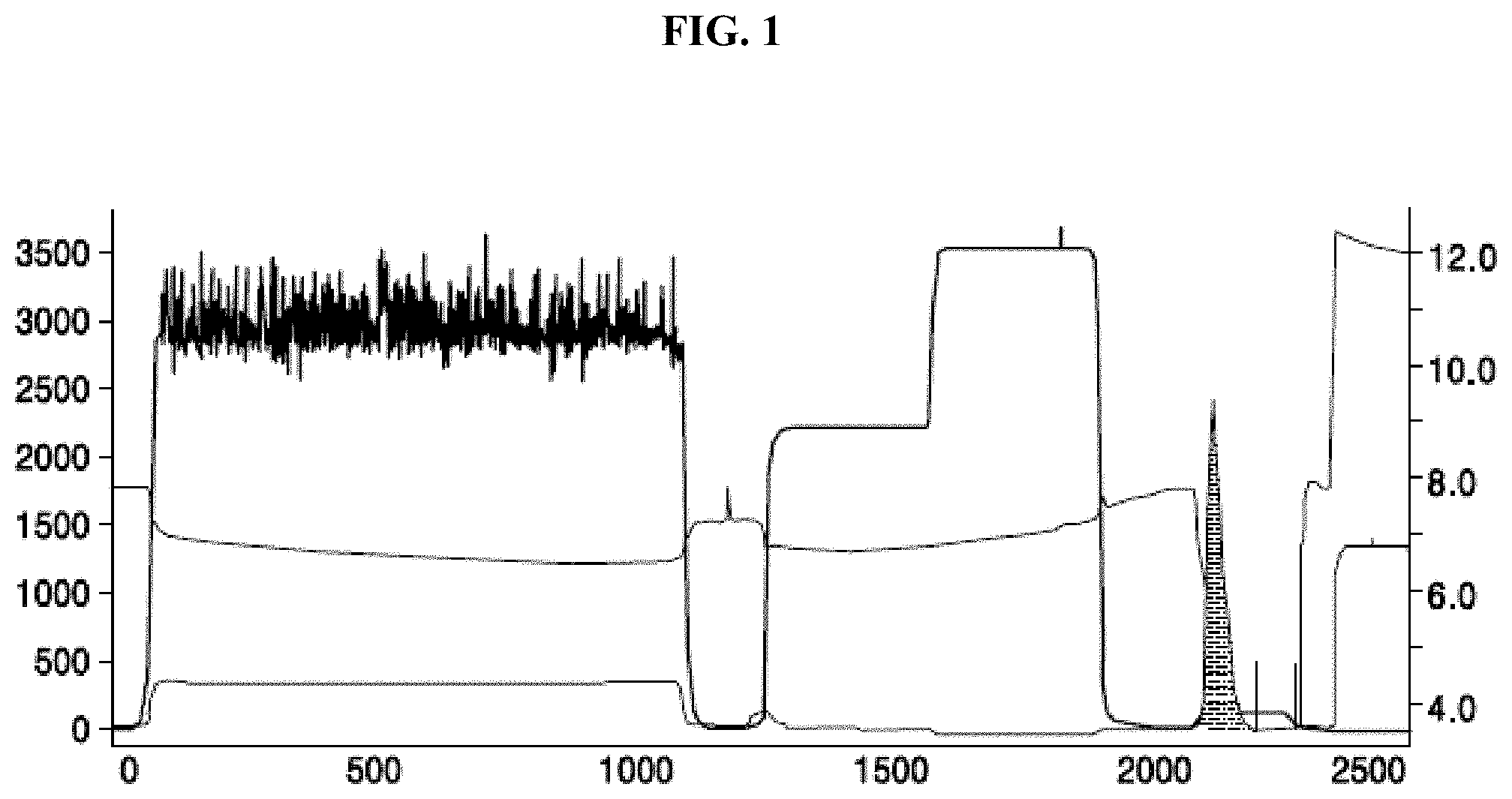
[0067] FIG. 1 shows an elution peak chromatogram of the anti-PD-1 expression product;
[0068] FIG. 2 shows a structure of an anti-PD-1/anti-HER2 heterodimeric antibody molecule;
[0069] FIG. 3 shows a structure of an incomplete antibody of a heavy chain and a light chain;
[0070] FIG. 4A shows an analysis result of size exclusion chromatography-high-performance liquid chromatography (SEC-HPLC) performed on an anti-PD-1 incomplete antibody of a heavy chain and a light chain; FIG. 4B shows an analysis result of SEC-HPLC performed on an anti-PD-1 incomplete antibody of a heavy chain and a light chain;
[0071] FIG. 5 shows an analysis result of SEC-HPLC performed on an anti-PD-1/anti-HER2 heterodimeric antibody molecule;
[0072] FIG. 6 shows a purity analysis result of SEC-HPLC performed on an anti-PD-1/anti-HER2 heterodimeric antibody molecule;
[0073] FIG. 7A shows affinity of an anti-PD-1/anti-HER2 heterodimeric antibody for PD-1; FIG. 7B shows affinity of an anti-PD-1/anti-HER2 heterodimeric antibody for HER2;
[0074] FIG. 8 shows that a combination of a PD-1 monoclonal antibody and a HER2 monoclonal antibody cannot bind to PD-1 and HER2, and an anti-PD-1/anti-HER2 heterodimeric antibody had an activity of binding to two antigens at the same time;
[0075] FIG. 9 shows that an anti-PD-1/anti-HER2 heterodimeric antibody induced mutual approach of a SK-BR-3 and CHO/PD-1 cells;
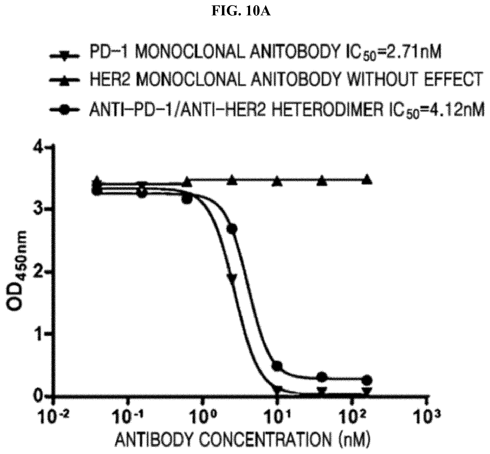
[0076] FIGS. 10A and 10B each show that an anti-PD-1/anti-HER2 heterodimeric antibody blocked PD-1/PD-L1 binding and PD-1/PD-L2 binding and better retained the blocking activity of the bivalent monoclonal antibody;
[0077] FIG. 11 shows that an anti-PD-1/anti-HER2 heterodimeric antibody exhibited T cell regulatory activity comparable to a PD-1 monoclonal antibody and significantly promoted secretion of cytokine IFN-.gamma.
[0078] FIG. 12 shows that an anti-PD-1/anti-HER2 heterodimeric antibody monoclonal antibody exhibited tumor cell killing inhibitory activity comparable to HER2; and
[0079] FIG. 13 shows that an anti-PD-1/anti-HER2 heterodimeric antibody exhibited stronger anti-tumor efficacy than a PD-1 monoclonal antibody or a HER2 monoclonal antibody and maintained satisfactory tumor regulatory action after stopping drug administration.
MODE OF DISCLOSURE
[0080] Definitions:
[0081] Covalent linkage refers to binding by a covalent bond between an Fc chain among two Fc chains and an antigen-binding functional site bound thereto in a heterodimeric bispecific antibody, thus binding the Fc chain with the antigen-binding functional site to form a molecule. The Fc chain may include a first antigen-binding site and a second antigen-binding site joined by at least one covalent linkage (e.g., a disulfide bond chain). The first Fc chain and the second Fc chain are each attached to an antigen-binding site by a covalent linkage (e.g., an imine bond or an amide bond). An antigen-binding site refers to a site that may specifically interact with a target molecule such as an antigen. Action thereof is highly selective, and thus a sequence that recognizes one target molecule generally does not recognize other molecular sequences.
[0082] Representative antigen-binding sites include: a variable region of an antibody, a structural allosteric variable region of an antibody, a binding domain of a receptor, a ligand binding domain, or an enzyme binding domain.
[0083] At least one inter-chain disulfide bond refers to a first Fc chain being linked to a second Fc chain by at least one disulfide bond to form a heterodimeric fragment. In the present disclosure, formation of at least one disulfide bond may be between the first Fc chain and the second Fc chain; or the first Fc chain, the second Fc chain, and an antigen-binding site bound thereto synthesized in the same cell. Also, the first Fc chain and the second Fc chain; or the first Fc chain, the second Fc chain, and an antigen-binding site bound thereto may each be separately synthesized in different cells, and then formed by in vitro reductive oxidation.
[0084] The first Fc chain and the second Fc chain may form a binding fragment by covalent linkage, wherein the covalent linkage may include a disulfide bond, each chain includes at least a portion of a constant region in a heavy chain of an immunoglobulin (Ig), and the first Fc chain and the second Fc chain may differ in amino acid sequence and include at least one amino acid difference. In the first Fc chain and the second Fc chain of the present disclosure, a strong mutual repulsion may occur among the same chains, and attraction may occur among the different chains. Thus, when co-expressed in cells, the first Fc chain and the second Fc chain; or the first Fc chain, the second Fc chain, and an antigen-binding site bound thereto are more prone to forming heterodimers. When the first Fc chain and the second Fc chain; or the first Fc chain, the second Fc chain, and an antigen-binding site bound thereto are each expressed in two host cells, respectively, the first Fc chain or the antigen-binding site bound to the first Fc chain may not be prone to forming a homodimer, and the second Fc chain or the antigen-binding site bound to the second Fc chain also may not be prone to forming a homodimer. In the present disclosure, when the first Fc chain and the second Fc chain; or the first Fc chain, the second Fc chain, and an antigen-binding site bound thereto are each expressed in two host cells in the presence of a reducing agent, respectively, a ratio of homodimers may be 50% or less, that is, a ratio of monomers (a single chain of the Fc chain or a single chain of the Fc chain and the antigen-binding site bound thereto) may be greater than 50%.
[0085] Immunoglobulin has a symmetrical structure having four polypeptide chains. Two of the four polypeptide chains are the same heavy chain having a relative large molecular weight and 450 to 550 amino acid residues, and a relative molecular weight thereof may be in a range of 55,000 Daltons (Da) to 70,000 Da. The other two of the four polypeptide chains are the same light chain (L chain) having a relatively small molecular weight and 210 amino acid residues, and a relative molecular weight thereof may be about 24,000 Da. The sequence of about 110 amino acids near the N-terminal in the different heavy and light chains of immunoglobulin may greatly vary. Thus, the sequence is referred to as a variable region (V region). The remaining amino acid sequence near the C-terminal is relatively stable. Thus, the remaining sequence is referred to as a constant region (C region). The variable region in the heavy chain accounts for about 1/4 of the length of the heavy chain, and the constant region accounts for about 3/4 of the length of the heavy chain. The known 5 types of Ig(s) include IgG (.gamma.), IgA (.alpha.), IgD (.delta.), IgM (.mu.), and IgE (.epsilon.). The heavy chain of each of IgG (.gamma.), IgA (.alpha.), and IgD (.delta.) includes three constant regions, namely, CH1, CH2, and C3. The heavy chain of each of IgM (.mu.) and IgE (.epsilon.) includes one variable heavy chain (VH) region and four constant regions, namely, CH1, CH2, CH3, and CH4. The constant region is both the backbone of an immunoglobulin molecule and one of the sites that activate an immune response.
[0086] The constant region in the present disclosure may include at least one interaction region of the first Fc chain and the second Fc chain, and the interaction region may be positioned in a portion of amino acids of the CH3 region in IgG, including at least GLN347, TYR349, THR 350, LEU 351, SER 354, ARG 355, ASP 356, GLU 357, LYS 360, SER 364, THR 366, LEU 368, LYS 370, ASN390, LYS392, THR394, PRO395, VAL 397, ASP399, SER400, PHE405, TYR407, LYS409, and LYS439.
[0087] The attachment of the first Fc chain and the second Fc chain to an antigen-binding site by a covalent bond or a linker may refer to the first Fc chain and the second Fc chain being respectively linked to an antigen-binding fragment of an antibody by a covalent bond or a linker, wherein the antigen-binding fragment recognizes a single chain antibody that recognizes an antigen, recognizes a receptor of a ligand, or recognizes a ligand of a receptor. The covalent bond is a type of chemical bond in which two or more atoms share outer electrons together, ideally reaching an electronic saturation state, thereby forming a relatively stable chemical structure called a covalent bond. The covalent bond is an interaction formed by sharing electron pairs between atoms. The atoms of the same or different elements may be bound by a covalent bond. The covalent bond between the first Fc chain and the second Fc chain of the present disclosure includes, but not limited to, a peptide bond formed by dehydration between an amino group of a molecule of an amino acid and a carboxyl group of another molecule of an amino acid, or a peptide bond or an imine bond between an aldehyde group of ethylene glycol, polyethylene glycol, another compound, or a multimer thereof and an amino group of a molecule of an amino acid. A linker may be an amino acid sequence, a compound, or a multimer of a compound, in which two polypeptide chains are joined by a covalent bond, wherein the amino acid sequence may include, but not limited to, a small peptide such as GGGGSGGGGSGGGGS, wherein the linker may link, by a peptide bond, the first Fc chain or the second Fc chain with a single chain antibody that may recognize an antigen or with a structural allosteric variant of a fragment of another antibody that may recognize an antigen.
[0088] The fact that the first Fc chain and the second Fc chain may be prone to forming a heterodimer and may not be prone to forming a homodimer means that a strong mutual repulsion may occur among the same polypeptide chains, and an attraction may occur among the different polypeptide chains of the first Fc chain and the second Fc chain polypeptide chain, and thus, when co-expressed in cells, the first Fc chain and the second Fc chain; or the first Fc chain, the second Fc chain, and an antigen-binding site bound thereto are more prone to forming heterodimers. When the first Fc chain and the second Fc chain; or the first Fc chain, the second Fc chain, and an antigen-binding site bound thereto are each expressed in two host cells, respectively, the first Fc chain or the antigen-binding site bound to the first Fc chain may not be prone to forming a homodimer, and the second Fc chain or the antigen-binding site bound to the second Fc chain also may not be prone to forming a homodimer.
[0089] The Kabat EU Index Numbering System means that Kabat uses a method of numbering each amino acid of an antibody sequence, and this method of numbering each residue has become a standard method in the art. The Kabat protocol may be extended to other antibodies that may not have been studied, and based on conserved amino acids, a target antibody may be aligned to one of the consensus sequences identified by Kabat.
[0090] An Fc fragment corresponds to a crystallizable fragment (Fc) or CH2 and CH3 binding domains of Ig, where interaction of Ig with effector molecules or cells occur.
[0091] IgG, namely, an abbreviation of Immunoglobulin G, is a main antibody component in a serum, and human IgG has four subtypes of IgG1, IgG2, IgG3 and IgG4 according to r-chain antigenic differences in the IgG molecules.
[0092] An incomplete antibody molecule refers to a structure formed by a heavy chain and a light chain of an antibody, in which the heavy chain and the light chain may be linked by a covalent bond or may not be linked by a covalent bond, and is a monoclonal antibody structure that may recognize an antigen.
[0093] A Fab fragment is a molecular recognition sequence and an antigen-binding fragment (Fab), which corresponds to two arms of an antibody molecule and consists of a complete variable heavy chain (VH) region and a CH1 region of the light and heavy chains. scFv is a molecular recognition sequence which is a structural isomer of an antibody fragment obtained by genetic engineering of a light chain variable region and a heavy chain variable region of an antibody. The extracellular region of a plasma membrane receptor is a molecular recognition sequence, and the plasma membrane receptor includes an extracellular region generally located on an outer side of a cell and recognizing and binding to the corresponding antigen or ligand; a transmembrane region that may anchor a receptor to a cell surface; and an intracellular region that may have kinase activity or may have a channel that transmits a signal in the cell. A ligand for a plasma membrane receptor refers to a protein, a polypeptide, or a compound that may be recognized and bound by an extracellular region of the plasma membrane receptor. Cytokines are low-molecular-weight soluble proteins produced by various cells induced by immunogens, mitogens, or other stimulators. Cytokines perform various functions, for example, in innate and adaptive immunity, hematopoiesis, cell growth, adult pluripotent stem cells (APSC) regulation, and repairing damaged tissues. Cytokines may be divided into interleukins, interferons, tumor necrosis factor superfamily, colony stimulating factors, chemokines, growth factors, and the like. A protein expression tag refers to an amino acid sequence added at an N-terminal or a C-terminal in a target protein, which may be a small peptide or a long amino acid. The addition of the protein expression tag may facilitate correct folding of a protein, separation and purification of a protein, and reducing degradation of a protein in a cell. Commonly used labels include, but not limited to, HA, SUMO, His, GST, GFP, and Flag.
[0094] The type of antibody to be used as a heterodimeric bispecific antibody of the present disclosure is not particularly limited. Preferably, antibodies known in the related art that are useful in treatment and/or prevention of diseases may be used in the present disclosure.
[0095] A heterodimeric bispecific antibody of the present disclosure may have at least one substitution, deletion, addition, and/or insertion. For example, some amino acids may replace other amino acids in a structure of a protein without significant loss of ability to bind to other polypeptides (e.g., antigens) or cells. Since the binding capacity and nature of a protein determine biological functional activity of the protein, some amino acid sequences may be substituted in a sequence of the protein without appreciable loss of biological utility or activity.
[0096] In many cases, a polypeptide variant may contain at least one conservative substitution. The term "conservative substitution" may refer to substitution of an amino acid with another amino acid having similar properties. One of ordinary skill in the art of peptide chemistry may expect substantially no change may occur in a secondary structure and hydrophilic properties of the polypeptide.
[0097] Substitutions of amino acids may be generally based on relative similarity of side chain substituents of the amino acids, such as hydrophobicity, hydrophilicity, charge, size, and the like. The exemplary substitutions with various foregoing characteristics are well known to one of ordinary skill in the art and include: arginine and lysine; glutamate and aspartate; serine and threonine; glutamine and asparagine; and valine, leucine, and isoleucine.
[0098] The term "identity" as used in the present disclosure has a meaning known in the art, and one of ordinary skill in the art may also be familiar with the rules and criteria for determining the identity between different sequences, referring to a homology percentage of identical residues between a sequence of a variant of a polynucleotide or polypeptide and a non-variant thereof after sequence alignment and introduction of gaps (if necessary, after obtaining the maximum percent of homology). In the present disclosure, in a case where the definition of identity is satisfied, an obtained variant sequence may have the same biological activity as the parent sequence. Methods and means for screening variant sequences using the activities are well known to one of ordinary skill in the art. Such variant sequences may be readily obtained by one of ordinary skill in the art in light of the teachings of the present disclosure. In an example embodiment, a variant of a polynucleotide or a polypeptide may have a polynucleotide or polypeptide identity of at least about 70%, at least about 75%, at least about 80%, at least about 90%, at least about 95%, at least about 98%, at least about 99%, at least about 99.1%, 99.2%, 99.3%, 99.4%, 99.5%, 99.6%, 99.7%, 99.8%, or 99.9%, as compared with the polynucleotide or the polypeptide, respectively. Due to redundancy of a genetic code, variants of these sequences encoding the same amino acid sequence may be present.
[0099] Another embodiment of the present disclosure includes a polynucleotide composition in which a polynucleotide sequence according to the present disclosure, a fragment thereof, or a complementary sequence thereof are hybridized under moderate to highly stringent conditions. Hybridization techniques are well known in the art of molecular biology. For the purpose of illustration, suitable moderately stringent conditions for testing the hybridization of a polynucleotide of the present disclosure to another polynucleotide include: pre-washing in a solution of 5.times. saline sodium citrate (SSC), 0.5% SDS, and 1.0 millimolar (mM) EDTA (pH 8.0); overnight hybridization at a temperature in a range of 50.degree. C. to 60.degree. C. in 5.times.SSC; and then washing twice in each SSC containing 0.1% SDS at 2.times., 0.5.times., and 0.2.times. for 20 minutes at a temperature of 65.degree. C. One of ordinary skill in the art may understand that stringency of hybridization may be readily manipulated, such as by varying a salt content of a hybridization solution and/or a temperature at which the hybridization occur. For example, in another embodiment, suitable highly stringent hybridization conditions include the conditions described above in addition to an elevated hybridization temperature in a range of, for example, 60.degree. C. to 65.degree. C. or 65.degree. C. to 70.degree. C.
[0100] A host cell of the present disclosure may be any cell that may be used in exogenous gene expression. The host cell may include, but not limited to, Escherichia coli (E. Coli), yeast, insect cells, plant cells, and mammalian cells.
[0101] Vectors of the present disclosure include vectors that may perform replication in any type of cell or organism, for example, including plasmids, bacteriophages, cosmids, and mini-chromosomes. In some embodiments, a vector including a polynucleotide of the present disclosure may be a vector suitable for propagation or replication of a polynucleotide or a vector suitable for expression of a polypeptide of the present disclosure. Such vectors are known in the art and are commercially available.
[0102] A "vector" may include both a shuttle vector and an expression vector. In general, a plasmid construct may also include a replication origin (such as a replication origin of ColE1) and a selectable marker (such as ampicillin or tetracycline resistance) for plasmid replication and selection in bacteria, respectively. An "expression vector" refers to a vector including a regulatory sequence or a regulatory element required for expression of an antibody of the present disclosure and including an antibody fragment in a bacterial or eukaryotic cell.
[0103] The vector of the present disclosure may be any vector used in exogenous gene expression, including, but not limited to, a plasmid vector. The plasmid vector may include at least one of a replication origin, a promoter, a target gene, a multiple cloning site, and a selection marker gene. Preferably, the vector of the present disclosure may include, but not limited to, a plasmid vector, such as a X0GC vector, modified from a pcDNA vector.
[0104] A subject of the present disclosure includes birds, reptiles, mammals, and the like. Preferably, the mammals include rodents, primates, and preferably, humans.
[0105] The disease in the present disclosure includes, but not limited to, tumors, and preferably, the tumors may include diseases such as leukemia, lymphoma, myeloma, brain tumors, head and neck squamous cell carcinoma, non-small cell lung cancer, nasopharyngeal cancer, esophageal cancer, stomach cancer, pancreas cancer, gallbladder cancer, liver cancer, colorectal cancer, breast cancer, ovarian cancer, cervical cancer, endometrial cancer, uterine sarcoma, prostate cancer, bladder cancer, renal cell carcinoma, and melanoma.
[0106] A pharmaceutically acceptable carrier refers to a pharmaceutical carrier commonly used in the pharmaceutical field, such as diluents; excipients and water; fillers such as starch; sucrose, lactose, or microcrystalline cellulose; binders such as cellulose derivatives, alginates, gelatin, and polyvinyl pyrrolidone; wetting agents such as glycerin; disintegrating agents such as sodium carboxymethyl starch, hydroxypropyl cellulose, croscarmellose, agar, calcium carbonate, and sodium bicarbonate; absorption enhancers such as quaternary ammonium compounds; surfactants such as cetyl alcohol and sodium lauryl sulfate; adsorption carriers such as aged soil and soap clay; lubricants such as talc, calcium, and magnesium stearate, micronized silica gel, polyethylene glycol, and the like. Adjuvants, such as flavoring agents, sweeteners, and the like may also be added to a composition.
[0107] The present disclosure will be further clarified by the following non-limiting examples, which are known to one of ordinary skill in the art, and many modifications may be made thereto without departing from the spirit and scope of the present disclosure.
[0108] The following experimental methods are general methods unless otherwise specified, and the experimental materials used may be easily obtained from commercial companies unless otherwise specified. The various antibodies used in the following Examples of the present disclosure are all derived from standard antibodies of the commercial route.
EXAMPLE 1
Construction of Vector of Anti-PD-1/Anti-HER2 Heterodimeric Antibody Molecule
[0109] An X0GC expression vector of the heavy chain and the light chain of the anti-human PD-1 was obtained, wherein the nucleotide sequence of the light chain variable region was as shown in the SEQ ID NO: 9, and the amino acid sequence was as shown in the SEQ ID NO:10. The nucleotide sequence of the light chain constant region was as shown in the SEQ ID NO: 3, and the amino acid sequence was as shown in the SEQ ID NO: 4. The nucleotide sequence of the heavy chain variable region was as shown in the SEQ ID NO: 11, and the amino acid sequence was as shown in the SEQ ID NO: 12. The nucleotide sequence of the heavy chain constant region was as shown in the SEQ ID NO: 13, and the amino acid sequence was as shown in the SEQ ID NO: 14. The light chain variable region, the light chain constant region, the heavy chain variable region, and the heavy chain constant region were respectively amplified by using a polymerase chain reaction (PCR) method. In the present disclosure, all PCR reactions were carried out using the Phusion high-fidelity DNA polymerase (F-530L, available from New England Biolabs (NEB)). PCR primers were designed according to the principle of base complementation and the need for restriction sites. The reaction system included 8.9 microliters (.mu.L) of H.sub.2O, 4 .mu.L of 5.times. Phusion high-fidelity DNA polymerase buffer solution, 4 .mu.L of 1 mM dNTP, 1 .mu.L of a forward primer, 1 .mu.L of a reverse primer, 0.1 .mu.L of Phusion high-fidelity DNA polymerase, and 1 .mu.L of template. The PCR products of the variable region and the constant region were electrophoresed on a 1.5% agarose gel, and the corresponding fragments were recovered using a DNA recovery kit (product No. A9282, available from Promega). The recovered variable region fragment and the constant region fragment were used as templates, and a forward primer of the variable region and a reverse primer of the constant region were used to perform a PCR reaction once more. Then, the corresponding fragments were recovered to thereby obtain a full-length fragment of the heavy chain and the light chain. The X0GC vector and the full-length fragment were cleaved by using restriction enzymes EcoRI (product No. R3101L, available from NEB) and Hindlll (product No. R3104L, available from NEB), and the restriction enzyme system included: 32 .mu.L of 10.times. buffer solution, 0.5 .mu.L of each of EcoRI and Hind III, 3 .mu.L of the full-length fragment obtained by gel recovery, and 14.5 .mu.L of H.sub.2O. The restriction enzyme system was subjected to a reaction at a temperature of 37.degree. C. for 3 hours. The restriction enzyme products were ligated using a T4 DNA ligase (NEB, product No. M0202V), and the reaction system included 2 .mu.L of 10.times. ligase buffer solution, 0.5 .mu.L of ligase, 3 .mu.L of the full-length fragment obtained by gel recovery, 3 .mu.L of the X0GC vector obtained by gel recovery, and 11.5 .mu.L of H.sub.2O. The reaction was allowed to react at room temperature for 12 hours. The ligation products were transformed into an E. coli competent cell DH5.alpha. (product No. CB104, available from Tiangen), respectively, to thereby obtain an X0GC expression vector of the heavy chain and the light chain of the antibody for expression of the heavy chain and the light chain of the antibody in eukaryotic cells.
[0110] An X0GC expression vector of the heavy chain and the light chain of the anti-human HER2 antibody was obtained. The sequence of the antibody variable region was derived from http://www.drugbank.ca/drugs/DB00072. The nucleotide sequence of the light chain variable region was as shown in the SEQ ID NO: 1, and the amino acid sequence was as shown in the SEQ ID NO:2. The nucleotide sequence of the light chain constant region was as shown in the SEQ ID NO: 3, and the amino acid sequence was as shown in the SEQ ID NO: 4. The nucleotide sequence of the heavy chain variable region was as shown in the SEQ ID NO: 5, and the amino acid sequence was as shown in the SEQ ID NO: 6. The nucleotide sequence of the heavy chain constant region was as shown in the SEQ ID NO: 7, and the amino acid sequence was as shown in the SEQ ID NO: 8. According to the foregoing method, an X0GC expression vector of a heavy chain and a light chain of an antibody were obtained for expression of the heavy chain and the light chain of the antibody in eukaryotic cells.
EXAMPLE 2
Expression of Anti-PD-1/Anti-HER2 Heterodimeric Antibody Molecule
[0111] The expression vector of a heavy chain and a light chain of an antibody including anti-human PD-1 were transfected into a 293F cell (FreeStyle.TM. 293-F Cells, product No. R79007, available from Invitrogen), and the expression vector of a heavy chain and a light chain of an antibody including anti-human HER2 were also transfected into a 293F cell. Cells were inoculated one day prior to transfection. Cells were collected by centrifugation on the day of transfection. The cells were resuspended in a fresh FreeStyle.TM. 293 expression medium (FreeStyle.TM. 293 Expression Medium, product No. 12338001, available from Gibco) at a cell density of 200.times.10.sup.5 cells/mL. A plasmid was added thereto according to the transfection volume at a final concentration of 36.67 .mu.g/mL, and the mixture was gently and homogeneously mixed. Subsequently, linear polyethyleneimine (PEI, linear, molecular weight of (MW) 25,000, product No. 43896, available from Alfa Aesar) was added thereto at a final concentration of 55 .mu.g/mL, and the mixture was gently and homogeneously mixed. Thereafter, the cells were placed in a cell culture incubator and incubated at a temperature of 37.degree. C. for 1 hour in a shaker at a rate of 120 revolutions per minute (rpm). A 19-fold transfection volume of fresh medium was then added thereto. Subsequently, the cells were incubated at a temperature of 37.degree. C. in a shaker at a rate of 120 rpm. The cell culture supernatant transfected for 5 days to 6 days was collected by centrifugation.
[0112] The amount of expression was determined by enzyme-linked immunosorbent assay (ELISA). The precipitate was removed by filtration using a 0.2 .mu.m filtration film before applying the result to column purification. This process was performed at a temperature of 4.degree. C.
EXAMPLE 3
Purification of Expression Product of Anti-PD-1/Anti-HER2 Heterodimeric Antibody Molecule
[0113] Using an AKTA explorer 100 type protein purification system (available from GE Healthcare), and affinity chromatography column rProtein A Sepharose Fast Flow (16 mm I.D., 10 mL, available from GE Healthcare) was purified at a temperature of 4.degree. C. The column was first equilibrated with mobile phase A (20 mM sodium phosphate buffer solution, 150 mM sodium chloride, pH 7.4). After the baseline was stabilized, samples were loaded to the supernatant of the treated cells at a flow rate of 5 mL/min. After the loading of the samples, equilibration was performed using mobile phase A. The sample were each an anti-PD-1 expression product and an anti-HER2 expression product. Subsequently, a 5-column volume was washed using mobile phase B1 (mobile phase A containing 0.5 M arginine). Then, a 5-column volume was eluted using mobile phase B2 (100 mM citric acid, pH 3.0) to collect a target protein at maximum, i.e., the elution peak. The flow rate of the elution was 5 mL/min. The elution peak chromatogram of the anti-PD-1 expression product is shown in FIG. 1. The elution peak of the anti-HER2 expression product was similar with that of the anti-PD-1 expression product (of which the result is not described herein), and the elution peak was collected (the gray area in FIG. 1), and the pH was adjusted to 5.0 by dropwise addition of 1 M sodium acetate solution.
EXAMPLE 4
Preparation and Purification of Anti-PD-1/Anti-HER2 Heterodimeric Antibody Molecule
[0114] The structure of the anti-PD-1/anti-HER2 heterodimeric antibody molecule is as shown in FIG. 2.
[0115] The anti-PD-1 expression product and the anti-HER2 expression product obtained by the aforementioned rProtein A Sepharose Fast Flow (16 mm I.D., 10 mL, available from GE Healthcare) were recombined in vitro to obtain a heterodimer. First, the purified and collected protein solution was concentrated by ultrafiltration through an ultrafiltration tube (standard molecular weight cutoff of 10 kiloDaltons (kDa)), and the solution was replaced with the phosphate buffer saline (PBS) solution (pH=7.4). PBS at a concentration of 1 mg/mL and 1M DTT having a final volume of 1/200 were added to the obtained anti-PD-1 and anti-HER2 purified expression product solutions. The final concentration of DTT was 5 mM. Reduction was performed at a temperature of 4.degree. C. (3 to 8 hours), and through the reduction, disulfide bonds were opened, and the disulfide bonds of a hinge region of a small amount of the antibody homodimeric molecules contained in the anti-PD-1 and anti-HER2 expression products were also opened, thus forming an incomplete antibody molecule containing a heavy chain and a light chain. The structure thereof is as shown in FIG. 3. The reduced sample was analyzed by using size exclusion chromatography-high-performance liquid chromatography (SEC-HPLC) (TSKgel superSW3000, available from TOSOH) using 1 mM of a DTT reducing agent in a mobile phase buffer solution. The results thereof are shown in FIGS. 4A and 4B. A ratio of the anti-PD-1 incomplete antibody molecules was 100%, and a ratio of the anti-HER2 incomplete antibody molecules was 89.3%, in which the remaining 10.7% was an aggregate, but no homodimer in which a disulfide bond was opened was present.
[0116] The reduced anti-PD-1 and anti-HER2 incomplete antibody molecules were mixed according to a mole ratio under a condition of 4.degree. C. for 24 hours to perform recombination. In the recombination, the anti-PD-1 and anti-HER2 incomplete antibody molecules formed a heterodimeric bispecific antibody including the anti-PD-1 and anti-HER2 incomplete antibody molecules through a non-covalent interaction between CH2 and CH3. Subsequently, the protein solution was subjected to ultrafiltration through an ultrafiltration concentrator (standard molecular weight cutoff of 10 kDa) and replacement with PBS solution (pH=7.4) to complete reduction. Oxidation was then performed by air or by using an oxidizing agent to reform disulfide bonds of the heterodimeric bispecific antibody. The conditions of oxidation included the addition of 100 mM L-dehydroascorbic acid as an oxidizing agent, a final concentration of protein of 1 mg/mL, and a final concentration of the oxidizing agent of 1 mM. Under this condition, oxidation was performed at 4.degree. C. for 24 hours. The sample obtained by the oxidation was subjected to SEC-HPLC analysis, and the results are shown in FIG. 5.
[0117] The heterodimeric antibody molecule obtained by reduction and oxidation of the anti-PD-1 and anti-HER2 incomplete antibody molecules was subjected to ultrafiltration through an ultrafiltration concentrator (standard molecular weight cutoff of 10 kDa) and replacement with 10 mM PBS solution, and the pH was 5.8. Using the AKTA explorer 100 type protein purification system (available from GE Healthcare) and ion chromatography column Source 15S (16 mm I.D., 17 mL, available from GE Healthcare), purification was performed at a temperature of 4.degree. C. The column was first equilibrated with mobile phase A (10 mM sodium phosphate buffer solution, pH 7.0). After the baseline was stabilized, samples were loaded to the treated protein solutions at a flow rate of 3 mL/min. After the loading of the samples, equilibration was performed using mobile phase A. Subsequently, a 20-column volume (0% B to 100% B, 170 min, and at a flow rate of 2 mL/min) was eluted from A (10 mM sodium phosphate, pH 5.8) to B (10 mM sodium phosphate, pH 5.8) to collect the elution peak. The collected protein solution was subjected to ultrafiltration through an ultrafiltration concentrator (standard molecular weight cutoff of 10 kDa), replacement with PBS solution (pH=7.4), and filtration sterilization. The temperature was maintained at 4.degree. C. The purified product was subjected to SEC-HPLC for purity analysis. The results thereof are shown in FIG. 6, and the purity was 99.96%.
EXAMPLE 5
Stability of Anti-PD-1/Anti-HER2 Heterodimeric Antibody Molecule
[0118] Fully sealed 1 mg/mL anti-PD-1/anti-HER2 heterodimer samples were allowed to stand in a climate chamber (KBF240, available from Binder) at a temperature of 40.degree. C. At the corresponding time points (the baseline (the first day), the two weeks, and four weeks), 20 .mu.g of sample was collected to perform SEC-HPLC, thereby performing isolation. The SEC-HPLC conditions were as follows: (1) size exclusion chromatography: TSKgel G3000SWxl (available from Tosoh Bioscience), 5 .mu.m, and 7.8 mm.times.30 cm; (2) mobile phase: 5 mM PBS, 150 mM NaCl, and pH 6.7; (3) flow rate: 0.6 mL/min; (4) UV detection wavelength: 280 nanometers (nm); and (5) acquisition time: 30 mins. The used instrument was the Agilent 1200 Infinity chromatograph, which was recorded using Agilent ChemStation, and the ratio of the remaining monomers was calculated. As shown in Table. 1, under the experimental conditions of 40.degree. C., the dimers did not undergo significant aggregation. Therefore, the anti-PD-1/anti-HER2 heterodimer is seen as having relatively excellent thermal stability.
TABLE-US-00001 TABLE 1 Stability of anti-PD-1/anti-HER2 heterodimeric antibody molecule Daily Main High Low change peak molecular molecular in main content weight weight peak content Item Time (%) (%) (%) (%) Anti-PD-1/ Start 99.42 0.32 0.26 0.09 anti-HER2 Two 98.69 0.25 1.06 heterodimeric weeks antibody Four 97.04 0.36 1.7 molecule weeks
EXAMPLE 6
In Vitro Target Binding Activity of Anti-PD-1/Anti-HER2 Heterodimeric Antibody
[0119] The ability of the anti-PD-1/anti-HER2 heterodimeric antibody to bind to a single antigen was determined by using ELISA.
[0120] ELISA was performed as follows: recombinant human PD-1 (product No. 10377-H08H, available from Beijing Yiqiao Shenzhou) or human HER2 (product No. 10004-H08H, available from Beijing Yiqiao Shenzhou) was coated on a 96-well high-adsorbing ELISA plate (product No. 42592, available from Costar) with a carbonate buffer solution (0.05 M) at pH 9.6. The coating concentration was 1 .mu.g/mL. The coating amount was 100 .mu.L per well, and the coating was performed at 4.degree. C. overnight. PBST washing was performed five times. The result was sealed at 300 .mu.L/well using PBST containing 1% BSA, and subjected to incubation for 1 hour at 25.degree. C. PBST washing was performed five times. 100 .mu.L of a heterodimeric antibody sample serially diluted in PBST containing 1% BSA, and a control, were added to each well and were incubated for 1 hour at 25.degree. C. PBST washing was performed five times. Subsequently, 100 .mu.L of the horseradish peroxidase-labeled anti-human IgG antibody (product No. AP309P, available from Chemicon) diluted at 1:2,000 in PBST containing 1% BSA was then added to each well, and the cells were incubated for 1 hour at 25.degree. C. PBST washing was performed five times. 100 .mu.L of the colorimetric substrate TMB was added to each well, and color development was allowed to occur for 10 minutes at room temperature. Color development was stopped by adding 100 .mu.L of 1 M H.sub.2SO.sub.4 to each well. The absorbance at 450 nm was read on a microplate reader.
[0121] As a result, as shown in FIGS. 7A and 7B, it was found that the anti-PD-1/anti-HER2 heterodimeric antibody had high affinity for PD-1 and HER2, thus maintaining antigen-affinitive activity of the bivalent monoclonal antibody.
EXAMPLE 7
Simultaneous Dual-Targeted Binding Activity of Anti-PD-1/Anti-HER2 Heterodimeric Antibody
[0122] The simultaneous binding ability of the anti-PD-1/anti-HER2 heterodimeric antibody to two different antigens was measured by using ELISA.
[0123] ELISA was performed as follows: recombinant human HER2 (product No. 10004-H08H, available from Beijing Yiqiao Shenzhou) was coated on a 96-well high-adsorbing ELISA plate with a carbonate buffer solution at pH 9.6. The coating concentration was 1 .mu.g/mL. The coating amount was 100 .mu.L per well, and the coating was performed at 4.degree. C. overnight. PBST washing was performed five times. The result was sealed at 300 .mu.L/well using PBST containing 1% BSA, and subjected to incubation for 1 hour at 25.degree. C. PBST washing was performed five times. 100 .mu.L of a heterodimeric antibody sample serially diluted in PBST containing 1% BSA, and a control, were added to each well and were incubated for 1 hour at 25.degree. C. PBST washing was performed five times. Subsequently, 100 .mu.L of biotin-labeled PD-1-Fc (available from Beijing Hanmi Pharmaceutical) diluted in PBST containing 1% BSA was added at 0.5 .mu.g/mL to each well, and the cells were incubated for 1 hour at 25.degree. C. 100 .mu.L of streptavidin-horseradish peroxidase conjugate (product No. 554066, available from BD Pharmingen) diluted at 1:1,000 in PBST containing 1% BSA was then added to each well, and the cells were incubated for 1 hour at 25.degree. C. PBST washing was performed five times. 100 .mu.L of the colorimetric substrate TMB was added to each well, and color development was allowed to occur for 10 minutes at room temperature. Color development was stopped by adding 100 .mu.L of 1 M H.sub.2SO.sub.4 to each well. The absorbance at 450 nm was read on a microplate reader.
[0124] As a result, as shown in FIG. 8, a combination of the PD-1 monoclonal antibody (the sequence of the heavy chain variable region and the sequence of the light chain variable region were identical to the corresponding sequence of the PD-1 binding site in the anti-PD-1/anti-HER2 heterodimeric antibody) and the HER2 monoclonal antibody (Trastuzumab) failed to bind to both PD-1 and HER2 simultaneously, and the anti-PD-1/anti-HER2 heterodimeric antibody only had activity of binding to the two antigens at the same time.
[0125] By using a flow cytometer (FCM, FACS Calibur, available from BD Biosicences), simultaneous binding ability of the anti-PD-1/anti-HER2 heterodimeric antibody to dual-targeted antigens was measured using high-PD-1-expressing CHO/PD-1 (product No. M00529, available from GenScript) cells and high-HER2-expressing SK-BR-3 cells.
[0126] The CHO/PD-1 cells were stained according to instructions of the PKH26 reagent kit (product No. SLBH4568V, available from Sigma). Briefly, the CHO/PD-1 cells were collected and washed once in a serum-free medium. Then, CHO/PD-1 was prepared as a 2.times.10.sup.7/mL cell suspension using Diluent C, the PKH26 reagent kit. Then, PKH26 dye was diluted to 4 .mu.M and mixed with the cell suspension at a ratio of 1:1. A cell density of the mixture suspension was 1.times.10.sup.7/mL, and a concentration of PKH26 was 2 .mu.M. The mixture suspension was incubated at room temperature for 1 hour. Subsequently, incubation was performed using an equal volume of fetal bovine serum (FBS) for 1 minute, thereby completing the staining. The result was centrifuged at a centrifugal force of 400 g for 10 minutes, washed twice with complete medium, and resuspended in complete medium for later use. The SK-BR-3 cells were stained according to the instructions of the CFSE reagent kit (product No. C34554, available from Life technology). Briefly, CFSE was diluted with PBS at a working concentration of 0.5 and pre-heated at a temperature of 37.degree. C., and the SK-BR-3 cells were collected by centrifugation at a rate of 1,000 rpm for 5 minutes. The SK-BR-3 cells were resuspended in the pre-heated CFSE working solution and incubated at a temperature of 37.degree. C. for 15 minutes, and then, the cells were collected by centrifugation at a rate of 1,000 rpm for 5 minutes. Next, the cells were resuspended in complete medium and incubated for 30 minutes. Thereafter, the cells were washed using complete medium, and resuspended to thereby completing the preparation. The stained cells were centrifuged and collected, and washed once with cold PBS containing 2% FBS. The cells were resuspended in cold PBS containing 2% FBS such that the cell density was 5.times.10.sup.6/mL. The SK-BR-3 cells and the CHO/PD-1 cells were mixed at a ratio of 1:1, and 100 .mu.L of each of the cells were collected from flow pipes, respectively, (that is, 2.5.times.10.sup.5 SK-BR-3 cells and 2.5.times.10.sup.5 CHO/PD-1 cells). Next, 100 .mu.L of the diluted heterodimeric antibody sample, the control, and the isotype control (human immunoglobulin, available from Jiangxi Boya Bio-Pharmaceutical, State Food and Drug Administration (SFDA) Approval No. S19993012) were added to cold PBS containing 2% FBS at a final concentration of 5 nM. The flow pipe was incubated on ice for 30 minutes. The cells were then washed with PBS containing 2% FBS. The cells were resuspended in 500 .mu.L of cold PBS, and the cells were suspended in a flow cytometer to perform measurement analysis.
[0127] As a result, as shown in FIG. 9, it was found that the heterodimeric antibody simultaneously bound to the high-PD-1-expressing CHO/PD-1 cells and the high-HER2 expressing SK-BR-3 cells. Thus, the anti-PD-1/anti-HER2 heterodimeric antibody were capable of attracting SK-BR-3 cells to CHO/PD-1 cells.
EXAMPLE 8
Blocking Activity of Anti-PD-1/Anti-HER2 Heterodimeric Antibody on Binding Between PD-1 and Ligand PD-L1 or PD-L2
[0128] The blocking activity of the anti-PD-1/anti-HER2 heterodimeric antibody on binding between PD-1 and PD-L1 and binding between PD-1and PD-L2 was measured by using ELISA.
[0129] Recombinant human PD-1-Fc was coated on a 96-well high-adsorbing ELISA plate with PBS at pH 9.6. The coating concentration was 1 .mu.g/mL. The coating amount was 100 .mu.L per well, and the coating was performed at 4.degree. C. overnight. PBST washing was performed five times. The result was sealed at 300 .mu.L/well using PBST containing 1% BSA, and subjected to incubation for 1 hour at 25.degree. C. PBST washing was performed five times. A heterodimer sample and the control serially diluted in PBST containing 1% BSA were added thereto, followed by addition of 100 .mu.L of biotin-labeled PD-L1-Fc at a final concentration of 1 .mu.g/mL or biotin-labeled PD-L2 at a final concentration of 4 .mu.g/mL to each well. Then, the cells were incubated for 1 hour at 25.degree. C. PBST washing was performed five times. Subsequently, 100 .mu.L of the horseradish peroxidase-labeled streptavidin (product No. 554066, available from BD Pharmingen) diluted at 1:1,000 in PBST containing 1% BSA was then added to each well, and the cells were incubated for 1 hour at 25.degree. C. PBST washing was performed five times. 100 .mu.L of the colorimetric substrate TMB was added to each well, and color development was allowed to occur for 10 minutes at room temperature. Color development was stopped by adding 100 .mu.L of 1 M H.sub.2SO.sub.4 to each well. The absorbance at 450 nm was read on a microplate reader.
[0130] As a result, as shown in FIGS. 10A and 10B, it was found that the anti-PD-1/anti-HER2 heterodimeric antibody blocked binding between PD-1 and PD-L1 and PD-1 and PD-L2, thus maintaining blocking activity of the bivalent monoclonal antibody at a relatively excellent level.
EXAMPLE 9
T Cell Regulatory Activity of Anti-PD-1/Anti-HER2 Heterodimeric Antibody Molecule
[0131] The regulatory activity of the anti-PD-1/anti-HER2 heterodimeric antibody on T cell immune reaction was measured by using mixed lymphocyte reaction (MLR).
[0132] Acquisition of human dendritic cells (DCs): human peripheral blood mononuclear cells (PBMCs) (product No. CC-2702, available from Lonza) were collected by reproduction. The human PBMCs were resuspended in a serum-free RPMI 1640 medium at a cell density of 5.times.10.sup.6/mL and inoculated in a cell culture flask, followed by incubation in a carbon dioxide incubator at 37.degree. C. for 90 minutes. The supernatant and the resuspended cells of the culture solution were discarded, and adherent cells were cultured in complete medium (RPMI 1640 containing 10% FBS). Subsequently, 100 ng/mL GM-CSF (product No. 10016-HNAH, available from Beijing Yiqiao Shenzhou) and 100 ng/mL IL-4 (product No. 11846-HNAE, available from Beijing Yiqiao Shenzhou) were added to the cells. The cells were incubated for three days, followed by a solution change. Then, the cells were incubated again for three days. Next, the medium was changed to complete medium (RPMI 1640 containing 10% FBS) containing 100 ng/mL GM-CSF, 100 ng/mL IL-4, and 20 ng/mL TNF-a, followed by incubation for one day. Accordingly, DCs were obtained.
[0133] Acquisition of human T cells: human PBMCs (product No. CC-2702, available from Lonza) were collected by reproduction. The PBMCs and PBMCs from which DCs were generated were each derived from different subjects. Human T cells were isolated according to instructions of the Pan T cell isolation kit (product No. 5150414820, available from Miltenyi Biotech). Briefly, PBMCs were washed with PBS once, and the PBMCs were resuspended in isolation buffer solution (2 mM ethylenediamine tetraacetic acid (EDTA), pH 7.2 PBS containing 0.5% BSA) at a concentration of 10.sup.7 cells per 40 .mu.L. Then, 10 .mu.L Pan T cell Biotin Antibody Cocktail was added thereto, followed by incubation at a temperature of 4.degree. C. for 5 minutes. Subsequently, 30 .mu.L of isolation buffer solution and 20 .mu.L of Pan T cell MicroBead Cocktail were added thereto, followed by incubation at a temperature of 4.degree. C. for 10 minutes. T cells were then obtained through a magnetic activated cell sorter (MACS) separation column.
[0134] The collected human DCs and human T cells were resuspended in complete medium (RPMI 1640 containing 10% FBS) and inoculated on a 96-well plate. The inoculated DCs and T cells were each mixed and cultured at a concentration of 1.times.10.sup.4/well, 1.times.10.sup.5/well, respectively. The heterodimeric antibody sample serially diluted in complete medium and the control were added thereto. The incubation plate was placed in a carbon dioxide incubator at 37.degree. C. and incubated for five days. Once the incubation was complete, the supernatant in the wells was taken to detect cytokine IFN-.gamma. (product. No. ELH-IFNg, available from RayBiotech) according to the kit manual.
[0135] As shown in FIG. 11, human T cells activate secretion of IFN-.gamma. under the stimulation of allogeneic DCs. When PD-1 monoclonal antibodies are added, activation of T cells may be enhanced, and secretion of cytokines may be promoted. However, HER2 monoclonal antibodies do not have such activity. The anti-PD-1/anti-HER2 heterodimeric antibody exhibited T cell regulatory activity comparable to a PD-1 monoclonal antibody and significantly promoted secretion of cytokine IFN-.gamma.
EXAMPLE 10
Tumor Cell Inhibitory Activity of Anti-PD-1/Anti-HER2 Heterodimeric Antibody Molecule
[0136] The anti-PD-1/anti-HER2 heterodimeric antibody was tested for killing activity thereof against human breast cancer cells, i.e., SK-BR-3 cells, in the presence of human PBMCs.
[0137] SK-BR-3 cells were incubated in an RPMI 1640 medium containing 10% FBS (i.e., complete medium). The SK-BR-3 cells were collected and resuspended in complete medium. 100 .mu.L of the cells were then inoculated in each well of a 96-well incubation plate at a cell density of 5.times.10.sup.4/mL. That is, 5,000 cells were inoculated in each well. Human PBMCs (product No. CC-2702, available from Lonza) were collected by reproduction. The human PBMCs were resuspended in an RPMI 1640 complete medium at a cell density of 5.times.10.sup.5/mL. 50 .mu.L of the cells were then inoculated in each well of a 96-well incubation plate. That is, 25,000 cells were inoculated in each well. A ratio of effective target cells was 5:1. 50 .mu..mu.L of each of the heterodimeric antibody sample serially diluted in complete medium and the control were added to each well. The incubation plate was placed in an incubator at 37.degree. C. and 5% carbon dioxide for three days of incubation. Once the incubation was complete, PBMCs in the cell incubation plate were washed and removed using a medium. Then, 100 .mu.L of complete medium and 20 .mu.L of MTS (CellTiter96 Aqueous One Solution, product No. G358B, available from Promega) were added thereto for detection of SK-BR-3 cells. The cell incubation plate was subjected to further incubation in the incubator for 3 to 4 hours. Thereafter, the absorbance at 490 nm was read on a microplate reader.
[0138] As shown in FIG. 12, when HER2 monoclonal antibodies were added, the HER2 monoclonal antibodies killed and inhibited SK-BR-3 cells; however, PD-1 monoclonal antibodies did not exhibit such activity in vitro. The anti-PD-1/anti-HER2 heterodimeric antibody also exhibited tumor cell-killing inhibitory activity comparable to the HER2 monoclonal antibodies.
EXAMPLE 11
Studies in Anti-Tumor Efficacy of Anti-PD-1/Anti-HER2 Heterodimeric Antibody Molecule in Animals
[0139] The experimental materials were selected from 6- to 8-week-old female immunodeficient NCG mice (available from Nanjing Biomedical Research Institute of Nanjing University). One week after the mice adapted to the environment, 5.times.10.sup.6 human breast cancer cells were subcutaneously inoculated in the right dorsal side of each mouse. Once a length of the tumor volume reached about 100 cubic milimeters (mm3), the mice were grouped according to tumor volume, each group including 8 mice. First, the mice immune system was partially humanized by administration of human PBMCs, and 5.times.10.sup.6 cells were intravenously inoculated in each mouse. Subsequently, each solvent (PBS), 35 nanomoles per kilogram (nmol/kg, 5 mg/kg) of the PD-1 monoclonal antibody, 35 nmol/kg (5 mg/kg) of the HER2 monoclonal antibody, a combination of 35 nmol/kg of the PD-1 monoclonal antibody and 35 nmol/kg of the HER2 monoclonal antibody, and 35 nmol/kg (5 mg/kg) of the anti-PD-1/anti-HER2 heterodimeric antibody were administered two times a week for 2 weeks by sequential administration. The administration was performed by intraperitoneal injection. From the start day of administration, the tumor volume was measured three times a week. The relatively long diameter (a) and the relatively short diameter (b) were measured, and the tumor volume was calculated as follows: tumor volume (mm3) =(a.times.b2)/2. The tumor volume was observed for 3 weeks, that is, observation of the change in tumor volume continued for one more week after administration was stopped.
[0140] As a result, as shown in FIG. 13, it was found that the anti-PD-1/anti-HER2 heterodimeric antibody had much stronger anti-tumor efficacy than the PD-1 monoclonal antibody and the HER2 monoclonal antibody. Even after administration was stopped, the anti-PD-1/anti-HER2 heterodimeric antibody exhibited moderate tumor inhibitory action.
Sequence CWU 1
1
141321DNAArtificial SequenceAnti-human HER2 antibody light chain
variable region 1gacattcaga tgactcagag cccttcttca ctgtcagctt
ccgtgggcga cagagtcact 60atcacctgcc gcgcaagtca ggatgtgaac accgcagtcg
cctggtacca gcagaagcct 120ggcaaagctc caaagctgct gatctacagc
gcatctttcc tgtattctgg agtgcccagt 180aggtttagtg ggtcacggtc
cggtaccgac ttcacactga ctatctccag cctgcagcct 240gaggattttg
ccacatacta ttgccagcag cactatacca caccccctac tttcggccag
300ggaaccaaag tggagatcaa g 3212107PRTArtificial SequenceAnti-human
HER2 antibody light chain variable region 2Asp Ile Gln Met Thr Gln
Ser Pro Ser Ser Leu Ser Ala Ser Val Gly1 5 10 15Asp Arg Val Thr Ile
Thr Cys Arg Ala Ser Gln Asp Val Asn Thr Ala 20 25 30Val Ala Trp Tyr
Gln Gln Lys Pro Gly Lys Ala Pro Lys Leu Leu Ile 35 40 45Tyr Ser Ala
Ser Phe Leu Tyr Ser Gly Val Pro Ser Arg Phe Ser Gly 50 55 60Ser Arg
Ser Gly Thr Asp Phe Thr Leu Thr Ile Ser Ser Leu Gln Pro65 70 75
80Glu Asp Phe Ala Thr Tyr Tyr Cys Gln Gln His Tyr Thr Thr Pro Pro
85 90 95Thr Phe Gly Gln Gly Thr Lys Val Glu Ile Lys 100
1053321DNAArtificial SequenceAnti-human HER2 antibody light chain
variable region 3cgaactgtgg ccgctccaag cgtcttcatt tttccaccct
ctgacgaaca gctgaagtca 60gggacagctt ccgtggtctg tctgctgaac aatttttacc
ccagggaggc caaagtgcag 120tggaaggtcg ataacgctct gcagagcgga
aattctcagg agagtgtgac agaacaggac 180tcaaaagatt ccacttatag
cctgtctagt accctgacac tgtccaaggc agactacgaa 240aagcataaag
tgtatgcctg tgaggtcaca catcagggtc tgtcaagccc cgtcactaag
300tccttcaatc gtggcgaatg c 3214107PRTArtificial SequenceAnti-human
HER2 antibody light chain variable region 4Arg Thr Val Ala Ala Pro
Ser Val Phe Ile Phe Pro Pro Ser Asp Glu1 5 10 15Gln Leu Lys Ser Gly
Thr Ala Ser Val Val Cys Leu Leu Asn Asn Phe 20 25 30Tyr Pro Arg Glu
Ala Lys Val Gln Trp Lys Val Asp Asn Ala Leu Gln 35 40 45Ser Gly Asn
Ser Gln Glu Ser Val Thr Glu Gln Asp Ser Lys Asp Ser 50 55 60Thr Tyr
Ser Leu Ser Ser Thr Leu Thr Leu Ser Lys Ala Asp Tyr Glu65 70 75
80Lys His Lys Val Tyr Ala Cys Glu Val Thr His Gln Gly Leu Ser Ser
85 90 95Pro Val Thr Lys Ser Phe Asn Arg Gly Glu Cys 100
1055360DNAArtificial SequenceAnti-human HER2 antibody heavy chain
variable region 5gaggtgcagc tggtcgaaag tgggggtggg ctggtgcagc
caggcggatc actgaggctg 60tcctgcgccg ctagcggctt caacatcaaa gacacctata
ttcactgggt ccgacaggca 120ccagggaagg gtctggaatg ggtggctcgt
atctacccta caaatggtta cactagatat 180gccgactccg tgaaaggccg
gtttactatt tctgctgata ccagtaagaa cacagcatac 240ctgcagatga
atagcctgag ggctgaggat accgcagtgt actattgctc tcggtggggg
300ggtgacggct tctacgctat ggattattgg ggccagggaa ctctggtcac
cgtgtccagc 3606120PRTArtificial SequenceAnti-human HER2 antibody
heavy chain variable region 6Glu Val Gln Leu Val Glu Ser Gly Gly
Gly Leu Val Gln Pro Gly Gly1 5 10 15Ser Leu Arg Leu Ser Cys Ala Ala
Ser Gly Phe Asn Ile Lys Asp Thr 20 25 30Tyr Ile His Trp Val Arg Gln
Ala Pro Gly Lys Gly Leu Glu Trp Val 35 40 45Ala Arg Ile Tyr Pro Thr
Asn Gly Tyr Thr Arg Tyr Ala Asp Ser Val 50 55 60Lys Gly Arg Phe Thr
Ile Ser Ala Asp Thr Ser Lys Asn Thr Ala Tyr65 70 75 80Leu Gln Met
Asn Ser Leu Arg Ala Glu Asp Thr Ala Val Tyr Tyr Cys 85 90 95Ser Arg
Trp Gly Gly Asp Gly Phe Tyr Ala Met Asp Tyr Trp Gly Gln 100 105
110Gly Thr Leu Val Thr Val Ser Ser 115 1207990DNAArtificial
SequenceAnti-human HER2 antibody heavy chain constant region
7gcttcaacaa aaggaccttc cgtgtttcca ctggcaccct ctagtaagag tacttcagga
60ggaaccgcag cactgggatg tctggtgaag gactacttcc cagagcccgt caccgtgtct
120tggaacagtg gagcactgac ctccggggtc catacatttc ctgccgtgct
gcagtcatcc 180ggtctgtata gcctgagctc tgtggtcaca gtcccaagtt
catccctggg cacccagaca 240tacatctgca acgtgaatca caaaccttcc
aatactaagg tcgacaagaa agtggaacca 300aaaagctgtg ataagactca
tacctgccca ccttgtcctg caccagagct gctgggaggt 360ccaagcgtgt
tcctgtttcc acccaagccc aaagacacac tgatgatttc tcgcacaccc
420gaagtcactt gtgtggtcgt ggacgtgtcc cacgaggatc ctgaagtcaa
gttcaactgg 480tacgtggatg gcgtcgaggt gcataatgct aagaccaaac
ccagagagga acagtacaac 540agcacctatc gcgtcgtgtc tgtcctgaca
gtgctgcacc aggattggct gaacggaaag 600gagtacaagt gcaaagtgag
caacaaggcc ctgcccgctc ctatcgagaa gaccatttct 660aaggctaaag
gccagcctag agaaccacag gtgtatacag agcctccaag tcgcgacgag
720ctgacaaaaa accaggtctc cctgacttgt ctggtgaagg gattctaccc
tagcgatatc 780gcagtggagt gggaatctaa tgggcagcca gaaaacaatt
ataagaccac accccctgtg 840ctggactcag atggttcctt ctttctgctg
agtgtgctga ccgtggacaa gtccaggtgg 900cagcagggga acgtcttttc
ctgcagcgtg atgcatgagg ccctgcacaa tcattacaca 960cagaaatctc
tgagtctgtc accaggaaag 9908330PRTArtificial SequenceAnti-human HER2
antibody heavy chain constant region 8Ala Ser Thr Lys Gly Pro Ser
Val Phe Pro Leu Ala Pro Ser Ser Lys1 5 10 15Ser Thr Ser Gly Gly Thr
Ala Ala Leu Gly Cys Leu Val Lys Asp Tyr 20 25 30Phe Pro Glu Pro Val
Thr Val Ser Trp Asn Ser Gly Ala Leu Thr Ser 35 40 45Gly Val His Thr
Phe Pro Ala Val Leu Gln Ser Ser Gly Leu Tyr Ser 50 55 60Leu Ser Ser
Val Val Thr Val Pro Ser Ser Ser Leu Gly Thr Gln Thr65 70 75 80Tyr
Ile Cys Asn Val Asn His Lys Pro Ser Asn Thr Lys Val Asp Lys 85 90
95Lys Val Glu Pro Lys Ser Cys Asp Lys Thr His Thr Cys Pro Pro Cys
100 105 110Pro Ala Pro Glu Leu Leu Gly Gly Pro Ser Val Phe Leu Phe
Pro Pro 115 120 125Lys Pro Lys Asp Thr Leu Met Ile Ser Arg Thr Pro
Glu Val Thr Cys 130 135 140Val Val Val Asp Val Ser His Glu Asp Pro
Glu Val Lys Phe Asn Trp145 150 155 160Tyr Val Asp Gly Val Glu Val
His Asn Ala Lys Thr Lys Pro Arg Glu 165 170 175Glu Gln Tyr Asn Ser
Thr Tyr Arg Val Val Ser Val Leu Thr Val Leu 180 185 190His Gln Asp
Trp Leu Asn Gly Lys Glu Tyr Lys Cys Lys Val Ser Asn 195 200 205Lys
Ala Leu Pro Ala Pro Ile Glu Lys Thr Ile Ser Lys Ala Lys Gly 210 215
220Gln Pro Arg Glu Pro Gln Val Tyr Thr Glu Pro Pro Ser Arg Asp
Glu225 230 235 240Leu Thr Lys Asn Gln Val Ser Leu Thr Cys Leu Val
Lys Gly Phe Tyr 245 250 255Pro Ser Asp Ile Ala Val Glu Trp Glu Ser
Asn Gly Gln Pro Glu Asn 260 265 270Asn Tyr Lys Thr Thr Pro Pro Val
Leu Asp Ser Asp Gly Ser Phe Phe 275 280 285Leu Leu Ser Val Leu Thr
Val Asp Lys Ser Arg Trp Gln Gln Gly Asn 290 295 300Val Phe Ser Cys
Ser Val Met His Glu Ala Leu His Asn His Tyr Thr305 310 315 320Gln
Lys Ser Leu Ser Leu Ser Pro Gly Lys 325 3309321DNAArtificial
SequenceAnti-human HER2 antibody light chain variable region
9gacatccaga tgacccagtc ccctagcagc gtgagcgctt ccgtgggcga cagggtgacc
60atcacctgca gggcctccca gggcatctcc tcctggctgg cctggtatca acagaagccc
120ggcaaggccc ccaagctgct gatctccgct gcctcctccc tgcagtccgg
agtgccttcc 180aggttcagcg gttctggctc cggcaccgac ttcaccctga
ccatctcctc cctgcagccc 240gaggacttcg ccacctacta ctgccagcag
gccaaccacc tgcctttcac cttcggcggc 300ggcaccaagg tggagatcaa g
32110107PRTArtificial SequenceAnti-human HER2 antibody light chain
variable region 10Asp Ile Gln Met Thr Gln Ser Pro Ser Ser Val Ser
Ala Ser Val Gly1 5 10 15Asp Arg Val Thr Ile Thr Cys Arg Ala Ser Gln
Gly Ile Ser Ser Trp 20 25 30Leu Ala Trp Tyr Gln Gln Lys Pro Gly Lys
Ala Pro Lys Leu Leu Ile 35 40 45Ser Ala Ala Ser Ser Leu Gln Ser Gly
Val Pro Ser Arg Phe Ser Gly 50 55 60Ser Gly Ser Gly Thr Asp Phe Thr
Leu Thr Ile Ser Ser Leu Gln Pro65 70 75 80Glu Asp Phe Ala Thr Tyr
Tyr Cys Gln Gln Ala Asn His Leu Pro Phe 85 90 95Thr Phe Gly Gly Gly
Thr Lys Val Glu Ile Lys 100 10511360DNAArtificial
SequenceAnti-human HER2 antibody heavy chain variable region
11caggtgcagc tggtgcagtc cggagccgag gtgaagaagc ccggctcctc cgtgaaggtg
60tcctgcaagg cctccggcgg caccttctcc tccaccgcca tctcctgggt gaggcaggct
120cctggccagg gactggagtg gatgggaggc atctggccct ccttcggcac
agcctcctac 180gcccagaagt tccagggcag ggtgaccatc accgccgacg
agagcacctc caccgcctac 240atggagctga gctccctgag gtccgaggac
accgccgtgt actactgtgc cagggccgag 300tactcctcca ccggcatctt
cgactactgg ggccagggca ccctggtgac agtgtcctcc 36012120PRTArtificial
SequenceAnti-human HER2 antibody heavy chain variable region 12Gln
Val Gln Leu Val Gln Ser Gly Ala Glu Val Lys Lys Pro Gly Ser1 5 10
15Ser Val Lys Val Ser Cys Lys Ala Ser Gly Gly Thr Phe Ser Ser Thr
20 25 30Ala Ile Ser Trp Val Arg Gln Ala Pro Gly Gln Gly Leu Glu Trp
Met 35 40 45Gly Gly Ile Trp Pro Ser Phe Gly Thr Ala Ser Tyr Ala Gln
Lys Phe 50 55 60Gln Gly Arg Val Thr Ile Thr Ala Asp Glu Ser Thr Ser
Thr Ala Tyr65 70 75 80Met Glu Leu Ser Ser Leu Arg Ser Glu Asp Thr
Ala Val Tyr Tyr Cys 85 90 95Ala Arg Ala Glu Tyr Ser Ser Thr Gly Ile
Phe Asp Tyr Trp Gly Gln 100 105 110Gly Thr Leu Val Thr Val Ser Ser
115 12013990DNAArtificial SequenceAnti-human HER2 antibody heavy
chain constant region 13gcttcaacaa aaggaccttc cgtgtttcca ctggcaccct
ctagtaagag tacttcagga 60ggaaccgcag cactgggatg tctggtgaag gactacttcc
cagagcccgt caccgtgtct 120tggaacagtg gagcactgac ctccggggtc
catacatttc ctgccgtgct gcagtcatcc 180ggtctgtata gcctgagctc
tgtggtcaca gtcccaagtt catccctggg cacccagaca 240tacatctgca
acgtgaatca caaaccttcc aatactaagg tcgacaagaa agtggaacca
300aaaagctgtg ataagactca tacctgccca ccttgtcctg caccagagct
gctgggaggt 360ccaagcgtgt tcctgtttcc acccaagccc aaagacacac
tgatgatttc tcgcacaccc 420gaagtcactt gtgtggtcgt ggacgtgtcc
cacgaggatc ctgaagtcaa gttcaactgg 480tacgtggatg gcgtcgaggt
gcataatgct aagaccaaac ccagagagga acagtacaac 540agcacctatc
gcgtcgtgtc tgtcctgaca gtgctgcacc aggattggct gaacggaaag
600gagtacaagt gcaaagtgag caacaaggcc ctgcccgctc ctatcgagaa
gaccatttct 660aaggctaaag gccagcctag agaaccacag gtgtatacac
tgcctccaag tcgcgacgag 720ctgacaaaaa accaggtctc cctgctgtgt
ctggtgaagg gattctaccc tagcgatatc 780gcagtggagt gggaatctaa
tgggcagcca gaaaacaatt ataagaccac accccctgtg 840ctgcggtcag
atggttcctt ctttctgtac agtaaactga ccgtggacaa gtccaggtgg
900cagcagggga acgtcttttc ctgcagcgtg atgcatgagg ccctgcacaa
tcattacaca 960cagaaatctc tgagtctgtc accaggaaag
99014330PRTArtificial SequenceAnti-human HER2 antibody heavy chain
constant region 14Ala Ser Thr Lys Gly Pro Ser Val Phe Pro Leu Ala
Pro Ser Ser Lys1 5 10 15Ser Thr Ser Gly Gly Thr Ala Ala Leu Gly Cys
Leu Val Lys Asp Tyr 20 25 30Phe Pro Glu Pro Val Thr Val Ser Trp Asn
Ser Gly Ala Leu Thr Ser 35 40 45Gly Val His Thr Phe Pro Ala Val Leu
Gln Ser Ser Gly Leu Tyr Ser 50 55 60Leu Ser Ser Val Val Thr Val Pro
Ser Ser Ser Leu Gly Thr Gln Thr65 70 75 80Tyr Ile Cys Asn Val Asn
His Lys Pro Ser Asn Thr Lys Val Asp Lys 85 90 95Lys Val Glu Pro Lys
Ser Cys Asp Lys Thr His Thr Cys Pro Pro Cys 100 105 110Pro Ala Pro
Glu Leu Leu Gly Gly Pro Ser Val Phe Leu Phe Pro Pro 115 120 125Lys
Pro Lys Asp Thr Leu Met Ile Ser Arg Thr Pro Glu Val Thr Cys 130 135
140Val Val Val Asp Val Ser His Glu Asp Pro Glu Val Lys Phe Asn
Trp145 150 155 160Tyr Val Asp Gly Val Glu Val His Asn Ala Lys Thr
Lys Pro Arg Glu 165 170 175Glu Gln Tyr Asn Ser Thr Tyr Arg Val Val
Ser Val Leu Thr Val Leu 180 185 190His Gln Asp Trp Leu Asn Gly Lys
Glu Tyr Lys Cys Lys Val Ser Asn 195 200 205Lys Ala Leu Pro Ala Pro
Ile Glu Lys Thr Ile Ser Lys Ala Lys Gly 210 215 220Gln Pro Arg Glu
Pro Gln Val Tyr Thr Leu Pro Pro Ser Arg Asp Glu225 230 235 240Leu
Thr Lys Asn Gln Val Ser Leu Leu Cys Leu Val Lys Gly Phe Tyr 245 250
255Pro Ser Asp Ile Ala Val Glu Trp Glu Ser Asn Gly Gln Pro Glu Asn
260 265 270Asn Tyr Lys Thr Thr Pro Pro Val Leu Arg Ser Asp Gly Ser
Phe Phe 275 280 285Leu Tyr Ser Lys Leu Thr Val Asp Lys Ser Arg Trp
Gln Gln Gly Asn 290 295 300Val Phe Ser Cys Ser Val Met His Glu Ala
Leu His Asn His Tyr Thr305 310 315 320Gln Lys Ser Leu Ser Leu Ser
Pro Gly Lys 325 330
References
D00001
D00002
D00003
D00004
D00005
D00006
D00007
D00008
D00009
D00010
D00011
D00012
D00013

D00014

D00015

D00016
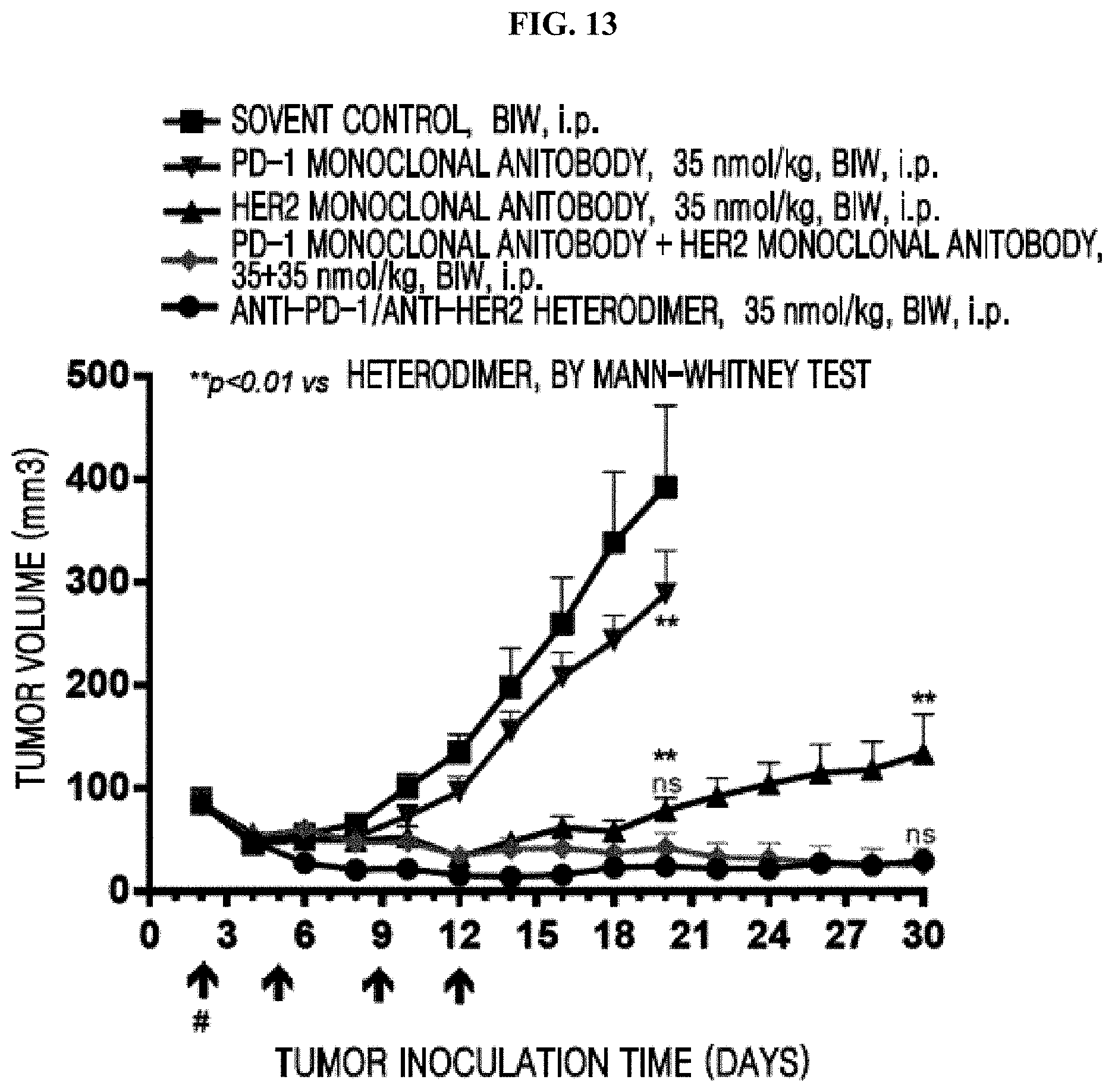
S00001
XML
uspto.report is an independent third-party trademark research tool that is not affiliated, endorsed, or sponsored by the United States Patent and Trademark Office (USPTO) or any other governmental organization. The information provided by uspto.report is based on publicly available data at the time of writing and is intended for informational purposes only.
While we strive to provide accurate and up-to-date information, we do not guarantee the accuracy, completeness, reliability, or suitability of the information displayed on this site. The use of this site is at your own risk. Any reliance you place on such information is therefore strictly at your own risk.
All official trademark data, including owner information, should be verified by visiting the official USPTO website at www.uspto.gov. This site is not intended to replace professional legal advice and should not be used as a substitute for consulting with a legal professional who is knowledgeable about trademark law.