Tau Immunotherapy
SEUBERT; Peter ; et al.
U.S. patent application number 16/933792 was filed with the patent office on 2021-02-04 for tau immunotherapy. This patent application is currently assigned to PROTHENA BIOSCIENCES LIMITED. The applicant listed for this patent is PROTHENA BIOSCIENCES LIMITED. Invention is credited to Robin BARBOUR, Philip James DOLAN, III, Yue LIU, Peter SEUBERT.
| Application Number | 20210032319 16/933792 |
| Document ID | / |
| Family ID | 1000005164060 |
| Filed Date | 2021-02-04 |


View All Diagrams
| United States Patent Application | 20210032319 |
| Kind Code | A1 |
| SEUBERT; Peter ; et al. | February 4, 2021 |
TAU IMMUNOTHERAPY
Abstract
The invention provides antibodies to tau. The antibodies inhibit or delay tau-associated pathologies and associated symptomatic deterioration.
| Inventors: | SEUBERT; Peter; (San Francisco, CA) ; DOLAN, III; Philip James; (Foster City, CA) ; LIU; Yue; (Foster City, CA) ; BARBOUR; Robin; (Walnut Creek, CA) | ||||||||||
| Applicant: |
|
||||||||||
|---|---|---|---|---|---|---|---|---|---|---|---|
| Assignee: | PROTHENA BIOSCIENCES
LIMITED DUBLIN 2 IE |
||||||||||
| Family ID: | 1000005164060 | ||||||||||
| Appl. No.: | 16/933792 | ||||||||||
| Filed: | July 20, 2020 |
Related U.S. Patent Documents
| Application Number | Filing Date | Patent Number | ||
|---|---|---|---|---|
| 16092439 | Oct 9, 2018 | 10752679 | ||
| PCT/IB2017/052543 | May 2, 2017 | |||
| 16933792 | ||||
| 62330786 | May 2, 2016 | |||
| Current U.S. Class: | 1/1 |
| Current CPC Class: | C07K 2317/92 20130101; A61P 25/28 20180101; C07K 2317/24 20130101; C07K 2317/94 20130101; C07K 16/18 20130101 |
| International Class: | C07K 16/18 20060101 C07K016/18; A61P 25/28 20060101 A61P025/28 |
Claims
1-35. (canceled)
36. An antibody comprising a heavy chain comprising a mature heavy chain variable region having the amino acid sequence of SEQ ID NO:35 linked to heavy constant region having the sequence of SEQ ID NO:29 with or without the C-terminal lysine and a light chain a mature light chain variable region having the amino sequence of SEQ ID NO:36 linked to a light chain constant region having the amino acid sequence of SEQ ID NO:32.
37-52. (canceled)
53. A nucleic acid encoding the heavy and light chains of an antibody as described in claim 36.
54-65. (canceled)
66. A pharmaceutical composition comprising an antibody of claim 36 and a pharmaceutically acceptable carrier.
67. A method of treating or effecting prophylaxis of Alzheimer's disease comprising administering an effective regime of an antibody as defined in claim 36 and thereby treating or effecting prophylaxis of Alzheimer's disease.
68. The method of claim 67, wherein the patient is an ApoE4 carrier.
69. A method of treating or effecting prophylaxis of a disease associated with tau comprising administering an effective regime of an antibody as defined in claim 36 and thereby treating or effecting prophylaxis of the disease.
70. The method of claim 69, wherein the disease is a neurological disease.
71-77. (canceled)
78. Nucleic acids encoding the heavy and light chains of an antibody of claim 36.
79. A host cell comprising the nucleic acid of claim 53.
80. A host cell comprising the nucleic acids of claim 78.
81. The host cell of claim 79, wherein the cell is a CHO or Sp2/0 cell.
82. The host cell of claim 80, wherein the cell is a CHO or Sp2/0 cell.
83. A method of producing an antibody comprising, culturing the host cell of claim 79, wherein the antibody is expressed from the nucleic acid.
84. A method of producing an antibody comprising, culturing the host cell of claim 80, wherein the antibody is expressed from the nucleic acids.
Description
CROSS-REFERENCE TO RELATED APPLICATIONS
[0001] The present application claims priority to US Provisional Application No. 62/330,786 filed May 2, 2016 and is related to US Provisional Application Nos. 61/780,624 filed Mar. 13, 2013 and 61/800,382, filed Mar. 15, 2013, and U.S. Ser. No. 14/776,724 each incorporated by reference in its entirely for all purposes.
SEQUENCE LISTING
[0002] This application includes a sequence listing submitted herewith as a text filed named "497106 SEQLST.TXT" created on May 2, 2017, and containing 54,309 bytes. The material contained in this text file is incorporated by reference in its entirety for all purposes.
BACKGROUND
[0003] Tau is a well-known human protein that can exist in phosphorylated forms (see, e.g., Goedert, Proc. Natl. Acad. Sci. U.S.A. 85:4051-4055(1988); Goedert, EMBO J. 8:393-399(1989); Lee, Neuron 2:1615-1624(1989); Goedert, Neuron 3:519-526(1989); Andreadis, Biochemistry 31:10626-10633(1992). Tau has been reported to have a role in stabilizing microtubules, particularly in the central nervous system. Total tau (t-tau, i.e., phosphorylated and unphosphorylated forms) and phospho-tau (p-tau, i.e., phosphorylated tau) are released by the brain in response to neuronal injury and neurodegeneration and have been reported to occur an increased levels in the CSF of Alzheimer's patients relative to the general population (Jack et al., Lancet Neurol 9: 119-28 (2010)).
[0004] Tau is the principal constituent of neurofibrillary tangles, which together with plaques are a hallmark characteristic of Alzheimer's disease. The tangles constitute abnormal fibrils measuring 10 nm in diameter occurring in pairs wound in a helical fashion with a regular periodicity of 80 nm. The tau within neurofibrillary tangles is abnormally phosphorylated (hyperphosphorylated) with phosphate groups attached to specific sites on the molecule. Severe involvement of neurofibrillary tangles is seen in the layer 11 neurons of the entorhinal cortex, the CA1 and subicular regions of the hippocampus, the amygdala, and the deeper layers (layers III, V, and superficial VI) of the neocortex in Alzheimer's disease. Hyperphosphorylated tau has also been reported to interfere with microtubule assembly, which may promote neuronal network breakdown.
[0005] Tau inclusions are part of the defining neuropathology of several neurodegenerative diseases including Alzheimer's disease, frontotemporal lobar degeneration, progressive supranuclear palsy and Pick's disease.
SUMMARY OF THE CLAIMED INVENTION
[0006] The invention provides an antibody comprising a mature heavy chain variable region having an amino acid sequence at least 90% identical to SEQ ID NO:15 and a mature light chain variable region at least 90% identical to SEQ ID NO:22. Optionally, three Kabat CDRs of SEQ ID NO:15 and three Kabat CDRs of SEQ ID NO:22. Optionally, at least one of positions H13, H28, H48 and H91 is occupied by K, P, M and F respectively and at least one of positions L1, L4, L36 and L43 is occupied by N, L, F and S respectively. Optionally, positions H13, H28, H48 and H91 are occupied by K, P, M and F respectively and al least two of positions L1, L4, L36 and L43 is occupied by N, L, F and S respectively. Optionally, positions H13, H28, H48 and H91 are occupied by K, P, M and F respectively, and at least three of positions L1, L4, L36 and L43 are occupied by N, L, F and S respectively. Optionally, positions H13, H28, H48 and H91 are occupied by K, P, M and F respectively, and positions L1, L4, L36 and L43 are occupied by N, L, F and S respectively. Optionally, the antibody comprises a mature heavy chain variable region having an amino acid sequence at least 95% identical to SEQ ID NO:15 and a mature light chain variable region at least 95% identical to SEQ ID NO:22. Optionally any differences in CDRs of the mature heavy chain variable region and mature light variable region from SEQ ID NOS: 15 and 22 respectively reside in positions 1160-1165. Optionally, the mature heavy chain variable region has an amino acid sequence designated SEQ ID NO:15 and the mature light chain variable region has an amino acid sequence designated SEQ ID NO:21, 22, or 23. Optionally, the mature heavy chain variable region has an amino acid sequence designated SEQ ID NO:15 and the mature light chain variable region has an amino acid sequence designated SEQ ID NO:22.
[0007] The invention further provides an antibody comprising a mature heavy chain variable region having an amino acid sequence at least 9% identical to SEQ ID NO:35 and a mature light chain variable region at least 90% identical to SEQ ID NO:36 provided position H1 is E and/or position L9 is S. Optionally, the antibody comprises three Kabat CDRs of SEQ ID NO:15 and three Kabat CDRs of SEQ ID NO:22. Optionally, at least one of position H13, H28, H48 and H91 is occupied by K, P, M and F respectively and at least one of positions L1, L4, L36 and L43 is occupied by N, L, F and S respectively. Optionally, positions H13, H28, H48 and H91 are occupied by K, P, M and F respectively and at least two of positions L1, L4, L36 and L43 is occupied by N, L, F and S respectively. Optionally, posit ions H13, H28, H48 and H91 are occupied by K, P, M and F respectively, and at least three of positions L1, L4, L36 and L43 are occupied by N, L, F and S respectively. Optionally, positions H13, H28, H48 and H91 are occupied by K, P, M and F respectively, and positions L1, L4, L36 and L43 are occupied by N, L, F and S respectively. Optionally, positions H13, H28, H48 and H91 are occupied by K, P, M and F respectively, and positions L1, L4, L36 and L43 are occupied by D, L, F and S respectively. Optionally, position H1 is occupied by E. Optionally, position L9 is occupied by S. Optionally, position H1 is E and position L9 is S. Optionally, the antibody comprises a mature heavy chain variable region having an amino acid sequence at least 95% identical to SEQ ID NO:35 and a mature light chain variable region at least 95% identical to SEQ ID NO:36.
[0008] In some antibodies, the mature heavy chain variable region is fused to a heavy chain constant region and the mature light chain variable region is fused to a light chain constant region. Optionally, the heavy chain constant region is a mutant form of natural human constant region which has reduced binding to an Fc.gamma. receptor relative to the natural human constant region. Optionally, the heavy chain constant region is of IgG1 isotype, optionally SEQ ID NO:29, provided the C-terminal lysine can be missing and the light chain constant region is kappa, preferably SEQ ID NO:32.
[0009] Some antibodies are conjugated to a cytotoxic or cytostatic agent. Some antibodies are Fab fragments.
[0010] The invention further provides a nucleic acid encoding the heavy and/or light chains of an antibody as described in any of the above antibodies.
[0011] The invention further provides a method of treating or effecting prophylaxis of Alzheimer's disease comprising administering an effective regime of any of the above antibodies and thereby treating or effecting prophylaxis of Alzheimer's disease. Optionally, the patient is an ApoE4 carrier.
[0012] The invention further provides a method of treating or effecting prophylaxis of a disease associated with tau comprising administering an effective regime of an antibody as defined in any of the above antibodies. Optionally, the disease is a neurological disease.
[0013] The invention further provides a method of reducing aberrant transmission of tau comprising administering an effective regime of any of the above antibodies, and thereby reducing transmission of tau.
[0014] The invention further provides a method of inducing phagocytosis of tau comprising administering an effective regime of any of the above antibodies and thereby inducing phagocytosis of tau. Optionally, the disease is a neurological disease.
[0015] The invention further provides a method of inhibiting tau aggregation or deposition comprising administering an effective regime of any of the above antibodies thereby inhibiting tau aggregation or deposition. Optionally, the disease is a neurological disease.
[0016] The invention further provides a method of inhibiting formation of tau tangles comprising administering an effective regime of an antibody of any of the above antibodies.
[0017] Optionally, the disease is a neurological disease.
[0018] The invention further provides a nucleic acid comprising a segment encoding a heavy chain variable region having the sequence of SEQ ID NO: 15 or SEQ ID NO:35. Optionally, the nucleic acid further comprises a segment encoding an IgG1 constant region. Optionally, the IgG1 constant region is a human IgG1 constant region. Optionally, the IgG1 constant region has a sequence of SEQ ID NO: 29 provided the C-terminal lysine can be omitted. Optionally, the segment encoding the IgG1 constant region has a nucleotide sequence of SEQ ID NO: 30. Optionally, the nucleic acid further comprises an intron linking the segments encoding the heavy chain variable region and the IgG1 constant region. Optionally, the segment encoding the IgG1 constant region has a nucleotide sequence of SEQ ID NO: 31. Optionally, the nucleic acid further comprising a segment encoding a kappa constant region. Optionally, the kappa constant region is a human kappa constant region. Optionally, the kappa constant region has the sequence of SEQ ID NO:32. Optionally, the nucleic acid encoding the kappa constant region has the sequence of SEQ ID NO:33. Optionally, the nucleic acid further comprises an intron linking the segment encoding the light chain variable region to the segment encoding the kappa constant region. Optionally, the segment encoding the kappa constant region has the sequence of SEQ ID NO:34.
[0019] The invention further provides nucleic acid(s) encoding the heavy chain variable region of SEQ ID NO:15 and/or the light chain variable region of SEQ ID NO:21, 22 or 23.
BRIEF DESCRIPTION OF THE DRAWINGS
[0020] FIG. 1 depicts the results of experiments designed to map the epitope(s) bound by the 16B5 monoclonal antibody. Western blots containing full-length Tau or deletion mutants of Tau (.DELTA.5-24 or .DELTA.25-44) were stained with 16B5 antibodies (left panel) or Tau46 antibodies (right panel). The Tau46 antibody hinds to the C-terminal epitope of Tau.
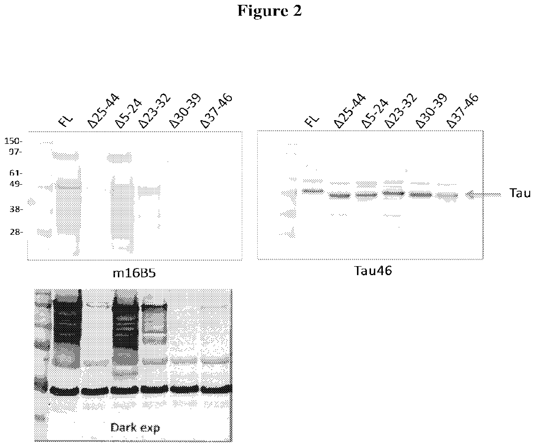
[0021] FIG. 2 depicts the results of experiments designed to map the epitope(s) bound by the 16B5 monoclonal antibody. Western blots containing full-length Tau or deletion mutants of Tau were stained with 16B5 antibodies (upper left panel) or Tau46 antibodies (right panel). A longer exposure of the blot stained with 16B5 antibodies is shown in the lower left panel. The deletion mutants of Tau analyzed in this experiment include A25-44, A5-24, A23-32, A30-39, and A37-46.
[0022] FIG. 3 depicts the results of an alanine scanning experiment designed to map the epitope(s) bound by the 16B5 monoclonal antibody. Western blots containing wild-type Tau (WT) or alanine point mutants of Tau were stained with 16B5 antibodies (left panel) or Tau46 antibodies (right panel). The alanine mutants of Tau analyzed in this experiment include T30A, M31A, H32A, Q33A, D34A, Q35A, E36A, G37A, D38A, T39A, D40A, A41L, and G42A.
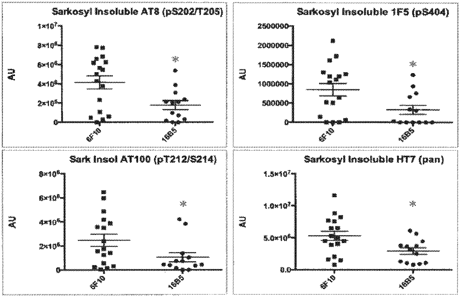
[0023] FIG. 4 shows relative amounts of tau protein detected in a sarkosyl insoluble fraction of the brainstem of transgenic mice that express the human tau.P301L protein. The mice were passively immunized with either the 16B5 antibody or the 6F10 antibody, a non-immune IgG isotype control. Samples were analyzed by Western blotting, antibody staining, and quantification of the resulting signal. Antibodies used to detect tau included anti-phospho-tau specific antibodies (AT8, upper left panel; AT100, lower left panel; or lF5, upper right panel) and a pan tau antibody (HT7, lower right panel).
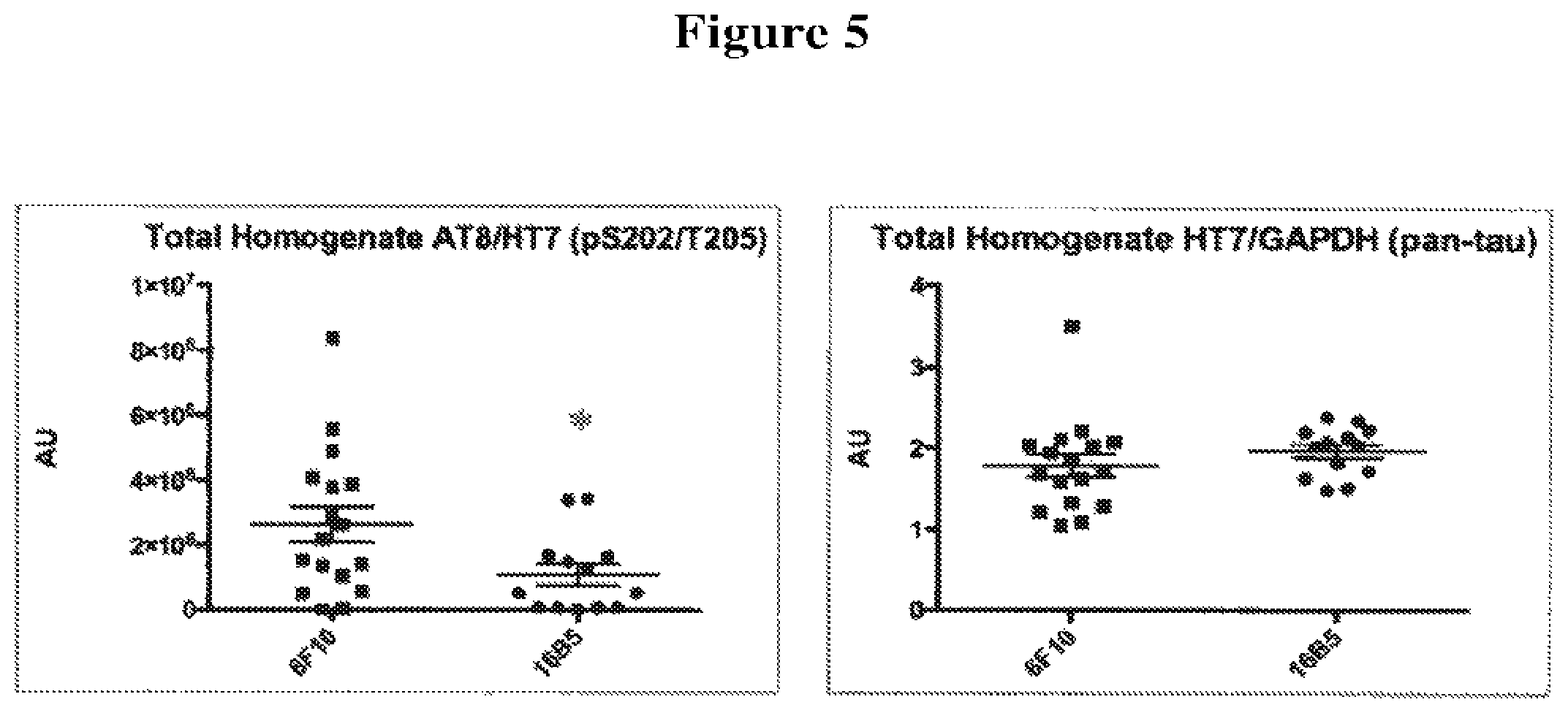
[0024] FIG. 5 shows the ratio of phospho-tau to total tau protein (left panel) and a normalized amount of total tau (right panel) detected in total brainstem homogenates of transgenic mice that express the human tau.P301 L protein. The mice were passively immunized with either the 16B5 antibody or the 6F10 antibody, a non-immune IgG1 isotype control. Samples were analyzed by Western blotting, antibody staining, and quantification of the resulting signal. The AT8 antibody was used to detect phospho-tau and the HT7 antibody was used to detect total tau. An anti-GAPDH antibody was used to normalize the amount of tau detected in mice treated with the 16B5 antibody versus the control 6F10 antibody.
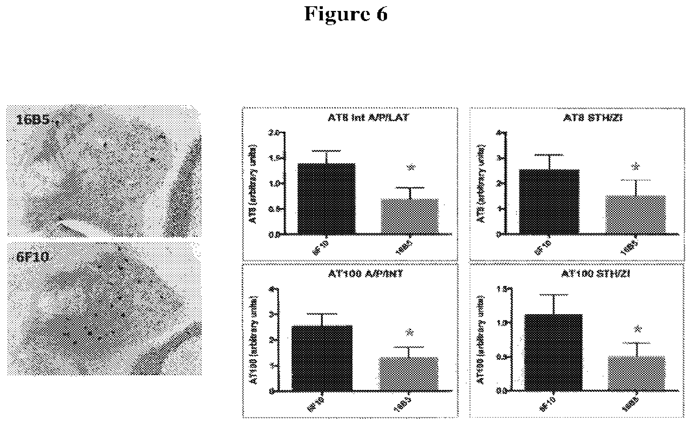
[0025] FIG. 6 depicts sections of cerebellar nuclei of transgenic mice that express the human tau.P301L protein, immunohistochemically stained using the AT8 anti-phospho-tau antibody. The mice were passively immunized with either the 16B5 antibody (upper left panel) or the 6F10 antibody (lower left panel), a non-immune IgG1 isotype control. Quantification of the amount of tau staining detected with the AT8 antibody in the interposed nucleus of the cerebellum, anterior and posterior part, annex lateral cerebellar nucleus (IntA/P/LAT) and the subthalamic nucleus annex zona incerta (STH/ZI) from mice passively immunized with 16B5 or 6F10 antibodies is shown in the upper bar graph panels. Quantification of the amount of phospho-tau staining deleted using the AT100 anti-phospho-tau antibody on IA/P/LAT and STH/ZI sections from mice passively immunized with 16B5 or 6F10 antibodies is shown in the lower bar graph panels. Statistical significance was assessed using the Student's t test, p<0.05.
[0026] FIG. 7 depicts tau immunoprecipitation results obtained with chimeric 16B5 antibodies and humanized 16B5 antibodies (H1L2 and H1L3 versions). Tau was immunoprecipitated from both soluble and insoluble fractions of postmortem frontal cortex samples obtained from an Alzheimer disease patient. Tau present in blotted immuno-precipitates was detected using a polyclonal anti-tau antibody (tau pAb).
[0027] FIG. 8 depicts the sequence alignment of humanized H1, H2, and chimeric 16B5 heavy chain and the sequence alignment of humanized L2, L4, and chimeric 16B5 light chain.
[0028] FIG. 9 depicts the thermostability analysis of H1L2, H1L4, and H2L4. Thermostability was analyzed using Differential Scanning Calorimetry (DSC).
DEFINITIONS
[0029] Monoclonal antibodies and other therapeutic agents are typically provided in isolated form. This means that the agent is typically at least 50% w/w pure of interfering proteins and other contaminants arising from its production or purification but does not exclude the possibility that the agent is combined with an excess of pharmaceutical acceptable carrier(s) or other vehicle intended to facilitate its use. Sometimes monoclonal antibodies (or other therapeutic agents) are at least 6%, 70%, 80%, 90%, 95% or 99% w/w pure of interfering proteins and contaminants from production or purification.
[0030] Antibodies of the invention typically bind to their designated target with an association constant of at least 10.sup.6, 10.sup.7, 10.sup.8, 10.sup.9, or 10.sup.10 M.sup.1. Such binding is specific binding in that it is delectably higher in magnitude and distinguishable from non-specific binding occurring to at least one unrelated target. Specific binding can be the result of formation of bonds between particular functional groups or particular spatial fit (e.g., lock and key type) whereas nonspecific binding is usually the result of van der Waals forces. Specific binding does not however necessarily imply that a monoclonal antibody binds one and only one target.
[0031] The basic antibody structural unit is a tetramer of subunits. Each tetramer includes two identical pairs of polypeptide chains, each pair having one "light" (about 25 kDa) and one "heavy" chain (about 50-70 kDa). The amino-terminal portion of each chain includes a variable region of about 100 to 110 or more amino acids primarily responsible for antigen recognition. This variable region is initially expressed linked to a cleavable signal peptide. The variable region without the signal peptide is sometimes referred to as a mature variable region. Thus, for example, a light chain mature variable region, means a light chain variable region without the light chain signal peptide. The carboxy-terminal portion of each chain defines a constant region primarily responsible for effector function. A constant region can include any or all of a CH1 region, hinge region, CH2 region and CH3 region.
[0032] Light chains are classified as either kappa or lambda. Heavy chains are classified as gamma, mu, alpha, delta, or epsilon, and define the antibody's isotype as IgG, IgM, IgA, IgD and IgE, respectively. Within light and heavy chains, the variable and constant regions are joined by a "J" region of about 12 or more amino acids, with the heavy chain also including a "D" region of about 10 or more amino acids. (See generally. Fundamental Immunology (Paul, W., ed., 2nd ed. Raven Press, N.Y., 1989), Ch. 7)(incorporated by reference in its entirely for all purposes).
[0033] The mature variable regions of each light/heavy chain pair form the antibody binding site. Thus, an intact antibody has two binding sites. Except in bifunctional or bispecific antibodies, the two binding sites are the same. The chains all exhibit the same general structure of relatively conserved framework regions (FR) joined by three hypervariable regions, also called complementarily determining regions or CDRs. The CDRs from the two chains of each pair are aligned by the framework regions, enabling binding to a specific epitope. From N-terminal to C-terminal, both light and heavy chains comprise the domains FR1, CDR1, FR2, CDR2, FR3, CDR3 and FR4. The assignment of amino acids to each domain is in accordance with the definitions of Kabat, Sequences of Protein of Immunological Interest (National Institutes of Health, Bethesda, Md., 1987 and 1991), or Chothia & Lesk, J. Mol. Biol. 196:901-917 (1987); Chothia et al., Nature 342:878-883 (1989). Kabat also provides a widely used numbering convention (Kabat numbering) in which corresponding residues between different heavy chains or between different light chains are assigned the same number.
[0034] The term "antibody" includes intact antibodies and binding fragments thereof. Typically, fragments compete with the intact antibody from which they were derived for specific binding to the target. Fragments include separate heavy chains, light chains Fab, Fab', F(ab').sub.2, F(ab)c, Fv and single domain antibodies. Single (variable) domain antibodies include VII regions separated from their VL partners (or vice versa) in conventional antibodies (Ward et al., 1989, Nature 341: 544-546) as well as VH regions (sometimes known as VHH) from species such as Camclidae or cartilaginous fish (e.g., a nurse shark) in which VII regions are not associated with VL regions (see, e.g., WO 9404678). Single domain antibodies in which one chain is separated from its natural partners are sometimes known as Dabs and single domain antibodies from Caemelidae or cartilaginous fish are sometimes known as nanobodies. Constant regions or parts of constant regions may or may not be present in single domain antibodies. For example, natural single variable region antibodies from Camelidae include a VHH variable region, and CH2 and CH3 constant regions. Single domain antibodies can be subject of humanization by analogous approaches to conventional antibodies. The Dabs type of antibodies are usually obtained from antibodies of human origin. NANOBODY types of antibody are of Camelidae or shark origin and can be subject to humanization. Fragments can be produced by recombinant DNA techniques, or by enzymatic or chemical separation of intact immunoglobulins. The term "antibody" also includes a bispecific antibody. A bispecific or bifunctional antibody is an artificial hybrid antibody having two different heavy/light chain pairs and two different binding sites (see, e.g., Songsivilai and Lachmann, Clin. Exp. Immunol., 79:315-321 (1990); Kostelny et al., J. Immunol., 148:1547-53 (1992)).
[0035] The term "epitope" refers to a site on an antigen to which an antibody binds. An epitope can be formed from contiguous amino acids or noncontiguous amino acids juxtaposed by tertiary folding of one or more proteins. Epitopes formed from contiguous amino acids are typically retained on exposure to denaturing solvents whereas epitopes formed by tertiary folding are typically lost on treatment with denaturing solvents. An epitope typically includes at least 3, and more usually, at least 5 or 8-10 amino acids in a unique spatial conformation. When an epitope is said to be within a range of amino acid residues in a protein (e.g., within residues 25 to 44 of tau), the range is inclusive of the residues defining its borders. Certain residues within the range contribute to the epitope, whereas others may not. The residues that form the epitope may or may not be contiguous with one another. Similarly, when an antibody binds to an epitope found within a particular range of amino acids, the antibody need not contact all the amino acids residues within the range, and the residues of the epitope that are contacted by the antibody may or may not be contiguous with one another. Methods of determining spatial conformation of epitopes include, for example, x-ray crystallography and 2-dimensional nuclear magnetic resonance. See, e.g., Epitope Mapping Protocols, in Methods in Molecular Biology, Vol. 66, Glenn E. Morris, Ed. (1996).
[0036] Antibodies that recognize the same or overlapping epitopes can be identified in a simple immunoassay showing the ability of one antibody to compete with the binding of another antibody to a target antigen. The epitope of an antibody can also be defined by X-ray crystallography of the antibody bound to its antigen to identify contact residues. Alternatively, two antibodies have the same epitope if all amino acid mutations in the antigen that reduce or eliminate binding of one antibody reduce or eliminate binding of the other. Two antibodies have overlapping epitopes if some amino acid mutations that reduce or eliminate binding of one antibody reduce or eliminate binding of the other. The invention includes antibodies that compete with 16B5 and/or which bind to the same epitope on tau as 16B5.
[0037] Competition between antibodies is determined by an assay in which an antibody under test inhibits specific binding of a reference antibody (e.g. 16B5) to a common antigen (see, e.g., Junghans et al., Cancer Res. 50:1495, 1990). A test antibody competes with a reference antibody if an excess of a test antibody (e.g., at least 2.times., 5.times., 10.times., 20.times. or 100.times.) inhibits binding of the reference antibody by at least 50% but preferably 75%, 90% or 99% as measured in a competitive binding assay. Antibodies identified by competition assay (competing antibodies) include antibodies binding to the same epitope as the reference antibody and antibodies binding to an adjacent epitope sufficiently proximal to the epitope bound by the reference antibody for steric hindrance to occur.
[0038] The term "patient" includes human and other mammalian subjects that receive either prophylactic or therapeutic treatment.
[0039] For purposes of classifying amino acids substitutions as conservative or nonconservative, amino acids are grouped as follows: Group 1 (hydrophobic side chains): met, ala, val, leu, ile; Group II (neutral hydrophilic side chains): cys, ser, thr; Group III (acidic side chains): asp, glu; Group IV (basic side chains): asn, gln, his, lys, arg; Group V (residues influencing chain orientation): gly, pro; and Group VI (aromatic side chains): trp, tyr, phe. Conservative substitutions involve substitutions between amino acids in the same class. Non-conservative substitutions constitute exchanging a member of on of these classes for a member of another.
[0040] Percentage sequence identities are determined with antibody sequences maximally aligned by the Kabat numbering convention. After alignment, if a subject antibody region (e.g., the entire mature variable region of a heavy or light chain) is being compared with the same region of a reference antibody, the percentage sequence identity between the subject and reference antibody regions is the number of positions occupied by the same amino acid in both the subject and reference antibody region divided by the total number of aligned positions of the two regions, with gaps not counted, multiplied by 100 to convert to percentage.
[0041] The term "adjuvant" refers to a compound that when administered in conjunction with an antigen augments and/or redirects the immune response to the antigen, but when administered alone does not generate an immune response to the antigen. Adjuvants can augment an immune response by several mechanisms including lymphocyte recruitment, stimulation of B and/or T cells, and stimulation of macrophages.
[0042] A disease is associated with tau if a population of patients with the disease have increased levels of tau in the brain, or increased deposition or inclusions of tau, or the presence of tau tangles in the brain, or increased phosphorylation of tau in the brain (average number of phosphate groups per molecule tau), or aberrant intercellular or intracellular transmission of tan compared with a population of subjects not known to have a neurological disease. A disease is also associated with tau if patients with a variant form of a tau gene have an increased risk of developing the disease relative to patients with a wildtype (most frequently occurring variant in a human population) tau gene.
[0043] An individual is at increased risk of a disease if the subject has at least one known risk-factor (e.g., genetic, biochemical, family history, situational exposure) placing individuals with that risk factor at a statistically significant greater risk of developing the disease than individuals without the risk factor.
[0044] The term "symptom" refers to a subjective evidence of a disease, such as altered gait, as perceived by the patient. A "sign" refers to objective evidence of a disease as observed by a physician.
[0045] Statistical significance means p.ltoreq.0.05.
DETAILED DESCRIPTION
I. General
[0046] The invention provides antibodies that bind to tau. Some antibodies specifically hind to an epitope within residues 23-46 of SEQ ID NO. 1. Some antibodies bind to tau irrespective of phosphorylation state. Some antibodies of the invent ion serve to inhibitor delay tau-associated pathologies and associated symptomatic deterioration. Although an understanding of mechanism is not required for practice of the invention, a reduction in toxicity may occur as a result of the antibody inducing phagocytosis of tau, inhibiting tau from inter or intramolecular aggregation, blocking cell-to-cell transmission, blocking tau binding to cells, blocking tau uptake, or from binding to other molecules, by stabilizing a non-toxic conformation, or by inhibiting intercellular or intracellular transmission of pathogenic tau forms, among other mechanisms. The antibodies of the invention or agents that induce such antibodies can be used in methods of treating or effecting prophylaxis of Alzheimer's and other diseases associated with tau.
II. Tau
[0047] Unless otherwise apparent from the context, reference to tau means a natural human form of tau including all isoforms irrespective of whether posttranslational modification (e.g., phosphorylation, glycation, or acetylation) is present. There are six major isoforms (splice variants) of tau occurring in the human brain. The longest of these variants has 441 amino acids, of which the initial met residue is cleaved. Residues are numbered according to the 441 isoform. Thus, for example, reference to a phosphorylation at position 404 means position 404 of the 441 isoform, or corresponding position of any other isoform when maximally aligned with the 441 isoform. The amino acid sequences of the isoforms and Swiss-Prot numbers are indicated below.
[0048] Reference to tau includes known natural variations about 30 of which are listed in the Swiss-Pro database and permutations thereof, as well as mutations associated with tau pathologies, such as dementia, Pick's disease, supranuclear palsy, etc. (see, e.g., Swiss-Pro database and Poorkaj, et al. Ann Neurol. 43:815-825 (1998)). Some examples of tau mutations numbered by the 441 isoform are a lysine to threonine mutation at amino acid residue 257 (K257T), an isoleucine to valine mutation at amino acid position 260 (I260V); a glycine to valine mutation at amino acid position 272 (G272V); an asparagine to lysine mutation at amino acid posit ion 279 (N279K); an asparagine to histidine mutation at amino acid position 296 (N296H); a proline to serine mutation al amino acid position 301 (P301S); a glycine to valine mutation at amino acid position 303 (G303V); a serine to asparagine mutation at position 305 (S305N); a glycine to serine mutation at amino acid position 335 (G335S); a valine to methionine mutation at position 337 (V337M); a glutamic acid to valine mutation at position 342 (E342V); a lysine to isoleucine mutation at amino acid position 369 (K3691); a glycine to arginine mutation at amino acid position 389 (G389R); and an arginine to tryptophan mutation at amino acid position 406 (R406W).
[0049] Tau can be phosphorylated at one or more amino acid residues including tyrosine at amino acid positions 18, 29, 97, 310, and 394 serine at amino acid positions 184, 185, 198, 199, 202, 208, 214, 235, 237, 238, 262, 293, 324, 356, 396, 400, 404, 409, 412, 413, and 422; and threonine at amino acids positions 175, 181, 205, 212, 217, 231, and 403.
III. Antibodies
[0050] A. Binding Specificity and Functional Properties
[0051] The invention provides antibodies that bind to tau. Some antibodies specifically hind to an epitope within residues 23-46 of SEQ ID NO:1. Some antibodies specifically bind to an epitope within residues 25-44 of SEQ ID NO:1. Some antibodies specifically bind to an epitope within 28-41 of SEQ ID NO:1. Some antibodies specifically bind to an epitope within residues 30-39 of SEQ ID NO:1. Some antibodies specifically bind to an epitope within residues 30-36 of SEQ ID NO:1. Some antibodies specifically bind to an epitope within residues 33-39 of SEQ ID NO:1. Sore antibodies specifically hind to an epitope within residues 33-36 of SEQ ID NO:1. Some antibodies specifically bind to an epitope including residues 28-30, 28-31, 28-32, 28-33, 28-34, 28-35, 28-36, 28-37, 28-38, 28-39, 28-40, 28-41, 29-31, 29-32, 29-33, 29-34, 29-35, 29-36, 29-37, 29-38, 29-39, 29-40, 29-41, 30-32, 30-33, 30-34, 30-35, 30-36, 30-37, 30-38, 30-39, 30-40, 30-41, 31-33, 31-34, 31-35, 31-36, 31-37, 31-38, 31-39, 31-40, 31-41, 32-34, 32-35, 32-36, 32-37, 32-38, 32-39, 32-40, 32-41, 33-35, 33-36, 33-37, 33-38, 33-39, 33-40, 33-41, 34-36, 34-37, 34-38, 34-39, 34-40, 34-41, 35-37, 35-38, 35-39, 35-40, 35-41, 36-38, 36-39, 36-40, 36-41 of SEQ ID NO:1. Some antibodies bind to tau irrespective of phosphorylation state. Some antibodies bind to an epitope not including a residue subject to phosphorylation. These antibodies can be obtained by immunizing with a tau polypeptide purified from a natural source or recombinantly expressed. Antibodies can be screened for binding tau in unphosphorylated form as well as a form in which one or more residues susceptible to phosphorylation are phosphorylated. Such antibodies preferably bind with indistinguishable affinities or at least within a factor of 1.5, 2 or 3-fold to phosphorylated tau compared to non-phosphorylated tau (i.e., are "pan-specific). 16B5 is an example of a pan-specific monoclonal antibody. The invention also provides antibodies binding to the same epitope as any of the foregoing antibodies, such as, for example, the epitope of 16B5. Also included are antibodies competing for binding to tau with any of the foregoing antibodies, such as, for example, competing with 16B5.
[0052] Other antibodies can be obtained by mutagenesis of cDNA encoding the heavy and light chains of an exemplary antibody, such as 16B5. Monoclonal antibodies that are at least 90%, 95% or 99% identical to 16B5 in amino acid sequence of the mature heavy and/or light chain variable regions and maintain its functional properties, and/or which differ from the respective antibody by a small number of functionally inconsequential amino acid substitutions (e.g., conservative substitutions), deletions, or insertions are also included in the invention. Monoclonal antibodies having at least one and preferably all six CDR(s) as defined by Kabat that are 90%, 95%, 99% or 100% identical to corresponding CDRs of 16B5 are also included.
[0053] C. Humanized Antibodies
[0054] A humanized antibody is a genetically engineered antibody in which the CDRs from a non-human "donor" antibody (e.g., 16B5) are grafted into human "acceptor" antibody sequences (see, e.g., Queen, U.S. Pat. Nos. 5,530,101 and 5,585,089; Winter, U.S. Pat. No. 5,225,539, Carter, U.S. Pat. No. 6,407,213, Adair, U.S. Pat. No. 5,859,205 6,881,557, Foote, U.S. Pat. No. 6,881,557). The acceptor antibody sequences can be, for example, a mature human antibody sequence, a composite of such sequences, a consensus sequence of human antibody sequences, or a germline region sequence. Thus, a humanized antibody is an antibody having some or all CDRs entirely or substantially from a donor antibody and variable region framework sequences and constant regions, if present, entirely or substantially from human antibody sequences. Similarly a humanized heavy chain has at least one, two and usually all three CDRs entirely or substantially from a donor antibody heavy chain, and a heavy chain variable region framework sequence and heavy chain constant region, if present, substantially from human heavy chain variable region framework and constant region sequences. Similarly a humanized light chain has at least one, two and usually all three CDRs entirely or substantially from a donor antibody light chain, and a light chain variable region framework sequence and light chain constant region, if present, substantially from human light chain variable region framework and constant region sequences. Other than nanobodies and dAbs, a humanized antibody comprises a humanized heavy chain and a humanized light chain. A CDR in a humanized antibody is substantially from a corresponding CDR in a non-human antibody when at least 85%, 90%, 95% or 100% of corresponding residues (as defined by Kabat) are identical between the respective CDRs. The variable region framework sequences of an antibody chain or the constant region of an antibody chain are substantially from a human variable region framework sequence or human constant region respectively when at least 85, 90, 95 or 100% of corresponding residues defined by Kabat are identical.
[0055] Although humanized antibodies often incorporate all six CDRs (preferably as defined by Kabat) from a mouse antibody, they can also be made with less than all CDRs (e.g., at least 3, 4, or 5) CDRs from a mouse antibody (e.g., Pascalis et al., J. Immunol. 169:3076, 2002; Vajdos et al., Journal of Molecular Biology, 320: 415-428, 2002; Iwahashi et al., Mol. Immunol. 36:1079-1091, 1999; Tamura et al, Journal of Immunology, 164:1432-1441, 2000).
[0056] In some antibodies only part of the CDRs, namely the subset of CDR residues required for binding, termed the SDRs, are needed to retain binding in a humanized antibody. CDR residues not contacting antigen and not in the SDRs can be identified based on previous studies (for example residues H60-H65 in CDR H2 are often not required), from regions of Kabat CDRs lying outside Chothia hypervariable loops (Chothia, J. Mol. Biol. 196:901, 1987), by molecular modeling and/or empirically, or as described in Gonzales et al., Mol. Immunol. 41: 863, 2004. In such humanized antibodies at positions in which one or more donor CDR residues is absent or in which an entire donor CR is omitted, the amino acid occupying the position can be an amino acid occupying the corresponding position (by Kabat numbering) in the acceptor antibody sequence. The number of such substitutions of acceptor for donor amino acids in the CDRs to include reflects a balance of competing considerations. Such substitutions are potentially advantageous in decreasing the number of mouse amino acids in a humanized antibody and consequently decreasing potential immunogenicity. However, substitutions can also cause changes of affinity, and significant reductions in affinity are preferably avoided. Positions for substitution within CDRs and amino acids to substitute can also be selected empirically.
[0057] The human acceptor antibody sequences can optionally be selected from among the many known human antibody sequences to provide a high degree of sequence identity (e.g., 65-85% identity) between a human acceptor sequence variable region frameworks and corresponding variable region frameworks of a donor antibody chain.
[0058] Certain amino acids from the human variable region framework residues can be selected for substitution based on their possible influence on CDR conformation and/or binding to antigen. Investigation of such possible influences is by modeling, examination of the characteristics of the amino acids at particular locations, or empirical observation of the effects of substitution or mutagenesis of particular amino acids.
[0059] For example, when an amino acid differs between a murine variable region framework residue and a selected human variable region framework residue, the human framework amino acid can be substituted by the equivalent framework amino acid from the mouse antibody when it is reasonably expected that the amino acid: [0060] (1) noncovalently hinds antigen directly, [0061] (2) is adjacent to a CDR region, [0062] (3) otherwise interacts with a CDR region (e.g. is within about 6 .ANG. of a CDR region), (e.g., identified by modeling the light or heavy chain on the solved structure of a homologous known immunoglobulin chain); and [0063] (4) a residue participating in the VL-VH interface.
[0064] Framework residues from classes (1)-(3) as defined by Queen, U.S. Pat. No. 5,530,101 are sometimes alternately referred to as canonical and vernier residues. Framework residues defining canonical class of the donor CDR loops determining the conformation of a CDR loop are sometimes referred to as canonical residues (Chothia and Lesk, J. Mol. Biol. 196, 901-917 (1987), Thornton & Marlin J. Mol. Biol., 263, 800-815, 1996). A layer of framework residues that support antigen-binding loop conformations play a role in fine-tuning the fit of an antibody to antigen are sometimes referred to as vernier residues (Foote & Winter, 1992, J. Mol Bio. 224, 487 499). Other candidates for substitution are residues creating a potential glycosylation site. Other candidates for substitution are acceptor human framework amino acids that are unusual for a human immunoglobulin at that position. These amino acids can be substituted with amino acids from the equivalent position of the mouse donor antibody or from the equivalent positions of more typical human immunoglobulins. Other candidates for substitution are acceptor human framework amino acids that are unusual for a human immunoglobulin at that position.
[0065] The invention provides humanized forms of the mouse 16B5 antibody. The mouse antibody comprises mature heavy and light chain variable regions having amino acid sequences comprising SEQ ID NOS. 10 and 16 respectively. The invention provides two exemplified humanized mature heavy chain variable regions (H1 and H2) and five exemplified humanized mature light chain variable region (L1, L2, L3, L4, and L5). Ill includes four backmuations. H2 includes the same four backmutations plus a further Q1E mutation (not a backmutation) to improve stability. L1 has 3 backmutations, L2 four backmutations, L3 had three backmutations, L4 has five backmutations (the same as L2 plus D9S to remove a proteolytic site), and L5 has the same backmutations as L4 except that position L1 is occupied by D. The H1L2 variant has the sane or better affinity as a chimeric 16B5 and eight backmutations. H1L1 and H1L3 have similar affinity to chimeric 16B5 and seven backmutations. The H1L4 variant, H2L4 variant and H2L2 variants have affinities within a factor of 2 of 16B5 and nine, ten or eight mutations respectively (all of which are backmutations except Q1E in H2). These variants have the benefit of improved stability (from Q1E in H2) or removal of proteolytic site in L4 or both. All of the humanized chains have at least 85% sequence identity to human germline sequences and thus meet INN criteria for designation as humanized antibodies.
[0066] The invention provides variants of the H1L2 humanized 16B5 antibody in which the humanized mature heavy chain variable region shows at least 90%, 95%, 96%, 97%, 98% or 99% identity to SEQ ID NO:15 and the humanized mature light chain mature variable region shows at least 90%, 95%, 96%, 97% 98% or 99% sequence identity to SEQ ID NO:22. Preferably, in such antibodies some or all of the backmutations in H1L2 are retained. In other words, at least 1, 2, 3 or 4 of positions position H13 is occupied by K, position H28 occupied by position H48 is occupied by M and position H91 is occupied by S. Preferably at least, 1, 2, 3 or all four positions position L1 is occupied by N, position L4 is occupied by L, position L36 is occupied by F and position L43 is occupied by S. The CDR regions of such humanized antibodies are preferably identical or substantially identical to the CDR regions of H1L2, which are the same as those of the mouse donor antibody. The CDR regions can be defined by any conventional definition (e.g., Chothia) but are preferably as defined by Kabat.
[0067] The invention provides variants of the H1L4 humanized 16B5 antibody in which the humanized mature heavy chain variable region shows at least 90%, 95%, 96%, 97%, 98% or 99% identity to SEQ ID NO:15 and the humanized mature light chain mature variable region shows at least 9%, 95%, 96%, 97% 98% or 99% sequence identity to SEQ ID NO:36. Preferably, in such antibodies some or all of the backmutations in H1L4 are retained. In other words, at least 1, 2, 3 or 4 of the following positions are occupied as follows: position H13 is occupied by K, position H28 occupied by P, position H48 is occupied by M and position H91 is occupied by F. Preferably at least, 1, 2, 3, 4 or all 5 positions position L1 is occupied by N, position L4 is occupied by L, position L9 is occupied by S, position L36 is occupied by F and position L43 is occupied by S. The CDR regions of such humanized antibodies are preferably identical or substantially identical to the CDR regions of H1L4, which are the same as those of the mouse donor antibody. The CDR regions can be defined by any conventional definition (e.g., Chothia) but are preferably as defined by Kabat.
[0068] The invention provides variants of the H2L2 humanized 16B5 antibody in which the humanized mature heavy chain variable region shows at least 90%, 95%, 96%, 97%, 98% or 99% identity to SEQ ID NO:35 and the humanized mature light chain mature variable region shows at least 90%, 95%, 96%, 97% 98% or 99% sequence identity to SEQ ID NO:22. Preferably, in such antibodies some or all of the mutations in H21.4 are retained. In other words, at least 1, 2, 3, 4 or 5 of the following positions are occupied as follows: position H1 is occupied by E, position H13 is occupied by K, position H28 occupied by P, position H48 is occupied by M and position H91 is occupied by F. Preferably at least, 1, 2, 3, or all 4 of the following positions are occupied as follows: position L1 is occupied by N, position L4 is occupied by L, position L36 is occupied by F and position L43 is occupied by S. The CDR regions of such humanized antibodies are preferably identical or substantially identical to the CDR regions of H2L3, which are the same as those of the mouse donor antibody. The CDR regions can be defined by any conventional definition (e.g., Chothia) but are preferably as defined by Kabat.
[0069] The invention provides variants of the H2L4 humanized 16B5 antibody in which the humanized mature heavy chain variable region shows at least 90%, 95%, 96%, 97%, 98% or 99% identity to SEQ ID NO:35 and the humanized mature light chain mature variable region shows at least 90%, 95%, 96%, 97% 98% or 99% sequence identity to SEQ ID NO:36. Preferably, in such antibodies some or all of the backmutations and other mutations in H21.4 are retained. In other words, at least 1, 2, 3, 4 or 5 of the following positions are occupied as follows: position H is occupied by E, position H13 is occupied by K, position H28 occupied by P, position H48 is occupied by M and position H91 is occupied by F. Preferably at least, 1, 2, 3, 4 or all five of the following positions are occupied as follows: position L1 is occupied by N, position L4 is occupied by L, position L9 is occupied by S, position L36 is occupied by F and position L43 is occupied by S. The CDR regions of such humanized antibodies are preferably identical or substantially identical to the CDR regions of H1L4, which are the same as those of the mouse donor antibody. The CDR regions can be defined by any conventional definition (e.g., Chothia) but are preferably as defined by Kabat.
[0070] One possibility for additional variation in 16B5 variants is additional backmutations in the variable region frameworks. Many of the framework residues not in contact with the CDRs in the humanized mAb can accommodate substitutions of amino acids from the corresponding positions of the donor mouse mAb or other mouse or human antibodies, and even many potential CDR-contact residues are also amenable to substitution or even amino acids within the CDRs may be altered, for example, with residues found at the corresponding position of the human acceptor sequence used to supply variable region frameworks. In addition, alternate human acceptor sequences can be used, for example, for the heavy and/or light chain. If different acceptor sequences are used, one or more of the backmutations recommended above may not be performed because the corresponding donor and acceptor residues are already the same without backmutation. For example, when using a heavy chain acceptor sequence in which position H13 is already occupied by K no backmutation is necessary.
[0071] The invention also includes humanized antibodies in which the mature light and heavy chain variable regions shows at least 90, 95, 96, 97, 98 or 99% sequence identity to the mature light and heavy chain variable regions of the humanized 16B5 H1L1 antibody (SEQ ID NOS: 15 and 21, respectively) or the humanized 16B5 H1L3 antibody (SEQ ID NOs: 15 and 23, respectively).
[0072] The invention also includes humanized antibodies in which the mature light and heavy chain variable regions shows at least 90, 95, 96, 97, 98 or 99% sequence identity to the mature light and heavy chain variable regions of the humanized 16B5 H2L1 antibody (SEQ ID NOs: X and 21, respectively), the humanized 16B5 H2L2 antibody (SEQ ID NOs: X and 22, respectively), or the humanized 16B5 H2L3 antibody (SEQ ID NOs: X and 23, respectively).
[0073] F. Selection of Constant Region
[0074] The heavy and light chain variable regions of chimeric, humanized (including veneered), or human antibodies can be linked to at least a portion of a human constant region. The choice of constant region depends, in part, whether antibody-dependent complement and/or cellular mediated cytotoxicity is desired. For example, human isotopes IgG1 and IgG3 have complement-mediated cytotoxicity whereas human isotypes IgG2 and IgG4 have poor or no complement-mediated cytotoxicity. Light chain constant regions can be lambda or kappa. Antibodies can be expressed as tetramers containing two light and two heavy chains, as separate heavy chains, light chains, as Fab, Fab', F(ab')2, and Fv, or as single chain antibodies in which heavy and light chain variable regions are linked through a spacer.
[0075] Human constant regions show allotypic variation and isoallotypic variation between different individuals, that is, the constant regions can differ in different individuals at one or more polymorphic positions. Isoaltoypes differ from allotypes in that sera recognizing an isoallotype binds to a non-polymorphic region of a one or more other isotypes. Reference to a human constant region includes a constant region with any natural allotype or any permutation of residues occupying polymorphic positions in natural allotypes or up to 3, 5 or 10 substitutions for reducing or increasing effector function as described below.
[0076] One or several amino acids al the amino or carboxy terminus of the light and/or heavy chain, such as the C-terminal lysine of the heavy chain, may be missing or derivatized in a proportion or all of the molecules. Substitutions can be made in the constant regions to reduce or increase effector function such as complement-mediated cytotoxicity or ADCC (see, e.g., Winter et al., U.S. Pat. No. 5,624,821; Tso et al., U.S. Pat. No. 5,834,597; and Lazar et al., Proc. Natl. Acad. Sci. USA 103:4005, 2006), or to prolong half-life in humans (see, e.g., Hinton et al., J. Biol. Chem. 279:6213, 2004). Exemplary substitutions include a Gln at position 250 and/or a Leu al position 428 (EU numbering is used in this paragraph for the constant region) for increasing the half-life of an antibody. Substitution at any or all of positions 234, 235, 236 and/or 237 reduce affinity for Fc.gamma. receptors, particularly Fc.gamma.R1 receptor (See, e.g., U.S. Pat. No. 6,624,821). An alanine substitution al positions 234, 235 and 237 of human IgG1 is preferred for reducing effector functions. Optionally, positions 234, 236 and/or 237 in human IgG2 are substituted with alanine and position 235 with glutamine. (See, e.g., U.S. Pat. No. 5,624,821.)
[0077] G. Expression of Recombinant Antibodies
[0078] Chimeric, humanized (including veneered) and human antibodies are typically produced by recombinant expression. Nucleic acids encoding the antibodies can be codon-optimized for expression in the desired cell-type (e.g., CHO or Sp2/0). Nucleic acids encoding the humanized 16B5 heavy and light chain variable regions disclosed herein have sequences comprising or consisting of, for example, SEQ ID NO: 25 (encoding Hu16B5H1), SEQ ID NO: 26 (encoding Hu16B5 L1), SEQ ID NO: 27 (encoding Hu16B5 L2), or SEQ ID NO: 28 (encoding Hu16B5 L3). For variable regions including signal peptides such as SEQ ID NOS. 10 and 16, the nucleic acid can encode the variable region with or without the signal peptide. Nucleic acid segments encoding heavy and light chain can be present on the same contiguous nucleic acid molecule or on separate molecules. The heavy and light chains can be expressed from the same vector or from different vectors. Nucleic acids are typically provided in isolated form.
[0079] Nucleic acids encoding a humanized 16B5 heavy chain variable region can be linked to a nucleic acid segment encoding a human IgG1 constant region, e.g., having the sequence of SEQ ID NO: 30. Such nucleic acids can also include an intron located between the segments encoding the heavy chain variable region and the IgG1 constant region, i.e., 5' to the segment encoding the constant region. An exemplary nucleic acid sequence encoding a human IgG1 constant region and having a mouse intron a its 5' end is shown in SEQ ID NO: 31.
[0080] Nucleic acids encoding humanized 16B5 light chain variable regions can be linked to a nucleic acid segment encoding a human kappa constant region, e.g., having the sequence of SEQ ID NO: 33. Such nucleic acids can also include an intron between the segments encoding the light chain variable region and the kappa constant region (i.e., 5' to the kappa constant region). An exemplary nucleic acid sequence encoding a human kappa constant region and having a human intron at its 5' end is shown in SEQ ID NO: 34.
[0081] Recombinant polynucleotide constructs typically include an expression control sequence operably linked to the coding sequences of antibody chains, including naturally-associated or heterologous promoter regions. Preferably, the expression control sequences are eukaryotic promoter systems in vectors capable of transforming or transfecting eukaryotic host cells. Once the vector has been incorporated into the appropriate host, the host is maintained under conditions suitable for high level expression of the nucleotide sequences, and the collection and purification of the crossreacting antibodies. The vector or vectors encoding the antibody chains can also contain a selectable gene, such as dihydrofolate reductase or glutamine synthase, to allow amplification of copy number of the nucleic acids encoding the antibody chains.
[0082] E. coli is a prokaryotic host particularly useful for expressing antibodies, particularly antibody fragments. Microbes, such as yeast are also useful for expression. Saccharomyces is a preferred yeas host, with suitable vectors having expression control sequences, an origin of replication, termination sequences and the like as desired. Typical promoters include 3-phosphoglycerate kinase and other glycolytic enzymes. Inducible yeast promoters include, among others, promoters from alcohol dehydrogenase, isocytochrome C, and enzymes responsible for maltose and galactose utilizations.
[0083] Mammalian cells are a preferred host for expressing nucleotide segments encoding immunoglobulins or fragments thereof. See Winnacker, From Genes to Clones, (VCH Publishers, NY, 1987). A number of suitable host cell lines capable of secreting intact heterologous proteins have been developed in the art, and include CHO cell lines, various COS cell lines, HeLa cells, HEK293 cells, L cells, and non-antibody-producing myelomas including Sp2/0 and NS0. Preferably, the cells are nonhuman. Expression vectors for these cells can include expression control sequences, such as an origin of replication, a promoter, an enhancer (Queen et al., Immunol. Rev. 89:49 (1986)), and necessary processing information sites, such as ribosome binding sites, RNA splice sites, polyadenylation sites, and transcriptional terminator sequences. Preferred expression control sequences are promoters derived from endogenous genes, cytomegalovirus, SV40, adenovirus, bovine papillomavirus, and the like. See Co et al., J. Immunol. 148:1149 (1992).
[0084] Having introduced vector(s) encoding antibody heavy and light chains into cell culture, cell pools can be screened for growth productivity and product quality in serum-free media. Top-producing cell pools can then be subjected ot FACS-based single-cell cloning to generate monoclonal lines. Specific productivities above 50 pg or 100 pg per cell per day, which correspond to product titers of greater than 7.5 g/L culture, are preferred. Antibodies produced by single cell clones can also be tested for turbidity, filtration properties, PAGE, IEF, UV scan, HP-SEC, carbohydrate-oligosaccharide mapping, mass spectrometry, and bining assay, such as ELISA or Biacore. A selected clone can then be banked in multiple vials and stored frozen for subsequent use.
[0085] Once expressed, antibodies can be purified according to standard procedures of the art, including protein A capture, column chromatography (e.g., hydrophobic interaction or ion exchange), low-pH for viral inactivation and the like (see generally, Scopes, Protein Purification (Springer-Verlag, NY, 1982)).
[0086] Methodology for commercial production of antibodies can be employed, including codon optimization, selection of promoters, transcription elements, and terminators, serum-free single cell cloning, cell banking, use of selection markers for amplification of copy number, CHO terminator, serum free single cell cloning, improvement of protein titers (see, e.g., U.S. Pat. Nos. 5,786,464, 6,114,148, 6,063,598, 7,569,339, WO2004/050884, WO2008/012142, WO2008/012142, WO2005/019442, WO2008/107388, and WO2009/027471, and U.S. Pat. No. 5,888,809).
IV. Active Immunogens
[0087] An agent used for active immunization serves to induce in a patient the same types of antibody described in connection with passive immunization above. Agents used for active immunization can be the same types of immunogens used for generating monoclonal antibodies in laboratory animals, e.g., a peptide of 3-15 or 3-12 or 5-12, or 5-8 contiguous amino acids from a region of tau corresponding to residues 23-46, 25-44, 28-41 or 30-39 of SEQ ID NO. 1, such as, for example, a peptide including residues 28-30, 28-31, 28-32, 28-33, 28-34, 28-35, 28-36, 28-37, 28-38, 28-39, 28-40, 28-41, 29-31, 29-32, 29-33, 29-34, 29-35, 29-36, 29-37, 29-38, 29-39, 29-40, 29-41, 30-32, 30-33, 30-34, 30-35, 30-36, 30-37, 30-38, 30-39, 30-40, 30-41, 31-33, 31-34, 31-35, 31-36, 31-37, 31-38, 31-39, 31-40, 31-41, 32-34, 32-35, 32-36, 32-37, 32-38, 32-39, 32-40, 32-41, 33-35, 33-36, 33-37, 33-38, 33-39, 33-40, 33-41, 34-36, 34-37, 34-38, 34-39, 34-40, 34-41, 35-37, 35-38, 35-39, 35-40, 35-41, 36-38, 36-39, 36-40, 36-41 of SEQ ID NO:1. For inducing antibodies binding to the same or overlapping epitope as 16B5, the epitope specificity of these antibodies can be mapped (e.g., by testing binding to a series of overlapping peptides spanning tau). A fragment of tau consisting of or including or overlapping the epitope can then be used as an immunogen. Such fragments are typically used in unphosphorylated form.
[0088] The heterologous carrier and adjuvant, if used may be the same as used for generating monoclonal antibody, but may also be selected for better pharmaceutical suitability for use in humans. Suitable carriers include serum albumins, keyhole limpet hemocyanin, immunoglobulin molecules, thyroglobulin, ovalbumin, tetanus toxoid, or a toxoid from other pathogenic bacteria, such as diphtheria (e.g., CRM197), E. coli, cholera, or H. pylori, or an attenuated toxin derivative. T cell epitopes are also suitable carrier molecules. Some conjugates can be formed by linking agents of the invention to an immunostimulatory polymer molecule (e.g., tripalmitoyl-S-glycerine cysteine (Pam.sub.4Cys), mannan (a mannose polymer), or glucan (a .beta.1>2 polymer)), cytokines (e.g., IL-1, IL-1 alpha and peptides, IL-2, .gamma.-INF, IL-10, GM-CSF), and chemokine (e.g., MIP1-.alpha. and .beta., and RANTES). Immunogens may be linked to the carriers with or without spacers amino acids (e.g., gly-gly). Additional carriers include virus-like particles. Virus-like particles (VLPs), also called pseudovirions or virus-derived particles, represent subunit structures composed of multiple copies of a viral capsid and/or envelope protein capable of self-assembly into VLPs of defined spherical symmetry in vivo. (Powilleit, et al., (2007) PLoS ONE 2(5):e415.) Alternatively, peptide immunogens can be linked to at least one artificial T-cell epitope capable of binding a large proportion of MHC Class II molecules, such as the pan DR epitope ("PADRE"). PADRE is described in U.S. Pat. No. 5,736,142, WO 95/07707, anti Alexander J et al, Immunity, 1:751-761 (1994). Active immunogens can be presented in multimeric form in which multiple copies of an immunogen and/or its carrier are presented as a single covalent molecule.
[0089] Fragments are often administered with pharmaceutically acceptable adjuvants. The adjuvant increases the titer of induced antibodies and/or the binding affinity of induced antibodies relative to the situation if the peptide were used alone. A variety of adjuvants can be used in combination with an immunogenic fragment of tau to elicit an immune response. Preferred adjuvants augment the intrinsic response to an immunogen without causing conformational changes in the immunogen that affect the qualitative form of the response. Preferred adjuvants include aluminum salts, such aluminum hydroxide and aluminum phosphate, 3 Dc-O-acylated monophosphoryl lipid A (MPL.TM.) (see GB 2220211 (RIBI ImmunoChem Research Inc., Hamilton, Mont., now part of Corixa). Stimulon.TM. QS-21 is a triterpene glycoside or saponin isolated from the hark of the Quillaja Saponaria Molina tree found in South America (see Kensil et al., in Vaccine Design: The Subunit and Adjuvant Approach (eds. Powell & Newman, Plenum Press, N Y, 1995); U.S. Pat. No. 5,057,540), (Aquila Biopharmaceuticals, Framingham, Mass.; now Antigenics, Inc., New York, N.Y.). Other adjuvants are oil in water emulsions (such as squalene or peanut oil), optionally in combination with immune stimulants, such as monophosphoryl lipid A (see Stoute et al., N. Eng. J. Med. 336, 86-91 (1997)), pluronic polymers, and killed mycobacteria. Ribi adjuvants are oil-in-water emulsions. Ribi contains a metabolize oil (squalene) emulsified with saline containing Tween 80. Ribi also contains refined mycobacterial products which act as immunostimulants and bacterial monophosphoryl lipid A. Another adjuvant is CpG (WO 98/40100). Adjuvants can be administered as a component of a therapeutic composition with an active agent or can be administered separately, before, concurrently with, or after administration of the therapeutic agent.
[0090] Analogs of natural fragments of tau that induce antibodies against tau can also be used. For example, one or more or all L-amino acids can be substituted with D amino acids in such peptides. Also the order of amino acids can be reversed (retro peptide). Optionally a peptide includes all D-amino acids in reverse order (retro-inverso peptide). Peptides and other compounds that do not necessarily have a significant amino acid sequence similarity with tau peptides but nevertheless serve as mimetics of tau peptides and induce a similar immune response. Anti-idiotypic antibodies against monoclonal antibodies to tau as described above can also be used. Such anti-Id antibodies mimic the antigen and generate an immune response to it (see Essential Immunology, Roit ed., Blackwell Scientific Publications, Palo Alto, Calif. 6th ed., p. 181).
[0091] Peptides (and optionally a carrier fused to the peptide) can also be administered in the form of a nucleic acid encoding the peptide and expressed in situ in a patient. A nucleic acid segment encoding an immunogen is typically linked to regulatory elements, such as a promoter and enhancer that allow expression of the DNA segment in the intended target cells of a patient. For expression in blood cells, as is desirable for induction of an immune response, promoter and enhancer elements from light or heavy chain immunoglobulin genes or the CMV major intermediate early promoter and enhancer are suitable to direct expression. The linked regulatory elements and coding sequences are often cloned into a vector. Antibodies can also be administered in the form of nucleic acids encoding the antibody heavy and/or light chains. If both heavy and light chains are present, the chains are preferably linked as a single chain antibody. Antibodies for passive administration can also be prepared e.g., by affinity chromatography from sera of patients treated with peptide immunogens.
[0092] The DNA can be delivered in naked form (i.e., without colloidal or encapsulating materials). Alternatively a number of viral vector systems can be used including retroviral systems (see, e.g., Lawrie and Tumin, Cur. Opin. Genet. Develop. 3, 102-109 (1993)): adenoviral vectors {see, e.g., Bett et al, J. Virol. 67, 591 1 (1993)); adeno-associated virus vectors {see, e.g., Zhou et al., J. Exp. Med. 179, 1867 (1994)), viral vectors from the pox family including vaccinia virus and the avian pox viruses, viral vectors from the alpha virus genus such as those derived from Sindbis and Semliki Forest Viruses (see, e.g., Dubensky et al., J. Virol. 70, 508-519 (1996)), Venezuelan equine encephalitis virus (see U.S. Pat. No. 5,643,576) and rhabdoviruses, such as vesicular stomatitis virus (sec WO 96/34625) and papillomaviruses (Ohe et al., Human Gene Therapy 6, 325-333 (1995); Woo et al, WO 94/12629 and Xiao & Brandsma, Nucleic Acids. Res. 24, 2630-2622 (1996)).
[0093] DNA encoding an immunogen, or a vector containing the same, can be packaged into liposomes. Suitable lipids and related analogs are described by U.S. Pat. Nos. 5,208,036, 5,264,618, 5,279,833, and 5,283,185. Vectors and DNA encoding an immunogen can also be adsorbed to or associated with particulate carriers, examples of which include polymethyl methacrylate polymers and polylactides and poly(lactide-co-glycolides), (see, e.g., McGee et al., J. Micro Encap. 1996).
V. Screening Methods
[0094] Antibodies can be initially screened for the intended binding specificity as described above. Active immunogens can likewise be screened for capacity to induce antibodies with such binding specificity. In this case, an active immunogen is used to immunize a laboratory animal and the resulting sera tested for the appropriate binding specificity.
[0095] Antibodies having the desired binding specificity can then be tested in cellular and animal models. The cells used for such screening are preferentially neuronal cells. A cellular model of tau pathology has been reported in which neuroblastoma cells are transfected with a four-repeat domain of tau, optionally with a mutation associated with tau pathology (e.g., delta K280, see Khlistunova, Current Alzheimer Research 4, 544-546 (2007)). In another model, tau is induced in the neuroblastoma N2a cell line by the addition of doxycyclin. The cell models enable one to study the toxicity of tau to cells in the soluble or aggregated state, the appearance of tau aggregates after switching on tau gene expression, the dissolution of tau aggregates after switching the gene expression off again, and the efficiency of antibodies in inhibiting formation of tau aggregates or disaggregating them.
[0096] Antibodies or active immunogens can also be screened in transgenic animal models of diseases associated with tau. Such transgenic animals can include a tau transgene (e.g., any of the human isoforms) and optionally a human APP transgene among others, such as a kinase that phosphorylates tau, ApoE, presenilin or alpha synuclein. Such transgenic animals are disposed to develop at least one sign or symptom of a disease associated with tau.
[0097] An exemplary transgenic animal is the K3 line of mice (Itner et al., Proc. Natl. Acad. Sci. USA 105(41):15997-6002 (2008)). These mice have a human tau transgene with a K 369 I mutation (the mutation is associated with Pick's disease) and a Thy 1.2 promoter. This model shows a rapid course of neurodegeneration, motor deficit and degeneration of afferent fibers and cerebellar granule cells. Another exemplary animal is the pR5 line of mice. These mice have a human tan transgene with a P301L mutation (the mutation is associated with frontotemporal dementia) and a Thy 1.2 promoter (Taconic, Germantown, N.Y., Lewis, et al., Nat Genet. 25:402-405 (2000)). These mice have a more gradual course of neurodegeneration. The mice develop neurofibrillary tangles in several brain regions and spinal cord, which is hereby incorporated by reference in its entirety). This is an excellent model to study the consequences of tangle development and for screening therapy that may inhibit the generation of these aggregates. Another advantage of these animals is the relatively early onset of pathology. In the homozygous line, behavioral abnormalities associated with tau pathology can be observed at least as early as 3 months, but the animals remain relatively healthy at least until 8 months of age. In other words, at 8 months, the animals ambulate, feed themselves, and can perform the behavioral tasks sufficiently well to allow the treatment effect to be monitored. Active immunization of these mice for 6-13 months with--AI wI KLH-PHF-1 generated titers of about 1,000 and showed fewer neurofibrillary tangles, less pSer422, and reduced weight loss relative to untreated control ice.
[0098] The activity of antibodies or active agents can be assessed by various criteria including reduction in amount of total tau or phosphorylated tau, reduction in other pathological characteristics, such as amyloid deposits of A.beta., and inhibition or delay or behavioral deficits. Active immunogens can also be tested for induction of antibodies in the sera. Both passive and active immunogens can be tested for passage of antibodies across the blood brain harrier into the brain of a transgenic animal. Antibodies or fragments inducing an antibody can also be tested in non-human primates that naturally or through induction develop symptoms of diseases characterized by tau. Tests on an antibody or active agent are usually performed in conjunction with a control in which a parallel experiment is conduct except that the antibody or active agent is absent (e.g., replaced by vehicle). Reduction, delay or inhibition of signs or symptoms disease attributable to an antibody or active agent under test can then be assessed relative to the control.
VI. Patients Amenable to Treatment
[0099] The presence of neurofibrillary tangles has been found in several diseases including Alzheimer's disease, Down's syndrome, mild cognitive impairment, primary age-related tauopathy, postencephalitic parkinsonism, posttraumatic dementia or dementia pugalistica, Pick's disease, type C Niemann-Pick disease, supranuclear palsy, frontotemporal dementia, frontotemporal lobar degeneration, argyrophilic grain disease, globular glial tauopathy, amyotrophic lateral sclerosis/parkinsonism dementia complex of Guam, corticobasal degeneration (CBD), dementia with Lewy bodies, Lewy body variant of Alzheimer disease (LBVAD), progressive supranuclear palsy (PSP). The present regimes can also be used in treatment or prophylaxis of any of these diseases. Because of the widespread association between neurological diseases and conditions and tau, the present regimes can be used in treatment or prophylaxis of any subject showing elevated levels of tau or phosphorylated tau (e.g., in the CSF) compared with a mean value in individuals without neurological disease. The present regimes can also be used in treatment or prophylaxis of neurological disease in individuals having a mutation in tau associated with neurological disease. The present methods are particularly suitable for treatment or prophylaxis of Alzheimer's disease, and especially in patients.
[0100] Patients amenable to treatment include individuals at risk of disease but not showing symptoms, as well as patients presently showing symptoms. Patients at risk of disease include those having a known genetic risk of disease. Such individuals include those having relatives who have experienced this disease, and those whose risk is determined by analysis of genetic or biochemical markers. Genetic markers of risk include mutations in tau, such as those discussed above, as well as mutations in other genes associated with neurological disease. For example, the ApoE4 allele in heterozygous and even more so in homozygous form is associated with risk of Alzheimer's disease. Other markers of risk of Alzheimer's disease include mutations in the APP gene, particularly mutations at position 717 and positions 670 and 671 referred to as the Hardy and Swedish mutations respectively, mutations in the presenilin genes, PS1 and PS2, a family history of A), hypercholesterolemia or atherosclerosis. Individuals presently suffering from Alzheimer's disease can be recognized by PET imaging, from characteristic dementia, as well as the presence of risk factors described above. In addition, a number of diagnostic tests are available for identifying individuals who have AD. These include measurement of CSF tau or phospho-tau and A.beta.42 levels. Elevated tau or phospho-tau and decreased A.beta.42 levels signify the presence of AD. Some mutations associated with Parkinson's disease. Ala30Pro or Ala53, or mutations in other genes associated with Parkinson's disease such as leucine-rich repeat kinase, PARK8. Individuals can also be diagnosed with any of the neurological diseases mentioned above by the criteria of the DSM IV TR.
[0101] In asymptomatic patients, treatment can begin at any age (e.g., 10, 20, 30). Usually, however, it is not necessary to begin treatment until a patient reaches 40, 50, 60 or 70 years of age. Treatment typically entails multiple dosages over a period of time. Treatment can be monitored by assaying antibody levels over time. If the response falls, a booster dosage is indicated. In the case of potential Down's syndrome patients, treatment can begin antenatally by administering therapeutic agent to the mother or shortly after birth.
VII. Pharmaceutical Compositions and Methods of Treatment
[0102] In prophylactic applications, an antibody or agent for inducing an antibody or a pharmaceutical composition the same is administered to a patient susceptible to, or otherwise at risk of a disease (e.g., Alzheimer's disease) in regime (dose, frequency and route of administration) effective to reduce the risk, lessen the severity, or delay the onset of at least one sign or symptom of the disease. In particular, the regime is preferably effective to inhibit or delay tau or phospho-tau and paired filaments formed from it in the brain, and/or inhibit or delay its toxic effects and/or inhibit/or delay development of behavioral deficits. In therapeutic applications, an antibody or agent to induce an antibody is administered to a patient suspected of, or already suffering from a disease (e.g., Alzheimer's disease) in a regime (dose, frequency and route of administration) effective to ameliorate or at least inhibit further deterioration of at least one sign or symptom of the disease. In particular, the regime is preferably effective to reduce or at least inhibit further increase of levels of tau, phosphor-tau, or paired filaments formed from it, associated toxicities and/or behavioral deficits.
[0103] A regime is considered therapeutically or prophylactically effective if an individual treated patient achieves an outcome more favorable than the mean outcome in a control population of comparable patients not treated by methods of the invention, or if a more favorable outcome is demonstrated in treated patients versus control patients in a controlled clinical trial (e.g., a phase II, phase II/III or phase III trial) at the p<0.05 or 0.01 or even 0.001 level.
[0104] Effective doses of vary depending on many different factors, such as means of administration, target site, physiological state of the patient, whether the patient is an ApoE carrier, whether the patient is human or an animal, other medications administered, and whether treatment is prophylactic or therapeutic.
[0105] An exemplary dosage range for antibodies is from about 0.01 to 100 mg/kg, and more usually 0.01 to 5 mg/kg or 0.1 to 3 mg/kg or 0.15-2 mg/kg or 0.15-1.5 mg/kg, of patient body weight. Antibody can be administered such doses daily, on alternative days, weekly, fortnightly, monthly, quarterly, or according to any other schedule determined by empirical analysis. An exemplary treatment entails administration in multiple dosages over a prolonged period, for example, of at least six months. Additional exemplary treatment regimes entail administration once per every two weeks or once a month or once every 3 to 6 months.
[0106] The amount of an agent for active administration varies from 0.1-500 g per patient and more usually from 1-1N or 1-10 .mu.g per injection for human administration. The timing of injections can vary significantly from once a day, to once a year, to once a decade. A typical regimen consists of an immunization followed by booster injections at time intervals, such as 6 week intervals or two months. Another regimen consists of an immunization followed by booster injections 1, 2 and 12 months later. Another regimen entails an injection every two months for life. Alternatively, booster injections can be on an irregular basis as indicated by monitoring of immune response.
[0107] Antibodies or agents for inducing antibodies are preferably administered via a peripheral route (i.e., one in which an administered or induced antibody crosses the blood brain harrier to reach an intended site in the brain. Routes of administration include topical, intravenous, oral, subcutaneous, intraarterial, intracranial, intrathecal, intraperitoneal, intranasal, intraocular, intrathecal, or intramuscular. Preferred routes for administration of antibodies are intravenous and subcutaneous. Preferred routes for active immunization are subcutaneous and intramuscular. This type of injection is most typically performed in the arm or leg muscles. In some methods, agents are injected directly into a particular tissue where deposits have accumulated, for example intracranial injection.
[0108] Pharmaceutical compositions for parenteral administration are preferably sterile and substantially isotonic and manufactured under GMP conditions. Pharmaceutical compositions can be provided in unit dosage form (i.e., the dosage for a single administration). Pharmaceutical compositions can be formulated using one or more physiologically acceptable carriers, diluents, excipients or auxiliaries. The formulation depends on the route of administration chosen. For injection, antibodies can be formulated in aqueous solutions, preferably in physiologically compatible buffers such as Hank's solution, Ringer's solution, or physiological saline or acetate buffer (to reduce discomfort at the site of injection). The solution can contain formulatory agents such as suspending, stabilizing and/or dispersing agents. Alternatively antibodies can be in lyophilized form for constitution with a suitable vehicle, e.g., sterile pyrogen-free water, before use.
[0109] The present regimes can be administered in combination with another agent effective in treatment or prophylaxis of the disease being treated. For example, in the case of Alzheimer's disease, the present regimes can be combined with immunotherapy against A.beta. (WO/2000/072880), cholinesterase inhibitors or memantine or in the case of Parkinson's disease immunotherapy against alpha synuclein WO/2008/103472, Levodopa, dopamine agonists, COMT inhibitors, MAO-B inhibitors, Amantadine, or anticholinergic agents.
VIII. In Vivo Imaging, Diagnostic Methods, and Optimizing Immunotherapy
[0110] The invention provides methods of in vivo imaging tau protein deposits (e.g., neurofibrillary tangles and tau inclusions) in a patient. The methods work by administering a reagent, such as antibody that binds tau (e.g., a mouse, humanized, chimeric or veneered 16B5 antibody), to the patient and then detecting the agent after it has bound. Antibodies binding to an epitope of tau within amino acids 24 to 46 are preferred. In some methods, the antibody binds to an epitope within amino acids 25 to 44, or within amino acids 30 to 39. A clearing response to the administered antibodies can be avoided or reduced by using antibody fragments lacking a full-length constant region, such as Fabs. In some methods, the same antibody can serve as both a treatment and diagnostic reagent.
[0111] Diagnostic reagents can be administered by intravenous injection into the body of the patient, or directly into the brain by intracranial injection or by drilling a hole through the skull. The dosage of reagent should be within the same ranges as for treatment methods. Typically, the reagent is labeled, although in some methods, the primary reagent with affinity for tau is unlabeled and a secondary labeling agent is used to bind to the primary reagent. The choice of label depends on the means of detection. For example, a fluorescent label is suitable for optical detection. Use of paramagnetic labels is suitable for tomographic detection without surgical intervention. Radioactive labels can also be detected using PET or SPECT.
[0112] The methods of in vivo imaging of tau protein deposits are useful to diagnose or confirm diagnosis of a tauopathy, such as Alzheimer's disease, frontotemporal lobar degeneration, progressive supranuclear palsy and Pick's disease, or susceptibility to such a disease. For example, the methods can be used on a patient presenting with symptoms of dementia. If the patient has abnormal neurofibrillary tangles, then the patient is likely suffering from Alzheimer's disease. Alternatively, if the patient has abnormal tau inclusions, then depending on the location of the inclusions, the patient may be suffering from frontotemporal lobar degeneration. The methods can also be used on asymptomatic patients. Presence of abnormal tau protein deposits indicates susceptibility to future symptomatic disease. The methods are also useful for monitoring disease progression and/or response to treatment in patients who have been previously diagnosed with a tau-related disease.
[0113] Diagnosis can be performed by comparing the number, size, and/or intensity of labeled loci, to corresponding baseline values. The base line values can represent the mean levels in a population of undiseased individuals. Baseline values can also represent previous levels determined in the same patient. For example, baseline values can be determined in a patient before beginning tau immunotherapy treatment, and measured values thereafter compared with the baseline values. A decrease in values relative to baseline signals a positive response to treatment.
[0114] In some patients, diagnosis of a tauopathy may be aided by performing a PET scan. A PET scan can be performed using, for example, a conventional PET imager and auxiliary equipment. The scan typically includes one or more regions of the brain known in general to be associated with tau protein deposits and one or more regions in which few if any deposits are generally present to serve as controls.
[0115] The signal detected in a PET scan can be represented as a multidimensional image. The multidimensional image can be in two dimensions representing a cross-section through the brain, in three dimensions, representing the three dimensional brain, or in four dimensions representing changes in the three dimensional brain over time. A color scale can be used with different colors indicating different amounts of label and, inferentially, tau protein deposit detected. The results of the scan can also be presented numerically, with numbers relating to the amount of label detected and consequently amount of tan protein deposits. The label present in a region of the brain known to be associated with deposits for a particular tauopathy (e.g., Alzheimer's disease) can be compared with the label present in a region known not to be associated with deposits to provide a ratio indicative of the extent of deposits within the former region. For the same radiolabeled ligand, such ratios provide a comparable measure of tau protein deposits and changes thereof between different patients.
[0116] In some methods, a PET scan is performed concurrent with or in the same patient visit as an MRI or CAT scan. An MRI or CAT scan provides more anatomical detail of the brain than a PET scan. However, the image from a PET scan can be superimposed on an MRI or CAT scan image more precisely indicating the location of PET ligand and inferentially tau deposits relative to anatomical structures in the brain. Some machines can perform both PET scanning and MRI or CAT scanning without the patient changing positions between the scans facilitating superimposition of images.
[0117] Suitable PET ligands include radiolabeled antibodies of the invention (e.g., a mouse, humanized, chimeric or veneered 6B5 antibody). The radioisotope used can be, for example, C.sup.11, N.sup.13, O.sup.15, F.sup.18, or I.sup.123. The interval between administering the PET ligand and performing the scan can depend on the PET ligand and particularly its rate of uptake and clearing into the brain, and the half-life of its radiolabel.
[0118] PET scans can also be performed as a prophylactic measure in asymptomatic patients or in patients who have symptoms of mild cognitive impairment but have not yet been diagnosed with a tauopathy but are al elevated risk of developing a tauopathy. For asymptomatic patients, scans are particularly useful for individuals considered at elevated risk of tauopathy because of a family history, genetic or biochemical risk factors, or mature age. Prophylactic scans can commence for example, at a patient age between 45 and 75 years. In some patients, a first scan is performed at age 50 years.
[0119] Prophylactic scans can be performed at intervals of for example, between six months and ten years, preferably between 1-5 years. In some patients, prophylactic scan are performed annually. If a PET scan performed as a prophylactic measure indicates abnormally high levels of tau protein deposits, immunotherapy can be commenced and subsequent PET scans performed as in patients diagnosed with a tauopathy. If a PET canned performed as a prophylactic measure indicates levels of tau protein deposits within normal levels, further PET scans can performed at intervals of between six months and 10 years, and preferably 1-5 years, as before, or in response to appearance of signs and symptoms of a tauopathy or mild cognitive impairment. By combining prophylactic scans with administration of tau-directed immunotherapy if and when an above normal level of tau protein deposits is detected, levels of tau protein deposits can be reduced to, or closer to, normal level, or at leas inhibited from increasing further, and the patient can remain free of the tauopathy for a longer period than if not receiving prophylactic scans and tau-directed immunotherapy (e.g., at least 5, 10, 15 or 20 years, or for the rest of the patient's life).
[0120] Normal levels of tau protein deposits can be determined by the amount of neurofibrillary tangles or tau inclusions in the brains of a representative sample of individuals in the general population who have not been diagnosed with a particular tauopathy (e.g., Alzheimer's disease) and are not considered at elevated risk of developing such disease (e.g., a representative sample of disease-free individuals under 50 years of age). Alternatively, a normal level can be recognized in an individual patient if the PET signal according to the present methods in a region of the brain in which tau protein deposits are known to develop is not different (within the accuracy of measurement) from the signal from a region of the brain in which it is known that such deposits do not normally develop. An elevated level in an individual can be recognized by comparison to the normal levels (e.g., outside mean and variance of a standard deviation) or simply from an elevated signal beyond experimental error in a region of the brain associated with tau protein deposits compared with a region not known to be associated with deposits. For purposes of comparing the levels of tau protein deposits in an individual and population, the tau protein deposits should preferably be determined in the same region(s) of the brain, these regions including at least one region in which tau protein deposits associated with a particular tauopathy (e.g., Alzheimer's disease) are known to form. A patient having an elevated level of tau protein deposits is a candidate for commencing immunotherapy.
[0121] After commencing immunotherapy, a decrease in the level of tau protein deposits can be first seen as an indication that the treatment is having the desired effect. The observed decrease can be, for example, in the range of 1-100%, 1-50%, or 1-25% of the baseline value. Such effects can be measured in one or more regions of the brain in which deposits are known to form or can be measured from an average of such regions. The total effect of treatment can be approximated by adding the percentage reduction relative to baseline to the increase in tau protein deposits that would otherwise occur in an average untreated patient.
[0122] Maintenance of tau protein deposits at an approximately constant level or even a small increase in tau protein deposits can also be an indication of response to treatment albeit a suboptimal response. Such responses can be compared with a time course of levels of tau protein deposits in patients with a particular tauopathy (e.g., Alzheimer's disease) that did not receive treatment, to determine whether the immunotherapy is having an effect in inhibiting further increases of tau protein deposits.
[0123] Monitoring of changes in tau protein deposits allows adjustment of the immunotherapy or other treatment regime in response to the treatment. PET monitoring provides an indication of the nature and extent of response to treatment. Then a determination can be made whether to adjust treatment and if desired treatment can be adjusted in response to the PET monitoring. PET monitoring thus allows for tau-directed immunotherapy or other treatment regime to be adjusted before other biomarkers, MRI or cognitive measures have detectably responded. A significant change means that comparison of the value of a parameter after treatment relative to basement provides some evidence that treatment has or has not resulted in a beneficial effect. In some instances, a change of values of a parameter in a patient itself provides evidence that treatment has or has not resulted in a beneficial effect. In other instances, the change of values, if any, in a patient, is compared with the change of values, if any, in a representative control population of patients not undergoing immunotherapy. A difference in response in a particular patient from the normal response in the control patient (e.g., mean plus variance of a standard deviation) can also provide evidence that an immunotherapy regime is or is not achieving a beneficial effect in a patient.
[0124] In some patients, monitoring indicates a detectable decline in tau protein deposits but that the level of tau protein deposits remains above normal. In such patients, if there are no unacceptable side effects, the treatment regime can be continued as is or even increased in frequency of administration and/or dose if not already at the maximum recommended dose.
[0125] If the monitoring indicates levels of tau protein deposits in a patient have already been reduced to normal, or near-normal, levels of tau protein deposits, the immunotherapy regime can be adjusted from one of induction (i.e., that reduces the level of tau protein deposits) to one of maintenance (i.e., that maintains tau protein deposits at an approximately constant level). Such a regime can be affected by reducing the dose and or frequency of administering immunotherapy.
[0126] In other patients, monitoring can indicate that immunotherapy is having some beneficial effect but a suboptimal effect. An optimal effect can be defined as a percentage reduction in the level of tau protein deposits within the lop half or quartile of the change in tau protein deposits (measured or calculated over the whole brain or representative region(s) thereof in which tau protein deposits are known to form) experienced by a representative sample of tauopathy patients undergoing immunotherapy al a given time point after commencing therapy. A patient experiencing a smaller decline or a patient whose tau protein deposits remains constant or even increases, but to a lesser extent than expected in the absence of immunotherapy (e.g., as inferred from a control group of patients not administered immunotherapy) can be classified as experiencing a positive but suboptimal response. Such patients can optionally be subject to an adjustment of regime in which the dose and or frequency of administration of an agent is increased.
[0127] In some patients, tau protein deposits may increase in similar or greater fashion to tau deposits in patients not receiving immunotherapy. If such increases persist over a period of lime, such as 18 months or 2 years, even after any increase in the frequency or dose of agents, immunotherapy can if desired be discontinued in favor of other treatments.
[0128] The foregoing description of diagnosing, monitoring, and adjusting treatment for tauopathies has been largely focused on using PET scans. However, any other technique for visualizing and/or measuring tau protein deposits that is amenable to the use of tau antibodies of the invention (e.g., a mouse, humanized, chimeric or veneered 16B5 antibody) can be used in place of PT scans to perform such methods.
EXAMPLES
Example 1. Generation of Antibody 16B5
[0129] Pan antibody 16B5, which recognizes tau whether or not it is phosphorylated, was raised to purified tau and selected based on its high affinity capture properties in an ELISA assay.
Example 2. Cloning and Sequencing of Antibody 16B5
[0130] RNA were extracted from pelleted cells expressing the 16B5 antibody using Trizol LS (Invitrogen). RNA concentrations were measured using the Quant-IT kit (Invitrogen).
[0131] 5'-RACE was used to amplify the 5' end of IgG mRNA using the Smart RACE kit (Clontech). About 1 .mu.g of RNA was used for the RT reaction and the cDNA pools were further amplified using the Universal primer provided with the Smart RACE kit and gene specific primers (GSPs) designed in ExonBIO.
[0132] Primer Sequences:
TABLE-US-00001 Universal Primer: (SEQ ID NO: 7) CTAATACGACTCACTATAGGGC GSPs: IgG1 and IgG2a: (SEQ ID NO: 8) CTC AAT TTT CTT GTC CAC CTT GGT GC IgG2b: (SEQ ID NO: 9) CTC AAG TTT TTT GTC CAC CGT GGT GC
[0133] PCR products were gel purified and cloned into the pSUPER-blunt vector (Adexon, www.adexonbiotech.com). For the heavy chain, 15 colonies were mini-prepared and sequenced. For the light chain, colony PCR was performed to distinguish endogenous aberrant light chain, and only clones that were not amplified from the colony PCR were sequenced. Sequencing results were analyzed on NTI vector. Adaptor and GSP primer sequences were marked on the map. The regions between the adaptor and GSP sequences are IgG heavy chain sequences which include leader, signal peptide and V-region, and part of the constant region. ORFs were marked on the map.
Example 3. Epitope Mapping of Antibody 16B5
[0134] Identification of epitope by peptide fragment analysis. The human tau sequence with 4 microtubule binding repeats and no N-terminal inserts, and containing a P301L mutation (rTau), was expressed in E. coli and purified. This form of tau has the sequence of SEQ ID NO:3, with the substitution of leucine for proline al position 243 (which corresponds to P301L using the numbering convent ion based on the longest isoform of tau). Enzymatic digests of 200 ug of tau were carried out with one of four different proteases: trypsin (which cleaves at the carboxyl end of arginine and lysine), chymotrypsin (which primarily cleaves at the carboxyl end of tyrosine, tryptophan, phenylalanine and leucine), LysC (which cleaves at the carboxyl end of lysine), or GluC (which cleaves after glutamate residues and rarely after aspartate residues). All proteases were obtained from Thermo Scientific, and digests were performed for 16 h at 37.degree. C. The resulting peptide fragments were incubated with 10 .mu.g of 16B5, and precipitated using Protein G magnetic heads (NEB). Precipitates were thoroughly washed in PBS containing 300 mM NaCl and 0.5% NP-40, then eluted with IM NaCl in 100 mM glycine, pH 2.8. Eluates were dried under vacuum and resuspended in 0.1% trifluoroacetic acid (TFA). Resuspended eluates were loaded onto a 4.6.times.50 mm C18 column, then fractionated by HPLC (Agilent 1260 Infinity system) using a linear gradient of acetonitrile with 0.075% TFA. Peak fractions were collected, dried and resuspended in distilled water. Peptide masses and identities were determined by MALDI-TOF/TOF. A peak corresponding to residues 25-44 of SEQ ID NO:1 was identified in the LysC MS Spectrum. Peaks corresponding to residues 25-44 of SEQ ID NO:1 and 24-44 were identified in the Trypsin MS Spectrum. No signal was obtained from the chymotrypsin and GluC digests, suggesting that some epitopes may comprise residue 29 of SEQ ID NO: 1 and/or residue 37 of SEQ ID NO: 1.
[0135] Identification of epitope by mutation analysis. Using the results determined by peptide fragment analysis (described above), deletion mutagenesis of rTau was carried out by whole plasmid amplification using standard molecular biology methods. Protein was expressed in small volumes of bacterial culture, and equal volumes of clarified bacterial lysate were electrophoresed, blotted, and stained with the 16B5 antibody. To control for sample loading, Tau46, an antibody with specificity for the C-terminal region of tau (C-terminal epitope), was used to stain duplicate blots. Both antibodies were used at a concentration of 0.2 .mu.g/mL. Images were captured using a Licor Odyssey fluorescent scanner. The following deletion mutants of tau were made and analyzed in this manner; A5-24, A23-32, A25-44, A30-39, and A37-46. As shown in FIGS. 1 and 2, the A25-44 and A30-39 deletion mutants of tau were not detected by the 16B5 antibody, providing evidence that an epitope recognized by 16B5 lies within those residues. The A37-46 deletion mutant of tau was only slightly detectable with 16B5, providing evidence that some of the residues within 37-46 (e.g., residue 37) may play a role in the binding of 16B5 to tau. The 16B5 antibody stained the .DELTA.23-32 deletion mutant of tau to a lesser extent than .DELTA.5-24 and to a greater extent than the .DELTA.25-44 and .DELTA.30-39 deletion mutants, providing evidence that 16B5 may also hind to a peptide comprising residues 33-36, 30-36, 33-37, 30-37 or 33-39. Taken as a whole, the data obtained from the tau deletion mutants suggests that an epitope recognized by 16B5 may comprise some or all of residues 23-32 of SEQ ID NO:1 and some or all of residues 37-46 of SEQ ID NO:1. For example, 16B5 may recognize an epitope within residues 32-38 or 28-41 of SEQ ID NO:1.
[0136] Identification of epitope by alanine scanning. Single residues within the region of tau spanning residues 30-42 were next mutated to alanine using PCR mutagenesis. The mutated proteins were expressed, and lysates were resolved by electrophoresis and blotted with either the 16B5 antibody or the Tau46 antibody, as described above. The results of this analysis are shown in FIG. 3. The specific point mutants analyzed, including T30A, M31A, H32A, Q33A, D34A, Q35A, E36A, G37A, D38A, T39A, D40A, A41L, and G42A, are listed above the blots. Residues of particular interest are enclosed in boxes on each blot. Detectable binding of 16B5 was completely eliminated by the Q33A tau mutant and substantially reduced by the G37A tau mutant, providing evidence that residue 33, and to a lesser extent residue 37, may be important components of an epitope recognized by 16B5. Other residues may be important components of an epitope recognized by 16B5 in a Biacore analysis.
Example 4. Passive Immunization in the hTau.P301L Transgenic Mouse Model of Tauopathy
[0137] Immunization. 3-month-old hTau.P301L-Tg female mice in the FVB/N genetic background were used for this study. Administration of 10 mg/kg of test and control antibodies was performed intraperitoneally, once a week. The treatment duration was about 5 months. Following 23 injections, the study ended with the sacrifice of the mice. Table 1 describes the test and control antibodies administered in this study.
TABLE-US-00002 TABLE 1 Dosing Scheme Group K Group M Antibody 16B5 6F10 Binding specificity Within 23-46 Non-immune IgG1 (see Example 3) isotype control N 22 22 Treatment N2 N3 Dose 10 mg/kg weekly 10 mg/kg weekly Dose volume 1.724 ml/kg 2.381 ml/kg
[0138] Premature death is a phenotype observed in transgenic murine tauopathy models. The particular model used in this study develops hyperphosphorylated Tau at the age of 6 months, although with a high variability of onset. The mice also suffer motor defects like hind limb clasping and reduced general mobility, and die prematurely at the age of 8-11 months (reMYND unpublished data, Terwel et al., 2005). Mice developing end-sage disease symptoms, characterized by the presence of the clasping phenotype and weight loss, were sacrificed. An unexpectedly high number of mice died prematurely without the presence of these symptoms. The cause of death in such cases is considered to be unrelated to late-stage tauopathy or the test antibody, and instead is thought to be related to the inbred FVB/N background.
[0139] Table 2 shows an overview of the overall survival of all mice during the course of the study.
TABLE-US-00003 TABLE 2 Survival during treatment (all causes of death) N at study start N alive at sacrifice % survival Group K N2 22(23)* 11 50(47) Group M N3 22 13 59 *One mouse in Group K had to be replaced at the beginning of the study. The data can be analyzed with or without this replacement mouse.
[0140] Following sacrifice, mice were dissected and the brainstems and midbrains were homogenized using a potter-type mechanical homogenizer (VOS 14 S40, rate 750 rpm; VWR) in 10 weight-volumes of ice-cold Tris-proteinase-phosphatase-inhibitor buffer (TPPI-buffer) containing: 20 mM Tris-HCl (pH 8.1); 150 Mm NaCl; 1 mM ethylene diamine tetraacetic acid (EDTA, Merck); 1 mM ethylene glycol tetraacetic acid (EGTA, Sigma-Aldrich); 5 mM sodium pyrophosphate (Sigma); 30 mM sodium fluoride (Sigma-Aldrich); 1 mM PMSF (Sigma); 2 mM sodium vanadate (Sigma); 10 mM 1,10-ortho-phenanthrolinemonohydrate (Sigma-Aldrich); 5 .mu.g/ml soya bean trypsin inhibitor; 5 .mu.g/ml pepstatin; and a cocktail of proteinase inhibitors (CPI, Roche Diagnostics GmbH, Germany). Fixed volumes of 140 .mu.l and 100 .mu.l of the brainstem and midbrain homogenates (TotH), respectively, (approximately half of the total volumes) were centrifuged at 136000.times.g, for 60 Min al 4.degree. C. (TLA-55 rotor, OptimaTMTLX Ultracentrifuge, Beckman Coulter) to generate a Tris-soluble fraction (SF), with the remainder of the total homogenates being stored at -80.degree. C. Due to a limited number of centrifuge holder (N=12), samples were randomized to equilibrate the centrifuge and divide the different treatment groups over the different centrifugation sessions.
[0141] The supernatant (S1, also referred to as "soluble fraction" or "SF") was separated from the pellet (P1), aliquoted and stored at -80.degree. C. The P1 pellet was solubilized in 10 weight volumes of a high-salt solution (0.85 M NaCl containing TPPI-buffer) and centrifuged at 20000.times.g, for 30 min at 4.degree. C. The resulting high-salt pellet (P2) was stored at -80.degree. C. The supernatant (S2) was brought to 1% Sarkosyl with one tenth 10% Sarkosyl and incubated at room temperature for 60 min in a top-over-top rotary tumbler, then centrifuged at 136000.times.g, for 60 min at 4.degree. C. The Sarkosyl soluble supernatant (S3) was stored at -80.degree. C. and the Sarkosyl insoluble pellet (P3, also referred to as "insoluble fraction" or "IF") was resuspended in 30 .mu.l TPPI buffer and aliquoted. The total homogenate (TotH), Tris-soluble (SF), and Sarkosyl-insoluble (IF) brainstem fractions generated by the fractionation protocol described above were used in subsequent poly-acrylamide gel electrophoresis and Western blotting analyses.
[0142] Poly-acrylamide gel electrophoresis and Western blotting. For application of conventional SDS-PAGE and Western blotting, samples were denatured and reduced by incubation at 95.degree. C. for 10 min, then separated on 7.5% Tris-HCl gels (Criterion XT Precast Gel, 26-well comb, 15 .mu.l, 1.0 mm; Biorad). After dry electrotransfer (iBlot.TM. Invitrogen) to PVDF-membranes (iBlot.TM. Gel Transfer Slacks, PVDF, Regular, Invitrogen), the membranes were washed in 0.4% PFA for 30 min and then washed in Tris-buffered saline. Next the membranes were incubated in Tris-buffered saline (TBS, pH 7.6) containing 5% (w/v) non-fat dry milk and 0.1% (v/v) Tween-20 for 1 hour. Blots were incubated with various anti-tau primary antibodies overnight, at the working concentrations shown in Table 3. After washing and incubation with an anti-mouse or anti-rabbit HRP-conjugated secondary antibody (goat-anti-mouse or goat-anti-rabbit IgG, DAKO), blots were developed by the ECL detection system (SuperSignal West Femto Maximum Sensitivity Substrate, product 34096, Thermo Scientific). Images were recorded digitally (VisionWorks Acquisition, UVP) with different exposure times, and dedicated software (VisionWorks Analysis, UVP) was used for analysis of the blots. For comparison, an inter-gel reference gel was run with aliquots of four fractions being run on each gel to be compared. Anti-tau primary monoclonal antibodies used for detection included AT100 (phospho-Tau, Thermo Scientific; dilution 1:250), AT8 (phospho-Tau, Thermo Scientific; dilution 1:500), HT7 (pan Tau, Pierce; dilution 1:1000), and 1F5 (epitope unknown to the Testing Facility, Neotope, dilution 3:500). Blots were re-probed with anti-GAPDH (Abcam 9485; dilution 1:2500) as a loading control. Pan Tau antibodies are not specific for phospho-Tau.
TABLE-US-00004 TABLE 3 Summary of antibodies used for biochemistry analysis Specificity mAb Supplier (human) Stock Conc. Work Conc. AT100 Thermo Phospho-PHF-tau 200 .mu.g/ml 0.8 .mu.g/ml Scientific pSer212/Thr214 AT 8 Thermo Phospho-PHF-tau 200 .mu.g/ml 0.4 .mu.g/ml Scientific pSer202/Thr205 HT7 Pierce between residue 200 .mu.g/ml 0.2 .mu.g/ml 159 and 163 HF5* Neotope pS.sup.404 1 mg/ml 6 .mu.g/ml GAPDH Abeam Human 1 mg/ml 0.4 .mu.g/ml *IgG2b isotype, JH131-1F5.4.1 hybridoma, lot # NB-0081
[0143] As shown in FIG. 4, a statistically significant reduction in the amount of tau was observed in sarkosyl insoluble brainstem fractions from animals treated with the 16B5 antibody, as compared to animals treated with the 6F10 control antibody. Statistical significance was assessed using the Student's t test, p<0.05. This reduction was observed with both phospho-tau specific antibodies (AT8, upper left-hand panel; AT100, lower left-hand panel; 1F5, upper right-hand panel) and pan-tau antibodies (HT7, lower right-hand panel). Western blots of the total homogenate also indicated a significant reduction in the ratio of phosphor-tau to total tau in the 16B5 treated animals relative to control animals treated with the 6F10 antibody, when detected with a phospho-specific antibody. See FIG. 5, left panel (showing the signal detected with the AT8 anti-phospho-tau antibody divided by the signal detected with the HT7 pan tau antibody). In contrast, there was no significant change in the ratio of total tau to GAPDH levels in the Iota homogenates of the 16B5 treated animals as compared to the control animals treated with the 6F10 antibody. See FIG. 5, right panel (showing the signal detected with the HT7 pan tau antibody divided by the signal detected with the GAPDH antibody). These data provide evidence that the level of phospho-Tau but not total tau was reduced in the homogenates.
[0144] Histological Analysis. Immuno-histochemical analysis using anti-phospho-tau antibodies was performed in the subthalamic nucleus annex zona incerta (STH/ZI) and the interposed nucleus of the cerebellum, anterior and posterior part, annex lateral cerebellar nucleus (IntA/P/LAT). Sagittal vibratome sections (40 .mu.m) were stored in PBS with 0.1% sodium azide at 4.degree. C. until use. Eight sections per mouse, at bregma indicated, were stained free-floating with mAbs AT8, AT100 or 1F5. Sections were selected for staining with the indicated antibodies as listed in Table 4 below. Sections of all animals selected for a particular staining were randomized for staining and blinded quantification.
[0145] Free-floating sections were incubated in Netwells.TM.. Sections were then washed twice in PBS and incubated for 20 minutes in hydrogen peroxide 1.5% in PBS and methanol (1:1) to remove endogenous peroxidase activity. After washing the sections three times in PBS containing 0.1% Trilon X100 (PBST), the sections were blocked for 30 min in 10% Fetal Calf Serum (FCS) in PBST Followed by an overnight incubation with primary antibodies AT8, AT100 (Thermo scientific), using a concentrations of 0.4 .mu.g/ml and 0.05 .mu.g/ml, respectively, in PBST with 10% FCS. After rinsing, the sections were incubated with goat anti-mouse peroxidase labeled (GAMPO) secondary antibody (DAKO, 1/500 in PBST, 10% FCS) and the signal was developed with 3,3'-diaminobenzidine tetrahydrochloride (DAB, 1 tablet per 10 ml Tris-HCl with 3 .mu.l H.sub.2O.sub.2 per 10 ml). Sections were counterstained with Mayer's hematoxylin, dehydrated in five steps (50, 70, 95 and 2.times.100%) in ethanol and xylene (Merck Eurolab) and mounted in Depex (Depcx mounting medium, BDH Laboratory).
TABLE-US-00005 TABLE 4 Summary of antibodies used for immunohistochemical analysis mAb Supplier Specificity Host Stock Conc. Work Conc. AT8 Thermo Human Mouse 200 .mu.g/ml 0.4 .mu.g/ml Scientific AT100 Thermo Human Mouse 200 .mu.g/ml 0.05 .mu.g/ml Scientific
[0146] Images were acquired with an Olympus BX41 microscope equipped with a Color view II Olympus camera and analyzed with a computer using AnalySIS Five Cell{circumflex over ( )}D software. Light intensity an condenser settings for the microscope were kept constant throughout the image acquisition process. All acquired images were subjected to the same computer subroutines to minimize investigator bias. Density slice thresholding was applied uniformly throughout analysis.
[0147] The region of interest as defined below was selected for automatic quantification of the staining signal(s). Subthalamic nucleus and zona incerta were delineated by cerebral peduncle ventrally and by white mater dorsally, respectively, as well as on the basis of differences in cell density (sagittal cerebellar sections bregma 1,32-1,92). Interposed nucleus of the cerebellum, anterior and posterior part, and lateral cerebellar nucleus were delineated by white matter and changes in cell density and the third ventricle (sagittal cerebellar sections, bregma 1,92-2,64 for LAT and 0,84-1,8 for IntA/P). For each staining, 6 brain sections containing the STH/ZI and 16 sections containing the IntA/P/LAT per mouse were included in the analysis.
[0148] As shown in FIG. 6, the amount of phospho-tau detected in the cerebellar nuclei and the subthalamic region of animals treated with the 16B5 antibody was significantly reduced as compared to the amount of phospho-tau detected in the same structures in control animals treated with the 6F10 antibody. Statistical significance was assessed using the Student's t test, p<0.05.
Example 5. Humanization of 16B5
[0149] Sequence analysis shows that the 16B5 antibody has a variable kappa (Vk) domain having the sequence of SEQ ID NO:16, which belongs to mouse Kabat subgroup 1, and corresponds to human Kabat subgroup 4. Kabat CDRs are underlined. The variable heavy (Vh) domain of the 16B5 antibody has the sequence of SEQ ID NO:10, which belongs to mouse Kabat subgroup 2b, and corresponds to human Kabat subgroup 1 (Kabat et al. (1991), Sequences of Proteins of Immunological Interest, Fifth Edition; NIH Publication No. 91-3242). Kabat CDRs are underlined.
[0150] The 16B5 Vk domain includes a 17 residue CDR-L1 sequence (KSSQSLLNSRTRKNYLA, SEQ ID NO: 17), a 7 residue CDR-L2 sequence (WASTRES, SEQ ID NO: 18), and an 8 residue CDR-L3 (KQSYTLRT, SEQ ID NO: 19). The CDR-L1 sequence belongs to canonical class 3, aid the CDR-1.2 and CDR-1.3 sequences belong to class 1 (Marlin & Thornton (1996), J. Mol. Biol. 263:800-15).
[0151] The 16B5 Vh domain includes a 5 residue CDR-H1 sequence (YHGMD, SEQ ID NO: 11) based on Kabat numbering or a 10 residue CDR-H1 sequence (GYPFTYHGMD, SEQ ID NO: 24) based on combined Kabat and Chothia numbering, a 17 residue CDR-H2 sequence (WINTYSGVPTYADDFKG, SEQ ID NO: 12), and an 8 residue CDR-H3 sequence (RRDFTMDF, SEQ ID NO: 13). The CDR-H1 sequence belongs to canonical class 1 and the CDR-H2 sequence belongs to class 2 (Marlin & Thornton (1996), J. Mol. Biol. 263:800-15). The CDR-H3 sequence has no canonical class, but probably has a kinked base according to the rules of Shirai et al. (1999), FEBS Lett. 455:188-97.
[0152] The residues al the interface between the Vk and Vh domains are usual residues for these positions in mice.
[0153] A search was performed over the protein sequences in the PDB database (Deshpande et al. (2011), J. Virol. 85:1820-33) to find structures which would provide a rough structural model of the 16B5 antibody. The structure of the anti-Cholera toxin antibody Fab fragment Tc33 (pdb code IZEA H) was used for the VI with a resolution of 1.78 .ANG.. It retained the same canonical structure for the loops as 16B5. The Fab crystal structure in the Dshb-Fab Complex (pdb code 2ZUQ_B) was used to model the VH domain of 16B5. It was solved at a resolution of 3.3 A and contained the same canonical structures for CDR-H1 and CDR-H2, and also the same length CDR-H3 with a kinked based. The BioLuminate program was used to model a rough structure of 16B5 Fv.
[0154] A search of the non-redundant protein sequence database from NCBI with a CDR"X"ed 16B5 Fv sequence allowed selection of suitable human frameworks into which to graft the murine CDRs. For Vk, a human kappa light chain with NCBI accession code ACJ7171.pro was chosen (SEQ ID NO:20). This human kappa light chain sequence has the same canonical classes for CDR-L2 and L3. For Vh, human Ig heavy chain BAC0202.1 was chosen (SEQ ID NO:14). It shares the canonical form of 16B5 CDR-H1 and H2, and H3 is 8 residues long with a predicted kinked base.
[0155] The humanized heavy chain and light chain designs and backmutations based on these human frameworks are shown in Tables 5 and 6, respectively.
[0156] A humanized 16B5 variable heavy chain (H1) having the sequence of SEQ ID NO: 15 was designed. The design includes four backmutations: R13K; S28P, V48M; and Y91F. The K at position 13 was selected because it is more frequent than R in humans. The P at position S28 was selected because it lies within the Chothia CDR region. The M at position 48 was selected because it is more frequent than V in humans. The F at position 98 was selected because it is located at an interface, making it desirable to keep the mouse residue.
[0157] A humanized 16B5 variable heavy chain (H2) having the sequence of SEQ ID NO: 35 was designed. The design includes four backmutations: R13K; S281, V48M; and Y91F. The rationale for each of these mutations is the same as for H1. The design also includes a QIE mutation for potential improved stability.
[0158] Four humanized 16B5 variable light chain sequences were designed: Version 1 (L1) has the sequence of SEQ ID NO: 21 and includes three backmutations: DLN; M4L; and Y361F. The N at position 1 was selected because it forms a potential hydrogen bond with N61 in HCDR2. The L at position 4 was selected because it contacts K96, Q97 and S98 in LCDR3; it also contacts F104, an interface residue. The F at position 36 was selected because Y can hydrogen bond with D106 in HCDR3, whereas F cannot. The hydrogen bond would constitute an extra interaction which may affect HCDR3 function, and thus is preferably avoided.
[0159] Version 2 (L2) has the sequence of SEQ ID NO: 22 and includes four backmutations: D1N; M4L; Y36F; and P43S. The rationale for D1N, M4L, and Y36F are the same as for Version 1. The S at position 43 was selected because S forms a hydrogen bond with Q110 in VH, which is close to HCDR3.
[0160] Version 3 (L3) has the sequence of SEQ ID NO: 23 and includes three backmutations: M4L; Y36F; and P43S. The rationale for each of these mutations is the same as for Versions 1 and 2.
[0161] Version 4 (L4) has the sequence of SEQ ID NO: 36 and includes the four backmutations of version 2, plus D9S. D9S was included to remove a proteolytic site.
[0162] Version 5 (L5) has the sequence of SEQ ID NO: 39 and includes the backmutations of version 4, except that position L1 is occupied by D.
TABLE-US-00006 TABLE 5 Sequences for humanization of 16B5 heavy chain Humanized Parent mouse Hu VH Design v1 Kabat Linear mAb Residues Acceptor FR (R13K, S28P, VH2 residue residue FR or 20 136 of B2 V48M, Y91F) SEQ ID # # CDR SEQ ID NO: 10 SEQ ID NO: 14 SEQ ID NO: 15 NO: 35 1 1 Fr1 Q Q Q E 2 2 Fr1 I V V V 3 3 Fr1 Q Q Q Q 4 4 Fr1 L L L L 5 5 Fr1 V V V V 6 6 Fr1 Q Q Q Q 7 7 Fr1 S S S S 8 8 Fr1 G G G G 9 9 Fr1 P S S S 10 10 Fr1 E E E E 11 11 Fr1 L L L L 12 12 Fr1 K K K K 13 13 Fr1 K R K K 14 14 Fr1 P P P P 15 15 Fr1 G G G G 16 16 Fr1 E A A A 17 17 Fr1 T S S S 18 18 Fr1 V V V V 19 19 Fr1 K K K K 20 20 Fr1 I V V V 21 21 Fr1 S S S S 22 22 Fr1 C C C C 23 23 Fr1 K K K K 24 24 Fr1 A A A A 25 25 Fr1 S S S S 26 26 Fr1 G G G G 27 27 Fr1 Y Y Y Y 28 28 Fr1 P S P P 29 29 Fr1 F F F F 30 30 Fr1 T T T T 31 31 CDR H1 Y S Y Y 32 32 CDR-H1 H Y H H 33 33 CDR-H1 G A G G 34 34 CDR H1 M V M M 35 35 CDR-H1 D N D D 35A CDR-H1 35B CDR-H1 36 36 Fr2 W W W W 37 37 Fr2 V V V V 38 38 Fr2 K R R R 39 39 Fr2 Q Q Q Q 40 40 Fr2 A A A A 41 41 Fr2 P P P P 42 42 Fr2 W G G G 43 43 Fr2 G Q Q Q 44 44 Fr2 G G G G 45 45 Fr2 L L L L 46 46 Fr2 E E E E 47 47 Fr2 W W W W 48 48 Fr2 M V M M 49 49 Fr2 G G G G 50 50 CDR-H2 W W W W 51 51 CDR-H2 I I I I 52 52 CDR-H2 N N N N 52A 53 CDR H2 T T T T 53 54 CDR-H2 Y N Y Y 54 55 CDR-H2 S T S S 55 56 CDR H2 G G G G 56 57 CDR H2 V N V V 57 58 CDR H2 P P P P 58 59 CDR H2 T T T T 59 60 CDR-H2 Y Y Y Y 60 61 CDR H2 A A A A 61 62 CDR-H2 D Q D D 62 63 CDR-H2 D G D D 63 64 CDR-H2 F F F F 64 65 CDR-H2 K T K K 65 66 CDR H2 G G G G 66 67 Fr3 R R R R 67 68 Fr3 F F F F 68 69 Fr3 A V V V 69 70 Fr3 F F F F 70 71 Fr3 S S S S 71 72 Fr3 L L L L 72 73 Fr3 E D D D 73 74 Fr3 T T T T 74 75 Fr3 S S S S 75 76 Fr3 V V V V 76 77 Fr3 G S S S 77 78 Fr3 T T T T 78 79 Fr3 A A A A 79 80 Fr3 Y Y Y Y 80 81 Fr3 L L L L 81 82 Fr3 Q Q Q Q 82 83 Fr3 I I I I 82A 84 Fr3 N S S S 82B 85 Fr3 N S S S 82C 86 Fr3 L L L L 83 87 Fr3 K K K K 84 88 Fr3 N A A A 85 89 Fr3 E A E E 86 90 Fr3 D D D D 87 91 Fr3 T T T T 88 92 Fr3 A A A A 89 93 Fr3 T V V V 90 94 Fr3 Y Y Y Y 91 95 Fr3 F Y F F 92 96 Fr3 C C C C 93 97 Fr3 A A A A 94 98 Fr3 R R R R 95 99 CDR H3 R A R R 96 100 CDR H3 R R R R 97 101 CDR H3 D G D D 98 102 CDR H3 F Q F F 99 103 CDR H3 T N T T 100 104 CDR H3 M G M M 100A CDR-H3 M 100B 100C 100D 100E 100F 100G 100H 100I 100J 100K 101 105 CDR H3 D D D D 102 106 CDR H3 F V F F 103 107 Fr4 W W W W 104 108 Fr4 G G G G 105 109 Fr4 Q Q Q Q 106 110 Fr4 G G G G 107 111 Fr4 T T T T 108 112 Fr4 S T T T 109 113 Fr4 V V V V 110 114 Fr4 T T T T 111 115 Fr4 V V V V 112 116 Fr4 S S S S 113 117 Fr4 S S S S
TABLE-US-00007 TABLE 6 Sequences for humanization of 16B5 light chain variable regions Parent Humanized mouse Humanized Humanized Design v3 mAb Hu VI, Design v1 Design v2 (M4L, Residues Acceptor (D1N, M4L, (D1N, M4L, Y36F, Humanized Humanized Kabat Linear 21 133 of FR Y36F) Y36F, P43S) P43S) Design v4 Design v5 residue residue FR or SEQ ID SEQ ID SEQ ID SEQ ID SEQ SEQ ID SEQ ID # # CDR NO: 16 NO: 20 NO: 21 NO: 22 ID NO: 23 NO: 36 NO: 39 1 1 Fr1 N D N N D N D 2 2 Fr1 I I I I I I I 3 3 Fr1 V V V V V V V 4 4 Fr1 L M L L L L L 5 5 Fr1 S T T T T T T 6 6 Fr1 Q Q Q Q Q Q Q 7 7 Fr1 S S S S S S S 8 8 Fr1 P P P P P P P 9 9 Fr1 S D D D D S S 10 10 Fr1 S S S S S S S 11 11 Fr1 L L L L L L L 12 12 Fr1 A A A A A A A 13 13 Fr1 V V V V V V V 14 14 Fr1 S S S S S S S 15 15 Fr1 P L L L L L L 16 16 Fr1 G G G G G G G 17 17 Fr1 E E E E E E E 18 18 Fr1 K R R R R R R 19 19 Fr1 V A A A A A A 20 20 Fr1 T T T T T T T 21 21 Fr1 M I I I I I I 22 22 Fr1 S N N N N N N 23 23 Fr1 C C C C C C C 24 24 CDR-L1 K K K K K K K 25 25 CDR-L1 S S S S S S S 26 26 CDR-L1 S S S S S S S 27 27 CDR L1 Q Q Q Q Q Q Q 27A 28 CDR L1 S S S S S S S 27B 29 CDR L1 L V L L L L L 27C 30 CDR-L1 L L L L L L L 27D 31 CDR L1 N Y N N N N N 27E 32 CDR L1 S S S S S S S 27F 33 CDR-L1 R S R R R R R 28 34 CDR L1 T N T T T T T 29 35 CDR L1 R N R R R R R 30 36 CDR L1 K K K K K K K 31 37 CDR L1 N N N N N N N 32 38 CDR-L1 Y Y Y Y Y Y Y 33 39 CDR-L1 L L L L L L L 34 40 CDR-L1 A A A A A A A 35 41 Fr2 W W W W W W W 36 42 Fr2 F Y F F F F F 37 43 Fr2 Q Q Q Q Q Q Q 38 44 Fr2 Q Q Q Q Q Q Q 39 45 Fr2 K K K K K K K 40 46 Fr2 P P P P P P P 41 47 Fr2 G G G G G G G 42 48 Fr2 Q Q Q Q Q Q Q 43 49 Fr2 S P P S S S S 44 50 Fr2 P P P P P P P 45 51 Fr2 K K K K K K K 46 52 Fr2 L L L L L L L 47 53 Fr2 L L L L L L L 48 54 Fr2 I I I I I I I 49 55 Fr2 Y Y Y Y Y Y Y 50 56 CDR L2 W W W W W W W 51 57 CDR L2 A A A A A A A 52 58 CDR L2 S S S S S S S 53 59 CDR L2 T T T T T T T 54 60 CDR L2 R R R R R R R 55 61 CDR L2 E E E E E E E 56 62 CDR-L2 S S S S S S S 57 63 Fr3 G G G G G G G 58 64 Fr3 V V V V V V V 59 65 Fr3 P P P P P P P 60 66 Fr3 D D D D D D D 61 67 Fr3 R R R R R R R 62 68 Fr3 F F F F F F F 63 69 Fr3 T S S S S S S 64 70 Fr3 G G G G G G G 65 71 Fr3 S S S S S S S 66 72 Fr3 G G G G G G G 67 73 Fr3 S S S S S S S 68 74 Fr3 G G G G G G G 69 75 Fr3 T T T T T T T 70 76 Fr3 D D D D D D D 71 77 Fr3 F F F F F F F 72 78 Fr3 T T T T T T T 73 79 Fr3 L L L L L L L 74 80 Fr3 T T T T T T T 75 81 Fr3 I I I I I I I 76 82 Fr3 S S S S S S S 77 83 Fr3 S S S S S S S 78 84 Fr3 V L L L L L L 79 85 Fr3 Q Q Q Q Q Q Q 80 86 Fr3 A A A A A A A 81 87 Fr3 E E E E E E E 82 88 Fr3 D D D D D D D 83 89 Fr3 L V V V V V V 84 90 Fr3 A A A A A A A 85 91 Fr3 V V V V V V V 86 92 Fr3 Y Y Y Y Y Y Y 87 93 Fr3 Y Y Y Y Y Y Y 88 94 Fr3 C C C C C C C 89 95 CDR L3 K Q K K K K K 90 96 CDR L3 Q Q Q Q Q Q Q 91 97 CDR-L3 S Y S S S S S 92 98 CDR L3 Y Y Y Y Y Y Y 93 99 CDR L3 T S T T T T T 94 100 CDR L3 L T L L L L L 95 CDR L3 P 95A CDR-L3 95B CDR-L3 95C CDR-L3 95D CDR-L3 95E CDR-L3 95F CDR-L3 96 101 CDR L3 R Q R R R R R 97 102 CDR L3 T T T T T T T 98 103 Fr4 F F F F F F F 99 104 Fr4 G G G G G G G 100 105 Fr4 G G G G G G G 101 106 Fr4 G G G G G G G 102 107 Fr4 T T T T T T T 103 108 Fr4 N K K K K K K 104 109 Fr4 L V V V V V V 105 110 Fr4 E E E E E E E 106 111 Fr4 I I I I I I I 106A 112 Fr4 K K K K K K K 107 113 Fr4 R R R R R R R
Example 6. Tau Affinity of Humanized 16B5 Antibodies
[0163] Binding data for humanized 16B5 antibodies having an H1L2, H1L4, H2L2, H2L4 design are shown in Table 7 below. For comparison, the binding data for chimeric 16B5 is also shown. The data was generated using a Biacore instrument.
[0164] Surface Plasmon Resonance measurements were performed using a Biacore T200 (GE Lifesciences). All experiments were performed using a mobile phase of 10 mM HEPES pH 7.4, 150 mM NaCl, and 0.05% Tween-20 at 30 .mu.l/min, over a CM5 sensor chip prepared by amine-coupling an anti-mouse or anti-human capture antibody. 16B5 (chimeric or humanized form) was bound to the immobilized capture antibody, and varied concentrations of recombinant purified hTua-P301L were applied to the antibody complex in successive iterations. Iterative steps were separated with high salt or low pH regeneration steps. The experiments were repeated with different preparations of antibody and antigen. Analysis was performed with onboard Biacore software. The H1L1 form had a dissociation constant about 1/3 of the chimeric antibody. Other forms had dissociation constants comparable to or at least within a factor of 2 of the chimeric antibody as shown in Table 7.
TABLE-US-00008 TABLE 7 k.sub.a (M.sup.-1s.sup.-1) K.sub.d (s.sup.-1) K.sub.D (M) H1L2 4.47E7 1.31E-4 2.94E-12 H1L4 4.36E7 1.37E-4 3.15E-12 H2L2 4.85E7 1.38E-4 2.86E-12 H2L4 4.34E7 1.71E-4 3.95E-12 Chi16B5 2.32E-12
Example 7. Immunoprecipitation Detection of Tau with Humanized 16B5 Antibodies
[0165] A postmortem sample of frontal cortex from an Alzheimer disease patient with a Braak score of 6 was sequentially extracted in buffers of increasing solubilization strength, in the following order: (i) High salt buffer (20 mM Tris, 5 mM EDTA, 1 mM DTT, 10% sucrose, 7500 mM NaCl pH 7.4), (ii) Triton buffer (20 mM Tris, 5 mM EDTA, 1 mM DTT, 10% sucrose, 1% Triton X100, 500 mM NaCl pH 7.4), and (iii) Sarkosyl Buffer (10 mM Tris, 5 mM EDTA, 1 mM DTT, 10% sucrose, 500 mM NaCl, 1% Sarkosyl, pH 7.4).
[0166] For each sample, 200 microgram of the high salt soluble, or 20 micrograms of the sarkosyl insoluble, fractions were diluted into 400 microliters of immunoprecipitation buffer (10 mM Tris, 150 mM NaCl, 0.5% Triton X100, 1 mM EGTA, 1 mM EDTA, pH 7.4). The samples were precleared with protein G magnetic beads (New England Biolabs), and 5 micrograms of antibody was added to each tube. Antibodies used included: 1) mouse non-immune IgG antibody (mIgG), as control; 2) human non-immune IgG antibody (hIgG), as control; 3) chimeric 16B5 antibody (Chi16B5) 4) humanized 16B5, version H1L2 (h16B6-H1L2); and humanized 16B5, version H1L3 (h16B6-H1L3). Precleared lysates and antibodies were incubated for 2 hours at 4.degree. C. Antibody/antigen complexes were precipitated by using protein G magnetic beads, and the precipitates were washed thoroughly with PBS/350 mM NaCl. After elution using Laemmli buffer, eluates were resolved by SDS-PAGE and blotted using a polyclonal tau antibody (DAKO).
[0167] As shown in FIG. 7, chimeric 16B5 and humanized 16B5 H1L2 and H1L3 recognized tau in both soluble and insoluble fractions from Alzheimer's brain.
Example 8. Immunohistochemical Characterization of Murine and Humanized 16B5 Tau Antibodies on Alzheimer's Disease Brain
[0168] Murine monoclonal anti-tau antibody 16B5 and its two humanized variants, h16B5-H1L2 and h16B5-N1D, were also tested immunohistochemically on fresh frozen sections of human brain cortex from Alzheimer's disease donors and non-diseased, aged controls.
[0169] Methods:
[0170] Human Brain Tissue
[0171] Frontal cortices were obtained from Sun Health Research Institute. Cases included six patients (mean age 86.8.+-.10.40 SEM) diagnosed with Alzheimer's disease and confirmed upon post mortem neuropathological assessment, and three non-diseased aged controls (mean age 77.+-.9.7 SEM). Demographics of the cases are listed in Table 8, below. Immunohistochemistry was performed on lightly acetone-fixed, 10 um slide-mounled cryosections.
TABLE-US-00009 TABLE 8 Demographics for cases examined immunohistochemically Expired Age Post-mortem Case Diagnosis (years) Sex interval (h) 11-21 AD 88 F 2.28 03-34 AD 88 F 3.3 08-06 AD 86 M 2.66 03-52 AD 86 M 2.2 01-16 AD 87 M 3 01-18 AD 86 M 3 10-63 Control 79 M 3 10-39 Control 93 M 3 10-22 Control 59 F 3.2
[0172] Immunohistochemistry
[0173] The immunoperoxidase method was the principal detection system, which consisted of either a peroxidase labeled polymer conjugated to goat anti-mouse immunoglobulins (EnVision+System HRP labeled Polymer; Dako K4001) or a Vector ABC amplification system for directly biotinylated humanized antibodies (ABC Elite Standard; PK-6100; Vector Laboratories). The staining was visualized with a DAB chromogen (Liquid DAB+Substrate Chromogen System; Dako K3468), which produced a brown deposit.
[0174] The negative control consisted of performing the entire immunhistochemical procedure on adjacent sections with an IgG isotype control antibody or an omission of the primary antibody.
[0175] Immunofluorescent Labeling
[0176] Double immunofluorescent staining was conducted to determine the relationship between the murine and humanized variants of the antibody, other tau antibodies that recognize various phosphorylated epitopes, and amyloid beta. Tissue sections were stained in parallel with an antibody cocktail containing biotinylated or FITC-tagged humanized 16B5 variants (1 ug/mL) and a murine antibody (either monoclonal 16B5 (1 ug/mL), AT8 (1:1000), AT100 (1:1000), or 3D6 (1 ug/mL). The murine antibodies were detected with a goat anti-mouse secondary conjugated to a 488 or 635 fluorophore (Invitrogen). The biotinylated humanized antibodies were detected with a streptavidin 635.
[0177] Preabsorprions
[0178] To assess the specificity of the antibodies to its target antigen, 1 ug/mL of 16B5 antibodies were preabsorbed with 50 ug/mL of purified human P301L tau or wild-type synuclein (an irrelevant protein) overnight at 4.degree. C. The antibodies were then applied to tissue and the immunohistochemistry procedure was conducted as outlined above.
[0179] Image Analysis
[0180] Slides were imaged with either an Olympus BX61 microscope, Olympus Nanozoomer 2.0HT, or a Leica SPE spectral confocal system. Images were collected as TIFF files.
[0181] Results
[0182] As shown in Table 9, below, mouse monoclonal antibody 16B5 and both humanized variants showed reactivity on Alzheimer's disease tissue, staining prominently neuropil threads, some neurofibrillary tangles (mostly globose), and some tau-positive neuritic plaques. Most of the 16B5 AD-fibrillar pathology were confined to the grey matter, but some reactivity was also detected in the white. The non-diseased control tissue, in contrast, showed diffuse background reactivity but was negative for any pathologies found in the AD tissue.
[0183] Double labeling experiments were performed with the murine monoclonal version of 16B5 and with (1) both humanized variants, (2) antibodies recognizing tau at various phosphorylated epitopes, and (3) beta amyloid to further characterize the pathologies recognized by the antibody variants.
[0184] Both h16B5-H1L2 and h16B5-N1D colocalized with monoclonal 16B5 antibody with high congruence on AD-fibrillar pathological structures. H16B5-H1L2 also detected pathologies that were shown to be immunoreactive to various phosphorylated tau epitopes, including serine202 and threonine205 (AT8), serine212 and threonine214 (AT100), and serine396 (in-house proprietary antibody, 20H1). Finally, double labeling with an amyloid beta antibody that recognizes the N-terminal amino acid sequence (3D6; aa 1-5) and 16B5 showed very little colocalization between A.beta. and 16B5-immunoreactive structures on amyloid plaques.
[0185] When 16B5 immunoreactively was compared to a well characterized commercially available monoclonal anti-tau antibody (Dako), both stained the fibrillar AD pathology which included tau-positive neuritic plaques, neuropil threads, and neurofibrillary tangles.
[0186] The specificity of the antibody was assessed by preabsorptions with purified recombinant P301L tau. A decrement in the staining was observed when 16B5 was preabsorbed with P301L tau, but staining was unaffected when the antibodies were preabsorbed with an irrelevant protein (wild-type synuclein) at the same molar concentrations.
[0187] Both the IgG-isotype control antibody and primary antibody omission sections were negative for staining across all tissues tested.
TABLE-US-00010 TABLE 9 16B5 antibodies characterized immunohistochemically Stain AD Antibody Lot# Tissue Concentration Murine 16B5 NB-0174A Ycs 1 .mu.g/mL Chimeric 16B5 061512 Yes 1 .mu.g/mL h16B5-H1L2 NB-0248 Yes 1 .mu.g/mL H16B5-N1D 011113 Yes 1 .mu.g/mL
Example 9. Thermostability Analysis
[0188] High thermostability is an important factor in whether therapeutic molecules such as antibodies are useful in vivo. Therefore, the thermostability of H1L2, H1L4, and H2L4 was analyzed using Differential Scanning Calorimetry (DSC).
[0189] As shown in FIG. 9 and Table 10 below, H1L2, H1L4, and H2L4 all show adequate thermal stability with H1L4 being highest (i.e., >75.degree. C.).
TABLE-US-00011 TABLE 10 Variant Tm Onset (.degree. C.) Tm 1 (.degree. C.) H1L2 64.49 74.49 H1L4 64.81 75.15 H2L4 64.55 74.56
Example 10. Aggregation Potential Analysis
[0190] Aggregation is an issue for long-term storage stability or therapeutic antibodies. Therefore, aggregation potential of H1L2, H2L2, H1L4, and H2L4 was analyzed using dynamic light scattering (DLS). As shown in Table 11 below, the aggregation potential of H1L2, H2L2, H1L4, and H2L4 are not significantly different.
TABLE-US-00012 TABLE 11 Variant Radius (nm) Polydispersity (%) Monomer Content (%) H1L2 5.91 .+-. 0.17 16.6 .+-. 5.27 99.9 .+-. 0.17 H2L2 6.05 .+-. 0.25 20.8 .+-. 4.28 99.9 .+-. 0.1 H1L4 6.32 .+-. 0.15 24.7 .+-. 2.98 100 .+-. 0 H2L4 6.12 .+-. 0.52 23.63 .+-. 9.05 99.9 .+-. 0.1
[0191] All publications (including GenBank Accession numbers, UniProtKB/Swiss-Pro accession numbers and the like), patents and patent applications cited are herein incorporated by reference in their entirety for all purposes to the same extent as if each individual publication, patent and patent application was specifically and individually indicated to be incorporated by reference in its entirety for all purposes. In the event of any variance in sequences associated with Genbank and UniProtKB/Swiss-Prot accession numbers and the like, the application refers to the sequences associated with the cited accession numbers as of the effective filing date of the application meaning the actual filing date or earlier dale of a priority application disclosing the relevant accession number. Any feature, step, element, embodiment, or aspect of the invention can be used in combination with any other unless specifically indicated otherwise. Although the present invention has been described in some detail by way of illustration and example for purposes of clarity and understanding, it will be apparent that certain change and modification may be practiced within the scope of the appended claims.
Sequence CWU 1
1
431441PRTHomo sapiens 1Met Ala Glu Pro Arg Gln Glu Phe Glu Val Met
Glu Asp His Ala Gly1 5 10 15Thr Tyr Gly Leu Gly Asp Arg Lys Asp Gln
Gly Gly Tyr Thr Met His 20 25 30Gln Asp Gln Glu Gly Asp Thr Asp Ala
Gly Leu Lys Glu Ser Pro Leu 35 40 45Gln Thr Pro Thr Glu Asp Gly Ser
Glu Glu Pro Gly Ser Glu Thr Ser 50 55 60Asp Ala Lys Ser Thr Pro Thr
Ala Glu Asp Val Thr Ala Pro Leu Val65 70 75 80Asp Glu Gly Ala Pro
Gly Lys Gln Ala Ala Ala Gln Pro His Thr Glu 85 90 95Ile Pro Glu Gly
Thr Thr Ala Glu Glu Ala Gly Ile Gly Asp Thr Pro 100 105 110Ser Leu
Glu Asp Glu Ala Ala Gly His Val Thr Gln Ala Arg Met Val 115 120
125Ser Lys Ser Lys Asp Gly Thr Gly Ser Asp Asp Lys Lys Ala Lys Gly
130 135 140Ala Asp Gly Lys Thr Lys Ile Ala Thr Pro Arg Gly Ala Ala
Pro Pro145 150 155 160Gly Gln Lys Gly Gln Ala Asn Ala Thr Arg Ile
Pro Ala Lys Thr Pro 165 170 175Pro Ala Pro Lys Thr Pro Pro Ser Ser
Gly Glu Pro Pro Lys Ser Gly 180 185 190Asp Arg Ser Gly Tyr Ser Ser
Pro Gly Ser Pro Gly Thr Pro Gly Ser 195 200 205Arg Ser Arg Thr Pro
Ser Leu Pro Thr Pro Pro Thr Arg Glu Pro Lys 210 215 220Lys Val Ala
Val Val Arg Thr Pro Pro Lys Ser Pro Ser Ser Ala Lys225 230 235
240Ser Arg Leu Gln Thr Ala Pro Val Pro Met Pro Asp Leu Lys Asn Val
245 250 255Lys Ser Lys Ile Gly Ser Thr Glu Asn Leu Lys His Gln Pro
Gly Gly 260 265 270Gly Lys Val Gln Ile Ile Asn Lys Lys Leu Asp Leu
Ser Asn Val Gln 275 280 285Ser Lys Cys Gly Ser Lys Asp Asn Ile Lys
His Val Pro Gly Gly Gly 290 295 300Ser Val Gln Ile Val Tyr Lys Pro
Val Asp Leu Ser Lys Val Thr Ser305 310 315 320Lys Cys Gly Ser Leu
Gly Asn Ile His His Lys Pro Gly Gly Gly Gln 325 330 335Val Glu Val
Lys Ser Glu Lys Leu Asp Phe Lys Asp Arg Val Gln Ser 340 345 350Lys
Ile Gly Ser Leu Asp Asn Ile Thr His Val Pro Gly Gly Gly Asn 355 360
365Lys Lys Ile Glu Thr His Lys Leu Thr Phe Arg Glu Asn Ala Lys Ala
370 375 380Lys Thr Asp His Gly Ala Glu Ile Val Tyr Lys Ser Pro Val
Val Ser385 390 395 400Gly Asp Thr Ser Pro Arg His Leu Ser Asn Val
Ser Ser Thr Gly Ser 405 410 415Ile Asp Met Val Asp Ser Pro Gln Leu
Ala Thr Leu Ala Asp Glu Val 420 425 430Ser Ala Ser Leu Ala Lys Gln
Gly Leu 435 4402412PRTHomo sapiens 2Met Ala Glu Pro Arg Gln Glu Phe
Glu Val Met Glu Asp His Ala Gly1 5 10 15Thr Tyr Gly Leu Gly Asp Arg
Lys Asp Gln Gly Gly Tyr Thr Met His 20 25 30Gln Asp Gln Glu Gly Asp
Thr Asp Ala Gly Leu Lys Glu Ser Pro Leu 35 40 45Gln Thr Pro Thr Glu
Asp Gly Ser Glu Glu Pro Gly Ser Glu Thr Ser 50 55 60Asp Ala Lys Ser
Thr Pro Thr Ala Glu Ala Glu Glu Ala Gly Ile Gly65 70 75 80Asp Thr
Pro Ser Leu Glu Asp Glu Ala Ala Gly His Val Thr Gln Ala 85 90 95Arg
Met Val Ser Lys Ser Lys Asp Gly Thr Gly Ser Asp Asp Lys Lys 100 105
110Ala Lys Gly Ala Asp Gly Lys Thr Lys Ile Ala Thr Pro Arg Gly Ala
115 120 125Ala Pro Pro Gly Gln Lys Gly Gln Ala Asn Ala Thr Arg Ile
Pro Ala 130 135 140Lys Thr Pro Pro Ala Pro Lys Thr Pro Pro Ser Ser
Gly Glu Pro Pro145 150 155 160Lys Ser Gly Asp Arg Ser Gly Tyr Ser
Ser Pro Gly Ser Pro Gly Thr 165 170 175Pro Gly Ser Arg Ser Arg Thr
Pro Ser Leu Pro Thr Pro Pro Thr Arg 180 185 190Glu Pro Lys Lys Val
Ala Val Val Arg Thr Pro Pro Lys Ser Pro Ser 195 200 205Ser Ala Lys
Ser Arg Leu Gln Thr Ala Pro Val Pro Met Pro Asp Leu 210 215 220Lys
Asn Val Lys Ser Lys Ile Gly Ser Thr Glu Asn Leu Lys His Gln225 230
235 240Pro Gly Gly Gly Lys Val Gln Ile Ile Asn Lys Lys Leu Asp Leu
Ser 245 250 255Asn Val Gln Ser Lys Cys Gly Ser Lys Asp Asn Ile Lys
His Val Pro 260 265 270Gly Gly Gly Ser Val Gln Ile Val Tyr Lys Pro
Val Asp Leu Ser Lys 275 280 285Val Thr Ser Lys Cys Gly Ser Leu Gly
Asn Ile His His Lys Pro Gly 290 295 300Gly Gly Gln Val Glu Val Lys
Ser Glu Lys Leu Asp Phe Lys Asp Arg305 310 315 320Val Gln Ser Lys
Ile Gly Ser Leu Asp Asn Ile Thr His Val Pro Gly 325 330 335Gly Gly
Asn Lys Lys Ile Glu Thr His Lys Leu Thr Phe Arg Glu Asn 340 345
350Ala Lys Ala Lys Thr Asp His Gly Ala Glu Ile Val Tyr Lys Ser Pro
355 360 365Val Val Ser Gly Asp Thr Ser Pro Arg His Leu Ser Asn Val
Ser Ser 370 375 380Thr Gly Ser Ile Asp Met Val Asp Ser Pro Gln Leu
Ala Thr Leu Ala385 390 395 400Asp Glu Val Ser Ala Ser Leu Ala Lys
Gln Gly Leu 405 4103383PRTHomo sapiens 3Met Ala Glu Pro Arg Gln Glu
Phe Glu Val Met Glu Asp His Ala Gly1 5 10 15Thr Tyr Gly Leu Gly Asp
Arg Lys Asp Gln Gly Gly Tyr Thr Met His 20 25 30Gln Asp Gln Glu Gly
Asp Thr Asp Ala Gly Leu Lys Ala Glu Glu Ala 35 40 45Gly Ile Gly Asp
Thr Pro Ser Leu Glu Asp Glu Ala Ala Gly His Val 50 55 60Thr Gln Ala
Arg Met Val Ser Lys Ser Lys Asp Gly Thr Gly Ser Asp65 70 75 80Asp
Lys Lys Ala Lys Gly Ala Asp Gly Lys Thr Lys Ile Ala Thr Pro 85 90
95Arg Gly Ala Ala Pro Pro Gly Gln Lys Gly Gln Ala Asn Ala Thr Arg
100 105 110Ile Pro Ala Lys Thr Pro Pro Ala Pro Lys Thr Pro Pro Ser
Ser Gly 115 120 125Glu Pro Pro Lys Ser Gly Asp Arg Ser Gly Tyr Ser
Ser Pro Gly Ser 130 135 140Pro Gly Thr Pro Gly Ser Arg Ser Arg Thr
Pro Ser Leu Pro Thr Pro145 150 155 160Pro Thr Arg Glu Pro Lys Lys
Val Ala Val Val Arg Thr Pro Pro Lys 165 170 175Ser Pro Ser Ser Ala
Lys Ser Arg Leu Gln Thr Ala Pro Val Pro Met 180 185 190Pro Asp Leu
Lys Asn Val Lys Ser Lys Ile Gly Ser Thr Glu Asn Leu 195 200 205Lys
His Gln Pro Gly Gly Gly Lys Val Gln Ile Ile Asn Lys Lys Leu 210 215
220Asp Leu Ser Asn Val Gln Ser Lys Cys Gly Ser Lys Asp Asn Ile
Lys225 230 235 240His Val Pro Gly Gly Gly Ser Val Gln Ile Val Tyr
Lys Pro Val Asp 245 250 255Leu Ser Lys Val Thr Ser Lys Cys Gly Ser
Leu Gly Asn Ile His His 260 265 270Lys Pro Gly Gly Gly Gln Val Glu
Val Lys Ser Glu Lys Leu Asp Phe 275 280 285Lys Asp Arg Val Gln Ser
Lys Ile Gly Ser Leu Asp Asn Ile Thr His 290 295 300Val Pro Gly Gly
Gly Asn Lys Lys Ile Glu Thr His Lys Leu Thr Phe305 310 315 320Arg
Glu Asn Ala Lys Ala Lys Thr Asp His Gly Ala Glu Ile Val Tyr 325 330
335Lys Ser Pro Val Val Ser Gly Asp Thr Ser Pro Arg His Leu Ser Asn
340 345 350Val Ser Ser Thr Gly Ser Ile Asp Met Val Asp Ser Pro Gln
Leu Ala 355 360 365Thr Leu Ala Asp Glu Val Ser Ala Ser Leu Ala Lys
Gln Gly Leu 370 375 3804410PRTHomo sapiens 4Met Ala Glu Pro Arg Gln
Glu Phe Glu Val Met Glu Asp His Ala Gly1 5 10 15Thr Tyr Gly Leu Gly
Asp Arg Lys Asp Gln Gly Gly Tyr Thr Met His 20 25 30Gln Asp Gln Glu
Gly Asp Thr Asp Ala Gly Leu Lys Glu Ser Pro Leu 35 40 45Gln Thr Pro
Thr Glu Asp Gly Ser Glu Glu Pro Gly Ser Glu Thr Ser 50 55 60Asp Ala
Lys Ser Thr Pro Thr Ala Glu Asp Val Thr Ala Pro Leu Val65 70 75
80Asp Glu Gly Ala Pro Gly Lys Gln Ala Ala Ala Gln Pro His Thr Glu
85 90 95Ile Pro Glu Gly Thr Thr Ala Glu Glu Ala Gly Ile Gly Asp Thr
Pro 100 105 110Ser Leu Glu Asp Glu Ala Ala Gly His Val Thr Gln Ala
Arg Met Val 115 120 125Ser Lys Ser Lys Asp Gly Thr Gly Ser Asp Asp
Lys Lys Ala Lys Gly 130 135 140Ala Asp Gly Lys Thr Lys Ile Ala Thr
Pro Arg Gly Ala Ala Pro Pro145 150 155 160Gly Gln Lys Gly Gln Ala
Asn Ala Thr Arg Ile Pro Ala Lys Thr Pro 165 170 175Pro Ala Pro Lys
Thr Pro Pro Ser Ser Gly Glu Pro Pro Lys Ser Gly 180 185 190Asp Arg
Ser Gly Tyr Ser Ser Pro Gly Ser Pro Gly Thr Pro Gly Ser 195 200
205Arg Ser Arg Thr Pro Ser Leu Pro Thr Pro Pro Thr Arg Glu Pro Lys
210 215 220Lys Val Ala Val Val Arg Thr Pro Pro Lys Ser Pro Ser Ser
Ala Lys225 230 235 240Ser Arg Leu Gln Thr Ala Pro Val Pro Met Pro
Asp Leu Lys Asn Val 245 250 255Lys Ser Lys Ile Gly Ser Thr Glu Asn
Leu Lys His Gln Pro Gly Gly 260 265 270Gly Lys Val Gln Ile Val Tyr
Lys Pro Val Asp Leu Ser Lys Val Thr 275 280 285Ser Lys Cys Gly Ser
Leu Gly Asn Ile His His Lys Pro Gly Gly Gly 290 295 300Gln Val Glu
Val Lys Ser Glu Lys Leu Asp Phe Lys Asp Arg Val Gln305 310 315
320Ser Lys Ile Gly Ser Leu Asp Asn Ile Thr His Val Pro Gly Gly Gly
325 330 335Asn Lys Lys Ile Glu Thr His Lys Leu Thr Phe Arg Glu Asn
Ala Lys 340 345 350Ala Lys Thr Asp His Gly Ala Glu Ile Val Tyr Lys
Ser Pro Val Val 355 360 365Ser Gly Asp Thr Ser Pro Arg His Leu Ser
Asn Val Ser Ser Thr Gly 370 375 380Ser Ile Asp Met Val Asp Ser Pro
Gln Leu Ala Thr Leu Ala Asp Glu385 390 395 400Val Ser Ala Ser Leu
Ala Lys Gln Gly Leu 405 4105381PRTHomo sapiens 5Met Ala Glu Pro Arg
Gln Glu Phe Glu Val Met Glu Asp His Ala Gly1 5 10 15Thr Tyr Gly Leu
Gly Asp Arg Lys Asp Gln Gly Gly Tyr Thr Met His 20 25 30Gln Asp Gln
Glu Gly Asp Thr Asp Ala Gly Leu Lys Glu Ser Pro Leu 35 40 45Gln Thr
Pro Thr Glu Asp Gly Ser Glu Glu Pro Gly Ser Glu Thr Ser 50 55 60Asp
Ala Lys Ser Thr Pro Thr Ala Glu Ala Glu Glu Ala Gly Ile Gly65 70 75
80Asp Thr Pro Ser Leu Glu Asp Glu Ala Ala Gly His Val Thr Gln Ala
85 90 95Arg Met Val Ser Lys Ser Lys Asp Gly Thr Gly Ser Asp Asp Lys
Lys 100 105 110Ala Lys Gly Ala Asp Gly Lys Thr Lys Ile Ala Thr Pro
Arg Gly Ala 115 120 125Ala Pro Pro Gly Gln Lys Gly Gln Ala Asn Ala
Thr Arg Ile Pro Ala 130 135 140Lys Thr Pro Pro Ala Pro Lys Thr Pro
Pro Ser Ser Gly Glu Pro Pro145 150 155 160Lys Ser Gly Asp Arg Ser
Gly Tyr Ser Ser Pro Gly Ser Pro Gly Thr 165 170 175Pro Gly Ser Arg
Ser Arg Thr Pro Ser Leu Pro Thr Pro Pro Thr Arg 180 185 190Glu Pro
Lys Lys Val Ala Val Val Arg Thr Pro Pro Lys Ser Pro Ser 195 200
205Ser Ala Lys Ser Arg Leu Gln Thr Ala Pro Val Pro Met Pro Asp Leu
210 215 220Lys Asn Val Lys Ser Lys Ile Gly Ser Thr Glu Asn Leu Lys
His Gln225 230 235 240Pro Gly Gly Gly Lys Val Gln Ile Val Tyr Lys
Pro Val Asp Leu Ser 245 250 255Lys Val Thr Ser Lys Cys Gly Ser Leu
Gly Asn Ile His His Lys Pro 260 265 270Gly Gly Gly Gln Val Glu Val
Lys Ser Glu Lys Leu Asp Phe Lys Asp 275 280 285Arg Val Gln Ser Lys
Ile Gly Ser Leu Asp Asn Ile Thr His Val Pro 290 295 300Gly Gly Gly
Asn Lys Lys Ile Glu Thr His Lys Leu Thr Phe Arg Glu305 310 315
320Asn Ala Lys Ala Lys Thr Asp His Gly Ala Glu Ile Val Tyr Lys Ser
325 330 335Pro Val Val Ser Gly Asp Thr Ser Pro Arg His Leu Ser Asn
Val Ser 340 345 350Ser Thr Gly Ser Ile Asp Met Val Asp Ser Pro Gln
Leu Ala Thr Leu 355 360 365Ala Asp Glu Val Ser Ala Ser Leu Ala Lys
Gln Gly Leu 370 375 3806352PRTHomo sapiens 6Met Ala Glu Pro Arg Gln
Glu Phe Glu Val Met Glu Asp His Ala Gly1 5 10 15Thr Tyr Gly Leu Gly
Asp Arg Lys Asp Gln Gly Gly Tyr Thr Met His 20 25 30Gln Asp Gln Glu
Gly Asp Thr Asp Ala Gly Leu Lys Ala Glu Glu Ala 35 40 45Gly Ile Gly
Asp Thr Pro Ser Leu Glu Asp Glu Ala Ala Gly His Val 50 55 60Thr Gln
Ala Arg Met Val Ser Lys Ser Lys Asp Gly Thr Gly Ser Asp65 70 75
80Asp Lys Lys Ala Lys Gly Ala Asp Gly Lys Thr Lys Ile Ala Thr Pro
85 90 95Arg Gly Ala Ala Pro Pro Gly Gln Lys Gly Gln Ala Asn Ala Thr
Arg 100 105 110Ile Pro Ala Lys Thr Pro Pro Ala Pro Lys Thr Pro Pro
Ser Ser Gly 115 120 125Glu Pro Pro Lys Ser Gly Asp Arg Ser Gly Tyr
Ser Ser Pro Gly Ser 130 135 140Pro Gly Thr Pro Gly Ser Arg Ser Arg
Thr Pro Ser Leu Pro Thr Pro145 150 155 160Pro Thr Arg Glu Pro Lys
Lys Val Ala Val Val Arg Thr Pro Pro Lys 165 170 175Ser Pro Ser Ser
Ala Lys Ser Arg Leu Gln Thr Ala Pro Val Pro Met 180 185 190Pro Asp
Leu Lys Asn Val Lys Ser Lys Ile Gly Ser Thr Glu Asn Leu 195 200
205Lys His Gln Pro Gly Gly Gly Lys Val Gln Ile Val Tyr Lys Pro Val
210 215 220Asp Leu Ser Lys Val Thr Ser Lys Cys Gly Ser Leu Gly Asn
Ile His225 230 235 240His Lys Pro Gly Gly Gly Gln Val Glu Val Lys
Ser Glu Lys Leu Asp 245 250 255Phe Lys Asp Arg Val Gln Ser Lys Ile
Gly Ser Leu Asp Asn Ile Thr 260 265 270His Val Pro Gly Gly Gly Asn
Lys Lys Ile Glu Thr His Lys Leu Thr 275 280 285Phe Arg Glu Asn Ala
Lys Ala Lys Thr Asp His Gly Ala Glu Ile Val 290 295 300Tyr Lys Ser
Pro Val Val Ser Gly Asp Thr Ser Pro Arg His Leu Ser305 310 315
320Asn Val Ser Ser Thr Gly Ser Ile Asp Met Val Asp Ser Pro Gln Leu
325 330 335Ala Thr Leu Ala Asp Glu Val Ser Ala Ser Leu Ala Lys Gln
Gly Leu 340 345 350722DNAArtificial SequenceDescription of
Artificial Sequence Synthetic primer 7ctaatacgac tcactatagg gc
22826DNAArtificial SequenceDescription of Artificial Sequence
Synthetic primer 8ctcaattttc ttgtccacct tggtgc 26926DNAArtificial
SequenceDescription of Artificial Sequence Synthetic primer
9ctcaagtttt ttgtccaccg tggtgc 2610225PRTMus sp. 10Met Asp Trp Val
Trp Asn Leu Leu Phe Leu Met Ala Ala Ala Gln Ser1 5
10 15Ile Gln Ala Gln Ile Gln Leu Val Gln Ser Gly Pro Glu Leu Lys
Lys 20 25 30Pro Gly Glu Thr Val Lys Ile Ser Cys Lys Ala Ser Gly Tyr
Pro Phe 35 40 45Thr Tyr His Gly Met Asp Trp Val Lys Gln Ala Pro Trp
Gly Gly Leu 50 55 60Glu Trp Met Gly Trp Ile Asn Thr Tyr Ser Gly Val
Pro Thr Tyr Ala65 70 75 80Asp Asp Phe Lys Gly Arg Phe Ala Phe Ser
Leu Glu Thr Ser Val Gly 85 90 95Thr Ala Tyr Leu Gln Ile Asn Asn Leu
Lys Asn Glu Asp Thr Ala Thr 100 105 110Tyr Phe Cys Ala Arg Arg Arg
Asp Phe Thr Met Asp Phe Trp Gly Gln 115 120 125Gly Thr Ser Val Thr
Val Ser Ser Ala Lys Thr Thr Pro Pro Ser Val 130 135 140Tyr Pro Leu
Ala Pro Gly Ser Ala Ala Gln Thr Asn Ser Met Val Thr145 150 155
160Leu Gly Cys Leu Val Lys Gly Tyr Phe Pro Glu Pro Val Thr Val Thr
165 170 175Trp Asn Ser Gly Ser Leu Ser Ser Gly Val His Thr Phe Pro
Ala Val 180 185 190Leu Gln Ser Asp Leu Tyr Thr Leu Ser Ser Ser Val
Thr Val Pro Ser 195 200 205Ser Thr Trp Pro Ser Glu Thr Val Thr Cys
Asn Val Ala His Pro Ala 210 215 220Ser225115PRTArtificial
SequenceDescription of Artificial Sequence Synthetic peptide 11Tyr
His Gly Met Asp1 51217PRTArtificial SequenceDescription of
Artificial Sequence Synthetic peptide 12Trp Ile Asn Thr Tyr Ser Gly
Val Pro Thr Tyr Ala Asp Asp Phe Lys1 5 10 15Gly138PRTArtificial
SequenceDescription of Artificial Sequence Synthetic peptide 13Arg
Arg Asp Phe Thr Met Asp Phe1 514118PRTArtificial
SequenceDescription of Artificial Sequence Synthetic polypeptide
14Gln Val Gln Leu Val Gln Ser Gly Ser Glu Leu Lys Arg Pro Gly Ala1
5 10 15Ser Val Lys Val Ser Cys Lys Ala Ser Gly Tyr Ser Phe Thr Ser
Tyr 20 25 30Ala Val Asn Trp Val Arg Gln Ala Pro Gly Gln Gly Leu Glu
Trp Val 35 40 45Gly Trp Ile Asn Thr Asn Thr Gly Asn Pro Thr Tyr Ala
Gln Gly Phe 50 55 60Thr Gly Arg Phe Val Phe Ser Leu Asp Thr Ser Val
Ser Thr Ala Tyr65 70 75 80Leu Gln Ile Ser Ser Leu Lys Ala Ala Asp
Thr Ala Val Tyr Tyr Cys 85 90 95Ala Arg Ala Arg Gly Gln Asn Gly Met
Asp Val Trp Gly Gln Gly Thr 100 105 110Thr Val Thr Val Ser Ser
11515117PRTArtificial SequenceDescription of Artificial Sequence
Synthetic polypeptide 15Gln Val Gln Leu Val Gln Ser Gly Ser Glu Leu
Lys Lys Pro Gly Ala1 5 10 15Ser Val Lys Val Ser Cys Lys Ala Ser Gly
Tyr Pro Phe Thr Tyr His 20 25 30Gly Met Asp Trp Val Arg Gln Ala Pro
Gly Gln Gly Leu Glu Trp Met 35 40 45Gly Trp Ile Asn Thr Tyr Ser Gly
Val Pro Thr Tyr Ala Asp Asp Phe 50 55 60Lys Gly Arg Phe Val Phe Ser
Leu Asp Thr Ser Val Ser Thr Ala Tyr65 70 75 80Leu Gln Ile Ser Ser
Leu Lys Ala Glu Asp Thr Ala Val Tyr Phe Cys 85 90 95Ala Arg Arg Arg
Asp Phe Thr Met Asp Phe Trp Gly Gln Gly Thr Thr 100 105 110Val Thr
Val Ser Ser 11516230PRTMus sp. 16Met Asp Ser Gln Ala Gln Val Leu
Ile Leu Leu Leu Leu Trp Val Ser1 5 10 15Gly Thr Cys Gly Asn Ile Val
Leu Ser Gln Ser Pro Ser Ser Leu Ala 20 25 30Val Ser Pro Gly Glu Lys
Val Thr Met Ser Cys Lys Ser Ser Gln Ser 35 40 45Leu Leu Asn Ser Arg
Thr Arg Lys Asn Tyr Leu Ala Trp Phe Gln Gln 50 55 60Lys Pro Gly Gln
Ser Pro Lys Leu Leu Ile Tyr Trp Ala Ser Thr Arg65 70 75 80Glu Ser
Gly Val Pro Asp Arg Phe Thr Gly Ser Gly Ser Gly Thr Asp 85 90 95Phe
Thr Leu Thr Ile Ser Ser Val Gln Ala Glu Asp Leu Ala Val Tyr 100 105
110Tyr Cys Lys Gln Ser Tyr Thr Leu Arg Thr Phe Gly Gly Gly Thr Asn
115 120 125Leu Glu Ile Lys Arg Ala Asp Ala Ala Pro Thr Val Ser Ile
Phe Pro 130 135 140Pro Ser Ser Glu Gln Leu Thr Ser Gly Gly Ala Ser
Val Val Cys Phe145 150 155 160Leu Asn Asn Phe Tyr Pro Lys Asp Ile
Asn Val Lys Trp Lys Ile Asp 165 170 175Gly Ser Glu Arg Gln Asn Gly
Val Leu Asn Ser Trp Thr Asp Gln Asp 180 185 190Ser Lys Asp Ser Thr
Tyr Ser Met Ser Ser Thr Leu Thr Leu Thr Lys 195 200 205Asp Glu Tyr
Glu Arg His Asn Ser Tyr Thr Cys Glu Ala Thr His Lys 210 215 220Thr
Ser Thr Ser Pro Ile225 2301717PRTArtificial SequenceDescription of
Artificial Sequence Synthetic peptide 17Lys Ser Ser Gln Ser Leu Leu
Asn Ser Arg Thr Arg Lys Asn Tyr Leu1 5 10 15Ala187PRTArtificial
SequenceDescription of Artificial Sequence Synthetic peptide 18Trp
Ala Ser Thr Arg Glu Ser1 5198PRTArtificial SequenceDescription of
Artificial Sequence Synthetic peptide 19Lys Gln Ser Tyr Thr Leu Arg
Thr1 520114PRTHomo sapiens 20Asp Ile Val Met Thr Gln Ser Pro Asp
Ser Leu Ala Val Ser Leu Gly1 5 10 15Glu Arg Ala Thr Ile Asn Cys Lys
Ser Ser Gln Ser Val Leu Tyr Ser 20 25 30Ser Asn Asn Lys Asn Tyr Leu
Ala Trp Tyr Gln Gln Lys Pro Gly Gln 35 40 45Pro Pro Lys Leu Leu Ile
Tyr Trp Ala Ser Thr Arg Glu Ser Gly Val 50 55 60Pro Asp Arg Phe Ser
Gly Ser Gly Ser Gly Thr Asp Phe Thr Leu Thr65 70 75 80Ile Ser Ser
Leu Gln Ala Glu Asp Val Ala Val Tyr Tyr Cys Gln Gln 85 90 95Tyr Tyr
Ser Thr Pro Gln Thr Phe Gly Gly Gly Thr Lys Val Glu Ile 100 105
110Lys Arg21113PRTArtificial SequenceDescription of Artificial
Sequence Synthetic polypeptide 21Asn Ile Val Leu Thr Gln Ser Pro
Asp Ser Leu Ala Val Ser Leu Gly1 5 10 15Glu Arg Ala Thr Ile Asn Cys
Lys Ser Ser Gln Ser Leu Leu Asn Ser 20 25 30Arg Thr Arg Lys Asn Tyr
Leu Ala Trp Phe Gln Gln Lys Pro Gly Gln 35 40 45Pro Pro Lys Leu Leu
Ile Tyr Trp Ala Ser Thr Arg Glu Ser Gly Val 50 55 60Pro Asp Arg Phe
Ser Gly Ser Gly Ser Gly Thr Asp Phe Thr Leu Thr65 70 75 80Ile Ser
Ser Leu Gln Ala Glu Asp Val Ala Val Tyr Tyr Cys Lys Gln 85 90 95Ser
Tyr Thr Leu Arg Thr Phe Gly Gly Gly Thr Lys Val Glu Ile Lys 100 105
110Arg22113PRTArtificial SequenceDescription of Artificial Sequence
Synthetic polypeptide 22Asn Ile Val Leu Thr Gln Ser Pro Asp Ser Leu
Ala Val Ser Leu Gly1 5 10 15Glu Arg Ala Thr Ile Asn Cys Lys Ser Ser
Gln Ser Leu Leu Asn Ser 20 25 30Arg Thr Arg Lys Asn Tyr Leu Ala Trp
Phe Gln Gln Lys Pro Gly Gln 35 40 45Ser Pro Lys Leu Leu Ile Tyr Trp
Ala Ser Thr Arg Glu Ser Gly Val 50 55 60Pro Asp Arg Phe Ser Gly Ser
Gly Ser Gly Thr Asp Phe Thr Leu Thr65 70 75 80Ile Ser Ser Leu Gln
Ala Glu Asp Val Ala Val Tyr Tyr Cys Lys Gln 85 90 95Ser Tyr Thr Leu
Arg Thr Phe Gly Gly Gly Thr Lys Val Glu Ile Lys 100 105
110Arg23113PRTArtificial SequenceDescription of Artificial Sequence
Synthetic polypeptide 23Asp Ile Val Leu Thr Gln Ser Pro Asp Ser Leu
Ala Val Ser Leu Gly1 5 10 15Glu Arg Ala Thr Ile Asn Cys Lys Ser Ser
Gln Ser Leu Leu Asn Ser 20 25 30Arg Thr Arg Lys Asn Tyr Leu Ala Trp
Phe Gln Gln Lys Pro Gly Gln 35 40 45Ser Pro Lys Leu Leu Ile Tyr Trp
Ala Ser Thr Arg Glu Ser Gly Val 50 55 60Pro Asp Arg Phe Ser Gly Ser
Gly Ser Gly Thr Asp Phe Thr Leu Thr65 70 75 80Ile Ser Ser Leu Gln
Ala Glu Asp Val Ala Val Tyr Tyr Cys Lys Gln 85 90 95Ser Tyr Thr Leu
Arg Thr Phe Gly Gly Gly Thr Lys Val Glu Ile Lys 100 105
110Arg2410PRTArtificial SequenceDescription of Artificial Sequence
Synthetic peptide 24Gly Tyr Pro Phe Thr Tyr His Gly Met Asp1 5
1025351DNAArtificial SequenceDescription of Artificial Sequence
Synthetic polynucleotide 25caggtccagt tggtgcagtc tggatctgag
ctgaagaagc ctggagcctc cgtcaaggtg 60tcctgcaagg cttctgggta tcccttcaca
taccatggaa tggactgggt gcgtcaggct 120cctggtcagg gtttagagtg
gatgggctgg ataaacacct actctggagt gccaacatat 180gctgatgact
tcaagggacg atttgtgttc tctttggaca cctctgtctc tactgcctat
240ttgcagatct cttctctcaa agccgaggac acggccgtgt atttttgtgc
aagacggcgt 300gattttacaa tggacttctg gggtcaagga accaccgtga
ccgtctcctc a 35126339DNAArtificial SequenceDescription of
Artificial Sequence Synthetic polynucleotide 26aacatcgtgc
tgacccagag ccccgatagc ctggccgtga gcctgggcga gagagccacc 60atcaactgca
agagcagcca gagcctgctg aacagcagga ccaggaagaa ctacctggcc
120tggttccagc agaagcccgg ccagcccccc aagctgctga tctactgggc
cagcaccagg 180gagagcggcg tgcccgatag gttcagcggc agcggcagcg
gcaccgattt caccctgacc 240atcagcagcc tgcaggccga ggatgtggcc
gtgtactact gcaagcagag ctacaccctg 300agaaccttcg gcggcggcac
caaggtggaa attaaacgt 33927339DNAArtificial SequenceDescription of
Artificial Sequence Synthetic polynucleotide 27aacatcgtgc
tgacccagag ccccgatagc ctggccgtga gcctgggcga gagagccacc 60atcaactgca
agagcagcca gagcctgctg aacagcagga ccaggaagaa ctacctggcc
120tggttccagc agaagcccgg ccagagcccc aagctgctga tctactgggc
cagcaccagg 180gagagcggcg tgcccgatag gttcagcggc agcggcagcg
gcaccgattt caccctgacc 240atcagcagcc tgcaggccga ggatgtggcc
gtgtactact gcaagcagag ctacaccctg 300agaaccttcg gcggcggcac
caaggtggaa attaaacgt 33928339DNAArtificial SequenceDescription of
Artificial Sequence Synthetic polynucleotide 28gacatcgtgc
tgacccagag ccccgatagc ctggccgtga gcctgggcga gagagccacc 60atcaactgca
agagcagcca gagcctgctg aacagcagga ccaggaagaa ctacctggcc
120tggttccagc agaagcccgg ccagagcccc aagctgctga tctactgggc
cagcaccagg 180gagagcggcg tgcccgatag gttcagcggc agcggcagcg
gcaccgattt caccctgacc 240atcagcagcc tgcaggccga ggatgtggcc
gtgtactact gcaagcagag ctacaccctg 300agaaccttcg gcggcggcac
caaggtggaa attaaacgt 33929330PRTHomo
sapiensMISC_FEATURE(330)..(330)May or may not be present 29Ala Ser
Thr Lys Gly Pro Ser Val Phe Pro Leu Ala Pro Ser Ser Lys1 5 10 15Ser
Thr Ser Gly Gly Thr Ala Ala Leu Gly Cys Leu Val Lys Asp Tyr 20 25
30Phe Pro Glu Pro Val Thr Val Ser Trp Asn Ser Gly Ala Leu Thr Ser
35 40 45Gly Val His Thr Phe Pro Ala Val Leu Gln Ser Ser Gly Leu Tyr
Ser 50 55 60Leu Ser Ser Val Val Thr Val Pro Ser Ser Ser Leu Gly Thr
Gln Thr65 70 75 80Tyr Ile Cys Asn Val Asn His Lys Pro Ser Asn Thr
Lys Val Asp Lys 85 90 95Arg Val Glu Pro Lys Ser Cys Asp Lys Thr His
Thr Cys Pro Pro Cys 100 105 110Pro Ala Pro Glu Leu Leu Gly Gly Pro
Ser Val Phe Leu Phe Pro Pro 115 120 125Lys Pro Lys Asp Thr Leu Met
Ile Ser Arg Thr Pro Glu Val Thr Cys 130 135 140Val Val Val Asp Val
Ser His Glu Asp Pro Glu Val Lys Phe Asn Trp145 150 155 160Tyr Val
Asp Gly Val Glu Val His Asn Val Lys Thr Lys Pro Arg Glu 165 170
175Glu Gln Tyr Asn Ser Thr Tyr Arg Val Val Ser Val Leu Thr Val Leu
180 185 190His Gln Asp Trp Leu Asn Gly Lys Glu Tyr Lys Cys Lys Val
Ser Asn 195 200 205Lys Ala Leu Pro Ala Pro Ile Glu Lys Thr Ile Ser
Lys Ala Lys Gly 210 215 220Gln Pro Arg Glu Pro Gln Val Tyr Thr Leu
Pro Pro Ser Arg Glu Glu225 230 235 240Met Thr Lys Asn Gln Val Ser
Leu Thr Cys Leu Val Lys Gly Phe Tyr 245 250 255Pro Ser Asp Ile Ala
Val Glu Trp Glu Ser Asn Gly Gln Pro Glu Asn 260 265 270Asn Tyr Lys
Thr Thr Pro Pro Val Leu Asp Ser Asp Gly Ser Phe Phe 275 280 285Leu
Tyr Ser Lys Leu Thr Val Asp Lys Ser Arg Trp Gln Gln Gly Asn 290 295
300Val Phe Ser Cys Ser Val Met His Glu Ala Leu His Asn His Tyr
Thr305 310 315 320Gln Lys Ser Leu Ser Leu Ser Pro Gly Lys 325
33030990DNAHomo sapiens 30gctagcacca agggcccatc ggtcttcccc
ctggcaccct cctccaagag cacctctggg 60ggcacagcgg ccctgggctg cctggtcaag
gactacttcc ccgaaccggt gacggtgtcg 120tggaactcag gcgccctgac
cagcggcgtg cacaccttcc cggctgtcct acagtcctca 180ggactctact
ccctcagcag cgtggtgacc gtgccctcca gcagcttggg cacccagacc
240tacatctgca acgtgaatca caagcccagc aacaccaagg tggacaagaa
agttgagccc 300aaatcttgtg acaaaactca cacatgccca ccgtgcccag
cacctgaact cctgggggga 360ccgtcagtct tcctcttccc cccaaaaccc
aaggacaccc tcatgatctc ccggacccct 420gaggtcacat gcgtggtggt
ggacgtgagc cacgaagacc ctgaggtcaa gttcaactgg 480tacgtggacg
gcgtggaggt gcataatgcc aagacaaagc cgcgggagga gcagtacaac
540agcacgtacc gtgtggtcag cgtcctcacc gtcctgcacc aggactggct
gaatggcaag 600gagtacaagt gcaaggtctc caacaaagcc ctcccagccc
ccatcgagaa aaccatctcc 660aaagccaaag ggcagccccg agaaccacag
gtgtacaccc tgcccccatc ccgggaggag 720atgaccaaga accaggtcag
cctgacctgc ctggtcaaag gcttctatcc cagcgacatc 780gccgtggagt
gggagagcaa tgggcagccg gagaacaact acaagaccac gcctcccgtg
840ctggactccg acggctcctt cttcctctac agcaagctca ccgtggacaa
gagcaggtgg 900cagcagggga acgtcttctc atgctccgtg atgcatgagg
ctctgcacaa ccactacacg 960cagaagagcc tctccctgtc tccgggtaaa
990311343DNAHomo sapiens 31ggtgagtgga tccgcggccg ctaaactctg
agggggtcgg atgacgtggc cattctttgc 60ctaaagcatt gagtttactg caaggtcaga
aaagcatgca aagccctcag aatggctgca 120aagagctcca acaaaacaat
ttagaacttt attaaggaat agggggaagc taggaagaaa 180ctcaaaacat
caagatttta aatacgcttc ttggtctcct tgctataatt atctgggata
240agcatgctgt tttctgtctg tccctaacat gccctgtgat tatccgcaaa
caacacaccc 300aagggcagaa ctttgttact taaacaccat cctgtttgct
tctttcctca gcctccacca 360agggcccatc ggtcttcccc ctggcaccct
cctccaagag cacctctggg ggcacagcgg 420ccctgggctg cctggtcaag
gactacttcc ccgaaccggt gacggtgtcg tggaactcag 480gcgccctgac
cagcggcgtg cacaccttcc cggctgtcct acagtcctca ggactctact
540ccctcagcag cgtggtgacc gtgccctcca gcagcttggg cacccagacc
tacatctgca 600acgtgaatca caagcccagc aacaccaagg tggacaagag
agttgagccc aaatcttgtg 660acaaaactca cacatgccca ccgtgcccag
cacctgaact cctgggggga ccgtcagtct 720tcctcttccc cccaaaaccc
aaggacaccc tcatgatctc ccggacccct gaggtcacat 780gcgtggtggt
ggacgtgagc cacgaagacc ctgaggtcaa gttcaactgg tacgtggacg
840gcgtggaggt gcataatgcc aagacaaagc cgcgggagga gcagtacaac
agcacgtacc 900gtgtggtcag cgtcctcacc gtcctgcacc aggactggct
gaatggcaag gagtacaagt 960gcaaggtctc caacaaagcc ctcccagccc
ccatcgagaa aaccatctcc aaagccaaag 1020ggcagccccg agaaccacag
gtgtacacgc tgcccccatc ccgggaggag atgaccaaga 1080accaggtcag
cctgacctgc ctggtcaaag gcttctatcc cagcgacatc gccgtggagt
1140gggagagcaa tgggcagccg gagaacaact acaagaccac gcctcccgtg
ctggactccg 1200acggctcctt cttcctctat agcaagctca ccgtggacaa
gagcaggtgg cagcagggga 1260acgtcttctc atgctccgtg atgcatgagg
ctctgcacaa ccactacacg cagaagagcc 1320tctccctgtc cccgggtaaa tga
134332106PRTHomo sapiens 32Thr Val Ala Ala Pro Ser Val Phe Ile Phe
Pro Pro Ser Asp Glu Gln1 5 10 15Leu Lys Ser Gly Thr Ala Ser Val Val
Cys Leu Leu Asn Asn Phe Tyr 20 25 30Pro Arg Glu Ala Lys Val Gln Trp
Lys Val Asp Asn Ala Leu Gln Ser 35 40 45Gly Asn Ser Gln Glu Ser Val
Thr Glu Gln Asp Ser Lys Asp Ser Thr 50 55 60Tyr Ser Leu Ser Ser Thr
Leu Thr Leu Ser Lys Ala Asp Tyr Glu Lys65 70 75 80His Lys Val Tyr
Ala Cys Glu Val Thr His Gln Gly Leu Ser Ser Pro 85 90 95Val Thr
Lys Ser Phe Asn Arg Gly Glu Cys 100 10533318DNAHomo sapiens
33acggtggctg caccatctgt cttcatcttc ccgccatctg atgagcagtt gaaatctgga
60actgcctctg ttgtgtgcct gctgaataac ttctatccca gagaggccaa agtacagtgg
120aaggtggata acgccctcca atcgggtaac tcccaggaga gtgtcacaga
gcaggacagc 180aaggacagca cctacagcct cagcagcacc ctgacgctga
gcaaagcaga ctacgagaaa 240cacaaagtct acgcctgcga agtcacccat
cagggcctga gctcgcccgt cacaaagagc 300ttcaacaggg gagagtgt
31834671DNAHomo sapiens 34cgtgagtgga tccgcggccg ctaaactctg
agggggtcgg atgacgtggc cattctttgc 60ctaaagcatt gagtttactg caaggtcaga
aaagcatgca aagccctcag aatggctgca 120aagagctcca acaaaacaat
ttagaacttt attaaggaat agggggaagc taggaagaaa 180ctcaaaacat
caagatttta aatacgcttc ttggtctcct tgctataatt atctgggata
240agcatgctgt tttctgtctg tccctaacat gccctgtgat tatccgcaaa
caacacaccc 300aagggcagaa ctttgttact taaacaccat cctgtttgct
tctttcctca ggaactgtgg 360ctgcaccatc tgtcttcatc ttcccgccat
ctgatgagca gttgaaatct ggaactgcct 420ctgttgtgtg cctgctgaat
aacttctatc ccagagaggc caaagtacag tggaaggtgg 480ataacgccct
ccaatcgggt aactcccagg agagtgtcac agagcaggac agcaaggaca
540gcacctacag cctcagcagc accctgacgc tgagcaaagc agactacgag
aaacacaaag 600tctacgcctg cgaagtcacc catcagggcc tgagctcgcc
cgtcacaaag agcttcaaca 660ggggagagtg t 67135117PRTArtificial
SequenceDescription of Artificial Sequence Synthetic polypeptide
35Glu Val Gln Leu Val Gln Ser Gly Ser Glu Leu Lys Lys Pro Gly Ala1
5 10 15Ser Val Lys Val Ser Cys Lys Ala Ser Gly Tyr Pro Phe Thr Tyr
His 20 25 30Gly Met Asp Trp Val Arg Gln Ala Pro Gly Gln Gly Leu Glu
Trp Met 35 40 45Gly Trp Ile Asn Thr Tyr Ser Gly Val Pro Thr Tyr Ala
Asp Asp Phe 50 55 60Lys Gly Arg Phe Val Phe Ser Leu Asp Thr Ser Val
Ser Thr Ala Tyr65 70 75 80Leu Gln Ile Ser Ser Leu Lys Ala Glu Asp
Thr Ala Val Tyr Phe Cys 85 90 95Ala Arg Arg Arg Asp Phe Thr Met Asp
Phe Trp Gly Gln Gly Thr Thr 100 105 110Val Thr Val Ser Ser
11536113PRTArtificial SequenceDescription of Artificial Sequence
Synthetic polypeptide 36Asn Ile Val Leu Thr Gln Ser Pro Ser Ser Leu
Ala Val Ser Leu Gly1 5 10 15Glu Arg Ala Thr Ile Asn Cys Lys Ser Ser
Gln Ser Leu Leu Asn Ser 20 25 30Arg Thr Arg Lys Asn Tyr Leu Ala Trp
Phe Gln Gln Lys Pro Gly Gln 35 40 45Ser Pro Lys Leu Leu Ile Tyr Trp
Ala Ser Thr Arg Glu Ser Gly Val 50 55 60Pro Asp Arg Phe Ser Gly Ser
Gly Ser Gly Thr Asp Phe Thr Leu Thr65 70 75 80Ile Ser Ser Leu Gln
Ala Glu Asp Val Ala Val Tyr Tyr Cys Lys Gln 85 90 95Ser Tyr Thr Leu
Arg Thr Phe Gly Gly Gly Thr Lys Val Glu Ile Lys 100 105
110Arg37351DNAArtificial SequenceDescription of Artificial Sequence
Synthetic polynucleotide 37gaggtccagt tggtgcagtc tggatctgag
ctgaagaagc ctggagcctc cgtcaaggtg 60tcctgcaagg cttctgggta tcccttcaca
taccatggaa tggactgggt gcgtcaggct 120cctggtcagg gtttagagtg
gatgggctgg ataaacacct actctggagt gccaacatat 180gctgatgact
tcaagggacg atttgtgttc tctttggaca cctctgtctc tactgcctat
240ttgcagatct cttctctcaa agccgaggac acggccgtgt atttttgtgc
aagacggcgt 300gattttacaa tggacttctg gggtcaagga accaccgtga
ccgtctcctc a 35138339DNAArtificial SequenceDescription of
Artificial Sequence Synthetic polynucleotide 38aacattgttt
tgacgcagtc tccatcctcc ctggctgtgt cactaggaga gagggccact 60atcaactgca
aatccagtca gagtctgctc aatagtagaa cccgaaagaa ttacttggct
120tggtttcagc agaagccagg gcagtctcct aaattgttga tctactgggc
atccactagg 180gaatctgggg tccctgatcg cttcagcggc agtggatctg
ggacagattt cactctcacc 240atcagcagtc tgcaggctga agacgtggca
gtttattact gcaagcaatc ttatactctt 300cggacgttcg gtggaggcac
caaggtggaa atcaaacgt 33939113PRTArtificial SequenceDescription of
Artificial Sequence Synthetic polypeptide 39Asp Ile Val Leu Thr Gln
Ser Pro Ser Ser Leu Ala Val Ser Leu Gly1 5 10 15Glu Arg Ala Thr Ile
Asn Cys Lys Ser Ser Gln Ser Leu Leu Asn Ser 20 25 30Arg Thr Arg Lys
Asn Tyr Leu Ala Trp Phe Gln Gln Lys Pro Gly Gln 35 40 45Ser Pro Lys
Leu Leu Ile Tyr Trp Ala Ser Thr Arg Glu Ser Gly Val 50 55 60Pro Asp
Arg Phe Ser Gly Ser Gly Ser Gly Thr Asp Phe Thr Leu Thr65 70 75
80Ile Ser Ser Leu Gln Ala Glu Asp Val Ala Val Tyr Tyr Cys Lys Gln
85 90 95Ser Tyr Thr Leu Arg Thr Phe Gly Gly Gly Thr Lys Val Glu Ile
Lys 100 105 110Arg40113PRTArtificial SequenceDescription of
Artificial Sequence Synthetic polypeptide 40Asn Ile Val Leu Thr Gln
Ser Pro Ser Ser Leu Ala Val Ser Leu Gly1 5 10 15Glu Arg Ala Thr Ile
Asn Cys Lys Ser Ser Gln Ser Leu Leu Asn Ser 20 25 30Arg Thr Arg Lys
Asn Tyr Leu Ala Trp Phe Gln Gln Lys Pro Gly Gln 35 40 45Ser Pro Lys
Leu Leu Ile Tyr Trp Ala Ser Thr Arg Glu Ser Gly Val 50 55 60Pro Asp
Arg Phe Ser Gly Ser Gly Ser Gly Thr Asp Phe Thr Leu Thr65 70 75
80Ile Ser Ser Leu Gln Ala Glu Asp Val Ala Val Tyr Tyr Cys Lys Gln
85 90 95Ser Tyr Thr Leu Arg Thr Phe Gly Gly Gly Thr Lys Val Glu Ile
Lys 100 105 110Arg41117PRTArtificial SequenceDescription of
Artificial Sequence Synthetic polypeptide 41Gln Val Gln Leu Val Gln
Ser Gly Ser Glu Leu Lys Lys Pro Gly Ala1 5 10 15Ser Val Lys Val Ser
Cys Lys Ala Ser Gly Tyr Pro Phe Thr Tyr His 20 25 30Gly Met Asp Trp
Val Arg Gln Ala Pro Gly Gln Gly Leu Glu Trp Met 35 40 45Gly Trp Ile
Asn Thr Tyr Ser Gly Val Pro Thr Tyr Ala Asp Asp Phe 50 55 60Lys Gly
Arg Phe Val Phe Ser Leu Asp Thr Ser Val Ser Thr Ala Tyr65 70 75
80Leu Gln Ile Ser Ser Leu Lys Ala Glu Asp Thr Ala Val Tyr Phe Cys
85 90 95Ala Arg Arg Arg Asp Phe Thr Met Asp Phe Trp Gly Gln Gly Thr
Thr 100 105 110Val Thr Val Ser Ser 11542117PRTArtificial
SequenceDescription of Artificial Sequence Synthetic polypeptide
42Gln Ile Gln Leu Val Gln Ser Gly Pro Glu Leu Lys Lys Pro Gly Glu1
5 10 15Thr Val Lys Ile Ser Cys Lys Ala Ser Gly Tyr Pro Phe Thr Tyr
His 20 25 30Gly Met Asp Trp Val Lys Gln Ala Pro Trp Gly Gly Leu Glu
Trp Met 35 40 45Gly Trp Ile Asn Thr Tyr Ser Gly Val Pro Thr Tyr Ala
Asp Asp Phe 50 55 60Lys Gly Arg Phe Ala Phe Ser Leu Glu Thr Ser Val
Gly Thr Ala Tyr65 70 75 80Leu Gln Ile Asn Asn Leu Lys Asn Glu Asp
Thr Ala Thr Tyr Phe Cys 85 90 95Ala Arg Arg Arg Asp Phe Thr Met Asp
Phe Trp Gly Gln Gly Thr Ser 100 105 110Val Thr Val Ser Ser
11543113PRTArtificial SequenceDescription of Artificial Sequence
Synthetic polypeptide 43Asn Ile Val Leu Ser Gln Ser Pro Ser Ser Leu
Ala Val Ser Pro Gly1 5 10 15Glu Lys Val Thr Met Ser Cys Lys Ser Ser
Gln Ser Leu Leu Asn Ser 20 25 30Arg Thr Arg Lys Asn Tyr Leu Ala Trp
Phe Gln Gln Lys Pro Gly Gln 35 40 45Ser Pro Lys Leu Leu Ile Tyr Trp
Ala Ser Thr Arg Glu Ser Gly Val 50 55 60Pro Asp Arg Phe Thr Gly Ser
Gly Ser Gly Thr Asp Phe Thr Leu Thr65 70 75 80Ile Ser Ser Val Gln
Ala Glu Asp Leu Ala Val Tyr Tyr Cys Lys Gln 85 90 95Ser Tyr Thr Leu
Arg Thr Phe Gly Gly Gly Thr Asn Leu Glu Ile Lys 100 105 110Arg
References
D00000
D00001
D00002
D00003
D00004
D00005
D00006
D00007
D00008
D00009
P00001
P00002
P00003

P00004

P00005
P00006
P00007

P00008

P00009
P00010
P00011

S00001
XML
uspto.report is an independent third-party trademark research tool that is not affiliated, endorsed, or sponsored by the United States Patent and Trademark Office (USPTO) or any other governmental organization. The information provided by uspto.report is based on publicly available data at the time of writing and is intended for informational purposes only.
While we strive to provide accurate and up-to-date information, we do not guarantee the accuracy, completeness, reliability, or suitability of the information displayed on this site. The use of this site is at your own risk. Any reliance you place on such information is therefore strictly at your own risk.
All official trademark data, including owner information, should be verified by visiting the official USPTO website at www.uspto.gov. This site is not intended to replace professional legal advice and should not be used as a substitute for consulting with a legal professional who is knowledgeable about trademark law.