R-Spondin Translocations and Methods Using the Same
Sauvage; Frederic J. de ; et al.
U.S. patent application number 16/895395 was filed with the patent office on 2021-01-28 for r-spondin translocations and methods using the same. The applicant listed for this patent is Genentech, Inc.. Invention is credited to Steffen Durinck, Zora Modrusan, Frederic J. de Sauvage, Somasekar Seshagiri, Eric William Stawiski.
| Application Number | 20210025008 16/895395 |
| Document ID | / |
| Family ID | 1000005137142 |
| Filed Date | 2021-01-28 |


View All Diagrams
| United States Patent Application | 20210025008 |
| Kind Code | A1 |
| Sauvage; Frederic J. de ; et al. | January 28, 2021 |
R-Spondin Translocations and Methods Using the Same
Abstract
Provided are therapies related to the treatment of pathological conditions, such as cancer.
| Inventors: | Sauvage; Frederic J. de; (Foster City, CA) ; Stawiski; Eric William; (San Francisco, CA) ; Durinck; Steffen; (Orinda, CA) ; Modrusan; Zora; (Fremont, CA) ; Seshagiri; Somasekar; (San Carlos, CA) | ||||||||||
| Applicant: |
|
||||||||||
|---|---|---|---|---|---|---|---|---|---|---|---|
| Family ID: | 1000005137142 | ||||||||||
| Appl. No.: | 16/895395 | ||||||||||
| Filed: | June 8, 2020 |
Related U.S. Patent Documents
| Application Number | Filing Date | Patent Number | ||
|---|---|---|---|---|
| 13764631 | Feb 11, 2013 | |||
| 16895395 | ||||
| 61674763 | Jul 23, 2012 | |||
| 61597746 | Feb 11, 2012 | |||
| Current U.S. Class: | 1/1 |
| Current CPC Class: | A61K 45/06 20130101; A61K 39/39558 20130101; C07K 14/415 20130101; C12Q 1/6886 20130101 |
| International Class: | C12Q 1/6886 20060101 C12Q001/6886; C07K 14/415 20060101 C07K014/415; A61K 39/395 20060101 A61K039/395; A61K 45/06 20060101 A61K045/06 |
Claims
1.-44. (canceled)
45. A method of identifying a wnt pathway antagonist, comprising: (a) contacting cancer cells comprising an RSPO2 translocation with a candidate wnt pathway antagonist, (b) contacting reference cancer cells with the candidate antagonist, (c) determining the level of wnt pathway signaling, distribution of cell cycle stage, level of cell proliferation, and/or level of cancer cell death of the cancer cells of (a) compared to the reference cancer cells of (b) in the presence of the antibodies, and (d) identifying the candidate as a wnt pathway antagonist by a decrease the level of wnt pathway signaling, change the distribution of cell cycle stage, decrease the level of cell proliferation, and/or increase the level of cancer cell death in the cancer cells of (a) compared to the reference cancer cells of (b).
46. The method of claim 45, wherein level of wnt pathway signaling is determined for the cancer cells of (a) and the reference cancer cells of (b), using a luciferase reporter assay.
47. The method of claim 45, wherein the distribution of cell cycle stage is determined for the cancer cells of (a) and the reference cancer cells of (b).
48. The method of claim 45, wherein level of cell proliferation is determined for the cancer cells of (a) and the reference cancer cells of (b).
49. The method of claim 45, wherein the level of cancer cell death in the cancer cells of (a) compared to the reference cancer cells of (b) is determined.
50. The method of claim 45, wherein the RSPO2 translocation comprises EIF3E and RSPO2.
51. The method of claim 50, wherein the RSPO2 translocation comprises EIF3E exon 1 and RSPO2 exon 2.
52. The method of claim 50, wherein the RSPO2 translocation comprises EIF3E exon 1 and RSPO2 exon 3.
53. The method of claim 50, wherein the RSPO2 translocation comprises SEQ ID NO:71.
54. The method of claim 45, wherein the wnt pathway antagonist is an antibody.
55. A method of identifying a wnt pathway antagonist, comprising: (a) contacting cancer cells comprising an RSPO3 translocation with a candidate wnt pathway antagonist, (b) contacting reference cancer cells with the candidate antagonist, (c) determining the level of wnt pathway signaling, distribution of cell cycle stage, level of cell proliferation, and/or level of cancer cell death of the cancer cells of (a) compared to the reference cancer cells of (b) in the presence of the antibodies, and (d) identifying the candidate as a wnt pathway antagonist by a decrease the level of wnt pathway signaling, change the distribution of cell cycle stage, decrease the level of cell proliferation, and/or increase the level of cancer cell death in the cancer cells of (a) compared to the reference cancer cells of (b).
56. The method of claim 55, wherein level of wnt pathway signaling is determined for the cancer cells of (a) and the reference cancer cells of (b), using a luciferase reporter assay.
57. The method of claim 55, wherein the distribution of cell cycle stage is determined for the cancer cells of (a) and the reference cancer cells of (b).
58. The method of claim 55, wherein level of cell proliferation is determined for the cancer cells of (a) and the reference cancer cells of (b).
59. The method of claim 55, wherein the level of cancer cell death in the cancer cells of (a) compared to the reference cancer cells of (b) is determined.
60. The method of claim 55, wherein the RSPO3 translocation comprises PTPRK and RSPO3.
61. The method of claim 50, wherein the RSPO3 translocation comprises PTPRK exon 1 and RSPO3 exon 2.
62. The method of claim 50, wherein the RSPO3 translocation comprises PTPRK exon 7 and RSPO3 exon 2.
63. The method of claim 50, wherein the RSPO3 translocation comprises SEQ ID NO:72 and/or SEQ ID NO: 73.
64. The method of claim 45, wherein the wnt pathway antagonist is an antibody.
Description
CROSS REFERENCE TO RELATED APPLICATIONS
[0001] This application is a continuation application of U.S. patent application Ser. No. 13/764,631, filed on Feb. 11, 2013, which claims benefit under 35 U.S.C. .sctn. 119 to U.S. Patent Application No. 61/597,746, filed on Feb. 11, 2012 and 61/674,763 filed on Jul. 23, 2012, the entire contents of which are incorporated herein by reference.
SEQUENCE LISTING
[0002] The Instant application contains a Sequence Listing submitted via EFS-Web and hereby incorporated by reference in its entirety. Said ASCII copy, created on Jun. 5, 2020, is named 2020-06-05_01146-0064-01US_Seq_ListST25.txt and is 56,495 bytes in size.
FIELD
[0003] Provided are therapies related to the treatment of pathological conditions, such as cancer.
BACKGROUND
[0004] Colorectal cancer (CRC) with over 100,000 new cases reported annually is the fourth most prevalent cancer and accounts for over 50,000 deaths per year in the United States (Siegel, R. et al., CA: A Cancer Journal for Clinicians 61:212-236 (2011)). Approximately 15% of CRCs exhibit microsatellite instability (MSI) arising from defects in DNA mismatch repair (MMR) system (Fearon, E. R., Annu. Rev. Pathol. 6:479-507 (2011)). The other .about.85% of microsatellite stable (MSS) CRCs are the result of chromosomal instability (CIN) (Fearon, E. R., Annu. Rev. Pathol. 6:479-507 (2011)). Genomic studies have identified acquisition of mutations in genes like APC, KRAS, and TP53 during CRC progression (Fearon, E. R., Annu. Rev. Pathol. 6:479-507 (2011)). Sequencing colon cancer protein-coding exons and whole genomes in a small number of samples have identified several additional mutations and chromosomal structural variants that likely contribute to oncogenesis (Wood, L. D. et al., Science 318:1108-1113 (2007); Timmermann, B. et al., PloS One 5:e15661 (2010)). However, recent insertional mutagenesis screens in mouse models of colon cancer suggested involvement of additional genes and pathways in CRC development (Starr, T. K. et al., Science 323:1747-1750 (2009); March, H. N. et al., Nat. Genet. 43:1202-1209 (2011)).
[0005] There remains a need to better understand the pathogenesis of cancers, in particular, human colon cancers and also to identify new therapeutic targets.
SUMMARY
[0006] The invention provides wnt pathway antagonists including R-spondin-translocation antagonists and methods of using the same.
[0007] Provided herein are methods of inhibiting cell proliferation of a cancer cell comprising contacting the cancer cell with an effective amount of an R-spondin-translocation antagonist. Further provided herein are methods of treating cancer in an individual comprising administering to the individual an effective amount of an R-spondin-translocation antagonist. In some embodiments of any of the methods, the cancer or cancer cell comprises an R-spondin translocation.
[0008] Provided herein are methods of treating cancer in an individual comprising administering to the individual an effective amount of a wnt pathway antagonist, wherein treatment is based upon the individual having cancer comprising an R-spondin translocation. Provided herein are methods of treating a cancer cell, wherein the cancer cell comprises an R-spondin translocation, and wherein the method comprises providing an effective amount of a wnt pathway antagonist. Also provided herein are methods of treating cancer in an individual provided that the individual has been found to have cancer comprising an R-spondin translocation, the treatment comprising administering to the individual an effective amount of a wnt pathway antagonist.
[0009] Further, provided herein are methods for treating cancer in an individual, the method comprising: determining that a sample obtained from the individual comprises an R-spondin translocation, and administering an effective amount of an anti-cancer therapy comprising a wnt pathway antagonist to the individual, whereby the cancer is treated.
[0010] Provided herein are methods of treating cancer, comprising: (a) selecting an individual having cancer, wherein the cancer comprising an R-spondin translocation; and (b) administering to the individual thus selected an effective amount of a wnt pathway antagonist, whereby the cancer is treated.
[0011] Provided herein are also methods of identifying an individual with cancer who is more likely or less likely to exhibit benefit from treatment with an anti-cancer therapy comprising a wnt pathway antagonist, the method comprising: determining presence or absence of an R-spondin translocation in a sample obtained from the individual, wherein presence of the R-spondin translocation in the sample indicates that the individual is more likely to exhibit benefit from treatment with the anti-cancer therapy comprising the wnt pathway antagonist or absence of the R-spondin translocation indicates that the individual is less likely to exhibit benefit from treatment with the anti-cancer therapy comprising the wnt pathway antagonist. In some embodiments, the method further comprises administering an effective amount of the anti-cancer therapy comprising a wnt pathway antagonist.
[0012] Provided herein are methods for predicting whether an individual with cancer is more or less likely to respond effectively to treatment with an anti-cancer therapy comprising a wnt pathway antagonist, the method comprising determining an R-spondin translocation, whereby presence of the R-spondin translocation indicates that the individual is more likely to respond effectively to treatment with the wnt pathway antagonist and absence of the R-spondin translocation indicates that the individual is less likely to respond effectively to treatment with the wnt pathway antagonist. In some embodiments, the method further comprises administering an effective amount of the anti-cancer therapy comprising a wnt pathway antagonist.
[0013] Further provided herein are methods of predicting the response or lack of response of an individual with cancer to an anti-cancer therapy comprising a wnt pathway antagonist comprising detecting in a sample obtained from the individual presence or absence of an R-spondin translocation, wherein presence of the R-spondin translocation is predictive of response of the individual to the anti-cancer therapy comprising the wnt pathway antagonist and absence of the R-spondin translocation is predictive of lack of response of the individual to the anti-cancer therapy comprising the wnt pathway antagonist. In some embodiments, the method further comprises administering an effective amount of the anti-cancer therapy comprising a wnt pathway antagonist.
[0014] In some embodiments of any of the methods, the R-spondin translocation is a RSPO1 translocation, RSPO2 translocation, RSPO3 translocation and/or RSPO4 translocation. In some embodiments, the R-spondin translocation is a RSPO2 translocation. In some embodiments, the RSPO2 translocation comprises EIF3E and RSPO2. In some embodiments, the RSPO2 translocation comprises EIF3E exon 1 and RSPO2 exon 2. In some embodiments, the RSPO2 translocation comprises EIF3E exon 1 and RSPO2 exon 3. In some embodiments, the RSPO2 translocation comprises SEQ ID NO:71 In some embodiments, the R-spondin translocation is a RSPO3 translocation. In some embodiments, the RSPO3 translocation comprises PTPRK and RSPO3. In some embodiments, the RSPO3 translocation comprises PTPRK exon 1 and RSPO3 exon 2. In some embodiments, the RSPO3 translocation comprises PTPRK exon 7 and RSPO3 exon 2. In some embodiments, the RSPO3 translocation comprises SEQ ID NO:72 and/or SEQ ID NO:73. In some embodiments of any of the methods, the R-spondin translocation is detected at the chromosomal level (e.g., FISH), DNA level, RNA level (e.g., RSPO1-translocation fusion transcript), and/or protein level (e.g., RSPO1-translocation fusion polypeptide).
[0015] In some embodiments of any of the methods, the cancer is colorectal cancer. In some embodiments, the cancer is a colon cancer or rectal cancer. [0016] 1) In some embodiments of any of the methods, the wnt pathway antagonist is an antibody, binding polypeptide, small molecule, or polynucleotide. In some embodiments, the wnt pathway antagonist is an R-spondin antagonist. In some embodiments, the R-spondin antagonist is a RSPO1 antagonist, RSPO2 antagonist, RSPO3 antagonist, and/or RSPO4 antagonist. In some embodiments, the wnt pathway antagonist is an isolated monoclonal antibody which binds R-spondin. In some embodiments, the R-spondin is RSPO2 and/or RSPO3. In some embodiments, the R-spondin antagonist is an R-spondin-translocation antagonist. In some embodiments, the R-spondin-translocation antagonist binds a RSPO1-translocation fusion polypeptide and/or polynucleotide, RSPO2-translocation fusion polypeptide and/or polynucleotide, RSPO3-translocation fusion polypeptide and/or polynucleotide and/or RSPO4-translocation fusion polypeptide and/or polynucleotide. In some embodiments, the R-spondin-translocation antagonist binds a RSPO2-translocation fusion polypeptide and/or polynucleotide. In some embodiments, the RSPO2-translocation fusion polypeptide and/or polynucleotide comprises EIF3E and RSPO2. In some embodiments, the RSPO2-translocation fusion polypeptide and/or polynucleotide comprises EIF3E exon 1 and RSPO2 exon 2. In some embodiments, the RSPO2-translocation fusion polypeptide and/or polynucleotide comprises EIF3E exon 1 and RSPO2 exon 3. In some embodiments, the RSPO2-translocation fusion polypeptide and/or polynucleotide comprises SEQ ID NO:71. In some embodiments, the R-spondin-translocation fusion polypeptide and/or polynucleotide is a RSPO3-translocation fusion polypeptide and/or polynucleotide. In some embodiments, the RSPO3-translocation fusion polypeptide and/or polynucleotide comprises PTPRK and RSPO3. In some embodiments, the RSPO3-translocation fusion polypeptide and/or polynucleotide comprises PTPRK exon 1 and RSPO3 exon 2. In some embodiments, the RSPO3-translocation fusion polypeptide and/or polynucleotide comprises PTPRK exon 7 and RSPO3 exon 2. In some embodiments, the RSPO3-translocation fusion polypeptide and/or polynucleotide comprises SEQ ID NO:72 and/or SEQ ID NO:73. In some embodiments, the method further comprises an additional therapeutic agent.
[0017] Provided herein are isolated R-spondin-translocation antagonists, wherein the R-spondin-translocation antagonist is an antibody, binding polypeptide, small molecule, or polynucleotide. In some embodiments, the R-spondin-translocation antagonist binds a RSPO1-translocation fusion polypeptide and/or polynucleotide, RSPO2-translocation fusion polypeptide and/or polynucleotide, RSPO3-translocation fusion polypeptide and/or polynucleotide and/or RSPO4-translocation fusion polypeptide and/or polynucleotide. In some embodiments, the R-spondin-translocation antagonist binds a RSPO2-translocation fusion polypeptide and/or polynucleotide. In some embodiments, the RSPO2-translocation fusion polypeptide and/or polynucleotide comprises EIF3E and RSPO2. In some embodiments, the RSPO2-translocation fusion polypeptide and/or polynucleotide comprises EIF3E exon 1 and RSPO2 exon 2. In some embodiments, the RSPO2-translocation fusion polypeptide and/or polynucleotide comprises EIF3E exon 1 and RSPO2 exon 3. In some embodiments, the RSPO2-translocation fusion polypeptide and/or polynucleotide comprises SEQ ID NO:71. In some embodiments, the R-spondin-translocation fusion polypeptide and/or polynucleotide is a RSPO3-translocation fusion polypeptide and/or polynucleotide. In some embodiments, the RSPO3-translocation fusion polypeptide and/or polynucleotide comprises PTPRK and RSPO3. In some embodiments, the RSPO3-translocation fusion polypeptide and/or polynucleotide comprises PTPRK exon 1 and RSPO3 exon 2. In some embodiments, the RSPO3-translocation fusion polypeptide and/or polynucleotide comprises PTPRK exon 7 and RSPO3 exon 2. In some embodiments, the RSPO3-translocation fusion polypeptide and/or polynucleotide comprises SEQ ID NO:72 and/or SEQ ID NO:73.
BRIEF DESCRIPTION OF THE FIGURES
[0018] FIG. 1|(A) Activation of an alternate novel 5' exon of MRPL33 in a tumor specific manner alters the N-terminal end of MRPL33 and makes the protein longer. (B) The boxplot shows the read counts for the upstream exon normalized by total number of reads aligning to MRPL33 for each sample. (C) Also shown is evidence of an alternate upstream MRPL33 promoter region showing H3K27Ac marking by USCS genome browser as well as an EST mapping to the upstream exon. MRLP33 Amino Acid Sequence MFLSAVFF AKSKSNETKSPLRGKEKNTLPLNGGLKMTLIYKEKTEGG DTDSEIL (SEQ ID NO:9); MRLP33 alternative promoter amino acid sequence MMAHLDFFLTYKWRAPKSKSLDQLSPNFLLRGRS ETKSPLRGKEKNTLPLNGGLKMTLIYKEKTEGGDTDSEIL (SEQ ID NO:10).
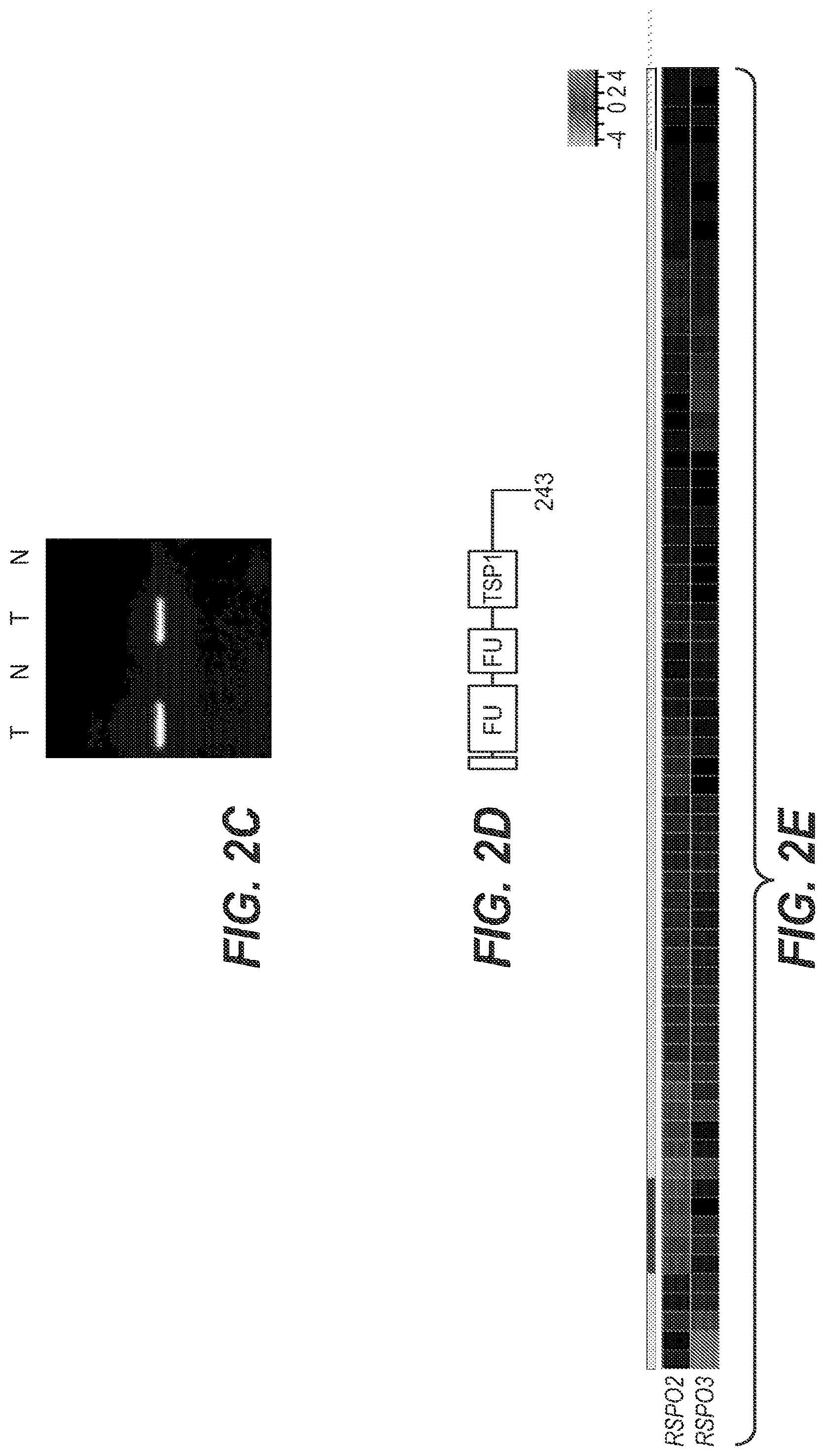
[0019] FIG. 2|Recurrent R-spondin translocations. (A) List of the type and frequency of R-spondin gene fusions in colon cancer. (B) Cartoon depicting the location, orientation and exon-intron architecture of EIF3E-RSPO2 fusion on the genome. The read evidence for EIF3E(e1)-RSPO2(e2) fusion identified using RNA-seq data are shown. (C) Independent RT-PCR derived products confirming the EIF3E-RSPO2 somatic fusion resolved on an agarose gel. RT-PCR products were Sanger sequenced to confirm the fusion junction and a relevant representative chromatogram is presented. (D) Schematic of the resulting EIF3E-RSPO2 fusion protein. (E) Tumors harboring R-spondin fusions show elevated expression of the corresponding RSPO gene shows on a heatmap. FIG. 2 discloses SEQ ID NOS 85-92 and 71, respectively, in order of appearance.
[0020] FIG. 3|Recurrence of PTPRK-RSPO3 gene fusion. (A) Cartoon depicting the location, orientation and exon-intron architecture of PTPRK-RSPO3 gene fusion on the genome. The read evidence for PTPRK(e1)-RSPO3(e2) fusion identified using RNA-seq data are shown. (B) Independent RT-PCR derived products confirming the PTPRK-RSPO3 somatic fusion resolved on an agarose gel. RT-PCR products were Sanger sequenced to confirm the fusion junction and a relevant representative chromatogram is presented. (C) Schematic of PTPRK, RSPO3 and the resulting PTPRK-RSPO3 fusion proteins. FIG. 3 discloses SEQ ID NOS 93-99 and 72, respectively, in order of appearance.
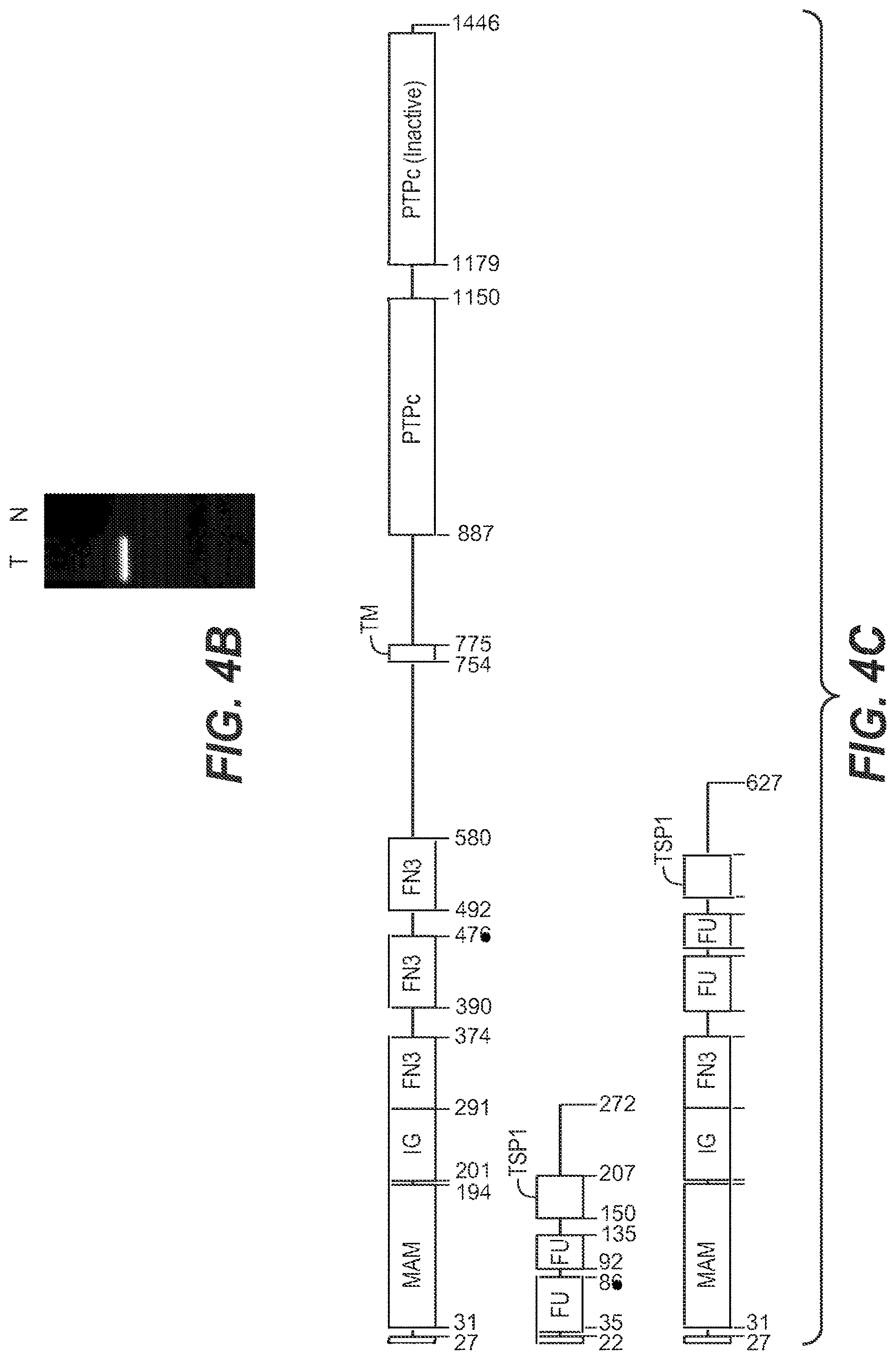
[0021] FIG. 4|(A) PTPRK(e7)-RSPO3(e2) fusion. (B) Gel showing the validation of this fusion by RT-PCR. (C) Schematic diagram of the native and fusion proteins. FIG. 4 discloses SEQ ID NOS 100-104 and 73, respectively, in order of appearance.
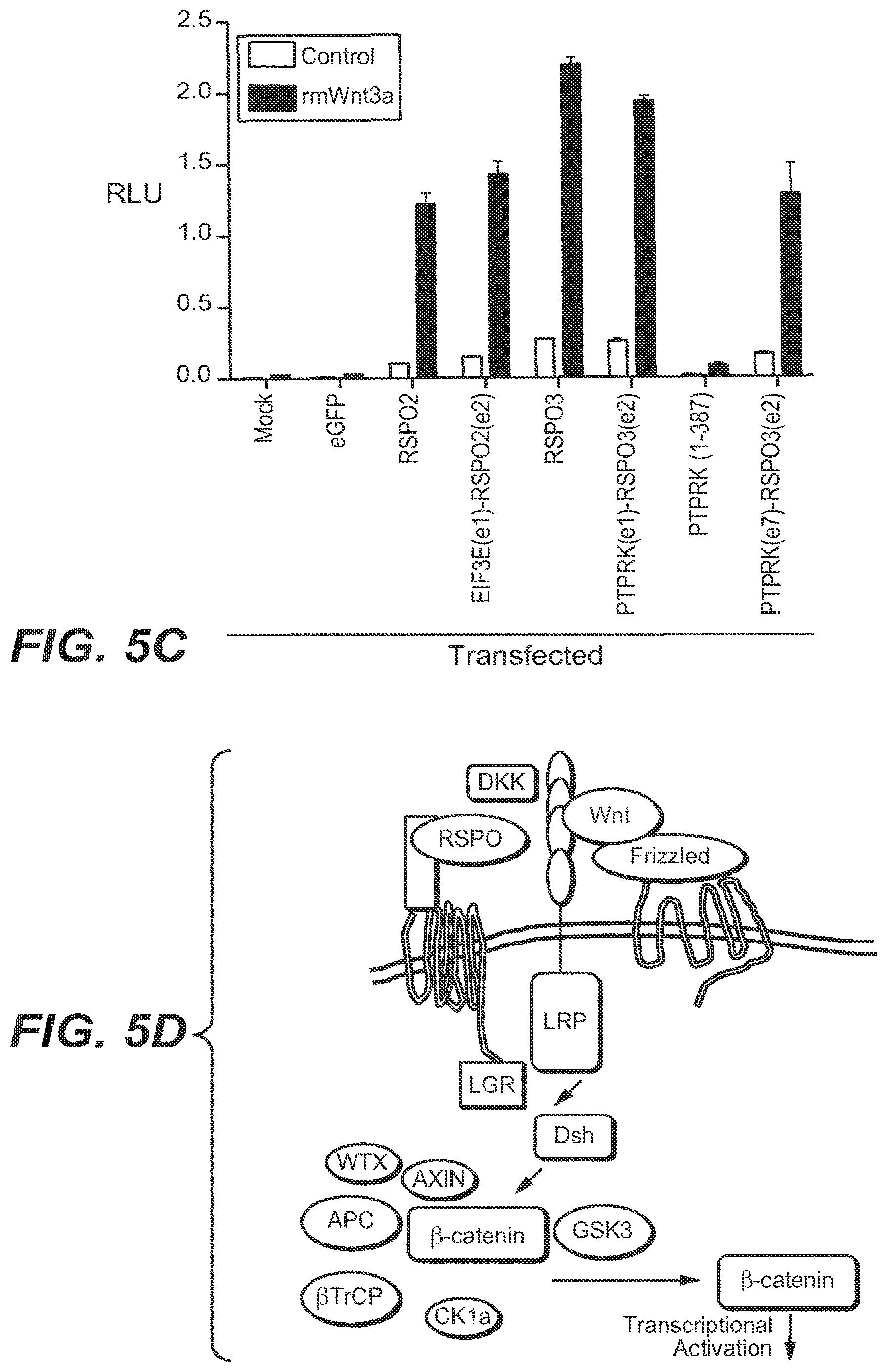
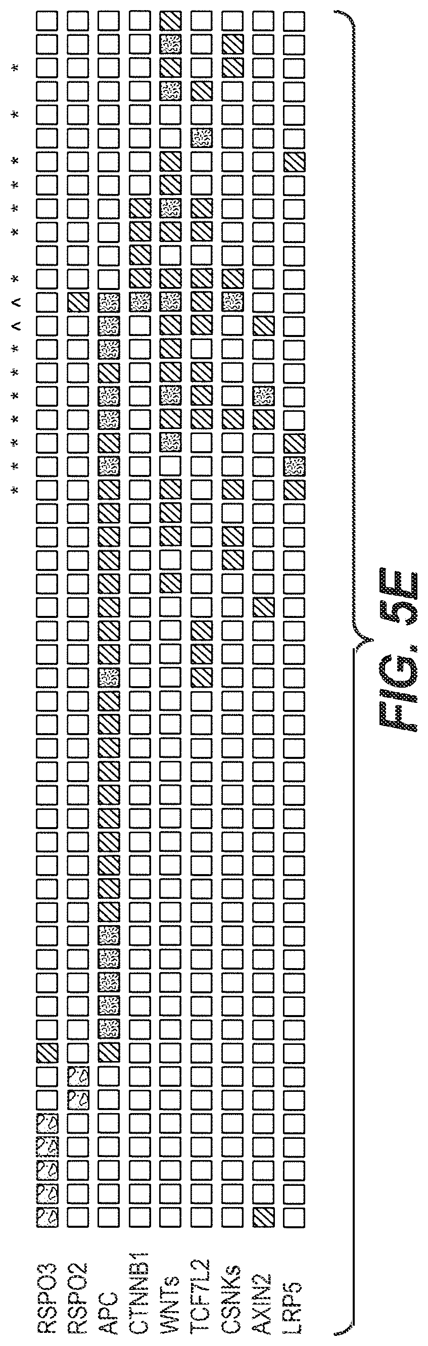
[0022] FIG. 5|RSPO fusion products activate Wnt signaling. (A) Secreted RSPO fusion proteins detected by Western blot in media from 293T cells transfected with expression constructs encoding the fusion proteins. The expected product is RSPO 1-387. (B and C) RSPO fusion proteins activate and potentiate Wnt signaling as measured using a luciferase reporter assay. Data shown are from condition media derived from cells transfected with the fusion constructs or directly transfected into the cell along with the reporter construct. Representative data from at least three experiments are shown. (D) Cartoon representing R-spondin mediated Wnt signaling pathway activation. (E) Plot depicting RSPO fusions and somatic mutations across a select set of Wnt signaling pathway genes.
[0023] FIG. 6|(A) KRAS mutations overlap with RSPO gene fusions. (B) RAS/RTK pathway alterations in colon cancer.
[0024] FIG. 7|Whole genome EIF3E-RSPO2 coordinates schematic and sequences. FIG. 7 discloses SEQ ID NOS 105-108, respectively, in order of appearance.
[0025] FIG. 8|Whole genome EIF3E-RSPO2 coordinates schematic and sequences. FIG. 8 discloses SEQ ID NOS 109-111, respectively, in order of appearance.
[0026] FIG. 9|Whole genome PTPRK-RSPO3 coordinates schematic and sequences. FIG. 9 discloses SEQ ID NOS 112-116, respectively, in order of appearance.
[0027] FIG. 10|Whole genome PTPRK-RSPO3 coordinates schematic and sequences. FIG. 10 discloses SEQ ID NOS 112 and 117-120, respectively, in order of appearance.
DETAILED DESCRIPTION
I. Definitions
[0028] The terms "R-spondin" and "RSPO" refer herein to a native R-spondin from any vertebrate source, including mammals such as primates (e.g., humans) and rodents (e.g., mice and rats), unless otherwise indicated. The term encompasses "full-length," unprocessed R-spondin as well as any form of R-spondin that results from processing in the cell. The term also encompasses naturally occurring variants of R-spondin, e.g., splice variants or allelic variants. R-spondin is a family of four proteins, R-spondin 1 (RSPO1), R-spondin 2 (RSPO2), R-spondin 3 (RSPO3), and R-spondin 4 (RSPO4). In some embodiments, the R-spondin is RSPO1. The sequence of an exemplary human RSPO1 nucleic acid sequence is SEQ ID NO:1 or an exemplary human RSPO1 is amino acid sequence of SEQ ID NO:2. In some embodiments, the R-spondin is RSPO2. The sequence of an exemplary human RSPO2 nucleic acid sequence is SEQ ID NO:3 or an exemplary human RSPO2 is amino acid sequence of SEQ ID NO:4. In some embodiments, the R-spondin is RSPO3. The sequence of an exemplary human RSPO3 nucleic acid sequence is SEQ ID NO:5 or an exemplary human RSPO3 is amino acid sequence of SEQ ID NO:6. In some embodiments, the R-spondin is RSPO4. The sequence of an exemplary human RSPO4 nucleic acid sequence is SEQ ID NO:7 or an exemplary human RSPO4 is amino acid sequence of SEQ ID NO:8.
[0029] "R-Spondin variant," "RSPO variant," or variations thereof, means an R-spondin polypeptide or polynucleotide, generally being or encoding an active R-Spondin polypeptide, as defined herein having at least about 80% amino acid sequence identity with any of the R-Spondin as disclosed herein. Such R-Spondin variants include, for instance, R-Spondin wherein one or more nucleic acid or amino acid residues are added or deleted. Ordinarily, an R-spondin variant will have at least about 80% sequence identity, alternatively at least about 81%, 82%, 83%, 84%, 85%, 86%, 87%, 88%, 89%, 90%, 91%, 92%, 93%, 94%, 95%, 96%, 97%, 98%, or 99% sequence identity, to R-Spondin as disclosed herein. Ordinarily, R-Spondin variant are at least about 10 residues in length, alternatively at least about 20, 30, 40, 50, 60, 70, 80, 90, 100, 110, 120, 130, 140, 150, 160, 170, 180, 190, 200, 210, 220, 230, 240, 250, 260, 270, 280, 290, 300, 310, 320, 330, 340, 350, 360, 370, 380, 390, 400, 410, 420, 430, 440, 450, 460, 470, 480, 490, 500, 510, 520, 530, 540, 550, 560, 570, 580, 590, 600 in length, or more. Optionally, R-Spondin variant will have or encode a sequence having no more than one conservative amino acid substitution as compared to R-Spondin, alternatively no more than 2, 3, 4, 5, 6, 7, 8, 9, or 10 conservative amino acid substitution as compared to R-Spondin.
[0030] The terms "R-spondin translocation" and "RSPO translocation" refer herein to an R-spondin wherein a portion of a broken chromosome including, for example, R-spondin, variant, or fragment thereof or a second gene, variant, or fragment thereof, reattaches in a different chromosome location, for example, a chromosome location different from R-spondin native location or a chromosome location in and/or around the R-spondin native location which is different from the second gene's native location. The R-spondin translocation may be a RSPO1 translocation, RSPO2 translocation, RSPO3 translocation, and/or RSPO4 translocation.
[0031] The terms "R-spondin-translocation fusion polynucleotide" and "RSPO-translocation fusion polynucleotide" refer herein to the nucleic acid sequence of an R-spondin translocation gene product or fusion polynucleotide. The R-spondin-translocation fusion polynucleotide may be a RSPO1-translocation fusion polynucleotide, RSPO2-translocation fusion polynucleotide, RSPO3-translocation fusion polynucleotide, and/or RSPO4-translocation fusion polynucleotide. The terms "R-spondin-translocation fusion polypeptide" and "RSPO-translocation fusion polypeptide" refer herein to the amino acid sequence of an R-spondin translocation gene product or fusion polynucleotide. The R-spondin-translocation fusion polypeptide may be a RSPO1-translocation fusion polypeptide, RSPO2-translocation fusion polypeptide, RSPO3-translocation fusion polypeptide, and/or RSPO4-translocation fusion polypeptide.
[0032] The term "R-spondin-translocation antagonist" as defined herein is any molecule that partially or fully blocks, inhibits, or neutralizes a biological activity mediated by an R-spondin-translocation fusion polypeptide. In some embodiments such antagonist binds to R-spondin-translocation fusion polypeptide. According to one embodiment, the antagonist is a polypeptide. According to another embodiment, the antagonist is an anti-R-spondin-translocation antibody. According to another embodiment, the antagonist is a small molecule antagonist. According to another embodiment, the antagonist is a polynucleotide antagonist. The R-spondin translocation may be a RSPO1-translocation antagonist, RSPO2-translocation antagonist, RSPO3-translocation antagonist, and/or RSPO4-translocation antagonist.
[0033] The term "wnt pathway antagonist" as defined herein is any molecule that partially or fully blocks, inhibits, or neutralizes a biological activity mediated by the wnt pathway (e.g., wnt pathway polypeptide). In some embodiments such antagonist binds to a wnt pathway polypeptide. According to one embodiment, the antagonist is a polypeptide. According to another embodiment, the antagonist is an antibody antagonist. According to another embodiment, the antagonist is a small molecule antagonist. According to another embodiment, the antagonist is a polynucleotide antagonist.
[0034] "Polynucleotide" or "nucleic acid" as used interchangeably herein, refers to polymers of nucleotides of any length, and include DNA and RNA. The nucleotides can be deoxyribonucleotides, ribonucleotides, modified nucleotides or bases, and/or their analogs, or any substrate that can be incorporated into a polymer by DNA or RNA polymerase or by a synthetic reaction. A polynucleotide may comprise modified nucleotides, such as methylated nucleotides and their analogs. A sequence of nucleotides may be interrupted by non-nucleotide components. A polynucleotide may comprise modification(s) made after synthesis, such as conjugation to a label. Other types of modifications include, for example, "caps," substitution of one or more of the naturally occurring nucleotides with an analog, internucleotide modifications such as, for example, those with uncharged linkages (e.g., methyl phosphonates, phosphotriesters, phosphoamidates, carbamates, etc.) and with charged linkages (e.g., phosphorothioates, phosphorodithioates, etc.), those containing pendant moieties, such as, for example, proteins (e.g., nucleases, toxins, antibodies, signal peptides, ply-L-lysine, etc.), those with intercalators (e.g., acridine, psoralen, etc.), those containing chelators (e.g., metals, radioactive metals, boron, oxidative metals, etc.), those containing alkylators, those with modified linkages (e.g., alpha anomeric nucleic acids, etc.), as well as unmodified forms of the polynucleotides(s). Further, any of the hydroxyl groups ordinarily present in the sugars may be replaced, for example, by phosphonate groups, phosphate groups, protected by standard protecting groups, or activated to prepare additional linkages to additional nucleotides, or may be conjugated to solid or semi-solid supports. The 5' and 3' terminal OH can be phosphorylated or substituted with amines or organic capping group moieties of from 1 to 20 carbon atoms. Other hydroxyls may also be derivatized to standard protecting groups. Polynucleotides can also contain analogous forms of ribose or deoxyribose sugars that are generally known in the art, including, for example, 2'-O-methyl-, 2'-O-allyl-, 2'-fluoro- or 2'-azido-ribose, carbocyclic sugar analogs, .alpha.-anomeric sugars, epimeric sugars such as arabinose, xyloses or lyxoses, pyranose sugars, furanose sugars, sedoheptuloses, acyclic analogs, and basic nucleoside analogs such as methyl riboside. One or more phosphodiester linkages may be replaced by alternative linking groups. These alternative linking groups include, but are not limited to, embodiments wherein phosphate is replaced by P(O)S ("thioate"), P(S)S ("dithioate"), (O)NR.sub.2 ("amidate"), P(O)R, P(O)OR', CO, or CH2 ("formacetal"), in which each R or R' is independently H or substituted or unsubstituted alkyl (1-20 C) optionally containing an ether (--O--) linkage, aryl, alkenyl, cycloalkyl, cycloalkenyl or araldyl. Not all linkages in a polynucleotide need be identical. The preceding description applies to all polynucleotides referred to herein, including RNA and DNA.
[0035] "Oligonucleotide," as used herein, refers to generally single-stranded, synthetic polynucleotides that are generally, but not necessarily, less than about 200 nucleotides in length. The terms "oligonucleotide" and "polynucleotide" are not mutually exclusive. The description above for polynucleotides is equally and fully applicable to oligonucleotides.
[0036] The term "primer" refers to a single stranded polynucleotide that is capable of hybridizing to a nucleic acid and following polymerization of a complementary nucleic acid, generally by providing a free 3'-OH group.
[0037] The term "small molecule" refers to any molecule with a molecular weight of about 2000 Daltons or less, preferably of about 500 Daltons or less.
[0038] The terms "host cell," "host cell line," and "host cell culture" are used interchangeably and refer to cells into which exogenous nucleic acid has been introduced, including the progeny of such cells. Host cells include "transformants" and "transformed cells," which include the primary transformed cell and progeny derived therefrom without regard to the number of passages. Progeny may not be completely identical in nucleic acid content to a parent cell, but may contain mutations. Mutant progeny that have the same function or biological activity as screened or selected for in the originally transformed cell are included herein.
[0039] The term "vector," as used herein, refers to a nucleic acid molecule capable of propagating another nucleic acid to which it is linked. The term includes the vector as a self-replicating nucleic acid structure as well as the vector incorporated into the genome of a host cell into which it has been introduced. Certain vectors are capable of directing the expression of nucleic acids to which they are operatively linked. Such vectors are referred to herein as "expression vectors."
[0040] An "isolated" antibody is one which has been separated from a component of its natural environment. In some embodiments, an antibody is purified to greater than 95% or 99% purity as determined by, for example, electrophoretic (e.g., SDS-PAGE, isoelectric focusing (IEF), capillary electrophoresis) or chromatographic (e.g., ion exchange or reverse phase HPLC). For review of methods for assessment of antibody purity, see, e.g., Flatman et al., J. Chromatogr. B 848:79-87 (2007).
[0041] An "isolated" nucleic acid refers to a nucleic acid molecule that has been separated from a component of its natural environment. An isolated nucleic acid includes a nucleic acid molecule contained in cells that ordinarily contain the nucleic acid molecule, but the nucleic acid molecule is present extrachromosomally or at a chromosomal location that is different from its natural chromosomal location.
[0042] The term "antibody" herein is used in the broadest sense and encompasses various antibody structures, including but not limited to monoclonal antibodies, polyclonal antibodies, multispecific antibodies (e.g., bispecific antibodies), and antibody fragments so long as they exhibit the desired antigen-binding activity.
[0043] An "antibody fragment" refers to a molecule other than an intact antibody that comprises a portion of an intact antibody that binds the antigen to which the intact antibody binds. Examples of antibody fragments include but are not limited to Fv, Fab, Fab', Fab'-SH, F(ab').sub.2; diabodies; linear antibodies; single-chain antibody molecules (e.g., scFv); and multispecific antibodies formed from antibody fragments.
[0044] An "antibody that binds to the same epitope" as a reference antibody refers to an antibody that blocks binding of the reference antibody to its antigen in a competition assay by 50% or more, and conversely, the reference antibody blocks binding of the antibody to its antigen in a competition assay by 50% or more. An exemplary competition assay is provided herein.
[0045] The terms "full length antibody," "intact antibody," and "whole antibody" are used herein interchangeably to refer to an antibody having a structure substantially similar to a native antibody structure or having heavy chains that contain an Fc region as defined herein.
[0046] The term "monoclonal antibody" as used herein refers to an antibody obtained from a population of substantially homogeneous antibodies, i.e., the individual antibodies comprising the population are identical and/or bind the same epitope, except for possible variant antibodies, e.g., containing naturally occurring mutations or arising during production of a monoclonal antibody preparation, such variants generally being present in minor amounts. In contrast to polyclonal antibody preparations, which typically include different antibodies directed against different determinants (epitopes), each monoclonal antibody of a monoclonal antibody preparation is directed against a single determinant on an antigen. Thus, the modifier "monoclonal" indicates the character of the antibody as being obtained from a substantially homogeneous population of antibodies, and is not to be construed as requiring production of the antibody by any particular method. For example, the monoclonal antibodies to be used in accordance with the present invention may be made by a variety of techniques, including but not limited to the hybridoma method, recombinant DNA methods, phage-display methods, and methods utilizing transgenic animals containing all or part of the human immunoglobulin loci, such methods and other exemplary methods for making monoclonal antibodies being described herein.
[0047] "Native antibodies" refer to naturally occurring immunoglobulin molecules with varying structures. For example, native IgG antibodies are heterotetrameric glycoproteins of about 150,000 Daltons, composed of two identical light chains and two identical heavy chains that are disulfide-bonded. From N- to C-terminus, each heavy chain has a variable region (VH), also called a variable heavy domain or a heavy chain variable domain, followed by three constant domains (CH1, CH2, and CH3). Similarly, from N- to C-terminus, each light chain has a variable region (VL), also called a variable light domain or a light chain variable domain, followed by a constant light (CL) domain. The light chain of an antibody may be assigned to one of two types, called kappa (.kappa.) and lambda (.lamda.), based on the amino acid sequence of its constant domain.
[0048] The term "chimeric" antibody refers to an antibody in which a portion of the heavy and/or light chain is derived from a particular source or species, while the remainder of the heavy and/or light chain is derived from a different source or species.
[0049] A "human antibody" is one which possesses an amino acid sequence which corresponds to that of an antibody produced by a human or a human cell or derived from a non-human source that utilizes human antibody repertoires or other human antibody-encoding sequences. This definition of a human antibody specifically excludes a humanized antibody comprising non-human antigen-binding residues.
[0050] A "humanized" antibody refers to a chimeric antibody comprising amino acid residues from non-human HVRs and amino acid residues from human FRs. In certain embodiments, a humanized antibody will comprise substantially all of at least one, and typically two, variable domains, in which all or substantially all of the HVRs (e.g., CDRs) correspond to those of a non-human antibody, and all or substantially all of the FRs correspond to those of a human antibody. A humanized antibody optionally may comprise at least a portion of an antibody constant region derived from a human antibody. A "humanized form" of an antibody, e.g., a non-human antibody, refers to an antibody that has undergone humanization.
[0051] The "class" of an antibody refers to the type of constant domain or constant region possessed by its heavy chain. There are five major classes of antibodies: IgA, IgD, IgE, IgG, and IgM, and several of these may be further divided into subclasses (isotypes), e.g., IgG.sub.1, IgG.sub.2, IgG.sub.3, IgG.sub.4, IgA.sub.1, and IgA.sub.2. The heavy chain constant domains that correspond to the different classes of immunoglobulins are called .alpha., .delta., .epsilon., .gamma., and .mu., respectively.
[0052] "Effector functions" refer to those biological activities attributable to the Fc region of an antibody, which vary with the antibody isotype. Examples of antibody effector functions include: C1q binding and complement dependent cytotoxicity (CDC); Fc receptor binding; antibody-dependent cell-mediated cytotoxicity (ADCC); phagocytosis; down regulation of cell surface receptors (e.g., B cell receptor); and B cell activation.
[0053] The term "Fc region" herein is used to define a C-terminal region of an immunoglobulin heavy chain that contains at least a portion of the constant region. The term includes native sequence Fc regions and variant Fc regions. In one embodiment, a human IgG heavy chain Fc region extends from Cys226, or from Pro230, to the carboxyl-terminus of the heavy chain. However, the C-terminal lysine (Lys447) of the Fc region may or may not be present. Unless otherwise specified herein, numbering of amino acid residues in the Fc region or constant region is according to the EU numbering system, also called the EU index, as described in Kabat et al., Sequences of Proteins of Immunological Interest, 5th Ed. Public Health Service, National Institutes of Health, Bethesda, Md., 1991.
[0054] "Framework" or "FR" refers to variable domain residues other than hypervariable region (HVR) residues. The FR of a variable domain generally consists of four FR domains: FR1, FR2, FR3, and FR4. Accordingly, the HVR and FR sequences generally appear in the following sequence in VH (or VL): FR1-H1(L1)-FR2-H2(L2)-FR3-H3(L3)-FR4.
[0055] A "human consensus framework" is a framework which represents the most commonly occurring amino acid residues in a selection of human immunoglobulin VL or VH framework sequences. Generally, the selection of human immunoglobulin VL or VH sequences is from a subgroup of variable domain sequences. Generally, the subgroup of sequences is a subgroup as in Kabat et al., Sequences of Proteins of Immunological Interest, Fifth Edition, NIH Publication 91-3242, Bethesda Md. (1991), vols. 1-3. In one embodiment, for the VL, the subgroup is subgroup kappa I as in Kabat et al., supra. In one embodiment, for the VH, the subgroup is subgroup III as in Kabat et al., supra.
[0056] An "acceptor human framework" for the purposes herein is a framework comprising the amino acid sequence of a light chain variable domain (VL) framework or a heavy chain variable domain (VH) framework derived from a human immunoglobulin framework or a human consensus framework, as defined below. An acceptor human framework "derived from" a human immunoglobulin framework or a human consensus framework may comprise the same amino acid sequence thereof, or it may contain amino acid sequence changes. In some embodiments, the number of amino acid changes are 10 or less, 9 or less, 8 or less, 7 or less, 6 or less, 5 or less, 4 or less, 3 or less, or 2 or less. In some embodiments, the VL acceptor human framework is identical in sequence to the VL human immunoglobulin framework sequence or human consensus framework sequence.
[0057] The term "variable region" or "variable domain" refers to the domain of an antibody heavy or light chain that is involved in binding the antibody to antigen. The variable domains of the heavy chain and light chain (VH and VL, respectively) of a native antibody generally have similar structures, with each domain comprising four conserved framework regions (FRs) and three hypervariable regions (HVRs). (See, e.g., Kindt et al., Kuby Immunology, 6.sup.th ed., W.H. Freeman and Co., page 91 (2007).) A single VH or VL domain may be sufficient to confer antigen-binding specificity. Furthermore, antibodies that bind a particular antigen may be isolated using a VH or VL domain from an antibody that binds the antigen to screen a library of complementary VL or VH domains, respectively. See, e.g., Portolano et al., J. Immunol. 150:880-887 (1993); Clarkson et al., Nature 352:624-628 (1991).
[0058] The term "hypervariable region" or "HVR," as used herein, refers to each of the regions of an antibody variable domain which are hypervariable in sequence and/or form structurally defined loops ("hypervariable loops"). Generally, native four-chain antibodies comprise six HVRs; three in the VH (H1, H2, H3), and three in the VL (L1, L2, L3). HVRs generally comprise amino acid residues from the hypervariable loops and/or from the "complementarity determining regions" (CDRs), the latter being of highest sequence variability and/or involved in antigen recognition. Exemplary hypervariable loops occur at amino acid residues 26-32 (L1), 50-52 (L2), 91-96 (L3), 26-32 (H1), 53-55 (H2), and 96-101 (H3). (Chothia and Lesk, J. Mol. Biol. 196:901-917 (1987).) Exemplary CDRs (CDR-L1, CDR-L2, CDR-L3, CDR-H1, CDR-H2, and CDR-H3) occur at amino acid residues 24-34 of L1, 50-56 of L2, 89-97 of L3, 31-35B of H1, 50-65 of H2, and 95-102 of H3. (Kabat et al., Sequences of Proteins of Immunological Interest, 5th Ed. Public Health Service, National Institutes of Health, Bethesda, Md. (1991).) With the exception of CDR1 in VH, CDRs generally comprise the amino acid residues that form the hypervariable loops. CDRs also comprise "specificity determining residues," or "SDRs," which are residues that contact antigen. SDRs are contained within regions of the CDRs called abbreviated-CDRs, or a-CDRs. Exemplary a-CDRs (a-CDR-L1, a-CDR-L2, a-CDR-L3, a-CDR-H1, a-CDR-H2, and a-CDR-H3) occur at amino acid residues 31-34 of L1, 50-55 of L2, 89-96 of L3, 31-35B of H1, 50-58 of H2, and 95-102 of H3. (See Almagro and Fransson, Front. Biosci. 13:1619-1633 (2008).) Unless otherwise indicated, HVR residues and other residues in the variable domain (e.g., FR residues) are numbered herein according to Kabat et al., supra.
[0059] "Affinity" refers to the strength of the sum total of noncovalent interactions between a single binding site of a molecule (e.g., an antibody) and its binding partner (e.g., an antigen). Unless indicated otherwise, as used herein, "binding affinity" refers to intrinsic binding affinity which reflects a 1:1 interaction between members of a binding pair (e.g., antibody and antigen). The affinity of a molecule X for its partner Y can generally be represented by the dissociation constant (Kd) Affinity can be measured by common methods known in the art, including those described herein. Specific illustrative and exemplary embodiments for measuring binding affinity are described in the following.
[0060] An "affinity matured" antibody refers to an antibody with one or more alterations in one or more hypervariable regions (HVRs), compared to a parent antibody which does not possess such alterations, such alterations resulting in an improvement in the affinity of the antibody for antigen.
[0061] The terms "anti-R-spondin-translocation antibody" and "an antibody that binds to R-spondin-translocation fusion polypeptide" refer to an antibody that is capable of binding R-spondin-translocation fusion polypeptide with sufficient affinity such that the antibody is useful as a diagnostic and/or therapeutic agent in targeting R-spondin translocation. In one embodiment, the extent of binding of an anti-R-spondin translocation antibody to an unrelated, non-R-spondin-translocation fusion polypeptide, and/or nontranslocated-R-spondin polypeptide is less than about 10% of the binding of the antibody to R-spondin-translocation fusion polypeptides measured, e.g., by a radioimmunoassay (RIA). In certain embodiments, an antibody that binds to R-spondin-translocation fusion polypeptide has a dissociation constant (Kd) of .ltoreq.1 .mu.M, .ltoreq.100 nM, .ltoreq.10 nM, .ltoreq.1 nM, .ltoreq.0.1 nM, .ltoreq.0.01 nM, or .ltoreq.0.001 nM (e.g.,10.sup.-8 M or less, e.g., from 10.sup.-8 M to 10.sup.-13 M, e.g., from 10.sup.-9 M to 10.sup.-13 M). In certain embodiments, an anti-R-spondin translocation antibody binds to an epitope of R-spondin translocation that is unique among R-spondin translocations.
[0062] A "blocking" antibody or an "antagonist" antibody is one which inhibits or reduces biological activity of the antigen it binds. Preferred blocking antibodies or antagonist antibodies substantially or completely inhibit the biological activity of the antigen.
[0063] A "naked antibody" refers to an antibody that is not conjugated to a heterologous moiety (e.g., a cytotoxic moiety) or radiolabel. The naked antibody may be present in a pharmaceutical formulation.
[0064] An "immunoconjugate" is an antibody conjugated to one or more heterologous molecule(s), including but not limited to a cytotoxic agent.
[0065] "Percent (%) amino acid sequence identity" with respect to a reference polypeptide sequence is defined as the percentage of amino acid residues in a candidate sequence that are identical with the amino acid residues in the reference polypeptide sequence, after aligning the sequences and introducing gaps, if necessary, to achieve the maximum percent sequence identity, and not considering any conservative substitutions as part of the sequence identity. Alignment for purposes of determining percent amino acid sequence identity can be achieved in various ways that are within the skill in the art, for instance, using publicly available computer software such as BLAST, BLAST-2, ALIGN or Megalign (DNASTAR) software. Those skilled in the art can determine appropriate parameters for aligning sequences, including any algorithms needed to achieve maximal alignment over the full length of the sequences being compared. For purposes herein, however, % amino acid sequence identity values are generated using the sequence comparison computer program ALIGN-2. The ALIGN-2 sequence comparison computer program was authored by Genentech, Inc., and the source code has been filed with user documentation in the U.S. Copyright Office, Washington D.C., 20559, where it is registered under U.S. Copyright Registration No. TXU510087. The ALIGN-2 program is publicly available from Genentech, Inc., South San Francisco, Calif., or may be compiled from the source code. The ALIGN-2 program should be compiled for use on a UNIX operating system, including digital UNIX V4.0D. All sequence comparison parameters are set by the ALIGN-2 program and do not vary.
[0066] In situations where ALIGN-2 is employed for amino acid sequence comparisons, the % amino acid sequence identity of a given amino acid sequence A to, with, or against a given amino acid sequence B (which can alternatively be phrased as a given amino acid sequence A that has or comprises a certain % amino acid sequence identity to, with, or against a given amino acid sequence B) is calculated as follows:
100 times the fraction X/Y
where X is the number of amino acid residues scored as identical matches by the sequence alignment program ALIGN-2 in that program's alignment of A and B, and where Y is the total number of amino acid residues in B. It will be appreciated that where the length of amino acid sequence A is not equal to the length of amino acid sequence B, the % amino acid sequence identity of A to B will not equal the % amino acid sequence identity of B to A. Unless specifically stated otherwise, all % amino acid sequence identity values used herein are obtained as described in the immediately preceding paragraph using the ALIGN-2 computer program.
[0067] The term "detection" includes any means of detecting, including direct and indirect detection.
[0068] The term "biomarker" as used herein refers to an indicator, e.g., predictive, diagnostic, and/or prognostic, which can be detected in a sample. The biomarker may serve as an indicator of a particular subtype of a disease or disorder (e.g., cancer) characterized by certain, molecular, pathological, histological, and/or clinical features. In some embodiments, the biomarker is a gene. In some embodiments, the biomarker is a variation (e.g., mutation and/or polymorphism) of a gene. In some embodiments, the biomarkers is a translocation. Biomarkers include, but are not limited to, polynucleotides (e.g., DNA, and/or RNA), polypeptides, polypeptide and polynucleotide modifications (e.g., posttranslational modifications), carbohydrates, and/or glycolipid-based molecular markers.
[0069] The "presence," "amount," or "level" of a biomarker associated with an increased clinical benefit to an individual is a detectable level in a biological sample. These can be measured by methods known to one skilled in the art and also disclosed herein. The expression level or amount of biomarker assessed can be used to determine the response to the treatment.
[0070] The terms "level of expression" or "expression level" in general are used interchangeably and generally refer to the amount of a biomarker in a biological sample. "Expression" generally refers to the process by which information (e.g., gene-encoded and/or epigenetic) is converted into the structures present and operating in the cell. Therefore, as used herein, "expression" may refer to transcription into a polynucleotide, translation into a polypeptide, or even polynucleotide and/or polypeptide modifications (e.g., posttranslational modification of a polypeptide). Fragments of the transcribed polynucleotide, the translated polypeptide, or polynucleotide and/or polypeptide modifications (e.g., posttranslational modification of a polypeptide) shall also be regarded as expressed whether they originate from a transcript generated by alternative splicing or a degraded transcript, or from a post-translational processing of the polypeptide, e.g., by proteolysis. "Expressed genes" include those that are transcribed into a polynucleotide as mRNA and then translated into a polypeptide, and also those that are transcribed into RNA but not translated into a polypeptide (for example, transfer and ribosomal RNAs).
[0071] "Elevated expression," "elevated expression levels," or "elevated levels" refers to an increased expression or increased levels of a biomarker in an individual relative to a control, such as an individual or individuals who are not suffering from the disease or disorder (e.g., cancer) or an internal control (e.g., housekeeping biomarker).
[0072] "Reduced expression," "reduced expression levels," or "reduced levels" refers to a decrease expression or decreased levels of a biomarker in an individual relative to a control, such as an individual or individuals who are not suffering from the disease or disorder (e.g., cancer) or an internal control (e.g., housekeeping biomarker).
[0073] The term "housekeeping biomarker" refers to a biomarker or group of biomarkers (e.g., polynucleotides and/or polypeptides) which are typically similarly present in all cell types. In some embodiments, the housekeeping biomarker is a "housekeeping gene." A "housekeeping gene" refers herein to a gene or group of genes which encode proteins whose activities are essential for the maintenance of cell function and which are typically similarly present in all cell types.
[0074] "Amplification," as used herein generally refers to the process of producing multiple copies of a desired sequence. "Multiple copies" mean at least two copies. A "copy" does not necessarily mean perfect sequence complementarity or identity to the template sequence. For example, copies can include nucleotide analogs such as deoxyinosine, intentional sequence alterations (such as sequence alterations introduced through a primer comprising a sequence that is hybridizable, but not complementary, to the template), and/or sequence errors that occur during amplification.
[0075] The term "multiplex-PCR" refers to a single PCR reaction carried out on nucleic acid obtained from a single source (e.g., an individual) using more than one primer set for the purpose of amplifying two or more DNA sequences in a single reaction.
[0076] "Stringency" of hybridization reactions is readily determinable by one of ordinary skill in the art, and generally is an empirical calculation dependent upon probe length, washing temperature, and salt concentration. In general, longer probes require higher temperatures for proper annealing, while shorter probes need lower temperatures. Hybridization generally depends on the ability of denatured DNA to reanneal when complementary strands are present in an environment below their melting temperature. The higher the degree of desired homology between the probe and hybridizable sequence, the higher the relative temperature which can be used. As a result, it follows that higher relative temperatures would tend to make the reaction conditions more stringent, while lower temperatures less so. For additional details and explanation of stringency of hybridization reactions, see Ausubel et al., Current Protocols in Molecular Biology, Wiley Interscience Publishers, (1995).
[0077] "Stringent conditions" or "high stringency conditions", as defined herein, can be identified by those that: (1) employ low ionic strength and high temperature for washing, for example 0.015 M sodium chloride/0.0015 M sodium citrate/0.1% sodium dodecyl sulfate at 50.degree. C.; (2) employ during hybridization a denaturing agent, such as formamide, for example, 50% (v/v) formamide with 0.1% bovine serum albumin/0.1% Ficoll/0.1% polyvinylpyrrolidone/50 mM sodium phosphate buffer at pH 6.5 with 750 mM sodium chloride, 75 mM sodium citrate at 42.degree. C.; or (3) overnight hybridization in a solution that employs 50% formamide, 5.times.SSC (0.75 M NaCl, 0.075 M sodium citrate), 50 mM sodium phosphate (pH 6.8), 0.1% sodium pyrophosphate, 5.times. Denhardt's solution, sonicated salmon sperm DNA (50 .mu.g/ml), 0.1% SDS, and 10% dextran sulfate at 42.degree. C., with a 10 minute wash at 42.degree. C. in 0.2.times.SSC (sodium chloride/sodium citrate) followed by a 10 minute high-stringency wash consisting of 0.1.times.SSC containing EDTA at 55.degree. C.
[0078] "Moderately stringent conditions" can be identified as described by Sambrook et al., Molecular Cloning: A Laboratory Manual, New York: Cold Spring Harbor Press, 1989, and include the use of washing solution and hybridization conditions (e.g., temperature, ionic strength and % SDS) less stringent that those described above. An example of moderately stringent conditions is overnight incubation at 37.degree. C. in a solution comprising: 20% formamide, 5.times.SSC (150 mM NaCl, 15 mM trisodium citrate), 50 mM sodium phosphate (pH 7.6), 5.times. Denhardt's solution, 10% dextran sulfate, and 20 mg/ml denatured sheared salmon sperm DNA, followed by washing the filters in 1.times.SSC at about 37-50.degree. C. The skilled artisan will recognize how to adjust the temperature, ionic strength, etc. as necessary to accommodate factors such as probe length and the like.
[0079] The term "diagnosis" is used herein to refer to the identification or classification of a molecular or pathological state, disease or condition (e.g., cancer). For example, "diagnosis" may refer to identification of a particular type of cancer. "Diagnosis" may also refer to the classification of a particular subtype of cancer, e.g., by histopathological criteria, or by molecular features (e.g., a subtype characterized by expression of one or a combination of biomarkers (e.g., particular genes or proteins encoded by said genes)).
[0080] The term "aiding diagnosis" is used herein to refer to methods that assist in making a clinical determination regarding the presence, or nature, of a particular type of symptom or condition of a disease or disorder (e.g., cancer). For example, a method of aiding diagnosis of a disease or condition (e.g., cancer) can comprise detecting certain biomarkers in a biological sample from an individual.
[0081] The term "sample," as used herein, refers to a composition that is obtained or derived from a subject and/or individual of interest that contains a cellular and/or other molecular entity that is to be characterized and/or identified, for example based on physical, biochemical, chemical and/or physiological characteristics. For example, the phrase "disease sample" and variations thereof refers to any sample obtained from a subject of interest that would be expected or is known to contain the cellular and/or molecular entity that is to be characterized. Samples include, but are not limited to, primary or cultured cells or cell lines, cell supernatants, cell lysates, platelets, serum, plasma, vitreous fluid, lymph fluid, synovial fluid, follicular fluid, seminal fluid, amniotic fluid, milk, whole blood, blood-derived cells, urine, cerebro-spinal fluid, saliva, sputum, tears, perspiration, mucus, tumor lysates, and tissue culture medium, tissue extracts such as homogenized tissue, tumor tissue, cellular extracts, and combinations thereof.
[0082] By "tissue sample" or "cell sample" is meant a collection of similar cells obtained from a tissue of a subject or individual. The source of the tissue or cell sample may be solid tissue as from a fresh, frozen and/or preserved organ, tissue sample, biopsy, and/or aspirate; blood or any blood constituents such as plasma; bodily fluids such as cerebral spinal fluid, amniotic fluid, peritoneal fluid, or interstitial fluid; cells from any time in gestation or development of the subject. The tissue sample may also be primary or cultured cells or cell lines. Optionally, the tissue or cell sample is obtained from a disease tissue/organ. The tissue sample may contain compounds which are not naturally intermixed with the tissue in nature such as preservatives, anticoagulants, buffers, fixatives, nutrients, antibiotics, or the like.
[0083] A "reference sample", "reference cell", "reference tissue", "control sample", "control cell", or "control tissue", as used herein, refers to a sample, cell, tissue, standard, or level that is used for comparison purposes. In one embodiment, a reference sample, reference cell, reference tissue, control sample, control cell, or control tissue is obtained from a healthy and/or non-diseased part of the body (e.g., tissue or cells) of the same subject or individual. For example, healthy and/or non-diseased cells or tissue adjacent to the diseased cells or tissue (e.g., cells or tissue adjacent to a tumor). In another embodiment, a reference sample is obtained from an untreated tissue and/or cell of the body of the same subject or individual. In yet another embodiment, a reference sample, reference cell, reference tissue, control sample, control cell, or control tissue is obtained from a healthy and/or non-diseased part of the body (e.g., tissues or cells) of an individual who is not the subject or individual. In even another embodiment, a reference sample, reference cell, reference tissue, control sample, control cell, or control tissue is obtained from an untreated tissue and/or cell of the body of an individual who is not the subject or individual.
[0084] For the purposes herein a "section" of a tissue sample is meant a single part or piece of a tissue sample, e.g., a thin slice of tissue or cells cut from a tissue sample. It is understood that multiple sections of tissue samples may be taken and subjected to analysis, provided that it is understood that the same section of tissue sample may be analyzed at both morphological and molecular levels, or analyzed with respect to both polypeptides and polynucleotides.
[0085] By "correlate" or "correlating" is meant comparing, in any way, the performance and/or results of a first analysis or protocol with the performance and/or results of a second analysis or protocol. For example, one may use the results of a first analysis or protocol in carrying out a second protocols and/or one may use the results of a first analysis or protocol to determine whether a second analysis or protocol should be performed. With respect to the embodiment of polynucleotide analysis or protocol, one may use the results of the polynucleotide expression analysis or protocol to determine whether a specific therapeutic regimen should be performed.
[0086] "Individual response" or "response" can be assessed using any endpoint indicating a benefit to the individual, including, without limitation, (1) inhibition, to some extent, of disease progression (e.g., cancer progression), including slowing down and complete arrest; (2) a reduction in tumor size; (3) inhibition (i.e., reduction, slowing down or complete stopping) of cancer cell infiltration into adjacent peripheral organs and/or tissues; (4) inhibition (i.e. reduction, slowing down or complete stopping) of metasisis; (5) relief, to some extent, of one or more symptoms associated with the disease or disorder (e.g., cancer); (6) increase in the length of progression free survival; and/or (9) decreased mortality at a given point of time following treatment.
[0087] The phrase "substantially similar," as used herein, refers to a sufficiently high degree of similarity between two numeric values (generally one associated with a molecule and the other associated with a reference/comparator molecule) such that one of skill in the art would consider the difference between the two values to not be of statistical significance within the context of the biological characteristic measured by said values (e.g., Kd values). The difference between said two values may be, for example, less than about 20%, less than about 10%, and/or less than about 5% as a function of the reference/comparator value. The phrase "substantially normal" refers to substantially similar to a reference (e.g., normal reference).
[0088] The phrase "substantially different," refers to a sufficiently high degree of difference between two numeric values (generally one associated with a molecule and the other associated with a reference/comparator molecule) such that one of skill in the art would consider the difference between the two values to be of statistical significance within the context of the biological characteristic measured by said values (e.g., Kd values). The difference between said two values may be, for example, greater than about 10%, greater than about 20%, greater than about 30%, greater than about 40%, and/or greater than about 50% as a function of the value for the reference/comparator molecule.
[0089] The word "label" when used herein refers to a detectable compound or composition. The label is typically conjugated or fused directly or indirectly to a reagent, such as a polynucleotide probe or an antibody, and facilitates detection of the reagent to which it is conjugated or fused. The label may itself be detectable (e.g., radioisotope labels or fluorescent labels) or, in the case of an enzymatic label, may catalyze chemical alteration of a substrate compound or composition which results in a detectable product.
[0090] An "effective amount" of an agent refers to an amount effective, at dosages and for periods of time necessary, to achieve the desired therapeutic or prophylactic result.
[0091] A "therapeutically effective amount" of a substance/molecule of the invention, agonist or antagonist may vary according to factors such as the disease state, age, sex, and weight of the individual, and the ability of the substance/molecule, agonist or antagonist to elicit a desired response in the individual. A therapeutically effective amount is also one in which any toxic or detrimental effects of the substance/molecule, agonist or antagonist are outweighed by the therapeutically beneficial effects. A "prophylactically effective amount" refers to an amount effective, at dosages and for periods of time necessary, to achieve the desired prophylactic result. Typically but not necessarily, since a prophylactic dose is used in subjects prior to or at an earlier stage of disease, the prophylactically effective amount will be less than the therapeutically effective amount.
[0092] The term "pharmaceutical formulation" refers to a preparation which is in such form as to permit the biological activity of an active ingredient contained therein to be effective, and which contains no additional components which are unacceptably toxic to a subject to which the formulation would be administered.
[0093] A "pharmaceutically acceptable carrier" refers to an ingredient in a pharmaceutical formulation, other than an active ingredient, which is nontoxic to a subject., A pharmaceutically acceptable carrier includes, but is not limited to, a buffer, excipient, stabilizer, or preservative.
[0094] As used herein, "treatment" (and grammatical variations thereof such as "treat" or "treating") refers to clinical intervention in an attempt to alter the natural course of the individual being treated, and can be performed either for prophylaxis or during the course of clinical pathology. Desirable effects of treatment include, but are not limited to, preventing occurrence or recurrence of disease, alleviation of symptoms, diminishment of any direct or indirect pathological consequences of the disease, preventing metastasis, decreasing the rate of disease progression, amelioration or palliation of the disease state, and remission or improved prognosis. In some embodiments, antibodies of the invention are used to delay development of a disease or to slow the progression of a disease.
[0095] The terms "cancer" and "cancerous" refer to or describe the physiological condition in mammals that is typically characterized by unregulated cell growth/proliferation. Examples of cancer include, but are not limited to, carcinoma, lymphoma (e.g., Hodgkin's and non-Hodgkin's lymphoma), blastoma, sarcoma, and leukemia. More particular examples of such cancers include squamous cell cancer, small-cell lung cancer, non-small cell lung cancer, adenocarcinoma of the lung, squamous carcinoma of the lung, cancer of the peritoneum, hepatocellular cancer, gastrointestinal cancer, pancreatic cancer, glioma, cervical cancer, ovarian cancer, liver cancer, bladder cancer, hepatoma, breast cancer, colon cancer, colorectal cancer, endometrial or uterine carcinoma, salivary gland carcinoma, kidney cancer, liver cancer, prostate cancer, vulval cancer, thyroid cancer, hepatic carcinoma, leukemia and other lymphoproliferative disorders, and various types of head and neck cancer.
[0096] The term "anti-cancer therapy" refers to a therapy useful in treating cancer. Examples of anti-cancer therapeutic agents include, but are limited to, e.g., chemotherapeutic agents, growth inhibitory agents, cytotoxic agents, agents used in radiation therapy, anti-angiogenesis agents, apoptotic agents, anti-tubulin agents, and other agents to treat cancer, anti-CD20 antibodies, platelet derived growth factor inhibitors (e.g., Gleevec.TM. (Imatinib Mesylate)), a COX-2 inhibitor (e.g., celecoxib), interferons, cytokines, antagonists (e.g., neutralizing antibodies) that bind to one or more of the following targets PDGFR-beta, BlyS, APRIL, BCMA receptor(s), TRAIL/Apo2, and other bioactive and organic chemical agents, etc. Combinations thereof are also included in the invention.
[0097] The term "cytotoxic agent" as used herein refers to a substance that inhibits or prevents a cellular function and/or causes cell death or destruction. Cytotoxic agents include, but are not limited to, radioactive isotopes (e.g., At.sup.211, I.sup.131, I.sup.125, Y.sup.90, Re.sup.186, Re.sup.188, Sm.sup.153, Bi.sup.212, P.sup.32, Pb.sup.212 and radioactive isotopes of Lu); chemotherapeutic agents or drugs (e.g., methotrexate, adriamicin, vinca alkaloids (vincristine, vinblastine, etoposide), doxorubicin, melphalan, mitomycin C, chlorambucil, daunorubicin or other intercalating agents); growth inhibitory agents; enzymes and fragments thereof such as nucleolytic enzymes; antibiotics; toxins such as small molecule toxins or enzymatically active toxins of bacterial, fungal, plant or animal origin, including fragments and/or variants thereof; and the various antitumor or anticancer agents disclosed below.
[0098] A "chemotherapeutic agent" refers to a chemical compound useful in the treatment of cancer. Examples of chemotherapeutic agents include alkylating agents such as thiotepa and cyclosphosphamide (CYTOXAN.RTM.); alkyl sulfonates such as busulfan, improsulfan and piposulfan; aziridines such as benzodopa, carboquone, meturedopa, and uredopa; ethylenimines and methylamelamines including altretamine, triethylenemelamine, triethylenephosphoramide, triethylenethiophosphoramide and trimethylomelamine; acetogenins (especially bullatacin and bullatacinone); delta-9-tetrahydrocannabinol (dronabinol, MARINOL.RTM.); beta-lapachone; lapachol; colchicines; betulinic acid; a camptothecin (including the synthetic analogue topotecan (HYCAMTIN.RTM.), CPT-11 (irinotecan, CAMPTOSAR.RTM.), acetylcamptothecin, scopolectin, and 9-aminocamptothecin); bryostatin; callystatin; CC-1065 (including its adozelesin, carzelesin and bizelesin synthetic analogues); podophyllotoxin; podophyllinic acid; teniposide; cryptophycins (particularly cryptophycin 1 and cryptophycin 8); dolastatin; duocarmycin (including the synthetic analogues, KW-2189 and CB1-TM1); eleutherobin; pancratistatin; a sarcodictyin; spongistatin; nitrogen mustards such as chlorambucil, chlornaphazine, chlorophosphamide, estramustine, ifosfamide, mechlorethamine, mechlorethamine oxide hydrochloride, melphalan, novembichin, phenesterine, prednimustine, trofosfamide, uracil mustard; nitrosoureas such as carmustine, chlorozotocin, fotemustine, lomustine, nimustine, and ranimnustine; antibiotics such as the enediyne antibiotics (e. g., calicheamicin, especially calicheamicin gammall and calicheamicin omegaI1 (see, e.g., Nicolaou et al., Angew. Chem Intl. Ed. Engl., 33: 183-186 (1994)); CDP323, an oral alpha-4 integrin inhibitor; dynemicin, including dynemicin A; an esperamicin; as well as neocarzinostatin chromophore and related chromoprotein enediyne antibiotic chromophores), aclacinomysins, actinomycin, authramycin, azaserine, bleomycins, cactinomycin, carabicin, carminomycin, carzinophilin, chromomycins, dactinomycin, daunorubicin, detorubicin, 6-diazo-5-oxo-L-norleucine, doxorubicin (including ADRIAMYCIN.RTM., morpholino-doxorubicin, cyanomorpholino-doxorubicin, 2-pyrrolino-doxorubicin, doxorubicin HCl liposome injection (DOXIL.RTM.), liposomal doxorubicin TLC D-99 (MYOCET.RTM.), pegylated liposomal doxorubicin (CAELYX.RTM.), and deoxydoxorubicin), epirubicin, esorubicin, idarubicin, marcellomycin, mitomycins such as mitomycin C, mycophenolic acid, nogalamycin, olivomycins, peplomycin, porfiromycin, puromycin, quelamycin, rodorubicin, streptonigrin, streptozocin, tubercidin, ubenimex, zinostatin, zorubicin; anti-metabolites such as methotrexate, gemcitabine (GEMZAR.RTM.), tegafur (UFTORAL.RTM.), capecitabine (XELODA.RTM.), an epothilone, and 5-fluorouracil (5-FU); folic acid analogues such as denopterin, methotrexate, pteropterin, trimetrexate; purine analogs such as fludarabine, 6-mercaptopurine, thiamiprine, thioguanine; pyrimidine analogs such as ancitabine, azacitidine, 6-azauridine, carmofur, cytarabine, dideoxyuridine, doxifluridine, enocitabine, floxuridine; androgens such as calusterone, dromostanolone propionate, epitiostanol, mepitiostane, testolactone; anti-adrenals such as aminoglutethimide, mitotane, trilostane; folic acid replenisher such as frolinic acid; aceglatone; aldophosphamide glycoside; aminolevulinic acid; eniluracil; amsacrine; bestrabucil; bisantrene; edatraxate; defofamine; demecolcine; diaziquone; elfornithine; elliptinium acetate; an epothilone; etoglucid; gallium nitrate; hydroxyurea; lentinan; lonidainine; maytansinoids such as maytansine and ansamitocins; mitoguazone; mitoxantrone; mopidanmol; nitraerine; pentostatin; phenamet; pirarubicin; losoxantrone; 2-ethylhydrazide; procarbazine; PSK.RTM. polysaccharide complex (JHS Natural Products, Eugene, Oreg.); razoxane; rhizoxin; sizofiran; spirogermanium; tenuazonic acid; triaziquone; 2,2',2'-trichlorotriethylamine; trichothecenes (especially T-2 toxin, verracurin A, roridin A and anguidine); urethan; vindesine (ELDISINE.RTM., FILDESIN.RTM.); dacarbazine; mannomustine; mitobronitol; mitolactol; pipobroman; gacytosine; arabinoside ("Ara-C"); thiotepa; taxoid, e.g., paclitaxel (TAXOL.RTM.), albumin-engineered nanoparticle formulation of paclitaxel (ABRAXANE.TM.), and docetaxel (TAXOTERE.RTM.); chloranbucil; 6-thioguanine; mercaptopurine; methotrexate; platinum agents such as cisplatin, oxaliplatin (e.g., ELOXATIN.RTM.), and carboplatin; vincas, which prevent tubulin polymerization from forming microtubules, including vinblastine (VELBAN.RTM.), vincristine (ONCOVIN.RTM.), vindesine (ELDISINE.RTM., FILDESIN.RTM.), and vinorelbine (NAVELBINE.RTM.); etoposide (VP-16); ifosfamide; mitoxantrone; leucovorin; novantrone; edatrexate; daunomycin; aminopterin; ibandronate; topoisomerase inhibitor RFS 2000; difluoromethylornithine (DMFO); retinoids such as retinoic acid, including bexarotene (TARGRETIN.RTM.); bisphosphonates such as clodronate (for example, BONEFOS.RTM. or OSTAC.RTM.), etidronate (DIDROCAL.RTM.), NE-58095, zoledronic acid/zoledronate (ZOMETA.RTM.), alendronate (FOSAMAX.RTM.), pamidronate (AREDIA.RTM.), tiludronate (SKELID.RTM.), or risedronate (ACTONEL.RTM.); troxacitabine (a 1,3-dioxolane nucleoside cytosine analog); antisense oligonucleotides, particularly those that inhibit expression of genes in signaling pathways implicated in aberrant cell proliferation, such as, for example, PKC-alpha, Raf, H-Ras, and epidermal growth factor receptor (EGF-R); vaccines such as THERATOPE.RTM. vaccine and gene therapy vaccines, for example, ALLOVECTIN.RTM. vaccine, LEUVECTIN.RTM. vaccine, and VAXID.RTM. vaccine; topoisomerase 1 inhibitor (e.g., LURTOTECAN.RTM.); rmRH (e.g., ABARELIX.RTM.); BAY439006 (sorafenib; Bayer); SU-11248 (sunitinib, SUTENT.RTM., Pfizer); perifosine, COX-2 inhibitor (e.g., celecoxib or etoricoxib), proteosome inhibitor (e.g., PS341); bortezomib (VELCADE.RTM.); CCI-779; tipifarnib (R11577); orafenib, ABT510; Bcl-2 inhibitor such as oblimersen sodium (GENASENSE.RTM.); pixantrone; EGFR inhibitors (see definition below); tyrosine kinase inhibitors (see definition below); serine-threonine kinase inhibitors such as rapamycin (sirolimus, RAPAMUNE.RTM.); farnesyltransferase inhibitors such as lonafarnib (SCH 6636, SARASAR.TM.); and pharmaceutically acceptable salts, acids or derivatives of any of the above; as well as combinations of two or more of the above such as CHOP, an abbreviation for a combined therapy of cyclophosphamide, doxorubicin, vincristine, and prednisolone; and FOLFOX, an abbreviation for a treatment regimen with oxaliplatin (ELOXATIN.TM.) combined with 5-FU and leucovorin.
[0099] Chemotherapeutic agents as defined herein include "anti-hormonal agents" or "endocrine therapeutics" which act to regulate, reduce, block, or inhibit the effects of hormones that can promote the growth of cancer. They may be hormones themselves, including, but not limited to: anti-estrogens with mixed agonist/antagonist profile, including, tamoxifen (NOLVADEX.RTM.), 4-hydroxytamoxifen, toremifene (FARESTON.RTM.), idoxifene, droloxifene, raloxifene (EVISTA.RTM.), trioxifene, keoxifene, and selective estrogen receptor modulators (SERMs) such as SERM3; pure anti-estrogens without agonist properties, such as fulvestrant (FASLODEX.RTM.), and EM800 (such agents may block estrogen receptor (ER) dimerization, inhibit DNA binding, increase ER turnover, and/or suppress ER levels); aromatase inhibitors, including steroidal aromatase inhibitors such as formestane and exemestane (AROMASIN.RTM.), and nonsteroidal aromatase inhibitors such as anastrazole (ARIMIDEX.RTM.), letrozole (FEMARA.RTM.) and aminoglutethimide, and other aromatase inhibitors include vorozole (RIVISOR.RTM.), megestrol acetate (MEGASE.RTM.), fadrozole, and 4(5)-imidazoles; lutenizing hormone-releaseing hormone agonists, including leuprolide (LUPRON.RTM. and ELIGARD.RTM.), goserelin, buserelin, and tripterelin; sex steroids, including progestines such as megestrol acetate and medroxyprogesterone acetate, estrogens such as diethylstilbestrol and premarin, and androgens/retinoids such as fluoxymesterone, all transretionic acid and fenretinide; onapristone; anti-progesterones; estrogen receptor down-regulators (ERDs); anti-androgens such as flutamide, nilutamide and bicalutamide; and pharmaceutically acceptable salts, acids or derivatives of any of the above; as well as combinations of two or more of the above.
[0100] The term "prodrug" as used in this application refers to a precursor or derivative form of a pharmaceutically active substance that is less cytotoxic to tumor cells compared to the parent drug and is capable of being enzymatically activated or converted into the more active parent form. See, e.g., Wilman, "Prodrugs in Cancer Chemotherapy" Biochemical Society Transactions, 14, pp. 375-382, 615th Meeting Belfast (1986) and Stella et al., "Prodrugs: A Chemical Approach to Targeted Drug Delivery," Directed Drug Delivery, Borchardt et al., (ed.), pp. 247-267, Humana Press (1985). The prodrugs of this invention include, but are not limited to, phosphate-containing prodrugs, thiophosphate-containing prodrugs, sulfate-containing prodrugs, peptide-containing prodrugs, D-amino acid-modified prodrugs, glycosylated prodrugs, .beta.-lactam-containing prodrugs, optionally substituted phenoxyacetamide-containing prodrugs or optionally substituted phenylacetamide-containing prodrugs, 5-fluorocytosine and other 5-fluorouridine prodrugs which can be converted into the more active cytotoxic free drug. Examples of cytotoxic drugs that can be derivatized into a prodrug form for use in this invention include, but are not limited to, those chemotherapeutic agents described above.
[0101] A "growth inhibitory agent" when used herein refers to a compound or composition which inhibits growth of a cell (e.g., a cell whose growth is dependent upon a wnt pathway gene and/or R-spondin translocation expression either in vitro or in vivo). Examples of growth inhibitory agents include agents that block cell cycle progression (at a place other than S phase), such as agents that induce G1 arrest and M-phase arrest. Classical M-phase blockers include the vincas (vincristine and vinblastine), taxanes, and topoisomerase II inhibitors such as doxorubicin, epirubicin, daunorubicin, etoposide, and bleomycin. Those agents that arrest G1 also spill over into S-phase arrest, for example, DNA alkylating agents such as tamoxifen, prednisone, dacarbazine, mechlorethamine, cisplatin, methotrexate, 5-fluorouracil, and ara-C. Further information can be found in The Molecular Basis of Cancer, Mendelsohn and Israel, eds., Chapter 1, entitled "Cell cycle regulation, oncogenes, and antineoplastic drugs" by Murakami et al., (W B Saunders: Philadelphia, 1995), especially p. 13. The taxanes (paclitaxel and docetaxel) are anticancer drugs both derived from the yew tree. Docetaxel (TAXOTERE.RTM., Rhone-Poulenc Rorer), derived from the European yew, is a semisynthetic analogue of paclitaxel (TAXOL.RTM., Bristol-Myers Squibb). Paclitaxel and docetaxel promote the assembly of microtubules from tubulin dimers and stabilize microtubules by preventing depolymerization, which results in the inhibition of mitosis in cells.
[0102] By "radiation therapy" is meant the use of directed gamma rays or beta rays to induce sufficient damage to a cell so as to limit its ability to function normally or to destroy the cell altogether. It will be appreciated that there will be many ways known in the art to determine the dosage and duration of treatment. Typical treatments are given as a one time administration and typical dosages range from 10 to 200 units (Grays) per day.
[0103] An "individual" or "subject" is a mammal. Mammals include, but are not limited to, domesticated animals (e.g., cows, sheep, cats, dogs, and horses), primates (e.g., humans and non-human primates such as monkeys), rabbits, and rodents (e.g., mice and rats). In certain embodiments, the individual or subject is a human.
[0104] The term "concurrently" is used herein to refer to administration of two or more therapeutic agents, where at least part of the administration overlaps in time. Accordingly, concurrent administration includes a dosing regimen when the administration of one or more agent(s) continues after discontinuing the administration of one or more other agent(s).
[0105] By "reduce" or "inhibit" is meant the ability to cause an overall decrease of 20%, 30%, 40%, 50%, 60%, 70%, 75%, 80%, 85%, 90%, 95%, or greater. Reduce or inhibit can refer to the symptoms of the disorder being treated, the presence or size of metastases, or the size of the primary tumor.
[0106] The term "package insert" is used to refer to instructions customarily included in commercial packages of therapeutic products, that contain information about the indications, usage, dosage, administration, combination therapy, contraindications and/or warnings concerning the use of such therapeutic products.
[0107] An "article of manufacture" is any manufacture (e.g., a package or container) or kit comprising at least one reagent, e.g., a medicament for treatment of a disease or disorder (e.g., cancer), or a probe for specifically detecting a biomarker described herein. In certain embodiments, the manufacture or kit is promoted, distributed, or sold as a unit for performing the methods described herein.
[0108] A "target audience" is a group of people or an institution to whom or to which a particular medicament is being promoted or intended to be promoted, as by marketing or advertising, especially for particular uses, treatments, or indications, such as individuals, populations, readers of newspapers, medical literature, and magazines, television or internet viewers, radio or internet listeners, physicians, drug companies, etc.
[0109] As is understood by one skilled in the art, reference to "about" a value or parameter herein includes (and describes) embodiments that are directed to that value or parameter per se. For example, description referring to "about X" includes description of "X".
[0110] It is understood that aspect and embodiments of the invention described herein include "consisting" and/or "consisting essentially of" aspects and embodiments. As used herein, the singular form "a", "an", and "the" includes plural references unless indicated otherwise.
II. Methods and Uses
[0111] Provided herein are methods utilizing a wnt pathway antagonist. In particular, provided herein are methods utilizing an R-spondin-translocation antagonist. For example, provided herein are methods of inhibiting cell proliferation of a cancer cell comprising contacting the cancer cell with an effective amount of an R-spondin-translocation antagonist. Also provided herein are methods of treating cancer in an individual comprising administering to the individual an effective amount of an R-spondin-translocation antagonist. In some embodiments, the cancer or cancer comprises an R-spondin translocation.
[0112] Also provided herein are methods of treating cancer in an individual comprising administering to the individual an effective amount of an anti-cancer therapy, wherein treatment is based upon the individual having cancer comprising one or more biomarkers. In some embodiments, the anti-cancer therapy comprises a wnt pathway antagonist. For example, provided are methods of treating cancer in an individual comprising administering to the individual an effective amount of a wnt pathway antagonist, wherein treatment is based upon the individual having cancer comprising an R-spondin translocation. In some embodiments, the win pathway antagonist is an R-spondin antagonist (e.g., RSPO1, RSPO2, RSPO3, and/or RSPO4 antagonist). In some embodiments, the wnt pathway antagonist is an R-spondin-translocation antagonist. In some embodiments, the R-spondin antagonist and/or R-spondin translocation antagonist is an isolated antibody that binds R-spondin (e.g., RSPO1, RSPO2, RSPO3, and/or RSPO4).
[0113] Further provided herein are methods of treating cancer in an individual provided that the individual has been found to have cancer comprising one or more biomarkers, the treatment comprising administering to the individual an effective amount of an anti-cancer therapy. In some embodiments, the anti-cancer therapy comprises a wnt pathway antagonist. For example, provided herein are methods of treating cancer in an individual provided that the individual has been found to have cancer comprising an R-spondin translocation, the treatment comprising administering to the individual an effective amount of a wnt pathway antagonist. In some embodiments, the wnt pathway antagonist is an R-spondin antagonist (e.g., RSPO1, RSPO2, RSPO3, and/or RSPO4 antagonist). In some embodiments, the wnt pathway antagonist is an R-spondin-translocation antagonist. In some embodiments, the R-spondin antagonist and/or R-spondin translocation antagonist is an isolated antibody that binds R-spondin (e.g., RSPO1, RSPO2, RSPO3, and/or RSPO4).
[0114] Provided herein are methods of treating a cancer cell, wherein the cancer cell comprises one or more biomarkers, the method comprising providing an effective amount of a wnt pathway antagonist. For example, provided herein are methods of treating a cancer cell, wherein the cancer cell comprises an R-spondin translocation, the method comprising providing an effective amount of a wnt pathway antagonist. In some embodiments, the wnt pathway antagonist is an R-spondin antagonist (e.g., RSPO1, RSPO2, RSPO3, and/or RSPO4 antagonist). In some embodiments, the wnt pathway antagonist is an R-spondin-translocation antagonist. In some embodiments, the R-spondin antagonist and/or R-spondin translocation antagonist is an isolated antibody that binds R-spondin (e.g., RSPO1, RSPO2, RSPO3, and/or RSPO4).
[0115] Provided herein are methods for treating cancer in an individual, the method comprising: determining that a sample obtained from the individual comprises one or more biomarkers, and administering an effective amount of an anti-cancer therapy comprising a wnt pathway antagonist to the individual, whereby the cancer is treated. For example, provided herein are methods for treating cancer in an individual, the method comprising: determining that a sample obtained from the individual comprises an R-spondin translocation, and administering an effective amount of an anti-cancer therapy comprising a wnt pathway antagonist to the individual, whereby the cancer is treated. In some embodiments, the wnt pathway antagonist is an R-spondin antagonist (e.g., RSPO1, RSPO2, RSPO3, and/or RSPO4 antagonist). In some embodiments, the wnt pathway antagonist is an R-spondin-translocation antagonist. In some embodiments, the R-spondin antagonist and/or R-spondin translocation antagonist is an isolated antibody that binds R-spondin (e.g., RSPO1, RSPO2, RSPO3, and/or RSPO4).
[0116] Provided herein are also methods of treating cancer, comprising: (a) selecting an individual having cancer, wherein the cancer comprises one or more biomarkers; and (b) administering to the individual thus selected an effective amount of a wnt pathway antagonist, whereby the cancer is treated. For example, provided herein are also methods of treating cancer, comprising: (a) selecting an individual having cancer, wherein the cancer comprises an R-spondin translocation; and (b) administering to the individual thus selected an effective amount of a wnt pathway antagonist, whereby the cancer is treated. In some embodiments, the wnt pathway antagonist is an R-spondin antagonist (e.g., RSPO1, RSPO2, RSPO3, and/or RSPO4 antagonist). In some embodiments, the wnt pathway antagonist is an R-spondin-translocation antagonist. In some embodiments, the R-spondin antagonist and/or R-spondin translocation antagonist is an isolated antibody that binds R-spondin (e.g., RSPO1, RSPO2, RSPO3, and/or RSPO4).
[0117] Further provided herein are methods of identifying an individual with cancer who is more or less likely to exhibit benefit from treatment with an anti-cancer therapy, the method comprising: determining presence or absence of one or more biomarkers in a sample obtained from the individual, wherein presence of the one or more biomarkers in the sample indicates that the individual is more likely to exhibit benefit from treatment with the anti-cancer therapy or absence of the one or more biomarkers indicates that the individual is less likely to exhibit benefit from treatment with the anti-cancer therapy. In some embodiments, the anti-cancer therapy comprises a wnt pathway antagonist. For example, provided herein are methods of identifying an individual with cancer who is more or less likely to exhibit benefit from treatment with an anti-cancer therapy comprising a wnt pathway antagonist, the method comprising: determining presence or absence of an R-spondin translocation in a sample obtained from the individual, wherein presence of the R-spondin translocation in the sample indicates that the individual is more likely to exhibit benefit from treatment with the anti-cancer therapy comprising the wnt pathway antagonist or absence of the R-spondin translocation indicates that the individual is less likely to exhibit benefit from treatment with the anti-cancer therapy comprising the wnt pathway antagonist. In some embodiments, the method further comprises administering an effective amount of a wnt pathway antagonist. In some embodiments, the wnt pathway antagonist is an R-spondin antagonist (e.g., RSPO1, RSPO2, RSPO3, and/or RSPO4 antagonist). In some embodiments, the wnt pathway antagonist is an R-spondin-translocation antagonist. In some embodiments, the R-spondin antagonist and/or R-spondin translocation antagonist is an isolated antibody that binds R-spondin (e.g., RSPO1, RSPO2, RSPO3, and/or RSPO4).
[0118] Provided herein are methods for predicting whether an individual with cancer is more or less likely to respond effectively to treatment with an anti-cancer therapy comprising a wnt pathway antagonist, the method comprising determining one or more biomarkers, whereby presence of the one or more biomarkers indicates that the individual is more likely to respond effectively to treatment with the wnt pathway antagonist and absence of the one or more biomarkers indicates that the individual is less likely to respond effectively to treatment with the wnt pathway antagonist. For example, provided herein are methods for predicting whether an individual with cancer is more or less likely to respond effectively to treatment with an anti-cancer therapy comprising a win pathway antagonist, the method comprising determining an R-spondin translocation, whereby presence of the R-spondin translocation indicates that the individual is more likely to respond effectively to treatment with the wnt pathway antagonist and absence of the R-spondin translocation indicates that the individual is less likely to respond effectively to treatment with the wnt pathway antagonist. In some embodiments, the method further comprises administering an effective amount of a wnt pathway antagonist. In some embodiments, the wnt pathway antagonist is an R-spondin antagonist (e.g., RSPO1, RSPO2, RSPO3, and/or RSPO4 antagonist). In some embodiments, the wnt pathway antagonist is an R-spondin-translocation antagonist. In some embodiments, the R-spondin antagonist and/or R-spondin translocation antagonist is an isolated antibody that binds R-spondin (e.g., RSPO1, RSPO2, RSPO3, and/or RSPO4).
[0119] Provided herein are methods of predicting the response or lack of response of an individual with cancer to an anti-cancer therapy comprising a wnt pathway antagonist comprising detecting in a sample obtained from the individual presence or absence of one or more biomarkers, wherein presence of the one or more biomarkers is predictive of response of the individual to the anti-cancer therapy comprising the wnt pathway antagonist and absence of the one or more biomarkers is predictive of lack of response of the individual to the anti-cancer therapy comprising the wnt pathway antagonist. For example, provided herein are methods of predicting the response or lack of response of an individual with cancer to an anti-cancer therapy comprising a wnt pathway antagonist comprising detecting in a sample obtained from the individual presence or absence of an R-spondin translocation, wherein presence of the R-spondin translocation is predictive of response of the individual to the anti-cancer therapy comprising the wnt pathway antagonist and absence of the R-spondin translocation is predictive of lack of response of the individual to the anti-cancer therapy comprising the wnt pathway antagonist. In some embodiments, the method further comprises administering an effective amount of a wnt pathway antagonist. In some embodiments, the wnt pathway antagonist is an R-spondin antagonist (e.g., RSPO1, RSPO2, RSPO3, and/or RSPO4 antagonist). In some embodiments, the wnt pathway antagonist is an R-spondin-translocation antagonist. In some embodiments, the R-spondin antagonist and/or R-spondin translocation antagonist is an isolated antibody that binds R-spondin (e.g., RSPO1, RSPO2, RSPO3, and/or RSPO4).
[0120] In some embodiments of any of the methods, the one or more biomarkers comprise one or more genes listed in Table 2. In some embodiments, the presence of one or more biomarkers comprises the presence of a variation (e.g., polymorphism or mutation) of one or more genes listed in Table 2 (e.g., a variation (e.g., polymorphism or mutation) in Table 2). In some embodiments of any of the methods, the one or more biomarkers comprise one or more genes listed in Table 3. In some embodiments, the presence of one or more biomarkers comprises the presence of a variation (e.g., polymorphism or mutation) of one or more genes listed in Table 3 (e.g., a variation (e.g., polymorphism or mutation) in Table 3). In some embodiments of any of the methods, the one or more biomarkers comprise one or more genes listed in Table 4. In some embodiments, the presence of one or more biomarkers comprises the presence of a variation (e.g., polymorphism or mutation) of one or more genes listed in Table 4 (e.g., a variation (e.g., polymorphism or mutation) in Table 4). In some embodiments of any of the methods, the one or more biomarkers comprise one or more genes listed in Table 5. In some embodiments, the presence of one or more biomarkers comprises the presence of a variation (e.g., polymorphism or mutation) of one or more genes listed in Table 5 (e.g., a variation (e.g., polymorphism or mutation) in Table 5). In some embodiments, the variation (e.g., polymorphism or mutation) is a somatic variation (e.g., polymorphism or mutation).
[0121] In some embodiments of any of the methods, the one or more biomarkers comprise one or more genes selected from the group consisting of KRAS, TP53, APC, PIK3CA, SMAD4, FBXW7, CSMD1, NRXN1, DNAH5, MRVI1, TRPS1, DMD, KIF2B, ATM, FAM5C, EVC2, OR2W3, SIN3A, SMARCA5, NCOR1, JARID2, TCF12, TCF7L2, PHF2, SOS2, RASGRF2, ARHGAP10, ARHGEF33, Rab40c, TET2, TET3, EP400, MLL, TMPRSS11A, ERBB3, EPHB4, EFNB3, EPHA1, TYRO3, TIE1, FLT, RIOK3, PRKCB, MUSK, MAP2K7, MAP4K5, PTPRN2, GPR4, GPR98, TOPORS, and SCN10A. In some embodiments, the one or more biomarkers comprise one or more genes selected from the group consisting of CSMD1, NRXN1, DNAH5, MRVI1, TRPS1, DMD, KIF2B, ATM, FAM5C, EVC2, OR2W3, TMPRSS11A, and SCN10A. In some embodiments, the one or more biomarkers comprise RAB40C, TCF12, C20orf132, GRIN3A, and/or SOS2. In some embodiments, the one or more biomarkers comprise ETV4, GRIND2D, FOXQ1, and/or CLDN1. In some embodiments, the one or more biomarkers comprise MRPL33. In some embodiments In some embodiments, the one or more biomarkers comprise one or more transcriptional regulators (e.g., TCF12, TCF7L2 and/or PHF2) In some embodiments, the one or more biomarkers comprise one or more Ras/Rho related regulators (e.g., SOS1 (e.g., R547W, T614M R854*, G1129V), SOS2 (e.g., R225*, R854C, and Q1296H) RASGRF2, ARHGAP10, ARHGEF33 and/or Rab40c (e.g., G251S)). In some embodiments, the one or more biomarkers comprise one or more chromatin modifying enzymes (e.g., TET1, TET2, TET3, EP400 and/or MLL). In some embodiments, the one or more chromatin modifying enzymes are TET1 and/or TET3. In some embodiments, the one or more chromatin modifying enzymes are TET1 (e.g., R81H, E417A, K540T, K792T, S879L, S1012*, Q1322*, C1482Y, A1896V, and A2129V), TET2 (e.g., K108T, T1181, S289L, F373L, K1056N, Y1169*, A1497V, and V1857M), and/or TET3 (e.g., T165M, A874T, M977V, G1398R, and R1576Q/W). In some embodiments, the one or more biomarkers comprise one or more receptor tyrosine kinases (e.g., ERBB3, EPHB4, EFNB3, EPHA1, TYRO3, TIE1 and FLT4). In some embodiments, the one or more biomarkers comprise one or more kinases (e.g., RIOK3, PRKCB, MUSK, MAP2K7 and MAP4K5). In some embodiments, the one or more biomarkers comprise one or more protein phosphatase (e.g., PTPRN2). In some embodiments, the one or more biomarkers comprise one or more GPRCs (e.g., GPR4 and/or GPR98). In some embodiments, the one or more biomarkers comprise one or more E3-ligase (e.g., TOPORS). In some embodiments, the presence of the one or more biomarkers comprise presence of a variation (e.g., polymorphism or mutation) of the one or more biomarkers listed in Table 2, 3, 4, and/or 5 (e.g., a variation (e.g., polymorphism or mutation) in Table 2, 3, 4, and/or 5). In some embodiments, the variation (e.g., polymorphism or mutation) comprise a somatic variation (e.g., polymorphism or mutation).
[0122] In some embodiments of any of the methods, the one or more biomarkers comprise one or more RSPO (e.g., RSPO1, RSPO2, RSPO3, and/or RSPO4). In some embodiments, presence of the one or more biomarkers is indicated by the presence of elevated expression levels (e.g., compared to reference) of one or more RSPO (e.g., RSPO1, RSPO2, RSPO3, and/or RSPO4). In some embodiments, the one or more biomarkers comprises RSPO1. In some embodiments, the one or more biomarkers comprises RSPO2. In some embodiments, the one or more biomarkers comprises RSPO3. In some embodiments, the one or more biomarkers comprises RSPO4.
[0123] In some embodiments of any of the methods, the one or more biomarkers comprise one or more genes listed in Table 6. In some embodiments, presence of the one or more biomarkers is indicated by the presence of elevated expression levels (e.g., compared to reference) of one or more genes listed in Table 6. In some embodiments, the one or more biomarkers comprise FOXA1, CLND1, and/or IGF2. In some embodiments, presence of the one or more biomarkers is indicated by presence of elevated expression levels (e.g., compared to reference) of FOXA1, CLND1, and/or IGF2. In some embodiments, the one or more biomarkers comprise a differentially expressed signaling pathway including, but not limited to, Calcium Signaling, cAMP-mediated signaling, Glutamate Receptor Signaling, Amyotrophic Lateral Sclerosis Signaling, Nitrogen Metabolism, Axonal Guidance Signaling, Role of IL-17A in Psoriasis, Serotonin Receptor Signaling, Airway Pathology in Chronic Obstructive Pulmonary Disease, Protein Kinase A Signaling, Bladder Cancer Signaling, HIF1.alpha. Signaling, Cardiac .beta.-adrenergic Signaling, Synaptic Long Term Potentiation, Atherosclerosis Signaling, Circadian Rhythm Signaling, CREB Signaling in Neurons, G-Protein Coupled Receptor Signaling, Leukocyte Extravasation Signaling, Complement System, Eicosanoid Signaling, Tyrosine Metabolism, Cysteine Metabolism, Synaptic Long Term Depression, Role of IL-17A in Arthritis, Cellular Effects of Sildenafil (Viagra), Neuropathic Pain Signaling In Dorsal Horn Neurons, D-arginine and D-ornithine Metabolism, Role of IL-17F in Allergic Inflammatory Airway Diseases, Thyroid Cancer Signaling, Hepatic Fibrosis/Hepatic Stellate Cell Activation, Dopamine Receptor Signaling, Role of NANOG in Mammalian Embryonic Stem Cell Pluripotency, Chondroitin Sulfate Biosynthesis, Endothelin-1 Signaling, Keratan Sulfate Biosynthesis, Phototransduction Pathway, Wnt/.beta.-catenin Signaling, Chemokine Signaling, Alanine and Aspartate Metabolism, Glycosphingolipid Biosynthesis--Neolactoseries, Bile Acid Biosynthesis, Role of Macrophages, Fibroblasts and Endothelial Cells in Rheumatoid Arthritis, .alpha.-Adrenergic Signaling, Taurine and Hypotaurine Metabolism, LPS/IL-1 Mediated Inhibition of RXR Function, Colorectal Cancer Metastasis Signaling, CCR3 Signaling in Eosinophils, and/or O-Glycan Biosynthesis.
[0124] In some embodiments of any of the methods, the one or more biomarkers comprise one or more genes listed in Table 7. In some embodiments, presence of the one or more biomarkers is indicated by the presence of elevated gene copy number (e.g., compared to reference) of one or more genes listed in Table 7. In some embodiments, the one or more biomarkers comprise IGF2, KRAS, and/or MYC. In some embodiments, presence of the one or more biomarkers is indicated by the presence of elevated gene copy number (e.g., compared to reference) of IGF2, KRAS, and/or MYC. In some embodiments, presence of the one or more biomarkers is indicated by the presence of reduced gene copy number (e.g., compared to reference) of one or more genes listed in Table 7. In some embodiments, the one or more biomarkers comprise FHIT, APC, and/or SMAD4. In some embodiments, presence of the one or more biomarkers is indicated by the presence of reduced gene copy number (e.g., compared to reference) of FHIT, APC, and/or SMAD4. In some embodiments, presence of the one or more biomarkers is indicated by the presence of elevated copy number (e.g., compared to reference) of chromosome 20q. In some embodiments, presence of the one or more biomarkers is indicated by the presence of reduced copy number (e.g., compared to reference) of chromosome 18q.
[0125] In some embodiments of any of the methods, the one or more biomarkers comprise one or more genes listed in Table 9. In some embodiments, presence of the one or more biomarkers is indicated by the presence of a variation (e.g., polymorphism or mutation) of one or more genes listed in Table 9 (e.g., a variation (e.g., polymorphism or mutation) in Table 9) and/or alternative splicing (e.g., compared to reference) of one or more genes listed in Table 9. In some embodiments, the one or more biomarkers comprise TP53, NOTCH2, MRPL33, and/or EIF5B. In some embodiments, the one or more biomarkers is MRPL33. In some embodiments, presence of the one or more biomarkers is indicated by the presence of a variation (e.g., polymorphism or mutation) of TP53, NOTCH2, MRPL33, and/or EIF5B (e.g., a variation (e.g., polymorphism or mutation) in Table 9) and/or alternative splicing (e.g., compared to reference) of TP53, NOTCH2, MRPL33, and/or EIF5B.
[0126] In some embodiments of any of the methods, the one or more biomarkers comprise a translocation (e.g., rearrangement and/or fusion) of one or more genes listed in Table 10. In some embodiments, the presence of one or more biomarkers comprises the presence of a translocation (e.g., rearrangement and/or fusion) of one or more genes listed in Table 10 (e.g., a translocation (e.g., rearrangement and/or fusion) in Table 10). In some embodiments of any of the methods, the translocation (e.g., rearrangement and/or fusion) is a PVT1 translocation (e.g., rearrangement and/or fusion). In some embodiments, the PVT1 translocation (e.g., rearrangement and/or fusion) comprises PVT1 and MYC. In some embodiments, the RSPO2 translocation (e.g., rearrangement and/or fusion) comprises PVT1 and IncDNA. In some embodiments of any of the methods, the translocation (e.g., rearrangement and/or fusion) is an R-spondin translocation (e.g., rearrangement and/or fusion). In some embodiments, the R-spondin translocation (e.g., rearrangement and/or fusion) is a RSPO1 translocation (e.g., rearrangement and/or fusion). In some embodiments, the R-spondin translocation (e.g., rearrangement and/or fusion) is a RSPO2 translocation (e.g., rearrangement and/or fusion). In some embodiments, the RSPO2 translocation (e.g., rearrangement and/or fusion) comprises EIF3E and RSPO2. In some embodiments, the RSPO2 translocation (e.g., rearrangement and/or fusion) comprises EIF3E exon 1 and RSPO2 exon 2. In some embodiments, the RSPO2 translocation (e.g., rearrangement and/or fusion) comprises EIF3E exon 1 and RSPO2 exon 3. In some embodiments, the RSPO2 translocation (e.g., rearrangement and/or fusion) comprises SEQ ID NO:71. In some embodiments, the RSPO2 translocation (e.g., rearrangement and/or fusion) is detectable by primers which include SEQ ID NO:12, 41, and/or 42. In some embodiments, the RSPO2 translocation (e.g., rearrangement and/or fusion) is driven by the EIF3E promoter. In some embodiments, the RSPO2 translocation (e.g., rearrangement and/or fusion) is driven by the RSPO2 promoter. In some embodiments, the R-spondin translocation (e.g., rearrangement and/or fusion) is a RSPO3 translocation (e.g., rearrangement and/or fusion). In some embodiments, the RSPO3 translocation (e.g., rearrangement and/or fusion) comprises PTPRK and RSPO3. In some embodiments, the RSPO3 translocation (e.g., rearrangement and/or fusion) comprises PTPRK exon 1 and RSPO3 exon 2. In some embodiments, the RSPO3 translocation (e.g., rearrangement and/or fusion) comprises PTPRK exon 7 and RSPO3 exon 2. In some embodiments, the RSPO3 translocation (e.g., rearrangement and/or fusion) comprises SEQ ID NO:72 and/or SEQ ID NO:73. In some embodiments, the RSPO3 translocation (e.g., rearrangement and/or fusion) is detectable by primers which include SEQ ID NO:13, 14, 43, and/or 44. In some embodiments, the RSPO3 translocation (e.g., rearrangement and/or fusion) is driven by the PTPRK promoter. In some embodiments, the RSPO3 translocation (e.g., rearrangement and/or fusion) is driven by the RSPO3 promoter. In some embodiments, the RSPO3 translocation (e.g., rearrangement and/or fusion) comprises the PTPRK secretion signal sequence (and/or does not comprise the RSPO3 secretion signal sequence). In some embodiments, the R-spondin translocation (e.g., rearrangement and/or fusion) is a RSPO4 translocation (e.g., rearrangement and/or fusion). In some embodiments, the R-spondin translocation (e.g., rearrangement and/or fusion) results in elevated expression levels of R-spondin (e.g., compared to a reference without the R-spondin translocation). In some embodiments, the R-spondin translocation (e.g., rearrangement and/or fusion) results in elevated activity and/or activation of R-spondin (e.g., compared to a reference without the R-spondin translocation). In some embodiments, the presence of one or more biomarkers comprises an R-spondin translocation (e.g., rearrangement and/or fusion), such as a translocation (e.g., rearrangement and/or fusion) in Table 10, and KRAS and/or BRAF. In some embodiments, the presence of one or more biomarkers is presence of an R-spondin translocation (e.g., rearrangement and/or fusion), such as a translocation (e.g., rearrangement and/or fusion) in Table 10, and a variation (e.g., polymorphism or mutation) KRAS and/or BRAF. In some embodiments, the presence of one or more biomarkers is presence of an R-spondin translocation (e.g., rearrangement and/or fusion), such as a translocation (e.g., rearrangement and/or fusion) in Table 10, and the absence of one or more biomarkers is absence of a variation (e.g., polymorphism or mutation) CTNNB1 and/or APC.
[0127] In some embodiments of any of the translocation (e.g., rearrangement and/or fusion), the translocation (e.g., rearrangement and/or fusion) is a somatic translocation (e.g., rearrangement and/or fusion). In some embodiments, the translocation (e.g., rearrangement and/or fusion) is an intra-chromosomal translocation (e.g., rearrangement and/or fusion). In some embodiments, the translocation (e.g., rearrangement and/or fusion) is an inter-chromosomal translocation (e.g., rearrangement and/or fusion). In some embodiments, the translocation (e.g., rearrangement and/or fusion) is an inversion. In some embodiments, the translocation (e.g., rearrangement and/or fusion) is a deletion. In some embodiments, the translocation (e.g., rearrangement and/or fusion) is a functional translocation fusion polynucleotide (e.g., functional R-spondin-translocation fusion polynucleotide) and/or functional translocation fusion polypeptide (e.g., functional R-spondin-translocation fusion polypeptide). In some embodiments, the functional translocation fusion polypeptide (e.g., functional R-spondin-translocation fusion polypeptide) activates a pathway known to be modulated by one of the tranlocated genes (e.g., wnt signaling pathway). In some embodiments, the pathway is canonical wnt signaling pathway. In some embodiments, the pathway is noncanonical wnt signaling pathway. In some embodiments, the Methods of determining pathway activation are known in the art and include luciferase reporter assays as described herein.
[0128] Examples of cancers and cancer cells include, but are not limited to, carcinoma, lymphoma, blastoma (including medulloblastoma and retinoblastoma), sarcoma (including liposarcoma and synovial cell sarcoma), neuroendocrine tumors (including carcinoid tumors, gastrinoma, and islet cell cancer), mesothelioma, schwannoma (including acoustic neuroma), meningioma, adenocarcinoma, melanoma, and leukemia or lymphoid malignancies. More particular examples of such cancers include squamous cell cancer (e.g., epithelial squamous cell cancer), lung cancer including small-cell lung cancer (SCLC), non-small cell lung cancer (NSCLC), adenocarcinoma of the lung and squamous carcinoma of the lung, cancer of the peritoneum, hepatocellular cancer, gastric or stomach cancer including gastrointestinal cancer, pancreatic cancer, glioblastoma, cervical cancer, ovarian cancer, liver cancer, bladder cancer, hepatoma, breast cancer (including metastatic breast cancer), colon cancer, rectal cancer, colorectal cancer, endometrial or uterine carcinoma, salivary gland carcinoma, kidney or renal cancer, prostate cancer, vulval cancer, thyroid cancer, hepatic carcinoma, anal carcinoma, penile carcinoma, testicular cancer, esophageal cancer, tumors of the biliary tract, as well as head and neck cancer. In some embodiments, the cancer is colorectal cancer. In some embodiments, the cancer is colon cancer. In some embodiments, the cancer is rectal cancer.
[0129] Presence and/or expression levels/amount of a biomarker (e.g., R-spondin translocation) can be determined qualitatively and/or quantitatively based on any suitable criterion known in the art, including but not limited to DNA, mRNA, cDNA, proteins, protein fragments and/or gene copy number. In certain embodiments, presence and/or expression levels/amount of a biomarker in a first sample is increased as compared to presence/absence and/or expression levels/amount in a second sample. In certain embodiments, presence/absence and/or expression levels/amount of a biomarker in a first sample is decreased as compared to presence and/or expression levels/amount in a second sample. In certain embodiments, the second sample is a reference sample, reference cell, reference tissue, control sample, control cell, or control tissue. Additional disclosures for determining presence/absence and/or expression levels/amount of a gene are described herein.
[0130] In some embodiments of any of the methods, elevated expression refers to an overall increase of about any of 10%, 20%, 30%, 40%, 50%, 60%, 70%, 80%, 90%, 95%, 96%, 97%, 98%, 99% or greater, in the level of biomarker (e.g., protein or nucleic acid (e.g., gene or mRNA)), detected by standard art known methods such as those described herein, as compared to a reference sample, reference cell, reference tissue, control sample, control cell, or control tissue. In certain embodiments, the elevated expression refers to the increase in expression level/amount of a biomarker in the sample wherein the increase is at least about any of 1.5.times., 1.75.times., 2.times., 3.times., 4.times., 5.times., 6.times., 7.times., 8.times., 9.times., 10.times., 25.times., 50.times., 75.times., or 100.times. the expression level/amount of the respective biomarker in a reference sample, reference cell, reference tissue, control sample, control cell, or control tissue. In some embodiments, elevated expression refers to an overall increase of greater than about 1.5 fold, about 1.75 fold, about 2 fold, about 2.25 fold, about 2.5 fold, about 2.75 fold, about 3.0 fold, or about 3.25 fold as compared to a reference sample, reference cell, reference tissue, control sample, control cell, control tissue, or internal control (e.g., housekeeping gene).
[0131] In some embodiments of any of the methods, reduced expression refers to an overall reduction of about any of 10%, 20%, 30%, 40%, 50%, 60%, 70%, 80%, 90%, 95%, 96%, 97%, 98%, 99% or greater, in the level of biomarker (e.g., protein or nucleic acid (e.g., gene or mRNA)), detected by standard art known methods such as those described herein, as compared to a reference sample, reference cell, reference tissue, control sample, control cell, or control tissue. In certain embodiments, reduced expression refers to the decrease in expression level/amount of a biomarker in the sample wherein the decrease is at least about any of 0.9.times., 0.8.times., 0.7.times., 0.6.times., 0.5.times., 0.4.times., 0.3.times., 0.2.times., 0.1.times., 0.05.times., or 0.01.times. the expression level/amount of the respective biomarker in a reference sample, reference cell, reference tissue, control sample, control cell, or control tissue.
[0132] Presence and/or expression level/amount of various biomarkers in a sample can be analyzed by a number of methodologies, many of which are known in the art and understood by the skilled artisan, including, but not limited to, immunohistochemical ("IHC"), Western blot analysis, immunoprecipitation, molecular binding assays, ELISA, ELIFA, fluorescence activated cell sorting ("FACS"), MassARRAY, proteomics, quantitative blood based assays (as for example Serum ELISA), biochemical enzymatic activity assays, in situ hybridization, Southern analysis, Northern analysis, whole genome sequencing, polymerase chain reaction ("PCR") including quantitative real time PCR ("qRT-PCR") and other amplification type detection methods, such as, for example, branched DNA, SISBA, TMA and the like), RNA-Seq, FISH, microarray analysis, gene expression profiling, and/or serial analysis of gene expression ("SAGE"), as well as any one of the wide variety of assays that can be performed by protein, gene, and/or tissue array analysis. Typical protocols for evaluating the status of genes and gene products are found, for example in Ausubel et al., eds., 1995, Current Protocols In Molecular Biology, Units 2 (Northern Blotting), 4 (Southern Blotting), 15 (Immunoblotting) and 18 (PCR Analysis). Multiplexed immunoassays such as those available from Rules Based Medicine or Meso Scale Discovery ("MSD") may also be used.
[0133] In some embodiments, presence and/or expression level/amount of a biomarker is determined using a method comprising: (a) performing gene expression profiling, PCR (such as rtPCR), RNA-seq, microarray analysis, SAGE, MassARRAY technique, or FISH on a sample (such as a subject cancer sample); and b) determining presence and/or expression level/amount of a biomarker in the sample. In some embodiments, the microarray method comprises the use of a microarray chip having one or more nucleic acid molecules that can hybridize under stringent conditions to a nucleic acid molecule encoding a gene mentioned above or having one or more polypeptides (such as peptides or antibodies) that can bind to one or more of the proteins encoded by the genes mentioned above. In one embodiment, the PCR method is qRT-PCR. In one embodiment, the PCR method is multiplex-PCR. In some embodiments, gene expression is measured by microarray. In some embodiments, gene expression is measured by qRT-PCR. In some embodiments, expression is measured by multiplex-PCR.
[0134] Methods for the evaluation of mRNAs in cells are well known and include, for example, hybridization assays using complementary DNA probes (such as in situ hybridization using labeled riboprobes specific for the one or more genes, Northern blot and related techniques) and various nucleic acid amplification assays (such as RT-PCR using complementary primers specific for one or more of the genes, and other amplification type detection methods, such as, for example, branched DNA, SISBA, TMA and the like).
[0135] Samples from mammals can be conveniently assayed for mRNAs using Northern, dot blot or PCR analysis. In addition, such methods can include one or more steps that allow one to determine the levels of target mRNA in a biological sample (e.g., by simultaneously examining the levels a comparative control mRNA sequence of a "housekeeping" gene such as an actin family member). Optionally, the sequence of the amplified target cDNA can be determined.
[0136] Optional methods of the invention include protocols which examine or detect mRNAs, such as target mRNAs, in a tissue or cell sample by microarray technologies. Using nucleic acid microarrays, test and control mRNA samples from test and control tissue samples are reverse transcribed and labeled to generate cDNA probes. The probes are then hybridized to an array of nucleic acids immobilized on a solid support. The array is configured such that the sequence and position of each member of the array is known. For example, a selection of genes whose expression correlates with increased or reduced clinical benefit of anti-angiogenic therapy may be arrayed on a solid support. Hybridization of a labeled probe with a particular array member indicates that the sample from which the probe was derived expresses that gene.
[0137] According to some embodiments, presence and/or expression level/amount is measured by observing protein expression levels of an aforementioned gene. In certain embodiments, the method comprises contacting the biological sample with antibodies to a biomarker (e.g., anti-R-spondin translocation antibodies) described herein under conditions permissive for binding of the biomarker, and detecting whether a complex is formed between the antibodies and biomarker. Such method may be an in vitro or in vivo method. In one embodiment, an antibody is used to select subjects eligible for therapy with wnt pathway antagonist, in particular R-spondin-translocation antagonist, e.g., a biomarker for selection of individuals.
[0138] In certain embodiments, the presence and/or expression level/amount of biomarker proteins in a sample is examined using IHC and staining protocols. IHC staining of tissue sections has been shown to be a reliable method of determining or detecting presence of proteins in a sample. In one aspect, expression level of biomarker is determined using a method comprising: (a) performing IHC analysis of a sample (such as a subject cancer sample) with an antibody; and b) determining expression level of a biomarker in the sample. In some embodiments, IHC staining intensity is determined relative to a reference value.
[0139] IHC may be performed in combination with additional techniques such as morphological staining and/or fluorescence in-situ hybridization. Two general methods of IHC are available; direct and indirect assays. According to the first assay, binding of antibody to the target antigen is determined directly. This direct assay uses a labeled reagent, such as a fluorescent tag or an enzyme-labeled primary antibody, which can be visualized without further antibody interaction. In a typical indirect assay, unconjugated primary antibody binds to the antigen and then a labeled secondary antibody binds to the primary antibody. Where the secondary antibody is conjugated to an enzymatic label, a chromogenic or fluorogenic substrate is added to provide visualization of the antigen. Signal amplification occurs because several secondary antibodies may react with different epitopes on the primary antibody.
[0140] The primary and/or secondary antibody used for IHC typically will be labeled with a detectable moiety. Numerous labels are available which can be generally grouped into the following categories: (a) Radioisotopes, such as 35S, 14C, 125I, 3H, and 131I; (b) colloidal gold particles; (c) fluorescent labels including, but are not limited to, rare earth chelates (europium chelates), Texas Red, rhodamine, fluorescein, dansyl, Lissamine, umbelliferone, phycocrytherin, phycocyanin, or commercially available fluorophores such SPECTRUM ORANGE7 and SPECTRUM GREEN7 and/or derivatives of any one or more of the above; (d) various enzyme-substrate labels are available and U.S. Pat. No. 4,275,149 provides a review of some of these. Examples of enzymatic labels include luciferases (e.g., firefly luciferase and bacterial luciferase; U.S. Pat. No. 4,737,456), luciferin, 2,3-dihydrophthalazinediones, malate dehydrogenase, urease, peroxidase such as horseradish peroxidase (HRPO), alkaline phosphatase, .beta.-galactosidase, glucoamylase, lysozyme, saccharide oxidases (e.g., glucose oxidase, galactose oxidase, and glucose-6-phosphate dehydrogenase), heterocyclic oxidases (such as uricase and xanthine oxidase), lactoperoxidase, microperoxidase, and the like.
[0141] Examples of enzyme-substrate combinations include, for example, horseradish peroxidase (HRPO) with hydrogen peroxidase as a substrate; alkaline phosphatase (AP) with para-Nitrophenyl phosphate as chromogenic substrate; and .beta.-D-galactosidase (.beta.-D-Gal) with a chromogenic substrate (e.g., p-nitrophenyl-.beta.-D-galactosidase) or fluorogenic substrate (e.g., 4-methylumbelliferyl-.beta.-D-galactosidase). For a general review of these, see U.S. Pat. Nos. 4,275,149 and 4,318,980.
[0142] Specimens thus prepared may be mounted and coverslipped. Slide evaluation is then determined, e.g., using a microscope, and staining intensity criteria, routinely used in the art, may be employed. In some embodiments, a staining pattern score of about 1+ or higher is diagnostic and/or prognostic. In certain embodiments, a staining pattern score of about 2+ or higher in an IHC assay is diagnostic and/or prognostic. In other embodiments, a staining pattern score of about 3 or higher is diagnostic and/or prognostic. In one embodiment, it is understood that when cells and/or tissue from a tumor or colon adenoma are examined using IHC, staining is generally determined or assessed in tumor cell and/or tissue (as opposed to stromal or surrounding tissue that may be present in the sample).
[0143] In alternative methods, the sample may be contacted with an antibody specific for said biomarker (e.g., anti-R-spondin translocation antibody) under conditions sufficient for an antibody-biomarker complex to form, and then detecting said complex. The presence of the biomarker may be detected in a number of ways, such as by Western blotting and ELISA procedures for assaying a wide variety of tissues and samples, including plasma or serum. A wide range of immunoassay techniques using such an assay format are available, see, e.g., U.S. Pat. Nos. 4,016,043, 4,424,279 and 4,018,653. These include both single-site and two-site or "sandwich" assays of the non-competitive types, as well as in the traditional competitive binding assays. These assays also include direct binding of a labeled antibody to a target biomarker.
[0144] Presence and/or expression level/amount of a selected biomarker in a tissue or cell sample may also be examined by way of functional or activity-based assays. For instance, if the biomarker is an enzyme, one may conduct assays known in the art to determine or detect the presence of the given enzymatic activity in the tissue or cell sample.
[0145] In certain embodiments, the samples are normalized for both differences in the amount of the biomarker assayed and variability in the quality of the samples used, and variability between assay runs. Such normalization may be accomplished by detecting and incorporating the expression of certain normalizing biomarkers, including well known housekeeping genes, such as ACTB. Alternatively, normalization can be based on the mean or median signal of all of the assayed genes or a large subset thereof (global normalization approach). On a gene-by-gene basis, measured normalized amount of a subject tumor mRNA or protein is compared to the amount found in a reference set. Normalized expression levels for each mRNA or protein per tested tumor per subject can be expressed as a percentage of the expression level measured in the reference set. The presence and/or expression level/amount measured in a particular subject sample to be analyzed will fall at some percentile within this range, which can be determined by methods well known in the art.
[0146] In certain embodiments, relative expression level of a gene is determined as follows:
Relative expression gene1 sample1=2 exp (Ct housekeeping gene-Ct gene1) with Ct determined in a sample.
Relative expression gene1 reference RNA=2 exp (Ct housekeeping gene-Ct gene1) with Ct determined in the reference sample.
Normalized relative expression gene1 sample1=(relative expression gene1 sample1/relative expression gene1 reference RNA).times.100
Ct is the threshold cycle. The Ct is the cycle number at which the fluorescence generated within a reaction crosses the threshold line.
[0147] All experiments are normalized to a reference RNA, which is a comprehensive mix of RNA from various tissue sources (e.g., reference RNA #636538 from Clontech, Mountain View, Calif.). Identical reference RNA is included in each qRT-PCR run, allowing comparison of results between different experimental runs.
[0148] In one embodiment, the sample is a clinical sample. In another embodiment, the sample is used in a diagnostic assay. In some embodiments, the sample is obtained from a primary or metastatic tumor. Tissue biopsy is often used to obtain a representative piece of tumor tissue. Alternatively, tumor cells can be obtained indirectly in the form of tissues or fluids that are known or thought to contain the tumor cells of interest. For instance, samples of lung cancer lesions may be obtained by resection, bronchoscopy, fine needle aspiration, bronchial brushings, or from sputum, pleural fluid or blood. Genes or gene products can be detected from cancer or tumor tissue or from other body samples such as urine, sputum, serum or plasma. The same techniques discussed above for detection of target genes or gene products in cancerous samples can be applied to other body samples. Cancer cells may be sloughed off from cancer lesions and appear in such body samples. By screening such body samples, a simple early diagnosis can be achieved for these cancers. In addition, the progress of therapy can be monitored more easily by testing such body samples for target genes or gene products.
[0149] In certain embodiments, a reference sample, reference cell, reference tissue, control sample, control cell, or control tissue is a single sample or combined multiple samples from the same subject or individual that are obtained at one or more different time points than when the test sample is obtained. For example, a reference sample, reference cell, reference tissue, control sample, control cell, or control tissue is obtained at an earlier time point from the same subject or individual than when the test sample is obtained. Such reference sample, reference cell, reference tissue, control sample, control cell, or control tissue may be useful if the reference sample is obtained during initial diagnosis of cancer and the test sample is later obtained when the cancer becomes metastatic.
[0150] In certain embodiments, a reference sample, reference cell, reference tissue, control sample, control cell, or control tissue is a combined multiple samples from one or more healthy individuals who are not the subject or individual. In certain embodiments, a reference sample, reference cell, reference tissue, control sample, control cell, or control tissue is a combined multiple samples from one or more individuals with a disease or disorder (e.g., cancer) who are not the subject or individual. In certain embodiments, a reference sample, reference cell, reference tissue, control sample, control cell, or control tissue is pooled RNA samples from normal tissues or pooled plasma or serum samples from one or more individuals who are not the subject or individual. In certain embodiments, a reference sample, reference cell, reference tissue, control sample, control cell, or control tissue is pooled RNA samples from tumor tissues or pooled plasma or serum samples from one or more individuals with a disease or disorder (e.g., cancer) who are not the subject or individual.
[0151] In some embodiments of any of the methods, the win pathway antagonist is an R-spondin antagonist (e.g., RSPO1, RSPO2, RSPO3, and/or RSPO4 antagonist). In some embodiments of any of the methods, the R-spondin antagonist in particular R-spondin-translocation antagonist is an antibody, binding polypeptide, binding small molecule, or polynucleotide. In some embodiments, the R-spondin antagonist in particular R-spondin-translocation antagonist is an antibody. In some embodiments, the antibody is a monoclonal antibody. In some embodiments, the antibody is a human, humanized, or chimeric antibody. In some embodiments, the antibody is an antibody fragment and the antibody fragment binds wnt pathway polypeptide in particular R-spondin antagonist and/or R-spondin-translocation fusion polypeptide.
[0152] In some embodiments of any of the methods, the individual according to any of the above embodiments may be a human.
[0153] In some embodiments of any of the methods, the method comprises administering to an individual having such cancer an effective amount of a wnt pathway antagonist in particular R-spondin-translocation antagonist. In one such embodiment, the method further comprises administering to the individual an effective amount of at least one additional therapeutic agent, as described below. In some embodiments, the individual may be a human.
[0154] The wnt pathway antagonist, in particular R-spondin-translocation antagonist, described herein can be used either alone or in combination with other agents in a therapy. For instance, a wnt pathway antagonist, in particular R-spondin-translocation antagonist, described herein may be co-administered with at least one additional therapeutic agent including another wnt pathway antagonist. In certain embodiments, an additional therapeutic agent is a chemotherapeutic agent.
[0155] Such combination therapies noted above encompass combined administration (where two or more therapeutic agents are included in the same or separate formulations), and separate administration, in which case, administration of the wnt pathway antagonist, in particular R-spondin-translocation antagonist, can occur prior to, simultaneously, and/or following, administration of the additional therapeutic agent and/or adjuvant. Wnt pathway antagonist, in particular R-spondin-translocation antagonist, can also be used in combination with radiation therapy.
[0156] A wnt pathway antagonist, in particular R-spondin-translocation antagonist (e.g., an antibody, binding polypeptide, and/or small molecule) described herein (and any additional therapeutic agent) can be administered by any suitable means, including parenteral, intrapulmonary, and intranasal, and, if desired for local treatment, intralesional administration. Parenteral infusions include intramuscular, intravenous, intraarterial, intraperitoneal, or subcutaneous administration. Dosing can be by any suitable route, e.g., by injections, such as intravenous or subcutaneous injections, depending in part on whether the administration is brief or chronic. Various dosing schedules including but not limited to single or multiple administrations over various time-points, bolus administration, and pulse infusion are contemplated herein.
[0157] Wnt pathway antagonist, in particular R-spondin antagonist (e.g., an antibody, binding polypeptide, and/or small molecule) described herein may be formulated, dosed, and administered in a fashion consistent with good medical practice. Factors for consideration in this context include the particular disorder being treated, the particular mammal being treated, the clinical condition of the individual, the cause of the disorder, the site of delivery of the agent, the method of administration, the scheduling of administration, and other factors known to medical practitioners. The wnt pathway antagonist, in particular R-spondin antagonist, need not be, but is optionally formulated with one or more agents currently used to prevent or treat the disorder in question. The effective amount of such other agents depends on the amount of the wnt pathway antagonist, in particular R-spondin antagonist, present in the formulation, the type of disorder or treatment, and other factors discussed above. These are generally used in the same dosages and with administration routes as described herein, or about from 1 to 99% of the dosages described herein, or in any dosage and by any route that is empirically/clinically determined to be appropriate.
[0158] For the prevention or treatment of disease, the appropriate dosage of a wnt pathway antagonist, in particular R-spondin antagonist, described herein (when used alone or in combination with one or more other additional therapeutic agents) will depend on the type of disease to be treated, the severity and course of the disease, whether the wnt pathway antagonist, in particular R-spondin antagonist, is administered for preventive or therapeutic purposes, previous therapy, the subject's clinical history and response to the wnt pathway antagonist, and the discretion of the attending physician. The wnt pathway antagonist, in particular R-spondin antagonist, is suitably administered to the individual at one time or over a series of treatments. One typical daily dosage might range from about 1 .mu.g/kg to 100 mg/kg or more, depending on the factors mentioned above. For repeated administrations over several days or longer, depending on the condition, the treatment would generally be sustained until a desired suppression of disease symptoms occurs. Such doses may be administered intermittently, e.g., every week or every three weeks (e.g., such that the individual receives from about two to about twenty, or e.g., about six doses of the wnt pathway antagonist). An initial higher loading dose, followed by one or more lower doses may be administered. An exemplary dosing regimen comprises administering. However, other dosage regimens may be useful. The progress of this therapy is easily monitored by conventional techniques and assays.
[0159] It is understood that any of the above formulations or therapeutic methods may be carried out using an immunoconjugate of the invention in place of or in addition to the wnt pathway antagonist, in particular R-spondin antagonist.
III. Therapeutic Compositions
[0160] Provided herein are wnt pathway antagonists useful in the methods described herein. In some embodiments, the wnt pathway antagonists are an antibody, binding polypeptide, binding small molecule, and/or polynucleotide. In some embodiments, the wnt pathway antagonists are canonical wnt pathway antagonists. In some embodiments, the win pathway antagonists are non-canonical wnt pathway antagonists.
[0161] In some embodiments, the wnt pathway antagonists are R-spondin antagonists. In some embodiments, the R-spondin antagonists are R-spondin-translocation antagonists. In some embodiments, the R-spondin antagonist inhibits LPR6 mediated wnt signaling. In some embodiments, the R-spondin antagonist inhibits and/or blocks the interaction of R-spondin and LRP6. In some embodiments, the R-spondin antagonist inhibits LGR5 mediated wnt signaling. In some embodiments, the R-spondin antagonist inhibits and/or blocks the interaction of R-spondin and LGR5. In some embodiments, the R-spondin antagonist inhibits KRM mediated wnt signaling. In some embodiments, the R-spondin antagonist inhibits and/or blocks the interaction of R-spondin and KRM. In some embodiments, the R-spondin antagonist inhibits syndecan (e.g., syndecan 4) mediated wnt signaling. In some embodiments, the R-spondin antagonist inhibits and/or blocks the interaction of R-spondin and syndecan (e.g., syndecan 4). Examples of R-spondin antagonists include, but are not limited to, those described in WO 2008/046649, WO 2008/020942, WO 2007/013666, WO 2005/040418, WO 2009/005809, U.S. Pat. Nos. 8,088,374, 7,541,431, WO 2011/076932, and/or US 2009/0074782, which are incorporated by reference in their entirety.
[0162] A wnt signaling pathway component or wnt pathway polypeptide is a component that transduces a signal originating from an interaction between a Wnt protein and an Fz receptor. As the wnt signaling pathway is complex, and involves extensive feedback regulation. Example of wnt signaling pathway components include Wnt (e.g., WNT1, WNT2, WNT2B, WNT3, WNT3A, WNT4, WNT5A, WNT5B, WNT6, WNT7A, WNT7B, WNT8A, WNT8B, WNT9A, WNT9B, WNT10A, WNT10B, WNT11, WNT16), Frizzled (e.g., Frz 1-10), RSPO (e.g., RSPO1, RSPO2, RSPO3, and/or RSPO4), LGR (e.g., LGR5), WTX, WISP (e.g., WISP1, WISP2, and/or WISP3), .beta.TrCp, STRA6, the membrane associated proteins LRP (e.g., LRP5 and/or LRP6), Axin, and Dishevelled, the extracellular Wnt interactive proteins sFRP, WIF-1, the LRP inactivating proteins Dkk and Krn, the cytoplasmic protein .beta.-catenin, members of the .beta.-catenin "degradation complex" APC, GSK3.beta., CKI.alpha. and PP2A, the nuclear transport proteins APC, pygopus and bcl9/legless, and the transcription factors TCF/LEF, Groucho and various histone acetylases such as CBP/p300 and Brg-1.
A. Antibodies
[0163] In one aspect, provided herein isolated antibodies that bind to a wnt pathway polypeptide. In any of the above embodiments, an antibody is humanized In a further aspect of the invention, an anti-wnt pathway antibody according to any of the above embodiments is a monoclonal antibody, including a chimeric, humanized or human antibody. In one embodiment, an anti-wnt pathway antibody is an antibody fragment, e.g., an Fv, Fab, Fab', scFv, diabody, or F(ab').sub.2 fragment. In another embodiment, the antibody is a full length antibody, e.g., an intact IgG1'' antibody or other antibody class or isotype as defined herein.
[0164] In some embodiments of any of the antibodies, the anti-win pathway antibody is an anti-LRP6 antibody. Examples of anti-LRP6 antibodies include, but are not limited to, the anti-LRP6 antibodies described in U.S. Patent Application No. 2011/0256127, which is incorporated by reference in its entirety. In some embodiments, the anti-LRP6 antibody inhibits signaling induced by a first Wnt isoform and potentiates signaling induced by a second Wnt isoform. In some embodiments, the first Wnt isoform is selected from the group consisting of Wnt3 and Wnt3a and the second Wnt isoform is selected from the group consisting of Wnt 1, 2, 2b, 4, 6, 7a, 7b, 8a, 9a, 9b, 10a, and 10b. In some embodiments, the first Wnt isoform is selected from the group consisting of Wnt 1, 2, 2b, 6, 8a, 9a, 9b, and 10b and the second Wnt isoform is selected from the group consisting of Wnt3 and Wnt3a.
[0165] In some embodiments of any of the antibodies, the anti-wnt pathway antibody is an anti-Frizzled antibody. Examples of anti-Frizzled antibodies include, but are not limited to, the anti-Frizzled antibodies described in U.S. Pat. No. 7,947,277, which is incorporated by reference in its entirety.
[0166] In some embodiments of any of the antibodies, the anti-wnt pathway antibody is an anti-STRA6 antibody. Examples of anti-STRA6 antibodies include, but are not limited to, the anti-STRA6 antibodies described in U.S. Pat. Nos. 7,173,115, 7,741,439, and/or 7,855,278, which are incorporated by reference in their entirety.
[0167] In some embodiments of any of the antibodies, the anti-wnt pathway antibody is an anti-S100-like cytokine polypeptide antibody. In some embodiments, the anti-S100-like cytokine polypeptide antibody is an anti-S100-A14 antibody. Examples of anti-S100-like cytokine polypeptide antibodies include, but are not limited to, the anti-S100-like cytokine polypeptide antibodies described in U.S. Pat. Nos. 7,566,536 and/or 7,005,499, which are incorporated by reference in their entirety.
[0168] In some embodiments of any of the antibodies, the anti-wnt pathway antibody is an anti-R-spondin antibody. In some embodiment, the R-spondin is RSPO1. In some embodiment, the R-spondin is RSPO2. In some embodiment, the R-spondin is RSPO3. In some embodiment, the R-spondin is RSPO4. In some embodiments, the R-spondin antagonist inhibits LPR6 mediated wnt signaling. In some embodiments, the R-spondin antagonist inhibits and/or blocks the interaction of R-spondin and LRP6. In some embodiments, the R-spondin antagonist inhibits LGRS mediated wnt signaling. In some embodiments, the R-spondin antagonist inhibits and/or blocks the interaction of R-spondin and LGR5. In some embodiments, the R-spondin antagonist inhibits LGR4 mediated wnt signaling. In some embodiments, the R-spondin antagonist inhibits and/or blocks the interaction of R-spondin and LGR4. In some embodiments, the R-spondin antagonist inhibits ZNRF3 and/or RNF43 mediated wnt signaling. In some embodiments, the R-spondin antagonist inhibits and/or blocks the interaction of R-spondin and ZNRF3 and/or RNF43. In some embodiments, the R-spondin antagonist inhibits KRM mediated wnt signaling. In some embodiments, the R-spondin antagonist inhibits and/or blocks the interaction of R-spondin and KRM. In some embodiments, the R-spondin antagonist inhibits syndecan (e.g., syndecan 4) mediated wnt signaling. In some embodiments, the R-spondin antagonist inhibits and/or blocks the interaction of R-spondin and syndecan (e.g., syndecan 4). Examples of R-spondin antibodies include, but are not limited to, any antibody disclosed in US 2009/0074782, U.S. Pat. Nos. 8,088,374, 8,158,757, 8,1587,58 and/or US Biological R9417-50C, which are incorporated by reference in their entirety.
[0169] In some embodiments, the anti-R-spondin antibody binds to an R-spondin-translocation fusion polypeptide. In some embodiments, the antibodies that bind to an R-spondin-translocation fusion polypeptide specifically bind an R-spondin-translocation fusion polypeptide, but do not substantially bind wild-type R-spondin and/or a second gene of the translocation. In some embodiments, the R-spondin-translocation fusion polypeptide is RSPO1-translocation fusion polypeptide. In some embodiments, the R-spondin-translocation fusion polypeptide is RSPO2-translocation fusion polypeptide. In some embodiments, the R-spondin-translocation fusion polypeptide is RSPO3-translocation fusion polypeptide. In some embodiments, the R-spondin-translocation fusion polypeptide is RSPO4-translocation fusion polypeptide. In some embodiments, the RSPO2-translocation fusion polypeptide comprises EIF3E and RSPO2. In some embodiments, the RSPO2-translocation fusion polypeptide comprises EIF3E exon 1 and RSPO2 exon 2. In some embodiments, the RSPO2-translocation fusion polypeptide comprises EIF3E exon 1 and RSPO2 exon 3. In some embodiments, the RSPO2-translocation fusion polypeptide comprises SEQ ID NO:71. In some embodiments, the RSPO3-translocation fusion polypeptide comprises PTPRK and RSPO3. In some embodiments, the RSPO3-translocation fusion polypeptide comprises PTPRK exon 1 and RSPO3 exon 2. In some embodiments, the RSPO3-translocation fusion polypeptide comprises PTPRK exon 7 and RSPO3 exon 2. In some embodiments, the RSPO3-translocation fusion polypeptide comprises SEQ ID NO:72 and/or SEQ ID NO:73.
[0170] In a further aspect, an anti-wnt pathway antibody, in particular, an anti-R-spondin-translocation antibody, according to any of the above embodiments may incorporate any of the features, singly or in combination, as described in Sections below:
[0171] 1. Antibody Affinity
[0172] In certain embodiments, an antibody provided herein has a dissociation constant (Kd) of .ltoreq.1 .mu.M. In one embodiment, Kd is measured by a radiolabeled antigen binding assay (RIA) performed with the Fab version of an antibody of interest and its antigen as described by the following assay. Solution binding affinity of Fabs for antigen is measured by equilibrating Fab with a minimal concentration of (.sup.125I)-labeled antigen in the presence of a titration series of unlabeled antigen, then capturing bound antigen with an anti-Fab antibody-coated plate (see, e.g., Chen et al., J. Mol. Biol. 293:865-881(1999)). To establish conditions for the assay, MICROTITER.RTM. multi-well plates (Thermo Scientific) are coated overnight with 5 .mu.g/ml of a capturing anti-Fab antibody (Cappel Labs) in 50 mM sodium carbonate (pH 9.6), and subsequently blocked with 2% (w/v) bovine serum albumin in PBS for two to five hours at room temperature (approximately 23.degree. C.). In a non-adsorbent plate (Nunc #269620), 100 pM or 26 pM [.sup.125I]-antigen are mixed with serial dilutions of a Fab of interest (e.g., consistent with assessment of the anti-VEGF antibody, Fab-12, in Presta et al., Cancer Res. 57:4593-4599 (1997)). The Fab of interest is then incubated overnight; however, the incubation may continue for a longer period (e.g., about 65 hours) to ensure that equilibrium is reached. Thereafter, the mixtures are transferred to the capture plate for incubation at room temperature (e.g., for one hour). The solution is then removed and the plate washed eight times with 0.1% polysorbate 20 (TWEEN-20.RTM.) in PBS. When the plates have dried, 150 .mu.l/well of scintillant (MICROSCINT-20.TM.; Packard) is added, and the plates are counted on a TOPCOUNT.TM. gamma counter (Packard) for ten minutes. Concentrations of each Fab that give less than or equal to 20% of maximal binding are chosen for use in competitive binding assays.
[0173] According to another embodiment, Kd is measured using surface plasmon resonance assays using a BIACORE.RTM.-2000 or a BIACORE .RTM.-3000 (BIAcore, Inc., Piscataway, N.J.) at 25.degree. C. with immobilized antigen CM5 chips at .about.10 response units (RU). Briefly, carboxymethylated dextran biosensor chips (CM5, BIACORE, Inc.) are activated with N-ethyl-N'-(3-dimethylaminopropyl)-carbodiimide hydrochloride (EDC) and N-hydroxysuccinimide (NHS) according to the supplier's instructions. Antigen is diluted with 10 mM sodium acetate, pH 4.8, to 5 .mu.g/ml (.about.0.2 .mu.M) before injection at a flow rate of 5 .mu.l/minute to achieve approximately 10 response units (RU) of coupled protein. Following the injection of antigen, 1 M ethanolamine is injected to block unreacted groups. For kinetics measurements, two-fold serial dilutions of Fab (0.78 nM to 500 nM) are injected in PBS with 0.05% polysorbate 20 (TWEEN-20.TM.) surfactant (PBST) at 25.degree. C. at a flow rate of approximately 25 .mu.l/min. Association rates (k.sub.on) and dissociation rates (k.sub.off) are calculated using a simple one-to-one Langmuir binding model (BIACORE.RTM. Evaluation Software version 3.2) by simultaneously fitting the association and dissociation sensorgrams. The equilibrium dissociation constant (Kd) is calculated as the ratio k.sub.off/k.sub.on. See, e.g., Chen et al., J. Mol. Biol. 293:865-881 (1999). If the on-rate exceeds 10.sup.6M.sup.-1s.sup.-1 by the surface plasmon resonance assay above, then the on-rate can be determined by using a fluorescent quenching technique that measures the increase or decrease in fluorescence emission intensity (excitation=295 nm; emission=340 nm, 16 nm band-pass) at 25.degree. C. of a 20 nM anti-antigen antibody (Fab form) in PBS, pH 7.2, in the presence of increasing concentrations of antigen as measured in a spectrometer, such as a stop-flow equipped spectrophometer (Aviv Instruments) or a 8000-series SLM-AMINCO.TM. spectrophotometer (ThermoSpectronic) with a stirred cuvette.
[0174] 2. Antibody Fragments
[0175] In certain embodiments, an antibody provided herein is an antibody fragment. Antibody fragments include, but are not limited to, Fab, Fab', Fab'-SH, F(ab').sub.2, Fv, and scFv fragments, and other fragments described below. For a review of certain antibody fragments, see Hudson et al., Nat. Med. 9:129-134 (2003). For a review of scFv fragments, see, e.g., Pluckthun, in The Pharmacology of Monoclonal Antibodies, vol. 113, Rosenburg and Moore eds., (Springer-Verlag, New York), pp. 269-315 (1994); see also WO 93/16185; and U.S. Pat. Nos. 5,571,894 and 5,587,458. For discussion of Fab and F(ab').sub.2 fragments comprising salvage receptor binding epitope residues and having increased in vivo half-life, see U.S. Pat. No. 5,869,046.
[0176] Diabodies are antibody fragments with two antigen-binding sites that may be bivalent or bispecific. See, for example, EP 404,097; WO 1993/01161; Hudson et al., Nat. Med. 9:129-134 (2003); and Hollinger et al., Proc. Natl. Acad. Sci. USA 90: 6444-6448 (1993). Triabodies and tetrabodies are also described in Hudson et al., Nat. Med. 9:129-134 (2003).
[0177] Single-domain antibodies are antibody fragments comprising all or a portion of the heavy chain variable domain or all or a portion of the light chain variable domain of an antibody. In certain embodiments, a single-domain antibody is a human single-domain antibody (Domantis, Inc., Waltham, Mass.; see, e.g., U.S. Pat. No. 6,248,516 B1).
[0178] Antibody fragments can be made by various techniques, including but not limited to proteolytic digestion of an intact antibody as well as production by recombinant host cells (e.g., E. coli or phage), as described herein.
[0179] 3. Chimeric and Humanized Antibodies
[0180] In certain embodiments, an antibody provided herein is a chimeric antibody. Certain chimeric antibodies are described, e.g., in U.S. Pat. No. 4,816,567; and Morrison et al., Proc. Natl. Acad. Sci. USA, 81:6851-6855 (1984)). In one example, a chimeric antibody comprises a non-human variable region (e.g., a variable region derived from a mouse, rat, hamster, rabbit, or non-human primate, such as a monkey) and a human constant region. In a further example, a chimeric antibody is a "class switched" antibody in which the class or subclass has been changed from that of the parent antibody. Chimeric antibodies include antigen-binding fragments thereof.
[0181] In certain embodiments, a chimeric antibody is a humanized antibody. Typically, a non-human antibody is humanized to reduce immunogenicity to humans, while retaining the specificity and affinity of the parental non-human antibody. Generally, a humanized antibody comprises one or more variable domains in which HVRs, e.g., CDRs, (or portions thereof) are derived from a non-human antibody, and FRs (or portions thereof) are derived from human antibody sequences. A humanized antibody optionally will also comprise at least a portion of a human constant region. In some embodiments, some FR residues in a humanized antibody are substituted with corresponding residues from a non-human antibody (e.g., the antibody from which the HVR residues are derived), e.g., to restore or improve antibody specificity or affinity.
[0182] Humanized antibodies and methods of making them are reviewed, e.g., in Almagro and Fransson, Front. Biosci. 13:1619-1633 (2008), and are further described, e.g., in Riechmann et al., Nature 332:323-329 (1988); Queen et al., Proc. Nat'l Acad. Sci. USA 86:10029-10033 (1989); U.S. Pat. Nos. 5,821,337, 7,527,791, 6,982,321, and 7,087,409; Kashmiri et al., Methods 36:25-34 (2005) (describing SDR (a-CDR) grafting); Padlan, Mol. Immunol. 28:489-498 (1991) (describing "resurfacing"); Dall'Acqua et al., Methods 36:43-60 (2005) (describing "FR shuffling"); and Osbourn et al., Methods 36:61-68 (2005) and Klimka et al., Br. J. Cancer, 83:252-260 (2000) (describing the "guided selection" approach to FR shuffling).
[0183] Human framework regions that may be used for humanization include but are not limited to: framework regions selected using the "best-fit" method (see, e.g., Sims et al., J. Immunol. 151:2296 (1993)); framework regions derived from the consensus sequence of human antibodies of a particular subgroup of light or heavy chain variable regions (see, e.g., Carter et al., Proc. Natl. Acad. Sci. USA, 89:4285 (1992); and Presta et al., J. Immunol., 151:2623 (1993)); human mature (somatically mutated) framework regions or human germline framework regions (see, e.g., Almagro and Fransson, Front. Biosci. 13:1619-1633 (2008)); and framework regions derived from screening FR libraries (see, e.g., Baca et al., J. Biol. Chem. 272:10678-10684 (1997) and Rosok et al., J. Biol. Chem. 271:22611-22618 (1996)).
[0184] 4. Human Antibodies
[0185] In certain embodiments, an antibody provided herein is a human antibody. Human antibodies can be produced using various techniques known in the art. Human antibodies are described generally in van Dijk and van de Winkel, Curr. Opin. Pharmacol. 5: 368-74 (2001) and Lonberg, Curr. Opin. Immunol. 20:450-459 (2008).
[0186] Human antibodies may be prepared by administering an immunogen to a transgenic animal that has been modified to produce intact human antibodies or intact antibodies with human variable regions in response to antigenic challenge. Such animals typically contain all or a portion of the human immunoglobulin loci, which replace the endogenous immunoglobulin loci, or which are present extrachromosomally or integrated randomly into the animal's chromosomes. In such transgenic mice, the endogenous immunoglobulin loci have generally been inactivated. For review of methods for obtaining human antibodies from transgenic animals, see Lonberg, Nat. Biotech. 23:1117-1125 (2005). See also, e.g., U.S. Pat. Nos. 6,075,181 and 6,150,584 describing XENOMOUSE.TM. technology; U.S. Pat. No. 5,770,429 describing HuMab.RTM. technology; U.S. Pat. No. 7,041,870 describing K-M MOUSE.RTM. technology, and U.S. Patent Application Publication No. US 2007/0061900, describing VelociMouse.RTM. technology). Human variable regions from intact antibodies generated by such animals may be further modified, e.g., by combining with a different human constant region.
[0187] Human antibodies can also be made by hybridoma-based methods. Human myeloma and mouse-human heteromyeloma cell lines for the production of human monoclonal antibodies have been described. (See, e.g., Kozbor J. Immunol., 133: 3001 (1984); and Boerner et al., J. Immunol., 147: 86 (1991).) Human antibodies generated via human B-cell hybridoma technology are also described in Li et al., Proc. Natl. Acad. Sci. USA, 103:3557-3562 (2006). Additional methods include those described, for example, in U.S. Pat. No. 7,189,826 (describing production of monoclonal human IgM antibodies from hybridoma cell lines) and Ni, Xiandai Mianyixne, 26(4):265-268 (2006) (describing human-human hybridomas). Human hybridoma technology (Trioma technology) is also described in Vollmers and Brandlein, Histology and Histopathology, 20(3):927-937 (2005) and Vollmers and Brandlein, Methods and Findings in Experimental and Clin. Pharma., 27(3):185-91 (2005).
[0188] Human antibodies may also be generated by isolating Fv clone variable domain sequences selected from human-derived phage display libraries. Such variable domain sequences may then be combined with a desired human constant domain. Techniques for selecting human antibodies from antibody libraries are described below.
[0189] 5. Library Derived Antibodies
[0190] Antibodies of the invention may be isolated by screening combinatorial libraries for antibodies with the desired activity or activities. For example, a variety of methods are known in the art for generating phage display libraries and screening such libraries for antibodies possessing the desired binding characteristics. Such methods are reviewed, e.g., in Hoogenboom et al., in METHODS IN MOL. BIOL. 178:1-37 (O'Brien et al., ed., Human Press, Totowa, N.J., 2001) and further described, e.g., in the McCafferty et al., Nature 348:552-554; Clackson et al., Nature 352: 624-628 (1991); Marks et al., J. Mol. Biol. 222: 581-597 (1992); Marks and Bradbury, in METHODS IN MOL. BIOL. 248:161-175 (Lo, ed., Human Press, Totowa, N.J., 2003); Sidhu et al., J. Mol. Biol. 338(2): 299-310 (2004); Lee et al., J. Mol. Biol. 340(5): 1073-1093 (2004); Fellouse, Proc. Natl. Acad. Sci. USA 101(34): 12467-12472 (2004); and Lee et al., J. Immunol. Methods 284(1-2): 119-132(2004).
[0191] In certain phage display methods, repertoires of VH and VL genes are separately cloned by polymerase chain reaction (PCR) and recombined randomly in phage libraries, which can then be screened for antigen-binding phage as described in Winter et al., Ann. Rev. Immunol., 12: 433-455 (1994). Phage typically display antibody fragments, either as single-chain Fv (scFv) fragments or as Fab fragments. Libraries from immunized sources provide high-affinity antibodies to the immunogen without the requirement of constructing hybridomas. Alternatively, the naive repertoire can be cloned (e.g., from human) to provide a single source of antibodies to a wide range of non-self and also self antigens without any immunization as described by Griffiths et al., EMBO J, 12: 725-734 (1993). Finally, naive libraries can also be made synthetically by cloning unrearranged V-gene segments from stem cells, and using PCR primers containing random sequence to encode the highly variable CDR3 regions and to accomplish rearrangement in vitro, as described by Hoogenboom and Winter, J. Mol. Biol., 227: 381-388 (1992). Patent publications describing human antibody phage libraries include, for example: U.S. Pat. No. 5,750,373, and US Patent Publication Nos. 2005/0079574, 2005/0119455, 2005/0266000, 2007/0117126, 2007/0160598, 2007/0237764, 2007/0292936, and 2009/0002360.
[0192] Antibodies or antibody fragments isolated from human antibody libraries are considered human antibodies or human antibody fragments herein.
[0193] 6. Multispecific Antibodies
[0194] In certain embodiments, an antibody provided herein is a multispecific antibody, e.g., a bispecific antibody. Multispecific antibodies are monoclonal antibodies that have binding specificities for at least two different sites. In certain embodiments, one of the binding specificities is for wnt pathway polypeptide such as an R-spondin-translocation fusion polypeptide and the other is for any other antigen. In certain embodiments, bispecific antibodies may bind to two different epitopes of wnt pathway polypeptide such as an R-spondin-translocation fusion polypeptide. Bispecific antibodies may also be used to localize cytotoxic agents to cells which express wnt pathway polypeptide such as an R-spondin-translocation fusion polypeptide. Bispecific antibodies can be prepared as full length antibodies or antibody fragments.
[0195] Techniques for making multispecific antibodies include, but are not limited to, recombinant co-expression of two immunoglobulin heavy chain-light chain pairs having different specificities (see Milstein and Cuello, Nature 305: 537 (1983)), WO 93/08829, and Traunecker et al., EMBO J. 10: 3655 (1991)), and "knob-in-hole" engineering (see, e.g., U.S. Pat. No. 5,731,168). Multi-specific antibodies may also be made by engineering electrostatic steering effects for making antibody Fc-heterodimeric molecules (WO 2009/089004A1); cross-linking two or more antibodies or fragments (see, e.g., U.S. Pat. No. 4,676,980, and Brennan et al., Science, 229: 81 (1985)); using leucine zippers to produce bi-specific antibodies (see, e.g., Kostelny et al., J. Immunol., 148(5):1547-1553 (1992)); using "diabody" technology for making bispecific antibody fragments (see, e.g., Hollinger et al., Proc. Natl. Acad. Sci. USA, 90:6444-6448 (1993)); and using single-chain Fv (sFv) dimers (see, e.g., Gruber et al., J. Immunol., 152:5368 (1994)); and preparing trispecific antibodies as described, e.g., in Tutt et al., J. Immunol. 147: 60 (1991).
[0196] Engineered antibodies with three or more functional antigen binding sites, including "Octopus antibodies," are also included herein (see, e.g., US 2006/0025576).
[0197] The antibody or fragment herein also includes a "Dual Acting FAb" or "DAF" comprising an antigen binding site that binds to a wnt pathway polypeptide such as an R-spondin-translocation fusion polypeptide as well as another, different antigen (see, US 2008/0069820, for example).
[0198] 7. Antibody Variants
[0199] a) Glycosylation Variants
[0200] In certain embodiments, an antibody provided herein is altered to increase or decrease the extent to which the antibody is glycosylated. Addition or deletion of glycosylation sites to an antibody may be conveniently accomplished by altering the amino acid sequence such that one or more glycosylation sites is created or removed.
[0201] Where the antibody comprises an Fc region, the carbohydrate attached thereto may be altered. Native antibodies produced by mammalian cells typically comprise a branched, biantennary oligosaccharide that is generally attached by an N-linkage to Asn297 of the CH2 domain of the Fc region. See, e.g., Wright et al., TIBTECH 15:26-32 (1997). The oligosaccharide may include various carbohydrates, e g., mannose, N-acetyl glucosamine (GlcNAc), galactose, and sialic acid, as well as a fucose attached to a GlcNAc in the "stem" of the biantennary oligosaccharide structure. In some embodiments, modifications of the oligosaccharide in an antibody of the invention may be made in order to create antibody variants with certain improved properties.
[0202] In one embodiment, antibody variants are provided having a carbohydrate structure that lacks fucose attached (directly or indirectly) to an Fc region. For example, the amount of fucose in such antibody may be from 1% to 80%, from 1% to 65%, from 5% to 65% or from 20% to 40%. The amount of fucose is determined by calculating the average amount of fucose within the sugar chain at Asn297, relative to the sum of all glycostructures attached to Asn 297 (e.g., complex, hybrid and high mannose structures) as measured by MALDI-TOF mass spectrometry, as described in WO 2008/077546, for example. Asn297 refers to the asparagine residue located at about position 297 in the Fc region (Eu numbering of Fc region residues); however, Asn297 may also be located about .+-.3 amino acids upstream or downstream of position 297, i.e., between positions 294 and 300, due to minor sequence variations in antibodies. Such fucosylation variants may have improved ADCC function. See, e.g., US Patent Publication Nos. US 2003/0157108 (Presta, L.); US 2004/0093621 (Kyowa Hakko Kogyo Co., Ltd). Examples of publications related to "defucosylated" or "fucose-deficient" antibody variants include: US 2003/0157108; WO 2000/61739; WO 2001/29246; US 2003/0115614; US 2002/0164328; US 2004/0093621; US 2004/0132140; US 2004/0110704; US 2004/0110282; US 2004/0109865; WO 2003/085119; WO 2003/084570; WO 2005/035586; WO 2005/035778; W02005/053742; WO2002/031140; Okazaki et al., J. Mol. Biol. 336:1239-1249 (2004); Yamane-Ohnuki et al., Biotech. Bioeng. 87: 614 (2004). Examples of cell lines capable of producing defucosylated antibodies include Lec13 CHO cells deficient in protein fucosylation (Ripka et al., Arch. Biochem. Biophys. 249:533-545 (1986); US 2003/0157108, Presta, L; and WO 2004/056312, Adams et al., especially at Example 11), and knockout cell lines, such as alpha-1,6-fucosyltransferase gene, FUT8, knockout CHO cells (see, e.g., Yamane-Ohnuki et al., Biotech. Bioeng. 87: 614 (2004); Kanda, Y. et al., Biotechnol. Bioeng., 94(4):680-688 (2006); and WO2003/085107).
[0203] Antibodies variants are further provided with bisected oligosaccharides, e.g., in which a biantennary oligosaccharide attached to the Fc region of the antibody is bisected by GlcNAc. Such antibody variants may have reduced fucosylation and/or improved ADCC function. Examples of such antibody variants are described, e.g., in WO 2003/011878 (Jean-Mairet et al.); U.S. Pat. No. 6,602,684 (Umana et al.); and US 2005/0123546 (Umana et al.). Antibody variants with at least one galactose residue in the oligosaccharide attached to the Fc region are also provided. Such antibody variants may have improved CDC function. Such antibody variants are described, e.g., in WO 1997/30087 (Patel et al.); WO 1998/58964 (Raju, S.); and WO 1999/22764 (Raju, S.).
[0204] b) Fc Region Variants
[0205] In certain embodiments, one or more amino acid modifications may be introduced into the Fc region of an antibody provided herein, thereby generating an Fc region variant. The Fc region variant may comprise a human Fc region sequence (e.g., a human IgG1, IgG2, IgG3 or IgG4 Fc region) comprising an amino acid modification (e.g., a substitution) at one or more amino acid positions.
[0206] In certain embodiments, the invention contemplates an antibody variant that possesses some but not all effector functions, which make it a desirable candidate for applications in which the half life of the antibody in vivo is important yet certain effector functions (such as complement and ADCC) are unnecessary or deleterious. In vitro and/or in vivo cytotoxicity assays can be conducted to confirm the reduction/depletion of CDC and/or ADCC activities. For example, Fc receptor (FcR) binding assays can be conducted to ensure that the antibody lacks Fc.gamma.R binding (hence likely lacking ADCC activity), but retains FcRn binding ability. The primary cells for mediating ADCC, NK cells, express Fc(RIII only, whereas monocytes express Fc(RI, Fc(RII and Fc(RIII. FcR expression on hematopoietic cells is summarized in Table 3 on page 464 of Ravetch and Kinet, Annu. Rev. Immunol. 9:457-492 (1991). Non-limiting examples of in vitro assays to assess ADCC activity of a molecule of interest is described in U.S. Pat. No. 5,500,362 (see, e.g., Hellstrom, I. et al., Proc. Nat'l Acad. Sci. USA 83:7059-7063 (1986)) and Hellstrom, I et al., Proc. Nat'l Acad. Sci. USA 82:1499-1502 (1985); U.S. Pat. No. 5,821,337 (see Bruggemann, M. et al., J. Exp. Med. 166:1351-1361 (1987)). Alternatively, non-radioactive assays methods may be employed (see, for example, ACTI.TM. non-radioactive cytotoxicity assay for flow cytometry (CellTechnology, Inc. Mountain View, Calif.; and CytoTox 96.RTM. non-radioactive cytotoxicity assay (Promega, Madison, Wis.). Useful effector cells for such assays include peripheral blood mononuclear cells (PBMC) and Natural Killer (NK) cells. Alternatively, or additionally, ADCC activity of the molecule of interest may be assessed in vivo, e.g., in a animal model such as that disclosed in Clynes et al., Proc. Natl. Acad. Sci. USA 95:652-656 (1998). C1q binding assays may also be carried out to confirm that the antibody is unable to bind C1q and hence lacks CDC activity. See, e.g., C1q and C3c binding ELISA in WO 2006/029879 and WO 2005/100402. To assess complement activation, a CDC assay may be performed (see, for example, Gazzano-Santoro et al., J. Immunol. Methods 202:163 (1996); Cragg, M. S. et al., Blood 101:1045-1052 (2003); and Cragg, M. S. and M. J. Glennie, Blood 103:2738-2743 (2004)). FcRn binding and in vivo clearance/half life determinations can also be performed using methods known in the art (see, e.g., Petkova, S. B. et al., Int I. Immunol. 18(12):1759-1769 (2006)).
[0207] Antibodies with reduced effector function include those with substitution of one or more of Fc region residues 238, 265, 269, 270, 297, 327 and 329 (U.S. Pat. No. 6,737,056). Such Fc mutants include Fc mutants with substitutions at two or more of amino acid positions 265, 269, 270, 297 and 327, including the so-called "DANA" Fc mutant with substitution of residues 265 and 297 to alanine (U.S. Pat. No. 7,332,581).
[0208] Certain antibody variants with improved or diminished binding to FcRs are described. (See, e.g., U.S. Pat. No. 6,737,056; WO 2004/056312, and Shields et al., J. Biol. Chem. 9(2): 6591-6604 (2001).) In certain embodiments, an antibody variant comprises an Fc region with one or more amino acid substitutions which improve ADCC, e.g., substitutions at positions 298, 333, and/or 334 of the Fc region (EU numbering of residues). In some embodiments, alterations are made in the Fc region that result in altered (i.e., either improved or diminished) C1q binding and/or Complement Dependent Cytotoxicity (CDC), e.g., as described in U.S. Pat. No. 6,194,551, WO 99/51642, and Idusogie et al., J. Immunol. 164: 4178-4184 (2000).
[0209] Antibodies with increased half lives and improved binding to the neonatal Fc receptor (FcRn), which is responsible for the transfer of maternal IgGs to the fetus (Guyer et al., J. Immunol. 117:587 (1976) and Kim et al., J. Immunol. 24:249 (1994)), are described in US2005/0014934A1 (Hinton et al.). Those antibodies comprise an Fc region with one or more substitutions therein which improve binding of the Fc region to FcRn. Such Fc variants include those with substitutions at one or more of Fc region residues: 238, 256, 265, 272, 286, 303, 305, 307, 311, 312, 317, 340, 356, 360, 362, 376, 378, 380, 382, 413, 424 or 434, e.g., substitution of Fc region residue 434 (U.S. Pat. No. 7,371,826). See also Duncan & Winter, Nature 322:738-40 (1988); U.S. Pat. Nos. 5,648,260; 5,624,821; and WO 94/29351 concerning other examples of Fc region variants.
[0210] c) Cysteine Engineered Antibody Variants
[0211] In certain embodiments, it may be desirable to create cysteine engineered antibodies, e.g., "thioMAbs," in which one or more residues of an antibody are substituted with cysteine residues. In particular embodiments, the substituted residues occur at accessible sites of the antibody. By substituting those residues with cysteine, reactive thiol groups are thereby positioned at accessible sites of the antibody and may be used to conjugate the antibody to other moieties, such as drug moieties or linker-drug moieties, to create an immunoconjugate, as described further herein. In certain embodiments, any one or more of the following residues may be substituted with cysteine: V205 (Kabat numbering) of the light chain; A118 (EU numbering) of the heavy chain; and S400 (EU numbering) of the heavy chain Fc region. Cysteine engineered antibodies may be generated as described, e.g., in U.S. Pat. No. 7,521,541.
B. Immunoconjugates
[0212] Further provided herein are immunoconjugates comprising an anti-wnt pathway antibody such as an R-spondin-translocation fusion polypeptide herein conjugated to one or more cytotoxic agents, such as chemotherapeutic agents or drugs, growth inhibitory agents, toxins (e.g., protein toxins, enzymatically active toxins of bacterial, fungal, plant, or animal origin, or fragments thereof), or radioactive isotopes.
[0213] In one embodiment, an immunoconjugate is an antibody-drug conjugate (ADC) in which an antibody is conjugated to one or more drugs, including but not limited to a maytansinoid (see U.S. Pat. Nos. 5,208,020, 5,416,064 and European Patent EP 0 425 235 B1); an auristatin such as monomethylauristatin drug moieties DE and DF (MMAE and MMAF) (see U.S. Pat. Nos. 5,635,483 and 5,780,588, and 7,498,298); a dolastatin; a calicheamicin or derivative thereof (see U.S. Pat. Nos. 5,712,374, 5,714,586, 5,739,116, 5,767,285, 5,770,701, 5,770,710, 5,773,001, and 5,877,296; Hinman et al., Cancer Res. 53:3336-3342 (1993); and Lode et al., Cancer Res. 58:2925-2928 (1998)); an anthracycline such as daunomycin or doxorubicin (see Kratz et al., Current Med. Chem. 13:477-523 (2006); Jeffrey et al., Bioorganic & Med. Chem. Letters 16:358-362 (2006); Torgov et al., Bioconj. Chem. 16:717-721 (2005); Nagy et al., Proc. Natl. Acad. Sci. USA 97:829-834 (2000); Dubowchik et al., Bioorg. & Med. Chem. Letters 12:1529-1532 (2002); King et al., J. Med. Chem. 45:4336-4343 (2002); and U.S. Pat. No. 6,630,579); methotrexate; vindesine; a taxane such as docetaxel, paclitaxel, larotaxel, tesetaxel, and ortataxel; a trichothecene; and CC1065.
[0214] In another embodiment, an immunoconjugate comprises an antibody as described herein conjugated to an enzymatically active toxin or fragment thereof, including but not limited to diphtheria A chain, nonbinding active fragments of diphtheria toxin, exotoxin A chain (from Pseudomonas aeruginosa), ricin A chain, abrin A chain, modeccin A chain, alpha-sarcin, Aleurites fordii proteins, dianthin proteins, Phytolaca americana proteins (PAPI, PAPII, and PAP-S), Momordica charantia inhibitor, curcin, crotin, Sapaonaria officinalis inhibitor, gelonin, mitogellin, restrictocin, phenomycin, enomycin, and the tricothecenes.
[0215] In another embodiment, an immunoconjugate comprises an antibody as described herein conjugated to a radioactive atom to form a radioconjugate. A variety of radioactive isotopes are available for the production of radioconjugates. Examples include At.sup.211, I.sup.131, I.sup.125, Y.sup.90, Re.sup.186, Re.sup.188, Sm.sup.153, Bi.sup.212, P.sup.32, Pb.sup.212 and radioactive isotopes of Lu. When the radioconjugate is used for detection, it may comprise a radioactive atom for scintigraphic studies, for example Tc.sup.99 or I.sup.123, or a spin label for nuclear magnetic resonance (NMR) imaging (also known as magnetic resonance imaging, MRI), such as iodine-123 again, iodine-131, indium-111, fluorine-19, carbon-13, nitrogen-15, oxygen-17, gadolinium, manganese or iron.
[0216] Conjugates of an antibody and cytotoxic agent may be made using a variety of bifunctional protein coupling agents such as N-succinimidyl-3-(2-pyridyldithio) propionate (SPDP), succinimidyl-4-(N-maleimidomethyl) cyclohexane-1-carboxylate (SMCC), iminothiolane (IT), bifunctional derivatives of imidoesters (such as dimethyl adipimidate HCl), active esters (such as disuccinimidyl suberate), aldehydes (such as glutaraldehyde), bis-azido compounds (such as bis (p-azidobenzoyl) hexanediamine), bis-diazonium derivatives (such as bis-(p-diazoniumbenzoyl)-ethylenediamine), diisocyanates (such as toluene 2,6-diisocyanate), and bis-active fluorine compounds (such as 1,5-difluoro-2,4-dinitrobenzene). For example, a ricin immunotoxin can be prepared as described in Vitetta et al., Science 238:1098 (1987). Carbon-14-labeled 1-isothiocyanatobenzyl-3-methyldiethylene triaminepentaacetic acid (MX-DTPA) is an exemplary chelating agent for conjugation of radionucleotide to the antibody. See WO94/11026. The linker may be a "cleavable linker" facilitating release of a cytotoxic drug in the cell. For example, an acid-labile linker, peptidase-sensitive linker, photolabile linker, dimethyl linker or disulfide-containing linker (Chari et al., Cancer Res. 52:127-131 (1992); U.S. Pat. No. 5,208,020) may be used.
[0217] The immunuoconjugates or ADCs herein expressly contemplate, but are not limited to such conjugates prepared with cross-linker reagents including, but not limited to, BMPS, EMCS, GMBS, HBVS, LC-SMCC, MBS, MPBH, SBAP, SIA, SIAB, SMCC, SMPB, SMPH, sulfo-EMCS, sulfo-GMBS, sulfo-KMUS, sulfo-MBS, sulfo-SIAB, sulfo-SMCC, and sulfo-SMPB, and SVSB (succinimidyl-(4-vinylsulfone)benzoate) which are commercially available (e.g., from Pierce Biotechnology, Inc., Rockford, Ill., U.S.A).
C. Binding Polypeptides
[0218] Provided herein are wnt pathway binding polypeptide antagonists for use as a wnt pathway antagonist in any of the methods described herein. Wnt pathway binding polypeptide antagonists are polypeptides that bind, preferably specifically, to a wnt pathway polypeptide.
[0219] In some embodiments of any of the wnt pathway binding polypeptide antagonists, the wnt pathway binding polypeptide antagonist is a chimeric polypeptide. In some embodiments, the wnt pathway binding polypeptide antagonist comprises (a) a Frizzled domain component, and (b) a Fc domain. For example, any wnt pathway antagonists described in U.S. Pat. No. 7,947,277, which is incorporated by reference in its entirety.
[0220] In some embodiments of any of the wnt pathway binding polypeptide antagonists, the wnt pathway binding polypeptide antagonist is a polypeptide that binds specifically to Dvl PDZ, wherein said polypeptide comprises a C-terminal region comprising a sequence with Gly at position -2, Trp or Tyr at position '1, Phe or Leu at position 0, and a hydrophobic or aromatic residue at position -3, wherein amino acid numbering is based on the C-terminal residue being in position 0. In some embodiments, position -6 is Trp. In some embodiments, position -1 is Trp. In some embodiments of any of the wnt pathway binding polypeptide antagonists, the wnt pathway binding polypeptide antagonist is a polypeptide that binds specifically to Dvl PDZ at a binding affinity of IC50=1.5 uM or better. In some embodiments, the polypeptide inhibits Dvl PDZ interaction with its endogenous binding partner. In some embodiments, the polypeptide inhibits endogenous Dvl-mediated Wnt signaling. In some embodiments, a polypeptide comprising a C-terminus consisting of KWYGWL (SEQ ID NO: 80). In some embodiments, the polypeptide comprises the amino acid sequence X.sub.1-X.sub.2-W-X.sub.3-D-X.sub.4-P, and wherein X.sub.1 is L or V, X.sub.2 is L, X.sub.3 is S or T, and X.sub.4 is I, F or L. In some embodiments, the polypeptide comprises the amino acid sequence GEIVLWSDIPG (SEQ ID NO:81). In some embodiments, the polypeptide is any polypeptide described in U.S. Pat. Nos. 7,977,064 and/or 7,695,928, which are incorporated by reference in their entirety.
[0221] In some embodiments of any of the wnt pathway binding polypeptide antagonists, the binding polypeptide binds WISP. In some embodiments, the WISP is WISP1, WISP2, and/or WISP3. In some embodiments, the polypeptide is any polypeptide described in U.S. Pat. Nos. 6,387,657, 7,455,834, 7,732,567, 7,687,460, and/or 7,101,850 and/or U.S. Patent Application No. 2006/0292150, which are incorporated by reference in their entirety.
[0222] In some embodiments of any of the wnt pathway binding polypeptide antagonists, the binding polypeptide binds a S100-like cytokine polypeptide. In some embodiments, the S100-like cytokine polypeptide is a S100-A14 polypeptide. In some embodiments, the polypeptide is any polypeptide described in U.S. Pat. Nos. 7,566,536 and/or 7,005,499, which are incorporated by reference in their entirety.
[0223] In some embodiments of any of the wnt pathway binding polypeptide antagonists, the wnt pathway binding polypeptide antagonist is a polypeptide that binds specifically to STRA6. In some embodiments, the polypeptide is any polypeptide described in U.S. Pat. Nos. 7,173,115, 7,741,439, and/or 7,855,278, which are incorporated by reference in their entirety.
[0224] In some embodiments of any of the wnt pathway binding polypeptide antagonists, the binding polypeptide binds R-spondin polypeptide. In some embodiment, the R-spondin polypeptide is RSPO1 polypeptide. In some embodiment, the R-spondin polypeptide is RSPO2 polypeptide. In some embodiment, the R-spondin polypeptide is RSPO3 polypeptide. In some embodiment, the R-spondin polypeptide is RSPO4 polypeptide.
[0225] In some embodiments of any of the binding polypeptides, the wnt pathway binding polypeptide antagonists bind to an R-spondin-translocation fusion polypeptide. In some embodiments, the binding polypeptide specifically bind an R-spondin-translocation fusion polypeptide, but do not substantially bind wild-type R-spondin and/or a second gene of the translocation. In some embodiments, the R-spondin-translocation fusion polypeptide is RSPO1-translocation fusion polypeptide. In some embodiments, the R-spondin-translocation fusion polypeptide is RSPO2-translocation fusion polypeptide. In some embodiments, the R-spondin-translocation fusion polypeptide is RSPO3-translocation fusion polypeptide. In some embodiments, the R-spondin-translocation fusion polypeptide is RSPO4-translocation fusion polypeptide. In some embodiments, the RSPO2-translocation fusion polypeptide comprises EIF3E and RSPO2. In some embodiments, the RSPO2-translocation fusion polypeptide comprises EIF3E exon 1 and RSPO2 exon 2. In some embodiments, the RSPO2-translocation fusion polypeptide comprises EIF3E exon 1 and RSPO2 exon 3. In some embodiments, the RSPO2-translocation fusion polypeptide comprises SEQ ID NO:71. In some embodiments, the RSPO3-translocation fusion polypeptide comprises PTPRK and RSPO3. In some embodiments, the RSPO3-translocation fusion polypeptide comprises PTPRK exon 1 and RSPO3 exon 2. In some embodiments, the RSPO3-translocation fusion polypeptide comprises PTPRK exon 7 and RSPO3 exon 2. In some embodiments, the RSPO3-translocation fusion polypeptide comprises SEQ ID NO:72 and/or SEQ ID NO:73.
[0226] Binding polypeptides may be chemically synthesized using known polypeptide synthesis methodology or may be prepared and purified using recombinant technology. Binding polypeptides are usually at least about 5 amino acids in length, alternatively at least about 6, 7, 8, 9, 10, 11, 12, 13, 14, 15, 16, 17, 18, 19, 20, 21, 22, 23, 24, 25, 26, 27, 28, 29, 30, 31, 32, 33, 34, 35, 36, 37, 38, 39, 40, 41, 42, 43, 44, 45, 46, 47, 48, 49, 50, 51, 52, 53, 54, 55, 56, 57, 58, 59, 60, 61, 62, 63, 64, 65, 66, 67, 68, 69, 70, 71, 72, 73, 74, 75, 76, 77, 78, 79, 80, 81, 82, 83, 84, 85, 86, 87, 88, 89, 90, 91, 92, 93, 94, 95, 96, 97, 98, 99, or 100 amino acids in length or more, wherein such binding polypeptides that are capable of binding, preferably specifically, to a target, wnt pathway polypeptide, as described herein. Binding polypeptides may be identified without undue experimentation using well known techniques. In this regard, it is noted that techniques for screening polypeptide libraries for binding polypeptides that are capable of specifically binding to a polypeptide target are well known in the art (see, e.g., U.S. Pat. Nos. 5,556,762, 5,750,373, 4,708,871, 4,833,092, 5,223,409, 5,403,484, 5,571,689, 5,663,143; PCT Publication Nos. WO 84/03506 and WO84/03564; Geysen et al., Proc. Natl. Acad. Sci. U.S.A., 81:3998-4002 (1984); Geysen et al., Proc. Natl. Acad. Sci. U.S.A., 82:178-182 (1985); Geysen et al., in Synthetic Peptides as Antigens, 130-149 (1986); Geysen et al., J. Immunol. Meth., 102:259-274 (1987); Schoofs et al., J. Immunol., 140:611-616 (1988), Cwirla, S. E. et al., (1990) Proc. Natl. Acad. Sci. USA, 87:6378; Lowman, H. B. et al., (1991) Biochemistry, 30:10832; Clackson, T. et al., (1991) Nature, 352: 624; Marks, J. D. et al., (1991), J. Mol. Biol., 222:581; Kang, A. S. et al., (1991) Proc. Natl. Acad. Sci. USA, 88:8363, and Smith, G. P. (1991) Current Opin. Biotechnol., 2:668).
[0227] In this regard, bacteriophage (phage) display is one well known technique which allows one to screen large polypeptide libraries to identify member(s) of those libraries which are capable of specifically binding to a target polypeptide, win pathway polypeptide. Phage display is a technique by which variant polypeptides are displayed as fusion proteins to the coat protein on the surface of bacteriophage particles (Scott, J. K. and Smith, G. P. (1990) Science, 249: 386). The utility of phage display lies in the fact that large libraries of selectively randomized protein variants (or randomly cloned cDNAs) can be rapidly and efficiently sorted for those sequences that bind to a target molecule with high affinity. Display of peptide (Cwirla, S. E. et al., (1990) Proc. Natl. Acad. Sci. USA, 87:6378) or protein (Lowman, H. B. et al., (1991) Biochemistry, 30:10832; Clackson, T. et al., (1991) Nature, 352: 624; Marks, J. D. et al., (1991), J. Mol. Biol., 222:581; Kang, A. S. et al., (1991) Proc. Natl. Acad. Sci. USA, 88:8363) libraries on phage have been used for screening millions of polypeptides or oligopeptides for ones with specific binding properties (Smith, G. P. (1991) Current Opin. Biotechnol., 2:668). Sorting phage libraries of random mutants requires a strategy for constructing and propagating a large number of variants, a procedure for affinity purification using the target receptor, and a means of evaluating the results of binding enrichments. U.S. Pat. Nos. 5,223,409, 5,403,484, 5,571,689, and 5,663,143.
[0228] Although most phage display methods have used filamentous phage, lambdoid phage display systems (WO 95/34683; U.S. Pat. No. 5,627,024), T4 phage display systems (Ren et al., Gene, 215: 439 (1998); Zhu et al., Cancer Research, 58(15): 3209-3214 (1998); Jiang et al., Infection & Immunity, 65(11): 4770-4777 (1997); Ren et al., Gene, 195(2):303-311 (1997); Ren, Protein Sci., 5: 1833 (1996); Efimov et al., Virus Genes, 10: 173 (1995)) and T7 phage display systems (Smith and Scott, Methods in Enzymology, 217: 228-257 (1993); U.S. Pat. No. 5,766,905) are also known.
[0229] Additional improvements enhance the ability of display systems to screen peptide libraries for binding to selected target molecules and to display functional proteins with the potential of screening these proteins for desired properties. Combinatorial reaction devices for phage display reactions have been developed (WO 98/14277) and phage display libraries have been used to analyze and control bimolecular interactions (WO 98/20169; WO 98/20159) and properties of constrained helical peptides (WO 98/20036). WO 97/35196 describes a method of isolating an affinity ligand in which a phage display library is contacted with one solution in which the ligand will bind to a target molecule and a second solution in which the affinity ligand will not bind to the target molecule, to selectively isolate binding ligands. WO 97/46251 describes a method of biopanning a random phage display library with an affinity purified antibody and then isolating binding phage, followed by a micropanning process using microplate wells to isolate high affinity binding phage. The use of Staphylococcus aureus protein A as an affinity tag has also been reported (Li et al., (1998) Mol Biotech., 9:187). WO 97/47314 describes the use of substrate subtraction libraries to distinguish enzyme specificities using a combinatorial library which may be a phage display library. A method for selecting enzymes suitable for use in detergents using phage display is described in WO 97/09446. Additional methods of selecting specific binding proteins are described in U.S. Pat. Nos. 5,498,538, 5,432,018, and WO 98/15833.
[0230] Methods of generating peptide libraries and screening these libraries are also disclosed in U.S. Pat. Nos. 5,723,286, 5,432,018, 5,580,717, 5,427,908, 5,498,530, 5,770,434, 5,734,018, 5,698,426, 5,763,192, and 5,723,323.
D. Binding Small Molecules
[0231] Provided herein are wnt pathway small molecule antagonists for use as a wnt pathway antagonist in any of the methods described herein. In some embodiments, the wnt pathway antagonist is a canonical wnt pathway antagonist. In some embodiments, the wnt pathway antagonist is a non-canonical wnt pathway antagonist.
[0232] In some embodiments of any of the small molecules, the wnt pathway small molecule antagonist is an R-spondin small molecule antagonist (e.g., RSPO1, 2, 3, and/or 4 small molecule antagonist). In some embodiment, the R-spondin small molecule antagonist is RSPO1-translocation small molecule antagonist. In some embodiment, the R-spondin small molecule antagonist is RSPO2-translocation small molecule antagonist. In some embodiment, the R-spondin small molecule antagonist is RSPO3-translocation antagonist. In some embodiment, the R-spondin small molecule antagonist is RSPO4-translocation small molecule antagonist.
[0233] In some embodiments of any of the small molecules, the small molecule binds to an R-spondin-translocation fusion polypeptide. In some embodiments, small molecule specifically binds an R-spondin-translocation fusion polypeptide, but do not substantially bind wild-type R-spondin and/or a second gene of the translocation. In some embodiments, the R-spondin-translocation fusion polypeptide is RSPO1-translocation fusion polypeptide. In some embodiments, the R-spondin-translocation fusion polypeptide is RSPO2-translocation fusion polypeptide. In some embodiments, the R-spondin-translocation fusion polypeptide is RSPO3-translocation fusion polypeptide. In some embodiments, the R-spondin-translocation fusion polypeptide is RSPO4-translocation fusion polypeptide. In some embodiments, the RSPO2-translocation fusion polypeptide comprises EIF3E and RSPO2. In some embodiments, the RSPO2-translocation fusion polypeptide comprises EIF3E exon 1 and RSPO2 exon 2. In some embodiments, the RSPO2-translocation fusion polypeptide comprises EIF3E exon 1 and RSPO2 exon 3. In some embodiments, the RSPO2-translocation fusion polypeptide comprises SEQ ID NO:71. In some embodiments, the RSPO3-translocation fusion polypeptide comprises PTPRK and RSPO3. In some embodiments, the RSPO3-translocation fusion polypeptide comprises PTPRK exon 1 and RSPO3 exon 2. In some embodiments, the RSPO3-translocation fusion polypeptide comprises PTPRK exon 7 and RSPO3 exon 2. In some embodiments, the RSPO3-translocation fusion polypeptide comprises SEQ ID NO:72 and/or SEQ ID NO:73.
[0234] Small molecules are preferably organic molecules other than binding polypeptides or antibodies as defined herein that bind, preferably specifically, to wnt pathway polypeptide as described herein. Organic small molecules may be identified and chemically synthesized using known methodology (see, e.g., PCT Publication Nos. WO00/00823 and WO00/39585). Organic small molecules are usually less than about 2000 Daltons in size, alternatively less than about 1500, 750, 500, 250 or 200 Daltons in size, wherein such organic small molecules that are capable of binding, preferably specifically, to a polypeptide as described herein may be identified without undue experimentation using well known techniques. In this regard, it is noted that techniques for screening organic small molecule libraries for molecules that are capable of binding to a polypeptide target are well known in the art (see, e.g., PCT Publication Nos. WO00/00823 and WO00/39585). Organic small molecules may be, for example, aldehydes, ketones, oximes, hydrazones, semicarbazones, carbazides, primary amines, secondary amines, tertiary amines, N-substituted hydrazines, hydrazides, alcohols, ethers, thiols, thioethers, disulfides, carboxylic acids, esters, amides, ureas, carbamates, carbonates, ketals, thioketals, acetals, thioacetals, aryl halides, aryl sulfonates, alkyl halides, alkyl sulfonates, aromatic compounds, heterocyclic compounds, anilines, alkenes, alkynes, diols, amino alcohols, oxazolidines, oxazolines, thiazolidines, thiazolines, enamines, sulfonamides, epoxides, aziridines, isocyanates, sulfonyl chlorides, diazo compounds, acid chlorides, or the like.
E. Antagonist Polynucleotides
[0235] Provided herein are wnt pathway polynucleotide antagonists for use as a wnt pathway antagonist in any of the methods described herein. The polynucleotide may be an antisense nucleic acid and/or a ribozyme. The antisense nucleic acids comprise a sequence complementary to at least a portion of an RNA transcript of a wnt pathway gene. However, absolute complementarity, although preferred, is not required. In some embodiments, the wnt pathway antagonist is a canonical wnt pathway antagonist. In some embodiments, the wnt pathway antagonist is a non-canonical wnt pathway antagonist. In some embodiments, wnt pathway polynucleotide is R-spondin. In some embodiments, the R-spondin is RSPO1. In some embodiments, the R-spondin is RSPO2. In some embodiments, the R-spondin is RSPO3. In some embodiments, the R-spondin is RSPO4. Examples of polynucleotide antagonists include those described in WO 2005/040418 such as TCCCATTTGCAAGGGTTGT (SEQ ID NO: 82) and/or AGCTGACTGTGATACCTGT(SEQ ID NO: 83).
[0236] In some embodiments of any of the polynucleotides, the polynucleotide binds to an R-spondin-translocation fusion polynucleotide. In some embodiments, polynucleotide specifically binds an R-spondin-translocation fusion polynucleotide, but do not substantially bind wild-type R-spondin and/or a second gene of the translocation. In some embodiments, the R-spondin-translocation fusion polynucleotide is RSPO1-translocation fusion polynucleotide. In some embodiments, the R-spondin-translocation fusion polynucleotide is RSPO2-translocation fusion polynucleotide. In some embodiments, the R-spondin-translocation fusion polynucleotide is RSPO3-translocation fusion polynucleotide. In some embodiments, the R-spondin-translocation fusion polynucleotide is RSPO4-translocation fusion polynucleotide. In some embodiments, the RSPO2-translocation fusion polynucleotide comprises EIF3E and RSPO2. In some embodiments, the RSPO2-translocation fusion polynucleotide comprises EIF3E exon 1 and RSPO2 exon 2. In some embodiments, the RSPO2-translocation fusion polynucleotide comprises EIF3E exon 1 and RSPO2 exon 3. In some embodiments, the RSPO2-translocation fusion polynucleotide comprises SEQ ID NO:71. In some embodiments, the RSPO3-translocation fusion polynucleotide comprises PTPRK and RSPO3. In some embodiments, the RSPO3-translocation fusion polynucleotide comprises PTPRK exon 1 and RSPO3 exon 2. In some embodiments, the RSPO3-translocation fusion polynucleotide comprises PTPRK exon 7 and RSPO3 exon 2. In some embodiments, the RSPO3-translocation fusion polynucleotide comprises SEQ ID NO:72 and/or SEQ ID NO:73.
[0237] A sequence "complementary to at least a portion of an RNA," referred to herein, means a sequence having sufficient complementarity to be able to hybridize with the RNA, forming a stable duplex; in the case of double stranded win pathway antisense nucleic acids, a single strand of the duplex DNA may thus be tested, or triplex formation may be assayed. The ability to hybridize will depend on both the degree of complementarity and the length of the antisense nucleic acid. Generally, the larger the hybridizing nucleic acid, the more base mismatches with an wnt pathway RNA it may contain and still form a stable duplex (or triplex as the case may be). One skilled in the art can ascertain a tolerable degree of mismatch by use of standard procedures to determine the melting point of the hybridized complex.
[0238] Polynucleotides that are complementary to the 5' end of the message, e.g., the 5' untranslated sequence up to and including the AUG initiation codon, should work most efficiently at inhibiting translation. However, sequences complementary to the 3' untranslated sequences of mRNAs have been shown to be effective at inhibiting translation of mRNAs as well. See generally, Wagner, R., 1994, Nature 372:333-335. Thus, oligonucleotides complementary to either the 5'- or 3'-non-translated, non-coding regions of the wnt pathway gene, could be used in an antisense approach to inhibit translation of endogenous wnt pathway mRNA. Polynucleotides complementary to the 5' untranslated region of the mRNA should include the complement of the AUG start codon. Antisense polynucleotides complementary to mRNA coding regions are less efficient inhibitors of translation but could be used in accordance with the invention. Whether designed to hybridize to the 5'-, 3'- or coding region of wnt pathway mRNA, antisense nucleic acids should be at least six nucleotides in length, and are preferably oligonucleotides ranging from 6 to about 50 nucleotides in length. In specific aspects the oligonucleotide is at least 10 nucleotides, at least 17 nucleotides, at least 25 nucleotides or at least 50 nucleotides.
[0239] In one embodiment, the wnt pathway antisense nucleic acid of the invention is produced intracellularly by transcription from an exogenous sequence. For example, a vector or a portion thereof, is transcribed, producing an antisense nucleic acid (RNA) of the wnt pathway gene. Such a vector would contain a sequence encoding the wnt pathway antisense nucleic acid. Such a vector can remain episomal or become chromosomally integrated, as long as it can be transcribed to produce the desired antisense RNA. Such vectors can be constructed by recombinant DNA technology methods standard in the art. Vectors can be plasmid, viral, or others know in the art, used for replication and expression in vertebrate cells. Expression of the sequence encoding wnt pathway, or fragments thereof, can be by any promoter known in the art to act in vertebrate, preferably human cells. Such promoters can be inducible or constitutive. Such promoters include, but are not limited to, the SV40 early promoter region (Bernoist and Chambon, Nature 29:304-310 (1981), the promoter contained in the 3' long terminal repeat of Rous sarcoma virus (Yamamoto et al., Cell 22:787-797 (1980), the herpes thymidine promoter (Wagner et al., Proc. Natl. Acad. Sci. U.S.A. 78:1441-1445 (1981), the regulatory sequences of the metallothionein gene (Brinster et al., Nature 296:39-42 (1982)), etc.
F. Antibody and Binding Polypeptide Variants
[0240] In certain embodiments, amino acid sequence variants of the antibodies and/or the binding polypeptides provided herein are contemplated. For example, it may be desirable to improve the binding affinity and/or other biological properties of the antibody and/or binding polypeptide. Amino acid sequence variants of an antibody and/or binding polypeptides may be prepared by introducing appropriate modifications into the nucleotide sequence encoding the antibody and/or binding polypeptide, or by peptide synthesis. Such modifications include, for example, deletions from, and/or insertions into and/or substitutions of residues within the amino acid sequences of the antibody and/or binding polypeptide. Any combination of deletion, insertion, and substitution can be made to arrive at the final construct, provided that the final construct possesses the desired characteristics, e.g., target-binding.
[0241] In certain embodiments, antibody variants and/or binding polypeptide variants having one or more amino acid substitutions are provided. Sites of interest for substitutional mutagenesis include the HVRs and FRs. Conservative substitutions are shown in Table 1 under the heading of "conservative substitutions." More substantial changes are provided in Table 1 under the heading of "exemplary substitutions," and as further described below in reference to amino acid side chain classes. Amino acid substitutions may be introduced into an antibody and/or binding polypeptide of interest and the products screened for a desired activity, e.g., retained/improved antigen binding, decreased immunogenicity, or improved ADCC or CDC.
TABLE-US-00001 TABLE 1 Original Preferred Residue Exemplary Substitutions Substitutions Ala (A) Val; Leu; Ile Val Arg (R) Lys; Gln; Asn Lys Asn (N) Gln; His; Asp, Lys; Arg Gln Asp (D) Glu; Asn Glu Cys (C) Ser; Ala Ser Gln (Q) Asn; Glu Asn Glu (E) Asp; Gln Asp Gly (G) Ala Ala His (H) Asn; Gln; Lys; Arg Arg Ile (I) Leu; Val; Met; Ala; Phe; Norleucine Leu Leu (L) Norleucine; Ile; Val; Met; Ala; Phe Ile Lys (K) Arg; Gln; Asn Arg Met (M) Leu; Phe; Ile Leu Phe (F) Trp; Leu; Val; Ile; Ala; Tyr Tyr Pro (P) Ala Ala Ser (S) Thr Thr Thr (T) Val; Ser Ser Trp (W) Tyr; Phe Tyr Tyr (Y) Trp; Phe; Thr; Ser Phe Val (V) Ile; Leu; Met; Phe; Ala; Norleucine Leu
[0242] Amino acids may be grouped according to common side-chain properties:
[0243] (1) hydrophobic: Norleucine, Met, Ala, Val, Leu, Ile;
[0244] (2) neutral hydrophilic: Cys, Ser, Thr, Asn, Gln;
[0245] (3) acidic: Asp, Glu;
[0246] (4) basic: His, Lys, Arg;
[0247] (5) residues that influence chain orientation: Gly, Pro;
[0248] (6) aromatic: Trp, Tyr, Phe.
[0249] Non-conservative substitutions will entail exchanging a member of one of these classes for another class.
[0250] One type of substitutional variant involves substituting one or more hypervariable region residues of a parent antibody (e.g., a humanized or human antibody). Generally, the resulting variant(s) selected for further study will have modifications (e.g., improvements) in certain biological properties (e.g., increased affinity, reduced immunogenicity) relative to the parent antibody and/or will have substantially retained certain biological properties of the parent antibody. An exemplary substitutional variant is an affinity matured antibody, which may be conveniently generated, e.g., using phage display-based affinity maturation techniques such as those described herein. Briefly, one or more HVR residues are mutated and the variant antibodies displayed on phage and screened for a particular biological activity (e.g., binding affinity).
[0251] Alterations (e.g., substitutions) may be made in HVRs, e.g., to improve antibody affinity. Such alterations may be made in HVR "hotspots," i.e., residues encoded by codons that undergo mutation at high frequency during the somatic maturation process (see, e.g., Chowdhury, Methods Mol. Biol. 207:179-196 (2008)), and/or SDRs (a-CDRs), with the resulting variant VH or VL being tested for binding affinity. Affinity maturation by constructing and reselecting from secondary libraries has been described, e.g., in Hoogenboom et al., in METHODS IN MOL. BIOL. 178:1-37 (O'Brien et al., ed., Human Press, Totowa, N.J., (2001).) In some embodiments of affinity maturation, diversity is introduced into the variable genes chosen for maturation by any of a variety of methods (e.g., error-prone PCR, chain shuffling, or oligonucleotide-directed mutagenesis). A secondary library is then created. The library is then screened to identify any antibody variants with the desired affinity. Another method to introduce diversity involves HVR-directed approaches, in which several HVR residues (e.g., 4-6 residues at a time) are randomized. HVR residues involved in antigen binding may be specifically identified, e.g., using alanine scanning mutagenesis or modeling. CDR-H3 and CDR-L3 in particular are often targeted.
[0252] In certain embodiments, substitutions, insertions, or deletions may occur within one or more HVRs so long as such alterations do not substantially reduce the ability of the antibody to bind antigen. For example, conservative alterations (e.g., conservative substitutions as provided herein) that do not substantially reduce binding affinity may be made in HVRs. Such alterations may be outside of HVR "hotspots" or SDRs. In certain embodiments of the variant VH and VL sequences provided above, each HVR either is unaltered, or contains no more than one, two or three amino acid substitutions.
[0253] A useful method for identification of residues or regions of the antibody and/or the binding polypeptide that may be targeted for mutagenesis is called "alanine scanning mutagenesis" as described by Cunningham and Wells (1989) Science, 244:1081-1085. In this method, a residue or group of target residues (e.g., charged residues such as arg, asp, his, lys, and glu) are identified and replaced by a neutral or negatively charged amino acid (e.g., alanine or polyalanine) to determine whether the interaction of the antibody with antigen is affected. Further substitutions may be introduced at the amino acid locations demonstrating functional sensitivity to the initial substitutions. Alternatively, or additionally, a crystal structure of an antigen-antibody complex to identify contact points between the antibody and antigen. Such contact residues and neighboring residues may be targeted or eliminated as candidates for substitution. Variants may be screened to determine whether they contain the desired properties.
[0254] Amino acid sequence insertions include amino- and/or carboxyl-terminal fusions ranging in length from one residue to polypeptides containing a hundred or more residues, as well as intrasequence insertions of single or multiple amino acid residues. Examples of terminal insertions include an antibody with an N-terminal methionyl residue. Other insertional variants of the antibody molecule include the fusion to the N- or C-terminus of the antibody to an enzyme (e.g., for ADEPT) or a polypeptide which increases the serum half-life of the antibody.
G. Antibody and Binding Polypeptide Derivatives
[0255] In certain embodiments, an antibody and/or binding polypeptide provided herein may be further modified to contain additional nonproteinaceous moieties that are known in the art and readily available. The moieties suitable for derivatization of the antibody and/or binding polypeptide include but are not limited to water soluble polymers. Non-limiting examples of water soluble polymers include, but are not limited to, polyethylene glycol (PEG), copolymers of ethylene glycol/propylene glycol, carboxymethylcellulose, dextran, polyvinyl alcohol, polyvinyl pyrrolidone, poly-1,3-dioxolane, poly-1,3,6-trioxane, ethylene/maleic anhydride copolymer, polyaminoacids (either homopolymers or random copolymers), and dextran or poly(n-vinyl pyrrolidone)polyethylene glycol, propropylene glycol homopolymers, prolypropylene oxide/ethylene oxide co-polymers, polyoxyethylated polyols (e.g., glycerol), polyvinyl alcohol, and mixtures thereof. Polyethylene glycol propionaldehyde may have advantages in manufacturing due to its stability in water. The polymer may be of any molecular weight, and may be branched or unbranched. The number of polymers attached to the antibody and/or binding polypeptide may vary, and if more than one polymer is attached, they can be the same or different molecules. In general, the number and/or type of polymers used for derivatization can be determined based on considerations including, but not limited to, the particular properties or functions of the antibody and/or binding polypeptide to be improved, whether the antibody derivative and/or binding polypeptide derivative will be used in a therapy under defined conditions, etc.
[0256] In another embodiment, conjugates of an antibody and/or binding polypeptide to nonproteinaceous moiety that may be selectively heated by exposure to radiation are provided. In one embodiment, the nonproteinaceous moiety is a carbon nanotube (Kam et al., Proc. Natl. Acad. Sci. USA 102: 11600-11605 (2005)). The radiation may be of any wavelength, and includes, but is not limited to, wavelengths that do not harm ordinary cells, but which heat the nonproteinaceous moiety to a temperature at which cells proximal to the antibody and/or binding polypeptide-nonproteinaceous moiety are killed.
H. Recombinant Methods and Compositions
[0257] Antibodies and/or binding polypeptides may be produced using recombinant methods and compositions, e.g., as described in U.S. Pat. No. 4,816,567. In one embodiment, isolated nucleic acid encoding an anti-wnt pathway antibody. Such nucleic acid may encode an amino acid sequence comprising the VL and/or an amino acid sequence comprising the VH of the antibody (e.g., the light and/or heavy chains of the antibody). In a further embodiment, one or more vectors (e.g., expression vectors) comprising such nucleic acid encoding the antibody and/or binding polypeptide are provided. In a further embodiment, a host cell comprising such nucleic acid is provided. In one such embodiment, a host cell comprises (e.g., has been transformed with): (1) a vector comprising a nucleic acid that encodes an amino acid sequence comprising the VL of the antibody and an amino acid sequence comprising the VH of the antibody, or (2) a first vector comprising a nucleic acid that encodes an amino acid sequence comprising the VL of the antibody and a second vector comprising a nucleic acid that encodes an amino acid sequence comprising the VH of the antibody. In one embodiment, the host cell is eukaryotic, e.g., a Chinese Hamster Ovary (CHO) cell or lymphoid cell (e.g., Y0, NS0, Sp20 cell). In one embodiment, a method of making an antibody such as an anti-wnt pathway antibody and/or binding polypeptide is provided, wherein the method comprises culturing a host cell comprising a nucleic acid encoding the antibody and/or binding polypeptide, as provided above, under conditions suitable for expression of the antibody and/or binding polypeptide, and optionally recovering the antibody and/or polypeptide from the host cell (or host cell culture medium).
[0258] For recombinant production of an antibody such as an anti-wnt pathway antibody and/or a binding polypeptide, nucleic acid encoding the antibody and/or the binding polypeptide, e.g., as described above, is isolated and inserted into one or more vectors for further cloning and/or expression in a host cell. Such nucleic acid may be readily isolated and sequenced using conventional procedures (e.g., by using oligonucleotide probes that are capable of binding specifically to genes encoding the heavy and light chains of the antibody).
[0259] Suitable host cells for cloning or expression of vectors include prokaryotic or eukaryotic cells described herein. For example, antibodies may be produced in bacteria, in particular when glycosylation and Fc effector function are not needed. For expression of antibody fragments and polypeptides in bacteria, see, e.g., U.S. Pat. Nos. 5,648,237, 5,789,199, and 5,840,523. (See also Charlton, METHODS IN MOL. BIOL., Vol. 248 (B. K. C. Lo, ed., Humana Press, Totowa, N.J., 2003), pp. 245-254, describing expression of antibody fragments in E. coli.) After expression, the antibody may be isolated from the bacterial cell paste in a soluble fraction and can be further purified.
[0260] In addition to prokaryotes, eukaryotic microbes such as filamentous fungi or yeast are suitable cloning or expression hosts for vectors, including fungi and yeast strains whose glycosylation pathways have been "humanized," resulting in the production of an antibody with a partially or fully human glycosylation pattern. See Gerngross, Nat. Biotech. 22:1409-1414 (2004), and Li et al., Nat. Biotech. 24:210-215 (2006).
[0261] Suitable host cells for the expression of glycosylated antibody and/or glycosylated binding polypeptides are also derived from multicellular organisms (invertebrates and vertebrates). Examples of invertebrate cells include plant and insect cells. Numerous baculoviral strains have been identified which may be used in conjunction with insect cells, particularly for transfection of Spodoptera frugiperda cells.
[0262] Plant cell cultures can also be utilized as hosts. See, e.g., U.S. Pat. Nos. 5,959,177, 6,040,498, 6,420,548, 7,125,978, and 6,417,429 (describing PLANTIBODIES.TM. technology for producing antibodies in transgenic plants).
[0263] Vertebrate cells may also be used as hosts. For example, mammalian cell lines that are adapted to grow in suspension may be useful. Other examples of useful mammalian host cell lines are monkey kidney CV1 line transformed by SV40 (COS-7); human embryonic kidney line (293 or 293 cells as described, e.g., in Graham et al., J. Gen Virol. 36:59 (1977)); baby hamster kidney cells (BHK); mouse sertoli cells (TM4 cells as described, e.g., in Mather, Biol. Reprod. 23:243-251 (1980)); monkey kidney cells (CV1); African green monkey kidney cells (VERO-76); human cervical carcinoma cells (HELA); canine kidney cells (MDCK; buffalo rat liver cells (BRL 3A); human lung cells (W138); human liver cells (Hep G2); mouse mammary tumor (MMT 060562); TRI cells, as described, e.g., in Mather et al., Annals N.Y. Acad. Sci. 383:44-68 (1982); MRC 5 cells; and FS4 cells. Other useful mammalian host cell lines include Chinese hamster ovary (CHO) cells, including DHFR.sup.- CHO cells (Urlaub et al., Proc. Natl. Acad. Sci. USA 77:4216 (1980)); and myeloma cell lines such as Y0, NS0 and Sp2/0. For a review of certain mammalian host cell lines suitable for antibody production and/or binding polypeptide production, see, e.g., Yazaki and Wu, METHODS IN MOL. BIOL., Vol. 248 (B. K. C. Lo, ed., Humana Press, Totowa, N.J.), pp. 255-268 (2003).
[0264] While the description relates primarily to production of antibodies and/or binding polypeptides by culturing cells transformed or transfected with a vector containing antibody- and binding polypeptide-encoding nucleic acid. It is, of course, contemplated that alternative methods, which are well known in the art, may be employed to prepare antibodies and/or binding polypeptides. For instance, the appropriate amino acid sequence, or portions thereof, may be produced by direct peptide synthesis using solid-phase techniques [see, e.g., Stewart et al., Solid-Phase Peptide Synthesis, W.H. Freeman Co., San Francisco, Calif. (1969); Merrifield, J. Am. Chem. Soc., 85:2149-2154 (1963)]. In vitro protein synthesis may be performed using manual techniques or by automation. Automated synthesis may be accomplished, for instance, using an Applied Biosystems Peptide Synthesizer (Foster City, Calif.) using manufacturer's instructions. Various portions of the antibody and/or binding polypeptide may be chemically synthesized separately and combined using chemical or enzymatic methods to produce the desired antibody and/or binding polypeptide.
IV. Methods of Screening and/or Identifying Wnt Pathway Antagonists with Desired Function
[0265] Techniques for generating wnt pathway antagonists such as antibodies, binding polypeptides, and/or small molecules have been described above. Additional wnt pathway antagonists such as anti-wnt pathway antibodies, binding polypeptides, small molecules, and/or polynucleotides provided herein may be identified, screened for, or characterized for their physical/chemical properties and/or biological activities by various assays known in the art.
[0266] Provided herein are methods of screening for and/or identifying a wnt pathway antagonist which inhibits wnt pathway signaling, induces cancer cell cycle arrest, inhibits cancer cell proliferation, and/or promotes cancer cell death said method comprising: (a) contacting (i) a cancer cell, cancer tissue, and/or cancer sample, wherein the cancer cell, cancer tissue, and/or cancer comprises one or more biomarkers, and (ii) a reference cancer cell, reference cancer tissue, and/or reference cancer sample with a wnt pathway candidate antagonist, (b) determining the level of wnt pathway signaling, distribution of cell cycle stage, level of cell proliferation, and/or level of cancer cell death, whereby decreased level of wnt pathway signaling, a difference in distribution of cell cycle stage, decreased level of cell proliferation, and/or increased level of cancer cell death between the cancer cell, cancer tissue, and/or cancer sample, wherein the cancer cell, cancer tissue, and/or cancer comprises one or more biomarkers, and reference cancer cell, reference cancer tissue, and/or reference cancer sample identifies the wnt pathway candidate antagonist as an wnt pathway antagonist which inhibits wnt pathway signaling, induces cancer cell cycle arrest, inhibits cancer cell proliferation, and/or promotes cancer cell cancer death. In some embodiments, the wnt pathway antagonist is an R-spondin antagonist.
[0267] Further provided herein are methods of screening for and/or identifying a wnt pathway antagonist which inhibits wnt pathway signaling, induces cancer cell cycle arrest, inhibits cancer cell proliferation, and/or promotes cancer cell death said method comprising: (a) contacting a cancer cell, cancer tissue, and/or cancer sample, wherein the cancer cell, cancer tissue, and/or cancer comprises one or more biomarkers with a wnt pathway candidate antagonist, (b) determining the level of wnt pathway signaling, distribution of cell cycle stage, level of cell proliferation, and/or level of cancer cell death to the cancer cell, cancer tissue, and/or cancer sample in the absence of the wnt pathway candidate antagonist, whereby decreased level of win pathway signaling, a difference in distribution of cell cycle stage, decreased level of cell proliferation, and/or increased level of cancer cell death between the cancer cell, cancer tissue, and/or cancer sample in the presence of the wnt pathway candidate antagonist and the cancer cell, cancer tissue, and/or cancer sample in the absence of the wnt pathway candidate antagonist identifies the wnt pathway candidate antagonist as an wnt pathway antagonist which inhibits wnt pathway signaling, induces cancer cell cycle arrest, inhibits cancer cell proliferation, and/or promotes cancer cell cancer death. In some embodiments, the wnt pathway antagonist is an R-spondin antagonist.
[0268] In some embodiments of any of the methods, the one or more biomarkers is a translocation (e.g., rearrangement and/or fusion) of one or more genes listed in Table 9. In some embodiments of any of the methods, the translocation (e.g., rearrangement and/or fusion) is an R-spondin translocation (e.g., rearrangement and/or fusion). In some embodiments, the R-spondin translocation (e.g., rearrangement and/or fusion) is a RSPO1 translocation (e.g., rearrangement and/or fusion). In some embodiments, the R-spondin translocation (e.g., rearrangement and/or fusion) is a RSPO2 translocation (e.g., rearrangement and/or fusion). In some embodiments, the RSPO2 translocation (e.g., rearrangement and/or fusion) comprises EIF3E and RSPO2. In some embodiments, the RSPO2 translocation (e.g., rearrangement and/or fusion) comprises EIF3E exon 1 and RSPO2 exon 2. In some embodiments, the RSPO2 translocation (e.g., rearrangement and/or fusion) comprises EIF3E exon 1 and RSPO2 exon 3. In some embodiments, the RSPO2 translocation (e.g., rearrangement and/or fusion) comprises SEQ ID NO:71 In some embodiments, the RSPO2 translocation (e.g., rearrangement and/or fusion) is detectable by primers which include SEQ ID NO:12, 41, and/or 42. In some embodiments, the RSPO2 translocation (e.g., rearrangement and/or fusion) is driven by the EIF3E promoter. In some embodiments, the RSPO2 translocation (e.g., rearrangement and/or fusion) is driven by the RSPO2 promoter. In some embodiments, the R-spondin translocation (e.g., rearrangement and/or fusion) is a RSPO3 translocation (e.g., rearrangement and/or fusion). In some embodiments, the RSPO3 translocation (e.g., rearrangement and/or fusion) comprises PTPRK and RSPO3. In some embodiments, the RSPO3 translocation (e.g., rearrangement and/or fusion) comprises PTPRK exon 1 and RSPO3 exon 2. In some embodiments, the RSPO3 translocation (e.g., rearrangement and/or fusion) comprises PTPRK exon 7 and RSPO3 exon 2. In some embodiments, the RSPO3 translocation (e.g., rearrangement and/or fusion) comprises SEQ ID NO:72 and/or SEQ ID NO:73. In some embodiments, the RSPO3 translocation (e.g., rearrangement and/or fusion) is detectable by primers which include SEQ ID NO:13, 14, 43, and/or 44. In some embodiments, the RSPO3 translocation (e.g., rearrangement and/or fusion) is driven by the PTPRK promoter. In some embodiments, the RSPO3 translocation (e.g., rearrangement and/or fusion) is driven by the RSPO3 promoter. In some embodiments, the RSPO3 translocation (e.g., rearrangement and/or fusion) comprises the PTPRK secretion signal sequence (and/or does not comprise the RSPO3 secretion signal sequence). In some embodiments, the R-spondin translocation (e.g., rearrangement and/or fusion) is a RSPO4 translocation (e.g., rearrangement and/or fusion). In some embodiments, the R-spondin translocation (e.g., rearrangement and/or fusion) results in elevated expression levels of R-spondin (e.g., compared to a reference without the R-spondin translocation. In some embodiments, the one or more biomarkers is an R-spondin translocation (e.g., rearrangement and/or fusion) and KRAS and/or BRAF. In some embodiments, the presence of one or more biomarkers is presence of an R-spondin translocation (e.g., rearrangement and/or fusion) and a variation (e.g., polymorphism or mutation) KRAS and/or BRAF. In some embodiments, the presence of one or more biomarkers is presence of an R-spondin translocation (e.g., rearrangement and/or fusion) and the absence of one or more biomarkers is absence of a variation (e.g., polymorphism or mutation) CTNNB1 and/or APC.
[0269] Methods of determining the level of win pathway signaling are known in the art and are described in the Examples herein. In some embodiments, the levels of wnt pathway signaling are determined using a luciferase reporter assay as described in the Examples. In some embodiments, the wnt pathway antagonist inhibits wnt pathway signaling by reducing the level of wnt pathway signaling by about any of 10, 20, 30, 40, 50, 60, 70, 80, 90, or 100%.
[0270] The growth inhibitory effects of a wnt pathway antagonist described herein may be assessed by methods known in the art, e.g., using cells which express wnt pathway either endogenously or following transfection with the respective gene(s). For example, appropriate tumor cell lines, and wnt pathway polypeptide-transfected cells may be treated with a wnt pathway antagonist described herein at various concentrations for a few days (e.g., 2-7) days and stained with crystal violet or MTT or analyzed by some other colorimetric assay. Another method of measuring proliferation would be by comparing .sup.3H-thymidine uptake by the cells treated in the presence or absence an antibody, binding polypeptide, small molecule, and/or polynucleotides of the invention. After treatment, the cells are harvested and the amount of radioactivity incorporated into the DNA quantitated in a scintillation counter. Appropriate positive controls include treatment of a selected cell line with a growth inhibitory antibody known to inhibit growth of that cell line. Growth inhibition of tumor cells in vivo can be determined in various ways known in the art.
[0271] Methods of determining the distribution of cell cycle stage, level of cell proliferation, and/or level of cell death are known in the art. In some embodiments, cancer cell cycle arrest is arrest in G1.
[0272] In some embodiments, the wnt pathway antagonist will inhibit cancer cell proliferation of the cancer cell, cancer tissue, or cancer sample in vitro or in vivo by about 25-100% compared to the untreated cancer cell, cancer tissue, or cancer sample, more preferably, by about 30-100%, and even more preferably by about 50-100% or about 70-100%. For example, growth inhibition can be measured at a wnt pathway antagonist concentration of about 0.5 to about 30 .mu.g/ml or about 0.5 nM to about 200 nM in cell culture, where the growth inhibition is determined 1-10 days after exposure of the tumor cells to the wnt pathway candidate antagonist. The wnt pathway antagonist is growth inhibitory in vivo if administration of the wnt pathway candidate antagonist at about 1 .mu.g/kg to about 100 mg/kg body weight results in reduction in tumor size or reduction of tumor cell proliferation within about 5 days to 3 months from the first administration of the wnt pathway candidate antagonist, preferably within about 5 to 30 days.
[0273] To select for a writ pathway antagonists which induces cancer cell death, loss of membrane integrity as indicated by, e.g., propidium iodide (PI), trypan blue or 7AAD uptake may be assessed relative to a reference. API uptake assay can be performed in the absence of complement and immune effector cells. wnt pathway-expressing tumor cells are incubated with medium alone or medium containing the appropriate a wnt pathway antagonist. The cells are incubated for a 3-day time period. Following each treatment, cells are washed and aliquoted into 35 mm strainer-capped 12.times.75 tubes (1 ml per tube, 3 tubes per treatment group) for removal of cell clumps. Tubes then receive PI (10 .mu.g/ml). Samples may be analyzed using a FACSCAN.RTM. flow cytometer and FACSCONVERT.RTM. CellQuest software (Becton Dickinson). Those wnt pathway antagonists that induce statistically significant levels of cell death as determined by PI uptake may be selected as cell death-inducing antibodies, binding polypeptides, small molecules, and/or polynucleotides.
[0274] To screen for wnt pathway antagonists which bind to an epitope on or interact with a polypeptide bound by an antibody of interest, a routine cross-blocking assay such as that described in Antibodies, A Laboratory Manual, Cold Spring Harbor Laboratory, Ed Harlow and David Lane (1988), can be performed. This assay can be used to determine if a candidate wnt pathway antagonist binds the same site or epitope as a known antibody. Alternatively, or additionally, epitope mapping can be performed by methods known in the art. For example, the antibody and/or binding polypeptide sequence can be mutagenized such as by alanine scanning, to identify contact residues. The mutant antibody is initially tested for binding with polyclonal antibody and/or binding polypeptide to ensure proper folding. In a different method, peptides corresponding to different regions of a polypeptide can be used in competition assays with the candidate antibodies and/or polypeptides or with a candidate antibody and/or binding polypeptide and an antibody with a characterized or known epitope.
[0275] In some embodiments of any of the methods of screening and/or identifying, the wnt pathway candidate antagonist is an antibody, binding polypeptide, small molecule, or polynucleotide. In some embodiments, the wnt pathway candidate antagonist is an antibody. In some embodiments, the wnt pathway antagonist (e.g., R-spondin-translocation antagonist) antagonist is a small molecule.
[0276] In one aspect, a wnt pathway antagonist is tested for its antigen binding activity, e.g., by known methods such as ELISA, Western blot, etc.
V. Pharmaceutical Formulations
[0277] Pharmaceutical formulations of a wnt pathway antagonist as described herein are prepared by mixing such antibody having the desired degree of purity with one or more optional pharmaceutically acceptable carriers (REMINGTON'S PHARMA. SCI. 16th edition, Osol, A. Ed. (1980)), in the form of lyophilized formulations or aqueous solutions. In some embodiments, the wnt pathway antagonist is a small molecule, an antibody, binding polypeptide, and/or polynucleotide. Pharmaceutically acceptable carriers are generally nontoxic to recipients at the dosages and concentrations employed, and include, but are not limited to: buffers such as phosphate, citrate, and other organic acids; antioxidants including ascorbic acid and methionine; preservatives (such as octadecyldimethylbenzyl ammonium chloride; hexamethonium chloride; benzalkonium chloride; benzethonium chloride; phenol, butyl or benzyl alcohol; alkyl parabens such as methyl or propyl paraben; catechol; resorcinol; cyclohexanol; 3-pentanol; and m-cresol); low molecular weight (less than about 10 residues) polypeptides; proteins, such as serum albumin, gelatin, or immunoglobulins; hydrophilic polymers such as polyvinylpyrrolidone; amino acids such as glycine, glutamine, asparagine, histidine, arginine, or lysine; monosaccharides, disaccharides, and other carbohydrates including glucose, mannose, or dextrins; chelating agents such as EDTA; sugars such as sucrose, mannitol, trehalose or sorbitol; salt-forming counter-ions such as sodium; metal complexes (e.g., Zn-protein complexes); and/or non-ionic surfactants such as polyethylene glycol (PEG). Exemplary pharmaceutically acceptable carriers herein further include interstitial drug dispersion agents such as soluble neutral-active hyaluronidase glycoproteins (sHASEGP), for example, human soluble PH-20 hyaluronidase glycoproteins, such as rHuPH20 (HYLENEX.RTM., Baxter International, Inc.). Certain exemplary sHASEGPs and methods of use, including rHuPH20, are described in US Patent Publication Nos. 2005/0260186 and 2006/0104968. In one aspect, a sHASEGP is combined with one or more additional glycosaminoglycanases such as chondroitinases.
[0278] Exemplary lyophilized formulations are described in U.S. Pat. No. 6,267,958. Aqueous antibody formulations include those described in U.S. Pat. No. 6,171,586 and WO 2006/044908, the latter formulations including a histidine-acetate buffer.
[0279] The formulation herein may also contain more than one active ingredients as necessary for the particular indication being treated, preferably those with complementary activities that do not adversely affect each other. Such active ingredients are suitably present in combination in amounts that are effective for the purpose intended.
[0280] Active ingredients may be entrapped in microcapsules prepared, for example, by coacervation techniques or by interfacial polymerization, for example, hydroxymethylcellulose or gelatin-microcapsules and poly-(methylmethacylate) microcapsules, respectively, in colloidal drug delivery systems (for example, liposomes, albumin microspheres, microemulsions, nano-particles and nanocapsules) or in macroemulsions. Such techniques are disclosed in REMINGTON'S PHARMA. SCI. 16th edition, Osol, A. Ed. (1980).
[0281] Sustained-release preparations may be prepared. Suitable examples of sustained-release preparations include semipermeable matrices of solid hydrophobic polymers containing the wnt pathway antagonist, which matrices are in the form of shaped articles, e.g., films, or microcapsules.
[0282] The formulations to be used for in vivo administration are generally sterile. Sterility may be readily accomplished, e.g., by filtration through sterile filtration membranes.
VI. Articles of Manufacture
[0283] In another aspect of the invention, an article of manufacture containing materials useful for the treatment, prevention and/or diagnosis of the disorders described above is provided. The article of manufacture comprises a container and a label or package insert on or associated with the container. Suitable containers include, for example, bottles, vials, syringes, IV solution bags, etc. The containers may be formed from a variety of materials such as glass or plastic. The container holds a composition which is by itself or combined with another composition effective for treating, preventing and/or diagnosing the condition and may have a sterile access port (for example the container may be an intravenous solution bag or a vial having a stopper pierceable by a hypodermic injection needle). At least one active agent in the composition is a wnt pathway antagonist (e.g., R-spondin antagonist, e.g., R-spondin-translocation antagonist) described herein. The label or package insert indicates that the composition is used for treating the condition of choice. Moreover, the article of manufacture may comprise (a) a first container with a composition contained therein, wherein the composition comprises a wnt pathway antagonist (e.g., R-spondin antagonist, e.g., R-spondin-translocation antagonist); and (b) a second container with a composition contained therein, wherein the composition comprises a further cytotoxic or otherwise therapeutic agent.
[0284] In some embodiments, the article of manufacture comprises a container, a label on said container, and a composition contained within said container; wherein the composition includes one or more reagents (e.g., primary antibodies that bind to one or more biomarkers or probes and/or primers to one or more of the biomarkers described herein), the label on the container indicating that the composition can be used to evaluate the presence of one or more biomarkers in a sample, and instructions for using the reagents for evaluating the presence of one or more biomarkers in a sample. The article of manufacture can further comprise a set of instructions and materials for preparing the sample and utilizing the reagents. In some embodiments, the article of manufacture may include reagents such as both a primary and secondary antibody, wherein the secondary antibody is conjugated to a label, e.g., an enzymatic label. In some embodiments, the article of manufacture one or more probes and/or primers to one or more of the biomarkers described herein.
[0285] In some embodiments of any of the articles of manufacture, the one or more biomarkers comprises a translocation (e.g., rearrangement and/or fusion) of one or more genes listed in Table 9. In some embodiments of any of the articles of manufacture, the translocation (e.g., rearrangement and/or fusion) is an R-spondin translocation (e.g., rearrangement and/or fusion). In some embodiments, the R-spondin translocation (e.g., rearrangement and/or fusion) is a RSPO1 translocation (e.g., rearrangement and/or fusion). In some embodiments, the R-spondin translocation (e.g., rearrangement and/or fusion) is a RSPO2 translocation (e.g., rearrangement and/or fusion). In some embodiments, the RSPO2 translocation (e.g., rearrangement and/or fusion) comprises EIF3E and RSPO2. In some embodiments, the RSPO2 translocation (e.g., rearrangement and/or fusion) comprises EIF3E exon 1 and RSPO2 exon 2. In some embodiments, the RSPO2 translocation (e.g., rearrangement and/or fusion) comprises EIF3E exon 1 and RSPO2 exon 3. In some embodiments, the RSPO2 translocation (e.g., rearrangement and/or fusion) comprises SEQ ID NO:71. In some embodiments, the RSPO2 translocation (e.g., rearrangement and/or fusion) is detectable by primers which include SEQ ID NO:12, 41, and/or 42. In some embodiments, the RSPO2 translocation (e.g., rearrangement and/or fusion) is driven by the EIF3E promoter. In some embodiments, the RSPO2 translocation (e.g., rearrangement and/or fusion) is driven by the RSPO2 promoter. In some embodiments, the R-spondin translocation (e.g., rearrangement and/or fusion) is a RSPO3 translocation (e.g., rearrangement and/or fusion). In some embodiments, the RSPO3 translocation (e.g., rearrangement and/or fusion) comprises PTPRK and RSPO3. In some embodiments, the RSPO3 translocation (e.g., rearrangement and/or fusion) comprises PTPRK exon 1 and RSPO3 exon 2. In some embodiments, the RSPO3 translocation (e.g., rearrangement and/or fusion) comprises PTPRK exon 7 and RSPO3 exon 2. In some embodiments, the RSPO3 translocation (e.g., rearrangement and/or fusion) comprises SEQ ID NO:72 and/or SEQ ID NO:73. In some embodiments, the RSPO3 translocation (e.g., rearrangement and/or fusion) is detectable by primers which include SEQ ID NO:13, 14, 43, and/or 44. In some embodiments, the RSPO3 translocation (e.g., rearrangement and/or fusion) is driven by the PTPRK promoter. In some embodiments, the RSPO3 translocation (e.g., rearrangement and/or fusion) is driven by the RSPO3 promoter. In some embodiments, the RSPO3 translocation (e.g., rearrangement and/or fusion) comprises the PTPRK secretion signal sequence (and/or does not comprise the RSPO3 secretion signal sequence). In some embodiments, the R-spondin translocation (e.g., rearrangement and/or fusion) is a RSPO4 translocation (e.g., rearrangement and/or fusion). In some embodiments, the R-spondin translocation (e.g., rearrangement and/or fusion) results in elevated expression levels of R-spondin (e.g., compared to a reference without the R-spondin translocation. In some embodiments, the one or more biomarkers is an R-spondin translocation (e.g., rearrangement and/or fusion) and KRAS and/or BRAF. In some embodiments, the presence of one or more biomarkers is presence of an R-spondin translocation (e.g., rearrangement and/or fusion) and a variation (e.g., polymorphism or mutation) KRAS and/or BRAF. In some embodiments, the presence of one or more biomarkers is presence of an R-spondin translocation (e.g., rearrangement and/or fusion) and the absence of one or more biomarkers is absence of a variation (e.g., polymorphism or mutation) CTNNB1 and/or APC.
[0286] In some embodiments of any of the articles of manufacture, the articles of manufacture comprise primers. In some embodiments, the primers are any of SEQ ID NO:12, 13, 14, 41, 42, 43, and/or 44.
[0287] In some embodiments of any of the article of manufacture, the wnt pathway antagonist (e.g., R-spondin-translocation antagonist) is an antibody, binding polypeptide, small molecule, or polynucleotide. In some embodiments, the wnt pathway antagonist (e.g., R-spondin-translocation antagonist) is a small molecule. In some embodiments, the wnt pathway antagonist (e.g., R-spondin-translocation antagonist) is an antibody. In some embodiments, the antibody is a monoclonal antibody. In some embodiments, the antibody is a human, humanized, or chimeric antibody. In some embodiments, the antibody is an antibody fragment and the antibody fragment binds wnt pathway polypeptide (e.g., R-spondin-translocation fusion polypeptide).
[0288] The article of manufacture in this embodiment of the invention may further comprise a package insert indicating that the compositions can be used to treat a particular condition. Alternatively, or additionally, the article of manufacture may further comprise a second (or third) container comprising a pharmaceutically-acceptable buffer, such as bacteriostatic water for injection (BWFI), phosphate-buffered saline, Ringer's solution and dextrose solution. It may further include other materials desirable from a commercial and user standpoint, including other buffers, diluents, filters, needles, and syringes.
[0289] Other optional components in the article of manufacture include one or more buffers (e.g., block buffer, wash buffer, substrate buffer, etc), other reagents such as substrate (e.g., chromogen) which is chemically altered by an enzymatic label, epitope retrieval solution, control samples (positive and/or negative controls), control slide(s) etc.
[0290] It is understood that any of the above articles of manufacture may include an immunoconjugate described herein in place of or in addition to a wnt pathway antagonist.
EXAMPLES
[0291] The following are examples of methods and compositions of the invention. It is understood that various other embodiments may be practiced, given the general description provided above.
Materials and Methods for Examples
[0292] Samples, DNA and RNA Preps and MSI Testing
[0293] Patient-matched fresh frozen primary colon tumors and normal tissue samples were obtained from commercial sources subjected to genomic analysis described below. All tumor and normal tissue were subject to pathology review. From a set of 90 samples 74 tumor pairs were identified for further analysis. Tumor DNA and RNA were extracted using Qiagen AllPrep DNA/RNA kit (Qiagen, CA). Tumor samples were assessed for microsatellite instability using an MSI detection kit (Promega, WI).
[0294] Exome Capture and Sequencing
[0295] Seventy two tumor samples and matched normal tissues were analyzed by exome sequencing. Exome capture was performed using SeqCap EZ human exome library v2.0 (Nimblegen, WI) consisting of 2.1 million empirically optimized long oligonucleotides that target 30,000 coding genes (300,000 exons, total size 36.5 Mb). The library was capable of capturing a total of 44.1 Mb of the genome, including genes and exons represented in RefSeq (January 2010), CCDS (September 2009) and miRBase (v.14, September 2009). Exome capture libraries generated were sequenced on HiSeq 2000 (Illumina, CA). One lane of 2.times.75 bp paired-end data was collected for each sample.
[0296] RNA-seq
[0297] RNA from 68 colon tumor and matched normal sample pairs was used to generate RNA-seq libraries using TruSeq RNA Sample Preparation kit (Illumina, CA). RNA-seq libraries were multiplex (two per lane) and sequenced on HiSeq 2000 as per manufacturer's recommendation (Illumina, CA). .about.30 million 2.times.75 bp paired-end sequencing reads per sample were generated.
[0298] Sequence Data Processing
[0299] All short read data was evaluated for quality control using the Bioconductor ShortRead package. Morgan, M. et al., Bioinformafics 25, 2607-2608 (2009). To confirm that all samples were identified correctly, all exome and RNA-seq data variants that overlapped with the Illuman 2.5 M array data were compared and checked for consistency. An all by all germline variant comparison was also done between all samples to check that all pairs were correctly matched between the tumor and normal and correspondingly did not match with any other patient pair above a cutoff of 90%.
[0300] Variant Calling
[0301] Sequencing reads were mapped to UCSC human genome (GRCh37/hg19) using BWA software set to default parameters. Li, H. & Durbin, R. Bioinformatics 25, 1754-1760 (2009). Local realignment, duplicate marking and raw variant calling were performed as described previously. DePristo, M. A. et al., Nat. Genet. 43, 491-498 (2011). Known germline variations represented in dbSNP Build 131 ENREF 4 (Sherry, S. T. et al., Nucleic Acids Res 29, 308-311 (2001)), but not represented in COSMIC ENREF 5 (Forbes, S. A. et al., Nucleic Acids Res. 38, D652-657 (2010)), were additionally filtered out. In addition variants that were present in both the tumor and normal samples were removed as germline variations. Remaining variations present in the tumor sample, but absent in the matched normal were predicted to be somatic. Predicted somatic variations were additionally filtered to include only positions with a minimum of 10.times. coverage in both the tumor and matched normal as well as an observed variant allele frequency of <3% in the matched normal and a significant difference in variant allele counts using Fisher's exact test. To evaluate the performance of this algorithm, 807 protein-altering variants were randomly selected and validated them using Sequenom (San Diego, Calif.) nucleic acid technology as described previously. Kan, Z. et al., Nature 466, 869-873 (2010). Of these, 93% (753) validated as cancer specific with the invalidated variants being equally split between not being seen in the tumor and also being seen in the adjacent normal (germline). Indels were called using the GATK Indel Genotyper Version 2 which reads both the tumor and normal BAM file for a given pair. DePristo, M. A. et al., Nat. Genet. 43, 491-498 (2011).
[0302] In order to identify variants grossly violating a binomial assumption, or variant calls affected by a specific mapper, Sequenom validated variants were additionally included using the following algorithm. Reads were mapped to UCSC human genome (GRCh37/hg19) using GSNAP. Wu, T. D. & Nacu, S. Bioinformatics 26, 873-881 (2010). Variants seen at least twice at a given position and greater than 10% allele frequency were selected. These variants were additionally filtered for significant biases in strand and position using Fisher's exact test. In addition variants that did not have adequate coverage in the adjacent normal as determined as at least a 1% chance of being missed using a beta-binomial distribution at a normal allele frequency of 12.5% were excluded. All novel protein-altering variants included in the second algorithm were validated by Sequenom, which resulted in a total of 515 additional variants. The effect of all non-synonymous somatic mutations on gene function was predicted using SIFT (Ng, P. C. & Henikoff, S. Genome Res 12, 436-446 (2002)) and PolyPhen ENREF 9 (Ramensky, V., Bork, P. & Sunyaev, S. Nucleic Acids Res 30, 3894-3900 (2002)). All variants were annotated using Ensembl (release 59, www.ensembl.org).
[0303] Validation of Somatic Mutations and Indels
[0304] Single base pair extension followed by nucleic acid mass spectrometry (Sequenom, CA) was used as described previously to validate the predicted somatic mutations. Tumor and matched normal DNA was whole genome amplified and using the REPLI-g Whole Genome Amplification Midi Kit (Qiagen, CA) and cleaned up as per manufacturer's recommendations and used. Variants found as expected in the tumor but absent in the normal were designated somatic. Those that were present in both tumor and normal were classified as germline. Variants that could not be validated in tumor or normal were designated as failed. For indel validation, primers for PCR were designed that will generate an amplicon of .about.300 bp that contained the indel region. The region was PCR amplified in both tumor and matched normal sample using Phusion (NEB, MA) as per manufacturer's instructions. The PCR fragments were then purified on a gel an isolated the relevant bands and Sanger sequenced them. The sequencing trace files were analyzed using Mutation Surveyor (SoftGenetics, PA). Indels that were present in the tumor and absent in the normal were designated somatic and are reported in Table 3.
[0305] Mutational Significance
[0306] Mutational significance of genes was evaluated using a previously described method ENREF 10. Briefly this method can identify genes that have statistically significant more protein-altering mutations than what would be expected based on a calculated background mutation rate. The background mutation rate was calculated for six different nucleotide mutation categories (A,C,G,T,CG1,CG2) in which there was sufficient coverage (.gtoreq.10.times.) in both the tumor and matched normal sample. A nonsynonymous to synonymous ratio, r.sub.i, was calculated using a simulation of mutating all protein coding nucleotides and seeing if the resulting change would result in a synonymous or nonsynonymous change. The background mutation rate, f.sub.i, was determined by multiplying the number of synonymous somatic variants by r.sub.i and normalizing by the total number of protein-coding nucleotides. The number of expected mutations for a given gene was determined as the number of protein-coding bases multiplied by f.sub.i and integrated across all mutation categories. A p-value was calculated using a Poisson probability function given the expected and observed number of mutations for each gene. P values were corrected for multiple testing using the Benjamini Hochberg method and the resulting q-values were converted to q-scores by taking the negative log 10 of the q-values. Given that different mutation rates existed for the MSI and MSS samples, qscores were calculated separately for each with the two hypermutated samples being removed completely. In order to not underestimate the background mutation rates, the seven samples with less than 50% tumor content were excluded from the analysis. Pathway mutational significance was also calculated as previously described, with the exception that the BioCara Pathway database used used which was downloaded as part of MSigDB (Subramanian A. et al., Proc. of the Natl Acad. Of Sci. USA 102, 15545-15550 (2005)).
[0307] Whole Genome Sequencing and Analysis
[0308] Paired-end DNA-Seq reads were aligned to GRCh37 using BWA. Further processing of the alignments to obtain mutation calls was similar to the exome sequencing analysis using the GATK pipeline. Copy-number was calculated by computing the number of reads in 10 kb non-overlapping bins and taking the ratio tumor/normal of these counts. Chromosomal breakpoints were predicted using breakdancer. Chen, K. et al., Nat. Methods 6, 677-681 (2009). Genome plots were created using Circos (Krzywinski, M. et al., Genome Res. 19, 1639-1459 (2009)).
[0309] RNA-Seq Data Analysis
[0310] RNA-Seq reads were aligned to the human genome version GRCh37 using GSNAP (Wu, T. D. & Nacu, S. Bioinformatics 26, 873-881 (2010). Expression counts per gene were obtained by counting the number of reads aligning concordant and uniquely to each gene locus as defined by CCDS. The gene counts were then normalized for library size and subsequently variance stabilized using the DESeq Bioconductor software package. Anders, S. & Huber, W. Genome Biology 11, R106 (2010). Differential gene expression was computed by pairwise t-tests on the variance stabilized counts followed by correction for multiple testing using the Benjamini & Hochberg method.
[0311] SNP Array Data Generation and Analysis
[0312] Illumina HumanOmni2.5_4v1 arrays were used to assay 74 colon tumors and matched normals for genotype, DNA copy and LOH at .about.2.5 million SNP positions. These samples all passed our quality control metrics for sample identity and data quality (see below). A subset of 2295239 high-quality SNPs was selected for all analyses.
[0313] After making modifications to permit use with Illumina array data, the PICNIC (Greenman, C. D. et al., Biostatistics 11, 164-175 (2010)) algorithm was applied to estimate total copy number and allele-specific copy number/LOH. Modification included replacement of the segment initialization component with the CBS algorithm (Venkatraman, E. S. & Olshen, A. B. Bioinformatics 23, 657-663 (2007)), and adjustment of the prior distribution for background raw copy number signal (abjusted mean of 0.7393 and a standard deviation of 0.05). For the preprocessing required by PICNIC's hidden Markov model (HMM), a Bayesiaan model to estimate cluster centroids for each SNP. For SNP k and genotype g, observed data in normal sample were modeled as following a bivariate Gaussian distribution. Cluster centers for the three diploid genotypes were modeled jointly by a 6-dimensional Gaussian distribution with mean treated as a hyperparameter and set empirically based on a training set of 156 normal samples. Cluster center and within-genotype covariance matrices were modeled as inverse Wishart with scale matrix hyperparameters also set empirically and with degrees of freedom manually tuned to provide satisfactory results for a wide range of probe behavior and minor allele frequencies. Finally, signal for SNP k (for the A and B alleles separately) was transformed with a non-linear function: y=.alpha..sub.kx.sup..gamma..sup.l+.beta..sub.k with parameters selected based on the posterior distributions computed above.
[0314] Sample identity was verified using genotype concordance between all samples. Pairs of tumors from the same patient were expected to have >90% concordance and all other pairs were expected to have <80% concordance. Samples failing those criteria were excluded from all analyses. Following modified PICNIC, the quality of the overall HMM fit was assessed by measuring the root mean squared error (RMSE) between the raw and HMM-fitted value for each SNP. Samples with and RMSE>1.5 were excluded from all analyses. Finally to account for two commonly observed artifacts, fitted copy number values were set to "NA" for singletons with fitted copy number 0 or when the observed and fitted means differed by more tha 2 for regions of inferred copy gain.
[0315] Recurrent DNA Copy Number Gain and Loss
[0316] Genomic regions with recurrent DNA copy gain and loss were identified using GISTIC, version 2.0. Mermel, C. H. et al., Genome Biology 12, R41 (2011). Segmented integer total copy number values obtained from PICNIC, c, were converted to loge ratio values, y, as y=log.sub.2(c+0.1)-1. Cutoffs of +/-0.2 were used to categorize loge ratio values as gain or loss, respectively. A minimum segment length of 20 SNPs and a loge ratio "cap" value of 3 were used.
[0317] Fusion Detection and Validation
[0318] Putative fusions were identified using a computational pipeline developed called GSTRUCT-fusions. The pipeline was based on a generate-and-test strategy that is fundamentally similar to methodology reported previously for finding readthrough fusions. Nacu, S. et al., BMC Med Genomics 4, 11 (2011). Paired-end reads were aligned using our alignment program GSNAP. Nacu, S. et al., BMC Med Genomics 4, 11 (2011). GSNAP has the ability to detect splices representing translocations, inversions, and other distant fusions within a single read end.
[0319] These distant splices provided one set of candidate fusions for the subsequent testing stage. The other set of candidate fusions derived from unpaired unique alignments, where each end of the paired-end read aligned uniquely to a different chromosome, and also from paired, but discordant unique alignments, where each end aligned uniquely to the same chromosome, but with an apparent genomic distance that exceeded 200,000 bp or with genomic orientations that suggested an inversion or scrambling event.
[0320] Candidate fusions were then filtered against known transcripts from RefSeq, aligned to the genome using GMAP. Wu, T. D. & Watanabe, C. K. Bioinformatics 21, 1859-1875 (2005). Both fragments flanking a distant splice, or both ends of an unpaired or discordant paired-end alignment, were required to map to known exon regions. This filtering step eliminated approximately 90% of the candidates. Candidate inversions and deletions were further eliminated that suggested rearrangements of the same gene, as well as apparent readthrough fusion events involving adjacent genes in the genome, which our previous research indicated were likely to have a transcriptional rather than genomic origin.
[0321] For the remaining candidate fusion events, artificial exon-exon junctions consisting of the exons distal to the supported donor exon and the exons proximal to the supported acceptor exon were constructed. The exons included in the proximal and distal computations were limited so that the cumulative length along each gene was within an estimated maximum insert length of 200 bp. As a control, all exon-exon junctions consisting of combinations of exons within the same gene were constructed for all genes contributing to a candidate fusion event.
[0322] In the testing stage of our pipeline, we constructed a genomic index from the artificial exon-exon junctions and controls using the GMAP_BUILD program included as part of the GMAP and GSNAP package. This genomic index and the GSNAP program with splice detection turned off were used to re-align the original read ends that were not concordant to the genome. Reads were extracted that aligned to an intergenic junction corresponding to a candidate fusion, but not to a control intragenic junction.
[0323] The results of the re-alignment were filtered to require that each candidate fusion have at least one read with an overhang of 20 bp. Each candidate fusion was also required to have at least 10 supporting reads. For each remaining candidate fusion, the two component genes were aligned against each other using GMAP and eliminated the fusion if the alignment had any region containing 60 matches in a window of 75 bp. The exon-exon junction were also aligned against each of the component genes using GMAP and eliminated the fusion if the alignment had coverage greater than 90% of the junction and identity greater than 95%.
[0324] Validation of gene fusions was done using reverse transcription (RT)-PCR approach using both colon tumor and matched normal samples. 500 ng of total RNA was reverse transcribed to cDNA with a High Capacity cDNA Reverse Transcription kit (Life Technologies, CA) following manufacturer's instructions. 50 ng of cDNA was amplified in a 25 .mu.l reaction containing 400 pM of each primer, 300 .mu.M of each deoxynucleoside triphosphates and 2.5 units of LongAmp Taq DNA polymerase (New England Biolabs, MA). PCR was performed with an initial denaturation at 95.degree. C. for 3 minutes followed by 35 cycles of 95.degree. C. for 10 seconds, 56.degree. C. for 1 minute and 68.degree. C. for 30 seconds and a final extension step at 68.degree. C. for 10 minutes. 3 .mu.l of PCR product was run on 1.2% agarose gel to identify samples containing gene fusion. Specific PCR products were purified with either a QIAquick PCR Purification kit or Gel Extraction kit (Qiagen, CA). The purified DNA was either sequenced directly with PCR primers specific to each fusion or cloned into TOPO cloning vector pCR2.1 (Life Technologies, CA) prior to Sanger sequencing. The clones were sequenced using Sanger sequencing on a ABI3730xl (Life Technologies, CA) as per manufacturer instructions. The Sanger sequencing trace files were analyzed using Sequencher (Gene Cordes Corp., MI).
[0325] RSPO Fusion Activity Testing
[0326] Eukaryotic expression plasmid pRK5E driving the expression of c-terminal FLAG tag EIF3E, PTPKR (amino acids 1-387), RSPO2, RSPO3, EIF3E(e1)-RSPO2(e2), PTPRK(e1)-RSPO3(e2), PTPRK(e7)-RSPO3(e2) was generated using standard PCR and cloning strategies.
[0327] Cells, Conditioned Media, Immunoprecipitation and Western Blot
[0328] HEK 293T, human embryonic kidney cells, were maintained in DMEM supplemented with 10% FBS. For expression analysis and condition media generation 3.times.10.sup.5 HEK29T cells were plated in 6-well plates in 1.5 ml DMEM containing 10% FBS. Cells were transfected with 1 .mu.g of DNA using FIG. 6 (Roche) according to the manufacturer's instructions. Media was conditioned for 48 hours, collected, centrifuged, and used to stimulate the luciferase reporter assay (final concentration 0.1-0.4.times.). For expression analysis, media was collected, centrifuged to remove debris and used for immunoprecipitation.
[0329] Luciferase Reporter Assays
[0330] HEK 293T cells were plated at a density of 50,000 cells/ml in 90 .mu.l of media containing 2.5% FBS per well of a 96-well plate. After 24 hours, cells were transfected using FIG. 6 according to manufacturer's instructions (Roche, CA) with the following DNA per well: 0.04 .mu.g TOPbrite Firefly reporter (Nature Chem. Biol. 5, 217-219 (2009)), 0.02 .mu.g pRL SV40-Renilla (Promega, WI) and 0.01 .mu.g of the appropriate R-spondin or control constructs. Cells were stimulated with 25 .mu.l of either fresh or conditioned media containing 10% FBS with or without rmWnt3a (20-100 ng/ml (final), R&D Systems, MN). Following 24 hours stimulation, 50 .mu.l of media was removed and replaced with Dual-Glo luciferase detection reagents (Promega, WI) according to manufacturer's instructions. An Envision Luminometer (Perkin-Elmer, MA) was used to detect luminescence. To control for transfection efficiency, Firefly luciferase levels were normalized to Renilla luciferase levels to generate the measure of relative luciferase units (RLU). Experimental data was presented as mean.+-.SD from three independent wells.
[0331] Immunoprecipitation and Western Blot
[0332] To confirm that the RSPO wild type and RSPO fusion proteins were secreted, FLAG tagged proteins were immunoprecipitated from the media using anti-FLAG-M2 antibody coupled beads (Sigma, MO), boiled in SDS-PAGE loading buffer, resolved on a 4-20% SDS-PAGE (Invitrogen, Carlsbad, Calif.) and transferred onto a nitrocellulose membrane. RSPO and other FLAG tagged proteins expressed in cells were detected from cell lysates using western blot as described before (Bijay p85 paper). Briefly, immunoprecipitated proteins and proteins from cell lysates were detected by Western blot using FLAG-HRP-conjugated antibody and chemiluminescences Super signal West Dura chemiluminescence detection substrate (Thermo Fisher Scientific, IL).
Example 1
CRC Mutation Profile
[0333] Identifying and understanding changes in cancer genomes is essential for the development of targeted therapeutics. In these examples, a systematically analysis of over 70 pairs of primary human colon cancers was undertaken by applying next generation sequencing to characterize their exomes, transcirptomes and copy number alterations. 36,303 protein altering somatic changes were identified that include several new recurrent mutations in Wnt pathway genes like TCF12 and TCF7L2, chromatin remodeling proteins such as TET2 and TET3 and receptor tyrosine kinases including ERBB3. The analysis for significant cancer genes identified 18 candidates, including cell cycle checkpoint kinase ATM. The copy number and RNA-seq data analysis identified amplifications and corresponding overexpression of IGF2 in a subset of colon tumors. Further, using RNA-seq data multiple fusion transcripts were identified including recurrent gene fusions of the R-spondin genes RSPO2 and RSPO3, occurring in 10% of the samples. The RSPO fusion proteins were demonstrated to be biologically active and potentiate Wnt signaling. The RSPO fusions aremutually exclusive with APC mutations indicating that they likely play a role in activating Wnt signaling and tumorigenesis. The R-spondin gene fusions and several other gene mutations identified in these examples provide new opportunities for therapeutic intervention in colon cancer.
[0334] 74 primary colon tumors and their matched adjacent normal samples were characterized. Whole-exome sequencing for 72 (15 MSI and 57 MSS) of the 74 colon tumor and adjacent normal sample pairs to assess the mutational spectra was performed. These 74 tumor/normal pairs were also analyzed on Illumina 2.5M array to assess chromosomal copy number changes. RNA-seq data for 68 tumor/normal pairs was also obtained. Finally, the genome of an MSI and MSS tumor/normal pair at 30.times. coverage from this set of samples was sequenced and analyzed.
TABLE-US-00002 Lengthy table referenced here US20210025008A1-20210128-T00001 Please refer to the end of the specification for access instructions.
[0335] Exons were captured using Nimblegen SeqCap EZ human exome library v2.0 and sequenced on HiSeq 2000 (Illumina, CA) to generate 75 bp paired-end sequencing reads. The targeted regions had a mean coverage of 179.times. with 97.4% bases covered at .gtoreq.10 times. 95,075 somatic mutations in the 72 colon tumor samples analyzed were identified of which 36,303 were protein-altering. Two MSS samples showed an unusually large number of mutations (24,830 and 5,780 mutations of which 9,479 and 2,332 were protein-altering mutations respectively). These were designated as hypermutated samples and were not considered for calculating the background mutation rate. 52,312 somatic mutations in the 15 MSI samples (18,436 missense, 929 nonsense, 22 stop lost, 436 essential splice site, 363 protein-altering indels, 8,065 synonymous, 16,675 intronic and 7,386 others) and 12,153 somatic mutations in the 55 MSS samples (3,922 missense, 289 nonsense, 6 stop lost, 69 essential splice site, 20 protein-altering indels 1,584 synonymous, 4,375 intronic and 1,888 others) studied (Table 2 and 3) were found. About 98% (35,524/36,303) of the protein altering single nucleotide variants reported in these examples are novel and have not been reported in COSMIC ENREF 7 v54 (Forbes, S. A. et al., Nucleic Acids Res. 38:D652-657 (2010)). Thirty seven percent of the somatic mutations reported were validated using RNA-seq data or mass spectrometry genotyping with a validation rate of 93% (Table 2). All the indels reported were confirmed somatic using Sanger sequencing (Table 3-Somatic Indels). A mean non-synonymous mutation rate of 2.8/Mb (31-149 coding region mutations in the 55 samples) in the MSS samples and 40/Mb (764-3113 coding region mutations in the 15 samples) in the MSI samples was observed, consistent with the MMR defect in the later.
TABLE-US-00003 TABLE 3 Somatic Indels Pos. Pos. AA Gene Location cDNA protein chg Ref Var PRMT6 1:107599370 70 11 -- CG C KCNA10 1:111060763 1035 216 -- AC A CSDE1 1:115262367 0 0 -- GA G SIKE1 1:115316998 0 0 -- GA G SYCP1 1:115537601 3132 964 -- GA G VANGL1 1:116206586 780 170 -- CT C PRDM2 1:14108749 5315 1487 -- CA C PIAS3 1:145585533 1888 600 -- TG T BCL9 1:147091501 2280 514 -- AC A BCL9 1:147092681-147092680 3459 907 -- -- C ZNF687 1:151261079 2337 731 -- AC A RFX5 1:151318741 235 19 -- TG T RFX5 1:151318741 235 19 -- TG T PYGO2 1:154932028 620 150 -- TG T UBQLN4 1:156020953 519 142 -- GC G NES 1:156640235 3878 1249 -- AC A KIRREL 1:158057655 0 0 -- AG A BRP44 1:167893779 0 0 -- GA G CACYBP 1:174976327 874 142 -- CA C RASAL2 1:178426849-178426857 2774 808 DNT/- GGACAACACA G (SEQ ID NO: 84) ASPM 1:197059222 0 0 -- GA G UBE2T 1:202304824 209 20 -- TG T PLEKHA6 1:204228411 1359 348 -- AC A PLEKHA6 1:204228411 1359 348 -- AC A PLEKHA6 1:204228411 1359 348 -- AC A PLEKHA6 1:204228411 1359 348 -- AC A DYRK3 1:206821441 1066 300 -- TA T RPS6KC1 1:213414598 1929 593 -- CA C CENPF 1:214815702 4189 1341 -- GA G TGFB2 1:218609371 1365 300 -- GA G ITPKB 1:226924541 619 207 -- TC T OBSCN 1:228481047 0 0 -- TC T CHRM3 1:240071597 1625 282 -- AC A TCEB3 1:24078404 1658 463 -- TA T AHCTF1 1:247014550 4872 1624 -- CA C RHD 1:25599125 145 29 -- AT A FAM54B 1:26156056 741 203 -- TC T EPHA10 1:38185238 2690 868 -- TG T PTCH2 1:45293652-45293653 2051 640 -- GAC G FAM151A 1:55078268-55078270 850 230 KM/M ATCT A L1TD1 1:62675692-62675694 1541 416 E/- GGAA G RPE65 1:68904737 940 296 -- CT C ZNF644 1:91406040 1089 291 -- CT C ADD3 10:111893350 2462 699 -- CA C DHTKD1 10:12139966-12139967 1704 548 -- GCA G TACC2 10:123842278 603 88 -- AG A KIAA1217 10:24783491 1772 581 -- CT C PTCHD3 10:27702951 347 77 -- CG C SVIL 10:29760116 6036 1862 -- TC T ZEB1 10:31815887-31815886 3107 1023 -- -- GA ANK3 10:61831290-61831289 9541 3117 -- -- T SIRT1 10:69648852 813 254 -- CA C DDX50 10:70666693-70666692 420 105 -- -- A USP54 10:75290284 0 0 -- TA T BTAF1 10:93756247 3443 1144 -- AT A MYOF 10:95079629 5598 1866 -- CT C HELLS 10:96352051-96352050 1937 611 -- -- A GOLGA7B 10:99619319-99619318 181 39 -- -- C AP2A2 11:1000475 2184 668 -- GC G ZBED5 11:10875781 1211 238 -- AT A C11orf57 11:111953460 769 216 -- CA C SIDT2 11:117052572 876 119 -- GC G MFRP 11:119213688 1297 384 -- TG T PKNOX2 11:125237794 454 47 -- GC G ZBTB44 11:130131353 710 139 -- CT C COPB1 11:14504704 0 0 -- TA T MYOD1 11:17742463-17742462 864 215 -- -- C KCNC1 11:17794004 1418 455 -- GA G PTPN5 11:18751286-18751285 1840 470 -- -- G PAX6 11:31812317 1635 389 -- TG T CCDC73 11:32635625 2283 747 -- GT G UBQLN3 11:5529015 1922 592 -- GA G TNKS1BP1 11:57080526 1801 546 -- TC T FAM111B 11:58892377 998 269 -- CA C PATL1 11:59434440 0 0 -- TA T PRPF19 11:60666410 0 0 -- GA G STX5 11:62598585 285 44 -- TG T RIN1 11:66102953-66102955 597 157 LP/P GGGA G SPTBN2 11:66457417 0 0 -- TG T PC 11:66617803 2655 869 -- GC G SWAP70 11:9735070 397 100 -- CA C NCOR2 12:124846685 3240 1028 -- CG C SFRS8 12:132210169 966 276 -- GA G GOLGA3 12:133375067 0 0 -- TA T ATF7IP 12:14578133-14578134 1437 428 -- ACT A KDM5A 12:416953 3960 1199 -- CT C FAM113B 12:47628998 883 51 -- AG A MLL2 12:49434492 7061 2354 -- AG A ACVR1B 12:52374795 665 208 -- GT G ESPL1 12:53677181 3027 979 -- CA C DGKA 12:56347514 2434 724 -- AC A BAZ2A 12:57004252 1920 576 -- TC T GLI1 12:57860075 893 272 -- TG T LRIG3 12:59279691 0 0 -- GA G ATN1 12:7045535 1342 369 -- GC G PTPRB 12:70981054 0 0 -- GA G ZFC3H1 12:72021721 0 0 -- TA T ZFC3H1 12:72021721 0 0 -- TA T PTPRQ 12:80904230-80904229 0 0 -- -- T PTPRQ 12:81063246 0 0 -- TA T MGAT4C 12:86373479 1112 371 -- AG A ELK3 12:96641029 798 173 -- GC G TMPO 12:98921672 492 96 -- CA C UPF3A 13:115057211 846 264 -- CA C KL 13:33628153-33628152 1076 356 -- -- A SPG20 13:36909782-36909783 246 62 -- CTT C MRPS31 13:41323308-41323307 961 308 -- -- C NAA16 13:41892982 504 60 -- GA G ZC3H13 13:46543661-46543660 3367 1006 -- -- T DIAPH3 13:60348388 0 0 -- TA T DYNC1H1 14:102483256 7932 2590 -- GC G TPPP2 14:21498757-21498756 140 6 -- -- A CHD8 14:21862450 5180 1727 -- TG T ACIN1 14:23549379 1667 447 -- GC G CBLN3 14:24898079 653 61 -- TC T CTAGE5 14:39788502 0 0 -- CT C C14orf106 14:45693722 2527 690 -- CT C MAP4K5 14:50952368 0 0 -- CA C SPTB 14:65259995 2440 800 -- CG C ISM2 14:77948984-77948983 711 218 -- -- A PTPN21 14:88940113 2750 849 -- AT A DICER1 14:95583036 0 0 -- GA G
NIPA2 15:23021236 714 34 -- GC G DUOXA2 15:45406932 414 43 -- CG C ADAM10 15:59009931 0 0 -- TA T TLN2 15:63054019 4811 1593 -- GA G HERC1 15:64015557 0 0 -- TA T ISL2 15:76633583-76633582 1063 301 -- -- A KIAA1024 15:79750586 2172 699 -- TA T BNC1 15:83933100 989 301 -- CT C ANPEP 15:90334189 2978 888 -- TA T SV2B 15:91832792-91832791 2219 583 -- -- T UBE2I 16:1370650 662 182 -- CG C ARHGAP17 16:24942180 2533 814 -- TG T GTF3C1 16:27509009 2339 767 -- CT C ZNF785 16:30594709-30594710 433 130 -- CTT C ZNF434 16:3433715 0 0 -- GA G CREBBP 16:3817721 4055 1084 -- CT C CTCF 16:67645339-67645338 1047 201 -- -- A CDH1 16:68863582 2512 774 -- AG A FTSJD1 16:71318173-71318172 1988 551 -- -- A ZFHX3 16:72992483 2235 521 -- CT C USP7 16:9017275 0 0 -- CA C NUFIP2 17:27614342 759 224 -- CT C EVI2B 17:29632035 741 198 -- GT G MED1 17:37564512 4168 1321 -- AC A WIPF2 17:38420993 805 189 -- AC A FKBP10 17:39975559 929 275 -- TC T COL1A1 17:48271492 1786 556 -- AG A SFRS1 17:56083739 553 115 -- TG T RNF43 17:56435161 2464 659 -- AC A RNF43 17:56438159-56438161 1320 278 E/- ACTC A USP32 17:58300952 0 0 -- TA T SMURF2 17:62602763 0 0 -- TA T TP53 17:7578222-7578223 816 209 -- TTC T TP53 17:7578262-7578263 776 196 -- TCG T TP53 17:7578475 645 152 -- CG C TP53 17:7579420 457 89 -- AG A DNAH2 17:7697598-7697597 7609 2532 -- -- C CBX8 17:77768662 1060 314 -- TG T TEX19 17:80320302-80320301 585 92 -- -- G RNF138 18:29709075-29709074 0 0 -- -- T KLHL14 18:30350229-30350231 712 108 SS/S GGAA G RTTN 18:67697249 5812 1915 -- CT C SMARCA4 19:11141498 3759 1159 -- TG T DAZAP1 19:1430254 953 255 -- GC G CLEC17A 19:14698433-14698435 167 43 ME/M TGGA T NOTCH3 19:15302611 823 249 -- TC T TMEM59L 19:18727842-18727841 680 198 -- -- G C19orf12 19:30193879 326 67 -- GC G TLE2 19:3028804 0 0 -- TG T CLIP3 19:36509879 1332 368 -- AG A ZNF585A 19:37644213-37644212 819 196 -- -- A RYR1 19:38979989 5850 1907 -- GA G SUPT5H 19:39961164-39961163 1856 559 -- -- GT C19orf69 19:41949132 70 20 -- AC A ZNF284 19:44590645 1172 338 -- CA C ZNF230 19:44635227 703 154 -- TA T ZNF541 19:48025197 3682 1228 -- AT A GRIN2D 19:48908418 981 298 -- GC G TEAD2 19:49850473 974 295 -- TG T SLC17A7 19:49933867 1764 531 -- CG C PPP1R12C 19:55607456 1132 372 -- TG T IL11 19:55877466 645 170 -- GC G MAP2K7 19:7968894-7968893 64 22 -- MAP2K7 19:7975006 325 109 -- CG C GCC2 2:109087914 2176 710 -- GT G LYPD1 2:133426062-133426061 170 57 -- -- T RIF1 2:152319747 3874 1238 -- TC T NEB 2:152471104 0 0 -- TA T PXDN 2:1670168 1160 370 -- CG C NOSTRIN 2:169721406 2367 538 -- GA G GAD1 2:171702015 0 0 -- AG A RAD51AP2 2:17698737 970 316 -- GT G CERKL 2:182430854 0 0 -- TA T AOX1 2:201469483 975 245 -- TC T BMPR2 2:203420130 2281 581 -- GA G BMPR2 2:203420130 2281 581 -- GA G AAMP 2:219132279 427 112 -- AC A ZNF142 2:219507691-219507692 3969 1183 -- GCT G RNF25 2:219528925 1576 379 -- AG A NGEF 2:233785196 905 209 -- CG C HJURP 2:234746304 0 0 -- GA G AGAP1 2:236649677 1672 392 -- GC G HDAC4 2:240002823 3495 901 -- TG T EMILIN1 2:27305819 1879 460 -- TG T FAM82A1 2:38178783 541 142 -- AT A SLC8A1 2:40656343 1239 360 -- CT C OXER1 2:42991089 313 77 -- AC A STON1- 2:48808425 764 218 -- CA C GTF2A1L PCYOX1 2:70502282 714 229 -- AC A DNAH6 2:84752697 371 78 -- TA T TXNDC9 2:99936266-99936270 0 0 -- TAAAAA T ESF1 20:13740507 0 0 -- GA G POFUT1 20:30804473 553 164 -- CT C ASXL1 20:31022442 2353 643 -- AG A ROMO1 20:34287672 298 40 -- CT C RBL1 20:35663914 0 0 -- TA T ZNF831 20:57766220 146 49 -- GC G SYCP2 20:58467047 2501 788 -- AT A NRIP1 21:16338330 2788 728 -- CT C CXADR 21:18933045 1345 199 -- TA T KRTAP25-1 21:31661780 53 10 -- GA G DOPEY2 21:37619932 0 0 -- AT A BRWD1 21:40558989 7254 2309 -- TA T ZNF295 21:43412316-43412315 2073 630 -- -- TO TRPM2 21:45837907 3257 1082 -- GC G SMARCB1 22:24175857-24175859 1319 371 EK/E GAGA G ZNRF3 22:29445999-29445998 1694 510 -- -- G TIMP3 22:33255324 897 199 -- GC G LARGE 22:33733727-33733726 1764 398 -- -- G TRIOBP 22:38130773 4685 1477 -- TG T ATF4 22:39917951 1172 134 -- GC G CERK 22:47086002 1541 476 -- TC T CERK 22:47103788 780 223 -- CG C PLXNB2 22:50714395 0 0 -- TG T MORC1 3:108813922 0 0 -- TA T KIAA2018 3:113375178 5762 1784 -- TG T POLQ 3:121248570-121248569 1429 477 -- -- A NPHP3 3:132420382-132420381 0 0 -- -- A TMEM108 3:133099024-133099023 678 156 -- -- C HDAC11 3:13538268 468 95 -- TC T ATR 3:142274740 2442 774 -- AT A SLC9A9 3:143567076-143567075 298 30 -- -- A C3orf16 3:149485161-149485160 1745 430 -- -- T NR2C2 3:15084406 1956 580 -- CT C DHX36 3:154007619 0 0 -- TA T
METTL6 3:15466599 0 0 -- TG T SMC4 3:160134209-160134210 0 0 -- GTT G SMC4 3:160143940 3008 853 -- CA C FAM131A 3:184062513-184062512 1034 285 -- -- C TGFBR2 3:30691872 732 150 -- GA G TRAK1 3:42242450 1731 444 -- AC A PTH1R 3:46930537 0 0 -- TG T SETD2 3:47165283 886 281 -- CT C PLXNB1 3:48465485 639 179 -- AC A COL7A1 3:48612871 6189 2027 -- CG C APEH 3:49713809-49713808 0 0 -- -- A HESX1 3:57232526 0 0 -- GA G ATXN7 3:63981832 2887 778 -- GC G UBA3 3:69111085 0 0 -- TA T EMCN 4:101337124 0 0 -- GA G GSTCD 4:106640295 725 169 -- GC G TBCK 4:106967842 0 0 -- GA G ANK2 4:114280135 10414 3454 -- AG A KIAA1109 4:123192271-123192270 7964 2531 -- -- C SLC7A11 4:139153539 0 0 -- TA T UCP1 4:141484372-141484373 0 0 -- GAA G FGFBP1 4:15938178 373 26 -- CT C FGFBP1 4:15938178 373 26 -- CT C SNX25 4:186272695 2200 636 -- GA G FAT1 4:187549521 0 0 -- TA T LGI2 4:25005321 1576 464 -- GC G SH3BP2 4:2831451-2831450 901 301 -- -- C RGS12 4:3432431 4767 1288 -- AC A KLF3 4:38690460 617 104 -- TA T ZBTB49 4:4304019-4304018 576 152 -- -- C TEC 4:48169933-48169935 689 177 ED/D ATCT A KIAA1211 4:57179443 826 145 -- TC T UGT2A2 4:70512968-70512967 451 132 -- -- T APC 5:112116587-112116586 1011 211 -- APC 5:112164566 2020 547 -- GT G APC 5:112173784-112173783 2872 831 -- APC 5:112173987 3076 899 -- AC A APC 5:112174659-112174658 3747 1123 -- APC 5:112175162 4251 1291 -- TC T APC 5:112175212-112175216 4301 1307 -- TAAAAG T APC 5:112175530-112175529 4618 1413 -- APC 5:112175548-112175549 4637 1419 -- GCC G APC 5:112175746 4835 1485 -- CT C APC 5:112175752 4841 1487 -- CT C APC 5:112175752-112175755 4841 1487 -- CTTTA C ZNF608 5:123983544 2656 845 -- GC G FSTL4 5:132534947-132534946 2619 790 -- -- C PCDHB1 5:140431111 151 19 -- AT A PCDHGC3 5:140857742 2173 687 -- GA G PCDH1 5:141244531-141244533 1511 455 K/- ACTT A PDE6A 5:149301270 981 287 -- AT A C5orf52 5:157106903 438 126 -- GA G GABRA6 5:161115971-161115970 516 81 -- -- T DOCK2 5:169081434 123 24 -- GC G LCP2 5:169677853 1567 454 -- GT G FAM193B 5:176958525 0 0 -- TG T CANX 5:179149920 1403 468 -- AT A TBC1D9B 5:179306627 0 0 -- AC A CDH10 5:24488219-24488218 2428 640 -- -- T NIPBL 5:37064899 8819 2774 -- CA C KIAA0947 5:5464626 5401 1727 -- TG T DEPDC1B 5:59893744-59893743 0 0 -- -- A COL4A3BP 5:74807153 558 88 -- TG T CHD1 5:98236745 779 210 -- CT C GRIK2 6:102503432 3029 847 -- CA C C6orf203 6:107361137 863 58 -- CT C KIAA1919 6:111587361 949 199 -- AT A LAMA4 6:112440366-112440365 5105 1605 -- -- T PHACTR1 6:13206135 504 168 -- TG T IYD 6:150690252 225 29 -- GA G IGF2R 6:160485488 4090 1314 -- CG C ATXN1 6:16327163 2317 460 -- AG A THBS2 6:169641977 1021 257 -- TG T LRRC16A 6:25600800 3746 1126 -- TA T TEAD3 6:35446237 753 189 -- TG T DLK2 6:43418413 1267 339 -- AG A DSP 6:7581583-7581585 5501 1720 LE/L TAGA T SENP6 6:76331349 0 0 -- AT A CYB5R4 6:84634231 874 245 -- CA C MANEA 6:96053922 1164 344 -- AT A SFRS18 6:99849343 1696 497 -- CT C DNAJC2 7:102964992 841 197 -- AT A RELN 7:103301977 0 0 -- TA T DOCK4 7:111368605 5724 1909 -- AG A IFRD1 7:112112339 1577 369 -- TA T WNT16 7:120971879 784 165 -- TG T TRIM24 7:138264224-138264223 2746 844 -- -- C ETV1 7:13978876 0 0 -- GA G DENND2A 7:140218541 0 0 -- TA T PRKAG2 7:151372597-151372596 1098 198 -- -- G BAGE3 7:151845524 13878 4553 -- TA T NEUROD6 7:31378635 571 83 -- CT C AEBP1 7:44146447 861 186 -- AC A AUTS2 7:70236570 2091 590 -- TC T CLIP2 7:73731913 364 13 -- TG T STYXL1 7:75651314 0 0 -- TA T PION 7:76950143 0 0 -- TA T MAGI2 7:77762294 3369 1039 -- AG A LMTK2 7:97784092 766 158 -- AC A CSMD3 8:113516210 0 0 -- GA G EIF2C2 8:141561430 1415 459 -- TG T MAPK15 8:144803436-144803437 1178 353 -- CGA C BIN3 8:22487477 435 113 -- CT C C8orf80 8:27888776 2035 631 -- AT A MYBL1 8:67488453-67488452 1259 420 -- -- T NR4A3 9:102607096 1497 485 -- CT C INVS 9:103054983 2629 815 -- CG C ZNF618 9:116770795 814 239 -- GA G NR6A1 9:127287159-127287160 0 0 -- GAA G BRD3 9:136918529 257 24 -- CG C MTAP 9:21815490 143 48 -- GA G LINGO2 9:27949751 1373 307 -- GC G IL33 9:6254556 0 0 -- TA T ZCCHC6 9:88937823 3015 948 -- TA T HNRNPH2 X:100668112 1294 379 -- CT C CLDN2 X:106171948-106171952 816 164 -- TCTTTA T APLN X:128782615 529 37 -- TG T BCORL1 X:129190011 5372 1753 -- TC T BCORL1 X:129190011 5372 1753 -- TC T BCORL1 X:129190011 5372 1753 -- TC T ARHGEF6 X:135790933 0 0 -- GA G ATP11C X:138840030 0 0 -- GA G AFF2 X:148037457 2361 628 -- GA G PNMA3 X:152225667 591 85 -- AG A F8 X:154159223 3043 948 -- AG A
PHKA2 X:18942259-18942258 0 0 -- -- A DMD X:32366648 0 0 -- TA T PRRG1 X:37312611-37312610 555 131 -- -- C RP2 X:46713008 361 67 -- TG T WNK3 X:54328300-54328299 0 0 -- -- A VSIG4 X:65242709 0 0 -- GA G EFNB1 X:68060323-68060322 1646 289 -- -- G IL2RG X:70327614 1174 361 -- TG T RGAG4 X:71350840 912 184 -- GC G ZDHHC15 X:74649036 0 0 -- TA T FAM9A X:8759221 0 0 -- CA C
[0336] The analysis of the base level transitions and transversions at mutated sites revealed that in CRCs C to T transitions to be predominant, regardless of the MMR status, both in the whole exome and whole genome analysis. This was consistent with previous mutation reports (Wood, L. D. et al., Science 318:1108-1113 (2007); Sjoblom, T. et al., Science 314:268-274 (2006); Bass, A. J. et al., Nat. Genet. 43:964-968 (2011)). The two hyper mutated tumors samples examined also showed higher proportion of C to A and T to G transversions, consistent with the much higher mutation rate observed for these samples.
[0337] Consistent with the exome mutation data, the MSS whole genome analyzed showed 17,651 mutations compared to the 97,968 mutations observed in the MSI whole genome. The average whole genome mutation rate was 6.2/Mb and 34.5/Mb for the MSS and MSI genome respectively. A mutation rate of 4.0-9.8/Mb was previously reported for MSS CRC genomes (Bass, A. J. et al., Nat. Genet. 43:964-968 (2011)).
Example 2
Analysis of Mutated Genes
[0338] The mutation analysis identified protein altering somatic single nucleotide variants in 12,956 genes including 3,257 in the MSS samples, 9,851 in the MSI samples and 6,891 in the two hyper mutated samples. Among the frequently mutated class of proteins are human kinases including RTKs, G-protein coupled receptors, and nuclear hormone receptors. In an effort to understand the impact of the mutations on gene function SIFT ENREF 10 (Ng, P. C. & Henikoff, S., Genome Res 12:436-446 (2002)), Polyphen ENREF 11 (Ramensky, V. et al., Nucleic Acids Res 30:3894-3900 (2002)) and mCluster (Yue, P. et al., Hum. Mutat. 31:264-271 (2010)) was applied and 36.7% of the mutations were found likely to have a functional consequence, in contrast to 12% for germline variants from the normal samples, based on at least two of the three methods (Table 2).
[0339] To further understand the relevance of the mutated genes, a previously described q-score metric was applied to rank significantly mutated cancer genes ENREF 13 (Kan, Z. et al., Nature 466:869-873 (2010)). In MSS samples, 18 significant cancer genes (q-score>=1; .ltoreq.10% false discovery rate) were identified (KRAS, TP53, APC, PIK3CA, SMAD4, FBXW7, CSMD1, NRXN1, DNAH5, MRVI1, TRPS1, DMD, KIF2B, ATM, FAM5C, EVC2, OR2W3, TMPRSS11A, and SCN10A). The significantly mutated MSS colon cancer genes included previously reported genes including KRAS, APC, TP53, SMAD4, FBXW7, and PIK3CA and several new genes including the cell cycle checkpoint gene ATM. Genes like KRAS and TP53 were among the top mutated MSI colon cancer genes, however, none of the genes achieved statistical significance due to the limited number of MSI samples analyzed.
[0340] In an effort to establish the relevance of the mutated genes, the mutated genes were compared against 399 candidate colon cancer genes identified in screens involving mouse models of cancer (Starr, T. K. et al., Science 323, 1747-1750 (2009); March, H. N. et al., Nat. Genet. 43, 1202-1209 (2011)). Of the 399 genes mutations were found in 327. When the data sets were analyzed via an alternative method, of the 432 genes, mutations were found in 356. The frequently mutated genes in the data set that overlapped with mouse colon cancer model hits included KRAS, APC, SMAD4, FBXW7 and EP400. Additionally, genes involved in chromatin remodeling like SIN3A, SMARCA5 and NCOR1 and histone modifying enzyme JARID2 found in the mouse CRC screen (Starr, T. K. et al., Science 323, 1747-1750 (2009); March, H. N. et al., Nat. Genet. 43, 1202-1209 (2011)) were also mutated in our exome screen. Further, TCF12, identified in the mouse colon cancer model screen, was mutated in 5 (Q179*, G444*, and R603W/Q) of our samples (7%) and contained a hotspot mutation at R603 (3 of 5 mutations; R603W/Q). This hotspot mutation within the TCF12 helix-loop-helix domain will likely abolish its ability to bind DNA, suggesting a loss-function mutation. Interestingly, all of the TCF12 mutations were identified in MSI samples. The TCF12 transcription factor has been previously implicated in colon cancer metastasis ENREF 14 (Lee, C. C. et al., J. of Biol. Chem. 287:2798-2809 (2011)). The presence of hotspots in this gene and its identification in mouse CRC model screen indicates that it likely functions as a CRC driver gene.
[0341] Mutational hotspots, where the same position in a gene was mutated across independent samples, are indicative of functionally relevant driver cancer gene. In this study, 270 genes were identified with hotspot mutation (Table 4). Seventy of these genes were not previously reported in COSMIC ENREF 7. Comparison of our mutations with those reported in COSMIC identified an additional 245 hotspot mutations in 166 genes (Table 5). Utilizing an alternative data analysis method, 274 genes were identified with hotspot mutations with forty of these genes not previously in COSMI and an additional 435 hotspot mutations in 361 genes. Genes with novel hotspot mutations include transcriptional regulators (TCF12, TCF7L2 and PHF2), Ras/Rho related regulators (SOS1 (e.g., R547W, T614M R854*, G1129V), SOS2 (e.g., R225*, R854C, and Q1296H), RASGRF2, ARHGAP10, ARHGEF33 and Rab40c (e.g., G251S)), chromatin modifying enzymes (TET2, TET3, EP400 and MLL), glutamate receptors (GRIN3A and GRM8), receptor tyrosine kinases (ERBB3, EPHB4, EFNB3, EPHA1, TYRO3, TIE1 and FLT4), other kinases (RIOK3, PRKCB , MUSK, MAP2K7 and MAP4K5), protein phosphatase (PTPRN2), GPRCs (GPR4 and GPR98) and E3-ligase (TOPORS). Of further interest in this gene set are TET2 and TET3, both of which encode methylcytosine dioxygenase involved in DNA methylation ENREF 15 (Mohr, F. et al., Exp. Hematol. 39:272-281 (2011)). While mutations in TET2 have been reported in myeloid cancers, thus far mutations in TET3 or TET1 have not been reported in solid tumors, especially, in CRC ENREF 15 (Mohr, F. et al., Exp. Hematol. 39:272-281 (2011)). All the three family members TET1 (e.g., R81H, E417A, K540T, K792T, S879L, S1012*, Q1322*, C1482Y, A1896V, and A2129V), TET2 (e.g., K108T, T1181, S289L, F373L, K1056N, Y1169*, A1497V, and V1857M), and TET3 (e.g., T165M, A874T, M977V, G1398R, and R1576Q/W) are mutated in these examples.
TABLE-US-00004 TABLE 4 Hotspot mutations Gene Pos. Prot. Mutation Locations SEPT14 157 R157H 7:55910723, 7:55910723 ACMSD 162 A162V 2:135621200, 2:135621200 ACRV1 257 R257Q 11:125542516, 11:125542516 ADAMTS12 604 R604W 5:33637760, 5:33637760 ADAMTS14 297 D297N 10:72489068, 10:72489068 ALDH16A1 581 A581V 19:49969344, 19:49969344 ALK 551 R551Q 2:29519919, 2:29519920 ANGPTL4 136 R136Q 19:8430926, 19:8430926 ANKRD28 401 R401H 3:15753727, 3:15753728 ANKRD28 208 R208C 3:15776944, 3:15776944 APC 1450 R1450* 5:112175639, 5:112175639 APC 232 R232* 5:112128191, 5:112128191 APC 564 R564* 5:112164616, 5:112164616 APC 876 R876* 5:112173917, 5:112173917, 5:112173918, 5:112173917 APC 1378 Q1378* 5:112175423, 5:112175423 APC 653 R653M 5:112170862, 5:112170862 APOB 3036 S3036Y 2:21230633, 2:21230633 APOB 1513 R1513Q 2:21235202, 2:21235202 ARHGAP10 348 V348I 4:148827796, 4:148827796 ARHGEF33 48 Q48K 2:39156114, 2:39156114 ASB10 242 A242V 7:150878540, 7:150878540 ASPG 270 R270C 14:104569983, 14:104569983 ATF7IP 159 P159A 12:14577324, 12:14577324 BCL6 594 R594Q 3:187443345, 3:187443345 BDKRB2 128 T128M 14:96707048, 14:96707048 BEST3 388 R388Q 12:70049531, 12:70049532 BNC2 575 S575R 9:16436469, 9:16436469 BRAF 600 V600E 7:140453136, 7:140453136, 7:140453136, 7:140453136 BRIP1 745 A745T 17:59821817, 17:59821817 BTBD7 667 T667M 14:93714943, 14:93714943 C10orf90 84 A84T 10:128193519, 10:128193519 C12orf35 235 N235K 12:32134594, 12:32134592 C12orf4 335 R335Q 12:4627253, 12:4627253 C13orf1 58 A58T 13:50505205, 13:50505205 C20orf132 57 Q57E 20:35807795, 20:35807795 C2orf86 227 R227Q 2:63661024, 2:63661024 C5orf49 66 Y66H 5:7835563, 5:7835563 C6orf118 212 A212T 6:165715177, 6:165715176 C6orf174 368 G368C 6:127768362, 6:127768362 C7orf63 125 K125N 7:89894633, 7:89894633 C8A 484 R484C 1:57378145, 1:57378146 C9orf167 145 A145V 9:140173575, 9:140173575 CACNA1A 110 A110V 19:13565991, 19:13565991 CACNA1D 1278 A1278T 3:53787695, 3:53787695 CACNA1E 398 E398* 1:181684494, 1:181684494 CACNA1I 601 R601Q 22:40045722, 22:40045721 CBX6 199 R199C 22:39262858, 22:39262857 CCDC117 277 M277I 22:29182305, 22:29182305 CCDC157 469 R469Q 22:30769656, 22:30769655 CCDC6 139 E139* 10:61612349, 10:61612349 CCRL1 26 Q26* 3:132319317, 3:132319317 CDH8 291 L291H 16:61854981, 16:61854981 CLEC2L 145 E145A 7:139226768, 7:139226767 CLEC3A 156 R156C 16:78064610, 16:78064610 COL14A1 1048 F1048S 8:121282343, 8:121282343 CRISP2 88 R88C 6:49667526, 6:49667525 CSNK1G2 263 R263W 19:1979336, 19:1979336 CYP11A1 86 G86D 15:74659670, 15:74659670 CYP2E1 328 E328* 10:135350581, 10:135350581 DAB2IP 333 R333H 9:124522546, 9:124522545 DDX21 440 R440C 10:70730038, 10:70730039 DENND2A 572 S572Y 7:140266950, 7:140266950 DICER1 1813 E1813Q 14:95557630, 14:95557630 DLGAP2 912 R912Q 8:1645425, 8:1645424 DNAH11 1281 A1281V 7:21646341, 7:21646341 DNAJC10 180 R180Q 2:183593627, 2:183593626 DPYD 561 R561Q 1:97981340, 1:97981340 DSEL 56 K56R 18:65181709, 18:65181709 DSP 2586 R2586* 6:7585251, 6:7585252 DVL1L1 227 R227C 1:1275810, 1:1275809 EFNB3 106 R106H 17:7611470, 17:7611469 EGFR 671 R671C 7:55240767, 7:55240767 EMR1 887 A887T 19:6937648, 19:6937648 ENOX1 298 R298H 13:43918817, 13:43918818 EP400 1786 R1786C 12:132512700, 12:132512700 EP400 2523 A2523T 12:132537755, 12:132537755 EPHA1 844 R844W 7:143090930, 7:143090929 EPHB4 866 R866H 7:100403204, 7:100403204 EPHB4 535 R535W 7:100411629, 7:100411629 EPS8 571 R571Q 12:15793746, 12:15793747 ERC2 619 R619Q 3:56044541, 3:56044541 EXOC6B 785 R785Q 2:72406546, 2:72406547 F8 2166 R2166* X:154091436, X:154091436 FAM110B 160 A160V 8:59059268, 8:59059267 FAM43B 273 D273E 1:20880285, 1:20880285 FAM90A1 71 P71L 12:8376723, 12:8376724 FAT4 132 A132T 4:126237960, 4:126237960 FBXL17 216 R216* 5:107216863, 5:107216863 FBXW7 465 R465C 4:153249385, 4:153249385, 4:153249384, 4:153249384 FBXW7 582 S582L 4:153245446, 4:153245446 FBXW7 505 R505C 4:153247289, 4:153247289 FBXW7 369 E369* 4:153251901, 4:153251901 FCAR 110 R110W 19:55396904, 19:55396904 FHOD3 1353 R1353C 18:34340727, 18:34340727 FKBP1C 19 R19C 6:63921516, 6:63921516 FLT4 1031 R1031* 5:180043905, 5:180043905 FRMD4A 851 R851C 10:13699038, 10:13699037 FRY 2194 T2194M 13:32813912, 13:32813912 FSTL5 404 R404C 4:162459420, 4:162459420 FSTL5 252 D252Y 4:162577620, 4:162577620 FUBP1 451 R451C 1:78428511, 1:78428511 GAL3ST2 326 G326S 2:242743360, 2:242743360 GALNTL2 395 E395K 3:16252734, 3:16252734 GBF1 1243 A1243V 10:104135186, 10:104135186 GCG 65 Y65* 2:163003931, 2:163003931 GCM2 265 R265I 6:10874955, 6:10874955 GDF3 84 R84C 12:7848075, 12:7848075 GNAS 844 R844C 20:57484420, 20:57484421, 20:57484420 GPR4 14 R14H 19:46095084, 19:46095085 GPR98 2200 S2200Y 5:89985786, 5:89985786 GRHL1 434 R434* 2:10130854, 2:10130855 GRIN3A 225 R225C 9:104499589, 9:104499589 GRLF1 1187 R1187Q 19:47425492, 19:47425492 GRM8 30 R30I 7:126883170, 7:126883170 GSR 233 R233C 8:30553995, 8:30553994 GYLTL1B 267 R267W 11:45947619, 11:45947619 HAO1 84 R84H 20:7915169, 20:7915169 HCFC2 191 E191* 12:104473320, 12:104473320 HERC2 4634 A4634V 15:28359770, 15:28359770 HGF 234 R234C 7:81374362, 7:81374361 HHIPL2 303 K303N 1:222716944, 1:222716944 HIST1H1T 167 G167W 6:26107823, 6:26107823 HIVEP2 1028 R1028* 6:143092794, 6:143092794, 6:143092794 HMCN1 1647 T1647M 1:185985120, 1:185985120 HRASLS5 118 K118T 11:63256365, 11:63256365 HSD17B3 184 S184Y 9:99007682, 9:99007682 HTR1A 50 A50T 5:63257399, 5:63257398 HYI 118 R118Q 1:43917949, 1:43917949 IGDCC3 132 R132C 15:65667450, 15:65667450 IGLL5 176 A176V 22:23237753, 22:23237753 IKZF4 255 R255Q 12:56426393, 12:56426392 ITGAD 669 V669I 16:31424528, 16:31424528 KBTBD3 356 R356Q 11:105924349, 11:105924350 KBTBD6 670 R670H 13:41704639, 13:41704640 KCNA3 105 R105H 1:111217118, 1:111217118 KCND2 247 R247H 7:119915426, 7:119915425 KIAA0895 282 A282T 7:36396534, 7:36396534 KIAA1024 73 V73A 15:79748707, 15:79748707 KIF21A 911 G911C 12:39726518, 12:39726518 KIF27 623 R623Q 9:86504110, 9:86504110 KIF7 841 R841W 15:90176988, 15:90176988 LAMB3 367 R367H 1:209803114, 1:209803115 LASS3 95 E95D 15:101031058, 15:101031058 LCT 694 A694S 2:136570154, 2:136570154 LDLRAD2 148 L148M 1:22141247, 1:22141247 LRP2 3726 R3726C 2:170028612, 2:170028611 LRP2 2095 R2095* 2:170066149, 2:170066149 KRAS 12 G12V 12:25398284, 12:25398284, 12:25398284, 12:25398284, 12:25398285, 12:25398284, 12:25398284, 12:25398285, 12:25398284, 12:25398284, 12:25398285, 12:25398284, 12:25398284, 12:25398284, 12:25398284, 12:25398284, 12:25398284, 12:25398284, 12:25398284, 12:25398284, 12:25398284, 12:25398284, 12:25398285, 12:25398284, 12:25398284, 12:25398284, 12:25398284, 12:25398284, 12:25398284 MAP7D2 546 E546* X:20031734, X:20031734 MDN1 3240 R3240C 6:90405377, 6:90405377 MGST3 13 R13H 1:165619080, 1:165619080 MID1 178 H178Q X:10535054, X:10535054 MLL 933 R933W 11:118344671, 11:118344672 MPP3 257 R257H 17:41898416, 17:41898416 MRPL18 108 R108H 6:160218402, 6:160218402 MRVI1 517 P517H 11:10628314, 11:10628314 MUSK 842 R842H 9:113563165, 9:113563165 MYH2 445 R445H 17:10442604, 17:10442605 NBEA 203 R203* 13:35619164, 13:35619164 NKAIN4 110 R110C 20:61879073, 20:61879073 NLRP12 656 R656C 19:54312947, 19:54312946 NLRP2 467 R467Q 19:55494466, 19:55494465 NMUR2 108 R108H 5:151784352, 5:151784352 NUDCD1 521 R521H 8:110255428, 8:110255428 NUP93 77 R77* 16:56792499, 16:56792499 OIT3 506 R506H 10:74692161, 10:74692161 OLFML2B 679 V679I 1:161953686, 1:161953686 OR10A7 261 R261Q 12:55615590, 12:55615589 OR2D2 122 R122H 11:6913367, 11:6913368 OR4N4 290 R290H 15:22383341, 15:22383341, 15:22383341 OR5D18 237 R237C 11:55587808, 11:55587808 OR6P1 201 L201R 1:158532793, 1:158532793 PCBP1 100 L100Q 2:70315174, 2:70315174 PCDHA13 301 E301* 5:140262754, 5:140262756 PCDHA4 266 A266T 5:140187568, 5:140187568 PCDHA7 681 R681W 5:140216009, 5:140216010 PCMTD1 200 R200Q 8:52744111, 8:52744111 PDE8B 436 R436H 5:76703224, 5:76703223 PDZRN3 971 R971H 3:73432805, 3:73432806 PER2 1049 A1049T 2:239160369, 2:239160369 PHF2 700 P700L 9:96428129, 9:96428129 PHLDB2 438 R438M 3:111604237, 3:111604236 PIK3CA 545 E545A 3:178936092, 3:178936091, 3:178936091, 3:178936092, 3:178936091, 3:178936091 PIK3CA 1025 T1025A 3:178952018, 3:178952018 PIK3CA 1047 H1047R 3:178952085, 3:178952085, 3:178952085, 3:178952085, 3:178952085 PIK3CA 111 K111E 3:178916944, 3:178916946, 3:178916944 PKD2L2 448 R448Q 5:137257339, 5:137257339 PLEKHA4 204 R204H 19:49362807, 19:49362808 PLEKHH3 155 G155S 17:40825688, 17:40825688 PNKD 222 R222Q 2:219206751, 2:219206751 POLE 286 P286R 12:133253184, 12:133253184 PRDM2 282 E282D 1:14105136, 1:14105136 PRKCB 161 R161C 16:24046820, 16:24046821 PRKCH 465 S465L 14:61952335, 14:61952335 PROM1 472 R472Q 4:16008200, 4:16008200 PSKH2 32 A32T 8:87081758, 8:87081758 PTGS2 600 R600H 1:186643501, 1:186643501 PTHLH 94 R94Q 12:28116524, 12:28116524 PTPRN2 545 R545H 7:157903599, 7:157903599 PXDN 1198 R1198W 2:1651960, 2:1651960 RAB40C 251 G251S 16:677527, 16:677527, 16:677527 RASGRF2 244 R244I 5:80369115, 5:80369115 RBM22 216 R216W 5:150075168, 5:150075168 RBMXL2 287 G287R 11:7111210, 11:7111210 RBP3 967 G967S 10:48387979, 10:48387979 RECQL5 872 R872H 17:73624488, 17:73624488 RETSAT 125 R125G 2:85578127, 2:85578126 RIOK3 306 I306S 18:21053494, 18:21053494 RNF43 132 R132* 17:56440943, 17:56440943 SAMSN1 235 R235C 21:15882693, 21:15882692 SCRN2 250 R250W 17:45916181, 17:45916181 SEMA3F 477 R477C 3:50222220, 3:50222220 SEMA7A 261 R261H 15:74708935, 15:74708935 SETD2 1322 R1322Q 3:47162161, 3:47162161 SFMBT2 617 R617W 10:7218087, 10:7218086 SLC13A3 169 R169Q 20:45239120, 20:45239121 SLC13A4 111 R111H 7:135392895, 7:135392896 SLC15A1 677 E677D 13:99337074, 13:99337074 SLC1A6 365 V365F 19:15067364, 19:15067364
SLC28A3 154 R154* 9:86917179, 9:86917179 SLC35B2 333 R333* 6:44222745, 6:44222745 SLC45A3 81 R81H 1:205632677, 1:205632678 SLC6A1 342 V342M 3:11067991, 3:11067991 SMAD4 361 R361H 18:48591919, 18:48591918, 18:48591918, 18:48591918, 18:48591919, 18:48591918 SMARCAL1 541 T541N 2:217300197, 2:217300196, 2:217300197 SMC4 1056 S1056L 3:160149483, 3:160149483 SNW1 198 R198L 14:78203359, 14:78203359 SOS2 824 R824C 14:50612229, 14:50612229 SPTA1 268 R268* 1:158648201, 1:158648201 SULT4A1 32 R32C 22:44258169, 22:44258169 SUV39H1 230 I230M X:48558973, X:48558973 TCF12 603 R603W 15:57565289, 15:57565290, 15:57565290 TCF7L2 465 R465C 10:114925333, 10:114925334 TECR 66 R66H 19:14674503, 19:14674503 TEKT2 268 R268W 1:36552859, 1:36552860 TET3 1578 R1576Q 2:74328921, 2:74328920 TFIP11 386 R386Q 22:26895242, 22:26895242 TIE1 583 R583C 1:43778092, 1:43778093 TMC7 375 A375P 16:19049313, 16:19049313 TMEM175 335 R335C 4:951772, 4:951772 TMEM201 474 R474H 1:9669925, 1:9669924 TMEM71 83 D83N 8:133764098, 8:133764098 TNRC18 811 A811T 7:5417032, 7:5417032 TOPORS 188 R188Q 9:32543960, 9:32543961 TP53 176 C176Y 17:7578403, 17:7578403 TP53 175 R175H 17:7578406, 17:7578406, 17:7578406, 17:7578406 TP53 213 R213* 17:7578212, 17:7578212 TP53 248 R248L 17:7577538, 17:7577539, 17:7577538, 17:7577539 TP53 273 R273H 17:7577120, 17:7577121, 17:7577120, 17:7577120, 17:7577121 TP53 282 R282W 17:7577094, 17:7577094 TP53 196 R196* 17:7578263, 17:7578263 TP53 257 L257Q 17:7577511, 17:7577511 TP53 245 G245S 17:7577548, 17:7577547 TP53BP1 1405 R1405* 15:43713260, 15:43713260 TRBC2 68 A68T 7:142498925, 7:142498925 TRIM22 262 W262C 11:5729415, 11:5729414 TRIM23 525 E525* 5:64887748, 5:64887748 TRIM66 895 R895Q 11:8643322, 11:8643322 TSHZ2 222 A222V 20:51870662, 20:51870662 TSPAN17 266 A266T 5:176083806, 5:176083806 TYRO3 0 -- 15:41870082 UBQLN3 624 R624W 11:5528919, 11:5528919 UNC13A 285 R285H 19:17769048, 19:17769049 UROC1 656 G656S 3:126207045, 3:126207044 USP6NL 492 A492T 10:11505504, 10:11505504 WDFY4 1091 R1091C 10:49986751, 10:49986751 WDR16 549 E549* 17:9545080, 17:9545080 WHSC1 104 E104K 4:1902691, 4:1902691 WIPF1 458 P458S 2:175431882, 2:175431881 WSCD2 583 Y583* 12:108642111, 12:108642111 XCR1 166 I166V 3:46062944, 3:46062944 ZBTB32 170 P170S 19:36206036, 19:36206036 ZBTB40 1174 A1174V 1:22850933, 1:22850933 ZHX3 249 N249K 20:39832810, 20:39832810 ZNF14 547 R547* 19:19822451, 19:19822451 ZNF142 834 R834* 2:219508739, 2:219508738 ZNF19 45 E45D 16:71512807, 16:71512807 ZNF211 486 R486I 19:58153272, 19:58153272 ZNF235 254 R254C 19:44792828, 19:44792827 ZNF236 154 A154D 18:74580744, 18:74580744, 18:74580744, ZNF442 309 R309* 19:12461474, 19:12461473 ZNF470 445 R445I 19:57089131, 19:57089131 ZNF480 97 N97H 19:52819176, 19:52819176 ZNF507 56 E56* 19:32843902, 19:32843902 ZNF577 402 E402* 19:52376039, 19:52376039 ZNF662 172 R172H 3:42956002, 3:42956001 ZNF668 206 A206V 16:31075164, 16:31075164 ZNF789 219 H219Q 7:99084490, 7:99084490 ZNF831 1412 S1412I 20:57828999, 20:57828999 ZNRF3 102 R102* 22:29439389, 22:29439389 LSM14A 272 R272C 19:34710328, 19:34710328 MAP2 905 R905* 2:210559607, 2:210559607 MAP2K7 195 R195L 19:7975348, 19:7975348 MAP4K5 172 R172* 14:50941823, 14:50941823 LRRC8D 588 R588W 1:90400389, 1:90400390 KRAS 13 G13D 12:25398281, 12:25398281, 12:25398281, 12:25398281, 12:25398281
TABLE-US-00005 TABLE 5 Hotspot mutations identified through metanalysis using COSMIC mutation data Gene Prot. Mut. Locations SEPT9 346 T346M 17:75483629 ABP1 660 N660S 7:150557654 ACSL4 133 R133H X:108926079 ADAMTS14 682 V682I 10:72503414 AGRN 0 -- 1:985612 ALDH18A1 64 R64H 10:97402861 ALDH8A1 69 R69C 6:135265038 ALK 401 R401* 2:29606679 ALOX15 500 R500* 17:4536198 ANO2 704 R704* 12:5708776 ANO2 657 D657N 12:5722087 ANTXR1 192 A192V 2:69304553 APC 1705 T1705A 5:112176404 APC 1400 S1400L 5:112175490 APC 1355 S1355Y 5:112175355 APC 117 S117* 5:112103015 APC 499 R499* 5:112162891 APC 302 R302* 5:112151261 APC 283 R283* 5:112151204 APC 1386 R1386* 5:112175447 APC 1114 R1114* 5:112174631 APC 1367 Q1367* 5:112175390 APC 1338 Q1338* 5:112175303 APC 1009 H1009R 5:112174317 APC 1312 G1312* 5:112175225 APC 1408 E1408* 5:112175513 APC 1379 E1379* 5:112175426 APC 1306 E1306* 5:112175207 ARHGAP20 987 D987Y 11:110450711 ARID1A 1276 R1276Q 1:27099948 ASPM 1610 V1610D 1:197073552 ATM 352 I352N 11:108117844 ATP10A 793 R793W 15:25953415 ATP10A 1211 A1211T 15:25926004 ATP6V1E2 135 R135C 2:46739448 AZGP1 46 A46T 7:99569570 B3GAT1 11 V11I 11:134257523 BAP1 128 G128* 3:52441470 BCL11B 358 S358A 14:99642101 BTBD3 218 L218H 20:11900472 CARD11 353 T353A 7:2977627 CC2D1B 534 R534Q 1:52823367 CD40LG 11 R11Q X:135730439 CDC73 54 Y54H 1:193094270 CDK5RAP1 169 R169Q 20:31979986 CDKN2A 80 R80* 9:21971120 CDKN2A 124 R124H 9:21970987 CDKN2A 107 R107H 9:21971038 CDKN2A 76 A76T 9:21971132 CDKN2B 60 R60H 9:22006224 COL11A1 1770 A1770V 1:103345240 COL3A1 420 G420S 2:189859023 CORO2B 113 R113Q 15:69003075 CREB3L1 235 A235V 11:46332691 CTNNB1 41 T41A 3:41266124 CTNNB1 45 S45P 3:41266136 CYTH1 386 A386T 17:76672214 DAPK3 454 R454C 19:3959104 DAXX 306 R306Q 6:33288635 DGKB 466 R466H 7:14647098 DLEC1 844 S844L 3:38139094 DMTF1 315 T315A 7:86813835 DNAH3 3772 Y3772C 16:20959833 DNAH5 4200 K4200R 5:13717530 DOCK1 1665 A1665T 10:129224219 DPF3 79 R79H 14:73220034 DSCAML1 1762 V1762I 11:117303143 ECE2 438 R438C 3:184001714 EPHB6 106 R106* 7:142561874 ERBB2 755 L755M 17:37880219 ERBB3 104 V104M 12:56478854 ERBB3 284 G284R 12:56481922 FAM184A 723 T723M 6:119301436 FAM71B 318 I318N 5:156590323 FBLN7 407 T407M 2:112944983 FBXL7 160 T160M 5:15928350 FBXW7 367 R367* 4:153251907 FBXW7 224 R224Q 4:153268137 FBXW7 470 H470R 4:153249369 FER1L6 810 G810D 8:125047660 FREM2 484 V484A 13:39262932 FTSJ2 53 R53W 7:2279194 FZD7 390 A390T 2:202900538 GJD4 340 A340T 10:35897459 GKN1 118 K118N 2:69206110 GPR113 771 A771V 2:26534284 GPR149 542 R542C 3:154056060 GRIK2 723 E723* 6:102483297 GRM3 271 V271I 7:86415919 GRM8 219 S219L 7:126746621 GTF3C1 733 G733W 16:27509111 HCFC2 239 G239V 12:104474557 HCK 389 V389F 20:30681738 HCN3 293 S293L 1:155254337 HEPHL1 687 F687L 11:93819336 HERC2 3384 L3384I 15:28414709 HSD17B7 245 P245L 1:162773312 IQUB 735 R735H 7:123092969 ITGA8 895 R895* 10:15600156 ITGB2 439 V439M 21:46311821 ITPR3 1849 R1849H 6:33653483 JAG1 959 A959V 20:10622148 JUNB 250 R250L 19:12903334 KIAA0100 804 N804T 17:26962194 KIAA1109 0 -- 4:123201138 KIAA1377 68 R68* 11:101793445 KIF26B 2024 R2024H 1:245862232 KIT 52 D52G 4:55561765 KL 920 R920H 13:33638043 KRAS 19 L19F 12:25398262 KRAS 146 A146T 12:25378562 KRTAP21-1 15 G15S 21:32127654 LAMC1 327 P327S 1:183079747 LRFN5 445 R445H 14:42357162 MAEA 357 R357H 4:1332266 MAGI1 971 V971M 3:65365020 MAK 272 R272* 6:10802142 MARK4 418 R418H 19:45783969 MKNK2 149 F149L 19:2043171 MKRN3 76 P76Q 15:23811156 MSH2 580 E580* 2:47698180 MUC16 2683 E2683* 19:9083768 MYST4 1373 E1373G 10:76788700 MYT1 503 T503M 20:62843482 NBEA 2219 R2219H 13:36124684 NCAN 871 T871M 19:19339041 NEB 3538 R3538W 2:152471050 NEURL4 366 R366H 17:7229863 NF1 416 R416* 17:29528489 NF1 1858 A1858T 17:29654820 NF2 459 Q459H 22:30070861 NGEF 259 R259W 2:233785047 NHS 373 R373* X:17742490 NLRP4 442 G442R 19:56370083 NOS3 474 R474C 7:150698505 NPSR1 85 F85L 7:34724271 NRAS 61 Q61L 1:115256529 NRAS 12 G12A 1:115258747 NTN3 440 D440N 16:2523319 NUP98 493 Y493H 11:3756486 OR5T1 322 F322L 11:56044078 OR6Y1 214 I214S 1:158517255 OXGR1 252 V252I 13:97639260 PALB2 1008 P1008T 16:23632774 PBRM1 0 -- 3:52678719 PGR 740 R740Q 11:100922293 PIK3CA 1052 T1052K 3:178952100 PIK3CA 88 R88Q 3:178916876 PIK3CA 546 Q546K 3:178936094 PIK3CA 986 K986N 3:178951903 PIK3CA 594 K594E 3:178937392 PIK3CA 542 E542K 3:178936082 PIK3CA 420 C420R 3:178927980 PIK3R1 574 R574I 5:67591128 PIK3R1 543 R543I 5:67591035 PIK3R1 348 R348* 5:67588951 PIK3R1 162 R162* 5:67569823 PIK3R1 564 N564D 5:67591097 PIK3R1 527 N527K 5:67590988 PIK3R1 285 N285H 5:67576771 PKHD1 1081 R1081H 6:51897950 PNLIPRP1 129 S129F 10:118354297 PPP1R3A 948 T948M 7:113518304 PPP1R3A 554 G554V 7:113519486 PPP5C 242 D242E 19:46887063 PRPS1L1 58 S58G 7:18067234 PTCH1 563 A563T 9:98238357 PTEN 233 R233* 10:89717672 PTEN 130 R130Q 10:89692905 PTEN 125 K125T 10:89692890 PTEN 28 I28M 10:89653786 PTEN 93 H93Y 10:89692793 PTEN 3 A3D 10:89624234 PTPN11 76 E76G 12:112888211 PTPRC 582 F582Y 1:198697493 RAD50 1109 I1100T 5:131953923 RAP1GAP 609 V609M 1:21926031 RASGEF1C 293 G293S 5:179546376 RBM14 505 G505R 11:66392860 RNF175 221 S221R 4:154636784 RPN1 263 R263C 3:128350847 RPS6KA5 263 S263Y 14:91386568 SAMD7 67 R67W 3:169639114 SEC23IP 770 G770R 10:121685734 SETD4 90 R90Q 21:37420633 SF3B1 568 R568C 2:198268326 SIK1 68 L68V 21:44845358 SLC24A3 82 R82W 20:19261704 SLC27A3 462 G462S 1:153749660 SLC2A5 238 R238C 1:9100032 SLC45A3 272 R272C 1:205632105 SMAD4 509 W509* 18:48604705 SMAD4 356 P356S 18:48591903 SMAD4 386 G386V 18:48593406 SMAD4 493 D493A 18:48604656 SMAD4 351 D351G 18:48591889 SMARCA4 966 R966W 19:11134230 SMARCB1 383 R383W 22:24176329 SMO 324 A324T 7:128846040 SNTB1 401 R401Q 8:121561133 SOX6 93 R93* 11:16340160 SPCS2 4 A4S 11:74660340 SPEN 907 T907I 1:16255455 STK11 314 P314H 19:1223004 SYNE1 3671 V3671M 6:152674795 TAF1B 519 F519C 2:10059940 TAS1R2 707 R707H 1:19166493 TDRD9 564 R564H 14:104471720 TET2 1857 V1857M 4:106197173 TET2 108 K108T 4:106155359 TET2 373 F373L 4:106156155 TEX11 639 R639* X:69828950 TFDP1 115 G115D 13:114287470 THSD7A 1526 S1526L 7:11419270 TLR9 901 R901C 3:52255631 TMEM132C 563 G563S 12:129180490 TMEM38A 53 A53T 19:16790827 TP53 234 Y234H 17:7577581 TP53 125 T125M 17:7579313 TP53 241 S241Y 17:7577559 TP53 337 R337L 17:7574017 TP53 158 R158H 17:7578457 TP53 152 P152L 17:7578475 TP53 151 P151H 17:7578478 TP53 254 I254S 17:7577520 TP53 232 I232T 17:7577586 TP53 193 H193Y 17:7578272 TP53 244 G244C 17:7577551 TP53 238 C238F 17:7577568 TP53 0 -- 17:7577018 TP53 0 -- 17:7577156 TP53 0 -- 17:7577157 TP53 0 -- 17:7578555 TPO 585 D585N 2:1491748 TREX2 7 P7H X:152713281 TRIM37 895 A895V 17:57089700 UBR5 1978 R1978* 8:103292691 VHL 127 G127V 3:10188237 WT1 346 T346M 11:32421555 YIPF1 159 R159Q 1:54337050 YSK4 512 I5121 2:135744908 ZDBF2 888 E888K 2:207171914 ZFHX4 2394 A2394T 8:77766385 ZNF429 67 R67Q 19:21713460
ZNF564 157 R157Q 19:12638452
Example 3
Expression and Copy Number Alteration
[0342] The RNA-seq data was used to compute differentially expressed genes between tumor and normal samples (Table 6). The top differentially overexpressed genes include FOXQ1 and CLND1 which have both been implicated in tumorigenesis (Kaneda, H. et al., Cancer Res. 70:2053-2063 (2010)). Importantly, in analyzing the RNA-seq data, IGF2 upregulation was identified in 12% (8/68) of the colon tumors examined A majority (7/8) of the tumors with IGF2 overexpression also showed focal amplification of the IGF2 locus as measured by Illumina 2.5M array. Overall the differentially expressed genes affect multiple signaling pathways including Calcium Signaling, cAMP-mediated signaling, Glutamate Receptor Signaling, Amyotrophic Lateral Sclerosis Signaling, Nitrogen Metabolism, Axonal Guidance Signaling, Role of IL-17A in Psoriasis, Serotonin Receptor Signaling, Airway Pathology in Chronic Obstructive Pulmonary Disease, Protein Kinase A Signaling, Bladder Cancer Signaling, HIF1.alpha. Signaling, Cardiac .beta.-adrenergic Signaling, Synaptic Long Term Potentiation, Atherosclerosis Signaling, Circadian Rhythm Signaling, CREB Signaling in Neurons, G-Protein Coupled Receptor Signaling, Leukocyte Extravasation Signaling, Complement System, Eicosanoid Signaling, Tyrosine Metabolism, Cysteine Metabolism, Synaptic Long Term Depression, Role of IL-17A in Arthritis, Cellular Effects of Sildenafil (Viagra), Neuropathic Pain Signaling In Dorsal Horn Neurons, D-arginine and D-ornithine Metabolism, Role of IL-17F in Allergic Inflammatory Airway Diseases, Thyroid Cancer Signaling, Hepatic Fibrosis/Hepatic Stellate Cell Activation, Dopamine Receptor Signaling, Role of NANOG in Mammalian Embryonic Stem Cell Pluripotency, Chondroitin Sulfate Biosynthesis, Endothelin-1 Signaling, Keratan Sulfate Biosynthesis, Phototransduction Pathway, Wnt/.beta.-catenin Signaling, Chemokine Signaling, Alanine and Aspartate Metabolism, Glycosphingolipid Biosynthesis--Neolactoseries, Bile Acid Biosynthesis, Role of Macrophages, Fibroblasts and Endothelial Cells in Rheumatoid Arthritis, .alpha.-Adrenergic Signaling, Taurine and Hypotaurine Metabolism, LPS/IL-1 Mediated Inhibition of RXR Function, Colorectal Cancer Metastasis Signaling, CCR3 Signaling in Eosinophils, and O-Glycan Biosynthesis.
TABLE-US-00006 TABLE 6 Differentially Expressed Genes Gene Med. Ratio GRIN2D 5.527911151 ESM1 5.8492323 SCARA5 -5.385767469 CLEC3B -4.299952709 CDH3 5.215804799 FAM107A -3.972772143 ETV4 5.202149185 LIFR -3.797126397 CFD -3.553187855 ABCA8 -5.344364012 ADH1B -6.387892211 CLDN1 5.012197386 PCSK2 -6.510043576 CADM3 -5.656232948 GCNT2 -3.893699055 NFE2L3 3.030392992 PLP1 -6.925097821 GREM2 -4.936580737 KRT80 5.779751934 GNG7 -3.111266907 FIGF -5.893082321 ABI3BP -3.927046547 BMP3 -6.026497259 FAM135B -5.249518149 TMEM100 -4.113484387 FOXQ1 5.961706421 PRIMA1 -6.536400714 RXRG -5.17454591 NPY2R -5.14798919 STMN2 -4.313406115 FGL2 -3.470259436 XKR4 -5.330615225 PMP2 -5.699849035 LGI1 -5.654013059 OGN -5.532547559 STMN4 -5.165270827 CNTN2 -5.725939567 MAL -4.946126006 CMA1 -4.728693462 TRIB3 3.512044792 C16orf89 -4.647446159 NKX2-3 -3.772558945 NRXN1 -6.423571094 SGCG -4.315399416 ASPA -4.85466365 PRPH -5.709414092 SCGN -5.617899565 FXYD1 -4.366726331 PDK4 -3.783018003 SCN9A -4.210073456 LYVE1 -4.003213022 ADCY5 -4.897621234 SCN11A -4.89796532 LGI4 -3.654270687 TNXB -4.618096417 TUBB4 -5.392668311 AFF3 -4.544564729 PDX1 4.962327216 FHL1 -5.16962219 TMEFF2 -4.698800032 SLCO4A1 3.054897403 MGAT4C -3.527256991 MMRN1 -4.358473391 KIAA1199 4.989222927 PLAC9 -3.544659302 PI16 -6.329320626 MAMDC2 -6.16899378 SFRP1 -5.719553754 ANK2 -4.698529299 SPHKAP -3.648224781 SCN7A -7.144549308 ENSG00000170091 -5.71036492 CDH19 -6.322889292 SCG2 -3.422093337 CXCL12 -3.487164375 CDH10 -3.421342024 RERGL -5.731261829 MPZ -3.920611558 SYT10 -4.190609336 RELN -3.986177885 CMTM5 -4.756084449 CTNND2 -4.740498304 NOVA1 -5.061410431 CADM2 -5.485961881 ZNF536 -4.571820763 RBM24 -3.569579564 S100B -3.827538343 ADHFE1 -3.662707626 GLP2R -4.345544907 PHOX2B -5.937887122 VAT1L -3.228136479 PIRT -6.031181735 SDPR -4.38545828 GRIK3 -5.197048843 GSTM5 -3.615514934 SST -5.824093007 PKHD1L1 -4.242036298 SLC7A14 -5.520042397 CHRDL1 -5.107430525 DPT -5.051072538 NAP1L2 -4.961540922 SOX10 -5.724445462 CTSG -4.258813557 KIAA1257 3.264630691 CNR1 -5.472912411 C2orf88 -3.489231209 VIP -4.860630378 TMEM151B -5.008283549 ANO5 -4.232602678 PTN -3.44306466 ST8SIA3 -4.79377543 MUSTN1 -3.245149184 GFRA2 -3.811511174 ATP1A2 -7.307217248 PRKCB -3.797860637 FAM123A -3.035990832 ANGPTL7 -5.947492322 WNT2 4.717355945 ARPP21 -3.941970851 DNER -4.314790344 VSTM2A -5.109872721 GPM6B -4.031255119 MYOM1 -4.650824187 ASTN1 -5.126882925 RASGRP2 -3.503626906 C6orf223 4.226814021 ANGPTL1 -5.424044031 ENPP6 -3.963010538 LRRN2 -3.5025362 BAALC -3.426625507 C2orf40 -5.929905648 ATCAY -5.088408777 ADAM33 -3.969644735 IGSF10 -4.187581248 INHBA 3.61816183 ADCYAP1R1 -5.525027043 GRIN2A -4.44436921 CHL1 -3.413871889 NTN1 -3.354856128 MYLK -4.40930035 FOXF2 -3.273857064 USP2 -3.134670717 CNGB1 -3.796951333 PTGS1 -3.928784334 JAM2 -3.225588456 SETBP1 -3.299570168 C2CD4A 4.171923278 MAB21L1 -4.648224781 HBB -3.10879867 VSNL1 3.375999204 NGB -5.687368193 MYOC -6.743818793 KIF1A -5.583478047 LEMD1 5.429399854 KRT24 -5.939566634 CHODL -4.306804825 MYH11 -6.614033693 SCN2B -5.019950619 BAI3 -5.029545504 SORCS1 -5.345853041 SYNPO2 -5.938491333 C9orf4 -3.946781299 C7 -4.817175938 HSPB6 -5.759563929 OLFM3 -5.152622362 SNAP91 -5.039150058 ASB2 -4.463866848 HPSE2 -3.786836392 C12orf53 -3.50784602 CHGA -5.718288794 KIF5A -4.179157002 CCDC69 -3.785092508 PPP1R12B -3.964688977 GPER -3.374629722 RIC3 -5.121450191 CAMK2A -3.315318636 UNC5D -3.456610995 NLGN1 -5.36205776 CBLN2 -4.410205906 CLU -3.575663389 C1orf95 -5.541950034 ENTPD3 -3.440071356 ZBTB16 -5.143639363 MAPK4 -6.268370446 ENSG00000234602 3.542010519 PDE2A -3.622736206 CPNE7 4.696574774 RALYL -3.54986467 CHST9 -3.858149202 SLIT3 -3.701786983 SRPX -3.676380924 ALK -4.400128747 FMN2 -5.931523283 MED12L -3.505446576 GNAO1 -5.424519258 GABRG2 -4.48694237 PLEKHN1 3.36299512 PGM5 -5.403079028 IGSF11 -5.005562617 RYR3 -4.359671118 FAM189A2 -3.291843764 SCN3A -3.249263581 ZIM2 -3.923857044 MUSK -4.806618761 PDZD4 -4.652064044 LCN6 -3.528251776 IL8 3.733680463 OTX1 5.606699636 NTRK3 -4.190549367 SPOCK3 -5.313979085 FAM129A -4.00370568 NEFM -4.972634341 TMEM59L -4.351475682 TCEAL5 -4.044195288 SNCG -3.194688135 SLC27A6 -3.944375846 GAD1 4.607492087 CAMK2B -3.748134652 ARHGAP20 -3.301303729 GUCA2B -7.224954766 MYOT -4.653308928 VIT -3.54751268 LONRF2 -6.377805944 LMOD1 -5.04599233 CALY -5.271272834 GAP43 -4.71341546 MYT1L -3.629480911 ELAVL4 -4.406765367 JPH4 -3.596788653 RGMA -3.985267039 KCNMA1 -4.992859998 KIAA2022 -5.25714319 ULBP2 3.251373 PDZRN4 -5.95489 KLK6 6.329258 TNS1 -4.19155 TLX2 -3.09629 PGR -4.27086 FXYD6 -3.75281 ENSG00000186198 -3.577 CA10 -3.80922 P2RX2 -3.60054 SNTG2 -3.04582 ADD2 -3.37298 C7orf58 -3.71657
NTNG1 -4.33834 MT1M -3.55477 PPP1R1A -6.04336 SPEG -4.57945 RBFOX3 -6.45602 MYL9 -4.27584 GRIK1 -3.25517 LRP1B -3.73288 SLC4A11 3.038906 FRMPD4 -5.18841 SALL4 3.82405 SORBS1 -3.59918 LRFN5 -3.93986 GDNF -3.38792 LRRC55 -3.23821 PALM -3.04045 POU5F1B 3.400104 MSRB3 -3.5926 NACAD -3.3653 SLC30A10 -5.73614 PRICKLE2 -3.00229 CORO2B -3.16284 JPH2 -4.49583 RNF150 -4.85505 SCARA3 -3.1352 SALL2 -3.43114 SLC17A8 -4.17524 MAOB -3.46607 ADAMTS8 -4.17885 OTOP3 -4.14905 PACSIN1 -3.12832 UCHL1 -3.37593 TNNI3 3.475204 MFAP5 -3.73929 ITGA7 -3.5897 DNAJB5 -3.77773 C14orf180 -3.28894 CA1 -6.9112 ATP2B4 -3.48549 MRVI1 -3.02877 SIGLEC6 -3.16606 CCBE1 -5.06789 BVES -4.20565 TMIGD1 -6.41231 KCNQ5 -4.00333 L1CAM -4.14288 PTH1R -3.19452 MYEOV 3.166568 SLC2A4 -4.46266 ZCCHC12 -3.49788 VIPR2 -3.68461 PSD -5.87501 CHRNA3 -3.10067 NRXN2 -3.13659 C8orf46 -4.37921 GPR17 -3.52967 CACNA1H -3.64108 DKK4 3.476871 PDLIM3 -3.71073 SCN3B -3.3718 GYLTL1B 4.082537 AGTR1 -4.79524 ULBP1 3.320975 AQP8 -7.23747 ARL4D -3.38549 FAM46B -4.53516 RND2 -3.61077 ARHGEF25 -3.24015 PRKAA2 -4.51677 TACR1 -3.80639 NBEA -3.79003 FABP4 -5.42586 ODZ1 -3.89586 C5orf4 -3.0289 PPP1R14A -4.03457 HTR1D 3.884431 MMP13 3.671083 RPH3A -3.35741 SGCA -4.55537 MAPK15 3.320975 FEV -4.02478 GDF15 3.02245 RIMS4 -4.24287 SULT1A2 -3.79483 C6orf186 -4.60198 TTYH1 -3.33098 HSPB7 -4.74217 SLITRK3 -6.10753 CD1C -3.12922 GPR133 -3.04867 EDN3 -3.70756 KCNA1 -4.65058 RERG -3.17221 CA14 -3.58713 SORCS3 -4.02347 ZG16 -5.39174 CNTNAP3B -3.6873 DOCK3 -3.39657 DACT3 -3.71844 SIM2 3.536988 CHRM2 -7.34891 PTPRT -3.37251 ADH1C -3.51198 FAM189A1 -3.40677 ASCL2 3.879815 SERTM1 -3.06772 POPDC2 -4.95848 WBSCR17 -3.51278 SULT4A1 -5.00147 HLF -3.91785 DDN 3.337204 MAP1B -3.10167 CLDN11 -3.45731 PLCXD3 -4.84211 MAP6 -3.67268 MADCAM1 -3.50743 CTNNA2 -4.70269 RET -3.70964 AZGP1 3.513263 VWC2 -3.11767 GCG -5.94559 STK31 3.869912 OSR1 -3.8245 TAGLN -3.54734 RAB9B -3.67691 FBXL22 -3.44664 NPAS3 -3.21742 FGF10 -3.65639 ADCY2 -3.40603 GRHL3 3.473116 DDR2 -3.12621 EPHA6 -5.87065 WNT7B 3.107819 TNS4 3.872147 ENSG00000172901 -3.34783 CACNA2D1 -3.1969 AQP4 -3.03599 TWIST2 -3.06429 SCRG1 -5.53503 FNDC9 -3.67385 C11orf86 -4.68391 SULT2B1 3.1843 PNCK -5.38004 ZDHHC15 -3.06835 CLDN2 5.310113 FILIP1 -3.78534 ABCC8 -3.0022 CAP2 -3.2824 LIX1 -4.29903 PRRT4 -3.06141 B3GALT1 -3.69549 CPNE4 -3.60054 STAC2 -3.70576 PPP1R3C -3.27984 NECAB2 -3.2714 ASB5 -6.21444 PTPRN -3.45244 NNAT -4.58578 MGP -3.10442 WDR72 4.380471 CLMP -3.01603 KRT6A 3.797132 MPP2 -3.37321 PCK1 -3.24127 KCNK2 -3.80447 IL11 3.803898 LGR5 3.195895 CRABP1 -4.05718 UNC80 -3.71831 CASQ1 -4.56195 UST -3.03978 NOS1 -6.01896 JPH3 -3.656 CPB1 -3.22272 ATRNL1 -4.89143 LRRC4C -3.78069 KCNK3 -4.66311 KY -4.27669 SNAP25 -4.69627 AKAP12 -3.03021 ADRB3 -3.86996 NPTXR -3.0905 C10orf140 -3.44724 EXTL1 -3.23226 TCN1 5.883899 SOHLH2 -3.7527 SLC26A2 -3.4259 ANO3 -3.40677 SERPINB5 3.010596 TACSTD2 3.803266 COL21A1 -3.21866 CLCA4 -5.73343 WNT9A -3.10701 SCG3 -4.84991 DSCAML1 -4.05228 WDR17 -4.00891 ADIPOQ -6.95511 TESC 3.379012 HAND1 -7.23383 ART4 -3.18603 GLDN -3.09313 KCNIP3 -3.54139 SLIT2 -3.26504 RNF183 3.39193 LRCH2 -3.28776 SH3GL2 -3.57011 KCTD8 -3.83424 CHRNB4 -3.62563 CERS1 -3.17135 CHD5 -3.20136 DTNA -3.82362 CCDC80 -3.0985 ENSG00000166869 -3.90266 CPXM2 -4.17959 DAND5 -3.98467 DGKB -4.15446 HIF3A -3.6805 HPCAL4 -3.24851 CCDC169 -3.48135 TMEM35 -5.87287 NEGR1 -4.18072 LDB3 -6.44118 ELANE -3.01674 ABCA6 -3.1197 ZNF471 -3.10221 GFRA1 -4.85831 DCLK1 -4.28576 PAPPA2 -4.80217 SFTA2 3.697678 MYOCD -5.20677 HMGCLL1 -3.57011 SYT9 -3.72752 MMP11 3.476176 PKNOX2 -3.41966 ATP2B2 -3.50563 PLIN4 -6.50771 RGS9 -3.41372 GALNTL1 -3.71028 VWA2 4.684454 EPHA7 -5.68169 KHDRBS2 -3.32022 SLC9A9 -3.02137 CEND1 -3.89797 ADH1A -3.53935 FAM70A -3.22263 ATP2B3 -4.40254 SLC5A7 -5.54508 BCHE -5.9095 NRG2 -4.68132 EPHA5 -4.17595 SEMA6D -3.01017
HAND2 -5.22194 CNN1 -5.8107 GPC5 -3.57394 TUB -3.23422 PRKG2 -3.49777 ACTG2 -6.10699 SLC25A34 -3.9354 ZNF229 -3.21126 SLC35F1 -3.74017 RASGEF1C -4.3263 ZNF727 -3.30848 ABCB5 -3.98259 LRRK2 -3.12594 FAM176A 3.177313 RBM20 -4.1105 MEIS1 -3.19375 DES -6.69236 C1QTNF9 -3.92526 SLC17A7 -3.3932 EFHC2 -3.27123 TMEM130 -4.36447 DIRAS1 -3.16403 ZMAT4 -3.40709 PTPRZ1 -5.77615 CPEB1 -4.46103 PHOX2A -4.23422 NLGN4X -3.04296 ATP6V1G2 -3.55979 BEST4 -5.95684 THRB -3.20412 WISP2 -5.3983 GRIK5 -4.77377 DARC -3.24148 C6orf174 -3.92882 GUCA2A -5.3278 SLC6A15 -4.37144 AOC3 -3.97636 NGFR -3.93572 LGI3 -4.24132 NFASC -3.11179 GRIA1 -3.57011 SYP -3.15922 EPHX4 3.512462 DUSP26 -4.13989 CTHRC1 3.080178 PCDH9 -4.11247 CA7 -6.19335 EGFL6 3.166084 FBXO32 -3.02151 PYY -6.36724 KIAA1644 -5.0075 NRSN1 -4.23319 SEMA3E -5.7604 C1orf173 -3.89609 CCL23 -4.10995 ATP1B2 -3.35903 DIRAS2 -4.285 CXCL3 3.414119 PCP4L1 -5.84118 C2orf70 3.623413 NPTX1 -6.3263 PCOLCE2 -3.83253 HEPACAM -4.285 CNTNAP3 -4.46258 CAV1 -3.2595 KIAA1045 -4.0874 LRRTM1 -4.44609 SEZ6L -4.32666 CRYAB -3.85914 ADAMTSL3 -4.67756 ELAVL3 -4.63805 CCL21 -3.44647 SYT5 -4.12123 GFRA3 -5.01204 FIGN -3.00533 PCDH10 -4.341 MMP7 6.216617 SPARCL1 -3.36702 OTOP2 -8.12168 CNTD2 4.300648 SFRP5 -5.11522 ABCA9 -3.81151 BEND5 -3.66782 FAM163A -3.67521 TMEM132B -3.32426 COL11A1 4.703239 IGFBP6 -3.05252 PYGM -5.86766 LYNX1 -3.79672 ST8SIA1 -3.0922 TLL1 -3.01592 EML1 -3.36098 SLC4A4 -4.54921 MAP2 -3.16049 CCNO 3.479898 COL19A1 -3.66553 HTR3A -4.72177 CNTN1 -4.35232 ADRA1A -3.46392 DMD -3.60911 TMEM179 -3.23581 TACR2 -5.57163 DPYSL5 -4.68945 CSRP1 -3.16604 SCNN1B -4.78493 CNTFR -5.48107 GPM6A -7.05382 CASQ2 -6.97291 CHGB -4.37302 EEF1A2 -4.32423 RBPMS2 -5.2819 MMP1 4.611965 TAGLN3 -5.51147 ASXL3 -3.25378 CNKSR2 -3.76265 FGFBP2 -3.4953 GHR -3.12319 CELF4 -4.19572 CUX2 -3.78755 DLG2 -3.41983 GRIA2 -3.13335 SPIB -4.95933 AR -3.46973 LMX1A -3.07579 NAP1L3 -3.15647 HEPN1 -3.48966 SLITRK2 -3.62411 FAM181B -4.05256 KRT222 -3.88727 RASD2 -3.08403 ENSG00000156475 -3.70456 ABCG2 -4.10507 AKAP6 -3.99525 KCNMB1 -5.21732 FOXD3 -4.61265 MRGPRF -3.788 ANKRD35 -3.15042 HSPB8 -5.19288 IBSP 3.429821 CFL2 -3.60155 CNGA3 -4.70795 KCNB1 -5.91463 PRELP -4.32292 KIRREL3 -3.7696 CST1 6.01139 CNTN3 -3.89004 LIMS2 -3.73614 BEX1 -5.05729 FOXP2 -4.26963 BHMT2 -4.36555 TCEAL2 -5.6985 FLNC -5.09657 SYNGR1 -3.54338 CXCL1 3.08057 SEMA3D -3.33337 CAND2 -3.47155 GRIA4 -3.67598 KIAA0408 -4.1775 KLK8 4.906754 REEP2 -3.92231 CILP -4.88337 COL10A1 6.229643 PTCHD1 -5.72018 FGF13 -3.1075 TCEAL6 -3.90028 PRSS22 3.796724 CD300LG -4.20088 ZDHHC22 -4.05715 GPRASP1 -3.07048 SV2B -3.47286 NDE1 -4.07805 CTNNA3 -4.63484 DMRTA1 -3.4379 HTR4 -4.20483 CA4 -5.90306 NPAS4 -3.90303 NECAB1 -4.4301 MAPT -4.07028 TNNT3 -3.6104 INA -4.86742 LMO3 -6.04405 CLIP4 -3.26924 MASP1 -5.93003 SEZ6 -3.81918 SYT4 -5.08841 CLVS2 -3.44001 TCEAL7 -3.00191 PLN -4.77387 KCTD4 -3.30001 SLC10A4 -3.7343 C1QTNF7 -4.12134 RSPO2 -5.33522 P2RY12 -3.56585 CHST8 -3.13524 STOX2 -3.05401 MAB21L2 -5.0333 SLC18A3 -3.99774 IL17B -3.26935 SHISA3 -3.12044 RAB3C -3.7531 UBE2QL1 -3.20056 GPT -3.45351 CORO6 -3.60142 PKIB -3.53135 TRIM9 -3.56341 MORN5 -6.87885 TRPM6 -4.2107 AP3B2 -3.96509 DYNC1I1 -3.84378 TLX1 3.90657 SMYD1 -6.92391 TPO -3.03245 FEZF1 4.145292 STXBP5L -4.38119 C15orf59 -3.11512 CSPG4 -3.24734 HOXB8 3.758374 DNASE1L3 -3.78422 STK32A -3.58912 NIPAL4 -3.75232 SYPL2 -3.51243 BTNL8 -3.56206 GDF1 -3.06235 KRT16 3.228284 LRRTM4 -3.28156 CA9 4.115683 BEND4 -3.23908 PENK -5.56339 TRPV3 -3.25367 ST6GAL2 -3.08256 C9orf71 -4.08237 FLNA -3.69003 SLC26A3 -5.74678 TPM2 -3.48339 C8orf85 -3.63174 MMP3 4.001157 MS4A12 -5.72245 NPY -4.33465 MPPED2 -3.44536 ALPI -4.27169 KCNC1 -3.18694 TMEM72 -4.72328 FAM163B -3.57859 DPP10 -4.59947 CLEC5A 3.260118 CPNE6 -3.37143 ITGB1BP2 -3.00778 SLITRK5 -3.90369 PLA2G5 -3.71785 UCN3 -3.72869 CALD1 -3.05258
STON1-GTF2A1L -3.0375 PDE6A -3.60006 KRT6B 4.798528 GPIHBP1 -3.50724 KLK10 3.487382 C4orf39 -3.02818 STAC -3.35799 CRLF1 -3.20379 SLC4A10 -3.13074 AKR1B10 -3.46237 CST2 3.483231 NKX3-2 -3.21332 REEP1 -3.46272 HRASLS5 -4.03008 TUSC5 -4.62354 KRT23 4.884049 TUBB2B -3.24294 CPLX2 -3.94707 DSCR6 3.028702 FCER2 -4.78069 MYADML2 3.209455 KCNA2 -3.13365 SV2C -3.78632 DCHS2 -4.2511 PCYT1B -3.17282 ZNF385B -3.25358 PTGIS -3.7594 C6orf168 -3.30589 SNCA -3.01935 LRAT -3.89481 TMEM74 -3.406 SCN4A -3.72869 CA2 -5.11198 SLC8A2 -4.48591 KCNA5 -3.45695 TPH1 -3.20483 WSCD2 -4.87618 KCNMB2 -3.10173 ENSG00000241186 3.118557 CIDEA -3.26865 GABRB3 -4.50283 KCNIP1 -3.16613 C6orf105 -3.61541 NOTUM 4.401768 KLHL34 -3.1504 C1orf70 -3.00556 CLDN8 -4.97278 DPEP1 6.134526 SCNN1G -4.65465 STRA6 3.757395 OMD -3.85155 CARTPT -5.03476 CCL24 3.328538 SLCO1B3 4.350979 PLIN1 -4.0474 TMEM82 -3.60685 CALB2 -3.70005 CES1 -3.1966 DAO -4.48241 INSL5 -5.05983 AK5 -3.0314 KRTAP13-2 -4.63517 NXPH3 -3.40456 GTF2A1L -3.15117 CWH43 -4.40603 CDO1 -3.38273 DSG3 3.778247 TMEM211 3.460662 PRUNE2 -3.08848 PKP1 3.65574 NPPC -3.53724 RAET1L 3.027935 DHRS9 -3.13217 CCDC136 -3.33404 CDON -3.00288 PRDM6 -3.28755 PCSK1N -4.0894 CCL19 -3.40271 DLX1 -3.38643 NKAIN2 -3.32274 KLK7 3.937762 GPR15 -3.81204 FAM19A4 -3.27095 TMEM236 -3.94135 RGS13 -3.26189 ADAMTS19 -3.28724 AFF2 -3.37251 HS6ST2 3.561665 MMP10 3.376316 ADRA1D -3.54704 COMP 3.932262 SMPX -5.10753 CYP4B1 -3.06758 LGALS9C -3.00879 FAM150A 3.651605 TG 3.001709 ANPEP -3.23022 TNFRSF13B -3.86004 HSPB3 -3.48254 CD22 -3.53242 HSD17B2 -3.25123 CLEC17A -3.32539 FAM5C -3.97373 RPRM -4.18572 PCP4 -4.67099 PIWIL1 3.12939 BLK -3.69271 SLC17A4 -3.31472 PEG10 -3.43391 ZIC2 3.206285 UGT2A3 -3.67931 TF -4.10524 THBS4 -4.81204 ENSG00000181495 -3.35886 FCRLA -3.79316 TLR10 -3.13859 CXCL5 4.082364 PRSS33 3.145979 PHYHIP -3.00667 ASPG -3.38654 C6 -3.27127 MYPN -3.1019 B4GALNT2 -3.65998 B3GALT5 -3.27156 MT1H -3.33951 SLC6A19 -5.20458 WFIKKN2 -3.02818 HRASLS2 -3.11679 FCRL1 -3.96835 PNPLA3 3.007076 TEX11 -3.50005 CNR2 -3.60619 UNC93A 3.098461 MS4A1 -4.05133 FAM129C -3.4555 PTGDR -3.38298 SOX2 -3.87896 TCL1A -4.87298 NEUROD1 -3.91126 FCRL4 -3.59163 ABCB11 -3.61699 OR51E2 -3.21721 MSLN 3.156575 NTSR1 -4.19058 SFRP2 -3.06381 CR2 -4.33926 CNTNAP5 -3.28156 HS3ST5 -3.32274 GDF5 -3.6779 IGJ -3.37943 SLC6A17 -3.03858 CEACAM7 -3.71794 NPR3 -3.0056 HSD3B2 -3.65443 SLC6A20 3.640564 PITX2 3.733959 VPREB3 -3.55929 CLCA1 -4.54287 SI -3.14912 PLA2G2D -3.10473 FSTL5 -3.95247 FCRL3 -3.28603 C4orf7 -4.10287 SERPINA9 -3.05435 LEP -3.10313 PAX5 -3.45097 CNNM1 -3.01846 MEP1B -3.1861 OTC -3.16879 ITLN1 -3.06475 GALNT13 -3.23173 FCGBP -3.06625 REG1A 3.21229 GP2 -3.17456 APOB -4.0069 FABP6 4.971592 REG3A 4.052759 GDF10 -3.18603 TTR -3.00706 MTTP -3.07406
[0343] Copy number alterations in 74 tumor/normal pairs were assessed by applying GISTIC to the PICNIC segmented copy number data. In addition to the IGF2 amplifications, known amplifications were found involving KRAS (13%; 10/74) and MYC (31%; 23/74) located in a broad amplicon on chromosome 8q (Table 7). Focal deletion involving FHIT, a tumor suppressor was observed in 21% (16/74) of the samples (Table 8). FHIT, which encodes a diadenosine 5',5'''-P1,P3-triphosphate hydrolase involved in purine metabolism, has previously been reported to be lost in other cancers ENREF 18 (Pichiorri, F. et al., Future Oncol. 4:815-824 (2008)). Deletion of APC (18%; 14/74) and SMAD4 (29%; 22/74) was also observed. Finally, chromosome 20q was found to be frequently gained and in contrast, 18q to be lost.
[0344] When copy number alterations were analyzed using PICNIC probe-level copy number calling, CBS segmentation of the copy number tumor/normal ratios and GISTIC on these tumor/normal ratios, the top set of genes with copy number alterations were similar though the percentages varied slightly. Known amplifications involving KRAS (13%; 10/74) and MYC (23%; 17/74) located in a broad amplicon on chromosome 8q. Deletion involving FHIT, a tumor suppressor was observed in 30% (22/74) of the samples. Deletion of APC (8%; 6/74), PTEN (4%, 3/74) and SMAD3 (9%, 10/74). SMAD4 and SMAD2 are both altered in 27% (20/74) of the samples and are located within 3 Mb from each other on 18q which is frequently lost.
TABLE-US-00007 TABLE 7 Genes with significant copy number gain GeneName Freq. LYZL1 0.040541 TH 0.108108 IGF2 0.108108 INS-IGF2 0.108108 INS 0.108108 ERC1 0.121622 RAD52 0.121622 CASC1 0.135135 LRMP 0.121622 C12orf77 0.108108 IFLTD1 0.162162 C12orf5 0.094595 SLCO1A2 0.121622 IAPP 0.121622 PYROXD1 0.121622 RECQL 0.121622 GOLT1B 0.108108 C12orf39 0.108108 GYS2 0.108108 LDHB 0.108108 NECAP1 0.135135 SLC2A14 0.135135 NANOGP1 0.135135 SLC2A3 0.135135 LYRM5 0.135135 KRAS 0.135135 POTEM 0.067568 OR4N2 0.067568 OR4Q3 0.067568 OR4M1 0.067568 OR4K2 0.067568 OR4K5 0.067568 OR4K1 0.067568 C14orf17 0.067568 OR11K2P 0.067568 OR4H12P 0.067568 OR4K6P 0.067568 MIR193B 0.108108 MIR365-1 0.108108 SHISA9 0.081081 ERCC4 0.108108 MKL2 0.094595 MIR144 0.081081 MIR451 0.081081 C17orf63 0.081081 ERAL1 0.081081 NUFIP2 0.081081 TAOK1 0.081081 ABHD15 0.081081 TP53I13 0.081081 GIT1 0.081081 ANKRD13B 0.081081 CORO6 0.081081 SSH2 0.081081 TRAF4 0.081081 ZNF761 0.135135 TPM3P6 0.135135 ZNF813 0.148649 ZNF331 0.135135 GHRH 0.337838 CTNNBL1 0.351351 KIAA1755 0.337838 BPI 0.337838 LBP 0.337838 PTPRT 0.297297 TOX2 0.378378 JPH2 0.364865 MATN4 0.351351 RBPJL 0.351351 SDC4 0.351351 SYS1 0.351351 TP53TG5 0.351351 DBNDD2 0.351351 PIGT 0.351351 WFDC2 0.351351 C20orf123 0.351351 SLC13A3 0.351351 ZFP64 0.405405 TSHZ2 0.364865 BCAS1 0.364865 MIR499 0.378378 MIR644 0.391892 EDEM2 0.378378 PROCR 0.378378 MMP24 0.378378 EIF6 0.378378 FAM83C 0.378378 DYNLRB1 0.391892 MAP1LC3A 0.391892 PIGU 0.391892 TP53INP2 0.378378 NCOA6 0.378378 GGT7 0.378378 ACSS2 0.378378 GSS 0.378378 MYH7B 0.378378 TRPC4AP 0.378378 EBAG9 0.22973 KCNS2 0.243243 ZNF572 0.310811 CPSF1 0.22973 PSCA 0.256757 LY6K 0.256757 C8orf55 0.256757 SLURP1 0.256757 LYPD2 0.256757 LYNX1 0.27027 LY6D 0.27027 GML 0.27027 CYP11B1 0.256757 TIGD5 0.243243 PYCRL 0.243243 CYP11B2 0.256757 HNRNPA1P4 0.27027 TAGLN2P1 0.256757 HMGB1P46 0.256757 PGAM1P13 0.27027 SMOX 0.216216 MRPS33P4 0.364865 SUMO1P1 0.364865 C20orf112 0.351351 COMMD7 0.351351 DNMT3B 0.337838 CDK5RAP1 0.337838 RALY 0.351351 EIF2S2 0.351351 ASIP 0.364865 AHCY 0.364865 ITCH 0.405405 KIF16B 0.256757 CHRNA4 0.378378 KCNQ2 0.378378 EEF1A2 0.378378 C20orf203 0.351351 BAK1P1 0.351351 BPIFB5P 0.337838 BPIFB9P 0.337838 TPM3P2 0.351351 RPS2P1 0.351351 XPOTP1 0.364865 CDC42P1 0.391892 ITCH-AS1 0.391892 ITCH-IT1 0.391892 FDX1P1 0.391892 HMGB3P1 0.378378 MT1P3 0.378378 NCRNA00154 0.378378 SYS1-DBNDD2 0.351351 SRMP1 0.351351 TOP3B 0.081081 IGLVI-70 0.081081 IGLV4-69 0.081081 IGLVI-68 0.081081 IGLV10-67 0.081081 IGLVIV-66-1 0.081081 IGLVV-66 0.081081 IGLVIV-65 0.081081 IGLVIV-64 0.081081 IGLVI-63 0.081081 IGLV1-62 0.081081 IGLV8-61 0.081081 IGLV4-60 0.081081 IGLVIV-59 0.081081 IGILVV-58 0.081081 IGLV6-57 0.081081 IGLVI-56 0.081081 IGLV11-55 0.081081 IGLV10-54 0.081081 IGLVIV-53 0.081081 PRAMEL 0.081081 FAM108A6P 0.081081 SOCS2P2 0.081081 BMP6P1 0.081081 SPINK5 0.027027 SPINK14 0.027027 SNORA9 0.202703 SNORA5A 0.202703 SNORA5C 0.202703 SNORA5B 0.202703 RNU7-35P 0.216216 DNAH11 0.216216 RAMP3 0.202703 NACAD 0.202703 TBRG4 0.202703 C7orf40 0.202703 CCM2 0.202703 GLCCI1 0.22973 ICA1 0.216216 MYO1G 0.202703 CDCA7L 0.216216 AQP1 0.202703 STEAP1B 0.216216 POU6F2 0.22973 HECW1 0.216216 KIAA0087 0.216216 CREB5 0.216216 CHN2 0.216216 HECW1-IT1 0.216216 RNU7-67P 0.256757 RNU7-84P 0.256757 RNY4P5 0.22973 MIR1208 0.283784 MIR548D1 0.256757 MIR1204 0.310811 MIR1205 0.283784 MIR1207 0.283784 MIR30B 0.243243 MIR30D 0.243243 MIR937 0.243243 MIR939 0.22973 MIR1234 0.22973 MIR2053 0.27027 MIR548A3 0.22973 MIR1273 0.256757 MIR875 0.283784 MIR599 0.283784 SLC45A4 0.243243 LY6H 0.256757 ZNF707 0.243243 GPIHBP1 0.22973 ZFP41 0.256757 GLI4 0.256757 ZNF696 0.256757 TOP1MT 0.283784 CCDC166 0.243243 MAPK15 0.243243 FTH1P11 0.283784 IMPA1P 0.283784 NIPA2P4 0.283784 RPS26P34 0.283784 PVT1 0.310811 NACAP1 0.256757 RPS12P15 0.310811 POU5F1P2 0.310811 OSR2 0.27027 SYBU 0.243243 GPR20 0.243243 SQLE 0.324324 VPS13B 0.324324 KIAA0196 0.324324 MMP16 0.243243 STAU2 0.256757 NSMCE2 0.324324 CSMD3 0.283784 TRIB1 0.256757
FAM84B 0.283784 POU5F1B 0.351351 MYC 0.310811 TOX 0.27027 TMEM75 0.283784 GSDMC 0.256757 FAM49B 0.27027 COX6C 0.27027 RGS22 0.283784 ASAP1 0.256757 TRPS1 0.22973 FBXO43 0.27027 POLR2K 0.27027 ADCY8 0.27027 GDAP1 0.256757 EIF3H 0.22973 SPAG1 0.297297 RNF19A 0.310811 EFR3A 0.256757 CRISPLD1 0.256757 UTP23 0.22973 ANKRD46 0.297297 HNF4G 0.27027 OC90 0.256757 NKAIN3 0.256757 HHLA1 0.256757 ZFHX4 0.243243 SNX31 0.297297 KCNQ3 0.256757 PABPC1 0.310811 MED30 0.22973 PEX2 0.243243 EXT1 0.27027 PKIA 0.283784 LRRC6 0.216216 FAM164A 0.283784 IL7 0.283784 SAMD12 0.256757 TNFRSF11B 0.27027 STMN2 0.256757 YWHAZ 0.297297 TMEM71 0.216216 COLEC10 0.27027 NOV 0.243243 ENPP2 0.283784 PHF20L1 0.216216 ZNF706 0.27027 GRHL2 0.297297 TG 0.22973 TAF2 0.283784 TPD52 0.22973 NCALD 0.297297 DSCC1 0.27027 DEPTOR 0.27027 RRM2B 0.283784 SLA 0.22973 UBR5 0.310811 ENY2 0.27027 EYA1 0.27027 NDUFB9 0.297297 DENND3 0.256757 POP1 0.243243 MTSS1 0.283784 PKHD1L1 0.27027 NIPAL2 0.256757 STK3 0.310811 NUDCD1 0.27027 RSPO2 0.310811 TSPYL5 0.22973 MTDH 0.216216 LAPTM4B 0.256757 EIF3E 0.310811 FER1L6 0.310811 TMEM65 0.324324 TRMT12 0.310811 RNF139 0.310811 TATDN1 0.310811 TTC35 0.256757 TMEM74 0.27027 TRHR 0.310811 WDYHV1 0.256757 C8orf17 0.202703 CHRAC1 0.189189 EIF2C2 0.22973 FBXO32 0.297297 KLHL38 0.310811 ANXA13 0.310811 ABRA 0.256757 PTK2 0.22973 MAL2 0.27027 RPL35AP19 0.256757 MRPS36P3 0.256757 HMGB1P19 0.22973 UBA52P5 0.256757 DUTP2 0.256757 IMPDH1P6 0.256757 FER1L6-AS1 0.310811 ARF1P3 0.310811 RPL19P14 0.283784 MRP63P7 0.27027 GAPDHP62 0.297297 RPS26P6 0.297297 RPS10P16 0.22973 RPS26P35 0.243243 RPS17P14 0.27027 TPM3P3 0.243243 ANGPT1 0.256757 FAM91A1 0.297297 PLEKHF2 0.202703 C8orf37 0.202703 RALYL 0.243243 ATAD2 0.256757 C8orf34 0.216216 ZFPM2 0.27027 KCNK9 0.27027 TRAPPC9 0.27027 OXR1 0.310811 CHMP4C 0.243243 SCRIB 0.243243 TMED10P1 0.243243 RHPN1 0.283784 MAFA 0.27027 ZC3H3 0.27027 GSDMD 0.256757 C8orf73 0.256757 PUF60 0.243243 NAPRT1 0.256757 NRBP2 0.243243 EEF1D 0.243243 EPPK1 0.243243 PLEC 0.22973 SLC39A4 0.22973 VPS28 0.22973 TONSL 0.22973 CYHR1 0.22973 WISP1 0.22973 NDRG1 0.22973 ODF1 0.310811 KLF10 0.310811 COL14A1 0.27027 AZIN1 0.310811 ESRP1 0.283784 ST3GAL1 0.256757 ZBTB10 0.283784 ZFAT 0.256757 ATP6V1C1 0.310811 ZNF704 0.243243 ZNF7 0.202703 MRPL13 0.243243 C8orf56 0.310811 MTBP 0.243243 BAALC 0.310811 PMP2 0.283784 SNTB1 0.310811 FABP9 0.283784 HAS2 0.324324 FABP4 0.283784 FZD6 0.310811 FABP12 0.283784 COMMD5 0.202703 IMPA1 0.283784 ZNF250 0.202703 ZHX2 0.27027 CTHRC1 0.283784 DERL1 0.22973 SLC25A32 0.283784 DCAF13 0.283784 WDR67 0.22973 ZNF16 0.243243 SLC10A5 0.283784 RIMS2 0.243243 ZNF252 0.243243 KHDRBS3 0.202703 C8orf77 0.243243 C8orf33 0.243243 CPA6 0.22973 C8orf38 0.202703 ZFAND1 0.283784 FAM135B 0.243243 PREX2 0.256757 FAM83A 0.243243 TM7SF4 0.22973 C8orf76 0.256757 DPYS 0.22973 COL22A1 0.256757 LRP12 0.22973 ZHX1 0.256757 FAM83H 0.243243 TRAPPC2P2 0.27027 PRKRIRP7 0.283784 RPL3P9 0.256757 RPSAP47 0.283784 MCART5P 0.243243 CKS1BP7 0.243243 HMGB1P41 0.243243 BOP1 0.22973 HSF1 0.22973 DGAT1 0.22973 PTP4A3 0.283784 SCRT1 0.22973 GPR172A 0.22973 TSNARE1 0.216216 FBXL6 0.22973 BAI1 0.243243 ARC 0.243243 ADCK5 0.22973 TSTA3 0.22973 LY6E 0.256757 ZNF623 0.243243 AK3P2 0.256757 C8orf31 0.256757 C8orf51 0.283784 MTND2P7 0.256757 MAPRE1P1 0.22973 TMCC1P1 0.27027 NCRNA00051 0.22973 JRK 0.243243 HPYR1 0.216216 ST13P6 0.256757 RPL5P24 0.310811 MTND1P5 0.310811
TABLE-US-00008 TABLE 8 Genes with significant copy number loss GeneName Freq. ZNF29P 0.216216 CDRT15L1 0.216216 IL6STP1 0.216216 MEIS3P1 0.216216 NCRNA00188 0.243243 HS3ST3A1 0.243243 COX10 0.22973 CDRT15 0.22973 PMP22 0.216216 TEKT3 0.22973 MACROD2-AS1 0.189189 GAS7 0.243243 MYH13 0.216216 TRIM16 0.216216 ZNF286A 0.216216 TBC1D26 0.216216 TTC19 0.22973 DSEL 0.418919 TMX3 0.364865 CCDC102B 0.405405 DOK6 0.391892 CD226 0.364865 RTTN 0.337838 SOCS6 0.324324 CBLN2 0.364865 NETO1 0.391892 ZNF407 0.351351 GALR1 0.351351 ATP9B 0.27027 LSM12P1 0.189189 KIAA1328 0.310811 ADAM5P 0.283784 ADNP2 0.27027 PARD6G 0.27027 PIK3C3 0.337838 CHST9-AS1 0.310811 RIT2 0.310811 CTSB 0.189189 CCDC110 0.22973 APC 0.189189 MRO 0.297297 ME2 0.310811 ELAC1 0.297297 TRAPPC8 0.297297 SMAD4 0.297297 MEX3C 0.283784 DCC 0.364865 MBD2 0.351351 POLI 0.351351 STARD6 0.364865 C18orf54 0.364865 C18orf26 0.324324 RAB27B 0.310811 KIAA1456 0.216216 MTND4P7 0.22973 RNF138 0.297297 ADAM3A 0.283784 SYT4 0.337838 SLC14A2 0.256757 SLC14A1 0.27027 PSTPIP2 0.283784 ATP5A1 0.283784 HAUS1 0.283784 DYM 0.310811 C18orf32 0.243243 RPL17 0.243243 BHLHA9 0.216216 TUSC5 0.216216 SLC25A37 0.202703 OR4F21 0.202703 ZNF596 0.202703 FBXO25 0.202703 C8orf42 0.202703 ADAM28 0.216216 ERICH1 0.202703 DLGAP2 0.202703 NAT2 0.22973 UNC5D 0.189189 CDH20 0.297297 NEFL 0.162162 RNF152 0.297297 PIGN 0.297297 KIAA1468 0.310811 PHLPP1 0.297297 ZNF521 0.297297 VPS4B 0.283784 SERPINB7 0.27027 SERPINB2 0.310811 SERPINB10 0.310811 HMSD 0.310811 SERPINB8 0.297297 CHST9 0.297297 CDH7 0.405405 CDH2 0.297297 CDH19 0.391892 ARHGEF10 0.175676 ADAMDEC1 0.216216 FHIT 0.216216 ADAM7 0.216216 CSMD1 0.256757 NEFM 0.162162 RPL23AP53 0.202703 FAM87A 0.202703 MCPH1 0.189189 ARHGAP28 0.216216 ANGPT2 0.189189 HLA-H 0.094595 HLA-T 0.148649 DDX39BP1 0.148649 MCCD1P1 0.148649 HLA-K 0.135135 DEFA6 0.202703 PAICSP4 0.256757 MSRA 0.22973 RAP1GAP2 0.216216 ROBO1 0.162162 PBK 0.175676 INTS10 0.243243 FBXO16 0.189189 FZD3 0.202703 EXTL3 0.189189 RBFOX1 0.121622 IRF2 0.202703 PPP2CB 0.216216 CASP3 0.202703 TEX15 0.22973 PURG 0.22973 WRN 0.22973 NRG1 0.202703 CCDC111 0.202703 MLF1IP 0.202703 SORBS2 0.22973 MIR1539 0.243243 MIR744 0.243243 MIR1288 0.22973 MIR1305 0.22973 MIR596 0.175676 MIR383 0.256757 MIR1261 0.22973 SNORD58C 0.243243 SNORA37 0.324324 SNORD49B 0.243243 SNORD49A 0.243243 SNORD65 0.243243 LONRF1 0.202703 DLC1 0.256757 C8orf48 0.256757 SGCZ 0.283784 PSD3 0.216216 CSGALNACT1 0.202703 ESCO2 0.175676 ODZ3 0.22973 FUT10 0.189189 CADM2 0.162162
[0345] Besides assessing expression, the RNA-seq data can be exploited to examine splicing patterns. Among the mutated genes there are several that carry somatic mutations in canonical splice sites that will likely affect their splicing. 112 genes were found with canonical splice site mutations that show evidence for splicing defects based on RNA-seq data. The affected genes include TP53, NOTCH2 and EIF5B (Table 9). RNA-seq data was also used to analyze tumor specific expression of certain exons in gene coding regions. Two novel tumor specific exons upstream of the first 5'annotated exon of a mitochondrial large subunit MRPL33 gene were identified (FIG. 1). Analysis of this genomic region identified transcription factor binding sites 5' of these novel exons, further supporting our observation.
TABLE-US-00009 TABLE 9 Splice Site Mutation Effects GeneName Position Ref. Var. TP53 7577157 T A EYA3 28369163 T C RAD54L 46739138 G A RAD54L 46743654 T C TBCD 80895237 G A MYO5B 47380018 C A ZNF780A 40590706 C A NAV1 201757595 G A EIF5B 100010862 G A KNTC1 123042146 G T ANKS1A 35054827 G A IP6K2 48728917 T G ATP13A1 19757157 C T YWHAQ 9728458 C A SETD2 47127805 C A REEP5 112238216 C A PHF19 123631609 C T TAF10 6632535 C A YES1 756836 C T LAMP2 119575751 T G SETD7 140439198 T C FAM102A 130707645 C T BRAP 112093368 A G SEL1L3 25785913 C A TEC 48140840 C A PTPRB 70932795 C A TP53 7577156 C A NOTCH2 120529707 T C MRPS2 138393821 T C CORO1B 67206140 A G C2CD3 73768590 C T ALG8 77820487 C A POLG 89865248 T G LIMD2 61776073 T G VAPA 9931961 T C TFCP2 51497987 C A ABI3BP 100469455 C A ABCD4 74753521 T G CNOT1 58573864 C T IVNS1ABP 185274666 A G EPRS 220191851 C A KIF13B 29024889 C A PKD1 2156679 C T ASPHD1 29916287 G T INPP5K 1417274 C A DUS3L 5788189 T C SFRS15 33078671 C T PRKCZ 2106661 A G SLC2A5 9098566 C A LEPRE1 43213085 T C ARNT 150790507 C A ARHGEF11 156915955 C A YWHAQ 9731646 T C USP40 234451010 C A METTL6 15455670 C A GLB1 33055803 C A USP19 49149716 C T LPCAT1 1474801 C A LHFPL2 77784977 C A SNX2 122153070 T G AARS2 44278899 C A PHIP 79727301 C T TECPR1 97863225 T C TRAPPC9 141321346 C T NAPRT1 144659348 C A ANXA1 75778390 A G PTCH1 98239040 C T PKN3 131475777 G A ZER1 131493674 C T DNMBP 101667853 T C SUV420H1 67953396 C A USP28 113683227 C A KIRREL3 126299185 T C CHD4 6688084 C A CAPRIN2 30869611 C A CSAD 53566434 T C PDS5B 33347464 T C SIN3A 75682164 T C PDXDC2 70072890 T C PRPF8 1554252 T G TP53 7578555 C T PER1 8050991 C T HDAC5 42155785 T C MED16 871254 C A SAE1 47712415 G T TTC3L 38572531 A G USP11 47099703 G T FANCC 97887468 C A OTUD7B 149949513 T C C1orf9 172554157 G T SLC4A3 220500394 G T CLASP2 33614847 C A LRRFIP2 37100402 C A SLC2A9 9909970 C A ACSL1 185678862 T C FAT1 187527368 C A C5orf42 37125512 C A SFRS18 99858841 C A FAM184A 119332597 C A PPP3CC 22380264 T C RAB11FIP1 37720632 C A CDH17 95143103 C T EXT1 119122323 C A ALDH1A1 75527039 C A DNLZ 139256633 C A MTPAP 30604966 C A TFAM 60147949 G A RSL1D1 11933550 A C GPCPD1 5545725 C A CXADR 18933019 G A KIF13A 17799672 T C CELSR2 109815787 G A MTO1 74189850 G C SOS2 50655420 T C RPS10 34389506 C T XPNPEP1 111640599 C T
Example 4
Recurrent R-Spondin Fusions Activate Wnt Pathway Signaling
[0346] RNA-seq data was next used to identify intra- and inter-chromosomal rearrangements such as gene fusions that occur in cancer genomes ENREF 9 (Ozsolak, F. & Milos, P. M. Nature Rev Genet. 12:87-98 (2011)). In mapping the paired-end RNA-seq data, 36 somatic gene fusions, including two recurrent ones, were indentified in the analyzed CRC transcriptomes. The somatic nature of the fusions was established by confirming it presence in the tumors and absence in corresponding matched normal using RT-PCR. Further, all fusions reported in these examples were Sanger sequenced and validated (Table 10). The majority of predicted somatic fusions identified were intra-chromosomal (89%; 32/36).
TABLE-US-00010 TABLE 10 Gene Fusions 5' GeneName 3' GeneName Type Genomic position 5' PCR primer 3' PCR primer bp PVT1 ENST00000502082 intrachrom. 8:128806980-8:128433074 CTTGCGGAAAGGATG TGGTGATCCAGAGAA 150 TTGG GAAGC (SEQ ID NO: 11) (SEQ ID NO: 40) EIF3E(e1) RSPO2(e2) deletion 8:109260842-8:109095035 ACTACTCGCATCGCG GGGAGGACTCAGAGG 155 CACT GAGAC (SEQ ID NO: 12) (SEQ ID NO: 41) EIF3E(e1) RSPO2(e2) deletion 8:109260842-8:109095035 ACTACTCGCATCGCG GGGAGGACTCAGAGG 155 CACT GAGAC (SEQ ID NO: 12) (SEQ ID NO: 41) EIF3E(e1) RSPO2(e3) deletion 8:109260842-8:109001472 ACTACTCGCATCGCG TGCAGGCACTCTCCA 205 CACT TACTG (SEQ ID NO: 12) (SEQ ID NO: 42) EIF3E(e1) RSPO2(e3) deletion 8:109260842-8:109001472 ACTACTCGCATCGCG TGCAGGCACTCTCCA 205 CACT TACTG (SEQ ID NO: 12) (SEQ ID NO: 42) PTPRK(e1) RSPO3(e2) inversion 6:128841404-6:127469793 AAACTCGGCATGGAT GCTTCATGCCAATTC 226 ACGAC TTTCC (SEQ ID NO: 13) (SEQ ID NO: 43) PTPRK(e1) RSPO3(e2) inversion 6:128841404-6:127469793 AAACTCGGCATGGAT GCTTCATGCCAATTC 226 ACGAC TTTCC (SEQ ID NO: 13) (SEQ ID NO: 43) PTPRK(e1) RSPO3(e2) inversion 6:128841404-6:127469793 AAACTCGGCATGGAT GCTTCATGCCAATTC 226 ACGAC TTTCC (SEQ ID NO: 13) (SEQ ID NO: 43) PTPRK(e1) RSPO3(e2) inversion 6:128841404-6:127469793 AAACTCGGCATGGAT GCTTCATGCCAATTC 226 ACGAC TTTCC (SEQ ID NO: 13) (SEQ ID NO: 43) PTPRK(e7) RSPO3(e2) inversion 6:128505577-6:127469793 TGCAGTCAATGCTCC GCCAATTCTTTCCAG 250 AACTT AGCAA (SEQ ID NO: 14) (SEQ ID NO: 44) ETV6 NTRK3 translocation 12:12022903-15:88483984 AAGCCCATCAACCTC GGGCTGAGGTTGTAG 206 TCTCA CACTC (SEQ ID NO: 15) (SEQ ID NO: 45) ANXA2 RORA intrachrom. 15:60674541-15:60824050 CTCTACACCCCCAAG TGACACCATAATGGA 164 TGCAT TTCCTG (SEQ ID NO: 16) (SEQ ID NO: 46) TUBGCP3 PDS5B inversion 13:113200013-13:33327470 AACAGGAGACCCGTA AAAGGGCACAGATTG 221 CATGC CCATA (SEQ ID NO: 17) (SEQ ID NO: 47) ARHGEF18 NCRNA00157 translocation 19:7460133-21:19212970 CCAGCTGCTAGCTAC ACTAGGTGGTCCAGG 186 TGTGGA GTGTG (SEQ ID NO: 18) (SEQ ID NO: 48) NT5C2 ASAH2 deletion 10:104899163-10:51978390 TGAACCGAAGTTTAG TGCTCAAGCAGGTAA 156 CAATGG GATGC (SEQ ID NO: 19) (SEQ ID NO: 49) NRBP2 VPS28 intrachrom. 8:144919211-8:145649651 TGATGAACTTTGCAG ATGGTCTCCATCAGC 208 CCACT TCTCG (SEQ ID NO: 20) (SEQ ID NO: 50) CDC42SE2 KIAA0146 translocation 5:130651837-8:48612965 AGGGCCAGATTTGAG AAACTGAAAATCCCC 188 TGTGT GCTGT (SEQ ID NO: 21) (SEQ ID NO: 51) MED13L LAG3 inversion 12:116675273-12:6886957 GTGTATGGCGTCGTG GCTCCAGTCACCAAA 205 ATGTC AGGAG (SEQ ID NO: 22) (SEQ ID NO: 52) PEX5 LOC389634 inversion 12:7362838-12:8509737 CATGTCGGAGAACAT TGTGGAGTCTCTTGC 230 CTGGA GTGTC (SEQ ID NO: 23) (SEQ ID NO: 53) PLCE1 CYP2C19 deletion 10:95792009-10:96602594 CCTTACTGCCTTGTG TGGGGATGAGGTCGA 224 GGAGA TGTAT (SEQ ID NO: 24) (SEQ ID NO: 54) TPM3 NTRK1 inversion 1:154142876-1:156844363 CAGAGACCCGTGCTG CCAAAAGGTGTTTCG 124 AGTTT TCCTT (SEQ ID NO: 25) (SEQ ID NO: 55) PAN3 RFC3 deletion 13:28752072-13:34395269 GACTTTGGTGCCCTC CAATTTTTCCACTCC 150 AACAT AACACC (SEQ ID NO: 26) (SEQ ID NO: 56) CWC27 RNF180 intrachrom. 5:64181373-5:63665442 AACGGGAACTCTTAG CATGTCAAACCACCA 182 CAGCA TCCAC (SEQ ID NO: 27) (SEQ ID NO: 57) CAPN1 SPDYC intrachrom. 11:64956217-11:64939414 GAGACTTCATGCGGG ATCTGGAAGCAGGGG 199 AGTTC TCTTT (SEQ ID NO: 28) (SEQ ID NO: 58) COG8 TERF2 intrachrom. 16:69373079-16:69391464 TGGCCTTCGCTAACT TCCCCATATTTCTGC 233 ACAAGA ACTCC (SEQ ID NO: 29) (SEQ ID NO: 59) TADA2A MEF2B translocation 17:35767040-19:19293492 GCTCTTTGGCGCGGA GGAGCTACCTGTGGC 152 TTA CCT (SEQ ID NO: 30) (SEQ ID NO: 60) STRBP DENND1A intrachrom. 9:125935956-9:126220176 GTTGCAAAAGGCTTG ACGAAGGCTTCCTCA 155 CTGAT CAGAA (SEQ ID NO: 31) (SEQ ID NO: 61) CXorf56 UBE2A inversion X:118694231-X:118717090 TGATTGATGCTGCCA CACGCTTTTCATATT 161 AACAT CCCGT (SEQ ID NO: 32) (SEQ ID NO: 62) MED13L CD4 inversion 12:116675273-12:6923308 GTGTATGGCGTCGTG TCCCAAAGGCTTCTT 151 ATGTC CTTGA (SEQ ID NO: 22) (SEQ ID NO: 63) PRR12 PRRG2 intrachrom. 19:50097872-19:50093157 ATGAACCTTATCTCG GTCGTGTACCCCAGA 227 GCCCT GGCT (SEQ ID NO: 33) (SEQ ID NO: 64) ATP9A ARFGEF2 inversion 20:50307278-20:47601266 ATGTGTACGCAGAAG GTGCAGGAATTGGGC 150 AGCCA TATGT (SEQ ID NO: 34) (SEQ ID NO: 65) ANKRD17 HS3ST1 deletion 4:73956384-4:11401737 GGAAAATCCTCATAT AGCAGGGAAGCCTCC 158 TTGCCA TAGTC (SEQ ID NO: 35) (SEQ ID NO: 66) RBM47 ATP8A1 intrachrom. 4:40517884-4:42629126 AGACCCAGGAGGAGT GGTCAGCCAGTGAGG 151 GAGGT TCTTC (SEQ ID NO: 36) (SEQ ID NO: 67) FRS2 RAP1B intrachrom. 12:69924740-12:69042479 AGATGCCCAGATGCA CAAAGCAGACTTTCC 161 AAAGT AACGC (SEQ ID NO: 37) (SEQ ID NO: 68) CHEK2 PARVB inversion 22:29137757-22:44553862 GGCTGAGGGTGGAGT CTTCTGATCGAAGCT 191 TTGTA TTCCG (SEQ ID NO: 38) (SEQ ID NO: 69) SFI1 TPST2 inversion 22:31904362-22:26940641 CCCCAGTTAGAAGGG CACTCTCATCTCTGG 190 GAAGA GCTCC (SEQ ID NO: 39) (SEQ ID NO: 70)
[0347] The recurrent fusions identified in these examples involve the R-spondin family members, RSPO2 (3%; 2/68) and RSPO3 (8%; 5/68; FIG. 2A) found in MSS CRC samples. R-spondins are secreted proteins known to potentiate canonical Wnt signaling ENREF 20 (Yoon, J. K. & Lee, J. S. Cell Signal. 24(2):369-77 (2012)), potentially by binding to the LGR family of GPCRs ENREF 21 (Carmon, K. S. et al., Proceedings of the National Academy of Sciences of the United States of America 108:11452-11457 (2011); de Lau, W. et al., Nature 476:293-297 (2011); Glinka, A. et al., EMBO Reports 12:1055-1061 (2011)). The recurrent RSPO2 fusion identified in two tumor samples involves EIF3E (eukaryotic translation initiation factor 3) exon 1 and RSPO2 exon 2 (FIG. 2B). This fusion transcript was expected to produce a functional RSPO2 protein driven by EIF3E promoter (FIG. 2D). A second RSPO2 fusion detected in the same samples involves EIF3E exon 1 and RSPO2 exon 3 (Table 10). However, this EIF3E(e1)-RSPO2(e3) was not expected to produce a functional protein. To confirm the nature of the alteration at the genome level, whole genome sequencing (WGS) of the tumors was performed containing RSPO2 fusions. Analysis of junction spanning reads, mate-pair reads and copy number data derived from the WGS data, identified a 158 kb deletion in one sample and a 113 kb deletion in the second sample, both of which places exon 1 of EIF3E in close proximity to the 5' end of RSPO2.
[0348] RSPO3 translocations were observed in 5 of 68 tumors and they involve PTPRK (protein tyrosine kinase receptor kappa) as its 5' partner. WGS reads from the 5 tumors expressing the RSPO3fusions showed rearrangements involving a simple (3 samples) or a complex (2 samples) inversion that places RSPO3 in proximity to PTPRK on the same strand as PTPRK on chromosome 6q. Two different RSPO3 fusion variants were identified consisting either of exon 1 (e 1) or exon 7 (e7) of PTPRK and exon 2 (e2) of RSPO3 (FIG. 3 and FIG. 4). The RSPO3 fusions likely arise from a deletion-inversion event at the chromosomal level as normally PTPRK and RSPO3 are 850 Kb apart on opposing strands on chromosome 6q. The PTPRK(e1)-RSPO3(e2), found in four samples, was an in-frame fusion that preserves the entire coding sequence of RSPO3 and replaces its secretion signal sequence with that of PTPRK (FIG. 3C). The PTPRK(e7)-RSPO3(e2), detected in one sample, was also an in-frame fusion that encodes a .about.70 KDa protein consisting of the first 387 amino acids of PTPRK, including its secretion signal sequence, and the RSPO3 amino acids 34-272 lacking its native signal peptide (FIG. 4C). Interestingly, PTPRK contains a much stronger secretion signal sequence compared to RSPO3 and potentially leads to more efficient secretion of the fusion variants identified. Additionally, RNA-seq data showed that the mRNA expression of RSPO2 and RSPO3 in colon tumor samples containing the fusions was elevated compared to their matched normal samples and tumor samples lacking R-spondin fusions (FIG. 2E). Further, all the RSPO positive fusion tumors expressed the potential R-spondin receptors LGR4/5/623-25, though LGR6 expression was lower compared to LGR4/5.
[0349] To determine if the predicted R-spondin fusion proteins were functional, expression constructs containing a C-terminal flag tag were generated and tested their expression following transfecting into mammalian 293T cells. Western blot analysis of the conditioned media showed that the fusion proteins were expressed and secreted (FIG. 5A). The R-spondin fusion products were biologically active as determined by their ability to potentiate Wnt signaling using a Wnt luciferase reporter. As observed with the wildtype RSPO2/3, stimulation with conditioned media of cells transfected with RSPO fusion expression constructs led to activation of the Wnt luciferase reporter (FIG. 5B) compared to that of control transfected cells. The observed activation, while apparent in the absence of exogenous WNT, was further potentiated in the presence of recombinant WNT, consistent with the known role of R-spondins in Wnt signaling ENREF 20 (Carmon, K. S. et al., Proceedings of the National Academy of Sciences of the United States of America 108:11452-11457 (2011); de Lau, W. et al., Nature 476:293-297 (2011); Glinka, A. et al., EMBO Reports 12:1055-1061 (2011)).
[0350] To further characterize the RSPO gene fusions, RSPO gene fusions were analyzed in the context of mutations and other alterations that occur in components of cellular signaling pathways including the Wnt signaling cascade (FIG. 6B). The RSPO2 and RSPO3 fusions were mutually exclusive between themselves, besides being mutually exclusive with APC mutations (FIG. 5E), except for one sample that had a single copy deletion in the APC coding region (FIG. 5E). Also, the RSPO gene fusions were mutually exclusive with CTNNB1, another Wnt pathway gene that was mutated in CRC. Further, all of the samples with RSPO gene fusions also carried mutation in KRAS or BRAF (FIG. 6A). The majority of APC mutant samples had RAS pathway gene mutations, indicating that the RSPO gene fusions are likely to play the same role as APC mutations by promoting Wnt signaling during colon tumor development. In data not shown, tumors with RSPO gene fusions were shown to exhibit a WNT expression signature similar to that of APC mutant tumors indicating that R-Spondins can activate the WNT pathway in colon tumors in the absence of downstream WNT mutations. These findings indicate that the R-spondins likely function as drivers in human CRCs.
[0351] In these examples, an in-depth extensive genomic analysis of human primary colon tumors was reported. In sequencing and analyzing human CRC exomes and transcriptomes, multiple new recurrent somatic mutations were found. Many of the significantly mutated genes in these examples (APC, KRAS, PIK3CA, SMAD4, FBXW7, TP53, TCF7L2) agree with the previous findings. In addition, multiple mutations in 111 out of the 140 genes they highlighted in their study were reported. Further, 11 additional significant colon cancer genes including ATM and TMPRSS11A have been identified that have not been previously reported. The examples identified multiple hotspot containing genes including TCF12 and ERBB3. The ERBB3 oncogenic mutants identified here potentially provide new opportunities for therapeutic intervention in CRC. Combined analysis of expression and copy number data identified IGF2 overexpression in a subset of our human CRC samples.
[0352] Finally, using RNA-seq data, new recurrent fusions involving R-spondins have been identified that occur at a frequency of approximately 10%. The fusions results in functional R-spondin proteins that potentiate Wnt signaling. R-spondins provide attractive targets for antibody based therapy in colon cancer patients that harbor them. Besides directly targeting R-spondins, other therapeutic strategies that block Wnt signaling will likely be effective against tumors positive for R-spondin fusions.
TABLE-US-00011 RSPO1 Nuclic Acid Sequence (SEQ ID NO: 1) ATGCGGCTTGGGCTGTGTGTGGTGGCCCTGGTTCTGAGCTGGACGCACCTCACCATCAGCAGCCGGG GGATCAAGGGGAAAAGGCAGAGGCGGATCAGTGCCGAGGGGAGCCAGGCCTGTGCCAAAGGCTGTGA GCTCTGCTCTGAAGTCAACGGCTGCCTCAAGTGCTCACCCAAGCTGTTCATCCTGCTGGAGAGGAAC GACATCCGCCAGGTGGGCGTCTGCTTGCCGTCCTGCCCACCTGGATACTTCGACGCCCGCAACCCCG ACATGAACAAGTGCATCAAATGCAAGATCGAGCACTGTGAGGCCTGCTTCAGCCATAACTTCTGCAC CAAGTGTAAGGAGGGCTTGTACCTGCACAAGGGCCGCTGCTATCCAGCTTGTCCCGAGGGCTCCTCA GCTGCCAATGGCACCATGGAGTGCAGTAGTCCTGCGCAATGTGAAATGAGCGAGTGGTCTCCGTGGG GGCCCTGCTCCAAGAAGCAGCAGCTCTGTGGTTTCCGGAGGGGCTCCGAGGAGCGGACACGCAGGGT GCTACATGCCCCTGTGGGGGACCATGCTGCCTGCTCTGACACCAAGGAGACCCGGAGGTGCACAGTG AGGAGAGTGCCGTGTCCTGAGGGGCAGAAGAGGAGGAAGGGAGGCCAGGGCCGGCGGGAGAATGCCA ACAGGAACCTGGCCAGGAAGGAGAGCAAGGAGGCGGGTGCTGGCTCTCGAAGACGCAAGGGGCAGCA ACAGCAGCAGCAGCAAGGGACAGTGGGGCCACTCACATCTGCAGGGCCTGCCTAG RSPO1 Amino Acid Sequence (SEQ ID NO: 2) MRLGLCVVALVLSWTHLTISSRGIKGKRQRRISAEGSQACAKGCELCSEVNGCLKCSPKLFILLERN DIRQVGVCLPSCPPGYFDARNPDMNKCIKCKIEHCEACFSHNFCTKCKEGLYLHKGRCYPACPEGSS AANGTMECSSPAQCEMSEWSPWGPCSKKQQLCGFRRGSEERTRRVLHAPVGDHAACSDTKETRRCTV RRVPCPEGQKRRKGGQGRRENANRNLARKESKEAGAGSRRRKGQQQQQQQGTVGPLTSAGPA RSPO2 Nucleic Acid Sequence (SEQ ID NO: 3) ATGCAGTTTCGCCTTTTCTCCTTTGCCCTCATCATTCTGAACTGCATGGATTACAGCCACTGCCAAG GCAACCGATGGAGACGCAGTAAGCGAGCTAGTTATGTATCAAATCCCATTTGCAAGGGTTGTTTGTC TTGTTCAAAGGACAATGGGTGTAGCCGATGTCAACAGAAGTTGTTCTTCTTCCTTCGAAGAGAAGGG ATGCGCCAGTATGGAGAGTGCCTGCATTCCTGCCCATCCGGGTACTATGGACACCGAGCCCCAGATA TGAACAGATGTGCAAGATGCAGAATAGAAAACTGTGATTCTTGCTTTAGCAAAGACTTTTGTACCAA GTGCAAAGTAGGCTTTTATTTGCATAGAGGCCGTTGCTTTGATGAATGTCCAGATGGTTTTGCACCA TTAGAAGAAACCATGGAATGTGTGGAAGGATGTGAAGTTGGTCATTGGAGCGAATGGGGAACTTGTA GCAGAAATAATCGCACATGTGGATTTAAATGGGGTCTGGAAACCAGAACACGGCAAATTGTTAAAAA GCCAGTGAAAGACACAATACTGTGTCCAACCATTGCTGAATCCAGGAGATGCAAGATGACAATGAGG CATTGTCCAGGAGGGAAGAGAACACCAAAGGCGAAGGAGAAGAGGAACAAGAAAAAGAAAAGGAAGC TGATAGAAAGGGCCCAGGAGCAACACAGCGTCTTCCTAGCTACAGACAGAGCTAACCAATAA RSPO2 Amino Acid Sequence (SEQ ID NO: 4) MQFRLFSFALIILNCMDYSHCQGNRWRRSKRASYVSNPICKGCLSCSKDNGCSRCQQKLEFFLRREG MRQYGECLHSCPSGYYGHRAPDMNRCARCRIENCDSCFSKDFCTKCKVGFYLHRGRCFDECPDGFAP LEETMECVEGCEVGHWSEWGTCSRNNRTCGFKWGLETRTRQIVKKPVKDTILCPTIAESRRCKMTMR HCPGGKRTPKAKEKRNKKKKRKLIERAQEQHSVFLATDRANQ RSPO3 Nucleic Acid Sequence (SEQ ID NO: 5) ATGCACTTGCGACTGATTTCTTGGCTTTTTATCATTTTGAACTTTATGGAATACATCGGCAGCCAAA ACGCCTCCCGGGGAAGGCGCCAGCGAAGAATGCATCCTAACGTTAGTCAAGGCTGCCAAGGAGGCTG TGCAACATGCTCAGATTACAATGGATGTTTGTCATGTAAGCCCAGACTATTTTTTGCTCTGGAAAGA ATTGGCATGAAGCAGATTGGAGTATGTCTCTCTTCATGTCCAAGTGGATATTATGGAACTCGATATC CAGATATAAATAAGTGTACAAAATGCAAAGCTGACTGTGATACCTGTTTCAACAAAAATTTCTGCAC AAAATGTAAAAGTGGATTTTACTTACACCTTGGAAAGTGCCTTGACAATTGCCCAGAAGGGTTGGAA GCCAACAACCATACTATGGAGTGTGTCAGTATTGTGCACTGTGAGGTCAGTGAATGGAATCCTTGGA GTCCATGCACGAAGAAGGGAAAAACATGTGGCTTCAAAAGAGGGACTGAAACACGGGTCCGAGAAAT AATACAGCATCCTTCAGCAAAGGGTAACCTGTGTCCCCCAACAAATGAGACAAGAAAGTGTACAGTG CAAAGGAAGAAGTGTCAGAAGGGAGAACGAGGAAAAAAAGGAAGGGAGAGGAAAAGAAAAAAACCTA ATAAAGGAGAAAGTAAAGAAGCAATACCTGACAGCAAAAGTCTGGAATCCAGCAAAGAAATCCCAGA GCAACGAGAAAACAAACAGCAGCAGAAGAAGCGAAAAGTCCAAGATAAACAGAAATCGGTATCAGTC AGCACTGTACACTAG RSPO3 Amino Acid Sequence (SEQ ID NO: 6) MHLRLISWLFIILNEMEYIGSQNASRGRRQRRMHPNVSQGCQGGCATCSDYNGCLSCKPRLFFALER IGMKQIGVCLSSCPSGYYGTRYPDINKCTKCKADCDTCFNKNECTKCKSGFYLHLGKCLDNCPEGLE ANNHTMECVSIVHCEVSEWNPWSPCTKKGKTCGFKRGTETRVREIIQHPSAKGNLCPPTNETRKCTV QRKKCQKGERGKKGRERK RSPO4 Nucleic Acid Sequence (SEQ ID NO: 7) ATGCGGGCGCCACTCTGCCTGCTCCTGCTCGTCGCCCACGCCGTGGACATGCTCGCCCTGAACCGAA GGAAGAAGCAAGTGGGCACTGGCCTGGGGGGCAACTGCACAGGCTGTATCATCTGCTCAGAGGAGAA CGGCTGTTCCACCTGCCAGCAGAGGCTCTTCCTGTTCATCCGCCGGGAAGGCATCCGCCAGTACGGC AAGTGCCTGCACGACTGTCCCCCTGGGTACTTCGGCATCCGCGGCCAGGAGGTCAACAGGTGCAAAA AATGTGGGGCCACTTGTGAGAGCTGCTTCAGCCAGGACTTCTGCATCCGGTGCAAGAGGCAGTTTTA CTTGTACAAGGGGAAGTGTCTGCCCACCTGCCCGCCGGGCACTTTGGCCCACCAGAACACACGGGAG TGCCAGGGGGAGTGTGAACTGGGTCCCTGGGGCGGCTGGAGCCCCTGCACACACAATGGAAAGACCT GCGGCTCGGCTTGGGGCCTGGAGAGCCGGGTACGAGAGGCTGGCCGGGCTGGGCATGAGGAGGCAGC CACCTGCCAGGTGCTTTCTGAGTCAAGGAAATGTCCCATCCAGAGGCCCTGCCCAGGAGAGAGGAGC CCCGGCCAGAAGAAGGGCAGGAAGGACCGGCGCCCACGCAAGGACAGGAAGCTGGACCGCAGGCTGG ACGTGAGGCCGCGCCAGCCCGGCCTGCAGCCCTGA RSPO4 Amino Acid Sequence (SEQ ID NO: 8) MRAPLCLLLLVAHAVDMLALNRRKKQVGTGLGGNCTGCTICSEENGCSTCQQRLFLFIRREGIRQYG KCLHDCPPGYFGIRGQEVNRCKKCGATCESCFSQDFCIRCKRQFYLYKGKCLPTCPPGTLAHQNTRE CQGECELGPWGGWSPCTHNGKTCGSAWGLESRVREAGRAGHEEAATCQVLSESRKCPIQRPCPGERS PGQKKGRKDRRPRKDRKLDRRLDVRPRQPGLQP EIF3E(e1)-RSP02(e2) translocation fusion polynucleotide (SEQ ID NO: 74) GAGCACAGACTCCCTTTTCTTTGGCAAGATGGCGGAGTACGACTTGACTACTCGCATCGCGCACTTT TTGGATCGGCATCTAGTCTTTCCGCTTCTTGAATTTCTCTCTGTAAAGGAGGTTCGTGGCGGAGAGA TGCTGATCGCGCTGAACTGACCGGTGCGGCCCGGGGGTGAGTGGCGAGTCTCCCTCTGAGTCCTCCC CAGCAGCGCGGCCGGCGCCGGCTCTTTGGGCGAACCCTCCAGTTCCTAGACTTTGAGAGGCGTCTCT CCCCCGCCCGACCGCCCAGATGCAGTTTCGCCTTTTCTCCTTTGCCCTCATCATTCTGAACTGCATG GATTACAGCCACTGCCAAGGCAACCGATGGAGACGCAGTAAGCGAGCTAGTTATGTATCAAATCCCA TTTGCAAGGGTTGTTTGTCTTGTTCAAAGGACAATGGGTGTAGCCGATGTCAACAGAAGTTGTTCTT CTTCCTTCGAAGAGAAGGGATGCGCCAGTATGGAGAGTGCCTGCATTCCTGCCCATCCGGGTACTAT GGACACCGAGCCCCAGATATGAACAGATGTGCAAGATGCAGAATAGAAAACTGTGATTCTTGCTTTA GCAAAGACTTTTGTACCAAGTGCAAAGTAGGCTTTTATTTGCATAGAGGCCGTTGCTTTGATGAATG TCCAGATGGTTTTGCACCATTAGAAGAAACCATGGAATGTGTGGAAGGATGTGAAGTTGGTCATTGG AGCGAATGGGGAACTTGTAGCAGAAATAATCGCACATGTGGATTTAAATGGGGTCTGGAAACCAGAA CACGGCAAATTGTTAAAAAGCCAGTGAAAGACACAATACTGTGTCCAACCATTGCTGAATCCAGGAG ATGCAAGATGACAATGAGGCATTGTCCAGGAGGGAAGAGAACACCAAAGGCGAAGGAGAAGAGGAAC AAGAAAAAGAAAAGGAAGCTGATAGAAAGGGCCCAGGAGCAACACAGCGTCTTCCTAGCTACAGACA GAGCTAACCAATAA EIF3E(e1)-RSP02(e2) translocation fusion polypeptide sequence (SEQ ID NO: 75) MAEYDLTTRIAHFLDRHLVFPLLEFLSVKEVRGGEMLIALNMQFRLFSFALIILNCMDYSHCQGNRW RRSKRASYVSNPICKGCLSCSKDNGCSRCQQKLFFFLRREGMRQYGECLHSCPSGYYGHRAPDMNRC ARCRIENCDSCFSKDFCTKCKVGFYLHRGRCFDECPDGFAPLEETMECVEGCEVGHWSEWGTCSRNN RTCGFKWGLETRTRQIVKKPVKDTILCPTIAESRRCKMTMRHCPGGKRTPKAKEKRNKKKKRKLIER AQEQHSVFLATDRANQ PTPRK(e1)-RSP03(e2) translocation fusion polynucleotide sequence (SEQ ID NO: 76) ATGGATACGACTGCGGCGGCGGCGCTGCCTGCTTTTGTGGCGCTCTTGCTCCTCTCTCCTTGGCCTC TCCTGGGATCGGCCCAAGGCCAGTTCTCCGCAGTGCATCCTAACGTTAGTCAAGGCTGCCAAGGAGG CTGTGCAACATGCTCAGATTACAATGGATGTTTGTCATGTAAGCCCAGACTATTTTTTGCTCTGGAA AGAATTGGCATGAAGCAGATTGGAGTATGTCTCTCTTCATGTCCAAGTGGATATTATGGAACTCGAT ATCCAGATATAAATAAGTGTACAAAATGCAAAGCTGACTGTGATACCTGTTTCAACAAAAATTTCTG CACAAAATGTAAAAGTGGATTTTACTTACACCTTGGAAAGTGCCTTGACAATTGCCCAGAAGGGTTG GAAGCCAACAACCATACTATGGAGTGTGTCAGTATTGTGCACTGTGAGGTCAGTGAATGGAATCCTT GGAGTCCATGCACGAAGAAGGGAAAAACATGTGGCTTCAAAAGAGGGACTGAAACACGGGTCCGAGA AATAATACAGCATCCTTCAGCAAAGGGTAACCTGTGTCCCCCAACAAATGAGACAAGAAAGTGTACA GTGCAAAGGAAGAAGTGTCAGAAGGGAGAACGAGGAAAAAAAGGAAGGGAGAGGAAAAGAAAAAAAC CTAATAAAGGAGAAAGTAAAGAAGCAATACCTGACAGCAAAAGTCTGGAATCCAGCAAAGAAATCCC AGAGCAACGAGAAAACAAACAGCAGCAGAAGAAGCGAAAAGTCCAAGATAAACAGAAATCGGTATCA GTCAGCACTGTACACTAG PTPRK(e1)-RSP03(e2) translocation fusion polypeptide sequence (SEQ ID NO: 77) MDTTAAAALPAFVALLLLSPWPLLGSAQGQFSAVHPNVSQGCQGGCATCSDYNGCLSCKPRLFFALE RIGMKQIGVCLSSCPSGYYGTRYPDINKCTKCKADCDTCFNKNFCTKCKSGFYLHLGKCLDNCPEGL EANNHTMECVSIVHCEVSEWNPWSPCTKKGKTCGFKRGTETRVREIIQHPSAKGNLCPPTNETRKCT VQRKKCQKGERGKKGR PTPRK(e7)-RSP03(e2) translocation fusion polynucleotide sequence (SEQ ID NO: 78) ATGGATACGACTGCGGCGGCGGCGCTGCCTGCTTTTGTGGCGCTCTTGCTCCTCTCTCCTTGGCCTC TCCTGGGATCGGCCCAAGGCCAGTTCTCCGCAGGTGGCTGTACTTTTGATGATGGTCCAGGGGCCTG TGATTACCACCAGGATCTGTATGATGACTTTGAATGGGTGCATGTTAGTGCTCAAGAGCCTCATTAT CTACCACCCGAGATGCCCCAAGGTTCCTATATGATAGTGGACTCTTCAGATCACGACCCTGGAGAAA AAGCCAGACTTCAGCTGCCTACAATGAAGGAGAACGACACTCACTGCATTGATTTCAGTTACCTATT ATATAGCCAGAAAGGACTGAATCCTGGCACTTTGAACATATTAGTTAGGGTGAATAAAGGACCTCTT GCCAATCCAATTTGGAATGTGACTGGATTCACGGGTAGAGATTGGCTTCGGGCTGAGCTAGCAGTGA GCACCTTTTGGCCCAATGAATATCAGGTAATATTTGAAGCTGAAGTCTCAGGAGGGAGAAGTGGTTA TATTGCCATTGATGACATCCAAGTACTGAGTTATCCTTGTGATAAATCTCCTCATTTCCTCCGTCTA GGGGATGTAGAGGTGAATGCAGGGCAAAACGCTACATTTCAGTGCATTGCCACAGGGAGAGATGCTG TGCATAACAAGTTATGGCTCCAGAGACGAAATGGAGAAGATATACCAGTAGCCCAGACTAAGAACAT
CAATCATAGAAGGTTTGCCGCTTCCTTCAGATTGCAAGAAGTGACAAAAACTGACCAGGATTTGTAT CGCTGTGTAACTCAGTCAGAACGAGGTTCCGGTGTGTCCAATTTTGCTCAACTTATTGTGAGAGAAC CGCCAAGACCCATTGCTCCTCCTCAGCTTCTTGGTGTTGGGCCTACATATTTGCTGATCCAACTAAA TGCCAACTCGATCATTGGCGATGGTCCTATCATCCTGAAAGAAGTAGAGTACCGAATGACATCAGGA TCCTGGACAGAAACCCATGCAGTCAATGCTCCAACTTACAAATTATGGCATTTAGATCCAGATACCG AATATGAGATCCGAGTTCTACTTACAAGACCTGGTGAAGGTGGAACGGGGCTCCCAGGACCTCCACT AATCACCAGAACAAAATGTGCAGTGCATCCTAACGTTAGTCAAGGCTGCCAAGGAGGCTGTGCAACA TGCTCAGATTACAATGGATGTTTGTCATGTAAGCCCAGACTATTTTTTGCTCTGGAAAGAATTGGCA TGAAGCAGATTGGAGTATGTCTCTCTTCATGTCCAAGTGGATATTATGGAACTCGATATCCAGATAT AAATAAGTGTACAAAATGCAAAGCTGACTGTGATACCTGTTTCAACAAAAATTTCTGCACAAAATGT AAAAGTGGATTTTACTTACACCTTGGAAAGTGCCTTGACAATTGCCCAGAAGGGTTGGAAGCCAACA ACCATACTATGGAGTGTGTCAGTATTGTGCACTGTGAGGTCAGTGAATGGAATCCTTGGAGTCCATG CACGAAGAAGGGAAAAACATGTGGCTTCAAAAGAGGGACTGAAACACGGGTCCGAGAAATAATACAG CATCCTTCAGCAAAGGGTAACCTGTGTCCCCCAACAAATGAGACAAGAAAGTGTACAGTGCAAAGGA AGAAGTGTCAGAAGGGAGAACGAGGAAAAAAAGGAAGGGAGAGGAAAAGAAAAAAACCTAATAAAGG AGAAAGTAAAGAAGCAATACCTGACAGCAAAAGTCTGGAATCCAGCAAAGAAATCCCAGAGCAACGA GAAAACAAACAGCAGCAGAAGAAGCGAAAAGTCCAAGATAAACAGAAATCGGTATCAGTCAGCACTG TACACTAG PTPRK(e7)-RSP03(e2) translocation fusion polypeptide sequence (SEQ ID NO: 79) MDTTAAAALPAFVALLLLSPWPLLGSAQGQFSAGGCTFDDGPGACDYHQDLYDDFEWVHVSAQEPHY LPPEMPQGSYMIVDSSDHDPGEKARLQLPTMKENDTHCIDFSYLLYSQKGLNPGTLNILVRVNKGPL ANPIWNVTGFTGRDWLRAELAVSTFWPNEYQVIFEAEVSGGRSGYIAIDDIQVLSYPCDKSPHFLRL GDVEVNAGQNATFQCIATGRDAVHNKLWLQRRNGEDIPVAQTKNINHRRFAASFRLQEVTKTDQDLY RCVTQSERGSGVSNFAQLIVREPPRPIAPPQLLGVGPTYLLIQLNANSIIGDGPIILKEVEYRMTSG SWTETHAVNAPTYKLWHLDPDTEYEIRVLLTRPGEGGTGLPGPPLITRTKCAVHPNVSQGCQGGCAT CSDYNGCLSCKPRLFFALERIGMKQIGVCLSSCPSGYYGTRYPDINKCTKCKADCDTCFNKNFCTKC KSGFYLHLGKCLDNCPEGLEANNHTMECVSIVHCEVSEWNPWSPCTKKGKTCGFKRGTETRVREIIQ HPSAKGNLCPPTNETRKCTVQRKKCQKGERGKKGRERKRKKPNKGESKEAIPDSKSLESSKEIPEQR ENKQQQKKRKVQDKQKSVSVSTVH
[0353] Although the foregoing invention has been described in some detail by way of illustration and example for purposes of clarity of understanding, the descriptions and examples should not be construed as limiting the scope of the invention. The disclosures of all patent and scientific literature cited herein are expressly incorporated in their entirety by reference.
TABLE-US-LTS-00001 LENGTHY TABLES The patent application contains a lengthy table section. A copy of the table is available in electronic form from the USPTO web site (https://seqdata.uspto.gov/?pageRequest=docDetail&DocID=US20210025008A1). An electronic copy of the table will also be available from the USPTO upon request and payment of the fee set forth in 37 CFR 1.19(b)(3).
Sequence CWU 1
1
1201792DNAHomo sapiens 1atgcggcttg ggctgtgtgt ggtggccctg gttctgagct
ggacgcacct caccatcagc 60agccggggga tcaaggggaa aaggcagagg cggatcagtg
ccgaggggag ccaggcctgt 120gccaaaggct gtgagctctg ctctgaagtc
aacggctgcc tcaagtgctc acccaagctg 180ttcatcctgc tggagaggaa
cgacatccgc caggtgggcg tctgcttgcc gtcctgccca 240cctggatact
tcgacgcccg caaccccgac atgaacaagt gcatcaaatg caagatcgag
300cactgtgagg cctgcttcag ccataacttc tgcaccaagt gtaaggaggg
cttgtacctg 360cacaagggcc gctgctatcc agcttgtccc gagggctcct
cagctgccaa tggcaccatg 420gagtgcagta gtcctgcgca atgtgaaatg
agcgagtggt ctccgtgggg gccctgctcc 480aagaagcagc agctctgtgg
tttccggagg ggctccgagg agcggacacg cagggtgcta 540catgcccctg
tgggggacca tgctgcctgc tctgacacca aggagacccg gaggtgcaca
600gtgaggagag tgccgtgtcc tgaggggcag aagaggagga agggaggcca
gggccggcgg 660gagaatgcca acaggaacct ggccaggaag gagagcaagg
aggcgggtgc tggctctcga 720agacgcaagg ggcagcaaca gcagcagcag
caagggacag tggggccact cacatctgca 780gggcctgcct ag 7922263PRTHomo
sapiens 2Met Arg Leu Gly Leu Cys Val Val Ala Leu Val Leu Ser Trp
Thr His1 5 10 15Leu Thr Ile Ser Ser Arg Gly Ile Lys Gly Lys Arg Gln
Arg Arg Ile 20 25 30Ser Ala Glu Gly Ser Gln Ala Cys Ala Lys Gly Cys
Glu Leu Cys Ser 35 40 45Glu Val Asn Gly Cys Leu Lys Cys Ser Pro Lys
Leu Phe Ile Leu Leu 50 55 60Glu Arg Asn Asp Ile Arg Gln Val Gly Val
Cys Leu Pro Ser Cys Pro65 70 75 80Pro Gly Tyr Phe Asp Ala Arg Asn
Pro Asp Met Asn Lys Cys Ile Lys 85 90 95Cys Lys Ile Glu His Cys Glu
Ala Cys Phe Ser His Asn Phe Cys Thr 100 105 110Lys Cys Lys Glu Gly
Leu Tyr Leu His Lys Gly Arg Cys Tyr Pro Ala 115 120 125Cys Pro Glu
Gly Ser Ser Ala Ala Asn Gly Thr Met Glu Cys Ser Ser 130 135 140Pro
Ala Gln Cys Glu Met Ser Glu Trp Ser Pro Trp Gly Pro Cys Ser145 150
155 160Lys Lys Gln Gln Leu Cys Gly Phe Arg Arg Gly Ser Glu Glu Arg
Thr 165 170 175Arg Arg Val Leu His Ala Pro Val Gly Asp His Ala Ala
Cys Ser Asp 180 185 190Thr Lys Glu Thr Arg Arg Cys Thr Val Arg Arg
Val Pro Cys Pro Glu 195 200 205Gly Gln Lys Arg Arg Lys Gly Gly Gln
Gly Arg Arg Glu Asn Ala Asn 210 215 220Arg Asn Leu Ala Arg Lys Glu
Ser Lys Glu Ala Gly Ala Gly Ser Arg225 230 235 240Arg Arg Lys Gly
Gln Gln Gln Gln Gln Gln Gln Gly Thr Val Gly Pro 245 250 255Leu Thr
Ser Ala Gly Pro Ala 2603732DNAHomo sapiens 3atgcagtttc gccttttctc
ctttgccctc atcattctga actgcatgga ttacagccac 60tgccaaggca accgatggag
acgcagtaag cgagctagtt atgtatcaaa tcccatttgc 120aagggttgtt
tgtcttgttc aaaggacaat gggtgtagcc gatgtcaaca gaagttgttc
180ttcttccttc gaagagaagg gatgcgccag tatggagagt gcctgcattc
ctgcccatcc 240gggtactatg gacaccgagc cccagatatg aacagatgtg
caagatgcag aatagaaaac 300tgtgattctt gctttagcaa agacttttgt
accaagtgca aagtaggctt ttatttgcat 360agaggccgtt gctttgatga
atgtccagat ggttttgcac cattagaaga aaccatggaa 420tgtgtggaag
gatgtgaagt tggtcattgg agcgaatggg gaacttgtag cagaaataat
480cgcacatgtg gatttaaatg gggtctggaa accagaacac ggcaaattgt
taaaaagcca 540gtgaaagaca caatactgtg tccaaccatt gctgaatcca
ggagatgcaa gatgacaatg 600aggcattgtc caggagggaa gagaacacca
aaggcgaagg agaagaggaa caagaaaaag 660aaaaggaagc tgatagaaag
ggcccaggag caacacagcg tcttcctagc tacagacaga 720gctaaccaat aa
7324243PRTHomo sapiens 4Met Gln Phe Arg Leu Phe Ser Phe Ala Leu Ile
Ile Leu Asn Cys Met1 5 10 15Asp Tyr Ser His Cys Gln Gly Asn Arg Trp
Arg Arg Ser Lys Arg Ala 20 25 30Ser Tyr Val Ser Asn Pro Ile Cys Lys
Gly Cys Leu Ser Cys Ser Lys 35 40 45Asp Asn Gly Cys Ser Arg Cys Gln
Gln Lys Leu Phe Phe Phe Leu Arg 50 55 60Arg Glu Gly Met Arg Gln Tyr
Gly Glu Cys Leu His Ser Cys Pro Ser65 70 75 80Gly Tyr Tyr Gly His
Arg Ala Pro Asp Met Asn Arg Cys Ala Arg Cys 85 90 95Arg Ile Glu Asn
Cys Asp Ser Cys Phe Ser Lys Asp Phe Cys Thr Lys 100 105 110Cys Lys
Val Gly Phe Tyr Leu His Arg Gly Arg Cys Phe Asp Glu Cys 115 120
125Pro Asp Gly Phe Ala Pro Leu Glu Glu Thr Met Glu Cys Val Glu Gly
130 135 140Cys Glu Val Gly His Trp Ser Glu Trp Gly Thr Cys Ser Arg
Asn Asn145 150 155 160Arg Thr Cys Gly Phe Lys Trp Gly Leu Glu Thr
Arg Thr Arg Gln Ile 165 170 175Val Lys Lys Pro Val Lys Asp Thr Ile
Leu Cys Pro Thr Ile Ala Glu 180 185 190Ser Arg Arg Cys Lys Met Thr
Met Arg His Cys Pro Gly Gly Lys Arg 195 200 205Thr Pro Lys Ala Lys
Glu Lys Arg Asn Lys Lys Lys Lys Arg Lys Leu 210 215 220Ile Glu Arg
Ala Gln Glu Gln His Ser Val Phe Leu Ala Thr Asp Arg225 230 235
240Ala Asn Gln5819DNAHomo sapiens 5atgcacttgc gactgatttc ttggcttttt
atcattttga actttatgga atacatcggc 60agccaaaacg cctcccgggg aaggcgccag
cgaagaatgc atcctaacgt tagtcaaggc 120tgccaaggag gctgtgcaac
atgctcagat tacaatggat gtttgtcatg taagcccaga 180ctattttttg
ctctggaaag aattggcatg aagcagattg gagtatgtct ctcttcatgt
240ccaagtggat attatggaac tcgatatcca gatataaata agtgtacaaa
atgcaaagct 300gactgtgata cctgtttcaa caaaaatttc tgcacaaaat
gtaaaagtgg attttactta 360caccttggaa agtgccttga caattgccca
gaagggttgg aagccaacaa ccatactatg 420gagtgtgtca gtattgtgca
ctgtgaggtc agtgaatgga atccttggag tccatgcacg 480aagaagggaa
aaacatgtgg cttcaaaaga gggactgaaa cacgggtccg agaaataata
540cagcatcctt cagcaaaggg taacctgtgt cccccaacaa atgagacaag
aaagtgtaca 600gtgcaaagga agaagtgtca gaagggagaa cgaggaaaaa
aaggaaggga gaggaaaaga 660aaaaaaccta ataaaggaga aagtaaagaa
gcaatacctg acagcaaaag tctggaatcc 720agcaaagaaa tcccagagca
acgagaaaac aaacagcagc agaagaagcg aaaagtccaa 780gataaacaga
aatcggtatc agtcagcact gtacactag 8196219PRTHomo sapiens 6Met His Leu
Arg Leu Ile Ser Trp Leu Phe Ile Ile Leu Asn Phe Met1 5 10 15Glu Tyr
Ile Gly Ser Gln Asn Ala Ser Arg Gly Arg Arg Gln Arg Arg 20 25 30Met
His Pro Asn Val Ser Gln Gly Cys Gln Gly Gly Cys Ala Thr Cys 35 40
45Ser Asp Tyr Asn Gly Cys Leu Ser Cys Lys Pro Arg Leu Phe Phe Ala
50 55 60Leu Glu Arg Ile Gly Met Lys Gln Ile Gly Val Cys Leu Ser Ser
Cys65 70 75 80Pro Ser Gly Tyr Tyr Gly Thr Arg Tyr Pro Asp Ile Asn
Lys Cys Thr 85 90 95Lys Cys Lys Ala Asp Cys Asp Thr Cys Phe Asn Lys
Asn Phe Cys Thr 100 105 110Lys Cys Lys Ser Gly Phe Tyr Leu His Leu
Gly Lys Cys Leu Asp Asn 115 120 125Cys Pro Glu Gly Leu Glu Ala Asn
Asn His Thr Met Glu Cys Val Ser 130 135 140Ile Val His Cys Glu Val
Ser Glu Trp Asn Pro Trp Ser Pro Cys Thr145 150 155 160Lys Lys Gly
Lys Thr Cys Gly Phe Lys Arg Gly Thr Glu Thr Arg Val 165 170 175Arg
Glu Ile Ile Gln His Pro Ser Ala Lys Gly Asn Leu Cys Pro Pro 180 185
190Thr Asn Glu Thr Arg Lys Cys Thr Val Gln Arg Lys Lys Cys Gln Lys
195 200 205Gly Glu Arg Gly Lys Lys Gly Arg Glu Arg Lys 210
2157705DNAHomo sapiens 7atgcgggcgc cactctgcct gctcctgctc gtcgcccacg
ccgtggacat gctcgccctg 60aaccgaagga agaagcaagt gggcactggc ctggggggca
actgcacagg ctgtatcatc 120tgctcagagg agaacggctg ttccacctgc
cagcagaggc tcttcctgtt catccgccgg 180gaaggcatcc gccagtacgg
caagtgcctg cacgactgtc cccctgggta cttcggcatc 240cgcggccagg
aggtcaacag gtgcaaaaaa tgtggggcca cttgtgagag ctgcttcagc
300caggacttct gcatccggtg caagaggcag ttttacttgt acaaggggaa
gtgtctgccc 360acctgcccgc cgggcacttt ggcccaccag aacacacggg
agtgccaggg ggagtgtgaa 420ctgggtccct ggggcggctg gagcccctgc
acacacaatg gaaagacctg cggctcggct 480tggggcctgg agagccgggt
acgagaggct ggccgggctg ggcatgagga ggcagccacc 540tgccaggtgc
tttctgagtc aaggaaatgt cccatccaga ggccctgccc aggagagagg
600agccccggcc agaagaaggg caggaaggac cggcgcccac gcaaggacag
gaagctggac 660cgcaggctgg acgtgaggcc gcgccagccc ggcctgcagc cctga
7058234PRTHomo sapiens 8Met Arg Ala Pro Leu Cys Leu Leu Leu Leu Val
Ala His Ala Val Asp1 5 10 15Met Leu Ala Leu Asn Arg Arg Lys Lys Gln
Val Gly Thr Gly Leu Gly 20 25 30Gly Asn Cys Thr Gly Cys Ile Ile Cys
Ser Glu Glu Asn Gly Cys Ser 35 40 45Thr Cys Gln Gln Arg Leu Phe Leu
Phe Ile Arg Arg Glu Gly Ile Arg 50 55 60Gln Tyr Gly Lys Cys Leu His
Asp Cys Pro Pro Gly Tyr Phe Gly Ile65 70 75 80Arg Gly Gln Glu Val
Asn Arg Cys Lys Lys Cys Gly Ala Thr Cys Glu 85 90 95Ser Cys Phe Ser
Gln Asp Phe Cys Ile Arg Cys Lys Arg Gln Phe Tyr 100 105 110Leu Tyr
Lys Gly Lys Cys Leu Pro Thr Cys Pro Pro Gly Thr Leu Ala 115 120
125His Gln Asn Thr Arg Glu Cys Gln Gly Glu Cys Glu Leu Gly Pro Trp
130 135 140Gly Gly Trp Ser Pro Cys Thr His Asn Gly Lys Thr Cys Gly
Ser Ala145 150 155 160Trp Gly Leu Glu Ser Arg Val Arg Glu Ala Gly
Arg Ala Gly His Glu 165 170 175Glu Ala Ala Thr Cys Gln Val Leu Ser
Glu Ser Arg Lys Cys Pro Ile 180 185 190Gln Arg Pro Cys Pro Gly Glu
Arg Ser Pro Gly Gln Lys Lys Gly Arg 195 200 205Lys Asp Arg Arg Pro
Arg Lys Asp Arg Lys Leu Asp Arg Arg Leu Asp 210 215 220Val Arg Pro
Arg Gln Pro Gly Leu Gln Pro225 230954PRTArtificial
SequenceDescription of Artificial Sequence Synthetic polypeptide
9Met Phe Leu Ser Ala Val Phe Phe Ala Lys Ser Lys Ser Asn Glu Thr1 5
10 15Lys Ser Pro Leu Arg Gly Lys Glu Lys Asn Thr Leu Pro Leu Asn
Gly 20 25 30Gly Leu Lys Met Thr Leu Ile Tyr Lys Glu Lys Thr Glu Gly
Gly Asp 35 40 45Thr Asp Ser Glu Ile Leu 501074PRTArtificial
SequenceDescription of Artificial Sequence Synthetic polypeptide
10Met Met Ala His Leu Asp Phe Phe Leu Thr Tyr Lys Trp Arg Ala Pro1
5 10 15Lys Ser Lys Ser Leu Asp Gln Leu Ser Pro Asn Phe Leu Leu Arg
Gly 20 25 30Arg Ser Glu Thr Lys Ser Pro Leu Arg Gly Lys Glu Lys Asn
Thr Leu 35 40 45Pro Leu Asn Gly Gly Leu Lys Met Thr Leu Ile Tyr Lys
Glu Lys Thr 50 55 60Glu Gly Gly Asp Thr Asp Ser Glu Ile Leu65
701119DNAArtificial SequenceDescription of Artificial Sequence
Synthetic primer 11cttgcggaaa ggatgttgg 191219DNAArtificial
SequenceDescription of Artificial Sequence Synthetic primer
12actactcgca tcgcgcact 191320DNAArtificial SequenceDescription of
Artificial Sequence Synthetic primer 13aaactcggca tggatacgac
201420DNAArtificial SequenceDescription of Artificial Sequence
Synthetic primer 14tgcagtcaat gctccaactt 201520DNAArtificial
SequenceDescription of Artificial Sequence Synthetic primer
15aagcccatca acctctctca 201620DNAArtificial SequenceDescription of
Artificial Sequence Synthetic primer 16ctctacaccc ccaagtgcat
201720DNAArtificial SequenceDescription of Artificial Sequence
Synthetic primer 17aacaggagac ccgtacatgc 201821DNAArtificial
SequenceDescription of Artificial Sequence Synthetic primer
18ccagctgcta gctactgtgg a 211921DNAArtificial SequenceDescription
of Artificial Sequence Synthetic primer 19tgaaccgaag tttagcaatg g
212020DNAArtificial SequenceDescription of Artificial Sequence
Synthetic primer 20tgatgaactt tgcagccact 202120DNAArtificial
SequenceDescription of Artificial Sequence Synthetic primer
21agggccagat ttgagtgtgt 202220DNAArtificial SequenceDescription of
Artificial Sequence Synthetic primer 22gtgtatggcg tcgtgatgtc
202320DNAArtificial SequenceDescription of Artificial Sequence
Synthetic primer 23catgtcggag aacatctgga 202420DNAArtificial
SequenceDescription of Artificial Sequence Synthetic primer
24ccttactgcc ttgtgggaga 202520DNAArtificial SequenceDescription of
Artificial Sequence Synthetic primer 25cagagacccg tgctgagttt
202620DNAArtificial SequenceDescription of Artificial Sequence
Synthetic primer 26gactttggtg ccctcaacat 202720DNAArtificial
SequenceDescription of Artificial Sequence Synthetic primer
27aacgggaact cttagcagca 202820DNAArtificial SequenceDescription of
Artificial Sequence Synthetic primer 28gagacttcat gcgggagttc
202921DNAArtificial SequenceDescription of Artificial Sequence
Synthetic primer 29tggccttcgc taactacaag a 213018DNAArtificial
SequenceDescription of Artificial Sequence Synthetic primer
30gctctttggc gcggatta 183120DNAArtificial SequenceDescription of
Artificial Sequence Synthetic primer 31gttgcaaaag gcttgctgat
203220DNAArtificial SequenceDescription of Artificial Sequence
Synthetic primer 32tgattgatgc tgccaaacat 203320DNAArtificial
SequenceDescription of Artificial Sequence Synthetic primer
33atgaacctta tctcggccct 203420DNAArtificial SequenceDescription of
Artificial Sequence Synthetic primer 34atgtgtacgc agaagagcca
203521DNAArtificial SequenceDescription of Artificial Sequence
Synthetic primer 35ggaaaatcct catatttgcc a 213620DNAArtificial
SequenceDescription of Artificial Sequence Synthetic primer
36agacccagga ggagtgaggt 203720DNAArtificial SequenceDescription of
Artificial Sequence Synthetic primer 37agatgcccag atgcaaaagt
203820DNAArtificial SequenceDescription of Artificial Sequence
Synthetic primer 38ggctgagggt ggagtttgta 203920DNAArtificial
SequenceDescription of Artificial Sequence Synthetic primer
39ccccagttag aaggggaaga 204020DNAArtificial SequenceDescription of
Artificial Sequence Synthetic primer 40tggtgatcca gagaagaagc
204120DNAArtificial SequenceDescription of Artificial Sequence
Synthetic primer 41gggaggactc agagggagac 204220DNAArtificial
SequenceDescription of Artificial Sequence Synthetic primer
42tgcaggcact ctccatactg 204320DNAArtificial SequenceDescription of
Artificial Sequence Synthetic primer 43gcttcatgcc aattctttcc
204420DNAArtificial SequenceDescription of Artificial Sequence
Synthetic primer 44gccaattctt tccagagcaa 204520DNAArtificial
SequenceDescription of Artificial Sequence Synthetic primer
45gggctgaggt tgtagcactc 204621DNAArtificial SequenceDescription of
Artificial Sequence Synthetic primer 46tgacaccata atggattcct g
214720DNAArtificial SequenceDescription of Artificial Sequence
Synthetic primer 47aaagggcaca gattgccata 204820DNAArtificial
SequenceDescription of Artificial Sequence Synthetic primer
48actaggtggt ccagggtgtg 204920DNAArtificial
SequenceDescription of Artificial Sequence Synthetic primer
49tgctcaagca ggtaagatgc 205020DNAArtificial SequenceDescription of
Artificial Sequence Synthetic primer 50atggtctcca tcagctctcg
205120DNAArtificial SequenceDescription of Artificial Sequence
Synthetic primer 51aaactgaaaa tccccgctgt 205220DNAArtificial
SequenceDescription of Artificial Sequence Synthetic primer
52gctccagtca ccaaaaggag 205320DNAArtificial SequenceDescription of
Artificial Sequence Synthetic primer 53tgtggagtct cttgcgtgtc
205420DNAArtificial SequenceDescription of Artificial Sequence
Synthetic primer 54tggggatgag gtcgatgtat 205520DNAArtificial
SequenceDescription of Artificial Sequence Synthetic primer
55ccaaaaggtg tttcgtcctt 205621DNAArtificial SequenceDescription of
Artificial Sequence Synthetic primer 56caatttttcc actccaacac c
215720DNAArtificial SequenceDescription of Artificial Sequence
Synthetic primer 57catgtcaaac caccatccac 205820DNAArtificial
SequenceDescription of Artificial Sequence Synthetic primer
58atctggaagc aggggtcttt 205920DNAArtificial SequenceDescription of
Artificial Sequence Synthetic primer 59tccccatatt tctgcactcc
206018DNAArtificial SequenceDescription of Artificial Sequence
Synthetic primer 60ggagctacct gtggccct 186120DNAArtificial
SequenceDescription of Artificial Sequence Synthetic primer
61acgaaggctt cctcacagaa 206220DNAArtificial SequenceDescription of
Artificial Sequence Synthetic primer 62cacgcttttc atattcccgt
206320DNAArtificial SequenceDescription of Artificial Sequence
Synthetic primer 63tcccaaaggc ttcttcttga 206419DNAArtificial
SequenceDescription of Artificial Sequence Synthetic primer
64gtcgtgtacc ccagaggct 196520DNAArtificial SequenceDescription of
Artificial Sequence Synthetic primer 65gtgcaggaat tgggctatgt
206620DNAArtificial SequenceDescription of Artificial Sequence
Synthetic primer 66agcagggaag cctcctagtc 206720DNAArtificial
SequenceDescription of Artificial Sequence Synthetic primer
67ggtcagccag tgaggtcttc 206820DNAArtificial SequenceDescription of
Artificial Sequence Synthetic primer 68caaagcagac tttccaacgc
206920DNAArtificial SequenceDescription of Artificial Sequence
Synthetic primer 69cttctgatcg aagctttccg 207020DNAArtificial
SequenceDescription of Artificial Sequence Synthetic primer
70cactctcatc tctgggctcc 207120DNAArtificial SequenceDescription of
Artificial Sequence Synthetic oligonucleotide 71tgtaaaggag
gttcgtggcg 207220DNAArtificial SequenceDescription of Artificial
Sequence Synthetic oligonucleotide 72ttctccgcag tgcatcctaa
207320DNAArtificial SequenceDescription of Artificial Sequence
Synthetic oligonucleotide 73aaatgtgcag tgcatcctaa
20741019DNAArtificial SequenceDescription of Artificial Sequence
Synthetic polynucleotide 74gagcacagac tcccttttct ttggcaagat
ggcggagtac gacttgacta ctcgcatcgc 60gcactttttg gatcggcatc tagtctttcc
gcttcttgaa tttctctctg taaaggaggt 120tcgtggcgga gagatgctga
tcgcgctgaa ctgaccggtg cggcccgggg gtgagtggcg 180agtctccctc
tgagtcctcc ccagcagcgc ggccggcgcc ggctctttgg gcgaaccctc
240cagttcctag actttgagag gcgtctctcc cccgcccgac cgcccagatg
cagtttcgcc 300ttttctcctt tgccctcatc attctgaact gcatggatta
cagccactgc caaggcaacc 360gatggagacg cagtaagcga gctagttatg
tatcaaatcc catttgcaag ggttgtttgt 420cttgttcaaa ggacaatggg
tgtagccgat gtcaacagaa gttgttcttc ttccttcgaa 480gagaagggat
gcgccagtat ggagagtgcc tgcattcctg cccatccggg tactatggac
540accgagcccc agatatgaac agatgtgcaa gatgcagaat agaaaactgt
gattcttgct 600ttagcaaaga cttttgtacc aagtgcaaag taggctttta
tttgcataga ggccgttgct 660ttgatgaatg tccagatggt tttgcaccat
tagaagaaac catggaatgt gtggaaggat 720gtgaagttgg tcattggagc
gaatggggaa cttgtagcag aaataatcgc acatgtggat 780ttaaatgggg
tctggaaacc agaacacggc aaattgttaa aaagccagtg aaagacacaa
840tactgtgtcc aaccattgct gaatccagga gatgcaagat gacaatgagg
cattgtccag 900gagggaagag aacaccaaag gcgaaggaga agaggaacaa
gaaaaagaaa aggaagctga 960tagaaagggc ccaggagcaa cacagcgtct
tcctagctac agacagagct aaccaataa 101975284PRTArtificial
SequenceDescription of Artificial Sequence Synthetic polypeptide
75Met Ala Glu Tyr Asp Leu Thr Thr Arg Ile Ala His Phe Leu Asp Arg1
5 10 15His Leu Val Phe Pro Leu Leu Glu Phe Leu Ser Val Lys Glu Val
Arg 20 25 30Gly Gly Glu Met Leu Ile Ala Leu Asn Met Gln Phe Arg Leu
Phe Ser 35 40 45Phe Ala Leu Ile Ile Leu Asn Cys Met Asp Tyr Ser His
Cys Gln Gly 50 55 60Asn Arg Trp Arg Arg Ser Lys Arg Ala Ser Tyr Val
Ser Asn Pro Ile65 70 75 80Cys Lys Gly Cys Leu Ser Cys Ser Lys Asp
Asn Gly Cys Ser Arg Cys 85 90 95Gln Gln Lys Leu Phe Phe Phe Leu Arg
Arg Glu Gly Met Arg Gln Tyr 100 105 110Gly Glu Cys Leu His Ser Cys
Pro Ser Gly Tyr Tyr Gly His Arg Ala 115 120 125Pro Asp Met Asn Arg
Cys Ala Arg Cys Arg Ile Glu Asn Cys Asp Ser 130 135 140Cys Phe Ser
Lys Asp Phe Cys Thr Lys Cys Lys Val Gly Phe Tyr Leu145 150 155
160His Arg Gly Arg Cys Phe Asp Glu Cys Pro Asp Gly Phe Ala Pro Leu
165 170 175Glu Glu Thr Met Glu Cys Val Glu Gly Cys Glu Val Gly His
Trp Ser 180 185 190Glu Trp Gly Thr Cys Ser Arg Asn Asn Arg Thr Cys
Gly Phe Lys Trp 195 200 205Gly Leu Glu Thr Arg Thr Arg Gln Ile Val
Lys Lys Pro Val Lys Asp 210 215 220Thr Ile Leu Cys Pro Thr Ile Ala
Glu Ser Arg Arg Cys Lys Met Thr225 230 235 240Met Arg His Cys Pro
Gly Gly Lys Arg Thr Pro Lys Ala Lys Glu Lys 245 250 255Arg Asn Lys
Lys Lys Lys Arg Lys Leu Ile Glu Arg Ala Gln Glu Gln 260 265 270His
Ser Val Phe Leu Ala Thr Asp Arg Ala Asn Gln 275
28076822DNAArtificial SequenceDescription of Artificial Sequence
Synthetic polynucleotide 76atggatacga ctgcggcggc ggcgctgcct
gcttttgtgg cgctcttgct cctctctcct 60tggcctctcc tgggatcggc ccaaggccag
ttctccgcag tgcatcctaa cgttagtcaa 120ggctgccaag gaggctgtgc
aacatgctca gattacaatg gatgtttgtc atgtaagccc 180agactatttt
ttgctctgga aagaattggc atgaagcaga ttggagtatg tctctcttca
240tgtccaagtg gatattatgg aactcgatat ccagatataa ataagtgtac
aaaatgcaaa 300gctgactgtg atacctgttt caacaaaaat ttctgcacaa
aatgtaaaag tggattttac 360ttacaccttg gaaagtgcct tgacaattgc
ccagaagggt tggaagccaa caaccatact 420atggagtgtg tcagtattgt
gcactgtgag gtcagtgaat ggaatccttg gagtccatgc 480acgaagaagg
gaaaaacatg tggcttcaaa agagggactg aaacacgggt ccgagaaata
540atacagcatc cttcagcaaa gggtaacctg tgtcccccaa caaatgagac
aagaaagtgt 600acagtgcaaa ggaagaagtg tcagaaggga gaacgaggaa
aaaaaggaag ggagaggaaa 660agaaaaaaac ctaataaagg agaaagtaaa
gaagcaatac ctgacagcaa aagtctggaa 720tccagcaaag aaatcccaga
gcaacgagaa aacaaacagc agcagaagaa gcgaaaagtc 780caagataaac
agaaatcggt atcagtcagc actgtacact ag 82277217PRTArtificial
SequenceDescription of Artificial Sequence Synthetic polypeptide
77Met Asp Thr Thr Ala Ala Ala Ala Leu Pro Ala Phe Val Ala Leu Leu1
5 10 15Leu Leu Ser Pro Trp Pro Leu Leu Gly Ser Ala Gln Gly Gln Phe
Ser 20 25 30Ala Val His Pro Asn Val Ser Gln Gly Cys Gln Gly Gly Cys
Ala Thr 35 40 45Cys Ser Asp Tyr Asn Gly Cys Leu Ser Cys Lys Pro Arg
Leu Phe Phe 50 55 60Ala Leu Glu Arg Ile Gly Met Lys Gln Ile Gly Val
Cys Leu Ser Ser65 70 75 80Cys Pro Ser Gly Tyr Tyr Gly Thr Arg Tyr
Pro Asp Ile Asn Lys Cys 85 90 95Thr Lys Cys Lys Ala Asp Cys Asp Thr
Cys Phe Asn Lys Asn Phe Cys 100 105 110Thr Lys Cys Lys Ser Gly Phe
Tyr Leu His Leu Gly Lys Cys Leu Asp 115 120 125Asn Cys Pro Glu Gly
Leu Glu Ala Asn Asn His Thr Met Glu Cys Val 130 135 140Ser Ile Val
His Cys Glu Val Ser Glu Trp Asn Pro Trp Ser Pro Cys145 150 155
160Thr Lys Lys Gly Lys Thr Cys Gly Phe Lys Arg Gly Thr Glu Thr Arg
165 170 175Val Arg Glu Ile Ile Gln His Pro Ser Ala Lys Gly Asn Leu
Cys Pro 180 185 190Pro Thr Asn Glu Thr Arg Lys Cys Thr Val Gln Arg
Lys Lys Cys Gln 195 200 205Lys Gly Glu Arg Gly Lys Lys Gly Arg 210
215781884DNAArtificial SequenceDescription of Artificial Sequence
Synthetic polynucleotide 78atggatacga ctgcggcggc ggcgctgcct
gcttttgtgg cgctcttgct cctctctcct 60tggcctctcc tgggatcggc ccaaggccag
ttctccgcag gtggctgtac ttttgatgat 120ggtccagggg cctgtgatta
ccaccaggat ctgtatgatg actttgaatg ggtgcatgtt 180agtgctcaag
agcctcatta tctaccaccc gagatgcccc aaggttccta tatgatagtg
240gactcttcag atcacgaccc tggagaaaaa gccagacttc agctgcctac
aatgaaggag 300aacgacactc actgcattga tttcagttac ctattatata
gccagaaagg actgaatcct 360ggcactttga acatattagt tagggtgaat
aaaggacctc ttgccaatcc aatttggaat 420gtgactggat tcacgggtag
agattggctt cgggctgagc tagcagtgag caccttttgg 480cccaatgaat
atcaggtaat atttgaagct gaagtctcag gagggagaag tggttatatt
540gccattgatg acatccaagt actgagttat ccttgtgata aatctcctca
tttcctccgt 600ctaggggatg tagaggtgaa tgcagggcaa aacgctacat
ttcagtgcat tgccacaggg 660agagatgctg tgcataacaa gttatggctc
cagagacgaa atggagaaga tataccagta 720gcccagacta agaacatcaa
tcatagaagg tttgccgctt ccttcagatt gcaagaagtg 780acaaaaactg
accaggattt gtatcgctgt gtaactcagt cagaacgagg ttccggtgtg
840tccaattttg ctcaacttat tgtgagagaa ccgccaagac ccattgctcc
tcctcagctt 900cttggtgttg ggcctacata tttgctgatc caactaaatg
ccaactcgat cattggcgat 960ggtcctatca tcctgaaaga agtagagtac
cgaatgacat caggatcctg gacagaaacc 1020catgcagtca atgctccaac
ttacaaatta tggcatttag atccagatac cgaatatgag 1080atccgagttc
tacttacaag acctggtgaa ggtggaacgg ggctcccagg acctccacta
1140atcaccagaa caaaatgtgc agtgcatcct aacgttagtc aaggctgcca
aggaggctgt 1200gcaacatgct cagattacaa tggatgtttg tcatgtaagc
ccagactatt ttttgctctg 1260gaaagaattg gcatgaagca gattggagta
tgtctctctt catgtccaag tggatattat 1320ggaactcgat atccagatat
aaataagtgt acaaaatgca aagctgactg tgatacctgt 1380ttcaacaaaa
atttctgcac aaaatgtaaa agtggatttt acttacacct tggaaagtgc
1440cttgacaatt gcccagaagg gttggaagcc aacaaccata ctatggagtg
tgtcagtatt 1500gtgcactgtg aggtcagtga atggaatcct tggagtccat
gcacgaagaa gggaaaaaca 1560tgtggcttca aaagagggac tgaaacacgg
gtccgagaaa taatacagca tccttcagca 1620aagggtaacc tgtgtccccc
aacaaatgag acaagaaagt gtacagtgca aaggaagaag 1680tgtcagaagg
gagaacgagg aaaaaaagga agggagagga aaagaaaaaa acctaataaa
1740ggagaaagta aagaagcaat acctgacagc aaaagtctgg aatccagcaa
agaaatccca 1800gagcaacgag aaaacaaaca gcagcagaag aagcgaaaag
tccaagataa acagaaatcg 1860gtatcagtca gcactgtaca ctag
188479627PRTArtificial SequenceDescription of Artificial Sequence
Synthetic polypeptide 79Met Asp Thr Thr Ala Ala Ala Ala Leu Pro Ala
Phe Val Ala Leu Leu1 5 10 15Leu Leu Ser Pro Trp Pro Leu Leu Gly Ser
Ala Gln Gly Gln Phe Ser 20 25 30Ala Gly Gly Cys Thr Phe Asp Asp Gly
Pro Gly Ala Cys Asp Tyr His 35 40 45Gln Asp Leu Tyr Asp Asp Phe Glu
Trp Val His Val Ser Ala Gln Glu 50 55 60Pro His Tyr Leu Pro Pro Glu
Met Pro Gln Gly Ser Tyr Met Ile Val65 70 75 80Asp Ser Ser Asp His
Asp Pro Gly Glu Lys Ala Arg Leu Gln Leu Pro 85 90 95Thr Met Lys Glu
Asn Asp Thr His Cys Ile Asp Phe Ser Tyr Leu Leu 100 105 110Tyr Ser
Gln Lys Gly Leu Asn Pro Gly Thr Leu Asn Ile Leu Val Arg 115 120
125Val Asn Lys Gly Pro Leu Ala Asn Pro Ile Trp Asn Val Thr Gly Phe
130 135 140Thr Gly Arg Asp Trp Leu Arg Ala Glu Leu Ala Val Ser Thr
Phe Trp145 150 155 160Pro Asn Glu Tyr Gln Val Ile Phe Glu Ala Glu
Val Ser Gly Gly Arg 165 170 175Ser Gly Tyr Ile Ala Ile Asp Asp Ile
Gln Val Leu Ser Tyr Pro Cys 180 185 190Asp Lys Ser Pro His Phe Leu
Arg Leu Gly Asp Val Glu Val Asn Ala 195 200 205Gly Gln Asn Ala Thr
Phe Gln Cys Ile Ala Thr Gly Arg Asp Ala Val 210 215 220His Asn Lys
Leu Trp Leu Gln Arg Arg Asn Gly Glu Asp Ile Pro Val225 230 235
240Ala Gln Thr Lys Asn Ile Asn His Arg Arg Phe Ala Ala Ser Phe Arg
245 250 255Leu Gln Glu Val Thr Lys Thr Asp Gln Asp Leu Tyr Arg Cys
Val Thr 260 265 270Gln Ser Glu Arg Gly Ser Gly Val Ser Asn Phe Ala
Gln Leu Ile Val 275 280 285Arg Glu Pro Pro Arg Pro Ile Ala Pro Pro
Gln Leu Leu Gly Val Gly 290 295 300Pro Thr Tyr Leu Leu Ile Gln Leu
Asn Ala Asn Ser Ile Ile Gly Asp305 310 315 320Gly Pro Ile Ile Leu
Lys Glu Val Glu Tyr Arg Met Thr Ser Gly Ser 325 330 335Trp Thr Glu
Thr His Ala Val Asn Ala Pro Thr Tyr Lys Leu Trp His 340 345 350Leu
Asp Pro Asp Thr Glu Tyr Glu Ile Arg Val Leu Leu Thr Arg Pro 355 360
365Gly Glu Gly Gly Thr Gly Leu Pro Gly Pro Pro Leu Ile Thr Arg Thr
370 375 380Lys Cys Ala Val His Pro Asn Val Ser Gln Gly Cys Gln Gly
Gly Cys385 390 395 400Ala Thr Cys Ser Asp Tyr Asn Gly Cys Leu Ser
Cys Lys Pro Arg Leu 405 410 415Phe Phe Ala Leu Glu Arg Ile Gly Met
Lys Gln Ile Gly Val Cys Leu 420 425 430Ser Ser Cys Pro Ser Gly Tyr
Tyr Gly Thr Arg Tyr Pro Asp Ile Asn 435 440 445Lys Cys Thr Lys Cys
Lys Ala Asp Cys Asp Thr Cys Phe Asn Lys Asn 450 455 460Phe Cys Thr
Lys Cys Lys Ser Gly Phe Tyr Leu His Leu Gly Lys Cys465 470 475
480Leu Asp Asn Cys Pro Glu Gly Leu Glu Ala Asn Asn His Thr Met Glu
485 490 495Cys Val Ser Ile Val His Cys Glu Val Ser Glu Trp Asn Pro
Trp Ser 500 505 510Pro Cys Thr Lys Lys Gly Lys Thr Cys Gly Phe Lys
Arg Gly Thr Glu 515 520 525Thr Arg Val Arg Glu Ile Ile Gln His Pro
Ser Ala Lys Gly Asn Leu 530 535 540Cys Pro Pro Thr Asn Glu Thr Arg
Lys Cys Thr Val Gln Arg Lys Lys545 550 555 560Cys Gln Lys Gly Glu
Arg Gly Lys Lys Gly Arg Glu Arg Lys Arg Lys 565 570 575Lys Pro Asn
Lys Gly Glu Ser Lys Glu Ala Ile Pro Asp Ser Lys Ser 580 585 590Leu
Glu Ser Ser Lys Glu Ile Pro Glu Gln Arg Glu Asn Lys Gln Gln 595 600
605Gln Lys Lys Arg Lys Val Gln Asp Lys Gln Lys Ser Val Ser Val Ser
610 615 620Thr Val His625806PRTArtificial SequenceDescription of
Artificial Sequence Synthetic peptide 80Lys Trp Tyr Gly Trp Leu1
58111PRTArtificial SequenceDescription of Artificial Sequence
Synthetic peptide 81Gly Glu Ile Val Leu Trp Ser Asp Ile Pro Gly1 5
108219DNAArtificial SequenceDescription of Artificial Sequence
Synthetic oligonucleotide 82tcccatttgc aagggttgt
198319DNAArtificial SequenceDescription of Artificial Sequence
Synthetic oligonucleotide 83agctgactgt gatacctgt
198410DNAArtificial SequenceDescription of Artificial Sequence
Synthetic oligonucleotide 84ggacaacaca
108575DNAArtificial SequenceDescription of Artificial Sequence
Synthetic oligonucleotide 85atttctctct gtaaaggagg ttcgtggcgg
agagatgctg atcgcgctga actgaccggt 60gcggcccggg ggtga
758675DNAArtificial SequenceDescription of Artificial Sequence
Synthetic oligonucleotide 86ttccgcttct tgaatttctc tctgtaaagg
aggttcgtgg cggagagatg ctgatcgcgc 60tgaactgacc ggtgc
758775DNAArtificial SequenceDescription of Artificial Sequence
Synthetic oligonucleotide 87ccgcttcttg aatttctctc tgtaaaggag
gttcgtggcg gagagatgct gatcgcgctg 60aactgaccgg tgcgg
758875DNAArtificial SequenceDescription of Artificial Sequence
Synthetic oligonucleotide 88tcggcatcta gtctttccgc ttcttgaatt
tctctctgta aaggaggttc gtggcggaga 60gatgctgatc gcgct
758975DNAArtificial SequenceDescription of Artificial Sequence
Synthetic oligonucleotide 89ctttccgctt cttgaatttc tctctgtaaa
ggaggttcgt ggcggagaga tgctgatcgc 60gctgaactga ccggt
759075DNAArtificial SequenceDescription of Artificial Sequence
Synthetic oligonucleotide 90aatttctctc tgtaaaggag gttcgtggcg
gagagatgct gatcgcgctg aactgaccgg 60tgcggcccgg ggggg
759175DNAArtificial SequenceDescription of Artificial Sequence
Synthetic oligonucleotide 91cgcacttttt ggatcggcat ctagtctttc
cgcttcttga atttctctct gtaaaggagg 60ttcgtggcgg agaga
759275DNAArtificial SequenceDescription of Artificial Sequence
Synthetic oligonucleotide 92ctttccgctt cttgaatttc tctctgtaaa
ggaggttcgt ggcggagaga tgctgatcgc 60gctgaactgc ccggt
759375DNAArtificial SequenceDescription of Artificial Sequence
Synthetic oligonucleotide 93tctcctggga tcggcccaag gccagttctc
cgcagtgcat cctaacgtta gtcaaggctg 60ccaaggaggc tgtgc
759475DNAArtificial SequenceDescription of Artificial Sequence
Synthetic oligonucleotide 94ctcctgggat cggcccaagg ccagttctcc
gcagtgcatc ctaacgttag tcaaggctgc 60caaggaggct gtgca
759575DNAArtificial SequenceDescription of Artificial Sequence
Synthetic oligonucleotide 95tcctgggatc ggcccaaggc cagttctccg
cagtgcatcc taacgttagt caaggctgcc 60aaggaggctg tgcaa
759675DNAArtificial SequenceDescription of Artificial Sequence
Synthetic oligonucleotide 96cctgggatcg gcccaaggcc agttctccgc
agtgcatcct aacgttagtc aaggctgcca 60aggaggctgt gcaac
759775DNAArtificial SequenceDescription of Artificial Sequence
Synthetic oligonucleotide 97ctgggatcgg cccaaggaca gttctccgca
gtgcatccta acgttagtca aggctgccaa 60ggaggctgtg caaca
759875DNAArtificial SequenceDescription of Artificial Sequence
Synthetic oligonucleotide 98atcggcccaa ggccagttct ccgcagtgca
tcctaacgtt agtcaaggct gccaaggagg 60ctgtgcaaca tgctc
759975DNAArtificial SequenceDescription of Artificial Sequence
Synthetic oligonucleotide 99tcggcccaag gccagttctc cgcagtgcat
cctaacgtta gtcaaggctg ccaaggaggc 60tgtgcaacat gctca
7510075DNAArtificial SequenceDescription of Artificial Sequence
Synthetic oligonucleotide 100ccaggacctc cactaatcac cagaacaaaa
tgtgcagtgc atcctaacgt tagtcaaggc 60tgccaaggag gctgt
7510175DNAArtificial SequenceDescription of Artificial Sequence
Synthetic oligonucleotide 101caggacctcc actaatcacc agaacaaaat
gtgcagtgca tcctaacgtt agtcaaggct 60gccaaggagg ctgtg
7510275DNAArtificial SequenceDescription of Artificial Sequence
Synthetic oligonucleotide 102aggacctcca ctaatcacca gaacaaaatg
tgcagtgcat cctaacgtta gtcaaggctg 60ccaaggaggc tgtgc
7510375DNAArtificial SequenceDescription of Artificial Sequence
Synthetic oligonucleotide 103gccctccact aatcaccaga acaaaatgtg
cagtgcatcc taacgttagt caaggctgcc 60aaggaggctg tgcaa
7510475DNAArtificial SequenceDescription of Artificial Sequence
Synthetic oligonucleotide 104ctaatcacca gaacaaaatg tgcagtgcat
cctaacgtta gtcaaggctg ccaaggaggc 60tgtgcaacat gccca
75105100DNAArtificial SequenceDescription of Artificial Sequence
Synthetic polynucleotide 105ttggcataac ttgatatttc ttgttctcgc
tttgtcacct aagctggagt gcagtggcac 60aatcttagct cattacagcc ctgactttct
ggttaaggtt 100106100DNAArtificial SequenceDescription of Artificial
Sequence Synthetic polynucleotide 106tgttctcgct ttgtcaccta
agctggagtg cagtggcaca atcttagctc attacagccc 60tgactttctg gttaaggttt
aaggatgtcg ccaggcgcag 100107100DNAArtificial SequenceDescription of
Artificial Sequence Synthetic polynucleotide 107tcttgttctc
gctttgtcac ctaagctgga gtgcagtggc acaatcttag ctcattacag 60ccctgacttt
ctggttaagg tttaaggatg tcgccaggcg 10010869DNAArtificial
SequenceDescription of Artificial Sequence Synthetic
oligonucleotide 108ttggcataac ttgatatttc ttgatcatct tgcaagatga
aggtttaagg atgtcgccag 60gcgcagtgg 6910956DNAArtificial
SequenceDescription of Artificial Sequence Synthetic
oligonucleotide 109caaacctgca tgttctgcac atgtatccca gaactaaaga
ggaccactta acagtt 56110100DNAArtificial SequenceDescription of
Artificial Sequence Synthetic polynucleotide 110tgcacatgta
tcccagaact aaagaggacc acttaacagt tttgattaag taggtctggt 60gtgtggccaa
aaacctgcat ttctaacaag ctctcccagg 100111105DNAArtificial
SequenceDescription of Artificial Sequence Synthetic polynucleotide
111aacaaacctg catgttctgc acatgtatcc cagaactaaa agtataaccc
aaacttaaga 60ggaccactta acagttttga ttaagtaggt ctggtgtgtg gccaa
10511211DNAArtificial SequenceDescription of Artificial Sequence
Synthetic oligonucleotide 112caaagggaag a 11113100DNAArtificial
SequenceDescription of Artificial Sequence Synthetic polynucleotide
113tgagccagta gctcctggag aaatcttgga agtctgggct ggcaaaggga
agatttgtag 60ctgagagtta agagaatggt tagatgtttt aaagaatggg
10011485DNAArtificial SequenceDescription of Artificial Sequence
Synthetic oligonucleotide 114tgagccagta gctcctgtag aaatcttgga
agtctgggct ggcaaaggga agatttgtag 60ctgagagtta agagaatggt tagat
85115100DNAArtificial SequenceDescription of Artificial Sequence
Synthetic polynucleotide 115gcctcccaag gtgctgagat tacaggcgtg
agccatcgcg caattttagt gagaaaaatc 60aacggcattt aatgttaact ccacgattac
tgtttctttc 100116107DNAArtificial SequenceDescription of Artificial
Sequence Synthetic polynucleotide 116ctaggcctcc caaggtgctg
agattacagg cgtgagccat cgcgcatttt agtgagaaaa 60atcaacggca tttaatgtta
actccacgat tactgtttct ttcctca 107117100DNAArtificial
SequenceDescription of Artificial Sequence Synthetic polynucleotide
117ttccgtttgt attgttttct tttggtagca tatatgccac acatatgatg
tagtatatgg 60gattatttta ccctatttga tggagcagat gaaagcaaaa
100118118DNAArtificial SequenceDescription of Artificial Sequence
Synthetic polynucleotide 118ttttggtagc atatatgcca cacatatgat
gtagtatatg ggattatttt accctattga 60tggagcagat gaaagcaaaa tgcttctccc
cattttaaga cattaaactt ctgtaaga 118119100DNAArtificial
SequenceDescription of Artificial Sequence Synthetic polynucleotide
119tttttaaaat agtacctcag tagcaaaggg ccaactacac tgggacagca
gttgtgtctc 60cattaaatta ggtgtgcttt gattctccaa aataaagaat
100120114DNAArtificial SequenceDescription of Artificial Sequence
Synthetic polynucleotide 120aaatcaatgt ttttttaaaa tagtacctca
gtagcaaagg gccaactaca ctgggacagc 60agttgtgtct ccattaaatt aggtgtgctt
tgattctcca aaataaagaa tttt 114
References
D00001
D00002
D00003
D00004
D00005
D00006
D00007
D00008
D00009
D00010
D00011
D00012
D00013

D00014
D00015
D00016

D00017

S00001
T00001


XML
uspto.report is an independent third-party trademark research tool that is not affiliated, endorsed, or sponsored by the United States Patent and Trademark Office (USPTO) or any other governmental organization. The information provided by uspto.report is based on publicly available data at the time of writing and is intended for informational purposes only.
While we strive to provide accurate and up-to-date information, we do not guarantee the accuracy, completeness, reliability, or suitability of the information displayed on this site. The use of this site is at your own risk. Any reliance you place on such information is therefore strictly at your own risk.
All official trademark data, including owner information, should be verified by visiting the official USPTO website at www.uspto.gov. This site is not intended to replace professional legal advice and should not be used as a substitute for consulting with a legal professional who is knowledgeable about trademark law.