VIRULENT PSEUDOMONAS FLUORESCENS PHAGE Pf1901, AND PHAGE Pf1901 PREPARATION AND APPLICATION THEREOF
LI; Jianrong ; et al.
U.S. patent application number 16/596249 was filed with the patent office on 2021-01-28 for virulent pseudomonas fluorescens phage pf1901, and phage pf1901 preparation and application thereof. This patent application is currently assigned to Bohai University. The applicant listed for this patent is Bohai University. Invention is credited to Fengling Bai, Ying Bu, Chun Chang, Xiaohong Hu, Jianrong LI, Qiuying Li, Tingting Li, Xuepeng Li, Xuefei Liu, Ying Liu, Gang Song, Tong Sun, Qinghai Wang, Tianhui Yan, Jia Yao, Shumin Yi, Xiangyang Yu, Defu Zhang, Jian Zhang, Ming Zhang, Peng Zhang, Xudong Zhang, Qiuhua Zhu.
| Application Number | 20210024899 16/596249 |
| Document ID | / |
| Family ID | 1000004383855 |
| Filed Date | 2021-01-28 |


| United States Patent Application | 20210024899 |
| Kind Code | A1 |
| LI; Jianrong ; et al. | January 28, 2021 |
VIRULENT PSEUDOMONAS FLUORESCENS PHAGE Pf1901, AND PHAGE Pf1901 PREPARATION AND APPLICATION THEREOF
Abstract
The present invention provides a virulent Pseudomonas fluorescens (P. fluorescens) phage .PHI.Pf1901, and a phage .PHI.Pf1901 preparation and an application thereof, and relates to the technical field of phage. The virulent P. fluorescens phage .PHI.Pf1901 has an accession number of CCTCC M2019447. The virulent phage .PHI.Pf1901 has a titer of (1.4-3).times.10.sup.10 PFU/mL. The virulent phage .PHI.Pf1901 has an optimal multiplicity of infection (MOI) value of 0.0001. The virulent P. fluorescens phage .PHI.Pf1901 provided by the present invention exhibits very high specificity and lytic ability to P. fluorescens, which can be used to control P. fluorescens, with strong lytic and scavenging effects on a host.
| Inventors: | LI; Jianrong; (Jinzhou, CN) ; Zhang; Defu; (Jinzhou, CN) ; Zhu; Qiuhua; (Jinzhou, CN) ; Zhang; Ming; (Jinzhou, CN) ; Song; Gang; (Jinzhou, CN) ; Li; Xuepeng; (Jinzhou, CN) ; Yu; Xiangyang; (Jinzhou, CN) ; Zhang; Peng; (Jinzhou, CN) ; Wang; Qinghai; (Jinzhou, CN) ; Zhang; Jian; (Jinzhou, CN) ; Zhang; Xudong; (Jinzhou, CN) ; Yao; Jia; (Jinzhou, CN) ; Hu; Xiaohong; (Jinzhou, CN) ; Liu; Xuefei; (Jinzhou, CN) ; Bai; Fengling; (Jinzhou, CN) ; Li; Tingting; (Jinzhou, CN) ; Li; Qiuying; (Jinzhou, CN) ; Bu; Ying; (Jinzhou, CN) ; Yi; Shumin; (Jinzhou, CN) ; Sun; Tong; (Jinzhou, CN) ; Liu; Ying; (Jinzhou, CN) ; Yan; Tianhui; (Jinzhou, CN) ; Chang; Chun; (Jinzhou, CN) | ||||||||||
| Applicant: |
|
||||||||||
|---|---|---|---|---|---|---|---|---|---|---|---|
| Assignee: | Bohai University Jinzhou CN |
||||||||||
| Family ID: | 1000004383855 | ||||||||||
| Appl. No.: | 16/596249 | ||||||||||
| Filed: | October 8, 2019 |
| Current U.S. Class: | 1/1 |
| Current CPC Class: | C12N 7/00 20130101; C12N 2795/00021 20130101 |
| International Class: | C12N 7/00 20060101 C12N007/00 |
Foreign Application Data
| Date | Code | Application Number |
|---|---|---|
| Jul 22, 2019 | CN | 201910660676.6 |
Claims
1. A virulent Pseudomonas fluorescens (P. fluorescens) phage .PHI.Pf1901, with an accession number of CCTCC M2019447.
2. The virulent P. fluorescens phage .PHI.Pf1901 according to claim 1, wherein the virulent phage thereof has a titer of (1.4-3).times.10.sup.10 PFU/mL.
3. The virulent P. fluorescens phage .PHI.Pf1901 according to claim 1, wherein the virulent phage thereof has an optimal multiplicity of infection value of 0.0001.
4. An application of the virulent P. fluorescens phage according to claim 1 in the inhibition of P. fluorescens.
5. An application of the virulent P. fluorescens phage according to claim 2 in the inhibition of P. fluorescens.
6. An application of the virulent P. fluorescens phage according to claim 3 in the inhibition of P. fluorescens.
7. The application according to claim 4, wherein the quantity ratio of the virulent P. fluorescens phage .PHI.Pf1901 to the P. fluorescens is (1-10):1.
8. The application according to claim 5, wherein the quantity ratio of the virulent P. fluorescens phage .PHI.Pf1901 to the P. fluorescens is (1-10):1.
9. The application according to claim 6, wherein the quantity ratio of the virulent P. fluorescens phage .PHI.Pf1901 to the P. fluorescens is (1-10):1.
10. A preparation comprising the virulent P. fluorescens phage .PHI.Pf1901 according to claim 1, wherein the content of the virulent phage therein is (1.4-3).times.10.sup.12 PFU.
11. A preparation comprising the virulent P. fluorescens phage .PHI.Pf1901 according to claim 2, wherein the content of the virulent phage therein is (1.4-3).times.10.sup.12 PFU.
12. A preparation comprising the virulent P. fluorescens phage .PHI.Pf1901 according to claim 3, wherein the content of the virulent phage therein is (1.4-3).times.10.sup.12 PFU.
13. The preparation according to claim 10, wherein the content of the virulent phage in the preparation is 1.42.times.10.sup.12 PFU.
14. The preparation according to claim 11, wherein the content of the virulent phage in the preparation is 1.42.times.10.sup.12 PFU.
15. The preparation according to claim 12, wherein the content of the virulent phage in the preparation is 1.42.times.10.sup.12 PFU.
16. An application of the preparation according to claim 10 in the inhibition of P. fluorescens.
17. An application of the preparation according to claim 11 in the inhibition of P. fluorescens.
18. An application of the preparation according to claim 12 in the inhibition of P. fluorescens.
19. An application of the preparation according to claim 13 in the inhibition of P. fluorescens.
20. An application of the preparation according to claim 14 in the inhibition of P. fluorescens.
Description
TECHNICAL FIELD
[0001] The present invention relates to the technical field of phage, and in particular to a virulent Pseudomonas fluorescens (P. fluorescens) phage .PHI.Pf1901, and a phage .PHI.Pf1901 preparation and an application thereof.
BACKGROUND
[0002] Aquatic products are favored by consumers because they feature high protein, low fat, and low calorie. With an increasing proportion of aquatic products in the dietary pattern of Chinese residents, they promote the rapid development of the aquaculture industry. According to the Food and Agriculture Organization (FAO) of the United Nations (UN), 29.2% of the total catch and farming quantity of aquatic products worldwide are used for marketing fresh, and their demands are growing by 10% every year. It follows that the popularity of aquatic products worldwide and their underlying economic value and potential are tremendous. However, differences in processing and packaging methods of aquatic products during storage may cause some microbes to get dominant gradually and produce bad odors and off-flavor metabolites, finally resulting in corruptive aquatic products, which not only lose nutritive value, but also cause harm to consumer's food safety and physical health. Among bacteria that cause corruption of fresh aquatic products, Pseudomonas fluorescens (P. fluorescens) is a dominant spoilage bacterium.
[0003] P. fluorescens is a dominant spoilage bacterium for both freshwater fish and seafood and also common in clinically contaminated blood and blood products. Provided that P. fluorescens-contaminated blood and blood products are transfused into a patient, there may be serious consequences, such as septicemia, septic shock, and intravascular coagulation. Because many existing antibiotics are insensitive to P. fluorescens, mortality will be high once such microbe is infected.
[0004] A principal method for controlling the growth of spoilage organisms in aquatic products is to use antibiotics, but their chronic massive use will bring such problems as environmental pollution, development of drug resistance, and food safety; moreover, because antibiotics feature long development period and high research and development costs, current research focuses on seeking for a new technology or product that can effectively control bacteria in aquatic products and functions in a completely harmless and eco-friendly manner, partially or completely replacing antibiotics. Phage control is attracting more and more attention due to a plurality of advantages, such as no pollution to the environment, no destruction of ecological environment, and no development of bacterial drug resistance.
[0005] Phage is a virus that specifically lyses bacteria, actinomycetes, and cyanobacteria, is mainly composed of proteins and nucleic acids, has no cell structure, and widely exists in soil, air, water, and organisms. There are two major types according to proliferation: virulent and temperate phages. Virulent phage refers to a bacteriophage which carries out replication and proliferation processes immediately following infection in a host bacterium and finally results in lysis of a host cell to release offspring phages, in which the proliferation process includes five steps: adsorption, entry, replication, assembly, and release. The virulent phage has been used to treat bacterial infections due to its strong lytic ability to a host bacterium. Development of phage preparations for drug-resistant bacterial infections has been a research focus in China and overseas.
[0006] So far, virulent P. fluorescens phages remain problems with respect to low titer and difficulty in application.
SUMMARY
[0007] In view of this, an objective of the present invention is to provide a virulent Pseudomonas fluorescens (P. fluorescens) phage .PHI.Pf1901, and a phage .PHI.Pf1901 preparation and an application thereof.
[0008] In order to achieve the foregoing invention objective, the present invention provides the following technical solutions:
[0009] A virulent P. fluorescens phage .PHI.Pf1901 has an accession number of CCTCC M2019447.
[0010] Preferably, the virulent phage .PHI.Pf1901 has a titer of (1.4-3).times.10.sup.10 PFU/mL.
[0011] Preferably, the virulent phage .PHI.Pf1901 has an optimal multiplicity of infection (MOI) value of 0.0001.
[0012] The present invention provides an application of the virulent P. fluorescens phage .PHI.Pf1901 in the inhibition of P. fluorescens.
[0013] Preferably, the quantity ratio of the virulent P. fluorescens phage .PHI.Pf1901 to the P. fluorescens is (1-10):1.
[0014] The present invention further provides a preparation of the virulent P. fluorescens phage, where the content of the virulent phage therein is (1.4-3).times.10.sup.12 PFU.
[0015] Preferably, the content of the virulent phage therein is 1.42.times.10.sup.12 PFU.
[0016] The present invention provides an application of the .PHI.Pf1901 preparation in the inhibition of P. fluorescens.
[0017] Beneficial effects of the present invention: The virulent P. fluorescens phage .PHI.Pf1901 provided by the present invention exhibits very high specificity and lytic ability to P. fluorescens, and has a titer of (1.4-3).times.10.sup.10 PFU/mL and an optimal multiplicity of infection (MOI) value of 0.0001; the preparation of the virulent P. fluorescens phage provided by the present invention has a virulent phage content of (1.4-3).times.10.sup.12 PFU, which can be used to control P. fluorescens in aquatic products, has strong lytic and scavenging effects on a host, and features strong environmental adaptability, wide tolerance ranges of pH and temperature, and insensitivity to chloroform.
BRIEF DESCRIPTION OF THE DRAWINGS
[0018] FIG. 1 shows a picture of plate lysis of lytic host cells of a virulent phage .PHI.Pf1901 of the present invention;
[0019] FIG. 2 shows the thermal stability of the virulent phage .PHI.Pf1901;
[0020] FIG. 3 shows the measurement of an optimum pH range of the virulent phage .PHI.Pf1901;
[0021] FIG. 4 shows the adsorption rate of the virulent phage .PHI.Pf1901;
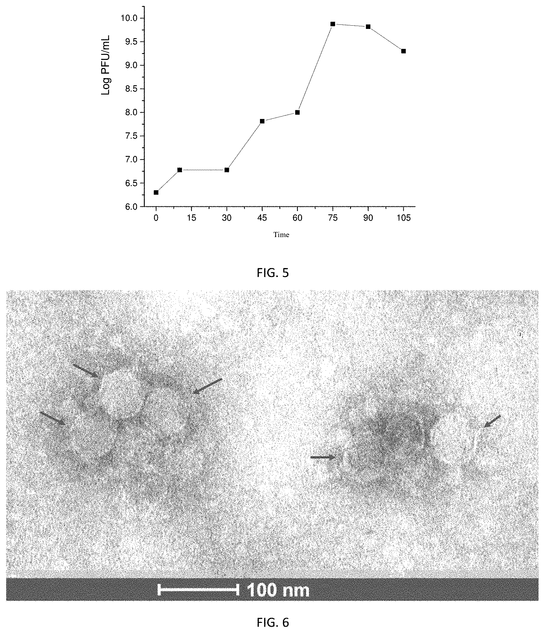
[0022] FIG. 5 shows a plot of one-step growth curve of the virulent phage .PHI.Pf1901;
[0023] FIG. 6 shows a TEM graph of the virulent phage .PHI.Pf1901;
[0024] FIG. 7 shows the control effect of the virulent phage .PHI.Pf1901 on P. fluorescens in tilapia fillets.
DESCRIPTION OF BIOLOGICAL PRESERVATION
[0025] The virulent P. fluorescens phage .PHI.Pf1901 provided by the present invention is preserved in China Center for Type Culture Collection (CCTCC), Wuhan University, Bayi Road, Hongshan District, Wuhan City, Hubei Province on Jun. 26, 2019, with an accession number of CCTCC M2019447.
DETAILED DESCRIPTION
[0026] The present invention provides a virulent Pseudomonas fluorescens (P. fluorescens) phage .PHI.Pf1901, with an accession number of CCTCC M2019447. The virulent P. fluorescens phage .PHI.Pf1901 provided by the present invention is derived from a sewage sample. In a specific embodiment of the present invention, the virulent phage .PHI.Pf1901 is derived from Jinzhou City, Liaoning Province; the virulent phage .PHI.Pf1901 can specifically identify and lyse P. fluorescens. The titer of the virulent phage .PHI.Pf1901 is preferably (1.4-3).times.10.sup.10 PFU/mL, and more preferably 1.42.times.10.sup.10 PFU/mL; the virulent phage .PHI.Pf1901 has an optimal multiplicity of infection (MOI) value of 0.0001.
[0027] In the present invention, the virulent phage .PHI.Pf1901 is isolated and obtained by the following steps: 1) mixing a sewage sample with CaCl.sub.2, conducting solid-liquid separation, collecting and filtering a liquid phase component, to obtain a filtrate; 2) mixing the filtrate obtained in step 1) with nutrient solution and P. fluorescens suspension, conducting shaking culture, to obtain a culture medium; and 3) centrifuging the culture medium, collecting supernatant, filtering, mixing and incubating the resulting filtrate with P. fluorescens suspension, mixing with the nutrient solution, growing in a plain agar plate to obtain the virulent phage .PHI.Pf1901.
[0028] In the present invention, the solid-liquid separation is conducted after mixing the sewage sample with CaCl.sub.2, and the filtrate is obtained by collecting and filtering the liquid phase component. In the present invention, the sewage sample preferably is derived from the wastewater discharged from Xiaoling River, Jinzhou City, Liaoning Province; in the present invention, the concentration of the CaCl.sub.2 in the mixture is preferably 0.8-1.2 mmol/L, and more preferably 1.0 mmol/L; the role of the CaCl.sub.2 is to cause phages in the sample to absorb host bacteria more easily. In the present invention, the solid-liquid separation is preferably centrifugation, the rotational speed of the centrifugation is preferably 8000 r/min, and the centrifugal time is preferably 10 min; in the present invention, the liquid phase component is collected and filtered after the solid-liquid separation, the filtering is preferably filtration by filter membrane, and the pore size of the filter membrane is preferably 0.22 .mu.m.
[0029] In the present invention, the culture medium is obtained by mixing the filtrate with nutrient solution and P. fluorescens suspension and conducting shaking culture. In the present invention, the filtrate, the nutrient solution, and the P. fluorescens suspension are at a volume ratio of 20:5:2; the nutrient solution is preferably LB liquid medium; the P. fluorescens is preferably in logarithmic growth phase. In the present invention, the temperature of the shaking culture is preferably 30.degree. C., the rotational speed of the shaking culture is preferably 130 r/min, and the time of the shaking culture is preferably 10-14 h, and more preferably 12 h.
[0030] In the present invention, after obtained the culture medium, the culture medium is centrifuged, followed by collecting and filtering the supernatant. In the present invention, the temperature of the centrifugation is preferably 4.degree. C., the rotational speed of the centrifugation is preferably 8000 r/min, and the centrifugal time is preferably 10 min. In the present invention, the filtering is preferably filtration by filter membrane, and the pore size of the filter membrane is preferably 0.22 .mu.m. In the present invention, after the filtering, the resulting filtrate is mixed and incubated with P. fluorescens suspension and then mixed with the nutrient solution, and is grown in a plain agar plate to obtain the virulent phage .PHI.Pf1901. In the present invention, the volume ratio of the filtrate to the P. fluorescens suspension is preferably 1:2; the mixing and incubating time is preferably 10-20 min. In the present invention, the volume ratio of the filtrate to the nutrient solution is preferably 1:50; the nutrient solution is preferably LB medium with an agar content of 0.75 wt % at a temperature of 50.degree. C. In the present invention, the plain agar plate is a plate that sterilized 1.5 wt % agar powder for 15 min at 121.degree. C. In the present invention, a double-layer plate is formed after the foregoing procedure. In the present invention, the double-layer plate is incubated in a constant temperature incubator for 10-14 h at 30.degree. C. after solidification; the double-layer plate requires no inverted culture, in which water favors phage adsorption.
[0031] After culturing and obtaining the virulent phage .PHI.Pf1901, the present invention preferably further includes purification of the virulent phage. In the present invention, the purification includes the following steps: S1) selecting and mixing a single plaque on the foregoing double-layer plate well with SM buffer to obtain a phage suspension; S2) conducting the phage suspension on gradient dilution and then growing in the double-layer plate; and S3) repeating the foregoing S1) to S2) two to four times to obtain purified phages.
[0032] In the present invention, the single plaque is preferably mixed well with 1 mL of SM buffer, the mixing time is preferably greater than or equal to 4 h, and the mixing temperature is preferably 4.degree. C.; the mixing is preferably conducted on a shaking table. In the present invention, the phage suspension prior to dilution is preferably oscillated; the dilution factor is preferably 10-fold gradient dilution, and more preferably selecting 10.sup.-5 and 10.sup.-6 diluents to grow on a double-layer plate. In the specific embodiment of the present invention, the diluent is preferably mixed with LB medium to prepare a double-layer plate after mixing with P. fluorescens suspension in logarithmic phase at a 2:1 volume ratio for 10-15 min. The foregoing double-layer plate purification steps are preferably to repeat three times to obtain purified phage .PHI.Pf1901.
[0033] After obtaining the purified phage .PHI.Pf1901, the present invention further includes storage of the purified phage .PHI.Pf1901. In the present invention, the storage method is preferably to conduct the purified phage .PHI.Pf1901 and SM buffer on mixing oscillation, centrifuge, collect supernatant, and store at 4.degree. C.
[0034] The present invention further provides a preparation comprising the virulent P. fluorescens phage .PHI.Pf1901, where the content of the virulent phage in the preparation is (1.4-3).times.10.sup.12 PFU, and preferably 1.42.times.10.sup.12 PFU.
[0035] The present invention further provides applications of the virulent P. fluorescens phage and the preparation comprising the virulent P. fluorescens phage .PHI.Pf1901 in the inhibition of P. fluorescens. In the present invention, the quantity ratio of the virulent P. fluorescens phage .PHI.Pf1901 to the P. fluorescens is (1-10):1. In the present invention, the virulent P. fluorescens phage .PHI.Pf1901 can be used to inhibit P. fluorescens in various environments, including but not limited to P. fluorescens in aquatic products.
[0036] The technical solutions of the present invention will be described in detail below in connection with Embodiments, but they should not be construed as the limitation of the protection scope of the present invention.
Embodiment 1
[0037] (1) Experimental Materials
[0038] Consumables: 0.22 .mu.m and 0.45 .mu.m filter membranes, Eppendorf tube (EP tube), test tubes, 50 mL centrifuge tubes, SM buffer, plain agar plate, LB plate, LB liquid medium, and LB semi-solid medium
[0039] Host bacteria: Pseudomonas fluorescens (P. fluorescens)
[0040] Medium: LB liquid medium, and agar powder
[0041] (2) Strain Culture
[0042] 1) P. fluorescens was removed at -20.degree. C. and cultured on a marked LB medium for 16-18 h at 30.degree. C.
[0043] 2) A single colony of P. fluorescens was picked up, grown on a LB liquid medium, and cultured for 8-10 h at 30.degree. C. with shaking at 130 r/min until it became turbid.
[0044] (3) Isolation of Phage
[0045] 1) Forty-five milliliters of sewage sample (wastewater at the outfall of Xiaoling River, Jinzhou City, Liaoning Province) was taken, CaCl.sub.2 (which allowed phages in the sample to absorb host bacteria more easily) was added to a final concentration of 1 mmol/L, dissolved, mixed well, and centrifuged for 10 min at 8000 r/min for supernatant. After filtration through a filter membrane with a pore size of 0.22 .mu.m, the resulting filtrate was stored at 4.degree. C.
[0046] 2) Twenty milliliters of the filtrate passed through the filter membrane was mixed with 5 mL of LB liquid medium and 2 mL of P. fluorescens suspension in logarithmic phase, and cultured on a shaking table overnight at 30.degree. C. with shaking at 130 r/min.
[0047] 3) After centrifugation for 10 min at 8000 r/min at 4.degree. C. and filtration through a 0.22 .mu.m filter membrane, 100 .mu.l of supernatant was mixed with 200 .mu.l of P. fluorescens suspension, and incubated for 15 min at room temperature, then mixed well with 5 mL of LB medium containing 0.75 wt % agar at 50.degree. C., and poured onto a plain agar plate (1.5 wt % agar powder, sterilized for 15 min at 121.degree. C.) to form a double-layer plate.
[0048] 4) After solidification, the double-layer plate was placed in a constant temperature incubator, incubated overnight at 30.degree. C., without inverted culture. Water favored phage adsorption.
[0049] (4) Purification of Phage (stored at 4.degree. C. temporarily)
[0050] 1) After culture overnight, a sterile inoculating needle was used to pick up a single clear plague from the foregoing plate with plagues into 1 mL of SM buffer and mixed well on a shaking table for 4 h at 4.degree. C. (alternatively, placed overnight).
[0051] 2) After oscillating and mixing well on a vortex mixer, 10 .mu.L of phage suspension was diluted gradiently (dilution factor was selected at 10.sup.-5 and 10.sup.-6). Then, 100 .mu.L of P. fluorescens suspension in logarithmic phase was mixed with 200 .mu.L of phage diluent for 15 min at room temperature, followed by preparing a double-layer plate with 5 mL of LB medium containing 0.75 wt % agar and incubating overnight at a constant temperature of 30.degree. C. Pure phage .PHI.Pf1901 was obtained after the double-layer plate method was repeated three times. The one-step growth curve of the phage .PHI.Pf1901 is illustrated in FIG. 5 and its TEM graph is shown in FIG. 6.
[0052] (5) Phage Titration and Determination of Optimal Multiplicity of Infection (MOI)
[0053] A solid plain agar plate was prepared in advance as a bottom layer of a double-layer plate. Sterile SM buffer was used for 10-fold serial gradient dilution of phage proliferation liquid. Then, 100 .mu.L of phage diluent with proper countable dilution factors (10.sup.-7 and 10.sup.-8) and 200 .mu.L of P. fluorescens suspension in logarithmic phase were added to 5 mL of LB semi-solid medium at 45-50.degree. C., mixed well on a vortex mixer, and poured onto the bottom plate. After sufficient condensation, the medium was inverted and incubated at 30.degree. C. in a constant temperature incubator until clear plagues appeared (for 12 h). For each dilution factor three parallel groups were set and averaged. The calculation formula of phage titer is as follows: phage titer (PFU/mL)=number of plagues.times.dilution factor.times.10. As shown in Table 1, the phage titer is calculated as 1.42.times.10.sup.10 PFU/mL.
TABLE-US-00001 TABLE 1 Phage titration results Dilution Number of Group average Overall average factor plagues Phage titer of phage titer of phage titer 10.sup.7 141 1.41 .times. 10.sup.10 1.36 .times. 10.sup.10 1.42 .times. 10.sup.10 10.sup.7 120 1.20 .times. 10.sup.10 10.sup.7 148 1.48 .times. 10.sup.10 10.sup.8 5 5.00 .times. 10.sup.9 1.47 .times. 10.sup.10 10.sup.8 22 2.20 .times. 10.sup.10 10.sup.8 17 1.70 .times. 10.sup.10
TABLE-US-00002 TABLE 2 Optimal MOIs of the virulent phage .PHI.Pf1901 Phage titer Bacterial count Phage count Optimal MOI at 12 h 2.236 .times. 10.sup.9 10.sup.10 10 1.8 .times. 10.sup.10 2.236 .times. 10.sup.9 10.sup.9 1 6.3 .times. 10.sup.11 2.236 .times. 10.sup.9 10.sup.8 0.1 1.1 .times. 10.sup.10 2.236 .times. 10.sup.9 10.sup.7 0.01 1.0 .times. 10.sup.10 2.236 .times. 10.sup.9 10.sup.6 0.001 2.0 .times. 10.sup.10 2.236 .times. 10.sup.9 10.sup.5 0.0001 1.8 .times. 10.sup.11
[0054] (6) Storage of Phage
[0055] Five milliliters of SM buffer were added onto the phage .PHI.Pf1901 plate which had conducted pure culture three times (for a plate with a diameter of 9 cm, 5 mL of SM buffer was added; for a plate with a diameter of 15 cm, 10 mL of SM buffer was added), oscillating on a shaking table for hours at 4.degree. C. The liquid on the plate was transferred into a sterile EP tube and centrifuged for 15 min at 5000 r/min at 4.degree. C. to remove bacteria debris. The resulting supernatant was pipetted into a new EP tube and stored at 4.degree. C. temporarily.
[0056] (7) Determination of Adsorption Rate
[0057] With MOI=0.1, phage .PHI.Pf1901 with a titer of 10.sup.10 and P. fluorescens suspension with a bacteria concentration of 10.sup.9 were added to culture on a shaking table; 100 .mu.L of mixture was pipetted for titration at different culture times; the test was repeated three times.
[0058] The calculation formula is: phage adsorption rate=(1-unabsorbed phage titer/initial phage titer).times.100%.
[0059] From FIG. 4, 86% of the phage .PHI.Pf1901 absorbed immediately after the phage .PHI.Pf1901 was mixed with the host bacteria suspension, 97% of the phage .PHI.Pf1901 absorbed after incubation for 5 min, and the adsorption rate of the phage .PHI.Pf1901 reached more than 90% between 5 min and 22 min, suggesting that the phage .PHI.Pf1901 exhibited a good adsorption rate.
[0060] (8) Measurement of Sensitivity to Chloroform
[0061] One milliliter of purified P. fluorescens phage .PHI.Pf1901 proliferation liquid was pipetted, mixed well with 1% chloroform solution while oscillating; after standing still for 30 min at room temperature for stratification, supernatant was pipetted for titration; changes in phage titer were compared before and after chloroform treatment, and data were recorded.
[0062] Phage .PHI.Pf1901 proliferation liquid: liquid of solid plate proliferation.
[0063] Phage .PHI.Pf1901 suspension: phage proliferation liquid treated by NaCl and PEG 8000.
[0064] The titers of the phage .PHI.Pf1901 proliferation liquid before and after chloroform treatment were 1.3.times.10.sup.11 and 1.2.times.10.sup.11, respectively.
[0065] The titers of the phage .PHI.Pf1901 suspension before and after chloroform treatment were 2.4.times.10.sup.10 and 1.2.times.10.sup.11, respectively.
[0066] The test results are shown particularly in Table 3.
TABLE-US-00003 TABLE 3 Sensitivity of virulent phage .PHI.Pf1901 to chloroform Presence of Sensitivity Phage chloroform Phage titer to chloroform Phage proliferation liquid Yes 1.3 .times. 10.sup.11 Insensitive No 1.2 .times. 10.sup.11 Phage suspension Yes 2.4 .times. 10.sup.10 Insensitive No 1.2 .times. 10.sup.11
[0067] (9) Determination of Optimal Temperature Range
[0068] Two hundred microliters each of phage .PHI.Pf1901 was placed in water bath kettles at room temperature (25.degree. C.) and preset at 30.degree. C., 40.degree. C., 50.degree. C., 60.degree. C., 70.degree. C., and 80.degree. C., respectively, and titrated 60 min after incubation.
[0069] Results are shown in FIG. 2. The phage .PHI.Pf1901 could maintain high titers after treating when treating for 1 h at room temperature (25.degree. C.), 30.degree. C., 40.degree. C., and 50.degree. C.; at 60.degree. C. and 70.degree. C., the titer of the phage .PHI.Pf1901 declined; at 80.degree. C., the titer of the phage .PHI.Pf1901 was 0.
[0070] (10) Determination of Optimal pH Range
[0071] Ten percent of phage .PHI.Pf1901 was placed on LB liquid media at pH 4, pH 5, pH 6, pH 7, pH 8, pH 9, pH 10, pH 11, and pH 12, respectively, and titrated 1 h after treatment at room temperature.
[0072] Results are shown in FIG. 3. The phage .PHI.Pf1901 could maintain high titers when treating for 1 h in LB liquid media at pH 5, pH 6, pH 7, pH 8, pH 9, pH 10, and pH 11; the titer of the phage .PHI.Pf1901 was low at pH 4 and pH 12.
Embodiment 2
[0073] Detection of lytic ability of virulent phage .PHI.Pf1901 to Pseudomonas fluorescens (P. fluorescens) (by plate method)
[0074] At room temperature, 200 .mu.L of P. fluorescens suspension with a concentration of 10.sup.9 was mixed with 100 .mu.L of phage .PHI.Pf1901 diluent for 10-15 min and then with 5 mL of LB medium containing 0.75% agar to make a double-layer plate, which was cultured overnight at a constant temperature of 30.degree. C. Result of plate lysis of lytic host cells is illustrated in FIG. 1. Through observation and measurement, plague was 1.36-3.06 mm in diameter.
Embodiment 3
[0075] Detection of lytic ability of virulent phage .PHI.Pf1901 to Pseudomonas fluorescens (P. fluorescens) (by OD.sub.600 method)
[0076] A single P. fluorescens was grown in 10 mL of LB broth and cultured on a shaking table overnight at 30.degree. C. with shaking at 130 r/min, and then 200 pl of the purified phage .PHI.Pf1901 was added to 5 mL of P. fluorescens suspension removed, while culturing on a shaking table for 2-4 h. Clarity was observed for the suspension every 30 min. Results are shown in Table 4.
TABLE-US-00004 TABLE 4 Lytic ability of virulent phage .PHI.Pf1901 (by OD.sub.600 method) Time/min Groups 0 30 60 90 120 150 180 210 Control 0.059 0.590 0.593 0.603 0.060 0.057 0.058 0.061 Host bacteria 0.301 0.280 0.273 0.257 0.252 0.214 0.215 0.177
Embodiment 4
[0077] Scavenging effect of virulent phage .PHI.Pf1901 on Pseudomonas fluorescens (P. fluorescens) in tilapia fillets
[0078] The ratio of virulent phage .PHI.Pf1901 to P. fluorescens was set at 1:1 and 10:1, while a control group was assigned. An observation was made of the scavenging effect of phage on P. fluorescens in a tilapia fillet.
[0079] Detailed steps:
[0080] Tilapias were purchased from a supermarket. The fish was filleted into split fillets weighed each 5.+-.0.5 g using a sterile scalpel blade. The fillets were immersed in freshly prepared 70% alcohol and sterilized for 1 min, placed in a sterile plastic plate after sterilization, and air dried in a benchtop.
[0081] For the air-dried fillets, 0.1 ml of 10.sup.4 cfu/ml P. fluorescens suspension was spread evenly on one side. After inoculation, the fillets were placed in the benchtop for 15 min to allow the bacteria to absorb spontaneously.
[0082] Low concentration group: Fillets were spread evenly with 0.1 ml of 10.sup.4 PFU/ml phage .PHI.Pf1901 suspension on the P. fluorescens inoculated surface;
[0083] High concentration group: Fillets were spread evenly with 0.1 ml of 10.sup.5 PFU/ml phage .PHI.Pf1901 suspension on the P. fluorescens inoculated surface;
[0084] Control group: Fillets were spread evenly with 0.1 ml of 0.85% NaCl on the P. fluorescens inoculated surface;
[0085] Each fillet was stored in a sterile plate. Three parallel groups were set in each group, sealed with cling film, placed in an incubator at 25.degree. C., and sampled at 0, 3, 6, 9, 12, 24, and 36 h, respectively. When sampling and testing, each fillet was removed and placed in a sterile sampling bag, in which 100 ml of 0.85% NaCl was added. After 10-fold gradient dilution on a solid LB plate and culture by the spread plate method, counting was carried out.
TABLE-US-00005 TABLE 5 Scavenging effect of virulent phage .PHI.Pf1901 on P. fluorescens in tilapia fillets Total plate count in a tilapia fillet at different sampling time (CFU/mL) Group 0 h 3 h 6 h 9 h 12 h 24 h 36 h Control 4.40E+04 3.53E+04 3.90E+04 3.10E+04 9.90E+04 1.83E+04 1.50E+04 Cf Ad Be Ac Ag Ab Aa High 1.21E+04 4.20E+03 1.39E+04 4.86E+04 2.72E+04 1.09E+04 9.83E+04 concentration Bab Aa Bde Ag Abc Acd ABef Low 1.26E+03 9.21E+02 4.60E+03 8.82E+03 3.08E+04 8.38E+04 1.11E+05 concentration Aa Aa Aa Aa Aa Aa Ba
[0086] From Table 5 and FIG. 7, at 0-15 h, there was a significant control effect of high and low concentrations of phage .PHI.Pf1901 suspension on P. fluorescens; at 15-36 h, the scavenging effect of the phage on the bacteria declined possibly because fillets were placed for a long time and water loss on the fillet surface resulted in low bacterial activity. By horizontal comparison among groups, low concentration group had a good control effect on P. fluorescens at 0-15 h. By vertical comparison among different groups, there was no significant difference between high and low concentration groups (p<0.05).
[0087] The foregoing descriptions are only preferred implementation manners of the present invention. It should be noted that for a person of ordinary skill in the art, several improvements and modifications may further be made without departing from the principle of the present invention. These improvements and modifications should also be deemed as falling within the protection scope of the present invention.
* * * * *
D00001
D00002
D00003
D00004
XML
uspto.report is an independent third-party trademark research tool that is not affiliated, endorsed, or sponsored by the United States Patent and Trademark Office (USPTO) or any other governmental organization. The information provided by uspto.report is based on publicly available data at the time of writing and is intended for informational purposes only.
While we strive to provide accurate and up-to-date information, we do not guarantee the accuracy, completeness, reliability, or suitability of the information displayed on this site. The use of this site is at your own risk. Any reliance you place on such information is therefore strictly at your own risk.
All official trademark data, including owner information, should be verified by visiting the official USPTO website at www.uspto.gov. This site is not intended to replace professional legal advice and should not be used as a substitute for consulting with a legal professional who is knowledgeable about trademark law.