Trans-antigen Targeting In Heterogeneous Cancers And Methods Of Use Thereof
Lim; Wendell A. ; et al.
U.S. patent application number 17/040476 was filed with the patent office on 2021-01-28 for trans-antigen targeting in heterogeneous cancers and methods of use thereof. The applicant listed for this patent is The Regents of the University of California. Invention is credited to Joseph H. Choe, Wendell A. Lim, Hideho Okada, Kole T. Roybal, Jasper Z. Williams.
| Application Number | 20210023136 17/040476 |
| Document ID | / |
| Family ID | 1000005182309 |
| Filed Date | 2021-01-28 |


| United States Patent Application | 20210023136 |
| Kind Code | A1 |
| Lim; Wendell A. ; et al. | January 28, 2021 |
TRANS-ANTIGEN TARGETING IN HETEROGENEOUS CANCERS AND METHODS OF USE THEREOF
Abstract
Provided are methods of treating a subject for a heterogeneous cancer. The methods of the present disclosure include integrating at least two antigens expressed heterogeneously in the cancer and/or in the cancer microenvironment, including where the antigens are expressed in trans, i.e., expressed by at least two different cell types. The subject methods will generally involve immune cells into which circuits have been introduced that employ one or more binding triggered transcriptional switches and one or more encoded therapeutics specific for antigens expressed by cancer cells and/or by neighboring non-cancer cells. Nucleic acids containing sequences encoding all or portions of such circuits are also provided, as well as cells, expression cassettes and vectors that contain such nucleic acids. Also provided are kits for practicing the described methods.
| Inventors: | Lim; Wendell A.; (San Francisco, CA) ; Okada; Hideho; (San Francisco, CA) ; Roybal; Kole T.; (San Francisco, CA) ; Choe; Joseph H.; (San Francisco, CA) ; Williams; Jasper Z.; (San Francisco, CA) | ||||||||||
| Applicant: |
|
||||||||||
|---|---|---|---|---|---|---|---|---|---|---|---|
| Family ID: | 1000005182309 | ||||||||||
| Appl. No.: | 17/040476 | ||||||||||
| Filed: | April 4, 2019 | ||||||||||
| PCT Filed: | April 4, 2019 | ||||||||||
| PCT NO: | PCT/US2019/025829 | ||||||||||
| 371 Date: | September 22, 2020 |
Related U.S. Patent Documents
| Application Number | Filing Date | Patent Number | ||
|---|---|---|---|---|
| 62653901 | Apr 6, 2018 | |||
| Current U.S. Class: | 1/1 |
| Current CPC Class: | A61K 38/1774 20130101; A61K 35/17 20130101; A61P 35/00 20180101 |
| International Class: | A61K 35/17 20060101 A61K035/17; A61K 38/17 20060101 A61K038/17; A61P 35/00 20060101 A61P035/00 |
Goverment Interests
STATEMENT REGARDING FEDERALLY SPONSORED RESEARCH
[0002] This invention was made with government support under grant nos. R01 CA196277, P50 GM081879 and R35 NS105068 awarded by the National Institutes of Health. The government has certain rights in the invention.
Claims
1. A method of treating a subject for a heterogeneous cancer comprising a priming cell and a cancer cell, the method comprising: administering to the subject an immune cell genetically modified with: (a) a nucleic acid sequence encoding a binding triggered transcriptional switch (BTTS) that binds to a priming antigen expressed by the priming cell; (b) a nucleic acid sequence encoding an antigen-specific therapeutic that binds to a killing antigen expressed by the cancer cell; and (c) a regulatory sequence operably linked to (b) that is responsive to the BTTS; wherein binding of the BTTS to the priming antigen activates expression of the antigen-specific therapeutic which binds the killing antigen thereby inducing killing of the cancer cell.
2. The method according to claim 1, wherein the priming antigen is not expressed by the cancer cell.
3. The method according to claim 1 or 2, wherein less than 95% of the cells of the heterogeneous cancer express the priming antigen.
4. The method according to any of the preceding claims, wherein less than 90% of the cells of the heterogeneous cancer express the priming antigen.
5. The method according to any of the preceding claims, wherein less than 50% of the cells of the heterogeneous cancer express the priming antigen.
6. The method according to any of the preceding claims, wherein the heterogeneous cancer is a solid tumor.
7. The method according to any of the preceding claims, wherein the heterogeneous cancer comprises a second cancer cell expressing both the priming antigen and the killing antigen and binding of the BTTS to the priming antigen activates expression of the antigen-specific therapeutic which binds the killing antigen thereby inducing killing of the second cancer cell.
8. The method according to any of the preceding claims, wherein the antigen-specific therapeutic, when expressed, is expressed on the surface of the immune cell.
9. The method according to claim 8, wherein the antigen-specific therapeutic is a chimeric antigen receptor (CAR) or a T cell receptor (TCR).
10. The method according to any of claims 1 to 7, wherein the antigen-specific therapeutic, when expressed, is secreted by the immune cell.
11. The method according to claim 10, wherein the antigen-specific therapeutic is a chimeric bispecific binding member.
12. The method according to claim 11, wherein the chimeric bispecific binding member is a TCR-targeted bispecific binding agent.
13. The method according to claim 11 or 12, wherein the chimeric bispecific binding member is specific for the killing antigen and a protein expressed on the surface of an immune cell.
14. The method according to any of the preceding claims, wherein the antigen-specific therapeutic comprises a bio-orthogonal adapter molecule.
15. The method according to claim 14, wherein the bio-orthogonal adapter molecule binds an extracellular domain of a switchable CAR.
16. The method according to any of the preceding claims, wherein the BTTS is a SynNotch polypeptide.
17. The method according to any of the preceding claims, wherein the immune cell is a myeloid cell.
18. The method according to any of claims 1 to 16, wherein the immune cell is a lymphoid cell.
19. The method according to claim 18, wherein the lymphoid cell is selected from the group consisting of: a T lymphocyte, a B lymphocyte and a Natural Killer cell.
20. The method according to any of the preceding claims, wherein the priming cell is a cancerous cell.
21. The method according to any of claims 1 to 19, wherein the priming cell is a non-cancerous cell in the proximity of the killing antigen-expressing cancer cell.
22. The method according to claim 21, wherein the non-cancerous cell is a stromal cell.
23. The method according to any of the preceding claims, wherein the immune cell is further genetically modified with a nucleic acid sequence encoding a BTTS that binds to a second priming antigen expressed by the heterogeneous cancer.
24. The method according to claim 23, wherein the BTTS that binds to the first priming antigen is also the BTTS that binds to the second priming antigen.
25. The method according to claim 23, wherein the immune cell is genetically modified to encode a first BTTS that binds to the first priming antigen and a second BTTS that binds the second priming antigen.
26. The method according to any of the preceding claims, wherein the immune cell is further genetically modified with a nucleic acid sequence encoding a second antigen-specific therapeutic that binds to a second killing antigen expressed by the heterogeneous tumor.
27. The method according to claim 26, wherein the second killing antigen is expressed by the priming cell.
28. The method according to claim 26, wherein the second killing antigen is expressed by the cancer cell that expresses the first killing antigen.
29. The method according to claim 26, wherein the second killing antigen is expressed by a cancerous cell of the heterogeneous tumor other than the priming cell or the cancer cell that expresses the first killing antigen.
30. The method according to any of the preceding claims, wherein the method further comprises identifying the heterogeneous tumor as comprising the priming cell and the cancer cell.
31. The method according to claim 30, wherein the identifying comprises assaying a biological sample obtained from the subject for cellular expression of the priming antigen and the killing antigen.
32. The method according to claim 31, wherein the biological sample is a tumor biopsy.
33. A method of treating a subject for a heterogeneous cancer comprising priming cells and cancer cells, the method comprising: administering to the subject an immune cell genetically modified with: (a) a nucleic acid sequence encoding a binding triggered transcriptional switch (BTTS) that binds to at least one priming antigen expressed by the priming cells; (b) a nucleic acid sequence encoding an antigen-specific therapeutic that binds to at least one killing antigen expressed by the cancer cells; and (c) a regulatory sequence operably linked to (b) that is responsive to the BTTS; wherein binding of the BTTS to the at least one priming antigen activates expression of the antigen-specific therapeutic which binds the at least one killing antigen thereby inducing killing of the cancer cell.
34. The method according to claim 33, wherein the BTTS binds to at least two different priming antigens such that binding of the BTTS to any of the at least two different priming antigens activates expression of the antigen-specific therapeutic.
35. The method according to claim 33 or claim 34, wherein the antigen-specific therapeutic binds to at least two different killing antigens such that binding of the antigen-specific therapeutic to any of the at least two different killing antigens induces killing of the cancer cells.
36. The method according to any of claims 33 to 35, wherein at least a portion of the cancer cells do not express at least one of the at least one priming antigens.
37. The method according to any of claims 33 to 36, wherein at least one of the at least one killing antigens is expressed by at least a portion of the priming cells.
38. The method according to any of claims 33 to 37, wherein less than 95% of the cells of the heterogeneous cancer express the at least one priming antigen.
39. The method according to any of claims 33 to 38, wherein less than 90% of the cells of the heterogeneous cancer express the at least one priming antigen.
40. The method according to any of claims 33 to 39, wherein less than 50% of the cells of the heterogeneous cancer express the at least one priming antigen.
41. The method according to any of claims 33 to 40, wherein the heterogeneous cancer is a solid tumor.
42. The method according to any of claims 33 to 41, wherein the heterogeneous cancer comprises a cancer cell expressing at least one priming antigen and at least one killing antigen and binding of the BTTS to at least one of the at least one priming antigens activates expression of the antigen-specific therapeutic which binds at least one of the at least one killing antigens thereby inducing killing of the cancer cell.
43. The method according to any of claims 33 to 42, wherein the antigen-specific therapeutic, when expressed, is expressed on the surface of the immune cell.
44. The method according to claim 43, wherein the antigen-specific therapeutic is a chimeric antigen receptor (CAR) or a T cell receptor (TCR).
45. The method according to any of claims 33 to 42, wherein the antigen-specific therapeutic, when expressed, is secreted by the immune cell.
46. The method according to claim 45, wherein the antigen-specific therapeutic is a chimeric bispecific binding member.
47. The method according to claim 46, wherein the chimeric bispecific binding member is a TCR-targeted bispecific binding agent.
48. The method according to claim 46 or 47, wherein the chimeric bispecific binding member is specific for the at least one killing antigen and a protein expressed on the surface of an immune cell.
49. The method according to any of claims 33 to 48, wherein the antigen-specific therapeutic comprises a bio-orthogonal adapter molecule.
50. The method according to claim 49, wherein the bio-orthogonal adapter molecule binds an extracellular domain of a switchable CAR.
51. The method according to any of claims 33 to 50, wherein the BTTS is a SynNotch polypeptide.
52. The method according to any of claims 33 to 51, wherein the immune cell is a myeloid cell.
53. The method according to any of claims 33 to 51, wherein the immune cell is a lymphoid cell.
54. The method according to claim 53, wherein the lymphoid cell is selected from the group consisting of: a T lymphocyte, a B lymphocyte and a Natural Killer cell.
55. The method according to any of claims 33 to 54, wherein the priming cells comprise cancerous cells, non-cancerous cells, or a mixture thereof.
56. The method according to any of claims 33 to 55, wherein the method further comprises identifying the heterogeneous tumor as comprising the priming cells and the cancer cells.
57. The method according to claim 56, wherein the identifying comprises assaying a biological sample obtained from the subject for cellular expression of the at least one priming antigen and the at least one killing antigen.
58. The method according to claim 57, wherein the biological sample is a tumor biopsy.
Description
CROSS-REFERENCE TO RELATED APPLICATIONS
[0001] This application claims the benefit of U.S. Provisional Patent Application Ser. No. 62/653,901 filed Apr. 6, 2018; the disclosure of which application is herein incorporated by reference.
INTRODUCTION
[0003] Currently, the design of targeted oncological therapies is mainly based on categorization of different cancers and tumors from separate patients into categories based on characteristic cancer/tumor features, including morphology, biomarker expression, genomics and the like. Thus, inter-patient tumor heterogeneity is broadly recognized. However, it is gradually becoming clear that intra-tumor heterogeneity has a significant impact on pathology as well as the relative effectiveness of targeted therapies. Clonal cancer cell populations that evade a targeted therapy may result in a minimal but significant residual group of cancer cells, which could serve as a source for recurrent and, in some cases, refractory cancers. Moreover, heterogeneity in tumor antigen expression makes targeting all cells of a heterogeneous tumor with a single therapy particularly difficult.
SUMMARY
[0004] Provided are methods of treating a subject for a heterogeneous cancer. The methods of the present disclosure include integrating at least two antigens expressed heterogeneously in the cancer and/or in the cancer microenvironment, including where the antigens are expressed in trans, i.e., expressed by at least two different cell types. The subject methods will generally involve immune cells into which circuits have been introduced that employ one or more binding triggered transcriptional switches and one or more encoded therapeutics specific for antigens expressed by cancer cells and/or by neighboring non-cancer cells. Nucleic acids containing sequences encoding all or portions of such circuits are also provided, as well as cells, expression cassettes and vectors that contain such nucleic acids. Also provided are kits for practicing the described methods.
BRIEF DESCRIPTION OF THE DRAWINGS
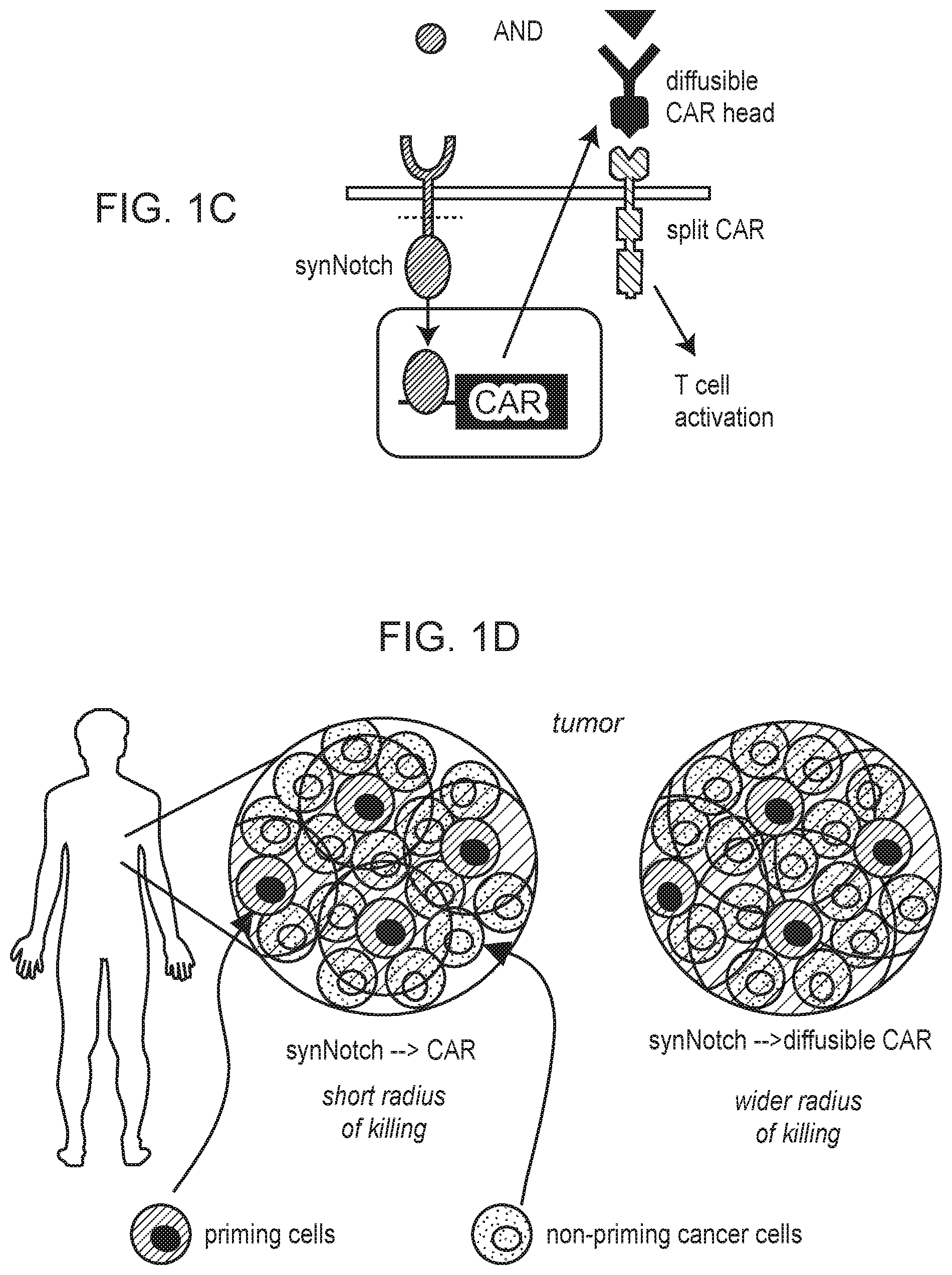
[0005] FIG. 1A-1D depict examples of trans-killing circuits, with or without diffusible components and employing antigen recognition of priming antigens expressed on cancerous or non-cancerous cells.
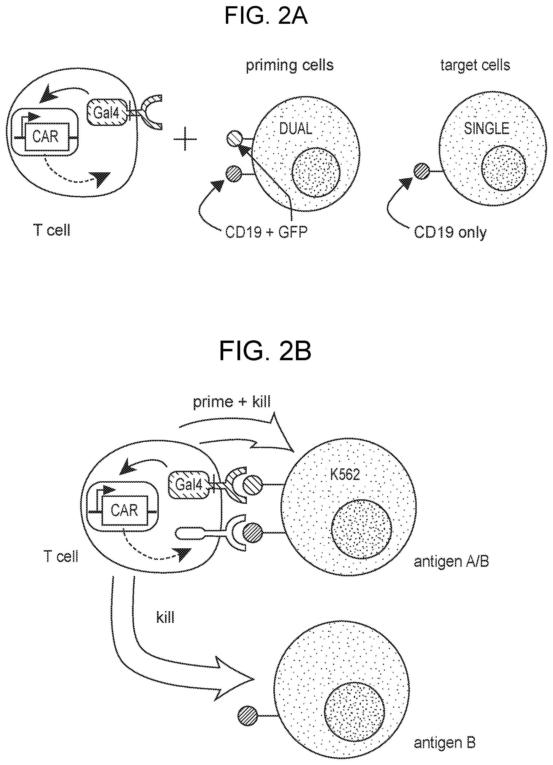
[0006] FIG. 2A-2C depict efficient trans-killing in heterogeneous mixtures of targeted cells containing various ratios of priming cells to target cells.
[0007] FIG. 3A-3D depict target cell killing using 2-receptor IF/THEN circuits where CAR expression is induced by a GFP antigen-expressing cells.
[0008] FIG. 4A-4C depict target cell killing using 2-receptor IF/THEN circuits where CAR expression is induced by a PNE antigen-expressing cells.
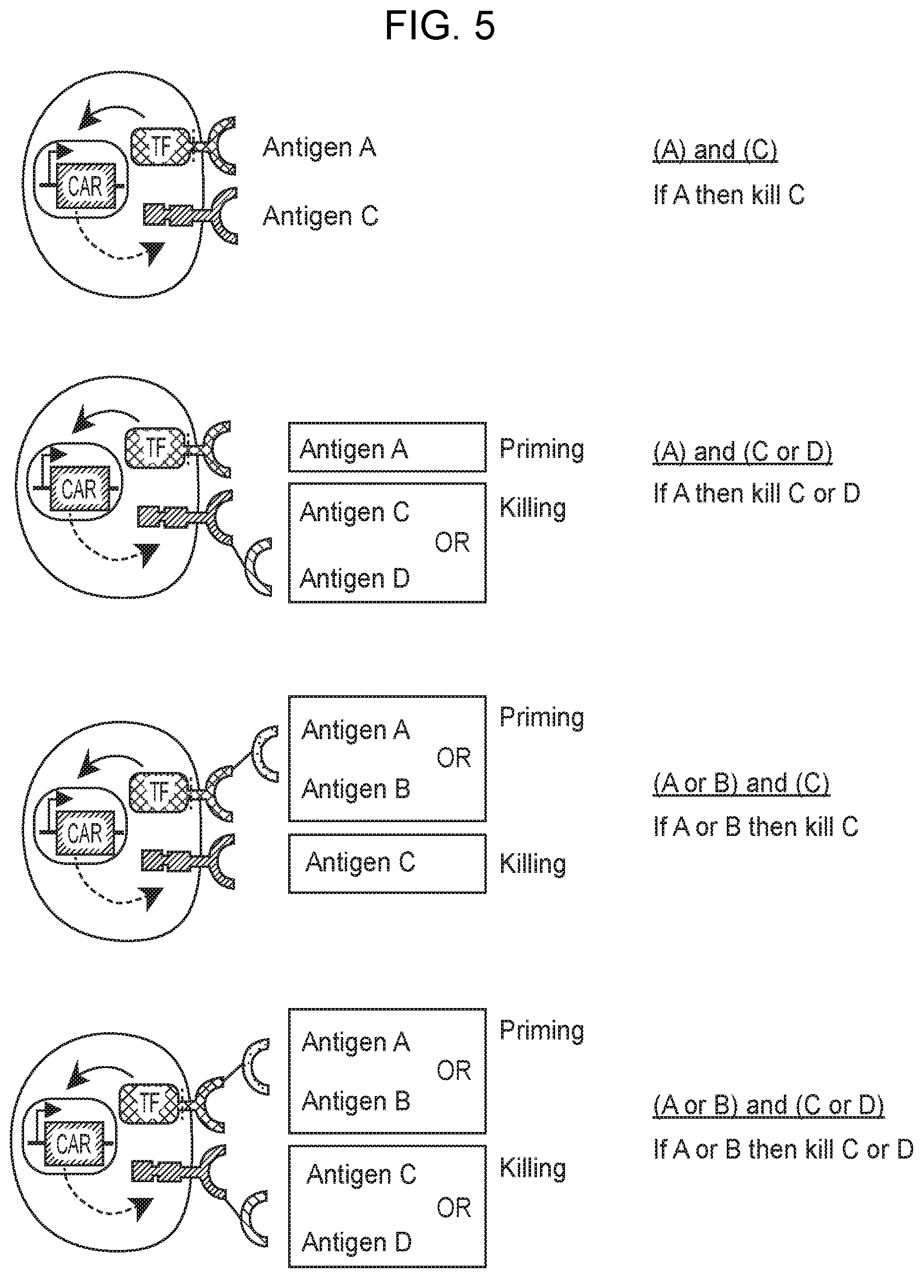
[0009] FIG. 5 depicts cells that contain IF/THEN circuits with and without OR gate functionality at the relevant binding triggered transcriptional switch, the antigen-specific therapeutic, or both.
DEFINITIONS
[0010] As used herein, the term "heterogeneous cancer" generally refers to a cancer displaying some level of intracancer or intratumor heterogeneity, e.g., at the molecular, cellular, tissue or organ level. A heterogeneous cancer is composed of at least two different cell types, where different cell types may be defined in variety of ways. For example, different cell types may differ genomically (e.g., through the presence of a mutation in one cell type that is absent in another), transcriptionally (e.g., through expression of a gene in one cell type that is not expressed in another, through enhanced or reduced expression of a gene in one cell type as compared to another, etc.), or proteomically (e.g., through expression of a protein in one cell type that is not expressed in another, through enhanced or reduced expression of a protein in one cell type as compared to another, etc.). In some instances, cancer heterogeneity may be identified based on the presence of two or more phenotypically different cells present in a cancer, including e.g., where such phenotypically different cells are identified through clinical testing (e.g., histology, immunohistochemistry, in situ hybridization, cytometry, transcriptomics, mutational analysis, whole genome sequencing, proteomics, etc.).
[0011] As such, a heterogeneous cancer, as defined herein, will generally include at least one cancerous cell type and at least one other cell type, where the one other cell type may be a second cancerous cell type or a non-cancerous cell type. For example, a heterogeneous cancer may include a first cancerous cell type and a second cancerous cell type. Alternatively, a heterogeneous cancer may include a cancerous cell type and a non-cancerous cell type. Although a heterogeneous cancer will include at least two different cell types, such cancers are not so limited and may include e.g., more than two different cell types, three or more different cell types, four or more different cell types, five or more different cell types, etc., where at least one cell type is cancerous and the additional cell types may each be cancerous or non-cancerous.
[0012] As summarized above, heterogeneity of a cancer may be defined by differing gene or protein expression by different subpopulations of cells of the cancer. For example, in some instances, a first subpopulation of cells may express a first gene product from a first gene that is not expressed by a second subpopulation of cells, where such a second cell population may or may not express a second gene product from a second gene that defines the second population. Put another way, subpopulations of cells within a heterogeneous cancer may, in some instances, each be defined by the presence or absence (or relative levels) of one or more expressed gene products, where useful expressed gene products for defining cell types may include but are not limited to biomarkers, antigens, wild-type proteins, mutated proteins, wild-type transcripts, mutated transcripts, etc.
[0013] Cancer heterogeneity, in some instances, may include or exclude heterogeneity at the subject level, i.e., intrapatient heterogeneity. As used herein, the term "intrapatient heterogeneity" generally refers to heterogeneity observed between multiple cancers, e.g., multiple tumors, present in a single subject. For example, a primary tumor and a metastasis with a subject may be heterogeneous, e.g., differentially expressing a particular gene product, such as a biomarker, an antigen or a mutated protein. Multiple heterogeneous cancers may arise in a subject through various mechanisms including but not limited to mutation, clonal expansion, metastasis, selection, and combinations thereof. For example, two different intrapatient heterogeneous cancers arising by metastasis of a primary tumor may be heterogeneous with respect to the tissues in which they reside. Alternatively, two different intrapatient heterogeneous cancers derived from the same primary tumor may arise due to mutation and clonal expansion, where one cancer is a subclone of the other. Various other mechanism by which different intrapatient heterogeneous cancers may arise are possible and fall within the scope of the term as used herein.
[0014] Cancer heterogeneity, in some instances as used herein, may exclude heterogeneity at the population level, i.e., interpatient heterogeneity. As used herein, the term "interpatient heterogeneity" generally refers to differences observed between two cancers or two tumors present in separate subjects or patients.
[0015] As used herein, the terms "treatment," "treating," "treat" and the like, refer to obtaining a desired pharmacologic and/or physiologic effect and/or a response related to the treatment. The effect can be prophylactic in terms of completely or partially preventing a disease or symptom thereof and/or can be therapeutic in terms of a partial or complete cure for a disease and/or adverse effect attributable to the disease. "Treatment," as used herein, covers any treatment of a disease in a mammal, particularly in a human, and includes: (a) preventing the disease from occurring in a subject which can be predisposed to the disease but has not yet been diagnosed as having it; (b) inhibiting the disease, i.e., arresting its development; and (c) relieving the disease, i.e., causing regression of the disease.
[0016] A "therapeutically effective amount" or "efficacious amount" refers to the amount of an agent (including biologic agents, such as cells), or combined amounts of two agents, that, when administered to a mammal or other subject for treating a disease, is sufficient to effect such treatment for the disease. The "therapeutically effective amount" will vary depending on the agent(s), the disease and its severity and the age, weight, etc., of the subject to be treated.
[0017] The terms "individual," "subject," "host," and "patient," used interchangeably herein, refer to a mammal, including, but not limited to, murines (e.g., rats, mice), non-human primates, humans, canines, felines, ungulates (e.g., equines, bovines, ovines, porcines, caprines), lagomorphs, etc. In some cases, the individual is a human. In some cases, the individual is a non-human primate. In some cases, the individual is a rodent, e.g., a rat or a mouse. In some cases, the individual is a lagomorph, e.g., a rabbit.
[0018] The term "refractory", used herein, refers to a disease or condition that does not respond to treatment. With regard to cancer, "refractory cancer", as used herein, refers to cancer that does not respond to treatment. A refractory cancer may be resistant at the beginning of treatment or it may become resistant during treatment. Refractory cancer may also called resistant cancer.
[0019] The term "histology" and "histological" as used herein generally refers to microscopic analysis of the cellular anatomy and/or morphology of cells obtained from a multicellular organism including but not limited to plants and animals.
[0020] The term "cytology" and "cytological" as used herein generally refers to a subclass of histology that includes the microscopic analysis of individual cells, dissociated cells, loose cells, clusters of cells, etc. Cells of a cytological sample may be cells in or obtained from one or more bodily fluids or cells obtained from a tissue that have been dissociated into a liquid cellular sample.
[0021] The terms "chimeric antigen receptor" and "CAR", used interchangeably herein, refer to artificial multi-module molecules capable of triggering or inhibiting the activation of an immune cell which generally but not exclusively comprise an extracellular domain (e.g., a ligand/antigen binding domain), a transmembrane domain and one or more intracellular signaling domains. The term CAR is not limited specifically to CAR molecules but also includes CAR variants. CAR variants include split CARs wherein the extracellular portion (e.g., the ligand binding portion) and the intracellular portion (e.g., the intracellular signaling portion) of a CAR are present on two separate molecules. CAR variants also include ON-switch CARs which are conditionally activatable CARs, e.g., comprising a split CAR wherein conditional hetero-dimerization of the two portions of the split CAR is pharmacologically controlled (e.g., as described in PCT publication no. WO 2014/127261 A1 and US Patent Application No. 2015/0368342 A1, the disclosures of which are incorporated herein by reference in their entirety). CAR variants also include bispecific CARs, which include a secondary CAR binding domain that can either amplify or inhibit the activity of a primary CAR. CAR variants also include inhibitory chimeric antigen receptors (iCARs) which may, e.g., be used as a component of a bispecific CAR system, where binding of a secondary CAR binding domain results in inhibition of primary CAR activation. CAR molecules and derivatives thereof (i.e., CAR variants) are described, e.g., in PCT Application No. US2014/016527; Fedorov et al. Sci Transl Med (2013); 5(215):215ra172; Glienke et al. Front Pharmacol (2015) 6:21; Kakarla & Gottschalk 52 Cancer J (2014) 20(2):151-5; Riddell et al. Cancer J (2014) 20(2):141-4; Pegram et al. Cancer J (2014) 20(2):127-33; Cheadle et al. Immunol Rev (2014) 257(1):91-106; Barrett et al. Annu Rev Med (2014) 65:333-47; Sadelain et al. Cancer Discov (2013) 3(4):388-98; Cartellieri et al., J Biomed Biotechnol (2010) 956304; the disclosures of which are incorporated herein by reference in their entirety. Useful CARs also include the anti-CD19-4-1BB-CD3.zeta. CAR expressed by lentivirus loaded CTL019 (Tisagenlecleucel-T) CAR-T cells as commercialized by Novartis (Basel, Switzerland).
[0022] The terms "T cell receptor" and "TCR" are used interchangeably and will generally refer to a molecule found on the surface of T cells, or T lymphocytes, that is responsible for recognizing fragments of antigen as peptides bound to major histocompatibility complex (MHC) molecules. The TCR complex is a disulfide-linked membrane-anchored heterodimeric protein normally consisting of the highly variable alpha (.alpha.) and beta (.beta.) chains expressed as part of a complex with CD3 chain molecules. Many native TCRs exist in heterodimeric .alpha..beta. or .gamma..delta. forms. The complete endogenous TCR complex in heterodimeric .alpha..beta. form includes eight chains, namely an alpha chain (referred to herein as TCR.alpha. or TCR alpha), beta chain (referred to herein as TCR.beta. or TCR beta), delta chain, gamma chain, two epsilon chains and two zeta chains. In some instance, a TCR is generally referred to by reference to only the TCR.alpha. and TCR.beta. chains, however, as the assembled TCR complex may associate with endogenous delta, gamma, epsilon and/or zeta chains an ordinary skilled artisan will readily understand that reference to a TCR as present in a cell membrane may include reference to the fully or partially assembled TCR complex as appropriate.
[0023] Recombinant or engineered individual TCR chains and TCR complexes have been developed. References to the use of a TCR in a therapeutic context may refer to individual recombinant TCR chains. As such, engineered TCRs may include individual modified TCR.alpha. or modified TCR.beta. chains as well as single chain TCRs that include modified and/or unmodified TCR.alpha. and TCR.beta. chains that are joined into a single polypeptide by way of a linking polypeptide.
[0024] As used herein, by "chimeric bispecific binding member" is meant a chimeric polypeptide having dual specificity to two different binding partners (e.g., two different antigens). Non-limiting examples of chimeric bispecific binding members include bispecific antibodies, bispecific conjugated monoclonal antibodies (mab).sub.2, bispecific antibody fragments (e.g., F(ab).sub.2, bispecific scFv, bispecific diabodies, single chain bispecific diabodies, etc.), bispecific T cell engagers (BiTE), bispecific conjugated single domain antibodies, micabodies and mutants thereof, and the like. Non-limiting examples of chimeric bispecific binding members also include those chimeric bispecific agents described in Kontermann. MAbs. (2012) 4(2): 182-197; Stamova et al. Antibodies 2012, 1(2), 172-198; Farhadfar et al. Leuk Res. (2016) 49:13-21; Benjamin et al. Ther Adv Hematol. (2016) 7(3):142-56; Kiefer et al. Immunol Rev. (2016) 270(1):178-92; Fan et al. J Hematol Oncol. (2015) 8:130; May et al. Am J Health Syst Pharm. (2016) 73(1):e6-e13; the disclosures of which are incorporated herein by reference in their entirety.
[0025] A "biological sample" encompasses a variety of sample types obtained from an individual or a population of individuals and can be used in various ways, including e.g., the isolation of cells or biological molecules, diagnostic assays, etc. The definition encompasses blood and other liquid samples of biological origin, solid tissue samples such as a biopsy specimen or tissue cultures or cells derived therefrom and the progeny thereof. The definition also includes samples that have been manipulated in any way after their procurement, such as by mixing or pooling of individual samples, treatment with reagents, solubilization, or enrichment for certain components, such as cells, polynucleotides, polypeptides, etc. The term "biological sample" encompasses a clinical sample, and also includes cells in culture, cell supernatants, cell lysates, serum, plasma, biological fluid, and tissue samples. The term "biological sample" includes urine, saliva, cerebrospinal fluid, interstitial fluid, ocular fluid, synovial fluid, blood fractions such as plasma and serum, and the like. The term "biological sample" also includes solid tissue samples, tissue culture samples (e.g., biopsy samples), and cellular samples. Accordingly, biological samples may be cellular samples or acellular samples.
[0026] The terms "antibodies" and "immunoglobulin" include antibodies or immunoglobulins of any isotype, fragments of antibodies which retain specific binding to antigen, including, but not limited to, Fab, Fv, scFv, and Fd fragments, chimeric antibodies, humanized antibodies, single-chain antibodies, nanobodies, single-domain antibodies, and fusion proteins comprising an antigen-binding portion of an antibody and a non-antibody protein.
[0027] "Antibody fragments" comprise a portion of an intact antibody, for example, the antigen binding or variable region of the intact antibody. Examples of antibody fragments include Fab, Fab', F(ab')2, and Fv fragments; diabodies; linear antibodies (Zapata et al., Protein Eng. 8(10): 1057-1062 (1995)); single-chain antibody molecules; and multispecific antibodies formed from antibody fragments. Papain digestion of antibodies produces two identical antigen-binding fragments, called "Fab" fragments, each with a single antigen-binding site, and a residual "Fc" fragment, a designation reflecting the ability to crystallize readily. Pepsin treatment yields an F(ab')2 fragment that has two antigen combining sites and is still capable of cross-linking antigen.
[0028] "Single-chain Fv" or "sFv" antibody fragments comprise the VH and VL domains of antibody, wherein these domains are present in a single polypeptide chain. In some embodiments, the Fv polypeptide further comprises a polypeptide linker between the VH and VL domains, which enables the sFv to form the desired structure for antigen binding. For a review of sFv, see Pluckthun in The Pharmacology of Monoclonal Antibodies, vol. 113, Rosenburg and Moore eds., Springer-Verlag, New York, pp. 269-315 (1994).
[0029] The term "nanobody" (Nb), as used herein, refers to the smallest antigen binding fragment or single variable domain (V.sub.HH) derived from naturally occurring heavy chain antibody and is known to the person skilled in the art. They are derived from heavy chain only antibodies, seen in camelids (Hamers-Casterman et al. (1993) Nature 363:446; Desmyter et al. (2015) Curr. Opin. Struct. Biol. 32:1). In the family of "camelids" immunoglobulins devoid of light polypeptide chains are found. "Camelids" comprise old world camelids (Camelus bactrianus and Camelus dromedarius) and new world camelids (for example, Llama paccos, Llama glama, Llama guanicoe and Llama vicugna). A single variable domain heavy chain antibody is referred to herein as a nanobody or a V.sub.HH antibody.
[0030] As used herein, the term "affinity" refers to the equilibrium constant for the reversible binding of two agents and is expressed as a dissociation constant (Kd). Affinity can be at least 1-fold greater, at least 2-fold greater, at least 3-fold greater, at least 4-fold greater, at least 5-fold greater, at least 6-fold greater, at least 7-fold greater, at least 8-fold greater, at least 9-fold greater, at least 10-fold greater, at least 20-fold greater, at least 30-fold greater, at least 40-fold greater, at least 50-fold greater, at least 60-fold greater, at least 70-fold greater, at least 80-fold greater, at least 90-fold greater, at least 100-fold greater, or at least 1000-fold greater, or more, than the affinity of an antibody for unrelated amino acid sequences. Affinity of an antibody to a target protein can be, for example, from about 100 nanomolar (nM) to about 0.1 nM, from about 100 nM to about 1 picomolar (pM), or from about 100 nM to about 1 femtomolar (fM) or more. As used herein, the term "avidity" refers to the resistance of a complex of two or more agents to dissociation after dilution. The terms "immunoreactive" and "preferentially binds" are used interchangeably herein with respect to antibodies and/or antigen-binding fragments.
[0031] The term "binding" refers to a direct association between two molecules, due to, for example, covalent, electrostatic, hydrophobic, and ionic and/or hydrogen-bond interactions, including interactions such as salt bridges and water bridges. Non-specific binding would refer to binding with an affinity of less than about 10.sup.-7 M, e.g., binding with an affinity of 10.sup.-6 M, 10.sup.-5 M, 10.sup.-4 M, etc.
[0032] A "orthogonal" or "orthogonalized" member or members of a binding pair are modified from their original or wild-type forms such that the orthogonal pair specifically bind one another but do not specifically or substantially bind the non-modified or wild-type components of the pair. Any binding partner/specific binding pair may be orthogonalized, including but not limited to e.g., those binding partner/specific binding pairs described herein.
[0033] The terms "domain" and "motif", used interchangeably herein, refer to both structured domains having one or more particular functions and unstructured segments of a polypeptide that, although unstructured, retain one or more particular functions. For example, a structured domain may encompass but is not limited to a continuous or discontinuous plurality of amino acids, or portions thereof, in a folded polypeptide that comprise a three-dimensional structure which contributes to a particular function of the polypeptide. In other instances, a domain may include an unstructured segment of a polypeptide comprising a plurality of two or more amino acids, or portions thereof, that maintains a particular function of the polypeptide unfolded or disordered. Also encompassed within this definition are domains that may be disordered or unstructured but become structured or ordered upon association with a target or binding partner. Non-limiting examples of intrinsically unstructured domains and domains of intrinsically unstructured proteins are described, e.g., in Dyson & Wright. Nature Reviews Molecular Cell Biology 6:197-208.
[0034] The terms "synthetic", "chimeric" and "engineered" as used herein generally refer to artificially derived polypeptides or polypeptide encoding nucleic acids that are not naturally occurring. Synthetic polypeptides and/or nucleic acids may be assembled de novo from basic subunits including, e.g., single amino acids, single nucleotides, etc., or may be derived from pre-existing polypeptides or polynucleotides, whether naturally or artificially derived, e.g., as through recombinant methods. Chimeric and engineered polypeptides or polypeptide encoding nucleic acids will generally be constructed by the combination, joining or fusing of two or more different polypeptides or polypeptide encoding nucleic acids or polypeptide domains or polypeptide domain encoding nucleic acids. Chimeric and engineered polypeptides or polypeptide encoding nucleic acids include where two or more polypeptide or nucleic acid "parts" that are joined are derived from different proteins (or nucleic acids that encode different proteins) as well as where the joined parts include different regions of the same protein (or nucleic acid encoding a protein) but the parts are joined in a way that does not occur naturally.
[0035] The term "recombinant", as used herein describes a nucleic acid molecule, e.g., a polynucleotide of genomic, cDNA, viral, semisynthetic, and/or synthetic origin, which, by virtue of its origin or manipulation, is not associated with all or a portion of the polynucleotide sequences with which it is associated in nature. The term recombinant as used with respect to a protein or polypeptide means a polypeptide produced by expression from a recombinant polynucleotide. The term recombinant as used with respect to a host cell or a virus means a host cell or virus into which a recombinant polynucleotide has been introduced. Recombinant is also used herein to refer to, with reference to material (e.g., a cell, a nucleic acid, a protein, or a vector) that the material has been modified by the introduction of a heterologous material (e.g., a cell, a nucleic acid, a protein, or a vector).
[0036] The term "operably linked" refers to a juxtaposition wherein the components so described are in a relationship permitting them to function in their intended manner. For instance, a promoter is operably linked to a coding sequence if the promoter affects its transcription or expression. Operably linked nucleic acid sequences may but need not necessarily be adjacent. For example, in some instances a coding sequence operably linked to a promoter may be adjacent to the promoter. In some instances, a coding sequence operably linked to a promoter may be separated by one or more intervening sequences, including coding and non-coding sequences. Also, in some instances, more than two sequences may be operably linked including but not limited to e.g., where two or more coding sequences are operably linked to a single promoter.
[0037] The terms "polynucleotide" and "nucleic acid," used interchangeably herein, refer to a polymeric form of nucleotides of any length, either ribonucleotides or deoxyribonucleotides. Thus, this term includes, but is not limited to, single-, double-, or multi-stranded DNA or RNA, genomic DNA, cDNA, DNA-RNA hybrids, or a polymer comprising purine and pyrimidine bases or other natural, chemically or biochemically modified, non-natural, or derivatized nucleotide bases.
[0038] The terms "polypeptide," "peptide," and "protein", used interchangeably herein, refer to a polymeric form of amino acids of any length, which can include genetically coded and non-genetically coded amino acids, chemically or biochemically modified or derivatized amino acids, and polypeptides having modified peptide backbones. The term includes fusion proteins, including, but not limited to, fusion proteins with a heterologous amino acid sequence, fusions with heterologous and homologous leader sequences, with or without N-terminal methionine residues; immunologically tagged proteins; and the like.
[0039] A "vector" or "expression vector" is a replicon, such as plasmid, phage, virus, or cosmid, to which another DNA segment, i.e. an "insert", may be attached so as to bring about the replication of the attached segment in a cell.
[0040] The term "Heterologous", as used herein, means a nucleotide or polypeptide sequence that is not found in the native (e.g., naturally-occurring) nucleic acid or protein, respectively. Heterologous nucleic acids or polypeptide may be derived from a different species as the organism or cell within which the nucleic acid or polypeptide is present or is expressed. Accordingly, a heterologous nucleic acids or polypeptide is generally of unlike evolutionary origin as compared to the cell or organism in which it resides.
[0041] Before the present invention is further described, it is to be understood that this invention is not limited to particular embodiments described, as such may, of course, vary. It is also to be understood that the terminology used herein is for the purpose of describing particular embodiments only, and is not intended to be limiting, since the scope of the present invention will be limited only by the appended claims.
[0042] Where a range of values is provided, it is understood that each intervening value, to the tenth of the unit of the lower limit unless the context clearly dictates otherwise, between the upper and lower limit of that range and any other stated or intervening value in that stated range, is encompassed within the invention. The upper and lower limits of these smaller ranges may independently be included in the smaller ranges, and are also encompassed within the invention, subject to any specifically excluded limit in the stated range. Where the stated range includes one or both of the limits, ranges excluding either or both of those included limits are also included in the invention.
[0043] Unless defined otherwise, all technical and scientific terms used herein have the same meaning as commonly understood by one of ordinary skill in the art to which this invention belongs. Although any methods and materials similar or equivalent to those described herein can also be used in the practice or testing of the present invention, the preferred methods and materials are now described. All publications mentioned herein are incorporated herein by reference to disclose and describe the methods and/or materials in connection with which the publications are cited.
[0044] It must be noted that as used herein and in the appended claims, the singular forms "a," "an," and "the" include plural referents unless the context clearly dictates otherwise. Thus, for example, reference to "a cell" includes a plurality of such cells and reference to "the cell" includes reference to one or more cells and equivalents thereof known to those skilled in the art, and so forth. It is further noted that the claims may be drafted to exclude any optional element. As such, this statement is intended to serve as antecedent basis for use of such exclusive terminology as "solely," "only" and the like in connection with the recitation of claim elements, or use of a "negative" limitation.
[0045] It is appreciated that certain features of the invention, which are, for clarity, described in the context of separate embodiments, may also be provided in combination in a single embodiment. Conversely, various features of the invention, which are, for brevity, described in the context of a single embodiment, may also be provided separately or in any suitable sub-combination. All combinations of the embodiments pertaining to the invention are specifically embraced by the present invention and are disclosed herein just as if each and every combination was individually and explicitly disclosed. In addition, all sub-combinations of the various embodiments and elements thereof are also specifically embraced by the present invention and are disclosed herein just as if each and every such sub-combination was individually and explicitly disclosed herein.
[0046] The publications discussed herein are provided solely for their disclosure prior to the filing date of the present application. Nothing herein is to be construed as an admission that the present invention is not entitled to antedate such publication by virtue of prior invention. Further, the dates of publication provided may be different from the actual publication dates which may need to be independently confirmed.
DETAILED DESCRIPTION
[0047] The present disclosure involves circuits integrating two or more antigens expressed heterogeneously by cells that are spatially associated with one another. For example, the subject circuits may integrate a first antigen expressed on a first cell and a second antigen expressed on a second cell to produce a desired outcome with respect to the second cell. The integration of two antigens expressed by different cells of a heterogeneous cell population to result in a desired targeting event may be referred to herein as "trans-antigen targeting".
[0048] For example, an employed circuit may integrate a first antigen (e.g., a "priming antigen") expressed by a first cell type, referred to as a "priming cell", and a second antigen (e.g., a "targeting antigen" or "targeted antigen") expressed by a nearby second cell type, referred to as a "targeted cell", to target the second cell type in trans. A cell modified with such a circuit is primed by the presence of the priming antigen on the first cell type to then target the nearby targeted cell. When such a circuit is present in an immune cell, the immune cell may target the targeted cell for destruction or killing and, as such, the targeting antigen may also, in some instances, be referred to as a "killing antigen".
[0049] For comparison, in this context cis-targeting refers to integrating of two antigens to target a single cell which expresses both a priming antigen and a targeting antigen to produce a desired outcome with respect to the single cell. Thus, in cis-targeting, the targeted cell expresses both the priming antigen and the targeting antigen such that the two antigens are expressed in cis with respect to the cell. In trans-antigen targeting (also referred to herein as "trans-targeting" for simplicity), the targeted cell expresses only the targeting antigen and not the priming antigen such that the two antigens are expressed in trans with respect to the two cells. As such, trans targeting may be employed to target a cell that does not express a priming antigen. In some instances, a circuit of the present disclosure may employ both trans-targeting and cis-antigen targeting (also referred to herein as "cis-targeting" for simplicity), i.e., cis- and trans-targeting may be combined in a single circuit. In some instances, a circuit of the present disclosure may employ only trans-targeting and may e.g., exclude cis-targeting.
[0050] The circuits of the present disclosure will generally employ at least one binding triggered transcriptional switch (BTTS) as described in more detail below. A cell may be modified to express a BTTS responsive to a priming antigen. The BTTS may be expressed in the plasma membrane of the cell. Binding of the BTTS to the priming antigen may induce expression of a protein in the BTTS expressing cell. The induced protein may be a heterologous antigen-specific protein, such as a second BTTS or a heterologous antigen-specific therapeutic, as described in more detail below. In the context of cis-targeting, binding of the BTTS to a priming antigen expressed on a priming cell induces expression of an antigen specific protein that is specific for a targeting antigen that is also expressed by the priming cell (i.e., the cell is both the priming cell and the targeted cell). In the context of trans-targeting, binding of the BTTS to a priming antigen expressed on a priming cell induces expression of an antigen specific protein that is specific for a targeting antigen that is expressed on a cell that does not express the priming antigen (i.e., a cell other than the priming cell).
[0051] In this manner, trans-targeting allows for targeting of cells by an antigen specific protein, such as an antigen-specific therapeutic, only in the presence of priming cells. Correspondingly, trans-targeting allows for targeting of cells with an antigen specific protein, such as an antigen-specific therapeutic, in a heterogeneous cell population, such as a heterogeneous cancer, where the targeted cells do not express a primary antigen (e.g., a priming antigen) but are spatially associated with cells that do express the primary antigen.
Methods
[0052] As summarized above, the present disclosure includes methods employing trans-targeting, e.g., to target a cell expressing a targeting antigen based on the cell's proximity to a priming cell expressing a priming antigen. In some instances, the instant methods may be employed to target an antigen-specific protein, such as an antigen-specific therapeutic, to a particular cell of a heterogeneous population of cells, such a heterogeneous cancer.
Methods of Treatment
[0053] As summarized above, provided are methods of treating a subject for a heterogeneous cancer. Such treatments may include obtaining a desired effect with respect to at least one cell type (or subpopulation of cells) of the heterogeneous cancer. In some instances, treatments may include obtaining a desired effect with respect to more than one cell type (or subpopulation of cells) of the heterogeneous cancer, including two or more, three or more, four or more, five or more, etc., cell types or subpopulations of cells of the heterogeneous cancer. Desired effects of the treatments, as described in more detail below, will vary. For example, with respect to one or more targeted cell types, desired effects will vary and may include but are not limited to e.g., killing of the one or more targeted cell types, reducing the proliferation of the one or more targeted cell types, and the like.
[0054] The subject methods may include introducing into a subject in need thereof, cells that contain nucleic acid sequences encoding a circuit for trans-targeting of a cell of a heterogeneous cancer. The introduced cells may be immune cells, including e.g., myeloid cells or lymphoid cells.
[0055] In some instances, the instant methods may include contacting a cell with one or more nucleic acids encoding a circuit wherein such contacting is sufficient to introduce the nucleic acid(s) into the cell. Any convenient method of introducing nucleic acids into a cell may find use herein including but not limited viral transfection, electroporation, lipofection, bombardment, chemical transformation, use of a transducible carrier (e.g., a transducible carrier protein), and the like. Nucleic acids may be introduced into cells maintained or cultured in vitro or ex vivo. Nucleic acids may also be introduced into a cell in a living subject in vivo, e.g., through the use of one or more vectors (e.g., viral vectors) that deliver the nucleic acids into the cell without the need to isolate, culture or maintain the cells outside of the subject.
[0056] Introduced nucleic acids may be maintained within the cell or transiently present. As such, in some instance, an introduced nucleic acid may be maintained within the cell, e.g., integrated into the genome. Any convenient method of nucleic acid integration may find use in the subject methods, including but not limited to e.g., viral-based integration, transposon-based integration, homologous recombination-based integration, and the like. In some instance, an introduced nucleic acid may be transiently present, e.g., extrachromosomally present within the cell. Transiently present nucleic acids may persist, e.g., as part of any convenient transiently transfected vector.
[0057] An introduced nucleic acid encoding a circuit may be introduced in such a manner as to be operably linked to a regulatory sequence, such as a promoter, that drives the expression of one or more components of the circuit. The source of such regulatory sequences may vary and may include e.g., where the regulatory sequence is introduced with the nucleic acid, e.g., as part of an expression construct or where the regulatory sequence is present in the cell prior to introducing the nucleic acid or introduced after the nucleic acid. As described in more detail herein, useful regulatory sequence can include e.g., endogenous promoters and heterologous promoters. For example, in some instances, a nucleic acid may be introduced as part of an expression construct containing a heterologous promoter operably linked to a nucleic acid sequence. In some instances, a nucleic acid may be introduced as part of an expression construct containing a copy of a promoter that is endogenous to the cell into which the nucleic acid is introduced. In some instances, a nucleic acid may be introduced without a regulatory sequence and, upon integration into the genome of the cell, the nucleic acid may be operably linked to an endogenous regulatory sequence already present in the cell. Depending on the confirmation and/or the regulatory sequence utilized, expression of each component of the circuit from the nucleic acid may be configured to be constitutive, inducible, tissue-specific, cell-type specific, etc., including combinations thereof.
[0058] Any convenient method of delivering the circuit encoding components may find use in the subject methods. In some instances, the subject circuit may be delivered by administering to the subject a cell expressing the circuit. In some instances, the subject circuit may be delivered by administering to the subject a nucleic acid comprising one or more nucleotide sequences encoding the circuit. Administering to a subject a nucleic acid encoding the circuit may include administering to the subject a cell containing the nucleic acid where the nucleic acid may or may not yet be expressed. In some instances, administering to a subject a nucleic acid encoding the circuit may include administering to the subject a vector designed to deliver the nucleic acid to a cell.
[0059] Accordingly, in the subject methods of treatment, nucleic acids encoding a circuit or components thereof may be administered in vitro, ex vivo or in vivo. In some instances, cells may be collected from a subject and transfected with nucleic acid and the transfected cells may be administered to the subject, with or without further manipulation including but not limited to e.g., in vitro expansion. In some instances, the nucleic acid, e.g., with or without a delivery vector, may be administered directly to the subject.
[0060] As summarized above, the methods described herein may be employed to treat a subject having a heterogeneous cancer. In some instances, the heterogeneous cancer is a tumor, such as a solid tumor. Cancer cells of a heterogeneous cancer targeted in the methods of the present disclosure will generally be in the proximity of a cell expressing a priming antigen. A cell expressing a priming antigen may be a cancerous or a non-cancerous cell in the proximity of cancerous cells of a heterogeneous cancer.
[0061] For example, in some instances, a cell expressing a priming antigen may be a non-cancerous cell (i.e., a non-malignant cell) in the microenvironment of the cancer. Essentially any non-cancerous cell in the cancer or tumor microenvironment, or otherwise within sufficient proximity to cancer cells, may serve as the priming cell in the instant methods. Useful non-mutually exclusive examples of non-cancerous cells that may be employed include but are not limited to cells of the lymphatic system (e.g., lymphatic endothelial cells and the like), cells of the stroma (e.g., fibroblasts, pericytes (i.e., perivascular stromal cells), and the like), immune cells and cells of hematopoetic origin (e.g., myeloid-derived suppressor cells (MDSCs), tumor associated macrophages (TAMs), hematopoetic stem cells (HSCs) and derivatives thereof, and the like), cells of the vascular system (e.g., vascular endothelial cells, tumor-associated endothelial cells (TECs), vascular smooth muscle cells, and the like), adipocytes, cells of mesenchymal origin (e.g., mesenchymal stem cells (MSCs) and derivatives thereof, fibrocytes, and the like), etc.
[0062] In some instances, non-cancerous cells useful as priming cells may include stromal cells. Stromal cells may be differentiated from cells of the associated organ or parenchyma cells. Useful stromal cells include fibroblasts, including activated fibroblasts (e.g., myofibroblasts, cancer associated fibroblasts (CAFs), etc.) and non-activated fibroblasts.
[0063] In some instances, useful priming cells may include myofibroblasts or CAFs. CAFs have been detected in various cancer types including but not necessarily limited to breast cancer, prostate cancer, pancreatic cancer, cholangiocarcinoma, lung cancer, gastric cancer, colorectal cancer, brain cancer, renal cancer, and ovarian cancer. The origin of CAFs has been attributed to various cells types including but not limited to resident tissue fibroblasts, bone marrow-derived mesenchymal stem cells, hematopoietic stem cells, epithelial cells, endothelial cells, and the like. CAFs may be derived from several different cell types, and therefore may be heterogeneous.
[0064] Markers for CAFs may include but are not limited to .alpha.-smooth muscle actin (.alpha.-SMA), activation protein (FAP), tenascin-C, periostin, neuron glial antigen-2 (NG2), vimentin, desmin, platelet derived growth factor receptor-.alpha. (PDGFR.alpha.), platelet derived growth factor receptor-.beta. (PDGFR.beta.) and fibroblast specific protein-1 (FSP-1). Markers for myofibroblasts may include but are not limited to FAP and the ED-A splice variant of fibronectin. The tissue distribution and function of FAP-.alpha., however, may not be restricted to stromal fibroblasts. Negative markers for CAFs include but are not limited to cytokeratin and CD31 and other epithelial and endothelial markers. In some instances, markers for CAFs and/or myofibroblasts may not necessarily be specific. In some instances, CAFs and/or myofibroblasts may be identified based on a combination of markers, including positive and negative markers, including but not limited to e.g., combinations of the markers described herein.
[0065] In some instances, useful priming cells may include adipocytes, adipose tissue-derived stem cells (ASCs) and derivatives thereof, and other cells of adipose origin. ASCs have been observed to be located adjacent to cancer cells and directly interacting with tumor cells. adipocytes have been shown to promote breast cancer development. Cells of adipose origin useful as priming cells include cancer associated adipocytes (CAAs). CAAs may be derived from circulating progenitors in the bone marrow. In some instances, CAAs may, at least partly, or may not be a source of CAFs. Useful markers for CAAs include but are not limited to e.g., adipocyte and pre-adipocyte markers (e.g., 4-1BB/TNFRSF9/CD137, Adiponectin/Acrp30, gAdiponectin/gAcrp30, AdipoR1, AdipoR2, CIDEA, Clathrin Heavy Chain 2/CHC22, DLK2/EGFL9, FABP4/A-FABP, FATP1, FATP2, FATP4, FATP5, FATP6, Galectin-12, Glut4, Leptin/OB, Perilipin-2, PGC1 alpha, PPAR gamma/NR1C3, Pref-1/DLK1/FA1, Seipin/BSCL2, UCP1, VSTM2A, VSTM2B, ZIC1, etc.), beige/brown adipose markers (e.g., brown markers (MYF5, EVA1 and OPLAH) and beige markers (CD137/TNFRSF9 and TBX1)), and uncoupling protein-1 (UCP1). In some instances, ASCs differentiate into an .alpha.-SMA positive and tenascin-C positive CAF-like myofibroblastic phenotype.
[0066] In some instances, cancerous cells may be useful as priming cells including essentially any cancerous cell type associated with a targeted cancer cell. Multiple different cancerous cell types may be present together in a tumor microenvironment and/or a cancer niche. Different cancer cell types that may serve, respectively, as priming and targeting cells in a circuit as described herein may be of the same or different origin, including e.g., the same or different clonal origin. In some instances, a cancer cell useful as a priming cell may be a mutant of the targeted cancer cell. In some instances, a targeted cancer cell may be a mutant of the priming cell. In some instances, a cancer cell useful as a priming cell may be a clone or subclone of the targeted cancer cell. In some instances, a targeted cancer cell may be a clone or subclone of the priming cell.
[0067] Priming cells and targeted cells of a subject circuit will generally differ in the expression of at least one surface expressed epitope, e.g., a surfaced expressed protein, an antigen presented in the context of MHC, etc. In some instances, a priming cell or a targeted cell expresses one surface epitope not expressed by the other. In some instances, a priming cell or a targeted cell does not express one surface epitope expressed by the other. In some instances, a priming cell or a targeted cell expresses one surface epitope more highly than the surface epitope is expressed by the other cell. In some instances, a priming cell or a targeted cell expresses one surface epitope less highly than the surface epitope is expressed by the other cell. Where priming and targeted cells differ in the level, e.g., as compared to the presence/absence, of expression of a surface epitope employed as priming and/or targeting antigen the difference in level may vary but will generally be substantially different, e.g., sufficiently different to allow for practically targeting of one cell versus the other. Differences in expression between cells may range from less than one order of magnitude of expression to ten orders of magnitude of expression or more, including but not limited to e.g., 1 order of magnitude, 2 orders of magnitude, 3 orders of magnitude, 4 orders of magnitude, 5 orders of magnitude, 6 orders of magnitude, 7 orders of magnitude, 8 orders of magnitude, 9 orders of magnitude, 10 orders of magnitude, etc. In some instances, two cell types differing in level of expression of a particular epitope may be said to be "high" and "low" for the epitope, respectively, where high versus low expression may be differentiated using conventional methods known to the relevant artisan.
[0068] In some instances, the presence or absence of a particular epitope will be defined by the limit of detection of the method employed to detect the epitope, including e.g., where such limit of detection may or may not be based on an appropriate reference standard or positive or negative control. For example, where the epitope is present below the limit of detection the cell may be said to be "negative" for the epitope. Correspondingly, where the epitope is present below the level detected in a reference standard or appropriate control the cell may be said to be negative for the epitope. Where the epitope is present above the limit of detection the cell may be said to be "positive" for the epitope. Correspondingly, where the epitope is present above the level detected in a reference standard or appropriate control the cell may be said to be positive for the epitope.
[0069] As summarized above, priming cells and targeted cells in a heterogeneous cancer will generally be in sufficient proximity to allow for recognition of a targeted cell expressing a targeting antigen, but not the priming antigen, by a primed immune cell. Relative proximity between a priming cell and a targeted cell sufficient for trans-targeting of the targeted cell will vary and, as described herein, may be modified as desired depending on how the subject circuit is designed (e.g., through the use of a more or less stable antigen-specific therapeutic, through the use of a diffusible payload, etc.). In some instances, the priming cell and the targeted cell may be adjacent. In some instances, the priming cell and the targeted cell may be non-adjacent. As such, the proximity, expressed in this context as the distance between, a priming cell and a targeted cell may range from about 1 cell diameter to 100 cell diameters or more, including but not limited to e.g., 1 to 100 cell diameters, 2 to 100 cell diameters, 5 to 100 cell diameters, 10 to 100 cell diameters, 1 to 50 cell diameters, 2 to 50 cell diameters, 5 to 50 cell diameters, 10 to 50 cell diameters, 1 to 25 cell diameters, 2 to 25 cell diameters, 5 to 25 cell diameters, 10 to 25 cell diameters, etc.
[0070] Cancer heterogeneity may be present in a heterogeneous cancer at a variety of levels, including e.g., molecular level heterogeneity, cellular level heterogeneity, tissue level heterogeneity, organ level heterogeneity. The degree of heterogeneity in heterogeneous cancers will vary. Cancer heterogeneity may manifest in multiple ways in terms of observable features including, e.g., tissue physiology, morphology, histology, genotypes, gene expression, protein expression, and combinations thereof. The degree of heterogeneity within a particular cancer may also vary. For example, with respect to each individual cell type present in a heterogeneous cancer, a subject cell type (e.g., a priming cell type, a targeted cell type or another cell type) will represent less than 100% of the cells of the cancer including but not limited to e.g., less than 95%, less than 90%, less than 85%, less than 80%, less than 75%, less than 70%, less than 65%, less than 60%, less than 55%, less than 50%, less than 45%, less than 40%, less than 35%, less than 30%, less than 25%, less than 20%, less than 15%, less than 10%, less than 5%, less than 4%, less than 3%, less than 2%, or less than 1% of the cells of the heterogeneous cancer or a heterogeneous tumor.
[0071] As such, in some instances, a targeted cell of a herein disclosed method may represent less than 50% of the cells of the heterogeneous cancer or heterogeneous tumor, including but not limited to e.g., less than 45%, less than 40%, less than 35%, less than 30%, less than 25%, less than 20%, less than 15%, less than 10%, less than 5%, less than 4%, less than 3%, less than 2%, or less than 1% of the cells of the heterogeneous cancer or a heterogeneous tumor.
[0072] In some instances, a particular cell type present in a heterogeneous cancer (e.g., a priming cell type, a targeted cell type or another cell type) may be majority cell type of the heterogeneous cancer, including e.g., where the particular cell type represents 50% or greater, including e.g., 60% or greater, 70% or greater, 80% or greater, 90% or greater, 95% or greater, of the cells of the heterogeneous cancer or a heterogeneous tumor.
[0073] As such, in some instances, a priming cell of a herein disclosed method may represent 50% or greater of the cells of the heterogeneous cancer or heterogeneous tumor, including but not limited to e.g., 60% or greater, 70% or greater, 80% or greater, 90% or greater, 95% or greater, of the cells of the heterogeneous cancer or a heterogeneous tumor.
[0074] At the tissue or organ level, the spatial distribution of different cell types of a heterogeneous cancer or heterogeneous tumor may vary in character. For example, in some instances, a cancer or tumor may display diffuse cellular heterogeneity, clustered cellular heterogeneity, intermixed cellular heterogeneity, or the like. In some instances, the instant methods may be employed to treat a subject having a cancer with diffuse cellular heterogeneity, where e.g., the priming cell type and the targeted cell type are diffusely positioned within the heterogeneous cancer. In some instances, the instant methods may be employed to treat a subject having a cancer with clustered cellular heterogeneity, where e.g., the priming cell type, the targeted cell type or both are clustered in particular regions of the heterogeneous cancer. In some instances, the instant methods may be employed to treat a subject having a cancer with intermixed cellular heterogeneity, where e.g., the priming cell type and the targeted cell type are intermixed, including regularly or irregularly intermixed, throughout the heterogeneous cancer or within in particular regions of the heterogeneous cancer. The particular pattern of priming cell and targeted cell heterogeneity displayed by a particular cancer may, in some instances, be the result of clonal proliferation of one or more cell types of the cancer, including e.g., where during growth of the cancer one or more subclones was generated and/or one or more clonal or subclonal cell populations developed clonal dominance.
[0075] The methods of the present disclosure may be employed to target and treat a variety of cancers, including e.g., primary cancer, secondary cancers, re-growing cancers, recurrent cancers, refractory cancers and the like. For example, in some instances, the methods of the present disclosure may be employed as an initial treatment of a primary cancer identified in a subject. In some instances, the methods of the present disclosure may be employed as a non-primary (e.g., secondary or later) treatment, e.g., in a subject with a cancer that is refractory to a prior treatment, in a subject with a cancer that is re-growing following a prior treatment, in a subject with a mixed response to a prior treatment (e.g., a positive response to at least one tumor in the subject and a negative or neutral response to at least a second tumor in the subject), and the like.
[0076] In some instances, the method of the present disclosure may be employed to target, treat or clear a subject for minimal residual disease (MRD) remaining after a prior cancer therapy. Targeting, treating and/or clearance of MRD may be pursued using the instant methods whether the MRD is or has been determined to be refractory to the prior treatment or not. In some instances, a method of the present disclosure may be employed to target, treat and/or clear a subject of MRD following a determination that the MRD is refractory to a prior treatment or one or more available treatment options other than those employing the herein described circuits.
[0077] In some instances, the instant methods may be employed prophylactically for surveillance. For example, a subject in need thereof may be administered a treatment involving one or more of the herein described circuits when the subject does not have detectable disease but is at risk of developing a heterogeneous cancer of heterogeneous tumor. In some instances, a prophylactic approach may be employed when a subject is at particularly high risk of developing a primary cancer that would be predicted to be a heterogeneous cancer. In some instances, a prophylactic approach may be employed when a subject has been previously treated for a cancer and is at risk of reoccurrence. Essentially any combination of priming/targeting antigen may be employed in prophylactic treatments, including those described herein. In some instances, the herein described methods may be used to prophylactically surveil a subject for cancer cells expressing one or more commonly mutated proteins, including mutations found in refractory cancers, including e.g., where the killing antigen, the priming antigen or both are directed to commonly mutated surface expressed proteins. Accordingly, in some instances, methods of the present disclosure may be employed to treat subjects that do not necessarily present with a heterogeneous cancer, including primary and non-primary cancers/tumors, but are at an increased risk of developing such a heterogeneous cancer.
[0078] Genes commonly mutated in cancers include e.g., ABI1, ABL1, ABL2, ACKR3, ACSL3, ACSL6, AFF1, AFF3, AFF4, AKAP9, AKT1, AKT2, ALDH2, ALK, AMER1, APC, ARHGAP26, ARHGEF12, ARID1A, ARID2, ARNT, ASPSCR1, ASXL1, ATF1, ATIC, ATM, ATP1A1, ATP2B3, ATRX, AXIN1, BAP1, BCL10, BCL11A, BCL11B, BCL2, BCL3, BCL6, BCL7A, BCL9, BCOR, BCR, BIRC3, BLM, BMPR1A, BRAF, BRCA1, BRCA2, BRD3, BRD4, BRIP1, BTG1, BUB1B, C15orf65, C2orf44, CACNA1D, CALR, CAMTA1, CANT1, CARD11, CARS, CASC5, CASP8, CBFA2T3, CBFB, CBL, CBLB, CBLC, CCDC6, CCNB1IP1, CCND1, CCND2, CCND3, CCNE1, CD274, CD74, CD79A, CD79B, CDC73, CDH1, CDH11, CDK12, CDK4, CDK6, CDKN2A, CDKN2C, CDX2, CEBPA, CEP89, CHCHD7, CHEK2, CHIC2, CHN1, CIC, CIITA, CLIP1, CLP1, CLTC, CLTCL1, CNBP, CNOT3, CNTRL, COL1A1, COL2A1, COX6C, CREB1, CREB3L1, CREB3L2, CREBBP, CRLF2, CRTC1, CRTC3, CSF3R, CTNNB1, CUX1, CYLD, DAXX, DCTN1, DDB2, DDIT3, DDX10, DDX5, DDX6, DEK, DICER1, DNM2, DNMT3A, EBF1, ECT2L, EGFR, EIF3E, EIF4A2, ELF4, ELK4, ELL, ELN, EML4, EP300, EPS15, ERBB2, ERC1, ERCC2, ERCC3, ERCC4, ERCC5, ERG, ETV1, ETV4, ETV5, ETV6, EWSR1, EXT1, EXT2, EZH2, EZR, FAM46C, FANCA, FANCC, FANCD2, FANCE, FANCF, FANCG, FAS, FBXO11, FBXW7, FCGR2B, FCRL4, FEV, FGFR1, FGFR1OP, FGFR2, FGFR3, FH, FHIT, FIP1L1, FLCN, FLI1, FLT3, FNBP1, FOXA1, FOXL2, FOXO1, FOXO3, FOXO4, FOXP1, FSTL3, FUBP1, FUS, GAS7, GATA1, GATA2, GATA3, GMPS, GNA11, GNAQ, GNAS, GOLGA5, GOPC, GPC3, GPHN, H3F3A, H3F3B, HERPUD1, HEY1, HIM, HIST1H4I, HLA-A, HLF, HMGA1, HMGA2, HNF1A, HNRNPA2B1, HOOK3, HOXA11, HOXA13, HOXA9, HOXC11, HOXC13, HOXD11, HOXD13, HRAS, HSP90AA1, HSP90AB1, IDH1, IDH2, IKZF1, IL2, IL21R, IL6ST, IL7R, IRF4, ITK, JAK1, JAK2, JAK3, JAZF1, JUN, KAT6A, KAT6B, KCNJ5, KDM5A, KDM5C, KDM6A, KDR, KDSR, KIAA1549, KIAA1598, KIF5B, KIT, KLF4, KLF6, KLK2, KMT2A, KMT2C, KMT2D, KRAS, KTN1, LASP1, LCK, LCP1, LHFP, LIFR, LMNA, LMO1, LMO2, LPP, LRIG3, LSM14A, LYL1, MAF, MAFB, MALT1, MAML2, MAP2K1, MAP2K2, MAP2K4, MAX, MDM2, MDM4, MECOM, MED12, MEN1, MET, MITF, MKL1, MLF1, MLH1, MLLT1, MLLT10, MLLT11, MLLT3, MLLT4, MLLT6, MN1, MNX1, MPL, MSH2, MSH6, MSI2, MSN, MTCP1, MUC1, MUTYH, MYB, MYC, MYCL, MYCN, MYD88, MYH11, MYH9, MYO5A, NAB2, NACA, NBN, NCKIPSD, NCOA1, NCOA2, NCOA4, NDRG1, NF1, NF2, NFATC2, NFE2L2, NFIB, NFKB2, NIN, NKX2-1, NONO, NOTCH1, NOTCH2, NPM1, NR4A3, NRAS, NRG1, NSD1, NT5C2, NTRK1, NTRK3, NUMA1, NUP214, NUP98, NUTM1, NUTM2A, NUTM2B, OLIG2, OMD, P2RY8, PAFAH1B2, PALB2, PATZ1, PAX3, PAX5, PAX7, PAX8, PBRM1, PBX1, PCM1, PCSK7, PDCD1LG2, PDE4DIP, PDGFB, PDGFRA, PDGFRB, PERI, PHF6, PHOX2B, PICALM, PIK3CA, PIK3R1, PIM1, PLAG1, PLCG1, PML, PMS1, PMS2, POT1, POU2AF1, POU5F1, PPARG, PPFIBP1, PPP2R1A, PRCC, PRDM1, PRDM16, PRF1, PRKAR1A, PRRX1, PSIP1, PTCH1, PTEN, PTPN11, PTPRB, PTPRC, PTPRK, PWWP2A, RABEP1, RAC1, RAD21, RAD51B, RAF1, RALGDS, RANBP17, RAP1GDS1, RARA, RB1, RBM15, RECQL4, REL, RET, RHOH, RMI2, RNF213, RNF43, ROS1, RPL10, RPL22, RPL5, RPN1, RSPO2, RSPO3, RUNX1, RUNX1T1, SBDS, SDC4, SDHAF2, SDHB, SDHC, SDHD, SEPT5, SEPT6, SEPT9, SET, SETBP1, SETD2, SF3B1, SFPQ, SH2B3, SH3GL1, SLC34A2, SLC45A3, SMAD4, SMARCA4, SMARCB1, SMARCE1, SMO, SOCS1, SOX2, SPECC1, SRGAP3, SRSF2, SRSF3, SS18, SS18L1, SSX1, SSX2, SSX2B, SSX4, SSX4B, STAG2, STAT3, STAT5B, STAT6, STIL, STK11, SUFU, SUZ12, SYK, TAF15, TAL1, TAL2, TBL1XR1, TCEA1, TCF12, TCF3, TCF7L2, TCL1A, TERT, TET1, TET2, TFE3, TFEB, TFG, TFPT, TFRC, THRAP3, TLX1, TLX3, TMPRSS2, TNFAIP3, TNFRSF14, TNFRSF17, TOP1, TP53, TPM3, TPM4, TPR, TRAF7, TRIM24, TRIM27, TRIM33, TRIP11, TRRAP, TSC1, TSC2, TSHR, TTL, U2AF1, UBR5, USP6, VHL, VTI1A, WAS, WHSC1, WHSC1L1, WIF1, WRN, WT1, WWTR1, XPA, XPC, XPO1, YWHAE, ZBTB16, ZCCHC8, ZMYM2, ZNF331, ZNF384, ZNF521 and ZRSR2.
[0079] As described above, the level of heterogeneity of a particular cancer may be defined at various levels, including e.g., the molecular level, the cellular level, the tissue level, the organ level, etc. Accordingly, numerous cancers may display at least some level of heterogeneity and the instant methods of treatment of this disclosure may therefore find use in treating various cancers including but not limited to, e.g., Acute Lymphoblastic Leukemia (ALL), Acute Myeloid Leukemia (AML), Adrenocortical Carcinoma, AIDS-Related Cancers (e.g., Kaposi Sarcoma, Lymphoma, etc.), Anal Cancer, Appendix Cancer, Astrocytomas, Atypical Teratoid/Rhabdoid Tumor, Basal Cell Carcinoma, Bile Duct Cancer (Extrahepatic), Bladder Cancer, Bone Cancer (e.g., Ewing Sarcoma, Osteosarcoma and Malignant Fibrous Histiocytoma, etc.), Brain Stem Glioma, Brain Tumors (e.g., Astrocytomas, Central Nervous System Embryonal Tumors, Central Nervous System Germ Cell Tumors, Craniopharyngioma, Ependymoma, etc.), Breast Cancer (e.g., female breast cancer, male breast cancer, childhood breast cancer, etc.), Bronchial Tumors, Burkitt Lymphoma, Carcinoid Tumor (e.g., Childhood, Gastrointestinal, etc.), Carcinoma of Unknown Primary, Cardiac (Heart) Tumors, Central Nervous System (e.g., Atypical Teratoid/Rhabdoid Tumor, Embryonal Tumors, Germ Cell Tumor, Lymphoma, etc.), Cervical Cancer, Childhood Cancers, Chordoma, Chronic Lymphocytic Leukemia (CLL), Chronic Myelogenous Leukemia (CML), Chronic Myeloproliferative Neoplasms, Colon Cancer, Colorectal Cancer, Craniopharyngioma, Cutaneous T-Cell Lymphoma, Duct (e.g., Bile Duct, Extrahepatic, etc.), Ductal Carcinoma In Situ (DCIS), Embryonal Tumors, Endometrial Cancer, Ependymoma, Esophageal Cancer, Esthesioneuroblastoma, Ewing Sarcoma, Extracranial Germ Cell Tumor, Extragonadal Germ Cell Tumor, Extrahepatic Bile Duct Cancer, Eye Cancer (e.g., Intraocular Melanoma, Retinoblastoma, etc.), Fibrous Histiocytoma of Bone (e.g., Malignant, Osteosarcoma, ect.), Gallbladder Cancer, Gastric (Stomach) Cancer, Gastrointestinal Carcinoid Tumor, Gastrointestinal Stromal Tumors (GIST), Germ Cell Tumor (e.g., Extracranial, Extragonadal, Ovarian, Testicular, etc.), Gestational Trophoblastic Disease, Glioma, Hairy Cell Leukemia, Head and Neck Cancer, Heart Cancer, Hepatocellular (Liver) Cancer, Histiocytosis (e.g., Langerhans Cell, etc.), Hodgkin Lymphoma, Hypopharyngeal Cancer, Intraocular Melanoma, Islet Cell Tumors (e.g., Pancreatic Neuroendocrine Tumors, etc.), Kaposi Sarcoma, Kidney Cancer (e.g., Renal Cell, Wilms Tumor, Childhood Kidney Tumors, etc.), Langerhans Cell Histiocytosis, Laryngeal Cancer, Leukemia (e.g., Acute Lymphoblastic (ALL), Acute Myeloid (AML), Chronic Lymphocytic (CLL), Chronic Myelogenous (CML), Hairy Cell, etc.), Lip and Oral Cavity Cancer, Liver Cancer (Primary), Lobular Carcinoma In Situ (LCIS), Lung Cancer (e.g., Non-Small Cell, Small Cell, etc.), Lymphoma (e.g., AIDS-Related, Burkitt, Cutaneous T-Cell, Hodgkin, Non-Hodgkin, Primary Central Nervous System (CNS), etc.), Macroglobulinemia (e.g., Waldenstrom, etc.), Male Breast Cancer, Malignant Fibrous Histiocytoma of Bone and Osteosarcoma, Melanoma, Merkel Cell Carcinoma, Mesothelioma, Metastatic Squamous Neck Cancer with Occult Primary, Midline Tract Carcinoma Involving NUT Gene, Mouth Cancer, Multiple Endocrine Neoplasia Syndromes, Multiple Myeloma/Plasma Cell Neoplasm, Mycosis Fungoides, Myelodysplastic Syndromes, Myelodysplastic/Myeloproliferative Neoplasms, Myelogenous Leukemia (e.g., Chronic (CML), etc.), Myeloid Leukemia (e.g., Acute (AML), etc.), Myeloproliferative Neoplasms (e.g., Chronic, etc.), Nasal Cavity and Paranasal Sinus Cancer, Nasopharyngeal Cancer, Neuroblastoma, Non-Hodgkin Lymphoma, Non-Small Cell Lung Cancer, Oral Cancer, Oral Cavity Cancer (e.g., Lip, etc.), Oropharyngeal Cancer, Osteosarcoma and Malignant Fibrous Histiocytoma of Bone, Ovarian Cancer (e.g., Epithelial, Germ Cell Tumor, Low Malignant Potential Tumor, etc.), Pancreatic Cancer, Pancreatic Neuroendocrine Tumors (Islet Cell Tumors), Papillomatosis, Paraganglioma, Paranasal Sinus and Nasal Cavity Cancer, Parathyroid Cancer, Penile Cancer, Pharyngeal Cancer, Pheochromocytoma, Pituitary Tumor, Pleuropulmonary Blastoma, Primary Central Nervous System (CNS) Lymphoma, Prostate Cancer, Rectal Cancer, Renal Cell (Kidney) Cancer, Renal Pelvis and Ureter, Transitional Cell Cancer, Retinoblastoma, Rhabdomyosarcoma, Salivary Gland Cancer, Sarcoma (e.g., Ewing, Kaposi, Osteosarcoma, Rhabdomyosarcoma, Soft Tissue, Uterine, etc.), Sezary Syndrome, Skin Cancer (e.g., Childhood, Melanoma, Merkel Cell Carcinoma, Nonmelanoma, etc.), Small Cell Lung Cancer, Small Intestine Cancer, Soft Tissue Sarcoma, Squamous Cell Carcinoma, Squamous Neck Cancer (e.g., with Occult Primary, Metastatic, etc.), Stomach (Gastric) Cancer, T-Cell Lymphoma, Testicular Cancer, Throat Cancer, Thymoma and Thymic Carcinoma, Thyroid Cancer, Transitional Cell Cancer of the Renal Pelvis and Ureter, Ureter and Renal Pelvis Cancer, Urethral Cancer, Uterine Cancer (e.g., Endometrial, etc.), Uterine Sarcoma, Vaginal Cancer, Vulvar Cancer, Waldenstrom Macroglobulinemia, Wilms Tumor, and the like.
[0080] The methods of treating described herein may, in some instances, be performed in a subject that has previously undergone one or more conventional treatments. For example, in the case of oncology, the methods described herein may, in some instances, be performed following a conventional cancer therapy including but not limited to e.g., conventional chemotherapy, conventional radiation therapy, conventional immunotherapy, surgery, etc. In some instances, the methods described herein may be used when a subject has not responded to or is refractory to a conventional therapy.
[0081] With respect to the cancer as a whole, desired effects of the described treatments may result in a reduction in the number of cells in the cancer, a reduction in the size of a tumor, a reduction in the overall proliferation of the cancer, a reduction in the overall growth rate of a tumor, etc. For example, an effective treatment is in some cases a treatment that, when administered in one or more doses to an individual in need thereof, reduces the number of cancer cells in the individual and/or reduces tumor mass in the individual, by at least about 10%, at least about 15%, at least about 20%, at least about 25%, at least about 30%, at least about 40%, at least about 50%, at least about 75%, or more than 75%, compared to the number of cancer cells and/or tumor mass in the absence of the treatment.
[0082] In some embodiments, an effective treatment is a treatment that, when administered alone (e.g., in monotherapy) or in combination (e.g., in combination therapy) with one or more additional therapeutic agents, in one or more doses, is effective to reduce one or more of tumor growth rate, cancer cell number, and tumor mass, by at least about 5%, at least about 10%, at least about 15%, at least about 20%, at least about 25%, at least about 30%, at least about 40%, at least about 50%, at least about 60%, at least about 70%, at least about 80%, at least about 90%, or more, compared to the tumor growth rate, cancer cell number, or tumor mass in the absence of the treatment.
[0083] In some instances, treatment may involve activation of an immune cell containing nucleic acid sequences encoding a circuit as described herein. Accordingly, the present disclosure correspondingly presents methods of activating an immune cell, e.g., where the immune cell expresses a priming/targeting circuit as described herein and is contacted with a first cell expressing a priming antigen and a second cell expressing a targeting antigen.
[0084] Immune cell activation, as a result of the methods described herein, may be measured in a variety of ways, including but not limited to e.g., measuring the expression level of one or more markers of immune cell activation. Useful markers of immune cell activation include but are not limited to e.g., CD25, CD38, CD40L (CD154), CD69, CD71, CD95, HLA-DR, CD137 and the like. For example, in some instances, upon antigen binding by an immune cell receptor an immune cell may become activated and may express a marker of immune cell activation (e.g., CD69) at an elevated level (e.g., a level higher than a corresponding cell not bound to antigen). Levels of elevated expression of activated immune cells of the present disclosure will vary and may include an increase, such as a 1-fold or greater increase in marker expression as compared to un-activated control, including but not limited to e.g., a 1-fold increase, a 2-fold increase, a 3-fold increase, a 4-fold increase, etc.
[0085] In some instances, an immune cell modified to encode a circuit of the present disclosure, when bound to a targeted antigen, may have increased cytotoxic activity, e.g., as compared to an un-activated control cell. In some instances, activated immune cells encoding a subject circuit may show 10% or greater cell killing of antigen expressing target cells as compared to un-activated control cells. In some instances, the level of elevated cell killing of activated immune cells will vary and may range from 10% or greater, including but not limited to e.g., 20% or greater, 30% or greater, 40% or greater, 50% or greater, 60% or greater, 70% or greater, 80% or greater, 90% or greater, etc., as compared to an appropriate control.
[0086] In some instances, treatment may involve modulation, including induction, of the expression and/or secretion of a cytokine by an immune cell containing nucleic acid sequences encoding a circuit as described herein. Non-limiting examples of cytokines, the expression/secretion of which may be modulated, include but are not limited to e.g., Interleukins and related (e.g., IL-1-like, IL-1.alpha., IL-1.beta., IL-1RA, IL-18, IL-2, IL-4, IL-7, IL-9, IL-13, IL-15, IL-3, IL-5, GM-CSF, IL-6-like, IL-6, IL-11, G-CSF, IL-12, LIF, OSM, IL-10-like, IL-10, IL-20, IL-14, IL-16, IL-17, etc.), Interferons (e.g., IFN-.alpha., IFN-.beta., IFN-.gamma., etc.), TNF family (e.g., CD154, LT-.beta., TNF-.alpha., TNF-.beta., 4-1BBL, APRIL, CD70, CD153, CD178, GITRL, LIGHT, OX40L, TALL-1, TRAIL, TWEAK, TRANCE, etc.), TGF-.beta. family (e.g., TGF-.beta.1, TGF-.beta.2, TGF-.beta.3, etc.) and the like.
[0087] In some instances, activation of an immune cell through a circuit of the present disclosure may induce an increase in cytokine expression and/or secretion relative to that of a comparable cell where the circuit is not present or otherwise inactive. The amount of the increase may vary and may range from a 10% or greater increase, including but not limited to e.g., 10% or greater, 25% or greater, 50% or greater, 75% or greater, 100% or greater, 150% or greater, 200% or greater, 250% or greater, 300% or greater, 350% or greater 400% or greater, etc.
[0088] Conventional Treatments and Combination Therapy
[0089] As will be readily understood, the methods of treating described herein may, in some instances, be combined with one or more conventional treatments. For example, in the case of oncology, the methods described herein may, in some instances, be combined with a conventional cancer therapy including but not limited to e.g., conventional chemotherapy, conventional radiation therapy, conventional immunotherapy, surgery, etc. Also as described above, in some instances, the methods of treating described herein may be employed following conventional therapy, e.g., to treat a heterogeneous cancer that is refractory to a conventional therapy, to treat a subject for MRD following conventional therapy, and the like.
[0090] In some instances, the methods described herein may be used before or after a conventional therapy. For example, the methods described herein may be used as an adjuvant therapy, e.g., after a subject has seen improvement from a conventional therapy, or may be used when a subject has not responded to a conventional therapy. In some instances, the methods described herein may be used prior to an additional therapy, e.g., to prepare a subject for an additional therapy, e.g., a conventional therapy as described herein.
[0091] Standard cancer therapies include surgery (e.g., surgical removal of cancerous tissue), radiation therapy, bone marrow transplantation, chemotherapeutic treatment, antibody treatment, biological response modifier treatment, and certain combinations of the foregoing.
[0092] Radiation therapy includes, but is not limited to, x-rays or gamma rays that are delivered from either an externally applied source such as a beam, or by implantation of small radioactive sources.
[0093] Suitable antibodies for use in cancer treatment include, but are not limited to, naked antibodies, e.g., trastuzumab (Herceptin), bevacizumab (Avastin.TM.), cetuximab (Erbitux.TM.) panitumumab (Vectibix.TM.), Ipilimumab (Yervoy.TM.), rituximab (Rituxan), alemtuzumab (Lemtrada.TM.), Ofatumumab (Arzerra.TM.), Oregovomab (OvaRex.TM.), Lambrolizumab (MK-3475), pertuzumab (Perjeta.TM.), ranibizumab (Lucentis.TM.) etc., and conjugated antibodies, e.g., gemtuzumab ozogamicin (Mylortarg.TM.), Brentuximab vedotin (Adcetris.TM.), 90Y-labelled ibritumomab tiuxetan (Zevalin.TM.), 1311-labelled tositumoma (Bexxar.TM.), etc. Suitable antibodies for use in cancer treatment include, but are not limited to, antibodies raised against tumor-associated antigens. Such antigens include, but are not limited to, CD20, CD30, CD33, CD52, EpCAM, CEA, gpA33, Mucins, TAG-72, CAIX, PSMA, Folate-binding protein, Gangliosides (e.g., GD2, GD3, GM2, etc.), Le y, VEGF, VEGFR, Integrin alpha-V-beta-3, Integrin alpha-5-beta-1, EGFR, ERBB2, ERBB3, MET, IGF1R, EPHA3, TRAILR1, TRAILR2, RANKL, FAP, Tenascin, etc.
[0094] Conventional cancer therapies also include targeted therapies for cancer including but not limited to e.g., Ado-trastuzumab emtansine (Kadcyla) targeting HER2 (ERBB2/neu) (approved for use in Breast cancer); Afatinib (Gilotrif) targeting EGFR (HER1/ERBB1), HER2 (ERBB2/neu) (approved for use in Non-small cell lung cancer); Aldesleukin (Proleukin) targeting (approved for use in Renal cell carcinoma, Melanoma); Alectinib (Alecensa) targeting ALK (approved for use in Non-small cell lung cancer); Alemtuzumab (Campath) targeting CD52 (approved for use in B-cell chronic lymphocytic leukemia); Atezolizumab (Tecentriq) targeting PD-L1 (approved for use in Urothelial carcinoma, Non-small cell lung cancer); Avelumab (Bavencio) targeting PD-L1 (approved for use in Merkel cell carcinoma); Axitinib (Inlyta) targeting KIT, PDGFR.beta., VEGFR1/2/3 (approved for use in Renal cell carcinoma); Belimumab (Benlysta) targeting BAFF (approved for use in Lupus erythematosus); Belinostat (Beleodaq) targeting HDAC (approved for use in Peripheral T-cell lymphoma); Bevacizumab (Avastin) targeting VEGF ligand (approved for use in Cervical cancer, Colorectal cancer, Fallopian tube cancer, Glioblastoma, Non-small cell lung cancer, Ovarian cancer, Peritoneal cancer, Renal cell carcinoma); Blinatumomab (Blincyto) targeting CD19/CD3 (approved for use in Acute lymphoblastic leukemia (precursor B-cell)); Bortezomib (Velcade) targeting Proteasome (approved for use in Multiple myeloma, Mantle cell lymphoma); Bosutinib (Bosulif) targeting ABL (approved for use in Chronic myelogenous leukemia); Brentuximab vedotin (Adcetris) targeting CD30 (approved for use in Hodgkin lymphoma, Anaplastic large cell lymphoma); Brigatinib (Alunbrig) targeting ALK (approved for use in Non-small cell lung cancer (ALK+)); Cabozantinib (Cabometyx, Cometriq) targeting FLT3, KIT, MET, RET, VEGFR2 (approved for use in Medullary thyroid cancer, Renal cell carcinoma); Carfilzomib (Kyprolis) targeting Proteasome (approved for use in Multiple myeloma); Ceritinib (Zykadia) targeting ALK (approved for use in Non-small cell lung cancer); Cetuximab (Erbitux) targeting EGFR (HER1/ERBB1) (approved for use in Colorectal cancer, Squamous cell cancer of the head and neck); Cobimetinib (Cotellic) targeting MEK (approved for use in Melanoma); Crizotinib (Xalkori) targeting ALK, MET, ROS1 (approved for use in Non-small cell lung cancer); Dabrafenib (Tafinlar) targeting BRAF (approved for use in Melanoma, Non-small cell lung cancer); Daratumumab (Darzalex) targeting CD38 (approved for use in Multiple myeloma); Dasatinib (Sprycel) targeting ABL (approved for use in Chronic myelogenous leukemia, Acute lymphoblastic leukemia); Denosumab (Xgeva) targeting RANKL (approved for use in Giant cell tumor of the bone); Dinutuximab (Unituxin) targeting B4GALNT1 (GD2) (approved for use in Pediatric neuroblastoma); Durvalumab (Imfinzi) targeting PD-L1 (approved for use in Urothelial carcinoma); Elotuzumab (Empliciti) targeting SLAMF7 (CS1/CD319/CRACC) (approved for use in Multiple myeloma); Enasidenib (Idhifa) targeting IDH2 (approved for use in Acute myeloid leukemia); Erlotinib (Tarceva) targeting EGFR (HER1/ERBB1) (approved for use in Non-small cell lung cancer, Pancreatic cancer); Everolimus (Afinitor) targeting mTOR (approved for use in Pancreatic, gastrointestinal, or lung origin neuroendocrine tumor, Renal cell carcinoma, Nonresectable subependymal giant cell astrocytoma, Breast cancer); Gefitinib (Iressa) targeting EGFR (HER1/ERBB1) (approved for use in Non-small cell lung cancer); Ibritumomab tiuxetan (Zevalin) targeting CD20 (approved for use in Non-Hodgkin's lymphoma); Ibrutinib (Imbruvica) targeting BTK (approved for use in Mantle cell lymphoma, Chronic lymphocytic leukemia, Waldenstrom's macroglobulinemia); Idelalisib (Zydelig) targeting PI3K.delta. (approved for use in Chronic lymphocytic leukemia, Follicular B-cell non-Hodgkin lymphoma, Small lymphocytic lymphoma); Imatinib (Gleevec) targeting KIT, PDGFR, ABL (approved for use in GI stromal tumor (KIT+), Dermatofibrosarcoma protuberans, Multiple hematologic malignancies); Ipilimumab (Yervoy) targeting CTLA-4 (approved for use in Melanoma); Ixazomib (Ninlaro) targeting Proteasome (approved for use in Multiple Myeloma); Lapatinib (Tykerb) targeting HER2 (ERBB2/neu), EGFR (HER1/ERBB1) (approved for use in Breast cancer (HER2+)); Lenvatinib (Lenvima) targeting VEGFR2 (approved for use in Renal cell carcinoma, Thyroid cancer); Midostaurin (Rydapt) targeting FLT3 (approved for use in acute myeloid leukemia (FLT3+)); Necitumumab (Portrazza) targeting EGFR (HER1/ERBB1) (approved for use in Squamous non-small cell lung cancer); Neratinib (Nerlynx) targeting HER2 (ERBB2/neu) (approved for use in Breast cancer); Nilotinib (Tasigna) targeting ABL (approved for use in Chronic myelogenous leukemia); Niraparib (Zejula) targeting PARP (approved for use in Ovarian cancer, Fallopian tube cancer, Peritoneal cancer); Nivolumab (Opdivo) targeting PD-1 (approved for use in Colorectal cancer, Head and neck squamous cell carcinoma, Hodgkin lymphoma, Melanoma, Non-small cell lung cancer, Renal cell carcinoma, Urothelial carcinoma); Obinutuzumab (Gazyva) targeting CD20 (approved for use in Chronic lymphocytic leukemia, Follicular lymphoma); Ofatumumab (Arzerra, HuMax-CD20) targeting CD20 (approved for use in Chronic lymphocytic leukemia); Olaparib (Lynparza) targeting PARP (approved for use in Ovarian cancer); Olaratumab (Lartruvo) targeting PDGFR.alpha. (approved for use in Soft tissue sarcoma); Osimertinib (Tagrisso) targeting EGFR (approved for use in Non-small cell lung cancer); Palbociclib (Ibrance) targeting CDK4, CDK6 (approved for use in Breast cancer); Panitumumab (Vectibix) targeting EGFR (HER1/ERBB1) (approved for use in Colorectal cancer); Panobinostat (Farydak) targeting HDAC (approved for use in Multiple myeloma); Pazopanib (Votrient) targeting VEGFR, PDGFR, KIT (approved for use in Renal cell carcinoma); Pembrolizumab (Keytruda) targeting PD-1 (approved for use in Classical Hodgkin lymphoma, Melanoma, Non-small cell lung cancer (PD-L1+), Head and neck squamous cell carcinoma, Solid tumors (MSI-H)); Pertuzumab (Perjeta) targeting HER2 (ERBB2/neu) (approved for use in Breast cancer (HER2+)); Ponatinib (Iclusig) targeting ABL, FGFR1-3, FLT3, VEGFR2 (approved for use in Chronic myelogenous leukemia, Acute lymphoblastic leukemia); Ramucirumab (Cyramza) targeting VEGFR2 (approved for use in Colorectal cancer, Gastric cancer or Gastroesophageal junction (GEJ) adenocarcinoma, Non-small cell lung cancer); Regorafenib (Stivarga) targeting KIT, PDGFR.beta., RAF, RET, VEGFR1/2/3 (approved for use in Colorectal cancer, Gastrointestinal stromal tumors, Hepatocellular carcinoma); Ribociclib (Kisqali) targeting CDK4, CDK6 (approved for use in Breast cancer (HR+, HER2-)); Rituximab (Rituxan, Mabthera) targeting CD20 (approved for use in Non-Hodgkin's lymphoma, Chronic lymphocytic leukemia, Rheumatoid arthritis, Granulomatosis with polyangiitis); Rituximab/hyaluronidase human (Rituxan Hycela) targeting CD20 (approved for use in Chronic lymphocytic leukemia, Diffuse large B-cell lymphoma, Follicular lymphoma); Romidepsin (Istodax) targeting HDAC (approved for use in Cutaneous T-cell lymphoma, Peripheral T-cell lymphoma); Rucaparib (Rubraca) targeting PARP (approved for use in Ovarian cancer); Ruxolitinib (Jakafi) targeting JAK1/2 (approved for use in Myelofibrosis); Siltuximab (Sylvant) targeting IL-6 (approved for use in Multicentric Castleman's disease); Sipuleucel-T (Provenge) targeting (approved for use in Prostate cancer); Sonidegib (Odomzo) targeting Smoothened (approved for use in Basal cell carcinoma); Sorafenib (Nexavar) targeting VEGFR, PDGFR, KIT, RAF (approved for use in Hepatocellular carcinoma, Renal cell carcinoma, Thyroid carcinoma); Temsirolimus (Torisel) targeting mTOR (approved for use in Renal cell carcinoma); Tositumomab (Bexxar) targeting CD20 (approved for use in Non-Hodgkin's lymphoma); Trametinib (Mekinist) targeting MEK (approved for use in Melanoma, Non-small cell lung cancer); Trastuzumab (Herceptin) targeting HER2 (ERBB2/neu) (approved for use in Breast cancer (HER2+), Gastric cancer (HER2+)); Vandetanib (Caprelsa) targeting EGFR (HER1/ERBB1), RET, VEGFR2 (approved for use in Medullary thyroid cancer); Vemurafenib (Zelboraf) targeting BRAF (approved for use in Melanoma); Venetoclax (Venclexta) targeting BCL2 (approved for use in Chronic lymphocytic leukemia); Vismodegib (Erivedge) targeting PTCH, Smoothened (approved for use in Basal cell carcinoma); Vorinostat (Zolinza) targeting HDAC (approved for use in Cutaneous T-cell lymphoma); Ziv-aflibercept (Zaltrap) targeting PIGF, VEGFA/B (approved for use in Colorectal cancer); and the like.
[0095] Biological response modifiers suitable for use in connection with the methods of the present disclosure include, but are not limited to, (1) inhibitors of tyrosine kinase (RTK) activity; (2) inhibitors of serine/threonine kinase activity; (3) tumor-associated antigen antagonists, such as antibodies that bind specifically to a tumor antigen; (4) apoptosis receptor agonists; (5) interleukin-2; (6) interferon-.alpha.; (7) interferon-.gamma.; (8) colony-stimulating factors; (9) inhibitors of angiogenesis; and (10) antagonists of tumor necrosis factor.
[0096] Chemotherapeutic agents are non-peptidic (i.e., non-proteinaceous) compounds that reduce proliferation of cancer cells, and encompass cytotoxic agents and cytostatic agents. Non-limiting examples of chemotherapeutic agents include alkylating agents, nitrosoureas, antimetabolites, antitumor antibiotics, plant (vinca) alkaloids, and steroid hormones.
[0097] Agents that act to reduce cellular proliferation are known in the art and widely used. Such agents include alkylating agents, such as nitrogen mustards, nitrosoureas, ethylenimine derivatives, alkyl sulfonates, and triazenes, including, but not limited to, mechlorethamine, cyclophosphamide (Cytoxan.TM.), melphalan (L-sarcolysin), carmustine (BCNU), lomustine (CCNU), semustine (methyl-CCNU), streptozocin, chlorozotocin, uracil mustard, chlormethine, ifosfamide, chlorambucil, pipobroman, triethylenemelamine, triethylenethiophosphoramine, busulfan, dacarbazine, and temozolomide.
[0098] Antimetabolite agents include folic acid analogs, pyrimidine analogs, purine analogs, and adenosine deaminase inhibitors, including, but not limited to, cytarabine (CYTOSAR-U), cytosine arabinoside, fluorouracil (5-FU), floxuridine (FudR), 6-thioguanine, 6-mercaptopurine (6-MP), pentostatin, 5-fluorouracil (5-FU), methotrexate, 10-propargyl-5,8-dideazafolate (PDDF, CB3717), 5,8-dideazatetrahydrofolic acid (DDATHF), leucovorin, fludarabine phosphate, pentostatine, and gemcitabine.
[0099] Suitable natural products and their derivatives, (e.g., vinca alkaloids, antitumor antibiotics, enzymes, lymphokines, and epipodophyllotoxins), include, but are not limited to, Ara-C, paclitaxel (Taxol.RTM.), docetaxel (Taxotere.RTM.), deoxycoformycin, mitomycin-C, L-asparaginase, azathioprine; brequinar; alkaloids, e.g. vincristine, vinblastine, vinorelbine, vindesine, etc.; podophyllotoxins, e.g. etoposide, teniposide, etc.; antibiotics, e.g. anthracycline, daunorubicin hydrochloride (daunomycin, rubidomycin, cerubidine), idarubicin, doxorubicin, epirubicin and morpholino derivatives, etc.; phenoxizone biscyclopeptides, e.g. dactinomycin; basic glycopeptides, e.g. bleomycin; anthraquinone glycosides, e.g. plicamycin (mithramycin); anthracenediones, e.g. mitoxantrone; azirinopyrrolo indolediones, e.g. mitomycin; macrocyclic immunosuppressants, e.g. cyclosporine, FK-506 (tacrolimus, prograf), rapamycin, etc.; and the like.
[0100] Other anti-proliferative cytotoxic agents are navelbene, CPT-11, anastrazole, letrazole, capecitabine, reloxafine, cyclophosphamide, ifosamide, and droloxafine.
[0101] Microtubule affecting agents that have antiproliferative activity are also suitable for use and include, but are not limited to, allocolchicine (NSC 406042), Halichondrin B (NSC 609395), colchicine (NSC 757), colchicine derivatives (e.g., NSC 33410), dolstatin 10 (NSC 376128), maytansine (NSC 153858), rhizoxin (NSC 332598), paclitaxel (Taxol.RTM.), Taxol.RTM. derivatives, docetaxel (Taxotere.RTM.), thiocolchicine (NSC 361792), trityl cysterin, vinblastine sulfate, vincristine sulfate, natural and synthetic epothilones including but not limited to, eopthilone A, epothilone B, discodermolide; estramustine, nocodazole, and the like.
[0102] Hormone modulators and steroids (including synthetic analogs) that are suitable for use include, but are not limited to, adrenocorticosteroids, e.g. prednisone, dexamethasone, etc.; estrogens and pregestins, e.g. hydroxyprogesterone caproate, medroxyprogesterone acetate, megestrol acetate, estradiol, clomiphene, tamoxifen; etc.; and adrenocortical suppressants, e.g. aminoglutethimide; 17.alpha.-ethinylestradiol; diethylstilbestrol, testosterone, fluoxymesterone, dromostanolone propionate, testolactone, methylprednisolone, methyl-testosterone, prednisolone, triamcinolone, chlorotrianisene, hydroxyprogesterone, aminoglutethimide, estramustine, medroxyprogesterone acetate, leuprolide, Flutamide (Drogenil), Toremifene (Fareston), and Zoladex. Estrogens stimulate proliferation and differentiation, therefore compounds that bind to the estrogen receptor are used to block this activity. Corticosteroids may inhibit T cell proliferation.
[0103] Other chemotherapeutic agents include metal complexes, e.g. cisplatin (cis-DDP), carboplatin, etc.; ureas, e.g. hydroxyurea; and hydrazines, e.g. N-methylhydrazine; epidophyllotoxin; a topoisomerase inhibitor; procarbazine; mitoxantrone; leucovorin; tegafur; etc. Other anti-proliferative agents of interest include immunosuppressants, e.g. mycophenolic acid, thalidomide, desoxyspergualin, azasporine, leflunomide, mizoribine, azaspirane (SKF 105685); Iressa.RTM. (ZD 1839, 4-(3-chloro-4-fluorophenylamino)-7-methoxy-6-(3-(4-morpholinyl)propoxy)qu- inazoline); etc.
[0104] "Taxanes" include paclitaxel, as well as any active taxane derivative or pro-drug. "Paclitaxel" (which should be understood herein to include analogues, formulations, and derivatives such as, for example, docetaxel, TAXOL.TM., TAXOTERE.TM. (a formulation of docetaxel), 10-desacetyl analogs of paclitaxel and 3'N-desbenzoyl-3'N-t-butoxycarbonyl analogs of paclitaxel) may be readily prepared utilizing techniques known to those skilled in the art (see also WO 94/07882, WO 94/07881, WO 94/07880, WO 94/07876, WO 93/23555, WO 93/10076; U.S. Pat. Nos. 5,294,637; 5,283,253; 5,279,949; 5,274,137; 5,202,448; 5,200,534; 5,229,529; and EP 590,267), or obtained from a variety of commercial sources, including for example, Sigma Chemical Co., St. Louis, Mo. (T7402 from Taxus brevifolia; or T-1912 from Taxus yannanensis).
[0105] Paclitaxel should be understood to refer to not only the common chemically available form of paclitaxel, but analogs and derivatives (e.g., Taxotere.TM. docetaxel, as noted above) and paclitaxel conjugates (e.g., paclitaxel-PEG, paclitaxel-dextran, or paclitaxel-xylose).
[0106] Also included within the term "taxane" are a variety of known derivatives, including both hydrophilic derivatives, and hydrophobic derivatives. Taxane derivatives include, but not limited to, galactose and mannose derivatives described in International Patent Application No. WO 99/18113; piperazino and other derivatives described in WO 99/14209; taxane derivatives described in WO 99/09021, WO 98/22451, and U.S. Pat. No. 5,869,680; 6-thio derivatives described in WO 98/28288; sulfenamide derivatives described in U.S. Pat. No. 5,821,263; and taxol derivative described in U.S. Pat. No. 5,415,869. It further includes prodrugs of paclitaxel including, but not limited to, those described in WO 98/58927; WO 98/13059; and U.S. Pat. No. 5,824,701.
[0107] In some instances, methods of treating a subject for cancer may further include administering an agent which enhances the activity of the treatment. Such agents that enhance the activity of the treatment will vary widely and may include but are not limited to e.g., agents that inhibit an inhibitor molecule. Suitable inhibitory molecules that may be targeted include but are not limited to e.g., PD1, PD-L1, CTLA4, TIM3, LAG3, VISTA, BTLA, TIGIT, LAIR1, CD160, 2B4 and TGFR beta.
[0108] Inhibiting of inhibitory molecules may be achieved by any convenient method including but not limited to e.g., the administration of a direct inhibitor of the inhibitory molecule (e.g., an antibody that binds the inhibitory molecule, a small molecule antagonist of the inhibitory molecule, etc.), administration of an agent that inhibits expression of the inhibitory molecule (e.g., an inhibitory nucleic acid, e.g., a dsRNA, e.g., an siRNA or shRNA targeting a nucleic acid encoding the inhibitory molecule), an indirect inhibitor of the inhibitory signaling, and the like. In some instances, an agent that may be administered may be an antibody or antibody fragment that binds to an inhibitory molecule. For example, the agent can be an antibody or antibody fragment that binds to PD1, PD-L1, PD-L2 or CTLA4 (e.g., ipilimumab (also referred to as MDX-010 and MDX-101, and marketed as Yervoy (Bristol-Myers Squibb)), Tremelimumab (Pfizer, formerly known as ticilimumab, CP-675,206)), TIM3, LAG3, or the like.
[0109] In some instances, the methods of the instant disclosure may be used without any additional conventional therapy including e.g., where the method described herein is the sole method used to treat the subject. For example, in the case of oncology, the methods described herein may, in some instances, be the sole method used to treat the subject for a cancer.
[0110] Determining when combination therapies, e.g., involving the administration of one or more agents that ameliorates one or more side effects of a therapy described herein or involving the administration of one or more agents that enhances a therapy described herein, are indicated and the specifics of the administration of such combination therapies are within the skill of the relevant medical practitioner. In some instances, dosage regimens and treatment schedules of combination therapies may be determined through clinical trials.
[0111] Testing
[0112] As summarized above, the methods of the present disclosure may, in some instances, include testing, where such testing may include but is not limited to e.g., testing of the subject, testing of a biological sample obtained from the subject, and the like. In some instances, methods of the present disclosure may include testing and/or evaluating a subject for a heterogeneous cancer or a heterogeneous tumor. Testing may be employed, in some instances, to determine or identify whether a subject has a heterogeneous cancer or a heterogeneous tumor or whether a cancer or a tumor, in a subject known to have such cancer or tumor, is a heterogeneous cancer or a heterogeneous tumor.
[0113] In some instances, a cancer or a tumor of a subject may be tested or evaluated to determine, detect or identify whether the cancer or tumor expresses one or more particular antigens, including but not limited to e.g., a priming antigen and/or a targeting antigen. In some instances, a tissue or organ within which a cancer or a tumor resides may be tested or evaluated to determine, detect or identify whether the tissue or organ expresses one or more particular antigens, including but not limited to e.g., a priming antigen and/or a targeting antigen. In some instances, whether a method of the present disclosure is employed and/or the particular combination of priming/targeting antigens employed in a subject circuit may be determined based on testing the subject for particular antigen expression in cancerous and/or non-cancerous cells.
[0114] Subjects suitable for testing will include those that have or have not been previously treated for a cancer or a tumor including a heterogeneous cancer or a tumor. For example, in some instances, a subject may have been recently diagnosed with a cancer or a tumor and the subject may be tested, e.g., to evaluate the presence of priming and/or targeting antigens, before any treatment of the diagnosed cancer or tumor. In some instances, the subject may be been previously treated for a cancer or a tumor and the subject may be tested, e.g., to evaluate the presence of priming and/or targeting antigens, after treatment of the diagnosed cancer or tumor, including e.g., where the subject's cancer or tumor is responsive or refractory to the prior treatment. In some instances, the subject may be undergoing treatment for a cancer or a tumor and the subject may be tested, e.g., to evaluate the presence of priming and/or targeting antigens, during the treatment of the diagnosed cancer or tumor, including e.g., where the subject's cancer or tumor is responsive or refractory to the ongoing treatment or where the subject's response is as yet unknown.
[0115] Testing of a subject may include assaying a biological sample obtained from the subject. Useful biological samples may include but are not limited to e.g., biopsy (e.g., tumor biopsy, tumor containing organ biopsy, tumor containing tissue biopsy, biopsy of non-tumor tissue, etc.), blood samples, and the like. Any convenient method of collecting a biological sample may find use in the herein described methods including but not limited to e.g., needle biopsy, excisional biopsy, incisional biopsy, endoscopic biopsy, laparoscopic biopsy, thoracoscopic biopsy, mediastinoscopic biopsy, laparotomy, thoracotomy, skin biopsy, sentinel lymph node mapping, sentinel lymph node biopsy, and the like. Any convenient and appropriate technique for needle biopsy may be utilized for collection of a sample to be analyzed according to the methods described herein including but not limited to, e.g., fine needle aspiration (FNA), core needle biopsy, stereotactic core biopsy, vacuum assisted biopsy, and the like.
[0116] In some instances, a sample may be obtained by a surgical biopsy. Any convenient and appropriate technique for surgical biopsy may be utilized for collection of a sample to be analyzed according to the methods described herein including but not limited to, e.g., excisional biopsy, incisional biopsy, wire localization biopsy, and the like. In some instances, a surgical biopsy may be obtained as a part of a surgical procedure which has a primary purpose other than obtaining the sample. Using breast cancer as a representative example, a sample may be obtained during a procedure having the primary purpose of, e.g., partial mastectomy, segmental mastectomy, quadrantectomy, simple mastectomy, total mastectomy, radical mastectomy, modified radical mastectomy, skin-sparing mastectomy, breast augmentation, breast reconstruction, lymph node surgery, axillary lymph node dissection, sentinel lymph node surgery, and the like.
[0117] Any convenient method of assaying a biological sample may find use in the herein described methods including but not limited to e.g., a blood chemistry test, cancer gene mutation testing, complete blood count (CBC), cytogenetic analysis, immunophenotyping, sputum cytology (i.e., sputum culture), tumor marker tests, urinalysis, urine cytology, histology, cytology (including e.g., flow cytometry, including FACS), immunohistochemistry, gene expression analysis, proteomics, in situ hybridization, and the like.
[0118] In some instances, a testing of a subject may include multi-sampling. Multi-sampling, as used herein, generally refers to the process of taking multiple samples of a suspected tumor and/or multiple samples of multiple tumors present in a subject. Multi-sampling may be performed at one instance, e.g., where multiple samples are collected from various locations during one period of collection, or over multiple instances, e.g., were one or more sites are sampled over at multiple instances over a period of time. Multi-sampling may find use in subject with heterogeneous cancers, e.g., to ensure that the heterogeneity of a cancer or tumor is sufficiently sampled, e.g., to detect the cellular distribution and/or antigen distribution of a particular cancer or tumor.
[0119] In some instances, a subject may be evaluated, in certain contexts, through one or more of the following diagnostics procedures: 3D CT angiography, Angiography, Anoscopy, Autofluorescence bronchoscopy/fluorescence bronchoscopy, Barium swallow or enema, Biopsy, Bone Marrow Aspiration and Biopsy, Bone Scan, Bronchoscopy, CA-125 test, CAD for mammography, CTC Test, Chest x-ray, Colonoscopy, Complete Blood Count Test, Computed Tomography Scan, CT-guided biopsy, DEXA scan, Digital Breast Tomosynthesis, Electrocardiogram, Endobronchial ultrasound, Endoscopic ultrasound, ERCP, Flow cytometry, Full-field digital mammography, Genetic testing, Large bore CT scanner/RT with simulation, Lumbar puncture, Magnetic Resonance Imaging, Mammography, Miraluma breast imaging, MRI-Guided Breast Biopsy, Multi-detector CT scanner, Multiple-gated acquisition (MUGA) scan, Navigational Bronchoscopy, Nuclear Medicine Imaging, Oncotype DX Test, Pap test, Pelvic exam, PET Scan, PET-CT Scan, Radiofrequency ablation, Sentinel lymph node biopsy, Spiral CT, Tumor marker testing, Tumor molecular profiling, Ultrasound, Video Capsule Endoscopy, X-ray, and the like.
[0120] Diagnostic procedures may be performed for a variety of reasons including but not limited to e.g., to screen for cancer or precancerous conditions before a person has any symptoms of disease; to help diagnose cancer; to provide information about the stage of a cancer; to provide information about the malignancy of a tumor; to provide information about the size and/or extent of a primary tumor; to provide information about whether or not a tumor has metastasized; to plan treatment; to monitor a patient's general health during treatment; to check for potential side effects of the treatment; to determine whether a cancer is responding to treatment; to find out whether a cancer has recurred; etc.
[0121] Antigens
[0122] Antigens employed in the present methods include, as described above, priming antigens and targeting antigens and others in some instances. In instances where the targeted cell is targeted for killing, the subject targeting antigen may be referred to herein as a "killing antigen". Such terms may, but need not necessarily, be used interchangeably where appropriate. Targeting antigens are generally expressed by cancerous cells whereas a priming antigen may be expressed by cancerous or non-cancerous cells.
[0123] As described herein with regards to cancer heterogeneity, the relative presence of an antigen and/or the relative presence of cells expressing an antigen will vary. In general, less than 100% of the cells of a heterogeneous cancer will express a priming antigen utilized in the described methods, including but not limited to e.g., where less than 95%, less than 90%, less than 85%, less than 80%, less than 75%, less than 70%, less than 65%, less than 60%, less than 55%, less than 50%, less than 45%, less than 40%, less than 35%, less than 30%, less than 25%, less than 20%, less than 15%, less than 10%, or less than 5% of cells of the heterogeneous cancer express the priming antigen.
[0124] In some instances, all cells of a heterogeneous cancer may express the employed killing antigen. Such heterogeneous cancers may be said to be homogeneous for killing antigen expression. In some instances, a heterogeneous cancer may be heterogeneous for priming antigen expression but homogeneous for killing antigen expression. Accordingly, in certain embodiments, certain cells of the heterogeneous cancer may express both the priming antigen and the killing antigen. In such instances, the heterogeneous cancer will generally still include cells that express the killing antigen and not the priming antigen.
[0125] In some instances, a heterogeneous cancer may be heterogeneous for both priming antigen expression and targeting/killing antigen expression, including where the targeting/killing antigen is expressed by less than 100% of the cells of the heterogeneous cancers. In some instances, the targeting/killing antigen may be expressed in a majority of the cells of the heterogeneous cancer but less than 100% of the cells, including but not limited to e.g., where more than 95%, more than 90%, more than 85%, more than 80%, more than 75%, more than 70%, more than 65%, more than 60%, more than 55%, or more than 50% of the cells of the heterogeneous cancer.
[0126] Useful antigens that may be employed as priming antigens and/or targeting antigens include but are not limited to e.g., cancer antigens, i.e., an antigen expressed by (synthesized by) a neoplasia or cancer cell, i.e., a cancer cell associated antigen or a cancer (or tumor) specific antigen.
[0127] A cancer cell associated antigen can be an antigen associated with, e.g., a breast cancer cell, a B cell lymphoma, a pancreatic cancer, a Hodgkin lymphoma cell, an ovarian cancer cell, a prostate cancer cell, a mesothelioma, a lung cancer cell (e.g., a small cell lung cancer cell), a non-Hodgkin B-cell lymphoma (B-NHL) cell, an ovarian cancer cell, a prostate cancer cell, a mesothelioma cell, a lung cancer cell (e.g., a small cell lung cancer cell), a melanoma cell, a chronic lymphocytic leukemia cell, an acute lymphocytic leukemia cell, a neuroblastoma cell, a glioma, a glioblastoma, a medulloblastoma, a colorectal cancer cell, etc. A cancer cell associated antigen may also be expressed by a non-cancerous cell.
[0128] Non-limiting examples of cancer associated antigens include but are not limited to e.g., CD19, CD20, CD38, CD30, Her2/neu, ERBB2, CA125, MUC-1, prostate-specific membrane antigen (PSMA), CD44 surface adhesion molecule, mesothelin, carcinoembryonic antigen (CEA), epidermal growth factor receptor (EGFR), EGFRvIII, vascular endothelial growth factor receptor-2 (VEGFR2), high molecular weight-melanoma associated antigen (HMW-MAA), MAGE-A1, IL-13R-a2, GD2, and the like. Cancer-associated antigens also include, e.g., 4-1BB, 5T4, adenocarcinoma antigen, alpha-fetoprotein, BAFF, B-lymphoma cell, C242 antigen, CA-125, carbonic anhydrase 9 (CA-IX), C-MET, CCR4, CD152, CD19, CD20, CD200, CD22, CD221, CD23 (IgE receptor), CD28, CD30 (TNFRSF8), CD33, CD4, CD40, CD44 v6, CD51, CD52, CD56, CD74, CD80, CEA, CNT0888, CTLA-4, DRS, EGFR, EpCAM, CD3, FAP, fibronectin extra domain-B, folate receptor 1, GD2, GD3 ganglioside, glycoprotein 75, GPNMB, HER2/neu, HGF, human scatter factor receptor kinase, IGF-1 receptor, IGF-I, IgG1, L1-CAM, IL-13, IL-6, insulin-like growth factor I receptor, integrin .alpha.5.beta.1, integrin .alpha.v.beta.3, MORAb-009, MS4A1, MUC1, mucin CanAg, N-glycolylneuraminic acid, NPC-1C, PDGF-R .alpha., PDL192, phosphatidylserine, prostatic carcinoma cells, RANKL, RON, ROR1, SCH 900105, SDC1, SLAMF7, TAG-72, tenascin C, TGF beta 2, TGF-.beta., TRAIL-R1, TRAIL-R2, tumor antigen CTAA16.88, VEGF-A, VEGFR-1, VEGFR2, and vimentin.
[0129] A cancer cell specific antigen can be an antigen specific for cancer and/or a particular type of cancer or cancer cell including e.g., a breast cancer cell, a B cell lymphoma, a pancreatic cancer, a Hodgkin lymphoma cell, an ovarian cancer cell, a prostate cancer cell, a mesothelioma, a lung cancer cell (e.g., a small cell lung cancer cell), a non-Hodgkin B-cell lymphoma (B-NHL) cell, an ovarian cancer cell, a prostate cancer cell, a mesothelioma cell, a lung cancer cell (e.g., a small cell lung cancer cell), a melanoma cell, a chronic lymphocytic leukemia cell, an acute lymphocytic leukemia cell, a neuroblastoma cell, a glioma, a glioblastoma, a medulloblastoma, a colorectal cancer cell, etc. A cancer (or tumor) specific antigen is generally not expressed by non-cancerous cells (or non-tumor cells). In some instances, a cancer (or tumor) specific antigen may be minimally expressed by one or more non-cancerous cell types (or non-tumor cell types). By "minimally expressed" is meant that the level of expression, in terms of either the per-cell expression level or the number of cells expressing, minimally, insignificantly or undetectably results in binding of the specific binding member to non-cancerous cells expressing the antigen.
[0130] In some instances, a specific binding member may specifically bind a target comprising a fragment of a protein (e.g., a peptide) in conjunction with a major histocompatibility complex (MHC) molecule. As MHC molecules present peptide fragments of both intracellularly expressed and extracellularly expressed proteins, specific binding members directed to MHC-peptide complexes allows for the targeting of intracellular antigens as well as extracellularly expressed antigens. Peptides which may be targeted in the context of MHC include but are not limited to e.g., those described in PCT Pub. No. WO 2018/039247; the disclosure of which is incorporated herein by reference in its entirety.
[0131] Useful antigens also include surface expressed antigens. As used herein the term "surface expressed antigen" generally refers to antigenic proteins that are expressed at least partially extracellularly such that at least a portion of the protein is exposed outside the cell and available for binding with a binding partner. Essentially any surface expressed protein may find use as a target of a BTTS or antigen-specific therapeutic of the instant disclosure.
[0132] Non-limiting examples of useful antigens include but are not limited to e.g., CD19, CD20, CD38, CD30, Her2/neu, ERBB2, CA125, MUC-1, prostate-specific membrane antigen (PSMA), CD44 surface adhesion molecule, mesothelin, carcinoembryonic antigen (CEA), epidermal growth factor receptor (EGFR), EGFRvIII, vascular endothelial growth factor receptor-2 (VEGFR2), high molecular weight-melanoma associated antigen (HMW-MAA), MAGE-A1, IL-13R-a2, GD2, and the like. In some instances, useful antigens may be selected from: AFP, BCMA, CD10, CD117, CD123, CD133, CD138, CD171, CD19, CD20, CD22, CD30, CD33, CD34, CD38, CD5, CD56, CD7, CD70, CD80, CD86, CEA, CLD18, CLL-1, cMet, EGFR, EGFRvIII, EpCAM, EphA2, GD-2, Glypican 3, GPC3, HER-2, kappa immunoglobulin, LeY, LMP1, mesothelin, MG7, MUC1, NKG2D-ligands, PD-L1, PSCA, PSMA, ROR1, ROR1R, TACI and VEGFR2 and may include, e.g., an antigen binding-domain of or derived from a CAR currently or previously under investigation in one or more clinical trials.
[0133] Antigen-Specific Therapeutics
[0134] As summarized above, in the present methods a BTTS responsive to a priming antigen may induce the expression of an antigen-specific therapeutic responsive to a targeting antigen. Useful antigen-specific therapeutics will vary and may include surfaced expressed and secreted antigen-specific therapeutics. For example, in some instances, an antigen-specific therapeutic used in the methods of the present disclosure may be expressed, in response to the activation of a BTTS, on the surface of an immune cell, i.e., the immune cell genetically modified to encode a priming/targeting circuit as described herein. In some instances, an antigen-specific therapeutic used in the methods of the present disclosure may be secreted, in response to the activation of a BTTS, from an immune cell, i.e., the immune cell genetically modified to encode a priming/targeting circuit as described herein.
[0135] In general, except where described otherwise, the antigen-specific therapeutic of a herein described circuit will not be expressed in the absence of the activation of the BTTS that induces its expression. Also, except where described otherwise, an antigen-specific therapeutic of a herein described circuit will not be active in the absence of the antigen to which it binds, i.e., without binding the antigen to which the antigen-specific therapeutic is specific. Binding of its respective antigen, or antigens in the case of multi- or bispecific agents, results in activation of the antigen-specific therapeutic. When expressed by, or otherwise engaged with, an immune cell and bound to antigen(s) the antigen-specific therapeutic may activate the immune cell. Activated immune cells may mediate one or more beneficial effects with respect to a heterogeneous cancer of a subject, including those described herein such as but not limited to e.g., cancer cell killing, cytokine release, and the like.
[0136] Antigen-specific therapeutics useful in the methods of the present disclosure will vary and may include but are not limited to e.g., chimeric antigen receptors (CARs), T cell receptors (TCRs), chimeric bispecific binding members, and the like.
[0137] Useful CARs include essentially any CAR useful in the treatment of cancer, including single-chain and multi-chain CARs, directed to a targeting antigen. A CAR used in the instant methods will generally include, at a minimum, an antigen binding domain, a transmembrane domain and an intracellular signaling domain. An employed CAR may further include one or more costimulatory domains.
[0138] Non-limiting examples of CARs that may be employed include those used in commercialized CAR T cell (CART) therapies including e.g., the anti-CD19-4-1BB-CD3.zeta. CAR expressed by lentivirus loaded CTL019 (Tisagenlecleucel-T) CAR-T cells, also referred to as Kymriah.TM. (tisagenlecleucel) as commercialized by Novartis (Basel, Switzerland) and the anti-BCMA-4-1BB-CD3.zeta. CAR expressed by lentivirus loaded CAR-T cells called "bb2121" as commercialized by bluebird bio, Inc. (Cambridge, Mass.) and Celgene Corporation (Summit, N.J.).
[0139] Useful CARs or useful domains thereof may, in some instances, include those described in U.S. Pat. Nos. 9,914,909; 9,821,012; 9,815,901; 9,777,061; 9,662,405; 9,657,105; 9,629,877; 9,624,276; 9,598,489; 9,587,020; 9,574,014; 9,573,988; 9,499,629; 9,446,105; 9,394,368; 9,328,156; 9,233,125; 9,175,308 and 8,822,647; the disclosures of which are incorporated herein by reference in their entirety. In some instances, useful CARs may include or exclude heterodimeric, also referred to as dimerizable or switchable, CARs and/or include or exclude one or more of the domains thereof. Useful heterodimeric CARs and/or useful domains thereof may, in some instances, include those described in U.S. Pat. Nos. 9,587,020 and 9,821,012 as well as U.S. Pub. Nos. US20170081411A1, US20160311901A1, US20160311907A1, US20150266973A1 and PCT Pub. Nos. WO2014127261A1, WO2015142661A1, WO2015090229A1 and WO2015017214A1; the disclosures of which are incorporated herein by reference in their entirety.
[0140] In some instances, the antigen binding domain of a CAR, such but not limited to e.g., those described in any one of the documents referenced above, may be substituted or amended with an alternative or additional antigen binding domain directed to a different antigen, such as but not limited to one or more of the antigens described herein, for use in the herein described methods. In such instances, the intracellular portions (i.e., the intracellular signaling domain or the one or more co-stimulatory domains) of the antigen-domain-substituted CAR may or may not be modified.
[0141] Useful CARs and/or useful domains thereof may, in some instances, include those that have been or are currently being investigated in one or more clinical trials, including but not limited to the CARs directed to the following antigens (listed with an exemplary corresponding clinical trial number, further information pertaining to which may be retrieved by visiting www(dot)clinicaltrials(dot)gov): AFP, e.g., in NCT03349255; BCMA, e.g., in NCT03288493; CD10, e.g., in NCT03291444; CD117, e.g., in NCT03291444; CD123, e.g., in NCT03114670; CD133, e.g., in NCT02541370; CD138, e.g., in NCT01886976; CD171, e.g., in NCT02311621; CD19, e.g., in NCT02813252; CD20, e.g., in NCT03277729; CD22, e.g., in NCT03244306; CD30, e.g., in NCT02917083; CD33, e.g., in NCT03126864; CD34, e.g., in NCT03291444; CD38, e.g., in NCT03291444; CD5, e.g., in NCT03081910; CD56, e.g., in NCT03291444; CD7, e.g., in NCT02742727; CD70, e.g., in NCT02830724; CD80, e.g., in NCT03356808; CD86, e.g., in NCT03356808; CEA, e.g., in NCT02850536; CLD18, e.g., in NCT03159819; CLL-1, e.g., in NCT03312205; cMet, e.g., in NCT01837602; EGFR, e.g., in NCT03182816; EGFRvIII, e.g., in NCT02664363; EpCAM, e.g., in NCT03013712; EphA2, e.g., in NCT02575261; GD-2, e.g., in NCT01822652; Glypican 3, e.g., in NCT02905188; GPC3, e.g., in NCT02723942; HER-2, e.g., in NCT02547961; kappa immunoglobulin, e.g., in NCT00881920; LeY, e.g., in NCT02958384; LMP1, e.g., in NCT02980315; mesothelin, e.g., in NCT02930993; MG7, e.g., in NCT02862704; MUC1, e.g., in NCT02587689; NKG2D-ligands, e.g., in NCT02203825; PD-L1, e.g., in NCT03330834; PSCA, e.g., in NCT02744287; PSMA, e.g., in NCT03356795; ROR1, e.g., in NCT02706392; ROR1R, e.g., in NCT02194374; TACI, e.g., in NCT03287804; and VEGFR2, e.g., in NCT01218867.
[0142] In some instances, the antigen binding domain of a previously investigated CAR, such but not limited to e.g., tisagenlecleucel or bb2121 or a CAR that has been or is currently being investigated in a clinical trial as listed above, may be substituted or amended with an alternative or additional antigen binding domain directed to a different antigen, such as but not limited to one or more of the antigens described herein, for use in the herein described methods. In such instances, the intracellular portions (i.e., the intracellular signaling domain or the one or more co-stimulatory domains) of the antigen-domain-substituted CAR may or may not be modified.
[0143] Useful TCRs include essentially any TCR useful in the treatment of cancer, including single-chain and multi-chain TCRs, directed to a targeting antigen. A TCR used in the instant methods will generally include, at a minimum, an antigen binding domain and a modified or unmodified TCR chain, or portion thereof, including but not limited to e.g., a modified or unmodified .alpha.-chain, a modified or unmodified .beta.-chain, etc.. An employed TCR may further include one or more costimulatory domains. In some instances, a TCR employed herein will include an alpha chain and a beta chain and recognize antigen when presented by a major histocompatibility complex.
[0144] Essentially any TCR can be induced by a BTTS using a method of the present disclosure including e.g., TCRs that are specific for any of a variety of epitopes, including, e.g., an epitope expressed on the surface of a cancer cell, a peptide-MHC complex on the surface of cancer cell, and the like. In some cases, the TCR is an engineered TCR.
[0145] Non-limiting examples of engineered TCRs, including those having immune cell activation function, useful in the methods described herein include, e.g., antigen-specific TCRs, Monoclonal TCRs (MTCRs), Single chain MTCRs, High Affinity CDR2 Mutant TCRs, CD1-binding MTCRs, High Affinity NY-ESO TCRs, VYG HLA-A24 Telomerase TCRs, including e.g., those described in PCT Pub Nos. WO 2003/020763, WO 2004/033685, WO 2004/044004, WO 2005/114215, WO 2006/000830, WO 2008/038002, WO 2008/039818, WO 2004/074322, WO 2005/113595, WO 2006/125962; Strommes et al. Immunol Rev. 2014; 257(1):145-64; Schmitt et al. Blood. 2013; 122(3):348-56; Chapuls et al. Sci Transl Med. 2013; 5(174):174ra27; Thaxton et al. Hum Vaccin Immunother. 2014; 10(11):3313-21 (PMID:25483644); Gschweng et al. Immunol Rev. 2014; 257(1):237-49 (PMID:24329801); Hinrichs et al. Immunol Rev. 2014; 257(1):56-71 (PMID:24329789); Zoete et al. Front Immunol. 2013; 4:268 (PMID:24062738); Man et al. Clin Exp Immunol. 2012; 167(2):216-25 (PMID:22235997); Zhang et al. Adv Drug Deliv Rev. 2012; 64(8):756-62 (PMID:22178904); Chhabra et al. Scientific World Journal. 2011; 11:121-9 (PMID:21218269); Boulter et al. Clin Exp Immunol. 2005; 142(3):454-60 (PMID:16297157); Sami et al. Protein Eng Des Sel. 2007; 20(8):397-403; Boulter et al. Protein Eng. 2003; 16(9):707-11; Ashfield et al. IDrugs. 2006; 9(8):554-9; Li et al. Nat Biotechnol. 2005; 23(3):349-54; Dunn et al. Protein Sci. 2006; 15(4):710-21; Liddy et al. Mol Biotechnol. 2010; 45(2); Liddy et al. Nat Med. 2012; 18(6):980-7; Oates, et al. Oncoimmunology. 2013; 2(2):e22891; McCormack, et al. Cancer Immunol Immunother. 2013 April; 62(4):773-85; Bossi et al. Cancer Immunol Immunother. 2014; 63(5):437-48 and Oates, et al. Mol Immunol. 2015 October; 67(2 Pt A):67-74; the disclosures of which are incorporated herein by reference in their entirety.
[0146] In some instances, a circuit of the described methods involves the induction of an engineered TCR targeting a cancer antigen. In some instances, an engineered TCR induced to be expressed in a methods of the instant disclosure is an engineered TCR targeting an antigen target listed in the following table.
TABLE-US-00001 Engineered TCR Targets: Target HLA References NY-ESO-1 HLA-A2 J Immunol. (2008) 180(9): 6116-31 MART-1 HLA-A2 J Immunol. (2008) 180(9): 6116-31; Blood. (2009) 114(3): 535-46 MAGE-A3 HLA-A2 J Immunother. (2013) 36(2): 133-51 MAGE-A3 HLA-A1 Blood. (2013) 122(6): 863-71 CEA HLA-A2 Mol Ther. (2011) 19(3): 620-626 gp100 HLA-A2 Blood. (2009) 114(3): 535-46 WT1 HLA-A2 Blood. (2011) 118(6): 1495-503 HBV HLA-A2 J Hepatol. (2011) 55(1): 103-10 gag (WT and/ HLA-A2 Nat Med. (2008) 14(12): 1390-5 or .alpha./6) P53 HLA-A2 Hum Gene Ther. (2008) 19(11): 1219-32 TRAIL bound N/A J Immunol. (2008) 181(6): 3769-76 to DR4 HPV-16 HLA-A2 Clin Cancer Res. (2015) 21(19): 4431-9 (E6 and/or E7) Survivin HLA-A2 J Clin Invest. (2015) 125(1): 157-68 KRAS mutants HLA-A11 Cancer Immunol Res. (2016) 4(3): 204-14 SSX2 HLA-A2 PLoS One. (2014) 9(3): e93321 MAGE-A10 HLA-A2 J ImmunoTherapy Cancer. (2015) 3(Suppl2): P14 MAGE-A4 HLA-A24 Clin Cancer Res. (2015) 21(10): 2268-77 AFP HLA-A2 J ImmunoTherapy Cancer. (2013) 1(Suppl1): P10
[0147] In some instances, an expressed TCR targeting a particular antigen may be described as an anti-[antigen] TCR. Accordingly, in some instances, exemplary TCRs that may be induced to be expressed in the methods of the instant disclosure include but are not limited to e.g., an anti-NY-ESO-1 TCR; an anti-MART-1 TCR; an anti-MAGE-A3 TCR; an anti-MAGE-A3 TCR; an anti-CEA TCR; an anti-gp100 TCR; an anti-WT1 TCR; an anti-HBV TCR; an anti-gag (WT and/or a/6) TCR; an anti-P53 TCR; an anti-TRAIL bound to DR4 TCR; an anti-HPV-16 (E6 and/or E7) TCR; an anti-Survivin TCR; an anti-KRAS mutants TCR; an anti-SSX2 TCR; an anti-MAGE-A10 TCR; an anti-MAGE-A4 TCR; an anti-AFP TCR; and the like.
[0148] Useful TCRs include those having wild-type affinity for their respective antigen as well as those having enhanced affinity for their respective antigen. TCRs having enhanced affinity for their respective antigen may be referred to as "affinity enhanced" or "enhanced affinity" TCRs. The affinity of a TCR may be enhanced by any convenient means, including but not limited to binding-site engineering (i.e., rational design), screening (e.g., TCR display), or the like. Non-limiting examples of affinity enhanced TCRs and methods of generating enhanced affinity TCRs include but are not limited to e.g., those described in PCT Pub. Nos. 20150118208, 2013256159, 20160083449; 20140349855, 20100113300, 20140371085, 20060127377, 20080292549, 20160280756, 20140065111, 20130058908, 20110038842, 20110014169, 2003276403 and the like; the disclosures of which are incorporated herein by reference in their entirety. Further engineered TCRs, modifications thereof, that may be expressed in response to release of an intracellular domain of a BTTS of the present disclosure include e.g., those described in PCT Application No. US2017/048040; the disclosure of which is incorporated herein by reference in its entirety.
[0149] Useful TCRs may, in some instances, also include those described in U.S. Pat. Nos. 9,889,161; 9,889,160; 9,868,765; 9,862,755; 9,717,758; 9,676,867; 9,409,969; 9,115,372; 8,951,510; 8,906,383; 8,889,141; 8,722,048; 8,697,854; 8,603,810; 8,383,401; 8,361,794; 8,283,446; 8,143,376; 8,003,770; 7,998,926; 7,666,604; 7,456,263; 7,446,191; 7,446,179; 7,329,731; 7,265,209; and 6,770,749; the disclosures of which are incorporated herein by reference in their entirety.
[0150] In some instances, the antigen binding domain of a TCR, such as but not limited to e.g., those described or referenced above, may be substituted or amended with an alternative or additional antigen binding domain directed to a different antigen, such as but not limited to one or more of the antigens described herein, for use in the herein described methods. In such instances, the other portions (i.e., the transmembrane domain, any intracellular signaling domains, etc.) of the antigen-domain-substituted TCR may or may not be modified.
[0151] As summarized above, in some instances, useful antigen-specific therapeutics may include those that, upon induction by an activated BTTS, are expressed and secreted from the producing cell, including e.g., where the secreting cell is an immune cell. For example, upon binding of a BTTS expressed by an immune cell, the BTTS may induce expression and secretion of an encoded antigen-specific therapeutic specific for a targeting antigen. The secreted antigen-specific therapeutic may target a target antigen expressing cancer cell in trans, thereby mediating killing of the target cell. As described herein, in some instances, a secreted antigen-specific therapeutic may increase the zone of targeting or the zone of killing of a subject circuit as compared to a similar circuit encoding a non-secreted (e.g., membrane expressed) antigen-specific therapeutic.
[0152] Useful secreted antigen-specific therapeutics will vary and in some instances may include but are not limited to e.g., chimeric bispecific binding members. In some instances, useful chimeric bispecific binding members may include those that target a protein expressed on the surface of an immune cell, including but not limited to e.g., a component of the T cell receptor (TCR), e.g., one or more T cell co-receptors. Chimeric bispecific binding members that bind to a component of the TCR may be referred to herein as a TCR-targeted bispecific binding agent. Chimeric bispecific binding members useful in the instant methods will generally be specific for a targeting antigen and may, in some instances, be specific for a targeting antigen and a protein expressed on the surface of an immune cell (e.g., a component of a TCR such as e.g., a CD3 co-receptor).
[0153] In some instances, useful chimeric bispecific binding members may include a bispecific T cell engager (BiTE). A BiTE is generally made by fusing a specific binding member (e.g., a scFv) that binds an immune cell antigen to a specific binding member (e.g., a scFv) that binds a cancer antigen (e.g., a tumor associated antigen, a tumor specific antigen, etc.). For example, an exemplary BiTE includes an anti-CD3 scFv fused to an anti-tumor associated antigen (e.g., EpCAM, CD19, etc.) scFv via a short peptide linker (e.g., a five amino acid linker, e.g., GGGGS). In some instances, a BiTE suitable for use as herein described methods may include e.g., an anti-CD3.times.anti-CD19 BiTE (e.g., Blinatumomab), an anti-EpCAM.times.anti-CD3 BiTE (e.g., MT110), an anti-CEA.times.anti-CD3 BiTE (e.g., MT111/MEDI-565), an anti-CD33.times.anti-CD3 BiTE, an anti-HER2 BiTE, an anti-EGFR BiTE, an anti-IgE BiTE, and the like.
[0154] In some instances, the antigen binding domain of a chimeric bispecific binding member, such as but not limited to e.g., those described or referenced above, may be substituted or amended with an alternative or additional antigen binding domain directed to a different antigen, such as but not limited to one or more of the antigens described herein, for use in the herein described methods. In such instances, the other portions (i.e., linker domain, any immune cell targeting domains, etc.) of the antigen-domain-substituted chimeric bispecific binding member may or may not be modified.
[0155] In some instances, a payload induced by binding of a BTTS to its respective priming antigen in a herein described method may include a secreted bio-orthogonal adapter molecule. Such bio-orthogonal adapter molecules may, in some instances, be configured to target and bind a targeting antigen and also bind or be bound by a heterologous polypeptide expressed by an immune cell.
[0156] For example, in some instances, a subject circuit employed in the herein described methods may encode, within an immune cell: a BTTS responsive to a priming antigen; a bio-orthogonal adapter molecule specific for a targeting antigen; and a therapeutic, or portion thereof, which binds the bio-orthogonal adapter molecule. In such a circuit, expression and secretion of the bio-orthogonal adapter molecule is induced upon binding of the BTTS to its respective priming antigen. Then, in the presence of both (1) a cancer cell expressing the targeting antigen and (2) the therapeutic that binds the bio-orthogonal adapter molecule, the therapeutic binds the bio-orthogonal adapter molecule which then binds the targeting antigen, thereby activating the therapeutic. The activated therapeutic may then mediate a therapeutic effect (e.g., a cytotoxic effect) on the cancer cell expressing the targeting antigen, including where targeting antigen is expressed in trans with respect to the priming antigen. As described herein, in some instances, a secreted bio-orthogonal adapter molecule may increase the zone of targeting or the zone of killing of a subject circuit as compared to a similar circuit encoding a non-secreted (e.g., membrane expressed) antigen-specific therapeutic.
[0157] Bio-orthogonal adapter molecules may be employed in various contexts within the herein described methods. For example, in some instances, a bio-orthogonal adapter molecule may be employed that includes a diffusible antigen binding portion of an antigen-specific therapeutic, such as e.g., a diffusible antigen binding portion of a CAR, a diffusible antigen binding portion of a TCR, or the like. In some instances, such diffusible antigen binding portion of antigen-specific therapeutics may be referred to a "diffusible head", including e.g., a "diffusible CAR head", a "diffusible TCR head", and the like.
[0158] In some instances, the therapeutic may bind directly to the bio-orthogonal adapter molecule. Strategies for direct binding of the therapeutic to the bio-orthogonal adapter molecule may vary. For example, in some instances, the therapeutic may include a binding domain (e.g., such as an orthogonal antibody or fragment thereof) that binds a binding moiety (e.g., an orthogonal epitope to which an antibody may be directed) covalently attached to the bio-orthogonal adapter. As a non-limiting example, a therapeutic may include a binding domain to a non-naturally occurring epitope, e.g., an anti-fluorescein antibody or a fragment thereof, and the bio-orthogonal adapter molecule may include the epitope, e.g., a fluorescein, covalently attached thereto. In some instances, the configuration of the bio-orthogonal adapter molecule and therapeutic interaction may be reversed as compared to that previously described, including e.g., where the therapeutic includes a covalently attached epitope and the bio-orthogonal adapter molecule includes a binding domain to the epitope. Useful epitopes will vary and may include but are not limited to e.g., small molecule-based epitopes, peptide-based epitopes, oligonucleotide-based epitopes, and the like. The epitope-binding domains will vary correspondingly and may include but are not limited to e.g., small molecule binding domains, peptide binding domains, oligonucleotide binding domains, and the like.
[0159] Non-limiting examples of useful bio-orthogonal adapter molecules, and the domains that bind thereto, include but are not limited to e.g., the peptide neo-epitopes and the antibody binding domains that bind thereto as used in switchable CAR (sCAR) T cells, including but not limited to e.g., those described in Rodgers et al. (Proc Natl Acad Sci USA. (2016) 113(4):E459-68 and Cao et al., Angew Chem Int Ed Engl. (2016) 55(26):7520-4; the disclosures of which are incorporated herein by reference in their entirety.
[0160] As an example, in some instances, a circuit encoded by an immune cell may be configured such that binding of a BTTS to priming antigen induces expression of a peptide neo-epitope (PNE) orthogonal adapter (i.e., a PNE linked to an antigen binding domain, e.g., an antibody-based antigen binding domain) specific for a targeting/killing antigen. Upon expression, the PNE orthogonal adapter is secreted by the immune cell. The immune cell may further express a CAR or portion thereof (e.g., a sCAR as described above) that specifically binds to the PNE such that, when the PNE orthogonal adapter binds to the targeting/killing antigen and is bound by, e.g., a sCAR, the sCAR then induces antigen-specific activation of the immune cell.
[0161] As described herein, the expression of a therapeutic, or potion thereof, that binds a bio-orthogonal adapter molecule may or may not be regulated or controlled and the expression of the bio-orthogonal adapter molecule may or may not be induced by the presence of an antigen. For example, with reference to the PNE orthogonal adapter embodiment described above, in some instances the sCAR may be constitutively expressed by the immune cell. Any convenient means of constitutive expression may be employed including but not limited to e.g., the use of a constitutive promoter.
[0162] Also, with reference to the PNE orthogonal adapter embodiment, in some instances expression of the sCAR by the immune cell may be regulated, including but not limited to e.g., where the expression of the sCAR is controlled by a BTTS, i.e., is induced upon binding of a BTTS to its respective antigen. In some instances, a BTTS employed to drive expression of a sCAR in this embodiment may also drive expression of one or more components of the circuit (such as e.g., the PNE orthogonal adapter). Such a configuration may be employed, e.g., in a two antigen circuit (e.g., a two antigen AND-gate) where the BTTS is specific for a first antigen which induces expression of both the sCAR and the PNE orthogonal adapter, which is specific for a second antigen.
[0163] In some instances, also with reference to the PNE orthogonal adapter embodiment, a BTTS employed to drive expression of a sCAR may not drive expression of any other component(s) of the circuit and may be specifically designated to control only the expression (i.e., antigen-specific expression) of the sCAR. In this way, in some instances, a three-input circuit (e.g., a three input AND-gate) may be configured where three different antigens are required to induce expression and immune cell activation through a circuit employing a first BTTS responsive to a first antigen driving expression of a PNE orthogonal adapter (specific for a second antigen) and a second BTTS responsive to a third antigen driving expression of the sCAR.
[0164] In some instances, regulated expression of the sCAR may be provided by a mechanism other than a BTTS that may or may not be antigen specific. Useful mechanisms include a conditional expression systems, e.g., regulatable promoters (e.g., an inducible or repressible promoter or system), cell type or tissue specific promoters, recombination based systems (e.g., CRE/lox, etc.), and the like.
[0165] While the foregoing embodiments have been described with specific regard to a PNE orthogonal adapter/sCAR system, this has been done merely for clarity and it will be readily apparent to an ordinarily skilled artisan in view of the present disclosure that such principles generally apply to, and may be adapted to, the use of bio-orthogonal adapter molecules and secreted payloads generally in the herein described circuits.
[0166] In some instances, the therapeutic may bind indirectly to the bio-orthogonal adapter molecule, including e.g., where binding is mediated by a diffusible dimerizing agent. Non-limiting examples of suitable dimerizing agents, and the dimerizing domains that bind thereto, include protein dimerizers.
[0167] Protein dimerizers generally include polypeptide pairs that dimerize, e.g., in the presence of or when exposed to a dimerizing agent. The dimerizing polypeptide pairs of a protein dimerizer may homo-dimerize or hetero-dimerize (i.e., the dimerizing polypeptide pairs may include two of the same polypeptide that form a homodimer or two different polypeptides that form a heterodimer). Non-limiting pairs of protein dimerizers (with the relevant dimerizing agent in parentheses) include but are not limited to e.g., FK506 binding protein (FKBP) and FKBP (rapamycin); FKBP and calcineurin catalytic subunit A (CnA) (rapamycin); FKBP and cyclophilin (rapamycin); FKBP and FKBP-rapamycin associated protein (FRB) (rapamycin); gyrase B (GyrB) and GyrB (coumermycin); dihydrofolate reductase (DHFR) and DHFR (methotrexate); DmrB and DmrB (AP20187); PYL and ABI (abscisic acid); Cry2 and CIB1 (blue light); GAI and GID1 (gibberellin); and the like. Further description, including the amino acid sequences, of such protein dimerizers is provided in U.S. Patent Application Publication No. US 2015-0368342 A1; the disclosure of which is incorporated herein by reference in its entirety.
[0168] Useful protein dimerizers also include those nuclear hormone receptor derived protein dimerizers that dimerize in the presence of a dimerizing agent described in PCT Pub. No. WO 2017/120546 and U.S. Patent Pub. No. US 2017/0306303 A1; the disclosures of which are incorporated by reference herein in their entirety, and the like. Such nuclear hormone receptor derived dimerizers will generally include a first member of the dimerization pair that is a co-regulator of a nuclear hormone receptor and a second member of the dimerization pair comprises an LBD of the nuclear hormone receptor.
[0169] Where a bio-orthogonal adapter molecule is employed in a subject circuit, the expression of the therapeutic, which binds the bio-orthogonal adapter molecule to mediate targeting antigen recognition, may or may not be controlled by the circuit. Put another way, the expression of the therapeutic may or may not be tied to the activation of the BTTS (i.e., the binding of the BTTS to the priming antigen) of the circuit. In some instances, the circuit may be configured such that binding of a BTTS to its antigen induces expression of a therapeutic which binds a bio-orthogonal adapter molecule. In some instances, the BTTS that induces expression of the therapeutic is the same BTTS that induces expression of the bio-orthogonal adapter molecule. In some instance, the therapeutic is induced by a BTTS that is different (i.e., separate) from the BTTS that induces expression of the bio-orthogonal adapter molecule.
[0170] In some instances, expression of a therapeutic which binds a bio-orthogonal adapter molecule may not be induced by a BTTS. For example, in some instances, rather than being induced by a BTTS, such a therapeutic is expressed under the control of a separate regulatory element or sequence, including but not limited to e.g., where the expression of the therapeutic is constitutive, inducible, conditional, tissue specific, cell type specific, or the like. In some instances, for example, independent expression (e.g., constitutive expression, inducible expression, etc.) of the therapeutic by introduced immune cells allows for a diffusible bio-orthogonal adapter molecule to mediate the activation of the therapeutic in immune cells that are distant from the site of priming.
[0171] In some instances, an antigen-specific therapeutic may have an extracellular domain that includes a first member of a specific binding pair that binds a second member of the specific binding pair, wherein the extracellular domain does not include any additional first or second member of a second specific binding pair. For example, in some instances, an antigen-specific therapeutic may have an extracellular domain that includes a first antigen-binding domain that binds an antigen, wherein the extracellular domain does not include any additional antigen-binding domains and does not bind any other antigens. A subject antigen-specific therapeutic may, in some instances, include only a single extracellular domain. Accordingly, an employed antigen-specific therapeutic may be specific for a single antigen and only specific for the single antigen. Such, antigen-specific therapeutics may be referred to as a "single antigen antigen-specific therapeutic".
[0172] In some instances, an antigen-specific therapeutic may have an extracellular domain that includes the first or second members of two or more specific binding pairs. For example, in some instances, an antigen-specific therapeutic may have an extracellular domain that includes a first antigen-binding domain and a second antigen-binding domain that are different such that the extracellular domain is specific for two different antigens. In some instances, an antigen-specific therapeutic may have two or more extracellular domains that each includes the first or second members of two different specific binding pairs. For example, in some instances, an antigen-specific therapeutic may have a first extracellular domain that includes a first antigen-binding domain and a second extracellular domain that includes a second antigen-binding domain where the two different antigen binding domains are each specific for a different antigen. As such, the antigen-specific therapeutic may be specific for two different antigens.
[0173] An antigen-specific therapeutic specific for two or more different antigens, containing either two extracellular domains or one extracellular domain specific for two different antigens, may be configured such that the binding of either antigen to the antigen-specific therapeutic is sufficient to active the antigen-specific therapeutic. Such an antigen-specific therapeutic, capable of being activated by any of two or more antigens, may find use in the described circuits as a component of a logic gate containing OR functionality. In some instances, an antigen-specific therapeutic specific for two different antigens may be referred to as a "two-headed antigen-specific therapeutic". Antigen-specific therapeutics specific for multiple antigens will not be limited to only two antigens and may, e.g., be specific for and/or activated by more than two antigens, including e.g., three or more, four or more, five or more, etc.
[0174] An example of an antigen-specific therapeutic specific for two or more different antigens is a tandem CAR (also referred to as "tan CAR" or "tanCAR"). A "tandem CAR" is a bispecific CAR that includes two or more non-identical antigen recognition domains. Non-limiting examples of tandem CARs include those described in U.S. Pat. Nos. 9,447,194; 10,155,038; 10,189,903; and 10,239,948; U.S. Patent Application Pub. No. 20130280220 and PCT Application Pub. No. WO/2013/123061; the disclosures of which are incorporated herein by reference in their entirety. Tandem CARs may be configured to bind a variety of different antigens, including but not limited to e.g., two or more or the antigens described herein and/or two or more of the antigens described in U.S. Pat. Nos. 9,447,194; 10,155,038; 10,189,903; and 10,239,948; U.S. Patent Application Pub. No. 20130280220 and PCT Application Pub. No. WO/2013/123061.
[0175] Binding Triggered Transcriptional Switches (BTTS)
[0176] The methods of the instant disclosure include the use of circuits employing a BTTS to induce expression of an encoded antigen-specific therapeutic. As used herein, a "binding-triggered transcriptional switch" or BTTS generally refers to a synthetic modular polypeptide or system of interacting polypeptides having an extracellular domain that includes a first member of a specific binding pair, a binding-transducer and an intracellular domain. Upon binding of the second member of the specific binding pair to the BTTS the binding signal is transduced to the intracellular domain such that the intracellular domain becomes activated and performs some function within the cell that it does not perform in the absence of the binding signal. Binding triggered transcriptional switches are described in e.g., PCT Pub. No. WO 2016/138034 as well as U.S. Pat. Nos. 9,670,281 and 9,834,608; the disclosures of which are incorporated herein by reference in their entirety.
[0177] The specific binding member of the extracellular domain generally determines the specificity of the BTTS. In some instances, a BTTS may be referred according to its specificity as determined based on its specific binding member. For example, a specific binding member having binding partner "X" may be referred to as an X-BTTS or an anti-X BTTS.
[0178] Any convenient specific binding pair, i.e., specific binding member and specific binding partner pair, may find use in the BTTS of the instant methods including but not limited to e.g., antigen-antibody pairs, ligand receptor pairs, scaffold protein pairs, etc. In some instances, the specific binding member may be an antibody and its binding partner may be an antigen to which the antibody specifically binds. In some instances, the specific binding member may be a receptor and its binding partner may be a ligand to which the receptor specifically binds. In some instances, the specific binding member may be a scaffold protein and its binding partner may be a protein to which the scaffold protein specifically binds. Useful specific binding pairs include those specific for one or more priming antigens and/or targeting/killing antigens, including those described herein.
[0179] In some cases, the specific binding member is an antibody. The antibody can be any antigen-binding antibody-based polypeptide, a wide variety of which are known in the art. In some instances, the specific binding member is or includes a monoclonal antibody, a single chain Fv (scFv), a Fab, etc. Other antibody based recognition domains (cAb VHH (camelid antibody variable domains) and humanized versions, IgNAR VH (shark antibody variable domains) and humanized versions, sdAb VH (single domain antibody variable domains) and "camelized" antibody variable domains are suitable for use. In some instances, T-cell receptor (TCR) based recognition domains such as single chain TCR (scTv, single chain two-domain TCR containing V.alpha.V.beta.) are also suitable for use.
[0180] Where the specific binding member of is an antibody-based binding member, the BTTS can be activated in the presence of a binding partner to the antibody-based binding member, including e.g., an antigen specifically bound by the antibody-based binding member. In some instances, antibody-based binding member may be defined, as is commonly done in the relevant art, based on the antigen bound by the antibody-based binding member, including e.g., where the antibody-based binding member is described as an "anti-" antigen antibody, e.g., an anti-CD19 antibody. Accordingly, antibody-based binding members suitable for inclusion in a BTTS or an antigen-specific therapeutic of the present methods can have a variety of antigen-binding specificities.
[0181] The components of BTTS's, employed in the described methods, and the arrangement of the components of the switch relative to one another will vary depending on many factors including but not limited to e.g., the desired binding trigger, the activity of the intracellular domain, the overall function of the BTTS, the broader arrangement of a molecular circuit comprising the BTTS, etc. The first binding member may include but is not limited to e.g., those agents that bind an antigen described herein. The intracellular domain may include but is not limited e.g., those intracellular domains that activate or repress transcription at a regulatory sequence, e.g., to induce or inhibit expression of a downstream component of a particular circuit.
[0182] The binding transducer of BTTS's will also vary depending on the desired method of transduction of the binding signal. Generally, binding transducers may include those polypeptides and/or domains of polypeptides that transduce an extracellular signal to intracellular signaling e.g., as performed by the receptors of various signal transduction pathways. Transduction of a binding signal may be achieved through various mechanisms including but not limited to e.g., binding-induced proteolytic cleavage, binding-induced phosphorylation, binding-induced conformational change, etc. In some instances, a binding-transducer may contain a ligand-inducible proteolytic cleavage site such that upon binding the binding-signal is transduced by cleavage of the BTTS, e.g., to liberate an intracellular domain. For example, in some instances, a BTTS may include a Notch derived cleavable binding transducer, such as, e.g., a chimeric notch receptor polypeptide as described herein.
[0183] In other instances, the binding signal may be transduced in the absence of inducible proteolytic cleavage. Any signal transduction component or components of a signaling transduction pathway may find use in a BTTS whether or not proteolytic cleavage is necessary for signal propagation. For example, in some instances, a phosphorylation-based binding transducer, including but not limited to e.g., one or more signal transduction components of the Jak-Stat pathway, may find use in a non-proteolytic BTTS.
[0184] For simplicity, BTTS's, including but not limited to chimeric notch receptor polypeptides, are described primarily as single polypeptide chains. However, BTTS's, including chimeric notch receptor polypeptides, may be divided or split across two or more separate polypeptide chains where the joining of the two or more polypeptide chains to form a functional BTTS, e.g., a chimeric notch receptor polypeptide, may be constitutive or conditionally controlled. For example, constitutive joining of two portions of a split BTTS may be achieved by inserting a constitutive heterodimerization domain between the first and second portions of the split polypeptide such that upon heterodimerization the split portions are functionally joined.
[0185] Useful BTTS's that may be employed in the subject methods include, but are not limited to modular extracellular sensor architecture (MESA) polypeptides. A MESA polypeptide comprises: a) a ligand binding domain; b) a transmembrane domain; c) a protease cleavage site; and d) a functional domain. The functional domain can be a transcription regulator (e.g., a transcription activator, a transcription repressor). In some cases, a MESA receptor comprises two polypeptide chains. In some cases, a MESA receptor comprises a single polypeptide chain. Non-limiting examples of MESA polypeptides are described in, e.g., U.S. Patent Publication No. 2014/0234851; the disclosure of which is incorporated herein by reference in its entirety.
[0186] Useful BTTS's that may be employed in the subject methods include, but are not limited to polypeptides employed in the TANGO assay. The subject TANGO assay employs a TANGO polypeptide that is a heterodimer in which a first polypeptide comprises a tobacco etch virus (Tev) protease and a second polypeptide comprises a Tev proteolytic cleavage site (PCS) fused to a transcription factor. When the two polypeptides are in proximity to one another, which proximity is mediated by a native protein-protein interaction, Tev cleaves the PCS to release the transcription factor. Non-limiting examples of TANGO polypeptides are described in, e.g., Barnea et al. (Proc Natl Acad Sci USA. 2008 Jan. 8; 105(1):64-9); the disclosure of which is incorporated herein by reference in its entirety.
[0187] Useful BTTS's that may be employed in the subject methods include, but are not limited to von Willebrand Factor (vWF) cleavage domain-based BTTS's, such as but not limited to e.g., those containing a unmodified or modified vWF A2 domain. A subject vWF cleavage domain-based BTTS will generally include: an extracellular domain comprising a first member of a binding pair; a von Willebrand Factor (vWF) cleavage domain comprising a proteolytic cleavage site; a cleavable transmembrane domain and an intracellular domain. Non-limiting examples of vWF cleavage domains and vWF cleavage domain-based BTTS's are described in Langridge & Struhl (Cell (2017) 171(6):1383-1396); the disclosure of which is incorporated herein by reference in its entirety.
[0188] Useful BTTS's that may be employed in the subject methods include, but are not limited to chimeric Notch receptor polypeptides, such as but not limited to e.g., synNotch polypeptides, non-limiting examples of which are described in PCT Pub. No. WO 2016/138034, U.S. Pat. Nos. 9,670,281, 9,834,608, Roybal et al. Cell (2016) 167(2):419-432, Roybal et al. Cell (2016) 164(4):770-9, and Morsut et al. Cell (2016) 164(4):780-91; the disclosures of which are incorporated herein by reference in their entirety.
[0189] SynNotch polypeptides are generally proteolytically cleavable chimeric polypeptides that generally include: a) an extracellular domain comprising a specific binding member; b) a proteolytically cleavable Notch receptor polypeptide comprising one or more proteolytic cleavage sites; and c) an intracellular domain. Binding of the specific binding member by its binding partner generally induces cleavage of the synNotch at the one or more proteolytic cleavage sites, thereby releasing the intracellular domain. In some instances, the instant methods may include where release of the intracellular domain triggers (i.e., induces) the production of an encoded payload, the encoding nucleic acid sequence of which is contained within the cell. Depending on the particular context, the produced payload is then generally expressed on the cell surface or secreted. SynNotch polypeptides generally include at least one sequence that is heterologous to the Notch receptor polypeptide (i.e., is not derived from a Notch receptor), including e.g., where the extracellular domain is heterologous, where the intracellular domain is heterologous, where both the extracellular domain and the intracellular domain are heterologous to the Notch receptor, etc.
[0190] Useful synNotch BTTS's will vary in the domains employed and the architecture of such domains. SynNotch polypeptides will generally include a Notch receptor polypeptide that includes one or more ligand-inducible proteolytic cleavage sites. The length of Notch receptor polypeptides will vary and may range in length from about 50 amino acids or less to about 1000 amino acids or more.
[0191] In some cases, the Notch receptor polypeptide present in a synNotch polypeptide has a length of from 50 amino acids (aa) to 1000 aa, e.g., from 50 aa to 75 aa, from 75 aa to 100 aa, from 100 aa to 150 aa, from 150 aa to 200 aa, from 200 aa to 250 aa, from 250 a to 300 aa, from 300 aa to 350 aa, from 350 aa to 400 aa, from 400 aa to 450 aa, from 450 aa to 500 aa, from 500 aa to 550 aa, from 550 aa to 600 aa, from 600 aa to 650 aa, from 650 aa to 700 aa, from 700 aa to 750 aa, from 750 aa to 800 aa, from 800 aa to 850 aa, from 850 aa to 900 aa, from 900 aa to 950 aa, or from 950 aa to 1000 aa. In some cases, the Notch receptor polypeptide present in a synNotch polypeptide has a length of from 300 aa to 400 aa, from 300 aa to 350 aa, from 300 aa to 325 aa, from 350 aa to 400 aa, from 750 aa to 850 aa, from 50 aa to 75 aa. In some cases, the Notch receptor polypeptide has a length of from 310 aa to 320 aa, e.g., 310 aa, 311 aa, 312 aa, 313 aa, 314 aa, 315 aa, 316 aa, 317 aa, 318 aa, 319 aa, or 320 aa. In some cases, the Notch receptor polypeptide has a length of 315 aa. In some cases, the Notch receptor polypeptide has a length of from 360 aa to 370 aa, e.g., 360 aa, 361 aa, 362 aa, 363 aa 364 aa, 365 aa, 366 aa, 367 aa, 368 aa, 369 aa, or 370 aa. In some cases, the Notch receptor polypeptide has a length of 367 aa.
[0192] In some cases, a Notch receptor polypeptide comprises an amino acid sequence having at least 50%, at least 55%, at least 60%, at least 65%, at least 70%, at least 75%, at least 80%, at least 85%, at least 90%, at least 95%, at least 98%, at least 99%, or 100%, amino acid sequence identity to the amino acid sequence of a Notch receptor. In some instances, the Notch regulatory region of a Notch receptor polypeptide is a mammalian Notch regulatory region, including but not limited to e.g., a mouse Notch (e.g., mouse Notch1, mouse Notch2, mouse Notch3 or mouse Notch4) regulatory region, a rat Notch regulatory region (e.g., rat Notch1, rat Notch2 or rat Notch3), a human Notch regulatory region (e.g., human Notch1, human Notch2, human Notch3 or human Notch4), and the like or a Notch regulatory region derived from a mammalian Notch regulatory region and having at least 50%, at least 55%, at least 60%, at least 65%, at least 70%, at least 75%, at least 80%, at least 85%, at least 90%, at least 95%, at least 98%, at least 99%, or 100%, amino acid sequence identity to the amino acid sequence of a mammalian Notch regulatory region of a mammalian Notch receptor amino acid sequence.
[0193] Subject Notch regulatory regions may include or exclude various components (e.g., domains, cleavage sites, etc.) thereof. Examples of such components of Notch regulatory regions that may be present or absent in whole or in part, as appropriate, include e.g., one or more EGF-like repeat domains, one or more Lin12/Notch repeat domains, one or more heterodimerization domains (e.g., HD-N or HD-C), a transmembrane domain, one or more proteolytic cleavage sites (e.g., a furin-like protease site (e.g., an S1 site), an ADAM-family protease site (e.g., an S2 site) and/or a gamma-secretase protease site (e.g., an S3 site)), and the like. Notch receptor polypeptides may, in some instances, exclude all or a portion of one or more Notch extracellular domains, including e.g., Notch-ligand binding domains such as Delta-binding domains. Notch receptor polypeptides may, in some instances, include one or more non-functional versions of one or more Notch extracellular domains, including e.g., Notch-ligand binding domains such as Delta-binding domains. Notch receptor polypeptides may, in some instances, exclude all or a portion of one or more Notch intracellular domains, including e.g., Notch Rbp-associated molecule domains (i.e., RAM domains), Notch Ankyrin repeat domains, Notch transactivation domains, Notch PEST domains, and the like. Notch receptor polypeptides may, in some instances, include one or more non-functional versions of one or more Notch intracellular domains, including e.g., non-functional Notch Rbp-associated molecule domains (i.e., RAM domains), non-functional Notch Ankyrin repeat domains, non-functional Notch transactivation domains, non-functional Notch PEST domains, and the like.
[0194] Non-limiting examples of particular synNotch BTTS's, the domains thereof, and suitable domain arrangements are described in PCT Pub. Nos. WO 2016/138034, WO 2017/193059, WO 2018/039247 and U.S. Pat. Nos. 9,670,281 and 9,834,608; the disclosures of which are incorporated herein by reference in their entirety.
[0195] Domains of a useful BTTS, e.g., the extracellular domain, the binding-transducer domain, the intracellular domain, etc., may be joined directly, i.e., with no intervening amino acid residues or may include a peptide linker that joins two domains. Peptide linkers may be synthetic or naturally derived including e.g., a fragment of a naturally occurring polypeptide.
[0196] A peptide linker can vary in length of from about 3 amino acids (aa) or less to about 200 aa or more, including but not limited to e.g., from 3 aa to 10 aa, from 5 aa to 15 aa, from 10 aa to 25 aa, from 25 aa to 50 aa, from 50 aa to 75 aa, from 75 aa to 100 aa, from 100 aa to 125 aa, from 125 aa to 150 aa, from 150 aa to 175 aa, or from 175 aa to 200 aa. A peptide linker can have a length of from 3 aa to 30 aa, e.g., 3, 4, 5, 6, 7, 8, 9, 10, 11, 12, 13, 14, 15, 16, 17, 18, 19, 20, 21, 22, 23, 24, 25, 26, 27, 28, 29, or 30 aa. A peptide linker can have a length of from 5 aa to 50 aa, e.g., from 5 aa to 40 aa, from 5 aa to 35 aa, from 5 aa to 30 aa, from 5 aa to 25 aa, from 5 aa to 20 aa, from 5 aa to 15 aa or from 5 aa to 10 aa.
[0197] In some instances, a BTTS may have an extracellular domain that includes a first member of a specific binding pair that binds a second member of the specific binding pair, wherein the extracellular domain does not include any additional first or second member of a second specific binding pair. For example, in some instances, a BTTS may have an extracellular domain that includes a first antigen-binding domain that binds an antigen, wherein the extracellular domain does not include any additional antigen-binding domains and does not bind any other antigens. A subject BTTS may, in some instances, include only a single extracellular domain. Accordingly, an employed BTTS may be specific for a single antigen and only specific for the single antigen. Such, BTTS's may be referred to as a "single antigen BTTS"
[0198] In some instances, a BTTS may have an extracellular domain that includes the first or second members of two or more specific binding pairs. For example, in some instances, a BTTS may have an extracellular domain that includes a first antigen-binding domain and a second antigen-binding domain that are different such that the extracellular domain is specific for two different antigens. In some instances, a BTTS may have two or more extracellular domains that each includes the first or second members of two different specific binding pairs. For example, in some instances, a BTTS may have a first extracellular domain that includes a first antigen-binding domain and a second extracellular domain that includes a second antigen-binding domain where the two different antigen binding domains are each specific for a different antigen. As such, the BTTS may be specific for two different antigens.
[0199] A BTTS specific for two or more different antigens, containing either two extracellular domains or one extracellular domain specific for two different antigens, may be configured such that the binding of either antigen to the BTTS is sufficient to trigger activation of the BTTS, e.g., proteolytic cleavage of a cleavage domain of the BTTS, e.g., releasing an intracellular domain of the BTTS. Such a BTTS, capable of being triggered by any of two or more antigens, may find use in the described circuits as a component of a logic gate containing OR functionality. In some instances, a BTTS specific for two different antigens may be referred to as a "two-headed BTTS" or a tandem BTTS (or tanBTTS). For example, in some instances, a synNotch BTTS configured to bind two or more different antigens may be referred to as a tandem SynNotch or tanSynNotch. BTTS specific for multiple antigens will not be limited to only two antigens and may, e.g., be specific for and/or triggered by more than two antigens, including e.g., three or more, four or more, five or more, etc.
[0200] Methods of Making
[0201] The present disclosure further includes methods of making the nucleic acids, circuits, and cells employed in the herein described methods. In making the subject nucleic acids and circuits, and components thereof, any convenient methods of nucleic acid manipulation, modification and amplification (e.g., collectively referred to as "cloning") may be employed. In making the subject cells, containing the nucleic acids encoding the described circuits, convenient methods of transfection, transduction, culture, etc., may be employed.
[0202] A nucleotide sequence encoding all or a portion of the components of a circuit of the present disclosure can be present in an expression vector and/or a cloning vector. Where a subject circuit or component thereof is split between two or more separate polypeptides, nucleotide sequences encoding the two or more polypeptides can be cloned in the same or separate vectors. An expression vector can include a selectable marker, an origin of replication, and other features that provide for replication and/or maintenance of the vector. Suitable expression vectors include, e.g., plasmids, viral vectors, and the like.
[0203] Large numbers of suitable vectors and promoters are known to those of skill in the art; many are commercially available for generating a subject recombinant construct. The following vectors are provided by way of example. Bacterial: pBs, phagescript, PsiX174, pBluescript SK, pBs KS, pNH8a, pNH16a, pNH18a, pNH46a (Stratagene, La Jolla, Calif., USA); pTrc99A, pKK223-3, pKK233-3, pDR540, and pRIT5 (Pharmacia, Uppsala, Sweden). Eukaryotic: pWLneo, pSV2cat, pOG44, PXR1, pSG (Stratagene) pSVK3, pBPV, pMSG and pSVL (Pharmacia).
[0204] Expression vectors generally have convenient restriction sites located near the promoter sequence to provide for the insertion of nucleic acid sequences encoding heterologous proteins. A selectable marker operative in the expression host may be present. Suitable expression vectors include, but are not limited to, viral vectors (e.g. viral vectors based on vaccinia virus; poliovirus; adenovirus (see, e.g., Li et al., Invest Opthalmol Vis Sci 35:2543 2549, 1994; Borras et al., Gene Ther 6:515 524, 1999; Li and Davidson, PNAS 92:7700 7704, 1995; Sakamoto et al., H Gene Ther 5:1088 1097, 1999; WO 94/12649, WO 93/03769; WO 93/19191; WO 94/28938; WO 95/11984 and WO 95/00655); adeno-associated virus (see, e.g., Ali et al., Hum Gene Ther 9:81 86, 1998, Flannery et al., PNAS 94:6916 6921, 1997; Bennett et al., Invest Opthalmol Vis Sci 38:2857 2863, 1997; Jomary et al., Gene Ther 4:683 690, 1997, Rolling et al., Hum Gene Ther 10:641 648, 1999; Ali et al., Hum Mol Genet 5:591 594, 1996; Srivastava in WO 93/09239, Samulski et al., J. Vir. (1989) 63:3822-3828; Mendelson et al., Virol. (1988) 166:154-165; and Flotte et al., PNAS (1993) 90:10613-10617); SV40; herpes simplex virus; human immunodeficiency virus (see, e.g., Miyoshi et al., PNAS 94:10319 23, 1997; Takahashi et al., J Virol 73:7812 7816, 1999); a retroviral vector (e.g., Murine Leukemia Virus, spleen necrosis virus, and vectors derived from retroviruses such as Rous Sarcoma Virus, Harvey Sarcoma Virus, avian leukosis virus, human immunodeficiency virus, myeloproliferative sarcoma virus, and mammary tumor virus); and the like.
[0205] As noted above, in some embodiments, a nucleic acid comprising a nucleotide sequence encoding a circuit or component thereof of the present disclosure will in some embodiments be DNA or RNA, e.g., in vitro synthesized DNA, recombinant DNA, in vitro synthesized RNA, recombinant RNA, etc. Methods for in vitro synthesis of DNA/RNA are known in the art; any known method can be used to synthesize DNA/RNA comprising a desired sequence. Methods for introducing DNA/RNA into a host cell are known in the art. Introducing DNA/RNA into a host cell can be carried out in vitro or ex vivo or in vivo. For example, a host cell (e.g., an NK cell, a cytotoxic T lymphocyte, etc.) can be transduced, transfected or electroporated in vitro or ex vivo with DNA/RNA comprising a nucleotide sequence encoding all or a portion of a circuit of the present disclosure.
[0206] Methods of the instant disclosure may further include culturing a cell genetically modified to encode a circuit of the instant disclosure including but not limited to e.g., culturing the cell prior to administration, culturing the cell in vitro or ex vivo (e.g., the presence or absence of one or more antigens), etc. Any convenient method of cell culture may be employed whereas such methods will vary based on various factors including but not limited to e.g., the type of cell being cultured, the intended use of the cell (e.g., whether the cell is cultured for research or therapeutic purposes), etc. In some instances, methods of the instant disclosure may further include common processes of cell culture including but not limited to e.g., seeding cell cultures, feeding cell cultures, passaging cell cultures, splitting cell cultures, analyzing cell cultures, treating cell cultures with a drug, harvesting cell cultures, etc.
[0207] Methods of the instant disclosure may, in some instances, further include receiving and/or collecting cells that are used in the subject methods. In some instances, cells are collected from a subject. Collecting cells from a subject may include obtaining a tissue sample from the subject and enriching, isolating and/or propagating the cells from the tissue sample. Isolation and/or enrichment of cells may be performed using any convenient method including e.g., isolation/enrichment by culture (e.g., adherent culture, suspension culture, etc.), cell sorting (e.g., FACS, microfluidics, etc.), and the like. Cells may be collected from any convenient cellular tissue sample including but not limited to e.g., blood (including e.g., peripheral blood, cord blood, etc.), bone marrow, a biopsy, a skin sample, a cheek swab, etc. In some instances, cells are received from a source including e.g., a blood bank, tissue bank, etc. Received cells may have been previously isolated or may be received as part of a tissue sample thus isolation/enrichment may be performed after receiving the cells and prior to use. In certain instances, received cells may be non-primary cells including e.g., cells of a cultured cell line. Suitable cells for use in the herein described methods are further detailed herein.
Nucleic Acids
[0208] As summarized above, the present disclosure provides nucleic acids encoding a circuit for trans-targeting of a cancer and components thereof. The subject nucleic acids may include, e.g., a sequence encoding a BTTS specific for a priming antigen and a sequence encoding a targeting antigen-specific therapeutic. Such nucleic acids may be configured such that the sequence encoding the targeting antigen-specific therapeutic is operably linked to a regulatory sequence responsive to activation of the BTTS. Provided are nucleic acids encoding essentially any circuit employing trans-targeting utilizing recognition of a priming antigen expressed on a first cell to target a second cell expressing a targeting antigen, including but not limited to those circuits specifically described herein. Encompassed are isolated nucleic acids encoding the subject circuits as well as various configurations containing such nucleic acids, such as vectors, e.g., expression cassettes, recombinant expression vectors, viral vectors, and the like.
[0209] Recombinant expression vectors of the present disclosure include those comprising one or more of the described nucleic acids. A nucleic acid comprising a nucleotide sequence encoding all or a portion of the components of a circuit of the present disclosure will in some embodiments be DNA, including, e.g., a recombinant expression vector. A nucleic acid comprising a nucleotide sequence encoding all or a portion of the components of a circuit of the present disclosure will in some embodiments be RNA, e.g., in vitro synthesized RNA.
[0210] As summarized above, in some instances, the subject circuits may make use of an encoding nucleic acid (e.g., a nucleic acid encoding a BTTS or an antigen-specific therapeutic) that is operably linked to a regulatory sequence such as a transcriptional control element (e.g., a promoter; an enhancer; etc.). In some cases, the transcriptional control element is inducible. In some cases, the transcriptional control element is constitutive. In some cases, the promoters are functional in eukaryotic cells. In some cases, the promoters are cell type-specific promoters. In some cases, the promoters are tissue-specific promoters.
[0211] Depending on the host/vector system utilized, any of a number of suitable transcription and translation control elements, including constitutive and inducible promoters, transcription enhancer elements, transcription terminators, etc. may be used in the expression vector (see e.g., Bitter et al. (1987) Methods in Enzymology, 153:516-544).
[0212] A promoter can be a constitutively active promoter (i.e., a promoter that is constitutively in an active/"ON" state), it may be an inducible promoter (i.e., a promoter whose state, active/"ON" or inactive/"OFF", is controlled by an external stimulus, e.g., the presence of a particular temperature, compound, or protein.), it may be a spatially restricted promoter (i.e., transcriptional control element, enhancer, etc.)(e.g., tissue specific promoter, cell type specific promoter, etc.), and it may be a temporally restricted promoter (i.e., the promoter is in the "ON" state or "OFF" state during specific stages of embryonic development or during specific stages of a biological process, e.g., hair follicle cycle in mice).
[0213] Suitable promoter and enhancer elements are known in the art. For expression in a bacterial cell, suitable promoters include, but are not limited to, lacI, lacZ, T3, T7, gpt, lambda P and trc. For expression in a eukaryotic cell, suitable promoters include, but are not limited to, light and/or heavy chain immunoglobulin gene promoter and enhancer elements; cytomegalovirus immediate early promoter; herpes simplex virus thymidine kinase promoter; early and late SV40 promoters; promoter present in long terminal repeats from a retrovirus; mouse metallothionein-I promoter; and various art-known tissue specific promoters.
[0214] In some instances, a transcriptional control element of a herein described nucleic acid may include a cis-acting regulatory sequence. Any suitable cis-acting regulatory sequence may find use in the herein described nucleic acids. For example, in some instances a cis-acting regulatory sequence may be or include an upstream activating sequence or upstream activation sequence (UAS). In some instances, a UAS of a herein described nucleic acid may be a Gal4 responsive UAS.
[0215] Suitable reversible promoters, including reversible inducible promoters are known in the art. Such reversible promoters may be isolated and derived from many organisms, e.g., eukaryotes and prokaryotes. Modification of reversible promoters derived from a first organism for use in a second organism, e.g., a first prokaryote and a second a eukaryote, a first eukaryote and a second a prokaryote, etc., is well known in the art. Such reversible promoters, and systems based on such reversible promoters but also comprising additional control proteins, include, but are not limited to, alcohol regulated promoters (e.g., alcohol dehydrogenase I (alcA) gene promoter, promoters responsive to alcohol transactivator proteins (AlcR), etc.), tetracycline regulated promoters, (e.g., promoter systems including TetActivators, TetON, TetOFF, etc.), steroid regulated promoters (e.g., rat glucocorticoid receptor promoter systems, human estrogen receptor promoter systems, retinoid promoter systems, thyroid promoter systems, ecdysone promoter systems, mifepristone promoter systems, etc.), metal regulated promoters (e.g., metallothionein promoter systems, etc.), pathogenesis-related regulated promoters (e.g., salicylic acid regulated promoters, ethylene regulated promoters, benzothiadiazole regulated promoters, etc.), temperature regulated promoters (e.g., heat shock inducible promoters (e.g., HSP-70, HSP-90, soybean heat shock promoter, etc.), light regulated promoters, synthetic inducible promoters, and the like.
[0216] Inducible promoters suitable for use include any inducible promoter described herein or known to one of ordinary skill in the art. Examples of inducible promoters include, without limitation, chemically/biochemically-regulated and physically-regulated promoters such as alcohol-regulated promoters, tetracycline-regulated promoters (e.g., anhydrotetracycline (aTc)-responsive promoters and other tetracycline-responsive promoter systems, which include a tetracycline repressor protein (tetR), a tetracycline operator sequence (tetO) and a tetracycline transactivator fusion protein (tTA)), steroid-regulated promoters (e.g., promoters based on the rat glucocorticoid receptor, human estrogen receptor, moth ecdysone receptors, and promoters from the steroid/retinoid/thyroid receptor superfamily), metal-regulated promoters (e.g., promoters derived from metallothionein (proteins that bind and sequester metal ions) genes from yeast, mouse and human), pathogenesis-regulated promoters (e.g., induced by salicylic acid, ethylene or benzothiadiazole (BTH)), temperature/heat-inducible promoters (e.g., heat shock promoters), and light-regulated promoters (e.g., light responsive promoters from plant cells).
[0217] In some cases, the promoter is an immune cell promoter such as a CD8 cell-specific promoter, a CD4 cell-specific promoter, a neutrophil-specific promoter, or an NK-specific promoter. For example, a CD4 gene promoter can be used; see, e.g., Salmon et al. (1993) Proc. Natl. Acad. Sci. USA 90: 7739; and Marodon et al. (2003) Blood 101:3416. As another example, a CD8 gene promoter can be used. NK cell-specific expression can be achieved by use of an Ncr1 (p46) promoter; see, e.g., Eckelhart et al. (2011) Blood 117:1565.
[0218] In some instances, an immune cell specific promoter of a nucleic acid of the present disclosure may be a promoter of a B29 gene promoter, a CD14 gene promoter, a CD43 gene promoter, a CD45 gene promoter, a CD68 gene promoter, a IFN-.beta. gene promoter, a WASP gene promoter, a T-cell receptor .beta.-chain gene promoter, a V9 .gamma. (TRGV9) gene promoter, a V2 .delta. (TRDV2) gene promoter, and the like.
[0219] In some cases, a nucleic acid comprising a nucleotide sequence encoding a circuit of the present disclosure, or one or more components thereof, is a recombinant expression vector or is included in a recombinant expression vector. In some embodiments, the recombinant expression vector is a viral construct, e.g., a recombinant adeno-associated virus (AAV) construct, a recombinant adenoviral construct, a recombinant lentiviral construct, a recombinant retroviral construct, etc. In some cases, a nucleic acid comprising a nucleotide sequence encoding a circuit of the present disclosure, or one or more components thereof, is a recombinant lentivirus vector. In some cases, a nucleic acid comprising a nucleotide sequence encoding a circuit of the present disclosure, or one or more components thereof, is a recombinant AAV vector.
[0220] Suitable expression vectors include, but are not limited to, viral vectors (e.g. viral vectors based on vaccinia virus; poliovirus; adenovirus (see, e.g., Li et al., Invest Opthalmol Vis Sci 35:2543 2549, 1994; Borras et al., Gene Ther 6:515 524, 1999; Li and Davidson, PNAS 92:7700 7704, 1995; Sakamoto et al., Hum Gene Ther 5:1088 1097, 1999; WO 94/12649, WO 93/03769; WO 93/19191; WO 94/28938; WO 95/11984 and WO 95/00655); adeno-associated virus (see, e.g., Ali et al., Hum Gene Ther 9:81 86, 1998, Flannery et al., PNAS 94:6916 6921, 1997; Bennett et al., Invest Opthalmol Vis Sci 38:2857 2863, 1997; Jomary et al., Gene Ther 4:683 690, 1997, Rolling et al., Hum Gene Ther 10:641 648, 1999; Ali et al., Hum Mol Genet 5:591 594, 1996; Srivastava in WO 93/09239, Samulski et al., J. Vir. (1989) 63:3822-3828; Mendelson et al., Virol. (1988) 166:154-165; and Flotte et al., PNAS (1993) 90:10613-10617); SV40; herpes simplex virus; human immunodeficiency virus (see, e.g., Miyoshi et al., PNAS 94:10319 23, 1997; Takahashi et al., J Virol 73:7812 7816, 1999); a retroviral vector (e.g., Murine Leukemia Virus, spleen necrosis virus, and vectors derived from retroviruses such as Rous Sarcoma Virus, Harvey Sarcoma Virus, avian leukosis virus, a lentivirus, human immunodeficiency virus, myeloproliferative sarcoma virus, and mammary tumor virus); and the like. In some cases, the vector is a lentivirus vector. Also suitable are transposon-mediated vectors, such as piggyback and sleeping beauty vectors.
[0221] In some instances, nucleic acids of the present disclosure may have a single sequence encoding two or more polypeptides where expression of the two or more polypeptides is made possible by the presence of a sequence element between the individual coding regions that facilitates separate expression of the individual polypeptides. Such sequence elements, may be referred to herein as bicistronic-facilitating sequences, where the presence of a bicistronic-facilitating sequence between two coding regions makes possible the expression of a separate polypeptide from each coding region present in a single nucleic acid sequence. In some instances, a nucleic acid may contain two coding regions encoding two polypeptides present in a single nucleic acid with a bicistronic-facilitating sequence between the coding regions. Any suitable method for separate expression of multiple individual polypeptides from a single nucleic acid sequence may be employed and, similarly, any suitable method of bicistronic expression may be employed.
[0222] In some instances, a bicistronic-facilitating sequence may allow for the expression of two polypeptides from a single nucleic acid sequence that are temporarily joined by a cleavable linking polypeptide. In such instances, a bicistronic-facilitating sequence may include one or more encoded peptide cleavage sites. Suitable peptide cleavage sites include those of self-cleaving peptides as well as those cleaved by a separate enzyme. In some instances, a peptide cleavage site of a bicistronic-facilitating sequence may include a furin cleavage site (i.e., the bicistronic-facilitating sequence may encode a furin cleavage site).
[0223] In some instances, the bicistronic-facilitating sequence may encode a self-cleaving peptide sequence. Useful self-cleaving peptide sequences include but are not limited to e.g., peptide 2A sequences, including but not limited to e.g., the T2A sequence.
[0224] In some instances, a bicistronic-facilitating sequence may include one or more spacer encoding sequences. Spacer encoding sequences generally encode an amino acid spacer, also referred to in some instances as a peptide tag. Useful spacer encoding sequences include but are not limited to e.g., V5 peptide encoding sequences, including those sequences encoding a V5 peptide tag.
[0225] Multi- or bicistronic expression of multiple coding sequences from a single nucleic acid sequence may make use of but is not limited to those methods employing furin cleavage, T2A, and V5 peptide tag sequences. For example, in some instances, an internal ribosome entry site (IRES) based system may be employed. Any suitable method of bicistronic expression may be employed including but not limited to e.g., those described in Yang et al. (2008) Gene Therapy. 15(21):1411-1423; Martin et al. (2006) BMC Biotechnology. 6:4; the disclosures of which are incorporated herein by reference in their entirety.
Cells
[0226] As summarized above, the present disclosure also provides immune cells. Immune cells of the present disclosure include those that contain one or more of the described nucleic acids, expression vectors, etc., encoding a described circuit. Immune cells of the present disclosure include mammalian immune cells including e.g., those that are genetically modified to produce the components of a circuit of the present disclosure or to which a nucleic acid, as described above, has been otherwise introduced. In some instances, the subject immune cells have been transduced with one or more nucleic acids and/or expression vectors to express one or more components of a circuit of the present disclosure.
[0227] Suitable mammalian immune cells include primary cells and immortalized cell lines. Suitable mammalian cell lines include human cell lines, non-human primate cell lines, rodent (e.g., mouse, rat) cell lines, and the like. In some instances, the cell is not an immortalized cell line, but is instead a cell (e.g., a primary cell) obtained from an individual. For example, in some cases, the cell is an immune cell, immune cell progenitor or immune stem cell obtained from an individual. As an example, the cell is a lymphoid cell, e.g., a lymphocyte, or progenitor thereof, obtained from an individual. As another example, the cell is a cytotoxic cell, or progenitor thereof, obtained from an individual. As another example, the cell is a stem cell or progenitor cell obtained from an individual.
[0228] As used herein, the term "immune cells" generally includes white blood cells (leukocytes) which are derived from hematopoietic stem cells (HSC) produced in the bone marrow. "Immune cells" includes, e.g., lymphoid cells, i.e., lymphocytes (T cells, B cells, natural killer (NK) cells), and myeloid-derived cells (neutrophil, eosinophil, basophil, monocyte, macrophage, dendritic cells). "T cell" includes all types of immune cells expressing CD3 including T-helper cells (CD4+ cells), cytotoxic T-cells (CD8+ cells), T-regulatory cells (Treg) and gamma-delta T cells. A "cytotoxic cell" includes CD8+ T cells, natural-killer (NK) cells, and neutrophils, which cells are capable of mediating cytotoxicity responses. "B cell" includes mature and immature cells of the B cell lineage including e.g., cells that express CD19 such as Pre B cells, Immature B cells, Mature B cells, Memory B cells and plasmablasts. Immune cells also include B cell progenitors such as Pro B cells and B cell lineage derivatives such as plasma cells.
[0229] Immune cells encoding a circuit of the present disclosure may be generated by any convenient method. Nucleic acids encoding one or more components of a subject circuit may be stably or transiently introduced into the subject immune cell, including where the subject nucleic acids are present only temporarily, maintained extrachromosomally, or integrated into the host genome. Introduction of the subject nucleic acids and/or genetic modification of the subject immune cell can be carried out in vivo, in vitro, or ex vivo.
[0230] In some cases, the introduction of the subject nucleic acids and/or genetic modification is carried out ex vivo. For example, a T lymphocyte, a stem cell, or an NK cell is obtained from an individual; and the cell obtained from the individual is modified to express components of a circuit of the present disclosure. The modified cell can thus be redirected to one or more antigens of choice, as defined by the one or more antigen binding domains present on the introduced components of the circuit. In some cases, the modified cell is modulated ex vivo. In other cases, the cell is introduced into (e.g., the individual from whom the cell was obtained) and/or already present in an individual; and the cell is modulated in vivo, e.g., by administering a nucleic acid or vector to the individual in vivo.
Circuits
[0231] As summarized above, the present disclosure also provides circuits encoded by nucleic acid sequences, also referred to in some instances as molecular circuits. Such circuits may, in some instances, be present and/or configured in expression vectors and/or expression cassettes. The subject nucleic acids of the present circuits may, in some instances, be contained within a vector, including e.g., viral and non-viral vectors. Such circuits may, in some instances, be present in cells, such as immune cells, or may be introduced into cells by various means, including e.g., through the use of a viral vector. Cells may, in some instances, be genetically modified to encode a subject circuit, where such modification may be effectively permanent (e.g., integrated) or transient as desired.
[0232] Encoded components of the circuits of the present disclosure will generally include at a minimum at least one encoded BTTS and at least one encoded antigen-specific therapeutic. Circuits of the present disclosure integrate multiple inputs, where such inputs include antigens, such as priming antigens, targeting antigens and the like. The expression of a component of a circuit of the present disclosure may be dependent upon the state (i.e., active/inactive state) of another component of the circuit. For example, the expression of an antigen-specific therapeutic may be dependent upon the activation of a BTTS, where the BTTS is activated by binding to an antigen for which the BTTS is specific. In some instances, dependency of one component of the circuit on another may be mediated by a regulatory sequence. For example, a sequence encoding a second component of a circuit may be operably linked to a regulatory sequence that is responsive to the activation of a first component of the circuit, thus linking the expression of the second component to the activation of the first.
[0233] The use of a BTTS in a circuit of the present disclosure facilitates the linking of expression and/or activity to molecular binding events. Systems involving binding-triggered transcriptional switches, and components thereof, have been described in PCT Publication No. WO 2016/138034, US Patent Application Pub. No. US 2016-0264665 A1 and issued U.S. Pat. Nos. 9,670,281 and 9,834,608; the disclosures of which are incorporated by reference herein in their entirety.
[0234] Circuits of the present disclosure may be configured in various ways. In some instances, the independent activities and/or induced expression of two or more polypeptides or domains of a single polypeptide may generate a logic gated circuit. Such logic gated circuits may include but are not limited to e.g., "AND gates", "OR gates", "NOT gates" and combinations thereof including e.g., higher order gates including e.g., higher order AND gates, higher order OR gates, higher order NOT gates, higher order combined gates (i.e., gates using some combination of AND, OR and/or NOT gates). In some instances, useful circuits may further include IF/THEN gates.
[0235] "AND" gates include where two or more inputs are required for propagation of a signal. For example, in some instances, an AND gate allows signaling through a first input of a first polypeptide or a first polypeptide domain and a second input dependent upon the output of the first input. In an AND gate two inputs, e.g., two antigens, are required for signaling through the circuit.
[0236] "OR" gates include where either of two or more inputs may allow for the propagation of a signal. For example, in some instances, an OR gate allows signaling through binding of either of two different antigens. In an OR gate any one input, e.g., either of two antigens, may induce the signaling output of the circuit. In one embodiment, an OR gate may be achieved through the use of two separate molecules or constructs. In another embodiment, an OR gate may be achieved through the use of a single construct that recognizes two antigens, including e.g., a BTTS or an antigen-specific therapeutic (e.g., a CAR or TCR) having two different antigen binding domains that each bind a different antigen and each binding event can independently propagate the signal (e.g., induce expression of a downstream component of the circuit, activate an immune cell, etc.).
[0237] "NOT" gates include where an input is capable of preventing the propagation of a signal. For example, in some instances, a NOT gate inhibits signaling through a circuit of the instant disclosure. In one embodiment, a NOT gate may prevent the expression of a component of a circuit, or activation of a particular component of the circuit, e.g., a CAR or a TCR.
[0238] "IF/THEN" gates include where the output of the gate depends upon a first input. For example, in some instances, IF a first input is present THEN signaling may proceed through a second input, and where the first input is absent signaling may not proceed. A non-limiting example of a circuit that includes an IF/THEN gate is a circuit having at least two receptors where the first receptor, in response to an input, induces expression of the second receptor, which has some output in response to a second input. As such, IF the first input of the first receptor is present, THEN the second receptor is expressed and signaling can proceed through the second receptor via the second input to produce the output. IF/THEN gates may or may not include an OR component (e.g., a receptor with OR functionality).
[0239] Non-limiting examples of IF/THEN gates, including examples with OR functionality, are depicted in FIG. 5. The circuit depicted in the first (top) cell of FIG. 5 includes a BTTS responsive to antigen "A" and an antigen-specific therapeutic that binds antigen "C". Note that although the antigen-specific therapeutic is depicted as a CAR, the disclosure is not so limited and other antigen-specific therapeutics may be readily substituted. In the first (top) circuit, IF antigen A is present THEN cell killing is induced based on the presence of antigen C.
[0240] In various embodiments, OR functionality may be employed, including where one or more components of a subject circuit include an OR functionality. As shown in the second, third and fourth cells depicted in FIG. 5, OR functionality may be provided by a BTTS, an antigen-specific therapeutic, or both having specificity for, and being triggered or activated by, two or more antigens.
[0241] For example, in the second (from the top) cell depicted in FIG. 5, a circuit is employed that includes a BTTS responsive to antigen "A" and an antigen-specific therapeutic that binds to, and is activated by, antigen "C" or antigen "D". In such a circuit, IF antigen A is present THEN cell killing is induced based on the presence of antigen C OR antigen D. Note that killing of cells expressing antigen C and antigen D may also be induced, as well as killing of cells that express antigen C alone or antigen D alone.
[0242] In the third (from the top) cell depicted in FIG. 5, a circuit is employed that includes a BTTS responsive to antigen "A" or antigen "B" and an antigen-specific therapeutic that binds to, and is activated by, antigen "C". In such a circuit, IF antigen A OR antigen B is present THEN cell killing is induced based on the presence of antigen C. Note that the immune cells encoding the subject circuit may be primed to kill by a cell expressing only antigen A, only antigen B, or both antigens A and B.
[0243] In the fourth (bottom) cell depicted in FIG. 5, a circuit is employed that includes a BTTS responsive to antigen "A" or antigen "B" and an antigen-specific therapeutic that binds to, and is activated by, antigen "C" or antigen "D". In such a circuit, IF antigen A OR antigen B is present THEN cell killing is induced based on the presence of antigen C or antigen D. Note that the immune cells encoding the subject circuit may be primed to kill by a cell expressing only antigen A, only antigen B, or both antigens A and B. Also note that killing of cells expressing antigen C and antigen D may also be induced, as well as killing of cells that express antigen C alone or antigen D alone.
[0244] In some instances, the use of OR functionality may have certain advantages. For example, the above described circuits having OR gate functionality (i.e., the second, third and fourth cells of FIG. 5) and variations thereof provide resistance to escape and improved efficacy for heterogeneous cancers because, without being bound by theory, to escape a cancer (or tumor) would need to contain, or evolve/produce, a cell that does not express either of the two priming and/or killing antigens.
[0245] In some instances, multiple antigen binding domains present on a BTTS or antigen-specific therapeutic may provide an OR gate capability to the herein described molecular circuits. For example, in some instances, a BTTS having two different antigen binding domains may be responsive to a first antigen (e.g., a first priming antigen) OR a second antigen (e.g., a second priming antigen). In some instances, an antigen-specific therapeutic (e.g., a CAR, a TCR, etc.) having two different antigen binding domains may be responsive to a first antigen (e.g., a first targeting antigen) OR a second antigen (e.g., a second targeting antigen).
[0246] In some instances, such OR gates may be combined with other gates, including an AND gate. For example, a nucleic acid encoding an OR-gate antigen-specific therapeutic having two different antigen binding domains may be operably linked to a promoter that is responsive to a BTTS which is responsive to a first antigen. As such, upon binding the first antigen, the BTTS drives expression of the antigen-specific therapeutic which is responsive to two different antigens, resulting in an AND-OR gate.
[0247] In some instances, OR gates may find use in the circuits of the present disclosure to produce an OR gate for two or more priming antigens. For example, in some instances, the circuit may be configured such that the cell genetically modified with the circuit contains a nucleic acid sequence encoding a BTTS that binds to a first priming antigen or a second priming antigen expressed by the heterogeneous cancer, thereby producing a cell that is primed by either the first priming antigen or the second priming antigen.
[0248] Useful components for configuring such priming antigens include but are not limited to e.g., two headed BTTS's (i.e., a BTTS that includes one or more extracellular domains that bind two or more different antigens). In some instances, a circuit of the present disclosure may include a BTTS that binds to the first priming antigen is also the BTTS that binds to the second priming antigen. In some instances, a circuit of the present disclosure may include a first BTTS that binds to the first priming antigen and a second BTTS that binds the second priming antigen.
[0249] In some instances, OR gates may find use in the circuits of the present disclosure to produce an OR gate for two or more targeting antigens (or two or more killing antigens). For example, in some instances, the circuit may be configured such that the cell genetically modified with the circuit contains a nucleic acid sequence encoding an antigen-specific therapeutic that binds to a first targeting/killing antigen or a second targeting/killing antigen expressed by a targeted cancer cell (or expressed by two different targeted cancer cells), thereby producing a cell that is activated, e.g., activated for cell killing, by either the first targeting/killing antigen or the second targeting/killing antigen. In some instances, a circuit of the present disclosure may include nucleic acid sequence encoding a first antigen-specific therapeutic and second antigen-specific therapeutic that each bind to a different targeting/killing antigen.
[0250] In some instances, an OR gate may be employed to allow for simultaneous targeting of cells both in trans and in cis. For example, in some instances, a second killing antigen to which an OR gate is directed may be expressed by the priming cell. In some instances, an OR gate for targeting may be employed to target two antigens that that are not mutually exclusively expressed within cells of the cancer (i.e., cancer cells with overlapping, but not completely coincident, expression of two antigens). For example, in some instances, the second killing antigen to which an OR gate is targeted may be expressed by a subpopulation of cancer cells that also expresses the first killing antigen. However, the cancer may further include a subpopulation of cells that express the second killing antigen but not the first killing antigen. In some instances, the first and second killing antigens employed in an OR gate will not have overlapping expression in the cells of the heterogeneous cancer. As such, in some instances, the second killing antigen may be expressed by a cancerous cell of the heterogeneous tumor other than the priming cell and/or the cancer cell that expresses the first killing antigen.
Kits
[0251] The present disclosure provides a kit for carrying out a method as described herein and/or constructing one or more circuits, components thereof, nucleic acids encoding a circuit or a component thereof, etc. In some cases, a subject kit comprises a vector, e.g., an expression vector or a delivery vector, comprising a nucleotide sequence encoding a circuit of the present disclosure or one or more portions thereof. Delivery vectors may be provided in a delivery device or may be provided separately, e.g., as a kit that includes the delivery vector and the delivery device as separate components of the kit.
[0252] In some cases, a subject kit comprises a cell, e.g., a host cell or host cell line, that is or is to be genetically modified with a nucleic acid comprising nucleotide sequence encoding a circuit of the present disclosure or a portion thereof. In some cases, a subject kit comprises a cell, e.g., a host cell, that is or is to be genetically modified with a recombinant expression vector comprising a nucleotide sequence encoding a circuit of the present disclosure. Kit components can be in the same container, or in separate containers.
[0253] Any of the above-described kits can further include one or more additional reagents, where such additional reagents can be selected from: a dilution buffer; a reconstitution solution; a wash buffer; a control reagent; a control expression vector; a nucleic acid encoding a negative control (e.g., a circuit that lacks the one or more critical elements); a nucleic acid encoding a positive control polypeptide; and the like.
[0254] In addition to above-mentioned components, a subject kit can further include instructions for using the components of the kit to practice the subject methods. The instructions for practicing the subject methods are generally recorded on a suitable recording medium. For example, the instructions may be printed on a substrate, such as paper or plastic, etc. As such, the instructions may be present in the kits as a package insert, in the labeling of the container of the kit or components thereof (i.e., associated with the packaging or subpackaging) etc. In other embodiments, the instructions are present as an electronic storage data file present on a suitable computer readable storage medium, e.g. CD-ROM, diskette, flash drive, etc. In yet other embodiments, the actual instructions are not present in the kit, but means for obtaining the instructions from a remote source, e.g. via the internet, are provided. An example of this embodiment is a kit that includes a web address where the instructions can be viewed and/or from which the instructions can be downloaded. As with the instructions, this means for obtaining the instructions is recorded on a suitable substrate.
EXAMPLES OF NON-LIMITING ASPECTS OF THE DISCLOSURE
[0255] Aspects, including embodiments, of the present subject matter described above may be beneficial alone or in combination, with one or more other aspects or embodiments. Without limiting the foregoing description, certain non-limiting aspects of the disclosure are provided below. As will be apparent to those of skill in the art upon reading this disclosure, each of the individually numbered aspects may be used or combined with any of the preceding or following individually numbered aspects. This is intended to provide support for all such combinations of aspects and is not limited to combinations of aspects explicitly provided below:
1. A method of treating a subject for a heterogeneous cancer comprising a priming cell and a cancer cell, the method comprising: administering to the subject an immune cell genetically modified with:
[0256] (a) a nucleic acid sequence encoding a binding triggered transcriptional switch (BTTS) that binds to a priming antigen expressed by the priming cell;
[0257] (b) a nucleic acid sequence encoding an antigen-specific therapeutic that binds to a killing antigen expressed by the cancer cell but not the priming cell; and
[0258] (c) a regulatory sequence operably linked to (b) that is responsive to the BTTS; wherein binding of the BTTS to the priming antigen activates expression of the antigen-specific therapeutic which binds the killing antigen thereby inducing killing of the cancer cell.
2. The method according to aspect 1, wherein less than 90% of the cells of the heterogeneous cancer express the priming antigen. 3. The method according to aspect 2, wherein less than 50% of the cells of the heterogeneous cancer express the priming antigen. 4. The method according to any of the preceding aspects, wherein the heterogeneous cancer is a solid tumor. 5. The method according to any of the preceding aspects, wherein the heterogeneous cancer comprises a second cancer cell expressing both the priming antigen and the killing antigen and binding of the BTTS to the priming antigen activates expression of the antigen-specific therapeutic which binds the killing antigen thereby inducing killing of the second cancer cell. 6. The method according to any of the preceding aspects, wherein the antigen-specific therapeutic, when expressed, is expressed on the surface of the immune cell. 7. The method according to aspect 6, wherein the antigen-specific therapeutic is a chimeric antigen receptor (CAR) or a T cell receptor (TCR). 8. The method according to any of aspects 1 to 5, wherein the antigen-specific therapeutic, when expressed, is secreted by the immune cell. 9. The method according to aspect 8, wherein the antigen-specific therapeutic is a chimeric bispecific binding member. 10. The method according to aspect 9, wherein the chimeric bispecific binding member is a TCR-targeted bispecific binding agent. 11. The method according to aspects 9 or 10, wherein the chimeric bispecific binding member is specific for the killing antigen and a protein expressed on the surface of an immune cell. 12. The method according to any of the preceding aspects, wherein the antigen-specific therapeutic comprises a bio-orthogonal adapter molecule. 13. The method according to aspect 12, wherein the bio-orthogonal adapter molecule binds an extracellular domain of a switchable CAR. 14. The method according to any of the preceding aspects, wherein the BTTS is a SynNotch polypeptide. 15. The method according to any of the preceding aspects, wherein the immune cell is a myeloid cell. 16. The method according to any of aspects 1 to 14, wherein the immune cell is a lymphoid cell. 17. The method according to aspect 16, wherein the lymphoid cell is selected from the group consisting of: a T lymphocyte, a B lymphocyte and a Natural Killer cell. 18. The method according to any of the preceding aspects, wherein the priming cell is a cancerous cell. 19. The method according to any of aspects 1 to 17, wherein the priming cell is a non-cancerous cell in the proximity of the killing antigen-expressing cancer cell. 20. The method according to aspect 19, wherein the non-cancerous cell is a stromal cell. 21. The method according to any of the preceding aspects, wherein the immune cell is further genetically modified with a nucleic acid sequence encoding a BTTS that binds to a second priming antigen expressed by the heterogeneous cancer. 22. The method according to aspect 21, wherein the BTTS that binds to the first priming antigen is also the BTTS that binds to the second priming antigen. 23. The method according to aspect 21, wherein the immune cell is genetically modified to encode a first BTTS that binds to the first priming antigen and a second BTTS that binds the second priming antigen. 24. The method according to any of the preceding aspects, wherein the immune cell is further genetically modified with a nucleic acid sequence encoding a second antigen-specific therapeutic that binds to a second killing antigen expressed by the heterogeneous tumor. 25. The method according to aspect 24, wherein the second killing antigen is expressed by the priming cell. 26. The method according to aspect 24, wherein the second killing antigen is expressed by the cancer cell that expresses the first killing antigen. 27. The method according to aspect 24, wherein the second killing antigen is expressed by a cancerous cell of the heterogeneous tumor other than the priming cell or the cancer cell that expresses the first killing antigen. 28. The method according to any of the preceding aspects, wherein the method further comprises identifying the heterogeneous tumor as comprising the priming cell and the cancer cell. 29. The method according to aspect 28, wherein the identifying comprises assaying a biological sample obtained from the subject for cellular expression of the priming antigen and the killing antigen. 30. The method according to aspect 29, wherein the biological sample is a tumor biopsy. 32. A method of treating a subject for a heterogeneous cancer comprising a priming cell and a cancer cell, the method comprising: administering to the subject an immune cell genetically modified with: [0259] (a) a nucleic acid sequence encoding a binding triggered transcriptional switch (BTTS) that binds to a priming antigen expressed by the priming cell; [0260] (b) a nucleic acid sequence encoding an antigen-specific therapeutic that binds to a killing antigen expressed by the cancer cell; and [0261] (c) a regulatory sequence operably linked to (b) that is responsive to the BTTS; wherein binding of the BTTS to the priming antigen activates expression of the antigen-specific therapeutic which binds the killing antigen thereby inducing killing of the cancer cell. 33. The method according to aspect 32, wherein the priming antigen is not expressed by the cancer cell. 34. The method according to aspect 32 or 33, wherein less than 95% of the cells of the heterogeneous cancer express the priming antigen. 35. The method according to any of aspects 32 to 34, wherein less than 90% of the cells of the heterogeneous cancer express the priming antigen. 36. The method according to any of aspects 32 to 35, wherein less than 50% of the cells of the heterogeneous cancer express the priming antigen. 37. The method according to any of aspects 32 to 36, wherein the heterogeneous cancer is a solid tumor. 38. The method according to any of aspects 32 to 37, wherein the heterogeneous cancer comprises a second cancer cell expressing both the priming antigen and the killing antigen and binding of the BTTS to the priming antigen activates expression of the antigen-specific therapeutic which binds the killing antigen thereby inducing killing of the second cancer cell. 39. The method according to any of aspects 32 to 38, wherein the antigen-specific therapeutic, when expressed, is expressed on the surface of the immune cell. 40. The method according to aspect 39, wherein the antigen-specific therapeutic is a chimeric antigen receptor (CAR) or a T cell receptor (TCR). 41. The method according to any of aspects 32 to 38, wherein the antigen-specific therapeutic, when expressed, is secreted by the immune cell. 42. The method according to aspect 41, wherein the antigen-specific therapeutic is a chimeric bispecific binding member. 43. The method according to aspect 42, wherein the chimeric bispecific binding member is a TCR-targeted bispecific binding agent. 44. The method according to aspects 41 or 42, wherein the chimeric bispecific binding member is specific for the killing antigen and a protein expressed on the surface of an immune cell. 45. The method according to any of aspects 32 to 44, wherein the antigen-specific therapeutic comprises a bio-orthogonal adapter molecule. 46. The method according to aspect 45, wherein the bio-orthogonal adapter molecule binds an extracellular domain of a switchable CAR. 47. The method according to any of aspects 32 to 46, wherein the BTTS is a SynNotch polypeptide. 48. The method according to any of aspects 32 to 47, wherein the immune cell is a myeloid cell. 49. The method according to any of aspects 32 to 47, wherein the immune cell is a lymphoid cell. 50. The method according to aspect 49, wherein the lymphoid cell is selected from the group consisting of: a T lymphocyte, a B lymphocyte and a Natural Killer cell. 51. The method according to any of aspects 32 to 50, wherein the priming cell is a cancerous cell. 52. The method according to any of aspects 32 to 50, wherein the priming cell is a non-cancerous cell in the proximity of the killing antigen-expressing cancer cell. 53. The method according to aspect 52, wherein the non-cancerous cell is a stromal cell. 54. The method according to any of aspects 32 to 53, wherein the immune cell is further genetically modified with a nucleic acid sequence encoding a BTTS that binds to a second priming antigen expressed by the heterogeneous cancer. 55. The method according to aspect 54, wherein the BTTS that binds to the first priming antigen is also the BTTS that binds to the second priming antigen. 56. The method according to aspect 54, wherein the immune cell is genetically modified to encode a first BTTS that binds to the first priming antigen and a second BTTS that binds the second priming antigen. 57. The method according to any of aspects 32 to 56, wherein the immune cell is further genetically modified with a nucleic acid sequence encoding a second antigen-specific therapeutic that binds to a second killing antigen expressed by the heterogeneous tumor. 58. The method according to aspect 57, wherein the second killing antigen is expressed by the priming cell. 59. The method according to aspect 57, wherein the second killing antigen is expressed by the cancer cell that expresses the first killing antigen. 60. The method according to aspect 57, wherein the second killing antigen is expressed by a cancerous cell of the heterogeneous tumor other than the priming cell or the cancer cell that expresses the first killing antigen. 61. The method according to any of aspects 32 to 60, wherein the method further comprises identifying the heterogeneous tumor as comprising the priming cell and the cancer cell. 62. The method according to aspect 61, wherein the identifying comprises assaying a biological sample obtained from the subject for cellular expression of the priming antigen and the killing antigen. 63. The method according to aspect 62, wherein the biological sample is a tumor biopsy. 64. A method of treating a subject for a heterogeneous cancer comprising priming cells and cancer cells, the method comprising: administering to the subject an immune cell genetically modified with: [0262] (a) a nucleic acid sequence encoding a binding triggered transcriptional switch (BTTS) that binds to at least one priming antigen expressed by the priming cells; [0263] (b) a nucleic acid sequence encoding an antigen-specific therapeutic that binds to at least one killing antigen expressed by the cancer cells; and [0264] (c) a regulatory sequence operably linked to (b) that is responsive to the BTTS; wherein binding of the BTTS to the at least one priming antigen activates expression of the antigen-specific therapeutic which binds the at least one killing antigen thereby inducing killing of the cancer cell. 65. The method according to aspect 64, wherein the BTTS binds to at least two different priming antigens such that binding of the BTTS to any of the at least two different priming antigens activates expression of the antigen-specific therapeutic. 66. The method according to aspect 64 or aspect 65, wherein the antigen-specific therapeutic binds to at least two different killing antigens such that binding of the antigen-specific therapeutic to any of the at least two different killing antigens induces killing of the cancer cells. 67. The method according to any of aspects 64 to 66, wherein at least a portion of the cancer cells do not express at least one of the at least one priming antigens. 68. The method according to any of aspects 64 to 67, wherein at least one of the at least one killing antigens is expressed by at least a portion of the priming cells. 69. The method according to any of aspects 64 to 68, wherein less than 95% of the cells of the heterogeneous cancer express the at least one priming antigen. 70. The method according to any of aspects 64 to 69, wherein less than 90% of the cells of the heterogeneous cancer express the at least one priming antigen. 71. The method according to any of aspects 64 to 70, wherein less than 50% of the cells of the heterogeneous cancer express the at least one priming antigen. 72. The method according to any of aspects 64 to 71, wherein the heterogeneous cancer is a solid tumor. 73. The method according to any of aspects 64 to 72, wherein the heterogeneous cancer comprises a cancer cell expressing at least one priming antigen and at least one killing antigen and binding of the BTTS to at least one of the at least one priming antigens activates expression of the antigen-specific therapeutic which binds at least one of the at least one killing antigens thereby inducing killing of the cancer cell. 74. The method according to any of aspects 64 to 73, wherein the antigen-specific therapeutic, when expressed, is expressed on the surface of the immune cell. 75. The method according to aspect 74, wherein the antigen-specific therapeutic is a chimeric antigen receptor (CAR) or a T cell receptor (TCR). 76. The method according to any of aspects 62 to 73, wherein the antigen-specific therapeutic, when expressed, is secreted by the immune cell. 77. The method according to aspect 76, wherein the antigen-specific therapeutic is a chimeric bispecific binding member. 78. The method according to aspect 77, wherein the chimeric bispecific binding member is a TCR-targeted bispecific binding agent. 79. The method according to aspects 77 or 78, wherein the chimeric bispecific binding member is specific for the at least one killing antigen and a protein expressed on the surface of an immune cell. 80. The method according to any of aspects 64 to 79, wherein the antigen-specific therapeutic comprises a bio-orthogonal adapter molecule. 81. The method according to aspect 80, wherein the bio-orthogonal adapter molecule binds an extracellular domain of a switchable CAR. 82. The method according to any of aspects 64 to 81, wherein the BTTS is a SynNotch polypeptide. 83. The method according to any of aspects 64 to 82, wherein the immune cell is a myeloid cell. 84. The method according to any of aspects 64 to 82, wherein the immune cell is a lymphoid cell. 85. The method according to aspect 84, wherein the lymphoid cell is selected from the group consisting of: a T lymphocyte, a B lymphocyte and a Natural Killer cell. 86. The method according to any of aspects 64 to 85, wherein the priming cells comprise cancerous cells, non-cancerous cells, or a mixture thereof. 87. The method according to any of aspects 64 to 86, wherein the method further comprises identifying the heterogeneous tumor as comprising the priming cells and the cancer cells. 88. The method according to aspect 87, wherein the identifying comprises assaying a biological sample obtained from the subject for cellular expression of the at least one priming antigen and the at least one killing antigen. 89. The method according to aspect 88, wherein the biological sample is a tumor biopsy.
EXAMPLES
[0265] The following examples are put forth so as to provide those of ordinary skill in the art with a complete disclosure and description of how to make and use the present invention, and are not intended to limit the scope of what the inventors regard as their invention nor are they intended to represent that the experiments below are all or the only experiments performed. Efforts have been made to ensure accuracy with respect to numbers used (e.g. amounts, temperature, etc.) but some experimental errors and deviations should be accounted for. Unless indicated otherwise, parts are parts by weight, molecular weight is weight average molecular weight, temperature is in degrees Celsius, and pressure is at or near atmospheric. Standard abbreviations may be used, e.g., bp, base pair(s); kb, kilobase(s); pl, picoliter(s); s or sec, second(s); min, minute(s); h or hr, hour(s); aa, amino acid(s); kb, kilobase(s); bp, base pair(s); nt, nucleotide(s); i.m., intramuscular(ly); i.p., intraperitoneal(ly); s.c., subcutaneous(ly); and the like.
Example 1: synNotch Circuits for Trans-Killing in Heterogeneous Tumors
[0266] Circuits were designed to attack tumors with heterogeneous antigen expression, for example, where antigen A is cancer specific but heterogeneous and antigen B is homogeneous, but not absolutely tumor specific. The designed circuits included a synNotch binding-triggered transcriptional switch configured to bind a priming antigen (circle) and a chimeric antigen receptor (CAR) configured to bind a killing antigen (triangle) (FIG. 1A). Activation of the synNotch through binding of the priming antigen induces expression of the payload. The payload is depicted as a CAR, but could alternatively be a different antigen-specific therapeutic such as, e.g., a T cell receptor (TCR) or a bispecific agent.
[0267] Priming of therapeutic cells, such as a cell engineered with a circuit as depicted in FIG. 1A, creates a killing zone around the therapeutic cell such that tumor cells expressing the killing antigen are targeted even when such tumor cells do not express the priming antigen. Examples of this scenario are schematized in FIG. 1B. The left panel of FIG. 1B depicts a therapeutic cell, shown as a T cell, primed by a tumor heterogeneously expressing the priming antigen. The right panel of FIG. 1B depicts a therapeutic cell, shown as a T cell, primed by non-cancer cells of the tumor microenvironment, shown as stromal cells, that express the priming antigen. In both cases, the primed therapeutic cell targets and kills tumor cells in its proximity, including those expressing the killing antigen but not the priming antigen. In this way, cells in the proximity of the tumor, whether cancer cells or non-cancer cells of the tumor microenvironment, prime the therapeutic cells to create a killing zone around the primed cell, leading to effective clearance of all tumor cells.
[0268] The size of the killing zone may be widened or tuned as desired, e.g., through the use of a diffusible payload, stability of the therapeutic employed (e.g., CAR stability). For example, FIG. 1C depicts a circuit that includes a synNotch binding-triggered transcriptional switch configured to bind a priming antigen (circle) which induces expression of a diffusible CAR head. The diffusible CAR head is specific for a killing antigen (triangle) and is bound by a portion of a CAR, referred to in FIG. 1C as a "split CAR", that includes the intracellular signaling components necessary for T cell activation upon antigen binding. Accordingly, by diffusing away from the primed cell, the diffusible CAR head serves to mediate antigen recognition and target cell killing in more distant T cells that express the split CAR, but do not necessarily express the diffusible CAR head.
[0269] As depicted in the left panel of FIG. 1D, by using a circuit that includes a synNotch driving expression of a traditional CAR (i.e., a single continuous chain having an antigen recognition domain and the intracellular signaling components), the killing radius of non-priming cancer cells that express the killing antigen is kept relatively short. In comparison, as depicted in the right panel of FIG. 1D, by using a circuit that includes a diffusible orthogonal bispecific adapter, such as a diffusible CAR head, the killing radius of non-priming cancer cells that express the killing antigen is widened. Accordingly, the desired killing radius may be controlled as desired. In some instances, e.g., a short killing radius may be desired where a killing antigen is expressed in non-cancerous tissues (i.e., bystander tissues). In other instances, a wide killing radius may be desired where, e.g., relatively few cells expressing the priming antigen are present diffusely throughout a cancerous area of a subject.
[0270] The effectiveness of the prime/kill approach in heterogeneous tumors was demonstrated in vitro using mixtures containing priming cells and target cells at various ratios. The cells employed in this example, namely therapeutic T cells encoding a prime/kill circuit, priming cells and target cells, are schematically depicted in FIG. 2A. As depicted in FIG. 2B, the T cell encoding the circuit are primed by the antigen A/B expressing K562 priming cells and kill such cells due to priming antigen and killing antigen recognition in cis. However, the primed T cell also kills the cell expressing antigen B (but not antigen A) through priming antigen and killing antigen recognition in trans.
[0271] In vitro killing data using a synNotch binding-triggered transcriptional switch to antigen A (surface-expressed GFP) to drive expression of a CAR to antigen B (CD19) in heterogeneous mixtures having different ratios of A/B vs. B target cells (100% AB cells, 0% B cells; 75% AB cells, 25% B cells; 50% AB cells, 50% B cells; 33% AB cells, 77% B cells; 10% AB cells, 90% B cells; and 5% AB cells, 95% B cells) is provided in FIG. 2C. This data demonstrates efficient trans-killing of target cells expressing only the killing antigen even when a small percentage of cells in the culture express the priming antigen.
Example 2: Multi-Receptor Circuits for IF/THEN Gated T Cell Activation
[0272] Molecular circuits were designed to demonstrate the use of multiple receptors configured in IF/THEN gates, including where each receptor employed was responsive to a single antigen or two different antigens in an OR gate. Schematic depictions of cells configured to contain/express the designed 2-receptor circuits are provided in FIG. 3A.
[0273] Specifically, FIG. 3A provides diagrams depicting 2-receptor circuits engineered in primary human CD8 T cells to generate 2-input IF/THEN gates controlling T cell activation. Primary human CD8 T cells engineered with anti-PNE-synNotch or anti-PNE/anti-GFP-synNotch and anti-HER2 CAR or anti-HER2/EGFR CAR are shown. These CD8+ synNotch AND-gate T cells first sense the corresponding surface antigen via the respective synNotch receptor, which then induces expression of the corresponding CAR in order to target cells expressing the CAR binding antigen(s) for killing in both cis and trans. Tumor heterogeneity was mimicked by mixing K562 cells (GFP+EGFR+) and K562 cells (HER2+) in 50:50 cell ratios.
[0274] The circuits depicted in FIG. 3A were tested and histograms showing K562 cell survival after 72 hr co-culture with the indicated T cell type are provided in FIG. 3B. K562 HER2+ cells were stained with cell trace violet in order to distinguish them from K562 EGFR+GFP+ cells. These data demonstrate target cell killing when cells expressing the GFP antigen, to which the synNotch binds, are present. Moreover, killing of target cells was observed to be antigen-specific, as essentially only HER2+ cells were killed when synNotch activation induced expression of anti-HER2 CAR and both HER2+ cells and EGFR+ cells were killed when synNotch activation induced expression of anti-HER2/EGFR CAR.
[0275] The data presented in FIG. 3B was quantified. Specifically, FIG. 3C provides quantification of CD8+ synNotch.fwdarw.CAR T cell killing of K562 EGFR/GFP cells (left bar of each pair) and K562 HER2 cells (right bar of each pair) normalized to untransduced T cells shown in FIG. 3B. This normalized quantification further supports the finding that killing of target cells was antigen-specific. Specifically, the quantification shows specific killing of HER2+ cells, as compared to EGFR+/GFP+ positive cells, when synNotch activation induced expression of anti-HER2 CAR and killing of both HER2+ cells and EGFR+/GFP+ cells when synNotch activation induced expression of anti-HER2/EGFR CAR.
[0276] As a control to visualize any basal level of killing that may be independent of circuit activation, FIG. 3D provides quantification of synNotch independent killing of K562 cells expressing only CAR antigens (i.e., not expressing synNotch antigens). In order to account for K562 killing independent of synNotch activation, the engineered synNotch CAR T cells were co-cultured with K562 EGFR+HER2+ cells for 72 hours. K562 EGFR+HER2+ cells have only CAR binding antigens and therefore, any death of K562 EGFR+HER2+ cells is likely due to CAR expression and activation independent of synNotch activation (e.g., leaky expression/activation).
[0277] These same 2-receptor circuits were further tested with alternate synNotch antigen, PNE. For example, FIG. 4A provides schematic depictions of the 2-receptor circuits engineered in primary human CD8 T cells to generate a 2-input IF/THEN gate controlling T cell activation and the corresponding antigen-expressing mixed K562 cell populations in which the circuits were tested. Primary human CD8 T cells were engineered with anti-PNE-synNotch or anti-PNE/anti-GFP-synNotch and anti-HER2 CAR or anti-HER2/EGFR CAR. Specifically in this example, these CD8+ synNotch AND-gate T cells first sense the corresponding PNE surface antigen via the anti-PNE-synNotch receptor (if present), and then the cells are induced to express the corresponding CAR in order to kill any target cell expressing an antigen to which the CAR binds. Tumor heterogeneity was mimicked by mixing K562 cells (EGFR+) with K562 cells (HER2+PNE+) in 50:50 cell ratios.
[0278] FIG. 4B provides histograms showing survival of EGFR+K562 cells and K562 HER2+/PNE+ cells after 72 hr co-culture with the indicated T cell type. K562 HER2+PNE+ cells were stained with cell trace violet in order to distinguish them from K562 EGFR+ cells. These data show that, in the presence of cells expressing the synNotch antigen (i.e., PNE in this case), target cells expressing the respective CAR antigen(s) were killed. However, in the absence of cells expressing PNE, target cells expressing the respective CAR antigen(s) were essentially not killed. In addition, target cell killing was CAR-antigen specific. For example, when anti-HER2 CAR was induced HER2+target cells but not EGFR+target cells were killed and when anti-HER2/EGFR CAR was induced both HER2+ and EGFR+target cells were killed.
[0279] Quantification of the data provided in FIG. 4B is provided in FIG. 4C. Specifically, FIG. 4C shows quantification of CD8+ synNotch.fwdarw.CAR T cell killing of K562 EGFR+ cells (left bar in each pair) and K562 HER2+/PNE+ cells (right bar in each pair) normalized to untransduced T cells shown in FIG. 4B.
[0280] Collectively, the data presented in this example demonstrates the successful construction of various multi-receptor IF/THEN gates and specific targeting, in cis and trans, of target cells based on synNotch antigen and CAR antigen expression of the cells of the mixed population. These examples show that various priming antigens and killing antigens may be employed in such circuits. In addition, these data demonstrate the successful use, not only of single antigen receptors, but also multi-antigen receptors in these circuits. The ability to use dual-antigen receptors as OR gates in these circuits allows for wider activation and/or killing, through increased priming and/or killing antigen recognition, by administered therapeutic cells as compared to circuits employing only single-antigen receptors.
[0281] While the present invention has been described with reference to the specific embodiments thereof, it should be understood by those skilled in the art that various changes may be made and equivalents may be substituted without departing from the true spirit and scope of the invention. In addition, many modifications may be made to adapt a particular situation, material, composition of matter, process, process step or steps, to the objective, spirit and scope of the present invention. All such modifications are intended to be within the scope of the claims appended hereto.
* * * * *
D00000
D00001
D00002
D00003
D00004
D00005
D00006
D00007
D00008
D00009
D00010
XML
uspto.report is an independent third-party trademark research tool that is not affiliated, endorsed, or sponsored by the United States Patent and Trademark Office (USPTO) or any other governmental organization. The information provided by uspto.report is based on publicly available data at the time of writing and is intended for informational purposes only.
While we strive to provide accurate and up-to-date information, we do not guarantee the accuracy, completeness, reliability, or suitability of the information displayed on this site. The use of this site is at your own risk. Any reliance you place on such information is therefore strictly at your own risk.
All official trademark data, including owner information, should be verified by visiting the official USPTO website at www.uspto.gov. This site is not intended to replace professional legal advice and should not be used as a substitute for consulting with a legal professional who is knowledgeable about trademark law.