Immortalization of Splenic and Peripheral Blood Macrophages Using a Multi-Cistronic V-RAF/V-MYC Lentivirus
Schaefer; Brian C. ; et al.
U.S. patent application number 16/982977 was filed with the patent office on 2021-01-21 for immortalization of splenic and peripheral blood macrophages using a multi-cistronic v-raf/v-myc lentivirus. This patent application is currently assigned to The Henry M. Jackson Foundation for the Advancement of Military Medicine, Inc.. The applicant listed for this patent is The Henry M. Jackson Foundation for the Advancement of Military Medicine, Inc.. Invention is credited to Chelsi Beauregard, Brian C. Schaefer.
| Application Number | 20210017540 16/982977 |
| Document ID | / |
| Family ID | 1000005169481 |
| Filed Date | 2021-01-21 |
View All Diagrams
| United States Patent Application | 20210017540 |
| Kind Code | A1 |
| Schaefer; Brian C. ; et al. | January 21, 2021 |
Immortalization of Splenic and Peripheral Blood Macrophages Using a Multi-Cistronic V-RAF/V-MYC Lentivirus
Abstract
Vectors and methods are disclosed for immortalizing mammalian cells by co-expression of v-raf and v-myc proteins. A replication-defective viral vector is used for improved safety. The vector comprises an optional marker gene, and is especially useful for producing an immortalized macrophage by a method that involves contacting the vector with a monocyte, proliferatively growing the monocyte, growing the monocytic cell on a solid surface, and then growing the monocytic cell on a porous surface. An immortalized macrophage is also disclosed.
| Inventors: | Schaefer; Brian C.; (Columbia, MD) ; Beauregard; Chelsi; (Rockville, MD) | ||||||||||
| Applicant: |
|
||||||||||
|---|---|---|---|---|---|---|---|---|---|---|---|
| Assignee: | The Henry M. Jackson Foundation for
the Advancement of Military Medicine, Inc. Bethesda MD |
||||||||||
| Family ID: | 1000005169481 | ||||||||||
| Appl. No.: | 16/982977 | ||||||||||
| Filed: | March 19, 2019 | ||||||||||
| PCT Filed: | March 19, 2019 | ||||||||||
| PCT NO: | PCT/US19/23010 | ||||||||||
| 371 Date: | September 21, 2020 |
Related U.S. Patent Documents
| Application Number | Filing Date | Patent Number | ||
|---|---|---|---|---|
| 62645092 | Mar 19, 2018 | |||
| Current U.S. Class: | 1/1 |
| Current CPC Class: | C12N 2506/115 20130101; C12N 2740/15043 20130101; C12N 5/0645 20130101; G01N 33/56966 20130101; C12N 2533/70 20130101; C12N 2810/6081 20130101; G01N 2333/70596 20130101; C12N 15/86 20130101 |
| International Class: | C12N 15/86 20060101 C12N015/86; C12N 5/0786 20060101 C12N005/0786; G01N 33/569 20060101 G01N033/569 |
Goverment Interests
STATEMENT REGARDING FEDERALLY SPONSORED RESEARCH OR DEVELOPMENT
[0001] This invention was made with government support under Uniformed Services University Program Project Grant MIC-73-2515 (aka HT9404-13-0008). The government has certain rights in the invention.
Claims
1. A replication-deficient viral vector for immortalizing mammalian cells comprising, a polynucleotide encoding a v-raf protein comprising a sequence having at least 95% identity to SEQ ID NO: 4 and a polynucleotide encoding a v-myc protein comprising a sequence having at least 95% identity to SEQ ID NO: 5.
2. The replication-deficient viral vector of claim 1, wherein the v-raf and v-myc proteins are expressed from a single bicistronic or multicistronic mRNA transcript and separated by a 2 A self-cleaving linker.
3. The replication-deficient viral vector of claim 1, further comprising a polynucleotide encoding a surface marker.
4. The replication-deficient viral vector of claim 3, wherein the polynucleotide encoding a surface marker encodes a thy1.1 protein comprising a sequence having at least 95% identity to SEQ ID NO: 3.
5. The replication-deficient viral vector of claim 4, wherein the polynucleotide encoding the thy1.1 protein is operatively linked to an internal ribosome entry site.
6. The replication-deficient viral vector of claim 4, wherein the thy1.1 protein is expressed from a multicistronic mRNA transcript and separated from the v-raf and v-myc proteins by a 2 A self-cleaving linker.
7. The replication-deficient viral vector of claim 1, wherein the vector is a lentiviral vector.
8. The replication-deficient viral vector of claim 1, comprising a polynucleotide encoding the p30Gag-vRaf-T2A-vMyc fusion protein of SEQ ID NO: 1.
9. The replication-deficient viral vector of claim 4, comprising a polynucleotide encoding (a) the Thyl.1 protein of SEQ ID NO: 3 and the p30Gag-vRaf-T2A-vMyc fusion protein of SEQ ID NO: 1, or (b) the Thy1.1-P2A-p30Gag-vRaf-T2A-vMyc fusion protein of SEQ ID NO: 2.
10. A host cell comprising the replication-deficient viral vector of claim 1.
11. The host cell of claim 10, wherein the cell is a HEK-293T cell.
12. A replication deficient virus produced by the host cell of claim 10.
13. A method of immortalizing a mammalian cell comprising contacting the cell with the replication-deficient viral vector of claim 1.
14. The method of claim 13, wherein the mammalian cell is a monocyte.
15. The method of claim 14, wherein the monocyte is a peripheral blood mononuclear cell.
16. The method of claim 14, wherein the monocyte is a splenocyte.
17. A method of differentiating the immortalized monocyte of claim 14 into a macrophage comprising, (a) proliferatively growing the monocytic cell, (b) growing the monocytic cell on a solid surface, (c) growing the monocytic cell on a porous surface.
18. The method of claim 17, wherein the porous surface is a dextran-based bead.
19. An immortalized macrophage produced by the method of claim 17.
20. The immortalized macrophage of claim 19 wherein the macrophage (a) expresses a surface protein characteristic of a macrophage, and (b) does not express a surface protein characteristic of an undifferentiated monocyte progenitor cell.
21. The immortalized macrophage of claim 19 wherein the macrophage has phagocytic activity.
22. The immortalized macrophage of claim 19 wherein the macrophage responds to treatment with .gamma.-interferon by upregulating expression of an MHC II gene.
23. A method of detecting an immortalized mammalian cell an immortalized monocyte, or an immortalized macrophage, by contacting the immortalized mammalian cell, monocyte or immortalized macrophage with a reagent having specific affinity for the surface marker of claim 3.
24. The method of claim 23 wherein the reagent is a fluorescently-labeled antibody having specific affinity for thy1.1.
Description
REFERENCE TO SEQUENCE LISTING
[0002] A computer readable text file, entitled "044508-5087_ST25.txt," created on or about Mar. 19, 2019 with a file size of about 26 kb contains the sequence listing for this application and is hereby incorporated by reference in its entirety.
BACKGROUND OF THE INVENTION
[0003] Macrophages are immune cells derived from monocytes that play an important role in host defense. Impaired macrophage responses have been observed in a number of debilitating genetic diseases. These diseases are difficult to study, however, because primary monocytes and macrophages obtained from patients do not live very long and are difficult to work with, and the availability of primary monocytes/macrophages is limited by the amount of blood that can be drawn from an individual patient. Likewise, there are very few immortalized monocyte/macrophage cell lines. Most existing lines are derived from tumors rather than true monocytes or macrophages, and none are from subjects with macrophage-centric immunodeficiencies. Thus, it would be highly desirable to have a means of efficiently immortalizing macrophages.
[0004] Previously, murine macrophage from fresh bone marrow were immortalized using the J2 virus, which expresses the v-raf and v-myc oncogenes. Blasi (1985) Nature 318:667. However, despite the longstanding need for a method of producing immortalized macrophage cell lines, J2 has not been widely adopted as a reagent for generating macrophage cell lines because it poses safety risks as a replicating and infectious virus. Further limitations of the J2 method for monocyte/macrophage immortalization are that the J2 virus is only capable of infecting mouse cells, the method for producing J2 virus involves a messy co-culture with a viral producer line, and the method for producing immortalized macrophage with the J2 virus uses monocytes obtained from bone marrow.
SUMMARY OF THE INVENTION
[0005] The present disclosure relates to a novel system for immortalizing mammalian cells, including monocytes and macrophages, by transducing them with a replication deficient virus that express the v-raf and v-myc oncogenes and, in some embodiments, a cell surface marker that can be used to identify and track transduced cells. The disclosure includes replication deficient viral vectors, host cells that produce replication deficient virus, methods of immortalizing mammalian cells such as monocytes and macrophages with a replication deficient virus, immortalized monocytes and macrophage cells, and methods of detecting an immortalized monocyte or macrophage.
[0006] In some embodiments, the viral vectors are lentiviral vectors pseudotyped with a vesicular stomatitis virus glycoprotein (VSV-G) that mediates infection of a broad range of species and cell types. The v-raf and v-myc oncogenes may be expressed independently or from a bicistronic vector. Alternatively, a multicistronic vector may be used in embodiments further comprising a cell surface marker. The individual genes of a bicistronic or multicistronic vector may be separated by self-cleaving linkers and/or internal ribosome entry sites.
[0007] The viral vectors are transformed into a host cell that produces the replication deficient virus. The host cell may be a HEK-293T cell to increase the yield of replication deficient virus. The virus produced by the host cell is used to transduce the oncogenes and optional cell surface marker into a mammalian cell, which is immortalized when the oncogenes are expressed. The cell transduced by the replication deficient virus is typically a monocyte or macrophage, and can be obtained from blood (a peripheral blood mononuclear cells), from the spleen (a splenocyte) or from bone marrow. Monocytes and macrophage can be isolated from any mammalian species, including a mouse, a ferret, a pig, or a human.
[0008] Immortalized macrophage cell lines may be produced by transducing a monocyte or macrophage with a replication-deficient virus that drives expression of v-raf, v-myc, and, optionally, a surface marker such as Thy1.1. After transduction, the cells are grown in media comprising granulocyte-monocyte colony stimulating factor (GM-CSF) for about 10 days. The GM-CSF is then removed, and the cells adhere to the surface of a culture dish. Cytodex beads are then added, and cells start proliferating on the surface of the bead. The resulting immortalized cells have the appearance and function of macrophage. They express mature macrophage surface markers (F4-80.sup.+CD11b.sup.+) and do not express markers found only on myeloid progenitor cells (Sca1 and c-kit). Like macrophages, they are capable of phagocytosis, and respond to gamma interferon (.gamma.-IFN) by upregulating MHC II expression.
[0009] The replication-deficient viral vectors and methods of producing immortalized macrophage of the present invention have significant advantages when compared to prior vectors and methods. The new vectors are safer to use because they are replication deficient, and the new methods use viral supernatants for infection instead of a "messy" co-culture with a viral producer cell line. The new vectors have a more compact genome that provides an increase in viral yield, and efficiently produce the v-raf and v-myc oncogenes on a single mRNA transcript with a self-cleaving peptide. In contrast, it is unclear how the v-myc gene of the J2 virus can be expressed, as it is situated downstream of the v-raf gene and does not have any known regulatory sequences. This increased yield with the new vectors and methods allows transduction and successful cell line production to be performed with as few as 1.times.10.sup.5 peripheral blood mononuclear cells (PBMCs). Immortalized macrophage can be produced from readily available blood cells, so a painful and invasive bone marrow aspiration is not required. The VSV-G pseudotyped lentiviral vector allows the method to be performed on blood from a broad range of mammalian species, including ferrets, pigs and humans, whereas the J2 virus could only transduce mouse cells. The cytokine independence of the resulting macrophage further facilitates use of the method in a broad range of mammalian species, including species without commercially available growth factors. Finally, the availability of a surface marker such as Thy1.1, that is recognized by a widely available antibody, allows infected cells to be traced over time (by flow cytometry or microscopy) and specifically isolated (e.g., by flow cytometry or bead-based sorting).
BRIEF DESCRIPTION OF THE DRAWINGS
[0010] The present application can be better understood by reference to the following drawings. The drawings are merely exemplary to illustrate certain features that may be used singularly or in any combination with other features and the present application should not be limited to the embodiments shown.
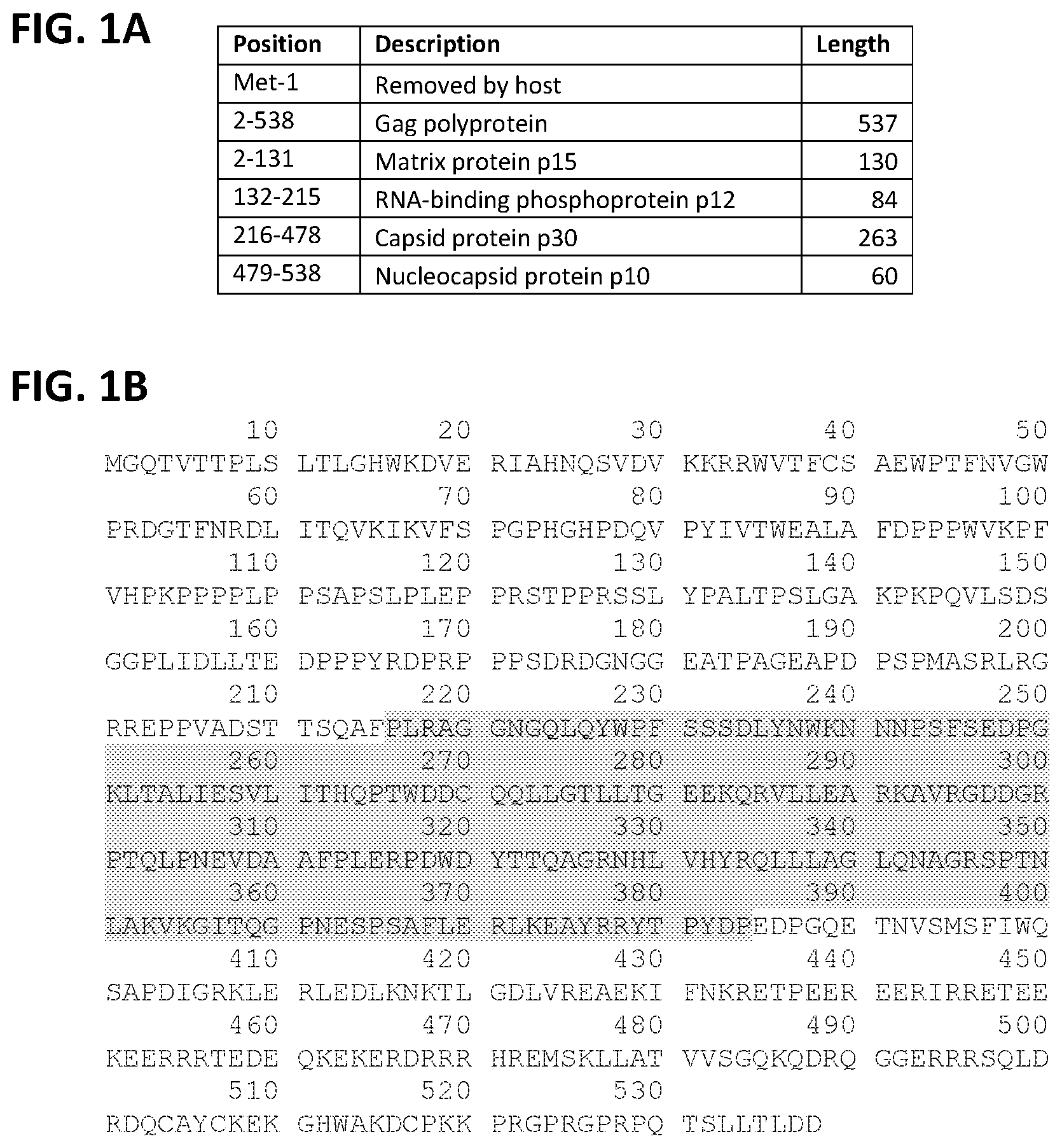
[0011] FIG. 1A-B. Identification of the p30 capsid region of MMLV-Gag included in LIVeMac constructs. (FIG. 1A) Cleavage products of the Gag polyprotein. (FIG. 1B) MMLV-Gag sequence with shading to indicate the p30 capsid region of Gag included in Gag-vRaf fusion for LIVeMac constructs. The sequence presented in this figure is from UniProt and is presented for illustrative purposes only. It differs slightly from the Gag-p30 sequence in LIVeMac, as not all MMLV isolates are identical.
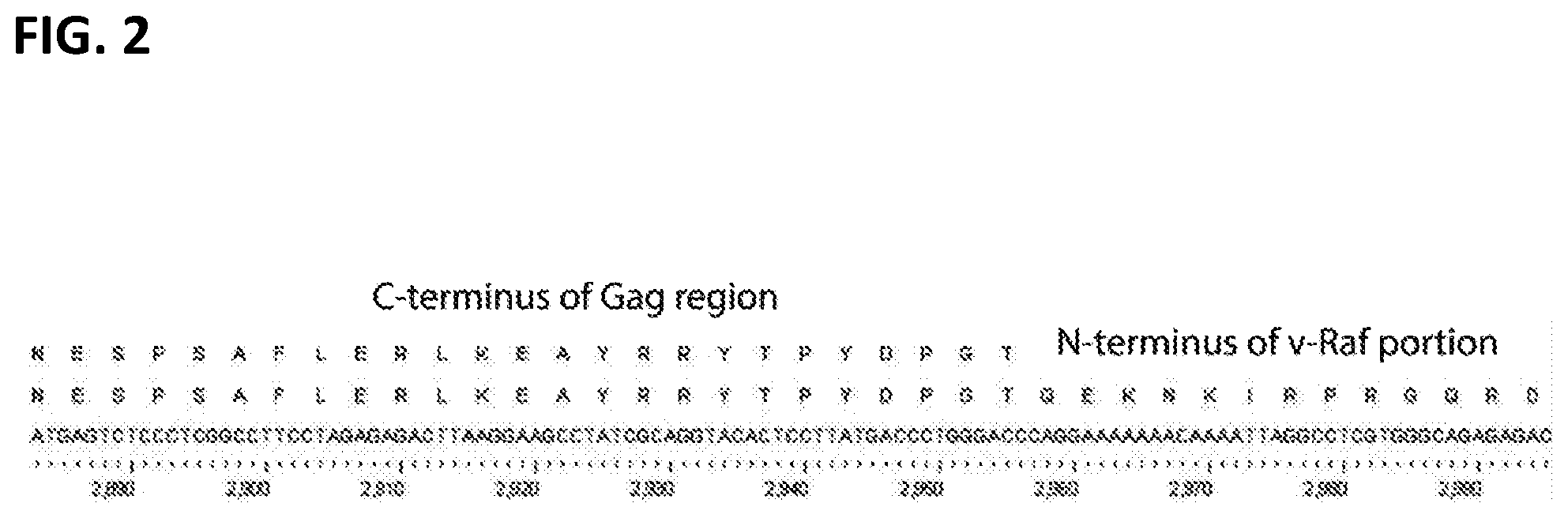
[0012] FIG. 2. The Gag-vRaf junction of all LIVeMac constructs.
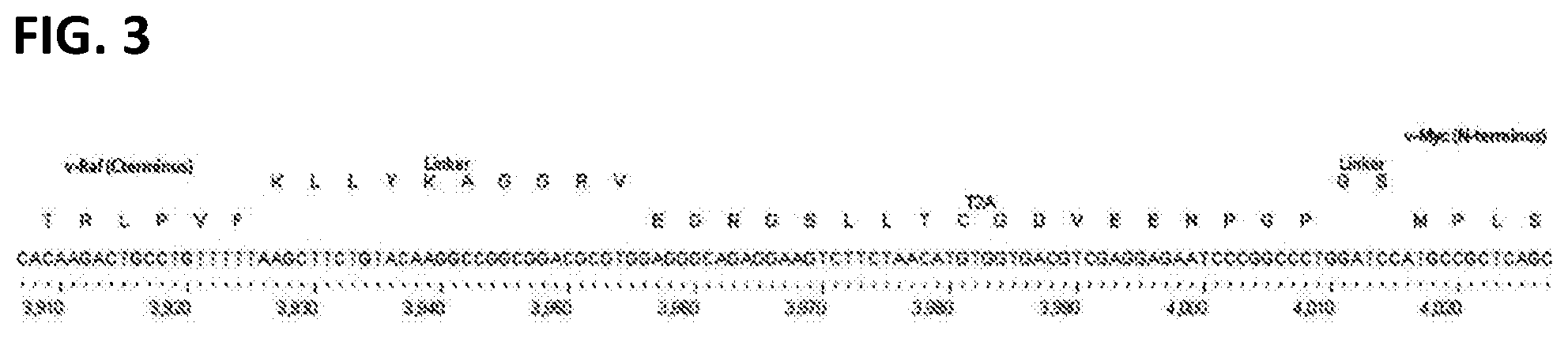
[0013] FIG. 3. The vRaf-T2A-vMyc junction of all LIVeMac constructs.
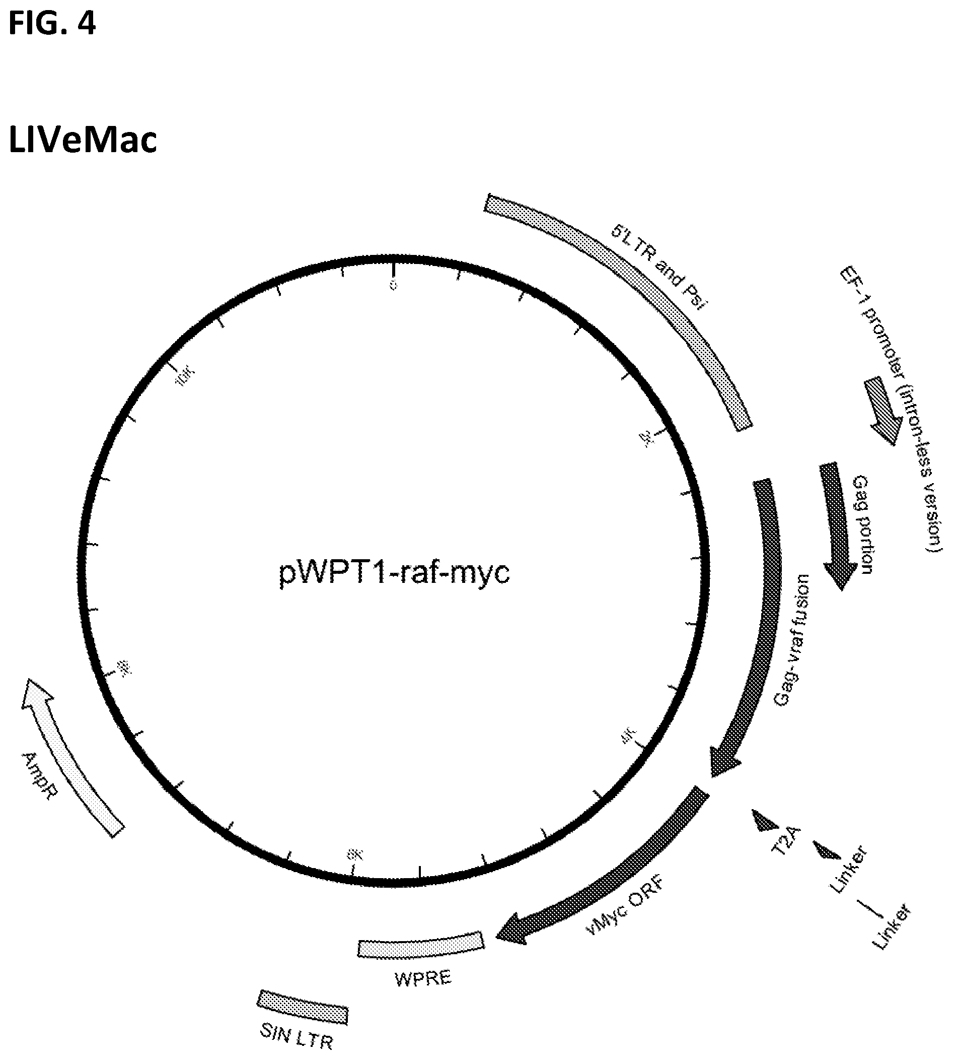
[0014] FIG. 4. Map of LIVeMac (untagged version).
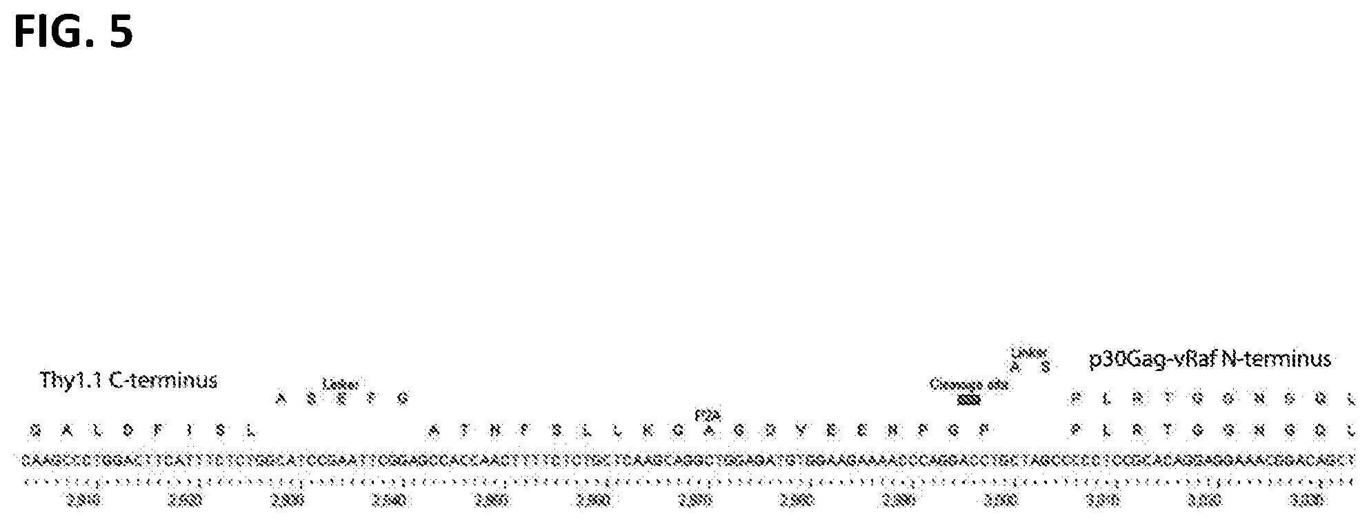
[0015] FIG. 5. The Thy1.1-P2A-p30Gag-vRaf junction region of LIVeMac-Thy1.1-N.
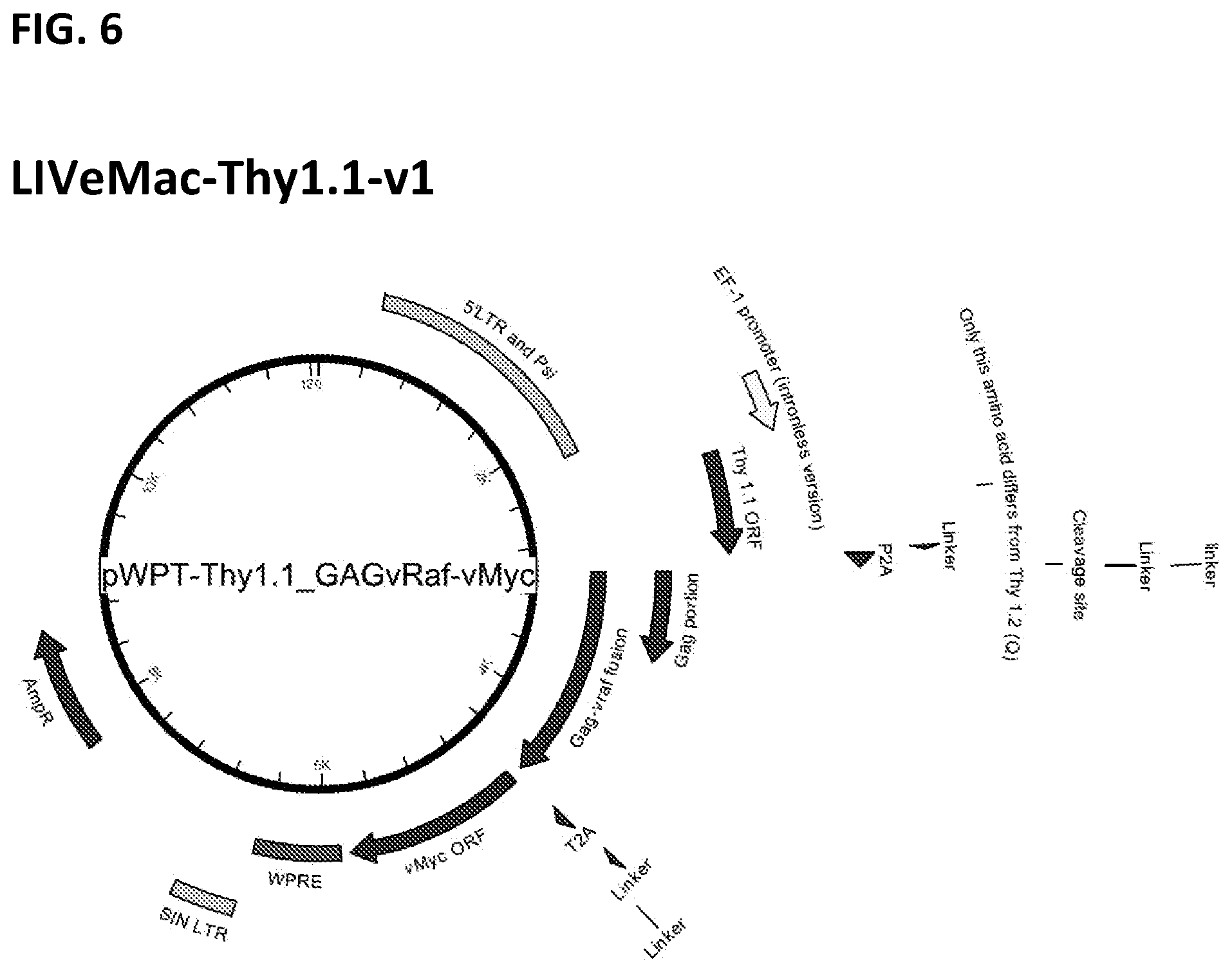
[0016] FIG. 6. Map of LIVeMac-Thy1.1-v1.
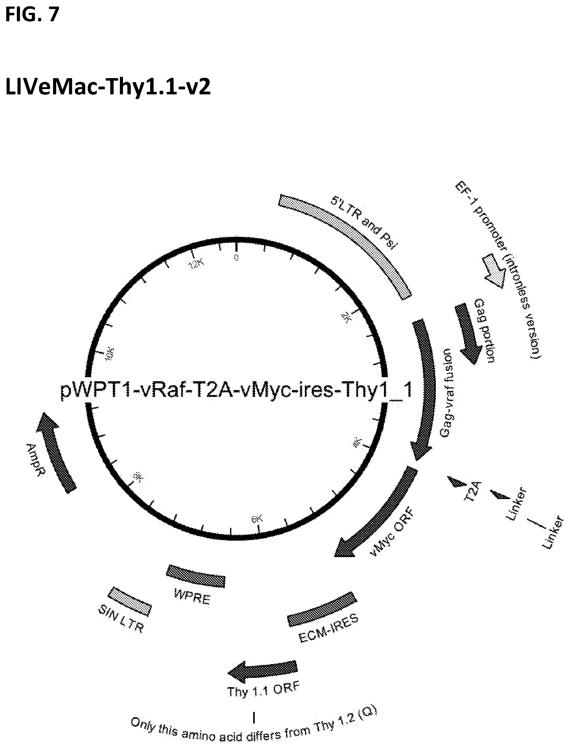
[0017] FIG. 7. Map of LIVeMac-Thy1.1-v2.
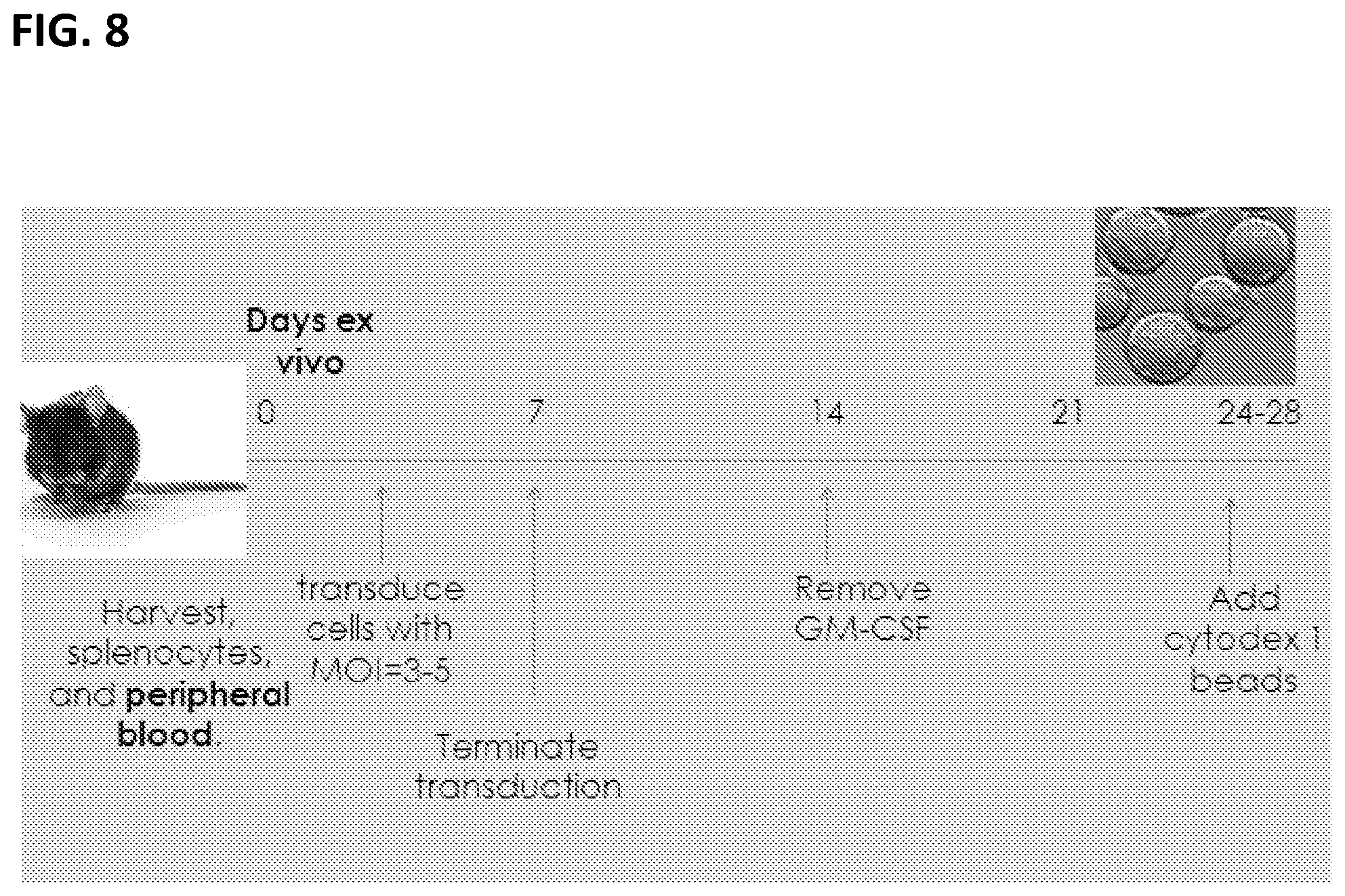
[0018] FIG. 8. Protocol for generating immortalized mouse macrophage.
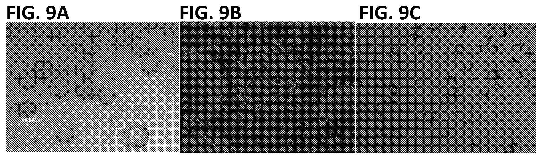
[0019] FIGS. 9A-C. Macrophage immortalized with LIVeMac. Images were taken 38 days ex vivo (34 days post-transduction). (FIG. 9A) 40.times. image of cells adherent to Cytodex 1 beads and cells adherent to the bottom of a tissue culture plate. The cells on beads are rounded; the cells on the plate look like macrophage. (FIG. 9B) 100.times. image of Cytodex 1 beads with cell colonies growing on their surface. At day 38, the cells require contact with the bead for cell division. (FIG. 9C) 100.times. image of cells adherent to plate. These cells do not replicate but begin to take on the appearance of phagocytic cells.
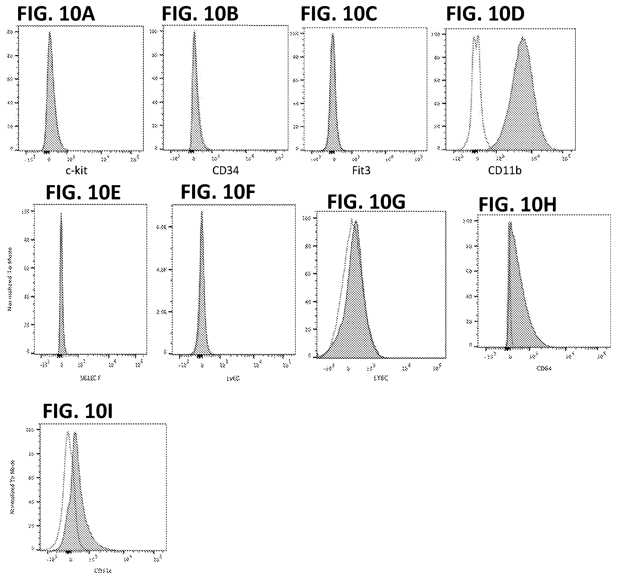
[0020] FIGS. 10A-I. Peripheral blood derived cells immortalized with LIVeMac display surface markers characteristic of mature macrophages, as determined by flow cytometry. No labeling was observed when staining for immature myeloid and progenitor markers (FIG. 10A) c-kit, (FIG. 10B) CD34, or (FIG. 10C) Flt3, or for granulocyte markers Siglec F (FIG. 10E), Ly6G (FIG. 10F) or Ly6C (FIG. 10G). Positive staining was observed for mature macrophage markers (FIG. 10D) CD11b and F4-80 (not shown; and see FIG. 11). F4-80 is a mouse marker for fully mature macrophage and is specific to this cell type. Staining for CD64 (FIG. 10H) and weak staining for CD11c (FIG. 10I) is also consistent with mature macrophages.
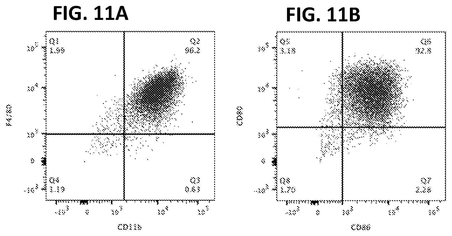
[0021] FIGS. 11A-B. Splenocyte cells immortalized with LIVeMac display positive staining for (FIG. 11A) CD11b and F4/80 and (FIG. 11B) CD80 and CD86, surface markers characteristic of mature macrophages, as determined by flow cytometry.
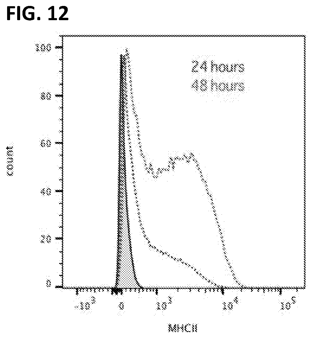
[0022] FIG. 12. Peripheral blood derived mononuclear cells immortalized with LIVeMac are IFN.gamma. responsive. Macrophages were treated with 100 U/mL of rmIFN.gamma. for 24 or 48 hours, and then stained for MHC II and analyzed by flow cytometry. By 48 hours, all cells had upregulated MHC II, indication that these macrophages are interferon-responsive.
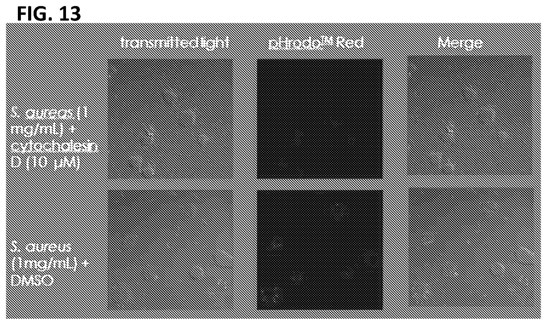
[0023] FIG. 13. Macrophages immortalized with LIVeMac are phagocytic. Confocal images of immortalized macrophage treated with DMSO or cytochalasin D and then infected with pHrodo Red Staphylococcus aureus Bioparticles.RTM. for 60 minutes. These bioparticles will only fluoresce when the pH of the environment is low. Thus, fluorescence can be detected when the bacteria reach the lysosome. Macrophages pretreated with cytochalasin D are unable to undergo phagocytosis while those mock treated with DMSO are able to phagocytose the S. aureus in their environment, as indicated by the internalized red fluorescent bacteria.
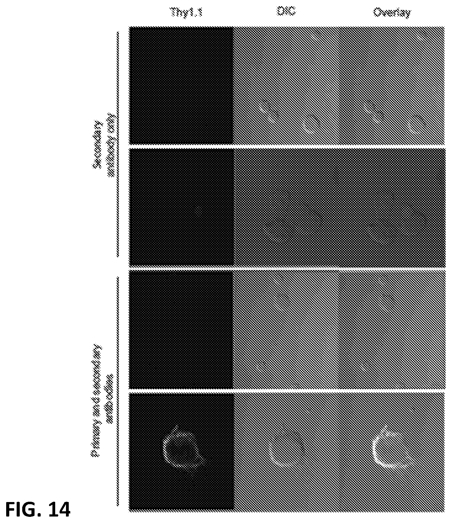
[0024] FIG. 14. Splenocytes cultured in 1.times. GM-CSF for four days were transduced with the multicistronic LIVeMac and maintained in 1.times. GM-CSF for an additional 10 days before being moved to growth factor free media. Cells were stained 21 days ex vivo with anti-Thy1.1-Biotin and anti-streptavidin AlexaFluor 647 or with streptavidin AlexaFluor 647 only.
DETAILED DESCRIPTION OF THE INVENTION
[0025] The following detailed description is presented to enable any person skilled in the art to make and use the object of this application. For purposes of explanation, specific nomenclature is set forth to provide a thorough understanding of the present application. However, it will be apparent to one skilled in the art that these specific details are not required to practice the subject of this application. This description is intended to be read in connection with the accompanying drawings, which are to be considered part of the entire written description of this application. Descriptions of specific applications are provided only as representative examples. The present application is not intended to be limited to the embodiments shown, but is to be accorded the widest possible scope consistent with the principles and features disclosed herein.
[0026] One aspect of the present disclosure relates to a replication-deficient viral vector for immortalizing mammalian cells comprising, a polynucleotide encoding a v-raf protein comprising a sequence having at least 95% identity to SEQ ID NO: 4 and a polynucleotide encoding a v-myc protein comprising a sequence having at least 95% identity to SEQ ID NO: 5.
[0027] In some embodiments, the v-raf and v-myc proteins are expressed from a single bicistronic or multicistronic mRNA transcript and separated by a 2A self-cleaving linker.
[0028] In some embodiments, the vector comprises a promoter that is an EF1a promoter or another suitable promoter.
[0029] In some embodiments, the replication-deficient viral vector further comprises a polynucleotide encoding a surface marker.
[0030] In some embodiments, the surface marker is a thy1.1 protein comprising a sequence having at least 95% identity to SEQ ID NO: 6. In some further embodiments, the polynucleotide encoding the thy1.1 protein is operatively linked to an internal ribosome entry site.
[0031] In some embodiments, the thy1.1 protein is expressed from a multicistronic mRNA transcript and separated from the v-raf and v-myc proteins by a 2A self-cleaving linker.
[0032] In other embodiments, the surface marker is an NGFR or an epitope-tagged version of any desired surface protein.
[0033] In some embodiments, the replication-deficient viral vector further comprises a polynucleotide encoding a fluorescent protein including, but not limited to, GFP, dsRed, CFP, or YFP.
[0034] In some embodiments, the replication-deficient viral vector further comprises a polynucleotide encoding a drug-selectable marker.
[0035] In some embodiments, the replication-deficient viral vector further comprises a polynucleotide encoding a VSV-G envelope glycoprotein comprising a sequence having at least 95% identity to SEQ ID NO: 7.
[0036] In some embodiments, the vector is a lentiviral vector. In some embodiments, the lentiviral vector comprises nucleic acids derived from a J2 virus.
[0037] In some embodiments, the replication-deficient viral vector comprises a polynucleotide encoding the p30Gag-vRaf-T2A-vMyc fusion protein of SEQ ID NO: 1. In some embodiments, the replication-deficient viral vector comprises a polynucleotide encoding (a) the Thy1.1 protein of SEQ ID NO: 3 and the p30Gag-vRaf-T2A-vMyc fusion protein of SEQ ID NO: 1, or (b) the Thy1.1-P2A-p30Gag-vRaf-T2A-vMyc fusion protein of SEQ ID NO: 2.
[0038] Another aspect of the present disclosure relates to a host cell comprising the replication-deficient virus as described herein. In some embodiments, the cell is a HEK-293T cell.
[0039] Another aspect of the present disclosure relates to a replication deficient virus produced by a host cell as described herein.
[0040] Another aspect of the present disclosure relates to a method of immortalizing a mammalian cell comprising contacting the cell with the replication-deficient virus as described herein.
[0041] In some embodiments, the mammalian cell is inclusive of, but not limited to, mouse, rat, ferret, pig and human.
[0042] In some embodiments, the mammalian cell is a monocyte, a macrophage or a related cell. In some further embodiments, the monocyte, macrophage or related cell is a peripheral blood mononuclear cell. In other further embodiments, the monocyte, macrophage or related cell is a splenocyte.
[0043] In some embodiments, the macrophage or related cell is inclusive of, but not limited to, microglia, Kupffer cells, alveolar macrophages, Langerhans cells, adipose tissue macrophages, osteoclasts, tumor associated macrophages, and dendritic cells.
[0044] Another aspect of the present disclosure relates to a method of differentiating the immortalized monocyte as described herein into a macrophage comprising, (a) proliferatively growing the monocytic cell, (b) growing the monocytic cell on a solid surface, and (c) growing the monocytic cell on a porous surface. In some embodiments, the porous surface is a dextran-based bead.
[0045] Another aspect of the present disclosure relates to an immortalized macrophage produced by the method described herein. In some embodiments, the immortalized macrophage (a) expresses a surface protein characteristic of a macrophage, and (b) does not express a surface protein characteristic of an undifferentiated monocyte progenitor cell. In some embodiments, the macrophage has phagocytic activity. In some embodiments, the macrophage responds to treatment with .gamma.-interferon by upregulating expression of an MHC II gene.
[0046] Another aspect of the present disclosure relates to a method of detecting the immortalized monocyte produced by the method described herein or the immortalized macrophage as described herein by contacting the immortalized monocyte or immortalized macrophage with a reagent having specific affinity for the surface marker as described herein. In some embodiments, the reagent is a fluorescently-labeled antibody having specific affinity for thy1.1.
[0047] Defects in macrophage function play a major role human genetic diseases. Immortalization of macrophages from individuals with macrophage defects could be used to screen drug libraries or to elucidate the basis of disease and develop novel therapeutics.
[0048] Such diseases include lipid storage diseases, such as (but not limited to) Gaucher disease or Niemann-Pick disease; diseases characterized by defects in macrophage activation, such as (but not limited to) anhidrotic ectodermal dysplasia with immune deficiency (EDA-ID), IL-12 or IL-12 receptor deficiency, interferon (IFN)-gamma deficiency, or STAT-1 deficiency; immunodeficiencies affecting phagocyte functions such as (but not limited to) chronic granulomatous disease and myeloperoxidase deficiency.
[0049] Immortalized macrophages of the present disclosure would also provide benefits in regard to diseases characterized by defects in phagocyte function (although impairment of neutrophil function receives the most attention, these immunodeficiencies also affect macrophage phagocytic function, and immortalized macrophages from such patients would thus be an important resource to study these diseases) including, but not limited to, chronic granulomatous disease or myeloperoxidase deficiency.
[0050] Immortalized macrophages of the present disclosure can be used in studies involving infectious diseases and are particularly applicable to pathogens which are tropic for myeloid cells including (but not limited to) Mycobacterium tuberculosis and other mycobacterial species; Yersina species, and Salmonella species.
[0051] There are also many other diseases in which macrophages are known or suspected play a major role (atherosclerosis, cancer, Lupus, rheumatoid arthritis), and immortalized macrophages from individuals with these diseases may be useful to elucidate defects in macrophage function that contribute to pathology.
[0052] In mice, LIVeMac can be used to immortalize macrophages from genetically distinct specimens (i.e. knockout vs wild type or species A vs species B). The resulting cell lines will allow researchers to investigate a particular gene or mutation without having to repeatedly harvest primary cells.
[0053] At the same time, this platform provides consistent background (cells were immortalized the same way) reducing the number of variables involved in the experiment. Rare genetic mutations affecting macrophages and which cause disease or cancer can be studied indefinitely.
[0054] Macrophage cell lines created using LIVeMac could also likely be used to create cell lines lacking expression of desired target genes by CRISPR or similar techniques.
[0055] Another aspect of the present disclosure relates to use of the immortalized macrophages to produce certain biologicals, particularly those normally produced in relatively large amounts by macrophages--cytokines are the most obvious potential product (e.g., IL-6, TNF-a, IFN-g), but production of other mediators normally produced in high quantities by macrophages (e.g., galectin-3) would also be possible.
[0056] Another aspect of the present disclosure relates to polynucleotides encoding polypeptides having at least 95% identity to a v-raf protein, a v-myc protein, and/or a thy1.1 protein. The similarity or identity of amino acid sequences, i.e. the percentage of sequence identity, can be determined via sequence alignments. Such alignments can be carried out with several art-known algorithms, such as with the mathematical algorithm of Karlin and Altschul (Karlin & Altschul (1993) Proc. Natl. Acad. Sci. USA 90: 5873-5877), with hmmalign (HMMER package, hmmer.wustl.edu/) or with the CLUSTAL algorithm (Thompson (1994) Nucleic Acids Res. 22, 4673-80). The grade of sequence identity (sequence matching) may be calculated using e.g. BLAST, BLAT or BlastZ (or BlastX). A similar algorithm is incorporated into the BLASTP programs of Altschul (1990) J. Mol. Biol. 215: 403-410. BLAST protein searches may be performed with the BLASTP program, score=50, word length=3. To obtain gapped alignments for comparative purposes, Gapped BLAST is utilized as described in Altschul (1997) Nucleic Acids Res. 25: 3389-3402. When utilizing BLAST and Gapped BLAST programs, the default parameters of the respective programs are used. Sequence matching analysis may be supplemented by established homology mapping techniques like Shuffle-LAGAN (Brudno (2003) Bioinformatics, 19 Suppl 1:154-162) or Markov random fields.
[0057] Changes to the amino acid sequence of a polypeptide can alter its function or have no measurable effect. Silent changes with no measurable effect are most likely to be conservative substitutions and small insertions or deletions on solvent-exposed surfaces that are located away from active sites and substrate-binding sites. In contrast, function is more likely to be affected by non-conservative substitutions, large insertions or deletions, and changes within active sites, substrate-binding sites, and at buried positions important for protein folding or conformation. Changes that alter protein function may increase or decrease reaction rates or binding affinities. For example, changes that increase the size of a substrate-binding site may permit an enzyme to act on larger substrates and changes that position a catalytic amino acid side chain closer to a target site on a substrate may increase the enzymatic rate.
[0058] A substitution is the replacement within a polypeptide of a new amino acid residue for an old one. In a conservative substitution, the old and new amino acids have similar characteristics such as size and charge. Naturally occurring residues are divided into groups based on common side chain properties:
[0059] (group 1) hydrophobic (aliphatic): methionine (Met), Alanine (Ala), Valine (Val), Leucine (Leu), Isoleucine (IIe)
[0060] (group 2) neutral hydrophilic: Cysteine (Cys), Serine (Ser), Threonine (Thr)
[0061] (group 3) acidic: Aspartic acid (Asp), Glutamic acid (Glu)
[0062] (group 4) basic: Asparagine (Asn), Glutamine (Gin), Histidine (His), Lysine (Lys), Arginine (Arg)
[0063] (group 5) residues that influence chain orientation: Glycine (Gly), Proline (Pro); and
[0064] (group 6) aromatic: Tryptophan (Trp), Tyrosine (Tyr), Phenylalanine (Phe)
[0065] Non-conservative substitutions will entail exchanging a member of one of these classes for another.
[0066] Thus, in some cases, the basic amino acids Lys, Arg and His may be interchangeable; the acidic amino acids Asp and Glu may be interchangeable; the neutral polar amino acids Ser, Thr, Cys, Gln, and Asn may be interchangeable; the non-polar aliphatic amino acids Gly, Ala, Val, Me, and Leu are interchangeable but because of size Gly and Ala are more closely related and Val, lie and Leu are more closely related to each other, and the aromatic amino acids Phe, Trp and Tyr may be interchangeable.
[0067] Extensive structure-function studies on v-raf, v-myc, and thy1 provide guidance on changes that can be made to their amino acid sequences without destroying their functions. See Wellbrock (2004) Nat. Rev. Mol. Cell. Biol. 5:875-85; Leicht (2007) Biochim Biophys Acta, 1773: 1196-1212; Meyer (2008) Nature Reviews Cancer 8:976-990; Mansour (2004) J. Immunol. 173:3581-3588; Kuhn (2002) Proteins. 49:142-5.
[0068] Viral vectors provide an efficient means for modification of eukaryotic cells and their use is now commonplace in academic laboratories and industry for both research and clinical gene therapy applications. Lentiviral vectors, derived from the human immunodeficiency virus, have been extensively investigated and optimized over the past two decades. Lentiviral vectors offer several attractive properties as gene-delivery vehicles, including: (i) sustained gene delivery through stable vector integration into host genome; (ii) the capability of infecting both dividing and non-dividing cells; (iii) broad tissue tropisms, including important gene- and cell-therapy-target cell types; (iv) no expression of viral proteins after vector transduction; (v) the ability to deliver complex genetic elements, such as polycistronic or intron-containing sequences; (vi) potentially safer integration site profile; and (vii) a relatively easy system for vector manipulation and production. Sakuma T. et al., Lentiviral vectors: basic to translational, Biochem J. 2012 May 1; 443(3):603-18. Self-inactivating lentiviral vectors were developed for improved safety. Third-generation lentiviral vectors require three helper plasmids in addition to the plasmid carrying the transgene. All accessory genes of HIV-1 (vif, vpr, vpu, and nef) have been removed because they are not necessary. Merten (2016) Mol. Ther. Methods Clin. Dev. 3:16017.
[0069] The host range of retroviral vectors including lentiviral vectors can be expanded or altered by a process known as pseudotyping. Pseudotyped lentiviral vectors consist of vector particles bearing glycoproteins derived from other enveloped viruses. Such particles possess the tropism of the virus from which the glycoprotein was derived. Among the first and still most widely used glycoproteins for pseudotyping lentiviral vectors is the vesicular stomatitis virus glycoproteins (VSV-G), due to the very broad tropism and stability of the resulting pseudotypes. Cronin (2005) Curr. Gene Ther. 5:387-398.
[0070] Co-expression of multiple genes at a desired ratio is highly attractive for a broad array of basic research and biomedical applications including cellular reprogramming, expression of multiple subunits of complex multimeric proteins in gene therapy, tagging of protein of interest for live cell imaging or cell sorting, and generation of efficient tools for fate mapping and genome editing. Strategies for multigene co-expression include introduction of multiple vectors, use of multiple promoters in a single vector, fusion proteins, proteolytic cleavage sites between genes, internal ribosome entry sites, and "self-cleaving" 2A peptides. 2A peptides are 18-22 amino-acid (aa)-long viral oligopeptides that mediate "cleavage" of polypeptides during translation in eukaryotic cells. The designation "2A" refers to a specific region of the viral genome and different viral 2As have generally been named after the virus they were derived from. The first discovered 2A was F2A (foot-and-mouth disease virus), after which E2A (equine rhinitis A virus), P2A (porcine teschovirus-1 2A), and T2A (thosea asigna virus 2A) were also identified. The mechanism of 2A-mediated "self-cleavage" was recently discovered to be ribosome skipping the formation of a glycyl-prolyl peptide bond at the C-terminus of the 2A. A highly conserved sequence GDVEXNPGP is shared by different 2As at the C-terminus, and is essential for the creation of steric hindrance and ribosome skipping. 2A peptides lead to relatively high levels of downstream protein expression compared to other strategies for multi-gene co-expression, and they are small in size thus bearing a lower risk of interfering with the function of co-expressed genes. 2A peptides have been successfully employed for polycistronic and bi-cistronic multigene expression. Liu (2017) Sci. Rep. 7:2193.
[0071] The present invention is further illustrated by the following examples which should not be construed as limiting. The contents of all references, patents and published patent applications cited throughout this application, as well as the Figures, are incorporated herein by reference.
EXAMPLES
Example 1
Construction of LIVeMac Plasmids
[0072] Lentiviral vectors constructed for the purpose of producing immortalized macrophages are referred to as LIVeMac (Lentivirus Immortalizing Vertebrate Macrophages) plasmids, and have bicistronic or multicistronic inserts.
[0073] The v-raf and v-myc oncogenes were isolated by PCR from the J2 plasmid (Rapp (1985) Virology 55:23-33; Blasi (1985) Nature 318:667-70). The v-raf gene of J2 is part of a MMLV-Gag/v-raf fusion. The Gag portion of this polyprotein is likely to be cleaved in J2-infected cells, as illustrated in FIG. 1A. To reduce the length of DNA inserted into the lentiviral vector, which should increase transformation efficiency and viral yields, DNA encoding the Matrix protein (p15) and RNA-binding phosphoprotein (p12) was excluded from the PCR product and replaced with an initiator methionine. The resulting Gag/v-raf fusion contained only the shaded region of Gag shown in FIG. 1B. The Gag-p30/v-raf fusion site at position 384 of the Gag polyprotein is identical to the corresponding region of J2, and does not include the nucleocapsid protein (p10). This modified Gag-v-Raf was amplified by PCR using a primer encoding the self-cleaving T2A peptide and inserted into the pBluescript-KS+ vector (pBS), yielding pBS-v-Raf-T2A.
[0074] After confirming the sequence of pBS-v-Raf-T2A, the v-myc gene of J2 was inserted downstream of T2A, in the same reading frame, as illustrated in FIG. 3. In this manner, both v-Raf and v-Myc are produced as a single open reading frame (ORF) that is co-translationally cleaved into v-Raf and v-Myc components by the inherent self-cleaving activity of the T2A peptide sequence. This plasmid was called pBS-v-Raf-T2A-v-Myc.
[0075] The v-Raf-T2A-v-Myc portion was subcloned into the 2.sup.nd-generation self-inactivating lentiviral vector, pWPT, to generate pWPT-v-Raf-T2A-v-Myc, also known as LIVeMac. FIG. 4. pWPT has a self-inactivating 3' LTR, which results in inactivation of the 5'LTR upon reverse transcription. Consequently, an internal EF-1 promoter drives transcription of inserted genes. The pWPT portion of this construct was obtained as a pWPT-GFP plasmid. The GFP gene was replaced with a polylinker sequence, and v-Raf-T2A-v-Myc was then inserted into the polylinker. SEQ ID NO: 1 shows the amino acid sequence of the p30Gag-vRaf-T2A-vMyc fusion protein of LIVeMac.
[0076] LIVeMac-Thy1.1-v1 has a Thy1.1 marker inserted at the 5' end of the Gag/v-raf fusion, which is separated from Gag/v-raf by a P2A self-cleaving peptide. FIG. 5 presents the sequence of the junction with the P2A linker, and FIG. 6 is a map of the entire LIVeMac-Thy1.1-v1 plasmid. To construct LIVeMac-Thy1.1-v1, the full expression cassette was first assembled in in pBS (yielding pBS-Thy1.1-P2A-v-Raf-T2A-v-Myc), followed by subcloning into pWTP. LIVeMac-Thy1.1-v1 expresses a Thy1.1-vRaf-vMyc polyprotein with two self-cleaving sites, yielding separate Thy1.1, v-Raf, and v-Myc components. SEQ ID NO: 2 shows the amino acid sequence of the Thy1.1-P2A-p30Gag-vRaf-T2A-vMyc fusion protein of LIVeMac-Thy1.1-v1.
[0077] LIVeMac-Thy1.1-v2 has a Thy1.1 marker inserted with an internal ribosome entry site (IRES) at the 3' end of the Gag/v-raf fusion. FIG. 7. To construct LIVeMac-Thy1.1-v2, v-Raf-T2A-v-Myc was inserted upstream of an IRES-Thy1.1 cassette in the vector pSP72. This cassette was then subcloned into pWPT, yielding pWPT-v-Raf-T2A-v-Myc-IRES-Thy1.1, also known as LIVeMac-Thy1.1-v2. In this construct, the Thy1.1 cassette is produced from the same primary viral RNA transcript as a separate ORF, via internal ribosome binding at the IRES. SEQ ID NO: 3 shows the amino acid sequence of the Thy1.1 polypeptide of LIVeMac-Thy1.1-v2. LIVeMac-Thy1.1-v2 also expresses the p30Gag-vRaf-T2A-vMyc fusion protein of SEQ ID NO: 1.
Example 2
Production of Viral Supernatants
[0078] LIVeMac vectors were used to produce virus by employing a 2.sup.nd-generation lentiviral packaging system. LIVeMac plasmid DNA was co-transfected into HEK-293T cells with a plasmid encoding the vesicular stomatitis virus glycoprotein (VSV-G) and the packaging plasmid, pCMV-R8.74, which encodes lentivirus structural proteins. The resulting supernatants contain infectious, replication-incompetent VSV-G-pseudotyped lentiviral particles.
[0079] The transfection protocol used, as follows, is a modified version of that described by Jordan (1996) Nucl. Acids. Res. 24:596-601.
[0080] Day 1
[0081] Afternoon before transfection, split cells (HEK-293T or the Phoenix packaging line) so that they will be 50-80% confluent in 24 hours. Split cells into a 6 well plate in 1 ml of complete media (DME works well, RPMI does not work well), and grow cells in 37.degree. C., 5% CO.sub.2 incubator. Plating 6.times.10.sup.5 per well gives the appropriate density. Note: it is also worthwhile to pretreat wells with 100 .mu.g/ml poly-D-lysine (Sigma P-0899) in ddH.sub.2O (5 minutes, room temp). Wash wells 2.times. with sterile 1.times. BSS or 1.times. PBS before plating cells. This treatment will prevent cell loss during the media change on Day 3.
[0082] Day 2
[0083] 1. Warm to 37.degree. C. the appropriate volume of Iscove's Modified Dulbecco's Medium (IMDM supplemented with antibiotics and 10% fetal bovine serum) enough to provide 1.5 mL per well to be transfected. Replace the DMEM with 1.5 mL pre-warmed IMDM. Note: It is important to use IMDM--this will increase the transfection efficiency from 30-50% to 100%.
[0084] 2. For each well of a 6-well plate to be transfected, assemble the following in a sterile 1.5 mL tube: add 1.7 .mu.g of Qiagen- or CsCl-purified lentiviral plasmid DNA (e.g., pWPT-GFP), 0.8 .mu.g of the packaging plasmid pCMV-R8.74 and 0.5 .mu.g of the pMD2.G envelope plasmid to 7.5 .mu.l of 2.5 M CaCl.sub.2. Add sterile ddH.sub.2O to a final volume of 75 .mu.l. Prepare all DNA samples to be used in transfections before proceeding to Step 2. Note: The ratio of lentiviral DNA: packaging DNA: envelope DNA is consistent with a posted web protocol: lentiweb.com/protocols_lentivectors.php. Empirical optimization may yield improved results in your hands. However, the DNA total should always be 3 .mu.g.
[0085] 3. Add 75 .mu.l of 2.times. HEPES solution and pipet up and down 4.times. to mix.
[0086] 4. After exactly 1 minute add the calcium-phosphate-DNA precipitate in dropwise fashion to one well of the cells in the six well plate. Distribute the 150 .mu.l over as much of the surface of the media as possible (i.e., do not put the entire 150 .mu.l in the center of the well). Note: You should be able to observe a very fine DNA precipitate using a 20.times. microscope objective. If the precipitate is not present or is composed primarily of large aggregates, check the pH of the 2.times. HEPES solution. Certain plasmids seem to have a tendency to form aggregates, so it may not be possible to eliminate aggregates in every case.
[0087] 5. Repeat Steps 2 and 3 for each well to be transfected. When transfecting larger or small numbers of cells, adjust all volumes proportionally--thus, the final DNA concentration should be 3 .mu.g/150 .mu.l of 2.times. HEPES-calcium-phosphate transfection mix, and the final concentration of calcium in the media following addition of the transfection mix is approximately 12.5 mM.
[0088] Day 3
[0089] 24 hours following addition of transfection mix, remove media, wash cells 2.times. with 2 mL of sterile 1.times. PBS or 1.times. BSS, then replace with 2 ml fresh complete media per well (e.g., DMEM+antibiotics and 10% fetal bovine serum). Note (1): 24 hours is the optimal time for the cells to be in the presence of the precipitated DNA. 2 mL of media is the optimal volume (maximal number of viral particles produced at the maximal concentration). Note (2): A simplified variation of this step, which eliminates the need to safely aspirate and discard lentivirus-containing supernatant, is to add 1.5 mL of complete DMEM to the existing 1.5 mL of IMDM already in the well. This variation reproducibly yields 3 mL of high-titer supernatant per transfected well.
[0090] Days 4 and 5
[0091] Harvest viral supernatant 24 hours after replacement of the media. A second harvest (with approximately 50% of the titer of the first) can be made 12-24 hours later--simply add another 2 mL of media, and return the plate to the incubator.
Other Factors
[0092] Effective viral titers may be increased by incubating cells at 32.degree. C. (days 3-5).
[0093] Virus can also be concentrated by centrifugation (Ichim (2011) Translational Medicine 9:137; Zhang (2001) Gene Therapy 8:1745-1751).
[0094] Target cells can be spinfected: place cells in a tissue culture well with retroviral supernatant and polybrene at 10 .mu.g/ml (more polybrene will give better infection frequency, but is toxic to certain cell types), spin in a swinging bucket rotor at 1000 g for 2 hours at room temp (use a ziploc bag to keep the CO.sub.2 in the plate). Immediately following spinfection, replace supernatant with fresh media.
[0095] Retroviral supernatants can be frozen (-70.degree. C.) with a 2.times. loss of titer. Do not refreeze after thawing. Supernatants can also be stored short term at 4.degree. C. It takes approximately 2 weeks for a 50% loss of titer, although this rate may vary with media composition, etc. It would be best to test this empirically for each type of media to be used.
[0096] Solutions
[0097] 2.5 M CaCl.sub.2: 18.375 g CaCl.sub.2(2H.sub.2O), MilliQ H.sub.2O to 50 ml, Filter Sterilize.
[0098] 2.times. HEPES Solution: 14 ml 1 M NaCl (140 mM final); 0.5 ml 300 mM NaPhosphate (1.5 mM final); 5 ml 1M HEPES, pH 7.05 (50 mM final); pH to exactly 7.05; MilliQ H.sub.2O to 100 ml, Filter Sterilize.
[0099] 300 mM NaPhosphate: 1.38 g Na.sub.2HPO.sub.4; 0.63 g NaH.sub.2PO.sub.4; MilliQ H.sub.2O to 50 ml.
[0100] 1M HEPES, pH 7.05; 11.92 g HEPES Acid; pH to 7.05; MilliQ H.sub.2O to 50 ml.
Example 3
Immortalization of Peripheral Blood Mononuclear Cells with LIVeMac
[0101] The overall procedure used for preparing immortalized macrophages from PBMCs is illustrated in FIG. 8. Prepare peripheral blood mononuclear cells according to standard protocols by, for example, lysing red blood cells and the collecting cells by density gradient centrifugation.
[0102] Culture collected cells in a petri plate or non-tissue treated 24 well plate in DMEM with 10% Cosmic Calf Serum, L-Glutamine, and Penicillin/streptomycin. The media should also contain GM-CSF from a feeder cell line at a concentration of 1.times..
[0103] Cells should be left in this condition for 4 days. At this point myeloid cells should be proliferating and appear as colonies of round clustered cells. This is the optimal time to transduce the cells.
[0104] Coat plates that cells will be transduced in with Retronectin (a fibronectin fragment that binds retrovirus, enhancing infectivity) at 50 .mu.g/mL per the company protocol.
[0105] Add viral supernatant containing LIVeMac to coated plates and spin at 1000 g for 90 minutes at 32.degree. C. MOI of around at least 5 work best though lower can be used.
[0106] Incubate the plate at 32.degree. C. for 2.5 hours then remove the supernatant and gently wash one time with PBS, without letting the well dry out.
[0107] Cells that have been in culture for four days were added to the well in 1/2 conditioned media (media the cells had been growing in) and 1/2 new media (same type as in step 2). As few as 100,000 cells can be plated in each well of a 24 well plate and still result in immortalized macrophages. Around 200,000-250,000 cells is ideal. Overcrowding of the wells, causes the cells to differentiate differently and does not result in immortalized macrophages.
[0108] Three days later, cells are removed using trypsin from the wells containing virus and washed 2.times. with PBS. Cell viability should be at around 80%-90% at this point and the cells should have about tripled in number.
[0109] At this point, cell proliferation should be finished or very slow. Seed the cells in a new non-tissue treated 24 well plate in all new DMEM with 1.times. GMCSF.
[0110] Seven days later you can wash the well with PBS to remove the dead cells and move the rest of them to a new well. Alternatively, you can leave them in the same well. At this point most cells will have died (presumably those that did not get infected with the virus) and lifting remaining cells risks losing even more cells.
[0111] At this point cells are cultured in DMEM without GM-CSF. The cells will adhere to the bottom of the plate and take on a particular morphology (See pictures below). They will be metabolically inactive. The media will not change color but the cells will remain adherent and look healthy.
[0112] Two weeks later, add 200 .mu.g of cytodex 1 beads directly to the adherent cells in the wells, without lifting the cells first.
[0113] Cells will begin to adhere to the beads within a few days but will take about a week to see obvious replication.
[0114] Cells will continue expanding and can be split by taking cytodex beads with cells on them and moving them to wells with new beads.
[0115] Slowly decreasing the concentration of beads in the wells will eventually lead to cultures that no longer require the beads to replicate. Taking the beads away directly, however, will put the cells back in the quiescent state.
[0116] Cells are now ready to use for experiments.
[0117] FIGS. 9A-C present the appearance of macrophages on the surface of culture dishes and on cytodex beeds on day 38 ex vivo.
Example 4
Immortalization of Splenocytes with LIVeMac
[0118] When harvesting cells, first treat spleen with collagenase.
[0119] Separate cells by pushing spleen through a cell strainer and then separate PBMC using lymphocyte separation media.
[0120] After 1-2 days most cells will be dead (T, B, NK cells are in media without proper cytokines and most splenocytes fall in that category) but myeloid cells in the 1.times. GM-CSF media will be adherent. After approximately two days (after adherent myeloid cells appear, but before they die from exposure to dead cells), remove the supernatant, wash one time with PBS and then replace with new media.
[0121] The cells are kept in culture so that like the peripheral blood samples, they are harvested for transfection at day 4. Follow the protocol as above from here forward.
Example 5
Characterization of Immortalized Macrophages
[0122] Immortalized macrophage prepared by transducing mouse PBMCs with LIVeMac were stained for cell surface markers and analyzed by flow cytometry. Results presented in FIG. 10 indicate that the cells were mature macrophage because they are negative for cell surface markers of immature myeloid progenitor cells, c-kit, CD34, and Flt3 (FIGS. 10A-C) positive for CD11b (FIG. 10D) and F4-80 (FIG. 11). These cells are also negative for markers of granulocytes (FIGS. 10 E-G). The cells are also positive for CD64 and weakly positive for CD11c (FIGS. 10 H-I), consistent with a macrophage phenotype. Like primary macrophage, the immortalized macrophage responded to gamma-interferon by upregulating expression of MHC II as illustrated in FIG. 11. Additionally, the immortalized macrophages were competent for phagocytosis, as illustrated in FIG. 12. Splenocytes transduced with the multicistronic virus maintained a morphology consistent with macrophages and monocytes. Thy1.1 was detected on the surface of these transduced cells as illustrated in FIG. 13.
[0123] The above description is for the purpose of teaching the person of ordinary skill in the art how to practice the present invention, and it is not intended to detail all those obvious modifications and variations of it which will become apparent to the skilled worker upon reading the description. It is intended, however, that all such obvious modifications and variations be included within the scope of the present invention, which is defined by the following claims. The claims are intended to cover the components and steps in any sequence which is effective to meet the objectives there intended, unless the context specifically indicates the contrary.
Sequence CWU 1
1
51937PRTArtificial SequenceSynthetic Sequence (p30Gag-vRaf-T2A-vMyc
fusion protein of LIVeMac) 1Met Pro Leu Arg Thr Gly Gly Asn Gly Gln
Leu Gln Tyr Trp Pro Phe1 5 10 15Ser Ser Ser Asp Leu Tyr Asn Trp Arg
Asn Asn Asn Pro Ser Phe Ser 20 25 30Glu Asp Pro Gly Lys Leu Thr Ala
Leu Ile Glu Ser Val Leu Ile Thr 35 40 45His Gln Pro Thr Trp Asp Asp
Cys Gln Gln Leu Leu Gly Thr Leu Leu 50 55 60Thr Gly Glu Glu Lys Gln
Arg Val Leu Leu Glu Ala Arg Lys Ala Val65 70 75 80Arg Gly Asp Asp
Gly Arg Pro Thr Gln Leu Pro Asn Glu Val Asp Ala 85 90 95Ala Phe Pro
Leu Glu Arg Pro Asp Trp Glu Tyr Thr Thr Gln Arg Gly 100 105 110Arg
Asn His Leu Val Leu Tyr Arg Gln Leu Leu Leu Ala Gly Leu Gln 115 120
125Asn Ala Gly Arg Ser Pro Thr Asn Leu Ala Lys Val Lys Gly Ile Thr
130 135 140Gln Gly Pro Asn Glu Ser Pro Ser Ala Phe Leu Glu Arg Leu
Lys Glu145 150 155 160Ala Tyr Arg Arg Tyr Thr Pro Tyr Asp Pro Gly
Thr Gln Glu Lys Asn 165 170 175Lys Ile Arg Pro Arg Gly Gln Arg Asp
Ser Ser Tyr Tyr Trp Lys Ile 180 185 190Glu Ala Ser Glu Val Met Leu
Ser Thr Arg Ile Gly Ser Gly Ser Phe 195 200 205Gly Thr Val Tyr Lys
Gly Lys Trp His Gly Asp Val Ala Val Lys Ile 210 215 220Leu Lys Val
Val Asp Pro Thr Pro Glu Gln Leu Gln Ala Phe Arg Asn225 230 235
240Glu Val Ala Val Leu Arg Lys Thr Arg His Val Asn Ile Leu Leu Phe
245 250 255Met Gly Tyr Met Thr Lys Asp Asn Leu Ala Ile Val Thr Gln
Trp Cys 260 265 270Glu Gly Ser Ser Leu Tyr Lys His Leu His Val Gln
Glu Thr Lys Phe 275 280 285Gln Met Phe Gln Leu Ile Asp Ile Ala Arg
Gln Thr Ala Gln Gly Met 290 295 300Asp Tyr Leu His Ala Lys Asn Ile
Ile His Arg Asp Met Lys Ser Asn305 310 315 320Asn Ile Phe Leu His
Glu Gly Leu Thr Val Lys Ile Gly Asp Phe Gly 325 330 335Leu Ala Thr
Val Lys Ser Arg Trp Ser Gly Ser Gln Gln Val Glu Gln 340 345 350Pro
Thr Gly Ser Ile Leu Trp Met Ala Pro Glu Val Ile Arg Met Gln 355 360
365Asp Ser Asn Pro Phe Ser Phe Gln Ser Asp Val Tyr Ser Tyr Gly Ile
370 375 380Val Leu Tyr Glu Leu Met Thr Gly Glu Leu Pro Tyr Ser His
Ile Asn385 390 395 400Asn Arg Asp Gln Ile Ile Phe Met Val Gly Arg
Gly Tyr Ala Ser Pro 405 410 415Asp Leu Ser Lys Leu Tyr Lys Asn Cys
Pro Lys Ala Met Lys Arg Leu 420 425 430Val Ala Asp Cys Leu Lys Lys
Val Arg Glu Glu Arg Pro Leu Phe Pro 435 440 445Gln Ile Leu Ser Ser
Ile Ala Leu Leu Gln His Ser Leu Pro Lys Ile 450 455 460Asn Arg Ser
Ala Ser Glu Pro Ser Leu His Arg Ala Ser His Thr Glu465 470 475
480Asp Ile Asn Ser Cys Thr Leu Thr Ser Thr Arg Leu Pro Val Phe Lys
485 490 495Leu Leu Tyr Lys Ala Gly Gly Arg Val Glu Gly Arg Gly Ser
Leu Leu 500 505 510Thr Cys Gly Asp Val Glu Glu Asn Pro Gly Pro Gly
Ser Met Pro Leu 515 520 525Ser Val Ser Leu Pro Ser Lys Asn Tyr Asp
Tyr Asp Tyr Asp Ser Val 530 535 540Gln Pro Tyr Phe Tyr Phe Glu Glu
Glu Glu Glu Asn Phe Tyr Leu Ala545 550 555 560Ala Gln Gln Arg Ser
Ser Glu Leu Gln Pro Pro Ala Pro Ser Glu Asp 565 570 575Ile Trp Lys
Lys Phe Glu Leu Leu Pro Ala Pro Pro Leu Ser Pro Ser 580 585 590Cys
Arg Ser Asn Leu Ala Ala Ala Ser Cys Phe Pro Ser Thr Ala Asp 595 600
605Gln Leu Glu Met Val Thr Glu Leu Leu Gly Gly Asp Met Val Asn Gln
610 615 620Ser Ser Ile Cys Asp Pro Asp Asp Glu Ser Phe Val Lys Ser
Ile Ile625 630 635 640Ile Arg Asp Cys Met Trp Ser Gly Phe Ser Ala
Ala Ala Lys Leu Glu 645 650 655Lys Val Val Ser Glu Lys Leu Ala Thr
Tyr Lys Ala Ser Arg Arg Glu 660 665 670Gly Gly Pro Ala Ala Ala Ser
Arg Pro Gly Pro Pro Pro Ser Gly Pro 675 680 685Pro Pro Pro Pro Ala
Gly Pro Ala Ala Ser Ala Gly Leu Tyr Leu His 690 695 700Asp Leu Gly
Ala Ala Ala Ala Gly Cys Ile Gly Ser Ser Val Val Phe705 710 715
720Pro Cys Pro Leu Gly Arg Arg Gly Pro Pro Gly Ala Gly Pro Ala Ala
725 730 735Leu Leu Gly Val Asp Ala Pro Pro Thr Ala Gly Gly Gly Ser
Glu Glu 740 745 750Glu Gln Glu Glu Asp Glu Glu Ile Asp Val Val Thr
Leu Ala Glu Ala 755 760 765Asn Glu Ser Glu Ser Ser Thr Glu Ser Ser
Thr Glu Ala Ser Glu Glu 770 775 780His Cys Lys Pro His His Ser Pro
Leu Val Leu Lys Arg Cys His Val785 790 795 800Asn Ile His Gln His
Asn Tyr Ala Ala Pro Pro Ser Thr Lys Val Glu 805 810 815Tyr Pro Ala
Ala Lys Arg Leu Lys Leu Asp Ser Gly Arg Val Leu Lys 820 825 830Gln
Ile Ser Asn Asn Arg Lys Cys Ser Ser Pro Arg Thr Leu Asp Ser 835 840
845Glu Glu Asn Asp Lys Arg Arg Thr His Asn Val Leu Glu Arg Gln Arg
850 855 860Arg Asn Glu Leu Lys Leu Arg Phe Phe Ala Leu Arg Asp Gln
Ile Pro865 870 875 880Glu Val Ala Asn Asn Glu Lys Ala Pro Lys Val
Val Ile Leu Lys Lys 885 890 895Ala Thr Glu Tyr Val Leu Ser Leu Gln
Ser Asp Glu His Lys Leu Ile 900 905 910Ala Glu Lys Glu Gln Leu Arg
Arg Arg Arg Glu Gln Leu Lys His Asn 915 920 925Leu Glu Gln Leu Arg
Asn Ser Arg Ala 930 93521124PRTArtificial SequenceSynthetic
Sequence (Thy1.1-P2A- p30Gag-vRaf- T2A-vMyc fusion protein of
LIVeMac-Thy1.1-v1) 2Met Asn Pro Ala Ile Ser Val Ala Leu Leu Leu Ser
Val Leu Gln Val1 5 10 15Ser Arg Gly Gln Lys Val Thr Ser Leu Thr Ala
Cys Leu Val Asn Gln 20 25 30Asn Leu Arg Leu Asp Cys Arg His Glu Asn
Asn Thr Lys Asp Asn Ser 35 40 45Ile Gln His Glu Phe Ser Leu Thr Arg
Glu Lys Arg Lys His Val Leu 50 55 60Ser Gly Thr Leu Gly Ile Pro Glu
His Thr Tyr Arg Ser Arg Val Thr65 70 75 80Leu Ser Asn Gln Pro Tyr
Ile Lys Val Leu Thr Leu Ala Asn Phe Thr 85 90 95Thr Lys Asp Glu Gly
Asp Tyr Phe Cys Glu Leu Arg Val Ser Gly Ala 100 105 110Asn Pro Met
Ser Ser Asn Lys Ser Ile Ser Val Tyr Arg Asp Lys Leu 115 120 125Val
Lys Cys Gly Gly Ile Ser Leu Leu Val Gln Asn Thr Ser Trp Met 130 135
140Leu Leu Leu Leu Leu Ser Leu Ser Leu Leu Gln Ala Leu Asp Phe
Ile145 150 155 160Ser Leu Ala Ser Glu Phe Gly Ala Thr Asn Phe Ser
Leu Leu Lys Gln 165 170 175Ala Gly Asp Val Glu Glu Asn Pro Gly Pro
Ala Ser Pro Leu Arg Thr 180 185 190Gly Gly Asn Gly Gln Leu Gln Tyr
Trp Pro Phe Ser Ser Ser Asp Leu 195 200 205Tyr Asn Trp Arg Asn Asn
Asn Pro Ser Phe Ser Glu Asp Pro Gly Lys 210 215 220Leu Thr Ala Leu
Ile Glu Ser Val Leu Ile Thr His Gln Pro Thr Trp225 230 235 240Asp
Asp Cys Gln Gln Leu Leu Gly Thr Leu Leu Thr Gly Glu Glu Lys 245 250
255Gln Arg Val Leu Leu Glu Ala Arg Lys Ala Val Arg Gly Asp Asp Gly
260 265 270Arg Pro Thr Gln Leu Pro Asn Glu Val Asp Ala Ala Phe Pro
Leu Glu 275 280 285Arg Pro Asp Trp Glu Tyr Thr Thr Gln Arg Gly Arg
Asn His Leu Val 290 295 300Leu Tyr Arg Gln Leu Leu Leu Ala Gly Leu
Gln Asn Ala Gly Arg Ser305 310 315 320Pro Thr Asn Leu Ala Lys Val
Lys Gly Ile Thr Gln Gly Pro Asn Glu 325 330 335Ser Pro Ser Ala Phe
Leu Glu Arg Leu Lys Glu Ala Tyr Arg Arg Tyr 340 345 350Thr Pro Tyr
Asp Pro Gly Thr Gln Glu Lys Asn Lys Ile Arg Pro Arg 355 360 365Gly
Gln Arg Asp Ser Ser Tyr Tyr Trp Lys Ile Glu Ala Ser Glu Val 370 375
380Met Leu Ser Thr Arg Ile Gly Ser Gly Ser Phe Gly Thr Val Tyr
Lys385 390 395 400Gly Lys Trp His Gly Asp Val Ala Val Lys Ile Leu
Lys Val Val Asp 405 410 415Pro Thr Pro Glu Gln Leu Gln Ala Phe Arg
Asn Glu Val Ala Val Leu 420 425 430Arg Lys Thr Arg His Val Asn Ile
Leu Leu Phe Met Gly Tyr Met Thr 435 440 445Lys Asp Asn Leu Ala Ile
Val Thr Gln Trp Cys Glu Gly Ser Ser Leu 450 455 460Tyr Lys His Leu
His Val Gln Glu Thr Lys Phe Gln Met Phe Gln Leu465 470 475 480Ile
Asp Ile Ala Arg Gln Thr Ala Gln Gly Met Asp Tyr Leu His Ala 485 490
495Lys Asn Ile Ile His Arg Asp Met Lys Ser Asn Asn Ile Phe Leu His
500 505 510Glu Gly Leu Thr Val Lys Ile Gly Asp Phe Gly Leu Ala Thr
Val Lys 515 520 525Ser Arg Trp Ser Gly Ser Gln Gln Val Glu Gln Pro
Thr Gly Ser Ile 530 535 540Leu Trp Met Ala Pro Glu Val Ile Arg Met
Gln Asp Ser Asn Pro Phe545 550 555 560Ser Phe Gln Ser Asp Val Tyr
Ser Tyr Gly Ile Val Leu Tyr Glu Leu 565 570 575Met Thr Gly Glu Leu
Pro Tyr Ser His Ile Asn Asn Arg Asp Gln Ile 580 585 590Ile Phe Met
Val Gly Arg Gly Tyr Ala Ser Pro Asp Leu Ser Lys Leu 595 600 605Tyr
Lys Asn Cys Pro Lys Ala Met Lys Arg Leu Val Ala Asp Cys Leu 610 615
620Lys Lys Val Arg Glu Glu Arg Pro Leu Phe Pro Gln Ile Leu Ser
Ser625 630 635 640Ile Ala Leu Leu Gln His Ser Leu Pro Lys Ile Asn
Arg Ser Ala Ser 645 650 655Glu Pro Ser Leu His Arg Ala Ser His Thr
Glu Asp Ile Asn Ser Cys 660 665 670Thr Leu Thr Ser Thr Arg Leu Pro
Val Phe Lys Leu Leu Tyr Lys Ala 675 680 685Gly Gly Arg Val Glu Gly
Arg Gly Ser Leu Leu Thr Cys Gly Asp Val 690 695 700Glu Glu Asn Pro
Gly Pro Gly Ser Met Pro Leu Ser Val Ser Leu Pro705 710 715 720Ser
Lys Asn Tyr Asp Tyr Asp Tyr Asp Ser Val Gln Pro Tyr Phe Tyr 725 730
735Phe Glu Glu Glu Glu Glu Asn Phe Tyr Leu Ala Ala Gln Gln Arg Ser
740 745 750Ser Glu Leu Gln Pro Pro Ala Pro Ser Glu Asp Ile Trp Lys
Lys Phe 755 760 765Glu Leu Leu Pro Ala Pro Pro Leu Ser Pro Ser Cys
Arg Ser Asn Leu 770 775 780Ala Ala Ala Ser Cys Phe Pro Ser Thr Ala
Asp Gln Leu Glu Met Val785 790 795 800Thr Glu Leu Leu Gly Gly Asp
Met Val Asn Gln Ser Ser Ile Cys Asp 805 810 815Pro Asp Asp Glu Ser
Phe Val Lys Ser Ile Ile Ile Arg Asp Cys Met 820 825 830Trp Ser Gly
Phe Ser Ala Ala Ala Lys Leu Glu Lys Val Val Ser Glu 835 840 845Lys
Leu Ala Thr Tyr Lys Ala Ser Arg Arg Glu Gly Gly Pro Ala Ala 850 855
860Ala Ser Arg Pro Gly Pro Pro Pro Ser Gly Pro Pro Pro Pro Pro
Ala865 870 875 880Gly Pro Ala Ala Ser Ala Gly Leu Tyr Leu His Asp
Leu Gly Ala Ala 885 890 895Ala Ala Gly Cys Ile Gly Ser Ser Val Val
Phe Pro Cys Pro Leu Gly 900 905 910Arg Arg Gly Pro Pro Gly Ala Gly
Pro Ala Ala Leu Leu Gly Val Asp 915 920 925Ala Pro Pro Thr Ala Gly
Gly Gly Ser Glu Glu Glu Gln Glu Glu Asp 930 935 940Glu Glu Ile Asp
Val Val Thr Leu Ala Glu Ala Asn Glu Ser Glu Ser945 950 955 960Ser
Thr Glu Ser Ser Thr Glu Ala Ser Glu Glu His Cys Lys Pro His 965 970
975His Ser Pro Leu Val Leu Lys Arg Cys His Val Asn Ile His Gln His
980 985 990Asn Tyr Ala Ala Pro Pro Ser Thr Lys Val Glu Tyr Pro Ala
Ala Lys 995 1000 1005Arg Leu Lys Leu Asp Ser Gly Arg Val Leu Lys
Gln Ile Ser Asn 1010 1015 1020Asn Arg Lys Cys Ser Ser Pro Arg Thr
Leu Asp Ser Glu Glu Asn 1025 1030 1035Asp Lys Arg Arg Thr His Asn
Val Leu Glu Arg Gln Arg Arg Asn 1040 1045 1050Glu Leu Lys Leu Arg
Phe Phe Ala Leu Arg Asp Gln Ile Pro Glu 1055 1060 1065Val Ala Asn
Asn Glu Lys Ala Pro Lys Val Val Ile Leu Lys Lys 1070 1075 1080Ala
Thr Glu Tyr Val Leu Ser Leu Gln Ser Asp Glu His Lys Leu 1085 1090
1095Ile Ala Glu Lys Glu Gln Leu Arg Arg Arg Arg Glu Gln Leu Lys
1100 1105 1110His Asn Leu Glu Gln Leu Arg Asn Ser Arg Ala 1115
11203162PRTMus musculus 3Met Asn Pro Ala Ile Ser Val Ala Leu Leu
Leu Ser Val Leu Gln Val1 5 10 15Ser Arg Gly Gln Lys Val Thr Ser Leu
Thr Ala Cys Leu Val Asn Gln 20 25 30Asn Leu Arg Leu Asp Cys Arg His
Glu Asn Asn Thr Lys Asp Asn Ser 35 40 45Ile Gln His Glu Phe Ser Leu
Thr Arg Glu Lys Arg Lys His Val Leu 50 55 60Ser Gly Thr Leu Gly Ile
Pro Glu His Thr Tyr Arg Ser Arg Val Thr65 70 75 80Leu Ser Asn Gln
Pro Tyr Ile Lys Val Leu Thr Leu Ala Asn Phe Thr 85 90 95Thr Lys Asp
Glu Gly Asp Tyr Phe Cys Glu Leu Arg Val Ser Gly Ala 100 105 110Asn
Pro Met Ser Ser Asn Lys Ser Ile Ser Val Tyr Arg Asp Lys Leu 115 120
125Val Lys Cys Gly Gly Ile Ser Leu Leu Val Gln Asn Thr Ser Trp Met
130 135 140Leu Leu Leu Leu Leu Ser Leu Ser Leu Leu Gln Ala Leu Asp
Phe Ile145 150 155 160Ser Leu4323PRTMoloney murine leukemia virus
4Gln Glu Lys Asn Lys Ile Arg Pro Arg Gly Gln Arg Asp Ser Ser Tyr1 5
10 15Tyr Trp Lys Ile Glu Ala Ser Glu Val Met Leu Ser Thr Arg Ile
Gly 20 25 30Ser Gly Ser Phe Gly Thr Val Tyr Lys Gly Lys Trp His Gly
Asp Val 35 40 45Ala Val Lys Ile Leu Lys Val Val Asp Pro Thr Pro Glu
Gln Leu Gln 50 55 60Ala Phe Arg Asn Glu Val Ala Val Leu Arg Lys Thr
Arg His Val Asn65 70 75 80Ile Leu Leu Phe Met Gly Tyr Met Thr Lys
Asp Asn Leu Ala Ile Val 85 90 95Thr Gln Trp Cys Glu Gly Ser Ser Leu
Tyr Lys His Leu His Val Gln 100 105 110Glu Thr Lys Phe Gln Met Phe
Gln Leu Ile Asp Ile Ala Arg Gln Thr 115 120 125Ala Gln Gly Met Asp
Tyr Leu His Ala Lys Asn Ile Ile His Arg Asp 130 135 140Met Lys Ser
Asn Asn Ile Phe Leu His Glu Gly Leu Thr Val Lys Ile145 150 155
160Gly Asp Phe Gly Leu Ala Thr Val Lys Ser Arg Trp Ser Gly Ser Gln
165 170 175Gln Val Glu Gln Pro Thr Gly Ser Ile Leu Trp Met Ala Pro
Glu Val 180 185 190Ile Arg Met Gln Asp Ser Asn Pro Phe Ser Phe Gln
Ser Asp Val Tyr 195 200 205Ser Tyr Gly Ile Val Leu Tyr Glu Leu
Met Thr Gly Glu Leu Pro Tyr 210 215 220Ser His Ile Asn Asn Arg Asp
Gln Ile Ile Phe Met Val Gly Arg Gly225 230 235 240Tyr Ala Ser Pro
Asp Leu Ser Lys Leu Tyr Lys Asn Cys Pro Lys Ala 245 250 255Met Lys
Arg Leu Val Ala Asp Cys Leu Lys Lys Val Arg Glu Glu Arg 260 265
270Pro Leu Phe Pro Gln Ile Leu Ser Ser Ile Ala Leu Leu Gln His Ser
275 280 285Leu Pro Lys Ile Asn Arg Ser Ala Ser Glu Pro Ser Leu His
Arg Ala 290 295 300Ser His Thr Glu Asp Ile Asn Ser Cys Thr Leu Thr
Ser Thr Arg Leu305 310 315 320Pro Val Phe5412PRTAvian carcinoma
virus 5Met Pro Leu Ser Val Ser Leu Pro Ser Lys Asn Tyr Asp Tyr Asp
Tyr1 5 10 15Asp Ser Val Gln Pro Tyr Phe Tyr Phe Glu Glu Glu Glu Glu
Asn Phe 20 25 30Tyr Leu Ala Ala Gln Gln Arg Ser Ser Glu Leu Gln Pro
Pro Ala Pro 35 40 45Ser Glu Asp Ile Trp Lys Lys Phe Glu Leu Leu Pro
Ala Pro Pro Leu 50 55 60Ser Pro Ser Cys Arg Ser Asn Leu Ala Ala Ala
Ser Cys Phe Pro Ser65 70 75 80Thr Ala Asp Gln Leu Glu Met Val Thr
Glu Leu Leu Gly Gly Asp Met 85 90 95Val Asn Gln Ser Ser Ile Cys Asp
Pro Asp Asp Glu Ser Phe Val Lys 100 105 110Ser Ile Ile Ile Arg Asp
Cys Met Trp Ser Gly Phe Ser Ala Ala Ala 115 120 125Lys Leu Glu Lys
Val Val Ser Glu Lys Leu Ala Thr Tyr Lys Ala Ser 130 135 140Arg Arg
Glu Gly Gly Pro Ala Ala Ala Ser Arg Pro Gly Pro Pro Pro145 150 155
160Ser Gly Pro Pro Pro Pro Pro Ala Gly Pro Ala Ala Ser Ala Gly Leu
165 170 175Tyr Leu His Asp Leu Gly Ala Ala Ala Ala Gly Cys Ile Gly
Ser Ser 180 185 190Val Val Phe Pro Cys Pro Leu Gly Arg Arg Gly Pro
Pro Gly Ala Gly 195 200 205Pro Ala Ala Leu Leu Gly Val Asp Ala Pro
Pro Thr Ala Gly Gly Gly 210 215 220Ser Glu Glu Glu Gln Glu Glu Asp
Glu Glu Ile Asp Val Val Thr Leu225 230 235 240Ala Glu Ala Asn Glu
Ser Glu Ser Ser Thr Glu Ser Ser Thr Glu Ala 245 250 255Ser Glu Glu
His Cys Lys Pro His His Ser Pro Leu Val Leu Lys Arg 260 265 270Cys
His Val Asn Ile His Gln His Asn Tyr Ala Ala Pro Pro Ser Thr 275 280
285Lys Val Glu Tyr Pro Ala Ala Lys Arg Leu Lys Leu Asp Ser Gly Arg
290 295 300Val Leu Lys Gln Ile Ser Asn Asn Arg Lys Cys Ser Ser Pro
Arg Thr305 310 315 320Leu Asp Ser Glu Glu Asn Asp Lys Arg Arg Thr
His Asn Val Leu Glu 325 330 335Arg Gln Arg Arg Asn Glu Leu Lys Leu
Arg Phe Phe Ala Leu Arg Asp 340 345 350Gln Ile Pro Glu Val Ala Asn
Asn Glu Lys Ala Pro Lys Val Val Ile 355 360 365Leu Lys Lys Ala Thr
Glu Tyr Val Leu Ser Leu Gln Ser Asp Glu His 370 375 380Lys Leu Ile
Ala Glu Lys Glu Gln Leu Arg Arg Arg Arg Glu Gln Leu385 390 395
400Lys His Asn Leu Glu Gln Leu Arg Asn Ser Arg Ala 405 410
D00001
D00002
D00003
D00004
D00005
D00006
D00007
D00008
D00009
D00010
D00011
D00012
D00013
D00014
S00001
XML
uspto.report is an independent third-party trademark research tool that is not affiliated, endorsed, or sponsored by the United States Patent and Trademark Office (USPTO) or any other governmental organization. The information provided by uspto.report is based on publicly available data at the time of writing and is intended for informational purposes only.
While we strive to provide accurate and up-to-date information, we do not guarantee the accuracy, completeness, reliability, or suitability of the information displayed on this site. The use of this site is at your own risk. Any reliance you place on such information is therefore strictly at your own risk.
All official trademark data, including owner information, should be verified by visiting the official USPTO website at www.uspto.gov. This site is not intended to replace professional legal advice and should not be used as a substitute for consulting with a legal professional who is knowledgeable about trademark law.