Siloxane Polymer-based Cancer Stem Cell Preparation Method
JON; Sang Yong ; et al.
U.S. patent application number 16/912973 was filed with the patent office on 2021-01-21 for siloxane polymer-based cancer stem cell preparation method. The applicant listed for this patent is KOREA ADVANCED INSTITUTE OF SCIENCE AND TECHNOLOGY. Invention is credited to Sang Yong JON, Daeyoup Lee, Yoomi Lee, Jun Hyuk SONG.
| Application Number | 20210017499 16/912973 |
| Document ID | / |
| Family ID | 1000005034632 |
| Filed Date | 2021-01-21 |


View All Diagrams
| United States Patent Application | 20210017499 |
| Kind Code | A1 |
| JON; Sang Yong ; et al. | January 21, 2021 |
SILOXANE POLYMER-BASED CANCER STEM CELL PREPARATION METHOD
Abstract
The present invention relates to a method or kit for producing cancer stem cell spheroids, and a method of screening of drugs for treating cancer cell resistance using the prepared cancer stem cell spheroid, and it can conveniently produce cancer stem cell spheroids, and the prepared cancer stem cell spheroid can be effectively utilized for screening drugs for treating cancer cell resistance.
| Inventors: | JON; Sang Yong; (Yuseong-gu, KR) ; SONG; Jun Hyuk; (Yuseong-gu, KR) ; Lee; Daeyoup; (Yuseong-gu, KR) ; Lee; Yoomi; (Yuseong-gu, KR) | ||||||||||
| Applicant: |
|
||||||||||
|---|---|---|---|---|---|---|---|---|---|---|---|
| Family ID: | 1000005034632 | ||||||||||
| Appl. No.: | 16/912973 | ||||||||||
| Filed: | June 26, 2020 |
| Current U.S. Class: | 1/1 |
| Current CPC Class: | C08G 77/18 20130101; C12N 2501/998 20130101; C12N 5/0695 20130101; C12N 5/0031 20130101; C12N 2500/99 20130101; C12N 2533/30 20130101; G01N 33/5011 20130101 |
| International Class: | C12N 5/095 20060101 C12N005/095; G01N 33/50 20060101 G01N033/50; C12N 5/00 20060101 C12N005/00 |
Foreign Application Data
| Date | Code | Application Number |
|---|---|---|
| Jul 18, 2019 | KR | 10-2019-0087134 |
Claims
1. A method for preparing cancer stem cell spheroids, comprising culturing cancer cells on a cell culture substrate comprising a siloxane polymer, using a medium for cell culture comprising albumin, wherein the albumin is at least one uses selected from the group consisting of the following (1) to (2): (1) a use for inducing the cancer cells into cancer stem cells, (2) a use for inducing the cancer cells into spheroids.
2. The method according to claim 1, wherein the albumin is added to the medium at a concentration of 1 to 500 mg/ml.
3. The method according to claim 1, wherein the albumin is comprised as a single component in serum free media, or is provided as being comprised in serum replacement, to induce the cancer cells into cancer stem cell spheroids.
4. The method according to claim 1, wherein the albumin is provided as a formation with an increased albumin content prepared by adding the albumin additionally to serum replacement, or is provided as a formation with an increased albumin content by adding the albumin to Fetal Bovine Serum (FBS), to induce the cancer cells into cancer stem cell spheroids.
5. The method according to claim 1, wherein the cancer stem cell spheroids are formed within 120 hours after the start of culturing the cancer cells.
6. The method according to claim 1, wherein the albumin is selected from the group consisting of serum albumin, ovalbumin, lactalbumin and combinations thereof.
7. The method according to claim 6, wherein the serum albumin is selected from the group consisting of bovine serum albumin, human serum albumin and combinations thereof.
8. The method according to claim 1, wherein the cancer stem cells are cancer stem cells specific to a subject who the cancer cells are derived from.
9. The method according to claim 1, wherein the cancer stem cells have at least one characteristic selected from the group consisting of strengthened or enhanced cell migration, cell penetration, drug resistance and cancer-formation ability compared to the parent cancer cells.
10. The method according to claim 1, wherein the cancer stem cell expresses at least one marker selected from the group consisting of CD47, BMI-1, CD24, CXCR4, DLD4, GLI-1, GLI-2, PTEN, CD166, ABCG2, CD171, CD34, CD96, TIM-3, CD38, STRO-1, CD19, CD44, CD133, ALDH1A1, ALDH1A2, EpCAM, CD90, and LGR5.
11. The method according to claim 1, wherein the cancer cells are derived from ovarian cancer, breast cancer, liver cancer, brain cancer, colorectal cancer, prostate cancer, cervical cancer, lung cancer, stomach cancer, skin cancer, pancreatic cancer, oral cancer, rectal cancer, laryngeal cancer, thyroid cancer, parathyroid cancer, colon cancer, bladder cancer, peritoneal carcinoma, adrenal cancer, tongue cancer, small intestine cancer, esophageal cancer, renal pelvis cancer, renal cancer, heart cancer, duodenal cancer, ureteral cancer, urethral cancer, pharynx cancer, vaginal cancer, tonsil cancer, anal cancer, pleura cancer, thymic carcinoma or nasopharyngeal carcinoma.
12. The method according to claim 1, wherein the method for preparation of cancer stem cell spheroids does not perform artificial gene manipulation.
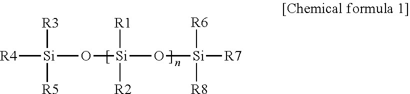
13. The method according to claim 1, wherein the siloxane polymer is in a form which a homopolymer or heteropolymer comprising a monomer having the following chemical formula 1 is linked by cross-linking: ##STR00004## in the chemical formula 1, R1 to R8 are independently of each other hydrogen, C1-10 alkyl, C2-10 alkenyl, C5-14 heterocycle, C3-10 cycloalkyl or C3-10 cycloalkenyl, and n is an integer of 0 to 100,000.
14. The method according to claim 1, wherein the siloxane polymer is in a form which a heteropolymer of a first monomer having the following chemical formula 1 and a second monomer is linked by cross-linking, wherein the second monomer is at least one selected from the group consisting of 1,3, 5-trivinyl-1,3 ,5-trimethylcyclotri siloxane, 2,4, 6,8-tetramethyl-2,4, 6, 8-tetravinyl cy cl otetrasiloxane (V4D4), 2,4, 6,8, 10-pentamethyl-2,4, 6,8, 10-pentavinylcyclopentasiloxane, 2,4,6,8, 10,12-hexamethyl-2,4,6, 8,10,12-hexavinyl-cyclohexasiloxane, octa(vinylsilasesquioxane), and 2,2, 4,4, 6,6, 8,8, 10,10, 12,12-dodecamethylcyclohexasiloxane: ##STR00005## in the chemical formula 1, R1 to R8 are independently of each other hydrogen, C1-10 alkyl, C2-10 alkenyl, C5-14 heterocycle, C3-10 cycloalkyl or C3-10 cycloalkenyl, and n is an integer of 0 to 100,000.
15. The method according to claim 13, wherein the siloxane polymer is a polymer of at least one siloxane monomer selected from the group consisting of dimethylsiloxane (DMS), tetramethyldisiloxane (TMDS), hexavinyldisiloxane, hexamethyldisiloxane, octamethyltrisiloxane, dodecamethylpentatetrasiloxane, tetradecamethylhexasiloxane, methylphenylsiloxane, diphenylsiloxane, and phenyltrimethicone.
16. The method according to claim 1, wherein the polymerization ratio of the siloxane polymer is 50:1 to 1:10.
17. A kit for preparing cancer stem cells in a spheroid, comprising a cell culture substrate comprising a siloxane polymer; and a medium for cell culture comprising albumin, wherein the albumin is at least one uses selected from the group consisting of the following (1) to (2): (1) a use for inducing the cancer cells into cancer stem cells, (2) a use for inducing the cancer cells into a spheroid.
18. A method for screening a therapeutic drug for cancer, comprising preparing cancer stem cell spheroids with using the method for preparing cancer stem cell spheroids according to claim 1; treating a candidate substance to the cancer stem cell spheroids; measuring viabilities of the cancer stem cells in the group treated by the candidate substance and in the control group untreated by the candidate substance; and comparing the viabilities of cancer stem cells in the group treated by the candidate substance and in the control group untreated by the candidate substance.
19. The method according to claim 18, wherein further comprising determining the candidate substance is a therapeutic drug for cancer, when the viability of cancer stem cells in the group treated by the candidate substance is lower than that of the control group.
20. A method for screening a drug for reducing drug resistance of cancer cells, comprising preparing cancer stem cell spheroids with using the method for preparation of cancer stem cell spheroids according to claim 1; treating a candidate substance for reducing drug resistance of cancer cells to the cancer stem cell spheroids, together with a cancer cell-resistant drug; and comparing the viabilities of cancer stem cells in the group treated by the candidate substance and in a control group untreated by the candidate substance.
Description
CROSS REFERENCE TO RELATED APPLICATIONS
[0001] This application claims priority to Korean Patent Application No. 10-2019-0087134 filed on Jul. 18, 2019, which is incorporated by reference in its entirety herein.
TECHNICAL FIELD
[0002] The present invention relates to a preparation method of cancer stem cell spheroids or a kit for preparing cancer stem cell spheroids. In addition, it relates to a method of screening of drugs for treating cancer cell resistance using cancer stem cell spheroids prepared by the method of producing or kit.
BACKGROUND ART
[0003] Cancer stem cells (CSCs or tumor-initiating cells: TIC) have many features similar to normal stem cells, such as self-regenerative ability, endogenous drug resistance and differentiation, and the like. Since cancer cells similar to stem cells have been discovered in acute myeloid leukemia, there is increasing evidence that a small number of cancer stem cells are present in tumor aggregates primarily responsible for tumor recurrence and drug resistance. Therefore, cancer stem cells have attracted considerable attention in the field of cancer research and drug resistance.
[0004] Cancer stem cells are generally isolated from patient-derived tumor tissue based on cancer stem cell surface markers. However, the supply of the patient-derived tumor tissue is limited, and only a small amount of cancer stem cells can be isolated, which makes it difficult to obtain cancer stem cells. Alternatively, attempts have been made to separate cancer stem cells from existing cancer cell lines, but since cancer stem cells are contained less than 1 to 2% in the cancer cell line, it is not practical to secure a sufficient amount of cancer stem cells (Cell 144, 646-674 (2011)). In addition, since the three-dimensional structure of cancer cells can better represent the tumor environment than the two-dimensional monolayer structure, considerable interest is currently shown in developing a method for promoting formation of cancer cells. The spheroid, which is used for drug screening or efficacy testing, is currently produced by a method for inserting cells into a hole of a hydrophilic ULA (ultra-low-attachment) surface, a concave agarose gel (U-bottom) or a hanging-drop cell substrate, and the like. However, even the spheroid produced by the method does not sufficiently contain cancer stem cells. In this situation, there is a need to develop a simple method for producing cancer stem cell spheroids having cancer-formation ability in a human cancer cell line.
[0005] Accordingly, the present inventors have tried to develop a method for producing cancer stem cell spheroids, and as a result, they have established a method for producing cancer stem cell spheroids using a cell culture substrate comprising a siloxane polymer and a medium comprising albumin, thereby completing the present invention.
DISCLOSURE
Technical Problem
[0006] One embodiment of the present invention is to provide a method for preparing a stem cell spheroid from a cancer cell, comprising culturing a cancer cell using a medium for cell culture comprising albumin.
[0007] The albumin may be a use for inducing cancer cells into cancer stem cells, a use for inducing cancer cells into a spheroid, or a use for inducing cancer cells into cancer stem cell spheroids.
[0008] The cancer cell may be cultured on a cell culture substrate comprising a siloxane polymer.
[0009] The cell culture substrate comprising a siloxane polymer may be a use for inducing cancer cells into cancer stem cells, a use for inducing cancer cells into spheroids, or a use for inducing cancer cells into cancer stem cell spheroids.
[0010] Another embodiment of the present invention is to provide a kit for preparing cancer stem cell spheroids, comprising a cell culture substrate and a medium for cell culture.
[0011] The cell culture substrate may comprise a siloxane polymer, and the medium for cell culture may comprise albumin, and the siloxane polymer or the albumin may be a use for inducing a cancer cell into cancer stem cells, a use for inducing a cancer cell into a spheroid, or a use for inducing a cancer cell into cancer stem cell spheroids.
[0012] Other one embodiment of the present invention is to provide a method for screening of drugs for treating cancer cell resistance, comprising (a) preparing cancer stem cell spheroids by the method for preparation of cancer stem cell spheroids; (b) treating a candidate substance for treating cancer cell resistance to the cancer stem cell spheroid of the (a) step; and (c) comparing the cancer stem cell spheroid group in which the candidate substance for treating cancer cell resistance of the (b) step and the control group in which the candidate substance for treating cancer cell resistance is untreated.
Technical Solution
[0013] This is specifically described as follows. Meanwhile, each description and embodiment disclosed in the present application may be applied to each other description and embodiment. In other words, all combinations of various elements disclosed in the present application fall within the scope of the present application. In addition, the scope of the present application is not considered to be limited by the specific description disclosed below.
[0014] As one aspect to achieve the objects of the present invention, a composition for inducing cancer stem cells from cancer cells, comprising a medium for cell culture containing albumin is provided.
[0015] The albumin may be (1) a use for inducing the cancer cells into cancer stem cells, (2) a use for inducing the cancer cell into a spheroid, or (3) a use for inducing the cancer cell into cancer stem cell spheroids.
[0016] As another aspect to achieve the objects of the present invention, a method for preparing cancer stem cells from cancer cells, comprising culturing a cancer cell using a composition for inducing cancer stem cells from cancer cells, comprising a medium for cell culture containing albumin is provided.
[0017] The cancer cell may be cultured on a cell culture substrate, and the cell culture substrate may comprise a siloxane polymer.
[0018] The cell culture substrate comprising the siloxane polymer may be (1) a use for inducing the cancer cells into cancer stem cells, (2) a use for inducing the cancer cell into a spheroid, or (3) a use for inducing the cancer cells into cancer stem cell spheroids.
[0019] As other aspect to achieve the objects of the present invention, a kit for preparing cancer stem cell spheroids, comprising a cell culture substrate, and a composition for inducing cancer stem cells from a cancer cell, comprising a medium for cell culture containing albumin, wherein the cell culture substrate comprise a siloxane polymer and the medium comprises albumin, is provided.
[0020] As other aspect to achieve the objects of the present invention, a method of screening of drugs for treating cancer cell resistance, comprising preparing cancer stem cell spheroids; treating a candidate substance for treating cancer cell resistance to the cancer stem cell spheroid; and comparing cancer stem cell spheroids group in which the candidate substance for treating cancer cell resistance is treated and a control group in which the candidate substance for treating cancer cell resistance is not treated is provided.
[0021] The present inventors have found that when a cancer cell is cultured in a medium comprising albumin, on a cell culture substrate comprising a polymer formed by a siloxane compound, a three-dimensional cancer stem cell spheroid like in vivo environment, which completely has characteristics of the cancer stem cell, can be prepared with high yield, thereby providing the present invention.
[0022] Hereinafter, the present invention will be described in more detail.
[0023] According to one embodiment of the present invention, it relates to a method for preparation of cancer stem cells, comprising culturing a cancer cell on a cell culture substrate, comprising a siloxane polymer. The culturing a cancer cell may be culturing the cancer cell using a medium for cell culture comprising albumin. The cancer cell is a general cancer cell which does not have characteristics of the caner stem cell, and after the culturing, it has characteristics of the cancer stem cell (for example, expression of cancer stem cell marker genes, in vivo cancer-formation ability, drug resistance, cell migration or cell penetration, etc.). Therefore, the culturing a cancer cell may be culturing the cancer cell using a composition for inducing cancer stem cells from the cancer cells, and the composition for inducing cancer stem cells from the cancer cells may comprise a medium for cell culture comprising albumin.
[0024] The medium for cell culture may further comprise amino acids, vitamins, antioxidants, trace elements, proteins, collagen precursors, and the like. The amino acid may include glycine, histidine, isoleucine, methionine, phenylalanine, proline, hydroxyproline, serine, threonine, tryptophan, tyrosine, valine, etc., but not limited thereto, and the amino acid may be L-type amino acid or D-type amino acid. The vitamin may include thiamine, ascorbic acid, etc., but not limited thereto. The antioxidants may include glutathione, but not limited thereto. The trace elemets may include Ag.sup.+, Al.sup.3+, Ba.sup.2+, Cd.sup.2+, Co.sup.2+, Ge.sup.4+, Se.sup.4+, Br.sup.-, I.sup.-, F.sup.-, Mn.sup.2+, Si.sup.4+, V.sup.5+, Mo.sup.6+, Ni.sup.2+, Rb.sup.+, Sn.sup.2+, Zr.sup.4+, etc, but not limited thereto. The proteins may include transferrine, insulin, lipid-rich albumin (for example, AlbuMAX, etc.), but are not limited thereto. The collagen precursor may include L-proline, L-hydroxyproline, ascorbic acid, but not limited thereto.
[0025] As one aspect to achieve the objects of the present invention, a method for producing cancer stem cells from cancer cells, comprising culturing cancer cells using a composition for inducing cancer stem cells from cancer cells, comprising a medium for cell culture containing albumin is provided.
[0026] The culturing cancer cells using a composition for inducing cancer stem cells from cancer cells, comprising a medium for cell culture containing albumin is culturing an isolated cancer cell using a composition comprising a medium for cell culture comprising albumin, and the culturing may be performed on a cell culture substrate comprising a siloxane polymer.
[0027] When the cell culture substrate is a linear siloxane substrate, as a spheroid may not be formed when culturing a cancer cell using a medium for cell culture comprising albumin (FBS) (Example 7-3), it is inferred that the culture medium also affects spheroid formation in addition to surface functional stimuli of the substrate when cancer stem cell spheroids are prepared from a cancer cell using the linear siloxane substrate.
[0028] The cancer stem cell spheroid may be formed within 240 hours, within 20 hours, within 180 hours, within 150 hours, within 120 hours, within 110 hours, within 100 hours, within 96 hours, within 90 hours, within 84 hours, within 80 hours, within 72 hours, within 70 hours, within 60 hours, within 50 hours, within 40 hours, within 30 hours, within 24 hours, within 20 hours, within 12 hours, within 10 hours, or within 5 hours, after the start of culturing a cancer cell.
[0029] The term of the present invention, "cancer cell" or "isolated cancer cell" may be a cell derived from a human or a cell derived from various individuals except for humans, but not limited thereto. In addition, the isolated cancer cell may include all of in vivo or in vitro cells, but not limited thereto. Specifically, the isolated cancer cell may be specifically a cell derived from various tissues of humans, and may be a cancer cell derived from ovarian cancer, breast cancer, liver cancer, brain cancer, colorectal cancer, prostate cancer, cervical cancer, lung cancer, stomach cancer, skin cancer, pancreatic cancer, oral cancer, rectal cancer, laryngeal cancer, thyroid cancer, parathyroid cancer, colon cancer, bladder cancer, peritoneal carcinoma, adrenal cancer, tongue cancer, small intestine cancer, esophageal cancer, renal pelvis cancer, renal cancer, heart cancer, duodenal cancer, ureteral cancer, urethral cancer, pharynx cancer, vaginal cancer, tonsil cancer, anal cancer, pleura cancer, thymic carcinoma or nasopharyngeal carcinoma, but not limited thereto, and it includes all cancer cells which can be used for the objects of the present invention, and includes all primary cultured cells isolated by biopsy from cancer tissue, or established cell lines, but not limited thereto.
[0030] In addition, to confirm the cancer cell, a cancer cell marker may be used. Specifically, as the marker, AFP (Alpha-fetoprotein), CA15-3, CA27-29, CA19-9, CA-125, Calcitonin, Calretinin, CD34, CD117, Desmin, inhibin, Myo D1, NSE (neuronspecific enolase), PLAP (placental alkaline phosphatase) or PSA (prostatespecific antigen), or the like may be used, but not limited thereto.
[0031] The term of the present invention, "siloxane compound" is a compound comprising a siloxane group (Si--O bond) and is intended to include all siloxane monomers or siloxane polymers. The "siloxane polymer" means a polymer comprising a siloxane group as a repeated unit, and for example, it may include a linear siloxane polymer or cyclic siloxane polymer. The siloxane monomer or the siloxane polymer may be a compound having chemical formula 1, and may include a polymer having chemical formula 2 of the cyclic siloxane, and the like.
##STR00001##
[0032] In the chemical formula 1,
[0033] R1 to R8 may be independently of each other hydrogen, C1-10 alkyl, C2-10 alkenyl, C5-14 heterocycle, C3-10 cycloalkyl or C3-10 cycloalkenyl, and n is an integer of 0 to 100,000. For example, the R1 to R8 may be independently of each other hydrogen, methyl, ethyl, propyl, ethylene, propylene, vinyl group, and the like, but not limited thereto.
[0034] According to one embodiment of the present invention, the linear siloxane compound may be at least one selected from the group consisting of dimethylsiloxane (DMS), tetramethyldisiloxane (TMDS), hexavinyldisiloxane, hexamethyldisiloxane, octamethyltrisiloxane, dodecamethylpentatetrasiloxane, tetradecamethylhexasiloxane, methylphenylsiloxane, diphenylsiloxane and phenyltrimethicone, and the linear siloxane polymer may be formed as the linear siloxane compound is polymerized.
[0035] According to one embodiment of the present invention, the siloxane polymer may be a polymer formed by polymerization of a base compound using a curing agent, and the base compound and the curing agent may be polymerized at a ratio of 100:1 to 1:100, 100:1 to 1:80, 100:1 to 1:50, 100:1 to 1:30, 100:1 to 1:20, 100:1 to 1:15, 100:1 to 1:10, 80:1 to 1:100, 80:1 to 1:80, 80:1 to 1:50, 80:1 to 1:30, 80:1 to 1:20, 80:1 to 1:15, 80:1 to 1:10, 60:1 to 1:100, 60:1 to 1:90, 60:1 to 1:80, 60:1 to 1:70, 60:1 to 1:60, 60:1 to 1:50, 60:1 to 1:40, 60:1 to 1:30, 60:1 to 1:20, 60:1 to 1:15, 60:1 to 1:10, 50:1 to 1:100, 50:1 to 1:90, 50:1 to 1:80, 50:1 to 1:70, 50:1 to 1:60, 50:1 to 1:50, 50:1 to 1:40, 50:1 to 1:30, 50:1 to 1:20, 50:1 to 1:15, or 50:1 to 1:10, but not limited thereto.
[0036] The siloxane polymer according to one embodiment of the present invention may be a cross-linked siloxane compound, and may be a cross-linked monomer of at least 1% or more, 5% or more, 10% or more, 20% or more, 30% or more, 40% or more, 50% or more, 60% or more, 70% or more, 80% or more, or 90% or more, but not limited thereto, Specifically, the siloxane polymer may be water-insoluble as at least 1% or more of siloxane compound polymer is cross-linked. For example, in the chemical formula 1, R1 to R8 may be independently of each other linear siloxane compound or siloxane polymer, and therefore, the compound of chemical formula 1 may be a siloxane polymer formed as cross-linked.
[0037] The base compound for preparing a siloxane polymer may be the siloxane compound represented by chemical formula 1, for example, siloxane oligomer, dimethylsiloxane, tetram ethyl di siloxane, hexavinyldisiloxane, hexam ethyldisiloxane, octamethyltrisiloxane, trialkoxysiloxane, or tetraalkoxysiloxane, or the like, and the curing agent may be a siloxane cross-linker, a metal catalyst (platinum catalyst, ruthenium catalyst, etc.), hexamethylenetetramine, ammonia (NH3) or hydrogen chloride (HCl), or the like.
[0038] The siloxane compound according to one embodiment of the present invention may be a cyclic siloxane compound or cyclosiloxane polymer, and is used to include compounds which have a cyclosiloxane structure as a basic structure, and has a functional group (for example, alkyl group, alkenyl group, etc.) at the position of its silicon atom. According to one embodiment of the present invention, the cyclosiloxane compound is represented by the following chemical formula 2.
##STR00002##
[0039] In the formula, A is
##STR00003##
(n=an integer of 1-8);
[0040] R1 and R2 are independently of each other hydrogen or C2-10 alkenyl with the proviso that at least two positions of R1 are C2-10 alkenyl; and
[0041] R2 is independently of each other hydrogen, C1-10 alkyl, C2-10 alkenyl, halo group, metal element, C5-14 heterocycle, C3-10 cycloalkyl or C3-10 cycloalkenyl.
[0042] The term of the present invention, "alkyl" means a straight-chain or branched-chain, unsubstituted or substituted, saturated hydrocarbon group, and for example, includes methyl, ethyl, propyl, isobutyl, pentyl or hexyl, and the like. C1-C10 alkyl means an alkyl group having an alkyl unit of 1 to 10 carbon atoms, and when C1-C10 alkyl is substituted, the number of carbon atoms of the substituent is not comprised. Herein, C1-C10 alkyl may be C1-C8 alkyl, C1-C7 alkyl or C1-C6 alkyl.
[0043] The term of the present invention, "alkenyl" represents a straight-chain or branched-chain, unsubstituted or substituted, unsaturated hydrocarbon group having designated carbon atoms, and for example, includes vinyl, propenyl, allyl, isopropenyl, butenyl, isobutenyl, t-butenyl, n-pentenyl, and n-hexenyl. C2-C10 alkenyl means an alkenyl group having an alkenyl unit of 1 to 10 carbon atoms, and when C2-C10 alkenyl is substituted, the number of carbon atoms of the substituent is not comprised.
[0044] According to one embodiment of the present invention, herein, C2-10 alkenyl is C2-8 alkenyl, C2-6 alkenyl, C2-5 alkenyl, C2-4 alkenyl or C2-3 alkenyl. According to one embodiment of the present invention, at least three parts of the R1 is C2-10 alkenyl. According to one embodiment of the present invention, the cyclosiloxane has n+1 or n+2 of C2-10 alkenyl at the R1 position. For example, when n is 2, the compound of chemical formula 1 becomes a cyclotetrasioloxane having 3 or 4 C2-10 alkenyls at the R1 position. This alkenyl group is involved in polymerization.
[0045] The term of the present invention, "halo" represents a halogen element, and for example, includes flouro, chloro, bromo and iodo. The term of the present invention, "metal element" means an element which makes metallic simple substance such as alkali metal elements (Li, Na, K, Rb, Cs, Fr), alkali earth metal elements (Ca, Sr, Ba, Ra), aluminum family elements (Al, Ga, In, Tl), tin family elements (Sn, Pb), coinage metal elements (Cu, Ag, Au), zinc family elements (Zn, Cd, Hg), rare earth elements (Sc, Y, 57-71), titanium family elements (Ti, Zr, Hf), vanadium family elements (V, Nb, Ta), chrome family elements (Cr, Mo, W), manganese family elements (Mn, Tc, Re), iron family elements (Fe, Co, Ni), platinum family elements (Ru, Rh, Pd, Os, Ir, Pt) and actinide elements (89-103), and the like.
[0046] The term of the present invention, "heterocycle" means a partially or completely saturated, monocycle type or bicycle type of 5 to 14 membered heterocycle ring. N, O and S are examples of heteroatoms. Pyrrole, furan, thiophene, imidazole, pyrazole, oxazole, isoxazole, thiazole, isothiazole, tetrazole, 1,2,3,5-oxathiadiazole-2-oxide, triazolone, oxadiaxolone, isoxazolone, oxadiazolidine dione, 3-hydroxypyro-2,4-dione, 5-oxo-1,2,4-thiadiazole, pyridine, pyrazine, pyrimidine, indole, isoindole, indazole, phthalazine, quinoline, isoquinoline, quinoxaline, quinazoline, cinnoline and carboline are examples of C5-14 heterocycles.
[0047] The term of the present invention, "cycloalkyl" means a cyclic hydrocarbon radical, and this includes cyclopropyl, cyclobutyl and cyclopentyl. C3-10 cycloalkyl means a cycloalkyl having 3-10 carbon atoms which form a ring structure, and when C3-10 cycloalkyl is substituted, the number of carbon atoms of the substituent is not comprised.
[0048] According to one embodiment of the present invention, herein, C1-C10 cycloalkyl is C1-C8 cycloalkyl, C1-C7 cycloalkyl or C1-C6 cycloalkyl.
[0049] The term of the present invention, "cycloalkenyl" means a cyclic hydrocarbon group having at least one double bond, and for example, includes cyclopentene, cyclohexene and cyclohexadiene. C3-10 cycloalkenyl means a cycloalkenyl having 3-10 carbon atoms which form a ring structure, and when C3-10 cycloalkenyl is substituted, the number of carbon atoms of the substituent is not comprised.
[0050] According to one embodiment of the present invention, C2-10 cycloalkenyl is C2-8 cycloalkenyl, C2-6 cycloalkenyl, C2-5 cycloalkenyl, C2-4 cycloalkenyl or C2-3 cycloalkenyl.
[0051] According to one embodiment of the present invention, the R2 is independently of each other hydrogen, C1-10 alkyl or C2-10 alkenyl. According to one specific example, at least two parts or at least three parts of the R2 may be C1-10 alkyl or C2-10 alkenyl. According to one specific example, the cyclosiloxane may have n+1 or n+2 of C1-10 alkyl or C2-10 alkenyl at the R2 position.
[0052] According to one embodiment of the present invention, the n is an integer of 1-7, an integer of 1-6, an integer of 1-5, an integer of 1-4 or an integer of 1-3.
[0053] According to one embodiment of the present invention, the cyclosiloxane compound is selected from the group consisting of 2,4, 6,8-tetra(C2-10)alkenyl -2,4,6, 8-tetra(C1-10)alkyl cy cl otetrasiloxane, 1,3,5-tri(C1-10)alkyl-1,3,5-tri(C2-10)alkenylcyclotrisiloxane, 1,3, 5,7-tetra(C1-10)alkyl-1,3,5, 7-tetra(C2-10)alkenyl cy cl .degree. tetra siloxane, 1,3,5,7, 9-penta(C1-10)alkyl-1,3,5, 7,9-penta(C2-10)alkenylcyclopentasiloxane, 1,3,5-tri(C1-10)alkyl-1,3,5-tri(C2-10)alkenylcyclotrisiloxane, 1,3, 5,7-tetra(C1-10)alkyl-1,3,5, 7-tetra(C2-10)alkenyl cy cl .degree. tetra siloxane, 1,3,5,7, 9-penta(C1-10)alkyl-1,3,5, 7,9-penta(C2-10)alkenylcyclopentasiloxane, 1,3,5-tri(C1-10)alkyl-1,3,5-tri(C2-10)alkenylcyclotrisiloxane, 1,3, 5,7-tetra(C1-10)alkyl-1,3,5, 7-tetra(C2-10)alkenyl cy cl .degree. tetra siloxane, 1,3,5,7, 9-penta(C1-10)alkyl-1,3,5, 7,9-penta(C2-10)alkenylcyclopentasiloxane, hexa(C2-10)alkenylcyclotrisiloxane, octa(C2-10)alkenylcyclotetrasiloxane, deca(C2-10)alkenylcyclopentasiloxane, 2,4,6, 8-tetravinyl-2,4, 6,8,-tetramethylcyclotetrasiloxane and combinations thereof.
[0054] According to one specific example, the cyclosiloxane compound is selected from the group consisting of 1,3,5 -trivinyl-1,3,5 -trim ethyl ecy cl otri siloxane, 2,4, 6,8-tetramethyl-2,4, 6,8-tetravi nyl cy cl otetrasiloxane (V4D4), 2,4, 6,8, 10-p entamethyl-2,4, 6,8, 10-p entavinyl cy cl op entasil oxane, 2,4,6,8, 10,12-hexamethyl-2,4,6, 8,10,12-hexavinyl-cyclohexasiloxane, octa(vinylsilasesquioxane), 2,2,4,4, 6,6, 8,8, 10,10, 12,12-dodecamethylcyclohexasiloxane, 2,4,6,8-tetra(C2-4)alkenyl -2,4,6, 8-tetra(C 1-6)alkyl cy clotetrasiloxane (as one example, 2,4,6,8-tetravinyl-2,4,6,8-tetramethylcyclotetrasiloxane), 1,3,5-tri(C1-6)alkyl-1,3,5-tri(C2-4)alkenylcyclotrisiloxane (as one example, 1,3,5-triisopropyl-1,3,5-trivinylcyclotrisiloxane), 1,3,5,7-tetra(C1-6)alkyl-1,3,5,7-tetra(C2-4)alkenylcyclotetrasiloxane (as one example, 1,3,5,7-tetraisopropyl-1,3,5,7-tetravinylcyclotetrasiloxane), 1,3,5,7,9-penta(C1-6)alkyl-1,3,5,7,9-penta(C2-4)alkenylcyclopentasiloxane (as one example, 1,3,5,7,9-p entai sopropyl-1,3, 5,7,9-pentavinyl cy clop entasil oxane), 1,3,5-tri(C1-6)alkyl-1,3,5-tri(C2-4)alkenylcyclotrisiloxane (as one example, 1,3,5-tri-sec-butyl-1,3,5-trivinylcyclotrisiloxane), 1,3,5,7-tetra(C1-6)alkyl-1,3,5,7-tetra(C2-4)alkenylcyclotetrasiloxane (as one example, 1,3, 5,7-tetra-se c-butyl -1,3,5, 7-tetravinyl cy cl otetrasiloxane), 1,3,5,7,9-penta(C1-6)alkyl-1,3,5,7,9-penta(C2-4)alkenylcyclopentasiloxane (as one example, 1,3,5,7,9-p enta-se c-butyl-1,3,5,7, 9-p entavinyl cy cl op entasiloxane), 1,3,5-tri(C1-6)alkyl-1,3,5-tri(C2-4)alkenylcyclotrisiloxane (as one example, 1,3,5-triethyl-1,3,5-trivinylcyclotrisiloxane), 1,3,5,7-tetra(C1-6)alkyl-1,3,5,7-tetra(C2-4)alkenylcyclotetrasiloxane (as one example, 1,3, 5,7-tetraethyl -1,3,5 ,7-tetravi nylcyclotetrasiloxane), 1,3,5,7,9-penta(C1-6)alkyl-1,3,5,7,9-penta(C2-4)alkenylcyclopentasiloxane (as one example, 1,3,5,7,9-p entaethyl-1,3,5,7, 9-p entavinyl cy cl op entasiloxane), hexa(C2-4)alkenylcyclotrisiloxane (as one example, hexavinylcyclotrisiloxane), octa(C2-4)alkenylcyclotetrasiloxane (as one example, octavinylcyclotetrasiloxane), deca(C2-4)alkenylcyclopentasiloxane (as one example, decavinylcyclopentasiloxane) and combinations thereof.
[0055] The term of the present invention, "cell culture substrate comprising a siloxane compound" may mean that a polymer formed by siloxane is a part of a cell culture substrate (for example, cell culture substrate of which surface is coated with the polymer), and also may mean that the solid polymer formed by siloxane itself may be used as a cell culture substrate, but not limited thereto.
[0056] It is sufficient that the cell culture substrate provides any space capable of culturing a cell, and its shape is not limited. For example, the cell culture substrate may be a dish (culture dish), a chalet or plate (for example, 6-well, 24-well, 48-well, 96-well, 384-well or 9600-well microtiter plate, microplate, dip-well plate, etc.), a flask, a chamber slide, a tube, a cell factory, a roller bottle, a spinner flask, hollow fibers, a microcarrier, beads, and the like, but not limited thereto, and any material having support properties can be used without limitation as the cell culture substrate, and for example, plastics (for example, polystyrene, polyethylene, polypropylene, etc.), metals, silicon and glass, and the like may be used as the cell culture substrate.
[0057] In addition, the polymer formed by the siloxane compound is used as a meaning including all of (1) homopolymers formed by polymerization of homogeneous siloxane compounds, (2) copolymers formed by polymerization of heterogeneous siloxane compounds, and (3) copolymers formed by polymerization of homogeneous or heterogeneous siloxane compounds with other monomer compounds. Herein, the copolymer may be random copolymers, block copolymers, alternating copolymers or graft copolymers, but not limited thereto.
[0058] Therefore, according to one embodiment of the present invention, the polymer formed by the siloxane compound is a homogeneous polymer formed by polymerization of homogeneous siloxane compounds, and for example, may be a homogeneous polymer formed by polymerization of homogeneous linear siloxane compounds, or a homogeneous polymer formed by polymerization of homogeneous cyclosiloxane compounds.
[0059] According to another embodiment of the present invention, the polymer formed by the siloxane compound is a copolymer formed by a first monomer that is the siloxane compound and a second monomer that can polymerize therewith, and for example, may be a copolymer formed by a first monomer that is the linear siloxane compound and a second monomer that can polymerized therewith, or a copolymer formed by a first monomer that is the cyclosiloxane compound and a second monomer that can polymerized therewith.
[0060] According to one specific example, the second monomer is a siloxane compound different from the first monomer (copolymer formed by heterogeneous siloxane compounds, for example, copolymer formed by heterogeneous linear siloxane compounds, copolymer formed by heterogeneous cyclosiloxane compounds, or copolymer formed by heterogeneous linear siloxane compound and cyclosiloxane compound).
[0061] According to another specific example, the second monomer is a compound having a carbon double bond for polymerization with the first monomer. Then, the first monomer may also have a carbon double bond for polymerization with the second monomer. Such a second monomer compound may be, for example, selected from the group consisting of siloxane having a vinyl group (for example, hexavinyldisiloxane, tetramethyldisiloxane, etc.), methacrylate-based monomers, acrylate-based monomers, aromatic vinyl-based monomers (for example, divinylbenzene, vinylbenzoate, styrene, etc.), acrylamide-based monomers (for example, N-isopropylacrylamide, N,N-dimethylacrylamide, etc.), maleic anhydride, silazane or cyclosilazane having a vinyl group (for example, 2,4,6-trimethyl-2,4,6-trivinylcyclosilazane, etc.), C3-10 cycloalkane having a vinyl group (for example, 1,2,4-trivinylcyclohexane, etc.), vinylpyrrolidone, 2-(methacryloyloxy)ethylacetoacetate, 1-3 (-aminopropyl)imidazole, vinylimidazole, vinylpyridine, silane having a vinyl group (for example, allyltrichlorosilane, acryloxymethyltrimethoxysilane, etc.) and combinations thereof.
[0062] According to other specific example, the second monomer may be at least one selected from the group consisting of 1,3, 5-trivinyl-1,3,5-trim ethyl cy cl otri siloxane, 2,4,6, 8-tetram ethyl-2,4, 6, 8-tetravinylcyclotetrasiloxane (V4D4), 2,4, 6,8, 10-p entamethyl-2,4, 6,8, 10-p entavinyl cy cl op entasil oxane, 2,4,6,8, 10,12-hexamethyl-2,4,6, 8,10,12-hexavinyl-cyclohexasiloxane, octa(vinylsilasesquioxane), and 2,2,4,4, 6,6, 8,8, 10,10, 12,12-dodecamethylcyclohexasiloxane.
[0063] The methacrylate-based monomer includes, for example, methacrylate, methacrylic acid, glycidyl methacryl ate, p erfluoromethacryl ate, benzylmethacrylate, 2-(dim ethyl amino)ethylm ethacryl ate, p erfurilm ethacryl ate, 3,3,4,4,5,5, 6,6, 7,7, 8,8, 9,9, 10,10, 10-heptadecaflourodecylmethacrylate, hexylmethacrylate, methacrylic anhydride, p entafl ouropheny lm ethacryl ate, prop argylmethacryl ate, tetrahy drop erp erillm ethacryl ate, butylmethacrylate, methacryl oylchl ori de and di(ethyleneglycol)methylestermethacrylate, and the like.
[0064] The acrylate-based monomer includes, for example, acrylate, 2-(dimehtylamino)ethyl acrylate, ethyl eneglycolacryl ate, 1H, 1H,7H-dodecafluoroheptylacryl ate, 1H,1H,7H-dodecafluoroheptylacryl ate, isobornyl acrylate, 1H,1H,2H,2H-perfluorodecylacrylate, tetrahy drop erfurilacryl ate, p oly (ethyl enegly col)di acryl ate, 1H,1H,7H-dodecafluoroheptylacryl ate and propargylacrylate, and the like.
[0065] The copolymer of the present invention may further comprise a monomer other than monomers mentioned herein as a comonomer.
[0066] According to one embodiment of the present invention, the copolymer contains at least 50% or more of the siloxane compound. According to one specific example, the copolymer contains at least 60% or more, 70% or more, 80% or more or 90% or more of the siloxane compound. This content is based on the flow rate (unit: sccm), and 90% means the content of the siloxane compound contained in the copolymer formed by flowing (dropping) each monomer at a flow rate of 9:1 (siloxane compound: other monomer), and 80%, 70% and 60% mean the content of the siloxane compound comprised in the copolymer formed by flowing at a flow rate of 8:1, 7:1 and 6:1.
[0067] In addition, the cell culture substrate comprising the polymer may be a cell culture substrate comprising a polymer having various thicknesses. The thickness of the polymer may be, for example, about 10, 11, 12, 13, 14, 15, 16, 17, 18, 19, 20, 21, 22, 23, 24, 25, 26, 27, 28, 29, 30, 31, 32, 33, 34, 35, 36, 37, 38, 39, 40, 41, 42, 43, 44, 45, 46, 47, 48, 49, 50, 60, 70, 80, 90, 100, 200, 300 nm or more, or about 10,000, 5,000, 1,000, 900, 800, 700, 600, 500, 400, 300 nm or less, or about 10 to 300 nm, 10 to 500 nm, 10 to 1000 nm, 50 to 300 nm, 50 to 500 nm, 50-1000 nm, but not limited thereto.
[0068] The cell culture substrate comprising a siloxane polymer according to one embodiment of the present invention may have a water contact angle of 160.degree. or more, 150.degree. or more, 140.degree. or more, 130.degree. or more, 120.degree. or more, 110.degree. or more, 100.degree. or more, 90.degree. or more, 80.degree. or more, 70.degree. or more, 60.degree. or more, 50.degree. or more, 40.degree. or more, 30.degree. or more, 20.degree. or more, or 10.degree. or more.
[0069] In the method for preparing a cancer stem cell from a cancer cell, comprising culturing a cancer cell using a medium for cell culture containing albumin, the cancer stem cell may be in a spheroid form. The method may be characterized by not comprising any other compound known for additional gene manipulation or stem cell proliferation, or known to differentiate a stem cell from an adult cell. The medium for cell culture may not comprise other growth factors except for albumin.
[0070] The term, "spheroid" means a cell aggregate forming a three-dimensional sphere form by gathering 1000 or more of single cells, and as it can more accurately copy structural and physical properties of the three-dimensional tissue surrounding cells in a human body, it is usefully used in treatment and research fields, and on purpose of the present invention, the spheroid is characterized by a cancer stem cell spheroid.
[0071] In addition, the term of the present invention, "cancer stem cell (or tumor initiating cell)" means a cell having an ability to produce tumor, and the cancer stem cell has similar characteristics to normal stem cells. The cancer stem cell causes tumor through self-regeneration and differentiation that are characteristics of the stem cell in various cell types, and therefore it has a cancer-formation ability. It becomes a reason for recurrence and metastasis by causing new tumor distinguished from other groups in tumor by the cancer-formation ability. In addition, as another characteristic of the cancer stem cell, it has drug resistance, and therefore it has resistance to chemical therapies such as anticancer agent usage, and the like, and thus only common cancer cells are removed and cancer stem cells remain without dying, and the cancer may recur again. Thus, to completely cure cancer, it is important to study the cancer stem cell.
[0072] Furthermore, to confirm the cancer stem cell, a cancer stem cell marker may be used. The cancer stem cell marker may be CD47, BMI-1, CD24, CXCR4, DLD4, GLI-1, GLI-2, PTEN, CD166, ABCG2, CD171, CD34, CD96, TIM-3, CD38, STRO-1 and CD19, and specifically, it may be CD44, CD133, ALDH1A1, ALDH1A2, EpCAM, CD90 and LGRS, but not limited thereto.
[0073] The method of preparing and kit for preparing cancer stem cell spheroids of the present invention have an advantage capable of preparing a cancer stem cell more simply and rapidly, since artificial gene manipulation is not required for preparing a spheroid.
[0074] In addition, it has been confirmed that a cancer stem cell (CSC) marker gene prepared by the method and kit is expressed (Example 6), and has a drug resistance property by drug discharging, and has a cancer-formation ability in vivo (Example 12), and therefore the cancer stem cell spheroid prepared by the method and kit of the present invention may be used for studying cancer stem cells and screening its therapeutic agent by having properties of the caner stem cell.
[0075] The cancer stem cell spheroid of the present invention may be cultured in a three-dimensional, stereoscopic culture form, and may be a cancer stem cell spheroid which has a characteristic of drug resistance or is cancer cell-derived patient-specific, but not limited thereto.
[0076] The term of the present invention, "albumin" consists of basic substances of cells with globulin, and it is comprised in the culture medium of a cancer cell plated in the cell culture substrate of the present invention, and substances capable of forming cancer stem cell spheroids from a cancer cell are included without limitation. The albumin of the present invention may be selected from the group consisting of serum albumin, ovalbumin, lactalbumin and combinations thereof, but not limited thereto. As the example, a commercially available serum replacement (SR) is also included, but not limited thereto. Most of cells require serum to proliferate, and artificial serum or serum replacement which can perform an equal or similar function to natural serum may be used. The artificial serum or serum replacement may be used as a substitute for natural serum in cell culture, and it commonly comprises albumin. The albumin of the present invention may be added as a single component of albumin, or be provided as a formulation comprised in a serum replacement, a formulation prepared by further adding albumin to a serum replacement, or a formulation prepared by further adding albumin to FBS, and more preferably, it may be provided as a formulation in which albumin is further added to a serum replacement, but not limited thereto. In addition, the serum albumin may be selected from the group consisting of bovine serum albumin, human serum albumin and combinations thereof depending on its origin, but not limited thereto. Herein, it has been confirmed that the spheroid prepared using bovine serum albumin expresses a cancer stem cell-related marker (Example 6), and therefore it can be seen that albumin can induce a cancer stem cell.
[0077] The albumin concentration may be comprised in a medium at a concentration of 0.1 mg/ml to 500 mg/ml. Specifically, the albumin concentration may be comprised in a medium ata concentration of about 0.1, 0.2, 0.5, 0.6, 1, 1.1, 2, 3, 4, 5, 6, 11, 16, 21, 26, 31, 36, 41, 46, 51, 56, 61, 66, 71, 76, 81, 86, 91, 96, 100, 101, 106, 111, 116, 121, 126, 131, 136, 141, 146 mg/ml or more, or about 500, 450, 400, 350, 300, 250, 200, 199, 195, 190, 175, 170, 150, 149, 144, 139, 134, 129, 124, 119, 114, 109, 104, 99, 94, 89, 84, 79, 74, 69, 64, 59, 54, 49, 44, 39, 34, 29, 24, 19, 14, 9, 4, 1.4, 0.9, 0.4 mg/ml or less, more specifically, about 0.1 mg/ml to about 500 mg/ml, about 0.5 mg/ml to about 500 mg/ml, about lmg/ml to about 500 mg/ml, about 5 mg/ml to about 500 mg/ml, about 10 mg/ml to about 500 mg/ml, about 20 mg/ml to about 500 mg/ml, about 40 mg/ml to about 500 mg/ml, about 0.1 mg/ml to about 400 mg/ml, about 0.5 mg/ml to about 400 mg/ml, about lmg/ml to about 400 mg/ml, about 5 mg/ml to about 400 mg/ml, about 10 mg/ml to about 400 mg/ml, about 20 mg/ml to about 400 mg/ml, about 40 mg/ml to about 400 mg/ml, about 0.1 mg/ml to about 300 mg/ml, about 0.5 mg/ml to about 300 mg/ml, about lmg/ml to about 300 mg/ml, about 5 mg/ml to about 300 mg/ml, about 10 mg/ml to about 300 mg/ml, about 20 mg/ml to about 300 mg/ml, about 40 mg/ml to about 300 mg/ml, about 0.1 mg/ml to about 200 mg/ml, about 0.5 mg/ml to about 200 mg/ml, about 1 mg/ml to about 200 mg/ml, about 5 mg/ml to about 200 mg/ml, about 10 mg/ml to about 200 mg/ml, about 20 mg/ml to about 200 mg/ml, about 40 mg/ml to about 200 mg/ml, about 0.1 mg/ml to about 150 mg/ml, about 0.5 mg/ml to about 150 mg/ml, about 1 mg/ml to about 150 mg/ml, about 5 mg/ml to about 150 mg/ml, about 10 mg/ml to about 150 mg/ml, about 20 mg/ml to about 150 mg/ml, about 40 mg/ml to about 150 mg/ml, about 0.1 mg/ml to about 100 mg/ml, about 0.5 mg/ml to about 100 mg/ml, about 1 mg/ml to about 100 mg/ml, about 5 mg/ml to about 100 mg/ml, about 10 mg/ml to about 100 mg/ml, about 20 mg/ml to about 100 mg/ml, about 40 mg/ml to about 100 mg/ml, about 0.1 mg/ml to about 80 mg/ml, about 0.5 mg/ml to about 80 mg/ml, about 1 mg/ml to about 80 mg/ml, about 5 mg/ml to about 80 mg/ml, about 10 mg/ml to about 80 mg/ml, about 20 mg/ml to about 80 mg/ml, about 40 mg/ml to about 80 mg/ml, about 0.1 mg/ml to about 70 mg/ml, about 0.5 mg/ml to about 70 mg/ml, about 1 mg/ml to about 70 mg/ml, about 5 mg/ml to about 70 mg/ml, about 10 mg/ml to about 70 mg/ml, about 20 mg/ml to about 70 mg/ml, about 40 mg/ml to about 70 mg/ml, about 0.1 mg/ml to about 60 mg/ml, about 0.5 mg/ml to about 60 mg/ml, about 1 mg/ml to about 60 mg/ml, about 5 mg/ml to about 60 mg/ml, about 10 mg/ml to about 60 mg/ml, about 20 mg/ml to about 60 mg/ml, about 40 mg/ml to about 60 mg/ml, about 0.1 mg/ml to about 50 mg/ml, about 0.5 mg/ml to about 50 mg/ml, about 1 mg/ml to about 50 mg/ml, about 5 mg/ml to about 50 mg/ml, about 10 mg/ml to about 50 mg/ml, about 20 mg/ml to about 50 mg/ml, about 40 mg/ml to about 50 mg/ml, about 0.1 mg/ml to about 40 mg/ml, about 0.5 mg/ml to about 40 mg/ml, about 1 mg/ml to about 40 mg/ml, about 5 mg/ml to about 40 mg/ml, about 10 mg/ml to about 40 mg/ml, about 20 mg/ml to about 40 mg/ml, or about 40 mg/ml, and may be comprised in a medium at a concentration of albumin comprised in a serum replacement, but not limited thereto. More preferably, the albumin concentration may be comprised in a medium at a concentration of 0.1 mg/ml to 400 mg/ml, or 0.1 mg/ml to 200 mg/ml. Further preferably, the albumin concentration may be comprised in a medium at a concentration of 0.1 mg/ml to 400 mg/ml, 0.1 mg/ml to 300 mg/ml, 0.5 mg/ml to 400 mg/ml, 0.5 mg/ml to 200 mg/ml, or 0.5 mg/ml to 100 mg/ml.
[0078] Herein, the term, "about" includes all of .+-.0.5, .+-.0.4, .+-.0.3, .+-.0.2, .+-.0.1, and the like, and about includes all the numerical values equal or similar to the numerical value behind the term, but not limited thereto.
[0079] Herein, the term, "culture" means growing a cell under a suitably controlled environment condition, and the culture process of the present invention may be conducted according to suitable medium and culture conditions known in the art. This culture process may be adjusted and used by those skilled in the art according to the selected cell. Specifically, herein, to prepare cancer stem cell spheroids, it may be cultured in an albumin-containing medium, and as the example, it may be cultured in a serum replacement (SR)-containing medium, but not limited thereto.
[0080] Other aspect of the present invention provides cancer stem cell spheroids prepared by the method of preparing. The "cancer stem cell" and "spheroid" are as described above.
[0081] Other aspect of the present invention relates to a kit for preparing cancer stem cell spheroids, comprising a cell culture substrate comprising a siloxane polymer and a medium for cell culture comprising albumin. The medium for cell culture may induce a cancer cell into cancer stem cell spheroids, and therefore one example of the present invention relates to a kit for preparing cancer stem cell spheroids, comprising a cell culture substrate comprising a siloxane polymer, and a composition for inducing cancer stem cell spheroids from a cancer cell, and the composition for inducing a cancer stem cell from a cancer cell may comprise a medium for cell culture comprising albumin.
[0082] The "cell culture substrate comprising a siloxane polymer", "albumin", "cancer stem cell" and "spheroid" are as described above.
[0083] The kit of the present invention can prepare a caner stem cell spheroid. The kit may comprise a cell culture substrate and a medium as basic composition, and specifically, the cell culture substrate may be a substrate comprising a polymer formed by a siloxane compound, but any substrate which can prepare or culture cancer stem cell spheroids is included without limitation. In addition, the medium may be specifically an albumin-containing medium or serum replacement-containing medium, but any medium which can prepare or culture cancer stem cell spheroids is included without limitation. In the kit, instructions for the method for preparing cancer stem cell spheroids may be further comprised.
[0084] Other aspect of the present invention provides a method for screening a drug for treating cancer cell resistance, comprising (a) preparing cancer stem cell spheroids by the method of preparing; (b) treating a candidate substance for treating cancer cell resistance to the cancer stem cell spheroid of the (a) step; and (c) comparing cancer stem cell spheroids group in which the candidate substance for treating cancer cell resistance of the (b) step is treated and a control group in which the candidate substance for treating cancer cell resistance is not treated. The "cancer stem cell" and "spheroid" are as described above.
[0085] The comparing cancer stem cell spheroids group in which the candidate substance for treating cancer cell resistance is treated and a control group in which the candidate substance for treating cancer cell resistance is not treated of the (c) step may comprise measuring and comparting the expression level of cancer stem cell markers, and the measuring the expression level of cancer stem cell markers may use common methods for measuring the expression level used in the art without limitation, and as the example, there is western blot, ELISA, radioimmunoassay, radioimmunodiffusion, Ouchterlony immunodiffusion, Rocket immunoelectrophoresis, immunohistostaining, immunoprecipitation assay, complement fixation assay, FACS or protein chip method, or the like.
[0086] The term of the present invention, "candidate substance" is a substance expected to treat cancer or substance expected to improve its prognosis, and specifically, may be a substance capable of treating cancer or improving prognosis by removing a cancer stem cell and inhibiting cancer cell resistance, and any substance expected to directly or indirectly enhancing or improving cancer or a cancer stem cell is included without limitation. The example of the candidate substance includes all predicted therapeutic substances such as compounds, genes or proteins, or the like. The screening method of the present invention may confirm the expression level of cancer stem cell markers before and after administration of the candidate substance, and also, determine the corresponding candidate substance as a predicted therapeutic agent for a cancer stem cell or cancer cell resistance, when the expression level is reduced compared to that before administering the candidate substance.
[0087] In addition, the (b) step may further comprise treating with a drug having resistance, but not limited thereto.
Advantageous Effects
[0088] The method of producing and kit for producing cancer stem cell spheroids of the present invention can conveniently produce cancer stem cell spheroids, and the cancer stem cell spheroid prepared by the method and kit can be effectively utilized for screening drugs for treating cancer cell resistance.
BRIEF DESCRIPTION OF THE DRAWINGS
[0089] The patent or application file contains at least one drawing executed in color. Copies of this patent or patent application publication with color drawing(s) will be provided by the Office upon request and payment of the necessary fee.
[0090] FIG. 1a to FIG. 1f show structures of the compounds used for PTF manufacture, and FIG. 1g to FIG. 11 show structures of various cyclosiloxane compounds, and FIG. 1m is a drawings which shows the process of forming a spheroid having cancer-formation ability on a specific PTF surface, and FIG. 1n is a drawing which confirms the formation of a spheroid having cancer-formation ability on various functional PTFs, and FIG. 1o to 1t are drawings which show that a spheroid is formed on a substrate comprising various cyclosiloxane compounds.
[0091] FIG. 1u is a reaction formula which shows the structure of the siloxane oligomer and siloxane cross-linker and the structure of its general polymer (PDMS) according to the cross-linking polymerization reaction by the platinum-based catalyst.
[0092] FIG. 1v is a reaction formula which shows the structure of cyclosiloxane and dimethylsiloxane and the structure of its copolymer according to the cross-linking polymerization reaction by the platinum-based catalyst.
[0093] FIG. 1w is a drawing which confirms whether the spheroid is formed on the conventional TCP and substrate comprising various siloxane compounds.
[0094] FIG. 1x is a drawing which shows the result of cross-linking polymerization and curing reactions of the mixed solutions between the dimethylsiloxane oligomer and cross-linker at various ratios.
[0095] FIG. 1y is a drawing which shows that the spheroid is formed on the surface of the substrate comprising the dimethylsiloxane compound at various ratios (50:1, 100:1 and 1:10) within 24 hours.
[0096] FIG. 1z is a drawing which shows that the spheroid is formed on the cell culture substrate comprising the 2,4,6,8-tetramethyl-2,4,6,8-tetravinylcyclotetrasiloxane (V4D4) and 1,1,3,3 -tetramethyl di siloxane (TMDS)-based compound.
[0097] FIG. 2a is a drawing which confirms whether various human cancer cell lines form a spheroid on a surface of pV4D4 PTF.
[0098] FIG. 2b is a drawing which confirms whether various human cancer cell lines form any type of spheroid on a surface of pV4D4 PTF.
[0099] FIG. 2c is a drawing which confirms the spheroid formation and aspect on the PDMS PF surface for various human cancer cell lines.
[0100] FIG. 3a is a drawing which shows an FT-IR spectrum of V4D4 monomer and pV4D4 PTF, and FIG. 3b is a drawing which shows the result of XPS survey scan of pV4D4 PTF, and
[0101] FIG. 3c is a drawing which shows the water contact angles of the uncoated Si wafer, pV4D4-coated Si wafer, uncoated cell culture substrate, and pV4D4-coated cell culture substrate, and FIG. 3d is a drawing which shows the AFM images of uncoated TCP and pV4D4-coated TCP.
[0102] FIG. 4 is a drawing which confirms the formation of a spheroid on pV4D4-coated TCP having a PTF thickness of 10, 50, 100, 200, and 300 nm.
[0103] FIG. 5a is a drawing which shows the expression level of CD133 and CD44 of cells cultured in various kinds of media containing FBS and SR, and FIG. 5b is a drawing which confirms the albumin content of FBS and SR by western blot.
[0104] FIG. 6a is an image which shows spheroid formation according to the concentration of BSA comprised in a serum-free medium (SFM), and FIG. 6b is a drawing which shows the expression level of CD133 according to the concentration of BSA.
[0105] FIG. 7a is a drawing which shows the CD133 expression level of the cell cultured in a serum-free medium (SFM) containing FBS, SR or BSA of 40 mg/ml in TCP or pV4D4.
[0106] FIG. 7b is a drawing which shows the spheroid formation of three kinds of cancer cells cultured in a BSA-containing serum-free medium (SFM) in pV4D4.
[0107] FIG. 7c is a graph which shows the expression level of CD133 that is a cancer stem cell marker gene of the spheroid produced in a substrate comprising various cyclosiloxane compounds, and in the x axis of FIG. 7c, 1g shows the CD133 expression of the cancer stem cell spheroid produced in a substrate in which pV4D4, and cyclosiloxane compounds of FIG. 1g are copolymerized, and lh shows the CD133 expression of the cancer stem cell spheroid produced in a substrate in which pV4D4, and cyclosiloxane compounds of FIG. 1h are copolymerized, and 1i shows the CD133 expression of the cancer stem cells spheroid produced in a substrate in which pV4D4, and cyclosiloxane compounds of FIG. 1i are copolymerized, and 1j shows the CD133 expression of the cancer stem cells spheroid produced in a substrate in which pV4D4, and cyclosiloxane compounds of FIG. 1j are copolymerized, and 1k shows the CD133 expression of the cancer stem cells spheroid produced in a substrate in which pV4D4, and cyclosiloxane compounds of FIG. 1k are copolymerized, and 11 shows the CD133 expression of the cancer stem cells spheroid produced in a substrate in which pV4D4, and cyclosiloxane compounds of FIG. 11 are copolymerized.
[0108] FIG. 7d is a drawing which shows measuring the CD133 expression level after culturing SKOV3 in a substrate comprising a cyclosiloxane polymer according to various albumin concentrations.
[0109] FIG. 7e is a graph showing the expression level of CD133 of the spheroid formed by culturing a cancer cell in a BSA-added medium so that the concentration of albumin is 0, 0.01 mg/ml, 0.1 mg/ml, lmg/ml, 10 mg/ml, 100 mg/ml, 200 mg/ml, and 400 mg/ml in SFM medium, in a substrate comprising a cyclosiloxane compound, according to the concentration of albumin.
[0110] FIG. 7f is a drawing which confirms that the spheroid is formed by culturing the ovarian cancer cell line (SKOV3) on the PDMS substrate using FBS or SR as a culture medium.
[0111] FIG. 7g is a drawing which shows the spheroid formed by culturing the cancer cell on the SR medium in which the FBS medium and albumin (BSA) are added at various concentrations (0 mg/ml, 5 mg/ml, 10 mg/ml, 20 mg/ml and 40 mg/ml) on the conventional TCP and substrate comprising the dimethylsiloxane compound (10:1).
[0112] FIG. 7h is a drawing which shows the mRNA expression level of CSCS-related markers for the T47D-ssiCSC spheroid cultured on the PDMS surface for 8 days, based on GAPDH (housekeeping gene).
[0113] FIG. 8a is a drawing which shows the shapes of the SKOV3 spheroids produced using hanging-drop, U-bottom, ULA and pV4D4.
[0114] FIG. 8b is a drawing which shows the laminin expression pattern in the SKOV3 spheroids produced on the ULA or pV4D4 surface, and red represents laminin and blue represents nuclei.
[0115] FIG. 8c is a drawing which shows the ALDH1A1 mRNA expression level of the SKOV3 spheroids produced using hanging-drop, U-bottom, ULA and pV4D4.
[0116] FIG. 8d is a drawing which shows the Oct3/4, Sox2 and Nanog mRNA expression level in SKOV3-ssiCSCs (4 days and 8 days) on the pV4D4 surface.
[0117] FIG. 8e is a drawing which shows the aspect of formation of the SKOV3 spheroid prepared using ULA and PDMS.
[0118] FIG. 8f is a drawing which confirms that the expression of CD133 known as CSC markers is significantly increased on the SKOV3 spheroid prepared by culturing on PDMS through the quantitative real-time PCR analysis.
[0119] FIG. 8g is a drawing which confirms that the expression of ALDH1A1 known as CSC markers is significantly increased on the SKOV3 spheroid prepared by culturing on PDMS through the quantitative real-time PCR analysis.
[0120] FIG. 8h is a drawing which confirms that the expression of Dickkopf-related protein as the major inhibitory factor of the Wnt/.beta.-catenin signaling pathway and CSC marker known to be activated generally in the cancer stem cell is significantly reduced in the SKOV3 spheroid prepared by culturing on PDMS.
[0121] FIG. 8i is a drawing which confirms that the expression of Oct3/4, Sox2 and Nanog which are typical self-regenerative genes is significantly increased, in the SKOV3 spheroid prepared by culturing on PDMS, compared to the 2D-cultured SKOV3 control group grown on the TCP.
[0122] FIG. 9 is a drawing which shows the result of the wound healing assay (a) and invasion assay (b) of SKOV3-ssiCSCs produced on the pV4D4 surface.
[0123] FIG. 10 is a drawing which confirms the spheroid formation by SKOV3-ssiCSCs and U87MG-ssiCS Cs.
[0124] FIG. 11a to FIG. 11c are drawings which show the CSC-related marker mRNA expression level (FIG. 11a and FIG. 11b) and the flow cytometry result (FIG. 11c), in SKOV3-, MCF-7-, Hep3B and SW480-ssiCSC spheroids cultured on the pV4D4 surface for 4 days and 8 days.
[0125] FIG. 12a and FIG. 12b are drawings which show the side-population assay result (FIG. 12a) and the cell viability for doxorubicin (FIG. 12b), of SKOV3-ssiCSC, MCF-7-ssiCSC, Hep3B-ssiCSC and SW480-ssiCSC spheroids cultured on the pV4D4 surface for 4 days and 8 days, and FIG. 12c is a drawing which shows the cell viability for doxorubicin in a cell in which SW480-ssiCSCs are subcultured once or twice, and FIG. 12d is a drawing which shows the mRNA expression level of the drug discharge ABC transporter-related gene of SKOV3-ssiCSCs produced by culturing for 8 days.
[0126] FIG. 13a is a drawing which shows the process of forming tumor by administering SKOV3-ssiCSC spheroid-derived cells to a BABL/c nude mouse, and FIG. 13b is a drawing which shows the tumor-metastasized liver, and FIG. 13c is a drawing of H&E staining the tumor-metastasized liver and observing it, and FIG. 13d is a drawing which shows lesions metastasized in the liver of the BABL/c nude mouse in which the SKOV3-ssiCSC spheroid-derived cell is injected, and FIG. 13e is a drawing of staining TNC to the tumor-metastasized liver and observing it.
[0127] FIG. 14a shows the heat map of Wnt target gene of the SKOV3-ssiCSC spheroid (n=46), and FIG. 14b shows the expression (1 day, 4 days and 8 days) of DKK1 in SKOV3-ssiCSCs and the expression (4 days and 8 days) level of AXIN2 and MMP-2 mRNA in SKOV3-ssiCSCs, and FIG. 14c shows the western blot result of phosphorylated .beta.-catenin and the entire .beta.-catenin of SKOV3-ssiCSCs (4 days and 8 days), and FIG. 14d is a drawing which shows the location of .beta.-catenin in cells of SKOV3-ssiCSCs, and FIG. 14e is a drawing which shows the TNC expression in SKOV3-ssiCSCs.
[0128] FIG. 15a is a drawing which shows the TNC expression in MCF-7-ssiCSC, Hep3B-ssiCSC, and SW480-ssiCSC spheroids, and FIG. 15b is a drawing which shows DKK1 mRNA expression level.
[0129] FIG. 16a is a drawing which shows observing the spheroid formed by culturing a cancer cell in a BSA-added FBS medium, on a substrate comprising a cyclosiloxane compound, with a microscope.
[0130] FIG. 16b is a graph showing the DKK-1 gene expression level of the spheroid formed by culturing a cancer cell in a BSA-added FBS medium, on a substrate comprising a cyclosiloxane compound, based on Beta-actin (housekeeping gene).
[0131] FIG. 16c is a graph showing the DKK-1 gene expression level of the spheroid formed by culturing a cancer cell in a BSA-added FBS medium, on a substrate comprising a cyclosiloxane compound, based on GAPDH (housekeeping gene).
DETAILED DESCRIPTION OF THE EMBODIMENTS
[0132] Hereinafter, the present invention will be described in more detail by referential examples, comparative examples and examples. However, these referential examples, comparative examples and example are intended to exemplarily illustrate the present invention, but the scope of the present invention is not limited to these referential examples, comparative examples and examples.
REFERENTIAL EXAMPLE 1
Heterologous Tumor Formation Analysis
[0133] Female BALB/c nude mice (6 weeks) were obtained from Orient Bio Inc., and were stored in an aseptic condition in the animal laboratory of Korea Advanced Institute of Science and Technology. The mice were randomly assigned in random experimental groups. All operations were performed under isoflurane anesthesia, and for ethical procedures and scientific management, all the animal-related procedures were examined and approved by Korea Advanced Institute of Science and Technology, Institutional Animal Care and Use Committee (KAIST-IACUC) (Approval number: KA2014-21).
[0134] In addition, to prepare a human ovarian cancer heterologous model, different series of concentrations (10.sup.6 to 10.sup.2 cells) of 2D-cultured control SKOV3 cell or SKOV3-ssiCSC isolated from a spheroid corresponding thereto was mixed with 50% Matrigel (Corning), and then was subcutaneously injected to 6-week female BALB/c nude mice. Tumor formation was monitored for 130 days at maximum, and it was recorded that tumor was formed when the tumor volume reached about 50 mm.sup.3. To prepare a human breast cancer heterologous model, different series of concentrations (10.sup.7 to 10.sup.2 cells) of 2D control cell or ssiCSC derived from MCF7-Luc cancer cell was subcutaneously injected to 6-week female BALB/c nude mice. 50 .mu.l sesame oil (Sigma) dissolved in .beta.-estradiol 17-valerate (2.5 m; Sigma) was subcutaneously administered to BALB/c nude mice through a neck every 10 days. To prepare a human glioma heterologous model, different series of concentrations (10.sup.6 to 10.sup.2 cells) of 2D control U87MG cell, ULA-cultured U87MG spheroid or pV4D4-cultured U87MG-ssiCSC cell was mixed with 50% Matrigel, and was subcutaneously injected to 6-week female BALB/c nude mice. Tumor formation from MCF7-Luc and U87MG was monitored by 90 days, and it was recorded that tumor was formed when the tumor volume reached about 50 mm.sup.3.
REFERENTIAL EXAMPLE 2
Cell Viability Analysis
[0135] ssiCSC spheroids prepared from different kinds of cancer cells (SKOV3, MCF-7, Hep3B and SW480) were isolated using trypsin (TrypLE Express, Gibco), and the isolated cells were washed with D-PBS twice. The ssiCSC was plated on a 96-well plate (1.times.10.sup.4 cells/well) and was cultured in a cell growth medium at 37.degree. C. for 24 hours. Then, the medium was removed, and a new medium comprising various concentrations of doxorubicin was added to each well and cultured for 24 hours. Next, each well was washed with D-PBS and was replaced with a new cell growth medium of 100 .mu.l, and then WST-1 cell proliferation reagent (Roche) of 10 .mu.l was added and cultured for 4 hours. Then, the absorbance at 450 nm (standard wavelength, 600 nm) was measured using a microplate reader (Molecular Devices).
REFERENTIAL EXAMPLE 3
Histological Analysis and Immunohistochemistry
[0136] Liver biopsy samples obtained from BALB/C nude mice inoculated by the 2D control group or SKOV3-ssiCSC cancer cell were fixed with 10% formalin, dehydrated and embedded with paraffin, and cut into samples in a thickness of 5 .mu.m, and placed on a slide. The samples were dewaxed and stained with hematoxylin % eosin (H&E) for histological evaluation with a standard optical microscope (Eclipse 80i, Nickon).
[0137] Liver metastasis was confirmed by an immunohistochemical method after embedding tissue with paraffin and fragmentating it (5 .mu.m). The fragmented liver tissue was sterilized with 10 mM sodium citrate buffer (pH 6.0) for antigen recovery, and blocked with PBS containing 5% bovine serum albumin (BSA) and 1% goat serum, and then incubated with a rabbit anti-human TNC primary antibody at a room temperature (RT) for 1 hour (20 m/ml; cat. no. AB19011; Millipore). After incubation, the slide was washed with D-PBS, and incubated with a biotin-attached anti-rabbit secondary antibody (1:200; Vector Laboratories) at a room temperature for 30 minutes, and then incubated with HRP (horseradish peroxidase, 1:500, Vector) at a room temperature for 30 minutes. The immunoreactive protein was visualized using a substrate, 3,3-diaminobenzidine (Vector Laboratories), and then counterstained using hematoxylin.
REFERENTIAL EXAMPLE 4
Western Blot Analysis
[0138] 2D control SKOV3 cells and SKOV3-ssiCSC spheroids were dissolved with RIPA dissolution buffer containing proteinase inhibition cocktail (ThermoFisher Scientific) on ice for 30 minutes. Using Bradford protein analysis kit (Bio-Rad), the protein of the lysates was quantified, and the equivalent amount of protein (50m) was isolated by electrophoresis using Bolt 4-12% Bis-Tris Plus polyacryl amide gel (ThermoFisher Scientific). According to the manufacturer's instructions, the gel was dry blotted on a PVDF (polyvinylidene difluoride) film using iBlot2 transfer system (ThermoFisher Scientific).
[0139] The PVDF film was immunoblotted by incubating with a primary rabbit anti-phospho-P-catenin antibody (1:1000, cat. no. 9561; Cell Signaling Technology), a mouse anti-.beta.-catenin antibody (1:1000, cat. no. 13-8400; Invitrogen), and a rabbit anti-GAPDH antibody (1:1000, cat. no. 25778; Santa Cruz Biotechnology), and then using standard procedures, it was incubated suitably with an HRP-bound anti-rabbit IgG secondary antibody (1:5000, cat. no. 31460; Invitrogen) or an anti-mouse IgG (1:5000, cat. no. 31430; Invitrogen) secondary antibody. The protein was visualized using SuperSignal West Pico Chemiluminescent Substrate (ThermoFisher Scientific) and ChemiDoc MP system (Bio-Rad).
REFERENTIAL EXAMPLE 5
Flow Cytometry
[0140] Flow cytometry was performed as follows. Specifically, after treating 2D control cancer cells and ssiCSC spheroids corresponding thereto, which were cultured as a single layer (cultured for 8 days) with trypsin, the cells were isolated with buffer [D-PBS containing 1% FBS (fetal bovine serum)], respectively. SKOV3, MCF-7, Hep3B, and SW480 cancer cells were stained with an APC (allophycocyanin)-conjugated anti-CD133 primary antibody (1:100; eBioScience), an FITC-conjugated anti-CD44 primary antibody (1:200; BD Biosciences), an PE (phycoerythrin)-conjugated anti-CD90 primary antibody (1:100, MACS; Miltenyi Biotec), and an FITC-conjugated anti-CD133 primary antibody (1:100; Miltenyi Biotec), and were analyzed using a flow cytometry system (BD Calibur and BD LSR Fortessa).
[0141] In addition, for side population assays, 2D control cancer cells and ssiCSCs were isolated using trypsin, and stained with Hoechst 33342 (ThermoFisher Scientific) in DMEM containing 2% FBS and 10 mM HEPES buffer at 37.degree. C. for 90 minutes. Then, the cells were washed with HBSS containing 2% FBS and analyzed using a flow cytometry system (BD LSR Fortessa). The flow cytometry data histogram and plot were analyzed using FlowJo software (Tree Star Inc.).
REFERENTIAL EXAMPLE 6
Live Cell Imaging
[0142] ssiCSC spheroids were imaged using LumaScope 620 system (Etaluma) allowing live ell imaging in a standard incubator (humidification 5% carbon dioxide, 37.degree. C.). Phase difference images were observed using a 10.times. object lens every 2.5 minutes for 24 hours.
REFERENTIAL EXAMPLE 7
RNA Extraction and mRNA Sequencing
[0143] According to the manufacturer's protocol, mRNA was extracted from SKOV3 spheroids and 2D control SKOV3 cells which were cultured on an pV4D4-coated plate for 8 days, using a magnetic mRNA separation kit (NEB). As described in the manufacturer's protocol, using DNase-treated mRNA and NEXTflex Rapid Directional mRNA-Seq kit (BIOO), libraries were manufactured. Each library was sequenced using a single-end method (50-bp reads) in HiSeq2500 system. The sequenced result was compared with human genome (Hg19 version) using STAR aligner (v.2.4.0) 61.
[0144] In addition, to investigate DEG, HOMER software algorithm and DESeq R package were used. Heatmap and MA plot were visualized using pheatmap function and plotMA function of R statistical programming language v.3.3.0 (http://www.r-project.org/), respectively.
REFERENTIAL EXAMPLE 8
Immune Staining Method for Immunocytochemistry
[0145] SKOV3 spheroids were transferred from ULA plate and pV4D4 plate to a 1.5-ml tube, and incubated in 4% paraformaldehyde solution (Sigma) at a room temperature for 30 minutes to fix the spheroids. The fixed spheroids were incubated in D-PBS (Dulbecco's phosphate-buffered saline) solution containing 0.25%(w/v) Triton X-100 (Sigma) at a room temperature for 10 minutes, and washed with D-PBS, and then for blocking, incubated with D-PBS containing 3% BSA.
[0146] To staining the spheroids with laminin, the fixed spheroids were incubated with an anti-human laminin primary rabbit antibody (1:100, cat. no.11575; Abcam) at 4.degree. C. for 12 hours. Then, after washing with D-PBS, obtained spheroids were incubated with a rhodamine red-X-conjugated anti-rabbit secondary antibody (1:500, cat. no. R6394; Invitrogen) at a room temperature for 1 hour, and then incubated with Hoechst 33342 for 10 minutes.
[0147] In addition, for TNC staining, SKOV3 2D control group or SKOV3 spheroids were incubated with an anti-human TNC primary rabbit antibody (20 m/ml, cat. no.AB19011; Millipore) at 4.degree. C. for 12 hours. Then, after washing with D-PBS, the cells and spheroids were incubated with an FITC-conjugated anti-rabbit secondary antibody (1:500, cat. no.sc-2012; Santa Cruz) at a room temperature for 1 hour. Then, they were incubated with Hoechst 33342 for 10 minutes.
[0148] For .beta.-catenin staining, SKOV3 2D control group and SKOV3-ssiCSCs were incubated with a mouse anti-human .beta.-catenin primary antibody (1:100, cat. no.13-8400; Invitrogen) at a room temperature for 1 hour. Then, after washing with D-PBS, the cells were incubated with a TRITC-conjugated anti-mouse secondary antibody (1:1000, cat. no.ab6786; Abcam) at a room temperature for 1 hour, and then incubated with Hoechst 33342 for 10 minutes. All fluorescent images were visualized using a confocal laser-scanning microscope (LSM 780, Carl Zeiss).
REFERENTIAL EXAMPLE 9
Statistical Analysis and Data Reference
[0149] Data were represented by mean .+-.standard deviation (s.d.). Using unpaired Student's t-test of GraphPad Prism software (La Jolla), statistical analysis was performed. P value<0.05 was considered as statistically significant.
[0150] In addition, GSE106848 RNA sequencing data of Gene Expression Omnibus data storage of NCBI were used.
Example 1
Production of Cell Culture Substrate or Cover Glass Comprising Siloxane Polymer
[0151] (1) Production of Cell Culture Substrate Comprising Siloxane Polymer
[0152] 1-1: Production of PTF Cell Culture Substrate or Cover Glass Through iCVD Process
[0153] A polymer thin film (PTF) comprising a cyclosiloxane polymer was prepared by the following method.
[0154] At first, pV4D4 [poly(2,4,6,8-tetravinyl-2,4,6,8-tetramethylcyclotetrasiloxane) polymer thin film (PTF) was prepared. Specifically, for evaporation of monomers, V4D4 [2,4,6,8-tetravinyl-2,4,6,8-tetramethyl cyclotetrasiloxane] (99%; Gelest) and tert-butyl peroxide (TBPO, 98%; Aldrich) were heated to 70 and 30, respectively. The evaporated V4D4 and TBPO were introduced into iCVD chamber (Daeki Hi-Tech Co. Ltd.) at a flow rate of 1.5 and 1 standard cm.sup.3/min (sccm). The substrate temperature was maintained at 40, and the filament temperature was maintained at 200, and the pressure of the iCVD chamber was set to 180mTorr. The deposition rate of pV4D4 film was estimated to be 1.8nm/min. The thickness of the pV4D4 film was monitored at the position using an He-Ne laser (JDS Uniphase) interferometer system.
[0155] 1-2: Production of Cell Culture Substrate Comprising Various Cyclosiloxane Polymers
[0156] To produce cell culture substrates comprising various cyclosiloxane compounds, using 1,3, 5 -trivi nyl -1,3 ,5 -trim ethyl cy cl otri siloxane, 2,4, 6,8-tetram ethyl-2,4, 6,8-tetravinylcyclotetrasiloxane (V4D4), 2,4, 6,8, 10-p entamethyl-2,4, 6,8, 10-pentavinylcyclopentasiloxane, 2,4,6,8, 10,12-hexamethyl-2,4,6, 8,10,12-hexavinyl-cyclohexasiloxane, octa(vinylsilasesquioxane), and 2,2,4,4,6,6,8,8,10, 10,12,12-dodecamethyl cycl ohexasil oxane, copolymer substrates were formed at a ratio of 1:9 with pV4D4, respectively. The chemical structures of the various cyclosiloxane compounds were shown in FIG. 1g to FIG. 11.
[0157] FIG. 1g to FIG. 1l shows the structures of the various cyclosiloxane compounds, and FIG. 1g shows the structure of 1,3,5-trivinyl-1,3,5-trimethylcyclotrisiloxane, and FIG. 1h shows the structure of 2,4,6,8-tetrametyl-2,4,6,8-tetravinylcyclotetrasiloxane (V4D4), and FIG. 1i shows the structure of 2,4,6,8,10-pentamethyl-2,4,6,8,10-pentavinylcyclopentasiloxane, and FIG. 1j shows the structure of 2,4,6,8,10,12-hexamethyl-2,4,6,8,10,12-hexavinyl-cyclohexasiloxane, and FIG. 1k shows the structure of octa(vinylsilasesquioxane), and FIG. 1l shows the structure of 2,2, 4,4, 6,6, 8,8, 10,10, 12,12-dodecamethyl cycl ohexasil oxane.
[0158] 1-3: Analysis Method
[0159] Fourier-transform infrared spectrums (FT-IR) of the V4D4 monomer and pV4D4 polymer were obtained using 64 mean scans and 0.085 cm .sup.-1 optical resolution in a normal absorbance mode using ALPHA FTIR spectrometer (Bruker Optics, USA). Each spectrum was calibrated at baseline and recorded in the 400-4000 cm .sup.-1 range.
[0160] The chemical composition of the pV4D4 PTF surface was analyzed by X-ray photoelectron spectroscopy (XPS; K-alpha, Thermo VG Scientific Inc.) under the atmospheric pressure of 2.0.times.10.sup.-9 mbar. The XPS spectrum was recorded in the 100-1100 eV range using a monochromatic Al Ka radiation X-ray source with kinetic energy (KE) of 12 kV and 1486.6 eV.
[0161] The surface topography in the 45.times.45 .mu.m region was analyzed by an atomic force microscope (AFM; PSIA XE-100, Park Systems) at a scan rate of 0.5 Hz in a non-contact mode.
[0162] The water contact angles for the Si wafer, pV4D4-coated Si wafer, tissue culture substrate and pV4D4-coated substrate were measured using a contact angle analyzer (Phoenix 150; Surface Electro Optics, Inc.) by dropping 10 .mu.l deionized water on the corresponding surface.
[0163] (2) Production of Cell Culture Substrate Comprising Linear Siloxane Polymer
[0164] 1-4: Production of PF Cell Culture Substrate Through Cross-Linking Reaction
[0165] A polymer film (PF) comprising a linear siloxane polymer was prepared by the following method.
[0166] At first, a PDMS (polydimethylsiloxane) polymer film (PF) was prepared. Specifically, for cross-linking polymerization and curing of a monomer and an oligomer, SILGARD.RTM. 184 Silicone Elastomer Base and SILGARD.RTM. 184 Silicone Elastomer Curing Agent of SILGARD.TM. 184 Silicone Elastomer Kit (Dow Corning) was mixed and stirred at various weight ratios (9:1, 8:1, 7:1, 6:1, 5:1, 4:1, 3:1, 1:1) without limitation to a specific ratio, in addition to 10:1 ratio, according to the manufacturer's instructions and protocols.
[0167] All bubbles formed in the reactants were removed under the decompression condition at a room temperature for 20 minutes or more using a vacuum desiccator.
[0168] The viscous reactants were aliquoted in 10 ul (96-well), 500 ul (350 or 6-well), and 4 ml (1000), respectively, on general tissue culture plates (TCP) (96-well, 35 o or 6-well, 1000) with various sizes for cell culture using a direct replacement pipette, and then were spread evenly to apply the entire bottom of the substrate. The substrates were placed in a 60.degree. C. oven and the lid was opened a little, followed by curing for 12 hours or more. Herein, mixing of the aforementioned oligomer and catalyst and cross-linker is not limited to a specific ratio, and may be applied to various cell culture platforms without limitation to the substrates with the aforementioned sizes, and the aforementioned aliquot amount for each substrate is also sufficient as long as it can cover all of the substrate bottom, and the aforementioned curing time is not limited to 12 hours.
[0169] 1-5: Production of Cell Culture Substrate Comprising Linear Siloxane Polymer at Various Polymerization Reaction Ratios
[0170] To produce a cell culture substrate comprising siloxane compounds at various ratios, a polymer substrate was formed using a monomer and an oligomer of dimethylsiloxane and a curing sample at various weight ratios. The chemical structure of the general dimethylsiloxane compound formed and the reaction formula were shown in FIG. 1u.
[0171] FIG. 1u is a reaction formula which shows the structure of the siloxane oligomer and siloxane cross-linker and the structure of its general polymer (PDMS) according to the cross-linking polymerization reaction by the platinum-based catalyst.
[0172] 1-6: Production of Cell Culture Substrate Comprising Various Siloxane Polymers
[0173] To produce a cell culture substrate comprising various siloxane compounds, a copolymer substrate was formed using 2,4, 6,8-tetram ethyl-2,4, 6,8-tetravinyl cy cl otetra siloxane (2,4,6, 8-tetram ethyl-2,4,6, 8-tetravinyl-1,3,5,7,2,4,6,8-tetraoxatetrasilocane, V4D4) and 1,1,3,3-tetramethyldisiloxane (TMDS) at the molar ratio of 1:4 as follows.
[0174] Specifically, TMDS and toluene and Karstedt platinum catalyst were put in a two-neck reaction flask, and V4D4 was filled in a dropping funnel as much as the ratio corresponding to 0.25 times of TMDS. After raising the temperature by 55.degree. C., V4D4 was slowly dropped in the reaction mixture. After adding V4D4, hydrosilylation reaction was progressed between V4D4 and TMDS, stirring at the temperature of 65.degree. C. under the nitrogen condition for about 2 hours. Toluene was removed by distilling at 100.degree. C. for 1 hour, and it was heated at 120.degree. C. for 2 hours to obtain colorless and transparent products.
[0175] The products were aliquoted in 10 .mu.l (96-well) and 500 .mu.l (350 or 6-well), 4 ml (1000), respectively, on general plastic substrates (TCP) (96-well, 350 or 6-well, 1000) with various sizes for cell culture, and then were spread evenly to apply the entire bottom of the substrate. The substrates were placed in a vacuum oven and the curing reaction was progressed under the pressure of 40 MPa and the temperature of 120.degree. C. for 24 hours. The chemical structure of the formed siloxane compound and the reaction formula were shown in FIG. 1v. FIG. 1v is a reaction formula which shows the structure of cyclosiloxane and dimethylsiloxane and the structure of its copolymer according to the cross-linking polymerization reaction by the platinum-based catalyst.
EXAMPLE 2
Formation of Cancer Cell-Derived Spheroids Using Various Polymer Thin Films (PTF)
[0176] 2-1: Preparation of Various Human Cancer Cell Lines
[0177] Human ovarian cancer cell lines (SKOV3, OVCAR3), human breast cancer cell lines (MCF-7, T47D, BT-474), human hepatocarcinoma cell lines (Hep3B, HepG2), human glioblastoma cell lines (U87MG, U251), human colorectal cancer cell lines (SW480, HT-29, HCT116, Caco-2), human lung cancer cell lines (A549, NCIH358, NCI-H460) and a human prostate cancer cell line (22RV1), a human cervical cancer cell line (HeLa), a human melanoma cell line (A375), and a human stomach cancer cell line (NCI-N87) were purchased from Korean Cell Line Bank (KCLB). It was confirmed that all cancer cells had no mycoplasma using e-Myco mycoplasma PCR detection kit (iNtRON Biotechnology). Those skilled in the art may clearly know that the contents of the present invention are not limited to specific types such as roots or origins of cell lines.
[0178] 2-2: Method for Forming Spheroids
[0179] Cancer cells (1x10.sup.6) were inoculated on carious polymer thin film substrates, and cultured appropriately in RPMI-1640 medium, DMEM (Dulbecco's Modified Eagle Medium) medium, or MEM (Minimal Essential Medium) medium, comprising 10%(v/v) serum replacement (SR, Gibco), 1%(v/v) penicillin/streptomycin (P/S, Gibco) and L-glutamine, under the humidified 5% CO.sub.2 atmosphere of 37.degree. C.
[0180] Specifically, SKOV3, T47D, BT-474, SW480, HT29, 22RV1, A549, NCI-H358, NCI-N87, OVCAR3, NCI-H460, and HCT116 cell lines were cultured in RPMI-1640 medium (Gibco) comprising 10%(v/v) SR, 1% (v/v) P/S, and 25 mM HEPES (Gibco). MCF-7, Hep3B, HeLa, U251, and A375 cell lines were cultured in DMEM comprising 10%(v/v) SR and 1%(v/v) P/S(Gibco). HepG2, U87MG, and Caco-2 cell lines were cultured in MEM comprising 10% (v/v) SR and 1% (v/v) P/S (Gibco). In addition, for optimal growth of spheroids, the medium was replaced ever 2-3 days.
[0181] 2-3: Confirmation of Specificity of Spheroid Formation of Cyclosiloxane Polymer Thin Films
[0182] To introduce various surface functionality on a cell culture substrate, a library of polymer thin films (PTFs) was constructed on conventional tissue culture plates (TCP) from various monomers using iCVD (initiated chemical vapor deposition) process, and the manufacturing capacity of cancer-forming spheroids of each PTF was confirmed (FIG. 1m). For this, the human cervical cancer cell line, SKOV3 was cultured in various PTFs. The chemical structures composing tested PTFs were shown in FIG. 1a to FIG. 1f. FIG. 1a shows the structure of EGDMA (ethylene glycol diacrylate) and its polymer (pEGDMA), and FIG. 1b shows the structure of VIDZ (1-vinyl imidazole) and its polymer (pVIDZ), and FIG. 1c shows the structure of IBA (isobornyl acrylate) and its polymer (pIBA), and FIG. 1d shows the structure of PFDA (1H,1H,2H,2H-perfluorodecyl acrylate) and its polymer (pPFDA), and FIG. 1e shows the structure of GMA (glycidyl methacrylate) and its polymer (pGMA), and FIG. if shows the structure of V4D4 (2,4,6,8-tetravinyl-2,4,6,8-tetramethyl cyclotetrasiloxane) and its polymer (pV4D4).
[0183] As a result, it was confirmed that a very large number of multicellular spheroids were formed within 24 hours only on pV4D4 [poly(2,4,6,8-tetravinyl-2,4,6,8-tetramethyl cyclotetrasiloxane)] PTF prepared by a cyclosiloxane compound polymer. In contrast thereto, SKOV3 grown on other PTFs showed a form of spreading by being attached similarly to cells grown on TCP (FIG. 1n). FIG. In is a drawing which confirms formation of cancer-forming spheroids on conventional TCP and various functional PTFs.
[0184] 2-4: Confirmation of Specificity of Spheroid Formation of Siloxane Polymer Thin Film
[0185] To confirm the specificity of spheroid formation of the siloxane polymer thin film, the human ovarian cancer cell line, SKOV3 was cultured using a siloxane polymer (PDMS) thin film as a polymer thin film (PTF), but it was performed by the substantially same method as Example 2-3. As a culture medium, FBS or SR medium was used.
[0186] Specifically, cancer cells (3.3 to 5.times.10.sup.4/cm.sup.2) were inoculated on the polymer film substrate at various ratios, and were appropriately cultured on RPMI-1640 (Gibco) medium comprising 10% (v/v) serum replacement (SR, Gibco), 1% (v/v) penicillin/streptomycin (P/S, Gibco), 25 mM HEPES (Gibco) and L-glutamine under the humidified 5% CO.sub.2 atmosphere of 37.degree. C. In addition, for the optimal growth of the spheroid, the medium was replaced per 2-3 days.
[0187] The result was shown in FIG. 7f FIG. 7f is a drawing which confirms that the spheroid is formed by culturing the ovarian cancer cell line (SKOV3) on the PDMS substrate using FBS or SR as a culture medium. As a result, it was confirmed that the spheroid was formed when the cancer cell was cultured on PDMS, and it could be confirmed that the spheroid formation was induced by the siloxane polymer thin film. In contrast thereto, the spheroid was not formed in case of SKOV3 cultured on TCP. Accordingly, it was confirmed that the siloxane polymer substrate had the specificity of cancer cell spheroid formation.
[0188] However, on the PDMS substrate that is the dimethyl siloxane compound, the spheroid form was shown only in the SR medium, and each cancer cell seemed to agglomerate each other and form a colony within 24 hours on the FBS medium, but it did not grow into a spheroid and spread soon, so the spheroid was not well formed. Based on the result, it can be seen that SR has the higher albumin content than FBS and the induction of the spheroid is promoted. Otherwise, it suggests that the spheroid formation is promoted by an unknown substance which is not comprised in FBS but is comprised in SR.
[0189] 2-5: Spheroid Formation on Siloxane Polymer Substrates at Various Polymerization Reaction Ratios
[0190] To confirm whether a spheroid is formed on cell culture substrates comprising dimethylsiloxane compounds at various ratios, the SKOV3 cell was inoculated on the cell culture substrate prepared in Example 1-5 to confirm that the spheroid was formed, in 6, 24, 48 and 72 hours, respectively.
[0191] The result was shown in FIG. 1w. As the result of confirming that a spheroid is confirmed on the cell culture substrate comprising dimethylsiloxane compounds at various ratios, it was confirmed that very many multicellular spheroids were formed on the PDMS PF cell substrate comprising dimethylsiloxane at all the ratios (1:1, 2:1, 3:1, 4:1, 5:1, 6:1, 7:1, 8:1, 9:1, 10:1) within 24 hours, and high efficiency and reproducibility were shown, and the spheroid mostly showed a dense sphere form. In contrast thereto, the SKOV3 grown on the conventional
[0192] TCP showed a form of being attached and spreading (FIG. 1w). FIG. 1w is a drawing which confirms whether a spheroid is formed on the conventional TCP and substrate comprising various siloxane compounds.
[0193] In general, it is known that the intensity and elasticity of the synthesized dimethylsiloxane compound are different depending on the degree of cross-linking polymerization according to the reaction ratio of the dimethylsiloxane oligomer and curing agent, and the more the mixed amount of the cross-linker and the degree of cross-linking are, the intensity and elasticity of the elastic body is increased. Through the result of the corresponding example, it could be seen that a high quality of spheroid was produced with high efficiency within 24 hours, when cancer cells were aliquoted on the PDMS substrate surface showing the extensive intensity and elasticity according to the dimethylsiloxane compounds at various ratios.
[0194] 2-6: Establishment of Mixing Ratio of Oligomer and Curing Agent Capable of Spheroid-Inducing Surface Formation
[0195] Furthermore, to thoroughly confirm the range of the ratio of the dimethylsiloxane compound at which a spheroid is formed in detail, it was confirmed and selected whether the PDMS elastic body surface suitable for cell culture was formed by a curing action, by progressing the reaction in a 60.degree. C. oven for 10 days or more, so that the cross-linking polymerization and curing sufficiently occurred, according to various ratios changing between the elastic body oligomer and curing agent (cross-linker) (1000:1, 500:1, 100:1, 50:1, 1:10, 1:20, 1:50, 1:100, 1:200, 1:500, 1:1000). The result was shown in FIG. 1x. FIG. 1x is a drawing which shows the result of the cross-linking polymerization and curing reaction of the mixed solution of the dimethylsiloxane oligomer and cross-linker at various ratios.
[0196] As a result, it was not cured and remained in a fluid state at the other ratios (1000:1, 500:1, 1:20, 1:50, 1:100, 1:200, 1:500 and 1:1000) except for 50:1, 100:1 and 1:10 between the oligomer and cross-linker, and in particular, the ratio comprising a high concentration of oligomer (1000:1 and 500:1) showed the high viscosity (FIG. 1x). It was presumed that each component required for the reaction was present too little to cause the sufficient cross-linking polymerization and curing action, and therefore two components were not appropriately mixed, and thus the reaction did not occur.
[0197] 2-7: Confirmation of Spheroid Induction at Established Mixing Ratio of Oligomer and Curing Agent
[0198] In 24 hours after cancer cells were inoculated (5.times.10.sup.4/cm.sup.2) on the substrates in which the curing reaction was progressed within the mixing ratio of the oligomer and curing agent capable of forming the spheroid-inducing surface, the spheroid formation was confirmed.
[0199] Specifically, as the result of selecting the cross-linked and cured ratio suitable for cell culture (50:1, 100:1 and 1:10) and aliquoting the SKOV3 cells on each substrate surface, it was confirmed that a significant number of spheroids were formed within 24 hours in all PDMS substrates comprising dimethylsiloxane and the high efficiency and reproducibility were shown (FIG. 1y). In contrast thereto, in case of SKOV3 grown on TCP, a spheroid was not formed.
[0200] Accordingly, it was confirmed that the siloxane polymer substrate had the specificity of cancer cell spheroid formation. FIG. 1y is a drawing which shows that a spheroid is formed on the surface of the substrate comprising dimethylsiloxane compounds at various ratios (50:1, 100:1 and 1:10) within 24 hours.
[0201] Therefore, it could be seen that a functional PF cell culture substrate in which an appropriate PDMS elastic body surface capable of forming cancer stem cell spheroids was composed, when the dimethylsiloxane oligomer and curing agent were mixed at a ratio corresponding to the range of 100:1 to 1:10.
EXAMPLE 3
Confirmation of Possibility of Spheroid Formation of Substrate Comprising Various Siloxane Compounds
[0202] (1) Spheroid Formation in Substrate Comprising Various Cyclosiloxane Compounds
[0203] To confirm whether spheroids are formed on a cell culture substrate comprising various cyclosiloxane compounds, SKOV3 cells were inoculated on the cell culture substrate produced in Example 1-2 and in 24 hours, whether spheroids were formed was confirmed.
[0204] Specifically, as the result of confirming whether spheroids were formed on the cell culture substrate comprising various cyclosiloxane compounds of FIG. 1g to FIG. 1l, it was confirmed that spheroids were formed even on the cell substrate comprising 1,3,5-trivinyl-1,3,5-trimethylcyclotrisiloxane (FIG. 1g), 2,4,6, 8-tetramethyl-2,4,6,8-tetravinylcyclotetrasiloxane (V4D4) (FIG. 1h), 2,4,6,8, 10-pentamethyl-2,4, 6,8,10-pentavinyl cycl opentasiloxane (FIG. 1i), 2,4,6,8, 10,12-hexamethyl-2,4, 6,8, 10,12-hexavinyl-cyclohexasiloxane (FIG. 1j), octa(vinylsilasesquioxane) (FIG. 1k), and 2,2,4,4, 6,6, 8,8, 10,10, 12,12-dodecamethylcyclohexasiloxane (FIG. 11) (FIG. 1o to FIG. 10.
[0205] FIG. 1o to FIG. 1t show spheroids formed on the substrate comprising various cyclosiloxane compounds, and FIG. to shows spheroids formed on the cell culture substrate comprising 1,3,5-trivinyl-1,3,5-trimethylcyclotrisiloxane, and FIG. 1p shows spheroids formed on the cell culture substrate comprising 2,4,6,8-tetramethyl-2,4,6,8-tetravinylcyclotetrasiloxane (V4D4), and FIG. 1q shows spheroids formed on the cell culture substrate comprising 2,4,6,8,10-pentamethyl-2,4,6,8,10-pentavinylcyclopentasiloxane, and FIG. 1r shows spheroids formed on the cell culture substrate comprising 2,4,6,8, 10,12-hexamethyl-2,4, 6,8, 10,12-hexavinyl-cyclohexasiloxane, and FIG. is shows spheroids formed on the cell culture substrate comprising octa(vinylsilasesquioxane), and FIG. It shows spheroids formed on the cell culture substrate comprising 2,2,4,4,6,6,8,8, 10,10,12,12-dodecamethylcyclohexasiloxane.
[0206] (2) Spheroid Formation on Substrate Comprising Various Linear Siloxane Compounds
[0207] To confirm whether a spheroid is formed on a cell culture substrate comprising various dimethylsiloxane compounds, SKOV3 cells were inoculated on the substrate produced in Example 1-6 and in 24 hours, the spheroid formation was confirmed. Specifically, as the result of observing whether a spheroid was formed on the cell culture substrate comprising the siloxane compound of FIG. 1v, it was confirmed that the spheroid was produced even on the substrate comprising a siloxane copolymer (FIG. 1z). Such a result suggests that the formation of cancer stem cell spheroids is induced on the surface comprising various siloxane (co)polymers based on the linear dimethylsiloxane. FIG. 1z is a drawing which shows that a spheroid is formed on the cell culture substrate comprising the 2,4, 6,8-tetramethyl-2,4, 6, 8-tetravinyl cy cl otetrasiloxane (V4D4) and 1,1,3,3 -tetramethyl di siloxane (TMDS)-based compound.
EXAMPLE 4
Formation of Possibility of Spheroid Formation Using Various Cancer Cell Lines
[0208] (1) Cyclosiloxane Polymer Substrate
[0209] Whether PTFs comprising a cyclosiloxane compound polymer had spheroid-forming enhancing ability even in other cancer cell lines other than the human ovarian cancer cell line SKOV3 was confirmed.
[0210] As a result, multicellular spheroids (-50-300 .mu.m diameter) were formed in most of human cancer cell lines within 24 hours regardless of roots or origins, and showed high efficiency and reproducibility (FIG. 2a). The shape of each spheroid varied from the shape of a `grape cluster` to a dense sphere (FIG. 2b), and this result indicates the diversity of the PTF platform.
[0211] (2) Linear Siloxane Polymer Substrate
[0212] Whether the PF comprising a dimethylsiloxane compound polymer showed spheroid-formation enhancing ability for other cancer cell lines other than the human ovarian cancer cell line SKOV3 was confirmed.
[0213] As a result, a multicellular spheroid (diameter within and without 100 .mu.m) was formed in human ovarian cell lines regardless of roots or origins within 24 hours, and the high efficiency and reproducibility were shown (FIG. 2c). The form of each spheroid is mostly a dense sphere, but when applied to other cell lines other than the cell line suggested in the present example, it is not limited to a dense sphere, and various multicellular aggregate forms such as a `grape cluster` shape, and the like may be derived. In addition, in 8 days after culturing, compared to Day 1, more cells were clustered and a much larger, more mature and denser spherical spheroid was confirmed (FIG. 2c). Such a result indicates the diversity and versatility of the PF platform.
COMPARATIVE EXAMPLE 1
Conventional Method for Forming Spheroids
[0214] To form spheroids by conventional methods, it was performed as follows.
[0215] Specifically, Hanging-drop 96-well plate (3D Biomatrix), U-bottom 96-well plate (SBio), and ultra-low-attachment (ULA) 6-well plate (Corning) were used. Cells were inoculated on hanging drop plate at a density of 1.times.10.sup.4 cells/50 .mu.l, and inoculated on U-bottom plate at a density of 5.times.10.sup.4 cells/2ml, and inoculated on ULA plate at a density of 5.times.10.sup.5 cells/2ml. For optimal growth of spheroids, the medium was replaced every 2-3 days.
EXAMPLE 5
Analysis of Characteristics of Prepared Cancer Stem Cell Spheroids
[0216] (1) Cyclosiloxane Polymer Substrate
[0217] 5-1: Characteristic of Forming Cancer Cell-Derived Spheroids of Cyclosiloxane Compound Polymer Substrate
[0218] In the process of spheroid formation of Example 2-3, each cancer cell was attached on pV4D4 surface at first, but immediately multicellular spheroids were formed simultaneously by intercellular interaction. The activated intercellular interaction on the pV4D4 is a phenomenon which is not observed in other spheroid-forming technology, dependent on simple physical or mechanical contact-based binding.
[0219] Different from the conventional hydrophilic ULA (ultra-low-attachment) surface, the pV4D4 PTF surface (FIG. 3a and b, Table 1), characterized by FT-IR (Fourier transform infrared) spectroscopy and XPS (X-ray photoelectron spectroscopy), is relatively hydrophobic with the water contact angle of .about.90.degree. (FIG. 3c), and has a smooth surface with roughness similar to conventional TCPs (FIG. 3d).
TABLE-US-00001 TABLE 1 Atoms Measured value [%] Theoretical value [%] C 59.08 60 O 21.49 20 Si 19.42 20 Total 100 100
[0220] In addition, pV4D4 was deposited on TCP with a thickness of 10, 50, 100, 200 and 300 nm using an He-Ne laser (JDS Uniphase) interferometer system to produce pV4D4 PTFs with various thickness, and the correlation of the thickness and spheroid formation ability was confirmed, and the change of thickness of pV4D4 PTFs in the range of 50 to 300 nm did not affect the spheroid formation ability at all (FIG. 4). Taking the results together, it can be seen that in case of pV4D4, the specific surface functionality (chemical or biological stimulus) present in pV4D4, not a mechanical signal, induces spheroid formation.
[0221] These results suggest that cell culture substrates comprising a polymer formed by cyclosiloxane compound can form 3D spheroids having a specific property from cancer cells.
[0222] 5-2: Analysis of Shapes of Cancer Stem Cell Spheroids Prepared in Cyclosiloxane Polymer Substrate
[0223] At first, characteristics of cancer cell spheroids prepared by culturing in the pV4D4 PTF for 4 to 8 days were compared with spheroids prepared by conventional spheroid-forming method prepared in Comparative example 1.
[0224] As a result, SKOV3 cancer cell formed one big aggregated spheroid by the hanging-drop method and U-bottom method, but formed several small spheroids on the ULA and pV4D4 surface, and the spheroids formed on the pV4D4 were more homogeneous and slightly smaller than the spheroids formed on the ULA (FIG. 8a). In addition, as the result of comparing SKOV3 spheroids cultured on the ULA surface or pV4D4 surface for 8 days by immunocytochemistry analysis, in case of spheroids cultured on the pV4D4 surface, laminin which is a major component of extracellular matrix (ECM) was present inside of the spheroids, but in case of spheroids cultured on the ULA surface, laminin was present only around the spheroids (FIG. 8b).
[0225] Based on the result, it is shown that the spheroids prepared by culturing in pV4D4 of the present invention are not cancer cell aggregates such as spheroids prepared using the conventional method, and repeat the ECM-mediated multicellular structure of tumor tissue in vivo. It is shown that the ECM plays a critical role in the development of drug resistance, self-regeneration and cancer-formation ability in the tumor microenvironment.
[0226] (2) Linear Siloxane Polymer Substrate
[0227] 5-3: Cancer Cell-Derived Spheroid Formation Characteristics of Linear Siloxane Compound Polymer Substrate
[0228] In the process of spheroid formation of Example 2-5, each cancer cell was attached on the PDMS surface at first, not limited to a specific polymerization ratio, and immediately, formed a multicellular spheroid by intercellular interaction spontaneously. The intercellular interaction activated on the PDMS is a phenomenon which is dependent on simple physical or mechanical contact-based binding and is not observed in other spheroid-forming technologies. Different from the conventional hydrophilic ULA (ultra-low-attachment) surface, the PDMS PF surface is relatively hydrophobic generally known, and has a surface showing similar roughness to conventional TCPs.
[0229] In addition, as the result of curing and producing PDMS PF with various thickness and confirming the correlation between the thickness and spheroid-forming ability, the change of the thickness of the PDMS PF in various ranges did not affect the spheroid-forming ability at all. Taken the results together, it can be seen that in case of PDMS, the specific surface functionality (chemical or biological stimulus) present in the PDMS, not a mechanical signal, induces the spheroid formation.
[0230] Such results suggest that cell culture substrates comprising a polymer formed by a siloxane compound can form a 3D spheroid having specific properties from a cancer cell.
[0231] 5-4: Analysis of Form of Cancer Stem Cell Spheroid Prepared In Linear Siloxane Compound Polymer Substrate
[0232] The characteristics of the cancer cell spheroid prepared by culturing in PDMS at the day and for 1, 4 to 8 days were compared to the spheroid prepared by the conventional spheroid-forming method prepared in Comparative example 1.
[0233] As a result, the SKOV3 cancer cell formed several small spheroids in the ULA and PDMS surfaces, but the spheroid formed in the ULA was not homogeneous and had a large size mostly, and partially formed one large multicellular aggregate form, whereas the spheroid formed in PDMS (10:1) was much more homogeneous and slightly smaller than the ULA-based spheroid (FIG. 8e).
EXAMPLE 6
Preparation of Cancer Stem Cell Spheroids Using Albumin
[0234] (1) Preparation of Cancer Stem Cell Spheroids In Cyclosiloxane Polymer Substrate
[0235] 6-1: Preparation of Cancer Stem Cell Spheroids in Cyclosiloxane Polymer Substrate
[0236] To form cancer stem cell spheroids, SKOV3 cells (1.times.10.sup.6) were inoculated on a substrate coated by pV4D4, and suitably cultured on RPMI-1640 comprising 10%(v/v) serum replacement (SR, Gibco), 1%(v/v) penicillin/streptomycin (P/S, Gibco) and L-glutamine under the humidified 5% CO.sub.2 atmosphere of 37.degree. C. For optimal growth of spheroids, the medium was replaced every 2-3 days, and spheroids were obtained. The albumin concentration of the serum replacement was lmg/ml or more, and was higher than the concentration of the albumin comprised in FBS (fetal bovine serum) serum.
[0237] 6-2: Confirmation of Cancer Stem Cell Spheroid Formation Through Confirmation of CSC-Related Gene Expression
[0238] To confirm whether spheroids prepared in Example 6-1 have properties of cancer stem cells, expression of CSC-related genes was confirmed using qRT-PCR and RT-PCR. As a control group, spheroids formed by the conventional method of Comparative example 1 was used.
[0239] Specifically, to perform qRT-PCR, according to the manufacturer's instructions, total RNA was isolated from 2D-cultured control cancer cells and ssiCSC spheroids. The isolated total RNA was mixed with AccuPower RT PreMix (Bioneer) and was under reverse transcription to cDNA using Rotor-Gene Q thermocycler (Qiagen). The qRT-PCR experiment was performed with 50 ng RNA using Rotor-Gene Q thermocycler (Qiagen) and KAPA SYBR FAST Universal qPCR kit (Kapa Biosystems) according to the manufacturer's instructions.
[0240] In addition, to analyze the expression level of CD44, CD133, ALDH1A1, ALDH1A2 and EpCAM that are cancer stem cell marker genes using RT-PCR, a 30-cycle program was performed using HyperScript One-step RT-PCR kit (GeneAll Biotechnology Co. Ltd.) according to the manufacturer's instructions. .beta.-actin was used as an internal control.
[0241] The sequences of primers for performing qRT-PCR and RT-PCR were shown in the following Table 2.
TABLE-US-00002 TABLE 2 Gene (Accession number) Primer pair Primer sequence SEQ ID NO. Human .beta.-actin Forward primer GTCTTCCCCTCCATCGTG 1 (NM_001101.3) Reverse primer AGGTGTGGTGCCAGATTTTC 2 Human ALDH1A1 Forward primer CGCCAGACTTACCTGTCCTA 3 (NM_000689.4) Reverse primer GTCAACATCCTCCTTATCTCCT 4 Human ALDH1A2 Forward primer CAGCTTTGTGCTGTGGCAAT 5 (NM_003888.3) Reverse primer GGAAGCCAGCCTCCTTGAT 6 Human EpCAM Forward primer AGTTGGTGCACAAAATACTGTCAT 7 (NM_002354.2) Reverse primer TCCCAAGTTTTGAGCCATTC 8 Human CD44 Forward primer TCCAACACCTCCCAGTATGA 9 (XM_006718390.3) Reverse primer GGCAGGTCTGTGACTGATGT 10 Human CD90 Forward primer AGAGACTTGGATGAGGAG 11 (NM_001311162.1) Reverse primer CTGAGAATGCTGGAGATG 12 Human CD113 Forward primer ACCAGGTAAGAACCCGGATCAA 13 (XM_006713974.3) Reverse primer CAAGAATTCCGCCTCCTAGCACT 14 Human LGR5 Forward primer CCTGCTTGACTTTGAGGAAGACC 15 (NM_001277227.1) Reverse primer CCAGCCATCAAGCAGGTGTTCA 16 Human Oct3/4 Forward primer CTTGCTGCAGAAGTGGGTGGAGGAA 17 (NM_001285987.1) Reverse primer CTGCAGTGTGGGTTTCGGGCA 18 Human Sox2 Forward primer CATCACCCACAGCAAATGACA 19 (NM_003106.3) Reverse primer GCTCCTACCGTACCACTAGAACTT 20 Human Nanog Forward primer AATACCTCAGCCTCCAGCAGATG 21 (XM_011520852.1) Reverse primer TGCGTCACACCATTGCTATTCTTC 22 Human ABC81 Forward primer TGACATTTATTCAAAGTTAAAAGCA 23 (NM_001348946.1) Reverse primer TAGACACTTTATGCAAACATTTCAA 24 Human ABC82 Forward primer CGTTGTCAGTTATGCAGCGG 25 (NM_000593.5) Reverse primer ATAGATCCCGTCACCCACGA 26 Human ABC85 Forward primer CACAAAAGGCCATTCAGGCT 27 (XM_011515367.2) Reverse primer GCTGAGGAATCCACCCAATCT 28 Human ABCC1 Forward primer GGAATACCAGCAACCCCGACTT 29 (XM_017023243.1) Reverse primer TTTTGGTTTTGTTGAGAGGTGTC 30 Human ABCC2 Forward primer TCATGTTAGGATTGAAGCCAAAGGC 31 (NM_001348989.1) Reverse primer TGTGAGATTGACCAACAGACCTGA 32 Human DKK1 Forward primer TCCCCTGTGATTGCAGTAAA 33 (NM_012242.2) Reverse primer TCCAAGAGATCCTTGCGTTC 34 Human .beta.-catenin Forward primer ACAGCTCGTTGTACCGCTGG 35 (NM_001330729.1) Reverse primer AGCTTGGGGTCCACCACTAG 36 Human AXIN2 Forward primer AGTGTGAGGTCCACGGAAAC 37 (XM_017025194.1) Reverse primer CTTCACACTGCGATGCATTT 38 Human MMP-2 Forward primer TCTCCTGACATTGACCTTGGC 39 (NM_001302510.1) Reverse primer CAAGGTGCTGGCTGAGTAGATC 40
[0242] As a result, it was confirmed that the expression of ALDH1A1 (aldehyde dehydrogenase 1 family member A1) known as a CSC marker was largely increased only in SKOV3 spheroids prepared by culturing in pV4D4, among various spheroid forming methods through quantitative real-time PCR (quantitative real-time PCR polymerase chain reaction; qRT-PCR) analysis (FIG. 8c). In addition, it was confirmed that in SKOV3 spheroids prepared by culturing in pV4D4, the expression of Oct3/4, Sox2 and Nanog that are typical self-regenerative genes was significantly increased, compared to the 2D-cultured SKOV3 control group grown on TCP (FIG. 8d). Through the result, it could be seen that the cancer cells in the spheroids had stem cell characteristics.
[0243] 6-3: Confirmation of Cancer Stem Cell Inducing Function of Albumin
[0244] To confirm that the cancer stem cell (CSC) characteristics of spheroids were induced by albumin, the following experiment was performed.
[0245] At first, when various kinds of FBSs and serum replacements (SR) were used, to confirm the expression level of CSC marker genes, the following experiment was performed. Specifically, after culturing U87MG plated on the pV4D4 PTF in 3 kinds (Welgene, Hyclone, GIBCO) of FBSs and SRs for 6 days, the expression level of the CSC markers, CD133 and CD44 was confirmed by flow cytometry. As a result, it was confirmed that the expression level of CD133 and CD44 in case that SR was added was excellent than 3 kinds of FBS (FIG. 5a). In addition, as the result of comparing the albumin content of FBS and SR using native-gel, it was confirmed that SR comprised more amount of albumin than FBS (FIG. 5b). Based on the result, it can be seen that SR promotes CSC induction of spheroids, as it has a higher albumin content than FBS. Then, after culturing U85MG of 5.times.10.sup.5 plated on the pV4D4 PTF in a serum-free medium (SFM) comprising FBS and various concentrations of bovine serum albumin (BSA) (0.1, 5, 10, 20, 40, and 80 mg/ml) for 8 days, the spheroid formation was confirmed, and the expression level of the CSC marker gene (CD133) of the cell cultured in the serum-free medium (SFM) at a BSA concentration of 0.1, 5, 10, 20, 40, and 80 mg/ml was confirmed.
[0246] As a result, it was confirmed that spheroids were formed in a BSA-comprising medium, and it was confirmed that the CSC marker, CD133 was expressed (FIG. 6a and b). In addition, it was confirmed that the expression level of CD133 was increased, as the concentration of BSA was raised. Furthermore, it was confirmed that spheroids were formed, but the CSC marker, CD133 was not expressed, when FBS comprised in a general cell growth medium was used. In other words, it could be seen that characteristics of cancer stem cells were shown as the CSC marker was expressed under the medium comprising albumin at a specific concentration or higher, but the CSC marker was not expressed in case that the albumin was comprised at a low concentration, and therefore they did not have characteristic of cancer stem cells, and thereby it was confirmed that cancer stem cells were induced by albumin at a specific concentration or higher.
[0247] In addition, when U87MG, SKOV3, and MCF7 were cultured in a serum-free medium (SFM) comprising FBS, SR or 40 mg/ml BSA in TCP and pV4D4 PTF, the expression level of the CSC marker, CD133 was confirmed by flow cytometry, and represented by a chart (FIG. 7a and FIG. 7b).
[0248] Based on the result, it could be seen that albumin could induce cancer stem cells, and when cultured on the pV4D4 PTF, culturing by comprising albumin at a specific concentration or higher in a serum-free medium (SFM) could induce cancer stem cells efficiently. Therefore, it could be seen that SR promoted cancer stem cell (CSC) induction of the spheroid, due to the higher albumin content than FBS. In addition, it was confirmed that a spheroid having cancer stem cell characteristics of expressing a CSC marker in the medium comprising albumin at a specific concentration was formed, and a cancer cell was induced to a cancer stem cell by albumin at a specific concentration or more.
[0249] 6-4: Confirmation of Cancer Stem Cell Characteristics of Spheroids Prepared in Substrates Comprising Various Cyclosiloxane Compounds
[0250] To confirm whether spheroids prepared in substrates comprising various cyclosiloxane compounds have cancer stem cell characteristics, the expression level of the cancer stem cell marker gene, CD133 was measured, and the result was shown in FIG. 7c.
[0251] Specifically, using pV4D4 and 6 kinds of cyclosiloxane compounds of FIG. 1g to FIG. 1l, copolymer substrates were formed at a ratio of 9:1, respectively. FIG. 1g shows 1,3,5-trivinyl-1,3,5-trimethyl cycl otrisil oxane, and FIG. 1h shows 2,4,6,8-tetramethyl-2,4,6,8-tetravinyl cy cl otetrasiloxane (V4D4), and FIG. 1i shows 2,4,6,8,10-pentamethyl-2,4,6,8, 10-pentavinylcycl opentasil oxane, and FIG. 1j shows 2,4,6,8,10,12-hexamethyl-2,4,6,8,10,12-hexavinyl-cycl ohexasiloxane, and FIG. 1k shows octa(vinylsilasesquioxane), and FIG. 11 shows 2,2,4,4,6,6,8,8,10,10,12,12-dodecamethylcyclohexasiloxane. SKOV3 cells were treated to each substrate, and in 24 hours, it was confirmed that spheroids were formed, and in 8 days, it was confirmed that the number of cells expressing CD133 was increased by flow cytometry.
[0252] In the axis of FIG. 7c, 1g shows the CD133 expression of cancer stem cell spheroids prepared in the substrate in which pV4D4 and the cyclosiloxane compound of FIG. 1g were copolymerized, and lh shows the CD133 expression of cancer cell spheroids prepared in the substrate in which pV4D4 and the cyclosiloxane compound of FIG. 1h were copolymerized, and 1i shows the CD133 expression of cancer stem cell spheroids prepared in the substrate of pV4D4 and the cyclosiloxane compound of FIG. 1i were copolymerized, and lj shows the CD133 expression of cancer stem cell spheroids prepared in the substrate of pV4D4 and the cyclosiloxane compound of FIG. 1j were copolymerized, and lk shows the CD133 expression of cancer stem cell spheroids prepared in the substrate of pV4D4 and the cyclosiloxane compound of FIG. 1k were copolymerized, and 1l shows the CD133 expression of cancer stem cell spheroids prepared in the substrate of pV4D4 and the cyclosiloxane compound of FIG. 11 were copolymerized.
[0253] Thus, it could be confirmed that cancer stem cell characteristics could be induced even when other cyclosiloxane compounds other than pV4D4 were used.
[0254] (2) Preparation of Cancer Stem Cell Spheroid in Linear Siloxane Polymer Substrate
[0255] It was confirmed that a spheroid was formed by culturing a cancer cell in a siloxane polymer substrate in Example 2-4, and to confirm that cancer stem cell characteristics are induced when a medium comprising albumin as a culture medium, the following was conducted.
[0256] 6-5: Preparation of Cancer Stem Cell Spheroid in Linear Siloxane Polymer Substrate
[0257] To form cancer stem cell spheroids, SKOV3 cells (3.3-5.times.10.sup.4/cm.sup.2) were inoculated in a PDMS-coated substrate, and were appropriately cultured in RPMI-1640 medium comprising 10% (v/v) serum replacement (SR, Gibco), 1%(v/v) penicillin/streptomycin (P/S, Gibco), 25 mM HEPES (Gibco) and L-glutamine under the 37.degree. C. humidified 5% CO.sub.2 atmosphere. For optimal growth of a spheroid, the medium was replaced per 2-3 days, and a spheroid was obtained. The albumin concentration of the serum replacement was 1 mg/ml or more, and was higher than the concentration of albumin comprised in FBS (fetal bovine serum) serum.
[0258] 6-6: Confirmation of Cancer Stem Cell Spheroid Formation Through Confirmation of CSC-Related Gene Expression
[0259] To confirm whether the spheroid prepared in Example 6-5 has characteristics of cancer stem cells, the expression of a CSC-related gene was confirmed using qRT-PCR.
[0260] Specifically, to perform qRT-PCR, according to the manufacturer's instructions, total RNA was isolated from the 2D monolayer-cultured control cancer cell and ssiCSC spheroid. To quantitatively analyze the expression level of CD133, ALDH1A1, DKK1, OCT3/4, SOX2 and NANOG that are cancer stem cell marker genes including a cancer stem cell-specific surface marker and a stem cell self-regenerative gene, for the isolated total RNA, using Rotor-Gene Q thermocycler (Qiagen) and LeGene SB-Green One-Step qRT-PCR kit (LeGene Biosciences), according to the manufacturer's instructions, the qRT-PCR experiment was performed by a 35-40 cycles program with 100 ng RNA. A housekeeping gene, GAPDH was used as an internal control.
[0261] The primer sequences for performing the qRT-PCR were shown in the following Table 3.
TABLE-US-00003 TABLE 3 Gene (Accession number) Primer pair Primer sequence SEQ ID NO. Human GAPDH Forward primer CTGACTTCAACAGCGACACC 41 (M33197.1) Reverse primer TAGCCAAATTCGTTGTCATACC 42 Human ALDH1A1 Forward primer CGCCAGACTTACCTGTCCTA 43 (NM_000689.4) Reverse primer GTCAACATCCTCCTTATCTCCT 44 Human CD133 Forward primer ACCAGGTAAGAACCCGGATCAA 45 (XM_006713974.3) Reverse primer CAAGAATTCCGCCTCCTAGCACT 46 Human Oct3/4 Forward primer AAGCGAACCAGTATCGAGAACC 47 (NM_001285987.1) Reverse primer CTGATCTGCTGCAGTGTGGGT 48 Human Sox2 Forward primer GGCAATAGCATGGCGAGC 49 (NM_003106.3) Reverse primer TTCATGTGCGCGTAACTGTC 50 Human Nanog Forward primer AATACCTCAGCCTCCAGCAGATG 51 (XM_011520852.1) Reverse primer TGCGTCACACCATTGCTATTCTTC 52 Human DKK1 Forward primer TCCCCTGTGATTGCAGTAAA 53 (NM_012242.2) Reverse primer TCCAAGAGATCCTTGCGTTC 54
[0262] As a result, it was confirmed that the expression of CD133 (prominin-1, cluster of differentiation 133) and ALDH1A1 (aldehyde dehydrogenase 1 family member A1), known as CSC markers, was significantly increased in the SKOV3 spheroid prepared by culturing in PDMS by quantitative real-time polymerase chain reaction (qRT-PCR) analysis (FIG. 8f and FIG. 8g). In addition, it was confirmed that the expression of Dickkopf-related protein 1 (DKK1), a major inhibitory factor of Wnt/.beta.-catenin signaling pathway known to be generally activated in a cancer stem cell and a CSC marker was significantly reduced (FIG. 8h). In addition, it was confirmed that the expression of Oct3/4, Sox2 and Nanog, typical self-regenerative genes, was significantly increased, in the SKOV3 spheroid prepared by culturing in PDMS, compared to the 2D-cultured SKOV3 control group grown on the TCP (FIG. 8i). However, although the cancer cell cultured in the TCP substrate uncoated with a siloxane polymer (2D-monolayer-cultured control-SKOV3) was cultured by adding a medium comprising some albumin, the cancer stem cell characteristics were not induced (FIG. 8i). By the result, it could be seen that the cancer cell in the spheroid cultured by adding a medium comprising albumin in a substrate coated with a siloxane polymer had stem cell characteristics. It will be obvious to those skilled in the art that this result is not limited to a specific ratio (10:1).
EXAMPLE 7
Cancer Stem Cell Spheroids at Various Albumin Concentrations
[0263] 7-1: Confirmation of spheroid formation at various albumin concentrations
[0264] The medium was composed by adding BSA so that the concentration of albumin was 0, 0.01 mg/ml, 0.1 mg/ml, lmg/ml, 2 mg/ml, 5 mg/ml, and 10 mg/ml to an SFM medium, and by culturing cancer cells in a substrate comprising a cyclosiloxane compound and a TCP substrate, whether spheroids were formed was confirmed.
[0265] As a result, as could be seen in FIG. 7d, the spheroid shape was shown in the cyclosiloxane compound, pV4D4 substrate, but spheroids were not formed in the TCP substrate.
[0266] 7-2: Confirmation of Cancer Stem Cell Markers of Spheroids
[0267] The medium was composed by adding BSA so that the concentration of albumin was 0, 0.01 mg/ml, 0.1 mg/ml, lmg/ml, 10 mg/ml, 100 mg/ml, 200 mg/ml, 400 mg/ml to an SFM medium, and by culturing cancer cells in a substrate comprising a cyclosiloxane compound, whether spheroids were formed was confirmed.
[0268] As a result, as could be seen in FIG. 7e, it could be confirmed that the expression level of CD133 was changed according to the albumin concentration.
[0269] Taking the results together, it can be seen that one example of polymers formed by cyclosiloxane compounds, the pV4D4 surface provides a specific stimulus which activates and modifies SKOV3 cancer cells and induces formation of spheroids of cancer cells, and albumin induces their cancer stem cell characteristics, thereby forming spheroids comprising a significantly large amount of CSC-like cells. Accordingly, the CSC-like cells were named surface-stimuli-induced cancer stem cells (ssiCSCs).
[0270] 7-3: Cancer Stem Cell Spheroid Formation in Linear Siloxane Substrate
[0271] By culturing a cancer cell in FBS and SR media containing albumin (bovine serum albumin: BSA) at different concentrations on the substrate comprising a dimethylsiloxane compound (10:1) and TCP substrate, whether a spheroid was formed was confirmed (FIG. 7f).
[0272] As a result, as could be seen in FIG. 7f, a spheroid form was shown only in the SR medium in the dimethylsiloxane compound, PDMS substrate, and in the FBS medium, each cancer cell seemed to agglomerate each other and form a colony within 24 hours on the FBS medium, but it did not grow into a spheroid and spread soon, so the spheroid was not well formed. On the other hand, in the TCP substrate, in any case, a spheroid was not formed.
[0273] Then, by composing an FBS medium, and an SR medium in which bovine serum albumin (BSA) at various concentrations (5, 10, 20, 40 mg/ml) was added, 5.times.10.sup.5 SKOV3 cells were inoculated on the PDMS PF (10:1) and TCP and cultured for 48 hours, and in 6, 24 and 48 hours, the spheroid formation and aspect were confirmed.
[0274] As a result, it was confirmed that in the TCP substrate, in any case, a spheroid was not formed, and in the PDMS PF substrate, a spheroid was formed in the SR medium comprising a high concentration of BSA rather than the FBS (FIG. 7g).
[0275] This means that in case of the linear siloxane substrate, there are cases where a spheroid is not formed when using FBS as a culture medium, different from the cyclosiloxane substrate, and as a spheroid is well formed when using SR as a culture medium, it could be presumed that SR affects the spheroid formation due to its higher albumin content than FBS or an unknown substance comprised in SR affects the spheroid formation. Therefore, it could be confirmed that the spheroid formation was affected by not only the surface functional stimulus of the substrate but also the culture medium, when preparing a spheroid in the linear siloxane substrate.
[0276] 7-4: Confirmation of Characteristics of Cancer Stem Cell Spheroid Formed in Linear Siloxane Substrate
[0277] To confirm the generalization possibility and versatility of the method for preparation of a spheroid using PDMS, ssiCSC spheroids derived from various cell lines such as human breast cancer cell lines (T47D and BT474) and the like were prepared, and CSC-related characteristics were confirmed. For this, the human cancer cell line derived from breast cancer tissue (T47D) was selected. In addition, presumed CSC characteristics for T47D were confirmed using a cancer stem cell marker such as a specific surface marker of the breast cancer cell line, and the like: CD44 (cluster of differentiation 44), CD24 (cluster of differentiation 24) and ALDH1A1. To confirm the expression of CSC marker genes, the corresponding 2D control group cultured on the TCP and the ssiCSC spheroid cultured on the PDMS surface for 8 days were compared and analyzed by qRT-PCR.
[0278] As a result, while CD24 of cell-type specific CSC marker genes was reduced in the ssiCSC spheroid, the CD44 expression was significantly upregulated, and the common marker, ALDH1A1 was increased (FIG. 7h). This result suggests that the ssiCSC spheroid prepared using PDMS has characteristics similar to CSC.
EXAMPLE 8
Confirmation of Cancer Stem Cell Spheroid Formation Ability Using Various Cancer Cell Lines
[0279] To confirm the possibility of generalization of the method of preparing cancer stem cell spheroids, ssiCSC spheroids derived from various cancer cell lines were prepared, and the CSC-related characteristics were confirmed. For this, 4 kinds of human cancer cell lines derived from various tissues were selected: SKOV3, MCF-7 (human breast cancer), Hep3B (human liver cancer) and SW480 (human colorectal cancer). In addition, estimated CSC characteristics for each cell line were confirmed using specific surface markers by each cell line: SKOV331 -ALDH1A1; MCF-7-CD44 (cluster of differentiation 44); Hep3B36-CD90; and SW48037 -LGRS (leucine-rich repeat-containing G-proteincoupled receptor 5). Furthermore, CD133 was used as a general estimated CSC marker for all cell lines. The expression of CSC marker genes was confirmed by confirming ssiCSC spheroids cultured on the pV4D4 surface for 4 days and 8 days by qRT-PCR, and the expression of the corresponding 2D control group cultured with TCP and the CSC marker genes were compared.
[0280] As a result, each cell-type specific CSC marker gene was significantly upregulated in each spheroid, and the expression of the common marker, CD133 was increased in all ssiCSC spheroids (FIG. 11a). In addition, as the expression level of the marker genes was increased over the culture time, this shows that CSC-like properties are intensified as it is cultured. Furthermore, RTPCR (Reverse transcription-PCR) analysis showed that the expression of various CSC-related genes was increased in all ssiCSC spheroids, compared to the 2D culture control cancer cells (FIG. 11b).
[0281] Then, fractions of the CSC-marker-positive cancer cells estimated in spheroids prepared by culturing on the pV4D4 surface for 8 days were quantified by flow cytometry. As a result, it was shown that the expression of cell-type-specific CSC-related surface markers (indicated by gene counts) was increased approximately 10 times in ssiCSC spheroids of SKOV3, Hep3B and SW480, compared to the 2D-cultured control group, and in case of CD44 of MCF-7 cell, it was increased less than 10 times (FIG. 11c).
[0282] Such results suggest that ssiCSC spheroids prepared using pV4D4 have properties similar to CSC.
EXAMPLE 9
Wound Healing Assay, Invasion Assay and Spheroid Formation Analysis of Prepared Cancer Stem Cell Spheroids
[0283] 9-1: Analysis Method
[0284] SKOV3 cells were cultured in the pV4D4-coated substrate for 8 days. After confirming SKOV3-spheroid formation, the ssiCSC spheroids were isolated with trypsin (TrypLE Express; Gibco) and the isolated cells were washed with D-PBS twice.
[0285] Wound healing assay was conducted by densely culturing SKOV3 cells and SKOV3-ssiCSCs in a 6-well plate in a single layer, and then synchronizing the cells in a 1% FBS-containing medium for 24 hours. Then, "wound" was made by uniformly scratching the cell single layer with a standard 200 .mu.l pipette tip. Dropped cells were removed by washing with D-PBS twice, and then a serum-free medium was added. The movement of the cells to the wound region was observed using a phase difference microscope (LumaScope 620, Etaluma) right after the wound was made (0 h), in 12 hours (12 h) and in 24 hours (24 h) after it was made.
[0286] Invasion assay was conducted by culturing SKOV3 cells and SKOV3-ssiCSCs cells in a serum-free medium for 24 hours at first, and then culturing in Transwell chamber (Corning). Cells (1.times.10.sup.5 cells/well) were plated in the upper chamber of the transparent PET film (8.0 .mu.m pore size) coated with Matrigel (200 m/ml; Corning), and allowed to penetrate the lower chamber filled with a medium comprising 10% FBS. The cells were cultured for 24 hours and fixed with 4% formaldehyde (Sigma). Cells which did not penetrate on the upper chamber of the film were removed using a cotton swab. Moving cells on the lower surface of the film were stained with Hoechst 33342 (ThermoFisher Scientific), and the nuclei of penetrated cells were counted using a fluorescence microscope (Eclipse 80i, Nikon). Penetration was calculated by the mean cell number per 5 fields of each film.
[0287] For spheroid formation assay, SKOV3 cells and SKOV3-ssiCSCs were cultured in DMEM/F12 (1:1, Gibco) comprising B27 (Invitrogen), 20ng/ml EGF (epidermal growth factor, Gibco), 10 ng/ml LIF (leukemia inhibitory factor, Invitrogen) and 20ng/ml bFGF (basic fibroblast growth factor, Invitrogen). The formation of spheroids was observed by images in 1 hour and 24 hours using a phase difference microscope (LumaScope 620; Etaluma).
[0288] 9-2: Result
[0289] In the wound healing assay, it was confirmed that cancer cells isolated from SKOV3 spheroids prepared by culturing in pV4D4 for 8 days migrated faster than 2D-cultured control cells and filled the gap (FIG. 9a), and in the transwell-based invasion assay, the cancer cells isolated from the spheroids could penetrate the gel substrate more than the control cells (-4 times) (FIG. 9b), and through this, it can be seen that the spheroids prepared by culturing in pV4D4 have enhanced cell mobility and penetrability.
EXAMPLE 10
Confirmation of Maintenance of CSC Characteristics of Prepared Cancer Stem Cell Spheroids
[0290] By culturing cancer cells in conventional TCPs, which is isolated from SKOV3 cancer stem cell spheroids prepared by culturing in pV4D4 for 8 days to single cells, "spheroid formation ability" was evaluated. The drawing confirming formation of spheroids by the SKOV3-ssiCSCs and U87MG-ssiCSCs was shown in FIG. 10.
[0291] As can be seen in FIG. 10, it is shown that spheroids are formed simultaneously, and thus this shows that the spheroids maintain CSC-like characteristics.
EXAMPLE 11
Confirmation of Drug Resistance of ssiCSC
[0292] One of other important characteristics of CSC is having immanent or acquired drug resistance for chemotherapeutic agents due to the ability of pushing drugs out. Regarding this, the drug-release ability of each cancer cell isolated from spheroids prepared by culturing on the pV4D4 surface for 8 days was confirmed through Hoechst-dye-based side-population assay. As a result, it was confirmed that fractions of the drug release-positive cell were significantly increased in the ssiCSC prepared from 4 kinds of cancer cell lines compared to the 2D-cultured control group. Specifically, the drug release-positive fractions were increased 0% to 13.8% in SKOV3 cell, 0.59% to 9.6% in MCF-7 cell, 0.58% to 9.2% in Hep3B cell, and 0.1% to 10% in Hep3B cell (FIG. 12a).
[0293] In addition, the drug resistance of ssiCSC for doxorubicin (DOX) known as an anti-cancer agent was confirmed. Specifically, ssiCSC spheroids prepared by culturing on the pV4D4 surface for 8 days were isolated to single cells, and the cell was cultured on the conventional TCP surface to a 2D single layer, and then DOX at various concentrations was treated for 24 hours. As the result of measuring the cell viability using WST-1 analysis method, ssiCSC had higher resistance even to Dox of 50 .mu.M compared to the 2D control group (FIG. 12b). Furthermore, SKOV3- and SW480-ssiCSC had complete resistance to Dox, and SW480-ssiCSC showed higher cell viability than the cancer cells of the control group in which DOX was not treated. The SW480-ssiCSC maintained drug resistance when subcultured on the TCP surface twice, and through this, it can be seen that original cancer cells were transformed into CSC-like cells (FIG. 12c).
[0294] The drug-release ability is known to be mediated by ATP-binding cassette (ABC) protein family. Accordingly, using qRT-PCR, in SKOV3-ssiCSC, the expression of multi-drug resistance (MDR) genes, the ABCB1, ABCB2, ABCBS, ABCC1 and ABCG2 panel was analyzed. It was confirmed that in the 5 all MDR-related genes, compared to the 2D-cultured control group, ssiCSC was highly upregulated. In particular, in case of ABCB1 and ABCBS genes, the level of upregulation was remarkable (FIG. 12d). The result that MDR genes were significantly upregulated in ssiCSC showed the correlation with the side-population assay result (FIG. 12a) and DOX resistance test result (FIG. 12b).
[0295] As the result of synthesizing molecular or functional analysis of ssiCSC spheroids of the 4 kinds of type cells, it was confirmed that cancer cells were transformed into CSC-like cells which strongly expressed CSC-related genes and had intensive drug resistance, when exposed to a specific stimulus present on the pV4D4 surface.
EXAMPLE 12
Confirmation of In Vivo Cancer-formation Ability of ssiCSC Spheroids
[0296] The cancer-formation ability of ssiCSC in vivo was confirmed. Specifically, SKOV3-derived ssiCSC spheroids were isolated to single cells, and the cells at a series of different concentrations (10 .sup.2 to 10.sup.6 cells) were mixed with Matrigel and subcutaneously injected to BALB/c nude mice (FIG. 13a). The heterologous tumor formation by the cells isolated from the spheroids were monitored for 120 days and compared with the 2D TCP-cultured SKOV3 control group (Table 4).
TABLE-US-00004 TABLE 4 I Tumor formation and metstasis of SKOV3 in BALB/c nude mice..sup.a Tumor formation Liver metastasis Cell 2 D 2 D number.sup.b control ssiCSC control ssiCSC 100 0/5 0/5 0/5 4/5 1,000 0/5 1/5 0/5 4/5 10,000 0/5 4/5 0/5 4/5 100,000 0/5 3/5 0/5 5/5 1,000,000 2/4 -- 0/4 -- .sup.aTumor formation and mestasis were monitored up to 120 days. .sup.bAll cells were dissociated into single cells and counted with a hemocytometer before subcutaneous injection.
[0297] As a result, it was confirmed that the 2D control group did not form tumor at a cell dose of 10 .sup.5 or less (0/5 mouse), and could form tumor at 50% frequency at a cell dose of 10 .sup.6 (2/4 mice) (Table 4). In contrast thereto, ssiCSC-derived cells could form tumor at higher frequency than the control group even at a very small dose. Specifically, the tumor-forming frequency was 60% (3/5 mice) in case of 10 .sup.5 cell dose, 80% (4/5 mice) in case of 10 .sup.4 cell dose, and 20% (1/5 mouse) in case of 10 .sup.5 cell dose (Table 4). Considering how difficult to obtain heterologous tumor of human ovarian cells (SKOV3) from athymic nude mice without using severe combined immunodeficiency (SCID) mice in general, it could be confirmed that the cancer-formation ability of SKOV3-ssiCSC in vivo was excellent through the result.
[0298] In addition, metastatic nodules which were markedly abnormal were found in the liver of ssiCSC-inoculated mice, whereas the liver of 2D SKOV3 control group-inoculated mice appeared normal (FIG. 13b). Through histological analysis, while it was confirmed that a number of metastatic lesions appeared throughout the tissue, clearly distinguishing between the normal region and tumor region, in the ssiCSC-inoculated abnormal liver, there was no evidence of metastasis in the liver of 2D control cancer cells-inoculated mice (FIG. 13c). In particular, the mice in which cells derived from SKOV3-ssiCSC were inoculated at a cell dose of 10 .sup.2 showed liver metastasis at a high frequency (4/5 mice) (FIG. 13d, Table 4), and based on this, it could be confirmed that SKOV3-ssiCSCs had very enhanced metastasis ability and cancer-formation ability. The immunohistochemical examination of liver metastasis for expression of tenascin-C (TNC) which was a major component of cancer-specific ECM and an essential component of metastatic environment confirmed that TNC was significantly present around the tumor boundary in which the normal tissue was contacted (FIG. 13e). Through this, it can be seen that the tumor nodules of the liver are due to metastasis of SKOV3-ssiCSCs injected subcutaneously.
[0299] Then, the cancer-formation ability of ssiCSCs derived from various cancer cell lines was confirmed. As a result, ssiCSCs derived from luciferase-introduced MCF-7 (MCF7-Luc) cell and U87MG human glioblastoma cell had significantly increased cancer-formation ability compared to the 2D-cultured control cell (Tables 5 and 6).
TABLE-US-00005 TABLE 5 I Tumor formation of MCF-7-Luc in BALB/c nude mice..sup.a Cell number 2 D control ssiCSC 100 -- 0/5 1,000 -- 2/5 10,000 -- 2/5 100,000 0/5 4/5 1,000,000 0/5 -- 10,000,000 1/5 -- .sup.aTumor formation was monitored up to 90 days.
TABLE-US-00006 TABLE 6 I Tumor formation of U87MG in BALB/c nude mice..sup.a Cell number 2 D control ULA ssiCSC 100 -- 0/5 1/5 1,000 -- 0/5 2/5 10,000 1/4 0/5 3/5 100,000 2/4 -- -- 1,000,000 4/4 -- -- .sup.aTumor formation was monitored up to 90 days.
[0300] Specifically, the 2D-cultured MCF7-Luc cell did not form tumor even if inoculated at a cell dose of 10.sup.6 per mouse, but the MCF7-Luc-ssiCSC formed tumor at a high frequency (4/5 mice) even if inoculated at a cell dose of 10 .sup.5 per mouse (Table 5). Similar thereto, when U87MG-ssiCSCs were inoculated at a cell dose of 10 .sup.4, tumor was formed at 60% frequency (3/5 mice), whereas there was no tumor formed when U87MG spheroids cultured on the ULA surface were inoculated, and this shows that the difference of the cancer-formation ability of spheroids cultured in ULA- and pV4D4- is distinct.
[0301] Taking the result together, it can be seen that the pV4D4-based PTF may be used as a platform capable of preparing cancer-forming spheroids and may be used for preparation of various human heterologous tumor models which are difficult to be prepared in athymic nude mice.
EXAMPLE 13
Confirmation of Cancer-Formation Ability and Wnt/.beta.-Catenin Signaling of ssiCSC Spheroids
[0302] To confirm cellular and molecular mechanisms related to stem cell-like characteristics of ssiCSCs, several important signaling pathways related to the cancer-formation ability and stem cell of CSCs like Notch, Hedgehog and Wnt/.beta.-catenin were confirmed.
[0303] At first, an experiment to confirm whether the Wnt/.beta.-catenin signaling pathway was activated and the expression of Wnt target genes (n=46) was increased in SKOV3-ssiCSCs was performed. As a result, it was confirmed that the expression of 30 genes of 46 Wnt/.beta.-catenin target genes was increased 1.5 times in SKOV3-ssiCSC, and the expression of the core inhibitory factor of the Wnt signaling pathway, Dickkopf-related protein 1 (DKK1) was significantly reduced (FIG. 14a). In addition, as the result of qRT-PCR analysis in SKOV3-ssiCSC spheroids cultured for 1 day, 4 days and 8 days, it was confirmed that the expression of DKK1 mRNA was dramatically reduced (FIG. 14b), and this shows that Wnt/.beta.-catenin signaling is activated from the initial step of spheroid formation. In addition, the qRT-PCR result showed that the reduction of DKK1 expression is directly related to the increase of the expression of AXIN2 (axis inhibition protein 2) and MMP2 (matrix metalloproteinase-2) which are downstream target genes of Wnt/.beta.-catenin signaling (FIG. 14b). Furthermore, the qRT-PCR shows that there was no result of changes in the level of .beta.-catenin mRNA in ssiCSC spheroids, but the western blot analysis result shows that the phosphorylated .beta.-catenin protein was significantly reduced (FIG. 14c). Moreover, the result of immunostaining shows that .beta.-catenin is hardly present in the nuclei of 2D-cultured SKOV3 cells, but .beta.-catenin moves to the nuclei in ssiCSCs (FIG. 14d).
[0304] Then, upstream signals causing significant reduction of DKK1 in ssiCSC spheroids was confirmed. As a result, it was confirmed that TNC related to the liver metastasis (FIG. 13e) downregulated DKK1, thereby activating Wnt/.beta.-catenin signaling pathways in SKOV3-ssiCSC. Accordingly, to confirm the association between TNC and DKK1, SKOV3-ssiCSC spheroids cultured for 8 days were immunostained. As a result, as TNC was sufficiently present throughout the spheroids, it was confirmed that the TNC downregulated target DKK1, thereby activating Wnt/.beta.-catenin signaling pathways (FGI. 14e).
[0305] In addition, ssiCSC obtained from MCF-7, Hep3B and SW480 spheroids showed significant expression of TNC (FIG. 15a) together with significant reduction of DKK1 gene expression (FIG. 15b), and this shows that the same Wnt/.beta.-catenin signaling is involved in the process of preparing ssiCSC in other cancer cells.
[0306] Taking the result together, activation of Wnt/.beta.-catenin signaling pathways mediated by TNC-DKK1 shows that cancer cells can be converted into cancer-forming CSC-like phenotypes due to the pV4D4 surface.
EXAMPLE 14
Formation of Cancer Stem Cell Spheroids in FBS Medium with Increased Albumin Concentration
[0307] Cancer cells were cultured in a medium to which BSA was added so that the albumin concentration in the FBS medium was higher than a certain level, to confirm whether cancer stem cell spheroids were formed.
[0308] Specifically, after adding BSA to the FBS medium so that the albumin concentration was 5 mg/ml, 10 mg/ml, SKOV3 cells were cultured on the pV4D4 substrate. As a control group, an FBS medium to which BSA was not added was used.
[0309] As a result, as could be seen in FIG. 16a, it was confirmed that spheroids were not formed well when the albumin concentration was not a certain level or higher as BSA was not added, but spheroids were formed when the albumin concentration was increased at a certain level or higher as BSA was added.
[0310] In addition, as the result of measuring the expression level of DKK1 of cancer cells cultured like this and showing it based on Beta-actin (FIG. 16b) and GAPDH (FIG. 16c), it was confirmed that cancer stem cell characteristics were not shown when cultured in the FBS medium to which BSA was not added, but cancer stem cells were induced only when the albumin concentration was increased at a certain level or higher by adding BSA.
Sequence CWU 1
1
54118DNAArtificial Sequence(Synthetic)Human b-actin_F 1gtcttcccct
ccatcgtg 18220DNAArtificial Sequence(Synthetic)Human b-actin_R
2aggtgtggtg ccagattttc 20320DNAArtificial Sequence(Synthetic)Human
ALDH1A1_F 3cgccagactt acctgtccta 20422DNAArtificial
Sequence(Synthetic)Human ALDH1A1_R 4gtcaacatcc tccttatctc ct
22520DNAArtificial Sequence(Synthetic)Human ALDH1A2_F 5cagctttgtg
ctgtggcaat 20620DNAArtificial Sequence(Synthetic)Human ALDH1A2_R
6ggaaagccag cctccttgat 20724DNAArtificial Sequence(Synthetic)Human
EpCAM_F 7agttggtgca caaaatactg tcat 24820DNAArtificial
Sequence(Synthetic)Human EpCAM_R 8tcccaagttt tgagccattc
20920DNAArtificial Sequence(Synthetic)Human CD44_F 9tccaacacct
cccagtatga 201020DNAArtificial Sequence(Synthetic)Human CD44_R
10ggcaggtctg tgactgatgt 201118DNAArtificial
Sequence(Synthetic)Human CD90_F 11agagacttgg atgaggag
181218DNAArtificial Sequence(Synthetic)Human CD90_R 12ctgagaatgc
tggagatg 181322DNAArtificial Sequence(Synthetic)Human CD133_F
13accaggtaag aacccggatc aa 221423DNAArtificial
Sequence(Synthetic)Human CD133_R 14caagaattcc gcctcctagc act
231523DNAArtificial Sequence(Synthetic)Human LGR5_F 15cctgcttgac
tttgaggaag acc 231622DNAArtificial Sequence(Synthetic)Human LGR5_R
16ccagccatca agcaggtgtt ca 221725DNAArtificial
Sequence(Synthetic)Human Oct3/4_F 17cttgctgcag aagtgggtgg aggaa
251821DNAArtificial Sequence(Synthetic)Human Oct3/4_R 18ctgcagtgtg
ggtttcgggc a 211921DNAArtificial Sequence(Synthetic)Human Sox2_F
19catcacccac agcaaatgac a 212024DNAArtificial
Sequence(Synthetic)Human Sox2_R 20gctcctaccg taccactaga actt
242123DNAArtificial Sequence(Synthetic)Human Nanog_F 21aatacctcag
cctccagcag atg 232224DNAArtificial Sequence(Synthetic)Human Nanog_R
22tgcgtcacac cattgctatt cttc 242325DNAArtificial
Sequence(Synthetic)Human ABCB1_F 23tgacatttat tcaaagttaa aagca
252425DNAArtificial Sequence(Synthetic)Human ABCB1_R 24tagacacttt
atgcaaacat ttcaa 252520DNAArtificial Sequence(Synthetic)Human
ABCB2_F 25cgttgtcagt tatgcagcgg 202620DNAArtificial
Sequence(Synthetic)Human ABCB2_R 26atagatcccg tcacccacga
202720DNAArtificial Sequence(Synthetic)Human ABCB5_F 27cacaaaaggc
cattcaggct 202821DNAArtificial Sequence(Synthetic)Human ABCB5_R
28gctgaggaat ccacccaatc t 212922DNAArtificial
Sequence(Synthetic)Human ABCC1_F 29ggaataccag caaccccgac tt
223023DNAArtificial Sequence(Synthetic)Human ABCC1_R 30ttttggtttt
gttgagaggt gtc 233125DNAArtificial Sequence(Synthetic)Human ABCG2_F
31tcatgttagg attgaagcca aaggc 253224DNAArtificial
Sequence(Synthetic)Human ABCG2_R 32tgtgagattg accaacagac ctga
243320DNAArtificial Sequence(Synthetic)Human DKK1_F 33tcccctgtga
ttgcagtaaa 203420DNAArtificial Sequence(Synthetic)Human DKK1_R
34tccaagagat ccttgcgttc 203520DNAArtificial
Sequence(Synthetic)Human b-catenin_F 35acagctcgtt gtaccgctgg
203620DNAArtificial Sequence(Synthetic)Human b-catenin_R
36agcttggggt ccaccactag 203720DNAArtificial
Sequence(Synthetic)Human AXIN2_F 37agtgtgaggt ccacggaaac
203820DNAArtificial Sequence(Synthetic)Human AXIN2_R 38cttcacactg
cgatgcattt 203921DNAArtificial Sequence(Synthetic)Human MMP-2_F
39tctcctgaca ttgaccttgg c 214022DNAArtificial
Sequence(Synthetic)Human MMP-2_R 40caaggtgctg gctgagtaga tc
224122DNAArtificial Sequence(Synthetic)Human GAPDH (M33197.1) FWD
41caaggtgctg gctgagtaga tc 224222DNAArtificial
Sequence(Synthetic)Human GAPDH (M33197.1) REV 42tagccaaatt
cgttgtcata cc 224320DNAArtificial Sequence(Synthetic)Human ALDH1A1
(NM_000689.4) FWD 43cgccagactt acctgtccta 204422DNAArtificial
Sequence(Synthetic)Human ALDH1A1 (NM_000689.4) RVS 44gtcaacatcc
tccttatctc ct 224522DNAArtificial Sequence(Synthetic)Human CD133
(XM_006713974.3) FWD 45accaggtaag aacccggatc aa 224623DNAArtificial
Sequence(Synthetic)Human CD133 (XM_006713974.3) RVS 46caagaattcc
gcctcctagc act 234722DNAArtificial Sequence(Synthetic)Human Oct3/4
(NM_001285987.1) FWD 47aagcgaacca gtatcgagaa cc 224821DNAArtificial
Sequence(Synthetic)Human Oct3/4 (NM_001285987.1) RVS 48ctgatctgct
gcagtgtggg t 214918DNAArtificial Sequence(Synthetic)Human Sox2
(NM_003106.3) FWD 49ggcaatagca tggcgagc 185020DNAArtificial
Sequence(Synthetic)Human Sox2 (NM_003106.3) RVS 50ttcatgtgcg
cgtaactgtc 205123DNAArtificial Sequence(Synthetic)Human Nanog
(XM_011520852.1) FWD 51aatacctcag cctccagcag atg
235224DNAArtificial Sequence(Synthetic)Human Nanog (XM_011520852.1)
RVS 52tgcgtcacac cattgctatt cttc 245320DNAArtificial
Sequence(Synthetic)Human DKK1 (NM_012242.2) FWD 53tcccctgtga
ttgcagtaaa 205420DNAArtificial Sequence(Synthetic)Human DKK1
(NM_012242.2) RVS 54tccaagagat ccttgcgttc 20
References
D00000
D00001
D00002
D00003
D00004
D00005
D00006
D00007
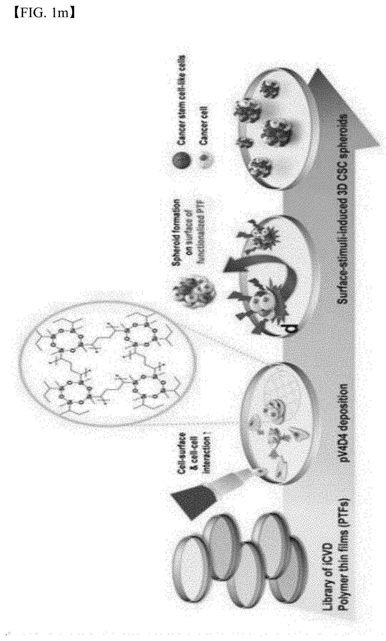
D00008

D00009

D00010

D00011

D00012

D00013

D00014

D00015

D00016

D00017

D00018

D00019

D00020

D00021
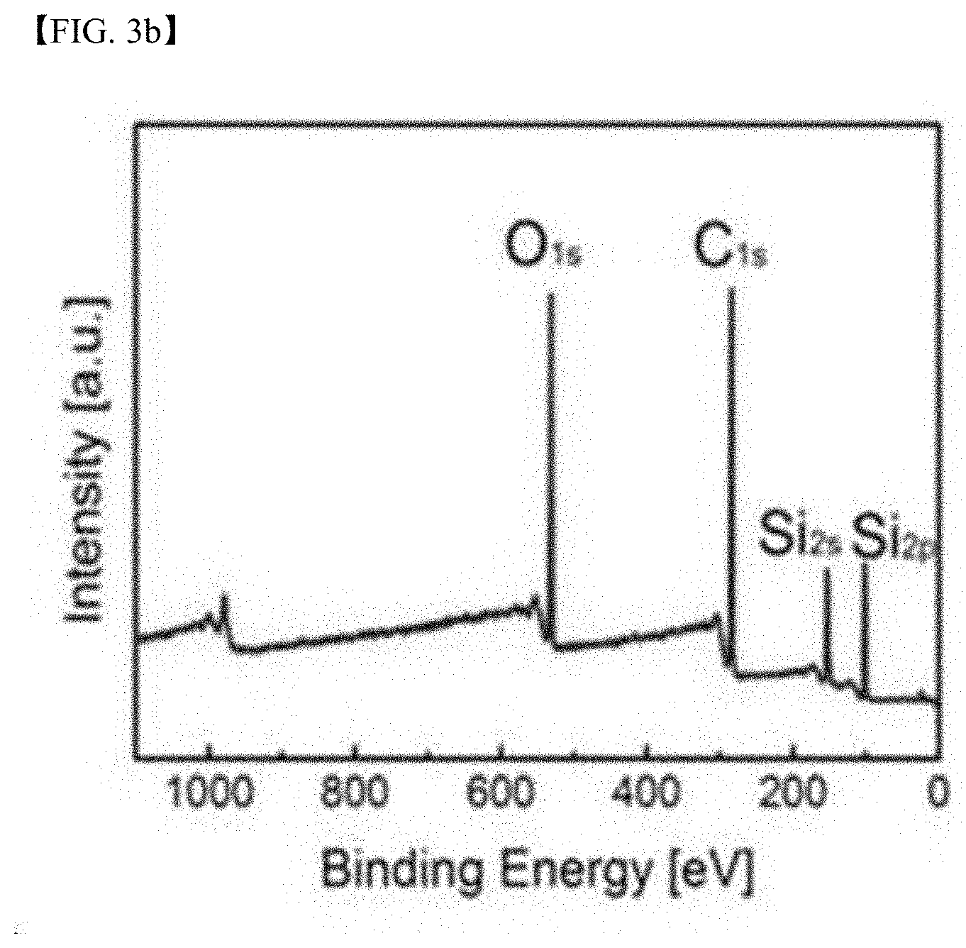
D00022

D00023

D00024

D00025
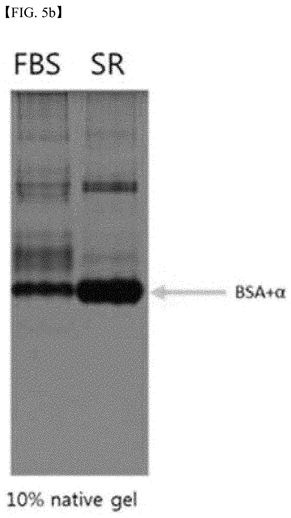
D00026

D00027
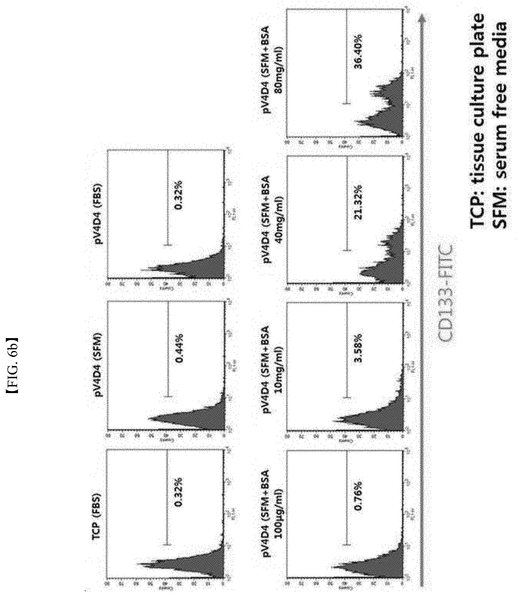
D00028

D00029
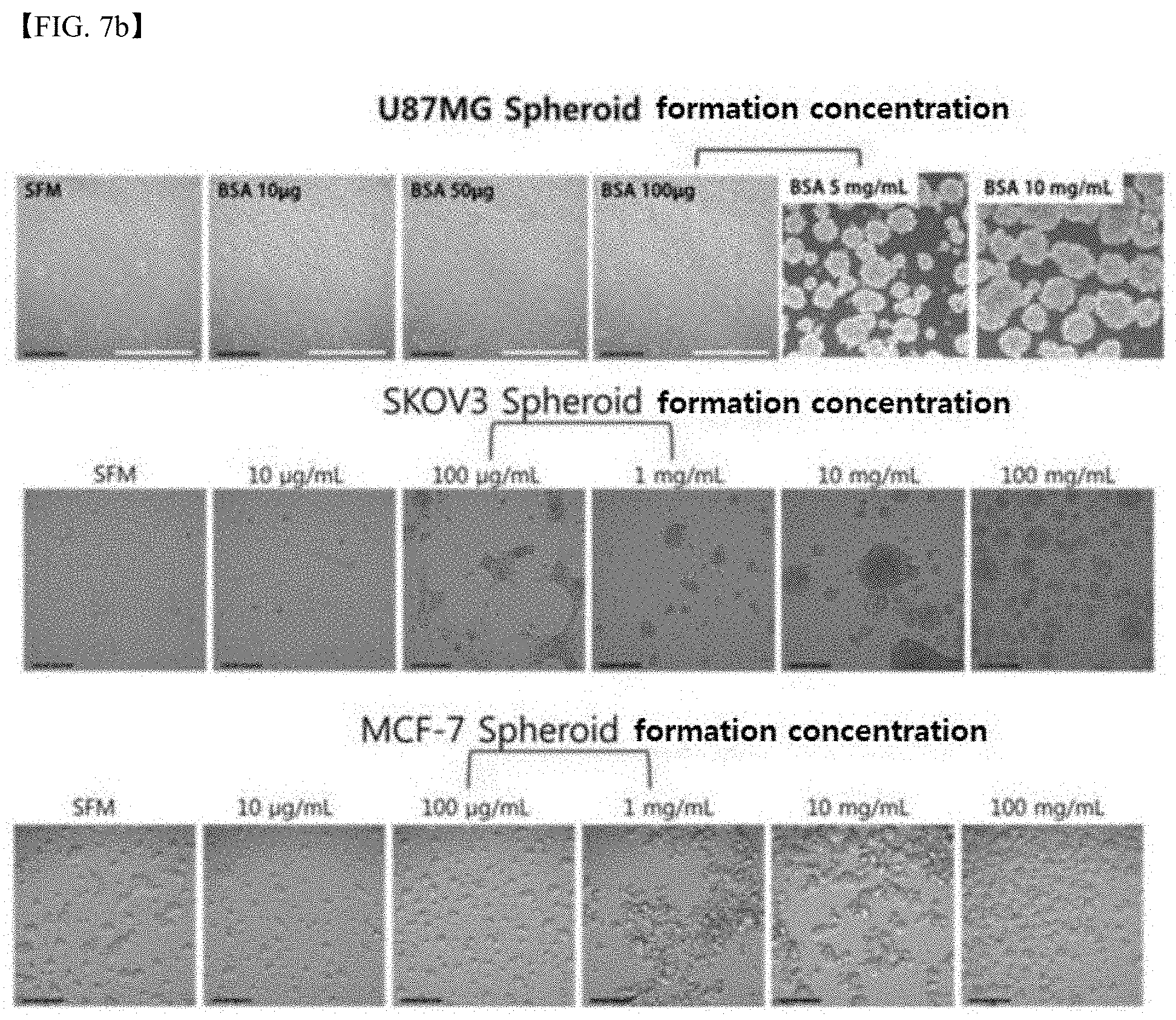
D00030

D00031

D00032

D00033

D00034
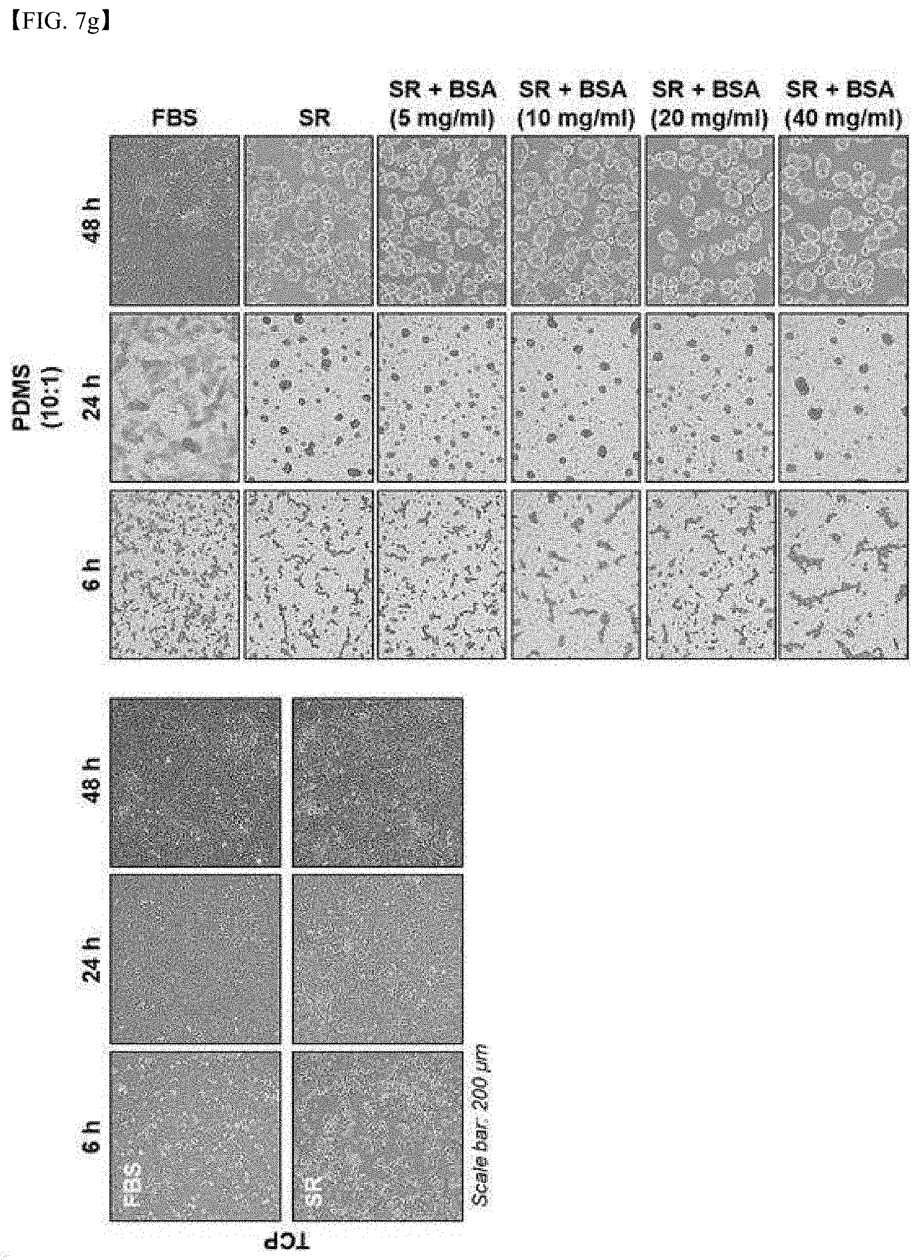
D00035

D00036

D00037

D00038

D00039
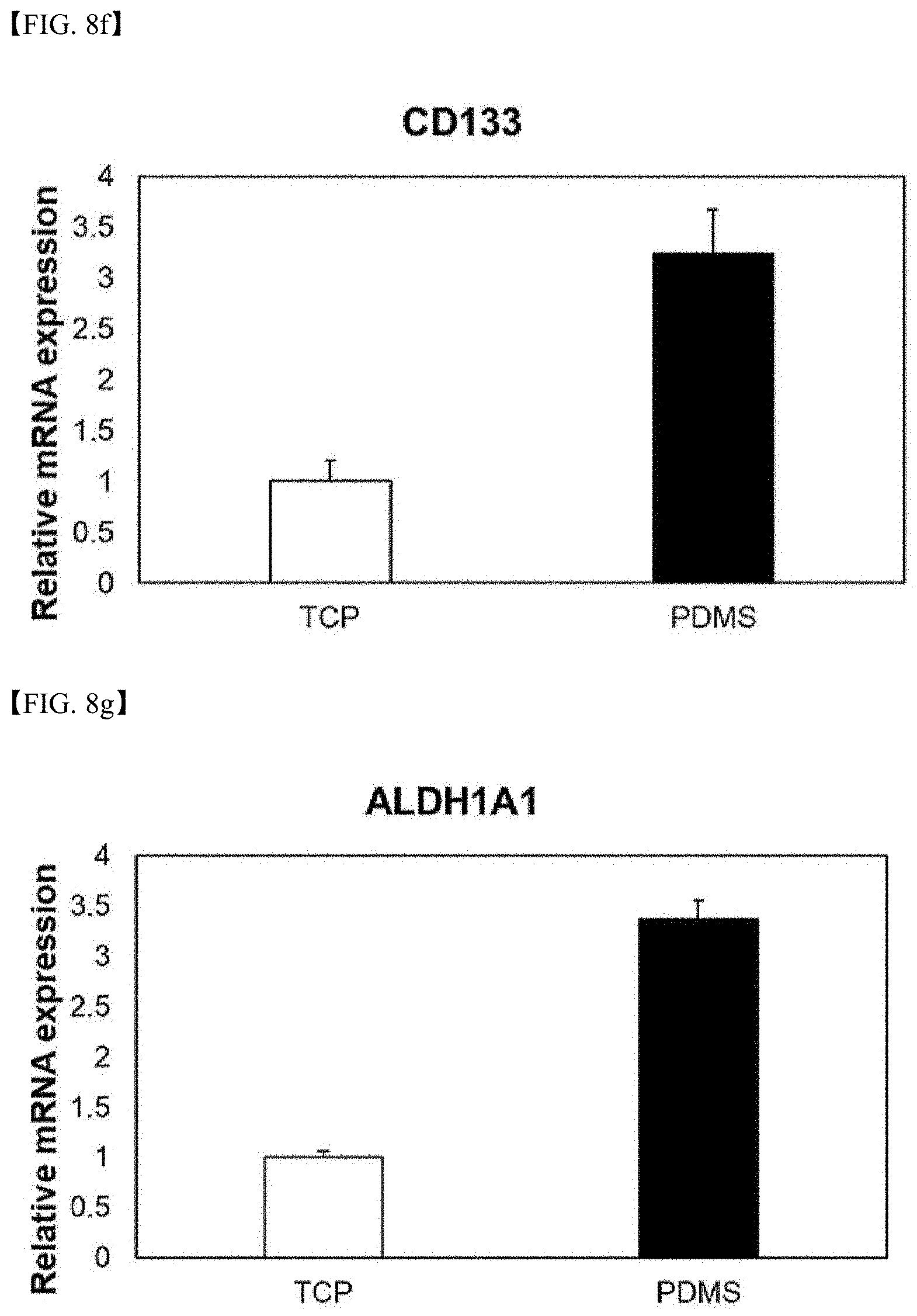
D00040
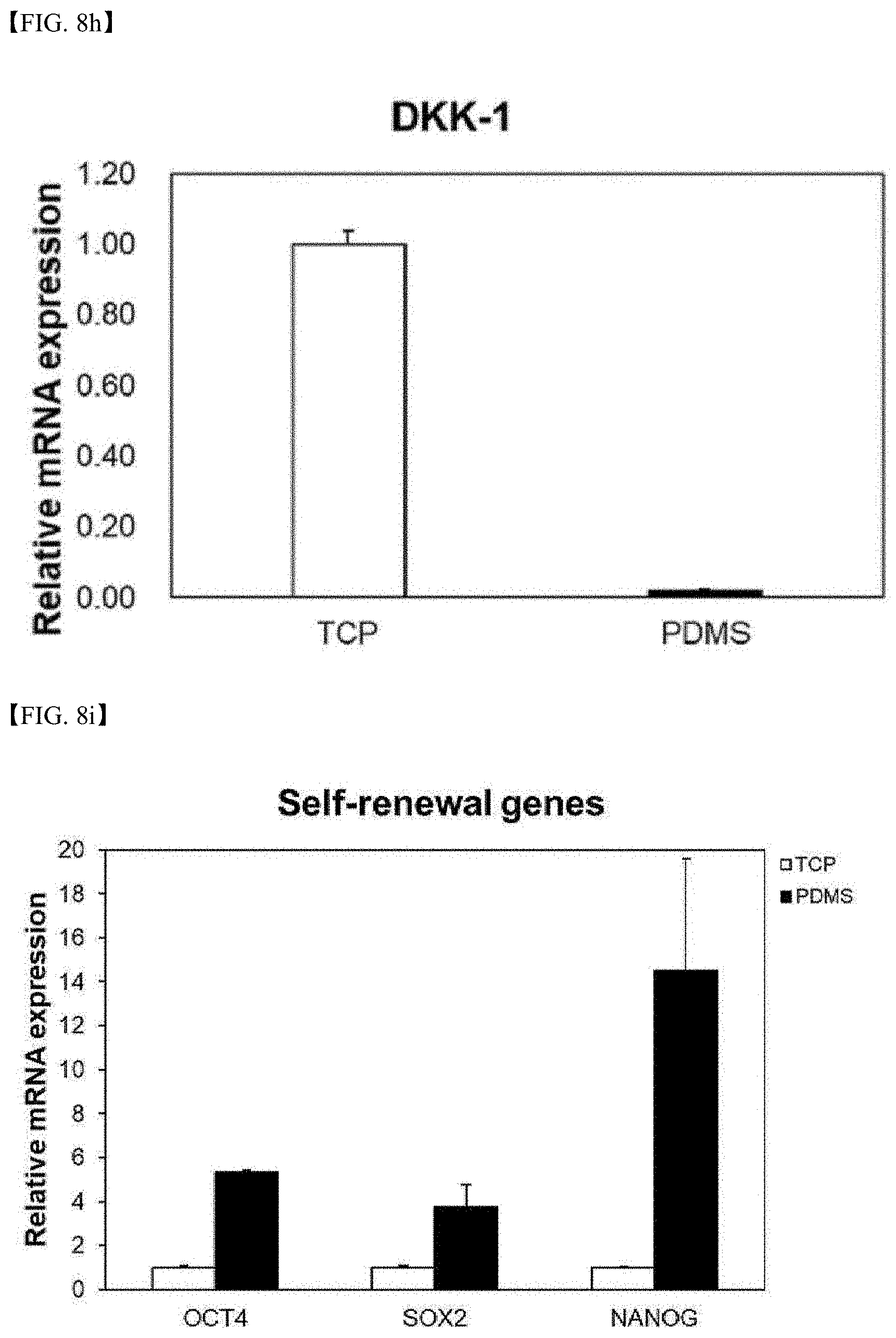
D00041

D00042

D00043

D00044

D00045
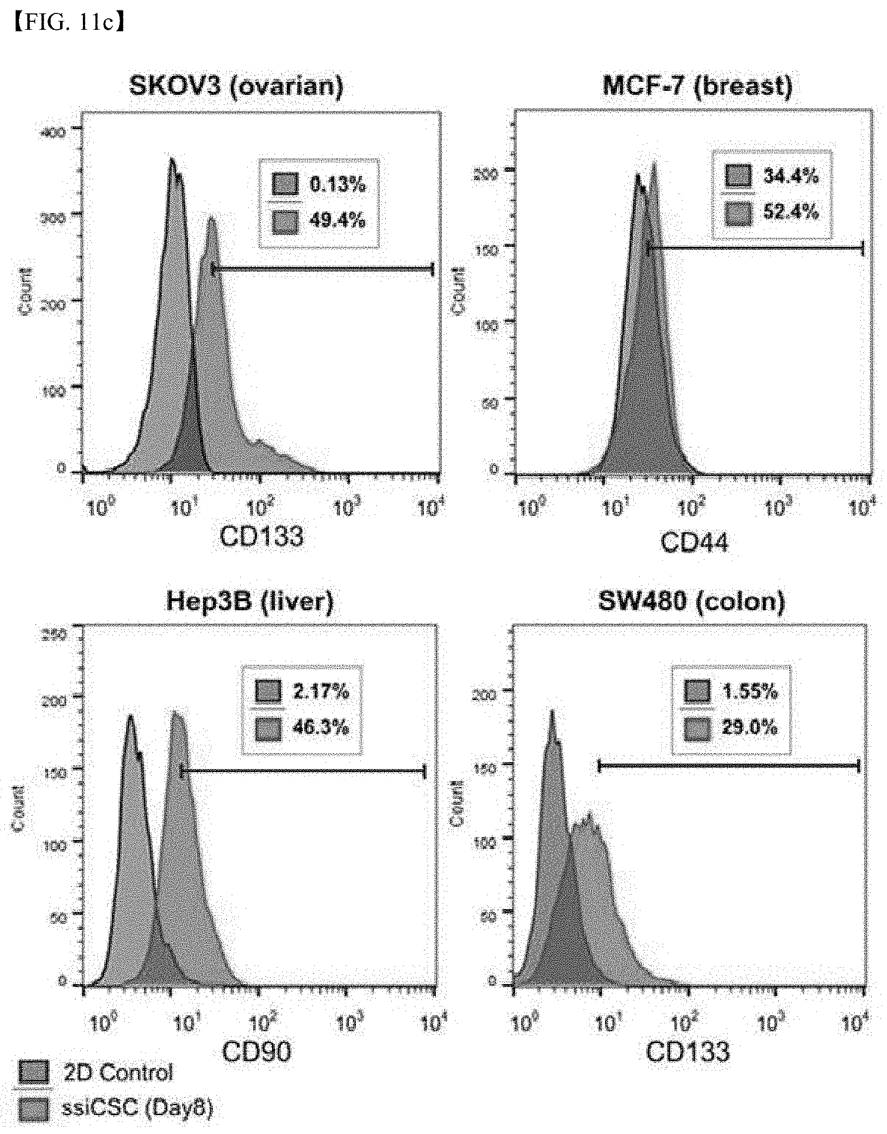
D00046
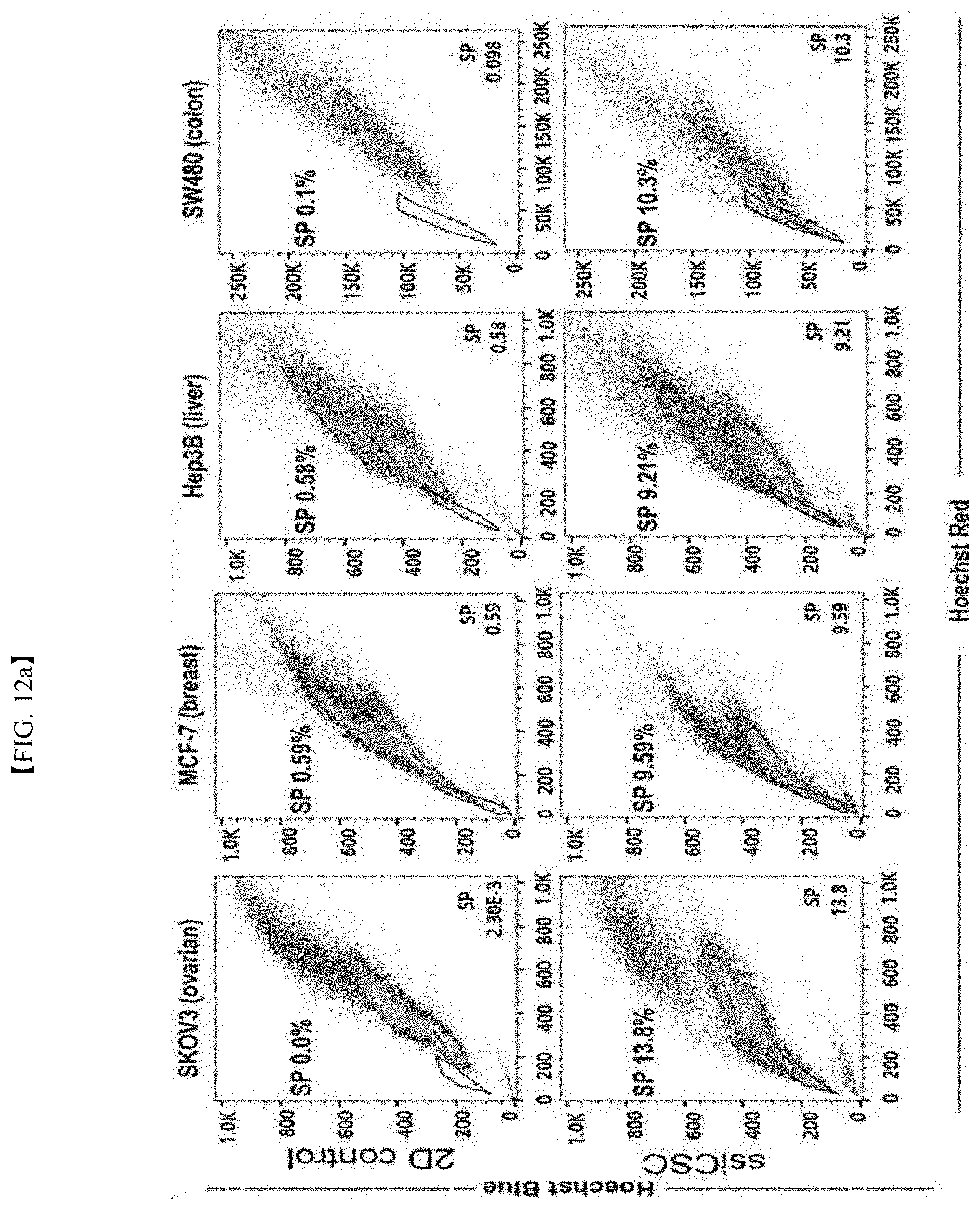
D00047
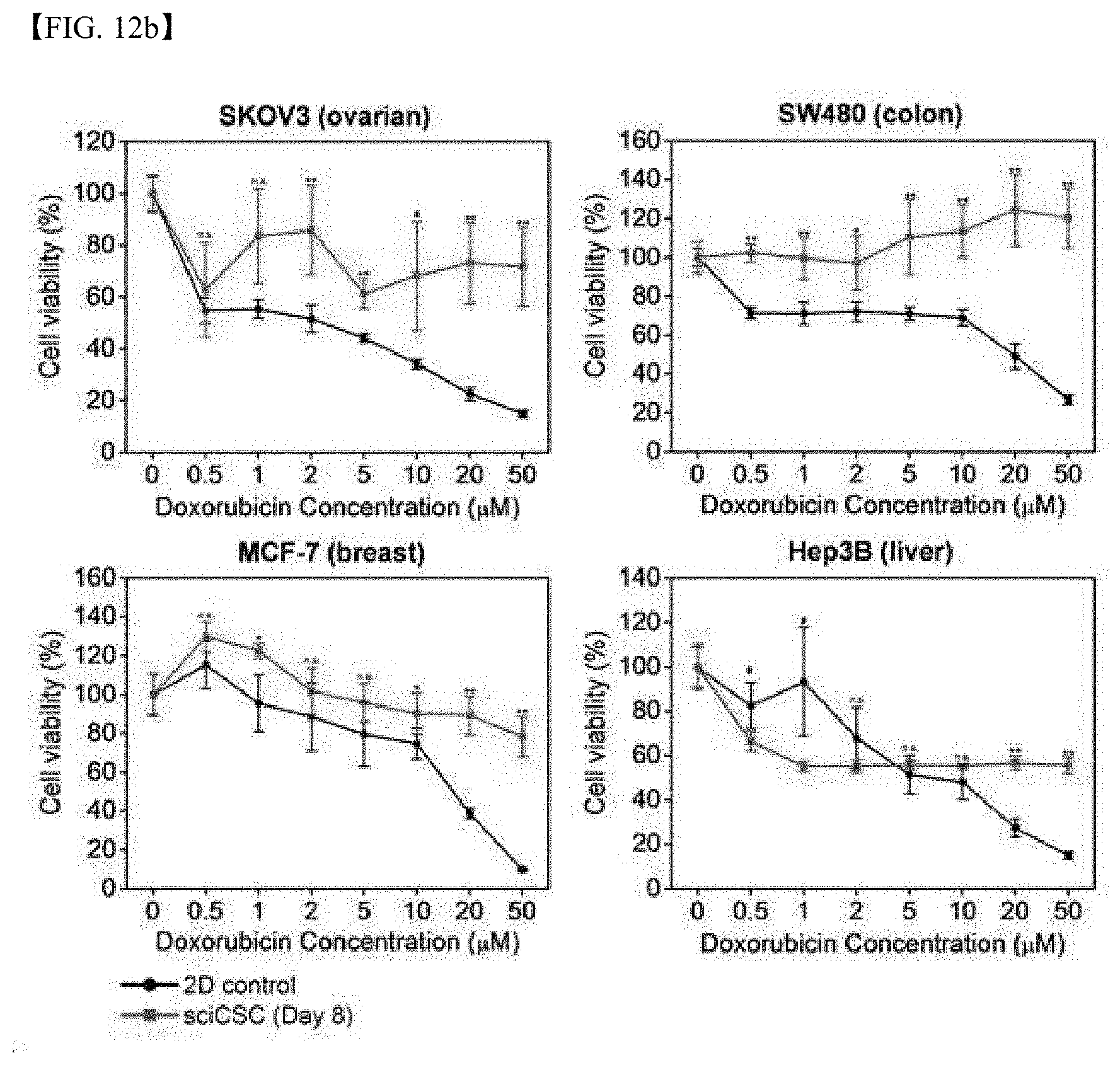
D00048

D00049

D00050

D00051
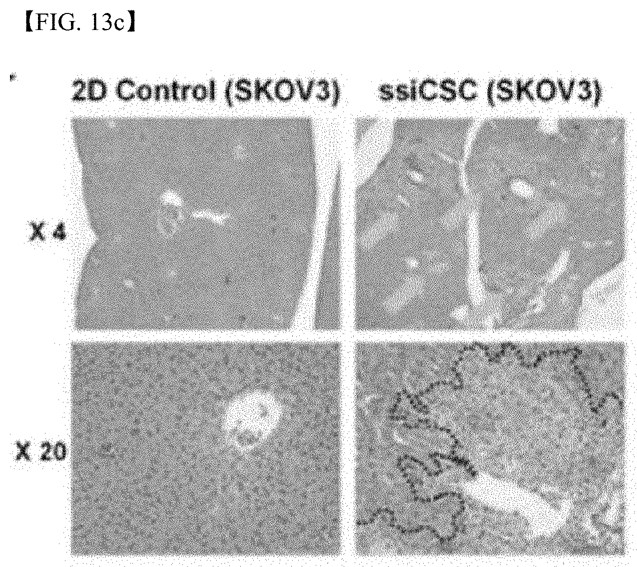
D00052

D00053
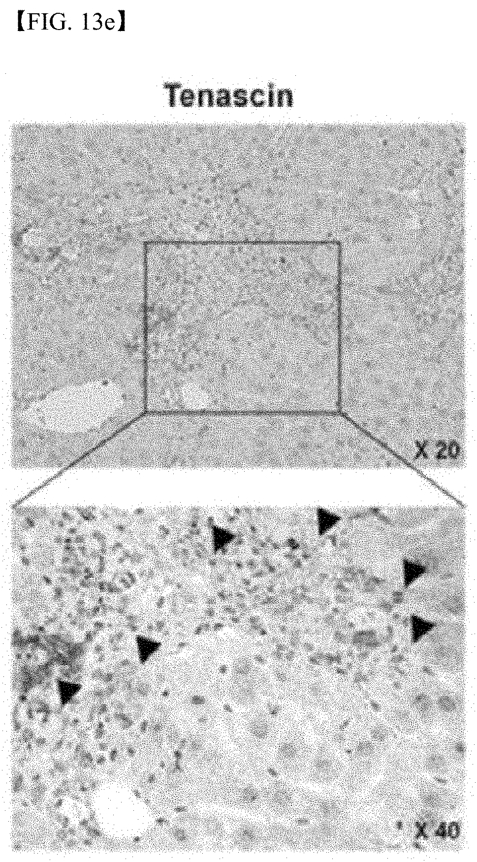
D00054
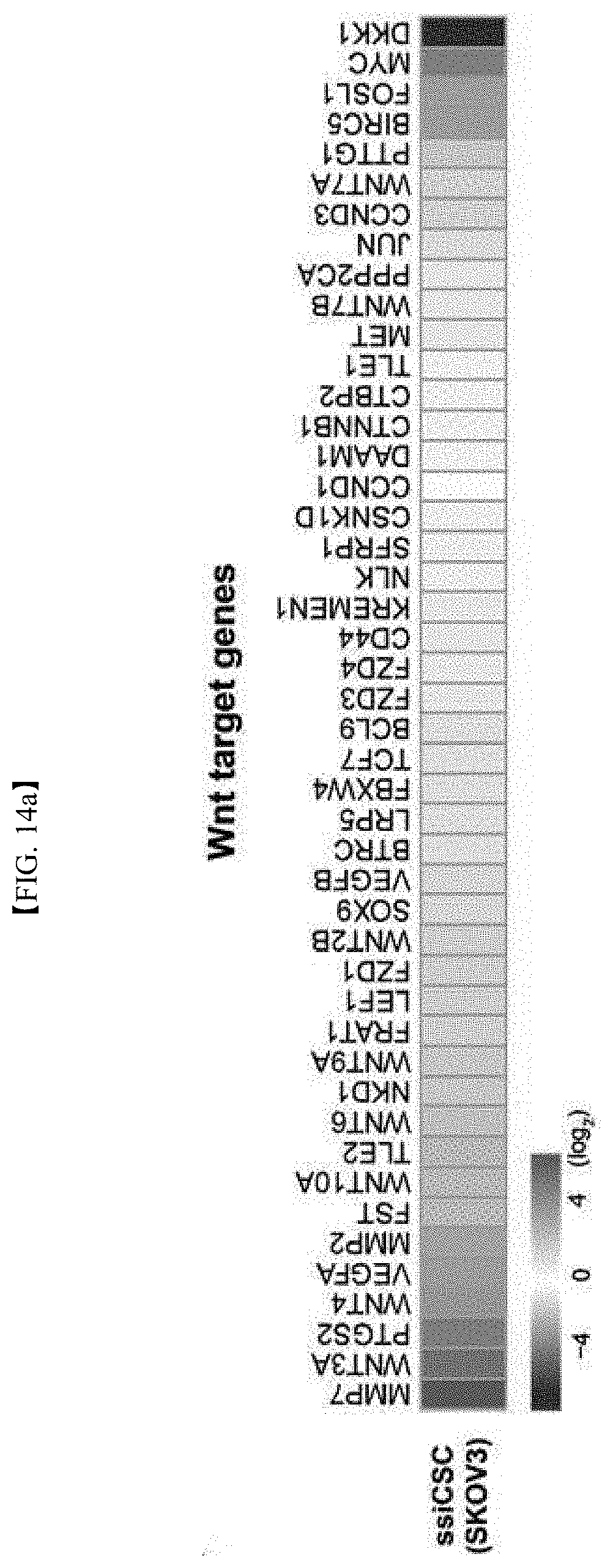
D00055

D00056

D00057

D00058
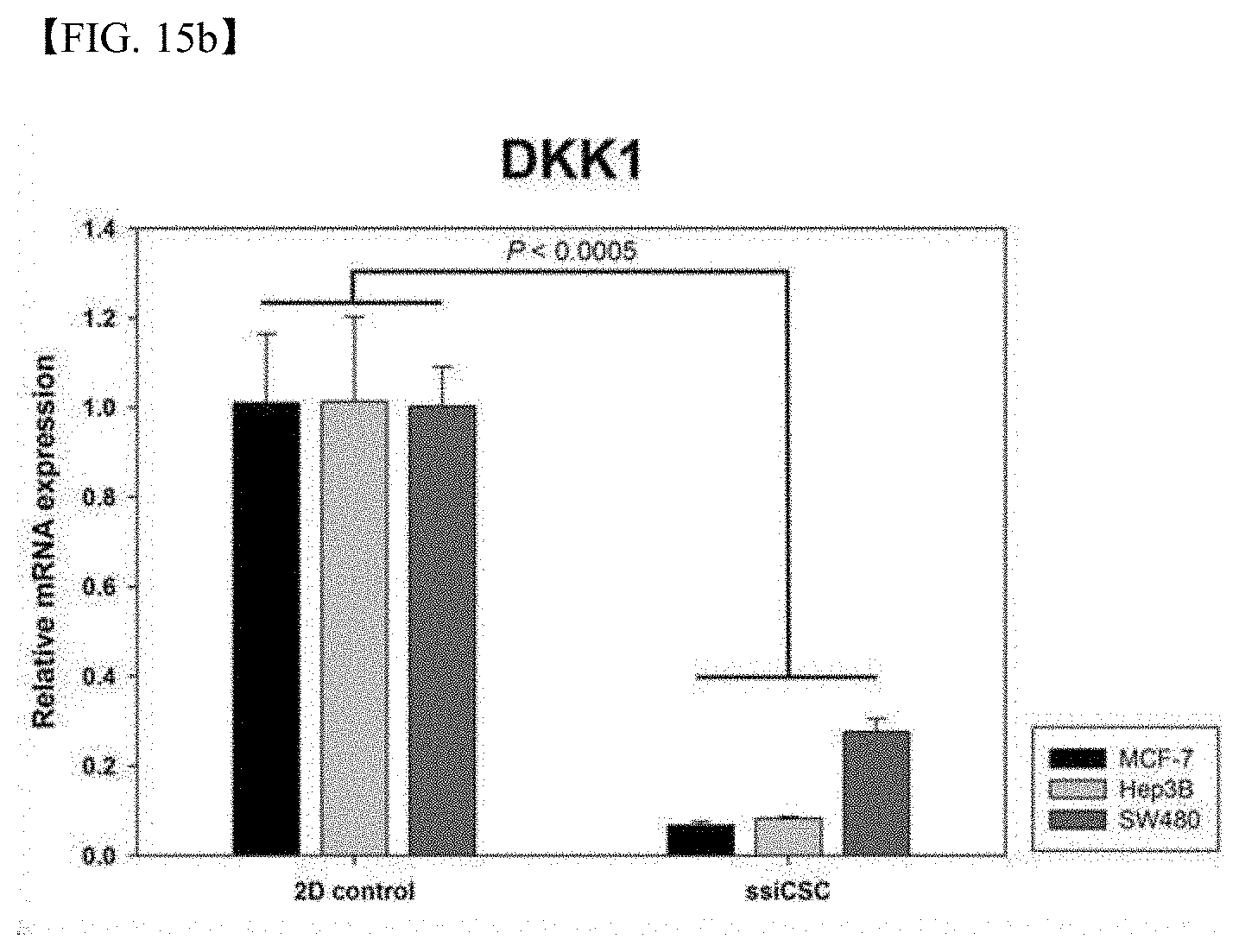
D00059
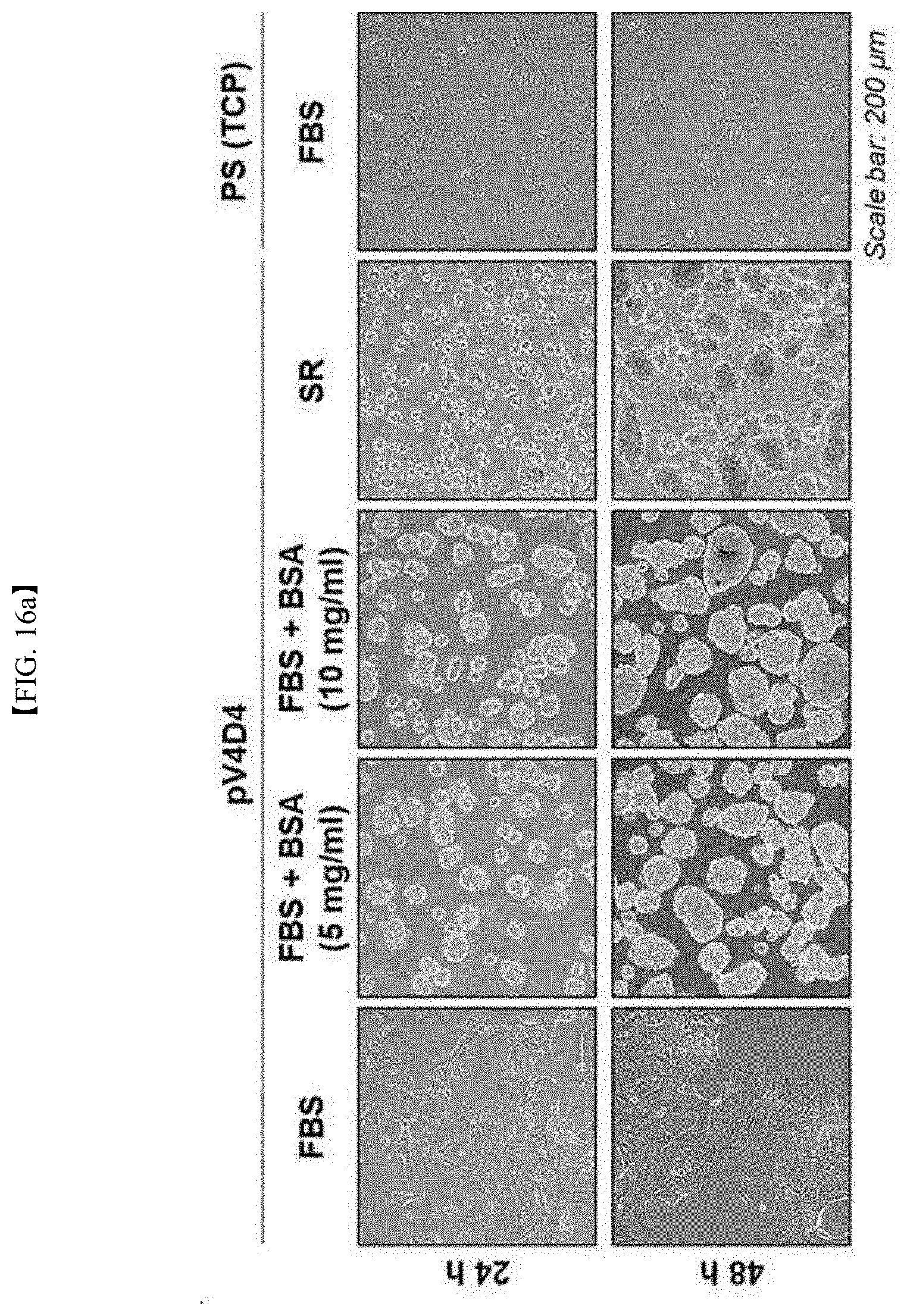
D00060

D00061
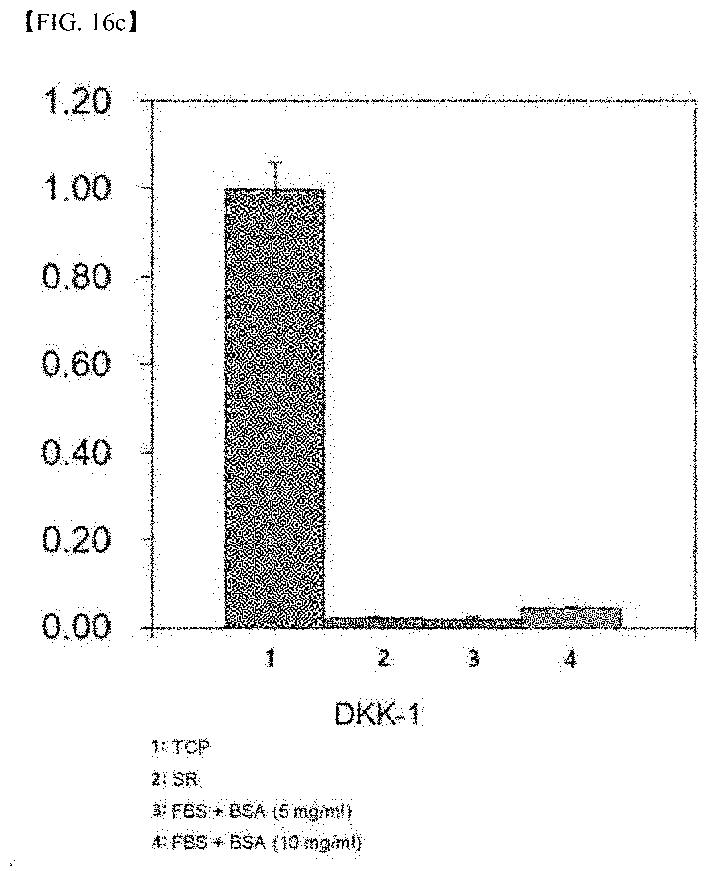
S00001
XML
uspto.report is an independent third-party trademark research tool that is not affiliated, endorsed, or sponsored by the United States Patent and Trademark Office (USPTO) or any other governmental organization. The information provided by uspto.report is based on publicly available data at the time of writing and is intended for informational purposes only.
While we strive to provide accurate and up-to-date information, we do not guarantee the accuracy, completeness, reliability, or suitability of the information displayed on this site. The use of this site is at your own risk. Any reliance you place on such information is therefore strictly at your own risk.
All official trademark data, including owner information, should be verified by visiting the official USPTO website at www.uspto.gov. This site is not intended to replace professional legal advice and should not be used as a substitute for consulting with a legal professional who is knowledgeable about trademark law.