Compositions And Methods For Cellular Reprogramming
HODGKINSON; Conrad ; et al.
U.S. patent application number 16/982964 was filed with the patent office on 2021-01-21 for compositions and methods for cellular reprogramming. This patent application is currently assigned to Duke University. The applicant listed for this patent is Duke University. Invention is credited to Victor DZAU, Conrad HODGKINSON, Jaewoo Lee, Bruce A. SULLENGER.
| Application Number | 20210017497 16/982964 |
| Document ID | / |
| Family ID | 1000005167102 |
| Filed Date | 2021-01-21 |


View All Diagrams
| United States Patent Application | 20210017497 |
| Kind Code | A1 |
| HODGKINSON; Conrad ; et al. | January 21, 2021 |
COMPOSITIONS AND METHODS FOR CELLULAR REPROGRAMMING
Abstract
Disclosed herein are compositions and methods for cellular reprogramming. The compositions comprise one or more miRs and an activator of NF.kappa.B. Also provided are methods for enhancing or upregulating cardiomyocyte maturation in a cell or a subject and methods for inhibiting or downregulating cardiomyocyte maturation.
| Inventors: | HODGKINSON; Conrad; (Durham, NC) ; DZAU; Victor; (Durham, NC) ; Lee; Jaewoo; (Durham, NC) ; SULLENGER; Bruce A.; (Durham, NC) | ||||||||||
| Applicant: |
|
||||||||||
|---|---|---|---|---|---|---|---|---|---|---|---|
| Assignee: | Duke University Durham NC |
||||||||||
| Family ID: | 1000005167102 | ||||||||||
| Appl. No.: | 16/982964 | ||||||||||
| Filed: | March 21, 2019 | ||||||||||
| PCT Filed: | March 21, 2019 | ||||||||||
| PCT NO: | PCT/US19/23461 | ||||||||||
| 371 Date: | September 21, 2020 |
Related U.S. Patent Documents
| Application Number | Filing Date | Patent Number | ||
|---|---|---|---|---|
| 62645847 | Mar 21, 2018 | |||
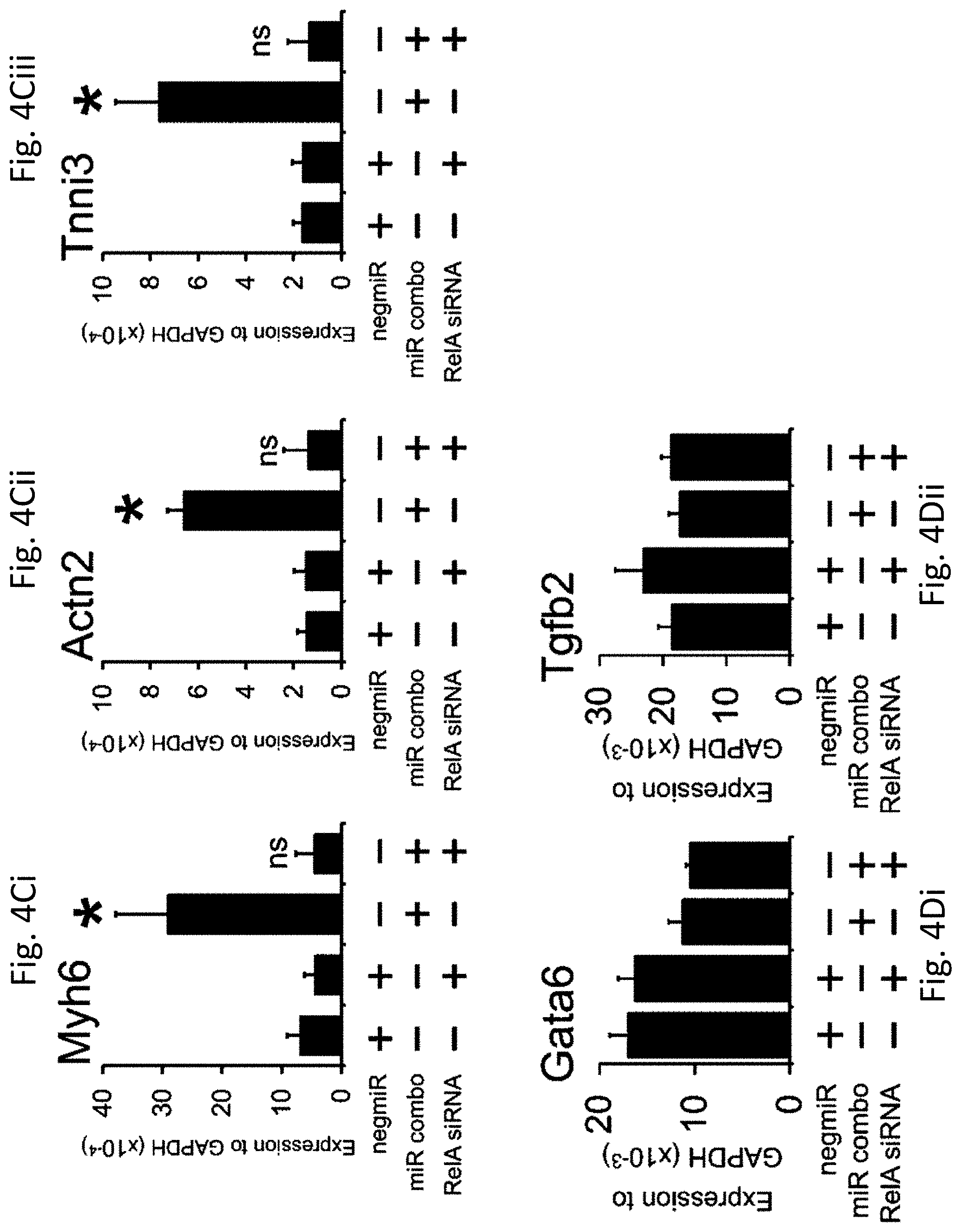
| 62782480 | Dec 20, 2018 | |||
| Current U.S. Class: | 1/1 |
| Current CPC Class: | C12N 5/0657 20130101; C12N 2501/999 20130101; A61K 31/713 20130101; C12N 2501/65 20130101 |
| International Class: | C12N 5/077 20060101 C12N005/077; A61K 31/713 20060101 A61K031/713 |
Goverment Interests
STATEMENT OF GOVERNMENT SUPPORT
[0002] This invention was made with government support under 5R01H731814-02 awarded by the National Institutes of Health, DP2OD008586 awarded by the National Institutes of Health, and CBET-1151035 awarded by the National Science Foundation. The government has certain rights in the invention.
Claims
1. A reprogramming composition comprising: (a) one or more miRs comprising a nucleotide sequence having at least 80% sequence identity tomiR-1 (SEQ ID NO: 11), miR-133a (SEQ ID NO: 14), miR-208 (SEQ ID NO: 18), and mir-499-5p (SEQ ID NO: 23), and combinations thereof; and (b) an activator of NF.kappa.B.
2. The composition of claim 1, wherein the activator of NF.kappa.B comprises a TLR3 agonist.
3. The composition of claim 1, wherein the activator of NF.kappa.B comprises a modified 5'-triphospate, 2'-fluoro modified non-linear RNA, the RNA comprising a stem-loop formed from the complete or partial hybridization of at least 8 nucleotide pairings.
4. The composition of claim 3, wherein the non-linear RNA comprises an oligonucleotide having at least 80% sequence identity to ICR2 (SEQ ID NO: 30), ICR4 (SEQ ID NO: 37), ICR4A (SEQ ID NO: 38), ICR5 X (SEQ ID NO: 39), or ICR5Y (SEQ ID NO: 40).
5. The composition of claim 4, wherein the non-linear RNA comprises an oligonucleotide having at least 80% sequence identity to ICR2 (SEQ ID NO: 30).
6. The composition of claim 4, wherein the non-linear RNA comprises an oligonucleotide having at least 80% sequence identity to ICR4 (SEQ ID NO: 37).
7. The composition of claim 1 further comprising a cytoplasmic delivery agent, reprogramming media, a reprogramming efficiency-enhancing molecule, or any combination thereof.
8. The composition of claim 1, wherein the one or more miRs consists essentially of: miR-1 (SEQ ID NO: 11), miR-133a (SEQ ID NO: 14), miR208 (SEQ ID NO: 18), and mir-499-5p (SEQ ID NO: 23).
9. The composition of claim 1, wherein the one or more miRs comprise a portion of a pre-miRNA.
10. A pharmaceutical composition comprising an effective amount of the composition of claim 1 and one or more pharmaceutically acceptable carriers, excipients, or diluents.
11. A method for enhancing or upregulating cardiomyocyte maturation in a cell comprising contacting the cell with an effective amount of the reprogramming composition of claim 1 for a sufficient time such that the cell is reprogrammed into a cardiomyocyte.
12. The method of claim 11, wherein the cell is a fibroblast.
13. The method of claim 11, wherein the cell comprises cardiac fibrotic tissue.
14. (canceled)
15. (canceled)
16. (canceled)
17. (canceled)
18. (canceled)
19. (canceled)
20. (canceled)
21. (canceled)
22. A method of enhancing or upregulating cardiomyocyte maturation in a subject comprising administering (i) an effective amount of the of the composition of claim 1 or (ii) a pharmaceutical composition comprising the effective amount of the composition of claim 1 and one or more pharmaceutically acceptable carriers, excipients, or diluents.
23. The method of claim 22, wherein the subject has a cardiovascular disease or has suffered a myocardial infarction.
24. (canceled)
25. (canceled)
26. (canceled)
27. (canceled)
28. (canceled)
29. (canceled)
30. (canceled)
31. (canceled)
32. A method for inhibiting or downregulating cardiomyocyte maturation in a cell comprising contacting the cell with an effective amount of a composition comprising a TLR3 inhibitor, a NF.kappa.B inhibitor, a ikbkb inhibitor, or a combination thereof for a sufficient time such that cardiomyocyte maturation is inhibited or down-regulated in the cell.
33. The method of claim 32, wherein the cell is a cardiomyocyte-committed precursor.
34. The method of claim 33, wherein the cell is a fibroblast.
35. The method of claim 32, wherein the composition comprises the TLR3 inhibitor and the TLR3 inhibitor is a TLR3 siRNA or CU-CPT-4a, wherein the composition comprises the NF.kappa.B inhibitor and the NF.kappa.B inhibitor is a NF.kappa.B siRNA, a RelA siRNA, or Bay 11-7085, or wherein the composition comprises the ikbkb inhibitor and the ikbkb inhibitor is a ikbkb siRNA.
36. (canceled)
37. (canceled)
38. (canceled)
39. (canceled)
40. (canceled)
41. The method of claim 32, wherein the composition further comprises a cytoplasmic delivery agent, cellular media, or any combination thereof.
Description
CROSS-REFERENCE TO RELATED APPLICATION
[0001] This patent application claims the benefit of priority of United States Provisional Patent Applications Nos. 62/645,847, filed Mar. 21, 2018, and 62/782,480, filed Dec. 20, 2018, the contents of each are incorporated herein by reference in their entirety.
BACKGROUND
[0003] Heart disease is the number one killer of men and women worldwide. Generally, heart tissue has a limited capacity for regeneration or self-renewal. After a patient recovers from a myocardial infarction, the organ bears a scar and heart function is diminished. The ability to regenerate damaged organs such as the heart remains elusive. As such, there is a pressing need in the art to develop new strategies for the regeneration of damaged organs.
BRIEF SUMMARY OF THE INVENTION
[0004] Disclosed herein are compositions and methods for cellular reprogramming. One aspect of the invention is a reprogramming composition comprising one or more miRs comprising a nucleotide sequence having at least 80% sequence identity to miR-1, miR-126, miR-133, miR-133a, mir-206, miR-208, miR-499, mir-499-5p, and combinations thereof; and an activator of NF.kappa.B.
[0005] Another aspect of the invention is a pharmaceutical composition including an effective amount of the composition of claim 1 and one or more pharmaceutically acceptable carriers, excipients, or diluents.
[0006] Another aspect of the invention is a method for enhancing or upregulating cardiomyocyte maturation in a cell comprising contacting the cell with an effective amount of any of the compositions described herein for a sufficient time such that the cell is reprogrammed into a cardiomyocyte.
[0007] Another aspect of the invention is a method of enhancing or upregulating cardiomyocyte maturation in a subject comprising administering (i) an effective amount of any of the compositions described or (ii) any of the pharmaceutical compositions comprising the effective amount of any of the compositions described and one or more pharmaceutically acceptable carriers, excipients, or diluents.
[0008] Another aspect of the invention is a method for inhibiting or downregulating cardiomyocyte maturation in a cell comprising contacting the cell with an effective amount of a composition comprising a TLR3 inhibitor, a NF.kappa.B inhibitor, a ikbkb inhibitor, or a combination thereof for a sufficient time such that cardiomyocyte maturation is inhibited or down-regulated in the cell.
BRIEF DESCRIPTION OF THE DRAWINGS
[0009] Non-limiting embodiments of the present invention will be described by way of example with reference to the accompanying figures, which are schematic and are not intended to be drawn to scale. In the figures, each identical or nearly identical component illustrated is typically represented by a single numeral. For purposes of clarity, not every component is labeled in every figure, nor is every component of each embodiment of the invention shown where illustration is not necessary to allow those of ordinary skill in the art to understand the invention.
[0010] FIGS. 1A-1Eiii show TLR3 inhibition inhibits maturation of reprogrammed fibroblasts into cardiomyocytes. Neonatal cardiac fibroblasts were transfected with negative control miR (negmiR) or miR combo. The day after transfection media was replaced and the cells incubated with either vehicle or the TLR3 pharmacological inhibitor CU-CPT-4a (10 .mu.M) for a further 4 days. After incubation with the TLR3 pharmacological inhibitor, cells were cultured in normal growth media for a further 6 days. Quantitative PCR was used to analyze mRNA levels of 13 components of the cardiomyocyte sarcomere.
[0011] FIG. 1A shows a heat-map overview of the qPCR analysis. Expression values were normalized to the average expression of the negmiR vehicle samples and then averaged (N=3 technical replicates (fibroblasts were derived from single litter and seeded into 3 individual wells). Averages for each gene were then converted to Z-scores. Centroid linkage and Euclidean methods were employed for clustering and distance measurements respectively.
[0012] FIGS. 1Bi-1Ciii show neonatal cardiac fibroblasts were transfected with negative control miR (negmiR) or miR combo. The day after transfection media was replaced and the cells incubated with either vehicle or the TLR3 pharmacological inhibitor CU-CPT-4a (10 .mu.M) for a further 4 days. After incubation with the TLR3 pharmacological inhibitor, cells were cultured in normal growth media for a further 10 days.
[0013] FIGS. 1Bi-1Biii show RNA levels of the cardiomyocyte sarcomere components Myh6 (amyosin heavy chain) (FIG. 1Bi), Actn2 (.alpha.sarcomeric actinin) (FIG. 1Bii), and Tnni3 (cardiac troponin-I) (FIG. 1Biii) was determined by qPCR. N=4 independent experiments.
[0014] FIGS. 1Ci and 1Cii show cells provided miR combo (FIG. 1Ci) or miR combo and TLR3 antagonist (FIG. 1Cii) fixed and stained with anti-Actn2 antibodies (red). Nuclei were stained with DAPI (blue). Scale bar 50 microns.
[0015] FIG. 1Ciii shows quantification of immunostaining. Cells expressing Actn2 were counted and expressed as a percentage of the total cell population. N=6 independent experiments.
[0016] FIGS. 1D-1Eiii show neonatal cardiac fibroblasts were first transfected with either a control siRNA or a siRNA that targeted TLR3. Two days later, the cells were transfected again with either the negative control miR negmiR or miR combo. The day after transfection with miRNAs the media was replaced and the cells cultured in normal growth media for 14 days.
[0017] FIG. 1D shows quantification of TLR3 knockdown by qPCR. N=3 independent experiments.
[0018] FIGS. 1Ei-1Eiii show RNA levels of the cardiomyocyte structural proteins Myh6 (amyosin heavy chain) (FIG. 1Ei), Actn2 (.alpha.sarcomeric actinin) (FIG. 1Eii) and Tnni3 (cardiac troponin-I) (FIG. 1Eiii) was determined by qPCR. N=4 independent experiments. Data represented as Mean.+-.SEM. ***P<0.001, **P<0.01, *P<0.05, ns: not significant. For A, B and D comparisons are made between miR combo and negmiR for each group. For C, comparison is made between control siRNA and siRNA targeting TLR3.
[0019] FIGS. 2Ai-2Biv show neither TLR3 inhibition nor TLR3 activation affects early stage cardiac reprogramming.
[0020] FIGS. 2Ai-2Aiv show neonatal cardiac fibroblasts were first transfected with either a control siRNA or a siRNA that targeted TLR3. Two days later, the cells were transfected again with either the negative control miR negmiR or miR combo. The day after transfection with miRNAs, the media was replaced and the cells cultured in normal growth media for 3 days. RNA levels of the cardiomyocyte-lineage commitment factors Gata4 (FIG. 2Ai), Hand2 (FIG. 2Aii), Tbx5 (FIG. 2Aiii), and Mef2C (FIG. 2Aiv) was determined by qPCR. N=9 independent experiments. Comparisons are made between miR combo+control siRNA and miR combo+TLR3 siRNA, ns: not significant. Data represented as Mean.+-.SEM.
[0021] FIGS. 2Bi-2Biv show neonatal cardiac fibroblasts were transfected with negative control miR (negmiR) or miR combo. The day after transfection media was replaced and the cells incubated with vehicle or the TLR3 agonist Poly(I:C) LMW (low molecular weight Poly(I:C)) for a further 3 days. RNA levels of the cardiomyocyte-lineage commitment factors Gata4 (FIG. 2Bi), Hand2 (FIG. 2Bii), Tbx5 (FIG. 2Biii), and Mef2C (FIG. 2Biv) was determined by qPCR. N=6 independent experiments. Comparisons are made between miR combo+vehicle and miR combo+TLR3 agonist, ns: not significant. Data represented as Mean.+-.SEM.
[0022] FIGS. 3Ai-3Diii show NF.kappa.B is important for miR combo reprogramming.
[0023] FIGS. 3Ai-3Biii show neonatal cardiac fibroblasts were transfected with negative control miR (negmiR) or miR combo. The day after transfection media was replaced and the cells incubated with vehicle or the NF.kappa.B antagonist Bay 11-7085. After one day of treatment, the media was replaced with normal growth media and cells cultured for a further 12 days.
[0024] FIGS. 3Ai-3Aiii RNA levels of the cardiomyocyte structural proteins Myh6 (amyosin heavy chain) (FIG. 3Ai), Actn2 (.alpha.sarcomeric actinin) (FIG. 3Aii), and Tnni3 (cardiac troponin-I) (FIG. 3Aiii) following treatment with the NF.kappa.B antagonist Bay 11-7085 was determined by qPCR. N=3 independent experiments.
[0025] FIGS. 3Bi-3Bii shows cells provided miR combo (FIG. 3Bi) or miR combo and NF.kappa.B inhibitor (FIG. 3Bii) were fixed and stained with anti-Actn2 antibodies (red). Nuclei were stained with DAPI (blue). Scale bar 100 microns. Inset pictures are at 5.times. magnification.
[0026] FIGS. 3Biii shows quantification of immunostaining. Cells expressing Actn2 were counted and expressed as a percentage of the total cell population. N=3 independent experiments.
[0027] FIGS. 3C-3Diii show neonatal cardiac fibroblasts were transfected with microRNAs (negmiR or miR combo) and siRNA (control siRNA or a siRNA that targeted Ikbkb). The day after transfection with miRNAs the media was replaced and the cells cultured in normal growth media for either 4 days (to assess knockdown efficiency) or 14 days (to assess RNA levels of cardiomyocyte structural proteins).
[0028] FIG. 3C show quantification of Ikbkb knockdown by qPCR. N=3 independent experiments.
[0029] FIGS. 3Di-3Diii show RNA levels of the cardiomyocyte structural proteins Myh6 (amyosin heavy chain) (FIG. 3Di), Actn2 (.alpha.sarcomeric actinin) (FIG. 3Dii), and Tnni3 (cardiac troponin-I) (FIG. 3Diii) was determined by qPCR. N=3 independent experiments.
[0030] Data represented as Mean.+-.SEM. Comparisons are made between miR combo and negmiR for each group, **P<0.01, *P<0.05, ns: not significant.
[0031] FIGS. 4A-4Dii show RelA mediates the effects of NF.kappa.B. Neonatal cardiac fibroblasts were first transfected with either a control siRNA or a siRNA that targeted the NF.kappa.B subunit RelA. Two days later, the cells were transfected again with either the negative control miR negmiR or miR combo. The day after transfection with miRNAs, the media was replaced and the cells cultured in normal growth media for 13 days.
[0032] FIG. 4A shows quantification of RelA knockdown by qPCR.
[0033] FIGS. 4Bi-4Bii show cells provided miR combo and control siRNA (FIG. 4Bi) and miR combo+RelA siRNA (FIG. 4Bii) fixed and stained with anti-Actn2 antibodies (red). Nuclei were stained with DAPI (blue). Scale bar 100 microns. Inset pictures are at 5.times. magnification.
[0034] FIG. 4Biii shows quantification of immunostaining. Cells expressing Actn2 were counted and expressed as a percentage of the total cell population. N=3 independent experiments.
[0035] FIGS. 4Ci-4Ciii show RNA levels of the cardiomyocyte structural proteins Myh6 (amyosin heavy chain) (FIG. 4Ci), Actn2 (.alpha.sarcomeric actinin) (FIG. 4Cii), and Tnni3 (cardiac troponin-I) (FIG. 4Ciii) was determined by qPCR. N=3 independent experiments.
[0036] FIGS. 4Di-4Dii show RNA levels of the endodermal marker Gata6 (FIG. 4Di) and the general marker of differentiation Tgfb2 (FIG. 4Dii) were determined by qPCR. N=3 independent experiments.
[0037] Data represented as Mean.+-.SEM. Comparisons are made between miR combo and negative control miR (negmiR) for each group, **P<0.01, *P<0.05, ns: not significant.
[0038] FIGS. 5Ai-5Bviii show the NF.kappa.B subunit RelA binds to the promoters of cardiomyocyte maturation genes.
[0039] FIGS. 5Ai-5Aviii show neonatal cardiac fibroblasts were transfected with negmiR or miR combo. After 7 days, chromatin DNA was subjected to ChIP analysis. Primers were designed to target the first 1 Kb of the indicated cardiomyocyte sarcomere genes Actn2 (FIG. 5Ai), Myh6 (FIG. 5Aii), Mypn (FIG. 5Aiii), Tnni3 (FIG. 5Aiv), Ttn (FIG. 5Av), Myoz2 (FIG. 5Avi), Tnnc1 (FIG. 5Avii), and Tnnt2 (FIG. 5Aviii), (represented by -01 Kb). Results are presented as the fold enrichment in RelA binding where percent input of the negmiR control was taken to be 1. N=3 independent experiments. Data represented as Mean.+-.SEM. Comparisons are made between miR combo and negative control miR (negmiR), **P<0.01, *P<0.05, ns: not significant.
[0040] FIGS. 5Bi-5Bviii show neonatal cardiac fibroblasts were transfected with miR combo and either a control siRNA or a siRNA that targeted RelA. After 7 days, chromatin DNA was subjected to ChIP analysis. Primers were designed to target the first 1 Kb of the indicated cardiomyocyte sarcomere genes Actn2 (FIG. 5Bi), Myh6 (FIG. 5Bii), Mypn (FIG. 5Biii), Tnni3 (FIG. 5Biv), Ttn (FIG. 5Bv), Myoz2 (FIG. 5Bvi), Tnnc1 (FIG. 5Bvii), and Tnnt2 (FIG. 5Bviii), (represented by -01 Kb). Results are presented as the percentage of chromatin input. N=3 independent experiments. Data represented as Mean.+-.SEM.
[0041] FIGS. 6A-6B show microRNAs activate TLR3.
[0042] FIG. 6A shows neonatal cardiac fibroblasts were transfected with negmiR or miR combo. A mock transfection where lipid reagent alone was added to the cells was also used. The TLR3 inhibitor CU-CPT-4a, which interferes with RNA binding to TLR3, was added one day post-transfection. IL6 concentration in the media was assessed 4 days post-transfection and values expressed as pg IL6 per .mu.g of total protein. N=4 independent experiments. Data represented as Mean.+-.SEM. Comparisons are made to the respective mock transfected group (*P<0.05, ns: not significant) and between negmiR and miR combo (.dagger.P<0.05).
[0043] FIG. 6B shows neonatal cardiac fibroblasts were transfected with microRNAs (mock, miR combo) and siRNA (non-targeting control, TLR3, Ikbkb). IL6 concentration in the media was assessed 4 days post-transfection and values expressed as a ratio between miR combo and mock transfected cells. N=3 independent experiments. Data represented as Mean.+-.SEM. Comparisons are made to miR combo plus non-targeting control siRNA group, **P<0.01, *P<0.05.
[0044] FIGS. 7Ai-7Dii show TLR3 agonists enhance maturation of miR combo reprogrammed cardiomyocytes. Neonatal cardiac fibroblasts were transfected with negative control miR (negmiR) or miR combo. The day after transfection media was replaced and the cells incubated with vehicle or the TLR3 agonist Poly(I:C) LMW (low molecular weight Poly(I:C)) for a further 4 days. After incubation with the TLR3 agonist, cells were cultured in normal growth media for a further 10 days.
[0045] FIGS. 7Ai-7Aiii show RNA levels of the cardiomyocyte structural proteins Myh6 (amyosin heavy chain) (FIG. 7Ai), Actn2 (.alpha.sarcomeric actinin) (FIG. 7Aii), and Tnni3 (cardiac troponin-I) (FIG. 7Aiii) was determined by qPCR. N=5-14.
[0046] FIGS. 7Bi-7Bvi show cells provided miR combo (FIG. 7Bi, FIGS. 7Bii, and 7Biii) and miR combo and TLR3 agonist (FIG. 7Biv, FIGS. 7Bv, and 7Bvi) fixed and stained with anti-Actn2 antibodies (red). Nuclei were stained with DAPI (blue). N=6 independent experiments. Scale bar 50 microns. Inset pictures are at 5.times. magnification to show sarcomere structure.
[0047] FIG. 7C show quantification of immunostaining. Cells expressing Actn2 were counted and expressed as a percentage of the total cell population.
[0048] FIGS. 7Di-7Dii show neonatal cardiac fibroblasts were transfected with negative control miR (negmiR) or miR combo. The day after transfection media was replaced and the cells incubated with differentiation media (DMEM+2% FBS+ITS+AA) and the TLR3 agonist Poly(I:C) LMW for the indicated times (FIG. 7Di). Fourteen days after the transfection, the numbers of beating colonies were counted. N=4 independent experiments (FIG. 7Dii). Data represented as Mean.+-.SEM. *Comparisons made between vehicle and TLR3 agonist for each group ***P<0.001, **P<0.01, *P<0.05. .dagger.Comparisons made between miR combo and negmiR for each group .dagger..dagger..dagger.P<0.001, .dagger..dagger.P<0.01, .dagger.P<0.05.
[0049] FIGS. 8A-8D show ICR2-activated cardiomyocyte maturation evaluated via qPCR by measuring the expression of Actn2 (FIG. 8A), Myh6 (FIG. 8B), Tnni3 (FIG. 8C), and Cacna1c (FIG. 8D).
[0050] FIGS. 9A-9D shows ICR4-activated cardiomyocyte maturation evaluated via qPCR by measuring the expression of Actn2 (FIG. 9A), Myh6 (FIG. 9B), Tnni3 (FIG. 9C), and Cacna1c (FIG. 9D).
[0051] FIG. 10 shows high doses of miR combo impair normal cellular functions by measuring the change in cell number.
[0052] FIGS. 11A-11B show miR combo mediated maturation with ICR2 (FIG. 11A) and PolyIC (FIG. 11B) evaluated by antibody staining for a-sarcomeric actinin.
DETAILED DESCRIPTION OF THE INVENTION
[0053] Cardiomyocyte maturation may be enhanced or upregulated by agonists of the innate immune system, such as pattern recognition receptor agonists, or inhibited or downregulated by antagonists. As demonstrated in the Examples, cardiomyocyte maturation may be effectively controlled in committed cellular precursors to accelerate or retard maturation via pattern recognition receptors and associated signaling pathways.
[0054] "Pattern recognition receptors" or "PRRs" are protein receptors that detect molecules typical of pathogens and/or cellular damage. These proteins are expressed primarily by cells of the innate immune system, such as dendritic cells, macrophages, monocytes, neutrophils and epithelial cells. PRRs are used to identify pathogen-associated molecular patterns (PAMPs), which are associated with microbial pathogens, and damage-associated molecular patterns (DAMPs), which are associated with components of host's cells that are released during cell damage or death. PRRs mediate the immune response to PAMPs and DAMPs and release inflammatory cytokines.
[0055] "Pathogen-associated molecular patterns" or "PAMPs" activate immune responses by identifying exogenous molecules. Exemplary PAMPs include, without limitation, nucleic acids, bacterial lipopolysaccharides, endotoxins, bacterial flagellin, lipoteichoic acid, peptidoglycan, and unmethylated CpG motifs. Induction of the immune response to one or more exogenous molecules assists with the prevention or recovery from infection. "Damage-associated molecular patterns" or "DAMPs" activate immune responses by identifying host molecules. Exemplary DAMPs include, without limitation, nuclear or cytosolic proteins released outside the cell and nucleic acids.
[0056] Toll-like receptors are a subset of PRRs. "Toll-like receptors" or "TLRs" are a class of extracellular, membrane-bound PPRs that share a common structural motif of a leucine-rich repeat. TLRs interacting with PAMPs or DAMPs trigger signaling through NF.kappa.B resulting in the increase of inflammatory cytokines. The TLRs include TLR1, TLR2, TLR3, TLR4, TLR5, TLR6, TLR7, TLR8, TLR9, TLR10, TLR11, TLR12, and TLR13. "Cytokines" include broad category of proteins, typically between about 5 to about 20 kDa, that are involved with cell signaling, such as chemokines, interferons, interleukins, lymphokines, and tumour necrosis factors.
[0057] "NF.kappa.B" or "nuclear factor kappa-light-chain-enhancer of activated B cells" is a protein complex that controls transcription of DNA, cytokine production, and cell survival. NF.kappa.B is important in regulating cellular responses because it belongs to the category of "rapid-acting" primary transcription factors, i.e., transcription factors that are present in cells in an inactive state and do not require new protein synthesis in order to become activated. Proteins of the NF.kappa.B family share a Rel homology domain in their N-terminus. A subfamily of NF-.kappa.B proteins, including RelA, RelB, and c-Rel, have a transactivation domain in their C-termini. In contrast, the NF-.kappa.B1 and NF-.kappa.B2 proteins are synthesized as large precursors, p105, and p100, which undergo processing to generate the mature NF-.kappa.B subunits, p50 and p52, respectively.
[0058] NF.kappa.B may be activated by PAMPs and DAMPs as well as other heterologous compounds, including heterologous nucleic acids. An "activator" is a substance that increases the activity of an enzyme. An "activator of NF.kappa.B" is a substance that increase the activity of NF.kappa.B. Activators of NF.kappa.B may interact with cells to increase the activity of NF.kappa.B through various mechanisms, including, but limited to interaction with various PPRs such as TLRs or, more specifically, TLR3. "TLR3" or "Toll-like receptor 3" is a transmembrane protein encoded by the TLR3 gene that is a member of the toll-like receptor family of PRRs of the innate immune system. TLR3 recognizes nucleic acids, such as dsRNA associated with viral infections, and induces the activation of NF.kappa.B. As demonstrated in the Examples that follow, NF.kappa.B activation or inhibition may be effectively used to accelerate or retard cardiomyocyte maturation.
Enhancement or Upregulation of Cardiomyocyte Maturation.
[0059] A first aspect of the invention is compositions and methods for enhancing or upregulating cardiomyocyte maturation via the direct reprogramming of precursor cells. Reprogramming compositions for enhancing or upregulating cardiomyocyte maturation comprise (i) one or more miRs comprising a nucleotide sequence having at least 80% sequence identity to miR-1, miR-126, miR-133, miR-133a, mir-206, miR-208, miR-499, mir-4995p, and combinations thereof and (ii) an activator of NF.kappa.B. The activator of NF.kappa.B may be a TLR-pathway agonist, suitably a TLR3-pathway agonist. A "TLR-pathway agonist" is a composition or substance capable of interacting a TLR receptor or a substance associated with a TLR pathway that induces a biological response. A "TLR3-pathway agonist" is a TLR-pathway agonist that is capable of interacting with TLR3 or a substance associated with a TLR3 pathway that induces a biological response.
[0060] A "miR", also known as "miRNA" or "microRNA", is a small non-coding RNA typically comprising RNA having between about 15 to about 25 nucleotides. Some miRs are capable of folding back onto themselves to resemble dsRNA. miR-1, miR-126, miR-133, miR-133a, mir-206, miR-208, miR-499, mir-499-5p may be capable activating NF.kappa.B. Although miR-1, miR-126, miR-133, miR-133a, mir-206, miR-208, miR-499, mir-499-5p, or combinations thereof may be suitable for use activating NF.kappa.B, as used herein "activator of NF.kappa.B" excludes miR-1, miR-126, miR-133, miR-133a, mir-206, miR-208, miR-499, mir-499-5p, or any combination thereof. The use of miRs for direct reprogramming of cells to cardiomyoctes and cardiomyocytic tissue is described in US Patent Pub. No. 2014/0011281, published Jan. 9, 2014, and US Patent Pub. No. 2018/0042969, the contents of which are incorporated herein by reference in its entirety.
[0061] Nucleotide sequences of these preferred oligonucleotide constructs or combinations of constructs (and their corresponding mature forms) are listed below. Exemplary oligomeric compounds (stem-loop precursors) range in size from 50-90 nucleotides in length (or any length within that range, with an average length of approximately 70 nucleotides), and exemplary mature oligonucleotide compounds are 17 to 25 subunits in length, e.g., oligomeric compounds are 17, 18, 19, 20, 21, 22, 23, 24 or 25 subunits in length. For example, a stem-loop precursor is approximately 70 nucleotides and the mature nucleotide product is approximately 22 nucleotides in length. The uncapitalized "mir-" refers to the pre-miRNA, while a capitalized "miR-" refers to the mature form. A pre-microRNA comprises a stem-loop secondary structure.
TABLE-US-00001 TABLE 1 miRs Mmu-miR-1 STEM-LOOP (SEQ ID NO: 1) GCUUGGGACACAUACUUCUUUAUAUGCCCAUAUGAACCUGCUAAGCUAUG GAAUGUAAAGAAGUAUGUAUUUCAGGC MATURE (SEQ ID NO: 2) UGGAAUGUAAAGAAGUAUGUAU Mmu-miR-133a STEM-LOOP (SEQ ID NO: 3) GCUAAAGCUGGUAAAAUGGAACCAAAUCGCCUCUUCAAUGGAUUUGGUCC CCUUCAACCAGCUGUAGC MATURE (SEQ ID NO: 4) UUUGGUCCCCUUCAACCAGCUG Mmu-miR-206 STEM-LOOP (SEQ ID NO: 5) CCAGGCCACAUGCUUCUUUAUAUCCUCAUAGAUAUCUCAGCACUAUGGAA UGUAAGGAAGUGUGUGGUUUUGG MATURE (SEQ ID NO: 6) UGGAAUGUAAGGAAGUGUGUGG Mmu-miR-208a STEM-LOOP (SEQ ID NO: 7) UUCCUUUGACGGGUGAGCUUUUGGCCCGGGUUAUACCUGACACUCACGUA UAAGACGAGCAAAAAGCUUGUUGGUCAGAGGAG MATURE (SEQ ID NO: 8) AUAAGACGAGCAAAAAGCUUGU Human miR-1-1 STEM-LOOP (SEQ ID NO: 9) UGGGAAACAUACUUCUUUAUAUGCCCAUAUGGACCUGCUAAGCUAUGGAA UGUAAAGAAGUAUGUAUCUCA Human miR-1-2 STEM-LOOP (SEQ ID NO: 10) ACCUACUCAGAGUACAUACUUCUUUAUGUACCCAUAUGAACAUACAAUGC UAUGGAAUGUAAAGAAGUAUGUAUUUUUGGUAGGC MATURE SEQUENCE FOR BOTH miR1 STEM-LOOPS: (SEQ ID NO: 11) UGGAAUGUAAAGAAGUAUGUAU Human miR-133a Human miR-133a-1 STEM-LOOP (SEQ ID NO: 12) ACAAUGCUUUGCUAGAGCUGGUAAAAUGGAACCAAAUCGCCUCUUCAAUG GAUUUGGUCCCCUUCAACCAGCUGUAGCUAUGCAUUGA Human miR-133a-2 STEM-LOOP (SEQ ID NO: 13) GGGAGCCAAAUGCUUUGCUAGAGCUGGUAAAAUGGAACCAAAUCGACUGU CCAAUGGAUUUGGUCCCCUUCAACCAGCUGUAGCUGUGCAUUGAUGGCGC CG MATURE SEQUENCE FOR BOTH miR133a STEM LOOPS (SEQ ID NO: 14) UUUGGUCCCCUUCAACCAGCUG Human miR-206 STEM-LOOP (SEQ ID NO: 15) UGCUUCCCGAGGCCACAUGCUUCUUUAUAUCCCCAUAUGGAUUACUUUGC UAUGGAAUGUAAGGAAGUGUGUGGUUUCGGCAAGUG MATURE SEQUENCE FOR miR-206 (SEQ ID NO: 16) UGGAAUGUAAGGAAGUGUGUGG Human miR-208a STEM-LOOP (SEQ ID NO: 17) UGACGGGCGAGCUUUUGGCCCGGGUUAUACCUGAUGCUCACGUAUAAGAC GAGCAAAAAGCUUGUUGGUCA MATURE SEQUENCE FOR miR-208 (SEQ ID NO: 18) AUAAGACGAGCAAAAAGCUUGU Human miR-138-1 STEM-LOOP (SEQ ID NO: 19) CCCUGGCAUGGUGUGGUGGGGCAGCUGGUGUUGUGAAUCAGGCCGUUGCC AAUCAGAGAACGGCUACUUCACAACACCAGGGCCACACCACACUACAGG Human miR-138-2 STEM-LOOP (SEQ ID NO: 20) CGUUGCUGCAGCUGGUGUUGUGAAUCAGGCCGACGAGCAGCGCAUCCUCU UACCCGGCUAUUUCACGACACCAGGGUUGCAUCA MATURE SEQUENCE FOR BOTH miR-138-1 and miR-138-2 (SEQ ID NO: 21) AGCUGGUGUUGUGAAUCAGGCCG Human miR-499-5p STEM-LOOP (MMu-miR-499) (SEQ ID NO: 22) GGGUGGGCAGCUGUUAAGACUUGCAGUGAUGUUUAGCUCCUCUGCAUGUG AACAUCACAGCAAGUCUGUGCUGCUGCCU MATURE (Mmu-miR-499/Hsa-miR-499-5p; sequence is conserved) (SEQ ID NO: 23) UUAAGACUUGCAGUGAUGUUU Human miR-126 STEM-LOOP (Hsa-miR-126) (SEQ ID NO: 42) CGCUGGCGACGGGACAUUAUUACUUUUGGUACGCGCUGUGACACUUCAAA CUCGUACCGUGAGUAAUAAUGCGCCGUCCACGGCA MATURE SEQUENCE FOR miR-126 (SEQ ID NO: 43) UCGUACCGUGAGUAAUAAUGCG Mature Sequence for miR-126-5p (SEQ ID NO: 44) CAUUAUUACUUUUGGUACGCG
[0062] As demonstrated in the Examples that follow, high doses of miRs may impair normal cellular function. As a result, it was surprisingly found that miRs in combination with a distinct activator of NF.kappa.B may enhance or upregulate cardiomyocyte maturation without the deleterious effects of high doses of miRs. Suitably, the one or more miRs contacting cells are present in an amount less than about 0.30 mM, suitably less than or equal to about 0.28 mM, 0.26 mM, 0.24 nM, 0.22 mM, 0.20 mM, 1.8 mM, 1.6 mM, 0.14 mM, 0.12 mM, or 0.10 mM.
[0063] The one or more miRs may be suitably selected from a variety of miRs, including one or more nucleotide sequences having at least 80% sequence identity to miR-1, miR126, miR-133, miR-133a, mir206, miR-208, miR-499, and mir-499-5p. Suitably the one or more MiRs may comprise a nucleotide sequence having at least 85%, 90%, 95% or more sequence identity to miR-1, miR126, miR-133, miR-133a, mir206, miR-208, miR-499, and mir-499-5p. Suitably a combination more than one miR may include any two, any three, or any four miRs having a nucleotide sequences having at least 80%, 85%, 90%, 95% or more sequence identity to miR-1, miR126, miR-133, miR-133a, mir206, miR-208, miR-499, and mir-499-5p. Suitably the combination may include four miRs having at least 80%, 85%, 90%, 95% or more sequence identity to miR-1, miR-133a, miR208, and mir-499-5p. Suitably, the combination may include four miRs consisting essentially of miR-1, miR-133a, miR208, and mir-499-5p.
[0064] Suitably the one or more miRs comprise mirl; mirl, mir133a, and mir208; mirl, mir133a, and mir206; mirl, mir133a, mir208, and mir499-Sp; mirl, mir133a, mir206, and mir499-Sp; mirl and mir133; mirl and mir138; mirl and mir206; mirl and mir208; mir133 and mir138; mir133 and mir206; mir133 and mir208; mir138 and mir206; mir138 and mir208; mir206 and mir208; mirl, mir138, and mir208; mirl, mir206, and mir208; mir138, mir206, and mir208; mirl, mir133, and mir206; mirl, mir133, and mir208; mirl, mir138, and mir206; mir133, mir138, and mir208; and mir133, mir138, and mir206. In certain embodiments, the one or more miRs consist essentially of mirl; mirl, mir133a, and mir208; mirl, mir133a, and mir206; mirl, mir133a, mir208, and mir499-Sp; mirl, mir133a, mir206, and mir499-Sp; mirl and mir133; mirl and mir138; mirl and mir206; mirl and mir208; mir133 and mir138; mir133 and mir206; mir133 and mir208; mir138 and mir206; mir138 and mir208; mir206 and mir208; mirl, mir138, and mir208; mirl, mir206, and mir208; mir138, mir206, and mir208; mirl, mir133, and mir206; mirl, mir133, and mir208; mirl, mir138, and mir206; mir133, mir138, and mir208; and mir133, mir138, and mir206.
[0065] In the Examples that follow, "miR combo" is a combination of mirl, mir133a, mir208, and mir499-5p while "negmiR" is a miRNA that does not target TLR3 and used as a negative control.
[0066] Suitably, the composition comprises an activator of NF.kappa.B such as a TLR agonist or TLR3 agonist. The TLR3 agonist may comprise an RNA composition such as a 5'-triphospate, 2'-fluoro modified non-linear RNA. The 5'-triphospate, 2'-fluoro modified non-linear RNA comprises 2'-fluoro modified pyrimidines or 2'-fluoro modified purines. The 2'-fluoro modification may be present on at least one pyrimidine or purine, and may be present on any number of pyrimidines or purines, including all of the pyrimidines, all of the purines, or all of the pyrimidines and purines. Suitably the 2'-fluoro-modification is present in 10%, 20%, 30%, 40%, 50%, 60%, 70%, 80%, 90% or 100% of the pyrimidines and/or purines or any range therebetween. The 2-fluoro modification may be present on a uridine, a cytidine, a guanine, an adenine, or any combination thereof. In some embodiments, only uridines are 2'-fluoro modified. In an embodiment, all of the uridines in the RNA are 2'-fluoro-modified, all of the cytidines in the RNA are 2'-fluoro-modified, all of the guanines in the RNA are 2'-fluoro-modified, all of the adenines in the RNA are 2'-fluoro-modified, or any combination thereof. 5'-triphospate, 2'-fluoro modified non-linear RNA is described in International Pub. No. 2018/187328, published Oct. 11, 2013, the contents of which are incorporated herein by reference in its entirety. The RNA compositions may comprise phosphorothioate modified nucleotides where a sulfur atom is substituted for a non-bridging oxygen of the phosphate. Suitably the phosphorothioate modification is present in 10%, 20%, 30%, 40%, 50%, 60%, 70%, 80%, 90% or 100% of the nucleotides or any range therebetween. In certain embodiments, the last 3 to 5 nucleotides at the 5'- and/or 3'-end of the oligonucleotide are phosphorothioate modified. In other embodiments, all of the nucleotides of the oligonucleotide are phosphorothioate modified.
[0067] The RNA compositions may comprise a blunt-end stem loop, a stem-loop having a 5'-overhang, a stem-loop having a 3'-overhang, or both a 5'-overhang and a 3'-overhang. Blunt-end stem loops comprise a 5'-terminal nucleotide and its 3'-terminal complement that are capable of hybridizing with each other, forming the stem-loop. Stem-loops having only a 5'-overhang comprise a 3'-terminal nucleotide capable of hybridizing with its complement to form the stem loop. Stem-loops having only a 3'-overhang comprise a 5'-terminal nucleotide capable of hybridizing with its complement to form the stem loop. For stem-loops having both a 5'-overhang and a 3'-overhang, neither the 5'-terminal nucleotide nor the 3'-terminal nucleotide form a part of the stem-loop.
[0068] A 5'- or 3'-overhang may be any length that allows for the RNA composition to inhibit cell growth or induce cell death. Suitably, the 5'- and/or 3'-overhang may be about 1 to about 50 nucleotides in length. In some embodiments, the 5'- and/or 3'-overhang is about 1 to about 10 nucleotides in length, including lengths of 1, 2, 3, 4, 5, 6, 7, 8, 9, or 10 nucleotides or any range of lengths therebetween. In other cases, the 5'- and/or 3'-overhang is about 10 to about 50 nucleotides in length, including lengths of 10, 11, 12, 13, 14, 15, 16, 17, 18, 19, 20, 21, 22, 23, 24, 25, 26, 27, 28, 29, 30, 31, 32, 33, 34, 35, 36, 37, 38, 39, 40, 41, 42, 43, 44, 45, 46, 47, 48, 49, or 50 nucleotides or any range of lengths therebetween.
[0069] In certain embodiments, the RNA composition comprises multiple stem-loops. RNA compositions having multiple stem-loops minimally comprise a first stem-loop, a second stem-loop, and a spacer between the stem-loops.
[0070] The RNA composition may comprise a nucleotide sequence allowing for a terminal nucleotide to hybridize with it complement to form either the first stem-loop, the second stem-loop, or both. In some embodiments, the RNA composition comprises a 5'-triphosphate modified terminal nucleotide capable of hybridizing with its complementary nucleotide to form either the first or second stem-loop. In some embodiments, the RNA composition comprises a 3'-terminal nucleotide capable of hybridizing with its complementary nucleotide to form either stem-loop.
[0071] The RNA composition may comprise a 5'- or 3'-overhang associated with either or both of the first stem-loop and the second stem-loop. The 5'- or 3'-overhang associated with either the first stem-loop or the second stem-loop may be any length that allows for the RNA composition to inhibit cell growth or induce cell death. Suitably, the 5'- and/or 3'-overhang may be about 1 to about 50 nucleotides in length. In some embodiments, the 5'- and/or 3'-overhang is about 1 to about 10 nucleotides in length, including lengths of 1, 2, 3, 4, 5, 6, 7, 8, 9, of 10 nucleotides or any range of lengths therebetween. In other cases, the 5'- and/or 3'-overhang is about 10 to about 50 nucleotides in length, including lengths of 10, 11, 12, 13, 14, 15, 16, 17, 18, 19, 20, 21, 22, 23, 24, 25, 26, 27, 28, 29, 30, 31, 32, 33, 34, 35, 36, 37, 38, 39, 40, 41, 42, 43, 44, 45, 46, 47, 48, 49, or 50 nucleotides or any range of lengths therebetween.
[0072] The spacer connects the stem loops in a multi-stem loop composition. In some embodiments, the spacer comprises a segment of ssRNA, a segment of dsRNA, or a combination thereof. A dsRNA segment may comprise a completely or partially hybridized segment of a segment of a first nucleotide sequence with a second nucleotide sequence. Spacers having only partial hybridization may have any number of nucleotide-pair mismatches that prevent nucleotide pairing between complementary nucleotides along the spacer. Preferably, the spacer remains thermodynamically or kinetically stable under physiological conditions. In some cases, the stem-loop has 1, 2, 3, 4, 5, or more nucleotide-pair mismatches.
[0073] The spacer may be any suitable length to provide the benefit of cytotoxicity without substantially inducing IFN production. Suitably, the length of the spacer may include between about 5 to about 100 nucleotides along a ssRNA segment, about 5 to about 100 hybridized or mismatched nucleotide pairs along a dsRNA segment, or a combination thereof. In some embodiments, the length of the spacer is about 5 to about 50 nucleotides, including lengths of 5, 6, 7, 8, 9, 10, 11, 12, 13, 14, 15, 16, 17, 18, 19, 20, 21, 22, 23, 24, 25, 26, 27, 28, 29, 30, 31, 32, 33, 34, 35, 36, 37, 38, 39, 40, 41, 42, 43, 44, 45, 46, 47, 48, 49, or 50 nucleotides or any range of lengths therebetween.
[0074] In some embodiments, the spacer is not associated with secondary structure. In other embodiments, the spacer is associated with secondary structure. Structured spacers may comprise a stem-loop, resulting in RNA compositions comprising at least a third stem-loop. The third stem-loops may be formed from the complete or partial hybridization of nucleotides and result in a hair-pin structural motif. The stem-loop may be formed from any suitable number of nucleotide pairings, including any number of nucleotide pairings between about 5 and about 30 or about 8 to about 25. In certain embodiments, the stem-loop comprises 5, 6, 7, 8, 9, 10, 11, 12, 13, 14, 15, 16, 17, 18, 19, 20, 21, 22, 23, 24, 25, 26, 27, 28, 29, or 30 nucleotide pairings or any number of nucleotide pairings therebetween. Stem-loops having only partial hybridization may have any number of nucleotide-pair mismatches that prevent nucleotide pairing between complementary nucleotides along the stem so long as the stem-loop remains stable under physiological conditions. In some cases, the stem-loop has 1, 2, 3, 4, or 5 nucleotide-pair mismatches or any range of nucleotide-pair mismatches therebetween.
[0075] Exemplary RNA oligonucleotides are provided in Table 2. The RNA compositions, referred to as Immunogenic Cancer cell-killing RNAs (ICRs), comprising 2'F pyrimidine-incorporated 5'ppp RNAs were designed and generated to contain 5'ppp and various predicted secondary structures including 3'-overhanged hairpin (ICR1, ICR1A, ICR1B, ICR1C), blunt-ended hairpin (ICR2-3, ICR2, ICR2A, ICR2B), 5' overhanged hairpin (ICR3, ICR3A, ICR3B, ICR3C), ssRNA comprising multiple stem-loops (ICR4, ICR4A) and dsRNA comprising multiple stem-loops (ICR5, which is formed from the hybridization of ICR5X and ICR5Y) at various lengths. Linear 5'ppp ssRNA (ICR-L) and long dsRNA (pIC) were also generated for comparison. As will be apparent to those of skill in the art, each of ICR1, ICR1A, ICR1B, ICR1C, ICR2A, ICR2B, ICR3, ICR3A, ICR3B, ICR3C, ICR4, ICR4A, ICR5X, and ICR5Y comprise the oligonucleotide sequence of ICR2.
TABLE-US-00002 TABLE 2 Single-stranded RNA RNA Sequence (5'.fwdarw.3') SEQ ID NO ICR1 ggaug cggua ccuga cagca uccua SEQ ID NO: 45 ICR1A ggaug cggua ccuga cagca uccua aagug SEQ ID NO: 24 ICR1B ggaug cggua ccuga cagca uccua aagug gugga aguga g SEQ ID NO: 25 ICR1C ggaug cggua ccuga cagca uccua aagug gugga aguga SEQ ID NO: 26 gugag ugaaa uaaaa a ICR2-3 ggacg uaccu gacgu cc SEQ ID NO: 27 ICR2-2 ggauc guacc ugacg aucc SEQ ID NO: 28 ICR2-1 ggauc gguac cugac agauc c SEQ ID NO: 29 ICR2 ggaug cggua ccuga cagca ucc SEQ ID NO: 30 ICR2A ggacg augcg guacc ugaca gcauc gucc SEQ ID NO: 31 ICR2B ggaug cggua ccuga cagca uccac cuggg augcu gucag SEQ ID NO: 32 guacc gcauc c ICR3 ggagc ggaug cggua ccuga cagca ucc SEQ ID NO: 33 ICR3A gggga ggaca gcgga ugcgg uaccu gacag caucc SEQ ID NO: 34 ICR3B ggaau gaggg gagga cagcg gauge gguac cugac agcau cc SEQ ID NO: 35 ICR3C gggua aguga augag gggag gacag cggau gcggu accug SEQ ID NO: 36 acagc aucc ICR4 ggaug cggua ccuga cagca uccua aacuc auggu ccaug SEQ ID NO: 37 uuugu ccaug gacca ICR4A ggaug cggua ccuga cagca uccua aacuc auggu ccaug SEQ ID NO: 38 uuugu ccaug gacca acuac cgaca uugua ugugu ugaua uaaug u ICR5X ggaug cggua ccuga cagca uccug aguuu aguug uugu SEQ ID NO: 39 ICR5Y ggaug cggua ccuga cagca uccac aacaa cuaaa cuca SEQ ID NO: 40 ICR-L gguuu uuuuu uuuuu uuuuu uuu SEQ ID NO: 41
[0076] In some embodiments, the RNA composition comprises an oligonucleotide capable of forming a stem-loop. In some embodiments, the RNA composition comprises one or more stem-loops formed from the complete or partial hybridization of an oligonucleotide having at least 50% sequence identity to ICR2. In particular embodiments, the RNA composition comprises one or more stem-loops formed from the complete or partial hybridization of an oligonucleotide having at least 60%, 70%, 80%, 81%, 82%, 83%, 84%, 85%, 86%, 87%, 88%, 89%, 90%, 91%, 92,%, 93%, 94%, 95%, 96%, 97%, 98%, 99%, or 100% sequence identity to ICR2. The RNA composition may also consist essentially of one or more stem-loops formed from the complete or partial hybridization of an oligonucleotide having at least 50%, 60%, 70%, 80%, 81%, 82%, 83%, 84%, 85%, 86%, 87%, 88%, 89%, 90%, 91%, 92,%, 93%, 94%, 95%, 96%, 97%, 98%, 99%, or 100% sequence identity to ICR2.
[0077] In some embodiments, the RNA composition comprises one or more oligonucleotides having at least 50% sequence identity to ICR1, ICR1A, ICR1B, ICR1C, ICR2A, ICR2B, ICR3, ICR3A, ICR3B, ICR3C, ICR4, ICR4A, ICR5X, or ICR5Y. In particular embodiments, the RNA composition comprises one or more oligonucleotides having at least 60%, 70%, 80%, 81%, 82%, 83%, 84%, 85%, 86%, 87%, 88%, 89%, 90%, 91%, 92,%, 93%, 94%, 95%, 96%, 97%, 98%, 99%, or 100% sequence identity to any ICR1, ICR1A, ICR1B, ICR1C, ICR2A, ICR2B, ICR3, ICR3A, ICR3B, ICR3C, ICR4, ICR4A, ICR5X, or ICR5Y. The RNA composition may also consist essentially of one or more oligonucleotides having at least 50%, 60%, 70%, 80%, 81%, 82%, 83%, 84%, 85%, 86%, 87%, 88%, 89%, 90%, 91%, 92, %, 93%, 94%, 95%, 96%, 97%, 98%, 99%, or 100% sequence identity to any of ICR1, ICR1A, ICR1B, ICR1C, ICR2A, ICR2B, ICR3, ICR3A, ICR3B, ICR3C, ICR4, ICR4A, ICR5X, or ICR5Y.
[0078] In some embodiments, the activator of NF.kappa.B may be a substance other than an RNA product. Suitably, the activator of NF.kappa.B may be a microbe (e.g., a bacteria or a virus), a microbial product (e.g., a bacterial product or a viral product), an cytokine, a oxidative stressor, a physical stressor, a therapeutically used drug, a modified protein, an overexpressed protein, an overexpressed ligand, a apoptic mediator, a mitogen, a growth factor, a hormone, a physiological mediator, a chemical agent. Exemplary activators of NF.kappa.B are described in Pahl, H. L., Oncogene (1999) 18, 6853-6866, which is incorporated herein by reference in its entirety.
[0079] The TLR-pathway agonist may further comprise reprogramming media. Reprogramming media comprises a base tissue culture media, insulin-transferrin-selenium (ITS) or ascorbic acid in a somatic cell-reprogramming, e.g., fibroblast-to-cardiomyocyte-reprogramming, amount. The media may further comprise bovine serum albumin (BSA) or L-glutamine. A somatic cell reprogramming amount of insulin-transferrin-selenium is characterized by insulin being present in an amount of 10 nanomolar to 10 micromolar (e.g., 100 nM), transferrin being present in an amount of 0.002 to 1 gram per liter (e.g., 0.055 g/l), and selenium being present in an amount of 1-100 .mu.g per liter (e.g., 6.7 .mu.g per liter). Optionally, the media comprises 0.2 mM to 20 mM L-glutamine (e.g., 2 mM). The media may also optionally include 50 .mu.M to 50 millimolar ascorbic acid such as 100-500 .mu.M, e.g, 250 .mu.M, of ascorbic acid. The use of reprogramming media for direct reprogramming of cells to cardiomyoctes and cardiomyocytic tissue is described in US Patent Pub. No. 2018/0042969, the contents of which are incorporated herein by reference in its entirety.
[0080] The reprogramming composition may comprise one or more reprogramming efficiency-enhancing molecules. "Reprogramming efficiency-enhancing molecules" are molecules suitable for increasing the efficiency of conversion to cardiac myocytes. Exemplary reprogramming efficiency-enhancing molecules include valproic acid, bone morphogenetic protein 4 (BMP4), Janus protein tyrosine kinase (JAK) inhibitor 1, RG108, R(+) Bay K 8644, PS48, and A83-01. These agents are delivered (e.g., infused or injected) to the subject before, after, or together with the TLR-pathway agonist.
[0081] The reprogramming composition may comprise a cytoplasmic delivery agent. A "cytoplasmic delivery agent" is an agent that transport of molecules, suitably nucleic acids, across membranes. Exemplary cytoplasmic delivery agents include, without limitation, transfection agents such as DharmaFECT, liposomes, synthetic polymers, cell-penetrating peptides, nanoparticles, viral particles, electroporation buffers, nucleofection reagents, or any combination thereof.
[0082] Methods of enhancing or upregulating cardiomyocyte maturation comprise contacting a cell with an effective amount of any of the compositions described for a sufficient time such that the cell is reprogrammed into a cardiomyocyte. Suitably the cell is a fibroblast, e.g., a cardiofibroblast or a dermal fibroblast, and/or comprises cardiac fibrotic tissue.
[0083] Methods of enhancing or upregulating cardiomyocyte maturation in a subject comprising administering an effective amount of any of the compositions described or any of the pharmaceutical compositions described. Suitably the subject is in need of enhancing or upregulating cardiomyocyte maturation to alleviate symptoms, eliminate the causation of resultant symptoms either on a temporary or permanent basis, and/or to prevent or slow the appearance or to reverse the progression or severity of resultant symptoms of cardiac fibrotic tissue. Suitably the cell is a fibroblast, e.g., a cardiofibroblast or a dermal fibroblast, and/or comprises cardiac fibrotic tissue.
Inhibition or Downregulation of Cardiomyocyte Maturation
[0084] Another aspect of the invention is compositions and methods for inhibiting or downregulating cardiomyocyte maturation. Compositions for inhibiting or downregulating cardiomyocyte maturation comprise a TLR inhibitor, a NF.kappa.B inhibitor, a ikbkb inhibitor, or any combination thereof.
[0085] A "TLR inhibitor" is a composition or substance capable of interacting specifically with a TLR receptor that blocks or dampens a biological response. The TLR inhibitor may be a TLR3 inhibit. Suitably a TLR inhibitor may be a TLR antagonist. A "TLR-pathway antagonist" is a composition or substance capable of interacting a TLR receptor or a substance associated with a TLR pathway that blocks or dampens a biological response. A "TLR3-pathway antagonist" is a TLR-pathway antagonist that is capable of interacting with TLR3 or a substance associated with a TLR3 pathway that blocks or dampens a biological response. Exemplary TLR or TLR3 inhibitors include, without limitation, CU-CPT-4a, or a siRNA that interferes with the translation of the TLR protein.
[0086] A "NF.kappa.B inhibitor" is a composition or substance capable of interacting specifically with NF.kappa.B that blocks or dampens a biological response. Exemplary NF.kappa.B inhibitors include, without limitation, Bay 11-7085, or a siRNA that interferes with the translation of a NF.kappa.B protein such as RelA.
[0087] A "ikbkb inhibitor" or "inhibitor of NF.kappa.B kinase subunit beta" is a composition or substance capable of interacting specifically with ikbkb that blocks or dampens a biological response or a siRNA that interferes with the translation of the ikbkb protein.
[0088] Suitably the TLR inhibitor is a TLR3 inhibitor. e.g., an antibody, shRNA small molecule or other competitive inhibitor capable of blocking TLR3 activation and/or signaling. Suitably, the TLR inhibitor is a TLR3 inhibitor such as CU-CPT-4a used in the Examples.
[0089] Suitably the composition may further comprise a cytoplasmic delivery agent, cellular media, or any combination thereof.
[0090] Methods of inhibiting or downregulating cardiomyocyte maturation comprise contacting a cell with an effective amount of any of the compositions described for a sufficient time such that cardiomyocyte maturation is inhibited or down-regulated in the cell. Suitably the cell is a fibroblast, e.g., a cardiofibroblast or a dermal fibroblast, and/or comprises cardiac fibrotic tissue.
Pharmaceutical Compositions
[0091] The compositions utilized in the methods disclosed herein may be formulated as pharmaceutical compositions that include: (a) a therapeutically effective amount of one or more compounds as disclosed herein; and (b) one or more pharmaceutically acceptable carriers, excipients, or diluents. The pharmaceutical composition may include one or more compounds as disclosed herein in a range of about 0.1 to 2000 mg, including about 0.5 to 500 mg or about 1 to 100 mg. The pharmaceutical composition may be administered to provide the compound at a daily dose of about 0.1 to 100 mg/kg body weight, including about 0.5 to 20 mg/kg body weight or about 0.1 to 10 mg/kg body weight. In some embodiments, after the pharmaceutical composition is administered to a patient (e.g., after about 1, 2, 3, 4, 5, or 6 hours post-administration). The concentration of the compound at the site of action is an effective amount of a composition if at least some of the cells at the site of action have or will mature into a cardiomyocyte.
[0092] The compounds utilized in the methods disclosed herein may be formulated as a pharmaceutical composition that includes a carrier. For example, the carrier may be selected from the group consisting of proteins, carbohydrates, sugar, talc, magnesium stearate, cellulose, calcium carbonate, and starch-gelatin paste.
[0093] The compounds utilized in the methods disclosed herein may be formulated as a pharmaceutical composition that includes one or more binding agents, filling agents, lubricating agents, suspending agents, sweeteners, flavoring agents, preservatives, buffers, wetting agents, disintegrants, and effervescent agents.
[0094] Suitable diluents may include pharmaceutically acceptable inert fillers, such as microcrystalline cellulose, lactose, dibasic calcium phosphate, saccharides, and mixtures of any of the foregoing.
[0095] The compounds utilized in the methods disclosed herein may be formulated as a pharmaceutical composition for delivery via any suitable route. For example, the pharmaceutical composition may be administered via oral, intravenous, intramuscular, subcutaneous, topical, and pulmonary route.
[0096] The compounds utilized in the methods disclosed herein may be administered in conventional dosage forms prepared by combining the active ingredient with standard pharmaceutical carriers or diluents according to conventional procedures well known in the art.
[0097] Pharmaceutical compositions comprising the compounds may be adapted for administration by any appropriate route, for example by the oral (including buccal or sublingual), rectal, nasal, topical (including buccal, sublingual or transdermal), vaginal or parenteral (including subcutaneous, intramuscular, intravenous or intradermal) route. Such formulations may be prepared by any method known in the art of pharmacy, for example by bringing into association the active ingredient with the carrier(s) or excipient(s).
[0098] Pharmaceutical compositions adapted for oral administration may be presented as discrete units such as capsules or tablets; powders or granules; solutions or suspensions in aqueous or non-aqueous liquids; edible foams or whips; or oil-in-water liquid emulsions or water-in-oil liquid emulsions.
[0099] Pharmaceutical compositions adapted for transdermal administration may be presented as discrete patches intended to remain in intimate contact with the epidermis of the recipient for a prolonged period of time. For example, the active ingredient may be delivered from the patch by iontophoresis.
[0100] Pharmaceutical compositions adapted for parenteral administration include aqueous and non-aqueous sterile injection solutions which may contain anti-oxidants, buffers, bacteriostats and solutes which render the formulation isotonic with the blood of the intended recipient; and aqueous and non-aqueous sterile suspensions which may include suspending agents and thickening agents. The formulations may be presented in unit-dose or multi-dose containers, for example sealed ampoules and vials, and may be stored in a freeze-dried (lyophilized) condition requiring only the addition of the sterile liquid carrier, for example water for injections, immediately prior to use. Extemporaneous injection solutions and suspensions may be prepared from sterile powders, granules and tablets.
[0101] The compositions and methods disclosed herein may be administered as pharmaceutical compositions and, therefore, pharmaceutical compositions incorporating the compounds are considered to be embodiments of the compositions disclosed herein. Such compositions may take any physical form which is pharmaceutically acceptable; illustratively, they can be orally administered pharmaceutical compositions. Such pharmaceutical compositions contain an effective amount of a disclosed compound, which effective amount is related to the daily dose of the compound to be administered. Each dosage unit may contain the daily dose of a given compound or each dosage unit may contain a fraction of the daily dose, such as one-half or one-third of the dose. The amount of each compound to be contained in each dosage unit can depend, in part, on the identity of the particular compound chosen for the therapy and other factors, such as the indication for which it is given. The pharmaceutical compositions disclosed herein may be formulated so as to provide quick, sustained, or delayed release of the active ingredient after administration to the patient by employing well known procedures.
[0102] The compounds for use according to the methods of disclosed herein may be administered as a single composition or a combination of compounds. For example, a composition for cardiomyocyte maturation may be administered as a single compound or in combination with another compound for cardiomyocyte maturation or that has a different pharmacological activity.
[0103] As indicated above, pharmaceutically acceptable salts of the compounds are contemplated and also may be utilized in the disclosed methods. The term "pharmaceutically acceptable salt" as used herein, refers to salts of the compounds which are substantially non-toxic to living organisms. Typical pharmaceutically acceptable salts include those salts prepared by reaction of the compounds as disclosed herein with a pharmaceutically acceptable mineral or organic acid or an organic or inorganic base. Such salts are known as acid addition and base addition salts. It will be appreciated by the skilled reader that most or all of the compounds as disclosed herein are capable of forming salts and that the salt forms of pharmaceuticals are commonly used, often because they are more readily crystallized and purified than are the free acids or bases.
[0104] Acids commonly employed to form acid addition salts may include inorganic acids such as hydrochloric acid, hydrobromic acid, hydroiodic acid, sulfuric acid, phosphoric acid, and the like, and organic acids such as p-toluenesulfonic, methanesulfonic acid, oxalic acid, p-bromophenylsulfonic acid, carbonic acid, succinic acid, citric acid, benzoic acid, acetic acid, and the like. Examples of suitable pharmaceutically acceptable salts may include the sulfate, pyrosulfate, bisulfate, sulfite, bisulfate, phosphate, monohydrogenphosphate, dihydrogenphosphate, metaphosphate, pyrophosphate, bromide, iodide, acetate, propionate, decanoate, caprylate, acrylate, formate, hydrochloride, dihydrochloride, isobutyrate, caproate, heptanoate, propiolate, oxalate, malonate, succinate, suberate, sebacate, fumarate, maleat-, butyne-1,4-dioate, hexyne-1,6-dioate, benzoate, chlorobenzoate, methylbenzoate, hydroxybenzoate, methoxybenzoate, phthalate, xylenesulfonate, phenylacetate, phenylpropionate, phenylbutyrate, citrate, lactate, alpha-hydroxybutyrate, glycolate, tartrate, methanesulfonate, propanesulfonate, naphthalene-1-sulfonate, naphthalene-2-sulfonate, mandelate, and the like.
[0105] Base addition salts include those derived from inorganic bases, such as ammonium or alkali or alkaline earth metal hydroxides, carbonates, bicarbonates, and the like. Bases useful in preparing such salts include sodium hydroxide, potassium hydroxide, ammonium hydroxide, potassium carbonate, sodium carbonate, sodium bicarbonate, potassium bicarbonate, calcium hydroxide, calcium carbonate, and the like.
[0106] The particular counter-ion forming a part of any salt of a compound disclosed herein is may not be critical to the activity of the compound, so long as the salt as a whole is pharmacologically acceptable and as long as the counterion does not contribute undesired qualities to the salt as a whole. Undesired qualities may include undesirably solubility or toxicity.
[0107] Pharmaceutically acceptable esters and amides of the compounds can also be employed in the compositions and methods disclosed herein. Examples of suitable esters include alkyl, aryl, and aralkyl esters, such as methyl esters, ethyl esters, propyl esters, dodecyl esters, benzyl esters, and the like. Examples of suitable amides include unsubstituted amides, monosubstituted amides, and disubstituted amides, such as methyl amide, dimethyl amide, methyl ethyl amide, and the like.
[0108] In addition, the methods disclosed herein may be practiced using solvate forms of the compounds or salts, esters, and/or amides, thereof. Solvate forms may include ethanol solvates, hydrates, and the like.
Subjects
[0109] As used herein, a "subject" may be interchangeable with "patient" or "individual" and means an animal, which may be a human or non-human animal, in need of treatment. A "subject in need of treatment" may include a subject having a disease, disorder, or condition that is responsive to therapy with the compositions disclosed herein. For example, a "subject in need of treatment" may include a subject having a cardiovascular disease, such as an atherosclerotic disease, or having suffered a myocardial infarction.
Methods of Treatment
[0110] As used herein, the terms "treating" or "to treat" each mean to alleviate symptoms, eliminate the causation of resultant symptoms either on a temporary or permanent basis, and/or to prevent or slow the appearance or to reverse the progression or severity of resultant symptoms of the named disease or disorder. As such, the methods disclosed herein encompass both therapeutic and prophylactic administration.
[0111] As used herein the term "effective amount" refers to the amount or dose of the compound, upon single or multiple dose administration to the subject, which provides the desired effect in the subject under diagnosis or treatment. The disclosed methods may include administering an effective amount of the disclosed compositions (e.g., as present in a pharmaceutical composition) for inducing cardiomyocyte maturation or inhibiting cardiomyocyte maturation.
[0112] An effective amount can be readily determined by the attending diagnostician, as one skilled in the art, by the use of known techniques and by observing results obtained under analogous circumstances. In determining the effective amount or dose of compound administered, a number of factors can be considered by the attending diagnostician, such as: the species of the subject; its size, age, and general health; the degree of involvement or the severity of the disease or disorder involved; the response of the individual subject; the particular compound administered; the mode of administration; the bioavailability characteristics of the preparation administered; the dose regimen selected; the use of concomitant medication; and other relevant circumstances.
[0113] A typical daily dose may contain from about 0.01 mg/kg to about 100 mg/kg (such as from about 0.05 mg/kg to about 50 mg/kg and/or from about 0.1 mg/kg to about 25 mg/kg) of each compound used in the present method of treatment.
[0114] Compositions can be formulated in a unit dosage form, each dosage containing from about 1 to about 500 mg of each compound individually or in a single unit dosage form, such as from about 5 to about 300 mg, from about 10 to about 100 mg, and/or about 25 mg. The term "unit dosage form" refers to a physically discrete unit suitable as unitary dosages for a patient, each unit containing a predetermined quantity of active material calculated to produce the desired therapeutic effect, in association with a suitable pharmaceutical carrier, diluent, or excipient.
EXAMPLES
Example: Cardiomyocyte Maturation Requires TLR3 Activated NF.kappa.B
[0115] We demonstrate that TLR3 inhibition blocked cardiomyocyte maturation; precursor cells committed to the cardiomyocyte lineage failed to express maturation genes and sarcomeres did not develop. We establish that the effects of TLR3 upon cardiomyocyte maturation were dependent upon the RelA subunit of NF.kappa.B. Importantly, under conditions that promote the development of mature cardiomyocytes NF.kappa.B became significantly enriched at the promoters of cardiomyocyte maturation genes. Furthermore, activation of the TLR3-NF.kappa.B pathway enhanced cardiomyocyte maturation. This study therefore demonstrates that the TLR3-NF.kappa.B pathway is necessary for the maturation of committed precursors into mature cardiomyocytes.
Introduction
[0116] Cardiomyocytes are an essential component of the heart. Genetic manipulation can cause abnormal cardiac morphogenesis; typically leading to embryonic lethality. This embryonic lethality is difficult to diagnose prenatally and limits our understanding of the process by which precursors commit to the cardiac lineage and mature into fully functional cardiomyocytes. Due to this hurdle, many researchers have taken to replicating cardiomyocyte development in the culture dish. Various methods have been employed including reprogramming strategies [1-8] [9-14].
[0117] In vitro modelling suggests that cardiomyocyte development has two phases; Initiation and Maturation [15-18]. In the Initiation phase of cardiomyocyte development, the precursor cell initially expresses a number of so-called pioneer transcription factors. These pioneer transcription factors induce significant epigenetic remodeling; the precursor phenotype is silenced and various genes that are necessary for commitment to the cardiomyocyte lineage are activated. The pioneer transcription factors have been identified: combined expression of Gata4, Tbx5, Mef2C and Hand1 is necessary for the initial commitment to the cardiomyocyte lineage [16]. The key epigenetic mechanism in the Initiation phase of cardiomyocyte development is histone methylation. H3K4 becomes methylated [19] whereas H3K27 is de-methylated [14]. These epigenetic modifications work in concert to activate and repress numerous genes that are necessary to stabilize commitment to the cardiomyocyte lineage. Whereas the steps in the Initiation phase of cardiomyocyte development are well known, the process of cardiomyocyte maturation has not been studied in detail.
[0118] Two recent studies have shown that TLR3 is important for reprogramming fibroblasts to iPS [20] and endothelial cells [21]. Specifically, activation of TLR3 causes global changes in the expression and activity of epigenetic modifiers that favor DNA accessibility, and phenotypic fluidity. Interestingly, TLR3 plays a role in the inflammatory response and it is known that inflammation plays a major role in the cardiac response to injury [22, 23]. We show herein that cardiomyocyte maturation requires TLR3 activated NF.kappa.B. Precursor cells that had committed to the cardiomyocyte lineage were prevented from maturing into cardiomyocytes by TLR3 inhibitors or TLR3 knockdown. Further experiments demonstrate that TLR3 controlled cardiomyocyte maturation via NF.kappa.B. Pharmacological inhibition of NF.kappa.B, as well as knockdown of Ikbkb (Inhibitor of Nuclear Factor Kappa B Kinase) which activates NF.kappa.B, prevented cardiomyocyte maturation. Moreover, conditions that induce cardiomyocyte formation induced NF.kappa.B binding to the promoters of cardiomyocyte maturation genes. Moreover, we found that microRNAs activate TLR3.
Materials & Methods
[0119] Chemicals:
[0120] TLR agonists were purchased from Invivogen (Mouse TLR1-9 agonist kit, tlrl-kit1mw). The TLR3 antagonist CU-CPT-4a, NF.kappa.B antagonist Bay 11-7085 and the AP1 antagonist SR 11302 were purchased from Tocris.
[0121] Microrna Transfection:
[0122] Mouse (C57BL/6) neonatal cardiac fibroblasts were isolated from 2 day old mouse neonates according to the method outlined in Jayawardena et al [10]. Following isolation fibroblasts were cultured in growth media containing DMEM (ATCC, Catalogue number 30-2002) supplemented with 15% v/v FBS (Thermo Scientific Hyclone Fetal bovine serum, Catalogue number SH30071.03, Lot number AXK49952) and 1% v/v penicillin/streptomycin (Gibco, Catalogue number 15140-122, 100 units Penicillin, 100 ug/ml Streptomycin). Fibroblasts were passaged once the cells had reached 70-80% confluence using 0.05% w/v trypsin (Gibco, Catalogue number 25300-054). Freshly isolated fibroblasts were labelled as Passage 0. Experiments were conducted with cells at passage 2. For all experiments, cells were seeded at 5000 cells/cm.sup.2 in growth media. After 24 hours, the cells were transfected with transfection reagent alone (Dharmafect-I, ThermoScientific), with transfection reagent plus non-targeting microRNAs (negmiR), or with transfection reagent plus our previously reported combination of cardiac reprogramming microRNAs[9] (miR combo, miR-1, miR-133, miR-208, miR-499).
[0123] qPCR: Total RNA was extracted using Quick-RNA MiniPrep Kit according to the manufacturer's instructions (Zymo Research). Total RNA (50 ng-100 ng) was converted to cDNA using a high capacity cDNA reverse transcription kit (Applied Biosystems). cDNA was used in a standard qPCR reaction involving FAM conjugated gene specific primers and TaqMan Gene Expression Master Mix (Applied Biosystems). The following primers were used for qPCR: Gapdh (Mm99999915_m1), Tnni3 (Mm00437164_m1), Actn2 (Mm00473657_m1), Myh6 (Mm00440359_m1), Cacna1c (Mm00437917_m1), Mef2C (Mm01340482_m1), Tbx5 (Mm00803518_m1), Gata4 (Mm00484689_m1) and Hand2 (Mm00439247 m1).
[0124] Immunofluorescence:
[0125] Cells were fixed with 2% v/v paraformaldehyde (EMS) as described previously [24]. Fixed cells were blocked in antibody buffer (5% w/v BSA, 0.1% v/v Tween-20, in PBS) for 1 hr at room temperature. Following blocking, cells were incubated overnight at 4.degree. C. with a-sarcomeric actinin antibody (Sigma, A7811, 1:100) in antibody buffer. After the overnight incubation, cells were washed three times in antibody buffer. Following washing, cells were incubated with Alexa-Fluor conjugated secondary antibodies (Invitrogen, Goat Anti-mouse 594 nm) at a 1:500 dilution in antibody buffer for 1 hr at room temperature. Nuclei were stained by DAPI at 1 .mu.g/ml for 30 minutes at room temperature in antibody buffer. Following washing in PBS to remove unbound complexes, immunofluorescence was measured using a Zeiss Axiovert 200 inverted microscope.
[0126] siRNA Knockdown:
[0127] siRNA pools (four siRNAs targeting the gene) and a negative control siRNA were purchased from Dharmacon. siRNAs were made to 2004 in nuclease free water, aliquoted, and stored -80.degree. C. until use. Fibroblasts were seeded into 12 well plates at 20,000 cells per well one day prior to transfection. On the day of transfection siRNAs were diluted to 5 .mu.M in nuclease free water. For each well, 5 .mu.l of the working siRNA solution was diluted with 95 .mu.l Optimem-Serum Free media. In a separate tube 5 .mu.l of Dharmafect-I (Dharmacon) was diluted with 95 .mu.l Optimem-Serum Free media. After 5 minute incubation the two solutions were combined. After 20 minutes complete media lacking antibiotics was added (800 .mu.l) and the transfection complexes added to the cells.
[0128] Chip Assays:
[0129] ChIP assays were performed according the manufacturer's instructions (Cell Signaling, SimpleChIP Enzymatic Chromatin IP kit #9003). Neonatal cardiac fibroblast nuclei were digested with 0.1 ul Micrococcal nuclease per 4.times.10.sup.6 cells (amount of Micrococcal nuclease was empirically determined according the manufacturer's instructions). Immunoprecipitation was performed with ChIP validated antibodies: (1) rabbit IgG control (Cell Signaling, #2729); (2) Histone H3 (Cell Signaling, #4620); and (3) RelA (Cell Signaling, #8242). Immunoprecipitated DNA was quantified by qPCR (ThermoFisher, Power SYBR Green PCR Master Mix, #4367659) with primers for the promoters of Myh6 (Qiagen, EpiTect ChIP qPCR Primer Assay For Mouse Myh6, NM 010856.3 (-)08 Kb #GPM1045733(-)08A and EpiTect ChIP qPCR Primer Assay For Mouse Myh6, NM 010856.3 (-)01 Kb #GPM1045733(-)01A), Actn2 (Qiagen, EpiTect ChIP qPCR Primer Assay For Mouse Actn2, NM 033268.3 (-)01 Kb, #GPM1044781(-)01A) and Tnni3 (Qiagen, EpiTect ChIP qPCR Primer Assay For Mouse Tnni3, NM 009406.3 (-)01 Kb, #GPM1052593(-)01A). PCR reactions included the positive control Histone H3 sample and the negative control rabbit IgG sample. A serial dilution of the 2% input chromatin DNA (undiluted, 1:5, 1:25, 1:125) was used to create a standard curve and determine efficiency of amplification. Percent input was calculated and negative control IgG values subtracted. Data is presented as the fold change of percent input between miR combo and negmiR treated samples.
[0130] IL6 ELISA:
[0131] IL6 ELISA kits were from R&D Systems. Fresh media (1 ml) was added to the cells one day prior to assaying for IL6. Per manufacturer's instructions 50 ul of media was assayed and the amount of IL6 in pg/ml in the culture media was determined via a standard curve. The IL6 pg/ml value was then adjusted for the total volume of the media (1 ml) and the total cellular protein in each well to correct for differences in cell number[25].
[0132] Generating Beating Reprogrammed Cardiomyocytes:
[0133] Isolated mouse (C57BL/6) neonatal cardiac fibroblasts (passage 2) were seeded into 12-well dishes at 15000 cells/cm.sup.2 in growth media. Twenty-four hours later growth media was removed and the cells transfected with negmiR or miR combo as described above. One day later, the transfection complexes were removed and media was replaced with a chemically defined reprogramming media[12] that contained 1 ug/ml Poly(I:C) (LMW). For the next four days, cells received fresh chemically defined reprogramming media [12] containing 1 ug/ml Poly(I:C) (LMW) daily. After this period, the cells received chemically defined reprogramming media [12] without Poly(I:C) (LMW) for a further 10 days. Media was replaced every other day. Beating colonies were identified with a Zeiss Axiovert 200 inverted microscope.
[0134] Images:
[0135] Images were processed with CorelDraw and Zeiss software (Axiovision Rel4.8 and Zen Blue).
[0136] Statistics:
[0137] All statistical analysis was performed using GraphPad. Experiments containing two conditions a t-test was performed. ANOVA was used for experiments with three or more conditions followed by Bonferroni post-hoc tests for comparisons between individual groups. A P-value of less than 0.05 was considered significant.
Results
[0138] TLR3 Inhibition Blocks the Maturation Phase of Cardiac Reprogramming.
[0139] The mechanisms by which committed cells mature into cardiomyocytes are unclear. Two recent studies have shown that TLR3 is important for reprogramming fibroblasts to iPS [20] and endothelial cells [21]. Moreover, TLR3 induces inflammation and inflammation is known to be important in injury. Consequently, we asked ourselves if TLR3 played a hitherto unknown role in the development of mature cardiomyocytes. We were interested in TLRs as these receptors are key mediators of the inflammatory responses in the heart.
[0140] In the first instance, we tested our hypothesis with the specific TLR3 pharmacological inhibitor CU-CPT-4a [26, 27]. We were specifically interested in the maturation phase of cardiac reprogramming. To that end, we carried out an initial screen for the mRNA levels for components of that are involved in cardiomyocyte sarcomere function. We used our previously described miR combo to induce cardiac reprogramming. MiR combo is a combination of four microRNAs (miR-1, -133, -208, -499) that robustly induces cardiac reprogramming both in vitro and in vivo[9, 11-14]. As shown in FIG. 1A miR combo significantly induced the expression of 13 components of the cardiomyocyte sarcomere. The effect of miR combo upon cardiomyocyte sarcomere gene expression was completely abolished by the TLR3 pharmacological inhibitor CU-CPT-4a (FIG. 1).
[0141] We verified our initial screen by measuring the mRNA levels of three components of the cardiomyocyte sarcomere: Myh6 (.alpha.myosin heavy chain), Actn2 (.alpha.sarcomeric actinin) and Tnni3 (cardiac troponin-I). As we observed in our initial screen, pharmacological inhibition of TLR3 completely inhibited miR combo reprogramming with respect to the expression of Myh6, Actn2, and Tnni3 (FIGS. 1Bi-1Biii). We then assessed the effects of TLR3 inhibition upon the maturation of reprogrammed fibroblasts at the cellular level. The ability of miR combo to generate mature cardiomyocytes with organized sarcomeres was completely inhibited by CU-CPT-4a (FIGS. 1Ci-1Ciii). These results were then verified by siRNA mediated knockdown of TLR3. Knockdown of TLR3 by siRNA was robust (FIG. 1D) and completely abrogated miR combo reprogramming with respect to the expression of Myh6, Actn2, and Tnni3 (FIGS. 1Ei-1Eiii).
[0142] Neither TLR3 Inhibition Nor TLR3 Activation Affects the Initiation Phase of Cardiac Reprogramming.
[0143] Following these results, we wanted to investigate the mechanism by which TLR3 influenced the maturation of reprogrammed cells in more detail. During heart development, in the initial phase of differentiation of precursors into cardiomyocytes epigenetic processes act to turn on expression of the cardiomyocyte-lineage commitment factors Gata4, Hand2, Tbx5 and Mef2C are expressed[28]. Similarly, increased expression of these cardiomyocyte-lineage commitment factors in fibroblasts represents the initial phase of cardiac reprogramming [1-6, 9, 12, 14]. We found that the expression of the cardiomyocyte-lineage commitment factors Gata4, Hand2, Tbx5 and Mef2C, that was induced by miR combo, was not affected by either TLR3 knockdown (FIGS. 2Ai-2Aiv) or by TLR3 activation (FIGS. 2Bi-2Biv). This data indicates that the effects of TLR3 upon the cardiac reprogramming were not due to changes in the initiation phase of cardiac reprogramming.
[0144] TLR3 Controls the Maturation Phase of Cardiac Reprogramming Via the RelA Subunit of NF.kappa.B.
[0145] TLR3 mediates the activation of a number of transcription factors[22]. Of these transcription factors, two mediate the vast majority of the effects of TLR3: AP1 and NF.kappa.B[22]. Consequently, we hypothesized that TLR3 would influence maturation of reprogrammed cells via AP1 and/or NF.kappa.B. Pharmacological inhibition of AP1 had no effect on the ability of miR combo to reprogram fibroblasts (data not shown). In contrast, the pharmacological inhibition of NF.kappa.B completely inhibited miR combo reprogramming at both the RNA (FIGS. 3Ai-3Aiii) and protein (FIGS. 3Bi-3Biii) level.
[0146] We further verified a role for NF.kappa.B in the maturation of reprogrammed cells by targeting Ikbkb; a kinase that is necessary for NF.kappa.B activation[29]. Knockdown of Ikbkb was robust (FIG. 3C). Importantly, knockdown of Ikbkb completely inhibited miR combo reprogramming with respect to the expression of the cardiomyocyte maturation markers Myh6, Actn2, and Tnni3 (FIGS. 3Di-3Diii). In agreement with the studies described above, Ikbkb knockdown did not influence the initiation phase of cardiac reprogramming (data not shown). Moreover; Ikbkb knockdown did not affect miR combo mediated suppression of endodermal, ectodermal and vascular markers (data not shown).
[0147] There are five NF.kappa.B proteins: NF-.kappa.B1 (p105/p50); NF-.kappa.B2 (p100/p52); RelA (p65); RelB; and c-Rel. Only RelA, RelB and c-Rel induce transcription. We focused on RelA as it is the most highly expressed Rel protein. Knockdown of RelA, which was found to be robust (FIG. 4A), prevented the appearance of Actn2(+) cells in miR combo transfected fibroblasts (FIGS. 4Bi-4Biii). Effects at the protein level were also observed at the mRNA level; targeting RelA with siRNA completely inhibited miR combo reprogramming with respect to sarcomere-related gene expression (FIGS. 4Ci-4Ciii). We also noted that RelA knockdown had no effect on the expression of the endodermal marker Gata6 or the general differentiation marker Tgfb2 (FIGS. 4Di-4Dii).
[0148] Finally, we wanted to determine how RelA controlled the expression of cardiomyocyte maturation genes. Consequently, we used ChIP assays to determine if miR combo induced RelA binding to the promoters of components of the cardiomyocyte sarcomere. Significant enrichment of RelA was observed at the Actn2, Myh6, Mypn and Tnni3 promoters (FIGS. 5Ai-5Aiv) following miR combo treatment. Similar enrichment was also observed for the Ttn, Myoz2, Tnnc1 and Tnnt2 promoters; however, this failed to reach P<0.05 significance (FIGS. 5Av-5Aviii). There was no enrichment in the unrelated gene RPL30 (data not shown). Targeted knockdown of RelA completely removed the ChIP signal. This result verified that RelA was indeed binding to the promoters of the cardiomyocyte sarcomere genes (FIGS. 5Bi-5Bviii).
[0149] MicroRNAs Activate TLR3
[0150] The pharmacological inhibitor and siRNA mediated knockdown experiments suggested that miR combo activated TLR3. To test this further we transfected cells with microRNAs and assessed TLR3 activity by measuring IL6 secretion into the media. IL6 secretion is an accepted measurement of the activity of TLRs, including TLR3 [30-37]. When compared to mock transfected fibroblasts both the control non-targeting miRNA (negmiR) and miR combo significantly induced IL6 secretion (FIG. 6A). Comparisons between negmiR and miR combo indicated that miR combo had the stronger effect. The induction of IL6 secretion by microRNAs was TLR3 dependent; the addition of the TLR3 inhibitor CU-CPT-4a, which inhibits interaction between RNA and TLR3, completely ablated the effect of negmiR and miR combo upon IL6 secretion (FIG. 6A). Targeted knockdown of TLR3 or Ikbkb inhibited miR combo induced IL6 secretion; further validating that miR combo activated the TLR3-NF.kappa.B pathway (FIG. 6B).
[0151] Pharmacological Activation of TLR3 Enhances Maturation of Reprogrammed Fibroblasts.
[0152] Following the identification of the mechanism by which TLR3 controlled miR combo reprogramming, we next examined if stimulation of TLR3 could enhance the efficiency of miR combo. As expected, miR combo increased RNA levels of Myh6, Actn2 and Tnni3 (FIGS. 7Ai-7Aiii). The effect of miR combo upon Myh6, Actn2 and Tnni3 expression was significantly enhanced by the addition of the TLR3 agonist Poly(I:C) (FIGS. 7Ai-7Aiii). Intriguingly, Poly(I:C) also induced expression of Myh6, Actn2 and Tnni3 in the control negmiR samples (FIGS. 7Ai-7Aiii). This effect, considering that Poly(I:C) had no effect on the expression of cardiomyocyte-commitment factors, is further evidence that the TLR3 pathway controls the maturation phase of cardiac reprogramming. We then performed immunostaining to determine if the effects at the RNA level were also observed at the protein level. Indeed, we found that the number of Actn2(+) cells that formed in response to miR combo treatment was increased by the TLR3 agonist Poly(I:C) (FIGS. 7Bi-7Bvi, with quantification provided in FIG. 7C). We also noted that TLR3 activation enhanced sarcomere maturation (see figure inserts in FIGS. 7Bi-7Bvi).
[0153] In accordance with our previous study [9], we found that transfecting fibroblasts with miR combo led to the appearance of spontaneously beating colonies (FIGS. 7Di-7Dii). The ability of miR combo to form spontaneously beating colonies was increased 3-fold by the addition of the TLR3 agonist Poly(I:C) (FIGS. 7Di-7Dii). Importantly, mature beating colonies were observed within one week of transfection.
Discussion
[0154] In this study we demonstrate that TLR3 activated NF.kappa.B is an important mechanism for the maturation of committed precursors into cardiomyocytes.
[0155] Our study clearly identified a role for TLR3 activated NF.kappa.B specifically in the maturation phase of cardiac reprogramming. This differs from previous studies which have linked TLR3 activated NK.kappa.B to the reprogramming to iPS [20] or endothelial cells [21]. These previous studies demonstrate that TLR3-NF.kappa.B causes global changes in the expression and activity of epigenetic modifiers that favors increased DNA accessibility. In this open chromatin configuration, the activation of the pluripotency program by the Yamanaka factors[20], or the induction of endothelial lineage by trans-differentiation factors[21], is facilitated. In these studies, the epigenetic plasticity that is induced by TLR3 activation is largely mediated by NF.kappa.B, as shown using pharmacological or molecular antagonists of NF.kappa.B.
[0156] We have extended this work by examining the role of TLR in maturation of cardiomyocyte precursors. We found that TLR3 activation increased the binding of NF.kappa.B directly to cardiomyocyte sarcomere genes. By contrast, we found that TLR3 played no role in the commitment of precursors into the cardiomyocyte lineage. TLR3 inhibition or knockdown did not influence the expression of various transcription factors that are necessary for commitment into the cardiomyocyte lineage.
[0157] As mentioned above, we found that miR combo induced RelA binding to the promoters of various components of the cardiomyocyte sarcomere. Canonical RelA binding sites are present in the Myh6 promoter but are absent in the promoters of the Actn2, Mypn and Tnni3 genes. Non-canonical RelA binding sites have been identified in other genes [43], and they are present in in the Actn2, Mypn and Tnni3 gene promoters. However, it is also possible that RelA influences cardiac gene expression through an indirect mechanism. RelA, and NF.kappa.B, have been shown to modulate gene expression through binding to other proteins [44-46] as well as by modulating the activity of the epigenetic machinery. It is possible that the RelA subunit of NF.kappa.B plays a similar role in cardiomyocyte maturation.
[0158] Our study suggests that microRNAs directly activate TLR3. Several TLRs are known to bind to nucleic acids: TLR3; TLR7; TLR8; and TLR9 [47]. TLR3 recognizes double-stranded (ds) RNA; whereas TLR7 and TLR8 bind to single-stranded RNA. In contrast, TLR9 is activated by unmethylated CpG sequences in DNA molecules [47]. Only a limited number of reports have demonstrated that microRNAs bind to TLRs. Even though microRNAs are dsRNA molecules, the microRNAs miR-21, miR-29a, and Let-7b bind, and activate, TLR7 and TLR8 [48, 49]. With respect to TLR3, it was originally suggested that microRNAs might be too small to induce efficient dimerization, and thus activation, of TLR3 [50]. However, this assumption is likely to need revision both in light of our results as well as the recent report that the plant derived microRNA FvmiR168 binds to dendritic cell TLR3[51]. We found that miR combo more strongly induced TLR3 than the negative control microRNA used in our studies. This may suggest that TLR3 activation by microRNAs is sequence dependent. In support of this notion, siRNA mediated activation of TLRs has been shown to be sequence dependent [52].
Example: TLR3 Activation Enhances Cardiomyocyte Maturation
[0159] Cardiac fibroblasts were transfected with miR combo or negmiR (control) and incubated with various concentrations of ICR2 (FIGS. 8A-8D) or ICR4 (FIGS. 9A-9D) for 4 days. Reprograming was evaluated via qPCR, where we measure the expression of genes that are necessary for sarcomere function (e.g. Actn2, Myh6, Tnni3) as well as cardiac ion channels (Cacna1c). Gene expression was evaluated at day 14. To further verify that ICR2 induced maturation, cardiac fibroblasts were incubated with miR combo in addition to PolyIC or ICR2 for 14 days. Sarcomeres were visualized by antibody staining for a-sarcomeric actinin (FIGS. 11A and 11B).
Example: High Doses of miR Combo Impair Normal Cellular Functions
[0160] Neonatal cardiac fibroblasts were transfected with either 0.1 mM or 0.3 mM miR combo. Equivalent concentrations of a non-targeting miRNA were used as a control. Cell number is represented as a fold change derived from GAPDH expression at day 0 and day 14 (FIG. 10). The dotted line indicates the cell number at day 0. Cell number increased significantly in both concentrations of control miR and the standard concentration of miR combo. High concentrations of miR combo impaired normal cell number growth either by inhibiting cell proliferation or by increasing rate of cell death. N=2.
REFERENCES
[0161] 1. Qian L, Huang Y, Spencer C I, et al. In vivo reprogramming of murine cardiac fibroblasts into induced cardiomyocytes. Nature. 2012; 485:593-598. [0162] 2. Fu J D, Stone N R, Liu L, et al. Direct reprogramming of human fibroblasts toward a cardiomyocyte-like state. Stem Cell Reports. 2013; 1:235-247. [0163] 3. Nam Y J, Song K, Luo X, et al. Reprogramming of human fibroblasts toward a cardiac fate. Proc Natl Acad Sci USA. 2013; 110:5588-5593. [0164] 4. Song K, Nam Y J, Luo X, et al. Heart repair by reprogramming non-myocytes with cardiac transcription factors. Nature. 2012; 485:599-604. [0165] 5. Nam Y J, Lubczyk C, Bhakta M, et al. Induction of diverse cardiac cell types by reprogramming fibroblasts with cardiac transcription factors. Development 2014; 141:4267-4278. [0166] 6. Ifkovits J L, Addis R C, Epstein J A, et al. Inhibition of TGFbeta signaling increases direct conversion of fibroblasts to induced cardiomyocytes. PLoS One. 2014; 9:e89678. [0167] 7. Narita H, Shima F, Yokoyama J, et al. Engraftment and morphological development of vascularized human iPS cell-derived 3D-cardiomyocyte tissue after xenotransplantation. Sci Rep. 2017; 7:13708. [0168] 8. Yamakawa H, Ieda M. Strategies for heart regeneration: approaches ranging from induced pluripotent stem cells to direct cardiac reprogramming. Int Heart J. 2015; 56:1-5. [0169] 9. Jayawardena T M, Egemnazarov B, Finch E A, et al. MicroRNA-mediated in vitro and in vivo direct reprogramming of cardiac fibroblasts to cardiomyocytes. Circ Res. 2012; 110:1465-1473. [0170] 10. Jayawardena T, Mirotsou M, Dzau V J. Direct reprogramming of cardiac fibroblasts to cardiomyocytes using microRNAs. Methods Mol Biol. 2014; 1150:263-272. [0171] 11. Jayawardena T M, Finch E A, Zhang L, et al. MicroRNA induced cardiac reprogramming in vivo: evidence for mature cardiac myocytes and improved cardiac function. Circ Res. 2015; 116:418-424. [0172] 12. Wang X, Hodgkinson C P, Lu K, et al. Selenium Augments microRNA Directed Reprogramming of Fibroblasts to Cardiomyocytes via Nanog. Sci Rep. 2016; 6:23017. [0173] 13. Li Y, Dal-Pra S, Mirotsou M, et al. Tissue-engineered 3-dimensional (3D) microenvironment enhances the direct reprogramming of fibroblasts into cardiomyocytes by microRNAs. Sci Rep. 2016; 6:38815. [0174] 14. Dal-Pra S, Hodgkinson C P, Mirotsou M, et al. Demethylation of H3K27 Is Essential for the Induction of Direct Cardiac Reprogramming by miR Combo. Circ Res. 2017. [0175] 15. Kathiriya I S, Nora E P, Bruneau B G. Investigating the transcriptional control of cardiovascular development. Circ Res. 2015; 116:700-714. [0176] 16. Bruneau B G. Signaling and transcriptional networks in heart development and regeneration. Cold Spring Harb Perspect Biol. 2013; 5:a008292. [0177] 17. David L, Polo J M. Phases of reprogramming. Stem Cell Res. 2014; 12:754-761. [0178] 18. Buganim Y, Faddah D A, Jaenisch R. Mechanisms and models of somatic cell reprogramming. Nat Rev Genet 2013; 14:427-439. [0179] 19. Liu Z, Chen O, Zheng M, et al. Re-patterning of H3K27me3, H3K4me3 and DNA methylation during fibroblast conversion into induced cardiomyocytes. Stem Cell Res. 2016; 16:507-518. [0180] 20. Lee J, Sayed N, Hunter A, et al. Activation of innate immunity is required for efficient nuclear reprogramming. Cell. 2012; 151:547-558. [0181] 21. Sayed N, Wong W T, Ospino F, et al. Transdifferentiation of human fibroblasts to endothelial cells: role of innate immunity. Circulation. 2015; 131:300-309. [0182] 22. Hodgkinson C P, Ye S. Toll-like receptors, their ligands, and atherosclerosis. Scientific World Journal. 2011; 11:437-453. [0183] 23. Lin Y T, Verma A, Hodgkinson C P. Toll-like receptors and human disease: lessons from single nucleotide polymorphisms. Curr Genomics. 2012; 13:633-645. [0184] 24. Hodgkinson C P, Naidoo V, Patti K G, et al. Abi3 bp is a multifunctional autocrine/paracrine factor that regulates mesenchymal stem cell biology. Stem Cells. 2013; 31:1669-1682. [0185] 25. Hodgkinson C P, Laxton R C, Patel K, et al. Advanced glycation end-product of low density lipoprotein activates the toll-like 4 receptor pathway implications for diabetic atherosclerosis. Arterioscler Thromb Vasc Biol. 2008; 28:2275-2281. [0186] 26. Cheng K, Wang X, Yin H. Small-molecule inhibitors of the TLR3/dsRNA complex. J Am Chem Soc. 2011; 133:3764-3767. [0187] 27. Faksh A, Britt R D, Jr., Vogel E R, et al. TLR3 activation increases chemokine expression in human fetal airway smooth muscle cells. Am J Physiol Lung Cell Mol Physiol. 2016; 310:L202-211. [0188] 28. Bruneau B G. Transcriptional regulation of vertebrate cardiac morphogenesis. Circ Res. 2002; 90:509-519. [0189] 29. Banerjee A, Gerondakis S. Coordinating TLR-activated signaling pathways in cells of the immune system. Immunol Cell Biol. 2007; 85:420-424. [0190] 30. Hahnlein J S, Ramwadhdoebe T H, Semmelink J F, et al. Distinctive expression of T cell guiding molecules in human autoimmune lymph node stromal cells upon TLR3 triggering. Sci Rep. 2018; 8:1736. [0191] 31. Bernard J J, Cowing-Zitron C, Nakatsuji T, et al. Ultraviolet radiation damages self noncoding RNA and is detected by TLR3. Nat Med. 2012; 18:1286-1290. [0192] 32. Schaefer T M, Fahey J V, Wright J A, et al. Innate immunity in the human female reproductive tract: antiviral response of uterine epithelial cells to the TLR3 agonist poly(I:C). J Immunol. 2005; 174:992-1002. [0193] 33. Town T, Jeng D, Alexopoulou L, et al. Microglia recognize double-stranded RNA via TLR3. J Immunol. 2006; 176:3804-3812. [0194] 34. Kumar A, Zhang J, Yu F S. Toll-like receptor 3 agonist poly(I:C)-induced antiviral response in human corneal epithelial cells. Immunology. 2006; 117:11-21. [0195] 35. Lan T, Wang D, Bhagat L, et al. Design of synthetic oligoribonucleotide-based agonists of Toll-like receptor 3 and their immune response profiles in vitro and in vivo. Org Biomol Chem. 2013; 11:1049-1058. [0196] 36. Mastri M, Shah Z, McLaughlin T, et al. Activation of Toll-like receptor 3 amplifies mesenchymal stem cell trophic factors and enhances therapeutic potency. Am J Physiol Cell Physiol. 2012; 303:C1021-1033. [0197] 37. Sironi M, Biasin M, Cagliani R, et al. A common polymorphism in TLR3 confers natural resistance to HIV-1 infection. J Immunol. 2012; 188:818-823. [0198] 38. Epelman S, Liu P P, Mann D L. Role of innate and adaptive immune mechanisms in cardiac injury and repair. Nat Rev Immunol. 2015; 15:117-129. [0199] 39. Nahrendorf M, Swirski F K. Innate immune cells in ischaemic heart disease: does myocardial infarction beget myocardial infarction? Eur Heart J. 2016; 37:868-872. [0200] 40. Kis A, Yellon D M, Baxter G F. Role of nuclear factor-kappa B activation in acute ischaemia-reperfusion injury in myocardium. Br J Pharmacol. 2003; 138:894-900. [0201] 41. Mann D L. The emerging role of innate immunity in the heart and vascular system: for whom the cell tolls. Circ Res. 2011; 108:1133-1145. [0202] 42. Newton K, Dixit V M. Signaling in innate immunity and inflammation. Cold Spring Harb Perspect Biol. 2012; 4. [0203] 43. Wong D, Teixeira A, Oikonomopoulos S, et al. Extensive characterization of NF-kappaB binding uncovers non-canonical motifs and advances the interpretation of genetic functional traits. Genome Biol. 2011; 12:R70. [0204] 44. Ashburner B P, Westerheide S D, Baldwin A S, Jr. The p65 (RelA) subunit of NF-kappaB interacts with the histone deacetylase (HDAC) corepressors HDAC1 and HDAC2 to negatively regulate gene expression. Mol Cell Biol. 2001; 21:7065-7077. [0205] 45. Yamit-Hezi A, Nir S, Wolstein O, et al. Interaction of TAFII105 with selected p65/RelA dimers is associated with activation of subset of NF-kappa B genes. J Biol Chem. 2000; 275:18180-18187. [0206] 46. Islam K N, Mendelson C R. Potential role of nuclear factor kappaB and reactive oxygen species in cAMP and cytokine regulation of surfactant protein-A gene expression in lung type II cells. Mol Endocrinol. 2002; 16:1428-1440. [0207] 47. Chen X, Liang H, Zhang J, et al. microRNAs are ligands of Toll-like receptors. RNA. 2013; 19:737-739. [0208] 48. Fabbri M, Paone A, Calore F, et al. MicroRNAs bind to Toll-like receptors to induce prometastatic inflammatory response. Proc Natl Acad Sci USA. 2012; 109:E2110-2116. [0209] 49. Lehmann S M, Kruger C, Park B, et al. An unconventional role for miRNA: let-7 activates Toll-like receptor 7 and causes neurodegeneration. Nat Neurosci. 2012; 15:827-835. [0210] 50. Wang Y, Liu L, Davies D R, et al. Dimerization of Toll-like receptor 3 (TLR3) is required for ligand binding. J Biol Chem. 2010; 285:36836-36841. [0211] 51. Cavalieri D, Rizzetto L, Tocci N, et al. Plant microRNAs as novel immunomodulatory agents. Sci Rep. 2016; 6:25761. [0212] 52. Judge A D, Sood V, Shaw J R, et al. Sequence-dependent stimulation of the mammalian innate immune response by synthetic siRNA. Nat Biotechnol. 2005; 23:457-462. [0213] 53. Matsumoto M, Tatematsu M, Nishikawa F, et al. Defined TLR3-specific adjuvant that induces NK and CTL activation without significant cytokine production in vivo. Nat Commun. 2015; 6:6280.
Sequence CWU 1
1
45177RNAArtificial SequenceMmu-miR-1 stem-loop 1gcuugggaca
cauacuucuu uauaugccca uaugaaccug cuaagcuaug gaauguaaag 60aaguauguau
uucaggc 77222RNAArtificial SequenceMmu-miR-1 mature 2uggaauguaa
agaaguaugu au 22368RNAArtificial SequenceMmu-miR-133a stem-loop
3gcuaaagcug guaaaaugga accaaaucgc cucuucaaug gauuuggucc ccuucaacca
60gcuguagc 68422RNAArtificial SequenceMmu-miR-133a mature
4uuuggucccc uucaaccagc ug 22573RNAArtificial SequenceMmu-miR-206
stem-loop 5ccaggccaca ugcuucuuua uauccucaua gauaucucag cacuauggaa
uguaaggaag 60ugugugguuu ugg 73622RNAArtificial SequenceMmu-miR-206
mature 6uggaauguaa ggaagugugu gg 22783RNAArtificial
SequenceMmu-miR-208a stem-loop 7uuccuuugac gggugagcuu uuggcccggg
uuauaccuga cacucacgua uaagacgagc 60aaaaagcuug uuggucagag gag
83822RNAArtificial SequenceMmu-miR-208a mature 8auaagacgag
caaaaagcuu gu 22971RNAArtificial SequenceHuman miR-1-1 stem-loop
9ugggaaacau acuucuuuau augcccauau ggaccugcua agcuauggaa uguaaagaag
60uauguaucuc a 711085RNAArtificial SequenceHuman miR-1-2 stem-loop
10accuacucag aguacauacu ucuuuaugua cccauaugaa cauacaaugc uauggaaugu
60aaagaaguau guauuuuugg uaggc 851122RNAArtificial SequenceMature
sequence for both miR1 stem-loops 11uggaauguaa agaaguaugu au
221288RNAArtificial SequenceHuman miR-133a-1 stem-loop 12acaaugcuuu
gcuagagcug guaaaaugga accaaaucgc cucuucaaug gauuuggucc 60ccuucaacca
gcuguagcua ugcauuga 8813102RNAArtificial SequenceHuman miR-133a-2
stem-loop 13gggagccaaa ugcuuugcua gagcugguaa aauggaacca aaucgacugu
ccaauggauu 60ugguccccuu caaccagcug uagcugugca uugauggcgc cg
1021422RNAArtificial SequenceMature sequence for both miR133a
stem-loops 14uuuggucccc uucaaccagc ug 221586RNAArtificial
SequenceHuman miR-206 stem-loop 15ugcuucccga ggccacaugc uucuuuauau
ccccauaugg auuacuuugc uauggaaugu 60aaggaagugu gugguuucgg caagug
861622RNAArtificial SequenceMature sequence for miR-206
16uggaauguaa ggaagugugu gg 221771RNAArtificial SequenceHuman
miR-208a stem-loop 17ugacgggcga gcuuuuggcc cggguuauac cugaugcuca
cguauaagac gagcaaaaag 60cuuguugguc a 711822RNAArtificial
SequenceMature sequence for miR-208 18auaagacgag caaaaagcuu gu
221999RNAArtificial SequenceHuman miR-138-1 stem-loop 19cccuggcaug
gugugguggg gcagcuggug uugugaauca ggccguugcc aaucagagaa 60cggcuacuuc
acaacaccag ggccacacca cacuacagg 992084RNAArtificial SequenceHuman
miR-138-2 stem-loop 20cguugcugca gcugguguug ugaaucaggc cgacgagcag
cgcauccucu uacccggcua 60uuucacgaca ccaggguugc auca
842123RNAArtificial SequenceMature sequence for both miR-138-1 and
miR-138- 2 21agcugguguu gugaaucagg ccg 232279RNAArtificial
SequenceHuman miR-499-5p stem-loop 22gggugggcag cuguuaagac
uugcagugau guuuagcucc ucugcaugug aacaucacag 60caagucugug cugcugccu
792321RNAArtificial SequenceMature Mmu-miR-499/Hsa-miR-499-5p;
sequence is conserved 23uuaagacuug cagugauguu u 212430RNAArtificial
SequenceICR1A 24ggaugcggua ccugacagca uccuaaagug
302541RNAArtificial SequenceICR1B 25ggaugcggua ccugacagca
uccuaaagug guggaaguga g 412656RNAArtificial SequenceICR1C
26ggaugcggua ccugacagca uccuaaagug guggaaguga gugagugaaa uaaaaa
562717RNAArtificial SequenceICR2-3 27ggacguaccu gacgucc
172819RNAArtificial SequenceICR2-2 28ggaucguacc ugacgaucc
192921RNAArtificial SequenceICR2-1 29ggaucgguac cugacagauc c
213023RNAArtificial SequenceICR2 30ggaugcggua ccugacagca ucc
233129RNAArtificial SequenceICR2A 31ggacgaugcg guaccugaca gcaucgucc
293251RNAArtificial SequenceICR2B 32ggaugcggua ccugacagca
uccaccuggg augcugucag guaccgcauc c 513328RNAArtificial SequenceICR3
33ggagcggaug cgguaccuga cagcaucc 283435RNAArtificial SequenceICR3A
34ggggaggaca gcggaugcgg uaccugacag caucc 353542RNAArtificial
SequenceICR3B 35ggaaugaggg gaggacagcg gaugcgguac cugacagcau cc
423649RNAArtificial SequenceICR3C 36ggguaaguga augaggggag
gacagcggau gcgguaccug acagcaucc 493755RNAArtificial SequenceICR4
37ggaugcggua ccugacagca uccuaaacuc augguccaug uuuguccaug gacca
553886RNAArtificial SequenceICR4A 38ggaugcggua ccugacagca
uccuaaacuc augguccaug uuuguccaug gaccaacuac 60cgacauugua uguguugaua
uaaugu 863939RNAArtificial SequenceICR5X 39ggaugcggua ccugacagca
uccugaguuu aguuguugu 394039RNAArtificial SequenceICR5Y 40ggaugcggua
ccugacagca uccacaacaa cuaaacuca 394123RNAArtificial SequenceICR-L
41gguuuuuuuu uuuuuuuuuu uuu 234285RNAArtificial SequenceHuman
miR-126 stem-loop 42cgcuggcgac gggacauuau uacuuuuggu acgcgcugug
acacuucaaa cucguaccgu 60gaguaauaau gcgccgucca cggca
854322RNAArtificial SequenceMature sequence for miR-126
43ucguaccgug aguaauaaug cg 224421RNAArtificial SequencemiR-126-5p
44cauuauuacu uuugguacgc g 214525RNAArtificial SequenceICR1
45ggaugcggua ccugacagca uccua 25
D00001
D00002
D00003
D00004
D00005
D00006
D00007
D00008
D00009
D00010
D00011
D00012
D00013

D00014

D00015

D00016

S00001
XML
uspto.report is an independent third-party trademark research tool that is not affiliated, endorsed, or sponsored by the United States Patent and Trademark Office (USPTO) or any other governmental organization. The information provided by uspto.report is based on publicly available data at the time of writing and is intended for informational purposes only.
While we strive to provide accurate and up-to-date information, we do not guarantee the accuracy, completeness, reliability, or suitability of the information displayed on this site. The use of this site is at your own risk. Any reliance you place on such information is therefore strictly at your own risk.
All official trademark data, including owner information, should be verified by visiting the official USPTO website at www.uspto.gov. This site is not intended to replace professional legal advice and should not be used as a substitute for consulting with a legal professional who is knowledgeable about trademark law.