Multiplex Her2 And Estrogen Receptor Co-staining Assays For Detecting Tumor Heterogeneity
NITTA; Hiro ; et al.
U.S. patent application number 17/028293 was filed with the patent office on 2021-01-14 for multiplex her2 and estrogen receptor co-staining assays for detecting tumor heterogeneity. The applicant listed for this patent is Nihon University School of Medicine, Ventana Medical Systems, Inc.. Invention is credited to Eslie DENNIS, Brian D. KELLY, Shinobu MASUDA, Hiro NITTA.
| Application Number | 20210011023 17/028293 |
| Document ID | / |
| Family ID | 1000005106541 |
| Filed Date | 2021-01-14 |


View All Diagrams
| United States Patent Application | 20210011023 |
| Kind Code | A1 |
| NITTA; Hiro ; et al. | January 14, 2021 |
MULTIPLEX HER2 AND ESTROGEN RECEPTOR CO-STAINING ASSAYS FOR DETECTING TUMOR HETEROGENEITY
Abstract
Disclosed herein are methods for detecting the presence and/or amount of HER2 protein, HER2 nucleic acid (for example, HER2 genomic DNA), ER protein, and Chromosome 17 centromere DNA in a single sample. Samples stained for HER2 protein, HER2 DNA, ER protein, and Chromosome 17 DNA allow for the identification of various types of cancer cells, for example HER2 protein positive/ER protein positive/HER2 gene positive cells, HER2 protein positive/ER protein negative/HER2 gene positive cells, HER2 protein negative/ER protein positive/HER2 gene positive cells, and HER2 protein negative/ER protein negative/HER2 gene positive cells.
| Inventors: | NITTA; Hiro; (Tucson, AZ) ; KELLY; Brian D.; (Tucson, AZ) ; DENNIS; Eslie; (Tucson, AZ) ; MASUDA; Shinobu; (Tokyo, JP) | ||||||||||
| Applicant: |
|
||||||||||
|---|---|---|---|---|---|---|---|---|---|---|---|
| Family ID: | 1000005106541 | ||||||||||
| Appl. No.: | 17/028293 | ||||||||||
| Filed: | September 22, 2020 |
Related U.S. Patent Documents
| Application Number | Filing Date | Patent Number | ||
|---|---|---|---|---|
| 15201273 | Jul 1, 2016 | |||
| 17028293 | ||||
| PCT/EP2014/071663 | Oct 9, 2014 | |||
| 15201273 | ||||
| 61943937 | Feb 24, 2014 | |||
| 61889862 | Oct 11, 2013 | |||
| Current U.S. Class: | 1/1 |
| Current CPC Class: | C12Q 2600/118 20130101; G01N 33/58 20130101; C12Q 2600/16 20130101; G01N 2333/723 20130101; G01N 33/5091 20130101; C12Q 2600/158 20130101; C12Q 1/6886 20130101; G01N 2333/82 20130101; G01N 33/57492 20130101 |
| International Class: | G01N 33/58 20060101 G01N033/58; C12Q 1/6886 20060101 C12Q001/6886; G01N 33/50 20060101 G01N033/50; G01N 33/574 20060101 G01N033/574 |
Claims
1. A multiplex method for co-detecting human epidermal growth factor receptor 2 (HER2) protein, estrogen receptor (ER) protein, and HER2 genomic DNA in a sample on a single slide, said method comprising: contacting the sample with a HER2 protein-specific antibody and staining the HER2 protein with a first chromogen; contacting the sample with an ER-specific antibody and staining the ER protein with a second chromogen; and contacting the sample with a HER2 genomic DNA-specific nucleic acid probe and staining the HER2 genomic DNA with a third chromogen; wherein the steps of contacting the sample with the HER2 protein-specific antibody and staining the HER2 protein with the first chromogen and contacting the sample with the ER-specific antibody and staining the ER protein with the second chromogen are performed before the step of contacting the sample with the HER2 genomic DNA-specific nucleic acid probe, wherein the first chromogen allows each of the other chromogens to be visible, the second chromogen allows each of the other chromogens to be visible, and the third chromogen allows each of the other chromogens to be visible.
2. The method of claim 1 further comprising visualizing the chromogens using bright-field microscopy.
3. The method of claim 1, wherein the method is automated.
4. The method of claim 1, wherein the sample is subjected to a protease treatment after the steps of contacting the sample with the HER2 protein-specific antibody and staining the HER2 protein with the first chromogen and contacting the sample with the ER-specific antibody and staining the ER protein with the second chromogen, but before the step of contacting the sample with a HER2 genomic DNA-specific nucleic acid probe, wherein the protease treatment is effective to allow for hybridization of the nucleic acid probe to its respective DNA target.
5. The method of claim 1, wherein the first chromogen comprises 3,3'-diaminobenzidine (DAB).
6. The method of claim 1, wherein the HER2 protein-specific antibody comprises a polyclonal antibody or a monoclonal antibody that specifically binds to the HER2 protein.
7. The method of claim 1, wherein stammg the HER2 protein comprises contacting the sample with a detectably labeled secondary antibody that specifically binds to the HER2-specific antibody.
8. The method of claim 7, wherein the enzyme detectable label comprises horseradish peroxidase, wherein detecting the HER2 protein in the sample further comprises contacting the sample with a substrate for the horseradish peroxidase and the first chromogen to produce a colored precipitate, and wherein the substrate comprises hydrogen peroxidase, and the first chromogen comprises 3,3'-diaminobenzidine (DAB).
9. The method of claim 1, wherein the second chromogen comprises Fast Red.
10. The method of claim 1, wherein the ER-specific antibody comprises a polyclonal antibody or a monoclonal antibody that specifically binds to the ER protein.
11. The method of claim 1, wherein stammg the ER protein comprises contacting the sample with a detectably labeled secondary antibody that specifically binds to the ER-specific antibody.
12. The method of claim 11, wherein the detectably labeled secondary antibody comprises a secondary antibody conjugated to an enzyme.
13. The method of claim 12, wherein detecting the ER protein in the sample further comprises contacting the sample with a substrate for the enzyme and the second chromogen to produce a colored precipitate.
14. The method of claim 13, wherein the enzyme comprises alkaline phosphatase, the substrate comprises naphthol, and the second chromogen comprises Fast Red.
15. The method of claim 1, wherein the third chromogen comprises silver acetate.
16. The method of claim 1, wherein the HER2 DNA-specific nucleic acid probe comprises a set of two or more single-stranded oligonucleotide target probes specific for HER2 DNA.
17. The method of claim 16, wherein the set of two or more single-stranded oligonucleotide target probes are specific for a region between nucleotides 35,027,979 and 35,355,516 of human chromosome 17.
18. The method of claim 16, wherein the target probes can achieve an enumerable signal when hybridized to HER2 DNA.
19. A multiplex method for co-detecting HER2 protein, ER protein, and HER2 genomic DNA in a sample on a single slide, said method comprising: contacting the sample with a HER2 protein-specific rabbit monoclonal antibody and staining the HER2 protein with a first chromogen; contacting the sample with an ER-specific rabbit monoclonal antibody and staining the ER protein with a second chromogen; and contacting the sample with a HER2 genomic DNA-specific nucleic acid probe and staining the HER2 genomic DNA with a third chromogen; wherein the steps of contacting the sample with the HER2 protein-specific rabbit antibody and staining the HER2 protein with the first chromogen and contacting the sample with the ER-specific rabbit antibody and staining the ER protein with the second chromogen are performed before the step of contacting the sample with the HER2 genomic DNA-specific nucleic acid probe, wherein the first chromogen allows each of the other chromogens to be visible, the second chromogen allows each of the other chromogens to be visible, and the third chromogen allows each of the other chromogens to be visible.
Description
CROSS-REFERENCE TO RELATED APPLICATIONS
[0001] This patent application is a continuation of U.S. application Ser. No. 15/201,273, filed 1 Jul. 2016, which is a continuation of International Patent Application No. PCT/EP2014/071663 filed Oct. 9, 2014, which claims priority to and the benefit of U.S. Provisional Patent Application No. 61/943,937 filed Feb. 24, 2014 and U.S. Provisional Patent Application No. 61/889,862 filed Oct. 11, 2013. Each patent application is incorporated herein by reference as if set forth in its entirety
REFERENCE TO SEQUENCE LISTING SUBMITTED AS A COMPLIANT ASCII TEXT FILE (.txt)
[0002] Pursuant to the EFS-Web legal framework and 37 CFR .sctn..sctn. 1.821-825 (see MPEP .sctn. 2442.03(a)), a Sequence Listing in the form of an ASCII-compliant text file (entitled "Sequence_Listing_3000022-07001_ST25.txt" created on 18 Sep. 2020, and 3,655 bytes in size) is submitted concurrently with the instant application, and the entire contents of the Sequence Listing are incorporated herein by reference.
FIELD
[0003] This disclosure relates to immunohistochemistry and in situ hybridization, particularly to the detection of HER2 protein, HER2 nucleic acid, and estrogen receptor protein in a single sample.
BACKGROUND
[0004] Breast cancer accounts for about 23% of all cancers worldwide, and is responsible for hundreds of thousands of deaths each year. Breast cancers vary in their response to different treatments and it is important to select an appropriate treatment regimen for each patient. Receptor status is a common classification system that is used to select treatments for a patient with breast cancer. Breast tumors may be positive for or be negative for estrogen receptor (ER) protein, HER2 (also known as ErbB2) protein, and/or progesterone receptor (PR) protein. Breast tumors are also routinely screened for HER2 gene amplification, as another measure of whether the tumor is HER2 positive or negative. Some breast tumors are negative for all three markers and are referred to as "triple negative" tumors.
[0005] Selection of therapy is based on whether the tumor is ER positive, HER2 positive, or is triple negative. ER and/or PR positive tumors are typically treated with hormone-blocking therapy (such as tamoxifen), while HER2 positive tumors are treated with HER2-targeting therapeutics such as trastuzumab or lapatinib. A subset of HER2 positive tumors are also positive for ER. Some of such tumors may respond favorably to a combination of anti-estrogen and anti-HER2 therapies (e.g., Rimawi et al., J. Clin. Oncol. 14:1726-1731, 2013; Montemurro et al., Ann. Oncol. doi: 10.1093/annonc/mdt287, 2013; Vaz-Luis et al., Ann. Oncol. 24:283-291, 2013).
[0006] Although these methods of breast cancer classification and targeted treatment have improved patient outcomes, many HER2 positive tumors do not respond to, or acquire resistance to, HER2-targeting therapies. This may be in part due to discordance between HER2 protein expression and HER2 gene amplification and the potential role of tumor heterogeneity (e.g., Nitta et al., Diagn. Pathol. 7:60, 2012) (see FIG. 12). For example, while a tumor may comprise HER2 positive/ER positive cells, the tumor may also comprise other cell types such as HER2 protein negative/ER protein negative/HER2 gene positive cells or HER2 protein negative/ER protein positive/HER2 gene positive cells, and those cells may respond differently to various treatments (FIG. 13 shows a tumor sample with three different cell type populations). Thus, while one particular treatment may be best for the HER2 positive/ER positive cells, other treatments may be needed to address the other cell types. Without knowing that other cell types are present in the tumor, those other treatments may not necessarily be given to the patient.
[0007] Current HER2/ER screening methods involve single or dual marker assays. For example, a tissue section of a tumor sample is tested for HER2 protein and/or ER protein. Depending on the results, another tissue section of the tumor sample may be tested for HER2 gene copy number. The separate nature of these assays do not allow for co-staining of HER2 protein, ER protein, and HER2 DNA. As such, it would not be possible to determine the extent of tumor heterogeneity. For example, it would not be possible to detect individual cells that are HER2 protein negative/HER2 gene positive amongst a population of cells that are HER2 protein positive without co-staining the markers on the same slide. Multiplexing, or co-staining multiple markers on the same slide, would make it possible to identify those cells within the population of cells in the sample that differentially express multiple markers. Such information about the extent of tumor heterogeneity may be valuable as it may help a physician determine an appropriate therapy for a patient.
[0008] Despite the appeal of a multiplex assay for co-staining HER2 protein, ER protein, and HER2 DNA, workers in this field believed it was not possible to perform such an assay and achieve clear signals similar to what would be seen with a single stain. One of the reasons is that workers in this field believe that assay conditions for detecting the various markers are irreconcilably incompatible with each other. For example, the cell conditioning procedure that is used to pre-treat the cells prior to the HER2 DNA and chromosome 17 DNA ISH components was thought to be incompatible with the HER2 protein and ER protein IHC assay. In particular, the cell conditioning steps used by an automated stainer for the detection of nucleic acids tend to decrease the ability to detect proteins in the sample. Without being bound to a particular theory, it was believed that the proteases used in nucleic acid pretreatment steps would digest the very proteins that are to be detected in a protein assay. Furthermore, the cell conditioning steps used for automated protein detection would not sufficiently enable gene detection.
[0009] In a multiplex assay for co-staining HER2 protein, ER protein, and HER2 DNA, it is thoughts to be commercially advantageous to be able to use the same animal antibody (e.g., rabbit antibody) for the HER2 protein and ER protein. However, workers in the field believed that a multiplex assay using same animal antibody (e.g., rabbit antibody) for both HER2 protein and ER protein would not be possible because the use of HER2-specific antibody together with a ER-specific antibody would result in significant amounts of background, and thereby preclude the ability to detect the proteins appropriately.
[0010] As such, prior to the present invention, workers in the field believed that a multiplex assay for co-staining HER2 protein, ER protein, and HER2 DNA would not be possible, and much less the use of the same animal antibody in a multiplex assay for co-staining HER2 protein, ER protein, and HER2 DNA.
SUMMARY
[0011] Despite the complexity of a multiplex assay for co-staining HER2 protein, ER protein, and HER2 DNA, the inventors have surprisingly discovered methods for co-detecting multiple target molecules, e.g., two or more proteins and/or nucleic acids, in a single sample (on a single slide). The disclosed methods include detecting presence and/or amount of HER2 protein, HER2 nucleic acid (for example, HER2 genomic DNA), and ER protein in a single sample. Detecting the amount of HER2 nucleic acid may include detecting the presence and amount of its reference chromosome (chromosome 17, e.g., chromosome 17 centromere DNA). The methods provide rapid and accurate subtyping of breast tumors with respect to HER2 status (e.g., HER2 protein expression and/or HER2 gene amplification) and ER status (e.g., ER protein expression).
[0012] In some embodiments, the methods include contacting the sample (such as a breast tumor sample) with an antibody that specifically binds HER2 protein and detecting the presence (e.g., via staining) and/or amount of HER2 protein, contacting the sample with an antibody that specifically binds ER protein and detecting the presence and/or amount of ER protein (e.g., via staining), and contacting the sample with a nucleic acid probe that specifically binds to HER2 genomic DNA and detecting (e.g., via staining) the presence and/or amount of HER2 genomic DNA (such as HER2 gene copy number).
[0013] In some embodiments, the methods further include detection of a centromere nucleic acid (such as chromosome 17 centromere DNA) in the same sample. In some examples, the methods include determining a ratio of HER2 gene copy number to chromosome 17 centromere DNA copy number, for example to determine the presence and/or amount of HER2 gene amplification (such as HER2 gene copy number) in the sample.
[0014] Even in homogeneous tissues, where multiplexing would not provide the distinct advantage of detecting tumor heterogeneity, multiplexing has other advantages, such as the preservation of sample.
[0015] In summary, the present invention features multiplex methods for co-detecting human epidermal growth factor receptor 2 (HER2) protein, estrogen receptor (ER) protein, and HER2 genomic DNA (and optionally chromosome 17 centromere DNA) in a sample on a single slide.
[0016] In some embodiments, the method comprises contacting the sample with a HER2 protein-specific antibody and staining the HER2 protein with a chromogen; contacting the sample with an ER-specific antibody and staining the ER protein with a chromogen; and contacting the sample with a HER2 genomic DNA-specific nucleic acid and staining the HER2 genomic DNA with a chromogen. The chromogen used for HER2 protein allows each of the other chromogens to be visible. The chromogen used for ER protein allows each of the other chromogens to be visible. The chromogen used for HER2 DNA allows each of the other chromogens to be visible.
[0017] In some embodiments, the steps of contacting the sample with the HER2 protein-specific antibody and staining the HER2 protein with the chromogen and contacting the sample with the ER-specific antibody and staining the ER protein with the chromogen are performed before the step of contacting the sample with the HER2 genomic DNA-specific nucleic acid
[0018] In some embodiments, the method comprises contacting the sample with a HER2 protein-specific antibody, contacting the sample with a secondary antibody that specifically binds to the HER2 protein-specific primary antibody, and staining the HER2 protein with a first chromogen, the first chromogen is at a level effective to make HER2 protein visible and to block HER2 protein-specific antibody not bound by the secondary antibody; contacting the sample with an ER-specific antibody and staining the ER protein with a second chromogen, wherein the HER2 protein-specific antibody is not evidently detected with the second chromogen as the first chromogen being previously introduced blocks HER2 protein-specific antibody not bound by the secondary antibody; and contacting the sample with a HER2 genomic DNA-specific nucleic acid probe and staining the HER2 genomic DNA with a third chromogen. The steps of contacting the sample with the HER2 protein-specific antibody and staining the HER2 protein with the first chromogen and contacting the sample with the ER-specific antibody and staining the ER protein with the second chromogen may be performed before the step of contacting the sample with the HER2 genomic DNA-specific nucleic acid probe. The first chromogen produces a first color that allows visualization (e.g., is transparent enough to allow visualization) of a second color produced by the second chromogen and a third color produced by the third chromogen (and optionally a fourth color produced by a fourth chromogen). In some embodiments, the second chromogen blocks the visibility of no more than 10% of the third chromogen on the slide. In some embodiments, the second chromogen blocks the visibility of no more than 8% of the third chromogen on the slide. In some embodiments, the second chromogen blocks the visibility of no more than 6% of the third chromogen on the slide. In some embodiments, the second chromogen blocks the visibility of no more than 4% of the third chromogen on the slide. In some embodiments, the second chromogen blocks the visibility of no more than 2% of the third chromogen on the slide. In some embodiments, the second chromogen does not block any of the visibility of either the third chromogen.
[0019] In some embodiments, the sample is subjected to a protease treatment (e.g., proteinase K, pepsin, collagenase, dispase, a combination thereof, etc.) after the steps of contacting the sample with the HER2 protein-specific antibody and staining the HER2 protein with the first chromogen and contacting the sample with the ER-specific antibody and staining the ER protein with the second chromogen, but before the step of contacting the sample with a HER2 genomic DNA-specific nucleic acid probe. The protease treatment is effective to allow for hybridization of the nucleic acid probe to its respective DNA target. In some embodiments, the sample is subjected to a heat treatment after the steps of contacting the sample with the HER2 protein-specific antibody and staining the HER2 protein with the first chromogen and contacting the sample with the ER-specific antibody and staining the ER protein with the second chromogen, but before the protease treatment. In some embodiments, the protease treatment does not eliminate the first color or the second color, and tissue morphology is sufficiently maintained so as to allow for the detection of the first color and the second color.
[0020] In some embodiments, the first chromogen comprises 3,3'-diaminobenzidine (DAB). The step of staining the HER2 protein may comprise contacting the sample with a detectably labeled secondary antibody that specifically binds to the HER2-specific antibody. In some embodiments, the second chromogen comprises Fast Red. The step of staining the ER protein may comprise contacting the sample with a detectably labeled secondary antibody that specifically binds to the ER-specific antibody. In some embodiments, the third chromogen comprises silver acetate. In some embodiments, the HER2 DNA-specific nucleic acid probe comprises a set of two or more single-stranded oligonucleotide target probes specific for HER2 DNA. In some embodiments, the HER2 genomic DNA-specific nucleic acid probe comprises a detectable label.
[0021] The method may further comprise contacting the sample with a chromosome 17 (CHR17) centromere-specific nucleic acid probe and staining the CHR17 centromere with a fourth chromogen. In some embodiments, the sample is contacted with the HER2 DNA-specific nucleic acid probe and the chromosome 17 centromere-specific nucleic acid probe simultaneously. In some embodiments, the fourth chromogen comprises digoxygenin (DIG).
[0022] The chromosome 17 centromere-specific nucleic acid probe may comprise a set of two or more single-stranded oligonucleotide control probes specific for X distinct monomers of an alpha satellite control region of CHR17, wherein X=2-14. In some embodiments, the control probes are configured to achieve at least two enumerable signals per cell with a staining intensity of .gtoreq.2 and staining coverage of .gtoreq.50% of the number of total nuclei within 3 hours of hybridization. In some embodiments, each control probe comprises a sequence selected from the group consisting of SEQ ID NOs: 1-14; or a sequence selected from the group consisting of a truncated version of SEQ ID NOs: 1-14, the truncated version being at least 40 contiguous bp of said SEQ ID NOs: 1-14; or a sequence selected from the group consisting of a sequence that has at least 70% sequence identity to one of SEQ ID NOs: 1-14, or complements thereof. In some embodiments, the step of contacting the sample with the CHR17 centromere-specific nucleic acid probe comprises hybridizing the probe under conditions for a period of time less than about 3 hours. In some embodiments, the method is free from the use of blocking DNA. In some embodiments, an amount of blocking DNA is used in one or more steps of the method. In some embodiments, the control probes are configured to hybridize uniquely and specifically to a portion of the control region of human chromosome 17 so that other chromosomes or portions thereof are not evidently labeled without the influence of blocking DNA.
[0023] More specifically, the method may comprise contacting the sample with a HER2 protein-specific primary antibody; contacting the sample with a biotin-conjugated secondary antibody that specifically binds to the HER2 protein-specific primary antibody; contacting the sample with streptavidin conjugated to horseradish peroxidase; contacting the sample with hydrogen peroxide substrate and 3,3'-diaminobenzidine (DAB), thereby producing a brown precipitate in the vicinity of the HER2 protein, the DAB is effective to block HER2 protein-specific primary antibody not bound by the secondary antibody; contacting the sample with an ER-specific primary antibody; contacting the sample with an alkaline-phosphatase-conjugated secondary antibody that specifically binds to the ER-specific primary antibody; contacting the sample with a naphthol phosphate and a second chromogen, thereby producing a red precipitate in the vicinity of the ER protein, the HER2 protein-specific primary antibody is not evidently detected with Fast Red as previously introduced DAB blocks HER2 protein-specific antibody not bound by the secondary antibody; contacting the sample with a HER2 DNA-specific nucleic acid probe conjugated to dinitrophenyl; contacting the sample with a primary antibody that specifically binds to dinitrophenyl; contacting the sample with a horseradish peroxidase-conjugated secondary antibody that specifically binds to the primary antibody; contacting the sample with silver acetate, hydroquinone, and hydrogen peroxide, thereby producing a black precipitate in the nuclei corresponding to HER2 DNA; and contacting the sample with a chromosome 17 (CHR17) centromere-specific nucleic acid probe conjugated to digoxigenin; contacting the sample with a primary antibody that specifically binds to digoxigenin; contacting the sample with an alkaline phosphatase-conjugated secondary antibody that specifically binds to the anti-digoxigenin primary antibody; contacting the sample with a naphthol phosphate and Fast Red, thereby producing a red precipitate in the vicinity of the chromosome 17 centromere DNA. The method may further comprise visually determining the presence and/or amount of the HER2 protein, ER protein, HER2 genomic DNA, and chromosome 17 centromere DNA in the sample. The method may feature bright field microscopy, e.g., to determine the presence and/or amount of the HER2 protein, ER protein, HER2 genomic DNA, and chromosome 17 centromere DNA in the sample.
[0024] The method may comprise visually determining the presence and/or amount of the HER2 protein, ER protein, HER2 genomic DNA, and CHR17 centromere in the sample. The method may be capable of detecting cells that are categorized as: (i) HER2 protein positive, ER protein positive, and HER2 gene positive; (ii) HER2 protein positive, ER protein negative, and HER2 gene positive; (iii) HER2 protein negative, ER protein positive, and HER2 gene positive; (iv) HER2 protein negative, ER protein positive, and HER2 gene negative; (v) HER2 protein negative, ER protein negative, and HER2 gene positive; or (vi) HER2 protein negative, ER protein negative, and HER2 gene negative.
[0025] The present invention also features a single slide comprising a sample of cells chromogenically stained for HER2 protein, ER protein, and HER2 DNA. The present invention also features a single slide comprising a sample of cells chromogenically stained for HER2 protein, ER protein, HER2 DNA, and chromosome 17. Each marker (e.g., HER2 protein, ER protein, HER2 DNA, chromosome 17) are stained with a different chromogen. For example, in some embodiments, HER2 protein is stained with a first chromogen, ER protein is stained with a second chromogen, and HER2 DNA is stained with a third chromogen. In some embodiments, HER2 protein is stained with a first chromogen, ER protein is stained with a second chromogen, HER2 DNA is stained with a third chromogen, and chromosome 17 is stained with a fourth chromogen. In some embodiments, the first chromogen comprises DAB, the second chromogen comprises Fast Red, and the third chromogen comprises silver acetate.
[0026] The present invention also features a multiplex method for co-detecting human epidermal growth factor receptor 2 (HER2) protein, Ki67 protein, HER2 genomic DNA, and chromosome 17 centromere DNA in a sample on a single slide. The method may comprise contacting the sample with a HER2 protein-specific antibody and staining the HER2 protein with a first chromogen, the first chromogen is at a level effective to make HER2 protein visible and block excess HER2 protein-specific antibody; contacting the sample with a Ki67-specific antibody and staining the Ki67 protein with a second chromogen, wherein the HER2 protein-specific antibody is not evidently detected with the second chromogen as previously introduced first chromogen blocks excess HER2 protein-specific antibody; contacting the sample with a HER2 genomic DNA-specific nucleic acid probe and staining the HER2 genomic DNA with a third chromogen; and contacting the sample with a chromosome 17 (CHR17) centromere-specific nucleic acid probe and staining the CHR17 centromere with a fourth chromogen.
[0027] The present invention also features multiplex methods for co-detecting a HER2 protein, ER protein, and HER2 genomic DNA in a sample on a single slide, wherein the method comprises staining the HER2 protein by contacting the sample with a HER2 protein-specific antibody and contacting the sample with a first chromogen component for the HER2 protein-specific antibody, the first chromogen component is adapted to emit or make visible a first color, wherein the presence of the first color indicates the presence of the HER2 protein; staining the ER protein by contacting the sample with a ER protein-specific antibody and contacting the sample with a second chromogen component for the ER protein-specific antibody, the second chromogen component is adapted to emit or make visible a second color, wherein the presence of the second color indicates the presence of the ER protein; and staining HER2 DNA by contacting the sample with a HER2 DNA-specific nucleic acid probe and contacting the sample with a third chromogen component for the HER2 DNA-specific nucleic acid probe, the third chromogen component is adapted to emit or make visible a third color, wherein the presence of the third color indicates the presence of HER2 DNA. In some embodiments, the method further comprises staining chromosome 17 centromere DNA by contacting the sample with a chromosome 17 centromere DNA-specific nucleic acid probe and contacting the sample with a fourth chromogen component for the chromosome 17 centromere DNA-specific nucleic acid probe, the fourth chromogen component is adapted to emit or make visible a fourth color, wherein the presence of the fourth color indicates the presence of chromosome 17 centromere DNA. In some embodiments, the first chromogen component comprises DAB, the second chromogen component comprises fast red, and the third chromogen component comprises silver. In some embodiments, the first color is transparent enough to allow visualization of the second color and the third color.
[0028] The foregoing and other features of the disclosure will become more apparent from the following detailed description, which proceeds with reference to the accompanying figures.
BRIEF DESCRIPTION OF THE DRAWINGS
[0029] The patent or application file contains at least one drawing executed in color. Copies of this patent or patent application publication with color drawing(s) will be provided by the Office upon request and payment of the necessary fee.
[0030] FIGS. 1A and 1B are a pair of images of a breast tumor tissue sample stained for HER2 gene (black dots), HER2 protein (brown color), and ER protein (red color) at 4.times. magnification (FIG. 1A) and 60.times. magnification (FIG. 1B). The sample is HER2 gene amplified, HER2 protein positive, and ER protein positive. However, some cells (circled) are negative for HER2 protein, though they are ER protein positive and have HER2 gene amplification.
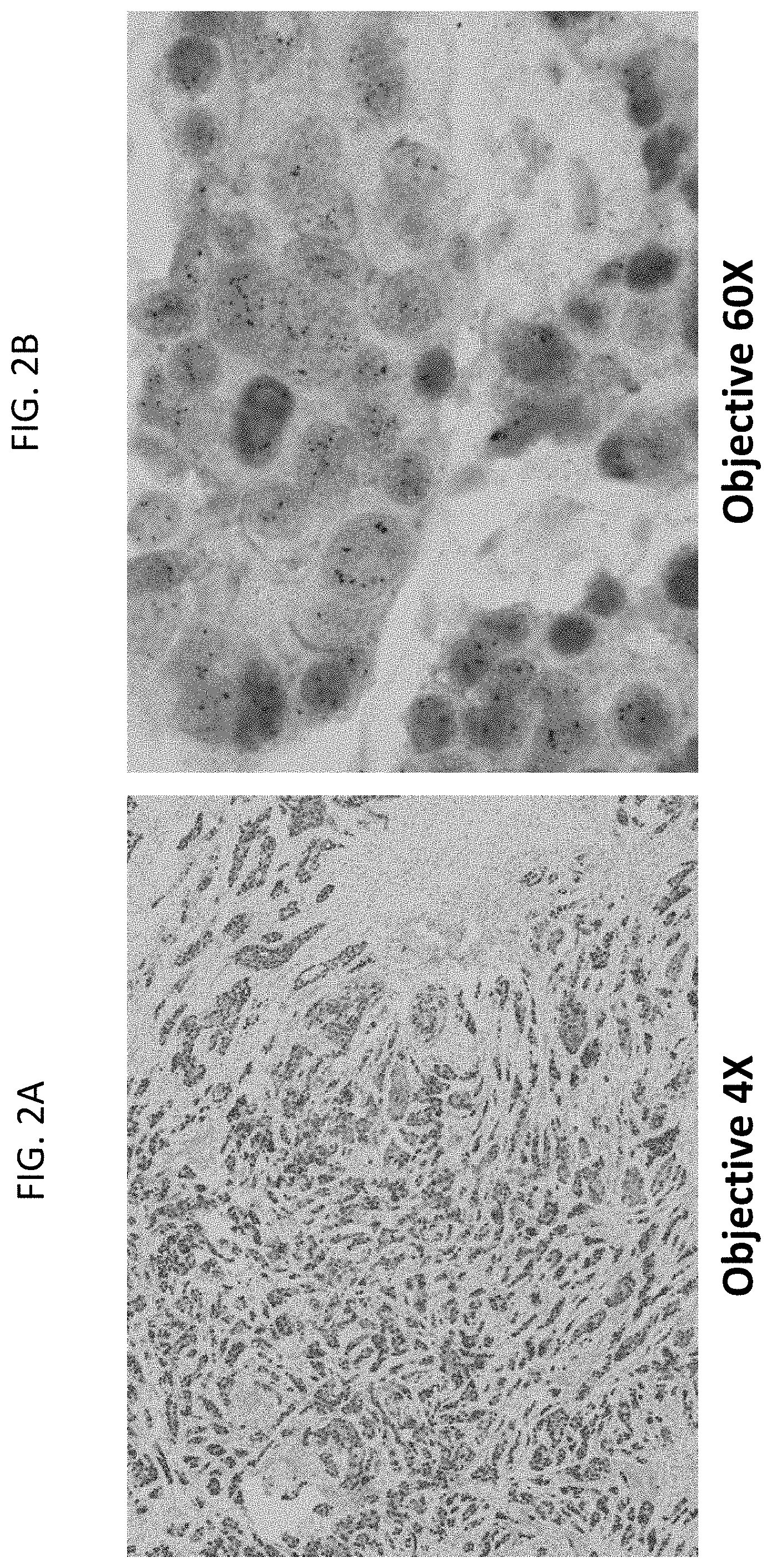
[0031] FIGS. 2A and 2B are a pair of images of a breast tumor tissue sample stained for HER2 gene (black dots), HER2 protein (brown color), and ER protein (red color) at 4.times. magnification (FIG. 2A) and 60.times. magnification (FIG. 2B). The sample has amplified HER2 gene and is ER protein positive, but is HER2 protein negative, as evidenced by the faint or absent brown staining.
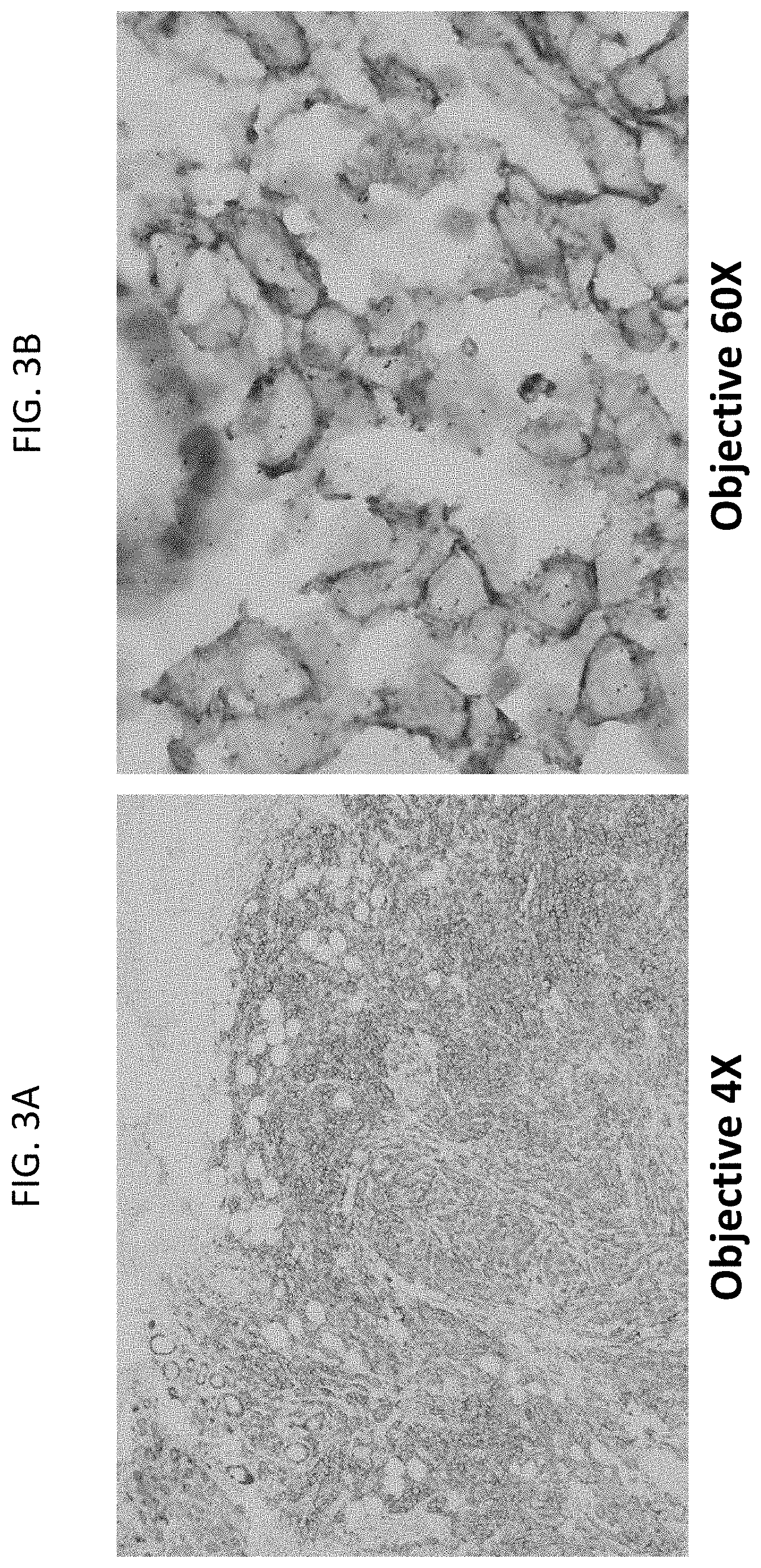
[0032] FIGS. 3A and 3B are a pair of images of a breast tumor tissue sample stained for HER2 gene (black dots), HER2 protein (brown color), and ER protein (red color) at 4.times. magnification (FIG. 3A) and 60.times. magnification (FIG. 3B). The sample shows HER2 gene amplification and is HER2 protein positive, but is ER negative, as evidenced by the lack of red staining. The red staining in FIG. 3B is ER protein staining in normal mammary gland cells in the sample.
[0033] FIGS. 4A-4C are a series of images showing ER protein IHC with iVIEW DAB staining (FIG. 4A) or ULTRAVIEW Red staining (FIG. 4B) and HER2 gene and protein IHC/ISH with ULTRAVIEW Red IHC staining (FIG. 4C) in a breast tissue sample. 20.times. magnification.
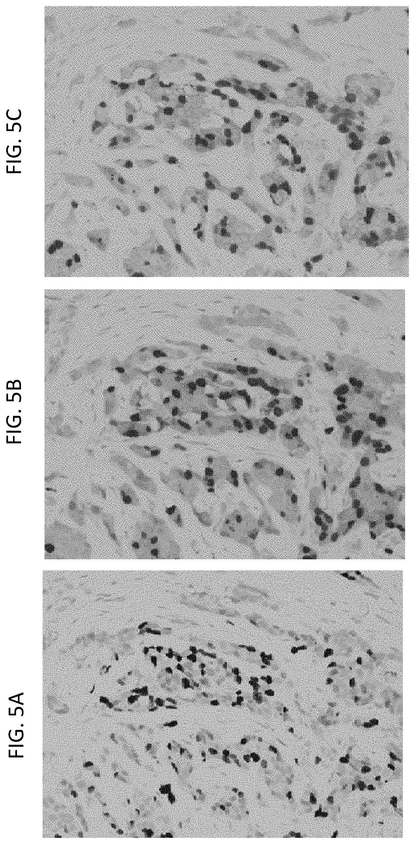
[0034] FIGS. 5A-5C are a series of images showing Ki67 protein IHC with iVIEW DAB staining (FIG. 5A) or ULTRAVIEW Red staining (FIG. 5B) and HER2 gene and protein IHC/ISH with ULTRAVIEW Red IHC staining (FIG. 5C) in a breast tissue sample. 20.times. magnification.
[0035] FIG. 6 is an image of exemplary detection of HER2 gene (black dots), HER2 protein (brown color), and Ki67 (red color) in a breast tissue sample.
[0036] FIGS. 7A-7D are a series of images of staining of HER2 protein (brown staining), HER2 gene (black dots), and Ki67 protein (red staining) (FIGS. 7A and 7C) or HER2 protein (brown staining), HER2 gene (black dots), and ER protein (red staining) (FIGS. 7B and 7D) in a breast tissue sample at 20.times. magnification (FIGS. 7A and 7B) or 60.times. magnification (FIGS. 7C and 7D).
[0037] FIGS. 8A-8C are a series of images showing HER2 gene (black dots), HER2 protein (brown staining), and ER protein (red staining) in a HER2 equivocal breast tissue sample. FIG. 8B shows the sample at 10.times. magnification. The boxed red area on the upper left side in FIG. 8B is shown in FIG. 8A at 60.times. magnification and the boxed blue area (located approximately in the middle) in FIG. 8B is shown in FIG. 8C at 60.times. magnification.
[0038] FIGS. 9A-9C are a series of images showing HER2 gene (black dots), HER2 protein (brown staining), and ER protein (red staining) in a HER2 positive breast tissue sample. FIG. 9B shows the sample at 10.times. magnification. The boxed red area on the upper left side in FIG. 9B is shown in FIG. 9A at 60.times. magnification and the boxed blue area (located approximately in the middle) in FIG. 9B is shown in FIG. 9C at 60.times. magnification.
[0039] FIGS. 10A and 10B are a pair of images showing staining of HER2 protein (brown), ER protein (purple), HER2 gene (black spots), and chromosome 17 centromere DNA (red spots) in an exemplary HER2 positive/ER positive breast tissue sample at 20.times. magnification (FIG. 10A) and 60.times. magnification (FIG. 10B).
[0040] FIGS. 11A and 11B are a pair of images showing staining of HER2 protein (brown), ER protein (purple), HER2 gene (black spots), and chromosome 17 centromere DNA (red spots) in an exemplary HER2 negative/ER positive breast tissue sample at 20.times. magnification (FIG. 11A) and at 60.times. magnification (FIG. 11B).
[0041] FIG. 12 shows a schematic representation of four types of cells: HER2 protein positive/ER protein positive/HER2 gene positive, HER2 protein negative/ER protein positive/HER2 gene positive, HER2 protein positive, ER protein negative/HER2 gene positive, HER2 protein negative/ER protein negative/HER2 gene positive. Some tumors exhibiting heterogeneity may have two or more of the cell types.
[0042] FIG. 13 shows a demonstration of the micro-intratumoral heterogeneity of breast cancer using the methods of the present invention (HER2 gene/HER2 protein/ER protein assay). The tumor heterogeneity of HER2 protein and ER protein expression was observed at a low magnification (A). However, at a high magnification, three phenotypic and genetic types of breast cancer cell populations were recognized: 1) HER2 protein positive, HER2 gene positive, and ER positive cell population (B); 2) HER2 protein negative, HER2 gene positive, and ER protein positive cell population (C); and 3) HER2 protein negative, HER2 gene positive, and ER negative cell population (D).
[0043] FIG. 14 shows a round shape defined by a simple closed curve fitting within a first region. The first region is an area on and between an inner concentric circle and an outer concentric circle. The inner concentric circle has an inner radius (R.sub.in) and the outer concentric circle has a outer radius (R.sub.out). R.sub.in is .gtoreq.50% of R.sub.out. The simple closed curve has a radius R.sub.simple wherein R.sub.in.ltoreq.R.sub.simple.ltoreq.R.sub.out.
[0044] FIG. 15 shows a schematic representation of various steps used to stain HER2, ER, and HER2 DNA. The present invention is not limited to the markers, reagents, steps, or order of steps shown in FIG. 15.
[0045] FIGS. 16A, 16B, 16C, and 16D show a series of assays performed on breast cancer samples and examples of scores. The left panel shows HER2 IHC assays. The middle panel shows a HER2 dual ISH. The right panel shows the HER2 gene-protein assay (three markers): HER2 protein is shown in brown, HER2 DNA is stained in black, and chromosome 17 is shown in red. FIG. 16A shows a sample scored as 3+ (HER2 IHC). FIG. 16B shows a sample scored as 2+ (HER2 IHC). FIG. 16C shows a sample scored as 1+ (HER2 IHC). FIG. 16D shows a sample scored as 0 or negative (HER2 IHC).
[0046] FIG. 17 shows an example of a HER2 gene protein assay performed on a breast cancer sample. HER2 protein is shown in brown, HER2 DNA is stained in black, and chromosome 17 is shown in red. The sample shows heterogeneity: the cells at the bottom left are HER2 protein negative (1+) but are HER2 DNA amplified, the cells in the middle are HER2 protein equivocal (2+) but are HER2 DNA amplified, and the cells on the left are HER2 protein positive (3+) and are HER2 DNA amplified. Thus, not all the breast cancer cells in the sample overexpress HER2 protein.
[0047] FIG. 18 shows a HER2 gene-protein assay performed on a gastric cancer sample. HER2 protein is shown in brown, HER2 DNA is stained in black, and chromosome 17 is shown in red. The sample shows heterogeneity: the cells highlighted in the yellow box on the lower left hand side are HER2 protein negative, while other cells in the sample are HER2 protein positive. The present invention is not limited to gene-protein assays in breast cancer cells and may be performed in any appropriate tissue, e.g., gastric tissue.
DETAILED DESCRIPTION
I. Terms
[0048] Unless otherwise explained, all technical and scientific terms used herein have the same meaning as commonly understood by one of ordinary skill in the art to which a disclosed invention belongs. The singular terms "a," "an," and "the" include plural referents unless context clearly indicates otherwise. Similarly, the word "or" is intended to include "and" unless the context clearly indicates otherwise. "Comprising" means "including." Hence "comprising A or B" means "including A" or "including B" or "including A and B."
[0049] Suitable methods and materials for the practice and/or testing of embodiments of the disclosure are described below. Such methods and materials are illustrative only and are not intended to be limiting. Other methods and materials similar or equivalent to those described herein can be used. For example, conventional methods well known in the art to which the disclosure pertains are described in various general and more specific references, including, for example, Sambrook et al., Molecular Cloning: A Laboratory Manual, 2d ed., Cold Spring Harbor Laboratory Press, 1989; Sambrook et al., Molecular Cloning: A Laboratory Manual, 3d ed., Cold Spring Harbor Press, 2001; Ausubel et al., Current Protocols in Molecular Biology, Greene Publishing Associates, 1992 (and Supplements to 2000); Ausubel et al., Short Protocols in Molecular Biology: A Compendium of Methods from Current Protocols in Molecular Biology, 4th ed., Wiley & Sons, 1999; Harlow and Lane, Antibodies: A Laboratory Manual, Cold Spring Harbor Laboratory Press, 1990; and Harlow and Lane, Using Antibodies: A Laboratory Manual, Cold Spring Harbor Laboratory Press, 1999, the disclosures of which are incorporated in their entirety herein by reference.
[0050] All publications, patent applications, patents, and other references mentioned herein are incorporated by reference in their entirety for all purposes. In case of conflict, the present specification, including explanations of terms, will control.
[0051] Although methods and materials similar or equivalent to those described herein can be used to practice or test the disclosed technology, suitable methods and materials are described below. The materials, methods, and examples are illustrative only and not intended to be limiting.
[0052] In order to facilitate review of the various embodiments of the disclosure, the following explanations of specific terms are provided:
[0053] Antibody: A polypeptide that includes at least a light chain or heavy chain immunoglobulin variable region and specifically binds an epitope of an antigen (such as HER2 protein or ER protein). Antibodies include monoclonal antibodies, polyclonal antibodies, or fragments of antibodies. An antibody can be conjugated or otherwise labeled with a detectable label, such as an enzyme, hapten, or fluorophore.
[0054] Detect: To determine if an agent (such as a signal or particular antigen, protein or nucleic acid) is present or absent, for example, in a sample. In some examples, this can further include quantification, and/or localization, for example localization within a cell or particular cellular compartment. "Detecting" refers to any method of determining if something exists, or does not exist, such as determining if a target molecule is present in a biological sample. For example, "detecting" can include using a visual or a mechanical device to determine if a sample displays a specific characteristic. In certain examples, light microscopy and other microscopic means are used to detect a detectable label bound to or proximally to a target. Accordingly, in some embodiments, an agent (e.g., antigen, protein, nucleic acid) is "visible" if it is "detected", via a detectable label directly or indirectly linked to the agent.
[0055] Detectable label: A molecule or material that can produce a signal (such as a visual, electrical, or other signal) that indicates the presence and/or amount of a target (such as a protein or nucleic acid) in a sample. When conjugated to a specific binding molecule (for example, an antibody or nucleic acid probe), the detectable label can be used to locate and/or quantify the target to which the specific binding molecule is directed. A detectable label can be detected directly or indirectly, and several different detectable labels can be used in combination to detect one or more targets. For example, a first detectable label, such as a hapten conjugated to an antibody specific to a target, can be detected indirectly by using a second detectable label that is conjugated to a molecule that specifically binds the first detectable label. In addition, multiple detectable labels that can be separately detected can be conjugated to different specific binding molecules that specifically bind different targets to provide a multiplex assay that can provide detection of the multiple targets in a single sample.
[0056] Detectable labels include chromogenic, fluorescent, phosphorescent and/or luminescent molecules, catalysts (such as enzymes) that convert one substance into another substance to provide a detectable signal (such as by converting a colorless substance into a colored substance or vice versa, or by producing a precipitate or increasing sample turbidity), haptens that can be detected through antibody-hapten binding interactions using additional detectably labeled antibody conjugates, and paramagnetic and magnetic molecules or materials. Particular examples of detectable labels include: enzymes, such as horseradish peroxidase, alkaline phosphatase, acid phosphatase, glucose oxidase, .beta.-galactosidase or .beta.-glucuronidase; fluorophores, such as fluoresceins, luminophores, coumarins, BODIPY dyes, resorufins, and rhodamines (many additional examples of fluorescent molecules can be found in The Handbook--A Guide to Fluorescent Probes and Labeling Technologies, Molecular Probes, Eugene, Oreg.); nanoparticles, such as quantum dots (U.S. Pat. Nos. 6,815,064, 6,682596 and 6,649,138, the disclosures of which are incorporated in their entirety herein by reference); metal chelates, such as DOTA and DPTA chelates of radioactive or paramagnetic metal ions like Gd.sup.3+; and liposomes, for example, liposomes containing trapped fluorescent molecules. Where the detectable label includes an enzyme, a detectable substrate such as a chromogen, a fluorogenic compound, or a luminogenic compound is used in combination with the enzyme to generate a detectable signal (a wide variety of such compounds are commercially available, for example, from Life Technologies, Carlsbad, Calif.).
[0057] Alternatively, an enzyme can be used in a metallographic detection scheme. In some examples, metallographic detection methods include using an enzyme, such as alkaline phosphatase, in combination with a water-soluble metal ion and a redox-inactive substrate of the enzyme. The substrate is converted to a redox-active agent by the enzyme, and the redox-active agent reduces the metal ion, causing it to form a detectable precipitate (see, for example, U.S. Pat. Nos. 7,642,064; 7,632,652; the disclosures of which are incorporated in their entirety herein by reference). In other examples, metallographic detection methods include using an oxido-reductase enzyme (such as horseradish peroxidase) along with a water soluble metal ion, an oxidizing agent and a reducing agent, again to form a detectable precipitate (see, for example, U.S. Pat. No. 6,670,113, the disclosures of which are incorporated in their entirety herein by reference). Haptens are small molecules that can be bound by antibodies. Exemplary haptens include dinitrophenyl (DNP), biotin, digoxigenin (DIG), and fluorescein. Additional haptens include oxazole, pyrazole, thiazole, nitroaryl, benzofuran, triperpene, urea, thiourea, rotenoid, coumarin and cyclolignan haptens, such as those disclosed in U.S. Pat. No. 7,695,929, the disclosures of which are incorporated in their entirety herein by reference.
[0058] Estrogen receptor (ER): Also known as estrogen receptor 1 (ESR1), estrogen receptor alpha (ER-alpha) estrogen nuclear receptor alpha; GenBank Gene ID Accession No. 2099. A hormone-activated transcription factor. Upon binding to estrogen (or other ER agonists) the estrogen receptor localizes to the nucleus and forms homodimers or heterodimers with estrogen receptor 2 and activates transcription of various genes.
[0059] ER nucleic acid and protein sequences are publicly available. For example, the ER gene is located on chromosome 6q25.1 and its sequence is disclosed as GenBank Accession No. NC_000006.11 (152011631-152424409). GenBank Accession Nos. NM_001122742, NM_001122741, NM_001122740, NM_000125, XM_005266856, and XM_005266857 disclose ER nucleic acid sequences, and GenBank Accession Nos.: NP_001116214, NP_001116213, NP_001116212, NP_000116, XP_005266913, and XP_005266914 disclose ER protein sequences, all of which are incorporated by reference as provided by GenBank on Oct. 4, 2013.
[0060] HER2: Also known as v-erb-b2 avian erythroblastic leukemia viral oncogene homolog 2 (ErbB2), human epidermal growth factor receptor 2, Her2/neu, c-erb B2/neu, and neuroblastoma/glioblastoma derived oncogene homolog; GenBank Gene ID Accession No. 2064. A member of the epidermal growth factor receptor tyrosine kinase family. Her2 heterodimerizes with other ligand-bound EGF receptor family members, though it lacks a ligand binding domain and cannot bind ligands itself. Amplification and/or overexpression of Her2 occur in several types of cancer, including breast and ovarian cancer.
[0061] Her2 nucleic acid and protein sequences are publicly available. For example, the Her2 gene is located on chromosome 17q12 and its sequence is disclosed as GenBank Accession No. NC_000017.10 (37844167-37884915). GenBank Accession Nos. NM_001005862, NM_004448, XM_005257139, and XM_005257140 disclose Her2 nucleic acid sequences, and GenBank Accession Nos.: NP_001005862, NP_004439, XP_005257196, and XP_005257197 disclose Her2 protein sequences, all of which are incorporated by reference as provided by GenBank on Oct. 4, 2013.
[0062] Hybridization: To form base pairs between complementary regions of two strands of DNA, RNA, or between DNA and RNA, thereby forming a duplex molecule. Hybridization conditions resulting in particular degrees of stringency will vary depending upon the nature of the hybridization method and the composition and length of the hybridizing nucleic acid sequences. Generally, the temperature of hybridization and the ionic strength (such as the Na+ concentration) of the hybridization buffer will determine the stringency of hybridization. The presence of a chemical which decreases hybridization (such as formamide) in the hybridization buffer will also determine the stringency (Sadhu et al., J. Biosci. 6:817-821, 1984, the disclosures of which are incorporated in their entirety herein by reference). Calculations regarding hybridization conditions for attaining particular degrees of stringency are discussed in Sambrook et al., (1989) Molecular Cloning, second edition, Cold Spring Harbor Laboratory, Plainview, N.Y. (chapters 9 and 11). Hybridization conditions for ISH are also discussed in Landegent et al., Hum. Genet. 77:366-370, 1987; Lichter et al., Hum. Genet. 80:224-234, 1988; and Pinkel et al., Proc. Natl. Acad. Sci. USA 85:9138-9142, 1988, the disclosures of which are incorporated in their entirety herein by reference.
[0063] Immunohistochemistry (IHC): A method of determining the presence or distribution of an antigen in a sample by detecting interaction of the antigen with a specific binding agent, such as an antibody. A sample is contacted with an antibody under conditions permitting antibody-antigen binding. Antibody-antigen binding can be detected by means of a detectable label conjugated to the antibody (direct detection) or by means of a detectable label conjugated to a secondary antibody, which binds specifically to the primary antibody (e.g., indirect detection).
[0064] In situ hybridization (ISH): A method of determining the presence or distribution of a nucleic acid in a sample using hybridization of a labeled nucleic acid probe to localize a specific DNA or RNA sequence in a portion or section of tissue (in situ), or, if the tissue is small enough (e.g., plant seeds, Drosophila embryos), in the entire tissue (whole mount ISH). DNA ISH can be used to determine the structure of chromosomes, such as for use in medical diagnostics to assess chromosomal integrity and/or to determine gene copy number in a sample. RNA ISH measures and localizes mRNAs and other transcripts within tissue sections or whole mounts.
[0065] For ISH, sample cells and tissues are usually treated to fix the target nucleic acids in place and to increase access of the probe to the target molecule. The detectably labeled probe hybridizes to the target sequence at elevated temperature, and then the excess probe is washed away. Solution parameters, such as temperature, salt and/or detergent concentration, can be manipulated to remove any non-identical interactions (e.g., so only exact sequence matches will remain bound). Then, the labeled probe is localized and potentially quantitated in the tissue using either autoradiography, fluorescence microscopy or immunohistochemistry, respectively. ISH can also use two or more probes, which are typically differently labeled to simultaneously detect two or more nucleic acids.
[0066] Probe: An isolated nucleic acid (such as an isolated synthetic oligonucleotide), attached to a detectable label or reporter molecule. Typical labels include radioactive isotopes, enzyme substrates, co-factors, ligands, chemiluminescent or fluorescent agents, haptens (including, but not limited to, DNP), and enzymes. Methods for labeling and guidance in the choice of labels appropriate for various purposes are discussed, e.g., in Sambrook et al. (In Molecular Cloning: A Laboratory Manual, CSHL, New York, 1989) and Ausubel et al. (In Current Protocols in Molecular Biology, Greene Publ. Assoc. and Wiley-Intersciences, 1992, the disclosures of which are incorporated in their entirety herein by reference).
[0067] Probes can be selected to provide a desired specificity, and may comprise at least 15, 20, 25, 30, 35, 40, 45, 50 or more nucleotides of a target nucleic acid. In particular examples, probes can include at least 100, 250, 500, 600, 1000, or more nucleotides of a target nucleic acid. In some examples, the probe includes segments of nucleotides that are from non-contiguous portions of a target nucleic acid, such as a HER2 genomic nucleic acid.
[0068] Sample: The term "sample" refers to any liquid, semi-solid or solid substance (or material) in or on which a target can be present. In particular, a sample can be a biological sample or a sample obtained from a biological material. Exemplary biological samples include tissue samples and/or cytology samples, for example, obtained from an animal subject, such as a human subject. In other examples, a biological sample can be a biological fluid obtained from, for example, blood, plasma, serum, urine, bile, ascites, saliva, cerebrospinal fluid, aqueous or vitreous humor, or any bodily secretion, a transudate, an exudate (for example, fluid obtained from an abscess or any other site of infection or inflammation), or fluid obtained from a joint (for example, a normal joint or a joint affected by disease). A biological sample can also be a sample obtained from any organ or tissue (including a biopsy or autopsy specimen, such as a tumor biopsy) or can include a cell (whether a primary cell or cultured cell) or medium conditioned by any cell, tissue or organ.
[0069] Specific binding: A term that refers to the binding of agent that preferentially binds to a defined target (such as an antibody to a specific protein or antigen or a nucleic acid probe to a specific nucleic acid sequence). With respect to a target protein, "specifically binds" refers to the preferential association of an antibody or other ligand, in whole or part, with a specific polypeptide. "Specifically binds" refers to the preferential association of a nucleic acid probe, in whole or part, with a specific nucleic acid, when referring to a target nucleic acid.
[0070] A specific binding agent binds substantially only to a particular target. A minor amount of non-specific interaction may occur between a specific binding agent and a non-target protein or nucleic acid. Antibody to antigen specific binding typically results in greater than 2-fold, such as greater than 5-fold, greater than 10-fold, or greater than 100-fold increase in amount of bound antibody or other ligand (per unit time) to a target protein, as compared to a non-target protein. Immunoassay formats can be used to select antibodies that specifically react with a particular protein (such as antibodies that specifically bind HER2 protein or ER protein). See Harlow & Lane, Antibodies, A Laboratory Manual, Cold Spring Harbor Publications, New York (1988), for a description of immunoassay formats and conditions.
[0071] Specific binding of a nucleic acid probe to a target nucleic acid molecule typically results in greater than 2-fold, such as greater than 5-fold, greater than 10-fold, or greater than 100-fold increase in amount of bound nucleic acid probe to a target nucleic acid as compared to a non-target nucleic acid. A variety of ISH conditions are appropriate for selecting nucleic acid probes that bind specifically with a particular nucleic acid sequence (such as a HER2-specific probe or a chromosome 17 centromere probe).
[0072] Subject: Any multi-cellular vertebrate organism, such as human or non-human mammals (e.g., veterinary subjects).
II. Overview of Several Embodiments
[0073] Disclosed herein are methods for co-detecting multiple target molecules (such as two or more proteins and/or nucleic acids) in a single sample on a single slide. In particular embodiments, the methods include detecting the presence and/or amount of HER2 protein, ER protein, and HER2 genomic DNA (such as HER2 gene copy number) in a single sample. In some embodiments, the methods further include detecting the presence and/or amount of chromosome 17 centromere DNA in the sample, and in some examples, determining a ratio of HER2 genomic DNA to chromosome 17 centromere DNA (such as a ratio of HER2 gene copy number to chromosome 17 centromere copy number). The methods include utilizing different detectable labels and/or detection systems for each of the HER2 protein, ER protein, HER2 genomic DNA, and chromosome 17 centromere DNA (if included), such that each can be individually visually detected in a single sample. FIG. 15 shows a non-limiting example of a gene-protein assay for detecting HER2 protein, HER2 DNA, and chromosome 17 DNA.
[0074] In some embodiments of the methods, a sample is contacted with an antibody (e.g., primary antibody) that specifically binds to HER2 protein and HER2 protein is detected, the sample is contacted with an antibody (e.g., primary antibody) that specifically binds to ER protein and ER protein is detected, and the sample is contacted with a nucleic acid probe that specifically binds to HER2 genomic DNA and HER2 genomic DNA is detected. In one embodiment, the method comprises detecting HER2 protein and ER protein before detecting HER2 DNA (or before detecting HER2 DNA and CHR17 DNA). In one specific embodiment, the method comprises sequentially detecting HER2 protein (contacting the sample with a HER2-specific antibody and detecting HER2 protein in the sample), followed by detecting ER protein (contacting the sample with an ER-specific antibody and detecting ER protein in the sample), and then followed by detecting HER2 genomic DNA (contacting the sample with a HER2 genomic DNA-specific nucleic acid probe and detecting HER2 genomic DNA).
[0075] As an example, reference is made to FIGS. 1A-1B, showing a pair of images of a breast tumor tissue sample stained for HER2 gene (black punctate nuclear staining), HER2 protein (brown membrane staining), and ER protein (red cytoplasmic staining) at 4.times. magnification (FIG. 1A) and 60.times. magnification (FIG. 1B). The sample is HER2 gene amplified, HER2 protein positive, and ER protein positive. However, some cells (circled) are negative for HER2 protein, though they are ER protein positive and have HER2 gene amplification. Since the HER2-targeted therapies target the HER2 protein, this heterogeneity could result in failure of the therapy to affect (e.g., inhibit or even kill) tumor cells that are HER2 gene amplified, but do not overexpress the HER2 protein. However, those cells that are ER-positive would still be affected by ER-targeted therapies.
[0076] In additional embodiments the method includes contacting the sample (simultaneously or sequentially) with a HER2 genomic DNA-specific nucleic acid probe and a chromosome 17 centromere genomic DNA-specific nucleic acid probe and detecting HER2 genomic DNA and then detecting chromosome 17 centromere genomic DNA.
[0077] The methods may utilize different detectable labels and/or detection systems for each of the targets such that each can be individually detected in a single sample. The proteins/DNA may be detected by the chromogens using additional reagents such as secondary antibodies specific for the primary antibodies.
[0078] The first marker (e.g., HER2) may be stained a first color, the second marker (e.g., ER) may be stained a second color, the third marker (e.g., HER2 DNA) may be stained a third color, and the fourth marker (e.g., chromosome 17) may be stained a fourth color. The first color is transparent enough to allow visualization of the second color and/or third and/or the fourth color. In some embodiments, the first color blocks no more than 50%, no more than 40%, no more than 30%, no more than 20%, no more than 10%, no more than 8%, no more than 6%, no more than 4%, no more than 2%, or none of the intensity of the second color and/or the third color and/or the fourth color. The second color allows visualization of the first color and/or third and/or the fourth color. In some embodiments, the second color blocks no more than 50%, no more than 40%, no more than 30%, no more than 20%, no more than 10%, no more than 8%, no more than 6%, no more than 4%, no more than 2%, or none of the intensity of the first color and/or the third color and/or the fourth color. The third color allows visualization of the first color and/or second color and/or the fourth color. In some embodiments, the third color blocks no more than 50%, no more than 40%, no more than 30%, no more than 20%, no more than 10%, no more than 8%, no more than 6%, no more than 4%, no more than 2%, or none of the intensity of the first color and/or the second color and/or the third color.
[0079] Detection includes but is not limited to bright field microscopy. In some embodiments, the step of staining protein is performed before the step of staining DNA. For example, the step of staining the HER2 protein and ER protein is performed before the step of staining HER2 DNA and chromosome 17 DNA.
[0080] The HER2 protein can be detected using a first chromogen. The ER protein can be detected using a second (different) chromogen. The HER2 DNA can be detected using a third (different) chromogen. The chromosome 17 centromere DNA can be detected with a fourth (different) chromogen. The proteins/DNA may be detected by the chromogens using additional reagents such as secondary antibodies specific for the primary antibodies.
[0081] The first chromogen may be used at a level so as to block HER2 protein-specific antibody that is not bound by its appropriate secondary antibody. This can help reduce cross reactivity if, for example, the host species is the same for the HER2 protein-specific primary antibody and the ER protein-specific primary antibody. In some embodiments, the first chromogen (for detecting HER2) comprises 3,3'-diaminobenzidine (DAB).
[0082] In some embodiments, the second chromogen is transparent enough so that it blocks no more than 10% of the third chromogen and/or fourth chromogen. In some embodiments, the second chromogen is transparent enough so that it blocks no more than 8% of the third chromogen and/or fourth chromogen. In some embodiments, the second chromogen is transparent enough so that it blocks no more than 6% of the third chromogen and/or fourth chromogen. In some embodiments, the second chromogen is transparent enough so that it blocks no more than 4% of the third chromogen and/or fourth chromogen. In some embodiments, the second chromogen is transparent enough so that it blocks no more than 2% of the third chromogen and/or fourth chromogen. In some embodiments, the second chromogen is transparent enough so that it does not block any of the visibility of the third chromogen and/or fourth chromogen. For example, all of the color resulting from the third chromogen and/or the fourth chromogen that is present on the slide is visible--the second chromogen does not prevent the visibility of the color resulting from the third chromogen and/or fourth chromogen.
[0083] In some examples of the disclosed methods, the sample is contacted with an antibody that specifically binds to HER2 protein. Methods of constructing HER2-specific antibodies are known in the art. In addition, such antibodies may be commercially available. In one specific example, the sample is contacted with an anti-HER2 rabbit monoclonal antibody, such as anti-HER-2/neu (4B5) rabbit monoclonal antibody (Ventana Medical Systems, Inc., Tucson, Ariz., e.g., catalog number 790-2991). Additional HER2-specific antibodies include anti-c-erbB2 antibody A0485 (Dako, Carpinteria, Calif.). In some examples, the HER2-specific antibody is detectably labeled, allowing detection of HER2 protein in the sample. In other examples, after contacting the sample with the anti-HER2 antibody (the primary antibody), the sample is contacted with a detectably labeled secondary antibody raised against the primary antibody, such as a secondary antibody conjugated to an enzyme (for example, alkaline phosphatase (AP) or horseradish peroxidase (HRP)) or a secondary antibody conjugated to a hapten that can be detected with a further reagent conjugated to an enzyme. The presence of HER2 protein is detected by contacting the enzyme with a chromogen and/or substrate composition, which produces a colored precipitate in the vicinity of the anti-HER2 antibody. The presence and/or amount of HER2 protein is detected by determining staining intensity in the sample. In some examples, the staining intensity is rated by a slide reader on a numeric scale, such as a scale of 0-3 (for example, where 0 indicates no staining relative to background, 1 indicates weak staining, 2 indicates moderate staining, and 3 indicates strong staining).
[0084] Any appropriate chromogen or detection composition may be used for any of the markers. See, for example, WO 2013148498, the disclosure of which is incorporated in its entirety herein by reference.
[0085] In one particular example, the method includes contacting the sample with a primary antibody that specifically binds to the HER2 protein (for example, anti-HER2 4B5 rabbit monoclonal antibody), for example under conditions sufficient for the anti-HER2 antibody to specifically bind to HER2 protein in the sample. The sample is then contacted with a biotinylated secondary antibody that specifically binds the primary antibody, for example under conditions sufficient for the secondary antibody to specifically bind to the primary antibody. The sample is then contacted with HRP-conjugated streptavidin, for example under conditions sufficient for the streptavidin-HRP to specifically bind to the biotin, followed by contacting the sample with hydrogen peroxide substrate and 3,3'-diaminobenzidine (DAB) chromogen, which produces a brown precipitate near the anti-HER2 antibody (and HER2 protein) that can be visually detected by light (bright-field) microscopy. In one example, the reagents (except for the anti-HER2 antibody) are included in a kit, such as the IVIEW DAB Detection Kit (Ventana Medical Systems, Tucson, Ariz., catalog number 760-091). One of ordinary skill in the art can select alternative detection reagents (such as alternative secondary antibodies, enzymes, substrates, and/or chromogens) including those that produce a different color precipitate for detection of the HER2 protein.
[0086] In some examples of the disclosed methods, the sample is contacted with an antibody that specifically binds to ER protein. Methods of constructing ER-specific antibodies are known in the art. In addition, such antibodies may be commercially available. In one specific example, the sample is contacted with an anti-ER rabbit monoclonal antibody, such as anti-ER (SP1) rabbit monoclonal antibody (Ventana Medical Systems, Inc., Tucson, Ariz., e.g., catalog number 790-4324). Additional ER-specific antibodies include anti-ER monoclonal antibodies 1D5 and ER-2-123 (Dako, Carpinteria, Calif.). In some examples, the ER-specific antibody is detectably labeled, allowing detection of ER protein in the sample. In other examples, after contacting the sample with the anti-ER antibody (the primary antibody), the sample is contacted with a detectably labeled secondary antibody raised against the primary antibody, such as a secondary antibody conjugated to an enzyme (for example, AP or HRP) or a secondary antibody conjugated to a hapten that can be detected with a further reagent conjugated to an enzyme. The presence of ER protein is detected by contacting the enzyme with a chromogen and/or substrate composition, which produces a colored precipitate in the vicinity of the anti-ER antibody. The presence and/or amount of ER protein is detected by determining staining intensity in the sample. In some examples, the staining is scored by a slide reader by determining a percentage of tumor cells in the sample that are stained for the ER protein.
[0087] In one particular example, the method includes contacting the sample with a primary antibody that specifically binds to the ER protein (for example, anti-ER SP1 rabbit monoclonal antibody), for example under conditions sufficient for the anti-ER antibody to specifically bind to ER protein in the sample. The sample is then contacted with an AP-conjugated secondary antibody that specifically binds the primary antibody, for example under conditions sufficient for the secondary antibody to specifically bind to the primary antibody. The sample is then contacted with a naphthol phosphate and Fast Red chromogen, which produces a red precipitate near the anti-ER antibody (and ER protein) that can be visually detected by light microscopy. In one example, the reagents (except for the anti-ER antibody) are included in a kit, such as the ULTRAVIEW Universal Alkaline Phosphatase Red Detection Kit (Ventana Medical Systems, Tucson, Ariz., catalog number 760-501). One of ordinary skill in the art can select alternative detection reagents (such as alternative antibodies, enzymes, substrates, and/or chromogens) including those that produce a different color precipitate for detection of the ER protein. In some embodiments, the chromogen (e.g., the second chromogen) used for ER comprises any other appropriate chromogen (see US20130260379, the disclosure of which is incorporated in its entirety herein by reference), e.g., fast red, discovery purple, etc.
[0088] Alternatively, the method includes contacting the sample with a primary antibody that specifically binds to the ER protein (for example, anti-ER SP1 rabbit monoclonal antibody), for example under conditions sufficient for the anti-ER antibody to specifically bind to ER protein in the sample. The sample is then contacted with a biotinylated secondary antibody that specifically binds the primary antibody, for example under conditions sufficient for the secondary antibody to specifically bind to the primary antibody. The sample is then contacted with streptavidin-HRP, followed by hydrogen peroxide and Discovery Purple chromogen (a tyramide-rhodamine conjugate; Ventana Medical Systems, Tucson, Ariz., part number 700-229), which produces a purple dye bound to the sample near the anti-ER antibody (and ER protein) that can be visually detected by light microscopy.
[0089] In some examples, of the disclosed methods, the sample is contacted with a nucleic acid probe that specifically binds to HER2 genomic DNA. Methods of constructing HER2-specific nucleic acid probes are known to one of ordinary skill in the art. HER2-specific nucleic acid probes may also be commercially available. For example, a HER2 probe suitable for use in the disclosed methods includes the HER2 probe included in the INFORM HER2 Dual ISH Probe Cocktail (Ventana Medical Systems, Tucson, Ariz., catalog number 780-4422). In one example, the sample is contacted with a hapten-labeled HER2 nucleic acid probe, for example under conditions specific for the probe to specifically bind to (hybridize with) HER2 genomic DNA in the sample. The sample is then contacted with an antibody that specifically binds to the hapten, for example, under conditions sufficient for the antibody to specifically bind to the hapten. The antibody may be conjugated to an enzyme (such as AP or HRP) or alternatively, the sample may be contacted with a second antibody that specifically binds the anti-hapten antibody, where the second antibody is conjugated to an enzyme. The presence of HER2 genomic DNA is detected by contacting the enzyme with a chromogen and/or substrate composition to produce a colored precipitate in the vicinity of the HER2 nucleic acid probe. In some examples, the gene copy number of HER2 DNA in the sample is scored by a slide reader by counting the number of areas of precipitate ("spots") in the nuclei of the tumor cells.
[0090] In one particular example, the method includes contacting the sample with a HER2 genomic DNA probe conjugated to dinitrophenyl (DNP), for example under conditions sufficient for the HER2 probe to specifically bind to HER2 genomic DNA in the sample. The sample is then contacted with an anti-hapten antibody that specifically binds DNP, for example under conditions sufficient for the anti-DNP antibody to specifically bind to the DNP. The sample is then contacted with an HRP-conjugated secondary antibody that specifically binds to the anti-DNP antibody, for example under conditions sufficient for the secondary antibody to specifically bind to the anti-DNP antibody. The sample is then contacted with chromogen and substrate silver acetate, hydroquinone, and hydrogen peroxide. The silver ions are reduced by hydroquinone to metallic silver ions, which can be visually detected by light microscopy as black spots. In one example, the reagents (except for the HER2 probe) are included in a kit, such as the ULTRAVIEW SISH DNP Detection Kit (Ventana Medical Systems, Tucson, Ariz., catalog number 760-098). One of ordinary skill in the art can select alternative detection reagents (such as alternative haptens, antibodies, enzymes, substrates, and/or chromogens) including those that produce a different color precipitate for detection of HER2 genomic DNA.
[0091] In additional examples, the disclosed methods further include contacting the sample with a probe that specifically binds to chromosome 17 centromere DNA and detecting chromosome 17 DNA (such as chromosome 17 copy number) in the sample. In some examples, of the disclosed methods, the sample is contacted with a nucleic acid probe that specifically binds to chromosome 17 centromere DNA. Methods of constructing chromosome 17 centromere-specific nucleic acid probes are known to one of ordinary skill in the art. In addition, chromosome 17 centromere nucleic acid probes may also be commercially available. For example, a chromosome 17 centromere probe suitable for use in the disclosed methods includes the chromosome 17 centromere probe included in the INFORM HER2 Dual ISH Probe Cocktail (Ventana Medical Systems, Tucson, Ariz., catalog number 780-4422). In one example, the sample is contacted with a hapten-labeled chromosome 17 centromere nucleic acid probe, for example under conditions specific for the probe to specifically bind to (hybridize with) chromosome 17 centromere genomic DNA in the sample. The sample is then contacted with an antibody that specifically binds to the hapten, for example, under conditions sufficient for the antibody to specifically bind to the hapten. The antibody may be conjugated to an enzyme (such as AP or HRP) or alternatively, the sample may be contacted with a second antibody that specifically binds the anti-hapten antibody, where the second antibody is conjugated to an enzyme. The presence of chromosome 17 centromere genomic DNA is detected by contacting the enzyme with a chromogen and/or substrate composition to produce a colored precipitate in the vicinity of the chromosome 17 centromere nucleic acid probe. In some examples, the gene copy number of chromosome 17 centromere DNA in the sample is scored by a slide reader by counting the number of areas of precipitate ("spots") in the nuclei of the tumor cells.
[0092] In a particular example, the method includes contacting the sample with a chromosome 17 centromere DNA probe conjugated to digoxigenin (DIG), for example under conditions sufficient for the chromosome 17 centromere probe to specifically bind to chromosome 17 centromere DNA in the sample. The sample is then contacted with an anti-hapten antibody that specifically binds DIG, for example under conditions sufficient for the anti-DIG antibody to specifically bind to the DIG. The sample is then contacted with an AP-conjugated secondary antibody that specifically binds to the anti-DIG antibody, for example under conditions sufficient for the secondary antibody to specifically bind to the anti-DIG antibody. The sample is then contacted with a naphthol phosphate and Fast Red, producing a red precipitate which is deposited in the nuclei near the chromosome 17 centromere probe (and the chromosome 17 centromere DNA) and can be visually detected by light microscopy as red spots. In one example, the reagents (except for the chromosome 17 centromere probe) are included in a kit, such as the ULTRAVIEW Red ISH DIG Detection Kit (Ventana Medical Systems, Tucson, Ariz., catalog number 760-505). One of ordinary skill in the art can select alternative detection reagents (such as alternative haptens, antibodies, enzymes, substrates, and/or chromogens) including those that produce a different color precipitate for detection of chromosome 17 centromere DNA.
[0093] In some embodiments, the HER2 DNA-specific nucleic acid probe comprises a set of two or more single-stranded oligonucleotide target probes specific for HER2 DNA. The oligonucleotide probes may be specific for a region between nucleotides 35,027,979 and 35,355,516 of human chromosome 17. In some embodiments, the HER2 DNA oligonucleotide probes (target probes) each comprise between 50 to 100 nucleotides. The single strand oligonucleotide HER2 probe (HER2 oligonucleotide probe) may be a dinitrophenyl (DNP)-labeled, repeat-free genomic probe specifically targeting the HER2 gene region. Similar to INFORM HER2 DUAL ISH DNA Probe, the HER2 oligonucleotide probe may span >327,000 nucleotides (nt) (35,027,979-35,355,516) of genomic DNA from human Chromosome 17, encompassing the HER2 target region (UCSC Genome Browser on Human May 2004 (NCBI35/hg17) Assembly). In some embodiments, the HER2 oligonucleotide sequences are designed from the sequences in INFORM HER2 DUAL ISH DNA Probe. Each of the HER2 oligonucleotides may be designed with 80-mer length; hence stringency level for non-target binding may be raised higher according to the aforementioned oligonucleotide probe design criteria. Specificity of the HER2 oligonucleotide probe may be experimentally validated on metaphase spreads under the examined ISH assay conditions. Bioinformatic searches were used to identify HER2 specific nucleic acid sequences around the HER2 target region. The selected genomic target nucleic acid sequence was separated into consecutive non-overlapping 80 nt segments. One thousand one hundred and ninety-six (1196) .about.80mer oligonucleotides were synthesized each carrying 5 DNP haptens on an abasic phosphoramidite spaced 20 nt apart. A representative structure for these oligonucleotides is shown here: 5'T[DNP]CTCGTCTCGGCCCCCGACCT[DNP]GCGTCCTGGGCCCGCAGGGG[DNP]AGTCCTGCCCCATGC- TCCCG[DNP]GGCGGGGCCGCCCTGTGCCC[DNP]T-3' (SEQ ID NO: 15). The oligonucleotides were affinity purified and analyzed by mass spectrometry and gel electrophoresis. HER2 oligonucleotide probe was bulked in a formamide-based buffer without human blocking DNA. In the initial screening process, the number of oligonucleotides, the number and spacing of DNP haptens were functionally tested in the formamide-based buffer without human blocking DNA for sensitivity and specificity to HER2 gene. Additional detail may be found in U.S. Provisional Patent Application No. 61/943,196, entitled SINGLE STRAND OLIGONUCLEOTIDES FOR TISSUE DIAGNOSTICS, filed on Feb. 21, 2014, the disclosure of which is incorporated in its entirety herein by reference.
[0094] In some embodiments, the chromosome 17 centromere-specific nucleic acid probe comprises a set of two or more single-stranded oligonucleotide control probes. The oligonucleotide control probes are specific for two or more (between 2 and 14, e.g., 2, 3, 4, 5, 6, 7, 8, 9, 10, 11, 12, 13, 14, .gtoreq.4, .gtoreq.6, .gtoreq.8, etc.) distinct monomers of the alpha satellite control region of chromosome 17. In some embodiments, the chromosome 17 oligonucleotide probes (control probes) each comprise between 50 to 100 nucleotides.
[0095] In some embodiments, the chromosome 17 oligonucleotide control probes (each control probe) may comprise one of SEQ ID NOs: 1-14 (or complements thereof) (see below in Table 1). In some embodiments, the control probes (each control probe) may comprise a truncated version of one of SEQ ID NOs: 1-14 (or complements thereof). The truncated version may be at least 30 contiguous base pairs of said sequence, at least 40 contiguous base pairs of said sequence, or at least 50 30 contiguous base pairs of said sequence. In some embodiments, the control probes (each control probe) may comprise a sequence that has at least 70%, at least 80%, at least 90%, or at least 95% sequence identity to one of SEQ ID NOs: 1-14 (or complements thereof).
TABLE-US-00001 TABLE 1 Single-stranded Oligonucleotide Probes for Chromosome 17 Oligo name Sequences Length SEQ ID. NO. 1 AATTCGTTGGAAACGGGATAATTTCAGCTGACTAA 79 ACAGAAGCAGTCTCAGAATCTTCTTTGTGATGTTTG CATTCAAA SEQ ID. NO. 2 CTTCGTTCGAAACGGGTATATCTTCACATGCCATCT 79 AGACAGAAGCATCCTCAGAAGCTTCTCTGTGATGA CTGCATTC SEQ ID. NO. 3 TGAACTCTCCTTTTGAGAGCGCAGTTTTGAAACTCT 79 CTTTCTGTGGCATCTGCAAGGGGACATGTAGACCT CTTTGAAG SEQ ID. NO. 4 TTTCGTTGGAAACGGAATCATCTTCACATAAAAAC 79 TACACAGATGCATTCTCAGGAACTTTTTGGTGATGT TTGTATTC SEQ ID. NO. 5 CCTATGGTAGTAAAGGGAATAGCTTCATAGAAAAA 83 CTAGACAGAAGCATTCTCAGAAAATACTTTGTGAT GATTGAGTTTAAC SEQ ID. NO. 6 CACAGAGCTGAACATTCCTTTGGATGGAGCAGGTT 87 TGAGACACTCTTTTTGTACAATCTACAAGTGGATAT TTGGACCTCTCTGAGG SEQ ID. NO. 7 GTTTCACATTGCTTTTCATAGAGTAGTTCTGAAACA 71 TGCTTTTCGTAGTGTCTACAAGTGGACATTTGGAG SEQ ID. NO. 8 CCTGTGGTGGAAAACGAATTATCGTCACGTAAAAA 58 CTAGAGAGAAGCATTGTCAGAAA SEQ ID. NO. 9 TGCATTCAACTCACAGAGTTGAAGGTTCCTTTTCAA 65 AGAGCAGTTTCCAATCACTCTTTGTGTGG SEQ ID. NO. 10 CATTCCCTTTGACAGAGCAGTTTGGAAACTCTCTTT 71 GTGTAGAATCTGCAAGTGGAGATATGGACCGCTTT SEQ ID. NO. 11 CCTATGGTAGTAAAGGAAATAGCTTCATATAAAAG 80 CTAGACAGTAGCATTCACAGAAAACTCTTGGTGAC GACTGAGTTT SEQ ID. NO. 12 ATTTCGTTGGAAACGGGATAAACCGCACAGAACTA 80 AACAGAAGCATTCTCAGAACCTTCTTCGTGATGTTT GCATTCAAC SEQ ID. NO. 13 CGTAGTAAAGGAAATAACTTCCTATAAAAAGAAG 80 ACAGAAGCTTTCTCAGAAAATTCTTTGGGATGATT GAGTTGAACTC SEQ ID. NO. 14 ACAGAGCTGAGCATTCCTTGCGATGTAGCAGTTTA 79 GAAACACACTTTCTGCAGAATCTGCAATTGCATAT TTGGACCTT
[0096] The HER2 DNA oligonucleotide probes (target probes) and the chromosome 17 centromere oligonucleotide probes (control probes) can achieve an enumerable signal when hybridized to its respective DNA target. An enumerable signal has a generally round shape. In some embodiments, a round shape is a shape defined by a simple closed curve (see FIG. 14) fitting within a first region. The first region is an area on and between an inner concentric circle and an outer concentric circle. The inner concentric circle has an inner radius (R.sub.n) and the outer concentric circle has a outer radius (R.sub.out). R.sub.in is .gtoreq.50% of R.sub.out, and the simple closed curve has a radius R.sub.simple wherein R.sub.in.ltoreq.R.sub.simple.ltoreq.R.sub.out.
[0097] The HER2 DNA oligonucleotide probes may be hybridized under conditions for a period of time less than about 3 hours, less than about 2 hours, 1 hour, or less than about an hour. The chromosome 17 centromere oligonucleotide probes may be hybridized under conditions for a period of time less than hours, less than about 2 hours, 1 hour, or less than about an hour. The chromosome 17 centromere oligonucleotide probes (control probes) may achieve at least two enumerable signals per cell, e.g., with a staining intensity of .gtoreq.2 and staining coverage of .gtoreq.50% of the number of total nuclei within 3 hours of hybridization (or within 2 hours of hybridization, or within 1 hour of hybridization). In some embodiments, the chromosome 17 centromere oligonucleotide probes are configured to hybridize uniquely and specifically to a portion of the control region of human chromosome 17 so that other chromosomes or portions thereof are not evidently labeled without the influence of blocking DNA. In some embodiments, the chromosome 17 oligonucleotide control probes and/or the HER2 DNA oligo probes each comprise between 50 to 100 nucleotides.
[0098] The chromosome 17 oligonucleotide control probes may each comprise a detectable label, e.g., a hapten (e.g., dinitrophenyl, digoxigenin, biotin, or fluorescein, etc.). The labeled chromosome 17 oligonucleotide probes may be detected using any appropriate method or reagent, e.g., with a secondary antibody directed to the hapten and/or with other detection components and reagents. For example, in a particular example, the method comprises contacting the sample with a chromosome 17 oligonucleotide control probes conjugated to digoxigenin (DIG), for example under conditions sufficient for the chromosome 17 oligonucleotide control probes to specifically bind to chromosome 17 centromere DNA in the sample. The sample is then contacted with an anti-hapten antibody that specifically binds DIG, for example under conditions sufficient for the anti-DIG antibody to specifically bind to the DIG. The sample is then contacted with a HRP-conjugated secondary antibody that specifically binds to the anti-DIG antibody, for example under conditions sufficient for the secondary antibody to specifically bind to the anti-DIG antibody. The sample is then contacted with a chromogen component, producing a precipitate which is deposited in the nuclei near the chromosome 17 oligonucleotide control probes (and the chromosome 17 centromere DNA) and can be visually detected by light microscopy. One of ordinary skill in the art can select appropriate detection reagents (such as alternative haptens, antibodies, enzymes, substrates, and/or chromogens) including those that produce a different color precipitate for detection of chromosome 17 centromere DNA.
[0099] The disclosed methods are directed to detection of multiple protein and nucleic acid targets in a single sample. As a result, the detectable signal for each member of the assay must be individually distinguishable. Therefore, in some examples, the visual signal generated by the detection assay for each member of the assay is a different color. In one specific example, the methods result in a brown staining for HER2 protein (for example, brown staining at the cell membrane), red staining for ER protein (for example red staining in the nucleus), and black staining for HER2 genomic DNA (for example, black spots in the nucleus, such as individually distinguishable black spots or clusters of black spots). In another specific example, the methods result in a brown staining for HER2 protein, purple staining for ER protein, and black staining for HER2 genomic DNA. In another specific example, the methods result in a brown staining for HER2 protein, red staining for ER protein, and black staining for HER2 genomic DNA. One of ordinary skill in the art can select different combinations of detection reagents to provide different colored staining for each of the HER2 protein, ER protein, and HER2 genomic DNA. In additional examples, the methods further result in red staining for chromosome 17 centromere DNA (for example, red spots in the nucleus, such as individually distinguishable red spots or clusters of red spots). In a particular example, the methods result in brown staining of HER2 protein, purple staining of ER protein, black staining of HER2 genomic DNA, and red staining of chromosome 17 centromere DNA. In some embodiments, HER2 protein staining with DAB (brown) staining is utilized because this is the currently accepted detection system and is familiar to pathologists. However, additional color combinations can be used.
[0100] The methods disclosed herein may also include steps for pre-treatment of tissue samples prior to or between the steps including contacting the sample with a HER2-specific antibody, and ER-specific antibody, a HER2-specific nucleic acid probe, and/or a chromosome 17 centromere-specific nucleic acid probe. These steps are known to one of ordinary skill in the art and may include deparaffinization of a sample (such as a FFPE sample), cell conditioning, washes, and so on. An exemplary protocol, including such pre-treatment and other steps is provided in Example 1. One of skill in the art can make adjustments to these conditions (for example, minor adjustments to times and/or temperatures of incubations, wash steps, etc.).
[0101] Exemplary chromogens that can be used in the disclosed methods include (but are not limited to) those shown in Table 2. While not exhaustive, Table 2 provides insight into the varieties of presently available chromogens. Further illustrative chromogens include those described in U.S. Pat. Publ. 2013/0260379 and U.S. Prov. Pat. App. No. 61/831,552, filed Jun. 5, 2013, the disclosures of which are incorporated in their entirety herein by reference.
TABLE-US-00002 TABLE 2 Exemplary commercially available chromogen/substrate systems Abbr. Name Color Enzyme DAB 3,3'-diamino-benzidine + H.sub.2O.sub.2 brown-black peroxidase AEC 3-amino-9-ethyl-carbazole + H.sub.2O.sub.2 red peroxidase CN 4-chloro-1-naphthol + H.sub.2O.sub.2 blue peroxidase BCIP/NBT 5-bromo-4-chloro-3-indolyl-phosphate + indigo-black alkaline nitroblue tetrazolium phosphatase FAST RED 4-chloro-2-methylbenzenediazonium + red alkaline 3-hydroxy-2-naphthoic acid 2,4- phosphatase dimethylanilide phosphate FAST BLUE Naphthol AS-MX phosphate disodium salt + blue alkaline fast blue BB salt hemi(zinc chloride) salt phosphatase FUCHSIN Naphthol AS-BI + New Fuchsin red alkaline phosphatase NBT nitroblue tetrazolium + phenazine methosulfate blue-purple dehydrogenase ALK GOLD.dagger. 3-methyl-1-phenyl-1H-pyrazol-5-yl yellow-gold alkaline dihydrogen phosphate + fast blue BB phosphatase .dagger.International Pat. Publ. No. WO 2012/024185, incorporated herein by reference
[0102] In some embodiments, the methods include determining whether the sample is positive or negative for HER2. In some examples, the sample is determined to be positive or negative for HER2 protein, positive or negative for HER2 gene amplification, or both. One of ordinary skill in the art can determine whether a sample (such as a breast tumor sample) is positive or negative for HER2 protein and/or HER2 gene amplification. In some examples, the sample is scored semi-quantitatively for HER2 protein, such as 0 (negative), 1+ (negative), 2+ (equivocal), or 3+ (positive). In some examples, the sample is scored for HER2 gene amplification based on HER2 gene copy number, such as six or more copies of HER2 (positive) or fewer than six copies of HER2 (negative). In other examples, the sample is scored for HER2 gene amplification based on the ratio of HER2 gene copy number to chromosome 17 centromere copy number, such as HER2/CEN17<1.8 (negative), 1.8.gtoreq.HER2/CEN17.ltoreq.2.2 (equivocal), HER2/CEN17<2.2 (positive). Additional HER2 test guidelines are available and include those described in Wolff et al., J. Clin. Oncol., doi:10.1200/JCO.2013.50.9984, the disclosures of which are incorporated in their entirety herein by reference. FIG. 16 shows examples of scoring for HER2 protein.
[0103] In some embodiments, the methods also include determining whether the sample is positive or negative for ER protein. One of ordinary skill in the art can determine whether a sample (such as a breast tumor sample) is positive or negative for ER protein. In some examples, a sample is determined to be ER positive if there is ER protein staining in the nucleus of .ltoreq.1% of the tumor cells in the sample and is determined to be ER negative if there is ER protein staining in the nucleus of <1% of the tumor cells in the sample. In additional examples, a sample is determined to have low ER expression if ER staining is detected in 1-10% of tumor cells in the sample and is determined to have high ER expression if ER staining is detected in >10% of the tumor cells in the sample.
[0104] The disclosed methods can be automated (for example, as described in Example 1). Systems for automated IHC and/or ISH are commercially available, such as the BENCHMARK ULTRA slide staining system, the BENCHMARK XT slide staining system, and the DISCOVERY XT slide staining system (Ventana Medical Systems, Tucson, Ariz.), BOND-MAX and BOND-III slide stainers (Leica Biosystems, Buffalo Grove, Ill.), and the IQ Kinetic slide stainer (Biocare Medical, Concord, Calif.). Ventana Medical Systems, Inc. is the assignee of a number of United States patents disclosing systems and methods for performing automated analyses, including U.S. Pat. Nos. 5,650,327; 5,654,200; 6,296,809; 6,352,861; 6,582,962; 6,827,901 and 6,943,029, the disclosures of which are incorporated in their entirety herein by reference.
III. Samples
[0105] Exemplary samples include, without limitation, blood smears, cytocentrifuge preparations, cytology smears, core biopsies, and/or fine-needle aspirates. In some examples, the samples include tissue sections (e.g., cryostat tissue sections and/or paraffin-embedded tissue sections). In particular embodiments, the samples include tumor cells, such as breast tumor cells or ovarian tumor cells. Methods of obtaining a biological sample from a subject are known in the art. For example, methods of obtaining breast tissue or breast cells are routine. Exemplary biological samples may be isolated from normal cells or tissues, or from neoplastic cells or tissues. In particular examples, a biological sample includes a tumor sample, such as a breast tumor sample.
[0106] For example, a sample from a breast tumor that contains cellular material can be obtained by surgical excision of all or part of the tumor, by collecting a fine needle aspirate from the tumor, as well as other methods known in the art. In particular examples, a tissue or cell sample is applied to a substrate and analyzed to detect HER2 protein, ER protein, and HER2 genomic DNA. A solid support can hold the biological sample and permit the convenient detection of components (e.g., proteins and/or nucleic acid molecules) in the sample. Exemplary supports include microscope slides (e.g., glass microscope slides or plastic microscope slides), coverslips (e.g., glass coverslips or plastic coverslips), tissue culture dishes, multi-well plates, membranes (e.g., nitrocellulose or polyvinylidene fluoride (PVDF)) or BIACORE.TM. chips.
[0107] The samples described herein can be prepared using any method now known or hereafter developed in the art. Generally, tissue samples are prepared by fixing and embedding the tissue in a medium. In other examples, samples include a cell suspension which is prepared as a monolayer on a solid support (such as a glass slide) for example by smearing or centrifuging cells onto the solid support. In further examples, fresh frozen (for example, unfixed) tissue sections may be used in the methods disclosed herein.
[0108] The process of fixing a sample can vary. Fixing a tissue sample preserves cells and tissue constituents in as close to a life-like state as possible and allows them to undergo preparative procedures without significant change. Fixation arrests the autolysis and bacterial decomposition processes that begin upon cell death, and stabilizes the cellular and tissue constituents so that they withstand the subsequent stages of tissue processing, such as for ISH or IHC.
[0109] Tissues can be fixed by any suitable process, including perfusion or by submersion in a fixative. Fixatives can be classified as cross-linking agents (such as aldehydes, e.g., formaldehyde, paraformaldehyde, and glutaraldehyde, as well as non-aldehyde cross-linking agents), oxidizing agents (e.g., metallic ions and complexes, such as osmium tetroxide and chromic acid), protein-denaturing agents (e.g., acetic acid, methanol, and ethanol), fixatives of unknown mechanism (e.g., mercuric chloride, acetone, and picric acid), combination reagents (e.g., Carnoy's fixative, methacarn, Bouin's fluid, B5 fixative, Rossman's fluid, and Gendre's fluid), microwaves, and miscellaneous fixatives (e.g., excluded volume fixation and vapor fixation). Additives may also be included in the fixative, such as buffers, detergents, tannic acid, phenol, metal salts (such as zinc chloride, zinc sulfate, and lithium salts), and lanthanum.
[0110] The most commonly used fixative in preparing samples is formaldehyde, generally in the form of a formalin solution (4% formaldehyde in a buffer solution, referred to as 10% buffered formalin). In one example, the fixative is 10% neutral buffered formalin.
[0111] In some examples an embedding medium is used. An embedding medium is an inert material in which tissues and/or cells are embedded to help preserve them for future analysis. Embedding also enables tissue samples to be sliced into thin sections. Embedding media include paraffin, celloidin, OCT.TM. compound, agar, plastics, or acrylics. Many embedding media are hydrophobic; therefore, the inert material may need to be removed prior to histological or cytological analysis, which utilizes primarily hydrophilic reagents. The term deparaffinization or dewaxing is broadly used herein to refer to the partial or complete removal of any type of embedding medium from a biological sample. For example, paraffin-embedded tissue sections are dewaxed by passage through organic solvents, such as toluene, xylene, limonene, or other suitable solvents.
IV. Methods of Treatment
[0112] The disclosed methods can further include selecting and/or administering a treatment to the subject. In some examples, a treatment is selected and administered based on the HER2 and/or ER status of the subject's tumor. For example, a subject with an ER positive/HER2 negative tumor is administered one or more anti-estrogen therapeutics, such as tamoxifen, letrozole, toremifene, fulvestrant, anastrozole, and/or exemestane. A subject with a HER2 positive/ER negative tumor is administered one or more HER2-targeting therapies, such as trastuzumab, lapatinib, pertuzumab, and/or trastuzumab emtansine. A subject with a HER2 positive/ER positive tumor is administered both one or more anti-estrogen therapeutics and one or more HER2-targeting therapies. In one example, a subject with a HER2 positive/ER positive tumor is administered trastuzumab and letrozole; trastuzumab and anastrozole; or trastuzumab, lapatinib, and letrozole. In additional examples, subjects are also administered neoadjuvant chemotherapy, regardless of ER or HER2 status. For example, subjects can be treated with taxanes (such as paclitaxel or docetaxel), anthracyclines (such as daunorubicin, doxorubicin, epirubicin, or mitoxantrone), cyclophosphamide, capecitabine, 5-fluorouracil, methotrexate, or combinations thereof. One of skill in the art can select appropriate therapeutic regimens for a subject based on the HER2 and ER status of the subject, and the age, condition, previous treatment history of the subject, and other factors.
[0113] The following examples are provided to illustrate certain specific features of working embodiments and general protocols. The scope of the present disclosure is not limited to those features exemplified by the following examples.
EXAMPLE 1
HER2 and ER Gene-Protein Assay
[0114] This example describes a multiplex gene-protein assay for detection of HER2 protein, ER protein, and HER2 gene copy number in a sample.
[0115] A multiplex assay for detection of HER2 and ER protein and HER2 gene copy number in a single sample was developed. HER2 protein was first detected by IHC using PATHWAY anti-HER2/neu (4B5) rabbit monoclonal antibody (Ventana Medical Systems, Tucson, Ariz.) with iVIEW DAB detection (Ventana Medical Systems, Tucson, Ariz.). ER protein was next detected by IHC using CONFIRM anti-estrogen receptor (SP1) rabbit monoclonal antibody (Ventana Medical Systems, Tucson, Ariz.) with ULTRAVIEW Alkaline Phosphatase Red detection (Ventana Medical Systems, Tucson, Ariz.). Finally, HER2 genomic DNA was detected with ISH using a DNP-labeled HER2 probe and detected with ULTRAVIEW SISH DNP detection (Ventana Medical Systems, Tucson, Ariz.). All steps were performed on a BENCHMARK XT automated IHC/ISH staining instrument (Ventana Medical Systems, Tucson, Ariz., Catalog #: N750-BMKXT-FS) with NexES V10.6 as follows: [0116] (1) Baking: 60.degree. C. for 4 minutes, rinse; [0117] (2) Deparaffinization was performed to remove the wax for reagent penetration using EZ Prep (VMSI Catalog #: 950-102): 2.times.8 minutes at 72.degree. C., rinse; [0118] (3) Cell Conditioning was performed using used CC1 (VMSI Catalog #: 950-124) 2.times.16 minutes and 1.times.8 minutes at 95.degree. C., rinse slide with reaction buffer; [0119] (4) Treat with IVIEW inhibitor (VMSI Catalog #: 253-2187) for 4 minutes at 37.degree. C., rinse slide with reaction buffer; [0120] (5) Primary Antibody Application: PATHWAY anti-HER2/neu 4B5 antibody (VMSI Catalog #790-2991), incubated for 32 minutes at 37.degree. C., rinse slide with reaction buffer; [0121] (6) Detection with IVIEW DAB Detection system: Biotin Blocker A (VMSI catalog #253-2030) for 4 minutes at 37.degree. C., rinse, Biotin Blocker B (VMSI catalog #253-2031) for 4 minutes at 37.degree. C., rinse, IVIEW biotin Ig (VMSI catalog #253-2188) for 8 minutes at 37.degree. C., rinse, IVIEW SA-HRP (VMSI catalog #253-2189) for 8 minutes at 37.degree. C., rinse, IVIEW DAB (VMSI catalog #253-2190) and IVIEW hydrogen peroxide (VMSI catalog #253-2191) for 8 minutes at 37.degree. C., rinse, and IVIEW Copper (VMSI catalog #253-2192) for 4 minutes at 37.degree. C., rinse (all rinses with reaction buffer); [0122] (7) Optional: Reaction buffer was applied and the sample was incubated at 95.degree. C. for 8 minutes, incubated 4 minutes without heating, rinsed with reaction buffer;
[0123] (8) Primary Antibody Application: CONFIRM anti-ER (SP1) antibody (VMSI catalog #790-4324), incubated for 16 minutes at 37.degree. C., rinse slide with reaction buffer; [0124] (9) Detection was with ULTRAVIEW Universal Alkaline Phosphatase Red Detection System: ULTRAVIEW Red Universal Alkaline Phosphatase Multimer (VMSI catalog #253-4327) for 16 minutes at 37.degree. C., rinse, ULTRAVIEW Red enhancer (VMSI catalog #253-4326) for 4 minutes at 37.degree. C., ULTRAVIEW Red naphthol (VMSI catalog #253-4328) for 4 minutes at 37.degree. C., ULTRAVIEW Fast Red A (VMSI catalog #253-429) and ULTRAVIEW Fast Red B (VMSI catalog #253-4330) for 16 minutes at 37.degree. C., rinse (all rinses with reaction buffer); [0125] (10) Apply 900 .mu.l of rinse buffer, 4 minutes at 37.degree. C., Cell Conditioning: Cell Conditioner 2 (VMSI catalog #950-123) for 3 cycles of 8 minutes at 90.degree. C., rinse; [0126] (11) Protease treatment: ISH Protease 2 (VMSI catalog #780-4148) for 12 minutes at 37.degree. C., rinse; [0127] (12) Clarification: HybClear solution (VMSI catalog #780-4572) for 4 minutes at 52.degree. C.; [0128] (13) Probe: HER2 DNP probe(VMSI catalog #780-4422) for 4 minutes at 52.degree. C., 4 minutes at 80.degree. C., and 6 hours at 44.degree. C., rinse; [0129] (14) Stringency wash with rinse buffer 4.times.8 minutes at 72.degree. C., rinse [0130] (15) Detection with ULTRAVIEW SISH DNP Detection system: silver ISH anti-DNP antibody (VMSI catalog #253-4414) for 20 minutes at 37.degree. C., rinse, silver ISH DNP HRP (VMSI catalog #253-4413) for 24 minutes at 37.degree. C., rinse, silver ISH DNP chromogen A (VMSI catalog #253-4410) for 4 minutes at room temperature, rinse, silver ISH DNP chromogen A for 4 minutes at room temperature, silver ISH DNP chromogen B (VMSI catalog #253-4411) for 4 minutes at room temperature, and silver ISH DNP chromogen C (VMSI catalog #253-4412) for 8 minutes at room temperature, rinse; [0131] (16) Counterstain & Post-counterstain: Mayer's heamatoxylin (42 lifesciences).
[0132] The staining protocol results in brown staining of HER2 protein, red staining of the ER protein, and black staining of the HER2 genomic DNA. Representative breast tumor samples showing a sample which has amplified HER2 gene, is HER2 protein positive and ER protein positive (FIGS. 1A and 1B), a sample with amplified HER2 gene, HER2 protein negative, and ER protein positive (FIGS. 2A and 2B), and a sample with amplified HER2 gene, HER2 protein positive, and ER protein negative (FIGS. 3A and 3B) are provided. Within sample heterogeneity was observed. For example, even in the HER2 protein positive sample (FIG. 1), some cells were HER2 gene amplification and ER protein positive, but lacked HER2 protein, (circled cells in FIG. 1B).
EXAMPLE 2
Comparison of Detection Methods and Use of Ki67
[0133] This example describes comparison of detection methods for the ER protein IHC and also comparison of ER IHC with Ki67 IHC.
[0134] Staining of ER protein IHC with iVIEW DAB reagents or ULTRAVIEW Red reagents was tested in breast tumor samples (FIGS. 4A and 4B) and compared with the HER2 IHC/ISH stained with ULTRAVIEW Red (FIG. 4C). The ULTRAVIEW Red staining (FIG. 4C) was selected for inclusion in the assay (as described in Example 1). Similar experiments were performed using Ki67 protein IHC instead of ER IHC (FIGS. 5A-5C). FIG. 6 shows a sample stained for HER2 gene, HER2 protein, and Ki67 protein. An example of HER2 gene and protein staining with Ki67 or ER IHC in a HER2 positive sample is shown in FIGS. 7A-7D. An example of HER2 gene and protein staining with Ki67 or ER IHC in an HER2 equivocal case is shown in FIGS. 8 and 9, respectively.
EXAMPLE 3
Fourplex HER2 and ER Gene-Protein Assay
[0135] This example describes a multiplex gene-protein assay for detection of HER2 protein, ER protein, HER2 gene copy number, and chromosome 17 copy number in a sample.
[0136] A multiplex assay for detection of HER2 and ER protein, HER2 gene copy number, and chromosome 17 centromere DNA gene copy number in a single sample was developed. HER2 protein was first detected by IHC using PATHWAY anti-HER2/neu (4B5) rabbit monoclonal antibody (Ventana Medical Systems, Tucson, Ariz.) with iVIEW DAB detection (Ventana Medical Systems, Tucson, Ariz.). ER protein was next detected by IHC using CONFIRM anti-estrogen receptor (SP1) rabbit monoclonal antibody (Ventana Medical Systems, Tucson, Ariz.) with Discovery Purple detection or Alkaline Phosphatase Red Detection (Ventana Medical Systems, Tucson, Ariz.). Finally HER2 nucleic acid genomic DNA and chromosome 17 centromere DNA were detected with dual ISH using a DNP-labeled HER2 probe detected with ULTRAVIEW SISH DNP detection (Ventana Medical Systems, Tucson, Ariz.) and a DIG-labeled chromosome 17 centromere probe detected with HRP-Green (42 lifesciences). All steps were performed on a BENCHMARK XT automated IHC/ISH staining instrument (Ventana Medical Systems, Tucson, Ariz., Catalog #: N750-BMKXT-FS) with NexES V10.6 as follows: [0137] (1) Baking: 60.degree. C. for 4 minutes, rinse; [0138] (2) Deparaffinization was performed to remove the wax for reagent penetration using EZ Prep (VMSI Catalog #: 950-102): 2.times.8 minutes at 72.degree. C., rinse; [0139] (3) Cell Conditioning was performed using used CC1 (VMSI Catalog #: 950-124) 2.times.16 minutes and 1.times.8 minutes at 95.degree. C., rinse slide with reaction buffer; [0140] (4) Treat with IVIEW inhibitor (VMSI Catalog #: 253-2187) for 4 minutes at 37.degree. C., rinse slide with reaction buffer; [0141] (5) Primary Antibody Application: PATHWAY anti-HER2/neu 4B5 antibody (VMSI Catalog #790-2991), incubated for 32 minutes at 37.degree. C., rinse slide with reaction buffer; [0142] (6) Detection with IVIEW DAB Detection system: Biotin Blocker A (VMSI catalog #253-2030) for 4 minutes at 37.degree. C., rinse, Biotin Blocker B (VMSI catalog #253-2031) for 4 minutes at 37.degree. C., rinse, IVIEW biotin Ig (VMSI catalog #253-2188) for 8 minutes at 37.degree. C., rinse, IVIEW SA-HRP (VMSI catalog #253-2189) for 8 minutes at 37.degree. C., rinse, IVIEW DAB (VMSI catalog #253-2190) and IVIEW hydrogen peroxide (VMSI catalog #253-2191) for 8 minutes at 37.degree. C., rinse, and IVIEW Copper (VMSI catalog #253-2192) for 4 minutes at 37.degree. C., rinse (all rinses with reaction buffer); [0143] (7) Optional: Reaction buffer was applied and the sample was incubated at 95.degree. C. for 8 minutes, incubated 4 minutes without heating, rinsed with reaction buffer; [0144] (8) Primary Antibody Application: CONFIRM anti-ER (SP1) antibody (VMSI catalog #790-4324), incubated for 16 minutes at 37.degree. C., rinse slide with reaction buffer; [0145] (9) Detection was with ULTRAVIEW Universal Alkaline Phosphatase Red Detection System: ULTRAVIEW Red Universal Alkaline Phosphatase Multimer (VMSI catalog #253-4327) for 16 minutes at 37.degree. C., rinse, ULTRAVIEW Red enhancer (VMSI catalog #253-4326) for 4 minutes at 37.degree. C., ULTRAVIEW Red naphthol (VMSI catalog #253-4328) for 4 minutes at 37.degree. C., ULTRAVIEW Fast Red A (VMSI catalog #253-429) and ULTRAVIEW Fast Red B (VMSI catalog #253-4330) for 16 minutes at 37.degree. C., rinse (all rinses with reaction buffer); [0146] (10) Apply 900 .mu.l of rinse buffer, 4 minutes at 37.degree. C., Cell Conditioning: Cell Conditioner 2 (VMSI catalog #950-123) for 3 cycles of 8 minutes at 90.degree. C., rinse; [0147] (11) Protease treatment: ISH Protease 2 (VMSI catalog #780-4148) for 8 minutes at 37.degree. C., rinse; [0148] (12) Clarification: HybClear solution (VMSI catalog #780-4572) for 4 minutes at 52.degree. C.; [0149] (13) Probe: HER2 DNP and Chr17 DIG probe cocktail (VMSI catalog #780-4422) for 4 minutes at 52.degree. C., 4 minutes at 80.degree. C., and 6 hours at 44.degree. C., rinse; [0150] (14) Stringency wash with rinse buffer 4.times.8 minutes at 72.degree. C., rinse [0151] (15) HER2 Detection with ULTRAVIEW SISH DNP Detection system: silver ISH anti-DNP antibody (VMSI catalog #253-4414) for 20 minutes at 37.degree. C., rinse, silver ISH DNP HRP (VMSI catalog #253-4413) for 24 minutes at 37.degree. C., rinse, silver ISH DNP chromogen A (VMSI catalog #253-4410) for 4 minutes at room temperature, rinse, silver ISH DNP chromogen A for 4 minutes at room temperature, silver ISH DNP chromogen B (VMSI catalog #253-4411) for 4 minutes at room temperature, and silver ISH DNP chromogen C (VMSI catalog #253-4412) for 8 minutes at room temperature, rinse (all rinses with reaction buffer); [0152] (16) Chr17 ISH signal detection: Incubate with ultraView Red DIG Mouse anti-DIG Antibody from Detection Kit (VMSI catalog #760-505) followed by UltraMap anti-Ms HRP (VMSI catalog #760-4313). Green detection with HRP-Green (42 life sciences, Germany). [0153] (17) Counterstain & Post-counterstain: Mayer's hematoxylin (42 lifesciences).
EXAMPLE 4
Fourplex HER2 and ER Gene-Protein Assay Using HER2 Oligo Probes
[0154] This example describes a multiplex gene-protein assay for detection of HER2 protein, ER protein, HER2 gene copy number, and chromosome 17 copy number in a sample.
[0155] HER2 protein is detected by IHC using PATHWAY anti-HER2/neu (4B5) rabbit monoclonal antibody (Ventana Medical Systems, Tucson, Ariz.) with iVIEW DAB detection (Ventana Medical Systems, Tucson, Ariz.). ER protein is next detected by IHC using CONFIRM anti-estrogen receptor (SP1) rabbit monoclonal antibody (Ventana Medical Systems, Tucson, Ariz.) with Discovery Purple detection or Alkaline Phosphatase Red detection (Ventana Medical Systems, Tucson, Ariz.). HER2 nucleic acid genomic DNA and chromosome 17 centromere DNA are then detected with dual ISH using a set of DNP-labeled HER2 DNA-specific oligo probes and a set of DIG-labeled chromosome 17 centromere-specific oligo probes. The HER2 oligo probes are detected with ULTRAVIEW SISH DNP detection (Ventana Medical Systems, Tucson, Ariz.), and the chromosome 17 probes are detected with ULTRAVIEW Red ISH DIG detection (Ventana Medical Systems, Tucson, Ariz.). All steps are performed on a BENCHMARK XT automated IHC/ISH staining instrument (Ventana Medical Systems, Tucson, Ariz., Catalog #: N750-BMKXT-FS) with NexES V10.6 as follows: [0156] (1) Baking: 60.degree. C. for 4 minutes, rinse; [0157] (2) Deparaffinization was performed to remove the wax for reagent penetration using EZ Prep (VMSI Catalog #: 950-102): 2.times.8 minutes at 72.degree. C., rinse; [0158] (3) Cell Conditioning was performed using used CC1 (VMSI Catalog #: 950-124) 2.times.16 minutes and 1.times.8 minutes at 95.degree. C., rinse slide with reaction buffer; [0159] (4) Treat with IVIEW inhibitor (VMSI Catalog #: 253-2187) for 4 minutes at 37.degree. C., rinse slide with reaction buffer; [0160] (5) Primary Antibody Application: PATHWAY anti-HER2/neu 4B5 antibody (VMSI Catalog #790-2991), incubated for 32 minutes at 37.degree. C., rinse slide with reaction buffer; [0161] (6) Detection with IVIEW DAB Detection system: Biotin Blocker A (VMSI catalog #253-2030) for 4 minutes at 37.degree. C., rinse, Biotin Blocker B (VMSI catalog #253-2031) for 4 minutes at 37.degree. C., rinse, IVIEW biotin Ig (VMSI catalog #253-2188) for 8 minutes at 37.degree. C., rinse, IVIEW SA-HRP (VMSI catalog #253-2189) for 8 minutes at 37.degree. C., rinse, IVIEW DAB (VMSI catalog #253-2190) and IVIEW hydrogen peroxide (VMSI catalog #253-2191) for 8 minutes at 37.degree. C., rinse, and IVIEW Copper (VMSI catalog #253-2192) for 4 minutes at 37.degree. C., rinse (all rinses with reaction buffer); [0162] (7) Optional: Reaction buffer was applied and the sample was incubated at 95.degree. C. for 8 minutes, incubated 4 minutes without heating, rinsed with reaction buffer; [0163] (8) Primary Antibody Application: CONFIRM anti-ER (SP1) antibody (VMSI catalog #790-4324), incubated for 16 minutes at 37.degree. C., rinse slide with reaction buffer; [0164] (9) Detection was with ULTRAVIEW Universal Alkaline Phosphatase Red Detection System: ULTRAVIEW Red Universal Alkaline Phosphatase Multimer (VMSI catalog #253-4327) for 16 minutes at 37.degree. C., rinse, ULTRAVIEW Red enhancer (VMSI catalog #253-4326) for 4 minutes at 37.degree. C., ULTRAVIEW Red naphthol (VMSI catalog #253-4328) for 4 minutes at 37.degree. C., ULTRAVIEW Fast Red A (VMSI catalog #253-429) and ULTRAVIEW Fast Red B (VMSI catalog #253-4330) for 16 minutes at 37.degree. C., rinse (all rinses with reaction buffer); [0165] (10) Apply 900 .mu.l of rinse buffer, 4 minutes at 37.degree. C., Cell Conditioning: Cell Conditioner 2 (VMSI catalog #950-123) for 3 cycles of 8 minutes at 90.degree. C., rinse; [0166] (11) Protease treatment: ISH Protease 3 (VMSI catalog #780-4149) for 20 minutes at 37.degree. C. [0167] (12) Pre-hybridization: HybReady solution (VMSI catalog #780-4409) for 4 minutes at 50.degree. C. [0168] (13) Probe: HER2 DNP oligoprobes and Chr17 DIG oligoprobes (VMSI) for 4 minutes at 50.degree. C. [0169] (14) Denaturing: Heat for 8 minutes at 80.degree. C. [0170] (15) Hybridization: Incubate for 1 hour at 44.degree. C. [0171] (16) First stringency wash: Three 2.times.SSC cycles of 8 minutes at 68.degree. C. [0172] (17) HER2 ISH signal detection: ultraView SISH DNP Detection Kit (VMSI catalog #760-098). [0173] (18) Second stringency wash: Three 2.times.SSC cycles of 8 minutes at 76.degree. C. [0174] (19) Chr17 ISH signal detection: Incubate with ultraView Red DIG Mouse anti-DIG Antibody from Detection Kit (VMSI catalog #760-505) followed by UltraMap anti-Ms HRP (VMSI catalog #760-4313). Green detection with HRP-Green (42 life sciences, Germany). [0175] (20) Counterstaining: Mayer's hematoxylin (42 life sciences).
[0176] The staining protocol results in brown staining of HER2 protein, purple staining of ER protein, black staining of the HER2 genomic DNA, and green/blue staining of chromosome 17 centromere DNA. A representative sample, which has amplified HER2 gene, is HER2 protein positive, and ER protein positive, is shown in FIGS. 10A and 10B. A sample considered to be HER2 negative (protein and gene) and ER positive is shown in FIGS. 11A and 11B.
[0177] In view of the many possible embodiments to which the principles of the disclosure may be applied, it should be recognized that the illustrated embodiments are only examples and should not be taken as limiting the scope of the invention. Rather, the scope of the invention is defined by the following claims. We therefore claim as our invention all that comes within the scope and spirit of these claims.
Other Embodiments
[0178] 1 A multiplex method for co-detecting human epidermal growth factor receptor 2 (HER2) protein, estrogen receptor (ER) protein, and HER2 genomic DNA in a sample on a single slide, said method comprising: [0179] contacting the sample with a HER2 protein-specific antibody and staining the HER2 protein with a chromogen; [0180] contacting the sample with an ER-specific antibody and staining the ER protein with a chromogen; and [0181] contacting the sample with a HER2 genomic DNA-specific nucleic acid probe and staining the HER2 genomic DNA with a chromogen; [0182] wherein the steps of contacting the sample with the HER2 protein-specific antibody and staining the HER2 protein with the chromogen and contacting the sample with the ER-specific antibody and staining the ER protein with the chromogen are performed before the step of contacting the sample with the HER2 genomic DNA-specific nucleic acid probe, [0183] wherein the chromogen used for HER2 protein allows each of the other chromogens to be visible, the chromogen used for ER protein allows each of the other chromogens to be visible, and the chromogen used for the HER2 DNA allows each of the other chromogens to be visible.
[0184] 2. The method of embodiment 1, wherein each enumerable signal has a generally round shape, a round shape is a shape defined by a simple closed curve fitting within a first region, the first region is an area on and between an inner concentric circle and an outer concentric circle, the inner concentric circle having an inner radius (R.sub.in) and the outer concentric circle having a outer radius (R.sub.out) wherein R.sub.in is .gtoreq.50% of R.sub.out, and the simple closed curve has a radius R.sub.simple wherein R.sub.in.ltoreq.R.sub.simple.ltoreq.R.sub.out.
[0185] 3. The method of embodiment 2, wherein the target probes each comprise between 50 to 100 nucleotides.
[0186] 4. The method of embodiment 1, wherein the HER2 genomic DNA-specific nucleic acid probe comprises a detectable label.
[0187] 5. The method of embodiment 4, wherein the detectable label is a hapten.
[0188] 6. The method of embodiment 5, wherein the hapten comprises dinitrophenyl, digoxigenin, biotin, or fluorescein.
[0189] 7. The method of embodiment 4, wherein detecting the HER2 genomic DNA in the sample comprises contacting the sample with a primary antibody that specifically binds to the detectable label.
[0190] 8. The method of embodiment 7, further comprising contacting the sample with a secondary antibody that specifically binds to the primary antibody.
[0191] 9. The method of embodiment 8, wherein the secondary antibody is conjugated to an enzyme.
[0192] 10. The method of embodiment 9, further comprising contacting the sample with a substrate for the enzyme and a metal.
[0193] 11. The method of embodiment 10, wherein the enzyme is horseradish peroxidase, the substrate is hydrogen peroxidase, and the metal is silver acetate.
[0194] 12. The method embodiment 1, wherein the step of contacting the sample with the HER2 genomic DNA-specific nucleic acid probe comprises hybridizing the probe under conditions for a period of time less than about 3 hours.
[0195] 13. The method of embodiment 1 further comprising contacting the sample with a chromosome 17 (CHR17) centromere-specific nucleic acid probe and staining the CHR17 centromere with a chromogen.
[0196] 14. The method of embodiment 13, wherein the sample is contacted with the HER2 DNA-specific nucleic acid probe and the chromosome 17 centromere-specific nucleic acid probe simultaneously.
[0197] 15. The method of embodiment 13, wherein the chromogen for chromosome 17 comprises digoxygenin (DIG).
[0198] 16. The method of embodiment 13, wherein the CHR17 centromere-specific nucleic acid probe comprises a set of two or more single-stranded oligonucleotide control probes specific for X distinct monomers of an alpha satellite control region of CHR17, wherein X=2-14.
[0199] 17. The method of embodiment 16, wherein the control probes are configured to achieve at least two enumerable signals per cell with a staining intensity of .gtoreq.2 and staining coverage of .gtoreq.50% of the number of total nuclei within 3 hours of hybridization.
[0200] 18. The method of embodiment 17, wherein each control probe comprises: [0201] a sequence selected from the group consisting of SEQ ID NOs: 1-14; or [0202] a sequence selected from the group consisting of a truncated version of SEQ ID NOs: 1-14, the truncated version being at least 40 contiguous bp of said SEQ ID NOs:1-14; or [0203] a sequence selected from the group consisting of a sequence that has at least 70% sequence identity to one of SEQ ID NOs: 1-14, or [0204] complements thereof.
[0205] 19. The method of embodiment 17, wherein X.ltoreq.4.
[0206] 20. The method of embodiment 17, wherein X.ltoreq.6.
[0207] 21. The method of embodiment 17, wherein X.ltoreq.8.
[0208] 22. The method of embodiment 13, wherein the step of contacting the sample with the CHR17 centromere-specific nucleic acid probe comprises hybridizing the probe under conditions for a period of time less than about 3 hours.
[0209] 23. The method of embodiment 13, wherein the method is free from the use of blocking DNA.
[0210] 24. The method of embodiment 13, wherein an amount of blocking DNA is used in one or more steps of the method.
[0211] 25. The method of embodiment 16, wherein the control probes can achieve an enumerable signal when hybridized to chromosome 17.
[0212] 26. The method of embodiment 25, wherein each enumerable signal has a generally round shape, a round shape is a shape defined by a simple closed curve fitting within a first region, the first region is an area on and between an inner concentric circle and an outer concentric circle, the inner concentric circle having an inner radius (R.sub.in) and the outer concentric circle having a outer radius (R.sub.out) wherein R.sub.in is .gtoreq.50% of R.sub.out, and the simple closed curve has a radius R.sub.simple wherein R.sub.in.ltoreq.R.sub.simple.ltoreq.R.sub.out.
[0213] 27. The method of embodiment 16, wherein the control probes are configured to hybridize uniquely and specifically to a portion of the control region of human chromosome 17 so that other chromosomes or portions thereof are not evidently labeled without the influence of blocking DNA.
[0214] 28. The method of embodiment 16, wherein the control probes each comprise between 50 to 100 nucleotides.
[0215] 29. The method of embodiment 16 further comprising determining HER2 gene copy number and CHR17 centromere copy number in the sample.
[0216] 30. The method of embodiment 29 further comprising determining a ratio of HER2 gene copy number in the sample to the chromosome 17 centromere DNA copy number in the sample.
[0217] 31. A multiplex method for co-detecting human epidermal growth factor receptor 2 (HER2) protein, estrogen receptor (ER) protein, HER2 genomic DNA, and chromosome 17 (CHR17) centromere DNA in a sample on a single slide, said method comprising: [0218] contacting the sample with a HER2 protein-specific primary antibody; contacting the sample with a biotin-conjugated secondary antibody that specifically binds to the HER2 protein-specific primary antibody; contacting the sample with streptavidin conjugated to horseradish peroxidase; contacting the sample with hydrogen peroxide substrate and 3,3'-diaminobenzidine (DAB), thereby producing a brown precipitate in the vicinity of the HER2 protein, the DAB is effective to block HER2 protein-specific primary antibody not bound by the secondary antibody; [0219] contacting the sample with an ER-specific primary antibody; contacting the sample with an alkaline-phosphatase-conjugated secondary antibody that specifically binds to the ER-specific primary antibody; contacting the sample with a naphthol phosphate and a second chromogen, thereby producing a red precipitate in the vicinity of the ER protein, the HER2 protein-specific primary antibody is not evidently detected with Fast Red as previously introduced DAB blocks HER2 protein-specific antibody not bound by the secondary antibody; [0220] contacting the sample with a HER2 DNA-specific nucleic acid probe conjugated to dinitrophenyl; contacting the sample with a primary antibody that specifically binds to dinitrophenyl; contacting the sample with a horseradish peroxidase-conjugated secondary antibody that specifically binds to the primary antibody; contacting the sample with silver acetate, hydroquinone, and hydrogen peroxide, thereby producing a black precipitate in the nuclei corresponding to HER2 DNA; and [0221] contacting the sample with a chromosome 17 (CHR17) centromere-specific nucleic acid probe conjugated to digoxigenin; contacting the sample with a primary antibody that specifically binds to digoxigenin; contacting the sample with an alkaline phosphatase-conjugated secondary antibody that specifically binds to the anti-digoxigenin primary antibody; contacting the sample with a naphthol phosphate and Fast Red, thereby producing a red precipitate in the vicinity of the chromosome 17 centromere DNA.
[0222] 32. The method of embodiment 31 further comprising visually determining the presence and/or amount of the HER2 protein, ER protein, HER2 genomic DNA, and chromosome 17 centromere DNA in the sample.
[0223] 33. The method of embodiment 32, wherein bright-field microscopy is used to determine the presence and/or amount of the HER2 protein, ER protein, HER2 genomic DNA, and chromosome 17 centromere DNA in the sample.
[0224] 34. The method of embodiment 32, wherein determining the presence and/or amount of the HER2 genomic DNA in the sample comprises determining gene copy number of the HER2 genomic DNA, and wherein determining the presence and/or amount of the chromosome 17 centromere DNA in the sample comprises determining copy number of the chromosome 17 centromere DNA.
[0225] 35. The method of embodiment 34, further comprising determining a ratio of the gene copy number of the HER2 genomic DNA and the copy number of the chromosome 17 centromere DNA.
[0226] 36. The method of embodiment 32, wherein the steps of contacting the sample with the HER2 protein-specific antibody and staining the HER2 protein with the first chromogen and contacting the sample with the ER-specific antibody and staining the ER protein with the second chromogen are performed before the step of contacting the sample with the HER2 genomic DNA-specific nucleic acid probe and with the chromosome 17 DNA-specific nucleic acid probe.
[0227] 37. The method of embodiment 30, wherein the first chromogen produces a first color that is transparent enough to allow visualization of a second color produced by the second chromogen and a third color produced by the third chromogen and a fourth color produced by the fourth chromogen.
[0228] 38. The method of embodiment 1 further comprising visually determining the presence and/or amount of the HER2 protein, ER protein, HER2 genomic DNA, and CHR17 centromere in the sample.
[0229] 39. The method of embodiment 1, wherein the method is capable of detecting cells that are categorized as: (i) HER2 protein positive, ER protein positive, and HER2 gene positive; (ii) HER2 protein positive, ER protein negative, and HER2 gene positive; (iii) HER2 protein negative, ER protein positive, and HER2 gene positive; (iv) HER2 protein negative, ER protein positive, and HER2 gene negative; (v) HER2 protein negative, ER protein negative, and HER2 gene positive; or (vi) HER2 protein negative, ER protein negative, and HER2 gene negative.
[0230] 40. The method of claim 39, wherein the method is capable of detecting more than one category of cells within the sample.
[0231] 41. A single slide comprising a sample of cells chromogenically stained for HER2 protein, ER protein, and HER2 DNA.
[0232] 42. The slide of embodiment 41, wherein each of HER2 protein, ER protein, and HER2 DNA are stained with a different chromogen.
[0233] 43. The slide of embodiment 42, wherein HER2 protein is stained with a first chromogen, ER protein is stained with a second chromogen, and HER2 DNA is stained with a third chromogen.
[0234] 44. The slide of embodiment 43, wherein the first chromogen comprises DAB, the second chromogen comprises Fast Red, and the third chromogen comprises silver acetate.
[0235] 45. A single slide comprising a sample of cells chromogenically stained for HER2 protein, ER protein, HER2 DNA, and chromosome 17.
[0236] 46. The slide of embodiment 45, wherein each of HER2 protein, ER protein, HER2 DNA, and chromosome 17 are stained with a different chromogen.
[0237] 47. The slide of embodiment 45, wherein HER2 protein is stained with a first chromogen, ER protein is stained with a second chromogen, HER2 DNA is stained with a third chromogen, and chromosome 17 is stained with a fourth chromogen.
[0238] 48. The slide of embodiment 45, wherein more than 50% of the nuclei have enumerable signals for chromosome 17.
[0239] 49. The slide of embodiment 48, wherein each enumerable signal is a generally round shape, a round shape is a shape defined by a simple closed curve fitting within a first region, the first region is an area on and between an inner concentric circle and an outer concentric circle, the inner concentric circle having an inner radius (R.sub.in) and the outer concentric circle having a outer radius (R.sub.out) wherein R.sub.in is .gtoreq.50% of R.sub.out, and the simple closed curve has a radius R.sub.simple wherein R.sub.in.ltoreq.R.sub.simple.ltoreq.R.sub.out.
[0240] 50. A multiplex method for co-detecting human epidermal growth factor receptor 2 (HER2) protein, Ki67 protein, HER2 genomic DNA, and chromosome 17 centromere DNA in a sample on a single slide, said method comprising: [0241] contacting the sample with a HER2 protein-specific antibody and staining the HER2 protein with a first chromogen, the first chromogen is at a level effective to make HER2 protein visible and block excess HER2 protein-specific antibody; [0242] contacting the sample with a Ki67-specific antibody and staining the Ki67 protein with a second chromogen, wherein the HER2 protein-specific antibody is not evidently detected with the second chromogen as previously introduced first chromogen blocks excess HER2 protein-specific antibody; [0243] contacting the sample with a HER2 genomic DNA-specific nucleic acid probe and staining the HER2 genomic DNA with a third chromogen; and [0244] contacting the sample with a chromosome 17 (CHR17) centromere-specific nucleic acid probe and staining the CHR17 centromere with a fourth chromogen.
[0245] 51. The method of embodiment 50 further comprising visualizing the chromogens using bright-field microscopy.
[0246] 52. A multiplex method for co-detecting a HER2 protein, ER protein, and HER2 genomic DNA in a sample on a single slide, said method comprising: [0247] staining the HER2 protein by contacting the sample with a HER2 protein-specific antibody and contacting the sample with a first chromogen component for the HER2 protein-specific antibody, the first chromogen component is adapted to emit or make visible a first color, wherein the presence of the first color indicates the presence of the HER2 protein; [0248] staining the ER protein by contacting the sample with a ER protein-specific antibody and contacting the sample with a second chromogen component for the ER protein-specific antibody, the second chromogen component is adapted to emit or make visible a second color, wherein the presence of the second color indicates the presence of the ER protein; and [0249] staining HER2 DNA by contacting the sample with a HER2 DNA-specific nucleic acid probe and contacting the sample with a third chromogen component for the HER2 DNA-specific nucleic acid probe, the third chromogen component is adapted to emit or make visible a third color, wherein the presence of the third color indicates the presence of HER2 DNA.
[0250] 53. The method of embodiment 52 further comprising staining chromosome 17 centromere DNA by contacting the sample with a chromosome 17 centromere DNA-specific nucleic acid probe and contacting the sample with a fourth chromogen component for the chromosome 17 centromere DNA-specific nucleic acid probe, the fourth chromogen component is adapted to emit or make visible a fourth color, wherein the presence of the fourth color indicates the presence of chromosome 17 centromere DNA.
[0251] 54. The method of embodiment 52, wherein the sample is a tissue sample.
[0252] 55. The method of embodiment 52, wherein the first chromogen component comprises DAB, the second chromogen component comprises fast red, and the third chromogen component comprises silver.
[0253] 56. The method of embodiment 52, wherein the first color is transparent enough to allow visualization of the second color and the third color.
[0254] 57. The method of embodiment 52 further comprising visualizing the colors using bright-field microscopy.
[0255] 58. The method embodiment 52, wherein the method is automated.
[0256] 59. The method of embodiment 52, wherein the steps of staining the HER2 protein and staining the ER protein are performed before the step of staining HER2 DNA.
[0257] 60. The method of embodiment 52, wherein the sample is subjected to a protease treatment after the steps of staining the HER2 protein and ER protein but before the step of staining HER2 DNA, wherein the protease treatment is effective to allow for hybridization of the nucleic acid probes to their respective DNA targets.
[0258] 61. The method of embodiment 60, wherein the sample is subjected to a heat treatment after the steps of staining the HER2 protein and ER protein but before the protease treatment.
[0259] 62. The method of embodiment 60, wherein the protease comprises proteinase K, pepsin, collagenase, dispase, or a combination thereof.
[0260] 63. The method of embodiment 60, wherein the protease treatment does not eliminate the first color nor the second color, and tissue morphology is sufficiently maintained so as to allow for the detection of the first color and the second color.
[0261] 64. The method of embodiment 60, wherein the HER2 protein-specific antibody comprises a first label, and the first chromogenic component comprises an inducing component for inducing the first label to emit the first color.
[0262] 65. The method of embodiment 60, wherein the first chromogen component comprises a detectably labeled secondary antibody that specifically binds to the HER2 protein-specific antibody.
[0263] 66. The method of embodiment 52, wherein the ER protein-specific antibody comprises a second label, and the second chromogenic component comprises an inducing component for inducing the first label to emit the second color.
[0264] 67. The method of embodiment 52, wherein the second chromogen component comprises a detectably labeled secondary antibody that specifically binds to the ER protein-specific antibody.
[0265] 68. The method of embodiment 52, wherein the HER2 DNA-specific nucleic acid probe comprises a detectable label.
[0266] 69. The method of embodiment 68, wherein the detectable label is a hapten.
[0267] 70. The method of embodiment 69 wherein the hapten comprises dinitrophenyl, digoxigenin, biotin, or fluorescein.
[0268] 71. The method of embodiment 66, wherein the second chromogen component comprises a primary antibody that specifically binds to the second label.
[0269] 72. The method of embodiment 71, wherein the second chromogen component further comprises a secondary antibody that specifically binds to the primary antibody.
[0270] 73. The method of embodiment 72, wherein the secondary antibody is conjugated to an enzyme.
[0271] 74. The method of embodiment 73, wherein the second chromogen component further comprises a substrate for the enzyme and a metal.
[0272] 75. The method of embodiment 74, wherein the enzyme of the secondary antibody comprises horseradish peroxidase, the substrate comprises hydrogen peroxidase, and the metal comprises silver.
[0273] 76. The method of embodiment 53, wherein the chromosome 17 centromere-specific nucleic acid probe comprises a set of two or more single-stranded oligonucleotide control probes specific for X distinct monomers of an alpha satellite control region of chromosome 17, wherein X=2-14.
[0274] 77. The method of embodiment 76, wherein X.gtoreq.4.
[0275] 78. The method of embodiment 76, wherein X.gtoreq.6.
[0276] 79. The method of embodiment 76, wherein X.gtoreq.8.
[0277] 80. The method of embodiment 76, wherein the control probes are configured to achieve at least two enumerable signals per cell with a staining intensity of .gtoreq.2 and staining coverage of .gtoreq.50% of the number of total nuclei within 3 hours of hybridization.
[0278] 81. The method of embodiment 76, wherein each control probe comprises: [0279] a sequence selected from the group consisting of SEQ ID NOs: 61-74; or [0280] a sequence selected from the group consisting of a truncated version of SEQ ID NOs: 61-74, the truncated version being at least 40 contiguous bp of said SEQ ID NOs:61-74; or [0281] a sequence selected from the group consisting of a sequence that has at least 70% sequence identity to one of SEQ ID NOs: 61-74, or [0282] complements thereof.
[0283] 82. The method of embodiment 76, wherein the step of contacting the sample with the chromosome 17 centromere-specific nucleic acid probe comprises hybridizing the probe under conditions for a period of time less than about 3 hours.
[0284] 83. The method of embodiment 76, wherein the method is free from the use of blocking DNA.
[0285] 84. The method of embodiment 76, wherein an amount of blocking DNA is used in one or more steps of the method.
[0286] 85. The method of embodiment 76, wherein the control probes can achieve an enumerable signal when hybridized to chromosome 17.
[0287] 86. The method of embodiment 76, wherein the control probes are configured to hybridize uniquely and specifically to a portion of the control region of human chromosome 17 so that other chromosomes or portions thereof are not evidently labeled without the influence of blocking DNA.
[0288] 87. The method of embodiment 76, wherein the control probes each comprise between 50 to 100 nucleotides.
[0289] 88. The method of embodiment 53, wherein the chromosome 17 centromere DNA-specific nucleic acid probe comprises a detectable label.
[0290] 89. A multiplex method for co-detecting human epidermal growth factor receptor 2 (HER2) protein, estrogen receptor (ER) protein, and HER2 genomic DNA in a sample on a single slide, said method comprising: [0291] contacting the sample with a HER2 protein-specific antibody, contacting the sample with a secondary antibody that specifically binds to the HER2 protein-specific primary antibody, and staining the HER2 protein with a first chromogen, the first chromogen is at a level effective to make HER2 protein visible and to block HER2 protein-specific antibody not bound by the secondary antibody; [0292] contacting the sample with an ER-specific antibody and staining the ER protein with a second chromogen, wherein the HER2 protein-specific antibody is not evidently detected with the second chromogen as the first chromogen being previously introduced blocks HER2 protein-specific antibody not bound by the secondary antibody; and [0293] contacting the sample with a HER2 genomic DNA-specific nucleic acid probe and staining the HER2 genomic DNA with a third chromogen; [0294] wherein the steps of contacting the sample with the HER2 protein-specific antibody and staining the HER2 protein with the first chromogen and contacting the sample with the ER-specific antibody and staining the ER protein with the second chromogen are performed before the step of contacting the sample with the HER2 genomic DNA-specific nucleic acid probe, [0295] wherein the first chromogen produces a first color that is transparent enough to allow visualization of a second color produced by the second chromogen and a third color produced by the third chromogen.
[0296] 90. The method of embodiment 89, wherein the sample comprises a breast tissue sample.
[0297] 91. The method of embodiment 90, wherein the breast tissue sample comprises breast tumor cells.
[0298] 92. The method of embodiment 90, wherein the breast tissue sample is a fresh tissue sample, a frozen tissue sample, or a fixed tissue sample.
[0299] 93. The method of embodiment 89 further comprising visualizing the chromogens using bright-field microscopy.
[0300] 94. The method of embodiment 89, wherein the method is automated.
[0301] 95. The method of embodiment 89, wherein the sample is subjected to a protease treatment after the steps of contacting the sample with the HER2 protein-specific antibody and staining the HER2 protein with the first chromogen and contacting the sample with the ER-specific antibody and staining the ER protein with the second chromogen, but before the step of contacting the sample with a HER2 genomic DNA-specific nucleic acid probe, wherein the protease treatment is effective to allow for hybridization of the nucleic acid probe to its respective DNA target.
[0302] 96. The method of embodiment 95, wherein the sample is subjected to a heat treatment after the steps of contacting the sample with the HER2 protein-specific antibody and staining the HER2 protein with the first chromogen and contacting the sample with the ER-specific antibody and staining the ER protein with the second chromogen, but before the protease treatment.
[0303] 97. The method of embodiment 95, wherein the protease comprises proteinase K, pepsin, collagenase, dispase, or a combination thereof.
[0304] 98. The method embodiment 95, wherein the protease treatment does not eliminate the first color or the second color, and tissue morphology is sufficiently maintained so as to allow for the detection of the first color and the second color.
[0305] 99. The method of embodiment 89, wherein the first chromogen comprises 3,3'-diaminobenzidine (DAB).
[0306] 100. The method of embodiment 89, wherein the HER2 protein-specific antibody comprises a polyclonal antibody or a monoclonal antibody that specifically binds to the HER2 protein.
[0307] 101. The method of embodiment 100, wherein the HER2 protein-specific monoclonal antibody comprises a rabbit monoclonal antibody.
[0308] 102. The method of embodiment 101, wherein the rabbit monoclonal antibody is an anti-HER2 4B5 rabbit monoclonal antibody.
[0309] 103. The method of embodiment 89, wherein staining the HER2 protein comprises contacting the sample with a detectably labeled secondary antibody that specifically binds to the HER2-specific antibody.
[0310] 104. The method of embodiment 103, wherein the detectably labeled secondary antibody comprises a biotinylated secondary antibody.
[0311] 105. The method of embodiment 104, wherein staining the HER2 protein in the sample further comprises contacting the sample with streptavidin conjugated to an enzyme, a substrate for the enzyme, and the first chromogen to produce a colored precipitate.
[0312] 106. The method of embodiment 105, wherein the enzyme comprises horseradish peroxidase, the substrate comprises hydrogen peroxidase, and the first chromogen comprises 3,3'-diaminobenzidine (DAB).
[0313] 107. The method of embodiment 89, wherein the second chromogen comprises Fast Red.
[0314] 108. The method of embodiment 89, wherein the ER-specific antibody comprises a polyclonal antibody or a monoclonal antibody that specifically binds to the ER protein.
[0315] 109. The method of embodiment 108, wherein the ER-specific monoclonal antibody comprises a rabbit monoclonal antibody.
[0316] 110. The method of embodiment 109, wherein the rabbit monoclonal antibody is an anti-ER SP1 rabbit monoclonal antibody.
[0317] 111. The method of embodiment 89, wherein staining the ER protein comprises contacting the sample with a detectably labeled secondary antibody that specifically binds to the ER-specific antibody.
[0318] 112. The method of embodiment 111, wherein the detectably labeled secondary antibody comprises a secondary antibody conjugated to an enzyme.
[0319] 113. The method of embodiment 112, wherein detecting the ER protein in the sample further comprises contacting the sample with a substrate for the enzyme and the second chromogen to produce a colored precipitate.
[0320] 114. The method of embodiment 111, wherein the enzyme comprises alkaline phosphatase, the substrate comprises naphthol, and the second chromogen comprises Fast Red.
[0321] 115. The method of embodiment 89, wherein the third chromogen comprises silver acetate.
[0322] 116. The method of embodiment 89, wherein the HER2 DNA-specific nucleic acid probe comprises a set of two or more single-stranded oligonucleotide target probes specific for HER2 DNA.
[0323] 117. The method of embodiment 116, wherein the set of two or more single-stranded oligonucleotide target probes are specific for a region between nucleotides 35,027,979 and 35,355,516 of human chromosome 17.
[0324] 118. The method of embodiment 116, wherein the target probes can achieve an enumerable signal when hybridized to HER2 DNA.
[0325] 119. The method of embodiment 118, wherein each enumerable signal has a generally round shape, a round shape is a shape defined by a simple closed curve fitting within a first region, the first region is an area on and between an inner concentric circle and an outer concentric circle, the inner concentric circle having an inner radius (R.sub.in) and the outer concentric circle having a outer radius (R.sub.out) wherein R.sub.in is .gtoreq.50% of R.sub.out, and the simple closed curve has a radius R.sub.simple wherein R.sub.in.ltoreq.R.sub.simple.ltoreq.R.sub.out.
[0326] 120. The method of embodiment 116, wherein the target probes each comprise between 50 to 100 nucleotides.
[0327] 121. The method of embodiment 117, wherein the HER2 genomic DNA-specific nucleic acid probe comprises a detectable label.
[0328] 122. The method of embodiment 121, wherein the detectable label is a hapten.
[0329] 123. The method of embodiment 122, wherein the hapten comprises dinitrophenyl, digoxigenin, biotin, or fluorescein.
[0330] 124. The method of embodiment 121, wherein detecting the HER2 genomic DNA in the sample comprises contacting the sample with a primary antibody that specifically binds to the detectable label.
[0331] 125. The method of embodiment 124, further comprising contacting the sample with a secondary antibody that specifically binds to the primary antibody.
[0332] 126. The method of embodiment 125, wherein the secondary antibody is conjugated to an enzyme.
[0333] 127. The method of embodiment 126, further comprising contacting the sample with a substrate for the enzyme and a metal.
[0334] 128. The method of embodiment 127, wherein the enzyme is horseradish peroxidase, the substrate is hydrogen peroxidase, and the metal is silver acetate.
[0335] 129. The method of embodiment 89, wherein the step of contacting the sample with the HER2 genomic DNA-specific nucleic acid probe comprises hybridizing the probe under conditions for a period of time less than about 3 hours.
[0336] 130. The method of embodiment 89 further comprising contacting the sample with a chromosome 17 (CHR17) centromere-specific nucleic acid probe and staining the CHR17 centromere with a fourth chromogen.
[0337] 131. The method of embodiment 130, wherein the sample is contacted with the HER2 DNA-specific nucleic acid probe and the chromosome 17 centromere-specific nucleic acid probe simultaneously.
[0338] 132. The method of embodiment 130, wherein the fourth chromogen comprises digoxygenin (DIG).
[0339] 133. The method of embodiment 130, wherein the CHR17 centromere-specific nucleic acid probe comprises a set of two or more single-stranded oligonucleotide control probes specific for X distinct monomers of an alpha satellite control region of CHR17, wherein X=2-14.
[0340] 134. The method of embodiment 133, wherein the control probes are configured to achieve at least two enumerable signals per cell with a staining intensity of .gtoreq.2 and staining coverage of .gtoreq.50% of the number of total nuclei within 3 hours of hybridization.
[0341] 135. The method of embodiment 133, wherein each control probe comprises: [0342] a sequence selected from the group consisting of SEQ ID NOs: 1-14; or [0343] a sequence selected from the group consisting of a truncated version of SEQ ID NOs: 1-14, the truncated version being at least 40 contiguous bp of said SEQ ID NOs:1-14; or [0344] a sequence selected from the group consisting of a sequence that has at least 70% sequence identity to one of SEQ ID NOs: 1-14, or [0345] complements thereof.
[0346] 136. The method of embodiment 133, wherein X.ltoreq.4.
[0347] 137. The method of embodiment 133, wherein X.ltoreq.6.
[0348] 138. The method of embodiment 133, wherein X.ltoreq.8.
[0349] 139. The method of embodiment 133, wherein the step of contacting the sample with the CHR17 centromere-specific nucleic acid probe comprises hybridizing the probe under conditions for a period of time less than about 3 hours.
[0350] 140. The method of embodiment 133, wherein the method is free from the use of blocking DNA.
[0351] 141. The method of embodiment 133, wherein an amount of blocking DNA is used in one or more steps of the method.
[0352] 142. The method of embodiment 133, wherein the control probes can achieve an enumerable signal when hybridized to chromosome 17.
[0353] 143. The method of embodiment 142, wherein each enumerable signal has a generally round shape, a round shape is a shape defined by a simple closed curve fitting within a first region, the first region is an area on and between an inner concentric circle and an outer concentric circle, the inner concentric circle having an inner radius (R.sub.in) and the outer concentric circle having a outer radius (R.sub.out) wherein R.sub.in is .gtoreq.50% of R.sub.out, and the simple closed curve has a radius R.sub.simple wherein R.sub.in.ltoreq.R.sub.simple.ltoreq.R.sub.out.
[0354] 144. The method of embodiment 133, wherein the control probes are configured to hybridize uniquely and specifically to a portion of the control region of human chromosome 17 so that other chromosomes or portions thereof are not evidently labeled without the influence of blocking DNA.
[0355] 145. The method of embodiment 133, wherein the control probes each comprise between 50 to 100 nucleotides.
[0356] 146. The method of embodiment 133 further comprising determining HER2 gene copy number and CHR17 centromere copy number in the sample.
[0357] 147. The method of embodiment 146 further comprising determining a ratio of HER2 gene copy number in the sample to the chromosome 17 centromere DNA copy number in the sample.
Sequence CWU 1
1
15179DNAHomo sapiens 1aattcgttgg aaacgggata atttcagctg actaaacaga
agcagtctca gaatcttctt 60tgtgatgttt gcattcaaa 79279DNAHomo sapiens
2cttcgttcga aacgggtata tcttcacatg ccatctagac agaagcatcc tcagaagctt
60ctctgtgatg actgcattc 79379DNAHomo sapiens 3tgaactctcc ttttgagagc
gcagttttga aactctcttt ctgtggcatc tgcaagggga 60catgtagacc tctttgaag
79479DNAHomo sapiens 4tttcgttgga aacggaatca tcttcacata aaaactacac
agatgcattc tcaggaactt 60tttggtgatg tttgtattc 79583DNAHomo sapiens
5cctatggtag taaagggaat agcttcatag aaaaactaga cagaagcatt ctcagaaaat
60actttgtgat gattgagttt aac 83687DNAHomo sapiens 6cacagagctg
aacattcctt tggatggagc aggtttgaga cactcttttt gtacaatcta 60caagtggata
tttggacctc tctgagg 87771DNAHomo sapiens 7gtttcacatt gcttttcata
gagtagttct gaaacatgct tttcgtagtg tctacaagtg 60gacatttgga g
71858DNAHomo sapiens 8cctgtggtgg aaaacgaatt atcgtcacgt aaaaactaga
gagaagcatt gtcagaaa 58965DNAHomo sapiens 9tgcattcaac tcacagagtt
gaaggttcct tttcaaagag cagtttccaa tcactctttg 60tgtgg 651071DNAHomo
sapiens 10cattcccttt gacagagcag tttggaaact ctctttgtgt agaatctgca
agtggagata 60tggaccgctt t 711180DNAHomo sapiens 11cctatggtag
taaaggaaat agcttcatat aaaagctaga cagtagcatt cacagaaaac 60tcttggtgac
gactgagttt 801280DNAHomo sapiens 12atttcgttgg aaacgggata aaccgcacag
aactaaacag aagcattctc agaaccttct 60tcgtgatgtt tgcattcaac
801380DNAHomo sapiens 13cgtagtaaag gaaataactt cctataaaaa gaagacagaa
gctttctcag aaaattcttt 60gggatgattg agttgaactc 801479DNAHomo sapiens
14acagagctga gcattccttg cgatgtagca gtttagaaac acactttctg cagaatctgc
60aattgcatat ttggacctt 791582DNAHomo sapiens 15tctcgtctcg
gcccccgacc tgcgtcctgg gcccgcaggg gagtcctgcc ccatgctccc 60gggcggggcc
gccctgtgcc ct 82
D00001
D00002
D00003
D00004
D00005
D00006
D00007
D00008
D00009
D00010
D00011
D00012
D00013
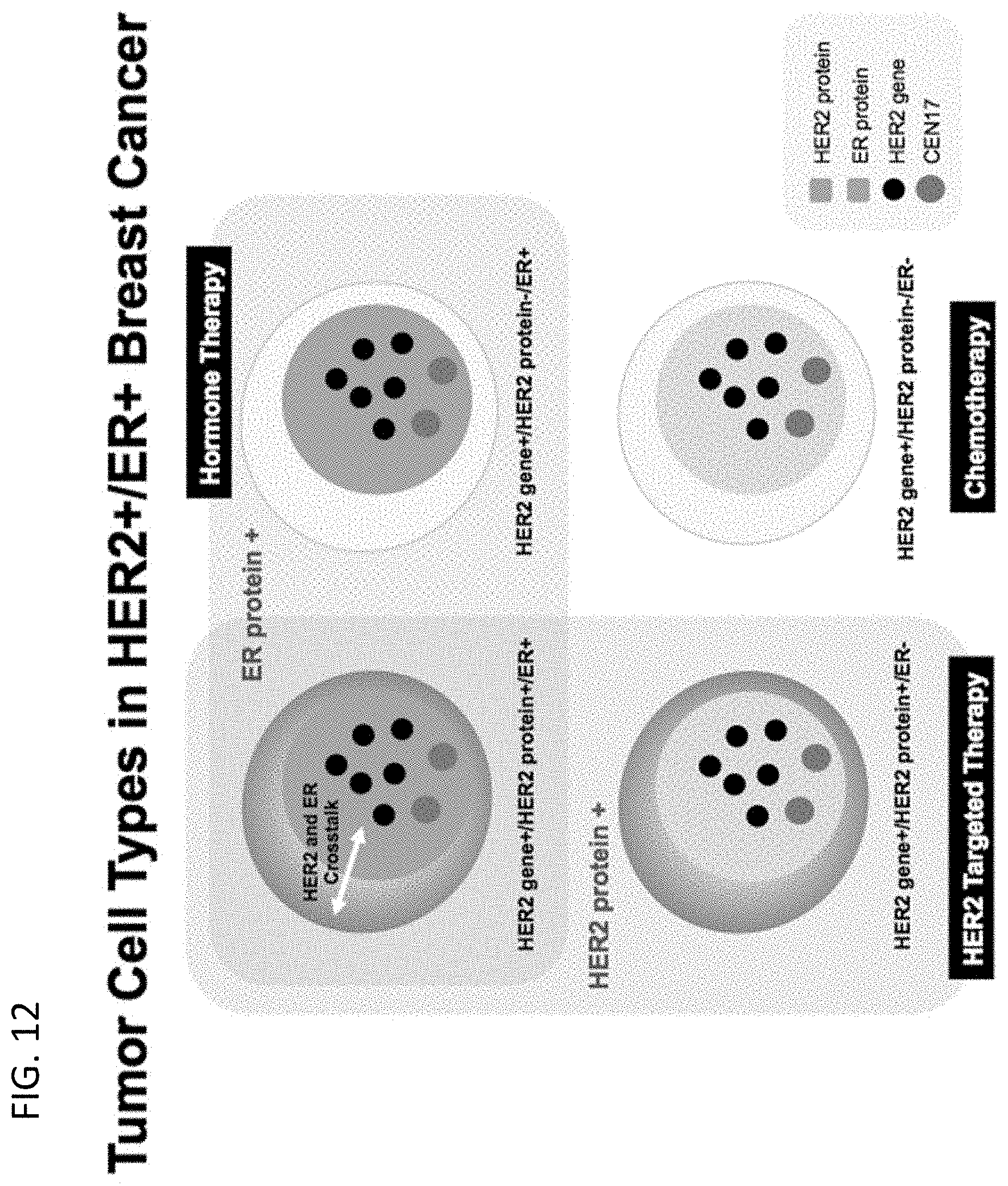
D00014
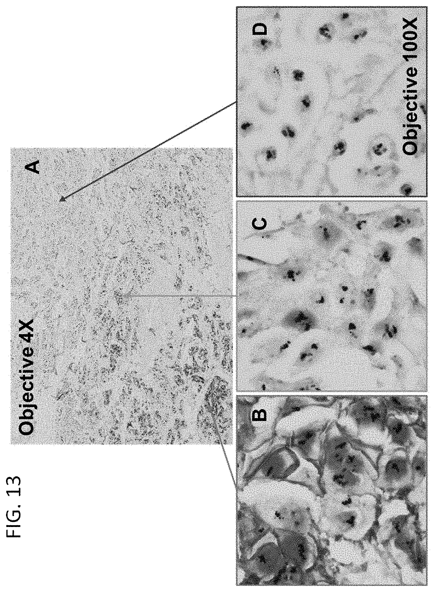
D00015

D00016

D00017

D00018
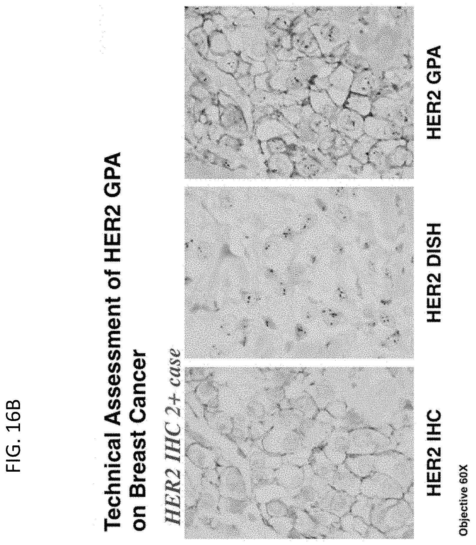
D00019

D00020

D00021

D00022

S00001
XML
uspto.report is an independent third-party trademark research tool that is not affiliated, endorsed, or sponsored by the United States Patent and Trademark Office (USPTO) or any other governmental organization. The information provided by uspto.report is based on publicly available data at the time of writing and is intended for informational purposes only.
While we strive to provide accurate and up-to-date information, we do not guarantee the accuracy, completeness, reliability, or suitability of the information displayed on this site. The use of this site is at your own risk. Any reliance you place on such information is therefore strictly at your own risk.
All official trademark data, including owner information, should be verified by visiting the official USPTO website at www.uspto.gov. This site is not intended to replace professional legal advice and should not be used as a substitute for consulting with a legal professional who is knowledgeable about trademark law.