Cardiomyocyte Maturation Promoter
YOSHIDA; Yoshinori ; et al.
U.S. patent application number 17/042635 was filed with the patent office on 2021-01-14 for cardiomyocyte maturation promoter. This patent application is currently assigned to Kyoto University. The applicant listed for this patent is Kyoto University, Takeda Pharmaceutical Company Limited. Invention is credited to Shigeru KONDO, Kenji MIKI, Yoshinori YOSHIDA.
| Application Number | 20210009956 17/042635 |
| Document ID | / |
| Family ID | 1000005165115 |
| Filed Date | 2021-01-14 |

View All Diagrams
| United States Patent Application | 20210009956 |
| Kind Code | A1 |
| YOSHIDA; Yoshinori ; et al. | January 14, 2021 |
CARDIOMYOCYTE MATURATION PROMOTER
Abstract
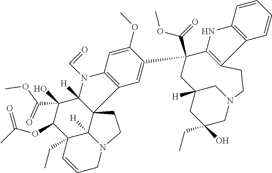
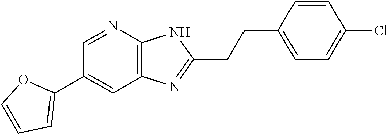
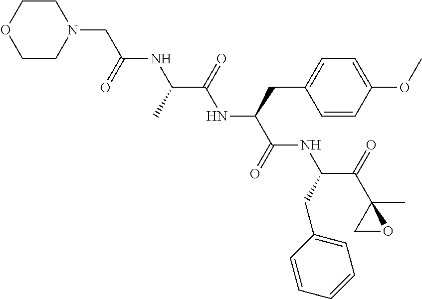
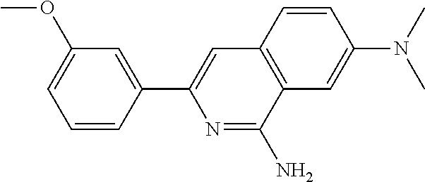
The present invention provides a cardiomyocyte maturation promoter. The present invention provides a cardiomyocyte maturation promoter comprising one or more compounds selected from 2-methoxy-5-((Z)-2-(3,4,5-trimethoxyphenyl)vinyl)phenol, (1-ethyl-1H-benzotriazol-5-yl)methyl (2-(2-methoxy-4-methylphenyl)-4-methyl-1,3-thiazol-5-yl)carbamate, (2'beta)-22-oxovincaleukoblastine, 2-(2-(4-chlorophenyl)ethyl)-6-(2-furyl)-3H-imidazo[4,5-b]pyridine, 4,5-anhydro-1,2-dideoxy-4-methyl-2-((N-(morpholin-4-ylacetyl)-L-alanyl-O-- methyl-L-tyrosyl)amino)-1-phenyl-L-threo-pent-3-ulose, 3-(3-methoxyphenyl)-N7,N7-dimethylisoquinoline-1,7-diamine, methyl 4-(2-benzylbenzoyl)-2,5-dimethyl-1H-pyrrole-3-carboxylate, 2'-(4-aminophenyl)-1H,1'H-2,5'-bibenzimidazol-5-amine, and salts thereof.
| Inventors: | YOSHIDA; Yoshinori; (Kyoto, JP) ; MIKI; Kenji; (Kyoto, JP) ; KONDO; Shigeru; (Kanagawa, JP) | ||||||||||
| Applicant: |
|
||||||||||
|---|---|---|---|---|---|---|---|---|---|---|---|
| Assignee: | Kyoto University Sakyo-ku, Kyoto-shi, Kyoto JP Takeda Pharmaceutical Company Limited Chuo-ku, Osaka-shi, Osaka JP |
||||||||||
| Family ID: | 1000005165115 | ||||||||||
| Appl. No.: | 17/042635 | ||||||||||
| Filed: | March 28, 2019 | ||||||||||
| PCT Filed: | March 28, 2019 | ||||||||||
| PCT NO: | PCT/JP2019/013530 | ||||||||||
| 371 Date: | September 28, 2020 |
| Current U.S. Class: | 1/1 |
| Current CPC Class: | C12N 5/0657 20130101; C12N 15/113 20130101 |
| International Class: | C12N 5/077 20060101 C12N005/077; C12N 15/113 20060101 C12N015/113 |
Foreign Application Data
| Date | Code | Application Number |
|---|---|---|
| Mar 30, 2018 | JP | 2018-069871 |
Claims
1. A cardiomyocyte maturation promoter comprising at least one compound selected from 2-methoxy-5-((Z)-2-(3,4,5-trimethoxyphenyl)vinyl)phenol, (1-ethyl-1H-benzotriazol-5-yl)methyl (2-(2-methoxy-4-methylphenyl)-4-methyl-1,3-thiazol-5-yl)carbamate, (2'beta)-22-oxovincaleukoblastine, 2-(2-(4-chlorophenyl)ethyl)-6-(2-furyl)-3H-imidazo[4,5-b]pyridine, 4,5-anhydro-1,2-dideoxy-4-methyl-2-((N-(morpholin-4-ylacetyl)-L-alanyl-O-- methyl-L-tyrosyl)amino)-1-phenyl-L-threo-pent-3-ulose, 3-(3-methoxyphenyl)-N7,N7-dimethylisoquinoline-1,7-diamine, methyl 4-(2-benzylbenzoyl)-2,5-dimethyl-1H-pyrrole-3-carboxylate, 2'-(4-aminophenyl)-1H,1'H-2,5'-bibenzimidazol-5-amine, and salts thereof.
2. A method for preparing a mature cardiomyocyte, which comprises a step of culturing an immature cardiomyocyte in the presence of the cardiomyocyte maturation promoter according to claim 1.
3. A mature cardiomyocyte prepared by the method according to claim 2.
Description
TECHNICAL FIELD
[0001] The present invention relates to a cardiomyocyte maturation promoter, and a preparation method of mature cardiomyocytes.
Background of the Invention
[0002] Cardiac diseases are the leading cause of death in the world. Cardiac transplantation, which is currently the sole therapeutic option for severe heart failure patients, suffers from donor shortage. A potential therapeutic option as an alternative to cardiac transplantation is transplantation of cardiomyocytes derived from pluripotent stem cells (e.g., iPS cells (induced pluripotent stem cells), ES cells (embryonic stem cells), etc.). The alternative option has been desired to be developed promptly. Moreover, cardiomyocytes derived from pluripotent stem cells (e.g., iPS cells, ES cells, etc.) are also required as cells used for drug toxicity tests and cardiac disease model studies.
[0003] Improvement of efficiency and safety is essential to apply iPS cell-derived mature cardiomyocytes to regenerative medicine. Regarding efficiency, there is a problem of low cost efficiency, because the number of cardiomyocytes capable of inducing maturation is small and proteins such as growth factors in a culture medium are very expensive. Regarding safety, there is a problem that cardiomyocytes have a low purity, and proliferative cells other than the cardiomyocytes can be contaminated, so that there is a risk of canceration.
[0004] Moreover, for drug toxicity tests and cardiac disease model studies using cardiomyocytes, it is necessary to collect a large number of mature cardiomyocytes that sufficiently mimic cardiomyocytes in the living body. Cardiomyocytes lose their division potential at the same time as the birth, and regeneration of those cells is very difficult. Because of such properties, a number of studies have been carried out for inducing differentiation of pluripotent stem cells into cardiomyocytes in order to obtain a large number of cardiomyocytes (Patent Document 1, Patent Document 2, Non-patent Document 1, Non-patent Document 2 and Non-patent Document 3).
[0005] However, in general, it is said that cardiomyocytes derived from human pluripotent stem cells stay in an immature stage similar to fetal cardiomyocytes, and that their ion channel function is insufficient compared to adult cardiomyocytes. Thus, for the purpose of screening of drug toxicity and therapeutic agents in relation to ion channels, mature cardiomyocytes are required to be used.
[0006] Therefore, mature cardiomyocytes and a preparation method thereof are required as cells used for cardiomyocyte transplantation and for screening of drug toxicity and therapeutic drug.
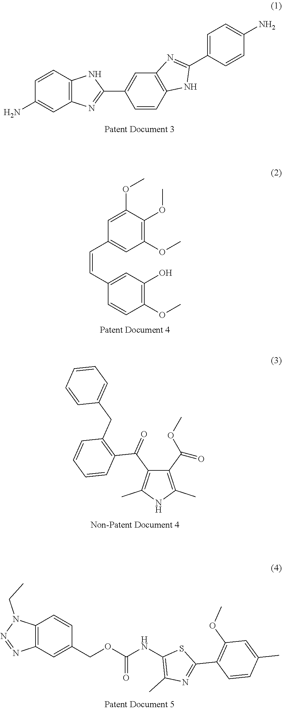
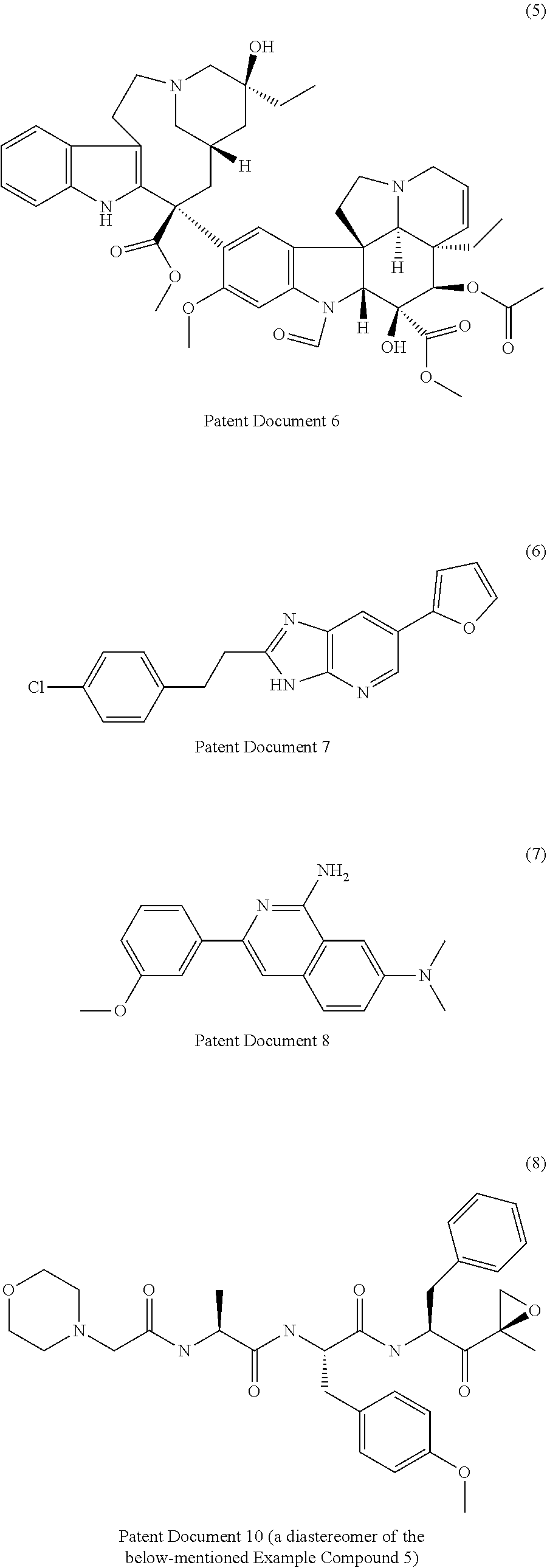
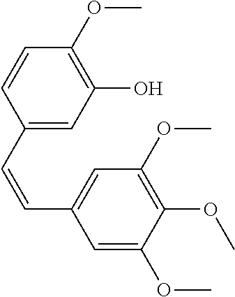
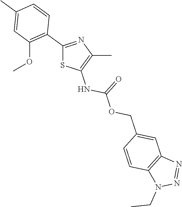
[0007] Patent Documents 3 to 9 and Non-Patent Document 4 disclose the following compounds.
##STR00001## ##STR00002##
[0008] However, none of the documents disclose that the above-mentioned compounds have an activity to promote the maturation of a cardiomyocyte.
[0009] It is possible to obtain mature cardiomyocytes by culturing immature cardiomyocytes for a long period (for example, 1 year or more). However, as a commercial preparation method of mature cardiomyocytes, such method takes too much time and requires expensive medium and medium additive.
[0010] Therefore, development of a cardiomyocyte maturation promoter, which can efficiently prepare mature cardiomyocytes with high purity in a short period at low cost, is still desired.
DOCUMENT LIST
Patent Document
[0011] Patent Document 1: WO 2007/002136 [0012] Patent Document 2: WO 2009/118928 [0013] Patent Document 3: WO 2006/094235 [0014] Patent Document 4: WO 92/16486 [0015] Patent Document 5: WO 2012/002527 [0016] Patent Document 6: WO 02/076402 [0017] Patent Document 7: WO 03/045929 [0018] Patent Document 8: WO 2008/063548 [0019] Patent Document 9: US 2007/0293465
Non-Patent Document
[0019] [0020] Non-Patent Document 1: Yan P, et al, Biochem Biophys Res Commun. 379:115-20 (2009) [0021] Non-Patent Document 2: Laflamme M A, et al, Nat Biotechnol, 25:1015-1024 (2007) [0022] Non-Patent Document 3: Yang L et al, Nature, 453:524-528 (2008) [0023] Non-Patent Document 4: Journal of Medicinal Chemistry, 1993, vol. 36, pp 2739-2744
SUMMARY OF THE INVENTION
Problems to be Solved by the Invention
[0024] The present invention aims to provide a cardiomyocyte maturation promoter, which can efficiently prepare mature cardiomyocytes with high purity in a short period at low cost.
Means of Solving the Problems
[0025] The present inventors have found that the following compounds or salts thereof have an activity to promote the maturation of a cardiomyocyte. As a result of further studies, they have completed the present invention.
[0026] Accordingly, the present invention is as follows.
[1] A cardiomyocyte maturation promoter comprising at least one compound selected from 2-methoxy-5-((Z)-2-(3,4,5-trimethoxyphenyl)vinyl)phenol, (1-ethyl-1H-benzotriazol-5-yl)methyl (2-(2-methoxy-4-methylphenyl)-4-methyl-1,3-thiazol-5-yl)carbamate, (2'beta)-22-oxovincaleukoblastine, 2-(2-(4-chlorophenyl)ethyl)-6-(2-furyl)-3H-imidazo[4,5-b]pyridine, 4,5-anhydro-1,2-dideoxy-4-methyl-2-((N-(morpholin-4-ylacetyl)-L-alanyl-O-- methyl-L-tyrosyl)amino)-1-phenyl-L-threo-pent-3-ulose, 3-(3-methoxyphenyl)-N7,N7-dimethylisoquinoline-1,7-diamine, methyl 4-(2-benzylbenzoyl)-2,5-dimethyl-1H-pyrrole-3-carboxylate, 2'-(4-aminophenyl)-1H,1'H-2,5'-bibenzimidazol-5-amine, and salts thereof. [2] A method for preparing a mature cardiomyocyte, which comprises a step of culturing an immature cardiomyocyte in the presence of the cardiomyocyte maturation promoter of the above-mentioned [1]. [3] A mature cardiomyocyte prepared by the method of the above-mentioned [2].
Effect of the Invention
[0027] Since the above-mentioned compounds or salts thereof have an action of maturing cardiomyocytes, they are useful as a cardiomyocyte maturation promoter. The method for promoting cardiomyocyte maturation using the above-mentioned compound matures immature cardiomyocytes in a short period at low cost, as compared with a method for culturing immature cardiomyocytes for a long period.
BRIEF DESCRIPTION OF THE DRAWINGS
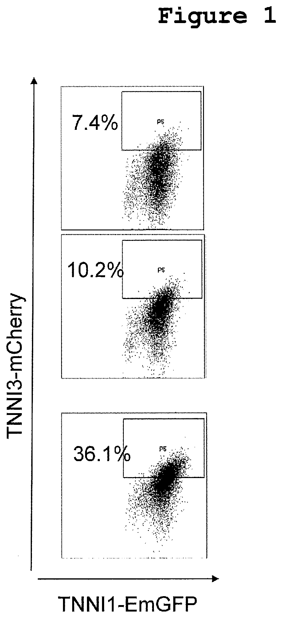
[0028] FIG. 1 shows the results of mCherry expression analysis by a flow cytometry in Experimental Example 2. The FIGURES show the results in cases of control (no addition), control (DMSO addition) and addition of the compound of Example No. 2, in order from the top.
DETAILED DESCRIPTION OF THE INVENTION
[0029] The cardiomyocyte maturation promoter of the present invention comprises one or more compounds selected from the following compounds: [0030] 2-methoxy-5-((Z)-2-(3,4,5-trimethoxyphenyl)vinyl)phenol (Example No. 1), [0031] (1-ethyl-1H-benzotriazol-5-yl)methyl (2-(2-methoxy-4-methylphenyl)-4-methyl-1,3-thiazol-5-yl)carbamate (Example No. 2), [0032] (2'beta)-22-oxovincaleukoblastine (Example No. 3), 2-(2-(4-chlorophenyl)ethyl)-6-(2-furyl)-3H-imidazo[4,5-b]pyridine (Example No. 4), [0033] 4,5-anhydro-1,2-dideoxy-4-methyl-2-((N-(morpholin-4-ylacetyl)-L-alanyl-O-- methyl-L-tyrosyl)amino)-1-phenyl-L-threo-pent-3-ulose (Example No. 5), [0034] 3-(3-methoxyphenyl)-N7,N7-dimethylisoquinoline-1,7-diamine (Example No. 6), [0035] methyl 4-(2-benzylbenzoyl)-2,5-dimethyl-1H-pyrrole-3-carboxylate (Example No. 7), [0036] 2'-(4-aminophenyl)-1H,1'H-2,5'-bibenzimidazol-5-amine (Example No. 8), [0037] and salts thereof.
[0038] When the above-mentioned compound is in the form of a salt, the salt is preferably a pharmacologically acceptable salt, and examples of such salt include a salt with inorganic base, a salt with organic base, a salt with inorganic acid, a salt with organic acid, a salt with basic or acidic amino acid and the like.
[0039] Preferable examples of the salt with inorganic base include alkali metal salts such as sodium salt, potassium salt and the like, alkaline earth metal salts such as calcium salt, magnesium salt and the like, aluminum salt, ammonium salt and the like.
[0040] Preferable examples of the salt with organic base include salts with trimethylamine, triethylamine, pyridine, picoline, ethanolamine, diethanolamine, triethanolamine, tromethamine[tris(hydroxymethyl)methylamine], tert-butylamine, cyclohexylamine, benzylamine, dicyclohexylamine, N,N-dibenzylethylenediamine and the like.
[0041] Preferable examples of the salt with inorganic acid include salts with hydrochloric acid, hydrobromic acid, nitric acid, sulfuric acid, phosphoric acid and the like.
[0042] Preferable examples of the salt with organic acid include salts with formic acid, acetic acid, trifluoroacetic acid, phthalic acid, fumaric acid, oxalic acid, tartaric acid, maleic acid, citric acid, succinic acid, malic acid, methanesulfonic acid, benzenesulfonic acid, p-toluenesulfonic acid and the like.
[0043] Preferable examples of the salt with basic amino acid include salts with arginine, lysine, ornithine and the like. Preferable examples of the salt with acidic amino acid include salts with aspartic acid, glutamic acid and the like.
[0044] The cardiomyocyte maturation promoter of the present invention may comprise one or more kind(s) of the above-mentioned compounds or salts thereof.
[0045] The above-mentioned compounds or salts thereof can be each produced according to a method known per se, for example, according to the methods described in Patent Documents 3 to 9 and Non-Patent Document 4 or methods analogous thereto.
[0046] The above-mentioned compounds may be a hydrate, a non-hydrate, a solvate or a non-solvate.
[0047] Furthermore, the above-mentioned compounds may be labeled with or substituted by an isotope (e.g., .sup.2H, .sup.3H, .sup.11C, .sup.14C, .sup.18F, .sup.35S, .sup.125I etc.) and the like.
[0048] Deuteriums conversion form wherein .sup.1H is converted to .sup.2H(D) are also encompassed in the above-mentioned compounds.
[0049] Tautomers are also encompassed in the above-mentioned compounds.
[0050] The above-mentioned compounds may be a pharmaceutically acceptable cocrystal or cocrystal salt. The cocrystal or cocrystal salt means a crystalline substance constituted with two or more special solids at room temperature, each having different physical properties (e.g., structure, melting point, melting heat, hygroscopicity, solubility and stability). The cocrystal or cocrystal salt can be produced by a cocrystallization method known per se.
[0051] The cardiomyocyte maturation promoter of the present invention can be used as it is or by mixing with a pharmacologically acceptable carrier, and formulating the mixture according to a method known per se.
[0052] Since the above-mentioned compounds or salts thereof have an excellent activity to promote the maturation of a cardiomyocyte, they are useful as a cardiomyocyte maturation promoter. Therefore, by culturing immature cardiomyocytes in the presence of the above-mentioned compound or a salt thereof, mature cardiomyocytes can be prepared in a shorter period than when culturing in only medium components.
[0053] As used herein, the term "pluripotency" means ability to differentiate into various tissues or cells having different morphologies and/or functions, and to differentiate into any series of cells of three germ layers. The "pluripotency" cannot differentiate into blastodisc. Therefore, the "pluripotency" is distinguished from the term "totipotency" which can differentiate into every tissue of the living body including blastodisc, in that it has no ability to form an individual.
[0054] As used herein, the term "multipotency" means ability to differentiate into plural limited numbers of series of cells. For example, mesenchymal stem cells, hematopoietic stem cells and neural stem cells are multipotent, but not pluripotent.
[0055] As used herein, examples of the "stem cells" include pluripotent stem cells.
[0056] The "cardiomyocytes" to which the cardiomyocyte maturation promoter of the present invention is applied is not particularly limited, and they are preferably derived from a mammal (e.g., mouse, rat, hamster, rabbit, cat, dog, cow, sheep, pig, monkey, human), more preferably derived from human.
[0057] The cardiomyocytes whose maturation can be promoted by the cardiomyocyte maturation promoter of the present invention are not particularly limited as long as it can be determined that they are in an immature stage based on the below-mentioned marker expression levels, morphologies and structures (e.g., sarcomere, mitochondria), properties (e.g., pulsatility, electrophysiological maturity) and the like.
[0058] The cardiomyocytes whose maturation can be promoted by the cardiomyocyte maturation promoter of the present invention may also be cells prepared by differentiation induction from pluripotent stem cells, or immature cardiomyocytes isolated from a living body (e.g., cardiomyocytes derived from a mouse or rat fetus or newborn).
[0059] Examples of the pluripotent stem cells include, but are not limited to, embryonic stem (ES) cells, germline stem cells ("GS cells"), embryonic germ cells ("EG cells"), induced pluripotent stem (iPS) cells, embryonic stem cells derived from a cloned embryo obtained by nuclear transfer (ntES cells), and the like. The pluripotent stem cells are preferably ES cells, iPS cells or ntES cells.
(A) Embryonic Stem Cells
[0060] ES cells are stem cells established from the inner cell mass of an early embryo (for example, blastocyst) of a mammal such as human, mouse and the like, which cells have pluripotency and growth ability by self-renewal.
[0061] ES cells are embryo-derived stem cells originated from the inner cell mass of a blastocyst, which is the embryo formed following the 8-cell stage and the morula stage of a fertilized egg. ES cells have ability to differentiate into any cells constituting an adult, that is, the so-called pluripotency of differentiation, and growth ability by self-renewal. ES cells were discovered in mouse in 1981 (M. J. Evans and M. H. Kaufman (1981), Nature 292: 154-156), and this was followed by establishment of ES cell lines of primates such as human, monkey and the like (J. A. Thomson et al. (1998), Science 282: 1145-1147; J. A. Thomson et al. (1995), Proc. Natl. Acad. Sci. USA, 92: 7844-7848; J. A. Thomson et al. (1996), Biol. Reprod., 55: 254-259; J. A. Thomson and V. S. Marshall (1998), Curr. Top. Dev. Biol., 38: 133-165).
[0062] ES cells can be established by removing the inner cell mass from the blastocyst of a fertilized egg of a subject animal, followed by culturing the inner cell mass on feeder fibroblasts. The cells can be maintained by subculture using a culture medium supplemented with a substance(s) such as leukemia inhibitory factor (LIF), basic fibroblast growth factor (bFGF), and the like. Methods of establishment and maintenance of human and monkey ES cells are described in, for example, U.S. Pat. No. 5,843,780 B; Thomson J A, et al. (1995), Proc Natl. Acad. Sci. USA. 92: 7844-7848; Thomson J A, et al. (1998), Science. 282: 1145-1147; H. Suemori et al. (2006), Biochem. Biophys. Res. Commun., 345: 926-932; M. Ueno et al. (2006), Proc. Natl. Acad. Sci. USA, 103: 9554-9559; H. Suemori et al. (2001), Dev. Dyn., 222: 273-279; H. Kawasaki et al. (2002), Proc. Natl. Acad. Sci. USA, 99: 1580-1585; and Klimanskaya I, et al. (2006), Nature. 444: 481-485.
[0063] With regard to culture medium for preparation of ES cells, human ES cells can be maintained, for example, using DMEM/F-12 culture medium supplemented with 0.1 mM 2-mercaptoethanol, 0.1 mM non-essential amino acids, 2 mM L-glutamic acid, 20% KSR and 4 ng/ml bFGF at 37.degree. C. under a moist atmosphere of 2% CO.sub.2/98% air (O. Fumitaka et al. (2008), Nat. Biotechnol., 26:215-224). ES cells are required to be subcultured every 3 to 4 days, and the subculture can be carried out using, for example, 0.25% trypsin and 0.1 mg/ml collagenase IV in PBS containing 1 mM CaCl.sub.2 and 20% KSR.
[0064] Selection of ES cells can be generally carried out by the Real-Time PCR method using, as an index, expression of a gene marker such as alkaline phosphatase, Oct-3/4, Nanog and the like. In particular, for selection of human ES cells, expression of a gene marker such as OCT-3/4, NANOG, ECAD and the like can be used as an index (E. Kroon et al. (2008), Nat. Biotechnol., 26: 443-452).
[0065] With regard to ES cells, as mouse ES cells, various mouse ES cell lines established by inGenious targeting laboratory, Riken (Institute of Physical and Chemical Research) etc., are available, and, as human ES cells, various human ES cell lines established by NIH, Riken, Kyoto University, Cellartis etc., are available. For examples, examples of human ES cell lines include line CHB-1-CHB-12, line RUES1, line RUES2, line HUES1-HUES28 etc., of NIH, line H1 and line H9 of WisCell Research, and line KhES-1, line KhES-2, line KhES-3, line KhES-4, line KhES-5, line SSES1, line SSES2, line SSES3 etc., of Riken. Alternatively, clinical-grade cell lines, research or clinical cell lines prepared from these cell lines, and the like may be used.
(B) Germline Stem Cells
[0066] Germline stem cells are pluripotent stem cells derived from testis, and play a role as the origin for spermatogenesis. Similarly to ES cells, these cells can be induced to differentiate into various series of cells, and, for example, have a property to enable preparation of a chimeric mouse by transplantation of the cells to a mouse blastocyst (M. Kanatsu-Shinohara et al. (2003) Biol. Reprod., 69: 612-616; K. Shinohara et al. (2004), Cell, 119: 1001-1012). Germline stem cells are capable of self-renewal in a culture medium containing glial cell line-derived neurotrophic factor (GDNF), and, by repeating their subculture under the same culture conditions as those for ES cells, germline stem cells can be obtained (Masanori Takehashi et al. (2008), Experimental Medicine, 26(5) (extra edition): 41-46, Yodosha (Tokyo, Japan)).
(C) Embryonic Germ Cells
[0067] Embryonic germ cells are established from fetal primordial germ cells and have pluripotency similarly to ES cells. They can be established by culturing primordial germ cells in the presence of substances such as LIF, bFGF, stem cell factor and the like (Y. Matsui et al. (1992), Cell, 70: 841-847; J. L. Resnick et al. (1992), Nature, 359: 550-551).
(D) Induced Pluripotent Stem Cells
[0068] The "induced pluripotent stem cell (iPSC)" refers to a cell obtained by introducing a certain factor (nuclear reprogramming factor) into a mammalian somatic cell or an undifferentiated stem cell to perform reprogramming. At present, there are various "induced pluripotent stem cells", and those that can also be used are as follows: an iPSC established by Yamanaka, et al., by introducing four factors, Oct3/4, Sox2, Klf4 and c-Myc into a mouse fibroblast (Takahashi K, Yamanaka S., Cell, (2006) 126: 663-676); as well as an iPSC derived from a human cell established by introducing the same four factors to a human fibroblast (Takahashi K, Yamanaka S., et al. Cell, (2007) 131: 861-872.); a Nanog-iPS cell established by introducing the four factors described above and then performing screening with the expression of Nanog as an index (Okita, K., Ichisaka, T., and Yamanaka, S. (2007). Nature 448, 313-317.); an iPS cell prepared with a method in which c-Myc is not included (Nakagawa M, Yamanaka S., et al. Nature Biotechnology, (2008) 26, 101-106); and an iPS cell established by introducing six factors with a virus free method (Okita K et al. Nat. Methods 2011 May; 8(5):409-12, Okita K et al. Stem Cells. 31(3):458-66). In addition, an induced pluripotent stem cell prepared by Thomson, et al., which is established by introducing four factors, OCT3/4, SOX2, NANOG and LIN28 (Yu J., Thomson J A. et al., Science (2007) 318: 1917-1920.), an induced pluripotent stem cell prepared by Daley, et al. (Park I H, Daley G Q. et al., Nature (2007) 451: 141-146), and an induced pluripotent stem cell prepared by Sakurada, et al. (Japanese Patent Application Laid-Open No. 2008-307007), etc. can also be used.
[0069] Besides, any of the induced pluripotent stem cells known so in the art described in all published papers (for example, Shi Y., Ding S., et al., Cell Stem Cell, (2008) Vol 3, Issue 5, 568-574; Kim J B., Scholer H R., et al., Nature, (2008) 454, 646-650; Huangfu D., Melton, D A., et al., Nature Biotechnology, (2008) 26, No 7, 795-797), or patents (for example, Japanese Patent Application Laid-Open No. 2008-307007, Japanese Patent Application Laid-Open No. 2008-283972, US 2008-2336610, US 2009-047263, WO 2007-069666, WO 2008-118220, WO 2008-124133, WO 2008-151058, WO 2009-006930, WO 2009-006997, WO 2009-007852) can also be used.
[0070] As induced pluripotent cell lines, various iPSC lines established by NIH, Riken, Kyoto University, etc. are available. For example, examples of human iPSC lines include line HiPS-RIKEN-1A, line HiPS-RIKEN-2A, line HiPS-RIKEN-12A and line Nips-B2 of Riken, line 253G1, line 201B7, line 409B2, line 454E2, line 606A1, line 610B1 and line 648A1 of Kyoto University, and the like. Alternatively, clinical-grade cell lines provided by Kyoto University, Cellular Dynamics International etc., research or clinical cell lines prepared from these cell lines, and the like may be used.
[0071] The term "somatic cells" used herein means any animal cells (preferably cells of mammals including human) excluding germ-line cells and totipotent cells such as eggs, oocytes, ES cells and the like. Examples of the somatic cells include, but are not limited to, any of fetal somatic cells, neonatal somatic cells, and mature, healthy or diseased somatic cells, as well as any of primary cultured cells, subcultured cells, and established cell lines. Specific examples of the somatic cells include (1) tissue stem cells (somatic stem cells) such as neural stem cells, hematopoietic stem cells, mesenchymal stem cells, dental pulp stem cells and the like; (2) tissue progenitor cells; (3) differentiated cells such as lymphocytes, epithelial cells, endothelial cells, muscle cells, fibroblasts (skin cells etc.), hair cells, hepatic cells, gastric mucosal cells, enterocytes, spleen cells, pancreatic cells (pancreatic exocrine cells etc.), brain cells, lung cells, kidney cells, adipocytes and the like, and the like.
[0072] In the present invention, the mammalian individual from which somatic cells are collected is not limited, and preferably human. In cases where the obtained iPS cells are to be used for human regenerative medicine, it is especially preferred to collect somatic cells from the patient himself or another person having the same or substantially the same human leukocyte antigen (HLA) type in view of prevention of the rejection reaction. Here, "substantially the same" HLA type means that the HLA type is matching to an extent which allows survival of transplanted cells by use of an immunosuppressant(s) and/or the like when cells obtained by differentiation induction from iPS cells derived from the somatic cells are transplanted to the patient. For example, it means that the person has the same major HLAs (for example, at the three gene loci HLA-A, HLA-B and HLA-DR) as those of the patient (the same applies hereinafter). On the other hand, in cases where the cells are not administered (transplanted) to human, for example, in a method for testing toxicity of a candidate drug to cardiomyocytes, the origin of the somatic cells to be used as the source of the iPS cells is not limited. In cases where the iPS cells are used as the source of cells for screening for evaluation of drug sensitivity or side effects of a patient, it is preferred to collect somatic cells from the patient himself or another person who has the same genetic polymorphism(s) associated with the drug sensitivity or the side effects.
(E) ES Cells Derived from Cloned Embryo Obtained by Nuclear Transfer
[0073] ntES cells are ES cells derived from a cloned embryo prepared by the nuclear transfer technique, and have almost the same properties as those of ES cells derived from fertilized eggs (T. Wakayama et al. (2001), Science, 292: 740-743; S. Wakayama et al. (2005), Biol. Reprod., 72: 932-936; J. Byrne et al. (2007), Nature, 450: 497-502). That is, an ntES (nuclear transfer ES) cell is an ES cell established from the inner cell mass of a blastocyst derived from a cloned embryo obtained by replacement of the nucleus of an unfertilized egg with the nucleus of a somatic cell. For preparation of ntES cells, the combination of the nuclear transfer technique (J. B. Cibelli et al. (1998), Nature Biotechnol., 16: 642-646) and the ES cell preparation technique (described above) is employed (Sayaka Wakayama et al. (2008), Experimental Medicine 26(5) (extra edition), pp. 47-52). In nuclear transfer, reprogramming can be achieved by injecting the nucleus of a somatic cell into a mammalian enucleated unfertilized egg, and culturing the resultant for several hours.
[0074] As used herein, the term "cardiomyocyte" means a cell expressing at least one marker gene selected from the group consisting of cardiac troponin (cTNT), .alpha.MHC (.alpha. myosin heavy chain, MYH6) and .beta.MHC (MYH7). Examples of cTNT include NCBI accession number NM_000364 in a case of human, and NM_001130176 in a case of mouse. Examples of .alpha.MHC include NCBI accession number NM_002471 in a case of human, and NM_001164171 in a case of mouse. Examples of .beta.MHC include NCBI accession number NM_000257 in a case of human, and NM_080728 in a case of mouse.
[0075] It is known that an isoform switch occurs in which the expression of troponin I1 (TNNI1) is decreased and the expression of troponin I3 (TNNI3) is increased, as cardiomyocytes mature (Fikru B. Bedada, (2014) 3(4): 594-605.). As used herein, "cardiomyocyte is matured (mature)" means that at least TNNI3 expression is increased.
[0076] With regard to methods of inducing differentiation of pluripotent stem cells into immature cardiomyocytes, cardiomyocytes can be prepared from pluripotent stem cells by, for example, a method reported by Laflamme M A et al. (Laflamme M A & Murry C E, Nature 2011, Review).
[0077] Other examples of the method include, but are not limited to, a method in which cardiomyocytes are prepared by formation of cell mass (embryoid bodies) by suspension culture of induced pluripotent stem cells, a method in which cardiomyocytes are prepared in the presence of a substance that suppresses bone morphogenic protein (BMP) signaling (WO 2005/033298), a method in which cardiomyocytes are prepared by addition of Activin A and BMP in this order (WO 2007/002136), a method in which cardiomyocytes are prepared in the presence of a substance that promotes activation of the canonical Wnt signaling pathway (WO 2007/126077), a method in which Flk/KDR-positive cells are isolated from induced pluripotent stem cells, followed by preparation of cardiomyocytes in the presence of cyclosporin A (WO 2009/118928), and the like.
[0078] In addition, a method of inducing differentiation into cardiomyocytes using cytokine by embryoid body formation (Yang L, et al., Human cardiovascular progenitor cells develop from a KDR+ embryonic-stem-cell-derived population., Nature., 2008 May 22; 453(7194):524-8), a method of inducing differentiation into cardiomyocytes by adhesion culture under cytokine-free condition (Lian X, et al., Robust cardiomyocyte differentiation from human pluripotent stem cells via temporal modulation of canonical Wnt signaling., Proc Natl Acad Sci USA., 2012 Jul. 3; 109(27):E1848-57), a method of inducing differentiation into cardiomyocytes by combination use of adhesion culture and suspension culture under cytokine-free condition (Minami I, et al., A small molecule that promotes cardiac differentiation of human pluripotent stem cells under defined, cytokine- and xeno-free conditions., Cell Rep., 2012 Nov. 29; 2(5):1448-60) and the like are also suggested.
[0079] The medium to obtain immature cardiomyocytes from pluripotent stem cells is not particularly limited, and mediums known per se can be used. Examples thereof include IMDM medium, Medium 199 medium, Eagle's Minimum Essential Medium (EMEM) medium, .alpha.MEM medium, Dulbecco's modified Eagle's Medium (DMEM) medium, Ham's F12 medium, RPMI 1640 medium, Fischer's medium, Neurobasal Medium (Life Technologies), StemPro34 (Invitrogen), medium after removal of solution C from StemFit AK02 (AJINOMOTO AK02, 400 mL of solution A and 100 mL of B solution, total 500 mL), Essential 6 medium (Thermo Fischer Scientific) and mixed media thereof, and the like.
[0080] Additives known per se can be added to these mediums, depending on cells and cultural conditions. For example, the medium may contain serum, or may be serum-free. If necessary, the medium may contain one or more of serum replacements such as albumin, transferrin, Knockout Serum Replacement (KSR) (serum replacement for FBS during ES cell culture), N2 supplement (Invitrogen), B27 supplement (Invitrogen), fatty acids, insulin, collagen precursors, trace elements, 2-mercaptoethanol, 1-thiolglycerol and the like, and may also contain one or more of substances such as lipids, amino acids, L-glutamine, Glutamax (Invitrogen), non-essential amino acids, vitamins, growth factors, low-molecular-weight compounds, antibiotics, antioxidants, pyruvic acid, buffers, inorganic salts and the like. Among them, the preferred medium is StemPro34 or a medium after removal of solution C from StemFit AK02, each containing transferrin, 1-thiolglycerol, L-glutamine and ascorbic acid, or RPMI1640 containing B27 supplement.
[0081] A combination of activin A, BMP4 and bFGF, or CHIR99021 is used as an initial additive for differentiation induction into cardiomyocytes using the above-mentioned medium.
[0082] In the case of the combination of activin A, BMP4 and bFGF, the use concentrations of these additives are preferably 1 ng/ml to 100 ng/ml for activin A, 1 ng/ml to 100 ng/ml for BMP4, and 1 ng/ml to 100 ng/ml for bFGF, more preferably 6 ng/ml for activin A, 10 ng/ml for BMP4, and 5 ng/ml for bFGF.
[0083] In the case of CHIR99021, the use concentration is preferably 100 nM to 100 .mu.M, more preferably 4 to 6 .mu.M.
[0084] After addition of the above-mentioned additive, by adding vascular endothelial growth factor (VEGF) and a Wnt inhibitor to the medium, the pluripotent stem cells can be differentiated into cardiomyocytes.
[0085] The concentration of the VEGF to be used is preferably 1 to 100 ng/ml, more preferably 10 ng/ml.
[0086] Examples of the Wnt inhibitor include DKK1 protein (e.g., NCBI accession number:NM_012242 in a case of human), sclerostin (e.g., NCBI accession number:NM_025237 in a case of human), IWR-1 (Merck Millipore), IWP-2 (Sigma-Aldrich), IWP-3 (Sigma-Aldrich), IWP-4 (Sigma-Aldrich), PNU-74654 (Sigma-Aldrich), XAV939 (Sigma-Aldrich) and derivatives thereof, and the like. Among them, IWP-3, IWP-4 and IWR-1 are preferably used.
[0087] The concentration of the Wnt inhibitor to be used is not particularly limited as long as it inhibits Wnt, and it is preferably 1 nM to 50 .mu.M, particularly preferably 1 to 2 .mu.M.
[0088] The culture period for preparation of immature cardiomyocytes from pluripotent stem cells is, for example, 5 to 365 days, preferably 5 to 100 days, more preferably 5 to 60 days, further more preferably 5 to 40 days, still more preferably 5 to 30 days.
[0089] The amount of the compound to be used as the cardiomyocyte maturation promoter of the present invention is not particularly limited, and it is, for example, 0.01 to 100 .mu.M, preferably 0.1 to 30 .mu.M, more preferably 1 to 10 .mu.M, per 1.times.10.sup.4 to 1.times.10.sup.7 cell of immature cardiomyocytes.
[0090] As an index of cardiomyocyte maturity, expression level of marker for cardiomyocyte maturation, morphology and structure (e.g., sarcomere, mitochondria), property (e.g., pulsatility, electrophysiological maturity) and the like can be used. These indexes can be confirmed by a method known per se.
[0091] For example, the expression level of marker for cardiomyocyte maturation can be analyzed by measuring the expression level of marker gene using PCR; analyzed by the expression level of marker protein using western blot and the like; or analyzed by fluorescent label using microscope or flow cytometry. The index of electrophysiological maturity can be analyzed by the depth of resting membrane potential using patch clamping technique, and the like. The index of sarcomere microstructure or mitochondria can be observed by electronic microscope; analyzed by fluorescent label using microscope or flow cytometry; or function-analyzed by extracellular flux analyzer, and the like.
[0092] In one embodiment, the mature cardiomyocytes obtained by using the cardiomyocyte maturation promoter of the present invention can be used in cardiac regenerative medicine. For example, a composition comprising cell mass of the cardiomyocytes prepared by the method of the present invention can be administered to the heart of a patient suffering from cardiac disease. Specifically, the cardiomyocytes obtained by the method of the present invention may be directly transplanted into the heart of a patient suffering from cardiac disease as a cell suspension, or in the form of a cardiomyocyte sheet (single layer or multi layers). For example, WO 2012/133945, WO 2013/137491, WO 2014/192909 and WO 2016/076368 are referred to for the preparation method of cardiomyocyte sheets.
[0093] In another embodiment, the cardiomyocytes obtained by using the cardiomyocyte maturation promoter of the present invention can also be used for drug screening or drug cardiotoxicity evaluation for the treatment of cardiac disease, since the cardiomyocytes are homogeneously mature. For example, by administering a test drug to the cardiomyocytes so obtained by the method of the present invention, and then measuring the response of the cardiomyocytes, the effect and toxicity of the test drug can be evaluated.
[0094] In another embodiment, the cardiomyocytes with reduced automaticity, which are obtained by using the cardiomyocyte maturation promoter of the present invention, can be used in cardiac regenerative medicine.
EXAMPLES
Experimental Example 1
[0095] For detection of cardiomyocyte maturation, double knock-in human iPS cell lines, in which reporter protein sequences, EmGFP (Sequence Number 1) and mCherry (Sequence Number 2) were inserted into gene loci for TNNI1 and TNNI3, respectively, were prepared (the human iPS cells were prepared from episomal vector (loaded gene; OCT3/4, KLF4, SOX2, L-MYC, LIN28, mouse p53DD) using PBMC (LP_167, Sample ID:20130318) purchased from CTL (reference literature; Okita K, et al. Stem Cells. 2012 Nov. 29. doi: 10.1002/stem.1293)).
[0096] The maintenance culture of the above-mentioned reporter iPS cell lines was carried out by a conventional method (Okita K, et al. Stem Cells. 2012 Nov. 29. doi: 10.1002/stem.1293).
[0097] For differentiation induction into cardiomyocytes, the reporter iPS cell lines were treated with 0.5.times.TrypLE select (Life Technologies, diluted in 1/2 with 0.5 mM EDTA/PBS) for 4 to 5 minutes, and the cells were exfoliated using a cell scraper (IWAKI), and then dissociated into the single cell by pipetting. The medium was removed by centrifugation (1,000 rpm, 5 min), and the obtained cells were seeded on a bioreactor (ABLE) by 1.times.10.sup.7 cells per 30 mL of the bioreactor, and then cultured in a medium prepared by adding 1% L-glutamine, transferrin 150 .mu.g/mL, ascorbic acid 50 .mu.g/mL (Sigma), monothioglycerol 4.times.10.sup.-4 M, 10 .mu.M Rock inhibitor (Y-27632), 2 ng/mL BMP4 (R&D) and 0.5% Matrigel (Growth Factor Reduced) to a medium after removal of solution C from StemFit AK02 (AJINOMOTO AK02, 400 mL of solution A and 100 mL of B solution, total 500 mL), at 37.degree. C., under 5% oxygen condition (55 rpm, stirred suspension culture method) to form embryoid bodies (0 day).
[0098] Next day (1st day), 9 .mu.L (final concentration 3 ng/mL) of .mu.g/mL activin A, 15 .mu.L (final concentration 5 ng/mL) of 10 .mu.g/mL bFGF and 24 .mu.L (final concentration 10 ng/mL) of 10 .mu.g/mL BMP4 were added to the bioreactor, and the cells were cultured at 37.degree. C. for additional 2 days, under 5% oxygen condition.
[0099] Then (3rd day), the obtained embryoid bodies were collected in a 50 mL centrifuge tube, and then subjected to centrifugation (200 g, 1 min). The medium was removed, and the embryoid bodies were cultured in a medium prepared by adding 1% L-glutamine, transferrin 150 .mu.g/mL, ascorbic acid 50 .mu.g/mL (Sigma), monothioglycerol 4.times.10.sup.-4 M, 10 ng/mL VEGF, 1 .mu.M IWP-3, 0.6 .mu.M dorsomorphin and 5.4 .mu.M SB431542 to a medium after removal of solution C from StemFit AK02 (AJINOMOTO AK02, 400 mL of solution A and 100 mL of B solution, total 500 mL), at 37.degree. C. for 3 days, under 5% oxygen condition (55 rpm, stirred suspension culture method).
[0100] Then (6th day), the bioreactor was left to stand to precipitate the embryoid bodies, and 80 to 90% of the medium was removed. A medium prepared by adding 1% L-glutamine, transferrin 150 .mu.g/mL, ascorbic acid 50 .mu.g/mL (Sigma), monothioglycerol 4.times.10.sup.-4 M and 5 ng/mL VEGF to a medium after removal of solution C from StemFit AK02 (AJINOMOTO AK02, 400 mL of solution A and 100 mL of B solution, total 500 mL) was added to the bioreactor so that the total volume became 30 mL, and the embryoid bodies were cultured for 8 days, at 37.degree. C., under 5% oxygen condition (55 rpm, stirred suspension culture method). During the culture, the medium was replaced with a medium under the same conditions every 2 to 3 days.
[0101] 14th Day after the start of the differentiation induction, the embryoid bodies were treated with 2 mg/mL collagenase type I (Sigma) for 2 hr, and then with 0.25% trypsin/EDTA (Invitrogen) for 10 min. 50% FBS/IMDM (Thermo Fisher Science) was added thereto, and the embryoid bodies were dissociated into the single cell by pipetting, and then subjected to centrifugation (1,000 rpm, 5 minutes). After the centrifugation, the supernatant was removed, and the cells were re-suspended in a 100 mL of StemFit AK02 medium-based AK02 medium for cardiomyocyte differentiation (prepared by adding ascorbic acid 50 .mu.g/mL, L-glutamine 2 mM, transferrin 150 mg/mL, monothioglycerol 4.times.10.sup.-4 M and VEGF 5 ng/ml to AJINOMOTO AK02 (400 mL of solution A and 100 mL of B solution, total 500 mL)). The re-suspended cells were seeded on CellCarrier-384 Ultra Microplate (Perkin Elmer/6057300) pre-coated with iMatrix-511 (purchased from Nippi, Inc.) by 1.0.times.10.sup.4 cells/well, and cultured in StemFit AK02 medium-based AK02 medium for cardiomyocyte differentiation (prepared by adding ascorbic acid 50 .mu.g/mL, L-glutamine 2 mM, transferrin 150 mg/mL and monothioglycerol 4.times.10.sup.-4 M to AJINOMOTO AK02 (400 mL of solution A and 100 mL of B solution, total 500 mL) by 70 .mu.L/well).
[0102] The test compound (the compounds of Examples No. 1 to 8, shown in the following Table 1, 1 .mu.M, one compound per well) was added to the wells by 50 .mu.L/well on 3rd and 6th culture days. On 8th culture day, the cells were fixed with paraformaldehyde (Wako Pure Chemical Corporation, 163-20145), and subjecting to immunostaining using rat anti-mCherry (Invitrogen, M11217) as a primary antibody and goat anti-rat IgG Alexa 647 (Invitrogen, A-21247) as a secondary antibody. The expression level of Alexa 647 was measured by HCS (high contents screening) system (Perkin Elmer/OperaPhenix High Contents Imaging System) (shooting mode; non-confocal, objective lens; 10.times.air NAO.3).
[0103] The average fluorescent intensity (average of n=60) of control well (no addition of the test compound) was 797, calculated from the measured fluorescent intensities. The results are shown in Table 2 (average of n=3).
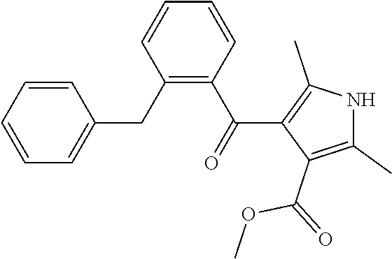
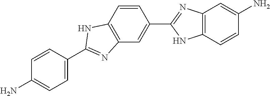
TABLE-US-00001 TABLE 1 Ex. No. IUPAC NAME Structure ADDITIVE MS 1 2-methoxy-5-((Z)- 2-(3,4,5- trimethoxyphenyl) vinyl)phenol ##STR00003## 317.2 2 (1-ethyl-1H- benzotriazol-5-yl) methyl(2-(2-methoxy- 4-methylphenyl)-4- methyl-1,3-thiazol- 5-yl)carbamate ##STR00004## 438.2 3 (2'beta)-22- oxovincaleukoblastine ##STR00005## H2SO4 825.3 4 2-(2-(4- chlorophenyl)ethyl)-6- (2-furyl)-3H-imidazo [4,5-b]pyridine ##STR00006## 324.1 5 4,5-anhydro-1,2-dideoxy- 4-methyl-2-((N- (morpholin-4-ylacetyl)-L- alanyl-O-methyl-L- tyrosyl)amino)-1-phenyl- L-threo-pont-3-ulose ##STR00007## 581.3 6 3-(3-methoxyphenyl)- N7,N7- dimethylisoquinoline- 1,7-diamine ##STR00008## 294.0 7 methyl 4-(2- benzylbenzoyl)-2,5- dimethyl-1H-pyrrole- 3-carboxylate ##STR00009## 348.2 8 2'-(4-amirophenyl)- 1H,1'H-2.5'- bibenzimidazol-5-amine ##STR00010## 341.2
TABLE-US-00002 TABLE 2 average fluorescent Example No. intensity (at 1 .mu.M) 1 1381 2 1331 3 1710 4 1125 5 1791 6 1305 7 1294 8 1150
Experimental Example 2
[0104] For detection of cardiomyocyte maturation, double knock-in human iPS cell lines, in which reporter protein sequences, EmGFP (Sequence Number 1) and mCherry (Sequence Number 2) were inserted into gene loci for TNNI1 and TNNI3, respectively, were prepared (the human iPS cells were prepared from episomal vector (loaded gene; OCT3/4, KLF4, SOX2, L-MYC, LIN28, mouse p53DD) using PBMC (LP_167, Sample ID:20130318) purchased from CTL (reference literature; Okita K, et al. Stem Cells. 2012 Nov. 29. doi: 10.1002/stem.1293)).
[0105] The maintenance culture of the above-mentioned reporter iPS cell lines was carried out by a conventional method (Okita K, et al. Stem Cells. 2012 Nov. 29. doi: 10.1002/stem.1293).
[0106] For differentiation induction into cardiomyocytes, the reporter iPS cell lines were treated with 0.5.times.TrypLE select (Life Technologies, diluted in 1/2 with 0.5 mM EDTA/PBS) for 4 to 5 minutes, and the cells were exfoliated using a cell scraper (IWAKI), and then dissociated into the single cell by pipetting. The medium was removed by centrifugation (1,000 rpm, 5 min), and the obtained cells were suspended in a medium prepared by adding 1% L-glutamine, transferrin 150 .mu.g/mL, ascorbic acid 50 .mu.g/mL (Sigma), monothioglycerol 4.times.10.sup.-4 M, 10 .mu.M Rock inhibitor (Y-27632), 2 ng/mL BMP4 (R&D) and 0.5% Matrigel (Growth Factor Reduced) to StemPro-34 SFM (ThermoFisher) in a 6 well-plate by 2.times.10.sup.6 cells/1.5 mL/well, and then subjected to static culture at 37.degree. C., under 5% oxygen condition to form embryoid bodies (0 day).
[0107] Next day (1st day), a medium prepared by adding 1% L-glutamine, transferrin 150 .mu.g/mL, ascorbic acid 50 .mu.g/mL (Sigma), monothioglycerol 4.times.10.sup.-4 M, 1.8 .mu.L (concentration 12 ng/mL) of 10 .mu.g/mL activin A, 15 .mu.L (concentration 10 ng/mL) of .mu.g/mL bFGF and 2.7 .mu.L (concentration 18 ng/mL) of 10 .mu.g/mL BMP4 to 1.5 mL of StemPro-34 SFM (ThermoFisher) was added to each well of the 6 well plate containing the embryoid bodies to adjust the volume combined with the 0 day's medium to final 3 mL (activin A: 6 ng/ml, bFGF: 5 ng/ml, BMP4: 10 ng/ml), and the embryoid bodies were cultured at 37.degree. C. for additional 2 days, under 5% oxygen condition.
[0108] Then (3rd day), the 6 well plate containing the embryoid bodies was inclined, and then left to stand for 1 to 2 minutes until the embryoid bodies were precipitated at the edges of the wells, and the supernatant was aspirated so as not to suck the embryoid bodies. Then, IMDM (ThermoFisher) medium was added to each well, and, as above, the plate was inclined, and then left to stand for 1 to 2 minutes, and the supernatant was aspirated so as not to suck the embryoid bodies. A medium prepared by adding 1% L-glutamine, transferrin 150 .mu.g/mL, ascorbic acid 50 .mu.g/mL (Sigma), monothioglycerol 4.times.10.sup.-4 M, 10 ng/mL VEGF, 1 .mu.M IWP-3, 0.6 .mu.M dorsomorphin and 5.4 .mu.M SB431542 to 3 mL of StemPro-34 SFM (ThermoFisher) was added to each well of the 6 well plate, and then the embryoid bodies were cultured at 37.degree. C. for 3 days, under 5% oxygen condition.
[0109] Then (6th day), the 6 well plate containing the embryoid bodies was inclined, and then left to stand for 1 to 2 minutes until the embryoid bodies were precipitated at the edges of the wells, and the supernatant was aspirated so as not to suck the embryoid bodies. A medium prepared by adding 1% L-glutamine, transferrin 150 .mu.g/mL, ascorbic acid 50 .mu.g/mL (Sigma), monothioglycerol 4.times.10.sup.-4 M and 5 ng/mL VEGF to 2 mL of StemPro-34 SFM (ThermoFisher) was added to each well of the 6 well plate, and then the embryoid bodies were cultured at 37.degree. C. for 4 days, under 5% oxygen condition. On 8th day, the medium was replaced with a medium under the same conditions.
[0110] Then (10th day), the 6 well plate containing the embryoid bodies was inclined, and then left to stand for 1 to 2 minutes until the embryoid bodies were precipitated at the edges of the wells, and the supernatant was aspirated so as not to suck the embryoid bodies. A medium prepared by adding (1) 1% L-glutamine, transferrin 150 .mu.g/mL, ascorbic acid 50 .mu.g/mL (Sigma), monothioglycerol 4.times.10.sup.-4 M and 5 ng/mL VEGF, and (2) 40 nM of the compound of Example No. 2 or DMSO (0.1% addition relative to the medium volume) to 2 mL of StemPro-34 SFM (ThermoFisher) was added to each well of the 6 well plate, and then the embryoid bodies were cultured at 37.degree. C. for 6 days, under general oxygen condition. On 13th day, the medium was replaced with a medium under the same conditions.
[0111] On the 16th day, after the embryoid bodies under each condition were photographed by fluorescence microscope, the 6 well plate containing the embryoid bodies was inclined, and then left to stand for 1 to 2 minutes until the embryoid bodies were precipitated at the edges of the wells, and the supernatant was aspirated so as not to suck the embryoid bodies. Then, 2 mL of PBS was added to each well, and, as so above, the plate was inclined, and then left to stand for 1 to 2 minutes, and the PBS was aspirated so as not to suck the embryoid bodies. A solution prepared by adding DNase 10 .mu.g/mL and Liberase 100 .mu.g/mL to 3 mL of IMDM was added to each well, and the plate was left to stand at 37.degree. C. for 1 hr, under general oxygen condition. After 1 hr, the plate was inclined, and then left to stand for 1 to 2 minutes until the embryoid bodies were precipitated at the edges of the wells, and the supernatant was aspirated so as not to suck the embryoid bodies. Then, 2 mL of PBS was added to each well, and, as above, the plate was inclined, and then left to stand for 1 to 2 minutes, and the PBS was aspirated so as not to suck the embryoid bodies. A solution prepared by adding DNase 10 .mu.g/mL to 2 mL of TrypLE select was added to each well, and the plate was left to stand at 37.degree. C. for 10 minutes, under general oxygen condition. Then, a medium prepared by adding 1% L-glutamine, transferrin 150 .mu.g/mL, ascorbic acid 50 .mu.g/mL (Sigma), monothioglycerol 4.times.10.sup.-4 M, 5 ng/mL VEGF and DNase 10 .mu.g/mL to 2 mL of StemPro-34 SFM (ThermoFisher) was added to each well, and the embryoid bodies were dissociated into the single cell by pipetting, and then subjected to centrifugation (1,000 rpm, 5 minutes). After the centrifugation, the supernatant was removed, the cells were suspended in 1 to 2 mL of 2% FBS/PBS, and the mCherry expression in TNNI1.sup.+ cells was analyzed by flow cytometry (BD FACSAria Fusion cell sorter). The results are shown in Table 3 and FIG. 1.
TABLE-US-00003 TABLE 3 mCherry.sup.+ (%) control (addition of (1) alone) 7.4 control 10.2 (0.1% DMSO addition relative to the medium volume) Example No. 2 40 nM 36.1
INDUSTRIAL APPLICABILITY
[0112] Since the above-mentioned compounds or salts thereof have an activity to promote the maturation of a cardiomyocyte, they are useful as a cardiomyocyte maturation promoter.
[0113] This application is based on patent application No. 2018-69871 filed on Mar. 30, 2018 in Japan, the contents of which are encompassed in full herein.
Sequence CWU 1
1
21720DNAArtificial SequenceEmerald Green Fluorescent Protein
(EmGFP) 1atggtgagca agggcgagga gctgttcacc ggggtggtgc ccatcctggt
cgagctggac 60ggcgacgtaa acggccacaa gttcagcgtg tccggcgagg gcgagggcga
tgccacctac 120ggcaagctga ccctgaagtt catctgcacc accggcaagc
tgcccgtgcc ctggcccacc 180ctcgtgacca ccttcaccta cggcgtgcag
tgcttcgccc gctaccccga ccacatgaag 240cagcacgact tcttcaagtc
cgccatgccc gaaggctacg tccaggagcg caccatcttc 300ttcaaggacg
acggcaacta caagacccgc gccgaggtga agttcgaggg cgacaccctg
360gtgaaccgca tcgagctgaa gggcatcgac ttcaaggagg acggcaacat
cctggggcac 420aagctggagt acaactacaa cagccacaag gtctatatca
ccgccgacaa gcagaagaac 480ggcatcaagg tgaacttcaa gacccgccac
aacatcgagg acggcagcgt gcagctcgcc 540gaccactacc agcagaacac
ccccatcggc gacggccccg tgctgctgcc cgacaaccac 600tacctgagca
cccagtccgc cctgagcaaa gaccccaacg agaagcgcga tcacatggtc
660ctgctggagt tcgtgaccgc cgccgggatc actctcggca tggacgagct
gtacaagtaa 7202711DNAArtificial SequencemCherry 2atggtgagca
agggcgagga ggataacatg gccatcatca aggagttcat gcgcttcaag 60gtgcacatgg
agggctccgt gaacggccac gagttcgaga tcgagggcga gggcgagggc
120cgcccctacg agggcaccca gaccgccaag ctgaaggtga ccaagggtgg
ccccctgccc 180ttcgcctggg acatcctgtc ccctcagttc atgtacggct
ccaaggccta cgtgaagcac 240cccgccgaca tccccgacta cttgaagctg
tccttccccg agggcttcaa gtgggagcgc 300gtgatgaact tcgaggacgg
cggcgtggtg accgtgaccc aggactcctc cctgcaggac 360ggcgagttca
tctacaaggt gaagctgcgc ggcaccaact tcccctccga cggccccgta
420atgcagaaga agaccatggg ctgggaggcc tcctccgagc ggatgtaccc
cgaggacggc 480gccctgaagg gcgagatcaa gcagaggctg aagctgaagg
acggcggcca ctacgacgct 540gaggtcaaga ccacctacaa ggccaagaag
cccgtgcagc tgcccggcgc ctacaacgtc 600aacatcaagt tggacatcac
ctcccacaac gaggactaca ccatcgtgga acagtacgaa 660cgcgccgagg
gccgccactc caccggcggc atggacgagc tgtacaagta g 711
D00000
D00001
S00001
XML
uspto.report is an independent third-party trademark research tool that is not affiliated, endorsed, or sponsored by the United States Patent and Trademark Office (USPTO) or any other governmental organization. The information provided by uspto.report is based on publicly available data at the time of writing and is intended for informational purposes only.
While we strive to provide accurate and up-to-date information, we do not guarantee the accuracy, completeness, reliability, or suitability of the information displayed on this site. The use of this site is at your own risk. Any reliance you place on such information is therefore strictly at your own risk.
All official trademark data, including owner information, should be verified by visiting the official USPTO website at www.uspto.gov. This site is not intended to replace professional legal advice and should not be used as a substitute for consulting with a legal professional who is knowledgeable about trademark law.