Synthetic Platelets
Mitragotri; Samir ; et al.
U.S. patent application number 17/030519 was filed with the patent office on 2021-01-14 for synthetic platelets. This patent application is currently assigned to PRESIDENT AND FELLOWS OF HARVARD COLLEGE. The applicant listed for this patent is PRESIDENT AND FELLOWS OF HARVARD COLLEGE, THE REGENTS OF THE UNIVERSITY OF CALIFORNIA. Invention is credited to Yongsheng Gao, Samir Mitragotri, Apoorva Sarode.
| Application Number | 20210008223 17/030519 |
| Document ID | / |
| Family ID | 1000005106625 |
| Filed Date | 2021-01-14 |


View All Diagrams
| United States Patent Application | 20210008223 |
| Kind Code | A1 |
| Mitragotri; Samir ; et al. | January 14, 2021 |
SYNTHETIC PLATELETS
Abstract
Provided herein are particles comprising a polymer substrate comprising one or more hyaluronic acid chains; and two or more peptide moieties bound directly to each hyaluronic acid chain. In some embodiments, the two or more peptide moieties comprising collagen-binding peptide (CBP) and von Willebrand binding peptide (VBP). The particles can be utilized in, e.g., methods of hemostatic treatment.
| Inventors: | Mitragotri; Samir; (Lexington, MA) ; Sarode; Apoorva; (Somerville, MA) ; Gao; Yongsheng; (Quincy, MA) | ||||||||||
| Applicant: |
|
||||||||||
|---|---|---|---|---|---|---|---|---|---|---|---|
| Assignee: | PRESIDENT AND FELLOWS OF HARVARD
COLLEGE Cambridge MA THE REGENTS OF THE UNIVERSITY OF CALIFORNIA Oakland CA |
||||||||||
| Family ID: | 1000005106625 | ||||||||||
| Appl. No.: | 17/030519 | ||||||||||
| Filed: | September 24, 2020 |
Related U.S. Patent Documents
| Application Number | Filing Date | Patent Number | ||
|---|---|---|---|---|
| 15968062 | May 1, 2018 | |||
| 17030519 | ||||
| 15116178 | Aug 2, 2016 | |||
| PCT/US2015/014326 | Feb 3, 2015 | |||
| 15968062 | ||||
| 61935297 | Feb 3, 2014 | |||
| Current U.S. Class: | 1/1 |
| Current CPC Class: | A61P 7/02 20180101; A61K 47/6939 20170801; C12Y 304/21068 20130101; A61K 47/595 20170801; A61K 47/6925 20170801; A61K 47/62 20170801; A61K 47/61 20170801; A61K 38/482 20130101 |
| International Class: | A61K 47/69 20060101 A61K047/69; A61K 38/48 20060101 A61K038/48; A61P 7/02 20060101 A61P007/02; A61K 47/62 20060101 A61K047/62; A61K 47/59 20060101 A61K047/59; A61K 47/61 20060101 A61K047/61 |
Goverment Interests
STATEMENT REGARDING FEDERALLY SPONSORED RESEARCH OR DEVELOPMENT
[0002] This invention was made with United States government support under Grant Number DGE-1144085 awarded by the National Science Foundation. The United States government has certain rights in the invention.
Claims
1. A particle comprising a polymer substrate comprising one or more hyaluronic acid chains; and two or more peptide moieties bound directly to each hyaluronic acid chain, the two or more peptide moieties comprising collagen-binding peptide (CBP) and von Willebrand binding peptide (VBP).
2. The particle of claim 1, wherein the two or more peptide moieties comprise: a peptide comprising SEQ ID NO: 1 and a peptide comprising at least one of SEQ ID NOs: 2, 6, and 7.
3. The particle of claim 1, wherein the two or more peptide moieties comprise: a peptide comprising SEQ ID NO: 1 and a peptide comprising SEQ ID NO: 7.
4. The particle of claim 1, wherein the two or more peptide moieties further comprise at least one of a platelet binding peptide; a wound healing peptide; a tPA-binding peptide; a clot binding peptide; an anti-thrombotic peptide; and a thrombolytic peptide.
5. The particle of claim 4, wherein the two or more peptide moieties comprise two or more of: a peptide comprising SEQ ID NO: 1; a peptide comprising SEQ ID NO: 2; a peptide comprising SEQ ID NO: 3; a peptide comprising SEQ ID NO: 4; a peptide comprising SEQ ID NO: 5; a peptide comprising SEQ ID NO: 8; a peptide comprising SEQ ID NO: 7; heparin; and tPA.
6. The particle of claim 1, wherein the two or more peptide moieties further comprise fibrinogen mimetic peptide (FMP).
7. The particle of claim 6, wherein the two or more peptide moieties comprise: a peptide comprising SEQ ID NO: 1; a peptide comprising at least one of SEQ ID NOs: 2, 6, and 7; and a peptide comprising at least one of SEQ ID NOs: 3, 4, 8, and 9.
8. The particle of claim 1, wherein there are 66 of each peptide moiety per each hyaluronic acid chain.
9. The particle of claim 8, wherein each hyaluronic acid chain is 250 kDa in size.
10. The particle of claim 1, comprising 10 mol % of each of the peptide moieties relative to the hyaluronic acid disaccharide units.
11. The particle of claim 1, wherein the particle further comprises polyvinyl alcohol, doxorubicin-gemcitabine-glycine-hyaluronic acid (DOX-GEM-gly-HA), or polylactic-co-glycolic acid.
12. The particle of claim 1, further comprising a thrombolytic agent in an interior of the particle.
13. The particle of claim 12, wherein the thrombolytic agent is tPA.
14. The particle of claim 1, wherein the particle is less than 500 nm in diameter.
15. A method comprising administering the particle of claim 1 to a subject in need of hemostatic treatment.
Description
CROSS-REFERENCE TO RELATED APPLICATIONS
[0001] This application is a continuation under 35 U.S.C. .sctn. 120 of co-pending U.S. application Ser. No. 15/968,062 filed May 1, 2018, which is a continuation-in-part of co-pending U.S. application Ser. No. 15/116,178 filed Aug. 2, 2016 now abandoned, which is a 35 U.S.C. .sctn. 371 National Phase Entry Application of International Application No. PCT/US2015/014326 filed Feb. 3, 2015, which designates the U.S. and claims benefit under 35 U.S.C. .sctn. 119(e) of U.S. Provisional Application No. 61/935,297 entitled "Artificial Platelets and Related Systems" filed Feb. 3, 2014, the contents of which are incorporated herein by reference in their entireties.
REFERENCE TO SEQUENCE LISTING, A TABLE, OR A COMPUTER PROGRAM LISTING COMPACT DISK APPENDIX
[0003] This application is submitted with a computer readable sequence listing, submitted herewith via EFS as the ASCII text file named: "002806-092160-USP1_SL.txt," file size approximately 2,523 bytes, created on May 1, 2018 and hereby incorporated by reference in its entirety.
BACKGROUND AND SUMMARY OF THE INVENTION
[0004] Excessive blood loss is responsible for approximately 3 million deaths worldwide due to trauma, and is the leading cause of preventable deaths following serious injuries. Hemostatic agents have the potential to prevent or reduce blood loss after serious wounds. In practice, however, hemostatic agents are often not effective in this task. For severe wounds, current emergency hemostats are usually administered externally as a hemostat-containing-gauze. While effective in the treatment of externally accessible wounds, these agents are unable to treat internal wounds, especially those which may have multiple bleeding sites. Application of current hemostats is further limited since the precise site of hemorrhage is not always known. Accordingly, there is a need in the art for hemostatic agents which can effectively overcome the shortcomings of the prior art.
[0005] Disclosed herein are novel functionalized particles for the delivery of various agents to the bloodstream and/or tissues of an animal. In one implementation, the functionalized particles of the invention can act as synthetic platelets. Platelets are an important component of the wound response in animals. Wounding results in the exposure of collagen, thrombin, and von Willebrand factor to the blood and causes the activation of platelets. Activated platelets clump together and begin the clotting process which stops bleeding. Unfortunately, unlike red blood cells, platelets cannot be stored for long periods of time. Accordingly, there is a need in the art for platelet substitutes which can aid in treating patients with wounds, including internal wounds. Additionally, there is a need for effective and selective agents which can aid in the initiation of clotting in wounded tissues. As described herein, the synthetic platelets of the invention can fulfill this unmet need. Additional embodiments of the invention include wound healing particles, anti-thrombotic particles, thrombolytic particles, and modified red blood cells having platelet functions, as set forth herein.
BRIEF DESCRIPTION OF THE DRAWINGS
[0006] FIG. 1. FIG. 1 is a schematic overview of the synthesis process for synthetic platelets.
[0007] FIG. 2A and 2B. FIG. 2A is a schematic depiction of a wound site without synthetic platelets. FIG. 2B is a schematic depiction of a wound site in which synthetic platelets are aiding in thrombus formation.
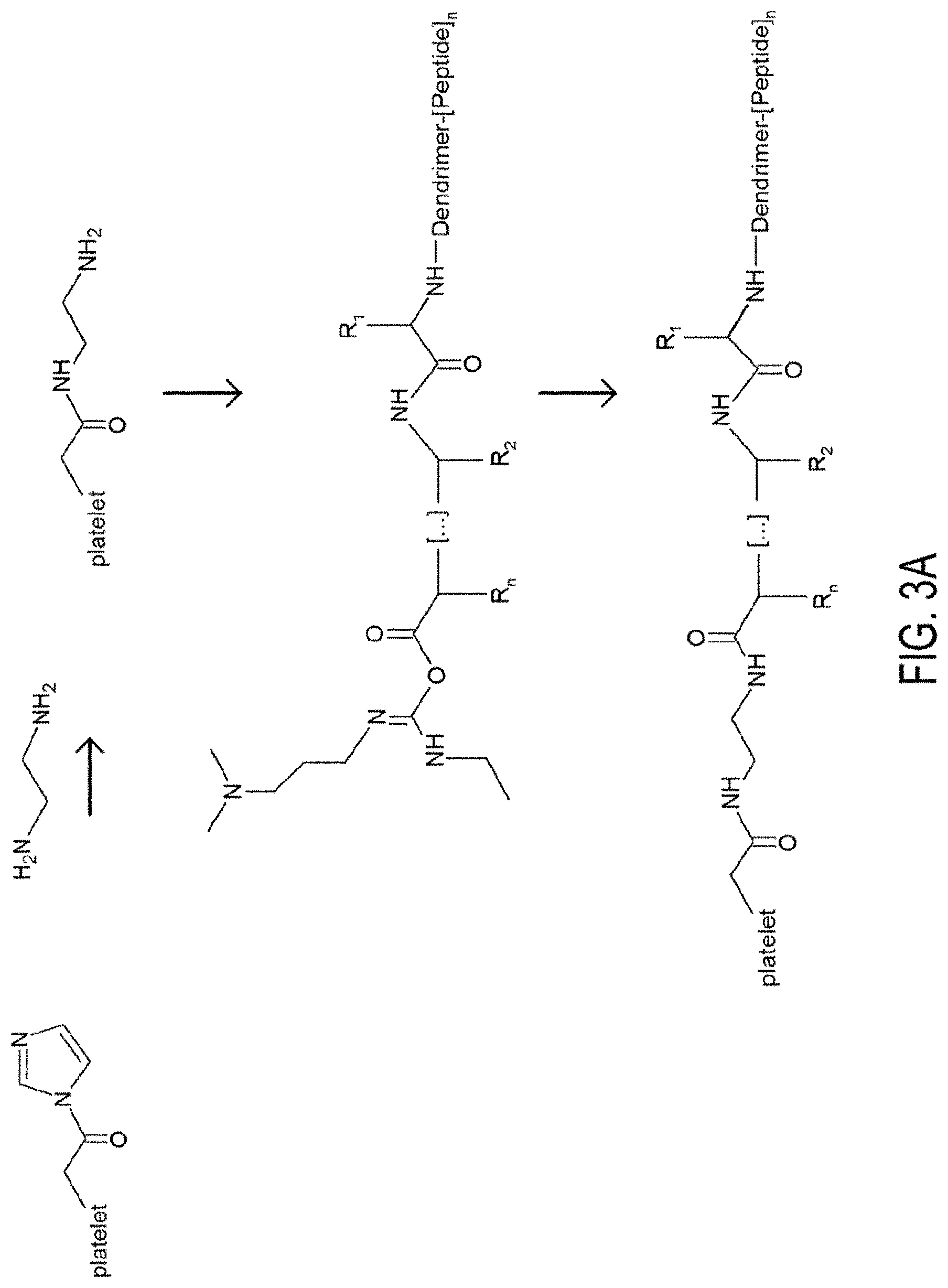
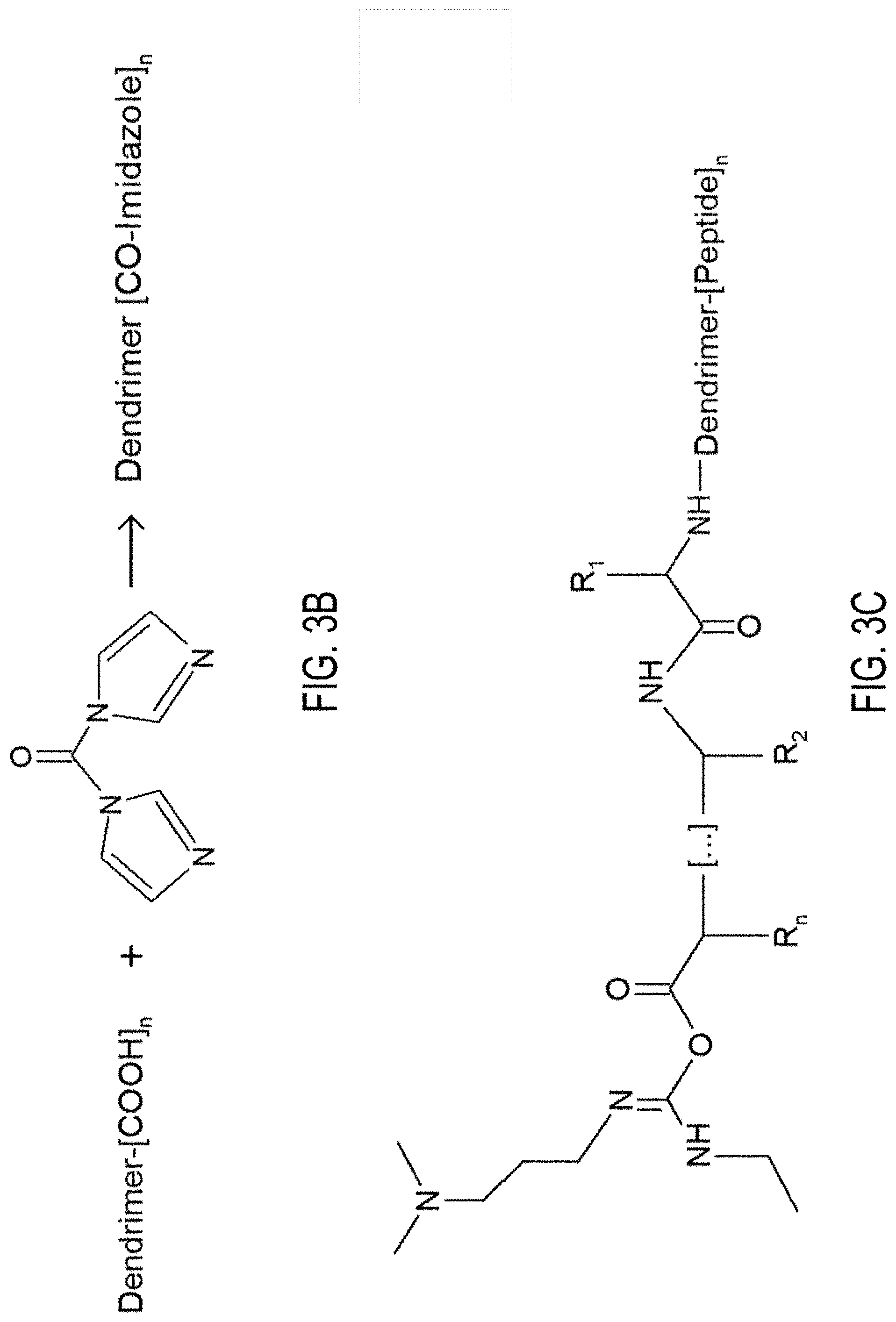
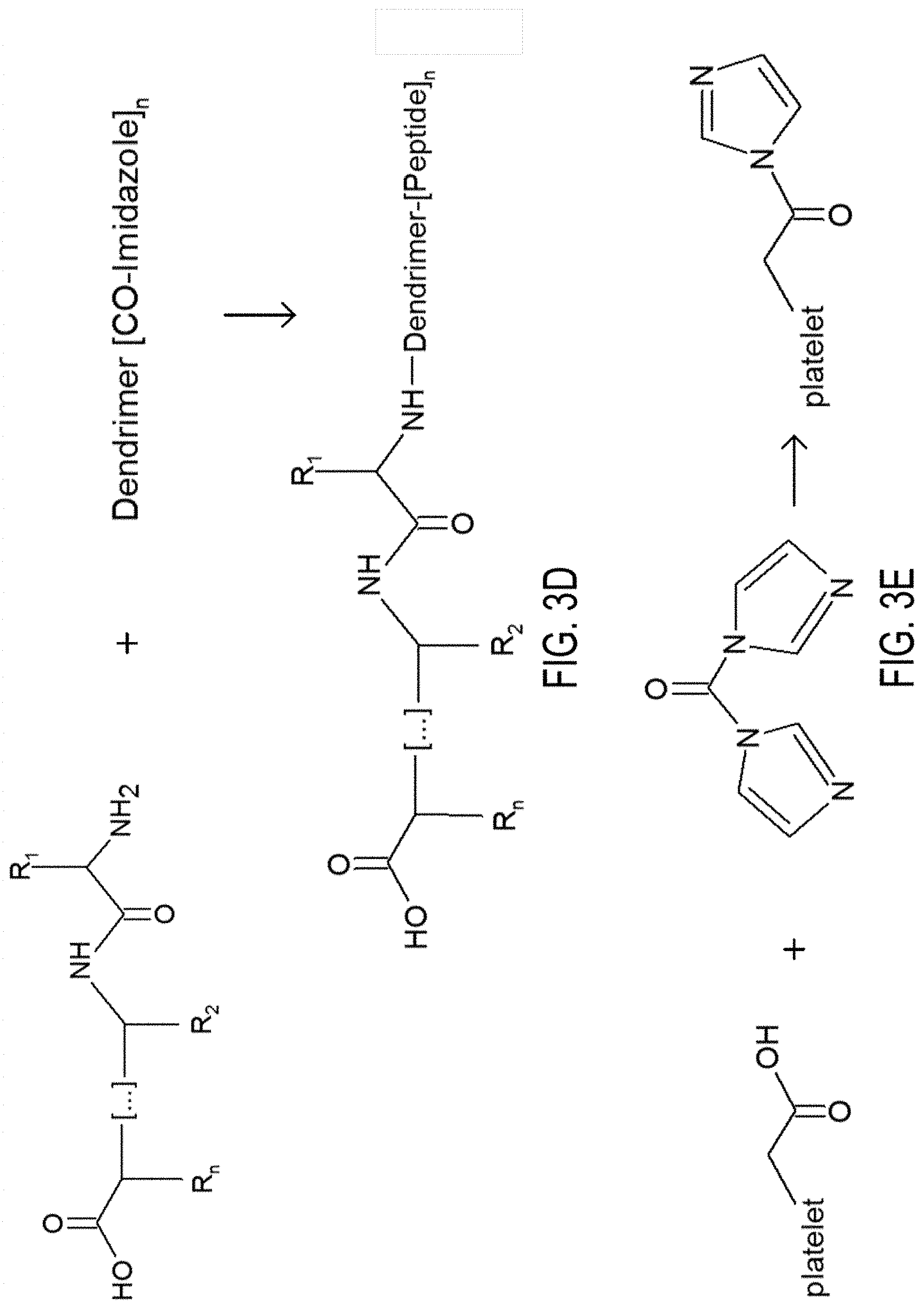
[0008] FIG. 3A, 3B, 3C, 3D and 3E. FIG. 3A depicts the synthesis of synthetic platelets. FIG. 3B depicts the activation of dendrimer COOH groups by CDI. FIG. 3C depicts the activation of dendrimer-peptide conjugates via EDC. FIG. 3D depicts coupling of the peptide on the activated dendrimer. FIG. 3E depicts artificial platelet activation via CDI.
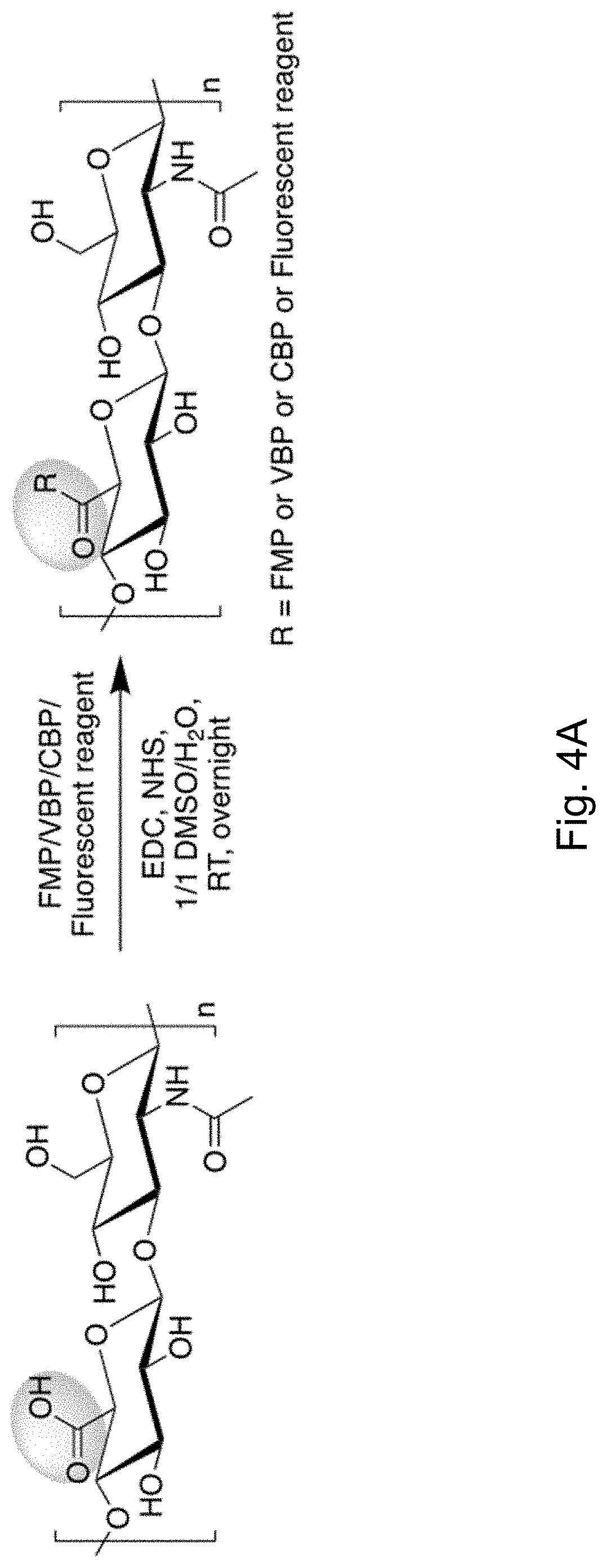
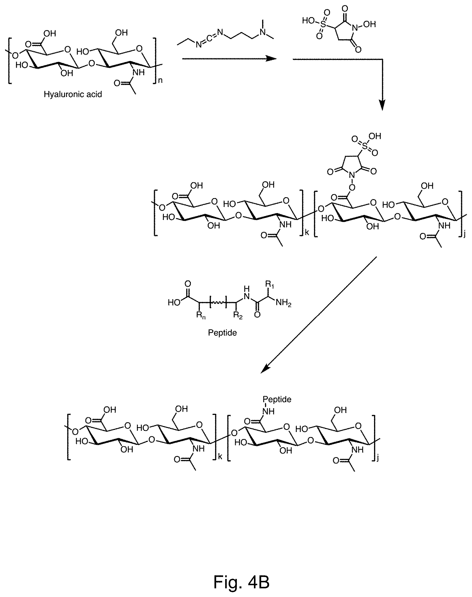
[0009] FIGS. 4A-4B depict the synthesis of HA polymer peptide conjugates via the EDC/sulfo-NHS chemistry.
[0010] FIG. 5 depicts .sup.1HNMR spectra of HA/Alexa Fluor.RTM. 647, FMP and HA/FMP/Alexa Fluor.RTM. 647 conjugate.
[0011] FIG. 6 depicts .sup.1HNMR spectra of HA/Alexa Fluor.RTM. 647, FMP, VBP and HA/FMP/VBP/Alexa Fluor.RTM. 647 conjugate.
[0012] FIG. 7 depicts .sup.1HNMR spectra of HA/Alexa Fluor.RTM. 647, FMP, VBP, CBP and HA/FMP/VBP/CBP/Alexa Fluor.RTM. 647 conjugate.
[0013] FIGS. 8A-8B depict representative cryo-TEM of HA (FIG. 8A) and HA/FMP/VBP/CBP conjugate (FIG. 8B).
[0014] FIG. 9 depicts a graph showing the variation of zeta potential and number-based mean particle size with each adsorption step. Low polydispersity indices indicate a uniform size distribution following each layer deposition.
[0015] FIGS. 10A-10B depict discoidal flexible hollow polyelectrolyte shells. FIG. 10 depicts scanning electron micrograph showing the discoidal geometry (Anselmo, et. al. 2014), FIG. 10B depicts cryo-transmission electron micrograph depicting the hollow internal structure and thus, complete removal of the polystyrene cores (Scale bar: 200 nm).
[0016] FIG. 11 depicts a reaction scheme for the covalent coupling of peptides with dendrimers using CDI or EDC chemistry.
[0017] FIGS. 12A-12C depict scanning electron micrographs of (FIG. 12A) Texas red dextran-loaded CaCO3 microparticles, (FIG. 12B) soft, flexible Texas red dextran-loaded microcapsules after core dissolution. FIG. 12C depicts confocal microscopy imaging was performed on these microcapsules to visualize the loaded cargo. FITC-labeled (PAH/BSA)6 shell encapsulating Texas red dextran in its core.
[0018] FIG. 13 depicts a graph showing the variation of zeta potential with each adsorption step of the layer by layer coating.
[0019] FIG. 14 depict the % Encapsulation efficiency achieved with in situ loading of Texas red dextran and tPA in CaCO3 microparticles, respectively.
DETAILED DESCRIPTION OF THE INVENTION
[0020] Presented herein are novel compositions and methods encompassing functionalized particles, being termed "functionalized" because they carry selected biologically active moieties, which may be utilized in various medical applications.
[0021] In one aspect, the functionalized particles of the invention comprise synthetic platelets which may be used as thrombogenic agents. Alternative embodiments comprise thrombolytic agents, healing agents, hemostatic peptide-polymer conjugates, wound healing peptide-polymer conjugates, anti-thrombolytic peptide-polymer conjugates, thrombolytic polymer-peptide conjugates and other agents. In another aspect, the invention comprises methods and compositions encompassing the modification of cells and cell components to create agents with platelet functions.
[0022] Functionalized Particles. The functionalized particles of the invention, in general, comprise three elements: (1) a substrate; (2) linkers; and (3) functional moieties. Linkers are anchored on the outer surface of the substrate. Functional moieties are bonded to the terminal ends of the linkers. The size, shape, and composition of the substrate, the linker properties, and the identity of the functional moieties will be selected depending on the desired functionality of the particles. In some embodiments of any of the aspects, the substrate is a polymer or a polymer-peptide bilayer or mixture. In some embodiments of any of the aspects, the substrate is hyaluronic acid and the one or more functional moieties are peptides.
[0023] In one implementation, the substrate comprises a shell formed over a core, the core optionally being dissolved or degraded subsequent to shelf formation, leaving the shell hollow. The choice of core shape, size and composition, along with the shell composition and thickness, will determine the final properties of the shell.
[0024] The Core. In those embodiments utilizing a shell, the function of the core is to provide a substrate for the creation of the shell. The core will define the shape and the size of the functional body. In some embodiments, the core is retained and makes up part of the final product. In other embodiments, subsequent to formation of the shell, the core is partially or wholly degraded and the resulting shell is substantially hollow. The core may be comprised of any material or materials, so long as the outer surface of the core will support synthesis, deposition, or formation of the shell thereon. For example, a core having an outer surface capable of binding cationic/anionic polymers, polyelectrolytes, or proteins may be used.
[0025] Exemplary core materials include polystyrene (PS), polystyrene latex (PS), poly (lactic-co-glycolic) acid (PLGA), (PAH), Hyaluronic acid (HA), calcium hydroxide (Ca(OH).sub.2), (CaOH.sub.2), and silica materials. Core materials that can be readily dissolved or digested are used in those embodiments where a hollow shell is desired. Materials amenable to digestion by polar solvents, aqueous solvents, acids, bases, enzymes, etc may be used. In some embodiments of any of the aspects, the core is CaCO.sub.3.
[0026] Optionally, materials may be embedded or encased within the core, such that when the core is dissolved, these materials remain within the shell. Exemplary materials for inclusion in the core include drugs, for example encapsulated in slow-release dissolving materials), markers (e.g. fluorophores, quantum dots, or other visible markers), gene therapy constructs (e.g. nucleic acids, optionally encapsulated), and other materials.
[0027] The size and shape of the core will largely determine the final size and shape of the shell deposited onto it. Core size may vary from a few nanometers to over ten micrometers. For intravenous uses, bodies larger than 10 micrometers may cause cardiopulmonary complications, for example by aggregating in capillaries. For intravenous uses, core sizes in the range of 10-500 nanometers are effective for efficient circulation in the body.
[0028] The shape of the core may vary. In general, cores that are substantially spherical or elliptical may be used.
[0029] The Shell. The shell comprises a polymeric, proteinaceous, or other material which is synthesized, deposited, or otherwise formed on the outer surface of the core. Exemplary shell materials include proteins, polyelectrolytes, and polymers. The shell materials will generally be biocompatible. The shell materials will optionally be somewhat flexible. The shell materials are not limited to permeable materials. For example, in embodiments where retaining the core is advantageous, the shell materials may be substantially impermeable. In those embodiments where the core is to be partially or wholly dissolved or digested, the shell materials must be adequately porous or permeable to the dissolution agent that it can reach the core, and must be sufficiently inert or resistant to the dissolution agent that the shell will not be significantly degraded when removing the core from the body.
[0030] In one embodiment, the shell material comprises a multilayer structure formed using layer-by-layer synthesis. Such structures are formed by the sequential layering of two materials, the two materials typically having opposite charges in order to facilitate adsorption of each layer on the other. Advantageously, layer-by-layer synthesis allows a high degree of control over shell thickness. After the alternating bi-layers have been deposited, a cross-linking or other fixative step is performed to bond and strengthen the shell. For example, chemical cross linking, UV-activated cross linking, and/or the inclusion of intercalated cross-linking agents may be used. Any number of bilayers may be used, for example 1 to 20, depending on the desired qualities of the finished shell. For example, if a very thin and flexible shell is desired, a low number (e.g. 1 or 2) of bilayers may be used. When small numbers of bilayers are used, a more substantial crosslinking process is required to ensure the strength of the shell. Thicker shells can be created using higher numbers of bi-layers. In some embodiments of any of the aspects, the bilayers are crosslinked, e.g., using 2% (v/V) glutaraldehyde solution.
[0031] Various layer-by-layer synthesis methods are known in the art, for example as described in: Wang, Y., A. S. Angelatos, and F. Caruso, Template Synthesis of Nanostructured Materials via Layer-by-Layer Assembly. Chemistry of Materials, 2007. 20(3): p. 848-858; Zhou, Z., A. C. Anselmo, and S. Mitragotri, Synthesis of protein-based, rod-shaped particles from spherical templates using layer-by-layer assembly. Adv Mater, 2013. 25(19): p. 2723-7; Doshi, N., et al., Platelet mimetic particles for targeting thrombi in flowing blood. Adv Mater, 2012. 24(28): p. 3864-9; Doshi, N., et al., Red blood cell-mimicking synthetic biomaterial particles. Proc Natl Acad Sci U S A, 2009. 106(51): p. 21495-9; del Mercato, L.L., et al., LbL multilayer capsules: recent progress and future outlook for their use in life sciences. Nanoscale, 2010. 2(4): p. 458-67; Johnston, A. P., et al., Layer-by-layer engineered capsules and their applications. Current Opinion in Colloid & Interface Science, 2006. 11(4): p. 203-209; and Yan, Y., M. Bjornmalm, and F. Caruso, Assembly of Layer-by-Layer Particles and Their Interactions with Biological Systems. Chemistry of Materials, 2013.
[0032] An exemplary system for shell formation is the use of alternating bi-layers of poly (allylamine hydrochrolide) (PAH) and bovine serum albumin (BSA). Alternatively, actin and PAH layers may be used. Additional exemplary shell materials include Poly-L-lysine, Actin, Hemoglobin, human serum albumin, poly(4-styrene sulfonate), PMA, PVPON, Chitosan, dextran, and alginate, as known in the art. Further exemplary shell materials include any protein pair, wherein one protein has an isoelectric point greater than 7, and the second protein has an isoelectric point less than 7; a positively charged synthetic polymer and a negatively charged polymer; and other self-assembling molecules.
[0033] In some embodiments, the shell/substrate polymer is hyaluronic acid, polyvinyl alcohol, DOX-GEM-gly-HA, or polylactic-co-glycolic acid.
[0034] Core dissolution. After the shell layer has been synthesized, deposited, or formed, the core may optionally be dissolved, degraded, or otherwise removed. In one embodiment, the dissolution is effected by exposing the shell and core to a solution which dissolves the core material but which does not significantly affect the shell material(s). For example, when a polystyrene core has been utilized, it can be dissolved by exposure to a tetrahydrofuran-isopropyl alcohol gradient, as described in Example 1. The choice of appropriate dissolution agent will depend on the composition of the core and the shell.
[0035] Upon removal of the core, depending on the material comprising the shell, the remaining shell will be flexible. For example, BSA-PAH shells will exhibit high flexibility, which aids in in vivo circulation of the particles. When a substantially spherical core is used, upon core dissolution, a flexible shell such as BSA-PAH will assume a discoid platelet-like structure.
[0036] Functionalized Polymers. Another implementation of the invention encompasses the use of polymers, as opposed to proteins, as the functional agent-harboring scaffold or substrate. Likewise, hybrid substrates comprising bilayers of protein and polymeric material may be employed as the substrate. It will be understood that the methods of functionalizing shells and utilizing functionalized shells described herein are equally applicable to functionalized particles wherein a polymeric material not configured as a shell replaces the shell as the substrate for functional moieties. Examples of polymers that could be used include hyaluronic acid, polyvinyl alcohol, or polylactic-co-glycolic acid. As with the shell, linkers are anchored on the polymerchain, the linkers having functional moieties are bonded to the terminal ends of the linkers.
[0037] The choice of polymer type, molecular weight, composition, linker properties, and the identity of the functional moieties will be selected depending on the desired functionality of the polymer conjugates. Polymeric materials may be formed into any number of structures, including shells around a core (wherein the core may optionally be dissolved), discoid or spherical bodies, planar bodies, fibers, and other secondary structures. In one embodiment, the polymeric material comprises a single polymer chain.
[0038] Linkers. In one embodiment, the functional moieties may be directly attached to the substrate (e.g. the shell or polymeric substrate) without the use of any linker moiety. Alternatively, in many implementations, the functional moieties are attached to the substrate using linkers. A linker is any molecule capable of (1) binding to the substrate material, and (2) which is also capable of binding a functional agent, typically at its terminal end(s). Linkers anchor the functional moiety to the substrate, and in some cases serve the role of spacer, holding the functional moiety a distance off the surface of the substrate to avoid interactions between the functional moiety and the substrate material. For example, linkers of 2-3 times the length of the functional moiety may be used to hold such compound at a distance from the substrate surface that will avoid undesired interactions.
[0039] Where functional moieties of highly divergent sizes are used, the smaller moieties can be placed at the ends of longer linkers which compensate for the size difference between the moieties, such that both are displayed at about the same plane the functional particle's outer perimeter. The smaller species may be tethered to a spacer molecule, for example a PEG molecule, the length of which is selected to match the size of the larger species (e.g. PEG linkers ranging from 500 to 5,000 kD).
[0040] Further, linkers can control the density of functional moieties and can control the display geometry of the functional moieties, for example separating portions of the substrate surface displaying a selected functional moiety from other portions displaying different moieties.
[0041] In one embodiment, the linkers comprise highly branched molecules, for example dendrimers. The advantage of using a highly branched molecule as a linker is that a single anchoring site on the substrate surface can serve as attachment point for numerous functional moieties, greatly increasing the effective concentration of functional moieties, and thus the binding avidity of the body to the biological target. Exemplary branched linkers include dendrimers, for example dendrimers having anywhere from 2 to 2,000 branches. For example, a dendrimer having 44 to 2,048 or more branches can be utilized, e.g. dendrimers of generation 2-10 or more. Higher generation dendrimers increase the surface area of the functional body and the number of active molecules. Exemplary biocompatible dendrimers include poly(amidoamine) ("PAMAM") dendrimers and peptide-based dendrimers, as known in the art.
[0042] Functional Moieties. The functional bodies of the invention comprise one or more types of functional agents linked to the substrate by the linker. The functional moieties may comprise any material which imparts properties or functions to the functional body. Properties and functions include binding ability and avidity for target proteins or cell types, enzymatic activity, and any other biological ability, effect, or action. Functional moieties may include, for example, proteins, peptides, peptidomimetics, antibodies, enzymes, binding sites, markers, fluorescent probes, affinity tags, chelating agents, radioactive probes, etc.
[0043] Multiple types of functional moieties may be included in the functionalized particle. For example, in the artificial platelet implementation of the invention described below, three different peptides are included to impart the desired thrombogenic function. Any number of desired compounds may be included. In one embodiment, in addition to specific functional moieties that impart biological function, ancillary moieties are included which aid in the circulation of the functional body in the bloodstream of the target animal and the retention of the functional body within a living organism, such as moieties which increase solubility or reduce immunogenicity.
[0044] When multiple types of functional moiety are to be included in a functionalized particle, it will generally be advantageous to perform separate conjugation reactions between each type of functional moiety and the linker Linker-functional moiety conjugates can then be joined to the substrate in a single conjugation reaction, wherein the different types of linker-functional moiety conjugates are present in the stochastic ratios desired for the final product. Alternatively, different linker-functional moiety species can be added to substrate in a serial fashion to control relative densities. Alternatively, linkers can be first conjugated to the substrate, followed by functionalization reactions to join functional moieties to the linkers.
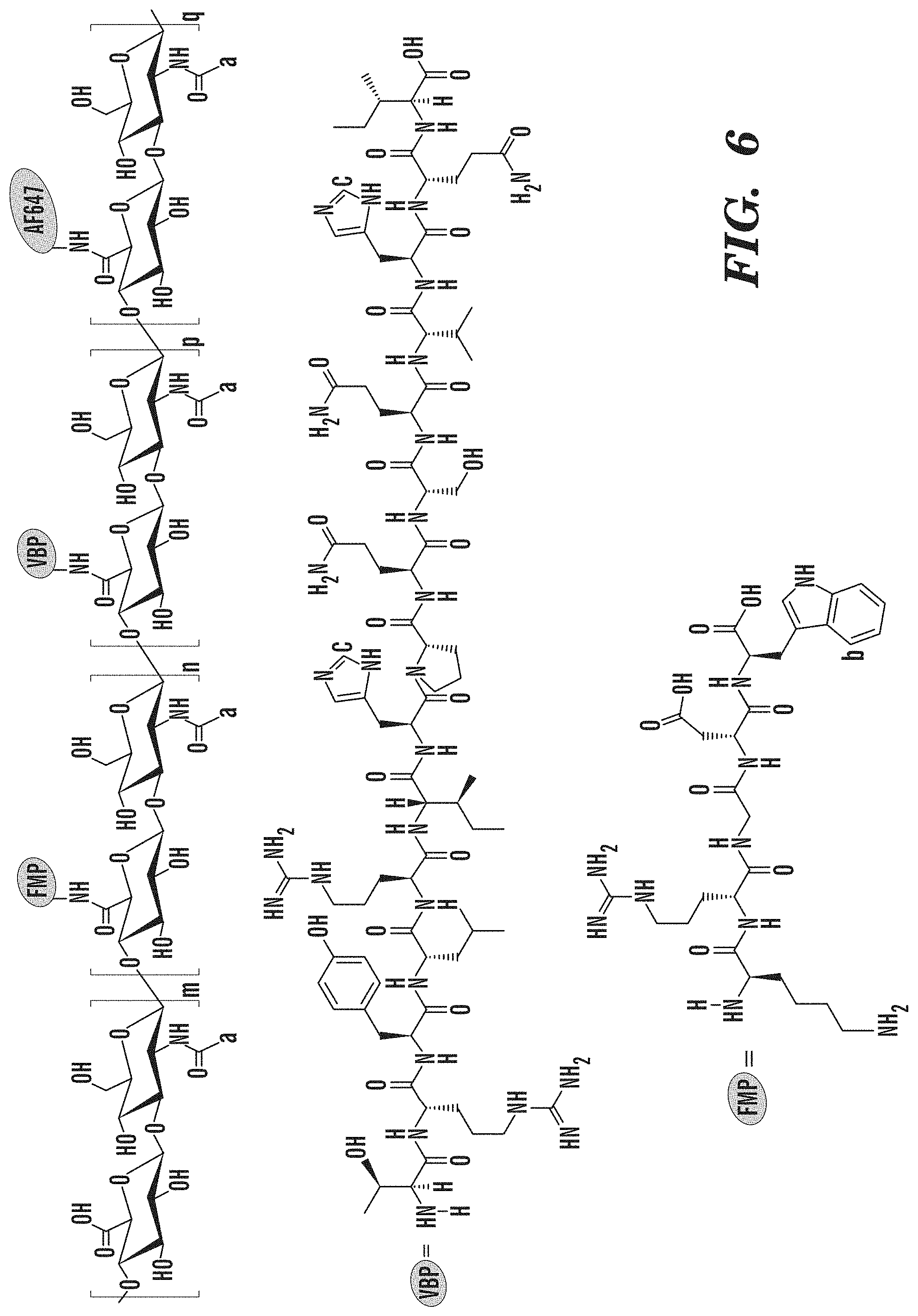
[0045] Conjugation of functional moieties to linkers, and linkers to substrate materials can be accomplished using any conjugation chemistries known in the art which are compatible with the shell material, linker composition, and makeup of the functional moiety. For example, PAMAM dendrimers with terminal COOH-- groups can be activated with carbonyldiimidazole and subsequently linked with peptide amino groups to conjugate linkers to functional moieties comprising peptides or other compounds having a terminal amino group. Likewise, EDC/NHS coupling, as known in the art, can be used to link dendrimer-peptide conjugates to the surface of a proteinaceous shell. In general, any carbodiimide chemistry can be used to conjugate linkers to functional moieties and to shell materials. Other chemistries that could be employed include activation of the carboxyl group on the dendrimer with any agent that forms a carboxyl chloride, or an activated ester, to react with the nucleophiles on the shell.
[0046] The density of functional moieties per body may vary considerably, from less than 50,000 per body to >100 million per body. For example 10-30 million functional moieties may be present on a shell of 200 nm in diameter. The density of functional moieties on the shell can be tuned by controlling the concentration of the dendrimer-peptide conjugates in the coupling solution during the reaction with the shell. Alternatively, by controlling the size, and thus the surface area, of the substrate used.
[0047] In some embodiments, the particles described herein can comprise an active agent and/or active ingredient. As used herein, the term "active agent" refers to an agent which, when released in vivo, possesses the desired biological activity, for example, therapeutic, diagnostic and/or prophylactic properties in vivo. It is understood that the term includes stabilized and/or extended release-formulated pharmaceutically active agents. Exemplary pharmaceutically active agents include, but are not limited to, those found in Harrison's Principles of Internal Medicine, 19th Edition, Eds. T. R. Harrison et al. McGraw-Hill N.Y., N.Y.; Physicians Desk Reference, 71st Edition, 2017, Oradell New Jersey, Medical Economics Co.; Pharmacological Basis of Therapeutics, 8th Edition, Goodman and Gilman, 2017; the current edition of the United States Pharmacopeia, The National Formulary; current edition of Goodman and Oilman's The Pharmacological Basis of Therapeutics; and current edition of The Merck Index, the complete content of all of which are herein incorporated in its entirety. In some embodiments, an active agent and/or ingredient can be a functional moiety. In some embodiments, an active agent and/or ingredient is not a functional moiety, e.g., it is not conjugated or attached to the substrate/shell. In some embodiments, the active agent and/or ingredient is tPA.
[0048] In one aspect, the invention broadly encompasses any functional particle comprising a substrate (e.g. a shell or polymeric material), linkers, and functional moieties, for example: wherein the shell is a layer-by-layer structure, for example a structure comprising one or more BSA-PAH bilayers, which such layer-by-layer structure is formed around a particulate core, the core optionally being degraded or dissolved subsequent to formation of the shell; wherein the linkers may comprise branched linkers, for example dendritic linkers such as PAMAM dendrimers; and wherein the one or more functional moieties comprise a biologically active or biologically targeting agent. It will be understood that variants of the above structure fall within the scope of the invention, for example the shell may comprise a structure other than a layer-by-layer structure (for example, the polymer-conjugates described above) or the linkers may be linear rather than branched.
[0049] For convenience, the systems described herein are directed to humans, including peptide sequences which are derived from or are otherwise biocompatible with humans. The invention further encompasses functionalized particles comprising materials that may be used in canines, felines, rats, mice, cows, pigs, monkeys, and other species, including appropriate homologs or orthologs of the human sequences or sequences described herein.
[0050] The invention further encompasses methods of utilizing the functionalized particles or polymers described herein. In one embodiment, the invention comprises the administration of a functionalized particle to an animal in need of treatment, the functional moieties of the particle being effectors of the required treatment. Such administration may be intravenous, topical, or may comprise a localized injection or other delivery. It will be understood that the functionalized particles or polymers may be administered in or with pharmaceutically acceptable carriers, including for example, solutions, gels, or particulates. The invention further encompasses kits, wherein such kits may comprise: functionalized particles in combination with pharmaceutically acceptable carriers; functionalized particles in combination with adjunct or accessory agents (e.g. drugs); and functionalized particles in combination with delivery mechanisms, such as syringes, hypodermic needles, and intravenous needles. The invention includes the use of the compositions described herein in bandages, dressings, sutures, and other wound treatment articles. The invention further encompasses methods of delivering functionalized particles to cells, including isolated cells, tissue explants, cultured cells, and others. The methods of the invention encompass medical therapeutic treatment of humans, veterinary treatments, and research uses.
[0051] In some embodiments, the particles described herein are about 1,000 nm or less in diameter. In some embodiments, the particles described herein are 1,000 nm or less in diameter. In some embodiments, the particles described herein are about 500 nm or less in diameter. In some embodiments, the particles described herein are 500 nm or less in diameter. In some embodiments, the particles described herein are about 400 nm or less in diameter. In some embodiments, the particles described herein are 400 nm or less in diameter. In some embodiments, the particles described herein are about 300 nm or less in diameter. In some embodiments, the particles described herein are 300 nm or less in diameter. In some embodiments, the particles described herein are about 200 nm or less in diameter. In some embodiments, the particles described herein are 200 nm or less in diameter. In some embodiments, the particles described herein are about 100 nm or less in diameter. In some embodiments, the particles described herein are 100 nm or less in diameter.
[0052] Various embodiments of the invention are described next.
[0053] Synthetic Platelets for Promoting Platelet Aggregation. In one embodiment, the invention comprises synthetic platelets that promote thrombus formation in wounded tissues. The thrombogenic synthetic platelets of the invention comprise the general functionalized particle configurations described above, wherein the one or more functional moieties comprise a wound-targeting ligand and a thrombogenic agent, for example a platelet aggregation agonist or platelet binding agent. The wound-targeting ligand comprises any agent which selectively binds to peptides or other species which are presented by damaged endothelium cells. For example, in one embodiment, the wound-targeting ligand comprise a collagen binding agent. In another embodiment, the wound-targeting ligand comprises a von Willebrand binding agent. In another embodiment, the platelet aggregation agonist or platelet binding agent comprises a fibrinogen mimetic. In one implementation the platelet aggregation promoting particles comprise three functional moieties: a collagen-binding peptide (CBP); a von Willebrand binding peptide (VBP); and a fibrinogen mimetic peptide (FMP). Typically, these functional moieties will comprise peptides. These three functional moieties act synergistically to effectively promote thrombus formation. Specifically, the CBP and VBP moieties promote adhesion to fibrinogen while the FMP moiety enhances cross-binding to native platelets, aiding in thrombus formation. In some embodiments, the CBP and VBP moieties promote adhesion to collagen and von Willebrand factor while the FMP moiety enhances cross-binding to native platelets, aiding in thrombus formation.
[0054] In some embodiments of any of the aspects, the particle comprises CBP and VBP. In some embodiments of any of the aspects, the particle comprises CBP and FMP. In some embodiments of any of the aspects, the particle comprises VBP and FMP.
[0055] In some embodiments of any of the aspects, the particle comprises CBP and VBP and a hyaluronic acid substrate. In some embodiments of any of the aspects, the particle comprises CBP and FMP and a hyaluronic acid substrate. In some embodiments of any of the aspects, the particle comprises VBP and FMP and a hyaluronic acid substrate. In some embodiments of any of the aspects, the particle comprises VBP, CBP, and FMP and a hyaluronic acid substrate.
[0056] In some embodiments of any of the aspects, the particle comprises CBP and VBP attached directly to a hyaluronic acid substrate. In some embodiments of any of the aspects, the particle comprises CBP and FMP attached directly to a hyaluronic acid substrate. In some embodiments of any of the aspects, the particle comprises VBP and FMP attached directly to a hyaluronic acid substrate. In some embodiments of any of the aspects, the particle comprises VBP, CBP, and FMP attached directly to a hyaluronic acid substrate.
[0057] In some embodiments of any of the aspects, the particle comprises multiple functional moieties, which each at a ratio of 1:1 to each other. In some embodiments of any of the aspects, the particle comprises multiple functional moieties, which each at a ratio of about 1:1 to each other. In some embodiments of any of the aspects, the particle comprises multiple functional moieties, which each at a ratio of from 2:1 to 1:2 to each other. In some embodiments of any of the aspects, the particle comprises multiple functional moieties, which each at a ratio of from about 2:1 to about 1:2 to each other.
[0058] In some embodiments of any of the aspects, the particle comprises multiple functional moieties, which each at a ratio of from 5:1 to 1:5 to each other. In some embodiments of any of the aspects, the particle comprises multiple functional moieties, which each at a ratio of from about 5:1 to about 1:5 to each other. In some embodiments of any of the aspects, the particle comprises multiple functional moieties, which each at a ratio of from 10:1 to 1:10 to each other. In some embodiments of any of the aspects, the particle comprises multiple functional moieties, which each at a ratio of from about 10:1 to about 1:10 to each other.
[0059] The CBP moiety of the synthetic platelets of the invention comprises any collagen-binding peptide or other collagen-binding agent known in the art. The task of this moiety is to achieve adhesion of the artificial platelet to wounded tissues (where collagen has become exposed to the blood) at low blood flow rates. For example, in one embodiment, the CBP moiety of the invention comprises SEQ ID NO: 1, known as [GPO].sub.7, a seven-mer of the tripeptide glycine- proline-hydroxyproline. For example, see Kehrel B., Wierwille S., Clemetson K. J., Anders O., Steiner M., Knight C. G., Farndale R. W., Okuma M., Barnes M. J. "Glycoprotein VI is a major collagen receptor for platelet activation: it recognizes the platelet-activating quaternary structure of collagen, whereas CD36, glycoprotein IIb/IIIa, and von Willebrand factor do not." Blood 1998; 91: 491-9; and Farndale R. W., Sixma J. J., Barnes M. J., de Groot P. G., The role of collagen in thrombosis and haemostasis. J Thromb Haemost 2004; 2: 561-73.
[0060] Other exemplary CBP peptides include, for example, sequences described in Munnix et al., 2008, "Collagen-mimetic peptides mediate flow-dependent thrombus formation by high- or low-affinity binding of integrin a2b1 and glycoprotein VI," Journal of Thrombosis and Haemostasis, 6: 2132-2142.
[0061] The von Willebrand binding peptide, VBP, of the synthetic platelets of the invention comprises any peptide or other agent that effectively binds von Willebrand factor. The task of this moiety is to achieve adhesion of the artificial platelet to wounded tissues, where von Willebrand factor has become exposed to the blood, at high blood flow rates. In one embodiment, the VBP may comprise a peptide having SEQ ID NO: 2 or 7, which is a human-derived sequence, abstracted from Factor VIII, which is a natural protein ligand for the vWF. Other exemplary VBP's include: those described in published PCT patent application number WO 2007052067, entitled "Von willebrand factor (vwf) binding peptides," by Farndale et al.; sequences described in Moriki et al., 2010, "Identification of ADAMTS13 Peptide Sequences Binding to Von Willebrand Factor," Biochemistry Biophys Res Commun, 391:783-788; and sequences described in Lisman et al., 2006, "A single high-affinity binding site for collagen on von Willebrand factor in collagen III, identified using synthetic triple helical peptides," Blood 108: 3753-56. VBP may further include peptides described in Huang, et al., "Affinity purification of von Willebrand factor using ligands derived from peptide libraries," Bioorg Med Chem. 1996 May;4(5):699-708, for example SEQ ID NO: NO 6.
[0062] The thrombogenic moiety of the synthetic platelets of the invention comprises any agent which promotes thrombogenesis at wound sites (while avoiding off-target thrombus formation). The thrombogenic moiety may comprise a fibrinogen mimetic peptide, or any other agent which effectively binds the GPIIb/IIIa site of activated platelets, which aids in crosslinking activated platelets and promotes platelet aggregation at the wound site. In one embodiment, the FMP comprises SEQ ID NO: 3. In another embodiment, the FMP moiety of the synthetic platelets of the invention may comprise SEQ ID NO: 4, which is less likely to induce off-target platelet activation than SEQ ID NO: 3. In one embodiment, the FMP comprises SEQ ID NO: 8. Any RGD-based fibrinogen peptide may be used. Non-peptide binders of fibrinogen may also be employed, for example as described in Sugihara, et al., "Novel Non-Peptide Fibrinogen Receptor Antagonists. 1. Synthesis and Glycoprotein IIb-IIIa Antagonistic Activities of 1,3,4-Tri substituted 2-Oxopiperazine Derivatives Incorporating Side-Chain Functions of the RGDF Peptide," ("RGDF" disclosed as SEQ ID NO: 9) J. Med. Chem., 1998, 41 (4), pp 489-502.
[0063] With respect to the specific functional moieties described for the synthetic platelets, and for all other embodiments of the functionalized particles and modified cells described below, it is understood that one of skill in the art may readily select and utilize functional analogs of the peptides disclosed herein in place of the described peptides. For example, any molecule, including for example small molecules, peptides, proteins or fragments thereof, polymers, antibodies, modified sequences (including sequences comprising non-natural amino acids) or any other composition of matter capable of effecting the same or similar physiological response may be used in place of the exemplary peptides set forth herein. Likewise, one of skill in the art may select variants of SEQ ID NO: 1, SEQ ID NO: 2, SEQ ID NO: 3, SEQ ID NO: 4, SEQ ID NO: 5, SEQ ID NO: 6, SEQ ID NO: 7, SEQ ID NO: 8 and other sequences described herein. As used herein, a variant of a peptide is a sequence having one or more amino acid substitutions and which retains some or all of the original peptide's function.
[0064] The relative proportions of CBP, VBP, and FMP in the synthetic platelets of the invention may vary. For example, CBP:VBP:FMP ratios of 1:1:1, 1:1:2, or 1:1:3 may be utilized. It will be understood that in some alternative embodiments, one binding moiety, e.g. CMB or VBP, may be omitted, however, the use of both enhances particle effectiveness.
[0065] The synthetic platelets described herein may be utilized in various ways. For example, in one embodiment, the invention comprises a method wherein synthetic platelets are injected into the blood stream of an animal, e.g. a human patient, in need of treatment, e.g. having a wound or otherwise needing hemostatic treatment, for example, in order to enhance thrombus formation at externally accessible or internally bleeding sites. For intravenous injection, doses in the range of 0.0001%- 500% of the normal platelet concentration of the treated animal species may be utilized. For example, synthetic platelet does of 3-75 mg/kg, for example 15 mg/kg may be used. In an alternative method, the synthetic platelets are applied topically to wounds or surgical incisions of an animal, e.g. a human patient and may be incorporated into bandages, dressings, etc.
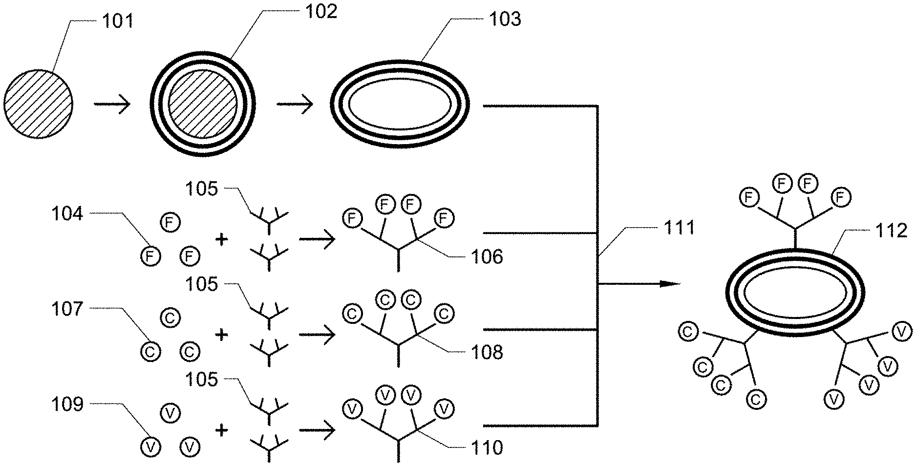
[0066] An exemplary synthesis of the artificial hemostatic platelets of the invention is depicted schematically in FIG. 1. Here, a core 101 is coated with a polymer shell 102. After the shell is synthesized, the core is dissolved, leaving a hollow, flexible, discoid body formed by the shell 103. In a first reaction, fibrinogen mimetic peptides 104 are conjugated with dendrimers 105, yielding dendrimer-conjugated fibrinogen mimetic peptides 106. In a separate reaction, collagen binding peptides 107 are conjugated with dendrimers 105, yielding dendrimer-conjugated collagen-binding peptides 108. In a third separate reaction, von Willebrand-binding peptides 109 are conjugated with dendrimers 105, yielding dendrimer conjugated von Willebrand-binding peptides 110. In a single reaction 111, the three batches of dendrimer-conjugated peptides are reacted with activated shells, yielding shells that are decorated with dendrimers conjugated to the three functional peptides 112.
[0067] With reference to FIG. 2A and FIG. 2B, these schematic diagrams depict wound sites. In FIG. 2A, healthy endothelial cells 101 surround a wound site, where wound-specific factors 102 are exposed. Red blood cells 103 are escaping from the wound site, as platelets 104 are not abundant enough to form a thrombus. In FIG. 2B, the synthetic platelets of the invention 205 are present, and a thrombus 206 is rapidly formed to seal the wound.
[0068] Wound Healing Particles. Subsequent to clot formation, wound healing processes repair the damaged tissue. Various wound healing peptides (WHP's) are known to promote the healing process. Histatin or derivatives thereof, for example, may act as a wound healing peptide. For example, WHP's include those described in Demidova-Rice et al., "Human Platelet-Rich Plasma- and Extracellular Matrix-Derived Peptides Promote Impaired Cutaneous Wound Healing In Vivo," 2012, PLOS ONE, DOI: 10.1371/journal.pone.0032146; Demidova-Rice et al., "Bioactive peptides derived from vascular endothelial cell extracellular matrices promote microvascular morphogenesis and wound healing in vitro," Wound Repair Regen. 2011 19(1):59-70; United States Published Patent Application number 2013010861, entitled "Wound Healing Peptides and Uses Thereof," by Herman et al.; and frog skin-derived peptides described in Liu et al., "A potential wound healing-promoting peptide from frog skin," Int J Biochem Cell Biol. 2014 April;49:32-41, SEQ ID NO: 6.
[0069] In one embodiment, the invention comprises wound healing particles. The wound healing particles comprise the basic functionalized particle configurations described above, i.e. a shell and linkers having terminal functional moieties, and further comprise: (1) a wound-targeting ligand; and (2) a wound healing agent. The wound-targeting ligand may be any agent which selectively binds to species that are present in wounded tissues, e.g. collagen, von Willebrand factor, etc. The wound healing agent may be any agent that promotes healing of wounds.
[0070] In the wound healing particles, the wound binding moieties may comprise CBP, VBP, and other compounds known in the art to selectively bind to wound specific sites, motifs, cells, etc. For example, arginylglycylaspartic acid (RGD) and peptides based thereon may be used. The wound healing agent may comprise those listed above or any other known in the art. The wound binding moieties, e.g. CBP and VBP, target the wound healing particles to wound sites, wherein the WHP's can promote healing processes at the wound site.
[0071] The invention further comprises methods of administering such wound healing particles to an animal in need of treatment, e.g. a human patient having one or more wounds or wounded tissues. Wound healing particle doses may be administered at any physiologically effective dose which does not cause excessive adverse side effects, for example, doses of 3-75 mg/kg, for example 15 mg/kg may be used.
[0072] Anti-thrombotic or Thrombolytic Particle. Pathological thrombosis is implicated in many conditions. To treat such conditions, anti-thrombosis or thrombolytic compounds need to be delivered to the site of the undesired clotting activity or clot. In one embodiment, the invention comprises particles capable of binding to clotting sites and delivering anti-thrombotic or thrombolytic agents. The anti-thrombotic or thrombolytic particles of the invention comprise the basic functionalized particle configurations described above, i.e. a shell and linkers having terminal functional moieties.
[0073] The functional moieties of the anti-thrombotic particles include (1) a targeting agent that binds the particle to factors found in clotting sites, such as activated platelets, and (2) an anti-thrombotic agent. In one embodiment, SEQ ID NO: 4 may be used as the targeting entity which binds the particle to clot-specific or clot-associated factors at the site of the pathological event. Alternatively, SEQ ID NO: 3 or 8 may be used as well. In one embodiment, heparin is utilized as the anti-thrombotic agent. Other thrombolytic drugs or peptides known in the art may be used in combination with or in place of heparin.
[0074] The functional moieties of the thrombolytic particles include (1) a targeting agent that binds the particle to wounded tissue sites, and (2) thrombolytic agents that degrade clots or promote clot dissolution. In one embodiment, SEQ ID NO: 4 may be used as the targeting entity which binds the particle to clot-specific or clot associated factors at the site of the pathological event. In one embodiment, tPA is utilized as the thrombolytic moiety.
[0075] It will be noted that protein-based thrombolytic agents like tPA are very large molecules and can decrease the display of the smaller targeting peptide by steric crowding. It is possible to obviate to this problem by binding the thrombus targeting peptide onto a long PEG or other tethering moiety, the length being that which will place the targeting peptide at about the same plane as the large tPA or other large thrombolytic molecule. For example a PEG molecule of 500 to 5,000 kDa may be used.
[0076] In an alternative embodiment, the thrombolytic platelets of the invention comprise a tPA-recruiting particle, comprising a targeting functional moiety, such as SEQ ID NO: 4, and a tPA ligand, which such ligand is capable of binding tPA in an active configuration. An exemplary tPA ligand is the peptide SEQ ID NO: 5. Such tPA-recruiting particles can bind to the clot by means of the targeting moiety and then the tPA ligand will recruit native or co-administered tPA to that region, which in turn aids in clot dissolution.
[0077] In one aspect, the invention comprises methods of administering anti-thrombotic particles, thrombolytic particles, or tPA-recruiting particles to an animal in need of anti-thrombotic or thrombolytic treatment, e.g. a human patient, for example having undesirable clotting activity or clots. For example, the particles may be administered to an animal to aid in dissolving deep venous thrombi. Administration of the particles may be intravenous or topical. In one implementation, the invention comprises the administration of tPA-recruiting particles in combination with tPA. Anti-thrombosis, thrombolytic, and tPA-recruiting particles each may be administered at any physiologically effective dose which does not cause excessive adverse side effects, for example, doses of 3-75 mg/kg, for example 15 mg/kg, may be used.
[0078] In some embodiments of any of the aspects, an agent or moiety described herein (e.g, a thrombolytic agent) can be provided in the interior of the particle instead of attached to the exterior of the substrate. Methods of manufacturing such particles are described, e.g., in Example 5 herein.
[0079] In some embodiments of any of the aspects, the particles comprise one or more functional moieties selected from: a wound targeting ligand; a platelet binding agent; a wound binding peptide; a wound healing peptide; a tPA-binding moiety; a wound or clot binding moiety; a hemostatic peptide; an anti-thrombotic agent; and a thrombolytic agent.
[0080] In some embodiments of any of the aspects, described herein is a particle comprising one or more of the functional moieties selected from: a peptide comprising SEQ ID NO: 1; a peptide comprising SEQ ID NO: 2; a peptide comprising SEQ ID NO: 3; a peptide comprising SEQ ID NO: 4; a peptide comprising SEQ ID NO: 5; a peptide comprising SEQ ID NO: 7; a peptide comprising SEQ ID NO: 8; heparin; and tPA. In some embodiments of any of the aspects, described herein is a particle comprising two or more of the functional moieties selected from: a peptide comprising SEQ ID NO: 1; a peptide comprising SEQ ID NO: 7; and a peptide comprising SEQ ID NO: 8.In some embodiments of any of the aspects, described herein is a particle comprising the functional moieties of: a peptide comprising SEQ ID NO: 1; a peptide comprising SEQ ID NO: 7; and a peptide comprising SEQ ID NO: 8 and further comprising tPA on the interior of the particle.
[0081] Modified Red Blood Cells. In a further aspect, the invention comprises red blood cells (or fragments thereof) which are modified to perform platelet functions. The modified red blood cells of the invention comprise red blood cells (or fragments thereof) which display agents having platelet functions, for example wound binding functions and thrombogenic agents such as platelet aggregation agonists or platelet binding agents. For example in one embodiment, the modified red blood cells of the invention display CBP, VBP, and FMP peptides tethered to their surface membrane. In this implementation of the invention, the red blood cells take the place of the shell as the substrate for functional moiety attachment. As with the synthetic platelets of the invention, the surface-bound CBP, VBP, and FMP will effect binding to wounded tissues and promotion of platelet aggregation. Such active moieties may be tethered to the outer membrane of the red blood cell using linear or branched linkers, for example PAMAM dendrimers. Such active moieties may be present in the same relative proportions described above for artificial thrombogenic platelets, for example, CBP:VBP:FMP ratios of 1:1:1 or 1:1:2. Any surface concentration of functional moieties, for example 50,000 to 100 million functional moieties per red blood cell, may be utilized.
[0082] Any methodology known in the art for surface modification of red blood cells may be used to attach the functional moieties to the red blood cell membrane, for example as described in: Henry SM, "Modification of red blood cells for laboratory quality control use," Curr Opin Hematol. 2009 November;16(6):467-72; United States Pat. No. 6,946,127, entitled "Decorated red blood cells," by Bitensky et al.; and U.S. Pat. No. 8,211,656, entitled "Biological Targeting Compositions and Methods of Using the Same" by Hyde et al.
[0083] The invention further comprises methods of administering modified red blood cells having platelet functions to an animal, e.g. a human patient, in need of treatment, for example, in order to enhance thrombus formation at wounded, e.g. internally bleeding sites. Administration may be intravenous, topical, or may comprise a localized injection. Modified red blood cell may be administered at any physiologically effective dose which does not cause excessive adverse side effects, for example, doses of 3-75 mg/kg, for example 15 mg/kg may be used.
EXAMPLES
Example 1
[0084] In this example, artificial thrombogenic platelets, referred to as SynPlats, were made and are tested in live animals.
[0085] Synthetic nanoplatelets fabrication. 200 nm carboxylate PS spheres (Polysciences, Warrington, Pa.) were suspended in 0.5 M sodium chloride (Fisher). 2 mg/ml of positively-charged Poly(allylamine) hydrochloride (Sigma) was dissolved in 0.5 M sodium chloride and incubated with 3.times.10.sup.12 particles at room temperature under constant rotation for 30 minutes. Particles were then centrifuged at 15000 g for 30 minutes and resuspended in 0.5 M sodium chloride. Particles were washed 2 more times at 15000 g for 30 minutes in 0.5 M sodium chloride. Following PAH coating, negatively-charged bovine serum albumin (Sigma) was coated onto of PAH layers under identical conditions. This procedure was repeated for multiple (PAH/BSA) bilayers. Intermittent crosslinking with 2% glutaraldehyde (Polysciences) for 1 hour under constant rotation was performed to ensure sufficient structural integrity of the outer shells. The particles were then exposed to a tetrahydrofuran-isopropyl alcohol gradient (1:3, 1:2, 1:1, 2:1, and pure THF) for 30 minutes each at room temperature under constant rotation so as to dissolve the PS core. Particles were then washed 10.times. with saline (BD), so as to remove any residual solvent, sterilized via UV overnight and stored at 4C for no longer than 2 days.
[0086] Peptide conjugation to synthetic nanoplatelets. In this example, CBP comprising SEQ ID NO: 1, VBP comprising SEQ ID NO: 2, and FMP comprising SEQ ID NO: 3 were utilized. Each of the three peptides were, in separate batches, coupled to poly(amido amine) (PAMAM) dendrimers (Sigma) via EDC/NHS chemistry in MES buffer at pH=4.5. Purification of dendrimer-peptide conjugates was performed via size exclusion chromatography. The outer layer of SynPlats were activated with cabonyldiimidazole (CDI) at 1 mg/ml in acetone for 45 minutes and diaminoethane was then added to form primary amino groups on SynPlat surface. All SynPlats were tested qualitatively for free amines via the Kaiser test. Finally, dendrimer-peptide conjugates were conjugated to SynPlats via EDC/NHS chemistry in MES buffer at 4.5 pH for 12 hours.
[0087] Scanning electron microscopy (SEM). An FEI XL40 SEM at 3-10 kV with a 5 mm working distance was used for imaging particles. Samples were coated with palladium (at 10 kV) via a Hummer sputtering system.
[0088] In vivo hemostasis. 3.times.10.sup.1 SynPlats in saline were injected via tail vein into healthy female BALB/c mice (18-20 g; n=3-6 per group). 5 minutes after injection, 2 mm long sections of the tail, from the distil tip, were amputated. The amputated tail was immediately immersed in 14 ml of sterile saline at 37C. The times until bleeding from the amputated tail stopped were recorded.
[0089] In vivo biodistribution. 3.times.10.sup.10 SynPlats, either plain or conjugated with CBP-VBP-RGD peptides, in saline were injected via tail vein into healthy female BALB/c mice (18-20 g; n=3-6 per group). 5 minutes following injection, 2 mm long sections of the tail, from the distil tip, were amputated. 55 minutes following tail amputation, animals were sacrificed via CO.sub.2 overdose and organs were collected. The organs were dissolved overnight in Solvable at a concentration of 100 mg organ per 1 ml of Solvable. Dissolved organ solutions were measured for their fluorescence at a concentration of 2 mg of organ per 200 ul Solvable. An identical amount of each organ from non-injected control animals were subtracted from each organ value for CBP-VBP-FMP SynPlats and plain SynPlat groups.
[0090] FTIR. All FTIR samples were suspended in identical concentrations and volumes of a water/acetone mixture. Samples were pipetted onto a zinc selenide ATR crystal and water/acetone mixture was evaporated completely leaving a film of the sample. Samples were then placed into an FTIR spectrometer (NICOLET 4700, Thermo Electron Corporation) and the chamber was purged with nitrogen for 30 minutes. Dry crystal backgrounds were subtracted from each sample's spectrum.
[0091] Tail Sectioning. The tail samples were harvested and then immediately frozen in OCT compound and sectioned at a thickness of 15 microns on a cryotome (Leica). After sectioning, the tail sections were mounted on a glass slide. 30 ul of Permount mounting medium (Fisher) were placed on top of the tail sections along with a glass cover slip to seal the slides.
[0092] Confocal Microscopy. Individual imaging of SynPlats was performed on a Olympus Fluoview 1000 using differential interference contrast mode. A BX60 microscope was used to image tail sections.
Results
[0093] Synthesis and Characterization of SynPlats. Synthetic nanoplatelets (SynPlats) were synthesized using the layer-by-layer (LbL) approach, a proven method for creating a variety of flexible capsules that are mechanically and morphologically similar to circulatory cells. Briefly, 200 nm spherical polystyrene (PS) nanoparticles were coated with alternative layers of poly(allyamine) hydrochloride (PAH) and bovine serum albumin (BSA). PAH and BSA were chosen as the polycation and polyanion, respectively, due to their reliability in capsule synthesis via LbL as well as use as materials for numerous biomedical applications, for example, as described in del Mercato, L. L., et al., LbL multilayer capsules: recent progress and future outlook for their use in life sciences. Nanoscale, 2010. 2(4): p. 458-67; Johnston, A. P., et al., Layer-by-layer engineered capsules and their applications. Current Opinion in Colloid & Interface Science, 2006. 11(4): p. 203-209; and Yan, Y., M. Bjornmalm, and F. Caruso, Assembly of Layer-by-Layer Particles and Their Interactions with Biological Systems. Chemistry of Materials, 2013.
[0094] SynPlats were characterized at each step for sufficient PAH/BSA coating via fluormetric assays. Briefly, PAH-AF594 and BSA-AF488 were complementarily coated and the fluorescent intensity for each dye was determined at each coating layer. 4 bi-layers were used in this example. The linear relationship of independently dyed polyelectrolytes implies uniform coating of both PAH and BSA. Coating was also confirmed qualitatively via confocal imaging of the final SynPlat product. PS core removal was performed via incubation with tetrahydrofuran (THF) and isopropyl alcohol (IPA) at increasing THF:IPA ratios. PS core removal was confirmed via FTIR. Since SynPlats are comprised of PS, PAH and BSA many similar peaks overlap and become difficult to resolve. However, wavenumbers 700 cm.sup.-1 and 760 cm.sup.-1 represent polystyrene peaks that are absent in both BSA and PAH. The resultant particles following removal of the core were oblate ellipsoidal in shape, which resembles natural platelets.
[0095] Peptide Conjugation to SynPlats. The size of the particles (.about.200 nm), however, was much smaller than that of natural platelets (.about.2 mm). This was done in order to avoid cardiopulmonary interference. Specifically, particles larger than lung capillaries are known to physically get trapped in lungs (the first capillary bed encountered following tail vein injection) which can impede the passage of blood, effectively impairing oxygen delivery. Further, SEQ ID NO: 3 peptide has been shown to lead to lung targeting of hemostatic particles to lungs (over 50% of injected dose). These same SEQ ID NO: 3 particles, at high doses (40 mg/kg), have been shown to induce cardiopulmonary complications in animals; a finding substantiated by the fact that the parent protein of SEQ ID NO: 3, fibrinogen, also targets lung tissue in addition to inducing hemostasis. The combination of high lung targeting and physical entrapment due to large size can potentially lead to cardiopulmonary issues. To circumvent these issues, two steps were taken: (i) smaller (200 nm) templates were used so as to prevent physical entrapment in lung tissue and (ii) lower particle doses (15 mg/kg) were used.
[0096] These three peptides of the SynPlats, a CBP comprising SEQ ID NO: 1, a VBP comprising SEQ ID NO: 2, and a FMP comprising SEQ ID NO: 3 have been shown to act synergistically to promote hemostasis more efficiently, as described in Modery-Pawlowski, C. L., et al., In vitro and in vivo hemostatic capabilities of a functionally integrated platelet-mimetic liposomal nanoconstruct. Biomaterials, 2013. 34(12): p. 3031-41. Specifically, CBP and VBP promote adhesion at low and high shear, respectively, which are excluded from the overwhelming majority of platelet substitute designs. Conjugation of hemostatic peptides to SynPlats must be done in a way so as to ensure that peptides do not detach from SynPlats in vivo in order to avoid off-site activation of platelets. Further, due to the likelihood of peptides non-specifically interacting with the BSA rich surface on SynPlats and potentially unable to bind to target sites, peptides must be presented in a way so as to avoid direct interaction with the globular BSA-terminated layer of SynPlats to ensure high binding avidity and selectively. To accomplish this goal, branched dendrimers were used to first bridge the covalent attachment of the peptides to SynPlat surface. Briefly, the peptides were coupled to poly(amido amine) (PAMAM) dendrimers via EDC/NHS chemistry and the outer layer of SynPlats were activated with cabonyldiimidazole (CDI) where diaminoethane was then added to form primary amino groups on SynPlat surface. Dendrimer-peptide conjugates, were mixed and then directly conjugated to SynPlats via EDC/NHS chemistry. Peptide conjugation was quantified via fluorescent labeling of dendrimers and confirmed qualitatively via confocal microscopy. The number of peptide molecules per SynPlat was (in .times.10{circumflex over ( )}6 peptides/particle): CBP: 6.86.+-.0.13; VBP: 7.08.+-.0.18; and RGD: 14.7.+-.0.15.
[0097] Triggering of Hemostasis In Vivo Using SynPlats. SynPlats were next investigated in vivo for their ability to halt bleeding in a standard tail transection model in BALB/c mice. SynPlats without peptides and saline injections alone showed no decrease in tail bleeding times. SynPlats functionalized with SEQ ID NO: 3 peptide alone lowered bleeding time by .about.45%. However, SynPlats functionalized with all three peptides (SEQ ID NO: 1, SEQ ID NO: 2; and SEQ ID NO: 3) lowered bleeding time by .about.65%. Further, micron sized SynPlats were unable to instigate hemostasis to the same extent as their 200 nm counterparts, likely due to the lower circulation time of micron sized particles. Non-flexible spherical 200 nm SynPlats with the PS core, identically decorated to their flexible counterpart, were unable to cause hemostasis as rapidly as the more flexible, disc-shaped, SynPlats. Organ distribution for 200 nm SynPlats with and without peptide functionalization showed similar organ distribution except in case of the tail section containing the clot. In this case, a 3-fold increase in SynPlats functionalized with CBP-VBP-RGD peptides in the tail section containing the clot was seen over plain SynPlats.
[0098] The results demonstrated the ability of SynPlats to significantly reduce the bleeding time. In case of normal hemostatic plug formation, circulating platelets become activated and bind to the damaged endothelium due to exposure of collagen and release of vWF from wound site. In case of hemostatic plug formation following injection of SynPlats, activated circulating platelets and SynPlats both bind to injured endothelium, as well as to each other, effectively forming the hemostatic plug much faster than in the absence of SynPlats. Brightfield and fluorescent images show the interaction between fluorescently labeled SynPlats and the clot. On average, the hemostatic plug took around 195 seconds to form when it consisted of just natural circulating platelets. However, after an injection of SynPlats, the hemostatic plug formed in 35% of the time it took when no SynPlats were injected. The SynPlats described here offer a new tool for the treatment of serious hemorrhage.
[0099] All patents, patent applications, and publications cited in this specification are herein incorporated by reference to the same extent as if each independent patent application, or publication was specifically and individually indicated to be incorporated by reference. The disclosed embodiments are presented for purposes of illustration and not limitation. While the invention has been described with reference to the described embodiments thereof, it will be appreciated by those of skill in the art that modifications can be made to the structure and elements of the invention without departing from the spirit and scope of the invention as a whole.
Example 2
[0100] Further described herein are hyaluronic-acid-hemostatic peptide conjugates, platelet-like nanoparticles (PLN or SynPlat), and thrombolytic particles.
[0101] Hyaluronic-acid-hemostatic peptide conjugates. In the foregoing examples, the use of hyaluronic acid as a substrate for functional moieties is described. In the following example, specific exemplary hyaluronic-acid-hemostatic peptide conjugates are provided.
[0102] Functionalized Polymers. Another implementation of the invention encompasses the use of polymers, as opposed to proteins, as the functional agent-harboring scaffold or substrate. Likewise, hybrid substrates comprising bilayers of protein and polymeric material may be employed as the substrate. It will be understood that the methods of functionalizing shells and utilizing functionalized shells described herein are equally applicable to functionalized particles wherein a polymeric material not configured as a shell replaces the shell as the substrate for functional moieties. Examples of polymers that can be used include hyaluronic acid, polyvinyl alcohol, or polylactic-co-glycolic acid. As with the shell, linkers are anchored on the polymer chain, the linkers having functional moieties bonded to the terminal ends of the linkers.
[0103] Platelet-like nanoparticles (PLN or SynPlat), Provided herein are optimized conditions for manufacturing the particles.
[0104] Thrombolytic particles. Also described herein are thrombolytic particles, e.g., CaCO.sub.3 microspheres with drug loaded inside. The nanoparticles and/or microparticles still have a substrate with FMP, VBP, and CBP attached, but the difference is that the substrate is made from CaCO.sub.3 and the particles are now loaded with a drug inside (e.g., tPA).
Example 3
Hyaluronic Acid-Hemostatic Peptide Conjugates
[0105] Materials. Hyaluronic acid (HA, 250 kDa) was obtained from Creative PEGWorks. Peptides including the collagen-binding peptide (CBP; [GPO].sub.7) (SEQ ID NO: 1), the von Willebrand Factor binding peptide (VBP; TRYLRIHPQSQVHQI (SEQ ID NO: 7)) and the linear fibrinogen-mimetic peptide (FMP; KRGDW (SEQ ID NO: 8)) were obtained from GenScript USA, Inc. Alexa Fluor.RTM. 647 was obtained from Thermo Fisher Scientific. All other chemicals were reagent grade and obtained from Sigma Aldrich.
[0106] Synthesis of HA/FMP/VBP/CBP/Alexa Fluor.RTM. 647. Sodium hyaluronate (250kDa) was dissolved in 1:1 milli-Q water:DMSO (7.5 mg/mL) by constant stirring for 1 h at room temperature. Sulfo-NHS (N-hydroxysulfosuccinimide, 2.times. molar excess of total amount of FMP, VBP, CBP and Alexa Fluor.RTM. 647) was dissolved into milli-Q Water (150 mg/mL) and EDCHCl (N'-ethylcarbodiimide hydrochloride, 2.times. molar excess of total amount of FMP, VBP, CBP and Alexa Fluor.RTM. 647) was dissolved into DMSO (50 mg/mL). Both solutions were added to the HA solution and stirred for 1 h at room temperature. Then, 10 mol % of FMP (50 mg/mL), 10 mol % of VBP (50 mg/mL), 10 mol % of CBP (50 mg/mL) and 0.3 mol % mole of Alexa Fluor.RTM. 647 (2 mg/mL) compared to HA disaccharide units were dissolved DMSO and added to reaction. The number of FMP, VBP, CBP and Alexa Fluor.RTM. 647 molecules per single HA chain in feed were 66, 66, 66 and 2, respectively. After reaction at room temperature for 24 h, the resulting product was poured into dialysis membrane tube (Spectra/Por.RTM., MWCO of 3.5 kDa), and dialyzed against a large excess amount of 1:1 milli-Q water:DMSO for three days (solvent changes 3 times) and pure milli-Q water for another four days(water changes 2 times/day). The products were collected, lyophilized for three days and kept at -20.degree. C. freezer.
[0107] Synthesis of HA/FMP/VBP/Alexa Fluor.RTM. 647. FMP, VBP and Alexa Fluor.RTM. 647 were conjugated to the same polymer backbone (DOX-GEM-gly-HA) using identical synthetic steps as the HA/FMP/VBP/CBP Alexa Fluor.RTM. 647 conjugates. Briefly, sodium hyaluronate (250 kDa) was dissolved in 1:1 milli-Q water:DMSO (7.5 mg/mL) and mixed with sulfo-NHS (2.times. molar excess of total amount of FMP, VBP and Alexa Fluor.RTM. 647) milli-Q Water solution (150 mg/mL) and EDCHCl (2.times. molar excess of FMP, VBP and Alexa Fluor.RTM. 647) DMSO solution (50 mg/mL), and stirred for 1 h at room temperature. Then, 10 mol % of FMP (50 mg/mL), 10 mol % of VBP (50 mg/mL) and 0.3% mole of Alexa Fluor.RTM. 647 (2 mg/mL) relatively to HA disaccharide units were dissolved DMSO and added to reaction. The number of FMP, VBP and Alexa Fluor.RTM. 647 molecules per single HA chain in feed were 66, 66 and 2, respectively. After reaction at room temperature for 24 h, the resulting product was purified by dialysis and lyophilization and stored at -20.degree. C. freezer.
[0108] Synthesis of HA/FMP/Alexa Fluor.RTM. 647. Similarly, HA/FMP/Alexa Fluor.RTM. 647 conjugates were synthesized by dissolving sodium hyaluronate (250 kDa) in 1:1 milli-Q water:DMSO (7.5 mg/mL) and mixed with sulfo-NHS (2.times. molar excess of total amount of FMP and Alexa Fluor.RTM. 647) milli-Q Water solution (150 mg/mL) and EDCHCl (2.times. molar excess of total amount of FMP and Alexa Fluor.RTM. 647) DMSO solution (50 mg/mL), and stirred for 1 h at room temperature. Then, 10 mol % of FMP (50 mg/mL) and 0.3% mole of Alexa Fluor.RTM. 647 (2 mg/mL) relatively to HA disaccharide units were dissolved DMSO and added to reaction. The number of FMP and Alexa Fluor.RTM. 647 molecules per single HA chain in feed were 66 and 2, respectively. After reaction at room temperature for 24h, the resulting product was purified by dialysis and lyophilization and stored at -20 .degree. C. freezer.
[0109] Synthesis of HA/Alexa Fluor.RTM. 647. Similarly, HA/Alexa Fluor.RTM. 647 conjugates were synthesized by dissolving sodium hyaluronate (250 kDa) in 1:1 milli-Q water:DMSO (7.5 mg/mL) and mixed with sulfo-NHS (2.times. molar excess of Alexa Fluor.RTM. 647) milli-Q Water solution (150 mg/mL) and EDCHCl (2.times. molar excess of Alexa Fluor.RTM. 647) DMSO solution (50 mg/mL), and stirred for 1 h at room temperature. Then, 0.3% mole of Alexa Fluor.RTM. 647 (2 mg/mL) relatively to HA disaccharide units was dissolved DMSO and added to reaction. The number of Alexa Fluor.RTM. 647 molecules per single HA chain in feed was 2. After reaction at room temperature for 24 h, the resulting product was purified by dialysis and lyophilization and stored at -20.degree. C. freezer.
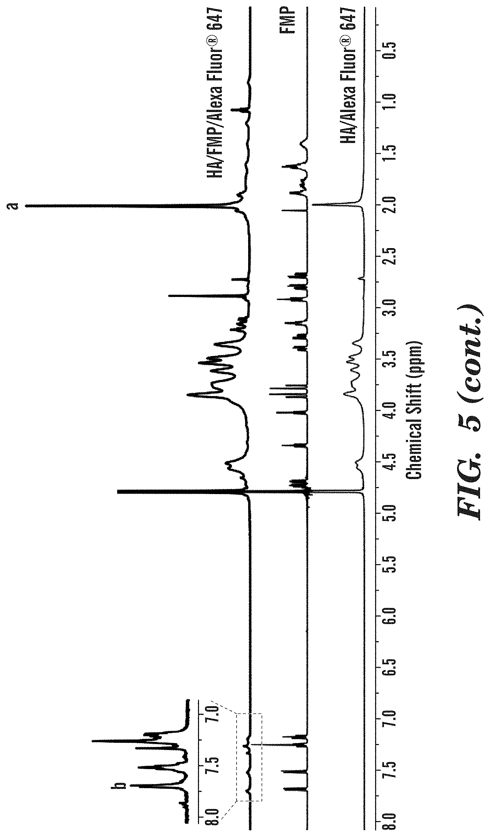
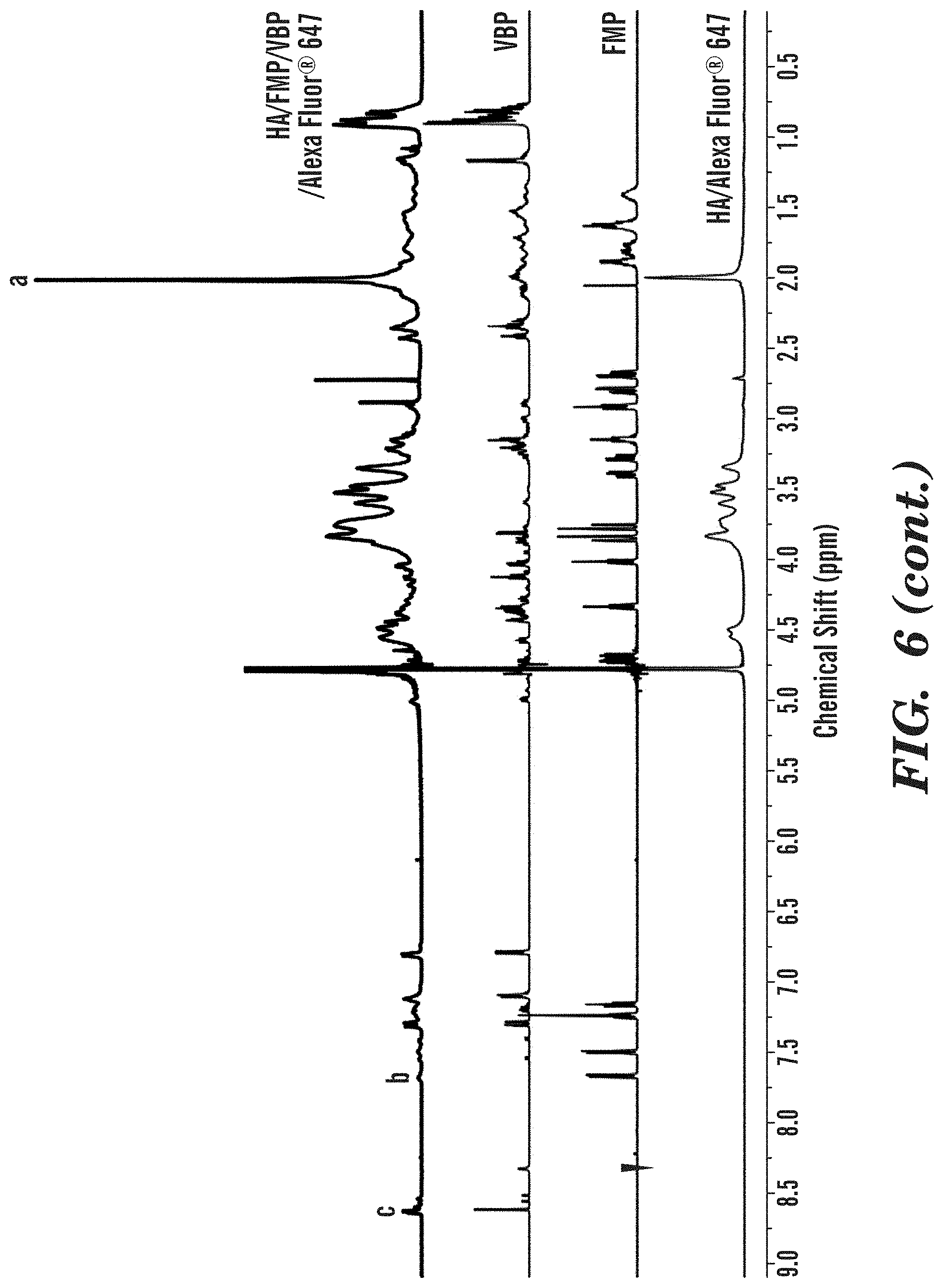
[0110] .sup.1HNMR characterization of HA/peptide conjugates. The obtained HA/peptide conjugates were characterized by proton nuclear magnetic resonance (.sup.1H NMR) in D.sub.2O. .sup.1H NMR analysis was carried out on an Agilent DD2 600 MHz NMR Spectrometer with MestReNova 10.0.1 processing software. The chemical shifts were referenced to the lock D.sub.2O (4.79 ppm).
[0111] .sup.1H NMR spectra of HA/Alexa Fluor.RTM. 647, FMP and HA/FMP/Alexa Fluor.RTM. 647 conjugate are shown in FIG. 5. Given the small amount of Alexa Fluor.RTM. 647 fed, no detectable signals are from Alexa Fluor.RTM. 647, and thus, the HA/Alexa Fluor.RTM. 647 spectrum was used to as control group to verify the conjugation of peptide. Characteristic peaks from both FMP and HA were found on the HA/FMP/Alexa Fluor.RTM. 647 conjugate spectrum, demonstrating the successful conjugation. In particular, the peaks at .delta.=7.15-7.70 ppm correspond to the aromatic rings in tryptophan from FMP, and the peak at .delta.=1.95-2.05 ppm corresponds to methyl groups in the acetamido moiety of HA. For a quantitative analysis, the methyl resonance of acetamido moiety of HA at (.delta.=1.95-2.05 ppm) was used as an internal standard (a in FIG. 2). The FMP content in HA/FMP/Alexa Fluor.RTM. 647 conjugate was determined from the comparison of the peak area at .delta.=1.95-2.05 ppm with that at .delta.=7.15-7.70 ppm corresponding to the aromatic ring in tryptophan (b in FIG. 2). The degree of substitution(DS.sub.FMP) was calculated as
D S F M P = ( Integration of b ) / 5 ( Integration of a ) / 3 .times. 1 0 0 % ( 1 ) ##EQU00001##
[0112] The DS.sub.FMP of HA/FMP/Alexa Fluor.RTM. 647 conjugate was calculated as 7.79%.
[0113] Similarly, the .sup.1H NMR spectra of HA/FMP/VBP/Alexa Fluor.RTM. 647 conjugate was shown in FIG. 3. In addition to the peaks from FMP and HA, the peaks at .delta.=8.63 ppm (c in FIG. 6) and .delta.=7.31 ppm correspond to the aromatic rings in histidine from VBP. The VBP content in HA/FMP/VBP/Alexa Fluor.RTM. 647 conjugate was determined from the comparison of the peak area at .delta.=1.95-2.05 ppm with that at .delta.=8.63 ppm (c in FIG. 6). The degree of substitution(DS.sub.VBP) was calculated as
D S V B P = Integration of c ( Integration of a ) / 3 .times. 1 0 0 % ( 2 ) ##EQU00002##
[0114] The DS.sub.FMP and DS.sub.VBP of HA/FMP/VBP/Alexa Fluor.RTM. 647 conjugate were 7.38% and 6.49%, respectively.
[0115] Since there is no aromatic moiety in CBP and all the peaks from CBP are overlapped with those from HA, the DS.sub.CBP cannot be calculated via the .sup.1H NMR spectrum (FIG. 6). However, the peak at .delta.=7.90 ppm is existed in both CBP and conjugate spectra, suggesting the successful conjugation of CBP. A modified CBP with histidine as the C-terminal group will be used in the following study to facilitate the calculation of degree of substitution. The DS.sub.VMP and DS.sub.VBP of HA/FMP/VBP/CBP/Alexa Fluor.RTM. 647 conjugate were 8.01% and 5.68%, respectively, as calculated by eq. 1 and 2.
[0116] It should be mentioned that the degree of substitution calculated by .sup.1H NMR is well correlated with the micro bicinchoninic acid assay (micro BCA assay). For one batch of HA/FMP conjugate with DS.sub.FMP of 22% (from .sup.1H NMR), the micro BCA assay showed a DS.sub.FMP of 28%. However, due to the incompatibility of the BCA assay with multiple peptide conjugation, the degree of substitution is mainly analyzed by .sup.1H NMR.
[0117] Cyro-TEM. The morphology of HA/FMP/VBP/CBP conjugate was also characterized by cryo-TEM (FEI Tecnai G2 Sphera) at an accelerating voltage of 200 kV, under low-dose mode. The cryo-TEM image of HA (FIGS. 8A-8B) shows the typical linear polymer strands, while HA/FMP/VBP/CBP conjugates were existed as nanoparticles with size around 200 nm.
Example 4
Platelet-Like Nanoparticles
[0118] Particle fabrication and characterization. Hollow discoidal polyelectrolyte capsules were fabricated using LbL synthesis. 200 nm carboxylate polystyrene (PS) spheres have been chosen as the sacrificial core material for the alternate electrostatic deposition of cationic poly allyl amine hydrochloride (PAH) and anionic bovine serum albumin (BSA). Briefly, 1.42.times.10.sup.12PS particles were alternately incubated in 1 mL of 2 mg/mL PAH and BSA solutions for 30 minutes each, to obtain a total of four PAH/BSA bilayers. Following each layer, the particles were washed with 1 mL of deionized water via centrifugation to remove excess unbound polyelectrolytes. The bilayers were crosslinked using 2% (v/V) glutaraldehyde solution, prepared in deionized water. At pH 7, glutaraldehyde reacts rapidly with amine groups of PAH and BSA, thus chemically linking the polyelectrolyte layers with each other. This helps to improve the shell integrity under various downstream processing conditions as well as hemodynamic flow. The crosslinking reaction was terminated using sodium borohydride, followed by multiple washes with water.
[0119] To ensure uniformity of coating and complete core dissolution, maintaining colloidal stability of the system at every stage is a critical requirement. Therefore, various factors were studied for their impact on the suspension stability of these particles. Table 1 enlists the optimized process conditions for the layer-by-layer synthesis.
TABLE-US-00001 TABLE 1 Optimized conditions for layer-by-layer deposition on template polystyrene nanospheres Optimized Factors studied Range conditions Salinity of reaction medium 0-0.5 M 0 M Polyelectrolyte concentration 0.5-3 mg/mL 2 mg/mL Crosslinker concentration 0.5-2% v/V 2% # crosslinking steps 0-2 1 Sonication time 60-240 s 120 s
[0120] The size distribution and the coating uniformity was confirmed using dynamic light scattering measurements (DLS). The particle size remained in the range of 170-220 nm with a gradual increase with the number of layers, whereas the zeta potential sharply alternated between positive and negative values after each adsorption step (FIG. 9). Zeta potential values of the order .+-.30 to 50 mV indicate a good stability of the colloidal suspension. This stability behavior was confirmed from consistent DLS measurements for the stock, following 0, 1, 3, 5 and 7 days of storage at 4.degree. C.
[0121] Furthermore, polystyrene cores were dissolved with tetrahydrofuran to yield platelet-like discoidal polyelectrolyte shells (FIG. 10A). Cryo-transmission electron micrographs indicate the presence of hollow cores with uniform shell thickness (FIG. 10B).
Dendrimer Peptide Conjugation
[0122] Succinamic acid terminated PAMAM dendrimers (Generation 5.0, M.W. 28,826 Da) were conjugated separately with fibrinogen-mimicking peptide (FMP; KRGDW (SEQ ID NO: 8), 660.72 Da), collagen binding peptide (CBP; [GPO].sub.7, (SEQ ID NO: 1) 1997.08 Da) and von Willebrand factor binding peptide (VBP; TRYLRIHPQSQVHQI (SEQ ID NO: 7), 1876.14 Da) using well-established chemistries. Conjugation was performed using 1-ethyl-3-(3-dimethylaminopropyl) carbodiimide hydrochloride (EDC, M.W. 191.7 Da) and carbonyldiimidazole (CDI, M.W. 162.15 Da), respectively to compare and determine the reaction scheme suitable for maximum peptide loading. EDC or CDI was used to activate the carboxylic acid (--COOH) groups on dendrimers for direct conjugation to primary amines (--NH2) via amide bonds. FIG. 11 shows the reaction scheme for the conjugation. The unreacted peptides were separated from the conjugates using 10 kDa Amicon ultrafilters and washed thoroughly with phosphate buffered saline (PBS, pH-7.2), to obtain the purified products.
[0123] In order to verify that the peptide was chemically conjugated with the dendrimers, a negative control of physically mixed (without EDC or CDI activation) dendrimers and peptides, was used. MicroBCA assay was used to quantify the amount of free peptide in supernatant, and thus the conjugation efficiency of the reaction, defined as
Conjugation efficiency = ( Amount of peptide added ) - ( Amount of peptide in supernatant ) ( Amount of peptide added ) .times. 1 0 0 . ##EQU00003##
[0124] Table 2 shows the peptide loading obtained with the different reaction routes for each of the two peptides. However, the microBCA assay could not be used to quantify CBP, owing to the absence of any aromatic functional groups on it. Fluorescent labeling of CBP or modification of its sequence will be implemented for its quantitative characterization in the future.
TABLE-US-00002 TABLE 2 Comparison between the conjugation efficiencies obtained using different carboxylate activators. Dendrimer-VBP Dendrimer-FMP conjugate conjugate Molar ratio % Molar ratio % Peptide peptide: conjugation peptide: conjugation Crosslinker dendrimer efficiency dendrimer efficiency EDC 39.4 38.9 .+-. 4.5 58.2 45.4 .+-. 8.2 CDI 9.1 12.6 .+-. 3.2 8.2 6.4 .+-. 3.6 None 3.2 2.1 .+-. 1.8 5.5 4.3 .+-. 2.3
[0125] The calibration of absorbance signals indicated that the peptide loading obtained from physical adsorption was significantly low as compared to the chemically conjugated products. Thus, the peptides were covalently coupled to the dendrimer carboxyl groups. Furthermore, EDC chemistry was found to be more efficient as compared to CDI for dendrimer-peptide conjugation. It was found that a dendrimer groups: EDC: peptide molar ratio of 1:1:2 yields the highest degree of conjugation.
References
[0126] Anselmo, A. C. et. al., Platelet-like Nanoparticles: Mimicking Shape, Flexibility, and Surface Biology of Platelets to Target Vascular Injuries. ACS Nano 2014 8 (11): 11243-53.
Example 5
Thrombolytic Particles
[0127] Intravenous administration of tissue plasminogen activator (tPA) is the only FDA-approved therapy for acute ischemic stroke (Shaw et. al., 2009). But this approach has several pharmacokinetic limitations, accompanied by systemic side-effects. Drug encapsulation potential of the hollow polyelectrolyte capsules has been used to induce targeted fibrinolytic effect in such thrombotic conditions. To develop tPA carriers with platelet-like morphology, layer-by-layer technique was used with alternate deposition of polyallyl amine hydrochloride (PAH) and bovine serum albumin (BSA) on 2 .mu.m calcium carbonate (CaCO.sub.3) spheres. Micron-sized templates were used to increase loading capacity of the hollow cores, as well as to achieve enhanced margination behavior under hemodynamic flow. Furthermore, polystyrene templates were replaced with CaCO.sub.3 spheres in order to achieve core dissolution at milder conditions and thus prevent tPA denaturation.
[0128] tPA encapsulation was achieved via in situ loading in CaCO.sub.3 microspheres. To begin with, Texas red dextran (70 kDa) was used as a model molecule to determine the efficacy of this approach and optimize the reaction conditions. 1 mL of Texas red-dextran aqueous solution (2 mg/mL) was mixed with 1 mL of 1M calcium chloride dihydrate (CaCl.sub.2.2H.sub.2O) and 1 mL of sodium carbonate (Na.sub.2CO.sub.3) under vigorous agitation for 15 minutes at room temperature to allow nucleation and growth of CaCO.sub.3 crystals. Porous microspheres observed in scanning electron micrographs (FIG. 12A) indicate the formation of vaterite crystalline polymorph of CaCO.sub.3. The synthesized dextran-loaded CaCO.sub.3 microparticles were washed twice with MilliQ water to remove residual salts from the medium. The supernatant was collected for quantification of loaded fluorophore and the microparticles were dried under vacuum overnight. Layer-by-layer deposition was performed on these particles using PAH and FITC-BSA, as described earlier (FIG. 12). The cores were dissolved using 0.2M ethylenediaminetetraacetic acid (EDTA) solution at pH 8, yielding soft, flexible microcapsules (FIG. 12B-12C).
[0129] Further, recombinant human tPA (Abeam, Mass.) was encapsulated using similar protocol. Briefly, 1 mL of tPA solution (5.3 IU/mL) in 0.1M NaCl was mixed with 1 mL of 1M CaCl.sub.2. 2H.sub.2O solution for 5 minutes. 1 mL of 1M Na.sub.2CO.sub.3 was added to this mixture and vigorously stirred for 15 minutes at room temperature. The precipitated tPA-loaded CaCO.sub.3 particles were washed twice with deionized water to remove NaCl and free tPA. The supernatant was collected and used to determine the loading efficiency using human tPA activity ELISA (Molecular Innovations, Inc.) at a wavelength of 450 nm (FIG. 14). To determine the effect of encapsulation and core dissolution on tPA activity, tPA-loaded CaCO.sub.3 microparticles were dissolved using EDTA and the solution was used to determine the amount of active tPA. It was observed that .about.60% of loaded tPA activity was retained during the core dissolution process. The loss could be caused due to unfolding of the protein structure during the encapsulation and dissolution. Co-encapsulation of tPA with osmolytes like trehalose will be implemented to prevent its misfolding and inactivation.
References
[0130] Shaw, G. J. et. al. Ultrasound-enhanced Thrombolysis with tPA-loaded Echogenic Liposomes. Thromb Res.2009 124 (3): 306-10.
Sequence CWU 1
1
9121PRTHomo
sapiensMOD_RES(3)..(3)HydroxyprolineMOD_RES(6)..(6)HydroxyprolineMOD_RES(-
9)..(9)HydroxyprolineMOD_RES(12)..(12)HydroxyprolineMOD_RES(15)..(15)Hydro-
xyprolineMOD_RES(18)..(18)HydroxyprolineMOD_RES(21)..(21)Hydroxyproline
1Gly Pro Xaa Gly Pro Xaa Gly Pro Xaa Gly Pro Xaa Gly Pro Xaa Gly1 5
10 15Pro Xaa Gly Pro Xaa 20215PRTHomo sapiens 2Thr Arg Tyr Leu Arg
Ile His Pro Gln Ser Trp Val His Gln Ile1 5 10 1535PRTHomo sapiens
3Gly Arg Gly Asp Ser1 548PRTHomo sapiens 4Tyr Met Glu Ser Arg Ala
Asp Arg1 5517PRTHomo sapiensMOD_RES(1)..(1)Acetylated-Arg 5Arg Met
Ala Pro Glu Glu Glu Met Asp Arg Pro Phe Leu Tyr Val Val1 5 10
15Arg67PRTRana sp.MISC_FEATURE(7)..(7)May or may not be present
6Arg Val Arg Ser Phe Tyr Lys1 5715PRTHomo sapiens 7Thr Arg Tyr Leu
Arg Ile His Pro Gln Ser Gln Val His Gln Ile1 5 10
1585PRTUnknownDescription of Unknown Linear fibrinogen-mimetic
peptide 8Lys Arg Gly Asp Trp1 594PRTArtificial SequenceDescription
of Artificial Sequence Synthetic peptide 9Arg Gly Asp Phe1
D00000
D00001
D00002
D00003
D00004
D00005
D00006
D00007
D00008
D00009
D00010
D00011
D00012
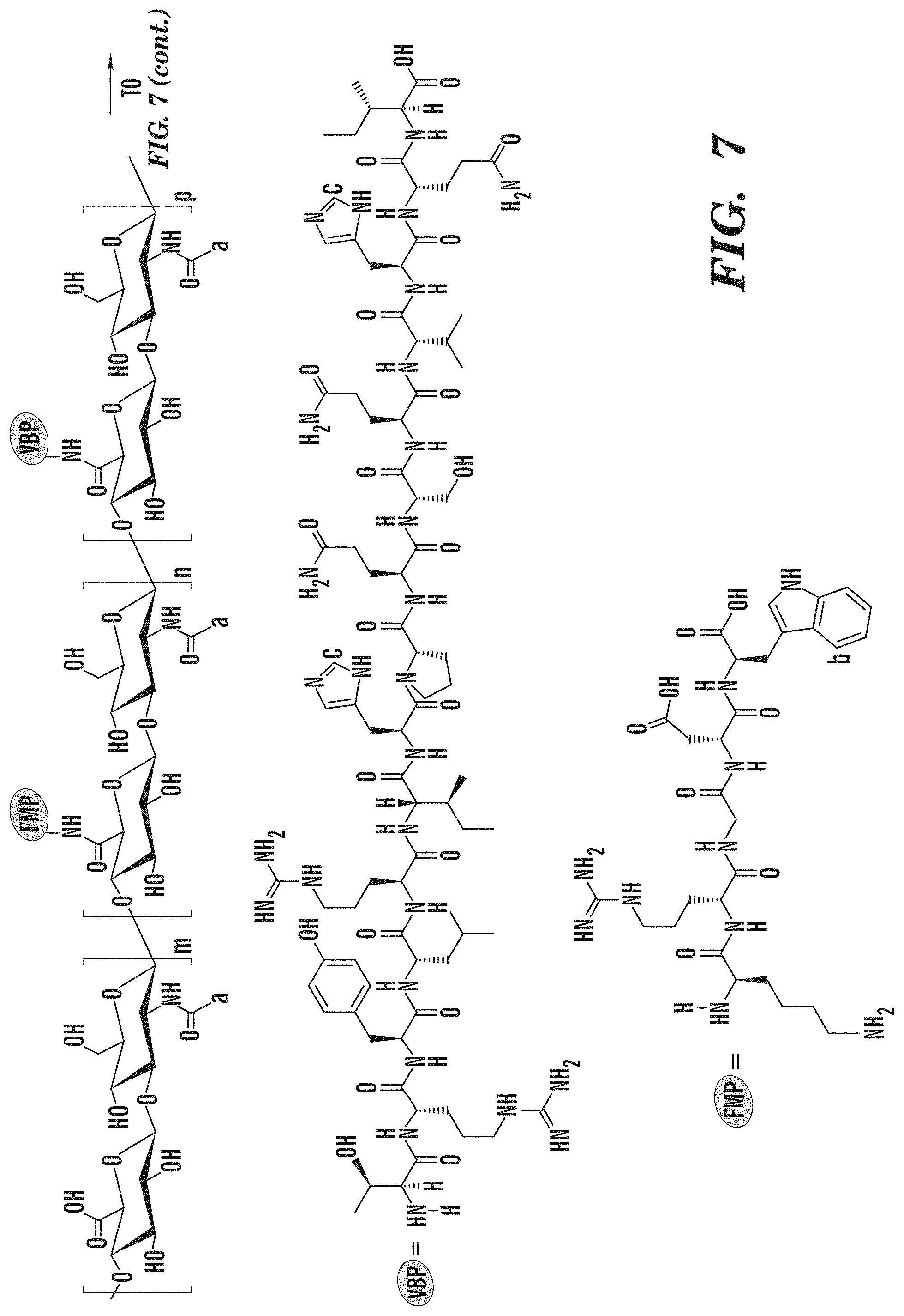
D00013
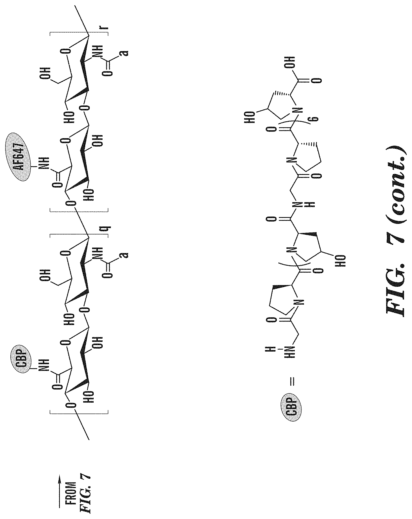
D00014

D00015

D00016

D00017

D00018

D00019
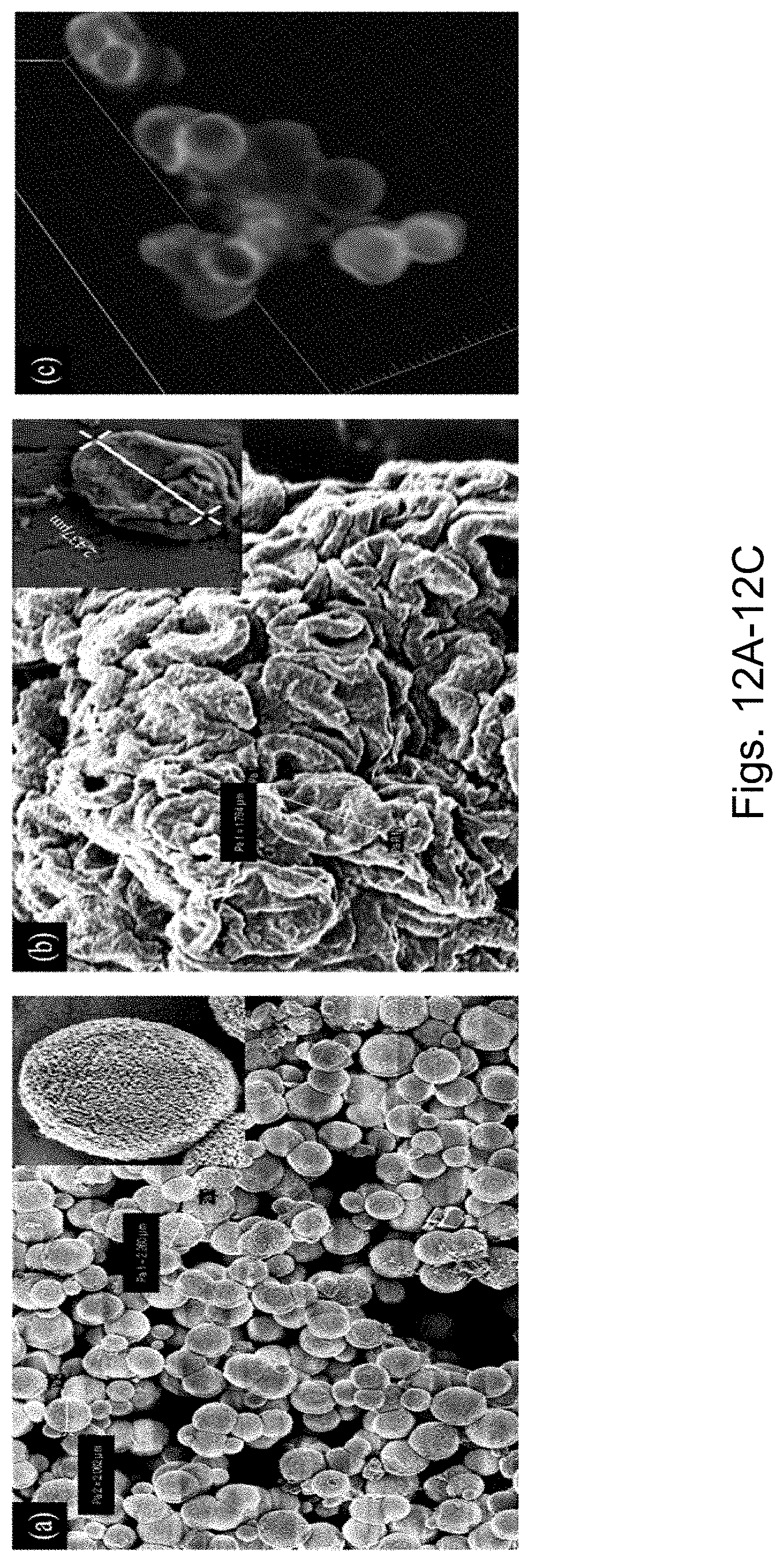
D00020


S00001
XML
uspto.report is an independent third-party trademark research tool that is not affiliated, endorsed, or sponsored by the United States Patent and Trademark Office (USPTO) or any other governmental organization. The information provided by uspto.report is based on publicly available data at the time of writing and is intended for informational purposes only.
While we strive to provide accurate and up-to-date information, we do not guarantee the accuracy, completeness, reliability, or suitability of the information displayed on this site. The use of this site is at your own risk. Any reliance you place on such information is therefore strictly at your own risk.
All official trademark data, including owner information, should be verified by visiting the official USPTO website at www.uspto.gov. This site is not intended to replace professional legal advice and should not be used as a substitute for consulting with a legal professional who is knowledgeable about trademark law.