Compositions And Methods For Evaluating Attenuation And Infectivity Of Listeria Strains
MOLLI; Poonam ; et al.
U.S. patent application number 16/979436 was filed with the patent office on 2021-01-07 for compositions and methods for evaluating attenuation and infectivity of listeria strains. This patent application is currently assigned to ADVAXIS, INC.. The applicant listed for this patent is ADVAXIS, INC.. Invention is credited to Poonam MOLLI, Anu WALLECHA.
| Application Number | 20210003558 16/979436 |
| Document ID | / |
| Family ID | |
| Filed Date | 2021-01-07 |


View All Diagrams
| United States Patent Application | 20210003558 |
| Kind Code | A1 |
| MOLLI; Poonam ; et al. | January 7, 2021 |
COMPOSITIONS AND METHODS FOR EVALUATING ATTENUATION AND INFECTIVITY OF LISTERIA STRAINS
Abstract
Methods and compositions are provided for assessing attenuation and/or infectivity of bacteria or Listeria strains, such as Listeria monocytogenes.
| Inventors: | MOLLI; Poonam; (North Brunswick, NJ) ; WALLECHA; Anu; (Yardley, PA) | ||||||||||
| Applicant: |
|
||||||||||
|---|---|---|---|---|---|---|---|---|---|---|---|
| Assignee: | ADVAXIS, INC. Princeton NJ |
||||||||||
| Appl. No.: | 16/979436 | ||||||||||
| Filed: | March 8, 2019 | ||||||||||
| PCT Filed: | March 8, 2019 | ||||||||||
| PCT NO: | PCT/US2019/021303 | ||||||||||
| 371 Date: | September 9, 2020 |
Related U.S. Patent Documents
| Application Number | Filing Date | Patent Number | ||
|---|---|---|---|---|
| 62640855 | Mar 9, 2018 | |||
| Current U.S. Class: | 1/1 |
| International Class: | G01N 33/50 20060101 G01N033/50; C12N 1/20 20060101 C12N001/20; G01N 33/483 20060101 G01N033/483 |
Claims
1. A method of assessing attenuation or infectivity of a test Listeria strain, comprising: (a) infecting differentiated THP-1 cells with the test Listeria strain, wherein the THP-1 cells have been differentiated into macrophages prior to infecting with the test Listeria strain; (b) lysing the THP-1 cells and plating the lysate on agar; and (c) counting the Listeria that have multiplied inside the THP-1 cells by growth on the agar.
2. The method of claim 1, further comprising differentiating the THP-1 cells into macrophages using phorbol 12-myristate 13-acetate (PMA) prior to step (a).
3. The method of claim 1, wherein step (a) comprises infecting the differentiated THP-1 cells at a multiplicity of infection (MOI) of 1:1.
4. The method of claim 1, further comprising killing Listeria not taken up by the THP-1 cells in between steps (a) and (b).
5. The method of claim 4, wherein the killing is performed using an antibiotic, optionally wherein the antibiotic is gentamicin.
6. The method of claim 1, wherein step (b) is performed at 0 hours post-infection.
7. The method of claim 1, wherein step (b) is performed at 3 hours post-infection.
8. The method of claim 1, further comprising comparing uptake and intracellular growth of the test Listeria strain with a wild type Listeria strain and/or a reference sample.
9. The method of claim 1, wherein the test Listeria strain is a Listeria monocytogenes strain.
10. The method of claim 1, wherein the test Listeria strain is a recombinant Listeria strain comprising a nucleic acid comprising a first open reading frame encoding a fusion polypeptide, wherein the fusion polypeptide comprises a PEST-containing peptide fused to a disease-associated antigenic peptide.
11. The method of claim 10, wherein the PEST-containing peptide is listeriolysin O (LLO) or a fragment thereof, and the disease-associated antigenic peptide is selected from the group consisting of a Human Papilloma virus (HPV) protein E7 Prostate Specific Antigen (PSA), a chimeric Her2 antigen, and Her2/neu chimeric antigen, or a fragment thereof.
12. The method of claim 10, wherein the recombinant Listeria strain is an attenuated Listeria monocytogenes strain comprising a deletion of or inactivating mutation in prfA, wherein the nucleic acid is in an episomal plasmid and comprises a second open reading frame encoding a D133V PrfA mutant protein.
13. The method of claim 10, wherein the recombinant Listeria strain is an attenuated Listeria monocytogenes strain comprising a deletion of or inactivating mutation in actA, dal, and dat, wherein the nucleic acid is in an episomal plasmid and comprises a second open reading frame encoding an alanine racemase enzyme or a D-amino acid aminotransferase enzyme, and wherein the PEST-containing peptide is an N-terminal fragment of listeriolysin O (LLO).
14. A method of assessing attenuation or infectivity of a test bacteria strain, comprising: (a) differentiating THP-1 cells; (b) infecting the differentiated THP-1 cells with the test bacteria strain, wherein the infecting comprises: (i) inoculating the differentiated THP-1 cells with the test bacteria strain; (ii) incubating the test bacteria strain with the differentiated THP-1 cells for 1-5 hours to form infected THP1 cells; (iii) removing extracellular bacteria from the infected THP-1 cells; and (iv) incubating the infected THP-1 cells in growth media for 0-10 hours; (c) lysing the infected THP-1 cells to form a lysate; (d) plating the lysate or a dilution of the lysate on a plate containing media capable of supporting growth of the bacteria; and (e) counting colony forming units of the bacteria on the plate.
15. The method of claim 14, wherein the step of infecting the differentiated THP-1 cells is at a multiplicity of infection (MOI) of 1:1.
16. The method of claim 14, wherein the step of removing extracellular bacteria comprises adding an antibiotic effective against the bacteria, optionally wherein the antibiotic is gentamicin.
17. The method of claim 14, wherein the infected THP-1 cells are incubated in growth media for 0, 1, 3, or 5 hours.
18. The method of claim 14, wherein the test bacteria strain is an L. monocytogenes strain.
19. The method of claim 1, wherein the test Listeria strain is a recombinant Listeria strain comprising a nucleic acid comprising a first open reading frame encoding a fusion polypeptide, wherein the fusion polypeptide comprises a PEST-containing peptide fused to two or more disease-associated antigenic peptides.
20. The method of claim 19, wherein the PEST-containing peptide comprises a bacterial secretion signal sequence, and the fusion polypeptide further comprises a ubiquitin protein fused to a carboxy-terminal antigenic peptide, wherein the PEST-containing peptide, the two or more disease-associated antigenic peptides, the ubiquitin, and the carboxy-terminal antigenic peptide are arranged in tandem from the amino-terminal end to the carboxy-terminal end of the fusion polypeptide.
Description
CROSS-REFERENCE TO RELATED APPLICATIONS
[0001] This application claims the benefit of U.S. Application No. 62/640,855, filed Mar. 9, 2018, which is herein incorporated by reference in its entirety for all purposes.
REFERENCE TO A SEQUENCE LISTING SUBMITTED AS A TEXT FILE VIA EFS WEB
[0002] The Sequence Listing written in file 528092_SeqListing_ST25.txt is 89 kilobytes, was created on Feb. 25, 2019, and is hereby incorporated by reference.
BACKGROUND
[0003] Listeria monocytogenes (Lm) is a gram-positive, non-spore forming bacterial organism that is responsible for listeriosis in humans. In order to use Lm-based immunotherapies such as cancer immunotherapies, the bacteria are bio-engineered to be attenuated such that they can be used to deliver tumor-specific antigen and generate antigen-specific immune response but not cause listeriosis. Primary macrophages can be used to assess the ability of Lm-based immunotherapies to infect and replicate in the cytosol. However, better methods are needed to assess attenuation and infectivity of Listeria strains.
SUMMARY
[0004] Methods and compositions are provided for assessing attenuation and/or infectivity of bacteria or Listeria strains, such as Listeria monocytogenes. In some aspects, provided are methods for assessing attenuation or infectivity of a test Listeria strain. Such methods can comprise, for example: (a) infecting differentiated THP-1 cells with the test Listeria strain, wherein the THP-1 cells have been differentiated into macrophages prior to infecting with the test Listeria strain; (b) lysing the THP-1 cells and plating the lysate on agar; and; and (c) counting the Listeria that have multiplied inside the THP-1 cells by growth on the agar.
BRIEF DESCRIPTION OF THE DRAWINGS
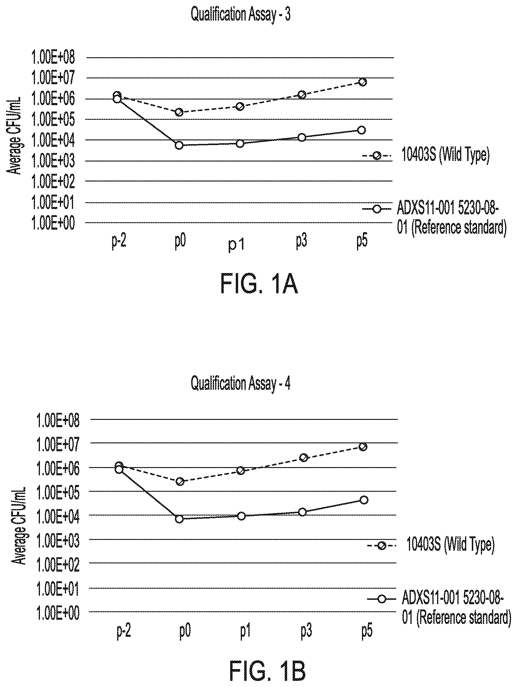
[0005] FIG. 1A. Graph illustrating bacterial growth rates and doubling times for reference standard and wild type control plotted as time versus viable cell counts (VCC) for qualification assay 3.
[0006] FIG. 1B. Graph illustrating bacterial growth rates and doubling times for reference standard and wild type control plotted as time versus viable cell counts (VCC) for qualification assay 4.
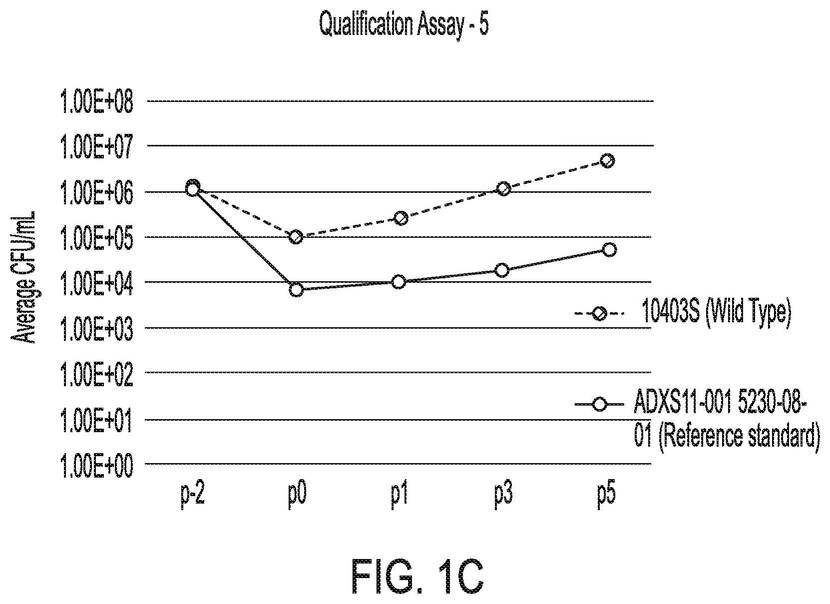
[0007] FIG. 1C. Graph illustrating bacterial growth rates and doubling times for reference standard and wild type control plotted as time versus viable cell counts (VCC) for qualification assay 5.
[0008] FIG. 2A. Graph illustrating bacterial growth rates and doubling times for wild type plotted as time versus viable cell counts (VCC) showing inter-assay comparison.
[0009] FIG. 2B. Graph illustrating bacterial growth rates and doubling times for reference standard ADXS11-001 plotted as time versus viable cell counts (VCC) showing inter-assay comparison.
[0010] FIG. 3. Graph illustrating the raw count information observed at time points: p-2, p0, p1, p3, and p5.
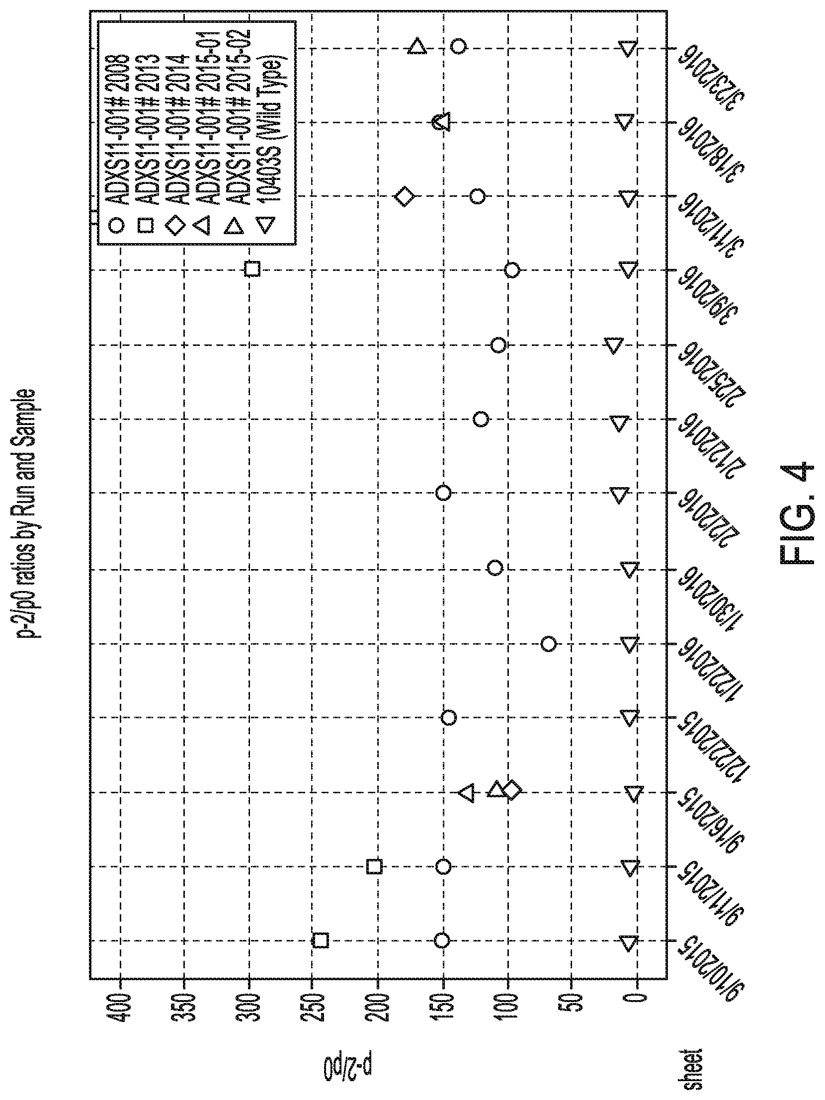
[0011] FIG. 4. Graph illustrating the ratio of the count at p-2 to that seen at p0.
[0012] FIG. 5. Graph illustrating the ratio of the count at p-2 to that seen at p0 as a ratio to wild type.
[0013] FIG. 6. Graph illustrating the ratio of the count at p3 and p % to that seen at p0.
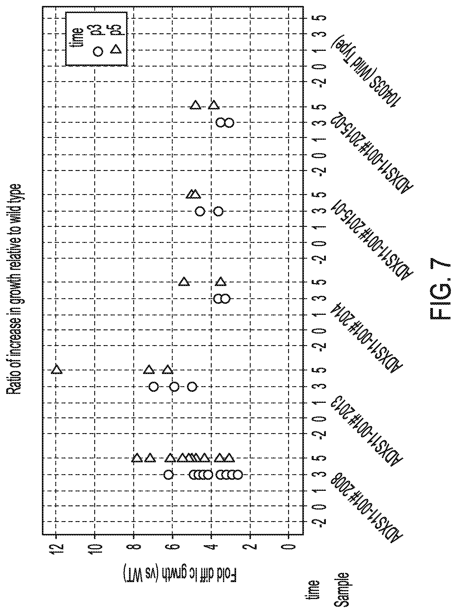
[0014] FIG. 7. Graph illustrating the ratio of the count at p3 and p % to that seen at p0 relative to wild type.
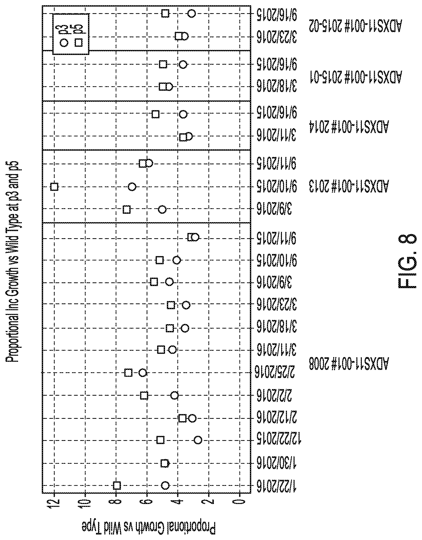
[0015] FIG. 8. Graph illustrating the ratio of the count at p3 and p % to that seen at p0 relative to wild type by data run.
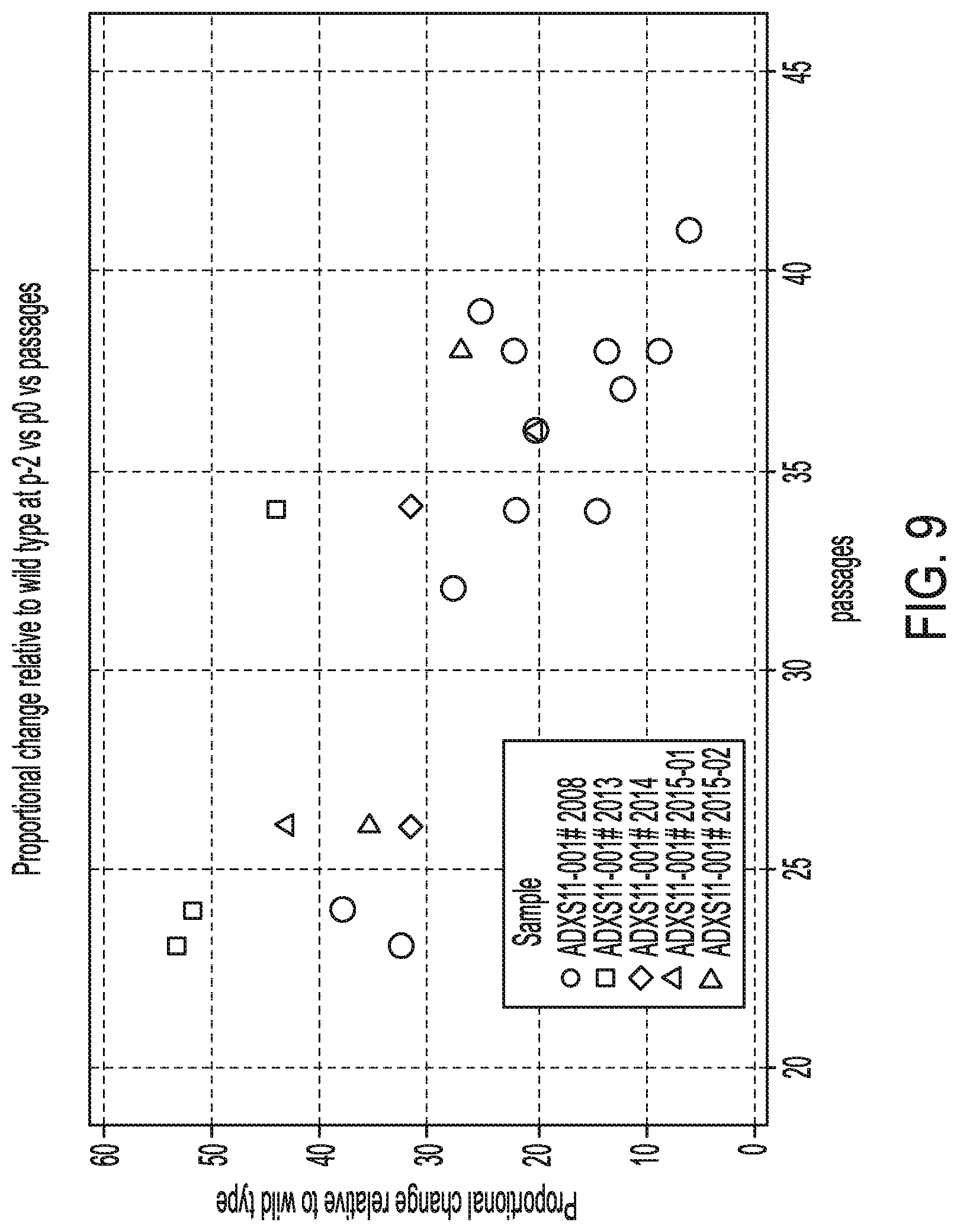
[0016] FIG. 9. Graph illustrating the impact of the number of passages in the proportional decrease in counts from p-2 to p0 relative to wild type.
[0017] FIG. 10. Graph illustrating regression analysis was used to evaluate the impact of the number of passages.
[0018] FIG. 11. Graph illustrating the relationship between the two resulting variables for each curve in FIG. 3.
DEFINITIONS
[0019] The terms "protein," "polypeptide," and "peptide," used interchangeably herein, refer to polymeric forms of amino acids of any length, including coded and non-coded amino acids and chemically or biochemically modified or derivatized amino acids. The terms include polymers that have been modified, such as polypeptides having modified peptide backbones.
[0020] Proteins are said to have an "N-terminus" and a "C-terminus." The term "N-terminus" relates to the start of a protein or polypeptide, terminated by an amino acid with a free amine group (--NH2). The term "C-terminus" relates to the end of an amino acid chain (protein or polypeptide), terminated by a free carboxyl group (--COOH).
[0021] The term "fusion protein" refers to a protein comprising two or more peptides linked together by peptide bonds or other chemical bonds. The peptides can be linked together directly by a peptide or other chemical bond. For example, a chimeric molecule can be recombinantly expressed as a single-chain fusion protein. Alternatively, the peptides can be linked together by a "linker" such as one or more amino acids or another suitable linker between the two or more peptides.
[0022] The terms "nucleic acid" and "polynucleotide," used interchangeably herein, refer to polymeric forms of nucleotides of any length, including ribonucleotides, deoxyribonucleotides, or analogs or modified versions thereof. They include single-, double-, and multi-stranded DNA or RNA, genomic DNA, cDNA, DNA-RNA hybrids, and polymers comprising purine bases, pyrimidine bases, or other natural, chemically modified, biochemically modified, non-natural, or derivatized nucleotide bases.
[0023] Nucleic acids are said to have "5' ends" and "3' ends" because mononucleotides are reacted to make oligonucleotides in a manner such that the 5' phosphate of one mononucleotide pentose ring is attached to the 3' oxygen of its neighbor in one direction via a phosphodiester linkage. An end of an oligonucleotide is referred to as the "5' end" if its 5' phosphate is not linked to the 3' oxygen of a mononucleotide pentose ring. An end of an oligonucleotide is referred to as the "3' end" if its 3' oxygen is not linked to a 5' phosphate of another mononucleotide pentose ring. A nucleic acid sequence, even if internal to a larger oligonucleotide, also may be said to have 5' and 3' ends. In either a linear or circular DNA molecule, discrete elements are referred to as being "upstream" or 5' of the "downstream" or 3' elements.
[0024] "Codon optimization" refers to a process of modifying a nucleic acid sequence for enhanced expression in particular host cells by replacing at least one codon of the native sequence with a codon that is more frequently or most frequently used in the genes of the host cell while maintaining the native amino acid sequence. For example, a polynucleotide encoding a fusion polypeptide can be modified to substitute codons having a higher frequency of usage in a given Listeria cell or any other host cell as compared to the naturally occurring nucleic acid sequence. Codon usage tables are readily available, for example, at the "Codon Usage Database." The optimal codons utilized by L. monocytogenes for each amino acid are shown US 2007/0207170, herein incorporated by reference in its entirety for all purposes. These tables can be adapted in a number of ways. See Nakamura et al. (2000) Nucleic Acids Research 28:292, herein incorporated by reference in its entirety for all purposes. Computer algorithms for codon optimization of a particular sequence for expression in a particular host are also available (see, e.g., Gene Forge).
[0025] The term "plasmid" or "vector" includes any known delivery vector including a bacterial delivery vector, a viral vector delivery vector, a peptide immunotherapy delivery vector, a DNA immunotherapy delivery vector, an episomal plasmid, an integrative plasmid, or a phage vector. The term "vector" refers to a construct which is capable of delivering, and, optionally, expressing, one or more fusion polypeptides in a host cell.
[0026] The term "episomal plasmid" or "extrachromosomal plasmid" refers to a nucleic acid vector that is physically separate from chromosomal DNA (i.e., episomal or extrachromosomal and does not integrated into a host cell's genome) and replicates independently of chromosomal DNA. A plasmid may be linear or circular, and it may be single-stranded or double-stranded. Episomal plasmids may optionally persist in multiple copies in a host cell's cytoplasm (e.g., Listeria), resulting in amplification of any genes of interest within the episomal plasmid.
[0027] The term "genomically integrated" refers to a nucleic acid that has been introduced into a cell such that the nucleotide sequence integrates into the genome of the cell and is capable of being inherited by progeny thereof. Any protocol may be used for the stable incorporation of a nucleic acid into the genome of a cell.
[0028] The term "stably maintained" refers to maintenance of a nucleic acid molecule or plasmid in the absence of selection (e.g., antibiotic selection) for at least 10 generations without detectable loss. For example, the period can be at least 15 generations, 20 generations, at least 25 generations, at least 30 generations, at least 40 generations, at least 50 generations, at least 60 generations, at least 80 generations, at least 100 generations, at least 150 generations, at least 200 generations, at least 300 generations, or at least 500 generations. Stably maintained can refer to a nucleic acid molecule or plasmid being maintained stably in cells in vitro (e.g., in culture), being maintained stably in vivo, or both.
[0029] An "open reading frame" or "ORF" is a portion of a DNA which contains a sequence of bases that could potentially encode a protein. As an example, an ORF can be located between the start-code sequence (initiation codon) and the stop-codon sequence (termination codon) of a gene.
[0030] A "promoter" is a regulatory region of DNA usually comprising a TATA box capable of directing RNA polymerase II to initiate RNA synthesis at the appropriate transcription initiation site for a particular polynucleotide sequence. A promoter may additionally comprise other regions which influence the transcription initiation rate. The promoter sequences disclosed herein modulate transcription of an operably linked polynucleotide. A promoter can be active in one or more of the cell types disclosed herein (e.g., a eukaryotic cell, a non-human mammalian cell, a human cell, a rodent cell, a pluripotent cell, a one-cell stage embryo, a differentiated cell, or a combination thereof). A promoter can be, for example, a constitutively active promoter, a conditional promoter, an inducible promoter, a temporally restricted promoter (e.g., a developmentally regulated promoter), or a spatially restricted promoter (e.g., a cell-specific or tissue-specific promoter). Examples of promoters can be found, for example, in WO 2013/176772, herein incorporated by reference in its entirety.
[0031] "Operable linkage" or being "operably linked" refers to the juxtaposition of two or more components (e.g., a promoter and another sequence element) such that both components function normally and allow the possibility that at least one of the components can mediate a function that is exerted upon at least one of the other components. For example, a promoter can be operably linked to a coding sequence if the promoter controls the level of transcription of the coding sequence in response to the presence or absence of one or more transcriptional regulatory factors. Operable linkage can include such sequences being contiguous with each other or acting in trans (e.g., a regulatory sequence can act at a distance to control transcription of the coding sequence).
[0032] "Sequence identity" or "identity" in the context of two polynucleotides or polypeptide sequences makes reference to the residues in the two sequences that are the same when aligned for maximum correspondence over a specified comparison window. When percentage of sequence identity is used in reference to proteins it is recognized that residue positions which are not identical often differ by conservative amino acid substitutions, where amino acid residues are substituted for other amino acid residues with similar chemical properties (e.g., charge or hydrophobicity) and therefore do not change the functional properties of the molecule. When sequences differ in conservative substitutions, the percent sequence identity may be adjusted upwards to correct for the conservative nature of the substitution. Sequences that differ by such conservative substitutions are said to have "sequence similarity" or "similarity." Means for making this adjustment are well-known. Typically, this involves scoring a conservative substitution as a partial rather than a full mismatch, thereby increasing the percentage sequence identity. Thus, for example, where an identical amino acid is given a score of 1 and a non-conservative substitution is given a score of zero, a conservative substitution is given a score between zero and 1. The scoring of conservative substitutions is calculated, e.g., as implemented in the program PC/GENE (Intelligenetics, Mountain View, Calif.).
[0033] "Percentage of sequence identity" refers to the value determined by comparing two optimally aligned sequences (greatest number of perfectly matched residues) over a comparison window, wherein the portion of the polynucleotide sequence in the comparison window may comprise additions or deletions (i.e., gaps) as compared to the reference sequence (which does not comprise additions or deletions) for optimal alignment of the two sequences. The percentage is calculated by determining the number of positions at which the identical nucleic acid base or amino acid residue occurs in both sequences to yield the number of matched positions, dividing the number of matched positions by the total number of positions in the window of comparison, and multiplying the result by 100 to yield the percentage of sequence identity. Unless otherwise specified (e.g., the shorter sequence includes a linked heterologous sequence), the comparison window is the full length of the shorter of the two sequences being compared.
[0034] Unless otherwise stated, sequence identity/similarity values refer to the value obtained using GAP Version 10 using the following parameters: % identity and % similarity for a nucleotide sequence using GAP Weight of 50 and Length Weight of 3, and the nwsgapdna.cmp scoring matrix; % identity and % similarity for an amino acid sequence using GAP Weight of 8 and Length Weight of 2, and the BLOSUM62 scoring matrix; or any equivalent program thereof. "Equivalent program" includes any sequence comparison program that, for any two sequences in question, generates an alignment having identical nucleotide or amino acid residue matches and an identical percent sequence identity when compared to the corresponding alignment generated by GAP Version 10.
[0035] The term "conservative amino acid substitution" refers to the substitution of an amino acid that is normally present in the sequence with a different amino acid of similar size, charge, or polarity. Examples of conservative substitutions include the substitution of a non-polar (hydrophobic) residue such as isoleucine, valine, or leucine for another non-polar residue. Likewise, examples of conservative substitutions include the substitution of one polar (hydrophilic) residue for another such as between arginine and lysine, between glutamine and asparagine, or between glycine and serine. Additionally, the substitution of a basic residue such as lysine, arginine, or histidine for another, or the substitution of one acidic residue such as aspartic acid or glutamic acid for another acidic residue are additional examples of conservative substitutions. Examples of non-conservative substitutions include the substitution of a non-polar (hydrophobic) amino acid residue such as isoleucine, valine, leucine, alanine, or methionine for a polar (hydrophilic) residue such as cysteine, glutamine, glutamic acid or lysine and/or a polar residue for a non-polar residue. Typical amino acid categorizations are summarized below.
TABLE-US-00001 TABLE 1 Amino Acid Categorizations. Alanine Ala A Nonpolar Neutral 1.8 Arginine Arg R Polar Positive -4.5 Asparagine Asn N Polar Neutral -3.5 Aspartic acid Asp D Polar Negative -3.5 Cysteine Cys C Nonpolar Neutral 2.5 Glutamic acid Glu E Polar Negative -3.5 Glutamine Gln Q Polar Neutral -3.5 Glycine Gly G Nonpolar Neutral -0.4 Histidine His H Polar Positive -3.2 Isoleucine Ile I Nonpolar Neutral 4.5 Leucine Leu L Nonpolar Neutral 3.8 Lysine Lys K Polar Positive -3.9 Methionine Met M Nonpolar Neutral 1.9 Phenylalanine Phe F Nonpolar Neutral 2.8 Proline Pro P Nonpolar Neutral -1.6 Serine Ser S Polar Neutral -0.8 Threonine Thr T Polar Neutral -0.7 Tryptophan Trp W Nonpolar Neutral -0.9 Tyrosine Tyr Y Polar Neutral -1.3 Valine Val V Nonpolar Neutral 4.2
[0036] A "homologous" sequence (e.g., nucleic acid sequence) refers to a sequence that is either identical or substantially similar to a known reference sequence, such that it is, for example, at least 50%, at least 55%, at least 60%, at least 65%, at least 70%, at least 75%, at least 80%, at least 85%, at least 90%, at least 95%, at least 96%, at least 97%, at least 98%, at least 99%, or 100% identical to the known reference sequence.
[0037] The term "wild type" refers to entities having a structure and/or activity as found in a normal (as contrasted with mutant, diseased, altered, or so forth) state or context. Wild type gene and polypeptides often exist in multiple different forms (e.g., alleles).
[0038] The term "isolated" with respect to proteins and nucleic acid refers to proteins and nucleic acids that are relatively purified with respect to other bacterial, viral or cellular components that may normally be present in situ, up to and including a substantially pure preparation of the protein and the polynucleotide. The term "isolated" also includes proteins and nucleic acids that have no naturally occurring counterpart, have been chemically synthesized and are thus substantially uncontaminated by other proteins or nucleic acids, or has been separated or purified from most other cellular components with which they are naturally accompanied (e.g., other cellular proteins, polynucleotides, or cellular components).
[0039] "Exogenous" or "heterologous" molecules or sequences are molecules or sequences that are not normally expressed in a cell or are not normally present in a cell in that form. Normal presence includes presence with respect to the particular developmental stage and environmental conditions of the cell. An exogenous or heterologous molecule or sequence, for example, can include a mutated version of a corresponding endogenous sequence within the cell or can include a sequence corresponding to an endogenous sequence within the cell but in a different form (i.e., not within a chromosome). An exogenous or heterologous molecule or sequence in a particular cell can also be a molecule or sequence derived from a different species than a reference species of the cell or from a different organism within the same species. For example, in the case of a Listeria strain expressing a heterologous polypeptide, the heterologous polypeptide could be a polypeptide that is not native or endogenous to the Listeria strain, that is not normally expressed by the Listeria strain, from a source other than the Listeria strain, derived from a different organism within the same species.
[0040] In contrast, "endogenous" molecules or sequences or "native" molecules or sequences are molecules or sequences that are normally present in that form in a particular cell at a particular developmental stage under particular environmental conditions.
[0041] The term "variant" refers to an amino acid or nucleic acid sequence (or an organism or tissue) that is different from the majority of the population but is still sufficiently similar to the common mode to be considered to be one of them (e.g., splice variants).
[0042] The term "isoform" refers to a version of a molecule (e.g., a protein) with only slight differences compared to another isoform, or version (e.g., of the same protein). For example, protein isoforms may be produced from different but related genes, they may arise from the same gene by alternative splicing, or they may arise from single nucleotide polymorphisms.
[0043] The term "fragment" when referring to a protein means a protein that is shorter or has fewer amino acids than the full length protein. The term "fragment" when referring to a nucleic acid means a nucleic acid that is shorter or has fewer nucleotides than the full length nucleic acid. A fragment can be, for example, an N-terminal fragment (i.e., removal of a portion of the C-terminal end of the protein), a C-terminal fragment (i.e., removal of a portion of the N-terminal end of the protein), or an internal fragment. A fragment can also be, for example, a functional fragment or an immunogenic fragment.
[0044] The term "analog" when referring to a protein means a protein that differs from a naturally occurring protein by conservative amino acid differences, by modifications which do not affect amino acid sequence, or by both.
[0045] The term "functional" refers to the innate ability of a protein or nucleic acid (or a fragment, isoform, or variant thereof) to exhibit a biological activity or function. Such biological activities or functions can include, for example, the ability to elicit an immune response when administered to a subject. Such biological activities or functions can also include, for example, binding to an interaction partner. In the case of functional fragments, isoforms, or variants, these biological functions may in fact be changed (e.g., with respect to their specificity or selectivity), but with retention of the basic biological function.
[0046] The terms "immunogenicity" or "immunogenic" refer to the innate ability of a molecule (e.g., a protein, a nucleic acid, an antigen, or an organism) to elicit an immune response in a subject when administered to the subject. Immunogenicity can be measured, for example, by a greater number of antibodies to the molecule, a greater diversity of antibodies to the molecule, a greater number of T-cells specific for the molecule, a greater cytotoxic or helper T-cell response to the molecule, and the like.
[0047] The term "antigen" is used herein to refer to a substance that, when placed in contact with a subject or organism (e.g., when present in or when detected by the subject or organism), results in a detectable immune response from the subject or organism. An antigen may be, for example, a lipid, a protein, a carbohydrate, a nucleic acid, or combinations and variations thereof. For example, an "antigenic peptide" refers to a peptide that leads to the mounting of an immune response in a subject or organism when present in or detected by the subject or organism. For example, such an "antigenic peptide" may encompass proteins that are loaded onto and presented on MHC class I and/or class II molecules on a host cell's surface and can be recognized or detected by an immune cell of the host, thereby leading to the mounting of an immune response against the protein. Such an immune response may also extend to other cells within the host, such as diseased cells (e.g., tumor or cancer cells) that express the same protein.
[0048] The term "epitope" refers to a site on an antigen that is recognized by the immune system (e.g., to which an antibody binds). An epitope can be formed from contiguous amino acids or noncontiguous amino acids juxtaposed by tertiary folding of one or more proteins. Epitopes formed from contiguous amino acids (also known as linear epitopes) are typically retained on exposure to denaturing solvents whereas epitopes formed by tertiary folding (also known as conformational epitopes) are typically lost on treatment with denaturing solvents. An epitope typically includes at least 3, and more usually, at least 5 or 8-10 amino acids in a unique spatial conformation. Methods of determining spatial conformation of epitopes include, for example, x-ray crystallography and 2-dimensional nuclear magnetic resonance. See, e.g., Epitope Mapping Protocols, in Methods in Molecular Biology, Vol. 66, Glenn E. Morris, Ed. (1996), herein incorporated by reference in its entirety for all purposes.
[0049] The term "mutation" refers to the any change of the structure of a gene or a protein. For example, a mutation can result from a deletion, an insertion, a substitution, or a rearrangement of chromosome or a protein. An "insertion" changes the number of nucleotides in a gene or the number of amino acids in a protein by adding one or more additional nucleotides or amino acids. A "deletion" changes the number of nucleotides in a gene or the number of amino acids in a protein by reducing one or more additional nucleotides or amino acids.
[0050] A "frameshift" mutation in DNA occurs when the addition or loss of nucleotides changes a gene's reading frame. A reading frame consists of groups of 3 bases that each code for one amino acid. A frameshift mutation shifts the grouping of these bases and changes the code for amino acids. The resulting protein is usually nonfunctional. Insertions and deletions can each be frameshift mutations.
[0051] A "missense" mutation or substitution refers to a change in one amino acid of a protein or a point mutation in a single nucleotide resulting in a change in an encoded amino acid. A point mutation in a single nucleotide that results in a change in one amino acid is a "nonsynonymous" substitution in the DNA sequence. Nonsynonymous substitutions can also result in a "nonsense" mutation in which a codon is changed to a premature stop codon that results in truncation of the resulting protein. In contrast, a "synonymous" mutation in a DNA is one that does not alter the amino acid sequence of a protein (due to codon degeneracy).
[0052] The term "somatic mutation" includes genetic alterations acquired by a cell other than a germ cell (e.g., sperm or egg). Such mutations can be passed on to progeny of the mutated cell in the course of cell division but are not inheritable. In contrast, a germinal mutation occurs in the germ line and can be passed on to the next generation of offspring.
[0053] The term "in vitro" refers to artificial environments and to processes or reactions that occur within an artificial environment (e.g., a test tube).
[0054] The term "in vivo" refers to natural environments (e.g., a cell or organism or body) and to processes or reactions that occur within a natural environment.
[0055] Compositions or methods "comprising" or "including" one or more recited elements may include other elements not specifically recited. For example, a composition that "comprises" or "includes" a protein may contain the protein alone or in combination with other ingredients.
[0056] Designation of a range of values includes all integers within or defining the range, and all subranges defined by integers within the range.
[0057] Unless otherwise apparent from the context, the term "about" encompasses values within a standard margin of error of measurement (e.g., SEM) of a stated value or variations .+-.0.5%, 1%, 5%, or 10% from a specified value.
[0058] The singular forms of the articles "a," "an," and "the" include plural references unless the context clearly dictates otherwise. For example, the term "an antigen" or "at least one antigen" can include a plurality of antigens, including mixtures thereof.
[0059] Statistically significant means p.ltoreq.0.05.
DETAILED DESCRIPTION
I. Overview
[0060] Disclosed herein is are cell-based assays using differentiated THP-1 cells to analyze intracellular growth of Listeria-based immunotherapies. Such assays can be used, for example, to evaluate attenuation of recombinant Listeria strains compared to wild type Listeria or to assess potency or infectivity of recombinant Listeria strains.
[0061] As one specific example, ADXS11-001 is a recombinant Listeria monocytogenes (Lm) strain attenuated due to the irreversible deletion of prfA in the genome and, further, its complementation with mutated prfA gene (D133V). The prfA gene regulates the transcription of several virulence genes such as hly (Listeriolysin O or LLO), actA (Actin nucleator A), plcA (phospholipase A), and plcB (phospholipase B), that are required for in vivo intracellular growth and survival of Lm. The complementation with mutated prfA in ADXS11-001 causes a reduction in the expression of the virulence genes. The plasmid in the ADXS11-001 immunotherapy also contains human papillomavirus protein E7 fused to truncated Listeriolysin O (tLLO)) under the control of the hly promoter. In order to evaluate attenuation of ADXS11-001, infection and replication is assessed in a macrophage cell infection assay using wild type Lm as control.
[0062] The biological activity of ADXS11-001 relies upon uptake of ADXS11-001 by antigen presenting cells (APC) such as macrophages and dendritic cells, its escape from phagolysosome, intracellular replication in the cytosol of APC, expression of tLLO-E7, processing, and presentation of tLLO-E7 on surface of APC to stimulate E7-specific cytotoxic T cell response. Using differentiated THP-1 cells is a superior alternative to using primary macrophages to monitor the ability of ADXS11-001 to infect and replicate in the cytosol of macrophage. The method is also advantageous in that it is quantitative.
II. Methods for Evaluating Attenuation and Infectivity of Listeria
[0063] Methods and compositions are provided for assessing attenuation and/or infectivity of bacteria. In some embodiments, the bacteria is a Listeria strain. In some embodiments, the Listeria strain is a Listeria monocytogenes strain. In some embodiments, the L. monocytogenes strain is a mutant, recombinant, or attenuated L. monocytogenes strain. Examples of recombinant Listeria strains that can be used in such methods are provided in more detail elsewhere herein. Such methods utilize macrophage cell lines or macrophage-like cell lines with macrophage phenotypes. Such cells can be immortalized cells. For example, the cell line can be a human monocyte cell line such as THP-1 cells. THP-1 designates a spontaneously immortalized monocyte-like cell line, derived from the peripheral blood of a childhood case of acute monocytic leukemia (M5 subtype). THP-1 cells can be differentiated into macrophage-like cells using, for example, phorbol 12-myristate 13-acetate (commonly known as PMA or TPA).
[0064] In some embodiments, the methods comprise: (a) infecting differentiated THP-1 cells with a test Listeria strain, wherein the THP-1 cells have been differentiated into macrophages prior to infecting with the test Listeria strain; (b) lysing the THP-1 cells and plating the lysate on agar; and (c) counting the Listeria that have multiplied inside the THP-1 cells by growth on the agar. The differentiated THP-1 cells can be grown as adherent cells. Other macrophage-like cells can also be used. Other macrophage-like immortalized cells and/or cell lines can also be used.
[0065] In some embodiments, the methods further comprise differentiating the THP-1 cells into macrophages. For example, such differentiation can be accomplished using phorbol 12-myristate 13-acetate (PMA) prior to step (a) as disclosed elsewhere herein. In some embodiments, prior to differentiation, the passage number for the THP-1 cells is less than 32.
[0066] In some embodiments, step (a) comprises infecting the differentiated THP-1 cells at a multiplicity of infection (MOI) of 1:1. However, any suitable multiplicity of infection can be used.
[0067] Optionally, such methods can further comprise killing all the Listeria not taken up by the THP-1 cells in between steps (a) and (b). For example, the killing can be performed using an antibiotic such as gentamicin.
[0068] Optionally, the lysing step (b) is performed at 3 hours post-infection. However, the lysing step can be performed at other time points as well, such as 1, 2, 3, 4, 5, 6, 7, 8, 9, or 10 hours post-infection.
[0069] In some embodiments, infecting differentiated THP-1 cells with a bacteria strain comprises incubating the bacteria with the differentiated THP-1 cells for 1-5 h, 2-3 h, 1 h, 2 h, 3 h, 2 h.+-.60 min, 2 h.+-.50 min, 2 h.+-.40 min, 2 h.+-.30 min, 2 h.+-.25 min, 2 h.+-.20 min, 2 h 15 min, 2 h 10 min, 2 h 5 min, or 2 h 3 min. In some embodiments, the bacteria is a Listeria. In some embodiments, the Listeria is L. monocytogenes. In some embodiments, the L. monocytogenes is attenuated relative to wild-type L. monocytogenes. In some embodiments, an inoculating media containing the bacteria is added to the differentiated THP-1 cells.
[0070] In some embodiments, the infecting step further comprises one or more washing steps and/or a killing step. A washing step can comprise removing bacteria-containing media from the THP-1 cells and optionally rinsing the THP-1 cells, thereby remove bacteria that have not infected the THP-1 cells. The washing step, if used, can be performed following incubation of the bacteria with the THP-1 cells and before the lysing step. A killing step can comprise adding an antibiotic effective against the bacteria to the THP-1 cells, thereby killing bacteria not taken up by the THP-1 cells (i.e., extracellular bacteria). The antibiotic can be added at a concentration effective for killing the bacteria. The killing step, if used, can be performed after incubation of the bacteria with the THP-1 cells and before the lysing step. The killing step can be performed after or before a washing step, or between two washing steps. In some embodiments, the antibiotic is added to the THP-1 cells and incubated for 15-75 min, 20-60 min, 30-50 min, or about 42-45 min. In some embodiments, the antibiotic is gentamicin.
[0071] In some embodiments, the lysing step (b) is performed immediately after the infection step (0 h post-infection), 0-10 h post-infection, 1 h post-infection, 2 h post-infection, 3 h post-infection, 4 h post-infection, 5 h post-infection, 6 h post-infection, 7 h post-infection, 8 h post-infection, 9 h post-infection, or 10 h post-infection. In some embodiments, the lysing step is performed immediately after the infecting step (p0), 1 h post-infection (p1), 3 h post-infection (p3), or 5 h post-infection (p5). If lysis is not performed immediately after the infection step, the THP-1 cells can be incubated in growth media until lysis. Intracellular growth of the bacteria can occur during the post-infection incubation. The lysing step can comprise collecting the THP-1 cells in water or similar solvent capable of lysing the THP-1 cells, but not the bacteria, to form a lysate, and plating the lysate on media capable of supporting growth of the bacteria and allowing counting the number of colony forming units (CFUs). In some embodiments, the lysate can be diluted. In some embodiments, one or more different dilutions of the lysate can be plated on the media.
[0072] In some embodiments, the counting step can comprise determining the number of CFUs from the lysate. In some embodiments, the number of CFUs in an inoculating media is determined. In some embodiments, the number of CFUs is determined after different post-infection lysis periods or a bacteria strain. In some embodiments, CFUs for a bacteria strain are determined for the inoculating media, immediately after the infection step, and at one or more times post-infection. In some embodiments, CFUs for a bacteria strain are determined, immediately after the infection step and at three hours post-infection. In some embodiments, the CFUs determined at one time and compared with the CFUs determined at another post-infection time. In some embodiments, uptake, or infectivity rate is calculated by comparing the CFUs of the inoculating media with the CFUs at 0 h post-infection. In some embodiments, intracellular growth rate is calculated by comparing the CFUs at 1-10 h post-infection with the CFUs at 0 h post-infection. In some embodiments, intracellular growth rate is calculated by comparing the CFUs at 1 h, 3 h, or 5 h post-infection with the CFUs determined as 0 h post-infection.
[0073] Such methods can further comprise comparing uptake and/or intracellular growth of a test bacteria strain, such as a mutant, recombinant, or attenuated L. monocytogenes strain with a control, such as wild type Listeria strain, and/or a reference sample.
[0074] Additional embodiments are disclosed in the examples.
III. Recombinant Bacteria or Listeria Strains
[0075] The methods disclosed herein assess attenuation and infectivity of bacteria strains, such as a Listeria strain. Such bacteria strains can be recombinant bacteria strains. Such recombinant bacteria strains can comprise a recombinant fusion polypeptide disclosed herein or a nucleic acid encoding the recombinant fusion polypeptide as disclosed elsewhere herein. Preferably, the bacteria strain is a Listeria strain, such as a Listeria monocytogenes (Lm) strain. Lm has a number of inherent advantages as a vaccine vector. The bacterium grows very efficiently in vitro without special requirements, and it lacks LPS, which is a major toxicity factor in gram-negative bacteria, such as Salmonella. Genetically attenuated Lm vectors also offer additional safety as they can be readily eliminated with antibiotics, in case of serious adverse effects, and unlike some viral vectors, no integration of genetic material into the host genome occurs.
[0076] The recombinant Listeria strain can be any Listeria strain. Examples of suitable Listeria strains include Listeria seeligeri, Listeria grayi, Listeria ivanovii, Listeria murrayi, Listeria welshimeri, Listeria monocytogenes (Lm), or any other known Listeria species. Preferably, the recombinant listeria strain is a strain of the species Listeria monocytogenes. Examples of Listeria monocytogenes strains include the following: L. monocytogenes 10403S wild type (see, e.g., Bishop and Hinrichs (1987) J Immunol 139:2005-2009; Lauer et al. (2002) J Bact 184:4177-4186); L. monocytogenes DP-L4056, which is phage cured (see, e.g., Lauer et al. (2002) J Bact 184:4177-4186); L. monocytogenes DP-L4027, which is phage cured and has an hly gene deletion (see, e.g., Lauer et al. (2002) J Bact 184:4177-4186; Jones and Portnoy (1994) Infect Immunity 65:5608-5613); L. monocytogenes DP-L4029, which is phage cured and has an actA gene deletion (see, e.g., Lauer et al. (2002) J Bact 184:4177-4186; Skoble et al. (2000) J Cell Biol 150:527-538); L. monocytogenes DP-L4042 (delta PEST) (see, e.g., Brockstedt et al. (2004) Proc Natl Acad Sci. USA 101:13832-13837 and supporting information); L. monocytogenes DP-L4097 (LLO-S44A) (see, e.g., Brockstedt et al. (2004) Proc Natl Acad Sci USA 101:13832-13837 and supporting information); L. monocytogenes DP-L4364 (delta lplA; lipoate protein ligase) (see, e.g., Brockstedt et al. (2004) Proc Natl Acad Sci USA 101:13832-13837 and supporting information); L. monocytogenes DP-L4405 (delta inlA) (see, e.g., Brockstedt et al. (2004) Proc Natl Acad Sci USA 101:13832-13837 and supporting information); L. monocytogenes DP-L4406 (delta inlB) (see, e.g., Brockstedt et al. (2004) Proc Natl Acad Sci USA 101:13832-13837 and supporting information); L. monocytogenes CS-LOOOl (delta actA; delta inlB) (see, e.g., Brockstedt et al. (2004) Proc Natl Acad Sci USA 101:13832-13837 and supporting information); L. monocytogenes CS-L0002 (delta actA; delta lplA) (see, e.g., Brockstedt et al. (2004) Proc Natl Acad Sci USA 101:13832-13837 and supporting information); L. monocytogenes CS-L0003 (LLO L461T; delta lplA) (see, e.g., Brockstedt et al. (2004) Proc Natl Acad Sci USA 101:13832-13837 and supporting information); L. monocytogenes DP-L4038 (delta actA; LLO L461T) (see, e.g., Brockstedt et al. (2004) Proc Nat Acad Sci USA 101:13832-13837 and supporting information); L. monocytogenes DP-L4384 (LLO S44A; LLO L461T) (see, e.g., Brockstedt et al. (2004) Proc Natl Acad Sci USA 101:13832-13837 and supporting information); a L. monocytogenes strain with an lpLA1 deletion (encoding lipoate protein ligase LplA1) (see, e.g., O'Riordan et al. (2003) Science 302:462-464); L. monocytogenes DP-L4017 (10403S with LLO L461T) (see, e.g., U.S. Pat. No. 7,691,393); L. monocytogenes EGD (see, e.g., GenBank Accession No. AL591824). In another embodiment, the Listeria strain is L. monocytogenes EGD-e (see GenBank Accession No. NC_003210; ATCC Accession No. BAA-679); L. monocytogenes DP-L4029 (actA deletion, optionally in combination with uvrAB deletion (DP-L4029uvrAB) (see, e.g., U.S. Pat. No. 7,691,393); L. monocytogenes actA-/inlB--double mutant (see, e.g., ATCC Accession No. PTA-5562); L. monocytogenes lplA mutant or hly mutant (see, e.g., US 2004/0013690); L. monocytogenes dal/dat double mutant (see, e.g., US 2005/0048081). Other L. monocytogenes strains includes those that are modified (e.g., by a plasmid and/or by genomic integration) to contain a nucleic acid encoding one of, or any combination of, the following genes: hly (LLO; listeriolysin); iap (p60); inlA; inlB; inlC; dal (alanine racemase); dat (D-amino acid aminotransferase); plcA; plcB; actA; or any nucleic acid that mediates growth, spread, breakdown of a single walled vesicle, breakdown of a double walled vesicle, binding to a host cell, or uptake by a host cell. Each of the above references is herein incorporated by reference in its entirety for all purposes.
[0077] The recombinant bacteria or Listeria can have wild-type virulence, can have attenuated virulence, or can be a virulent. For example, a recombinant Listeria of can be sufficiently virulent to escape the phagosome or phagolysosome and enter the cytosol. Such Listeria strains can also be live-attenuated Listeria strains, which comprise at least one attenuating mutation, deletion, or inactivation as disclosed elsewhere herein. Preferably, the recombinant Listeria is an attenuated auxotrophic strain. An auxotrophic strain is one that is unable to synthesize a particular organic compound required for its growth. Examples of such strains are described in U.S. Pat. No. 8,114,414, herein incorporated by reference in its entirety for all purposes.
[0078] Preferably, the recombinant Listeria strain lacks antibiotic resistance genes. For example, such recombinant Listeria strains can comprise a plasmid that does not encode an antibiotic resistance gene. However, some recombinant Listeria strains provided herein comprise a plasmid comprising a nucleic acid encoding an antibiotic resistance gene. Antibiotic resistance genes may be used in the conventional selection and cloning processes commonly employed in molecular biology and vaccine preparation. Exemplary antibiotic resistance genes include gene products that confer resistance to ampicillin, penicillin, methicillin, streptomycin, erythromycin, kanamycin, tetracycline, chloramphenicol (CAT), neomycin, hygromycin, and gentamicin.
[0079] A. Bacteria or Listeria Strains Comprising Recombinant Fusion Polypeptides or Nucleic Acids Encoding Recombinant Fusion Polypeptides
[0080] The recombinant bacteria strains (e.g., Listeria strains) disclosed herein comprise a recombinant fusion polypeptide disclosed herein or a nucleic acid encoding the recombinant fusion polypeptide as disclosed elsewhere herein.
[0081] In bacteria or Listeria strains comprising a nucleic acid encoding a recombinant fusion protein, the nucleic acid can be codon optimized. Examples of optimal codons utilized by L. monocytogenes for each amino acid are shown US 2007/0207170, herein incorporated by reference in its entirety for all purposes. A nucleic acid is codon-optimized if at least one codon in the nucleic acid is replaced with a codon that is more frequently used by L. monocytogenes for that amino acid than the codon in the original sequence.
[0082] The nucleic acid can be present in an episomal plasmid within the bacteria or Listeria strain and/or the nucleic acid can be genomically integrated in the bacteria or Listeria strain. Some recombinant bacteria or Listeria strains comprise two separate nucleic acids encoding two recombinant fusion polypeptides as disclosed herein: one nucleic acid in an episomal plasmid, and one genomically integrated in the bacteria or Listeria strain.
[0083] The episomal plasmid can be one that is stably maintained in vitro (in cell culture), in vivo (in a host), or both in vitro and in vivo. If in an episomal plasmid, the open reading frame encoding the recombinant fusion polypeptide can be operably linked to a promoter/regulatory sequence in the plasmid. If genomically integrated in the bacteria or Listeria strain, the open reading frame encoding the recombinant fusion polypeptide can be operably linked to an exogenous promoter/regulatory sequence or to an endogenous promoter/regulatory sequence. Examples of promoters/regulatory sequences useful for driving constitutive expression of a gene are well-known and include, for example, an hly, hlyA, actA, prfA, and p60 promoters of Listeria, the Streptococcus bac promoter, the Streptomyces griseus sgiA promoter, and the B. thuringiensis phaZ promoter. In some cases, an inserted gene of interest is not interrupted or subjected to regulatory constraints which often occur from integration into genomic DNA, and in some cases, the presence of the inserted heterologous gene does not lead to rearrangement or interruption of the cell's own important regions.
[0084] Such recombinant bacteria or Listeria strains can be made by transforming a bacteria or Listeria strain or an attenuated bacteria or Listeria strain described elsewhere herein with a plasmid or vector comprising a nucleic acid encoding the recombinant fusion polypeptide. The plasmid can be an episomal plasmid that does not integrate into a host chromosome. Alternatively, the plasmid can be an integrative plasmid that integrates into a chromosome of the bacteria or Listeria strain. The plasmids used herein can also be multicopy plasmids. Methods for transforming bacteria are well-known, and include calcium-chloride competent cell-based methods, electroporation methods, bacteriophage-mediated transduction, chemical transformation techniques, and physical transformation techniques. See, e.g., de Boer et al. (1989) Cell 56:641-649; Miller et al. (1995) FASEB J. 9:190-199; Sambrook et al. (1989) Molecular Cloning: A Laboratory Manual, Cold Spring Harbor Laboratory, New York; Ausubel et al. (1997) Current Protocols in Molecular Biology, John Wiley & Sons, New York; Gerhardt et al., eds., 1994, Methods for General and Molecular Bacteriology, American Society for Microbiology, Washington, D.C.; and Miller, 1992, A Short Course in Bacterial Genetics, Cold Spring Harbor Laboratory Press, Cold Spring Harbor, N.Y., each of which is herein incorporated by reference in its entirety for all purposes.
[0085] Bacteria or Listeria strains with genomically integrated heterologous nucleic acids can be made, for example, by using a site-specific integration vector, whereby the bacteria or Listeria comprising the integrated gene is created using homologous recombination. The integration vector can be any site-specific integration vector that is capable of infecting a bacteria or Listeria strain. Such an integration vector can comprise, for example, a PSA attPP' site, a gene encoding a PSA integrase, a U153 attPP' site, a gene encoding a U153 integrase, an A118 attPP' site, a gene encoding an A118 integrase, or any other known attPP' site or any other phage integrase.
[0086] Such bacteria or Listeria strains comprising an integrated gene can also be created using any other known method for integrating a heterologous nucleic acid into a bacteria or Listeria chromosome. Techniques for homologous recombination are well-known, and are described, for example, in Baloglu et al. (2005) Vet Microbiol 109(1-2):11-17); Jiang et al. 2005) Acta Biochim Biophys Sin (Shanghai) 37(1):19-24), and U.S. Pat. No. 6,855,320, each of which is herein incorporated by reference in its entirety for all purposes.
[0087] Integration into a bacteria or Listerial chromosome can also be achieved using transposon insertion. Techniques for transposon insertion are well-known, and are described, for example, for the construction of DP-L967 by Sun et al. (1990) Infection and Immunity 58: 3770-3778, herein incorporated by reference in its entirety for all purposes. Transposon mutagenesis can achieve stable genomic insertion, but the position in the genome where the heterologous nucleic acids has been inserted is unknown.
[0088] Integration into a bacterial or Listerial chromosome can also be achieved using phage integration sites (see, e.g., Lauer et al. (2002) J Bacteriol 184(15):4177-4186, herein incorporated by reference in its entirety for all purposes). For example, an integrase gene and attachment site of a bacteriophage (e.g., U153 or PSA listeriophage) can be used to insert a heterologous gene into the corresponding attachment site, which may be any appropriate site in the genome (e.g. comK or the 3' end of the arg tRNA gene). Endogenous prophages can be cured from the utilized attachment site prior to integration of the heterologous nucleic acid. Such methods can result, for example, in single-copy integrants. In order to avoid a "phage curing step," a phage integration system based on PSA phage can be used (see, e.g., Lauer et al. (2002) J Bacteriol 184:4177-4186, herein incorporated by reference in its entirety for all purposes). Maintaining the integrated gene can require, for example, continuous selection by antibiotics. Alternatively, a phage-based chromosomal integration system can be established that does not require selection with antibiotics. Instead, an auxotrophic host strain can be complemented. For example, a phage-based chromosomal integration system for clinical applications can be used, where a host strain that is auxotrophic for essential enzymes, including, for example, D-alanine racemase is used (e.g., Lm dal(-)dat(-)).
[0089] Conjugation can also be used to introduce genetic material and/or plasmids into bacteria. Methods for conjugation are well-known, and are described, for example, in Nikodinovic et al. (2006) Plasmid 56(3):223-227 and Auchtung et al. (2005) Proc Nat Acad Sci USA 102(35):12554-12559, each of which is herein incorporated by reference in its entirety for all purposes.
[0090] In a specific example, a recombinant bacteria or Listeria strain can comprise a nucleic acid encoding a recombinant fusion polypeptide genomically integrated into the bacteria or Listeria genome as an open reading frame with an endogenous actA sequence (encoding an ActA protein) or an endogenous hly sequence (encoding an LLO protein). For example, the expression and secretion of the fusion polypeptide can be under the control of the endogenous actA promoter and ActA signal sequence or can be under the control of the endogenous hly promoter and LLO signal sequence. As another example, the nucleic acid encoding a recombinant fusion polypeptide can replace an actA sequence encoding an ActA protein or an hly sequence encoding an LLO protein.
[0091] Selection of recombinant bacteria or Listeria strains can be achieved by any means. For example, antibiotic selection can be used. Antibiotic resistance genes may be used in the conventional selection and cloning processes commonly employed in molecular biology and vaccine preparation. Exemplary antibiotic resistance genes include gene products that confer resistance to ampicillin, penicillin, methicillin, streptomycin, erythromycin, kanamycin, tetracycline, chloramphenicol (CAT), neomycin, hygromycin, and gentamicin. Alternatively, auxotrophic strains can be used, and an exogenous metabolic gene can be used for selection instead of or in addition to an antibiotic resistance gene. As an example, in order to select for auxotrophic bacteria comprising a plasmid encoding a metabolic enzyme or a complementing gene provided herein, transformed auxotrophic bacteria can be grown in a medium that will select for expression of the gene encoding the metabolic enzyme (e.g., amino acid metabolism gene) or the complementing gene. Alternatively, a temperature-sensitive plasmid can be used to select recombinants or any other known means for selecting recombinants.
[0092] B. Attenuation of Bacteria or Listeria Strains
[0093] The recombinant bacteria strains (e.g., recombinant Listeria strains) disclosed herein can be attenuated. The term "attenuation" encompasses a diminution in the ability of the bacterium to cause disease in a host animal. For example, the pathogenic characteristics of an attenuated Listeria strain may be lessened compared with wild-type Listeria, although the attenuated Listeria is capable of growth and maintenance in culture. Using as an example the intravenous inoculation of BALB/c mice with an attenuated Listeria, in some embodiments, the lethal dose at which 50% of inoculated animals survive (LD.sub.50) is increased above the LD.sub.50 of wild-type Listeria by at least about 10-fold, at least about 100-fold, at least about 1,000 fold, at least about 10,000 fold, or at least about 100,000-fold. An attenuated strain of Listeria is thus one that does not kill an animal to which it is administered, or is one that kills the animal only when the number of bacteria administered is vastly greater than the number of wild-type non-attenuated bacteria which would be required to kill the same animal. An attenuated bacterium should also be construed to mean one which is incapable of replication in the general environment because the nutrient required for its growth is not present therein. Thus, the bacterium is limited to replication in a controlled environment wherein the required nutrient is provided. Attenuated strains are environmentally safe in that they are incapable of uncontrolled replication
[0094] (1) Methods of Attenuating Bacteria and Listeria Strains
[0095] Attenuation can be accomplished by any known means. For example, such attenuated strains can be deficient in one or more endogenous virulence genes or one or more endogenous metabolic genes. Examples of such genes are disclosed herein, and attenuation can be achieved by inactivation of any one of or any combination of the genes disclosed herein. Inactivation can be achieved, for example, through deletion or through mutation (e.g., an inactivating mutation). The term "mutation" includes any type of mutation or modification to the sequence (nucleic acid or amino acid sequence) and may encompass a deletion, a truncation, an insertion, a substitution, a disruption, or a translocation. For example, a mutation can include a frameshift mutation, a mutation which causes premature termination of a protein, or a mutation of regulatory sequences which affect gene expression. Mutagenesis can be accomplished using recombinant DNA techniques or using traditional mutagenesis technology using mutagenic chemicals or radiation and subsequent selection of mutants. Deletion mutants may be preferred because of the accompanying low probability of reversion. The term "metabolic gene" refers to a gene encoding an enzyme involved in or required for synthesis of a nutrient utilized or required by a host bacteria. For example, the enzyme can be involved in or required for the synthesis of a nutrient required for sustained growth of the host bacteria. The term "virulence" gene includes a gene whose presence or activity in an organism's genome that contributes to the pathogenicity of the organism (e.g., enabling the organism to achieve colonization of a niche in the host (including attachment to cells), immunoevasion (evasion of host's immune response), immunosuppression (inhibition of host's immune response), entry into and exit out of cells, or obtaining nutrition from the host).
[0096] A specific example of such an attenuated strain is Listeria monocytogenes (Lm) dal(-)dat(-) (Lmdd). Another example of such an attenuated strain is Lm dal(-)dat(-)AactA (LmddA). See, e.g., US 2011/0142791, herein incorporated by references in its entirety for all purposes. LmddA is based on a Listeria strain which is attenuated due to the deletion of the endogenous virulence gene actA. Such strains can retain a plasmid for antigen expression in vivo and in vitro by complementation of the dal gene. Alternatively, the LmddA can be a dal/dat/actA Listeria having mutations in the endogenous dal, dat, and actA genes. Such mutations can be, for example, a deletion or other inactivating mutation.
[0097] Another specific example of an attenuated strain is LmprfA(-) or a strain having a partial deletion or inactivating mutation in the prfA gene. The PrfA protein controls the expression of a regulon comprising essential virulence genes required by Lm to colonize its vertebrate hosts; hence the prfA mutation strongly impairs PrfA ability to activate expression of PrfA-dependent virulence genes.
[0098] Yet another specific example of an attenuated strain is Lm inlB(-)actA(-) in which two genes critical to the bacterium's natural virulence--internalin B and act A--are deleted.
[0099] Other examples of attenuated bacteria or Listeria strains include bacteria or Listeria strains deficient in one or more endogenous virulence genes. Examples of such genes include actA, prfA, plcB, plcA, inlA, inlB, inlC, inU, and bsh in Listeria. Attenuated Listeria strains can also be the double mutant or triple mutant of any of the above-mentioned strains. Attenuated Listeria strains can comprise a mutation or deletion of each one of the genes, or comprise a mutation or deletion of, for example, up to ten of any of the genes provided herein (e.g., including the actA, prfA, and dal/dat genes). For example, an attenuated Listeria strain can comprise a mutation or deletion of an endogenous internalin C(inlC) gene and/or a mutation or deletion of an endogenous actA gene. Alternatively, an attenuated Listeria strain can comprise a mutation or deletion of an endogenous internalin B (inlB) gene and/or a mutation or deletion of an endogenous actA gene. Alternatively, an attenuated Listeria strain can comprise a mutation or deletion of endogenous inlB, inlC, and actA genes. Translocation of Listeria to adjacent cells is inhibited by the deletion of the endogenous actA gene and/or the endogenous inlC gene or endogenous inlB gene, which are involved in the process, thereby resulting in high levels of attenuation with increased immunogenicity and utility as a strain backbone. An attenuated Listeria strain can also be a double mutant comprising mutations or deletions of both plcA and plcB. In some cases, the strain can be constructed from the EGD Listeria backbone.
[0100] A bacteria or Listeria strain can also be an auxotrophic strain having a mutation in a metabolic gene. As one example, the strain can be deficient in one or more endogenous amino acid metabolism genes. For example, the generation of auxotrophic strains of Listeria deficient in D-alanine, for example, may be accomplished in a number of ways that are well-known, including deletion mutations, insertion mutations, frameshift mutations, mutations which cause premature termination of a protein, or mutation of regulatory sequences which affect gene expression. Deletion mutants may be preferred because of the accompanying low probability of reversion of the auxotrophic phenotype. As an example, mutants of D-alanine which are generated according to the protocols presented herein may be tested for the ability to grow in the absence of D-alanine in a simple laboratory culture assay. Those mutants which are unable to grow in the absence of this compound can be selected.
[0101] Examples of endogenous amino acid metabolism genes include a vitamin synthesis gene, a gene encoding pantothenic acid synthase, a D-glutamic acid synthase gene, a D-alanine amino transferase (dat) gene, a D-alanine racemase (dal) gene, dga, a gene involved in the synthesis of diaminopimelic acid (DAP), a gene involved in the synthesis of Cysteine synthase A (cysK), a vitamin-B12 independent methionine synthase, trpA, trpB, trpE, asnB, gltD, gltB, leuA, argG, and thrC. The Listeria strain can be deficient in two or more such genes (e.g., dat and dal). D-glutamic acid synthesis is controlled in part by the dal gene, which is involved in the conversion of D-glu+pyr to alpha-ketoglutarate+D-ala, and the reverse reaction.
[0102] As another example, an attenuated Listeria strain can be deficient in an endogenous synthase gene, such as an amino acid synthesis gene. Examples of such genes include folP, a gene encoding a dihydrouridine synthase family protein, ispD, ispF, a gene encoding a phosphoenolpyruvate synthase, hisF, hisH, fli, a gene encoding a ribosomal large subunit pseudouridine synthase, ispD, a gene encoding a bifunctional GMP synthase/glutamine amidotransferase protein, cobS, cobB, cbiD, a gene encoding a uroporphyrin-III C-methyltransferase/uroporphyrinogen-III synthase, cobQ, uppS, truB, dxs, mvaS, dapA, ispG, folC, a gene encoding a citrate synthase, argJ, a gene encoding a 3-deoxy-7-phosphoheptulonate synthase, a gene encoding an indole-3-glycerol-phosphate synthase, a gene encoding an anthranilate synthase/glutamine amidotransferase component, menB, a gene encoding a menaquinone-specific isochorismate synthase, a gene encoding a phosphoribosylformylglycinamidine synthase I or II, a gene encoding a phosphoribosylaminoimidazole-succinocarboxamide synthase, carB, carA, thyA, mgsA, aroB, hepB, rluB, ilvB, ilvN, alsS, fabF, fabH, a gene encoding a pseudouridine synthase, pyrG, truA, pabB, and an atp synthase gene (e.g., atpC, atpD-2, aptG, atpA-2, and so forth).
[0103] Attenuated Listeria strains can be deficient in endogenousphoP, aroA, aroC, aroD, or plcB. As yet another example, an attenuated Listeria strain can be deficient in an endogenous peptide transporter. Examples include genes encoding an ABC transporter/ATP-binding/permease protein, an oligopeptide ABC transporter/oligopeptide-binding protein, an oligopeptide ABC transporter/permease protein, a zinc ABC transporter/zinc-binding protein, a sugar ABC transporter, a phosphate transporter, a ZIP zinc transporter, a drug resistance transporter of the EmrB/QacA family, a sulfate transporter, a proton-dependent oligopeptide transporter, a magnesium transporter, a formate/nitrite transporter, a spermidine/putrescine ABC transporter, a Na/Pi-cotransporter, a sugar phosphate transporter, a glutamine ABC transporter, a major facilitator family transporter, a glycine betaine/L-proline ABC transporter, a molybdenum ABC transporter, a techoic acid ABC transporter, a cobalt ABC transporter, an ammonium transporter, an amino acid ABC transporter, a cell division ABC transporter, a manganese ABC transporter, an iron compound ABC transporter, a maltose/maltodextrin ABC transporter, a drug resistance transporter of the Bcr/CflA family, and a subunit of one of the above proteins.
[0104] Other attenuated bacteria and Listeria strains can be deficient in an endogenous metabolic enzyme that metabolizes an amino acid that is used for a bacterial growth process, a replication process, cell wall synthesis, protein synthesis, metabolism of a fatty acid, or for any other growth or replication process. Likewise, an attenuated strain can be deficient in an endogenous metabolic enzyme that can catalyze the formation of an amino acid used in cell wall synthesis, can catalyze the synthesis of an amino acid used in cell wall synthesis, or can be involved in synthesis of an amino acid used in cell wall synthesis. Alternatively, the amino acid can be used in cell wall biogenesis. Alternatively, the metabolic enzyme is a synthetic enzyme for D-glutamic acid, a cell wall component.
[0105] Other attenuated Listeria strains can be deficient in metabolic enzymes encoded by a D-glutamic acid synthesis gene, dga, an alr (alanine racemase) gene, or any other enzymes that are involved in alanine synthesis. Yet other examples of metabolic enzymes for which the Listeria strain can be deficient include enzymes encoded by serC (a phosphoserine aminotransferase), asd (aspartate betasemialdehyde dehydrogenase; involved in synthesis of the cell wall constituent diaminopimelic acid), the gene encoding gsaB-glutamate-1-semialdehyde aminotransferase (catalyzes the formation of 5-aminolevulinate from (S)-4-amino-5-oxopentanoate), hemL (catalyzes the formation of 5-aminolevulinate from (S)-4-amino-5-oxopentanoate), aspB (an aspartate aminotransferase that catalyzes the formation of oxalozcetate and L-glutamate from L-aspartate and 2-oxoglutarate), argF-1 (involved in arginine biosynthesis), aroE (involved in amino acid biosynthesis), aroB (involved in 3-dehydroquinate biosynthesis), aroD (involved in amino acid biosynthesis), aroC (involved in amino acid biosynthesis), hisB (involved in histidine biosynthesis), hisD (involved in histidine biosynthesis), hisG (involved in histidine biosynthesis), metX (involved in methionine biosynthesis), proB (involved in proline biosynthesis), argR (involved in arginine biosynthesis), argJ (involved in arginine biosynthesis), thil (involved in thiamine biosynthesis), LMOf2365_1652 (involved in tryptophan biosynthesis), aroA (involved in tryptophan biosynthesis), ilvD (involved in valine and isoleucine biosynthesis), ilvC (involved in valine and isoleucine biosynthesis), leuA (involved in leucine biosynthesis), dapF (involved in lysine biosynthesis), and thrB (involved in threonine biosynthesis) (all GenBank Accession No. NC_002973).
[0106] An attenuated Listeria strain can be generated by mutation of other metabolic enzymes, such as a tRNA synthetase. For example, the metabolic enzyme can be encoded by the trpS gene, encoding tryptophanyltRNA synthetase. For example, the host strain bacteria can be .DELTA.(trpS aroA), and both markers can be contained in an integration vector.
[0107] Other examples of metabolic enzymes that can be mutated to generate an attenuated Listeria strain include an enzyme encoded by murE (involved in synthesis of diaminopimelic acid; GenBank Accession No: NC_003485), LMOf2365_2494 (involved in teichoic acid biosynthesis), WecE (Lipopolysaccharide biosynthesis protein rffA; GenBank Accession No: AE014075.1), or amiA (an N-acetylmuramoyl-L-alanine amidase). Yet other examples of metabolic enzymes include aspartate aminotransferase, histidinol-phosphate aminotransferase (GenBank Accession No. NP_466347), or the cell wall teichoic acid glycosylation protein GtcA.
[0108] Other examples of metabolic enzymes that can be mutated to generate an attenuated Listeria strain include a synthetic enzyme for a peptidoglycan component or precursor. The component can be, for example, UDP-N-acetylmuramylpentapeptide, UDP-N-acetylglucosamine, MurNAc-(pentapeptide)-pyrophosphoryl-undecaprenol, GlcNAc-p-(1,4)-MurNAc-(pentapeptide)-pyrophosphorylundecaprenol, or any other peptidoglycan component or precursor.
[0109] Yet other examples of metabolic enzymes that can be mutated to generate an attenuated Listeria strain include metabolic enzymes encoded by murG, murD, murA-1, or murA-2 (all set forth in GenBank Accession No. NC_002973). Alternatively, the metabolic enzyme can be any other synthetic enzyme for a peptidoglycan component or precursor. The metabolic enzyme can also be a trans-glycosylase, a trans-peptidase, a carboxy-peptidase, any other class of metabolic enzyme, or any other metabolic enzyme. For example, the metabolic enzyme can be any other Listeria metabolic enzyme or any other Listeria monocytogenes metabolic enzyme.
[0110] Other bacteria strains can be attenuated as described above for Listeria by mutating the corresponding orthologous genes in the other bacteria strains.
[0111] (2) Methods of Complementing Attenuated Bacteria and Listeria Strains
[0112] The attenuated bacteria or Listeria strains disclosed herein can further comprise a nucleic acid comprising a complementing gene or encoding a metabolic enzyme that complements an attenuating mutation (e.g., complements the auxotrophy of the auxotrophic Listeria strain). For example, a nucleic acid having a first open reading frame encoding a fusion polypeptide as disclosed herein can further comprise a second open reading frame comprising the complementing gene or encoding the complementing metabolic enzyme. Alternatively, a first nucleic acid can encode the fusion polypeptide and a separate second nucleic acid can comprise the complementing gene or encode the complementing metabolic enzyme.
[0113] The complementing gene can be extrachromosomal or can be integrated into the bacteria or Listeria genome. For example, the auxotrophic Listeria strain can comprise an episomal plasmid comprising a nucleic acid encoding a metabolic enzyme. Such plasmids will be contained in the Listeria in an episomal or extrachromosomal fashion. Alternatively, the auxotrophic Listeria strain can comprise an integrative plasmid (i.e., integration vector) comprising a nucleic acid encoding a metabolic enzyme. Such integrative plasmids can be used for integration into a Listeria chromosome. Preferably, the episomal plasmid or the integrative plasmid lacks an antibiotic resistance marker.
[0114] The metabolic gene can be used for selection instead of or in addition to an antibiotic resistance gene. As an example, in order to select for auxotrophic bacteria comprising a plasmid encoding a metabolic enzyme or a complementing gene provided herein, transformed auxotrophic bacteria can be grown in a medium that will select for expression of the gene encoding the metabolic enzyme (e.g., amino acid metabolism gene) or the complementing gene. For example, a bacteria auxotrophic for D-glutamic acid synthesis can be transformed with a plasmid comprising a gene for D-glutamic acid synthesis, and the auxotrophic bacteria will grow in the absence of D-glutamic acid, whereas auxotrophic bacteria that have not been transformed with the plasmid, or are not expressing the plasmid encoding a protein for D-glutamic acid synthesis, will not grow. Similarly, a bacterium auxotrophic for D-alanine synthesis will grow in the absence of D-alanine when transformed and expressing a plasmid comprising a nucleic acid encoding an amino acid metabolism enzyme for D-alanine synthesis. Such methods for making appropriate media comprising or lacking necessary growth factors, supplements, amino acids, vitamins, antibiotics, and the like are well-known and are available commercially.
[0115] Once the auxotrophic bacteria comprising the plasmid encoding a metabolic enzyme or a complementing gene provided herein have been selected in appropriate medium, the bacteria can be propagated in the presence of a selective pressure. Such propagation can comprise growing the bacteria in media without the auxotrophic factor. The presence of the plasmid expressing the metabolic enzyme or the complementing gene in the auxotrophic bacteria ensures that the plasmid will replicate along with the bacteria, thus continually selecting for bacteria harboring the plasmid. Production of the bacteria or Listeria strain can be readily scaled up by adjusting the volume of the medium in which the auxotrophic bacteria comprising the plasmid are growing.
[0116] In one specific example, the attenuated strain is a strain having a deletion of or an inactivating mutation in dal and dat (e.g., Listeria monocytogenes (Lm) dal(-)dat(-) (Lmdd) or Lm dal(-)dat(-)AactA (LmddA)), and the complementing gene encodes an alanine racemase enzyme (e.g., encoded by dal gene) or a D-amino acid aminotransferase enzyme (e.g., encoded by dat gene). An exemplary alanine racemase protein can have the sequence set forth in SEQ ID NO: 76 (encoded by SEQ ID NO: 78; GenBank Accession No: AF038438) or can be a homologue, variant, isoform, analog, fragment, fragment of a homologue, fragment of a variant, fragment of an analog, or fragment of an isoform of SEQ ID NO: 76. The alanine racemase protein can also be any other Listeria alanine racemase protein. Alternatively, the alanine racemase protein can be any other gram-positive alanine racemase protein or any other alanine racemase protein. An exemplary D-amino acid aminotransferase protein can have the sequence set forth in SEQ ID NO: 77 (encoded by SEQ ID NO: 79; GenBank Accession No: AF038439) or can be a homologue, variant, isoform, analog, fragment, fragment of a homologue, fragment of a variant, fragment of an analog, or fragment of an isoform of SEQ ID NO: 77. The D-amino acid aminotransferase protein can also be any other Listeria D-amino acid aminotransferase protein. Alternatively, the D-amino acid aminotransferase protein can be any other gram-positive D-amino acid aminotransferase protein or any other D-amino acid aminotransferase protein.
[0117] In another specific example, the attenuated strain is a strain having a deletion of or an inactivating mutation in prfA (e.g., Lm prfA(-)), and the complementing gene encodes a PrfA protein. For example, the complementing gene can encode a mutant PrfA (D133V) protein that restores partial PrfA function. An example of a wild type PrfA protein is set forth in SEQ ID NO: 80 (encoded by nucleic acid set forth in SEQ ID NO: 81), and an example of a D133V mutant PrfA protein is set forth in SEQ ID NO: 82 (encoded by nucleic acid set forth in SEQ ID NO: 83). The complementing PrfA protein can be a homologue, variant, isoform, analog, fragment, fragment of a homologue, fragment of a variant, fragment of an analog, or fragment of an isoform of SEQ ID NO: 80 or 82. The PrfA protein can also be any other Listeria PrfA protein. Alternatively, the PrfA protein can be any other gram-positive PrfA protein or any other PrfA protein.
[0118] In another example, the bacteria strain or Listeria strain can comprise a deletion of or an inactivating mutation in an actA gene, and the complementing gene can comprise an actA gene to complement the mutation and restore function to the Listeria strain.
[0119] Other auxotroph strains and complementation systems can also be adopted for the use with the methods and compositions provided herein.
IV. Recombinant Fusion Polypeptides
[0120] The recombinant fusion polypeptides in the recombinant bacteria or Listeria strains disclosed herein can be in any form. Some such fusion polypeptides can comprise a PEST-containing peptide fused to one or more disease-associated antigenic peptides. Other such recombinant fusion polypeptides can comprise one or more disease-associated antigenic peptides, and wherein the fusion polypeptide does not comprise a PEST-containing peptide.
[0121] Another example of a recombinant fusion polypeptides comprises from N-terminal end to C-terminal end a bacterial secretion sequence, a ubiquitin (Ub) protein, and one or more disease-associated antigenic peptides (i.e., in tandem, such as Ub-peptide1-peptide2). Alternatively, if two or more disease-associated antigenic peptides are used, a combination of separate fusion polypeptides can be used in which each antigenic peptide is fused to its own secretion sequence and Ub protein (e.g., Ub-peptide1; Ub2-peptide2).
[0122] Nucleic acids (termed minigene constructs) encoding such recombinant fusion polypeptides are also disclosed. Such minigene nucleic acid constructs can further comprise two or more open reading frames linked by a Shine-Dalgarno ribosome binding site nucleic acid sequence between each open reading frame. For example, a minigene nucleic acid construct can further comprise two to four open reading frames linked by a Shine-Dalgarno ribosome binding site nucleic acid sequence between each open reading frame. Each open reading frame can encode a different polypeptide. In some nucleic acid constructs, the codon encoding the carboxy terminus of the fusion polypeptide is followed by two stop codons to ensure termination of protein synthesis.
[0123] The bacterial signal sequence can be a Listerial signal sequence, such as an Hly or an ActA signal sequence, or any other known signal sequence. In other cases, the signal sequence can be an LLO signal sequence. An exemplary LLO signal sequence is set forth in SEQ ID NO: 97. The signal sequence can be bacterial, can be native to a host bacterium (e.g., Listeria monocytogenes, such as a secA1 signal peptide), or can be foreign to a host bacterium. Specific examples of signal peptides include an Usp45 signal peptide from Lactococcus lactis, a Protective Antigen signal peptide from Bacillus anthracis, a secA2 signal peptide such the p60 signal peptide from Listeria monocytogenes, and a Tat signal peptide such as a B. subtilis Tat signal peptide (e.g., PhoD). In specific examples, the secretion signal sequence is from a Listeria protein, such as an ActA.sub.300 secretion signal or an ActA.sub.100 secretion signal. An exemplary ActA signal sequence is set forth in SEQ ID NO: 98.
[0124] The ubiquitin can be, for example, a full-length protein. The ubiquitin expressed from the nucleic acid construct provided herein can be cleaved at the carboxy terminus from the rest of the recombinant fusion polypeptide expressed from the nucleic acid construct through the action of hydrolases upon entry to the host cell cytosol. This liberates the amino terminus of the fusion polypeptide, producing a peptide in the host cell cytosol.
[0125] Selection of, variations of, and arrangement of antigenic peptides within a fusion polypeptide are discussed in detail elsewhere herein, and examples of disease-associated antigenic peptides are discussed in more detail elsewhere herein.
[0126] The recombinant fusion polypeptides can comprise one or more tags. For example, the recombinant fusion polypeptides can comprise one or more peptide tags N-terminal and/or C-terminal to one or more antigenic peptides. A tag can be fused directly to an antigenic peptide or linked to an antigenic peptide via a linker (examples of which are disclosed elsewhere herein). Examples of tags include the following: FLAG tag; 2.times.FLAG tag; 3.times.FLAG tag; His tag, 6.times.His tag; and SIINFEKL tag. An exemplary SIINFEKL tag is set forth in SEQ ID NO: 16 (encoded by any one of the nucleic acids set forth in SEQ ID NOS: 1-15). An exemplary 3.times.FLAG tag is set forth in SEQ ID NO: 32 (encoded by anyone of the nucleic acids set forth in SEQ ID NOS: 17-31). An exemplary variant 3.times.FLAG tag is set forth in SEQ ID NO: 99. Two or more tags can be used together, such as a 2.times.FLAG tag and a SIINFEKL tag, a 3.times.FLAG tag and a SIINFEKL tag, or a 6.times.His tag and a SIINFEKL tag. If two or more tags are used, they can be located anywhere within the recombinant fusion polypeptide and in any order. For example, the two tags can be at the C-terminus of the recombinant fusion polypeptide, the two tags can be at the N-terminus of the recombinant fusion polypeptide, the two tags can be located internally within the recombinant fusion polypeptide, one tag can be at the C-terminus and one tag at the N-terminus of the recombinant fusion polypeptide, one tag can be at the C-terminus and one internally within the recombinant fusion polypeptide, or one tag can be at the N-terminus and one internally within the recombinant fusion polypeptide. Other tags include chitin binding protein (CBP), maltose binding protein (MBP), glutathione-S-transferase (GST), thioredoxin (TRX), and poly(NANP). Particular recombinant fusion polypeptides comprise a C-terminal SIINFEKL tag. Such tags can allow for easy detection of the recombinant fusion protein, confirmation of secretion of the recombinant fusion protein, or for following the immunogenicity of the secreted fusion polypeptide by following immune responses to these "tag" sequence peptides. Such immune response can be monitored using a number of reagents including, for example, monoclonal antibodies and DNA or RNA probes specific for these tags.
[0127] The recombinant fusion polypeptides disclosed herein can be expressed by recombinant Listeria strains or can be expressed and isolated from other vectors and cell systems used for protein expression and isolation. Recombinant Listeria strains comprising expressing such antigenic peptides can be used, for example in immunogenic compositions comprising such recombinant Listeria and in vaccines comprising the recombinant Listeria strain and an adjuvant. Expression of one or more antigenic peptides as a fusion polypeptides with a nonhemolytic truncated form of LLO, ActA, or a PEST-like sequence in host cell systems in Listeria strains and host cell systems other than Listeria can result in enhanced immunogenicity of the antigenic peptides.
[0128] Nucleic acids encoding such recombinant fusion polypeptides are also disclosed. The nucleic acid can be in any form. The nucleic acid can comprise or consist of DNA or RNA, and can be single-stranded or double-stranded. The nucleic acid can be in the form of a plasmid, such as an episomal plasmid, a multicopy episomal plasmid, or an integrative plasmid. Alternatively, the nucleic acid can be in the form of a viral vector, a phage vector, or in a bacterial artificial chromosome. Such nucleic acids can have one open reading frame or can have two or more open reading frames (e.g., an open reading frame encoding the recombinant fusion polypeptide and a second open reading frame encoding a metabolic enzyme). In one example, such nucleic acids can comprise two or more open reading frames linked by a Shine-Dalgarno ribosome binding site nucleic acid sequence between each open reading frame. For example, a nucleic acid can comprise two to four open reading frames linked by a Shine-Dalgarno ribosome binding site nucleic acid sequence between each open reading frame. Each open reading frame can encode a different polypeptide. In some nucleic acids, the codon encoding the carboxy terminus of the fusion polypeptide is followed by two stop codons to ensure termination of protein synthesis.
[0129] A. Antigenic Peptides
[0130] Disease-associated peptides include peptides from proteins that are expressed in a particular disease. For example, such peptides may be from proteins that are expressed in a disease tissue but not in a corresponding normal tissue, or that are expressed at abnormally high levels in a disease tissue. The term "disease" as used herein is intended to be generally synonymous, and is used interchangeably with, the terms "disorder" and "condition" (as in medical condition), in that all reflect an abnormal condition of the human or animal body or of one of its parts that impairs normal functioning, is typically manifested by distinguishing signs and symptoms, and causes the human or animal to have a reduced duration or quality of life. Examples of disease-associated antigenic peptides can include Human Papilloma Virus (HPV) E7 or E6, a Prostate Specific Antigen (PSA), a chimeric Her2 antigen, Her2/neu chimeric antigen. The Human Papilloma Virus can be HPV 16 or HPV 18. The antigenic peptide can also include HPV16 E6, HPV16 E7, HPV18 E6, HPV18 E7 antigens operably linked in tandem or HPV16 antigenic peptide operably linked in tandem to an HPV antigenic peptide.
[0131] The fusion polypeptide can include a single antigenic peptide or can includes two or more antigenic peptides. Each antigenic peptide can be of any length sufficient to induce an immune response, and each antigenic peptide can be the same length or the antigenic peptides can have different lengths. For example, an antigenic peptide disclosed herein can be 5-100, 15-50, or 21-27 amino acids in length, or 15-100, 15-95, 15-90, 15-85, 15-80, 15-75, 15-70, 15-65, 15-60, 15-55, 15-50, 15-45, 15-40, 15-35, 15-30, 20-100, 20-95, 20-90, 20-85, 20-80, 20-75, 20-70, 20-65, 20-60, 20-55, 20-50, 20-45, 20-40, 20-35, 20-30, 11-21, 15-21, 21-31, 31-41, 41-51, 51-61, 61-71, 71-81, 81-91, 91-101, 101-121, 121-141, 141-161, 161-181, 181-201, 8-27, 10-30, 10-40, 15-30, 15-40, 15-25, 1-10, 10-20, 20-30, 30-40, 1-100, 5-75, 5-50, 5-40, 5-30, 5-20, 5-15, 5-10, 1-75, 1-50, 1-40, 1-30, 1-20, 1-15, 1-10, 8-11, or 11-16 amino acids in length. For example, an antigenic peptide can be at least 15, 16, 17, 18, 19, 20, 21, 22, 23, 24, 25, 26, 27, 28, 29, 30, 31, 32, 33, 34, 35, 36, 37, 38, 39, 40, 41, 42, 43, 44, 45, 46, 47, 48, 49, 50, 51, 52, 53, 54, 55, 56, 57, 58, 59, or 60 amino acids in length. Some specific examples of antigenic peptides are 21 or 27 amino acids in length. Other antigenic peptides can be full-length proteins or fragments thereof.
[0132] As one example, an antigenic peptide can comprise a neoepitope. These neoepitopes can be, for example, patient-specific (i.e., subject-specific) cancer mutations. Antigenic peptides comprising neoepitopes can be generated in a process for creating a personalized immunotherapy comprising comparing nucleic acids extracted from a cancer sample from a subject to nucleic acids extracted from a normal or healthy reference sample in order to identify somatic mutations or sequence differences present in the cancer sample compared with the normal or healthy sample. For examples, these mutations or sequence differences can be somatic, nonsynonymous missense mutations, or somatic frameshift mutations, and can encode an expressed amino acid sequence. A peptide expressing such somatic mutations or sequence differences can be referred to as a "neoepitope." A cancer-specific neoepitope may refer to an epitope that is not present in a reference sample (such as a normal non-cancerous or germline cell or tissue) but is found in a cancer sample. This includes, for example, situations in which in a normal non-cancerous or germline cell a corresponding epitope is found, but due to one or more mutations in a cancer cell, the sequence of the epitope is changed so as to result in the neoepitope. A neoepitope can comprise a mutated epitope, and can comprise non-mutated sequence on either or both sides of the mutation.
[0133] As another example, antigenic peptides can comprise recurrent cancer mutations. For example, a recombinant fusion polypeptide disclosed herein can comprise a PEST-containing peptide fused to two or more antigenic peptides (i.e., in tandem, such as PEST-peptide1-peptide2) or can comprise two or more antigenic peptides not fused to a PEST-containing peptide, wherein each antigenic peptide comprises a single, recurrent cancer mutation (i.e., a single, recurrent change in the amino acid sequence of a protein, or a sequence encoded by a single, different, nonsynonymous, recurrent cancer mutation in a gene), and wherein at least two of the antigenic peptides comprise different recurrent cancer mutations and are fragments of the same cancer-associated protein. Alternatively, each of the antigenic peptides can comprise a different recurrent cancer mutation from a different cancer-associated protein. Alternatively, a combination of separate fusion polypeptides can be used in which each antigenic peptide is fused (or is not fused) to its own PEST-containing peptide (e.g., PEST1-peptide1; PEST2-peptide2). Optionally, some or all of the fragments are non-contiguous fragments of the same cancer-associated protein. Non-contiguous fragments are fragments that do not occur sequentially in a protein sequence (e.g., the first fragment consists of residues 10-30, and the second fragment consists of residues 100-120; or the first fragment consists of residues 10-30, and the second fragment consists of residues 20-40). Optionally, each of the antigenic peptides comprises a different recurrent cancer mutation from a single type of cancer.
[0134] Recurrent cancer mutations can be from cancer-associated proteins. The term "cancer-associated protein" includes proteins having mutations that occur in multiple types of cancer, that occur in multiple subjects having a particular type of cancer, or that are correlated with the occurrence or progression of one or more types of cancer. For example, a cancer-associated protein can be an oncogenic protein (i.e., a protein with activity that can contribute to cancer progression, such as proteins that regulate cell growth), or it can be a tumor-suppressor protein (i.e., a protein that typically acts to alleviate the potential for cancer formation, such as through negative regulation of the cell cycle or by promoting apoptosis). Preferably, a cancer-associated protein has a "mutational hotspot." A mutational hotspot is an amino acid position in a protein-coding gene that is mutated (preferably by somatic substitutions rather than other somatic abnormalities, such as translocations, amplifications, and deletions) more frequently than would be expected in the absence of selection. Such hotspot mutations can occur across multiple types of cancer and/or can be shared among multiple cancer patients. Mutational hotspots indicate selective pressure across a population of tumor samples. Tumor genomes contain recurrent cancer mutations that "drive" tumorigenesis by affecting genes (i.e., tumor driver genes) that confer selective growth advantages to the tumor cells upon alteration. Such tumor driver genes can be identified, for example, by identifying genes that are mutated more frequently than expected from the background mutation rate (i.e., recurrence); by identifying genes that exhibit other signals of positive selection across tumor samples (e.g., a high rate of non-silent mutations compared to silent mutations, or a bias towards the accumulation of functional mutations); by exploiting the tendency to sustain mutations in certain regions of the protein sequence based on the knowledge that whereas inactivating mutations are distributed along the sequence of the protein, gain-of-function mutations tend to occur specifically in particular residues or domains; or by exploiting the overrepresentation of mutations in specific functional residues, such as phosphorylation sites. Many of these mutations frequently occur in the functional regions of biologically active proteins (for example, kinase domains or binding domains) or interrupt active sites (for example, phosphorylation sites) resulting in loss-of-function or gain-of-function mutations, or they can occur in such a way that the three-dimensional structure and/or charge balance of the protein is perturbed sufficiently to interfere with normal function. Genomic analysis of large numbers of tumors reveals that mutations often occur at a limited number of amino acid positions. Therefore, a majority of the common mutations can be represented by a relatively small number of potential tumor-associated antigens or T cell epitopes.
[0135] A "recurrent cancer mutation" is a change in the amino acid sequence of a protein that occurs in multiple types of cancer and/or in multiple subjects having a particular types of cancer. Such mutations associated with a cancer can result in tumor-associated antigens that are not normally present in corresponding healthy tissue.
[0136] Tumor-driver genes and cancer-associated proteins having common mutations that occur across multiple cancers or among multiple cancer patients are known, and sequencing data across multiple tumor samples and multiple tumor types exists. See, e.g., Chang et al. (2016) Nat Biotechnol 34(2):155-163; Tamborero et al. (2013) Sci Rep 3:2650, each of which is herein incorporated by reference in its entirety.
[0137] Each antigenic peptide can also be hydrophilic or can score up to or below a certain hydropathy threshold, which can be predictive of secretability in Listeria monocytogenes or another bacteria of interest. For example, antigenic peptides can be scored by a Kyte and Doolittle hydropathy index 21 amino acid window, and all scoring above a cutoff (around 1.6) can be excluded as they are unlikely to be secretable by Listeria monocytogenes. Likewise, the combination of antigenic peptides or the fusion polypeptide can be hydrophilic or can score up to or below a certain hydropathy threshold, which can be predictive of secretability in Listeria monocytogenes or another bacteria of interest.
[0138] The antigenic peptides can be linked together in any manner. For example, the antigenic peptides can be fused directly to each other with no intervening sequence. Alternatively, the antigenic peptides can be linked to each other indirectly via one or more linkers, such as peptide linkers. In some cases, some pairs of adjacent antigenic peptides can be fused directly to each other, and other pairs of antigenic peptides can be linked to each other indirectly via one or more linkers. The same linker can be used between each pair of adjacent antigenic peptides, or any number of different linkers can be used between different pairs of adjacent antigenic peptides. In addition, one linker can be used between a pair of adjacent antigenic peptides, or multiple linkers can be used between a pair of adjacent antigenic peptides.
[0139] Any suitable sequence can be used for a peptide linker. As an example, a linker sequence may be, for example, from 1 to about 50 amino acids in length. Some linkers may be hydrophilic. The linkers can serve varying purposes. For example, the linkers can serve to increase bacterial secretion, to facilitate antigen processing, to increase flexibility of the fusion polypeptide, to increase rigidity of the fusion polypeptide, or any other purpose. In some cases, different amino acid linker sequences are distributed between the antigenic peptides or different nucleic acids encoding the same amino acid linker sequence are distributed between the antigenic peptides (e.g., SEQ ID NOS: 84-94) in order to minimize repeats. This can also serve to reduce secondary structures, thereby allowing efficient transcription, translation, secretion, maintenance, or stabilization of the nucleic acid (e.g., plasmid) encoding the fusion polypeptide within a Lm recombinant vector strain population. Other suitable peptide linker sequences may be chosen, for example, based on one or more of the following factors: (1) their ability to adopt a flexible extended conformation; (2) their inability to adopt a secondary structure that could interact with functional epitopes on the antigenic peptides; and (3) the lack of hydrophobic or charged residues that might react with the functional epitopes. For example, peptide linker sequences may contain Gly, Asn and Ser residues. Other near neutral amino acids, such as Thr and Ala may also be used in the linker sequence. Amino acid sequences which may be usefully employed as linkers include those disclosed in Maratea et al. (1985) Gene 40:39-46; Murphy et al. (1986) Proc Natl Acad Sci USA 83:8258-8262; U.S. Pat. Nos. 4,935,233; and 4,751,180, each of which is herein incorporated by reference in its entirety for all purposes. Specific examples of linkers include those in Table 2 (each of which can be used by itself as a linker, in a linker comprising repeats of the sequence, or in a linker further comprising one or more of the other sequences in the table), although others can also be envisioned (see, e.g., Reddy Chichili et al. (2013) Protein Science 22:153-167, herein incorporated by reference in its entirety for all purposes). Unless specified, "n" represents an undetermined number of repeats in the listed linker.
TABLE-US-00002 TABLE 2 Linkers. Peptide Linker Example SEQ ID NO: Hypothetical Purpose (GAS).sub.n GASGAS 33 Flexibility (GSA).sub.n GSAGSA 34 Flexibility (G).sub.n; n = 4-8 GGGG 35 Flexibility (GGGGS).sub.n; n = 1-3 GGGGS 36 Flexibility VGKGGSGG VGKGGSGG 37 Flexibility (PAPAP).sub.n PAPAP 38 Rigidity (EAAAK).sub.n; n = 1-3 EAAAK 39 Rigidity (AYL).sub.n AYLAYL 40 Antigen Processing (LRA).sub.n LRALRA 41 Antigen Processing (RLRA).sub.n RLRA 42 Antigen Processing
[0140] B. PEST-Containing Peptides
[0141] The recombinant fusion proteins disclosed herein comprise a PEST-containing peptide. The PEST-containing peptide may at the amino terminal (N-terminal) end of the fusion polypeptide (i.e., N-terminal to the antigenic peptides), may be at the carboxy terminal (C-terminal) end of the fusion polypeptide (i.e., C-terminal to the antigenic peptides), or may be embedded within the antigenic peptides. In some recombinant Listeria strains and methods, a PEST containing peptide is not part of and is separate from the fusion polypeptide. Fusion of an antigenic peptides to a PEST-like sequence, such as an LLO peptide, can enhance the immunogenicity of the antigenic peptides and can increase cell-mediated and antitumor immune responses (i.e., increase cell-mediated and anti-tumor immunity). See, e.g., Singh et al. (2005) J Immunol 175(6):3663-3673, herein incorporated by reference in its entirety for all purposes.
[0142] A PEST-containing peptide is one that comprises a PEST sequence or a PEST-like sequence. PEST sequences in eukaryotic proteins have long been identified. For example, proteins containing amino acid sequences that are rich in prolines (P), glutamic acids (E), serines (S) and threonines (T) (PEST), generally, but not always, flanked by clusters containing several positively charged amino acids, have rapid intracellular half-lives (Rogers et al. (1986) Science 234:364-369, herein incorporated by reference in its entirety for all purposes). Further, it has been reported that these sequences target the protein to the ubiquitin-proteasome pathway for degradation (Rechsteiner and Rogers (1996) Trends Biochem. Sci. 21:267-271, herein incorporated by reference in its entirety for all purposes). This pathway is also used by eukaryotic cells to generate immunogenic peptides that bind to MHC class I and it has been hypothesized that PEST sequences are abundant among eukaryotic proteins that give rise to immunogenic peptides (Realini et al. (1994) FEBS Lett. 348:109-113, herein incorporated by reference in its entirety for all purposes). Prokaryotic proteins do not normally contain PEST sequences because they do not have this enzymatic pathway. However, a PEST-like sequence rich in the amino acids proline (P), glutamic acid (E), serine (S) and threonine (T) has been reported at the amino terminus of LLO and has been reported to be essential for L. monocytogenes pathogenicity (Decatur and Portnoy (2000) Science 290:992-995, herein incorporated by reference in its entirety for all purposes). The presence of this PEST-like sequence in LLO targets the protein for destruction by proteolytic machinery of the host cell so that once the LLO has served its function and facilitated the escape of L. monocytogenes from the phagosomal or phagolysosomal vacuole, it is destroyed before it can damage the cells.
[0143] Identification of PEST and PEST-like sequences is well-known and is described, for example, in Rogers et al. (1986) Science 234(4774):364-378 and in Rechsteiner and Rogers (1996) Trends Biochem. Sci. 21:267-271, each of which is herein incorporated by reference in its entirety for all purposes. A PEST or PEST-like sequence can be identified using the PEST-find program. For example, a PEST-like sequence can be a region rich in proline (P), glutamic acid (E), serine (S), and threonine (T) residues. Optionally, the PEST-like sequence can be flanked by one or more clusters containing several positively charged amino acids. For example, a PEST-like sequence can be defined as a hydrophilic stretch of at least 12 amino acids in length with a high local concentration of proline (P), aspartate (D), glutamate (E), serine (S), and/or threonine (T) residues. In some cases, a PEST-like sequence contains no positively charged amino acids, namely arginine (R), histidine (H), and lysine (K). Some PEST-like sequences can contain one or more internal phosphorylation sites, and phosphorylation at these sites precedes protein degradation.
[0144] In one example, the PEST-like sequence fits an algorithm disclosed in Rogers et al. In another example, the PEST-like sequence fits an algorithm disclosed in Rechsteiner and Rogers. PEST-like sequences can also be identified by an initial scan for positively charged amino acids R, H, and K within the specified protein sequence. All amino acids between the positively charged flanks are counted, and only those motifs containing a number of amino acids equal to or higher than the window-size parameter are considered further. Optionally, a PEST-like sequence must contain at least one P, at least one D or E, and at least one S or T.
[0145] The quality of a PEST motif can be refined by means of a scoring parameter based on the local enrichment of critical amino acids as well as the motifs hydrophobicity. Enrichment of D, E, P, S, and T is expressed in mass percent (w/w) and corrected for one equivalent of D or E, one 1 of P, and one of S or T. Calculation of hydrophobicity can also follow in principle the method of Kyte and Doolittle (1982) J. Mol. Biol. 157:105, herein incorporated by reference in its entirety for all purposes. For simplified calculations, Kyte-Doolittle hydropathy indices, which originally ranged from -4.5 for arginine to +4.5 for isoleucine, are converted to positive integers, using the following linear transformation, which yielded values from 0 for arginine to 90 for isoleucine: Hydropathy index=10*Kyte-Doolittle hydropathy index+45.
[0146] A potential PEST motif's hydrophobicity can also be calculated as the sum over the products of mole percent and hydrophobicity index for each amino acid species. The desired PEST score is obtained as combination of local enrichment term and hydrophobicity term as expressed by the following equation: PEST score=0.55*DEPST-0.5*hydrophobicity index.
[0147] Thus, a PEST-containing peptide can refer to a peptide having a score of at least +5 using the above algorithm. Alternatively, it can refer to a peptide having a score of at least 6, at least 7, at least 8, at least 9, at least 10, at least 11, at least 12, at least 13, at least 14, at least 15, at least 16, at least 17, at least 18, at least 19, at least 20, at least 21, at least 22, at least 23, at least 24, at least 25, at least 26, at least 27, at least 28, at least 29, at least 30, at least 32, at least 35, at least 38, at least 40, or at least 45.
[0148] Any other known available methods or algorithms can also be used to identify PEST-like sequences. See, e.g., the CaSPredictor (Garay-Malpartida et al. (2005) Bioinformatics 21 Suppl 1:i169-76, herein incorporated by reference in its entirety for all purposes). Another method that can be used is the following: a PEST index is calculated for each stretch of appropriate length (e.g. a 30-35 amino acid stretch) by assigning a value of one to the amino acids Ser, Thr, Pro, Glu, Asp, Asn, or Gln. The coefficient value (CV) for each of the PEST residues is one and the CV for each of the other AA (non-PEST) is zero.
[0149] Examples of PEST-like amino acid sequences are those set forth in SEQ ID NOS: 43-51. One example of a PEST-like sequence is KENSISSMAPPASPPASPKTPIEKKHADEIDK (SEQ ID NO: 43). Another example of a PEST-like sequence is KENSISSMAPPASPPASPK (SEQ ID NO: 44). However, any PEST or PEST-like amino acid sequence can be used. PEST sequence peptides are known and are described, for example, in U.S. Pat. Nos. 7,635,479; 7,665,238; and US 2014/0186387, each of which is herein incorporated by reference in its entirety for all purposes.
[0150] The PEST-like sequence can be from a Listeria species, such as from Listeria monocytogenes. For example, the Listeria monocytogenes ActA protein contains at least four such sequences (SEQ ID NOS: 45-48), any of which are suitable for use in the compositions and methods disclosed herein. Other similar PEST-like sequences include SEQ ID NOS: 52-54. Streptolysin O proteins from Streptococcus sp. also contain a PEST sequence. For example, Streptococcus pyogenes streptolysin O comprises the PEST sequence KQNTASTETTTTNEQPK (SEQ ID NO: 49) at amino acids 35-51 and Streptococcus equisimilis streptolysin O comprises the PEST-like sequence KQNTANTETTTTNEQPK (SEQ ID NO: 50) at amino acids 38-54. Another example of a PEST-like sequence is from Listeria seeligeri cytolysin, encoded by the lso gene: RSEVTISPAETPESPPATP (e.g., SEQ ID NO: 51).
[0151] Alternatively, the PEST-like sequence can be derived from other prokaryotic organisms. Other prokaryotic organisms wherein PEST-like amino acid sequences would be expected include, for example, other Listeria species.
[0152] (1) Listeriolysin O (LLO)
[0153] One example of a PEST-containing peptide that can be utilized in the compositions and methods disclosed herein is a listeriolysin O (LLO) peptide. An example of an LLO protein is the protein assigned GenBank Accession No. P13128 (SEQ ID NO: 55; nucleic acid sequence is set forth in GenBank Accession No. X15127). SEQ ID NO: 55 is a proprotein including a signal sequence. The first 25 amino acids of the proprotein is the signal sequence and is cleaved from LLO when it is secreted by the bacterium, thereby resulting in the full-length active LLO protein of 504 amino acids without the signal sequence. An LLO peptide disclosed herein can comprise the signal sequence or can comprise a peptide that does not include the signal sequence. Exemplary LLO proteins that can be used comprise, consist essentially of, or consist of the sequence set forth in SEQ ID NO: 55 or homologues, variants, isoforms, analogs, fragments, fragments of homologues, fragments of variants, fragments of analogs, and fragments of isoforms of SEQ ID NO: 55. Any sequence that encodes a fragment of an LLO protein or a homologue, variant, isoform, analog, fragment of a homologue, fragment of a variant, or fragment of an analog of an LLO protein can be used. A homologous LLO protein can have a sequence identity with a reference LLO protein, for example, of greater than 70%, 72%, 75%, 78%, 80%, 82%, 83%, 85%, 87%, 88%, 90%, 92%, 93%, 95%, 96%, 97%, 98%, or 99%.
[0154] Another example of an LLO protein is set forth in SEQ ID NO: 56. LLO proteins that can be used can comprise, consist essentially of, or consist of the sequence set forth in SEQ ID NO: 56 or homologues, variants, isoforms, analogs, fragments, fragments of homologues, fragments of variants, fragments of analogs, and fragments of isoforms of SEQ ID NO: 56.
[0155] Another example of an LLO protein is an LLO protein from the Listeria monocytogenes 10403S strain, as set forth in GenBank Accession No.: ZP_01942330 or EBA21833, or as encoded by the nucleic acid sequence as set forth in GenBank Accession No.: NZ_AARZ01000015 or AARZ01000015.1. Another example of an LLO protein is an LLO protein from the Listeria monocytogenes 4b F2365 strain (see, e.g., GenBank Accession No.: YP_012823), EGD-e strain (see, e.g., GenBank Accession No.: NP_463733), or any other strain of Listeria monocytogenes. Yet another example of an LLO protein is an LLO protein from Flavobacteriales bacterium HTCC2170 (see, e.g., GenBank Accession No.: ZP_01106747 or EAR01433, or encoded by GenBank Accession No.: NZ_AAOC01000003). LLO proteins that can be used can comprise, consist essentially of, or consist of any of the above LLO proteins or homologues, variants, isoforms, analogs, fragments, fragments of homologues, fragments of variants, fragments of analogs, and fragments of isoforms of the above LLO proteins.
[0156] Proteins that are homologous to LLO, or homologues, variants, isoforms, analogs, fragments, fragments of homologues, fragments of variants, fragments of analogs, and fragments of isoforms thereof, can also be used. One such example is alveolysin, which can be found, for example, in Paenibacillus alvei (see, e.g., GenBank Accession No.: P23564 or AAA22224, or encoded by GenBank Accession No.: M62709). Other such homologous proteins are known.
[0157] The LLO peptide can be a full-length LLO protein or a truncated LLO protein or LLO fragment. Likewise, the LLO peptide can be one that retains one or more functionalities of a native LLO protein or lacks one or more functionalities of a native LLO protein. For example, the retained LLO functionality can be allowing a bacteria (e.g., Listeria) to escape from a phagosome or phagolysosome, or enhancing the immunogenicity of a peptide to which it is fused. The retained functionality can also be hemolytic function or antigenic function. Alternatively, the LLO peptide can be a non-hemolytic LLO. Other functions of LLO are known, as are methods and assays for evaluating LLO functionality.
[0158] An LLO fragment can be a PEST-like sequence or can comprise a PEST-like sequence. LLO fragments can comprise one or more of an internal deletion, a truncation from the C-terminal end, and a truncation from the N-terminal end. In some cases, an LLO fragment can comprise more than one internal deletion. Other LLO peptides can be full-length LLO proteins with one or more mutations.
[0159] Some LLO proteins or fragments have reduced hemolytic activity relative to wild type LLO or are non-hemolytic fragments. For example, an LLO protein can be rendered non-hemolytic by deletion or mutation of the activation domain at the carboxy terminus, by deletion or mutation of cysteine 484, or by deletion or mutation at another location.
[0160] Other LLO proteins are rendered non-hemolytic by a deletion or mutation of the cholesterol binding domain (CBD) as detailed in U.S. Pat. No. 8,771,702, herein incorporated by reference in its entirety for all purposes. The mutations can comprise, for example, a substitution or a deletion. The entire CBD can be mutated, portions of the CBD can be mutated, or specific residues within the CBD can be mutated. For example, the LLO protein can comprise a mutation of one or more of residues C484, W491, and W492 (e.g., C484, W491, W492, C484 and W491, C484 and W492, W491 and W492, or all three residues) of SEQ ID NO: 55 or corresponding residues when optimally aligned with SEQ ID NO: 55 (e.g., a corresponding cysteine or tryptophan residue). As an example, a mutant LLO protein can be created wherein residues C484, W491, and W492 of LLO are substituted with alanine residues, which will substantially reduce hemolytic activity relative to wild type LLO. The mutant LLO protein with C484A, W491A, and W492A mutations is termed "mutLLO."
[0161] As another example, a mutant LLO protein can be created with an internal deletion comprising the cholesterol-binding domain. The sequence of the cholesterol-binding domain of SEQ ID NO: 55 set forth in SEQ ID NO: 74. For example, the internal deletion can be a 1-11 amino acid deletion, an 11-50 amino acid deletion, or longer. Likewise, the mutated region can be 1-11 amino acids, 11-50 amino acids, or longer (e.g., 1-50, 1-11, 2-11, 3-11, 4-11, 5-11, 6-11, 7-11, 8-11, 9-11, 10-11, 1-2, 1-3, 1-4, 1-5, 1-6, 1-7, 1-8, 1-9, 1-10, 2-3, 2-4, 2-5, 2-6, 2-7, 2-8, 2-9, 2-10, 3-4, 3-5, 3-6, 3-7, 3-8, 3-9, 3-10, 12-50, 11-15, 11-20, 11-25, 11-30, 11-35, 11-40, 11-50, 11-60, 11-70, 11-80, 11-90, 11-100, 11-150, 15-20, 15-25, 15-30, 15-35, 15-40, 15-50, 15-60, 15-70, 15-80, 15-90, 15-100, 15-150, 20-25, 20-30, 20-35, 20-40, 20-50, 20-60, 20-70, 20-80, 20-90, 20-100, 20-150, 30-35, 30-40, 30-60, 30-70, 30-80, 30-90, 30-100, or 30-150 amino acids). For example, a mutated region consisting of residues 470-500, 470-510, or 480-500 of SEQ ID NO: 55 will result in a deleted sequence comprising the CBD (residues 483-493 of SEQ ID NO: 55). However, the mutated region can also be a fragment of the CBD or can overlap with a portion of the CBD. For example, the mutated region can consist of residues 470-490, 480-488, 485-490, 486-488, 490-500, or 486-510 of SEQ ID NO: 55. For example, a fragment of the CBD (residues 484-492) can be replaced with a heterologous sequence, which will substantially reduce hemolytic activity relative to wild type LLO. For example, the CBD (ECTGLAWEWWR; SEQ ID NO: 74) can be replaced with a CTL epitope from the antigen NY-ESO-1 (ESLLMWITQCR; SEQ ID NO: 75), which contains the HLA-A2 restricted epitope 157-165 from NY-ESO-1. The resulting LLO is termed "ctLLO."
[0162] In some mutated LLO proteins, the mutated region can be replaced by a heterologous sequence. For example, the mutated region can be replaced by an equal number of heterologous amino acids, a smaller number of heterologous amino acids, or a larger number of amino acids (e.g., 1-50, 1-11, 2-11, 3-11, 4-11, 5-11, 6-11, 7-11, 8-11, 9-11, 10-11, 1-2, 1-3, 1-4, 1-5, 1-6, 1-7, 1-8, 1-9, 1-10, 2-3, 2-4, 2-5, 2-6, 2-7, 2-8, 2-9, 2-10, 3-4, 3-5, 3-6, 3-7, 3-8, 3-9, 3-10, 12-50, 11-15, 11-20, 11-25, 11-30, 11-35, 11-40, 11-50, 11-60, 11-70, 11-80, 11-90, 11-100, 11-150, 15-20, 15-25, 15-30, 15-35, 15-40, 15-50, 15-60, 15-70, 15-80, 15-90, 15-100, 15-150, 20-25, 20-30, 20-35, 20-40, 20-50, 20-60, 20-70, 20-80, 20-90, 20-100, 20-150, 30-35, 30-40, 30-60, 30-70, 30-80, 30-90, 30-100, or 30-150 amino acids). Other mutated LLO proteins have one or more point mutations (e.g., a point mutation of 1 residue, 2 residues, 3 residues, or more). The mutated residues can be contiguous or not contiguous.
[0163] In one example embodiment, an LLO peptide may have a deletion in the signal sequence and a mutation or substitution in the CBD.
[0164] Some LLO peptides are N-terminal LLO fragments (i.e., LLO proteins with a C-terminal deletion). Some LLO peptides are at least 494, 489, 492, 493, 500, 505, 510, 515, 520, or 525 amino acids in length or 492-528 amino acids in length. For example, the LLO fragment can consist of about the first 440 or 441 amino acids of an LLO protein (e.g., the first 441 amino acids of SEQ ID NO: 55 or 56, or a corresponding fragment of another LLO protein when optimally aligned with SEQ ID NO: 55 or 56). Other N-terminal LLO fragments can consist of the first 420 amino acids of an LLO protein (e.g., the first 420 amino acids of SEQ ID NO: 55 or 56, or a corresponding fragment of another LLO protein when optimally aligned with SEQ ID NO: 55 or 56). Other N-terminal fragments can consist of about amino acids 20-442 of an LLO protein (e.g., amino acids 20-442 of SEQ ID NO: 55 or 56, or a corresponding fragment of another LLO protein when optimally aligned with SEQ ID NO: 55 or 56). Other N-terminal LLO fragments comprise any .DELTA.LLO without the activation domain comprising cysteine 484, and in particular without cysteine 484. For example, the N-terminal LLO fragment can correspond to the first 425, 400, 375, 350, 325, 300, 275, 250, 225, 200, 175, 150, 125, 100, 75, 50, or 25 amino acids of an LLO protein (e.g., the first 425, 400, 375, 350, 325, 300, 275, 250, 225, 200, 175, 150, 125, 100, 75, 50, or 25 amino acids of SEQ ID NO: 55 or 56, or a corresponding fragment of another LLO protein when optimally aligned with SEQ ID NO: 55 or 56). Preferably, the fragment comprises one or more PEST-like sequences. LLO fragments and truncated LLO proteins can contain residues of a homologous LLO protein that correspond to any one of the above specific amino acid ranges. The residue numbers need not correspond exactly with the residue numbers enumerated above (e.g., if the homologous LLO protein has an insertion or deletion relative to a specific LLO protein disclosed herein). Examples of N-terminal LLO fragments include SEQ ID NOS: 57, 58, and 59. LLO proteins that can be used comprise, consist essentially of, or consist of the sequence set forth in SEQ ID NO: 57, 58, or 59 or homologues, variants, isoforms, analogs, fragments, fragments of homologues, fragments of variants, fragments of analogs, and fragments of isoforms of SEQ ID NO: 57, 58, or 59. In some compositions and methods, the N-terminal LLO fragment set forth in SEQ ID NO: 59 is used. An example of a nucleic acid encoding the N-terminal LLO fragment set forth in SEQ ID NO: 59 is SEQ ID NO: 60.
[0165] (2) ActA
[0166] Another example of a PEST-containing peptide that can be utilized in the compositions and methods disclosed herein is an ActA peptide. ActA is a surface-associated protein and acts as a scaffold in infected host cells to facilitate the polymerization, assembly, and activation of host actin polymers in order to propel a Listeria monocytogenes through the cytoplasm. Shortly after entry into the mammalian cell cytosol, L. monocytogenes induces the polymerization of host actin filaments and uses the force generated by actin polymerization to move, first intracellularly and then from cell to cell. ActA is responsible for mediating actin nucleation and actin-based motility. The ActA protein provides multiple binding sites for host cytoskeletal components, thereby acting as a scaffold to assemble the cellular actin polymerization machinery. The N-terminus of ActA binds to monomeric actin and acts as a constitutively active nucleation promoting factor by stimulating the intrinsic actin nucleation activity. The actA and hly genes are both members of the 10-kb gene cluster regulated by the transcriptional activator PrfA, and actA is upregulated approximately 226-fold in the mammalian cytosol. Any sequence that encodes an ActA protein or a homologue, variant, isoform, analog, fragment of a homologue, fragment of a variant, or fragment of an analog of an ActA protein can be used. A homologous ActA protein can have a sequence identity with a reference ActA protein, for example, of greater than 70%, 72%, 75%, 78%, 80%, 82%, 83%, 85%, 87%, 88%, 90%, 92%, 93%, 95%, 96%, 97%, 98%, or 99%.
[0167] One example of an ActA protein comprises, consists essentially of, or consists of the sequence set forth in SEQ ID NO: 61. Another example of an ActA protein comprises, consists essentially of, or consists of the sequence set forth in SEQ ID NO: 62. The first 29 amino acid of the proprotein corresponding to either of these sequences are the signal sequence and are cleaved from ActA protein when it is secreted by the bacterium. An ActA peptide can comprise the signal sequence (e.g., amino acids 1-29 of SEQ ID NO: 61 or 62), or can comprise a peptide that does not include the signal sequence. Other examples of ActA proteins comprise, consist essentially of, or consist of homologues, variants, isoforms, analogs, fragments, fragments of homologues, fragments of isoforms, or fragments of analogs of SEQ ID NO: 61 or 62.
[0168] Another example of an ActA protein is an ActA protein from the Listeria monocytogenes 10403S strain (GenBank Accession No.: DQ054585) the NICPBP 54002 strain (GenBank Accession No.: EU394959), the S3 strain (GenBank Accession No.: EU394960), NCTC 5348 strain (GenBank Accession No.: EU394961), NICPBP 54006 strain (GenBank Accession No.: EU394962), M7 strain (GenBank Accession No.: EU394963), S19 strain (GenBank Accession No.: EU394964), or any other strain of Listeria monocytogenes. LLO proteins that can be used can comprise, consist essentially of, or consist of any of the above LLO proteins or homologues, variants, isoforms, analogs, fragments, fragments of homologues, fragments of variants, fragments of analogs, and fragments of isoforms of the above LLO proteins.
[0169] ActA peptides can be full-length ActA proteins or truncated ActA proteins or ActA fragments (e.g., N-terminal ActA fragments in which a C-terminal portion is removed). Preferably, truncated ActA proteins comprise at least one PEST sequence (e.g., more than one PEST sequence). In addition, truncated ActA proteins can optionally comprise an ActA signal peptide. Examples of PEST-like sequences contained in truncated ActA proteins include SEQ ID NOS: 45-48. Some such truncated ActA proteins comprise at least two of the PEST-like sequences set forth in SEQ ID NOS: 45-48 or homologs thereof, at least three of the PEST-like sequences set forth in SEQ ID NOS: 45-48 or homologs thereof, or all four of the PEST-like sequences set forth in SEQ ID NOS: 45-48 or homologs thereof. Examples of truncated ActA proteins include those comprising, consisting essentially of, or consisting of about residues 30-122, about residues 30-229, about residues 30-332, about residues 30-200, or about residues 30-399 of a full length ActA protein sequence (e.g., SEQ ID NO: 62). Other examples of truncated ActA proteins include those comprising, consisting essentially of, or consisting of about the first 50, 100, 150, 200, 233, 250, 300, 390, 400, or 418 residues of a full length ActA protein sequence (e.g., SEQ ID NO: 62). Other examples of truncated ActA proteins include those comprising, consisting essentially of, or consisting of about residues 200-300 or residues 300-400 of a full length ActA protein sequence (e.g., SEQ ID NO: 62). For example, the truncated ActA consists of the first 390 amino acids of the wild type ActA protein as described in U.S. Pat. No. 7,655,238, herein incorporated by reference in its entirety for all purposes. As another example, the truncated ActA can be an ActA-N100 or a modified version thereof (referred to as ActA-N100*) in which a PEST motif has been deleted and containing the nonconservative QDNKR (SEQ ID NO: 73) substitution as described in US 2014/0186387, herein incorporated by references in its entirety for all purposes. Alternatively, truncated ActA proteins can contain residues of a homologous ActA protein that corresponds to one of the above amino acid ranges or the amino acid ranges of any of the ActA peptides disclosed herein. The residue numbers need not correspond exactly with the residue numbers enumerated herein (e.g., if the homologous ActA protein has an insertion or deletion, relative to an ActA protein utilized herein, then the residue numbers can be adjusted accordingly).
[0170] Examples of truncated ActA proteins include, for example, proteins comprising, consisting essentially of, or consisting of the sequence set forth in SEQ ID NO: 63, 64, 65, or 66 or homologues, variants, isoforms, analogs, fragments of variants, fragments of isoforms, or fragments of analogs of SEQ ID NO: 63, 64, 65, or 66. SEQ ID NO: 63 referred to as ActA/PEST1 and consists of amino acids 30-122 of the full length ActA sequence set forth in SEQ ID NO: 62. SEQ ID NO: 64 is referred to as ActA/PEST2 or LA229 and consists of amino acids 30-229 of the full length ActA sequence set forth in the full-length ActA sequence set forth in SEQ ID NO: 62. SEQ ID NO: 65 is referred to as ActA/PEST3 and consists of amino acids 30-332 of the full-length ActA sequence set forth in SEQ ID NO: 62. SEQ ID NO: 66 is referred to as ActA/PEST4 and consists of amino acids 30-399 of the full-length ActA sequence set forth in SEQ ID NO: 62. As a specific example, the truncated ActA protein consisting of the sequence set forth in SEQ ID NO: 64 can be used.
[0171] Examples of truncated ActA proteins include, for example, proteins comprising, consisting essentially of, or consisting of the sequence set forth in SEQ ID NO: 67, 69, 70, or 72 or homologues, variants, isoforms, analogs, fragments of variants, fragments of isoforms, or fragments of analogs of SEQ ID NO: 67, 69, 70, or 72. As a specific example, the truncated ActA protein consisting of the sequence set forth in SEQ ID NO: 67 (encoded by the nucleic acid set forth in SEQ ID NO: 68) can be used. As another specific example, the truncated ActA protein consisting of the sequence set forth in SEQ ID NO: 70 (encoded by the nucleic acid set forth in SEQ ID NO: 71) can be used. SEQ ID NO: 71 is the first 1170 nucleotides encoding ActA in the Listeria monocytogenes 10403S strain. In some cases, the ActA fragment can be fused to a heterologous signal peptide. For example, SEQ ID NO: 72 sets forth an ActA fragment fused to an Hly signal peptide.
[0172] C. Generating Immunotherapy Constructs Encoding Recombinant Fusion Polypeptides
[0173] Also provided herein are methods for generating immunotherapy constructs encoding or compositions comprising the recombinant fusion polypeptides disclosed herein. For example, such methods can comprise selecting and designing antigenic peptides to include in the immunotherapy construct (and, for example, testing the hydropathy of the each antigenic peptide, and modifying or deselecting an antigenic peptide if it scores above a selected hydropathy index threshold value), designing one or more fusion polypeptides comprising each of the selected antigenic peptides, and generating a nucleic acid construct encoding the fusion polypeptide.
[0174] The antigenic peptides can be screened for hydrophobicity or hydrophilicity. Antigenic peptides can be selected, for example, if they are hydrophilic or if they score up to or below a certain hydropathy threshold, which can be predictive of secretability in a particular bacteria of interest (e.g., Listeria monocytogenes). For example, antigenic peptides can be scored by Kyte and Doolittle hydropathy index with a 21 amino acid window, all scoring above cutoff (around 1.6) are excluded as they are unlikely to be secretable by Listeria monocytogenes. See, e.g., Kyte-Doolittle (1982) J Mol Biol 157(1):105-132; herein incorporated by reference in its entirety for all purposes. Alternatively, an antigenic peptide scoring about a selected cutoff can be altered (e.g., changing the length of the antigenic peptide). Other sliding window sizes that can be used include, for example, 9, 11, 13, 15, 17, 19, 21, 23, 25, 27 or more amino acids. For example, the sliding window size can be 9-11 amino acids, 11-13 amino acids, 13-15 amino acids, 15-17 amino acids, 17-19 amino acids, 19-21 amino acids, 21-23 amino acids, 23-25 amino acids, or 25-27 amino acids. Other cutoffs that can be used include, for example, the following ranges 1.2-1.4, 1.4-1.6, 1.6-1.8, 1.8-2.0, 2.0-2.2 2.2-2.5, 2.5-3.0, 3.0-3.5, 3.5-4.0, or 4.0-4.5, or the cutoff can be 1.4, 1.5, 1.7, 1.8, 1.9, 2.0, 2.1, 2.2, 2.3, 2.3, 2.5, 2.6, 2.7, 2.8, 2.9, 3.0, 3.1, 3.2, 3.3, 3.4, 3.5, 3.6, 3.7, 3.8, 3.9, 4.0, 4.1, 4.2, 4.3, 4.4, or 4.5. The cutoff can vary, for example, depending on the genus or species of the bacteria being used to deliver the fusion polypeptide.
[0175] Other suitable hydropathy plots or other appropriate scales include, for example, those reported in Rose et al. (1993) Annu Rev Biomol Struct 22:381-415; Biswas et al. (2003) Journal of Chromatography A 1000:637-655; Eisenberg (1984) Ann Rev Biochem 53:595-623; Abraham and Leo (1987) Proteins: Structure, Function and Genetics 2:130-152; Sweet and Eisenberg (1983) Mol Biol 171:479-488; Bull and Breese (1974) Arch Biochem Biophys 161:665-670; Guy (1985) Biophys J 47:61-70; Miyazawa et al. (1985) Macromolecules 18:534-552; Roseman (1988) J Mol Biol 200:513-522; Wolfenden et al. (1981) Biochemistry 20:849-855; Wilson (1981) Biochem J 199:31-41; Cowan and Whittaker (1990) Peptide Research 3:75-80; Aboderin (1971) Int J Biochem 2:537-544; Eisenberg et al. (1984) J Mol Biol 179:125-142; Hopp and Woods (1981) Proc Natl Acad Sci USA 78:3824-3828; Manavalan and Ponnuswamy (1978) Nature 275:673-674; Black and Mould (1991) Anal Biochem 193:72-82; Fauchere and Pliska (1983) Eur J Med Chem 18:369-375; Janin (1979) Nature 277:491-492; Rao and Argos (1986) Biochim Biophys Acta 869:197-214; Tanford (1962) Am Chem Soc 84:4240-4274; Welling et al. (1985) FEBS Lett 188:215-218; Parker et al. (1986) Biochemistry 25:5425-5431; and Cowan and Whittaker (1990) Peptide Research 3:75-80, each of which is herein incorporated by reference in its entirety for all purposes.
[0176] Optionally, the antigenic peptides can be scored for their ability to bind to the subject human leukocyte antigen (HLA) type (for example by using the Immune Epitope Database (IED) available at www.iedb.org, which includes netMHCpan, ANN, SMMPMBEC. SMM, CombLib_Sidney2008, PickPocket, and netMHCcons) and ranked by best MHC binding score from each antigenic peptide. Other sources include TEpredict (tepredict.sourceforge.net/help.html) or other available MHC binding measurement scales. Cutoffs may be different for different expression vectors such as Salmonella.
[0177] Optionally, the antigenic peptides can be screened for immunosuppressive epitopes (e.g., T-reg epitopes, IL-10-inducing T helper epitopes, and so forth) to deselect antigenic peptides or to avoid immunosuppressive influences.
[0178] Optionally, a predicative algorithm for immunogenicity of the epitopes can be used to screen the antigenic peptides. However, these algorithms are at best 20% accurate in predicting which peptide will generate a T cell response. Alternatively, no screening/predictive algorithms are used. Alternatively, the antigenic peptides can be screened for immunogenicity. For example, this can comprise contacting one or more T cells with an antigenic peptide, and analyzing for an immunogenic T cell response, wherein an immunogenic T cell response identifies the peptide as an immunogenic peptide. This can also comprise using an immunogenic assay to measure secretion of at least one of CD25, CD44, or CD69 or to measure secretion of a cytokine selected from the group comprising IFN-.gamma., TNF-.alpha., IL-1, and IL-2 upon contacting the one or more T cells with the peptide, wherein increased secretion identifies the peptide as comprising one or more T cell epitopes.
[0179] The selected antigenic peptides can be arranged into one or more candidate orders for a potential fusion polypeptide. If there are more usable antigenic peptides than can fit into a single plasmid, different antigenic peptides can be assigned priority ranks as needed/desired and/or split up into different fusion polypeptides (e.g., for inclusion in different recombinant Listeria strains). Priority rank can be determined by factors such as relative size, priority of transcription, and/or overall hydrophobicity of the translated polypeptide. The antigenic peptides can be arranged so that they are joined directly together without linkers, or any combination of linkers between any number of pairs of antigenic peptides, as disclosed in more detail elsewhere herein. The number of linear antigenic peptides to be included can be determined based on consideration of the number of constructs needed versus the mutational burden, the efficiency of translation and secretion of multiple epitopes from a single plasmid, and the MOI needed for each bacteria or Lm comprising a plasmid.
[0180] The combination of antigenic peptides or the entire fusion polypeptide (i.e., comprising the antigenic peptides and the PEST-containing peptide and any tags) also be scored for hydrophobicity. For example, the entirety of the fused antigenic peptides or the entire fusion polypeptide can be scored for hydropathy by a Kyte and Doolittle hydropathy index with a sliding 21 amino acid window. If any region scores above a cutoff (e.g., around 1.6), the antigenic peptides can be reordered or shuffled within the fusion polypeptide until an acceptable order of antigenic peptides is found (i.e., one in which no region scores above the cutoff). Alternatively, any problematic antigenic peptides can be removed or redesigned to be of a different size. Alternatively or additionally, one or more linkers between antigenic peptides as disclosed elsewhere herein can be added or modified to change the hydrophobicity. As with hydropathy testing for the individual antigenic peptides, other window sizes can be used, or other cutoffs can be used (e.g., depending on the genus or species of the bacteria being used to deliver the fusion polypeptide). In addition, other suitable hydropathy plots or other appropriate scales could be used.
[0181] Optionally, the combination of antigenic peptides or the entire fusion polypeptide can be further screened for immunosuppressive epitopes (e.g., T-reg epitopes, IL-10-inducing T helper epitopes, and so forth) to deselect antigenic peptides or to avoid immunosuppressive influences.
[0182] A nucleic acid encoding a candidate combination of antigenic peptides or fusion polypeptide can then be designed and optimized. For example, the sequence can be optimized for increased levels of translation, duration of expression, levels of secretion, levels of transcription, and any combination thereof. For example, the increase can be 2-fold to 1000-fold, 2-fold to 500-fold, 2-fold to 100-fold, 2-fold to 50-fold, 2-fold to 20-fold, 2-fold to 10-fold, or 3-fold to 5-fold relative to a control, non-optimized sequence.
[0183] For example, the fusion polypeptide or nucleic acid encoding the fusion polypeptide can be optimized for decreased levels of secondary structures possibly formed in the oligonucleotide sequence, or alternatively optimized to prevent attachment of any enzyme that may modify the sequence. Expression in bacterial cells can be hampered, for example, by transcriptional silencing, low mRNA half-life, secondary structure formation, attachment sites of oligonucleotide binding molecules such as repressors and inhibitors, and availability of rare tRNAs pools. The source of many problems in bacterial expressions is found within the original sequence. The optimization of RNAs may include modification of cis acting elements, adaptation of its GC-content, modifying codon bias with respect to non-limiting tRNAs pools of the bacterial cell, and avoiding internal homologous regions. Thus, optimizing a sequence can entail, for example, adjusting regions of very high (>80%) or very low (<30%) GC content. Optimizing a sequence can also entail, for example, avoiding one or more of the following cis-acting sequence motifs: internal TATA-boxes, chi-sites, and ribosomal entry sites; AT-rich or GC-rich sequence stretches; repeat sequences and RNA secondary structures; (cryptic) splice donor and acceptor sites; branch points; or a combination thereof. Optimizing expression can also entail adding sequence elements to flanking regions of a gene and/or elsewhere in the plasmid.
[0184] Optimizing a sequence can also entail, for example, adapting the codon usage to the codon bias of host genes (e.g., Listeria monocytogenes genes). For example, the codons below can be used for Listeria monocytogenes.
TABLE-US-00003 TABLE 3 Codons. A = GCA C = TGT D = GAT E = GAA F = TTC G = GGT H = CAT I = ATT K = AAA L = TTA M = ATG N = AAC P = CCA Q = CAA R = CGT S = TCT T = ACA V = GTT W = TGG Y = TAT STOP = TAA
[0185] A nucleic acid encoding a fusion polypeptide can be generated and introduced into a delivery vehicle such as a bacteria strain or Listeria strain. Other delivery vehicles may be suitable for DNA immunotherapy or peptide immunotherapy, such as a vaccinia virus or virus-like particle. Once a plasmid encoding a fusion polypeptide is generated and introduced into a bacteria strain or Listeria strain, the bacteria or Listeria strain can be cultured and characterized to confirm expression and secretion of the fusion polypeptide comprising the antigenic peptides.
V. Kits
[0186] Also provided are kits comprising a one or more reagents utilized in performing any of the methods disclosed herein or kits comprising any of the compositions, tools, or instruments disclosed herein.
[0187] For example, such kits can comprise THP-1 cells and optionally, one or more reagents or instructional materials for differentiating the THP-1 cells. Such kits can also comprise a recombinant bacteria or Listeria strain disclosed herein. In addition, such kits can additionally comprise an instructional material which describes use of the THP-1 cells and/or recombinant bacteria or Listeria strain to perform the methods disclosed herein. Although model kits are described below, the contents of other useful kits will be apparent in light of the present disclosure.
[0188] All patent filings, websites, other publications, accession numbers and the like cited above or below are incorporated by reference in their entirety for all purposes to the same extent as if each individual item were specifically and individually indicated to be so incorporated by reference. If different versions of a sequence are associated with an accession number at different times, the version associated with the accession number at the effective filing date of this application is meant. The effective filing date means the earlier of the actual filing date or filing date of a priority application referring to the accession number if applicable. Likewise, if different versions of a publication, website or the like are published at different times, the version most recently published at the effective filing date of the application is meant unless otherwise indicated. Any feature, step, element, embodiment, or aspect of the invention can be used in combination with any other unless specifically indicated otherwise. Although the present invention has been described in some detail byway of illustration and example for purposes of clarity and understanding, it will be apparent that certain changes and modifications may be practiced within the scope of the appended claims.
LISTING OF EMBODIMENTS
[0189] The subject matter disclosed herein includes, but is not limited to, the following embodiments.
[0190] 1. A method of assessing attenuation or infectivity of a test Listeria strain, comprising:
[0191] (a) infecting differentiated THP-1 cells with the test Listeria strain, wherein the TIP-1 cells have been differentiated into macrophages prior to infecting with the test Listeria strain;
[0192] (b) lysing the TIP-1 cells and plating the lysate on agar; and
[0193] (c) counting the Listeria that have multiplied inside the TIP-1 cells by growth on the agar.
[0194] 2. The method of embodiment 1, further comprising differentiating the THP-1 cells into macrophages using phorbol 12-myristate 13-acetate (PMA) prior to step (a).
[0195] 3. The method of embodiment 1 or 2, wherein infecting differentiated THP-1 cells with the test Listeria strain comprises: inoculating the differentiated TIP-1 cells with the test Listeria strain and incubating the test Listeria strain with the differentiated THP-1 cells for 1-5 hours to form infected TIP1 cells.
[0196] 4. The method of any preceding embodiment, wherein step (a) comprises infecting the differentiated TIP-1 cells at a multiplicity of infection (MOI) of 1:1.
[0197] 5. The method of any preceding embodiment, further comprising killing Listeria not taken up by the TIP-1 cells in between steps (a) and (b).
[0198] 6. The method of embodiment 5, wherein the killing is performed using an antibiotic, optionally wherein the antibiotic is gentamicin.
[0199] 7. The method of any one of embodiments 1-4, wherein extracellular Listeria are removed from infected TIP-1 cells prior to step (b).
[0200] 8. The method of embodiment 7, wherein removing extracellular Listeria comprises adding an antibiotic effective against the Listeria, optionally wherein the antibiotic is gentamicin.
[0201] 9. The method of embodiment 7 or 8, wherein infected THP-1 cells are incubated in growth media for 0-10 hours after removing extracellular Listeria and before step (b).
[0202] 10. The method of any preceding embodiment, wherein step (b) is performed at 0 hours post-infection.
[0203] 11. The method of any preceding embodiment, wherein step (b) is performed at 0 hours post-infection, 1 hour post-infection, 3 hours post-infection and/or 5 hours post-infection.
[0204] 12. The method of any preceding embodiment, wherein the agar contains a media capable of supporting growth of the Listeria.
[0205] 13. The method of any preceding embodiment, further comprising comparing uptake and intracellular growth of the test Listeria strain with a wild type Listeria strain and/or a reference sample.
[0206] 14. The method of any preceding embodiment, wherein the test Listeria strain is a Listeria monocytogenes strain.
[0207] 15. The method of any preceding embodiment, wherein the test Listeria strain is a recombinant Listeria strain comprising a nucleic acid comprising a first open reading frame encoding a fusion polypeptide, wherein the fusion polypeptide comprises a PEST-containing peptide fused to a disease-associated antigenic peptide.
[0208] 16. The method of embodiment 15, wherein the PEST-containing peptide is listeriolysin O (LLO) or a fragment thereof, and the disease-associated antigenic peptide is a Human Papilloma virus (HPV) protein E7 or a fragment thereof.
[0209] 17. The method of embodiment 15 or 16, wherein the recombinant Listeria strain is an attenuated Listeria monocytogenes strain comprising a deletion of or inactivating mutation in prfA, wherein the nucleic acid is in an episomal plasmid and comprises a second open reading frame encoding a D133V PrfA mutant protein.
[0210] 18. The method of embodiment 15, wherein the recombinant Listeria strain is an attenuated Listeria monocytogenes strain comprising a deletion of or inactivating mutation in actA, dal, and dat, wherein the nucleic acid is in an episomal plasmid and comprises a second open reading frame encoding an alanine racemase enzyme or a D-amino acid aminotransferase enzyme, and wherein the PEST-containing peptide is an N-terminal fragment of listeriolysin O (LLO).
[0211] 19. A method of assessing attenuation or infectivity of a test bacteria strain, comprising:
[0212] (a) differentiating THP-1 cells;
[0213] (b) infecting the differentiated THP-1 cells with the test bacteria strain, wherein the infecting comprises: [0214] (i) inoculating the differentiated THP-1 cells with the test bacteria strain; [0215] (ii) incubating the test bacteria strain with the differentiated THP-1 cells for 1-5 hours to form infected THP1 cells; [0216] (iii) removing extracellular bacteria from the infected THP-1 cells; and [0217] (iv) incubating the infected TIP-1 cells in growth media for 0-10 hours;
[0218] (c) lysing the infected THP-1 cells to form a lysate;
[0219] (d) plating the lysate or a dilution of the lysate on a plate containing media capable of supporting growth of the bacteria; and
[0220] (e) counting colony forming units of the bacteria on the plate.
[0221] 20. The method of embodiment 19, wherein the step of infecting the differentiated THP-1 cells is at a multiplicity of infection (MOI) of 1:1.
[0222] 21. The method of embodiment 19 or 20, wherein the step of removing extracellular bacteria comprises adding an antibiotic effective against the bacteria, optionally wherein the antibiotic is gentamicin.
[0223] 22. The method any one of embodiments 19-21, wherein the infected TIP-1 cells are incubated in growth media for 0, 1, 3, or 5 hours.
[0224] 23. The method any one of embodiments 19-22, wherein the test bacteria strain is an L. monocytogenes strain.
BRIEF DESCRIPTION OF THE SEQUENCES
[0225] The nucleotide and amino acid sequences listed in the accompanying sequence listing are shown using standard letter abbreviations for nucleotide bases, and three-letter code for amino acids. The nucleotide sequences follow the standard convention of beginning at the 5' end of the sequence and proceeding forward (i.e., from left to right in each line) to the 3' end. Only one strand of each nucleotide sequence is shown, but the complementary strand is understood to be included by any reference to the displayed strand. The amino acid sequences follow the standard convention of beginning at the amino terminus of the sequence and proceeding forward (i.e., from left to right in each line) to the carboxy terminus.
TABLE-US-00004 TABLE 4 Description of Sequences. SEQ ID NO Type Description 1 DNA SENFEKL Tag v1 2 DNA SENFEKL Tag v2 3 DNA SENFEKL Tag v3 4 DNA SENFEKL Tag v4 5 DNA SENFEKL Tag v5 6 DNA SENFEKL Tag v6 7 DNA SENFEKL Tag v7 8 DNA SENFEKL Tag v8 9 DNA SENFEKL Tag v9 10 DNA SENFEKL Tag v10 11 DNA SENFEKL Tag v11 12 DNA SENFEKL Tag v12 13 DNA SENFEKL Tag v13 14 DNA SENFEKL Tag v14 15 DNA SENFEKL Tag v15 16 Protein SENFEKL Tag 17 DNA RFLAG Tag v1 18 DNA RFLAG Tag v2 19 DNA RFLAG Tag v3 20 DNA RFLAG Tag v4 21 DNA RFLAG Tag v5 22 DNA RFLAG Tag v6 23 DNA RFLAG Tag v7 24 DNA RFLAG Tag v8 25 DNA RFLAG Tag v9 26 DNA RFLAG Tag v10 27 DNA RFLAG Tag v11 28 DNA RFLAG Tag v12 29 DNA RFLAG Tag v13 30 DNA RFLAG Tag v14 31 DNA RFLAG Tag v15 32 Protein RFLAG Tag 33 Protein Peptide Linker v1 34 Protein Peptide Linker v2 35 Protein Peptide Linker v3 36 Protein Peptide Linker v4 37 Protein Peptide Linker v5 38 Protein Peptide Linker v6 39 Protein Peptide Linker v7 40 Protein Peptide Linker v8 41 Protein Peptide Linker v9 42 Protein Peptide Linker v10 43 Protein PEST-Like Sequence v1 44 Protein PEST-Like Sequence v2 45 Protein PEST-Like Sequence v3 46 Protein PEST-Like Sequence v4 47 Protein PEST-Like Sequence v5 48 Protein PEST-Like Sequence v6 49 Protein PEST-Like Sequence v7 50 Protein PEST-Like Sequence v8 Si Protein PEST-Like Sequence v9 52 Protein PEST-Like Sequence v10 53 Protein PEST-Like Sequence v11 54 Protein PEST-Like Sequence v12 55 Protein LLO Protein v1 56 Protein LLO Protein v2 57 Protein N-Terminal Truncated LLO v1 58 Protein N-Terminal Truncated LLO v2 59 Protein N-Terminal Truncated LLO v3 60 DNA Nucleic Acid Encoding N-Terminal Truncated LLO v3 61 Protein ActA Protein v1 62 Protein ActA Protein v2 63 Protein ActA Fragment v1 64 Protein ActA Fragment v2 65 Protein ActA Fragment v3 66 Protein ActA Fragment v4 67 Protein ActA Fragment v5 68 DNA Nucleic Acid Encoding ActA Fragment v5 69 Protein ActA Fragment v6 70 Protein ActA Fragment v7 71 DNA Nucleic Acid Encoding ActA Fragment v7 72 Protein ActA Fragment Fused to Hly Signal Peptide 73 Protein ActA Substitution 74 Protein Cholesterol-Binding Domain of LLO 75 Protein HLA-A2 restricted Epitope from NY-ESO-1 76 Protein Lm Alanine Racemase 77 Protein Lm D-Amino Acid Aminotransferase 78 DNA Nucleic Acid Encoding Lm Alanine Racemase 79 DNA Nucleic Acid Encoding Lm D-Amino Acid Aminotransferase 80 Protein Wild Type PrfA 81 DNA Nucleic Acid Encoding Wild Type PrfA 82 Protein D133V PrfA 83 DNA Nucleic Acid Encoding D133V PrfA 84 DNA 4X Glycine Linker G1 85 DNA 4X Glycine Linker G2 86 DNA 4X Glycine Linker G3 87 DNA 4X Glycine Linker G4 88 DNA 4X Glycine Linker G5 89 DNA 4X Glycine Linker G6 90 DNA 4X Glycine Linker G7 91 DNA 4X Glycine Linker G8 92 DNA 4X Glycine Linker G9 93 DNA 4X Glycine Linker G10 94 DNA 4X Glycine Linker G11 95 Protein Detoxified Listeriolysin O (dtLLO) 96 Protein Modified Cholesterol-Binding Domain of dtLLO 97 Protein LLO Signal Sequence 98 Protein ActA Signal Sequence 99 Protein Variant FLAG Tag
EXAMPLES
Example 1. THP-1-Based Assays for Quantifying Intracellular Growth of Listeria monocytogenes
[0226] This example provides methods for quantifying the infection rate and/or intracellular growth of wild type Listeria monocytogenes and attenuated, recombinant Listeria monocytogenes. Cell-based assays, using differentiated TIP-1 cells, are used to analyze intracellular growth of Listeria based immunotherapies, quantitating bacteria post-infection by growth on brain heart infusion agar. In some embodiments, the described procedures are applicable to samples of ADXS11-001 or other Listeria strains.
[0227] Listeria monocytogenes is the Gram positive, non-spore forming bacterial organism that is responsible for listeriosis in humans. L. monocytogenes survives in vivo by escape from phagosomes within human macrophages. Once escaped, L. monocytogenes is able to replicate intracellularly within the cytosol of its host. The immunotherapy strain Lm-LLOE7 (e.g., ADXS11-001 L. monocytogenes, a live attenuated strain) contains a plasmid for the expression of a recombinant protein of interest (i.e., human papillomavirus protein E7 fused to truncated Listeriolysin O (tLLO)). The bacterial strain used in the Lm-LLOE7 immunotherapy is mutant strain, XFL-7, lacking the essential virulence gene prfA. The prfA gene is a transcription factor that acts on a number of genes including all of the virulence genes such as actA and hly (the gene that encodes LLO) but it is not required for in vitro culture of Listeria. XFL-7 is a virulent and can be taken up by macrophages but cannot escape the phagosome to multiply in the cytosol of macrophage. In order to evaluate attenuation of Lm-LLOE7, infection and replication are assessed in a macrophage cell infection assay, in parallel with wild type L. monocytogenes.
[0228] The recombinant protein is expressed from plasmid pGG55 containing a fusion of inactive LLO and HPV E7 coding sequences under the control of the hly promoter, which also drives expression of a plasmid copy of prfA. These genes are introduced into Gram-positive/Gram-negative bacteria shuttle plasmid pAM401, which can be amplified in E. coli as well as in Listeria since genetic manipulations cannot be readily carried out in Gram-positive organisms. Therefore plasmid genes include replication factors for Gram-positive and Gram-negative bacteria as well as antibiotic selection markers (chloramphenicol) for Gram-positive and Gram-negative bacteria. The plasmid confers resistance to chloramphenicol and is maintained in vitro by culture in the presence of chloramphenicol. In vivo, the plasmid is retained by trans complementation of the virulence factor PrfA, inactivated in XFL-7.
[0229] Described herein are cell-based assays, using differentiated TIP-1 cells, to analyze intracellular growth of Listeria based immunotherapies. TIP-1 cells are human monocytic cells that can be differentiated into macrophages by stimulating with Phorbol 12-myristate 13-acetate (PMA). Bacteria are quantitated pre-infection and post-infection at specific time points by lysis of TIP-1 cells and plating bacterial dilutions on brain heart infusion agar. Colony forming units (CFU) represent viable organisms surviving the macrophage intracellular environment.
[0230] Exemplary procedures using ADXS11-0001 is set forth below. However, these procedures and the procedures described in the other examples can be used for any Listeria strain. The sample(s) and reference standard are thawed, pelleted, re-suspended, and diluted to the target cell number prior to infection.
TABLE-US-00005 TABLE 5 Exemplary Materials. Sterile Polypropylene Tubes, various sizes, 15 mL to 50 mL (Falcon or equivalent) Latex Gloves (Dynarex or equivalent) 24 well plates (Costar, Cat #3524, or equivalent) 3 mL syringes (BD, 309577, or equivalent) Sterile Serological 1 mL Falcon 356520 or equivalent pipettes, 2 mL Falcon 357558 or equivalent 5 mL Fisher 1367610H or equivalent 10 mL Fisher 13676105 or equivalent 25 mL Fisher 1367811 or equivalent 50 mL Fisher 229230 or equivalent 100 mL Fisher 357600 or equivalent Pipette Barrier Tips (Avant or equivalent) Plate Spreaders (Copan Diagnostics Cat #174C510, or equivalent) Syringe and Needle (BD safetylock 1 mL #305554) Sterile Microcentrifuge tubes, 1.7 mL, VWR 3620 or equivalent Centrifuge tubes 15 mL Falcon 352059 or equivalent 50 mL VWR 352070 or equivalent Pipette tips 10 .mu.L Fisher brand 02-777-155 or equivalent 20 .mu.L Fisher brand 02-717-161 or equivalent 200 .mu.L Fisher brand 2770 or equivalent 1 mL Fisher brand 02-717-166 or equivalent Cuvettes 1 mL Fisher 212371 or equivalent 1.5 mL VWR 7590750 or equivalent 2.5 mL VWR 7590700 or equivalent
TABLE-US-00006 TABLE 6 Exemplary Apparatus/Equipment. Hemocytometer (Bright Line, or equivalent) Microscope (Olympus CK40 Inverted Microscope, or equivalent) Laboratory Timer (VWR, 46610-060, or equivalent) Centrifuge (Beckman Coulter, Allegra X-30R or equivalent) Centrifuge (Eppendorf 5418, or equivalent) Water Bath, 36 .+-. 2.degree. C. (Shel lab, or equivalent) Incubator, 36 .+-. 2.degree. C., 5 .+-. 1% CO.sub.2 (Lab line CO.sub.2, or equivalent) Storage Unit, 5 .+-. 3.degree. C. (Kenmore, or equivalent) Freezer, -20 .+-. 10.degree. C. (Frigidaire, or equivalent) Freezer, -80 .+-. 10.degree. C. (Sanyo, or equivalent) Incubator, 36 .+-. 2.degree. C., (New Brunswick scientific, or equivalent) Pipette aid Drummond scientific 156153 or equivalent Pipette 10 .mu.L VWR 459020862 or equivalent 20 .mu.L VWR 459030937 or equivalent 200 .mu.L VWR 459051087 or equivalent 1000 .mu.L VWR 459061892 or equivalent
Chemicals/Reagents
[0231] Prior to using in this assay, BHI plates can be visually inspected to ensure no gross contamination and for even spread of agar. Plates can be checked for growth suitability by streaking with wild type 10403S and ADXS11-001 and incubating at 37.degree. C. for 24 hours. Colonies should be visible for both wild type and ADXS11-001.
TABLE-US-00007 TABLE 7 Reagents. RPMI 1640 (Sigma, Cat# R8758 or equivalent) FBS (Sigma, Cat# F0926 or equivalent) L-glutamine 200 mM (Cellgro, Cat# 25-005-CL or equivalent) Phorbol 12-myristate 13-acetate (PMA), Sigma, Cat# P8139 or equivalent DMSO (Amresco, Cat# 67-68-5 or equivalent) 10 mg/mL Gentamicin (Sigma, Cat# 221465, or equivalent) 25 mg/mL Chloramphenicol (Amresco (VWR), Cat# 56-75-7, or equivalent) Brain Heart Infusion Agar plates (BD, Cat# PA-255003.08, or equivalent) PBS- Calcium and Magnesium free (Fisher, Cat# 10010-023, or equivalent) Sterile water (WFI) (Fisher, Cat# BP2470-1 or equivalent) Wild type: Listeria monocytogenes (Lm) (PHE Culture Collections) THP1 cell line: Sigma, Cat# 88081201 Streptomycin, 100 mg/mL (Sigma-Aldrich 56501 or equivalent) Sterile water for injection, Cat# BP281-1 or equivalent Current ADXS11-001 Reference standard
[0232] Reagent Preparation. All reagent preparations can be adjusted to meet the required volumes needed.
Complete RPMI (c-RPMI) [0233] 1. To 445 mL of RPMI, add the following: (1) 50 mL of FBS--irradiated; (2) 5 mL of L-glutamine (200 mM). [0234] 2. Label and store at 5.+-.3.degree. C. Expiration is 1 month from the preparation date.
1.6 mM PMA (Phorbol-12-Myristate-13-Acetate)
[0234] [0235] 1. Reconstitute 1 mg of PMA in 1 mL of DMSO for a final concentration of 1.6 mM PMA. [0236] 2. Aliquot 10 L into microcentrifuge tubes until depleted. [0237] 3. Label and store at -20.+-.10.degree. C. Expiration is 6 months from the preparation date. 25 .mu.g/mL Chloramphenicol [0238] 1. Reconstitute 0.5 g of Chloramphenicol in 20 mL of 100% Ethanol for a final concentration of 25 .mu.g/mL Chloramphenicol. [0239] 2. Label and store at -20.+-.10.degree. C. Expiration is 1 month from the preparation date. 100 .mu.g/mL Streptomycin [0240] 1. Reconstitute 4 g of Streptomycin in 40 mL of sterile water to a final concentration of 100 g/mL Streptomycin. Sterilize using 0.2 micron filter. Aliquot 1 mL into 1.5 mL Tubes until depleted. [0241] 2. Label and store at -20.+-.10.degree. C. Expiration is 1 month from the date of preparation. Brain Heart Infusion Agar+25 .mu.g/mL Chloramphenicol [0242] 1. Verify that BHI plates have no manufacturing defects (contaminations, broken plates, uneven agar etc.) before proceeding. [0243] 2. Add 180 .mu.L of sterile PBS and 20 .mu.L of Chloramphenicol (25 mg/mL) to Brain Heart Infusion agar plate and spread using sterile spreader to cover the entire surface of the plate. Spread until all the liquid is absorbed by the agar plate. [0244] 3. If preparing more than one plate, working stock of Chloramphenicol can be prepared (180 .mu.L.times.no. of plates for PBS and 20 .mu.L.times.no. of plates for Chloramphenicol) and 200 .mu.L added to each plate and spread using sterile spreader. [0245] 4. The expiration date of the BHI+25 .mu.g/mL Chloramphenicol plate will be either the expiration date of the plate as per the manufacturer or the expiration date of Chloramphenicol stock whichever is the earliest. Brain Heart Infusion Agar+100 .mu.g/mL Streptomycin [0246] 1. Verify that BHI plates have no manufacturing defects (contaminations, broken plates, uneven agar etc.) before proceeding. [0247] 2. Add 180 .mu.L of sterile PBS and 20 .mu.L of Streptomycin (100 mg/mL) to Brain Heart Infusion agar plate and spread using sterile spreader to cover the entire surface of the plate. Spread until all the liquid is absorbed by the agar plate. [0248] 3. If preparing more than one plate, working stock of Streptomycin can be prepared (180 .mu.L.times.no. of plates for PBS and 20 .mu.L.times.no. of plates for Streptomycin) and 200 .mu.L added to each plate and spread using sterile spreader. [0249] 4. The expiration date of the BHI+100 .mu.g/mL Streptomycin plate will be either the expiration date of the plate as per the manufacturer or the expiration date of Streptomycin stock, whichever is the earliest.
[0250] Controls
Negative/Sterility Controls
[0251] 1. Uninoculated: Three plates of each, BHI Agar+Streptomycin 100 .mu.g/mL and BHI Agar+25 .mu.g/mL Chloramphenicol. [0252] 2. Inoculated: Three plates of each, BHI Agar+Streptomycin 100 .mu.g/mL and BHI Agar+25 .mu.g/mL Chloramphenicol each inoculated with 100 .mu.L of PBS
Positive Controls
[0252] [0253] 1. Wild type: Two plates streaked with Listeria monocytogenes (PHE Culture Collections 10403S) on BHI Agar+Streptomycin 100 .mu.g/mL. [0254] 2. ADXS11-001: Two plates streaked with ADXS11-001 reference standard will act as a positive control on BHI Agar+25 .mu.g/mL Chloramphenicol
[0255] Preparation of THP-1 Cells [0256] 1. Thaw sufficient vials of THP-1 cells as required for procedure step 3. [0257] 2. Subpassage at least twice post thaw. In some embodiments, the THP-1 cells are below passage number of 32. [0258] 3. THP-1 cells already in culture can be used for the assay with the appropriate cell culture reference. [0259] 4. Prepare 40 mL of cells at a concentration of 1.0.times.10.sup.6 cells/mL in c-RPMI. Determine cell count. [0260] 5. Add 1 mL of cell suspension to each of two wells on a 24-well plate (see Plate Map in Appendix 2, Table 9 for one example). Label wells as "No PMA." To the remaining cell suspension (approximately 34 mL), add 16 .mu.M PMA (34 .mu.L) for a final concentration of approximately 16 nM PMA. Mix well. [0261] 6. Distribute 1 mL per well for a total of 10 wells (see Plate Map in Appendix 2, Table 9, for example). [0262] 7. Incubate overnight (16-20 h) at 36.+-.1.degree. C., 5.+-.1% CO.sub.2.
[0263] Infection. The following steps 1-13 are performed for the positive control, wild type bacteria and will be repeated for the reference standard and sample bacteria. [0264] 1. Retrieve one vial of Positive Control wild type L. monocytogenes or reference standard or sample L. monocytogenes as appropriate. [0265] 2. To thaw the vial completely, incubate at 36.+-.2.degree. C. for 1 minute followed by incubation at room temperature for up to 5 minutes. [0266] 3. Transfer total volume to a respectively labeled 1.5 mL centrifuge tube with syringe and needle. [0267] 4. Transfer 1.0 mL to a respectively labeled 1.5 mL centrifuge tubes. Discard residual material. [0268] 5. Pellet 1.0 mL of cells at 16,100.times.g using a microcentrifuge for 2 minutes. Discard supernatant and resuspend cells with 1.0 mL of room temperature c-RPMI. Prepare dilutions of bacteria using c-RPMI for a final concentration of 1.0.times.10.sup.6 CFU/mL. The final volume at this concentration should be approximately 15 mL. [0269] 6. Obtain a 24 well plate containing THP-1 cells. Label wells as wild type or with sample number as appropriate. [0270] 7. Observe cells under microscope and confirm distinction between cells treated with PMA and those untreated. Untreated cells will exhibit fluidity when shaken lightly. Treated cells will remain adherent when shaken lightly. [0271] 8. Aspirate media from all wells containing PMA treated cells using a pipette (vacuum aid can be used). [0272] 9. Transfer 1.0 mL of the prepared bacteria (step 6; 1.times.10.sup.6 CFU/mL) to wells of the plate. [0273] 10. Observe plates on a microscope to ensure that THP-1 cells are still adhering to the surface of the wells. [0274] 11. Incubate plate at 36.+-.2.degree. C., 5.+-.1% CO.sub.2. Record incubation start time. Plate is incubated for 2 hours before its next manipulation. [0275] 12. Perform viability testing of the 1.times.10.sup.6 CFU/mL dilution of bacteria, designated p-2. Utilize procedure outlined in Appendix 1. Utilize dilution scheme outlined in Appendix 1. Prepare positive and negative controls outlined in Appendix 1. [0276] 13. Repeat steps 1-12 using test L. monocytogenes sample (e.g., ADXS11-001).
[0277] Infection Stop. The following steps 1-10 will first be performed for the positive control, wild type bacteria and will be repeated for the sample bacteria. [0278] 1. Prepare c-RPMI containing 20 .mu.g/mL gentamicin. 2. After 2 hours, remove plate containing wild type or sample from 36.+-.2.degree. C., 5.+-.1% CO.sub.2 incubating conditions. [0279] 3. Remove L. monocytogenes containing media from each well using a pipette (vacuum aid can be used). [0280] 4. Carefully dispense 1 mL per well of the prepared c-RPMI containing 20 .mu.g/mL gentamicin, with slow addition against the side of the well to avoid disruption. [0281] 5. Return plate to incubating conditions (36.+-.2.degree. C., 5.+-.1% CO.sub.2) for 42-45 minutes. [0282] 6. Remove plate from incubating conditions. [0283] 7. Remove c-RPMI containing 20 .mu.g/mL gentamicin from each well using a pipette (vacuum aid can be used). [0284] 8. Wash cells carefully by addition of 1 mL of c-RPMI without gentamicin per well, slowly adding against the side of well to avoid disruption. [0285] 9. Remove c-RPMI from each well using a pipette (vacuum aid can be used). [0286] 10. Carefully dispense 1 mL of c-RPMI (without gentamicin) to each well by slowly adding against the side of well to avoid disruption and return plate to incubating conditions, 36.+-.2.degree. C., 5.+-.1% CO.sub.2 for 5-15 minutes. [0287] 11. Repeat steps 1-10 using plate containing reference standard and repeat using plate containing sample (e.g., ADXS11-001).
[0288] Collection Procedure for Detection of Intracellular L. monocytogenes growth [0289] 1. Remove plate from incubating conditions and record the time. First time point will be p0. Subsequent time points will be taken at p3 (3 hours) and optionally p5 (5 hours). [0290] 2. Observe wells under a microscope. Confirm that the layer of PMA treated THP-1 cells in each well is consistent and that minimal to no cells were dislodged and removed during the previous aspiration and dispensing steps. If any wells are observed to have significant THP-1 cell loss, mark well with an "X" to know not to use it. [0291] 3. Pick one well to use for the time point "p0" collection. [0292] 4. Remove the c-RPMI from the selected well by aspiration with a pipette (vacuum aid can be used). [0293] 5. Dispense 1 mL of sterile water into the well and using a micropipette, dislodge the THP-1 cells from the surface of the well by pipetting up and down. [0294] 6. Transfer entire contents into a 1.5 mL centrifuge tube. [0295] 7. Observe well under a microscope to confirm that cells have successfully been removed. If a significant portion of THP-1 cells remain, utilize a portion of the sterile water previously transferred to a 1.5 mL tube to dislodge the cells by pipetting up and down. Transfer contents back into 1.5 mL tube and confirm via microscopy that THP-1 cells have been removed. [0296] 8. Return plate to incubating conditions (36.+-.2.degree. C., 5.+-.1% CO.sub.2) until the next time point is ready to be collected. [0297] 9. Vortex the tube for at least 1 minute. [0298] 10. Perform viability testing. Utilize procedure outlined in Appendix 1. Utilize Dilution scheme outlined in Appendix 1, Table 8. [0299] 11. Repeat steps 1-10 using plate containing reference standard and repeat using plate containing sample (e.g., ADXS11-001).
[0300] Calculations [0301] 1. Uptake (p-2/p0) as a ratio of sample to wild type. [0302] 2. Intracellular Growth (p3/p0) as a ratio of wild type to sample.
[0303] APPENDIX 1--Viability testing procedure [0304] 1. Ensure that all agar plates are sufficiently dry prior to initiation of viability testing. [0305] 2. Prepare the following negative controls: (1) three un-inoculated plates of the appropriate agar type; and (2) three plates of the appropriate agar type inoculated with 100 .mu.L of PBS and spread with a sterile spreader. [0306] 3. Vortex 1.0 mL aliquot of THP-1 cells at max speed for 60 seconds. The p-2 time point viability will instead utilize the 1.0.times.10.sup.6 CFU/mL dilution prepared in PBS. [0307] 4. Serial dilutions will be prepared based on the L. monocytogenes cell type (wild type or Sample) and the time point being tested. Refer to Table 8. Serial dilutions prepared by transferring 100 .mu.L of the vortexed 1.0 mL aliquot into 900 .mu.L of PBS. This process is repeated until all dilutions required are obtained. [0308] 5. The inoculum for each dilution will be spread with a sterile spreader onto the appropriate agar type in triplicate. [0309] 6. Prepare the following positive controls. The appropriate positive control will be inoculated onto the appropriate agar, in duplicate, with 10 .mu.L inoculating loops. [0310] 7. Each plate will be allowed to absorb liquid and dry with its lid on for at least 15 minutes before being inverted and placed in an incubator at 35-38.degree. C. [0311] 8. After 16-24 hours, remove plates from incubating conditions. Ensure that all Listeria monocytogenes cell types (wild type and Sample) at each time point are incubated for the same duration. [0312] 9. Total number of colony forming units (CFUs) will be counted manually and recorded for each plate of each dilution.
TABLE-US-00008 [0312] TABLE 8 Dilutions to Be Used for Viability Testing of Wild Type and Sample at Each Time Point (Values May Be Adjusted as Needed). Construct Time Points Dilutions to titrate Wild Type Lm p-2 10.sup.1, 10.sup.2, 10.sup.3 p0 10.sup.1, 10.sup.2, 10.sup.3 p3 10.sup.1, 10.sup.2, 10.sup.3 ADXS11-001 Drug Product p-2 10.sup.1, 10.sup.2, 10.sup.3 Reference standard or p0 10.sup.1, 10.sup.2, 10.sup.3 Sample p3 10.sup.1, 10.sup.2, 10.sup.3 ADXS11-001 Drug Product p-2 10.sup.1, 10.sup.2, 10.sup.3 Sample p0 10.sup.1, 10.sup.2, 10.sup.3 p3 10.sup.1, 10.sup.2, 10.sup.3
[0313] APPENDIX 2--Preparation of 24-Well Plates. Preparation of the 24-well plates is shown below. Perform this plate setup for the Lm wild type then repeat for the reference standard and for the sample (e.g., ADXS11-001). Plate wells set up can be adjusted based on number of TIP-1 cells counted at time of seeding and the time points to be tested.
TABLE-US-00009 TABLE 9 24-Well Plate Set-Up. 1 2 3 4 5 6 A THP-1 Cells THP-1 Cells THP-1 Cells THP-1 Cells THP-1 Cells THP-1 with 16 nM with 16 nM with 16 nM with 16 nM with 16 nM Cells PMA Final PMA Final PMA Final PMA Final PMA Final NO PMA Concentration Concentration Concentration Concentration Concentration B THP-1 Cells THP-1 Cells THP-1 Cells THP-1 Cells THP-1 Cells THP-1 with 16 nM with 16 nM with 16 nM with 16 nM with 16 nM Cells PMA Final PMA Final PMA Final PMA Final PMA Final NO PMA Concentration Concentration Concentration Concentration Concentration C Empty Empty Empty Empty Empty Empty D Empty Empty Empty Empty Empty Empty
Example 2. Validation of THP-1-Based Assays for Quantifying Intracellular Growth of Listeria monocytogenes
[0314] This qualification study was conducted to demonstrate that the method described in Example 1 could be used to quantify attenuation of ADXS11-001 Drug Product compared to wild type Listeria monocytogenes (Lm). The method utilized human THP-1 cells and assessed the uptake and intracellular growth of ADXS11-001 Drug Product or wild type Lm in the TP-1 cells. This example summarizes the data generated from qualification experiments.
TABLE-US-00010 TABLE 10 Summary--Method Qualification Table. Parameter Results Precision 20% Relative Standard Deviation (RSD) Max--Wild type (Intra-Assay Repeatability) 21% RSD Max--ADXS11-001 reference Standard No significant difference in Rate of growth Intermediate Precision 47% RSD Max--Wild type viable cell counts (VCC) per time point (Inter-Assay Repeatability) 23% RSD Max--ADXS11-001 VCC per time point 29% RSD Max--Reportable Value (p3/p0) Specificity No growth in Negative controls Intracellular growth observed
[0315] Listeria monocytogenes is the Gram positive, non-spore forming bacterial organism that exhibits unique life-cycle in an antigen-presenting cell (APC). After initial uptake of Lm by APC phagosome, where the expression of cytolysin, Listeriolysin O (tLLO) is triggered, that mediates the escape of Lm from phagosomes. Once escaped, Lm is able to replicate intracellularly within the cytosol of its host. A cell-based assay, using differentiated TIP-1 cells, was used to analyze uptake and intracellular growth of Listeria based vaccines. TIP-1 cells are human macrophage cells, maintained in culture as monocytes but can easily be differentiated into macrophages by stimulating with Phorbol 12-myristate 13-acetate (PMA). Bacteria are quantitated pre- and post-infection at specific time points by lysis of TIP-1 cells and plating bacterial dilutions on brain heart infusion agar. Colony forming units (CFU) represent viable Lm surviving the macrophage intracellular environment.
[0316] The strain ADXS11-001 contains a plasmid for the expression of the protein of interest (i.e., human papillomavirus protein E7 fused to truncated Listeriolysin O (tLLO)). The TIP-1 infection assay was used to demonstrate attenuation of ADXS11-001 with respect to wild type parent strain 10403S. In this assay, TP1 cells were infected with either 10403S or ADXS11-001 at multiplicity of infection of 1:1, and in vitro growth of bacterial CFU was analyzed at different time points such as 1 h, 3 h and 5 h post-infection. As a result of attenuation, a significant reduction in the uptake and intracellular growth of ADXS11-001 was observed compared to 10403S.
[0317] Control Preparation. Wild type Lm 10403S and ADXS11-001 DP were prepared as described in Example 1. Briefly, samples were thawed at 36.+-.2.degree. C. and centrifuged, and concentration was adjusted to 1.0.times.10.sup.6 cells/mL using complete RPMI.
[0318] THP-1 Cells Preparation. THP-1 cell bank, Passage number P33, was frozen at a density of 1.times.10.sup.6 viable cells/mL. The TIP-1 cells; P33, were prepared as described in Example 1. Briefly, TIP-1 cells were plated at a concentration of 1.0.times.10.sup.6 cells/mL/well in 24 well plate in complete RPMI containing 16 nM PMA.
[0319] Sample Preparation. PMA-differentiated TIP-1 cells were infected with wild type Lm 10403S and ADXS11-001 DP as in Example 1. Bacterial colony forming units (CFU) were quantitated pre- and post-infection at specific time points by lysis of TIP-1 cells and by plating bacterial dilutions on agar plates.
[0320] Results. Results were generated from three independent experiments. The CFUs generated from each dilution and each time point from controls and samples were analyzed to capture all required calculations and qualification parameters were evaluated. Calculations of means, standard deviations, coefficients of variation, and raw data outputs were determined for inter- and intra-assay precision, and specificity was evaluated for each run.
[0321] Viability expressed as the number of cells counted serves as the raw data output of this assay. The following criteria was used for determination of viability. Data from an assay were considered acceptable only if the negative controls (un-inoculated and PBS inoculated plates) showed no colony growth. Colony forming unit (CFU) less than 40 was considered Too Few To Count (TFTC) and CFU greater than 600 was considered Too Numerous To Count (TNTC). Only values within these limits were quantified.
Precision (Intra-Assay Repeatability)
[0322] The % Relative Standard Deviation (RSD) for values of replicate controls and samples were calculated for intra-assay precision. % RSD for triplicate wells in each of the three assays at each time point ranged from 11% to 20% for the wild type and 9% to 21% for the ADXS11-001 reference standard sample. The maximum intra-assay variation as measured by % RSD across all time points for both the wild type and the ADXS11-001 reference standard was 21% and was observed at the p1 time point. p1 values were not however used in calculating reportable results. Intra-assay precession is expected to be well within the 21% RSD.
[0323] Values for p0, used in calculating the reportable value for the assay were 20% RSD Max for the wild type and 9% RSD Max for the Reference standard. Intracellular growth outputs, p3 and p5, showed Max % RSDs of 11% and 17% respectively for the wild type and 10% and 19% for the ADXS11-001 reference standard. p3 values showed greater inter-assay precision than p5 values. RSD Values for intra-assay precision are summarized in Table 11. Additionally, the rate of growth as demonstrated in the curves in FIG. 1, plotted as time versus viable cell counts (VCC) showed no apparent difference between wells as all curves show the same general shape and trend.
TABLE-US-00011 TABLE 11 % RSD Values at Each Time Point for Each of the Qualification Assays. Assay # Time point 3 4 5 Average Max Wild Type p-2 15 2 6 8 15 p0 16 20 9 15 20 p1 6 14 3 8 14 p3 11 4 3 6 11 p5 3 17 10 10 17 ADXS11-001 p-2 12 8 8 9 12 Reference standard p0 5 9 4 6 9 p1 21 12 7 13 21 p3 10 7 8 8 10 p5 13 19 12 15 19
Intermediate Precision.
[0324] Values obtained for three independent assays performed on multiple days by at two analysts were used to evaluate intermediate precision. Three assays utilizing three THP-1 cell passage numbers and infection and titration were done. The degree of agreement between individual test results expressed as the coefficient of variation including agreement between the averages of three replicate measurements of the sample at each post infection time point from each independent assay preparation was assessed. The assessment also included the agreement between the averages of three replicate measurements of the wild type control at each post infection time point from each independent assay preparation.
[0325] (A) Raw data, VCC at each time point. The VCC values normalized for dilution at each time point were calculated for all three assays. The highest % RSD observed for the wild type was 47% and for the ADXS11-001 reference standard was 23%. Results are summarized in Table 12. It should be noted that raw data % RSDs are not as significant since for the reportable results these values are further transformed into a ratio.
[0326] In addition, the rate of growth as demonstrated in the curves in FIG. 2, plotted as time versus viable cell counts (VCC) showed no apparent difference between the assays as all curves show the same general shape and trend.
TABLE-US-00012 TABLE 12 Raw Data VCCs Normalized for Dilution for Each Time Point for Each of the Qualification Assays. Assay # 3 4 5 A1 titration A1 titration A2 titration Time point A2 Infection A2 Infection A1 Infection Average % RSD Wild Type p-2 1300000 1236667 1333333 1290000 4% p0 248667 282667 106333 212556 44% p1 481167 734667 269000 494944 47% p3 1776667 2703333 1196667 1892222 40% p5 7166667 8633333 4853333 6884444 28% ADXS11-001 p-2 870000 900000 1080000 950000 12% Reference p0 5967 8100 7133 7067 15% standard p1 7500 10500 10300 9433 18% p3 16067 16400 19233 17233 10% p5 33700 51267 53150 46039 23% A1 = Analyst 1 A2 = Analyst 2
[0327] (B) Assay ratios (reportable values). For each experiment the reportable values were calculated as follows:
Uptake for Sample : Ratio at time point p 0 = [ VCC ( p - 2 ) VCC ( p 0 ) ] sample [ VC C ( p - 2 ) N C C ) p 0 ) ] Wild type ##EQU00001## Intracellular Growth : Ratio at time point p 3 = [ VCC ( p 3 ) VCC ( p 0 ) ] Wild type [ VCC ( p 3 ) NCC ( p 0 ) ] sample ##EQU00001.2## Intracellular Growth : Ratio at time point p 5 = [ VCC ( p 5 ) VCC ( p 0 ) ] Wild type [ VC C ( p 5 ) V C C ( p 0 ) ] sample ##EQU00001.3##
[0328] For all three assays the ratios were greater than 10 for sample uptake and greater than 2 for intracellular growth. The maximum % RSD was 39% between all three assays for sample uptake and a maximum of 29% for intracellular growth which is the reportable ratio value. Results are shown in Table 13. In addition, the rate of growth as demonstrated in the curves in FIG. 1, plotted as time versus viable cell counts (VCC) showed no apparent difference between the assays as all curves show the same general shape and trend.
TABLE-US-00013 TABLE 13 Reportable Values Results from the Three Qualification Assays. Assay # 3 4 5 A1 titration A1 titration A2 titration Reportable value A2 Infection A2 Infection A1 Infection Average % RSD Uptake for Sample: p-2/p0 28 25 12 22 39% Ratio at time point p0 Intracellular Growth: p3/p0 3 5 4 4 29% Ratio at time point p3 Intracellular Growth: p5/p0 5 5 6 5 11% Ratio at time point p3 A1 = Analyst 1 A2 = Analyst 2
[0329] (C) Analyst. The infection portion of the assay for assays 3 and 4 were done by analyst 1 and by analysts 2 in assay 5. The titration portion of the assay for assays 3 and 4 were done by analyst 2 and by analysts 1 in assay 5. The data suggest there may be a possible difference in the wild type raw data values between analysts and this is reflected in the p-2/p0 ratio. Reportable values, p3/p0 and p5/p0 show no analyst effect. Assay ratios independent of analysts, were 12 or greater for uptake and greater than 3 for intracellular growth which is a sufficient enough fold difference to distinguish between the wild type strain and the ADXS11-001 reference standard or sample. See Table 13.
[0330] (D) Day. No significant effects were observed for the reportable values for assays performed over different days. Reportable values for Assay 3 and Assay 4 in which the titration and infection portions of the assays were performed by the same analysts were within 3 units for uptake (p-2/p0) and 2 units for intracellular growth. All values were 12 or greater for uptake and greater than 2 for intracellular growth which is a significant enough fold difference to distinguish between the wild type strain and the ADXS11-001 reference standard and sample. See Table 13.
[0331] (E) THP-1 cell passage number. The cell passage number showed no significant impact on the reportable values of the assay. THP1 cell passage P32, P37 and P39 were used in this qualification. Each passage number gave reportable values that were 12 or greater for uptake and greater than 2 for intracellular growth which is a significant enough fold difference to distinguish between the wild type strain and the ADXS11-001 reference standard and sample. See Table 13.
Specificity
[0332] Un-inoculated and PBS inoculated blank samples of the TIP-1 matrix were tested for interference, and selectivity. This was included in each assay and no growth (no CFUs) were observed for blanks in all assays. These negative controls also demonstrate the absence of contamination which is indicative of no false negative or false positive results.
[0333] Detectable CFUs from the lysed THP-1 matrix in both sample and wild type control were generated in each of the assays. CFUs were detected from each of the assays in the presence of the same TP-1 matrix for the reference standard samples (ADXS11-001) and controls (wild type) thus demonstrating acceptable specificity.
[0334] Additionally, intracellular growth is an indicator that the recombinant Lm vaccine strain is able to enter the cells and multiply therefore supporting selectivity. Intracellular growth was observed and calculated for each of the three assays sufficient enough to demonstrate fold difference between the wild type strain and the ADXS11-001 reference standard and sample.
[0335] Results. The protocol set forth in Example 1 has been qualified for the analysis of Listeria monocytogenes infection and replication in differentiated TIP-1 cells for ADXS11-001. The method was demonstrated to be specific, in that the method detected a fold difference in the uptake and intracellular growth between the wild type and ADXS11-001. The method was also shown to be precise and repeatable, and reportable assay results were similarly independent of analyst, days on which the assays were performed, or TIP-1 cell passage number.
Example 3. Optimization of THP-1-Based Assays for Quantifying Intracellular Growth of Listeria monocytogenes
[0336] Data were obtained from a total of 13 representative test runs using the method set forth in Example 1. The data were evaluated to look for improvements in method efficiency while maintaining key quality attributes, including determining whether a shorter time frame for the development of the response is reasonable (3 hours vs. 5 hours), finding an upper bound on the number of passages of the TIP-1 cells, finding a lower bound on the baseline change in response from p-2 to p0, and determining the utility of the p time point.
[0337] The subject method is a cell based macrophage cell infection assay to assess infection and replication of ADXS11-001 as part of evaluating its attenuation. This is performed using both wild type (WT) L. monocytogenes cells, 10403S, and specific ADXS11-001 samples in parallel. This is a cell-based assay and uses bacteria that are quantified pre- and post-infection at specific time points by lysis of THP-1 cells, and plating bacterial dilutions on agar. Colony forming units (CFUs) represent the count of viable organisms surviving the macrophage intracellular environment. The ratio of the CFUs quantified at the different time points to themselves and to the WT presents the opportunity to quantify results.
[0338] As part of the infection step of the method, a 24-well plate is used to develop differential responses for both samples and WT, and then these are sampled and incubated to obtain a viable cell count. This viable cell count is referred to as p-2, as it precedes the infection start, and a 2 hour incubation time, after which measurements are taken again for the samples and the WT. The measurements after this 2 hour incubation are referred to as p0. Subsequent viable cell count measurements are also taken after 1 h (p1), 3 h (p3) and 5 h (p5) incubation times. The method reports: (a) update for sample (p-2/p0) as a ratio of sample to WT; and (b) intracellular growth (p3/p0) as a ratio of WT to sample. Data sources are set forth in Table 14.
TABLE-US-00014 TABLE 14 Data Used in the Analysis. ADXS11- Lm-10403S 001# 2008 ADXS11- ADXS11- ADXS11- ADXS11- Passages 10403S lot 5230- 001# 001# 001# 001# Run source (P) (Wild Type) 08-01 RS 2013 2014 2015-01 2015-02 Sep. 10, 2015 non-GMP Runs 23 X X X Sep. 11, 2015 non-GMP Runs 24 X X X Sep. 16, 2015 non-GMP Runs 26 X X X X Dec. 22, 2015 Qualification Run 32 X X Jan. 22, 2016 GMP Runs 38 X X Jan. 30, 2016 Qualification Run 39 X X Feb. 2, 2016 Qualification Run 37 X X Feb. 12, 2016 GMP Runs 38 X X Feb. 25, 2016 GMP Runs 41 X X Mar. 9, 2016 Additional GMP 34 X X X Runs Mar. 11, 2016 Additional GMP 34 X X X Runs Mar. 18, 2016 Additional GMP 36 X X X Runs Mar. 23, 2016 Additional GMP 38 X X X Runs
[0339] The results were screened for conformance to expectations. FIG. 3 displays the raw count information observed at all of the time points in the present method (p-2, p0, p1, p3, and p5). Each of the runs noted in Table 12 are in separate sub-plots, and the resulting curves for each of the batches in the key represent the test results. The data demonstrate an expected downward change in the first two hour period (p-2 to p0), followed by increases from p0 through p5.
[0340] Responses were as expected, with samples having notably lower counts after initial inoculation than the wild type organism, and similar rates of increase subsequent to the p0 time point.
[0341] FIG. 4 and FIG. 5 show a graphic portrayal of the data for the uptake for sample growth relative to wild type (WT). FIG. 4 shows the raw data, as the ratio of the count at p-2 to that seen at p0. The amount of change from p-2 to p0 is markedly different for samples. FIG. 5 shows the same, but converts the sample results as a ratio to wild type.
[0342] FIG. 5 shows that the ratio of the change in p-2/p0 response for samples vs wild type changes from run to run. The change is typically greater than a 5 fold difference for the sample relative to the wild type. This is shown as a red dashed line in FIG. 5. The relative response of the sample to wild type is related to the number of passages of the TIP-1 cells (shown below).
[0343] FIG. 6 and FIG. 7 show a graphic portrayal of the data for the intracellular growth (p3/p0) and (p5/p0) prior to taking ratio relative to wild type. FIG. 6 displays the ratios of samples prior to taking the ratio relative to wild type. There are notable differences in the results at p3 and p5 prior to taking the ratio. FIG. 7 adjusts the data as per the method to show the change in response relative to wild type. It demonstrates that the relative response at p3, p5 response is not substantially different. Similar variability is observed within sample type (run to run) and between samples.
[0344] FIG. 8 plots the same result as shown in FIG. 7, but also breaks the data out by run. This view of the results shows that the differences in the ratio of growth at p3 and p5 are smaller than those seen from run to run within sample. The data support the use of the proportional growth at p3 vs wild type on this basis.
[0345] To evaluate the impact of the number of passages, the proportional decrease in counts from p-2 to p0 (relative to wild type) was plotted for each sample against the number of passages for the organisms in that run. FIG. 9 shows a clear relationship.
[0346] Based on this graph, a regression analysis was used to evaluate the impact of the number of passages quantitatively. Results are shown in FIG. 10. The regression equation demonstrates an approximate linear response, and indicates that at 32 passages, the 95% prediction interval for individual results is at a relative response of 10 (an order of magnitude difference). Based on this analysis, it is recommended that a maximum number of passages of 32 be used to assure that the relative response (proportional difference in the p0 results relative to p-2) remains above 10.
[0347] To establish the utility of the p1 time point, the following steps were taken for each of the individual curves in FIG. 3: (1) all counts were converted to a Log 10 scale; (2) using the responses at p0, p1 and p3 the slope was calculated; this represents the degree of change in count per hour when using all three time points; this is shown on the x-axis; and (3) the difference in the response at p3 and p0 was calculated, and divided by 3 to represent an hourly change; this is shown on the y-axis.
[0348] The relationship between the two resulting variables for each curve is plotted in FIG. 11. It shows that essentially the same value for the hourly change in Log 10 (count) is seen whether a simple difference is used or a slope is calculated using all three time points. The p1 time point is not essential for use in calculations.
[0349] Based on the evaluation of the data, the following are supported. p3 vs p0 can be employed in lieu of p5 vs p0 to evaluate the relative response of the sample vs wild type. The test can terminate at p3. The effect of passages can be significant, and applying an upper bound of 32 on the number of passages of the THP-1 may be recommended. Applying this upper bound to the number of passages will provide confidence that the baseline change in response from p-2 to p0 (wild type to sample) remains at least 10-fold, which is recommended as the lower bound. Results obtained at p1 are not essential for calculations of the degree of change for either samples or wild type.
Example 4. THP-1-Based Infectivity Assays for Listeria monocytogenes
[0350] The ADXS11-001 is a cancer immunotherapy product which is a live attenuated Listeria monocytogenes strain genetically modified to express a fusion protein of listeriolysin O (LLO) and the Human Papilloma virus (HPV) protein E7, a tumor antigen found mainly in cells of cervical cancer, but also of vulvar, vaginal, penile and anal cancer as well as oropharyngeal cancer directly associated with Human Papilloma Virus 16 and 18, but also 31 and 45.
[0351] As a pathogen, Listeria monocytogenes is an intracellular pathogen infecting non-phagocytic and phagocytic cells by escaping into the cytoplasm after uptake into phagosomes. This is achieved by the expression of the protein listeriolysin O (LLO), which contributes to the disruption of the vacuolar membrane prior to fusion of the phagosome with lysosomes to form phagolysosomes. This allows the bacterium to escape into the cytoplasm, where it proliferates and spreads directly from cell to cell. THP-1 cells are a human macrophage cell line, maintained in culture as monocytes but can easily be differentiated into macrophage by stimulating with Phorbol 12-myristate 13-acetate (PMA).
[0352] The method described in this example is for determination of the Listeria monocytogenes drug product's (e.g., ADXS11-001) entry and escape into the cytoplasm at discrete time points post infection in differentiated THP-1 cells. PMA differentiated THP-1 cells are inoculated with wild type control and drug product ADXS11-001 respectively at 1:1 multiplicity of infection (M01). Infected THP-1 cells are then treated with gentamicin to kill extracellular bacteria. Bacteria are quantitated pre- and post-infection at specific time points by lysis of THP-1 cells and plating bacterial dilutions on Brain Heart Infusion (BHI) agar plates. Colony forming units (CFUs) represent viable organisms surviving the macrophage intracellular environment due to their escape from the lysosome.
[0353] An exemplary assay is set forth below. However, the assay can be used for any Listeria strain. Each assay occasion can evaluate up to 2 drug product samples against control along with reference standard.
TABLE-US-00015 TABLE 15 Assay Setup. Test Time points Number of dilutions Number of agar item analyzed per time point plates per dilution Control p-2 p0 p3 3 (1:10; 1:100; 1:10000) 3 Reference p-2 p0 p3 3 (1:10; 1:100; 1:10000) 3 standard Sample 1 p-2 p0 p3 3 (1:10; 1:100; 1:10000) 3 Sample 2 p-2 p0 p3 3 (1:10; 1:100; 1:10000) 3
Equipment, Reagents, and Consumables
[0354] Each assay occasion can evaluate up to 2 drug product samples against control along with reference standard.
TABLE-US-00016 TABLE 16 Equipment Equipment Class Requirement Biological safety cabinet Class II Incubator 37 .+-. 1.degree. C. & 5% .+-. 1 CO.sub.2 setting Incubator 37 .+-. 1.degree. C. Without CO.sub.2 setting Pipettes 2-1000 .mu.L Water bath To be set to 37.degree. C. controlled by certified Thermometer Cold storage 2-8.degree. C., -20.degree. C., -70.degree. C., -80.degree. C. & LN.sub.2 Centrifuge N/A Microcentrifuge 1.5/2.0 mL Eppendorf tubes, ~14,500 RCF Vortex N/A
TABLE-US-00017 TABLE 17 Reagents. Reagent/consumable Supplier Catalogue code Storage Phorbol 12-myristate 13-acetate (PMA) Sigma P8139 -20.degree. C. RPMI 1640 Sigma R0883 2-8.degree. C. Heat inactivated FBS Biosera FB-1001 -20.degree. C. Gentamicin (50 mg/mL) Thermo Fisher 15750-060 RT PBS GIBCO 10010-015 RT Brain Heart Infusion agar plate (BHI) Thermo Fisher P01198A 2-8.degree. C. Brain Heart Infusion agar plate (BHI) with 25 Teknova 81042 2-8.degree. C. pg/mL Chloramphenicol L-Glutamine Sigma G7513 -20.degree. C. Streptomycin Sigma S6501 2-8.degree. C. Spreader Gosselin ETAR-06 N/A 24-well cell culture plates Coming CLS3524 N/A Sterile Water (for injection/irrigation) Various N/A RT DMSO Sigma D2650 RT Trypan Blue (0.4%) Sigma T8154 RT Serological pipettes--volumes as required Various Various N/A Sterile tubes--volumes as appropriate Various Various N/A Glycerol Thermo BP229-1/ RT Fisher/Sigma G2025
Reagent Preparation Instructions
[0355] Note: volumes/amounts can be scaled up ordown as required.
[0356] Complete RPMI for routine subculturing (c-RPMI) (500 mL): 445 mL RPMI 1640, 50 mL FBS, 5 mL L-glutamine (200 mM). Storage at 2-8.degree. C. for up to 1 month.
[0357] Complete RRMI for thawing (c-RPMI-thaw) 505 mL: 400 mL RPMI 1640, 100 mL FBS, 5 mL L-glutamine (200 mM), stored at 2-8.degree. C. for up to 1 month.
[0358] Freezing solution 500 mL: 450 mL Heat inactivated FBS, 50 mL glycerol, prepared fresh.
[0359] 1.6 mM PMA: 1.0 mg PMA (Mw: 616.83), 1.0 mL DMSO, stored at -20.degree. C. for up to 6 months. Prepare 10 .mu.L aliquots in 2 mL sterile microcentrifuge tube. Each aliquot is single use.
[0360] 100 mg/mL Streptomycin: 1 g Streptomycin, 10 mL sterile water, sterilized using a 0.2 .mu.m filter, and stored at -20.degree. C. for up to 1 month. Prepare 1 mL aliquots in 2 mL sterile microcentrifuge tube. Each aliquot is single use.
[0361] Brain Heart Infusion Agar+100 .mu.g/mL Streptomycin. BHI plates were examiner to verify have no manufacturing defects (contaminations, broken plates, uneven agar etc.) before proceeding. The volume of each agar plate is approximately 22.8 mL. 177.2 .varies.L.times.number of agar plates PBS, 22.8 .varies.L.times.number of agar plates 100 mg/mL Streptomycin 100 mg/mL. 200 .varies.L of diluted Streptomycin added to each agar plate. Spread using sterile spreader to cover the entire surface of the plate. Spread until all the liquid is absorbed by the agar plate. Plates are stored at 2-8.degree. C. up to expiration date of either the agar plate or Streptomycin, whichever is earliest.
THP-1 Cell Line Culturing
[0362] Thawing THP-1 cells. Perform procedure under aseptic conditions in a Biological Safety Cabinet. Only use materials that are certified sterile and prepared aseptically. [0363] 1. Pre-warm c-RPMI-thaw medium in water bath set to 37.degree. C. [0364] 2. Place 3 mL of pre-warmed c-RPMI-thaw medium into a sterile 50 mL centrifuge tube. [0365] 3. Take THP-1 vial from cryogenic storage and thaw in a water bath set to 37.degree. C. until the content is almost thawed, but a small amount of ice crystals remain in the tube. [0366] 4. Thoroughly clean the vial with disinfectant. [0367] 5. Add thawed cells drop by drop to 50 mL centrifuge tube containing 3 mL c-RPMI-thaw medium. [0368] 6. Wash the cryovial with additional 1 mL of c-RPMI-thaw medium and transfer to the 50 mL tube containing cells. [0369] 7. Take .about.100 .mu.L cell suspension for counting. [0370] Note: to count using the hemocytometer dilute the cell suspension aliquot 1:2 in 0.4% Trypan blue. Ensure that suspension is well mixed by gentle pipetting. Use C-chips for counting. Conduct two independent counts. Determine cell viability (.gtoreq.85%) and density. [0371] 8. Centrifuge cell suspension at 150.times.g for 5 minutes at RT [0372] 9. Discard the supernatant, re-suspend the cells in pre-warmed c-RPMI-thaw medium to give cell density of 1-3.times.10.sup.5 live cells/mL. [0373] 10. Transfer the contents of the tube to a cell culture flask (e.g. T75) and incubate at 37.degree. C. 5% CO.sub.2 absolute humidity. [0374] 11. Keep flask in a vertical position until the cells reach the exponential phase of growth. [0375] 12. Cells are normally counted every 2-3 days.
[0376] Note: Once the culture is established (normally 6 days after thawing), the serum concentration will be reduced to 10% by using c-RPMI medium.
[0377] Routine THP-1 cell culture. Procedures are performed under aseptic conditions in a Biological Safety Cabinet, using materials certified as sterile and prepared aseptically.
[0378] In some embodiments, for routine cell culture and THP-1 assay, cell passage number is limited to P32. Each transfer of cells to a new culture vessel is considered a passage. Addition of medium to the same culture vessel to assure exponential growth does not change the passage number.
[0379] To keep the cells in exponential growth, cultures are maintained between 3-8.times.10.sup.5 live cells/mL. [0380] 1. Inspect cells for morphology and contamination under microscope. [0381] Do not proceed if majority of cells is attached to the culture vessel surface. In such case discard the culture and thaw another vial of working cell bank. [0382] 2. Remove about 1 mL of cell suspension to a vial for total cell count and viability. [0383] 3. In order to keep cells in the exponential phase of growth, cells will be supplemented with fresh c-RPMI media to a density of 3.times.10.sup.5 cells/mL until the volume of cell suspension reaches maximum allowed volume, then cell suspension will be passaged into new pre-labelled flasks at a seeding density of 3.times.10.sup.5 cells/mL. [0384] Minimum and maximum volume range for different size of flasks are shown below for optimal CO.sub.2 penetration: 775 flask: 15-37.5 mL; T150 flask: 30-75 mL [0385] 4. Incubate culture at 37.degree. C., 5% CO.sub.2 incubator. [0386] 5. Cells are normally counted every 2-3 days.
[0387] Cryopreservation of THP-1 cells. Procedures are performed under aseptic conditions in a Biological Safety Cabinet, using materials certified as sterile and prepared aseptically. [0388] 1. Follow "Routine THP-1 cell culture," steps 1 to 2. [0389] 2. Prepare a freeze medium consisting of heat inactivated FBS supplemented with 10% (v/v) glycerol. [0390] 3. Centrifuge cells at 150.times.g for 5 minutes at RT. [0391] 4. Discard supernatant and resuspend the cells by tapping the tube until no clumps are visible. Slowly, drop-wise, add freeze medium by swirling the tube to give 2.times. final freezing density (final freezing density is 2.times.10.sup.6 cells/mL). [0392] 5. Slowly add a second, equal volume of freeze medium to the tube containing cells. Gently swirl the tube during the addition to allow complete mix. [0393] 6. Aliquot 1 mL cell suspension into pre-labelled 2 mL cryovials using a serological pipette. [0394] 7. Place cryovials into a room temperature CoolCell or Mr Frosty container filled to the mark with 2-propanol. [0395] 8. Transfer freezing container to a -70.degree. C. freezer for 24-72 hours. [0396] 9. Transfer cryovials to a vapor phase nitrogen storage. [0397] 10. Record the location and details of the frozen cells batch.
[0398] Preparation of THP-1 Cells and Cell Differentiation (Day 1) Note: Prepare 1.times.THP-1 24-well plate per test item (control, reference or sample). Each plate requires a minimum of 7 PMA treated wells and 2 non-treated wells. Example plate layout as illustrated below.
TABLE-US-00018 TABLE 18 24-Well Plate. 1 2 3 4 5 6 A Empty Empty Empty Empty Empty No PMA cells B PMA PMA PMA PMA PMA No treated treated treated treated treated PMA cells cells cells cells cells cells C PMA PMA Empty Empty Empty Empty treated treated cells cells D Empty Empty Empty Empty Empty Empty
[0399] Plate layout includes contingency wells. [0400] 1. Pre-warm complete RPMI (c-RPMI) media in a water bath set to 37.degree. C. [0401] 2. Remove cells from incubator, visually inspect for signs of contamination and check cells under the microscope. [0402] Do not proceed in case of contamination. Do not proceed if majority of cells are attached to the culture vessel surface. In such case discard the culture and thaw another vial of working cell bank. [0403] 3. Pipette the cell suspension up and down a few times to mix cells and take out small volume of cells for cell counting. [0404] 4. Prepare 1 in 2 dilution of the cell suspension in 0.4% trypan blue. Ensure proper mixing of diluted cell suspension by gently mixing with pipette. [0405] 5. Prepare C-chips for a total of 2 independent cell counts and determine cell density and viability. Only proceed if cell viability is at least 85%. [0406] 6. Prepare cell suspension at 1.times.10.sup.6 live cell/mL: centrifuge appropriate volume of cell suspension at 150.times.g for 5 minutes at RT, discard supernatant and resuspend cell pellets in c-RPMI. Mix well. Prepare minimum 1 mL of cell suspension per well. [0407] 7. Add 1 mL of cell suspension to each of two wells labelled "NO PMA" of a 24-well plate (see plate layout). [0408] 8. To the remaining cell suspension add 16 pM (1 in 100 dilution from 1.6 mM stock) PMA for a final concentration of approximately 16 nM PMA. Mix well. [0409] 9. Add 1 mL PMA treated cell suspensions per well to min of 7 wells on each plate (see plate layout). [0410] 10. Incubate cells at 37.+-.1.degree. C., 5.+-.1% CO.sub.2 for 16-24 hrs.
Infection of THP-1 Cells and Time Course (Day 2)
[0411] Preparation of samples and controls. All manipulations of wells containing PMA treated THP-1 cells should be handled with care. Media should be aspirated or dispensed by tilting the plate at approximately a 45 degree angle. Pipette tips should not graze the surface of the well during aspiration or dispensing steps. [0412] 1. Retrieve one vial of wild type L. monocytogenes/reference standard or drug product sample. [0413] 2. Thaw vial at room temperature for up to 10 min and ensure that the sample is completely thawed. [0414] 3. Vortex and transfer cell suspension to a respectively labelled 2 mL microfuge tubes (1 mL). Record the exact volume transferred. [0415] 4. Centrifuge cells at 14500.times.g for 2 min at RT [0416] 5. Carefully discard supernatant and resuspend pellet with RT c-RPMI. Volume of medium should be equal to the volume of test item initially transferred in step 3. [0417] 6. Prepare dilutions of bacteria using c-RPMI to a final concentration of 1.0.times.10.sup.6 CFU/mL. The final volume at this concentration should be approximately 15 mL. [0418] Follow to the next Section immediately as L. monocytogenes can grow in c-RPMI
[0419] Infection of THP-1 cells with L. monocytogenes. [0420] 1. Remove one 24-well plate from the incubator. [0421] 2. Confirm THP-1 cells adherence under microscope and confirm distinction between cells treated with PMA (remain adherent when shaken lightly) and untreated (exhibit fluidity when shaken lightly). [0422] 3. Aspirate media from wells containing PMA treated cells and add 1 mL of the bacteria prepared in step 7 of "Preparation of sample and controls." [0423] 4. Observe plates on a microscope to ensure that THP-1 cells are still adhering to the surface of the wells. [0424] 5. Incubate plate at 37.+-.1.degree. C., 5.+-.1% CO.sub.2 for 2 h 3 min. [0425] 6. Perform viability testing of test item prepared in "Preparation of samples and controls" following "Viability testing procedure--p-2 time point."
[0426] Viability testing procedure--p-2 time point. Ensure that all agar plates are sufficiently dry prior to initiation of viability testing. [0427] 1. Use test item diluted to 1.0.times.10.sup.6 CFU/mL as described (Infection of THP 1 Cells and Time Course (Day 2)). [0428] 2. Serially dilute the bacterial suspension following the table:
TABLE-US-00019 [0428] bacterial suspension PBS total volume dilution dilution 1 100 .mu.L stock 900 .mu.L 1000 .mu.L 1 in 10 dilution 2 100 .mu.L dilution 1 900 .mu.L 1000 .mu.L 1 in 100 dilution 3 100 .mu.L dilution 2 900 .mu.L 1000 .mu.L 1 in 1000
[0429] 3. Spread 100 .mu.L of each dilution onto appropriate BHI agar plates. Make 3 agar plates for each dilution (i.e. in total 9 agar plates are produced per test item for p-2 time point). [0430] Use agar BHI plates for wild type L. monocytogenes control [0431] Use agar BM+chloramphenicol for ADXS11-001 test items [0432] 4. Each plate is allowed to absorb liquid and dry with its lid on for at least 15 min before being inverted and placed in an incubator at 35-38.degree. C. without CO.sub.2 for 16-24 hours.
[0433] Stopping the Infection. [0434] 1. Prepare c-RPMI containing 20 .mu.g/mL Gentamicin. [0435] 2. After 2 hours, remove plate containing wild type or sample from incubator. [0436] 3. Remove L. monocytogenes containing media from each well using a pipette. [0437] 4. Carefully dispense 1 mL per well of prepared c-RPMI containing 20 .mu.g/mL Gentamicin, with slow addition against the side of the well to avoid disruption. [0438] 5. Return plate to Incubator at 37.+-.1.degree. C., 5.+-.1% CO.sub.2 for 45 min. [0439] 6. Remove c-RPMI containing 20 .mu.g/ml Gentamicin from each well using a pipette. [0440] 7. Wash cells carefully by addition of 1 mL of c-RPMI without gentamicin per well (slowly adding against the side of well to avoid disruption of monolayer). [0441] 8. Remove c-RMPI from each well using a pipette. [0442] 9. Carefully dispense 1 mL of c-RPMI (without gentamicin) to each well by slowly adding against the side of well to avoid disruption and return plate to incubator set to 37.+-.1.degree. C., 5.+-.1% CO.sub.2 for at least 5 min. End of incubation time is designated as p0.
[0443] Detection of intracellular L. monocytogenes growth--p0. [0444] 1. Pick one well to use for the time point "p0" collection. [0445] Ensure the layer of PMA treated THP-1 cells in each well is consistent and that minimal to no cells were dislodged and removed during the previous aspiration and dispensing steps. If any wells are observed to have significant THP-1 cells loss, mark well to know not to use them. [0446] 2. Remove the c-RPMI from the selected well by aspiration with pipette. [0447] 3. Dispense 1 mL of sterile water into the well. Dislodge the THP-1 cells from surface of the well by pipetting up and down. Transfer entire contents into 2 mL centrifuge tube Observe under a microscope to confirm that cells have successfully been removed. If a significant portion of THP-1 cells remain, utilize a portion of the water previously transferred to a 2 mL tube to dislodge the cells by pipetting up and down. Transfer contents back into 2 mL tube and confirm under microscope that THP-1 cells have been removed. [0448] 4. Return plate to 37.+-.1.degree. C., 5.+-.1% CO.sub.2 incubator until the next time point is ready to be collected [0449] 5. Vortex cell lysates for at least 1 min to release intracellular bacteria and conduct viability testing as per "Viability testing procedure--p0/p3 time points."
[0450] Detection of Intracellular L. monocytogenes Growth--p3. [0451] 1. Remove plate from the incubator after 3 hours from the time designated as p0 ("Stopping the infection," step 9). [0452] 2. Pick one well to use for the time point "p3" collection. [0453] Ensure the layer of PMA treated THP-1 cells in each well is consistent and that minimal to no cells were dislodged and removed during the previous aspiration and dispensing steps. If any wells are observed to have significant THP-1 cells loss, mark well to know not to use them. [0454] 3. Remove the c-RPM1 from the selected well by aspiration with pipette. [0455] 4. Dispense 1 mL of sterile water into the well. Dislodge the THP-1 cells from surface of the well by pipetting up and down. Transfer entire contents into 2 mL centrifuge tube. Observe well under a microscope to confirm that calls have successfully been removed. If a significant portion of THP-1 cells remain, utilize a portion of the water previously transferred to a 2 mL tube to dislodge the cells by pipetting up and down. Transfer contents back into 2 mL tube and confirm under microscope that THP-1 cells have been removed. [0456] 5. Vortex cell lysates for at least 1 min to release intracellular bacteria and conduct viability testing as per "Viability testing procedure--p0/p3 time points."
[0457] Viability Testing Procedure--p0/p3 Time Points. [0458] 1. Use test item lysate generated in: [0459] For p0: "Detection of intracellular L. monocytogenes growth--p0, step 5 [0460] For p3: "Detection of intracellular L. monocytogenes growth--p3, step 5 [0461] 2. Serially dilute the bacterial suspension following the table: [0462] bacterial suspension PBS total volume dilution
TABLE-US-00020 [0462] bacterial suspension PBS total volume dilution dilution 1 100 .mu.L stock 900 .mu.L 1000 .mu.L 1 in 10 dilution 2 100 .mu.L dilution 1 900 .mu.L 1000 .mu.L 1 in 100 dilution 3 100 .mu.L dilution 2 900 .mu.L 1000 .mu.L 1 in 1000
[0463] 3. Spread 100 L of each dilution onto appropriate BHI agar plates. Make 3 agar plates for each dilution (i.e. in total 9 agar plates are produced per test item for p-2 time point). [0464] Use agar BHI plates for wild type L. monocytogenes control. [0465] Use agar BHI+chloramphenicol for ADXS11-001 test items. [0466] 4. Each plate will be allowed to absorb liquid and dry with its lid on for at least 15 min before being inverted and placed in an incubator at 35-38.degree. C. without CO.sub.2 for 16-24 hours.
[0467] Control Plates. [0468] 1. For each assay occasion prepare following negative control agar plates:
[0469] Uninoculated: [0470] 3.times.BHI agar+100 pg/mL streptomycin [0471] 3.times.BHI agar+25 pg/mL chloramphenicol
[0472] Inoculated: [0473] 3.times.BHI agar+100 pg/mL streptomycin inoculated with 100 .mu.L PBS [0474] 3.times.BHI agar+25 pg/mL chloramphenicol inoculated with 100 .mu.L PBS [0475] 2. For each assay occasion prepare following positive control agar plates: [0476] 2.times.BHI agar+100 pg/mL streptomycin inoculated with 10 .mu.L wild type L. monocytogenes at 1.times.10.sup.6 CFU/mL. [0477] 2.times.BHI Agar+25 pg/mL Chloramphenicol inoculated with 10 L reference standard at 1.times.10.sup.6 CFU/mL. [0478] 3. Plates are incubated alongside assay agar plates in 35-38.degree. C. without CO.sub.2 for 16-24 hours.
[0479] Colony Counting (Day 3) [0480] 1. After 16-24 hours, remove plates from incubator [0481] Note: ensure that all L. monocytogenes cell types (wild type, reference standard and sample) at each time point are incubated for the same duration). [0482] 2. Total number of colony forming units will be counted manually and recorded for each plate of each dilution in worksheet.
[0483] Calculations [0484] 1. Use only colony counts with values within 40-600 for subsequent calculations. Minimum 2 colony counts per dilution must be within range to perform necessary calculations. If more than two plates have colony counts outside of the 40-600 range, repeat the entire assay using an adjusted dilution at p0 and/or p3 time points ("Viability testing procedure--p0/p3 time points," step 2). [0485] 2. For each time point calculate the CFU/mL value:
[0485] CFU mL = colony count .times. 10 .times. dilution factor number of dilutions used ##EQU00002## [0486] 3. Perform log.sub.10 transformation of all calculated CFU/mL values. [0487] 4. Plot the data with log.sub.10 CFU/mL on y-axis and time on x-axis.
Assay Acceptance Criteria
[0487] [0488] 1. No evidence of bacterial growth on all negative control agar plates. [0489] 2. Colonies must be present on all positive control agar plates. [0490] 3. Mean log 10 (CFU/mL) for control calculated at p-2 is within 6.+-.0.5 [0491] 4. % CV*between valid colony count values for triplicate agar plates.ltoreq.30.
[0491] *CV=[(standard deviation/mean).times.100] [0492] 5. Calculate the Cell line performance (CLP) parameter following the formula:
[0492] Cell line performance ( CLP ) = P - 2 reference P 0 reference + P - 2 wt P 0 wt ##EQU00003## [0493] For all valid assay runs CLP.gtoreq.3. CLP values are subject to tracking. P-2, p0=mean CFU/mL value for time point p-2 and p0, respectively. [0494] 6. Calculate the reference response against control using following formula:
[0494] Reportable result reference ( RRS ) = P 3 wt P 0 wt + P 3 reference P 0 reference ##EQU00004## [0495] For all valid assay runs RRS.gtoreq.2.0. RRS values are subject to tracking. P3, p0=mean CFU/mL value for time point p3 and p0, respectively.
Reportable Results
[0495] [0496] 1. For each sample calculate the reportable result using following formula:
[0496] Reportable result reference ( RR ) = P 3 wt P 0 wt + P 3 sample P 0 sample ##EQU00005## [0497] Report to 1 decimal places (d.p.). [0498] 2. Assess the result against specification.
Example 5. Validation of THP-1-Based Infectivity Assays for Listeria monocytogenes
[0499] A general overview of the method is provided in Example 4.
TABLE-US-00021 TABLE 19 Summary. Parameter Acceptance Criteria Result Outcome Intra-assay Reportable result for all samples (% CV .ltoreq. 25%) 8.3% Pass precision Inter-assay Reportable result for all samples (% CV .ltoreq. 50%) 18.6% Pass precision (Intermediate) Specificity Two-way ANOVA analysis from wild type and P-2 CFU/mL values Pass reference samples will be applied to determine equivalent for control and similarity of growth patterns at p-2, p0, and p3 reference. No evidence for time points. At p-2 time point mean Infectivity is equivalency at p0 and p3 expected to be equivalent between control and reference. No evidence for equivalence is expected at p0 and p3 time points. Robustness Reportable result for all samples (% CV .ltoreq. 25%) 17.4% and Pass confirmation 9.1%
Methodology
[0500] Assay takes 3 days to complete. On day 1, THP-1 cells were plated in 24-well tissue culture plates at 1.times.10.sup.6 live cells/mL (one plate per test item--see above (Infection of THP 1 Cells and Time Course (Day 2)). Only cells with viability greater than 85% were used and passage number for the culture was limited to P32. THP-1 cells were then treated with PMA solution to stimulate differentiation to macrophages during the overnight incubation.
[0501] On the following day differentiation was confirmed visually using a light microscope. Differentiated cells adhere to the well surface and are morphologically distinct from undifferentiated rounded cells remaining in suspension.
[0502] Concentration of each test item was adjusted to 1.times.10.sup.6 CFU/mL (based on nominal concentration) and further serially diluted to 10.sup.-1, 10.sup.-2 and 10.sup.-3. 100 .mu.L of each dilution was plated on BHI agar plates and incubated for 16-24 hours to allow colony growth. 3 plates were prepared for each dilution. Colonies were then manually counted to produce p-2 CFU/mL values. At this time point the fixed amount of CFU/mL prior to infection were expected to produce 6.+-.0.5 log.sub.10 CFU/mL. This assured that the same amount of the test items is used to infect THP-1 cells.
[0503] Test items adjusted to 1.times.10.sup.6 CFU/mL were also added to differentiated THP-1 cells for 2 hours 3 minutes. During this time L. monocytogenes bacteria entered the THP-1 cells. All bacteria remaining in the culture medium were then killed by addition of gentamicin for 45 minutes. Gentamicin cannot penetrate the cell membrane of THP-1 cells and therefore only extracellular bacteria were removed in this step. THP-1 cells harboring L. monocytogenes were lysed. Lysates were serially diluted to 10.sup.-1, 10.sup.-2 and 10.sup.-3. 100 .mu.L of each dilution was plated on BHI agar plates and incubated for 16-24 hours to allow colony growth. 3 plates were prepared for each dilution. Colonies were then manually counted to produce p0 CFU/mL values. At this time point number of infecting bacterial cells was determined for each test item.
[0504] Several wells containing L. monocytogenes infected THP-1 cells were left in the incubator for 3 hours after completion of treatment with Gentamicin. At the end of the incubation time, cells were lysed. Lysates were serially diluted to 10.sup.-1, 10.sup.-2 and 10.sup.-3. 100 .mu.L of each dilution was plated on BHI agar plates and incubated for 16-24 hours to allow colony growth. 3 plates were prepared for each dilution. Colonies were then manually counted to produce p3 CFU/mL values. At this time point, infection progress was determined for each test item.
[0505] Control plates were also prepared to evaluate the aseptic technique and identity of test items via antibiotic resistance profile. Control plates were incubated alongside p-2, p0, and p3 BHI agar plates.
Data Analysis
[0506] Each BHI agar plate was manually counted. Each colony equals 1 CFU. Each preparation/lysate dilution (i.e. 10.sup.-1, 10.sup.-2 and 10.sup.-3) gave 3 colony count values (i.e. CFU). It was expected that at least one dilution will produce colony counts within 40-600 colonies/BHI agar plate and with % CV<30%. CFU/mL value was calculated for each test item at p-2, p0, and p3 time points based on the following equation:
CFU mL = colony count .times. 10 .times. dilution factor number of dilutions used ##EQU00006##
[0507] Log.sub.10 (CFU/mL) at p-2 for control was expected to be within 6.+-.0.5 for all valid assay runs.
[0508] To evaluate the intracellular growth of each test item the reportable result was calculated using the following equation:
Reportable result ( RR ) = P 3 wt P 0 wt + P 3 reference P 0 reference ##EQU00007##
Where p3, p0=mean CFU/mL for time point p3 and p0, respectively. Reportable result is calculated to 1 d.p. Reportable result for reference standard material is expected to be .gtoreq.2.0. In addition, the permissiveness to infection of differentiated THP-1 cells is measured by calculating the cell liner performance parameter:
Cell line performance ( CLP ) = P - 2 reference P 0 reference + P - 2 wt P 0 wt ##EQU00008##
Where p-2, p0=mean CFU/mL for time point p-2 and p0, respectively. Cell line performance parameter was calculated to 0 d.p. Cell line parameter was expected to be .gtoreq.3 for all valid assay runs.
Methodology for Evaluation of Method Performance Parameters
TABLE-US-00022 [0509] TABLE 20 Analytical Matrix. Intra-assay precision Inter-assay precision + robustness confirmation Assay no. A1 A2 A3 A4 A5 A6 A7 Test item 1 Control Control Control Control Control Control Control Test item 2 Ref. Ref. Ref. Ref. Ref. Ref. Ref. Test item 3 Ref. N/A N/A N/A N/A Ref. Ref. Test item 4 Ref. N/A N/A N/A N/A Ref. Ref. Culture Type Working Cell Bank (WCB) Assay Status Same day Different days and min of 2 groups of analysts and same analyst Multiplicity 1.1 of infection (MOI) Infection time 2 h 2 h .+-. 3 min THP-1 passage .ltoreq.p32 Ref. = references
[0510] Intra-assay precision. For the intra-assay precision, data were collected from one assay occasion (A1) consisting of reference material tested in triplicate (n=3) and one control (n=1). The data reflect variability under the same analytical conditions. Reference material (n=3) preparations were prepared and treated independently during the same assay occasion.
[0511] Calculations: mean/SD reportable result for reference standard (test item 2 in assay A1-A7)+% CV; n=7.
[0512] Specificity. Specificity of the assay was defined as ability of the test system to distinguish growth pattern of control from the reference material/sample.
[0513] In order to take into account the effect of time and item on CFU/mL a two-way analysis of variance (ANOVA) was performed with item, time and their interaction as fixed factors and replicate included as a random effect. The interaction effect describes the difference in time course within each item. The data were logarithmically transformed (base 10) prior to analysis.
[0514] Following the above ANOVA, the equivalence of each item was compared to control using the Two One Side Tests methodology (TOST). For each comparison the confidence interval for the difference between control mean and item mean was determined. Considering an equivalence interval of (-0.5, 0.5) for the difference between means, a 90% confidence interval for the difference between the two means was determined. If both confidence limits lie within the equivalence interval then the two means were declared equivalent.
[0515] Calculations were performed by ENVIGO statistics department using SAS software (version 9.1.3 using Proc GLM).
[0516] Robustness confirmation. Based on the findings from a pre-validation study, time of infection of TIP-1 cells was defined as 2 hours+/-3 minutes. To demonstrate that this range has no impact on the reportable result 2 assays were performed using the lower and upper limit of the infection time (A6 and A7). Mean reportable result from assay A1 (n=3) was compared to mean reportable result of assay A6 (n=3) and A7 (n=3). The % CV for A1, A6 and A7 is expected to be .ltoreq.25.
TABLE-US-00023 TABLE 21 Critical Materials. Material Supplier Nominal concentration Lot ADXS11-001 Advaxis 8.8 .times. 10.sup.9 CFU/mL 5265-14-01 (reference material) Wild type L. monocytogenes Advaxis 1.7 .times. 10.sup.9 CFU/mL NB89p25 (control)
Results
TABLE-US-00024 [0517] TABLE 22 Assay Acceptance Criteria Evaluation. Acceptance A1 A2 A3 A4 A5 A6 A7 Worksheet reference WS/047 WS/048 WS/049 WS/051 WS/054 WS/055 WS/052 Max. Control (TI-1) p-2 13 4 4 7 7 11 10 % CV colony Control (TI-1) p0 2 2 2/8.sup.2 11/11.sup.2 8/15.sup.2 3 5 (530% = Control (TI-1) p3 4 3 4 4 5 9 6 pass) Reference (TI-2) p-2 15 11 5 4 7 1 9 Reference (TI-2) p0 6 15 9 3 8 12 8 Reference (TI-3) p3 4 5 5 2 5 5 24 Reference (TI-3) p-2 10 11 2 Reference (TI-3) p0 3 6 6 Reference (TI-3) p3 4 6 10 Reference (TI-4) p-2 6 7 4 Reference (TI-4) p0 6 5 11 Reference (TI-4) p3 4 8 5 CLP (.gtoreq.3 = pass).sup.1 32 48 31 55 27 21 11 RRS (.gtoreq.2.0 = pass).sup.3 4.2 2.8 3.0 3.6 4.1 3.4 4.5 p-2 log10(CFU/mL) control 6.1 6.1 6.1 6.1 6.1 6.2 6.0 (5.5-6.5 = pass) Negative control plates Growth Growth Growth Growth Growth Growth Growth absent absent absent absent absent absent absent Positive control plates Growth Growth Growth Growth Growth Growth Growth present present present present present present present .sup.1CLP reported as mean for assays with more than one reference test item. .sup.2% CV for 10.sup.-2 and 10.sup.-3 dilutions respectively. .sup.3RRS reported as mean for assays with more than one reference test item.
TABLE-US-00025 TABLE 23 Intra-Assay Precision. Parameter Test item 2 Test item 3 Test item 4 Worksheet reference WS/047 WS/047 WS/047 Assay number A1 A1 A1 Test item type Reference Reference Reference Reportable results 4.2 3.9 4.6 (control: test item 1A1) SD/% CV 0.35/8.3% Validation acceptance Pass (% CV .ltoreq. 25%)
TABLE-US-00026 TABLE 24 Intra-Assay Precision. Parameter Test item 2 Test item 3 Test item 4 Worksheet reference WS/055 WS/055 WS/055 Assay number A6 A6 A6 Test item type Reference Reference Reference Reportable results 3.8 2.7 3.6 (control: test item 1 A1) SD/% CV 0.59/17.4% Validation acceptance Pass (% CV .ltoreq. 25%).sup.1 .sup.1Robustness confirmation. Assay A6 evaluated increased infection time (2 h .+-. 3 min)
TABLE-US-00027 TABLE 25 Intra-Assay Precision. Parameter Test item 2 Test item 3 Test item 4 Worksheet reference WS/052 WS/052 WS/052 Assay number A7 A7 A7 Test item type Reference Reference Reference Reportable results 4.8 4.0 4.5 (control: test item 1A1) SD/% CV 0.40/9.1% Validation acceptance Pass (% CV .ltoreq. 25%).sup.1 .sup.1Robustness confirmation. Assay A7 evaluated decreased infection time (2 h .+-. 3 min).
TABLE-US-00028 TABLE 26 Inter-Assay Precision (Intermediate). Parameter A1 A2 A3 A4 A5 A6 A7 Worksheet reference WS/047 WS/048 WS/049 WS/051 WS/054 WS/055 WS/052 Test item type Ref. Ref. Ref. Ref. Ref. Ref. Ref. Reportable results (control: test 4.2 2.8 3.0 3.6 4.1 3.8 4.8 item 1 from respective assay) % CV 0.70/18.6% Validation acceptance Pass (% CV .ltoreq. 50%).sup.1
TABLE-US-00029 TABLE 27 Specificity. Time Comparison point Difference Lower CL Upper CL Equivalence Item 2 vs control -2 -0.12 -0.18 -0.07 Yes Item 2 vs control 0 -1.55 -1.60 -1.51 No Item 2 vs control 3 -2.18 -2.23 -2.13 No Item 3 vs control -2 -0.14 -0.19 -0.10 Yes Item 3 vs control 0 -1.68 -1.73 -1.64 No Item 3 vs control 3 -2.28 -2.32 -2.23 No Item 4 vs control -2 -0.10 -0.15 -0.06 Yes Item 4 vs control 0 -1.62 -1.67 -1.57 No Item 4 vs control 3 -2.28 -2.32 -2.23 No
TABLE-US-00030 TABLE 28 Parameters Test item 2 Test item 3 Test item 4 Worksheet reference WS/047 WS/047 WS/047 Assay number A1 A1 A1 2-way ANOVA vs. p-2 Means equivalent Means equivalent Means equivalent test item 1 (control) p0 Means non-equivalent Means non-equivalent Means non-equivalent p3 Means non-equivalent Means non-equivalent Means non-equivalent Outcome Pass
TABLE-US-00031 TABLE 29 Colony Counts. Test p-2 p0 p3 Assay Item CFU CFU CFU CFU CFU CFU CFU CFU CFU No. No. Type dilution 1 2 3 dilution 1 2 3 dilution 1 2 3 A1 1 control 1/1000 118 154 132 1/100 368 375 385 1/1000 429 396 403 2 reference 1/1000 120 89 101 1/10 100 103 112 1/10 262 267 281 3 reference 1/1000 86 104 100 1/10 77 76 80 1/10 207 217 225 4 reference 1/1000 110 108 98 1/10 94 93 84 1/10 206 220 221 A2 1 control 1/1000 113 120 121 1/100 332 329 319 1/1000 353 340 362 2 reference 1/1000 86 82 100 1/10 50 59 44 1/20 190 204 184 A3 1 control 1/1000 135 146 140 1/100 375 325 363 1/1000 409 382 408 1/100 51 57 N/A.sup.1 2 reference 1/1000 85 82 91 1/10 79 95 89 1/10 269 249 271 A4 1 control 1/1000 120 137 14 1/100 410 403 337 1/1000 499 485 460 1/1000 61 51 50 2 reference 1/1000 102 103 109 1/10 70 70 67 1/10 196 202 195 A5 1 control 1/1000 125 110 123 1/100 310 351 303 1/1000 325 337 357 1/1000 40 52 41 2 reference 1/1000 81 90 80 1/10 100 93 110 1/10 208 227 226 A6 1 control 1/1000 155 127 155 1/100 202 215 210 1/1000 212 207 180 2 reference 1/1000 74 75 74 1/10 55 53 66 1/10 141 140 152 3 reference 1/1000 89 72 78 1/10 52 52 47 1/10 190 181 170 4 reference 1/1000 78 89 85 1/10 60 59 55 1/10 168 150 143 A7 1 control 1/1000 96 105 118 1/100 97 95 105 1/1000 115 107 120 2 reference 1/1000 89 75 86 1/10 72 67 79 1/10 144 158 224 3 reference 1/1000 78 81 79 1/10 58 64 65 1/10 174 196 162 4 reference 1/1000 85 80 87 1/10 75 67 60 1/10 167 180 166 .sup.1colony count outside of permitted range (40-600 colonies).
Sequence CWU 1
1
99136DNAArtificial SequenceSynthetic 1gcacgtagta taatcaactt
tgaaaaactg taataa 36236DNAArtificial SequenceSynthetic 2gcacgttcta
ttatcaactt cgaaaaacta taataa 36336DNAArtificial SequenceSynthetic
3gcccgcagta ttatcaattt cgaaaaatta taataa 36436DNAArtificial
SequenceSynthetic 4gcgcgctcta taattaactt cgaaaaactt taataa
36536DNAArtificial SequenceSynthetic 5gcacgctcca ttattaactt
tgaaaaactt taataa 36636DNAArtificial SequenceSynthetic 6gctcgctcta
tcatcaattt cgaaaaactt taataa 36736DNAArtificial SequenceSynthetic
7gcacgtagta ttattaactt cgaaaagtta taataa 36836DNAArtificial
SequenceSynthetic 8gcacgttcca tcattaactt tgaaaaacta taataa
36936DNAArtificial SequenceSynthetic 9gctcgctcaa tcatcaactt
tgaaaagcta taataa 361036DNAArtificial SequenceSynthetic
10gctcgctcta tcatcaactt cgaaaaattg taataa 361136DNAArtificial
SequenceSynthetic 11gctcgctcta ttatcaattt tgaaaaatta taataa
361236DNAArtificial SequenceSynthetic 12gctcgtagta ttattaattt
cgaaaaatta taataa 361336DNAArtificial SequenceSynthetic
13gctcgttcga ttatcaactt cgaaaaactg taataa 361436DNAArtificial
SequenceSynthetic 14gcaagaagca tcatcaactt cgaaaaactg taataa
361536DNAArtificial SequenceSynthetic 15gcgcgttcta ttattaattt
tgaaaaatta taataa 361610PRTArtificial SequenceSynthetic 16Ala Arg
Ser Ile Ile Asn Phe Glu Lys Leu1 5 101766DNAArtificial
SequenceSynthetic 17gattataaag atcatgacgg agactataaa gaccatgaca
ttgattacaa agacgacgat 60gacaaa 661866DNAArtificial
SequenceSynthetic 18gactataaag accacgatgg cgattataaa gaccatgata
ttgactacaa agatgatgat 60gataag 661966DNAArtificial
SequenceSynthetic 19gattataaag atcatgatgg cgactataaa gatcatgata
tcgattacaa agatgacgat 60gacaaa 662066DNAArtificial
SequenceSynthetic 20gactacaaag atcacgatgg tgactacaaa gatcacgaca
ttgattataa agacgatgat 60gacaaa 662166DNAArtificial
SequenceSynthetic 21gattacaaag atcacgatgg tgattataag gatcacgata
ttgattacaa agacgacgac 60gataaa 662266DNAArtificial
SequenceSynthetic 22gattacaaag atcacgatgg cgattacaaa gatcatgaca
ttgactacaa agacgatgat 60gataaa 662366DNAArtificial
SequenceSynthetic 23gattacaagg atcatgatgg tgattacaaa gatcacgata
tcgactacaa agatgatgac 60gataaa 662466DNAArtificial
SequenceSynthetic 24gactacaaag atcatgatgg tgattacaaa gatcatgaca
ttgattataa agatgatgat 60gacaaa 662566DNAArtificial
SequenceSynthetic 25gattataaag accatgatgg tgattataag gatcatgata
tcgattataa ggatgacgac 60gataaa 662666DNAArtificial
SequenceSynthetic 26gattataaag atcacgatgg cgattataaa gaccacgata
ttgattataa agacgacgat 60gacaaa 662766DNAArtificial
SequenceSynthetic 27gactataaag accacgatgg tgattataaa gatcacgaca
tcgactacaa agacgatgat 60gataaa 662866DNAArtificial
SequenceSynthetic 28gactacaaag atcacgacgg cgattataaa gatcacgata
ttgactataa agatgacgat 60gataaa 662966DNAArtificial
SequenceSynthetic 29gattataaag accatgatgg agattacaaa gatcatgata
ttgactataa agacgacgac 60gataaa 663066DNAArtificial
SequenceSynthetic 30gattataaag atcacgatgg tgactacaaa gatcacgata
tcgattataa agacgatgac 60gataaa 663166DNAArtificial
SequenceSynthetic 31gactacaaag atcacgatgg tgattataaa gaccatgata
ttgattacaa agatgatgat 60gacaaa 663222PRTArtificial
SequenceSynthetic 32Asp Tyr Lys Asp His Asp Gly Asp Tyr Lys Asp His
Asp Ile Asp Tyr1 5 10 15Lys Asp Asp Asp Asp Lys 20336PRTArtificial
SequenceSynthetic 33Gly Ala Ser Gly Ala Ser1 5346PRTArtificial
SequenceSynthetic 34Gly Ser Ala Gly Ser Ala1 5354PRTArtificial
SequenceSynthetic 35Gly Gly Gly Gly1365PRTArtificial
SequenceSynthetic 36Gly Gly Gly Gly Ser1 5378PRTArtificial
SequenceSynthetic 37Val Gly Lys Gly Gly Ser Gly Gly1
5385PRTArtificial SequenceSynthetic 38Pro Ala Pro Ala Pro1
5395PRTArtificial SequenceSynthetic 39Glu Ala Ala Ala Lys1
5406PRTArtificial SequenceSynthetic 40Ala Tyr Leu Ala Tyr Leu1
5416PRTArtificial SequenceSynthetic 41Leu Arg Ala Leu Arg Ala1
5424PRTArtificial SequenceSynthetic 42Arg Leu Arg
Ala14332PRTArtificial SequenceSynthetic 43Lys Glu Asn Ser Ile Ser
Ser Met Ala Pro Pro Ala Ser Pro Pro Ala1 5 10 15Ser Pro Lys Thr Pro
Ile Glu Lys Lys His Ala Asp Glu Ile Asp Lys 20 25
304419PRTArtificial SequenceSynthetic 44Lys Glu Asn Ser Ile Ser Ser
Met Ala Pro Pro Ala Ser Pro Pro Ala1 5 10 15Ser Pro
Lys4514PRTArtificial SequenceSynthetic 45Lys Thr Glu Glu Gln Pro
Ser Glu Val Asn Thr Gly Pro Arg1 5 104628PRTArtificial
SequenceSynthetic 46Lys Glu Ser Val Val Asp Ala Ser Glu Ser Asp Leu
Asp Ser Ser Met1 5 10 15Gln Ser Ala Asp Glu Ser Thr Pro Gln Pro Leu
Lys 20 254720PRTArtificial SequenceSynthetic 47Lys Ser Glu Glu Val
Asn Ala Ser Asp Phe Pro Pro Pro Pro Thr Asp1 5 10 15Glu Glu Leu Arg
204833PRTArtificial SequenceSynthetic 48Arg Gly Gly Arg Pro Thr Ser
Glu Glu Phe Ser Ser Leu Asn Ser Gly1 5 10 15Asp Phe Thr Asp Asp Glu
Asn Ser Glu Thr Thr Glu Glu Glu Ile Asp 20 25
30Arg4917PRTArtificial SequenceSynthetic 49Lys Gln Asn Thr Ala Ser
Thr Glu Thr Thr Thr Thr Asn Glu Gln Pro1 5 10
15Lys5017PRTArtificial SequenceSynthetic 50Lys Gln Asn Thr Ala Asn
Thr Glu Thr Thr Thr Thr Asn Glu Gln Pro1 5 10
15Lys5119PRTArtificial SequenceSynthetic 51Arg Ser Glu Val Thr Ile
Ser Pro Ala Glu Thr Pro Glu Ser Pro Pro1 5 10 15Ala Thr
Pro5228PRTArtificial SequenceSynthetic 52Lys Ala Ser Val Thr Asp
Thr Ser Glu Gly Asp Leu Asp Ser Ser Met1 5 10 15Gln Ser Ala Asp Glu
Ser Thr Pro Gln Pro Leu Lys 20 255320PRTArtificial
SequenceSynthetic 53Lys Asn Glu Glu Val Asn Ala Ser Asp Phe Pro Pro
Pro Pro Thr Asp1 5 10 15Glu Glu Leu Arg 205433PRTArtificial
SequenceSynthetic 54Arg Gly Gly Ile Pro Thr Ser Glu Glu Phe Ser Ser
Leu Asn Ser Gly1 5 10 15Asp Phe Thr Asp Asp Glu Asn Ser Glu Thr Thr
Glu Glu Glu Ile Asp 20 25 30Arg55529PRTArtificial SequenceSynthetic
55Met Lys Lys Ile Met Leu Val Phe Ile Thr Leu Ile Leu Val Ser Leu1
5 10 15Pro Ile Ala Gln Gln Thr Glu Ala Lys Asp Ala Ser Ala Phe Asn
Lys 20 25 30Glu Asn Ser Ile Ser Ser Met Ala Pro Pro Ala Ser Pro Pro
Ala Ser 35 40 45Pro Lys Thr Pro Ile Glu Lys Lys His Ala Asp Glu Ile
Asp Lys Tyr 50 55 60Ile Gln Gly Leu Asp Tyr Asn Lys Asn Asn Val Leu
Val Tyr His Gly65 70 75 80Asp Ala Val Thr Asn Val Pro Pro Arg Lys
Gly Tyr Lys Asp Gly Asn 85 90 95Glu Tyr Ile Val Val Glu Lys Lys Lys
Lys Ser Ile Asn Gln Asn Asn 100 105 110Ala Asp Ile Gln Val Val Asn
Ala Ile Ser Ser Leu Thr Tyr Pro Gly 115 120 125Ala Leu Val Lys Ala
Asn Ser Glu Leu Val Glu Asn Gln Pro Asp Val 130 135 140Leu Pro Val
Lys Arg Asp Ser Leu Thr Leu Ser Ile Asp Leu Pro Gly145 150 155
160Met Thr Asn Gln Asp Asn Lys Ile Val Val Lys Asn Ala Thr Lys Ser
165 170 175Asn Val Asn Asn Ala Val Asn Thr Leu Val Glu Arg Trp Asn
Glu Lys 180 185 190Tyr Ala Gln Ala Tyr Pro Asn Val Ser Ala Lys Ile
Asp Tyr Asp Asp 195 200 205Glu Met Ala Tyr Ser Glu Ser Gln Leu Ile
Ala Lys Phe Gly Thr Ala 210 215 220Phe Lys Ala Val Asn Asn Ser Leu
Asn Val Asn Phe Gly Ala Ile Ser225 230 235 240Glu Gly Lys Met Gln
Glu Glu Val Ile Ser Phe Lys Gln Ile Tyr Tyr 245 250 255Asn Val Asn
Val Asn Glu Pro Thr Arg Pro Ser Arg Phe Phe Gly Lys 260 265 270Ala
Val Thr Lys Glu Gln Leu Gln Ala Leu Gly Val Asn Ala Glu Asn 275 280
285Pro Pro Ala Tyr Ile Ser Ser Val Ala Tyr Gly Arg Gln Val Tyr Leu
290 295 300Lys Leu Ser Thr Asn Ser His Ser Thr Lys Val Lys Ala Ala
Phe Asp305 310 315 320Ala Ala Val Ser Gly Lys Ser Val Ser Gly Asp
Val Glu Leu Thr Asn 325 330 335Ile Ile Lys Asn Ser Ser Phe Lys Ala
Val Ile Tyr Gly Gly Ser Ala 340 345 350Lys Asp Glu Val Gln Ile Ile
Asp Gly Asn Leu Gly Asp Leu Arg Asp 355 360 365Ile Leu Lys Lys Gly
Ala Thr Phe Asn Arg Glu Thr Pro Gly Val Pro 370 375 380Ile Ala Tyr
Thr Thr Asn Phe Leu Lys Asp Asn Glu Leu Ala Val Ile385 390 395
400Lys Asn Asn Ser Glu Tyr Ile Glu Thr Thr Ser Lys Ala Tyr Thr Asp
405 410 415Gly Lys Ile Asn Ile Asp His Ser Gly Gly Tyr Val Ala Gln
Phe Asn 420 425 430Ile Ser Trp Asp Glu Val Asn Tyr Asp Pro Glu Gly
Asn Glu Ile Val 435 440 445Gln His Lys Asn Trp Ser Glu Asn Asn Lys
Ser Lys Leu Ala His Phe 450 455 460Thr Ser Ser Ile Tyr Leu Pro Gly
Asn Ala Arg Asn Ile Asn Val Tyr465 470 475 480Ala Lys Glu Cys Thr
Gly Leu Ala Trp Glu Trp Trp Arg Thr Val Ile 485 490 495Asp Asp Arg
Asn Leu Pro Leu Val Lys Asn Arg Asn Ile Ser Ile Trp 500 505 510Gly
Thr Thr Leu Tyr Pro Lys Tyr Ser Asn Lys Val Asp Asn Pro Ile 515 520
525Glu56529PRTArtificial SequenceSynthetic 56Met Lys Lys Ile Met
Leu Val Phe Ile Thr Leu Ile Leu Val Ser Leu1 5 10 15Pro Ile Ala Gln
Gln Thr Glu Ala Lys Asp Ala Ser Ala Phe Asn Lys 20 25 30Glu Asn Ser
Ile Ser Ser Val Ala Pro Pro Ala Ser Pro Pro Ala Ser 35 40 45Pro Lys
Thr Pro Ile Glu Lys Lys His Ala Asp Glu Ile Asp Lys Tyr 50 55 60Ile
Gln Gly Leu Asp Tyr Asn Lys Asn Asn Val Leu Val Tyr His Gly65 70 75
80Asp Ala Val Thr Asn Val Pro Pro Arg Lys Gly Tyr Lys Asp Gly Asn
85 90 95Glu Tyr Ile Val Val Glu Lys Lys Lys Lys Ser Ile Asn Gln Asn
Asn 100 105 110Ala Asp Ile Gln Val Val Asn Ala Ile Ser Ser Leu Thr
Tyr Pro Gly 115 120 125Ala Leu Val Lys Ala Asn Ser Glu Leu Val Glu
Asn Gln Pro Asp Val 130 135 140Leu Pro Val Lys Arg Asp Ser Leu Thr
Leu Ser Ile Asp Leu Pro Gly145 150 155 160Met Thr Asn Gln Asp Asn
Lys Ile Val Val Lys Asn Ala Thr Lys Ser 165 170 175Asn Val Asn Asn
Ala Val Asn Thr Leu Val Glu Arg Trp Asn Glu Lys 180 185 190Tyr Ala
Gln Ala Tyr Ser Asn Val Ser Ala Lys Ile Asp Tyr Asp Asp 195 200
205Glu Met Ala Tyr Ser Glu Ser Gln Leu Ile Ala Lys Phe Gly Thr Ala
210 215 220Phe Lys Ala Val Asn Asn Ser Leu Asn Val Asn Phe Gly Ala
Ile Ser225 230 235 240Glu Gly Lys Met Gln Glu Glu Val Ile Ser Phe
Lys Gln Ile Tyr Tyr 245 250 255Asn Val Asn Val Asn Glu Pro Thr Arg
Pro Ser Arg Phe Phe Gly Lys 260 265 270Ala Val Thr Lys Glu Gln Leu
Gln Ala Leu Gly Val Asn Ala Glu Asn 275 280 285Pro Pro Ala Tyr Ile
Ser Ser Val Ala Tyr Gly Arg Gln Val Tyr Leu 290 295 300Lys Leu Ser
Thr Asn Ser His Ser Thr Lys Val Lys Ala Ala Phe Asp305 310 315
320Ala Ala Val Ser Gly Lys Ser Val Ser Gly Asp Val Glu Leu Thr Asn
325 330 335Ile Ile Lys Asn Ser Ser Phe Lys Ala Val Ile Tyr Gly Gly
Ser Ala 340 345 350Lys Asp Glu Val Gln Ile Ile Asp Gly Asn Leu Gly
Asp Leu Arg Asp 355 360 365Ile Leu Lys Lys Gly Ala Thr Phe Asn Arg
Glu Thr Pro Gly Val Pro 370 375 380Ile Ala Tyr Thr Thr Asn Phe Leu
Lys Asp Asn Glu Leu Ala Val Ile385 390 395 400Lys Asn Asn Ser Glu
Tyr Ile Glu Thr Thr Ser Lys Ala Tyr Thr Asp 405 410 415Gly Lys Ile
Asn Ile Asp His Ser Gly Gly Tyr Val Ala Gln Phe Asn 420 425 430Ile
Ser Trp Asp Glu Val Asn Tyr Asp Pro Glu Gly Asn Glu Ile Val 435 440
445Gln His Lys Asn Trp Ser Glu Asn Asn Lys Ser Lys Leu Ala His Phe
450 455 460Thr Ser Ser Ile Tyr Leu Pro Gly Asn Ala Arg Asn Ile Asn
Val Tyr465 470 475 480Ala Lys Glu Cys Thr Gly Leu Ala Trp Glu Trp
Trp Arg Thr Val Ile 485 490 495Asp Asp Arg Asn Leu Pro Leu Val Lys
Asn Arg Asn Ile Ser Ile Trp 500 505 510Gly Thr Thr Leu Tyr Pro Lys
Tyr Ser Asn Lys Val Asp Asn Pro Ile 515 520
525Glu57441PRTArtificial SequenceSynthetic 57Met Lys Lys Ile Met
Leu Val Phe Ile Thr Leu Ile Leu Val Ser Leu1 5 10 15Pro Ile Ala Gln
Gln Thr Glu Ala Lys Asp Ala Ser Ala Phe Asn Lys 20 25 30Glu Asn Ser
Ile Ser Ser Val Ala Pro Pro Ala Ser Pro Pro Ala Ser 35 40 45Pro Lys
Thr Pro Ile Glu Lys Lys His Ala Asp Glu Ile Asp Lys Tyr 50 55 60Ile
Gln Gly Leu Asp Tyr Asn Lys Asn Asn Val Leu Val Tyr His Gly65 70 75
80Asp Ala Val Thr Asn Val Pro Pro Arg Lys Gly Tyr Lys Asp Gly Asn
85 90 95Glu Tyr Ile Val Val Glu Lys Lys Lys Lys Ser Ile Asn Gln Asn
Asn 100 105 110Ala Asp Ile Gln Val Val Asn Ala Ile Ser Ser Leu Thr
Tyr Pro Gly 115 120 125Ala Leu Val Lys Ala Asn Ser Glu Leu Val Glu
Asn Gln Pro Asp Val 130 135 140Leu Pro Val Lys Arg Asp Ser Leu Thr
Leu Ser Ile Asp Leu Pro Gly145 150 155 160Met Thr Asn Gln Asp Asn
Lys Ile Val Val Lys Asn Ala Thr Lys Ser 165 170 175Asn Val Asn Asn
Ala Val Asn Thr Leu Val Glu Arg Trp Asn Glu Lys 180 185 190Tyr Ala
Gln Ala Tyr Ser Asn Val Ser Ala Lys Ile Asp Tyr Asp Asp 195 200
205Glu Met Ala Tyr Ser Glu Ser Gln Leu Ile Ala Lys Phe Gly Thr Ala
210 215 220Phe Lys Ala Val Asn Asn Ser Leu Asn Val Asn Phe Gly Ala
Ile Ser225 230 235 240Glu Gly Lys Met Gln Glu Glu Val Ile Ser Phe
Lys Gln Ile Tyr Tyr 245 250 255Asn Val Asn Val Asn Glu Pro Thr Arg
Pro Ser Arg Phe Phe Gly Lys 260 265 270Ala Val Thr Lys Glu Gln Leu
Gln Ala Leu Gly Val Asn Ala Glu Asn 275 280 285Pro Pro Ala Tyr Ile
Ser Ser Val Ala Tyr Gly Arg Gln Val Tyr Leu 290 295 300Lys Leu Ser
Thr Asn Ser His Ser Thr Lys Val Lys
Ala Ala Phe Asp305 310 315 320Ala Ala Val Ser Gly Lys Ser Val Ser
Gly Asp Val Glu Leu Thr Asn 325 330 335Ile Ile Lys Asn Ser Ser Phe
Lys Ala Val Ile Tyr Gly Gly Ser Ala 340 345 350Lys Asp Glu Val Gln
Ile Ile Asp Gly Asn Leu Gly Asp Leu Arg Asp 355 360 365Ile Leu Lys
Lys Gly Ala Thr Phe Asn Arg Glu Thr Pro Gly Val Pro 370 375 380Ile
Ala Tyr Thr Thr Asn Phe Leu Lys Asp Asn Glu Leu Ala Val Ile385 390
395 400Lys Asn Asn Ser Glu Tyr Ile Glu Thr Thr Ser Lys Ala Tyr Thr
Asp 405 410 415Gly Lys Ile Asn Ile Asp His Ser Gly Gly Tyr Val Ala
Gln Phe Asn 420 425 430Ile Ser Trp Asp Glu Val Asn Tyr Asp 435
44058416PRTArtificial SequenceSynthetic 58Met Lys Lys Ile Met Leu
Val Phe Ile Thr Leu Ile Leu Val Ser Leu1 5 10 15Pro Ile Ala Gln Gln
Thr Glu Ala Lys Asp Ala Ser Ala Phe Asn Lys 20 25 30Glu Asn Ser Ile
Ser Ser Val Ala Pro Pro Ala Ser Pro Pro Ala Ser 35 40 45Pro Lys Thr
Pro Ile Glu Lys Lys His Ala Asp Glu Ile Asp Lys Tyr 50 55 60Ile Gln
Gly Leu Asp Tyr Asn Lys Asn Asn Val Leu Val Tyr His Gly65 70 75
80Asp Ala Val Thr Asn Val Pro Pro Arg Lys Gly Tyr Lys Asp Gly Asn
85 90 95Glu Tyr Ile Val Val Glu Lys Lys Lys Lys Ser Ile Asn Gln Asn
Asn 100 105 110Ala Asp Ile Gln Val Val Asn Ala Ile Ser Ser Leu Thr
Tyr Pro Gly 115 120 125Ala Leu Val Lys Ala Asn Ser Glu Leu Val Glu
Asn Gln Pro Asp Val 130 135 140Leu Pro Val Lys Arg Asp Ser Leu Thr
Leu Ser Ile Asp Leu Pro Gly145 150 155 160Met Thr Asn Gln Asp Asn
Lys Ile Val Val Lys Asn Ala Thr Lys Ser 165 170 175Asn Val Asn Asn
Ala Val Asn Thr Leu Val Glu Arg Trp Asn Glu Lys 180 185 190Tyr Ala
Gln Ala Tyr Ser Asn Val Ser Ala Lys Ile Asp Tyr Asp Asp 195 200
205Glu Met Ala Tyr Ser Glu Ser Gln Leu Ile Ala Lys Phe Gly Thr Ala
210 215 220Phe Lys Ala Val Asn Asn Ser Leu Asn Val Asn Phe Gly Ala
Ile Ser225 230 235 240Glu Gly Lys Met Gln Glu Glu Val Ile Ser Phe
Lys Gln Ile Tyr Tyr 245 250 255Asn Val Asn Val Asn Glu Pro Thr Arg
Pro Ser Arg Phe Phe Gly Lys 260 265 270Ala Val Thr Lys Glu Gln Leu
Gln Ala Leu Gly Val Asn Ala Glu Asn 275 280 285Pro Pro Ala Tyr Ile
Ser Ser Val Ala Tyr Gly Arg Gln Val Tyr Leu 290 295 300Lys Leu Ser
Thr Asn Ser His Ser Thr Lys Val Lys Ala Ala Phe Asp305 310 315
320Ala Ala Val Ser Gly Lys Ser Val Ser Gly Asp Val Glu Leu Thr Asn
325 330 335Ile Ile Lys Asn Ser Ser Phe Lys Ala Val Ile Tyr Gly Gly
Ser Ala 340 345 350Lys Asp Glu Val Gln Ile Ile Asp Gly Asn Leu Gly
Asp Leu Arg Asp 355 360 365Ile Leu Lys Lys Gly Ala Thr Phe Asn Arg
Glu Thr Pro Gly Val Pro 370 375 380Ile Ala Tyr Thr Thr Asn Phe Leu
Lys Asp Asn Glu Leu Ala Val Ile385 390 395 400Lys Asn Asn Ser Glu
Tyr Ile Glu Thr Thr Ser Lys Ala Tyr Thr Asp 405 410
41559441PRTArtificial SequenceSynthetic 59Met Lys Lys Ile Met Leu
Val Phe Ile Thr Leu Ile Leu Val Ser Leu1 5 10 15Pro Ile Ala Gln Gln
Thr Glu Ala Lys Asp Ala Ser Ala Phe Asn Lys 20 25 30Glu Asn Ser Ile
Ser Ser Met Ala Pro Pro Ala Ser Pro Pro Ala Ser 35 40 45Pro Lys Thr
Pro Ile Glu Lys Lys His Ala Asp Glu Ile Asp Lys Tyr 50 55 60Ile Gln
Gly Leu Asp Tyr Asn Lys Asn Asn Val Leu Val Tyr His Gly65 70 75
80Asp Ala Val Thr Asn Val Pro Pro Arg Lys Gly Tyr Lys Asp Gly Asn
85 90 95Glu Tyr Ile Val Val Glu Lys Lys Lys Lys Ser Ile Asn Gln Asn
Asn 100 105 110Ala Asp Ile Gln Val Val Asn Ala Ile Ser Ser Leu Thr
Tyr Pro Gly 115 120 125Ala Leu Val Lys Ala Asn Ser Glu Leu Val Glu
Asn Gln Pro Asp Val 130 135 140Leu Pro Val Lys Arg Asp Ser Leu Thr
Leu Ser Ile Asp Leu Pro Gly145 150 155 160Met Thr Asn Gln Asp Asn
Lys Ile Val Val Lys Asn Ala Thr Lys Ser 165 170 175Asn Val Asn Asn
Ala Val Asn Thr Leu Val Glu Arg Trp Asn Glu Lys 180 185 190Tyr Ala
Gln Ala Tyr Pro Asn Val Ser Ala Lys Ile Asp Tyr Asp Asp 195 200
205Glu Met Ala Tyr Ser Glu Ser Gln Leu Ile Ala Lys Phe Gly Thr Ala
210 215 220Phe Lys Ala Val Asn Asn Ser Leu Asn Val Asn Phe Gly Ala
Ile Ser225 230 235 240Glu Gly Lys Met Gln Glu Glu Val Ile Ser Phe
Lys Gln Ile Tyr Tyr 245 250 255Asn Val Asn Val Asn Glu Pro Thr Arg
Pro Ser Arg Phe Phe Gly Lys 260 265 270Ala Val Thr Lys Glu Gln Leu
Gln Ala Leu Gly Val Asn Ala Glu Asn 275 280 285Pro Pro Ala Tyr Ile
Ser Ser Val Ala Tyr Gly Arg Gln Val Tyr Leu 290 295 300Lys Leu Ser
Thr Asn Ser His Ser Thr Lys Val Lys Ala Ala Phe Asp305 310 315
320Ala Ala Val Ser Gly Lys Ser Val Ser Gly Asp Val Glu Leu Thr Asn
325 330 335Ile Ile Lys Asn Ser Ser Phe Lys Ala Val Ile Tyr Gly Gly
Ser Ala 340 345 350Lys Asp Glu Val Gln Ile Ile Asp Gly Asn Leu Gly
Asp Leu Arg Asp 355 360 365Ile Leu Lys Lys Gly Ala Thr Phe Asn Arg
Glu Thr Pro Gly Val Pro 370 375 380Ile Ala Tyr Thr Thr Asn Phe Leu
Lys Asp Asn Glu Leu Ala Val Ile385 390 395 400Lys Asn Asn Ser Glu
Tyr Ile Glu Thr Thr Ser Lys Ala Tyr Thr Asp 405 410 415Gly Lys Ile
Asn Ile Asp His Ser Gly Gly Tyr Val Ala Gln Phe Asn 420 425 430Ile
Ser Trp Asp Glu Val Asn Tyr Asp 435 440601323DNAArtificial
SequenceSynthetic 60atgaaaaaaa taatgctagt ttttattaca cttatattag
ttagtctacc aattgcgcaa 60caaactgaag caaaggatgc atctgcattc aataaagaaa
attcaatttc atccatggca 120ccaccagcat ctccgcctgc aagtcctaag
acgccaatcg aaaagaaaca cgcggatgaa 180atcgataagt atatacaagg
attggattac aataaaaaca atgtattagt ataccacgga 240gatgcagtga
caaatgtgcc gccaagaaaa ggttacaaag atggaaatga atatattgtt
300gtggagaaaa agaagaaatc catcaatcaa aataatgcag acattcaagt
tgtgaatgca 360atttcgagcc taacctatcc aggtgctctc gtaaaagcga
attcggaatt agtagaaaat 420caaccagatg ttctccctgt aaaacgtgat
tcattaacac tcagcattga tttgccaggt 480atgactaatc aagacaataa
aatagttgta aaaaatgcca ctaaatcaaa cgttaacaac 540gcagtaaata
cattagtgga aagatggaat gaaaaatatg ctcaagctta tccaaatgta
600agtgcaaaaa ttgattatga tgacgaaatg gcttacagtg aatcacaatt
aattgcgaaa 660tttggtacag catttaaagc tgtaaataat agcttgaatg
taaacttcgg cgcaatcagt 720gaagggaaaa tgcaagaaga agtcattagt
tttaaacaaa tttactataa cgtgaatgtt 780aatgaaccta caagaccttc
cagatttttc ggcaaagctg ttactaaaga gcagttgcaa 840gcgcttggag
tgaatgcaga aaatcctcct gcatatatct caagtgtggc gtatggccgt
900caagtttatt tgaaattatc aactaattcc catagtacta aagtaaaagc
tgcttttgat 960gctgccgtaa gcggaaaatc tgtctcaggt gatgtagaac
taacaaatat catcaaaaat 1020tcttccttca aagccgtaat ttacggaggt
tccgcaaaag atgaagttca aatcatcgac 1080ggcaacctcg gagacttacg
cgatattttg aaaaaaggcg ctacttttaa tcgagaaaca 1140ccaggagttc
ccattgctta tacaacaaac ttcctaaaag acaatgaatt agctgttatt
1200aaaaacaact cagaatatat tgaaacaact tcaaaagctt atacagatgg
aaaaattaac 1260atcgatcact ctggaggata cgttgctcaa ttcaacattt
cttgggatga agtaaattat 1320gat 132361633PRTArtificial
SequenceSynthetic 61Met Arg Ala Met Met Val Val Phe Ile Thr Ala Asn
Cys Ile Thr Ile1 5 10 15Asn Pro Asp Ile Ile Phe Ala Ala Thr Asp Ser
Glu Asp Ser Ser Leu 20 25 30Asn Thr Asp Glu Trp Glu Glu Glu Lys Thr
Glu Glu Gln Pro Ser Glu 35 40 45Val Asn Thr Gly Pro Arg Tyr Glu Thr
Ala Arg Glu Val Ser Ser Arg 50 55 60Asp Ile Glu Glu Leu Glu Lys Ser
Asn Lys Val Lys Asn Thr Asn Lys65 70 75 80Ala Asp Leu Ile Ala Met
Leu Lys Ala Lys Ala Glu Lys Gly Pro Asn 85 90 95Asn Asn Asn Asn Asn
Gly Glu Gln Thr Gly Asn Val Ala Ile Asn Glu 100 105 110Glu Ala Ser
Gly Val Asp Arg Pro Thr Leu Gln Val Glu Arg Arg His 115 120 125Pro
Gly Leu Ser Ser Asp Ser Ala Ala Glu Ile Lys Lys Arg Arg Lys 130 135
140Ala Ile Ala Ser Ser Asp Ser Glu Leu Glu Ser Leu Thr Tyr Pro
Asp145 150 155 160Lys Pro Thr Lys Ala Asn Lys Arg Lys Val Ala Lys
Glu Ser Val Val 165 170 175Asp Ala Ser Glu Ser Asp Leu Asp Ser Ser
Met Gln Ser Ala Asp Glu 180 185 190Ser Thr Pro Gln Pro Leu Lys Ala
Asn Gln Lys Pro Phe Phe Pro Lys 195 200 205Val Phe Lys Lys Ile Lys
Asp Ala Gly Lys Trp Val Arg Asp Lys Ile 210 215 220Asp Glu Asn Pro
Glu Val Lys Lys Ala Ile Val Asp Lys Ser Ala Gly225 230 235 240Leu
Ile Asp Gln Leu Leu Thr Lys Lys Lys Ser Glu Glu Val Asn Ala 245 250
255Ser Asp Phe Pro Pro Pro Pro Thr Asp Glu Glu Leu Arg Leu Ala Leu
260 265 270Pro Glu Thr Pro Met Leu Leu Gly Phe Asn Ala Pro Thr Pro
Ser Glu 275 280 285Pro Ser Ser Phe Glu Phe Pro Pro Pro Pro Thr Asp
Glu Glu Leu Arg 290 295 300Leu Ala Leu Pro Glu Thr Pro Met Leu Leu
Gly Phe Asn Ala Pro Ala305 310 315 320Thr Ser Glu Pro Ser Ser Phe
Glu Phe Pro Pro Pro Pro Thr Glu Asp 325 330 335Glu Leu Glu Ile Met
Arg Glu Thr Ala Pro Ser Leu Asp Ser Ser Phe 340 345 350Thr Ser Gly
Asp Leu Ala Ser Leu Arg Ser Ala Ile Asn Arg His Ser 355 360 365Glu
Asn Phe Ser Asp Phe Pro Leu Ile Pro Thr Glu Glu Glu Leu Asn 370 375
380Gly Arg Gly Gly Arg Pro Thr Ser Glu Glu Phe Ser Ser Leu Asn
Ser385 390 395 400Gly Asp Phe Thr Asp Asp Glu Asn Ser Glu Thr Thr
Glu Glu Glu Ile 405 410 415Asp Arg Leu Ala Asp Leu Arg Asp Arg Gly
Thr Gly Lys His Ser Arg 420 425 430Asn Ala Gly Phe Leu Pro Leu Asn
Pro Phe Ile Ser Ser Pro Val Pro 435 440 445Ser Leu Thr Pro Lys Val
Pro Lys Ile Ser Ala Pro Ala Leu Ile Ser 450 455 460Asp Ile Thr Lys
Lys Ala Pro Phe Lys Asn Pro Ser Gln Pro Leu Asn465 470 475 480Val
Phe Asn Lys Lys Thr Thr Thr Lys Thr Val Thr Lys Lys Pro Thr 485 490
495Pro Val Lys Thr Ala Pro Lys Leu Ala Glu Leu Pro Ala Thr Lys Pro
500 505 510Gln Glu Thr Val Leu Arg Glu Asn Lys Thr Pro Phe Ile Glu
Lys Gln 515 520 525Ala Glu Thr Asn Lys Gln Ser Ile Asn Met Pro Ser
Leu Pro Val Ile 530 535 540Gln Lys Glu Ala Thr Glu Ser Asp Lys Glu
Glu Met Lys Pro Gln Thr545 550 555 560Glu Glu Lys Met Val Glu Glu
Ser Glu Ser Ala Asn Asn Ala Asn Gly 565 570 575Lys Asn Arg Ser Ala
Gly Ile Glu Glu Gly Lys Leu Ile Ala Lys Ser 580 585 590Ala Glu Asp
Glu Lys Ala Lys Glu Glu Pro Gly Asn His Thr Thr Leu 595 600 605Ile
Leu Ala Met Leu Ala Ile Gly Val Phe Ser Leu Gly Ala Phe Ile 610 615
620Lys Ile Ile Gln Leu Arg Lys Asn Asn625 63062639PRTArtificial
SequenceSynthetic 62Met Gly Leu Asn Arg Phe Met Arg Ala Met Met Val
Val Phe Ile Thr1 5 10 15Ala Asn Cys Ile Thr Ile Asn Pro Asp Ile Ile
Phe Ala Ala Thr Asp 20 25 30Ser Glu Asp Ser Ser Leu Asn Thr Asp Glu
Trp Glu Glu Glu Lys Thr 35 40 45Glu Glu Gln Pro Ser Glu Val Asn Thr
Gly Pro Arg Tyr Glu Thr Ala 50 55 60Arg Glu Val Ser Ser Arg Asp Ile
Glu Glu Leu Glu Lys Ser Asn Lys65 70 75 80Val Lys Asn Thr Asn Lys
Ala Asp Leu Ile Ala Met Leu Lys Ala Lys 85 90 95Ala Glu Lys Gly Pro
Asn Asn Asn Asn Asn Asn Gly Glu Gln Thr Gly 100 105 110Asn Val Ala
Ile Asn Glu Glu Ala Ser Gly Val Asp Arg Pro Thr Leu 115 120 125Gln
Val Glu Arg Arg His Pro Gly Leu Ser Ser Asp Ser Ala Ala Glu 130 135
140Ile Lys Lys Arg Arg Lys Ala Ile Ala Ser Ser Asp Ser Glu Leu
Glu145 150 155 160Ser Leu Thr Tyr Pro Asp Lys Pro Thr Lys Ala Asn
Lys Arg Lys Val 165 170 175Ala Lys Glu Ser Val Val Asp Ala Ser Glu
Ser Asp Leu Asp Ser Ser 180 185 190Met Gln Ser Ala Asp Glu Ser Thr
Pro Gln Pro Leu Lys Ala Asn Gln 195 200 205Lys Pro Phe Phe Pro Lys
Val Phe Lys Lys Ile Lys Asp Ala Gly Lys 210 215 220Trp Val Arg Asp
Lys Ile Asp Glu Asn Pro Glu Val Lys Lys Ala Ile225 230 235 240Val
Asp Lys Ser Ala Gly Leu Ile Asp Gln Leu Leu Thr Lys Lys Lys 245 250
255Ser Glu Glu Val Asn Ala Ser Asp Phe Pro Pro Pro Pro Thr Asp Glu
260 265 270Glu Leu Arg Leu Ala Leu Pro Glu Thr Pro Met Leu Leu Gly
Phe Asn 275 280 285Ala Pro Thr Pro Ser Glu Pro Ser Ser Phe Glu Phe
Pro Pro Pro Pro 290 295 300Thr Asp Glu Glu Leu Arg Leu Ala Leu Pro
Glu Thr Pro Met Leu Leu305 310 315 320Gly Phe Asn Ala Pro Ala Thr
Ser Glu Pro Ser Ser Phe Glu Phe Pro 325 330 335Pro Pro Pro Thr Glu
Asp Glu Leu Glu Ile Met Arg Glu Thr Ala Pro 340 345 350Ser Leu Asp
Ser Ser Phe Thr Ser Gly Asp Leu Ala Ser Leu Arg Ser 355 360 365Ala
Ile Asn Arg His Ser Glu Asn Phe Ser Asp Phe Pro Leu Ile Pro 370 375
380Thr Glu Glu Glu Leu Asn Gly Arg Gly Gly Arg Pro Thr Ser Glu
Glu385 390 395 400Phe Ser Ser Leu Asn Ser Gly Asp Phe Thr Asp Asp
Glu Asn Ser Glu 405 410 415Thr Thr Glu Glu Glu Ile Asp Arg Leu Ala
Asp Leu Arg Asp Arg Gly 420 425 430Thr Gly Lys His Ser Arg Asn Ala
Gly Phe Leu Pro Leu Asn Pro Phe 435 440 445Ile Ser Ser Pro Val Pro
Ser Leu Thr Pro Lys Val Pro Lys Ile Ser 450 455 460Ala Pro Ala Leu
Ile Ser Asp Ile Thr Lys Lys Ala Pro Phe Lys Asn465 470 475 480Pro
Ser Gln Pro Leu Asn Val Phe Asn Lys Lys Thr Thr Thr Lys Thr 485 490
495Val Thr Lys Lys Pro Thr Pro Val Lys Thr Ala Pro Lys Leu Ala Glu
500 505 510Leu Pro Ala Thr Lys Pro Gln Glu Thr Val Leu Arg Glu Asn
Lys Thr 515 520 525Pro Phe Ile Glu Lys Gln Ala Glu Thr Asn Lys Gln
Ser Ile Asn Met 530 535 540Pro Ser Leu Pro Val Ile Gln Lys Glu Ala
Thr Glu Ser Asp Lys Glu545 550 555 560Glu Met Lys Pro Gln Thr Glu
Glu Lys Met Val Glu Glu Ser Glu Ser 565 570 575Ala Asn Asn Ala Asn
Gly Lys Asn Arg Ser Ala Gly Ile Glu Glu Gly 580 585 590Lys Leu Ile
Ala Lys Ser Ala Glu Asp Glu Lys Ala
Lys Glu Glu Pro 595 600 605Gly Asn His Thr Thr Leu Ile Leu Ala Met
Leu Ala Ile Gly Val Phe 610 615 620Ser Leu Gly Ala Phe Ile Lys Ile
Ile Gln Leu Arg Lys Asn Asn625 630 6356393PRTArtificial
SequenceSynthetic 63Ala Thr Asp Ser Glu Asp Ser Ser Leu Asn Thr Asp
Glu Trp Glu Glu1 5 10 15Glu Lys Thr Glu Glu Gln Pro Ser Glu Val Asn
Thr Gly Pro Arg Tyr 20 25 30Glu Thr Ala Arg Glu Val Ser Ser Arg Asp
Ile Glu Glu Leu Glu Lys 35 40 45Ser Asn Lys Val Lys Asn Thr Asn Lys
Ala Asp Leu Ile Ala Met Leu 50 55 60Lys Ala Lys Ala Glu Lys Gly Pro
Asn Asn Asn Asn Asn Asn Gly Glu65 70 75 80Gln Thr Gly Asn Val Ala
Ile Asn Glu Glu Ala Ser Gly 85 9064200PRTArtificial
SequenceSynthetic 64Ala Thr Asp Ser Glu Asp Ser Ser Leu Asn Thr Asp
Glu Trp Glu Glu1 5 10 15Glu Lys Thr Glu Glu Gln Pro Ser Glu Val Asn
Thr Gly Pro Arg Tyr 20 25 30Glu Thr Ala Arg Glu Val Ser Ser Arg Asp
Ile Glu Glu Leu Glu Lys 35 40 45Ser Asn Lys Val Lys Asn Thr Asn Lys
Ala Asp Leu Ile Ala Met Leu 50 55 60Lys Ala Lys Ala Glu Lys Gly Pro
Asn Asn Asn Asn Asn Asn Gly Glu65 70 75 80Gln Thr Gly Asn Val Ala
Ile Asn Glu Glu Ala Ser Gly Val Asp Arg 85 90 95Pro Thr Leu Gln Val
Glu Arg Arg His Pro Gly Leu Ser Ser Asp Ser 100 105 110Ala Ala Glu
Ile Lys Lys Arg Arg Lys Ala Ile Ala Ser Ser Asp Ser 115 120 125Glu
Leu Glu Ser Leu Thr Tyr Pro Asp Lys Pro Thr Lys Ala Asn Lys 130 135
140Arg Lys Val Ala Lys Glu Ser Val Val Asp Ala Ser Glu Ser Asp
Leu145 150 155 160Asp Ser Ser Met Gln Ser Ala Asp Glu Ser Thr Pro
Gln Pro Leu Lys 165 170 175Ala Asn Gln Lys Pro Phe Phe Pro Lys Val
Phe Lys Lys Ile Lys Asp 180 185 190Ala Gly Lys Trp Val Arg Asp Lys
195 20065303PRTArtificial SequenceSynthetic 65Ala Thr Asp Ser Glu
Asp Ser Ser Leu Asn Thr Asp Glu Trp Glu Glu1 5 10 15Glu Lys Thr Glu
Glu Gln Pro Ser Glu Val Asn Thr Gly Pro Arg Tyr 20 25 30Glu Thr Ala
Arg Glu Val Ser Ser Arg Asp Ile Glu Glu Leu Glu Lys 35 40 45Ser Asn
Lys Val Lys Asn Thr Asn Lys Ala Asp Leu Ile Ala Met Leu 50 55 60Lys
Ala Lys Ala Glu Lys Gly Pro Asn Asn Asn Asn Asn Asn Gly Glu65 70 75
80Gln Thr Gly Asn Val Ala Ile Asn Glu Glu Ala Ser Gly Val Asp Arg
85 90 95Pro Thr Leu Gln Val Glu Arg Arg His Pro Gly Leu Ser Ser Asp
Ser 100 105 110Ala Ala Glu Ile Lys Lys Arg Arg Lys Ala Ile Ala Ser
Ser Asp Ser 115 120 125Glu Leu Glu Ser Leu Thr Tyr Pro Asp Lys Pro
Thr Lys Ala Asn Lys 130 135 140Arg Lys Val Ala Lys Glu Ser Val Val
Asp Ala Ser Glu Ser Asp Leu145 150 155 160Asp Ser Ser Met Gln Ser
Ala Asp Glu Ser Thr Pro Gln Pro Leu Lys 165 170 175Ala Asn Gln Lys
Pro Phe Phe Pro Lys Val Phe Lys Lys Ile Lys Asp 180 185 190Ala Gly
Lys Trp Val Arg Asp Lys Ile Asp Glu Asn Pro Glu Val Lys 195 200
205Lys Ala Ile Val Asp Lys Ser Ala Gly Leu Ile Asp Gln Leu Leu Thr
210 215 220Lys Lys Lys Ser Glu Glu Val Asn Ala Ser Asp Phe Pro Pro
Pro Pro225 230 235 240Thr Asp Glu Glu Leu Arg Leu Ala Leu Pro Glu
Thr Pro Met Leu Leu 245 250 255Gly Phe Asn Ala Pro Thr Pro Ser Glu
Pro Ser Ser Phe Glu Phe Pro 260 265 270Pro Pro Pro Thr Asp Glu Glu
Leu Arg Leu Ala Leu Pro Glu Thr Pro 275 280 285Met Leu Leu Gly Phe
Asn Ala Pro Ala Thr Ser Glu Pro Ser Ser 290 295
30066370PRTArtificial SequenceSynthetic 66Ala Thr Asp Ser Glu Asp
Ser Ser Leu Asn Thr Asp Glu Trp Glu Glu1 5 10 15Glu Lys Thr Glu Glu
Gln Pro Ser Glu Val Asn Thr Gly Pro Arg Tyr 20 25 30Glu Thr Ala Arg
Glu Val Ser Ser Arg Asp Ile Glu Glu Leu Glu Lys 35 40 45Ser Asn Lys
Val Lys Asn Thr Asn Lys Ala Asp Leu Ile Ala Met Leu 50 55 60Lys Ala
Lys Ala Glu Lys Gly Pro Asn Asn Asn Asn Asn Asn Gly Glu65 70 75
80Gln Thr Gly Asn Val Ala Ile Asn Glu Glu Ala Ser Gly Val Asp Arg
85 90 95Pro Thr Leu Gln Val Glu Arg Arg His Pro Gly Leu Ser Ser Asp
Ser 100 105 110Ala Ala Glu Ile Lys Lys Arg Arg Lys Ala Ile Ala Ser
Ser Asp Ser 115 120 125Glu Leu Glu Ser Leu Thr Tyr Pro Asp Lys Pro
Thr Lys Ala Asn Lys 130 135 140Arg Lys Val Ala Lys Glu Ser Val Val
Asp Ala Ser Glu Ser Asp Leu145 150 155 160Asp Ser Ser Met Gln Ser
Ala Asp Glu Ser Thr Pro Gln Pro Leu Lys 165 170 175Ala Asn Gln Lys
Pro Phe Phe Pro Lys Val Phe Lys Lys Ile Lys Asp 180 185 190Ala Gly
Lys Trp Val Arg Asp Lys Ile Asp Glu Asn Pro Glu Val Lys 195 200
205Lys Ala Ile Val Asp Lys Ser Ala Gly Leu Ile Asp Gln Leu Leu Thr
210 215 220Lys Lys Lys Ser Glu Glu Val Asn Ala Ser Asp Phe Pro Pro
Pro Pro225 230 235 240Thr Asp Glu Glu Leu Arg Leu Ala Leu Pro Glu
Thr Pro Met Leu Leu 245 250 255Gly Phe Asn Ala Pro Thr Pro Ser Glu
Pro Ser Ser Phe Glu Phe Pro 260 265 270Pro Pro Pro Thr Asp Glu Glu
Leu Arg Leu Ala Leu Pro Glu Thr Pro 275 280 285Met Leu Leu Gly Phe
Asn Ala Pro Ala Thr Ser Glu Pro Ser Ser Phe 290 295 300Glu Phe Pro
Pro Pro Pro Thr Glu Asp Glu Leu Glu Ile Met Arg Glu305 310 315
320Thr Ala Pro Ser Leu Asp Ser Ser Phe Thr Ser Gly Asp Leu Ala Ser
325 330 335Leu Arg Ser Ala Ile Asn Arg His Ser Glu Asn Phe Ser Asp
Phe Pro 340 345 350Leu Ile Pro Thr Glu Glu Glu Leu Asn Gly Arg Gly
Gly Arg Pro Thr 355 360 365Ser Glu 37067390PRTArtificial
SequenceSynthetic 67Met Arg Ala Met Met Val Val Phe Ile Thr Ala Asn
Cys Ile Thr Ile1 5 10 15Asn Pro Asp Ile Ile Phe Ala Ala Thr Asp Ser
Glu Asp Ser Ser Leu 20 25 30Asn Thr Asp Glu Trp Glu Glu Glu Lys Thr
Glu Glu Gln Pro Ser Glu 35 40 45Val Asn Thr Gly Pro Arg Tyr Glu Thr
Ala Arg Glu Val Ser Ser Arg 50 55 60Asp Ile Lys Glu Leu Glu Lys Ser
Asn Lys Val Arg Asn Thr Asn Lys65 70 75 80Ala Asp Leu Ile Ala Met
Leu Lys Glu Lys Ala Glu Lys Gly Pro Asn 85 90 95Ile Asn Asn Asn Asn
Ser Glu Gln Thr Glu Asn Ala Ala Ile Asn Glu 100 105 110Glu Ala Ser
Gly Ala Asp Arg Pro Ala Ile Gln Val Glu Arg Arg His 115 120 125Pro
Gly Leu Pro Ser Asp Ser Ala Ala Glu Ile Lys Lys Arg Arg Lys 130 135
140Ala Ile Ala Ser Ser Asp Ser Glu Leu Glu Ser Leu Thr Tyr Pro
Asp145 150 155 160Lys Pro Thr Lys Val Asn Lys Lys Lys Val Ala Lys
Glu Ser Val Ala 165 170 175Asp Ala Ser Glu Ser Asp Leu Asp Ser Ser
Met Gln Ser Ala Asp Glu 180 185 190Ser Ser Pro Gln Pro Leu Lys Ala
Asn Gln Gln Pro Phe Phe Pro Lys 195 200 205Val Phe Lys Lys Ile Lys
Asp Ala Gly Lys Trp Val Arg Asp Lys Ile 210 215 220Asp Glu Asn Pro
Glu Val Lys Lys Ala Ile Val Asp Lys Ser Ala Gly225 230 235 240Leu
Ile Asp Gln Leu Leu Thr Lys Lys Lys Ser Glu Glu Val Asn Ala 245 250
255Ser Asp Phe Pro Pro Pro Pro Thr Asp Glu Glu Leu Arg Leu Ala Leu
260 265 270Pro Glu Thr Pro Met Leu Leu Gly Phe Asn Ala Pro Ala Thr
Ser Glu 275 280 285Pro Ser Ser Phe Glu Phe Pro Pro Pro Pro Thr Asp
Glu Glu Leu Arg 290 295 300Leu Ala Leu Pro Glu Thr Pro Met Leu Leu
Gly Phe Asn Ala Pro Ala305 310 315 320Thr Ser Glu Pro Ser Ser Phe
Glu Phe Pro Pro Pro Pro Thr Glu Asp 325 330 335Glu Leu Glu Ile Ile
Arg Glu Thr Ala Ser Ser Leu Asp Ser Ser Phe 340 345 350Thr Arg Gly
Asp Leu Ala Ser Leu Arg Asn Ala Ile Asn Arg His Ser 355 360 365Gln
Asn Phe Ser Asp Phe Pro Pro Ile Pro Thr Glu Glu Glu Leu Asn 370 375
380Gly Arg Gly Gly Arg Pro385 390681170DNAArtificial
SequenceSynthetic 68atgcgtgcga tgatggtggt tttcattact gccaattgca
ttacgattaa ccccgacata 60atatttgcag cgacagatag cgaagattct agtctaaaca
cagatgaatg ggaagaagaa 120aaaacagaag agcaaccaag cgaggtaaat
acgggaccaa gatacgaaac tgcacgtgaa 180gtaagttcac gtgatattaa
agaactagaa aaatcgaata aagtgagaaa tacgaacaaa 240gcagacctaa
tagcaatgtt gaaagaaaaa gcagaaaaag gtccaaatat caataataac
300aacagtgaac aaactgagaa tgcggctata aatgaagagg cttcaggagc
cgaccgacca 360gctatacaag tggagcgtcg tcatccagga ttgccatcgg
atagcgcagc ggaaattaaa 420aaaagaagga aagccatagc atcatcggat
agtgagcttg aaagccttac ttatccggat 480aaaccaacaa aagtaaataa
gaaaaaagtg gcgaaagagt cagttgcgga tgcttctgaa 540agtgacttag
attctagcat gcagtcagca gatgagtctt caccacaacc tttaaaagca
600aaccaacaac catttttccc taaagtattt aaaaaaataa aagatgcggg
gaaatgggta 660cgtgataaaa tcgacgaaaa tcctgaagta aagaaagcga
ttgttgataa aagtgcaggg 720ttaattgacc aattattaac caaaaagaaa
agtgaagagg taaatgcttc ggacttcccg 780ccaccaccta cggatgaaga
gttaagactt gctttgccag agacaccaat gcttcttggt 840tttaatgctc
ctgctacatc agaaccgagc tcattcgaat ttccaccacc acctacggat
900gaagagttaa gacttgcttt gccagagacg ccaatgcttc ttggttttaa
tgctcctgct 960acatcggaac cgagctcgtt cgaatttcca ccgcctccaa
cagaagatga actagaaatc 1020atccgggaaa cagcatcctc gctagattct
agttttacaa gaggggattt agctagtttg 1080agaaatgcta ttaatcgcca
tagtcaaaat ttctctgatt tcccaccaat cccaacagaa 1140gaagagttga
acgggagagg cggtagacca 117069100PRTArtificial SequenceSynthetic
69Met Gly Leu Asn Arg Phe Met Arg Ala Met Met Val Val Phe Ile Thr1
5 10 15Ala Asn Cys Ile Thr Ile Asn Pro Asp Ile Ile Phe Ala Ala Thr
Asp 20 25 30Ser Glu Asp Ser Ser Leu Asn Thr Asp Glu Trp Glu Glu Glu
Lys Thr 35 40 45Glu Glu Gln Pro Ser Glu Val Asn Thr Gly Pro Arg Tyr
Glu Thr Ala 50 55 60Arg Glu Val Ser Ser Arg Asp Ile Lys Glu Leu Glu
Lys Ser Asn Lys65 70 75 80Val Arg Asn Thr Asn Lys Ala Asp Leu Ile
Ala Met Leu Lys Glu Lys 85 90 95Ala Glu Lys Gly
10070390PRTArtificial SequenceSynthetic 70Met Arg Ala Met Met Val
Val Phe Ile Thr Ala Asn Cys Ile Thr Ile1 5 10 15Asn Pro Asp Ile Ile
Phe Ala Ala Thr Asp Ser Glu Asp Ser Ser Leu 20 25 30Asn Thr Asp Glu
Trp Glu Glu Glu Lys Thr Glu Glu Gln Pro Ser Glu 35 40 45Val Asn Thr
Gly Pro Arg Tyr Glu Thr Ala Arg Glu Val Ser Ser Arg 50 55 60Asp Ile
Glu Glu Leu Glu Lys Ser Asn Lys Val Lys Asn Thr Asn Lys65 70 75
80Ala Asp Leu Ile Ala Met Leu Lys Ala Lys Ala Glu Lys Gly Pro Asn
85 90 95Asn Asn Asn Asn Asn Gly Glu Gln Thr Gly Asn Val Ala Ile Asn
Glu 100 105 110Glu Ala Ser Gly Val Asp Arg Pro Thr Leu Gln Val Glu
Arg Arg His 115 120 125Pro Gly Leu Ser Ser Asp Ser Ala Ala Glu Ile
Lys Lys Arg Arg Lys 130 135 140Ala Ile Ala Ser Ser Asp Ser Glu Leu
Glu Ser Leu Thr Tyr Pro Asp145 150 155 160Lys Pro Thr Lys Ala Asn
Lys Arg Lys Val Ala Lys Glu Ser Val Val 165 170 175Asp Ala Ser Glu
Ser Asp Leu Asp Ser Ser Met Gln Ser Ala Asp Glu 180 185 190Ser Thr
Pro Gln Pro Leu Lys Ala Asn Gln Lys Pro Phe Phe Pro Lys 195 200
205Val Phe Lys Lys Ile Lys Asp Ala Gly Lys Trp Val Arg Asp Lys Ile
210 215 220Asp Glu Asn Pro Glu Val Lys Lys Ala Ile Val Asp Lys Ser
Ala Gly225 230 235 240Leu Ile Asp Gln Leu Leu Thr Lys Lys Lys Ser
Glu Glu Val Asn Ala 245 250 255Ser Asp Phe Pro Pro Pro Pro Thr Asp
Glu Glu Leu Arg Leu Ala Leu 260 265 270Pro Glu Thr Pro Met Leu Leu
Gly Phe Asn Ala Pro Thr Pro Ser Glu 275 280 285Pro Ser Ser Phe Glu
Phe Pro Pro Pro Pro Thr Asp Glu Glu Leu Arg 290 295 300Leu Ala Leu
Pro Glu Thr Pro Met Leu Leu Gly Phe Asn Ala Pro Ala305 310 315
320Thr Ser Glu Pro Ser Ser Phe Glu Phe Pro Pro Pro Pro Thr Glu Asp
325 330 335Glu Leu Glu Ile Met Arg Glu Thr Ala Pro Ser Leu Asp Ser
Ser Phe 340 345 350Thr Ser Gly Asp Leu Ala Ser Leu Arg Ser Ala Ile
Asn Arg His Ser 355 360 365Glu Asn Phe Ser Asp Phe Pro Leu Ile Pro
Thr Glu Glu Glu Leu Asn 370 375 380Gly Arg Gly Gly Arg Pro385
390711170DNAArtificial SequenceSynthetic 71atgcgtgcga tgatggtagt
tttcattact gccaactgca ttacgattaa ccccgacata 60atatttgcag cgacagatag
cgaagattcc agtctaaaca cagatgaatg ggaagaagaa 120aaaacagaag
agcagccaag cgaggtaaat acgggaccaa gatacgaaac tgcacgtgaa
180gtaagttcac gtgatattga ggaactagaa aaatcgaata aagtgaaaaa
tacgaacaaa 240gcagacctaa tagcaatgtt gaaagcaaaa gcagagaaag
gtccgaataa caataataac 300aacggtgagc aaacaggaaa tgtggctata
aatgaagagg cttcaggagt cgaccgacca 360actctgcaag tggagcgtcg
tcatccaggt ctgtcatcgg atagcgcagc ggaaattaaa 420aaaagaagaa
aagccatagc gtcgtcggat agtgagcttg aaagccttac ttatccagat
480aaaccaacaa aagcaaataa gagaaaagtg gcgaaagagt cagttgtgga
tgcttctgaa 540agtgacttag attctagcat gcagtcagca gacgagtcta
caccacaacc tttaaaagca 600aatcaaaaac catttttccc taaagtattt
aaaaaaataa aagatgcggg gaaatgggta 660cgtgataaaa tcgacgaaaa
tcctgaagta aagaaagcga ttgttgataa aagtgcaggg 720ttaattgacc
aattattaac caaaaagaaa agtgaagagg taaatgcttc ggacttcccg
780ccaccaccta cggatgaaga gttaagactt gctttgccag agacaccgat
gcttctcggt 840tttaatgctc ctactccatc ggaaccgagc tcattcgaat
ttccgccgcc acctacggat 900gaagagttaa gacttgcttt gccagagacg
ccaatgcttc ttggttttaa tgctcctgct 960acatcggaac cgagctcatt
cgaatttcca ccgcctccaa cagaagatga actagaaatt 1020atgcgggaaa
cagcaccttc gctagattct agttttacaa gcggggattt agctagtttg
1080agaagtgcta ttaatcgcca tagcgaaaat ttctctgatt tcccactaat
cccaacagaa 1140gaagagttga acgggagagg cggtagacca
117072226PRTArtificial SequenceSynthetic 72Met Lys Lys Ile Met Leu
Val Phe Ile Thr Leu Ile Leu Val Ser Leu1 5 10 15Pro Ile Ala Gln Gln
Thr Glu Ala Ser Arg Ala Thr Asp Ser Glu Asp 20 25 30Ser Ser Leu Asn
Thr Asp Glu Trp Glu Glu Glu Lys Thr Glu Glu Gln 35 40 45Pro Ser Glu
Val Asn Thr Gly Pro Arg Tyr Glu Thr Ala Arg Glu Val 50 55 60Ser Ser
Arg Asp Ile Glu Glu Leu Glu Lys Ser Asn Lys Val Lys Asn65 70 75
80Thr Asn Lys Ala Asp Leu Ile Ala Met Leu Lys Ala Lys Ala Glu Lys
85 90 95Gly Pro Asn Asn Asn Asn Asn Asn Gly Glu Gln Thr Gly Asn Val
Ala 100 105 110Ile Asn Glu Glu Ala Ser Gly Val Asp Arg Pro Thr Leu
Gln Val Glu 115 120 125Arg Arg His Pro Gly Leu Ser Ser Asp Ser Ala
Ala Glu Ile Lys Lys 130 135 140Arg Arg Lys Ala Ile Ala
Ser Ser Asp Ser Glu Leu Glu Ser Leu Thr145 150 155 160Tyr Pro Asp
Lys Pro Thr Lys Ala Asn Lys Arg Lys Val Ala Lys Glu 165 170 175Ser
Val Val Asp Ala Ser Glu Ser Asp Leu Asp Ser Ser Met Gln Ser 180 185
190Ala Asp Glu Ser Thr Pro Gln Pro Leu Lys Ala Asn Gln Lys Pro Phe
195 200 205Phe Pro Lys Val Phe Lys Lys Ile Lys Asp Ala Gly Lys Trp
Val Arg 210 215 220Asp Lys225735PRTArtificial SequenceSynthetic
73Gln Asp Asn Lys Arg1 57411PRTArtificial SequenceSynthetic 74Glu
Cys Thr Gly Leu Ala Trp Glu Trp Trp Arg1 5 107511PRTArtificial
SequenceSynthetic 75Glu Ser Leu Leu Met Trp Ile Thr Gln Cys Arg1 5
1076368PRTArtificial SequenceSynthetic 76Met Val Thr Gly Trp His
Arg Pro Thr Trp Ile Glu Ile Asp Arg Ala1 5 10 15Ala Ile Arg Glu Asn
Ile Lys Asn Glu Gln Asn Lys Leu Pro Glu Ser 20 25 30Val Asp Leu Trp
Ala Val Val Lys Ala Asn Ala Tyr Gly His Gly Ile 35 40 45Ile Glu Val
Ala Arg Thr Ala Lys Glu Ala Gly Ala Lys Gly Phe Cys 50 55 60Val Ala
Ile Leu Asp Glu Ala Leu Ala Leu Arg Glu Ala Gly Phe Gln65 70 75
80Asp Asp Phe Ile Leu Val Leu Gly Ala Thr Arg Lys Glu Asp Ala Asn
85 90 95Leu Ala Ala Lys Asn His Ile Ser Leu Thr Val Phe Arg Glu Asp
Trp 100 105 110Leu Glu Asn Leu Thr Leu Glu Ala Thr Leu Arg Ile His
Leu Lys Val 115 120 125Asp Ser Gly Met Gly Arg Leu Gly Ile Arg Thr
Thr Glu Glu Ala Arg 130 135 140Arg Ile Glu Ala Thr Ser Thr Asn Asp
His Gln Leu Gln Leu Glu Gly145 150 155 160Ile Tyr Thr His Phe Ala
Thr Ala Asp Gln Leu Glu Thr Ser Tyr Phe 165 170 175Glu Gln Gln Leu
Ala Lys Phe Gln Thr Ile Leu Thr Ser Leu Lys Lys 180 185 190Arg Pro
Thr Tyr Val His Thr Ala Asn Ser Ala Ala Ser Leu Leu Gln 195 200
205Pro Gln Ile Gly Phe Asp Ala Ile Arg Phe Gly Ile Ser Met Tyr Gly
210 215 220Leu Thr Pro Ser Thr Glu Ile Lys Thr Ser Leu Pro Phe Glu
Leu Lys225 230 235 240Pro Ala Leu Ala Leu Tyr Thr Glu Met Val His
Val Lys Glu Leu Ala 245 250 255Pro Gly Asp Ser Val Ser Tyr Gly Ala
Thr Tyr Thr Ala Thr Glu Arg 260 265 270Glu Trp Val Ala Thr Leu Pro
Ile Gly Tyr Ala Asp Gly Leu Ile Arg 275 280 285His Tyr Ser Gly Phe
His Val Leu Val Asp Gly Glu Pro Ala Pro Ile 290 295 300Ile Gly Arg
Val Cys Met Asp Gln Thr Ile Ile Lys Leu Pro Arg Glu305 310 315
320Phe Gln Thr Gly Ser Lys Val Thr Ile Ile Gly Lys Asp His Gly Asn
325 330 335Thr Val Thr Ala Asp Asp Ala Ala Gln Tyr Leu Asp Thr Ile
Asn Tyr 340 345 350Glu Val Thr Cys Leu Leu Asn Glu Arg Ile Pro Arg
Lys Tyr Ile His 355 360 36577289PRTArtificial SequenceSynthetic
77Met Lys Val Leu Val Asn Asn His Leu Val Glu Arg Glu Asp Ala Thr1
5 10 15Val Asp Ile Glu Asp Arg Gly Tyr Gln Phe Gly Asp Gly Val Tyr
Glu 20 25 30Val Val Arg Leu Tyr Asn Gly Lys Phe Phe Thr Tyr Asn Glu
His Ile 35 40 45Asp Arg Leu Tyr Ala Ser Ala Ala Lys Ile Asp Leu Val
Ile Pro Tyr 50 55 60Ser Lys Glu Glu Leu Arg Glu Leu Leu Glu Lys Leu
Val Ala Glu Asn65 70 75 80Asn Ile Asn Thr Gly Asn Val Tyr Leu Gln
Val Thr Arg Gly Val Gln 85 90 95Asn Pro Arg Asn His Val Ile Pro Asp
Asp Phe Pro Leu Glu Gly Val 100 105 110Leu Thr Ala Ala Ala Arg Glu
Val Pro Arg Asn Glu Arg Gln Phe Val 115 120 125Glu Gly Gly Thr Ala
Ile Thr Glu Glu Asp Val Arg Trp Leu Arg Cys 130 135 140Asp Ile Lys
Ser Leu Asn Leu Leu Gly Asn Ile Leu Ala Lys Asn Lys145 150 155
160Ala His Gln Gln Asn Ala Leu Glu Ala Ile Leu His Arg Gly Glu Gln
165 170 175Val Thr Glu Cys Ser Ala Ser Asn Val Ser Ile Ile Lys Asp
Gly Val 180 185 190Leu Trp Thr His Ala Ala Asp Asn Leu Ile Leu Asn
Gly Ile Thr Arg 195 200 205Gln Val Ile Ile Asp Val Ala Lys Lys Asn
Gly Ile Pro Val Lys Glu 210 215 220Ala Asp Phe Thr Leu Thr Asp Leu
Arg Glu Ala Asp Glu Val Phe Ile225 230 235 240Ser Ser Thr Thr Ile
Glu Ile Thr Pro Ile Thr His Ile Asp Gly Val 245 250 255Gln Val Ala
Asp Gly Lys Arg Gly Pro Ile Thr Ala Gln Leu His Gln 260 265 270Tyr
Phe Val Glu Glu Ile Thr Arg Ala Cys Gly Glu Leu Glu Phe Ala 275 280
285Lys781107DNAArtificial SequenceSynthetic 78atggtgacag gctggcatcg
tccaacatgg attgaaatag accgcgcagc aattcgcgaa 60aatataaaaa atgaacaaaa
taaactcccg gaaagtgtcg acttatgggc agtagtcaaa 120gctaatgcat
atggtcacgg aattatcgaa gttgctagga cggcgaaaga agctggagca
180aaaggtttct gcgtagccat tttagatgag gcactggctc ttagagaagc
tggatttcaa 240gatgacttta ttcttgtgct tggtgcaacc agaaaagaag
atgctaatct ggcagccaaa 300aaccacattt cacttactgt ttttagagaa
gattggctag agaatctaac gctagaagca 360acacttcgaa ttcatttaaa
agtagatagc ggtatggggc gtctcggtat tcgtacgact 420gaagaagcac
ggcgaattga agcaaccagt actaatgatc accaattaca actggaaggt
480atttacacgc attttgcaac agccgaccag ctagaaacta gttattttga
acaacaatta 540gctaagttcc aaacgatttt aacgagttta aaaaaacgac
caacttatgt tcatacagcc 600aattcagctg cttcattgtt acagccacaa
atcgggtttg atgcgattcg ctttggtatt 660tcgatgtatg gattaactcc
ctccacagaa atcaaaacta gcttgccgtt tgagcttaaa 720cctgcacttg
cactctatac cgagatggtt catgtgaaag aacttgcacc aggcgatagc
780gttagctacg gagcaactta tacagcaaca gagcgagaat gggttgcgac
attaccaatt 840ggctatgcgg atggattgat tcgtcattac agtggtttcc
atgttttagt agacggtgaa 900ccagctccaa tcattggtcg agtttgtatg
gatcaaacca tcataaaact accacgtgaa 960tttcaaactg gttcaaaagt
aacgataatt ggcaaagatc atggtaacac ggtaacagca 1020gatgatgccg
ctcaatattt agatacaatt aattatgagg taacttgttt gttaaatgag
1080cgcataccta gaaaatacat ccattag 110779870DNAArtificial
SequenceSynthetic 79atgaaagtat tagtaaataa ccatttagtt gaaagagaag
atgccacagt tgacattgaa 60gaccgcggat atcagtttgg tgatggtgta tatgaagtag
ttcgtctata taatggaaaa 120ttctttactt ataatgaaca cattgatcgc
ttatatgcta gtgcagcaaa aattgactta 180gttattcctt attccaaaga
agagctacgt gaattacttg aaaaattagt tgccgaaaat 240aatatcaata
cagggaatgt ctatttacaa gtgactcgtg gtgttcaaaa cccacgtaat
300catgtaatcc ctgatgattt ccctctagaa ggcgttttaa cagcagcagc
tcgtgaagta 360cctagaaacg agcgtcaatt cgttgaaggt ggaacggcga
ttacagaaga agatgtgcgc 420tggttacgct gtgatattaa gagcttaaac
cttttaggaa atattctagc aaaaaataaa 480gcacatcaac aaaatgcttt
ggaagctatt ttacatcgcg gggaacaagt aacagaatgt 540tctgcttcaa
acgtttctat tattaaagat ggtgtattat ggacgcatgc ggcagataac
600ttaatcttaa atggtatcac tcgtcaagtt atcattgatg ttgcgaaaaa
gaatggcatt 660cctgttaaag aagcggattt cactttaaca gaccttcgtg
aagcggatga agtgttcatt 720tcaagtacaa ctattgaaat tacacctatt
acgcatattg acggagttca agtagctgac 780ggaaaacgtg gaccaattac
agcgcaactt catcaatatt ttgtagaaga aatcactcgt 840gcatgtggcg
aattagagtt tgcaaaataa 87080237PRTArtificial SequenceSynthetic 80Met
Asn Ala Gln Ala Glu Glu Phe Lys Lys Tyr Leu Glu Thr Asn Gly1 5 10
15Ile Lys Pro Lys Gln Phe His Lys Lys Glu Leu Ile Phe Asn Gln Trp
20 25 30Asp Pro Gln Glu Tyr Cys Ile Phe Leu Tyr Asp Gly Ile Thr Lys
Leu 35 40 45Thr Ser Ile Ser Glu Asn Gly Thr Ile Met Asn Leu Gln Tyr
Tyr Lys 50 55 60Gly Ala Phe Val Ile Met Ser Gly Phe Ile Asp Thr Glu
Thr Ser Val65 70 75 80Gly Tyr Tyr Asn Leu Glu Val Ile Ser Glu Gln
Ala Thr Ala Tyr Val 85 90 95Ile Lys Ile Asn Glu Leu Lys Glu Leu Leu
Ser Lys Asn Leu Thr His 100 105 110Phe Phe Tyr Val Phe Gln Thr Leu
Gln Lys Gln Val Ser Tyr Ser Leu 115 120 125Ala Lys Phe Asn Asp Phe
Ser Ile Asn Gly Lys Leu Gly Ser Ile Cys 130 135 140Gly Gln Leu Leu
Ile Leu Thr Tyr Val Tyr Gly Lys Glu Thr Pro Asp145 150 155 160Gly
Ile Lys Ile Thr Leu Asp Asn Leu Thr Met Gln Glu Leu Gly Tyr 165 170
175Ser Ser Gly Ile Ala His Ser Ser Ala Val Ser Arg Ile Ile Ser Lys
180 185 190Leu Lys Gln Glu Lys Val Ile Val Tyr Lys Asn Ser Cys Phe
Tyr Val 195 200 205Gln Asn Leu Asp Tyr Leu Lys Arg Tyr Ala Pro Lys
Leu Asp Glu Trp 210 215 220Phe Tyr Leu Ala Cys Pro Ala Thr Trp Gly
Lys Leu Asn225 230 23581714DNAArtificial SequenceSynthetic
81atgaacgctc aagcagaaga attcaaaaaa tatttagaaa ctaacgggat aaaaccaaaa
60caatttcata aaaaagaact tatttttaac caatgggatc cacaagaata ttgtattttt
120ctatatgatg gtatcacaaa gctcacgagt attagcgaga acgggaccat
catgaattta 180caatactaca aaggggcttt cgttataatg tctggcttta
ttgatacaga aacatcggtt 240ggctattata atttagaagt cattagcgag
caggctaccg catacgttat caaaataaac 300gaactaaaag aactactgag
caaaaatctt acgcactttt tctatgtttt ccaaacccta 360caaaaacaag
tttcatacag cctagctaaa tttaatgatt tttcgattaa cgggaagctt
420ggctctattt gcggtcaact tttaatcctg acctatgtgt atggtaaaga
aactcctgat 480ggcatcaaga ttacactgga taatttaaca atgcaggagt
taggatattc aagtggcatc 540gcacatagct cagctgttag cagaattatt
tccaaattaa agcaagagaa agttatcgtg 600tataaaaatt catgctttta
tgtacaaaat cttgattatc tcaaaagata tgcccctaaa 660ttagatgaat
ggttttattt agcatgtcct gctacttggg gaaaattaaa ttaa
71482237PRTArtificial SequenceSynthetic 82Met Asn Ala Gln Ala Glu
Glu Phe Lys Lys Tyr Leu Glu Thr Asn Gly1 5 10 15Ile Lys Pro Lys Gln
Phe His Lys Lys Glu Leu Ile Phe Asn Gln Trp 20 25 30Asp Pro Gln Glu
Tyr Cys Ile Phe Leu Tyr Asp Gly Ile Thr Lys Leu 35 40 45Thr Ser Ile
Ser Glu Asn Gly Thr Ile Met Asn Leu Gln Tyr Tyr Lys 50 55 60Gly Ala
Phe Val Ile Met Ser Gly Phe Ile Asp Thr Glu Thr Ser Val65 70 75
80Gly Tyr Tyr Asn Leu Glu Val Ile Ser Glu Gln Ala Thr Ala Tyr Val
85 90 95Ile Lys Ile Asn Glu Leu Lys Glu Leu Leu Ser Lys Asn Leu Thr
His 100 105 110Phe Phe Tyr Val Phe Gln Thr Leu Gln Lys Gln Val Ser
Tyr Ser Leu 115 120 125Ala Lys Phe Asn Val Phe Ser Ile Asn Gly Lys
Leu Gly Ser Ile Cys 130 135 140Gly Gln Leu Leu Ile Leu Thr Tyr Val
Tyr Gly Lys Glu Thr Pro Asp145 150 155 160Gly Ile Lys Ile Thr Leu
Asp Asn Leu Thr Met Gln Glu Leu Gly Tyr 165 170 175Ser Ser Gly Ile
Ala His Ser Ser Ala Val Ser Arg Ile Ile Ser Lys 180 185 190Leu Lys
Gln Glu Lys Val Ile Val Tyr Lys Asn Ser Cys Phe Tyr Val 195 200
205Gln Asn Arg Asp Tyr Leu Lys Arg Tyr Ala Pro Lys Leu Asp Glu Trp
210 215 220Phe Tyr Leu Ala Cys Pro Ala Thr Trp Gly Lys Leu Asn225
230 23583713DNAArtificial SequenceSynthetic 83atgaacgctc aagcagaaga
attcaaaaaa tatttagaaa ctaacgggat aaaaccaaaa 60caatttcata aaaaagaact
tatttttaac caatgggatc cacaagaata ttgtattttt 120ctatatgatg
gtatcacaaa gctcacgagt attagcgaga acgggaccat catgaattta
180caatactaca aaggggcttt cgttataatg tctggcttta ttgatacaga
aacatcggtt 240ggctattata atttagaagt cattagcgag caggctaccg
catacgttat caaaataaac 300gaactaaaag aactactgag caaaaatctt
acgcactttt tctatgtttt ccaaacccta 360caaaaacaag tttcatacag
cctagctaaa tttaatgttt tttcgattaa cgggaagctt 420ggctctattt
gcggtcaact tttaatcctg acctatgtgt atggtaaaga aactcctgat
480ggcatcaaga ttacactgga taatttaaca atgcaggagt taggatattc
aagtggcatc 540gcacatagct cagctgttag cagaattatt tccaaattaa
agcaagagaa agttatcgtg 600tataaaaatt catgctttta tgtacaaaat
ctgattatct caaaagatat gcccctaaat 660tagatgaatg gttttattta
gcatgtcctg ctacttgggg aaaattaaat taa 7138412DNAArtificial
SequenceSynthetic 84ggtggtggag ga 128512DNAArtificial
SequenceSynthetic 85ggtggaggtg ga 128612DNAArtificial
SequenceSynthetic 86ggtggaggag gt 128712DNAArtificial
SequenceSynthetic 87ggaggtggtg ga 128812DNAArtificial
SequenceSynthetic 88ggaggaggtg gt 128912DNAArtificial
SequenceSynthetic 89ggaggtggag gt 129012DNAArtificial
SequenceSynthetic 90ggaggaggag gt 129112DNAArtificial
SequenceSynthetic 91ggaggaggtg ga 129212DNAArtificial
SequenceSynthetic 92ggaggtggag ga 129312DNAArtificial
SequenceSynthetic 93ggtggaggag ga 129412DNAArtificial
SequenceSynthetic 94ggaggaggag ga 1295529PRTArtificial
SequenceSynthetic 95Met Lys Lys Ile Met Leu Val Phe Ile Thr Leu Ile
Leu Val Ser Leu1 5 10 15Pro Ile Ala Gln Gln Thr Glu Ala Lys Asp Ala
Ser Ala Phe Asn Lys 20 25 30Glu Asn Ser Ile Ser Ser Met Ala Pro Pro
Ala Ser Pro Pro Ala Ser 35 40 45Pro Lys Thr Pro Ile Glu Lys Lys His
Ala Asp Glu Ile Asp Lys Tyr 50 55 60Ile Gln Gly Leu Asp Tyr Asn Lys
Asn Asn Val Leu Val Tyr His Gly65 70 75 80Asp Ala Val Thr Asn Val
Pro Pro Arg Lys Gly Tyr Lys Asp Gly Asn 85 90 95Glu Tyr Ile Val Val
Glu Lys Lys Lys Lys Ser Ile Asn Gln Asn Asn 100 105 110Ala Asp Ile
Gln Val Val Asn Ala Ile Ser Ser Leu Thr Tyr Pro Gly 115 120 125Ala
Leu Val Lys Ala Asn Ser Glu Leu Val Glu Asn Gln Pro Asp Val 130 135
140Leu Pro Val Lys Arg Asp Ser Leu Thr Leu Ser Ile Asp Leu Pro
Gly145 150 155 160Met Thr Asn Gln Asp Asn Lys Ile Val Val Lys Asn
Ala Thr Lys Ser 165 170 175Asn Val Asn Asn Ala Val Asn Thr Leu Val
Glu Arg Trp Asn Glu Lys 180 185 190Tyr Ala Gln Ala Tyr Pro Asn Val
Ser Ala Lys Ile Asp Tyr Asp Asp 195 200 205Glu Met Ala Tyr Ser Glu
Ser Gln Leu Ile Ala Lys Phe Gly Thr Ala 210 215 220Phe Lys Ala Val
Asn Asn Ser Leu Asn Val Asn Phe Gly Ala Ile Ser225 230 235 240Glu
Gly Lys Met Gln Glu Glu Val Ile Ser Phe Lys Gln Ile Tyr Tyr 245 250
255Asn Val Asn Val Asn Glu Pro Thr Arg Pro Ser Arg Phe Phe Gly Lys
260 265 270Ala Val Thr Lys Glu Gln Leu Gln Ala Leu Gly Val Asn Ala
Glu Asn 275 280 285Pro Pro Ala Tyr Ile Ser Ser Val Ala Tyr Gly Arg
Gln Val Tyr Leu 290 295 300Lys Leu Ser Thr Asn Ser His Ser Thr Lys
Val Lys Ala Ala Phe Asp305 310 315 320Ala Ala Val Ser Gly Lys Ser
Val Ser Gly Asp Val Glu Leu Thr Asn 325 330 335Ile Ile Lys Asn Ser
Ser Phe Lys Ala Val Ile Tyr Gly Gly Ser Ala 340 345 350Lys Asp Glu
Val Gln Ile Ile Asp Gly Asn Leu Gly Asp Leu Arg Asp 355 360 365Ile
Leu Lys Lys Gly Ala Thr Phe Asn Arg Glu Thr Pro Gly Val Pro 370 375
380Ile Ala Tyr Thr Thr Asn Phe Leu Lys Asp Asn Glu Leu Ala Val
Ile385 390 395 400Lys Asn Asn Ser Glu Tyr Ile Glu Thr Thr Ser Lys
Ala Tyr Thr Asp 405 410 415Gly Lys Ile Asn Ile Asp His Ser Gly Gly
Tyr Val Ala Gln Phe Asn 420 425 430Ile Ser Trp Asp Glu Val Asn Tyr
Asp Pro Glu Gly Asn Glu Ile Val
435 440 445Gln His Lys Asn Trp Ser Glu Asn Asn Lys Ser Lys Leu Ala
His Phe 450 455 460Thr Ser Ser Ile Tyr Leu Pro Gly Asn Ala Arg Asn
Ile Asn Val Tyr465 470 475 480Ala Lys Glu Ala Thr Gly Leu Ala Trp
Glu Ala Ala Arg Thr Val Ile 485 490 495Asp Asp Arg Asn Leu Pro Leu
Val Lys Asn Arg Asn Ile Ser Ile Trp 500 505 510Gly Thr Thr Leu Tyr
Pro Lys Tyr Ser Asn Lys Val Asp Asn Pro Ile 515 520
525Glu9611PRTArtificial SequenceSynthetic 96Glu Ala Thr Gly Leu Ala
Trp Glu Ala Ala Arg1 5 109725PRTArtificial SequenceSynthetic 97Met
Lys Lys Ile Met Leu Val Phe Ile Thr Leu Ile Leu Val Ser Leu1 5 10
15Pro Ile Ala Gln Gln Thr Glu Ala Lys 20 259829PRTArtificial
SequenceSynthetic 98Met Gly Leu Asn Arg Phe Met Arg Ala Met Met Val
Val Phe Ile Thr1 5 10 15Ala Asn Cys Ile Thr Ile Asn Pro Asp Ile Ile
Phe Ala 20 259921PRTArtificial SequenceSynthetic 99Asp Tyr Lys Asp
His Asp Gly Asp Tyr Lys Asp His Asp Ile Asp Tyr1 5 10 15Lys Asp Asp
Asp Lys 20
References
D00000
D00001
D00002
D00003
D00004
D00005
D00006
D00007
D00008
D00009
D00010
D00011
D00012


S00001
XML
uspto.report is an independent third-party trademark research tool that is not affiliated, endorsed, or sponsored by the United States Patent and Trademark Office (USPTO) or any other governmental organization. The information provided by uspto.report is based on publicly available data at the time of writing and is intended for informational purposes only.
While we strive to provide accurate and up-to-date information, we do not guarantee the accuracy, completeness, reliability, or suitability of the information displayed on this site. The use of this site is at your own risk. Any reliance you place on such information is therefore strictly at your own risk.
All official trademark data, including owner information, should be verified by visiting the official USPTO website at www.uspto.gov. This site is not intended to replace professional legal advice and should not be used as a substitute for consulting with a legal professional who is knowledgeable about trademark law.