Utilities Of Stimulated Whole Blood Culture Systems
KAY; HEIDI
U.S. patent application number 16/779807 was filed with the patent office on 2021-01-07 for utilities of stimulated whole blood culture systems. The applicant listed for this patent is HEIDI KAY. Invention is credited to HEIDI KAY.
| Application Number | 20210002691 16/779807 |
| Document ID | / |
| Family ID | |
| Filed Date | 2021-01-07 |


| United States Patent Application | 20210002691 |
| Kind Code | A1 |
| KAY; HEIDI | January 7, 2021 |
UTILITIES OF STIMULATED WHOLE BLOOD CULTURE SYSTEMS
Abstract
The invention describes a method for determining how to stimulate, monitor and/or inhibit virus production in whole blood culture. The invention relates to a test kit for performing the method and to the use of a suitable blood sampling system. The system can be used to determine how to activate or target latently HIV-infected cells, for clinical management of HIV treatments and for personalized therapeutic strategies.
| Inventors: | KAY; HEIDI; (NASHVILLE, TN) | ||||||||||
| Applicant: |
|
||||||||||
|---|---|---|---|---|---|---|---|---|---|---|---|
| Appl. No.: | 16/779807 | ||||||||||
| Filed: | February 3, 2020 |
Related U.S. Patent Documents
| Application Number | Filing Date | Patent Number | ||
|---|---|---|---|---|
| 14208086 | Mar 13, 2014 | 10550419 | ||
| 16779807 | ||||
| 61780663 | Mar 13, 2013 | |||
| Current U.S. Class: | 1/1 |
| International Class: | C12Q 1/06 20060101 C12Q001/06; C12Q 1/02 20060101 C12Q001/02 |
Claims
1. A Method for determining virus production from blood by incubating the whole blood in the presence of a nutrient medium and anticlotting agent in a blood sampling system; the blood incubation taking place substantially immediately following blood sampling at approximately 37 degrees C. for between 0-72 hours, without prior separation of the red corpuscles, by bringing the blood into contact with a blood anticlotting agent (heparin, EDTA, citrate, e.g.), and/or at least one stimulant (activating agent) for stimulating the blood cells to produce virus, and/or at least one virus inhibitor, and the determination of virus production from cell-associated provirus, soluble factors, virus particles, virus protein and/or RNA obtained from the supernatant. The medical blood sampling system comprises displaceable syringe piston, with an optional temporarily permeable partition for the separation of whole blood in the blood sampling system. Buffy coat (white blood cells) can be obtained from the system to determine cellular sources of virus production, activation states, integrated and unintegrated cell-associated viral DNA (provirus) and cellular responses to therapeutic strategies.
2. Method according to claim 1, wherein incubation takes place in the presence of nutrient medium brought together with at least one anticoagulant and also at least one virus stimulant and/or virus inhibitor prior to adding the blood.
3. Method according to claim 1, wherein at least one virus stimulant is contacted with the blood following the mixing of the blood with at least one anticoagulant.
4. Method according to claim 1, wherein at least one virus inhibitor is contacted with the blood following the mixing of the blood with at least one anticoagulant.
5. Method according to claim 1, wherein the whole blood is incubated in a thermostatically controllable device.
6. Method according to claim 5, wherein the thermostatically controllable device is a heating block or incubator.
7. Method according to claim 1, wherein the supernatant is used to measure virus particles, production, inhibition, activation, genomic sequences, drug resistance or infectivity.
8. Method according to claim 1, wherein the blood sampling system used for blood sampling is a syringe cylinder.
9. Method according to claim 1, wherein the temporarily permeable partition is a sealable plunger.
10. Method according to claim 1, wherein the blood anti-clotting agent is placed beforehand in the blood sampling system or in a separate vessel in dry form. sampling system having a removable cap.
11. Method according to claim 1, wherein the separated supernatant is preserved in a cooled or fixed state.
13. Method according to claim 1, wherein the blood sampling system used for blood sampling purposes is used for storage or as a dispatch container.
14. A test kit for determining virus production, activation of virus production or inhibition of virus production, comprising: a) blood sampling instruments; b) a blood sampling system for receiving blood; c) an anticoagulant; d) at least one stimulant for activating virus production and/or at least one virus production inhibitor; e) a culture solution for the blood; and f) a plunger fitting into the blood sampling system for the separation of settled blood corpuscles from the supernatant in the blood sampling system.
15. A kit according to claim 14, wherein the blood sampling system is a syringe cylinder.
16. A kit according to claim 14, wherein at least one component is selected rom the group consisting of a culture solution and a virus stimulant is contained in a dry form in the blood sampling system.
17. A kit according to claim 14, wherein at least one member selected from the group consisting of a culture solution and a stimulant is contained in volume-dosed manner in at least one perforating bottle.
18. A method for determining treatment of a virus infection comprising: (a) contacting a fluid sample comprising blood cells from an HIV-injected subject with at least one virus stimulant and/or at least one virus inhibitor; (b) determining in the fluid sample concentrations of at least two soluble factors relevant to virus production; and (c) comparing the concentrations of the soluble factors relevant to virus production or provirus levels to identify soluble factors relevant to treatment regimens, wherein a statistically significant similarity of the sample to the control is indicative of an alteration in virus production or virus inhibition.
18. A kit according to claim 14, wherein the plunger is located outside the blood sampling system and following the settling of the blood corpuscles can be introduced into a blood sampling system having a removable cap.
Description
CROSS-REFERENCE TO RELATED APPLICATION
[0001] This application claims priority to provisional application Ser. No. 61/780663, filed on Mar. 13, 2013.
BACKGROUND OF THE INVENTION
[0002] This invention pertains to uses of stimulated whole blood collection devices and their utilities related to virologic infections, viral vector systems and viral treatment regimens, including latent cellular reservoirs and drug resistance.
[0003] Despite successes of highly active antiretroviral therapy (HAART) inhibiting Human Immunodeficiency Virus (HIV) replication, viral latency and low-level replication enable viral persistence and prevent eradication. Achievement of either long-term HIV control in the absence of ART (functional cure) or complete elimination of HIV from the human body (sterilizing cure) remain unmet challenges (2, 3). Although a variety of cell types such as macrophages and long-lived resting memory CD4.sup.+ T cells are known viral reservoirs (7, 15), these and other latent HIV reservoirs are difficult to investigate owing to sampling volumes and limited availability of ex vivo human tissues. Understanding molecular mechanisms of HIV-1 latency in vivo and the dynamics of the latent reservoir is also complicated by the small numbers of latently infected cells and the lack of known phenotypic markers distinguishing them from uninfected ones.
[0004] Recently, the size of the latently infected virus reservoir in resting memory CD4.sup.+ T cells has been determined to be 60-fold greater than previously estimated [Y-C Ho 2013]. Current methods of T-cell activation reverse less than 1% of provirus to release infectious virus. Methods and therapeutic treatments to clear provirus are urgently needed to provide either functional or sterilizing curative strategies. The present invention provides a method for testing such strategies using whole blood culture to measure virus production, provirus, activation and inhibition of virus production.
[0005] Current knowledge of the latent viral state is largely based upon limited animal models (4, 5, 11, 16) and multiple T cell models (17) that can be implemented in laboratory settings. Such models allow investigators to manipulate conditions and clinical parameters otherwise difficult to control for in humans. Leukophoresis; sucrose or ficoll gradients; and antibody affinity/magnetic beads techniques are currently employed for the isolation of primary white blood cells from donors. Such processes may disturb the natural state of recovered cells and remove (or add) critical factors altering their normal function. While useful, none of these experimental systems sufficiently replicates biological properties of HIV infection, residing in multiple cell types and established through complex mechanisms. Therefore, to support goals of discovering and developing methods to purge the latent reservoir, and for identifying useful therapeutic patient strategies there is a critical need for improved models useful for determining the source and dynamics of persistent virus, for conducting sampling of specimens from naturally infected individuals, for characterizing activation of latent reservoirs and for identifying useful markers corresponding to latently infected T cells. Incremental changes in reservoir size and activity need to be consistently detected in order to identify promising therapies in a cost-effective and safe manner, reducing exposures to individuals. Novel and improved assays are also required to provide reproducible, sensitive, precise, feasible, accessible and standardizable measurements of persistent replication-competent virus, HIV DNA genomes, and tissue reservoirs of HIV in support of large clinical trials, for research purposes and for personalized medicine. Stimulated whole blood culture provides commercially available products to noninvasively evaluate the [0006] extent of viral replication [0007] cellular sources of virus production [0008] insult to the immune system [0009] extant viral quasispecies [0010] activation process of latently infected cellular reservoirs [0011] effectiveness of new curative strategies [0012] personalized approaches for patient treatment [0013] effects of virus, bacteria or fungal coinfections [0014] cotherapeutic complications
Drug Resistance
[0015] Current estimates report 34.0 million people are living with HIV globally; 2.5 million people were newly infected in 2011 (1). In the United States, 1.1 million people are estimated to be living with HIV; approximately 50,000 people are newly infected each year. Approximately 18% of U.S. HIV-infected individuals are unaware of their infection status; some evidence indicates that -half of new infections occur between 15-24 years of age (2).
[0016] Although antiretroviral therapeutics (ARTs) have greatly extended lifespans, new challenges related to long-term management of HIV have arisen. Daily adherence (>95%) to multiple therapeutics is mandatory to control disease progression (3, 4). FDA-approved ART drugs belong to limited drug classes. HIV produces very large numbers of mutational variants owing to high rates of virus particle production (10.sup.9-10.sup.10 per day, unsuppressed), careless genomic proof-reading (reverse transcriptase) and selective drug pressure (5). Despite years of continuous successful ART suppression, viral reservoirs lurking in cells and physiological compartments threaten to reestablish active infection (6), typically within .about.9 to days upon ART withdrawal or treatment interruption (7). Resurgence of virus reflects an archived historical collection of lifelong mutational variants, including their ART resistance profiles (6, 8-11). As HIV-infected individuals are infected young and living longer, ART options for retaining antiviral activity dwindle. As such, ART resistance testing has become a standard of care in HIV infection management.
[0017] HIV is now largely considered a chronic disease. Consequently, drug resistance in the face of limited drug classes is a mounting problem (12) leading to treatment failure with virus production surges and associated health risks. Furthermore, in the U.S. it is reported that approximately 6-16% of newly diagnosed HIV-infected individuals are now initially resistant to at least one ART class of drugs at first line therapy due to transmitted drug resistant mutations, consequently increasing risks for treatment failure (13-19). The extent of drug resistance further correlates with duration of uncontrolled virological replication (20), and clinically presents as increasing plasma virus production (viremia), decreasing CD4+ T-cell counts and the onset of both AIDS and non-AIDS related conditions (21, 22). Poor control of HIV is associated with higher costs (23, 24). The goal of ART is to preserve T-cell counts (>350 counts/uL) and activity through control of viremia (<50 viral RNA copies/mL) (25). There is currently no consensus on best treatment strategies for third-line treatment failure ("salvage therapy"); management of these patients is thus extremely challenging (26-29).
[0018] Since maintaining low to undetectable viremia (<50 copies/mL) with ART is the key to surviving HIV, detecting and quantifying drug resistance is critical for therapeutic selections (30, 31). The US Department of Health and Human Services (DHHS) guidelines for ART (25) state:
[0019] `Selection of a regimen should be individualized on the basis of virologic efficacy, toxicity, pill burden, dosing frequency, drug-drug interaction potential, resistance testing results, and comorbid conditions, and that based on individual patient characteristics and needs, in some instances, an alternative regimen may actually be a preferred regimen for a patient.`
[0020] Currently available FDA-approved resistance testing includes two genetic sequencing (genotyping) methods for detecting HIV genomic mutations. Alternative unapproved in vitro drug response (phenotyping) systems and other genotyping systems are also available to meet recommendations by the DHHS. Unfortunately, due to a lack of overall sensitivity, existing systems fail to adequately characterize viral load and mutational variants in that they require samples expressing HIV plasma viremia levels >500-1000 RNA copies/mL. Furthermore, the complexity of these detection platforms often necessitates the construction of analysis clones, recombinant vector systems, amplification of isolates or sequence-specific probes (32, 33). Current generation tools are lacking in overall sensitivity and specificity leading to as many much as 10-25% of minority virus quasispecies being overlooked. Test results also often require several weeks and rely upon complex assays performed by highly skilled staff using expensive instruments. Their utilization of historical data coupled with proprietary algorithms for interpretation sometimes lead to agreement failures (34-36). Furthermore, FDA-approved (2002-2003) genotyping is limited to (N)NRTI (non/nucleoside reverse transcriptase inhibitors) and PI (protease inhibitors) drug classes of HIV-1 subtype B, with integrase inhibitor resistance testing performed separately. For complex patterns of resistance, genotyping sequence comparisons cannot adequately predict responses (35, 37), for which time-consuming phenotyping assays are more appropriate. Expanding drug classes, emerging patterns of resistance and evolving virus strains (including non-B subtypes) argue for technological adaptation. The described invention addresses these shortcomings using a combination phenotyping/genotyping whole blood culture system that can directly amplify virus production from HIV-infected blood, directly test antiviral strategies and provide for analyses of virus mutations. The technology combines genotyping, phenotyping, viral tropism and host genetic HLA-B*5701 testing into one platform that can be performed in less than one week. It addresses prospects of a functional or sterilizing cure (55, 56) and pharmaceutical testing of new antiretroviral therapeutic candidates (57).
[0021] The invention combines genotyping and phenotyping through stimulated whole blood culture of HIV-infected donor blood. The standardizable incubation tubes require small volumes (1 mL/tube) of fresh blood to be collected into prefilled, sealed collection tubes containing supportive media, an anticoagulant, stimulants and/or inhibitors (38). Typically, there will be interrelated matched sets of 6-12 tubes per donor. T-cell expansion of HIV-infected cells using, for example, anti-CD3/anti-CD28, PHA (phytohemagglutinin) or viral proteins (39, 40) results in significant amplifications of virus particle production within 2-3 days (41). After gentle centrifugation of incubated samples, viral RNA (or proteins) can be quantified from collected supernatants using standard methods.
[0022] Flow cytometric analyses of cells obtained directly from whole blood cultures can be processed to define cellular factors including viral tropism (CXCR4 vs. CCR5) and HLA-B*5701 major histocompatibility complex expression. Blood collections from HIV- infected donors may range between 1.5 to 4.5.times.10.sup.6 lymphocytes/tube (3-8.times.10.sup.6 total white blood cells/tube) easily providing -10.sup.6 cells needed in replicate aliquots. Following antibody labeling, red blood cells can be lysed and cells can be washed and fixed in paraformaldehyde. Cells can be analyzed by gating on lymphocytes (CD3/CD4/CD8/CD45, e.g.) and monocytes (CD14/CD16/HLA-DR/CD45, e.g.) together with forward and side scatter channels, or by other methods such as microscopy. The system is amenable to stimulations under matched stimulant/drug-treated conditions. This strategy addresses evolving drug-selective mutational resistances of HIV, expanding therapeutic developments, host-specific cellular responses, and potential contraindications of therapeutic interactions.
[0023] Isolating viral quasispecies directly from stimulated whole blood cultures as opposed to constructing plasmids or vectors confers significant and distinct staging advantages over existing technologies (42). Over the 2-3 day incubation period, stimulated T-cell expansion will directly amplify virus populations from their primary cellular sources. Activated, infected CD4 T-cells survive approximately 2.2 days and can produce between 10.sup.3 to 10.sup.4 virions over their lifespan (43, 44). Since the HIV generation time from infectivity to production is .about.2.6 days, and the average viral decay half-life is .about.2.1 days (43), the proposed technology directly amplifies virus production ex vivo rather than requiring an individual to risk periods of unsuppressed viremia required for other methods. Importantly, all causes of clinical mortality--both AIDS and non-AIDS--can be associated with the cumulative measure of virus burden (22) and should be avoided. In this way, stimulated virus production may be possible for individuals whose viremia is below 500 viral RNA copies/mL.
[0024] Secondly, comparative analyses using selected drugs or drug combinations can be tested in the context of individualized patient characteristics and needs. Drug resistance is not an all-or-nothing phenomenon in HIV management. Rather, therapeutic value may be assessed with dose adjustments or in combination with other ARTs. Currently available ART resistance genotyping assays cannot evaluate evolving drugs and therapies (40) targeting host receptors, inhibiting TAT-TAR interactions, acting upon transcription pathways or stimulating the latent reservoirs. While phenotyping assays evaluate drug responsiveness, these are performed with truncated virus populations using cell lines in time-consuming assays (33).
[0025] Thirdly, the technology allows for direct collection and analysis of virus-producing cells to determine viral tropism and host factors such as Human Leukocyte Antigen (HLA)-B*5701. Given the inspiring aviremic outcomes of a delta32 CCRS mutation bone marrow transplant in the case of the "Berlin Patient" (45), focus on CCRS receptor inhibitors has soared. Nonetheless, available resistance tests have been inadequate since no consistent pattern of resistance-associated variants has been identified in the highly variable HIV V3 loop (46, 47). Current methods for tropism testing (48) report maximum % inhibition (Phenosense.RTM.) or coreceptor selection (Trofile.RTM.). Testing for dual tropism (viruses using both CXCR4 and CCRS coreceptors) is not currently available. By blocking the coreceptor with antibodies under stimulation, relative virus production (stimulated vs. stimulated+anti-CCR5 or anti-CXCR4) can be compared to determine activities for antiretroviral therapeutics such as Vicriviroc or Maraviroc.
[0026] Hypersensitivity reactions to Abacavir (NRTI) are largely (but not completely) associated with the HLA-B*5701 allele, requiring a screening test (usually phenotyping) prior to initiation of ART (49). Approximately 5-8% of the population carry this allele (50).
[0027] Finally, the system is amenable to developing possibilities for single cell detection systems, analysis of drug-drug interactions (other than ARTs), impacts of coinfections, contributions of relative and distinct cell populations and assessing efficacies of new virus eradication strategies. Isolation of virions, primary blood cells and soluble factors can each provide patient-specific information useful for clinical management of HIV infection and comorbid conditions.
DETAILED DESCRIPTION OF THE INVENTION
[0028] As HIV research requires in vitro models preserving physiological cellular interactions and accessible immune system measurements with and without stimulation, an instant leukocyte culture system (ILCS) is designed to capture virus production and associated cellular activity while minimizing sample collection and manipulation variables presented by leukophoresis (10, 14). Stimulated whole blood collections can be collected from HIV(+) donors, prefilled with cellular stimulants and/or therapeutics, together with supporting media, so that donors experience neither exposures nor risks different from a standard blood draw. Useful virus stimulants include, for example, anti-CD3/CD28; the phorbol ester Prostratin; concanavalin A; phorbol 12-myristate 13-acetate (PMA); Phytohaemagglutinin (PHA); Vorinostat, Valproic Acid; histone deacetylase inhibitors such as suberoylanilide hydroxamic acid (SAHA); protein kinase C activators such as Bryostatin; disulfram; Interleukin-2; Interleukin-7 or viral proteins (such as gp120, nef or tat) [N M Archin 2014; C N Chan 2013; S Deeks 2012; S R Lewin 2011; A Marcello, 2006]. Matched tubes containing stimulant/no stimulant/drug/therapeutic combinations provide for comparative analyses of responses from supernatants (soluble factors), recovered buffy coats (receptors, activation states, and intracellular proteins, cell-associated RNA, genomic DNA, mRNA), isolated oligonucleotide measures (RNA copies/mL) or virus particle proteins. Standard methods of analyses include, for example: flow cytometry, mass spectrometry, gel electrophoresis, Western blotting, polymerase chain reaction (PCR), imaging and capillary electrophoresis. Multilevel facets of proviral and/or active virus replication using stimulated whole blood culture systems provide an improved model for research and therapeutic applications. Further, virus inoculum can be introduced into a stimulated whole blood culture system to create an acute virus infection culture or to test a vaccine. Alternatively, a virus vector could be introduced or propagated in stimulated whole blood culture systems.
[0029] The present invention is directed to methods for the recognition and/or characterization of cellular virus production or virus inhibition in a biological sample, the relevant use of virus stimulants, and a kit. As used herein, "cellular virus production or virus inhibition" refers to changes in the numbers and expression rates of virus particles, particularly with respect to activation of latently infected cells harboring provirus in an inactive state. The changes in virus production can be altered by biological and/or chemical means. The production patterns can encompass one change of state or several changes of state.
[0030] As used herein, the term "biological sample" refers to a subset of tissues, cells or component parts (e.g., body fluids, including but not limited to blood, mucus, lymphatic fluid, synovial fluid, cerebrospinal fluid, saliva, amniotic fluid, amniotic cord blood, urine, vaginal fluid and semen). A "biological sample" further refers to a homogenate, lysate or extract prepared from an organism or a subset of its tissues, cells or component parts, or a fraction or portion thereof, including but not limited to, for example, plasma, serum, spinal fluid, lymph fluid, the external sections of the skin, respiratory, intestinal, and genitourinary tracts, tears, saliva, milk, blood cells, tumors, organs. Most often, the sample has been removed from an animal, but the term "biological sample" can also refer to cells or tissue analyzed in vivo, e.g., without removal from animal. Typically, a "biological sample" will contain cells from the animal, but the term can also refer to non-cellular biological material, such as non-cellular fractions of blood, saliva, or urine, which can be used to measure the virus-associated polynucleotide or polypeptides levels. A "biological sample" further refers to a medium, such as a nutrient broth or gel in which cells have been propagated, which contains cellular components, such as, for example, proteins or nucleic acid molecules.
[0031] The virus production capacity of infected blood cells is in some cases associated with dynamic changes of state. The invention provides an extracorporeal, e.g., ex vivo, method that permits recognition and/or characterization of these transformations of cellular states. This can be utilized in a particularly advantageous manner for assessing particular HIV-infected cells--particularly latently infected cells--and the ability of drugs or stimulants to alter the production of virus from these cells. As used herein, the term "efficacy" refers to the degree to which a desired effect is obtained, particularly in relation to treating an infection.
[0032] Patient-specific differences with regard to the virus production patterns of the blood cells can also be determined such that, in principle, sub-typing of infection state is feasible, e.g., classification in patient subgroups. The sub-typing process can have important consequences for curative and therapeutic approaches. For example, the method according to the invention can be followed by an appropriate individual therapy. Furthermore, the method according to the invention is suitable to monitor manifested and treated infections. Therefore the method according to the invention can be applied in combination with a defined therapeutic measure.
[0033] The methods described herein make it possible to determine the influence of a therapeutic measure on virus production and integrated or nonintegrated latent provirus. For example, it is possible to examine which medicinal products are capable of producing or altering an HIV-infection by virus production inhibition or activation.
[0034] It is also possible to examine the associated immunological behavior of the blood cells over a longer period. For this purpose blood cells from an HIV-infected donor or patient can be monitored for alterations in their immunological behavior towards a stimulus response. HIV infection alters the responsiveness of immunological cells. This makes it possible to establish the dynamic fluctuations in the activity patterns of the blood cells.
[0035] Generally speaking, when examining stimulated blood cells and/or the culture medium, all cellular changes of state can be referred to, e.g., chemical, biochemical and/or biological changes of state of the stimulated blood cells and/or of the blood cells are measured. The chemical changes can be, for example, physico-chemical changes in state. Changes can include, for example, the calcium influx into the blood cells, pH value changes, membrane potentials and/or phosphorylation levels.
[0036] In one embodiment, the present invention is directed to methods of detecting altered states of cells wherein the culture medium of the cells is examined to determine the substances secreted into the culture medium by stimulated blood cells. Examination of the secreted substances can take place during the stimulation process or after the stimulation process at intervals that can be readily determined by one of skill in the art. The concentrations of the substances secreted by the stimulated blood cells are determined by methods described herein and known in the art. Whether secreted or not, changes in the expression levels of genes can be monitored to determine the activation state of cells. Biochemical molecules, e.g., expression products, mRNA, polypeptides, etc., are detectable and allow for the determination of the activation state of stimulated cells.
[0037] As used herein, the term "nucleic acid" refers to deoxyribonucleotides or ribonucleotides and polymers thereof in either single- or double-stranded form. The term encompasses a nucleic acid containing known nucleotide analogs or modified backbone residues or linkages, which are synthetic, naturally occurring, and non-naturally occurring, which have similar binding properties as the reference nucleic acids, and which are metabolized in a manner similar to the reference nucleotides. Examples of such analogs include, without limitation, phosphorothioates, phosphoramidates, methyl phosphonates, chiral-methyl phosphonates, 2-O-methyl ribonucleotides, peptide-nucleic acids (PNAs). A nucleic acid sequence also encompasses naturally-occurring allelic variants of said nucleic acid.
[0038] As used herein, the term "oligonucleotide" refers to a nucleic acid molecule consisting of two or more deoxyribonucleotides or ribonucleotides joined by phosphodiester bonds, and preferably containing between about 6 and about 300 nucleotides in length. The size of the oligonucleotide will depend on many factors, including the ultimate function or use of the oligonucleotide. Preferably, an oligonucleotide that functions, for example, as an extension primer will be sufficiently long to prime the synthesis of extension products in the presence of a catalyst, e.g., DNA polymerase, and deoxynucleotide triphosphates. As used herein, the term "oligonucleotide" further refers to an oligonucleotide that has been modified structurally ("modified oligonucleotide") but functions similarly to the unmodified oligonucleotide. A modified oligonucleotide can contain non-naturally occurring portions, such as altered sugar moieties or inter-sugar linkages, such as a phosphorothioate.
[0039] As used herein, the term "polypeptide" refers to a polymer in which the monomers are amino acids and are joined together through peptide or disulfide bonds. It also refers to either a full-length naturally-occurring amino acid sequence or a fragment thereof between about 8 and about 500 amino acids in length. Additionally, unnatural amino acids, for example, beta-alanine, phenyl-glycine and homoarginine can be included. All of the amino acids used in the present invention can be either the D- or L-optical isomer. A polypeptide sequence also encompasses naturally-occurring allelic variants of said polypeptide.
[0040] As used herein, the term "a significant change in the expression level" refers to either an increase or a decrease of the expression level from the control level by an amount greater than the standard error of the assay employed to assess expression. The term also refers to a change by preferably at least about 10%, about 20%, about 25%, about 30%, preferably at least about 40%, about 50%, more preferably at least about 60%, about 70%, or about 90%, about 100%, about 150%, or about 200%, or greater. As used herein, the term "gene" refers to a nucleic acid sequence that encodes and regulates expression of a polypeptide. A gene includes, therefore, regulatory elements, e.g., promoters, splice sites, enhancers, repressor binding sites, etc. A gene can have many different "alleles," which are sequence variations that can affect the polypeptide sequence or expression level, or have no effect on the polypeptide. A gene can include one or more "open reading frames", which are nucleic acid sequences that encode a contiguous polypeptide. A gene can be present either endogenously or exogenously.
[0041] The present invention also relates to methods for determining genes that are differentially expressed, e.g., create cellular activity patterns, in response to different stimuli. The particular genes, herein referred to as "informative genes", are identified in cells, e.g., blood cells that have been exposed to a particular stimulus or have been induced to produce virus. Differential expression of informative genes can relate to, for example, differences in expression relative to an unstimulated state, or differences in expression observed over a range of two or more different stimulatory factors. A subset or all informative genes can be assayed for gene expression to generate an "expression profile" that includes genes that are characteristic of a particular cellular activity pattern. As used herein, an "expression profile" refers to the level or amount of gene expression of one or more informative genes in a given sample of cells at one or more time points. A "reference" expression profile is a profile of a particular set of informative genes under particular conditions such that the expression profile is characteristic of a particular condition. For example, a reference expression profile that quantitatively describes the expression of the informative genes listed in the Tables can be used as a reference expression profile. Thus by comparing gene expression from a cell or tissue samples with a reference expression profiles is indicative of a particular cellular activity pattern.
[0042] As used herein, the term "expression level" refers to the amount of virus production or mRNA transcribed from its corresponding gene, or other gene product, that is present in a biological sample. The expression level can be detected with or without comparison to a level from a control sample or a level expected of a control sample. A "control level" refers to a standard level of a biomarker by which a change is measured against. In one embodiment, the "control level" can be a normal level of a biomarker nucleic acid expression, or a biomarker polypeptide, or a biomarker biological activity from normal or healthy cells, tissues, or subjects, or from a population of normal or healthy cells, tissues, or subjects. The term "control expression level" can also refer to an established level of virus production that has been previously established based on measurement from HIV-infected subjects.
[0043] As used herein, "detecting" refers to the identification of the presence or absence of a molecule in a sample. Where the molecule to be detected is a polypeptide, the step of detecting can be performed, for example, by binding the polypeptide to an antibody that is detectably labeled. A detectable label is a molecule that is capable of generating, either independently, or in response to a stimulus, an observable signal. A detectable label can be, but is not limited to a fluorescent label, a chromogenic label, a luminescent label, or a radioactive label. Methods for "detecting" a label include, for example, quantitative and qualitative methods adapted for standard or confocal microscopy, flow-cytometry analysis, and those adapted for high throughput methods involving multiwell plates, arrays, microarrays and microbead multiplexing. One of ordinary skill in the art can select appropriate filter sets and excitation energy sources for the detection of fluorescent emission from a given fluorescent polypeptide or dye. "Detecting" as used herein can also include the use of multiple antibodies to a polypeptide to be detected, wherein the multiple antibodies bind to different epitopes on the polypeptide to be detected. Antibodies used in this manner can employ two or more detectable labels, and can include, for example a FRET (fluorescence resonance energy transfer) pair. A polypeptide molecule is "detected" according to the present invention when the level of detectable signal is at all greater than the background level of the detectable label, or where the level of measured polypeptide is at all greater than the level measured in a control sample.
[0044] As used herein, "detecting" also refers to identification of the presence of a target nucleic acid molecule, for example, by a process wherein the signal generated by a directly or indirectly labeled probe nucleic acid molecule is measured or observed. Detection of the nucleic acid is directly indicative of the presence, and thus the detection, of a target nucleic acid, such as a sequence encoding a marker gene, a virus particle, an integrated provirus or an unintegrated provirus. Methods and techniques for "detecting" fluorescent, radioactive, and other chemical labels may be found in Ausubel et al. (1995, Short Protocols in Molecular Biology, 3rd Ed. John Wiley and Sons, Inc.).
[0045] Alternatively, a nucleic acid can be "indirectly detected" wherein a moiety is attached to a probe nucleic acid that will hybridize with the target, wherein the moiety comprises, for example, an enzyme activity, allowing detection of the target in the presence of an appropriate substrate, or a specific antigen or other marker allowing detection by addition of an antibody or other specific indicator. Alternatively, a target nucleic acid molecule can be detected by amplifying a nucleic acid sample prepared from a patient clinical sample, using oligonucleotide primers that are specifically designed to hybridize with a portion of the target nucleic acid sequence. Quantitative amplification methods, such as, but not limited to TaqMan, Abbott or Siemens (commercially available quantitative PCR systems) can also be used to "detect" a target nucleic acid according to the invention. A nucleic acid molecule is "detected" as used herein where the level of nucleic acid measured (such as by quantitative PCR), or the level of detectable signal provided by the detectable label is above the background level.
[0046] Nucleic acid molecules can be detected and/or isolated by specific hybridization under particular stringency conditions. "Stringency conditions" for hybridization is a term of art that refers to incubation and wash conditions, e.g., conditions of temperature and buffer concentration, which permit hybridization of a particular nucleic acid to a second nucleic acid. The first nucleic acid can be perfectly complementary to the second, or the first and second can share some degree of complementarity less than perfect (e.g., 70%, 75%, 85%, 95%). For example, certain high stringency conditions can be used that distinguish perfectly complementary nucleic acids from those of less complementarity. "High stringency conditions", "moderate stringency conditions" and "low stringency conditions" for nucleic acid hybridizations are explained on pages 2.10.1-2.10.16 and pages 6.3.1-6.3.6 in Current Protocols in Molecular Biology (Ausubel, F. M. et al., "Current Protocols in Molecular Biology", John Wiley & Sons, (1998), the entire teachings of which are incorporated by reference herein). The conditions that determine the stringency of hybridization depend on parameters such as, for example, ionic strength, temperature, the concentration of destabilizing agents such as formamide or denaturing agents such as SDS, and factors such as the length of the nucleic acid sequence, base composition, percent mismatch between hybridizing sequences and the frequency of occurrence of subsets of that sequence within other non-identical sequences. Thus, equivalent conditions can be determined by varying one or more of these parameters while maintaining a similar degree of identity or similarity between the two nucleic acid molecules.
[0047] The methods of the present invention are useful for diagnosing or characterizing HIV virus infections. Diagnosis can be the early detection of virus production during acute infection. The methods described herein are also useful for monitoring the progression of HIV virus infection or the efficacy of antiviral therapeutic regimens. This monitoring and characterizing of an HIV infection refers to, for example, the measurement of a change in virus production or provirus levels before and after treatment with a therapeutic compound. In this case, a change in virus production in response to a therapeutic compound refers to either an increase or a decrease by at least about 10% relative to untreated. Alternatively, in the amount of the marker provirus presented in a clinical sample, in response to the presence of a therapeutic compound relative to the expression level in the absence of the therapeutic compound.
[0048] As used herein, the term "antibody" refers to the conventional immunoglobulin molecule, as well as fragments thereof that are also specifically reactive with one of the subject polypeptides. Antibodies can be fragmented using conventional techniques and the fragments screened for utility in the same manner as described herein below for whole antibodies. For example, F(ab).sub.2 fragments can be generated by treating antibody with pepsin. The resulting F(ab).sub.2 fragments can be treated to reduce disulfide bridges to produce Fab fragments. The antibodies of the present invention are further intended to include bispecific, single-chain, and chimeric and humanized molecules having affinity for a polypeptide conferred by at least one CDR region of the antibody. In preferred embodiments, the antibodies further comprise a label attached thereto and able to be detected (e.g., the label can be a radioisotope, fluorescent compound, chemiluminescent compound, enzyme, or enzyme co-factor). A "monoclonal antibody" is an antibody that recognizes only one epitope of an antigen. This type of antibodies is produced, for example, by the daughter cells of a single antibody-producing hybridoma.
[0049] An antibody of the present invention can include, but is not limited to, polyclonal, monoclonal, multispecific, human, humanized, or chimeric antibodies, single chain antibodies, Fab fragments, Fv fragments, F(ab') fragments, fragments produced by a Fab expression library, anti-iodiotypic antibodies, or other epitope binding polypeptide. Preferably, an antibody, useful in the present invention for the detection of a polypeptide, is a human antibody or fragment thereof, including scFv, Fab, Fab', F(ab'), Fd, single chain antibody, of Fv. An antibody can include a complete heavy or light chain constant region, or a portion thereof, or an absence thereof. An antibody can be obtained from a host, such as rabbit, mouse, rat, donkey, sheep, goat, guinea pig, camel, horse, or chicken. In one embodiment, an antibody useful in the invention can be a humanized antibody, in which amino acids have been replaced in the non-antigen binding regions in order to more closely resemble a human antibody, while still retaining the original binding ability. Methods for making humanized antibodies are known in the art (Teng et al., Proc. Natl. Acad. Sci. USA, 80:7308-7312, 1983; Kozbor et al., Immunology Today, 4:7279, 1983; Olsson et al., Meth. Enzymol., 92:3-16, 1982; WO 92/06193; and EP 0239400).
[0050] A non-immunoglobulin binding scaffold can also be used to detect targets as provided by the present invention. Avimers (avidity multimers) or aptamers, for example, can be used to bind specific targets. Other non-immunoglobulin binding scaffolds can be used based on, for example, receptors, protein A, the lipocalins, a fibronectin domain, an ankyrin consensus repeat domain, and thioredoxin. These non-immunoglobulin binding scaffolds can be, for example, detectably labeled, thereby allowing for the detection of a specific binding target.
[0051] In addition to detecting secreted substances, activation markers of the cells can be examined at the transcriptional, translational and/or post-translational level, preferably accompanied by the determination of their concentrations. Methods for determining changes include, for example, an immunoassay or electrophoretic methods. The immunoassays are generally based on the recognition of a target molecule by specific antibodies. An appropriate immunoassay for example is the ELISA method (enzyme-linked immunosorbant assay). An appropriate electrophoretic method for determining the concentration is, for instance, gel electrophoresis, particularly the two-dimensional polyacrylamide gel electrophoresis (2D PAGE). Furthermore, array technologies and, in particular, multiplex bead arrays or planar arrays are also useful for determining changes in expression levels.
[0052] The substances secreted by the stimulated blood can be "messenger" substances, e.g., mediators. The secreted substances can be proteins and/or peptides. For example, the proteins can be receptors and/or proteins with enzymatic activity. The secreted substances can be glycosylated proteins and/or peptides, and the state of glycosylation can also be determined to indicate changes in the activation state of the cells. The substances secreted by the stimulated blood cells can be low-molecular messenger substances, in particular radical oxygen compounds, lipidic messenger substances, cytokines, chemokines, soluble receptors and/or adhesion molecules. The secreted substances can be membrane-enwrapped vesicles, particularly exosomes and/or nucleosomes.
[0053] In one embodiment of the invention, the stimulated blood cells are recovered from the culture medium for the purpose of examination. For example, the blood cells can be recovered by means of centrifugation techniques known to those of skill in the art, e.g., gradient centrifugation or other separation methods using antibodies or specific binding structures employed to positively or negatively select cell populations and subpopulations. The recovery of the blood cells can also be performed with magnetic or flow-cytometric sorting technology.
[0054] When the stimulated blood cells are examined, gene products formed by the blood cells can also be examined. For example, RNA that is formed by the blood cells, especially viral RNA, viral DNA and mRNA (messenger RNA), can be analyzed. RNA and DNA formed in cells can be isolated by methods known in the art and quantitated, for example, by hybridization assays, e.g., hybridization assays performed on chips containing microarrays. The RNA from the blood cells can be isolated, for example, by means of extraction. Isolated RNA can be subjected to amplification, in particular a polymerase chain reaction with reverse transcriptase (RT-PCR) or nested PCR. Additional methods for detecting and quantitating RNA expression include, for example, Northern blotting or FISH (fluorescence in situ hybridization).
[0055] In addition, when the blood cells are examined, expression levels of proteins and/or peptides and/or virus particles can be determined. It is therefore possible to create expression patterns or profiles for examining proteins and/or peptides correlating with virus production or responses to therapeutic interventions.
[0056] Apoptotic signal pathways and processes in the stimulated blood cells can be determined. It is primarily the expression of signal transducers and/or receptors on and/or in the blood cells that are examined. Special priority is given to determining the density of the signal transducers and/or receptors on and/or in the blood cells. In addition, it is possible to examine the induction of enzymatic activities. For example, but not limited to, the enzymatic activities of phospholipases, cyclooxygenases, protein kinases, PARP (polyADP Ribose Polymerase), matrix-metalloproteinases, NADPH oxidases, phosphatases, kinases, ubiquitinylating enzymes and/or caspases can be examined. Methods for examining cellular signal transducers and/or receptors include, for example, analyses of surface markers and/or of phosphorylated signal transducer molecules, especially phosphorylated proteins. Appropriate analyses are based on, for example, histological staining techniques or flow cytometric methods known in the art.
[0057] In another embodiment, modified cell nucleus constituents of blood cells are examined to determine the activation state of stimulated or infected blood cells. Modified cell nucleus constituents are, for example, DNA and formed microRNA. DNA modifications, e.g., methylation and/or acetylation, can also be determined to detect changes in the state of stimulated cells.
[0058] In one embodiment, immune cells, particularly immune cells of the peripheral blood, primarily leukocytes, are used as blood cells. The blood cells are preferably whole blood. Leukocytes that contain lymphocytes, dendritic cells and/or macrophages can in particular be used as suitable blood cells. Primarily the stimulated blood cells are leukocytes. The leukocytes are in particular granulocytes, lymphocytes, NK-cells, dendritic cells, monocytes as well as precursors of these kinds of differentiation stages. The blood cells can be present especially as PBMC (peripheral blood mononuclear cells), which are primarily obtained by means of density gradient centrifugation. Leukocytes acquired in this way include, for example, T lymphocytes, B lymphocytes, NK cells (natural killer cells), monocytes, dendritic cells, eosinophils, plasma cells, as well as precursors thereof.
[0059] The blood cells useful in the present invention can already be present in an activated state before they are stimulated. This means that the blood cells can be pre-activated by stimulants or infection state. The stimulants can be Toll-like receptor ligands. For example, the blood cells of a donor with HIV infection can be used.
[0060] The blood cells can be enriched from blood prior to the stimulation process. A sample of the whole blood can be transferred to the culture medium with the whole blood cells being stimulated at least by the TLR ligands. Generally speaking "whole blood" is taken to be blood with all its blood constituents, including the blood cells, the blood plasma and the biologically active factors contained therein, for example the coagulation factors and complement proteins. Whole blood can be fresh blood, preferably fresh patient blood. In particular, blood cells can be obtained from a donor at specific intervals over an extended period. These samples obtained at different time points can be examined with regard to their activity patterns to, for example, monitor the progression of HIV infection or to determine the efficacy of treating HIV infection. This enables the current immunological performance and virus-producing capacity of the blood cells to be observed over an extended period, if necessary.
[0061] In one embodiment, blood cells can be stimulated over a period between 0 and about 48 hours, in particular between about 2 and about 48 hours, between about 12 and about 36 hours, between about 10 and about 24 hours. In a preferred embodiment, cells can be stimulated over a period of about 24 hours. In other embodiment, blood cells are stimulated for about 1 to 30 min, in particular between about 1 and 10 min. This is especially advantageous for examining early signal transductions. In particular the method according to the invention can be implemented as an "ultra-quick test". In this way it is possible to capture and examine relatively unspecific indicators of cellular activity. For example the calcium influx into the blood cells, changes to the intracellular and extracellular pH value, the phosphorylation of proteins and/or the formation of cAMP/cGAMP can all be determined.
[0062] In another embodiment, blood cells are stimulated over a period of about 1 to about 4 hours (quick test). Such a test is useful to examine the secretion of substances, e.g., messenger substances. For example, it is possible to measure the release of interleukin 1 (IL-1), tumor necrosis factor .alpha. (TNF.alpha.), interleukin-8 (IL-8) granzyme B, tryptase, histamine and/or perforin, etc. The secreted substances are in particular substances that are already preformed in the blood cells. These are normally stored in the cells in secretory granules. In addition, within the stimulation time mentioned in this section, it is possible to examine the synthesis of low molecular substances, in particular of messenger substances, such as, for example, prostaglandins and leukotrienes, as well as, for example, to examine the redistribution of surface markers. It is further possible to examine cytotoxic responses to infected cells.
[0063] In another preferred form of implementation the blood cells are stimulated over a period of about 6 to about 24 hours. In this way it is possible to capture and examine specific indicators of cellular activity. Specific cellular activity indicators include, for example, cytokines, chemokines, surface receptors, enzymes and/or other proteins or peptides, chemicals (such as peroxides, nitrites, nitrates, singlet oxygen, etc.) and exosomes and/or nucleosomes. The surface receptors can be, for example, adhesion molecules.
[0064] In accordance with the invention the blood cells can be stimulated in hollow cylindrical vessels, particularly of the blood vial or syringe cylinder type. Vessels of this type are particularly suitable for stimulating the blood cells if during the stimulation process a supernatant and a sediment form; the latter must essentially consist of the blood cells. In particular, a syringe can have a plunger that can be broken off and a sealing cap that opens up the entire cross section of the syringe cylinder. Syringe cylinders of this type can be handled like test tubes or centrifuge tubes after the plunger has been broken off. After the stimulation period, particularly after a period of stimulation lasting for over about 6 hours, an optional valve plunger can be used to separate cells from supernatant. The supernatant flows through the valve plunger. After the pressing-in of the valve, the valve plunger closes of its own accord so that it is no longer possible for the blood cells to mix with the supernatant. This is particularly advantageous if the examination of the activity of the blood cells is performed in a different location than where they were stimulated. Furthermore, in many cases preference is given to freezing the sample to be examined if it is to be kept for an extended period. This causes the blood cells contained in the sample to burst open, thus releasing their intracellular substances. The inserted valve plunger prevents the substances thus released from distorting the measuring results.
[0065] The present invention also applies to the use of virus stimulants and inhibitors for diagnosing and/or treating HIV infections. HIV infection alters the normal functions of immunological cells. According to the invention the intention is for treatments to be associated with changes in virus production patterns.
[0066] In one embodiment, the stimulants and drugs used according to the invention are primarily present in a stable form that can be added prior to blood collection or frozen together with the supportive media.
[0067] In another embodiment, the present invention is directed to a kit for diagnosis and/or tracking the treatment of HIV that includes a vessel for stimulating or inhibiting virus production. Stimulants or drugs can be included in the kit. The vessel contains at least one stimulant or drug. The kit according to the invention can comprise, separately, a vessel and at least one stimulant or drug. In particular the vessel can be a blood test tube or a syringe cylinder. Furthermore, the kit can further comprise a culture medium for the blood cells. Moreover, the kit according to the invention can also comprise a set of instruments for taking blood samples.
[0068] From 1.0 mL of blood, between 3-9.times.10.sup.6 white blood cells can typically be collected. Following incubation with a stimulant (between 5 and 72 hours, 37 degrees C.), supernatants can be collected by centrifugation, immediately tested or frozen and later analyzed for soluble factors. Corresponding buffy coats (white blood cells, WBC's) can be collected and analyzed using techniques such as flow cytometric analysis. Since lymphocytes typically range from 24-48% of total WBC's, between 1.3-4.7.times.10.sup.6 lymphocytes can be collected, providing sufficient cell numbers to examine the CD4.sup.+ memory cell reservoir by flow cytometry (250,000 typically needed). Cells can also be sorted into distinct populations (cell sorting) using characteristic protein surface or intracellular markers. Virus genomes, mRNA (messenger ribonucleic acids), viral proteins, virus particles and virus activity can also be measured from these culture systems, and further correlated with various factors to assess, for example, the state, cellular reservoirs, stimulus responses, or genetic variety of quasispecies. Cellular sources of virus production can be achieved using state of the art techniques such as FISH (fluorescence in situ hybridization) or flow virometry [A Arakelyan A, Fitzgerald W, Margolis L, Grivel J-C. Flow virometry: a nanoparticle based technology for analysis of individual viral particles. J Clin Invest 2013;123:3716-3727].
[0069] Buffy coats from stimulated whole blood culture can be collected into centrifuge tubes; red cells can be lysed and cells can be labeled, for example, for CD4+, CD45RO, CCR7, HLA-DR, CD25, CD27, CD38, CD14 and CD16 to identify monocytes, monocyte/macrophages, naive CD4+T cells, activated and resting CD4+ central memory and effector memory T cells (6, 12). Stimulation may reactivate latent virus in HAART-suppressed subjects; drugs or antibodies may inhibit such processes. Viral replication can be measured using, for example, commercially available real time PCR (polymerase chain reaction) techniques (such as Abbott, Siemens, Qiagen, e.g.); by intracellular mRNA probes (InCellDX, e.g.) (12); by p24 capsid ELISA (enzyme-linked immunosorbent assay) or multiplexing (Luminex, e.g); or by other methods. APOBEC3 can be measured in virus-producing cells by flow cytometry and in virions released in the supernatant [DePasquale, M PLoS One 2013]. Native immunity host factors, such as APOBEC3, can be identified and monitored in order to understand their role in HIV pathogenesis and how they can be exploited to reduce the viral burden (1, 9, 13). Proviral HIV load (both integrated and non-integrated) can be measured in unstimulated/stimulated primary blood mononuclear cells using techniques known in the art, such as nested PCR and Alu-qPCR (8).
[0070] The invention provides a method that permits the determination of the virological responses and activities in stimulated versus unstimulated whole blood cultures. Blood cells are stimulated in a culture medium (such as RPMI) and the stimulated blood cells, virus particles, cellular and viral genomic factors and/or culture medium can be examined or measured using methods known in the art. Potential therapeutic treatments can likewise be tested/added in this system for both potential risks and benefits. Such applications apply to a broad scale of therapeutic testing and development as well as to personalized therapeutic treatments of viral infections, especially suited to eradication and treatment strategies for HIV-1 infections.
[0071] In one embodiment, the present invention is directed to a method for monitoring virus production from HIV-infected cells comprising comparing the cellular virus production pattern of a biological sample from a subject to one or more control cellular virus production patterns, wherein a statistical difference of the sample to the control is indicative of antiviral or stimulatory responses. In one embodiment, the one or more control cellular virus production patterns are determined in blood cells stimulated with at least one drug or stimulant. In one embodiment, virus production is associated with a viremia (RNA copies/mL). In another embodiment, virus production is associated with integrated or nonintegrated provirus (virus DNA).
[0072] In another embodiment, the present invention is directed to a method for determining the efficacy of a drug or treatment for stimulating virus production from blood cells containing provirus. The method comprises comparing the cellular virus production pattern of a biological sample from an HIV-infected patient with one or more control cellular virus production activity patterns, wherein a statistical difference between one or more control cellular virus production activity patterns is indicative of the efficacy of the treatment for inhibiting or stimulating virus production.
[0073] In another embodiment, the present invention is directed to a kit comprising a vessel for stimulating blood cells comprising at least one virus-inducing stimulant.
[0074] In another embodiment, the present invention is directed to a method for the pre-clinical testing of an antiviral agent for a desired activity or lack of activity, comprising: a) using a cell culture system comprising mutually communicating first and second compartments, and further comprising a separation layer that is permeable to at least one substance secreted from a cell, wherein the first compartment comprises a syntopic tissue cell and immune cell culture, and wherein the second compartment comprises a blood cell culture; b) contacting the cell culture system with a candidate agent; c) incubating the cell culture system in the presence of the candidate agent; and d) analyzing virus production and/or cellular activity indicators, wherein the relative virus production is indicative of the presence or absence of a desired activity of the agent. In one embodiment, the cell culture system is primed with a mediator or activator prior to contact with the candidate agent. In one embodiment, the cells of the cell culture system are separated from the cell culture system prior to screening for cellular activity indicators.
BRIEF DESCRIPTION OF THE DRAWINGS
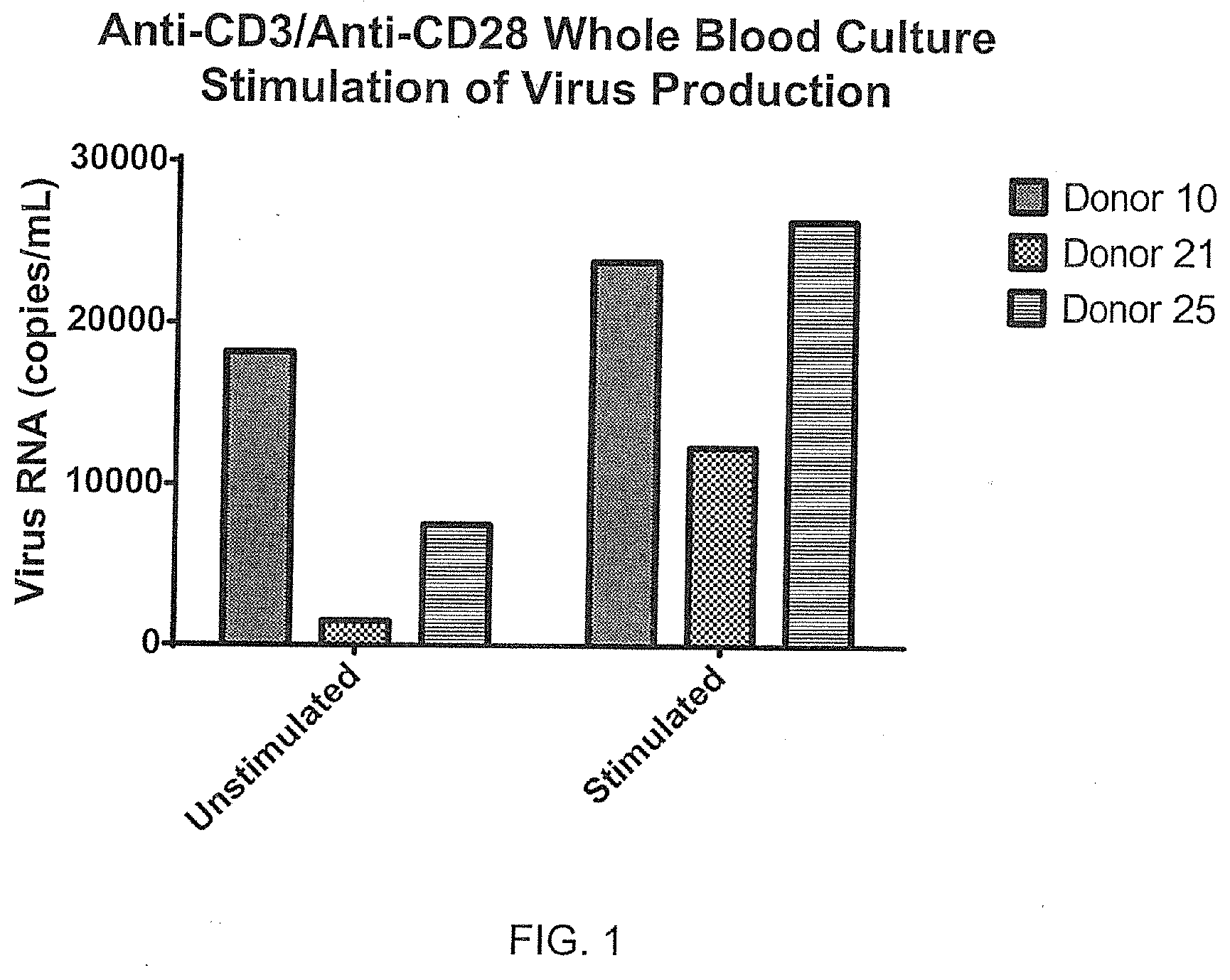
[0075] FIG. 1 depicts the amplification of virus production using stimulated whole blood cultures from HIV-infected blood.
[0076] FIG. 2 illustrates effective stimulation and expansion of leukocytes using anti-CD3/anti-CD28 costimulation with HIV-infected donor blood, resulting in expansion of viral populations from their cell-specific origins within 48 hours at 37 degrees C.
[0077] FIG. 3 illustrates virus production suppression of a candidate drug using stimulated whole blood cultures from HIV-infected blood.
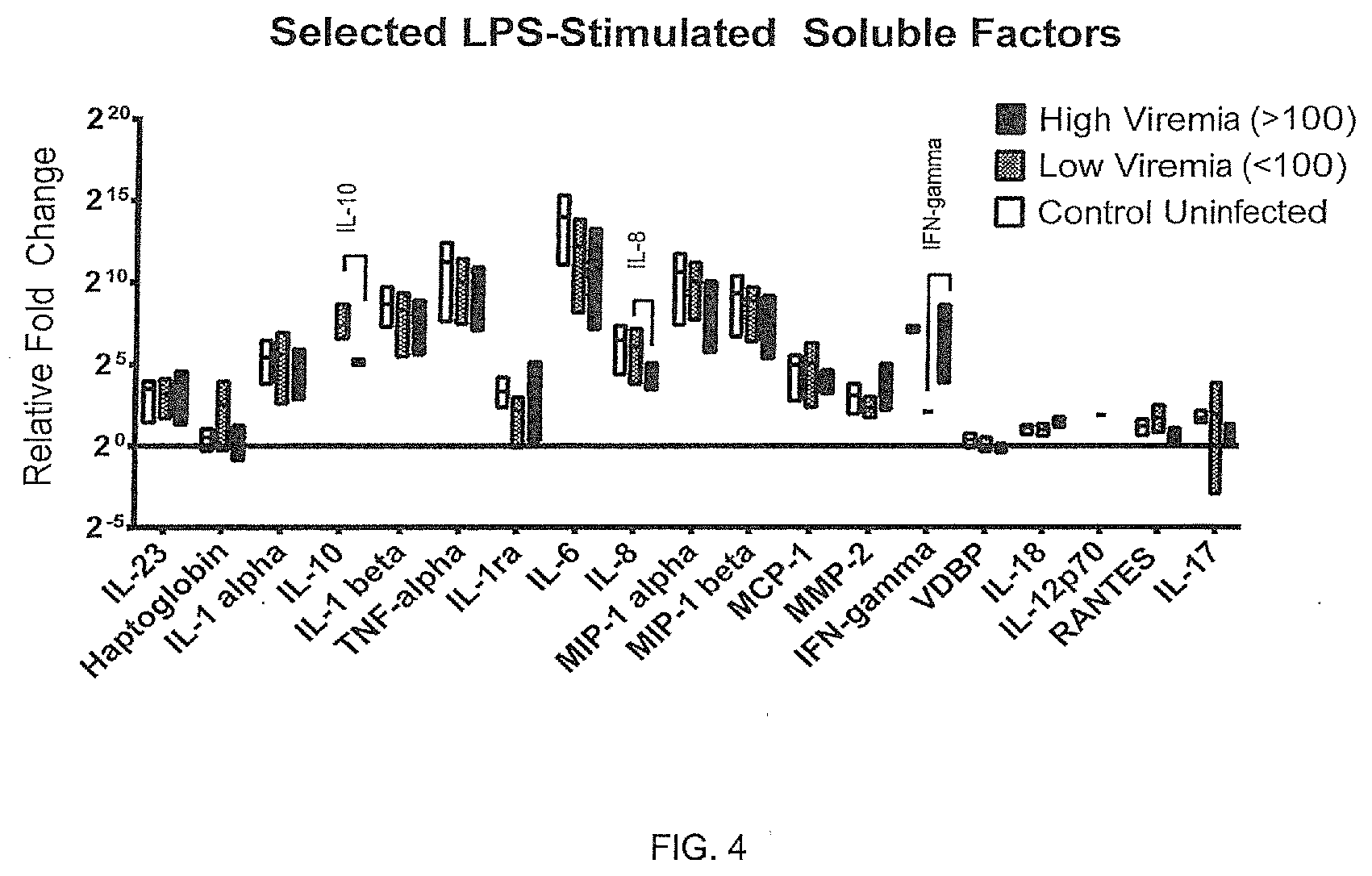
[0078] FIGS. 4-5 illustrates differences in stimulated immunological factors correlating with virus production rates (viremia) of HIV-infected whole blood cultures.
[0079] FIG. 6 illustrates differences in drug-treated immunological factors correlating with antiviral activities of HIV-infected whole blood cultures.
EXAMPLE 1
Anti-CD3/Anti-CD28 Antibodies Increase Virus Production as Compared with Matched Unstimulated Cells
[0080] Blood was collected into sequential preloaded (anticoagulant and stimulant in media) tubes and incubated 48 hours. Stimulants such as anti-CD3/anti-CD28 antibodies were matched to unstimulated tubes using whole blood culture at 37.degree. C. in a tissue culture incubator with gases off. Samples were then centrifuged (140 g, 5 min) and supernatants were collected in duplicate vials (.about.0.8 mL each) and frozen (-80.degree. C.). RNA was measured by RT-qPCR using the Abbott RealTime RNA assay. Recoveries were analyzed across matched donor sets (stimulated vs. unstimulated), reflecting relative virus amplification using the stimulant as shown in FIG. 1. Expansion of T-cell populations (identified by anti-CD3 antibodies) as illustrated in FIG. 2 are possible over this time period.
EXAMPLE 2
Antiviral Drug Effects Evidenced by Reduced Virus Production in Stimulated Whole Blood Cultures
[0081] Antiviral activities can be measured and compared using candidate therapeutics, drugs and/or stimulants. Blood was collected into sequential preloaded (anticoagulant and stimulant in media) tubes and incubated 48 hours. Anti-CD3/anti-CD28 antibodies stimulated virus production as compared with matched unstimulated tubes using whole blood culture at 37.degree. C. in a tissue culture incubator with gases off. Samples were then centrifuged (140 g, 5 min) and supernatants were collected in duplicate vials (.about.0.8 mL each) and frozen (-80.degree. C.). RNA was measured by RT-qPCR using the Abbott RealTime RNA assay. Relative virus inhibition correlates with increasing drug concentration as shown in FIG. 3.
EXAMPLE 3
Soluble Factor Responses Correlate with Virus Production Rates (Viremia)
[0082] HIV-infected and uninfected blood was collected into sequential preloaded (anticoagulant and stimulant and/or drug in media) tubes and incubated 48 hours. Using lipopolysaccharide, differences in soluble factors from cellular responses correlate with virus production (viremia) for select factors. Whole blood was cultured at 37.degree. C. in a tissue culture incubator with gases off. Samples were then centrifuged (140 g, 5 min) and supernatants were collected in duplicate vials (.about.0.8 mL each) and frozen (-80.degree. C.). Supernatants were analyzed using Luminex fluorescently-labeled multiplex microbeads for 47 soluble factors. Correlations between viremia and immune cell stimulant responses (FIG. 4); immune cell stimulant responses with drug (FIG. 5); or drug-induced responses (FIG. 5) are illustrated.
REFERENCES
[0083] 1. Chiu, Y. L., V. B. Soros, J. F. Kreisberg, K. Stopak, W. Yonemoto, and W. C. Greene. 2005. Cellular APOBEC3G restricts HIV-1 infection in resting CD4+ T cells. Nature 435:108-114.
[0084] 2. Coiras, M., M. R. Lopez-Huertas, M. Perez-Olmeda, and J. Alcami. 2009. Understanding HIV-1 latency provides clues for the eradication of long-term reservoirs. Nat Rev Microbiol 7:798-812.
[0085] 3. Colin, L., and C. Van Lint. 2009. Molecular control of HIV-1 postintegration latency: implications for the development of new therapeutic strategies. Retrovirology 6:111.
[0086] 4. Deere, J. D., R. F. Schinazi, and T. W. North. 2011. Simian immunodeficiency virus macaque models of HIV latency. Curr Opin HIV AIDS 6:57-61.
[0087] 5. Duyne, R. V., A. Narayanan, K. H. K, M. Saifuddin, L. Shultz, and F. Kashanchi. 2011. Humanized mouse models of HIV-1 latency. Curr HIV Res 9:595-605.
[0088] 6. Fritsch, R. D., X. Shen, G. P. Sims, K. S. Hathcock, R. J. Hodes, and P. E. Lipsky. 2005. Stepwise differentiation of CD4 memory T cells defined by expression of CCR7 and CD27. J Immunol 175:6489-6497.
[0089] 7. Haggerty, C. M., E. Pitt, and R. F. Siliciano. 2006. The latent reservoir for HIV-1 in resting CD4+T cells and other viral reservoirs during chronic infection: insights from treatment and treatment-interruption trials. Curr Opin HIV AIDS 1:62-68.
[0090] 8. Kourteva, Y., M. De Pasquale, T. Alias, C. McMunn, and R. T. D'Aquila. 2012. APOBEC3G expression and hypermutation are inversely associated with human immunodeficiency virus type 1 (HIV-1) burden in vivo. Virology 430:1-9.
[0091] 9. Luo, K., T. Wang, B. Liu, C. Tian, Z. Xiao, J. Kappes, and X. F. Yu. 2007. Cytidine deaminases APOBEC3G and APOBEC3F interact with human immunodeficiency virus type 1 integrase and inhibit proviral DNA formation. J Viral 81:7238-7248.
[0092] 10. Nalos, M., S. Huang, R. Sluyter, A. Khan, B. Santner-Nanan, R. Nanan, and A. S. McLean. 2008. "Host tissue damage" signal ATP impairs IL-12 and IFNgamma secretion in LPS stimulated whole human blood. Intensive Care Med 34:1891-1897.
[0093] 11. North, T. W., J. Higgins, J. D. Deere, T. L. Hayes, A. Villalobos, L. Adamson, B. L. Shacklett, R. F. Schinazi, and P. A. Luciw. 2010. Viral sanctuaries during highly active antiretroviral therapy in a nonhuman primate model for AIDS. J Virol 84:2913-2922.
[0094] 12. Patterson, B. K., S. McCallister, M. Schutz, J. N. Siegel, K. Shults, Z. Flener, and A. Landay. 2001. Persistence of intracellular HIV-1 mRNA correlates with HIV-1-specific immune responses in infected subjects on stable HAART. Aids 15:1635-1641.
[0095] 13. Romani, B., S. Engelbrecht, and R. H. Glashoff. 2009. Antiviral roles of APOBEC proteins against HIV-1 and suppression by Vif. Arch Virol 154:1579-1588.
[0096] 14. Schmolz, M., T. L. Hurst, D. M. Bailey, J. R. Powell, R. J. Forsey, J. M. Thompson, C. Williams, and G. Pawelec. 2004. Validation of a new highly standardised, lab-independent whole-blood leukocyte function assay for clinical trials (ILCS). Exp Gerontol 39:667-671.
[0097] 15. Smith, M. Z., F. Wightman, and S. R. Lewin. 2012. HIV reservoirs and strategies for eradication. Curr HIV/AIDS Rep 9:5-15.
[0098] 16. Strickland, S. L., R. R. Gray, S. L. Lamers, T. H. Burdo, E. Huenink, D. J. Nolan, B. Nowlin, X. Alvarez, C. C. Midkiff, M. M. Goodenow, K. Williams, and M. Salemi. 2012. Efficient transmission and persistence of low-frequency SIVmac251 variants in CD8-depleted rhesus macaques with different neuropathology. J Gen Virol 93:925-938.
[0099] 17. Tyagi, M., and F. Romerio. 2011. Models of HIV-1 persistence in the CD4+T cell compartment: past, present and future. Curr HIV Res 9:579-587.
* * * * *
D00001
D00002
D00003
D00004
D00005
D00006
XML
uspto.report is an independent third-party trademark research tool that is not affiliated, endorsed, or sponsored by the United States Patent and Trademark Office (USPTO) or any other governmental organization. The information provided by uspto.report is based on publicly available data at the time of writing and is intended for informational purposes only.
While we strive to provide accurate and up-to-date information, we do not guarantee the accuracy, completeness, reliability, or suitability of the information displayed on this site. The use of this site is at your own risk. Any reliance you place on such information is therefore strictly at your own risk.
All official trademark data, including owner information, should be verified by visiting the official USPTO website at www.uspto.gov. This site is not intended to replace professional legal advice and should not be used as a substitute for consulting with a legal professional who is knowledgeable about trademark law.