Retinol-binding Protein 3 (rbp3) As A Protective Factor In Non-diabetic Retinal Degeneration
King; George Liang ; et al.
U.S. patent application number 16/866346 was filed with the patent office on 2021-01-07 for retinol-binding protein 3 (rbp3) as a protective factor in non-diabetic retinal degeneration. The applicant listed for this patent is Joslin Diabetes Center, Inc.. Invention is credited to Hillary A. Keenan, George Liang King.
| Application Number | 20210000855 16/866346 |
| Document ID | / |
| Family ID | |
| Filed Date | 2021-01-07 |


View All Diagrams
| United States Patent Application | 20210000855 |
| Kind Code | A1 |
| King; George Liang ; et al. | January 7, 2021 |
RETINOL-BINDING PROTEIN 3 (RBP3) AS A PROTECTIVE FACTOR IN NON-DIABETIC RETINAL DEGENERATION
Abstract
Methods for increasing retinal thickness in a non-diabetic mammal, comprising administering to the mammal one or both of: (i) a composition comprising RBP3 polypeptide, and/or (ii) a composition comprising a nucleic acid encoding an RBP3 polypeptide.
| Inventors: | King; George Liang; (Dover, MA) ; Keenan; Hillary A.; (Welleley, MA) | ||||||||||
| Applicant: |
|
||||||||||
|---|---|---|---|---|---|---|---|---|---|---|---|
| Appl. No.: | 16/866346 | ||||||||||
| Filed: | May 4, 2020 |
Related U.S. Patent Documents
| Application Number | Filing Date | Patent Number | ||
|---|---|---|---|---|
| 15572671 | Nov 8, 2017 | |||
| PCT/US2016/032752 | May 16, 2016 | |||
| 16866346 | ||||
| 62162439 | May 15, 2015 | |||
| 62161566 | May 14, 2015 | |||
| Current U.S. Class: | 1/1 |
| International Class: | A61K 31/711 20060101 A61K031/711; A61K 38/17 20060101 A61K038/17; A61K 45/06 20060101 A61K045/06; A61K 35/76 20060101 A61K035/76; C12N 15/86 20060101 C12N015/86; A61K 48/00 20060101 A61K048/00; A61K 31/7088 20060101 A61K031/7088; A61K 38/16 20060101 A61K038/16; C12N 15/861 20060101 C12N015/861 |
Goverment Interests
FEDERALLY SPONSORED RESEARCH OR DEVELOPMENT
[0002] This invention was made with Government support under Grant Nos. DK090961-01 and DK094333-01 awarded by the National Institute of Diabetes and Digestive and Kidney Diseases (NIDDK) of the National Institutes of Health. The Government has certain rights in the invention.
Claims
1. A method of increasing retinal thickness in a non-diabetic mammal, the method comprising administering to the mammal one or both of: (i) a composition comprising RBP3 polypeptide, and/or (ii) a composition comprising a nucleic acid encoding an RBP3 polypeptide.
2. (canceled)
3. The method of claim 1, wherein the mammal is a human,
4. The method of claim 1, wherein the mammal has a retinal degenerative disorders associated with retinal thinning.
5. The method of claim 4, wherein the disorder is retinitis pigmentosa, lattice degeneration, or Stargardt disease.
6. The method of claim 4, wherein the disorder is retinal thinning associated with multiple sclerosis or Gaucher's disease.
7. The method of claim 1, wherein the nucleic acid encoding an RBP3 polypeptide is in a viral vector.
8. The method of claim 7, wherein the viral vector is an adeno-associated virus or a lentivirus.
9. The method of claim 1, wherein the nucleic acid encoding an RBP3 polypeptide comprises a sequence that is at least 80% identical to nucleotides 1 to 4276, 1 to 3855, 115 to 4276, 115 to 3855, 166 to 3855, 166 to 4276, 1 to 4151, 115 to 4151, or 166 to 4151 of SEQ ID NO:2.
10. The method of claim 1, wherein the RBP3 polypeptide comprises a sequence that is at least 80% identical to amino acids 18-1247 of SEQ ID NO:1.
11. The method or composition of claim 1, wherein the composition is administered by local (ocular) administration.
12. The method of claim 1, wherein the composition is formulated for administration on, in, or into the eye.
13. The method of claim 1, wherein the composition is formulated in eye drops, lotions, creams, or ointment.
Description
CLAIM OF PRIORITY
[0001] This application claims the benefit of U.S. patent application Ser. No. 62/161,566, filed on May 14, 2015; and 62/162,439, filed on May 15, 2015. The entire contents of the foregoing are hereby incorporated by reference.
TECHNICAL FIELD
[0003] Described are methods for increasing retinal thickness in a non-diabetic mammal, which include administering to the mammal one or both of: (i) a composition comprising RBP3 polypeptide, and/or (ii) a composition comprising a nucleic acid encoding an RBP3 polypeptide.
BACKGROUND
[0004] Retinal thinning resulting from photoreceptor loss is a primary cause of vision loss in retinal degenerative diseases such as Retinitis pigmentosa (RP) (Humayan et al., Invest Ophthalmol Vis Sci. 1999;40:143-148).
SUMMARY
[0005] RBP3, identified as a protective factor in diabetic retinopathy and nephropathy (see, e.g., US2014/0187498), has now been shown to play a role in retinal thickness in non-diabetic eyes. Thus, described herein are methods for increasing retinal thickness in mammals, e.g., non-diabetic mammals, e.g., mammals with or at risk for retinal degeneration associated with retinal thinning, e.g., retinitis pigmentosa, lattice degeneration, or Stargardt disease, or with multiple sclerosis or Gaucher's disease.
[0006] Thus, provided herein are methods for increasing retinal thickness in a non-diabetic mammal, the method comprising administering to the mammal one or both of: (i) a composition comprising RBP3 polypeptide, and/or (ii) a composition comprising a nucleic acid encoding an RBP3 polypeptide.
[0007] Also provided herein are compositions comprising RBP3 polypeptide, and/or a composition comprising a nucleic acid encoding an RBP3 polypeptide, for use in increasing retinal thickness in a non-diabetic mammal.
[0008] In some embodiments, the mammal is a human,
[0009] In some embodiments, the mammal has a retinal degenerative disorder associated with retinal thinning.
[0010] In some embodiments, the disorder is retinitis pigmentosa, lattice degeneration, or Stargardt disease.
[0011] In some embodiments, the disorder is retinal thinning associated with multiple sclerosis or Gaucher's disease.
[0012] In some embodiments, the nucleic acid encoding an RBP3 polypeptide is in a viral vector, e.g., an adeno-associated virus or a lentivirus.
[0013] In some embodiments, the nucleic acid encoding an RBP3 polypeptide comprises a sequence that is at least 80% identical to nucleotides 1 to 4276, 1 to 3855, 115 to 4276, 115 to 3855, 166 to 3855, 166 to 4276, 1 to 4151, 115 to 4151, or 166 to 4151 of SEQ ID NO:2.
[0014] In some embodiments, the RBP3 polypeptide comprises a sequence that is at least 80% identical to amino acids 18-1247 of SEQ ID NO:1.
[0015] In some embodiments, the composition is administered by local (ocular) administration.
[0016] In some embodiments, the composition is formulated for administration on, in, or into the eye. In some embodiments, the composition is formulated in eye drops, lotions, creams, or ointment.
[0017] Unless otherwise defined, all technical and scientific terms used herein have the same meaning as commonly understood by one of ordinary skill in the art to which this invention belongs. Methods and materials are described herein for use in the present invention; other, suitable methods and materials known in the art can also be used. The materials, methods, and examples are illustrative only and not intended to be limiting. All publications, patent applications, patents, sequences, database entries, and other references mentioned herein are incorporated by reference in their entirety. In case of conflict, the present specification, including definitions, will control.
[0018] Other features and advantages of the invention will be apparent from the following detailed description and figures, and from the claims.
DESCRIPTION OF DRAWINGS
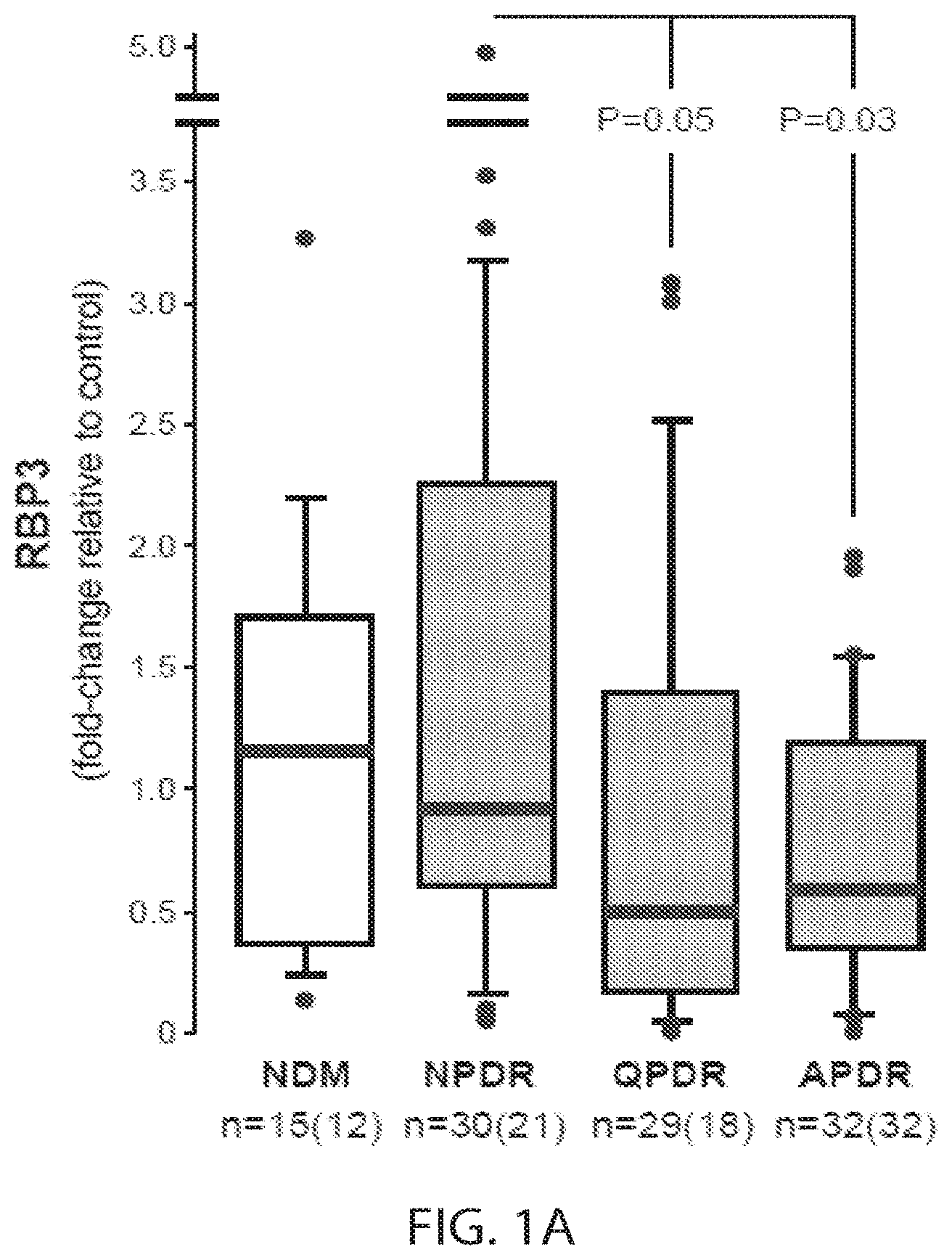
[0019] FIG. 1A. RBP3 expressions by Western blot (WB) in vitreous of mixed population of subjects with type 1 diabetes (Medalists, with 50 or more years of type 1 diabetes, and non-Medalists) and type 2 diabetes separated by grades of diabetic retinopathy (NDM=non-diabetic controls. NPDR=non-proliferative diabetic retinopathy. QPDR=quiescent proliferative diabetic retinopathy. APDR=active proliferative diabetic retinopathy. In box plots, the upper bar, upper box line, middle box line, bottom box line, and bottom bar respectively denote values of 90%, 75%, median, 25% and 10%. N=numbers of eyes (subjects). P-values were tested by Mann-Whitney's U-test for paired comparison.
[0020] FIG. 1B. Western blot analysis for RBP3 in human vitreous. Immunoblotting for RBP3 showed specific single bands sized at 135 kDa. Relative intensity of each band was normalized by the average of 3 of non-diabetic controls consistently run in each gel as controls to normalize the variation of experiments (FIG. 1A).
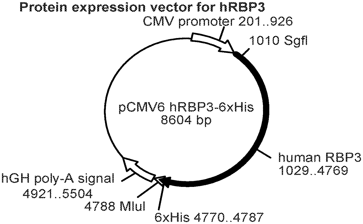
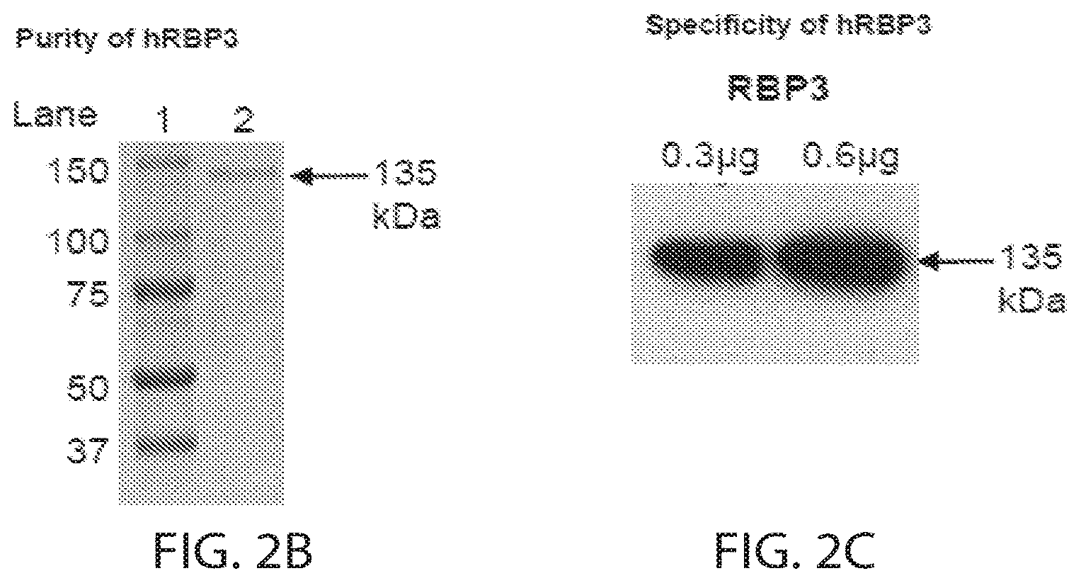
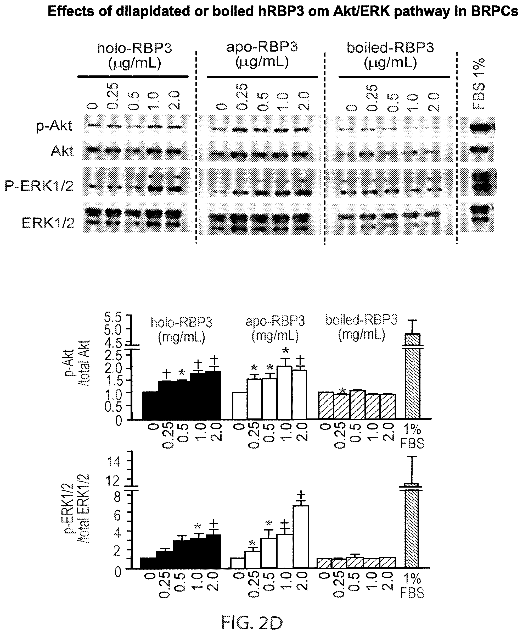
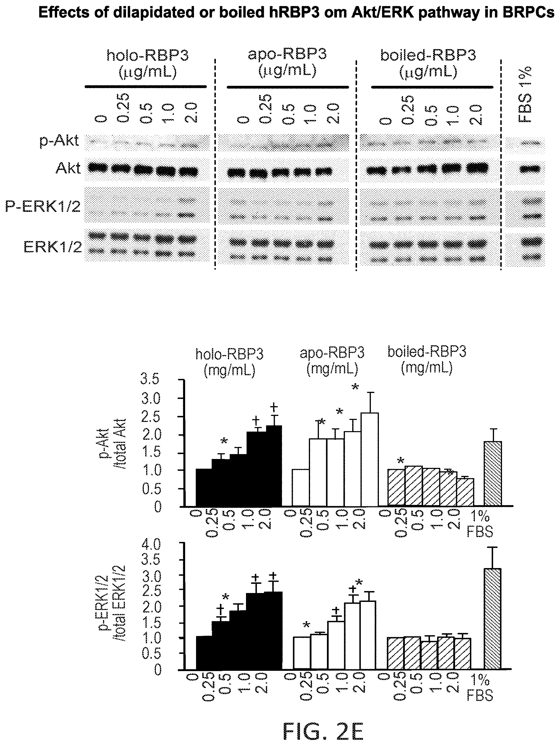
[0021] FIGS. 2A-E. Isolation and delipidation of recombinant human RBP3 protein. Human RBP3 protein (hRBP3) was expressed in 293A cells transfected with pCMV-hRBP3-His plasmid, shown in 2A, by FuGene HD (Promega Corp., Madison, Wis.) and purified by the centrifugal filter (Amicon Ultracel 50K). 2B and 2C show the purity and specificity of RBP3 protein was confirmed by Coomassie Blue staining and western blot analysis. To test the effect of retinoid and lipids binding to hRBP3, lipid-free RBP3 (apo-RBP3) was generated by delipidation methods. 2D and 2E show the effects of hRBP3 on Akt and ERK1/2 pathways in bovine retinal pericytes (BRPCs) and bovine retinal endothelial cells (BRECs) were assessed by western blot analysis with the antibodies detecting phosphorylation of Akt/ERK1/2. Inactivated hRBP3 was obtained by boiling (boiled-RBP3). Cells were incubated for 10 min with indicated concentrations of hRBP3s after overnight starvation in DMEM with 0.1% BSA. Ratio of p-Akt and p-ERK1/2 to total Akt and ERK1/2 were quantified by western blot and shown as fold-change to basal condition (0 .mu.M). *P<0.05 and .dagger.P<0.01 to basal condition (0 .mu.M).
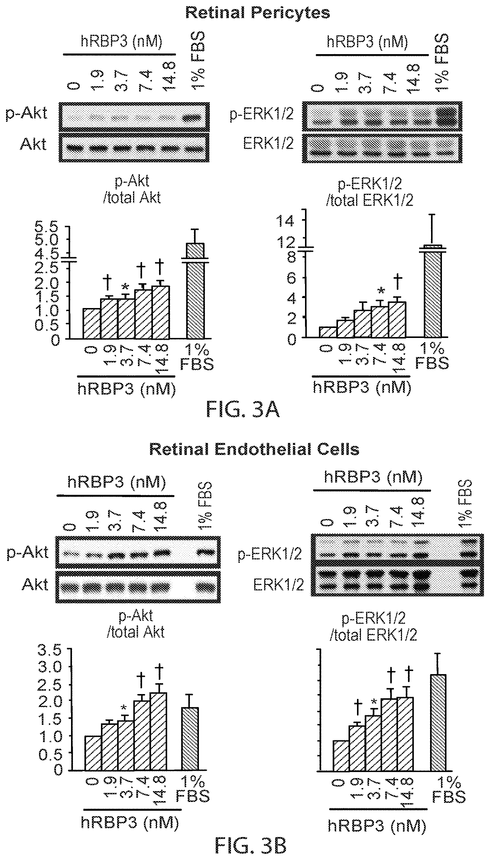
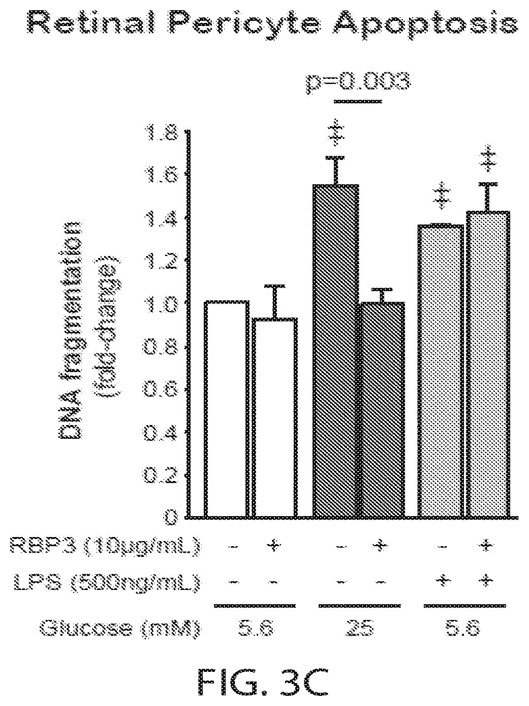
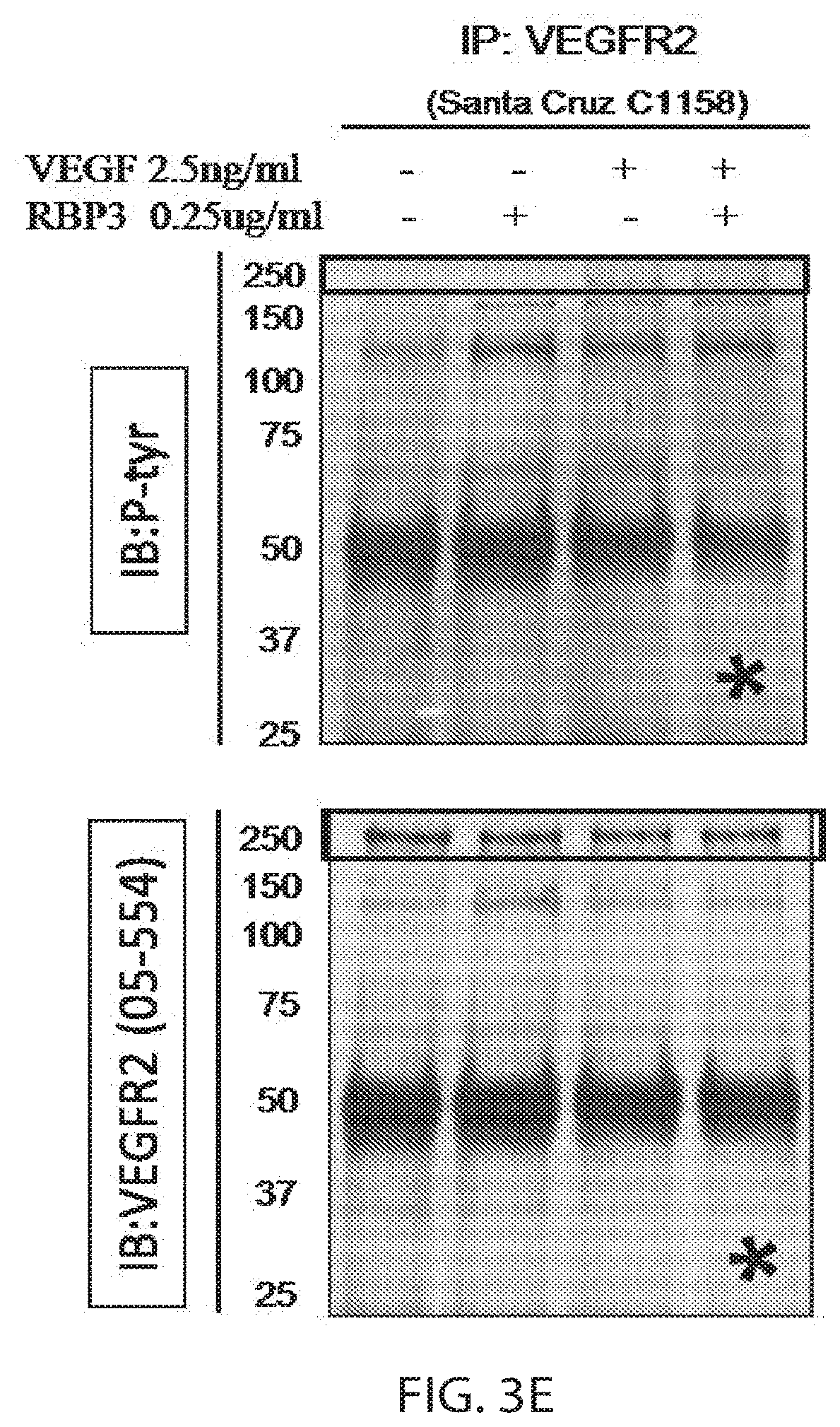
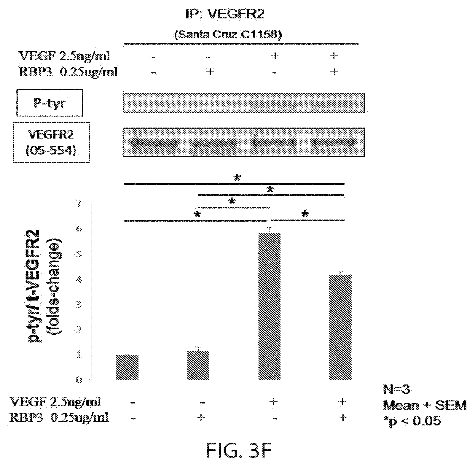
[0022] FIGS. 3A-F. Human RBP3 (hRBP3) effects on phosphorylation of Akt and ERK1/2 in bovine retinal pericytes (BRPCs; 2A) and endothelial cells (BRECs; 2B). Cells were incubated for 10 min with indicated concentrations of hRBP3 after overnight starvation in DMEM with 0.1% BSA. Ratio of p-Akt and p-ERK1/2 to total Akt and ERK1/2 were quantified by western blot and shown as fold-change to basal condition (0 .mu.M). *p<0.05 and .dagger.p<0.01 to basal condition (0 .mu.M). 3C shows RBP3 effect on pericyte apoptosis by DNA fragmentation assay. Apoptosis was induced by 72 hrs exposure to high glucose (25 mM) or LPS (500 ng/mL for 1 hr). hRBP3 was added 24 hrs before measurement of DNA fragmentation. 3D shows effects of hRBP3 or Medalist vitreous with high RBP3 expression in protected eye on high glucose- or VEGF-induced endothelial migration by scratch assay in BRECs. 3E shows a pair of immunoblots showing the effects of RBP3 (0.25 .mu.g/ml) and VEGF (2.5 ng/ml) costimulation for 10 minutes on endothelial migration by scratch assay in BRECs. The boxed areas in 3E are reproduced and enlarged at the top of 3F. The bottom of 3F is a graph showing quantification of the fold change in p-Tyr and VEGFR2 under single and costimulatory conditions. Vitreous RBP3 concentration was adjusted. P-values were tested by 2-tailed unpaired t-tests.
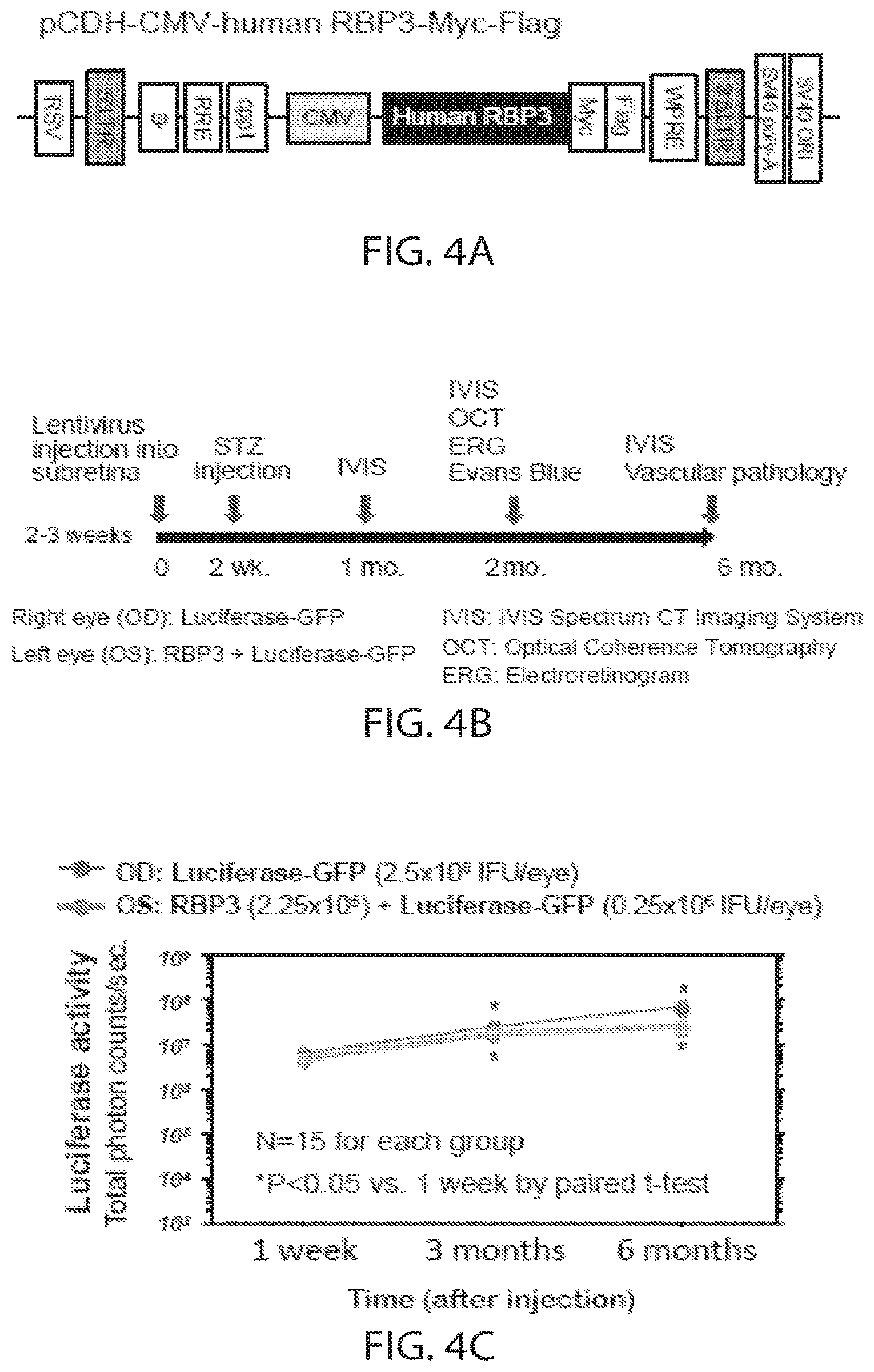
[0023] FIGS. 4A-E. Establishment of a genetic treatment model with hRBP3 overexpression in the subretina. We generated a lentiviral vector expressing RBP3 driven by CMV-promoter as shown in 4A. 4B indicates the time course of the overall in vivo experimental period. Subretinal injections of the lentivirus at the concentrations of luciferase-GFP (OD: 2.5.times.10.sup.6 IFU) and a cocktail of RBP3/luciferase-GFP (OS: 2.25/0.25.times.10.sup.6 IFU) were performed by a trans-corneal method at 2 weeks of age to express hRBP3 and luciferase reporter gene. Diabetes was induced by intraperitoneal injection of streptozotocin (STZ; Sigma-Aldrich, Milwaukee, Wis.; 55 mg/kgBW) at 3-4 week intervals. We confirmed luciferase expression in rat eyes at one week, three months and six months after injection using in vivo imaging system (IVIS Lumina system; Caliper Life Sciences, Hopkinton, Mass.) with intraperitoneal injection of D-Luciferin Firefly (50 mg/g BW, Caliper Life Sciences). The results of IVIS detecting luciferase activities are shown in 4C (time-course). 4D shows the expressions of total RBP3 and exogenous RBP3 tagged with FLAG and Myc by western blot analysis. NDM=non-diabetic rats. DM=diabetic rats. 4E shows the correlation of luciferase activity and the expression of RBP3.
[0024] FIGS. 5A-E. Protective effects of subretinal overexpression of hRBP3 against neural retina dysfunction and decrease of retinal thickness in STZ induced diabetic Lewis rats. NDM=non-diabetic rats. DM=diabetic rats. RBP3-=eye injected with luciferase gene only. RBP3+=eye injected with hRBP3 and luciferase genes. 5A shows scotopic response of neural retina to light flash indicated as amplitudes of oscillatory potential 1, A-wave and B-wave by dark-adapted electroretinogram (ERG). P-values were tested by 2-tailed unpaired t-tests. 5B and 5D show thicknesses of retina and retinal sub-layers measured by optical coherence tomography (OCT); OCT images are shown in 5C and 5E. In the left panel of 5C, the inner plexiform layer (IPL) is 0.042, the inner nuclear layer (INL) is 0.028, the outer nuclear layer (ONL) is 0.054, the inner segment ellipsoid and end tip (ISE+ET) is 0.04, and the Total is 0.203; in the right panel of 5C, the IPL is 0.043, the INL is 0.025, the ONL is 0.052, the ISE+ET is 0.048, and the Total is 0.216. 5D shows ONL thicknesses in OD: luciferase-GFP and OS: RBP3+luciferase-GFP animals. P-values were tested by ANOVA with Fisher's LSD test. *p<0.05 and **p<0.01 tested by ANOVA. N=6 for each.
[0025] FIGS. 6A-C. Effects of subretinal overexpression of hRBP3 in the retina of normal and STZ induced diabetic Lewis rats. NDM=non-diabetic rats. DM=diabetic rats. RBP3-=eye injected with luciferase gene only. RBP3+=eye injected with hRBP3 and luciferase genes. 6A shows quantified acellular capillaries in retinal vascular pathology. 6B shows retinal vascular permeability assessed using the Evans Blue method. 6C shows VEGF expression in vitreous measured by rat VEGF ELISA. P-values were tested by 2-tailed unpaired t-tests.
DETAILED DESCRIPTION
[0026] RBP3 was identified as a potential protective factor against diabetic retinopathy by comparison of individuals with and without proliferative diabetic retinopathy (PDR). The biological roles of human RBP3 were characterized in the retina using retinal vascular cells and eyes of diabetic and normal animals. As demonstrated herein, RBP3 expression can be recovered by genetic therapy, and this treatment increases retinal thickness. RBP3 is thus a therapeutic target for retinal degeneration associated with retinal thinning, e.g., retinitis pigmentosa, lattice degeneration, or Stargardt disease.
Retinol-Binding Protein 3 (RBP3)
[0027] RBP3, a 135 kDa glycolipoprotein highly conserved among mammalian species,.sup.21 is expressed specifically by rod and cone photoreceptors in the retina and secreted into the interphotoreceptor space..sup.22,23 RBP3 is also localized in vitreous humor and aqueous..sup.24,25 As shown in FIG. 1, RBP3 expression level in human vitreous has a certain degree of variability. In addition, the present and previous proteomic analyses using human vitreous revealed that RBP3 was one of the proteins paradoxically decreased in PDR..sup.15,29,30 RBP3 expression in human retinoblastoma cells was downregulated in a high glucose condition possibly by the stimulation of inflammatory cytokines..sup.26 RBP3 is negatively regulated at the transcriptional level by a blockade of elongation complexes during retinal development..sup.27
[0028] An exemplary sequence for human RBP3 can be found in GenBank at Acc. No. NM_002900.2 (nucleic acid) and NP_002891.1 (protein); the protein sequence is reproduced here:
TABLE-US-00001 (SEQ ID NO: 1) 1 mmrewvllms vllcglagpt hlfqpslvld makvlldnyc fpenllgmqe aiqqaikshe 61 ilsisdpqtl asvltagvqs slndprlvis yepstpeppp qvpaltslse eellawlqrg 121 lrhevlegnv gylrvdsvpg qevlsmmgef lvahvwgnlm gtsalvldlr hctggqvsgi 181 pyiisylhpg ntilhvdtiy nrpsntttei wtlpqvlger ygadkdvvvl tssqtrgvae 241 diahilkqmr raivvgertg ggaldlrklr igesdffftv pvsrslgplg ggsqtwegsg 301 vlpcvgtpae qalekalail tlrsalpgvv hclqevlkdy ytivdrvptl lqhlasmdfs 361 tvvseedlvt klnaglqaas edprllvrai gptetpswpa pdaaaedspg vapelpedea 421 irqalvdsvf qvsvlpgnvg ylrfdsfada svlgvlapyv lrqvweplqd tehlimdlrh 481 npggpssavp lllsyfqgpe agpvhlftty drrtnitqeh fshmelpgpr ystqrgvyll 541 tshrtataae efaflmqslg wativgeita gnllhtrtvp lldtpegsla ltvpvltfid 601 nhgeawlggg vvpdaivlae ealdkagevl efhqslgalv egtghlleah yarpevvgqt 661 sallraklaq gayrtavdle slasqltadl qevsgdhrll vfhspgelvv eeapppppav 721 pspeeltyli ealfktevlp gqlgylrfda maeletvkav gpqlvrlvwq qlvdtaalvi 781 dlrynpgsys taipllcsyf feaeprqhly svfdratskv tevwtlpqva gqrygshkdl 841 yilmshtsgs aaeafahtmq dlqratvige ptaggalsvg iyqvgssply asmptqmams 901 attgkawdla gvepditvpm sealsiaqdi valrakvptv lqtagklvad nyasaelgak 961 matklsglqs rysrvtseva laeilgadlq mlsgdphlka ahipenakdr ipgivpmqip 1021 spevfeelik fsfhtnvled nigylrfdmf gdgelltqvs rllvehiwkk imhtdamiid 1081 mrfniggpts sipilcsyff degppvlldk iysrpddsys elwthaqvvg erygskksmv 1141 iltssvtagt aeeftyimkr lgralvigev tsggcqppqt yhvddtnlyl tiptarsvga 1201 sdgsswegvg vtphvvvpae ealarakeml qhnqlrvkrs pglqdhl
SEQ ID NO:1 includes the entire sequence; as amino acids 1-17 are predicted to be a signal sequence, in some embodiments, only amino acids 18-1247 of SEQ ID NO:1 are used. See, e.g., Liou et al., Somat. Cell Molec. Genet. 13: 315-323, 1987.
[0029] In some embodiments, the sequence of RBP3 used in the present compositions and methods is about 80%, 85%, 90%, 95%, 99% or 100% identical to SEQ ID NO:1.
[0030] To determine the percent identity of two sequences, the sequences are aligned for optimal comparison purposes (gaps are introduced in one or both of a first and a second amino acid or nucleic acid sequence as required for optimal alignment, and non-homologous sequences can be disregarded for comparison purposes). The length of a reference sequence aligned for comparison purposes is at least 80% (in some embodiments, about 85%, 90%, 95%, or 100% of the length of the reference sequence) is aligned. The nucleotides or residues at corresponding positions are then compared. When a position in the first sequence is occupied by the same nucleotide or residue as the corresponding position in the second sequence, then the molecules are identical at that position. The percent identity between the two sequences is a function of the number of identical positions shared by the sequences, taking into account the number of gaps, and the length of each gap, which need to be introduced for optimal alignment of the two sequences.
[0031] The comparison of sequences and determination of percent identity between two sequences can be accomplished using a mathematical algorithm. For example, the percent identity between two amino acid sequences can be determined using the Needleman and Wunsch ((1970) J. Mol. Biol. 48:444-453) algorithm which has been incorporated into the GAP program in the GCG software package, using a Blossum 62 scoring matrix with a gap penalty of 12, a gap extend penalty of 4, and a frameshift gap penalty of 5.
Methods of Treatment
[0032] The methods described herein include methods for increasing retinal thickness in a subject, e.g., for the treatment of retinal degenerative disorders associated with retinal thinning. In some embodiments, the disorder is retinitis pigmentosa, lattice degeneration, or Stargardt disease. In some embodiments, the disorder is retinal thinning associated with multiple sclerosis or Gaucher's disease. Generally, the methods include administering a therapeutically effective amount of RBP3 protein or nucleic acid encoding RBP3 protein as described herein, to a subject who is in need of, or who has been determined to be in need of, such treatment. In preferred embodiments, the subject does not have diabetes, and/or does not have a mutation in RBP3, e.g., does not have a D1080N mutation (c.3238G-A transition in exon 2 of the RBP3 gene) described in den Hollander et al., Invest. Ophthal. Vis. Sci. 50: 1864-1872, 2009, or a mutation in RBP3 as described in Ksantini et al., Ophthal. Genet. 31: 200-204, 2010, Li et al., J. Biol. Chem. 288: 11395-11406, 2013, or Liou et al., J. Neurosci. 18: 4511-4520, 1998.
[0033] As used in this context, to "treat" means to ameliorate at least one symptom of the disorder associated with retinal thinning. Often, retinal thinning results in loss of visual acuity or risk of retinal tears or detachment; thus, a treatment can result in reduction of the risk of retinal tears or detachment and a return or approach to normal visual acuity. Administration of a therapeutically effective amount of a compound described herein for the treatment of a condition associated with retinal thinning will result in one or more of a decreased rate of retinal thinning, preservation of retinal thickness, and/or an increase in retinal thickness (i.e., an increase in thickness as compared to before administration of the treatment).
Subjects
[0034] The methods described herein can be used to treat subjects who have developed retinal thinning, e.g., a subject who has retinitis pigmentosa, lattice degeneration, Stargardt disease, or retinal thinning associated with multiple sclerosis or Gaucher's disease. Methods for diagnosing these diseases are known in the art; for example, retinal thickness can be determined using optical coherence tomography (OCT) imaging. In some embodiments, the thickness of the outer nuclear layer (ONL) of the retina is measured and considered.
[0035] The presence of retinal thinning can be diagnosed based on the presence of a retinal thickness that is below a reference level (of thickness). Suitable reference levels for retinal thickness can be determined based on epidemiological studies, e.g., using specific OCT imaging devices, e.g., as described in Chan et al., Arch Ophthalmol. 2006 February; 124(2): 193-198, and can depend on the age of the subject, see, e.g., Alamouti and Funk, Br J Ophthalmol. 2003 July; 87(7): 899-901.
[0036] In some embodiments, the methods described herein include detecting the presence of reduced levels of RBP3 protein or mRNA in the mammal, e.g., in the eye of the mammal, and optionally selecting a subject who has levels of RBP3 protein or mRNA below a reference level (e.g., a reference level that represents a level of RBP3 in a normal subject) for treatment using a method described herein. In some embodiments, the methods described herein include detecting the presence of reduced levels of RBP3 protein or mRNA in a mammal who does not have retinal thinning, e.g., in the eye of the mammal, and determining that the subject is at risk (i.e., has a higher risk that the general population) of developing retinal thinning when the subject has levels of RBP3 protein or mRNA below a reference level (e.g., a reference level that represents a level of RBP3 in a subject with a normal level of risk of developing retinal thinning).
[0037] The methods include obtaining a sample from a subject, and evaluating the presence and/or level of RBP3 in the sample, and comparing the presence and/or level with one or more references, e.g., a control reference that represents a normal level of RBP3, e.g., a level in an unaffected subject, and/or a disease reference that represents a level of the proteins associated with retinal thinning, e.g., a level in a subject having retinal thinning.
[0038] As used herein the term "sample", when referring to the material to be tested for the presence of a biological marker using the method of the invention, includes inter alia vitreous, whole blood, plasma, or serum. The type of sample used may vary depending upon the identity of the biological marker to be tested and the clinical situation in which the method is used. Various methods are well known within the art for the identification and/or isolation and/or purification of a biological marker from a sample. An "isolated" or "purified" biological marker is substantially free of cellular material or other contaminants from the cell or tissue source from which the biological marker is derived i.e. partially or completely altered or removed from the natural state through human intervention. For example, nucleic acids contained in the sample are first isolated according to standard methods, for example using lytic enzymes, chemical solutions, or isolated by nucleic acid-binding resins following the manufacturer's instructions.
[0039] The presence and/or level of a protein can be evaluated using methods known in the art, e.g., using standard electrophoretic and quantitative immunoassay methods for proteins, including but not limited to, Western blot; enzyme linked immunosorbent assay (ELISA); biotin/avidin type assays; protein array detection; radio-immunoassay; immunohistochemistry (IHC); immune-precipitation assay; FACS (fluorescent activated cell sorting); mass spectrometry (Kim (2010) Am J Clin Pathol 134:157-162; Yasun (2012) Anal Chem 84(14):6008-6015; Brody (2010) Expert Rev Mol Diagn 10(8):1013-1022; Philips (2014) PLOS One 9(3):e90226; Pfaffe (2011) Clin Chem 57(5): 675-687). The methods typically include revealing labels such as fluorescent, chemiluminescent, radioactive, and enzymatic or dye molecules that provide a signal either directly or indirectly. As used herein, the term "label" refers to the coupling (i.e. physically linkage) of a detectable substance, such as a radioactive agent or fluorophore (e.g. phycoerythrin (PE) or indocyanine (Cy5), to an antibody or probe, as well as indirect labeling of the probe or antibody (e.g. horseradish peroxidase, HRP) by reactivity with a detectable substance.
[0040] For example, levels of RBP3 protein in a sample (e.g., a sample of vitreous, blood, serum, or plasma) can be detected using an RBP3-binding antibody or fragment thereof. The term "antibody" as used herein refers to an immunoglobulin molecule or an antigen-binding portion thereof. Examples of antigen-binding portions of immunoglobulin molecules include F(ab) and F(ab').sub.2 fragments, which retain the ability to bind antigen. The antibody can be polyclonal, monoclonal, recombinant, chimeric, de-immunized or humanized, fully human, non-human, (e.g., murine), or single chain antibody. In some embodiments the antibody has effector function and can fix complement. In some embodiments, the antibody has reduced or no ability to bind an Fc receptor. For example, the antibody can be an isotype or subtype, fragment or other mutant, which does not support binding to an Fc receptor, e.g., it has a mutagenized or deleted Fc receptor binding region. Antibodies that bind to RBP3 are known in the art and commercially available, e.g., from Abbexa, Abcam, Abbiotec, Abnova, Atlas Antibodies, Life Technologies, OriGene, Novus Biologicals, United States Biological, and Santa Cruz Biotechnolgy, Inc.; additional antibodies and fragments thereof can be generated using methods known in the art, see, e.g., Harlow et. al., editors, Antibodies: A Laboratory Manual (1988); Goding, Monoclonal Antibodies: Principles and Practice, (N.Y. Academic Press 1983); Howard and Kaser, Making and Using Antibodies: A Practical Handbook (CRC Press; 1st edition, Dec. 13, 2006); Kontermann and Dubel, Antibody Engineering Volume 1 (Springer Protocols) (Springer; 2nd ed., May 21, 2010); Lo, Antibody Engineering: Methods and Protocols (Methods in Molecular Biology) (Humana Press; Nov. 10, 2010); and Dubel, Handbook of Therapeutic Antibodies: Technologies, Emerging Developments and Approved Therapeutics, (Wiley-VCH; 1 edition Sep. 7, 2010).
[0041] The antibody can be coupled to a detectable or imaging agent. Such agents are well known in the art and include paramagnetic agents, bioluminescent or fluorescent labels (e.g., GFP, FITC, rhodamine, or Texas Red), radioactive isotopes, and colorimetric/enzymatic agents (e.g., HRP, B-galactosidase). In a preferred embodiment, the antibody is coupled to a paramagnetic agent, e.g., a paramagnetic nanoparticle, e.g., cross-linked iron oxide (CLIO) nanoparticles; see, e.g., US 20110046004; Josephson et al., Bioconjug. Chem., 10(2):186-91 (1999).
[0042] In some embodiments, an ELISA method may be used, e.g., wherein the wells of a mictrotiter plate are coated with an antibody against which the protein is to be tested. The sample containing or suspected of containing the biological marker is then applied to the wells. After a sufficient amount of time, during which antibody-antigen complexes would have formed, the plate is washed to remove any unbound moieties, and a detectably labelled molecule is added. Again, after a sufficient period of incubation, the plate is washed to remove any excess, unbound molecules, and the presence of the labeled molecule is determined using methods known in the art. Variations of the ELISA method, such as the competitive ELISA or competition assay, and sandwich ELISA, may also be used, as these are well-known to those skilled in the art.
[0043] In some embodiments, an IHC method may be used. IHC provides a method of detecting a biological marker in situ. The presence and exact cellular location of the biological marker can be detected. Typically a sample is fixed with formalin or paraformaldehyde, embedded in paraffin, and cut into sections for staining and subsequent inspection by confocal microscopy. Current methods of IHC use either direct or indirect labelling. The sample may also be inspected by fluorescent microscopy when immunofluorescence (IF) is performed, as a variation to IHC.
[0044] Mass spectrometry, and particularly matrix-assisted laser desorption/ionization mass spectrometry (MALDI-MS) and surface-enhanced laser desorption/ionization mass spectrometry (SELDI-MS), is useful for the detection of biomarkers of this invention. (See U.S. Pat. Nos. 5,118,937; 5,045,694; 5,719,060; and 6,225,047)
[0045] The presence and/or level of a nucleic acid can be evaluated using methods known in the art, e.g., using polymerase chain reaction (PCR), reverse transcriptase polymerase chain reaction (RT-PCR), quantitative or semi-quantitative real-time RT-PCR, digital PCR i.e. BEAMing ((Beads, Emulsion, Amplification, Magnetics) Diehl (2006) Nat Methods 3:551-559) ; RNAse protection assay; Northern blot; various types of nucleic acid sequencing (Sanger, pyrosequencing, NextGeneration Sequencing); fluorescent in-situ hybridization (FISH); or gene array/chips) (Lehninger Biochemistry (Worth Publishers, Inc., current addition; Sambrook, et al, Molecular Cloning: A Laboratory Manual (3rd Edition, 2001); Bernard (2002) Clin Chem 48(8): 1178-1185; Miranda (2010) Kidney International 78:191-199; Bianchi (2011) EMBO Mol Med 3:495-503; Taylor (2013) Front. Genet. 4:142; Yang (2014) PLOS One 9(11):e110641); Nordstrom (2000) Biotechnol. Appl. Biochem. 31(2):107-112; Ahmadian (2000) Anal Biochem 280:103-110. In some embodiments, high throughput methods, e.g., protein or gene chips as are known in the art (see, e.g., Ch. 12, Genomics, in Griffiths et al., Eds. Modern genetic Analysis, 1999,W. H. Freeman and Company; Ekins and Chu, Trends in Biotechnology, 1999, 17:217-218; MacBeath and Schreiber, Science 2000, 289(5485):1760-1763; Simpson, Proteins and Proteomics: A Laboratory Manual, Cold Spring Harbor Laboratory Press; 2002; Hardiman, Microarrays Methods and Applications: Nuts & Bolts, DNA Press, 2003), can be used to detect the presence and/or level of RBP3. Measurement of the level of RBP3 can be direct or indirect. For example, the abundance levels of RBP3 can be directly quantitated. Alternatively, the amount of a biomarker can be determined indirectly by measuring abundance levels of RBP3 cDNA, amplified RNAs or DNAs, or by measuring quantities or activities of RNAs, or other molecules that are indicative of the expression level of the biomarker. In some embodiments a technique suitable for the detection of alterations in the structure or sequence of nucleic acids, such as the presence of deletions, amplifications, or substitutions, can be used for the detection of biomarkers of this invention.
[0046] RT-PCR can be used to determine the expression profiles of RBP3 (U.S. Patent No. 2005/0048542A1). The first step in expression profiling by RT-PCR is the reverse transcription of the RNA template into cDNA, followed by its exponential amplification in a PCR reaction (Ausubel et al (1997) Current Protocols of Molecular Biology, John Wiley and Sons). To minimize errors and the effects of sample-to-sample variation, RT-PCR is usually performed using an internal standard, which is expressed at constant level among tissues, and is unaffected by the experimental treatment. Housekeeping genes, such as glyceraldehyde-3-phosphate dehydrogenase (GAPDH), beta-actin (ACTB), lactate dehydrogenase A (LDHA), ribosomal protein L5 (RPL5), ubiquitin C (UBC), peptidylprolyl isomerase A (PPIA), TATA-box binding protein (TBP1), and/or hypoxanthine guanine phosphoribosyl transferase (HPRT1), are most commonly used.
[0047] Gene arrays are prepared by selecting probes which comprise a polynucleotide sequence, and then immobilizing such probes to a solid support or surface. For example, the probes may comprise DNA sequences, RNA sequences, co-polymer sequences of DNA and RNA, DNA and/or RNA analogues, or combinations thereof. The probe sequences can be synthesized either enzymatically in vivo, enzymatically in vitro (e.g. by PCR), or non-enzymatically in vitro.
[0048] In some embodiments, the presence and/or level of RBP3 is comparable to the presence and/or level of the protein(s) in the disease reference, and the subject has no overt signs or symptoms of retinal thinning, then the subject has an increased risk of developing retinal thinning, and a treatment, e.g., as known in the art or as described herein, can be administered to reduce the risk of developing retinal thinning. In some embodiments, once it has been determined that a person has retinal thinning, or has an increased risk of developing retinal thinning, then a treatment, e.g., as known in the art or as described herein, can be administered.
[0049] Suitable reference values can be determined using methods known in the art, e.g., using standard clinical trial methodology and statistical analysis. The reference values can have any relevant form. In some cases, the reference comprises a predetermined value for a meaningful level of RBP3, e.g., a control reference level that represents a normal level of RBP3, e.g., a level in an unaffected subject (with a normal retinal thickness) or a subject who is not at risk of developing retinal thinning, and/or a disease reference that represents a level of RBP3 associated with retinal thinning.
[0050] The predetermined level can be a single cut-off (threshold) value, such as a median or mean, or a level that defines the boundaries of an upper or lower quartile, tertile, or other segment of a clinical trial population that is determined to be statistically different from the other segments. It can be a range of cut-off (or threshold) values, such as a confidence interval. It can be established based upon comparative groups, such as where association with risk of developing disease or presence of disease in one defined group is a fold higher, or lower, (e.g., approximately 2-fold, 4-fold, 8-fold, 16-fold or more) than the risk or presence of disease in another defined group. It can be a range, for example, where a population of subjects (e.g., control subjects) is divided equally (or unequally) into groups, such as a low-risk group, a medium-risk group and a high-risk group, or into quartiles, the lowest quartile being subjects with the lowest risk and the highest quartile being subjects with the highest risk, or into n-quantiles (i.e., n regularly spaced intervals) the lowest of the n-quantiles being subjects with the lowest risk and the highest of the n-quantiles being subjects with the highest risk.
[0051] In some embodiments, the predetermined level is a level or occurrence in the same subject, e.g., at a different time point, e.g., an earlier time point.
[0052] Subjects associated with predetermined values are typically referred to as reference subjects. For example, in some embodiments, a control reference subject does not have a disorder described herein (e.g. retinal thinning). In the present methods, the subjects do not have diabetes.
[0053] A disease reference subject is one who has (or has an increased risk of developing) retinal thinning. An increased risk is defined as a risk above the risk of subjects in the general population.
[0054] Thus, in some cases the level of RBP3 in a subject being less than or equal to a reference level of RBP3 is indicative of a clinical status (e.g., indicative of a disorder as described herein, e.g., retinal thinning. In other cases the level of RBP3 in a subject being greater than or equal to the reference level of RBP3 is indicative of the absence of disease or normal risk of the disease (i.e., the same risk as the general population). In some embodiments, the amount by which the level in the subject is less than the reference level is sufficient to distinguish a subject from a control subject, and optionally is statistically significantly less than the level in a control subject. In cases where the level of RBP3 in a subject being equal to the reference level of RBP3, the "being equal" refers to being approximately equal (e.g., not statistically different).
[0055] The predetermined value can depend upon the particular population of subjects (e.g., human subjects) selected. For example, an apparently healthy population will have a different `normal` range of levels of RBP3 than will a population of subjects which have, are likely to have, or are at greater risk to have, a disorder described herein. Accordingly, the predetermined values selected may take into account the category (e.g., sex, age, health, risk, presence of other diseases) in which a subject (e.g., human subject) falls. Appropriate ranges and categories can be selected with no more than routine experimentation by those of ordinary skill in the art.
[0056] In characterizing likelihood, or risk, numerous predetermined values can be established.
Gene Therapy
[0057] Described herein are methods in which a nucleic acid encoding an RBP3 polypeptide or active fragment thereof, e.g., incorporated into a gene transfer construct , is used to increase retinal thickness, e.g., to treat a condition associated with retinal thinning as described herein. In some embodiments, the nucleotide sequence encoding human RBP3 as described herein is at least about 75% identical to the reference sequence of human RBP3 found in GenBank at Acc. No. NM 002900.2 (nucleic acid), e.g., SEQ ID NO:2. In some embodiments, the nucleotide sequences are about 80%, 85%, 90%, 95%, 99% or 100% identical to a sequence encoding a mature or full length human RBP3, e.g., a sequence comprising nucleotides 1 to 4276, 1 to 3855, 115 to 4276, 115 to 3855, 166 to 3855, 166 to 4276, 1 to 4151, 115 to 4151, or 166 to 4151 of SEQ ID NO:2 (nucleotides 115 to 165 encode a signal sequence).
[0058] Exemplary Human RBP3 sequence:
TABLE-US-00002 (SEQ ID NO: 2) 1 tgtccaccag ctgagaagga caagggcgga aggcagctgc acagagcagg gccacggcct 61 tgcacacagt ccagggagct tttgtgcagg agccaggcct ccccctgggt ccccatgatg 121 agagaatggg ttctgctcat gtccgtgctg ctctgtggcc tggctggccc cacacacctg 181 ttccagccaa gcctggtgct ggacatggcc aaggtcctct tggataacta ctgcttcccg 241 gagaacctgc tgggcatgca ggaagccatc cagcaggcca tcaagagcca tgagattctg 301 agcatctcag acccgcagac gctggccagt gtgctgacag ccggggtgca gagctccctg 361 aacgatcctc gcctggtcat ctcctatgag cccagcaccc ccgagcctcc cccacaagtc 421 ccagcactca ccagcctctc agaagaggaa ctgcttgcct ggctgcaaag gggcctccgc 481 catgaggttc tggagggtaa tgtgggctac ctgcgggtgg acagcgtccc gggccaggag 541 gtgctgagca tgatggggga gttcctggtg gcccacgtgt gggggaatct catgggcacc 601 tccgccttag tgctggatct ccggcactgc acaggaggcc aggtctctgg cattccctac 661 atcatctcct acctgcaccc agggaacacc atcctgcacg tggacactat ctacaaccgc 721 ccctccaaca ccaccacgga gatctggacc ttgccccagg tcctgggaga aaggtacggt 781 gccgacaagg atgtggtggt cctcaccagc agccagacca ggggcgtggc cgaggacatc 841 gcgcacatcc ttaagcagat gcgcagggcc atcgtggtgg gcgagcggac tgggggaggg 901 gccctggacc tccggaagct gaggataggc gagtctgact tcttcttcac ggtgcccgtg 961 tccaggtccc tggggcccct tggtggaggc agccagacgt gggagggcag cggggtgctg 1021 ccctgtgtgg ggactccggc cgagcaggcc ctggagaaag ccctggccat cctcactctg 1081 cgcagcgccc ttccaggggt agtccactgc ctccaggagg tcctgaagga ctactacacg 1141 ctggtggacc gtgtgcccac cctgctgcag cacttggcca gcatggactt ctccacggtg 1201 gtctccgagg aagatctggt caccaagctc aatgccggcc tgcaggctgc gtctgaggat 1261 cccaggctcc tggtgcgagc catcgggccc acagaaactc cttcttggcc cgcgcccgac 1321 gctgcagccg aagactcacc aggggtggcc ccagagttgc ctgaggacga ggctatccgg 1381 caagcactgg tggactctgt gttccaggtg tcggtgctgc caggcaatgt gggctacctg 1441 cgcttcgata gttttgctga cgcctccgtc ctgggtgtgt tggccccata tgtcctgcgc 1501 caggtgtggg agccgctaca ggacacggag cacctcatca tggacctgcg ccacaaccct 1561 ggagggccat cctctgctgt gcccctgctc ctgtcctact tccagggccc tgaggccggc 1621 cccgtgcacc tcttcaccac ctatgatcgc cgcaccaaca tcacgcagga gcacttcagc 1681 cacatggagc tcccgggccc acgctacagc acccaacgtg gggtgtatct gctcaccagc 1741 caccgcaccg ccacggccgc ggaggagttc gccttcctta tgcagtcgct gggctgggcc 1801 acactggtag gtgagatcac cgcgggcaac ctgctgcaca cccgcacggt gccgctgctg 1861 gacacacccg aaggcagcct cgcgctcacc gtgccggtcc tcaccttcat cgacaatcac 1921 ggcgaggcct ggctgggtgg tggagtggtg cccgatgcca tcgtgctggc cgaggaggcc 1981 ctggacaaag cccaggaagt gctggagttc caccaaagcc tgggggcctt ggtggagggc 2041 acagggcacc tgctggaggc ccactatgct cggccagagg tcgtggggca gaccagtgcc 2101 ctcctgcggg ccaagctggc ccagggcgcc taccgcacag ctgtggactt ggagtctctg 2161 gcctctcagc tcacagcaga cctccaggag gtgtctgggg accaccgctt gctagtgttc 2221 cacagccctg gcgagctggt ggtagaggaa gcacccccac caccccctgc tgtcccctct 2281 ccagaggagc tcacctacct tattgaggcc ctgttcaaga cagaggtgct gcccggccag 2341 ctgggctacc tgcgttttga cgccatggct gaactggaga cagtgaaggc cgtggggcca 2401 cagctggtgc ggctggtatg gcaacagctg gtggacacgg ctgcgctggt gatcgacctg 2461 cgctacaacc ctggcagcta ctccacggcc atcccgctgc tctgctccta cttctttgag 2521 gcagagcccc gccagcacct gtattctgtc tttgacaggg ccacctcaaa agtcacggag 2581 gtgtggacct tgccccaggt cgccggccag cgctacggct cacacaagga cctctacatc 2641 ctgatgagcc acaccagtgg ctctgcggcc gaggcctttg cacacaccat gcaggacctg 2701 cagcgggcca cggtcattgg ggagcccacg gccggaggcg cactctctgt gggcatctac 2761 caggtgggca gcagcccctt atatgcatcc atgcccaccc agatggccat gagtgccacc 2821 acaggcaagg cctgggacct ggctggtgtg gagcccgaca tcactgtgcc catgagcgaa 2881 gccctttcca tagcccagga catagtggct ctgcgtgcca aggtgcccac ggtgctgcag 2941 acggccggga agctggtggc tgataactat gcctctgccg agctgggggc caagatggcc 3001 accaaactga gcggtctgca gagccgctac tccagggtga cctcagaagt ggccctagcc 3061 gagatcctgg gggctgacct gcagatgctc tccggagacc cacacctgaa ggcagcccat 3121 atccctgaga atgccaagga ccgcattcct ggaattgtgc ccatgcagat cccttcccct 3181 gaagtatttg aagagctgat caagttttcc ttccacacta acgtgcttga ggacaacatt 3241 ggctacttga ggtttgacat gtttggggac ggtgagctgc tcacccaggt ctccaggctg 3301 ctggtggagc acatctggaa gaagatcatg cacacggatg ccatgatcat cgacatgagg 3361 ttcaacatcg gtggccccac atcctccatt cccatcttgt gctcctactt ctttgatgaa 3421 ggccctccag ttctgctgga caagatctac agccggcctg atgactctgt cagtgaactc 3481 tggacacacg cccaggttgt aggtgaacgc tatggctcca agaagagcat ggtcattctg 3541 accagcagtg tgacggccgg caccgcggag gagttcacct atatcatgaa gaggctgggc 3601 cgggccctgg tcattgggga ggtgaccagt gggggctgcc agccaccaca gacctaccac 3661 gtggatgaca ccaacctcta cctcactatc cccacggccc gttctgtggg ggcctcggat 3721 ggcagctcct gggaaggggt gggggtgaca ccccatgtgg ttgtccctgc agaagaggct 3781 ctcgccaggg ccaaggagat gctccagcac aaccagctga gggtgaagcg gagcccaggc 3841 ctgcaggacc acctgtaggg aagggcccca taggcagagc cccagggcag acagaacctc 3901 tgggacacac accaagggca ctcctgcagg tggcccggcc tgaggttccc aggagcagca 3961 aaggggcctg ctgagctctg gttaggttac agctggaggt gtgtatatat acacacacac 4021 acatgtatat acacatatat atgtgtatgt atatatatgt atatatatat ggctttccaa 4081 taaccaccta aattttaaca aaggttcctt ctaagtggta gaacttgggg tggtattttt 4141 accttccttc ttcatacttt gctctttttc ttaaatactc attaatgtgc atatatcatt 4201 attttcagat gcagctatca ttattccaaa atacaaaata aagaagataa aataaattat 4261 atacccgagc cattaaaaaa aaaaaaaaa
[0059] These methods can include the use of targeted expression vectors for in vivo transfection and expression of a polynucleotide that encodes an RBP3 polypeptide or active fragment thereof, preferably in particular cell types, especially cells of the retina, e.g., of the ONL of the retina. Expression constructs of such components can be administered in any effective carrier, e.g., any formulation or composition capable of effectively delivering the component gene to cells in vivo. Approaches include insertion of the RBP3 gene into gene transfer and expression constructs such as viral vectors, including recombinant retroviruses, adenovirus, adeno-associated virus, lentivirus, and herpes simplex virus-1, or recombinant bacterial or eukaryotic plasmids. Viral vectors transfect cells directly; plasmid DNA can be delivered naked or with the help of, for example, cationic liposomes (lipofectamine) or derivatized (e.g., antibody conjugated), polylysine conjugates, gramacidin S, artificial viral envelopes or other such intracellular carriers, as well as direct injection of the gene construct or CaPO.sub.4 precipitation carried out in vivo.
[0060] Retrovirus vectors can be used as a recombinant gene delivery system for the transfer of exogenous genes in vivo, particularly into humans. These vectors provide efficient delivery of genes into cells, and the transferred nucleic acids are stably integrated into the chromosomal DNA of the host. The development of specialized cell lines (termed "packaging cells") which produce only replication-defective retroviruses has increased the utility of retroviruses for gene therapy, and defective retroviruses are characterized for use in gene transfer for gene therapy purposes (for a review see Miller, Blood 76:271 (1990)). A replication defective retrovirus can be packaged into virions, which can be used to infect a target cell through the use of a helper virus by standard techniques. Protocols for producing recombinant retroviruses and for infecting cells in vitro or in vivo with such viruses can be found in Ausubel, et al., eds., Current Protocols in Molecular Biology, Greene Publishing Associates, (1989), Sections 9.10-9.14, and other standard laboratory manuals. Examples of suitable retroviruses include pLJ, pZIP, pWE and pEM which are known to those skilled in the art. Examples of suitable packaging virus lines for preparing both ecotropic and amphotropic retroviral systems include .PSI.Crip, .PSI.Cre, .PSI.2 and .PSI.Am. Retroviruses have been used to introduce a variety of genes into many different cell types, including epithelial cells, in vitro and/or in vivo (see for example Eglitis, et al. (1985) Science 230:1395-1398; Danos and Mulligan (1988) Proc. Natl. Acad. Sci. USA 85:6460-6464; Wilson et al. (1988) Proc. Natl. Acad. Sci. USA 85:3014-3018; Armentano et al. (1990) Proc. Natl. Acad. Sci. USA 87:6141-6145; Huber et al. (1991) Proc. Natl. Acad. Sci. USA 88:8039-8043; Ferry et al. (1991) Proc. Natl. Acad. Sci. USA 88:8377-8381; Chowdhury et al. (1991) Science 254:1802-1805; van Beusechem et al. (1992) Proc. Natl. Acad. Sci. USA 89:7640-7644; Kay et al.
[0061] (1992) Human Gene Therapy 3:641-647; Dai et al. (1992) Proc. Natl. Acad. Sci. USA 89:10892-10895; Hwu et al. (1993) J. Immunol. 150:4104-4115; U.S. Pat. No. 4,868,116; U.S. Pat. No. 4,980,286; PCT Application WO 89/07136; PCT Application WO 89/02468; PCT Application WO 89/05345; and PCT Application WO 92/07573).
[0062] Another viral gene delivery system useful in the present methods utilizes adenovirus-derived vectors. The genome of an adenovirus can be manipulated, such that it encodes and expresses a gene product of interest but is inactivated in terms of its ability to replicate in a normal lytic viral life cycle. See, for example, Berkner et al., BioTechniques 6:616 (1988); Rosenfeld et al., Science 252:431-434 (1991); and Rosenfeld et al., Cell 68:143-155 (1992). Suitable adenoviral vectors derived from the adenovirus strain Ad type 5 dl324 or other strains of adenovirus (e.g., Ad2, Ad3, or Ad7 etc.) are known to those skilled in the art. Recombinant adenoviruses can be advantageous in certain circumstances, in that they are not capable of infecting non-dividing cells and can be used to infect a wide variety of cell types, including epithelial cells (Rosenfeld et al., (1992) supra). Furthermore, the virus particle is relatively stable and amenable to purification and concentration, and as above, can be modified so as to affect the spectrum of infectivity. Additionally, introduced adenoviral DNA (and foreign DNA contained therein) is not integrated into the genome of a host cell but remains episomal, thereby avoiding potential problems that can occur as a result of insertional mutagenesis in situ, where introduced DNA becomes integrated into the host genome (e.g., retroviral DNA). Moreover, the carrying capacity of the adenoviral genome for foreign DNA is large (up to 8 kilobases) relative to other gene delivery vectors (Berkner et al., supra; Haj-Ahmand and Graham, J. Virol. 57:267 (1986).
[0063] Yet another viral vector system useful for delivery of nucleic acids is the adeno-associated virus (AAV). Adeno-associated virus is a naturally occurring defective virus that requires another virus, such as an adenovirus or a herpes virus, as a helper virus for efficient replication and a productive life cycle. (For a review see Muzyczka et al., Curr. Topics in Micro and Immuno1.158:97-129 (1992)). It is also one of the few viruses that may integrate its DNA into non-dividing cells, and exhibits a high frequency of stable integration (see for example Flotte et al., Am. J. Respir. Cell. Mol. Biol. 7:349-356 (1992); Samulski et al., J. Virol. 63:3822-3828 (1989); and McLaughlin et al., J. Virol. 62:1963-1973 (1989). Vectors containing as little as 300 base pairs of AAV can be packaged and can integrate. Space for exogenous DNA is limited to about 4.5 kb. An AAV vector such as that described in Tratschin et al., Mol. Cell. Biol. 5:3251-3260 (1985) can be used to introduce DNA into cells. A variety of nucleic acids have been introduced into different cell types using AAV vectors (see for example Hermonat et al., Proc. Natl. Acad. Sci. USA 81:6466-6470 (1984); Tratschin et al., Mol. Cell. Biol. 4:2072-2081 (1985); Wondisford et al., Mol. Endocrinol. 2:32-39 (1988); Tratschin et al., J. Virol. 51:611-619 (1984); and Flotte et al., J. Biol. Chem. 268:3781-3790 (1993). In preferred embodiments, the viral delivery vector is a recombinant AAV2/8 virus.
[0064] In addition to viral transfer methods, such as those illustrated above, non-viral methods can also be employed to cause expression of a nucleic acid compound described herein (e.g., a RBP3 nucleic acid) in the tissue of a subject. Typically non-viral methods of gene transfer rely on the normal mechanisms used by mammalian cells for the uptake and intracellular transport of macromolecules. In some embodiments, non-viral gene delivery systems can rely on endocytic pathways for the uptake of the subject gene by the targeted cell. Exemplary gene delivery systems of this type include liposomal derived systems, poly-lysine conjugates, and artificial viral envelopes. Other embodiments include plasmid injection systems such as are described in Meuli et al., J. Invest. Dermatol. 116(1):131-135 (2001); Cohen et al., Gene Ther. 7(22):1896-905 (2000); or Tam et al., Gene Ther. 7(21):1867-74 (2000).
[0065] In some embodiments, a nucleic acid encoding RBP3 is entrapped in liposomes bearing positive charges on their surface (e.g., lipofectins), which can be tagged with antibodies against cell surface antigens of the target tissue (Mizuno et al., No Shinkei Geka 20:547-551 (1992); PCT publication WO91/06309; Japanese patent application 1047381; and European patent publication EP-A-43075).
[0066] In some embodiments, the nucleic acid (e.g., cDNA) encoding RBP3 is operably linked to regulatory sequences. The term "regulatory sequence" includes promoters, enhancers and other expression control elements (e.g., polyadenylation signals). Regulatory sequences include, e.g., a promoter to drive expression of the RBP3 sequence in a cell, e.g., constitutive expression of a nucleotide sequence, and/or tissue-specific regulatory and/or inducible sequences. Exemplary promoters for use in the present methods include those that drive expression in the eye, e.g., in the retina, e.g., in the ONL, e.g., a rhodopsin (Rho) promoter or rhodopsin kinase (RK) promoter; a chimeric promoter consisting of an enhancer element of interphotoreceptor retinoid-binding protein promoter and a minimal sequence of the human transducin alpha-subunit promoter (IRBPe/GNAT2) (Dyka et al., Adv Exp Med Biol. 2014;801:695-701); synthetic transducin alpha-subunit promoter (synGNAT2/GNAT2) (Id.); an RPE65 promoter (Bainbridge et al., N Engl J Med 2008; 358: 2231-2239); phosphoglycerate kinase 1 (PGK) promoter; elongation factor-1 (EFS) promoter (KOstic et al., Gene Therapy (2003) 10, 818-821); and others.
[0067] In clinical settings, the gene delivery systems for the therapeutic gene can be introduced into a subject by any of a number of methods, each of which is familiar in the art. For instance, a pharmaceutical preparation of the gene delivery system can be introduced systemically, e.g., by intravenous injection, and specific transduction of the protein in the target cells will occur predominantly from specificity of transfection, provided by the gene delivery vehicle, cell-type or tissue-type expression due to the transcriptional regulatory sequences controlling expression of the receptor gene, or a combination thereof. In other embodiments, initial delivery of the recombinant gene is more limited, with introduction into the subject being quite localized. For example, the gene delivery vehicle can be introduced by catheter (see U.S. Pat. No. 5,328,470) or by stereotactic injection (e.g., Chen et al., PNAS USA 91: 3054-3057 (1994)). In preferred embodiments, the gene is delivered by intravitreal injection.
[0068] The pharmaceutical preparation of the gene therapy construct can consist essentially of the gene delivery system in an acceptable diluent, or can comprise a slow release matrix in which the gene delivery vehicle is embedded. Alternatively, where the complete gene delivery system can be produced intact from recombinant cells, e.g., retroviral vectors, the pharmaceutical preparation can comprise one or more cells, which produce the gene delivery system.
Pharmaceutical Compositions and Methods of Administration
[0069] The methods described herein can include the use of pharmaceutical compositions comprising RBP3 proteins and/or nucleic acids as an active ingredient.
[0070] Pharmaceutical compositions typically include a pharmaceutically acceptable carrier. As used herein the language "pharmaceutically acceptable carrier" includes saline, solvents, dispersion media, coatings, antibacterial and antifungal agents, isotonic and absorption delaying agents, and the like, compatible with pharmaceutical administration. Supplementary active compounds can also be incorporated into the compositions.
[0071] Pharmaceutical compositions are typically formulated to be compatible with its intended route of administration. Examples of routes of administration include systemic (e.g., parenteral and oral) and local (ocular) administration. Thus the methods described herein can include administration of RBP3 proteins in a formulation for administration on, in, or into the eye, e.g., in eye drops, lotions, creams, e.g., comprising microcapsules, microemulsions, nanoparticles, etc. Methods of formulating suitable pharmaceutical compositions for ocular delivery are known in the art, see, e.g., Losa et al., Pharmaceutical Research 10:1 (80-87 (1993); Gasco et al., J. Pharma Biomed Anal., 7(4):433-439 (1989); Fischer et al., Eur J Ophthalmol. 21 Suppl 6:S20-6 (2011); and Tangri and Khurana, Intl J Res Pharma Biomed Sci., 2(4):1541-1442 (2011).
[0072] For administration of proteins, ocular administration, e.g., via eye drops, ocular gel, lotion, ointment, or cream; or intraocular, e.g., intravitreal or periocular injection, is preferred. Subconjunctival, sub-Tenon's and juxtamacular injections can also be used.
[0073] Methods of formulating suitable pharmaceutical compositions are known in the art, see, e.g., Remington: The Science and Practice of Pharmacy, 21st ed., 2005; and the books in the series Drugs and the Pharmaceutical Sciences: a Series of Textbooks and Monographs (Dekker, N.Y.). For example, solutions or suspensions used for parenteral, intradermal, or subcutaneous application can include the following components: a sterile diluent such as water for injection, saline solution, fixed oils, polyethylene glycols, glycerine, propylene glycol or other synthetic solvents; antibacterial agents such as benzyl alcohol or methyl parabens; antioxidants such as ascorbic acid or sodium bisulfite; chelating agents such as ethylenediaminetetraacetic acid; buffers such as acetates, citrates or phosphates and agents for the adjustment of tonicity such as sodium chloride or dextrose. pH can be adjusted with acids or bases, such as hydrochloric acid or sodium hydroxide. The parenteral preparation can be enclosed in ampoules, disposable syringes or multiple dose vials made of glass or plastic.
[0074] Pharmaceutical compositions suitable for injectable use can include sterile aqueous solutions (where water soluble) or dispersions and sterile powders for the extemporaneous preparation of sterile injectable solutions or dispersion. For intravenous administration, suitable carriers include physiological saline, bacteriostatic water, Cremophor EL.TM. (BASF, Parsippany, N.J.) or phosphate buffered saline (PBS). In all cases, the composition must be sterile and should be fluid to the extent that easy syringability exists. It should be stable under the conditions of manufacture and storage and must be preserved against the contaminating action of microorganisms such as bacteria and fungi. The carrier can be a solvent or dispersion medium containing, for example, water, ethanol, polyol (for example, glycerol, propylene glycol, and liquid polyetheylene glycol, and the like), and suitable mixtures thereof. The proper fluidity can be maintained, for example, by the use of a coating such as lecithin, by the maintenance of the required particle size in the case of dispersion and by the use of surfactants. Prevention of the action of microorganisms can be achieved by various antibacterial and antifungal agents, for example, parabens, chlorobutanol, phenol, ascorbic acid, thimerosal, and the like. In many cases, it will be preferable to include isotonic agents, for example, sugars, polyalcohols such as mannitol, sorbitol, sodium chloride in the composition. Prolonged absorption of the injectable compositions can be brought about by including in the composition an agent that delays absorption, for example, aluminum monostearate and gelatin.
[0075] Sterile injectable solutions can be prepared by incorporating the active compound in the required amount in an appropriate solvent with one or a combination of ingredients enumerated above, as required, followed by filtered sterilization. Generally, dispersions are prepared by incorporating the active compound into a sterile vehicle, which contains a basic dispersion medium and the required other ingredients from those enumerated above. In the case of sterile powders for the preparation of sterile injectable solutions, the preferred methods of preparation are vacuum drying and freeze-drying, which yield a powder of the active ingredient plus any additional desired ingredient from a previously sterile-filtered solution thereof.
[0076] In one embodiment, the therapeutic compounds are prepared with carriers that will protect the therapeutic compounds against rapid elimination from the body, such as a controlled release formulation, including implants and microencapsulated delivery systems. Biodegradable, biocompatible polymers can be used, such as ethylene vinyl acetate, polyanhydrides, polyglycolic acid, collagen, polyorthoesters, and polylactic acid. Bioadhesive polymers most common to ophthalmic drug development include hydroxypropyl methylcellulose (HPMC), carboxymethylcellulose (CMC) and polyacrylic acid (PAA) derivatives, as well as hyaluronic acid (HA). Such formulations can be prepared using standard techniques, or obtained commercially, e.g., from Alza Corporation and Nova Pharmaceuticals, Inc. Liposomal suspensions (including liposomes targeted to selected cells with monoclonal antibodies to cellular antigens) can also be used as pharmaceutically acceptable carriers. These can be prepared according to methods known to those skilled in the art, for example, as described in U.S. Pat. No. 4,522,811; US 20090136445; Gupta et al., J Pharm Bioallied Sci. 2015 January-March; 7(1): 9-14. Soluble and insoluble ocular inserts, e.g., sustained-release implants, can also be used, e.g., rod-shaped, soluble hydroxy propyl cellulose ocular inserts such as the Lacrisert (Aton Pharma) or Ocusert (Alza Pharmaceuticals). Injectable nanoparticles and microparticles can also be used, e.g., polyion complex (PIC) micelles that are laser-activated. See, e.g., Saettone and Salminen, Adv Drug Deliv Rev 1995;16:95-106; Johnston TP, Dias CS, Mitra AK. Mucoadhesive polymers in ophthalmic drug delivery. In: Mitra AK, ed. Ophthalmic Drug Delivery Systems, v. 130. New York: Marcel Dekker, 2003; Chun DK, Shapiro A, Abelson MB. Ocular Pharmacokinetics. In: Albert DM,
[0077] Jakobiec FA, eds. Principles and Practice of Ophthalmology, 3rd ed. Canada: Elsevier, 2008; v. 1; Csernus VJ, Szende B, Schally AV. Release of peptides from sustained delivery systems (microcapsules and microparticles) in vivo. A histological and immunohistochemical study. Int J Pept Protein Res 1990;35:6:557-65; and Masuda I, Matsuo T, Yasuda T, Matsuo N. Gene transfer with liposomes to the intraocular tissues by different routes of administration. Invest Ophthalmol Vis Sci 1996;37:9:1914-20; Gupta et al., J Pharm Bioallied Sci. 2015 January-March; 7(1): 9-14. Single-use, non-preserved unit administration can also be used.
[0078] The pharmaceutical compositions can be included in a container, pack, or dispenser together with instructions for administration.
EXAMPLES
[0079] The invention is further described in the following examples, which do not limit the scope of the invention described in the claims.
Methods
[0080] The following materials and methods were used in the Examples herein.
Study design and population
[0081] We conducted this study using data and eye samples from Joslin 50-Year Medalist Study, the Beetham Eye Institute, the California Retina Consultants and the National Disease Research Interchange (NDRI). All study participants provided written informed consent. The institutional review boards at the Joslin Diabetes Center and each participating site approved the study protocols. The study design and the overall characteristics of the Medalists at baseline have been described previously..sup.17,18 All subjects were evaluated at the Joslin clinic with medical history, clinical and ophthalmic exam, and blood and urine collection. Methods used for the LS/MS-based proteomic analysis have been reported previously..sup.15 Retina and vitreous of Medalists with quiescent PDR (QPDR; n=11 eyes, 6 people) no-mild DR (n=6 eyes, 4 people) were analyzed using the criteria of a minimum 1.5-fold increase of peptide numbers with p-value<0.05 in both retina and vitreous by Mann-Whitney U test. Western blot analysis in was performed using human vitreous from a group including non-Medalists with type 1 and 2 diabetes of 83 individuals that consists of non-diabetic controls (NDM, n=12), non-PDR (NPDR, n=21), QPDR (n=18) and active PDR (APDR, n=32).
Cell-based assays using human RBP3 protein
[0082] Human RBP3 protein (hRBP3) was expressed in 293A cells transfected with pCMV-hRBP3-His plasmid by FuGene HD (Promega Corp., Madison, Wis.) and purified by the centrifugal filter (Amicon Ultracel 50K). The purity and specificity of RBP3 protein was confirmed by Coomassie Blue staining and western blot analysis. Lipid-free RBP3 (apo-RBP3) was generated by delipidation methods. Inactivated hRBP3 (apo-RBP3) was obtained by boiling. We isolated primary cultures of bovine retinal pericytes (BRPCs) and bovine retinal endothelial cells (BRECs) by homogenization and a series of filtration steps as reported previously..sup.5,19 The effects of hRBP3 on Akt and ERK1/2 pathways in BRPCs and BRECs were assessed by western blot analysis with the antibodies detecting phosphorylation of Akt/ERK1/2 using the following antibodies from Cell Signaling Technology (Danvers, Mass.): P-Akt (#4060), Akt (#9272), P-ERK1/2 (#9101) and ERK1/2 (#4695). DNA fragmentation in BRPCs was measured by quantitation of cytosolic oligonucleosome-bound DNA using ELISA (Roche Molecular Biochemicals) according to the manufacturer's instructions. Scratch assay was performed to assess the effect of hRBP3 on BRECs migration. The percentage of migration area was calculated as the ratio of covered area (original wound area - open wound area) to the original wound area.
In Vivo Gene Therapy with Human RBP3
[0083] Male pups born from Lewis rats (Charles River Laboratories International, Inc., Wilmington, Mass.) were used to develop the model of subretinal overexpression of hRBP3. First, we generated lentiviral vectors expressing RBP3 or luciferase-GFP driven by CMV-promoter (Dr. S. Kissler at Joslin Diabetes Center). We confirmed co-expressed luciferase activity in rat eyes using in vivo imaging system (IVIS Lumina system; Caliper
[0084] Life Sciences, Hopkinton, Mass.) with intraperitoneal injection of D-Luciferin Firefly (50 mg/g BW, Caliper Life Sciences), which correlates well with the expression of RBP3. Diabetes was induced by intraperitoneal injection of streptozotocin (STZ; Sigma-Aldrich, Milwaukee, Wis.; 55 mg/kgBW) at 6 week of age and ascertained by blood glucose at fasting>250 mg/dl and fed<450mg/dl. The experimental protocols were approved by the Joslin Diabetes Center Institutional Animal Care and Use Committee. The experiments were carried out in strict accordance with the recommendations in the Guide for the Care and Use of Laboratory Animals of the National Institutes of Health. In vivo scanning of the retina and scotopic elecroretinogram (ERG) were performed at 2 months after STZ injection by using the Bioptigen 840S SD-OCT imaging system (Charlotte, N.C.) and the ERG recording system (PowerLab, ADlnstruments Santa Clara, Calif.) controlled by the LED-driver (WLS-20, Mayo Co., Nagoya, Japan). The Evans blue-dye permeation technique was performed at 2 months after STZ injection to quantify retinal vascular permeability indicated as the rate of plasma extravasation per unit weight of retinal tissue. We assessed retinal vascular pathology at 6 months after STZ injection by identification of acellular capillaries indicated as per square millimeter of retinal area as described below.
Western Blot Analysis for RBP3 in Human Vitreous
[0085] Western blot analysis of the vitreous from the study population including non-Medalists with type 1 and 2 diabetes, as well as NDM (non-diabetes mellitus) controls was performed. 10 ul of each vitreous sample was sonicated for 5 seconds, and boiled in 2.times.laemmli sample buffer. 5 ul of the resulting sample was loaded onto a 4-20% TBS gel (Biorad). Gels were transferred to nitrocellulose membrane and immunoblotted with 1:2000 diluted rabbit polyclonal anti-RBP3 (abcam) antibody. The blots were scanned and the bands quantified using ImageJ software (NIH), and relative intensity of each band was normalized by the average of the three non-diabetic controls consistently run in each gel to normalize the variation of experiments.
Isolation and delipidation of recombinant human RBP3 protein
[0086] To express human RBP3 protein (hRBP3), 293A cells were split and plated on ten 10cm dishes at the concentration of 3.times.10.sup.5 cells/mL. 24 hours later, pCMV-hRBP3-His (2.7 .mu.g) and pAdvantage (0.3 .mu.g) plasmids for each plate were transfected by FuGene HD-based method according to the instruction of product (DNA:FuGene=1:3; Promega Corp., Madison, Wis.). The secreted RBP3 protein in the media were collected and partially purified by the filtration with a centrifugal filter (Amicon Ultracel 50K). The purity and specificity of RBP3 protein was confirmed by Coomassie Blue staining and western blot analysis. Lipid-free RBP3 (apo-RBP3) was generated by delipidation methods as reported previously (Norseen et al., Molecular and cellular biology 2012;32:2010-9). Briefly, lipid-bound hRBP3 (holo-RBP3) was first incubated with 40% butanol-60% diisopropyl ether (DIPE) at 30.degree. C. overnight to remove lipids and retinol in a glass tube. The tube was centrifuged at 5,000 rpm for 5 min, and the bottom phase containing hRBP3 was collected. This step was repeated twice more with 1 hr incubations of 40% butanol-60% (DIPE). Inactivated hRBP3 (boiled-RBP3) was obtained by boiling at 95.degree. C. for 30 min and three times of sonication for 15 sec.
Cell culture
[0087] Fresh calf eyes were obtained from a local slaughterhouse. We isolated primary cultures of bovine retinal pericytes (BRPCs) and bovine retinal endothelial cells (BRECs) by homogenization and a series of filtration steps (143 and 55 .mu.m; top of the 55 .mu.m filter was saved as retinal microvascular cells) as reported previously (Geraldes et al., Nature medicine 2009;15:1298-306; Nayak et al., The Journal of experimental medicine 1988;167:1003-15). BRPCs were subsequently propagated in DMEM with 20% FBS on collagen I-coated dishes (BD Biosciences). BRECs were grown in DMEM with 10% horse serum, 100 .mu.g/m1 heparin and 50 .mu.g/m1 endothelial cell growth supplement (Roche Applied Science) on 2% gelatin-coated dishes. We used cells from passages 3 through 6. We used serum free DMEM with 0.1% BSA for overnight starvation. We exposed cells to 5.6 mM glucose (low glucose) or 25 mM glucose (high glucose) for 72 hours. We adjusted the osmotic pressure in low glucose conditions by adding 19.4 mM mannitol.
Cell-Based Assays
[0088] The effects of hRBP3 on Akt and ERK1/2 pathways in BRPCs and BRECs were assessed by western blot analysis with the antibodies detecting phosphorylation of Akt/ERK1/2. BRPCs and BRECs were plated on 6 well plates with collagen-I and gelatin coating, respectively at the concentration of 5.times.10.sup.4 cells per well and grown in growth medium for 24 hours. After starvation overnight in 1 ml of DMEM/0.1% BSA, cells were treated with holo-, apo- and boiled-RBP3 at the indicated concentrations and incubated at 37.degree. C. for 10 min. Then, cells were washed with ice-cold PBS and lysed immediately with RIPA buffer including protease inhibitor. Cell lysates were denatured with .times.6 sample loading buffer for immunoblotting with the following antibodies from Cell Signaling: P-Akt (#4060), Akt (#9272), P-ERK1/2 (#9101) and ERK1/2 (#4695).
[0089] DNA fragmentation in BRPCs was measured by quantitation of cytosolic oligonucleosome-bound DNA using ELISA (Roche Molecular Biochemicals) according to the manufacturer's instructions. Briefly, cells were grown in 12 well plates at a density of 5.times.10.sup.4 cells per well in 1 ml DMEM and 20% FBS. BRPCs were exposed to 5.6 mM or 25 mM glucose condition or LPS (500 ng/mL) in DMEM/0.5% FBS media for 72 hours in presence or absence of rhRBP3 (10 .mu.g/m1), and then lysed directly on the plate. The cytosolic fraction was used as an antigen source in a sandwich ELISA with primary anti-histone antibody coated to the microtiter plate and a secondary anti-DNA antibody coupled to peroxidase. Absorbance values were measured at 405 nm wavelength and with a reference wavelength at 492 nm.
[0090] A scratch assay was performed to assess the effect of hRBP3 on BRECs migration as described previously (Liang et al., Nature protocols 2007;2:329-33). Cells were grown to confluence on 0.2% gelatin-coated 35 mm plates in growth media. Scratch wounds were created in confluent monolayers using a sterile p200 pipette tip. Perpendicular marks were placed at intervals of 1 mm across each scratch on the external surface of the well. After the suspended cells were washed, the wounded monolayers were incubated in each conditions of test medium. Every 6 hours up to 24 hours, repopulation of the wounded areas was observed under phase-contrast microscopy (Olympus, Japan). Using the NIH ImageJ image processing program, the size of the denuded area was determined at each time point from digital images. The percentage of migration area was calculated as the ratio of covered area (original wound area-open wound area) to the original wound area.
In Vivo Study
[0091] Pregnant female Lewis rats were obtained from Charles River Laboratories International, Inc. (Wilmington, Mass.). Born male pups were used to develop the model of subretinal overexpression of RBP3. First, we generated lentiviral vectors expressing RBP3 or luciferase-GFP driven by CMV-promoter as previously reported (Kissler et al., Methods in molecular biology 2009;555:109-18). Subretinal injections of lentivirus at the concentrations of luciferase-GFP (OD: 2.5.times.10.sup.6 IFU) and a cocktail of RBP3/luciferase-GFP (OS: 2.25/0.25.times.10.sup.6 IFU) were performed by a trans-corneal method at 2 weeks of age to express hRBP3 and luciferase reporter gene. We confirmed luciferase expression in rat eyes at one week, three months and six months after injection using in vivo imaging system (IVIS Lumina system; Caliper Life Sciences, Hopkinton, Mass.) with intraperitoneal injection of D-Luciferin Firefly (50 mg/g BW, Caliper Life Sciences), which correlates well with the expression of RBP3, as detailed below. Diabetes was induced by intraperitoneal injection of streptozotocin (STZ; Sigma-Aldrich, Milwaukee, Wis.; 55 mg/kgBW) after a 12 hrs overnight fast at 6 week of age and ascertained by blood glucose at fasting>250 mg/dl and fed<450mg/dl as measured by a glucometer and followed at 3-4 week intervals. Anesthesia used for these experiments was an intramuscular injection of ketamine (50 mg/kg; Bioniche Pharma, Lake Forest, Ill.) and xylazine (10 mg/kg; Sigma-Aldrich). At the conclusion of the studies, animals were killed by inhalation of carbon dioxide. The experimental protocols were approved by the Joslin Diabetes Center Institutional Animal Care and Use Committee. The experiments were carried out in strict accordance with the recommendations in the Guide for the Care and Use of Laboratory Animals of the National Institutes of Health. The following studies were performed.
Bioluminescence Imaging
[0092] Bioluminescence imaging was performed using an IVIS SpectrumCT Pre-clinical In Vivo Imaging System one week after the subretinal injection to confirm the infection of lentiviral vectors for firefly luciferase into the eyes. Rats were anesthetized by using isoflurane inhalation mixed in pure oxygen followed by an intraperitoneal injection of D-luciferin (50 mg/kg BW). Bioluminescence images in both eyes were acquired 5 min after luciferin inj ection.
Optical Coherence Tomography
[0093] In vivo scanning of the retina was performed at 2 months after STZ injection by using the Bioptigen 840S SD-OCT imaging system (Charlotte, N.C.). Rats were anesthetized by intramuscular injection of ketamine (75-80mg/kg)/xylazine (5-10 mg/kg) prior to the procedure. 2 drops of a dilating agent (1% Tropicamide) were applied to the eye. The rat was placed into a XYZ axis staging cassette that allows for alignment. The image was centered upon the optic nerve. A 2.5.times.2.5 mm rectangular volume scan was selected consisting of 100 B Scans.times.1000 A Scans. Each scan of the retina was digitized within 20 seconds. The staging cassette was rotated and the procedure was repeated for the contralateral eye. Rats ware returned to the cages for recovery.
Elecroretinogram
[0094] The elecroretinogram (ERG) recording system (PowerLab, ADlnstruments Santa Clara, Calif.) consists of a light emitting diode light stimulator, amplifiers, a computer, and a display. The light stimulator provides consistent full-field stimulation. The stimulus duration, stimulus intensity, and background intensity were controlled by the LED-driver (WLS-20, Mayo Co., Nagoya, Japan). Maximal stimulus was 1.4.times.10.sup.4 cd/m.sup.2 at the cornea. A white diffuser was placed between the LEDs and cornea to produce a homogenous stimulus and background illumination to the retina. The LED stimulator and contact lens were packaged as a unit and purchased from the vendor supplying the equipment. At 2 months after STZ inj ection, rats were dark-adapted within the ERG room overnight. Under dim red light, the rats were anesthetized with an intramuscular injection of 50 mg/kg ketamine hydrochloride and 10 mg/kg xylazine hydrochloride. The pupils were dilated with 1% tropicamide and kept in a warming box for 10 minutes. Before measurement, rats were placed on an electrically isolated heating pad to maintain the body temperature at 37.degree. C. A drop of Gonak was placed on the eye to maintain contact with the contact lens gold wire electrode. The ERG contact lens was then placed over the cornea. A 29 gauge needle electrode (PowerLab ADlnstruments MLA1202 ERG Needle Electrode), was placed subcutaneously into the base of the tail as a ground. A negative electrode, also a 29 gauge needle electrode, was placed subcutaneously in the forehead of the mouse. For the recording, a white light stimulus intensity of 1.4.times.10.sup.4 cd/m.sup.2 with duration of 5 msec was used. The signals were amplified with a bandpass filter between 1 and 1000Hz (PowerLab ML750) to reduce background noise. At least three signals were recorded with an interval of 20 seconds between stimulations. An average of all signals was used in the data analysis. Analysis of the data was performed using the ADInstruments Scope V3.6.4 software.
Retinal Vascular Permeability Measurements by Evans Blue-Dye Permeation
[0095] The Evans blue-dye permeation technique was performed at 2 months after STZ injection to quantify retinal vascular permeability. In brief, under anesthesia, each rat was infused with Evans blue dye (45 mg/kg BW) through an indwelling jugular catheter. The dye was allowed to circulate for 2 h prior to the time the rats were killed. After tissue fixation, the eyes were enucleated. Retinas were extracted with dimethyl formamide, and the resultant supernatant was used to determine Evans blue-dye content. Results were expressed as the rate of plasma extravasation per unit weight of retinal tissue.
Western Blot Analyses in Rat Retina
[0096] Rats were euthanized as described above, and their eyes were excised and subsequently separated into retina and vitreous which were stored in a deep freezer for the future experiments. Retina was lysed with RIPA buffer including protease inhibitor. Tissue lysates were denatured with x6 sample loading buffer for immunoblotting with the following antibodies: rabbit polyclonal anti-RBP3 (1:2000; ab101456, Abcam), anti-FLAG M2 (1:1000; #2368, Cell Signaling Technology), anti-Myc-Tag (1:1000, #2272, Cell Signaling Technology) and HRP-conjugated anti-.beta.-Actin (Santa Cruz).
Quantification of Pericyte Loss
[0097] We assessed retinal vascular pathology in diabetic rats with subretinal injection of lentiviral vector as reported previously (Geraldes et al., Nature medicine 2009;15:1298-306; Mohr et al., Diabetes 2002;51:1172-9). Whole rat eyes were dissected out at 6 months after STZ injection and fixed in 10% neutral buffered formalin for a week at room temperature. We isolated the retinal vasculature by a trypsin digest method and air-dried the vasculature onto glass slides. We then stained sections of the vasculature with hematoxylin and periodic acid-Schiff, dehydrated them and added coverslips. We quantified acellular capillaries in at least 1,000 capillary cells in four to seven field areas (400.times.magnification) in the mid-retina in a blinded manner. We identified acellular capillaries as capillary-sized vessel tubes having no nuclei anywhere along their length including or not vessel `clusters`, and we reported them per square millimeter of retinal area. To count acellular capillaries, we examined at least 1,000 capillary cells (endothelial cells and pericytes) in five field areas in the mid-retina (400.times.magnification) in a blinded manner.
Statistical Analysis
[0098] In the human study, we used the Mann-Whitney U test for the assessment of between-group differences in peptide hits in proteomic analysis and quantified band intensities of immunoblotting for vitreous RBP3. Data were reported as medians and interquartile ranges. For in vitro and in vivo studies, we used the unpaired two-tailed t-test for the comparison of groups. In physiological analysis of Lewis rats, eyes with dense cataracts or whole corneal defect were excluded from statistical analysis because of unreliability of the results due to the disturbance of optical clearance. Data were reported as means and standard deviations. A p-value of less than 0.05 was considered to indicate statistical significance.
Example 1. Factors Protective against Diabetic Retinopathy In Human Eye
[0099] To identify proteins increased in the retinal vascular unit and intraocular space as potential candidates for protective factors against DR, human retina and vitreous samples were analyzed by a LC/MS-based protein quantification method. Clinical characteristics of 83 participants in the current study used in proteomics and western blot analysis according to DR grade were described in Table 1. Overall major traits in subjects with diabetes were, age 66 (56-78), female gender 57.7%, type 1 diabetes 62.0%, HbA1c 7.4 (6.9-8.4) %, eGFR 54.2 (40.5-67.9) ml/min/1.73m.sup.2 and CVD 44.9%, indicated as medians (interquartile range) or percentages. Proteomic analysis using 17 eyes from 10 Medalists identified an exclusive peptide highly increased in both retina and vitreous of 6 no-mild NPDR compared to those of 11 QPDR; RBP3, interphotoreceptor retinoid-binding protein, was the only peptide that filled the criteria (1.5-fold increase in no-mild NPDR vs. QPDR with p-value less than 0.05 in both retina and vitreous) out of 318 retina and 327 vitreous detected peptides (Table 2). Peptide numbers of RBP3 were increased by 1.6-fold in retina (p=0.04) and by 2.1-fold in vitreous (p=0.005) of Medalists with no-mild NPDR compared to those with QPDR. RBP3 expression levels in vitreous of mixed population including subjects with type 1 diabetes (Medalists and non-Medalists) and type 2 diabetes according to DR grades and NDM controls were determined by immunoblotting (FIG. 1A) to support proteomics result. Vitreous RBP3 expression in QPDR (-45% to NPDR, p=0.05) and APDR (-36% to NPDR, p=0.03) were lower than NPDR, while there was no significant change between NPDR and NDM. Quality of immunoblotting was shown in FIG. 1B.
TABLE-US-00003 TABLE 1 Clinical Characteristics by Disease Status Used in Proteomics and Western Blot Analysis. Non-DM Non-PDR Quiescent PDR Active PDR Total number 12 21 18 32 Number of Medalists N/A 14 15 N/A Percentage or Median [lower quartile, upper quartile] Age (years) 79 [73, 83] 80 [71, 82.5] 71.5 [63.8, 79.3] 60 [43.5, 66] Gender (Female, %) 41.7% 52.4% 33.3% 40.6% Type of diabetes N/A 66.7% 94.4% 40.6% (Type 1, %) Duration of disease N/A 60 [27, 72].sup.(2.dagger.) 58 [51, 72.5] 24.5 [18.5, 30] (years) Age at diagnosis N/A 15.5 [5.3, 22.8].sup.(5.dagger.) 8 [5.5, 11.5].sup.(1.dagger.) 32 [13, 45.5] (years) HbA1c (%) N/A 7.1 [6.8, 8.2].sup.(2.dagger.) 7.5 [7.0, 8.3] 7.8 [7.1, 9.1].sup.(8.dagger.) BMI (kg/m.sup.2)* N/A 23.2 [21.4, 24.7 [21.5, 28.9] N/A 26.5] Total Cholesterol N/A 155 [135, 178].sup.(5.dagger.) 158 [141, 168].sup.(1.dagger.) 186 [129, 210].sup.(11.dagger.) (mg/dL) Triglycerides (mg/dL) N/A 71 [55, 156].sup.(4.dagger.) 69 [47, 115] 90 [58, 202].sup.(12.dagger.) HDL (mg/dL) N/A 60 [46, 72].sup.(5.dagger.) 54 [45, 74].sup.(1.dagger.) 44 [36, 62].sup.(11.dagger.) LDL (mg/dL) N/A 71 [56, 97].sup.(5.dagger.) 73 [65.5, 91].sup.(1.dagger.) 102 [59, 127].sup.(11.dagger.) CRP (mg/L)* N/A 0.8 [0.52, 3.05].sup.(1.dagger.) 2.1 [0.91, 6.6] N/A eGFR (ml/min/ N/A 57.5 [40.8, 49.8 [39.3, 71.8] N/A 1.73 m.sup.2)* 67.0] Hypertension (%) 33.3%.sup.(3.dagger.) 57.9%.sup.(2.dagger.) 56.3%.sup.(2.dagger.) N/A Neuropathy (%) N/A 42.1%.sup.(2.dagger.) 66.7% 31.3% CVD (%) N/A 52.6%.sup.(2.dagger.) 83.3% 18.8% DM = diabetes mellitus. PDR = proliferative diabetic retinopathy. BMI = body mass index. HDL = high-density lipoprotein cholesterol. LDL = low-density lipoprotein cholesterol. CRP = C-reactive protein. eGFR = estimated glomerular filtration rate. CVD = cardiovascular disease. *Available only in Medalists. .sup..dagger.Number of lacking data.
TABLE-US-00004 TABLE 2 A peptide selected by proteomic analysis in retina and vitreous of Medalists Protein Name Retina n=17 eyes, 10 people Vitreous n=17 eyes, 10 people RBP3 Interphotoreceptor No-Mild NPDR QPDR No-Mild NPDR QPDR retinoid-binding protein n = 6 n = 11 n = 6 n = 11 MW: 135366 Da Accession eyes, 4 eyes, 6 Fold - P- eyes, 4 eyes, 6 Fold - P- number: IPI00022337.1 people people change value people people change value Peptide hit numbers (Median) 414.5 268 1.55 0.04 560 269 2.08 <0.01 The proteomic analyses in retina and vitreous were performed as described in the previous report (Gao B B, Chen X, Timothy N, Aiello L P, Feener E P. Characterization of the vitreous proteome in diabetes without diabetic retinopathy and diabetes with proliferative diabetic retinopathy. J Proteome Res 2008 Jun; 7(6): 2516-25). Total numbers of detected peptides were 318 (retina) and 327 (vitreous) in median of all subjects. The selecting criteria was a minimum 1.5-fold increase of peptide numbers with P-value < 0.05 in both retina and vitreous. P-values were calculated by Kruskal-Wallis test.
Example 2. Characterization of Human RBP3 Function in Retinal Vascular Cells
[0100] To test the effects of human RBP3 (hRBP3) on retinal vascular cells, hRBP3 was extracted from the serum free culture media of 293A cells after a large-scale transient transfection with CMV-promoted expression vector (FIGS. 2A, B and C). Retinoic acid was detected in holo-RBP3 (2.0 ng/mL) but not in delipidated apo-RBP3 (Table 3). Phosphorylation of Akt (Ser.sup.473) and ERK1/2 (Thr.sup.202/Tyr.sup.204) in bovine retinal pericytes (BRPCs) and endothelial cells (BRECs) were significantly stimulated by short exposure (for 10 min) to hRBP3 in a dose-dependent manner (FIGS. 3A-3B). Delipidated apo-RBP3 stimulates phosphorylation of Akt and ERK in the same manner to holo-RBP3 in both BRPCs (FIG. 3A) and BRECs (FIG. 3B), while inactivated RBP3 by boiling did not. Retinal pericyte apoptosis induced by high glucose condition (25 mM) was completely inhibited by 10 .mu.g/mL (=14.8 nM) of hRBP3, while LPS-induced apoptosis was not (FIG. 3C). Retinal endothelial cell migrations induced by high glucose (25 mM) and VEGF (2.5 ng/mL) were inhibited by hRBP3 (0.25 .mu.g/mL=1.9 nM) to the basal levels with significance. Medalist vitreous with high RBP3 expression graded as NPDR, adjusted to the same RBP3 concentration to hRBP3 experiments (0.25 .mu.g/mL) according to the result from western blot analysis (FIGS. 1A and 1B), also completely inhibited VEGF-induced endothelial cell migration (FIG. 3D). Neutralizing antibody for RBP3 completely reversed these inhibitory effects of hRBP3 on migration of BRECs to the level of each stimulant, although there was no change when added to basal condition. When co-administered, RBP3 had an inhibitory effect on VEGF-induced pFlk-migration in BREC (3E-F).
TABLE-US-00005 TABLE 3 Efficacy of delipidation of hRBP3 Halo-RBP3 (Retinol- Apo-RBP3 bound) (Delipidated) Content of retinoic acid 2.03 ng/mL not detected Protein concentration 0.27 mg/mL 0.28 mg/mL Table 3 shows the profiles of hRBP3 before and after delipidation, which guaranteed the removal of retinoic acid without changing protein concentration measured by BCA protein assay.
Example 3. Human RBP3 Effects on Neuroretinal Dysfunction in Diabetes
[0101] Lentiviral vector expressing human RBP3 transgene (FIG. 4A) was injected into subretina of Lewis rats, a strain prone to neurodegeneration and loss of retinal capillaries,.sup.20 to investigate the role of RBP3 in diabetic retinopathy. Functional and structural changes of neuroretina were assessed by ERG and OCT at 1 month after the induction of diabetes by STZ, respectively (FIG. 4B). RBP3 expression was determined in combination of co-expressed luciferase activity detected by minimally invasive in vivo imaging system for time-course and postmortem western blot analysis (FIG. 4C). The model showed consistent RBP3 expression throughout the experimental term in the eyeground. RBP3 expression in retina was correlated to IVIS signal and significantly decreased in diabetic rats, recovered by exogenous RBP3 with Myc/FLAG-tag to non-diabetic controls (FIGS. 4D and 4E). As shown in FIG. 5A, the significant decrease of amplitudes of oscillatory potential 1, A-wave and B-wave in response to light stimuli by ERG designated impaired function of neural retina in diabetic rats. RBP3 overexpression in retina rescued the neuronal dysfunction to non-diabetic control level. FIGS. 5B-E shows the thinner thicknesses of total retina and retinal sub-layers measured by OCT in normal rats compared to controls, which were increased by RBP3 overexpression especially in photoreceptor layers (ONL and ISE+ET).
Example 4. Protective Role of Human RBP3 in Retina
[0102] Retinal vascular dysfunction in normal and diabetic rats were assessed by retinal vascular permeability along with VEGF expression at 2 months and acellular capillaries in retinal vascular pathology at 6 months after induction of diabetes using the same experimental animals with RBP3 overexpression in subretina (FIG. 4B). Vascular permeability was significantly increased by 2.9-fold in diabetic rats compared to non-diabetic rats, which was reduced by RBP3 overexpression (-69% of the increase), but not significantly changed in normal animals (FIG. 6A). At the same time point, VEGF levels in vitreous increased by 5.8-fold in DM was completely reduced by RBP3 (-103% of the increase) and a similar decrease in VEGF levels was seen in normal animals overexpressing VEGF (FIG. 6B). The number of acellular capillaries, a morphometric marker of early diabetic retinopathy, was significantly increased by 1.4-fold in 6-Mo DM compared to age-matched NDM, which was reversed by RBP3 to the NDM level (-92% of the increase), while no significant change was seen in NDM animals (FIG. 6C).
References
[0103] 1. Bourne et al. Causes of vision loss worldwide, 1990-2010: a systematic analysis. The Lancet Global health 2013;1:e339-49.
[0104] 2. Aiello et al. Vascular endothelial growth factor in ocular fluid of patients with diabetic retinopathy and other retinal disorders. The New England journal of medicine 1994;331:1480-7.
[0105] 3. Aiello et al. Suppression of retinal neovascularization in vivo by inhibition of vascular endothelial growth factor (VEGF) using soluble VEGF-receptor chimeric proteins. Proceedings of the National Academy of Sciences of the United States of America 1995;92:10457-61.
[0106] 4. Diabetic Retinopathy Clinical Research N, Elman M J, Aiello L P, et al. Randomized trial evaluating ranibizumab plus prompt or deferred laser or triamcinolone plus prompt laser for diabetic macular edema. Ophthalmology 2010;117:1064-77 e35.
[0107] 5. Geraldes et al. Activation of PKC-delta and SHP-1 by hyperglycemia causes vascular cell apoptosis and diabetic retinopathy. Nature medicine 2009;15:1298-306.
[0108] 6. Kondo and Kahn, Altered insulin signaling in retinal tissue in diabetic states. The Journal of biological chemistry 2004;279:37997-8006.
[0109] 7. Reiter et al. Diabetes reduces basal retinal insulin receptor signaling: reversal with systemic and local insulin. Diabetes 2006;55:1148-56.
[0110] 8. Mizutani et al. Accelerated death of retinal microvascular cells in human and experimental diabetic retinopathy. The Journal of clinical investigation 1996;97:2883-90.
[0111] 9. Cogan et al. Retinal vascular patterns. IV. Diabetic retinopathy. Archives of ophthalmology 1961;66:366-78.
[0112] 10. Barber et al. Neural apoptosis in the retina during experimental and human diabetes. Early onset and effect of insulin. The Journal of clinical investigation 1998;102:783-91.
[0113] 11. van Dijk et al. Selective loss of inner retinal layer thickness in type 1 diabetic patients with minimal diabetic retinopathy. Investigative ophthalmology & visual science 2009;50:3404-9.
[0114] 12. Bearse et al. Local multifocal oscillatory potential abnormalities in diabetes and early diabetic retinopathy. Investigative ophthalmology & visual science 2004;45:3259-65.
[0115] 13. Bresnick and Palta. Predicting progression to severe proliferative diabetic retinopathy. Archives of ophthalmology 1987;105:810-4.
[0116] 14. Antonetti et al. Diabetic retinopathy. The New England journal of medicine 2012;366:1227-39.
[0117] 15. Gao et al. Characterization of the vitreous proteome in diabetes without diabetic retinopathy and diabetes with proliferative diabetic retinopathy. Journal of proteome research 2008;7:2516-25.
[0118] 16. Cunha-Vaz. Studies on the pathophysiology of diabetic retinopathy. The blood-retinal barrier in diabetes. Diabetes 1983;32 Suppl 2:20-7.
[0119] 17. Sun et al. Protection from retinopathy and other complications in patients with type 1 diabetes of extreme duration: the joslin 50-year medalist study. Diabetes care 2011;34:968-74.
[0120] 18. Keenan et al. Residual insulin production and pancreatic ss-cell turnover after 50 years of diabetes: Joslin Medalist Study. Diabetes 2010;59:2846-53.
[0121] 19. Nayak et al. A monoclonal antibody (3G5)-defined ganglioside antigen is expressed on the cell surface of microvascular pericytes. The Journal of experimental medicine 1988;167:1003-15.
[0122] 20. Kern et al. Comparison of three strains of diabetic rats with respect to the rate at which retinopathy and tactile allodynia develop. Molecular vision 2010;16:1629-39.
[0123] 21. Chader. Interphotoreceptor retinoid-binding protein (IRBP): a model protein for molecular biological and clinically relevant studies. Friedenwald lecture. Investigative ophthalmology & visual science 1989;30:7-22.
[0124] 22. Fong et al. Purification and characterization of a retinol-binding glycoprotein synthesized and secreted by bovine neural retina. The Journal of biological chemistry 1984;259:6534-42.
[0125] 23. Fong et al. Characterization, localization, and biosynthesis of an interstitial retinol-binding glycoprotein in the human eye. Journal of neurochemistry 1984;42:1667-76.
[0126] 24. Wiggert et al. Immunochemical distribution of interphotoreceptor retinoid-binding protein in selected species. Investigative ophthalmology & visual science 1986;27:1041-9.
[0127] 25. Salvador-Silva et al. Retinoid processing proteins in the ocular ciliary epithelium. Molecular vision 2005;11:356-65.
[0128] 26. Garcia-Ramirez et al. Interphotoreceptor retinoid-binding protein (IRBP) is downregulated at early stages of diabetic retinopathy. Diabetologia 2009;52:2633-41.
[0129] 27. DesJardin et al. Transcription of photoreceptor genes during fetal retinal development. Evidence for positive and negative regulation. The Journal of biological chemistry 1993;268:6953-60.
[0130] 28. Ho et al. Mechanism of vitamin A movement between rod outer segments, interphotoreceptor retinoid-binding protein, and liposomes. The Journal of biological chemistry 1989;264:928-35.
[0131] 29. den Hollander et al. A homozygous missense mutation in the IRBP gene (RBP3) associated with autosomal recessive retinitis pigmentosa. Investigative ophthalmology & visual science 2009;50:1864-72.
[0132] 30. Ksantini et al. Screening genes of the visual cycle RGR, RBP1 and RBP3 identifies rare sequence variations. Ophthalmic genetics 2010;31:200-4.
[0133] 31. Bridges et al. Interstitial retinol-binding protein (IRBP) in subretinal fluid. Investigative ophthalmology & visual science 1986;27:1027-30.
[0134] 32. Newsome et al. Interphotoreceptor retinoid-binding protein levels in subretinal fluid from rhegmatogenous retinal detachment and retinopathy of prematurity. Archives of ophthalmology 1988;106:106-10.
[0135] 33. Wisard et al. Exaggerated eye growth in IRBP-deficient mice in early development. Investigative ophthalmology & visual science 2011;52:5804-11.
[0136] 34. Sato et al. Receptor interacting protein kinase-mediated necrosis contributes to cone and rod photoreceptor degeneration in the retina lacking interphotoreceptor retinoid-binding protein. The Journal of neuroscience : the official journal of the Society for Neuroscience 2013;33:17458-68.
[0137] 35. Ripps et al. The rhodopsin cycle is preserved in IRBP "knockout" mice despite abnormalities in retinal structure and function. Visual neuroscience 2000;17:97-105.
[0138] 36. Geraldes et al. Selective regulation of heme oxygenase-1 expression and function by insulin through IRS1/phosphoinositide 3-kinase/Akt-2 pathway. The Journal of biological chemistry 2008;283:34327-36.
[0139] 37. Bazan et al.. Endogenous fatty acids are covalently and noncovalently bound to interphotoreceptor retinoid-binding protein in the monkey retina. The Journal of biological chemistry 1985;260:13677-80.
[0140] 38. Suzuma et al. Stretch-induced retinal vascular endothelial growth factor expression is mediated by phosphatidylinositol 3-kinase and protein kinase C (PKC)-zeta but not by stretch-induced ERK1/2, Akt, Ras, or classical/novel PKC pathways. The Journal of biological chemistry 2002;277:1047-57.
OTHER EMBODIMENTS
[0141] It is to be understood that while the invention has been described in conjunction with the detailed description thereof, the foregoing description is intended to illustrate and not limit the scope of the invention, which is defined by the scope of the appended claims. Other aspects, advantages, and modifications are within the scope of the following claims.
Sequence CWU 1
1
212494PRTHomo sapiens 1Met Met Arg Glu Trp Val Leu Leu Met Ser Val
Leu Leu Cys Gly Leu1 5 10 15Ala Gly Pro Thr His Leu Phe Gln Pro Ser
Leu Val Leu Asp Met Ala 20 25 30Lys Val Leu Leu Asp Asn Tyr Cys Phe
Pro Glu Asn Leu Leu Gly Met 35 40 45Gln Glu Ala Ile Gln Gln Ala Ile
Lys Ser His Glu Ile Leu Ser Ile 50 55 60Ser Asp Pro Gln Thr Leu Ala
Ser Val Leu Thr Ala Gly Val Gln Ser65 70 75 80Ser Leu Asn Asp Pro
Arg Leu Val Ile Ser Tyr Glu Pro Ser Thr Pro 85 90 95Glu Pro Pro Pro
Gln Val Pro Ala Leu Thr Ser Leu Ser Glu Glu Glu 100 105 110Leu Leu
Ala Trp Leu Gln Arg Gly Leu Arg His Glu Val Leu Glu Gly 115 120
125Asn Val Gly Tyr Leu Arg Val Asp Ser Val Pro Gly Gln Glu Val Leu
130 135 140Ser Met Met Gly Glu Phe Leu Val Ala His Val Trp Gly Asn
Leu Met145 150 155 160Gly Thr Ser Ala Leu Val Leu Asp Leu Arg His
Cys Thr Gly Gly Gln 165 170 175Val Ser Gly Ile Pro Tyr Ile Ile Ser
Tyr Leu His Pro Gly Asn Thr 180 185 190Ile Leu His Val Asp Thr Ile
Tyr Asn Arg Pro Ser Asn Thr Thr Thr 195 200 205Glu Ile Trp Thr Leu
Pro Gln Val Leu Gly Glu Arg Tyr Gly Ala Asp 210 215 220Lys Asp Val
Val Val Leu Thr Ser Ser Gln Thr Arg Gly Val Ala Glu225 230 235
240Asp Ile Ala His Ile Leu Lys Gln Met Arg Arg Ala Ile Val Val Gly
245 250 255Glu Arg Thr Gly Gly Gly Ala Leu Asp Leu Arg Lys Leu Arg
Ile Gly 260 265 270Glu Ser Asp Phe Phe Phe Thr Val Pro Val Ser Arg
Ser Leu Gly Pro 275 280 285Leu Gly Gly Gly Ser Gln Thr Trp Glu Gly
Ser Gly Val Leu Pro Cys 290 295 300Val Gly Thr Pro Ala Glu Gln Ala
Leu Glu Lys Ala Leu Ala Ile Leu305 310 315 320Thr Leu Arg Ser Ala
Leu Pro Gly Val Val His Cys Leu Gln Glu Val 325 330 335Leu Lys Asp
Tyr Tyr Thr Leu Val Asp Arg Val Pro Thr Leu Leu Gln 340 345 350His
Leu Ala Ser Met Asp Phe Ser Thr Val Val Ser Glu Glu Asp Leu 355 360
365Val Thr Lys Leu Asn Ala Gly Leu Gln Ala Ala Ser Glu Asp Pro Arg
370 375 380Leu Leu Val Arg Ala Ile Gly Pro Thr Glu Thr Pro Ser Trp
Pro Ala385 390 395 400Pro Asp Ala Ala Ala Glu Asp Ser Pro Gly Val
Ala Pro Glu Leu Pro 405 410 415Glu Asp Glu Ala Ile Arg Gln Ala Leu
Val Asp Ser Val Phe Gln Val 420 425 430Ser Val Leu Pro Gly Asn Val
Gly Tyr Leu Arg Phe Asp Ser Phe Ala 435 440 445Asp Ala Ser Val Leu
Gly Val Leu Ala Pro Tyr Val Leu Arg Gln Val 450 455 460Trp Glu Pro
Leu Gln Asp Thr Glu His Leu Ile Met Asp Leu Arg His465 470 475
480Asn Pro Gly Gly Pro Ser Ser Ala Val Pro Leu Leu Leu Ser Tyr Phe
485 490 495Gln Gly Pro Glu Ala Gly Pro Val His Leu Phe Thr Thr Tyr
Asp Arg 500 505 510Arg Thr Asn Ile Thr Gln Glu His Phe Ser His Met
Glu Leu Pro Gly 515 520 525Pro Arg Tyr Ser Thr Gln Arg Gly Val Tyr
Leu Leu Thr Ser His Arg 530 535 540Thr Ala Thr Ala Ala Glu Glu Phe
Ala Phe Leu Met Gln Ser Leu Gly545 550 555 560Trp Ala Thr Leu Val
Gly Glu Ile Thr Ala Gly Asn Leu Leu His Thr 565 570 575Arg Thr Val
Pro Leu Leu Asp Thr Pro Glu Gly Ser Leu Ala Leu Thr 580 585 590Val
Pro Val Leu Thr Phe Ile Asp Asn His Gly Glu Ala Trp Leu Gly 595 600
605Gly Gly Val Val Pro Asp Ala Ile Val Leu Ala Glu Glu Ala Leu Asp
610 615 620Lys Ala Gln Glu Val Leu Glu Phe His Gln Ser Leu Gly Ala
Leu Val625 630 635 640Glu Gly Thr Gly His Leu Leu Glu Ala His Tyr
Ala Arg Pro Glu Val 645 650 655Val Gly Gln Thr Ser Ala Leu Leu Arg
Ala Lys Leu Ala Gln Gly Ala 660 665 670Tyr Arg Thr Ala Val Asp Leu
Glu Ser Leu Ala Ser Gln Leu Thr Ala 675 680 685Asp Leu Gln Glu Val
Ser Gly Asp His Arg Leu Leu Val Phe His Ser 690 695 700Pro Gly Glu
Leu Val Val Glu Glu Ala Pro Pro Pro Pro Pro Ala Val705 710 715
720Pro Ser Pro Glu Glu Leu Thr Tyr Leu Ile Glu Ala Leu Phe Lys Thr
725 730 735Glu Val Leu Pro Gly Gln Leu Gly Tyr Leu Arg Phe Asp Ala
Met Ala 740 745 750Glu Leu Glu Thr Val Lys Ala Val Gly Pro Gln Leu
Val Arg Leu Val 755 760 765Trp Gln Gln Leu Val Asp Thr Ala Ala Leu
Val Ile Asp Leu Arg Tyr 770 775 780Asn Pro Gly Ser Tyr Ser Thr Ala
Ile Pro Leu Leu Cys Ser Tyr Phe785 790 795 800Phe Glu Ala Glu Pro
Arg Gln His Leu Tyr Ser Val Phe Asp Arg Ala 805 810 815Thr Ser Lys
Val Thr Glu Val Trp Thr Leu Pro Gln Val Ala Gly Gln 820 825 830Arg
Tyr Gly Ser His Lys Asp Leu Tyr Ile Leu Met Ser His Thr Ser 835 840
845Gly Ser Ala Ala Glu Ala Phe Ala His Thr Met Gln Asp Leu Gln Arg
850 855 860Ala Thr Val Ile Gly Glu Pro Thr Ala Gly Gly Ala Leu Ser
Val Gly865 870 875 880Ile Tyr Gln Val Gly Ser Ser Pro Leu Tyr Ala
Ser Met Pro Thr Gln 885 890 895Met Ala Met Ser Ala Thr Thr Gly Lys
Ala Trp Asp Leu Ala Gly Val 900 905 910Glu Pro Asp Ile Thr Val Pro
Met Ser Glu Ala Leu Ser Ile Ala Gln 915 920 925Asp Ile Val Ala Leu
Arg Ala Lys Val Pro Thr Val Leu Gln Thr Ala 930 935 940Gly Lys Leu
Val Ala Asp Asn Tyr Ala Ser Ala Glu Leu Gly Ala Lys945 950 955
960Met Ala Thr Lys Leu Ser Gly Leu Gln Ser Arg Tyr Ser Arg Val Thr
965 970 975Ser Glu Val Ala Leu Ala Glu Ile Leu Gly Ala Asp Leu Gln
Met Leu 980 985 990Ser Gly Asp Pro His Leu Lys Ala Ala His Ile Pro
Glu Asn Ala Lys 995 1000 1005Asp Arg Ile Pro Gly Ile Val Pro Met
Gln Ile Pro Ser Pro Glu 1010 1015 1020Val Phe Glu Glu Leu Ile Lys
Phe Ser Phe His Thr Asn Val Leu 1025 1030 1035Glu Asp Asn Ile Gly
Tyr Leu Arg Phe Asp Met Phe Gly Asp Gly 1040 1045 1050Glu Leu Leu
Thr Gln Val Ser Arg Leu Leu Val Glu His Ile Trp 1055 1060 1065Lys
Lys Ile Met His Thr Asp Ala Met Ile Ile Asp Met Arg Phe 1070 1075
1080Asn Ile Gly Gly Pro Thr Ser Ser Ile Pro Ile Leu Cys Ser Tyr
1085 1090 1095Phe Phe Asp Glu Gly Pro Pro Val Leu Leu Asp Lys Ile
Tyr Ser 1100 1105 1110Arg Pro Asp Asp Ser Val Ser Glu Leu Trp Thr
His Ala Gln Val 1115 1120 1125Val Gly Glu Arg Tyr Gly Ser Lys Lys
Ser Met Val Ile Leu Thr 1130 1135 1140Ser Ser Val Thr Ala Gly Thr
Ala Glu Glu Phe Thr Tyr Ile Met 1145 1150 1155Lys Arg Leu Gly Arg
Ala Leu Val Ile Gly Glu Val Thr Ser Gly 1160 1165 1170Gly Cys Gln
Pro Pro Gln Thr Tyr His Val Asp Asp Thr Asn Leu 1175 1180 1185Tyr
Leu Thr Ile Pro Thr Ala Arg Ser Val Gly Ala Ser Asp Gly 1190 1195
1200Ser Ser Trp Glu Gly Val Gly Val Thr Pro His Val Val Val Pro
1205 1210 1215Ala Glu Glu Ala Leu Ala Arg Ala Lys Glu Met Leu Gln
His Asn 1220 1225 1230Gln Leu Arg Val Lys Arg Ser Pro Gly Leu Gln
Asp His Leu Met 1235 1240 1245Met Arg Glu Trp Val Leu Leu Met Ser
Val Leu Leu Cys Gly Leu 1250 1255 1260Ala Gly Pro Thr His Leu Phe
Gln Pro Ser Leu Val Leu Asp Met 1265 1270 1275Ala Lys Val Leu Leu
Asp Asn Tyr Cys Phe Pro Glu Asn Leu Leu 1280 1285 1290Gly Met Gln
Glu Ala Ile Gln Gln Ala Ile Lys Ser His Glu Ile 1295 1300 1305Leu
Ser Ile Ser Asp Pro Gln Thr Leu Ala Ser Val Leu Thr Ala 1310 1315
1320Gly Val Gln Ser Ser Leu Asn Asp Pro Arg Leu Val Ile Ser Tyr
1325 1330 1335Glu Pro Ser Thr Pro Glu Pro Pro Pro Gln Val Pro Ala
Leu Thr 1340 1345 1350Ser Leu Ser Glu Glu Glu Leu Leu Ala Trp Leu
Gln Arg Gly Leu 1355 1360 1365Arg His Glu Val Leu Glu Gly Asn Val
Gly Tyr Leu Arg Val Asp 1370 1375 1380Ser Val Pro Gly Gln Glu Val
Leu Ser Met Met Gly Glu Phe Leu 1385 1390 1395Val Ala His Val Trp
Gly Asn Leu Met Gly Thr Ser Ala Leu Val 1400 1405 1410Leu Asp Leu
Arg His Cys Thr Gly Gly Gln Val Ser Gly Ile Pro 1415 1420 1425Tyr
Ile Ile Ser Tyr Leu His Pro Gly Asn Thr Ile Leu His Val 1430 1435
1440Asp Thr Ile Tyr Asn Arg Pro Ser Asn Thr Thr Thr Glu Ile Trp
1445 1450 1455Thr Leu Pro Gln Val Leu Gly Glu Arg Tyr Gly Ala Asp
Lys Asp 1460 1465 1470Val Val Val Leu Thr Ser Ser Gln Thr Arg Gly
Val Ala Glu Asp 1475 1480 1485Ile Ala His Ile Leu Lys Gln Met Arg
Arg Ala Ile Val Val Gly 1490 1495 1500Glu Arg Thr Gly Gly Gly Ala
Leu Asp Leu Arg Lys Leu Arg Ile 1505 1510 1515Gly Glu Ser Asp Phe
Phe Phe Thr Val Pro Val Ser Arg Ser Leu 1520 1525 1530Gly Pro Leu
Gly Gly Gly Ser Gln Thr Trp Glu Gly Ser Gly Val 1535 1540 1545Leu
Pro Cys Val Gly Thr Pro Ala Glu Gln Ala Leu Glu Lys Ala 1550 1555
1560Leu Ala Ile Leu Thr Leu Arg Ser Ala Leu Pro Gly Val Val His
1565 1570 1575Cys Leu Gln Glu Val Leu Lys Asp Tyr Tyr Thr Leu Val
Asp Arg 1580 1585 1590Val Pro Thr Leu Leu Gln His Leu Ala Ser Met
Asp Phe Ser Thr 1595 1600 1605Val Val Ser Glu Glu Asp Leu Val Thr
Lys Leu Asn Ala Gly Leu 1610 1615 1620Gln Ala Ala Ser Glu Asp Pro
Arg Leu Leu Val Arg Ala Ile Gly 1625 1630 1635Pro Thr Glu Thr Pro
Ser Trp Pro Ala Pro Asp Ala Ala Ala Glu 1640 1645 1650Asp Ser Pro
Gly Val Ala Pro Glu Leu Pro Glu Asp Glu Ala Ile 1655 1660 1665Arg
Gln Ala Leu Val Asp Ser Val Phe Gln Val Ser Val Leu Pro 1670 1675
1680Gly Asn Val Gly Tyr Leu Arg Phe Asp Ser Phe Ala Asp Ala Ser
1685 1690 1695Val Leu Gly Val Leu Ala Pro Tyr Val Leu Arg Gln Val
Trp Glu 1700 1705 1710Pro Leu Gln Asp Thr Glu His Leu Ile Met Asp
Leu Arg His Asn 1715 1720 1725Pro Gly Gly Pro Ser Ser Ala Val Pro
Leu Leu Leu Ser Tyr Phe 1730 1735 1740Gln Gly Pro Glu Ala Gly Pro
Val His Leu Phe Thr Thr Tyr Asp 1745 1750 1755Arg Arg Thr Asn Ile
Thr Gln Glu His Phe Ser His Met Glu Leu 1760 1765 1770Pro Gly Pro
Arg Tyr Ser Thr Gln Arg Gly Val Tyr Leu Leu Thr 1775 1780 1785Ser
His Arg Thr Ala Thr Ala Ala Glu Glu Phe Ala Phe Leu Met 1790 1795
1800Gln Ser Leu Gly Trp Ala Thr Leu Val Gly Glu Ile Thr Ala Gly
1805 1810 1815Asn Leu Leu His Thr Arg Thr Val Pro Leu Leu Asp Thr
Pro Glu 1820 1825 1830Gly Ser Leu Ala Leu Thr Val Pro Val Leu Thr
Phe Ile Asp Asn 1835 1840 1845His Gly Glu Ala Trp Leu Gly Gly Gly
Val Val Pro Asp Ala Ile 1850 1855 1860Val Leu Ala Glu Glu Ala Leu
Asp Lys Ala Gln Glu Val Leu Glu 1865 1870 1875Phe His Gln Ser Leu
Gly Ala Leu Val Glu Gly Thr Gly His Leu 1880 1885 1890Leu Glu Ala
His Tyr Ala Arg Pro Glu Val Val Gly Gln Thr Ser 1895 1900 1905Ala
Leu Leu Arg Ala Lys Leu Ala Gln Gly Ala Tyr Arg Thr Ala 1910 1915
1920Val Asp Leu Glu Ser Leu Ala Ser Gln Leu Thr Ala Asp Leu Gln
1925 1930 1935Glu Val Ser Gly Asp His Arg Leu Leu Val Phe His Ser
Pro Gly 1940 1945 1950Glu Leu Val Val Glu Glu Ala Pro Pro Pro Pro
Pro Ala Val Pro 1955 1960 1965Ser Pro Glu Glu Leu Thr Tyr Leu Ile
Glu Ala Leu Phe Lys Thr 1970 1975 1980Glu Val Leu Pro Gly Gln Leu
Gly Tyr Leu Arg Phe Asp Ala Met 1985 1990 1995Ala Glu Leu Glu Thr
Val Lys Ala Val Gly Pro Gln Leu Val Arg 2000 2005 2010Leu Val Trp
Gln Gln Leu Val Asp Thr Ala Ala Leu Val Ile Asp 2015 2020 2025Leu
Arg Tyr Asn Pro Gly Ser Tyr Ser Thr Ala Ile Pro Leu Leu 2030 2035
2040Cys Ser Tyr Phe Phe Glu Ala Glu Pro Arg Gln His Leu Tyr Ser
2045 2050 2055Val Phe Asp Arg Ala Thr Ser Lys Val Thr Glu Val Trp
Thr Leu 2060 2065 2070Pro Gln Val Ala Gly Gln Arg Tyr Gly Ser His
Lys Asp Leu Tyr 2075 2080 2085Ile Leu Met Ser His Thr Ser Gly Ser
Ala Ala Glu Ala Phe Ala 2090 2095 2100His Thr Met Gln Asp Leu Gln
Arg Ala Thr Val Ile Gly Glu Pro 2105 2110 2115Thr Ala Gly Gly Ala
Leu Ser Val Gly Ile Tyr Gln Val Gly Ser 2120 2125 2130Ser Pro Leu
Tyr Ala Ser Met Pro Thr Gln Met Ala Met Ser Ala 2135 2140 2145Thr
Thr Gly Lys Ala Trp Asp Leu Ala Gly Val Glu Pro Asp Ile 2150 2155
2160Thr Val Pro Met Ser Glu Ala Leu Ser Ile Ala Gln Asp Ile Val
2165 2170 2175Ala Leu Arg Ala Lys Val Pro Thr Val Leu Gln Thr Ala
Gly Lys 2180 2185 2190Leu Val Ala Asp Asn Tyr Ala Ser Ala Glu Leu
Gly Ala Lys Met 2195 2200 2205Ala Thr Lys Leu Ser Gly Leu Gln Ser
Arg Tyr Ser Arg Val Thr 2210 2215 2220Ser Glu Val Ala Leu Ala Glu
Ile Leu Gly Ala Asp Leu Gln Met 2225 2230 2235Leu Ser Gly Asp Pro
His Leu Lys Ala Ala His Ile Pro Glu Asn 2240 2245 2250Ala Lys Asp
Arg Ile Pro Gly Ile Val Pro Met Gln Ile Pro Ser 2255 2260 2265Pro
Glu Val Phe Glu Glu Leu Ile Lys Phe Ser Phe His Thr Asn 2270 2275
2280Val Leu Glu Asp Asn Ile Gly Tyr Leu Arg Phe Asp Met Phe Gly
2285 2290 2295Asp Gly Glu Leu Leu Thr Gln Val Ser Arg Leu Leu Val
Glu His 2300 2305 2310Ile Trp Lys Lys Ile Met His Thr Asp Ala Met
Ile Ile Asp Met 2315 2320 2325Arg Phe Asn Ile Gly Gly Pro Thr Ser
Ser Ile Pro Ile Leu Cys 2330 2335 2340Ser Tyr Phe Phe Asp Glu Gly
Pro Pro Val Leu Leu Asp Lys Ile 2345 2350 2355Tyr Ser Arg Pro Asp
Asp Ser Val Ser Glu Leu Trp Thr His Ala 2360 2365 2370Gln Val Val
Gly Glu Arg Tyr Gly Ser Lys Lys Ser Met Val Ile 2375 2380 2385Leu
Thr Ser Ser Val Thr Ala Gly Thr Ala Glu Glu Phe Thr Tyr 2390 2395
2400Ile Met Lys Arg Leu Gly Arg Ala Leu Val Ile Gly Glu Val Thr
2405 2410 2415Ser Gly Gly Cys Gln Pro Pro Gln Thr Tyr His Val Asp
Asp Thr 2420 2425 2430Asn Leu Tyr Leu Thr Ile Pro Thr Ala Arg Ser
Val Gly Ala Ser 2435 2440 2445Asp Gly
Ser Ser Trp Glu Gly Val Gly Val Thr Pro His Val Val 2450 2455
2460Val Pro Ala Glu Glu Ala Leu Ala Arg Ala Lys Glu Met Leu Gln
2465 2470 2475His Asn Gln Leu Arg Val Lys Arg Ser Pro Gly Leu Gln
Asp His 2480 2485 2490Leu24289PRTHomo sapiens 2Thr Gly Thr Cys Cys
Ala Cys Cys Ala Gly Cys Thr Gly Ala Gly Ala1 5 10 15Ala Gly Gly Ala
Cys Ala Ala Gly Gly Gly Cys Gly Gly Ala Ala Gly 20 25 30Gly Cys Ala
Gly Cys Thr Gly Cys Ala Cys Ala Gly Ala Gly Cys Ala 35 40 45Gly Gly
Gly Cys Cys Ala Cys Gly Gly Cys Cys Thr Thr Gly Cys Ala 50 55 60Cys
Ala Cys Ala Gly Thr Cys Cys Ala Gly Gly Gly Ala Gly Cys Thr65 70 75
80Thr Thr Thr Gly Thr Gly Cys Ala Gly Gly Ala Gly Cys Cys Ala Gly
85 90 95Gly Cys Cys Thr Cys Cys Cys Cys Cys Thr Gly Gly Gly Thr Cys
Cys 100 105 110Cys Cys Ala Thr Gly Ala Thr Gly Ala Gly Ala Gly Ala
Ala Thr Gly 115 120 125Gly Gly Thr Thr Cys Thr Gly Cys Thr Cys Ala
Thr Gly Thr Cys Cys 130 135 140Gly Thr Gly Cys Thr Gly Cys Thr Cys
Thr Gly Thr Gly Gly Cys Cys145 150 155 160Thr Gly Gly Cys Thr Gly
Gly Cys Cys Cys Cys Ala Cys Ala Cys Ala 165 170 175Cys Cys Thr Gly
Thr Thr Cys Cys Ala Gly Cys Cys Ala Ala Gly Cys 180 185 190Cys Thr
Gly Gly Thr Gly Cys Thr Gly Gly Ala Cys Ala Thr Gly Gly 195 200
205Cys Cys Ala Ala Gly Gly Thr Cys Cys Thr Cys Thr Thr Gly Gly Ala
210 215 220Thr Ala Ala Cys Thr Ala Cys Thr Gly Cys Thr Thr Cys Cys
Cys Gly225 230 235 240Gly Ala Gly Ala Ala Cys Cys Thr Gly Cys Thr
Gly Gly Gly Cys Ala 245 250 255Thr Gly Cys Ala Gly Gly Ala Ala Gly
Cys Cys Ala Thr Cys Cys Ala 260 265 270Gly Cys Ala Gly Gly Cys Cys
Ala Thr Cys Ala Ala Gly Ala Gly Cys 275 280 285Cys Ala Thr Gly Ala
Gly Ala Thr Thr Cys Thr Gly Ala Gly Cys Ala 290 295 300Thr Cys Thr
Cys Ala Gly Ala Cys Cys Cys Gly Cys Ala Gly Ala Cys305 310 315
320Gly Cys Thr Gly Gly Cys Cys Ala Gly Thr Gly Thr Gly Cys Thr Gly
325 330 335Ala Cys Ala Gly Cys Cys Gly Gly Gly Gly Thr Gly Cys Ala
Gly Ala 340 345 350Gly Cys Thr Cys Cys Cys Thr Gly Ala Ala Cys Gly
Ala Thr Cys Cys 355 360 365Thr Cys Gly Cys Cys Thr Gly Gly Thr Cys
Ala Thr Cys Thr Cys Cys 370 375 380Thr Ala Thr Gly Ala Gly Cys Cys
Cys Ala Gly Cys Ala Cys Cys Cys385 390 395 400Cys Cys Gly Ala Gly
Cys Cys Thr Cys Cys Cys Cys Cys Ala Cys Ala 405 410 415Ala Gly Thr
Cys Cys Cys Ala Gly Cys Ala Cys Thr Cys Ala Cys Cys 420 425 430Ala
Gly Cys Cys Thr Cys Thr Cys Ala Gly Ala Ala Gly Ala Gly Gly 435 440
445Ala Ala Cys Thr Gly Cys Thr Thr Gly Cys Cys Thr Gly Gly Cys Thr
450 455 460Gly Cys Ala Ala Ala Gly Gly Gly Gly Cys Cys Thr Cys Cys
Gly Cys465 470 475 480Cys Ala Thr Gly Ala Gly Gly Thr Thr Cys Thr
Gly Gly Ala Gly Gly 485 490 495Gly Thr Ala Ala Thr Gly Thr Gly Gly
Gly Cys Thr Ala Cys Cys Thr 500 505 510Gly Cys Gly Gly Gly Thr Gly
Gly Ala Cys Ala Gly Cys Gly Thr Cys 515 520 525Cys Cys Gly Gly Gly
Cys Cys Ala Gly Gly Ala Gly Gly Thr Gly Cys 530 535 540Thr Gly Ala
Gly Cys Ala Thr Gly Ala Thr Gly Gly Gly Gly Gly Ala545 550 555
560Gly Thr Thr Cys Cys Thr Gly Gly Thr Gly Gly Cys Cys Cys Ala Cys
565 570 575Gly Thr Gly Thr Gly Gly Gly Gly Gly Ala Ala Thr Cys Thr
Cys Ala 580 585 590Thr Gly Gly Gly Cys Ala Cys Cys Thr Cys Cys Gly
Cys Cys Thr Thr 595 600 605Ala Gly Thr Gly Cys Thr Gly Gly Ala Thr
Cys Thr Cys Cys Gly Gly 610 615 620Cys Ala Cys Thr Gly Cys Ala Cys
Ala Gly Gly Ala Gly Gly Cys Cys625 630 635 640Ala Gly Gly Thr Cys
Thr Cys Thr Gly Gly Cys Ala Thr Thr Cys Cys 645 650 655Cys Thr Ala
Cys Ala Thr Cys Ala Thr Cys Thr Cys Cys Thr Ala Cys 660 665 670Cys
Thr Gly Cys Ala Cys Cys Cys Ala Gly Gly Gly Ala Ala Cys Ala 675 680
685Cys Cys Ala Thr Cys Cys Thr Gly Cys Ala Cys Gly Thr Gly Gly Ala
690 695 700Cys Ala Cys Thr Ala Thr Cys Thr Ala Cys Ala Ala Cys Cys
Gly Cys705 710 715 720Cys Cys Cys Thr Cys Cys Ala Ala Cys Ala Cys
Cys Ala Cys Cys Ala 725 730 735Cys Gly Gly Ala Gly Ala Thr Cys Thr
Gly Gly Ala Cys Cys Thr Thr 740 745 750Gly Cys Cys Cys Cys Ala Gly
Gly Thr Cys Cys Thr Gly Gly Gly Ala 755 760 765Gly Ala Ala Ala Gly
Gly Thr Ala Cys Gly Gly Thr Gly Cys Cys Gly 770 775 780Ala Cys Ala
Ala Gly Gly Ala Thr Gly Thr Gly Gly Thr Gly Gly Thr785 790 795
800Cys Cys Thr Cys Ala Cys Cys Ala Gly Cys Ala Gly Cys Cys Ala Gly
805 810 815Ala Cys Cys Ala Gly Gly Gly Gly Cys Gly Thr Gly Gly Cys
Cys Gly 820 825 830Ala Gly Gly Ala Cys Ala Thr Cys Gly Cys Gly Cys
Ala Cys Ala Thr 835 840 845Cys Cys Thr Thr Ala Ala Gly Cys Ala Gly
Ala Thr Gly Cys Gly Cys 850 855 860Ala Gly Gly Gly Cys Cys Ala Thr
Cys Gly Thr Gly Gly Thr Gly Gly865 870 875 880Gly Cys Gly Ala Gly
Cys Gly Gly Ala Cys Thr Gly Gly Gly Gly Gly 885 890 895Ala Gly Gly
Gly Gly Cys Cys Cys Thr Gly Gly Ala Cys Cys Thr Cys 900 905 910Cys
Gly Gly Ala Ala Gly Cys Thr Gly Ala Gly Gly Ala Thr Ala Gly 915 920
925Gly Cys Gly Ala Gly Thr Cys Thr Gly Ala Cys Thr Thr Cys Thr Thr
930 935 940Cys Thr Thr Cys Ala Cys Gly Gly Thr Gly Cys Cys Cys Gly
Thr Gly945 950 955 960Thr Cys Cys Ala Gly Gly Thr Cys Cys Cys Thr
Gly Gly Gly Gly Cys 965 970 975Cys Cys Cys Thr Thr Gly Gly Thr Gly
Gly Ala Gly Gly Cys Ala Gly 980 985 990Cys Cys Ala Gly Ala Cys Gly
Thr Gly Gly Gly Ala Gly Gly Gly Cys 995 1000 1005Ala Gly Cys Gly
Gly Gly Gly Thr Gly Cys Thr Gly Cys Cys Cys 1010 1015 1020Thr Gly
Thr Gly Thr Gly Gly Gly Gly Ala Cys Thr Cys Cys Gly 1025 1030
1035Gly Cys Cys Gly Ala Gly Cys Ala Gly Gly Cys Cys Cys Thr Gly
1040 1045 1050Gly Ala Gly Ala Ala Ala Gly Cys Cys Cys Thr Gly Gly
Cys Cys 1055 1060 1065Ala Thr Cys Cys Thr Cys Ala Cys Thr Cys Thr
Gly Cys Gly Cys 1070 1075 1080Ala Gly Cys Gly Cys Cys Cys Thr Thr
Cys Cys Ala Gly Gly Gly 1085 1090 1095Gly Thr Ala Gly Thr Cys Cys
Ala Cys Thr Gly Cys Cys Thr Cys 1100 1105 1110Cys Ala Gly Gly Ala
Gly Gly Thr Cys Cys Thr Gly Ala Ala Gly 1115 1120 1125Gly Ala Cys
Thr Ala Cys Thr Ala Cys Ala Cys Gly Cys Thr Gly 1130 1135 1140Gly
Thr Gly Gly Ala Cys Cys Gly Thr Gly Thr Gly Cys Cys Cys 1145 1150
1155Ala Cys Cys Cys Thr Gly Cys Thr Gly Cys Ala Gly Cys Ala Cys
1160 1165 1170Thr Thr Gly Gly Cys Cys Ala Gly Cys Ala Thr Gly Gly
Ala Cys 1175 1180 1185Thr Thr Cys Thr Cys Cys Ala Cys Gly Gly Thr
Gly Gly Thr Cys 1190 1195 1200Thr Cys Cys Gly Ala Gly Gly Ala Ala
Gly Ala Thr Cys Thr Gly 1205 1210 1215Gly Thr Cys Ala Cys Cys Ala
Ala Gly Cys Thr Cys Ala Ala Thr 1220 1225 1230Gly Cys Cys Gly Gly
Cys Cys Thr Gly Cys Ala Gly Gly Cys Thr 1235 1240 1245Gly Cys Gly
Thr Cys Thr Gly Ala Gly Gly Ala Thr Cys Cys Cys 1250 1255 1260Ala
Gly Gly Cys Thr Cys Cys Thr Gly Gly Thr Gly Cys Gly Ala 1265 1270
1275Gly Cys Cys Ala Thr Cys Gly Gly Gly Cys Cys Cys Ala Cys Ala
1280 1285 1290Gly Ala Ala Ala Cys Thr Cys Cys Thr Thr Cys Thr Thr
Gly Gly 1295 1300 1305Cys Cys Cys Gly Cys Gly Cys Cys Cys Gly Ala
Cys Gly Cys Thr 1310 1315 1320Gly Cys Ala Gly Cys Cys Gly Ala Ala
Gly Ala Cys Thr Cys Ala 1325 1330 1335Cys Cys Ala Gly Gly Gly Gly
Thr Gly Gly Cys Cys Cys Cys Ala 1340 1345 1350Gly Ala Gly Thr Thr
Gly Cys Cys Thr Gly Ala Gly Gly Ala Cys 1355 1360 1365Gly Ala Gly
Gly Cys Thr Ala Thr Cys Cys Gly Gly Cys Ala Ala 1370 1375 1380Gly
Cys Ala Cys Thr Gly Gly Thr Gly Gly Ala Cys Thr Cys Thr 1385 1390
1395Gly Thr Gly Thr Thr Cys Cys Ala Gly Gly Thr Gly Thr Cys Gly
1400 1405 1410Gly Thr Gly Cys Thr Gly Cys Cys Ala Gly Gly Cys Ala
Ala Thr 1415 1420 1425Gly Thr Gly Gly Gly Cys Thr Ala Cys Cys Thr
Gly Cys Gly Cys 1430 1435 1440Thr Thr Cys Gly Ala Thr Ala Gly Thr
Thr Thr Thr Gly Cys Thr 1445 1450 1455Gly Ala Cys Gly Cys Cys Thr
Cys Cys Gly Thr Cys Cys Thr Gly 1460 1465 1470Gly Gly Thr Gly Thr
Gly Thr Thr Gly Gly Cys Cys Cys Cys Ala 1475 1480 1485Thr Ala Thr
Gly Thr Cys Cys Thr Gly Cys Gly Cys Cys Ala Gly 1490 1495 1500Gly
Thr Gly Thr Gly Gly Gly Ala Gly Cys Cys Gly Cys Thr Ala 1505 1510
1515Cys Ala Gly Gly Ala Cys Ala Cys Gly Gly Ala Gly Cys Ala Cys
1520 1525 1530Cys Thr Cys Ala Thr Cys Ala Thr Gly Gly Ala Cys Cys
Thr Gly 1535 1540 1545Cys Gly Cys Cys Ala Cys Ala Ala Cys Cys Cys
Thr Gly Gly Ala 1550 1555 1560Gly Gly Gly Cys Cys Ala Thr Cys Cys
Thr Cys Thr Gly Cys Thr 1565 1570 1575Gly Thr Gly Cys Cys Cys Cys
Thr Gly Cys Thr Cys Cys Thr Gly 1580 1585 1590Thr Cys Cys Thr Ala
Cys Thr Thr Cys Cys Ala Gly Gly Gly Cys 1595 1600 1605Cys Cys Thr
Gly Ala Gly Gly Cys Cys Gly Gly Cys Cys Cys Cys 1610 1615 1620Gly
Thr Gly Cys Ala Cys Cys Thr Cys Thr Thr Cys Ala Cys Cys 1625 1630
1635Ala Cys Cys Thr Ala Thr Gly Ala Thr Cys Gly Cys Cys Gly Cys
1640 1645 1650Ala Cys Cys Ala Ala Cys Ala Thr Cys Ala Cys Gly Cys
Ala Gly 1655 1660 1665Gly Ala Gly Cys Ala Cys Thr Thr Cys Ala Gly
Cys Cys Ala Cys 1670 1675 1680Ala Thr Gly Gly Ala Gly Cys Thr Cys
Cys Cys Gly Gly Gly Cys 1685 1690 1695Cys Cys Ala Cys Gly Cys Thr
Ala Cys Ala Gly Cys Ala Cys Cys 1700 1705 1710Cys Ala Ala Cys Gly
Thr Gly Gly Gly Gly Thr Gly Thr Ala Thr 1715 1720 1725Cys Thr Gly
Cys Thr Cys Ala Cys Cys Ala Gly Cys Cys Ala Cys 1730 1735 1740Cys
Gly Cys Ala Cys Cys Gly Cys Cys Ala Cys Gly Gly Cys Cys 1745 1750
1755Gly Cys Gly Gly Ala Gly Gly Ala Gly Thr Thr Cys Gly Cys Cys
1760 1765 1770Thr Thr Cys Cys Thr Thr Ala Thr Gly Cys Ala Gly Thr
Cys Gly 1775 1780 1785Cys Thr Gly Gly Gly Cys Thr Gly Gly Gly Cys
Cys Ala Cys Ala 1790 1795 1800Cys Thr Gly Gly Thr Ala Gly Gly Thr
Gly Ala Gly Ala Thr Cys 1805 1810 1815Ala Cys Cys Gly Cys Gly Gly
Gly Cys Ala Ala Cys Cys Thr Gly 1820 1825 1830Cys Thr Gly Cys Ala
Cys Ala Cys Cys Cys Gly Cys Ala Cys Gly 1835 1840 1845Gly Thr Gly
Cys Cys Gly Cys Thr Gly Cys Thr Gly Gly Ala Cys 1850 1855 1860Ala
Cys Ala Cys Cys Cys Gly Ala Ala Gly Gly Cys Ala Gly Cys 1865 1870
1875Cys Thr Cys Gly Cys Gly Cys Thr Cys Ala Cys Cys Gly Thr Gly
1880 1885 1890Cys Cys Gly Gly Thr Cys Cys Thr Cys Ala Cys Cys Thr
Thr Cys 1895 1900 1905Ala Thr Cys Gly Ala Cys Ala Ala Thr Cys Ala
Cys Gly Gly Cys 1910 1915 1920Gly Ala Gly Gly Cys Cys Thr Gly Gly
Cys Thr Gly Gly Gly Thr 1925 1930 1935Gly Gly Thr Gly Gly Ala Gly
Thr Gly Gly Thr Gly Cys Cys Cys 1940 1945 1950Gly Ala Thr Gly Cys
Cys Ala Thr Cys Gly Thr Gly Cys Thr Gly 1955 1960 1965Gly Cys Cys
Gly Ala Gly Gly Ala Gly Gly Cys Cys Cys Thr Gly 1970 1975 1980Gly
Ala Cys Ala Ala Ala Gly Cys Cys Cys Ala Gly Gly Ala Ala 1985 1990
1995Gly Thr Gly Cys Thr Gly Gly Ala Gly Thr Thr Cys Cys Ala Cys
2000 2005 2010Cys Ala Ala Ala Gly Cys Cys Thr Gly Gly Gly Gly Gly
Cys Cys 2015 2020 2025Thr Thr Gly Gly Thr Gly Gly Ala Gly Gly Gly
Cys Ala Cys Ala 2030 2035 2040Gly Gly Gly Cys Ala Cys Cys Thr Gly
Cys Thr Gly Gly Ala Gly 2045 2050 2055Gly Cys Cys Cys Ala Cys Thr
Ala Thr Gly Cys Thr Cys Gly Gly 2060 2065 2070Cys Cys Ala Gly Ala
Gly Gly Thr Cys Gly Thr Gly Gly Gly Gly 2075 2080 2085Cys Ala Gly
Ala Cys Cys Ala Gly Thr Gly Cys Cys Cys Thr Cys 2090 2095 2100Cys
Thr Gly Cys Gly Gly Gly Cys Cys Ala Ala Gly Cys Thr Gly 2105 2110
2115Gly Cys Cys Cys Ala Gly Gly Gly Cys Gly Cys Cys Thr Ala Cys
2120 2125 2130Cys Gly Cys Ala Cys Ala Gly Cys Thr Gly Thr Gly Gly
Ala Cys 2135 2140 2145Thr Thr Gly Gly Ala Gly Thr Cys Thr Cys Thr
Gly Gly Cys Cys 2150 2155 2160Thr Cys Thr Cys Ala Gly Cys Thr Cys
Ala Cys Ala Gly Cys Ala 2165 2170 2175Gly Ala Cys Cys Thr Cys Cys
Ala Gly Gly Ala Gly Gly Thr Gly 2180 2185 2190Thr Cys Thr Gly Gly
Gly Gly Ala Cys Cys Ala Cys Cys Gly Cys 2195 2200 2205Thr Thr Gly
Cys Thr Ala Gly Thr Gly Thr Thr Cys Cys Ala Cys 2210 2215 2220Ala
Gly Cys Cys Cys Thr Gly Gly Cys Gly Ala Gly Cys Thr Gly 2225 2230
2235Gly Thr Gly Gly Thr Ala Gly Ala Gly Gly Ala Ala Gly Cys Ala
2240 2245 2250Cys Cys Cys Cys Cys Ala Cys Cys Ala Cys Cys Cys Cys
Cys Thr 2255 2260 2265Gly Cys Thr Gly Thr Cys Cys Cys Cys Thr Cys
Thr Cys Cys Ala 2270 2275 2280Gly Ala Gly Gly Ala Gly Cys Thr Cys
Ala Cys Cys Thr Ala Cys 2285 2290 2295Cys Thr Thr Ala Thr Thr Gly
Ala Gly Gly Cys Cys Cys Thr Gly 2300 2305 2310Thr Thr Cys Ala Ala
Gly Ala Cys Ala Gly Ala Gly Gly Thr Gly 2315 2320 2325Cys Thr Gly
Cys Cys Cys Gly Gly Cys Cys Ala Gly Cys Thr Gly 2330 2335 2340Gly
Gly Cys Thr Ala Cys Cys Thr Gly Cys Gly Thr Thr Thr Thr 2345 2350
2355Gly Ala Cys Gly Cys Cys Ala Thr Gly Gly Cys Thr Gly Ala Ala
2360 2365 2370Cys Thr Gly Gly Ala Gly Ala Cys Ala Gly Thr Gly Ala
Ala Gly 2375 2380 2385Gly Cys Cys Gly Thr Gly Gly Gly Gly Cys Cys
Ala Cys Ala Gly 2390 2395 2400Cys Thr Gly Gly
Thr Gly Cys Gly Gly Cys Thr Gly Gly Thr Ala 2405 2410 2415Thr Gly
Gly Cys Ala Ala Cys Ala Gly Cys Thr Gly Gly Thr Gly 2420 2425
2430Gly Ala Cys Ala Cys Gly Gly Cys Thr Gly Cys Gly Cys Thr Gly
2435 2440 2445Gly Thr Gly Ala Thr Cys Gly Ala Cys Cys Thr Gly Cys
Gly Cys 2450 2455 2460Thr Ala Cys Ala Ala Cys Cys Cys Thr Gly Gly
Cys Ala Gly Cys 2465 2470 2475Thr Ala Cys Thr Cys Cys Ala Cys Gly
Gly Cys Cys Ala Thr Cys 2480 2485 2490Cys Cys Gly Cys Thr Gly Cys
Thr Cys Thr Gly Cys Thr Cys Cys 2495 2500 2505Thr Ala Cys Thr Thr
Cys Thr Thr Thr Gly Ala Gly Gly Cys Ala 2510 2515 2520Gly Ala Gly
Cys Cys Cys Cys Gly Cys Cys Ala Gly Cys Ala Cys 2525 2530 2535Cys
Thr Gly Thr Ala Thr Thr Cys Thr Gly Thr Cys Thr Thr Thr 2540 2545
2550Gly Ala Cys Ala Gly Gly Gly Cys Cys Ala Cys Cys Thr Cys Ala
2555 2560 2565Ala Ala Ala Gly Thr Cys Ala Cys Gly Gly Ala Gly Gly
Thr Gly 2570 2575 2580Thr Gly Gly Ala Cys Cys Thr Thr Gly Cys Cys
Cys Cys Ala Gly 2585 2590 2595Gly Thr Cys Gly Cys Cys Gly Gly Cys
Cys Ala Gly Cys Gly Cys 2600 2605 2610Thr Ala Cys Gly Gly Cys Thr
Cys Ala Cys Ala Cys Ala Ala Gly 2615 2620 2625Gly Ala Cys Cys Thr
Cys Thr Ala Cys Ala Thr Cys Cys Thr Gly 2630 2635 2640Ala Thr Gly
Ala Gly Cys Cys Ala Cys Ala Cys Cys Ala Gly Thr 2645 2650 2655Gly
Gly Cys Thr Cys Thr Gly Cys Gly Gly Cys Cys Gly Ala Gly 2660 2665
2670Gly Cys Cys Thr Thr Thr Gly Cys Ala Cys Ala Cys Ala Cys Cys
2675 2680 2685Ala Thr Gly Cys Ala Gly Gly Ala Cys Cys Thr Gly Cys
Ala Gly 2690 2695 2700Cys Gly Gly Gly Cys Cys Ala Cys Gly Gly Thr
Cys Ala Thr Thr 2705 2710 2715Gly Gly Gly Gly Ala Gly Cys Cys Cys
Ala Cys Gly Gly Cys Cys 2720 2725 2730Gly Gly Ala Gly Gly Cys Gly
Cys Ala Cys Thr Cys Thr Cys Thr 2735 2740 2745Gly Thr Gly Gly Gly
Cys Ala Thr Cys Thr Ala Cys Cys Ala Gly 2750 2755 2760Gly Thr Gly
Gly Gly Cys Ala Gly Cys Ala Gly Cys Cys Cys Cys 2765 2770 2775Thr
Thr Ala Thr Ala Thr Gly Cys Ala Thr Cys Cys Ala Thr Gly 2780 2785
2790Cys Cys Cys Ala Cys Cys Cys Ala Gly Ala Thr Gly Gly Cys Cys
2795 2800 2805Ala Thr Gly Ala Gly Thr Gly Cys Cys Ala Cys Cys Ala
Cys Ala 2810 2815 2820Gly Gly Cys Ala Ala Gly Gly Cys Cys Thr Gly
Gly Gly Ala Cys 2825 2830 2835Cys Thr Gly Gly Cys Thr Gly Gly Thr
Gly Thr Gly Gly Ala Gly 2840 2845 2850Cys Cys Cys Gly Ala Cys Ala
Thr Cys Ala Cys Thr Gly Thr Gly 2855 2860 2865Cys Cys Cys Ala Thr
Gly Ala Gly Cys Gly Ala Ala Gly Cys Cys 2870 2875 2880Cys Thr Thr
Thr Cys Cys Ala Thr Ala Gly Cys Cys Cys Ala Gly 2885 2890 2895Gly
Ala Cys Ala Thr Ala Gly Thr Gly Gly Cys Thr Cys Thr Gly 2900 2905
2910Cys Gly Thr Gly Cys Cys Ala Ala Gly Gly Thr Gly Cys Cys Cys
2915 2920 2925Ala Cys Gly Gly Thr Gly Cys Thr Gly Cys Ala Gly Ala
Cys Gly 2930 2935 2940Gly Cys Cys Gly Gly Gly Ala Ala Gly Cys Thr
Gly Gly Thr Gly 2945 2950 2955Gly Cys Thr Gly Ala Thr Ala Ala Cys
Thr Ala Thr Gly Cys Cys 2960 2965 2970Thr Cys Thr Gly Cys Cys Gly
Ala Gly Cys Thr Gly Gly Gly Gly 2975 2980 2985Gly Cys Cys Ala Ala
Gly Ala Thr Gly Gly Cys Cys Ala Cys Cys 2990 2995 3000Ala Ala Ala
Cys Thr Gly Ala Gly Cys Gly Gly Thr Cys Thr Gly 3005 3010 3015Cys
Ala Gly Ala Gly Cys Cys Gly Cys Thr Ala Cys Thr Cys Cys 3020 3025
3030Ala Gly Gly Gly Thr Gly Ala Cys Cys Thr Cys Ala Gly Ala Ala
3035 3040 3045Gly Thr Gly Gly Cys Cys Cys Thr Ala Gly Cys Cys Gly
Ala Gly 3050 3055 3060Ala Thr Cys Cys Thr Gly Gly Gly Gly Gly Cys
Thr Gly Ala Cys 3065 3070 3075Cys Thr Gly Cys Ala Gly Ala Thr Gly
Cys Thr Cys Thr Cys Cys 3080 3085 3090Gly Gly Ala Gly Ala Cys Cys
Cys Ala Cys Ala Cys Cys Thr Gly 3095 3100 3105Ala Ala Gly Gly Cys
Ala Gly Cys Cys Cys Ala Thr Ala Thr Cys 3110 3115 3120Cys Cys Thr
Gly Ala Gly Ala Ala Thr Gly Cys Cys Ala Ala Gly 3125 3130 3135Gly
Ala Cys Cys Gly Cys Ala Thr Thr Cys Cys Thr Gly Gly Ala 3140 3145
3150Ala Thr Thr Gly Thr Gly Cys Cys Cys Ala Thr Gly Cys Ala Gly
3155 3160 3165Ala Thr Cys Cys Cys Thr Thr Cys Cys Cys Cys Thr Gly
Ala Ala 3170 3175 3180Gly Thr Ala Thr Thr Thr Gly Ala Ala Gly Ala
Gly Cys Thr Gly 3185 3190 3195Ala Thr Cys Ala Ala Gly Thr Thr Thr
Thr Cys Cys Thr Thr Cys 3200 3205 3210Cys Ala Cys Ala Cys Thr Ala
Ala Cys Gly Thr Gly Cys Thr Thr 3215 3220 3225Gly Ala Gly Gly Ala
Cys Ala Ala Cys Ala Thr Thr Gly Gly Cys 3230 3235 3240Thr Ala Cys
Thr Thr Gly Ala Gly Gly Thr Thr Thr Gly Ala Cys 3245 3250 3255Ala
Thr Gly Thr Thr Thr Gly Gly Gly Gly Ala Cys Gly Gly Thr 3260 3265
3270Gly Ala Gly Cys Thr Gly Cys Thr Cys Ala Cys Cys Cys Ala Gly
3275 3280 3285Gly Thr Cys Thr Cys Cys Ala Gly Gly Cys Thr Gly Cys
Thr Gly 3290 3295 3300Gly Thr Gly Gly Ala Gly Cys Ala Cys Ala Thr
Cys Thr Gly Gly 3305 3310 3315Ala Ala Gly Ala Ala Gly Ala Thr Cys
Ala Thr Gly Cys Ala Cys 3320 3325 3330Ala Cys Gly Gly Ala Thr Gly
Cys Cys Ala Thr Gly Ala Thr Cys 3335 3340 3345Ala Thr Cys Gly Ala
Cys Ala Thr Gly Ala Gly Gly Thr Thr Cys 3350 3355 3360Ala Ala Cys
Ala Thr Cys Gly Gly Thr Gly Gly Cys Cys Cys Cys 3365 3370 3375Ala
Cys Ala Thr Cys Cys Thr Cys Cys Ala Thr Thr Cys Cys Cys 3380 3385
3390Ala Thr Cys Thr Thr Gly Thr Gly Cys Thr Cys Cys Thr Ala Cys
3395 3400 3405Thr Thr Cys Thr Thr Thr Gly Ala Thr Gly Ala Ala Gly
Gly Cys 3410 3415 3420Cys Cys Thr Cys Cys Ala Gly Thr Thr Cys Thr
Gly Cys Thr Gly 3425 3430 3435Gly Ala Cys Ala Ala Gly Ala Thr Cys
Thr Ala Cys Ala Gly Cys 3440 3445 3450Cys Gly Gly Cys Cys Thr Gly
Ala Thr Gly Ala Cys Thr Cys Thr 3455 3460 3465Gly Thr Cys Ala Gly
Thr Gly Ala Ala Cys Thr Cys Thr Gly Gly 3470 3475 3480Ala Cys Ala
Cys Ala Cys Gly Cys Cys Cys Ala Gly Gly Thr Thr 3485 3490 3495Gly
Thr Ala Gly Gly Thr Gly Ala Ala Cys Gly Cys Thr Ala Thr 3500 3505
3510Gly Gly Cys Thr Cys Cys Ala Ala Gly Ala Ala Gly Ala Gly Cys
3515 3520 3525Ala Thr Gly Gly Thr Cys Ala Thr Thr Cys Thr Gly Ala
Cys Cys 3530 3535 3540Ala Gly Cys Ala Gly Thr Gly Thr Gly Ala Cys
Gly Gly Cys Cys 3545 3550 3555Gly Gly Cys Ala Cys Cys Gly Cys Gly
Gly Ala Gly Gly Ala Gly 3560 3565 3570Thr Thr Cys Ala Cys Cys Thr
Ala Thr Ala Thr Cys Ala Thr Gly 3575 3580 3585Ala Ala Gly Ala Gly
Gly Cys Thr Gly Gly Gly Cys Cys Gly Gly 3590 3595 3600Gly Cys Cys
Cys Thr Gly Gly Thr Cys Ala Thr Thr Gly Gly Gly 3605 3610 3615Gly
Ala Gly Gly Thr Gly Ala Cys Cys Ala Gly Thr Gly Gly Gly 3620 3625
3630Gly Gly Cys Thr Gly Cys Cys Ala Gly Cys Cys Ala Cys Cys Ala
3635 3640 3645Cys Ala Gly Ala Cys Cys Thr Ala Cys Cys Ala Cys Gly
Thr Gly 3650 3655 3660Gly Ala Thr Gly Ala Cys Ala Cys Cys Ala Ala
Cys Cys Thr Cys 3665 3670 3675Thr Ala Cys Cys Thr Cys Ala Cys Thr
Ala Thr Cys Cys Cys Cys 3680 3685 3690Ala Cys Gly Gly Cys Cys Cys
Gly Thr Thr Cys Thr Gly Thr Gly 3695 3700 3705Gly Gly Gly Gly Cys
Cys Thr Cys Gly Gly Ala Thr Gly Gly Cys 3710 3715 3720Ala Gly Cys
Thr Cys Cys Thr Gly Gly Gly Ala Ala Gly Gly Gly 3725 3730 3735Gly
Thr Gly Gly Gly Gly Gly Thr Gly Ala Cys Ala Cys Cys Cys 3740 3745
3750Cys Ala Thr Gly Thr Gly Gly Thr Thr Gly Thr Cys Cys Cys Thr
3755 3760 3765Gly Cys Ala Gly Ala Ala Gly Ala Gly Gly Cys Thr Cys
Thr Cys 3770 3775 3780Gly Cys Cys Ala Gly Gly Gly Cys Cys Ala Ala
Gly Gly Ala Gly 3785 3790 3795Ala Thr Gly Cys Thr Cys Cys Ala Gly
Cys Ala Cys Ala Ala Cys 3800 3805 3810Cys Ala Gly Cys Thr Gly Ala
Gly Gly Gly Thr Gly Ala Ala Gly 3815 3820 3825Cys Gly Gly Ala Gly
Cys Cys Cys Ala Gly Gly Cys Cys Thr Gly 3830 3835 3840Cys Ala Gly
Gly Ala Cys Cys Ala Cys Cys Thr Gly Thr Ala Gly 3845 3850 3855Gly
Gly Ala Ala Gly Gly Gly Cys Cys Cys Cys Ala Thr Ala Gly 3860 3865
3870Gly Cys Ala Gly Ala Gly Cys Cys Cys Cys Ala Gly Gly Gly Cys
3875 3880 3885Ala Gly Ala Cys Ala Gly Ala Ala Cys Cys Thr Cys Thr
Gly Gly 3890 3895 3900Gly Ala Cys Ala Cys Ala Cys Ala Cys Cys Ala
Ala Gly Gly Gly 3905 3910 3915Cys Ala Cys Thr Cys Cys Thr Gly Cys
Ala Gly Gly Thr Gly Gly 3920 3925 3930Cys Cys Cys Gly Gly Cys Cys
Thr Gly Ala Gly Gly Thr Thr Cys 3935 3940 3945Cys Cys Ala Gly Gly
Ala Gly Cys Ala Gly Cys Ala Ala Ala Gly 3950 3955 3960Gly Gly Gly
Cys Cys Thr Gly Cys Thr Gly Ala Gly Cys Thr Cys 3965 3970 3975Thr
Gly Gly Thr Thr Ala Gly Gly Thr Thr Ala Cys Ala Gly Cys 3980 3985
3990Thr Gly Gly Ala Gly Gly Thr Gly Thr Gly Thr Ala Thr Ala Thr
3995 4000 4005Ala Thr Ala Cys Ala Cys Ala Cys Ala Cys Ala Cys Ala
Cys Ala 4010 4015 4020Thr Gly Thr Ala Thr Ala Thr Ala Cys Ala Cys
Ala Thr Ala Thr 4025 4030 4035Ala Thr Ala Thr Gly Thr Gly Thr Ala
Thr Gly Thr Ala Thr Ala 4040 4045 4050Thr Ala Thr Ala Thr Gly Thr
Ala Thr Ala Thr Ala Thr Ala Thr 4055 4060 4065Ala Thr Gly Gly Cys
Thr Thr Thr Cys Cys Ala Ala Thr Ala Ala 4070 4075 4080Cys Cys Ala
Cys Cys Thr Ala Ala Ala Thr Thr Thr Thr Ala Ala 4085 4090 4095Cys
Ala Ala Ala Gly Gly Thr Thr Cys Cys Thr Thr Cys Thr Ala 4100 4105
4110Ala Gly Thr Gly Gly Thr Ala Gly Ala Ala Cys Thr Thr Gly Gly
4115 4120 4125Gly Gly Thr Gly Gly Thr Ala Thr Thr Thr Thr Thr Ala
Cys Cys 4130 4135 4140Thr Thr Cys Cys Thr Thr Cys Thr Thr Cys Ala
Thr Ala Cys Thr 4145 4150 4155Thr Thr Gly Cys Thr Cys Thr Thr Thr
Thr Thr Cys Thr Thr Ala 4160 4165 4170Ala Ala Thr Ala Cys Thr Cys
Ala Thr Thr Ala Ala Thr Gly Thr 4175 4180 4185Gly Cys Ala Thr Ala
Thr Ala Thr Cys Ala Thr Thr Ala Thr Thr 4190 4195 4200Thr Thr Cys
Ala Gly Ala Thr Gly Cys Ala Gly Cys Thr Ala Thr 4205 4210 4215Cys
Ala Thr Thr Ala Thr Thr Cys Cys Ala Ala Ala Ala Thr Ala 4220 4225
4230Cys Ala Ala Ala Ala Thr Ala Ala Ala Gly Ala Ala Gly Ala Thr
4235 4240 4245Ala Ala Ala Ala Thr Ala Ala Ala Thr Thr Ala Thr Ala
Thr Ala 4250 4255 4260Cys Cys Cys Gly Ala Gly Cys Cys Ala Thr Thr
Ala Ala Ala Ala 4265 4270 4275Ala Ala Ala Ala Ala Ala Ala Ala Ala
Ala Ala 4280 4285
D00000
D00001
D00002
D00003
D00004
D00005
D00006
D00007
D00008
D00009
D00010
D00011
D00012
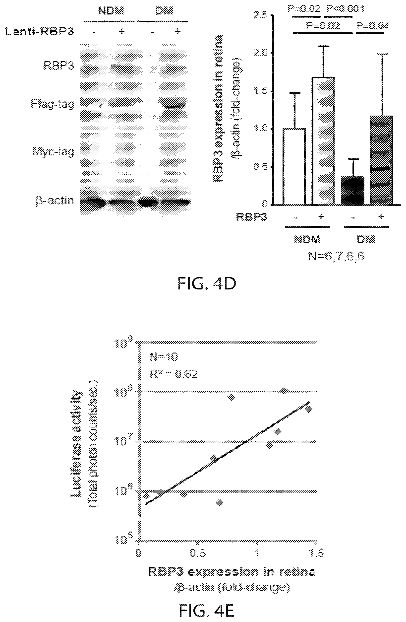
D00013
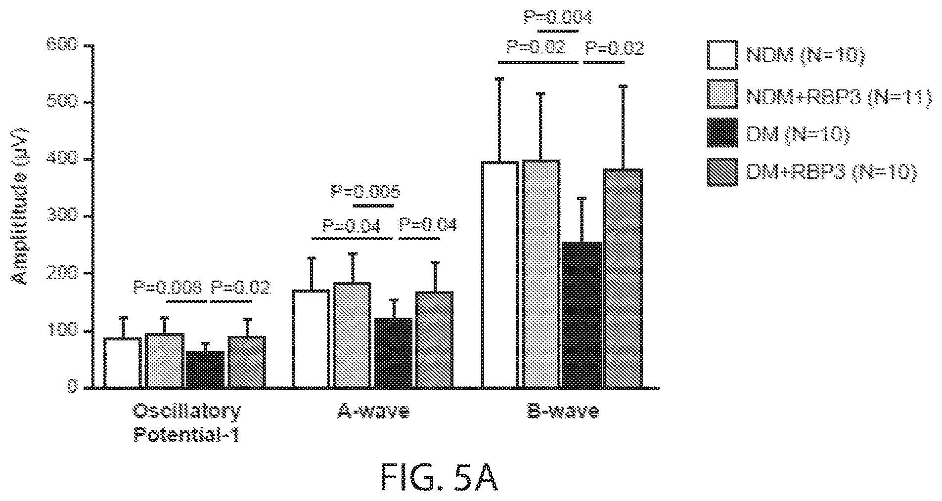
D00014
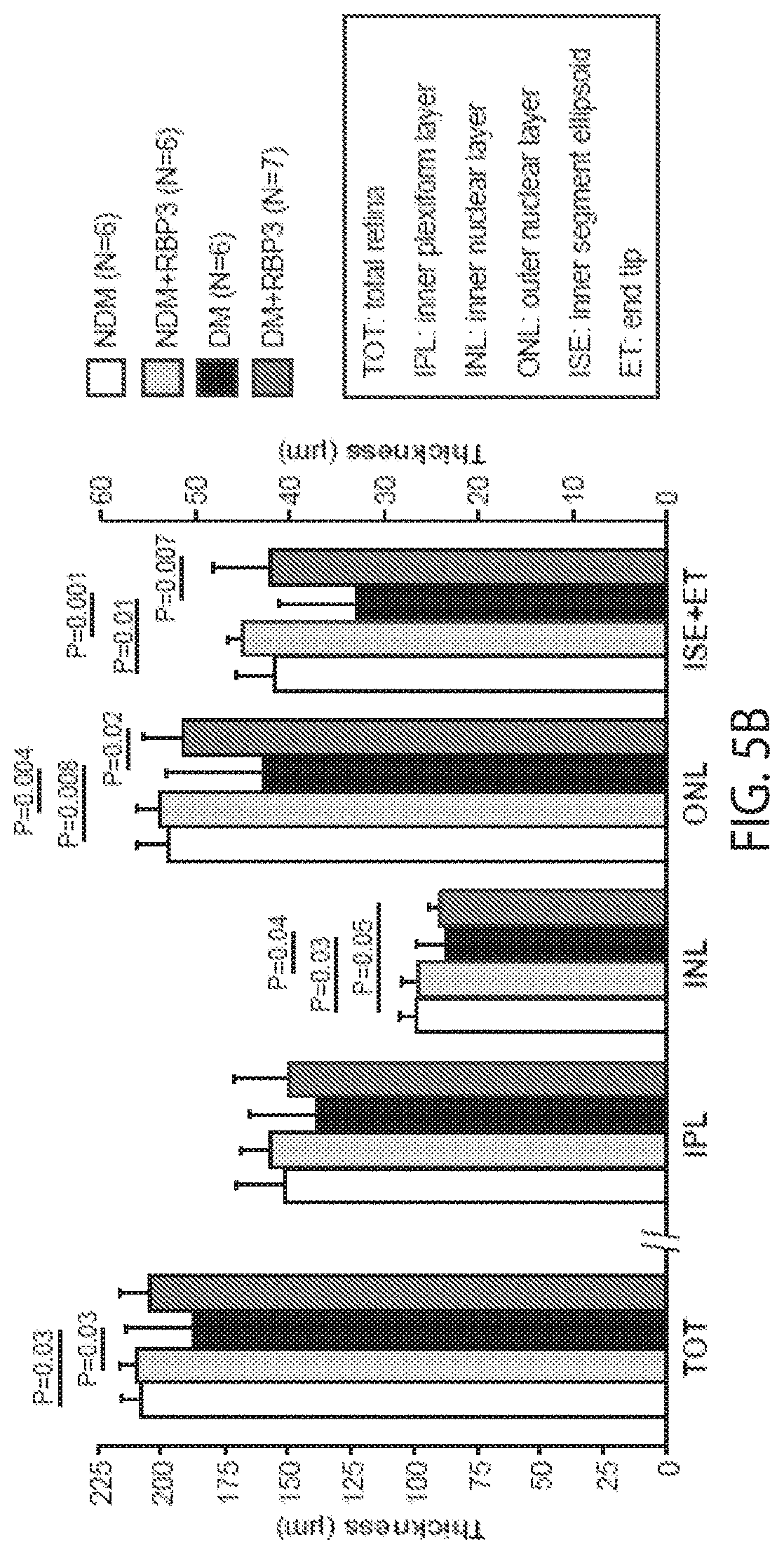
D00015
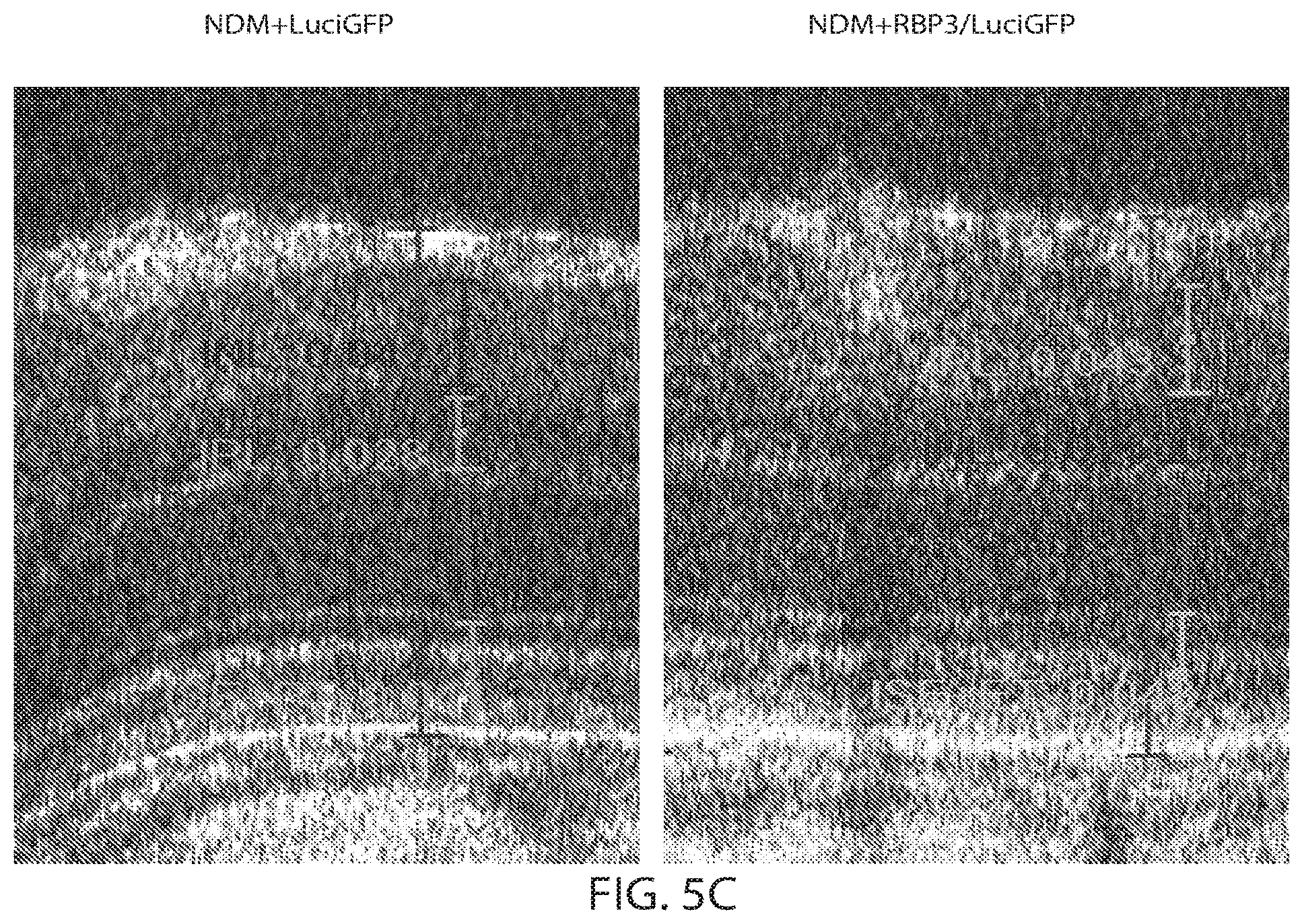
D00016
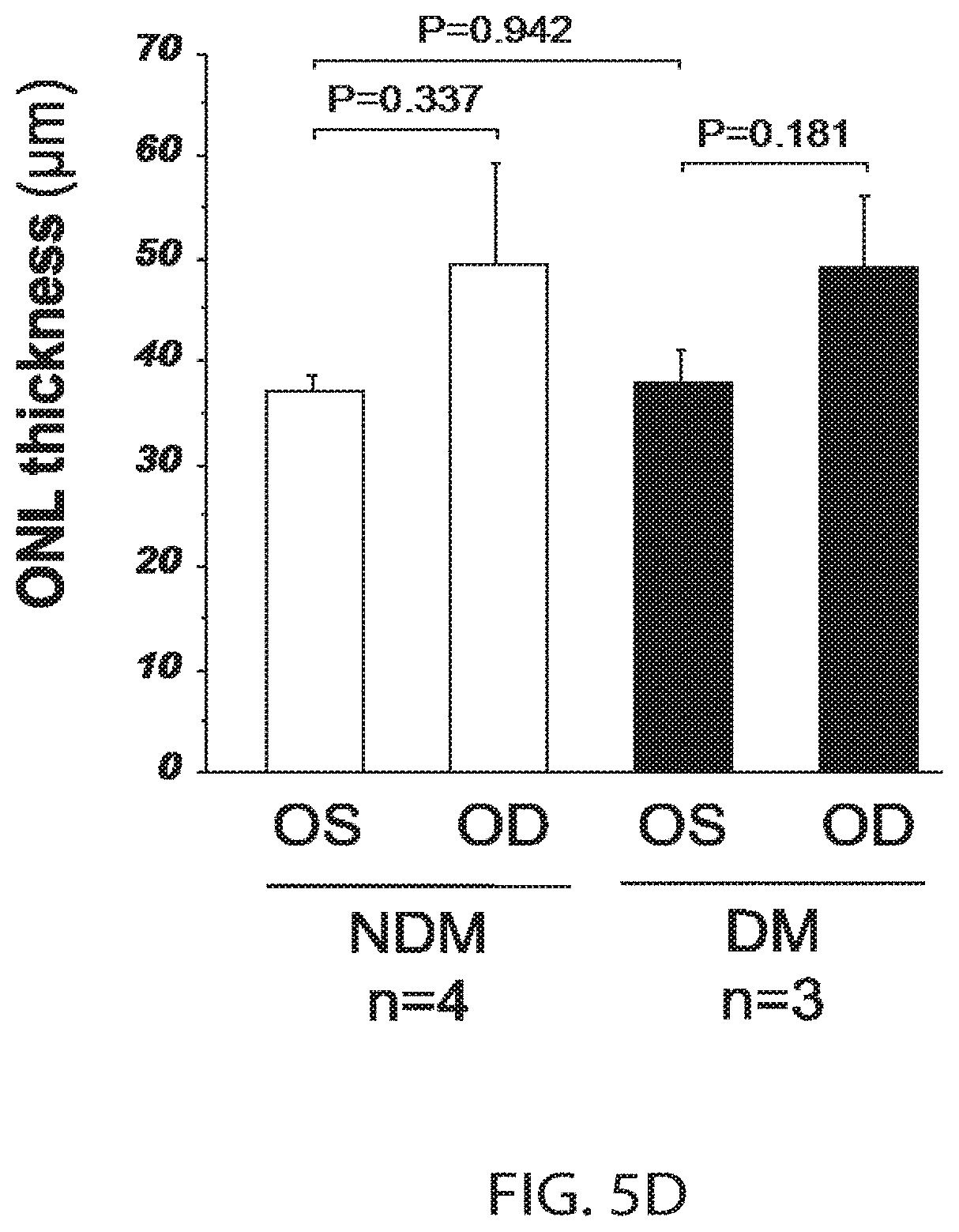
D00017
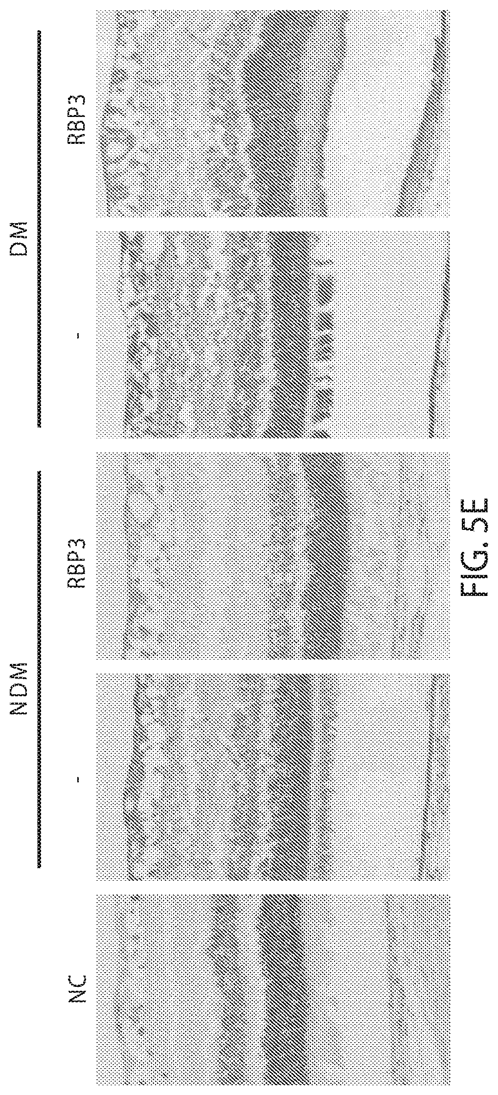
D00018
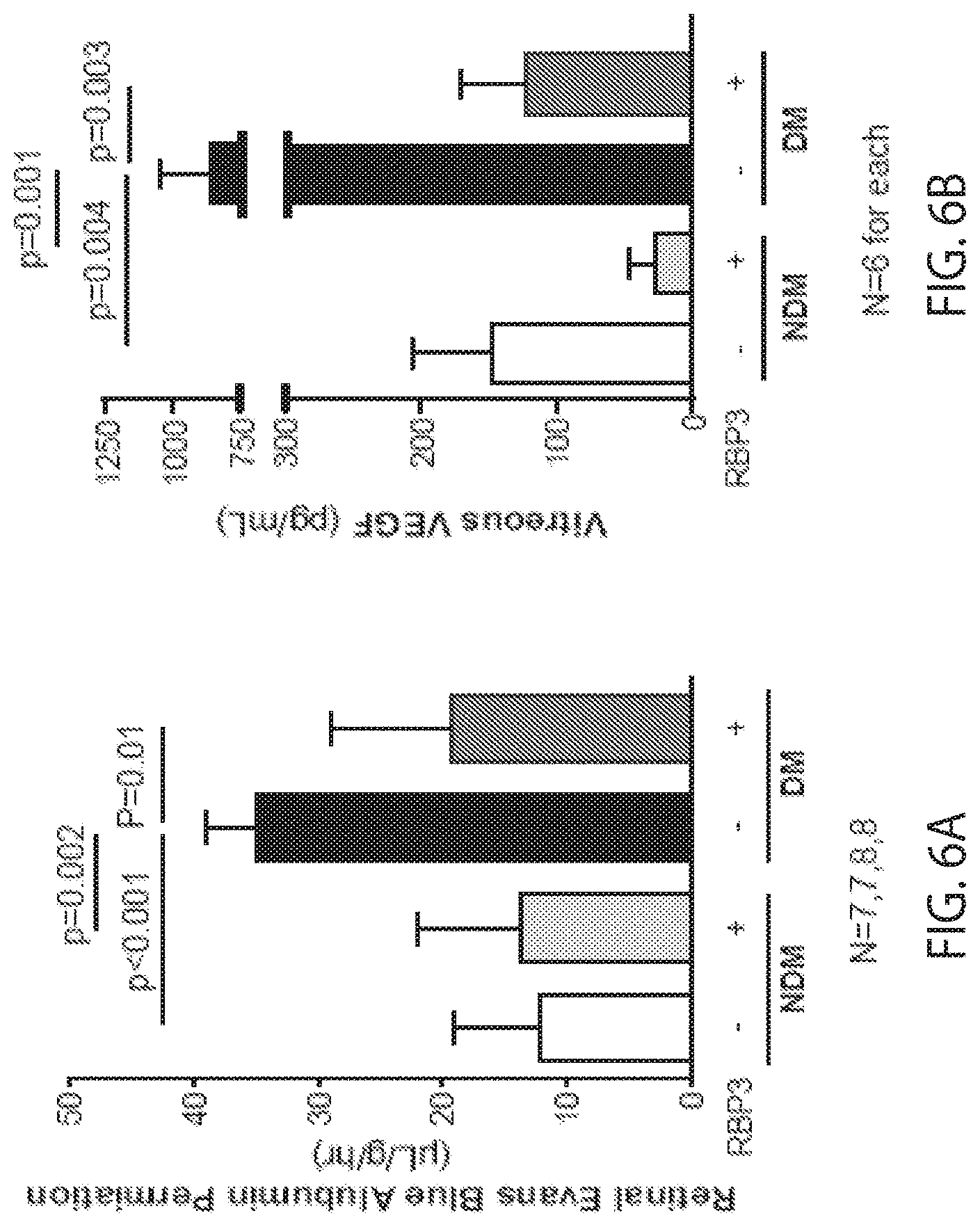
D00019
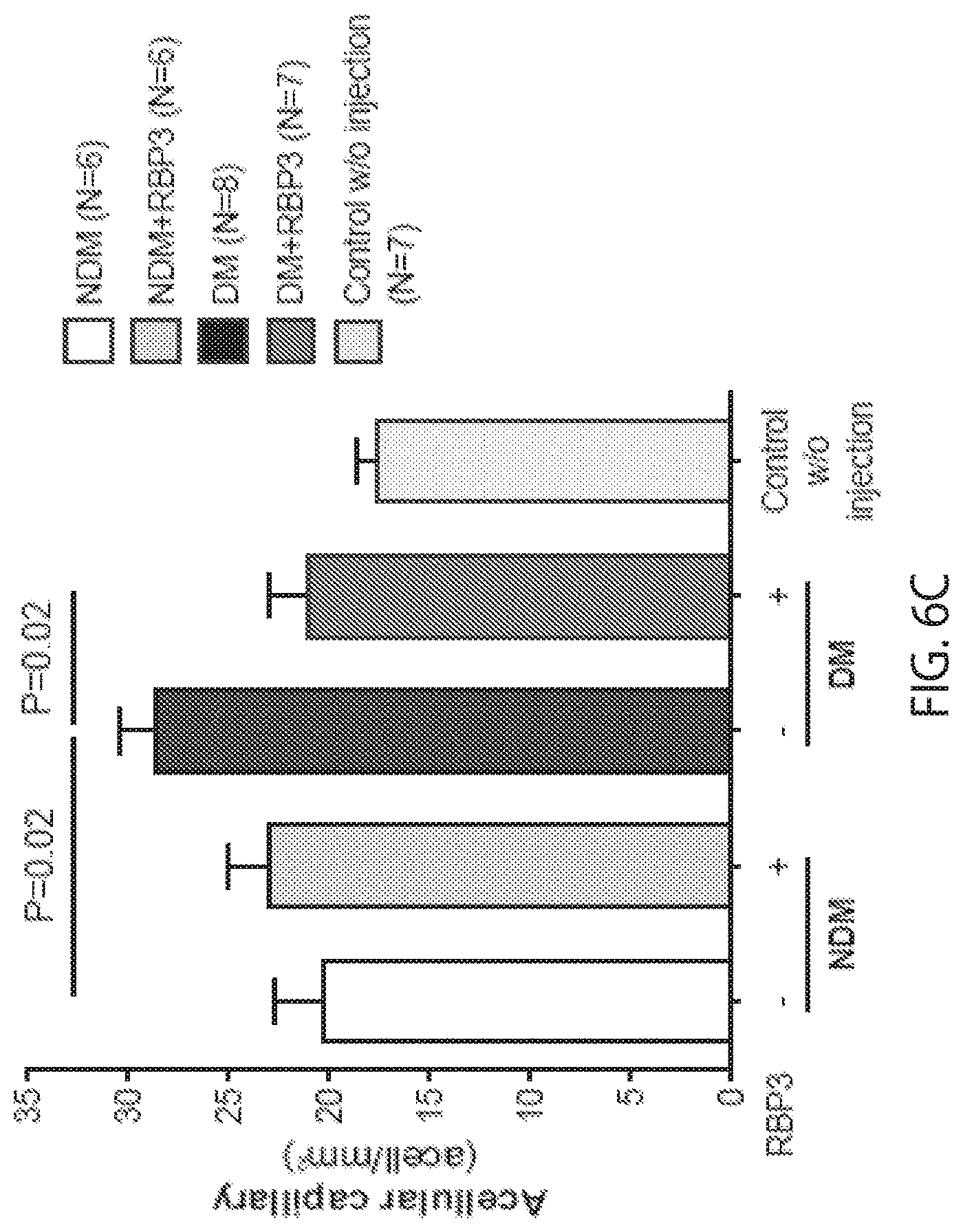
S00001
XML
uspto.report is an independent third-party trademark research tool that is not affiliated, endorsed, or sponsored by the United States Patent and Trademark Office (USPTO) or any other governmental organization. The information provided by uspto.report is based on publicly available data at the time of writing and is intended for informational purposes only.
While we strive to provide accurate and up-to-date information, we do not guarantee the accuracy, completeness, reliability, or suitability of the information displayed on this site. The use of this site is at your own risk. Any reliance you place on such information is therefore strictly at your own risk.
All official trademark data, including owner information, should be verified by visiting the official USPTO website at www.uspto.gov. This site is not intended to replace professional legal advice and should not be used as a substitute for consulting with a legal professional who is knowledgeable about trademark law.