Compositions And Methods For Inducing Conformational Changes In Cereblon And Other E3 Ubiquitin Ligases
Chamberlain; Philip ; et al.
U.S. patent application number 16/852360 was filed with the patent office on 2021-01-07 for compositions and methods for inducing conformational changes in cereblon and other e3 ubiquitin ligases. This patent application is currently assigned to Celgene Corporation. The applicant listed for this patent is Celgene Corporation. Invention is credited to Brian E. Cathers, Philip Chamberlain, Antonia Lopez-Girona.
| Application Number | 20210000813 16/852360 |
| Document ID | / |
| Family ID | |
| Filed Date | 2021-01-07 |


View All Diagrams
| United States Patent Application | 20210000813 |
| Kind Code | A1 |
| Chamberlain; Philip ; et al. | January 7, 2021 |
COMPOSITIONS AND METHODS FOR INDUCING CONFORMATIONAL CHANGES IN CEREBLON AND OTHER E3 UBIQUITIN LIGASES
Abstract
Provided herein are compositions, therapeutic methods, screening methods, computational methods and biomarkers based upon the elucidation of the interaction among cereblon, its substrates and certain compounds or agents, including small molecules, peptides, and proteins.
| Inventors: | Chamberlain; Philip; (San Diego, CA) ; Cathers; Brian E.; (San Diego, CA) ; Lopez-Girona; Antonia; (San Diego, CA) | ||||||||||
| Applicant: |
|
||||||||||
|---|---|---|---|---|---|---|---|---|---|---|---|
| Assignee: | Celgene Corporation Summit NJ |
||||||||||
| Appl. No.: | 16/852360 | ||||||||||
| Filed: | April 17, 2020 |
Related U.S. Patent Documents
| Application Number | Filing Date | Patent Number | ||
|---|---|---|---|---|
| 16049708 | Jul 30, 2018 | 10668057 | ||
| 16852360 | ||||
| 14752588 | Jun 26, 2015 | 10092555 | ||
| 16049708 | ||||
| 62018445 | Jun 27, 2014 | |||
| Current U.S. Class: | 1/1 |
| International Class: | A61K 31/454 20060101 A61K031/454; A61K 31/517 20060101 A61K031/517; C12Q 1/37 20060101 C12Q001/37 |
Claims
1-73. (canceled)
74. A method of identifying a test compound that induces a cereblon (CRBN) conformational change or an alteration of properties of a CRBN surface, wherein the method comprises: (a) obtaining a first three-dimensional structure of CRBN and a reference compound; (b) obtaining a second three-dimensional structure of CRBN and the test compound; and (c) comparing the first three-dimensional structure with the second three-dimensional structure; wherein a difference in the first and second three-dimensional structures is indicative of a test compound that induces the CRBN conformational change or alteration of properties of a CRBN surface; wherein the CRBN conformational change or the alteration of properties of the CRBN surface occurs in a cereblon modifying agent (CMA) binding pocket of the CRBN, or within an adjacent region thereof; and wherein the three-dimensional structure is assessed using x-ray crystallography, NMR spectroscopy, dual polarization interferometry, vibrational spectroscopy, or cryo-electron microscopy.
75. The method of claim 74, wherein the test compound has a specific downstream biological activity and wherein the difference in the first and second three-dimensional structures is indicative of the test compound having the specific downstream biological activity.
76. The method of claim 75, wherein the method further comprises assaying the specific downstream biological activity.
77. The method of claim 75, wherein the method further comprises administering the test compound to a patient, wherein the specific downstream biological activity is modulated in the patient, and wherein optionally the patient has a disease, and one or more symptoms of the disease are alleviated following administration of the test compound.
78. The method of claim 74, wherein the test compound has a specific therapeutic efficacy and wherein the difference in the first and second three-dimensional structures is indicative of the test compound having the specific therapeutic efficacy.
79. The method of claim 78, wherein the method further comprises administering the test compound to a patient having a disease, wherein one or more symptoms of the disease are alleviated following administration of the test compound.
80. The method of claim 78, wherein the method further comprises administering the test compound to a patient having a disease, disorder or condition, wherein one or more symptoms of the disease, disorder or condition are alleviated following administration of the test compound.
81. The method of claim 74, wherein the conformational change or alteration has an effect on W380, W386 and/or W400 of CRBN; has an effect on E377 of CRBN; or has an effect on V388 of CRBN; wherein the CRBN is human CRBN.
82. The method of claim 74, wherein the CRBN is further bound to DDB1, Cul4, Roc1, or any combination thereof.
83. The method of claim 74, wherein the CRBN is further bound to DDB1.
84. The method of claim 74, wherein the CRBN conformational change or the alteration of properties of the CRBN surface is assessed by x-ray crystallography.
85. The method of claim 74, wherein the CRBN is human CRBN or murine CRBN.
Description
CROSS-REFERENCE TO RELATED APPLICATIONS
[0001] This application is a continuation of U.S. patent application Ser. No. 16/049,708, filed on Jul. 30, 2018, which is a divisional of U.S. patent application Ser. No. 14/752,588, filed on Jun. 26, 2015, now U.S. Pat. No. 10,092,555, which claims the benefit of U.S. Provisional Patent Application No. 62/018,445, filed on Jun. 27, 2014, the disclosure of each of which is incorporated by reference herein in its entirety.
SEQUENCE LISTING
[0002] This application incorporates by reference in its entirety the Computer Readable Form ("CRF") of a Sequence Listing in ASCII text format submitted via EFS-Web. The Sequence Listing text file submitted via EFS-Web is entitled "14247-528-999_SEQ_LISTING.txt," was created on Apr. 17, 2020 and is 32,648 bytes in size.
1. FIELD
[0003] Provided herein are compositions, therapeutic methods, screening methods, computational methods and biomarkers based upon the elucidation of the interaction among cereblon, its substrates and certain compounds or agents, including small molecules, peptides, and proteins.
2. BACKGROUND
2.1 Cereblon
[0004] At least two isoforms of the protein cereblon (CRBN) exist, which are 442 and 441 amino acids long, respectively, and CRBN is conserved from plant to human. In humans, the CRBN gene has been identified as a candidate gene of an autosomal recessive nonsyndromic mental retardation (ARNSMR). See Higgins, J. J. et al., Neurology, 2004, 63:1927-1931. CRBN was initially characterized as an RGS-containing novel protein that interacted with a calcium-activated potassium channel protein (SLO1) in the rat brain, and was later shown to interact with a voltage-gated chloride channel (CIC-2) in the retina with AMPK1 and DDB1. See Jo, S. et al., J. Neurochem, 2005, 94:1212-1224; Hohberger B. et al., FEBS Lett, 2009, 583:633-637; Angers S. et al., Nature, 2006, 443:590-593. DDB1 was originally identified as a nucleotide excision repair protein that associates with damaged DNA binding protein 2 (DDB2). Its defective activity causes the repair defect in the patients with xeroderma pigmentosum complementation group E (XPE). DDB1 also appears to function as a component of numerous distinct DCX (DDB1-CUL4-X-box) E3 ubiquitin-protein ligase complexes which mediate the ubiquitination and subsequent proteasomal degradation of target proteins. CRBN has also been identified as a target for the development of therapeutic agents for diseases of the cerebral cortex. See WO 2010/137547 A1.
[0005] CRBN has recently been identified as a key molecular target that binds to thalidomide to cause birth defects. See Ito, T. et al., Science, 2010, 327:1345-1350. DDB1 was found to interact with CRBN and, thus, was indirectly associated with thalidomide. Moreover, thalidomide was able to inhibit auto-ubiquitination of CRBN in vitro, suggesting that thalidomide is an E3 ubiquitin-ligase inhibitor. Id. Importantly, this activity was inhibited by thalidomide in wild-type cells, but not in cells with mutated CRBN binding sites that prevent thalidomide binding. Id. The thalidomide binding site was mapped to a highly conserved C-terminal 104 amino acid region in CRBN. Id. Individual point mutants in CRBN, Y384A and W386A were both defective for thalidomide binding, with the double point mutant having the lowest thalidomide-binding activity. Id. A link between CRBN and the teratogenic effect of thalidomide was confirmed in animal models of zebra-fish and chick embryos. Id.
[0006] Whether binding to CRBN, the CRBN E3 ubiquitin-ligase complex, or one or more substrates of CRBN, is required for the beneficial effects of thalidomide and other drugs is yet to be established. Understanding these interactions with thalidomide and other drug targets will allow the definition of the molecular mechanisms of efficacy and/or toxicity and may lead to drugs with improved efficacy and toxicity profiles.
2.2 Compounds
[0007] A number of studies have been conducted with the aim of providing compounds that can safely and effectively be used to treat diseases associated with abnormal production of TNF-.alpha.. See, e.g., Marriott, J. B., et al., Expert Opin. Biol. Ther., 2001, 1(4): 1-8; G. W. Muller, et al., J Med Chem., 1996, 39(17): 3238-3240; and G. W. Muller, et al., Bioorg & Med Chem Lett., 1998, 8: 2669-2674. Some studies have focused on a group of compounds selected for their capacity to potently inhibit TNF-.alpha. production by LPS stimulated PBMC. L. G. Corral, et al., Ann. Rheum. Dis., 1999, 58:(Suppl I) 1107-1113. These compounds show not only potent inhibition of TNF-.alpha. but also marked inhibition of LPS induced monocyte IL1 and IL12 production. LPS induced IL6 is also inhibited by such compounds, albeit partially. These compounds are potent stimulators of LPS-induced IL-10. Id.
[0008] Compounds for the methods provided herein include, but are not limited to, the substituted 2-(2,6-dioxopiperidin-3-yl) phthalimides and substituted 2-(2,6-dioxopiperidin-3-yl)-1-oxoisoindoles described in U.S. Pat. Nos. 6,281,230 and 6,316,471, both to G. W. Muller, et al. Still other specific compounds disclosed herein belong to a class of isoindole-imides disclosed in U.S. Pat. Nos. 6,395,754, 6,555,554, 7,091,353, U.S. Publication No. 2004/0029832, and International Publication No. WO 98/54170, each of which is incorporated herein by reference.
[0009] Thalidomide, lenalidomide and pomalidomide have shown remarkable responses in patients with multiple myeloma, lymphoma and other hematological diseases such as myelodysplastic syndrome. See Galustian C, et al., Expert Opin Pharmacother., 2009, 10:125-133. These drugs display a broad spectrum of activity, including anti-angiogenic properties, modulation of pro-inflammatory cytokines, co-stimulation of T cells, increased NK cell toxicity, direct anti-tumor effects and modulation of stem cell differentiation.
[0010] For example, thalidomide and lenalidomide have emerged as important options for the treatment of multiple myeloma in newly diagnosed patients, in patients with advanced disease who have failed chemotherapy or transplantation, and in patients with relapsed or refractory multiple myeloma. Lenalidomide in combination with dexamethasone has been approved for the treatment of patients with multiple myeloma who have received at least one prior therapy. Pomalidomide may also be administered in combination with dexamethasone. U.S. Patent Publication No. 2004/0029832 A1, the disclosure of which is hereby incorporated in its entirety, discloses the treatment of multiple myeloma.
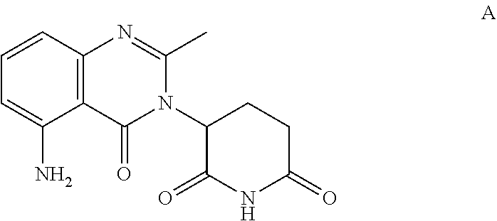
[0011] Another compound provided herein is 3-(5-amino-2-methyl-4-oxo-4H-quinazolin-3-yl)-piperidine-2,6-dione ("Compound A"), which has the following structure:
##STR00001##
or an enantiomer or a mixture of enantiomers thereof; or a pharmaceutically acceptable salt, solvate, hydrate, co-crystal, clathrate, or polymorph thereof.
[0012] Compound A can be prepared as described in U.S. Pat. No. 7,635,700, the disclosure of which is incorporated herein by reference in its entirety. The compound can be also synthesized according to other methods apparent to those of skill in the art based upon the teaching herein. In certain embodiments, Compound A is in a crystalline form described in U.S. Provisional Pat. App. No. 61/451,806, filed Mar. 11, 2011, which is incorporated herein by reference in its entirety. In some embodiments, the hydrochloride salt of Compound A is used in the methods provided herein. Methods of treating, preventing and/or managing cancers and other diseases using Compound A are described in U.S. Provisional Pat. App. No. 61/451,995, filed Mar. 11, 2011, which is incorporated herein by reference in its entirety.
[0013] Other compounds are provided in Section 5.8 herein.
[0014] Understanding the interactions of CRBN, the CRBN E3 ubiquitin-ligase complex, or one or more substrates of CRBN with thalidomide, lenalidomide, pomalidomide and other drug targets will allow the definition of the molecular mechanisms of efficacy and/or toxicity and may lead to drugs with improved efficacy and toxicity profiles.
3. SUMMARY
[0015] We have discovered that CRBN has pluripotent potential as a drug target for the treatment of various diseases. We believe we are the first to report and understand that cereblon can be induced to undergo conformational changes by use of certain small molecules or other agents that we will call "cereblon modifying agents" (CMAs). The use of the appropriate agent leads to a distinct conformational change or other alteration in the properties of the CRBN surface, and a resulting distinct phenotypic response. Cereblon is not simply a unidimensional or monotypic protein that interacts with a single substrate. Instead, without being limited by theory, the conformational change or phenotypic response of cereblon or its pathway is dependent upon cell type and, most importantly, the CMA used to interact with cereblon or its pathway.
[0016] As such, we describe herein a variety of distinct conformational changes, surface property alterations, phenotypic responses and CMAs. We also describe treatment methods, compositions, drug screens and computational methods that exploit these discoveries.
[0017] We also describe the use of known agents as CMAs for new treatment methods. In another embodiment we disclose the use of new CMA or CMA classes based upon the conformational change, alteration in surface properties, or phenotypic response. It should be noted that these discoveries permit a plethora of methods to be used to treat diseases associated with cereblon pathway. Thus, also described herein are known or new agents as CMAs for use in methods for treating diseases.
[0018] In one aspect, provided herein is a method of identifying a test compound that induces a CRBN conformational change (e.g., within the CMA-binding pocket of the CRBN) or otherwise altering the properties of a CRBN surface. In certain embodiments, the method comprises (a) (i) obtaining a first crystal structure of CRBN and a reference compound, and (ii) determining a three-dimensional structure of the first crystal by x-ray diffraction to obtain a first set of atomic coordinates; (b) (i) obtaining a second crystal comprising CRBN and the test compound, and (ii) determining a three-dimensional structure of the second crystal by x-ray diffraction to obtain a second set of atomic coordinates; and (c) comparing said first set of atomic coordinates with said second set of atomic coordinates; wherein a difference in atomic coordinates is indicative of a compound that induces a CRBN conformational change or otherwise alters the properties of a CRBN surface. In some embodiments, the first set of atomic coordinates and/or said second set of atomic coordinates define a CMA binding domain. In certain embodiments, the difference in atomic coordinates is determined by assessing differences in atomic distances. Also provided herein is a test compound identified by this method. In some embodiments, the test compound induces a CRBN conformational change. In other embodiments, the test compound alters the properties of the CRBN surface. In certain embodiments, the properties of the CRBN surface are altered by the placement of compound appendages. In certain embodiments, the conformational change or alteration occurs in a CMA binding pocket of the CRBN. In some embodiments, the conformational change or alteration in said CMA binding pocket has an effect on W380, W386 and/or W400 of CRBN. In other embodiments, the conformational change or alteration in said CMA binding pocket has an effect on E377 of CRBN. In other embodiments, the conformational change or alteration in said CMA binding pocket has an effect on V388 of CRBN. In certain embodiments, the conformational change or alteration in said CMA binding pocket has an effect on an adjacent region of the protein.
[0019] In certain embodiments of the various compositions and methods provided herein, the properties of the CRBN surface are altered by the placement of compound appendages.
[0020] In a second aspect, provided herein is a method of identifying a test compound that induces a CRBN conformational change (e.g., within the CMA-binding pocket of the CRBN) or otherwise altering the properties of a CRBN surface. In certain embodiments, the method comprises (a) (i) obtaining a first three-dimensional structure of CRBN and a reference compound; (b) (i) obtaining a second three-dimensional structure of CRBN and the test compound; and (c) comparing said first three-dimensional structure with said second three-dimensional structure; wherein a difference in the first and second three-dimensional structures is indicative of a compound that induces a CRBN conformational change or otherwise alters the properties of a CRBN surface. In some embodiments, the first three-dimensional structure is of a CRBN that is not bound to a reference compound (also sometimes referred to as an "unbound CRBN" herein, and which is not meant to preclude the CRBN being bound to other proteins, e.g., DDB1). In some embodiments, the CRBN that is not bound to a reference compound (or unbound CRBN) has a three-dimensional structure as determined by x-ray diffraction having the atomic coordinates set forth in Table 8. Also provided herein is a test compound identified by this method. In some embodiments, the test compound induces a CRBN conformational change. In other embodiments, the test compound alters the properties of the CRBN surface. In certain embodiments, the properties of the CRBN surface are altered by the placement of compound appendages. In certain embodiments, the conformational change or alteration occurs in a CMA binding pocket of the CRBN. In some embodiments, the conformational change or alteration in said CMA binding pocket has an effect on W380, W386 and/or W400 of CRBN In other embodiments, the conformational change or alteration in said CMA binding pocket has an effect on E377 of CRBN. In other embodiments, the conformational change or alteration in said CMA binding pocket has an effect on V388 of CRBN. In certain embodiments, the conformational change or alteration in said CMA binding pocket has an effect on an adjacent region of the protein. In an embodiment, the conformational change or alteration is assessed by x-ray crystallography. In another embodiment, the conformational change or alteration is assessed by a method comprising (a) (i) obtaining a first crystal structure of CRBN and a reference compound, and (ii) determining a three-dimensional structure of the first crystal by x-ray diffraction to obtain a first set of atomic coordinates; (b) (i) obtaining a second crystal comprising CRBN and the test compound, and (ii) determining a three-dimensional structure of the second crystal by x-ray diffraction to obtain a second set of atomic coordinates; and (c) comparing said first set of atomic coordinates with said second set of atomic coordinates; wherein a difference in atomic coordinates is indicative of a conformational change (e.g., within the CMA-binding pocket of the CRBN) or alteration of the properties of a CRBN surface (e.g., on an adjacent region of the protein). In certain embodiments, the conformational change or alteration is assessed by a method comprising (a) (i) obtaining a first three-dimensional structure of CRBN and a reference compound; (b) (i) obtaining a second three-dimensional structure of CRBN and the test compound; and (c) comparing said first three-dimensional structure with said second three-dimensional structure; wherein a difference in the first and second three-dimensional structures is indicative of a compound that induces a CRBN conformational change or alteration. In another embodiment, the conformational change or alteration is assessed by a method comprising (a) (i) obtaining a first crystal structure of an unbound CRBN, and (ii) determining a three-dimensional structure of the first crystal by x-ray diffraction to obtain a first set of atomic coordinates; (b) (i) obtaining a second crystal comprising CRBN and the test compound, and (ii) determining a three-dimensional structure of the second crystal by x-ray diffraction to obtain a second set of atomic coordinates; and (c) comparing said first set of atomic coordinates with said second set of atomic coordinates; wherein a difference in atomic coordinates is indicative of a conformational change (e.g., within the CMA-binding pocket of the CRBN) or alteration of the properties of a CRBN surface (e.g., on an adjacent region of the protein). In some embodiments, the first crystal structure of the unbound CRBN has a three-dimensional structure as determined by x-ray diffraction having the atomic coordinates set forth in Table 8. In certain embodiments, the conformational change or alteration is assessed by a method comprising (a) (i) obtaining a first three-dimensional structure of an unbound CRBN; (b) (i) obtaining a second three-dimensional structure of CRBN and the test compound; and (c) comparing said first three-dimensional structure with said second three-dimensional structure; wherein a difference in the first and second three-dimensional structures is indicative of a compound that induces a CRBN conformational change or alteration. In some embodiments, the first and/or second three-dimensional structures include a CMA binding domain of the CRBN. In other embodiments, the three-dimensional structure is assessed using x-ray crystallography, NMR spectroscopy, dual polarization interferometry, vibrational spectroscopy, or cryo-electron microscopy. In some embodiments, the CRBN is further bound to DDB1, Cul4, Roc1, or any combination thereof. In a specific embodiment, the CRBN is further bound to DDB1.
[0021] In some embodiments of the various compositions and methods provided herein, the crystal structure of CRBN is a crystal structure of mouse CRBN. In some embodiments, the crystal structure of CRBN is a crystal structure of mouse CRBN that is bound to a reference compound. In other embodiments, the crystal structure of CRBN is a crystal structure of mouse CRBN that is not bound to a reference compound. In one embodiment, the crystal structure of mouse CRBN that is not bound to a reference compound has a three-dimensional structure as determined by x-ray diffraction having the atomic coordinates set forth in Table 8. In certain embodiments of the various compositions and methods provided herein, the crystal structure of CRBN is a crystal structure of human CRBN. In some embodiments, the crystal structure of CRBN is a crystal structure of human CRBN that is bound to a reference compound. In other embodiments, the crystal structure of CRBN is a crystal structure of human CRBN that is not bound to a reference compound. In certain embodiments, the CRBN is further bound to DDB1. In other embodiments of the various compositions and methods provided herein, the CMA-binding pocket of CRBN is an IMiD.RTM.-binding pocket.
[0022] In a third aspect, provided herein is a method of identifying a test compound that has a specific downstream biological activity comprising: (a) (i) obtaining a first crystal structure of CRBN and a reference compound, and (ii) determining a three-dimensional structure of the first crystal by x-ray diffraction to obtain a first set of atomic coordinates; (b) (i) obtaining a second crystal comprising CRBN and the test compound, and (ii) determining a three-dimensional structure of the second crystal by x-ray diffraction to obtain a second set of atomic coordinates; and (c) comparing said first set of atomic coordinates with said second set of atomic coordinates; wherein a difference in atomic coordinates is indicative of a compound has the specific downstream biological activity. In some embodiments, the first crystal structure is of a CRBN that is not bound to a reference compound. In one embodiment, the crystal structure of mouse CRBN that is not bound to a reference compound has a three-dimensional structure as determined by x-ray diffraction having the atomic coordinates set forth in Table 8. In some embodiments, the first set of atomic coordinates and/or said second set of atomic coordinates define a CMA binding domain. In certain embodiments, the difference in atomic coordinates is determined by assessing differences in atomic distances. In some embodiments, the method further comprises assaying the specific biological activity. Also provided herein is a test compound identified by this method. In some embodiments, the compound induces a CRBN conformational change (e.g., within the CMA-binding pocket of the CRBN) or otherwise altering the properties of a CRBN surface. In some embodiments, the test compound induces a CRBN conformational change. In other embodiments, the test compound alters the properties of the CRBN surface. In certain embodiments, the properties of the CRBN surface are altered by the placement of compound appendages. In certain embodiments, the conformational change or alteration occurs in a CMA binding pocket of the CRBN. In some embodiments, the conformational change or alteration in said CMA binding pocket has an effect on W380, W386 and/or W400 of CRBN. In other embodiments, the conformational change or alteration in said CMA binding pocket has an effect on E377 of CRBN. In other embodiments, the conformational change or alteration in said CMA binding pocket has an effect on V388 of CRBN. In certain embodiments, the conformational change or alteration in said CMA binding pocket has an effect on an adjacent region of the protein. In certain embodiments, the method further comprises administering said compound to a patient, wherein said biological activity is modulated in said patient. In certain embodiments, the patient has a disease, and wherein one or more symptoms of said disease are alleviated following said administration.
[0023] In a fourth aspect, provided herein is a method of identifying a test compound that has a specific downstream biological activity comprising (a) (i) obtaining a first three-dimensional structure of CRBN and a reference compound; (b) (i) obtaining a second three-dimensional structure of CRBN and the test compound; and (c) comparing said first three-dimensional structure with said second three-dimensional structure; wherein a difference in the first and second three-dimensional structures is indicative of a compound has the specific downstream biological activity. In some embodiments, the first three-dimensional structure is of a CRBN that is not bound to a reference compound. In one embodiment, the crystal structure of mouse CRBN that is not bound to a reference compound has a three-dimensional structure as determined by x-ray diffraction having the atomic coordinates set forth in Table 8. Also provided herein is a test compound identified by this method. In some embodiments, the test compound induces a CRBN conformational change. In other embodiments, the test compound alters the properties of the CRBN surface. In certain embodiments, the properties of the CRBN surface are altered by the placement of compound appendages. In certain embodiments, the conformational change or alteration occurs in a CMA binding pocket of the CRBN. In some embodiments, the conformational change or alteration in said CMA binding pocket has an effect on W380, W386 and/or W400 of CRBN. In other embodiments, the conformational change or alteration in said CMA binding pocket has an effect on E377 of CRBN. In other embodiments, the conformational change or alteration in said CMA binding pocket has an effect on V388 of CRBN. In certain embodiments, the conformational change or alteration in said CMA binding pocket has an effect on an adjacent region of the protein. In an embodiment, the conformational change or alteration is assessed by x-ray crystallography. In another embodiment, the conformational change or alteration is assessed by a method comprising (a) (i) obtaining a first crystal structure of CRBN and a reference compound, and (ii) determining a three-dimensional structure of the first crystal by x-ray diffraction to obtain a first set of atomic coordinates; (b) (i) obtaining a second crystal comprising CRBN and the test compound, and (ii) determining a three-dimensional structure of the second crystal by x-ray diffraction to obtain a second set of atomic coordinates; and (c) comparing said first set of atomic coordinates with said second set of atomic coordinates; wherein a difference in atomic coordinates is indicative of a conformational change (e.g., within the CMA-binding pocket of the CRBN) or alteration of the properties of a CRBN surface (e.g., on an adjacent region of the protein). In certain embodiments, the conformational change or alteration is assessed by a method comprising (a) (i) obtaining a first three-dimensional structure of CRBN and a reference compound; (b) (i) obtaining a second three-dimensional structure of CRBN and the test compound; and (c) comparing said first three-dimensional structure with said second three-dimensional structure; wherein a difference in the first and second three-dimensional structures is indicative of a compound that induces a CRBN conformational change or alteration. In another embodiment, the conformational change or alteration is assessed by a method comprising (a) (i) obtaining a first crystal structure of CRBN that is not bound to a reference compound, and (ii) determining a three-dimensional structure of the first crystal by x-ray diffraction to obtain a first set of atomic coordinates; (b) (i) obtaining a second crystal comprising CRBN and the test compound, and (ii) determining a three-dimensional structure of the second crystal by x-ray diffraction to obtain a second set of atomic coordinates; and (c) comparing said first set of atomic coordinates with said second set of atomic coordinates; wherein a difference in atomic coordinates is indicative of a conformational change (e.g., within the CMA-binding pocket of the CRBN) or alteration of the properties of a CRBN surface (e.g., on an adjacent region of the protein). In certain embodiments, the conformational change or alteration is assessed by a method comprising (a) (i) obtaining a first three-dimensional structure of CRBN that is not bound to a reference compound; (b) (i) obtaining a second three-dimensional structure of CRBN and the test compound; and (c) comparing said first three-dimensional structure with said second three-dimensional structure; wherein a difference in the first and second three-dimensional structures is indicative of a compound that induces a CRBN conformational change or alteration. In some embodiments, the first and/or second three-dimensional structures include a CMA binding domain of the CRBN. In other embodiments of the various methods provided herein, the three-dimensional structure is assessed using x-ray crystallography, NMR spectroscopy, dual polarization interferometry, vibrational spectroscopy, or cryo-electron microscopy. In some embodiments, the three-dimensional structure of CRBN that is not bound to a reference compound has the atomic coordinates set forth in Table 8 as determined by x-ray diffraction. In some embodiments, the CRBN is further bound to DDB1, Cul4, Roc1, or any combination thereof. In certain embodiments, the CRBN is further bound to DDB1.
[0024] In some embodiments of the various compositions and methods provided herein, the CRBN is a mouse CRBN. In certain embodiments of the various compositions and methods provided herein, CRBN is a human CRBN. In some embodiments, the CRBN is further bound to DDB1. In other embodiments of the various compositions and methods provided herein, the CMA-binding pocket of CRBN is an IMiD.RTM.-binding pocket.
[0025] In a fifth aspect, provided herein is a method of identifying a test compound that has a specific therapeutic efficacy comprising: (a) (i) obtaining a first crystal structure of CRBN and a reference compound, and (ii) determining a three-dimensional structure of the first crystal by x-ray diffraction to obtain a first set of atomic coordinates; (b) (i) obtaining a second crystal comprising CRBN and the test compound, and (ii) determining a three-dimensional structure of the second crystal by x-ray diffraction to obtain a second set of atomic coordinates; and (c) comparing said first set of atomic coordinates with said second set of atomic coordinates; wherein a difference in atomic coordinates is indicative of a compound has the specific therapeutic efficacy. In some embodiments, the first crystal structure is of a CRBN that is not bound to a reference compound. In one embodiment, the first crystal structure of CRBN that is not bound to a reference compound has a three-dimensional structure as determined by x-ray diffraction having the atomic coordinates set forth in Table 8. In some embodiments, the first set of atomic coordinates and/or said second set of atomic coordinates define a CMA binding domain. In certain embodiments, the difference in atomic coordinates is determined by assessing differences in atomic distances. Also provided herein is a test compound identified by this method. In some embodiments, the test compound induces a CRBN conformational change. In other embodiments, the test compound alters the properties of the CRBN surface. In certain embodiments, the properties of the CRBN surface are altered by the placement of compound appendages. In certain embodiments, the conformational change or alteration occurs in a CMA binding pocket of the CRBN. In some embodiments, the conformational change or alteration in said CMA binding pocket has an effect on W380, W386 and/or W400 of CRBN. In other embodiments, the conformational change or alteration in said CMA binding pocket has an effect on E377 of CRBN. In other embodiments, the conformational change or alteration in said CMA binding pocket has an effect on V388 of CRBN. In certain embodiments, the conformational change or alteration in said CMA binding pocket has an effect on an adjacent region of the protein. In certain embodiments, the method further comprises administering said compound to a patient having disease, disorder or condition, wherein one or more symptoms of said disease, disorder or condition is alleviated following said administration. In some embodiments, the CRBN is further bound to DDB1, Cul4, Roc1, or any combination thereof. In certain embodiments, the CRBN is further bound to DDB1.
[0026] In a sixth aspect, provided herein is a method of identifying a test compound that has a specific downstream biological activity comprising: (a) (i) obtaining a first three-dimensional structure of CRBN and a reference compound; (b) (i) obtaining a second three-dimensional structure of CRBN and the test compound; and (c) comparing said first three-dimensional structure with said second three-dimensional structure; wherein a difference in the first and second three-dimensional structures is indicative of a compound has the specific downstream biological activity. In some embodiments, the first three-dimensional structure is of a CRBN that is not bound to a reference compound. In one embodiment, the first three-dimensional structure of CRBN that is not bound to a reference compound has the atomic coordinates set forth in Table 8 as determined by x-ray diffraction. Also provided herein is a test compound identified by this method. In some embodiments, the test compound induces a CRBN conformational change. In other embodiments, the test compound alters the properties of the CRBN surface. In certain embodiments, the properties of the CRBN surface are altered by the placement of compound appendages. In certain embodiments, the conformational change or alteration occurs in a CMA binding pocket of the CRBN. In some embodiments, the conformational change or alteration in said CMA binding pocket has an effect on W380, W386 and/or W400 of CRBN. In other embodiments, the conformational change or alteration in said CMA binding pocket has an effect on E377 of CRBN. In other embodiments, the conformational change or alteration in said CMA binding pocket has an effect on V388 of CRBN. In certain embodiments, the conformational change or alteration in said CMA binding pocket has an effect on an adjacent region of the protein. In an embodiment, the conformational change or alteration is assessed by x-ray crystallography. In another embodiment, the conformational change or alteration is assessed by a method comprising (a) (i) obtaining a first crystal structure of CRBN and a reference compound, and (ii) determining a three-dimensional structure of the first crystal by x-ray diffraction to obtain a first set of atomic coordinates; (b) (i) obtaining a second crystal comprising CRBN and the test compound, and (ii) determining a three-dimensional structure of the second crystal by x-ray diffraction to obtain a second set of atomic coordinates; and (c) comparing said first set of atomic coordinates with said second set of atomic coordinates; wherein a difference in atomic coordinates is indicative of a conformational change (e.g., within the CMA-binding pocket of the CRBN) or alteration of the properties of a CRBN surface (e.g., on an adjacent region of the protein). In certain embodiments, the conformational change or alteration is assessed by a method comprising (a) (i) obtaining a first three-dimensional structure of CRBN and a reference compound; (b) (i) obtaining a second three-dimensional structure of CRBN and the test compound; and (c) comparing said first three-dimensional structure with said second three-dimensional structure; wherein a difference in the first and second three-dimensional structures is indicative of a compound that induces a CRBN conformational change or alteration. In another embodiment, the conformational change or alteration is assessed by a method comprising (a) (i) obtaining a first crystal structure of CRBN that is not bound to a reference compound, and (ii) determining a three-dimensional structure of the first crystal by x-ray diffraction to obtain a first set of atomic coordinates; (b) (i) obtaining a second crystal comprising CRBN and the test compound, and (ii) determining a three-dimensional structure of the second crystal by x-ray diffraction to obtain a second set of atomic coordinates; and (c) comparing said first set of atomic coordinates with said second set of atomic coordinates; wherein a difference in atomic coordinates is indicative of a conformational change (e.g., within the CMA-binding pocket of the CRBN) or alteration of the properties of a CRBN surface (e.g., on an adjacent region of the protein). In certain embodiments, the conformational change or alteration is assessed by a method comprising (a) (i) obtaining a first three-dimensional structure of CRBN that is not bound to a reference compound; (b) (i) obtaining a second three-dimensional structure of CRBN and the test compound; and (c) comparing said first three-dimensional structure with said second three-dimensional structure; wherein a difference in the first and second three-dimensional structures is indicative of a compound that induces a CRBN conformational change or alteration. In one embodiment, the first three-dimensional structure of CRBN that is not bound to a reference compound has the atomic coordinates set forth in Table 8 as determined by x-ray diffraction. In some embodiments, the first and/or second three-dimensional structures include a CMA binding domain of the CRBN. In other embodiments, the three-dimensional structure is assessed using x-ray crystallography, NMR spectroscopy, dual polarization interferometry, vibrational spectroscopy, or cryo-electron microscopy. In some embodiments, the CRBN is further bound to DDB1, Cul4, Roc1, or any combination thereof. In a specific embodiment, the CRBN is further bound to DDB1.
[0027] In a seventh aspect, provided herein is a method of designing a test compound based on fit within CMA binding pocket of CRBN, comprising: (a) generating on a computer, three-dimensional structural features of a CRBN having a conformational change in the CMA binding pocket, (b) designing a test compound capable of selectively binding to said CMA binding pocket, (c) synthesizing said test compound, (d) contacting CRBN with said synthesized test compound, and (e) determining if said test compound binds to said CRBN. In certain embodiments, the conformational change occurs in a CMA binding pocket of the CRBN. In some embodiments, the conformational change is relative to a CRBN that is not bound to a reference compound. In other embodiments, the conformational change is relative to a CRBN that is bound to a reference compound.
[0028] In an eighth aspect, provided herein is a crystal of a complex comprising CRBN and a CMA, or an analog thereof. Also provided herein is a method of obtaining the crystal, comprising concentrating a purified complex of the CRBN and the CMA, or analog thereof, and obtaining the crystal. In certain embodiments, provided herein is a crystal comprising CRBN. In some embodiments, provided herein is a crystal consisting of CRBN. In certain embodiments, provided herein is a crystal of a complex comprising CRBN and DDB1. In some embodiments, provided herein is a crystal of a complex consisting of CRBN and DDB1. In certain embodiments, provided herein is a crystal of a complex comprising CRBN and a CMA, or an analog thereof. In some embodiments, provided herein is a crystal of a complex consisting of CRBN and a CMA or an analog thereof. In certain embodiments, provided herein is a crystal of a complex comprising CRBN, DDB1, and a CMA, or an analog thereof. In some embodiments, provided herein is a crystal of a complex consisting of CRBN, DDB1, and a CMA or an analog thereof. Methods of obtaining such crystals are also provided herein.
[0029] In certain embodiments, the CRBN is bound to DDB1. In some embodiments, the CRBN is bound to Cul4. In other embodiments, the CRBN is bound to Roc1. In some embodiments, the CRBN is bound to DDB1 and Cul4. In other embodiments, the CRBN is bound to DDB1 and Roc1. In yet other embodiments, the CRBN is bound to Cul4 and Roc1. In some embodiments, the CRBN is bound to DDB1, Cul4 and Roc1. In certain embodiments, CRBN that is bound to DDB1, Cul4 and/or Roc1 is a complex with DDB1, Cul4 and/or Roc1, respectively. Crystals comprising CRBN and DDB1, Cul4 and/or Roc1 are also contemplated, as are methods of obtaining such crystals.
[0030] In certain embodiments, the CMA is thalidomide. In other embodiments, the CMA is pomalidomide. In some embodiments, the CMA is CC-220. In other embodiments, the CMA is 1-(3-chloro-4-methylphenyl)-3-((2-(2,6-dioxopiperidin-3-yl)-1-oxoisoindol- in-5-yl)methyl)urea (CC-885). In certain embodiments, the CMA is a thalidomide analog. In other embodiments, the CMA is a pomalidomide analog. In some embodiments, the CMA is a CC-220 analog. In other embodiments, the CMA is a CC-885 analog. In other embodiments, the CMA is not thalidomide. In other embodiments, the CMA is not pomalidomide. In some embodiments, the CMA is not CC-220. In other embodiments, the CMA is not CC-885. In other embodiments, the CMA is not a thalidomide analog. In other embodiments, the CMA is not a pomalidomide analog. In some embodiments, the CMA is not a CC-220 analog. In other embodiments, the CMA is not a CC-885 analog.
[0031] In a ninth aspect, provided herein is a crystal of a complex comprising CRBN and a test compound, wherein said crystal has a three-dimensional structure as determined by x-ray diffraction having the atomic coordinates set forth in any one of Tables 3, 4, 5, 6 or 7. In one embodiment, the crystal has a three-dimensional structure as determined by x-ray diffraction having the atomic coordinates set forth in Table 3. In another embodiment, the crystal has a three-dimensional structure as determined by x-ray diffraction having the atomic coordinates set forth in Table 4. In other embodiments, the crystal has a three-dimensional structure as determined by x-ray diffraction having the atomic coordinates set forth in Table 5. In other embodiments, the crystal has a three-dimensional structure as determined by x-ray diffraction having the atomic coordinates set forth in Table 6. In other embodiments, the complex has a three-dimensional structure as determined by x-ray diffraction having the atomic coordinates set forth in Table 7. In some embodiments, the crystal further comprises DDB1, Cul4 and/or Roc1. In a specific embodiment, the complex further comprises DDB1.
[0032] In a tenth aspect, provided herein is a crystal of a complex comprising a CRBN and a test compound, wherein said crystal has a three-dimensional structure as determined by x-ray diffraction, wherein said three-dimensional structure has the atomic coordinates set forth in any one of Tables 3, 4, 5, 6 or 7. In one embodiment, the crystal has a three-dimensional structure as determined by x-ray diffraction, wherein said three-dimensional structure has the atomic coordinates set forth in Table 3. In another embodiment, the crystal has a three-dimensional structure as determined by x-ray diffraction, wherein said three-dimensional structure has the atomic coordinates set forth in Table 4. In yet another embodiment, the crystal has a three-dimensional structure as determined by x-ray diffraction, wherein said three-dimensional structure has the atomic coordinates set forth in Table 5. In yet another embodiment, the crystal has a three-dimensional structure as determined by x-ray diffraction, wherein said three-dimensional structure has the atomic coordinates set forth in Table 6. In some embodiments, the crystal further comprises DDB1, Cul4 and/or Roc1. In a specific embodiment, the crystal further comprises DDB1. In yet another embodiment, the crystal has a three-dimensional structure as determined by x-ray diffraction, wherein said three-dimensional structure has the atomic coordinates set forth in Table 7. In some embodiments, the complex further comprises DDB1, Cul4 and/or Roc1. In a specific embodiment, the complex further comprises DDB1.
[0033] In certain embodiments of the methods provided herein a complex comprising CRBN and reference compound has a three-dimensional structure as determined by x-ray diffraction, having the atomic coordinates set forth in any one of Tables 3, 4, 5, 6 or 7. In one embodiment, the three-dimensional structure has the atomic coordinates set forth in Table 3. In another embodiment, the three-dimensional structure has the atomic coordinates set forth in Table 4. In yet another embodiment, the three-dimensional structure has the atomic coordinates set forth in Table 5. In yet another embodiment, the three-dimensional structure has the atomic coordinates set forth in Table 6. In yet another embodiment, the three-dimensional structure has the atomic coordinates set forth in Table 7. In some embodiments, the complex further comprises DDB1, Cul4 and/or Roc1. In a specific embodiment, the complex further comprises DDB 1.
[0034] In an eleventh aspect, provided herein is a method of identifying a test compound that induces a specific biological activity, comprising contacting the test compound with CRBN, inducing a CRBN conformational change (e.g., within the CMA-binding pocket of the CRBN) or otherwise altering the properties of a CRBN surface, and assessing conformational change or alteration wherein conformational change or alteration is indicative of a specific biological activity. In some embodiments, the method further comprises assaying the specific biological activity. Also provided herein is a test compound identified by this method. In some embodiments, the test compound induces a CRBN conformational change. In other embodiments, the test compound induces a CRBN conformational change relative to a CRBN contacted with a reference compound. In some embodiments, the test compound induces a CRBN conformational change relative to a CRBN bound to a reference compound. In other embodiments, the test compound induces a CRBN conformational change relative to a CRBN that is not contacted with a reference compound. In one embodiment, the CRBN that is not contacted with a reference compound has a three-dimensional structure as determined by x-ray diffraction having the atomic coordinates set forth in Table 8. In some embodiments, the test compound induces a CRBN conformational change relative to an unbound CRBN. In other embodiments, the test compound alters the properties of the CRBN surface. In some embodiments, the test compound alters the properties of the CRBN surface relative to a CRBN contacted with a reference compound. In other embodiments, the test compound alters the properties of the CRBN surface relative to a CRBN bound to a reference compound. In some embodiments, the test compound alters the properties of the CRBN surface relative to a CRBN that is not contacted with a reference compound. In other embodiments, the test compound alters the properties of the CRBN surface relative to an unbound CRBN. In certain embodiments, the properties of the CRBN surface are altered by the placement of compound appendages. In certain embodiments, the conformational change or alteration occurs in a CMA binding pocket of the CRBN. In some embodiments, the conformational change or alteration in said CMA binding pocket has an effect on W380, W386 and/or W400 of CRBN. In other embodiments, the conformational change or alteration in said CMA binding pocket has an effect on E377 of CRBN. In other embodiments, the conformational change or alteration in said CMA binding pocket has an effect on V388 of CRBN. In certain embodiments, the conformational change or alteration in said CMA binding pocket has an effect on an adjacent region of the protein. In certain embodiments, the method further comprises administering said compound to a patient, wherein said biological activity is modulated in said patient. In certain embodiments, the patient has a disease, and wherein one or more symptoms of said disease are alleviated following said administration. In an embodiment, the conformational change or alteration is assessed by x-ray crystallography. In another embodiment, the conformational change is assessed by a method comprising (a) (i) obtaining a first crystal structure of CRBN and a reference compound, and (ii) determining a three-dimensional structure of the first crystal by x-ray diffraction to obtain a first set of atomic coordinates; (b) (i) obtaining a second crystal comprising CRBN and the test compound, and (ii) determining a three-dimensional structure of the second crystal by x-ray diffraction to obtain a second set of atomic coordinates; and (c) comparing said first set of atomic coordinates with said second set of atomic coordinates; wherein a difference in atomic coordinates is indicative of a conformational change (e.g., within the CMA-binding pocket of the CRBN) or alteration of the properties of a CRBN surface (e.g., on an adjacent region of the protein). In some embodiments, the first crystal structure is of a CRBN that is not bound to a reference compound. In one embodiment, the first crystal structure of CRBN that is not bound to a reference compound has the atomic coordinates set forth in Table 8 as determined by x-ray diffraction. In certain embodiments, the conformational change or alteration is assessed by a method comprising (a) (i) obtaining a first three-dimensional structure of CRBN and a reference compound; (b) (i) obtaining a second three-dimensional structure of CRBN and the test compound; and (c) comparing said first three-dimensional structure with said second three-dimensional structure; wherein a difference in the first and second three-dimensional structures is indicative of a compound that induces a CRBN conformational change or alteration. In other embodiments, the conformational change or alteration is assessed by a method comprising (a) (i) obtaining a first three-dimensional structure of CRBN that is not bound to a reference compound; (b) (i) obtaining a second three-dimensional structure of CRBN and the test compound; and (c) comparing said first three-dimensional structure with said second three-dimensional structure; wherein a difference in the first and second three-dimensional structures is indicative of a compound that induces a CRBN conformational change or alteration. In one embodiment, the first three-dimensional structure of CRBN that is not bound to a reference compound has the atomic coordinates set forth in Table 8 as determined by x-ray diffraction. In some embodiments, the first and/or second three-dimensional structures include a CMA binding domain of the CRBN. In other embodiments, the three-dimensional structure is assessed using x-ray crystallography, NMR spectroscopy, dual polarization interferometry, vibrational spectroscopy, or cryo-electron microscopy. In some embodiments, the CRBN is further bound to DDB1, Cul4, Roc1, or any combination thereof. In a specific embodiment, the CRBN is further bound to DDB1.
[0035] In a twelfth aspect, provided herein is a method of identifying a test compound that has a specific therapeutic utility, comprising contacting the test compound with CRBN, inducing a CRBN conformational change or otherwise altering the properties of the CRBN surface, and assessing the conformational change or alteration, wherein a conformational change or alteration is indicative of the specific therapeutic utility. Also provided herein is a test compound identified by this method. In some embodiments, the test compound induces a CRBN conformational change. In other embodiments, the test compound induces a CRBN conformational change relative to a CRBN contacted with a reference compound. In some embodiments, the test compound induces a CRBN conformational change relative to a CRBN bound to a reference compound. In other embodiments, the test compound induces a CRBN conformational change relative to a CRBN that is not contacted with a reference compound. In some embodiments, the test compound induces a CRBN conformational change relative to an unbound CRBN. In other embodiments, the test compound alters the properties of the CRBN surface. In some embodiments, the test compound alters the properties of the CRBN surface relative to a CRBN contacted with a reference compound. In other embodiments, the test compound alters the properties of the CRBN surface relative to a CRBN bound to a reference compound. In some embodiments, the test compound alters the properties of the CRBN surface relative to a CRBN that is not contacted with a reference compound. In other embodiments, the test compound alters the properties of the CRBN surface relative to an unbound CRBN. In one embodiment, the structure of the unbound CRBN has the atomic coordinates set forth in Table 8 as determined by x-ray diffraction. In certain embodiments, the properties of the CRBN surface are altered by the placement of compound appendages. In certain embodiments, the conformational change or alteration occurs in a CMA binding pocket of the CRBN. In some embodiments, the conformational change or alteration in said CMA binding pocket has an effect on W380, W386 and/or W400 of CRBN. In other embodiments, the conformational change or alteration in said CMA binding pocket has an effect on E377 of CRBN. In other embodiments, the conformational change or alteration in said CMA binding pocket has an effect on V388 of CRBN. In certain embodiments, the conformational change or alteration in said CMA binding pocket has an effect on an adjacent region of the protein. In some embodiments, the method further comprises administering said compound to a patient having a disease, wherein one or more symptoms of said disease is alleviated following said administration. In an embodiment, the conformational change or alteration is assessed by x-ray crystallography. In another embodiment, the conformational change or alteration is assessed by a method comprising (a) (i) obtaining a first crystal structure of CRBN and a reference compound, and (ii) determining a three-dimensional structure of the first crystal by x-ray diffraction to obtain a first set of atomic coordinates; (b) (i) obtaining a second crystal comprising CRBN and the test compound, and (ii) determining a three-dimensional structure of the second crystal by x-ray diffraction to obtain a second set of atomic coordinates; and (c) comparing said first set of atomic coordinates with said second set of atomic coordinates; wherein a difference in atomic coordinates is indicative of conformational change (e.g., within the CMA-binding pocket of the CRBN) or alteration of the properties of a CRBN surface (e.g., on an adjacent region of the protein). In certain embodiments, the conformational change or alteration is assessed by a method comprising (a) (i) obtaining a first three-dimensional structure of CRBN and a reference compound; (b) (i) obtaining a second three-dimensional structure of CRBN and the test compound; and (c) comparing said first three-dimensional structure with said second three-dimensional structure; wherein a difference in the first and second three-dimensional structures is indicative of a compound that induces a CRBN conformational change or alteration. In another embodiment, the conformational change or alteration is assessed by a method comprising (a) (i) obtaining a first crystal structure of CRBN that is not bound to a reference compound, and (ii) determining a three-dimensional structure of the first crystal by x-ray diffraction to obtain a first set of atomic coordinates; (b) (i) obtaining a second crystal comprising CRBN and the test compound, and (ii) determining a three-dimensional structure of the second crystal by x-ray diffraction to obtain a second set of atomic coordinates; and (c) comparing said first set of atomic coordinates with said second set of atomic coordinates; wherein a difference in atomic coordinates is indicative of conformational change (e.g., within the CMA-binding pocket of the CRBN) or alteration of the properties of a CRBN surface (e.g., on an adjacent region of the protein). In certain embodiments, the conformational change or alteration is assessed by a method comprising (a) (i) obtaining a first three-dimensional structure of CRBN that is not bound to a reference compound; (b) (i) obtaining a second three-dimensional structure of CRBN and the test compound; and (c) comparing said first three-dimensional structure with said second three-dimensional structure; wherein a difference in the first and second three-dimensional structures is indicative of a compound that induces a CRBN conformational change or alteration. In one embodiment, the first three-dimensional structure of CRBN that is not bound to a reference compound has the atomic coordinates set forth in Table 8 as determined by x-ray diffraction. In some embodiments, the first and/or second three-dimensional structures include a CMA binding domain of the CRBN. In other embodiments, the three-dimensional structure is assessed using x-ray crystallography, NMR spectroscopy, dual polarization interferometry, vibrational spectroscopy, or cryo-electron microscopy. In some embodiments, the CRBN is further bound to DDB1, Cul4, Roc1, or any combination thereof. In a specific embodiment, the CRBN is bound to DDB1.
[0036] In a thirteenth aspect, provided herein is a method of inducing a CRBN conformational change (e.g., within the CMA-binding pocket of the CRBN) or alteration of the properties of a CRBN surface (e.g., on an adjacent region of the protein), comprising contacting the CRBN with a compound, wherein said CRBN conformational change or alteration results in a specific biological activity. In one embodiment, the method induces a CRBN conformational change. In a specific embodiment, the CRBN conformational change is within the CMA-binding pocket of the CRBN. In one embodiment, the CRBN conformational change is relative to a CRBN that is bound to a reference compound. In one embodiment, the CRBN that is not bound to a reference compound has a three-dimensional structure as determined by x-ray diffraction having the atomic coordinates set forth in Table 8. In a certain embodiment, the CRBN conformational change is relative to an unbound CRBN. In a specific embodiment, the CRBN conformational change is relative to the CRBN prior to contact with the test compound. In another embodiment, the method induces an alteration of the properties of a CRBN surface. In a specific embodiment, the alteration of the properties of a CRBN surface are on an adjacent region of the protein. In one embodiment, the alteration of the properties of the CRBN surface is relative to a CRBN that is bound to a reference compound. In a certain embodiment, the alteration of the properties of the CRBN surface is relative to an unbound CRBN. In a specific embodiment, the alteration of the properties of the CRBN surface is relative to the CRBN prior to contact with the test compound. In some embodiments, the biological activity is a tumoricidal effect. In other embodiments, the biological activity is an apoptosis effect. In some embodiments, the biological activity is anti-proliferation. In yet other embodiments, the biological activity is PBMC viability. In some embodiments, the biological activity is toxicity. In certain embodiments, the biological activity is substrate degradation. In one embodiments, the biological activity is Aiolos degradation. In another embodiments, the biological activity is Ikaros degradation. In other embodiments, the biological activity is an immune-mediated effect. In another embodiment, the biological activity is IL-2 induction. In some embodiments, the biological activity is IL-2 repression. In yet other embodiments, the biological activity is an effect on fetal hemoglobin (HbF). Any combination of one, two, three or more of the aforementioned biological activities is also contemplated. In certain embodiments, the biological activity is based on specific cell type categories. In other embodiments, the biological activity is based on specific tissue type categories. In yet other embodiments, the biological activity is based on solid tumors or solid tumor categories. In some embodiments, the biological activity is based on non-solid tumor categories. In some embodiments, a CRBN conformational change is induced. In other embodiments, and alteration in the properties of a CRBN surface are induced. In certain embodiments, the conformational change or alteration occurs in a CMA binding pocket of the CRBN. In some embodiments, the conformational change or alteration in said CMA binding pocket has an effect on W380, W386 and/or W400 of CRBN. In other embodiments, the conformational change or alteration in said CMA binding pocket has an effect on E377 of CRBN. In other embodiments, the conformational change or alteration in said CMA binding pocket has an effect on V388 of CRBN. In certain embodiments, the conformational change or alteration in said CMA binding pocket has an effect on an adjacent region of the protein. In an embodiment, the conformational change is assessed by x-ray crystallography. In another embodiment, the conformational change or alteration is assessed by a method comprising (a) (i) obtaining a first crystal structure of CRBN and a reference compound, and (ii) determining a three-dimensional structure of the first crystal by x-ray diffraction to obtain a first set of atomic coordinates; (b) (i) obtaining a second crystal comprising CRBN and the test compound, and (ii) determining a three-dimensional structure of the second crystal by x-ray diffraction to obtain a second set of atomic coordinates; and (c) comparing said first set of atomic coordinates with said second set of atomic coordinates; wherein a difference in atomic coordinates is indicative of a conformational change (e.g., within the CMA-binding pocket of the CRBN) or alteration of the properties of a CRBN surface (e.g., on an adjacent region of the protein). In certain embodiments, the conformational change or alteration is assessed by a method comprising (a) (i) obtaining a first three-dimensional structure of CRBN and a reference compound; (b) (i) obtaining a second three-dimensional structure of CRBN and the test compound; and (c) comparing said first three-dimensional structure with said second three-dimensional structure; wherein a difference in the first and second three-dimensional structures is indicative of a compound that induces a CRBN conformational change (e.g., within the CMA-binding pocket of the CRBN) or alteration of the properties of a CRBN surface (e.g., on an adjacent region of the protein). In other embodiments, the conformational change or alteration is assessed by a method comprising (a) (i) obtaining a first three-dimensional structure of CRBN that is not bound to a reference compound (e.g., a CRBN prior to contact with the test compound); (b) (i) obtaining a second three-dimensional structure of CRBN and the test compound; and (c) comparing said first three-dimensional structure with said second three-dimensional structure; wherein a difference in the first and second three-dimensional structures is indicative of a compound that induces a CRBN conformational change (e.g., within the CMA-binding pocket of the CRBN) or alteration of the properties of a CRBN surface (e.g., on an adjacent region of the protein). In one embodiment, the first three-dimensional structure of CRBN that is not bound to a reference compound has the atomic coordinates set forth in Table 8 as determined by x-ray diffraction. In some embodiments, the first and/or second three-dimensional structures include a CMA binding domain of the CRBN. In other embodiments, the three-dimensional structure is assessed using x-ray crystallography, NMR spectroscopy, dual polarization interferometry, vibrational spectroscopy, or cryo-electron microscopy. In some embodiments, the CRBN is further bound to DDB1, Cul4, Roc1, or any combination thereof. In a specific embodiment, the CRBN is further bound to DDB1.
[0037] In a fourteenth aspect, provided herein is a method of inducing a CRBN conformational change (e.g., within the CMA-binding pocket of the CRBN) or alteration of the properties of a CRBN surface (e.g., on an adjacent region of the protein), comprising contacting the CRBN with a compound, wherein said CRBN conformational change or alteration results in a specific therapeutic utility. In one embodiment, the method induces a CRBN conformational change. In a specific embodiment, the CRBN conformational change is within the CMA-binding pocket of the CRBN. In one embodiment, the CRBN conformational change is relative to a CRBN that is bound to a reference compound. In a certain embodiment, the CRBN conformational change is relative to an unbound CRBN. In a specific embodiment, the CRBN conformational change is relative to the CRBN prior to contact with the test compound. In another embodiment, the method induces an alteration of the properties of a CRBN surface. In a specific embodiment, the alteration of the properties of a CRBN surface are on an adjacent region of the protein. In one embodiment, the alteration of the properties of the CRBN surface is relative to a CRBN that is bound to a reference compound. In a certain embodiment, the alteration of the properties of the CRBN surface is relative to an unbound CRBN. In a specific embodiment, the alteration of the properties of the CRBN surface is relative to the CRBN prior to contact with the test compound. In some embodiments, the unbound CRBN or the CRBN prior to contact with the test compound has a three-dimensional structure as determined by x-ray diffraction having the atomic coordinates set forth in Table 8. In some embodiments, the therapeutic utility is based on solid tumors or solid tumor categories. In other embodiments, the therapeutic utility is based on non-solid tumor categories. In certain embodiments, the conformational change or alteration occurs in a CMA binding pocket of the CRBN. In some embodiments, the conformational change or alteration in said CMA binding pocket has an effect on W380, W386 and/or W400 of CRBN. In other embodiments, the conformational change or alteration in said CMA binding pocket has an effect on E377 of CRBN. In other embodiments, the conformational change or alteration in said CMA binding pocket has an effect on V388 of CRBN. In certain embodiments, the conformational change or alteration in said CMA binding pocket has an effect on an adjacent region of the protein. In an embodiment, the conformational change or alteration is assessed by x-ray crystallography. In another embodiment, the conformational change or alteration is assessed by a method comprising (a) (i) obtaining a first crystal structure of CRBN and a reference compound, and (ii) determining a three-dimensional structure of the first crystal by x-ray diffraction to obtain a first set of atomic coordinates; (b) (i) obtaining a second crystal comprising CRBN and the test compound, and (ii) determining a three-dimensional structure of the second crystal by x-ray diffraction to obtain a second set of atomic coordinates; and (c) comparing said first set of atomic coordinates with said second set of atomic coordinates; wherein a difference in atomic coordinates is indicative of a conformational change (e.g., within the CMA-binding pocket of the CRBN) or alteration of the properties of a CRBN surface (e.g., on an adjacent region of the protein). In certain embodiments, the conformational change or alteration is assessed by a method comprising (a) (i) obtaining a first three-dimensional structure of CRBN and a reference compound; (b) (i) obtaining a second three-dimensional structure of CRBN and the test compound; and (c) comparing said first three-dimensional structure with said second three-dimensional structure; wherein a difference in the first and second three-dimensional structures is indicative of a compound that induces a CRBN conformational change (e.g., within the CMA-binding pocket of the CRBN) or alteration of the properties of a CRBN surface (e.g., on an adjacent region of the protein). In another embodiment, the conformational change or alteration is assessed by a method comprising (a) (i) obtaining a first crystal structure of CRBN that is not bound to a reference compound (e.g., a CRBN prior to contact with the test compound), and (ii) determining a three-dimensional structure of the first crystal by x-ray diffraction to obtain a first set of atomic coordinates; (b) (i) obtaining a second crystal comprising CRBN and the test compound, and (ii) determining a three-dimensional structure of the second crystal by x-ray diffraction to obtain a second set of atomic coordinates; and (c) comparing said first set of atomic coordinates with said second set of atomic coordinates; wherein a difference in atomic coordinates is indicative of a conformational change (e.g., within the CMA-binding pocket of the CRBN) or alteration of the properties of a CRBN surface (e.g., on an adjacent region of the protein). In certain embodiments, the conformational change or alteration is assessed by a method comprising (a) (i) obtaining a first three-dimensional structure of CRBN that is not bound to a reference compound (e.g., a CRBN prior to contact with the test compound); (b) (i) obtaining a second three-dimensional structure of CRBN and the test compound; and (c) comparing said first three-dimensional structure with said second three-dimensional structure; wherein a difference in the first and second three-dimensional structures is indicative of a compound that induces a CRBN conformational change (e.g., within the CMA-binding pocket of the CRBN) or alteration of the properties of a CRBN surface (e.g., on an adjacent region of the protein). In one embodiment, the first three-dimensional structure of CRBN that is not bound to a reference compound has the atomic coordinates set forth in Table 8 as determined by x-ray diffraction. In some embodiments, the first and/or second three-dimensional structures include a CMA binding domain of the CRBN. In other embodiments, the three-dimensional structure is assessed using x-ray crystallography, NMR spectroscopy, dual polarization interferometry, vibrational spectroscopy, or cryo-electron microscopy. In some embodiments, the CRBN is further bound to DDB1, Cul4, Roc1, or any combination thereof. In a specific embodiment, the CRBN is further bound to DDB 1.
[0038] In a fifteenth aspect, provided herein is a method of inducing a CRBN conformational change (e.g., within the CMA-binding pocket of the CRBN) or alteration of the properties of a CRBN surface (e.g., on an adjacent region of the protein), comprising contacting the CRBN with a test compound, wherein said CRBN conformational change or alteration results in a different substrate specificity as compared to the substrate specificity of a CRBN that is contacted with a reference compound. Also provided herein is a method of inducing a CRBN conformational change (e.g., within the CMA-binding pocket of the CRBN) or alteration of the properties of a CRBN surface (e.g., on an adjacent region of the protein), comprising contacting the CRBN with a test compound, wherein said CRBN conformational change or alteration results in a different substrate specificity as compared to the substrate specificity of an unbound CRBN (e.g., a CRBN prior to contact with the test compound). In one embodiment, the method induces a CRBN conformational change. In a specific embodiment, the CRBN conformational change is within the CMA-binding pocket of the CRBN. In one embodiment, the CRBN conformational change is relative to a CRBN that is bound to a reference compound. In a certain embodiment, the CRBN conformational change is relative to an unbound CRBN. In a specific embodiment, the CRBN conformational change is relative to the CRBN prior to contact with the test compound. In another embodiment, the method induces an alteration of the properties of a CRBN surface. In a specific embodiment, the alteration of the properties of a CRBN surface are on an adjacent region of the protein. In one embodiment, the alteration of the properties of the CRBN surface is relative to a CRBN that is bound to a reference compound. In a certain embodiment, the alteration of the properties of the CRBN surface is relative to an unbound CRBN. In a specific embodiment, the alteration of the properties of the CRBN surface is relative to the CRBN prior to contact with the test compound. In some embodiments, the unbound CRBN or the CRBN prior to contact with the test compound has a three-dimensional structure as determined by x-ray diffraction having atomic coordinates set forth in Table 8. In some embodiments, a CRBN conformational change is induced. In other embodiments, an alteration of the properties of a CRBN surface are induced. In certain embodiments, the properties of the CRBN surface are altered by the placement of compound appendages. In certain embodiments, the conformational change or alteration occurs in a CMA binding pocket of the CRBN. In some embodiments, the conformational change or alteration in said CMA binding pocket has an effect on W380, W386 and/or W400 of CRBN. In other embodiments, the conformational change or alteration in said CMA binding pocket has an effect on E377 of CRBN. In other embodiments, the conformational change or alteration in said CMA binding pocket has an effect on V388 of CRBN. In certain embodiments, the conformational change or alteration in said CMA binding pocket has an effect on an adjacent region of the protein. In an embodiment, the conformational change or alteration is assessed by x-ray crystallography. In another embodiment, the conformational change or alteration is assessed by a method comprising (a) (i) obtaining a first crystal structure of CRBN and a reference compound, and (ii) determining a three-dimensional structure of the first crystal by x-ray diffraction to obtain a first set of atomic coordinates; (b) (i) obtaining a second crystal comprising CRBN and the test compound, and (ii) determining a three-dimensional structure of the second crystal by x-ray diffraction to obtain a second set of atomic coordinates; and (c) comparing said first set of atomic coordinates with said second set of atomic coordinates; wherein a difference in atomic coordinates is indicative of conformational change (e.g., within the CMA-binding pocket of the CRBN) or alteration of the properties of a CRBN surface (e.g., on an adjacent region of the protein). In certain embodiments, the conformational change or alteration is assessed by a method comprising (a) (i) obtaining a first three-dimensional structure of CRBN and a reference compound; (b) (i) obtaining a second three-dimensional structure of CRBN and the test compound; and (c) comparing said first three-dimensional structure with said second three-dimensional structure; wherein a difference in the first and second three-dimensional structures is indicative of a compound that induces a CRBN conformational change or alteration. In another embodiment, the conformational change or alteration is assessed by a method comprising (a) (i) obtaining a first crystal structure of CRBN that is not bound to a reference compound (e.g., a CRBN prior to contact with the test compound), and (ii) determining a three-dimensional structure of the first crystal by x-ray diffraction to obtain a first set of atomic coordinates; (b) (i) obtaining a second crystal comprising CRBN and the test compound, and (ii) determining a three-dimensional structure of the second crystal by x-ray diffraction to obtain a second set of atomic coordinates; and (c) comparing said first set of atomic coordinates with said second set of atomic coordinates; wherein a difference in atomic coordinates is indicative of conformational change (e.g., within the CMA-binding pocket of the CRBN) or alteration of the properties of a CRBN surface (e.g., on an adjacent region of the protein). In certain embodiments, the conformational change or alteration is assessed by a method comprising (a) (i) obtaining a first three-dimensional structure of CRBN that is not bound to a reference compound (e.g., a CRBN prior to contact with the test compound); (b) (i) obtaining a second three-dimensional structure of CRBN and the test compound; and (c) comparing said first three-dimensional structure with said second three-dimensional structure; wherein a difference in the first and second three-dimensional structures is indicative of a compound that induces a CRBN conformational change or alteration. In one embodiment, the first three-dimensional structure of CRBN that is not bound to a reference compound has the atomic coordinates set forth in Table 8 as determined by x-ray diffraction. In some embodiments, the first and/or second three-dimensional structures include a CMA binding domain of the CRBN. In other embodiments, the three-dimensional structure is assessed using x-ray crystallography, NMR spectroscopy, dual polarization interferometry, vibrational spectroscopy, or cryo-electron microscopy. In some embodiments, the CRBN is further bound to DDB1, Cul4, Roc1, or any combination thereof. In a specific embodiment, the CRBN is further bound to DDB1.
[0039] In a sixteenth aspect, provided herein is a method of inducing a CRBN conformational change (e.g., within the CMA-binding pocket of the CRBN) or alteration of the properties of a CRBN surface (e.g., on an adjacent region of the protein), comprising contacting the CRBN with a test compound. In one embodiment, the method induces a CRBN conformational change. In a specific embodiment, the CRBN conformational change is within the CMA-binding pocket of the CRBN. In one embodiment, the CRBN conformational change is relative to a CRBN that is bound to a reference compound. In a certain embodiment, the CRBN conformational change is relative to an unbound CRBN. In a specific embodiment, the CRBN conformational change is relative to the CRBN prior to contact with the test compound. In another embodiment, the method induces an alteration of the properties of a CRBN surface. In a specific embodiment, the alteration of the properties of a CRBN surface are on an adjacent region of the protein. In one embodiment, the alteration of the properties of the CRBN surface is relative to a CRBN that is bound to a reference compound. In a certain embodiment, the alteration of the properties of the CRBN surface is relative to an unbound CRBN. In a specific embodiment, the alteration of the properties of the CRBN surface is relative to the CRBN prior to contact with the test compound. In one embodiment, the unbound CRBN or the CRBN prior to contact with the test compound has a first three-dimensional structure as determined by x-ray diffraction having the atomic coordinates set forth in Table 8. In some embodiments, said CRBN conformational change or alteration comprises a conformational change (e.g., within the CMA-binding pocket of the CRBN) or alteration of the properties of a CRBN surface (e.g., on an adjacent region of the protein) that is different than the conformational change (e.g., within the CMA-binding pocket of the CRBN) or alteration of the properties of a CRBN surface (e.g., on an adjacent region of the protein) contacted with a reference compound. In some embodiments, the CRBN conformational change or alteration results in a different biological activity. In some embodiments, the CRBN conformational change or alteration results in a different therapeutic utility. In other embodiments, the CRBN conformational change or alteration results in a different substrate specificity. In some embodiments, a CRBN conformational change is induced. In other embodiments, an alteration of the properties of a CRBN surface are induced. In certain embodiments, the conformational change or alteration occurs in a CMA binding pocket of the CRBN. In some embodiments, the conformational change or alteration in said CMA binding pocket has an effect on W380, W386 and/or W400 of CRBN. In other embodiments, the conformational change or alteration in said CMA binding pocket has an effect on E377 of CRBN. In other embodiments, the conformational change or alteration in said CMA binding pocket has an effect on V388 of CRBN. In certain embodiments, the conformational change or alteration in said CMA binding pocket has an effect on an adjacent region of the protein. In an embodiment, the conformational change or alteration is assessed by x-ray crystallography. In another embodiment, the conformational change or alteration is assessed by a method comprising (a) (i) obtaining a first crystal structure of CRBN and a reference compound, and (ii) determining a three-dimensional structure of the first crystal by x-ray diffraction to obtain a first set of atomic coordinates; (b) (i) obtaining a second crystal comprising CRBN and the test compound, and (ii) determining a three-dimensional structure of the second crystal by x-ray diffraction to obtain a second set of atomic coordinates; and (c) comparing said first set of atomic coordinates with said second set of atomic coordinates; wherein a difference in atomic coordinates is indicative of conformational change (e.g., within the CMA-binding pocket of the CRBN) or alteration of the properties of a CRBN surface (e.g., on an adjacent region of the protein). In certain embodiments, the conformational change or alteration is assessed by a method comprising (a) (i) obtaining a first three-dimensional structure of CRBN and a reference compound; (b) (i) obtaining a second three-dimensional structure of CRBN and the test compound; and (c) comparing said first three-dimensional structure with said second three-dimensional structure; wherein a difference in the first and second three-dimensional structures is indicative of a compound that induces a CRBN conformational change or alteration. In another embodiment, the conformational change or alteration is assessed by a method comprising (a) (i) obtaining a first crystal structure of CRBN that is not bound to a reference compound (e.g., a CRBN prior to contact with the test compound), and (ii) determining a three-dimensional structure of the first crystal by x-ray diffraction to obtain a first set of atomic coordinates; (b) (i) obtaining a second crystal comprising CRBN and the test compound, and (ii) determining a three-dimensional structure of the second crystal by x-ray diffraction to obtain a second set of atomic coordinates; and (c) comparing said first set of atomic coordinates with said second set of atomic coordinates; wherein a difference in atomic coordinates is indicative of conformational change (e.g., within the CMA-binding pocket of the CRBN) or alteration of the properties of a CRBN surface (e.g., on an adjacent region of the protein). In certain embodiments, the conformational change or alteration is assessed by a method comprising (a) (i) obtaining a first three-dimensional structure of CRBN that is not bound to a reference compound (e.g., a CRBN prior to contact with the test compound); (b) (i) obtaining a second three-dimensional structure of CRBN and the test compound; and (c) comparing said first three-dimensional structure with said second three-dimensional structure; wherein a difference in the first and second three-dimensional structures is indicative of a compound that induces a CRBN conformational change or alteration. In one embodiment, the first three-dimensional structure of CRBN that is not bound to a reference compound has the atomic coordinates set forth in Table 8 as determined by x-ray diffraction. In some embodiments, the first and/or second three-dimensional structures include a CMA binding domain of the CRBN. In other embodiments, the three-dimensional structure is assessed using x-ray crystallography, NMR spectroscopy, dual polarization interferometry, vibrational spectroscopy, or cryo-electron microscopy. In some embodiments, the CRBN is further bound to DDB1, Cul4, Roc1, or any combination thereof. In a specific embodiment, the CRBN is further bound to DDB 1.
[0040] In a seventeenth aspect, provided herein is a method of inducing a biological activity in a cell comprising CRBN, comprising contacting said cell with a test compound, wherein said compound induces a conformational change (e.g., within the CMA-binding pocket of the CRBN) or alteration of the properties of a CRBN surface (e.g., on an adjacent region of the protein) of said CRBN, and wherein said CRBN conformational change or alteration is as compared to a reference compound, and wherein the conformational change or alteration results in said biological activity. Also provided herein is a method of inducing a biological activity in a cell comprising CRBN, comprising contacting said cell with a test compound, wherein said compound induces a conformational change (e.g., within the CMA-binding pocket of the CRBN) or alteration of the properties of a CRBN surface (e.g., on an adjacent region of the protein) of said CRBN, and wherein said CRBN conformational change or alteration is as compared to an unbound CRBN (e.g., a CRBN prior to contact with the test compound), and wherein the conformational change or alteration results in said biological activity. In one embodiment, the method induces a CRBN conformational change. In a specific embodiment, the CRBN conformational change is within the CMA-binding pocket of the CRBN. In one embodiment, the CRBN conformational change is relative to a CRBN that is bound to a reference compound. In a certain embodiment, the CRBN conformational change is relative to an unbound CRBN. In a specific embodiment, the CRBN conformational change is relative to the CRBN prior to contact with the test compound. In one embodiment, the unbound CRBN or the CRBN prior to contact with the test compound has a first three-dimensional structure as determined by x-ray diffraction having the atomic coordinates set forth in Table 8. In another embodiment, the method induces an alteration of the properties of a CRBN surface. In a specific embodiment, the alteration of the properties of a CRBN surface are on an adjacent region of the protein. In one embodiment, the alteration of the properties of the CRBN surface is relative to a CRBN that is bound to a reference compound. In a certain embodiment, the alteration of the properties of the CRBN surface is relative to an unbound CRBN. In a specific embodiment, the alteration of the properties of the CRBN surface is relative to the CRBN prior to contact with the test compound. In some embodiments, the biological activity is a tumoricidal effect. In other embodiments, the biological activity is an apoptosis effect. In some embodiments, the biological activity is anti-proliferation. In yet other embodiments, the biological activity is PBMC viability. In some embodiments, the biological activity is toxicity. In certain embodiments, the biological activity is substrate degradation. In one embodiments, the biological activity is Aiolos degradation. In another embodiments, the biological activity is Ikaros degradation. In other embodiments, the biological activity is an immune-mediated effect. In another embodiment, the biological activity is IL-2 induction. In some embodiments, the biological activity is IL-2 repression. In yet other embodiments, the biological activity is a HbF effect. Any combination of one, two, three or more of the aforementioned biological activities is also contemplated. In certain embodiments, the biological activity is based on specific cell type categories. In other embodiments, the biological activity is based on specific tissue type categories. In yet other embodiments, the biological activity is based on solid tumors or solid tumor categories. In some embodiments, the biological activity is based on non-solid tumor categories. In some embodiments, a CRBN conformational change is induced. In other embodiments, an alteration of the properties of a CRBN surface are induced. In certain embodiments, the conformational change or alteration occurs in a CMA binding pocket of the CRBN. In some embodiments, the conformational change or alteration in said CMA binding pocket has an effect on W380, W386 and/or W400 of CRBN In other embodiments, the conformational change or alteration in said CMA binding pocket has an effect on E377 of CRBN. In other embodiments, the conformational change or alteration in said CMA binding pocket has an effect on V388 of CRBN. In certain embodiments, the conformational change or alteration in said CMA binding pocket has an effect on an adjacent region of the protein. In an embodiment, the conformational change or alteration is assessed by x-ray crystallography. In another embodiment, the conformational change or alteration is assessed by a method comprising (a) (i) obtaining a first crystal structure of CRBN and a reference compound, and (ii) determining a three-dimensional structure of the first crystal by x-ray diffraction to obtain a first set of atomic coordinates; (b) (i) obtaining a second crystal comprising CRBN and the test compound, and (ii) determining a three-dimensional structure of the second crystal by x-ray diffraction to obtain a second set of atomic coordinates; and (c) comparing said first set of atomic coordinates with said second set of atomic coordinates; wherein a difference in atomic coordinates is indicative of conformational change (e.g., within the CMA-binding pocket of the CRBN) or alteration of the properties of a CRBN surface (e.g., on an adjacent region of the protein). In certain embodiments, the conformational change is assessed by a method comprising (a) (i) obtaining a first three-dimensional structure of CRBN and a reference compound; (b) (i) obtaining a second three-dimensional structure of CRBN and the test compound; and (c) comparing said first three-dimensional structure with said second three-dimensional structure; wherein a difference in the first and second three-dimensional structures is indicative of a compound that induces a CRBN conformational change or alteration. In another embodiment, the conformational change or alteration is assessed by a method comprising (a) (i) obtaining a first crystal structure of CRBN that is not bound to a reference compound (e.g., a CRBN prior to contact with the test compound), and (ii) determining a three-dimensional structure of the first crystal by x-ray diffraction to obtain a first set of atomic coordinates; (b) (i) obtaining a second crystal comprising CRBN and the test compound, and (ii) determining a three-dimensional structure of the second crystal by x-ray diffraction to obtain a second set of atomic coordinates; and (c) comparing said first set of atomic coordinates with said second set of atomic coordinates; wherein a difference in atomic coordinates is indicative of conformational change (e.g., within the CMA-binding pocket of the CRBN) or alteration of the properties of a CRBN surface (e.g., on an adjacent region of the protein). In certain embodiments, the conformational change is assessed by a method comprising (a) (i) obtaining a first three-dimensional structure of CRBN that is not bound to a reference compound (e.g., a CRBN prior to contact with the test compound); (b) (i) obtaining a second three-dimensional structure of CRBN and the test compound; and (c) comparing said first three-dimensional structure with said second three-dimensional structure; wherein a difference in the first and second three-dimensional structures is indicative of a compound that induces a CRBN conformational change or alteration. In one embodiment, the unbound CRBN or the CRBN prior to contact with the test compound has a first three-dimensional structure as determined by x-ray diffraction having the atomic coordinates set forth in Table 8. In some embodiments, the first and/or second three-dimensional structures include a CMA binding domain of the CRBN. In other embodiments, the three-dimensional structure is assessed using x-ray crystallography, NMR spectroscopy, dual polarization interferometry, vibrational spectroscopy, or cryo-electron microscopy. In some embodiments, the CRBN is further bound to DDB1, Cul4, Roc1, or any combination thereof. In a specific embodiment, the CRBN is further bound to DDB1.
[0041] In an eighteenth aspect, provided herein is a method of treating or alleviating one or more symptoms of a CRBN-mediated disease or disorder in a patient, comprising administering a test compound to the subject, wherein said test compound induces a conformational change (e.g., within the CMA-binding pocket of the CRBN) or alteration of the properties of a CRBN surface (e.g., on an adjacent region of the protein), and wherein said CRBN conformational change or alteration is as compared to a reference compound, and wherein said CRBN conformational change or alteration results in treatment or alleviation of one or more symptoms of said disease or disorder. Also provided herein is a method of treating or alleviating one or more symptoms of a CRBN-mediated disease or disorder in a patient, comprising administering a test compound to the subject, wherein said test compound induces a conformational change (e.g., within the CMA-binding pocket of the CRBN) or alteration of the properties of a CRBN surface (e.g., on an adjacent region of the protein), and wherein said CRBN conformational change or alteration is as compared to an unbound CRBN (e.g., a CRBN prior to contact with the test compound), and wherein said CRBN conformational change or alteration results in treatment or alleviation of one or more symptoms of said disease or disorder. In one embodiment, the method induces a CRBN conformational change. In a specific embodiment, the CRBN conformational change is within the CMA-binding pocket of the CRBN. In one embodiment, the CRBN conformational change is relative to a CRBN that is bound to a reference compound. In a certain embodiment, the CRBN conformational change is relative to an unbound CRBN. In a specific embodiment, the CRBN conformational change is relative to the CRBN prior to contact with the test compound. In one embodiment, the unbound CRBN or the CRBN prior to contact with the test compound has a first three-dimensional structure as determined by x-ray diffraction having the atomic coordinates set forth in Table 8. In another embodiment, the method induces an alteration of the properties of a CRBN surface. In a specific embodiment, the alteration of the properties of a CRBN surface are on an adjacent region of the protein. In one embodiment, the alteration of the properties of the CRBN surface is relative to a CRBN that is bound to a reference compound. In a certain embodiment, the alteration of the properties of the CRBN surface is relative to an unbound CRBN. In a specific embodiment, the alteration of the properties of the CRBN surface is relative to the CRBN prior to contact with the test compound. In certain embodiments, the disease or disorder is a cancer or tumor. In some embodiments, a CRBN conformational change is induced. In other embodiments, an alteration of the properties of a CRBN surface are induced. In some embodiments, the conformational change or alteration in said CMA binding pocket has an effect on W380, W386 and/or W400 of CRBN. In other embodiments, the conformational change or alteration in said CMA binding pocket has an effect on E377 of CRBN. In other embodiments, the conformational change or alteration in said CMA binding pocket has an effect on V388 of CRBN. In certain embodiments, the conformational change or alteration in said CMA binding pocket has an effect on an adjacent region of the protein. In an embodiment, the conformational change or alteration is assessed by x-ray crystallography. In another embodiment, the conformational change or alteration is assessed by a method comprising (a) (i) obtaining a first crystal structure of CRBN and a reference compound, and (ii) determining a three-dimensional structure of the first crystal by x-ray diffraction to obtain a first set of atomic coordinates; (b) (i) obtaining a second crystal comprising CRBN and the test compound, and (ii) determining a three-dimensional structure of the second crystal by x-ray diffraction to obtain a second set of atomic coordinates; and (c) comparing said first set of atomic coordinates with said second set of atomic coordinates; wherein a difference in atomic coordinates is indicative of conformational change (e.g., within the CMA-binding pocket of the CRBN) or alteration of the properties of a CRBN surface (e.g., on an adjacent region of the protein). In certain embodiments, the conformational change or alteration is assessed by a method comprising (a) (i) obtaining a first three-dimensional structure of CRBN and a reference compound; (b) (i) obtaining a second three-dimensional structure of CRBN and the test compound; and (c) comparing said first three-dimensional structure with said second three-dimensional structure; wherein a difference in the first and second three-dimensional structures is indicative of a compound that induces a CRBN conformational change or alteration. In another embodiment, the conformational change or alteration is assessed by a method comprising (a) (i) obtaining a first crystal structure of CRBN that is not bound to a reference compound (e.g., a CRBN prior to contact with the test compound), and (ii) determining a three-dimensional structure of the first crystal by x-ray diffraction to obtain a first set of atomic coordinates; (b) (i) obtaining a second crystal comprising CRBN and the test compound, and (ii) determining a three-dimensional structure of the second crystal by x-ray diffraction to obtain a second set of atomic coordinates; and (c) comparing said first set of atomic coordinates with said second set of atomic coordinates; wherein a difference in atomic coordinates is indicative of conformational change (e.g., within the CMA-binding pocket of the CRBN) or alteration of the properties of a CRBN surface (e.g., on an adjacent region of the protein). In certain embodiments, the conformational change or alteration is assessed by a method comprising (a) (i) obtaining a first three-dimensional structure of CRBN that is not bound to a reference compound (e.g., a CRBN prior to contact with the test compound); (b) (i) obtaining a second three-dimensional structure of CRBN and the test compound; and (c) comparing said first three-dimensional structure with said second three-dimensional structure; wherein a difference in the first and second three-dimensional structures is indicative of a compound that induces a CRBN conformational change or alteration. In one embodiment, the first crystal structure of CRBN has a first three-dimensional structure as determined by x-ray diffraction having the atomic coordinates set forth in Table 8. In some embodiments, the first and/or second three-dimensional structures include a CMA binding domain of the CRBN. In other embodiments, the three-dimensional structure is assessed using x-ray crystallography, NMR spectroscopy, dual polarization interferometry, vibrational spectroscopy, or cryo-electron microscopy. In some embodiments, the CRBN is further bound to DDB1, Cul4, Roc1, or any combination thereof. In a specific embodiment, the CRBN is further bound to DDB1. In some embodiments provided herein is the above-described test compound for use in a method of treating or alleviating one or more symptoms of a CRBN-mediated disease or disorder in a patient.
[0042] In a nineteenth aspect, provided herein is a composition comprising a CRBN and a test compound, wherein said CRBN has a conformational change (e.g., within the CMA-binding pocket of the CRBN) or alteration of the properties of a CRBN surface (e.g., on an adjacent region of the protein) as compared to a reference compound. Also provided herein is a composition comprising a CRBN and a test compound, wherein said CRBN has a conformational change (e.g., within the CMA-binding pocket of the CRBN) or alteration of the properties of a CRBN surface (e.g., on an adjacent region of the protein) as compared to an unbound CRBN (e.g., a CRBN prior to contact with the test compound). In one embodiment, the CRBN has a conformational change. In a specific embodiment, the CRBN conformational change is within the CMA-binding pocket of the CRBN. In another embodiment, the CRBN has an alteration of the properties of a CRBN surface. In a specific embodiment, the alteration of the properties of a CRBN surface are on an adjacent region of the protein. In certain embodiments, the conformational change or alteration occurs in a CMA binding pocket of the CRBN. In some embodiments, the conformational change or alteration in said CMA binding pocket has an effect on W380, W386 and/or W400 of CRBN. In other embodiments, the conformational change or alteration in said CMA binding pocket has an effect on E377 of CRBN. In other embodiments, the conformational change or alteration in said CMA binding pocket has an effect on V388 of CRBN. In certain embodiments, the conformational change or alteration in said CMA binding pocket has an effect on an adjacent region of the protein. In an embodiment, the conformational change or alteration is assessed by x-ray crystallography. In another embodiment, the conformational change or alteration is assessed by a method comprising (a) (i) obtaining a first crystal structure of CRBN and a reference compound, and (ii) determining a three-dimensional structure of the first crystal by x-ray diffraction to obtain a first set of atomic coordinates; (b) (i) obtaining a second crystal comprising CRBN and the test compound, and (ii) determining a three-dimensional structure of the second crystal by x-ray diffraction to obtain a second set of atomic coordinates; and (c) comparing said first set of atomic coordinates with said second set of atomic coordinates; wherein a difference in atomic coordinates is indicative of conformational change (e.g., within the CMA-binding pocket of the CRBN) or alteration of the properties of a CRBN surface (e.g., on an adjacent region of the protein). In certain embodiments, the conformational change or alteration is assessed by a method comprising (a) (i) obtaining a first three-dimensional structure of CRBN and a reference compound; (b) (i) obtaining a second three-dimensional structure of CRBN and the test compound; and (c) comparing said first three-dimensional structure with said second three-dimensional structure; wherein a difference in the first and second three-dimensional structures is indicative of a compound that induces a CRBN conformational change or alteration. In another embodiment, the conformational change or alteration is assessed by a method comprising (a) (i) obtaining a first crystal structure of CRBN that is not bound to a reference compound (e.g., a CRBN prior to contact with the test compound), and (ii) determining a three-dimensional structure of the first crystal by x-ray diffraction to obtain a first set of atomic coordinates; (b) (i) obtaining a second crystal comprising CRBN and the test compound, and (ii) determining a three-dimensional structure of the second crystal by x-ray diffraction to obtain a second set of atomic coordinates; and (c) comparing said first set of atomic coordinates with said second set of atomic coordinates; wherein a difference in atomic coordinates is indicative of conformational change (e.g., within the CMA-binding pocket of the CRBN) or alteration of the properties of a CRBN surface (e.g., on an adjacent region of the protein). In certain embodiments, the conformational change or alteration is assessed by a method comprising (a) (i) obtaining a first three-dimensional structure of CRBN that is not bound to a reference compound (e.g., a CRBN prior to contact with the test compound); (b) (i) obtaining a second three-dimensional structure of CRBN and the test compound; and (c) comparing said first three-dimensional structure with said second three-dimensional structure; wherein a difference in the first and second three-dimensional structures is indicative of a compound that induces a CRBN conformational change or alteration. In one embodiment, the first three-dimensional structure of CRBN as determined by x-ray diffraction has atomic coordinates set forth in Table 8. In some embodiments, the first and/or second three-dimensional structures include a CMA binding domain of the CRBN. In other embodiments, the three-dimensional structure is assessed using x-ray crystallography, NMR spectroscopy, dual polarization interferometry, vibrational spectroscopy, or cryo-electron microscopy. In some embodiments, the CRBN is further bound to DDB1, Cul4, Roc1, or any combination thereof. In a specific embodiment, the CRBN is further bound to DDB1.
[0043] In a twentieth aspect, provided herein is a compound that induces a CRBN conformational change (e.g., within the CMA-binding pocket of the CRBN) or alteration of the properties of a CRBN surface (e.g., on an adjacent region of the protein) upon contact with said CRBN, as compared to the conformational change or alteration as compared to a reference compound. Also provided herein is a compound that induces a CRBN conformational change (e.g., within the CMA-binding pocket of the CRBN) or alteration of the properties of a CRBN surface (e.g., on an adjacent region of the protein) upon contact with said CRBN, as compared to the conformational change or alteration as compared to an unbound CRBN (e.g., prior to contact with the compound). In one embodiment, the unbound CRBN has a three-dimensional structure as determined by x-ray diffraction having atomic coordinates set forth in Table 8. In one embodiment, the compound induces a CRBN conformational change. In a specific embodiment, the CRBN conformational change is within the CMA-binding pocket of the CRBN. In another embodiment, the compound induces an alteration of the properties of a CRBN surface. In a specific embodiment, the alteration of the properties of a CRBN surface are on an adjacent region of the protein. In some embodiments, the CRBN conformational change or alteration results in a different biological activity. In some embodiments, the CRBN conformational change or alteration results in a different therapeutic utility. In other embodiments, the CRBN conformational change or alteration results in a different substrate specificity. In some embodiments, a CRBN conformational change is induced. In other embodiments, an alteration of the properties of a CRBN surface are induced. In certain embodiments, the conformational change or alteration occurs in a CMA binding pocket of the CRBN. In some embodiments, the conformational change or alteration in said CMA binding pocket has an effect on W380, W386 and/or W400 of CRBN In other embodiments, the conformational change or alteration in said CMA binding pocket has an effect on E377 of CRBN. In other embodiments, the conformational change or alteration in said CMA binding pocket has an effect on V388 of CRBN. In certain embodiments, the conformational change or alteration in said CMA binding pocket has an effect on an adjacent region of the protein. In an embodiment, the conformational change or alteration is assessed by x-ray crystallography. In another embodiment, the conformational change or alteration is assessed by a method comprising (a) (i) obtaining a first crystal structure of CRBN and a reference compound, and (ii) determining a three-dimensional structure of the first crystal by x-ray diffraction to obtain a first set of atomic coordinates; (b) (i) obtaining a second crystal comprising CRBN and the test compound, and (ii) determining a three-dimensional structure of the second crystal by x-ray diffraction to obtain a second set of atomic coordinates; and (c) comparing said first set of atomic coordinates with said second set of atomic coordinates; wherein a difference in atomic coordinates is indicative of conformational change (e.g., within the CMA-binding pocket of the CRBN) or alteration of the properties of a CRBN surface (e.g., on an adjacent region of the protein). In certain embodiments, the conformational change or alteration is assessed by a method comprising (a) (i) obtaining a first three-dimensional structure of CRBN and a reference compound; (b) (i) obtaining a second three-dimensional structure of CRBN and the test compound; and (c) comparing said first three-dimensional structure with said second three-dimensional structure; wherein a difference in the first and second three-dimensional structures is indicative of a compound that induces a CRBN conformational change or alteration. In another embodiment, the conformational change or alteration is assessed by a method comprising (a) (i) obtaining a first crystal structure of CRBN that is not bound to a reference compound (e.g., a CRBN prior to contact with the test compound), and (ii) determining a three-dimensional structure of the first crystal by x-ray diffraction to obtain a first set of atomic coordinates; (b) (i) obtaining a second crystal comprising CRBN and the test compound, and (ii) determining a three-dimensional structure of the second crystal by x-ray diffraction to obtain a second set of atomic coordinates; and (c) comparing said first set of atomic coordinates with said second set of atomic coordinates; wherein a difference in atomic coordinates is indicative of conformational change (e.g., within the CMA-binding pocket of the CRBN) or alteration of the properties of a CRBN surface (e.g., on an adjacent region of the protein). In certain embodiments, the conformational change or alteration is assessed by a method comprising (a) (i) obtaining a first three-dimensional structure of CRBN that is not bound to a reference compound (e.g., a CRBN prior to contact with the test compound); (b) (i) obtaining a second three-dimensional structure of CRBN and the test compound; and (c) comparing said first three-dimensional structure with said second three-dimensional structure; wherein a difference in the first and second three-dimensional structures is indicative of a compound that induces a CRBN conformational change or alteration. In one embodiment, the first three-dimensional structure of CRBN that is not bound to a reference compound has the atomic coordinates set forth in Table 8 as determined by x-ray diffraction. In some embodiments, the first and/or second three-dimensional structures include a CMA binding domain of the CRBN. In other embodiments, the three-dimensional structure is assessed using x-ray crystallography, NMR spectroscopy, dual polarization interferometry, vibrational spectroscopy, or cryo-electron microscopy. In some embodiments, the CRBN is further bound to DDB1, Cul4, Roc1, or any combination thereof. In a specific embodiment, the CRBN is further bound to DDB1.
[0044] In a twenty-first aspect, provided herein is a complex comprising a CRBN and test compound, wherein said CRBN has a conformational change (e.g., within the CMA-binding pocket of the CRBN) or alteration of the properties of a CRBN surface (e.g., on an adjacent region of the protein) as compared to a conformational change of said CRBN contacted with a reference compound. Also provided herein is a complex comprising a CRBN and test compound, wherein said CRBN has a conformational change (e.g., within the CMA-binding pocket of the CRBN) or alteration of the properties of a CRBN surface (e.g., on an adjacent region of the protein) as compared to a conformational change of an unbound CRBN (e.g., a CRBN prior to contact with the test compound). In one embodiment, the CRBN has a conformational change. In a specific embodiment, the CRBN has a conformational change within the CMA-binding pocket of the CRBN. In another embodiment, the CRBN has an alteration of the properties of a CRBN surface. In a specific embodiment, the alteration of the properties of a CRBN surface are on an adjacent region of the protein. In certain embodiments, the conformational change or alteration occurs in a CMA binding pocket of the CRBN. In some embodiments, the conformational change or alteration in said CMA binding pocket has an effect on W380, W386 and/or W400 of CRBN. In other embodiments, the conformational change or alteration in said CMA binding pocket has an effect on E377 of CRBN. In other embodiments, the conformational change or alteration in said CMA binding pocket has an effect on V388 of CRBN. In certain embodiments, the conformational change or alteration in said CMA binding pocket has an effect on an adjacent region of the protein. In an embodiment, the conformational change or alteration is assessed by x-ray crystallography. In another embodiment, the conformational change or alteration is assessed by a method comprising (a) (i) obtaining a first crystal structure of CRBN and a reference compound, and (ii) determining a three-dimensional structure of the first crystal by x-ray diffraction to obtain a first set of atomic coordinates; (b) (i) obtaining a second crystal comprising CRBN and the test compound, and (ii) determining a three-dimensional structure of the second crystal by x-ray diffraction to obtain a second set of atomic coordinates; and (c) comparing said first set of atomic coordinates with said second set of atomic coordinates; wherein a difference in atomic coordinates is indicative of conformational change (e.g., within the CMA-binding pocket of the CRBN) or alteration of the properties of a CRBN surface (e.g., on an adjacent region of the protein). In certain embodiments, the conformational change or alteration is assessed by a method comprising (a) (i) obtaining a first three-dimensional structure of CRBN and a reference compound; (b) (i) obtaining a second three-dimensional structure of CRBN and the test compound; and (c) comparing said first three-dimensional structure with said second three-dimensional structure; wherein a difference in the first and second three-dimensional structures is indicative of a compound that induces a CRBN conformational change or alteration. In another embodiment, the conformational change or alteration is assessed by a method comprising (a) (i) obtaining a first crystal structure of CRBN that is not bound to a reference compound (e.g., a CRBN prior to contact with the test compound), and (ii) determining a three-dimensional structure of the first crystal by x-ray diffraction to obtain a first set of atomic coordinates; (b) (i) obtaining a second crystal comprising CRBN and the test compound, and (ii) determining a three-dimensional structure of the second crystal by x-ray diffraction to obtain a second set of atomic coordinates; and (c) comparing said first set of atomic coordinates with said second set of atomic coordinates; wherein a difference in atomic coordinates is indicative of conformational change (e.g., within the CMA-binding pocket of the CRBN) or alteration of the properties of a CRBN surface (e.g., on an adjacent region of the protein). In certain embodiments, the conformational change or alteration is assessed by a method comprising (a) (i) obtaining a first three-dimensional structure of CRBN that is not bound to a reference compound (e.g., a CRBN prior to contact with the test compound); (b) (i) obtaining a second three-dimensional structure of CRBN and the test compound; and (c) comparing said first three-dimensional structure with said second three-dimensional structure; wherein a difference in the first and second three-dimensional structures is indicative of a compound that induces a CRBN conformational change or alteration. In one embodiment, the first three-dimensional structure of CRBN that is not bound to a reference compound has a three-dimensional structure as determined by x-ray diffraction having atomic coordinates set forth in Table 8. In some embodiments, the first and/or second three-dimensional structures include a CMA binding domain of the CRBN. In other embodiments, the three-dimensional structure is assessed using x-ray crystallography, NMR spectroscopy, dual polarization interferometry, vibrational spectroscopy, or cryo-electron microscopy. In some embodiments, the CRBN is further bound to DDB1, Cul4, Roc1, or any combination thereof. In a specific embodiment, the CRBN is further bound to DDB1.
[0045] In a twenty-second aspect, provided herein is a method of identifying a substrate ubiquitinated by a E3 ubiquitination ligase complex comprising CRBN, said method comprising: (i) contacting said CRBN with a compound that induces a conformational change (e.g., within the CMA-binding pocket of the CRBN) or alteration of the properties of a CRBN surface (e.g., on an adjacent region of the protein), wherein said CRBN conformational change or alteration results in ubiquitination of said substrate, (ii) assaying for ubiquitination of one or more substrates, and (iii) identifying said one or more ubiquitinated substrates; wherein said substrate is not ubiquitinated by said E3 ubiquitination ligase complex in the absence of said compound. Also provided is a substrate identified by this method. In some embodiments, the conformational change or alteration is as compared to the CMA binding pocket when the CRBN is contacted with a reference compound. In some embodiments, the conformational change or alteration is as compared to the CMA binding pocket of an unbound CRBN (e.g., a CRBN prior to contact with the compound that induce a conformational change or alteration of the properties of the CRBN surface). In some embodiments, a CRBN conformational change is induced. In other embodiments, an alteration of the properties of a CRBN surface are induced. In certain embodiments, the conformational change or alteration occurs in a CMA binding pocket of the CRBN. In some embodiments, the conformational change or alteration in said CMA binding pocket has an effect on W380, W386 and/or W400 of CRBN. In other embodiments, the conformational change or alteration in said CMA binding pocket has an effect on E377 of CRBN. In other embodiments, the conformational change or alteration in said CMA binding pocket has an effect on V388 of CRBN. In certain embodiments, the conformational change or alteration in said CMA binding pocket has an effect on an adjacent region of the protein. In an embodiment, the conformational change or alteration is assessed by x-ray crystallography. In another embodiment, the conformational change or alteration is assessed by a method comprising (a) (i) obtaining a first crystal structure of CRBN and a reference compound, and (ii) determining a three-dimensional structure of the first crystal by x-ray diffraction to obtain a first set of atomic coordinates; (b) (i) obtaining a second crystal comprising CRBN and the test compound, and (ii) determining a three-dimensional structure of the second crystal by x-ray diffraction to obtain a second set of atomic coordinates; and (c) comparing said first set of atomic coordinates with said second set of atomic coordinates; wherein a difference in atomic coordinates is indicative of conformational change (e.g., within the CMA-binding pocket of the CRBN) or alteration of the properties of a CRBN surface (e.g., on an adjacent region of the protein). In certain embodiments, the conformational change or alteration is assessed by a method comprising (a) (i) obtaining a first three-dimensional structure of CRBN and a reference compound; (b) (i) obtaining a second three-dimensional structure of CRBN and the test compound; and (c) comparing said first three-dimensional structure with said second three-dimensional structure; wherein a difference in the first and second three-dimensional structures is indicative of a compound that induces a CRBN conformational change or alteration. In another embodiment, the conformational change or alteration is assessed by a method comprising (a) (i) obtaining a first crystal structure of CRBN that is not bound to a reference compound (e.g., a CRBN prior to contact with the test compound), and (ii) determining a three-dimensional structure of the first crystal by x-ray diffraction to obtain a first set of atomic coordinates; (b) (i) obtaining a second crystal comprising CRBN and the test compound, and (ii) determining a three-dimensional structure of the second crystal by x-ray diffraction to obtain a second set of atomic coordinates; and (c) comparing said first set of atomic coordinates with said second set of atomic coordinates; wherein a difference in atomic coordinates is indicative of conformational change (e.g., within the CMA-binding pocket of the CRBN) or alteration of the properties of a CRBN surface (e.g., on an adjacent region of the protein). In certain embodiments, the conformational change or alteration is assessed by a method comprising (a) (i) obtaining a first three-dimensional structure CRBN that is not bound to a reference compound (e.g., a CRBN prior to contact with the test compound); (b) (i) obtaining a second three-dimensional structure of CRBN and the test compound; and (c) comparing said first three-dimensional structure with said second three-dimensional structure; wherein a difference in the first and second three-dimensional structures is indicative of a compound that induces a CRBN conformational change or alteration. In one embodiment, the first three-dimensional structure of CRBN that is not bound to a reference compound has a three-dimensional structure as determined by x-ray diffraction having atomic coordinates set forth in Table 8. In some embodiments, the first and/or second three-dimensional structures include a CMA binding domain of the CRBN. In other embodiments, the three-dimensional structure is assessed using x-ray crystallography, NMR spectroscopy, dual polarization interferometry, vibrational spectroscopy, or cryo-electron microscopy. In some embodiments, the CRBN is further bound to DDB1, Cul4, Roc1, or any combination thereof. In a specific embodiment, the CRBN is further bound to DDB1.
[0046] In a twenty-third aspect, provided herein is a method of recruiting a substrate for ubiquitination by a E3 ubiquitination ligase complex comprising CRBN, said method comprising contacting said CRBN with a compound that induces a conformational change of the CRBN (e.g., within the CMA-binding pocket of the CRBN) or alteration of the properties of a CRBN surface (e.g., on an adjacent region of the protein), wherein said CRBN conformational change or alteration results in recruitment and ubiquitination of said substrate; and wherein said substrate is not ubiquitinated by said E3 ubiquitination ligase complex in the absence of said compound. In some embodiments, the induction of the conformational change or alteration is as compared of the CRBN (e.g., the CMA binding pocket of CRBN) or CRBN surface when the CRBN is contacted with a reference compound. In other embodiments, the induction of the conformational change or alteration is as compared of the CRBN (e.g., the CMA binding pocket of CRBN) or CRBN surface when the CRBN is unbound (e.g., prior to contact with the compound). In some embodiments, a CRBN conformational change is induced. In other embodiments, an alteration of the properties of a CRBN surface are induced. In certain embodiments, the conformational change or alteration occurs in a CMA binding pocket of the CRBN. In some embodiments, the conformational change or alteration in said CMA binding pocket has an effect on W380, W386 and/or W400 of CRBN. In other embodiments, the conformational change or alteration in said CMA binding pocket has an effect on E377 of CRBN. In other embodiments, the conformational change or alteration in said CMA binding pocket has an effect on V388 of CRBN. In certain embodiments, the conformational change or alteration in said CMA binding pocket has an effect on an adjacent region of the protein. In an embodiment, the conformational change or alteration is assessed by x-ray crystallography. In another embodiment, the conformational change or alteration is assessed by a method comprising (a) (i) obtaining a first crystal structure of CRBN and a reference compound, and (ii) determining a three-dimensional structure of the first crystal by x-ray diffraction to obtain a first set of atomic coordinates; (b) (i) obtaining a second crystal comprising CRBN and the test compound, and (ii) determining a three-dimensional structure of the second crystal by x-ray diffraction to obtain a second set of atomic coordinates; and (c) comparing said first set of atomic coordinates with said second set of atomic coordinates; wherein a difference in atomic coordinates is indicative of conformational change (e.g., within the CMA-binding pocket of the CRBN) or alteration of the properties of a CRBN surface (e.g., on an adjacent region of the protein). In certain embodiments, the conformational change or alteration is assessed by a method comprising (a) (i) obtaining a first three-dimensional structure of CRBN and a reference compound; (b) (i) obtaining a second three-dimensional structure of CRBN and the test compound; and (c) comparing said first three-dimensional structure with said second three-dimensional structure; wherein a difference in the first and second three-dimensional structures is indicative of a compound that induces a CRBN conformational change or alteration. In some embodiments, the first and/or second three-dimensional structures include a CMA binding domain of the CRBN. In other embodiments, the three-dimensional structure is assessed using x-ray crystallography, NMR spectroscopy, dual polarization interferometry, vibrational spectroscopy, or cryo-electron microscopy. In some embodiments, the CRBN is further bound to DDB1, Cul4, Roc 1, or any combination thereof.
[0047] In certain embodiments of the various compositions and methods provided herein, the crystal comprises a full-length CRBN protein. In other embodiments of the various compositions and methods provided herein, the crystal comprises a portion of the CRBN protein. In certain embodiments, the portion of the CRBN protein is a CMA binding domain. That is, in certain embodiments provided herein, methods that comprise obtaining a crystal comprising CRBN and a compound, includes without limitation obtaining a crystal comprising a CMA binding domain (i.e., not the full-length CRBN) and a compound. In some embodiments, the crystal comprises the thalidomide binding domain (TBD) (a type of a CMA-binding domain) of human CRBN (amino acids 319-427 (SEQ ID NO: 5)). In other embodiments, the crystal comprises the TBD of mouse CRBN (amino acids 322-430 (SEQ ID NO: 6)). Crystals comprising other CMA-binding domains are also contemplated.
[0048] In other embodiments of the various compositions and methods provided herein, the three-dimensional structure comprises a full-length CRBN protein. In other embodiments of the various compositions and methods provided herein, the three-dimensional structure comprises a portion of the CRBN protein. In certain embodiments, the portion of the CRBN protein is a CMA-binding domain. That is, in certain embodiments provided herein, methods that comprise obtaining a three-dimensional structure of a complex comprising CRBN and a compound, includes without limitation obtaining a three-dimensional structure of a complex comprising a CMA binding domain (i.e., not the full-length CRBN) and a compound. In some embodiments, the crystal comprises TBD of human CRBN (amino acids 319-427 (SEQ ID NO: 5)). In other embodiments, the crystal comprises the TBD of mouse CRBN (amino acids 322-430 (SEQ ID NO: 6)). Three-dimensional structures comprising other CMA-binding domains are also contemplated.
[0049] Exemplary methods of obtaining the crystal structure, e.g., of a CRBN (or a CMA-binding domain thereof), either alone or complexed with a CMA, are provided elsewhere herein.
[0050] In certain embodiments of the various compositions and methods provided herein, the atomic coordinates are set forth in Table 3. In other embodiments of the various compositions and methods provided herein, the atomic coordinates are set forth in Table 4. In some embodiments of the various compositions and methods provided herein, the atomic coordinates are set forth in Table 5. In some embodiments of the various compositions and methods provided herein, the atomic coordinates are set forth in Table 6. In some embodiments of the various compositions and methods provided herein, the atomic coordinates are set forth in Table 7.
[0051] In certain embodiments of the various compositions and methods provided herein, the three-dimensional structure is reflected by the atomic coordinates set forth in Table 3. In other embodiments of the various compositions and methods provided herein, the three-dimensional structure is reflected by the atomic coordinates set forth in Table 4. In some embodiments of the various compositions and methods provided herein, the three-dimensional structure is reflected by the atomic coordinates set forth in Table 5. In some embodiments of the various compositions and methods provided herein, the three-dimensional structure is reflected by the atomic coordinates set forth in Table 6. In some embodiments of the various compositions and methods provided herein, the three-dimensional structure is reflected by the atomic coordinates set forth in Table 7. In some embodiments of the various compositions and methods provided herein, the three-dimensional structure is reflected by the atomic coordinates set forth in Table 8.
[0052] In certain embodiments of the various compositions and methods provided herein, the three-dimensional structure is provided in any one of the figures, including, for example, FIG. 12-18 or 20. In some embodiments of the various compositions and methods provided herein, the three-dimensional structure is provided in FIG. 12. In some embodiments of the various compositions and methods provided herein, the three-dimensional structure is provided in FIG. 13. In some embodiments of the various compositions and methods provided herein, the three-dimensional structure is provided in FIG. 14. In some embodiments of the various compositions and methods provided herein, the three-dimensional structure is provided in FIG. 15. In some embodiments of the various compositions and methods provided herein, the three-dimensional structure is provided in FIG. 16. In some embodiments of the various compositions and methods provided herein, the three-dimensional structure is provided in FIG. 17. In some embodiments of the various compositions and methods provided herein, the three-dimensional structure is provided in FIG. 18. In some embodiments of the various compositions and methods provided herein, the three-dimensional structure is provided in FIG. 20.
4. BRIEF DESCRIPTION OF THE DRAWINGS
[0053] FIG. 1 depicts the differences in protein homeostasis following treatment with a CMA due to altered protein ubiquitination and degradation.
[0054] FIG. 2 depicts crystals of CRBN complexed with a Apo (negative control), thalidomide, pomalidomide, CC-220, or CC-885. Identical projections of the CMA binding surfaces of CRBN show protein conformational changes.
[0055] FIGS. 3A-3B depicts the crystal structure of CC-220 in CRBN, in which the drug can act as a "molecular glue" or bridge with a substrate, such as a CRBN-associated protein. FIG. 3A: Unbound mouse CRBN prior to CC-220 binding, and FIG. 3B: mouse CRBN in complex with CC-220.
[0056] FIG. 4 depicts a complex of CRBN and CC-220 or thalidomide, and a 50.degree. rotation of "pthalimide" ring in CC-220 relative to thalidomide or pomalidomide is observed.
[0057] FIGS. 5A-B depict lenalidomide competes with Compound A and 1-(3-chloro-4-methylphenyl)-3-((2-(2,6-dioxopiperidin-3-yl)-1-oxoisoindol- in-5-yl)methyl)urea (CC-885) for CRBN. FIGS. 5A-B show that co-treatment of lenalidomide with either Compound A or CC-885 blocks the anti-proliferative effects of either drug through competition of binding to the CRBN complex in both ABC and GCB DLBCL. Co-culture of Lenalidomide with either CC-122 or CC-885 dampens the activity of these compounds as they target the same binding pocket with relative affinity.
[0058] FIG. 6 shows that CC-885 induced early apoptosis in the multiple myeloma (MM) cell line H929.
[0059] FIG. 7 shows that CC-885 is potent across a panel of solid tumor lines.
[0060] FIG. 8 shows that CC-885 binds CRBN protein MM cell extracts.
[0061] FIG. 9 shows that CRBN is required for the anti-proliferative activity of CC-885 in myeloma cells.
[0062] FIG. 10 shows that CRBN is required for the anti-proliferative activity of CC-885 in myeloma cells.
[0063] FIG. 11 shows that CRBN is required for both inhibition and pro-apoptotic activity of CC-885 in the MDA-MB-213 breast cancer cells.
[0064] FIG. 12 shows the structure of human cereblon in complex with human DDB1. Lenalidomide binds the thalidomide binding domain (TBD; sometimes also referred to as IMID.RTM.-binding domain (IBD) herein) (a type of CMA-binding domain) on the opposing face to the DDB1 binding motif. The Cereblon Lon-like domain (LLD) is shown in yellow. The TBD is shown in blue with the region deleted in a human polymorphism shown in red. DDB1 is shown in green with a grey surface. The DDB1 beta propeller domains are labeled BPA, BPB and BPC. Lenalidomide is shown as yellow sticks, and the zinc ion as a grey sphere.
[0065] FIG. 13 shows the structural superposition of cereblon with Bacillus subtilis lon protease (red, PDB code 3M65. The cereblon lon-like domain (LLD) is shown in yellow and the TBD is shown in blue. The DDB1 binding motif is inserted into the LLD, while the TBD exhibits no similarity to lon protease and occurs at the C-terminus.
[0066] FIG. 14 shows the structural superposition of cereblon:DDB1 (yellow and green, respectively) with DDB2:DDB1 (DDB2 shown in blue, DDB1 omitted). DDB1-binding is typically mediated by a helix-turn-helix motif such as helices c and d, which interact with the DDB1 beta-propellar C (BPC) domain. CRBN makes interactions with DDB1 BPC, but also places helix e to interact with the DDB1 beta-propeller A (BDA) domain.
[0067] FIG. 15 shows the TBD of human cereblon. The IMiD.RTM. binding domain is shown in blue, lenalidomide is shown in yellow sticks, the Lon-like domain is shown in the background in yellow. The IMiD.RTM. binding pocket is composed of 3 tryptophan residues: W380, W386 and W400. Three hydrogen bonds between the TBD and lenalidomide are shown as a dotted line to the protein backbone at H378 and W380 and to the sidechain of H378. The zinc binding site is shown as a grey sphere, and is approximately 18 .ANG. from lenalidomide. The beta sheet region which varies in the other structures is marked by a `.beta.`.
[0068] FIG. 16 shows the murine TBD in complex with thalidomide.
[0069] FIG. 17 shows the murine TBD in complex with pomalidomide.
[0070] FIG. 18 shows the structural superposition of cereblon TBD (blue) with methionine sulfoxide reductase (green) and RIG-I (magenta).
[0071] FIG. 19 shows the sequence alignment differences mapped onto the surface of the IMiD.RTM.-binding domain of cereblon (Homo sapiens has a sequence of SEQ ID NO: 17; Macaca has a sequence of SEQ ID NO: 18; Mus has a sequence of SEQ ID NO: 19; Rattus has a sequence of SEQ ID NO: 20; Drosophila has a sequence of SEQ ID NO: 21; Glycine max has a sequence of SEQ ID NO: 22). Conserved residues relative to the human protein are shown in red, conservative changes are shown in orange, nonconservative changes are shown in white. The IMiD.RTM. binding pocket is indicated by a blue arrow. Tryptophan residues directly involved in IMiD.RTM. binding are shown in bold in the sequence alignment. The IMiD.RTM.-binding pocket demonstrates extremely high conservation across plant and animal kingdoms.
[0072] FIG. 20 shows the TBD of murine cereblon showing the crystal contacts formed between protein monomers, bridged by thalomide molecules.
[0073] FIGS. 21A-21C show a CRBN-binding assay with thalidomide enantiomers.
[0074] FIG. 21A: Competitive elution assay using thalidomide-immobilized beads coupled with racemic thalidomide. Beads were washed three times with 0.5% NP-40 lysis buffer and bound proteins were eluted with wash buffer containing 1 mM S-, R-thalidomide (S-Thal or R-Thal) or 0.1% DMSO for the indicated time. The eluate was then analyzed by SDS-PAGE and immunoblotting (IB). FIG. 21B: Same as in FIG. 21A, but eluted with a buffer solution containing the indicated concentrations of Sor R-thalidomide (S- or R-Thal). FIG. 21C: Inhibitory effects of thalidomide enantiomers on auto-ubiquitylation of FH-CRBN was detected in the presence of MG132. Cells were treated with DMSO or the indicated concentrations of S- or R-thalidomide for 4 hours prior to harvesting.
[0075] FIG. 22A-22C shows Phylogenetically conserved tryptophan residues (W386 and W4000) confer binding of CRBN to IMiD.RTM. and are required for IMiD.RTM. function in vivo. FIG. 22A: Cell lines re-expressing full-length CRBN wild-type. W386A or W400A mutants were tested for the ability to bind IMiD.RTM. compounds. Assay was repeated at least three times. Representative Western blot of representative experiment is shown. FIG. 22B: Cell proliferation assay of DF15 (sensitive). DF15R (resistant; CRBN.sup.null), CRBNWT, CRBNW386A; CRBN400 cells treated with a dose response of pomalidomide. Assays were done in triplicate and error bars representative s.d. Data for each cell line was normalized to treatment with vehicle (DMSO). FIG. 22C: Western blots of key downstream effectors of IMiD.RTM. resistance.
[0076] FIGS. 23A-23B show the effects of E377V mutation on CRBN activity in vivo. FIG. 23A: Cell lines re-expressing RFP or full length CRBN wild type or E377V mutant were tested for the ability of CRBN proteins to bind IMiD.RTM. compounds. Assay was repeated at least three times. Representative Western blot of representative experiment is shown. FIG. 23B: Cell proliferation assay of DF15 (sensitive), DF15R (resistant; CRBN.sup.null), CRBN.sup.WT, CRBN.sup.E377V cells treated with a dose response of pomalidomide. Assays were done in triplicate and error bars represent s.d. Data for each cell line was normalized to treatment with vehicle (DMSO). The insert shows Western blots of Aiolos and .beta.-actin.
[0077] FIGS. 24A-24D show CRBN in lysates from DF15. FIG. 24A: Immunoblot analysis of CRBN in lysates from DF15, DF15R, DF15R RFP (RFP ctrl), DF15R CRBN.sup.WT (CRBN wt), DF15R CRBN.sup.W386A and CRBN.sup.W400A cells. FIG. 24B: CRBN analysis in DF15 and DF15R and DF15R-derived cell lines by immunohistochemistry confocal microscopy. Images were obtained using a Nikon E800 confocal microscope at a 40.times. magnification. CRBN signal is shown in cytoplasm and nucleus of the calls. DAPI staining identifies the nucleus of cells (darker staining). FIG. 24C: Immunoblot of anti-FLAG immunoprecipitation from cell extracts using FLAG-tagged CRBN proteins. FIG. 24D: Immunoblot of thalidomide analog affinity bead binding to CRBN in DF15, DF15R and DF15R CRBN.sup.WT cell extracts. Lane description in order (left to right): In=DF15 input prior to bead purification; V=DF15 extract control (1% DMSO preincubation); Len=DF15R extract preincubated with lenalidomide (30 .mu.M); Pom=DF15R extract preincubated with pomalidomide (30 .mu.M); In=DF15R CRBN.sup.WT input prior to bead purification; V=DF15R CRBN.sup.WT control (1% DMSO preincubation); Len=DF15R CRBN.sup.WT extract preincubated with lenalidomide (30 .mu.M); Pom=DF15R CRBN.sup.WT extract preincubated with pomalidomide (30 Representative immunoblot from two independent experiments with similar results are shown.
[0078] FIGS. 25A-25F show that CRBN inducible-knockdown cells had a dose and time dependent shRNA induction as measured by an increase in the percentage of RFP positive cells. FIG. 25A: An inducible shRNA construct targeting CRBN or a control construct was transduced into the multiple myeloma cell lines H929 and U266. Inducible shRNA expression (marked by turbo-RFP) was monitored by flow cytometry over a period of 1 to 9 days as indicated in the histograms (Top row). Annexin V assay staining demonstrated that following induction with doxycycline treatment, CRBN inducible-knockdown did not result in cell death (A Bottom row & C). FIG. 25B: CRBN protein expression was quantified by Western blotting. CRBN inducible-knockdown cells showed no significant changes in IRF4 or actin protein levels. FIG. 25C: Following doxycycline treatment, CRBN inducible-knockdown in H929 and U266 cells showed no effect in viability as measured by Annexin V+7AAD staining and analyzed by flow cytometry over a period of 1-9 days. FIGS. 25D-25F show that after 9 days of shRNA inducible knock-down, cells were treated with increasing doses of pomalidomide and compound effect in cell viability was assayed by flow cytometry after 5 day treatment. In CRBN inducible knockdown cells, pomalidomide had reduced anti-proliferative effects compared to shControl knockdown cells. Data are shown as mean of three independent experiments.+-.s.d.
[0079] FIGS. 26A-26B show IL2 costimulation by pomalidomide. FIG. 26A: Co-stimulation of IL2 release by pomalidomide in human PBMCs treated with anti-CD3. Data are shown as mean.+-.s.d. FIG. 26B: Costimulation of IL2 release by anti-CD28 (squares) or pomalidomide (circles) in mouse PBMC treated with anti-CD3. Data are shown as mean.+-.s.d.
[0080] FIG. 27 shows V388 is essential for the degradation of IKZF1/3 by lenalidomide.
5. DETAILED DESCRIPTION
[0081] The compositions, methods and kits provided herein are based, in part, on the discovery that E3 ubiquitin ligases, including CRBN, undergo conformational changes or otherwise alter the surface, upon ligand (e.g., CMA) binding. As shown in FIG. 1, CRBN binds proteins, such as DDB1, Cul4 and Roc1 to form a E3-ubiquitan ligase complex, which serve to facilitate targeted protein ubiquitination and degradation. In the presence of a CMA, the CRBN complex has an altered binding specificity and subsequent downstream biological effects. CRBN is believed to act as a novel substrate receptor for the complex. Without wishing to be bound by theory, it is believed that this ligand-dependent E3 ubiquitin ligase (e.g., CRBN) conformational change or alteration results in different E3 ubiquitin ligase surfaces being exposed, thereby affecting the recruitment and engagement of different substrates to the E3 ubiquitin ligase complex and resulting in the downstream differential phenotypic and/or therapeutic effects. In addition, ligands (including CMAs) provide binding interactions for the various different protein substrates. In specific embodiments, the E3 ubiquitin ligase is CRBN, and the CRBN ligands include CMAs, such as CC-885, or any analog thereof.
5.1 Definitions
[0082] All patents, applications, published applications and other publications are incorporated by reference in their entirety. Unless defined otherwise, all technical and scientific terms used herein have the same meaning as is commonly understood by one of ordinary skill in the art. All patents, applications, published applications and other publications are incorporated herein by reference in their entirety. In the event that there is a plurality of definitions for a term herein, those in this section prevail unless stated otherwise.
[0083] The term "about" or "approximately" means within 20%, preferably within 10%, and more preferably within 5% (or 1% or less) of a given value or range.
[0084] As used herein, "administer" or "administration" refers to the act of injecting or otherwise physically delivering a substance as it exists outside the body into a patient, such as by mucosal, intradermal, intravenous, intramuscular delivery and/or any other method of physical delivery described herein or known in the art. When a disease, disorder or condition, or a symptom thereof, is being treated, administration of the substance typically occurs after the onset of disease, disorder or condition or symptoms thereof. When a disease, disorder or condition, or symptoms thereof, are being prevented, administration of the substance typically occurs before the onset of the disease, disorder or condition or symptoms thereof.
[0085] "Biological sample" as used herein refers to a sample obtained from a biological subject, including sample of biological tissue or fluid origin, obtained, reached, or collected in vivo or in situ. A biological sample also includes samples from a region of a biological subject containing precancerous or cancer cells or tissues. Such samples can be, but are not limited to, organs, tissues, fractions and cells isolated from a mammal. Exemplary biological samples include but are not limited to cell lysate, a cell culture, a cell line, a tissue, oral tissue, gastrointestinal tissue, an organ, an organelle, a biological fluid, a blood sample, a urine sample, a skin sample, and the like. Preferred biological samples include but are not limited to whole blood, partially purified blood, PBMCs, tissue biopsies, and the like.
[0086] A biological marker or "biomarker" is a substance whose detection indicates a particular biological state, such as, for example, the presence of cancer. In some embodiments, biomarkers can either be determined individually, or several biomarkers can be measured simultaneously. In some embodiments, a "biomarker" indicates a change in the level of mRNA expression that may correlate with the risk or progression of a disease, or with the susceptibility of the disease to a given treatment. In some embodiments, the biomarker is a nucleic acid, such as a mRNA or cDNA. In additional embodiments, a "biomarker" indicates a change in the level of polypeptide or protein expression that may correlate with the risk, susceptibility to treatment, or progression of a disease. In some embodiments, the biomarker can be a polypeptide or protein, or a fragment thereof. The relative level of specific proteins can be determined by methods known in the art. For example, antibody based methods, such as an immunoblot, enzyme-linked immunosorbent assay (ELISA), or other methods can be used.
[0087] The terms "cancer" and "cancerous" refer to or describe the physiological condition in mammals that is typically characterized by unregulated cell growth. Examples of cancer include, but are not limited to, blood-borne tumors (e.g., multiple myeloma, lymphoma and leukemia), and solid tumors. Other exemplary cancers are provided elsewhere herein.
[0088] The term "capture agent," as used herein, refers to an agent that binds an mRNA or protein through an interaction that is sufficient to permit the agent to bind and concentrate the mRNA or protein from a homogeneous mixture.
[0089] The terms "cereblon" or "CRBN" and similar terms refers to the polypeptides ("polypeptides," "peptides" and "proteins" are used interchangeably herein) comprising the amino acid sequence any CRBN, such as a human CRBN protein (e.g., human CRBN isoform 1, GenBank Accession No. NP_057386; or human CRBN isoforms 2, GenBank Accession No. NP_001166953, each of which is herein incorporated by reference in its entirety), and related polypeptides, including SNP variants thereof. Related CRBN polypeptides include allelic variants (e.g., SNP variants); splice variants; fragments; derivatives; substitution, deletion, and insertion variants; fusion polypeptides; and interspecies homologs, which, in certain embodiments, retain CRBN activity and/or are sufficient to generate an anti-CRBN immune response.
[0090] As used herein, the term "cereblon-associated protein" or "CRBN-associated protein" refers to a protein that interacts with or binds to CRBN directly or indirectly. In certain embodiments, a "cereblon-associated protein" or "CRBN-associated protein" is a substrate of CRBN, for example, a protein substrate of the E3 ubiquitin ligase complex involving CRBN, or the downstream substrates thereof. In one embodiment, the CRBN-associated protein provided herein is a substrate of CRBN such as IKZF3, also known as "Aiolos," and/or IKZF1, also known as "Ikaros." In certain embodiments, a "cereblon-associated protein" or "CRBN-associated protein" is a binding protein of CRBN.
[0091] As used herein and unless otherwise indicated, the term "co-crystal" means a crystalline form that contains more than one compound in a crystal lattice. Co-crystals include crystalline molecular complexes of two or more non-volatile compounds bound together in a crystal lattice through non-ionic interactions. As used herein, co-crystals include pharmaceutical cocrystals wherein the crystalline molecular complexes containing a therapeutic compound and one or more additional non-volatile compound(s) (referred to herein as counter-molecule(s)). A counter-molecule in a pharmaceutical cocrystal is typically a non-toxic pharmaceutically acceptable molecule, such as, for example, food additives, preservatives, pharmaceutical excipients, or other APIs. In some embodiments, pharmaceutical cocrystals enhance certain physicochemical properties of drug products (e.g., solubility, dissolution rate, bioavailability and/or stability). without compromising the chemical structural integrity of the active pharmaceutical ingredient (API). See, e.g., Jones et al., "Pharmaceutical Cocrystals: An Emerging Approach to Physical Property Enhancement," MRS Bulletin, 2006, 31, 875-879; Trask, "An Overview of Pharmaceutical Cocrystals as Intellectual Property," Molecular Pharmaceutics, 2007, 4(3), 301-309; Schultheiss & Newman, "Pharmaceutical Cocrystals and Their Physicochemical Properties," Crystal Growth & Design, 2009, 9(6), 2950-2967; Shan & Zaworotko, "The Role of Cocrystals in Pharmaceutical Science," Drug Discovery Today, 2008, 13(9/10), 440-446; and Vishweshwar et al., "Pharmaceutical Co-Crystals," J. Pharm. Sci., 2006, 95(3), 499-516.
[0092] The term "complementary" refers to specific binding between polynucleotides based on the sequences of the polynucleotides. As used herein, a first polynucleotide and a second polynucleotide are complementary if they bind to each other in a hybridization assay under stringent conditions, e.g. if they produce a given or detectable level of signal in a hybridization assay. Portions of polynucleotides are complementary to each other if they follow conventional base-pairing rules, e.g. A pairs with T (or U) and G pairs with C, although small regions (e.g. less than about 3 bases) of mismatch, insertion, or deleted sequence may be present.
[0093] An improvement in the cancer or cancer-related disease can be characterized as a complete or partial response. "Complete response" refers to an absence of clinically detectable disease with normalization of any previously abnormal radiographic studies, bone marrow, and cerebrospinal fluid (CSF) or abnormal monoclonal protein measurements. "Partial response" refers to at least about a 10%, 20%, 30%, 40%, 50%, 60%, 70%, 80%, or 90% decrease in all measurable tumor burden (i.e., the number of malignant cells present in the subject, or the measured bulk of tumor masses or the quantity of abnormal monoclonal protein) in the absence of new lesions. The term "treatment" contemplates both a complete and a partial response.
[0094] As used herein, the term "composition" is intended to encompass a product containing the specified ingredients (e.g., an antibody provided herein) and, optionally, in the specified amounts, as well as any product which results, directly or indirectly, from combination of the specified ingredients in, optionally, the specified amounts.
[0095] The term "cycle number" or "CT" when used herein in reference to PCR methods, refers to the PCR cycle number at which the fluorescence level passes a given set threshold level. The CT measurement can be used, for example, to approximate levels of mRNA in an original sample. The CT measurement is often used in terms of "dCT" or the "difference in the CT" score, when the CT of one nucleic acid is subtracted from the CT of another nucleic acid.
[0096] The terms "determining", "measuring", "evaluating", "assessing" and "assaying" as used herein generally refer to any form of measurement, and include determining if an element is present or not. These terms include both quantitative and/or qualitative determinations. Assessing may be relative or absolute. "Assessing the presence of" can include determining the amount of something present, as well as determining whether it is present or absent.
[0097] The term "effective amount" as used herein refers to the amount of a therapy (e.g., a composition provided herein) which is sufficient to reduce and/or ameliorate the severity and/or duration of a given disease, disorder or condition and/or a symptom related thereto. This term also encompasses an amount necessary for the reduction or amelioration of the advancement or progression of a given disease, disorder or condition, reduction or amelioration of the recurrence, development or onset of a given disease, disorder or condition, and/or to improve or enhance the prophylactic or therapeutic effect(s) of another therapy. In some embodiments, "effective amount" as used herein also refers to the amount of therapy provided herein to achieve a specified result.
[0098] As used herein, an "effective patient tumor response" refers to any increase in the therapeutic benefit to the patient. An "effective patient tumor response" can be, for example, a 5%, 10%, 25%, 50%, or 100% decrease in the rate of progress of the tumor. An "effective patient tumor response" can be, for example, a 5%, 10%, 25%, 50%, or 100% decrease in the physical symptoms of a cancer. An "effective patient tumor response" can also be, for example, a 5%, 10%, 25%, 50%, 100%, 200%, or more increase in the response of the patient, as measured by any suitable means, such as gene expression, cell counts, assay results, etc.
[0099] The term "expressed" or "expression" as used herein refers to the transcription from a gene to give an RNA nucleic acid molecule at least complementary in part to a region of one of the two nucleic acid strands of the gene. The term "expressed" or "expression" as used herein also refers to the translation from the RNA molecule to give a protein, a polypeptide or a portion thereof.
[0100] As used herein, the term "immunomodulatory compound" or "immunomodulatory drug" refers generally to a molecule or agent capable of altering the immune response in some way. Non-limiting examples of immunomodulatory compounds include those disclosed in Section 5.8 below.
[0101] The terms "isolated" and "purified" refer to isolation of a substance (such as mRNA, antibody or protein) such that the substance comprises a substantial portion of the sample in which it resides, i.e. greater than the substance is typically found in its natural or un-isolated state. Typically, a substantial portion of the sample comprises, e.g., greater than 1%, greater than 2%, greater than 5%, greater than 10%, greater than 20%, greater than 50%, or more, usually up to about 90%-100% of the sample. For example, a sample of isolated mRNA can typically comprise at least about 1% total mRNA. Techniques for purifying polynucleotides are well known in the art and include, for example, gel electrophoresis, ion-exchange chromatography, affinity chromatography, flow sorting, and sedimentation according to density.
[0102] A "label" or a "detectable moiety" in reference to a nucleic acid, refers to a composition that, when linked with a nucleic acid, renders the nucleic acid detectable, for example, by spectroscopic, photochemical, biochemical, immunochemical, or chemical means. Exemplary labels include, but are not limited to, radioactive isotopes, magnetic beads, metallic beads, colloidal particles, fluorescent dyes, enzymes, biotin, digoxigenin, haptens, and the like. A "labeled nucleic acid or oligonucleotide probe" is generally one that is bound, either covalently, through a linker or a chemical bond, or noncovalently, through ionic bonds, van der Waals forces, electrostatic attractions, hydrophobic interactions, or hydrogen bonds, to a label such that the presence of the nucleic acid or probe can be detected by detecting the presence of the label bound to the nucleic acid or probe.
[0103] The term "likelihood" generally refers to an increase in the probability of an event. The term "likelihood" when used in reference to the effectiveness of a patient tumor response generally contemplates an increased probability that the rate of tumor progress or tumor cell growth will decrease. The term "likelihood" when used in reference to the effectiveness of a patient tumor response can also generally mean the increase of indicators, such as mRNA or protein expression, that may evidence an increase in the progress in treating the tumor.
[0104] As used herein, the terms "manage," "managing," and "management" refer to the beneficial effects that a subject derives from a therapy, which does not result in a cure of the disease, disorder or condition. In certain embodiments, a subject is administered one or more therapies to "manage" a disease, disorder or condition, or one or more symptoms thereof, so as to prevent the progression or worsening of the disease, disorder or condition.
[0105] The term "monitor," as used herein, generally refers to the overseeing, supervision, regulation, watching, tracking, or surveillance of an activity. For example, the term "monitoring the effectiveness of a compound" refers to tracking the effectiveness in treating a cancer in a patient or in a tumor cell culture. Similarly, the "monitoring," when used in connection with patient compliance, either individually, or in a clinical trial, refers to the tracking or confirming that the patient is actually taking a drug being tested as prescribed. The monitoring can be performed, for example, by following the expression of mRNA or protein biomarkers.
[0106] The terms "nucleic acid" and "polynucleotide" are used interchangeably herein to describe a polymer of any length composed of nucleotides, e.g., deoxyribonucleotides or ribonucleotides, or compounds produced synthetically, which can hybridize with naturally occurring nucleic acids in a sequence specific manner analogous to that of two naturally occurring nucleic acids, e.g., can participate in Watson-Crick base pairing interactions. As used herein in the context of a polynucleotide sequence, the term "bases" (or "base") is synonymous with "nucleotides" (or "nucleotide"), i.e., the monomer subunit of a polynucleotide. The terms "nucleoside" and "nucleotide" are intended to include those moieties that contain not only the known purine and pyrimidine bases, but also other heterocyclic bases that have been modified. Such modifications include methylated purines or pyrimidines, acylated purines or pyrimidines, alkylated riboses or other heterocycles. In addition, the terms "nucleoside" and "nucleotide" include those moieties that contain not only conventional ribose and deoxyribose sugars, but other sugars as well. Modified nucleosides or nucleotides also include modifications on the sugar moiety, e.g., wherein one or more of the hydroxyl groups are replaced with halogen atoms or aliphatic groups, or are functionalized as ethers, amines, or the like. "Analogues" refer to molecules having structural features that are recognized in the literature as being mimetics, derivatives, having analogous structures, or other like terms, and include, for example, polynucleotides incorporating non-natural nucleotides, nucleotide mimetics such as 2'-modified nucleosides, peptide nucleic acids, oligomeric nucleoside phosphonates, and any polynucleotide that has added substituent groups, such as protecting groups or linking moieties.
[0107] As used herein, the term "nucleotide," nucleic acid," "nucleic acid molecule," "polynucleotide," and other similar terms are used interchangeable and include DNA, RNA, mRNA and the like.
[0108] The term "nucleic acid" or "oligonucleotide probe" refers to a nucleic acid capable of binding to a target nucleic acid of complementary sequence, such as the mRNA biomarkers provided herein, through one or more types of chemical bonds, usually through complementary base pairing, usually through hydrogen bond formation. As used herein, a probe may include natural (e.g., A, G, C, or T) or modified bases (7-deazaguanosine, inosine, etc.). In addition, the bases in a probe may be joined by a linkage other than a phosphodiester bond, so long as it does not interfere with hybridization. It will be understood by one of skill in the art that probes may bind target sequences lacking complete complementarity with the probe sequence depending upon the stringency of the hybridization conditions. The probes are preferably directly labeled with isotopes, for example, chromophores, lumiphores, chromogens, or indirectly labeled with biotin to which a streptavidin complex may later bind. By assaying for the presence or absence of the probe, one can detect the presence or absence of a target mRNA biomarker of interest.
[0109] As used herein, and unless otherwise indicated, the term "optically pure" means a composition that comprises one optical isomer of a compound and is substantially free of other isomers of that compound. For example, an optically pure composition of a compound having one chiral center will be substantially free of the opposite enantiomer of the compound. An optically pure composition of a compound having two chiral centers will be substantially free of other diastereomers of the compound. A typical optically pure compound comprises greater than about 80% by weight of one enantiomer of the compound and less than about 20% by weight of other enantiomers of the compound, more preferably greater than about 90% by weight of one enantiomer of the compound and less than about 10% by weight of the other enantiomers of the compound, even more preferably greater than about 95% by weight of one enantiomer of the compound and less than about 5% by weight of the other enantiomers of the compound, more preferably greater than about 97% by weight of one enantiomer of the compound and less than about 3% by weight of the other enantiomers of the compound, and most preferably greater than about 99% by weight of one enantiomer of the compound and less than about 1% by weight of the other enantiomers of the compound.
[0110] The terms "optional" or "optionally" as used herein means that the subsequently described event or circumstance may or may not occur, and that the description includes, without limitation, instances where said event or circumstance occurs and instances in which it does not.
[0111] The term "pharmaceutically acceptable" as used herein means being approved by a regulatory agency of the Federal or a state government, or listed in the U.S. Pharmacopeia, European Pharmacopeia or other generally recognized Pharmacopeia for use in animals, and more particularly in humans.
[0112] As used herein and unless otherwise indicated, the term "pharmaceutically acceptable salt" encompasses non-toxic acid and base addition salts of the compound to which the term refers. Acceptable non-toxic acid addition salts include those derived from organic and inorganic acids or bases know in the art, which include, for example, hydrochloric acid, hydrobromic acid, phosphoric acid, sulfuric acid, methanesulphonic acid, acetic acid, tartaric acid, lactic acid, succinic acid, citric acid, malic acid, maleic acid, sorbic acid, aconitic acid, salicylic acid, phthalic acid, embolic acid, enanthic acid, and the like.
[0113] Compounds that are acidic in nature are capable of forming salts with various pharmaceutically acceptable bases. The bases that can be used to prepare pharmaceutically acceptable base addition salts of such acidic compounds are those that form non-toxic base addition salts, i.e., salts containing pharmacologically acceptable cations such as, but not limited to, alkali metal or alkaline earth metal salts and the calcium, magnesium, sodium or potassium salts in particular. Suitable organic bases include, but are not limited to, N,N-dibenzylethylenediamine, chloroprocaine, choline, diethanolamine, ethylenediamine, meglumaine (N-methylglucamine), lysine, and procaine.
[0114] The terms "polymerase chain reaction," or "PCR," as used herein generally refers to a procedure wherein small amounts of a nucleic acid, RNA and/or DNA, are amplified as described, for example, in U.S. Pat. No. 4,683,195 to Mullis. Generally, sequence information from the ends of the region of interest or beyond needs to be available, such that oligonucleotide primers can be designed; these primers will be identical or similar in sequence to opposite strands of the template to be amplified. The 5' terminal nucleotides of the two primers may coincide with the ends of the amplified material. PCR can be used to amplify specific RNA sequences, specific DNA sequences from total genomic DNA, and cDNA transcribed from total cellular RNA, bacteriophage or plasmid sequences, etc. See generally Mullis et al., Cold Spring Harbor Symp. Quant. Biol., 51: 263 (1987); Erlich, ed., PCR Technology, (Stockton Press, N Y, 1989).
[0115] As used herein the terms "polypeptide" and "protein" as used interchangeably herein, refer to a polymer of amino acids of three or more amino acids in a serial array, linked through peptide bonds. The term "polypeptide" includes proteins, protein fragments, protein analogues, oligopeptides and the like. The term polypeptide as used herein can also refer to a peptide. The amino acids making up the polypeptide may be naturally derived, or may be synthetic. The polypeptide can be purified from a biological sample.
[0116] The term "predict" generally means to determine or tell in advance. When used to "predict" the effectiveness of a cancer treatment, for example, the term "predict" can mean that the likelihood of the outcome of the cancer treatment can be determined at the outset, before the treatment has begun, or before the treatment period has progressed substantially.
[0117] As used herein, the term "prophylactic agent" refers to any agent that can totally or partially inhibit the development, recurrence, onset or spread of a disease, disorder or condition and/or symptom related thereto in a subject. In certain embodiments, the term "prophylactic agent" refers to a compound provided herein. In certain other embodiments, the term "prophylactic agent" refers to an agent other than a compound provided herein. In certain embodiments, a prophylactic agent is an agent which is known to be useful to or has been or is currently being used to prevent a disease, disorder or condition and/or a symptom related thereto or impede the onset, development, progression and/or severity of a disease, disorder or condition and/or a symptom related thereto.
[0118] The term "probe" as used herein, refers to a capture agent that is directed to a specific target mRNA biomarker sequence. Accordingly, each probe of a probe set has a respective target mRNA biomarker. A probe/target mRNA duplex is a structure formed by hybridizing a probe to its target mRNA biomarker.
[0119] The term "refractory or resistant" refers to a circumstance where patients, even after intensive treatment, have residual cancer cells (e.g., leukemia or lymphoma cells) in their lymphatic system, blood and/or blood forming tissues (e.g., marrow).
[0120] The term "regulate" as used herein refers to controlling the activity of a molecule or biological function, such as enhancing or diminishing the activity or function.
[0121] The term "sample" as used herein relates to a material or mixture of materials, typically, although not necessarily, in fluid form, containing one or more components of interest.
[0122] The term "sensitivity" and "sensitive" when made in reference to treatment with compound is a relative term which refers to the degree of effectiveness of the compound in lessening or decreasing the progress of a tumor or the disease being treated. For example, the term "increased sensitivity" when used in reference to treatment of a cell or tumor in connection with a compound refers to an increase of, at least a 5%, or more, in the effectiveness of the tumor treatment.
[0123] "Sequence identity" or "identity" in the context of two nucleic acid sequences refers to the residues in the two sequences which are the same when aligned for maximum correspondence over a specified comparison window, and can take into consideration additions, deletions and substitutions.
[0124] As used herein and unless otherwise indicated, the term "solvate" means a compound provided herein or a salt thereof, that further includes a stoichiometric or non-stoichiometric amount of solvent bound by non-covalent intermolecular forces. Where the solvent is water, the solvate is a hydrate.
[0125] As used herein and unless otherwise indicated, the term "stereomerically pure" means a composition that comprises one stereoisomer of a compound and is substantially free of other stereoisomers of that compound. For example, a stereomerically pure composition of a compound having one chiral center will be substantially free of the opposite enantiomer of the compound. A stereomerically pure composition of a compound having two chiral centers will be substantially free of other diastereomers of the compound. A typical stereomerically pure compound comprises greater than about 80% by weight of one stereoisomer of the compound and less than about 20% by weight of other stereoisomers of the compound, more preferably greater than about 90% by weight of one stereoisomer of the compound and less than about 10% by weight of the other stereoisomers of the compound, even more preferably greater than about 95% by weight of one stereoisomer of the compound and less than about 5% by weight of the other stereoisomers of the compound, and most preferably greater than about 97% by weight of one stereoisomer of the compound and less than about 3% by weight of the other stereoisomers of the compound. As used herein and unless otherwise indicated, the term "stereomerically enriched" means a composition that comprises greater than about 60% by weight of one stereoisomer of a compound, preferably greater than about 70% by weight, more preferably greater than about 80% by weight of one stereoisomer of a compound. As used herein and unless otherwise indicated, the term "enantiomerically pure" means a stereomerically pure composition of a compound having one chiral center. Similarly, the term "stereomerically enriched" means a stereomerically enriched composition of a compound having one chiral center.
[0126] The term "stringent assay conditions" refers to conditions that are compatible to produce binding pairs of nucleic acids, e.g., probes and target mRNAs, of sufficient complementarity to provide for the desired level of specificity in the assay while being generally incompatible to the formation of binding pairs between binding members of insufficient complementarity to provide for the desired specificity. The term stringent assay conditions generally refers to the combination of hybridization and wash conditions.
[0127] As used herein, the terms "subject" and "patient" are used interchangeably. As used herein, a subject can be a mammal such as a non-primate (e.g., cows, pigs, horses, cats, dogs, rats, etc.) or a primate (e.g., monkey and human). In specific embodiments, the subject is a human. In one embodiment, the subject is a mammal (e.g., a human) having a disease, disorder or condition. In another embodiment, the subject is a mammal (e.g., a human) at risk of developing a disease, disorder or condition.
[0128] The term "substantial identity" or "homologous" in their various grammatical forms in the context of polynucleotides generally means that a polynucleotide comprises a sequence that has a desired identity, for example, at least 60% identity, preferably at least 70% sequence identity, more preferably at least 80%, still more preferably at least 90% and even more preferably at least 95%, compared to a reference sequence. Another indication that nucleotide sequences are substantially identical is if two molecules hybridize to each other under stringent conditions.
[0129] As used herein, and unless otherwise specified, the term "therapeutically effective amount" of a compound is an amount sufficient to provide a therapeutic benefit in the treatment or management of a cancer, or to delay or minimize one or more symptoms associated with the presence of the cancer. A therapeutically effective amount of a compound means an amount of therapeutic agent, alone or in combination with other therapies, which provides a therapeutic benefit in the treatment or management of the cancer. The term "therapeutically effective amount" can encompass an amount that improves overall therapy, reduces or avoids symptoms or causes of cancer, or enhances the therapeutic efficacy of another therapeutic agent.
[0130] As used herein, the term "therapy" refers to any protocol, method and/or agent that can be used in the prevention, management, treatment and/or amelioration of a given disease, disorder or condition. In certain embodiments, the terms "therapies" and "therapy" refer to a drug therapy, biological therapy, supportive therapy, and/or other therapies useful in the prevention, management, treatment and/or amelioration of a given disease, disorder or condition known to one of skill in the art such as medical personnel.
[0131] As used herein, and unless otherwise specified, the terms "treat," "treating" and "treatment" refer to an action that occurs while a patient is suffering from the specified disease, disorder or condition. As used herein, the terms "treat," "treatment" and "treating" refer to the reduction or amelioration of the progression, severity, and/or duration of a disease, disorder or condition resulting from the administration of one or more therapies.
[0132] "Tumor," as used herein, refers to all neoplastic cell growth and proliferation, whether malignant or benign, and all pre-cancerous and cancerous cells and tissues. "Neoplastic," as used herein, refers to any form of dysregulated or unregulated cell growth, whether malignant or benign, resulting in abnormal tissue growth. Thus, "neoplastic cells" include malignant and benign cells having dysregulated or unregulated cell growth.
[0133] An mRNA that is "upregulated" is generally increased upon a given treatment or condition. An mRNA that is "downregulated" generally refers to a decrease in the level of expression of the mRNA in response to a given treatment or condition. In some situations, the mRNA level can remain unchanged upon a given treatment or condition. An mRNA from a patient sample can be "upregulated" when treated with a drug, as compared to a non-treated control. This upregulation can be, for example, an increase of about 5%, 10%, 20%, 30%, 40%, 50%, 60%, 70%, 90%, 100%, 200%, 300%, 500%, 1,000%, 5,000% or more of the comparative control mRNA level. Alternatively, an mRNA can be "downregulated", or expressed at a lower level, in response to administration of certain compounds or other agents. A downregulated mRNA can be, for example, present at a level of about 99%, 95%, 90%, 80%, 70%, 60%, 50%, 40%, 30%, 20%, 10%, 1% or less of the comparative control mRNA level. Similarly, the level of a polypeptide or protein biomarker from a patient sample can be increased when treated with a drug, as compared to a non-treated control. This increase can be about 5%, 10%, 20%, 30%, 40%, 50%, 60%, 70%, 90%, 100%, 200%, 300%, 500%, 1,000%, 5,000% or more of the comparative control protein level. Alternatively, the level of a protein biomarker can be decreased in response to administration of certain compounds or other agents. This decrease can be, for example, present at a level of about 99%, 95%, 90%, 80%, 70%, 60%, 50%, 40%, 30%, 20%, 10%, 1% or less of the comparative control protein level.
[0134] It should be noted that if there is a discrepancy between a depicted structure and a name given that structure, the depicted structure is to be accorded more weight. In addition, if the stereochemistry of a structure or a portion of a structure is not indicated with, for example, bold or dashed lines, the structure or portion of the structure is to be interpreted as encompassing all stereoisomers of it.
[0135] The practice of the embodiments provided herein will employ, unless otherwise indicated, conventional techniques of molecular biology, microbiology, and immunology, which are within the skill of those working in the art. Such techniques are explained fully in the literature. Examples of particularly suitable texts for consultation include the following: Sambrook et al. (1989) Molecular Cloning; A Laboratory Manual (2d ed.); D. N Glover, ed. (1985) DNA Cloning, Volumes I and II; M. J. Gait, ed. (1984) Oligonucleotide Synthesis; B. D. Hames & S J. Higgins, eds. (1984) Nucleic Acid Hybridization; B. D. Hames & S. J. Higgins, eds. (1984) Transcription and Translation; R. I. Freshney, ed. (1986) Animal Cell Culture; Immobilized Cells and Enzymes (IRL Press, 1986); Immunochemical Methods in Cell and Molecular Biology (Academic Press, London); Scopes (1987) Protein Purification: Principles and Practice (2d ed.; Springer Verlag, N.Y.); and D. M. Weir and C. C. Blackwell, eds. (1986) Handbook of Experimental Immunology, Volumes I-IV.
5.2 Methods of Identifying Compounds
[0136] Provided herein, for example, are compositions, methods and kits for screening or otherwise identifying a compound, that binds to an E3 ubiquitin ligase. In certain embodiments, the compound induces a conformational change in the E3 ubiquitin ligase ligand binding pocket. In other embodiments, the compound alters the properties of the E3 ubiquitin ligase surface. In some embodiments of the various compositions and methods provided herein, the compound alters the E3 ubiquitin ligase surface adjacent to the E3 ubiquitin ligase ligand binding pocket. In other embodiments, the compound alters one or more surrounding loops or other surface of the E3 ubiquitin ligase ligand binding pocket. In certain embodiments, the properties of the E3 ubiquitin ligase surface are altered by the placement of compound appendages. In a specific embodiment, the compound is a CMA. In another specific embodiment, the E3 ubiquitin ligase is CRBN.
[0137] While exemplary methods provided below and elsewhere refer to CRBN conformational changes (e.g., within the CMA binding pocket) or other alterations in the properties of a CRBN surface (e.g., on an adjacent region of the protein, such as a region adjacent to the CMA-binding pocket), it is understood that CRBN is merely illustrative and that the methods provided herein can be utilized with other E3 ubiquitin ligases.
[0138] In one aspect, provided herein is a method of identifying a test compound that induces a CRBN conformational change (e.g., within the CMA-binding pocket of the CRBN) or otherwise altering the properties of a CRBN surface (e.g., on an adjacent region of the protein). In certain embodiments, the method comprises (a) (i) obtaining a first crystal structure of CRBN and a reference compound, and (ii) determining a three-dimensional structure of the first crystal by x-ray diffraction to obtain a first set of atomic coordinates; (b) (i) obtaining a second crystal comprising CRBN and the test compound, and (ii) determining a three-dimensional structure of the second crystal by x-ray diffraction to obtain a second set of atomic coordinates; and (c) comparing said first set of atomic coordinates with said second set of atomic coordinates; wherein a difference in atomic coordinates is indicative of a compound that induces a CRBN conformational change or alteration. In some embodiments, a CRBN conformational change is induced. In other embodiments, an alteration of the properties of a CRBN surface are induced. In some embodiments, the first set of atomic coordinates and/or said second set of atomic coordinates define a CMA binding domain. In certain embodiments, the difference in atomic coordinates is determined by assessing differences in atomic distances. Also provided herein is a test compound identified by this method. In certain embodiments, the conformational change or alteration occurs in a CMA binding pocket of the CRBN. In some embodiments, the conformational change or alteration in said CMA binding pocket has an effect on W380, W386 and/or W400 of CRBN. In other embodiments, the conformational change or alteration in said CMA binding pocket has an effect on E377 of CRBN. In other embodiments, the conformational change or alteration in said CMA binding pocket has an effect on V388 of CRBN.
[0139] In another aspect, provided herein is a method of identifying a test compound that induces a CRBN conformational change (e.g., within the CMA-binding pocket of the CRBN) or otherwise alters the properties of a CRBN surface (e.g., on an adjacent region of the protein). In certain embodiments, the method comprises (a) (i) obtaining a first three-dimensional structure of CRBN and a reference compound; (b) (i) obtaining a second three-dimensional structure of CRBN and the test compound; and (c) comparing said first three-dimensional structure with said second three-dimensional structure; wherein a difference in the first and second three-dimensional structures is indicative of a compound that induces a CRBN conformational change or alteration. Also provided herein is a test compound identified by this method. In some embodiments, a CRBN conformational change is induced. In other embodiments, an alteration of the properties of a CRBN surface are induced. In certain embodiments, the conformational change or alteration occurs in a CMA binding pocket of the CRBN. In some embodiments, the conformational change or alteration in said CMA binding pocket has an effect on W380, W386 and/or W400 of CRBN. In other embodiments, the conformational change or alteration in said CMA binding pocket has an effect on E377 of CRBN. In other embodiments, the conformational change or alteration in said CMA binding pocket has an effect on V388 of CRBN. In an embodiment, the conformational change or alteration is assessed by x-ray crystallography. In another embodiment, the conformational change or alteration is assessed by a method comprising (a) (i) obtaining a first crystal structure of CRBN and a reference compound, and (ii) determining a three-dimensional structure of the first crystal by x-ray diffraction to obtain a first set of atomic coordinates; (b) (i) obtaining a second crystal comprising CRBN and the test compound, and (ii) determining a three-dimensional structure of the second crystal by x-ray diffraction to obtain a second set of atomic coordinates; and (c) comparing said first set of atomic coordinates with said second set of atomic coordinates; wherein a difference in atomic coordinates is indicative of conformational change (e.g., within the CMA-binding pocket of the CRBN) or alteration of the properties of a CRBN surface (e.g., on an adjacent region of the protein). In certain embodiments, the conformational change or alteration is assessed by a method comprising (a) (i) obtaining a first three-dimensional structure of CRBN and a reference compound; (b) (i) obtaining a second three-dimensional structure of CRBN and the test compound; and (c) comparing said first three-dimensional structure with said second three-dimensional structure; wherein a difference in the first and second three-dimensional structures is indicative of a compound that induces a CRBN conformational change or alteration. In some embodiments, the first and/or second three-dimensional structures include a CMA binding domain of the CRBN. In other embodiments, the three-dimensional structure is assessed using x-ray crystallography, NMR spectroscopy, dual polarization interferometry, vibrational spectroscopy, or cryo-electron microscopy. In some embodiments, the CRBN is further bound to DDB1, Cul4, Roc1, or any combination thereof.
[0140] In another aspect, provided herein is a method of identifying a test compound that has a specific downstream biological activity comprising: (a) (i) obtaining a first crystal structure of CRBN and a reference compound, and (ii) determining a three-dimensional structure of the first crystal by x-ray diffraction to obtain a first set of atomic coordinates; (b) (i) obtaining a second crystal comprising CRBN and the test compound, and (ii) determining a three-dimensional structure of the second crystal by x-ray diffraction to obtain a second set of atomic coordinates; and (c) comparing said first set of atomic coordinates with said second set of atomic coordinates; wherein a difference in atomic coordinates is indicative of a compound has the specific downstream biological activity. In some embodiments, the first set of atomic coordinates and/or said second set of atomic coordinates define a CMA binding domain. In certain embodiments, the difference in atomic coordinates is determined by assessing differences in atomic distances. In some embodiments, the method further comprises assaying the specific biological activity. Also provided herein is a test compound identified by this method. In certain embodiments, the method further comprises administering said compound to a patient, wherein said biological activity is modulated in said patient. In certain embodiments, the patient has a disease, and wherein one or more symptoms of said disease are alleviated following said administration. In certain embodiments, the conformational change or alteration occurs in a CMA binding pocket of the CRBN. In some embodiments, the conformational change or alteration in said CMA binding pocket has an effect on W380, W386 and/or W400 of CRBN. In other embodiments, the conformational change or alteration in said CMA binding pocket has an effect on E377 of CRBN. In other embodiments, the conformational change or alteration in said CMA binding pocket has an effect on V388 of CRBN.
[0141] In another aspect, provided herein is a method of identifying a test compound that has a specific downstream biological activity comprising (a) (i) obtaining a first three-dimensional structure of CRBN and a reference compound; (b) (i) obtaining a second three-dimensional structure of CRBN and the test compound; and (c) comparing said first three-dimensional structure with said second three-dimensional structure; wherein a difference in the first and second three-dimensional structures is indicative of a compound has the specific downstream biological activity. Also provided herein is a test compound identified by this method. In some embodiments, the test compound induces a CRBN conformational change. In other embodiments, the test compound alters the properties of the CRBN surface. In certain embodiments, the properties of the CRBN surface are altered by the placement of compound appendages. In certain embodiments, the conformational change or alteration occurs in a CMA binding pocket of the CRBN. In some embodiments, the conformational change or alteration in said CMA binding pocket has an effect on W380, W386 and/or W400 of CRBN. In other embodiments, the conformational change or alteration in said CMA binding pocket has an effect on E377 of CRBN. In other embodiments, the conformational change or alteration in said CMA binding pocket has an effect on V388 of CRBN. In certain embodiments, the conformational change or alteration in said CMA binding pocket has an effect on an adjacent region of the protein. In an embodiment, the conformational change or alteration is assessed by x-ray crystallography. In another embodiment, the conformational change or alteration is assessed by a method comprising (a) (i) obtaining a first crystal structure of CRBN and a reference compound, and (ii) determining a three-dimensional structure of the first crystal by x-ray diffraction to obtain a first set of atomic coordinates; (b) (i) obtaining a second crystal comprising CRBN and the test compound, and (ii) determining a three-dimensional structure of the second crystal by x-ray diffraction to obtain a second set of atomic coordinates; and (c) comparing said first set of atomic coordinates with said second set of atomic coordinates; wherein a difference in atomic coordinates is indicative of conformational change (e.g., within the CMA-binding pocket of the CRBN) or alteration of the properties of a CRBN surface (e.g., on an adjacent region of the protein). In certain embodiments, the conformational change or alteration is assessed by a method comprising (a) (i) obtaining a first three-dimensional structure of CRBN and a reference compound; (b) (i) obtaining a second three-dimensional structure of CRBN and the test compound; and (c) comparing said first three-dimensional structure with said second three-dimensional structure; wherein a difference in the first and second three-dimensional structures is indicative of a compound that induces a CRBN conformational change or alteration. In some embodiments, the first and/or second three-dimensional structures include a CMA binding domain of the CRBN. In other embodiments, the three-dimensional structure is assessed using x-ray crystallography, NMR spectroscopy, dual polarization interferometry, vibrational spectroscopy, or cryo-electron microscopy. In some embodiments, the CRBN is further bound to DDB1, Cul4, Roc1, or any combination thereof.
[0142] In some embodiments, the biological activity is a tumoricidal effect. In other embodiments, the biological activity is an apoptosis effect. In some embodiments, the biological activity is anti-proliferation. In yet other embodiments, the biological activity is PBMC viability. In some embodiments, the biological activity is toxicity. In certain embodiments, the biological activity is substrate degradation. In one embodiments, the biological activity is Aiolos degradation. In another embodiments, the biological activity is Ikaros degradation. In other embodiments, the biological activity is an immune-mediated effect. In another embodiment, the biological activity is IL-2 induction. In some embodiments, the biological activity is IL-2 repression. In yet other embodiments, the biological activity is a HbF effect. Any combination of one, two, three or more of the aforementioned biological activities is also contemplated. In certain embodiments, the biological activity is based on specific cell type categories. In other embodiments, the biological activity is based on specific tissue type categories. In yet other embodiments, the biological activity is based on solid tumors or solid tumor categories. In some embodiments, the biological activity is based on non-solid tumor categories.
[0143] In another aspect, provided herein is a method of identifying a test compound that has a specific therapeutic efficacy comprising: (a) (i) obtaining a first crystal structure of CRBN and a reference compound, and (ii) determining a three-dimensional structure of the first crystal by x-ray diffraction to obtain a first set of atomic coordinates; (b) (i) obtaining a second crystal comprising CRBN and the test compound, and (ii) determining a three-dimensional structure of the second crystal by x-ray diffraction to obtain a second set of atomic coordinates; and (c) comparing said first set of atomic coordinates with said second set of atomic coordinates; wherein a difference in atomic coordinates is indicative of a compound has the specific therapeutic efficacy. In some embodiments, the first set of atomic coordinates and/or said second set of atomic coordinates define a CMA binding domain. In certain embodiments, the difference in atomic coordinates is determined by assessing differences in atomic distances. Also provided herein is a test compound identified by this method. In certain embodiments, the method further comprises administering said compound to a patient having disease, disorder or condition, wherein one or more symptoms of said disease, disorder or condition is alleviated following said administration. In certain embodiments, the conformational change or alteration occurs in a CMA binding pocket of the CRBN. In some embodiments, the conformational change or alteration in said CMA binding pocket has an effect on W380, W386 and/or W400 of CRBN. In other embodiments, the conformational change or alteration in said CMA binding pocket has an effect on E377 of CRBN. In other embodiments, the conformational change or alteration in said CMA binding pocket has an effect on V388 of CRBN.
[0144] In another aspect, provided herein is a method of identifying a test compound that has a specific downstream therapeutic efficacy comprising: (a) (i) obtaining a first three-dimensional structure of CRBN and a reference compound; (b) (i) obtaining a second three-dimensional structure of CRBN and the test compound; and (c) comparing said first three-dimensional structure with said second three-dimensional structure; wherein a difference in the first and second three-dimensional structures is indicative of a compound has the specific downstream biological activity. Also provided herein is a test compound identified by this method. In some embodiments, the test compound induces a CRBN conformational change. In other embodiments, the test compound alters the properties of the CRBN surface. In certain embodiments, the properties of the CRBN surface are altered by the placement of compound appendages. In certain embodiments, the conformational change or alteration occurs in a CMA binding pocket of the CRBN. In some embodiments, the conformational change or alteration in said CMA binding pocket has an effect on W380, W386 and/or W400 of CRBN. In other embodiments, the conformational change or alteration in said CMA binding pocket has an effect on E377 of CRBN. In other embodiments, the conformational change or alteration in said CMA binding pocket has an effect on V388 of CRBN. In certain embodiments, the conformational change or alteration in said CMA binding pocket has an effect on an adjacent region of the protein. In an embodiment, the conformational change or alteration is assessed by x-ray crystallography. In another embodiment, the conformational change or alteration is assessed by a method comprising (a) (i) obtaining a first crystal structure of CRBN and a reference compound, and (ii) determining a three-dimensional structure of the first crystal by x-ray diffraction to obtain a first set of atomic coordinates; (b) (i) obtaining a second crystal comprising CRBN and the test compound, and (ii) determining a three-dimensional structure of the second crystal by x-ray diffraction to obtain a second set of atomic coordinates; and (c) comparing said first set of atomic coordinates with said second set of atomic coordinates; wherein a difference in atomic coordinates is indicative of conformational change (e.g., within the CMA-binding pocket of the CRBN) or alteration of the properties of a CRBN surface (e.g., on an adjacent region of the protein). In certain embodiments, the conformational change or alteration is assessed by a method comprising (a) (i) obtaining a first three-dimensional structure of CRBN and a reference compound; (b) (i) obtaining a second three-dimensional structure of CRBN and the test compound; and (c) comparing said first three-dimensional structure with said second three-dimensional structure; wherein a difference in the first and second three-dimensional structures is indicative of a compound that induces a CRBN conformational change or alteration. In some embodiments, the first and/or second three-dimensional structures include a CMA binding domain of the CRBN. In other embodiments, the three-dimensional structure is assessed using x-ray crystallography, NMR spectroscopy, dual polarization interferometry, vibrational spectroscopy, or cryo-electron microscopy. In some embodiments, the CRBN is further bound to DDB1, Cul4, Roc1, or any combination thereof.
[0145] 5.2.1. Computer-Based Design of Compounds
[0146] Provided herein are methods of designing compounds based on fit within the CMA binding pocket of CRBN. In some embodiments, the methods are computer-based methods.
[0147] In one aspect, provided herein is a method of designing a test compound based on fit within CMA binding pocket of CRBN, comprising: (a) generating on a computer, three-dimensional structural features of a CRBN having a conformational change or alteration in the CMA binding pocket, (b) designing a test compound capable of selectively binding to said CMA binding pocket, (c) synthesizing said test compound, (d) contacting CRBN with said synthesized test compound, and (e) determining if said test compound binds to said CRBN. In certain embodiments, the conformational change or alteration occurs in a CMA binding pocket of the CRBN.
[0148] In one embodiment, the method comprises (i) generating (e.g., on a computer) a three-dimensional structural features of a CRBN having a conformational change or alteration in the CMA binding pocket, (ii) designing a compound capable of selectively binding to said CMA binding pocket, (iii) synthesizing said compound, (iv) contacting a CRBN with said synthesized compound, and (v) identifying a synthesized compound that binds to said CMA binding pocket. In certain embodiments, the CMA binding pocket conformational change or alteration of the CRBN is defined by atomic coordinates as set forth in any one of Tables 3, 4, 5, 6 or 7; or a three dimensional structure as set forth in any one of FIG. 12-18 or 20. In some embodiments, the CMA binding pocket comprises W380, W386 and/or W400. Exemplary CMA binding pockets are provided in FIGS. 12-20 and 22A-22C; and Tables 3-5. In some embodiments, the method further comprises methods provided in Example 6.9.3.
[0149] In certain embodiments, the test compounds are synthesized and assayed for a biological activity as provided elsewhere herein. For example, in some embodiments, the biological activity is a tumoricidal effect, apoptosis effect, anti-proliferation, increased proliferation, PBMC viability, toxicity, substrate degradation (e.g., Aiolos or Ikaros degradation), an immune-mediated effect, IL-2 induction, or IL-2 repression.
[0150] In other embodiments, the test compounds are synthesized and assayed for a therapeutic activity as provided elsewhere herein.
5.3 Crystalline CRBN
[0151] Provided herein are crystalline forms of CRBN, for example, unbound CRBN or CRBN complexed with a CMA. In one embodiment, the crystalline form has atomic coordinates as set forth in any one of Tables 3, 4, 5, 6 or 7. In some embodiments, the crystalline form has a three dimensional structure as set forth in any one of FIG. 12-18 or 20. In certain embodiments, the CRBN is complexed with (or otherwise bound to) DDB1. In other embodiments, the CRBN is complexed with (or otherwise bound to) a CMA. In certain embodiments, the CMA is thalidomide, pomalidomide or lenalidomide. In other embodiments, the CRBN is complexed with (or otherwise bound to) DDB and a CMA. Exemplary crystals and methods of making the crystals are provided in Example 6.9.3 below.
[0152] 5.3.1. Crystalline CRBN--Unbound
[0153] Provided herein is a crystalline CRBN. In certain embodiments, the CRBN is further bound to DDB1, Cul4, Roc1, or any combination thereof.
[0154] In one embodiment, the CRBN has a three-dimensional structure. In certain embodiments, the three-dimensional structure is determined by x-ray diffraction. In a specific embodiment, the three-dimensional structure has the following atomic coordinates In one embodiment, the crystalline form has atomic coordinates as set forth in any one of Tables 3, 4, 5, 6 or 7. In some embodiments, the crystalline form has a three dimensional structure as set forth in any one of FIG. 12-18 or 20. In certain embodiments, the CRBN is complexed with (or otherwise bound to) DDB1. In other embodiments, the CRBN is complexed with (or otherwise bound to) a CMA. In certain embodiments, the CMA is thalidomide, pomalidomide or lenalidomide. In other embodiments, the CRBN is complexed with (or otherwise bound to) DDB and a CMA. Exemplary crystals and methods of making the crystals are provided in Example 6.9.3 below.
[0155] In a specific embodiment, the crystalline form has atomic coordinates set forth in Table 4. In other embodiments, the crystalline form has atomic coordinates set forth in Table 5. In other embodiments, the crystalline form has atomic coordinates set forth in Table 6. In other embodiments, the crystalline form has atomic coordinates set forth in Table 7.
[0156] In some embodiments, the crystalline form comprises a fragment of CRBN. In other embodiments, the crystalline form comprises a fragment of CRBN, either alone or in combination with DDB1 and/or a CMA provided herein.
[0157] Also provided herein is a method to obtain a crystalline CRBN, wherein said method comprises concentrating the CRBN and obtaining a crystal using methods provided in Example 6.9.3 below.
[0158] 5.3.2. Crystalline CRBN--Bound
[0159] Provided herein is a crystal of a complex comprising CRBN and a CMA. In one embodiment, the CMA is an immunomodulatory compound provided herein (see, e.g., Section 5.8 below). In other embodiments, the CMA is CC-885 or an analog thereof. In certain embodiments, the CRBN is further bound to DDB1, Cul4, Roc 1, or any combination thereof.
[0160] In one embodiment, the crystal of the complex has a three-dimensional structure. In certain embodiments, the three-dimensional structure is determined by x-ray diffraction. In one embodiment, the crystal has atomic coordinates as set forth in any one of Tables 3, 4, 5, 6 or 7. In some embodiments, the crystal has a three dimensional structure as set forth in any one of FIG. 12-18 or 20. In certain embodiments, the CRBN is complexed with (or otherwise bound to) DDB1. In other embodiments, the CRBN is complexed with (or otherwise bound to) a CMA. In certain embodiments, the CMA is thalidomide, pomalidomide or lenalidomide. In other embodiments, the CRBN is complexed with (or otherwise bound to) DDB and a CMA. Exemplary crystals and methods of making the crystals are provided in Example 6.9.3 below.
[0161] In one embodiment, the crystal has atomic coordinates set forth in Table 3. In a specific embodiment, the crystal has atomic coordinates set forth in Table 4. In other embodiments, the crystal has atomic coordinates set forth in Table 5. In other embodiments, the crystal has atomic coordinates set forth in Table 6. In other embodiments, the crystal has atomic coordinates set forth in Table 7.
[0162] Generally speaking, those of skill in the art will understand that the term "coordinates" or "structure coordinates" refers to Cartesian coordinates derived from mathematical equations related to the patterns obtained on diffraction of a monochromatic beam of X-rays by the atoms (scattering centers) of a polypeptide in crystal form. The diffraction data are used to calculate an electron density map of the repeating unit of the crystal. The electron density maps are then used to estimate the positions of the individual atoms of the polypeptide to construct a polypeptide model. Those of skill in the art will also understand that a set of structure coordinates for a polypeptide model is a relative set of points that define a structure in three dimensions.
[0163] Those of skill in the art understand that a set of structure coordinates determined by X-ray crystallography is approximate and not without standard error. Due to inherent limitations of the resolution of diffraction data, the positions of individual atoms in a polypeptide model are necessarily approximate. Slight variations in atomic position within a polypeptide model will have little effect on overall shape and structure of the polypeptide model. If such variations are within an acceptable standard error as compared to the original coordinates, the resulting three-dimensional shape and structure is considered to be structurally equivalent. By way of example, those skilled in the art understand that a set of structure coordinates for a given polypeptide model having a root mean square deviation of non-hydrogen atoms of less than about 1.5 .ANG. when superimposed on the non-hydrogen atom positions of a second set of structure coordinates for a second polypeptide model are typically considered to be substantially identical or homologous.
[0164] It also is possible that an entirely different set of coordinates could define a similar or identical shape. Slight variations in structure coordinates also can be generated by mathematically manipulating the coordinates of a polypeptide model. For example, the structure coordinates set forth herein could be manipulated by crystallographic permutations of the structure coordinates, fractionalization of the structure coordinates, integer additions or subtractions to sets of the structure coordinates, inversion of the structure coordinates, or any combination thereof. Alternatively, modification in the crystal structure due to mutations, additions, substitutions, and/or deletions of amino acids, or other changes in any of the components that make up the crystal, could also account for variations in structure coordinates. If such variations are within an acceptable standard error as compared to the original coordinates, the resulting three-dimensional shape is considered to be the same as that of the unmodified crystal structure.
[0165] In some embodiments, the crystal comprises a fragment of CRBN. In other embodiments, the crystal comprises a fragment of CRBN, either alone or in combination with DDB1 and/or a CMA provided herein.
[0166] In certain embodiments, the three-dimensional structure is indicative of a conformational change or alteration in the CRBN. In some embodiments, the CRBN conformational change or alteration is as compared to an unbound CRBN. In other embodiments, the CRBN conformational change or alteration is as compared to complex comprising the CRBN and an analog of the CMA. In yet other embodiments, the CRBN conformational change or alteration is as compared to a complex comprising CRBN and a different CMA.
[0167] Also provided herein is a crystal of a complex comprising CRBN and a CMA, or an analog thereof. Also provided herein is a method of obtaining the crystal, comprising concentrating a purified complex of the CRBN and the CMA, or analog thereof, and obtaining the crystal. In certain embodiments, the method further comprises methods provided in Example 6.9.3 below.
[0168] Also provided herein is a crystal of a complex comprising CRBN and a compound, wherein said crystal has a three-dimensional structure as determined by x-ray diffraction having the following atomic coordinates In one embodiment, the crystal has atomic coordinates as set forth in any one of Tables 3, 4, 5, 6 or 7. In some embodiments, the crystal has a three dimensional structure as set forth in any one of FIG. 12-18 or 20. In certain embodiments, the CRBN is complexed with (or otherwise bound to) DDB1. In other embodiments, the CRBN is complexed with (or otherwise bound to) a CMA. In certain embodiments, the CMA is thalidomide, pomalidomide or lenalidomide. In other embodiments, the CRBN is complexed with (or otherwise bound to) DDB and a CMA. Exemplary crystals and methods of making the crystals are provided in Example 6.9.3 below.
[0169] In one embodiment, the crystal has atomic coordinates set forth in Table 3. In a specific embodiment, the crystal has atomic coordinates set forth in Table 4. In other embodiments, the crystal has atomic coordinates set forth in Table 5. In other embodiments, the crystal has atomic coordinates set forth in Table 6. In other embodiments, the crystal has atomic coordinates set forth in Table 7.
[0170] In some embodiments, the crystal comprises a fragment of CRBN. In other embodiments, the crystal comprises a fragment of CRBN, either alone or in combination with DDB1 and/or a CMA provided herein.
[0171] Also provided herein is a crystal of a complex comprising a CRBN and a compound, wherein said crystal has a three-dimensional structure as determined by x-ray diffraction, wherein said three-dimensional structure has atomic coordinates as set forth in any one of Tables 3, 4, 5, 6 or 7 indicative of CRBN conformational change or alteration. In some embodiments, the CRBN conformational change or alteration is as compared to a complex comprising CRBN and reference compound. In certain embodiments, the conformational change or alteration occurs in a CMA binding pocket of the CRBN. In some embodiments, the conformational change or alteration in said CMA binding pocket has an effect on W380, W386 and/or W400 of CRBN. In other embodiments, the conformational change or alteration in said CMA binding pocket has an effect on E377 of CRBN. In other embodiments, the conformational change or alteration in said CMA binding pocket has an effect on V388 of CRBN.
[0172] In certain embodiments of the various compositions and methods provided herein, the CRBN is bound to DDB1. In some embodiments, the CRBN is bound to Cul4. In other embodiments, the CRBN is bound to Roc1. In some embodiments, the CRBN is bound to DDB1 and Cul4. In other embodiments, the CRBN is bound to DDB1 and Roc1. In yet other embodiments, the CRBN is bound to Cul4 and Roc1. In some embodiments, the CRBN is bound to DDB1, Cul4 and Roc1. In certain embodiments, CRBN that is bound to DDB1, Cul4 and/or Roc1 is a complex with DDB1, Cul4 and/or Roc1, respectively.
[0173] In some embodiments of the various compositions and methods provided herein, the CMA is an immunomodulatory compound provided herein (see, e.g., Section 5.8 below). In certain embodiments, the CMA is thalidomide. In other embodiments, the CMA is pomalidomide. In some embodiments, the CMA is CC-220. In other embodiments, the CMA is CC-885. In certain embodiments, the CMA is a thalidomide analog. In other embodiments, the CMA is a pomalidomide analog. In some embodiments, the CMA is a CC-220 analog. In other embodiments, the CMA is a CC-885 analog. In other embodiments, the CMA is not thalidomide. In other embodiments, the CMA is not pomalidomide. In some embodiments, the CMA is not CC-220. In other embodiments, the CMA is not CC-885. In other embodiments, the CMA is not a thalidomide analog. In other embodiments, the CMA is not a pomalidomide analog. In some embodiments, the CMA is not a CC-220 analog. In other embodiments, the CMA is not a CC-885 analog.
[0174] Also provided herein are methods of identifying a compound that induces a CRBN conformational change (e.g., within the CMA-binding pocket of the CRBN) or otherwise alters the properties of a CRBN surface (e.g., on an adjacent region of the protein) comprising: (i) obtaining a crystal comprising a complex comprising the CRBN and the compound, (ii) determining the three-dimensional structure of the crystal by x-ray diffraction to obtain atomic coordinates, and (iii) comparing said atomic coordinates with atomic coordinates provided in any one of Tables 3, 4, 5, 6 or 7, wherein a change or shift in atomic coordinates is indicative of a compound that induces said CRBN conformational change or alteration. In certain embodiments, the atomic coordinates define the CMA binding domain. Also provided herein are compounds identified by these methods. In some embodiments, the test compound induces a CRBN conformational change or alteration. In other embodiments, the test compound alters the properties of the CRBN surface. In certain embodiments, the properties of the CRBN surface are altered by the placement of compound appendages. In certain embodiments, the conformational change or alteration occurs in a CMA binding pocket of the CRBN. In some embodiments, the conformational change or alteration in said CMA binding pocket has an effect on W380, W386 and/or W400 of CRBN. In other embodiments, the conformational change or alteration in said CMA binding pocket has an effect on E377 of CRBN. In other embodiments, the conformational change or alteration in said CMA binding pocket has an effect on V388 of CRBN. In certain embodiments, the conformational change or alteration in said CMA binding pocket has an effect on an adjacent region of the protein.
5.4 Methods of Inducing a CRBN Conformational Change or Alteration
[0175] Provided herein are methods of inducing a CRBN conformational change (e.g., within the CMA-binding pocket of the CRBN) or otherwise alters the properties of a CRBN surface (e.g., on an adjacent region of the protein), said method comprising contacting the CRBN with a CMA.
[0176] In certain embodiments, a conformational change in the CMA-binding pocket is induced. In other embodiments, an alteration in the properties of the CRBN surface is induced. In some embodiments, a CRBN surface adjacent to the CMA-binding pocket is altered. In other embodiments, one or more surrounding loops that are adjacent or otherwise surround the CMA binding pocket are altered. In certain embodiments, the properties of the CRBN surface are altered by the placement of a compound's appendages. In a specific embodiment, the compound is a CMA.
[0177] In some embodiments, the CRBN conformational change results in an altered substrate specificity, biological activity and/or therapeutic utility as compared to CRBN in the absence of the CMA. In some embodiments, the CRBN conformational change results in an altered substrate specificity, biological activity and/or therapeutic utility as compared to CRBN in the presence of a different CMA. In some embodiments, a CRBN conformational change is induced. In other embodiments, an alteration of the properties of a CRBN surface are induced.
[0178] Provided herein, in certain embodiments, is a method of inducing a CRBN conformational change (e.g., within the CMA-binding pocket of the CRBN) or otherwise alters the properties of a CRBN surface (e.g., on an adjacent region of the protein), comprising contacting the CRBN with a test compound, wherein said CRBN conformational change or alteration comprises a conformational change or alteration within the CMA binding pocket of the CRBN that is different than the conformational change or alteration within the CMA binding pocket of a CRBN contacted with a reference compound. In some embodiments, the CRBN conformational change or alteration results in a different biological activity. In some embodiments, the CRBN conformational change or alteration results in a different therapeutic utility. In other embodiments, the CRBN conformational change or alteration results in a different substrate specificity. In some embodiments, the test compound induces a CRBN conformational change or alteration. In other embodiments, the test compound alters the properties of the CRBN surface. In certain embodiments, the properties of the CRBN surface are altered by the placement of compound appendages. In certain embodiments, the conformational change or alteration occurs in a CMA binding pocket of the CRBN. In some embodiments, the conformational change or alteration in said CMA binding pocket has an effect on W380, W386 and/or W400 of CRBN. In other embodiments, the conformational change or alteration in said CMA binding pocket has an effect on E377 of CRBN. In other embodiments, the conformational change or alteration in said CMA binding pocket has an effect on V388 of CRBN. In certain embodiments, the conformational change or alteration in said CMA binding pocket has an effect on an adjacent region of the protein. In an embodiment, the conformational change or alteration is assessed by x-ray crystallography. In another embodiment, the conformational change or alteration is assessed by a method comprising (a) (i) obtaining a first crystal structure of CRBN and a reference compound, and (ii) determining a three-dimensional structure of the first crystal by x-ray diffraction to obtain a first set of atomic coordinates; (b) (i) obtaining a second crystal comprising CRBN and the test compound, and (ii) determining a three-dimensional structure of the second crystal by x-ray diffraction to obtain a second set of atomic coordinates; and (c) comparing said first set of atomic coordinates with said second set of atomic coordinates; wherein a difference in atomic coordinates is indicative of conformational change (e.g., within the CMA-binding pocket of the CRBN) or alteration of the properties of a CRBN surface (e.g., on an adjacent region of the protein). In certain embodiments, the conformational change or alteration is assessed by a method comprising (a) (i) obtaining a first three-dimensional structure of CRBN and a reference compound; (b) (i) obtaining a second three-dimensional structure of CRBN and the test compound; and (c) comparing said first three-dimensional structure with said second three-dimensional structure; wherein a difference in the first and second three-dimensional structures is indicative of a compound that induces a CRBN conformational change or alteration. In some embodiments, the first and/or second three-dimensional structures include a CMA binding domain of the CRBN. In other embodiments, the three-dimensional structure is assessed using x-ray crystallography, NMR spectroscopy, dual polarization interferometry, vibrational spectroscopy, or cryo-electron microscopy. In some embodiments, the CRBN is further bound to DDB1, Cul4, Roc1, or any combination thereof.
[0179] Also provided herein is a composition comprising a CRBN and a test compound, wherein said CRBN has a conformational change or alteration as compared to a reference compound. In certain embodiments, the conformational change or alteration occurs in a CMA binding pocket of the CRBN. In some embodiments, the conformational change or alteration in said CMA binding pocket has an effect on W380, W386 and/or W400 of CRBN. In other embodiments, the conformational change or alteration in said CMA binding pocket has an effect on E377 of CRBN. In other embodiments, the conformational change or alteration in said CMA binding pocket has an effect on V388 of CRBN. In an embodiment, the conformational change or alteration is assessed by x-ray crystallography. In another embodiment, the conformational change or alteration is assessed by a method comprising (a) (i) obtaining a first crystal structure of CRBN and a reference compound, and (ii) determining a three-dimensional structure of the first crystal by x-ray diffraction to obtain a first set of atomic coordinates; (b) (i) obtaining a second crystal comprising CRBN and the test compound, and (ii) determining a three-dimensional structure of the second crystal by x-ray diffraction to obtain a second set of atomic coordinates; and (c) comparing said first set of atomic coordinates with said second set of atomic coordinates; wherein a difference in atomic coordinates is indicative of conformational change (e.g., within the CMA-binding pocket of the CRBN) or alteration of the properties of a CRBN surface (e.g., on an adjacent region of the protein). In certain embodiments, the conformational change or alteration is assessed by a method comprising (a) (i) obtaining a first three-dimensional structure of CRBN and a reference compound; (b) (i) obtaining a second three-dimensional structure of CRBN and the test compound; and (c) comparing said first three-dimensional structure with said second three-dimensional structure; wherein a difference in the first and second three-dimensional structures is indicative of a compound that induces a CRBN conformational change or alteration. In some embodiments, the first and/or second three-dimensional structures include a CMA binding domain of the CRBN. In other embodiments, the three-dimensional structure is assessed using x-ray crystallography, NMR spectroscopy, dual polarization interferometry, vibrational spectroscopy, or cryo-electron microscopy. In some embodiments, the CRBN is further bound to DDB1, Cul4, Roc1, or any combination thereof.
[0180] Also provided herein is a compound that induces a CRBN conformational change (e.g., within the CMA-binding pocket of the CRBN) or otherwise alters the properties of a CRBN surface (e.g., on an adjacent region of the protein) upon contact with said CRBN, as compared to the conformational change or alteration as compared to a reference compound. In some embodiments, the CRBN conformational change or alteration results in a different biological activity. In some embodiments, the CRBN conformational change or alteration results in a different therapeutic utility. In other embodiments, the CRBN conformational change or alteration results in a different substrate specificity. In some embodiments, the compound induces a CRBN conformational change or alteration. In other embodiments, the compound alters the properties of the CRBN surface. In certain embodiments, the properties of the CRBN surface are altered by the placement of compound appendages. In certain embodiments, the conformational change or alteration occurs in a CMA binding pocket of the CRBN. In some embodiments, the conformational change or alteration in said CMA binding pocket has an effect on W380, W386 and/or W400 of CRBN. In other embodiments, the conformational change or alteration in said CMA binding pocket has an effect on E377 of CRBN. In other embodiments, the conformational change or alteration in said CMA binding pocket has an effect on V388 of CRBN. In certain embodiments, the conformational change or alteration in said CMA binding pocket has an effect on an adjacent region of the protein. In an embodiment, the conformational change or alteration is assessed by x-ray crystallography. In another embodiment, the conformational change or alteration is assessed by a method comprising (a) (i) obtaining a first crystal structure of CRBN and a reference compound, and (ii) determining a three-dimensional structure of the first crystal by x-ray diffraction to obtain a first set of atomic coordinates; (b) (i) obtaining a second crystal comprising CRBN and the test compound, and (ii) determining a three-dimensional structure of the second crystal by x-ray diffraction to obtain a second set of atomic coordinates; and (c) comparing said first set of atomic coordinates with said second set of atomic coordinates; wherein a difference in atomic coordinates is indicative of conformational change (e.g., within the CMA-binding pocket of the CRBN) or alteration of the properties of a CRBN surface (e.g., on an adjacent region of the protein). In certain embodiments, the conformational change or alteration is assessed by a method comprising (a) (i) obtaining a first three-dimensional structure of CRBN and a reference compound; (b) (i) obtaining a second three-dimensional structure of CRBN and the test compound; and (c) comparing said first three-dimensional structure with said second three-dimensional structure; wherein a difference in the first and second three-dimensional structures is indicative of a compound that induces a CRBN conformational change or alteration. In some embodiments, the first and/or second three-dimensional structures include a CMA binding domain of the CRBN. In other embodiments, the three-dimensional structure is assessed using x-ray crystallography, NMR spectroscopy, dual polarization interferometry, vibrational spectroscopy, or cryo-electron microscopy. In some embodiments, the CRBN is further bound to DDB1, Cul4, Roc 1, or any combination thereof.
[0181] Also provided herein is a complex comprising a CRBN and test compound, wherein said CRBN has a conformational change or alteration as compared to a conformational change or alteration of said CRBN contacted with a reference compound. I In some embodiments, the CRBN has a conformational change or alteration. In other embodiments, the CRBN has an alteration of the properties of the CRBN surface. In certain embodiments, the conformational change or alteration occurs in a CMA binding pocket of the CRBN. In some embodiments, the conformational change or alteration in said CMA binding pocket has an effect on W380, W386 and/or W400 of CRBN. In other embodiments, the conformational change or alteration in said CMA binding pocket has an effect on E377 of CRBN. In other embodiments, the conformational change or alteration in said CMA binding pocket has an effect on V388 of CRBN. In certain embodiments, the conformational change or alteration in said CMA binding pocket has an effect on an adjacent region of the protein. In an embodiment, the conformational change or alteration is assessed by x-ray crystallography. In another embodiment, the conformational change or alteration is assessed by a method comprising (a) (i) obtaining a first crystal structure of CRBN and a reference compound, and (ii) determining a three-dimensional structure of the first crystal by x-ray diffraction to obtain a first set of atomic coordinates; (b) (i) obtaining a second crystal comprising CRBN and the test compound, and (ii) determining a three-dimensional structure of the second crystal by x-ray diffraction to obtain a second set of atomic coordinates; and (c) comparing said first set of atomic coordinates with said second set of atomic coordinates; wherein a difference in atomic coordinates is indicative of conformational change (e.g., within the CMA-binding pocket of the CRBN) or alteration of the properties of a CRBN surface (e.g., on an adjacent region of the protein). In certain embodiments, the conformational change or alteration is assessed by a method comprising (a) (i) obtaining a first three-dimensional structure of CRBN and a reference compound; (b) (i) obtaining a second three-dimensional structure of CRBN and the test compound; and (c) comparing said first three-dimensional structure with said second three-dimensional structure; wherein a difference in the first and second three-dimensional structures is indicative of a compound that induces a CRBN conformational change or alteration. In some embodiments, the first and/or second three-dimensional structures include a CMA binding domain of the CRBN. In other embodiments, the three-dimensional structure is assessed using x-ray crystallography, NMR spectroscopy, dual polarization interferometry, vibrational spectroscopy, or cryo-electron microscopy. In some embodiments, the CRBN is further bound to DDB1, Cul4, Roc1, or any combination thereof.
[0182] In various embodiments of the methods provided herein, the compound is a compound provided herein (see, e.g., Section 5.8 below). In other embodiments, the compound is not a compound provided herein. In various embodiments of the methods provided herein, the compound is a CMA. In certain embodiments, the CMA is thalidomide, lenalidomide, pomalidomide, 3-(5-amino-2-methyl-4-oxo-4H-quinazolin-3-yl)-piperidine-2,6-dione (Compound A) or 3-(4-((4-(morpholinomethyl)benzyl)oxy)-1-oxoisoindolin-2-yl)piperidine-2,- 6-dione, a stereoisomer thereof, or a pharmaceutically acceptable salt, solvate, hydrate, co-crystal, clathrate, or polymorph thereof. In certain embodiments of the methods provided herein, the CMA is not thalidomide, lenalidomide, pomalidomide, 3-(5-amino-2-methyl-4-oxo-4H-quinazolin-3-yl)-piperidine-2,6-dione (Compound A) or 3-(4-((4-(morpholinomethyl)benzyl)oxy)-1-oxoisoindolin-2-yl)piperidine-2,- 6-dione, a stereoisomer thereof, or a pharmaceutically acceptable salt, solvate, hydrate, co-crystal, clathrate, or polymorph thereof. In an embodiment, the compound is lenalidomide. In another embodiment, the compound is Compound A. In some embodiments, the compound is not thalidomide. In other embodiments, the compound is not lenalidomide. In other embodiments, the compound is not Compound A. In some embodiments, the compound is not thalidomide, lenalidomide, Compound A, or any analog thereof.
[0183] In certain embodiments of the various compositions and methods provided herein, the substrate is a CRBN-associated protein.
[0184] In some embodiments, the CRBN-associated protein is Ikaros. In other embodiments, the CRBN-associated protein is Aiolos. In some embodiments, the CRBN-associated protein is Ikaros and Aiolos. In certain embodiments, the CRBN-associated protein is interferon. In other embodiments, the CRBN-associated protein is an interferon pathway protein. In some embodiments, the interferon pathway protein is interferon-induced transmembrane protein 3 (IFITM3) and/or interferon regulatory factor 7 (IRF7). In one embodiment of the methods provided herein, the CRBN-associated protein is IKZF3 (Aiolos) having a molecular weight of 58 kDa. In another embodiment of the methods provided herein, the CRBN-associated protein is IKZF3 (Aiolos) having a molecular weight of 42 kDa. Other isoforms of Ikaros and/or Aiolos are also contemplated herein. In various embodiments of the methods provided herein, the CRBN-associated protein is interferon, an interferon pathway protein, casein kinase 1, alpha 1 (CSNK1A1), or a combination thereof. In other embodiments, the CRBN-associated protein is DDB1, DDB2, GSK3B, CUL4A, CUL4B, XBP-1, FAS1, RANBP6, DUS3L, PHGDH, AMPK, IRF4 or NF.kappa.B. In various embodiments of the methods provided herein, the CRBN-associated protein is DDB1, PABPC1, HNRNPR, RPL19, SYNCRIP, H2AFX, HSPA8, ALDOA, HIST1H2AA, HSPA1A, XRCC6, RPL12, RPL18A, RPL4, HNRNPA2B1, HNRNPC, RPS2, SEC24C, RPL9, USP15, SEC24A, CTPS, ABCE1, EEF1A1, IPO5, CPSF6, KCNAB2, C7ORF42, SMC4, GNB3, H2AFZ, HIST1H1C, HIST1H1D, HIST1H1E, ACTB, CSNK2A1, CRBN, DDX21, DHX9, DNAJC1, G3BP1, HSPA1B, IGF2BP2, RPL10A, RPL13A, RPL14, RPL15, RPL21, RPL3, RPL30, RPL7, RPL7A, RPLP1, RPLP2, MYH10, ILF3, NCL, RPS13, RPS16, RPS19, RPS6, SND1, EIF2S2, HNRNPH2, UBB, EEF1G, TBL1XR1, NACA, EIF4A1, FASN, PPAT, G3BP2, TUBA1A, UBAP2L, MCM2, UAP1, TUBA1C, EIF2S1, EIF3J, PRKDC, MCM7, RPL11, TUBA1B, STAT3, PTRH2, PABPC4, PTPRC, MACF1, UBE2O, DUT, GNB2L1, NUP88, H2AFJ, SEC23B, PDXK, ACLY, ARID1A, GBE1, HSPA9, DDX17, FUBP1, FBXO21, EWSR1, IFI16, YWHAE, UBA52, COPS6, GNAS, UBE2Q1, FERMT3, NAP1L2, TPD52, VAPA, EEF1AL3, DDIT4, NEDD8, HIST1H1A, HIST1H1B, or PCM1. Other isoforms of the aforementioned CRBN-associated proteins are also contemplated herein.
[0185] 5.4.1. Inducing a CRBN Conformational Change to Alter Substrate Specificity
[0186] Also provided herein are methods of inducing a conformational change (e.g., within the CMA-binding pocket of the CRBN) or alteration of the properties of a CRBN surface (e.g., on an adjacent region of the protein), said method comprising contacting the CRBN with a CMA, wherein said CRBN conformational change or alteration results in an altered substrate specificity. In certain embodiments, the altered substrate specificity is as compared to a CRBN in the absence of the CMA. In other embodiments, the altered substrate specificity is as compared to a CRBN in the presence of a different CMA. In some embodiments, a CRBN conformational change or alteration is induced. In other embodiments, an alteration of the properties of a CRBN surface are induced.
[0187] In one aspect, provided herein is a method of inducing a CRBN conformational change (e.g., within the CMA-binding pocket of the CRBN) or otherwise alters the properties of a CRBN surface (e.g., on an adjacent region of the protein), comprising contacting the CRBN with a test compound, wherein said CRBN conformational change or alteration results in a different substrate specificity as compared to the substrate specificity of a CRBN that is contacted with a reference compound. In certain embodiments, the conformational change or alteration occurs in a CMA binding pocket of the CRBN. In some embodiments, the conformational change or alteration in said CMA binding pocket has an effect on W380, W386 and/or W400 of CRBN. In other embodiments, the conformational change or alteration in said CMA binding pocket has an effect on E377 of CRBN. In other embodiments, the conformational change or alteration in said CMA binding pocket has an effect on V388 of CRBN. In some embodiments, a CRBN conformational change is induced. In other embodiments, an alteration of the properties of a CRBN surface are induced. In certain embodiments, the conformational change or alteration in said CMA binding pocket has an effect on an adjacent region of the protein. In an embodiment, the conformational change or alteration is assessed by x-ray crystallography. In another embodiment, the conformational change or alteration is assessed by a method comprising (a) (i) obtaining a first crystal structure of CRBN and a reference compound, and (ii) determining a three-dimensional structure of the first crystal by x-ray diffraction to obtain a first set of atomic coordinates; (b) (i) obtaining a second crystal comprising CRBN and the test compound, and (ii) determining a three-dimensional structure of the second crystal by x-ray diffraction to obtain a second set of atomic coordinates; and (c) comparing said first set of atomic coordinates with said second set of atomic coordinates; wherein a difference in atomic coordinates is indicative of conformational change (e.g., within the CMA-binding pocket of the CRBN) or alteration of the properties of a CRBN surface (e.g., on an adjacent region of the protein). In certain embodiments, the conformational change or alteration is assessed by a method comprising (a) (i) obtaining a first three-dimensional structure of CRBN and a reference compound; (b) (i) obtaining a second three-dimensional structure of CRBN and the test compound; and (c) comparing said first three-dimensional structure with said second three-dimensional structure; wherein a difference in the first and second three-dimensional structures is indicative of a compound that induces a CRBN conformational change or alteration. In some embodiments, the first and/or second three-dimensional structures include a CMA binding domain of the CRBN. In other embodiments, the three-dimensional structure is assessed using x-ray crystallography, NMR spectroscopy, dual polarization interferometry, vibrational spectroscopy, or cryo-electron microscopy. In some embodiments, the CRBN is further bound to DDB1, Cul4, Roc1, or any combination thereof.
[0188] Substrate specificity can be assessed using any method known to those in the art. In some embodiments, substrate specificity is assessed using UbiScan. In other embodiments, substrate specificity is assessed by quantitating the level of a protein or mRNA of a CRBN-associated protein as provided elsewhere herein.
[0189] 5.4.2. Inducing a CRBN Conformational Change to Modulate a Biological Activity or Therapeutic Efficacy
[0190] In some embodiments, CRBN conformational changes (e.g., within the CMA-binding pocket of the CRBN) or an alteration of the properties of a CRBN surface (e.g., on an adjacent region of the protein) following contact with a CMA results in an altered substrate specificity, wherein the altered substrate specificity, in turn, modulates downstream biological activity.
[0191] Provided herein are methods of inducing a CRBN conformational change (e.g., within the CMA-binding pocket of the CRBN) or otherwise alters the properties of a CRBN surface (e.g., on an adjacent region of the protein), said method comprising contacting the CRBN with a CMA, wherein said CRBN conformational change or alteration results in modulation of a biological activity in a cell or subject. In some embodiments, a CRBN conformational change is induced. In other embodiments, an alteration of the properties of a CRBN surface are induced.
[0192] In some embodiments, the conformational change or alteration occurs within the CMA binding pocket of the CRBN. In certain embodiments, the conformational change or alteration is assessed using any one of the methods provided herein. In certain embodiments, the conformational change or alteration in said CMA binding pocket has an effect on an adjacent region of the protein.
[0193] In another aspect, provided herein is a method of identifying a test compound that induces a specific biological activity, comprising contacting the test compound with CRBN, inducing a CRBN conformational change (e.g., within the CMA-binding pocket of the CRBN) or otherwise alters the properties of a CRBN surface (e.g., on an adjacent region of the protein), and assessing conformational change or alteration within the CMA-binding pocket of the CRBN, wherein conformational change or alteration is indicative of a specific biological activity. In some embodiments, the method further comprises assaying the specific biological activity. Also provided herein is a test compound identified by this method. In certain embodiments, the method further comprises administering said compound to a patient, wherein said biological activity is modulated in said patient. In certain embodiments, the patient has a disease, and wherein one or more symptoms of said disease are alleviated following said administration. In certain embodiments, the conformational change or alteration occurs in a CMA binding pocket of the CRBN. In some embodiments, the test compound induces a CRBN conformational change or alteration. In other embodiments, the test compound alters the properties of the CRBN surface. In certain embodiments, the properties of the CRBN surface are altered by the placement of compound appendages. In certain embodiments, the conformational change or alteration occurs in a CMA binding pocket of the CRBN. In some embodiments, the conformational change or alteration in said CMA binding pocket has an effect on W380, W386 and/or W400 of CRBN. In other embodiments, the conformational change or alteration in said CMA binding pocket has an effect on E377 of CRBN. In other embodiments, the conformational change or alteration in said CMA binding pocket has an effect on V388 of CRBN. In certain embodiments, the conformational change or alteration in said CMA binding pocket has an effect on an adjacent region of the protein. In an embodiment, the conformational change or alteration is assessed by x-ray crystallography. In another embodiment, the conformational change or alteration is assessed by a method comprising (a) (i) obtaining a first crystal structure of CRBN and a reference compound, and (ii) determining a three-dimensional structure of the first crystal by x-ray diffraction to obtain a first set of atomic coordinates; (b) (i) obtaining a second crystal comprising CRBN and the test compound, and (ii) determining a three-dimensional structure of the second crystal by x-ray diffraction to obtain a second set of atomic coordinates; and (c) comparing said first set of atomic coordinates with said second set of atomic coordinates; wherein a difference in atomic coordinates is indicative of conformational change (e.g., within the CMA-binding pocket of the CRBN) or alteration of the properties of a CRBN surface (e.g., on an adjacent region of the protein). In certain embodiments, the conformational change or alteration is assessed by a method comprising (a) (i) obtaining a first three-dimensional structure of CRBN and a reference compound; (b) (i) obtaining a second three-dimensional structure of CRBN and the test compound; and (c) comparing said first three-dimensional structure with said second three-dimensional structure; wherein a difference in the first and second three-dimensional structures is indicative of a compound that induces a CRBN conformational change or alteration. In some embodiments, the first and/or second three-dimensional structures include a CMA binding domain of the CRBN. In other embodiments, the three-dimensional structure is assessed using x-ray crystallography, NMR spectroscopy, dual polarization interferometry, vibrational spectroscopy, or cryo-electron microscopy. In some embodiments, the CRBN is further bound to DDB1, Cul4, Roc1, or any combination thereof.
[0194] In some embodiments, the biological activity is a tumoricidal effect. In one embodiment, the biological activity is modulation of apoptosis. In other embodiment, the biological activity is modulation of proliferation, e.g., an anti-proliferative effect. In some embodiments, the biological activity is modulation of PBMC viability. In certain embodiments, the biological activity is modulation of toxicity. In other embodiments, the biological activity is substrate degradation. In certain embodiments, the biological activity is degradation of Aiolos and/or Ikaros. In certain embodiments, the biological activity is a prevention of substrate degradation. In some embodiments, the substrate is a CRBN-associated protein. In some embodiments, the biological activity is an immune-mediated effect. In certain embodiments, the biological effect is modulation of IL-2. In yet other embodiments, the biological effect is an effect on fetal hemoglobin (HbF). In some embodiments, the effect is an effect on a CRBN-associated protein.
[0195] In certain embodiments, a biological activity is observed in one cell type, but not another cell type. In a specific embodiments, the biological activity is directly correlated with an observed CRBN conformational shift in the cell type(s). In an embodiments, the CRBN conformational shift is in a CMA-binding pocket of CRBN. Such conformational shifts can be assessed using any of the various methods provided elsewhere herein.
[0196] In some embodiments, a biological activity is observed in one tissue type, but not another tissue type. In a specific embodiments, the biological activity is directly correlated with an observed CRBN conformational shift in the tissue type(s). In an embodiments, the CRBN conformational shift is in a CMA-binding pocket of CRBN. Such conformational shifts can be assessed using any of the various methods provided elsewhere herein.
[0197] In certain embodiments, a biological activity is observed in one tumor (or cancer) type, but not another tumor (or cancer) type. In a specific embodiments, the biological activity is directly correlated with an observed CRBN conformational shift in the tumor (cancer) type(s). In an embodiments, the CRBN conformational shift is in a CMA-binding pocket of CRBN. Such conformational shifts can be assessed using any of the various methods provided elsewhere herein.
[0198] In some embodiments, a biological activity is observed in a solid tumor (or cancer), but not in a non-solid tumor (or cancer) (e.g., a hematological tumor). In a specific embodiments, the biological activity is directly correlated with an observed CRBN conformational shift in the tumor(s) (or cancer(s)). In an embodiments, the CRBN conformational shift is in a CMA-binding pocket of CRBN. Such conformational shifts can be assessed using any of the various methods provided elsewhere herein.
[0199] In some embodiments, a biological activity is observed in a non-solid tumor (or cancer) (e.g., a hematological tumor), but not in a solid tumor (or cancer). In a specific embodiments, the biological activity is directly correlated with an observed CRBN conformational shift in the tumor(s) (or cancer(s)). In an embodiments, the CRBN conformational shift is in a CMA-binding pocket of CRBN. Such conformational shifts can be assessed using any of the various methods provided elsewhere herein.
[0200] In certain embodiments, the solid tumor or cancer is a breast, kidney, ovary, colon, bladder, brain, liver of prostate tumor or cancer. In some embodiments, the non-solid tumor is a blood (hematological) cancer.
[0201] In some embodiments, exemplary tumors or cancers include without limitation acute lymphoblastic leukemia, acute myeloid leukemia, Ewing's sarcoma, gestational trophoblastic carcinoma, Hodgkin's disease, non-Hodgkin's lymphoma, Burkitt's lymphoma diffuse large cell lymphoma, follicular mixed lymphoma, lymphoblastic lymphoma, rhabdomyosarcoma, testicular carcinoma, wilms's tumor, anal carcinoma, bladder carcinoma, breast carcinoma, chronic lymphocytic leukemia, chronic myelogenous leukemia, hairy cell leukemia, head and neck carcinoma, meningioma, neuro fibrosoma, angio fibrosoma, lung (small cell) carcinoma, multiple myeloma, Non-Hodgkin's lymphoma, follicular lymphoma, ovarian carcinoma, brain tumors (astrocytoma), cervical carcinoma, colorectal carcinoma, hepatocellular carcinoma, human large hepatocellular carcinoma, Kaposi's sarcoma, lung (non-small-cell) carcinoma, melanoma, pancreatic carcinoma, prostate carcinoma, soft tissue sarcoma, breast carcinoma, colorectal carcinoma (stage II), bone tumors, osteogenic sarcoma, ovarian carcinoma, uterine fibroids, testicular carcinoma, or combinations thereof.
[0202] In one aspect, provided herein is a method of inducing a biological activity in a cell comprising CRBN, comprising contacting said cell with a test compound, wherein said compound induces a conformational change (e.g., within the CMA-binding pocket of the CRBN) or alteration of the properties of a CRBN surface (e.g., on an adjacent region of the protein) of said CRBN, and wherein said CRBN conformational change or alteration is as compared to a reference compound, and wherein the conformational change or alteration results in said biological activity. In some embodiments, a CRBN conformational change or alteration is induced. In other embodiments, an alteration of the properties of a CRBN surface are induced. In some embodiments, the biological activity is a tumoricidal effect. In other embodiments, the biological activity is an apoptosis effect. In some embodiments, the biological activity is anti-proliferation. In yet other embodiments, the biological activity is PBMC viability. In some embodiments, the biological activity is toxicity. In certain embodiments, the biological activity is substrate degradation. In one embodiments, the biological activity is Aiolos degradation. In another embodiments, the biological activity is Ikaros degradation. In other embodiments, the biological activity is an immune-mediated effect. In another embodiment, the biological activity is IL-2 induction. In some embodiments, the biological activity is IL-2 repression. In yet other embodiments, the biological activity is a HbF effect. Any combination of one, two, three or more of the aforementioned biological activities is also contemplated. In certain embodiments, the biological activity is based on specific cell type categories. In other embodiments, the biological activity is based on specific tissue type categories. In yet other embodiments, the biological activity is based on solid tumors or solid tumor categories. In some embodiments, the biological activity is based on non-solid tumor categories. In certain embodiments, the conformational change or alteration occurs in a CMA binding pocket of the CRBN. In some embodiments, the conformational change or alteration in said CMA binding pocket has an effect on W380, W386 and/or W400 of CRBN. In other embodiments, the conformational change or alteration in said CMA binding pocket has an effect on E377 of CRBN. In other embodiments, the conformational change or alteration in said CMA binding pocket has an effect on V388 of CRBN. In an embodiment, the conformational change or alteration is assessed by x-ray crystallography. In another embodiment, the conformational is assessed by a method comprising (a) (i) obtaining a first crystal structure of CRBN and a reference compound, and (ii) determining a three-dimensional structure of the first crystal by x-ray diffraction to obtain a first set of atomic coordinates; (b) (i) obtaining a second crystal comprising CRBN and the test compound, and (ii) determining a three-dimensional structure of the second crystal by x-ray diffraction to obtain a second set of atomic coordinates; and (c) comparing said first set of atomic coordinates with said second set of atomic coordinates; wherein a difference in atomic coordinates is indicative of conformational change (e.g., within the CMA-binding pocket of the CRBN) or alteration of the properties of a CRBN surface (e.g., on an adjacent region of the protein). In certain embodiments, the conformational change or alteration is assessed by a method comprising (a) (i) obtaining a first three-dimensional structure of CRBN and a reference compound; (b) (i) obtaining a second three-dimensional structure of CRBN and the test compound; and (c) comparing said first three-dimensional structure with said second three-dimensional structure; wherein a difference in the first and second three-dimensional structures is indicative of a compound that induces a CRBN conformational change or alteration. In some embodiments, the first and/or second three-dimensional structures include a CMA binding domain of the CRBN. In other embodiments, the three-dimensional structure is assessed using x-ray crystallography, NMR spectroscopy, dual polarization interferometry, vibrational spectroscopy, or cryo-electron microscopy. In some embodiments, the CRBN is further bound to DDB1, Cul4, Roc1, or any combination thereof. In some embodiments, the reference compound is an immunomodulatory compound provided herein.
[0203] In certain embodiments, the biological activity being modulated by the CMA has a direct effect on therapeutic utility of the CMA in a subject.
[0204] In one aspect, provided herein is a method of inducing a CRBN conformational change (e.g., within the CMA-binding pocket of the CRBN) or otherwise altering the properties of a CRBN surface (e.g., on an adjacent region of the protein), comprising contacting the CRBN with a compound, wherein said CRBN conformational change or alteration results in a specific therapeutic utility. In some embodiments, a CRBN conformational change or alteration is induced. In other embodiments, an alteration of the properties of a CRBN surface are induced. In some embodiments, the therapeutic utility is based on solid tumors or solid tumor categories. In other embodiments, the therapeutic utility is based on non-solid tumor categories. In certain embodiments, the conformational change or alteration occurs in a CMA binding pocket of the CRBN. In some embodiments, the conformational change or alteration in said CMA binding pocket has an effect on W380, W386 and/or W400 of CRBN. In other embodiments, the conformational change or alteration in said CMA binding pocket has an effect on E377 of CRBN. In other embodiments, the conformational change or alteration in said CMA binding pocket has an effect on V388 of CRBN. In an embodiment, the conformational change or alteration is assessed by x-ray crystallography. In another embodiment, the conformational change or alteration is assessed by a method comprising (a) (i) obtaining a first crystal structure of CRBN and a reference compound, and (ii) determining a three-dimensional structure of the first crystal by x-ray diffraction to obtain a first set of atomic coordinates; (b) (i) obtaining a second crystal comprising CRBN and the test compound, and (ii) determining a three-dimensional structure of the second crystal by x-ray diffraction to obtain a second set of atomic coordinates; and (c) comparing said first set of atomic coordinates with said second set of atomic coordinates; wherein a difference in atomic coordinates is indicative of conformational change (e.g., within the CMA-binding pocket of the CRBN) or alteration of the properties of a CRBN surface (e.g., on an adjacent region of the protein). In certain embodiments, the conformational change or alteration is assessed by a method comprising (a) (i) obtaining a first three-dimensional structure of CRBN and a reference compound; (b) (i) obtaining a second three-dimensional structure of CRBN and the test compound; and (c) comparing said first three-dimensional structure with said second three-dimensional structure; wherein a difference in the first and second three-dimensional structures is indicative of a compound that induces a CRBN conformational change or alteration. In some embodiments, the first and/or second three-dimensional structures include a CMA binding domain of the CRBN. In other embodiments, the three-dimensional structure is assessed using x-ray crystallography, NMR spectroscopy, dual polarization interferometry, vibrational spectroscopy, or cryo-electron microscopy. In some embodiments, the CRBN is further bound to DDB1, Cul4, Roc1, or any combination thereof.
[0205] In another aspect, provided herein is a method of identifying a test compound that has a specific therapeutic utility, comprising contacting the test compound with CRBN, inducing a CRBN conformational change (e.g., within the CMA-binding pocket of the CRBN) or otherwise altering the properties of a CRBN surface (e.g., on an adjacent region of the protein), and assessing the conformational change or alteration within the CMA-binding pocket of the CRBN, wherein a conformational change or alteration is indicative of the specific therapeutic utility. In some embodiments, a CRBN conformational change is induced. In other embodiments, an alteration of the properties of a CRBN surface are induced. Also provided herein is a test compound identified by this method. In some embodiments, the method further comprises administering said compound to a patient having a disease, wherein one or more symptoms of said disease is alleviated following said administration. In certain embodiments, the conformational change or alteration occurs in a CMA binding pocket of the CRBN. In some embodiments, the conformational change or alteration in said CMA binding pocket has an effect on W380, W386 and/or W400 of CRBN. In other embodiments, the conformational change or alteration in said CMA binding pocket has an effect on E377 of CRBN. In other embodiments, the conformational change or alteration in said CMA binding pocket has an effect on V388 of CRBN. In an embodiment, the conformational change or alteration is assessed by x-ray crystallography. In another embodiment, the conformational change or alteration is assessed by a method comprising (a) (i) obtaining a first crystal structure of CRBN and a reference compound, and (ii) determining a three-dimensional structure of the first crystal by x-ray diffraction to obtain a first set of atomic coordinates; (b) (i) obtaining a second crystal comprising CRBN and the test compound, and (ii) determining a three-dimensional structure of the second crystal by x-ray diffraction to obtain a second set of atomic coordinates; and (c) comparing said first set of atomic coordinates with said second set of atomic coordinates; wherein a difference in atomic coordinates is indicative of conformational change (e.g., within the CMA-binding pocket of the CRBN) or alteration of the properties of a CRBN surface (e.g., on an adjacent region of the protein). In certain embodiments, the conformational change or alteration is assessed by a method comprising (a) (i) obtaining a first three-dimensional structure of CRBN and a reference compound; (b) (i) obtaining a second three-dimensional structure of CRBN and the test compound; and (c) comparing said first three-dimensional structure with said second three-dimensional structure; wherein a difference in the first and second three-dimensional structures is indicative of a compound that induces a CRBN conformational change or alteration. In some embodiments, the first and/or second three-dimensional structures include a CMA binding domain of the CRBN. In other embodiments, the three-dimensional structure is assessed using x-ray crystallography, NMR spectroscopy, dual polarization interferometry, vibrational spectroscopy, or cryo-electron microscopy. In some embodiments, the CRBN is further bound to DDB1, Cul4, Roc1, or any combination thereof.
[0206] In certain embodiments of the methods provided herein, the contacting in step (a) is performed in vitro. In other embodiments, the contacting in step (a) is performed in vivo. In one embodiment, the CRBN is are contacted with the compound for a period of time, e.g., 5, 10, 15, 20, 25, 30, 35, 40, 45, 50, or 55 minutes, or 1, 2, 3, 4, 5, 6, 7, 8, 9, 10, 11, 12, 13, 14, 15, 16, 17, 18, 19, 20, 21, 22, 23, or 24 hours, or 2 or 3 or more days.
[0207] In some embodiments of the various methods provided herein, the therapeutic utility is the management or treatment of a CRBN-associated disease, disorder or a symptom thereof. In other embodiments, the therapeutic utility is the management or treatment of a cancer or tumor, or a symptom thereof. In some embodiments, the tumor or cancer is a liver cancer, kidney cancer, acute lymphoblastic leukemia, acute myeloid leukemia, Ewing's sarcoma, gestational trophoblastic carcinoma, Hodgkin's disease, non-Hodgkin's lymphoma, Burkitt's lymphoma diffuse large cell lymphoma, follicular mixed lymphoma, lymphoblastic lymphoma, rhabdomyosarcoma, testicular carcinoma, wilms's tumor, anal carcinoma, bladder carcinoma, breast carcinoma, chronic lymphocytic leukemia, chronic myelogenous leukemia, hairy cell leukemia, head and neck carcinoma, meningioma, neuro fibrosoma, angio fibrosoma, lung (small cell) carcinoma, multiple myeloma, Non-Hodgkin's lymphoma, follicular lymphoma, ovarian carcinoma, brain tumors (astrocytoma), cervical carcinoma, colorectal carcinoma, hepatocellular carcinoma, human large hepatocellular carcinoma, Kaposi's sarcoma, lung (non-small-cell) carcinoma, melanoma, pancreatic carcinoma, prostate carcinoma, soft tissue sarcoma, breast carcinoma, colorectal carcinoma (stage II), bone tumors, osteogenic sarcoma, ovarian carcinoma, uterine fibroids, testicular carcinoma, or combinations thereof. In some embodiments, the cancer or tumor is a lymphoma, leukemia, multiple myeloma, solid tumor, non-Hodgkin's lymphoma, DLBCL, mantle cell lymphoma, follicular lymphoma, acute myeloblastic leukemia, chronic lymphocytic leukemia, MDS or melanoma.
[0208] In another aspect, provided herein is a method of treating or alleviating one or more symptoms of a CRBN-mediated disease or disorder in a patient, comprising administering a test compound to the subject, wherein said compound induces a conformational change or alteration of said CRBN, and wherein said CRBN conformational change or alteration is as compared to a reference compound, and wherein said CRBN conformational change or alteration results in treatment or alleviation of one or more symptoms of said disease or disorder. In certain embodiments, the disease or disorder is a cancer or tumor. In some embodiments, the test compound induces a CRBN conformational change or alteration. In other embodiments, the test compound alters the properties of the CRBN surface. In certain embodiments, the properties of the CRBN surface are altered by the placement of compound appendages. In certain embodiments, the conformational change or alteration occurs in a CMA binding pocket of the CRBN. In some embodiments, the conformational change or alteration in said CMA binding pocket has an effect on W380, W386 and/or W400 of CRBN. In other embodiments, the conformational change or alteration in said CMA binding pocket has an effect on E377 of CRBN. In other embodiments, the conformational change or alteration in said CMA binding pocket has an effect on V388 of CRBN. In certain embodiments, the conformational change or alteration in said CMA binding pocket has an effect on an adjacent region of the protein. In an embodiment, the conformational change or alteration is assessed by x-ray crystallography. In another embodiment, the conformational change or alteration is assessed by a method comprising (a) (i) obtaining a first crystal structure of CRBN and a reference compound, and (ii) determining a three-dimensional structure of the first crystal by x-ray diffraction to obtain a first set of atomic coordinates; (b) (i) obtaining a second crystal comprising CRBN and the test compound, and (ii) determining a three-dimensional structure of the second crystal by x-ray diffraction to obtain a second set of atomic coordinates; and (c) comparing said first set of atomic coordinates with said second set of atomic coordinates; wherein a difference in atomic coordinates is indicative of conformational change (e.g., within the CMA-binding pocket of the CRBN) or alteration of the properties of a CRBN surface (e.g., on an adjacent region of the protein). In certain embodiments, the conformational change is assessed by a method comprising (a) (i) obtaining a first three-dimensional structure of CRBN and a reference compound; (b) (i) obtaining a second three-dimensional structure of CRBN and the test compound; and (c) comparing said first three-dimensional structure with said second three-dimensional structure; wherein a difference in the first and second three-dimensional structures is indicative of a compound that induces a CRBN conformational change. In some embodiments, the first and/or second three-dimensional structures include a CMA binding domain of the CRBN. In other embodiments, the three-dimensional structure is assessed using x-ray crystallography, NMR spectroscopy, dual polarization interferometry, vibrational spectroscopy, or cryo-electron microscopy. In some embodiments, the CRBN is further bound to DDB1, Cul4, Roc1, or any combination thereof. In some embodiments provided herein is a (test) compound for use in a method of treating or alleviating one or more symptoms of a CRBN-mediated disease or disorder in a patient, wherein the method comprises administering the (test) compound to a patient, wherein said compound is as described above.
[0209] Also provided herein is a method of alleviating one or more symptoms of a CRBN-associated disease, disorder, or a symptom thereof, in a patient, comprising administering a compound to the subject, wherein said compound induces a CRBN conformational change (e.g., within the CMA-binding pocket of the CRBN) or otherwise alters the properties of a CRBN surface (e.g., on an adjacent region of the protein) in a cell of said subject. In some embodiments, a CRBN conformational change is induced. In other embodiments, an alteration of the properties of a CRBN surface are induced. In some embodiments, the CRBN conformational change or alteration is assessed using a method provided elsewhere herein. In certain embodiments, the CRBN conformational change or alteration comprises atomic coordinates as provided in any one of Tables 3-5, or FIG. 12-18 or 20. In some embodiments, the CRBN conformational change or alteration results in alleviation of one or more symptoms of said CRBN-mediated disease or disorder in the patient. In some embodiments provided herein is a compound for use in a method of alleviating one or more symptoms of a CRBN-associated disease, disorder, or a symptom thereof, in a patient, the method comprises administering a compound to the subject.
[0210] In some embodiments of the various methods provided herein, the CRBN-associated disease or disorder is a cancer or tumor. In other embodiments, the CRBN-associated disease or disorder is not a cancer or tumor. In some embodiments, the tumor or cancer is a liver cancer, kidney cancer, acute lymphoblastic leukemia, acute myeloid leukemia, Ewing's sarcoma, gestational trophoblastic carcinoma, Hodgkin's disease, non-Hodgkin's lymphoma, Burkitt's lymphoma diffuse large cell lymphoma, follicular mixed lymphoma, lymphoblastic lymphoma, rhabdomyosarcoma, testicular carcinoma, wilms's tumor, anal carcinoma, bladder carcinoma, breast carcinoma, chronic lymphocytic leukemia, chronic myelogenous leukemia, hairy cell leukemia, head and neck carcinoma, meningioma, neuro fibrosoma, angio fibrosoma, lung (small cell) carcinoma, multiple myeloma, Non-Hodgkin's lymphoma, follicular lymphoma, ovarian carcinoma, brain tumors (astrocytoma), cervical carcinoma, colorectal carcinoma, hepatocellular carcinoma, human large hepatocellular carcinoma, Kaposi's sarcoma, lung (non-small-cell) carcinoma, melanoma, pancreatic carcinoma, prostate carcinoma, soft tissue sarcoma, breast carcinoma, colorectal carcinoma (stage II), bone tumors, osteogenic sarcoma, ovarian carcinoma, uterine fibroids, testicular carcinoma, or combinations thereof. In some embodiments, the cancer or tumor is a lymphoma, leukemia, multiple myeloma, solid tumor, non-Hodgkin's lymphoma, DLBCL, mantle cell lymphoma, follicular lymphoma, acute myeloblastic leukemia, chronic lymphocytic leukemia, MDS or melanoma.
[0211] 5.4.3. Methods of Assaying a Specific Biological Activity
[0212] In certain embodiments of the methods provided herein, the method comprises assaying a specific biological activity. In certain embodiments, the biological activity is substrate degradation. In certain embodiments, the substrate is a CRBN-associated protein. In some embodiments, the CRBN-associated protein is detected and/or quantified. In some embodiments, the methods comprises: (a) contacting the sample with a first antibody that immunospecifically binds to the CRBN-associated protein; (b) contacting the sample bound to the first antibody with a second antibody with a detectable label, wherein the second antibody immunospecifically binds to the CRBN-associated protein, and wherein the second antibody immunospecifically binds to a different epitope on CRBN-associated protein than the first antibody; (c) detecting the presence of second antibody bound to the sample; and (d) determining the protein level of the CRBN-associated protein based on the amount of detectable label in the second antibody. In one embodiment, the CRBN-associated protein is Ikaros. In another embodiment, the CRBN-associated protein is Aiolos. In yet another embodiment, the CRBN-associated protein is Ikaros and Aiolos. In yet other embodiments, the CRBN-associated protein is interferon. In other embodiments, the CRBN-associated protein is an interferon pathway protein. In other embodiments, the interferon pathway protein is interferon-induced transmembrane protein 3 (IFITM3) and/or interferon regulatory factor 7 (IRF7). In yet other embodiments, the CRBN-associated protein is casein kinase 1, alpha 1 (CSNK1A1).
[0213] In other embodiments, the method comprises: (a) obtaining RNA from the sample; (b) contacting the RNA with a primer comprising a sequence specifically binding to a sequence in the RNA to generate a first DNA molecule having a sequence complementary to said RNA; (c) amplifying the DNA corresponding to a segment of a gene encoding the CRBN-associated protein; and (d) determining the RNA level of the CRBN-associated protein based on the amount of the amplified DNA. In one embodiment, the CRBN-associated protein is Ikaros. In another embodiment, the CRBN-associated protein is Aiolos. In yet another embodiment, the CRBN-associated protein is Ikaros and Aiolos. In yet other embodiments, the CRBN-associated protein is interferon. In other embodiments, the CRBN-associated protein is an interferon pathway protein. In other embodiments, the interferon pathway protein is interferon-induced transmembrane protein 3 (IFITM3) and/or interferon regulatory factor 7 (IRF7). In yet other embodiments, the CRBN-associated protein is casein kinase 1, alpha 1 (CSNK1A1).
[0214] In certain embodiments, the CRBN-associated protein is DDB1, DDB2, GSK3B, CUL4A, CUL4B, XBP-1, FAS1, RANBP6, DUS3L, PHGDH, AMPK, IRF4 or NF.kappa.B. In certain embodiments, the CRBN-associated protein is DDB1, PABPC1, HNRNPR, RPL19, SYNCRIP, H2AFX, HSPA8, ALDOA, HIST1H2AA, HSPA1A, XRCC6, RPL12, RPL18A, RPL4, HNRNPA2B1, HNRNPC, RPS2, SEC24C, RPL9, USP15, SEC24A, CTPS, ABCE1, EEF1A1, IPO5, CPSF6, KCNAB2, C7ORF42, SMC4, GNB3, H2AFZ, HIST1H1C, HIST1H1D, HIST1H1E, ACTB, CSNK2A1, CRBN, DDX21, DHX9, DNAJC1, G3BP1, HSPA1B, IGF2BP2, RPL10A, RPL13A, RPL14, RPL15, RPL21, RPL3, RPL30, RPL7, RPL7A, RPLP1, RPLP2, MYH10, ILF3, NCL, RPS13, RPS16, RPS19, RPS6, SND1, EIF2S2, HNRNPH2, UBB, EEF1G, TBL1XR1, NACA, EIF4A1, FASN, PPAT, G3BP2, TUBA1A, UBAP2L, MCM2, UAP1, TUBA1C, EIF2S1, EIF3J, PRKDC, MCM7, RPL11, TUBA1B, STAT3, PTRH2, PABPC4, PTPRC, MACF1, UBE2O, DUT, GNB2L1, NUP88, H2AFJ, SEC23B, PDXK, ACLY, ARID1A, GBE1, HSPA9, DDX17, FUBP1, FBXO21, EWSR1, IFI16, YWHAE, UBA52, COPS6, GNAS, UBE2Q1, FERMT3, NAP1L2, TPD52, VAPA, EEF1AL3, DDIT4, NEDD8, HIST1H1A, HIST1H1B, PCM1. In some embodiments, these CRBN-associated proteins are evaluated in combination with other CRBN-associated proteins provided herein, such as Ikaros, Aiolos, interferon, an interferon pathway protein, and/or casein kinase 1, alpha 1.
[0215] In certain embodiments of the various methods provided herein, the two or more of the steps are performed sequentially. In other embodiments of the methods provided herein, two or more of steps are performed in parallel (e.g., at the same time).
[0216] Exemplary assays provided herein for the methods of detecting and quantifying the protein level of CRBN-associated protein are immunoassays such as western blot analysis, and an enzyme-linked immunosorbent assay (ELISA) (e.g., a sandwich ELISA). An exemplary assay provided herein for the methods of detecting and quantifying the RNA level of a CRBN-associated protein is reverse transcription polymerase chain reaction (RT-PCR), e.g., quantitative PCR or qPCR.
[0217] 5.4.4. Types of Cells
[0218] In certain embodiments, the biological activity is based on specific cell type categories. Such cells can include any type of cells, e.g., stem cells, blood cells (e.g., peripheral blood mononuclear cells), lymphocytes, B cells, T cells, monocytes, granulocytes, immune cells, or tumor or cancer cells.
[0219] For example, B cells (B lymphocytes) include plasma B cells, memory B cells, B1 cells, B2 cells, marginal-zone B cells, and follicular B cells. B cells can express immunoglobulins (antibodies, B cell receptor). In one embodiment, the cells sre Karpas 422, TMD8, WSU-DLCL2, OCI-LY10, Karpas 1106P, HT, SUDHL-10, Riva, OCI-LY19, SUDHL-4, SUDHL-6, OCI-LY3, and Farage.
[0220] Specific cell populations can be obtained or assessed using a combination of commercially available antibodies (e.g., Quest Diagnostic (San Juan Capistrano, Calif.); Dako (Denmark)).
[0221] In certain embodiments, the cell line is lenalidomide-resistant WSU-DLCL2 or TMD8 cell line. In certain embodiments, the cell line is a DLBCL cell line. In some embodiments, the cell line is a ABC-DLBCL (activated B cell-like DLBCL) cell line, for example, TMD8, OCI-LY10, Riva, or OCI-LY3 cell line. In other embodiments, the cell line is a GCB-DLBCL (germinal center B cell-like DLBCL) cell line, for example, Karpas 422, WSU-DLCL2, Karpas 1106P, HT, SUDHL-10, OCI-LY19, SUDHL-4, or SUDHL-6 cell line.
[0222] In some embodiments, the number and type of cells can be monitored, for example, by measuring changes in morphology and cell surface markers using standard cell detection techniques such as flow cytometry, cell sorting, immunocytochemistry (e.g., staining with tissue specific or cell-marker specific antibodies) fluorescence activated cell sorting (FACS), magnetic activated cell sorting (MACS), by examination of the morphology of cells using light or confocal microscopy, and/or by measuring changes in gene expression using techniques well known in the art, such as PCR and gene expression profiling. These techniques can be used, too, to identify cells that are positive for one or more particular markers. Fluorescence activated cell sorting (FACS) is a well-known method for separating particles, including cells, based on the fluorescent properties of the particles (Kamarch, 1987, Methods Enzymol, 151:150-165). Laser excitation of fluorescent moieties in the individual particles results in a small electrical charge allowing electromagnetic separation of positive and negative particles from a mixture. In one embodiment, cell surface marker-specific antibodies or ligands are labeled with distinct fluorescent labels. Cells are processed through the cell sorter, allowing separation of cells based on their ability to bind to the antibodies used. FACS sorted particles may be directly deposited into individual wells of 96-well or 384-well plates to facilitate separation and cloning.
[0223] In certain embodiments, subsets of cells are used or detected in the methods provided herein. Methods to sort and isolate specific populations of cells are well-known in the art and can be based on cell size, morphology, or intracellular or extracellular markers. Such methods include, but are not limited to, flow cytometry, flow sorting, FACS, bead based separation such as magnetic cell sorting, size-based separation (e.g., a sieve, an array of obstacles, or a filter), sorting in a microfluidics device, antibody-based separation, sedimentation, affinity adsorption, affinity extraction, density gradient centrifugation, laser capture microdissection, etc.
[0224] In one embodiment, the RNA (e.g., mRNA) or protein is purified and the presence or absence of a biomarker is measured by gene or protein expression analysis. In certain embodiments, the presence or absence of a biomarker is measured by quantitative real-time PCR (QRT-PCR), microarray, flow cytometry or immunofluorescence. In other embodiments, the presence or absence of a biomarker is measured by enzyme-linked immunosorbent assay-based methodologies (ELISA) or other similar methods known in the art.
[0225] 5.4.5. Methods of Detecting mRNA Levels in a Sample
[0226] Several methods of detecting or quantitating mRNA levels are known in the art. Exemplary methods include but are not limited to northern blots, ribonuclease protection assays, PCR-based methods, and the like. The mRNA sequence, e.g., the mRNA of CRBN or CRBN-associated proteins, or a fragment thereof, can be used to prepare a probe that is at least partially complementary. The probe can then be used to detect the mRNA sequence in a sample, using any suitable assay, such as PCR-based methods, Northern blotting, a dipstick assay, and the like.
[0227] In other embodiments, a nucleic acid assay for testing for immunomodulatory activity in a biological sample can be prepared. An assay typically contains a solid support and at least one nucleic acid contacting the support, where the nucleic acid corresponds to at least a portion of an mRNA that has altered expression during an immunomodulatory treatment in a patient, such as the mRNA of CRBN or CRBN-associated proteins. The assay can also have a means for detecting the altered expression of the mRNA in the sample.
[0228] The assay method can be varied depending on the type of mRNA information desired. Exemplary methods include but are not limited to Northern blots and PCR-based methods (e.g., qRT-PCR). Methods such as qRT-PCR can also accurately quantitate the amount of the mRNA in a sample.
[0229] Any suitable assay platform can be used to determine the presence of the mRNA in a sample. For example, an assay may be in the form of a dipstick, a membrane, a chip, a disk, a test strip, a filter, a microsphere, a slide, a multiwell plate, or an optical fiber. An assay system may have a solid support on which a nucleic acid corresponding to the mRNA is attached. The solid support may comprise, for example, a plastic, silicon, a metal, a resin, glass, a membrane, a particle, a precipitate, a gel, a polymer, a sheet, a sphere, a polysaccharide, a capillary, a film a plate, or a slide. The assay components can be prepared and packaged together as a kit for detecting an mRNA.
[0230] The nucleic acid can be labeled, if desired, to make a population of labeled mRNAs. In general, a sample can be labeled using methods that are well known in the art (e.g., using DNA ligase, terminal transferase, or by labeling the RNA backbone, etc.; see, e.g., Ausubel, et al., Short Protocols in Molecular Biology, 3rd ed., Wiley & Sons 1995 and Sambrook et al., Molecular Cloning: A Laboratory Manual, Third Edition, 2001 Cold Spring Harbor, N.Y.). In some embodiments, the sample is labeled with fluorescent label. Exemplary fluorescent dyes include but are not limited to xanthene dyes, fluorescein dyes, rhodamine dyes, fluorescein isothiocyanate (FITC), 6 carboxyfluorescein (FAM), 6 carboxy-2',4',7',4,7-hexachlorofluorescein (HEX), 6 carboxy 4', 5' dichloro 2', 7' dimethoxyfluorescein (JOE or J), N,N,N',N' tetramethyl 6 carboxyrhodamine (TAMRA or T), 6 carboxy X rhodamine (ROX or R), 5 carboxyrhodamine 6G (R6G5 or G5), 6 carboxyrhodamine 6G (R6G6 or G6), and rhodamine 110; cyanine dyes, e.g. Cy3, Cy5 and Cy7 dyes; Alexa dyes, e.g. Alexa-fluor-555; coumarin, Diethylaminocoumarin, umbelliferone; benzimide dyes, e.g. Hoechst 33258; phenanthridine dyes, e.g. Texas Red; ethidium dyes; acridine dyes; carbazole dyes; phenoxazine dyes; porphyrin dyes; polymethine dyes, BODIPY dyes, quinoline dyes, Pyrene, Fluorescein Chlorotriazinyl, R110, Eosin, JOE, R6G, Tetramethylrhodamine, Lissamine, ROX, Napthofluorescein, and the like.
[0231] In some embodiments, the mRNA sequences comprise at least one mRNA selected from the group consisting of the mRNA of DDB1, PABPC1, HNRNPR, RPL19, SYNCRIP, H2AFX, HSPA8, ALDOA, HIST1H2AA, HSPA1A, XRCC6, RPL12, RPL18A, RPL4, HNRNPA2B1, HNRNPC, RPS2, SEC24C, RPL9, USP15, SEC24A, CTPS, ABCE1, EEF1A1, IPO5, CPSF6, KCNAB2, C7ORF42, SMC4, GNB3, H2AFZ, HIST1H1C, HIST1H1D, HIST1H1E, ACTB, CSNK2A1, CRBN, DDX21, DHX9, DNAJC1, G3BP1, HSPA1B, IGF2BP2, RPL10A, RPL13A, RPL14, RPL15, RPL21, RPL3, RPL30, RPL7, RPL7A, RPLP1, RPLP2, MYH10, ILF3, NCL, RPS13, RPS16, RPS19, RPS6, SND1, EIF2S2, HNRNPH2, UBB, EEF1G, TBL1XR1, NACA, EIF4A1, FASN, PPAT, G3BP2, TUBA1A, UBAP2L, MCM2, UAP1, TUBA1C, EIF2S1, EIF3J, PRKDC, MCM7, RPL11, TUBA1B, STAT3, PTRH2, PABPC4, PTPRC, MACF1, UBE2O, DUT, GNB2L1, NUP88, H2AFJ, SEC23B, PDXK, ACLY, ARID1A, GBE1, HSPA9, DDX17, FUBP1, FBXO21, EWSR1, IFI16, YWHAE, UBA52, COPS6, GNAS, UBE2Q1, FERMT3, NAP1L2, TPD52, VAPA, EEF1AL3, DDIT4, NEDD8, HIST1H1A, HIST1H1B, PCM1, IKZF1, IKZF3, IFITM3, or CSNK1A1, or a fragment thereof. In one embodiment, the mRNA is Ikaros mRNA. In another embodiment, the mRNA is Aiolos mRNA. In another embodiment, the mRNA is IFITM3 mRNA. In another embodiment, the mRNA is CSNK1A1 mRNA. The nucleic acids may be present in specific, addressable locations on a solid support; each corresponding to at least a portion of mRNA sequences that are differentially expressed upon treatment of an immunomodulatory compound in a cell or a patient.
[0232] A typical mRNA assay method can contain the steps of 1) obtaining surface-bound subject probes; 2) hybridization of a population of mRNAs to the surface-bound probes under conditions sufficient to provide for specific binding (3) post-hybridization washes to remove nucleic acids not bound in the hybridization; and (4) detection of the hybridized mRNAs. The reagents used in each of these steps and their conditions for use may vary depending on the particular application.
[0233] Hybridization can be carried out under suitable hybridization conditions, which may vary in stringency as desired. Typical conditions are sufficient to produce probe/target complexes on a solid surface between complementary binding members, i.e., between surface-bound subject probes and complementary mRNAs in a sample. In certain embodiments, stringent hybridization conditions may be employed.
[0234] Hybridization is typically performed under stringent hybridization conditions. Standard hybridization techniques (e.g. under conditions sufficient to provide for specific binding of target mRNAs in the sample to the probes) are described in Kallioniemi et al., Science 258:818-821 (1992) and WO 93/18186. Several guides to general techniques are available, e.g., Tijssen, Hybridization with Nucleic Acid Probes, Parts I and II (Elsevier, Amsterdam 1993). For descriptions of techniques suitable for in situ hybridizations, see Gall et al. Meth. Enzymol., 21:470-480 (1981); and Angerer et al. in Genetic Engineering: Principles and Methods (Setlow and Hollaender, Eds.) Vol 7, pgs 43-65 (Plenum Press, New York 1985). Selection of appropriate conditions, including temperature, salt concentration, polynucleotide concentration, hybridization time, stringency of washing conditions, and the like will depend on experimental design, including source of sample, identity of capture agents, degree of complementarity expected, etc., and may be determined as a matter of routine experimentation for those of ordinary skill in the art.
[0235] Those of ordinary skill will readily recognize that alternative but comparable hybridization and wash conditions can be utilized to provide conditions of similar stringency.
[0236] After the mRNA hybridization procedure, the surface bound polynucleotides are typically washed to remove unbound nucleic acids. Washing may be performed using any convenient washing protocol, where the washing conditions are typically stringent, as described above. The hybridization of the target mRNAs to the probes is then detected using standard techniques.
[0237] Other methods, such as PCR-based methods, can also be used to follow the expression of CRBN or CRB-associated proteins. Examples of PCR methods can be found in the literature. Examples of PCR assays can be found in U.S. Pat. No. 6,927,024, which is incorporated by reference herein in its entirety. Examples of RT-PCR methods can be found in U.S. Pat. No. 7,122,799, which is incorporated by reference herein in its entirety. A method of fluorescent in situ PCR is described in U.S. Pat. No. 7,186,507, which is incorporated by reference herein in its entirety.
[0238] In some embodiments, Real-Time Reverse Transcription-PCR (qRT-PCR) can be used for both the detection and quantification of RNA targets (Bustin, et al., 2005, Clin. Sci., 109:365-379). Quantitative results obtained by qRT-PCR are generally more informative than qualitative data. Thus, in some embodiments, qRT-PCR-based assays can be useful to measure mRNA levels during cell-based assays. The qRT-PCR method is also useful to monitor patient therapy. Examples of qRT-PCR-based methods can be found, for example, in U.S. Pat. No. 7,101,663, which is incorporated by reference herein in its entirety.
[0239] In contrast to regular reverse transcriptase-PCR and analysis by agarose gels, real-time PCR gives quantitative results. An additional advantage of real-time PCR is the relative ease and convenience of use. Instruments for real-time PCR, such as the Applied Biosystems 7500, are available commercially, as are the reagents, such as TaqMan Sequence Detection chemistry. For example, TaqMan.RTM. Gene Expression Assays can be used, following the manufacturer's instructions. These kits are pre-formulated gene expression assays for rapid, reliable detection and quantification of human, mouse and rat mRNA transcripts. An exemplary PCR program, for example, is 50.degree. C. for 2 minutes, 95.degree. C. for 10 minutes, 40 cycles of 95.degree. C. for 15 seconds, then 60.degree. C. for 1 minute.
[0240] To determine the cycle number at which the fluorescence signal associated with a particular amplicon accumulation crosses the threshold (referred to as the CT), the data can be analyzed, for example, using a 7500 Real-Time PCR System Sequence Detection software v1.3 using the comparative CT relative quantification calculation method. Using this method, the output is expressed as a fold-change of expression levels. In some embodiments, the threshold level can be selected to be automatically determined by the software. In some embodiments, the threshold level is set to be above the baseline but sufficiently low to be within the exponential growth region of an amplification curve.
[0241] 5.4.6. Methods of Detecting Polypeptide or Protein Levels in a Sample
[0242] Several protein detection and quantitation methods can be used to measure the level of CRBN-associated proteins. Any suitable protein quantitation method can be used. In some embodiments, antibody-based methods are used. Exemplary methods that can be used include but are not limited to immunoblotting (western blot), enzyme-linked immunosorbent assay (ELISA), immunohistochemistry, flow cytometry, cytometric bead array, mass spectroscopy, and the like. Several types of ELISA are commonly used, including direct ELISA, indirect ELISA, and sandwich ELISA. In one embodiment, the CRBN-associated protein is Ikaros. In another embodiment, the CRBN-associated protein is Aiolos. In another embodiment, the CRBN-associated protein is interferon or an interferon pathway protein. In another embodiment, the CRBN-associated protein is casein kinase 1, alpha 1.
5.5 Substrate CRBN-Modifying Agents as Bridge to Substrate
[0243] Also provided herein are methods of using CMA as a bridge or "molecular glue" between CRBN, or a CRBN complex thereof, and a substrate.
[0244] Small molecules, such as plant auxins have previously been shown to promote protein-protein interactions in ubiquitin ligases. See, e.g., Tan et al. (2007) "Mechanism of auxin perception by the TIR1 ubiquitin ligase" Nature 446:640-645. Auxins, such as indole-3-acetic acid (IAA), are plant hormones that control many aspects of plant growth. Recent genetic and molecular studies in Arabidopsis revealed a crucial intracellular auxin signaling pathway in which a ubiquitin-dependent proteolytic system has a key role in sensing and transducing the hormone signal to transcriptional programs. At the center of the signaling cascade is the ubiquitin-ligase complex, SCF.sup.TIR1, which promotes the ubiquitin-dependent proteolysis of a family of transcriptional regulators known as Aux/IAAs in an auxin-dependent manner. TIR1, the F-box protein subunit of SCF.sup.TIR1 functions as the true auxin receptor. It has been shown that auxin binds directly to SCF.sup.TIR1 and promotes the interaction between TIR1 and Aux/IAAs. Using crystallographic analysis, Tan et al. showed that TIR1 has a auxin and substrate binding pocket, analogous to a three-wall room with an open ceiling. The natural auxin, IAA, binds and is tethered to the bottom of the TIR1 pocket. IAA7 degron peptide is the natural substrate and docks to the auxin-bound TIR1, enclosing the three-walled TIR1 pocket. While auxin binding does not induce significant conformational changes of the TIR1 hormone receptor, auxin does serve to enhance the substrate-binding activity of TIR1 by filling a cavity between the two proteins and extending the protein interaction interface. Upon interacting with both TIR1 and the substrate polypeptide, auxin mediates the formation of a continuous hydrophobic core among the three proteins, and acts as a "molecular glue" rather than an alosteric switch.
[0245] Jasomonites are another family of plant hormones that regulate plan growth, development and response to stress. See, e.g., Shears et al. (2010) "Jasmonate perception by inositol-phosphate-potentiated COI1-JAZ co-receptor" Nature 468:400-407. COI1 shares high homology with TIR1 and is an F-box protein that functions as the substrate-recruiting module of the Skp1-Cul1-F-box protein (SCF) ubiquitin E3 ligase complex. The jasmonate zim domain (JAZ) family of transcriptional repressors are SCF.sup.COI1 substrate targets, which associate with COI1 in a hormone-dependent manner. Crystallographic studies reveals that the direct interaction of jasmonite hormones with both the COI1 and JAZ protein is another example of utilization of a "molecular glue" mechanism to enhance the substrate-binding activity of the receptor.
5.6 Substrate Recruitment by CRBN-Modifying Agents
[0246] In certain embodiments, provided herein are methods of recruiting a substrate for ubiquitination by a E3 ubiquitination ligase complex comprising CRBN. In some embodiments, the method comprises contacting a CRBN with a CMA, resulting in a three-dimensional change of the CRBN (e.g., in the CMA-binding pocket) as provided elsewhere herein. In some embodiments, the method results in a CRBN conformational change. In other embodiments, the method results in an alteration of the properties of a CRBN surface. In a specific embodiment, the CRBN conformational change or alteration results in recruitment and ubiquitination of the substrate. In certain embodiments, the substrate is not ubiquitinated by the E3 ubiquitination ligase complex in the absence of the CMA. In one embodiment, the compound induces a conformational change or alteration having atomic coordinates as set forth in any one of Tables 3, 4, 5, 6 or 7; or a three dimensional structure as set forth in any one of FIG. 12-18 or 20. In certain embodiments, the conformational change or alteration is as compared to that having atomic coordinates as set forth in any one of Tables 3, 4, 5, 6 or 7; or a three dimensional structure as set forth in any one of FIG. 12-18 or 20. In certain embodiments, an isoindolinone ring is exposed on the surface of a CRBN-CMA complex. In some embodiments, the isoindolinone ring of the CRBN-CMA complex forms part of a neomorphic interface in substrate recruitment. In certain embodiments, the unused hydrophobic and/or polar bonding potential of the CMA and adjacent protein surface alters substrate recruitment. In other embodiments, the unused hydrophobic and/or polar bonding potential of the CMA and adjacent protein surface enhances substrate recruitment.
[0247] In a one aspect, provided herein is a method of recruiting a substrate for ubiquitination by a E3 ubiquitination ligase complex comprising CRBN, said method comprising contacting said CRBN with a compound that induces a CRBN conformational change (e.g., within the CMA-binding pocket of the CRBN) or otherwise alters the properties of a CRBN surface (e.g., on an adjacent region of the protein), wherein said CRBN conformational change or alteration results in recruitment and ubiquitination of said substrate; and wherein said substrate is not ubiquitinated by said E3 ubiquitination ligase complex in the absence of said compound. In some embodiments, the conformational change or alteration is as compared to the CMA binding pocket when the CRBN is contacted with a reference compound. In some embodiments, a CRBN conformational change is induced. In other embodiments, an alteration of the properties of a CRBN surface are induced. In certain embodiments, the conformational change or alteration occurs in a CMA binding pocket of the CRBN. In some embodiments, the conformational change or alteration in said CMA binding pocket has an effect on W380, W386 and/or W400 of CRBN. In other embodiments, the conformational change or alteration in said CMA binding pocket has an effect on E377 of CRBN. In other embodiments, the conformational change or alteration in said CMA binding pocket has an effect on V388 of CRBN. In certain embodiments, the conformational change or alteration in said CMA binding pocket has an effect on an adjacent region of the protein. In an embodiment, the conformational change or alteration is assessed by x-ray crystallography. In another embodiment, the conformational change or alteration is assessed by a method comprising (a) (i) obtaining a first crystal structure of CRBN and a reference compound, and (ii) determining a three-dimensional structure of the first crystal by x-ray diffraction to obtain a first set of atomic coordinates; (b) (i) obtaining a second crystal comprising CRBN and the test compound, and (ii) determining a three-dimensional structure of the second crystal by x-ray diffraction to obtain a second set of atomic coordinates; and (c) comparing said first set of atomic coordinates with said second set of atomic coordinates; wherein a difference in atomic coordinates is indicative of conformational change (e.g., within the CMA-binding pocket of the CRBN) or alteration of the properties of a CRBN surface (e.g., on an adjacent region of the protein). In certain embodiments, the conformational change or alteration is assessed by a method comprising (a) (i) obtaining a first three-dimensional structure of CRBN and a reference compound; (b) (i) obtaining a second three-dimensional structure of CRBN and the test compound; and (c) comparing said first three-dimensional structure with said second three-dimensional structure; wherein a difference in the first and second three-dimensional structures is indicative of a compound that induces a CRBN conformational change or alteration. In some embodiments, the first and/or second three-dimensional structures include a CMA binding domain of the CRBN. In other embodiments, the three-dimensional structure is assessed using x-ray crystallography, NMR spectroscopy, dual polarization interferometry, vibrational spectroscopy, or cryo-electron microscopy. In some embodiments, the CRBN is further bound to DDB1, Cul4, Roc1, or any combination thereof.
[0248] In another aspect, provided herein is a method of identifying a substrate ubiquitinated by a E3 ubiquitination ligase complex comprising CRBN, said method comprising: (i) contacting said CRBN with a compound that induces a CRBN conformational change (e.g., within the CMA-binding pocket of the CRBN) or otherwise alters the properties of a CRBN surface (e.g., on an adjacent region of the protein), wherein said CRBN conformational change or alteration results in ubiquitination of said substrate, (ii) assaying for ubiquitination of one or more substrates, and (iii) identifying said one or more ubiquitinated substrates; wherein said substrate is not ubiquitinated by said E3 ubiquitination ligase complex in the absence of said compound. In some embodiments, the compound induces a CRBN conformational change or alteration. In other embodiments, the compound alters the properties of the CRBN surface. In certain embodiments, the properties of the CRBN surface are altered by the placement of compound appendages. In certain embodiments, the conformational change or alteration occurs in a CMA binding pocket of the CRBN. In some embodiments, the conformational change or alteration in said CMA binding pocket has an effect on W380, W386 and/or W400 of CRBN. In other embodiments, the conformational change or alteration in said CMA binding pocket has an effect on E377 of CRBN. In other embodiments, the conformational change or alteration in said CMA binding pocket has an effect on V388 of CRBN. In certain embodiments, the conformational change or alteration in said CMA binding pocket has an effect on an adjacent region of the protein. Also provided is a substrate identified by this method. In some embodiments, the conformational change or alteration is as compared to the CMA binding pocket when the CRBN is contacted with a reference compound. In certain embodiments, the conformational change or alteration occurs in a CMA binding pocket of the CRBN. In an embodiment, the conformational change or alteration is assessed by x-ray crystallography. In another embodiment, the conformational change or alteration is assessed by a method comprising (a) (i) obtaining a first crystal structure of CRBN and a reference compound, and (ii) determining a three-dimensional structure of the first crystal by x-ray diffraction to obtain a first set of atomic coordinates; (b) (i) obtaining a second crystal comprising CRBN and the test compound, and (ii) determining a three-dimensional structure of the second crystal by x-ray diffraction to obtain a second set of atomic coordinates; and (c) comparing said first set of atomic coordinates with said second set of atomic coordinates; wherein a difference in atomic coordinates is indicative of conformational change (e.g., within the CMA-binding pocket of the CRBN) or alteration of the properties of a CRBN surface (e.g., on an adjacent region of the protein). In certain embodiments, the conformational change or alteration is assessed by a method comprising (a) (i) obtaining a first three-dimensional structure of CRBN and a reference compound; (b) (i) obtaining a second three-dimensional structure of CRBN and the test compound; and (c) comparing said first three-dimensional structure with said second three-dimensional structure; wherein a difference in the first and second three-dimensional structures is indicative of a compound that induces a CRBN conformational change or alteration. In some embodiments, the first and/or second three-dimensional structures include a CMA binding domain of the CRBN. In other embodiments, the three-dimensional structure is assessed using x-ray crystallography, NMR spectroscopy, dual polarization interferometry, vibrational spectroscopy, or cryo-electron microscopy. In some embodiments, the CRBN is further bound to DDB1, Cul4, Roc 1, or any combination thereof.
5.7 Changes in Substrate Distribution and Abundance
[0249] Also provided herein are methods of modulating the distribution pattern and/or abundance of a substrate in a cell. In some embodiments, the method comprises contacting a CRBN with a CMA, resulting in a CRBN conformational change (e.g., within the CMA-binding pocket of the CRBN) or other alteration of the properties of a CRBN surface (e.g., on an adjacent region of the protein), as provided elsewhere herein. In some embodiments, the CRBN has a conformational change. In other embodiments, the CRBN has an alteration of the properties of the CRBN surface. In a specific embodiment, the CRBN conformational change or alteration results in a modulation of the distribution pattern of a substrate in a cell. In other embodiments, the CRBN conformational change or alteration results in a modulation of the abundance of a substate in a cell. In some embodiments, the modulation is an increase. In other embodiments, the modulation is a decrease. In certain embodiments, the substrate is a CRBN-associated protein.
5.8 Compounds
[0250] Compounds for the methods provided herein include, but are not limited to, the immunomodulatory compounds, including compounds known as "IMiDs.RTM." (Celgene Corporation), a group of compounds that can be useful to treat several types of human diseases, including certain cancers.
[0251] As used herein and unless otherwise indicated, the term "immunomodulatory compound" can encompass certain small organic molecules that inhibit LPS induced monocyte TNF-.alpha., IL-1 , IL-12, IL-6, MIP-1.alpha., MCP-1, GM-CSF, G-CSF, and COX-2 production. These compounds can be prepared synthetically, or can be obtained commercially.
[0252] Exemplary immunomodulating compounds include but are not limited to N-{[2-(2,6-dioxo(3-piperidyl)-1,3-dioxoisoindolin-4-yl]methyl}cyclopropyl- -carboxamide; 3-[2-(2,6-dioxo-piperidin-3-yl)-1,3-dioxo-2,3-dihydro-1H-isoindol-4-ylmet- hyl]-1,1-dimethyl-urea; (-)-3-(3,4-Dimethoxy-phenyl)-3-(1-oxo-1,3-dihydro-isoindol-2-yl)-propiona- mide; (+)-3-(3,4-Dimethoxy-phenyl)-3-(1-oxo-1,3-dihydro-isoindol-2-yl)-pro- pionamide; (-)-{2-[1-(3-ethoxy-4-methoxyphenyl)-2-methylsulfonylethyl]-4-a- cetylaminoisoindoline-1,3-dione}; (+)-{2-[1-(3-ethoxy-4-methoxyphenyl)-2-methylsulfonylethyl]-4-acetylamino- isoindoline-1,3-dione}; Difluoro-methoxy SelCIDs; 1-phthalimido-1-(3,4-diethoxyphenyl)ethane; 3-(3,4-dimethoxyphenyl)-3-(3,5-dimethoxyphenyl)acrylo nitrile; 1-oxo-2-(2,6-dioxopiperidin-3-yl)-4-aminoisoindoline; 1,3-dioxo-2-(2,6-dioxopiperidin-3-yl)-4-aminoisoindoline; 4-amino-2-(3-methyl-2,6-dioxo-piperidine-3-yl)-isoindole-1,3-dione; 3-(3-acetoamidophthalimido)-3-(3-ethoxy-4-methoxyphenyl)-N-hydroxypropion- amide; 1-oxo-2-(2,6-dioxopiperidin-3-yl)-4-methylisoindoline; Cyclopropyl-N-{2-[(1S)-1-(3-ethoxy-4-methoxyphenyl)-2-(methylsulfonyl)eth- yl]-3-oxoisoindoline-4-yl}carboxamide; Substituted 2-(3-hydroxy-2,6-dioxopiperidin-5-yl) isoindoline; N-[2-(2,6-Dioxo-piperidin-3-yl)-1,3-dioxo-2,3-dihydro-1H-isoindol-5-ylmet- hyl]-4-trifluoromethoxybenzamide; (S)-4-chloro-N-((2-(3-methyl-2,6-dioxopiperidin-3-yl)-1,3-dioxoisoindolin- -5-yl)methyl) benzamide; Pyridine-2-carboxylic acid [2-[(3 S)-3-methyl-2,6-dioxo-piperidin-3-yl]-1,3-dioxo-2,3-dihydro-1H-isoindol-5- -ylmethyl]-amide; (S)--N-((2-(3-methyl-2,6-dioxopiperidin-3-yl)-1,3-dioxoisoindolin-5-yl)me- thyl)-4-(trifluoromethyl)benzamide; 3-(2,5-dimethyl-4-oxo-4H-quinazolin-3-yl)-piperidine-2,6-dione, and the like.
[0253] The inflammatory cytokine TNF-.alpha., which is produced by macrophages and monocytes during acute inflammation, causes a diverse range of signaling events within cells. Without being limited by a particular theory, one of the biological effects exerted by the immunomodulatory compounds disclosed herein is the reduction of myeloid cell TNF-.alpha. production. Immunomodulatory compounds disclosed herein may enhance the degradation of TNF-.alpha. mRNA.
[0254] Further, without being limited by theory, immunomodulatory compounds disclosed herein may also be potent co-stimulators of T cells and increase cell proliferation dramatically in a dose dependent manner. Immunomodulatory compounds disclosed herein may also have a greater co-stimulatory effect on the CD8+ T cell subset than on the CD4+ T cell subset. In addition, the compounds may have anti-inflammatory properties against myeloid cell responses, yet efficiently co-stimulate T cells to produce greater amounts of IL-2, IFN-.gamma., and to enhance T cell proliferation and CD8+ T cell cytotoxic activity. Further, without being limited by a particular theory, immunomodulatory compounds disclosed herein may be capable of acting both indirectly through cytokine activation and directly on Natural Killer ("NK") cells and Natural Killer T ("NKT") cells, and increase the NK cells' ability to produce beneficial cytokines such as, but not limited to, IFN-.gamma., and to enhance NK and NKT cell cytotoxic activity.
[0255] Specific examples of immunomodulatory compounds include cyano and carboxy derivatives of substituted styrenes such as those disclosed in U.S. Pat. No. 5,929,117; 1-oxo-2-(2,6-dioxo-3-fluoropiperidin-3yl) isoindolines and 1,3-dioxo-2-(2,6-dioxo-3-fluoropiperidine-3-yl) isoindolines such as those described in U.S. Pat. Nos. 5,874,448 and 5,955,476; the tetra substituted 2-(2,6-dioxopiperdin-3-yl)-1-oxoisoindolines described in U.S. Pat. No. 5,798,368; 1-oxo and 1,3-dioxo-2-(2,6-dioxopiperidin-3-yl) isoindolines (e.g., 4-methyl derivatives of thalidomide), substituted 2-(2,6-dioxopiperidin-3-yl) phthalimides and substituted 2-(2,6-dioxopiperidin-3-yl)-1-oxoisoindoles including, but not limited to, those disclosed in U.S. Pat. Nos. 5,635,517, 6,281,230, 6,316,471, 6,403,613, 6,476,052 and 6,555,554; 1-oxo and 1,3-dioxoisoindolines substituted in the 4- or 5-position of the indoline ring (e.g., 4-(4-amino-1,3-dioxoisoindoline-2-yl)-4-carbamoylbutanoic acid) described in U.S. Pat. No. 6,380,239; isoindoline-1-one and isoindoline-1,3-dione substituted in the 2-position with 2,6-dioxo-3-hydroxypiperidin-5-yl (e.g., 2-(2,6-dioxo-3-hydroxy-5-fluoropiperidin-5-yl)-4-aminoisoindolin-1- -one) described in U.S. Pat. No. 6,458,810; a class of non-polypeptide cyclic amides disclosed in U.S. Pat. Nos. 5,698,579 and 5,877,200; and isoindole-imide compounds such as those described in U.S. Publication No. 2003/0045552 published on Mar. 6, 2003, U.S. Publication No. 2003/0096841 published on May 22, 2003, and International Application No. PCT/US01/50401 (International Publication No. WO 02/059106). U.S. Publication No. 2006/0205787 describes 4-amino-2-(3-methyl-2,6-dioxopiperidin-3-yl)-isoindole-1,3-dione compositions. U.S. Publication No. 2007/0049618 describes isoindole-imide compounds. The entireties of each of the patents and patent applications identified herein are incorporated by reference. In one embodiment, immunomodulatory compounds do not include thalidomide.
[0256] Various immunomodulatory compounds disclosed herein contain one or more chiral centers, and can exist as racemic mixtures of enantiomers or mixtures of diastereomers. Thus, also provided herein is the use of stereomerically pure forms of such compounds, as well as the use of mixtures of those forms. For example, mixtures comprising equal or unequal amounts of the enantiomers of a particular immunomodulatory compounds may be used. These isomers may be asymmetrically synthesized or resolved using standard techniques such as chiral columns or chiral resolving agents. See, e.g., Jacques, J., et al., Enantiomers, Racemates and Resolutions (Wiley-Interscience, New York, 1981); Wilen, S. H., et al., Tetrahedron 33:2725 (1977); Eliel, E. L., Stereochemistry of Carbon Compounds (McGraw-Hill, N Y, 1962); and Wilen, S. H., Tables of Resolving Agents and Optical Resolutions p. 268 (E. L. Eliel, Ed., Univ. of Notre Dame Press, Notre Dame, Ind., 1972).
[0257] Immunomodulatory compounds provided herein include, but are not limited to, 1-oxo- and 1,3 dioxo-2-(2,6-dioxopiperidin-3-yl) isoindolines substituted with amino in the benzo ring as described in U.S. Pat. No. 5,635,517 which is incorporated herein by reference.
[0258] These compounds have the structure I:
##STR00002##
in which one of X and Y is C.dbd.O, the other of X and Y is C.dbd.O or CH.sub.2, and R.sup.2 is hydrogen or lower alkyl, in particular methyl. Specific immunomodulatory compounds include, but are not limited to:
##STR00003##
and optically pure isomers thereof.
[0259] The compounds can be obtained via standard, synthetic methods (see e.g., U.S. Pat. No. 5,635,517, incorporated herein by reference). The compounds are also available from Celgene Corporation, Warren, N.J.
[0260] Other specific immunomodulatory compounds belong to a class of substituted 2-(2,6-dioxopiperidin-3-yl) phthalimides and substituted 2-(2,6-dioxopiperidin-3-yl)-1-oxoisoindoles, such as those described in U.S. Pat. Nos. 6,281,230; 6,316,471; 6,335,349; and 6,476,052, and International Patent Application No. PCT/US97/13375 (International Publication No. WO 98/03502), each of which is incorporated herein by reference. Representative compounds are of formula:
##STR00004##
in which: one of X and Y is C.dbd.O and the other of X and Y is C.dbd.O or CH.sub.2; [0261] (i) each of R.sup.1, R.sup.2, R.sup.3, and R.sup.4, independently of the others, is halo, alkyl of 1 to 4 carbon atoms, or alkoxy of 1 to 4 carbon atoms or (ii) one of R.sup.1, R.sup.2, R.sup.3, and R.sup.4 is --NHR.sup.5 and the remaining of R.sup.1, R.sup.2, R.sup.3, and R.sup.4 are hydrogen; [0262] R.sup.5 is hydrogen or alkyl of 1 to 8 carbon atoms; [0263] R.sup.6 is hydrogen, alkyl of 1 to 8 carbon atoms, benzyl, or halo; [0264] provided that R.sup.6 is other than hydrogen if X and Y are C.dbd.O and (i) each of R.sup.1, R.sup.2, R.sup.3, and R.sup.4 is fluoro or (ii) one of R.sup.1, R.sup.2, R.sup.3, or R.sup.4 is amino.
[0265] Compounds representative of this class are of the formulas:
##STR00005##
wherein R.sup.1 is hydrogen or methyl. In a separate embodiment, provided herein is the use of enantiomerically pure forms (e.g. optically pure (R) or (S) enantiomers) of these compounds.
[0266] Still other specific immunomodulatory compounds disclosed herein belong to a class of isoindole-imides disclosed in U.S. Pat. No. 7,091,353, U.S. Patent Publication No. 2003/0045552, and International Application No. PCT/US01/50401 (International Publication No. WO 02/059106), each of which are incorporated herein by reference. Representative compounds are of formula II:
##STR00006##
and pharmaceutically acceptable salts, hydrates, solvates, clathrates, enantiomers, diastereomers, racemates, and mixtures of stereoisomers thereof, wherein: one of X and Y is C.dbd.O and the other is CH.sub.2 or C.dbd.O; R.sup.1 is H, (C.sub.1-C.sub.8)alkyl, (C.sub.3-C.sub.7)cycloalkyl, (C.sub.2-C.sub.8)alkenyl, (C.sub.2-C.sub.8)alkynyl, benzyl, aryl, (C.sub.0-C.sub.4)alkyl-(C.sub.1-C.sub.6)heterocycloalkyl, (C.sub.0-C.sub.4)alkyl-(C.sub.2-C.sub.5)heteroaryl, C(O)R.sup.3, C(S)R.sup.3, C(O)OR.sup.4, (C.sub.1-C.sub.8)alkyl-N(R.sup.6)2, (C.sub.1-C.sub.8)alkyl-OR.sup.5, (C.sub.1-C.sub.8)alkyl-C(O)OR.sup.5, C(O)NHR.sup.3, C(S)NHR.sup.3, C(O)NR.sup.3R.sup.3', C(S)NR.sup.3R.sup.3' or (C.sub.1-C.sub.8)alkyl-O(CO)R.sup.5; R.sup.2 is H, F, benzyl, (C.sub.1-C.sub.8)alkyl, (C.sub.2-C.sub.8)alkenyl, or (C.sub.2-C.sub.8)alkynyl; R.sup.3 and R.sup.3' are independently (C.sub.1-C.sub.8)alkyl, (C.sub.3-C.sub.7)cycloalkyl, (C.sub.2-C.sub.8)alkenyl, (C.sub.2-C.sub.8)alkynyl, benzyl, aryl, (C.sub.0-C.sub.4)alkyl-(C.sub.1-C.sub.6)heterocycloalkyl, (C.sub.0-C.sub.4)alkyl-(C.sub.2-C.sub.5)heteroaryl, (C.sub.0-C.sub.8)alkyl-N(R.sup.6)2, (C.sub.1-C.sub.8)alkyl-OR.sup.5, (C.sub.1-C.sub.8)alkyl-C(O)OR.sup.5, (C.sub.1-C.sub.8)alkyl-O(CO)R.sup.5, or C(O)OR.sup.5; R.sup.4 is (C.sub.1-C.sub.8)alkyl, (C.sub.2-C.sub.8)alkenyl, (C.sub.2-C.sub.8)alkynyl, (C.sub.1-C.sub.4)alkyl-OR.sup.5, benzyl, aryl, (C.sub.0-C.sub.4)alkyl-(C.sub.1-C.sub.6)heterocycloalkyl, or (C.sub.0-C.sub.4)alkyl-(C.sub.2-C.sub.5)heteroaryl; R.sup.5 is (C.sub.1-C.sub.8)alkyl, (C.sub.2-C.sub.8)alkenyl, (C.sub.2-C.sub.8)alkynyl, benzyl, aryl, or (C.sub.2-C.sub.5)heteroaryl; each occurrence of R.sup.6 is independently H, (C.sub.1-C.sub.8)alkyl, (C.sub.2-C.sub.8)alkenyl, (C.sub.2-C.sub.8)alkynyl, benzyl, aryl, (C.sub.2-C.sub.5)heteroaryl, or (C.sub.0-C.sub.8)alkyl-C(O)O--R.sup.5 or the R.sup.6 groups can join to form a heterocycloalkyl group; n is 0 or 1; and * represents a chiral-carbon center.
[0267] In specific compounds of formula II, when n is 0 then R.sup.1 is (C.sub.3-C.sub.7)cycloalkyl, (C.sub.2-C.sub.8)alkenyl, (C.sub.2-C.sub.8)alkynyl, benzyl, aryl, (C.sub.0-C.sub.4)alkyl-(C.sub.1-C.sub.6)heterocycloalkyl, (C.sub.0-C.sub.4)alkyl-(C.sub.2-C.sub.5)heteroaryl, C(O)R.sup.3, C(O)OR.sup.4, (C.sub.1-C.sub.8)alkyl-N(R.sup.6)2, (C.sub.1-C.sub.8)alkyl-OR.sup.5, (C.sub.1-C.sub.8)alkyl-C(O)OR.sup.5, C(S)NHR.sup.3, or (C.sub.1-C.sub.8)alkyl-O(CO)R.sup.5;
R.sup.2 is H or (C.sub.1-C.sub.8)alkyl; and R.sup.3 is (C.sub.1-C.sub.8)alkyl, (C.sub.3-C.sub.7)cycloalkyl, (C.sub.2-C.sub.8)alkenyl, (C.sub.2-C.sub.8)alkynyl, benzyl, aryl, (C.sub.0-C.sub.4)alkyl-(C.sub.1-C.sub.6)heterocycloalkyl, (C.sub.0-C.sub.4)alkyl-(C.sub.2-C.sub.5)heteroaryl, (C.sub.5-C.sub.8)alkyl-N(R.sup.6)2; (C.sub.0-C.sub.8)alkyl-NH--C(O)O--R.sup.5; (C.sub.1-C.sub.8)alkyl-OR.sup.5, (C.sub.1-C.sub.8)alkyl-C(O)OR.sup.5, (C.sub.1-C.sub.8)alkyl-O(CO)R.sup.5, or C(O)OR.sup.5; and the other variables have the same definitions.
[0268] In other specific compounds of formula II, R.sup.2 is H or (C.sub.1-C.sub.4)alkyl.
[0269] In other specific compounds of formula II, R.sup.1 is (C.sub.1-C.sub.8)alkyl or benzyl.
[0270] In other specific compounds of formula II, R.sup.1 is H, (C.sub.1-C.sub.8)alkyl, benzyl, CH.sub.2OCH.sub.3, CH.sub.2CH.sub.2OCH.sub.3, or
##STR00007##
[0271] In another embodiment of the compounds of formula II, R.sup.1 is
##STR00008##
wherein Q is O or S, and each occurrence of R.sup.7 is independently H,(C.sub.1-C.sub.8)alkyl, (C.sub.3-C.sub.7)cycloalkyl, (C.sub.2-C.sub.8)alkenyl, (C.sub.2-C.sub.8)alkynyl, benzyl, aryl, halogen, (C.sub.0-C.sub.4)alkyl-(C.sub.1-C.sub.6)heterocycloalkyl, (C.sub.0-C.sub.4)alkyl-(C.sub.2-C.sub.5)heteroaryl, (C.sub.0-C.sub.8)alkyl-N(R.sup.6)2, (C.sub.1-C.sub.8)alkyl-OR.sup.5, (C.sub.1-C.sub.8)alkyl-C(O)OR.sup.5, (C.sub.1-C.sub.8)alkyl-O(CO)R.sup.5, or C(O)OR.sup.5, or adjacent occurrences of R.sup.7 can be taken together to form a bicyclic alkyl or aryl ring.
[0272] In other specific compounds of formula II, R.sup.1 is C(O)R.sup.3.
[0273] In other specific compounds of formula II, R.sup.3 is (C.sub.0-C.sub.4)alkyl-(C.sub.2-C.sub.5)heteroaryl, (C.sub.1-C.sub.8)alkyl, aryl, or (C.sub.0-C.sub.4)alkyl-OR.sup.5.
[0274] In other specific compounds of formula II, heteroaryl is pyridyl, furyl, or thienyl.
[0275] In other specific compounds of formula II, R.sup.1 is C(O)OR.sup.4.
[0276] In other specific compounds of formula II, the H of C(O)NHC(O) can be replaced with (C.sub.1-C.sub.4)alkyl, aryl, or benzyl.
[0277] Further examples of the compounds in this class include, but are not limited to: [2-(2,6-dioxo-piperidin-3-yl)-1,3-dioxo-2,3-dihydro-1H-isoindol-4-ylmethy- l]-amide; (2-(2,6-dioxo-piperidin-3-yl)-1,3-dioxo-2,3-dihydro-1H-isoindol-- 4-ylmethyl)-carbamic acid tert-butyl ester; 4-(aminomethyl)-2-(2,6-dioxo(3-piperidyl))-isoindoline-1,3-dione; N-(2-(2,6-dioxo-piperidin-3-yl)-1,3-dioxo-2,3-dihydro-1H-isoindol-4-ylmet- hyl)-acetamide; N-{(2-(2,6-dioxo(3-piperidyl)-1,3-dioxoisoindolin-4-yl)methyl}cyclopropyl- -carboxamide; 2-chloro-N-{(2-(2,6-dioxo(3-piperidyl))-1,3-dioxoisoindolin-4-yl)methyl}a- cetamide; N-(2-(2,6-dioxo(3-piperidyl))-1,3-dioxoisoindolin-4-yl)-3-pyridy- lcarboxamide; 3-{1-oxo-4-(benzylamino)isoindolin-2-yl}piperidine-2,6-dione; 2-(2,6-dioxo(3-piperidyl))-4-(benzylamino)isoindoline-1,3-dione; N-{(2-(2,6-dioxo(3-piperidyl))-1,3-dioxoisoindolin-4-yl)methyl}propanamid- e; N-{(2-(2,6-dioxo(3-piperidyl))-1,3-dioxoisoindolin-4-yl)methyl}-3-pyrid- ylcarboxamide; N-{(2-(2,6-dioxo(3-piperidyl))-1,3-dioxoisoindolin-4-yl)methyl}heptanamid- e; N-{(2-(2,6-dioxo(3-piperidyl))-1,3-dioxoisoindolin-4-yl)methyl}-2-furyl- carboxamide; {N-(2-(2,6-dioxo(3-piperidyl))-1,3-dioxoisoindolin-4-yl)carbamoyl}methyl acetate; N-(2-(2,6-dioxo(3-piperidyl))-1,3-dioxoisoindolin-4-yl)pentanami- de; N-(2-(2,6-dioxo(3-piperidyl))-1,3-dioxoisoindolin-4-yl)-2-thienylcarbo- xamide; N-{[2-(2,6-dioxo(3-piperidyl))-1,3-dioxoisoindolin-4-yl] methyl}(butylamino)carboxamide; N-{[2-(2,6-dioxo(3-piperidyl))-1,3-dioxoisoindolin-4-yl] methyl}(octylamino)carboxamide; and N-{[2-(2,6-dioxo(3-piperidyl))-1,3-dioxoisoindolin-4-yl] methyl}(benzylamino)carboxamide.
[0278] Still other specific immunomodulatory compounds disclosed herein belong to a class of isoindole-imides disclosed in U.S. Patent Application Publication Nos. 2002/0045643, International Publication No. WO 98/54170, and U.S. Pat. No. 6,395,754, each of which is incorporated herein by reference. Representative compounds are of formula III:
##STR00009##
and pharmaceutically acceptable salts, hydrates, solvates, clathrates, enantiomers, diastereomers, racemates, and mixtures of stereoisomers thereof, wherein: one of X and Y is C.dbd.O and the other is CH.sub.2 or C.dbd.O;
R is H or CH.sub.2OCOR';
[0279] (i) each of R.sup.1, R.sup.2, R.sup.3, or R.sup.4, independently of the others, is halo, alkyl of 1 to 4 carbon atoms, or alkoxy of 1 to 4 carbon atoms or (ii) one of R.sup.1, R.sup.2, R.sup.3, or R.sup.4 is nitro or --NHR.sup.5 and the remaining of R.sup.1, R.sup.2, R.sup.3, or R.sup.4 are hydrogen; R.sup.5 is hydrogen or alkyl of 1 to 8 carbons R.sup.6 hydrogen, alkyl of 1 to 8 carbon atoms, benzo, chloro, or fluoro; R' is R.sup.7--CHR.sup.10--N(R.sup.8R.sup.9); R.sup.7 is m-phenylene or p-phenylene or -(CnH2n)- in which n has a value of 0 to 4; each of R.sup.8 and R.sup.9 taken independently of the other is hydrogen or alkyl of 1 to 8 carbon atoms, or R.sup.8 and R.sup.9 taken together are tetramethylene, pentamethylene, hexamethylene, or --CH.sub.2CH.sub.2X.sup.1CH.sub.2CH.sub.2-- in which X.sup.1 is --O--, --S--, or --NH--; R.sup.10 is hydrogen, alkyl of to 8 carbon atoms, or phenyl; and * represents a chiral-carbon center.
[0280] Other representative compounds are of formula:
##STR00010##
wherein: one of X and Y is C.dbd.O and the other of X and Y is C.dbd.O or CH.sub.2;
[0281] (i) each of R.sup.1, R.sup.2, R.sup.3, or R.sup.4, independently of the others, is halo, alkyl of 1 to 4 carbon atoms, or alkoxy of 1 to 4 carbon atoms or (ii) one of R.sup.1, R.sup.2, R.sup.3, and R.sup.4 is --NHR.sup.5 and the remaining of R.sup.1, R.sup.2, R.sup.3, and R.sup.4 are hydrogen;
[0282] R.sup.5 is hydrogen or alkyl of 1 to 8 carbon atoms;
[0283] R.sup.6 is hydrogen, alkyl of 1 to 8 carbon atoms, benzo, chloro, or fluoro;
[0284] R.sup.7 is m-phenylene or p-phenylene or -(CnH2n)- in which n has a value of 0 to 4;
[0285] each of R.sup.8 and R.sup.9 taken independently of the other is hydrogen or alkyl of 1 to 8 carbon atoms, or R.sup.8 and R.sup.9 taken together are tetramethylene, pentamethylene, hexamethylene, or --CH.sub.2CH.sub.2 X.sup.1CH.sub.2CH.sub.2-- in which X.sup.1 is --O--, --S--, or --NH--; and
[0286] R.sup.10 is hydrogen, alkyl of to 8 carbon atoms, or phenyl.
[0287] Other representative compounds are of formula:
##STR00011##
in which
[0288] one of X and Y is C.dbd.O and the other of X and Y is C.dbd.O or CH.sub.2;
[0289] each of R.sup.1, R.sup.2, R.sup.3, and R.sup.4, independently of the others, is halo, alkyl of 1 to 4 carbon atoms, or alkoxy of 1 to 4 carbon atoms or (ii) one of R.sup.1, R.sup.2, R.sup.3, and R.sup.4 is nitro or protected amino and the remaining of R.sup.1, R.sup.2, R.sup.3, and R.sup.4 are hydrogen; and
[0290] R.sup.6 is hydrogen, alkyl of 1 to 8 carbon atoms, benzo, chloro, or fluoro.
[0291] Other representative compounds are of formula:
##STR00012##
in which:
[0292] one of X and Y is C.dbd.O and the other of X and Y is C.dbd.O or CH.sub.2;
[0293] (i) each of R.sup.1, R.sup.2, R.sup.3, and R.sup.4, independently of the others, is halo, alkyl of 1 to 4 carbon atoms, or alkoxy of 1 to 4 carbon atoms or (ii) one of R.sup.1, R.sup.2, R.sup.3, and R.sup.4 is --NHR.sup.5 and the remaining of R.sup.1, R.sup.2, R.sup.3, and R.sup.4 are hydrogen;
[0294] R.sup.5 is hydrogen, alkyl of 1 to 8 carbon atoms, or CO--R.sup.7--CH(R.sup.10)NR.sup.8R.sup.9 in which each of R.sup.7, R.sup.8, R.sup.9, and R.sup.10 is as herein defined; and
[0295] R.sup.6 is alkyl of 1 to 8 carbon atoms, benzo, chloro, or fluoro.
[0296] Specific examples of the compounds are of formula:
##STR00013##
in which:
[0297] one of X and Y is C.dbd.O and the other of X and Y is C.dbd.O or CH.sub.2;
[0298] R.sup.6 is hydrogen, alkyl of 1 to 8 carbon atoms, benzyl, chloro, or fluoro;
[0299] R.sup.7 is m-phenylene, p-phenylene or -(CnH2n)- in which n has a value of 0 to 4;
[0300] each of R.sup.8 and R.sup.9 taken independently of the other is hydrogen or alkyl of 1 to 8 carbon atoms, or R.sup.8 and R.sup.9 taken together are tetramethylene, pentamethylene, hexamethylene, or --CH.sub.2CH.sub.2X.sup.1CH.sub.2CH.sub.2-- in which X.sup.1 is --O--, --S-- or --NH--; and
[0301] R.sup.10 is hydrogen, alkyl of 1 to 8 carbon atoms, or phenyl.
[0302] Other specific immunomodulatory compounds are 1-oxo-2-(2,6-dioxo-3-fluoropiperidin-3yl) isoindolines and 1,3-dioxo-2-(2,6-dioxo-3-fluoropiperidine-3-yl) isoindolines such as those described in U.S. Pat. Nos. 5,874,448 and 5,955,476, each of which is incorporated herein by reference. Representative compounds are of formula:
##STR00014##
wherein: Y is oxygen or H.sub.2 and each of R.sup.1, R.sup.2, R.sup.3, and R.sup.4, independently of the others, is hydrogen, halo, alkyl of 1 to 4 carbon atoms, alkoxy of 1 to 4 carbon atoms, or amino.
[0303] Other specific immunomodulatory compounds are the tetra substituted 2-(2,6-dioxopiperdin-3-yl)-1-oxoisoindolines described in U.S. Pat. No. 5,798,368, which is incorporated herein by reference. Representative compounds are of formula:
##STR00015##
wherein each of R.sup.1, R.sup.2, R.sup.3, and R.sup.4, independently of the others, is halo, alkyl of 1 to 4 carbon atoms, or alkoxy of 1 to 4 carbon atoms.
[0304] Other specific immunomodulatory compounds are 1-oxo and 1,3-dioxo-2-(2,6-dioxopiperidin-3-yl) isoindolines disclosed in U.S. Pat. No. 6,403,613, which is incorporated herein by reference. Representative compounds are of formula:
##STR00016##
in which
[0305] Y is oxygen or H.sub.2,
[0306] a first of R.sup.1 and R.sup.2 is halo, alkyl, alkoxy, alkylamino, dialkylamino, cyano, or carbamoyl, the second of R.sup.1 and R.sup.2, independently of the first, is hydrogen, halo, alkyl, alkoxy, alkylamino, dialkylamino, cyano, or carbamoyl, and
[0307] R.sup.3 is hydrogen, alkyl, or benzyl.
[0308] Specific examples of the compounds are of formula:
##STR00017##
wherein a first of R.sup.1 and R.sup.2 is halo, alkyl of from 1 to 4 carbon atoms, alkoxy of from 1 to 4 carbon atoms, dialkylamino in which each alkyl is of from 1 to 4 carbon atoms, cyano, or carbamoyl; the second of R.sup.1 and R.sup.2, independently of the first, is hydrogen, halo, alkyl of from 1 to 4 carbon atoms, alkoxy of from 1 to 4 carbon atoms, alkylamino in which alkyl is of from 1 to 4 carbon atoms, dialkylamino in which each alkyl is of from 1 to 4 carbon atoms, cyano, or carbamoyl; and R.sup.3 is hydrogen, alkyl of from 1 to 4 carbon atoms, or benzyl. Specific examples include, but are not limited to, 1-oxo-2-(2,6-dioxopiperidin-3-yl)-4-methylisoindoline.
[0309] Other representative compounds are of formula:
##STR00018##
wherein: a first of R.sup.1 and R.sup.2 is halo, alkyl of from 1 to 4 carbon atoms, alkoxy of from 1 to 4 carbon atoms, dialkylamino in which each alkyl is of from 1 to 4 carbon atoms, cyano, or carbamoyl; the second of R.sup.1 and R.sup.2, independently of the first, is hydrogen, halo, alkyl of from 1 to 4 carbon atoms, alkoxy of from 1 to 4 carbon atoms, alkylamino in which alkyl is of from 1 to 4 carbon atoms, dialkylamino in which each alkyl is of from 1 to 4 carbon atoms, cyano, or carbamoyl; and R.sup.3 is hydrogen, alkyl of from 1 to 4 carbon atoms, or benzyl.
[0310] Other specific immunomodulatory compounds disclosed herein are 1-oxo and 1,3-dioxoisoindolines substituted in the 4- or 5-position of the indoline ring described in U.S. Pat. Nos. 6,380,239 and 7,244,759, both of which are incorporated herein by reference. Representative compounds are of formula:
##STR00019##
in which the carbon atom designated C* constitutes a center of chirality (when n is not zero and R.sup.1 is not the same as R.sup.2); one of X.sup.1 and X.sup.2 is amino, nitro, alkyl of one to six carbons, or NH--Z, and the other of X.sup.1 or X.sup.2 is hydrogen; each of R.sup.1 and R.sup.2 independent of the other, is hydroxy or NH--Z; R.sup.3 is hydrogen, alkyl of one to six carbons, halo, or haloalkyl; Z is hydrogen, aryl, alkyl of one to six carbons, formyl, or acyl of one to six carbons; and n has a value of 0, 1, or 2; provided that if X.sup.1 is amino, and n is 1 or 2, then R.sup.1 and R.sup.2 are not both hydroxy; and the salts thereof.
[0311] Further representative compounds are of formula:
##STR00020##
in which the carbon atom designated C* constitutes a center of chirality when n is not zero and R.sub.1 is not R.sup.2; one of X.sup.1 and X.sup.2 is amino, nitro, alkyl of one to six carbons, or NH--Z, and the other of X.sup.1 or X.sup.2 is hydrogen; each of R.sup.1 and R.sup.2 independent of the other, is hydroxy or NH--Z; R.sup.3 is alkyl of one to six carbons, halo, or hydrogen; Z is hydrogen, aryl or an alkyl or acyl of one to six carbons; and n has a value of 0, 1, or 2.
[0312] Specific examples include, but are not limited to, 2-(4-amino-1-oxo-1,3-dihydro-isoindol-2-yl)-4-carbamoyl-butyric acid and 4-(4-amino-1-oxo-1,3-dihydro-isoindol-2-yl)-4-carbamoyl-butyric acid, which have the following structures, respectively, and pharmaceutically acceptable salts, solvates, prodrugs, and stereoisomers thereof:
##STR00021##
[0313] Other representative compounds are of formula:
##STR00022##
in which the carbon atom designated C* constitutes a center of chirality when n is not zero and R.sup.1 is not R.sup.2; one of X.sup.1 and X.sup.2 is amino, nitro, alkyl of one to six carbons, or NH--Z, and the other of X.sup.1 or X.sup.2 is hydrogen; each of R.sup.1 and R.sup.2 independent of the other, is hydroxy or NH--Z; R.sup.3 is alkyl of one to six carbons, halo, or hydrogen; Z is hydrogen, aryl, or an alkyl or acyl of one to six carbons; and n has a value of 0, 1, or 2; and the salts thereof.
[0314] Specific examples include, but are not limited to, 4-carbamoyl-4-{4-[(furan-2-yl-methyl)-amino]-1,3-dioxo-1,3-dihydro-isoind- ol-2-yl}-butyric acid, 4-carbamoyl-2-{4-[(furan-2-yl-methyl)-amino]-1,3-dioxo-1,3-dihydro-isoind- ol-2-yl}-butyric acid, 2-{4-[(furan-2-yl-methyl)-amino]-1,3-dioxo-1,3-dihydro-isoindol-2-yl}-4-p- henylcarbamoyl-butyric acid, and 2-{4-[(furan-2-yl-methyl)-amino]-1,3-dioxo-1,3-dihydro-isoindol-2-yl}-pen- tanedioic acid, which have the following structures, respectively, and pharmaceutically acceptable salts, solvate, prodrugs, and stereoisomers thereof:
##STR00023##
[0315] Other specific examples of the compounds are of formula:
##STR00024##
wherein:
[0316] one of X.sup.1 and X.sup.2 is nitro, or NH--Z, and the other of X.sup.1 or X.sup.2 is hydrogen;
[0317] each of R.sup.1 and R.sup.2, independent of the other, is hydroxy or NH--Z;
[0318] R.sup.3 is alkyl of one to six carbons, halo, or hydrogen;
[0319] Z is hydrogen, phenyl, an acyl of one to six carbons, or an alkyl of one to six carbons; and
[0320] n has a value of 0, 1, or 2; and
[0321] if --COR.sup.2 and --(CH.sub.2).sub.nCOR.sup.1 are different, the carbon atom designated C* constitutes a center of chirality.
[0322] Other representative compounds are of formula:
##STR00025##
wherein:
[0323] one of X.sup.1 and X.sup.2 is alkyl of one to six carbons;
[0324] each of R.sup.1 and R.sup.2, independent of the other, is hydroxy or NH--Z;
[0325] R.sup.3 is alkyl of one to six carbons, halo, or hydrogen;
[0326] Z is hydrogen, phenyl, an acyl of one to six carbons, or an alkyl of one to six carbons; and
[0327] n has a value of 0, 1, or 2; and
[0328] if --COR.sup.2 and --(CH.sub.2).sub.nCOR.sup.1 are different, the carbon atom designated C* constitutes a center of chirality.
[0329] Still other specific immunomodulatory compounds are isoindoline-1-one and isoindoline-1,3-dione substituted in the 2-position with 2,6-dioxo-3-hydroxypiperidin-5-yl described in U.S. Pat. No. 6,458,810, which is incorporated herein by reference. Representative compounds are of formula:
##STR00026##
wherein:
[0330] the carbon atoms designated * constitute centers of chirality;
[0331] X is --C(O)-- or --CH.sub.2--;
[0332] R.sup.1 is alkyl of 1 to 8 carbon atoms or --NHR.sup.3;
[0333] R.sup.2 is hydrogen, alkyl of 1 to 8 carbon atoms, or halogen; and
[0334] R.sup.3 is hydrogen,
[0335] alkyl of 1 to 8 carbon atoms, unsubstituted or substituted with alkoxy of 1 to 8 carbon atoms, halo, amino, or alkylamino of 1 to 4 carbon atoms,
[0336] cycloalkyl of 3 to 18 carbon atoms,
[0337] phenyl, unsubstituted or substituted with alkyl of 1 to 8 carbon atoms, alkoxy of 1 to 8 carbon atoms, halo, amino, or alkylamino of 1 to 4 carbon atoms,
[0338] benzyl, unsubstituted or substituted with alkyl of 1 to 8 carbon atoms, alkoxy of 1 to 8 carbon atoms, halo, amino, or alkylamino of 1 to 4 carbon atoms, or --COR.sup.4 in which
[0339] R.sup.4 is hydrogen,
[0340] alkyl of 1 to 8 carbon atoms, unsubstituted or substituted with alkoxy of 1 to 8 carbon atoms, halo, amino, or alkylamino of 1 to 4 carbon atoms, cycloalkyl of 3 to 18 carbon atoms,
[0341] phenyl, unsubstituted or substituted with alkyl of 1 to 8 carbon atoms, alkoxy of 1 to 8 carbon atoms, halo, amino, or alkylamino of 1 to 4 carbon atoms, or
[0342] benzyl, unsubstituted or substituted with alkyl of 1 to 8 carbon atoms, alkoxy of 1 to 8 carbon atoms, halo, amino, or alkylamino of 1 to 4 carbon atoms.
[0343] Other specific compounds provided herein are of formula:
##STR00027##
and pharmaceutically acceptable salts, solvates, and stereoisomers thereof, wherein: [0344] R.sup.1 is: hydrogen; halo; --(CH.sub.2).sub.nOH; (C.sub.1-C.sub.6)alkyl, optionally substituted with one or more halo; (C.sub.1-C.sub.6)alkoxy, optionally substituted with one or more halo; or --(CH.sub.2).sub.nNHR.sup.a, wherein R.sup.a is: [0345] hydrogen; [0346] (C.sub.1-C.sub.6)alkyl, optionally substituted with one or more halo; [0347] --(CH.sub.2).sub.n-(6 to 10 membered aryl); [0348] --C(O)--(CH.sub.2).sub.n-(6 to 10 membered aryl) or --C(O)--(CH.sub.2).sub.n-(6 to 10 membered heteroaryl), wherein the aryl or heteroaryl is optionally substituted with one or more of: halo; --SCF.sub.3; (C.sub.1-C.sub.6)alkyl, itself optionally substituted with one or more halo; or (C.sub.1-C.sub.6)alkoxy, itself optionally substituted with one or more halo; [0349] --C(O)--(C.sub.1-C.sub.8)alkyl, wherein the alkyl is optionally substituted with one or more halo; [0350] --C(O)--(CH.sub.2).sub.n--(C.sub.3-C.sub.10-cycloalkyl); [0351] --C(O)--(CH.sub.2).sub.n--NR.sup.bR.sup.c, wherein R.sup.b and R.sup.c are each independently: [0352] hydrogen; [0353] (C.sub.1-C.sub.6)alkyl, optionally substituted with one or more halo; [0354] (C.sub.1-C.sub.6)alkoxy, optionally substituted with one or more halo; or [0355] 6 to 10 membered aryl, optionally substituted with one or more of: halo; (C.sub.1-C.sub.6)alkyl, itself optionally substituted with one or more halo; or (C.sub.1-C.sub.6)alkoxy, itself optionally substituted with one or more halo; [0356] --C(O)--(CH.sub.2).sub.n--O--(C.sub.1-C.sub.6)alkyl; or [0357] --C(O)--(CH.sub.2).sub.n--O--(CH.sub.2).sub.n-(6 to 10 membered aryl); [0358] R.sup.2 is: hydrogen; --(CH.sub.2).sub.nOH; phenyl; --O--(C.sub.1-C.sub.6)alkyl; or (C.sub.1-C.sub.6)alkyl, optionally substituted with one or more halo; [0359] R.sup.3 is: hydrogen; or (C.sub.1-C.sub.6)alkyl, optionally substituted with one or more halo; and [0360] n is 0, 1, or 2.
[0361] Specific examples include, but are not limited to, 3-(5-amino-2-methyl-4-oxo-4H-quinazolin-3-yl)-piperidine-2,6-dione ("Compound A"), which has the following structure:
##STR00028##
or an enantiomer or a mixture of enantiomers thereof; or a pharmaceutically acceptable salt, solvate, hydrate, co-crystal, clathrate, or polymorph thereof.
[0362] Compound A can be prepared as described in U.S. Pat. No. 7,635,700, the disclosure of which is incorporated herein by reference in its entirety. The compound can be also synthesized according to other methods apparent to those of skill in the art based upon the teaching herein. In certain embodiments, Compound A is in a crystalline form described in U.S. Provisional Pat. App. No. 61/451,806, filed Mar. 11, 2011, which is incorporated herein by reference in its entirety. In some embodiments, the hydrochloride salt of Compound A is used in the methods provided herein. Methods of treating, preventing and/or managing cancers and other diseases using Compound A are described in U.S. Provisional Pat. App. No. 61/451,995, filed Mar. 11, 2011, which is incorporated herein by reference in its entirety.
[0363] Specific examples include, but are not limited to lenalidomide, which has the following structure:
##STR00029##
or an enantiomer or a mixture of enantiomers thereof; or a pharmaceutically acceptable salt, solvate, hydrate, co-crystal, clathrate, or polymorph thereof.
[0364] Lenalidomide can be prepared as described in WO2012/149299, the disclosure of which is incorporated herein by reference in its entirety. The compound can be also synthesized according to other methods apparent to those of skill in the art based upon the teaching herein.
[0365] Other specific compounds provided herein are of formula:
##STR00030##
or a pharmaceutically acceptable salt, solvate or stereoisomer thereof, wherein:
X is C.dbd.O or CH.sub.2;
[0366] R.sup.1 is --Y--R.sup.3; R.sup.2 is H or (C.sub.1-C.sub.6)alkyl; Y is: 6 to 10 membered aryl, heteroaryl or heterocycle, each of which may be optionally substituted with one or more halogen; or a bond; R.sup.3 is: --(CH.sub.2).sub.n-aryl, --O--(CH.sub.2).sub.n-aryl or --(CH.sub.2).sub.n--O-aryl, wherein the aryl is optionally substituted with one or more: (C.sub.1-C.sub.6)alkyl, itself optionally substituted with one or more halogen; (C.sub.1-C.sub.6)alkoxy, itself substituted with one or more halogen; oxo; amino; carboxyl; cyano; hydroxyl; halogen; deuterium; 6 to 10 membered aryl or heteroaryl, optionally substituted with one or more (C.sub.1-C.sub.6)alkyl, (C.sub.1-C.sub.6)alkoxy or halogen; --CONH.sub.2; or --COO--(C.sub.1-C.sub.6)alkyl, wherein the alkyl may be optionally substituted with one or more halogen; --(CH.sub.2).sub.n-heterocycle, --O--(CH.sub.2).sub.n-heterocycle or --(CH.sub.2).sub.n--O-heterocycle, wherein the heterocycle is optionally substituted with one or more: (C.sub.1-C.sub.6)alkyl, itself optionally substituted with one or more halogen; (C.sub.1-C.sub.6)alkoxy, itself substituted with one or more halogen; oxo; amino; carboxyl; cyano; hydroxyl; halogen; deuterium; 6 to 10 membered aryl or heteroaryl, optionally substituted with one or more (C.sub.1-C.sub.6)alkyl, (C.sub.1-C.sub.6)alkoxy or halogen; --CONH.sub.2; or --COO--(C.sub.1-C.sub.6)alkyl, wherein the alkyl may be optionally substituted with one or more halogen; or --(CH.sub.2).sub.n-heteroaryl, --O--(CH.sub.2).sub.n-heteroaryl or --(CH.sub.2).sub.n--O-heteroaryl, wherein the heteroaryl is optionally substituted with one or more: (C.sub.1-C.sub.6)alkyl, itself optionally substituted with one or more halogen; (C.sub.1-C.sub.6)alkoxy, itself substituted with one or more halogen; oxo; amino; carboxyl; cyano; hydroxyl; halogen; deuterium; 6 to 10 membered aryl or heteroaryl, optionally substituted with one or more (C.sub.1-C.sub.6)alkyl, (C.sub.1-C.sub.6)alkoxy or halogen; --CONH.sub.2; or --COO--(C.sub.1-C.sub.6)alkyl, wherein the alkyl may be optionally substituted with one or more halogen; and n is 0, 1, 2 or 3.
[0367] All of the compounds described can either be commercially purchased or prepared according to the methods described in the patents or patent publications disclosed herein. Further, optically pure compounds can be asymmetrically synthesized or resolved using known resolving agents or chiral columns as well as other standard synthetic organic chemistry techniques. Additional information on immunomodulatory compounds, their preparation, and use can be found, for example, in U.S. Patent Application Publication Nos. 2006/0188475, 2006/0205787, and 2007/0049618, each of which is incorporated by reference herein in its entirety.
[0368] The compounds may be small organic molecules having a molecular weight less than about 1,000 g/mol, and are not proteins, peptides, oligonucleotides, oligosaccharides or other macromolecules.
[0369] In certain embodiments of the various compositions and methods provided herein, a CMA is an immunomodulatory compound provided herein. In other embodiments, a CMA is not an immunomodulatory compound provided herein.
[0370] It should be noted that if there is a discrepancy between a depicted structure and a name given that structure, the depicted structure is to be accorded more weight. In addition, if the stereochemistry of a structure or a portion of a structure is not indicated with, for example, bold or dashed lines, the structure or portion of the structure is to be interpreted as encompassing all stereoisomers of it.
5.9 Treatment for Patients with a CRBN-Mediated Disease
[0371] Also provided herein is a method of treating and preventing a CRBN-mediated disease, which comprises administering to a patient a compound provided herein, or an enantiomer or a mixture of enantiomers thereof, or a pharmaceutically acceptable salt, solvate, hydrate, co-crystal, clathrate, or polymorph thereof. In certain embodiments, the CRBN-mediated disease or disorder is a cancer. In certain embodiments provided herein is a compound provided herein for use in a method of treating and preventing a CRBN-mediated disease, the method comprises administering to a patient the compound, or an enantiomer or a mixture of enantiomers thereof, or a pharmaceutically acceptable salt, solvate, hydrate, co-crystal, clathrate, or polymorph thereof.
[0372] In another embodiment, provided herein is method of managing a CRBN-mediated disease, which comprises administering to a patient a compound provided herein, or an enantiomer or a mixture of enantiomers thereof, or a pharmaceutically acceptable salt, solvate, hydrate, co-crystal, clathrate, or polymorph thereof. In certain embodiments, the CRBN-mediated disease or disorder is a cancer. In certain embodiments provided herein is a compound provided herein for use in a method of managing a CRBN-mediated disease, the method comprises administering to a patient the compound, or an enantiomer or a mixture of enantiomers thereof, or a pharmaceutically acceptable salt, solvate, hydrate, co-crystal, clathrate, or polymorph thereof.
[0373] Provided herein are methods of treating or managing lymphoma, particularly non-Hodgkin's lymphoma. In some embodiments, provided herein are methods for the treatment or management of non-Hodgkin's lymphoma (NHL), including but not limited to, diffuse large B-cell lymphoma (DLBCL), using prognostic factors. In certain embodiments provided herein is a compound provided herein for use in a method of treating or managing lymphoma, particularly non-Hodgkin's lymphoma.
[0374] Also provided herein are methods of treating patients who have been previously treated for a CRBN-mediated disease or disorder (e.g., a cancer) but are non-responsive to standard therapies, as well as those who have not previously been treated. Also provided herein are methods of treating patients regardless of patient's age, although some diseases or disorders are more common in certain age groups. Also provided herein are methods of treating patients who have undergone surgery in an attempt to treat the disease or condition at issue, as well as those who have not. In some embodiments provided herein a compounds provided herein for use in methods of treating patients as mentioned above.
[0375] Because patients with cancer have heterogeneous clinical manifestations and varying clinical outcomes, the treatment given to a patient may vary, depending on his/her prognosis. The skilled clinician will be able to readily determine without undue experimentation specific secondary agents, types of surgery, and types of non-drug based standard therapy that can be effectively used to treat an individual patient with cancer.
[0376] As used herein, the term "cancer" includes, but is not limited to, solid tumors and blood born tumors. The term "cancer" refers to disease of skin tissues, organs, blood, and vessels, including, but not limited to, cancers of the bladder, bone, blood, brain, breast, cervix, chest, colon, endrometrium, esophagus, eye, head, kidney, liver, lymph nodes, lung, mouth, neck, ovaries, pancreas, prostate, rectum, stomach, testis, throat, and uterus. Specific cancers include, but are not limited to, advanced malignancy, amyloidosis, neuroblastoma, meningioma, hemangiopericytoma, multiple brain metastase, glioblastoma multiforms, glioblastoma, brain stem glioma, poor prognosis malignant brain tumor, malignant glioma, recurrent malignant giolma, anaplastic astrocytoma, anaplastic oligodendroglioma, neuroendocrine tumor, rectal adenocarcinoma, Dukes C & D colorectal cancer, unresectable colorectal carcinoma, metastatic hepatocellular carcinoma, Kaposi's sarcoma, karotype acute myeloblastic leukemia, Hodgkin's lymphoma, non-Hodgkin's lymphoma, cutaneous T-Cell lymphoma, cutaneous B-Cell lymphoma, diffuse large B-Cell lymphoma, low grade follicular lymphoma, malignant melanoma, malignant mesothelioma, malignant pleural effusion mesothelioma syndrome, peritoneal carcinoma, papillary serous carcinoma, gynecologic sarcoma, soft tissue sarcoma, scleroderma, cutaneous vasculitis, Langerhans cell histiocytosis, leiomyosarcoma, fibrodysplasia ossificans progressive, hormone refractory prostate cancer, resected high-risk soft tissue sarcoma, unrescectable hepatocellular carcinoma, Waldenstrom's macroglobulinemia, smoldering myeloma, indolent myeloma, fallopian tube cancer, androgen independent prostate cancer, androgen dependent stage IV non-metastatic prostate cancer, hormone-insensitive prostate cancer, chemotherapy-insensitive prostate cancer, papillary thyroid carcinoma, follicular thyroid carcinoma, medullary thyroid carcinoma, and leiomyoma
[0377] In certain embodiments, the cancer is a blood borne tumor. In certain embodiments, the blood borne tumor is metastatic. In certain embodiments, the blood borne tumor is drug resistant. In certain embodiments, the cancer is myeloma or lymphoma.
[0378] In certain embodiments, the cancer is a solid tumor. In certain embodiments, the solid tumor is metastatic. In certain embodiments, the solid tumor is drug-resistant. In certain embodiments, the solid tumor is hepatocellular carcinoma, prostate cancer, ovarian cancer, or glioblastoma.
[0379] In one embodiment, provided herein are methods of preventing relapsed/refractory multiple myeloma in patients with impaired renal function or a symptom thereof, comprising administering an effective amount of a compound provided herein, or an enantiomer or a mixture of enantiomers thereof, or a pharmaceutically acceptable salt, solvate, hydrate, stereoisomer, tautomer or racemic mixtures thereof to a patient at risk of having relapsed/refractory multiple myeloma with impaired renal function. In one embodiment provided herein are compounds provided herein for use in the above-mentioned methods.
[0380] In certain embodiments, provided herein are methods for treating, preventing, and/or managing relapsed/refractory multiple myeloma in patients with impaired renal function. In certain embodiments provided herein are compounds provided herein for use in methods for treating, preventing, and/or managing relapsed/refractory multiple myeloma in patients with impaired renal function.
[0381] In certain embodiments, a therapeutically or prophylactically effective amount of the compound is from about 0.005 to about 1,000 mg per day, from about 0.01 to about 500 mg per day, from about 0.01 to about 250 mg per day, from about 0.01 to about 100 mg per day, from about 0.1 to about 100 mg per day, from about 0.5 to about 100 mg per day, from about 1 to about 100 mg per day, from about 0.01 to about 50 mg per day, from about 0.1 to about 50 mg per day, from about 0.5 to about 50 mg per day, from about 1 to about 50 mg per day, from about 0.02 to about 25 mg per day, or from about 0.05 to about 10 mg per day.
[0382] In certain embodiment, a therapeutically or prophylactically effective amount is from about 0.005 to about 1,000 mg per day, from about 0.01 to about 500 mg per day, from about 0.01 to about 250 mg per day, from about 0.01 to about 100 mg per day, from about 0.1 to about 100 mg per day, from about 0.5 to about 100 mg per day, from about 1 to about 100 mg per day, from about 0.01 to about 50 mg per day, from about 0.1 to about 50 mg per day, from about 0.5 to about 50 mg per day, from about 1 to about 50 mg per day, from about 0.02 to about 25 mg per day, or from about 0.05 to about 10 mg every other day.
[0383] In certain embodiments, the therapeutically or prophylactically effective amount is about 0.1, about 0.2, about 0.3. about 0.5, about 1, about 2, about 5, about 10, about 15, about 20, about 25, about 30, about 40, about 45, about 50, about 60, about 70, about 80, about 90, about 100, or about 150 mg per day.
[0384] In one embodiment, the recommended daily dose range of a compound provided herein, or an enantiomer or a mixture of enantiomers thereof, or a pharmaceutically acceptable salt, solvate, hydrate, co-crystal, clathrate, or polymorph thereof, for the conditions described herein lie within the range of from about 0.5 mg to about 50 mg per day, preferably given as a single once-a-day dose, or in divided doses throughout a day. In some embodiments, the dosage ranges from about 1 mg to about 50 mg per day. In other embodiments, the dosage ranges from about 0.5 to about 5 mg per day. Specific doses per day include 0.01, 0.05, 0.1, 0.2, 0.3, 0.5, 1, 2, 3, 4, 5, 6, 7, 8, 9, 10, 11, 12, 13, 14, 15, 16, 17, 18, 19, 20, 21, 22, 23, 24, 25, 26, 27, 28, 29, 30, 31, 32, 33, 34, 35, 36, 37, 38, 39, 40, 41, 42, 43, 44, 45, 46, 47, 48, 49 or 50 mg per day.
[0385] In a specific embodiment, the recommended starting dosage may be 0.01, 0.05, 0.1, 0.2, 0.3, 0.4, 0.5, 1, 2, 3, 4, 5, 10, 15, 20, 25 or 50 mg per day. In another embodiment, the recommended starting dosage may be 0.5, 1, 2, 3, 4, or 5 mg per day. The dose may be escalated to 15, 20, 25, 30, 35, 40, 45 and 50 mg/day. In a specific embodiment, the compound can be administered in an amount of about 25 mg/day to patients with NHL (e.g., DLBCL). In a particular embodiment, the compound can be administered in an amount of about 10 mg/day to patients with NHL (e.g., DLBCL).
[0386] In certain embodiments, the therapeutically or prophylactically effective amount is from about 0.001 to about 100 mg/kg/day, from about 0.01 to about 50 mg/kg/day, from about 0.01 to about 25 mg/kg/day, from about 0.01 to about 10 mg/kg/day, from about 0.01 to about 9 mg/kg/day, 0.01 to about 8 mg/kg/day, from about 0.01 to about 7 mg/kg/day, from about 0.01 to about 6 mg/kg/day, from about 0.01 to about 5 mg/kg/day, from about 0.01 to about 4 mg/kg/day, from about 0.01 to about 3 mg/kg/day, from about 0.01 to about 2 mg/kg/day, or from about 0.01 to about 1 mg/kg/day.
[0387] The administered dose can also be expressed in units other than mg/kg/day. For example, doses for parenteral administration can be expressed as mg/m.sup.2/day. One of ordinary skill in the art would readily know how to convert doses from mg/kg/day to mg/m.sup.2/day to given either the height or weight of a subject or both. For example, a dose of 1 mg/kg/day for a 65 kg human is approximately equal to 38 mg/m.sup.2/day.
[0388] In certain embodiments, the amount of the compound administered is sufficient to provide a plasma concentration of the compound at steady state, ranging from about 0.001 to about 500 .mu.M, about 0.002 to about 200 .mu.M, about 0.005 to about 100 .mu.M, about 0.01 to about 50 .mu.M, from about 1 to about 50 .mu.M, about 0.02 to about 25 .mu.M, from about 0.05 to about 20 .mu.M, from about 0.1 to about 20 .mu.M, from about 0.5 to about 20 .mu.M, or from about 1 to about 20
[0389] In other embodiments, the amount of the compound administered is sufficient to provide a plasma concentration of the compound at steady state, ranging from about 5 to about 100 nM, about 5 to about 50 nM, about 10 to about 100 nM, about 10 to about 50 nM or from about 50 to about 100 nM.
[0390] As used herein, the term "plasma concentration at steady state" is the concentration reached after a period of administration of a compound provided herein, e.g., a compound provided herein, or an enantiomer or a mixture of enantiomers thereof, or a pharmaceutically acceptable salt, solvate, hydrate, co-crystal, clathrate, or polymorph thereof. Once steady state is reached, there are minor peaks and troughs on the time dependent curve of the plasma concentration of the compound.
[0391] In certain embodiments, the amount of the compound administered is sufficient to provide a maximum plasma concentration (peak concentration) of the compound, ranging from about 0.001 to about 500 .mu.M, about 0.002 to about 200 .mu.M, about 0.005 to about 100 .mu.M, about 0.01 to about 50 .mu.M, from about 1 to about 50 .mu.M, about 0.02 to about 25 .mu.M, from about 0.05 to about 20 .mu.M, from about 0.1 to about 20 .mu.M, from about 0.5 to about 20 .mu.M, or from about 1 to about 20 .mu.M.
[0392] In certain embodiments, the amount of the compound administered is sufficient to provide a minimum plasma concentration (trough concentration) of the compound, ranging from about 0.001 to about 500 .mu.M, about 0.002 to about 200 .mu.M, about 0.005 to about 100 .mu.M, about 0.01 to about 50 .mu.M, from about 1 to about 50 .mu.M, about 0.01 to about 25 .mu.M, from about 0.01 to about 20 .mu.M, from about 0.02 to about 20 .mu.M, from about 0.02 to about 20 .mu.M, or from about 0.01 to about 20 .mu.M.
[0393] In certain embodiments, the amount of the compound administered is sufficient to provide an area under the curve (AUC) of the compound, ranging from about 100 to about 100,000 ng*hr/mL, from about 1,000 to about 50,000 ng*hr/mL, from about 5,000 to about 25,000 ng*hr/mL, or from about 5,000 to about 10,000 ng*hr/mL.
[0394] In certain embodiments, the patient to be treated with one of the methods provided herein has not been treated with anticancer therapy prior to the administration of a compound provided herein, or an enantiomer or a mixture of enantiomers thereof, or a pharmaceutically acceptable salt, solvate, hydrate, co-crystal, clathrate, or polymorph thereof. In certain embodiments, the patient to be treated with one of the methods provided herein has been treated with anticancer therapy prior to the administration of a compound provided herein, or an enantiomer or a mixture of enantiomers thereof, or a pharmaceutically acceptable salt, solvate, hydrate, co-crystal, clathrate, or polymorph thereof. In certain embodiments, the patient to be treated with one of the methods provided herein has developed drug resistance to the anticancer therapy.
[0395] The methods provided herein encompass treating a patient regardless of patient's age, although some diseases or disorders are more common in certain age groups. Further provided herein is a method for treating a patient who has undergone surgery in an attempt to treat the disease or condition at issue, as well in one who has not. Because the subjects with cancer have heterogeneous clinical manifestations and varying clinical outcomes, the treatment given to a particular subject may vary, depending on his/her prognosis. The skilled clinician will be able to readily determine without undue experimentation, specific secondary agents, types of surgery, and types of non-drug based standard therapy that can be effectively used to treat an individual subject with cancer.
[0396] Depending on the disease to be treated and the subject's condition, a compound provided herein, or an enantiomer or a mixture of enantiomers thereof; or a pharmaceutically acceptable salt, solvate, hydrate, co-crystal, clathrate, or polymorph thereof, may be administered by oral, parenteral (e.g., intramuscular, intraperitoneal, intravenous, CIV, intracistemal injection or infusion, subcutaneous injection, or implant), inhalation, nasal, vaginal, rectal, sublingual, or topical (e.g., transdermal or local) routes of administration. A compound provided herein, or an enantiomer or a mixture of enantiomers thereof; or a pharmaceutically acceptable salt, solvate, hydrate, co-crystal, clathrate, or polymorph thereof, may be formulated, alone or together, in suitable dosage unit with pharmaceutically acceptable excipients, carriers, adjuvants and vehicles, appropriate for each route of administration.
[0397] In one embodiment, a compound provided herein, or an enantiomer or a mixture of enantiomers thereof; or a pharmaceutically acceptable salt, solvate, hydrate, co-crystal, clathrate, or polymorph thereof, is administered orally. In another embodiment, a compound provided herein, or an enantiomer or a mixture of enantiomers thereof; or a pharmaceutically acceptable salt, solvate, hydrate, co-crystal, clathrate, or polymorph thereof, is administered parenterally. In yet another embodiment, a compound provided herein, or an enantiomer or a mixture of enantiomers thereof; or a pharmaceutically acceptable salt, solvate, hydrate, co-crystal, clathrate, or polymorph thereof, is administered intravenously.
[0398] A compound provided herein, or an enantiomer or a mixture of enantiomers thereof; or a pharmaceutically acceptable salt, solvate, hydrate, co-crystal, clathrate, or polymorph thereof, can be delivered as a single dose such as, e.g., a single bolus injection, or oral tablets or pills; or over time, such as, e.g., continuous infusion over time or divided bolus doses over time. The compound can be administered repeatedly if necessary, for example, until the patient experiences stable disease or regression, or until the patient experiences disease progression or unacceptable toxicity. For example, stable disease for solid tumors generally means that the perpendicular diameter of measurable lesions has not increased by 25% or more from the last measurement. Response Evaluation Criteria in Solid Tumors (RECIST) Guidelines, Journal of the National Cancer Institute 92(3): 205-216 (2000). Stable disease or lack thereof is determined by methods known in the art such as evaluation of patient symptoms, physical examination, visualization of the tumor that has been imaged using X-ray, CAT, PET, or Mill scan and other commonly accepted evaluation modalities.
[0399] A compound provided herein, or an enantiomer or a mixture of enantiomers thereof; or a pharmaceutically acceptable salt, solvate, hydrate, co-crystal, clathrate, or polymorph thereof, can be administered once daily (QD), or divided into multiple daily doses such as twice daily (BID), three times daily (TID), and four times daily (QID). In addition, the administration can be continuous (i.e., daily for consecutive days or every day), intermittent, e.g., in cycles (i.e., including days, weeks, or months of rest without drug). As used herein, the term "daily" is intended to mean that a therapeutic compound is administered once or more than once each day, for example, for a period of time. The term "continuous" is intended to mean that a therapeutic compound is administered daily for an uninterrupted period of at least 10 days to 52 weeks. The term "intermittent" or "intermittently" as used herein is intended to mean stopping and starting at either regular or irregular intervals. For example, intermittent administration of a compound provided herein is administration for one to six days per week, administration in cycles (e.g., daily administration for two to eight consecutive weeks, then a rest period with no administration for up to one week), or administration on alternate days. The term "cycling" as used herein is intended to mean that a therapeutic compound is administered daily or continuously but with a rest period.
[0400] In some embodiments, the frequency of administration is in the range of about a daily dose to about a monthly dose. In certain embodiments, administration is once a day, twice a day, three times a day, four times a day, once every other day, twice a week, once every week, once every two weeks, once every three weeks, or once every four weeks. In one embodiment, a compound provided herein, or an enantiomer or a mixture of enantiomers thereof; or a pharmaceutically acceptable salt, solvate, hydrate, co-crystal, clathrate, or polymorph thereof, is administered once a day. In another embodiment, a compound provided herein, or an enantiomer or a mixture of enantiomers thereof; or a pharmaceutically acceptable salt, solvate, hydrate, co-crystal, clathrate, or polymorph thereof, is administered twice a day. In yet another embodiment, a compound provided herein, or an enantiomer or a mixture of enantiomers thereof; or a pharmaceutically acceptable salt, solvate, hydrate, co-crystal, clathrate, or polymorph thereof, is administered three times a day. In still another embodiment, a compound provided herein, or an enantiomer or a mixture of enantiomers thereof; or a pharmaceutically acceptable salt, solvate, hydrate, co-crystal, clathrate, or polymorph thereof, is administered four times a day.
[0401] In certain embodiments, a compound provided herein, or an enantiomer or a mixture of enantiomers thereof; or a pharmaceutically acceptable salt, solvate, hydrate, co-crystal, clathrate, or polymorph thereof, is administered once per day from one day to six months, from one week to three months, from one week to four weeks, from one week to three weeks, or from one week to two weeks. In certain embodiments, a compound provided herein, or a pharmaceutically acceptable salt or solvate thereof, is administered once per day for one week, two weeks, three weeks, or four weeks. In one embodiment, a compound provided herein, or an enantiomer or a mixture of enantiomers thereof; or a pharmaceutically acceptable salt, solvate, hydrate, co-crystal, clathrate, or polymorph thereof, is administered once per day for one week. In another embodiment, a compound provided herein, or an enantiomer or a mixture of enantiomers thereof; or a pharmaceutically acceptable salt, solvate, hydrate, co-crystal, clathrate, or polymorph thereof, is administered once per day for two weeks. In yet another embodiment, a compound provided herein, or an enantiomer or a mixture of enantiomers thereof; or a pharmaceutically acceptable salt, solvate, hydrate, co-crystal, clathrate, or polymorph thereof, is administered once per day for three weeks. In still another embodiment, a compound provided herein, or an enantiomer or a mixture of enantiomers thereof; or a pharmaceutically acceptable salt, solvate, hydrate, co-crystal, clathrate, or polymorph thereof, is administered once per day for four weeks.
5.10 Combination Therapy with a Second Active Agent
[0402] A compound provided herein, or an enantiomer or a mixture of enantiomers thereof; or a pharmaceutically acceptable salt, solvate, hydrate, co-crystal, clathrate, or polymorph thereof, can also be combined or used in combination with other therapeutic agents useful in the treatment and/or prevention of cancer described herein.
[0403] As used herein, the term "in combination" includes the use of more than one therapy (e.g., one or more prophylactic and/or therapeutic agents). However, the use of the term "in combination" does not restrict the order in which therapies (e.g., prophylactic and/or therapeutic agents) are administered to a patient with a disease or disorder. A first therapy (e.g., a prophylactic or therapeutic agent such as a compound provided herein, a compound provided herein, e.g., a compound provided herein, or an enantiomer or a mixture of enantiomers thereof, or a pharmaceutically acceptable salt, solvate, hydrate, co-crystal, clathrate, or polymorph thereof) can be administered prior to (e.g., 5 minutes, 15 minutes, 30 minutes, 45 minutes, 1 hour, 2 hours, 4 hours, 6 hours, 12 hours, 24 hours, 48 hours, 72 hours, 96 hours, 1 week, 2 weeks, 3 weeks, 4 weeks, 5 weeks, 6 weeks, 8 weeks, or 12 weeks before), concomitantly with, or subsequent to (e.g., 5 minutes, 15 minutes, 30 minutes, 45 minutes, 1 hour, 2 hours, 4 hours, 6 hours, 12 hours, 24 hours, 48 hours, 72 hours, 96 hours, 1 week, 2 weeks, 3 weeks, 4 weeks, 5 weeks, 6 weeks, 8 weeks, or 12 weeks after) the administration of a second therapy (e.g., a prophylactic or therapeutic agent) to the subject. Triple therapy is also contemplated herein.
[0404] Administration of a compound provided herein and one or more second active agents to a patient can occur simultaneously or sequentially by the same or different routes of administration. The suitability of a particular route of administration employed for a particular active agent will depend on the active agent itself (e.g., whether it can be administered orally without decomposing prior to entering the blood stream) and the cancer being treated.
[0405] The route of administration of a compound provided herein is independent of the route of administration of a second therapy. In one embodiment, a compound provided herein is administered orally. In another embodiment, a compound provided herein is administered intravenously. Thus, in accordance with these embodiments, a compound provided herein is administered orally or intravenously, and the second therapy can be administered orally, parenterally, intraperitoneally, intravenously, intraarterially, transdermally, sublingually, intramuscularly, rectally, transbuccally, intranasally, liposomally, via inhalation, vaginally, intraoccularly, via local delivery by catheter or stent, subcutaneously, intraadiposally, intraarticularly, intrathecally, or in a slow release dosage form. In one embodiment, a compound provided herein and a second therapy are administered by the same mode of administration, orally or by IV. In another embodiment, a compound provided herein is administered by one mode of administration, e.g., by IV, whereas the second agent (an anticancer agent) is administered by another mode of administration, e.g., orally.
[0406] In one embodiment, the second active agent is administered intravenously or subcutaneously and once or twice daily in an amount of from about 1 to about 1000 mg, from about 5 to about 500 mg, from about 10 to about 350 mg, or from about 50 to about 200 mg. The specific amount of the second active agent will depend on the specific agent used, the type of disease being treated or managed, the severity and stage of disease, and the amount of a compound provided herein provided herein and any optional additional active agents concurrently administered to the patient. In certain embodiments, the second active agent is oblimersen (GENASENSE.RTM.), GM-CSF, G-CSF, SCF, EPO, taxotere, irinotecan, dacarbazine, transretinoic acid, topotecan, pentoxifylline, ciprofloxacin, dexamethasone, vincristine, doxorubicin, COX-2 inhibitor, IL2, IL8, IL18, IFN, Ara-C, vinorelbine, or a combination thereof.
[0407] In certain embodiments, GM-CSF, G-CSF, SCF or EPO is administered subcutaneously during about five days in a four or six week cycle in an amount ranging from about 1 to about 750 mg/m.sup.2/day, from about 25 to about 500 mg/m.sup.2/day, from about 50 to about 250 mg/m.sup.2/day, or from about 50 to about 200 mg/m.sup.2/day. In certain embodiments, GM-CSF may be administered in an amount of from about 60 to about 500 mcg/m.sup.2 intravenously over 2 hours or from about 5 to about 12 mcg/m.sup.2/day subcutaneously. In certain embodiments, G-CSF may be administered subcutaneously in an amount of about 1 mcg/kg/day initially and can be adjusted depending on rise of total granulocyte counts. The maintenance dose of G-CSF may be administered in an amount of about 300 (in smaller patients) or 480 mcg subcutaneously. In certain embodiments, EPO may be administered subcutaneously in an amount of 10,000 Unit 3 times per week.
[0408] In certain embodiments, a compound provided herein, e.g., a compound provided herein, or an enantiomer or a mixture of enantiomers thereof, or a pharmaceutically acceptable salt, solvate, hydrate, co-crystal, clathrate, or polymorph thereof, is administered with melphalan and dexamethasone to patients with amyloidosis. In certain embodiments, a compound provided herein, e.g., a compound provided herein, or an enantiomer or a mixture of enantiomers thereof, or a pharmaceutically acceptable salt, solvate, hydrate, co-crystal, clathrate, or polymorph thereof, and steroids can be administered to patients with amyloidosis.
[0409] In certain embodiments, a compound provided herein, e.g., a compound provided herein, or an enantiomer or a mixture of enantiomers thereof, or a pharmaceutically acceptable salt, solvate, hydrate, co-crystal, clathrate, or polymorph thereof, is administered with gemcitabine and cisplatinum to patients with locally advanced or metastatic transitional cell bladder cancer.
[0410] In certain embodiments, a compound provided herein, e.g., a compound provided herein, or an enantiomer or a mixture of enantiomers thereof, or a pharmaceutically acceptable salt, solvate, hydrate, co-crystal, clathrate, or polymorph thereof, is administered in combination with a second active ingredient as follows: temozolomide to pediatric patients with relapsed or progressive brain tumors or recurrent neuroblastoma; celecoxib, etoposide and cyclophosphamide for relapsed or progressive CNS cancer; temodar to patients with recurrent or progressive meningioma, malignant meningioma, hemangiopericytoma, multiple brain metastases, relapsed brain tumors, or newly diagnosed glioblastoma multiforms; irinotecan to patients with recurrent glioblastoma; carboplatin to pediatric patients with brain stem glioma; procarbazine to pediatric patients with progressive malignant gliomas; cyclophosphamide to patients with poor prognosis malignant brain tumors, newly diagnosed or recurrent glioblastoma multiforms; Gliadel.RTM. for high grade recurrent malignant gliomas; temozolomide and tamoxifen for anaplastic astrocytoma; or topotecan for gliomas, glioblastoma, anaplastic astrocytoma or anaplastic oligodendroglioma.
[0411] In certain embodiments, a compound provided herein, e.g., a compound provided herein, or an enantiomer or a mixture of enantiomers thereof, or a pharmaceutically acceptable salt, solvate, hydrate, co-crystal, clathrate, or polymorph thereof, is administered with methotrexate, cyclophosphamide, taxane, abraxane, lapatinib, herceptin, aromatase inhibitors, selective estrogen modulators, estrogen receptor antagonists, and/or PLX3397 (Plexxikon) to patients with metastatic breast cancer.
[0412] In certain embodiments, a compound provided herein, e.g., a compound provided herein, or an enantiomer or a mixture of enantiomers thereof, or a pharmaceutically acceptable salt, solvate, hydrate, co-crystal, clathrate, or polymorph thereof, is administered with temozolomide to patients with neuroendocrine tumors.
[0413] In certain embodiments, a compound provided herein, e.g., a compound provided herein, or an enantiomer or a mixture of enantiomers thereof, or a pharmaceutically acceptable salt, solvate, hydrate, co-crystal, clathrate, or polymorph thereof, is administered with gemcitabine to patients with recurrent or metastatic head or neck cancer.
[0414] In certain embodiments, a compound provided herein, e.g., a compound provided herein, or an enantiomer or a mixture of enantiomers thereof, or a pharmaceutically acceptable salt, solvate, hydrate, co-crystal, clathrate, or polymorph thereof, is administered with gemcitabine to patients with pancreatic cancer.
[0415] In certain embodiments, a compound provided herein, e.g., a compound provided herein, or an enantiomer or a mixture of enantiomers thereof, or a pharmaceutically acceptable salt, solvate, hydrate, co-crystal, clathrate, or polymorph thereof, is administered to patients with colon cancer in combination with ARISA.RTM., avastatin, taxol, and/or taxotere.
[0416] In certain embodiments, a compound provided herein, e.g., a compound provided herein, or an enantiomer or a mixture of enantiomers thereof, or a pharmaceutically acceptable salt, solvate, hydrate, co-crystal, clathrate, or polymorph thereof, is administered with capecitabine and/or PLX4032 (Plexxikon) to patients with refractory colorectal cancer or patients who fail first line therapy or have poor performance in colon or rectal adenocarcinoma.
[0417] In certain embodiments, a compound provided herein, e.g., a compound provided herein, or an enantiomer or a mixture of enantiomers thereof, or a pharmaceutically acceptable salt, solvate, hydrate, co-crystal, clathrate, or polymorph thereof, is administered in combination with fluorouracil, leucovorin, and irinotecan to patients with Dukes C & D colorectal cancer or to patients who have been previously treated for metastatic colorectal cancer.
[0418] In certain embodiments, a compound provided herein, e.g., a compound provided herein, or an enantiomer or a mixture of enantiomers thereof, or a pharmaceutically acceptable salt, solvate, hydrate, co-crystal, clathrate, or polymorph thereof, is administered to patients with refractory colorectal cancer in combination with capecitabine, xeloda, and/or CPT-11.
[0419] In certain embodiments, a compound provided herein, e.g., a compound provided herein, or an enantiomer or a mixture of enantiomers thereof, or a pharmaceutically acceptable salt, solvate, hydrate, co-crystal, clathrate, or polymorph thereof, is administered with capecitabine and irinotecan to patients with refractory colorectal cancer or to patients with unresectable or metastatic colorectal carcinoma.
[0420] In certain embodiments, a compound provided herein, e.g., a compound provided herein, or an enantiomer or a mixture of enantiomers thereof, or a pharmaceutically acceptable salt, solvate, hydrate, co-crystal, clathrate, or polymorph thereof, is administered alone or in combination with interferon alpha or capecitabine to patients with unresectable or metastatic hepatocellular carcinoma; or with cisplatin and thiotepa to patients with primary or metastatic liver cancer.
[0421] In certain embodiments, a compound provided herein, e.g., a compound provided herein, or an enantiomer or a mixture of enantiomers thereof, or a pharmaceutically acceptable salt, solvate, hydrate, co-crystal, clathrate, or polymorph thereof, is administered in combination with pegylated interferon alpha to patients with Kaposi's sarcoma.
[0422] In certain embodiments, a compound provided herein, e.g., a compound provided herein, or an enantiomer or a mixture of enantiomers thereof, or a pharmaceutically acceptable salt, solvate, hydrate, co-crystal, clathrate, or polymorph thereof, is administered in combination with fludarabine, carboplatin, and/or topotecan to patients with refractory or relapsed or high-risk acuted myelogenous leukemia.
[0423] In certain embodiments, a compound provided herein, e.g., a compound provided herein, or an enantiomer or a mixture of enantiomers thereof, or a pharmaceutically acceptable salt, solvate, hydrate, co-crystal, clathrate, or polymorph thereof, is administered in combination with liposomal daunorubicin, topotecan and/or cytarabine to patients with unfavorable karotype acute myeloblastic leukemia.
[0424] In certain embodiments, a compound provided herein, e.g., a compound provided herein, or an enantiomer or a mixture of enantiomers thereof, or a pharmaceutically acceptable salt, solvate, hydrate, co-crystal, clathrate, or polymorph thereof, is administered in combination with gemcitabine, abraxane, erlotinib, geftinib, and/or irinotecan to patients with non-small cell lung cancer.
[0425] In certain embodiments, a compound provided herein, e.g., a compound provided herein, or an enantiomer or a mixture of enantiomers thereof, or a pharmaceutically acceptable salt, solvate, hydrate, co-crystal, clathrate, or polymorph thereof, is administered in combination with carboplatin and irinotecan to patients with non-small cell lung cancer.
[0426] In certain embodiments, a compound provided herein, e.g., a compound provided herein, or an enantiomer or a mixture of enantiomers thereof, or a pharmaceutically acceptable salt, solvate, hydrate, co-crystal, clathrate, or polymorph thereof, is administered with doxetaxol to patients with non-small cell lung cancer who have been previously treated with carbo/VP 16 and radiotherapy.
[0427] In certain embodiments, a compound provided herein, e.g., a compound provided herein, or an enantiomer or a mixture of enantiomers thereof, or a pharmaceutically acceptable salt, solvate, hydrate, co-crystal, clathrate, or polymorph thereof, is administered in combination with carboplatin and/or taxotere, or in combination with carboplatin, pacilitaxel and/or thoracic radiotherapy to patients with non-small cell lung cancer.
[0428] In certain embodiments, a compound provided herein, e.g., a compound provided herein, or an enantiomer or a mixture of enantiomers thereof, or a pharmaceutically acceptable salt, solvate, hydrate, co-crystal, clathrate, or polymorph thereof, is administered in combination with taxotere to patients with stage IIIB or IV non-small cell lung cancer.
[0429] In certain embodiments, a compound provided herein, e.g., a compound provided herein, or an enantiomer or a mixture of enantiomers thereof, or a pharmaceutically acceptable salt, solvate, hydrate, co-crystal, clathrate, or polymorph thereof, is administered in combination with oblimersen (Genasense.RTM.) to patients with small cell lung cancer.
[0430] In certain embodiments, a compound provided herein, e.g., a compound provided herein, or an enantiomer or a mixture of enantiomers thereof, or a pharmaceutically acceptable salt, solvate, hydrate, co-crystal, clathrate, or polymorph thereof, is administered in combination with ABT-737 (Abbott Laboratories) and/or obatoclax (GX15-070) to patients with lymphoma and other blood cancers.
[0431] In certain embodiments, a compound provided herein, e.g., a compound provided herein, or an enantiomer or a mixture of enantiomers thereof, or a pharmaceutically acceptable salt, solvate, hydrate, co-crystal, clathrate, or polymorph thereof, is administered alone or in combination with a second active ingredient such as vinblastine or fludarabine to patients with various types of lymphoma, including, but not limited to, Hodgkin's lymphoma, non-Hodgkin's lymphoma, cutaneous T-Cell lymphoma, cutaneous B-Cell lymphoma, diffuse large B-Cell lymphoma or relapsed or refractory low grade follicular lymphoma.
[0432] In certain embodiments, a compound provided herein, e.g., a compound provided herein, or an enantiomer or a mixture of enantiomers thereof, or a pharmaceutically acceptable salt, solvate, hydrate, co-crystal, clathrate, or polymorph thereof, is administered in combination with taxotere, IL-2, IFN, GM-CSF, PLX4032 (Plexxikon) and/or dacarbazine to patients with various types or stages of melanoma.
[0433] In certain embodiments, a compound provided herein, e.g., a compound provided herein, or an enantiomer or a mixture of enantiomers thereof, or a pharmaceutically acceptable salt, solvate, hydrate, co-crystal, clathrate, or polymorph thereof, is administered alone or in combination with vinorelbine to patients with malignant mesothelioma, or stage TIM non-small cell lung cancer with pleural implants or malignant pleural effusion mesothelioma syndrome.
[0434] In certain embodiments, a compound provided herein, e.g., a compound provided herein, or an enantiomer or a mixture of enantiomers thereof, or a pharmaceutically acceptable salt, solvate, hydrate, co-crystal, clathrate, or polymorph thereof, is administered to patients with various types or stages of multiple myeloma in combination with dexamethasone, zoledronic acid, palmitronate, GM-CSF, biaxin, vinblastine, melphalan, busulphan, cyclophosphamide, IFN, palmidronate, prednisone, bisphosphonate, celecoxib, arsenic trioxide, PEG INTRON-A, vincristine, or a combination thereof.
[0435] In certain embodiments, a compound provided herein, e.g., a compound provided herein, or an enantiomer or a mixture of enantiomers thereof, or a pharmaceutically acceptable salt, solvate, hydrate, co-crystal, clathrate, or polymorph thereof, is administered to patients with relapsed or refractory multiple myeloma in combination with doxorubicin (Doxil.RTM.), vincristine and/or dexamethasone (Decadron.RTM.).
[0436] In certain embodiments, a compound provided herein, e.g., a compound provided herein, or an enantiomer or a mixture of enantiomers thereof, or a pharmaceutically acceptable salt, solvate, hydrate, co-crystal, clathrate, or polymorph thereof, is administered to patients with various types or stages of ovarian cancer such as peritoneal carcinoma, papillary serous carcinoma, refractory ovarian cancer or recurrent ovarian cancer, in combination with taxol, carboplatin, doxorubicin, gemcitabine, cisplatin, xeloda, paclitaxel, dexamethasone, or a combination thereof.
[0437] In certain embodiments, a compound provided herein, e.g., a compound provided herein, or an enantiomer or a mixture of enantiomers thereof, or a pharmaceutically acceptable salt, solvate, hydrate, co-crystal, clathrate, or polymorph thereof, is administered to patients with various types or stages of prostate cancer, in combination with xeloda, 5 FU/LV, gemcitabine, irinotecan plus gemcitabine, cyclophosphamide, vincristine, dexamethasone, GM-CSF, celecoxib, taxotere, ganciclovir, paclitaxel, adriamycin, docetaxel, estramustine, Emcyt, denderon or a combination thereof.
[0438] In certain embodiments, a compound provided herein, e.g., a compound provided herein, or an enantiomer or a mixture of enantiomers thereof, or a pharmaceutically acceptable salt, solvate, hydrate, co-crystal, clathrate, or polymorph thereof, is administered to patients with various types or stages of renal cell cancer, in combination with capecitabine, IFN, tamoxifen, IL-2, GM-CSF, Celebrex.RTM., or a combination thereof.
[0439] In certain embodiments, a compound provided herein, e.g., a compound provided herein, or an enantiomer or a mixture of enantiomers thereof, or a pharmaceutically acceptable salt, solvate, hydrate, co-crystal, clathrate, or polymorph thereof, is administered to patients with various types or stages of gynecologic, uterus or soft tissue sarcoma cancer in combination with IFN, a COX-2 inhibitor such as Celebrex.RTM., and/or sulindac.
[0440] In certain embodiments, a compound provided herein, e.g., a compound provided herein, or an enantiomer or a mixture of enantiomers thereof, or a pharmaceutically acceptable salt, solvate, hydrate, co-crystal, clathrate, or polymorph thereof, is administered to patients with various types or stages of solid tumors in combination with celebrex, etoposide, cyclophosphamide, docetaxel, apecitabine, IFN, tamoxifen, IL-2, GM-CSF, or a combination thereof.
[0441] In certain embodiments, a compound provided herein, e.g., a compound provided herein, or an enantiomer or a mixture of enantiomers thereof, or a pharmaceutically acceptable salt, solvate, hydrate, co-crystal, clathrate, or polymorph thereof, is administered to patients with scleroderma or cutaneous vasculitis in combination with celebrex, etoposide, cyclophosphamide, docetaxel, apecitabine, IFN, tamoxifen, IL-2, GM-CSF, or a combination thereof.
[0442] Also encompassed herein is a method of increasing the dosage of an anti-cancer drug or agent that can be safely and effectively administered to a patient, which comprises administering to the patient (e.g., a human) or an enantiomer or a mixture of enantiomers thereof, or a pharmaceutically acceptable salt, solvate, hydrate, co-crystal, clathrate, or polymorph thereof. Patients that can benefit by this method are those likely to suffer from an adverse effect associated with anti-cancer drugs for treating a specific cancer of the skin, subcutaneous tissue, lymph nodes, brain, lung, liver, bone, intestine, colon, heart, pancreas, adrenal, kidney, prostate, breast, colorectal, or combinations thereof. The administration of a compound provided herein, e.g., a compound provided herein, or an enantiomer or a mixture of enantiomers thereof, or a pharmaceutically acceptable salt, solvate, hydrate, co-crystal, clathrate, or polymorph thereof, alleviates or reduces adverse effects which are of such severity that it would otherwise limit the amount of anti-cancer drug.
[0443] In one embodiment, a compound provided herein, e.g., a compound provided herein, or an enantiomer or a mixture of enantiomers thereof, or a pharmaceutically acceptable salt, solvate, hydrate, co-crystal, clathrate, or polymorph thereof, is administered orally and daily in an amount ranging from about 0.1 to about 150 mg, from about 1 to about 50 mg, or from about 2 to about 25 mg, prior to, during, or after the occurrence of the adverse effect associated with the administration of an anti-cancer drug to a patient. In certain embodiments, a compound provided herein, e.g., a compound provided herein, or an enantiomer or a mixture of enantiomers thereof, or a pharmaceutically acceptable salt, solvate, hydrate, co-crystal, clathrate, or polymorph thereof, is administered in combination with specific agents such as heparin, aspirin, coumadin, or G-CSF to avoid adverse effects that are associated with anti-cancer drugs such as but not limited to neutropenia or thrombocytopenia.
[0444] In one embodiment, a compound provided herein, e.g., a compound provided herein, or an enantiomer or a mixture of enantiomers thereof, or a pharmaceutically acceptable salt, solvate, hydrate, co-crystal, clathrate, or polymorph thereof, is administered to patients with diseases and disorders associated with or characterized by, undesired angiogenesis in combination with additional active ingredients, including, but not limited to, anti-cancer drugs, anti-inflammatories, antihistamines, antibiotics, and steroids.
[0445] In another embodiment, encompassed herein is a method of treating, preventing and/or managing cancer, which comprises administering a compound provided herein, or an enantiomer or a mixture of enantiomers thereof, or a pharmaceutically acceptable salt, solvate, hydrate, co-crystal, clathrate, or polymorph thereof, in conjunction with (e.g. before, during, or after) conventional therapy including, but not limited to, surgery, immunotherapy, biological therapy, radiation therapy, or other non-drug based therapy presently used to treat, prevent or manage cancer. The combined use of the compound provided herein and conventional therapy may provide a unique treatment regimen that is unexpectedly effective in certain patients. Without being limited by theory, it is believed that a compound provided herein may provide additive or synergistic effects when given concurrently with conventional therapy. In certain embodiments provided herein is a compound provided herein for use in a method of treating, preventing and/or managing cancer, wherein the method comprises the above defined administration step.
[0446] As discussed elsewhere herein, encompassed herein is a method of reducing, treating and/or preventing adverse or undesired effects associated with conventional therapy including, but not limited to, surgery, chemotherapy, radiation therapy, hormonal therapy, biological therapy and immunotherapy. Also encompassed herein is a compound provided herein for use in a method of reducing, treating and/or preventing adverse or undesired effects associated with conventional therapy including, but not limited to, surgery, chemotherapy, radiation therapy, hormonal therapy, biological therapy and immunotherapy. A compound provided herein, e.g., a compound provided herein, or an enantiomer or a mixture of enantiomers thereof, or a pharmaceutically acceptable salt, solvate, hydrate, co-crystal, clathrate, or polymorph thereof, and other active ingredient can be administered to a patient prior to, during, or after the occurrence of the adverse effect associated with conventional therapy.
[0447] In one embodiment, a compound provided herein can be administered in an amount ranging from about 0.1 to about 150 mg, from about 1 to about 25 mg, or from about 2 to about 10 mg orally and daily alone, or in combination with a second active agent disclosed herein (see, e.g., section 5.4), prior to, during, or after the use of conventional therapy.
[0448] The following examples are offered by way of illustration, and not by way of limitation.
6. EXAMPLES
6.1 Preparation of 3-(4-amino-1-oxo-1,3-dihydro-isoindol-2-yl)-piperidine-2,6-dione (Lenalidomide)
Methyl 2-bromomethyl-3-nitrobenzoate
[0449] A stirred mixture of methyl 2-methyl-3-nitrobenzoate (14.0 g, 71.7 mmol) and N-bromosuccinimide (15.3 g, 86.1 mmol) in carbon tetrachloride (200 mL) was heated under gentle reflux for 15 hours while a 100 W bulb situated 2 cm away was shining on the flask. The mixture was filtered and the solid was washed with methylene chloride (50 mL). The filtrate was washed with water (2.times.100 mL), brine (100 mL) and dried. The solvent was removed in vacuo and the residue was purified by flash chromatography (hexane/ethyl acetate, 8/2) to afford 19 g (96%) of the product as a yellow solid: mp 70.0-71.5.degree. C.; 1H NMR (CDCl.sub.3) .delta. 8.12-8.09 (dd, J=1.3 and 7.8 Hz, 1H), 7.97-7.94 (dd, J=1.3 and 8.2 Hz, 1H), 7.54 (t, J=8.0 Hz, 1H). 5.15 (s, 2H), 4.00 (s, 3H); .sup.13C NMR (CDCl.sub.3) .delta. 165.85, 150.58, 134.68, 132.38, 129.08, 127.80, 53.06, 22.69; HPLC, Water Nove-Pak/C18, 3.9.times.150 mm, 4 micron, 1 mL/min, 240 nm, 40/60 CH.sub.3CN/0.1% H.sub.3PO.sub.4(aq) 7.27 min (98.92%); Anal. Calcd for C.sub.9H.sub.8NO.sub.4Br: C, 39.44; H, 2.94; N, 5.11; Br, 29.15. Found: C, 39.46; H, 3.00; N, 5.00; Br, 29.11.
t-Butyl N-(1-oxo-4-nitroisoindolin-2-yl)-L-glutamine
[0450] Triethylamine (2.9 g, 28.6 mmol) was added dropwise to a stirred mixture of methyl 2-bromomethyl-3-nitrobenzoate (3.5 g, 13.0 mmol) and L-glutamine t-butyl ester hydrochloride (3.1 g, 13.0 mmol) in tetrahydrofuran (90 mL). The mixture was heated to reflux for 24 hours. To the cooled mixture was added methylene chloride (150 mL) and the mixture was washed with water (2.times.40 mL), brine (40 mL) and dried. The solvent was removed in vacuo and the residue was purified by flash chromatography (3% CH.sub.3OH in methylene chloride) to afford 2.84 g (60%) of crude product which was used directly in the next reaction: 1H NMR (CDCl.sub.3) .delta. 8.40 (d, J=8.1 Hz, 1H), 8.15 (d, J=7.5 Hz, 1H), 7.71 (t, J=7.8 Hz, 1H), 5.83 (s, 1H), 5.61 (s, 1H), 5.12 (d, J=19.4 Hz, 1H), 5.04-4.98 (m, 1H), 4.92 (d, J=19.4 Hz, 1H), 2.49-2.22 (m, 4H). 1.46 (s, 9H); HPLC, Waters Nova-Pak C18, 3.9.times.150 mm, 4 micron, 1 mL/min, 240 nm, 25/75 CH.sub.3CN/0.1% H.sub.3PO.sub.4(aq) 6.75 min (99.94%).
N-(1-oxo-4-nitroisoindolin-2-yl)-L-glutamine
[0451] Hydrogen chloride gas was bubbled into a stirred 5.degree. C. solution of t-butyl N-(1-oxo-4-nitro-isoindolin-2-yl)-L-glutamine (3.6 g, 9.9 mmol) in methylene chloride (60 mL) for 1 hour. The mixture was then stirred at room temperature for another hour. Ether (40 mL) was added and the resulting mixture was stirred for 30 minutes. The slurry was filtered, washed with ether and dried to afford 3.3 g of the product: 1H NMR (DMSO-d.sub.6) .delta. 8.45 (d, J=8.1 Hz, 1H), 8.15 (d, J=7.5 Hz, 1H), 7.83 (t, J=7.9 Hz. 1H), 7.24 (s, 1H), 6.76 (s, 1H), 4.93 (s, 2H), 4.84-4.78 (dd, J=4.8 amd 10.4 Hz, 1H), 2.34-2.10 (m, 4H); .sup.13C NMR (DMSO-d.sub.6) .delta. 173.03, 171.88, 165.96, 143.35, 137.49, 134.77, 130.10, 129.61, 126.95, 53.65, 48.13, 31.50, 24.69; Anal. Calcd for C.sub.13H.sub.13N.sub.3O.sub.6: C, 50.82; H, 4.26; N, 13.68. Found: C, 50.53; H. 4.37; N, 13.22.
(S)-3-(1-oxo-4-nitroisoindolin-2-yl)piperidine-2,6-dione
[0452] A stirred suspension mixture of N-(1-oxo-4-nitroisoindolin-2-yl)-L-glutamine (3.2 g, 10.5 mmol) in anhydrous methylene chloride (150 mL) was cooled to -40.degree. C. with isopropanol/dry ice bath. Thionyl chloride (0.82 mL, 11.3 mmol) was added dropwise to the cooled mixture followed by pyridine (0.9 g. 11.3 mmol). After 30 min, triethylamine (1.2 g, 11.5 mmol) was added and the mixture was stirred at -30 to -40.degree. C. for 3 hours. The mixture was poured into ice water (200 mL) and the aqueous layer was extracted with methylene chloride (40 mL). The methylene chloride solution was washed with water (2.times.60 mL), brine (60 mL) and dried. The solvent was removed in vacuo and the solid residue was slurried with ethyl acetate (20 mL) to give 2.2 g (75%) of the product as a white solid: mp 285.degree. C.; 1H NMR (DMSO-d.sub.6) .delta.: 1.04 (s, 1H), 8.49-8.45 (dd, J=0.8 and 8.2 Hz, 1H), 8.21-8.17 (dd, J=7.3 Hz, 1H), 7.84 (t, J=7.6 Hz, 1H), 5.23-5.15 (dd, J=4.9 and 13.0 Hz, 1H), 4.96 (dd, J=19.3 and 32.4 Hz, 2H), 3.00-2.85 (m, 1H), 2.64-2.49 (m, 2H), 2.08-1.98 (m, 1H); .sup.13C NMR (DMSO-d.sub.6) .delta. 172.79, 170.69, 165.93, 143.33, 137.40, 134.68, 130.15, 129.60, 127.02, 51.82, 48.43, 31.16. 22.23; HPLC, Waters Nove-Pak/C18, 3.9.times.150 mm, 4 micron, 1 mL/min, 240 nm, 20/80 CH.sub.3CN/0.1% H.sub.3PO.sub.4(aq) 3.67 min (100%); Anal. Calcd for C.sub.13H.sub.nN.sub.3O.sub.5: C, 53.98; H, 3.83; N, 14.53. Found: C, 53.92; H, 3.70; N, 14.10.
3-(4-amino-1-oxo-1,3-dihydro-isoindol-2-yl)-piperidine-2,6-dione
[0453] A mixture of (S)-3-(1-oxo-4-nitroisoindolin-2-yl)piperidine-2,6-dione (1.0 g, 3.5 mmol) and 10% Pd/C (0.3 g) in methanol (600 mL) was hydrogenated in a Parr-Shaker apparatus at 50 psi of hydrogen for 5 hours. The mixture was filtered through Celite and the filtrate was concentrated in vacuo. The solid was slurried in hot ethyl acetate for 30 min, filtered and dried to afford 0.46 g (51%) of the product as a white solid: mp 235.5-239.degree. C.; .sup.1H NMR (DMSO-d.sub.6) .delta. 11.01 (s, 1H). 7.19 (t, J=7.6 Hz, 1H). 6.90 (d. J=7.3 Hz, 1H), 6.78 (d, J=7.8 Hz, 1H), 5.42 (s, 2H). 5.12 (dd. J=5.1 and 13.1 Hz, 1H), 4.17 (dd, J=17.0 and 28.8 Hz, 2H), 2.92-2.85 (m, 1H). 2.64-2.49 (m, 1H). 2.34-2.27 (m, 1H), 2.06-1.99 (m, 1H); .sup.13C NMR (DMSO-d.sub.6) .delta. 172.85, 171.19, 168.84, 143.58, 132.22. 128.79, 125.56, 116.37, 110.39, 51.48, 45.49, 31.20, 22.74; HPLC. Waters Nova-Pak/C18, 3.9.times.150 mm, 4 micron, 1 mL/min, 240 nm, 10/90 CH.sub.3CN/0.1% H.sub.3PO.sub.4(aq) 0.96 min (100%); Chiral analysis, Daicel Chiral Pak AD, 40/60 Hexane/IPA, 6.60 min (99.42%); Anal. Calcd for C.sub.13H.sub.13N.sub.3O.sub.3: C, 60.23; H, 5.05; N, 16.21. Found: C, 59.96; H. 4.98; N, 15.84.
[0454] 3-(4-Amino-1-oxo-1,3-dihydro-isoindol-2-yl)-piperidine-2,6-dione may also be prepared by methods known in the art, for example, as provided in Drugs of the Future, 2003, 28(5): 425-431, the entirety of which is incorporated by reference.
6.2 Preparation of 3-(5-amino-2-methyl-4-oxo-4H-quinazolin-3-yl)-piperidine-2,6-dione (Compound A)
[0455] To a solution of potassium hydroxide (16.1 g, 286 mmol) in water (500 mL), was added 3-nitrophthalimide (25.0 g, 130 mmol) in portion at 0.degree. C. The suspension was stirred at 0.degree. C. for 3 hrs, and then heated to 30.degree. C. for 3 hrs. To the solution, was added HCl (100 mL, 6N). The resulting suspension was cooled to 0.degree. C. for 1 hr. The suspension was filtered and washed with cold water (2.times.10 mL) to give 3-nitro-phthalamic acid as a white solid (24.6 g, 90% yield): .sup.1H NMR (DMSO-d.sub.6) .delta. 7.69 (brs, 1H, NHH), 7.74 (t, J=8 Hz, 1H, Ar), 7.92 (dd, J=1, 8 Hz, 1H, Ar), 8.13 (dd, J=1, 8 Hz, 1H, Ar), 8.15 (brs, 1H, NHH), 13.59 (s, 1H, OH); .sup.13C NMR (DMSO-d.sub.6) .delta. 125.33, 129.15, 130.25, 132.54, 136.72, 147.03, 165.90, 167.31.
[0456] To a mixture of 3-nitro-phthalamic acid (24.6 g, 117 mmol) and potassium hydroxide (6.56 g, 117 mmol) in water (118 mL), was added a mixture of bromine (6 mL), potassium hydroxide (13.2 g, 234 mmol) in water (240 mL) at 0.degree. C., followed by addition of a solution of potassium hydroxide (19.8 g, 351 mmol) in water (350 mL). After 5 minutes at 0.degree. C., the mixture was heated in a 100.degree. C. oil bath for 1 hr. The reaction solution was cooled to room temperature, and then, in an ice-water bath for 30 minutes. To the mixture, a solution of HCl (240 mL, 2N) was added dropwise at 0.degree. C., and the resulting mixture was kept for 1 hr. The suspension was filtered and washed with water (5 mL) to give 2-amino-6-nitro-benzoic acid as yellow solid (15.6 g, 73% yield): HPLC: Waters Symmetry C18, 5 .mu.m, 3.9.times.150 mm, 1 mL/min, 240 nm, CH.sub.3CN/0.1% H.sub.3PO.sub.4, 5% grad to 95% over 5 min, 5.83 min (85%); .sup.1H NMR (DMSO-d.sub.6) .delta. 6.90 (dd, J=1, 8 Hz, 1H, Ar), 7.01 (dd, J=1, 9 Hz, 1H, Ar), 7.31 (t, J=8 Hz, 1H, Ar), 8.5-9.5 (brs, 3H, OH, NH.sub.2); .sup.13C NMR (DMSO-d.sub.6) .delta. 105.58, 110.14, 120.07, 131.74, 149.80, 151.36, 166.30; LCMS: MH=183.
[0457] A mixture of 2-amino-6-nitro-benzoic acid (1.5 g, 8.2 mmol) in acetic anhydride (15 mL) was heated at 200.degree. C. for 30 minutes in a microwave oven. The mixture was filtered and washed with ethyl acetate (20 mL). The filtrate was concentrated in vacuo. The solid was stirred in ether (20 mL) for 2 hrs. The suspension was filtered and washed with ether (20 mL) to give 2-methyl-5-nitro-benzo[d][1,3]oxazin-4-one as a light brown solid (1.4 g, 85% yield): HPLC: Waters Symmetry C18, 5 .mu.m, 3.9.times.150 mm, 1 mL/min, 240 nm, CH.sub.3CN/0.1% H.sub.3PO.sub.4, 5% grad 95% in 5 min, 5.36 min (92%); .sup.1H NMR (DMSO-d.sub.6) .delta. 2.42 (s, 3H, CH.sub.3), 7.79 (dd, J=1, 8 Hz, 1H, Ar), 7.93 (dd, J=1, 8 Hz, 1H, Ar), 8.06 (t, J=8 Hz, 1H, Ar); .sup.13C NMR (DMSO-d.sub.6) .delta. 20.87, 107.79, 121.54, 128.87, 137.19, 147.12, 148.46, 155.18, 161.78; LCMS: MH=207.
[0458] Two vials each with a suspension of 5-nitro-2-methyl-benzo[d][1,3]oxazin-4-one (0.60 g, 2.91 mmol) and 3-amino-piperidine-2,6-dione hydrogen chloride (0.48 g, 2.91 mmol) in pyridine (15 mL) were heated at 170.degree. C. for 10 minutes in a microwave oven. The suspension was filtered and washed with pyridine (5 mL). The filtrate was concentrated in vacuo. The resulting mixture was stirred in HCl (30 mL, 1N), ethyl acetate (15 mL) and ether (15 mL) for 2 hrs. The suspension was filtered and washed with water (30 mL) and ethyl acetate (30 mL) to give a dark brown solid, which was stirred with methanol (50 mL) at room temperature overnight. The suspension was filtered and washed with methanol to give 3-(2-methyl-5-nitro-4-oxo-4H-quinazolin-3-yl)-piperidine-2,6-dione as a black solid (490 mg, 27% yield). The solid was used in the next step without further purification.
[0459] A mixture of 3-(2-methyl-5-nitro-4-oxo-4H-quinazolin-3-yl)-piperidine-2,6-dione (250 mg) and Pd(OH).sub.2 on carbon (110 mg) in DMF (40 mL) was shaken under hydrogen (50 psi) for 12 hrs. The suspension was filtered through a pad of Celite and washed with DMF (10 mL). The filtrate was concentrated in vacuo and the resulting oil was purified by flash column chromatography (silica gel, methanol/methylene chloride) to give 3-(5-amino-2-methyl-4-oxo-4H-quinazolin-3-yl)-piperidine-2,6-dione as a white solid (156 mg, 69% yield): HPLC: Waters Symmetry C18, 5 .mu.m, 3.9.times.150 mm, 1 mL/min, 240 nm, 10/90 CH.sub.3CN/0.1% H.sub.3PO.sub.4, 3.52 min (99.9%); mp: 293-295.degree. C.; .sup.1H NMR (DMSO-d.sub.6) .delta. 2.10-2.17 (m, 1H, CHH), 2.53 (s, 3H, CH.sub.3), 2.59-2.69 (m, 2H, CH.sub.2), 2.76-2.89 (m, 1H, CHH), 5.14 (dd, J=6, 11 Hz, 1H, NCH), 6.56 (d, J=8 Hz, 1H, Ar), 6.59 (d, J=8 Hz, 1H, Ar), 7.02 (s, 2H, NH.sub.2), 7.36 (t, J=8 Hz, 1H, Ar), 10.98 (s, 1H, NH); .sup.13C NMR (DMSO-d.sub.6) .delta. 20.98, 23.14, 30.52, 55.92, 104.15, 110.48, 111.37, 134.92, 148.17, 150.55, 153.62, 162.59, 169.65, 172.57; LCMS: MH=287; Anal. Calcd. for C.sub.14H.sub.14N.sub.4O.sub.3+0.3 H.sub.2O: C, 57.65; H, 5.05; N, 19.21. Found: C, 57.50; H, 4.73; N, 19.00.
6.3 Unbound CRBN Crystal Structure
[0460] This example describes the preparation of a CRBN crystal. Cereblon IMiD.RTM.-binding domain can be purified as follows: The gene encoding the thalidomide binding domain (TBD) of human CRBN (amino acids 319-427 (SEQ ID NO: 5)) or mouse CRBN (amino acids 322-430 (SEQ ID NO: 6)) is codon optimized and inserted into a pGEX6P-3 vector for expression as a GST-fusion protein in E. coli. BL21 (DE3) Star cells transformed with either plasmid are grown to OD 0.6 in TB media supplemented with 50 .mu.M zinc acetate, and induced with 0.5 mM IPTG for 4 hours at 37 C. Cells are resuspended in lysis buffer containing 50 mM Tris pH 7, 150 mM NaCl, 10% glycerol, 2 mM TECP, 1 mM DTT, 100 U/mL benzonase (Novagen), 1.times. Protease Inhibitor Cocktail-EDTA free (SD Biosciences), 0.5 mg/mL lysozyme (Sigma), and sonicated for 30 s before ultracentrifugation for 30 min at 100,000.times.g. GST-fused CRBN is then bound to glutathione affinity resin, washed in 50 mM Tris pH 7, 150 mM NaCl, 10% glycerol, 2 mM TCEP, 1 mM DTT, and eluted in the same buffer with the addition of 40 mM reduced glutathione. The GST tag is removed by overnight cleavage at 4 C with PreScission.TM. Protease (1 U/mg protein, GE Healthcare). CRBN TBD is further purified with ion-exchange chromatography by diluting the cleaved protein to 75 mM NaCl usign 50 mM Tris pH 7, 2 mM TCEP and 1 mM DTT, and binding to either a Mono S column (mouse TBD) or heparin column (human TBD). Protein is eluted using a gradient from 90 mM to 1M NaCl and pooled for size exclusion chromatography. Mouse CRBN TBD can be purified by size exclusion over an S75 16/600 in 5 mM sodium acetate pH 6, 10 mM TECP, and 5 mM DTT. Human CRBN TBD can be purified over an S75 16/600 in 20 mM MES pH 6, 200 mM NaCl, 10 mM TCEP and 1 mM DTT. Either the human or the mouse protein can be concentrated to 17 mg/mL. Alternatively, residues of murine 321-429 (SEQ ID NO: 7) fused to GST can be expressed in E. coli. Cells are lysed by sonication and the soluble fraction purified using GST-trap, ion exchange and size-exclusion chromatography. Protein can be concentrated to 28 mg/ml in 50 mM acetate buffer, pH 6.0, 1 mM DTT, 10 mM TCEP. Crystals can be obtained by sitting drop vapor diffusion by mixing the protein buffer 1:1 with and equilibrating against a mother liquor of crystallization buffer. For the mouse unbound TBD, the crystallization buffer contains 100 mM Sodium acetate pH 5, 600-800 mM Ammonium sulfate. Crystals can be grown at 4 degrees centrigrade and cryoprotected by addition of 20% glycerol and frozen under liquid nitrogen.
6.4 CRBN-Drug Complex Crystal Structures
[0461] This example describes the preparation of a crystal of CRBN complexed with a second compound used to study structure-activity relationships.
[0462] A crystal of CRBN complexed with a Apo was prepared and used as a negative control. In addition, a crystal of CRBN complexed with thalidomide, pomalidomide, CC-220, or CC-885 was prepared.
[0463] FIG. 2 depicts identical projections of certain CMA binding surfaces of CRBN and shows protein conformational changes.
[0464] FIG. 3A depicts the crystal structure of CRBN prior to CC-220 binding, and FIG. 3B depicts the crystal structure of CC-220 in CRBN, in which the drug can act as a "molecular glue" or bridge with a substrate, such as a CRBN-associated protein.
[0465] FIG. 4 depicts a complex of CRBN and CC-220 or thalidomide. A 50.degree. rotation of "pthalimide" ring in CC-220 relative to thalidomide or pomalidomide is observed. Such a rotation can allows for different biological effects downstream, as well as the resulting therapeutic effects.
6.5 Aiolos Degradation Assay for Identification of CMAs
[0466] The example describes an exemplary method for assaying a CRBN-mediated biological activity, and specifically Aiolos degradation.
[0467] Immunohistochemistry is performed using standard methods. For example, Immunohistochemistry is performed on the Bond-Max.RTM. automated slide stainer (Leica Microsystems) using the associated Bond Polymer Refine.RTM. Detection Kit. Four micron thick FFPE sections are deparaffinized on the instrument. Antigen retrieval is performed with Epitope Retrieval.TM. 2 (pH 9.0) for 20 minutes at 100.degree. C. The slides are blocked for endogenous peroxidase activity with Peroxide Block for 5 minutes at room temperature. Sections are then incubated with rabbit polyclonal antibody to Aiolos (Santa Cruz, sc-101982) at a 1/1000 dilution for 15 minutes at room temperature, followed by incubation with HRP labeled Polymer for 8 minutes at room temperature. Enzymatic detection of anti-Aiolos antibody is accomplished with hydrogen peroxide substrate and diaminobenzidine tetrahydrochloride (DAB) chromogen at room temperature for 10 minutes. Slides are counterstained with Hematoxylin for 5 minutes at room temperature.
6.6 Production of CC-885 Analogs
[0468] This example describes the production of CC-885, and analogs thereof.
1-(3-Chloro-4-methyl-phenyl)-3-[2-(2,6-dioxo-piperidin-3-yl)-1-oxo-2,3-dih- ydro-1H-isoindol-5-ylmethyl]urea
##STR00031##
[0470] Step 1: A mechanically stirred mixture of 4-bromo-2-methyl-benzoic acid (100 g, 465 mmol), iodomethane (95 g, 670 mmol) and sodium bicarbonate (112 g, 1340 mmol) in DMF (325 mL) was heated at 80.degree. C. overnight. The reaction mixture was cooled to room temperature and partitioned between water (1500 mL) and 4:1 hexanes:ethyl acetate (1500 mL). The organic layer was washed with water and dried (Na.sub.2SO.sub.4). The solvent was removed under vacuum to give 110 g of 4-bromo-2-methyl-benzoic acid methyl ester as an oil, in 100% yield; .sup.1H NMR (DMSO-d.sub.6) .delta. 2.51 (s, 3H), 3.84 (s, 3H), 7.40-7.78 (m, 3H).
[0471] Step 2: A mechanically stirred mixture of 4-bromo-2-methyl-benzoic acid methyl ester (115 g, 500 mmol), N-bromosuccinimide (90 g, 500 mmol) and AIBN (3.1 g) in acetonitrile (700 mL) was warmed over 45 minutes to a gentle reflux, and held at reflux for 21 hours. The reaction mixture was cooled to room temperature, diluted with saturated aqueous sodium bisulfite, and concentrated in vacuo. The residue was partitioned between water and 1:1 hexanes:ethyl acetate. The organic phase was washed with water, brine, and filtered through a pad of silica gel. The solvent was removed under vacuum to give an oil/solid mixture, which was digested in ether and filtered. The filtrate was chromatographed on silica gel using a hexanes-ethyl acetate gradient, eluting the product at 4:1 hexanes-ethyl acetate and providing 102 g of 4-bromo-2-bromomethyl-benzoic acid methyl ester, in 66% yield; .sup.1H NMR (DMSO-d.sub.6) .delta. 3.87 (s, 3H), 4.99 (s, 2H), 7.67-7.97 (m, 3H).
[0472] Step 3: A mechanically stirred mixture of 4-bromo-2-bromomethyl-benzoic acid methyl ester (121 g, 390 mmol) and 3-amino-piperidine-2,6-dione hydrochloride (64.2 g, 390 mmol) in DMF (400 mL) was treated dropwise with triethylamine (98.5 g, 980 mmol) over 75 minutes. After the addition was completed, the reaction mixture was stirred at room temperature overnight. The mixture was quenched sequentially with acetic acid (50 mL), water (2500 mL) and a 1:1 mixture of ethyl acetate and hexanes (600 mL). After stirring the mixture for 20 minutes, the solid was filtered, washed with water and air dried overnight. The solid was stirred in acetic acid (200 mL) and refluxed for 2 hours. The mixture was cooled to room temperature and filtered. The solid was washed with additional acetic acid, hexanes and air dried overnight to give 25.4 g of 3-(5-bromo-1-oxo-1,3-dihydro-isoindol-2-yl)-piperidine-2,6-dione as a grey solid, in 20% yield; .sup.1H NMR (DMSO-d.sub.6) .delta. 1.97-2.04 (m, 1H), 2.32-2.46 (m, 1H), 2.56-2.63 (m, 1H), 2.85-2.97 (m, 1H), 4.34 (d, J=17.7 Hz, 1H), 4.47 (d, J=17.7 Hz, 1H), 5.11 (dd, J=13.2 Hz, J=5.1 Hz, 1H), 7.67 (d, J=8.1 Hz, 1H), 7.72 (dd, J=8.1 Hz, J=1.5 Hz, 1H), 7.89 (d, J=0.9 Hz, 1H), 11.00 (s, 1H).
[0473] Step 4: A mechanically stirred mixture of 3-(5-bromo-1-oxo-1,3-dihydro-isoindol-2-yl)-piperidine-2,6-dione (25.2 g, 78 mmol), bis(diphenylphosphino)ferrocene (2.0 g), tris(dibenzylideneacetone)dipalladium (2.0 g) and zinc cyanide (9.4 g, 80 mmol) in DMF (300 mL) was heated to 120.degree. C. and stirred at this temperature for 19 hours. The reaction mixture was cooled to 40.degree. C., and another 9.4 g of zinc cyanide, 2 g of bis(diphenylphosphino)ferrocene and 2 g of tris(dibenzylideneacetone)dipalladium were added. The mixture was stirred at 120.degree. C. for 2 hours, cooled to room temperature and quenched with water (900 mL). The solid was filtered, washed with additional water and air dried overnight. The solid was stirred in hot acetic acid (200 mL) for 20 minutes. The solid was filtered, washed with additional acetic acid, ethyl acetate and hexanes, and air dried to give 30.8 g of crude 2-(2,6-dioxo-piperidin-3-yl)-1-oxo-2,3-dihydro-1H-isoindole-5-carbonitril- e as a gray solid; .sup.1H NMR (DMSO-d.sub.6) .delta. 1.99-2.06 (m, 1H), 2.35-2.45 (m, 1H), 2.57-2.63 (m, 1H), 2.86-2.98 (m, 1H), 4.42 (d, J=17.7 Hz, 1H), 4.55 (d, J=17.7 Hz, 1H), 5.15 (dd, J=13.2 Hz, J=5.1 Hz, 1H), 7.91 (d, J=7.8 Hz, 1H), 7.99 (dd, J=7.8 Hz, J=0.9 Hz, 1H), 8.16 (s, 1H), 11.03 (s, 1H).
[0474] Step 5: A mixture of 2-(2,6-dioxo-piperidin-3-yl)-1-oxo-2,3-dihydro-1H-isoindole-5-carbonitril- e (9.2 g, 34 mmol), 10% Pd--C (1.7 g) and concentrated HCl (5.3 g) in N-methylpyrrolidone (300 mL) was hydrogenated at 58 psi overnight. The crude reaction mixture was filtered through Celite, and the catalyst washed with water. The combined filtrate was concentrated in vacuo, and the product, 3-(5-aminomethyl-1-oxo-1,3-dihydro-isoindol-2-yl)-piperidine-2,6-dione hydrochloride, was isolated by fractional crystallization of the residue from isopropanol-water (1.9 g, 18%); .sup.1H NMR (DMSO-d.sub.6) .delta. 1.85-2.20 (m, 1H), 2.35-2.45 (m, 1H), 2.58-2.80 (m, 1H), 2.87-2.99 (m, 1H), 4.16 (s, 2H), 4.35 (d, J=17.5 Hz, 1H), 4.49 (d, J=17.5 Hz, 1H), 5.13 (dd, J=13.2 Hz, J=4.8 Hz, 1H), 7.63 (d, J=7.8 Hz, 1H), 7.72 (s, 1H), 7.79 (d, J=7.8 Hz, 1H), 8.43 (br, 3H), 11.01 (s, 1H).
[0475] Step 6: A mixture of 3-(5-aminomethyl-1-oxo-1,3-dihydro-isoindol-2-yl)-piperidine-2,6-dione hydrochloride (0.50 g, 1.6 mmol), 3-chloro-4-methylphenyl isocyanate (0.27 g, 1.6 mmol) and TEA (0.32 g, 3.2 mmol) in THF (25 mL) was heated to 40.degree. C. with stirring under N.sub.2. After 3 hours, an additional portion of 3-chloro-4-methylisocyanate (0.17 g, 1.1 mmol) was added, and stirring proceeded for 2 hours. The mixture was filtered, and the filter was washed with ethyl acetate. The solid was triturated with 10 mL of 1:1 acetone-DMF and filtered. The filter was washed with acetone, and the solid was dried under vacuum, providing 430 mg of the product, in 60% yield; mp 258-260.degree. C.; HPLC, Waters Symmetry C-18, 3.9.times.150 mm, 5 .mu.m, 1 mL/min, 240 nm, 40/60 CH.sub.3CN/0.1% H.sub.3PO.sub.4, 4.49 (98.75%); NMR (DMSO-d.sub.6) .delta. 1.90-1.96 (m, 1H), 2.16 (s, 3H), 2.25-2.39 (m, 1H), 2.50-2.55 (m, 1H), 2.78-2.91 (m, 1H), 4.24 (d, J=18.0 Hz, 1H), 4.33-4.41 (m, 3H), 5.04 (dd, J=13.5 Hz, J=4.5 Hz, 1H), 6.73 (t, J=6.0 Hz, 1H), 7.04-7.13 (m, 2H), 7.36-7.44 (m, 2H), 7.59-7.44 (m, 2H), 8.69 (s, 1H), 10.92 (s, 1H); .sup.13C NMR (DMSO-d.sub.6) .delta. 18.7, 22.5, 31.2, 42.8, 47.1, 51.5, 116.4, 117.6, 121.9, 122.9, 126.9, 127.4, 130.3, 131.0, 133.0, 139.6, 142.4, 144.7, 155.1, 167.9, 171.0, 172.9; Anal. Calcd for C.sub.22H.sub.21ClN.sub.4O.sub.4: C, 59.93; H, 4.80; N, 12.71. Found: C, 59.77; H, 4.61; N, 12.69.
6.7 CRBN is Required for the Anti-Proliferative Effect of CC-885 in Hematological and Solid Tumors
[0476] Lenalidomide competes with Compound A and CC-885 for CRBN
[0477] TMD8 (ABC) or Karpas 422 (GCB) cells were treated with either lenalidomide; Compound A; CC-122 and 100 .mu.M lenalidomide, 1-(3-chloro-4-methylphenyl)-3-((2-(2,6-dioxopiperidin-3-yl)-1-oxoisoindol- in-5-yl)methyl)urea (CC-885); or CC-885 and 100 .mu.M lenalidomide. Cells were cultured and cell-passaged long-term in lenalidomide or Compound A. The cells were then analyzed for proliferation using a .sup.3H-thymidine incorporation assay.
[0478] FIGS. 5A-5B show that lenalidomide competes with Compound A and CC-885 for CRBN. FIG. 5A shows that co-treatment with Compound A and 10 .mu.M lenalidomide blocks anti-proliferative effects of Compound A, though competition of binding to the CRBN complex. Likewise, FIG. 5B shows that co-treatment with CC-885 and 10 .mu.M lenalidomide blocks anti-proliferative effects of CC-885, though competition of binding to the CRBN complex. Co-culture of Lenalidomide with either CC-122 or CC-885 dampens the activity of these compounds as they target the same binding pocket with relative affinity.
6.8 Effect of CC-885 Analogs on CRBN Substrates
[0479] This example shows the effects of CC-885 analogs on CRBN substrates.
[0480] 6.8.1. CC-885 Induces Early Apoptosis in the MM Cell Line 11929
[0481] The multiple myeloma cell line, H929, was cultured and treated with CC-885 or other CMA analogs at various concentrations from 0.01 nM to 1000 nM. Apoptosis was assessed at 24 hrs. later.
[0482] As shown in FIG. 6, CC-885 induced early apoptosis in the multiple myeloma (MM) cell line H929, whereas other CMAs did not.
[0483] 6.8.2. CC-885 is Potent Across a Panel of Solid Tumor Lines
[0484] The potency of CC-885 was assessed across a panel of tumor cell lines, including B-lymphoma, breast, CNS, colon, kidney, leukemia, lung, melanoma, multiple myeloma, ovary and prostate.
[0485] As shown in FIG. 7, CC-885 is potent across a panel of tumor lines. While there was a differential cytotoxicity across the cancer cell panel, CC-885 had an overall low Therapeutic Index (i.e., toxic effects at low doses). Of note, traditional IMiDs.RTM. have no activity in solid tumor cell lines.
[0486] 6.8.3. CRBN is Required for Anti-Proliferative Activity of CC-885 in MM Cells
[0487] Next, it was determined if CC-885 binds CRBN in U266 multiple myeloma cells, and whether CRBN was required for CC-885 activity in those cells.
[0488] As shown in FIG. 8, CC-885 binds CRBN protein MM cell extracts. Immunoblot analysis of extracts prepared from U266 multiple myeloma cells and pre-incubated with vehicle DMSO (D), Pomalidomide (Pom) or CC-885 at the indicated concentrations, and bound to thalidomide analog affinity beads, washed and eluted with SDS buffer as previously described (Ito et al, 2010; Lopez-Girona et al, 2012). Samples were subjected to SDS-PAGE and immunoblot analysis using anti-CRBN 65-76 (1:10,000 dilution; Red signal) and anti-DDB1 (Cell Signaling) (1:2,000 dilution; Green signal). A LI-COR Odissey system was used to quantify CRBN band density and relative amounts of CRBN were determined by averaging at least three DMSO controls and expressing CRBN in each competition sample as percent inhibition of CRBN protein relative to the averaged controls as 100% binding. Moreover, FIGS. 9 and 10 show that CRBN is required for the anti-proliferative activity of CC-885 in myeloma cells. FIG. 9 shows CRBN immunoblot of extracts from DF15, DF15R shows lack of CRBN protein expression in DF15R cells. .beta.-Tubulin immunoblot used as loading control. Cell proliferation assay of DF15 (sensitive), DF15R (resistant; CRBNnull) cells treated with a dose response of CC-885. Assays were done in triplicate and error bars represent s.d. Data for each cell line was normalized to treatment with vehicle (DMSO). FIG. 10 shows CRBN immunoblot of extracts from DF15, DF15R and DF15R cells re-expressing full length CRBN wild type (CRBNWT) or re-expressing RFP control. .beta.-Tubulin immunoblot used as loading control. Cell proliferation assay of DF15 (sensitive), DF15R (resistant; CRBNnull), DF15R-hCRBNWT (human CRBN), DF15R-RFP cells treated with a dose response of CC-885. Data shows that re-expression of human CRBN in DF15R cells recues CC-885 anti-proliferative activity. Assays were done in triplicate and error bars represent s.d. Data for each cell line was normalized to treatment with vehicle (DMSO).
[0489] 6.8.4. CRBN is Required for Anti-Proliferative Activity in Breast Cancer Cells
[0490] Breast cancer cells (MDA-MB-231) were treated with CRBN siRNA or a negative control. CC-885 was added at a concentration between 0 to 100 .mu.M, and CRBN levels were assessed at 48 hours post treatment. CRBN was significantly reduced in treated cells, as compared to a negative control receiving no siRNA (data not shown).
[0491] CC-885-mediated growth inhibition and pro-apoptotic activity were also assessed. As shown in FIG. 11, CRBN is required for both inhibition and pro-apoptotic activity of CC-885 in the MDA-MB-213 breast cancer cells.
6.9 Structural Basis for Differentiated Responsiveness to Thalidomide-Class Drugs (IMiDs.RTM.) Defined by the Crystal Structure of a Lenalidomide:Human Cereblon: DDB1 Complex
[0492] The ubiquitin-proteasome pathway (UPP) is the principal mechanism by which proteins become marked for degradation contributing to intracellular protein homeostasis. Dysregulation of this key protein homeostatic mechanism is implicated in the etiology of diseases including cancer, neurological conditions and metabolic diseases (Petroski et al. BMC Biochem, 2008. 9Suppl1:S7. Drugs that modulate the ubiquitin-proteasome pathway therefore have the potential to impact a wide range of biological processes and diseases including cancer. Thalidomide (THAL) and related IMiD.RTM. immunomodulatory agents, such as lenalidomide (LEN) and pomalidomide (POM), have shown anticancer effects in different indications by the direct inhibition of cancer cell proliferation, modulation of the tumor microenvironment, and immunomodulation or stimulation (Ramsay et al., Blood, 2012. 120(7):1412-21; Quach et al., Leukemia, 2010. 24(1):22-32; Ramsay et al., Blood, 2013. 121(14): p. 2704-14; Eve et al, Br J Haematol., 2012. 159(2):154-63).
[0493] Cereblon (CRBN), first identified as a genetic cause of inherited autosomal recessive mental retardation, is a component of a ubiquitin E3 cullin4-ring ligase (CRL4) complex, and is the direct target of THAL and other IMiD.RTM. drugs. CRL4 ubiquitin ligases are formed by a Cullin protein (CUL4), which acts as an assembly factor that provides a scaffold for assembly of a RING-box domain protein (RBX1) and the adaptor protein Damaged DNA Binding Protein 1 (DDB1) (Angers et al., Nature, 2006. 443(7111):590-3). RBX1 is the docking site for the activated E2 protein, and DDB1 recruits substrate specificity receptors or DCAFs (DDB1-cullin4-associated-factors) to form the substrate-presenting side of the CRL4 complex (Angers et al., Nature, 2006. 443(7111):590-3; He et al., Genes Dev, 2006. 20(21):2949-54; Higa et al. Nat Cell Biol, 2006. 8(11): p. 1277-83). Roughly 60 DCAFs have been identified. These DCAFs are characterized by the presence of a WD-repeat domain (Higa et al. Nat Cell Biol, 2006. 8(11):1277-83). Biochemical and genetic evidence has shown that CRBN is a DCAF for CRL4 (Angers et al., Nature, 2006. 443(7111):590-3, Ito et al., Science, 2010. 327(5971):1345-50), and that binding of IMiD.RTM. compounds to CRBN affects CRL4.sup.CRBN ubiquitin E3 ligase activity thus mediating the anti-proliferative effects on multiple myeloma (MM) cells and the immunomodulatory effects on T cells (Ito et al., Science, 2010. 327(5971):1345-50; Lopez-Girona et al., Leukemia, 2012. 26(11):2326-35; Zhu et al., Blood. 2011. 118(18):4771-9).
[0494] Recently, several groups have identified the transcription factors Ikaros and Aiolos as substrates of the CRL4.sup.CRBN-IMiD.RTM. drug complex, thus explaining many of the therapeutic effects of IMiD.RTM. compounds on immune and tumor cells. These reports describe that binding of IMiD.RTM. compounds to CRBN promotes recruitment of Ikaros or Aiolos to CRL4.sup.CRBN resulting in the increased ubiquitylation and proteasomal-dependent degradation of these transcription factors in both MM and T cells. In myeloma cells, targeted knockdown of Ikaros and Aiolos mimics the decrease of the myeloma survival factor, IRF4, as well as the decrease in cell viability observed with IMiD.RTM. treatment. In T cells, Ikaros and Aiolos are known repressors of interleukin-2 (IL-2) transcription, and knockdown of either protein produces an increase in IL-2 similar to IMiD.RTM. drug treatment (Gandhi et al., Br J Haematol.; Lu et al., Science; Kronke, et al., Science, 2014. 343(6168):301-5.). These findings demonstrated that IMiD.RTM. binding confers new functionality to the CRL4.sup.CRBN complexes, raising the possibility that an IMiD.RTM. conferred neo-morphic structure is involved.
[0495] One further feature of historical and developmental importance in the pharmacology of lenalidomide and related drugs is the species specificity which these compounds exhibit. For example, it has been shown that mice and rats are insensitive to the teratogenic effects of thalidomide and lenalidomidE (Newman et al., Reprod Toxicol, 1993. 7(4):359-90), while primates, including humans, are sensitive. Consistent with the species differences in pharmacology, we demonstrate that mouse splenocytes are resistant to the IL-2 inducing effects of pomalidomide and that in a mouse A20 cell line engineered to express human cereblon and human Aiolos, mouse Aiolos is not degraded in the presence of lenalidomide while human Aiolos is.
[0496] 6.9.1. Summary
[0497] The Cul4:Rbx1:DDB1:Cereblon E3 ubiquitin ligase complex has been identified as the molecular target of a therapeutically important class of molecules known as IMiD.RTM. drugs or Immunomodulatory Drugs. IMiD.RTM. drugs directly bind Cereblon (CRBN) and in the process modulate the activity of the E3 ubiquitin ligase and the resulting stability of its client proteins. The recently described lenalidomide-mediated recruitment of the transcription factors Ikaros (IKZF1) and Aiolos (IKZF3) to the E3 complex promoting enhanced ubiquitination and degradation provides insight into the molecular basis for therapeutic activity of IMiD.RTM. drugs in multiple myeloma and other B cell malignancies.
[0498] Herein we describe the crystal structure of human CRBN bound to DDB1 and the IMiD.RTM. drug lenalidomide. CRBN binds DDB1 in the same cleft utilized by other DDB1 and Cullin Associated Factors (DCAFs); however, the interactions are non-canonical and involve more extensive interactions with both the beta-propeller A and C domains. The IMiD.RTM. binding site is comprised of a tryptophan ringed hydrophobic pocket which binds the glutarimid moiety common to lenalidomide, thalidomide, and pomalidomide. This binding conformation orients lenalidomide's isoindolinone ring towards solvent with the functional consequence of altering the substrate recognition properties of the E3 ligase.
[0499] Using structure guided point mutations and lentiviral knock-in myeloma models, we show that key IMiD.RTM. drug binding site residues are critical for drug-mediated myeloma cell antiproliferative effects. Importantly, a species specific CRBN polymorphism in rodents proximal to the lenalidomide binding site provides explanation for the long appreciated species specific responsiveness in humans and primates compared with rodents.
[0500] 6.9.2. Materials and Methods
[0501] 6.9.2.1 Structural Determination
[0502] Cereblon IMiD.RTM.-binding domain was purified as follows: The gene encoding the thalidomide binding domain (TBD) of human CRBN (amino acids 319-427 (SEQ ID NO: 5)) or mouse CRBN (amino acids 322-430 (SEQ ID NO: 6)) was codon optimized and inserted into a pGEX6P-3 vector for expression as a GST-fusion protein in E. coli. BL21 (DE3) Star cells transformed with either plasmid were grown to OD 0.6 in TB media supplemented with 50 .mu.M zinc acetate, and induced with 0.5 mM IPTG for 4 hours at 37 C. Cells were resuspended in lysis buffer containing 50 mM Tris pH 7, 150 mM NaCl, 10% glycerol, 2 mM TECP, 1 mM DTT, 100 U/mL benzonase (Novagen), 1.times. Protease Inhibitor Cocktail-EDTA free (SD Biosciences), 0.5 mg/mL lysozyme (Sigma), and sonicated for 30 s before ultracentrifugation for 30 min at 100,000.times.g. GST-fused CRBN was then bound to glutathione affinity resin, washed in 50 mM Tris pH 7, 150 mM NaCl, 10% glycerol, 2 mM TCEP, 1 mM DTT, and eluted in the same buffer with the addition of 40 mM reduced glutathione. The GST tag was removed by overnight cleavage at 4 C with PreScission Protease (1 U/mg protein, GE Healthcare). CRBN TBD was further purified with ion-exchange chromatography by diluting the cleaved protein to 75 mM NaCl usign 50 mM Tris pH 7, 2 mM TCEP and 1 mM DTT, and binding to either a Mono S column (mouse TBD) or heparin column (human TBD). Protein was eluted using a gradient from 90 mM to 1M NaCl and pooled for size exclusion chromatography. Mouse CRBN TBD was purified by size exclusion over an S75 16/600 in 5 mM sodium acetate pH 6, 10 mM TECP, and 5 mM DTT. Human CRBN TBD was purified over an S75 16/600 in 20 mM MES pH 6, 200 mM NaCl, 10 mM TCEP and 1 mM DTT. Either the human or the mouse protein was concentrated to 17 mg/mL. Alternatively, residues of murine 321-429 fused to GST were expressed in E. coli. Cells were lysed by sonication and the soluble fraction purified using GST-trap, ion exchange and size-exclusion chromatography. Protein was concentration to 28 mg/ml in 50 mM acetate buffer, pH 6.0, 1 mM DTT, 10 mM TCEP. Crystals were obtained by sitting drop vapor diffusion by mixing the protein buffer 1:1 with and equilibrating against a mother liquor of crystallization buffer. For the mouse unbound TBD, the crystallization buffer contained 100 mM Sodium acetate pH 5, 600-800 mM Ammonium sulfate. Crystals were grown at 4 degrees centrigrade and cryoprotected by addition of 20% glycerol and frozen under liquid nitrogen. Human TBD crystallized in both 100 mM sodium cacodylate pH 6.5, 200 mM ammonium sulfate, and 30% PEG 8,000, and 100 mM sodium cacodylate pH 6.5, 200 mM lithium sulfate, 25% PEG 2000 MME at 4 degrees centrigrade. Crystals were cryoprotected by the addition of 20% ethylene glycol prior to freezing. Crystals of murine TBD in complex with CC-220 were formed by sitting drop vapor diffusion. CC-220 was added to the protein to a final concentration of 1 mM, and the protein droplet was mixed 1:1 with, and subsequently equilibrated against a reservoir solution of 100 mM IVIES pH 6.5, 10 mM zinc sulfate heptahydrate, 25% PEG MME 550 and incubated at 4 degrees centrigrade. Crystals were cryoprotected by addition of 20% glycerol and frozen under liquid nitrogen.
[0503] The structure of murine cereblon thal-binding domain was solved by single wavelength anomalous dispersion using intrinsically bound zinc ions. Briefly, 480 degrees of data were collected from a single crystal at 1.0 A wavelength. Data were integrated and scaled using HKL2000 (Otwinowski et al., Methods in Enzymology, 1997. 276:307-326). Phasing and automated model building were performed with Crank in CCP4i, using the following subprograms: AFRO/CRUNCH2/BP3/SOLOMON/Buccaneer (Abrahams et al., Acta Crystallogr D Biol Crystallogr, 1996. 52(Pt 1):30-42; de Graaff, R. A., et al., Acta Crystallogr D Biol Crystallogr, 2001. 57(Pt 12):1857-62; Pannu et al., Acta Crystallogr D Biol Crystallogr, 2004. 60(Pt 1): 22-7; Pannu et al., Acta Crystallogr D Biol Crystallogr, 2011. 67(Pt 4):331-7). Subsequent manual model building and refinement were performed using Coot and Refmac5, respectively (Murshudov et al., Acta Crystallogr D Biol Crystallogr, 2011. 67(Pt 4):355-67; Emsley et al., Acta Crystallogr D Biol Crystallogr, 2010. 66(Pt 4):486-501). Subsequent structures were solved by molecular replacement using Phaser (McCoy et al., J Appl Crystallogr, 2007. 40(Pt 4): 658-674).
[0504] Human CRBN (amino acids 40-442) (SEQ ID NO: 8) fused to a N-terminal ZZ-6.times.His tag with a thrombin cleavage site and full length human DDB1 with or without a C-terminal strep-tag were co-expressed in SF9 insect cells using ESF921 medium supplemented with 50 .mu.M zinc acetate. Cells were resuspended in 8 volumes Lysis Buffer (50 mM Tris pH 7.5, 500 mM NaCl, 10% Glycerol, 10 mM Imidazole, 1 mM TCEP) plus 1.times. protease inhibitor cocktail and 2,000 Units TurboNuclease for 1 hour at 4.degree. C. The lysate was centrifuged at 105,000.times.g for 1 hour and the supernatant was loaded to 5 mL Ni-NTA, pre-equilibrated in lysis buffer. CRBN-DDB1 was eluted using a stepwise method with Elution Buffer (50 mM Tris pH 7.5, 500 mM NaCl, 10% Glycerol, 500 mM Imidazole, 1 mM TCEP). Peak fractions were pooled and the ZZ-6.times.His tag was cleaved by incubation with thombin overnight at 4 C (1 mg thrombin/100 mg target protein, Enzyme Research). Protein was subsequently diluted to 200 mM NaCl. Diluted complex was immediately applied to a HiTrap.TM. ANX 5 mL column (GE Lifesciences). Sample was loaded under a Tris pH 7.5 environment and changed on-column to Bis-Tris pH 6.0. Protein was eluted using a linear gradient from 200 mM to 1M NaCl. CRBN-DDB1 complex protein was polish purified by S-400 gel filtration (GE Lifesciences). Complexed protein, as identified by SDS-PAGE, was pooled and setup in crystallization or stored at -80.degree. C. in protein buffer (10 mM MES pH 6.0, 200 mM NaCl, 5 mM TCEP). CRBN-DDB1 at 30.2 mg/ml in storage buffer consisting of 10 mM MES pH6.0, 200 mM NaCl, 5 mM TCEP, or in an alternative buffer of 10 mM HEPES pH 7, 250 mM NaCl, 3 mM TCEP. CRBN-DDB1 was crystallized by sitting drop vapor diffusion. 1 mM lenalidomide was added to the mixture of CRBN-DDB1-Lenalidomide mixed 1:1 with, and subsequently equilibrated against, a reservoir solution containing 18% (w/v) PEG 10K (Hampton Research, Aliso Viejo, Calif.) and 100 mM HEPES pH 7.5 (Hampton Research, Aliso Viejo, Calif.). Initial crystallization conditions were identified by sparse matrix screening. Co-crystals appeared in seven days and reached dimensions of 0.10 mm.times.0.025 mm.times.0.025 mm by 21 days. Before data collection, crystals were cryoprotected in the reservoir solution supplemented with 20% Ethylene glycol and frozen in liquid nitrogen. The structure of human cereblon:DDB1 was solved by molecular replacement using Phaser (McCoy et al., J Appl Crystallogr, 2007. 40(Pt 4): 658-674), with DDB1 (PDB code 3EOC) and the murine cereblon TBD as search models. Subsequent manual model building and refinement were performed using Coot and Refmac5, respectively (Murshudov et al., Acta Crystallogr D Biol Crystallogr, 2011. 67(Pt 4):355-67; Emsley et al., Acta Crystallogr D Biol Crystallogr, 2010. 66(Pt 4):486-501). For the human CRBN-DDB1-CC-220 structure, CC-220 was added to the protein at 1 mM and was mixed 1:1 with buffer containing 200 mM sodium fluoride and 20% PEG 3350 and equilibrated against a mother liquor containing the same buffer at 20 C. Crystals were cryoprotected by the addition of 20% ethylene glycol.
[0505] 6.9.2.2 Cell Lines
[0506] The DF15R cell line, previously shown to lack detectable CRBN protein, was grown and cultured as previously described (Lopez-Girona et al., Leukemia, 2012. 26(11):2326-35).
[0507] 6.9.2.3 Wild-Type and Mutant CRBN Plasmids
[0508] Lentiviral expression vectors were produced in-house. Lentiviral particles were generated using HEK-293T cells and infection was done utilizing spinoculation methodology. Selection was performed 1 week after initial infection. DF15 and DF15R cells were transduce and a pool of puromycin-resistant cells selected and screened by Western blotting analysis with anti-CRBN antibody (Lopez-Girona et al., Leukemia, 2012. 26(11):2326-35). We confirmed the presence of the corresponding CRBN mutant versions by DNA genomic sequencing in all cell lines.
[0509] 6.9.2.4 CRBN Short Hairpin RNA (shRNA) Constructs
[0510] Inducible shRNA constructs targeting CRBN or a control construct (Open Biosystems) were transduced using spinoculation methodology into the multiple myeloma cell lines H929 and U266. Stably transduced cells were selected with puromycin. Inducible shRNA expression (marked by RFP) was monitored by fluorescence microscopy and flow cytometry using 1 .mu.g/mL doxycycline. CRBN protein expression was quantified by Western blotting. Cell proliferation and viability were assessed by 7AAD/flow cytometry assays.
[0511] 6.9.2.5 Immunoblot and Immunohistochemistry
[0512] Western blotting was performed with antibodies to Aiolos (Santa Cruz) IRF4, c-Myc, p21, p27, and ppRb (Ser608). CRBN65 antibody was used for CRBN protein detection (Lopez-Girona et al., Leukemia, 2012. 26(11):2326-35). Cereblon immunohistochemistry (IHC) was performed on the Bond-Max.RTM. automated slide stainer (Leica Microsystems, Buffalo Grove, Ill.) using the Bond Polymer Refine.RTM. Detection system. Formalin fixed paraffin embedded (FFPE) cell pellets were sectioned at four micron thick and deparaffinized on the Bond-Max instrument. Antigen retrieval was performed with Epitope Retrieval.RTM. 2 (ER2, pH 9.0) for 20 min at 100.degree. C. The slides were blocked for endogenous peroxidase activity with Peroxide Block for 5 minutes at room temperature. Sections were then incubated with the rabbit monoclonal antibody CRBN65 at a 1:4,000 dilution for 15 min at room temperature. Post-Primary and horseradish peroxidase (HRP) labeled Polymer were incubated at the instrument's default conditions. Antigen-antibody complex was then visualized with hydrogen peroxide substrate and diaminobenzidine tetrahydrochloride (DAB) chromogen. Slides were counterstained with hematoxylin.
[0513] 6.9.2.6 Thalidomide Analog Bead Assay to Measure Compound Binding to Endogenous CRBN
[0514] Coupling of thalidomide analog to FG-magnetic nanoparticle beads (Tamagawa Seiko Co. Tokyo, Japan) was carried out as described (Ito et al., Science, 2010. 327(5971):1345-50), and cell extract binding assays to these beads were performed with minor modifications. DF15, DF15R or DF15R derived cell line extracts were prepared in NP 40 lysis buffer (0.5% NP40, 50 mM Tris HCl (pH 8.0), 150 mM NaCl, 0.5 mM DTT, 0.25 mM PMSF, 1.times. protease inhibitor mix (Roche, Indianapolis, Ind.) at approximately 2.times.108 cells per ml (20 mg protein/ml). Cell debris and nucleic acids were cleared by centrifugation (14,000 rpm 30 min 4.degree. C.). In competition experiments 0.5 ml (3-5 mg protein) aliquots of the resulting extracts were preincubated (15 min room temp.) with 5 .mu.l DMSO (control) or 5 .mu.l compound at varying concentrations in DMSO. Thalidomide analog-coupled beads (0.3-0.5 mg) were added to protein extracts and samples rotated (2 hours, 4.degree. C.). Beads were washed three times with 0.5 ml NP40 buffer, and then bound proteins were eluted with SDS-PAGE sample buffer. Samples were subjected to SDS-PAGE and immunoblot analysis, performed using anti-CRBN 65-76 (Lopez-Girona et al., Leukemia, 2012. 26(11):2326-35) (1:10,000 dilution) for all studies; other antisera dilutions were DDB1 (1:2,000 dilution) or (3-actin (1:10,000 dilution). In thalidomide affinity bead competition assays, a LI-COR Odessey.TM. system was used to quantify CRBN band density and relative amounts of CRBN were determined by averaging at least three DMSO controls and expressing CRBN in each competition sample as percent inhibition of CRBN protein relative to the averaged controls as 100% binding.
[0515] 6.9.3. Results
[0516] 6.9.3.1 Crystal Structure of CRBN in Complex with DDB1 and Lenalidomide
[0517] The structure of CRBN reveals that CRBN is a DCAF that does not exhibit a WD-repeat architecture typical of the class. The CRBN structure (FIG. 12) reveals two distinct domains: an N-terminal Lon-like domain (LLD), and a C-terminal TBD. The structure reveals that the DDB1 and IMiD.RTM. binding sites occur on opposite sides of the cereblon surface. A human polymorphism linked to mental retardation has been found in CRBN resulting in premature truncation of CRBN at amino acid 419. The deleted region occurs within the IMiD.RTM. binding domain, with a short helical region and a .beta.-strand removed (deleted region shown in red in FIG. 12). The deleted region occurs at the interface between the LLD and TBD, and truncation of these residues might be expected to deleteriously affect the domain structure and stability of CRBN.
[0518] The CRBN Lon-like domain (LLD) spans residues 76-318 (SEQ ID NO: 9) and contains the DDB1 binding motif. Structural alignment with Bacillus subtilis Lon N-terminal domain yields an RMSD of 2.4 Angstroms over 165 aligned residues (FIG. 13) (Duman et al., J Mol Biol, 2010. 401(4): p. 653-70). However, as shown in FIG. 13, the DDB1 binding motif and the IMiD.RTM. binding site do not exhibit any homology with the examined Lon domains. When compared to B. subtilis Lon, the DDB1 binding motif appears to have been inserted into the Lon-like domain, and is composed of a series of helices between CRBN residues 188 and 248 (SEQ ID NO: 10). CRBN binds between the beta propeller A (BPA) and the beta propeller C (BPC) domains of DDB1 in a similar location to other DCAFs (e.g. DDB2 or DCAF9), as well as to the viral DDB1 binding proteins Hbx and SV5V (Li et al., Nat Struct Mol Biol, 2010. 17(1): p. 105-11). However, although CRBN binds to DDB1 in the same region, the nature of the interaction with DDB1 exhibits differences compared to DDB2, SV5V or Hbx. Previously studied DCAFs position a helix-turn-helix motif in the DDB1 binding site which predominantly forms interactions with the BPC domain (FIG. 14, helices c and d) (Li et al., Nat Struct Mol Biol, 2010. 17(1): p. 105-11). CRBN interacts with DDB1 via a series of helices; however, these do not superpose with the DCAFs which have been structurally characterized (FIG. 14). In CRBN, residues 221-248 (SEQ ID NO: 11) form helices which interact with the DDB1 BPC domain (helix a and b, FIG. 14). CRBN residues 191-197 (SEQ ID NO: 12) also interact with DDB1 BPC in a proximal region to DDB2 (FIG. 13). However, as well as interacting with the BPC domain, CRBN also positions a helix composed of residues 198 to 209 (SEQ ID NO: 13) to interact with the BPA domain of DDB1, the first example of such an interaction (helix e, FIG. 14). After the DDB1 binding motif, structural homology with the Lon domain is restored between residue 249 and 318 (SEQ ID NO: 14) which form a 4-helix bundle, thus leading to the IMiD.RTM. binding domain, or thalidomide binding domain (TBD) at residues 319-428 (SEQ ID NO: 15).
[0519] The TBD is located on the face of CRBN which is oriented away from the DDB1-binding site. The TBD is composed of a six-stranded antiparallel .beta.-sheet core, with a structural zinc ion, coordinated by four cysteine residues, located .about.18 .ANG. from the IMiD.RTM. binding site (FIG. 18). The IMiD.RTM. binding pocket itself is formed by three tryptophan residues, Trp380, Trp386, and Trp400, with a phenylalanine residue at the base (Phe402) (FIG. 18). These residues form a small hydrophobic pocket (tri-Trp pocket) in which the glutarimide portion of lenalidomide is accommodated. Within the tri-Trp pocket there are two hydrogen bonds between the glutarimide ring and the protein backbone at residues His378 and Trp380 (FIG. 13), and a further hydrogen bond with the sidechain of His378. The isoindolinone ring of lenalidomide is not enclosed within the hydrophobic pocket, and instead presents on the surface of the protein interacting with a beta-turn encompassing residues 351-353 (indicated by the symbol (3 in FIG. 13). The carbonyl oxygen of the isoindolinone ring is oriented towards the sidechain of His357; however, the distance is slightly too long for a hydrogen bond to form (4.1 .ANG.). Similarly, the aniline amino group of lenalidomide is oriented towards Glu377, but at a distance too great for an interaction to be made (5.5 .ANG.).
[0520] In determining whether CMA drug interaction with CRBN may serve to alter the specificity of the Cul4 ubiquitin ligase complex by recruitment of new partners or altering the substrate affinity of its client substrates, it is intriguing that the isoindolinone ring is exposed on the surface of the CRBN-IMiD.RTM. complex, indicating that this part of the IMiD.RTM. molecule would be available to form part of a `neomorphic` interface in substrate recruitment. In this instance, the unused hydrophobic and polar bonding potential of the IMiD.RTM. and the adjacent protein surface could play a key role in altering/enhancing binding partner recruitment.
[0521] The TBD can be expressed and purified in isolation, and we have used this system to obtain crystal structures of the murine CRBN TBD in complex with thalidomide and pomalidomide. The TBD structure was initially solved by taking advantage of the predicted zinc binding site in a single wavelength anomalous dispersion experiment at a normal synchrotron wavelength of 1.0 .ANG.. We noted that the electron density favors the S-enantiomer of thalidomide, which was crystallized as a racemic mixture. We subsequently confirmed that S-thalidomide binds CRBN more tightly than the R-enantiomer by competition for thalidomide immobilized beads (FIG. 21A-21C) consistent with previous results using the conformationally stable R- and S-methyl-pomalidomide analogs (Lopez-Girona et al., Leukemia. 26(11):2326-35). The core features of the IMiD.RTM. binding site which interact with the glutarimide ring are well conserved between the lenalidomide, thalidomide and pomalidomide structures, with the three tryptophans providing the key interactions to the glutarimide ring. The two hydrogen bonds to His378 and Trp380 are also conserved; however, the third hydrogen bond to the sidechain of His378 is not made. There is a striking difference between the lenalidomide structure and the others: the two beta-strands composed of residues 346-363 (SEQ ID NO: 16) are absent in both the thalidomide and the pomalidomide structures (FIGS. 15, 16 and 17). There are four protein-drug pairs in the asymmetric unit for both the thalidomide and pomalidomide structures. In all proteins in the asymmetric unit in the pomalidomide structure the beta-strand region is completely disordered (FIG. 17). The same is true for one protein (chain A) in the thalidomide structure (FIG. 16); however, in chains B and D the beta-strands have adopted an alternative conformation where these residues bind the phthalimide ring of thalidomide from an adjacent molecule in the asymmetric unit (FIG. 20). This reciprocal arrangement forms a dimer in the crystal lattice, with Tyr355 stacking against the accessible surface of the IMiD.RTM.. There is no evidence of dimerization in the presence or absence of IMiD.RTM. (data not shown). The change in conformation provides a substantial change in the surface of CRBN in the IMiD.RTM. binding site vicinity, with the intriguing potential to influence binding partner recruitment. The extent to which different conformations are influenced by protein truncation or the lattice environment is unknown.
[0522] The alternative substitutions on the isoindolinone/phthalimide ring provide a second potential mechanism by which to selectively recruit binding partners to the Cul4:DDB1:CRBN E3 ligase complex. Lenalidomide and pomalidomide differ from thalidomide in possessing an NH.sub.2 substitution on the isoindolinone/phthalimide ring. In the lenalidomide structure this NH.sub.2 moiety is oriented towards Glu377, where in the pomalidomide structure this group appears to be able to orient into either direction as the electron density supports differing orientations in the different chains in the asymmetric unit (data not shown).
[0523] We further sought to confirm that the IMiD binding pocket is present in the absence of ligand by determining the structure of apo TBD. This structure clearly shows that the pocket can exist in a similar conformation in the absence of ligand (Table 2)
TABLE-US-00001 TABLE 2 Crystallographic Statistics Protein: CRBN: DDB1: CRBN(TBD): CRBN(TBD): Ligand Lenalidomide Pomalidomide Thalidomide Data collection site CLS CMCF-08ID ALS 8.2.1 APS SER-CAT 22ID Wavelength ({acute over (.ANG.)}) 1.0 1.0 1.0 Resolution 50-3.0 (3.12-3.01)* 50-2.0 (2.11-2.00) 50-1.88 (1.95-1.88) Range ({acute over (.ANG.)}) Spacegroup P2.sub.12.sub.12.sub.1 I23 I23 Cell dimensions ({acute over (.ANG.)}) 71.8 129.1 198.7 143.3 143.3 143.3 143.1 143.1 143.1 Angles (deg) 90 90 90 90 90 90 90 90 90 No. of observations 228444 83521 1750276 No. of unique 37313 25718 76864 observations Completeness (%) 98.5 (85.8)* 92.3 (92.4) 99.9 (100.0) I/.sigma.I 10.1 (2.6) 10.1 (5.4) 38.4 (6.6) R.sub.merge (%) 18.1 (72.3) 9.0 (24.5) 8.9 (59.9) Refinement statistics R.sub.work/R.sub.free 20.3/27.8 19.7/24.0 18.0/22.9 RMSD for bond 0.012 0.017 0.014 length ({acute over (.ANG.)}) RMSD for bond 1.25 1.723 1.676 angles (deg) *Figures in parentheses are for the outer resolution shell.
[0524] Table 3 below sets forth the atomic coordinates for CRBN:DDB1:Lenalidomide.
[0525] Table 4 below sets forth the atomic coordinates for CRBN(TBD):Pomalidomide.
[0526] Table 5 below sets forth the atomic coordinates for CRBN(TBD): Thalidomide.
[0527] Table 6 below sets forth the atomic coordinates for CRBN: DDB1: CC-220.
[0528] Table 7 below sets forth the atomic coordinates for CRBN (TBD): CC-220.
[0529] Table 8 below sets forth the atomic coordinates for unbound CRBN-CRBN (TBD) (Apo).
TABLE-US-00002 Lengthy table referenced here US20210000813A1-20210107-T00001 Please refer to the end of the specification for access instructions.
TABLE-US-00003 Lengthy table referenced here US20210000813A1-20210107-T00002 Please refer to the end of the specification for access instructions.
TABLE-US-00004 Lengthy table referenced here US20210000813A1-20210107-T00003 Please refer to the end of the specification for access instructions.
TABLE-US-00005 Lengthy table referenced here US20210000813A1-20210107-T00004 Please refer to the end of the specification for access instructions.
TABLE-US-00006 Lengthy table referenced here US20210000813A1-20210107-T00005 Please refer to the end of the specification for access instructions.
TABLE-US-00007 Lengthy table referenced here US20210000813A1-20210107-T00006 Please refer to the end of the specification for access instructions.
[0530] A search for structural homologs to the TBD reveals that despite very low sequence identity (<14%), the same fold is found in other proteins including methionine sulfoxide reductase (MSR), an enzyme involved in the repair of oxidized proteins, dsRNA binding proteins RIG-I, MDA5 and LGP2 which are involved in sensing viral RNAs as part of the innate immune response (Lu et al. Structure, 2010. 18(8):1032-43; Li et al. J Biol Chem, 2009. 284(20): 13881-91). Despite sharing the same fold, the tri-Trp pocket residues are not conserved in any of these structurally similar proteins, indicating that these cannot bind IMiD.RTM. drugs in the same manner as CRBN. However, CRBN orthologues can be identified across animal and plant kingdoms, and these proteins exhibit 100% sequence conservation in the IMiD.RTM. drug binding pocket (FIG. 19). This level of sequence conservation is indicative of strong functional constraint(s), which suggests there may be an endogenous ligand with which IMiD.RTM. drugs compete. Tryptophan residues 380 (W380), 386 (W386) and 400 (W400) are fully conserved across CRBN orthologues (FIG. 19). The glutarimide binding pocket formed by these three tryptophan residues is reminiscent of the aromatic pockets used for binding methylated lysine and/or arginine found in royal family proteins containing chromodomains, tudor domains, plant homeo domain (PHD) fingers and malignant brain tumor (MBT) repeats, as well as bromodomains for binding to acetylated lysines and the tryptophan box of betaine-binding proteins such as ProX31 and BetP32. However, our isothermal titration calorimetry (ITC) analysis showed no significant binding affinity to modified lysines, arginines or betaines (data not shown).
[0531] A combination of mutations at both Tyrosine 384 (Y384) and W386 has been reported to cause a loss of the IMiD.RTM. drug effects. As shown in FIG. 15, W386 is part of the lenalidomide binding site and mutation of that residue would be expected to have consequences for IMiD.RTM. drug binding. Y384 however does not make any direct interactions with lenalidomide, and might be expected to have a more general effect on the stability of the domain.
[0532] 6.9.3.2 Mutational Analysis of Residues Proximal to the IMiD.RTM. Binding Site
[0533] To better understand the role of the residues involved in the CRBN-IMiD interactions in a cellular context, we performed mutagenesis of residues W386 or W400 to alanine and used lentiviral expression vectors to re-express full length wild type (CRBN.sup.WT), W386A (CRBN.sup.W386A) or W400A (CRBN'.sup.A) mutant versions of CRBN in a CRBN deficient myeloma cell line (DF15R) (Lopez-Girona et al., Leukemia. 26(11):2326-35). Transduction of the lentiviral constructs into DF15R cells and selection with puromycin generated cell lines that stably expressed wild type or mutant forms of CRBN at levels similar to the matched IMiD.RTM.-sensitive DF15 cell line (Lopez-Girona et al., Leukemia. 26(11):2326-35) (FIGS. 24A-24B). Immunoprecipitation-Western blotting analysis confirmed that similar to recombinant, wild type CRBN, mutants CRBN.sup.W386A and CRBN.sup.W400A interacted with DDB1 indicating that these proteins were properly folded and competent to form CRL4.sup.CRBN complexes in cells (FIG. 24C (Ito et al., Science, 2010. 327(5971):1345-50). Cell extracts from these lines were then used to assay the ability of CRBN to bind IMiD.RTM. compound. We used the thalidomide analog-coupled affinity bead assay (Ito et al., Science, 2010. 327(5971):1345-50; Lopez-Girona et al., Leukemia. 26(11):2326-35) (FIG. 24D) to demonstrate binding of recombinant CRBN to IMiD.RTM. compounds in cell extracts. Similar to the endogenous protein in DF15 cell extracts, the recombinant CRBN.sup.WT expressed in DF15R was able to bind thalidomide analog-beads (FIG. 22A), and preincubation of cell extracts with free thalidomide or pomalidomide competed for binding to the beads. In contrast, the tryptophan mutants CRBN.sup.W386A and CRBN.sup.W400A were unable to bind thalidomide analog-beads (FIG. 22A), consistent with the structural information that both tryptophan residues in CRBN are needed for the interaction with IMiD.RTM. compounds.
[0534] The antiproliferative effect of IMiD.RTM. compounds on myeloma cells is mediated through CRBN protein. Thus, engineered cells (FIGS. 25A-25F) (Lopez-Girona et al., Leukemia. 26(11):2326-35; Zhu et al., Blood. 2011. 118(18):4771-9) or cells made resistant to IMiD.RTM. drugs (Lopez-Girona et al., Leukemia. 26(11):2326-35) that lack detectable levels of CRBN, e.g., DF15R, become insensitive to the antiproliferative effects of the IMiD.RTM. compounds (FIG. 22B; FIGS. 25A-25F). Significantly, the re-expression of a single recombinant protein, CRBN.sup.WT, is sufficient to restore the sensitivity to IMiD.RTM. compounds in the resistant line DF15R (FIG. 22B) as well as in other cell lines (data not shown). In these cells, re-expression of CRBN.sup.WT also restores IMiD.RTM.-induced degradation of aiolos, and, as expected, restores the inhibition of c-Myc, IRF4, and phophorylated pRB expression (FIG. 22C). All of these downstream effects are consistent with the regained antiproliferative effect of pomalidomide (Gandhi et al., Br J Haematol.; Lu et al., Science; Kronke, et al., Science, 2014. 343(6168):301-5; Lopez-Girona et al. Br J Haematol, 2011. 154(3):325-36). In contrast, expression of the IMiD.RTM.-binding defective mutants, CRBN.sup.W386A or CRBN.sup.W400A, does not re-sensitize DF15R cells to the antiproliferative effects of IMiD.RTM. drugs (FIGS. 22B and 22C). These cellular data are consistent with the structural data indicating that both W386 and W400 residues mediate CRBN binding to IMiDs.RTM., and CRBN mediates the antiproliferative effects characteristic of IMiD.RTM. compounds in myeloma cells.
[0535] 6.9.3.3 Mutation of Variable Residues in the IMiD.RTM. Binding Site of CRBN
[0536] Since we hypothesized that IMiDs.RTM. are essential for altering the interaction between CRBN and substrate, we also investigated the role of other proximal residues. It is known that rodents do not respond to IMiD.RTM. drugs in the same way that humans do. Rodents appear to be resistant to the IL-2 induction (FIGS. 26A-26B), antiproliferative effects and also IMiD.RTM. compound induced teratogenicity (Newman et al., Reprod Toxicol, 1993. 7(4):359-90). A key step in understanding IMiD.RTM. biology and ultimately engineering IMiD.RTM. analogs without teratogenicity may indeed come from pin-pointing the key features which underlie the biological differences seen between rodents and humans. Within the IMiD.RTM. binding domain there are four differences between mouse and human proteins. Two of the variable residues are proximal to the IMiD.RTM. binding site (FIG. 19). One of these residues features a substantial change in properties from an acidic residue (glutamate 377) in human to a hydrophobic residue (valine) in mouse and rat.
[0537] To probe the function of the residues proximal to the bound IMiD.RTM. drug, we performed mutagenesis of residue E377 to valine in the human CRBN sequence and re-expressed the protein using a lentiviral system in DF15R cells. Cell extracts from this line were used to assay the ability of CRBN.sup.E377V to bind IMiD.RTM. compound. Similar to the recombinant CRBN.sup.WT, but in contrast to the CRBN.sup.W386A or CRBN.sup.W400A mutants, the CRBN.sup.E377V mutant was able to bind thalidomide analog-beads, and this binding was competed by preincubation of cell extracts with free thalidomide or pomalidomide comparable to CRBN.sup.WT (FIG. 23A). This is consistent with the structure that shows E377V is proximal to but does not directly interact with bound lenalidomide. CRBN.sup.E377V re-expression in DF15R was unable to rescue sensitivity to pomalidomide or to rescue the degradation of Aiolos (FIG. 23B and FIG. 23B insert). IMiD.RTM. binding to CRBN has been shown to enhance both the recruitment to CRL4.sup.CRBN and the degradation of Ikaros and Aiolos Gandhi et al., Br J Haematol.; Lu et al., Science; Kronke, et al., Science, 2014. 343(6168):301-5). To determine whether Aiolos binding had been altered, we performed pull-down assays to compare the ability of CRBN.sup.WT or CRBN.sup.E377V to interact with Aiolos in the presence or absence of IMiD.RTM. compounds.
[0538] The function of the residues proximal to the bound IMiD.RTM. drug in degradation of CRBN substrates induced by IMiD.RTM. drug was studied. 293FT CRBN-/- cells were transiently transfected with plasmids expressing V5-tagged IKZF1, FLAG-tagged IKZF3, Myc-tagged Substrate X, GFP, human CRNB (hCRBN) or variants thereof, or mouse CRBN (mCRBN) or variants thereof. Thirty-six hours after transfection, cells were treated with DMSO, 10 .mu.M lenalidomide or 1 .mu.M CC-885 for additional 12 hours. Cells were then wished with ice-cold 1.times.PBS twice, lysed in buffer A [50 mM Tris. CL, 150 mM NaCL, 1% triton-x 100, complete protease inhibitor tablet (roche), phosphatase inhibitor tablet (roche)]. Whole cell extracts were then harvested and subjected to immunoblot analysis. The results are shown in FIG. 27. As shown, V388 is essential for the destruction of Ikaros (IKZF1) or Aiolos (IKZF3) by lenalidomide. This result indicates that residue 388 of CRBN plays an important role in degradation of CRBN substrates, e.g., Ikaros or Aiolos, induced by IMiD.RTM. drug.
[0539] 6.9.4. Discussion
[0540] Ito et al. first reported CRBN as the primary molecular target of thalidomide, and through mutagenesis demonstrated that the C-terminal domain encompassed the thalidomide binding domain (Ito et al., Science, 2010. 327(5971):1345-50).
[0541] Herein, we have presented the crystal structure of CRBN in complex with DDB1 and lenalidomide, thus providing the first structural description of IMiD.RTM. drug binding. The IMiD.RTM.-binding site is a shallow hydrophobic pocket on the surface of CRBN: three tryptophan residues form the binding site for the glutarimide ring of the IMiD.RTM. drug, with 3 hydrogen bonds observed between CRBN and the glutarimide ring. The glutarimide ring is a defining feature of the IMiD.RTM. class of molecules and all IMiD.RTM.-CRBN structures solved to date replicate the CRBN-IMiD binding interaction described herein.
[0542] We have demonstrated that IMiD.RTM. binding and cellular function is dependent upon these key tryptophan interactions by comparing mutant knock-ins to wild type CRBN in a CRBN-deficient myeloma cell line, DF15R. We further used the knock-in system to study a glutamate residue, E377, which although proximal to bound lenalidomide, does not directly interact with the ligand. E377 is of keen interest as this residue is a valine in rodent species, which are resistant to teratogenic and other cellular IMiD.RTM. drug effects. We observe that knock-in of a human E377V mutant CRBN behaves in an identical manner to the knock-in of murine CRBN into our human myeloma cell line: neither mouse CRBN, nor human E377V mutant CRBN are able to rescue the IMiD.RTM. response although IMiD.RTM. binding is clearly evident (FIGS. 23A-23B). In addition, we found that residue 388 of CRBN is essential for the destruction of Aiolos and Ikaros by lenalidomide. Of note is that we have not detected an E377V mutation in the setting of clinical resistance but our work here implies that a single amino acid change could mediate resistance to IMiD.RTM. drugs. CRBN down-regulation is certainly a mode of IMiD.RTM. resistance as evidenced by our in vitro experiments and has been suggested by a number of reports as a mechanism for innate or acquired resistance to IMiD.RTM. drugs in the clinical setting.
[0543] We establish that IMiD.RTM. drug binding alone, although necessary for inducing the E3 ligase mediated degradation of Aiolos and Ikaros, may not be sufficient as demonstrated here with the E377V mutant. Interestingly, the lenalidomide isoindolinone group is presented on the surface of the CRBN protein. This binding mode generates a surface with a number of available hydrogen bonds from both the protein and lenalidomide in the proximity of an exposed hydrophobic group. IMiD.RTM. drug binding to CRBN has been demonstrated to increase the recruitment of substrates (e.g., Ikaros and Aiolos) to the ubiquitin ligase complex and enhance their degradation Gandhi et al., Br J Haematol.; Lu et al., Science; Kronke, et al., Science, 2014. 343(6168):301-5.). It is an interesting finding by us that V388 of CRBN is essential for IMiD.RTM. drug (e.g., lenalidomide) induced degradation of Ikaros and Aiolos. The unsatisfied bonding potential around the bound IMiD.RTM. drug could therefore form the basis of an artificially introduced interaction hotspot, thereby conferring a pharmacologically induced `neomorphic` function to this E3 ligase.
[0544] However, in the case of CRBN, we have shown that the IMiD.RTM. binding pocket is extremely conserved across the known orthologues, which indicates that IMiD.RTM. drugs may be binding in place of an endogenous ligand, which has yet to be identified. There are several examples of endogenous ligands that have been demonstrated to regulate substrate recruitment to ubiquitin ligase complexes in plant systems. For example, auxin and jasmonate are small molecule regulators of ligases in plants (Tan et al. Nature, 2007. 446(7136):640-5; Chini et al., Nature, 2007. 448(7154):666-71). Similarly, a number of natural products exhibit pharmacological activity by scaffolding macromolecular interactions (Thiel et al. Angew Chem Int Ed Engl, 2012. 51(9):2012-8).
[0545] The ability for a small molecule to scaffold a specific macromolecular interaction to an E3 ubiquitin ligase has particularly exciting implications for drug discovery. We have observed several categories of variation proximal to the IMiD.RTM. drug binding site: variations in the presentation and substitution pattern of the solvent exposed ligand groups, protein conformational differences, and species sequence differences. To fully rationalize the role of these in IMiD.RTM. drug biology, several further advancements are anticipated, such as identification of the endogenous ligand(s) and the binding mode of the substrate proteins, such as Aiolos, when recruited to the Cul4:Rbx1:DDB1:CRBN complex upon IMiD.RTM. drug treatment. By describing the crystal structure of an IMiD.RTM. bound to CRBN, a substrate adapter for an E3 ubiquitin ligase, progress towards the rational design of ubiquitin ligase modulators is made.
[0546] Throughout this application various publications have been referenced. The disclosures of these publications in their entireties are hereby incorporated by reference in this application in order to more fully describe the state of the art to which this invention pertains. Although the invention has been described with reference to the examples and embodiments provided above, it should be understood that various modifications can be made without departing from the spirit of the invention.
TABLE-US-LTS-00001 LENGTHY TABLES The patent application contains a lengthy table section. A copy of the table is available in electronic form from the USPTO web site (https://seqdata.uspto.gov/?pageRequest=docDetail&DocID=US20210000813A1). An electronic copy of the table will also be available from the USPTO upon request and payment of the fee set forth in 37 CFR 1.19(b)(3).
Sequence CWU 1
1
221442PRTHomo sapiensHuman CRBN Isoform 1 (Accession No.
NP_057386.2)(442 aa) 1Met Ala Gly Glu Gly Asp Gln Gln Asp Ala Ala
His Asn Met Gly Asn1 5 10 15His Leu Pro Leu Leu Pro Ala Glu Ser Glu
Glu Glu Asp Glu Met Glu 20 25 30Val Glu Asp Gln Asp Ser Lys Glu Ala
Lys Lys Pro Asn Ile Ile Asn 35 40 45Phe Asp Thr Ser Leu Pro Thr Ser
His Thr Tyr Leu Gly Ala Asp Met 50 55 60Glu Glu Phe His Gly Arg Thr
Leu His Asp Asp Asp Ser Cys Gln Val65 70 75 80Ile Pro Val Leu Pro
Gln Val Met Met Ile Leu Ile Pro Gly Gln Thr 85 90 95Leu Pro Leu Gln
Leu Phe His Pro Gln Glu Val Ser Met Val Arg Asn 100 105 110Leu Ile
Gln Lys Asp Arg Thr Phe Ala Val Leu Ala Tyr Ser Asn Val 115 120
125Gln Glu Arg Glu Ala Gln Phe Gly Thr Thr Ala Glu Ile Tyr Ala Tyr
130 135 140Arg Glu Glu Gln Asp Phe Gly Ile Glu Ile Val Lys Val Lys
Ala Ile145 150 155 160Gly Arg Gln Arg Phe Lys Val Leu Glu Leu Arg
Thr Gln Ser Asp Gly 165 170 175Ile Gln Gln Ala Lys Val Gln Ile Leu
Pro Glu Cys Val Leu Pro Ser 180 185 190Thr Met Ser Ala Val Gln Leu
Glu Ser Leu Asn Lys Cys Gln Ile Phe 195 200 205Pro Ser Lys Pro Val
Ser Arg Glu Asp Gln Cys Ser Tyr Lys Trp Trp 210 215 220Gln Lys Tyr
Gln Lys Arg Lys Phe His Cys Ala Asn Leu Thr Ser Trp225 230 235
240Pro Arg Trp Leu Tyr Ser Leu Tyr Asp Ala Glu Thr Leu Met Asp Arg
245 250 255Ile Lys Lys Gln Leu Arg Glu Trp Asp Glu Asn Leu Lys Asp
Asp Ser 260 265 270Leu Pro Ser Asn Pro Ile Asp Phe Ser Tyr Arg Val
Ala Ala Cys Leu 275 280 285Pro Ile Asp Asp Val Leu Arg Ile Gln Leu
Leu Lys Ile Gly Ser Ala 290 295 300Ile Gln Arg Leu Arg Cys Glu Leu
Asp Ile Met Asn Lys Cys Thr Ser305 310 315 320Leu Cys Cys Lys Gln
Cys Gln Glu Thr Glu Ile Thr Thr Lys Asn Glu 325 330 335Ile Phe Ser
Leu Ser Leu Cys Gly Pro Met Ala Ala Tyr Val Asn Pro 340 345 350His
Gly Tyr Val His Glu Thr Leu Thr Val Tyr Lys Ala Cys Asn Leu 355 360
365Asn Leu Ile Gly Arg Pro Ser Thr Glu His Ser Trp Phe Pro Gly Tyr
370 375 380Ala Trp Thr Val Ala Gln Cys Lys Ile Cys Ala Ser His Ile
Gly Trp385 390 395 400Lys Phe Thr Ala Thr Lys Lys Asp Met Ser Pro
Gln Lys Phe Trp Gly 405 410 415Leu Thr Arg Ser Ala Leu Leu Pro Thr
Ile Pro Asp Thr Glu Asp Glu 420 425 430Ile Ser Pro Asp Lys Val Ile
Leu Cys Leu 435 4402441PRTHomo sapiensHuman CRBN Isoform 2
(Accession No. NP_001166953.1)(441 aa) 2Met Ala Gly Glu Gly Asp Gln
Gln Asp Ala Ala His Asn Met Gly Asn1 5 10 15His Leu Pro Leu Leu Pro
Glu Ser Glu Glu Glu Asp Glu Met Glu Val 20 25 30Glu Asp Gln Asp Ser
Lys Glu Ala Lys Lys Pro Asn Ile Ile Asn Phe 35 40 45Asp Thr Ser Leu
Pro Thr Ser His Thr Tyr Leu Gly Ala Asp Met Glu 50 55 60Glu Phe His
Gly Arg Thr Leu His Asp Asp Asp Ser Cys Gln Val Ile65 70 75 80Pro
Val Leu Pro Gln Val Met Met Ile Leu Ile Pro Gly Gln Thr Leu 85 90
95Pro Leu Gln Leu Phe His Pro Gln Glu Val Ser Met Val Arg Asn Leu
100 105 110Ile Gln Lys Asp Arg Thr Phe Ala Val Leu Ala Tyr Ser Asn
Val Gln 115 120 125Glu Arg Glu Ala Gln Phe Gly Thr Thr Ala Glu Ile
Tyr Ala Tyr Arg 130 135 140Glu Glu Gln Asp Phe Gly Ile Glu Ile Val
Lys Val Lys Ala Ile Gly145 150 155 160Arg Gln Arg Phe Lys Val Leu
Glu Leu Arg Thr Gln Ser Asp Gly Ile 165 170 175Gln Gln Ala Lys Val
Gln Ile Leu Pro Glu Cys Val Leu Pro Ser Thr 180 185 190Met Ser Ala
Val Gln Leu Glu Ser Leu Asn Lys Cys Gln Ile Phe Pro 195 200 205Ser
Lys Pro Val Ser Arg Glu Asp Gln Cys Ser Tyr Lys Trp Trp Gln 210 215
220Lys Tyr Gln Lys Arg Lys Phe His Cys Ala Asn Leu Thr Ser Trp
Pro225 230 235 240Arg Trp Leu Tyr Ser Leu Tyr Asp Ala Glu Thr Leu
Met Asp Arg Ile 245 250 255Lys Lys Gln Leu Arg Glu Trp Asp Glu Asn
Leu Lys Asp Asp Ser Leu 260 265 270Pro Ser Asn Pro Ile Asp Phe Ser
Tyr Arg Val Ala Ala Cys Leu Pro 275 280 285Ile Asp Asp Val Leu Arg
Ile Gln Leu Leu Lys Ile Gly Ser Ala Ile 290 295 300Gln Arg Leu Arg
Cys Glu Leu Asp Ile Met Asn Lys Cys Thr Ser Leu305 310 315 320Cys
Cys Lys Gln Cys Gln Glu Thr Glu Ile Thr Thr Lys Asn Glu Ile 325 330
335Phe Ser Leu Ser Leu Cys Gly Pro Met Ala Ala Tyr Val Asn Pro His
340 345 350Gly Tyr Val His Glu Thr Leu Thr Val Tyr Lys Ala Cys Asn
Leu Asn 355 360 365Leu Ile Gly Arg Pro Ser Thr Glu His Ser Trp Phe
Pro Gly Tyr Ala 370 375 380Trp Thr Val Ala Gln Cys Lys Ile Cys Ala
Ser His Ile Gly Trp Lys385 390 395 400Phe Thr Ala Thr Lys Lys Asp
Met Ser Pro Gln Lys Phe Trp Gly Leu 405 410 415Thr Arg Ser Ala Leu
Leu Pro Thr Ile Pro Asp Thr Glu Asp Glu Ile 420 425 430Ser Pro Asp
Lys Val Ile Leu Cys Leu 435 4403444PRTMus musculusMouse CRBN
Isoform 1 (Accession No. NP_067424.2)(444 aa) 3Met Ala Gly Glu Gly
Asp Gln Gln Asp Ala Ala His Asn Met Gly Asn1 5 10 15His Leu Pro Leu
Leu Pro Asp Ser Glu Asp Glu Asp Asp Glu Ile Glu 20 25 30Met Glu Val
Glu Asp Gln Asp Ser Lys Glu Ala Arg Lys Pro Asn Ile 35 40 45Ile Asn
Phe Asp Thr Ser Leu Pro Thr Ser His Thr Tyr Leu Gly Ala 50 55 60Asp
Met Glu Glu Phe His Gly Arg Thr Leu His Asp Asp Asp Ser Cys65 70 75
80Gln Val Ile Pro Val Leu Pro Glu Val Leu Met Ile Leu Ile Pro Gly
85 90 95Gln Thr Leu Pro Leu Gln Leu Ser His Pro Gln Glu Val Ser Met
Val 100 105 110Arg Asn Leu Ile Gln Lys Asp Arg Thr Phe Ala Val Leu
Ala Tyr Ser 115 120 125Asn Val Gln Glu Arg Glu Ala Gln Phe Gly Thr
Thr Ala Glu Ile Tyr 130 135 140Ala Tyr Arg Glu Glu Gln Glu Phe Gly
Ile Glu Val Val Lys Val Lys145 150 155 160Ala Ile Gly Arg Gln Arg
Phe Lys Val Leu Glu Leu Arg Thr Gln Ser 165 170 175Asp Gly Ile Gln
Gln Ala Lys Val Gln Ile Leu Pro Glu Cys Val Leu 180 185 190Pro Ser
Thr Met Ser Ala Val Gln Leu Glu Ser Leu Asn Lys Cys Gln 195 200
205Val Phe Pro Ser Lys Pro Ile Ser Trp Glu Asp Gln Tyr Ser Cys Lys
210 215 220Trp Trp Gln Lys Tyr Gln Lys Arg Lys Phe His Cys Ala Asn
Leu Thr225 230 235 240Ser Trp Pro Arg Trp Leu Tyr Ser Leu Tyr Asp
Ala Glu Thr Leu Met 245 250 255Asp Arg Ile Lys Lys Gln Leu Arg Glu
Trp Asp Glu Asn Leu Lys Asp 260 265 270Asp Ser Leu Pro Glu Asn Pro
Ile Asp Phe Ser Tyr Arg Val Ala Ala 275 280 285Cys Leu Pro Ile Asp
Asp Val Leu Arg Ile Gln Leu Leu Lys Ile Gly 290 295 300Ser Ala Ile
Gln Arg Leu Arg Cys Glu Leu Asp Ile Met Asn Lys Cys305 310 315
320Thr Ser Leu Cys Cys Lys Gln Cys Gln Glu Thr Glu Ile Thr Thr Lys
325 330 335Asn Glu Ile Phe Ser Leu Ser Leu Cys Gly Pro Met Ala Ala
Tyr Val 340 345 350Asn Pro His Gly Tyr Val His Glu Thr Leu Thr Val
Tyr Lys Ala Ser 355 360 365Asn Leu Asn Leu Ile Gly Arg Pro Ser Thr
Val His Ser Trp Phe Pro 370 375 380Gly Tyr Ala Trp Thr Ile Ala Gln
Cys Lys Ile Cys Ala Ser His Ile385 390 395 400Gly Trp Lys Phe Thr
Ala Thr Lys Lys Asp Met Ser Pro Gln Lys Phe 405 410 415Trp Gly Leu
Thr Arg Ser Ala Leu Leu Pro Thr Ile Pro Glu Thr Glu 420 425 430Asp
Glu Ile Ser Pro Asp Lys Val Ile Leu Cys Leu 435 4404445PRTMus
musculusMouse CRBN Isoform 2 (Accession No. NP_780566.1) (445 aa)
4Met Ala Gly Glu Gly Asp Gln Gln Asp Ala Ala His Asn Met Gly Asn1 5
10 15His Leu Pro Leu Leu Pro Ala Asp Ser Glu Asp Glu Asp Asp Glu
Ile 20 25 30Glu Met Glu Val Glu Asp Gln Asp Ser Lys Glu Ala Arg Lys
Pro Asn 35 40 45Ile Ile Asn Phe Asp Thr Ser Leu Pro Thr Ser His Thr
Tyr Leu Gly 50 55 60Ala Asp Met Glu Glu Phe His Gly Arg Thr Leu His
Asp Asp Asp Ser65 70 75 80Cys Gln Val Ile Pro Val Leu Pro Glu Val
Leu Met Ile Leu Ile Pro 85 90 95Gly Gln Thr Leu Pro Leu Gln Leu Ser
His Pro Gln Glu Val Ser Met 100 105 110Val Arg Asn Leu Ile Gln Lys
Asp Arg Thr Phe Ala Val Leu Ala Tyr 115 120 125Ser Asn Val Gln Glu
Arg Glu Ala Gln Phe Gly Thr Thr Ala Glu Ile 130 135 140Tyr Ala Tyr
Arg Glu Glu Gln Glu Phe Gly Ile Glu Val Val Lys Val145 150 155
160Lys Ala Ile Gly Arg Gln Arg Phe Lys Val Leu Glu Leu Arg Thr Gln
165 170 175Ser Asp Gly Ile Gln Gln Ala Lys Val Gln Ile Leu Pro Glu
Cys Val 180 185 190Leu Pro Ser Thr Met Ser Ala Val Gln Leu Glu Ser
Leu Asn Lys Cys 195 200 205Gln Val Phe Pro Ser Lys Pro Ile Ser Trp
Glu Asp Gln Tyr Ser Cys 210 215 220Lys Trp Trp Gln Lys Tyr Gln Lys
Arg Lys Phe His Cys Ala Asn Leu225 230 235 240Thr Ser Trp Pro Arg
Trp Leu Tyr Ser Leu Tyr Asp Ala Glu Thr Leu 245 250 255Met Asp Arg
Ile Lys Lys Gln Leu Arg Glu Trp Asp Glu Asn Leu Lys 260 265 270Asp
Asp Ser Leu Pro Glu Asn Pro Ile Asp Phe Ser Tyr Arg Val Ala 275 280
285Ala Cys Leu Pro Ile Asp Asp Val Leu Arg Ile Gln Leu Leu Lys Ile
290 295 300Gly Ser Ala Ile Gln Arg Leu Arg Cys Glu Leu Asp Ile Met
Asn Lys305 310 315 320Cys Thr Ser Leu Cys Cys Lys Gln Cys Gln Glu
Thr Glu Ile Thr Thr 325 330 335Lys Asn Glu Ile Phe Ser Leu Ser Leu
Cys Gly Pro Met Ala Ala Tyr 340 345 350Val Asn Pro His Gly Tyr Val
His Glu Thr Leu Thr Val Tyr Lys Ala 355 360 365Ser Asn Leu Asn Leu
Ile Gly Arg Pro Ser Thr Val His Ser Trp Phe 370 375 380Pro Gly Tyr
Ala Trp Thr Ile Ala Gln Cys Lys Ile Cys Ala Ser His385 390 395
400Ile Gly Trp Lys Phe Thr Ala Thr Lys Lys Asp Met Ser Pro Gln Lys
405 410 415Phe Trp Gly Leu Thr Arg Ser Ala Leu Leu Pro Thr Ile Pro
Glu Thr 420 425 430Glu Asp Glu Ile Ser Pro Asp Lys Val Ile Leu Cys
Leu 435 440 4455109PRTHomo sapiensaa 319-427 of human CRBN (Isoform
1) 5Thr Ser Leu Cys Cys Lys Gln Cys Gln Glu Thr Glu Ile Thr Thr
Lys1 5 10 15Asn Glu Ile Phe Ser Leu Ser Leu Cys Gly Pro Met Ala Ala
Tyr Val 20 25 30Asn Pro His Gly Tyr Val His Glu Thr Leu Thr Val Tyr
Lys Ala Cys 35 40 45Asn Leu Asn Leu Ile Gly Arg Pro Ser Thr Glu His
Ser Trp Phe Pro 50 55 60Gly Tyr Ala Trp Thr Val Ala Gln Cys Lys Ile
Cys Ala Ser His Ile65 70 75 80Gly Trp Lys Phe Thr Ala Thr Lys Lys
Asp Met Ser Pro Gln Lys Phe 85 90 95Trp Gly Leu Thr Arg Ser Ala Leu
Leu Pro Thr Ile Pro 100 1056110PRTMus musculusaa 322-430 of mouse
CRBN (Isoform 2) 6Thr Ser Leu Cys Cys Lys Gln Cys Gln Glu Thr Glu
Ile Thr Thr Lys1 5 10 15Asn Glu Ile Phe Ser Leu Ser Leu Cys Gly Pro
Met Ala Ala Tyr Val 20 25 30Asn Pro His Gly Tyr Val His Glu Thr Leu
Thr Val Tyr Lys Ala Ser 35 40 45Asn Leu Asn Leu Ile Gly Arg Pro Ser
Thr Val His Ser Trp Phe Pro 50 55 60Gly Tyr Ala Trp Thr Ile Ala Gln
Cys Lys Ile Cys Ala Ser His Ile65 70 75 80Gly Trp Lys Phe Thr Ala
Thr Lys Lys Asp Met Ser Pro Gln Lys Phe 85 90 95Trp Gly Leu Thr Arg
Ser Ala Leu Leu Pro Thr Ile Pro Glu 100 105 1107110PRTMus
musculusaa 321-429 of mouse CRBN (Isoform 1) 7Thr Ser Leu Cys Cys
Lys Gln Cys Gln Glu Thr Glu Ile Thr Thr Lys1 5 10 15Asn Glu Ile Phe
Ser Leu Ser Leu Cys Gly Pro Met Ala Ala Tyr Val 20 25 30Asn Pro His
Gly Tyr Val His Glu Thr Leu Thr Val Tyr Lys Ala Ser 35 40 45Asn Leu
Asn Leu Ile Gly Arg Pro Ser Thr Val His Ser Trp Phe Pro 50 55 60Gly
Tyr Ala Trp Thr Ile Ala Gln Cys Lys Ile Cys Ala Ser His Ile65 70 75
80Gly Trp Lys Phe Thr Ala Thr Lys Lys Asp Met Ser Pro Gln Lys Phe
85 90 95Trp Gly Leu Thr Arg Ser Ala Leu Leu Pro Thr Ile Pro Glu 100
105 1108403PRTHomo sapiensaa 40-442 of human CRBN (Isoform 1) 8Glu
Ala Lys Lys Pro Asn Ile Ile Asn Phe Asp Thr Ser Leu Pro Thr1 5 10
15Ser His Thr Tyr Leu Gly Ala Asp Met Glu Glu Phe His Gly Arg Thr
20 25 30Leu His Asp Asp Asp Ser Cys Gln Val Ile Pro Val Leu Pro Gln
Val 35 40 45Met Met Ile Leu Ile Pro Gly Gln Thr Leu Pro Leu Gln Leu
Phe His 50 55 60Pro Gln Glu Val Ser Met Val Arg Asn Leu Ile Gln Lys
Asp Arg Thr65 70 75 80Phe Ala Val Leu Ala Tyr Ser Asn Val Gln Glu
Arg Glu Ala Gln Phe 85 90 95Gly Thr Thr Ala Glu Ile Tyr Ala Tyr Arg
Glu Glu Gln Asp Phe Gly 100 105 110Ile Glu Ile Val Lys Val Lys Ala
Ile Gly Arg Gln Arg Phe Lys Val 115 120 125Leu Glu Leu Arg Thr Gln
Ser Asp Gly Ile Gln Gln Ala Lys Val Gln 130 135 140Ile Leu Pro Glu
Cys Val Leu Pro Ser Thr Met Ser Ala Val Gln Leu145 150 155 160Glu
Ser Leu Asn Lys Cys Gln Ile Phe Pro Ser Lys Pro Val Ser Arg 165 170
175Glu Asp Gln Cys Ser Tyr Lys Trp Trp Gln Lys Tyr Gln Lys Arg Lys
180 185 190Phe His Cys Ala Asn Leu Thr Ser Trp Pro Arg Trp Leu Tyr
Ser Leu 195 200 205Tyr Asp Ala Glu Thr Leu Met Asp Arg Ile Lys Lys
Gln Leu Arg Glu 210 215 220Trp Asp Glu Asn Leu Lys Asp Asp Ser Leu
Pro Ser Asn Pro Ile Asp225 230 235 240Phe Ser Tyr Arg Val Ala Ala
Cys Leu Pro Ile Asp Asp Val Leu Arg 245 250 255Ile Gln Leu Leu Lys
Ile Gly Ser Ala Ile Gln Arg Leu Arg Cys Glu 260 265 270Leu Asp Ile
Met Asn Lys Cys Thr Ser Leu Cys Cys Lys Gln Cys Gln 275 280 285Glu
Thr Glu Ile Thr Thr Lys Asn Glu Ile Phe Ser Leu Ser Leu Cys 290
295 300Gly Pro Met Ala Ala Tyr Val Asn Pro His Gly Tyr Val His Glu
Thr305 310 315 320Leu Thr Val Tyr Lys Ala Cys Asn Leu Asn Leu Ile
Gly Arg Pro Ser 325 330 335Thr Glu His Ser Trp Phe Pro Gly Tyr Ala
Trp Thr Val Ala Gln Cys 340 345 350Lys Ile Cys Ala Ser His Ile Gly
Trp Lys Phe Thr Ala Thr Lys Lys 355 360 365Asp Met Ser Pro Gln Lys
Phe Trp Gly Leu Thr Arg Ser Ala Leu Leu 370 375 380Pro Thr Ile Pro
Asp Thr Glu Asp Glu Ile Ser Pro Asp Lys Val Ile385 390 395 400Leu
Cys Leu9243PRTHomo sapienshuman CRBN (aa 76-318) (Isoform 1) 9Asp
Ser Cys Gln Val Ile Pro Val Leu Pro Gln Val Met Met Ile Leu1 5 10
15Ile Pro Gly Gln Thr Leu Pro Leu Gln Leu Phe His Pro Gln Glu Val
20 25 30Ser Met Val Arg Asn Leu Ile Gln Lys Asp Arg Thr Phe Ala Val
Leu 35 40 45Ala Tyr Ser Asn Val Gln Glu Arg Glu Ala Gln Phe Gly Thr
Thr Ala 50 55 60Glu Ile Tyr Ala Tyr Arg Glu Glu Gln Asp Phe Gly Ile
Glu Ile Val65 70 75 80Lys Val Lys Ala Ile Gly Arg Gln Arg Phe Lys
Val Leu Glu Leu Arg 85 90 95Thr Gln Ser Asp Gly Ile Gln Gln Ala Lys
Val Gln Ile Leu Pro Glu 100 105 110Cys Val Leu Pro Ser Thr Met Ser
Ala Val Gln Leu Glu Ser Leu Asn 115 120 125Lys Cys Gln Ile Phe Pro
Ser Lys Pro Val Ser Arg Glu Asp Gln Cys 130 135 140Ser Tyr Lys Trp
Trp Gln Lys Tyr Gln Lys Arg Lys Phe His Cys Ala145 150 155 160Asn
Leu Thr Ser Trp Pro Arg Trp Leu Tyr Ser Leu Tyr Asp Ala Glu 165 170
175Thr Leu Met Asp Arg Ile Lys Lys Gln Leu Arg Glu Trp Asp Glu Asn
180 185 190Leu Lys Asp Asp Ser Leu Pro Ser Asn Pro Ile Asp Phe Ser
Tyr Arg 195 200 205Val Ala Ala Cys Leu Pro Ile Asp Asp Val Leu Arg
Ile Gln Leu Leu 210 215 220Lys Ile Gly Ser Ala Ile Gln Arg Leu Arg
Cys Glu Leu Asp Ile Met225 230 235 240Asn Lys Cys1061PRTHomo
sapienshuman CRBN (aa 188-248) (Isoform 1) 10Cys Val Leu Pro Ser
Thr Met Ser Ala Val Gln Leu Glu Ser Leu Asn1 5 10 15Lys Cys Gln Ile
Phe Pro Ser Lys Pro Val Ser Arg Glu Asp Gln Cys 20 25 30Ser Tyr Lys
Trp Trp Gln Lys Tyr Gln Lys Arg Lys Phe His Cys Ala 35 40 45Asn Leu
Thr Ser Trp Pro Arg Trp Leu Tyr Ser Leu Tyr 50 55 601128PRTHomo
sapienshuman CRBN (aa 221-248) (Isoform 1) 11Tyr Lys Trp Trp Gln
Lys Tyr Gln Lys Arg Lys Phe His Cys Ala Asn1 5 10 15Leu Thr Ser Trp
Pro Arg Trp Leu Tyr Ser Leu Tyr 20 25127PRTHomo sapienshuman CRBN
(aa 191-197) (Isoform 1) 12Pro Ser Thr Met Ser Ala Val1
51322PRTHomo sapienshuman CRBN (aa 198-209) (Isoform 1) 13Gln Leu
Glu Ser Leu Asn Lys Cys Gln Ile Phe Pro Ser Lys Pro Val1 5 10 15Ser
Arg Glu Asp Gln Cys 201470PRTHomo sapienshuman CRBN (aa 249-318)
(Isoform 1) 14Asp Ala Glu Thr Leu Met Asp Arg Ile Lys Lys Gln Leu
Arg Glu Trp1 5 10 15Asp Glu Asn Leu Lys Asp Asp Ser Leu Pro Ser Asn
Pro Ile Asp Phe 20 25 30Ser Tyr Arg Val Ala Ala Cys Leu Pro Ile Asp
Asp Val Leu Arg Ile 35 40 45Gln Leu Leu Lys Ile Gly Ser Ala Ile Gln
Arg Leu Arg Cys Glu Leu 50 55 60Asp Ile Met Asn Lys Cys65
7015110PRTHomo sapienshuman CRBN (aa 319-428) (Isoform 1) 15Thr Ser
Leu Cys Cys Lys Gln Cys Gln Glu Thr Glu Ile Thr Thr Lys1 5 10 15Asn
Glu Ile Phe Ser Leu Ser Leu Cys Gly Pro Met Ala Ala Tyr Val 20 25
30Asn Pro His Gly Tyr Val His Glu Thr Leu Thr Val Tyr Lys Ala Cys
35 40 45Asn Leu Asn Leu Ile Gly Arg Pro Ser Thr Glu His Ser Trp Phe
Pro 50 55 60Gly Tyr Ala Trp Thr Val Ala Gln Cys Lys Ile Cys Ala Ser
His Ile65 70 75 80Gly Trp Lys Phe Thr Ala Thr Lys Lys Asp Met Ser
Pro Gln Lys Phe 85 90 95Trp Gly Leu Thr Arg Ser Ala Leu Leu Pro Thr
Ile Pro Asp 100 105 1101617PRTMus musculusmouse CRBN (aa 346-363)
(Isoform 2) 16Gly Pro Met Ala Ala Tyr Val Asn Pro His Gly Tyr Val
His Glu Thr1 5 10 15Leu1760PRTHomo sapienssequence alignment in
Fig. 19 on the surface of the IMiD-binding domain of cereblon 17Tyr
Val His Glu Thr Leu Thr Val Tyr Lys Ala Cys Asn Leu Asn Leu1 5 10
15Ile Gly Arg Pro Ser Thr Glu His Ser Trp Phe Pro Gly Tyr Ala Trp
20 25 30Thr Val Ala Gln Cys Lys Ile Cys Ala Ser His Ile Gly Trp Lys
Phe 35 40 45Thr Ala Thr Lys Lys Asp Met Ser Pro Gln Lys Phe 50 55
601860PRTMacacaTsequence alignment in Fig. 19 on the surface of the
IMiD-binding domain of cereblon 18Tyr Val His Glu Thr Leu Thr Val
Tyr Lys Ala Cys Asn Leu Asn Leu1 5 10 15Ile Gly Arg Pro Ser Thr Glu
His Ser Trp Phe Pro Gly Tyr Ala Trp 20 25 30Thr Val Ala Gln Cys Lys
Ile Cys Ala Ser His Ile Gly Trp Lys Phe 35 40 45Thr Ala Thr Lys Lys
Asp Met Ser Pro Gln Lys Phe 50 55 601960PRTMussequence alignment in
Fig. 19 on the surface of the IMiD-binding domain of cereblon 19Tyr
Val His Glu Thr Leu Thr Val Tyr Lys Ala Ser Asn Leu Asn Leu1 5 10
15Ile Gly Arg Pro Ser Thr Val His Ser Trp Phe Pro Gly Tyr Ala Trp
20 25 30Thr Ile Ala Gln Cys Lys Ile Cys Ala Ser His Ile Gly Trp Lys
Phe 35 40 45Thr Ala Thr Lys Lys Asp Met Ser Pro Gln Lys Phe 50 55
602060PRTRattussequence alignment in Fig. 19 on the surface of the
IMiD-binding domain of cereblon 20Tyr Val His Glu Thr Leu Thr Val
Tyr Lys Ala Ser Asn Leu Asn Leu1 5 10 15Ile Gly Arg Pro Ser Thr Val
His Ser Trp Phe Pro Gly Tyr Ala Trp 20 25 30Thr Ile Ala Gln Cys Lys
Ile Cys Ala Ser His Ile Gly Trp Lys Phe 35 40 45Thr Ala Thr Lys Lys
Asp Met Ser Pro Gln Lys Phe 50 55 602162PRTDrosophilasequence
alignment in Fig. 19 on the surface of the IMiD-binding domain of
cereblon 21Tyr Ile His Glu Thr Asn Thr Val Tyr Arg Val Ile Ser His
Ala Ile1 5 10 15Gly Tyr Ser Gly Glu Pro Ser Thr Lys Phe Ser Trp Phe
Pro Gly Tyr 20 25 30Gln Trp His Ile Ile Leu Cys Lys Phe Cys Ala Gln
His Val Gly Trp 35 40 45Glu Phe Lys Ala Val His Pro Asn Leu Thr Pro
Lys Asn Phe 50 55 602260PRTGlycine maxsequence alignment in Fig. 19
on the surface of the IMiD-binding domain of cereblon 22Tyr Val His
Glu Ile Met Thr Leu Tyr Lys Ala Asn Gly Leu Ala Leu1 5 10 15Val Gly
Ser Ala Ala Thr Asp Tyr Ser Trp Phe Pro Gly Tyr Ala Trp 20 25 30Thr
Ile Ala Thr Cys Ala Thr Cys Lys Thr Gln Met Gly Trp Leu Phe 35 40
45Thr Ala Arg Asn Lys Gln Leu Lys Pro Ser Tyr Phe 50 55 60
References


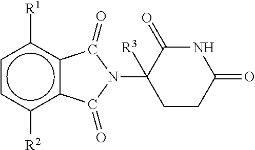
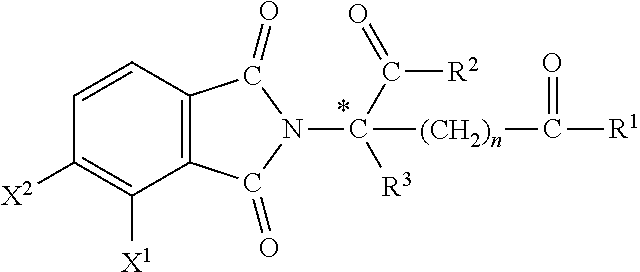

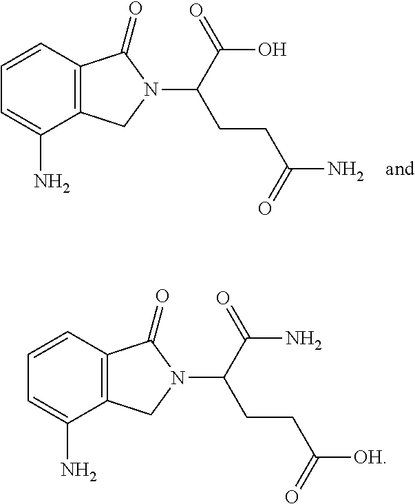


D00001

D00002

D00003

D00004
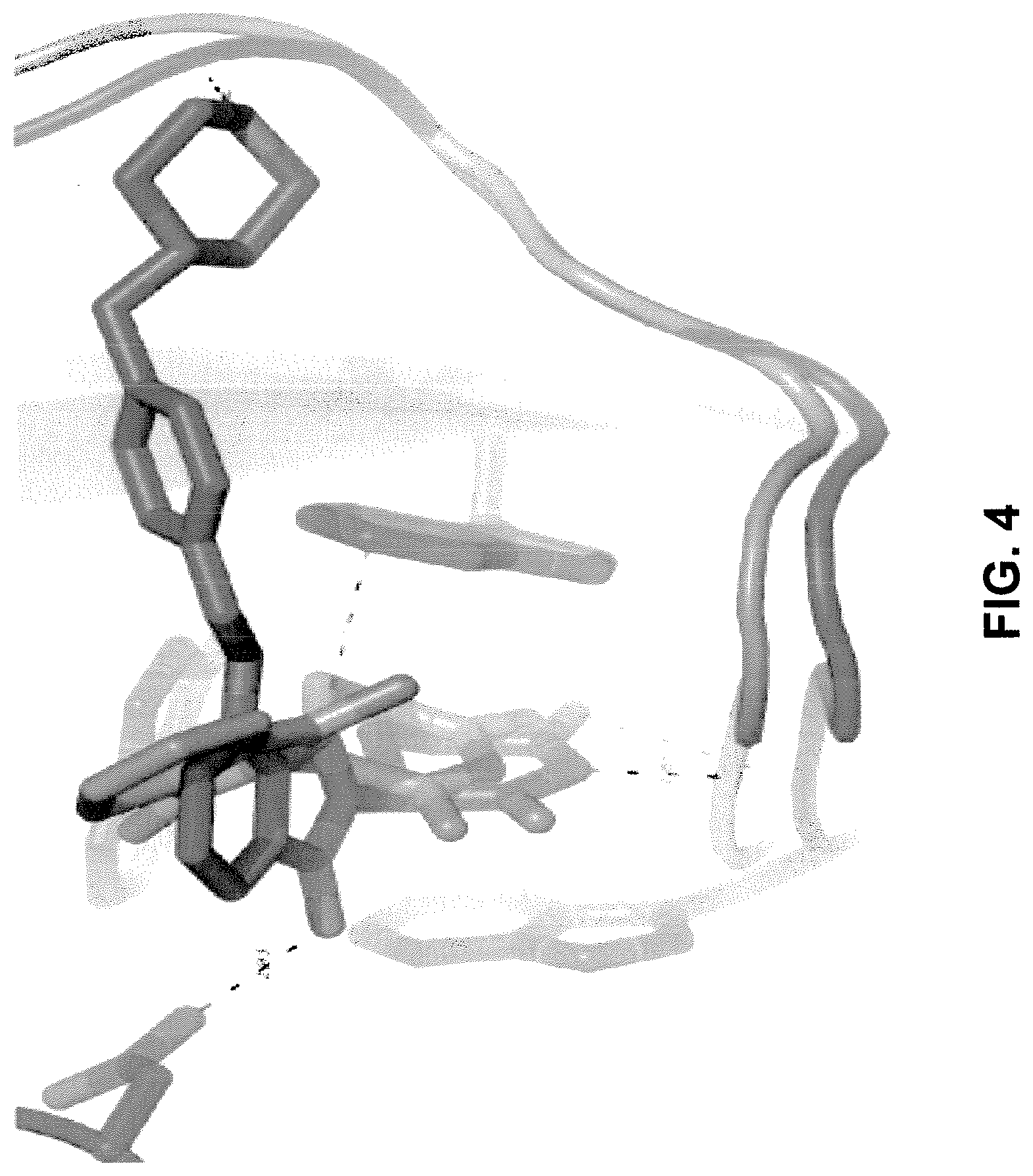
D00005

D00006
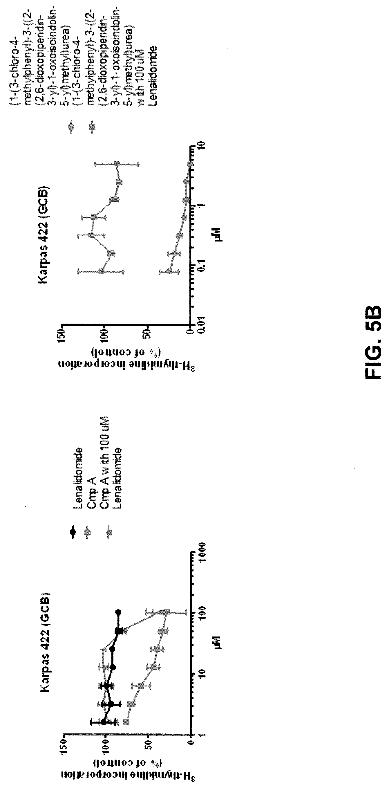
D00007

D00008

D00009
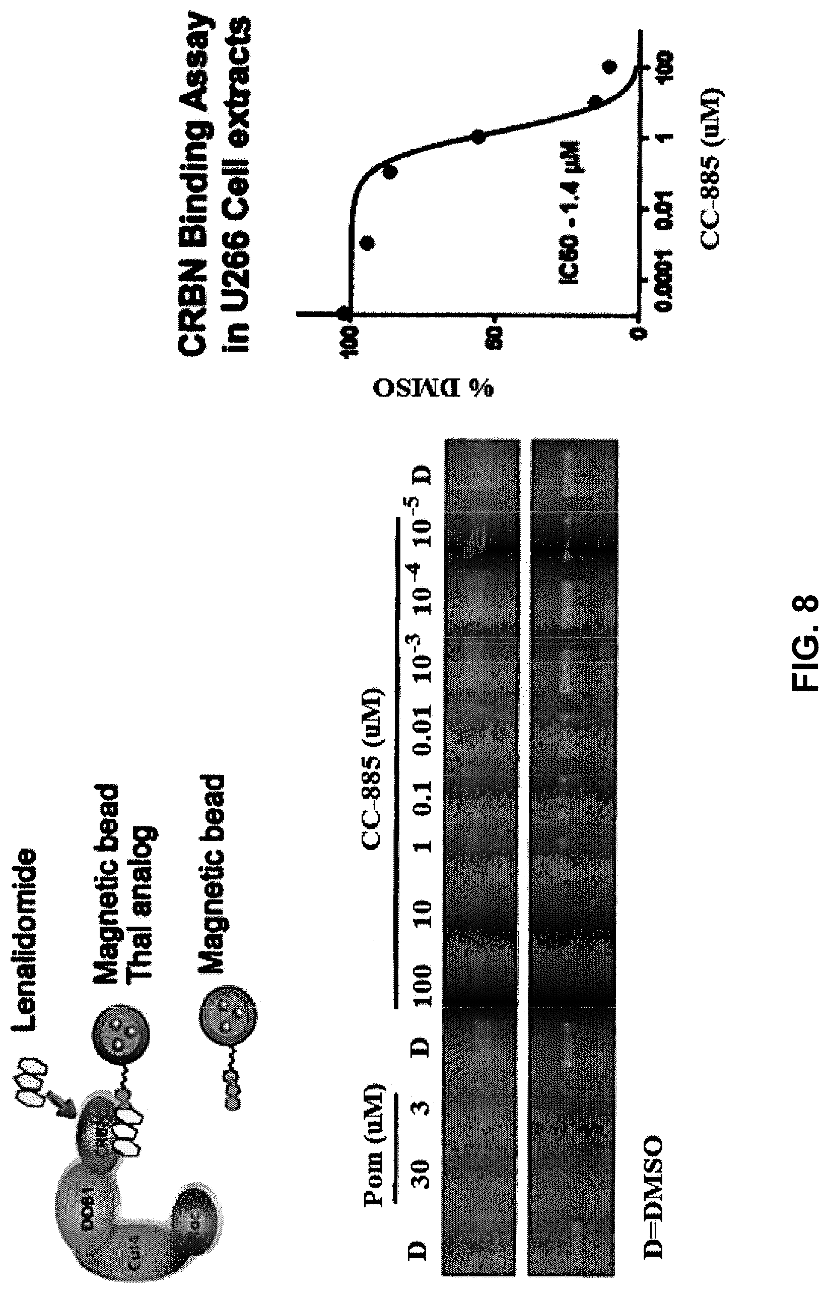
D00010
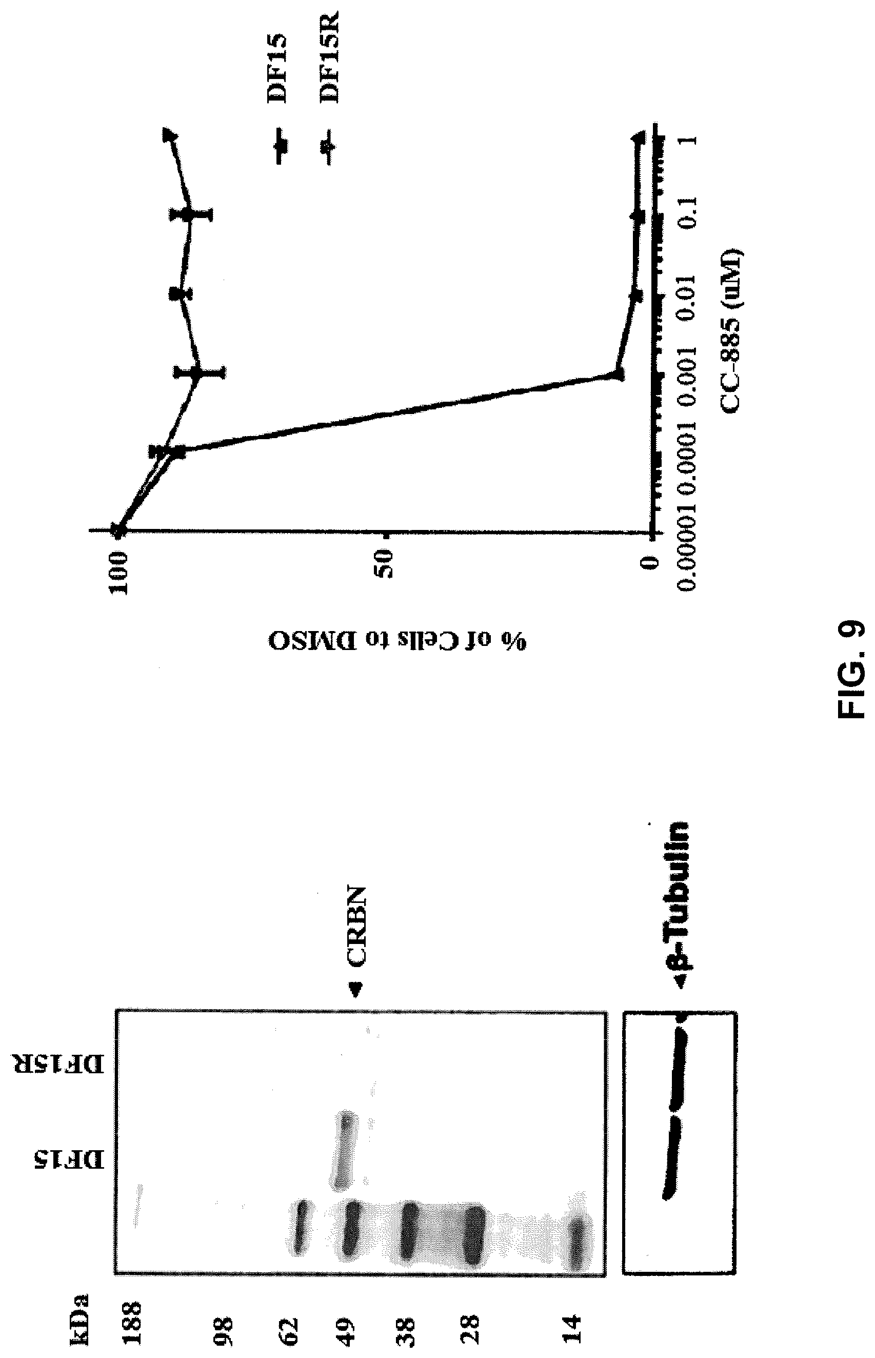
D00011

D00012

D00013
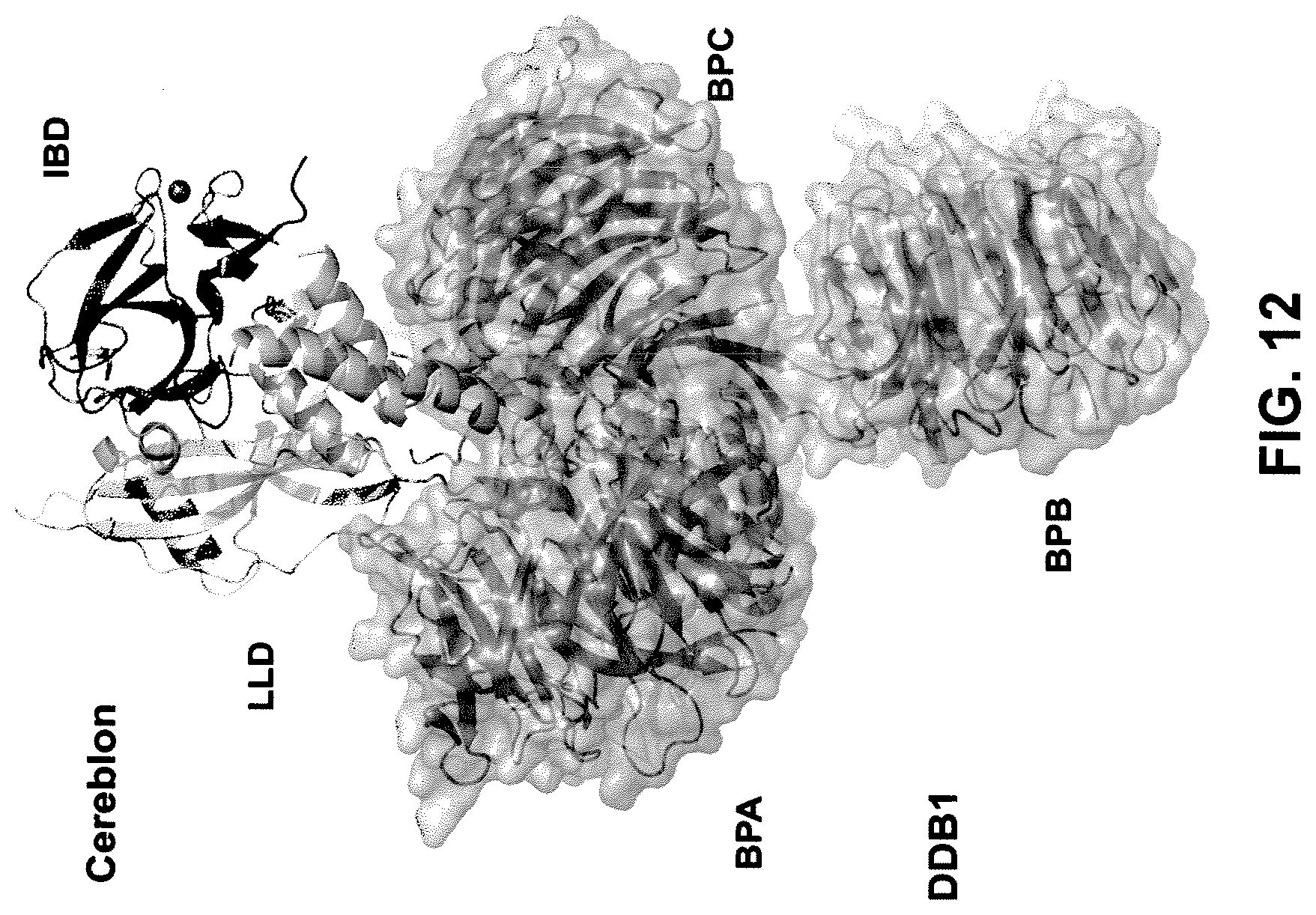
D00014

D00015

D00016

D00017

D00018

D00019
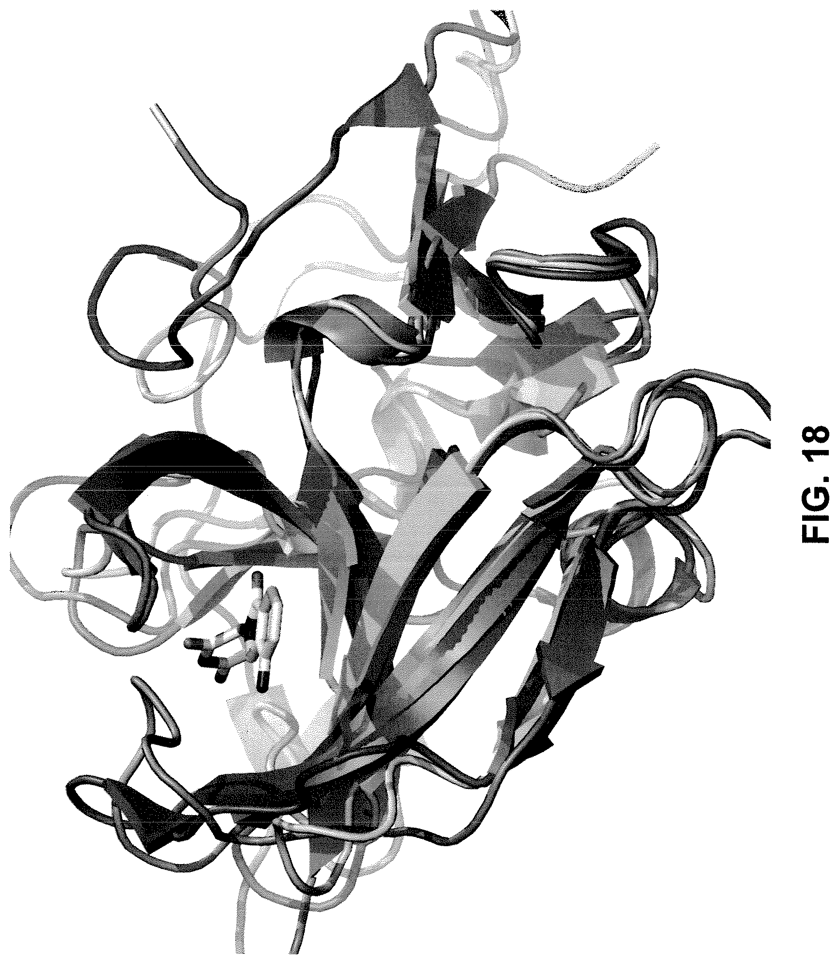
D00020

D00021

D00022
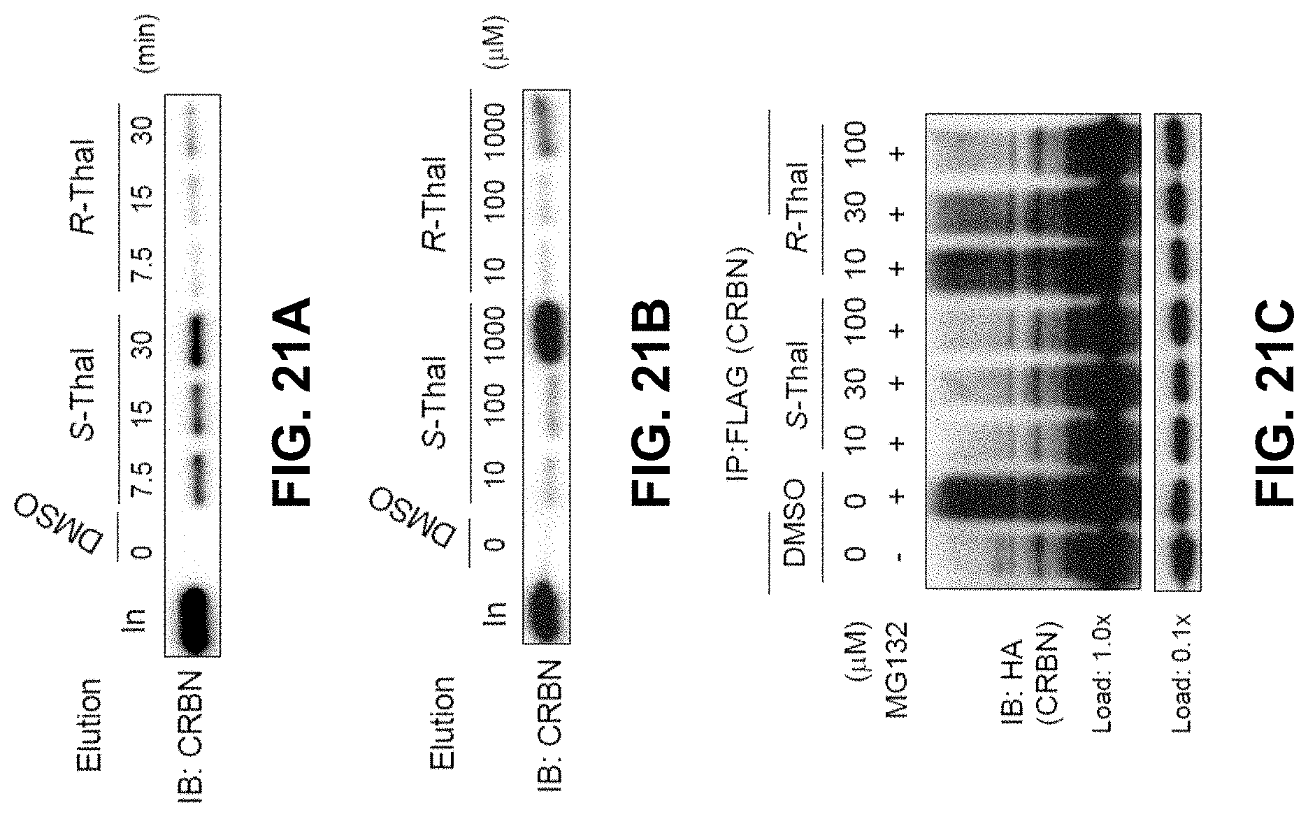
D00023
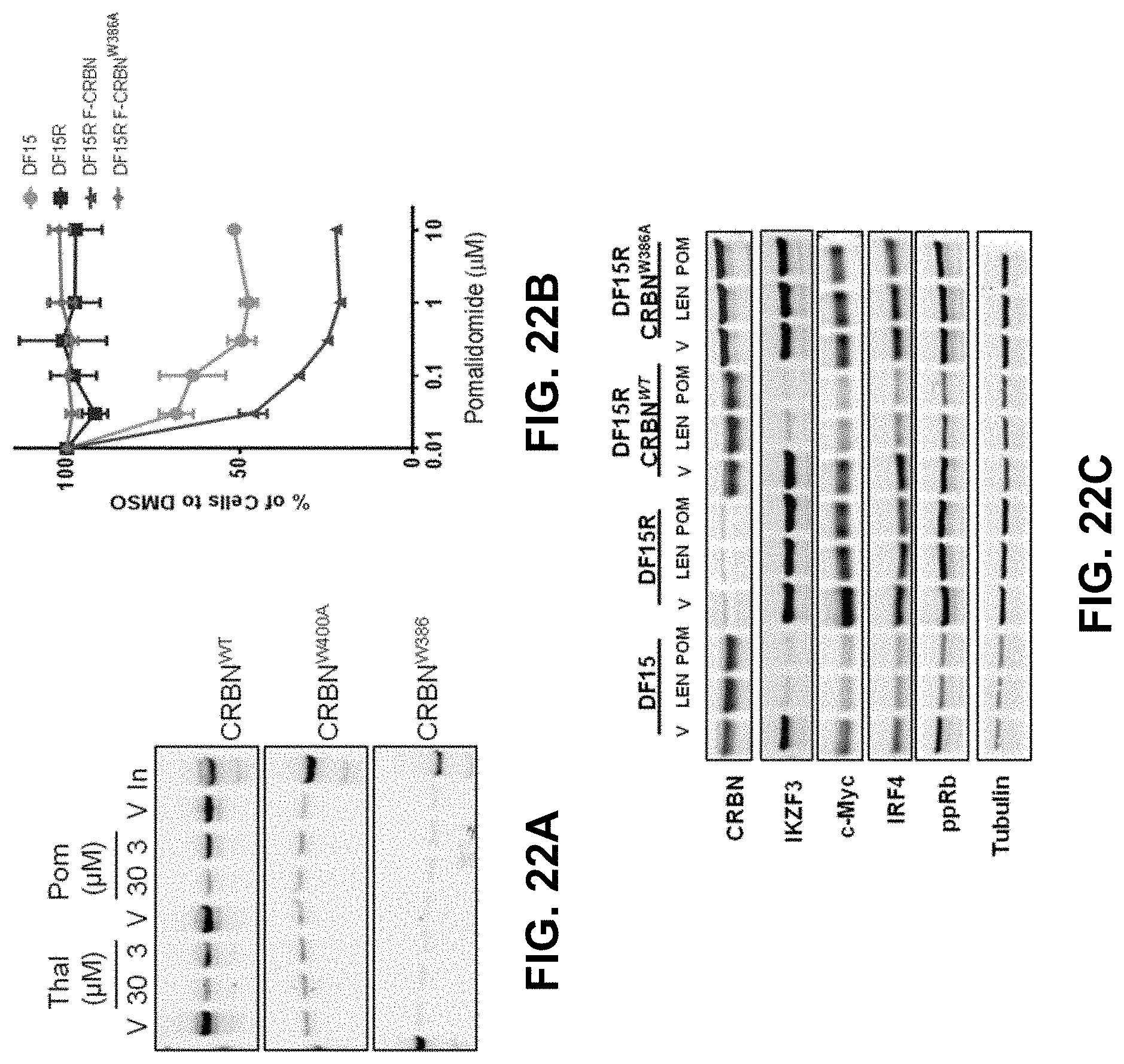
D00024
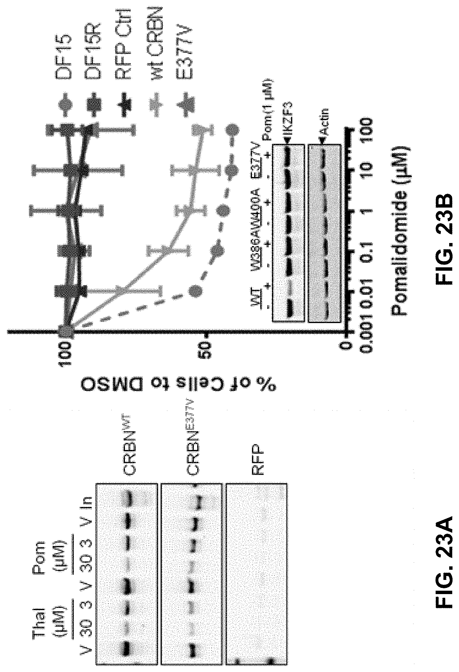
D00025
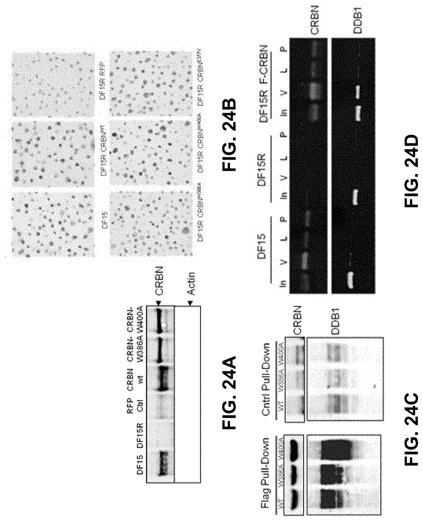
D00026

D00027

D00028

D00029

D00030

D00031
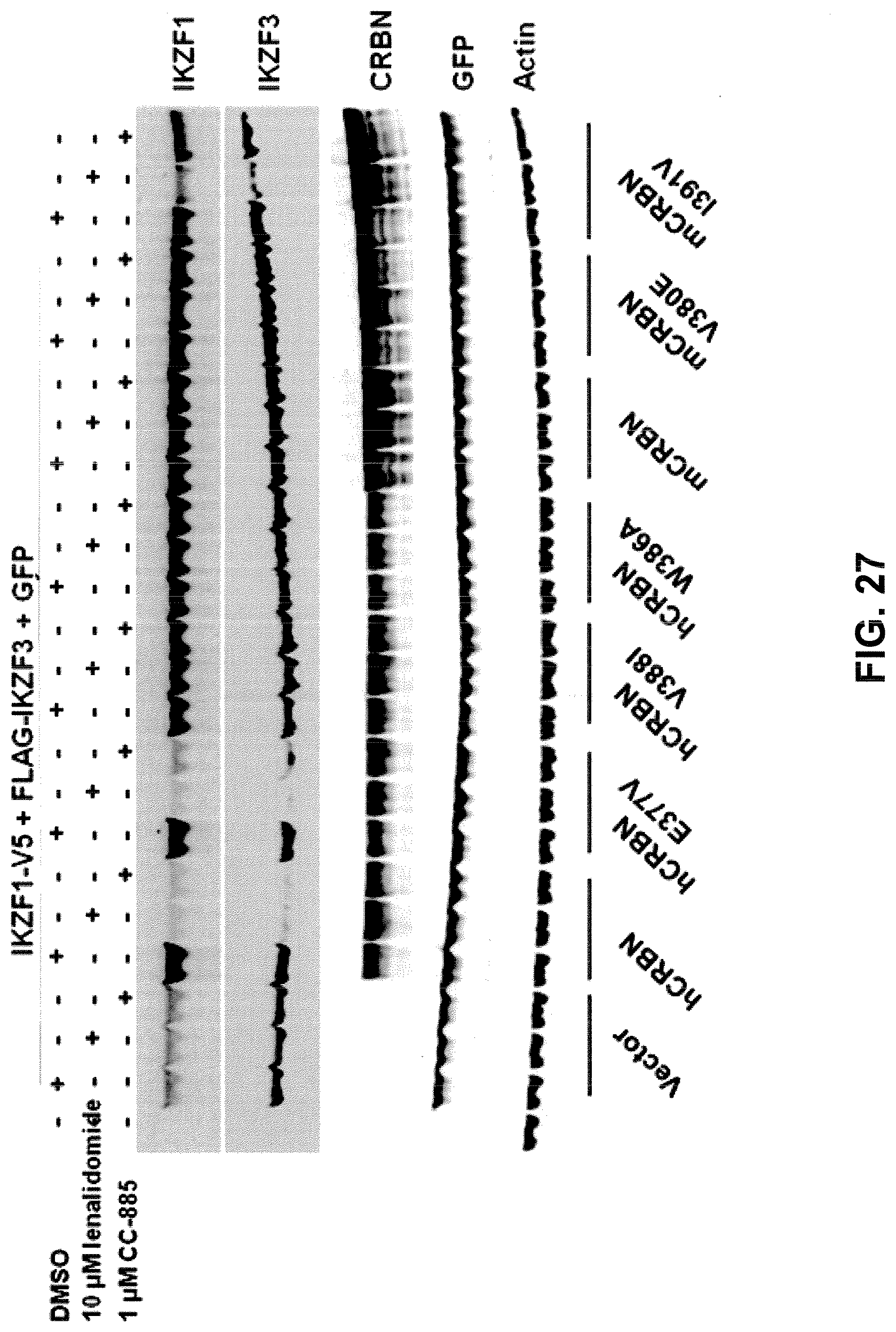
S00001
T00001


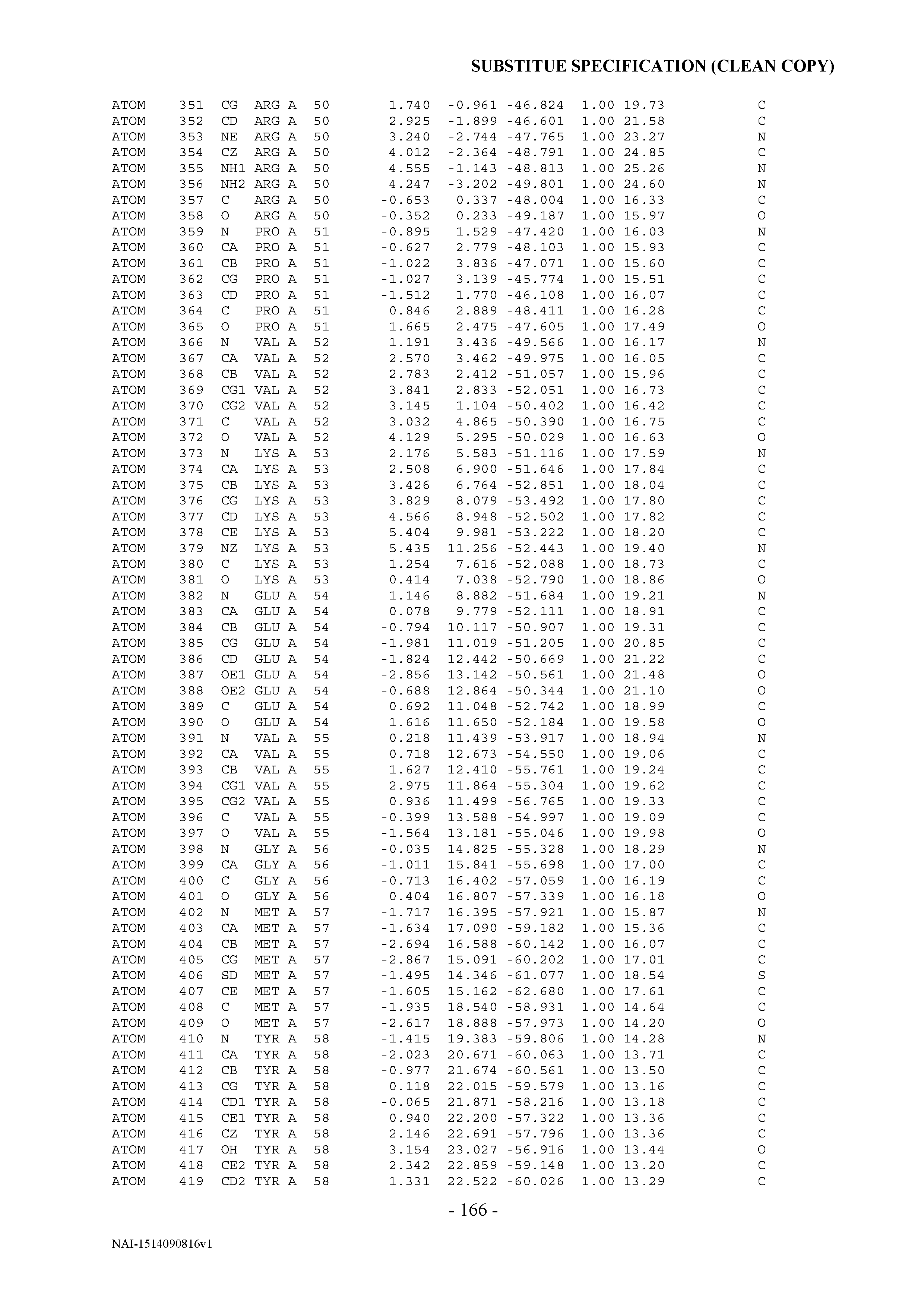
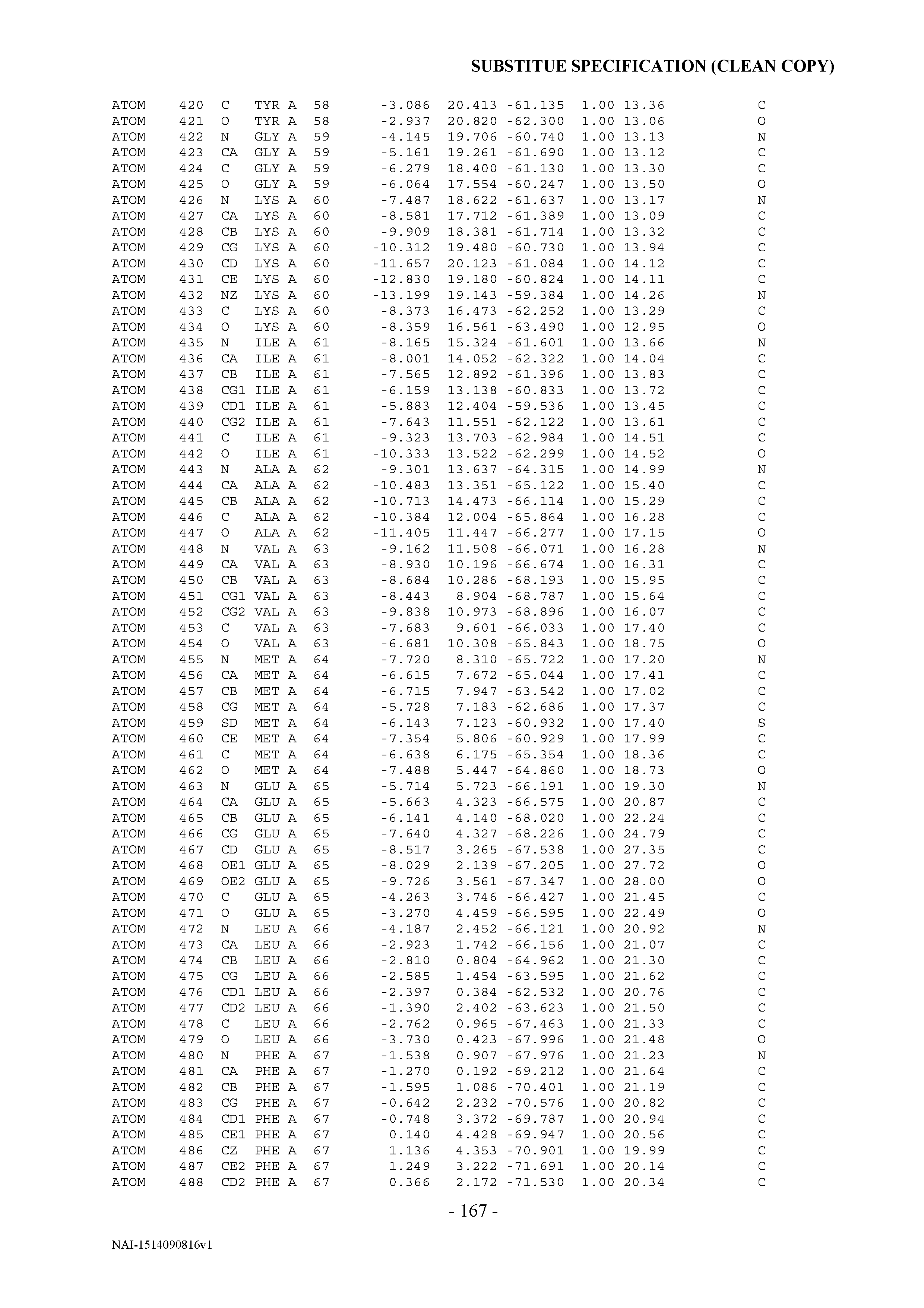


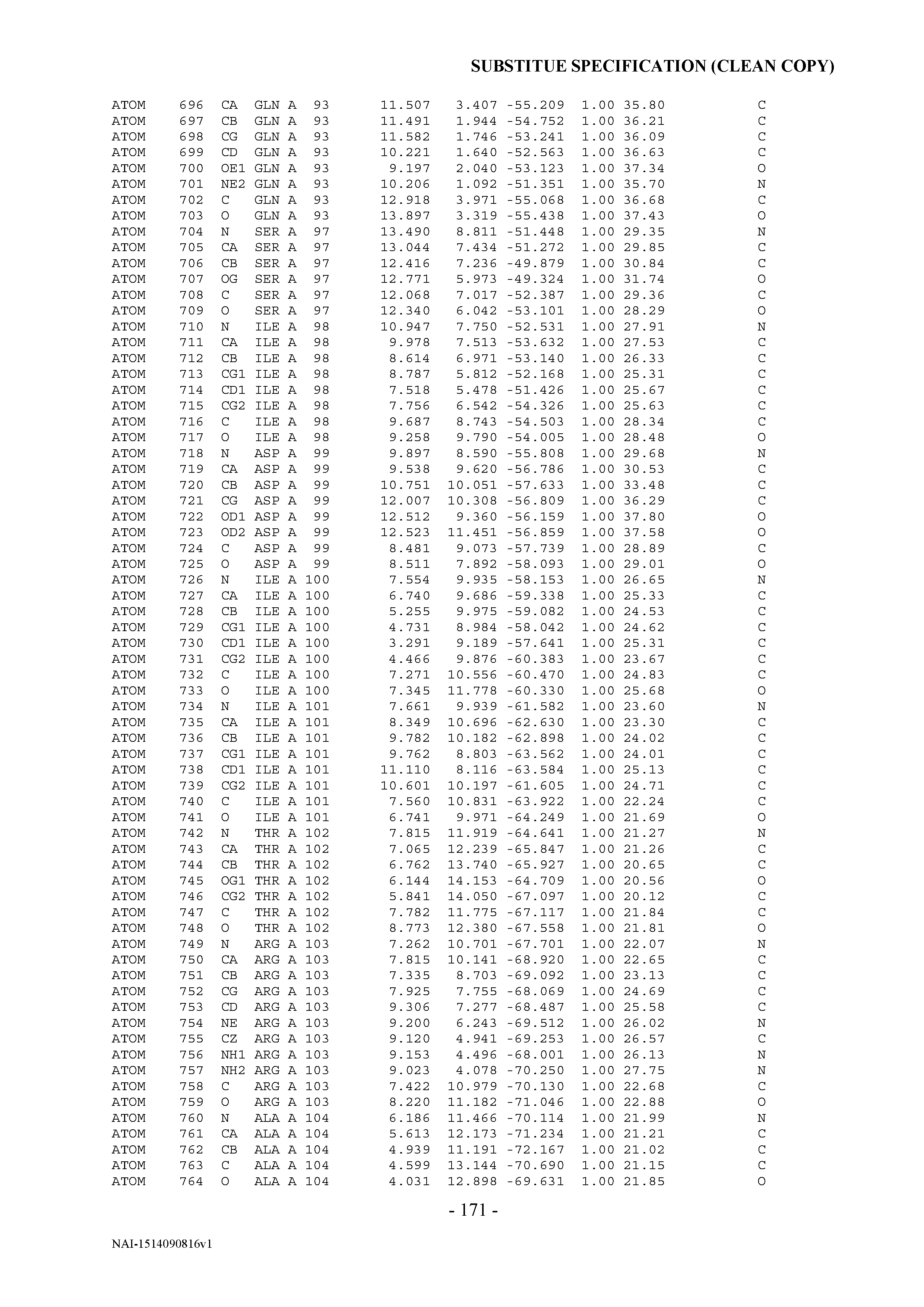


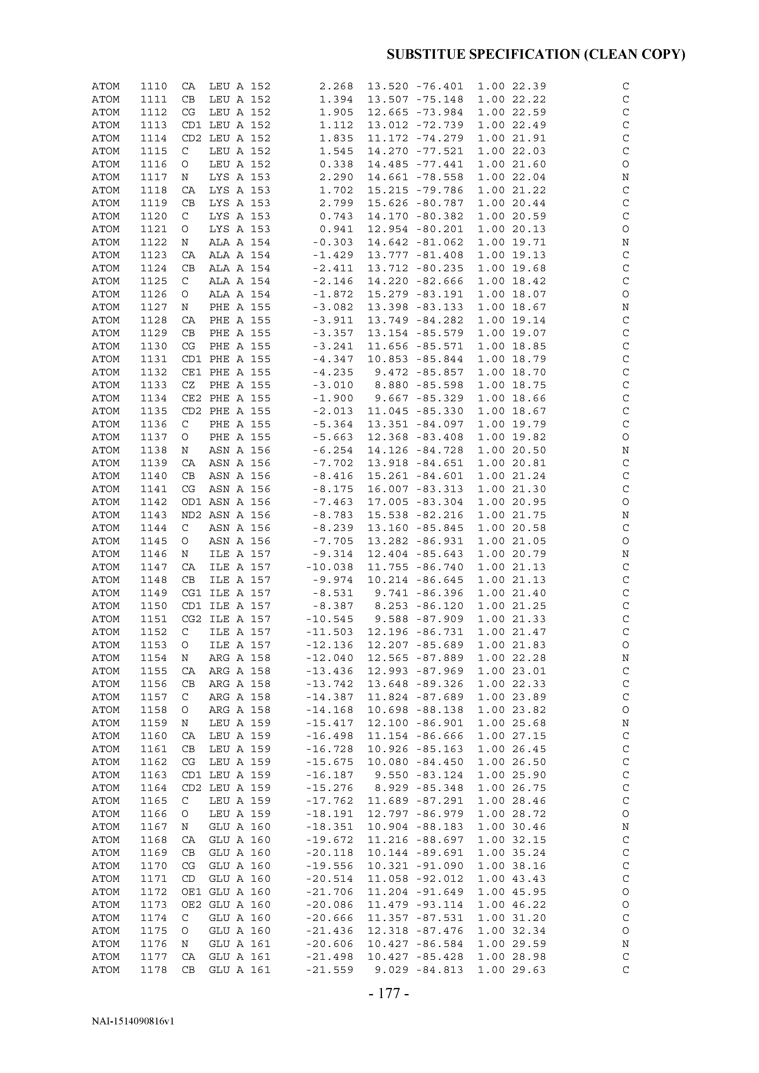


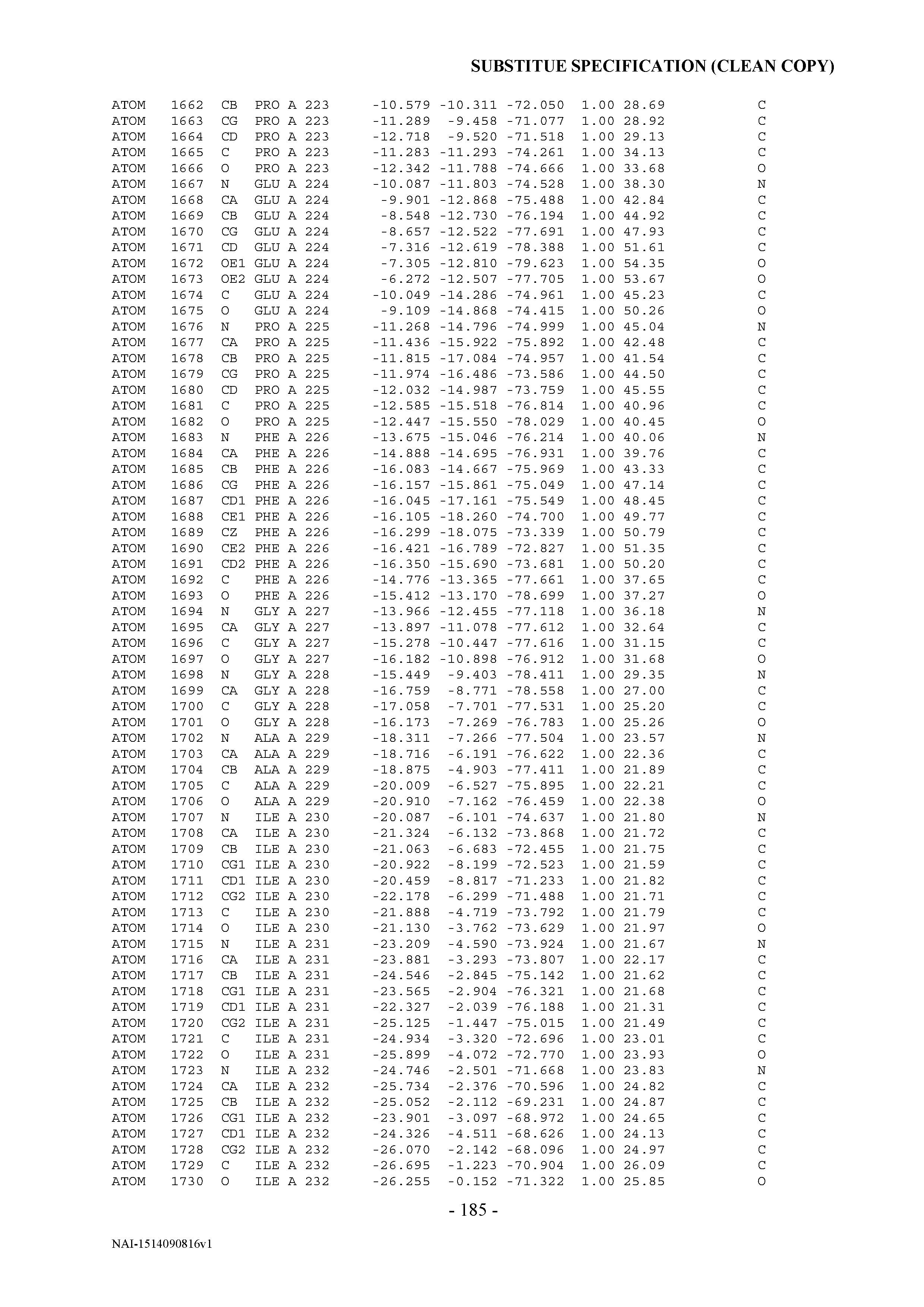


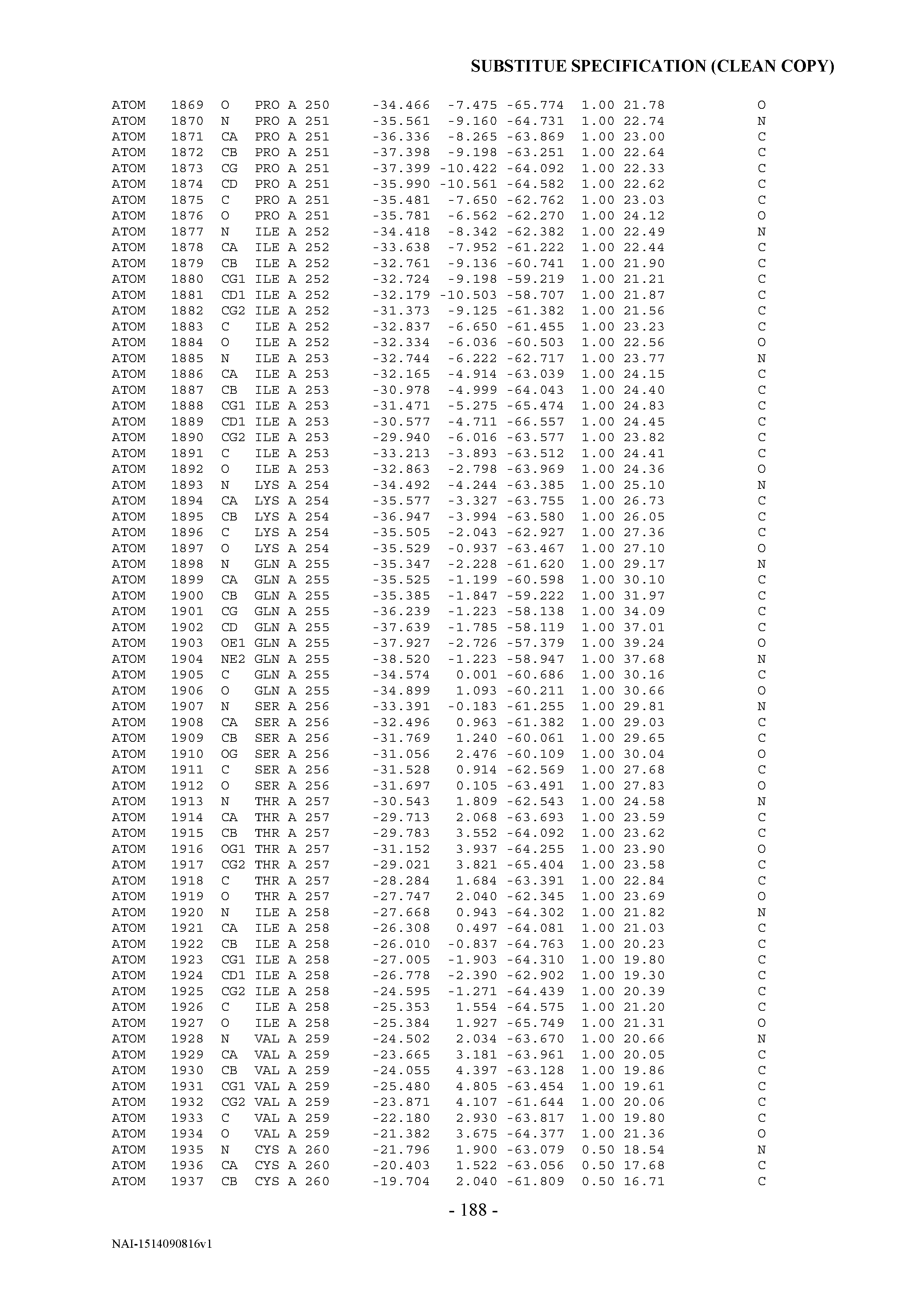


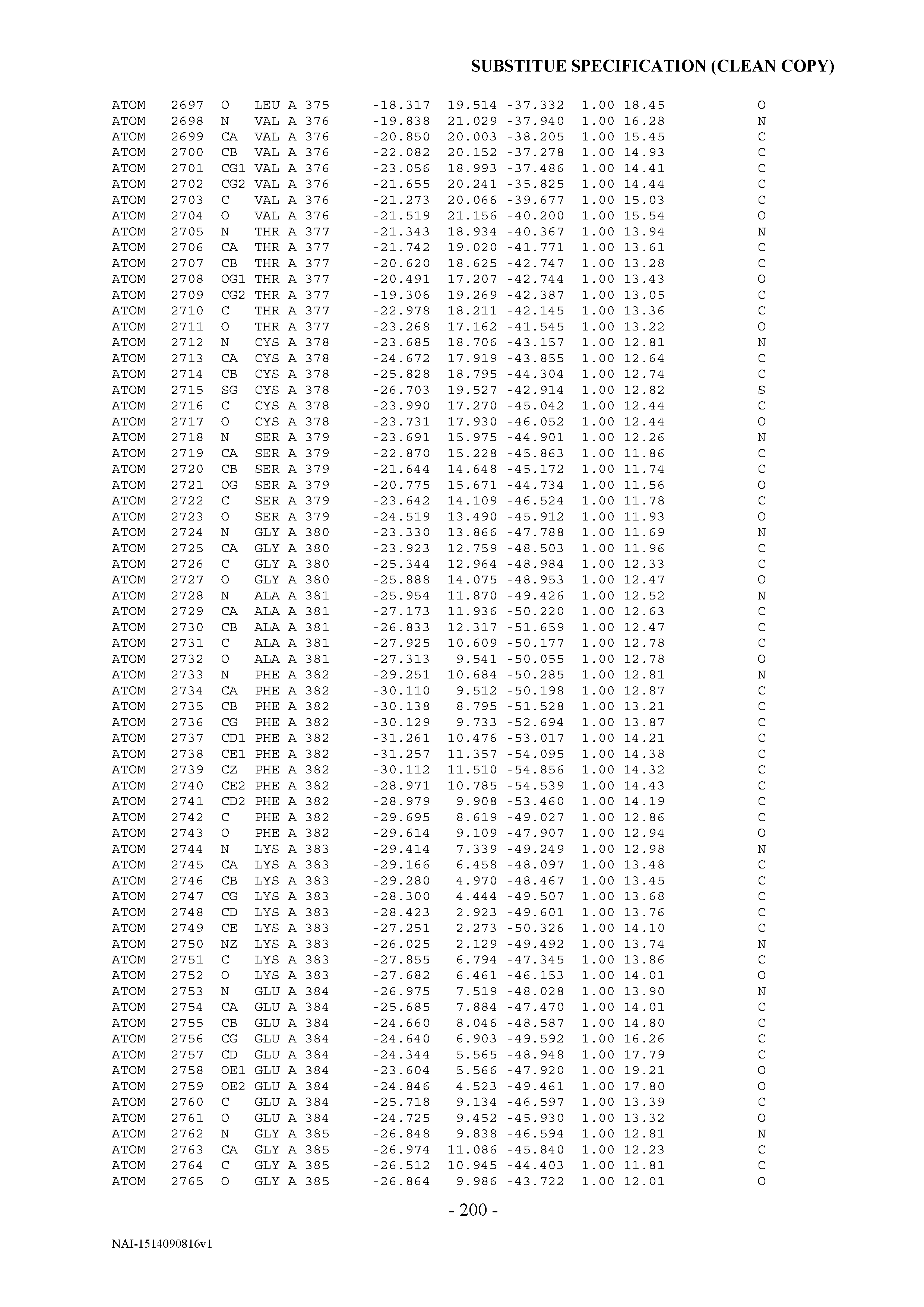

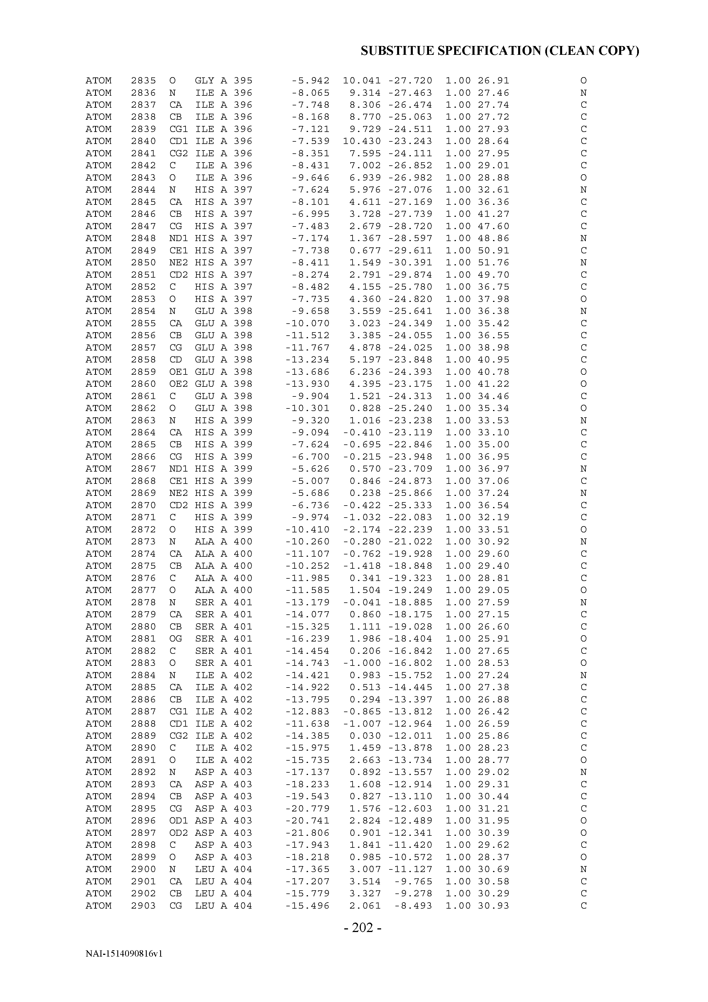
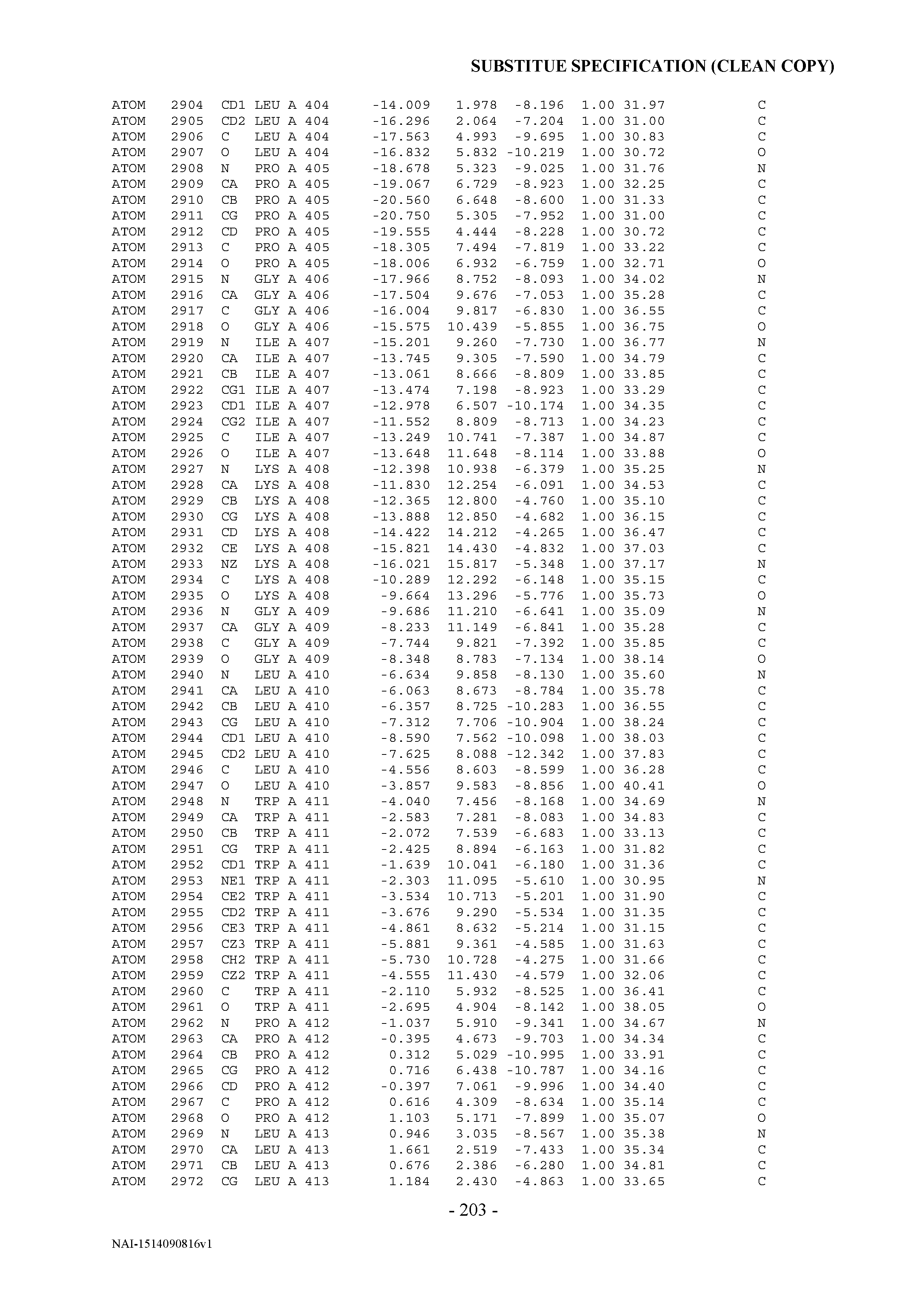

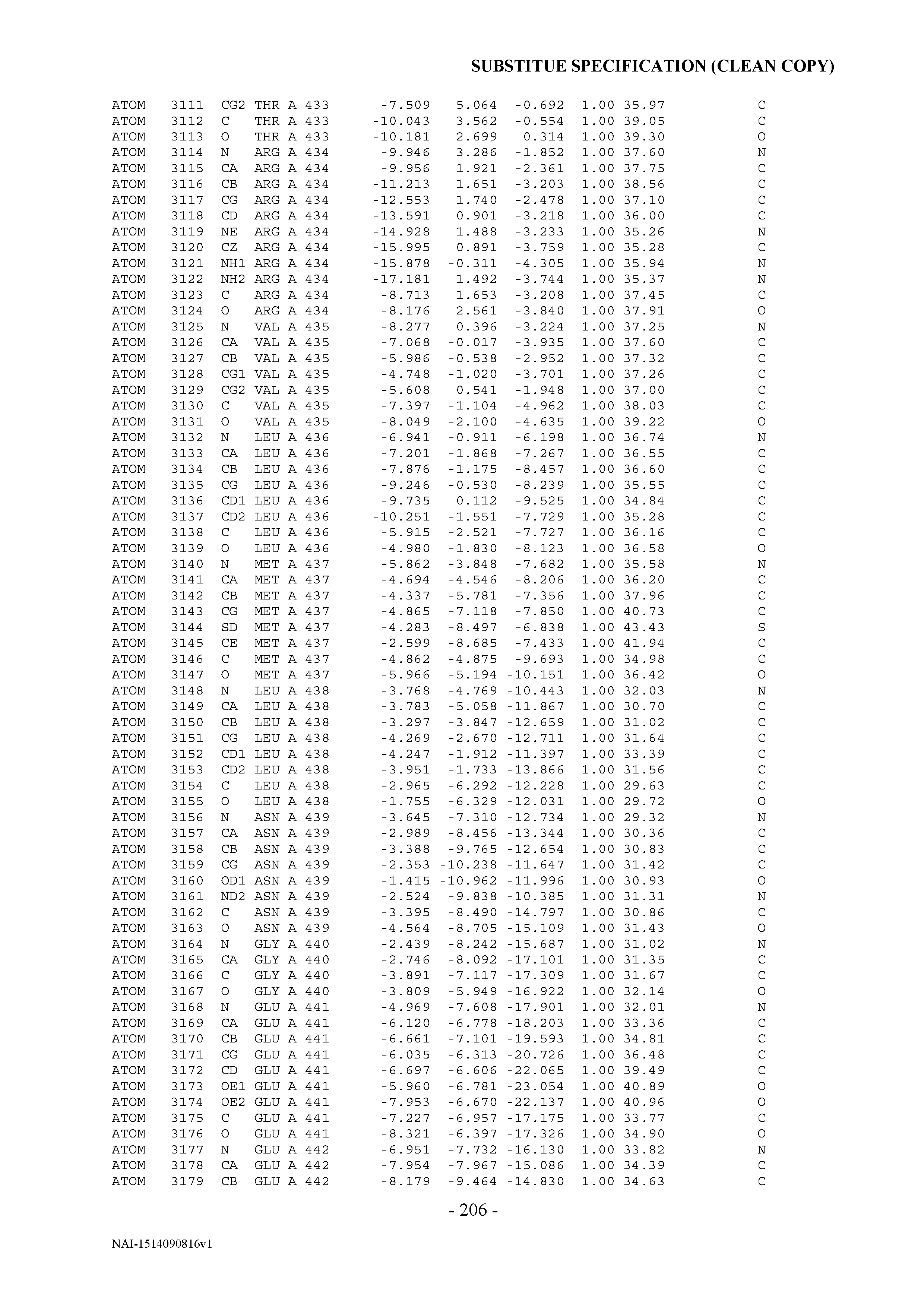


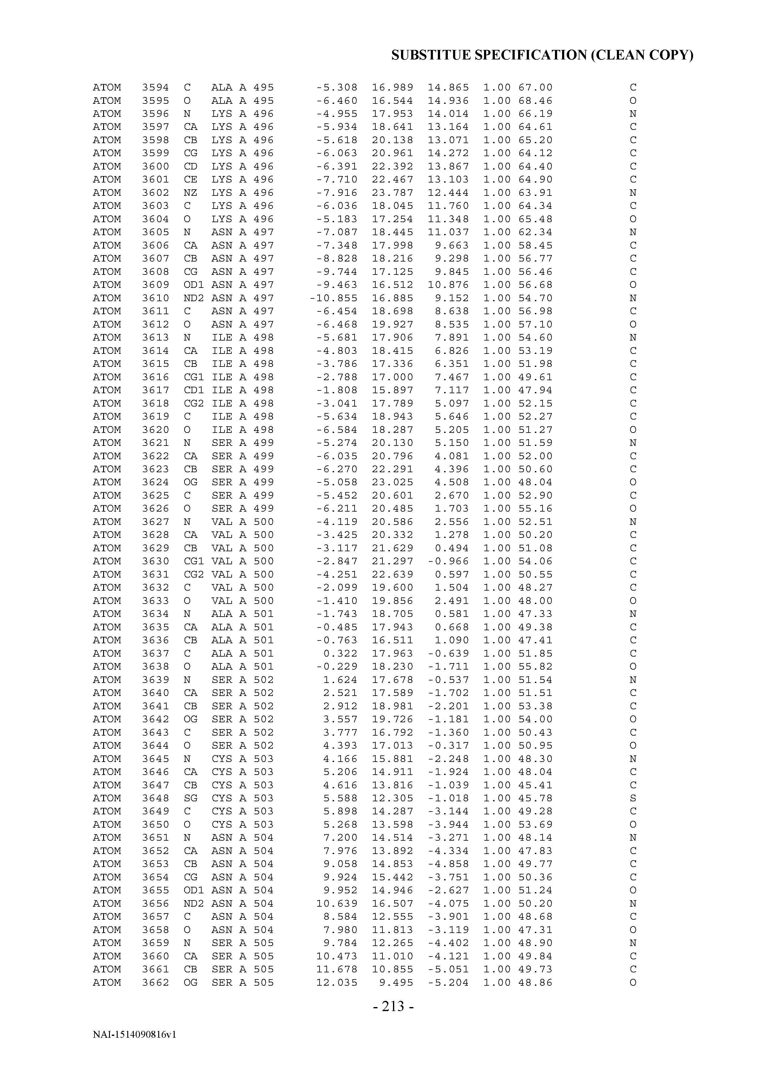
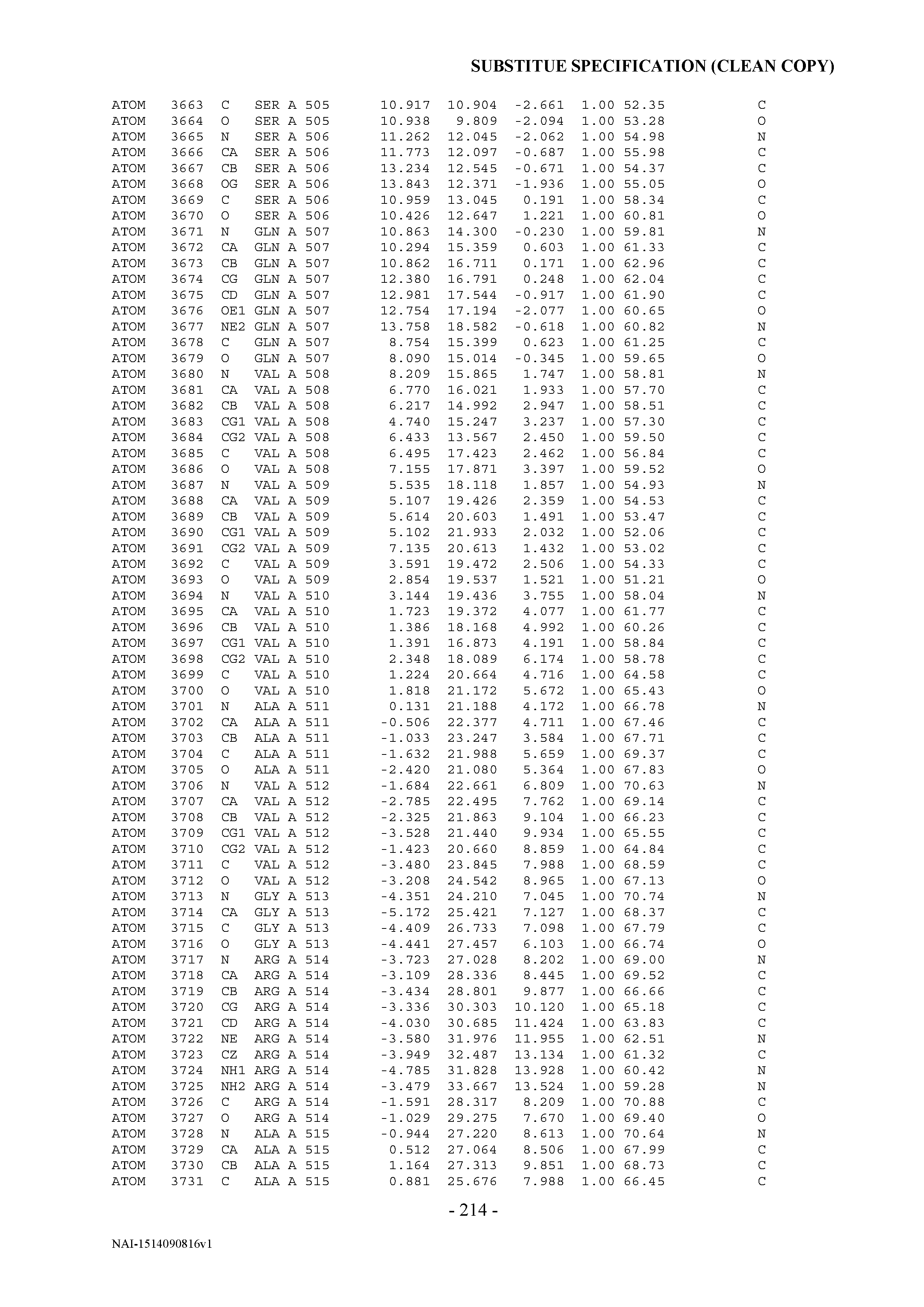


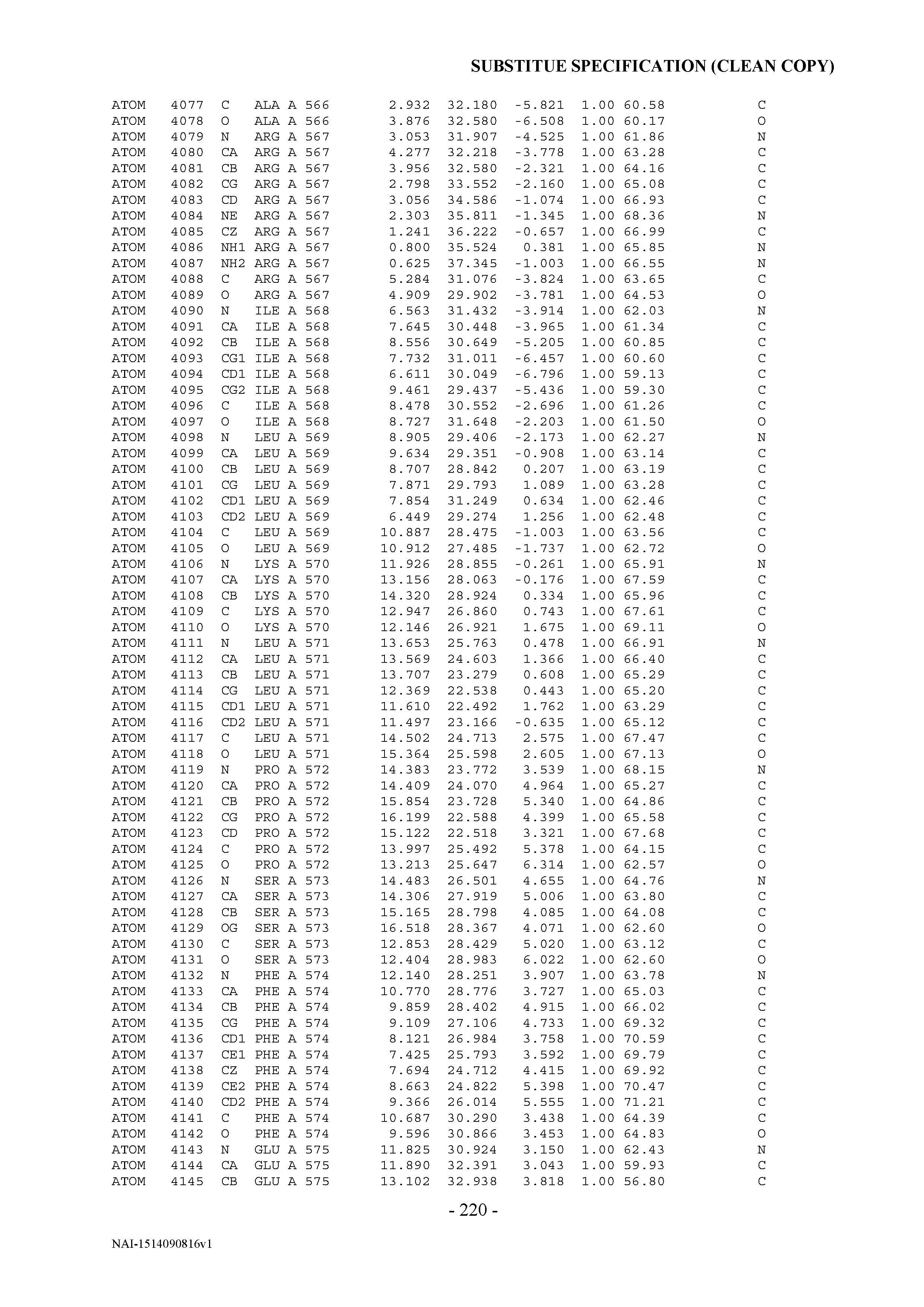
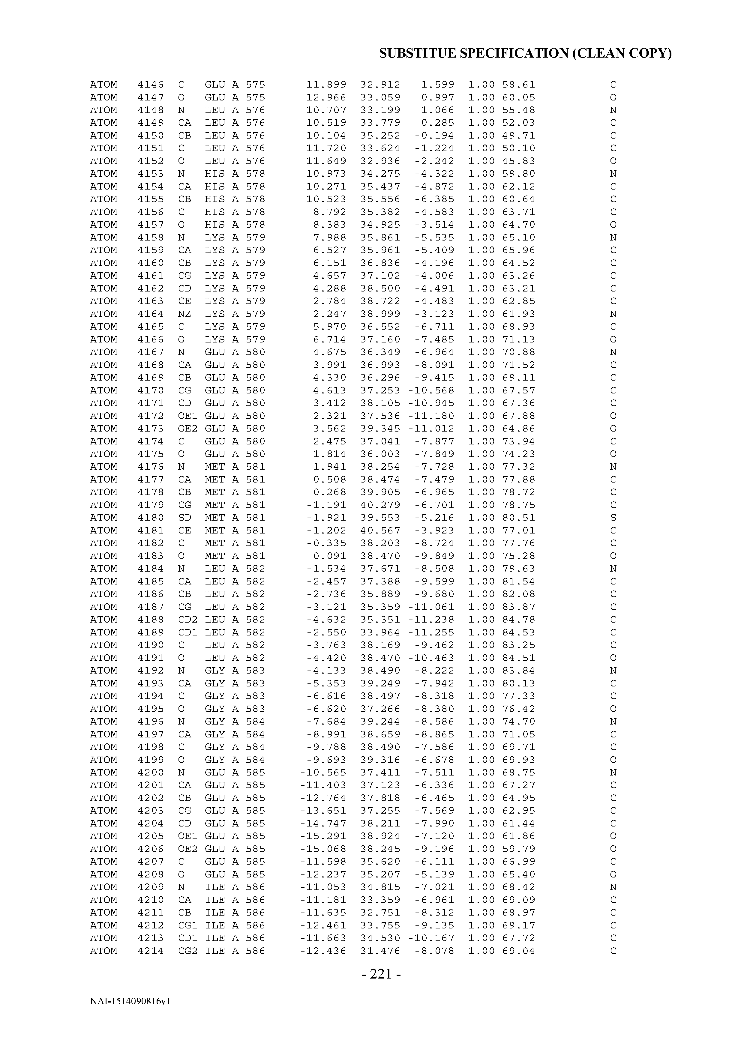
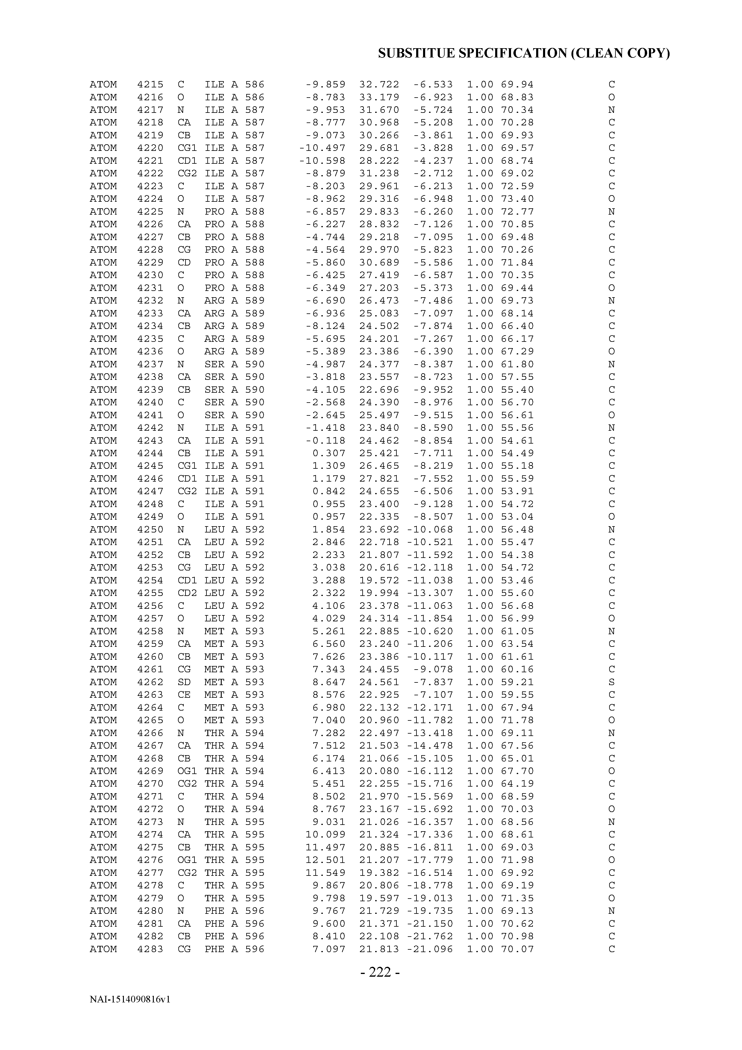
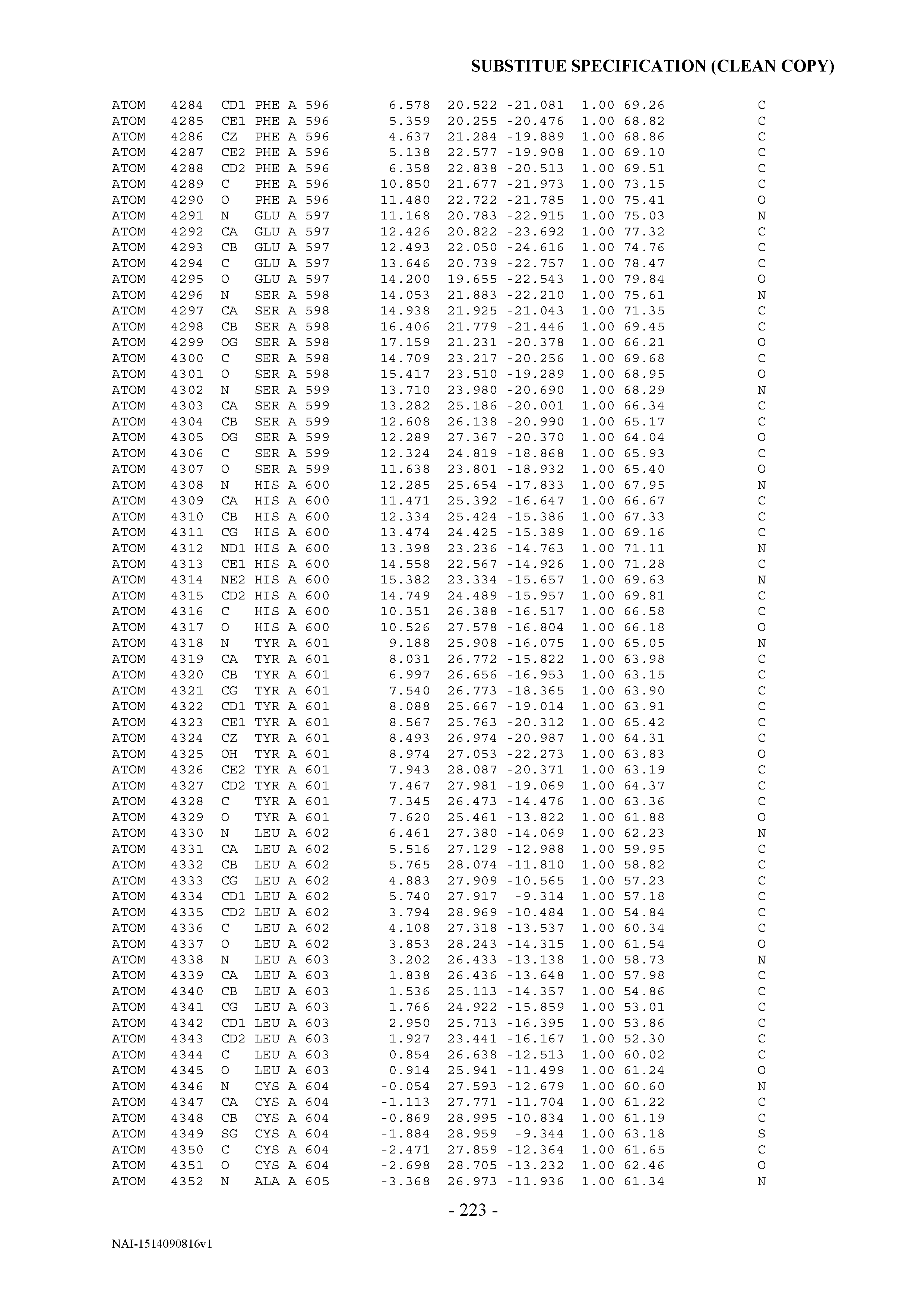

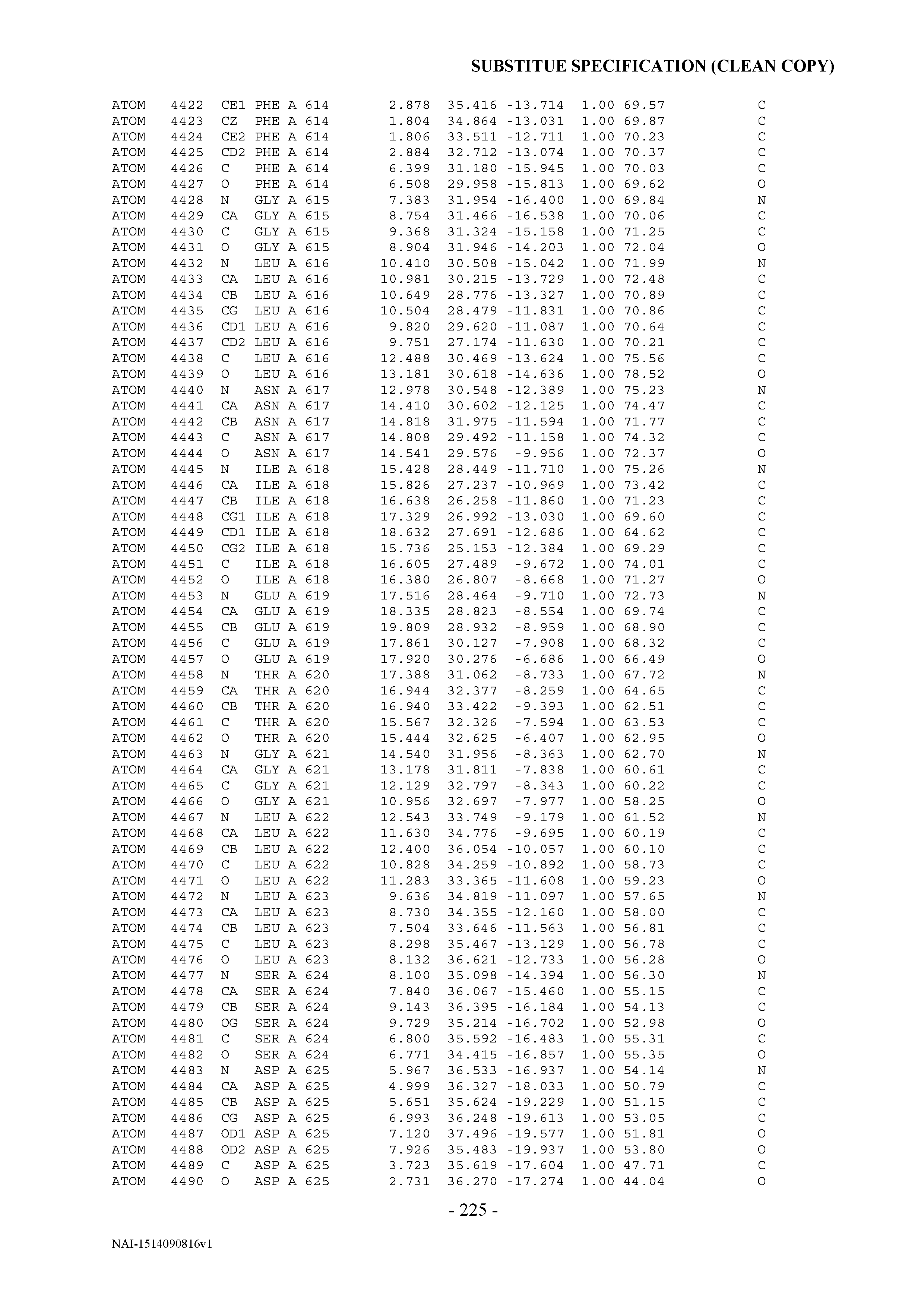


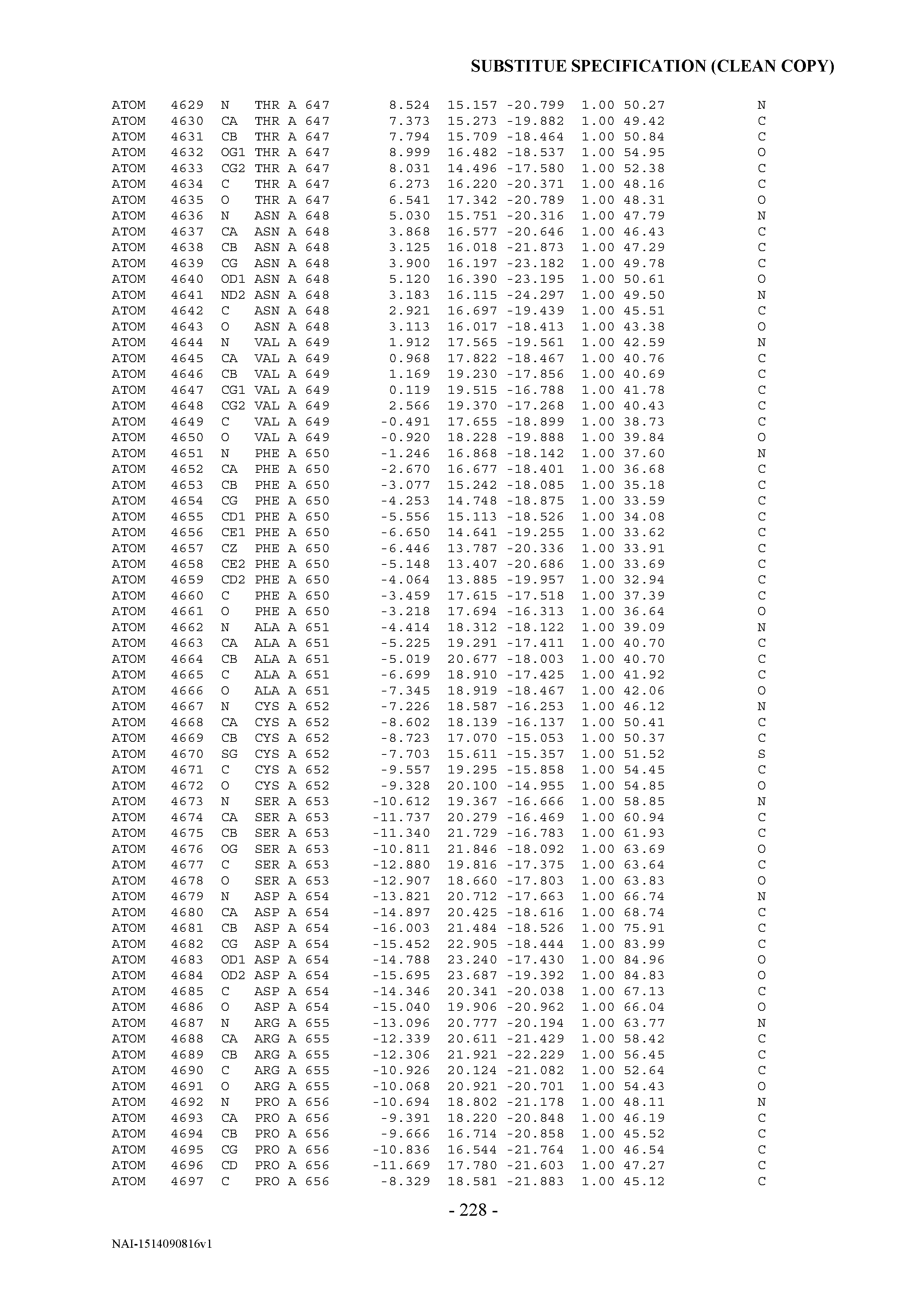


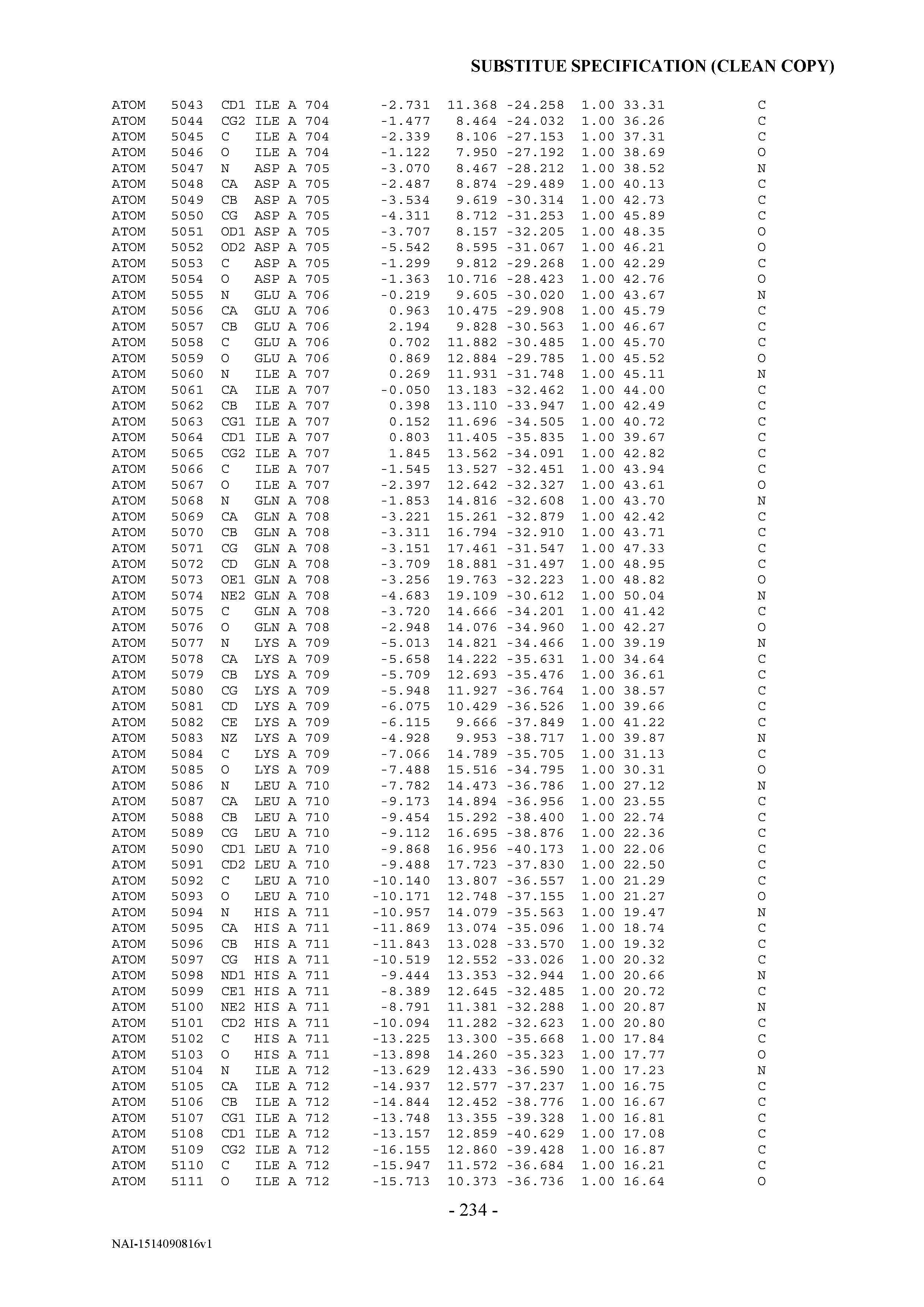


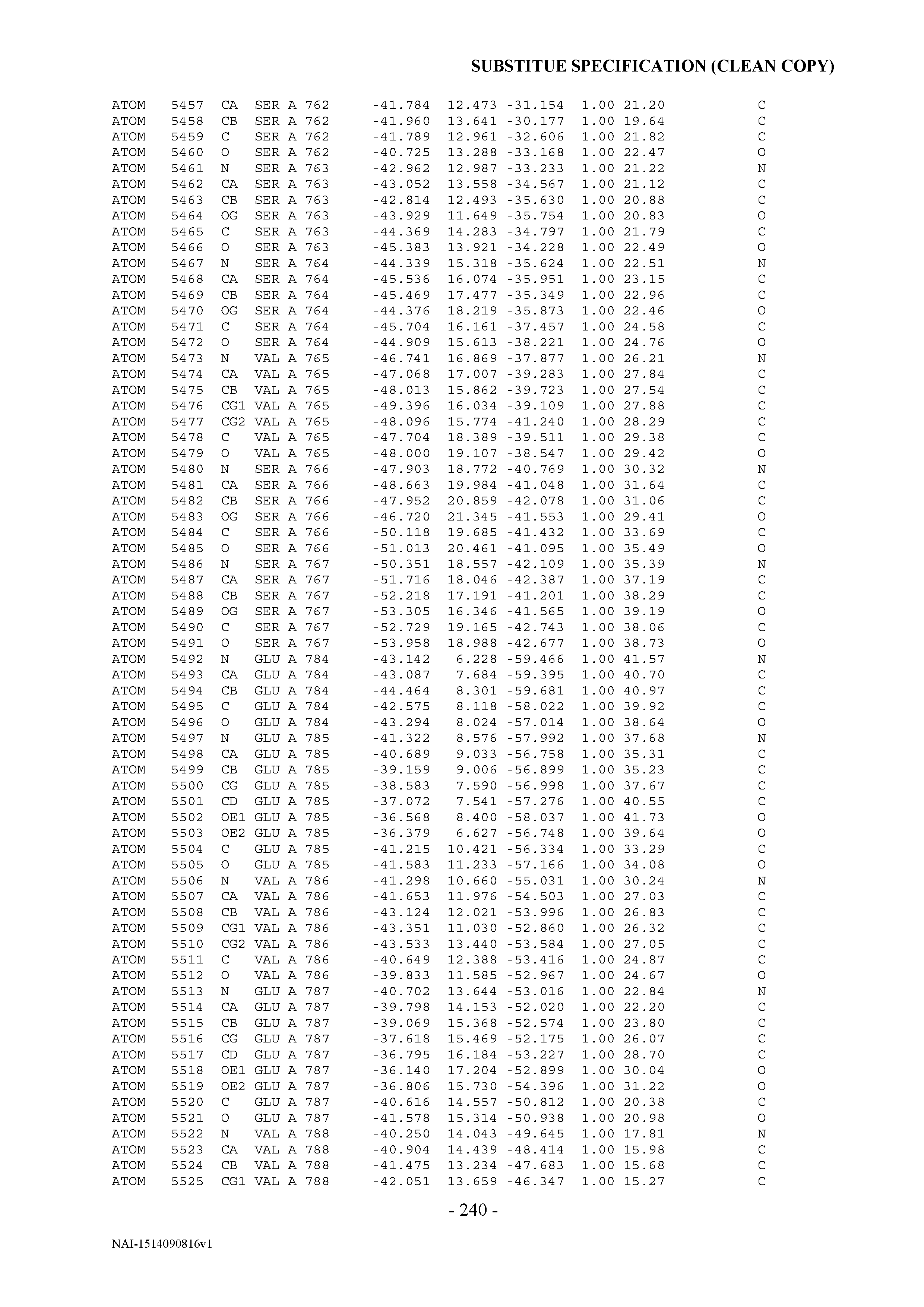


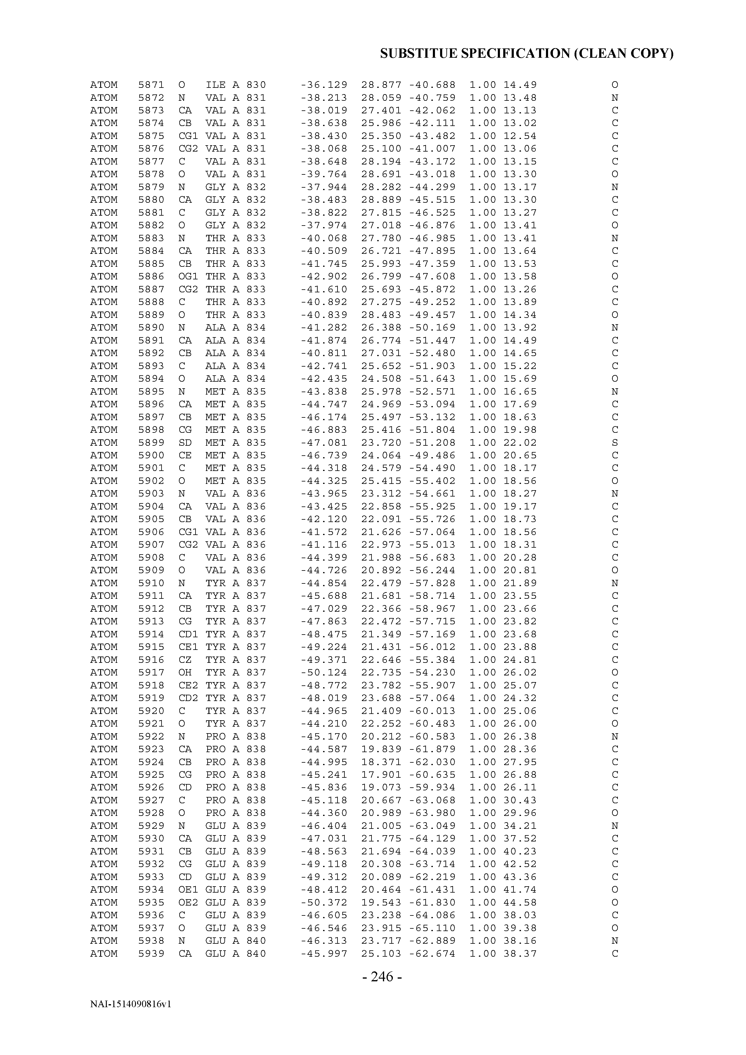


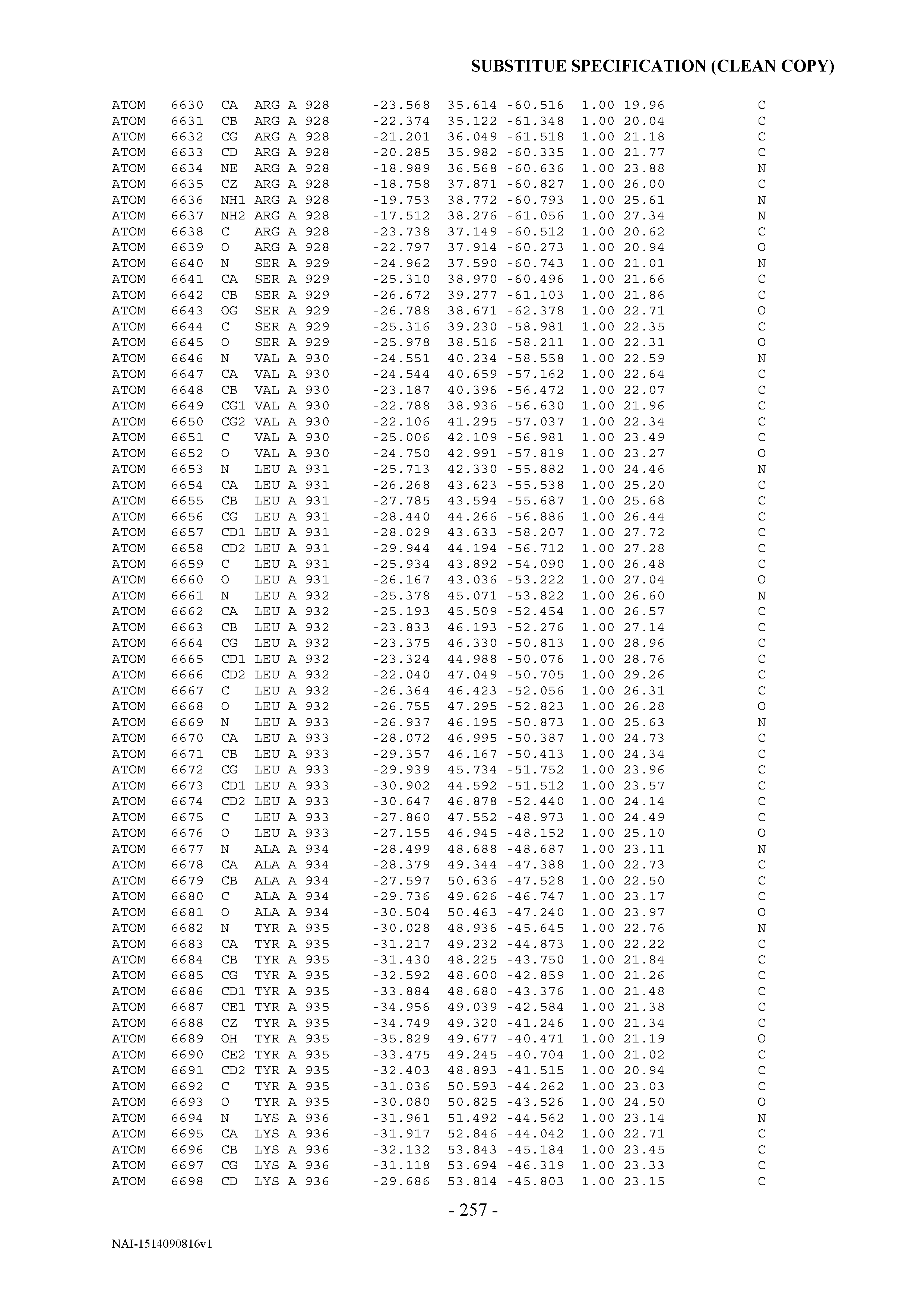


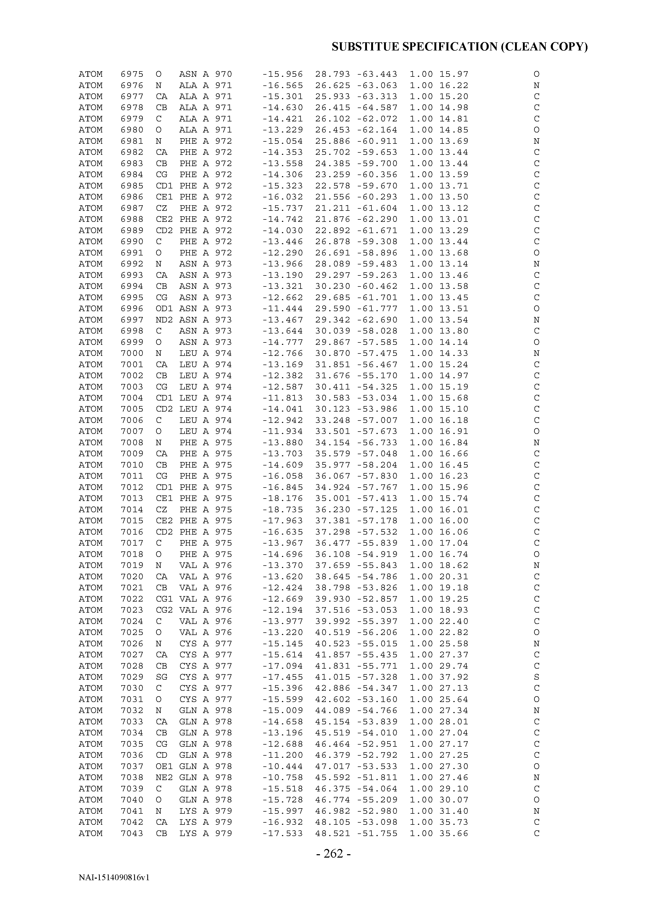


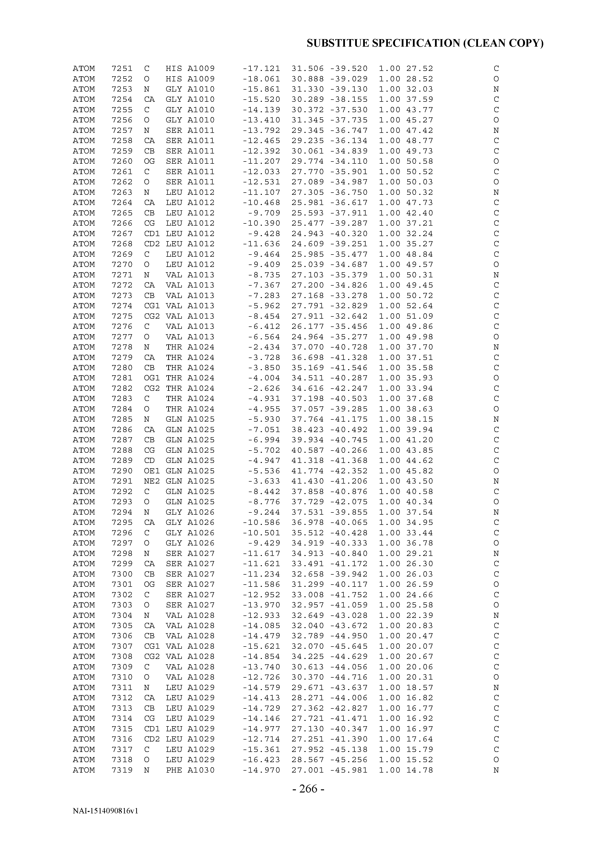


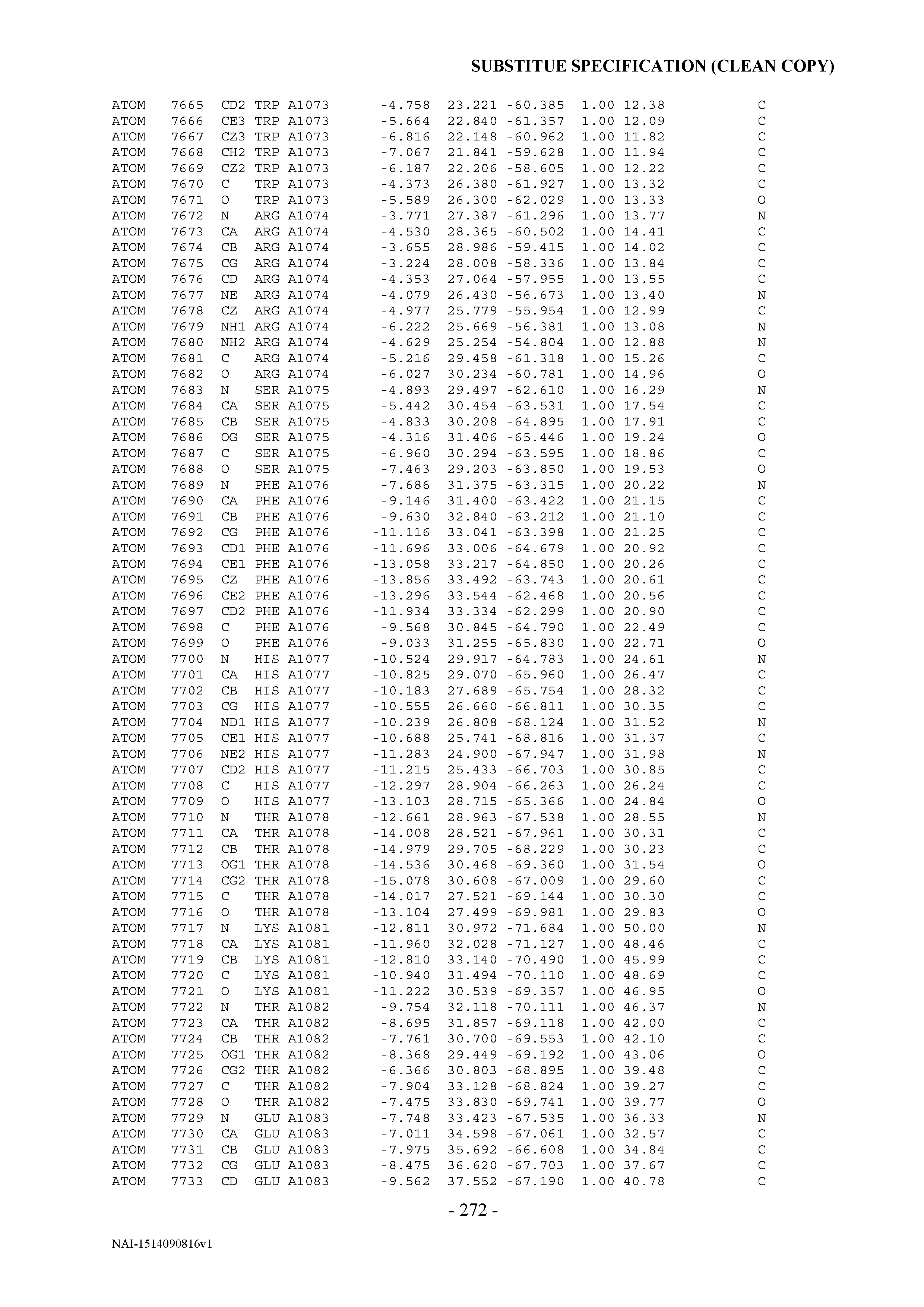
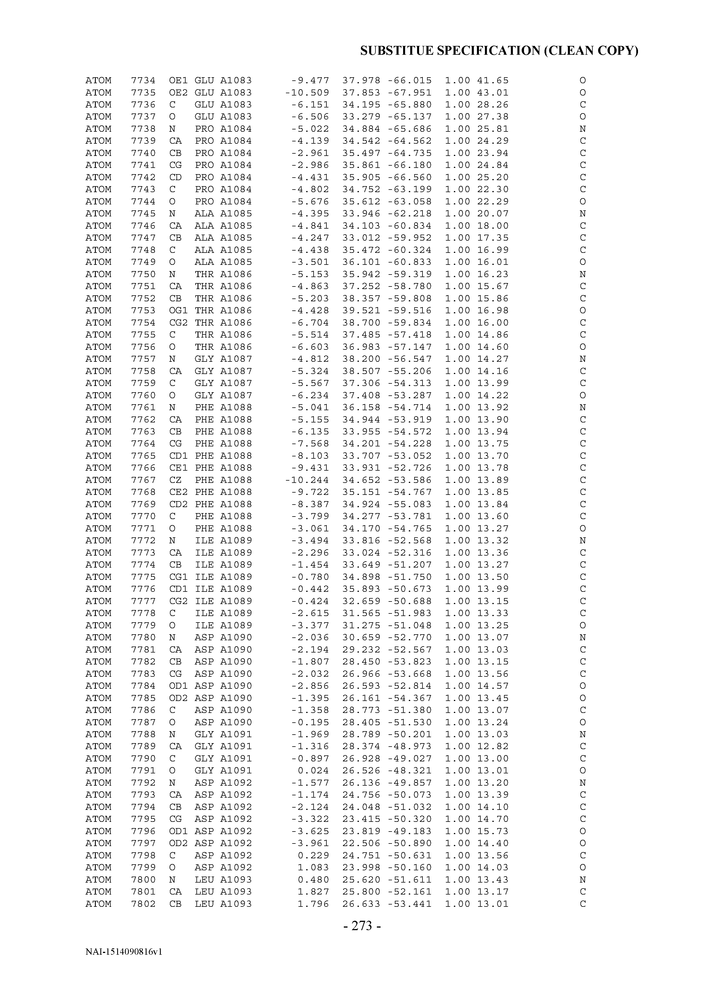


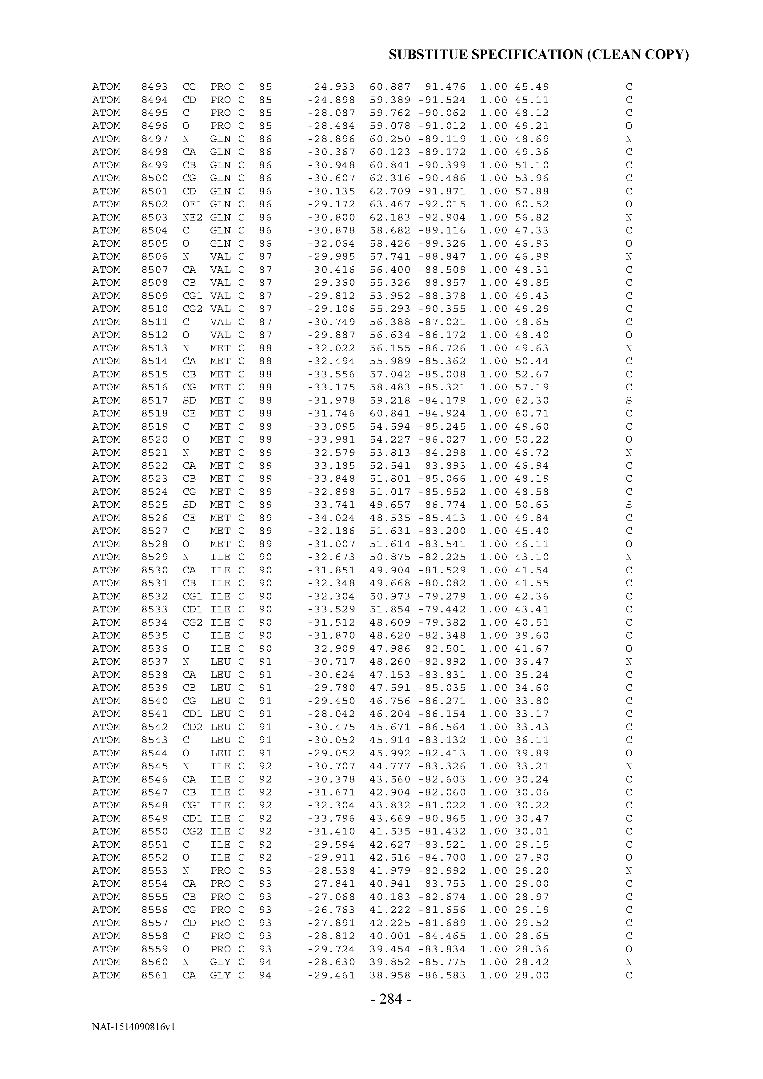
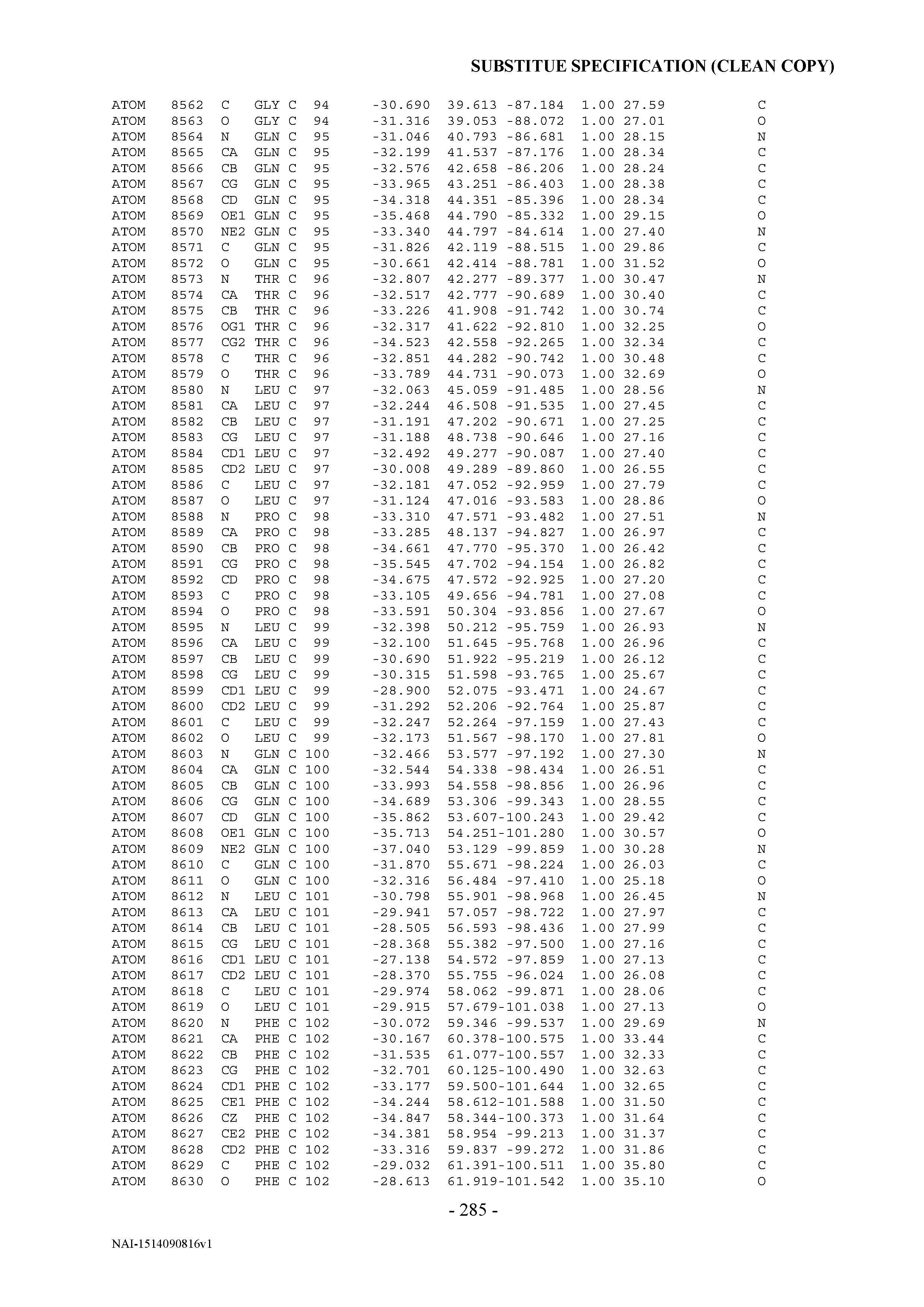
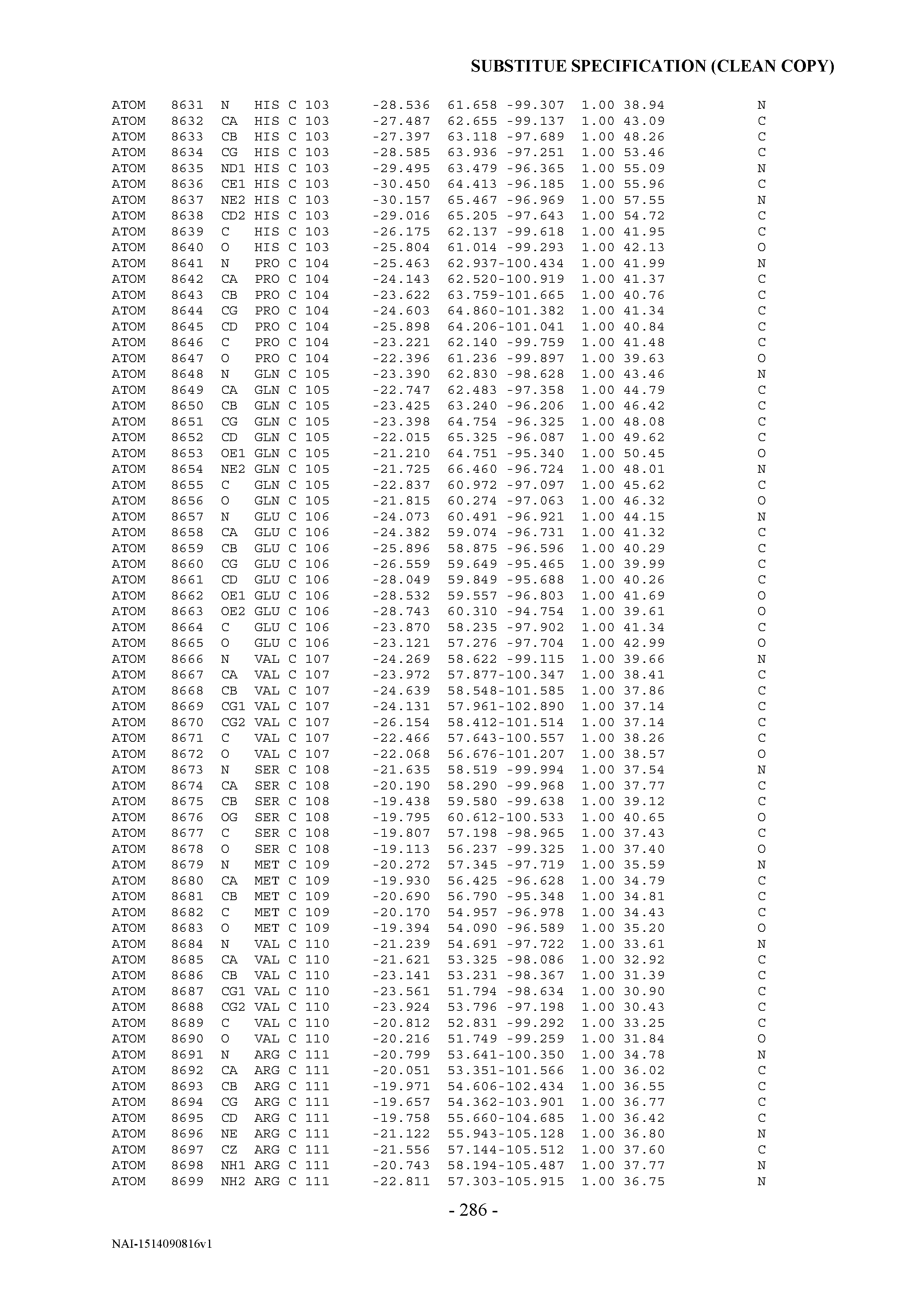
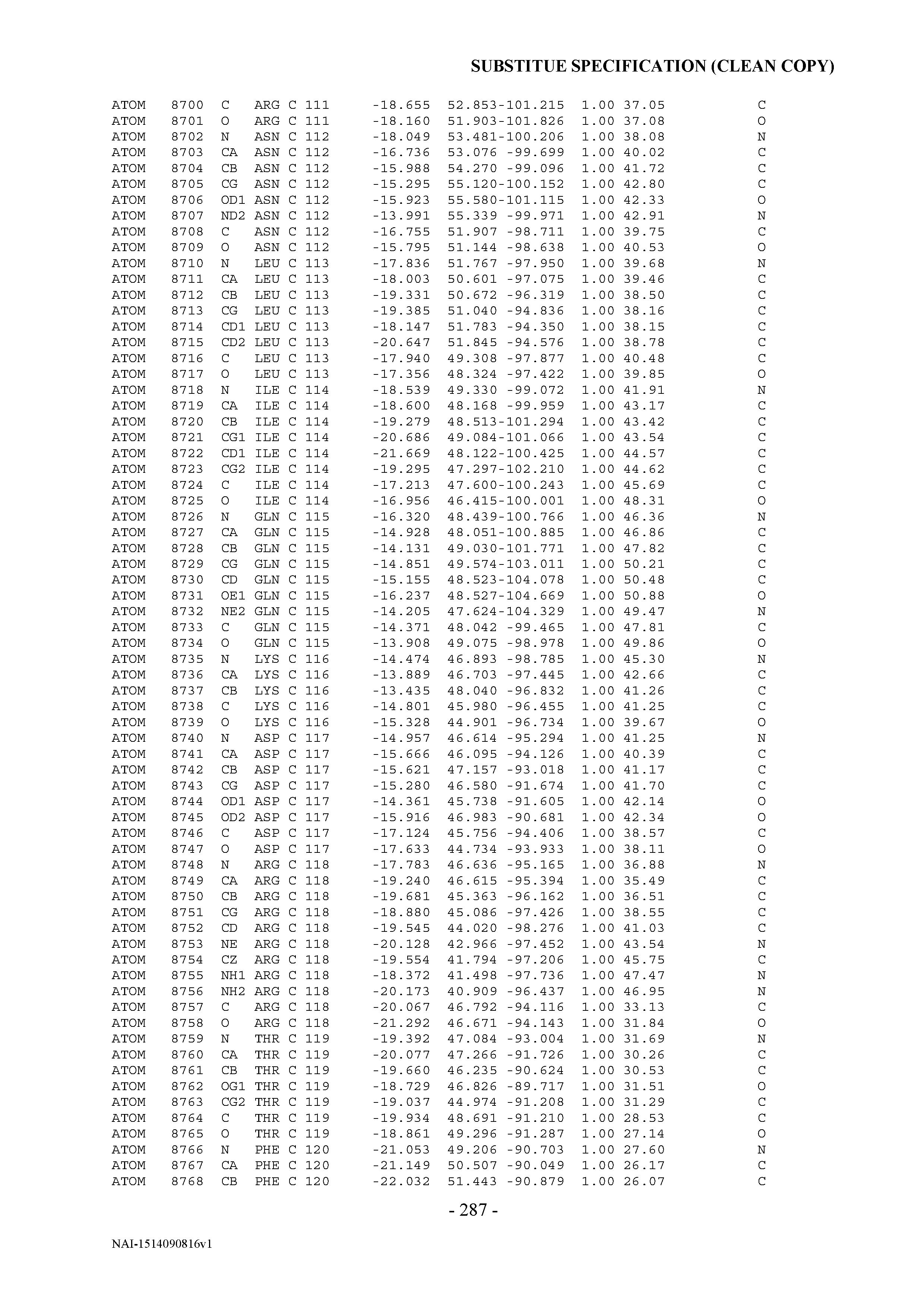


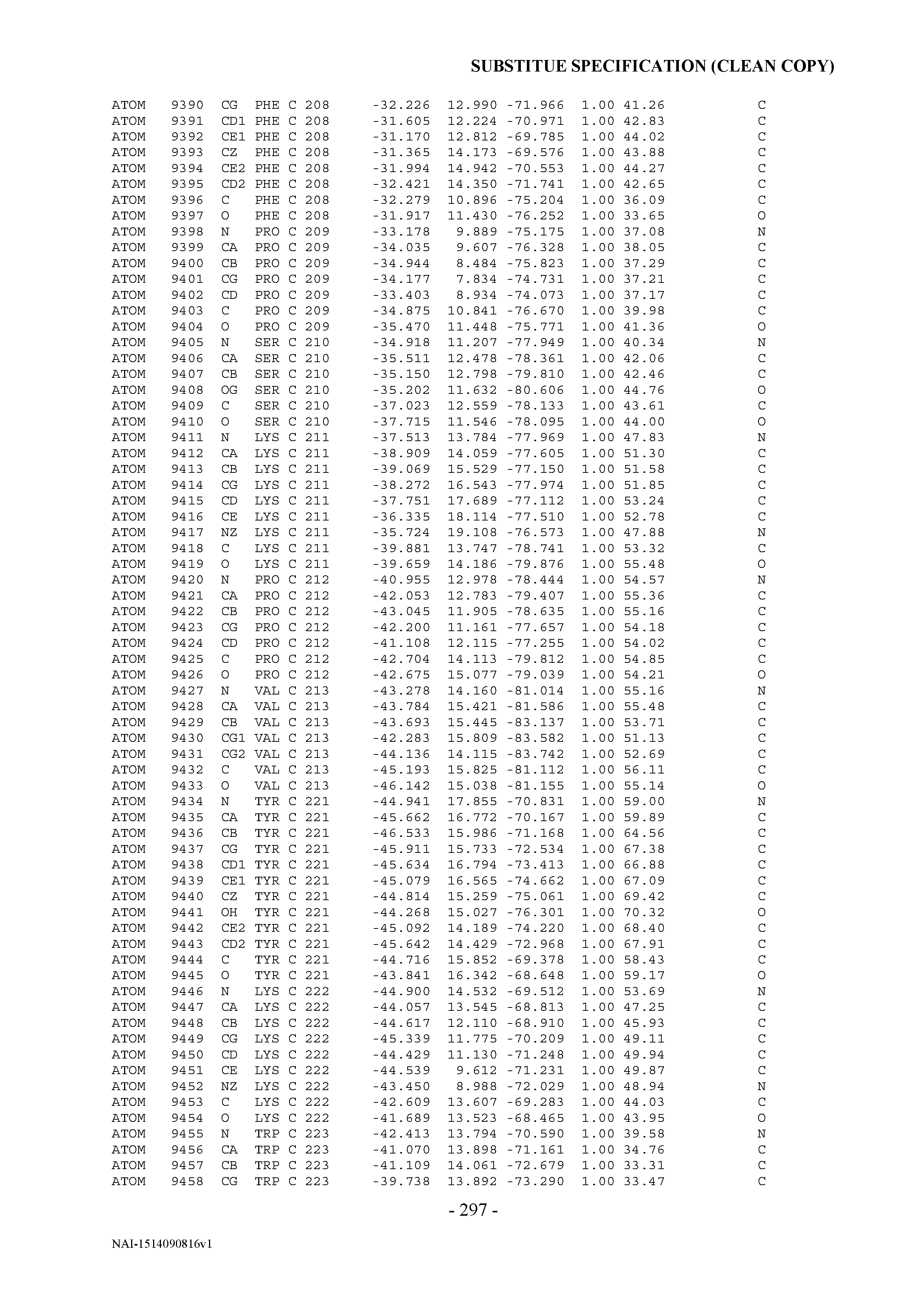


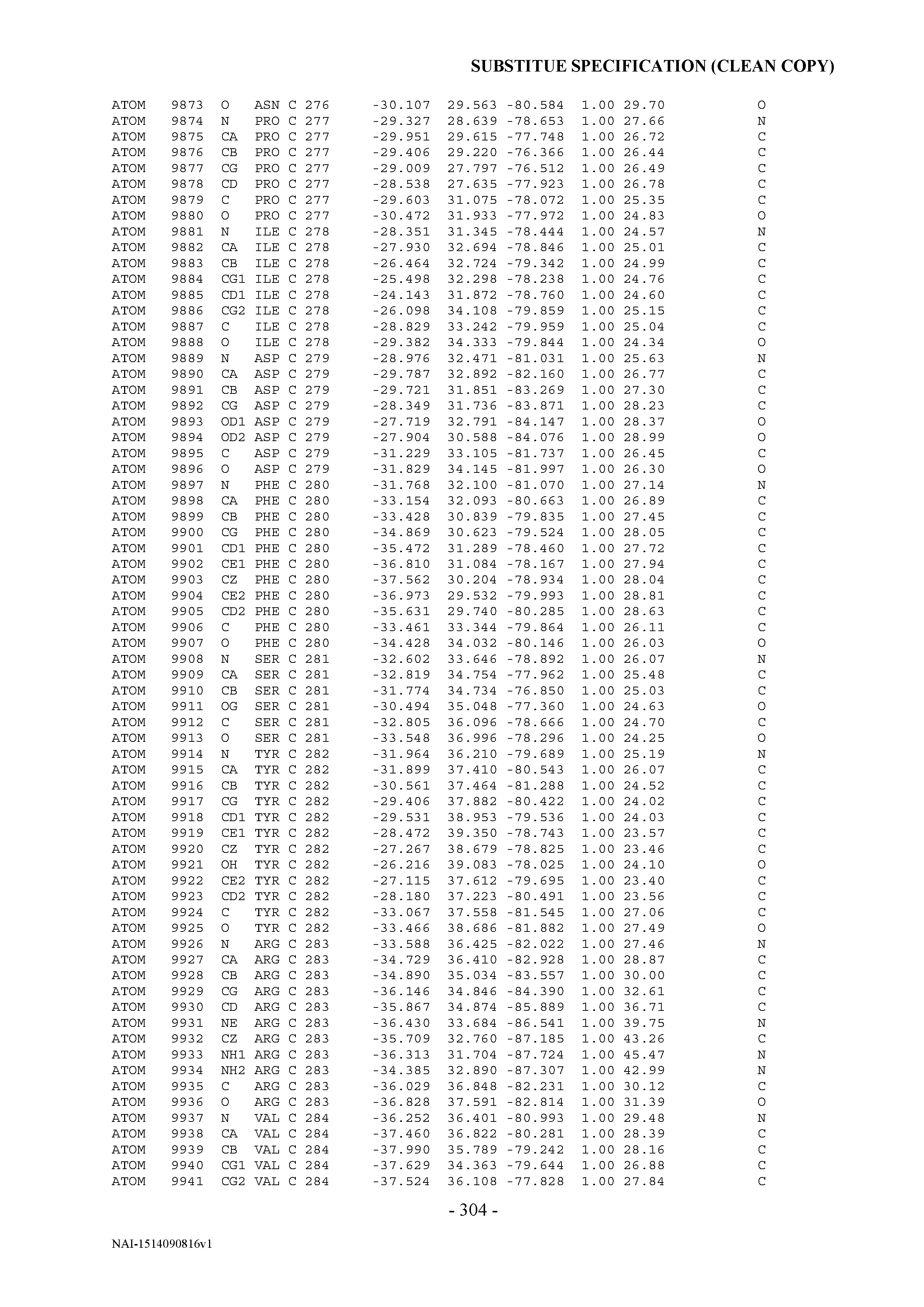


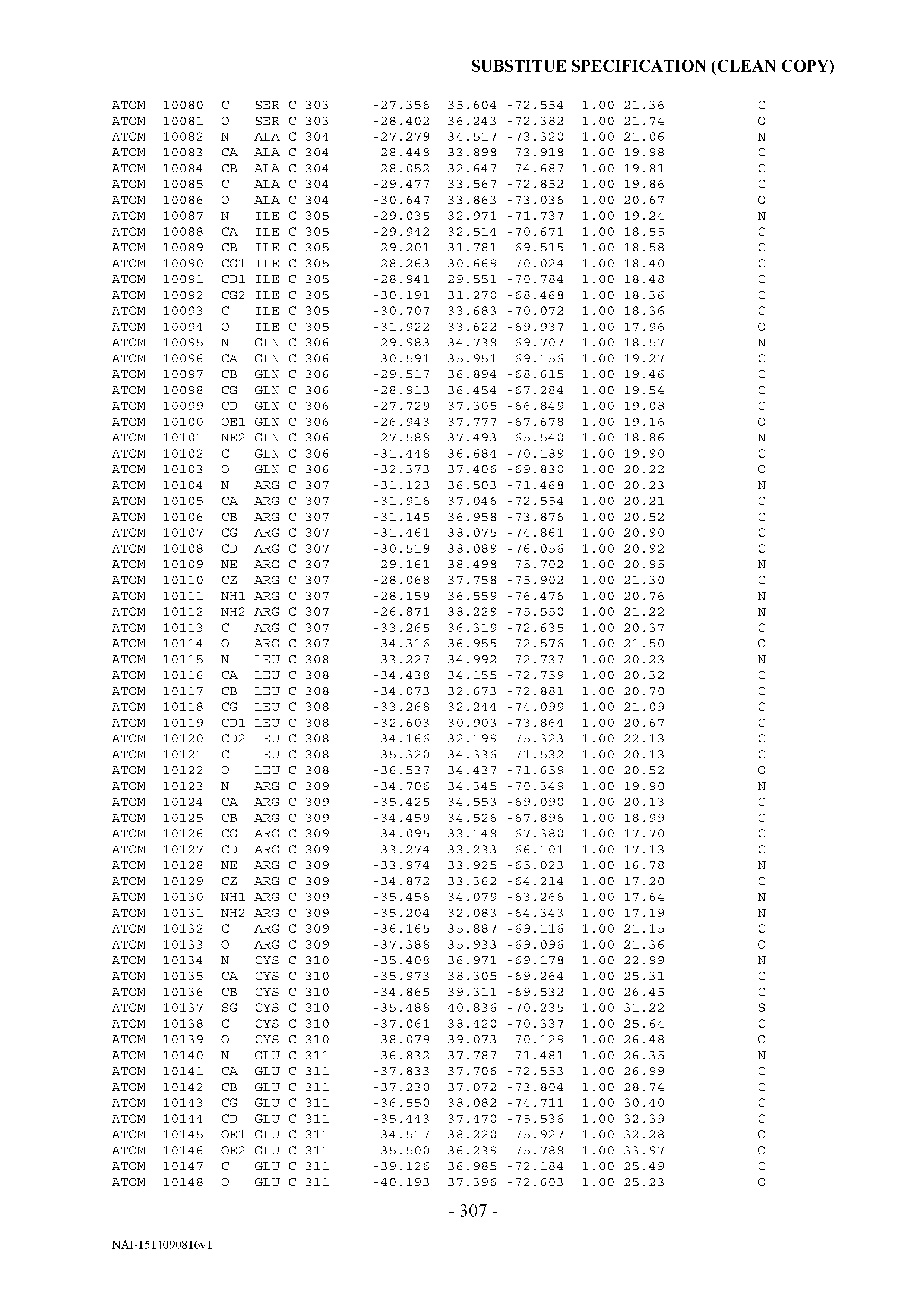


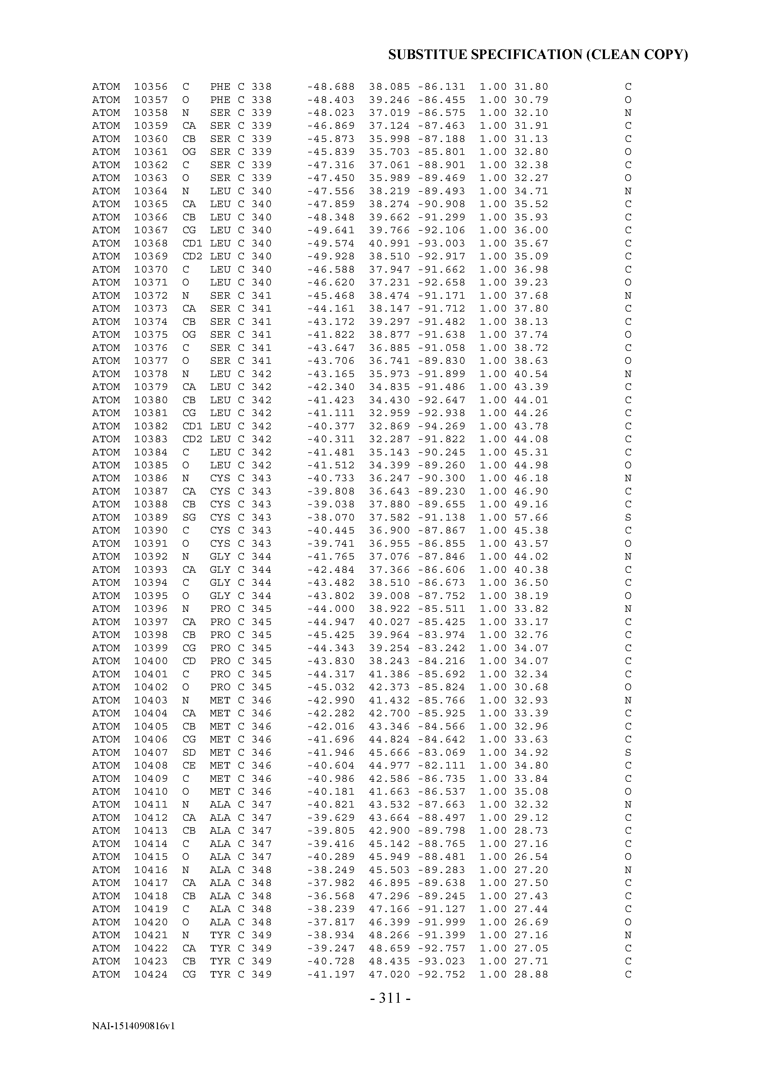


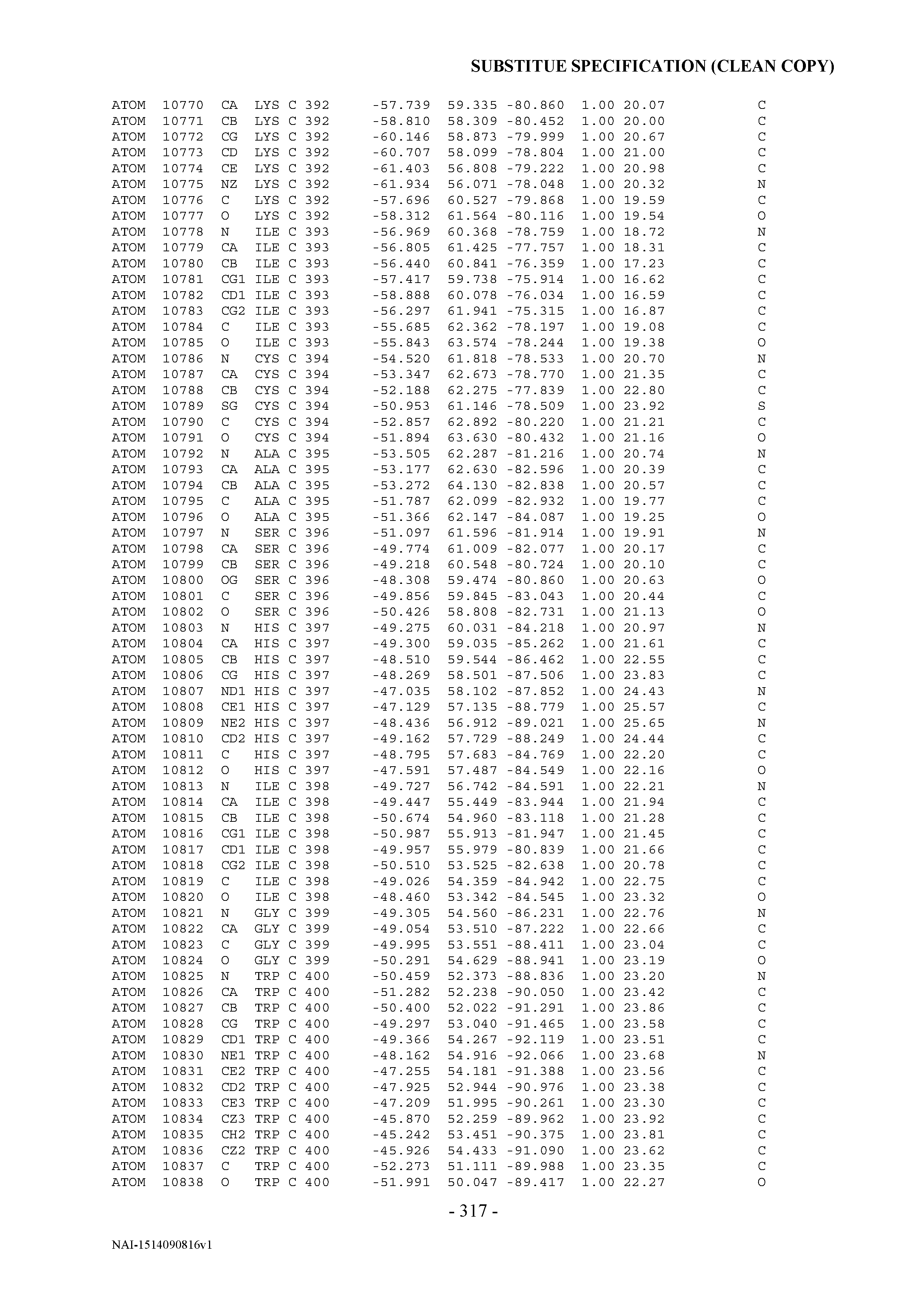


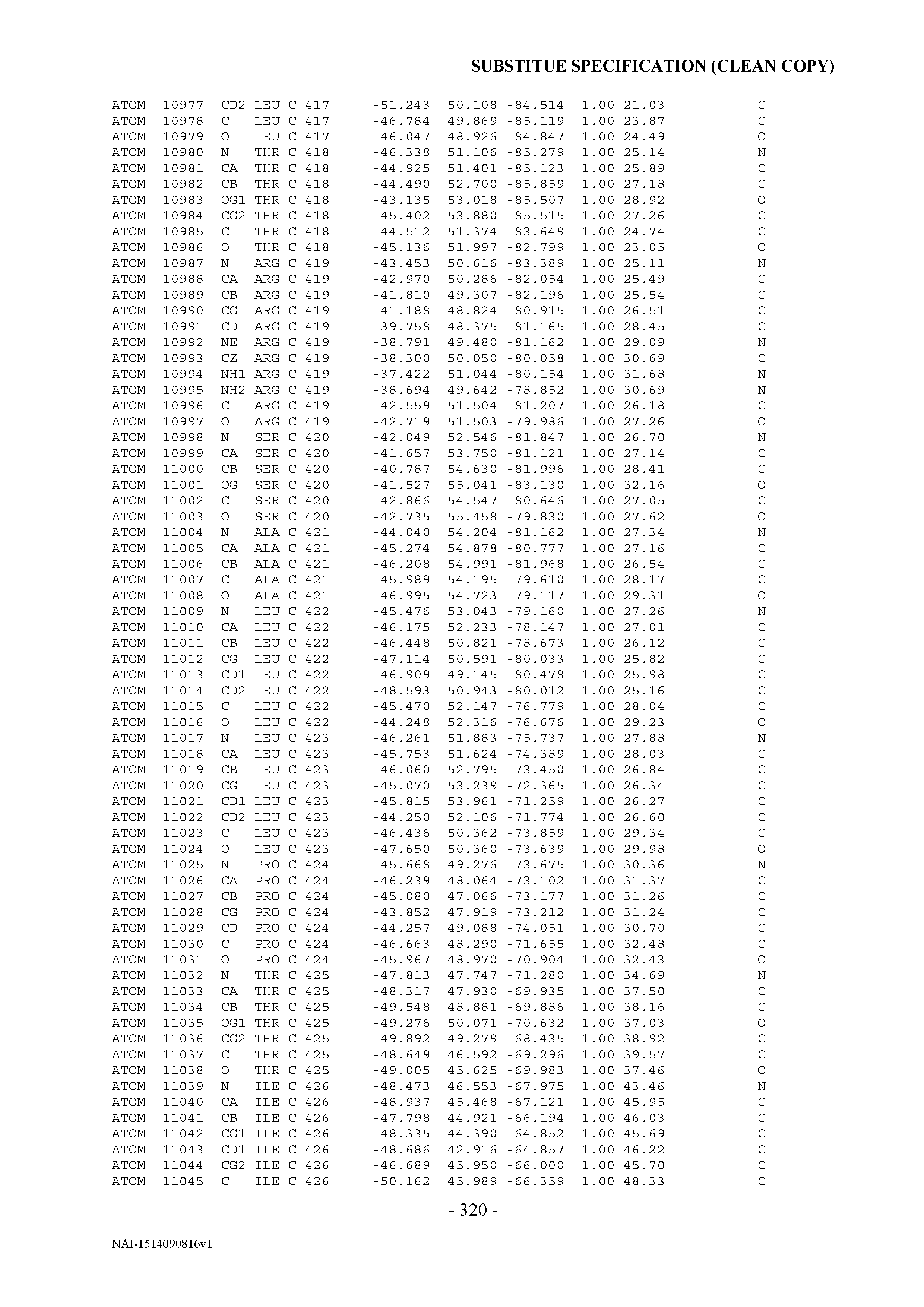

T00002


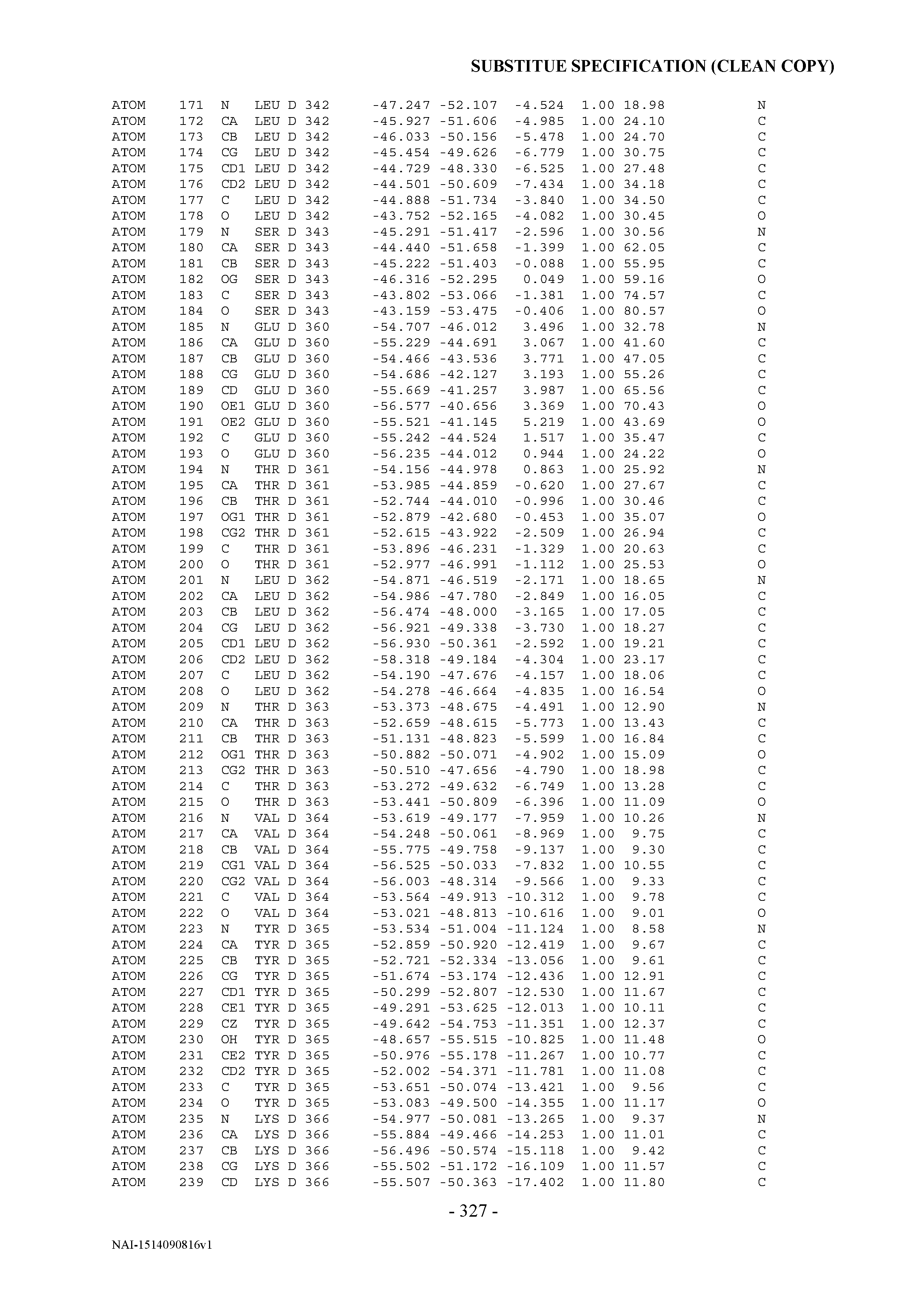
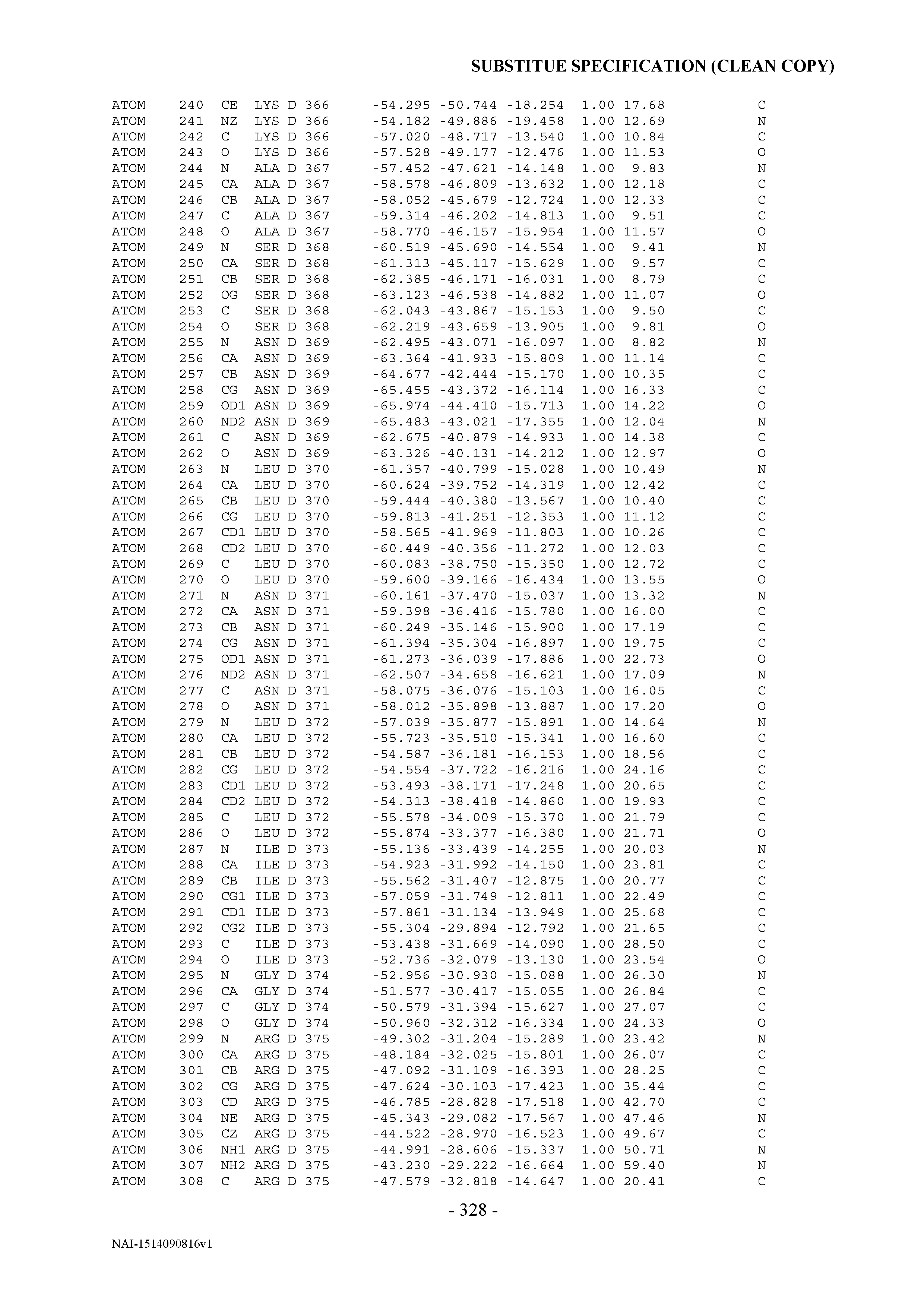
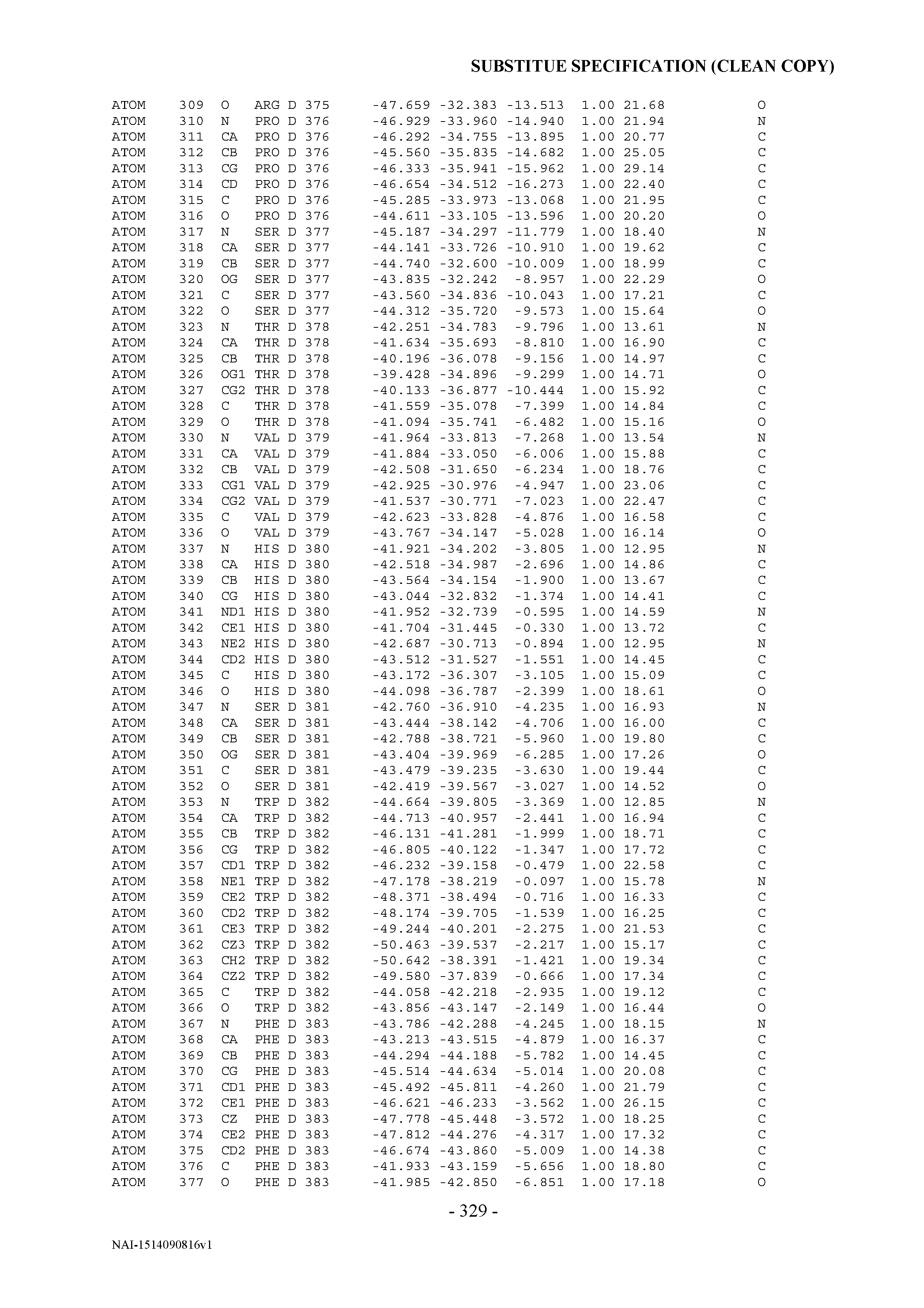


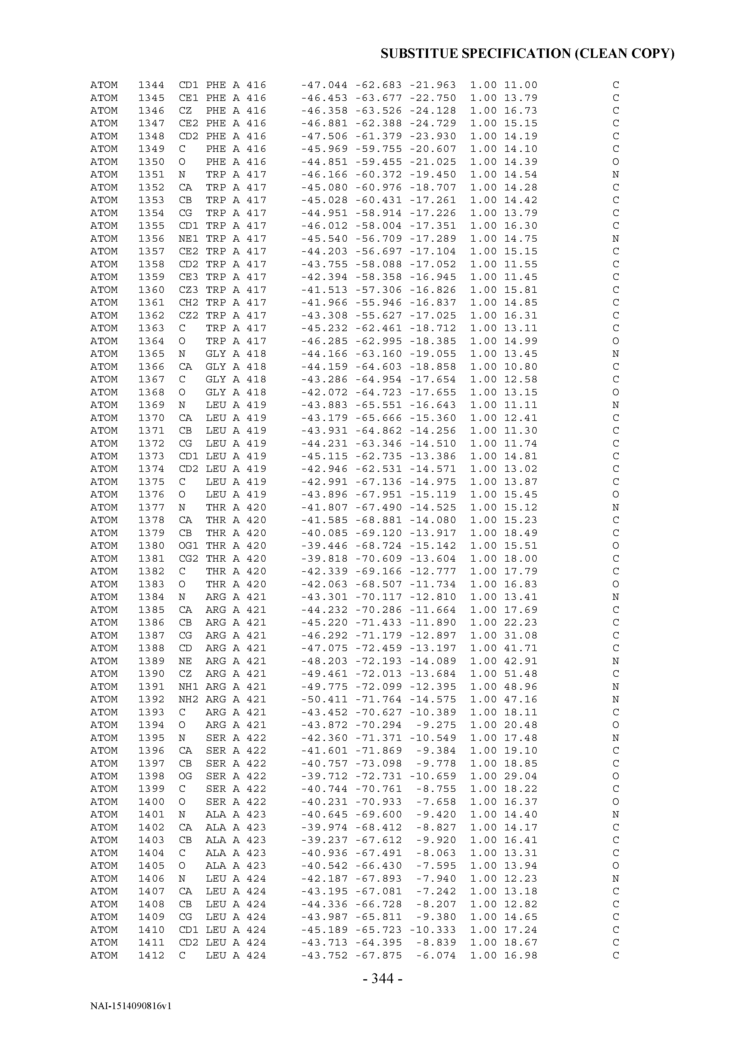


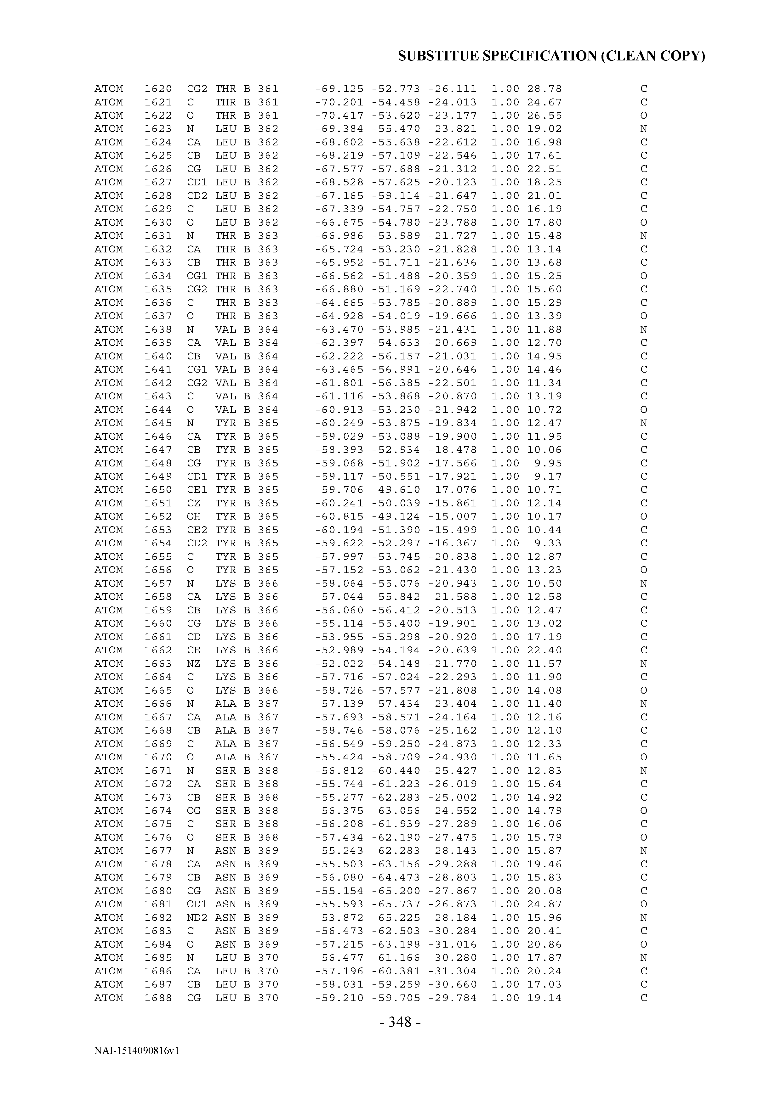


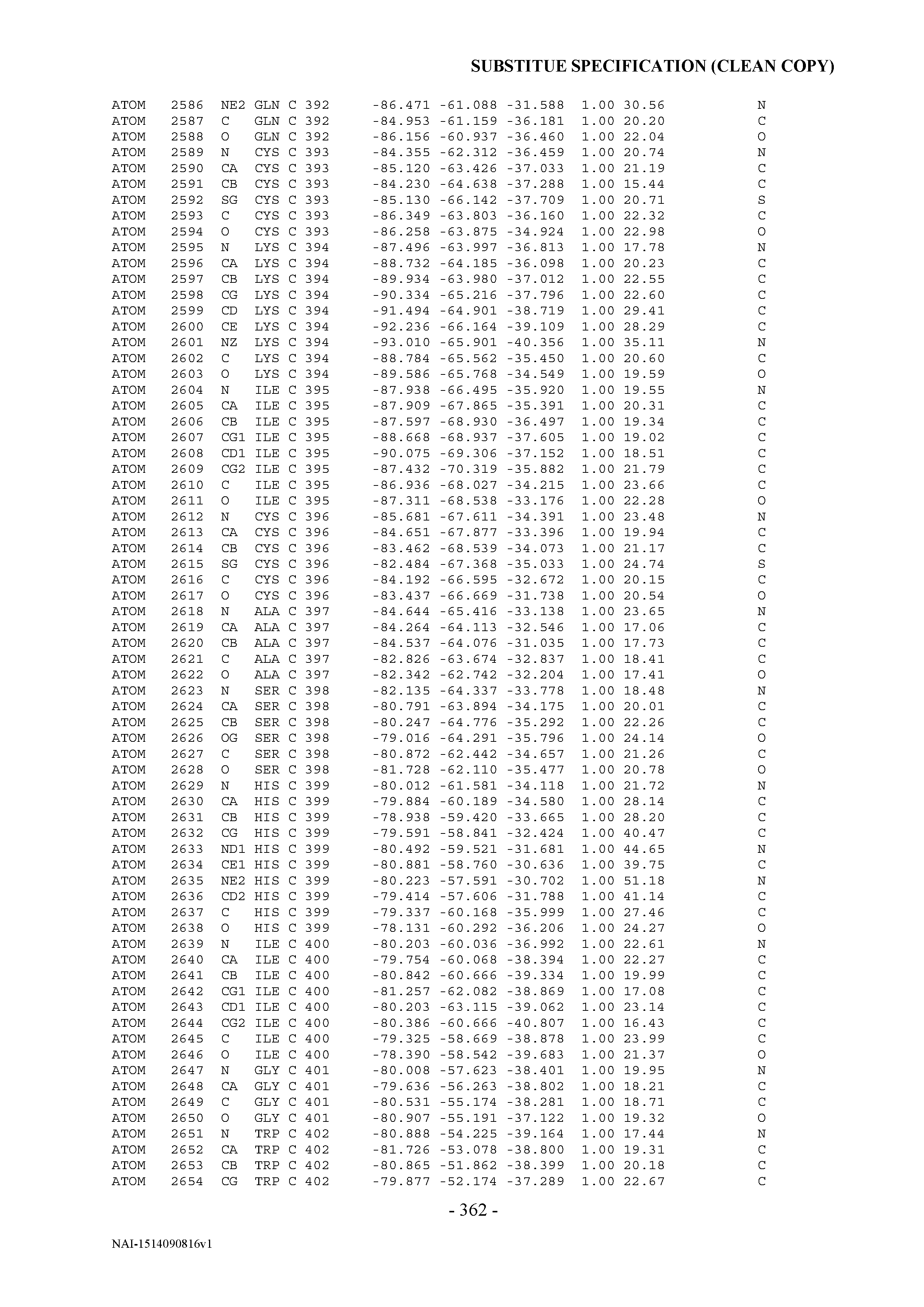
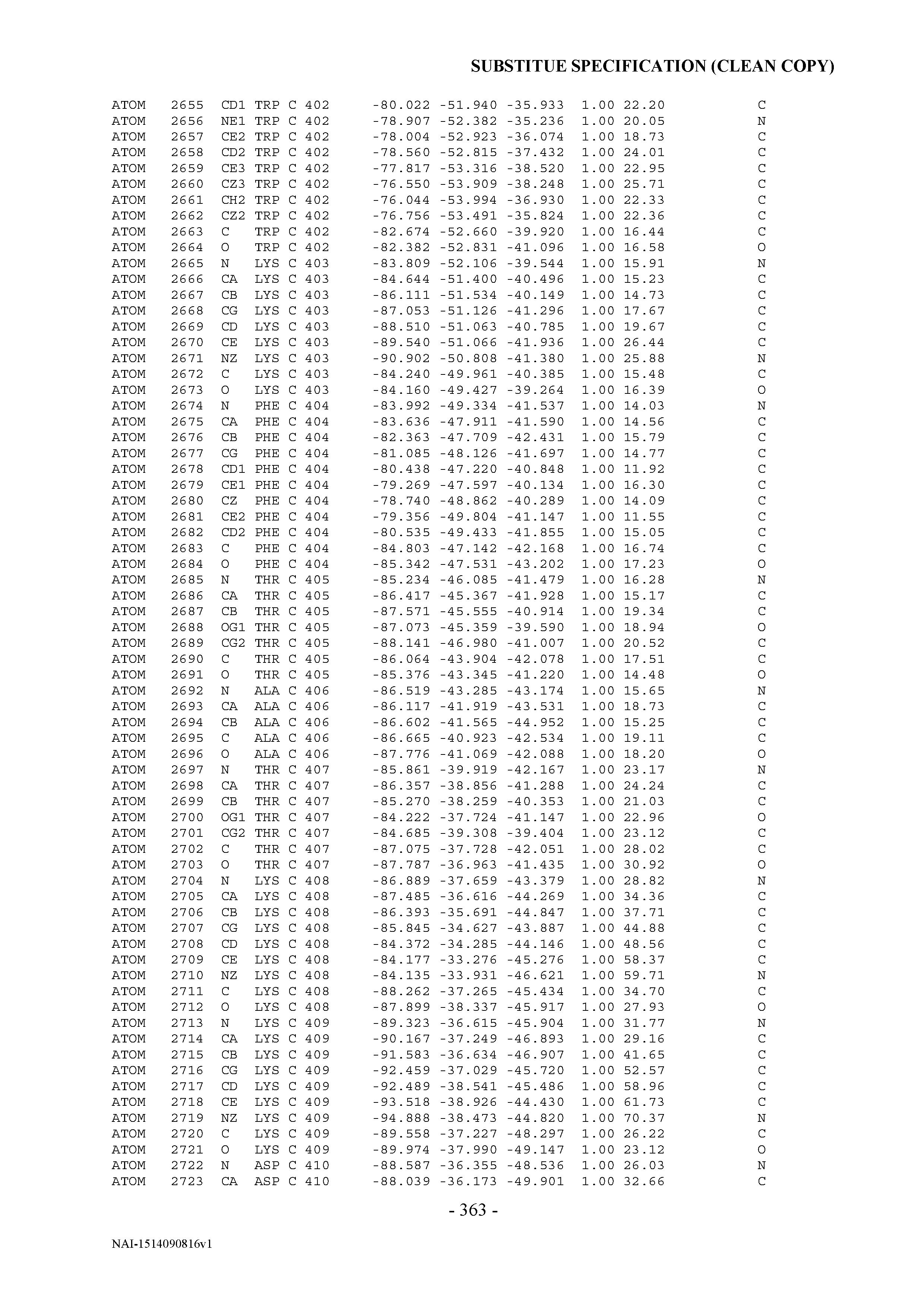


T00003
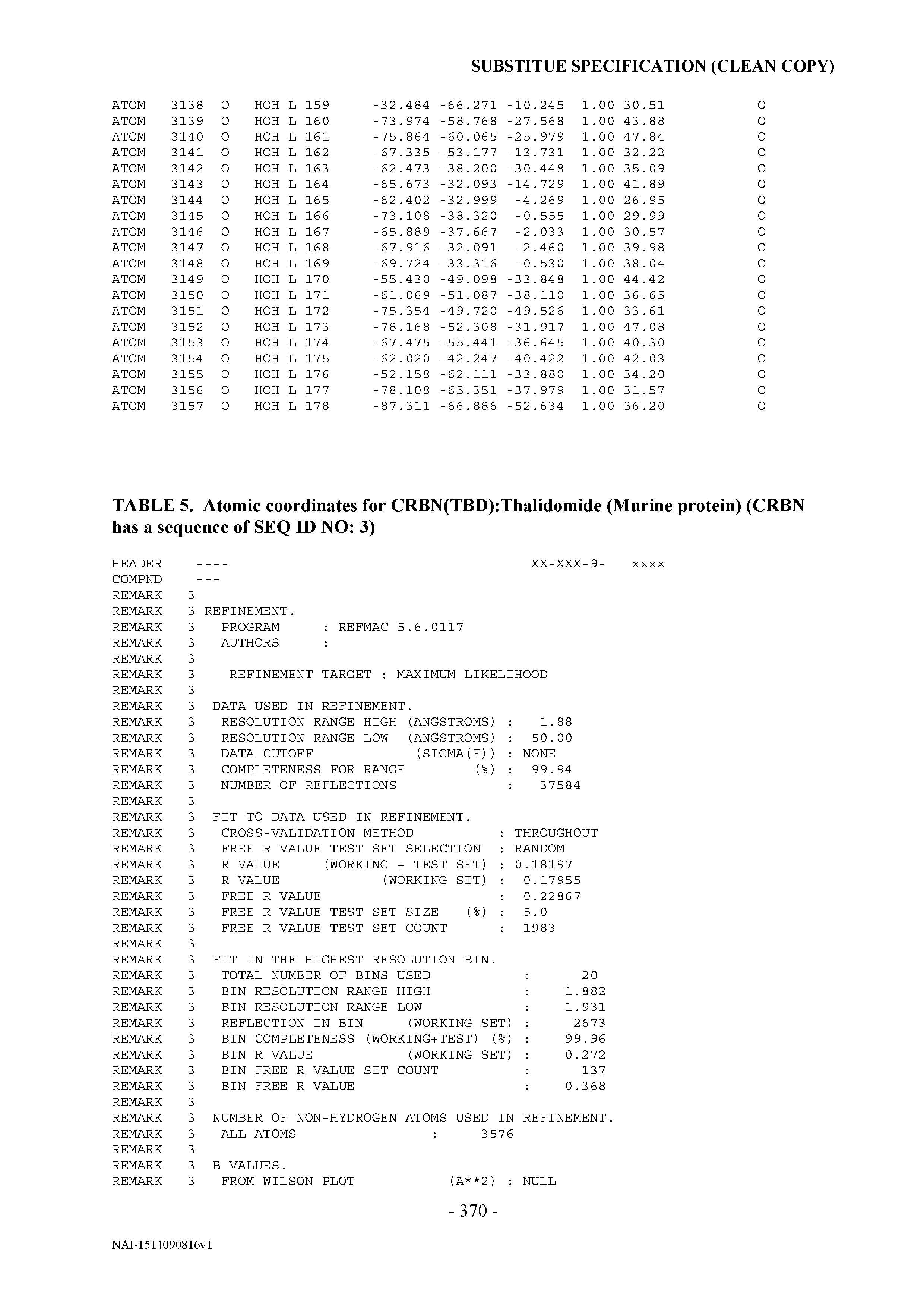


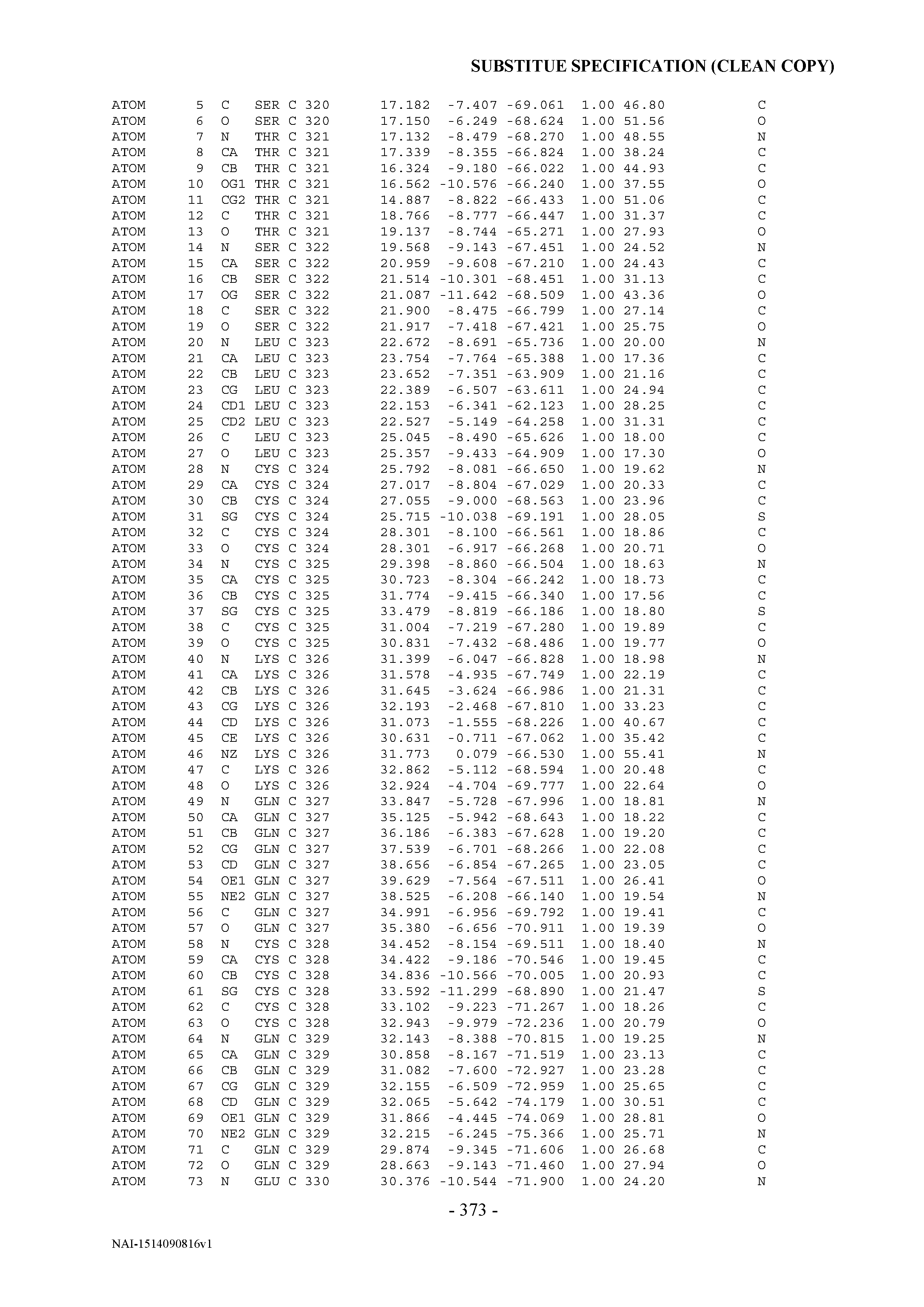

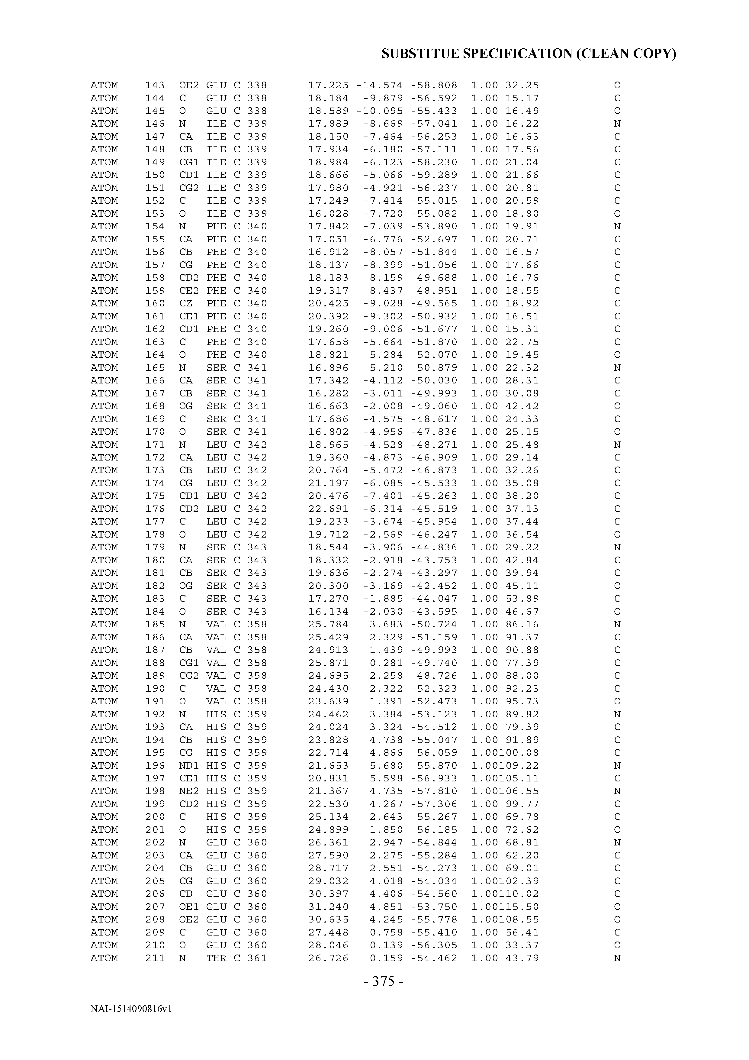
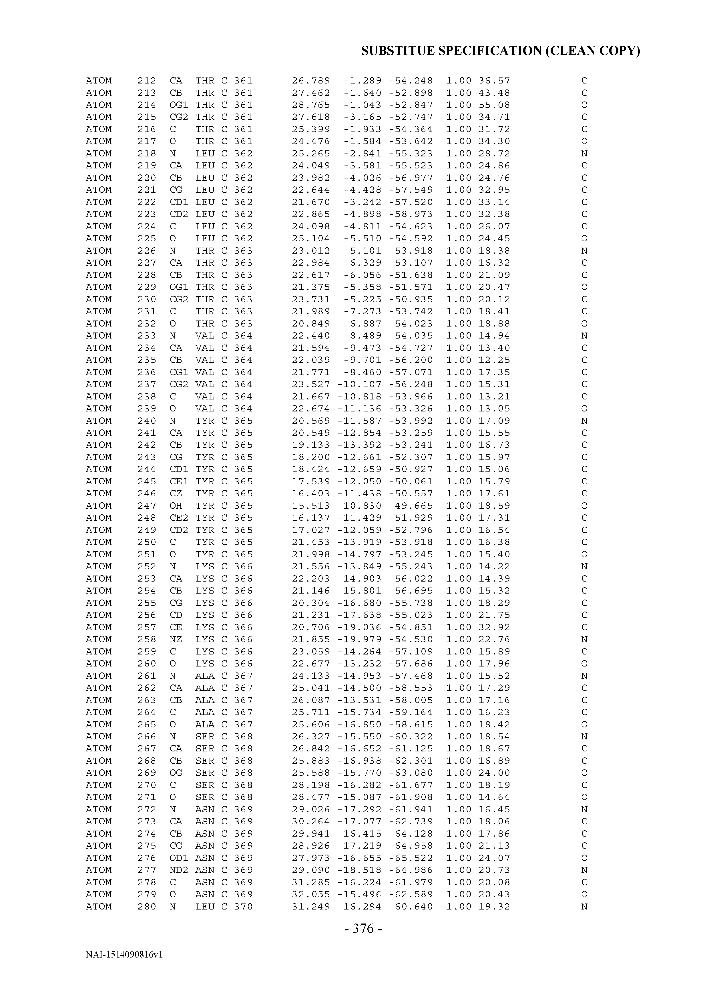

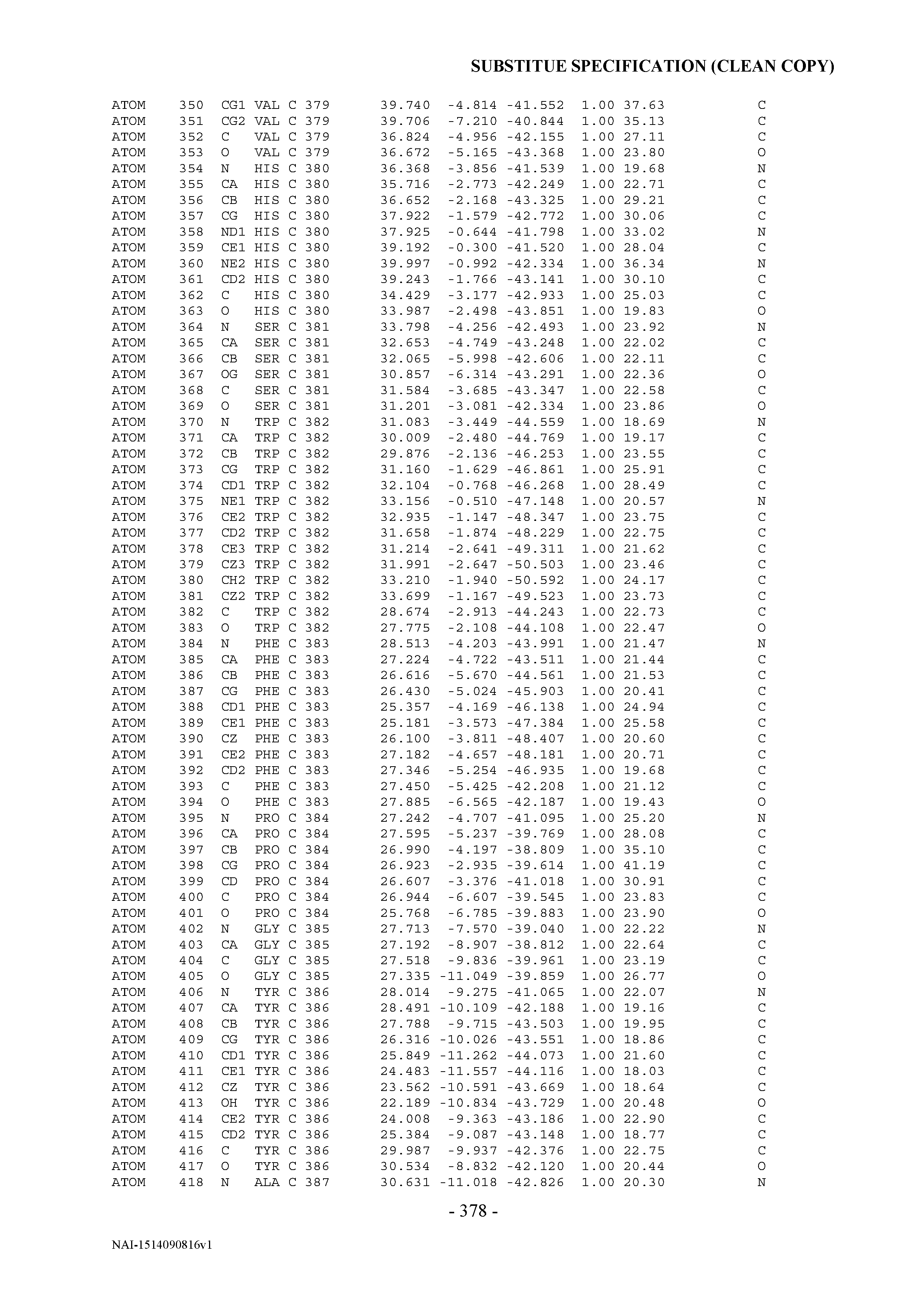


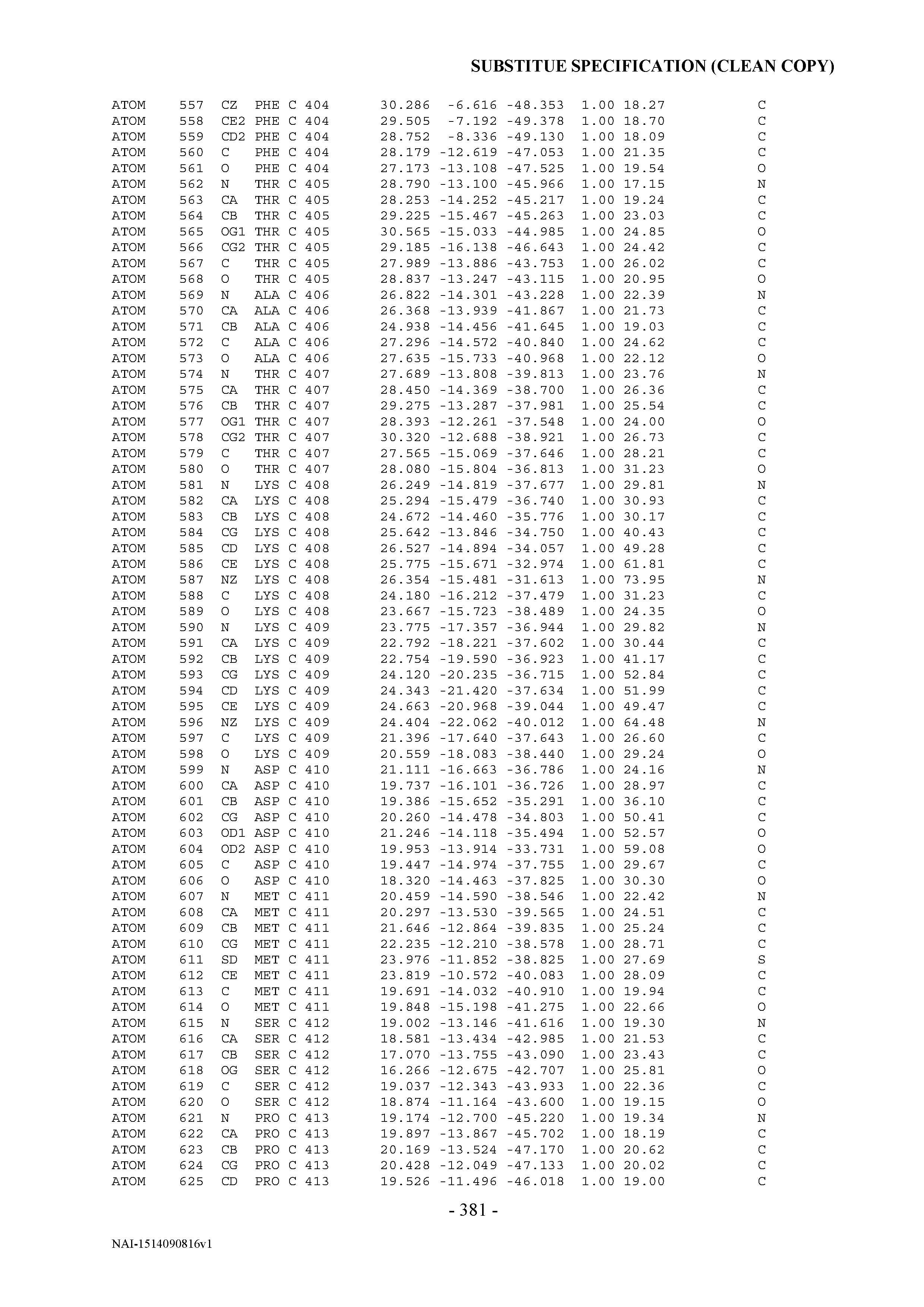

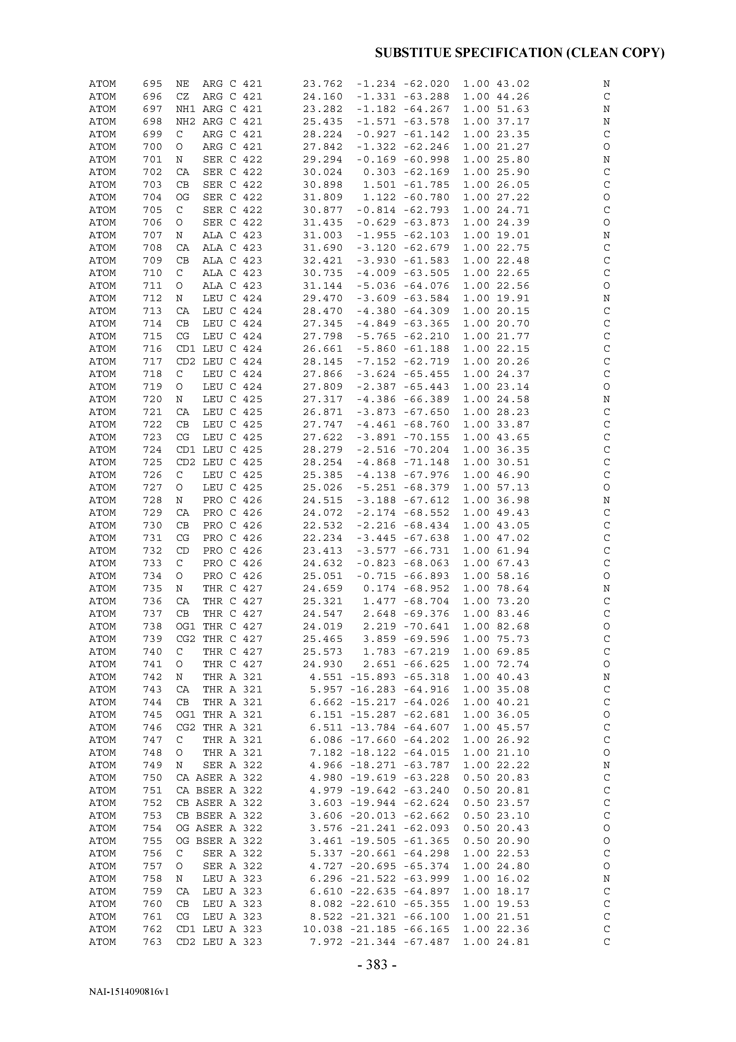
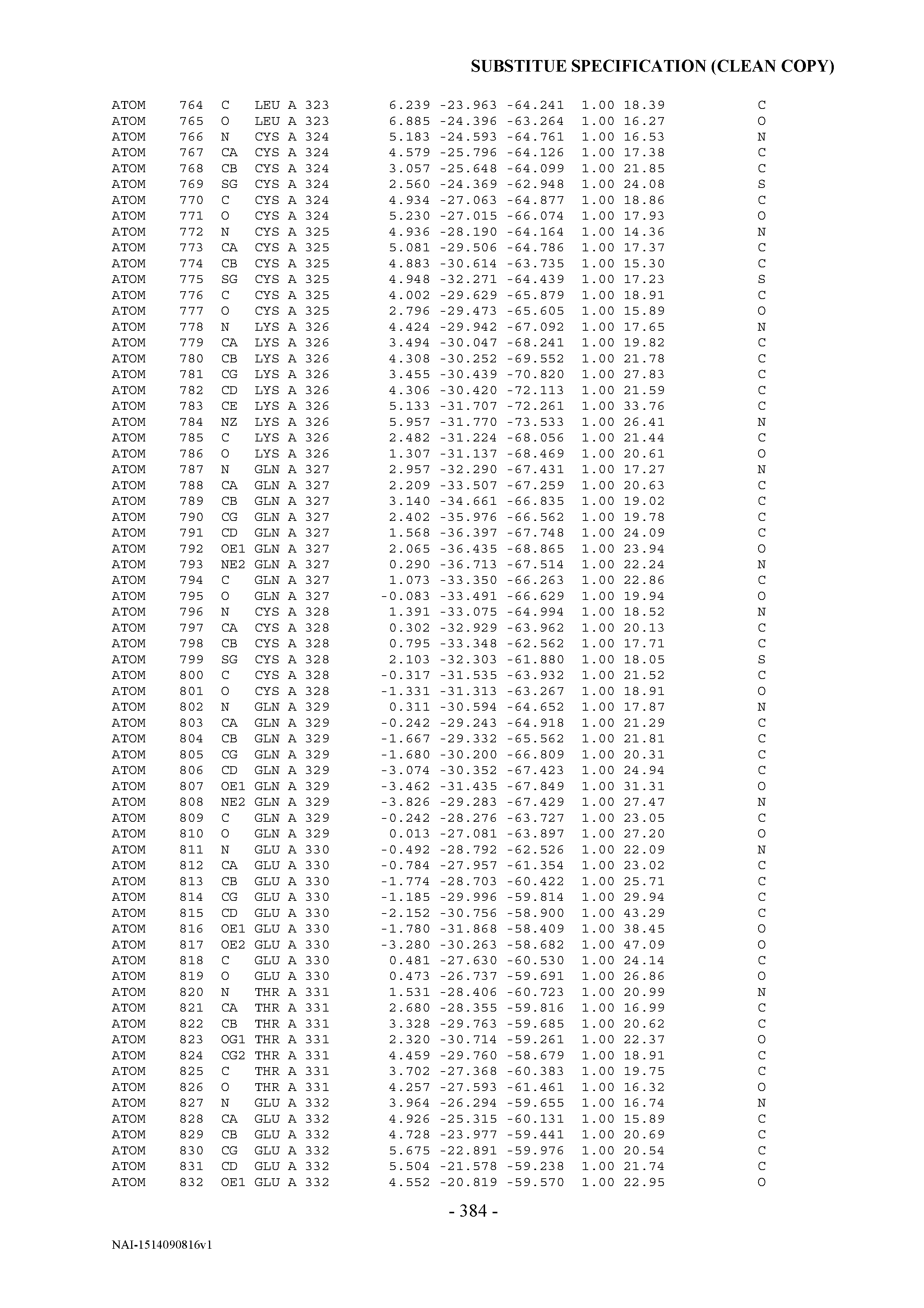


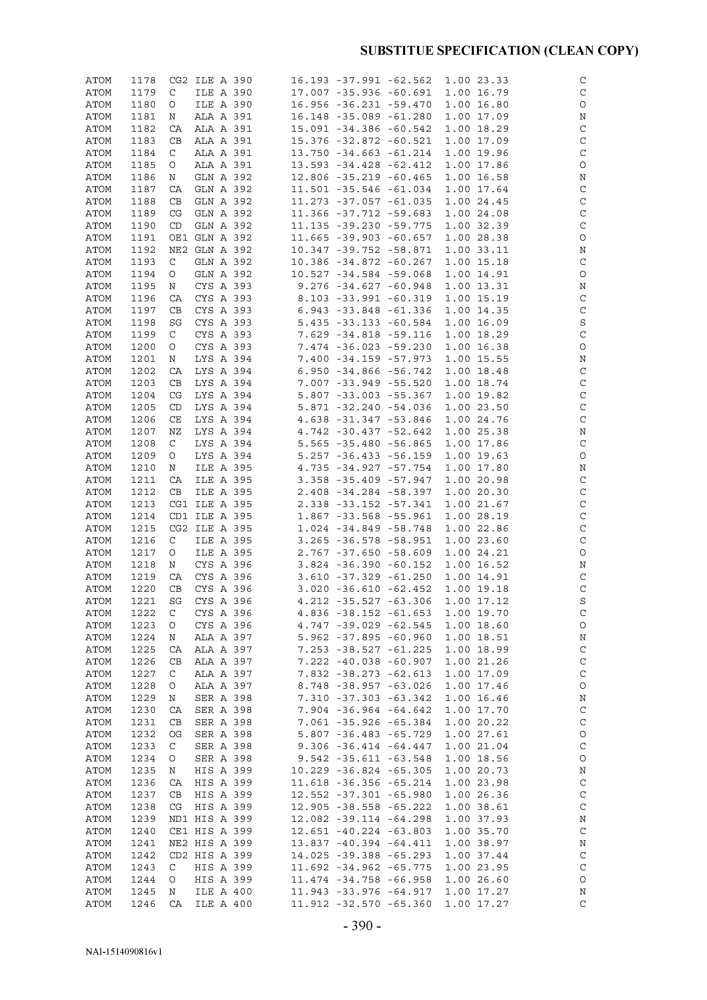
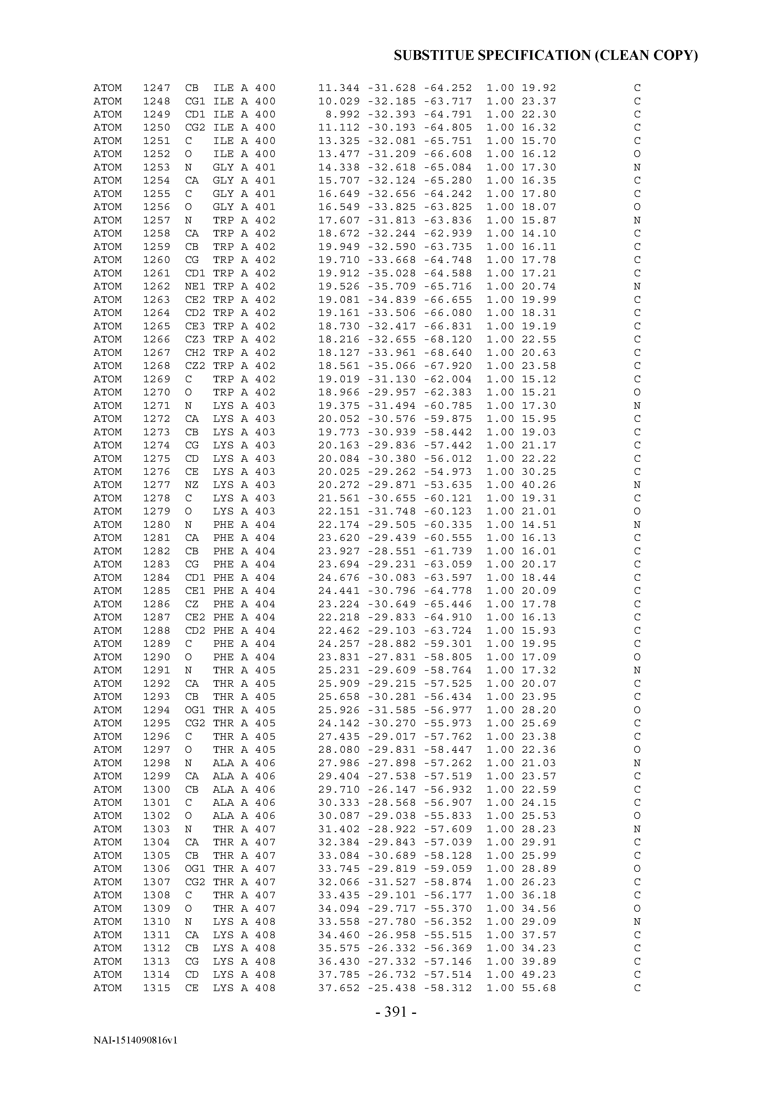


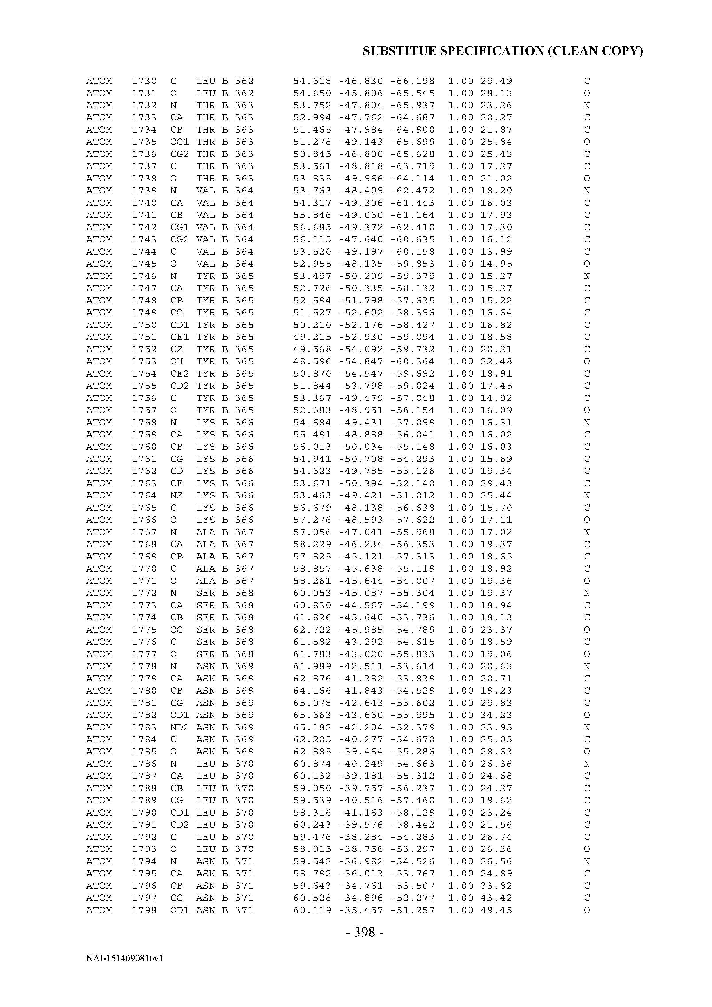


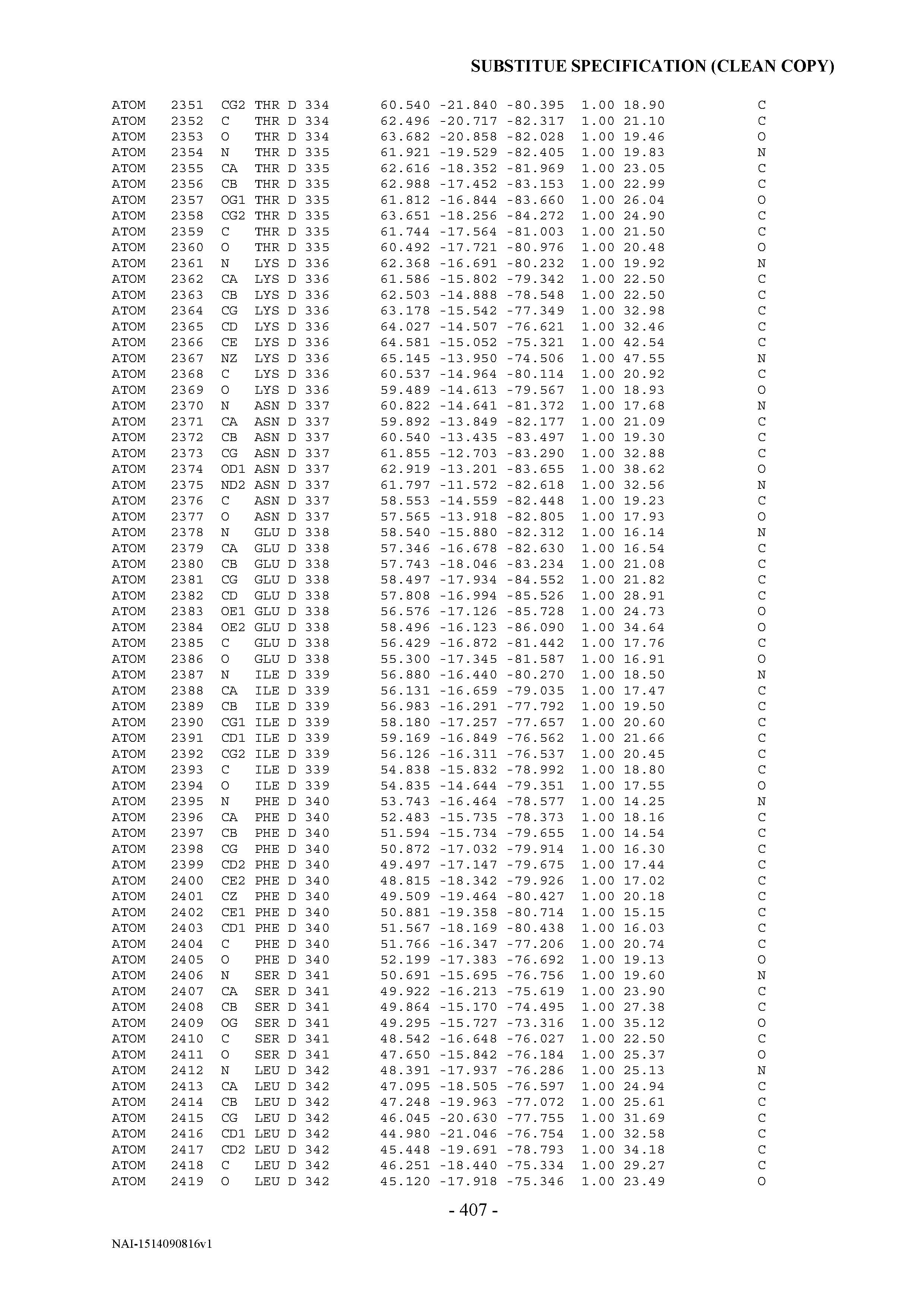


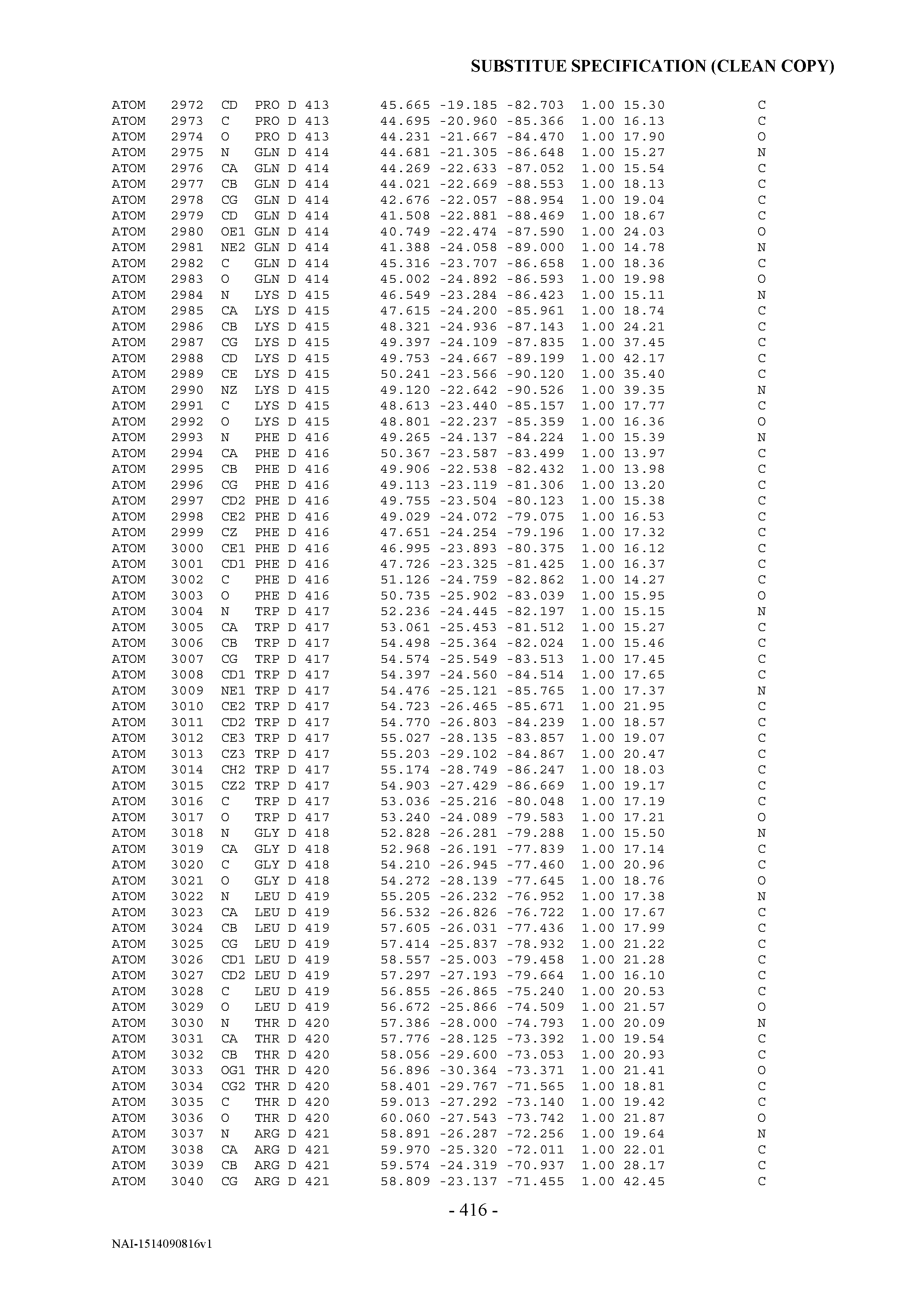
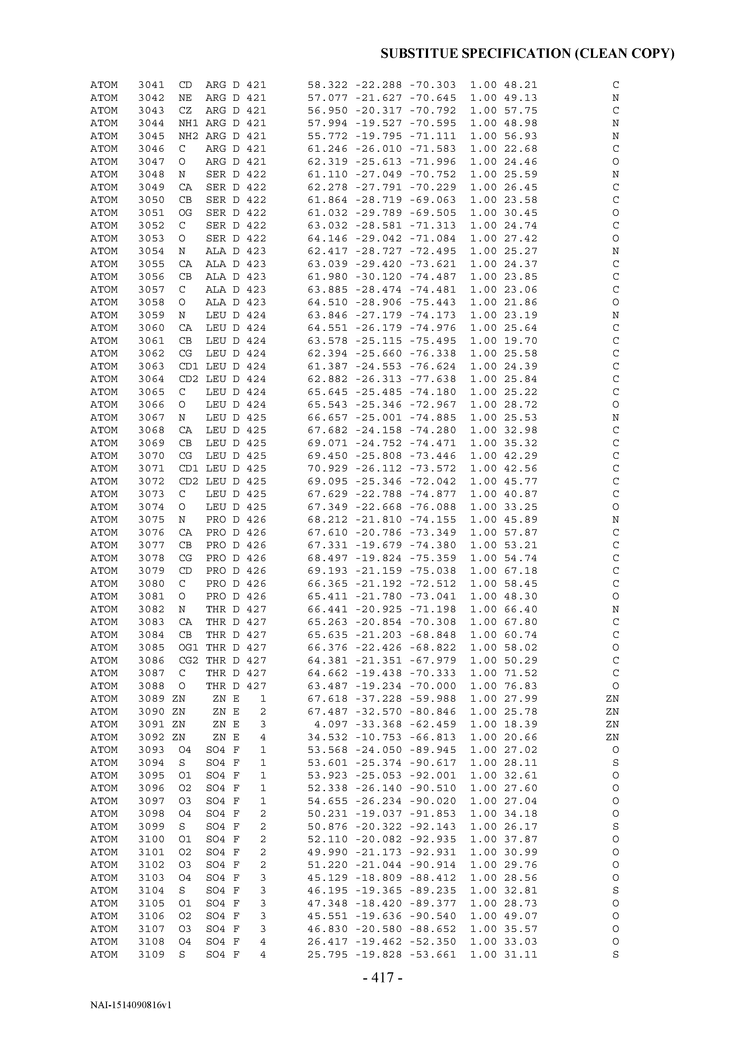


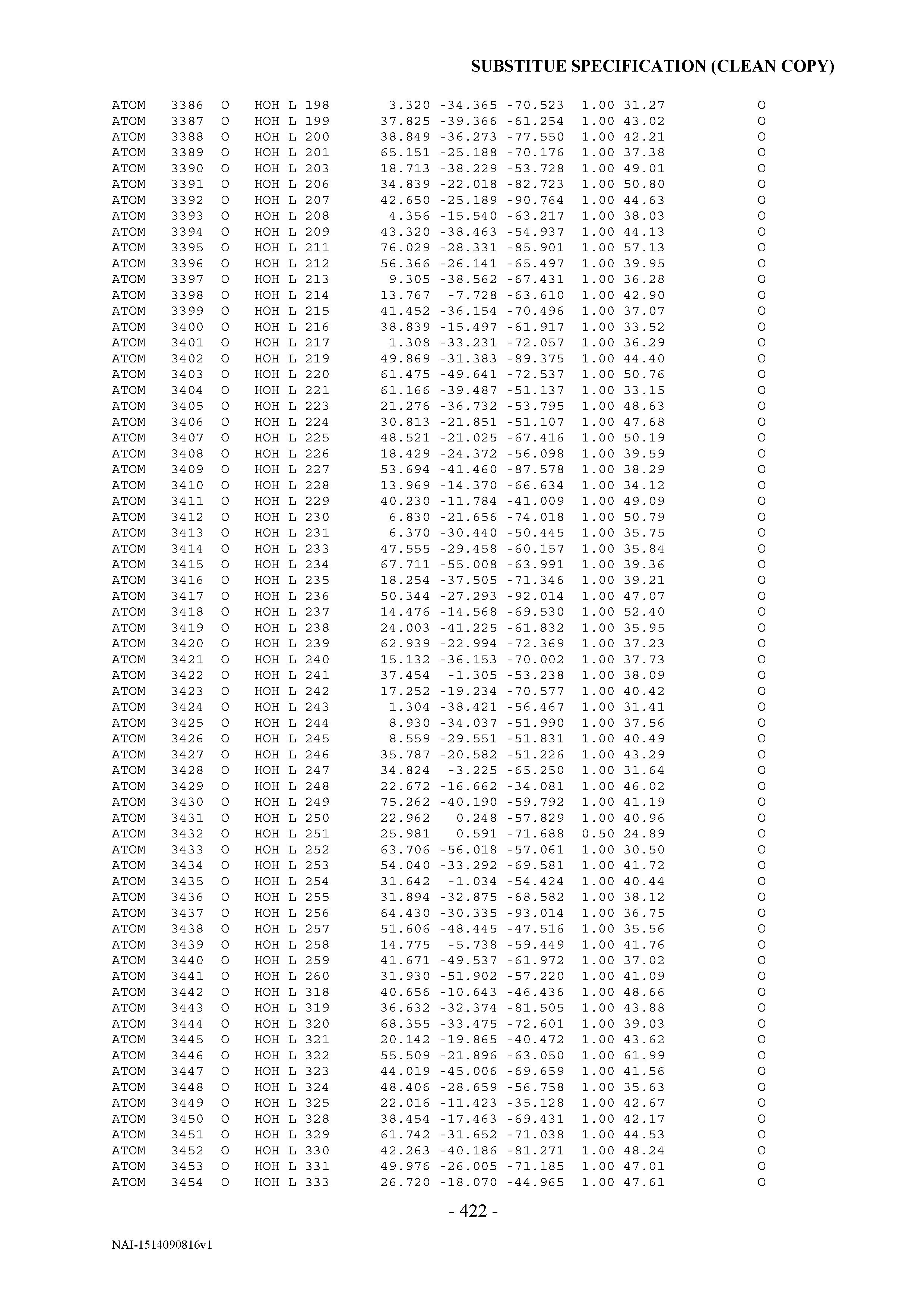


T00004
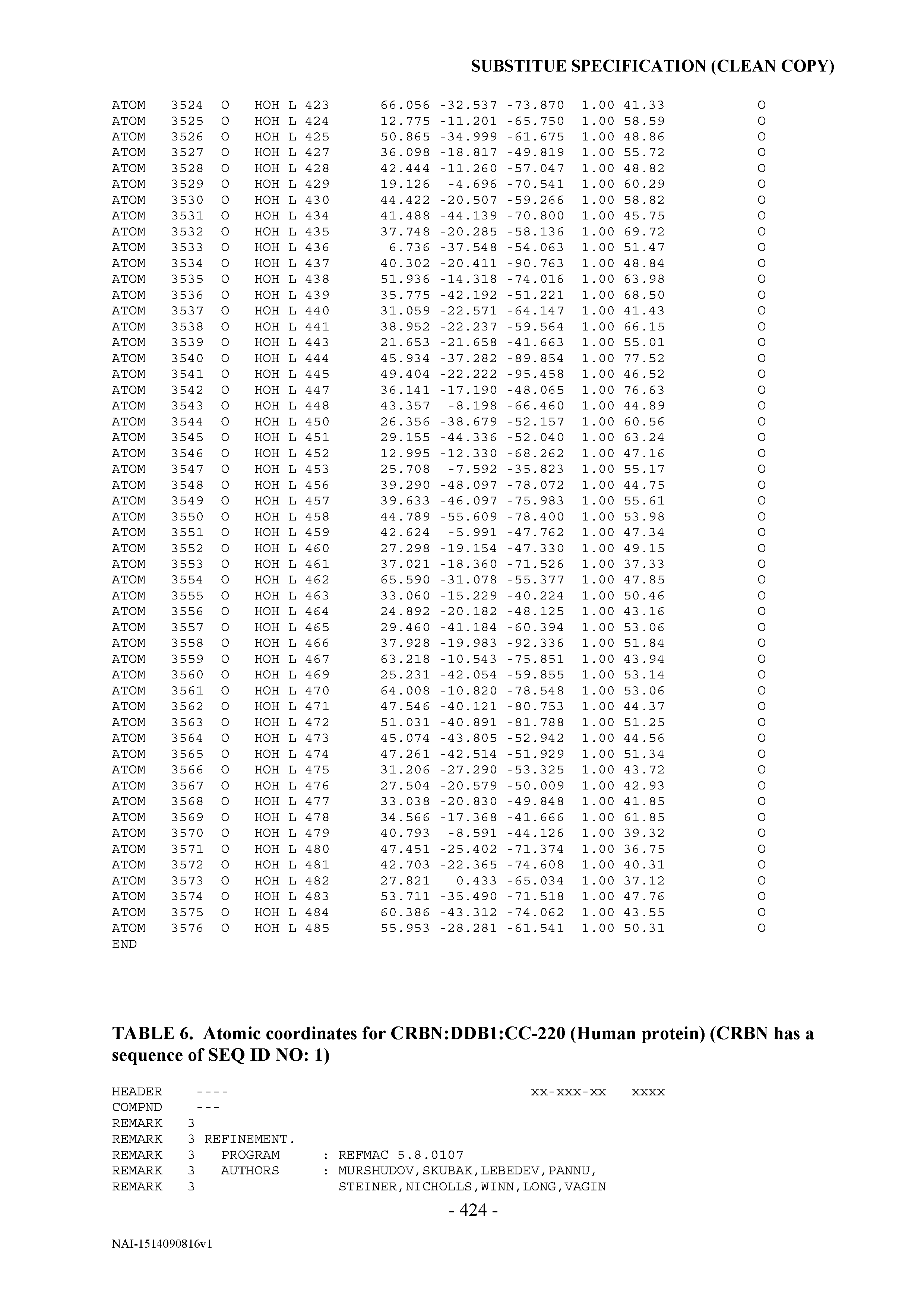

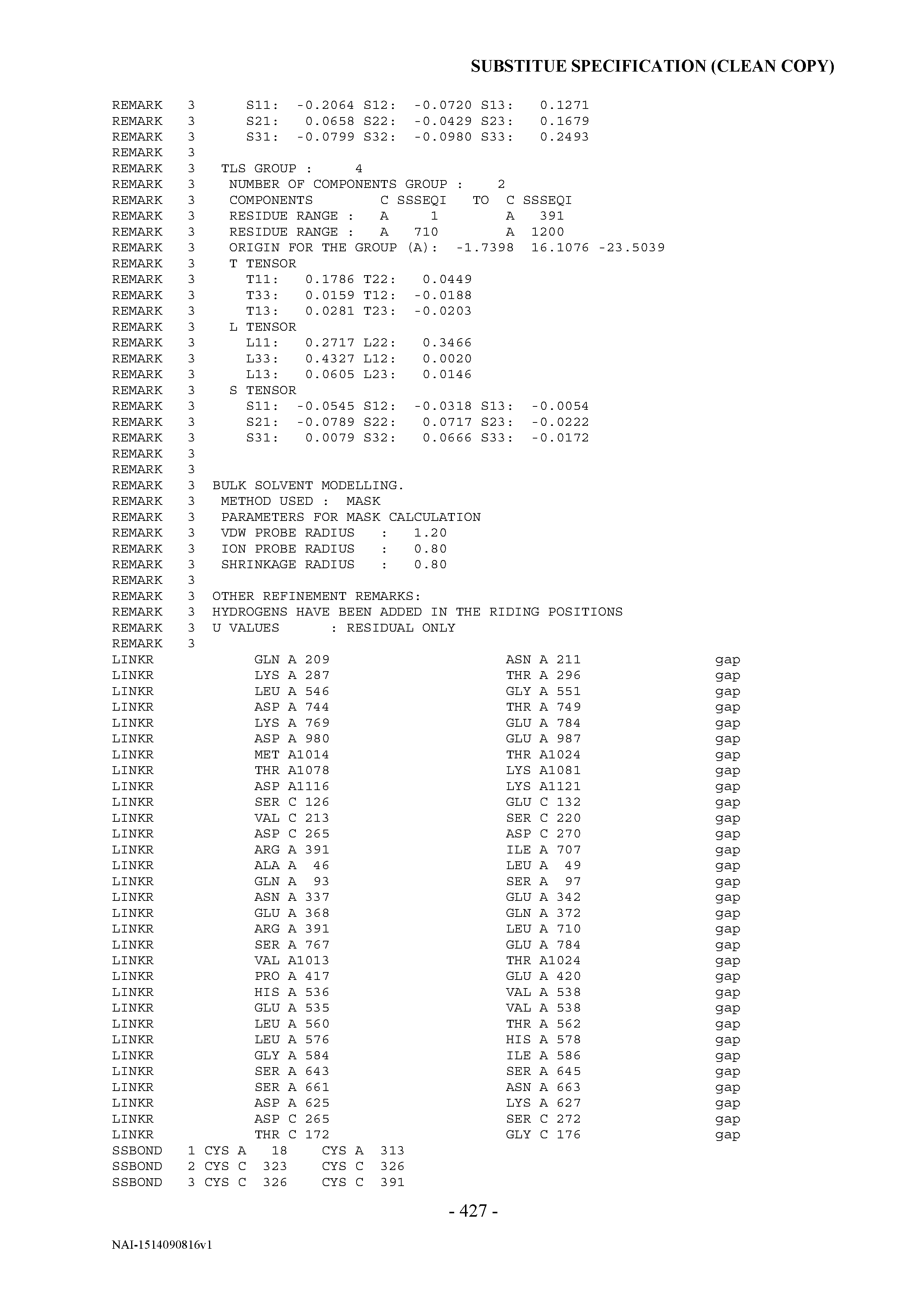


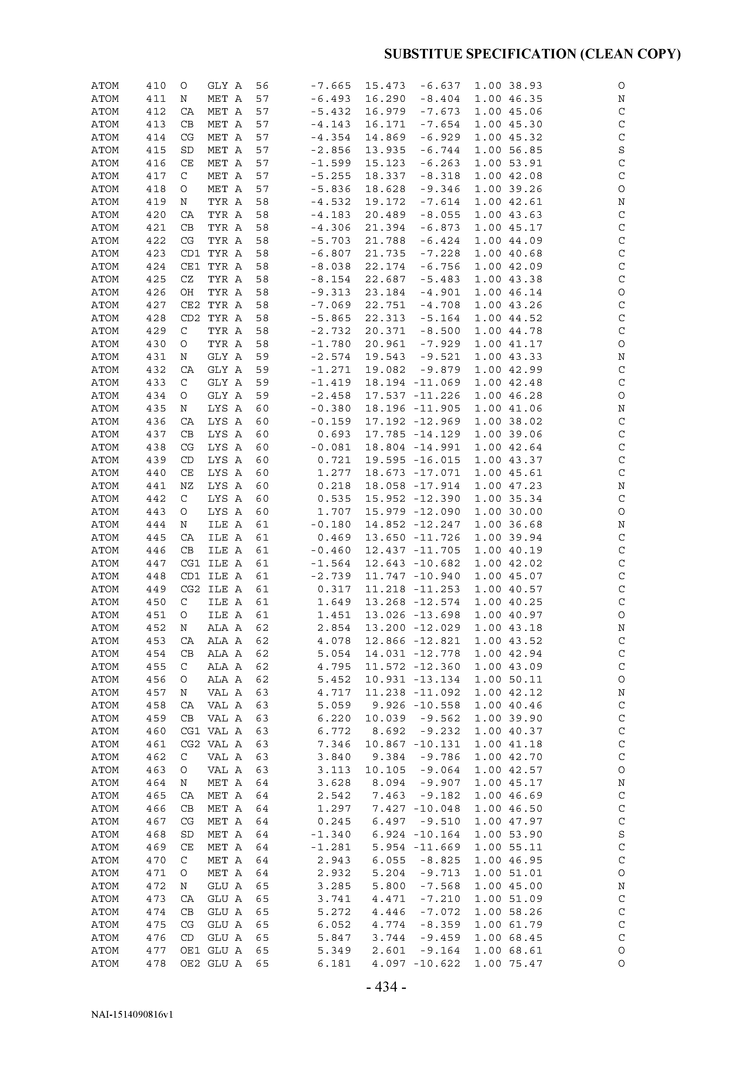


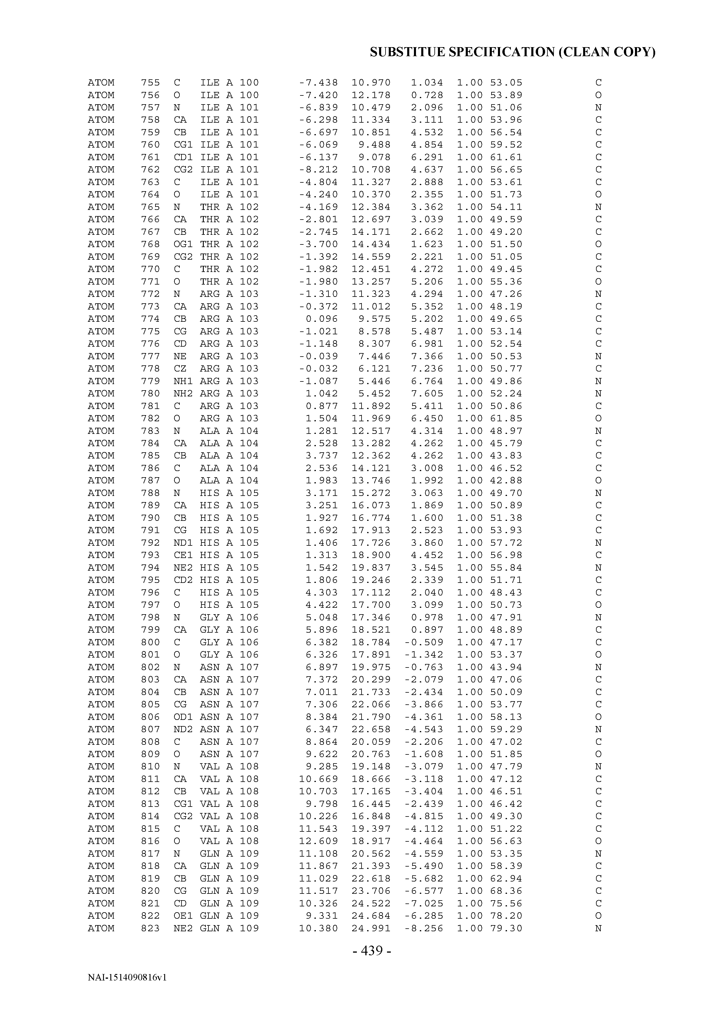


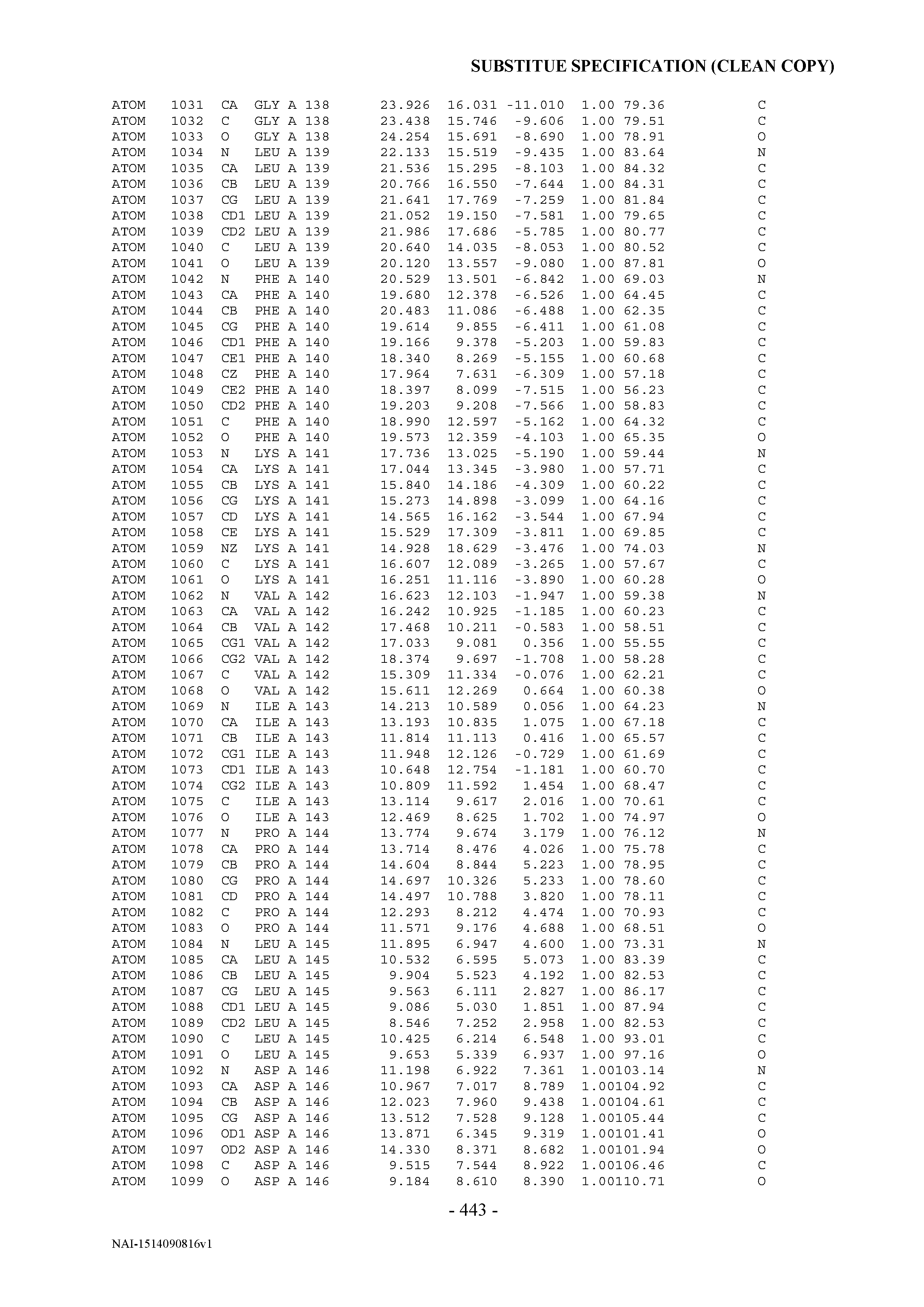


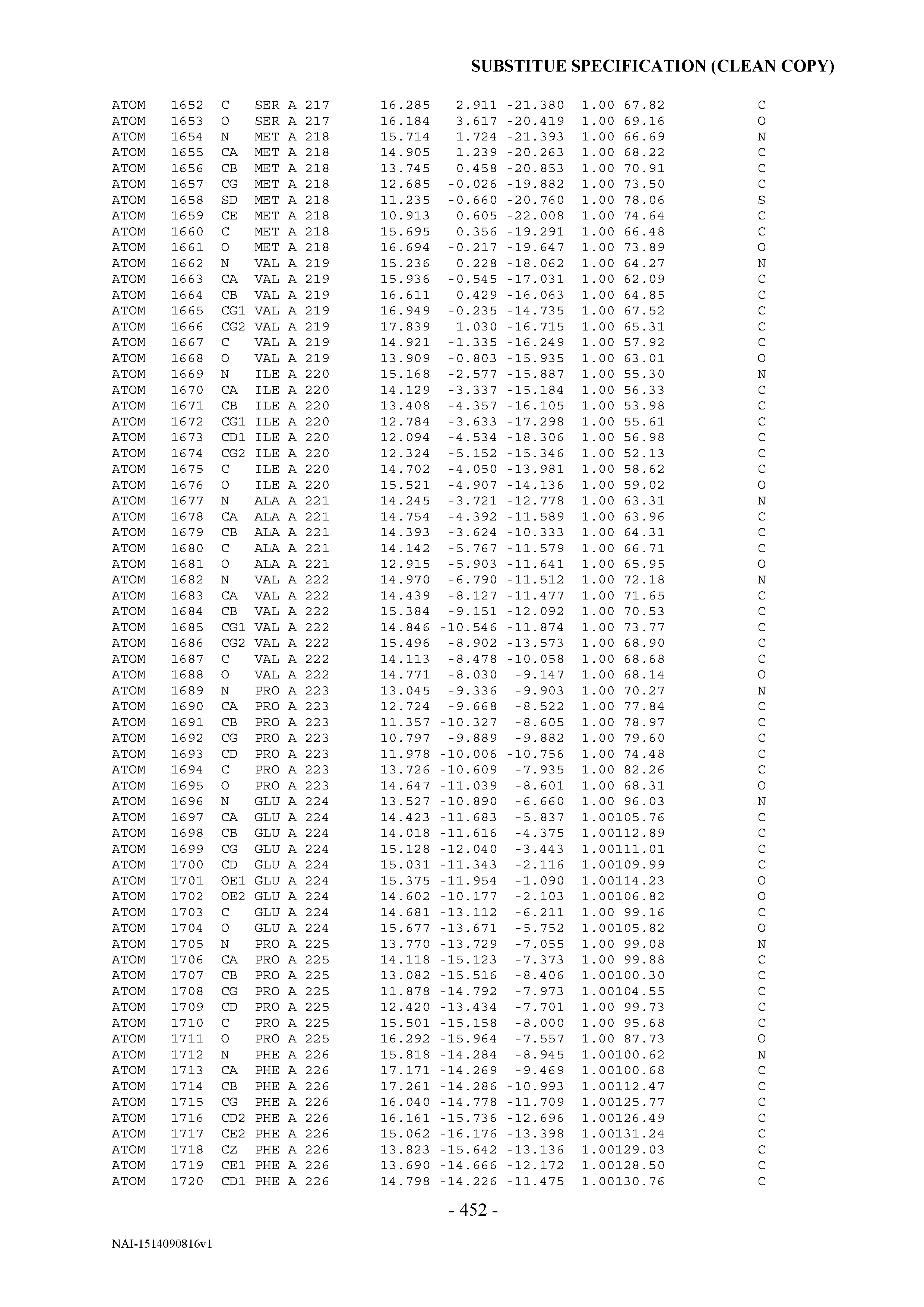
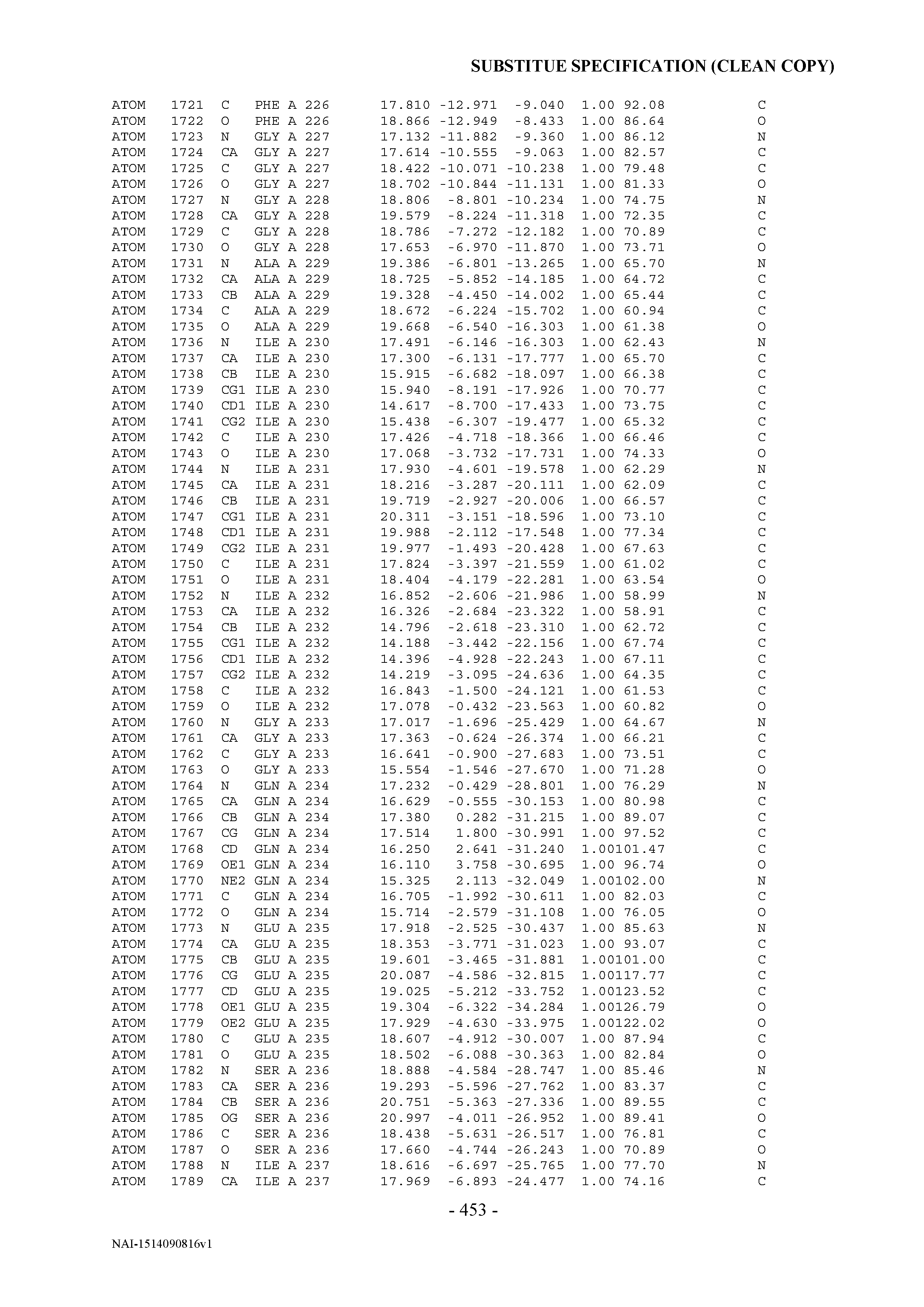


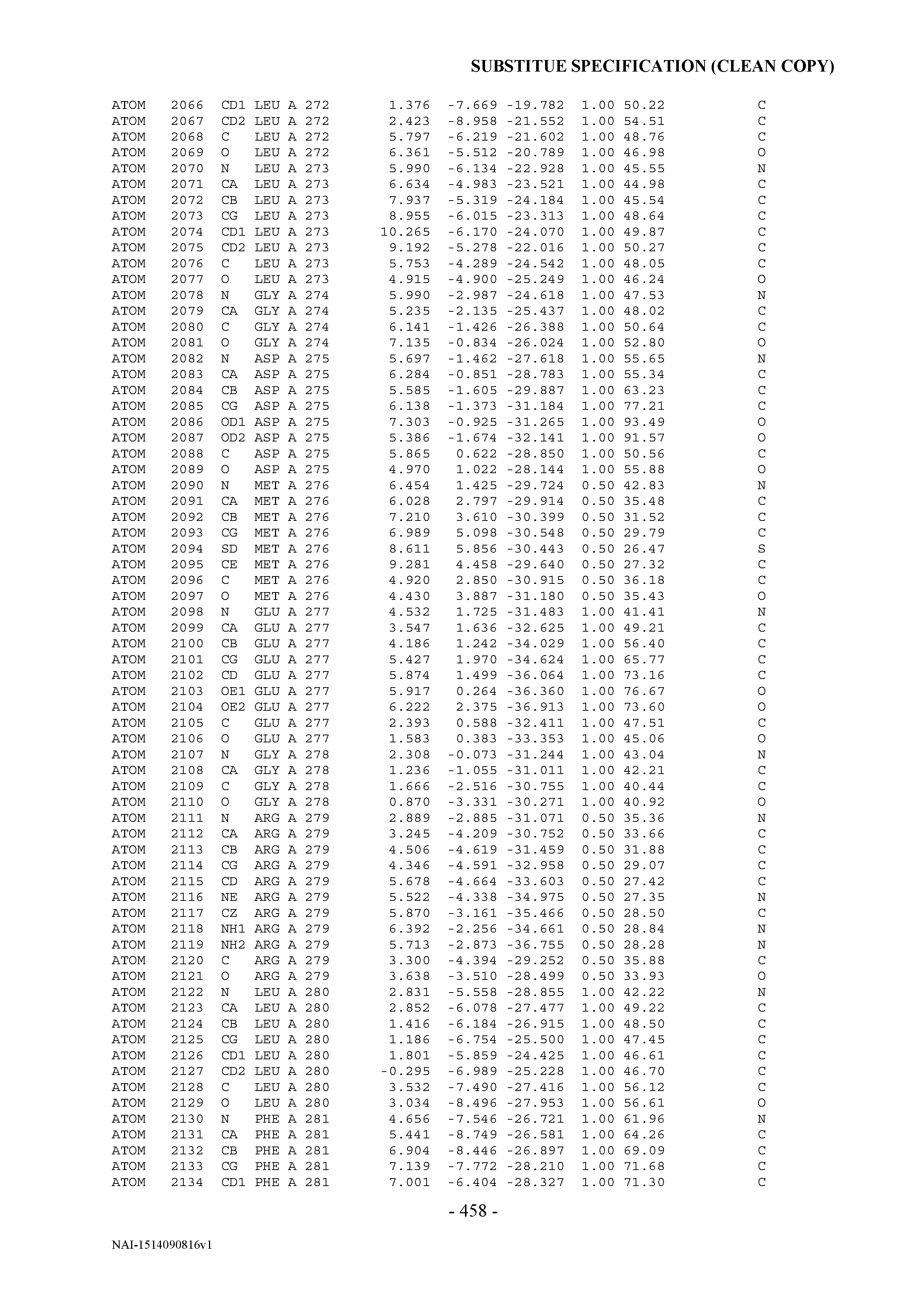


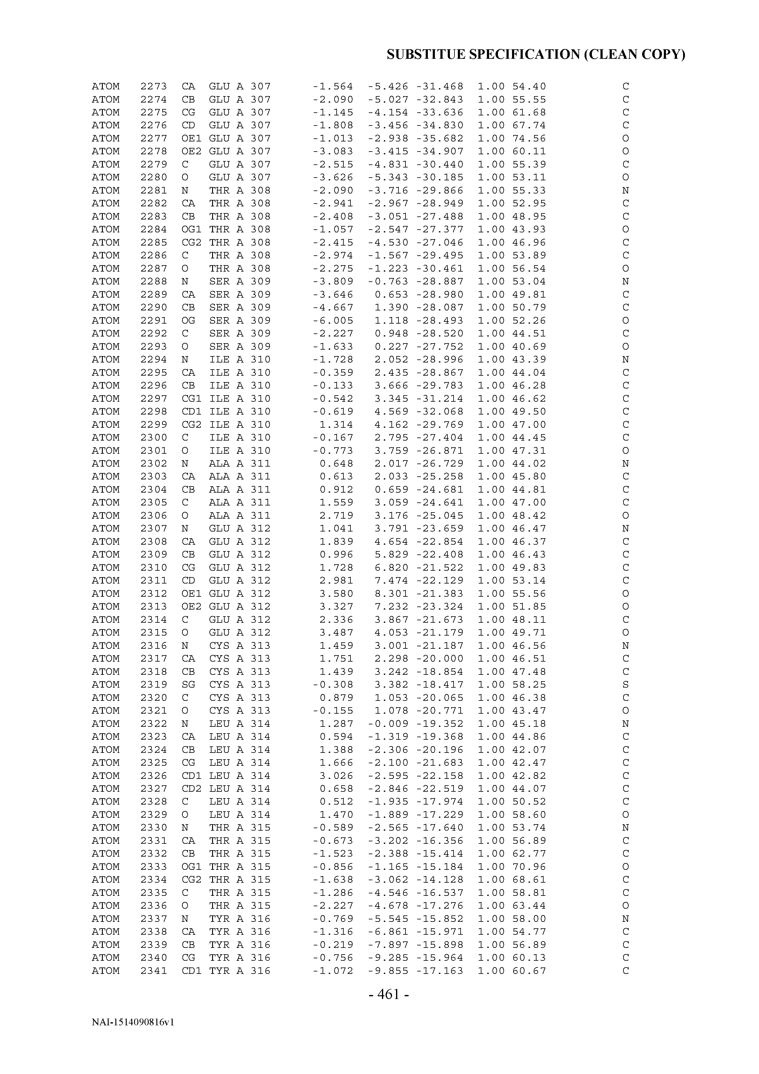
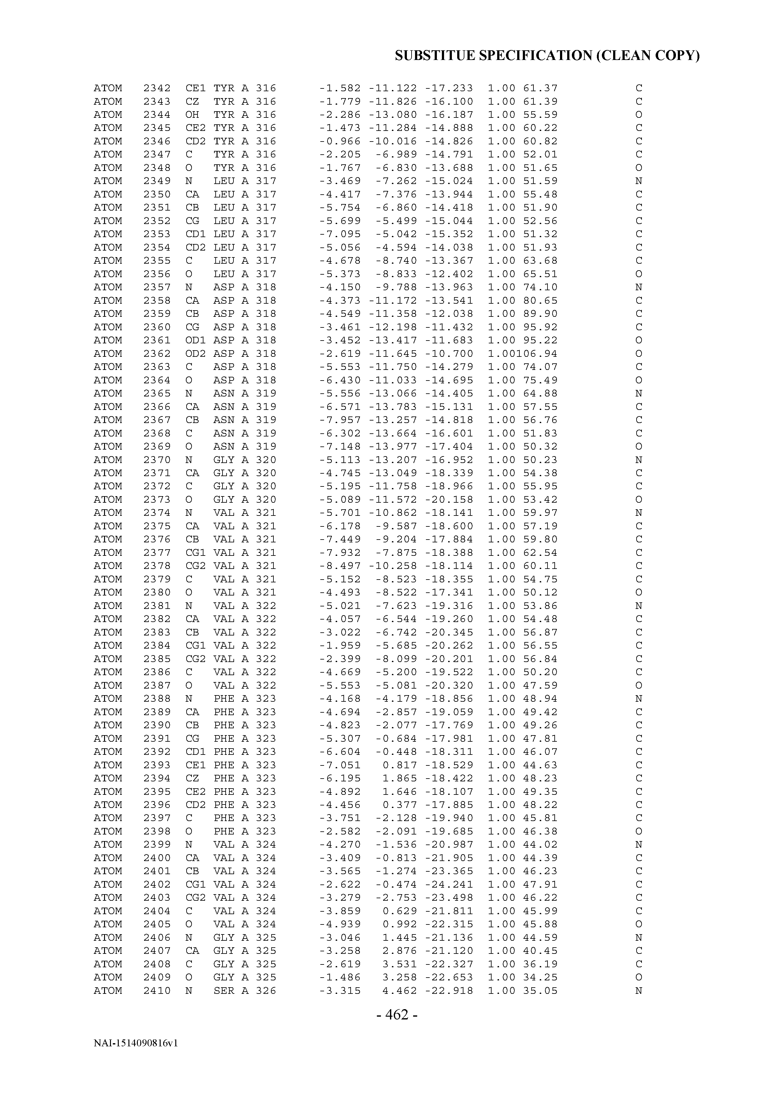


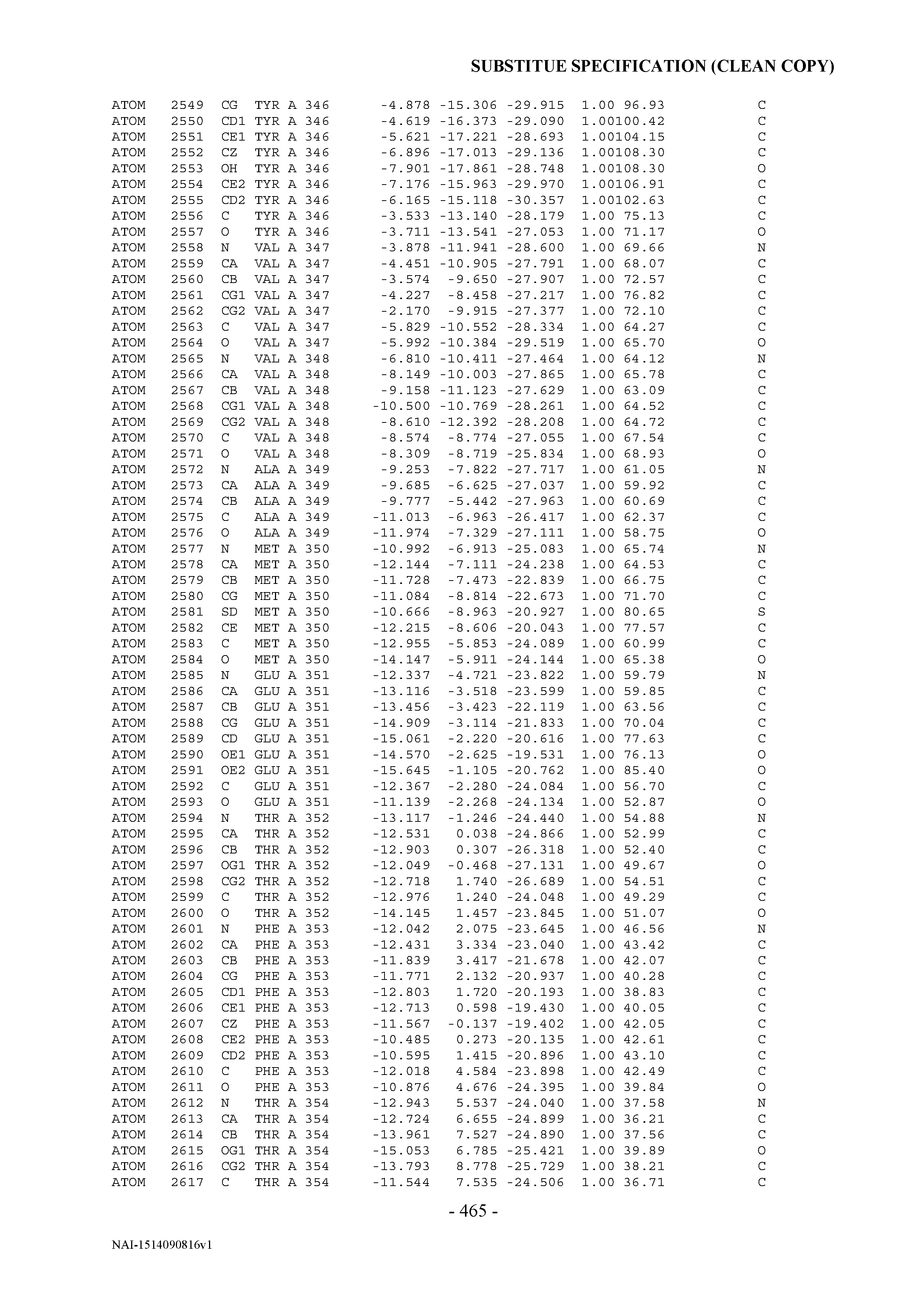

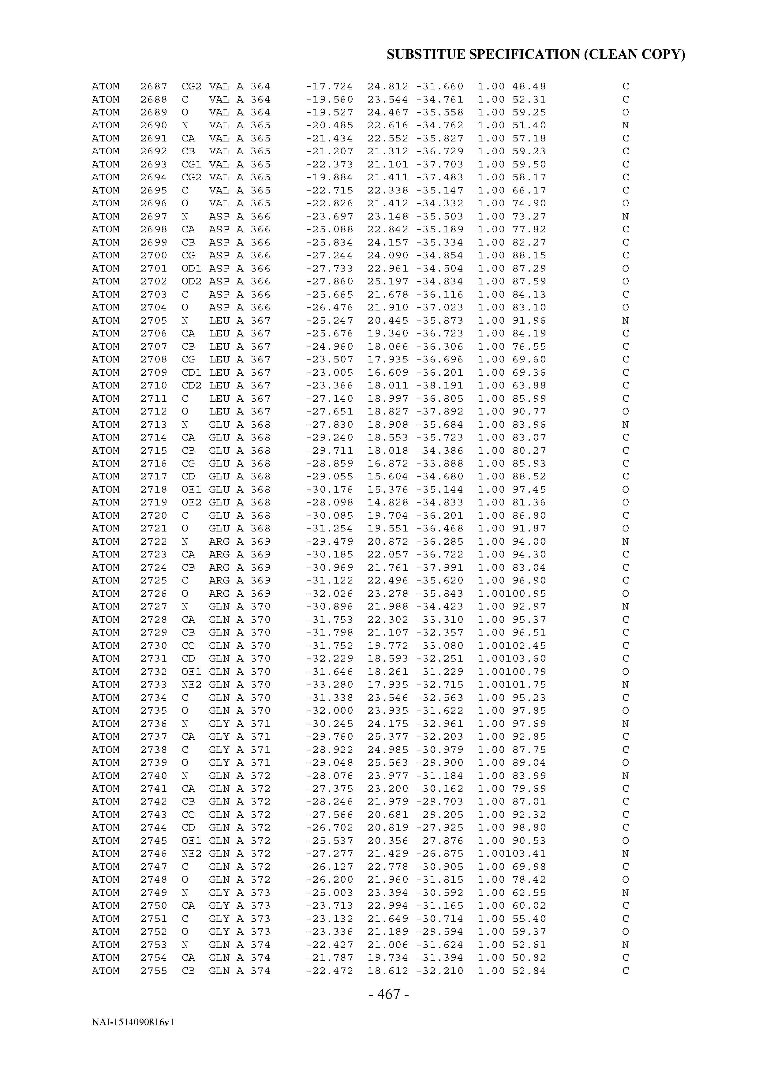

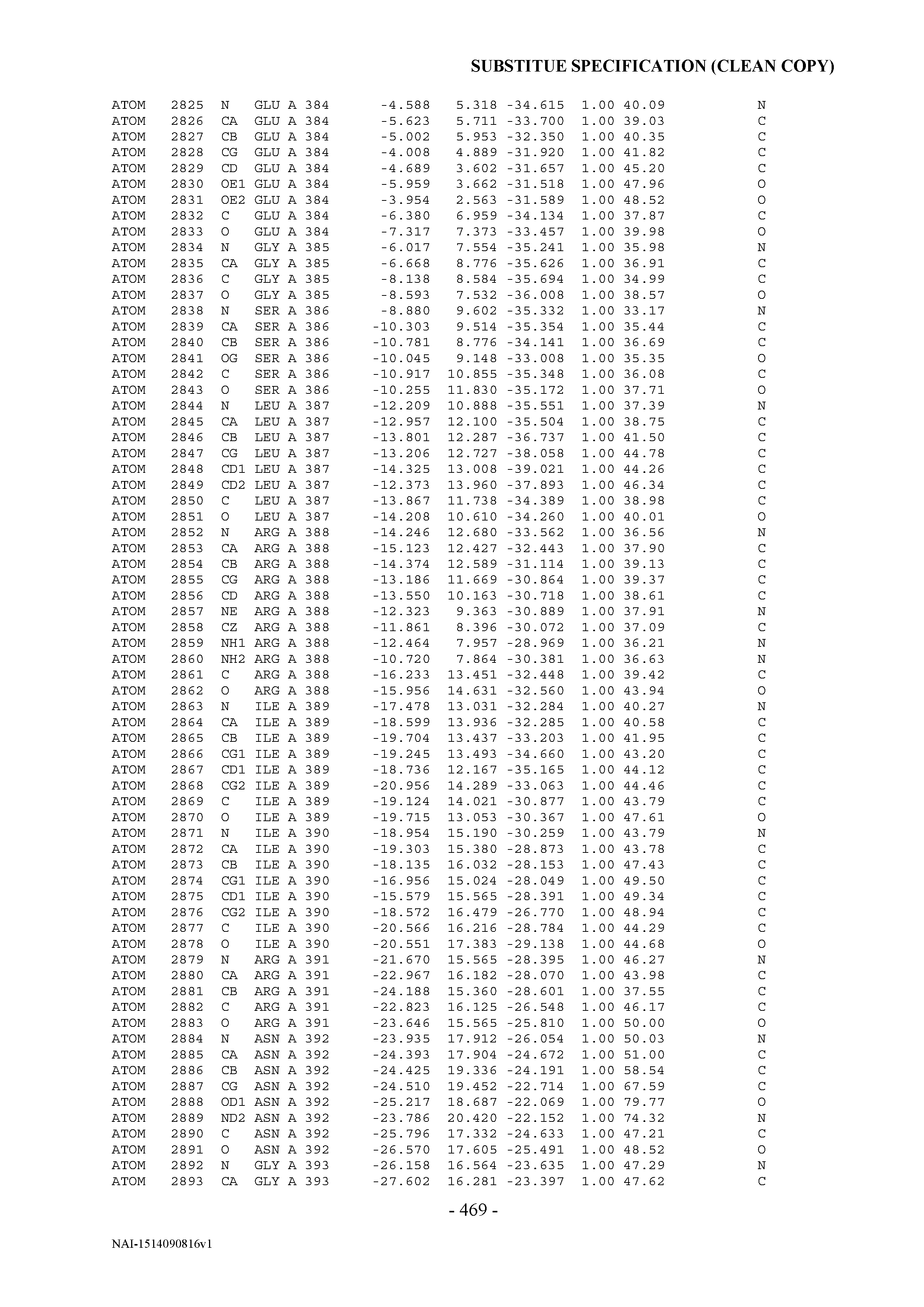


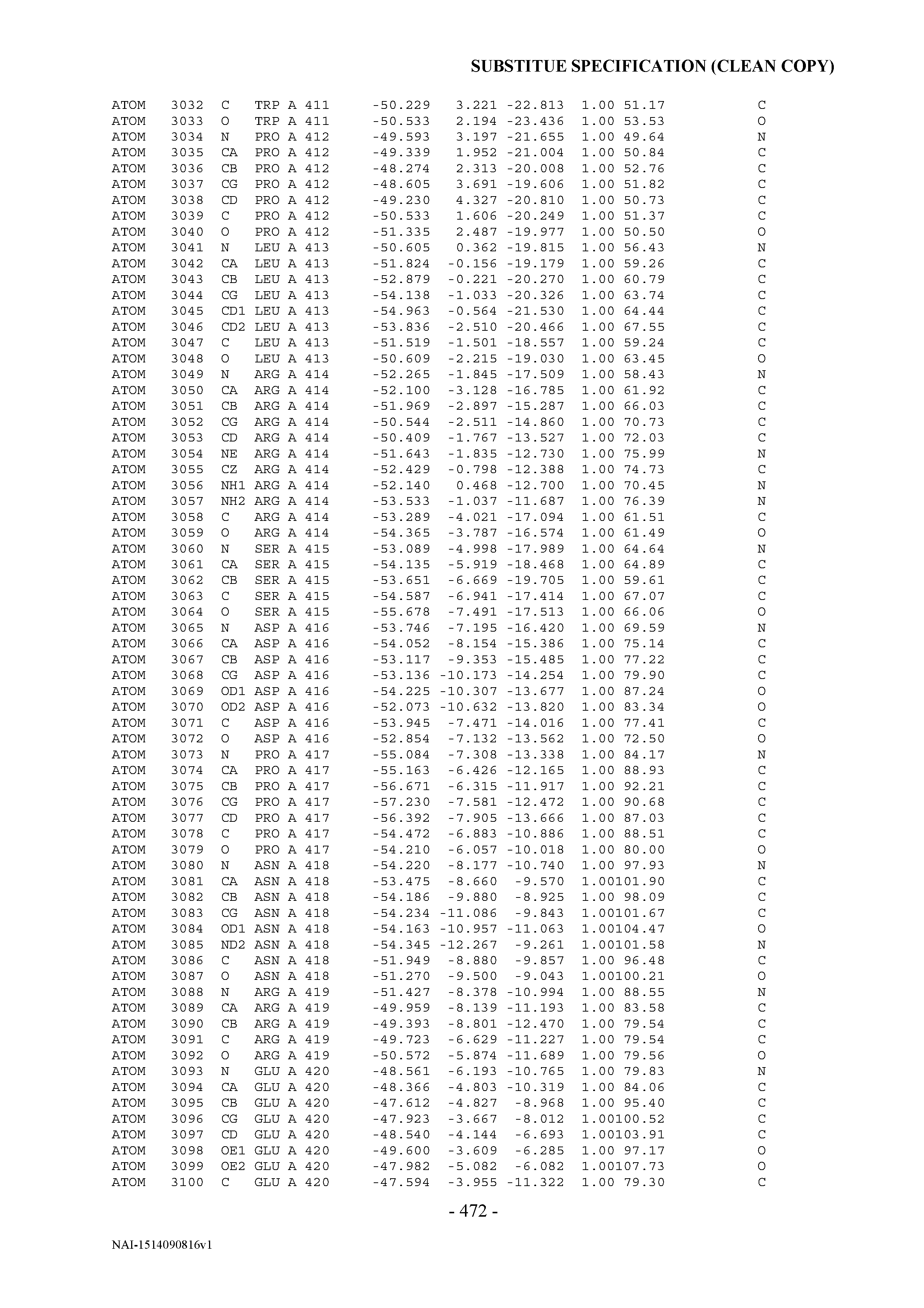


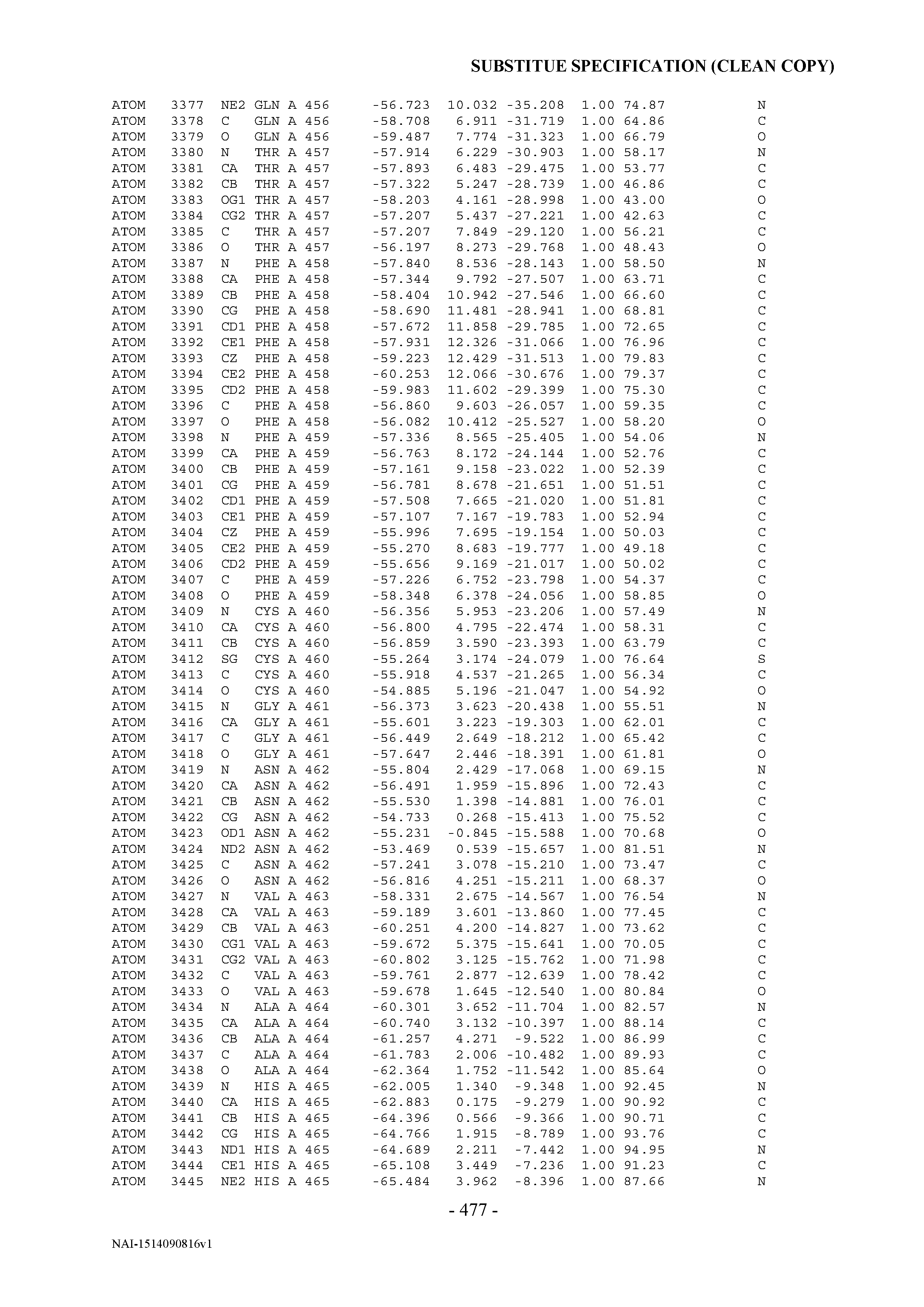
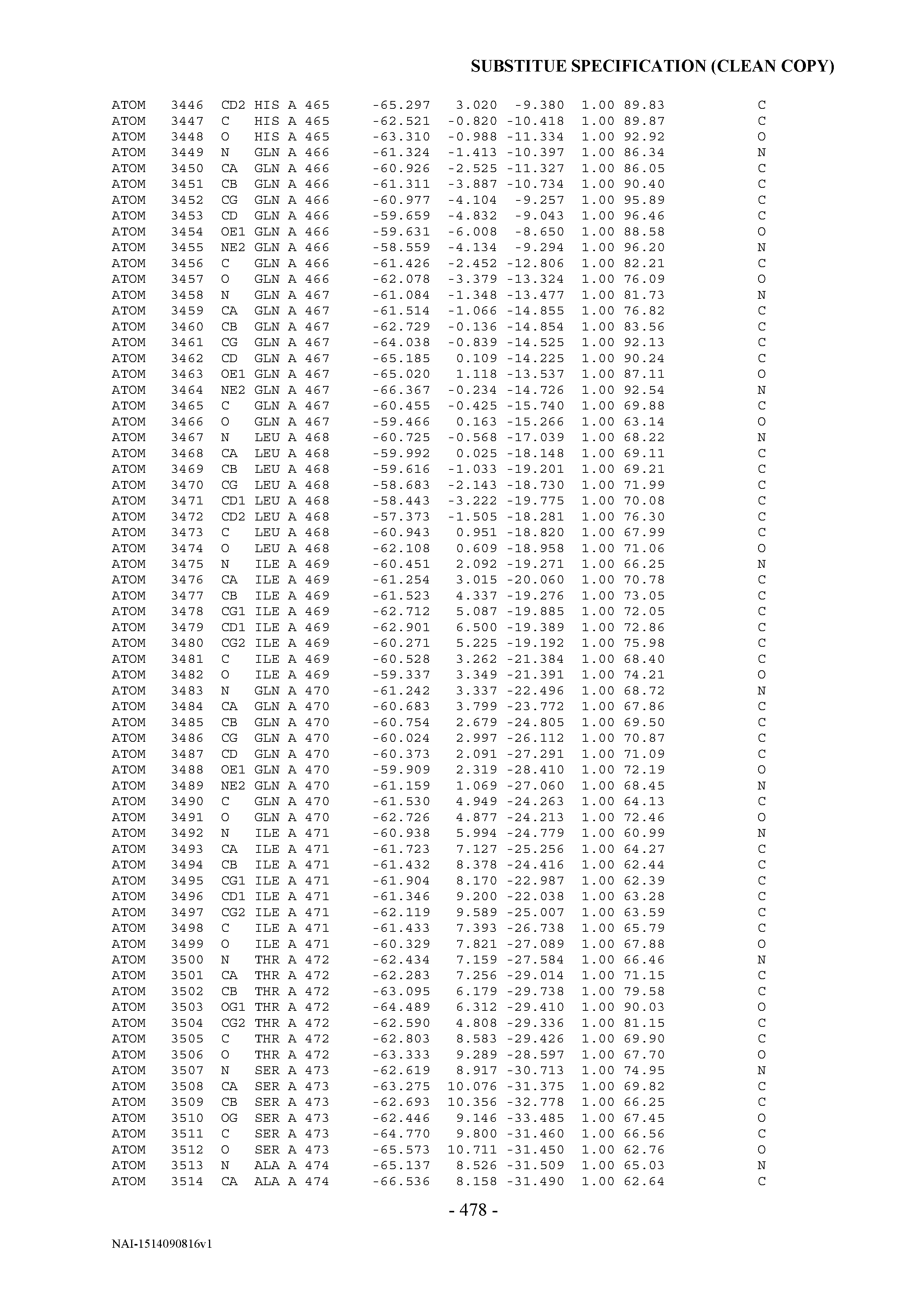

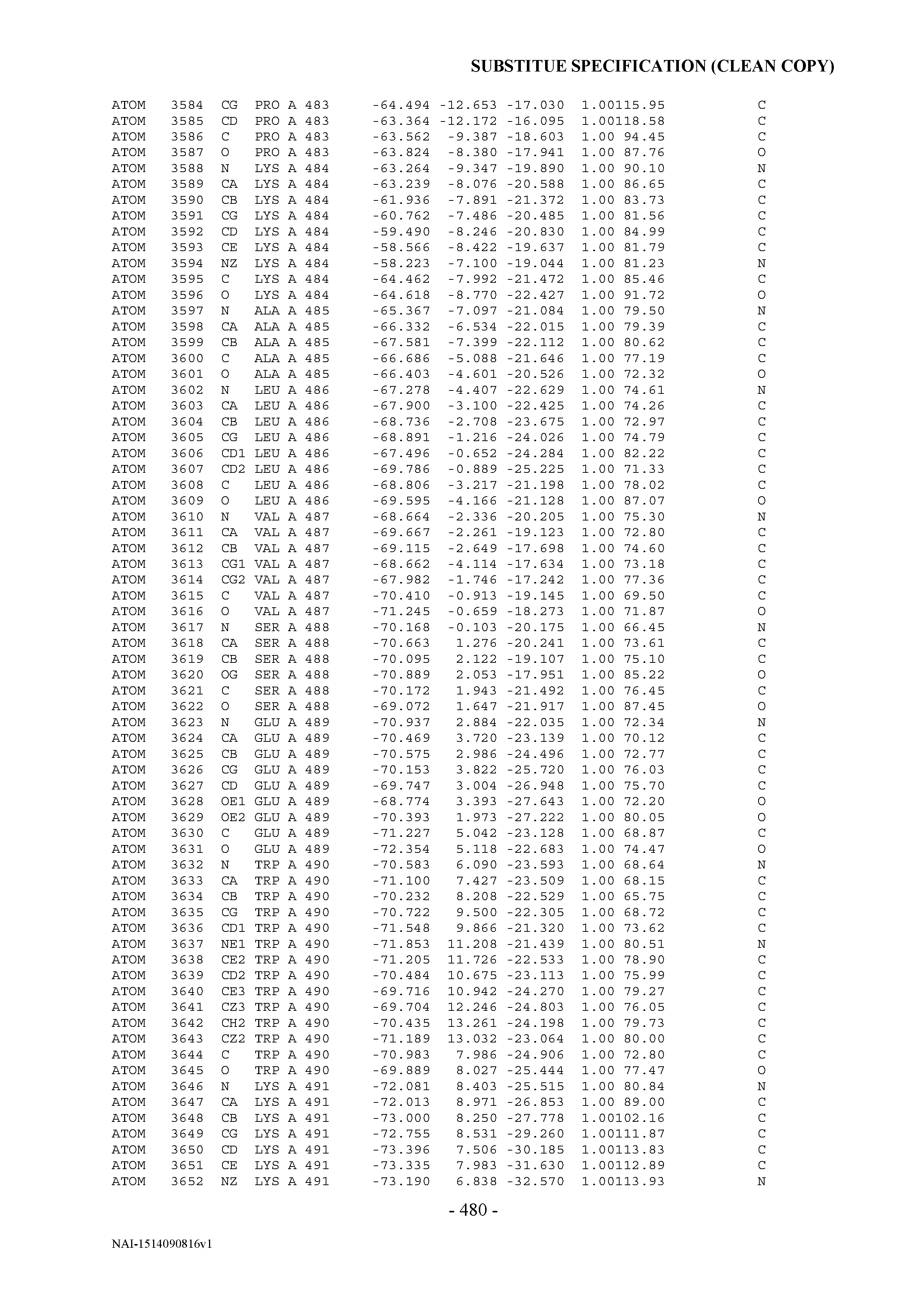


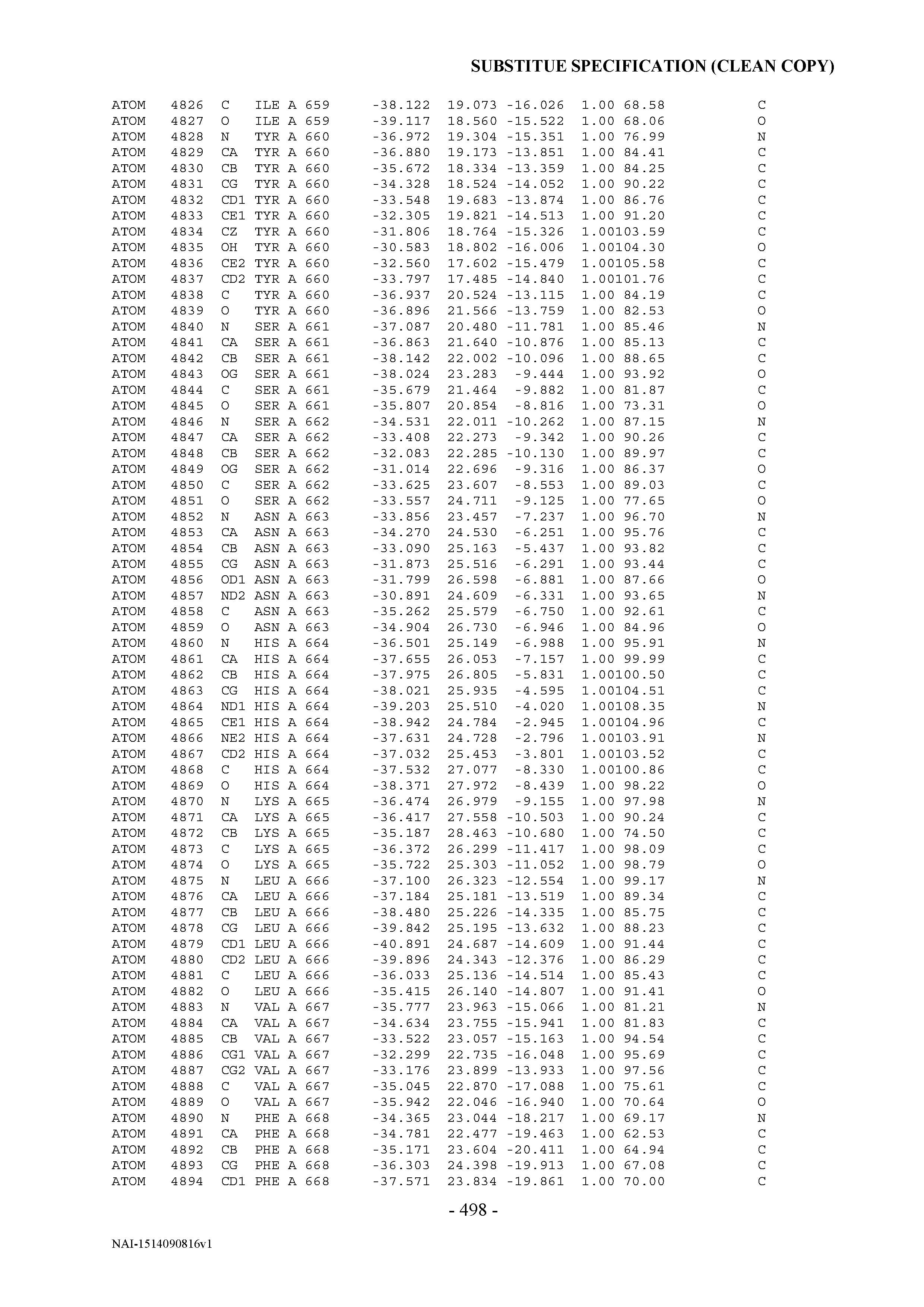

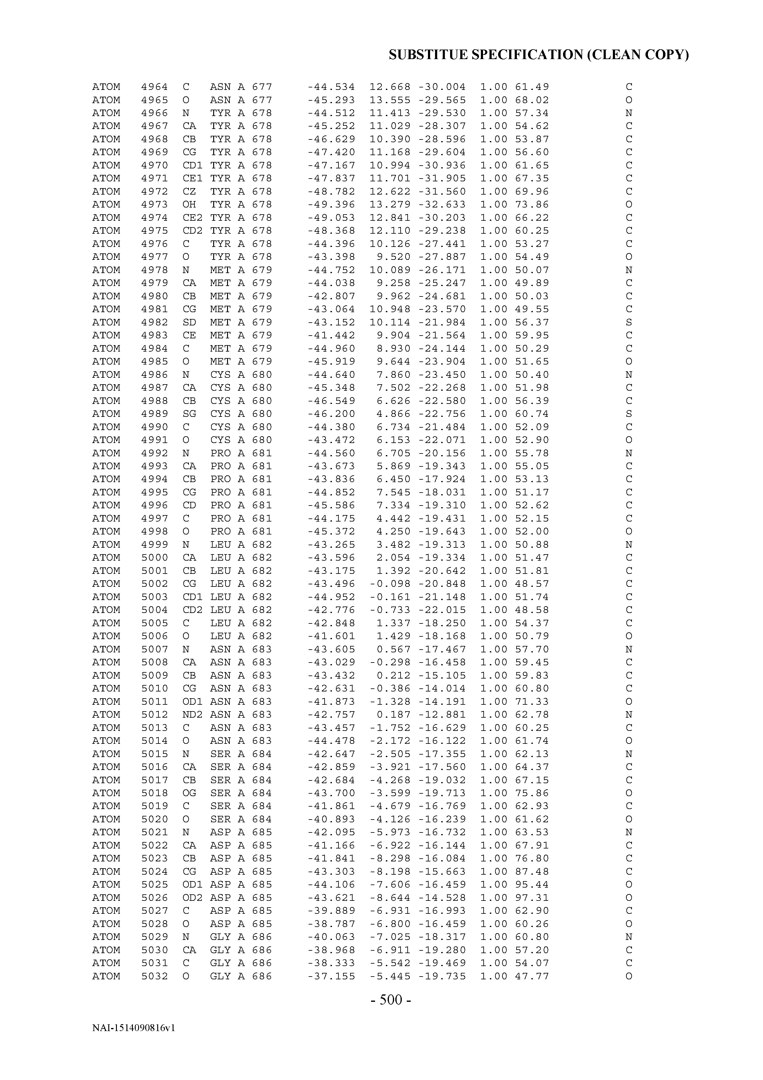


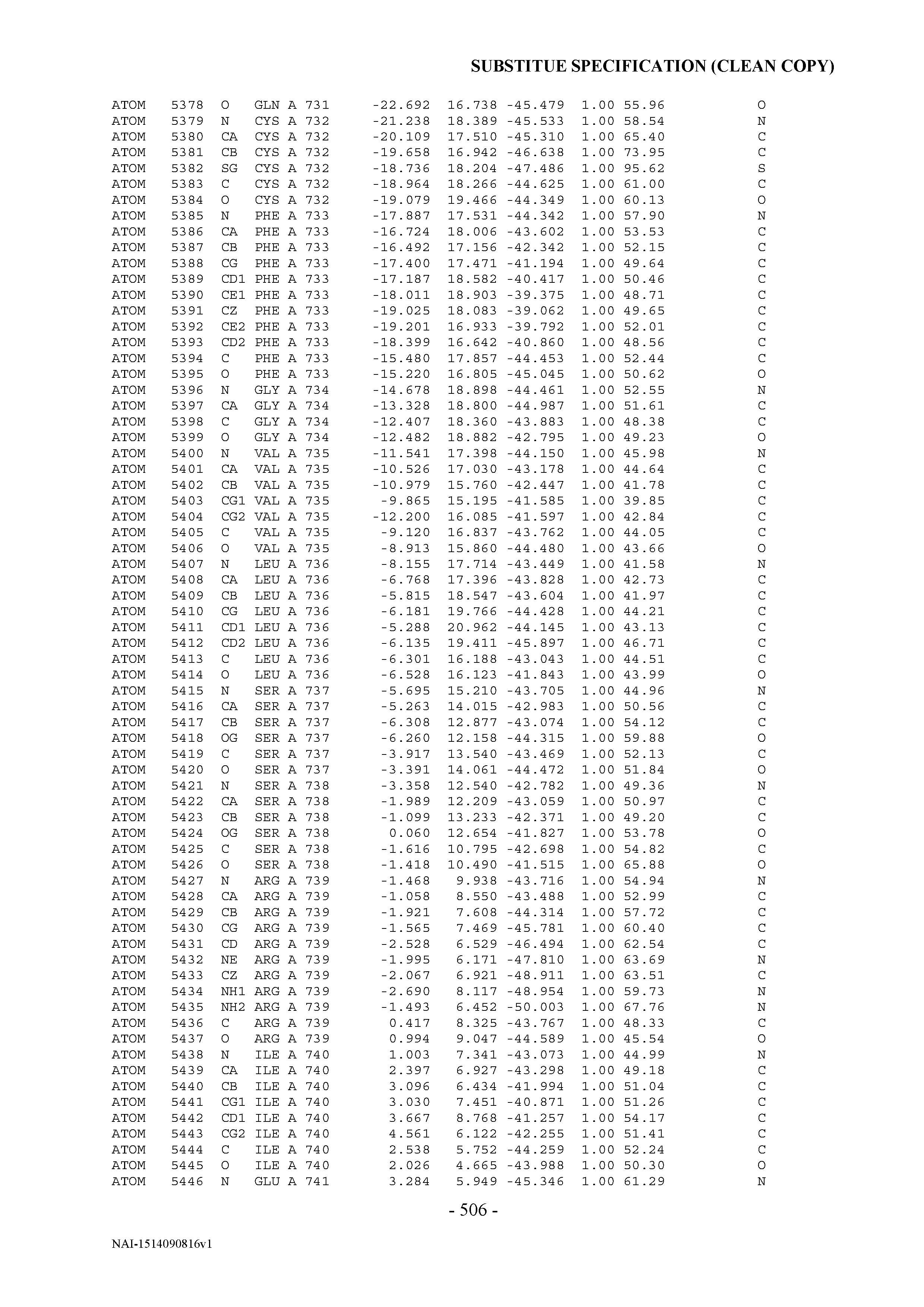
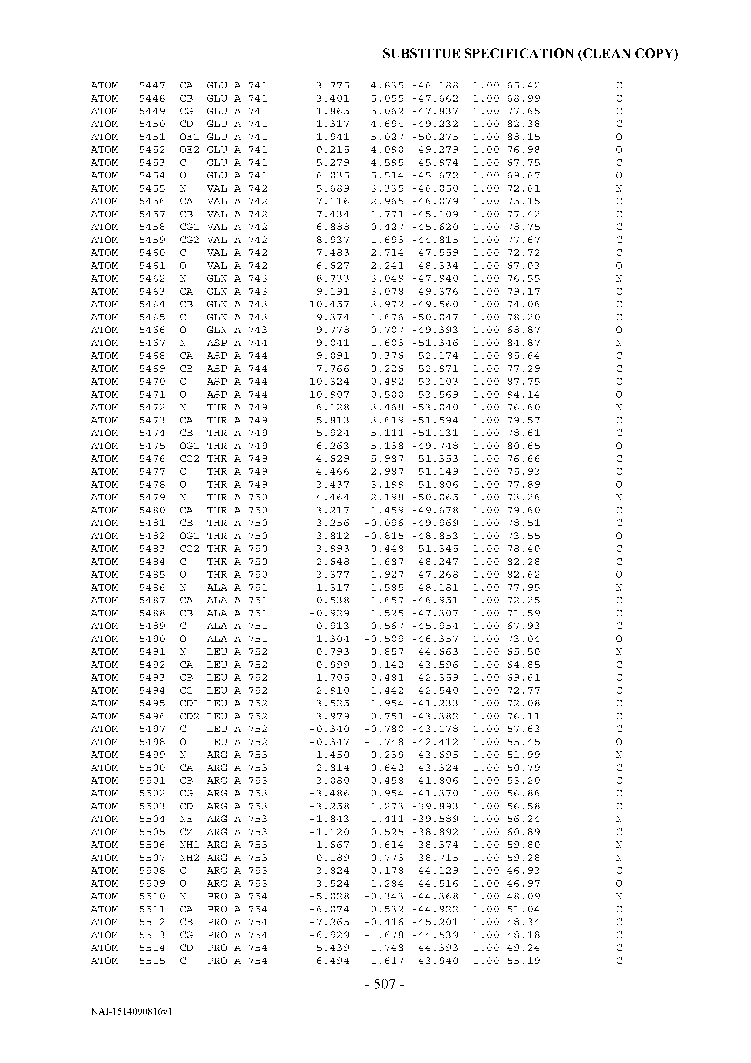


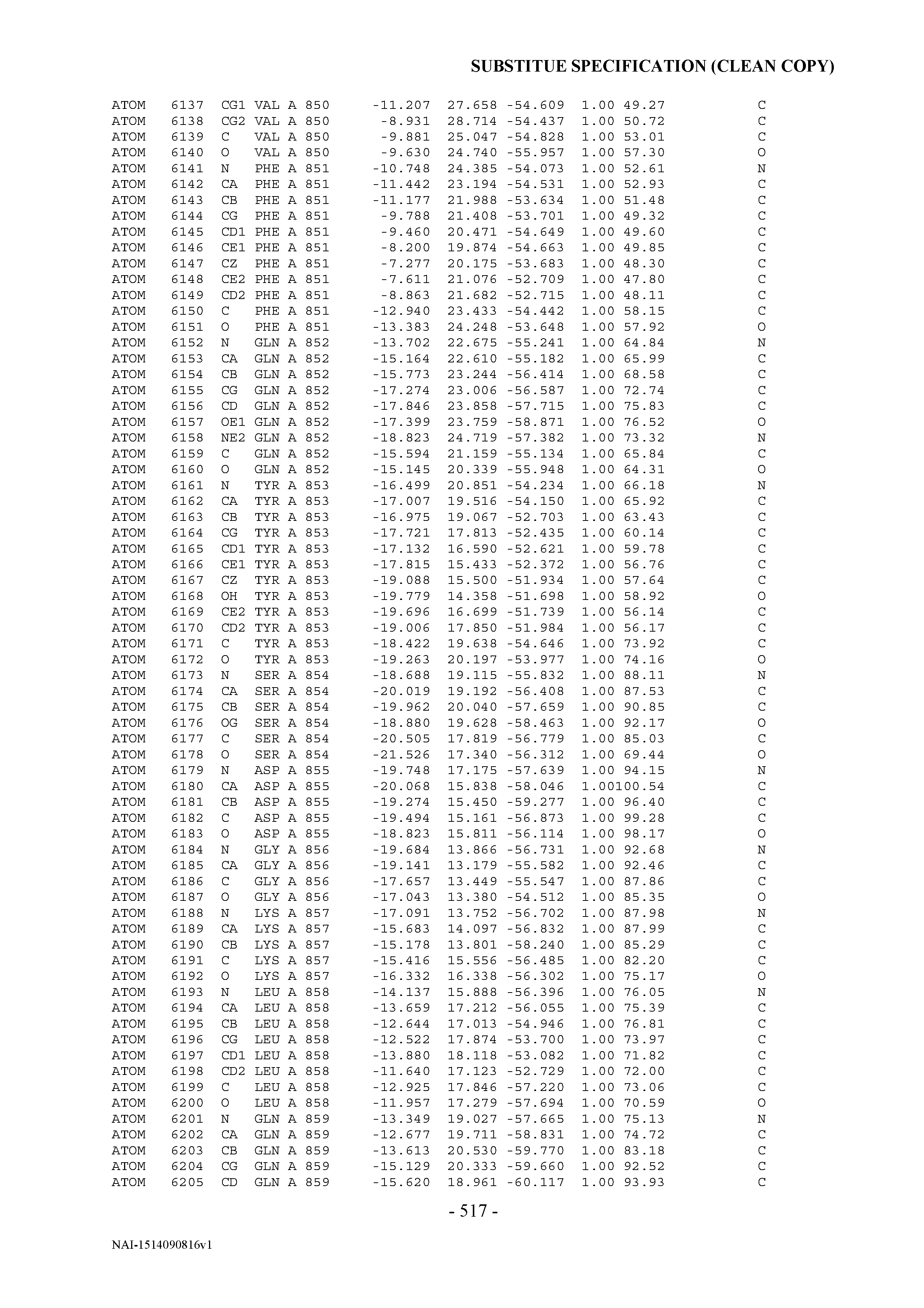
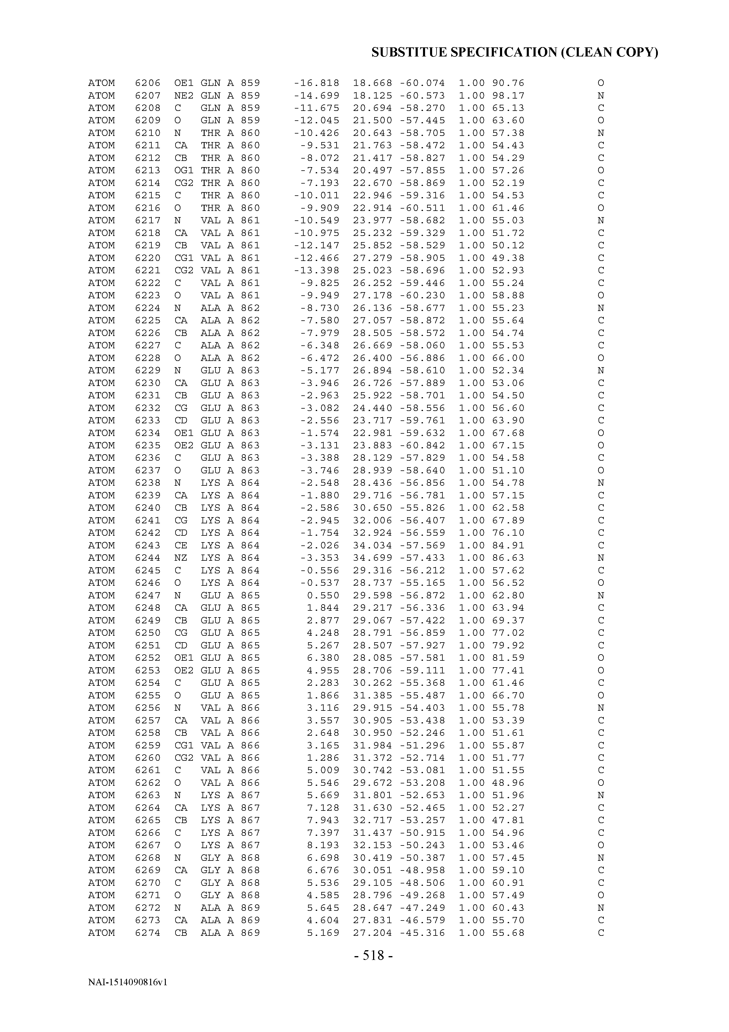
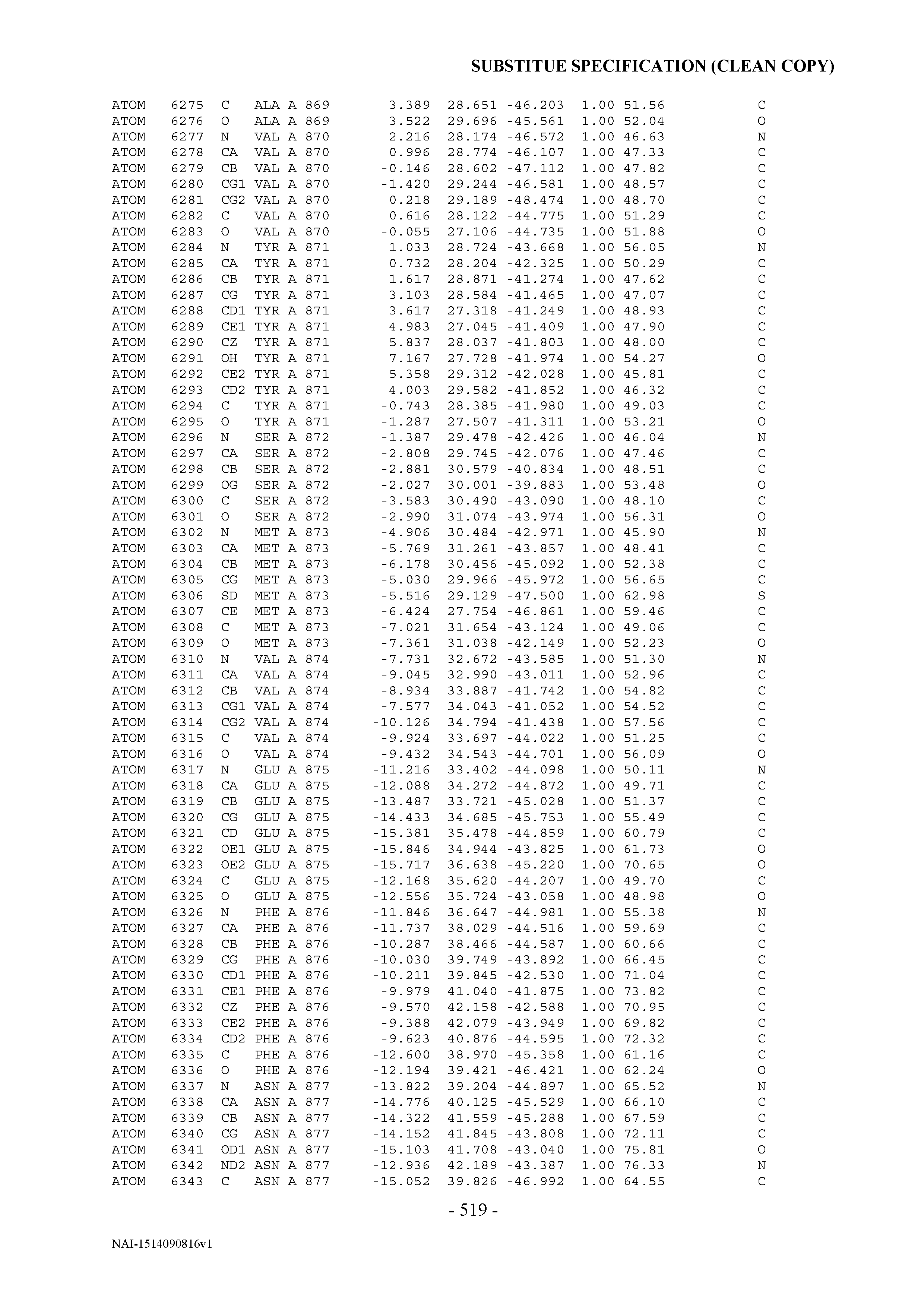


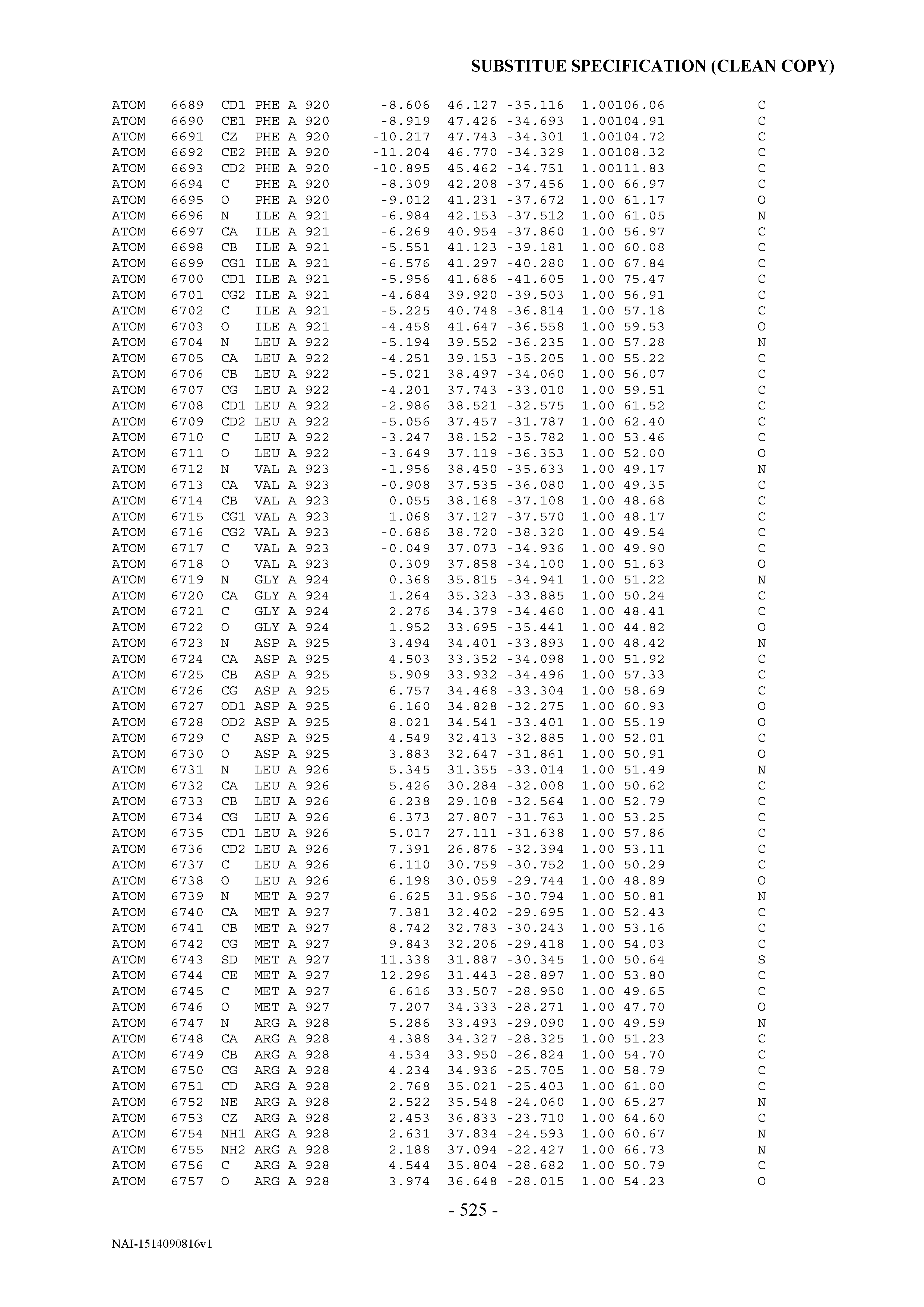

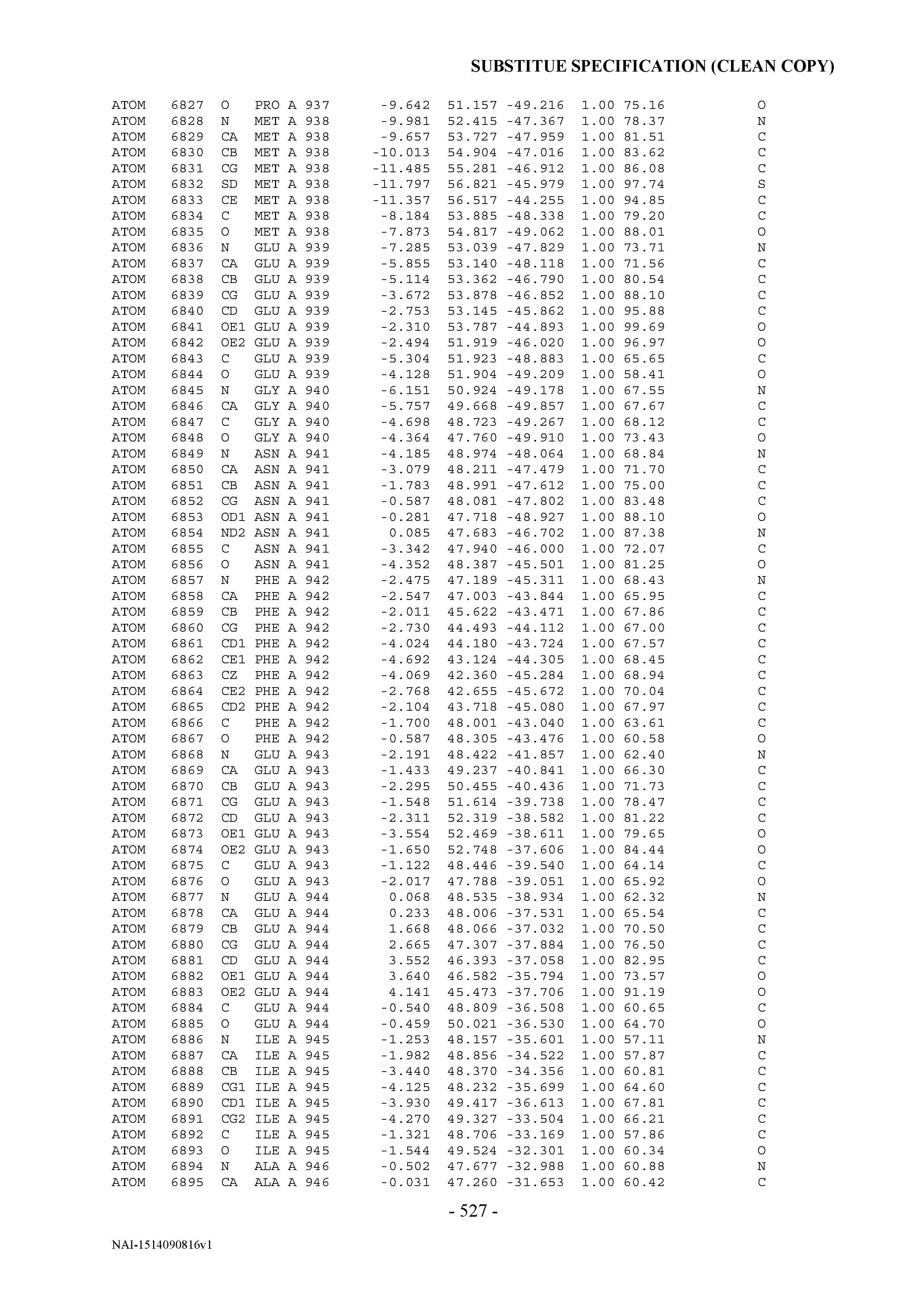


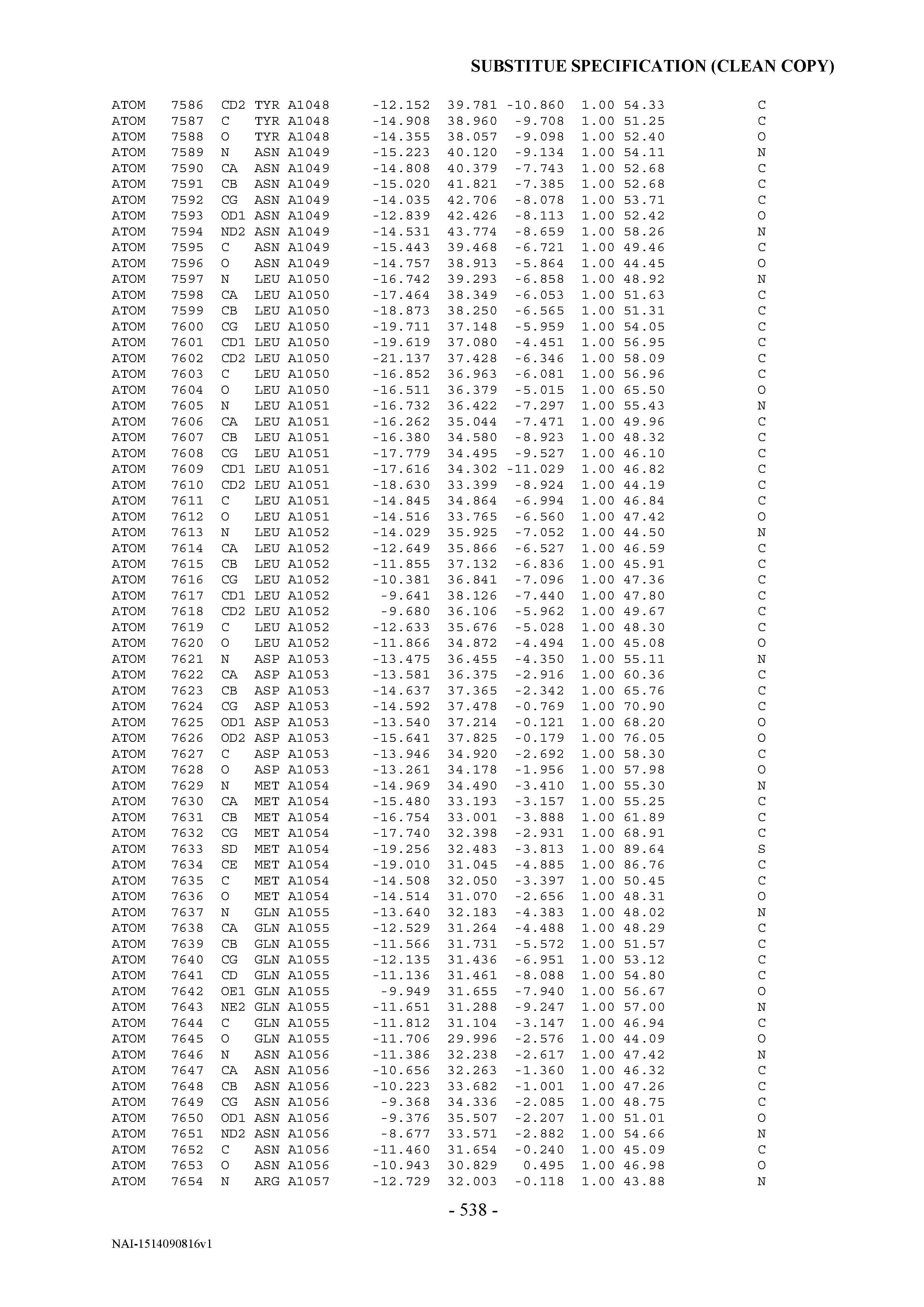
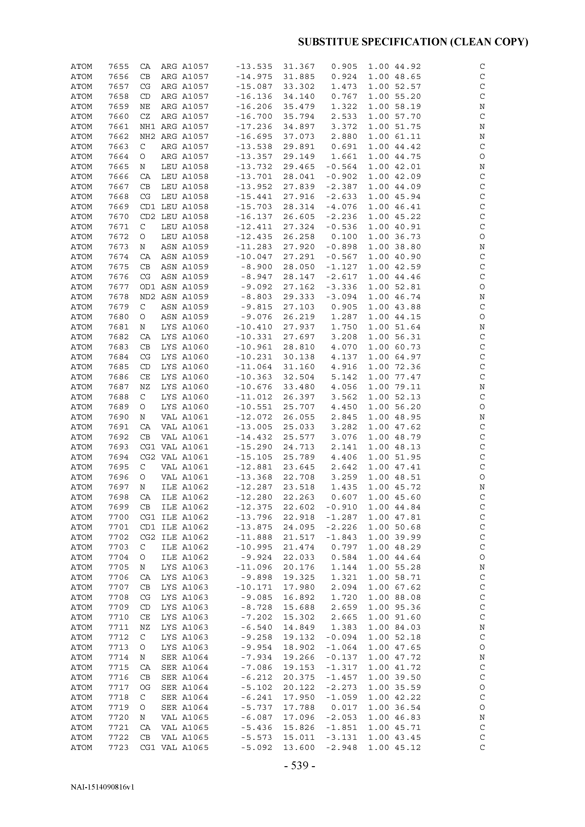


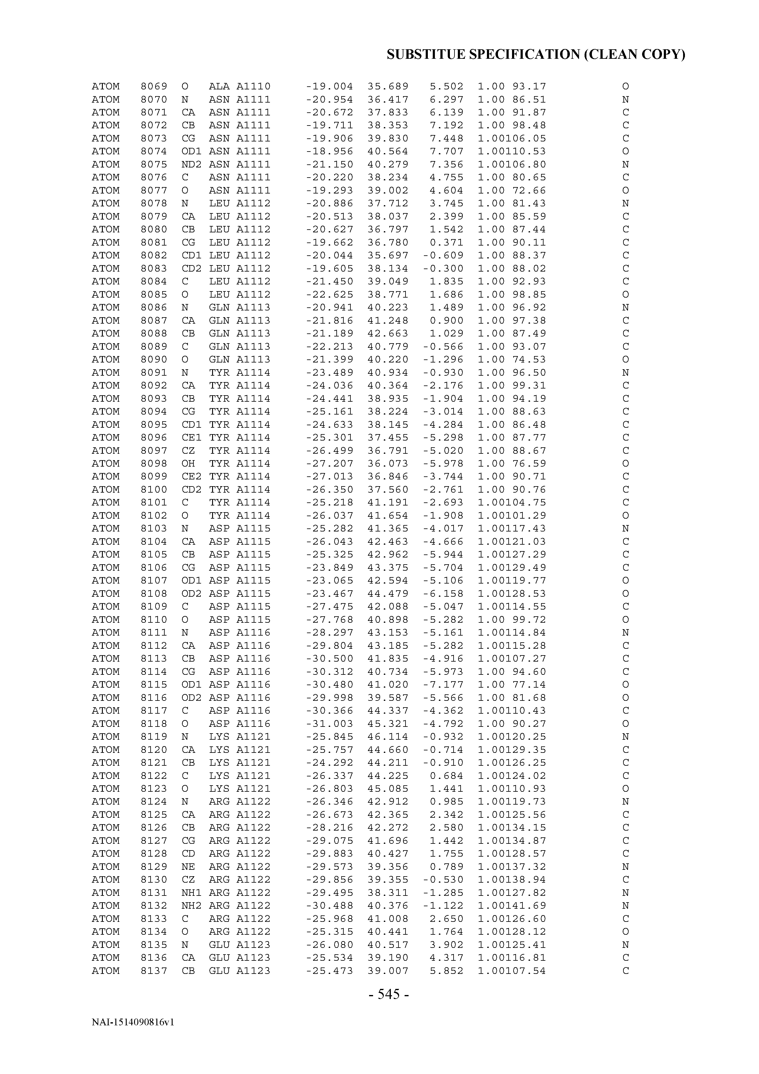

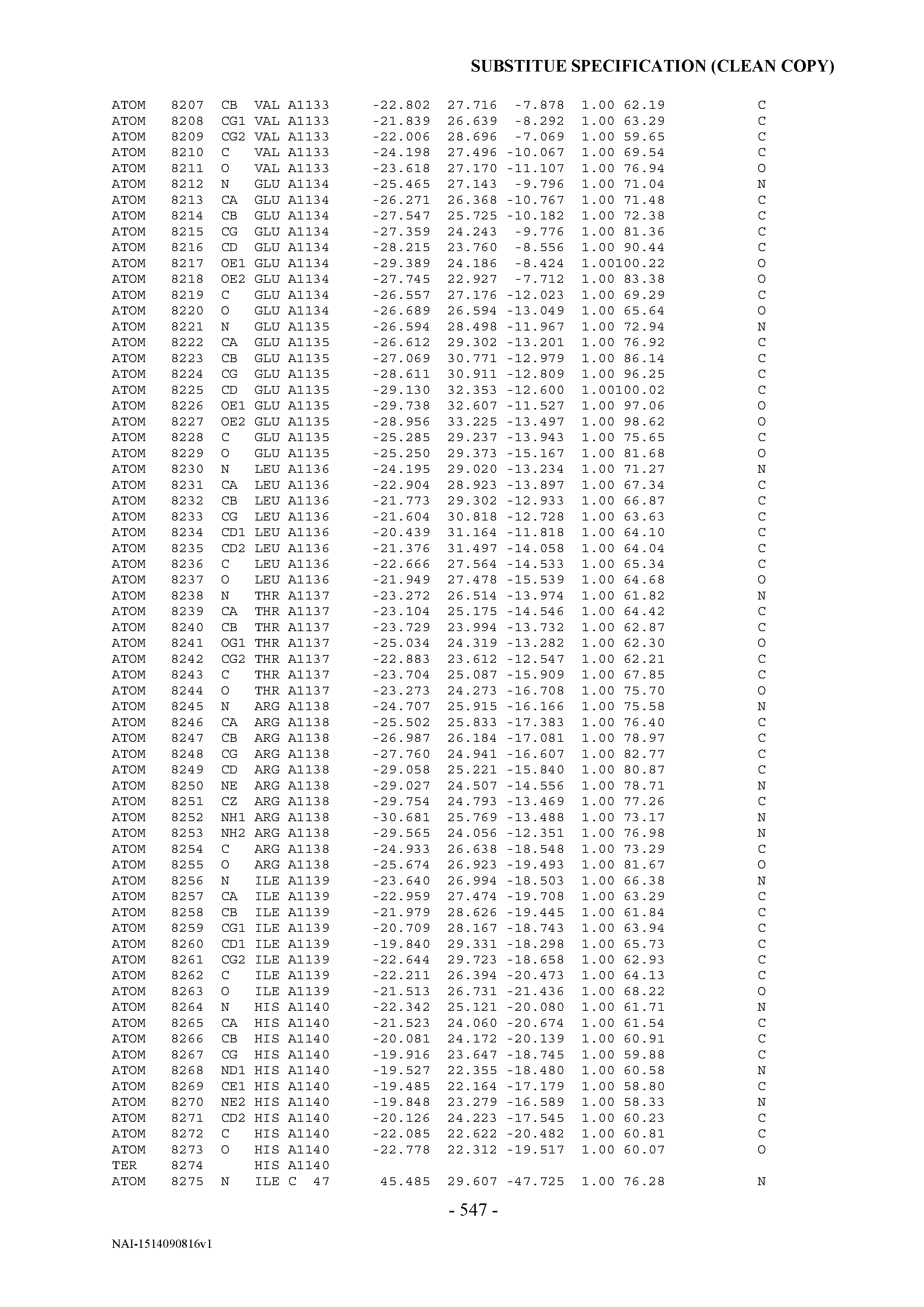


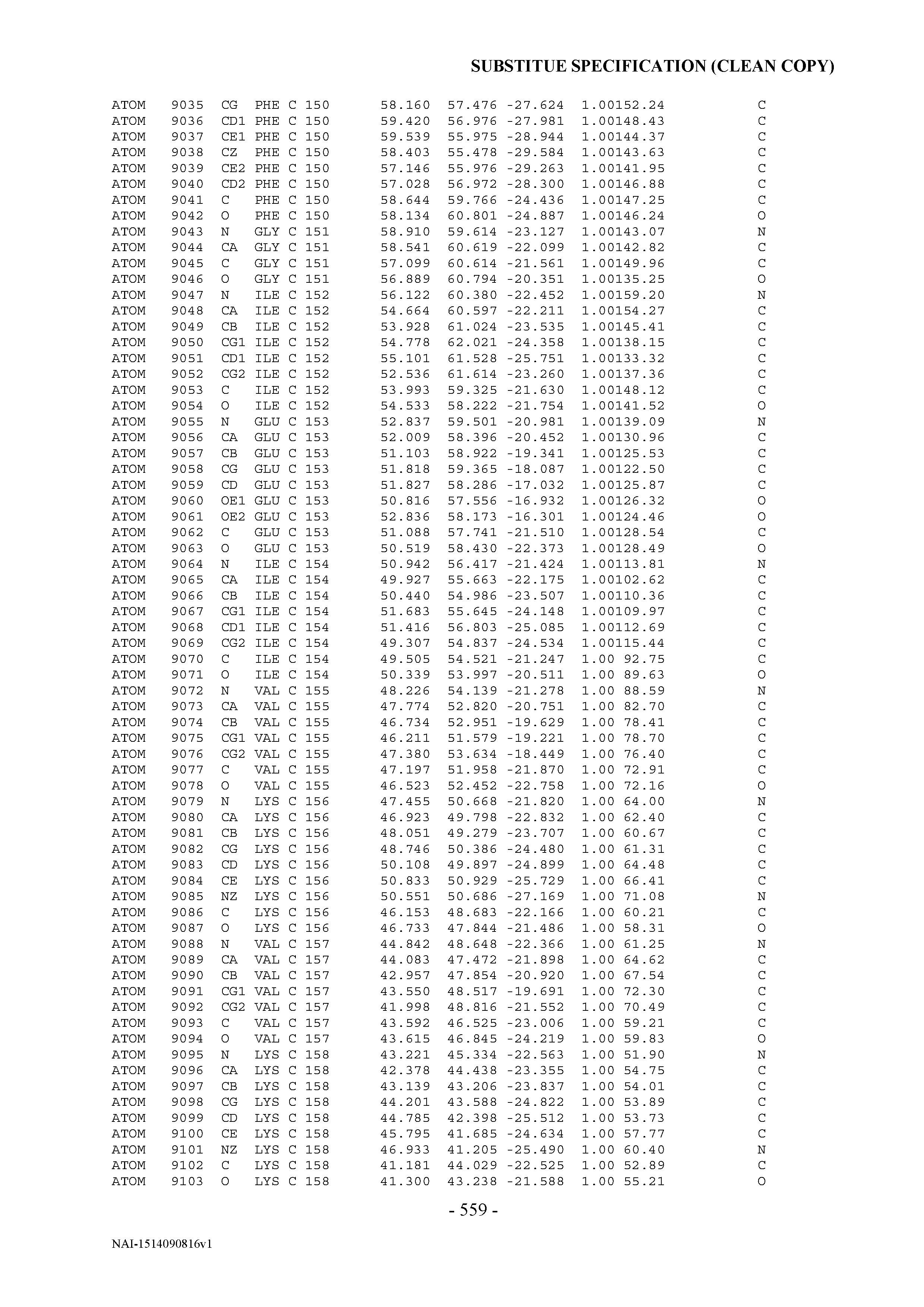

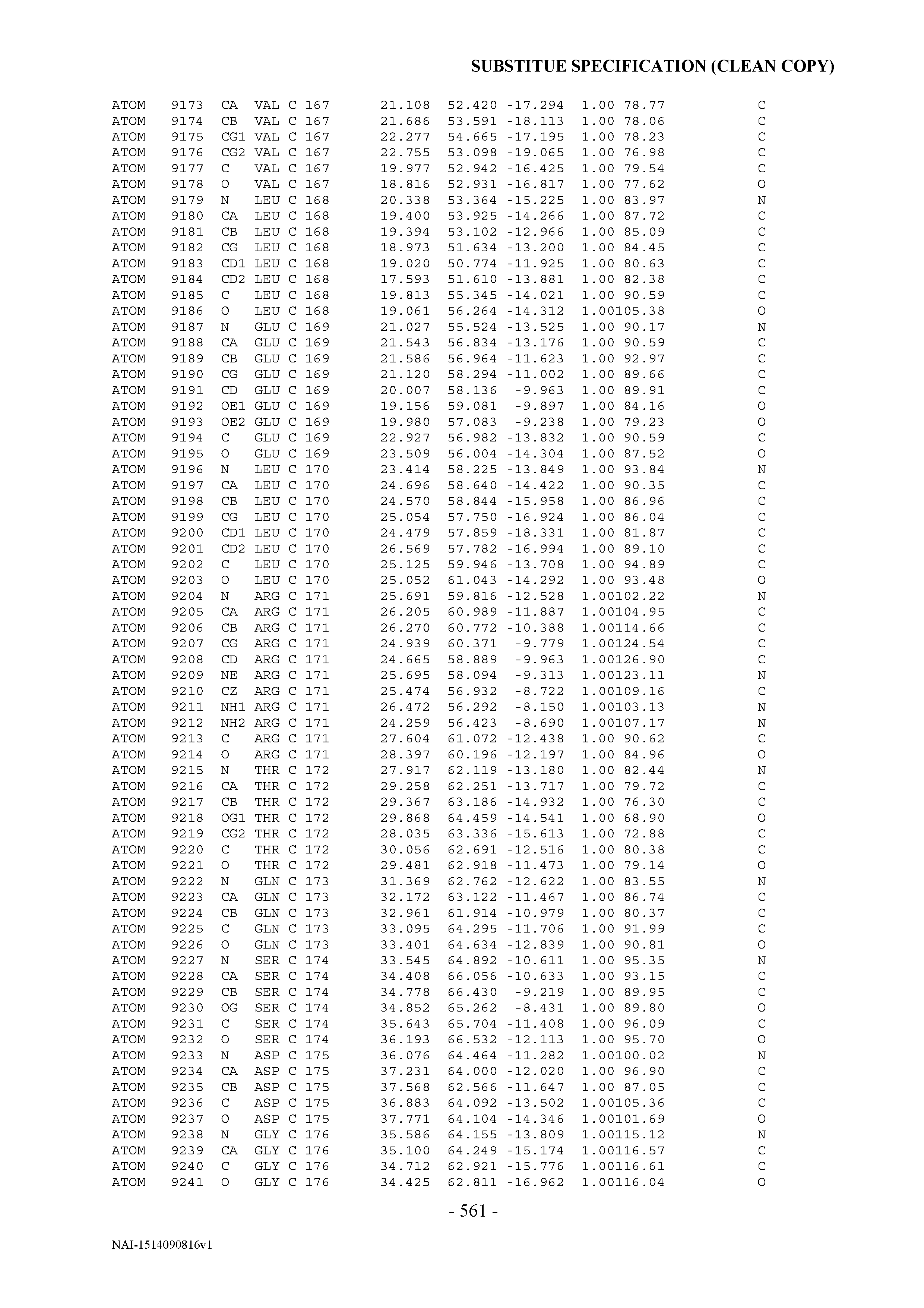


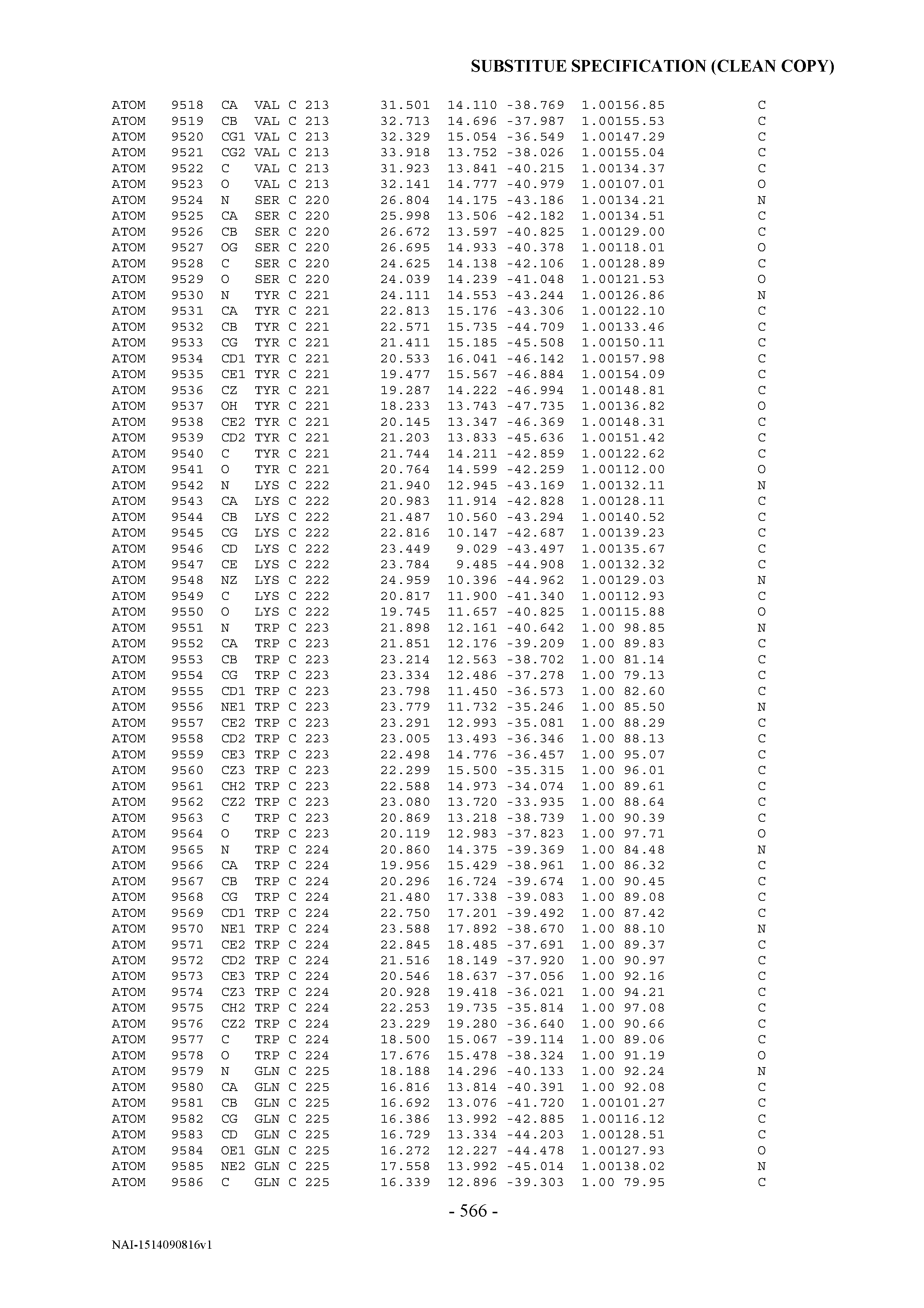


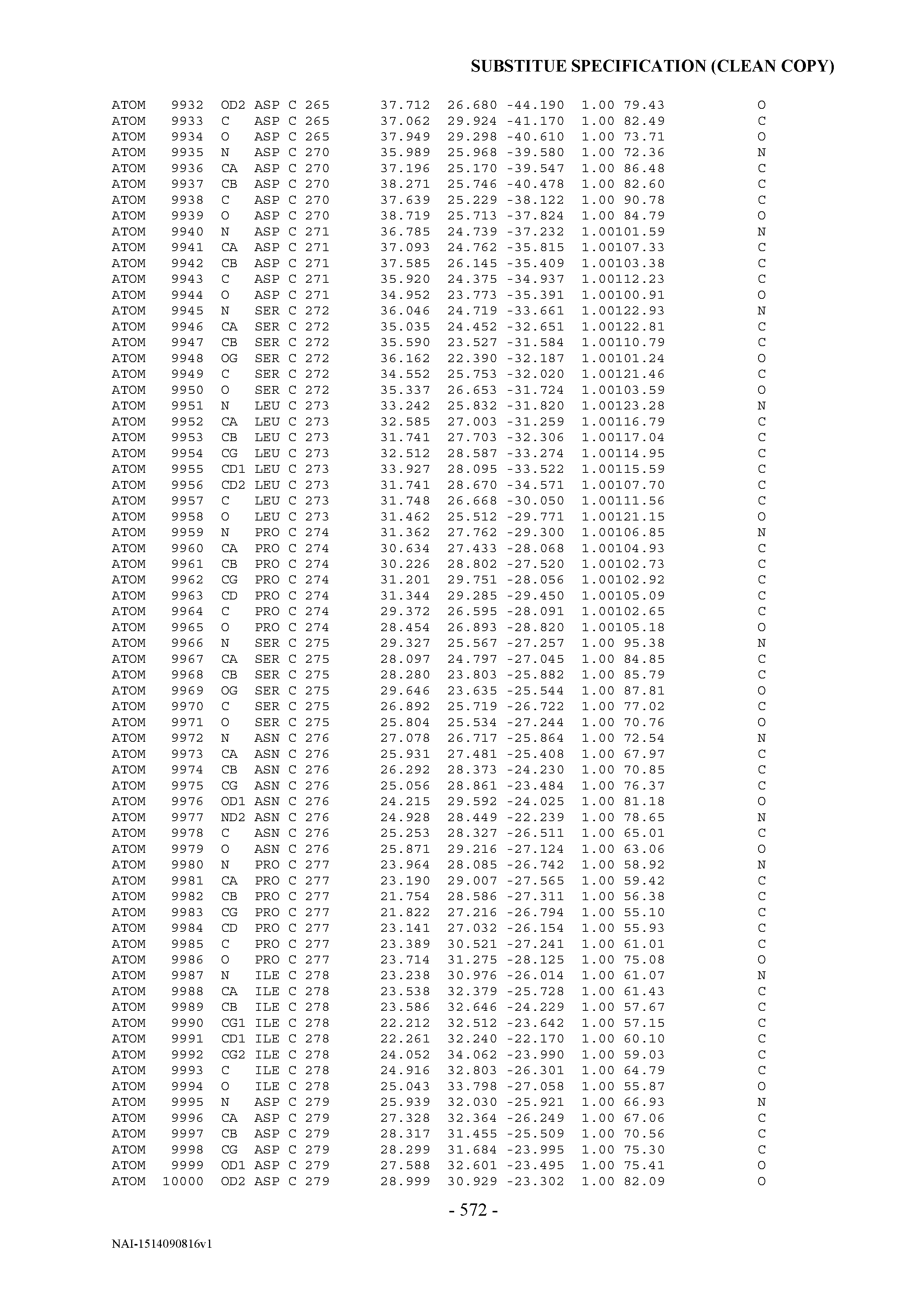
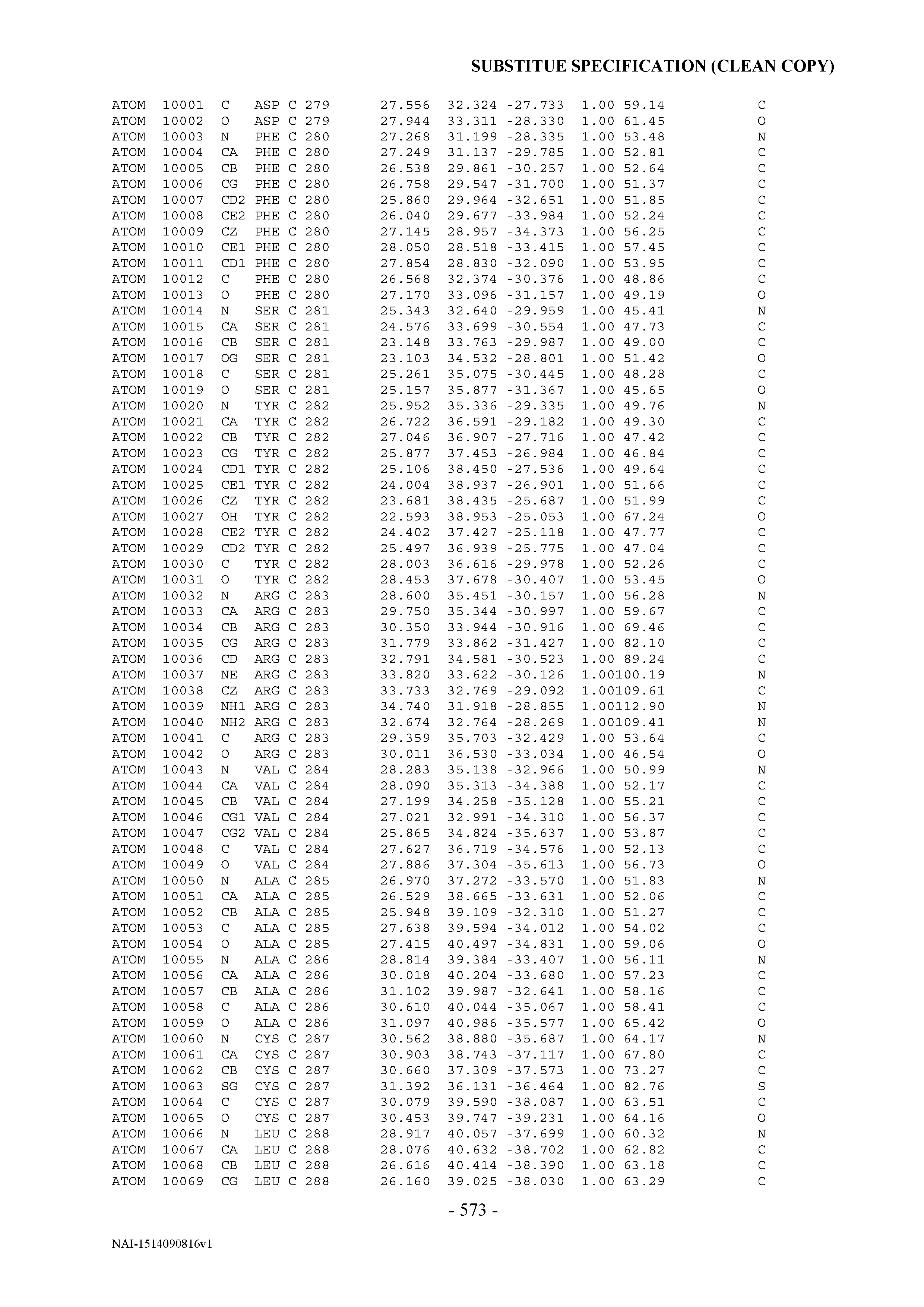

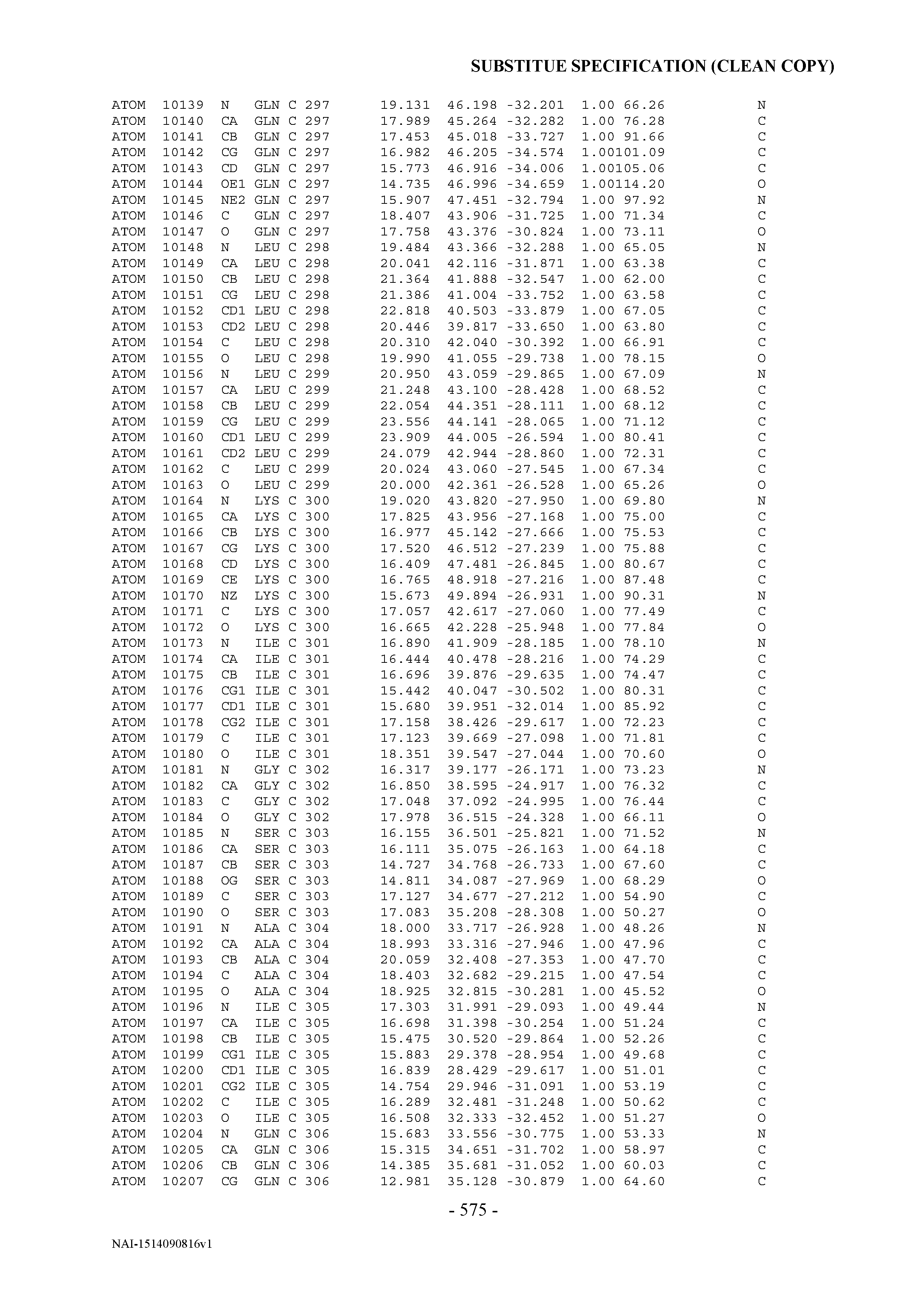
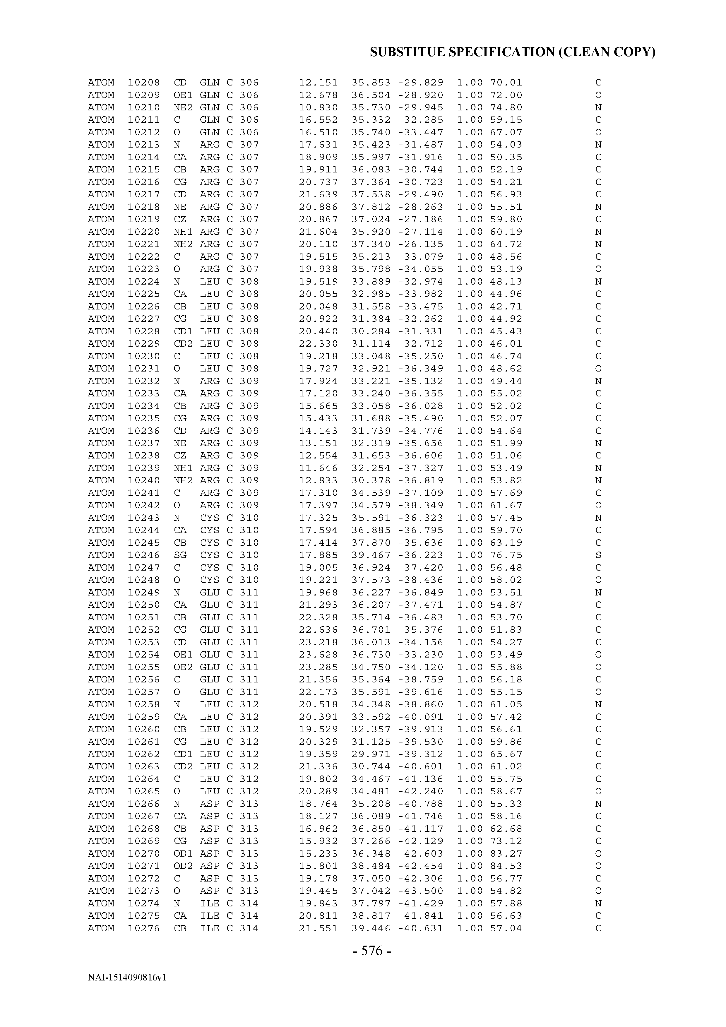
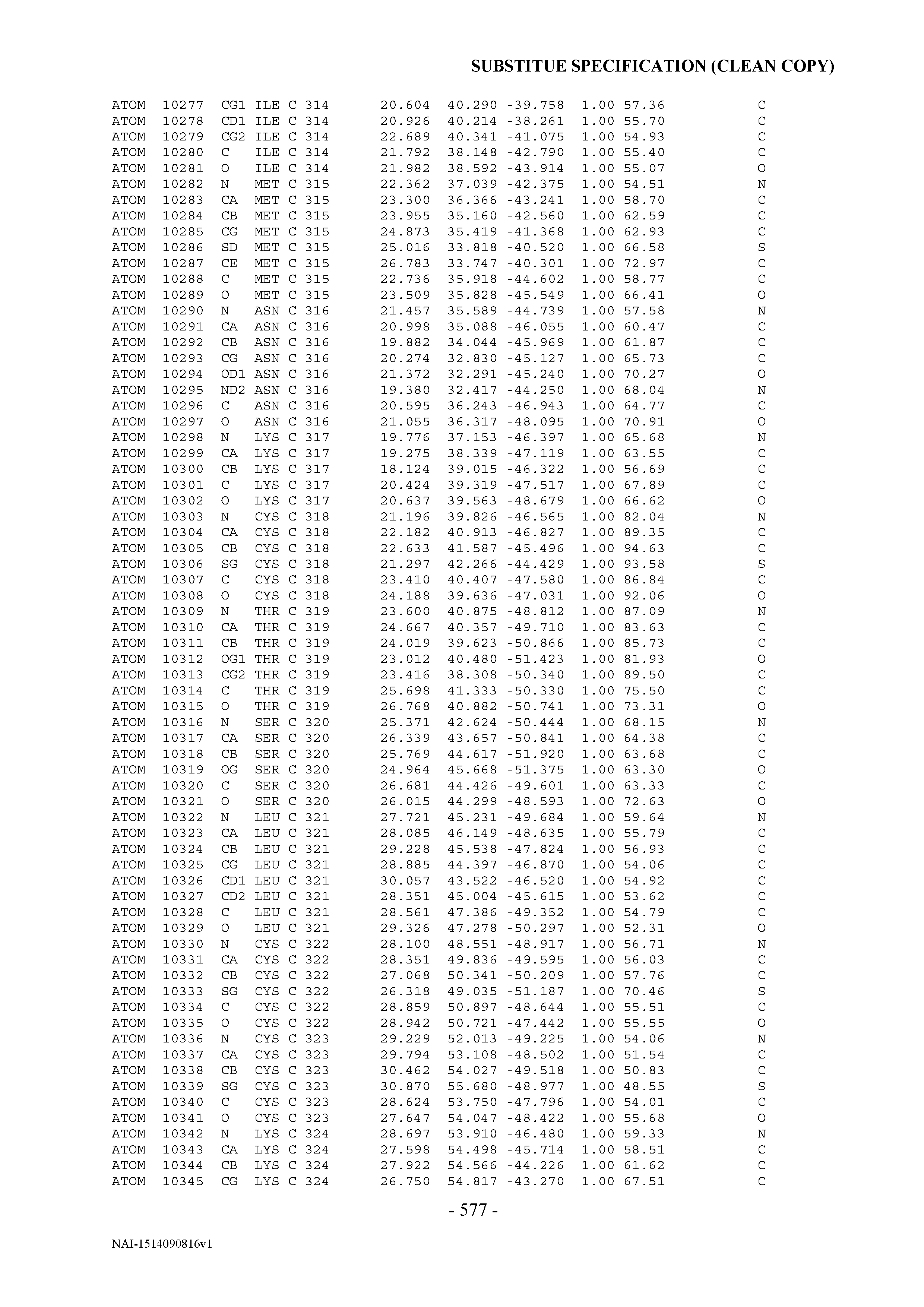


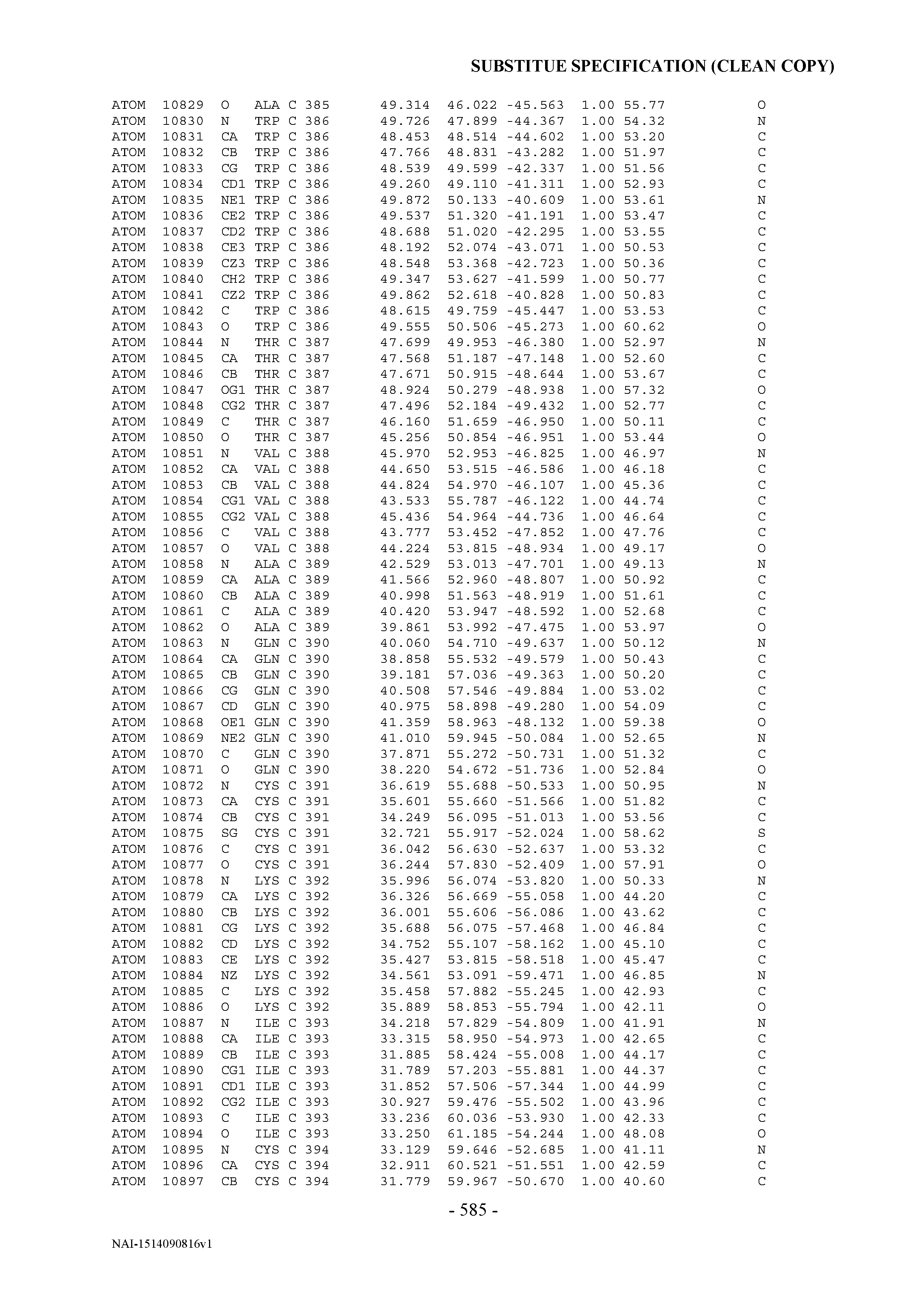
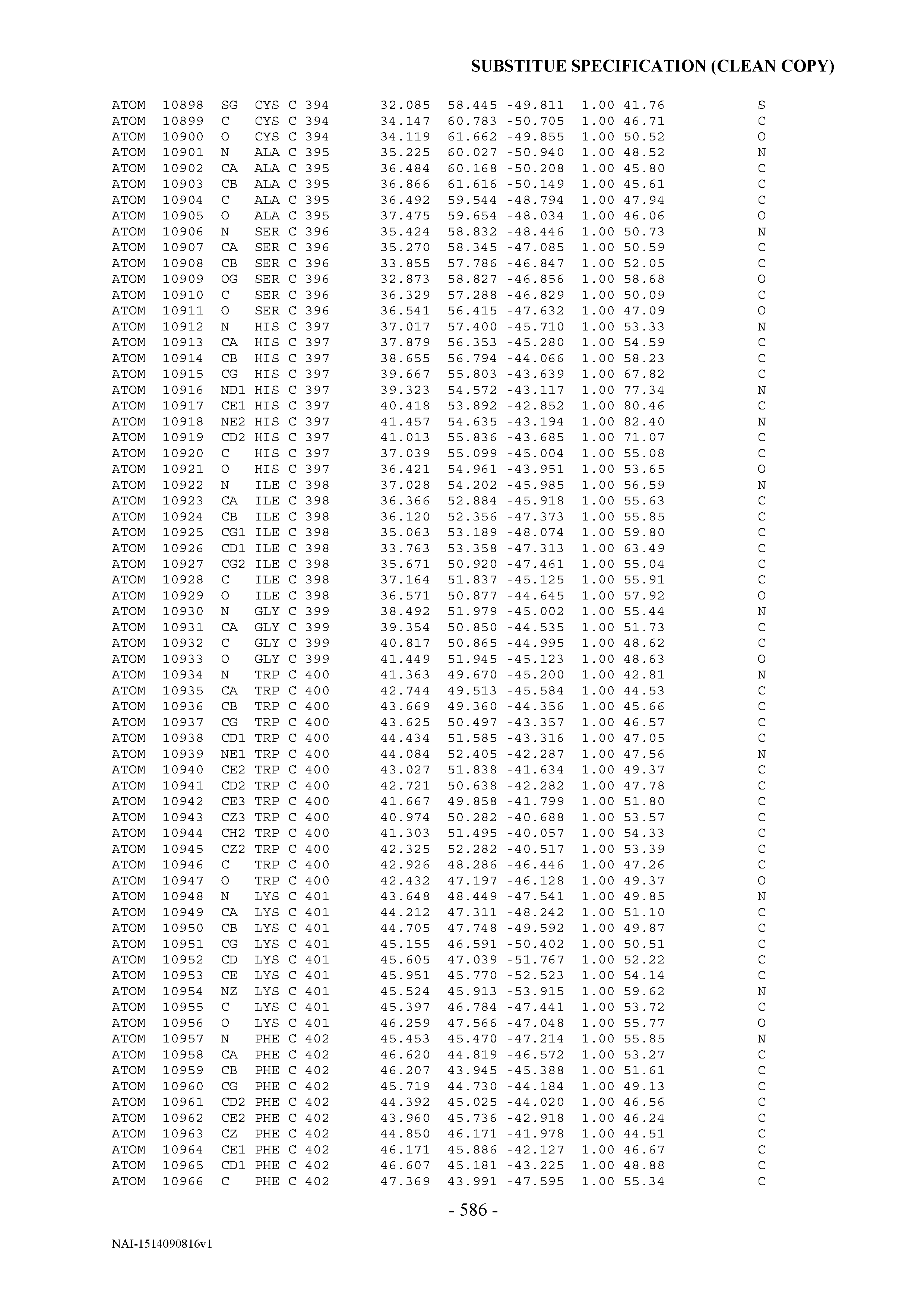


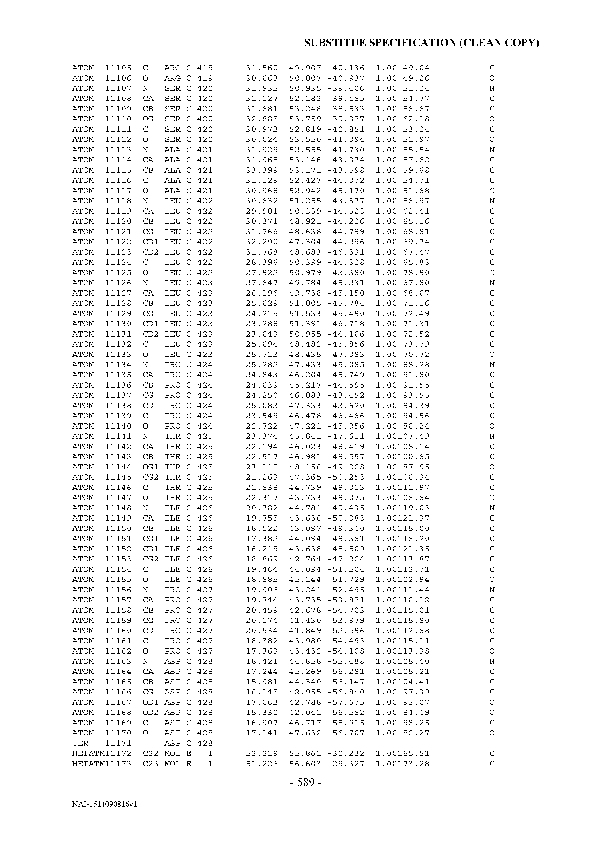
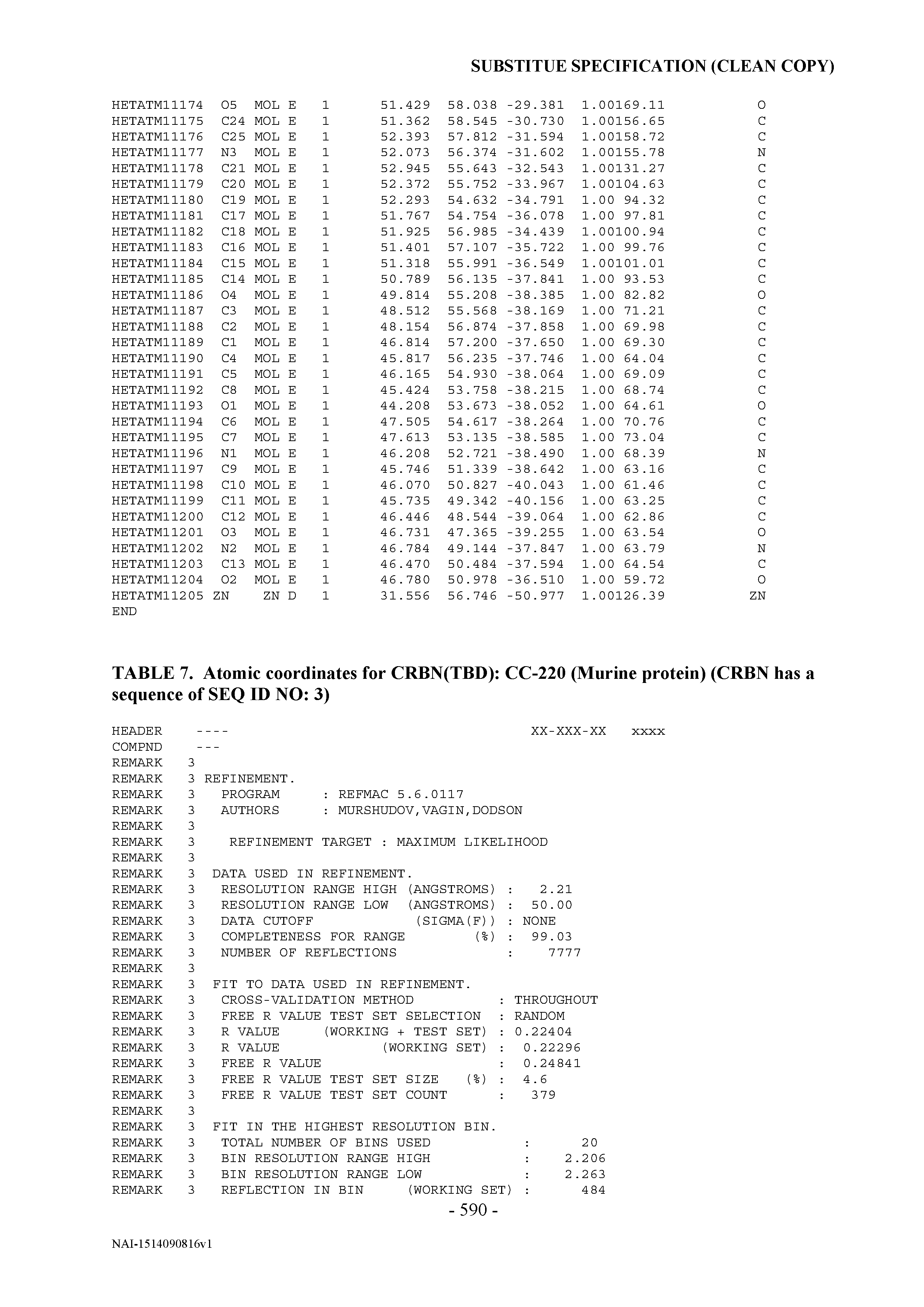
T00005


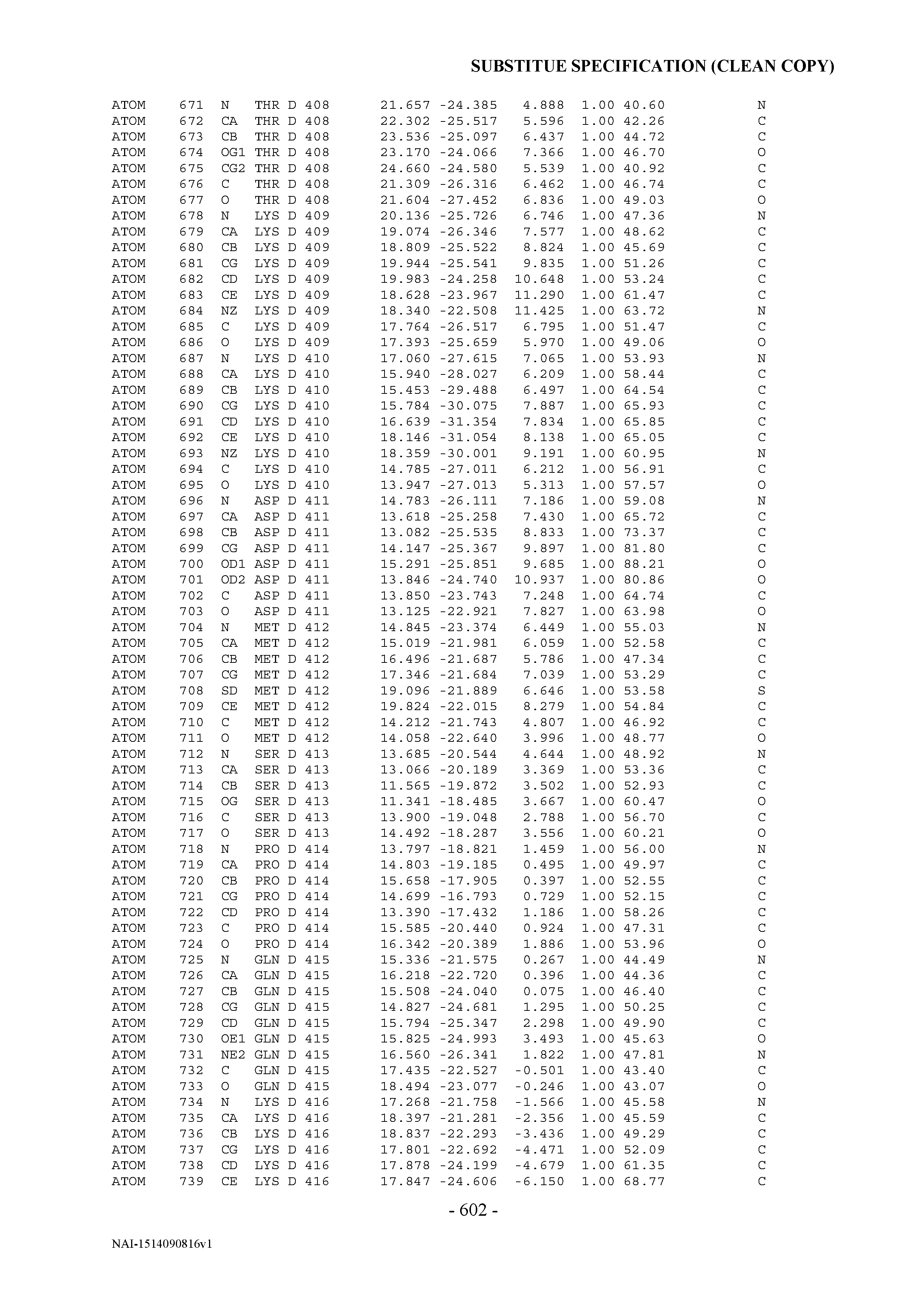

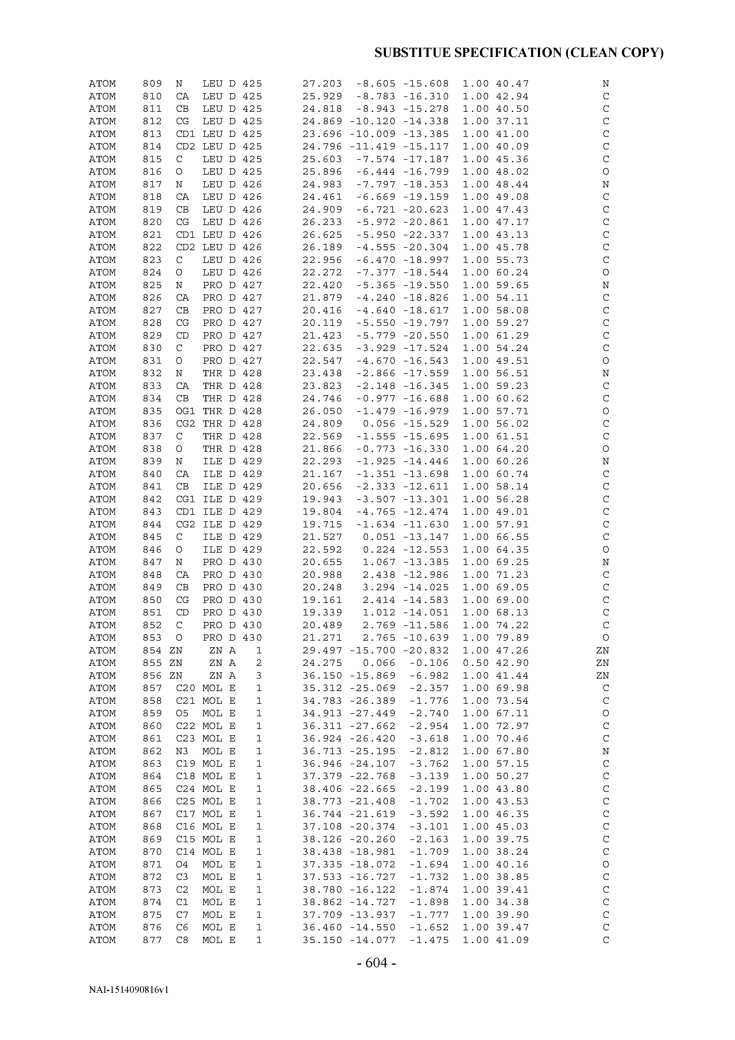
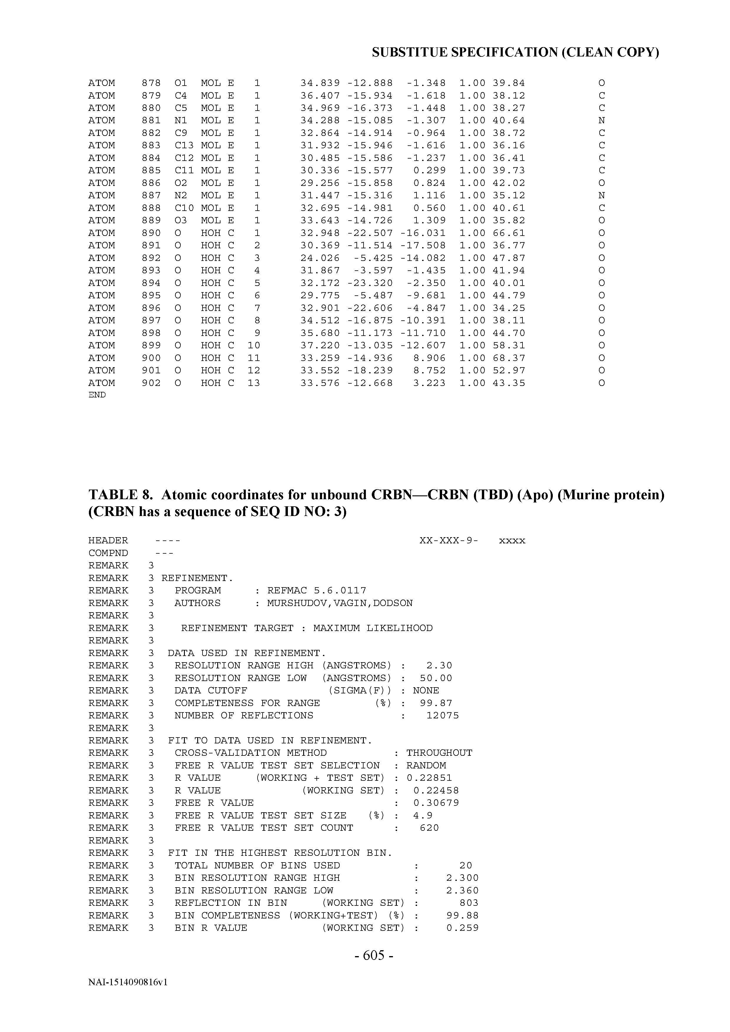
T00006


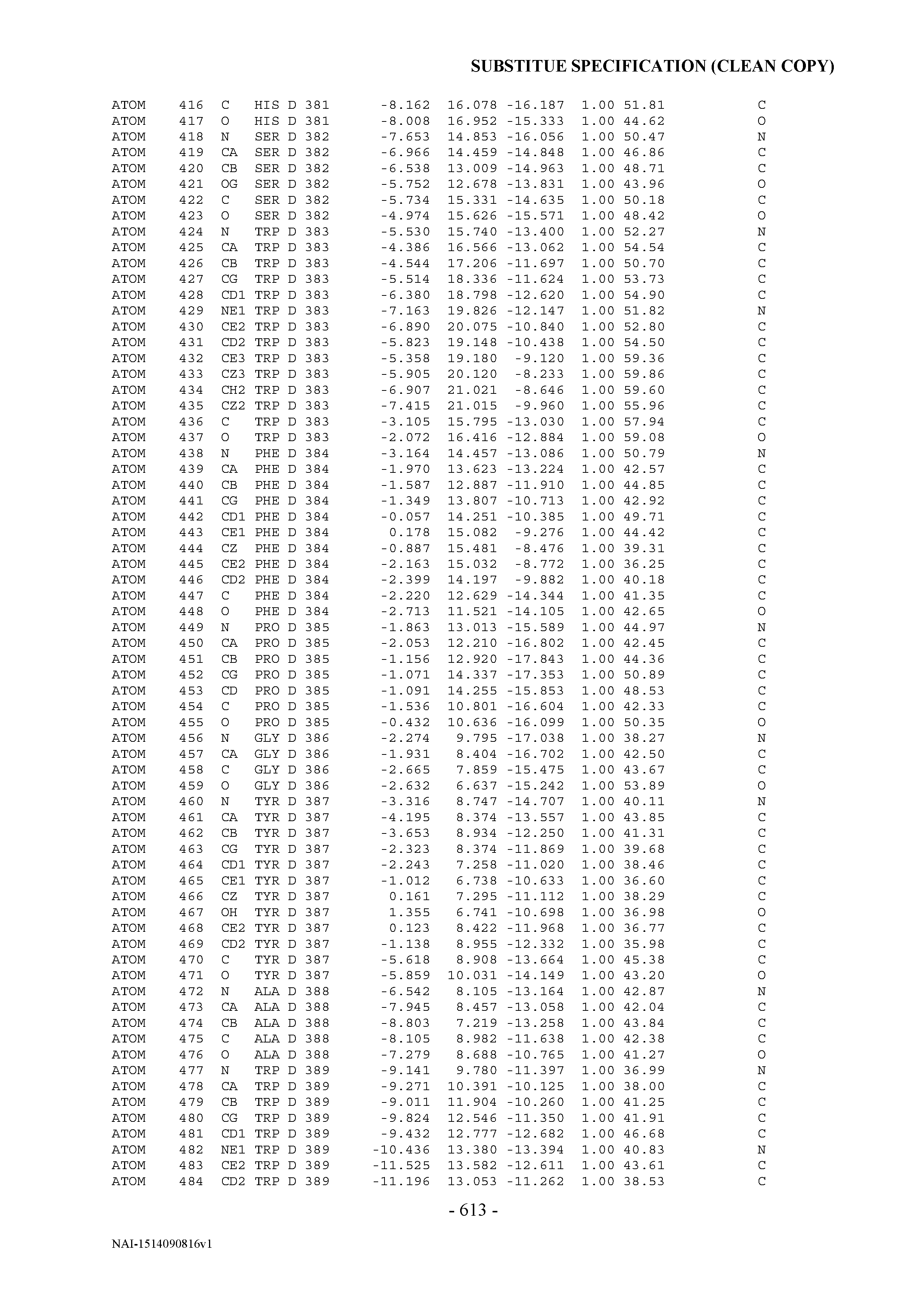
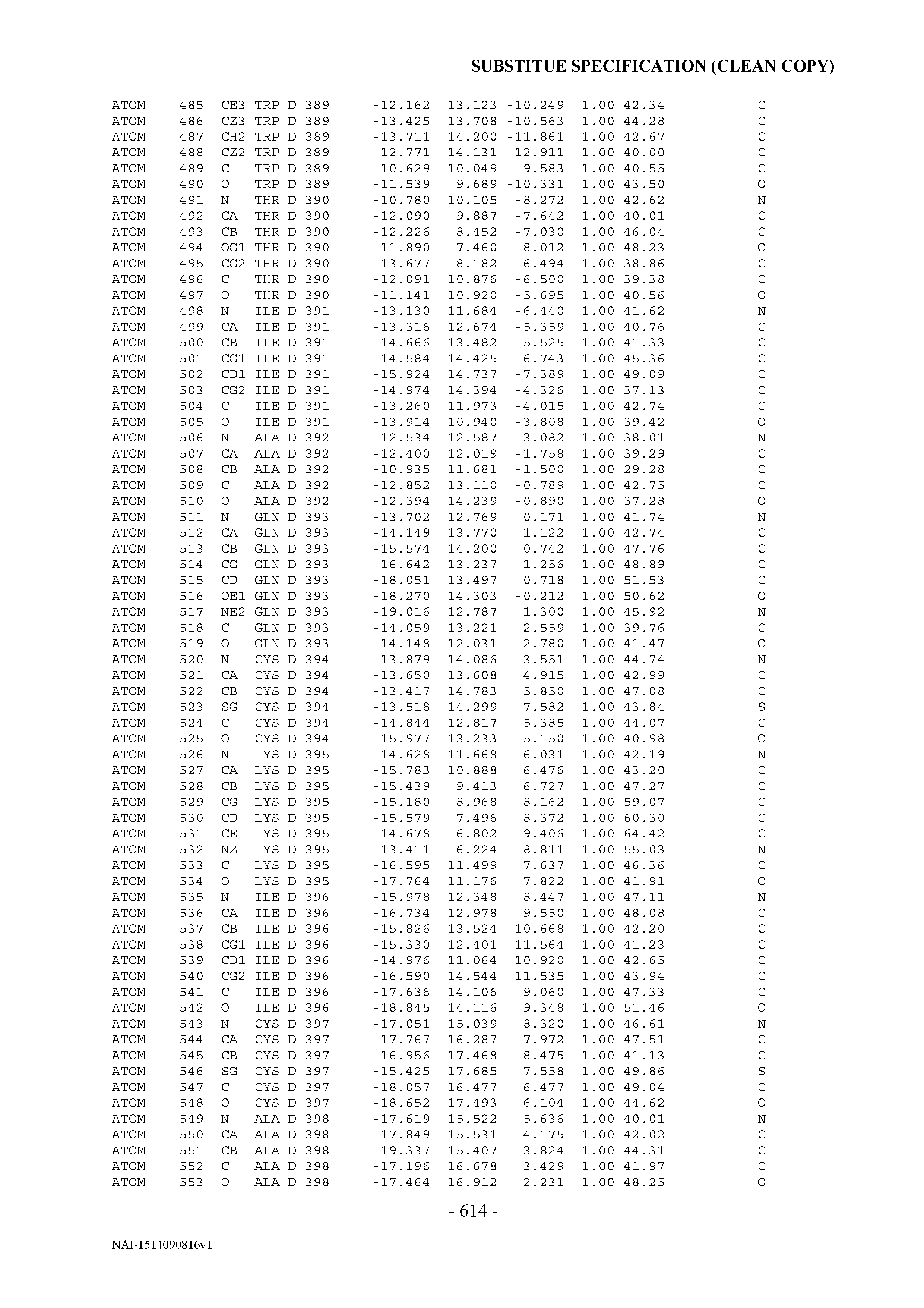


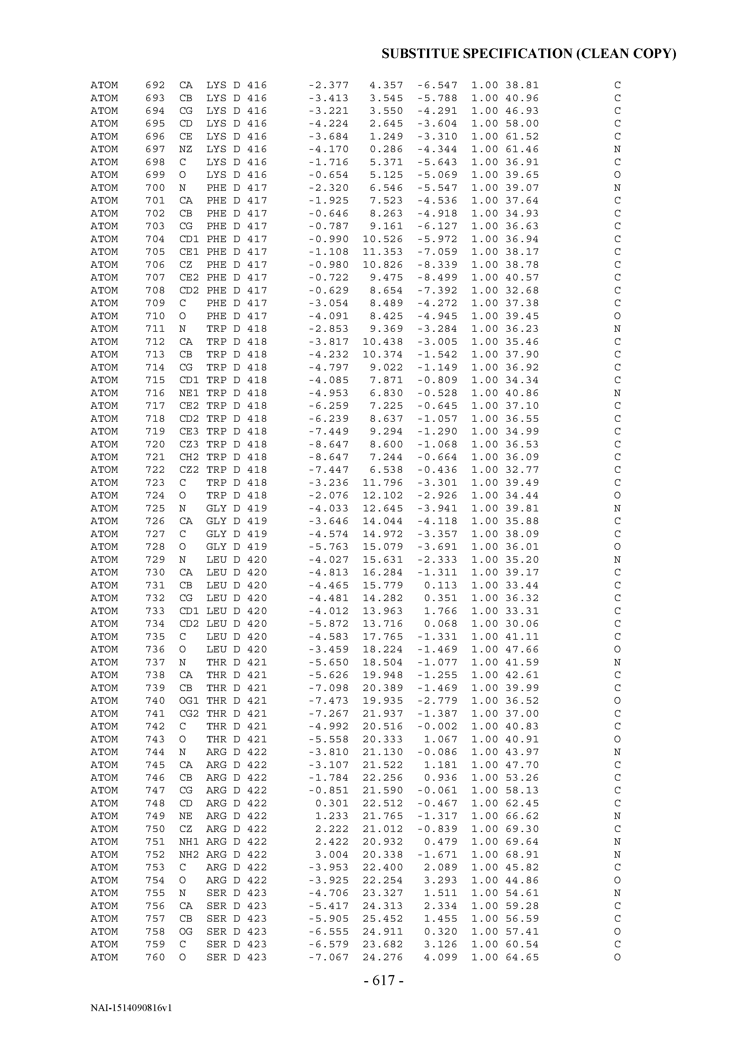
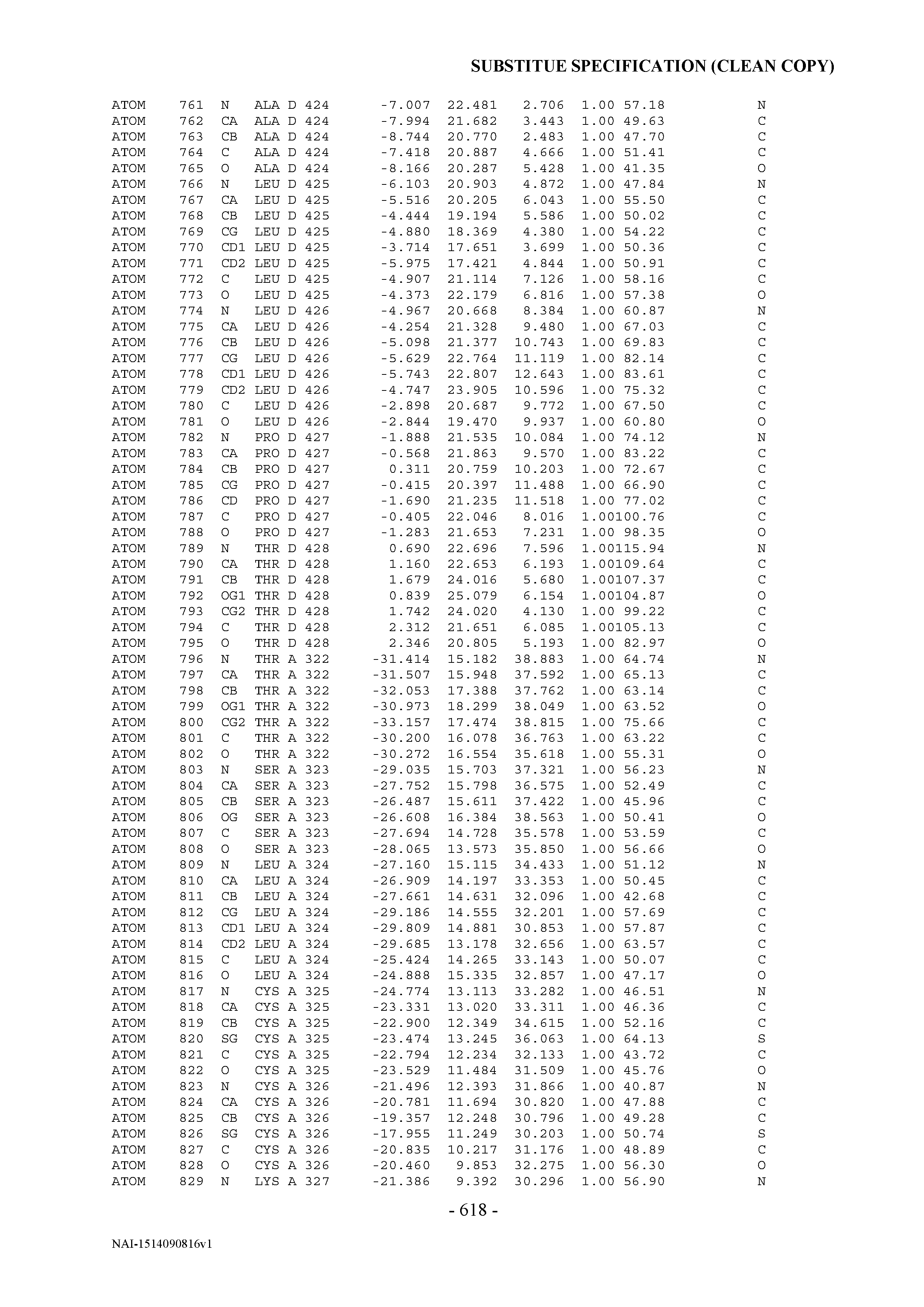


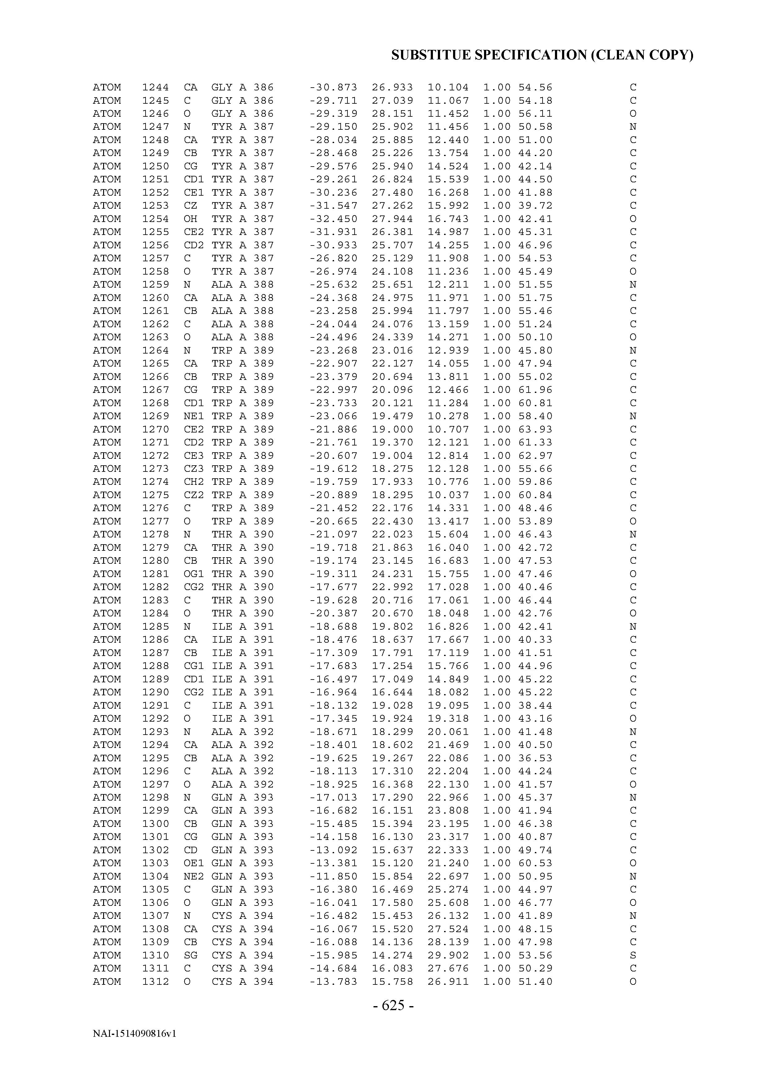


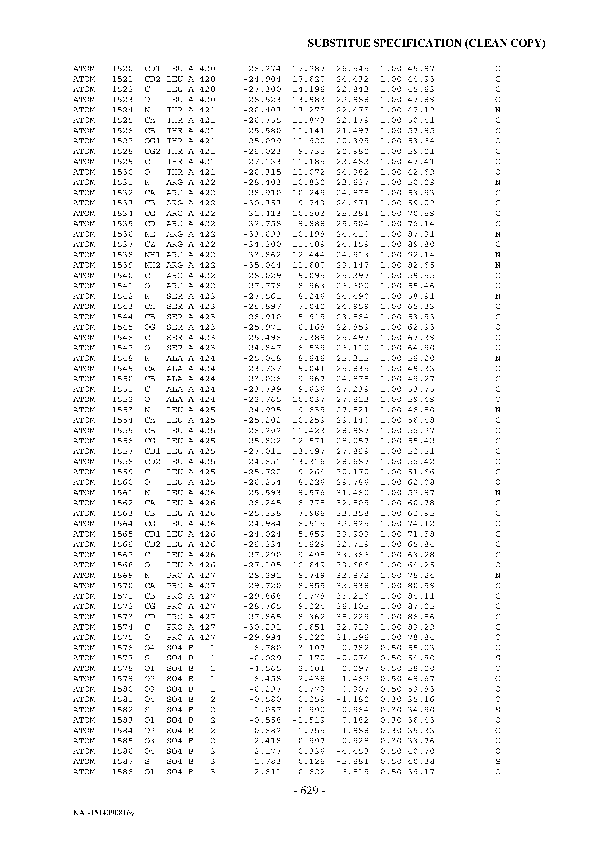
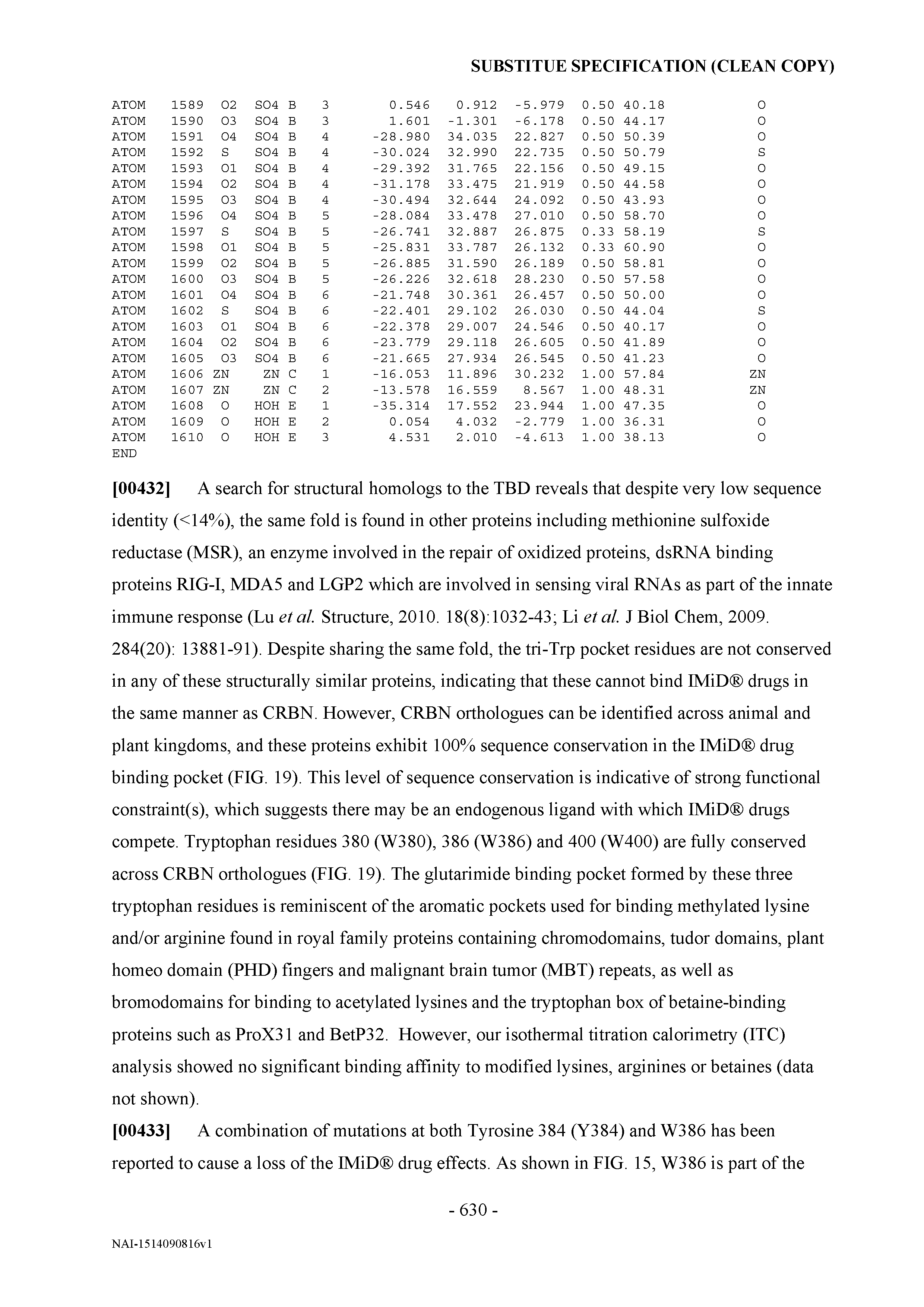
XML
uspto.report is an independent third-party trademark research tool that is not affiliated, endorsed, or sponsored by the United States Patent and Trademark Office (USPTO) or any other governmental organization. The information provided by uspto.report is based on publicly available data at the time of writing and is intended for informational purposes only.
While we strive to provide accurate and up-to-date information, we do not guarantee the accuracy, completeness, reliability, or suitability of the information displayed on this site. The use of this site is at your own risk. Any reliance you place on such information is therefore strictly at your own risk.
All official trademark data, including owner information, should be verified by visiting the official USPTO website at www.uspto.gov. This site is not intended to replace professional legal advice and should not be used as a substitute for consulting with a legal professional who is knowledgeable about trademark law.