Methods For Generating Genetically Modified Animals
Yang; Luhan
U.S. patent application number 16/607074 was filed with the patent office on 2020-12-31 for methods for generating genetically modified animals. The applicant listed for this patent is eGenesis, Inc.. Invention is credited to Luhan Yang.
| Application Number | 20200404891 16/607074 |
| Document ID | / |
| Family ID | 1000005122826 |
| Filed Date | 2020-12-31 |


View All Diagrams
| United States Patent Application | 20200404891 |
| Kind Code | A1 |
| Yang; Luhan | December 31, 2020 |
METHODS FOR GENERATING GENETICALLY MODIFIED ANIMALS
Abstract
The present disclosure provides methods of generating multiplexed genetically modified animals, for example, porcine endogenous retrovirus (PERV)-inactivated pigs. The disclosure also provides methods of improving the birth rate of multiplexed genetically modified animals. In some embodiments, the present closure is concerned with the generation and utilization of porcine cells in which porcine endogenous retroviral (PERV) elements have been inactivated. In sonic embodiments, the PERV-free or PERV-reduced porcine cells are cloned to produce porcine embryos. In some embodiments, the PERV-free or PERV-reduced embryos may be grown into adult swine from which organs and/or tissues may be extracted and used for such purposes as xenotransplantation into non-porcine animals such as humans.
| Inventors: | Yang; Luhan; (Brookline, MA) | ||||||||||
| Applicant: |
|
||||||||||
|---|---|---|---|---|---|---|---|---|---|---|---|
| Family ID: | 1000005122826 | ||||||||||
| Appl. No.: | 16/607074 | ||||||||||
| Filed: | April 20, 2018 | ||||||||||
| PCT Filed: | April 20, 2018 | ||||||||||
| PCT NO: | PCT/US2018/028539 | ||||||||||
| 371 Date: | October 21, 2019 |
Related U.S. Patent Documents
| Application Number | Filing Date | Patent Number | ||
|---|---|---|---|---|
| 62487898 | Apr 20, 2017 | |||
| 62500197 | May 2, 2017 | |||
| 62527702 | Jun 30, 2017 | |||
| 62543610 | Aug 10, 2017 | |||
| Current U.S. Class: | 1/1 |
| Current CPC Class: | C12N 15/8778 20130101; A01K 67/0278 20130101; C12N 2310/20 20170501; C12N 15/11 20130101; A01K 2217/072 20130101; C12N 9/22 20130101; C12N 2800/80 20130101; A01K 2267/02 20130101; A01K 2227/108 20130101 |
| International Class: | A01K 67/027 20060101 A01K067/027; C12N 15/877 20060101 C12N015/877; C12N 15/11 20060101 C12N015/11; C12N 9/22 20060101 C12N009/22 |
Claims
1. A swine grown from an embryo, wherein the embryo comprises porcine cells having at least 75% inactive porcine endogenous retroviral (PERV) elements.
2. The swine of claim 1, wherein about 100% of the PERV elements in the cells are inactive.
3. The swine of claim 1 or claim 2, wherein the PERV elements comprise one or more mutations or epigenetic changes that result in decreased or eliminated activity of PERV elements.
4. The swine of any one of claims 1-3, wherein the PERV elements of the porcine cell have been inactivated by a method comprising, administering to the cell a genome modifying agent specific to a gene involved in PERV replication and/or assembly, wherein the agent disrupts transcription and/or translation of the gene.
5. The swine of claim 4, wherein the agent is a nuclease or nickase or a nucleic acid encoding the nuclease or nickase.
6. The swine of claim 4, wherein the nuclease or nickase is a CRISPR-associated nuclease or nickase.
7. The swine of any one of claims 4-6, wherein the agent further comprises: a) a CRISPR guide RNA or tracrRNA or; b) a nucleic acid encoding the CRISPR guide RNA.
8. The swine of claim 7, wherein the CRISPR guide RNA comprises the nucleotide sequence of any one of SEQ ID NOs: 1-3 or 26-181, any strain specific genetic variant thereof, or any combination thereof.
9. The swine of claim 7, wherein the CRISPR guide RNA comprises the nucleotide sequence of any one of SEQ ID NOs: 1-3 or 26-116, any strain specific genetic variant thereof, or any combination thereof.
10. The swine of claim 7, wherein the CRISPR guide RNA comprises the nucleotide sequence of any one of SEQ ID NOs: 35, 36, 48, 99, 101, 102, 106, 108, 111, 113 or any combination thereof.
11. The swine of any one of claims 4-7, wherein the agent is a nucleic acid encoding the CRISPR-Cas9 nuclease or nickase, wherein the cell is engineered to stably express the agent, wherein the agent further comprises at least one guide RNA, and wherein at least one guide RNA sequence comprises the nucleotide sequence of any one of SEQ ID NOs: 1-3 or 26-116.
12. The swine of any one of the preceding claims, wherein the swine maintains a same or substantially same level of PERV inactivation for at least a month, at least 6 months, at least 1 year, at least 5 years, at least 10 years post-gestation.
13. An organ or tissue obtained from the swine of any one of the preceding claims.
14. A method of generating a porcine endogenous retrovirus (PERV)-inactivated pig comprising: i. obtaining a nuclear donor cell having a nucleus, wherein at least 75% of the PEP V elements in the nuclear donor cell are inactive; ii. transferring the nucleus of the nuclear donor cell into a recipient enucleated oocyte to generate nucleus-transferred oocyte; iii. subjecting the nucleus-transferred oocyte to activation; iv. culturing the nucleus-transferred oocyte to generate a blastocyst or embryo; v. transferring the blastocyst or embryo to a surrogate; and vi. generating a living PERV-inactivated pig from the blastocyst or embryo.
15. A method of generating a porcine endogenous retrovirus (PERV)-inactivated pig comprising: i. using a nucleus from a nuclear donor cell to generate a blastocyst or embryo, wherein at least 75% of the PERV elements in the nuclear donor cell are inactive; and transferring the blastocyst or embryo to a surrogate to generate a PERV-inactivated pig.
16. The method of claim 14 or 15 wherein the nuclear donor cell is a fetal cell.
17. The method of claim 14 or 15 wherein the nuclear donor cell is isolated from a chimeric PERV-inactivated fetus.
18. The method of claim 17 wherein the chimeric PERV-inactivated fetus is at about 10 days, about 20 days, about 30 days, or about 3 months gestation.
19. The method of claim 17 wherein the chimeric PERV-inactivated fetus is generated using a genuine modifying agent.
20. The method of claim 19 wherein the genome modifying agent is selected from the group consisting of Zinc Finger nuclease or nickase, TAL effector nuclease or nickase, deaminase, and CRISPR associate nuclease or nickase.
21. The method of claim 14 or 15 wherein the nuclear donor cell undergoes less than 30, less than 20, less than 10, less than 5, or less than 2 population doublings in vitro.
22. The method of claim 14 or 15 wherein the nuclear donor cell is isolated from a pig.
23. The method of claim 22 wherein the pig is less than 10 weeks, less than 8 weeks, less than 6 weeks, less than 5 weeks, less than 4 weeks, less than 3 weeks, less than 2 weeks, or less than 1 week in age.
24. The method of claim 14 or 15 wherein at least about 80%, at least about 90%, at least about 95%, at least about 99% of the PERlsi elements in the nuclear donor cell are inactive.
25. The method of claim 14 or 15 wherein the PERV-inactivated pig maintains a same or similar level of PERV inactivation for at least a month, at least 6 months, at least 1 year, at least 5 years, at least 10 years post-gestation.
26. The method of claim 14 or 15 further comprising transferring at least one wild type blastocyst or embryo to the surrogate.
27. An isolated porcine cell generated by the methods of any one of claims 14-26.
28. An organ or tissue obtained from a cell generated by the methods of any one of claims 14-26.
29. A method of improving the birth rate of PERV-inactivated pigs comprising: i. using nuclei from nuclear donor cells to generate blastocysts or embryos, wherein at least 75% of the PERV elements in the nuclear donor cells are inactive; and ii. transferring the blastocysts or embryos to at least one surrogate to generate at least one PERV-inactivated, wherein the miscarriage rate is reduced as compared to the miscarriage rate of fetuses generated from a cell wherein more than 25% of the PERV elements in the cell are active.
30. A method of generating a genetically modified animal comprising: i. using a nucleus from a nuclear donor cell to generate a blastocyst or embryo, wherein a plurality of nucleic, acid sequences in the nuclear donor cell are modified; and ii. transferring the blastocyst or embryo to a surrogate to generate the genetically modified animal.
31. The method of claim 29 or 30 wherein the nuclear donor cell is a fetal cell.
32. The method of claim 29 or 30 wherein the nuclear donor cell is isolated from a chimeric PERV-inactivated fetus.
33. The method of claim 32 wherein the chimeric PERV-inactivated fetus is at about 10 days, about 20 days, about 30 days, or about 3 months gestation.
34. The method of claim 32 wherein the chimeric PERV-inactivated fetus is generated using a genome modifying agent.
35. The method of claim 32 wherein the chimeric PERU-inactivated fetus is generated by zygote injection.
36. The method of claim 34 wherein the genome modifying agent is selected from the group consisting of Zinc Finger nuclease or nickase, TAL effector nuclease or nickase, deaminase, and CRISPR associate nuclease or nickase.
37. The method of claim 29 or 30 wherein the nuclear donor cell undergoes less than 30, less than 20, less than 10, less than 5, or less than 2 population doublings ire vitro.
38. The method of claim 31 wherein the pig is less than 10 weeks, less than 8 weeks, less than 6 weeks, less than 5 weeks, less than 4 weeks, less than 3 weeks, less than 2 weeks, or less than 1 week in age.
39. The method of claim 29 or 30 wherein at least about 80%, at least about 90%, at least about 95%, at least about 99% of the PEW elements in the nuclear donor cell are inactive.
40. The method of claim 29 or 30 wherein 100% of the PERV elements in the nuclear donor cell are inactive.
41. The method of claim 29 or 30 wherein the PERV-inactivated pig maintains a same or similar level of PERV inactivation for at least a month, at least 6 months, at least 1 year, at least 5 years, at least 10 years post-gestation.
42. The method of claim 29 or 30 wherein the PERV-inactivated pig is a PERV-free pig.
43. The method of claim 29 or 30 wherein the PERV-inactivated pig has at least 75%, at least 80%, at least 85%, at least 90%, at least 95%, or at least 99% inactive PERV elements.
44. The method of claim 29 or 30 further comprising transferring at least one wild type blastocyst or embryo to the surrogate.
45. A method of preventing or reducing risk of pregnancy loss or miscarriage following somatic cell nuclear transfer (SCT) of a genetically modified blastocyst or embryo comprising: i. using a nucleus from a genetically modified nuclear donor cell to generate a blastocyst or embryo; and ii. transferring the blastocyst or embryo to a surrogate to generate at least one a viable offspring wherein the rate of pregnancy loss or miscarriage is reduced as compared to the rate of pregnancy loss or miscarriage of a control.
46. The method of claim 45 wherein the genetically modified nuclear donor cell has a plurality of genetic modifications.
47. The method of claim 46 wherein the plurality of genetic modifications comprise at least about 2, at least about 3, at least about 4, at least about 5, at least about 10, at least about 15, at least about 20, at least about 30, at least about 40, at least about 50, at least about 60, at least about 70, at least about 80, at least about 90, or at least about 100 genetic modifications.
48. The method of claim 45 wherein the plurality of genetic modifications are to a single, repetitive gene sequence.
49. The method of claim 45 wherein at least a portion of the genetic modifications are to different genes.
50. The method of claim 45 wherein the genetically modified nuclear donor cell has been modified using a genome modifying agent selected from the group consisting of Zinc Finger nuclease or nickase, TAL effector nuclease or nickase, deaminase, and CRISPR associate nuclease or nickase.
Description
PRIORITY CLAIM
[0001] This application claims priority to U.S. Provisional Application No. 62/487,898, filed Apr. 20, 2017, U.S. Provisional Application No. 62/527,702, filed Jun. 30, 2017, and U.S. Provisional Application No. 62/543,610, filed Aug. 10, 2017, the disclosures of which are incorporated by reference herein in their entireties.
BACKGROUND OF THE DISCLOSURE
[0002] The shortage of human organs and tissues for transplantation represents a significant unmet medical need. In the United States and Europe alone, approximately 200,000 patients await organ transplantation, but only a small fraction of these patients ever receives donor organs (U.S. Department of Health and Human Services, Organ Procurement and Transplantation Network-2017; European Commission, Journalist workshop on organ donation and transplantation, Recent Facts & Figures, 2016). Several strategies have been envisioned to alleviate the critical shortage of donor organs, including developing organs in vitro from human induced pluripotent stem cells (hiPSCs), repopulating decellularized organ scaffolds, constructing 3D-printed organs, producing chimeric animals with human organs, and performing cross-species transplantation (i.e.; xenotransplantation), in particular, xenotransplantation is an attractive potential solution to the organ donation and transplantation shortage. Porcine organs are considered favorable resources for xenotransplantation since they are similar to human organs in size and function. Furthermore, pigs can be bred in large numbers under designated pathogen-free conditions and used in commercial production (Xenotransplantation 2.0, 2016, Nat. Biotechnol, 34:1).
[0003] Xenotransplantation has the potential to provide an almost unlimited supply of transplant organs for patients with chronic organ failure. However, the clinical use of porcine organs has been hindered by immunological incompatibilities and by the potential risk of porcine endogenous retroviral (PERK) element transmission. PERVs are proviruses in the porcine genome that originally became integrated in germ line chromosomes during exogenous retroviral infections (Gifford, R. & Tristem, M., 2003, Virus Genes 26, 291-315). There are three major subtypes of PERVs: PERV A, PERV B, and PERV C. Subtypes A and B are ubiquitous and can be transmitted to pigs and humans, whereas C is only present in some pig strains and can be transmitted among pigs (Patience et al., 2001, J. Virol, 75:2771-2775). PERVs, by definition, are not shared by other species and are typically the result of recent retroviral infections (Gifford and Tristem).
[0004] Most PERVs have become inactive over time with accumulated mutations, but certain intact PERVs have been shown to infect human cells in vitro (Patience et al., 1997, Nat. Med., 3:282-286; Yang et al., 2015, Science, 350:110141104). These intact PERVs pose a potential risk of zoonosis in pig-to-human xenotransplantation (Denner et al., 2016, Xenotransplantation, 23:53-59; Denner J and Tonjes R, 2012, Clin Microbiol Rev, 2.5:318-343), Because mutagenesis from PERV integration could potentially lead to tumorigenesis and immunodeficiency, as reported with other retroviruses (Bendinelli et al., 1985, Advances in Cancer Research, 45:125-181), PERVs have been considered to be a major safety concern in the context of clinical xenotransplantation. Previously, several groups have attempted to inactivate PERVs using TAL effector nucleases (Dunn et al., 2015, FASEB J, 29:LB761) or Zinc Finger Nucleases (Semaan et al., 2015, PLUS One, 10), but these attempts have been unsuccessful.
[0005] Genetically modified animals, engineered to have deliberate modifications in their genomes, have been used for decades for scientific purposes, to improve livestock, and for production of recombinant proteins, among other uses. Many techniques have been developed for generating genetically modified animals including transcription activator-like effector nuclease (TALEN), zinc finger nuclease (ZEN), deaminases, and clustered regularly interspaced short palindromic repeats (CRISPR)-based systems, such as CRISPR-Cas9. Recently, these techniques have been employed to target multiple nucleic acid sequences for multiplexed genome editing in vitro. However, the success rate of a live birth and/or survival after birth for genetically modified animals depends on numerous factors including the technique used, genomic region(s) targeted, and the species of animal modified, but the rate is lower than for non-genetically modified animals.
[0006] As such, there is a need for developing PERV-free pigs as a source of cells, tissues, and/or organs for transplantation to humans and for techniques to improve the birth and/or survival rate of genetically modified animals, in particular, those engineered to have multiple genetic modifications.
SUMMARY OF THE DISCLOSURE
[0007] In some aspects the disclosure provides swine grown from an embryo, wherein the embryo comprises porcine cells having at least 75% inactive porcine endogenous retroviral (PERV) elements.
[0008] In some aspects the disclosure provides methods of generating a porcine endogenous retrovirus (PERV)-inactivated pig comprising: obtaining a nuclear donor cell having a nucleus, wherein at least 75% of the PERV elements in the nuclear donor cell are inactive; transferring the nucleus of the nuclear donor cell into a recipient enucleated oocyte to generate nucleus-transferred oocyte; subjecting the nucleus-transferred oocyte to activation; culturing the nucleus-transferred oocyte to generate a blastocyst or embryo; transferring the blastocyst or embryo to a surrogate; and generating a living PERV-inactivated pig from the blastocyst or embryo.
[0009] In some aspects the disclosure provides methods of generating a porcine endogenous retrovirus (PERV)-inactivated pig comprising: using a nucleus from a nuclear donor cell to generate a blastocyst or embryo, wherein at least 75% of the PERV elements in the nuclear donor cell are inactive; and transferring the blastocyst or embryo to a surrogate to generate a PERV-inactivated pig.
[0010] In some aspects the disclosure provides methods of improving the birth rate of PERV-inactivated pigs comprising: using nuclei from nuclear donor cells to generate blastocysts or embryos, wherein at least 75% of the PERV elements in the nuclear donor cells are inactive; and transferring the blastocysts or embryos to at least one surrogate to generate at least one PERV-inactivated pig, wherein the miscarriage rate is reduced as compared to the miscarriage rate of fetuses generated from a cell wherein more than 25% of the PERV elements in the cell are active.
[0011] In some aspects the disclosure provides methods of generating a genetically modified animal comprising: using a nucleus from a nuclear donor cell to generate a blastocyst or embryo, wherein a plurality of nucleic acid sequences in the nuclear donor cell are modified; and transferring the blastocyst or embryo to a surrogate to generate the genetically modified animal.
[0012] In some aspects the disclosure provides methods of preventing or reducing risk of pregnancy loss or miscarriage following somatic cell nuclear transfer (SCNT) of a genetically modified blastocyst or embryo comprising: using, a nucleus from a genetically modified nuclear donor cell to generate a blastocyst or embryo; and transferring the blastocyst or embryo to a surrogate to generate at least one a viable offspring wherein the rate of pregnancy loss or miscarriage is reduced as compared to the rate of pregnancy loss or miscarriage of a control.
[0013] Also provided are organs or tissues obtained from the swine of any of the embodiments disclosed and described herein. Also provided are isolated porcine cells generated by any of the methods disclosed and described herein.
[0014] In some embodiments, about 100% of the PERV elements in the cells are inactive. In some embodiments, 100% of the PERV elements in the cells are inactive.
[0015] In some embodiments, the PERV elements comprise one or more mutations or epigenetic changes that result in decreased or eliminated activity of PERV elements.
[0016] In some embodiments, the PERV elements of the porcine cell have been inactivated by a method comprising, administering to the cell a genome modifying agent specific to a gene involved in PERV replication and/or assembly, wherein the agent disrupts transcription and/or translation of the gene. In some embodiments, the agent is a nuclease or nickase or a nucleic acid encoding the nuclease or nickase, for example, the nuclease or nickase is a CRISPR-associated nuclease or nickase. In some embodiments, the agent further comprises: a) a CRISPR guide RNA car tracrRNA or; b) a nucleic acid encoding the CRISPR guide RNA.
[0017] In some embodiments, the CRISPR guide RNA comprises the nucleotide sequence of any one of SEQ ID NOs: 1-3 or 26-181, any strain specific genetic variant thereof, or any combination thereof. In some embodiments, the CRISPR guide RNA comprises the nucleotide sequence of any one of SEQ ID NOs: 1-3 or 26-116, any strain specific genetic variant thereof, or any combination thereof. In some embodiments, the CRISPR guide RNA comprises the nucleotide sequence of any one of SEQ ID NOs: 35, 36, 48, 99, 101, 102, 106, 108, 111, 113, or any combination thereof. In some embodiments, the agent is a nucleic acid encoding the CRISPR-Cas9 nuclease or nickase, wherein the cell is engineered to stably express the agent, wherein the agent further comprises at least one guide RNA, and wherein at least one guide RNA sequence comprises the nucleotide sequence of any one of SEQ. ID NOs: 1-3 or 26-116.
[0018] In some embodiments, the swine maintains a same or substantially same level of VERY inactivation for at least a month, at least 6 months, at least 1 year, at least 5 years, at least 10 years post-gestation.
[0019] In some embodiments, the nuclear donor cell is a somatic cell, a fetal cell, a germline cell, a stem cell, or and induced pluripotent stem cell (iPSC). In some embodiments, the nuclear donor cell is a fetal cell. In some embodiments, the nuclear donor cell is isolated from a chimeric PERU inactivated fetus. In some embodiments, the chimeric. PER inactivated fetus is at about 10 days, about 20 days, about 30 days, or about 3 months gestation.
[0020] In some embodiments, the chimeric PERV-inactivated fetus is generated using a genome modifying agent, for example, Zinc Finger nuclease or nickase, TAL effector nuclease or nickase, deaminase, and CRISPR associate nuclease or nickase.
[0021] In some embodiments, the nuclear donor cell undergoes less than 30, less than 20, less than 10, less than 5, or less than 2 population doublings in vitro. In some embodiments, the nuclear donor cell is isolated from a pig. In some embodiments, the nuclear donor cell is isolated from the pig when the pig is less than 10 weeks, less than 8 weeks, less than 6 weeks, less than 5 weeks, less than 4 weeks, less than 3 weeks, less than 2 weeks, or less than 1 week in age. In some embodiments, at least about 80%, at least about 90%, at least about 95%, at least about 99% of the PEPS elements in the nuclear donor cell are inactive.
[0022] In some embodiments, the PERV-inactivated pig maintains a same or similar level of PERV inactivation for at least a month, at least 6 months, at least 1 year, at least 5 years, at least 10 years post-gestation.
[0023] In some embodiments, the methods further comprise transferring at least one wild type blastocyst or embryo to the surrogate.
[0024] In some embodiments, at least about 80%, at least about 90%, at least about 95%, at least about 99% of the PERV elements in the nuclear donor cell are inactive, in some embodiments, 100% of the PERV elements in the nuclear donor cell are inactive.
[0025] In some embodiments, the PERV-inactivated pig maintains a same or similar level of Fe RV inactivation for at least a month, at least 6 months, at least 1 year, at least 5 years, at least 10 years post-gestation. In some embodiments, the PERV-inactivated pig is a PERV-free pig. In some embodiments, the PERV-inactivated pigs has at least about 75%, at least about 80%, at least about 85%, at least about 90%, at least about 95%, or at least about 99% inactive PERV elements.
BRIEF DESCRIPTION OF THE DRAWINGS
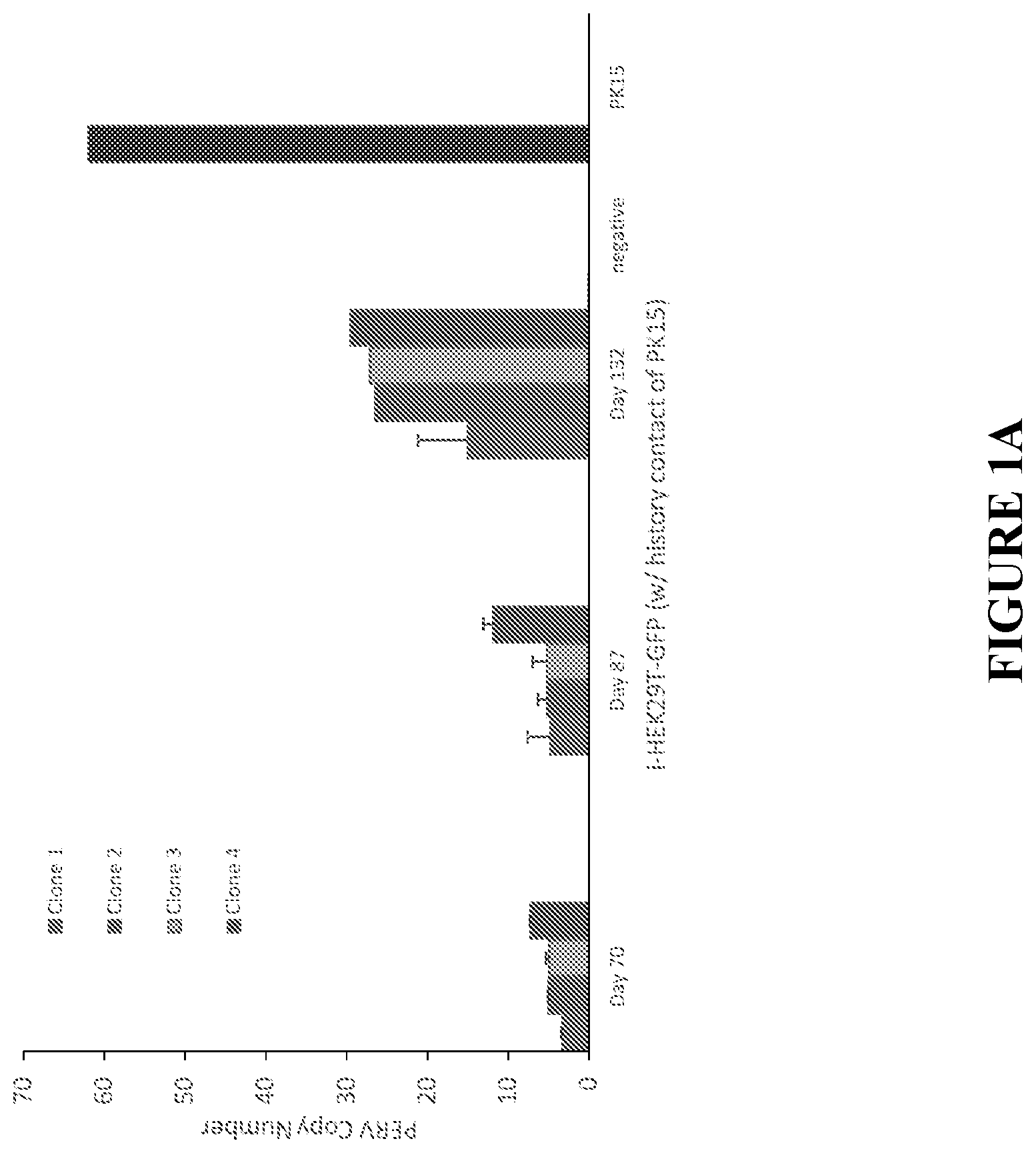
[0026] FIG. 1A shows that PERV copy number in infected human cells increases over time. Human HEK293T-GFP cells were co-cultured with equivalent numbers of pig PK15 cells for one week. The HEK293T-GFP single cells were isolated by flow cytometry based on the GFP signal and subsequently grown into clones. The HEK293T-GFP clones were isolated and cultured for the period of 70, 87 and 132 days. PERV copy numbers were analyzed by ddPCR using PEPS primers. HEK293T-GFP without any contact of PK15 cells were used as control.
[0027] FIG. 1B shows the PERV insertion sites in highly infected i-HEK293T-GFP clones. Among the 22 PERV insertion sites detected by inverse PCR, 15 were mapped to the intragenic region, A portion of the intragenic hits were tested, Seven out of 12 were validated by junction PCR and are shown. The 30 bp human genomic sequences are shown with solid underlining, whereas the PERV LTRs are shown in double underlining. The genomic sequences are aligned to the UCSC Genome Browser and the corresponding genes are listed.
[0028] FIG. 1C shows a simplified schematic of the experimental design used to examine human-to-human PERVs transmission, PERVs-infected with human HEK293T cell labeled with GRP (i-HEK293T-GFP) were co-cultured with unlabeled WT HEK293T (HEK293T) for 14 days. The HEK293T clones were isolated from the co-culture and genomic DNA was extracted to determine whether the HEK293T clones were infected by PERV from i-HEK293T-GFP cells.
[0029] FIG. 1D shows the detection of human-to-human PERV transmission, individual clones of HEK293T were grown from the single cells isolated from the co-culture of i-HEK293T-GFP with HEK293T through flow cytometry. The PCR gel image shows that 3 out of 4 randomly tested HEK293T clones contained PERV sequences (PERV pol, env, and gag), but no sequence of GFP or pig genomic DNA (tested by pig specific GGTA). Sample orders are: 1) HEK293T clone 1; 2) HEK293T clone 2; 3) HEK293T clone 3; 4) HEK293T clone 4; 5) HEK293T-GFP control; 6) i-HEK293T-GFP; 7) PK15 WT; and 8) negative control,
[0030] FIG. 1E illustrates that different i-HEK293T-GFP clones have different infectious potential. The percentages of infected HEK293T clones from the co-culture of i-HEK293T-GFP and WT HEK2931 varied from 20% to 97%. The percentage corresponds to the PERV copy number of i-HEK293T-GFP clones in the culture. The PERV copy number of the 4 parent i-HEK293T-GFP clones are: 15, 28, 27 and 28, respectively.
[0031] FIG. 2A shows chromosome mapping of PERV insertion sites. PERV integration sites on the chromosome were detected in the WT FFF3 cell line using hybridization capture and long reads Pacbio sequencing. Gray bars represent chromosomal scaffolds. Arrows (pointing up) represent PERVs in forward or plus chromosome strand. Arrows (pointing down) represent PERVs in the reverse or negative strand.
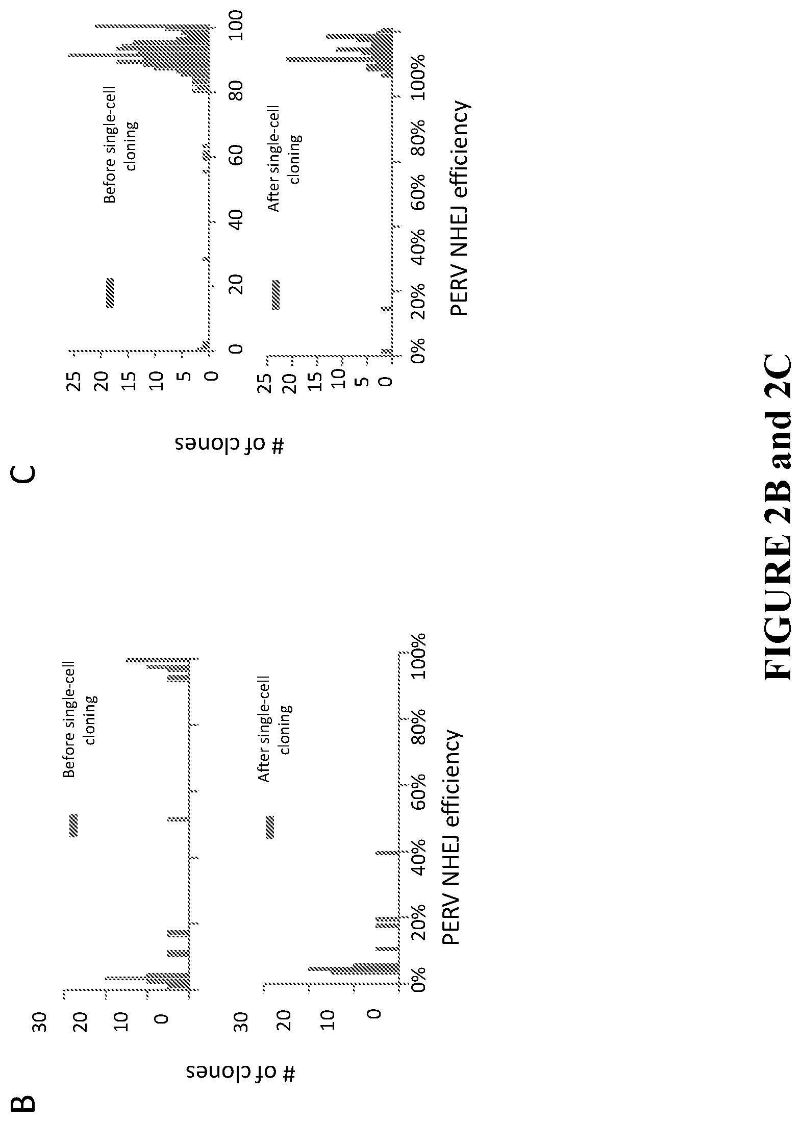
[0032] FIG. 2B graphs the survival of highly modified FFF3 clones. After targeting the PERVs in FFF3, single cells were sorted and immediately genotyped. A bimodal distribution of PERV targeting frequencies among single cells was observed (upper panel). 100% PERV-free FFF3 cells were present among the single cells directly genotyped. However, this pattern changed after expansion of the single cells (bottom panel). Among the single cell clones, only the ones with lower efficiency (<, 39%, the average targeting efficiency in the population was 37%) were obtained, and the persistence of some FFF3 clones that did not have 100% eradication of PERVs was observed (lower panel).
[0033] FIG. 2C shows treatment with PFT.alpha. and bFGF sustains the growth of highly modified FFF3 clones. The combined use of an inhibitor of p53 and a growth factor rescues the highly edited cells. A population of FFF3 was treated with PFT.alpha., and bFGF during the gene editing experiment; then, single cells were sorted for direct genotyping and for colony growth, Followed genotyping. Both the single cells and expanded clones showed similar bimodal distribution in PERV targeting efficiency, and highly modified clones survived under this condition.
[0034] FIG. 2D shows haplotypes of one of the 100% PERV-free clones at PERV poi loci after CRISPR-Cas9 treatment. Several 100% PERV-free clones were achieved from the PFT.alpha. and bFGF treated FFF3 population. The y-axis indicates the edited PERVs loci. The x-axis indicates the relative locations of the indels within the PERV loci. Aligned indel events in the PERV poi sequence are represented in red. Shades of purple indicate different haplotypes of PERVs,
[0035] FIG. 2E shows that PERV production is eliminated in 100% PERV modified clones. The reverse transcriptase (RT) activity of PERV particles presented in the cell culture supernatant was detected by a reverse transcription assay. The results demonstrated no PERV production in 100% PERV-free clone supernatant, whereas the supernatant of WT FFF3 cells showed significant RT activity. The sample order from left to right: 2-log DNA ladder (New England Biolabs); RT+(using commercial reverse transcriptase (RT)); RT-(no RT enzyme); RT+/FF WT (commercial RT enzyme plus WT 3FFF lysis (lysis of virus pellet from 3FFF culture media); 100% FFF3 (100% PERV-inactivated FFF3 clone lysis); WT FF (WT 3FFF lysis); neg (no lysis or RT enzyme, no RNA template).
[0036] FIG. 3A shows porcine blastocysts cloned from 100% PERV-free PFFF3. Day 7 PERV-free porcine blastocysts were stained with SOX2 (inner cell mass), DAPI (nuclei), and phalloidin (cell boundaries). Stained blastocysts were imaged using laser scanning confocal microscopy.
[0037] FIG. 3B shows PERV targeting efficiency of fetus cloned from 100% PERV-free PFFF3. The y-axis indicates the edited PERVs loci. The x-axis indicates the relative locations of the indels within the PERV loci. Aligned indel events in the PERV pol sequence are represented in red. Shades of purple indicate different PERV haplotypes.
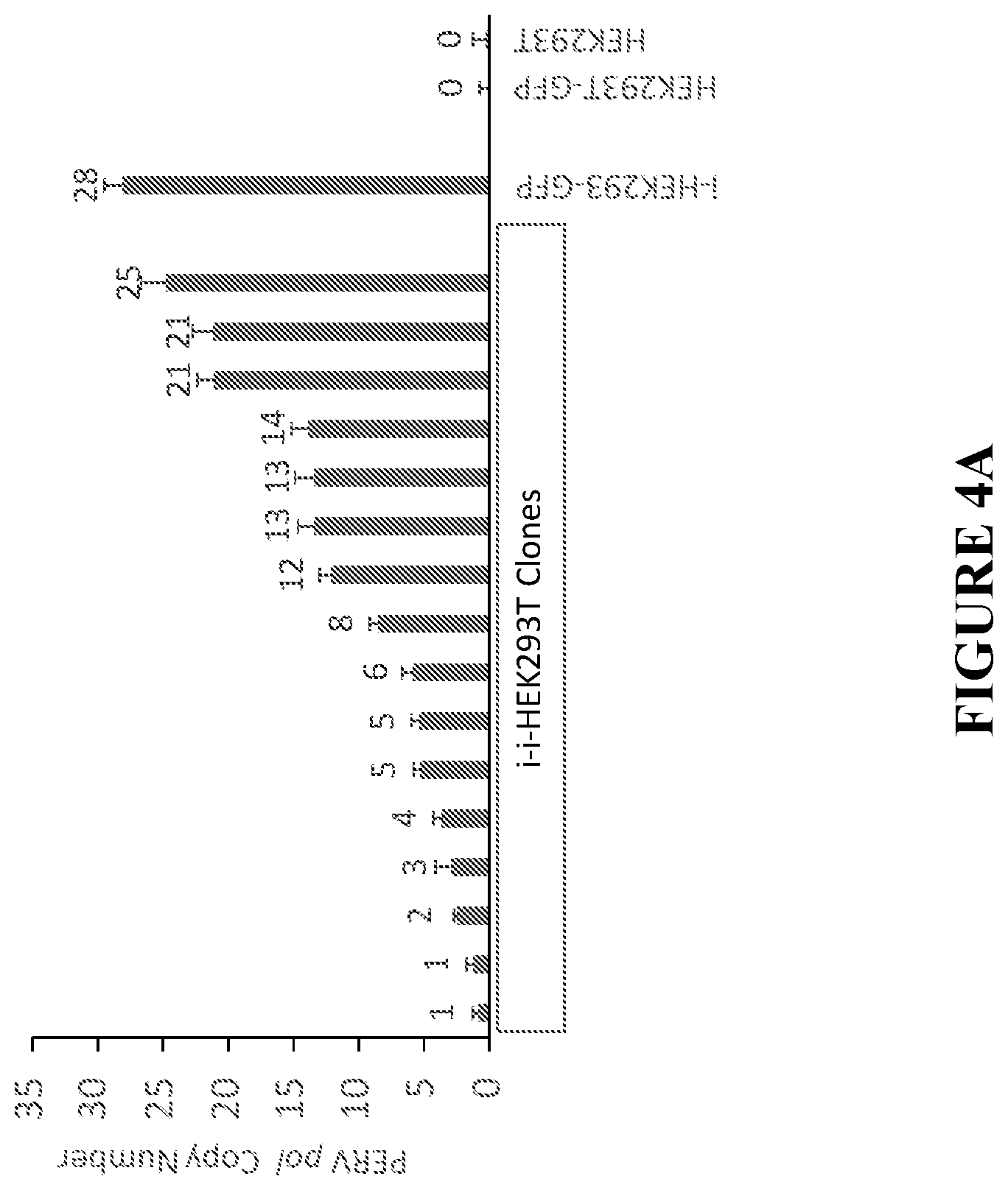
[0038] FIG. 4A shows PERV copy number in selected clones of iHEK293T-GFP. WT HEK293T (non-GFP) cells were co-cultured with equivalent number of cells of PERV infected HEK293T-GFP (i-HEK293T-GFP) clone 10 for two weeks. The GFP-negative single human cells were isolated by flow cytometry based on GFP signal. After the single cell colony grew up, genomic DNA was amplified via qPCR to detect and quantify PERV elements in the GFP-negative clones using i-HEK293T-GFP clone 10 PERV copy number as a standard marker (PERV copy number of clone 10 has been detected by ddPCR previous to the co-coculture experiment). PERV transmission from i-HEK293T-GFP clone 10 to WT HEK293T human cells was detected by qPCR of PERV pol gene and different WT HEK293T clones showed different copy number of PERVs.
[0039] FIG. 4B shows that PERVs insert preferentially in open chromatin and transcriptionally active areas. Gene expression levels, DNase signal, and H3K27Ac signals between areas around PERVs insertion sites and the remaining junctions were measured and compared. Increased transcription, DNase signal, and H3K27Ac levels in PERVs insertion sites was observed.
[0040] FIG. 5A is a graph illustrating the effects of modulators of DNA damage effects. FFF3 cells were treated with bFGF, PFTa, and bFGF+PFTa. Treatment with both the bFGF and PFTa alone did not increased PERV editing efficiency, whereas treatment with a cocktail of bFGF and PFTa significantly enhanced the targeting frequency (p-value=0.0016), demonstrating there exists a synergistic effect between bFGF and PFTa.
[0041] FIG. 5B is a graph illustrating the effect of treatment with a cocktail of PFT-a and bFGF on a population of FFF3-#5 over time (I)=days). Results showed a sustained high PERV targeting efficiency, whereas a population of FFF3-#4 that was not treated with the cocktail showed a decreased pattern of PERV editing efficiency. (ANOVA, day (p-value=0.23), PFTa/bFGF treatment (p-value=0.00002)).
[0042] FIG. 5C is a graph illustrating the effects of seven days of treatment with bcl-2 on FFF3 cells. No significant difference (p-value==0.565) among treatments with different dosages of Bcl-2 was detected. Dose-dependent cytotoxicity of Bcl-2 was also observed during the experiment.
[0043] FIG. 6A is a graph illustrating the effects of the large T antigen on targeting efficiency of PERVs in FFF3 population (p-value=0.05, Wilcoxon Test (One sided)).
[0044] FIG. 6B is a table illustrating the effects of the large T antigen on clones grown from sorted single cells used to analyze the PERV editing efficiency. Large T antigen treatment increased the ratio of >80% and >90% PERV-ko clones by 3.75 and 5.06 folds, respectively, compared with the untreated group.
[0045] FIG. 7 shows the transcription profile of 100% PERV-free PFFF3. RNAseq was used to analyze PERVs inactivation of pol gene. The y-axis indicates the sites. The X-axis indicates the relative locations of the indels within the PERV loci. In red, aligned indel events in the PERV pol sequence are represented.
[0046] FIG. 8 shows primers used to detect PERV junctions in FF3CF.
[0047] FIG. 9 shows PERV targeting efficiency of a piglet born PERV-free FFF3. The y-axis indicates the edited PERVs loci. The x-axis indicates the relative locations of the indels within the PERV loci. Aligned indel events in the PERV pol sequence are represented in red. Shades of purple indicate different PERVs haplotypes. The genotype of the pig showed that all the 25 copies of PERVs were functionally mutated.
[0048] FIG. 10 shows PERV targeting efficiency of nine piglets born PERV-free FFF3. "#1" piglets (six bars to left) and "#2" piglets (three bars to right) indicate two different PERV-free FFF3 clones.
[0049] FIG. 11 shows PERVs copy number in pig PK15 and in human HEK293T(iHEK293T-GFP) co-cultured with PK15. A) Copy number of PERV env A, B, C in PK15 cell line. PERV-A and PERV-B, but not PERV-C, were detected in PK15 cells. PERV-A copy number is around two times of PERV-B. In each 2D amplification plot, black dots (bottom left quadrant) represents droplets with no amplification, green (bottom right quadrant) represents droplets containing GAPDH (reference gene), blue (top left quadrant) represents droplets containing PERV env from subtypes A, B, or C, and orange (top right quadrant) represents droplets containing GAPDH and PERV env. The copy number of PERV subtypes A, B, and C, as measured relative to GAPDH, is shown under each plot. B) PK15 transmits PERVs to HEK293T-GFP human cells, and PERV copy number in HEK293T-GFP increases over time. HEK293T-GFP cells were co-cultured with equivalent numbers of PK15 cells. The human cell population was isolated by sequential rounds of sorting based on GFP expression. Purified human cells was collected at three time points and genomic DNA was harvested and amplified via ddPCR to detect and quantify PERV elements in the human cells. PERVs transmission from pig PK15 to human HEK293T cells was detected by ddPCR of PERV env gene. PERV-A and PERV-B, but not PERV-C, were detected in the HEK293T cells which have history of contact with PK15, and the PERV copy number increased in the HEK293T cells over time.
[0050] FIG. 12 depicts the validation of PERV subtype copy numbers in FFF3. PERV copy number of WT porcine primary fetal fibroblast cell line (FFF3) was detected by ddPCR of both pol and env genes. The PERV copy number (25) determined by pol gene is similar to that (24) detected by env gene.
[0051] FIG. 13 depicts a schematic describing the process of detecting PERVs location in the pig genome. To map the locations of the PERVs, PacBio long-reads genome sequencing (N50=2,439 bp, described below) was performed after PERVs-specific hybridization capture, and mapped 21 copies of PERVs in non-repetitive regions of the genome.
[0052] FIG. 14 depicts a schematic describing the design process of the pol targeting gRNAs.
[0053] FIG. 15A shows a representative karyotype of the PERV-free clones used for pig cloning. Different clones were analyzed using karyotyping, 5 showed abnormal karyotype and 3 normal. Example of 3 abnormal karyotype. Of note, all chromosomes translocated (indicated by color pairs) or with insertion or deletions contain PERVs except in one case (chromosome 10 of the pair translocated t(10,12), upper panel).
[0054] FIG. 15B shows a representative normal karyotype. This PERVs-free clone shows a completely normal karyotype.
[0055] FIG. 16A shows a representative image of PERV-KO fetuses. 6 live fetuses (2 WT and 4 PERV-free fetuses) in total were achieved from 3 surrogate sows. This picture showed the three live PERV-free fetuses achieved from a surrogate sow at day 50 of pregnancy.
[0056] FIG. 16B demonstrates PERVs inactivation efficiency in genomic DNA of fetuses. Fetus genomic DNA was used to measure PERV inactivation efficiency. Similar to the originated PERV-free cell line, the PERV-KO fetuses too showed .about.100% PERV inactivation efficiency, which suggests that no reinfection from surrogate sow occurred during pregnancy.
[0057] FIG. 17A shows a presentative image of PERV-free pig. This picture showed the first born pig (Laika) at day 2 after birth.
[0058] FIG. 17B demonstrates PERV inactivation in genomic DNA of Pigs. PERV-KO pigs were genotyped at different ages by deep sequencing of the PERV pol loci. All examined pigs showed .about.100% PERV inactivation efficiency, which demonstrates that there is no detectable PERV reinfection from surrogate sows to cloned pigs.
[0059] FIG. 17C demonstrates PERV inactivation at mRNA level. Total mRNA generated cDNA was used to detect the PERV inactivation efficiency of the PERV-KO pig transcripts. All pigs exhibited .about.100% PERV eradication efficiency at mRNA level.
[0060] FIG. 18 demonstrates PERV inactivation efficiency of single cells derived from a 30d PERV-free pig. Cells derived from 30d H9p01 PERV-free pig were sorted into single cells and PERV pol gene fragment were genotyped via deep sequencing using genomic DNA of the single cells as template. All single cells showed 100% PERV inactivation.
[0061] FIG. 19 shows PERV copy number of PERV-free pigs and WT FFF3 fibroblast cells. Genomic DNAs of PERV-free pigs and WT FFF3 fibroblast cells were used to measure PERV copy number by ddPCR. All PERV-free pigs showed similar PERV copy number as the WT FFF3 fibroblast cells.
[0062] FIG. 20 shows a karyotype representative of that all PERV-free pigs. All PERV-free pigs exhibited normal karyotype.
[0063] FIG. 21 shows a schematic of a cell line construction strategy according to an embodiment as described herein.
[0064] FIG. 22 shows a schematic of cell quality control strategies according to an embodiment as described herein.
[0065] FIG. 23 shows a schematic of a SCNT cloning procedure according to an embodiment as described herein.
[0066] FIG. 24 shows a schematic of a method for improving cell viability conditions according to an embodiment as described herein. Briefly, the single cell cloning process is skipped to shorten in vitro culture time window. The selected cell population is used instead of single cell clones for SCNT to reduce cell growth time and the sow itself selects the embryos with best gene modification for fetus development.
[0067] FIG. 25 shows a schematic of a method for improving cell viability conditions according to an embodiment as described herein. Briefly, modifications are conducted on a zygote by microinjection instead of cells to reduce stress on cells.
[0068] FIG. 26 shows a schematic of a method for improving cell viability conditions according to an embodiment as described herein. Briefly, fibroblast cells are isolated from 30 day fetuses and used for performing recloning by SCNT.
[0069] FIG. 27 demonstrates that PERV knockout (KO) efficiency increases with the number of gRNAs used in the transfection. Fetal fibroblasts were obtained from Yucatan with 50-60 PERV copies. Chromatography data are shown from fragment analysis.
[0070] FIG. 28 depicts PCR data showing inactivated PERV pol cannot be complemented by the activity of human ERV pol.
[0071] FIG. 29 demonstrates the impact of surrogate mother selection on delivery rate, liter size, survival rate, and expected pigs per sow.
[0072] FIG. 30 demonstrates the impact of the inclusion of a recloning step during pig production on delivery rate, liter size, survival rate, and expected pigs per sow.
[0073] FIG. 31A shows chromatography data from PERV fragment analysis in WT pigs.
[0074] FIG. 31B shows chromatography data from PERV fragment analysis in PERV KO Yorkshire. Data shows 100% PERV KO with only large deletions, with the majority being greater than 100 base pairs (bp).
DETAILED DESCRIPTION OF THE DISCLOSURE
[0075] The present disclosure provides for one or more genetically modified porcine cells having reduced intact PERVs, or methods of generating these cells. In some embodiments, the one or more porcine cells do not have any intact PERVs, or methods of generating these cells. In some embodiments, the PERV-reduced or PERV-free porcine cells may be cloned to produce porcine embryos, which may in turn be grown into adult swine from which organs and/or tissues may be extracted and used for such purposes as xenotransplantation into humans.
I. Definitions
[0076] The terms "about" and "approximately" as used in connection with a numerical value throughout the specification and the claims denotes an interval of accuracy, familiar and acceptable to a person skilled in the art.
[0077] Numeric ranges disclosed herein are inclusive of the numbers defining the ranges.
[0078] The terms "a" and "an" include plural referents unless the context in which the term is used clearly dictates otherwise. The terms "a" (or "an"), as well as the terms "one or more," and "at least one" can be used interchangeably herein. Furthermore, "and/or" where used herein is to be taken as specific disclosure of each of the two or more specified features or components with or without the other. Thus, the term "and/or" as used in a phrase such as "A and/or B" herein is intended to include "A and B," "A or B," "A" (alone), and "B" (alone). Likewise, the term "and/or" as used in a phrase such as "A, B, and/or C" is intended to encompass each of the following aspects: A, B, and C; A, B, or C; A or C; A or B; B or C; A and C; A and B; B and C; A (alone); B (alone); and C (alone).
[0079] Throughout this specification, the word "comprise" or variations such as "comprises" or "comprising" will be understood to imply the inclusion of a stated integer or groups of integers but not the exclusion of any other integer or group of integers. As used herein, the term "comprises" also encompasses the use of the narrower terms "consisting" and "consisting essentially of."
[0080] The terms "pig", "swine" and "porcine" are used herein interchangeably to refer to anything related to the various breeds of domestic pig, species Sus scrofa.
[0081] The term "biologically active" when used to refer to a fragment or derivative of a protein or polypeptide means that the fragment or derivative retains at least one measurable and/or detectable biological activity of the reference full-length protein or polypeptide. For example, a biologically active fragment or derivative of a CRISPRCas9 protein may be capable of binding a guide RNA, binding a target DNA sequence when complexed with a guide RNA, and/or cleaving one or more DNA strands.
[0082] The terms "treatment", "treating", "alleviation" and the like, when used in the context of a disease, injury or disorder, are used herein to generally mean obtaining a desired pharmacologic and/or physiologic effect, and may also be used to refer to improving, alleviating, and/or decreasing the severity of one or more symptoms of a condition being treated. The effect may be prophylactic in terms of completely or partially delaying the onset or recurrence of a disease, condition, or symptoms thereof, and/or may be therapeutic in terms of a partial or complete cure for a disease or condition and/or adverse effect attributable to the disease or condition. "Treatment" as used herein covers any treatment of a disease or condition of a mammal, particularly a human, and includes: (a) preventing the disease or condition from occurring in a subject which may be predisposed to the disease or condition but has not yet been diagnosed as having it; (b) inhibiting the disease or condition (e.g., arresting its development); or (c) relieving the disease or condition (e.g., causing regression of the disease or condition, providing improvement in one or more symptoms).
II. Porcine Cells, Tissues and Organs
[0083] In some embodiments, the cell is a mammalian cell. In some embodiments, the mammalian cell is a human cell. In some embodiments, the mammalian cell is a non-human mammalian cell, for example, equine, porcine, bovine, ovine, caprine, canine, or feline. In some embodiments, the cell is a porcine cell.
[0084] In some embodiments, the present disclosure provides for one or more porcine cells having reduced intact PERVs. In some embodiments, the porcine cell has been genetically modified such that the intact PERVs present in the porcine cell have been inactivated. In some embodiments, the porcine cell has less than 60, less than 50, less than 40, less than 30, less than 25, less than 20, less than 15, less than 10, less than 5, less than 3, less than 2. 1 or zero copies of intact PERVs. In some embodiments, the porcine cell has less than 10, less than 5, less than 3, less than 2, 1 or zero copies of intact PERVs. In some embodiments, the porcine cell has zero copies of intact PERVs. In some embodiments, the porcine cell has between about 60 copies and about 1 copy, between about 50 copies and about 1 copy, between about 40 copies and about 1 copy, between about 30 copies and about 1 copy, between about 20 copies and about 5 copies, between about 15 copies and about 10 copies, or between about 5 copies and about 1 copy of intact PERVs. In some embodiments, at least about 30%, about 35%, about 40%, about 45%, about 50%, about 55%, about 60%, about 65%, about 70%, about 75%, about 80%, about 85%, about 90%, about 95%, about 96%, about 97%, about 98%, about 99% or 100% of the PERVs in the cell are inactive. In some embodiments, between about 30% and 100%, between about 35% and 100%, between about 40% and 100%, between about 45% and 100%, between about 50% and 100%, between about 55% and 100%, between about 60% and 100%, between about 65% and 100%, between about 70% and 100%, between about 75% and 100%, between about 80% and 100%, between about 85% and 100%, between about 90% and 100%, between about 95% and 100%, between about 96% and 100%, between about 97% and 100%, between about 98% and 100%, or between about 99% and 100% of the PERVs in the cell are inactive. In some embodiments, 100% of the PERVs in the cell are inactive.
[0085] In some embodiments, the cell is a primary cell, for example, a pig primary cell. In some embodiments, the cell is a somatic cell. In some embodiments, the cell is a post-natal cell. In some embodiments, the cell is an adult cell (e.g., an adult ear fibroblast). In some embodiments, the cell is a fetal/embryonic cell (e.g., an embryonic blastomere). In some embodiments, the cell is a germ line cell. In some embodiments, the cell is an oocyte. In some embodiments, the cell is a stem cell. In some embodiments, the cell is a cell from a primary cell line. In some embodiments, the cell is an epithelial cell, a muscle cell, a fibroblast, an endothelium cell, a liver cell, a granulosa cell, a fat cell. In particular embodiments, the cell is a fibroblast. In some embodiments, the fibroblast is a fetal fibroblast, for example a female fetal fibroblast. In some embodiments, the cell is a cancer cell. In some embodiments, the cell is not a cancer cell. In some embodiments, the cell is in vitro. In some embodiments, the cell is in vivo. In some embodiments, the cell is a single cell. In some embodiments, the cell is a member of a cell colony.
[0086] In some embodiments, the cell is a porcine cell. Non-limiting examples of porcine cells are cells that originates from or are derived from any of the following pig breeds: American Landrace, American Yorkshire, Aksai Black Pied, Angeln Saddleback, Appalachian English, Arapawa Island, Auckland Island, Australian Yorkshire, Babi Kampung, Ba Xuyen, Bantu, Basque, Bazna, Beijing Black, Belarus Black Pied, Belgian Landrace, Bengali Brown Shannaj, Bentheim Black Pied, Berkshire, Bisaro, Bangur, Black Slavonian, Black Canarian, Breitovo, British Landrace, British Lop, British Saddleback, Bulgarian White, Cambrough, Cantonese, Celtic, Chato Murciano, Chester White, Chiangmai Blackpig, Choctaw Hog, Creole, Czech Improved White, Danish Landrace, Danish Protest, Dermantsi Pied, Li Yan, Duroc, Dutch Landrace, East Landrace, East Balkan, Essex, Estonian Bacon, Fengjing, Finnish Tandrace, Forest Mountain, French Landrace, Gascon, German Landrace, Gloucestershire Old Spots, Gottingen minipig, Grice, Guinea Hog, Hampshire, Hante, Hereford, Hezuo, Hogan Hog, Huntington Black Hog, Iberian, Italian Landrace, Japanese Landrace, Jeju Black, Jinhua, Kakhetian, Kele, Kemerovo, Korean Native, Krskopolje, Kunekune, Lamcombe, Large Black, Large Black-White, Large White, Latvian White, Leicoma, Lithuanian Native, Lithuanian White, Lincolnshire Curly-Coated, Livny, Malhado de Alcobaca, Mangalitsa, Meishan, Middle White, Minzhu, Minokawa Buta, Mong Cai, Mora Romagnola, Moura, Mukota, Mulefoot, Murom, Myrhorod, Nero dei Nebrodi, Neijiang, New Zealand, Ningxiang, North Caucasian, North Siberian, Norwegian Landrace, Norwegian Yorkshire, Ossabaw Island, Oxford Sandy and Black, Pakchong 5, Philippine Native, Pietrain, Poland China, Red Wattle, Saddleback, Semirechensk, Siberian Black Pied, Small Black, Small White, Spots, Surabaya Babi, Swabian-Hall, Swedish Landrace, Swallow Belied Mangalitza, Taihu pig, Tamworth, Thuoc Nhieu, Tibetan, Tokyo-X, Tsivilsk, Turopolje, Ukrainian Spotted Steppe, Ukrainian White Steppe, Urzhum, Vietnamese Potbelly, Welsh, Wessex Saddleback, West French White, Windsnyer, Wuzhishanm, Yanan, Yorkshire and Yorkshire Blue and White.
[0087] In some embodiments, the cell (e.g., the donor nuclear cell) is isolated from a chimeric/mosaic fetus. As used herein the terms "chimeric" and "mosaic" may be used interchangeably and refer to a fetus having two or more populations of cells with different genotypes in the one fetus that has developed from a single fertilized egg. In some embodiments, the chimeric fetus is at about 5 days, about 10 days, about 15 days, about 20 days, about 25 days, about 30 days, about 2 months, about 3 months, about 4 months, about 5 months, about 6 months, about 7 months, about 8 months, about 9 months gestation or longer. In some embodiments, the chimeric fetus is less than about 5 days, less than about 10 days, less than about 15 days, less than about 20 days, less than about 25 days, less than about 30 days, less than about 2 months, less than about 3 months, less than about 4 months, less than about 5 months, less than about 6 months, less than about 7 months, less than about 8 months, or less than about 9 months gestation.
[0088] In some embodiments, the chimeric fetus is generated using a genome modifying agent, for example Zinc Finger nuclease or nickase, TAL effector nuclease or nickase, deaminase, and CRISPR associate nuclease or nickase. In some embodiments, the chimeric fetus is generated by direct zygote injection.
[0089] In some embodiments, the nuclear donor cell is isolated from an animal that is less than about 10 weeks, less than about 9 weeks, less than about 8 weeks, less than about 7 weeks, less than about 6 weeks, less than about 5 weeks, less than about 4 weeks, less than about 3 weeks, less than about 2 weeks, or less than 1 about week in age.
[0090] In some embodiments, the nuclear donor cell, for example, one isolated from a chimeric fetus, is expanded in vitro for a period of time. In some embodiments, the cell is expanded in vitro for less than about 30, less than about 25, less than about 20, less than about 15, less than about 10, less than about 5, or less than about 2 population doublings. In some embodiments, the cell is expanded in vitro for not more than about 2, about 3, about 4, about 5, about 6, about 7, about 8, about 9, about 10, about 11, about 12, about 13, about 14, about 15, about 16, about 17, about 18, about 19, about 20, about 21, about 22, about 23, about 24, about 25, about 26, about 27, about 28, about 29, or about 30 population doublings.
[0091] In some embodiments, the cells, before or after genetic modification, are cultured in the presence of one or more factors that improves the health and/or survival of the cell. In some embodiments the factor is BEPP monohydrochloride, pifithrin-.alpha., R18 trifluoroacetate, fibroblast growth factor (fgf), large T antigen, BCL, or any combination thereof. In some embodiments the factor(s) induces RNA-dependent protein kinase apoptosis, inhibits p53 mediated apoptosis, inhibits 14-3-3 proteins, promotes proliferation, promotes immortalization, is antiapoptotic, or any combination thereof.
[0092] In some embodiments, the factor is a cell cycle checkpoint inhibitor. In some embodiments, the factor is an anti-apoptotic factor. In some embodiments, the factor is bcl-2. In some embodiments, the factor is an inhibitor of any one of the following: c-Myc, Bax, p53, tBid, and BCL. The skilled worker is aware of various small molecules and possible derivatives of those small molecules that may be used as anti-apoptotic checkpoint inhibitors or anti-apoptotic factors. In some embodiments, the factor is the SV40 antigen.
[0093] In some embodiments, the factor is a p53 inhibitor. In some embodiments, the factor is a pifithrin molecule or derivative thereof. In some embodiments, the factor is pifithrin-alpha and/or pifithrin-beta. Pifithrin (PFT)-.alpha. has been demonstrated to reversibly inhibit p53-dependent transcriptional activation and apoptosis, and PFT-.mu. has been demonstrated to inhibit p53 binding to mitochondria by reducing p53-binding affinity to Bcl-xL and Bcl-2. In some embodiments, the p53 inhibitor is a cyclized PFT-.alpha. p53 inactivator such as cyclic Pifithrin-.alpha. hydrobromide. In some embodiments, the p53 inhibitor is a nucleic acid that reduces or eliminates p53 expression in the cell. In some embodiments, the nucleic acid is an antisense and/or RNAi molecule. In some embodiments, the p53 inhibitor is a dominant negative p53 protein, or a nucleic acid encoding a dominant negative p53 inhibitor. In some embodiments, the p53 inhibitor is selected from the group consisting of pifithrin-.alpha., pifithrin-beta, pifithrin-.alpha. hydrobromide, pifithrin-mu, ellipticine, 9-hydroxyellipticine, nutlin-3, roscovitine, and SJ 172550.
[0094] In some embodiments, the factor is a growth factor. In some embodiments, the growth factor is of porcine origin. In some embodiments, the growth factor is a growth factor useful for the cell type that has been genetically modified. For example, if the genetically modified cell is fibroblast cell or cell of fibroblast lineage, than in some embodiments, the growth factor is fibroblast growth factor. In some embodiments, the fibroblast growth factor is basic fibroblast growth factor (bFGF) or fibroblast growth factor-2 (FGF-2). In some embodiments, the fibroblast growth factor is basic fibroblast growth factor (bFGF). In some embodiments, the growth factor is selected from the group consisting of: epidermal growth factor (EGF), insulin-like growth factor (IGF), platelet derived growth factor (PDGF), vascular endothelial growth factor (VEGF), and keratinocyte growth factor (KGF).
[0095] In some embodiments, the cells are administered both a p53 inhibitor and a growth factor. In certain embodiments, the cells are administered both bFGF and pifithrin-alpha.
[0096] In some embodiments, the cells of the disclosure are cultured in conditions of about 1-20% oxygen (O.sub.2), about 1-20% carbon dioxide (CO.sub.2), about 50-90% N.sub.2, or any combination thereof. In some embodiments, the cells of the present disclosure are cultured under hypoxic conditions (e.g., in the presence of less than 10% O.sub.2). In some embodiments, the cells of the present disclosure are cultured at about 37.degree. C. In some embodiments, the cells of the present disclosure can be cultured at about 5% O.sub.2, 5% CO.sub.2 and 90% N.sub.2. In some embodiments, the cells are cultured in a tri-gas incubator for at least a period of time.
[0097] In some embodiments, the cells of the present disclosure when cultured in vitro are split and/or frozen when the cells are below about 80%, about 70%, about 60%, about 50%, about 40%, about 30%, about 20%, about 10%, or less confluency. In some embodiments, the cells are split and/or frozen when the cells are below about 50% confluency.
III. Genetic Modifications
[0098] The present disclosure provides methods of generating genetically modified cells and animals derived therefrom.
[0099] In some embodiments, a plurality of endogenous nucleic acid sequences are modified by inactivation, insertion of exogenous nucleic acids, subtraction of endogenous nucleic acids, or any combination thereof to generate genetically modified cells and animals derived therefrom.
[0100] In some embodiments, a plurality of nucleic acid sequences in the cell (e.g., a nuclear donor cell) are modified. In some embodiments, at least about 2, at least about 5, at least about 10, at least about 20, at least about 30, at least about 40, at least about 50, at least about 60, at least about 70, at least about 80, at least about 90, at least about 100, or more nucleic acid sequences are modified. In some embodiments, the plurality of genetic modifications are to a single, repetitive gene sequence. In other embodiments, at least a portion of the genetic modifications are to different genes.
[0101] In some embodiments, the present disclosure provides for one or more cells having reduced intact viral elements (e.g., PERVs). In some embodiments, the nuclear donor cell has been genetically modified such that the intact viral elements present in the cell have been inactivated. In some embodiments, the cell has less than 60, less than 50, less than 40, less than 30, less than 25, less than 20, less than 15, less than 10, less than 5, less than 3, less than 2, 1 or zero copies of intact viral elements. In some embodiments, the porcine cell has less than 10, less than 5, less than 3, less than 2, 1 or zero copies of intact viral elements. In some embodiments, the porcine cell has zero copies of intact viral elements. In some embodiments, the cell has between about 60 copies and about 1 copy, between about 50 copies and about 1 copy, between about 40 copies and about 1 copy, between about 30 copies and about 1 copy, between about 20 copies and about 5 copies, between about 15 copies and about 10 copies, or between about 5 copies and about 1 copy of intact viral elements. In some embodiments, at least about 30%, about 35%, about 40%, about 45%, about 50%, about 55%, about 60%, about 65%, about 70%, about 75%, about 80%, about 85%, about 90%, about 95%, about 96%, about 97%, about 98%, about 99% or 100% of the viral elements in the cell are inactive. In some embodiments, between about 30% and 100%, between about 35% and 100%, between about 40% and 100%, between about 45% and 100%, between about 50% and 100%, between about 55% and 100%, between about 60% and 100%, between about 65% and 100%, between about 70% and 100%, btwcen about 75% and 100%, between about 80% and 100%, between about 85% and 100%, between about 90% and 100%, between about 95% and 100%, between about 96% and 100%, between about 97% and 100%, between about 98% and 100%, or between about 99% and 100% of the viral elements in the cell are inactive.
[0102] In some embodiments, the cells, tissues, organs or animals (e.g., pigs) of the present disclosure lack (or have been engineered to lack activated strains of) infectious viruses. In some embodiments, the cells lack an endogenous retrovirus. In some embodiments, the cells of the present disclosure lack (or have been engineered to lack activated strains of) any one or more of herpesvirus, porcine lymphotrophic herpesvirus (PLHV), pig cytomegalovirus (PCMV), encephalomyocarditis virus (EMCV), pig circovirus (PCV), hepatitis E virus (HEV), rabies viruses, pseudorabies viruses, parvovirus, swine vesicular disease viruses, porcine polio virus, hemagglutinating encephalomyocarditis viruses, swine influenza type A, adenovirus, transmissible gastroenteritis virus, vesicular stomatitis virus, feline leukemia virus, mouse mammary tumor virus, murine leukemia virus, simian immunodeficiency virus (SIV), equine infectious anemia, bovine immunodeficiency virus (BIV), and the like. In some embodiments, the cells, tissues, organs or pigs of the present disclosure lack (or have been engineered to lack activated strains of) PERVs.
[0103] In some embodiments, the cells, tissues, organs or animal (e.g., pigs) of the disclosure comprise one or more inactivating mutations. In some embodiments, the cells, tissues, organs or animals comprise one or more mutations or epigenetic changes that result in decreased or eliminated activity of the nucleic acid sequence (e.g., a gene or retroviral element such as PERV elements). In some embodiments, the one or more nucleic acid sequences (e.g., PERV element) is inactivated by genetically modifying the nucleic acid(s) present in the cell, tissue, organ or animal. In some embodiments, the inactivation of one or more nucleic acid sequences (e.g., PERV elements) is confirmed by means of an assay. In some embodiments, the assay is an infectivity assay, reverse transcriptase PCR assay, RNA-seq, real-time PCR, or junction PCR mapping assay.
[0104] The cells, tissues, organs or animals can be genetically modified according to any method known by one of skill in the art. In some embodiments, the nucleic acid(s) in the cell are genetically modified such that one or more nucleic acid sequences (e.g., genes or retroviral elements) in the cell are inactivated. In some embodiments, the nucleic acid sequences are genetically modified using any of the genetic modifications systems known in the art and/or disclosed herein. In some embodiments, the genetic modification system is a TALEN, a zinc finger nuclease, and/or a CRISPR-based system. In some embodiments, the genetic modification system is a CRISPR-Cas9 system. In some embodiments, the cell is genetically modified such that one or more nucleic acid sequences in the cell are inactivated, and the cell is further genetically modified such that the cell has reduced expression of one or more genes that would induce an immune response if the cell (or a tissue or organ cloned/derived from the cell) were transplanted to a human. In some embodiments, the cell is genetically modified such that one or more nucleic acid sequences in the cell are inactivated and the cell is further genetically modified such that the cell has increased expression of one or more genes that would suppress an immune response if the cell (or a tissue or organ cloned/derived from the cell) were transplanted to a human. In some embodiments, the cell is genetically modified such that one or more nucleic acid sequences in the cell are inactivated, and the cell is further genetically modified such that the cell has reduced expression of one or more genes that would induce an immune response if the cell (or a tissue or organ cloned/derived from the cell) were transplanted to a human, and the cell is further genetically modified such that the cell has increased expression of one or more genes that would suppress an immune response if the cell (or a tissue or organ cloned/derived from the cell) were transplanted to a human.
[0105] In some embodiments, the disclosure provides for a blastocyst or an embryo that was cloned from the genetically modified cell. In some embodiments, the genetically modified nucleic acid(s) are extracted from the genetically modified cell and cloned into a different cell. For example, in somatic cell nuclear transfer, the genetically modified nucleic acid from the genetically modified cell is introduced into an enucleated oocyte. In some embodiments, oocytes can be enucleated by partial zona dissection near the polar body and then pressing out cytoplasm at the dissection area. In some embodiments, oocytes are obtained with all or substantially all of the cytoplasm intact. In some embodiments, the oocytes are screened for multiple layers of cumulus cells. In some embodiments, an injection pipette with a sharp beveled tip is used to inject the genetically modified cell into an enucleated oocyte arrested at meiosis 2. Oocytes arrested at meiosis-2 are frequently termed "eggs." In some embodiments, an embryo is generated by fusing and activating the oocyte. Such an embryo may be referred to herein as a "genetically modified embryo." In some embodiments, the genetically modified embryo is transferred to the oviducts of a recipient female pig.
[0106] A genetically modified blastocyst or embryo can be generated according to any embodiment disclosed herein. In one embodiment, a porcine fetal fibroblast cell can be obtained and transfected using neon transfection reagent, biotin-conjugated beads can be used to counter select for knockout genes and/or antibody binding selection for knock-in genes through FACS, single cells having the desired modifications sorted and expanded in vitro, cells genotyped and/or PCR verified for positive clones, followed by cell transportation and SCNT cloning, or any combination thereof. In some embodiments, the genetic modifications are conducted on a zygote using, for example, microinjection instead of cells in vitro. It is contemplated that direct injection into a zygote may be less stressful on the cells. When zygote modification is performed, embryos are then transferred to obtain mosaic animals and cells are isolated from the mosaic fetus to be used for SCNT cloning.
[0107] In some embodiments, the cells, blastocysts, embryos and the like that are generated according to any embodiment disclosed herein are tested for quality control. Non-limiting examples of quality control include any of Miseq and Sanger sequencing to detect gene targeting efficiency, Exome sequencing and/or whole genome sequencing to detect off-targeting, karyotyping to detect chromosome abnormalities, RT-qPCR to detect target gene expression, RNAseq to detect the normality of whole gene expression pattern, antibody binding counter selection for knockout gene (e.g., by bead enrichment), specific gene antibody binding selection for knock-in genes (e.g., by flow cytometry sorting); specific gene antibody binding, human serum antibody binding, complement cytotoxicity, or natural killer (NK) cell assay to detect the physiological function of target gene, blastocyst development ratio to check whether gene editing influences embryo development, and any combination thereof.
IV. Surrogates and/or Somatic Cell Nuclear Transfer
[0108] In some embodiments, the genetically modified blastocyst or embryo is transferred to a surrogate, for example, the oviduct of the surrogate. In some embodiments, the genetically modified blastocyst or embryo is transferred to the oviducts of a surrogate about 20 to 24 hours after activation. In some embodiments, more than one blastocyst or embryo is transferred to the surrogate. In some embodiments between about 1 and about 600, between about 50 and about 500, between about 100 and about 400, or between about 200 and about 300 blastocysts or embryos are transferred. In some embodiments, the embryos are checked for quality control prior to transferring, for example, checking the cleavage and blastocyst ratio. In some embodiments, embryos or blastocysts derived from the sample cell are transferred into multiple surrogates, for example, between about 5-20 surrogates/cell line. In some embodiments, at least one wild type blastocyst or embryo is transferred to the surrogate at the same time or substantially the same time as the genetically modified blastocyst or embryo. In some embodiments, at least one wild type blastocyst or embryo is transferred to the surrogate prior to or after the genetically modified blastocyst or embryo is transferred to the surrogate. In some embodiments, surrogate is checked for pregnancy approximately 20-21 days after transfer of the genetically modified blastocyst or embryo.
[0109] In some embodiments, the surrogate is screened for certain characteristics prior to transferring of the blastocyst or embryo. Non-limiting examples of surrogate characteristics include physiological stage, age, fertility, maternal behavior, lactation and rearing ability, abortion frequency, disease, or any combination thereof. In some embodiments, the surrogate is selected for prior litter size, suitable age, having more than one birth history, and/or with suitable physiological stage. In some embodiments, the blastocyst or embryo transfer is performed in the spring or fall. In some embodiments, the blastocyst or embryo transfer is not performed in the summer and/or winter.
[0110] In some embodiments, a large surrogate is selection. It is contemplated that larger surrogates can improve the pig production as measured by any one, or combination of, outcome measures including delivery rate, liter size, survival rate, and expected pigs per sow as compared to smaller surrogates.
[0111] In some embodiments, a first round of SCNT is performed to generate an embryo that is transferred to a surrogate to generate a fetus and then cells are isolated from a fetus. The isolated cells are then used for a second round of SCNT and transferred to a surrogate to generate a genetically modified animal. In some embodiments, the fetus used in the second round of SCNT is sacrificed at about 10 days, about 20 days, about 30 days, about 40 days, about 50 days, about 60 days, about 70 days, about 80 days, about 90 days, about 100 days, or about 100 days gestation. In some embodiments, the fetus used in the second round of SCNT is less than about 50 days, less than about 40 days, less than about 30 days, less than about 20 days, or less than about 10 days gestation.
[0112] In some embodiments, the genetically modified blastocyst or embryo is grown into a post-natal genetically modified animal (e.g., pig). In some embodiments, the post-natal genetically modified animal is a neo-natal genetically modified animal. In some embodiments, the genetically modified animal is a juvenile genetically modified animal. In some embodiments, the genetically modified animal is an adult genetically modified animal (e.g., older than 5-6 months). In some embodiments, the genetically modified animal is a female genetically modified animal. In some embodiments, the animal is a male genetically modified animal. In some embodiments, the genetically modified animal is bred with a non-genetically modified animal. In some embodiments, the genetically modified animal is bred with another genetically modified animal. In some embodiments, the genetically modified animal is bred with another genetically modified animal that has reduced or no active retrovirus (e.g., PERVs). In some embodiments, the genetically modified animal is bred with a second genetically modified animal that has been genetically modified such that the cells, tissues or organs from the second genetically modified animal are less likely to induce an immune response if transplanted to a human.
V. Genetically Modified Animals
[0113] The present disclosure also provides for methods of preventing or reducing risk of pregnancy loss or miscarriage following somatic cell nuclear transfer (SCNT) of a genetically modified blastocyst or embryo comprising: using a nucleus from a genetically modified nuclear donor cell to generate a blastocyst or embryo; and transferring the blastocyst or embryo to a surrogate to generate at least one a viable offspring wherein the rate of pregnancy loss or miscarriage is reduced as compared to the rate of pregnancy loss or miscarriage of a control.
[0114] In some embodiments, the rate of pregnancy loss or miscarriage is reduced by at least about 5%, at least about 10%, at least about 20%, at least about 30%, at least about 40%, at least about 50%, at least about 60%, at least about 70%, at least about 80%, at least about 90%, or more as compared to the rate of pregnancy loss or miscarriage of a control genetically modified blastocyst or embryo.
[0115] In some embodiments the genetically modified animal maintains a same or similar level of genetic modification for at least a month, at least 6 months, at least 1 year, at least 5 years, at least 10 years post-gestation.
[0116] In some embodiments, the genetically modified animal remains alive for at least a month, at least 2 months, at least 3 months, at least 4 months, at least 5 months, at least 6 months, at least 7 months, at least 8 months, at least 9 months, at least 10 months, at least 11 months, at least a year, at least 2 years, at least 3 years, at least 4 years, at least 5 years, at least 6 years, at least 7 years, at least 8 years, at least 9 years, at least 10 years, or more.
[0117] In some embodiments the genetically modified pig are PERV-inactivated genetically modified pigs and maintain a same or similar level of PERV inactivation for at least a month, at least 6 months, at least 1 year, at least 5 years, at least 10 years post-gestation. In some embodiments, the genetically modified pig remains PERV-inactivated genetically modified pigs even after delivery from a non-PERV-inactivated surrogate or after being in a facility/space with other non-PERV-inactivated pigs.
[0118] In some embodiments, the disclosure provides for cells, tissues or organs obtained from any of the post-natal genetically modified animals (e.g., pigs) described herein. In some embodiments, the cell, tissue or organ is selected from the group consisting of: liver, lung, heart, brain, pancreas, muscle, blood, bone, testes and ovary. In some embodiments, the organ is liver, lung or heart. In some embodiments, the cell from the post-natal genetically modified animal is selected from the group consisting of: pancreatic islets, lung epithelial cells, cardiac muscle cells, skeletal muscle cells, smooth muscle cells, hepatocytes, non-parenchymal liver cells, gall bladder epithelial cells, gall bladder endothelial cells, bile duct epithelial cells, bile duct endothelial cells, hepatic vessel epithelial cells, hepatic vessel endothelial cells, sinusoid cells, choroid plexus cells, fibroblasts, Sertoli cells, neuronal cells, stem cells, and adrenal chromaffin cells. In some embodiments, the genetically modified organs, tissues or cells have been separated from their natural environment (i.e., separated from the animal in which they are being grown). In some embodiments, separation from the natural environment means a gross physical separation from the natural environment, e.g., removal from the genetically modified donor animal, and alteration of the genetically modified organs', tissues' or cells' relationship with the neighboring cells with which they are in direct contact (e.g., by dissociation).
III. Methods of Generating PERV-Reduced or PERV-Free Porcine Cells
[0119] The disclosure provides for methods of generating any of the PERV-reduced PERV-free porcine cells disclosed herein. In some embodiments, the disclosure provides a method of inactivating a PERV element in any of the porcine cells disclosed herein, comprising administering to the cell a genome modifying agent specific to a gene involved in PERV replication and/or assembly, wherein the agent disrupts transcription and/or translation of the gene. In some embodiments, the agent targets the start codon of the gene and inhibits transcription of the gene. In some embodiments, the agent targets an exon in the gene and the agent induces a frameshift mutation in the gene. In some embodiments, the agent introduces an inactivating mutation into the gene. In some embodiments, the agent represses transcription of the gene.
[0120] The skilled worker is aware of numerous assays for determining whether a cell is PERV free. One method would be to expose a human cell to a genetically modified cell believed to be PERV-reduced or PERV-free and monitor PERV infection of the human cell, i.e., an infectivity assay. See, e.g., Examples 1 and 2. Other methods of monitoring PERV activity in a genetically modified cell include, e.g., RNAseq, Reverse Transcriptase-PCR, microarrays, and/or junction PCR reactions to monitor expression and/or chromosomal integrity of PERV related genes (e.g., gag, pol and/or env).
[0121] In some embodiments, any of the agents disclosed herein is a polynucleotide. In some embodiments, the polynucleotide encodes one or more of the nucleases and/or nickases and/or RNA or DNA molecules described herein. In some embodiments, the polynucleotide agent is introduced to one or more PERV porcine cells. In some embodiments, the polynucleotide is introduced to the one or more PERV porcine cells in a manner such that the polynucleotide is transiently expressed by the one or more cells. In some embodiments, the polynucleotide is introduced to the one or more PERV porcine cells in a manner such that the polynucleotide is stably expressed by the one or more cells. In some embodiments, the polynucleotide is introduced in a manner such that it is stably incorporated in the porcine cell genome. In some embodiments, the polynucleotide is introduced along with one or more transposonal elements. In some embodiments, the transposonal element is a polynucleotide sequence encoding a transposase. In some embodiments, the transposonal element is a polynucleotide sequence encoding a PiggyBac transposase. In some embodiments, the transposable element is inducible. In some embodiments, the transposable element is doxycycline-inducible. In some embodiments, the polynucleotide further comprises a selectable marker. In some embodiments, the selectable marker is a puromycin-resistant marker. In some embodiments, the selectable marker is a fluorescent protein (e.g., GFP).
[0122] In some embodiments, the agent is a nuclease or a nickase that is used to target PERV DNA in the cell. In some embodiments, the agent specifically targets and suppresses expression of a PERV. In some embodiments, the agent comprises a transcription repressor domain. In some embodiments, the transcription repressor domain is a Kruppel associated box (KRAB).
[0123] In some embodiments, the agent is any programmable nuclease. In some embodiments, the agent is a natural homing meganuclease. In some embodiments, the agent is a TALEN-based agent, a ZFN-based agent, or a CRISPR-based agent, or any biologically active fragment, fusion, derivative or combination thereof. In some embodiments, the agent is a deaminase or a nucleic acid encoding a deaminase. In some embodiments, a cell is engineered to stably and/or transiently express a TALEN-based agent, a ZFN-based agent, and/or a CRISPR-based agent.
A. TALEN-Based Agents
[0124] In some embodiments, the agent is a TALEN-based agent. In some embodiments, the TALEN-based agent is one or more TALEN polypeptides/proteins or biologically active fragments or derivatives thereof, or one more nucleic acids encoding one or more TALEN polypeptides or fragments or derivatives thereof. Transcription activator-like (TAL) effector sequences can be assembled to bind DNA targets with specificity by assembling sequences of repeat variable-diresidues (RVDs). Fusion proteins of TAL effectors and nucleases (TALENs) can make targeted double-stranded breaks in cellular DNA that can be used to make specific genetic modifications to cells. In some embodiments, the agent is a TALEN polypeptide/protein or fragment or derivative thereof that targets one or more PERV DNA sequences in a PERV cell. In some embodiments, the Repeat Variable Diresidue (RVD) portion of the TALEN has been engineered to target one or more PERV DNA sequences in a PERV cell. In some embodiments, the TALEN-based agent is a nucleic acid encoding one or more TALEN protein. In some embodiments, the nucleic acid is in a plasmid. In some embodiments, the nucleic acid is mRNA.
[0125] In some embodiments, the TALEN protein is expressed in a cell and induces a site-specific double stranded DNA break in one or more PERV gene. In some embodiments, the TALEN protein introduces a donor sequence, wherein the donor sequence partially or completely replaces the PERV gene, thereby silencing or inactivating the PERV gene. In some embodiments, the TALEN is a left TALEN and further comprising a right TALEN that cooperates with the left TALEN to make the double strand break in the PERV gene. In another embodiment, the nucleic acid encoding the TALEN and/or the nucleic acid donor sequence is part of a vector or plasmid. In some embodiment, the TALEN includes a spacer (e.g., the spacer sequence is 12 to 30 nucleotides in length).
[0126] In some embodiments, the TALEN protein comprises a TAL effector DNA-binding domain and/or a modified FokI nuclease catalytic domain. In some embodiments, the TAL effector DNA-binding domain binds to a region in a PERV gene. In some embodiments, the TALEN comprises, from the N-terminus to the C-terminus: (i) a first segment of about 50 to about 200 amino acids in length; (ii) a TAL effector DNA-binding domain providing sequence-specific binding to a PERV nucleotide sequence; (iii) a second segment of about 20 to 100 amino acids in length; and (iv) a modified FokI nuclease catalytic domain.
[0127] Methods of engineering a TALEN to bind to specific nucleic acids are described in Cermak, et al, Nucl. Acids Res. 1-1 1 (2011). US Published Application No. 2011/0145940 discloses TAL effectors and methods of using them to modify DNA. Miller et al. Nature Biotechnol 29: 143 (2011) describes the generation of TALENs for site-specific nuclease architecture by linking TAL truncation variants to the catalytic domain of Fok I nuclease. General design principles for TALEN binding domains can be found in, for example, WO 2011/072246. Each of these documents is incorporated herein in its entirety.
[0128] In some embodiments, the TALEN-based agent targets a nucleotide sequence of a PERV. In some embodiments, the TALEN-based agent targets a nucleotide sequence that is conserved across more than one strain of PERV. In some embodiments, the TALEN-based agent targets a PERV pol, env, and/or gag gene. In some embodiments, the TALEN-based agent targets a PERV pol gene. In some embodiments, the TALEN-based agent targets the sequence encoding the catalytic core of a PERV pol gene.
B. ZFN-Based-Agents
[0129] In some embodiments, the agent is a zinc finger nuclease (ZFN)-based agent. In some embodiments, the ZFN-based agent is one or more ZFN polypeptides or biologically active fragments or derivatives thereof, or one more nucleic acids encoding one or more ZFN polypeptides or fragments or derivatives thereof. ZFNs are artificial restriction enzymes generated by fusing a zinc finger DNA-binding domain to a nuclease. Zinc finger domains can be engineered to target specific desired DNA sequences and this enables zinc-finger nucleases to target unique sequences within complex genomes. The DNA-binding domains of individual ZFNs typically contain between three and six individual zinc finger repeats and can each recognize between 9 and 18 base pairs (bp). If the zinc finger domains perfectly recognize a 3 basepair DNA sequence to generate a 3-finger array, that array can recognize a 9 basepair target site. In some embodiments, either I-finger or 2-finger modules are utilized to generate zinc-finger arrays with six or more individual zinc fingers. Because the specificities of individual zinc fingers can overlap and can depend on the context of the surrounding zinc fingers and DNA, ZFNs may not be useful for targeting specific PERVs.
[0130] Numerous selection methods have been developed to generate zinc-finger arrays capable of targeting desired sequences. In some embodiments, initial selection efforts utilize phage display to select proteins that bind a given DNA target from a large pool of partially randomized zinc-finger arrays. In some embodiments, yeast one-hybrid systems, bacterial one-hybrid and two-hybrid systems (e.g., the "OPEN" system), and mammalian cells may be used to select proteins that bind a given DNA. In particular, the OPEN system combines pre-selected pools of individual zinc fingers that were each selected to bind a given triplet and then utilizes a second round of selection to obtain 3-finger arrays capable of binding a desired 9-bp sequence.
[0131] In some embodiments, the process for editing the PERV gene sequence comprises introducing into a cell at least one nucleic acid encoding a zinc finger nuclease that recognizes the PERV sequence in the genome and is able to cleave a site in the PERV gene sequence. In some embodiments, process further comprises introducing at least one donor polynucleotide comprising a sequence for integration flanked by an upstream sequence and a downstream sequence that share substantial sequence identity with either side of the cleavage site. In some embodiments, process further comprises introducing at least one exchange polynucleotide comprising a sequence that is substantially identical to a portion of the genomic PERV sequence at the cleavage site and which further comprises at least one nucleotide change. In some embodiments, the cell is cultured to allow expression of the zinc finger nuclease such that the zinc finger nuclease introduces a double-stranded break into the genomic PERV sequence. In some embodiments, the double-stranded break is repaired by a non-homologous end-joining repair process such that a silencing or inactivating mutation is introduced into the chromosomal sequence. In some embodiments, the double-stranded break is repaired by a homology-directed repair process such that the sequence in the donor polynucleotide is integrated into the genomic PERV sequence or the sequence in the exchange polynucleotide is exchanged with the portion of the chromosomal sequence.
[0132] In some embodiments, the zinc finger nuclease targets a nucleotide sequence of a PERV. In some embodiments, the zinc finger nuclease targets a nucleotide sequence that is conserved across more than one strain of PERV. In some embodiments, the zinc finger nuclease targets a PERV pol, env, and/or gag gene. In some embodiments, the zinc finger nuclease targets a PERV pol gene. In some embodiments, the zinc finger nuclease targets the sequence encoding the catalytic core of a PERV pol gene.
C. CRISPR-Based Agents
[0133] In some embodiments, the agent is a CRISPR-based agent. In some embodiments, the CRISPR-based agent is one or more polynucleotides involved in the expression of or directing the activity of CRISPR-associated ("Cas") genes, including, but not limited to, sequences encoding a Cas gene, a tracr (trans-activating CRISPR) sequence (e.g. tracrRNA or an active partial tracrRNA), a tracr-mate sequence (encompassing a "direct repeat" and a tracrRNA-processed partial direct repeat in the context of an endogenous CRISPR system), a guide sequence (also referred to as a "spacer" in the context of an endogenous CRISPR system), and/or other sequences and transcripts from a CRISPR locus. In some embodiments, the CRISPR-based agent is a polynucleotide that encodes at least one CRISPR protein and one or more guide RNAs (gRNAs). In some embodiments, the one or more gRNAs comprise a sequence cognate to a PERV polynucleotide sequence and capable of binding to a protospacer adjacent motif ("PAM"). In some embodiments, the PAM includes the sequence NGG or NNGRRT.
[0134] In some embodiments, the agent is a CRISPR-based polypeptide or fragment or derivative thereof that targets one or more PERV DNA sequences in a PERV cell. In some embodiments, the CRISPR-based agent is characterized by elements that promote the formation of a CRISPR complex at the site of PERV DNA or RNA sequences. In some embodiments, the CRISPR-based agent is one or more CRISPR/Cas endonuclease or biologically active fragments or derivatives thereof, or one more nucleic acids encoding one or more CRISPR/Cas polypeptides or fragments or derivatives thereof. In some embodiments, the CRISPR/Cas endonuclease or a derivative thereof is from the CRISPR Type I system. In some embodiments, the CRISPRCas endonuclease or a derivative thereof is from the CRISPR Type II system. In some embodiments, the CRISPR/Cas endonuclease or a derivative thereof is from the CRISPR Type III system. In some embodiments, the CRISPR % Cas endonuclease or a derivative thereof is from the CRISPR Type IV system. In some embodiments, the CRISPRCas endonuclease or a derivative thereof is from the CRISPR Type V system. In some embodiments, the CRISPRICas endonuclease or a derivative thereof is from the CRISPR Type VI system. In some embodiments, the CRISPR/Cas endonuclease or a derivative thereof is from the CRISPR Type IIA, Type JIB or Type HC systems. In some embodiments, the CRISPR/Cas endonuclease or a derivative thereof is from the CRISPR Type IIC system. In some embodiments, the type II CRISPR/Cas endonuclease is Cas9, or a derivative thereof. In some embodiments, the CRISPR/Cas endonuclease or a derivative thereof is a type V CRISPR/Cas endonuclease, such as Cpf1, or a derivative thereof. In some embodiments, the site-directed modifying polypeptide is a Type III-B Cmr complex, e.g., a Type III-B Cmr complex derived from Pyrococcus furiosus, Sulfolobus sojatoricus, or Thermus thermophilus. See, e.g., Hale, C. R. et al. Genes & Development, 2014, 28:2432-2443, and Makarova K. S. et al. Nature Reviews Microbiology, 2015, 13, 1-15.
[0135] In particular embodiments, the CRISPR-based agent utilizes the Type I cas9 endonuclease. In some embodiments, the CRISPR-based agent comprises the Type II cas9 endonuclease and an additional polynucleotide. In some embodiments, the additional polynucleotide is a tracrRNA, crRNA (also referred to as a "tracr-mate RNA) and/or a synthetic single guide RNA (sgRNA). See, e.g., Jinek, M., et al. (2012) Science, 337, 816-821.
[0136] In some embodiments, the CRISPR-based agent is a Cas protein that lacks the ability to cleave double stranded DNA. In some embodiments, the Cas protein is capable of only cleaving a single strand of DNA, i.e., the Cas protein is a "nickase." In some embodiments, the Cas protein is incapable of cleaving either strand of DNA. In some embodiments, the Cas protein is a Cas9 protein that has been mutated such that it is a nickase or such that it lacks the ability to cleave either strand of DNA. In some embodiments, the Cas9 protein has a D10A and/or an H840A mutation. In some embodiments, the agent is a polynucleotide that encodes for a Cas9 protein having a DOA and/or an 11840A mutation. See, e.g., Cong L., ct al. (2013) Science, 339, 819-823; Jinck, M., et al. (2012) Science, 337, 816-821; Gasiunas, G., et al. (2012) Proc. Nat. Acad. Sci. USA. 109, E2579-2586; and Mali, P., et al. (2013) Science, 339, 823-826; each of which is incorporated by reference herein in its entirety.
[0137] In some embodiments, the CRISPR-based agent comprises a gRNA. In some embodiments, the gRNA targets a nucleotide sequence of a PERV. In some embodiments, the gRNA targets a nucleotide sequence that is conserved across more than one strain of PERV. In some embodiments, the gRNA targets a PERV pol, env, and/or gag gene. In some embodiments, the gRNA targets a PERV pol gene. In some embodiments, the gRNA targets the sequence encoding the catalytic core of a PERV pol gene. In some embodiments, the gRNA targets a non-catalytic core region of a PERV pol gene. In some embodiments, the non-catalytic core region of a PERV pol gene is upstream of the catalytic core region of a PER V pol gene.
[0138] In some embodiments, the gRNA comprises the nucleotide sequence of any one of SEQ ID NOs: 1-3 or 26-116, fragments, or any strain specific genetic variant thereof. In particular embodiments, the gRNA comprises the nucleotide sequence of any one of SEQ ID NOs: 1-3, fragments, or any strain specific genetic variant thereof. In some embodiments, the gRNA comprises the nucleotide sequence of any one of SEQ ID NOs: 1-3 or 26-181 fragments (e.g., protospacers), combinations, or any strain specific genetic variant thereof. It would be well understood by one of skill in the art that the guide RNA cutting site on the genome is composed of a 20 bp protospacer, typically followed by 3 pb PAM sequence (NGG, wherein N can be any nucleotide) and that the 20 bp protospacer is used as the guide RNA that pairs with Cas9 to make the genomic cut. It is also well established that truncation of the first 1-3 bp of a protospacer (e.g., truncated gRNA ("tru-gRNA")) can achieve similar cutting effects. See. e.g., Fu Y., et al. (2014) Nature, 32(3), 279-284. For tru-gRNAs, the genomic target sequences are considered identical if the last 17-19 bp of the gRNAs are identical and the first 1-3 bp truncated. In some embodiments, the agent comprises at least three guide RNAs, wherein the three guide RNA sequences comprise the nucleotide sequence of SEQ ID NOs: 1-3. In other embodiments, at least one guide RNA comprises the nucleotide sequence of any one of SEQ ID NOs: 35, 36, 48, 99, 101, 102, 106, 108, 111, 113, fragments (e.g., protospacers), any strain specific genetic variant or any combinations thereof.
[0139] In some embodiments, the agent comprises at least two guide RNAs, at least three guide RNAs, at least four guide RNAs, at least five guide RNAs, at least six guide RNAs, at least seven guide RNAs, at least eight guide RNAs, at least nine guide RNAs, at least 10 guide RNAs, at least 11 guide RNAs, at least 12 guide RNAs, at least 13 guide RNAs, at least 14 guide RNAs, at least 15 guide RNAs, at least 60 guide RNAs, at least 17 guide RNAs, at least 18 guide RNAs, at least 19 guide RNAs, at least about 20 guide RNAs, at least 3 about 0 guide RNAs, at least about 40 guide RNAs, at least about 50 guide RNAs, at least about 60 guide RNAs, at least about 70 guide RNAs, at least about 80 guide RNAs, at least about 90 guide RNAs, at least about 100 guide RNAs, or more.
[0140] In some embodiments, the agent comprises at least two guide RNAs. In some embodiments, using at least two guide RNAs results in at least about 30%, about 35%, about 40%, about 45%, about 50%, about 55%, about 60%, about 65%, about 70%, about 75%, about 80%, about 85%, about 90%, about 95%, about 96%, about 97%, about 98%, about 99% or 100% of the PERVs in the cell becoming inactive. In some embodiments, using at least two guide RNAs results in at least about 30%, about 35%, about 40%, about 45%, about 50%, about 55%, about 60%, about 65%, about 70%, about 75%, about 80%, about 85%, about 90%, about 95%, about 96%, about 97%, about 98%, about 99% or 100% of cells in a population of cells being at least about 95%, about 96%, about 97%, about 98%, about 99% or 100% PERV inactivated. Non-limiting combinations of guide RNAs include, for example, the combinations listed in the table below.
TABLE-US-00001 SEQ ID Nos. Set 1: 37, 169 Set 2: 48, 94 Set 3: 93, 179 Set 4: 94, 112 Set 5: 87, 157 Set 6: 95, 96, Set 7: 45, 50 Set 8: 99, 95 Set 9: 74, 127 Set 10: 99, 111 Set 11: 33, 103 Set 12: 96, 102, Set 13: 93, 167 Set 14: 48, 102 Set 15: 39, 118 Set 16: 111, 112 Set 17: 67, 119 Set 18: 110, 112 Set 19: 69, 142 Set 20: 102, 111
[0141] In some embodiments, the agent comprises at least three guide RNAs. In some embodiments, using at least three guide RNAs results in at least about 30%, about 35%, about 40%, about 45%, about 50%, about 55%, about 60%, about 65%, about 70%, about 75%, about 80%, about 85%, about 90%, about 95%, about 96%, about 97%, about 98%, about 99% or 100% of the PERVs in the cell becoming inactive. In some embodiments, using at least three guide RNAs results in at least about 30%, about 35%, about 40%, about 45%, about 50%, about 55%, about 60%, about 65%, about 70%, about 75%, about 80%, about 85%, about 90%, about 95%, about 96%, about 97%, about 98%, about 99% or 100% of cells in a population of cells being at least 95%, 96%, 97%, 98%, 99% or 100% PERV inactivated. Non-limiting combinations of guide RNAs include, for example, the combinations listed in the table below.
TABLE-US-00002 SEQ ID Nos. Set 1: 69, 92, 168 Set 2: 99, 102, 111 Set 3: 40, 80, 111 Set 4: 96, 102, 112 Set 5: 27, 100, 118 Set 6: 95, 96, 102 Set 7: 33, 69, 80 Set 8: 94, 99, 102 Set 9: 43, 112, 127 Set 10: 96, 99, 102 Set 11: 50, 67, 111 Set 12: 94, 96, 102 Set 13: 132, 135, 161 Set 14: 48, 94, 111 Set 15: 66, 76, 138 Set 16: 95. 96, 99 Set 17: 27, 49, 108 Set 18: 94, 102, 112 Set 19: 90, 134, 143 Set 20: 48, 94, 96
[0142] In some embodiments, the agent comprises at least four guide RNAs. In some embodiments, using at least four guide RNAs results in at least about 30%, about 35%, about 40%, about 45%, about 50%, about 55%, about 60%, about 65%, about 70%, about 75%, about 80%, about 85%, about 90%, about 95%, about 96%, about 97%, about 98%, about 99% or 100% of the PERVs in the cell becoming inactive. In some embodiments, using at least four guide RNAs results in at least about 30%, about 35%, about 40%, about 45%, about 50%, about 55%, about 60%, about 65%, about 70%, about 75%, about 80%, about 85%, about 90%, about 95%, about 96%, about 97%, about 98%, about 99% or 100% of cells in a population of cells being at least 95%, 96%, 97%, 98%, 99% or 100% PERV inactivated. Non-limiting combinations of guide RNAs include, for example, the combinations listed in the table below.
TABLE-US-00003 SEQ ID Nos. Set 1: 34, 124, 127, 161 Set 2: 104, 110, 111, 112 Set 3: 113, 118, 129, 171 Set 4: 102, 104, 110, 112 Set 5: 60, 67, 155, 170 Set 6: 99, 102, 104, 111 Set 7: 28, 101, 123, 126 Set 8: 96, 99, 102, 111 Set 9: 50, 66, 132, 173 Set 10: 95, 99, 102, 112 Set 11: 34, 76, 155, 163 Set 12: 48, 94, 96, 102 Set 13: 45, 117, 124, 180 Set 14: 102, 104, 111, 112 Set 15: 69, 122, 128, 178 Set 16: 48, 94, 96, 110 Set 17: 42, 63, 150, 177 Set 18: 48, 94, 99, 102 Set 19: 42, 92, 145, 172 Set 20: 48, 95, 96, 102
[0143] In some embodiments, the agent comprises at least five guide RNAs. In some embodiments, using at least five guide RNAs results in at least about 30%, about 35%, about 40%, about 45%, about 50%, about 55%, about 60%, about 65%, about 70%, about 75%, about 80%, about 85%, about 90%, about 95%, about 96%, about 97%, about 98%, about 99% or 100% of the PERVs in the cell becoming inactive. In some embodiments, using at least five guide RNAs results in at least about 30%, about 35%, about 40%, about 45%, about 50%, about 55%, about 60%, about 65%, about 70%, about 75%, about 80%, about 85%, about 90%, about 95%, about 96%, about 97%, about 98%, about 99% or 100% of cells in a population of cells being at least 95%, 96%, 97%, 98%, 99% or 100% PERV inactivated. Non-limiting combinations of guide RNAs include, for example, the combinations listed in the table below.
TABLE-US-00004 SEQ ID Nos. Set 1: 51, 113, 137, 160, 169 Set 2: 49, 67, 102, 171, 179 Set 3: 53, 94, 111, 134, 145 Set 4: 49, 68, 102, 171, 174 Set 5: 45, 49, 54, 102, 115 Set 6: 33, 55, 83, 108, 133 Set 7: 85, 90, 119, 137, 170 Set 8: 58, 65, 67, 74, 115 Set 9: 65, 55, 83, 99, 133 Set 10: 37, 44, 59, 136, 178 Set 11: 65, 107, 164, 180, 181 Set 12: 48, 99, 102, 104, 110, Set 13: 67, 86, 117, 133, 177 Set 14: 48, 99, 111, 102, 104 Set 15: 49, 68, 102, 171, 174 Set 16: 95, 102, 104, 111, 112 Set 17: 86, 87, 106, 107, 155 Set 18: 94, 102, 104, 111, 112 Set 19: 41, 42, 106, 108, 154 Set 20: 48, 96, 99, 102, 111
[0144] In some embodiments, using at least two guide RNAs (e.g., between 2 and 10 guide RNAs) in the PERV KO generates a deletion on the PERV pol sequence. In some embodiments, the deletion is greater than 1 bp, greater than 2 bp, greater than 3 bp, greater than 4 bp, greater than 5 bp, greater than 6 bp, greater than 7 bp, greater than 8 bp, greater than 9 bp, greater than about 10 bp, greater than about 15 bp, greater than about 20 bp, greater than about 25 bp, greater than about 30 bp, greater than about 35 bp, greater than about 40 bp, greater than about 45 bp, greater than about 50 bp, greater than about 55 bp, greater than about 60 bp, greater than about 65 bp greater than about 70 bp, greater than about 75 bp, greater than about 80 bp, greater than about 85 bp, greater than about 90 bp, greater than about 95 bp, greater than about 100 bp, greater than about 125 bp, greater than about 130 bp, greater than about 135 bp, greater than about 140 bp, greater than about 145 bp, greater than about 150 bp, greater than about 200 bp.
[0145] In some embodiments, one or more elements of the CRISPR-based agents are derived from a particular organism comprising an endogenous CRISPR system, such as Streptococcus pyogenes, Staphylococcus aureus, Neisseria meningitidis, Streptococcus thermophilus or Treponema denticola.
[0146] In some embodiments, the CRISPR-based agent is a combination of any of the CRISPR, based polypeptides/proteins and CRISPR-based polynucleotides disclosed herein. For example, in some embodiments, the CRISPR-based agent comprises a Cas endonuclease and guide RNA. In some embodiments, the CRISPR-based agent comprises a Cas endonuclease, tracrRNA and tracr-mate sequence. In some embodiments, the tracrRNA and tracr-mate sequence are engineered such that they are in the same molecule. In some embodiments, the CRISPR-based agent is one or more polynucleotides encoding any of the foregoing.
[0147] In some embodiments, the CRISPR-based agent is a chimeric RNA such as a CRISPR-Cas system RNA. In some embodiments, the CRISPR-based agent has at least one second guide sequence capable of hybridizing to an RNA sequence of the CRISPR-Cas system or a nucleic acid molecule for expression of a component of the CRISPR-Cas complex to diminish or eliminate functional expression of the system or complex, whereby the system or complex can be self-inactivating; and, the second guide sequence can be capable of hybridizing to a nucleic acid molecule for expression of the CRISPR enzyme.
[0148] In some embodiments, the disclosure provides methods for using any of the CRISPR-based agents disclosed herein. In some embodiments, the disclosure provides an effective means for modifying PERV polynucleotide sequences by utilizing any of the CRISPR-based agents disclosed herein. The CRISPR complex of the invention has a wide variety of utilities including modifying (e.g., deleting, inactivating) PERV polynucleotide sequences in different types of cells from various tissues and organs. As such the CRISPR complex of the invention has a broad spectrum of applications in, e.g., gene or genome editing.
[0149] In some embodiments, the disclosure provides an in vivo method of genomic editing comprising providing a quantity of one or more vectors each encoding at least one CRISPR protein and one or more guide RNAs (gRNAs), and administering the one or more vectors to a mammal, wherein in vivo expression of the one or more vectors includes binding of the CRISPR protein to a PERV locus cognate to the gRNA and in vivo generation of a double stranded break (DSB) in a population of cells in the mammal, wherein in vivo homologous recombination (HR) of the DSB results in editing of the genome of a population of cells in the mammal. In some embodiments, the CRISPR protein is cas9 and the one or more gRNAs comprise a sequence capable of binding to a protospacer adjacent motif ("PAM"). In some embodiments, HR includes non-homologous end joining (NHEJ) introducing missense or nonsense of a protein expressed at the PERV locus.
[0150] In some embodiments, the CRISPR-based agent further comprises a portion that modulates PERV expression. In some embodiments, the CRISPR-based agent is a fusion protein that comprises a transcription repressor domain. In some embodiments, the transcription repressor domain is a Krippel associated box (KRAB).
D. Improving Health/Survival of Genetically Modified Cells
[0151] In some embodiments, the health and/or survival of any of the genetically modified porcine cells described herein is compromised as a result of the genetic modification process (e.g., multiplex DNA damage effects). In these situations, it may be advantageous to treat the genetically modified cell or cells with one or more factors that improves the health and/or survival of the genetically modified cell.
[0152] In some embodiments, the genetically modified cell or cells are administered both a p53 inhibitor and a growth factor. In certain embodiments, the genetically modified cell or cells are administered both bFGF and pifithrin-alpha.
IV. Methods of Treatment
[0153] In some embodiments, any of the genetically modified porcine cells, tissues or organs disclosed herein may be used to treat a non-porcine subject. In some embodiments, the disclosure provides for methods of transplanting any of the genetically modified cells, tissues or organs described herein into a non-porcine subject in need thereof. In some embodiments, the non-porcine subject is a human. In some embodiments, the subject is a non-human primate.
[0154] In some embodiments, a genetically modified organ for use in any of the methods disclosed herein may be selected from the heart, lung, brain, liver, basal ganglia, brain stem medulla, midbrain, pons, cerebellum, cerebral cortex, hypothalamus, eye, pituitary, thyroid, parathyroid, esophagus, thymus, adrenal glands, appendix, bladder, gallbladder, small intestine, large intestine, small intestine, kidney, pancreas, spleen, stomach, skin, prostate, testes, ovaries, and/or uterus of the genetically modified pig. In some embodiments, a genetically modified tissue for use in any of the methods disclosed herein may be selected from cartilage (e.g., esophageal cartilage, cartilage of the knee, cartilage of the ear, cartilage of the nose), muscle such as, but not limited to, smooth and cardiac (e.g., heart valves), tendons, ligaments, bone (e.g., bone marrow), cornea, middle ear and veins of the genetically modified pig. In some embodiments, a genetically modified cell for use in any of the methods disclosed herein includes blood cells, skin follicles, hair follicles, and/or stem cells. Any portion of an organ or tissue (e.g., a portion of the eye such as the cornea) may also be administered the compositions of the present invention.
[0155] In some embodiments, the disclosure provides for treating a subject having a disease, disorder or injury that results in a damaged, deficient or absent organ, tissue or cell function. In some embodiments, the subject has suffered from an injury or trauma (e.g., an automobile accident) resulting in the damage of one or more cells, tissues or organs of the subject. In some embodiments, the subject has suffered a fire or acid burn. In some embodiments, the subject has a disease or disorder that results in a damaged, deficient or absent organ, tissue or cell function. In some embodiments, the subject is suffering from an autoimmune disease. In some embodiments, the disease is selected from the group consisting of: heart disease (e.g., atherosclerosis), dilated cardiomyopathy, severe coronary artery disease, scarred heart tissue, birth defects of the heart, diabetes Type I or Type II, hepatitis, cystic fibrosis, cirrhosis, kidney failure, lupus, scleroderma, IgA nephropathy, polycystic kidney disease, myocardial infarction, emphysema, chronic bronchitis, bronchiolitis obliterans, pulmonary hypertension, congenital diaphragmatic hernia, congenital surfactant protein B deficiency, and congenital cystic emphysematous lung disease, primary biliary cholangitis, sclerosing cholangitis, biliary atresia, alcoholism, Wilson's disease, hemochromatosis, and/or alpha-1 antitrypsin deficiency.
[0156] In some embodiments, any of the genetically modified cells, tissues and/or organs of the disclosure are separated from the genetically modified donor pig and administered into a non-porcine subject host. "Administering" or "administration", as used in this context, includes, but is not limited to, introducing, applying, injecting, implanting, grafting, suturing, and transplanting. According to the disclosure, the genetically modified cells, tissues and/or organs may be administered by a method or route which results in localization of the organs, tissues, cells or compositions of the invention at a desired site. The organs, tissues, cells or compositions of the invention can be administered to a subject by any appropriate route which results in delivery of the cells to a desired location in the subject where at least a portion of the cells remain viable. In some embodiments, at least 10%, 20%, 30%, 40%, 50%, 60%, 70%, 80%, 90%, or 100% of the cells (whether administered separately or as part of a tissue or organ) remain viable after administration to the subject. Methods of administering organs, tissues, cells or compositions of the invention are well-known in the art. In some embodiments, the cells, tissues and/or organs are transplanted into the host. In some embodiments, the cells, tissues and/or organs are injected into the host. In some embodiments, the cells, tissues and/or organs are grafted onto a surface of the host (e.g., bone or skin).
[0157] In some embodiments, it will be necessary to protect the genetically modified cell(s), tissue(s) or organ(s) from the immune system of the host to whom the genetically modified cell(s), tissue(s) or organ(s) are being administered. For example, in some embodiments, the genetically modified cell(s), tissue(s) or organ(s) is administered with a matrix or coating (e.g., gelatin) to protect the genetically modified cell(s), tissue(s) or organ(s) from an immune response from the host. In some embodiments, the matrix or coating is a biodegradable matrix or coating. In some embodiments, the matrix or coating is natural. In other embodiments, the matrix or coating is synthetic.
[0158] In some embodiments, the genetically modified cell(s), tissue(s) or organ(s) is administered with an immunosuppressive compound. In some embodiments, the immunosuppressive compound is a small molecule, a peptide, an antibody, and/or a nucleic acid (e.g., an antisense or siRNA molecule). In some embodiments, the immunosuppressive compound is a small molecule. In some embodiments, the small molecule is a steroid, an mTOR inhibitor, a calcineurin inhibitor, an antiproliferative agent or an IMDH inhibitor. In some embodiments, the small molecule is selected from the group consisting of corticosteroids (e.g., prednisone, budesonide, prednisolone), calcineurin inhibitors (e.g., cyclosporine, tacrolimus), mTOR inhibitors (e.g., sirolimus, everolimus), IMDH inhibitors (azathioprine, leflunomide, mycophenolate), antibiotics (e.g., dactinomycin, anthracyclines, mitomycin C, bleomycin, mithramycin) and methotrexate, or salts or derivatives thereof. In some embodiments, the immunosuppressive compound is a polypeptide selected from the group consisting of: CTLA4, anti-b7 antibody, abatacept, adalimumab, anakinra, certolizumab, etanercept, golimumab, infliximab, ixekizumab, natalizumab, rituximab, seckinumab, tocilizumab, ustekinumab, vedolizumab, basiliximab, daclizumab, and murmonab.
[0159] In some embodiments, the genetically modified cell(s), tissue(s) or organ(s) to be administered to the subject have been further genetically modified such that they are less likely to induce an immune response in the subject. In some embodiments, the genetically modified cell(s), tissue(s) or organ(s) have been further genetically modified such that they do not express functional immunostimulatory molecules.
EXAMPLES
[0160] The disclosure now being generally described, it will be more readily understood by reference to the following examples, which are included merely for purposes of illustration of certain aspects and embodiments of the present disclosure, and are not intended to limit the disclosure. For example, the particular constructs and experimental design disclosed herein represent exemplary tools and methods for validating proper function. As such, it will be readily apparent that any of the disclosed specific constructs and experimental plan can be substituted within the scope of the present disclosure.
Example 1. PERVs Infect, Replicate, and Propagate in Human Cells
[0161] Clinicians have feared that PERVs present in a pig donor organ or tissue could infect the cells of a human host. In order to determine if PERV remained active and propagated in human cells following infection, the PERV copy number was monitored both in a population and in clones of PERV-infected HEK293T-GFP cells (i-HEK293T-GFP) for more than 4 months. The PERV copy number increased over time (FIG. 1A) as determined by droplet digital PCR (ddPCR). There are three major subtypes of PERVs: PERV-A, PERV-B, and PERV-C. Subtypes A and B are ubiquitous and have been reported to be transmittable from pigs to humans, whereas subtype C is only present in some pig strains and can only be transmitted among pigs (Patience et al., 2001, J. Virol., 75, 2771-5). Consistent with previous reports, PERV-A and PERV-B were detected in the infected human cells (data not shown), confirming that they are human-tropic. However, PERV-C was not detected in either PK15 or i-HEK293T-GFP cells (data not shown). To determine whether the PERVs integrate into the human genome or remain episomal in the infected human cells, junction capture sequencing was performed on the infected clonal i,HEK293T-GFP cells. Novel PERV junctions were detected in the human genome. These PERV junctions are overrepresented in intragenic regions (15 out of 22 detected junctions) (FIG. 1B). In addition, PERV insertion sites have markedly higher transcription levels, DNase sensitivity, and H3K27 acetylation than randomly chosen genomic locations (FIG. 4B), suggesting that PERVs tend to integrate into transcriptionally active regions.
[0162] In order to determine if the increased copy number of PERVs in i-HEK293T-GFP clones is caused by intracellular PERV replication or by intercellular PERV transmission among human cells, clonal i-HEK293T-GFP cells were co-cultured with WT HEK293T cells for two weeks. The PERV elements were subsequently assessed in the co-cultured WT clones via PCR (FIGS. 1C and 1D). The presence of PERV pol, env, and gag genes was detected in WT HEK293 cells with no history of contact with porcine cells. The percentage of infected WT HEK293T cells varies from 18% to 97%, with the lower and higher percentages corresponding, respectively, to lower and higher PERV copy numbers in the parent i-HEK293T-GFP clones in the co-culture (FIG. 1E). These results indicate that infected human cells can transmit PERVs to fresh human cell populations. Therefore, there is a risk of PERV transmission to human hosts in the context of xenotransplantation, confirming the need to eliminate this risk by producing PERV-free pigs.
Example 2. PERV-Free Primary Porcine Cells
[0163] Generating PERV-free pigs involves production of primary porcine cells devoid of PERV activity, which can be cloned via somatic cell nuclear transfer (SCNT) to produce porcine embryos. The porcine fetal fibroblast cell line (FFF3) was selected to be modified to create PERV-free primary porcine cells. First, PERVs present in the FFF3 genome were mapped and characterized. The copy number of functional PERVs was estimated to be .about.25, as determined by ddPCR on the reverse transcriptase (pol) gene (data not shown). The estimated PERV copy number was close to the sum of 10 copies of the PERV-A env gene, 14 copies of the PERV-B env gene, and 0 copies of the PERV-C env gene which was identified in the genome (data not shown). Using whole genome sequencing, one copy of truncated PERV-B was detected. This truncated PERV-B was previously not detectable by ddPCR (data not shown).
[0164] PacBio long-reads genome sequencing (N50=2,439 bp) were performed after PERVs-specific hybridization capture. The results mapped 21 copies of PERVs to non-repetitive regions of the genome (FIG. 2A). Two additional copies were mapped to repetitive regions, and 2 could not be mapped to the current pig genome assembly (11% gaps, Sus scrofa build 10.2) (Groenen et al., 2012, Nature, 491:393-8). To target these PERVs for inactivation, three CRISPR guide RNAs (gRNAs) specific to the catalytic core of the PERV pol gene were designed having the sequences of SEQ ID NOs: 1-3, respectively. After treating a population of FFF3 cells with CRISPR-Cas9 and the three gRNAs for twelve days, 37% of the PERV pol loci had acquired inactivating mutations. Interestingly, results showed a bimodal distribution of targeting efficiency among single cells. About 34% (8 out of 23) single cells had high editing efficiency (>90%) and 60% (14 out of 23) single cells had low editing efficiency (<20%). This is presumably the result of gene conversion (Yang et al., 2015, Science, 80:350) for repetitive sequence targeting. Surprisingly, despite the presence of highly modified cells in the population, the above-mentioned procedure did not yield any single-cell FFF3 clones with high efficiency (FIG. 2B).
[0165] In order to determine if simultaneous DNA cleavages by Cas9 at multiple sites in the FFF3 genome could trigger DNA damage-induced senescence or apoptosis, the inhibition of senescence and/or apoptosis pathways were examined to determine if they could increase the targeting efficiency in FFF3 populations and enhance viability of the highly modified clones. The application of a cocktail containing p53 inhibitor, pifithrin alpha (PFT.alpha.), and growth factor bFGF during genetic modification resulted in significantly increased average targeting efficiency of the resulting FFF3 populations (FIG. 5A (ANOVA, p=0.0000.sup.2), FIG. 5B). In addition, transiently over-expressing SV40 large T antigen, an inhibitor of both p53 and Rb pathways, also resulted in increased average targeting efficiency (FIG. 6A, one-sided Wilcoxon test, p=0.05). On the contrary, over-expression of Bcl-2, an apoptosis inhibitor, did not increase the average targeting efficiency (FIG. 5C, ANOVA, p=0.565). A PFT.alpha. and bFGF cocktail was used because of its higher efficiency, lower cytotoxicity, and ease of control. Using this optimized cocktail, 100% PERV-inactivated FFF3 cells were isolated from the population treated with CRISPR-Cas9 (FIG. 2C, 2D).
[0166] RNA-seq (FIG. 7) was performed on 100% PERV-inactivated FFF3 clones. The results confirmed that all pol transcripts had been mutated. Furthermore, the effect of genome-wide disruption of PERV pol and its role in in vitro production of PERVs from FFF3 was examined. There was no detection of reverse transcriptase (RT) activity of PERVs in the cell culture supernatant of the 100% PERV-inactivated FFF3 (FIG. 2E). Furthermore, strong RT activity was observed in WT FFF3, suggesting that modified cells produced few or no PERV particles.
[0167] In order to examine off-target effects of CRISPR-Cas9 in the 100% PERV-inactivated cells, karyotyping was conducted. The karyotyping results showed normal chromosomal structures (data not shown). To examine chromosomal integrity with higher resolution, junction PCR reactions were conducted to check the integrity of all PERV genomic junctions on one side that would be lost in PERV-PERV deletions. All tested junctions remained intact, which indicates that no PERV-PERV deletions were detected in these regions. Primers used in the junction PCR reactions are indicated in FIG. 8. Therefore, there was no detection of off-target effects or on-target genomic damage caused by CRISPR-Cas9 in the 100% PERV-inactivated FFF3.
Example 3. PERV-Free Embryos
[0168] Having obtained FFF3 cells with 100% eradication of PERV activity, PERV-free embryos were then produced using SCNT. Using hand-made pig cloning (Du et al., 2007, Theriogenology, 68:1104-10), porcine embryos with 50-78% efficiency were successfully cloned. These embryos grew for seven days in culture and reached blastocyst stage. Normal blastocyst structure was observed. Furthermore, the pluripotency of inner cell mass (SOX2+) was validated on day 7 (FIG. 3A).
[0169] Using the PERV-free FFF3 cell line, additional rounds of pig cloning were conducted through somatic cell nuclear transfer, and implanted in surrogate mothers (250 embryos transferred). At day 49 after embryo transfer, the fetuses were collected from one surrogate sow by C-section. Four live fetuses were isolated from the sow. The PERV inactivation levels and PERV copy number were checked by ddPCR. The results showed there is no reinfection of PERV in the fetuses produced from the PERV-free cell line (FIG. 3B). The fetuses exhibited the same PERV inactivation efficiency (100%, FIG. 3B) and PERV copy number (data not shown) as its originated PERV-free cell line. Using the cell line derived from the PERV-free fetus, the second round of pig cloning was conducted.
Example 4. PERV-Free Piglets
[0170] PERV-free embryos generated by SCNT from FFF3 with 100% eradication of PERV activity were implanted into surrogate mothers (250 embryos transfer/sow). Fetal genomic DNA was used to measure PERV inactivation efficiency. Similar to the originated PERV-free cell line, the PERV-KO fetuses too showed .about.100% PERV inactivation efficiency, which suggests that no reinfection from surrogate sow occurred during pregnancy. (FIGS. 16A and 16B). Surprisingly, even with multiple genetic edits, three months, three weeks and three days later piglets were born completely PERV-free (FIGS. 9 and 10). 16 PERV-free piglets have been produced, 3 of them were born through C-section, 13 of them were born through natural delivery, and 2 out of the 13 died from the dystocia of natural delivery. DNA was isolated from all piglets and mRNA from different tissues of dead piglets and checked the PERV reinfection for both C-section and natural delivered piglets by deep sequencing, and the results showed that all the piglets showed 100% PERV eradication both in genomic DNA and mRNA level. In addition, piglets maintained in facilities having WT have shown no reinfection (FIG. 17). All PERV-free pigs showed similar PERV copy number with WT FFF3 cells (FIG. 19) and a normal karyotype (FIG. 20).
[0171] Taken together, this data unequivocally establishes that porcine cells transmit PERVs to primary human cells in vitro. A striking observation from the study was that PERV-infected human cells will pass the PERVs robustly to fresh human cells which have no history of contact with pig cells. Therefore, it is contemplated that it may be necessary to eliminate the risk of PERV transmission before human trials of xenotransplantation. These studies demonstrate the complete eradication of PERV activity in cloneable primary porcine fibroblasts. Furthermore, production of PERV-inactivated porcine embryos, fetuses and piglets using the modified fibroblast cells and the absence of reinfection by the surrogate sows has also been demonstrated. The PERV-free pig is the first mammal lacking active endogenous retroviruses and could lend insight into the biological function of these elements as relevant to the host. Most importantly, the PERV-free pig could serve as a safe source of organs and tissues for pig-to-human xenotransplantation. Since the severe lack of organs for transplantation is a serious challenge for medical treatment of organ failure, this would be a very important and long-awaited biomedical advance.
Methods
[0172] CRISPR-cas9 gRNAs Design
[0173] The R library DECIPHER was used to design specific gRNAs having the amino acid sequence of SEQ ID NOs: 1-3.
Cell Culture
[0174] Porcine PK15 and human HEK293T cells were maintained in Dulbecco's modified Eagle's medium (DMEM, Invitrogen) high glucose with sodium pyruvate supplemented with 10% fetal bovine serum (Invitrogen), and 1% penicillin/streptomycin (Pen/Strep, Invitrogen). All cells were maintained in a humidified incubator at 37.degree. C. and 5% Co.sub.2.
[0175] Porcine fetus fibroblast cells FFF3 were maintained in Dulbecco's modified Eagle's medium (DMEM, Invitrogen) high glucose with sodium pyruvate supplemented with 15% fetal bovine serum (Invitrogen), 1% penicillin/streptomycin (Pen/Strep, Invitrogen) and 1% HEPES (Thermo Fisher Scientific). All cells were maintained in a humidified tri-gas incubator at 38.degree. C. and 5% CO.sub.2, 90% N.sub.2, and 5% O.sub.2.
PiggyBac-Cas9/2gRNAs Construction and Cell Line Establishment
[0176] Similar to the procedure previously described by Yang et al, 2015, Science, 80:350, a DNA fragment encoding U6-gRNA1-U6-gRNA2 (Genewiz) was synthesized and incorporated into a previously constructed PiggyBac-cas9 plasmid. To establish the FFF3 cell lines with PiggyBac-Cas9/2gRNAs integration, 5.times.10.sup.5 FFF3 cells were transfected with 16 .mu.g PiggyBac-Cas9/2gRNAs plasmid and 4 .mu.g Super PiggyBac Transposase plasmid (System Biosciences) using the Neon transfection system according to the instructions in the commercial kits (Thermo Fisher Scientific). To select the cells carrying the integrated construct, 2 .mu.g/mL puromycin was applied to the transfected cells. Based on the negative control, in which puromycin was applied to wild type FFF3 cells, it was determined that the puromycin selection was completed in 4 days. The FFF3-PiggyBac cell line was maintained with 2 .mu.g/mL puromycin hereafter and a 2 .mu.g/ml doxycycline was applied to induce Cas9 expression of the doxycycline-inducible FFF3-PiggyBac cell line.
[0177] To avoid the Cas9-consistent expression in the FFF3 cell line, PiggyBac-Cas9/2gRNAs excision from the FFF3 genome was conducted by transfecting 5.times.10.sup.5 cells with 3 .mu.g PiggyBac Excision-Only Transposase vector using Lipofectamine 2000 reagent. The PiggyBac-Cas9/2gRNAs-excised FFF3 cells were then single-cell sorted into 96-well plates for clone growth and genotyping.
Genotyping of Single-Cell Clones
[0178] First, puromycin selection followed by PiggyBac excision was conducted on the FFF3, PiggyBac-Cas9/2gRNA cell line. Then the cells were single-cell sorted into both 96-well PCR plates for direct genotyping and 96-well cell culture plates for colony growth.
[0179] To genotype single FF cells without clonal expansion, the PERV loci from sorted single cells were directly amplified according to a previously reported single-cell genotyping protocol. Briefly, the single cells were sorted into 96-well PCR plates with each well carrying a 5 .mu.l lysis mixture, which contained 0.51 .mu.l 10.times.KAPA express extract buffer (KAPA Biosystems), 0.1 .mu.l of 1 U/.mu.l KAPA Express Extract Enzyme and 4.6 .mu.l water. The lysis reaction was incubated at 75.degree. C. for 15 min and the reaction was subsequently inactivated at 95.degree. C. for 5 min. All reactions were then added to 20 .mu.l PCR reactions containing 1.times.KAPA 2G fast (KAPA Biosystems), 0.2 .mu.M PERV Illumina primers (Methods Table 2). Reactions were incubated at 95.degree. C. for 3 min followed by 25 cycles of 95.degree. C., 20 s; 59.degree. C., 20 s and 72.degree. C., 10 s. To add the Illumina sequence adaptors, 3 .mu.l of reaction products were then added to 20 .mu.l of PCR mix containing 1.times.KAPA 2G fast(KAPA Biosystems) and 0.3 .mu.M primers carrying Illumina sequence adaptors. Reactions were incubated at 95.degree. C. for 3 min followed by 10 cycles of 95.degree. C., 20 s; 59.degree. C., 20 s and 72.degree. C., 10 s. PCR products were checked on EX 2% gels (Invitrogen), followed by the recovery of 300-400 bp products from the gel. These products were then mixed at roughly the same amount, purified (QIAquick Gel Extraction Kit), and sequenced with MiSeq Personal Sequencer (Illumina). Deep sequencing data was analyzed, and the PERV editing efficiency using CRISPR-GA was determined (Denner et al., 2016, Viruses, 8:215).
Primers Used in the PERV pol Genotyping
TABLE-US-00005 [0180] Illumina_PERV_pol forward: (SEQ ID NO: 4) 5'-ACACTCTTTCCCTACACGACGCTCTTCCGATCTCGACTGCCCCAAGG GTTCAA-3' Illumina_PERV_pol reverse: (SEQ ID NO: 5) 5'-GTGACTGGAGTTCAGACGTGTGCTCTTCCGATCTTCTCTCCTGCAAA TCTGGGCC-3'
PERV Infectivity Test of PK15-WT to GFP-HEK293T and GFP-HEK293T to WT HEK293T
I) PERV Infectivity Test of PK15 WT to GFP-HEK29T
[0181] The procedure is similar to that previously described by Yang et al., 2015, Science, 80:350. Briefly, 1.times.10.sup.5 cells of Lenti-GFP-HEK293T cells and 1.times.10.sub.5 PK15 WT cells were co-cultured together in a 10 cm dish and grown for one week. Sequential cell sorting was conducted through flow cytometry to obtain high purity GFP positive GFP-HEK293T cells. The GFP-HEK293T cells were sorted as bulk and cultured for 125 days. Genomic DNAs were isolated from the cells at different time points (d76, d80, and d125) to detect whether the PERV copy number increased with time going via ddPCR. Single cell sorting from the bulk cells was conducted at different time points. After the single cell grew up into clones, the PERV infection of some clones was examined by PCR first and then quantified the PERV copy number of the PERV-infected clones via ddPCR.
2) PERV Infectivity Test of GFP-HEK293T to WT HEK293T
[0182] To test whether PERV can be transmitted from human to human cells, 1.times.10.sup.5 cells of PERV-infected HEK293T-GFP (i-HEK293T-GFP) clone were co-cultured with 1.times.10.sup.5 cells of WT HEK293T for 2 weeks, then the GFP negative single cells were sorted via flow cytometry. After the single cell grew up into clones, the PERV infection by PCR was examined to detect the infection ratios of different i-HEK293T-GFP clones. Particularly, the PERV copy number of GFP negative clones which were co-cultured with i-HEK293T-GFP clone 10 were quantified via qPCR using the ddPCR-detected copy number of i-HEK293T-GFP clone 10 as the standard marker.
Reverse Transcriptase (RT) Assay
[0183] The RT assay is same as the protocol previously described by Yang et al., 2015, Science, 80:350. Briefly, to test the RT activity of the FFF3 wild type cells and 100% modified FFF3 clones, 5.times.10.sup.5 cells were plated in 10 cm Petri dishes, and the supernatan was collected after cells reached 80% confluency. The media were first centrifuged at 400 g for 4 min to get rid of cells and debris, then filtered with a 0.45 .mu.M Millex-HV Syringe Filter (EMD Millipore Corporation). The filtered supernatant was concentrated at 4000 g for 10 min using Amicon Ultra-15 Centrifugal Filter Unit (EMD Millipore Corporation). The concentrated supernatant was then ultra-centrifuged at 50,000 rpm for 60 min. The supernatant was carefully removed, and the virus pellet was fully resuspended and lysed with 20 .mu.l of 10% NP40 at 37 for 60 min. The RT reaction was conducted using the Omniscript RT Kit (Qiagen). The total volume of the reaction was 20 .mu.l, which contained 1.times.RT buffer, 0.5 mM dNTPs, 0.5 .mu.M Influenza reverse primer (5' CTGCATGACCAGGGTTTATG 3') (SEQ ID NO: 6), 100 units of RnaseOUT (Life Technology, Invitrogen), 100 units of SuperRnase Inhibitor (Life Technologies), 5 .mu.l of sample lysis and 40 ng of IDT-synthesized Influenza RNA template which was Rnase resistant in both 5' and 3' end. The RNA template sequence was 5' rA*rA*rC*rA*rU*rGrGrArArCrCrUrUrUrGrGrCrCrCrUrGrUrUrCrArUrrUrUrArGrArArAr- Ur CrArArGrUrCrArArGrArUrArCrGrCrArGrArArGrArGrUrArGrArCrArUrArArArCrCrCrU- rGrG rUrCrArUrGrCrArGrArCrCrU*rC*rA*rG*rU*rG 3' (*phosphodiester bond) (SEQ II) NO: 7). After the RT reaction was completed, the RT product was examined by PCR using Influenza forward (5' ACCTITGGCCCTGTTCAITT 3') (SEQ ID NO: 8) and Influenza reverse primers (sequence shown as above). The expected size of the amplicon was 72 bp.
PERV Copy Number Quantification
[0184] As previously described by Yang et al., 2015, Science, 80:350, Droplet Digital PCR.TM. (ddPCR.TM.) was used to quantify the copy number of PERVs according to the manufacturer's instructions (BioRad). Briefly, genomic DNA was extracted (DNeasy Blood & Tissue Kit, Qiagen) from cultured cells, and 50 ng genomic DNA was digested with Msel (10 U) at 37.degree. C. for 3 hrs, followed by inactivation at 65.degree. C. for 10 min. Then the ddPCR reaction was prepared. The reaction mixture contains 1.times. ddPCR Master mix, 1 .mu.l of 18 .mu.M target primers & 5 .mu.M target probe (VIC), 1 .mu.l of 18 .mu.M reference primers & 5 .mu.M reference probe (FAM), 5 ng digested DNA, and water to total volume of 20 .mu.L. The sequences of the primers and the probe information can be found in Extended Data Table 1.
[0185] After droplet generation with QX100 Droplet Generator, the droplets were carefully transferred into a 96-well PCR plate and proceeded to the thermal cycling, using a Biorad PCR machine as follows: 95.degree. C. 10 min for enzyme activation, then 40 cycles of 94.degree. C. 30 sec for denaturation and 60.degree. C. 1 min for annealing/extension (ramp rate 2.degree. C./sec), then 98.degree. C. 10 min for enzyme deactivation.
[0186] After the thermal cycling, the 96-well plate was placed in Biorad Droplet Reader and QunataSoft Software was used to set up the experimental design and read the experiment. Once the program was finished, the results were analyzed through gating according to the manufacturer's instructions (BioRad).
TABLE-US-00006 Methods Table 1-Primers used in ddPCR assay Name Sequence SEQ ID NO pervA env ddpcr fw 5'-TTCGCCTCAGAATAGAAACTCAG-3' SEQ ID NO: 9 pervA env ddpcr rv 5'-GTTAGGAGATGGCCTCTGTTCTT-3' SEQ ID NO: 10 pervA env ddpcr 5'/56- SEQ ID NO: 11 probe FAM/CCAAACCCTTATTTGGTCCTATA GCAAC/3BHQ_1/-3' pervB env ddpcr fw 5'-GGGCAAGTACAAAGTGATGAAAC-3' SEQ ID NO: 12 pervB env ddpcr rv 5'-ATTCCCCAGCTCATACCATTTAT-3' SEQ ID NO: 13 pervB env ddpcr 5'-/56- SEQ ID NO: 14 probe FAM/ATAAGAGCTGCTCCCCATCAGAC TTAGAT/3BHQ_1/-3' pervC env ddpcr fw 5'-TAAATGGTATGTCTTGGGGAATG-3' SEQ ID NO: 15 pervC env ddpcr rv 5'-AGACCGTATTTGGTCCTATAGCC-3' SEQ ID NO: 16 pervC env ddpcr probe 5'-/56- SEQ ID NO: 17 FAM/CCTGGTTGTTTACCCGAGCCTCC A/3BHQ_1/-3' PrimerPol1-FW 5'-CGACTGCCCCAAGGGTTCAA-3' SEQ ID NO: 18 PrimerPol2-FW 5'-CCGACTGCCCCAAGAGTTCAA-3' SEQ ID NO: 19 PrimerPol-RV 5'-TCTCTCCTGCAAATCTGGGCC-3' SEQ ID NO: 231 ProbePol 5'- SEQ ID NO: 20 /56FAM/CACGTACTGGAGGAGGGTCA CCTG-3' Primerpig_GAPDH_F 5'-ccgcgatctaatgttctctttc-3' SEQ ID NO: 21 Primerpig_GAPDH_R 5'-ttcactccgaccttcaccat-3' SEQ ID NO: 22 Probepig2_GAPDH 5'-/5HEX/cagccgcgtccctgagacac-3' SEQ ID NO: 23
Targeting Efficiency Calculation
[0187] As previously described in Yang et al. (6), a custom pipeline was built to estimate the efficiency of PERV inactivation. Briefly, the pol gene was amplified and sequenced via Illumina Next Generation Sequencing using PE250 or PE300. First, the two overlapping reads using PEAR (15) were combined and mapped to the reference region using BLAT. After mapping, the reads were grouped into sets containing specific combinations of haplotypes, and indel types. Read sets with representation lower than 0.5% of the total number of mapped reads were discarded. Finally, the mapping output was parsed to call the different insertions and deletions, as described in Guell et al (16).
PERV-Human Junction Capture
[0188] The PERV insertion sites in the highly infected i-HEK293-GFP clones were determined by the standard inverse PCR protocol. The genomic DNA of these clones were fragmented by Sau3AI digestion and self-ligated under low DNA concentration using T4 DNA ligase (NEB). Then the fragments containing the PERV-human junctions were amplified by PCR using primers on the PERV LTRs shown below. The PCR products were subclones using the PCR Blunt TOPO kit (Thermo Fisher) and Sanger sequenced. Sequences immediately next to the PERV LTRs were aligned to the human genome using USCS Genome Browser. Part of the hits in human genes were validated using junction PCR and Sanger sequencing.
TABLE-US-00007 SEQ ID NO: 24-PERV_LTR_F: ATGCCCCCGAATTCCAGA SEQ ID NO: 25-PERV_LTR_R: GGTTAGGTTGCATTTTCATCCTT
Handmade Cloning to Produce SCNT Porcine Embryos
[0189] All chemicals were purchased from Sigma-Aldrich except where otherwise indicated. The handmade cloning (HMC) was based on the procedure described by Du et al., Theriogenology, 68:1104-10. Briefly, porcine ovaries were obtained from slaughter house and cumulus cell-oocyte complexes (COC) were collected from big follicles. In-vitro maturation of porcine oocyte was then performed in four-well dishes (Nunc, Thermo Fisher Scientific) containing 400 uL of bicarbonate-buffered M199 medium (Gibco, Thermo Fisher Scientific) supplemented with 10% pig follicular fluid, 10% fetal bovine serum (FBS) (Gibco, Thermo Fisher Scientific), 10 IU/mL pregnant mare serum gonadotropin (PMSG) and 5 IU/mL human chorionic gonadotropin (hCG) under mineral oil and incubated for 42-43 hours at 38.5.degree. C., 5% CO.sub.2, 5% O.sub.2 and 90% N.sub.2i in humidified air. After maturation, the cumulus cells of the COC were removed by repeated pipetting in HEPES-buffered M199 containing 1 mg/ml hyaluronidase, followed by oocyte wash in T2 (EPES-buffered M199 with 2% FBS) droplets. From this point, all manipulations were performed on a heated stage adjusted to 39.degree. C. The solution droplets (20 ul/droplet) were made on a 60-mm Petri dish lid (Corning, Thermo Fisher Scientific) and covered with mineral oil. The transfer of oocytes was conducted by using a finely drawn and fire-polished glass pipette. The oocytes were placed into a pronase droplet (3.3 mg/mL) and digested for .about.1 min until the oocytes changed shape, then followed by series wash in T2 and T20 (HEPES-buffered M199 with 20% FBS) droplets and the zona pellucida of oocytes were removed during the wash. The zona free oocytes were then lined up in the T20 droplets containing 2.5 ug/iml Cytochalasin B (CB). Oocyte enucleation was directed by the polar body position and performed manually under a stereomicroscope with Ultra Sharp Splitting Blades (Shearer Precision Products LLC, Pullman, Wash.) to remove 1/3 of the oocyte cytoplasm, and then the enucleated oocytes were placed in T2 droplets for next step of fusion. In order to perform the oocyte-somatic cell fusion, the 100% PERV-KO FFF3 fibroblast cells were detached by using 0.25% Trypsin and neutralized with T10 (HEPES-buffered M199 with 10% FBS) and filtered to get separated single cells. Meanwhile, solution droplets for fusion were prepared on a 35-mm Petri dish lid (Corning, Thermo Fisher Scientific) and the enucleated oocytes called cytoplasts and 1-5 .mu.l somatic cells were placed in different T10 droplets. The cytoplasts were individually transferred to 0.4 mg/mL of phytohaemagglutinin (PHA-P) for 2-3 s and then quickly dropped onto a single cell that was settled to the bottom of the T10 droplet. Following attachment, the cytoplast-cell pair was picked up and balanced in a porcine fusion medium (pFM) droplet which contains 0.3 M mannitol and 5 .mu.m Poly (vinyl alcohol) (PVA), then transferred to a fusion chamber covered with 500 .mu.L pFM solution. The fusion chamber contained parallel platinum wires with a diameter of 0.5 mm and a separation of 0.8 mm.
[0190] Using an alternating current (AC) of 2 V (CELLFUSION: BLS CF-150/B.sub.SP, BLS Ltd., Hungary), the pair was attached to the negative pole wire with somatic cell furthest from the wire and then fused with a single direct current (DC) pulse of 100 V for 9 .mu.s. The pair was then carefully removed and incubated in T0 for 1 hr to allow complete fusion of somatic cell into the cytoplast. And the same procedure was repeated for more fusions of cytoplast and somatic cell.
[0191] The above fused pair was then fused with another unfused cytoplast. Similarly, the unfused cytoplast was transferred to the fusion chamber covered with 500 .mu.L of bovine fusion medium (bFM) which contains 0.3 M mannitol, 5 .mu.m PVA, 1 mM MgSO.sub.4 and 0.5 mM CaCl.sub.2. The same AC of 2V was used to attach the unfused cytoplast to the negative pole wire, then the fused pair was placed at the other side of the cytoplast with the pair furthest from the negative wire. A single pulse of 43V DC for 80 .mu.s was applied to fuse the cytoplast-pair triplet. The triplet i.e. constructed embryo was then placed in T10 droplet for .about.10 min to allow the complete fusion of cytoplasts. Chemical activation was conducted after the second fusion. The constructed embryos were incubated for 4 hr in 400 .mu.L PZM3 solution (Guell et al., 2014, Bioinformatics, 30:2968-2970) supplemented with 5 .mu.g/ml CB and 10 .mu.g/ml cycloheximide (CX) and covered with mineral oil, then the embryos were washed in PZM3 and transferred to a well of the well (WOW) system which contains small U bottom holes made with aggregation needle DN-09/B (BLS Ltd., Hungary) on the bottom of a 4-well dish. The constructed embryos in WOW system were then incubated in a humidified trigas incubator with 5% CO.sub.2, 5% O.sub.2, and 90% N.sub.2 at 38.5.degree. C. for 7 days to allow the blastocyst forms and the blastocyst per reconstructed embryo rates were evaluated.
Immunostaining
[0192] Day 7 porcine blastocysts were fixed and stained using the Pluripotent Stem Cell 4 Marker Immunocytochemistry Kit (Molecular Probes A24881), according to the manufacturer's instructions. However, the antibodies provided by the kit were substituted with primary goat anti-pig anti-SOX2 (sc-17320) and rabbit anti-goat IgG secondary antibodies conjugated with Alexa Fluor 647 (Invitrogen A-21446). NucBlue@ Live ReadyProbes.RTM. Reagent (Molecular Probes R37605) and Phalloidin (A22282) were applied five minutes before imaging. The final concentration of antibodies used in embryo staining is 1:100 for anti-SOX2, 1:200 for rabbit anti-goat IgG, 1:40 for Phalloidin and 1:100 for NucBlue.
Confocal Microscopy
[0193] Blastocysts were transferred to wells of a micro-Insert 4 Well in I-Dish 35 mm (Ibidi 80406) and imaged using a Leica TCS SP5 Confocal Laser Scanning Microscope with a 10.times. water objective. Images were cropped, segmented, and contrast-enhanced using a combination of Imaris and Fiji software.
Example 5. Single Cell Clones with Desired Genotype for SCNT
[0194] After CRISPR/cas9 gene editing, the cell population was single-cell sorted into 96-well plate and grown for -14 days in DMEM supplied with 4.5 g/L D-glucose, 110 mg/L sodium pyruvate, GlutaMax as well as 5 ng/ml basic fibroblast growth factor (bFGF) (13256029, Thermo Fisher Scientific) and 10 ng/ml cyclic pifithrin-.alpha. hydrobromide (cPFT-.alpha.) (P4236, Sigma), a P53 inhibitor. Genomic DNA was then isolated from the single cell clones and conducted genotyping. Next, the desired clones were expanded and cultured in the above described culture condition. This strategy generates clones with the desired genotype, but the cell viability may be suboptimal due to cell doubling too many times. However, the addition of bFGF and cPFT-.alpha. in the culture medium will greatly improve the cell viability.
Example 6. Bulk Cell Modification for SCNT
[0195] Following gene editing, the modified cells were enriched by antibody binding or beads selection and the enriched cell population was directly used for SCNT without going through single cell sort procedure. FIG. 24. This strategy reduces the cell culture time and circumvents cells from getting too old and generally less viable.
Example 7: In Vivo Modification
[0196] For in vitro gene editing, cellular damage is often difficult to avoid during the cell culture and transfection procedures, particularly with multiplexed editing. Therefore, an in vivo microinjection method was developed for the delivery of CRISPR/cas9 to pig zygote in order to achieve a mosaic fetus. FIG. 25. Thirty-day old fetuses were harvested and cells were extracted. Next, single cells were sorted and genotyped as single cell clones. The desired clones were expanded and used for SCNT. This strategy avoided the in vitro transfection procedure and the cells retained sufficient viability. As shown in FIG. 30, recloning significantly increased liter size and also showed an increase in survival rate, albeit a nonsignificant increase.
Example 8: Oocyte Collection and In-Vitro Maturation (IVM)
[0197] Ovaries from sexually mature gilts or sows were used for isolating cumulus-oocyte complexes (COC). Oocyte collection and IVM culture were performed as described in R. Tomii et al., J. Reprod. Dev. 55, 121-7 (2009). The oocytes surrounded with multiple layers (at least three layers) of cumulus cells were picked for maturation in-vitro in IVM medium at 38.5.degree. C. in 5% CO.sub.2, 5% 02 and 90% N.sub.2 atmosphere with saturated humidity for approximately 42-44 h
Example 9: Somatic Cell Nuclear Transfer (SCNT)
[0198] SCNT was performed as previously described by H. Wei et al., PLoS One. 8, e57728 (2013). The cultured COCs were freed of cumulus cells by treating with 0.1% (w/v) hyaluronidase. The first polar body contained in the oocytes and the adjacent cytoplasm was enucleated via gentle aspiration using a beveled pipette in TLH-PVA. Donor cells from a GTKOhCD55/hCD59-positive fibroblast cell line were inserted into the perivitelline space of an enucleated oocyte. The reconstructed embryos were fused with a single direct current pulse of 200 V/mm for 20 .mu.s using the Electro Cell Fusion Generator (LF201, NEPA GENE Co., Ltd., Japan) in fusion medium (0.25 M D-sorbic alcohol, 0.05 mM Mg(C.sub.2H.sub.3O.sub.2).sub.2, 20 mg/mL BSA and 0.5 mM HEPES [acid-free]). Then, the embryos were cultured for 0.5-1 h in PZM-3 and were activated with a single pulse of 150 V/mm for 100 ms in activation medium containing 0.25 M D-sorbic alcohol, 0.01 mM Ca(C.sub.2HO.sub.2).sub.2, 0.05 mM Mg(C.sub.2H.sub.3O.sub.2).sub.2 and 0.1 mg/mL BSA. The embryos were equilibrated in PZM-3 supplemented with Spg/mL cytochalasin B for 2 h at 38.5.degree. C. in a humidified atmosphere with 5% CO.sub.2, 5% O.sub.2 and 90% N.sub.2 (APM-30D, ASTEC, Japan) and then cultured in PZM-3 medium with the same culture conditions described above until embryo transfer.
Example 10: Embryo Transfer
[0199] For embryo transfer, sows were selected with at least one birth history and good litter size as surrogate mothers. The SCNT embryos were surgically transferred into the oviducts of the recipients. Pregnancy was confirmed approximately 23 days after surgical transfer using an ultrasound scanner (HS-101 V, Honda Electronics Co., Ltd., Yamazuka, Japan). In was determined that the selection of the surrogate can be important for pig cloning outcomes. For example, the larger the surrogate mother (e.g., Sanyuan) as compared to smaller surrogate (e.g., Diannan) the better the outcome measures in each of delivery rate, liter size, survival rate, and expected pigs per sow. FIG. 29.
Example 11: Fetus and Pig Analysis
[0200] To test whether there was any reinfection of PERVs from surrogate sows to fetuses and pigs cloned from PERV-free cells, WT and PERV knockout fetuses were collected from surrogate sows by cesarean section (c-section). Genomic DNAs and total RNAs were isolated from different tissues of cloned pigs at different time points. PERV genotypes were then checked of both genomic DNA and RNA via deep sequencing. PERV-knockout fetuses and pigs exhibited .about.100% PERV inactivation efficiency both in the genome and in the transcripts of tissues. PERV copy number was confirmed to be similar to the cell line from which they were cloned. No PERV reinfection was observed in either fetuses or pigs generated by these methods.
Example 12: Increased KO Efficiency with Increasing Number of Guide RNAs Used
[0201] To study the impact of different number of guide RNAs on the efficiency of PERV knockout (KO), experiments were performed using 2, 3 and 4 guide RNA combinations. Briefly, PERV KO was performed in fetal fibroblast cells of the Yucatan minipig that generate contains about 50-60 PERV copies. The PERV KO efficiency was measured using fragment analysis in bulk and results determined chromatographically. It was observed that KO efficiency increased with the greater number of gRNAs. FIG. 27. Use of 2 guide RNAs resulted in 12.52% of the cells in the population having a deletion in the PERV pol sequence, while use of 3 guide RNAs resulted in 22.19% of the cells in the population having a deletion in the PERV pol sequence. When 4 guide RNAs were used the KO efficiency increased to 52.47%. Importantly, when at least 4 guide RNAs were used deletions of greater than 100 bp were observed in a significant number of cells. FIG. 27, lower right panel. It is contemplated that unlike small indels, the large deletion generated by using greater than two guide RNAs will completely remove the catalytic domain of the PERV pol, thus it cannot be reversed by spontaneous mutations.
[0202] In addition to avoiding spontaneous mutations, it was demonstrated that inactivation of PERV pol cannot be complemented by the activity of human ERV pol. FIG. 28. Briefly, PK15 WT cells ("PK15-WT+mCherry"), PERV KO cells ("PK15-#15), and PERV KO cells expressing recombinant human ERV pol ("PK15-#15+recombinant ERV Pol) were cocultured with human 293-GFP cells. After two weeks, the 293-GFP cells were FACS isolated in the designated number and PERV infection, measured by pol and env levels, was determined using PCR. GGTA was used as negative control to show that pol/env expression resulted from PERV infection instead of the 293-GFP cells contaminated with PK15. As shown in FIG. 28, PK15-WT cells expressed both pol and env, while neither the PERV KO or the PERV KO expressing the recombinant human ERV pol showed expression of pol or env. This data demonstrates that PERVs in the PERV-inactivated pig organs will not be reactivated when exposed to human ERV.
[0203] Para A. A swine grown from an embryo, wherein the embryo comprises porcine cells having at least 75% inactive porcine endogenous retroviral (PERV) elements.
[0204] Para B. The swine of Para. A, wherein the porcine cells are a porcine female fetal fibroblast (PFFF3) cell.
[0205] Para. C. The swine of Para. A or Para. B, wherein about 100% of the PERV elements in the cells are inactive.
[0206] Para. D. The swine of any one of Paras. A-C, wherein the PERV elements comprise one or more mutations or epigenetic changes that result in decreased or eliminated activity of PERV elements.
[0207] Para. E. The swine of any one of Paras. A-D, wherein the PERV elements of the porcine cell have been inactivated by a method comprising, administering to the cell a genome modifying agent specific to a gene involved in PERV replication and/or assembly, wherein the agent disrupts transcription and/or translation of the gene.
[0208] Para. F. The swine of Para. E, wherein the agent is a nuclease or nickase or a nucleic acid encoding the nuclease or nickase.
[0209] Para. G. The swine of Para. F, wherein the nuclease or nickase is selected from the group consisting of a Zinc Finger nuclease or nickase, a TAL effector nuclease or nickase, and a CRISPR associated nuclease or nickase.
[0210] Para. H. The swine of Para. G, wherein the nuclease or nickase is a CRISPR-associated nuclease or nickase.
[0211] Para. I. The swine of Para. H., wherein the CRISPR-associated nuclease or nickase is a CRISPR-Cas9 nuclease or nickase, CRISPR-Cpf1 nuclease or nickase, or biologically active fragment or derivative thereof.
[0212] Para. J. The swine of any one of Paras. E-I, wherein the agent further comprises: a) a CRISPR guide RNA or tracrRNA or b) a nucleic acid encoding the CRISPR guide RNA.
[0213] Para. K. The swine of Para. J, wherein the CRISPR guide RNA comprises the nucleotide sequence of any one of SEQ ID NOs: 1-3 or 26-181, any strain specific genetic variant thereof, or any combination thereof.
[0214] Para. L. The swine of Para. J, wherein the CRISPR guide RNA comprises the nucleotide sequence of any one of SEQ ID NOs: 1-3 or 26-116, any strain specific genetic variant thereof, or any combination thereof.
[0215] Para. M. The swine of Para. J, wherein the CRISPR guide RNA comprises the nucleotide sequence of any one of SEQ ID NOs: 35, 36, 48, 99, 101, 102, 106, 108, 111, 113, or any combination thereof.
[0216] Para. N. The swine of any one of Paras. E-J, wherein the agent is a nucleic acid encoding the CRISPR-Cas9 nuclease or nickase, wherein the cell is engineered to stably express the agent, wherein the agent further comprises at least one guide RNA, and wherein at least one guide RNA sequence comprises the nucleotide sequence of any one of SEQ ID NOs: 1-3 or 26-116.
[0217] Para. O. The swine of Para. N wherein the agent comprises at least three guide RNAs, wherein the three guide RNA sequences comprise the nucleotide sequence of SEQ ID NOs: 1-3.
[0218] Para. P. The swine of any one of Paras. A-O, wherein the swine maintains a same or substantially same level of PERV inactivation for at least a month, at least 6 months, at least 1 year, at least 5 years, at least 10 years post-gestation.
[0219] Para. Q. An organ or tissue obtained from the swine of any one of Paras. A-P.
[0220] Para. R. A method of transplanting the organ or tissue of Para. Q to a subject, comprising the step of transplanting the organ into the subject.
[0221] Para. S. The method of Para. R, wherein the subject is a human.
[0222] Para. T. The method of Para. R, wherein the subject is a non-human primate.
[0223] Para. U. A method of generating a porcine endogenous retrovirus (PERV)-inactivated pig comprising: obtaining a nuclear donor cell having a nucleus, wherein at least 75% of the PERV elements in the nuclear donor cell are inactive; transferring the nucleus of the nuclear donor cell into a recipient enucleated oocyte to generate nucleus-transferred oocyte; subjecting the nucleus-transferred oocyte to activation; culturing the nucleus-transferred oocyte to generate a blastocyst or embryo; transferring the blastocyst or embryo to a surrogate; and generating a living PERV-inactivated pig from the blastocyst or embryo.
[0224] Para. V. A method of generating a porcine endogenous retrovirus (PERV)-inactivated pig comprising: using a nucleus from a nuclear donor cell to generate a blastocyst or embryo, wherein at least 75% of the PERV elements in the nuclear donor cell are inactive; and transferring the blastocyst or embryo to a surrogate to generate a PERV-inactivated pig.
[0225] Para. W. The method of Para. U or Para. V wherein the nuclear donor cell is a fetal cell.
[0226] Para. X. The method of Para. U or Para. V wherein the nuclear donor cell is isolated from a chimeric PERV-inactivated fetus.
[0227] Para. Y. The method of Para. X wherein the chimeric PERV-inactivated fetus is at about 10 days, about 20 days, about 30 days, or about 3 months gestation.
[0228] Para. Z. The method of Para. X wherein the chimeric PERV-inactivated fetus is generated using a genome modifying agent.
[0229] Para AA. The method of Para. Z wherein the chimeric PERV-inactivated fetus is generated by zygote injection.
[0230] Para AB. The method of Para. Z wherein the genome modifying agent is selected from the group consisting of Zinc Finger nuclease or nickase, TAL effector nuclease or nickase, deaminase, and CRISPR associate nuclease or nickase.
[0231] Para. AC. The method of Para. U or Para. V wherein the nuclear donor cell is expanded in vitro.
[0232] Para. AD. The method of Para. AC wherein the nuclear donor cell undergoes less than 30, less than 20, less than 10, less than 5, or less than 2 population doublings in vitro.
[0233] Para. AE. The method of Para. U or Para. V wherein the nuclear donor cell is a somatic cell.
[0234] Para. AF. The method of Para. U or Para. V wherein the nuclear donor cell is selected from the group consisting of a fetal muscle cell, a fibroblast, endothelium cell, liver cell.
[0235] Para. AG. The method of Para. U or Para. V wherein the nuclear donor cell is isolated from a pig.
[0236] Para. AH. The method of Para. AG wherein the pig is less than 10 weeks, less than 8 weeks, less than 6 weeks, less than 5 weeks, less than 4 weeks, less than 3 weeks, less than 2 weeks, or less than 1 week in age.
[0237] Para. AI. The method of Para. U or Para. V wherein at least about 80%, at least about 90%, at least about 95%, at least about 99% of the PERV elements in the nuclear donor cell are inactive.
[0238] Para. AJ. The method of Para. U or Para. V wherein 100% of the PERV elements in the nuclear donor cell are inactive.
[0239] Para. AK. The method of Para. U or Para. V wherein the PERV-inactivated pig maintains a same or similar level of PERV inactivation for at least a month, at least 6 months, at least 1 year, at least 5 years, at least 10 years post-gestation.
[0240] Para. AL. The method of Para. U or Para. V wherein the PERV-inactivated pig is a PERV-free pig.
[0241] Para. AM. The method of Para. U or Para. V wherein the PERV-inactivated pig has at least 75%, at least 80%, at least 85%, at least 90%, at least 95%, or at least 99% inactive PERV elements.
[0242] Para. AN. The method of Para. U or Para. V further comprising a deacetylase inhibitor.
[0243] Para. AO. The method of Para. U or Para. V further comprising transferring at least one wild type blastocyst or embryo to the surrogate.
[0244] Para. AP. An isolated porcine cell generated by the methods of any one of Paras. U to AO.
[0245] Para. AQ. An organ or tissue obtained from a cell generated by the methods of any one of Paras. U to AO.
[0246] Para. AR. A method of improving the birth rate of PERV-inactivated pigs comprising: using nuclei from nuclear donor cells to generate blastocysts or embryos, wherein at least 75% of the PERV elements in the nuclear donor cells are inactive; and transferring the blastocysts or embryos to at least one surrogate to generate at least one PERV-inactivated pig, wherein the miscarriage rate is reduced as compared to the miscarriage rate of fetuses generated from a cell wherein more than 25% of the PERV elements in the cell are active.
[0247] Para. AS. A method of generating a genetically modified animal comprising: using a nucleus from a nuclear donor cell to generate a blastocyst or embryo, wherein a plurality of nucleic acid sequences in the nuclear donor cell are modified; and transferring the blastocyst or embryo to a surrogate to generate the genetically modified animal.
[0248] Para. AT. The method of Paras. AR or AS wherein the nuclear donor cell is a fetal cell.
[0249] Para. AU. The method of Paras. AR or AS wherein the nuclear donor cell is isolated from a chimeric fetus.
[0250] Para. AV. The method of Paras. AR or AS wherein the chimeric fetus is at about 10 days, about 20 days, about 30 days, or about 3 months gestation.
[0251] Para. AW. The method of Para. AV wherein the chimeric fetus is generated using a genome modifying agent.
[0252] Para. AX. The method of Para. AU wherein the chimeric fetus is generated by zygote injection.
[0253] Para. AY. The method of Para. AW wherein the genome modifying agent is selected from the group consisting of Zinc Finger nuclease or nickase, TAL effector nuclease or nickase, deaminase, and CRISPR associate nuclease or nickase.
[0254] Para. AZ. The method of Paras. AR or AS wherein the nuclear donor cell is expanded in vitro.
[0255] Para BA. The method of Para. AZ wherein the nuclear donor cell undergoes less than 30, less than 20, less than 10, less than 5, or less than 2 population doublings in vitro.
[0256] Para BB. The method of Paras. AR or AS wherein the nuclear donor cell is a somatic cell.
[0257] Para. BC. The method of Paras. AR or AS wherein the nuclear donor cell is selected from the group consisting of a fetal muscle cell, a fibroblast, endothelium cell, liver cell.
[0258] Para. BD. The method of Paras. AR or AS wherein the nuclear donor cell is isolated from an animal that is less than 10 weeks, less than 8 weeks, less than 6 weeks, less than 5 weeks, less than 4 weeks, less than 3 weeks, less than 2 weeks, or less than 1 week in age.
[0259] Para. BE. The method of Paras. AR or AS wherein the plurality of nucleic acid sequences are modified by inactivation, insertion of exogenous nucleic acids, subtraction of endogenous nucleic acids, or any combination thereof.
[0260] Para. BF. The method of Paras. AR or AS wherein at least about 2, at least about 5, at least about 10, at least about 20, at least about 30, at least about 40, at least about 50, at least about 60, at least about 70, at least about 80, at least about 90, at least about 100, or more nucleic acid sequences are modified.
[0261] Para. BG. A method of preventing or reducing risk of pregnancy loss or miscarriage following somatic cell nuclear transfer (SCNT) of a genetically modified blastocyst or embryo comprising: using a nucleus from a genetically modified nuclear donor cell to generate a blastocyst or embryo; and transferring the blastocyst or embryo to a surrogate to generate at least one a viable offspring wherein the rate of pregnancy loss or miscarriage is reduced as compared to the rate of pregnancy loss or miscarriage of a control.
[0262] Para. BH. The method of Para. BG wherein the genetically modified nuclear donor cell has a plurality of genetic modifications.
[0263] Para. BI. The method of Para. BH wherein the plurality of genetic modifications comprise at least about 2, at least about 3, at least about 4, at least about 5, at least about 10, at least about 15, at least about 20, at least about 30, at least about 40, at least about 50, at least about 60, at least about 70, at least about 80, at least about 90, or at least about 100 genetic modifications.
[0264] Para. BJ. The method of Para. BG wherein the plurality of genetic modifications are to a single, repetitive gene sequence.
[0265] Para. BK. The method of Para. BG wherein at least a portion of the genetic modifications are to different genes.
[0266] Para. BL. The method of Para. BG wherein the genetically modified nuclear donor cell has been modified using a genome modifying agent selected from the group consisting of Zinc Finger nuclease or nickase, TAL effector nuclease or nickase, deaminase, and CRISPR associate nuclease or nickase.
INCORPORATION BY REFERENCE
[0267] All publications and patents mentioned herein are hereby incorporated by reference in their entirety as if each individual publication or patent was specifically and individually indicated to be incorporated by reference.
[0268] While specific embodiments of the subject disclosure have been discussed, the above specification is illustrative and not restrictive. Many variations of the disclosure will become apparent to those skilled in the art upon review of this specification and the claims below. The full scope of the disclosure should be determined by reference to the claims, along with their full scope of equivalents, and the specification, along with such variations.
Sequence CWU 1
1
231122DNASus scrofa 1ggtaccctcc tccagtacgt gg 22223DNASus scrofa
2ggtcatccac gtactggagg agg 23323DNASus scrofa 3ggtcatccac
gtactggatg agg 23453DNASus scrofa 4acactctttc cctacacgac gctcttccga
tctcgactgc cccaagggtt caa 53555DNASus scrofa 5gtgactggag ttcagacgtg
tgctcttccg atcttctctc ctgcaaatct gggcc 55620DNASus scrofa
6ctgcatgacc agggtttatg 20790RNASus
scrofamisc_feature(1)..(6)phosphodiester
bondmisc_feature(85)..(90)phosphodiester bond 7aacauggaac
cuuuggcccu guucauuuua gaaaucaagu caagauacgc agaagaguag 60acauaaaccc
uggucaugca gaccucagug 90820DNASus scrofa 8acctttggcc ctgttcattt
20923DNASus scrofa 9ttcgcctcag aatagaaact cag 231023DNASus scrofa
10gttaggagat ggcctctgtt ctt 231128DNASus
scrofamisc_signal(1)..(1)56-FAMmisc_signal(28)..(28)3BHQ_1
11ccaaaccctt atttggtcct atagcaac 281223DNASus scrofa 12gggcaagtac
aaagtgatga aac 231323DNASus scrofa 13attccccagc tcataccatt tat
231429DNASus
scrofamisc_signal(1)..(1)56-FAMmisc_signal(29)..(29)3BHQ_1
14ataagagctg ctccccatca gacttagat 291523DNASus scrofa 15taaatggtat
gtcttgggga atg 231623DNASus scrofa 16agaccgtatt tggtcctata gcc
231724DNASus
scrofamisc_signal(1)..(1)56-FAMmisc_signal(24)..(24)3BHQ_1
17cctggttgtt tacccgagcc tcca 241820DNASus scrofa 18cgactgcccc
aagggttcaa 201921DNASus scrofa 19ccgactgccc caagagttca a
212024DNASus scrofamisc_signal(1)..(1)56-FAM 20cacgtactgg
aggagggtca cctg 242122DNASus scrofa 21ccgcgatcta atgttctctt tc
222220DNASus scrofa 22ttcactccga ccttcaccat 202320DNASus
scrofamisc_signal(1)..(1)5HEX 23cagccgcgtc cctgagacac 202418DNASus
scrofa 24atgcccccga attccaga 182523DNASus scrofa 25ggttaggttg
cattttcatc ctt 232623DNASus scrofa 26atactcccct gctaccggtt agg
232723DNASus scrofa 27caatgattat cgaccagtac agg 232823DNASus scrofa
28acttgagaga ggtcaataaa agg 232923DNASus scrofa 29tgagcgccct
cccgcctgaa cgg 233023DNASus scrofa 30aaccactttt tgccttcgaa tgg
233123DNASus scrofa 31gatccaggta cgggaagaac cgg 233223DNASus scrofa
32acctggaccc gactgcccca agg 233323DNASus scrofa 33cgaagcccta
cacagagacc tgg 233423DNASus scrofa 34cttcaggatc caacaccctc agg
233523DNASus scrofa 35gtacgtggat gacctgcttc tgg 233623DNASus scrofa
36accaaacagg actgcttaga agg 233723DNASus scrofa 37ctgctggaat
tgtctgacct agg 233823DNASus scrofa 38ctacagagcc tctgctaaga agg
233923DNASus scrofa 39caggagagag gtaacatact tgg 234023DNASus scrofa
40ggcttcctaa ccggtagcag ggg 234123DNASus scrofa 41ctgtactggt
cgataatcat tgg 234223DNASus scrofa 42tataagggtt cgggaccgtt ggg
234323DNASus scrofa 43caggcgggag ggcgctcaag agg 234423DNASus scrofa
44gtgtaatctc aggcagaaga agg 234523DNASus scrofa 45gcaaaaagtg
gttggctagt ggg 234623DNASus scrofa 46tgcccggttc ttcccgtacc tgg
234723DNASus scrofa 47cccttggggc agtcgggtcc agg 234823DNASus scrofa
48ggcttcgtca aagatggtcg ggg 234923DNASus scrofa 49gttggatcct
gaagttggcc agg 235023DNASus scrofa 50actggaggag ggtcacctga ggg
235123DNASus scrofa 51tggtggctcc cgccagaagc agg 235223DNASus scrofa
52tagcagaggc tctgtagcct agg 235323DNASus scrofa 53tacctctctc
ctgcaaatct ggg 235423DNASus scrofa 54ctgctaccgg ttaggaagcc tgg
235523DNASus scrofa 55aggcttccta accggtagca ggg 235623DNASus scrofa
56caggcttcct aaccggtagc agg 235723DNASus scrofa 57ttggtcccag
gcttcctaac cgg 235823DNASus scrofa 58acctctctca agtcctgtac tgg
235923DNASus scrofa 59tgctaccggt taggaagcct ggg 236023DNASus scrofa
60ggtcgataat cattggtccc agg 236123DNASus scrofa 61accagtacag
gacttgagag agg 236223DNASus scrofa 62ggtgcaggac atacacccaa cgg
236323DNASus scrofa 63cttgagagag gtcaataaaa ggg 236423DNASus scrofa
64agaggtcaat aaaagggtgc agg 236523DNASus scrofa 65ttataagggt
tcgggaccgt tgg 236623DNASus scrofa 66tcaagaggtt ataagggttc ggg
236723DNASus scrofa 67ccagttccgt tcaggcggga ggg 236823DNASus scrofa
68ctcaagaggt tataagggtt cgg 236923DNASus scrofa 69gggcgctcaa
gaggttataa ggg 237023DNASus scrofa 70tgtaccagtt ccgttcaggc ggg
237123DNASus scrofa 71agggcgctca agaggttata agg 237223DNASus scrofa
72acggaactgg tacacagtat tgg 237323DNASus scrofa 73accagttccg
ttcaggcggg agg 237423DNASus scrofa 74gtgtaccagt tccgttcagg cgg
237523DNASus scrofa 75actgtgtacc agttccgttc agg 237623DNASus scrofa
76ggctagtggg gtgtaatctc agg 237723DNASus scrofa 77ctccattcga
aggcaaaaag tgg 237823DNASus scrofa 78caaaaagtgg ttggctagtg ggg
237923DNASus scrofa 79acctggatct ctccattcga agg 238023DNASus scrofa
80tcgggtccag gtgagctgcc cgg 238123DNASus scrofa 81ggggagttct
tgaacccttg ggg 238223DNASus scrofa 82gtgtagggct tcgtcaaaga tgg
238323DNASus scrofa 83ggcaaaaagt ggttggctag tgg 238423DNASus scrofa
84attcgaaggc aaaaagtggt tgg 238523DNASus scrofa 85gccttcgaat
ggagagatcc agg 238623DNASus scrofa 86gaagaaccgg gcagctcacc tgg
238723DNASus scrofa 87cgaatggaga gatccaggta cgg 238823DNASus scrofa
88gaatggagag atccaggtac ggg 238923DNASus scrofa 89atccaggtac
gggaagaacc ggg 239023DNASus scrofa 90cttgaaccct tggggcagtc ggg
239123DNASus scrofa 91tcttgaaccc ttggggcagt cgg 239223DNASus scrofa
92cggggagttc ttgaaccctt ggg 239323DNASus scrofa 93tcggggagtt
cttgaaccct tgg 239423DNASus scrofa 94gggcttcgtc aaagatggtc ggg
239523DNASus scrofa 95agggcttcgt caaagatggt cgg 239623DNASus scrofa
96gttggccagg tctctgtgta ggg 239723DNASus scrofa 97ggagggtcac
ctgagggtgt tgg 239823DNASus scrofa 98gaagcaggtc atccacgtac tgg
239923DNASus scrofa 99ttctaagcag tcctgtttgg tgg 2310023DNASus
scrofa 100acagagacct ggccaacttc agg 2310123DNASus scrofa
101ggtgaccctc ctccagtacg tgg 2310223DNASus scrofa 102tctggcggga
gccaccaaac agg 2310323DNASus scrofa 103aggtacgaag gcactactgc tgg
2310423DNASus scrofa 104agttggccag gtctctgtgt agg 2310523DNASus
scrofa 105agggtgttgg atcctgaagt tgg 2310623DNASus scrofa
106gtcatccacg tactggagga ggg 2310723DNASus scrofa 107tactggagga
gggtcacctg agg 2310823DNASus scrofa 108ggtcatccac gtactggagg agg
2310923DNASus scrofa 109gcaggtcatc cacgtactgg agg 2311023DNASus
scrofa 110cgtggatgac ctgcttctgg cgg 2311123DNASus scrofa
111gtggatgacc tgcttctggc ggg 2311223DNASus scrofa 112accttctaag
cagtcctgtt tgg 2311323DNASus scrofa 113ggactgctta gaaggtacga agg
2311423DNASus scrofa 114aatctgggcc ttcttagcag agg 2311523DNASus
scrofa 115ctaagaaggc ccagatttgc agg 2311623DNASus scrofa
116catacttggg gtacagtttg cgg 2311723DNASus scrofa 117ggcccagatt
tgcaggagag agg 2311823DNASus scrofa 118ttacctctct cctgcaaatc tgg
2311923DNASus scrofa 119aggagagagg taacatactt ggg 2312023DNASus
scrofa 120ggagagaggt aacatacttg ggg 2312123DNASus scrofa
121atacttgggg tacagtttgc ggg 2312223DNASus scrofa 122ctttgccaaa
cccatccctg cgg 2312323DNASus scrofa 123gccttcagtt gaataacctg tgg
2312423DNASus scrofa 124ggtagcaggg gagtattcca agg 2312523DNASus
scrofa 125gttcaaagat taatccaaca ggg 2312623DNASus scrofa
126caagcctggg cagaaaccgc agg 2312723DNASus scrofa 127tagcagggga
gtattccaag ggg 2312823DNASus scrofa 128gatattcatc atctaattgg agg
2312923DNASus scrofa 129aagcctgggc agaaaccgca ggg 2313023DNASus
scrofa 130ctttgacgaa gccctacaca ggg 2313123DNASus scrofa
131tacttggggt acagtttgcg ggg 2313223DNASus scrofa 132acagtacccc
ttgagtagag agg 2313323DNASus scrofa 133gtagcagggg agtattccaa ggg
2313423DNASus scrofa 134ttggggtaca gtttgcgggg cgg 2313523DNASus
scrofa 135cttcagttga ataacctgtg ggg 2313623DNASus scrofa
136ttgagtagag aggctcgaga agg 2313723DNASus scrofa 137gcggtttctg
cccaggcttg ggg 2313823DNASus scrofa 138gttggccagg tccctgtgta ggg
2313923DNASus scrofa 139aaggggattg gacaggaact agg 2314023DNASus
scrofa 140tcaagatata cagtcctggt tgg 2314123DNASus scrofa
141ttcagttgaa taacctgtgg ggg 2314223DNASus scrofa 142ctgggcagaa
accgcaggga tgg 2314323DNASus scrofa 143ttggggaaac tgctccaacc agg
2314423DNASus scrofa 144ttggagggtc aacacagtga tgg 2314523DNASus
scrofa 145gtattccaag gggattggac agg 2314623DNASus scrofa
146ctcgagcctc tctactcaag ggg 2314723DNASus scrofa 147tctcgagcct
ctctactcaa ggg 2314823DNASus scrofa 148gagaggctcg agaaggaatt tgg
2314923DNASus scrofa 149tgttcaaaga ttaatccaac agg 2315023DNASus
scrofa 150tcttgatcag gctttacttg ggg 2315123DNASus scrofa
151tggattaatc tttgaacatg cgg 2315223DNASus scrofa 152ggagcagttt
ccccaagcct ggg 2315323DNASus scrofa 153ttctcgagcc tctctactca agg
2315423DNASus scrofa 154tagttcctgt ccaatcccct tgg 2315523DNASus
scrofa 155cgaagcccta cacagggacc tgg 2315623DNASus scrofa
156atattcatca tctaattgga ggg 2315723DNASus scrofa 157caggaactag
gatgccctgt tgg 2315823DNASus scrofa 158ggcaaagcaa gttcccccac agg
2315923DNASus scrofa 159tggagcagtt tccccaagcc tgg 2316023DNASus
scrofa 160agaaaccgca gggatgggtt tgg 2316123DNASus scrofa
161cccacaggtt attcaactga agg 2316223DNASus scrofa 162gctcaaattt
cttttgaaca agg 2316323DNASus scrofa 163tgggcagaaa ccgcagggat ggg
2316423DNASus scrofa 164tctttgacga agccctacac agg 2316523DNASus
scrofa 165acagggacct ggccaacttc agg 2316623DNASus scrofa
166ggggagtatt ccaaggggat tgg 2316723DNASus scrofa 167tggagggtca
acacagtgat ggg 2316823DNASus scrofa 168gtcgatattc atcatctaat tgg
2316923DNASus scrofa 169catccctgcg gtttctgccc agg 2317023DNASus
scrofa 170atcttgatca ggctttactt ggg 2317123DNASus scrofa
171ctgatcaaga tatacagtcc tgg 2317223DNASus scrofa 172tatcttgatc
aggctttact tgg 2317323DNASus scrofa 173ttgttattca cagacacttc tgg
2317423DNASus scrofa 174gactgatact ggtgtagcac tgg 2317523DNASus
scrofa 175agttggccag gtccctgtgt agg 2317623DNASus scrofa
176tgcggtttct gcccaggctt ggg 2317723DNASus scrofa 177ttggaatact
cccctgctac cgg 2317823DNASus scrofa 178caggactgta tatcttgatc agg
2317923DNASus scrofa 179tggggtacag tttgcggggc ggg 2318023DNASus
scrofa 180gggtactgtc tgactgatac tgg 2318123DNASus scrofa
181ctgcggtttc tgcccaggct tgg 2318242DNAArtificial SequenceSynthetic
182gaccattacc aagcagggtc tgacaggtac tgaaaggatg aa
4218342DNAArtificial SequenceSynthetic 183attaaacaac tcagccgggt
gcggtgcctc tgaaaggatg aa 4218442DNAArtificial SequenceSynthetic
184aactgtatag tcttggataa acagttcagg tgaaaggatg aa
4218542DNAArtificial SequenceSynthetic 185cgtgggaaac aggggctccg
cgactgcgct tgaaaggatg aa 4218642DNAArtificial SequenceSynthetic
186gcaaatgtct ttattgtttg ttgctggttt tgaaaggatg aa
4218742DNAArtificial SequenceSynthetic 187ggagtgcagt gatctgatct
ccgctcatgg tgaaaggatg aa
4218842DNAArtificial SequenceSynthetic 188ccgttcctcc caaggcgggc
cttgaaagga tgaaaggatg aa 4218922DNASus scrofa 189actctatcct
ataaggttga ca 2219020DNASus scrofa 190taggttgcat tttcatcctt
2019119DNASus scrofa 191ggagcaaaag agcagtatg 1919220DNASus scrofa
192taggttgcat tttcatcctt 2019320DNASus scrofa 193tatatggacg
cattcagttg 2019420DNASus scrofa 194taggttgcat tttcatcctt
2019519DNASus scrofa 195ttttcggagg attttcagg 1919620DNASus scrofa
196taggttgcat tttcatcctt 2019720DNASus scrofa 197tactgcattt
gataccacat 2019820DNASus scrofa 198taggttgcat tttcatcctt
2019922DNASus scrofa 199gacttccgtc atttatgtaa ac 2220020DNASus
scrofa 200taggttgcat tttcatcctt 2020118DNASus scrofa 201ctctggtcgt
ttcagtgg 1820220DNASus scrofa 202taggttgcat tttcatcctt
2020318DNASus scrofa 203cagatgagtg gagcaagg 1820420DNASus scrofa
204taggttgcat tttcatcctt 2020519DNASus scrofa 205gttactgggt
cagggataa 1920620DNASus scrofa 206taggttgcat tttcatcctt
2020722DNASus scrofa 207ccctccatat acattttctc ta 2220820DNASus
scrofa 208taggttgcat tttcatcctt 2020918DNASus scrofa 209gagtcccatt
caccaagt 1821020DNASus scrofa 210taggttgcat tttcatcctt
2021118DNASus scrofa 211acgcacaaga caaagaca 1821219DNASus scrofa
212ctcccaccca gtttcatag 1921317DNASus scrofa 213cccgcaaatc tcacgag
1721417DNASus scrofa 214cccgcaaatc tcacgag 1721518DNASus scrofa
215gtttgacagt ggtgttgt 1821620DNASus scrofa 216taggttgcat
tttcatcctt 2021720DNASus scrofa 217tgttgagaca atagggtgat
2021820DNASus scrofa 218taggttgcat tttcatcctt 2021920DNASus scrofa
219tcatgaacaa gtgctgtatt 2022020DNASus scrofa 220taggttgcat
tttcatcctt 2022121DNASus scrofa 221ggaatagatt gtcttgggaa t
2122220DNASus scrofa 222taggttgcat tttcatcctt 2022317DNASus scrofa
223ggctctctgg ggcatct 1722417DNASus scrofa 224caggggcctg gggaatg
1722519DNASus scrofa 225tgtttgagag gcagaagat 1922620DNASus scrofa
226taggttgcat tttcatcctt 2022719DNASus scrofa 227aggtacttgg
tggtgaatg 1922820DNASus scrofa 228taggttgcat tttcatcctt
2022922DNASus scrofa 229tgagtatcag tatcatttgt ga 2223020DNASus
scrofa 230taggttgcat tttcatcctt 2023121DNASus scrofa 231tctctcctgc
aaatctgggc c 21
D00001
D00002
D00003
D00004
D00005
D00006
D00007
D00008
D00009
D00010
D00011
D00012
D00013

D00014

D00015

D00016

D00017

D00018

D00019

D00020

D00021

D00022

D00023

D00024

D00025

D00026

D00027

D00028

D00029

D00030

D00031

D00032

D00033

D00034

D00035

D00036

D00037

D00038
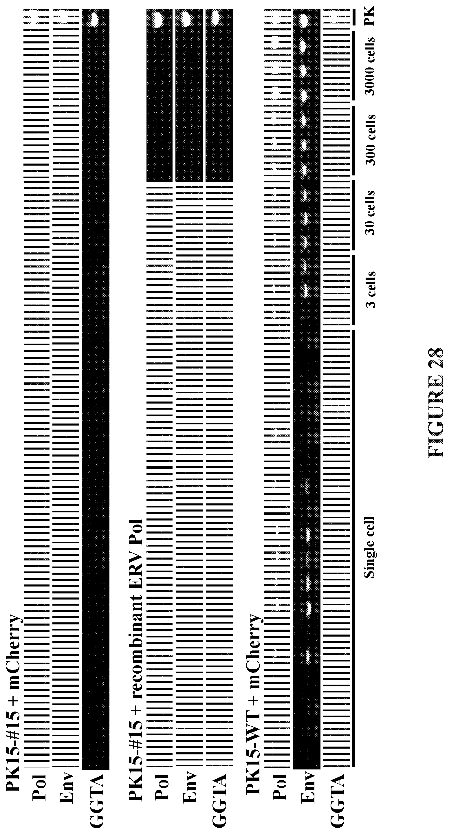
D00039

D00040
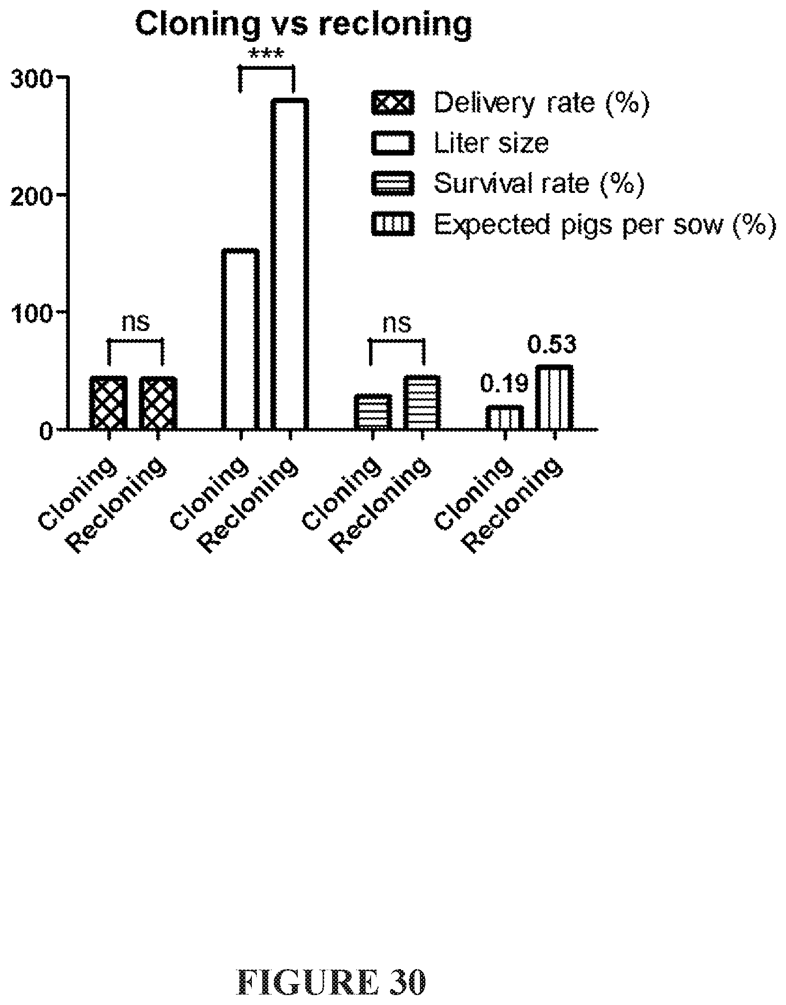
D00041

S00001
XML
uspto.report is an independent third-party trademark research tool that is not affiliated, endorsed, or sponsored by the United States Patent and Trademark Office (USPTO) or any other governmental organization. The information provided by uspto.report is based on publicly available data at the time of writing and is intended for informational purposes only.
While we strive to provide accurate and up-to-date information, we do not guarantee the accuracy, completeness, reliability, or suitability of the information displayed on this site. The use of this site is at your own risk. Any reliance you place on such information is therefore strictly at your own risk.
All official trademark data, including owner information, should be verified by visiting the official USPTO website at www.uspto.gov. This site is not intended to replace professional legal advice and should not be used as a substitute for consulting with a legal professional who is knowledgeable about trademark law.