Reagents and Methods for Breast Cancer Detection
EGLAND; Kristi ; et al.
U.S. patent application number 17/016270 was filed with the patent office on 2020-12-24 for reagents and methods for breast cancer detection. The applicant listed for this patent is Sanford Health. Invention is credited to Kristi EGLAND, Rick EVANS, James POTTALA.
| Application Number | 20200400672 17/016270 |
| Document ID | / |
| Family ID | 1000005073895 |
| Filed Date | 2020-12-24 |

| United States Patent Application | 20200400672 |
| Kind Code | A1 |
| EGLAND; Kristi ; et al. | December 24, 2020 |
Reagents and Methods for Breast Cancer Detection
Abstract
The present invention provides compositions including reagents for detecting human autoantibodies against at least two proteins selected from the group consisting of ANGTPL4, DKK1, EPHA2, LAMC2, SPON2, SSR2, GAL1, GFRA1, LRRC15, CD147, CD320, CDH3, LRP10, SPINT2, SUSD2, and CST2, and their use in detecting breast cancer or disease recurrence.
| Inventors: | EGLAND; Kristi; (Sioux Falls, SD) ; EVANS; Rick; (Sioux Falls, SD) ; POTTALA; James; (Sioux Falls, SD) | ||||||||||
| Applicant: |
|
||||||||||
|---|---|---|---|---|---|---|---|---|---|---|---|
| Family ID: | 1000005073895 | ||||||||||
| Appl. No.: | 17/016270 | ||||||||||
| Filed: | September 9, 2020 |
Related U.S. Patent Documents
| Application Number | Filing Date | Patent Number | ||
|---|---|---|---|---|
| 16002493 | Jun 7, 2018 | |||
| 17016270 | ||||
| 14660423 | Mar 17, 2015 | 10001484 | ||
| 16002493 | ||||
| 61954914 | Mar 18, 2014 | |||
| Current U.S. Class: | 1/1 |
| Current CPC Class: | G01N 2333/435 20130101; G01N 33/57415 20130101; G01N 2333/70503 20130101; G01N 33/564 20130101; G01N 2333/71 20130101; G01N 2333/705 20130101; G01N 2333/70596 20130101; G01N 2333/475 20130101; G01N 2333/912 20130101 |
| International Class: | G01N 33/574 20060101 G01N033/574; G01N 33/564 20060101 G01N033/564 |
Claims
1. A method for detecting breast cancer or disease recurrence, comprising (a) contacting a bodily fluid sample from a subject at risk of having breast cancer or breast cancer recurrence with one or more proteins selected from the group consisting of ANGTPL4, DKK1, EPHA2, LAMC2, SPON2, SSR2, GAL1, GFRA1, LRRC15, CD147, CD320, CDH3, LRP10, SPINT2, SUSD2, and CST2, secreted versions thereof, or extracellular domains thereof; and (b) detecting binding of autoantibodies in the bodily fluid sample against the one or more proteins, secreted versions thereof, or extracellular domains thereof; wherein the presence of autoantibodies against the one or more proteins, secreted versions thereof, or extracellular domains thereof correlates with a likelihood of the subject having breast cancer or breast cancer recurrence.
2. The method of claim 1, wherein the one or more proteins comprise one or more of ANGPTL4, DKK1, EPHA2, GAL1, LAMC2, SPON2, CST2, SPINT2 and SSR2, secreted versions thereof, or extracellular domains thereof.
3. The method of claim 1, wherein the contacting step comprises contacting the bodily fluid sample with two or more proteins selected from the group consisting of ANGTPL4, DKK1, EPHA2, LAMC2, SPON2, SSR2, GAL1, GFRA1, LRRC15, CD147, CD320, CDH3, LRP10, SPINT2, SUSD2, and CST2, secreted versions thereof, or extracellular domains thereof.
4. The method of claim 1, wherein the contacting step comprises contacting the bodily fluid sample with five or more proteins selected from the group consisting of ANGTPL4, DKK1, EPHA2, LAMC2, SPON2, SSR2, GAL1, GFRA1, LRRC15, CD147, CD320, CDH3, LRP10, SPINT2, SUSD2, and CST2, secreted versions thereof, or extracellular domains thereof.
5. The method of claim 1, wherein the contacting step comprises contacting the bodily fluid sample with proteins in of the following marker sets, secreted versions thereof, or extracellular domains thereof: ANGPTL4, DKK1, GAL1, GFRA1, GRANULIN, LRRC15, and MUC1; ANGPTL4, DKK1, GAL1, GRANULIN, LRRC15, and MUC1; ANGPTL4, DKK1, GAL1, and LRRC15; ANGPTL4, DKK1, GAL1, GFRA1, and LRRC15; DKK1, GAL1, GFRA1, GRANULIN, LRRC1, and5 MUC1; ANGPTL4, DKK1, GAL1, GFRA1, GRANULIN, and LRRC15; DKK1, GAL1, GRANULIN, LRRC15, and MUC1; DKK1, GAL1, GFRA1, GRANULIN, and LRRC15; DKK1, GAL1, GFRA1, LRRC15, and MUC1; ANGPTL4, DKK1, GAL1, GRANULIN, and LRRC15; DKK1, GAL1, GFRA1, and LRRC15; DKK1, GAL1, GRANULIN, and LRRC15; ANGPTL4, DKK1, GAL1, LRRC15, and MUC1; DKK1, GAL1, and LRRC15; ANGPTL4, GAL1, LRRC15, and MUC1; GAL1, GFRA1, LRRC15, and MUC1; GAL1, GFRA1, and LRRC15; ANGPTL4, GAL1, and LRRC15; DKK1, GAL1, LRRC15, and MUC1; ANGPTL4, GAL1, GFRA1, and LRRC15; GAL1, LRRC15, and MUC1; ANGPTL4, GAL1, GFRA1, LRRC15, and MUC1; ANGPTL4, GAL1, and GFRA1; DKK1, GAL1, and GFRA1; and GAL1, GFRA1, and MUC1.
6. The method of claim 1, wherein the contacting step comprises contacting the bodily fluid sample ANGPTL4, DKK1, GAL1, MUC1, GFRA1, GRN and LRRC15, secreted versions thereof, or extracellular domains thereof.
7. The method of claim 1, wherein the bodily fluid sample comprises a serum sample from the subject.
8. The method of claim 1, wherein the bodily fluid sample comprises a blood sample from the subject.
9. The method of claim 1, wherein the contacting comprises use of ELISA.
10. The method of claim 1, wherein the contacting comprises use of Longitudinal Assay Screening, wherein all target biomarkers are detected and quantitated within a single test and dilution.
11. The method of claim 1, wherein the one or more proteins, secreted versions thereof, or extracellular domains thereof are detectably labeled.
12. The method of claim 1, wherein the one or more proteins, secreted versions thereof, or extracellular domains thereof are immobilized on a surface.
13. The method of claim 1, wherein the method identifies the subject as likely to have breast cancer or breast cancer recurrence, and wherein the method further comprises treating the subject with an amount of a therapeutic sufficient to treat the breast cancer or breast cancer recurrence.
Description
CROSS REFERENCE
[0001] This application is a continuation of U.S. patent application Ser. No. 16/002,493 filed Jun. 7, 2018, which is a continuation of U.S. patent application Ser. No. 14/660,423 filed Mar. 17, 2015, which claims priority to U.S. Provisional Patent Application Ser. No. 61/954,914 filed Mar. 18, 2014, incorporated by reference herein in its entirety.
BACKGROUND
[0002] For patients with breast cancer (BCa), early and personalized diagnosis is crucial for optimizing treatments leading to long-term survival. Although mammography is the most widely used method to detect BCa, approximately 20% of screening mammograms result in a false negative diagnosis largely due to high breast density. Additionally, 1 in 10 women who get a mammogram will need additional imaging. Yet, the overwhelming majority of these women will not have BCa, as only 2 to 4 of every 1,000 screening mammograms leads to a cancer diagnosis. Therefore, there is an urgent clinical need to develop a novel, minimally invasive diagnostic strategy for the early diagnosis and monitoring of BCa.
SUMMARY OF THE INVENTION
[0003] In a first aspect, the invention provides compositions consisting of between 2 and 25 antibody detection markers, wherein the composition includes reagents for detecting human autoantibodies against at least two proteins selected from the group consisting of human ANGTPL4, DKK1, EPHA2, LAMC2, SPON2, SSR2, GAL1, GFRA1, LRRC15, CD147, CD320, CDH3, LRP10, SPINT2, SUSD2, and CST2. In one embodiment, the composition includes reagents for detecting human autoantibodies against at least two proteins selected from the group consisting of human ANGPTL4, DKK1, EPHA2, GAL1, LAMC2, SPON2, CST2, SPINT2 and SSR2. In a further embodiment, the composition includes reagents for detecting human autoantibodies against at least 5 proteins in the recited group. In another embodiment, the composition further includes reagents for detecting human autoantibodies against one or both of MUC1 and GRN. In various embodiments, the composition consists of between 2 and 20, 4 and 10, and 5-10 antibody detection markers. In various further embodiments, the composition includes reagents for detecting human autoantibodies against one of the following marker sets:
ANGPTL4, DKK1, GAL1, GFRA1, GRANULIN, LRRC15, and MUC1;
ANGPTL4, DKK1, GAL1, GRANULIN, LRRC15, and MUC1;
ANGPTL4, DKK1, GAL1, and LRRC15;
ANGPTL4, DKK1, GAL1, GFRA1, and LRRC15;
[0004] DKK1, GAL1, GFRA1, GRANULIN, LRRC1, and5 MUC1;
ANGPTL4, DKK1, GAL1, GFRA1, GRANULIN, and LRRC15;
DKK1, GAL1, GRANULIN, LRRC15, and MUC1;
DKK1, GAL1, GFRA1, GRANULIN, and LRRC15;
DKK1, GAL1, GFRA1, LRRC15, and MUC1;
ANGPTL4, DKK1, GAL1, GRANULIN, and LRRC15;
DKK1, GAL1, GFRA1, and LRRC15;
DKK1, GAL1, GRANULIN, and LRRC15;
ANGPTL4, DKK1, GAL1, LRRC15, and MUC1;
DKK1, GAL1, and LRRC15;
ANGPTL4, GAL1, LRRC15, and MUC1;
GAL1, GFRA1, LRRC15, and MUC1;
GAL1, GFRA1, and LRRC15;
ANGPTL4, GAL1, and LRRC15;
DKK1, GAL1, LRRC15, and MUC1;
ANGPTL4, GAL1, GFRA1, and LRRC15;
GAL1, LRRC15, and MUC1;
ANGPTL4, GAL1, GFRA1, LRRC15, and MUC1;
ANGPTL4, GAL1, and GFRA1;
DKK1, GAL1, and GFRA1; and
GAL1, GFRA1, and MUC1.
[0005] In another embodiment, the reagents for detecting human autoantibodies comprise the at least two proteins, or antigenic fragments thereof. In a further embodiment, the at least two proteins, or antigenic fragments thereof comprise native extracellular domains and/or native secreted proteins or antigenic fragments thereof. In a still further embodiment, the reagents are detectably labeled. In another embodiment, reagents are immobilized on a surface.
[0006] In another aspect, the invention provides methods for detecting breast cancer or disease recurrence, comprising contacting a bodily fluid sample from a subject at risk of having breast cancer or breast cancer recurrence with one or more reagents for detecting autoantibodies against one or more of human ANGTPL4, DKK1, EPHA2, LAMC2, SPON2, SSR2, GAL1, GFRA1, LRRC15, CD147, CD320, CDH3, LRP10, SPINT2, SUSD2, and CST2, wherein the presence of autoantibodies against the one or more proteins correlates with a likelihood of the subject having breast cancer or breast cancer recurrence. In another embodiment, the reagents comprise reagents for detecting autoantibodies against one or more of human ANGPTL4, DKK1, EPHA2, GAL1, LAMC2, SPON2, CST2, SPINT2 and SSR2. In various further embodiments, the reagents comprise reagents for detecting autoantibodies two or more, or five or more of the recited proteins. In another embodiment the reagents comprise reagents for detecting human autoantibodies against one of the following marker sets:
ANGPTL4, DKK1, GAL1, GFRA1, GRANULIN, LRRC15, and MUC1;
ANGPTL4, DKK1, GAL1, GRANULIN, LRRC15, and MUC1;
ANGPTL4, DKK1, GAL1, and LRRC15;
ANGPTL4, DKK1, GAL1, GFRA1, and LRRC15;
[0007] DKK1, GAL1, GFRA1, GRANULIN, LRRC1, and5 MUC1;
ANGPTL4, DKK1, GAL1, GFRA1, GRANULIN, and LRRC15;
DKK1, GAL1, GRANULIN, LRRC15, and MUC1;
DKK1, GAL1, GFRA1, GRANULIN, and LRRC15;
DKK1, GAL1, GFRA1, LRRC15, and MUC1;
ANGPTL4, DKK1, GAL1, GRANULIN, and LRRC15;
DKK1, GAL1, GFRA1, and LRRC15;
DKK1, GAL1, GRANULIN, and LRRC15;
ANGPTL4, DKK1, GAL1, LRRC15, and MUC1;
DKK1, GAL1, and LRRC15;
ANGPTL4, GAL1, LRRC15, and MUC1;
GAL1, GFRA1, LRRC15, and MUC1;
GAL1, GFRA1, and LRRC15;
ANGPTL4, GAL1, and LRRC15;
DKK1, GAL1, LRRC15, and MUC1;
ANGPTL4, GAL1, GFRA1, and LRRC15;
GAL1, LRRC15, and MUC1;
ANGPTL4, GAL1, GFRA1, LRRC15, and MUC1;
ANGPTL4, GAL1, and GFRA1;
DKK1, GAL1, and GFRA1; and
GAL1, GFRA1, and MUC1.
[0008] In a further embodiment, the reagents comprise reagents for detecting human autoantibodies against human ANGPTL4, DKK1, GAL1, MUC1, GFRA1, GRN and LRRC15. In another embodiment, the one or more reagents comprise the composition of any embodiment or combination of embodiments of the invention. In a further embodiment, the contacting comprises use of ELISA. In another embodiment, the bodily fluid sample comprises a serum sample from the subject. In a further embodiment, the method identifies the subject as likely to have breast cancer or breast cancer recurrence. In a further embodiment, the method further comprises treating the subject with an amount of a therapeutic sufficient to treat the breast cancer or breast cancer recurrence.
[0009] In a further aspect, the invention provides methods for treating a subject with breast cancer, comprising:
[0010] (a) testing a bodily fluid sample from a subject at risk of breast cancer, and identifying candidate subjects that: [0011] (i) have autoantibodies against at least one of ANGPTL4, DKK1, GAL1, MUC1, GFRA1, GRN and LRRC15; and/or [0012] (ii) do not have autoantibodies against GFRA1, GRN and/or LRRC15; and
[0013] (b) treating the candidate subjects with an amount of a therapeutic sufficient to treat the breast cancer.
[0014] In one embodiment, the contacting comprises use of Longitudinal Assay Screening, wherein all target biomarkers may be detected and quantitated within a single test and dilution. In a further embodiment, the bodily fluid sample comprises a blood sample from the subject.
BRIEF DESCRIPTION OF THE FIGURES
[0015] FIG. 1. Antigen conformation affects antibody recognition. A, ELISA analysis using an antigen designed to have native conformation. Wells were coated with anti-rabbit IgG followed by the HER-2-ECD-rFc protein generated in 293T cells. Serial dilutions of anti-HER-2 monoclonal antibodies generated against native HER-2, 3F32 (blue), Herceptin (green) or against denatured HER-2, 3F27 (red) were used in ELISA. Reactions were developed after addition of the appropriate secondary antibody. The O.D. is the absorbance reading for the reaction. B, ELISA analysis using a denatured antigen. Wells were coated with purified His-HER-2-ECD generated in E. coli, and serial dilutions of 3F32 (blue), Herceptin (green) or 3F27 (red) were added. After addition of the secondary antibody, the reactions were developed. C, detection of native HER-2 on SKBR3 cells via flow cytometry. Fluorescence indicates antibody recognition of HER-2 on the surface of SKBR3 cells. D, binding competition assay to demonstrate specificity of conformation-carrying antigen ELISA. Wells were precoated with anti-rabbit IgG followed by HER-2-ECD-rFc. Purified HER-2-Fc (black) or CD30-Fc (purple) chimeric proteins were serially diluted and added to a constant amount of Herceptin before addition to the wells. The reactions were developed after incubation with the secondary antibody.
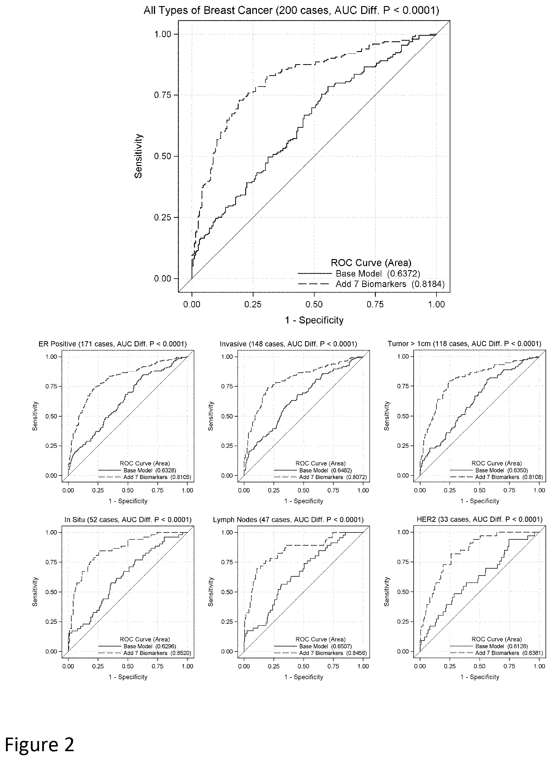
[0016] FIG. 2. ROC curve comparison for classification of breast cancer patients. The autoantibody responses against seven antigens (i.e. ANGPTL4, DKK1, GAL1, GFRA1, GRN, LRRC15 and MUC1) were added to a logistic regression model that included age, BMI, race and current smoking status. The ROC curves were determined for all subjects (top) and by specific subtypes of breast cancer including ER positive, invasive, maximum tumor dimension >1 cm, in situ, lymph node involvement and HER-2 amplification (bottom).
DETAILED DESCRIPTION OF THE INVENTION
[0017] All references cited are herein incorporated by reference in their entirety. Within this application, unless otherwise stated, the techniques utilized may be found in any of several well-known references such as: Molecular Cloning: A Laboratory Manual (Sambrook, et al., 1989, Cold Spring Harbor Laboratory Press), Gene Expression Technology (Methods in Enzymology, Vol. 185, edited by D. Goeddel, 1991. Academic Press, San Diego, Calif.), "Guide to Protein Purification" in Methods in Enzymology (M. P. Deutshcer, ed., (1990) Academic Press, Inc.); PCR Protocols: A Guide to Methods and Applications (Innis, et al. 1990. Academic Press, San Diego, Calif.), Culture of Animal Cells: A Manual of Basic Technique, 2.sup.nd Ed. (R. I. Freshney. 1987. Liss, Inc. New York, N.Y.), Gene Transfer and Expression Protocols, pp. 109-128, ed. E. J. Murray, The Humana Press Inc., Clifton, N.J.), and the Ambion 1998 Catalog (Ambion, Austin, Tex.).
[0018] In a first aspect, the present invention provides compositions consisting of between 2 and 25 antibody detection markers, wherein the composition includes reagents for detecting human autoantibodies against at least two proteins selected from the group consisting of human ANGTPL4, DKK1, EPHA2, LAMC2, SPON2, SSR2, GAL1, GFRA1, LRRC15, CD147, CD320, CDH3, LRP10, SPINT2, SUSD2, and CST2. The inventors have unexpectedly discovered that autoantibodies against the recited proteins provide an indication of whether a subject is suffering from breast cancer (BCa). Thus, the compositions of the invention can be used, for example, in diagnostic assays to discriminate between BCa and healthy patients by the detection of antibodies in a sample from the subject or to detect recurrence of disease in a breast cancer patient after treatment. In one embodiment, the composition includes reagents for detecting human autoantibodies against at least two proteins selected from the group consisting of ANGPTL4, DKK1, EPHA2, GAL1, LAMC2, SPON2, CST2, SPINT2 and SSR2.
[0019] In various embodiments, the composition includes reagents for detecting human autoantibodies against at least three, four, five, six, seven, eight, nine, ten, eleven, twelve, thirteen, fourteen, fifteen, or sixteen proteins in the recited group. In various further embodiments, the composition consists of between 2-24, 2-23, 2-22, 2-21, 2-20, 2-19, 2-18, 2-17, 2-16, 2-15, 2-14, 2-13, 2-12, 2-11, 2-10, 2-9, 2-8, 2-7, 2-6, 2-5, 2-4, 2-3, 3-25, 3-24, 3-23, 3-22, 3-21, 3-20, 3-19, 3-18, 3-17, 3-16, 3-15, 3-14, 3-13, 3-12, 3-11, 3-10, 3-9, 3-8, 3-7, 3-6, 3-5, 3-4, 4-25, 4-24, 4-23, 4-22, 4-21, 4-20, 4-19, 4-18, 4-17, 4-16, 4-15, 4-14, 4-13, 4-14, 4-13, 4-12, 4-11, 4-10, 4-9, 4-8, 4-7, 4-6, 4-5, 5-25, 5-24, 5-23, 5-22, 5-21, 5-20, 5-19, 5-18, 5-17, 5-16, 5-15, 5-14, 5-13, 5-12, 5-11, 5-10, 5-9, 5-8, 5-7, 5-6, 6-25, 6-24, 6-23, 6-22, 6-21, 6-20, 6-19, 6-18, 6-17, 6-16, 6-15, 6-14, 6-13, 6-12, 6-11, 6-10, 6-9, 6-8, 6-7, 7-25, 7-24, 7-23, 7-22, 7-21, 7-20, 7-19, 7-18, 7-17, 7-16, 7-15, 7-14, 7-13, 7-12, 7-11, 7-10, 7-9, 7-8, 8-25, 8-24, 8-23, 8-22, 8-21, 8-20, 8-19, 8-18, 8-17, 8-16, 8-15, 8-14, 8-13, 8-12, 8-11, 8-10, 8-9, 9-25, 9-24, 9-23, 9-22, 9-21, 9-20, 9-19, 9-18, 9-17, 9-16, 9-15, 9-14, 9-13, 9-12, 9-11, 9-10, 1, 2, 3, 4, 5, 6, 7, 8, 9, 10-11, 12, 13, 14, 15, or 16 antibody detection reagents.
[0020] As will be understood by those of skill in the art, the compositions may include additional antibody detection markers and controls as is appropriate for an intended use of the composition. In one non-limiting embodiment, the compositions may further comprise reagents for detecting antibodies against one or both of mucin-1 (MUC1), HER-2 (41), IGFBP2, and GRANULIN (GRN).
[0021] In further embodiments, the compositions comprise or consist of reagents for detecting human autoantibodies against one of the following marker sets, which are shown in the examples that follow (see Table 5) to provide strong predictive value for diagnosing breast cancer:
ANGPTL4, DKK1, GAL1, GFRA1, GRANULIN, LRRC15, and MUC1;
ANGPTL4, DKK1, GAL1, GRANULIN, LRRC15, and MUC1;
ANGPTL4, DKK1, GAL1, and LRRC15;
ANGPTL4, DKK1, GAL1, GFRA1, and LRRC15;
[0022] DKK1, GAL1, GFRA1, GRANULIN, LRRC1, and5 MUC1;
ANGPTL4, DKK1, GAL1, GFRA1, GRANULIN, and LRRC15;
DKK1, GAL1, GRANULIN, LRRC15, and MUC1;
DKK1, GAL1, GFRA1, GRANULIN, and LRRC15;
DKK1, GAL1, GFRA1, LRRC15, and MUC1;
ANGPTL4, DKK1, GAL1, GRANULIN, and LRRC15;
DKK1, GAL1, GFRA1, and LRRC15;
DKK1, GAL1, GRANULIN, and LRRC15;
ANGPTL4, DKK1, GAL1, LRRC15, and MUC1;
DKK1, GAL1, and LRRC15;
ANGPTL4, GAL1, LRRC15, and MUC1;
GAL1, GFRA1, LRRC15, and MUC1;
GAL1, GFRA1, and LRRC15;
ANGPTL4, GAL1, and LRRC15;
DKK1, GAL1, LRRC15, and MUC1;
ANGPTL4, GAL1, GFRA1, and LRRC15;
GAL1, LRRC15, and MUC1;
ANGPTL4, GAL1, GFRA1, LRRC15, and MUC1;
ANGPTL4, GAL1, and GFRA1;
DKK1, GAL1, and GFRA1; and
GAL1, GFRA1, and MUC1.
[0023] In another embodiment, the compositions comprise or consist of reagents for detecting human autoantibodies against human ANGPTL4, DKK1, GAL1, MUC1, GFRA1, GRN and LRRC15.
[0024] The antibody detection markers may be any suitable reagents that can be used to detect antibodies against the recited proteins, including but not limited to the recited protein, a secreted version of the protein (such as a native secreted form of the protein), or an extracellular domain of the protein. Secreted proteins are more easily delivered from tumor cells to lymph nodes, where interactions of immune cells take place resulting in abundant high-affinity antibodies. Membrane surface proteins are commonly released in a soluble form from tumor cells through metalloproteinase-dependent cleavage. The shed proteins are more easily transferred to the lymph nodes than intracellular protein. Thus, in one embodiment the antibody detection marker is a secreted or membrane portion of the recited protein. Exemplary amino acid sequences of the secreted or membrane portion of the recited human proteins are shown below.
TABLE-US-00001 ANGTPL4 (SEQ ID NO: 1) KSPRFASWDEMNVLAHGLLQLGQGLREHAERTRSQLSALERRLSACGSAC QGTEGSTDLPLAPESRVDPEVLHSLQTQLKAQNSRIQQLFHKVAQQQRHL EKQHLRIQHLQSQFGLLDHKHLDHEVAKPARRKRLPEMAQPVDPAHNVSR LHRLPRDCQELFQVGERQSGLFEIQPQGSPPFLVNCKMTSDGGWTVIQRR HDGSVDFNRPWEAYKAGFGDPHGEFWLGLEKVHSITGDRNSRLAVQLRDW DGNAELLQFSVHLGGEDTAYSLQLTAPVAGQLGATTVPPSGLSVPFSTWD QDHDLRRDKNCAKSLSGGWWFGTCSHSNLNGQYFRSIPQQRQKLKKGIFW KTWRGRYYPLQATTMLIQPMAAEAAS DKK1 (SEQ ID NO: 2) VSATLNSVLNSNAIKNLPPPLGGAAGHPGSAVSAAPGILYPGGNKYQTID NYQPYPCAEDEECGTDEYCASPTRGGDAGVQICLACRKRRKRCMRHAMCC PGNYCKNGICVSSDQNHFRGEIEETITESFGNDHSTLDGYSRRTTLSSKM YHTKGQEGSVCLRSSDCASGLCCARHFWSKICKPVLKEGQVCTKHRRKGS HGLEIFQRCYCGEGLSCRIQKDHHQASNSSRLHTCQRH EPHA2 (SEQ ID NO: 3) KEVVLLDFAAAGGELGWLTHPYGKGWDLMQNIMNDMPIYMYSVCNVMSGD QDNWLRTNWVYRGEAERIFIELKETVRDCNSFPGGASSCKETFNLYYAES DLDYGTNFQKRLFTKIDTIAPDEITVSSDFEARHVKLNVEERSVGPLTRK GEYLAFQDIGACVALLSVRVYYKKCPELLQGLAHFPETIAGSDAPSLATV AGTCVDHAVVPPGGEEPRMHCAVDGEWLVPIGQCLCQAGYEKVEDACQAC SPGFFKFEASESPCLECPEHTLPSPEGATSCECEEGFFRAPQDPASMPCT RPPSAPHYLTAVGMGAKVELRWTPPQDSGGREDIVYSVTCEQCWPESGEC GPCEASVRYSEPPHGLTRTSVTVSDLEPHMNYTFTVEARNGVSGLVTSRS FRTASVSINQTEPPKVRLEGRSTTSLSVSWSIPPPQQSRVWKYEVTYRKK GDSNSYNVRRTEGESVTLDDLAPDTTYLVQVQALTQEGQGAGSKVHEFQT LSPEGSGNL LAMC2 (SEQ ID NO: 4) TSRREVCDCNGKSRQCIFDRELHRQTGNGFRCLNCNDNTDGIHCEKCKNG FYRHRERDRCLPCNCNSKGSLSARCDNSGRCSCKPGVTGARCDRCLPGEH MLTDAGCTQDQRLLDSKCDCDPAGIAGPCDAGRCVCKPAVTGERCDRCRS GYYNLDGGNPEGCTQCFCYGHSASCRSSAEYSVHKITSTFHQDVDGWKAV QRNGSPAKLQWSQRHQDVFSSAQRLDPVYFVAPAKELGNQQVSYGQSLSE DYRVDRGGRHPSAHDVILEGAGLRITAPLMPLGKTLPCGLTKTYTFRLNE HPSNNWSPQLSYFEYRRLLRNLTALRIRATYGEYSTGYIDNVTLISARPV SGAPAPWVEQCICPVGYKGQFCQDCASGYKRDSARLGPFGTCIPCNCQGG GACDPDTGDCYSGDENPDIECADCPIGFYNDPHDPRSCKPCPCHNGFSCS VMPETEEVVCNNCPPGVTGARCELCADGYFGDPFGEHGPVRPCQPCQCNN NVDPSASGNCDRLTGRCLKCIHNTAGIYCDQCKAGYFGDPLAPNPADKCR ACNCNPMGSEPVGCRSDGTCVCKPGFGGPNCEHGAFSCPACYNQVKIQMD QFMQQLQRMEALISKAQGGDGVVPDTELEGRMQQAEQALQDILRDAQISE GASRSLGLQLAKVRSQENSYQSRLDDLKMTVERVRALGSQYQNRVRDTHR LITQMQLSLAESEASLGNTNIPASDHYVGPNGFKSLAQEATRLAESHVES ASNMEQLTRETEDYSKQALSLVRKALHEGVGSGSGSPDGAVVQGLVEKLE KTKSLAQQLTREATQAEIEADRSYQHSLRLLDSVSRLQGVSDQSFQVEEA KRIKQKADSLSSLVTRHMDEFKRTQKNLGNWKEEAQQLLQNGKSGREKSD QLLSRANLAKSRAQEALSMGNATFYEVESILKNLREFDLQVDNRKAEAEE AMKRLSYISQKVSDASDKTQQAERALGSAAADAQRAKNGAGEALEISSEI EQEIGSLNLEANVTADGALAMEKGLASLKSEMREVEGELERKELEFDTNM DAVQMVITEAQKVDTRAKNAGVTIQDTLNTLDGLLHLMGM SPON2 (SEQ ID NO: 5) QPLGGESICSARAPAKYSITFTGKWSQTAFPKQYPLFRPPAQWSSLLGAA HSSDYSMWRKNQYVSNGLRDFAERGEAWALMKEIEAAGEALQSVHEVFSA PAVPSGTGQTSAELEVQRRHSLVSFVVRIVPSPDWFVGVDSLDLCDGDRW REQAALDLYPYDAGTDSGFTFSSPNFATIPQDTVTEITSSSPSHPANSFY YPRLKALPPIARVTLLRLRQSPRAFIPPAPVLPSRDNEIVDSASVPETPL DCEVSLWSSWGLCGGHCGRLGTKSRTRYVRVQPANNGSPCPELEEEAECV PDNCV SSR2 (SEQ ID NO: 6) EEGARLLASKSLLNRYAVEGRDLTLQYNIYNVGSSAALDVELSDDSFPPE DFGIVSGMLNVKWDRIAPASNVSHTVVLRPLKAGYFNFTSATITYLAQED GPVVIGSTSAPGQGGILAQREFDRRFSPH GAL1 (SEQ ID NO: 7) LRVRGEVAPDAKSFVLNLGKDSNNLCLHFNPRFNAHGDANTIVCNSKDGG AWGTEQREAVFPFQPGSVAEVCITFDQANLTVKLPDGYEFKFPNRLNLEA INYMAADGDFKIKCVAFD GFRA1 (SEQ ID NO: 8) DRLDCVKASDQCLKEQSCSTKYRTLRQCVAGKETNFSLASGLEAKDECRS AMEALKQKSLYNCRCKRGMKKEKNCLRIYWSMYQSLQGNDLLEDSPYEPV NSRLSDIFRVVPFISDVFQQVEHIPKGNNCLDAAKACNLDDICKKYRSAY ITPCTTSVSNDVCNRRKCHKALRQFFDKVPAKHSYGMLFCSCRDIACTER RRQTIVPVCSYEEREKPNCLNLQDSCKTNYICRSRLADFFTNCQPESRSV SSCLKENYADCLLAYSGLIGTVMTPNYIDSSSLSVAPWCDCSNSGNDLEE CLKFLNFFKDNTCLKNAIQAFGNGSDVTVWQPAFPVQTTTATTTTALRVK NKPLGPAGSENEIPTHVLPPCANLQAQKLKSNVSGNTHLCISNGNYEKEG LGASSHITTKSMAAPPSCGLSPLLVLVVTALSTLLSLTETS LRRC15 (SEQ ID NO: 9) YHGCPSECTCSRASQVECTGARIVAVPTPLPWNAMSLQILNTHITELNES PFLNISALIALRIEKNELSRITPGAFRNLGSLRYLSLANNKLQVLPIGLF QGLDSLESLLLSSNQLLQIQPAHFSQCSNLKELQLHGNHLEYIPDGAFDH LVGLTKLNLGKNSLTHISPRVFQHLGNLQVLRLYENRLTDIPMGTFDGLV NLQELALQQNQIGLLSPGLFHNNHINLQRLYLSNNHISQLPPSVFMQLPQ LNRLTLFGNSLKELSPGIFGPMPNLRELWLYDNHISSLPDNVFSNLRQLQ VLILSRNQISFISPGAFNGLTELRELSLHTNALQDLDGNVFRMLANLQNI SLQNNRLRQLPGNIFANVNGLMAIQLQNNQLENLPLGIFDHLGKLCELRL YDNPWRCDSDILPLRNWLLLNQPRLGTDTVPVCFSPANVRGQSLIIINVN VAVPSVHVPEVPSYPETPWYPDTPSYPDTTSVSSTTELTSPVEDYTDLTT IQVTDDRSVWGMTQAQSG GRN (SEQ ID NO: 10) TRCPDGQFCPVACCLDPGGASYSCCRPLLDKWPTTLSRHLGGPCQVDAHC SAGHSCIFTVSGTSSCCPFPEAVACGDGHHCCPRGFHCSADGRSCFQRSG NNSVGAIQCPDSQFECPDFSTCCVMVDGSWGCCPMPQASCCEDRVHCCPH GAFCDLVHTRCITPTGTHPLAKKLPAQ RTNRAVALSSSVMCPDARSRCP DGSTCCELPSGKYGCCPMPNATCCSDHLHCCPQDTVCDLIQSKCLSKENA TTDLLTKLPAHTVGDVKCDMEVSCPDGYTCCRLQSGAWGCCPFTQAVCCE DHIHCCPAGFTCDTQKGTCEQGPHQVPWMEKAPAHLSLPDPQALKRDVPC DNVSSCPSSDTCCQLTSGEWGCCPIPEAVCCSDHQHCCPQGYTCVAEGQC QRGSEIVAGLEKMPARRASLSHPRDIGCDQHTSCPVGQTCCPSLGGSWAC CQLPHAVCCEDRQHCCPAGYTCNVKARSCEKEVVSAQPATFLARSPHVGV KDVECGEGHFCHDNQTCCRDNRQGWACCPYRQGVCCADRRHCCPAGFRCA ARGTKCLRREAPRWDAPLRDPALRQLL MUC1 (SEQ ID NO: 11) APKPATVVTGSGHASSTPGGEKETSATQRSSVPSSTEKNAFNSSLEDPST DYYQELQRDISEMFLQIYKQGGFLGLSNIKFRPGSVVVQLTLAFREGTIN VHDVETQFNQYKTEAASRYNLTISDVSVSDVPFPFSAQSGAGVPG CD147 (SEQ ID NO: 12) AAGTVFTTVEDLGSKILLTCSLNDSATEVTGHRWLKGGVVLKEDALPGQK TEFKVDSDDQWGEYSCVFLPEPMGTANIQLHGPPRVKAVKSSEHINEGET AMLVCKSESVPPVTDWAWYKITDSEDKALMNGSESRFFVSS CD320 (SEQ ID NO: 13) AGPSSGSCPPTKFQCRTSGLCVPLTWRCDRDLDCSDGSDEEECRIEPCTQ KGQCPPPPGLPCPCTGVSDCSGGTDKKLRNCSRLACLAGELRCTLSDDCI PLTWRCDGHPDCPDSSDELGCGTNEILPEGDATTMGPPVTLESVTSLRNA TTMGPPVTLESVPSVGNATSSSAGDQSGSPTAYG CDH3 (SEQ ID NO: 14) EPCRAVFREAEVTLEAGGAEQEPGQALGKVFMGCPGQEPALFSTDNDDFT VRNGETVQERRSLKERNPLKIFPSKRILRRHKRDWVVAPISVPENGKGPF PQRLNQLKSNKDRDTKIFYSITGPGADSPPEGVFAVEKETGWLLLNKPLD REEIAKYELFGHAVSENGASVEDPMNISIIVTDQNDHKPKFTQDTFRGSV LEGVLPGTSVMQMTATDEDDAIYTYNGVVAYSIHSQEPKDPHDLMFTIHR STGTISVISSGLDREKVPEYTLTIQATDMDGDGSTTTAVAVVEILDANDN APMFDPQKYEAHVPENAVGHEVQRLTVTDLDAPNSPAWRATYLIMGGDDG DHFTITTHPESNQGILTTRKGLDFEAKNQHTLYVEVTNEAPFVLKLPTST ATIVVHVEDVNEAPVFVPPSKVVEVQEGIPTGEPVCVYTAEDPDKENQKI SYRILRDPAGWLAMDPDSGQVTAVGTLDREDEQFVRNNIYEVMVLAMDNG
SPPTTGTGTLLLTLIDVNDHGPVPEPRQITICNQSPVRQVLNITDKDLSP HTSPFQAQLTDDSDIYWTAEVNEEGDTVVLSLKKFLKQDTYDVHLSLSDH GNKEQLTVIRATVCDCHGHVETCPGPWKGG HER2 (SEQ ID NO: 15) TQVCTGTDMKLRLPASPETHLDMLRHLYQGCQVVQGNLELTYLPTNASLS FLQDIQEVQGYVLIAHNQVRQVPLQRLRIVRGTQLFEDNYALAVLDNGDP LNNTTPVTGASPGGLRELQLRSLTEILKGGVLIQRNPQLCYQDTILWKDI FHKNNQLALTLIDTNRSRACHPCSPMCKGSRCWGESSEDCQSLTRTVCAG GCARCKGPLPTDCCHEQCAAGCTGPKHSDCLACLHFNHSGICELHCPALV TYNTDTFESMPNPEGRYTFGASCVTACPYNYLSTDVGSCTLVCPLHNQEV TAEDGTQRCEKCSKPCARVCYGLGMEHLREVRAVTSANIQEFAGCKKIFG SLAFLPESFDGDPASNTAPLQPEQLQVFETLEEITGYLYISAWPDSLPDL SVFQNLQVIRGRILHNGAYSLTLQGLGISWLGLRSLRELGSGLALIHHNT HLCFVHTVPWDQLFRNPHQALLHTANRPEDECVGEGLACHQLCARGHCWG PGPTQCVNCSQFLRGQECVEECRVLQGLPREYVNARHCLPCHPECQPQNG SVTCFGPEADQCVACAHYKDPPFCVARCPSGVKPDLSYMPIWKFPDEEGA CQPCPINCTHSCVDLDDKGCPAEQRASPLT IGFBP2 (SEQ ID NO: 6) EVLFRCPPCTPERLAACGPPPVAPPAAVAAVAGGARMPCAELVREPGCGC CSVCARLEGEACGVYTPRCGQGLRCYPHPGSELPLQALVMGEGTCEKRRD AEYGASPEQVADNGDDHSEGGLVENHVDSTMNMLGGGGSAGRKPLKSGMK ELAVFREKVTEQHRQMGKGGKHHLGLEEPKKLRPPPARTPCQQELDQVLE RISTMRLPDERGPLEHLYSLHIPNCDKHGLYNLKQCKMSLNGQRGECWCV NPNTGKLIQGAPTIRGDPECHLFYNEQQEARGVHTQRMQ LRP10 (SEQ ID NO: 17) HPDRIIFPNHACEDPPAVLLEVQGTLQRPLVRDSRTSPANCTWLILGSKE QTVTIRFQKLHLACGSERLTLRSPLQPLISLCEAPPSPLQLPGGNVTITY SYAGARAPMGQGFLLSYSQDWLMCLQEEFQCLNHRCVSAVQRCDGVDACG DGSDEAGCSSDPFPGLTPRPVPSLPCNVTLEDFYGVFSSPGYTHLASVSH PQSCHWLLDPHDGRRLAVRFTALDLGFGDAVHVYDGPGPPESSRLLRSLT HFSNGKAVTVETLSGQAVVSYHTVAWSNGRGFNATYHVRGYCLPWDRPCG LGSGLGAGEGLGERCYSEAQRCDGSWDCADGTDEEDCPGCPPGHFPCGAA GTSGATACYLPADRCNYQTFCADGADERRCRHCQPGNFRCRDEKCVYETW VCDGQPDCADGSDEWDCSYVLPRK SPINT2 (SEQ ID NO: 18) ADRERSIHDFCLVSKVVGRCRASMPRWWYNVTDGSCQLFVYGGCDGNSNN YLTKEECLKKCATVTENATGDLATSRNAADSSVPSAPRRQDSEDHSSDMF NYEEYCTANAVTGPCRASFPRWYFDVERNSCNNFIYGGCRGNKNSYRSEE ACMLRCFRQQENPPLPLGSKV SUSD2 (SEQ ID NO: 19) QESCSMRCGALDGPCSCHPTCSGLGTCCLDFRDFCLEILPYSGSMMGGKD FVVRHFKMSSPTDASVICRFKDSIQTLGHVDSSGQVHCVSPLLYESGRIP FTVSLDNGHSFPRAGTWLAVHPNKVSMMEKSELVNETRWQYYGTANTSGN LSLTWHVKSLPTQTITIELWGYEETGMPYSQEWTAKWSYLYPLATHIPNS GSFTFTPKPAPPSYQRWRVGALRIIDSKNYAGQKDVQALWTNDHALAWHL SDDFREDPVAWARTQCQAWEELEDQLPNFLEELPDCPCTLTQARADSGRF FTDYGCDMEQGSVCTYHPGAVHCVRSVQASLRYGSGQQCCYTADGTQLLT ADSSGGSTPDRGHDWGAPPFRTPPRVPSMSHWLYDVLSFYYCCLWAPDCP RYMQRRPSNDCRNYRPPRLASAFGDPHFVTFDGTNFTFNGRGEYVLLEAA LTDLRVQARAQPGTMSNGTETRGTGLTAVAVQEGNSDVVEVRLANRTGGL EVLLNQEVLSFTEQSWMDLKGMFLSVAAGDRVSIMLASGAGLEVSVQGPF LSVSVLLPEKFLTHTHGLLGTLNNDPTDDFTLHSGRVLPPGTSPQELFLF GANWTVHNASSLLTYDSWFLVHNFLYQPKHDPTFEPLFPSETTLNPSLAQ EAAKLCGDDHFCNFDVAATGSLSTGTATRVAHQLHQRRMQSLQPVVSCGW LAPPPNGQKEGNRYLAGSTIYFHCDNGYSLAGAETSTCQADGTWSSPTPK CQPGRSY A CST2 (SEQ ID NO:20) WSPQEEDRIIEGGIYDADLNDERVQRALHFVISEYNKATEDEYYRRLLRV LRAREQIVGGVNYFFDIEVGRTICTKSQPNLDTCAFHEQPELQKKQLCSF QIYEVPWEDRMSLVNSRCQEA
[0025] In a further embodiment, the antibody detection marker is a protein, such as those disclosed above, that is in its native form. As disclosed in the accompanying examples, the inventors utilized a eukaryotic expression system to generate conformation-carrying tumor antigens that are properly folded and contain non-continuous epitopes for use in the detection of autoantibodies. The protein may be used in any suitable format; in one non-limiting embodiment, the protein may be an Fc fusion protein.
[0026] In all of the above embodiments, the antibody detection reagents can be labeled with a detectable label. In one embodiment, the detectable labels for reagents to detect autoantibodies against one protein are distinguishable from the detectable labels to detect autoantibodies against the other protein. Methods for detecting the label include, but are not limited to spectroscopic, photochemical, biochemical, immunochemical, physical or chemical techniques. Any suitable detectable label can be used.
[0027] The compositions can be stored frozen, in lyophilized form, or as a solution. In one embodiment, the compositions can be placed on a solid support, such as in a microarray or microplate format; this embodiment facilitates use of the compositions in various detection assays. For example, anti-IgG can be used to precoat the wells of a microwell plate and the antibody detection reagents (such as the proteins discussed herein) can be added to the precoated wells.
[0028] In a second aspect, the present invention provides methods for detecting breast cancer or breast cancer recurrence, comprising contacting a bodily fluid sample from a subject at risk of having breast cancer or breast cancer recurrence with one or more reagents for detecting autoantibodies against one or more of human ANGTPL4, DKK1, EPHA2, LAMC2, SPON2, SSR2, GAL1, GFRA1, LRRC15, CD147, CD320, CDH3, LRP10, SPINT2, SUSD2, and CST2, wherein the presence of autoantibodies against the one or more proteins correlates with a likelihood of the subject having breast cancer or breast cancer recurrence.
[0029] In one embodiment, the composition includes reagents for detecting human autoantibodies against at least two proteins selected from the group consisting of human ANGPTL4, DKK1, EPHA2, GAL1, LAMC2, SPON2, CST2, SPINT2 and SSR2.
[0030] As will be understood by those of skill in the art, the methods may include the use of additional antibody detection markers and controls as is appropriate for an intended use of the composition. In one non-limiting embodiment, the compositions may further comprise reagents for detecting antibodies against one or both of mucin-1 (MUC1), HER-2 (41), IGFBP2, and GRANULIN.
[0031] In another embodiment of the methods of the invention, the compositions comprise or consist of reagents for detecting human autoantibodies against one of the following marker sets:
ANGPTL4, DKK1, GAL1, GFRA1, GRANULIN, LRRC15, and MUC1;
ANGPTL4, DKK1, GAL1, GRANULIN, LRRC15, and MUC1;
ANGPTL4, DKK1, GAL1, and LRRC15;
ANGPTL4, DKK1, GAL1, GFRA1, and LRRC15;
[0032] DKK1, GAL1, GFRA1, GRANULIN, LRRC1, and5 MUC1;
ANGPTL4, DKK1, GAL1, GFRA1, GRANULIN, and LRRC15;
DKK1, GAL1, GRANULIN, LRRC15, and MUC1;
DKK1, GAL1, GFRA1, GRANULIN, and LRRC15;
DKK1, GAL1, GFRA1, LRRC15, and MUC1;
ANGPTL4, DKK1, GAL1, GRANULIN, and LRRC15;
DKK1, GAL1, GFRA1, and LRRC15;
DKK1, GAL1, GRANULIN, and LRRC15;
ANGPTL4, DKK1, GAL1, LRRC15, and MUC1;
DKK1, GAL1, and LRRC15;
ANGPTL4, GAL1, LRRC15, and MUC1;
GAL1, GFRA1, LRRC15, and MUC1;
GAL1, GFRA1, and LRRC15;
ANGPTL4, GAL1, and LRRC15;
DKK1, GAL1, LRRC15, and MUC1;
ANGPTL4, GAL1, GFRA1, and LRRC15;
GAL1, LRRC15, and MUC1;
ANGPTL4, GAL1, GFRA1, LRRC15, and MUC1;
ANGPTL4, GAL1, and GFRA1;
DKK1, GAL1, and GFRA1; and
GAL1, GFRA1, and MUC1.
[0033] In another embodiment, the compositions comprise or consist of reagents for detecting human autoantibodies against ANGPTL4, DKK1, GAL1, MUC1, GFRA1, GRN and LRRC15.
[0034] The antibody detection markers may be any suitable reagents that can be used to detect antibodies against the recited proteins, including but not limited to the recited protein, a secreted version of the protein (such as a native secreted form of the protein), or an extracellular domain of the protein. Secreted proteins are more easily delivered from tumor cells to lymph nodes, where interactions of immune cells take place resulting in abundant high-affinity antibodies. Membrane surface proteins are commonly released in a soluble form from tumor cells through metalloproteinase-dependent cleavage. The shed proteins are more easily transferred to the lymph nodes than intracellular protein. Thus, in one embodiment the antibody detection marker is a secreted or membrane portion of the recited protein. Exemplary amino acid sequences of the secreted or membrane portion of the recited proteins are as disclosed herein.
[0035] In another embodiment, the antibody detection marker comprises or consists of a composition of the invention.
[0036] The contacting can be carried out under any suitable conditions for promoting binding between the autoantibodies in the bodily fluid sample and the reagent to forma binding complex that can be detected. Appropriate such conditions can be determined by those of skill in the art based on the intended assay, in light of the teachings herein. Similarly, any suitable additional steps can be used in the methods, such as one or more wash or other steps to remove unbound reagents.
[0037] Any suitable detection technique can be used, including but not limited to enzyme linked immunosorbent assays (ELISA), bead based assay platforms such as the Luminex systems, 2-D array based assay platforms such as SearchLight.RTM., and the Inanovate.RTM. `Longitudinal Assay Screening` platform which may be capable of quantitating all the listed breast cancer biomarker from patient samples at their clinically relevant concentrations in a single test and dilution. In one embodiment, the compositions can be placed on a solid support, such as in a microarray, glass slide, membrane, microplate format or beads. The embodiment facilitates use of the compositions. Exemplary such assays are provided in the examples.
[0038] Similarly, any suitable bodily fluid can be used, including but not limited to a serum sample, plasma sample or blood sample from the subject. The subject may be any subject at risk of breast cancer, such as a human subject.
[0039] In a further embodiment, method identifies the subject as likely to have breast cancer, and wherein the method further comprises treating the subject with an amount of a therapeutic sufficient to treat the breast cancer.
[0040] In one non-limiting embodiment of any of the above embodiments, ANGPTL4, DKK1, GAL1, and MUC1 autoantibody response are correlated with BCa; and autoantibody responses against GFRA1, GRN and LRRC15 are inversely correlated with BCa.
[0041] In one specific embodiment, the reagents include ANGPTL4, DKK1, GAL1, MUC1, GFRA1, GRN and LRRC15; where, autoantibody responses against GFRA1, GRN and LRRC15 are inversely correlated with BCa. As detailed in the examples, when the autoantibody responses against the 7 antigens were added to the base model, including age, body mass index (BMI), race and current smoking status, the assay had the following diagnostic capabilities: c-stat (95% CI), 0.82 (0.78 to 0.86); sensitivity, 73%; specificity, 76%; and PLR (95% CI), 3.04 (2.34 to 3.94). The model was calibrated across risk deciles (Hosmer-Lemeshow, p=0.13) and performed well in specific subtypes of BCa including estrogen receptor positive, HER-2 positive, invasive, in situ and tumor sizes >1 cm. Diagnostic capabilities of other exemplary marker sets are provided in Table 5.
[0042] In a third aspect, the invention provides methods for treating a subject with breast cancer, comprising:
[0043] (a) testing a bodily fluid sample from a subject at risk of breast cancer, and identifying candidate subjects that: [0044] (i) have autoantibodies against at least one of ANGPTL4, DKK1, GAL1, MUC1, GFRA1, GRN and LRRC15; and/or [0045] (b) do not have autoantibodies against GFRA1, GRN and/or LRRC15; and
[0046] (b) treating the candidate subjects with an amount of a therapeutic sufficient to treat the breast cancer.
Example 1
[0047] Breast cancer (BCa) patients elicit an autoantibody response against cancer proteins, which reflects and amplifies the cellular changes associated with tumorigenesis. Detection of autoantibodies in plasma may provide a minimally invasive mechanism for early detection of BCa. To identify cancer proteins that elicit a humoral response, we generated a cDNA library enriched for BCa genes that encode membrane and secreted proteins, which are more likely to induce an antibody response compared to intracellular proteins. To generate conformation-carrying antigens that are efficiently recognized by patients' antibodies, a eukaryotic expression strategy was established. Plasma from 200 BCa patients and 200 age-matched healthy controls were measured for autoantibody activity against 20 different antigens designed to have conformational epitopes using ELISA. A conditional logistic regression model was used to select a combination of autoantibody responses against the 20 different antigens to classify BCa patients from healthy controls. The best combination included ANGPTL4, DKK1, GAL1, MUC1, GFRA1, GRN and LRRC15; however, autoantibody responses against GFRA1, GRN and LRRC15 were inversely correlated with BCa. When the autoantibody responses against the 7 antigens were added to the base model, including age, BMI, race and current smoking status, the assay had the following diagnostic capabilities: c-stat (95% CI), 0.82 (0.78 to 0.86); sensitivity, 73%; specificity, 76%; and PLR (95% CI), 3.04 (2.34 to 3.94). The model was calibrated across risk deciles (Hosmer-Lemeshow, p=0.13) and performed well in specific subtypes of BCa including estrogen receptor positive, HER-2 positive, invasive, in situ and tumor sizes >1 cm.
INTRODUCTION
[0048] For patients with breast cancer (BCa), early and personalized diagnosis is crucial for optimizing treatments leading to long-term survival. Although mammography is the most widely used method to detect BCa, approximately 20% of screening mammograms result in a false negative diagnosis largely due to high breast density (1). Additionally, 1 in 10 women who get a mammogram will need additional imaging (2). Yet, the overwhelming majority of these women will not have BCa, as only 2 to 4 of every 1,000 screening mammograms leads to a cancer diagnosis (3). Therefore, there is an urgent clinical need to develop a novel, minimally invasive diagnostic strategy for the early diagnosis of BCa.
[0049] At present, there is no established tumor marker that is secreted into the peripheral circulation that can be measured by a blood test for the diagnosis of BCa. Currently, tumor markers that are accepted in clinical practice are tissue-based prognostic markers, such as the estrogen receptor (ER), HER-2 amplification, 21-gene Oncotype DX and 70-gene MammaPrint (6-12). All require an invasive biopsy or surgical procedure to acquire tumor tissue for assessment, bearing a heavy burden on patients. Serum tumor markers are valuable tools that allow minimally invasive procedures for sampling to promote the early diagnosis of cancer as well as following the prognosis after treatment (4, 5). However, tumor markers produced by tumor cells usually have relatively low concentrations in the peripheral circulation, especially in early stage disease.
[0050] Here we report the use of a molecular approach to identify tumor antigen candidates that elicit an antibody response in BCa patients. Previously, we generated a BCa cDNA library from membrane-associated polyribosomal (MAP) RNA, which encodes secreted and membrane proteins, and subtracted the library with RNA from normal tissues (29). Secreted proteins are more easily delivered from tumor cells to lymph nodes, where interactions of immune cells take place resulting in abundant high-affinity antibodies. Membrane surface proteins are commonly released in a soluble form from tumor cells through metalloproteinase-dependent cleavage. The shed proteins are more easily transferred to the lymph nodes than intracellular proteins (30, 31). Consequently, the obtained subtracted library, referred to as the membrane-associated polyribosomal cDNA library (MAPcL), is enriched with clones encoding membrane and secreted TAA that are highly abundant in BCa and should preferentially induce an antibody response in patients (29). In addition, we have established a method for producing recombinant antigens as Fc fusion proteins designed to have native conformations, which is essential for the expression of membrane and secreted proteins that may induce an antibody response in patients.
[0051] We have developed a conformation-carrying antigen ELISA-based strategy to discriminate between BCa and healthy patients by the detection of autoantibodies against a panel of TAAs. Twenty antigens were selected from the most abundant genes represented in the MAPcL, and Fc fusion proteins were generated. Blood was collected from 200 newly diagnosed BCa patients and 200 healthy women as age-matched controls. The 400 plasma samples were screened for the presence of autoantibodies against the 20 different MAPcL-derived antigens using ELISA. A combination of seven antigens with patient demographics yielded the best positive likelihood ratio to discriminate between healthy and BCa patients.
MATERIALS AND METHODS
Plasmid Construction
[0052] For production of MAPcL-rabbit Fc-tagged antigens, two constructs, pSecTag2 (Invitrogen, Carlsbad, Calif.) and pFUSE-rIgG-Fc1 (InvivoGen, San Diego, Calif.), were both utilized to generate the 20 MAPcL-rFc expression constructs because of restriction site availability for cloning. pSecTag2 was modified by amplifying the Fc portion of rabbit IgG using primers
TABLE-US-00002 (SEQ ID NO: 21) 5'-CCGGATATCAGCAAGCCCACGTGCCCAC-3' and (SEQ ID NO: 22) 5'-AAGGAAAAAAGCGGCCGCTC-ATTTACCCGGAGAGCGGGAG-3'
(Integrated DNA Technologies, Coralville, Iowa) using pFUSE-rIgG-Fc1 as a template. The rFc PCR product was digested with EcoRV and NotI and inserted into pSecTag2, referred to as pSecTag2-rFc, which contains an IgK signal sequence for secretion. The pFUSE-rIgG-Fc1 contains an IL2 signal sequence. To keep the signal sequence consistent between the two plasmids, the IgK leader sequence was amplified via PCR using pSecTag2 as a template. The IL2 leader sequence was then replaced with the IgK signal sequence, creating pFUSE-IgK-rFc.
[0053] The accession numbers of the 20 MAPcL genes used as templates for cloning and predicted signal sequences are indicated in Table 1. The signal sequences of each encoded protein were determined using SignalP (32, 33). If a protein contained a transmembrane domain, only the encoded extracellular portion was included. The transmembrane domains were predicted using the TMHMM database (34). The amino acid numbers encoded by the cloned fragment are shown in Table 1. ANGPTL4, CDH3, DKK1, SPON2, SSR2, CST2, GFRA1 and GAL1 were custom cloned into pSecTag2-rFc using the SfiI and KpnI restriction sites (Genscript, Piscataway, N.J.). EPHA2, IGFBP2 and LAMC2 were custom cloned into pSecTag2-rFc using the KpnI and BamHI restriction sites. GRN, MUC1 and LRRC15 were custom cloned into pSecTag2-rFc using the SfiI and BamHI restriction sites. HER-2, LRP10, SPINT2 and SUSD2 were cloned into pFUSE-IgK-rFc using the SfiI and XhoI restriction sites. CD147 was cloned into pFUSE-IgK-rFc using the BamHI and SacII restriction sites. CD320 was cloned into pFUSE-IgK-rFc using the EcoRI and XhoI restriction sites.
TABLE-US-00003 TABLE 1 MAPcL Candidates for Generation of rFc Fusion Proteins Signal Encoded Sequence* Amino Amino Acid Gene from MAPcL Accession # Acids Fragment.dagger. ANGPTL4 (angiopoietin- NM_139314 1-30 31-406 like 4) CD147 NM_198589 1-21 22-162 CD320 NM_016579 1-46 47-230 CDH3 (cadherin 3) NM_001793 1-24 25-654 CST2 (cystatin SA) NM_001322 1-20 21-141 DKK1 (dickkopf WNT NM_012242 1-28 29-266 signaling pathway inhibitor 1) EPHA2 (EPH receptor A2) NM_004431 1-26 27-535 GAL1 (lectin, galactoside- NM_002305 1-17 18-135 binding, soluble, 1) GFRA1 (GPI-linked anchor AF038421 1-24 25-465 protein) GRN (granulin) NM_002087 1-17 18-593 HER-2 NM_004448 1-22 23-652 IGFBP2 (insulin-like growth NM_000597 1-39 40-328 factor binding protein 2) LAMC2 (laminin, gamma 2) NM_005562 1-21 22-1111 LRP10 (low density NM_014045 1-16 17-440 lipoprotein receptor-related protein 10) LRRC15 (leucine rich repeat NM_001135057 1-27 28-544 containing 15) MUC1 (mucin 1) NM_002456 1-22 23-167 SPINT2 (serine peptidase NM_021102 1-27 28-198 inhibitor, Kunitz type, 2) SPON2 (spondin 2) NM_012445 1-26 27-331 SSR2 (signal sequence NM_003145 1-17 18-146 receptor, beta (translocon- associated protein beta)) SUSD2 (sushi domain NM_019601 1-27 28-785 containing 2) *The signal sequences of each encoded protein were determined using SignalP (32, 33) and were not included in the expression constructs. .dagger.The amino acid numbers indicate the encoded portion of the proteins cloned between the Ig.quadrature. signal sequence and the Fc portion of rabbit IgG to generate the secreted MAPcL-rFc fusion proteins.
[0054] For production of His-tagged HER-2, HER-2 was amplified via PCR using primers 5'-CCCAAGCTTGCAGCACCCAAGTGTGCACCGGCAC-3' (SEQ ID NO: 23) and 5'-GTGCTCGAGTCACGTC-AGAGGGCTGGCTCTCTGCTCG-3'(SEQ ID NO: 24). The product was digested with HindIII and XhoI and cloned directionally into the pET-28a expression vector.
Cell Culture
[0055] 293T and SKBR3 cell lines were cultured in DMEM with 10% FBS. Cultures were maintained at 37.degree. C. with 5% CO.sub.2 in a humidified incubator. All cell lines were authenticated and tested negatively for mycoplasma.
Protein Production
[0056] The MAPcL-rFc fusion proteins were produced in 293T cells. Briefly, 293T cells were transfected using Effectene (Qiagen, Valencia, Calif.) according to manufacturer's specifications. During transfection, the cells were cultured in DMEM with 2% FBS. Supernatants containing the secreted fusion proteins were harvested, centrifuged to clear cell debris and supplemented with 0.1% sodium azide. His-HER-2 was produced in E. coli BL21 (Invitrogen, Carlsbad, Calif.) and purified using IMAC affinity chromatography.
Sandwich ELISA
[0057] Microtiter plates (Nalge Nunc, Rochester, N.Y.) were coated overnight with 2 .mu.g/ml goat anti-rabbit Fc (Jackson Immunoresearch, West Grove, Pa.) diluted with phosphate buffered saline. The supernatants containing the rFc fusion proteins were diluted 1:3 serially in standard blocking buffer (0.5% bovine serum albumin and 0.1% sodium azide in phosphate buffered saline). Plates were washed once, and the serially diluted supernatants were transferred to the microtiter plates. Rabbit IgG of known concentration was diluted similarly and added to one row of the microtiter plate in order to quantify the amount of fusion protein present in the culture media. After incubating for two hours, plates were washed twice and 50 .mu.l of HRP-conjugated goat anti-rabbit IgG (Jackson Immunoresearch, West Grove, Pa.) diluted 1:3000 in standard blocking buffer with 0.05% Tween 20 added. After a 2-hour incubation, plates were washed 4 times and developed with 100 .mu.l/well of TMB substrate (Pierce, Rockford, Ill.). The development reaction was stopped after five minutes with 50 .mu.l/well of 2N H.sub.2SO.sub.4, and the absorbance was measured at 450 nm to determine the concentration. The absorbance at 690 nm was subtracted to remove background signal.
Antibody Recognition of Conformational Versus Denatured HER-2 Protein
[0058] For the conformational HER-2 assay, microtiter plates were coated with 2 .mu.g/ml goat anti-rabbit Fc (Jackson Immunoresearch, West Grove, Pa.) in PBS overnight. HER-2-ECD-rFc was then added to each well, 100 .mu.l/well. For denatured HER-2, microtiter plates were coated with 2 .mu.g/ml His-HER-2-ECD in PBS overnight.
[0059] Three HER-2 antibodies were used in the assay: anti-HER-2 3F27 (US Biological, Swampscott, Mass.), anti-HER-2 3F32 (US Biological, Swampscott, Mass.) and Herceptin (Genentech, South San Francisco, Calif.). Each antibody was diluted to 1 .mu.g/ml in standard blocking buffer with 0.05% Tween 20. The antibodies were then serially diluted. After washing once, 50 .mu.l/well of the serially diluted antibodies was added to the plates and incubated for 2 hours at room temperature. The plates were washed three times, and species appropriate HRP-conjugated secondary antibodies were added at a 1:3000 dilution. Plates were washed four times and developed with 100 .mu.l/well TMB substrate for five minutes. Development was stopped with 50 .mu.l/well 2N H.sub.2SO.sub.4. Absorbance was measured at 450 nm, and the 690 nm absorbance was subtracted to account for background.
[0060] The same antibodies were used to stain HER-2 in SKBR3 BCa cells via flow cytometry. SKBR3 cells were detached from dish using Cell Dissociation Solution Non-enzymatic 1.times. (Sigma, St. Louis, Mo., catalog #C5914). 2.times.10.sup.5 cells were incubated with 0.5 .mu.g/ml of each antibody for 1 hour at room temperature. The cells were then washed, and a 1:200 dilution of PE-conjugated antibody for the appropriate species was added. The cells were again washed, resuspended in FACS buffer (PBS with 5% bovine serum albumin and 0.1% sodium azide) and analyzed by flow cytometry.
Competition of Herceptin Binding
[0061] Microtiter plates were coated with 4 .mu.g/ml goat anti-rabbit Fc and incubated overnight. After one wash, 100 .mu.l/well HER-2-ECD-rFc was added to each well and incubated overnight. HER-2-Fc and CD30-Fc chimeric proteins (R&D Systems, Minneapolis, Minn.) were serially diluted from a starting concentration of 10 ug/ml. Herceptin was added to a final concentration of 10 ng/ml in each of the serial chimeric protein dilutions. Plates were washed twice, and 50 .mu.l/well of chimeric protein/Herceptin mixture was applied to the plate. Plates were then washed three times, and a 1:3000 dilution of HRP goat anti-human IgG was applied to each well, 50 .mu.l/well. After four washes, 100 .mu.l/well TMB substrate was added to each well. Development was stopped with 50 .mu.l/well 2N H2SO4 after 5 minutes. Absorbance was measured at 450 nm with 690 nm absorbance subtracted.
Patients
[0062] The inclusion criteria for cases were women over 30 years of age that were newly diagnosed with BCa (any type) at Sanford Health, Sioux Falls, S. Dak. Patients were asked to provide one extra 10 ml EDTA tube of blood prior to mastectomy, lumpectomy, radiation therapy, chemotherapy or other treatment. Case subjects were excluded only if they had a previous history of cancer of any kind. Healthy control subjects had a negative mammogram within six months before the blood draw. Healthy subjects were excluded if there was a history of previous cancer of any kind or a history of autoimmune disease. All patients provided written informed consent, and the Sanford Health IRB approved the study protocol. Blood samples from 200 BCa patients were collected from 10/08/09 to 4/17/12. In addition, 200 age-matched healthy control blood samples were collected from 10/16/09 to 1/19/11. See Table 2 for enrolled patients' characteristics.
TABLE-US-00004 TABLE 2 Patient Clinical and Pathological Characteristics Patients with Breast Cancer N = 200 Age: Mean (SD) 58.9 (11.4) White Race: n (%) 193 (97%) BMI [kg/m2]: Mean (SD) 29.7 (6.6) Smoking Status: n (%) Current 22 (11%) Never 120 (60%) Past 58 (29%) Family History Yes: n (%) 114 (58%) Tumor Type: n (%) Invasive 148 (74%) in situ 52 (26%) Histology: n (%) Ductal and Lobular 3 (2%) Ductal 173 (87%) Lobular 21 (11%) Other 2 (1%) ER Positive: n (%) 171 (86%) PR Positive: n (%) 147 (74%) HER-2 Amplification: n (%) Negative 156 (78%) Positive 33 (17%) Unknown 11 (6%) Triple Negative Yes: n (%) 18 (12%) Tumor Max Dimension [cm]: n (%) .ltoreq.1 66 (36%) >1 to .ltoreq.2 65 (35%) >2 53 (29%) Lymph Node Involvement: n (%) 47 (24%) Age-Matched Controls with Negative Mammogram N = 200 Age: Mean (SD) 58.8 (11.3) White Race: n (%) 192 (97%) BMI [kg/m2]: Mean (SD) 27.1 (5.5) Smoking Status: n (%) Current 7 (4%) Never 125 (63%) Past 67 (34%)
Serum Collection
[0063] Blood was collected in a 10 ml EDTA tube and centrifuged at 2000.times.g for 10 minutes. Plasma was removed from the tube, aliquoted and stored at -80 degrees Celsius until screening for the presence of autoantibodies.
Conformation-Carrying Antigen ELISA
[0064] Microtiter plates (Nalge Nunc, Rochester, N.Y.) were coated overnight with 4 .mu.g/ml goat anti-rabbit Fc (Jackson Immunoresearch, West Grove, Pa.) in phosphate buffered saline. Plates were washed once, and 100 .mu.l/well of MAPcL-rFc fusion protein was added. Plates were incubated for 2 hours and washed twice. The plates were then coated with 50 .mu.l/well of optimized blocking buffer (phosphate buffered saline with 0.5% bovine serum albumin, 0.2% dry milk, 0.1% polyvinylpyrrolidone, 20 mM L-Glutamine, 20 mM L-Arginine, 0.1% sodium azide, 10% goat serum, and 0.05% Tween 20). The plates were incubated for 1 hour at 37.degree. C. and washed once. Serum samples diluted 1:100 in optimized blocking buffer were added and incubated for 2 hours at room temperature. Plates were then washed three times, and autoantibodies were detected using an HRP-conjugated goat anti-human IgG (Jackson Immunoresearch, West Grove, Pa.) diluted 1:3000 in standard blocking buffer with 0.05% Tween 20. Plates were incubated for 1 hour at room temperature, washed four times and developed with 100 .mu.l/well of TMB substrate (Pierce, Rockford, Ill.) for 15 minutes. Development was stopped with 50 .mu.l/well 2N H.sub.2SO.sub.4, and the absorbance was measured at 450 nm. The absorbance at 690 nm was subtracted to remove background signal. Each 96-well plate included 14 samples from BCa subjects and 14 samples from normal mammogram subjects. Each sample was tested in triplicate within the same plate. One row in each plate was subjected only to blocking buffer as a negative control for the ELISA.
Statistical Methods
[0065] Controls were individually matched to 200 BCa patients 1:1 within a 3-year age window using a greedy caliper matching algorithm (35) while blinded to assay data. For each subject the antigen level was transformed by subtracting the mean of the blocking buffer from the mean of the triplicate measurements. If the difference was less than zero, it was set to zero, and the square root was taken to yield a more symmetrical distribution.
[0066] Differences in demographics and autoantibody responses between BCa patients and controls were tested using two-sample t-test and Chi-squared test for continuous and categorical data, respectively. The incremental improvement to the c-statistic (i.e. concordance index, area under the receiver operating characteristic (ROC) curve) was tested by adding the autoantibody response to each antigen to a logistic regression model that already included age, BMI, race, and current smoking status. The model calibration was tested using the Hosmer-Lemeshow goodness-of-fit measure, which constructs a Chi-squared statistic by comparing the predicted and observed number of cases by probability decile (36).
[0067] After assessing the individual antigens, a multivariable conditional logistic regression analysis with strata for age-matching was used to determine the subset of antigens that minimized Akaike's Information Criterion (37); all models were adjusted for BMI, race, and current smoking status. Exploratory subgroup analyses were performed to determine if the multivariable subset of antigens performed differently in a particular type of BCa. The multivariable model was tested in the following subgroups: invasive, in situ, ER positive, tumor maximum dimension >1 cm, lymph node involvement, and HER-2 positive. The critical level alpha was set to .ltoreq.0.05/20 antigens=0.0025 using the Bonferroni correction. SAS.RTM. (Cary, N.C.) version 9.3 software was used for all analyses.
Results
[0068] Generation of Tumor-Associated Antigens Designed to have Native Conformations
[0069] To identify TAAs that elicit a humoral response in patients, candidate genes that encode membrane and secreted proteins were selected from the most abundant genes represented in the MAPcL. Because only 10% of epitopes on proteins are in a linear continuous sequence (24), we utilized a eukaryotic expression system to generate conformation-carrying tumor antigens that are properly folded and contain noncontinuous epitopes for use in the detection of autoantibodies. Sequences encoding the extracellular domains (ECD) or the secreted proteins without the signal sequence of the candidate MAPcL genes were cloned 5' of the Fc region of rabbit IgG (rFc) into the pSecTag2-rFc vector or pFUSE-IgK-rFc, depending on restriction enzyme cloning sites. The IgK leader sequence contained in the vectors directs the fusion proteins to be secreted. The vectors encoding the fusion proteins were transiently transfected into 293T cells, and the corresponding fusion proteins were secreted into the media. Production of the secreted fusion proteins was confirmed using a sandwich ELISA, and the concentrations were determined by comparison to an established CD147-rFc standard (data not shown).
[0070] To demonstrate that the generated MAPcL-rFc proteins were designed to be folded into a native conformation, an ELISA analysis was performed using commercially available anti-HER-2 antibodies generated against either native (monoclonal antibody 3F32 and Herceptin) or denatured (monoclonal antibody 3F27) HER-2 protein. Two antigens consisting of the ECD of HER-2 were analyzed: the conformation-carrying HER-2-ECD-rFc protein generated in 293T cells and a His-HER-2-ECD protein that was produced in bacteria and purified over a nickel column. The anti-native HER-2 antibody (3F32) recognized the HER-2-ECD-rFc produced in 293T (FIG. 1A), but was unable to detect the purified His-HER-2-ECD protein produced in bacteria (FIG. 1B). Also, Herceptin was unable to detect the denatured His-HER-2-ECD protein purified from bacteria (FIG. 1B). However, a strong response was observed for Herceptin when HER-2-ECD-rFc protein was used as the antigen for the ELISA analysis (FIG. 1A). Although the 3F27 antibody generated against denatured HER-2 did not detect the HER-2-ECD-rFc protein (FIG. 1A), this antibody had a strong response to bacterial HER-2-ECD (FIG. 1B).
[0071] To confirm the specific recognition of native versus denatured epitopes by the purchased antibodies, flow cytometry was performed on unfixed SKBR3 cells, a BCa cell line known to have HER-2 amplification (38). Because surface HER-2 would retain its native confirmation on the unfixed SKBR3 cells, the anti-HER-2 3F27 antibody, specific for denatured HER-2, was unable to detect surface HER-2 on the cell membrane of SKBR3 cells by flow cytometry (FIG. 1C). When anti-HER-2 3F32 antibody and Herceptin, both of which recognize conformational HER-2, were used for flow cytometry analysis, a large shift in fluorescence was observed indicated that the antibodies recognized HER-2 present on the membrane of the SKBR3 cells (FIG. 1C).
[0072] A binding competition assay was performed to verify that the conformation-carrying antigen ELISA was recognizing the MAPcL antigen specifically. Wells were precoated with anti-rabbit IgG followed by HER-2-ECD-rFc. Purchased HER-2-Fc and CD30-Fc purified chimeric proteins (R&D Systems) were serially diluted and added to a constant amount of Herceptin (10 ng/ml) in each well. Following the addition of the HRP-conjugated secondary anti-human IgG antibody, the reactions were developed. Herceptin binding to HER-2-ECD-rFc was competed by addition of HER-2-Fc but not the CD30-Fc protein (FIG. 1D). This result indicates that Herceptin is binding specifically to the HER-2-ECD portion of the conformation-carrying fusion protein.
Screening of Patients for Autoantibodies Using the Conformation-Carrying Antigen ELISA
[0073] Twenty MAPcL-rFc fusion antigens designed to contain their native conformation were generated by cloning the sequences encoding the ECD or secreted proteins 5' of the rFc sequence (see Table 1 for identity of all 20 antigens). The expression plasmids were individually transfected into 293T cells, and the MAPcL-rFc fusion proteins were secreted into the media. The 20 fusion proteins were quantitated by sandwich ELISA analysis (data not shown). To detect autoantibodies in plasma collected from patients, a conformation-carrying antigen ELISA was developed using the generated MAPcL-rFc antigens. To immobilize the MAPcL-rFc fusion proteins, anti-rabbit IgG was used to precoat the wells of a 96-well plate. The media from the transfected 293T cells, which contains the generated MAPcL-rFc fusion proteins designed to have native conformations, was added to the precoated wells. To reduce plate variation and increase repeatability of the assay, three replicate samples using the plasma from each individual patient were distributed across the 96-well plate. After addition of an HRP-conjugated secondary anti-human IgG antibody, the plates were developed and the absorbance of each well was measured. The 200 plasma samples collected from newly diagnosed BCa patients and plasma from 200 age-matched healthy subjects were evaluated for autoantibody reactivity against the 20 antigens using the conformation-carrying ELISA.
[0074] The 200 BCa patients and 200 healthy controls had a mean (SD) age of 59 (11) years and 97% self identified as white race (Table 2). Cancer patients were more overweight (29.7 vs. 27.1 kg/m.sup.2, p<0.0001) and had different smoking habits (p=0.014), such that there was a greater prevalence of current smokers (11% vs. 4%) in the cancer subjects versus healthy. The 200 BCa patients represented the heterogeneity of the disease consisting of 74% invasive, 24% lymph node involvement, 86% ER-positive, 17% HER-2 positive and 12% triple negative BCa (Table 2). Analyzing the absorbance reading of the autoantibody responses against the individual antigens, we determined that there were significant Bonferroni adjusted differences between BCa patients and controls in autoantibody responses against 12 TAAs, i.e. ANGPTL4, DKK1, EPHA2, GAL1, HER-2, IGFBP2, LAMC2, MUC1, SPON2, CST2, SPINT2 and SSR2 (Table 3). Higher levels of these autoantibodies were detected in BCa patients. In logistic regression models adjusted for age, race, BMI and current smoking status, autoantibody responses against MUC1 (1.83), DKK1 (1.77) and GAL1 (1.75) (all p<0.0001) had the largest odds ratios (OR), such that a patient was about 1.8 times as likely to have BCa per 1 SD increase in autoantibody response against any of these three antigens (Table 3). Autoantibody responses against six of the twelve antigens (i.e. GAL1, DKK1, MUC1, ANGPTL4, EPHA2 and IGFBP2) also increased the area under the ROC curve when each of them was added individually to the base logistic regression model adjusted for age, BMI, race and current smoking status (all p<0.05). Five of the six models were well calibrated across probability deciles (minimum Hosmer-Lemeshow p=0.13), but the model including IGFBP2 was not calibrated (p=0.016).
TABLE-US-00005 TABLE 3 Absorbance Measurements of Autoantibodies and their Association with Breast Cancer Normal Breast Mammogram Cancer Odds Increase in Autoantibody (n = 200) (n = 200) P-value* Ratio.dagger. 95% CI c-statistic.dagger-dbl. P-value CD320 0.15 (0.12) 0.16 (0.12) 0.62 1.10 0.90-1.35 0.000 0.96 EPHA2 0.13 (0.06) 0.16 (0.10) 0.0006 1.64 1.21-2.24 0.034 0.037 GFRA1 0.18 (0.06) 0.20 (0.08) 0.0081 1.28 1.03-1.59 0.013 0.32 IGFBP2 0.21 (0.12) 0.25 (0.13) 0.0006 1.39 1.10-1.75 0.030 0.050 CST2 0.17 (0.09) 0.20 (0.10) 0.0013 1.39 1.12-1.73 0.026 0.13 GAL1 0.17 (0.06) 0.20 (0.07) <0.0001 1.75 1.37-2.23 0.051 0.021 HER-2 0.13 (0.04) 0.15 (0.06) <0.0001 1.65 1.28-2.13 0.039 0.054 LAMC2 0.15 (0.05) 0.17 (0.08) 0.0007 1.47 1.16-1.88 0.025 0.13 ANGPTL4 0.18 (0.05) 0.20 (0.06) 0.0001 1.57 1.24-1.99 0.041 0.032 DKK1 0.18 (0.10) 0.24 (0.11) <0.0001 1.77 1.40-2.24 0.060 0.0093 MUC1 0.14 (0.06) 0.18 (0.08) <0.0001 1.83 1.41-2.37 0.055 0.012 SSR2 0.14 (0.07) 0.17 (0.08) 0.0007 1.53 1.23-1.92 0.029 0.14 LRP10 0.14 (0.05) 0.15 (0.07) 0.0098 1.35 1.09-1.68 0.011 0.47 LRRC15 0.11 (0.04) 0.12 (0.05) 0.30 1.09 0.89-1.34 0.001 0.82 SPINT2 0.15 (0.07) 0.18 (0.09) 0.0022 1.40 1.13-1.74 0.018 0.31 SPON2 0.14 (0.07) 0.17 (0.08) <0.0001 1.65 1.31-2.07 0.042 0.052 CD147 0.10 (0.05) 0.12 (0.06) 0.0039 1.43 1.15-1.78 0.016 0.38 CDH3 0.10 (0.04) 0.12 (0.04) 0.0033 1.43 1.14-1.79 0.014 0.40 GRN 0.12 (0.06) 0.13 (0.07) 0.19 1.16 0.94-1.43 0.004 0.65 SUSD2 0.12 (0.04) 0.13 (0.05) 0.0085 1.36 1.10-1.70 0.013 0.38 Data shown as mean (SD) of {square root over (O.D. - Background)}; *Differences between groups were tested using t-tests; Significant Bonferroni adjusted p-value < 0.05/20 = 0.0025 are shown in bold; .dagger.Odds ratio (95% CI) for breast cancer prevalence per 1 SD increase in autoantibody was determined using logistic regression models adjusted for age, race, BMI and current smoking status; .dagger-dbl.Change in area under the ROC curve (i.e. c-statistic) was determined when autoantibody was added to the adjusted logistic regression models.
[0075] To increase the predictive ability of the conformation-carrying ELISA, the autoantibody response against a group of antigens was determined using conditional logistic regression analysis incorporating the individual age-matching study design and adjusting for BMI, race and current smoking status. The group with the best model fit (i.e. minimum AIC) contained the autoantibody responses against the following 7 antigens: ANGPTL4, DKK1, GAL1, MUC1, GFRA1, GRN and LRRC15 (Table 4). Of these 7, only autoantibody responses against ANGPTL4, DKK1, MUC1 and GAL1 individually showed a significant increase in the area under the ROC curve when added to the base model (Table 3). In the fully adjusted logistic regression model including the group of antigens, current smoking had the largest OR (95% CI) of prevalent BCa OR=7.88 (2.68-23.2); and BMI was also a significant risk factor OR=1.09 (1.04-1.13) per 1 kg/m.sup.2 increase (Table 4). GAL1 had an OR of 6.73 (3.42-13.3), so a patient was almost 7 times as likely to have BCa per 1 SD increase in autoantibody response against GAL1. The autoantibody responses against GFRA1 (OR=0.41), GRN (OR=0.55) and LRRC15 (OR=0.32) all had inverse associations with odds of prevalent BCa when adjusted for responses against the other antigens (Table 4). Taken together, the autoantibody response against the group of 7 antigens increased the area under the ROC curve from 0.64 to 0.82 (p<0.0001) and had the following diagnostic measures: sensitivity (72.9%), specificity (76.0%), and positive likelihood ratio (95% CI) 3.04 (2.34 to 3.94) (FIG. 2). The model was also calibrated across risk deciles (Hosmer-Lemeshow, p=0.13).
TABLE-US-00006 TABLE 4 Multivariable Logistic Regression Model Odds Ratios for Breast Cancer Variable Odds Ratio 95% CI Age (per 1 year) 1.00* 0.98 1.02 White Race 0.70 0.19 2.68 BMI (per 1 kg/m.sup.2) 1.09 1.04 1.13 Current Smoking 7.88 2.68 23.2 ANGPTL4 (per 1 SD) 1.71 1.16 2.50 DKK1 (per 1 SD) 1.87 1.28 2.73 GAL1 (per 1 SD) 6.73 3.42 13.3 GFRA1 (per 1 SD) 0.41 0.21 0.82 GRN (per 1 SD) 0.55 0.38 0.81 LRRC15 (per 1 SD) 0.32 0.19 0.55 MUC1 (per 1 SD) 1.67 1.16 2.41 *Due to individual 1:1 age-matching.
[0076] Because BCa is a heterogeneous disease, it is possible that the autoantibody response against a combination of antigens may categorize a subtype of BCa differently than analyzing all BCa subtypes as a whole. The BCa samples were grouped into individual BCa subtypes: invasive, in situ, ER positive, tumor maximum dimension >1 cm, lymph node involvement and HER-2 positive. The ability to discriminate cases from controls in each subtype was tested using autoantibody reactivity against the 7-antigen combination in addition to age, BMI, race and current smoking status (FIG. 2). The 7-antigen combination model performed similarly in all subtypes of BCa; the c-statistic was 0.81 to 0.85. Of the BCa subtypes, in situ tumors had the greatest area under the ROC curve (0.8520, p<0.0001) when analyzed for autoantibody responses against the 7-antigen combination. The model was not calibrated when considering only those cancers with lymph node involvement due to four unexpected BC as with very low model probabilities (Hosmer-Lemeshow p=0.0036).
DISCUSSION
[0077] Early detection of BCa allows a physician to treat the initial stage of the disease before metastasis, thereby allowing for a higher rate of remission or long-term survival for the patient. Detecting the presence of autoantibodies generated against tumor proteins in the blood of patients would be an ideal method for BCa detection. However, the tumor antigens need to be identified before specific autoantibody responses in patients can be ascertained. We generated a library that encodes membrane and secreted proteins that are highly expressed in BCa and may elicit an immune response.
[0078] We have shown that antigen conformation alters antibody-binding affinity in our assay, and the detection of autoantibodies is limited by epitope conformation (FIG. 1). We used a robust sample set to develop the conformation-carrying ELISA consisting of 200 plasma samples collected from newly diagnosed BCa patients before surgery, chemotherapy or radiation treatment. In addition, plasma was collected from 200 age-matched subjects defined by a confirmed normal mammogram in the preceding six months (Table 2). All 400 plasma samples were screened individually for autoantibody response against 20 TAAs designed to contain their native conformation using ELISA. Four of the 20 TAAs analyzed in our assay have previously been reported to generate an antibody response in BCa patients: MUC1 (39, 40), HER-2 (41), IGFBP2 (15) and GRN (42). Detection of autoantibodies against 12 of the 20 antigens was statistically significant for discriminating between normal and cancer samples (Table 3, bold). However, we did not observe a significant autoantibody response against GRN in our assay. Of the 12 significant antigens, 9 have not been previously associated with BCa autoantibodies. To our knowledge, this is the first report of the detection of autoantibodies against ANGPTL4, CST2, DKK1, EPHA2, GAL1, LAMC2, SPINT2, SPON2 and SSR2 in BCa patients (Table 3).
[0079] Previously it has been shown that screening serum against a panel of antigens to detect autoantibodies compared to only a single antigen increases the sensitivity of the assay (17). This finding is consistent with the fact that BCa is a heterogeneous disease (43), and each individual patient's immune system is distinct. A combination of seven TAAs, consisting of ANGPTL4, DKK1, GAL1, MUC1, GFRA1, GRN and LRRC15, had the greatest diagnostic capability (Table 4). Compared to previously published multiple antigen panels used to detect BCa autoantibodies (17, 44-46), the combination of these seven TAAs is unique, and our study contains the largest patient population of BCa and healthy samples. Interestingly, in the seven-antigen combination, four of the antigens have statistical significance individually (Table 3), but three of the antigens, GFRA1, GRN and LRRC15, were not statistically significant on their own (Table 3). However, GFRA1, GRN and LRRC15 were inversely associated with BCa, indicating that lower amounts of these autoantibodies in a patient, in combination with higher levels of the directly associated autoantibodies, increased the likelihood of having BCa (Table 4). When the 7 antigens were added to knowledge of current smoking status and BMI, the sensitivity and specificity of the assay was 72.9% and 76.0%, respectively. The area under the ROC curve (95% CI) was 0.82 (0.77 to 0.85), and the positive likelihood ratio was 3.04 for the conformation-carrying ELISA. Because BCa is a heterogeneous disease, patients were grouped into tumor characteristics, including ER positive, HER-2 positive, in situ, invasive, tumor size and lymph node involvement. The 7-antigen combination performed well for all groups (FIG. 2). These results suggest that the assay has potential clinical application. One serum recurrence marker for BCa that is currently used in the clinic is mucin-associated antigen CA27.29. The CA27.29 antigen is detected in the blood of a patient using a monoclonal antibody that recognizes MUC1. Because of the low sensitivity of the CA27.29 tumor marker, the test is used to follow a patient for BCa recurrence (47). Compared to the traditional CA27.29 tumor marker, the conformation-carrying ELISA described here shows great promise.
[0080] Currently, mammography is the standard method for BCa screening. However, the machinery necessary to perform a mammogram is expensive, requires specialized medical personnel to operate and is challenging to transport to medically underserved areas. The development of a blood test for the early detection of BCa would greatly advance access to screening. Drawing blood is a common procedure, and blood can easily be mailed to a clinical laboratory for analysis. This study demonstrates that a combination of autoantibody responses against antigens designed to contain conformational epitopes is a promising strategy for BCa detection. Future studies will focus on the identification of additional antigens to improve the sensitivity and specificity of the assay for translation into the clinic.
TABLE-US-00007 TABLE 5 Autoantibody combination subsets and their association with breast cancer Sensitivity Specificity ROC Increase in Plex Sets of Autoantibodies % % PLR AUC ROC AUC* 7 ANGTPL4 DKK1 GAL1 GFRA1 GRANULIN 72.9 76.0 3.04 0.818 0.181 LRRC15 MUC1 6 ANGPTL4 DKK1 GAL1 GRANULIN LRRC15 72.4 75.5 2.96 0.810 0.173 MUC1 4 ANGPTL4 DKK1 GAL1 LRRC15 69.8 74.5 2.74 0.790 0.152 5 ANGPTL4 DKK1 GAL1 GFRA1 LRRC15 69.3 74.5 2.72 0.803 0.165 6 DKK1 GAL1 GFRA1 GRANULIN LRRC15 70.4 74.0 2.71 0.809 0.172 MUC1 6 ANGPTL4 DKK1 GAL1 GFRA1 GRANULIN 70.9 73.5 2.68 0.812 0.175 LRRC15 5 DKK1 GAL1 GRANULIN LRRC15 MUC1 69.8 73.0 2.59 0.805 0.167 5 DKK1 GAL1 GFRA1 GRANULIN LRRC15 68.3 73.5 2.58 0.799 0.162 5 DKK1 GAL1 GFRA1 LRRC15 MUC1 68.8 73.0 2.55 0.797 0.160 5 ANGPTL4 DKK1 GAL1 GRANULAIN LRRC15 68.3 73.0 2.53 0.804 0.166 4 DKK1 GAL1 GFRA1 LRRC15 68.3 73.0 2.53 0.791 0.153 4 DKK1 GAL1 GRANULIN LRRC15 68.8 72.4 2.49 0.796 0.159 5 ANGPTL4 DKK1 GAL1 LRRC15 MUC1 69.8 71.4 2.44 0.794 0.157 3 DKK1 GAL1 LRRC15 66.8 72.4 2.42 0.784 0.147 4 ANGPTL4 GAL1 LRRC15 MUC1 66.8 72.4 2.42 0.784 0.147 4 GAL1 GFRA1 LRRC15 MUC1 68.3 71.4 2.39 0.788 0.151 3 GAL1 GFRA1 LRRC15 66.8 71.9 2.38 0.770 0.132 3 ANGPTL GAL1 LRRC15 71.6 69.8 2.37 0.774 0.137 4 DKK1 GAL1 LRRC15 MUC1 67.8 71.4 2.37 0.789 0.152 4 ANGPTL4 GAL1 GFRA1 LRRC15 68.8 70.9 2.36 0.793 0.156 3 GAL1 LRRC15 MUC1 67.3 71.4 2.35 0.778 0.141 5 ANGPTL4 GAL1 GFRA1 LRRC15 MUC1 68.8 69.9 2.29 0.798 0.161 3 ANGPTL4 GAL1 GFRA1 64.8 66.3 1.92 0.753 0.116 3 DKK1 GAL1 GFRA1 65.3 65.8 1.91 0.746 0.109 3 GAL1 GFRA1 MUC1 63.3 64.8 1.80 0.746 0.109 PLR = positive likelihood ratio, sensitivity/(100-specificity); ROC = receiver operating characteristic curve; AUC = area under curve; *Change in area under the ROC curve (i.e. c-statistic) was determined when the set of autoantibodies was added to a logistic regression model adjusted for age, race, BMI, and current smoking status (all p-value < 0.0001).
REFERENCES
[0081] 1. National Cancer Institute at the National Institutes of Health 2012 [updated Jul. 24, 2012; cited 2013]. Available from: http://www.cancer.gov/cancertopics/factsheet/detection/mammograms. [0082] 2. Breastcancer.org, Mammography: Benefits, Risks, What You Need to Know 2013. Available from: http://www.breastcancer.org/symptoms/testing/types/mammograms/benefits_ri- sks.jsp. [0083] 3. American Cancer Society, Find Support & Treatment, Mammograms and Other Breast Imaging Procedures 2012. Available from: http://www.cancer.org/Treatment/UnderstandingYour Diagnosis/ExamsandTestDescriptions/MammogramsandOtherBreastImagingProcedu- res/mammo grams-and-other-breast-imaging-procedures-having-a-mammogram. [0084] 4. Agnantis N J, Goussia A C, Stefanou D. Tumor markers. An update approach for their prognostic significance. Part I. In Vivo. 2003; 17(6):609-18. PubMed PMID: 14758728. [0085] 5. Arciero C, Somiari S B, Shriver C D, Brzeski H, Jordan R, Hu H, et al. Functional relationship and gene ontology classification of breast cancer biomarkers. Int J Biol Markers. 2003; 18(4):241-72. PubMed PMID: 14756541. [0086] 6. Bernoux A, de Cremoux P, Laine-Bidron C, Martin E C, Asselain B, Magdelenat H. Estrogen receptor negative and progesterone receptor positive primary breast cancer: pathological characteristics and clinical outcome. Institut Curie Breast Cancer Study Group. Breast Cancer Res Treat. 1998; 49(3):219-25. PubMed PMID: 9776505. [0087] 7. Dowsett M, Cooke T, Ellis I, Gullick W J, Gusterson B, Mallon E, et al. Assessment of HER2 status in breast cancer: why, when and how? Eur J Cancer. 2000; 36(2):170-6. PubMed PMID: 10741274. [0088] 8. Shak S. Overview of the trastuzumab (Herceptin) anti-HER2 monoclonal antibody clinical program in HER2-overexpressing metastatic breast cancer. Herceptin Multinational Investigator Study Group. Semin Oncol. 1999; 26(4 Suppl 12):71-7. PubMed PMID: 10482196. [0089] 9. Slamon D J, Clark G M, Wong S G, Levin W J, Ullrich A, McGuire W L. Human breast cancer: correlation of relapse and survival with amplification of the HER-2/neu oncogene. Science. 1987; 235(4785):177-82. PubMed PMID: 3798106. [0090] 10. Slamon D J, Godolphin W, Jones L A, Holt J A, Wong S G, Keith D E, et al. Studies of the HER-2/neu proto-oncogene in human breast and ovarian cancer. Science. 1989; 244(4905):707-12. PubMed PMID: 2470152. [0091] 11. Kaklamani V. A genetic signature can predict prognosis and response to therapy in breast cancer: Oncotype DX. Expert review of molecular diagnostics. 2006; 6(6):803-9. Epub 2006/12/05. doi: 10.1586/14737159.6.6.803. PubMed PMID: 17140367. [0092] 12. Manjili M H, Najarian K, Wang X Y. Signatures of tumor-immune interactions as biomarkers for breast cancer prognosis. Future Oncol. 2012; 8(6):703-11. Epub 2012/07/07. doi: 10.2217/fon.12.57. PubMed PMID: 22764768. [0093] 13. Reuschenbach M, von Knebel Doeberitz M, Wentzensen N. A systematic review of humoral immune responses against tumor antigens. Cancer immunology, immunotherapy: CII. 2009; 58(10):1535-44. Epub 2009/06/30. doi: 10.1007/s00262-009-0733-4. PubMed PMID: 19562338; PubMed Central PMCID: PMC2782676. [0094] 14. Casiano C A, Mediavilla-Varela M, Tan E M. Tumor-associated antigen arrays for the serological diagnosis of cancer. Mol Cell Proteomics. 2006; 5(10):1745-59. Epub 2006/05/31. doi: R600010-MCP200 [pii] 10.1074/mcp.R600010-MCP200. PubMed PMID: 16733262. [0095] 15. Lu H, Goodell V, Disis M L. Humoral Immunity Directed against Tumor-Associated Antigens As Potential Biomarkers for the Early Diagnosis of Cancer. J Proteome Res. 2008; 7(4):1388-94. PubMed PMID: 18311901. [0096] 16. Desmetz C, Mange A, Maudelonde T, Solassol J. Autoantibody signatures: progress and perspectives for early cancer detection. Journal of cellular and molecular medicine. 2011; 15(10):2013-24. Epub 2011/06/10. doi: 10.1111/j.1582-4934.2011.01355.x. PubMed PMID: 21651719. [0097] 17. Piura E, Piura B. Autoantibodies to tailor-made panels of tumor-associated antigens in breast carcinoma. Journal of oncology. 2011; 2011:982425. Epub 2011/03/23. doi: 10.1155/2011/982425. PubMed PMID: 21423545; PubMed Central PMCID: PMC3056218. [0098] 18. Piura E, Piura B. Autoantibodies to tumor-associated antigens in breast carcinoma. Journal of oncology. 2010; 2010:264926. Epub 2010/11/30. doi: 10.1155/2010/264926. PubMed PMID: 21113302; PubMed Central PMCID: PMC2989457. [0099] 19. Finn O J. Immune response as a biomarker for cancer detection and a lot more. N Engl J Med. 2005; 353(12):1288-90. PubMed PMID: 16177255. [0100] 20. Pavoni E, Pucci A, Vaccaro P, Monteriu G, Ceratti Ade P, Lugini A, et al. A study of the humoral immune response of breast cancer patients to a panel of human tumor antigens identified by phage display. Cancer Detect Prev. 2006; 30(3):248-56. PubMed PMID: 16876336. [0101] 21. Sioud M, Hansen M H. Profiling the immune response in patients with breast cancer by phage-displayed cDNA libraries. Eur J Immunol. 2001; 31(3):716-25. PubMed PMID: 11241275. [0102] 22. Storr S J, Chakrabarti J, Barnes A, Murray A, Chapman C J, Robertson J F. Use of autoantibodies in breast cancer screening and diagnosis. Expert Rev Anticancer Ther. 2006; 6(8):1215-23. PubMed PMID: 16925487. [0103] 23. Tan E M, Shi F D. Relative paradigms between autoantibodies in lupus and autoantibodies in cancer. Clin Exp Immunol. 2003; 134(2):169-77. PubMed PMID: 14616773. [0104] 24. Barlow D J, Edwards M S, Thornton J M. Continuous and discontinuous protein antigenic determinants. Nature. 1986; 322(6081):747-8. PubMed PMID: 2427953. [0105] 25. Laver W G, Air G M, Webster R G, Smith-Gill S J. Epitopes on protein antigens: misconceptions and realities. Cell. 1990; 61(4):553-6. PubMed PMID: 1693095. [0106] 26. Ramachandran N, Hainsworth E, Bhullar B, Eisenstein S, Rosen B, Lau A Y, et al. Self-assembling protein microarrays. Science. 2004; 305(5680):86-90. Epub 2004/07/03. doi: 10.1126/science.1097639 305/5680/86 [pii]. PubMed PMID: 15232106. [0107] 27. Ehrlich J R, Qin S, Liu B C. The `reverse capture` autoantibody microarray: a native antigen-based platform for autoantibody profiling. Nature protocols. 2006; 1(1):452-60. Epub 2007/04/05. doi: 10.1038/nprot.2006.66. PubMed PMID: 17406268. [0108] 28. Tan H T, Low J, Lim S G, Chung M C. Serum autoantibodies as biomarkers for early cancer detection. The FEBS journal. 2009; 276(23):6880-904. Epub 2009/10/29. doi: 10.1111/j.1742-4658.2009.07396.x. PubMed PMID: 19860826. [0109] 29. Egland K A, Vincent J J, Strausberg R, Lee B, Pastan I. Discovery of the breast cancer gene BASE using a molecular approach to enrich for genes encoding membrane and secreted proteins. Proc Natl Acad Sci USA. 2003; 100(3):1099-104. PubMed PMID: 12538848. [0110] 30. Boyle J S, Koniaras C, Lew A M. Influence of cellular location of expressed antigen on the efficacy of DNA vaccination: cytotoxic T lymphocyte and antibody responses are suboptimal when antigen is cytoplasmic after intramuscular DNA immunization. Int Immunol. 1997; 9(12):1897-906. PubMed PMID: 9466317. [0111] 31. Drew D R, Lightowlers M, Strugnell R A. Humoral immune responses to DNA vaccines expressing secreted, membrane bound and non-secreted forms of the Tania ovis 45W antigen. Vaccine. 2000; 18(23):2522-32. PubMed PMID: 10775786. [0112] 32. Bendtsen J D, Nielsen H, von Heijne G, Brunak S. Improved prediction of signal peptides: SignalP 3.0. J Mol Biol. 2004; 340(4):783-95. doi: 10.1016/j.jmb.2004.05.028. PubMed PMID: 15223320. [0113] 33. Nielsen H, Engelbrecht J, Brunak S, von Heijne G. Identification of prokaryotic and eukaryotic signal peptides and prediction of their cleavage sites. Protein engineering. 1997; 10(1):1-6. PubMed PMID: 9051728. [0114] 34. Krogh A, Larsson B, von Heijne G, Sonnhammer E L. Predicting transmembrane protein topology with a hidden Markov model: application to complete genomes. J Mol Biol. 2001; 305(3):567-80. doi: 10.1006/jmbi.2000.4315. PubMed PMID: 11152613. [0115] 35. Bergstralh E J, Kosanke J L. Computerized matching of cases to controls. Rochester, Minn.: Mayo Clinic Department of Health Science Research, 1995. [0116] 36. Hosmer D W, Lemeshow S. Goodness of fit tests for the multiple logistic regression model. Communications in Statistics--Theory and Methods: Taylor & Francis Group; 1980. p. 1043-69. [0117] 37. Akaike H. Information Theory and an Extension of the Maximum Likelihood Principle. In: Kotz S, Johnson N L, editors. Breakthroughs in Statistics. Springer Series in Statistics: Springer New York; 1992. p. 610-24. [0118] 38. Neve R M, Chin K, Fridlyand J, Yeh J, Baehner F L, Fevr T, et al. A collection of breast cancer cell lines for the study of functionally distinct cancer subtypes. Cancer Cell. 2006; 10(6):515-27. Epub 2006/12/13. doi: 10.1016/j.ccr.2006.10.008. PubMed PMID: 17157791; PubMed Central PMCID: PMC2730521. [0119] 39. Kotera Y, Fontenot J D, Pecher G, Metzgar R S, Finn O J. Humoral immunity against a tandem repeat epitope of human mucin MUC-1 in sera from breast, pancreatic, and colon cancer patients. Cancer Res. 1994; 54(11):2856-60. Epub 1994/06/01. PubMed PMID: 7514493. [0120] 40. von Mensdorff-Pouilly S, Gourevitch M M, Kenemans P, Verstraeten A A, Litvinov S V, van Kamp G J, et al. Humoral immune response to polymorphic epithelial mucin (MUC-1) in patients with benign and malignant breast tumours. Eur J Cancer. 1996; 32A(8):1325-31. Epub 1996/07/01. PubMed PMID: 8869094. [0121] 41. Disis M L, Pupa S M, Gralow J R, Dittadi R, Menard S, Cheever M A. High-titer HER-2/neu protein-specific antibody can be detected in patients with early-stage breast cancer. J Clin Oncol. 1997; 15(11):3363-7. Epub 1997/11/18. PubMed PMID: 9363867. [0122] 42. Ladd J J, Chao T, Johnson M M, Qiu J, Chin A, Israel R, et al. Autoantibody signatures involving glycolysis and splicesome proteins precede a diagnosis of breast cancer among postmenopausal women. Cancer Res. 2013; 73(5):1502-13. Epub 2012/12/28. doi: 10.1158/0008-5472.CAN-12-2560. PubMed PMID: 23269276. [0123] 43. Perou C M, Sorlie T, Eisen M B, van de Rijn M, Jeffrey S S, Rees C A, et al. Molecular portraits of human breast tumours. Nature. 2000; 406(6797):747-52. Epub 2000/08/30. doi: 10.1038/35021093. PubMed PMID: 10963602. [0124] 44. Lacombe J, Mange A, Jarlier M, Bascoul-Mollevi C, Rouanet P, Lamy P J, et al. Identification and validation of new autoantibodies for the diagnosis of DCIS and node negative early-stage breast cancers. Int J Cancer. 2013; 132(5):1105-13. Epub 2012/08/14. doi: 10.1002/ijc.27766. PubMed PMID: 22886747. [0125] 45. Mange A, Lacombe J, Bascoul-Mollevi C, Jarlier M, Lamy P J, Rouanet P, et al. Serum autoantibody signature of ductal carcinoma in situ progression to invasive breast cancer. Clin Cancer Res. 2012; 18(7):1992-2000. Epub 2012/02/11. doi: 10.1158/1078-0432.CCR-11-2527. PubMed PMID: 22322670. [0126] 46. Ye H, Sun C, Ren P, Dai L, Peng B, Wang K, et al. Mini-array of multiple tumor-associated antigens (TAAs) in the immunodiagnosis of breast cancer. Oncology letters. 2013; 5(2):663-8. Epub 2013/02/20. doi: 10.3892/01.2012.1062. PubMed PMID: 23420714; PubMed Central PMCID: PMC3573153. [0127] 47. Gion M, Mione R, Leon A E, Dittadi R. Comparison of the diagnostic accuracy of CA27.29 and CA15.3 in primary breast cancer. Clin Chem. 1999; 45(5):630-7. Epub 1999/05/01. PubMed PMID: 10222349.
Sequence CWU 1
1
241376PRTArtificial SequenceSynthetic 1Lys Ser Pro Arg Phe Ala Ser
Trp Asp Glu Met Asn Val Leu Ala His1 5 10 15Gly Leu Leu Gln Leu Gly
Gln Gly Leu Arg Glu His Ala Glu Arg Thr 20 25 30Arg Ser Gln Leu Ser
Ala Leu Glu Arg Arg Leu Ser Ala Cys Gly Ser 35 40 45Ala Cys Gln Gly
Thr Glu Gly Ser Thr Asp Leu Pro Leu Ala Pro Glu 50 55 60Ser Arg Val
Asp Pro Glu Val Leu His Ser Leu Gln Thr Gln Leu Lys65 70 75 80Ala
Gln Asn Ser Arg Ile Gln Gln Leu Phe His Lys Val Ala Gln Gln 85 90
95Gln Arg His Leu Glu Lys Gln His Leu Arg Ile Gln His Leu Gln Ser
100 105 110Gln Phe Gly Leu Leu Asp His Lys His Leu Asp His Glu Val
Ala Lys 115 120 125Pro Ala Arg Arg Lys Arg Leu Pro Glu Met Ala Gln
Pro Val Asp Pro 130 135 140Ala His Asn Val Ser Arg Leu His Arg Leu
Pro Arg Asp Cys Gln Glu145 150 155 160Leu Phe Gln Val Gly Glu Arg
Gln Ser Gly Leu Phe Glu Ile Gln Pro 165 170 175Gln Gly Ser Pro Pro
Phe Leu Val Asn Cys Lys Met Thr Ser Asp Gly 180 185 190Gly Trp Thr
Val Ile Gln Arg Arg His Asp Gly Ser Val Asp Phe Asn 195 200 205Arg
Pro Trp Glu Ala Tyr Lys Ala Gly Phe Gly Asp Pro His Gly Glu 210 215
220Phe Trp Leu Gly Leu Glu Lys Val His Ser Ile Thr Gly Asp Arg
Asn225 230 235 240Ser Arg Leu Ala Val Gln Leu Arg Asp Trp Asp Gly
Asn Ala Glu Leu 245 250 255Leu Gln Phe Ser Val His Leu Gly Gly Glu
Asp Thr Ala Tyr Ser Leu 260 265 270Gln Leu Thr Ala Pro Val Ala Gly
Gln Leu Gly Ala Thr Thr Val Pro 275 280 285Pro Ser Gly Leu Ser Val
Pro Phe Ser Thr Trp Asp Gln Asp His Asp 290 295 300Leu Arg Arg Asp
Lys Asn Cys Ala Lys Ser Leu Ser Gly Gly Trp Trp305 310 315 320Phe
Gly Thr Cys Ser His Ser Asn Leu Asn Gly Gln Tyr Phe Arg Ser 325 330
335Ile Pro Gln Gln Arg Gln Lys Leu Lys Lys Gly Ile Phe Trp Lys Thr
340 345 350Trp Arg Gly Arg Tyr Tyr Pro Leu Gln Ala Thr Thr Met Leu
Ile Gln 355 360 365Pro Met Ala Ala Glu Ala Ala Ser 370
3752238PRTArtificial SequenceSynthetic 2Val Ser Ala Thr Leu Asn Ser
Val Leu Asn Ser Asn Ala Ile Lys Asn1 5 10 15Leu Pro Pro Pro Leu Gly
Gly Ala Ala Gly His Pro Gly Ser Ala Val 20 25 30Ser Ala Ala Pro Gly
Ile Leu Tyr Pro Gly Gly Asn Lys Tyr Gln Thr 35 40 45Ile Asp Asn Tyr
Gln Pro Tyr Pro Cys Ala Glu Asp Glu Glu Cys Gly 50 55 60Thr Asp Glu
Tyr Cys Ala Ser Pro Thr Arg Gly Gly Asp Ala Gly Val65 70 75 80Gln
Ile Cys Leu Ala Cys Arg Lys Arg Arg Lys Arg Cys Met Arg His 85 90
95Ala Met Cys Cys Pro Gly Asn Tyr Cys Lys Asn Gly Ile Cys Val Ser
100 105 110Ser Asp Gln Asn His Phe Arg Gly Glu Ile Glu Glu Thr Ile
Thr Glu 115 120 125Ser Phe Gly Asn Asp His Ser Thr Leu Asp Gly Tyr
Ser Arg Arg Thr 130 135 140Thr Leu Ser Ser Lys Met Tyr His Thr Lys
Gly Gln Glu Gly Ser Val145 150 155 160Cys Leu Arg Ser Ser Asp Cys
Ala Ser Gly Leu Cys Cys Ala Arg His 165 170 175Phe Trp Ser Lys Ile
Cys Lys Pro Val Leu Lys Glu Gly Gln Val Cys 180 185 190Thr Lys His
Arg Arg Lys Gly Ser His Gly Leu Glu Ile Phe Gln Arg 195 200 205Cys
Tyr Cys Gly Glu Gly Leu Ser Cys Arg Ile Gln Lys Asp His His 210 215
220Gln Ala Ser Asn Ser Ser Arg Leu His Thr Cys Gln Arg His225 230
2353509PRTArtificial SequenceSynthetic 3Lys Glu Val Val Leu Leu Asp
Phe Ala Ala Ala Gly Gly Glu Leu Gly1 5 10 15Trp Leu Thr His Pro Tyr
Gly Lys Gly Trp Asp Leu Met Gln Asn Ile 20 25 30Met Asn Asp Met Pro
Ile Tyr Met Tyr Ser Val Cys Asn Val Met Ser 35 40 45Gly Asp Gln Asp
Asn Trp Leu Arg Thr Asn Trp Val Tyr Arg Gly Glu 50 55 60Ala Glu Arg
Ile Phe Ile Glu Leu Lys Phe Thr Val Arg Asp Cys Asn65 70 75 80Ser
Phe Pro Gly Gly Ala Ser Ser Cys Lys Glu Thr Phe Asn Leu Tyr 85 90
95Tyr Ala Glu Ser Asp Leu Asp Tyr Gly Thr Asn Phe Gln Lys Arg Leu
100 105 110Phe Thr Lys Ile Asp Thr Ile Ala Pro Asp Glu Ile Thr Val
Ser Ser 115 120 125Asp Phe Glu Ala Arg His Val Lys Leu Asn Val Glu
Glu Arg Ser Val 130 135 140Gly Pro Leu Thr Arg Lys Gly Phe Tyr Leu
Ala Phe Gln Asp Ile Gly145 150 155 160Ala Cys Val Ala Leu Leu Ser
Val Arg Val Tyr Tyr Lys Lys Cys Pro 165 170 175Glu Leu Leu Gln Gly
Leu Ala His Phe Pro Glu Thr Ile Ala Gly Ser 180 185 190Asp Ala Pro
Ser Leu Ala Thr Val Ala Gly Thr Cys Val Asp His Ala 195 200 205Val
Val Pro Pro Gly Gly Glu Glu Pro Arg Met His Cys Ala Val Asp 210 215
220Gly Glu Trp Leu Val Pro Ile Gly Gln Cys Leu Cys Gln Ala Gly
Tyr225 230 235 240Glu Lys Val Glu Asp Ala Cys Gln Ala Cys Ser Pro
Gly Phe Phe Lys 245 250 255Phe Glu Ala Ser Glu Ser Pro Cys Leu Glu
Cys Pro Glu His Thr Leu 260 265 270Pro Ser Pro Glu Gly Ala Thr Ser
Cys Glu Cys Glu Glu Gly Phe Phe 275 280 285Arg Ala Pro Gln Asp Pro
Ala Ser Met Pro Cys Thr Arg Pro Pro Ser 290 295 300Ala Pro His Tyr
Leu Thr Ala Val Gly Met Gly Ala Lys Val Glu Leu305 310 315 320Arg
Trp Thr Pro Pro Gln Asp Ser Gly Gly Arg Glu Asp Ile Val Tyr 325 330
335Ser Val Thr Cys Glu Gln Cys Trp Pro Glu Ser Gly Glu Cys Gly Pro
340 345 350Cys Glu Ala Ser Val Arg Tyr Ser Glu Pro Pro His Gly Leu
Thr Arg 355 360 365Thr Ser Val Thr Val Ser Asp Leu Glu Pro His Met
Asn Tyr Thr Phe 370 375 380Thr Val Glu Ala Arg Asn Gly Val Ser Gly
Leu Val Thr Ser Arg Ser385 390 395 400Phe Arg Thr Ala Ser Val Ser
Ile Asn Gln Thr Glu Pro Pro Lys Val 405 410 415Arg Leu Glu Gly Arg
Ser Thr Thr Ser Leu Ser Val Ser Trp Ser Ile 420 425 430Pro Pro Pro
Gln Gln Ser Arg Val Trp Lys Tyr Glu Val Thr Tyr Arg 435 440 445Lys
Lys Gly Asp Ser Asn Ser Tyr Asn Val Arg Arg Thr Glu Gly Phe 450 455
460Ser Val Thr Leu Asp Asp Leu Ala Pro Asp Thr Thr Tyr Leu Val
Gln465 470 475 480Val Gln Ala Leu Thr Gln Glu Gly Gln Gly Ala Gly
Ser Lys Val His 485 490 495Glu Phe Gln Thr Leu Ser Pro Glu Gly Ser
Gly Asn Leu 500 50541090PRTArtificial SequenceSynthetic 4Thr Ser
Arg Arg Glu Val Cys Asp Cys Asn Gly Lys Ser Arg Gln Cys1 5 10 15Ile
Phe Asp Arg Glu Leu His Arg Gln Thr Gly Asn Gly Phe Arg Cys 20 25
30Leu Asn Cys Asn Asp Asn Thr Asp Gly Ile His Cys Glu Lys Cys Lys
35 40 45Asn Gly Phe Tyr Arg His Arg Glu Arg Asp Arg Cys Leu Pro Cys
Asn 50 55 60Cys Asn Ser Lys Gly Ser Leu Ser Ala Arg Cys Asp Asn Ser
Gly Arg65 70 75 80Cys Ser Cys Lys Pro Gly Val Thr Gly Ala Arg Cys
Asp Arg Cys Leu 85 90 95Pro Gly Phe His Met Leu Thr Asp Ala Gly Cys
Thr Gln Asp Gln Arg 100 105 110Leu Leu Asp Ser Lys Cys Asp Cys Asp
Pro Ala Gly Ile Ala Gly Pro 115 120 125Cys Asp Ala Gly Arg Cys Val
Cys Lys Pro Ala Val Thr Gly Glu Arg 130 135 140Cys Asp Arg Cys Arg
Ser Gly Tyr Tyr Asn Leu Asp Gly Gly Asn Pro145 150 155 160Glu Gly
Cys Thr Gln Cys Phe Cys Tyr Gly His Ser Ala Ser Cys Arg 165 170
175Ser Ser Ala Glu Tyr Ser Val His Lys Ile Thr Ser Thr Phe His Gln
180 185 190Asp Val Asp Gly Trp Lys Ala Val Gln Arg Asn Gly Ser Pro
Ala Lys 195 200 205Leu Gln Trp Ser Gln Arg His Gln Asp Val Phe Ser
Ser Ala Gln Arg 210 215 220Leu Asp Pro Val Tyr Phe Val Ala Pro Ala
Lys Phe Leu Gly Asn Gln225 230 235 240Gln Val Ser Tyr Gly Gln Ser
Leu Ser Phe Asp Tyr Arg Val Asp Arg 245 250 255Gly Gly Arg His Pro
Ser Ala His Asp Val Ile Leu Glu Gly Ala Gly 260 265 270Leu Arg Ile
Thr Ala Pro Leu Met Pro Leu Gly Lys Thr Leu Pro Cys 275 280 285Gly
Leu Thr Lys Thr Tyr Thr Phe Arg Leu Asn Glu His Pro Ser Asn 290 295
300Asn Trp Ser Pro Gln Leu Ser Tyr Phe Glu Tyr Arg Arg Leu Leu
Arg305 310 315 320Asn Leu Thr Ala Leu Arg Ile Arg Ala Thr Tyr Gly
Glu Tyr Ser Thr 325 330 335Gly Tyr Ile Asp Asn Val Thr Leu Ile Ser
Ala Arg Pro Val Ser Gly 340 345 350Ala Pro Ala Pro Trp Val Glu Gln
Cys Ile Cys Pro Val Gly Tyr Lys 355 360 365Gly Gln Phe Cys Gln Asp
Cys Ala Ser Gly Tyr Lys Arg Asp Ser Ala 370 375 380Arg Leu Gly Pro
Phe Gly Thr Cys Ile Pro Cys Asn Cys Gln Gly Gly385 390 395 400Gly
Ala Cys Asp Pro Asp Thr Gly Asp Cys Tyr Ser Gly Asp Glu Asn 405 410
415Pro Asp Ile Glu Cys Ala Asp Cys Pro Ile Gly Phe Tyr Asn Asp Pro
420 425 430His Asp Pro Arg Ser Cys Lys Pro Cys Pro Cys His Asn Gly
Phe Ser 435 440 445Cys Ser Val Met Pro Glu Thr Glu Glu Val Val Cys
Asn Asn Cys Pro 450 455 460Pro Gly Val Thr Gly Ala Arg Cys Glu Leu
Cys Ala Asp Gly Tyr Phe465 470 475 480Gly Asp Pro Phe Gly Glu His
Gly Pro Val Arg Pro Cys Gln Pro Cys 485 490 495Gln Cys Asn Asn Asn
Val Asp Pro Ser Ala Ser Gly Asn Cys Asp Arg 500 505 510Leu Thr Gly
Arg Cys Leu Lys Cys Ile His Asn Thr Ala Gly Ile Tyr 515 520 525Cys
Asp Gln Cys Lys Ala Gly Tyr Phe Gly Asp Pro Leu Ala Pro Asn 530 535
540Pro Ala Asp Lys Cys Arg Ala Cys Asn Cys Asn Pro Met Gly Ser
Glu545 550 555 560Pro Val Gly Cys Arg Ser Asp Gly Thr Cys Val Cys
Lys Pro Gly Phe 565 570 575Gly Gly Pro Asn Cys Glu His Gly Ala Phe
Ser Cys Pro Ala Cys Tyr 580 585 590Asn Gln Val Lys Ile Gln Met Asp
Gln Phe Met Gln Gln Leu Gln Arg 595 600 605Met Glu Ala Leu Ile Ser
Lys Ala Gln Gly Gly Asp Gly Val Val Pro 610 615 620Asp Thr Glu Leu
Glu Gly Arg Met Gln Gln Ala Glu Gln Ala Leu Gln625 630 635 640Asp
Ile Leu Arg Asp Ala Gln Ile Ser Glu Gly Ala Ser Arg Ser Leu 645 650
655Gly Leu Gln Leu Ala Lys Val Arg Ser Gln Glu Asn Ser Tyr Gln Ser
660 665 670Arg Leu Asp Asp Leu Lys Met Thr Val Glu Arg Val Arg Ala
Leu Gly 675 680 685Ser Gln Tyr Gln Asn Arg Val Arg Asp Thr His Arg
Leu Ile Thr Gln 690 695 700Met Gln Leu Ser Leu Ala Glu Ser Glu Ala
Ser Leu Gly Asn Thr Asn705 710 715 720Ile Pro Ala Ser Asp His Tyr
Val Gly Pro Asn Gly Phe Lys Ser Leu 725 730 735Ala Gln Glu Ala Thr
Arg Leu Ala Glu Ser His Val Glu Ser Ala Ser 740 745 750Asn Met Glu
Gln Leu Thr Arg Glu Thr Glu Asp Tyr Ser Lys Gln Ala 755 760 765Leu
Ser Leu Val Arg Lys Ala Leu His Glu Gly Val Gly Ser Gly Ser 770 775
780Gly Ser Pro Asp Gly Ala Val Val Gln Gly Leu Val Glu Lys Leu
Glu785 790 795 800Lys Thr Lys Ser Leu Ala Gln Gln Leu Thr Arg Glu
Ala Thr Gln Ala 805 810 815Glu Ile Glu Ala Asp Arg Ser Tyr Gln His
Ser Leu Arg Leu Leu Asp 820 825 830Ser Val Ser Arg Leu Gln Gly Val
Ser Asp Gln Ser Phe Gln Val Glu 835 840 845Glu Ala Lys Arg Ile Lys
Gln Lys Ala Asp Ser Leu Ser Ser Leu Val 850 855 860Thr Arg His Met
Asp Glu Phe Lys Arg Thr Gln Lys Asn Leu Gly Asn865 870 875 880Trp
Lys Glu Glu Ala Gln Gln Leu Leu Gln Asn Gly Lys Ser Gly Arg 885 890
895Glu Lys Ser Asp Gln Leu Leu Ser Arg Ala Asn Leu Ala Lys Ser Arg
900 905 910Ala Gln Glu Ala Leu Ser Met Gly Asn Ala Thr Phe Tyr Glu
Val Glu 915 920 925Ser Ile Leu Lys Asn Leu Arg Glu Phe Asp Leu Gln
Val Asp Asn Arg 930 935 940Lys Ala Glu Ala Glu Glu Ala Met Lys Arg
Leu Ser Tyr Ile Ser Gln945 950 955 960Lys Val Ser Asp Ala Ser Asp
Lys Thr Gln Gln Ala Glu Arg Ala Leu 965 970 975Gly Ser Ala Ala Ala
Asp Ala Gln Arg Ala Lys Asn Gly Ala Gly Glu 980 985 990Ala Leu Glu
Ile Ser Ser Glu Ile Glu Gln Glu Ile Gly Ser Leu Asn 995 1000
1005Leu Glu Ala Asn Val Thr Ala Asp Gly Ala Leu Ala Met Glu Lys
1010 1015 1020Gly Leu Ala Ser Leu Lys Ser Glu Met Arg Glu Val Glu
Gly Glu 1025 1030 1035Leu Glu Arg Lys Glu Leu Glu Phe Asp Thr Asn
Met Asp Ala Val 1040 1045 1050Gln Met Val Ile Thr Glu Ala Gln Lys
Val Asp Thr Arg Ala Lys 1055 1060 1065Asn Ala Gly Val Thr Ile Gln
Asp Thr Leu Asn Thr Leu Asp Gly 1070 1075 1080Leu Leu His Leu Met
Gly Met 1085 10905305PRTArtificial SequenceSynthetic 5Gln Pro Leu
Gly Gly Glu Ser Ile Cys Ser Ala Arg Ala Pro Ala Lys1 5 10 15Tyr Ser
Ile Thr Phe Thr Gly Lys Trp Ser Gln Thr Ala Phe Pro Lys 20 25 30Gln
Tyr Pro Leu Phe Arg Pro Pro Ala Gln Trp Ser Ser Leu Leu Gly 35 40
45Ala Ala His Ser Ser Asp Tyr Ser Met Trp Arg Lys Asn Gln Tyr Val
50 55 60Ser Asn Gly Leu Arg Asp Phe Ala Glu Arg Gly Glu Ala Trp Ala
Leu65 70 75 80Met Lys Glu Ile Glu Ala Ala Gly Glu Ala Leu Gln Ser
Val His Glu 85 90 95Val Phe Ser Ala Pro Ala Val Pro Ser Gly Thr Gly
Gln Thr Ser Ala 100 105 110Glu Leu Glu Val Gln Arg Arg His Ser Leu
Val Ser Phe Val Val Arg 115 120 125Ile Val Pro Ser Pro Asp Trp Phe
Val Gly Val Asp Ser Leu Asp Leu 130 135 140Cys Asp Gly Asp Arg Trp
Arg Glu Gln Ala Ala Leu Asp Leu Tyr Pro145 150 155 160Tyr Asp Ala
Gly Thr Asp Ser Gly Phe Thr Phe Ser Ser Pro Asn Phe 165 170 175Ala
Thr Ile Pro Gln Asp Thr Val Thr Glu Ile Thr Ser Ser Ser Pro 180 185
190Ser His Pro Ala Asn Ser Phe Tyr Tyr Pro Arg Leu Lys Ala Leu Pro
195 200 205Pro Ile Ala Arg Val Thr Leu Leu Arg Leu Arg Gln Ser Pro
Arg Ala 210 215 220Phe Ile Pro Pro Ala Pro Val Leu Pro Ser Arg Asp
Asn Glu Ile Val225 230 235
240Asp Ser Ala Ser Val Pro Glu Thr Pro Leu Asp Cys Glu Val Ser Leu
245 250 255Trp Ser Ser Trp Gly Leu Cys Gly Gly His Cys Gly Arg Leu
Gly Thr 260 265 270Lys Ser Arg Thr Arg Tyr Val Arg Val Gln Pro Ala
Asn Asn Gly Ser 275 280 285Pro Cys Pro Glu Leu Glu Glu Glu Ala Glu
Cys Val Pro Asp Asn Cys 290 295 300Val3056129PRTArtificial
SequenceSynthetic 6Glu Glu Gly Ala Arg Leu Leu Ala Ser Lys Ser Leu
Leu Asn Arg Tyr1 5 10 15Ala Val Glu Gly Arg Asp Leu Thr Leu Gln Tyr
Asn Ile Tyr Asn Val 20 25 30Gly Ser Ser Ala Ala Leu Asp Val Glu Leu
Ser Asp Asp Ser Phe Pro 35 40 45Pro Glu Asp Phe Gly Ile Val Ser Gly
Met Leu Asn Val Lys Trp Asp 50 55 60Arg Ile Ala Pro Ala Ser Asn Val
Ser His Thr Val Val Leu Arg Pro65 70 75 80Leu Lys Ala Gly Tyr Phe
Asn Phe Thr Ser Ala Thr Ile Thr Tyr Leu 85 90 95Ala Gln Glu Asp Gly
Pro Val Val Ile Gly Ser Thr Ser Ala Pro Gly 100 105 110Gln Gly Gly
Ile Leu Ala Gln Arg Glu Phe Asp Arg Arg Phe Ser Pro 115 120
125His7118PRTArtificial SequenceSynthetic 7Leu Arg Val Arg Gly Glu
Val Ala Pro Asp Ala Lys Ser Phe Val Leu1 5 10 15Asn Leu Gly Lys Asp
Ser Asn Asn Leu Cys Leu His Phe Asn Pro Arg 20 25 30Phe Asn Ala His
Gly Asp Ala Asn Thr Ile Val Cys Asn Ser Lys Asp 35 40 45Gly Gly Ala
Trp Gly Thr Glu Gln Arg Glu Ala Val Phe Pro Phe Gln 50 55 60Pro Gly
Ser Val Ala Glu Val Cys Ile Thr Phe Asp Gln Ala Asn Leu65 70 75
80Thr Val Lys Leu Pro Asp Gly Tyr Glu Phe Lys Phe Pro Asn Arg Leu
85 90 95Asn Leu Glu Ala Ile Asn Tyr Met Ala Ala Asp Gly Asp Phe Lys
Ile 100 105 110Lys Cys Val Ala Phe Asp 1158441PRTArtificial
SequenceSynthetic 8Asp Arg Leu Asp Cys Val Lys Ala Ser Asp Gln Cys
Leu Lys Glu Gln1 5 10 15Ser Cys Ser Thr Lys Tyr Arg Thr Leu Arg Gln
Cys Val Ala Gly Lys 20 25 30Glu Thr Asn Phe Ser Leu Ala Ser Gly Leu
Glu Ala Lys Asp Glu Cys 35 40 45Arg Ser Ala Met Glu Ala Leu Lys Gln
Lys Ser Leu Tyr Asn Cys Arg 50 55 60Cys Lys Arg Gly Met Lys Lys Glu
Lys Asn Cys Leu Arg Ile Tyr Trp65 70 75 80Ser Met Tyr Gln Ser Leu
Gln Gly Asn Asp Leu Leu Glu Asp Ser Pro 85 90 95Tyr Glu Pro Val Asn
Ser Arg Leu Ser Asp Ile Phe Arg Val Val Pro 100 105 110Phe Ile Ser
Asp Val Phe Gln Gln Val Glu His Ile Pro Lys Gly Asn 115 120 125Asn
Cys Leu Asp Ala Ala Lys Ala Cys Asn Leu Asp Asp Ile Cys Lys 130 135
140Lys Tyr Arg Ser Ala Tyr Ile Thr Pro Cys Thr Thr Ser Val Ser
Asn145 150 155 160Asp Val Cys Asn Arg Arg Lys Cys His Lys Ala Leu
Arg Gln Phe Phe 165 170 175Asp Lys Val Pro Ala Lys His Ser Tyr Gly
Met Leu Phe Cys Ser Cys 180 185 190Arg Asp Ile Ala Cys Thr Glu Arg
Arg Arg Gln Thr Ile Val Pro Val 195 200 205Cys Ser Tyr Glu Glu Arg
Glu Lys Pro Asn Cys Leu Asn Leu Gln Asp 210 215 220Ser Cys Lys Thr
Asn Tyr Ile Cys Arg Ser Arg Leu Ala Asp Phe Phe225 230 235 240Thr
Asn Cys Gln Pro Glu Ser Arg Ser Val Ser Ser Cys Leu Lys Glu 245 250
255Asn Tyr Ala Asp Cys Leu Leu Ala Tyr Ser Gly Leu Ile Gly Thr Val
260 265 270Met Thr Pro Asn Tyr Ile Asp Ser Ser Ser Leu Ser Val Ala
Pro Trp 275 280 285Cys Asp Cys Ser Asn Ser Gly Asn Asp Leu Glu Glu
Cys Leu Lys Phe 290 295 300Leu Asn Phe Phe Lys Asp Asn Thr Cys Leu
Lys Asn Ala Ile Gln Ala305 310 315 320Phe Gly Asn Gly Ser Asp Val
Thr Val Trp Gln Pro Ala Phe Pro Val 325 330 335Gln Thr Thr Thr Ala
Thr Thr Thr Thr Ala Leu Arg Val Lys Asn Lys 340 345 350Pro Leu Gly
Pro Ala Gly Ser Glu Asn Glu Ile Pro Thr His Val Leu 355 360 365Pro
Pro Cys Ala Asn Leu Gln Ala Gln Lys Leu Lys Ser Asn Val Ser 370 375
380Gly Asn Thr His Leu Cys Ile Ser Asn Gly Asn Tyr Glu Lys Glu
Gly385 390 395 400Leu Gly Ala Ser Ser His Ile Thr Thr Lys Ser Met
Ala Ala Pro Pro 405 410 415Ser Cys Gly Leu Ser Pro Leu Leu Val Leu
Val Val Thr Ala Leu Ser 420 425 430Thr Leu Leu Ser Leu Thr Glu Thr
Ser 435 4409517PRTArtificial SequenceSynthetic 9Tyr His Gly Cys Pro
Ser Glu Cys Thr Cys Ser Arg Ala Ser Gln Val1 5 10 15Glu Cys Thr Gly
Ala Arg Ile Val Ala Val Pro Thr Pro Leu Pro Trp 20 25 30Asn Ala Met
Ser Leu Gln Ile Leu Asn Thr His Ile Thr Glu Leu Asn 35 40 45Glu Ser
Pro Phe Leu Asn Ile Ser Ala Leu Ile Ala Leu Arg Ile Glu 50 55 60Lys
Asn Glu Leu Ser Arg Ile Thr Pro Gly Ala Phe Arg Asn Leu Gly65 70 75
80Ser Leu Arg Tyr Leu Ser Leu Ala Asn Asn Lys Leu Gln Val Leu Pro
85 90 95Ile Gly Leu Phe Gln Gly Leu Asp Ser Leu Glu Ser Leu Leu Leu
Ser 100 105 110Ser Asn Gln Leu Leu Gln Ile Gln Pro Ala His Phe Ser
Gln Cys Ser 115 120 125Asn Leu Lys Glu Leu Gln Leu His Gly Asn His
Leu Glu Tyr Ile Pro 130 135 140Asp Gly Ala Phe Asp His Leu Val Gly
Leu Thr Lys Leu Asn Leu Gly145 150 155 160Lys Asn Ser Leu Thr His
Ile Ser Pro Arg Val Phe Gln His Leu Gly 165 170 175Asn Leu Gln Val
Leu Arg Leu Tyr Glu Asn Arg Leu Thr Asp Ile Pro 180 185 190Met Gly
Thr Phe Asp Gly Leu Val Asn Leu Gln Glu Leu Ala Leu Gln 195 200
205Gln Asn Gln Ile Gly Leu Leu Ser Pro Gly Leu Phe His Asn Asn His
210 215 220Asn Leu Gln Arg Leu Tyr Leu Ser Asn Asn His Ile Ser Gln
Leu Pro225 230 235 240Pro Ser Val Phe Met Gln Leu Pro Gln Leu Asn
Arg Leu Thr Leu Phe 245 250 255Gly Asn Ser Leu Lys Glu Leu Ser Pro
Gly Ile Phe Gly Pro Met Pro 260 265 270Asn Leu Arg Glu Leu Trp Leu
Tyr Asp Asn His Ile Ser Ser Leu Pro 275 280 285Asp Asn Val Phe Ser
Asn Leu Arg Gln Leu Gln Val Leu Ile Leu Ser 290 295 300Arg Asn Gln
Ile Ser Phe Ile Ser Pro Gly Ala Phe Asn Gly Leu Thr305 310 315
320Glu Leu Arg Glu Leu Ser Leu His Thr Asn Ala Leu Gln Asp Leu Asp
325 330 335Gly Asn Val Phe Arg Met Leu Ala Asn Leu Gln Asn Ile Ser
Leu Gln 340 345 350Asn Asn Arg Leu Arg Gln Leu Pro Gly Asn Ile Phe
Ala Asn Val Asn 355 360 365Gly Leu Met Ala Ile Gln Leu Gln Asn Asn
Gln Leu Glu Asn Leu Pro 370 375 380Leu Gly Ile Phe Asp His Leu Gly
Lys Leu Cys Glu Leu Arg Leu Tyr385 390 395 400Asp Asn Pro Trp Arg
Cys Asp Ser Asp Ile Leu Pro Leu Arg Asn Trp 405 410 415Leu Leu Leu
Asn Gln Pro Arg Leu Gly Thr Asp Thr Val Pro Val Cys 420 425 430Phe
Ser Pro Ala Asn Val Arg Gly Gln Ser Leu Ile Ile Ile Asn Val 435 440
445Asn Val Ala Val Pro Ser Val His Val Pro Glu Val Pro Ser Tyr Pro
450 455 460Glu Thr Pro Trp Tyr Pro Asp Thr Pro Ser Tyr Pro Asp Thr
Thr Ser465 470 475 480Val Ser Ser Thr Thr Glu Leu Thr Ser Pro Val
Glu Asp Tyr Thr Asp 485 490 495Leu Thr Thr Ile Gln Val Thr Asp Asp
Arg Ser Val Trp Gly Met Thr 500 505 510Gln Ala Gln Ser Gly
51510576PRTArtificial SequenceSynthetic 10Thr Arg Cys Pro Asp Gly
Gln Phe Cys Pro Val Ala Cys Cys Leu Asp1 5 10 15Pro Gly Gly Ala Ser
Tyr Ser Cys Cys Arg Pro Leu Leu Asp Lys Trp 20 25 30Pro Thr Thr Leu
Ser Arg His Leu Gly Gly Pro Cys Gln Val Asp Ala 35 40 45His Cys Ser
Ala Gly His Ser Cys Ile Phe Thr Val Ser Gly Thr Ser 50 55 60Ser Cys
Cys Pro Phe Pro Glu Ala Val Ala Cys Gly Asp Gly His His65 70 75
80Cys Cys Pro Arg Gly Phe His Cys Ser Ala Asp Gly Arg Ser Cys Phe
85 90 95Gln Arg Ser Gly Asn Asn Ser Val Gly Ala Ile Gln Cys Pro Asp
Ser 100 105 110Gln Phe Glu Cys Pro Asp Phe Ser Thr Cys Cys Val Met
Val Asp Gly 115 120 125Ser Trp Gly Cys Cys Pro Met Pro Gln Ala Ser
Cys Cys Glu Asp Arg 130 135 140Val His Cys Cys Pro His Gly Ala Phe
Cys Asp Leu Val His Thr Arg145 150 155 160Cys Ile Thr Pro Thr Gly
Thr His Pro Leu Ala Lys Lys Leu Pro Ala 165 170 175Gln Arg Thr Asn
Arg Ala Val Ala Leu Ser Ser Ser Val Met Cys Pro 180 185 190Asp Ala
Arg Ser Arg Cys Pro Asp Gly Ser Thr Cys Cys Glu Leu Pro 195 200
205Ser Gly Lys Tyr Gly Cys Cys Pro Met Pro Asn Ala Thr Cys Cys Ser
210 215 220Asp His Leu His Cys Cys Pro Gln Asp Thr Val Cys Asp Leu
Ile Gln225 230 235 240Ser Lys Cys Leu Ser Lys Glu Asn Ala Thr Thr
Asp Leu Leu Thr Lys 245 250 255Leu Pro Ala His Thr Val Gly Asp Val
Lys Cys Asp Met Glu Val Ser 260 265 270Cys Pro Asp Gly Tyr Thr Cys
Cys Arg Leu Gln Ser Gly Ala Trp Gly 275 280 285Cys Cys Pro Phe Thr
Gln Ala Val Cys Cys Glu Asp His Ile His Cys 290 295 300Cys Pro Ala
Gly Phe Thr Cys Asp Thr Gln Lys Gly Thr Cys Glu Gln305 310 315
320Gly Pro His Gln Val Pro Trp Met Glu Lys Ala Pro Ala His Leu Ser
325 330 335Leu Pro Asp Pro Gln Ala Leu Lys Arg Asp Val Pro Cys Asp
Asn Val 340 345 350Ser Ser Cys Pro Ser Ser Asp Thr Cys Cys Gln Leu
Thr Ser Gly Glu 355 360 365Trp Gly Cys Cys Pro Ile Pro Glu Ala Val
Cys Cys Ser Asp His Gln 370 375 380His Cys Cys Pro Gln Gly Tyr Thr
Cys Val Ala Glu Gly Gln Cys Gln385 390 395 400Arg Gly Ser Glu Ile
Val Ala Gly Leu Glu Lys Met Pro Ala Arg Arg 405 410 415Ala Ser Leu
Ser His Pro Arg Asp Ile Gly Cys Asp Gln His Thr Ser 420 425 430Cys
Pro Val Gly Gln Thr Cys Cys Pro Ser Leu Gly Gly Ser Trp Ala 435 440
445Cys Cys Gln Leu Pro His Ala Val Cys Cys Glu Asp Arg Gln His Cys
450 455 460Cys Pro Ala Gly Tyr Thr Cys Asn Val Lys Ala Arg Ser Cys
Glu Lys465 470 475 480Glu Val Val Ser Ala Gln Pro Ala Thr Phe Leu
Ala Arg Ser Pro His 485 490 495Val Gly Val Lys Asp Val Glu Cys Gly
Glu Gly His Phe Cys His Asp 500 505 510Asn Gln Thr Cys Cys Arg Asp
Asn Arg Gln Gly Trp Ala Cys Cys Pro 515 520 525Tyr Arg Gln Gly Val
Cys Cys Ala Asp Arg Arg His Cys Cys Pro Ala 530 535 540Gly Phe Arg
Cys Ala Ala Arg Gly Thr Lys Cys Leu Arg Arg Glu Ala545 550 555
560Pro Arg Trp Asp Ala Pro Leu Arg Asp Pro Ala Leu Arg Gln Leu Leu
565 570 57511145PRTArtificial SequenceSynthetic 11Ala Pro Lys Pro
Ala Thr Val Val Thr Gly Ser Gly His Ala Ser Ser1 5 10 15Thr Pro Gly
Gly Glu Lys Glu Thr Ser Ala Thr Gln Arg Ser Ser Val 20 25 30Pro Ser
Ser Thr Glu Lys Asn Ala Phe Asn Ser Ser Leu Glu Asp Pro 35 40 45Ser
Thr Asp Tyr Tyr Gln Glu Leu Gln Arg Asp Ile Ser Glu Met Phe 50 55
60Leu Gln Ile Tyr Lys Gln Gly Gly Phe Leu Gly Leu Ser Asn Ile Lys65
70 75 80Phe Arg Pro Gly Ser Val Val Val Gln Leu Thr Leu Ala Phe Arg
Glu 85 90 95Gly Thr Ile Asn Val His Asp Val Glu Thr Gln Phe Asn Gln
Tyr Lys 100 105 110Thr Glu Ala Ala Ser Arg Tyr Asn Leu Thr Ile Ser
Asp Val Ser Val 115 120 125Ser Asp Val Pro Phe Pro Phe Ser Ala Gln
Ser Gly Ala Gly Val Pro 130 135 140Gly14512141PRTArtificial
SequenceSynthetic 12Ala Ala Gly Thr Val Phe Thr Thr Val Glu Asp Leu
Gly Ser Lys Ile1 5 10 15Leu Leu Thr Cys Ser Leu Asn Asp Ser Ala Thr
Glu Val Thr Gly His 20 25 30Arg Trp Leu Lys Gly Gly Val Val Leu Lys
Glu Asp Ala Leu Pro Gly 35 40 45Gln Lys Thr Glu Phe Lys Val Asp Ser
Asp Asp Gln Trp Gly Glu Tyr 50 55 60Ser Cys Val Phe Leu Pro Glu Pro
Met Gly Thr Ala Asn Ile Gln Leu65 70 75 80His Gly Pro Pro Arg Val
Lys Ala Val Lys Ser Ser Glu His Ile Asn 85 90 95Glu Gly Glu Thr Ala
Met Leu Val Cys Lys Ser Glu Ser Val Pro Pro 100 105 110Val Thr Asp
Trp Ala Trp Tyr Lys Ile Thr Asp Ser Glu Asp Lys Ala 115 120 125Leu
Met Asn Gly Ser Glu Ser Arg Phe Phe Val Ser Ser 130 135
14013184PRTArtificial SequenceSynthetic 13Ala Gly Pro Ser Ser Gly
Ser Cys Pro Pro Thr Lys Phe Gln Cys Arg1 5 10 15Thr Ser Gly Leu Cys
Val Pro Leu Thr Trp Arg Cys Asp Arg Asp Leu 20 25 30Asp Cys Ser Asp
Gly Ser Asp Glu Glu Glu Cys Arg Ile Glu Pro Cys 35 40 45Thr Gln Lys
Gly Gln Cys Pro Pro Pro Pro Gly Leu Pro Cys Pro Cys 50 55 60Thr Gly
Val Ser Asp Cys Ser Gly Gly Thr Asp Lys Lys Leu Arg Asn65 70 75
80Cys Ser Arg Leu Ala Cys Leu Ala Gly Glu Leu Arg Cys Thr Leu Ser
85 90 95Asp Asp Cys Ile Pro Leu Thr Trp Arg Cys Asp Gly His Pro Asp
Cys 100 105 110Pro Asp Ser Ser Asp Glu Leu Gly Cys Gly Thr Asn Glu
Ile Leu Pro 115 120 125Glu Gly Asp Ala Thr Thr Met Gly Pro Pro Val
Thr Leu Glu Ser Val 130 135 140Thr Ser Leu Arg Asn Ala Thr Thr Met
Gly Pro Pro Val Thr Leu Glu145 150 155 160Ser Val Pro Ser Val Gly
Asn Ala Thr Ser Ser Ser Ala Gly Asp Gln 165 170 175Ser Gly Ser Pro
Thr Ala Tyr Gly 18014630PRTArtificial SequenceSynthetic 14Glu Pro
Cys Arg Ala Val Phe Arg Glu Ala Glu Val Thr Leu Glu Ala1 5 10 15Gly
Gly Ala Glu Gln Glu Pro Gly Gln Ala Leu Gly Lys Val Phe Met 20 25
30Gly Cys Pro Gly Gln Glu Pro Ala Leu Phe Ser Thr Asp Asn Asp Asp
35 40 45Phe Thr Val Arg Asn Gly Glu Thr Val Gln Glu Arg Arg Ser Leu
Lys 50 55 60Glu Arg Asn Pro Leu Lys Ile Phe Pro Ser Lys Arg Ile Leu
Arg Arg65 70 75 80His Lys Arg Asp Trp Val Val Ala Pro Ile Ser Val
Pro Glu Asn Gly 85 90 95Lys Gly Pro Phe Pro Gln Arg Leu Asn Gln Leu
Lys Ser Asn Lys Asp 100 105 110Arg Asp
Thr Lys Ile Phe Tyr Ser Ile Thr Gly Pro Gly Ala Asp Ser 115 120
125Pro Pro Glu Gly Val Phe Ala Val Glu Lys Glu Thr Gly Trp Leu Leu
130 135 140Leu Asn Lys Pro Leu Asp Arg Glu Glu Ile Ala Lys Tyr Glu
Leu Phe145 150 155 160Gly His Ala Val Ser Glu Asn Gly Ala Ser Val
Glu Asp Pro Met Asn 165 170 175Ile Ser Ile Ile Val Thr Asp Gln Asn
Asp His Lys Pro Lys Phe Thr 180 185 190Gln Asp Thr Phe Arg Gly Ser
Val Leu Glu Gly Val Leu Pro Gly Thr 195 200 205Ser Val Met Gln Met
Thr Ala Thr Asp Glu Asp Asp Ala Ile Tyr Thr 210 215 220Tyr Asn Gly
Val Val Ala Tyr Ser Ile His Ser Gln Glu Pro Lys Asp225 230 235
240Pro His Asp Leu Met Phe Thr Ile His Arg Ser Thr Gly Thr Ile Ser
245 250 255Val Ile Ser Ser Gly Leu Asp Arg Glu Lys Val Pro Glu Tyr
Thr Leu 260 265 270Thr Ile Gln Ala Thr Asp Met Asp Gly Asp Gly Ser
Thr Thr Thr Ala 275 280 285Val Ala Val Val Glu Ile Leu Asp Ala Asn
Asp Asn Ala Pro Met Phe 290 295 300Asp Pro Gln Lys Tyr Glu Ala His
Val Pro Glu Asn Ala Val Gly His305 310 315 320Glu Val Gln Arg Leu
Thr Val Thr Asp Leu Asp Ala Pro Asn Ser Pro 325 330 335Ala Trp Arg
Ala Thr Tyr Leu Ile Met Gly Gly Asp Asp Gly Asp His 340 345 350Phe
Thr Ile Thr Thr His Pro Glu Ser Asn Gln Gly Ile Leu Thr Thr 355 360
365Arg Lys Gly Leu Asp Phe Glu Ala Lys Asn Gln His Thr Leu Tyr Val
370 375 380Glu Val Thr Asn Glu Ala Pro Phe Val Leu Lys Leu Pro Thr
Ser Thr385 390 395 400Ala Thr Ile Val Val His Val Glu Asp Val Asn
Glu Ala Pro Val Phe 405 410 415Val Pro Pro Ser Lys Val Val Glu Val
Gln Glu Gly Ile Pro Thr Gly 420 425 430Glu Pro Val Cys Val Tyr Thr
Ala Glu Asp Pro Asp Lys Glu Asn Gln 435 440 445Lys Ile Ser Tyr Arg
Ile Leu Arg Asp Pro Ala Gly Trp Leu Ala Met 450 455 460Asp Pro Asp
Ser Gly Gln Val Thr Ala Val Gly Thr Leu Asp Arg Glu465 470 475
480Asp Glu Gln Phe Val Arg Asn Asn Ile Tyr Glu Val Met Val Leu Ala
485 490 495Met Asp Asn Gly Ser Pro Pro Thr Thr Gly Thr Gly Thr Leu
Leu Leu 500 505 510Thr Leu Ile Asp Val Asn Asp His Gly Pro Val Pro
Glu Pro Arg Gln 515 520 525Ile Thr Ile Cys Asn Gln Ser Pro Val Arg
Gln Val Leu Asn Ile Thr 530 535 540Asp Lys Asp Leu Ser Pro His Thr
Ser Pro Phe Gln Ala Gln Leu Thr545 550 555 560Asp Asp Ser Asp Ile
Tyr Trp Thr Ala Glu Val Asn Glu Glu Gly Asp 565 570 575Thr Val Val
Leu Ser Leu Lys Lys Phe Leu Lys Gln Asp Thr Tyr Asp 580 585 590Val
His Leu Ser Leu Ser Asp His Gly Asn Lys Glu Gln Leu Thr Val 595 600
605Ile Arg Ala Thr Val Cys Asp Cys His Gly His Val Glu Thr Cys Pro
610 615 620Gly Pro Trp Lys Gly Gly625 63015630PRTArtificial
SequenceSynthetic 15Thr Gln Val Cys Thr Gly Thr Asp Met Lys Leu Arg
Leu Pro Ala Ser1 5 10 15Pro Glu Thr His Leu Asp Met Leu Arg His Leu
Tyr Gln Gly Cys Gln 20 25 30Val Val Gln Gly Asn Leu Glu Leu Thr Tyr
Leu Pro Thr Asn Ala Ser 35 40 45Leu Ser Phe Leu Gln Asp Ile Gln Glu
Val Gln Gly Tyr Val Leu Ile 50 55 60Ala His Asn Gln Val Arg Gln Val
Pro Leu Gln Arg Leu Arg Ile Val65 70 75 80Arg Gly Thr Gln Leu Phe
Glu Asp Asn Tyr Ala Leu Ala Val Leu Asp 85 90 95Asn Gly Asp Pro Leu
Asn Asn Thr Thr Pro Val Thr Gly Ala Ser Pro 100 105 110Gly Gly Leu
Arg Glu Leu Gln Leu Arg Ser Leu Thr Glu Ile Leu Lys 115 120 125Gly
Gly Val Leu Ile Gln Arg Asn Pro Gln Leu Cys Tyr Gln Asp Thr 130 135
140Ile Leu Trp Lys Asp Ile Phe His Lys Asn Asn Gln Leu Ala Leu
Thr145 150 155 160Leu Ile Asp Thr Asn Arg Ser Arg Ala Cys His Pro
Cys Ser Pro Met 165 170 175Cys Lys Gly Ser Arg Cys Trp Gly Glu Ser
Ser Glu Asp Cys Gln Ser 180 185 190Leu Thr Arg Thr Val Cys Ala Gly
Gly Cys Ala Arg Cys Lys Gly Pro 195 200 205Leu Pro Thr Asp Cys Cys
His Glu Gln Cys Ala Ala Gly Cys Thr Gly 210 215 220Pro Lys His Ser
Asp Cys Leu Ala Cys Leu His Phe Asn His Ser Gly225 230 235 240Ile
Cys Glu Leu His Cys Pro Ala Leu Val Thr Tyr Asn Thr Asp Thr 245 250
255Phe Glu Ser Met Pro Asn Pro Glu Gly Arg Tyr Thr Phe Gly Ala Ser
260 265 270Cys Val Thr Ala Cys Pro Tyr Asn Tyr Leu Ser Thr Asp Val
Gly Ser 275 280 285Cys Thr Leu Val Cys Pro Leu His Asn Gln Glu Val
Thr Ala Glu Asp 290 295 300Gly Thr Gln Arg Cys Glu Lys Cys Ser Lys
Pro Cys Ala Arg Val Cys305 310 315 320Tyr Gly Leu Gly Met Glu His
Leu Arg Glu Val Arg Ala Val Thr Ser 325 330 335Ala Asn Ile Gln Glu
Phe Ala Gly Cys Lys Lys Ile Phe Gly Ser Leu 340 345 350Ala Phe Leu
Pro Glu Ser Phe Asp Gly Asp Pro Ala Ser Asn Thr Ala 355 360 365Pro
Leu Gln Pro Glu Gln Leu Gln Val Phe Glu Thr Leu Glu Glu Ile 370 375
380Thr Gly Tyr Leu Tyr Ile Ser Ala Trp Pro Asp Ser Leu Pro Asp
Leu385 390 395 400Ser Val Phe Gln Asn Leu Gln Val Ile Arg Gly Arg
Ile Leu His Asn 405 410 415Gly Ala Tyr Ser Leu Thr Leu Gln Gly Leu
Gly Ile Ser Trp Leu Gly 420 425 430Leu Arg Ser Leu Arg Glu Leu Gly
Ser Gly Leu Ala Leu Ile His His 435 440 445Asn Thr His Leu Cys Phe
Val His Thr Val Pro Trp Asp Gln Leu Phe 450 455 460Arg Asn Pro His
Gln Ala Leu Leu His Thr Ala Asn Arg Pro Glu Asp465 470 475 480Glu
Cys Val Gly Glu Gly Leu Ala Cys His Gln Leu Cys Ala Arg Gly 485 490
495His Cys Trp Gly Pro Gly Pro Thr Gln Cys Val Asn Cys Ser Gln Phe
500 505 510Leu Arg Gly Gln Glu Cys Val Glu Glu Cys Arg Val Leu Gln
Gly Leu 515 520 525Pro Arg Glu Tyr Val Asn Ala Arg His Cys Leu Pro
Cys His Pro Glu 530 535 540Cys Gln Pro Gln Asn Gly Ser Val Thr Cys
Phe Gly Pro Glu Ala Asp545 550 555 560Gln Cys Val Ala Cys Ala His
Tyr Lys Asp Pro Pro Phe Cys Val Ala 565 570 575Arg Cys Pro Ser Gly
Val Lys Pro Asp Leu Ser Tyr Met Pro Ile Trp 580 585 590Lys Phe Pro
Asp Glu Glu Gly Ala Cys Gln Pro Cys Pro Ile Asn Cys 595 600 605Thr
His Ser Cys Val Asp Leu Asp Asp Lys Gly Cys Pro Ala Glu Gln 610 615
620Arg Ala Ser Pro Leu Thr625 63016289PRTArtificial
SequenceSynthetic 16Glu Val Leu Phe Arg Cys Pro Pro Cys Thr Pro Glu
Arg Leu Ala Ala1 5 10 15Cys Gly Pro Pro Pro Val Ala Pro Pro Ala Ala
Val Ala Ala Val Ala 20 25 30Gly Gly Ala Arg Met Pro Cys Ala Glu Leu
Val Arg Glu Pro Gly Cys 35 40 45Gly Cys Cys Ser Val Cys Ala Arg Leu
Glu Gly Glu Ala Cys Gly Val 50 55 60Tyr Thr Pro Arg Cys Gly Gln Gly
Leu Arg Cys Tyr Pro His Pro Gly65 70 75 80Ser Glu Leu Pro Leu Gln
Ala Leu Val Met Gly Glu Gly Thr Cys Glu 85 90 95Lys Arg Arg Asp Ala
Glu Tyr Gly Ala Ser Pro Glu Gln Val Ala Asp 100 105 110Asn Gly Asp
Asp His Ser Glu Gly Gly Leu Val Glu Asn His Val Asp 115 120 125Ser
Thr Met Asn Met Leu Gly Gly Gly Gly Ser Ala Gly Arg Lys Pro 130 135
140Leu Lys Ser Gly Met Lys Glu Leu Ala Val Phe Arg Glu Lys Val
Thr145 150 155 160Glu Gln His Arg Gln Met Gly Lys Gly Gly Lys His
His Leu Gly Leu 165 170 175Glu Glu Pro Lys Lys Leu Arg Pro Pro Pro
Ala Arg Thr Pro Cys Gln 180 185 190Gln Glu Leu Asp Gln Val Leu Glu
Arg Ile Ser Thr Met Arg Leu Pro 195 200 205Asp Glu Arg Gly Pro Leu
Glu His Leu Tyr Ser Leu His Ile Pro Asn 210 215 220Cys Asp Lys His
Gly Leu Tyr Asn Leu Lys Gln Cys Lys Met Ser Leu225 230 235 240Asn
Gly Gln Arg Gly Glu Cys Trp Cys Val Asn Pro Asn Thr Gly Lys 245 250
255Leu Ile Gln Gly Ala Pro Thr Ile Arg Gly Asp Pro Glu Cys His Leu
260 265 270Phe Tyr Asn Glu Gln Gln Glu Ala Arg Gly Val His Thr Gln
Arg Met 275 280 285Gln17424PRTArtificial SequenceSynthetic 17His
Pro Asp Arg Ile Ile Phe Pro Asn His Ala Cys Glu Asp Pro Pro1 5 10
15Ala Val Leu Leu Glu Val Gln Gly Thr Leu Gln Arg Pro Leu Val Arg
20 25 30Asp Ser Arg Thr Ser Pro Ala Asn Cys Thr Trp Leu Ile Leu Gly
Ser 35 40 45Lys Glu Gln Thr Val Thr Ile Arg Phe Gln Lys Leu His Leu
Ala Cys 50 55 60Gly Ser Glu Arg Leu Thr Leu Arg Ser Pro Leu Gln Pro
Leu Ile Ser65 70 75 80Leu Cys Glu Ala Pro Pro Ser Pro Leu Gln Leu
Pro Gly Gly Asn Val 85 90 95Thr Ile Thr Tyr Ser Tyr Ala Gly Ala Arg
Ala Pro Met Gly Gln Gly 100 105 110Phe Leu Leu Ser Tyr Ser Gln Asp
Trp Leu Met Cys Leu Gln Glu Glu 115 120 125Phe Gln Cys Leu Asn His
Arg Cys Val Ser Ala Val Gln Arg Cys Asp 130 135 140Gly Val Asp Ala
Cys Gly Asp Gly Ser Asp Glu Ala Gly Cys Ser Ser145 150 155 160Asp
Pro Phe Pro Gly Leu Thr Pro Arg Pro Val Pro Ser Leu Pro Cys 165 170
175Asn Val Thr Leu Glu Asp Phe Tyr Gly Val Phe Ser Ser Pro Gly Tyr
180 185 190Thr His Leu Ala Ser Val Ser His Pro Gln Ser Cys His Trp
Leu Leu 195 200 205Asp Pro His Asp Gly Arg Arg Leu Ala Val Arg Phe
Thr Ala Leu Asp 210 215 220Leu Gly Phe Gly Asp Ala Val His Val Tyr
Asp Gly Pro Gly Pro Pro225 230 235 240Glu Ser Ser Arg Leu Leu Arg
Ser Leu Thr His Phe Ser Asn Gly Lys 245 250 255Ala Val Thr Val Glu
Thr Leu Ser Gly Gln Ala Val Val Ser Tyr His 260 265 270Thr Val Ala
Trp Ser Asn Gly Arg Gly Phe Asn Ala Thr Tyr His Val 275 280 285Arg
Gly Tyr Cys Leu Pro Trp Asp Arg Pro Cys Gly Leu Gly Ser Gly 290 295
300Leu Gly Ala Gly Glu Gly Leu Gly Glu Arg Cys Tyr Ser Glu Ala
Gln305 310 315 320Arg Cys Asp Gly Ser Trp Asp Cys Ala Asp Gly Thr
Asp Glu Glu Asp 325 330 335Cys Pro Gly Cys Pro Pro Gly His Phe Pro
Cys Gly Ala Ala Gly Thr 340 345 350Ser Gly Ala Thr Ala Cys Tyr Leu
Pro Ala Asp Arg Cys Asn Tyr Gln 355 360 365Thr Phe Cys Ala Asp Gly
Ala Asp Glu Arg Arg Cys Arg His Cys Gln 370 375 380Pro Gly Asn Phe
Arg Cys Arg Asp Glu Lys Cys Val Tyr Glu Thr Trp385 390 395 400Val
Cys Asp Gly Gln Pro Asp Cys Ala Asp Gly Ser Asp Glu Trp Asp 405 410
415Cys Ser Tyr Val Leu Pro Arg Lys 42018171PRTArtificial
SequenceSynthetic 18Ala Asp Arg Glu Arg Ser Ile His Asp Phe Cys Leu
Val Ser Lys Val1 5 10 15Val Gly Arg Cys Arg Ala Ser Met Pro Arg Trp
Trp Tyr Asn Val Thr 20 25 30Asp Gly Ser Cys Gln Leu Phe Val Tyr Gly
Gly Cys Asp Gly Asn Ser 35 40 45Asn Asn Tyr Leu Thr Lys Glu Glu Cys
Leu Lys Lys Cys Ala Thr Val 50 55 60Thr Glu Asn Ala Thr Gly Asp Leu
Ala Thr Ser Arg Asn Ala Ala Asp65 70 75 80Ser Ser Val Pro Ser Ala
Pro Arg Arg Gln Asp Ser Glu Asp His Ser 85 90 95Ser Asp Met Phe Asn
Tyr Glu Glu Tyr Cys Thr Ala Asn Ala Val Thr 100 105 110Gly Pro Cys
Arg Ala Ser Phe Pro Arg Trp Tyr Phe Asp Val Glu Arg 115 120 125Asn
Ser Cys Asn Asn Phe Ile Tyr Gly Gly Cys Arg Gly Asn Lys Asn 130 135
140Ser Tyr Arg Ser Glu Glu Ala Cys Met Leu Arg Cys Phe Arg Gln
Gln145 150 155 160Glu Asn Pro Pro Leu Pro Leu Gly Ser Lys Val 165
17019758PRTArtificial SequenceSynthetic 19Gln Glu Ser Cys Ser Met
Arg Cys Gly Ala Leu Asp Gly Pro Cys Ser1 5 10 15Cys His Pro Thr Cys
Ser Gly Leu Gly Thr Cys Cys Leu Asp Phe Arg 20 25 30Asp Phe Cys Leu
Glu Ile Leu Pro Tyr Ser Gly Ser Met Met Gly Gly 35 40 45Lys Asp Phe
Val Val Arg His Phe Lys Met Ser Ser Pro Thr Asp Ala 50 55 60Ser Val
Ile Cys Arg Phe Lys Asp Ser Ile Gln Thr Leu Gly His Val65 70 75
80Asp Ser Ser Gly Gln Val His Cys Val Ser Pro Leu Leu Tyr Glu Ser
85 90 95Gly Arg Ile Pro Phe Thr Val Ser Leu Asp Asn Gly His Ser Phe
Pro 100 105 110Arg Ala Gly Thr Trp Leu Ala Val His Pro Asn Lys Val
Ser Met Met 115 120 125Glu Lys Ser Glu Leu Val Asn Glu Thr Arg Trp
Gln Tyr Tyr Gly Thr 130 135 140Ala Asn Thr Ser Gly Asn Leu Ser Leu
Thr Trp His Val Lys Ser Leu145 150 155 160Pro Thr Gln Thr Ile Thr
Ile Glu Leu Trp Gly Tyr Glu Glu Thr Gly 165 170 175Met Pro Tyr Ser
Gln Glu Trp Thr Ala Lys Trp Ser Tyr Leu Tyr Pro 180 185 190Leu Ala
Thr His Ile Pro Asn Ser Gly Ser Phe Thr Phe Thr Pro Lys 195 200
205Pro Ala Pro Pro Ser Tyr Gln Arg Trp Arg Val Gly Ala Leu Arg Ile
210 215 220Ile Asp Ser Lys Asn Tyr Ala Gly Gln Lys Asp Val Gln Ala
Leu Trp225 230 235 240Thr Asn Asp His Ala Leu Ala Trp His Leu Ser
Asp Asp Phe Arg Glu 245 250 255Asp Pro Val Ala Trp Ala Arg Thr Gln
Cys Gln Ala Trp Glu Glu Leu 260 265 270Glu Asp Gln Leu Pro Asn Phe
Leu Glu Glu Leu Pro Asp Cys Pro Cys 275 280 285Thr Leu Thr Gln Ala
Arg Ala Asp Ser Gly Arg Phe Phe Thr Asp Tyr 290 295 300Gly Cys Asp
Met Glu Gln Gly Ser Val Cys Thr Tyr His Pro Gly Ala305 310 315
320Val His Cys Val Arg Ser Val Gln Ala Ser Leu Arg Tyr Gly Ser Gly
325 330 335Gln Gln Cys Cys Tyr Thr Ala Asp Gly Thr Gln Leu Leu Thr
Ala Asp 340 345 350Ser Ser Gly Gly Ser Thr Pro Asp Arg Gly His Asp
Trp Gly Ala Pro 355 360 365Pro Phe Arg Thr Pro Pro Arg Val Pro Ser
Met Ser His Trp Leu Tyr 370 375 380Asp Val Leu Ser Phe Tyr Tyr Cys
Cys Leu Trp Ala Pro Asp Cys Pro385 390 395 400Arg Tyr Met Gln Arg
Arg Pro Ser Asn Asp Cys Arg Asn Tyr Arg Pro 405 410 415Pro Arg Leu
Ala Ser Ala Phe Gly Asp Pro His Phe Val Thr Phe Asp 420
425 430Gly Thr Asn Phe Thr Phe Asn Gly Arg Gly Glu Tyr Val Leu Leu
Glu 435 440 445Ala Ala Leu Thr Asp Leu Arg Val Gln Ala Arg Ala Gln
Pro Gly Thr 450 455 460Met Ser Asn Gly Thr Glu Thr Arg Gly Thr Gly
Leu Thr Ala Val Ala465 470 475 480Val Gln Glu Gly Asn Ser Asp Val
Val Glu Val Arg Leu Ala Asn Arg 485 490 495Thr Gly Gly Leu Glu Val
Leu Leu Asn Gln Glu Val Leu Ser Phe Thr 500 505 510Glu Gln Ser Trp
Met Asp Leu Lys Gly Met Phe Leu Ser Val Ala Ala 515 520 525Gly Asp
Arg Val Ser Ile Met Leu Ala Ser Gly Ala Gly Leu Glu Val 530 535
540Ser Val Gln Gly Pro Phe Leu Ser Val Ser Val Leu Leu Pro Glu
Lys545 550 555 560Phe Leu Thr His Thr His Gly Leu Leu Gly Thr Leu
Asn Asn Asp Pro 565 570 575Thr Asp Asp Phe Thr Leu His Ser Gly Arg
Val Leu Pro Pro Gly Thr 580 585 590Ser Pro Gln Glu Leu Phe Leu Phe
Gly Ala Asn Trp Thr Val His Asn 595 600 605Ala Ser Ser Leu Leu Thr
Tyr Asp Ser Trp Phe Leu Val His Asn Phe 610 615 620Leu Tyr Gln Pro
Lys His Asp Pro Thr Phe Glu Pro Leu Phe Pro Ser625 630 635 640Glu
Thr Thr Leu Asn Pro Ser Leu Ala Gln Glu Ala Ala Lys Leu Cys 645 650
655Gly Asp Asp His Phe Cys Asn Phe Asp Val Ala Ala Thr Gly Ser Leu
660 665 670Ser Thr Gly Thr Ala Thr Arg Val Ala His Gln Leu His Gln
Arg Arg 675 680 685Met Gln Ser Leu Gln Pro Val Val Ser Cys Gly Trp
Leu Ala Pro Pro 690 695 700Pro Asn Gly Gln Lys Glu Gly Asn Arg Tyr
Leu Ala Gly Ser Thr Ile705 710 715 720Tyr Phe His Cys Asp Asn Gly
Tyr Ser Leu Ala Gly Ala Glu Thr Ser 725 730 735Thr Cys Gln Ala Asp
Gly Thr Trp Ser Ser Pro Thr Pro Lys Cys Gln 740 745 750Pro Gly Arg
Ser Tyr Ala 75520121PRTArtificial SequenceSynthetic 20Trp Ser Pro
Gln Glu Glu Asp Arg Ile Ile Glu Gly Gly Ile Tyr Asp1 5 10 15Ala Asp
Leu Asn Asp Glu Arg Val Gln Arg Ala Leu His Phe Val Ile 20 25 30Ser
Glu Tyr Asn Lys Ala Thr Glu Asp Glu Tyr Tyr Arg Arg Leu Leu 35 40
45Arg Val Leu Arg Ala Arg Glu Gln Ile Val Gly Gly Val Asn Tyr Phe
50 55 60Phe Asp Ile Glu Val Gly Arg Thr Ile Cys Thr Lys Ser Gln Pro
Asn65 70 75 80Leu Asp Thr Cys Ala Phe His Glu Gln Pro Glu Leu Gln
Lys Lys Gln 85 90 95Leu Cys Ser Phe Gln Ile Tyr Glu Val Pro Trp Glu
Asp Arg Met Ser 100 105 110Leu Val Asn Ser Arg Cys Gln Glu Ala 115
1202129DNAArtificial SequenceSynthetic 21ccggatatca gcaagcccac
gtgcccacc 292240DNAArtificial SequenceSynthetic 22aaggaaaaaa
gcggccgctc atttacccgg agagcgggag 402334DNAArtificial
SequenceSynthetic 23cccaagcttg cagcacccaa gtgtgcaccg gcac
342437DNAArtificial SequenceSynthetic 24gtgctcgagt cacgtcagag
ggctggctct ctgctcg 37
References
-
cancer.gov/cancertopics/factsheet/detection/mammograms
-
breastcancer.org/symptoms/testing/types/mammograms/benefits_risks.jsp
-
cancer.org/Treatment/UnderstandingYourDiagnosis/ExamsandTestDescriptions/MammogramsandOtherBreastImagingProcedures/mammograms-and-other-breast-imaging-procedures-having-a-mammogram
D00001
D00002
S00001
XML
uspto.report is an independent third-party trademark research tool that is not affiliated, endorsed, or sponsored by the United States Patent and Trademark Office (USPTO) or any other governmental organization. The information provided by uspto.report is based on publicly available data at the time of writing and is intended for informational purposes only.
While we strive to provide accurate and up-to-date information, we do not guarantee the accuracy, completeness, reliability, or suitability of the information displayed on this site. The use of this site is at your own risk. Any reliance you place on such information is therefore strictly at your own risk.
All official trademark data, including owner information, should be verified by visiting the official USPTO website at www.uspto.gov. This site is not intended to replace professional legal advice and should not be used as a substitute for consulting with a legal professional who is knowledgeable about trademark law.