Pharmaceutical Compositions And Methods Of Use For Activation Of Human Fibroblast And Myofibroblast Apoptosis
Xu; John ; et al.
U.S. patent application number 16/343309 was filed with the patent office on 2020-12-17 for pharmaceutical compositions and methods of use for activation of human fibroblast and myofibroblast apoptosis. This patent application is currently assigned to Sirnaomics, Inc.. The applicant listed for this patent is Qingfeng Li, Sirnaomics, Inc., Jia Zhou. Invention is credited to Qingfeng Li, Patrick Y. Lu, Vera Simonenko, John Xu, Jia Zhou.
| Application Number | 20200392507 16/343309 |
| Document ID | / |
| Family ID | 1000005101083 |
| Filed Date | 2020-12-17 |


View All Diagrams
| United States Patent Application | 20200392507 |
| Kind Code | A1 |
| Xu; John ; et al. | December 17, 2020 |
PHARMACEUTICAL COMPOSITIONS AND METHODS OF USE FOR ACTIVATION OF HUMAN FIBROBLAST AND MYOFIBROBLAST APOPTOSIS
Abstract
The current invention provides a method of activating fibroblast and myofibroblast apoptosis in a tissue of a mammal, comprising administering to the tissue a therapeutically effective amount of a composition comprising an siRNA molecule that binds to an mRNA that codes for TGFB1 protein in a mammalian cell, an siRNA molecule that binds to an mRNA that codes for COX-2 protein in a mammalian cell, and a pharmaceutically acceptable carrier comprising a pharmaceutically acceptable histidine-lysine polymer. The invention also provides additional methods for using this composition.
| Inventors: | Xu; John; (Germantown, MD) ; Lu; Patrick Y.; (Potomac, MD) ; Zhou; Jia; (Shanghai, CN) ; Li; Qingfeng; (Shanghai, CN) ; Simonenko; Vera; (Gaithersburg, MD) | ||||||||||
| Applicant: |
|
||||||||||
|---|---|---|---|---|---|---|---|---|---|---|---|
| Assignee: | Sirnaomics, Inc. Gaithersburg MD |
||||||||||
| Family ID: | 1000005101083 | ||||||||||
| Appl. No.: | 16/343309 | ||||||||||
| Filed: | October 30, 2017 | ||||||||||
| PCT Filed: | October 30, 2017 | ||||||||||
| PCT NO: | PCT/US2017/059072 | ||||||||||
| 371 Date: | April 18, 2019 |
Related U.S. Patent Documents
| Application Number | Filing Date | Patent Number | ||
|---|---|---|---|---|
| 62414780 | Oct 30, 2016 | |||
| Current U.S. Class: | 1/1 |
| Current CPC Class: | C12N 15/1136 20130101; C12N 2310/14 20130101; A61K 9/0019 20130101; A61K 31/713 20130101; A61K 47/34 20130101 |
| International Class: | C12N 15/113 20060101 C12N015/113; A61K 31/713 20060101 A61K031/713; A61K 47/34 20060101 A61K047/34 |
Claims
1. A method of activating fibroblast and myofibroblast apoptosis in a tissue of a mammal, comprising administering to the tissue a therapeutically effective amount of a composition comprising an siRNA molecule that binds to an mRNA that codes for TGF-.beta.1 protein in a mammalian cell, an siRNA molecule that binds to an mRNA that codes for COX-2 protein in a mammalian cell, and a pharmaceutically acceptable carrier comprising a pharmaceutically acceptable histidine-lysine co-polymer.
2. The method of claim 1, wherein the tissue is selected from the group consisting of skin scar, liver, lung, kidney, and heart tissue.
3-9. (canceled)
10. A method according to claim 1 wherein said mammal is a human, and wherein said composition comprises the siRNA molecule hmTF-25-2: sense, 5'-r(CCCAAGGGCUACCAUGCCAACUUCU)-3' (SEQ ID NO: 1), antisense, 5'-r(AGAAGUUGGCAUGGUAGCCCUUGGG)-3' (SEQ ID NO: 2), the siRNA molecule hmCX-25-1: sense, 5'-r(GGUCUGGUGCCUGGUCUGAUGAUGU)-3' (SEQ ID NO: 3), antisense, 5'-r(ACAUCAUCAGACCAGGCACCAGACC)-3' (SEQ ID NO:.
11. The method of claim 10, wherein the histidine-lysine co-polymer comprises the histidine-lysine co-polymer species H3K4b or the histidine-lysine co-polymer species PT73.
12. (canceled)
13. (canceled)
14. A method of down-regulating pro-fibrotic factors in the cells of a tissue of a mammal, comprising administering to the tissue a therapeutically effective amount of a composition comprising an siRNA molecule that binds to an mRNA that codes for TGF-.beta.1 protein in a mammalian cell, an siRNA molecule that binds to an mRNA that codes for COX-2 protein in a mammalian cell, and a pharmaceutically acceptable carrier comprising a pharmaceutically acceptable histidine-lysine co-polymer.
15. The method of claim 14, wherein the tissue is selected from the group consisting of skin scar, liver, lung, kidney, and heart tissue.
16-22. (canceled)
23. A method of reducing the size of a hypertrophic scar in the tissue of a mammal, comprising administering to the scar a therapeutically effective amount of a composition comprising an siRNA molecule that binds to an mRNA that codes for TGF-.beta.1 protein in a mammalian cell, an siRNA molecule that binds to an mRNA that codes for COX-2 protein in a mammalian cell, and a pharmaceutically acceptable carrier comprising a pharmaceutically acceptable histidine-lysine co-polymer.
24. The method of claim 23, wherein the tissue is selected from the group consisting of skin scar, liver, lung, kidney, and heart tissue.
25-31. (canceled)
32. A method of reducing fibrosis in the tissue of a mammal, comprising administering to the tissue a therapeutically effective amount of a composition comprising an siRNA molecule that binds to an mRNA that codes for TGF-.beta.1 protein in a mammalian cell, an siRNA molecule that binds to an mRNA that codes for COX-2 protein in a mammalian cell, and a pharmaceutically acceptable carrier comprising a pharmaceutically acceptable histidine-lysine co-polymer.
33. The method of claim 32, wherein the tissue is selected from the group consisting of liver, lung, kidney, and heart tissue.
34-37. (canceled)
38. A composition comprising an siRNA molecule that binds to an mRNA that codes for TGF-.beta.1 protein in a mammalian cell, an siRNA molecule that binds to an mRNA that codes for COX-2 protein in a mammalian cell, and a pharmaceutically acceptable carrier comprising a pharmaceutically acceptable histidine-lysine co-polymer.
39-43. (canceled)
44. The composition of claim 38, wherein the ratio between the two siRNA molecules is about 1:1 by mass.
45. The composition of claim 44, wherein the ratio of the molecules to the co-polymer is about 1:4, 1:4.5, or 1:5 by mass.
46. The composition of claim 45, wherein the composition comprises nanoparticles with an average size of about 150 nm in diameter.
47. The composition of claim 38, wherein the siRNA molecules are selected from the siRNA molecules identified in Table 1.
48. The composition of claim 38, wherein the siRNA molecule that targets TGF.beta.1 mRNA comprises hmTF-25-2: sense, 5'-r(CCCAAGGGCUACCAUGCCAACUUCU)-3' (SEQ ID NO: 1), antisense, 5'-r(AGAAGUUGGCAUGGUAGCCCUUGGG)-3' (SEQ ID NO: 2), and the siRNA molecule that targets COX-2 mRNA comprises hmCX-25-1: sense, 5'-r(GGUCUGGUGCCUGGUCUGAUGAUGU)-3' (SEQ ID NO: 3), antisense, 5'-r(ACAUCAUCAGACCAGGCACCAGACC)-3' (SEQ ID NO: 4).
49. The composition of claim 38, wherein the pharmaceutically acceptable histidine-lysine co-polymer comprises the histidine-lysine co-polymer species H3K4b or the histidine-lysine co-polymer species PT73.
50. The composition of claim 38, wherein the histidine-lysine co-polymer has the formula (R)K(R)--K(R)--(R)K(X), where R=KHHHKHHHKHHHKHHHK (SEQ ID NO: 5), or R=KHHHKHHHNHHHNHHHN (SEQ ID NO: 6), X=C(O)NH2, K=lysine, H=histidine, and N=asparagine.
51. The composition of claim 38, wherein the histidine-lysine co-polymer has the formula (R)K(R)--K(R)--(R)K(X), where R=KHHHKHHHKHHHKHHHK (SEQ ID NO: 5), or R=KHHHKHHHKHHHHKHHHK (SEQ ID NO: 7), X=C(O)NH2, K=lysine, and H=histidine.
52. The composition of claim 50, wherein the histidine-lysine co-polymer has the formula (R)K(R)--K(R)--(R)K(X), where R=KHHHKHHHKHHHKHHHK (SEQ ID NO: 5), X=C(O)NH2, K=lysine, H=histidine, and N=asparagine.
53. The composition of claim 51, wherein the histidine-lysine co-polymer has the formula (R)K(R)--K(R)--(R)K(X), where R=KHHHKHHHKHHHKHHHK (SEQ ID NO: 5), X=C(O)NH2, K=lysine, and H=histidine.
Description
CROSS-REFERENCE TO RELATED PATENT APPLICATION
[0001] This application claims the benefit of and priority to U.S. Provisional Patent Application No. 62/414,780, filed Oct. 30, 2016, which is incorporated herein by reference in its entirety.
FIELD OF THE INVENTION
[0002] The current invention relates to pharmaceutical compositions and methods for the activation of human fibroblast and myofibroblast apoptosis.
BACKGROUND
[0003] Human hypertrophic scar reduction and management are among the major therapeutic challenges due to lack of in-depth understanding of the underlying mechanism and the few validated treatment strategies available (Mustoe et al, 2002). Understanding the pathophysiology of fibrosis may lead to a novel therapeutic with improved clinical benefit (Wynn et al, 2012). Fibrosis is defined by excessive accumulation of extracellular matrix (ECM) in and around the damaged tissue, which can lead to permanent scarring (Miller et al, 2005). Hypertrophic scar (HTS) is the result of a disrupted balance between ECM protein deposition and degradation during the dermal wound healing process (Zhu et al, 2013). It is characterized by the prolonged inflammatory response to injury resulting in an increased vascularization, hypercellularity and excessive collagen deposition from local fibroblasts (Tredget et al, 1997). Fibroblasts are the most common cells in connective tissue, playing a key role in the wound healing process and can differentiate into myofibroblasts that results in increased ECM synthesis and tissue contraction (McDougall et al, 2006 and Nedelec et al, 2001).
[0004] Many treatment modalities for excessive scarring have not achieved satisfying remission. Those treatments included surgical excision, radiation, corticosteroid injections, cryotherapy, laser vaporization, topical 5-fluorouracil, bleomycin injection, paper tape to eliminate scar tension, pressure garment therapy, silicone gel sheeting, and short term use of ozonated oil. Thus, there is a need for a new treatment to reduce hypertrophic scars in humans.
[0005] We have developed a process to formulate Histidine-Lysine co-Polymer (HKP) with selected siRNA duplexes targeting both TGF-.beta.1 and COX-2 into an aqueous nanoparticle formulation. Delivery of this HKP/siRNA nanoparticle formulation through intra-dermal injection, revealed a synergistic effect of size reduction of excessive scars. This dual-targeted siRNA therapeutic approach exhibits potent anti-fibrotic activity through a newly discovered mechanism of action.
BRIEF DESCRIPTION OF THE FIGURES
[0006] FIG. 1: Two potent siRNA duplexes targeting TGF-.beta.1 and COX-2 respectively were selected using qRT-PCR analyses following cell transfections with corresponding siRNA dupelxes and total RNA solation. The selected siRNA duplexes targeting TGF-.beta.1 and COX-2 exhibited high homology to their corresponding gene sequences of human, mouse, monkey and pig.
[0007] FIG. 2: Comparisons of target gene (TGF-.beta.1, COX-2, .alpha.-SMA, Col1A1 and Col3A1) mRNA expressions in human fibroblast cells after transfections with the TGF-.beta.1.sub.siRNA, or COX-2.sub.siRNA, or TGF-.beta.1/COX-2.sub.siRNAs at 5 .mu.g/ml (*P<0.05, **p<0.01). The total RNA samples were collected for each treated cell culture plate followed with qRT-PCR analysis. The results were demonstrated in the figure with error bars and statistic significant indicators. Where NS represents non-specific siRNA treatment control.
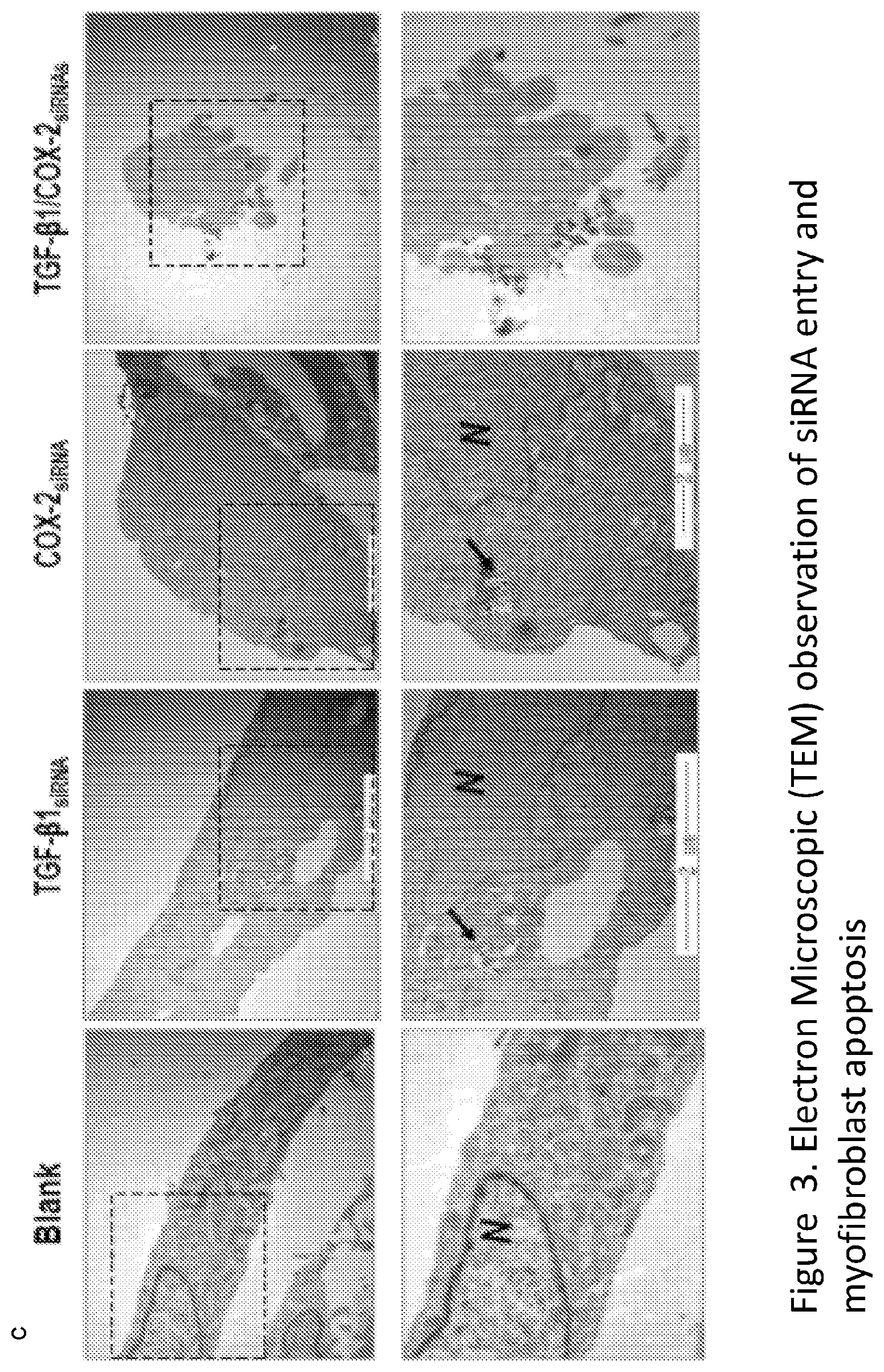
[0008] FIG. 3. Electron microscope images (SEM) of the fibroblast cells transfected with the TGF-.beta.1/COX-2.sub.siRNAs illustrates an strong apoptotic activity, where N indicates nucleus, black arrows indicate Lipid-siRNA particles and red arrows indicate apoptosis bodies.
[0009] FIG. 4. (Left) FACS analysis indicates significant increase of apoptotic cell population of human myofibroblasts when TGF-.beta.1/COX-2.sub.siRNAs was transfected simultaneously, as seen in the right lower panel, in comparison with the untreated and treated with the individual siRNA. (Right) Four different stages of treated fibroblasts: dying cells, apoptotic cells, viable cells and viable apoptotic cells. The cells treated with TGF-.beta.1/COX-2 siRNA in combination resulted in significant up regulation of apoptosis. Primary fibroblasts were treated with reagents as previously described (5 .mu.g/ml each). Annexin V/PI assay for apoptosis was conducted according to manufacturer's instruction (Invitrogen, CA). FACS data were presented here as means.
[0010] FIG. 5. (Left) Antibody staining specifically against .alpha.-SMA protein demonstrated a remarkable decrease of the protein expression with a TGF-.beta.1/COX-2.sub.siRNAs treatment, in comparison with groups treated with either TGF-.beta.1.sub.siRNA or COX-2.sub.siRNA alone. (Right) Hydroxyproline acid activity were significantly down regulated (*P<0.05) with treatments of TGF-.beta.1.sub.siRNA, or COX-2.sub.siRNA, or TGF-.beta.1/COX-2.sub.siRNAs. Among them TGF-.beta.1/COX-2.sub.siRNAs demonstrated a more potent inhibitory activity.
[0011] FIG. 6. (Left) SEM image of HKP (siRNA) nanoparticles were in a lyophilized form for long time storage and easy transportation. The nanoparticles were analyzed with a particle sizer (Brookhaven 190, N.Y., USA) and resulted in an average size of 150 nm.+-.30 in diameter. (Right) SEM image of HKP (TGF-.beta.1/COX-2.sub.siRNAS) nanoparticles in aqueous solution exhibited an average size of 150 nm in diameter for injectable administration.
[0012] FIG. 7. List of physicochemical properties of HKP (siRNA) nanoparticles have indicated that the particle sizes (upper panel and lower left) and zeta-potentials of HKP (siRNA) nanoparticles (middle panel and lower right) were measured, resulted in an average particles size about 150 nm in diameter, with Zeta-potentials of 38 vM.
[0013] FIG. 8. Comparison between Alexa Fluor labeled siRNA and HKP-packaged the same siRNA in vivo for their duration and dispersion after intra-scar injection. Samples collected from a human hypertrophic scar tissue implant model of mice, at 0, 24 and 48 hour time points post administrations.
[0014] FIG. 9. The qRT-PCR results reveal significant upregulated expressions of TGF-.beta.1 and COX-2 in human HTS tissue, comparing to those in normal human skin tissue. *P<0.05 (N=4).
[0015] FIG. 10. Injections of HKP (TGF-.beta.1/COX-2.sub.siRNAS) nanoparticle solution into the human hypertrophic scar resulted in down regulations of TGF-.beta.1 and COX-2 expressions in the tissue. This target gene silencing activity lasts up to 5 days, based on the qRT-PCR analyses following the one-shot administration.
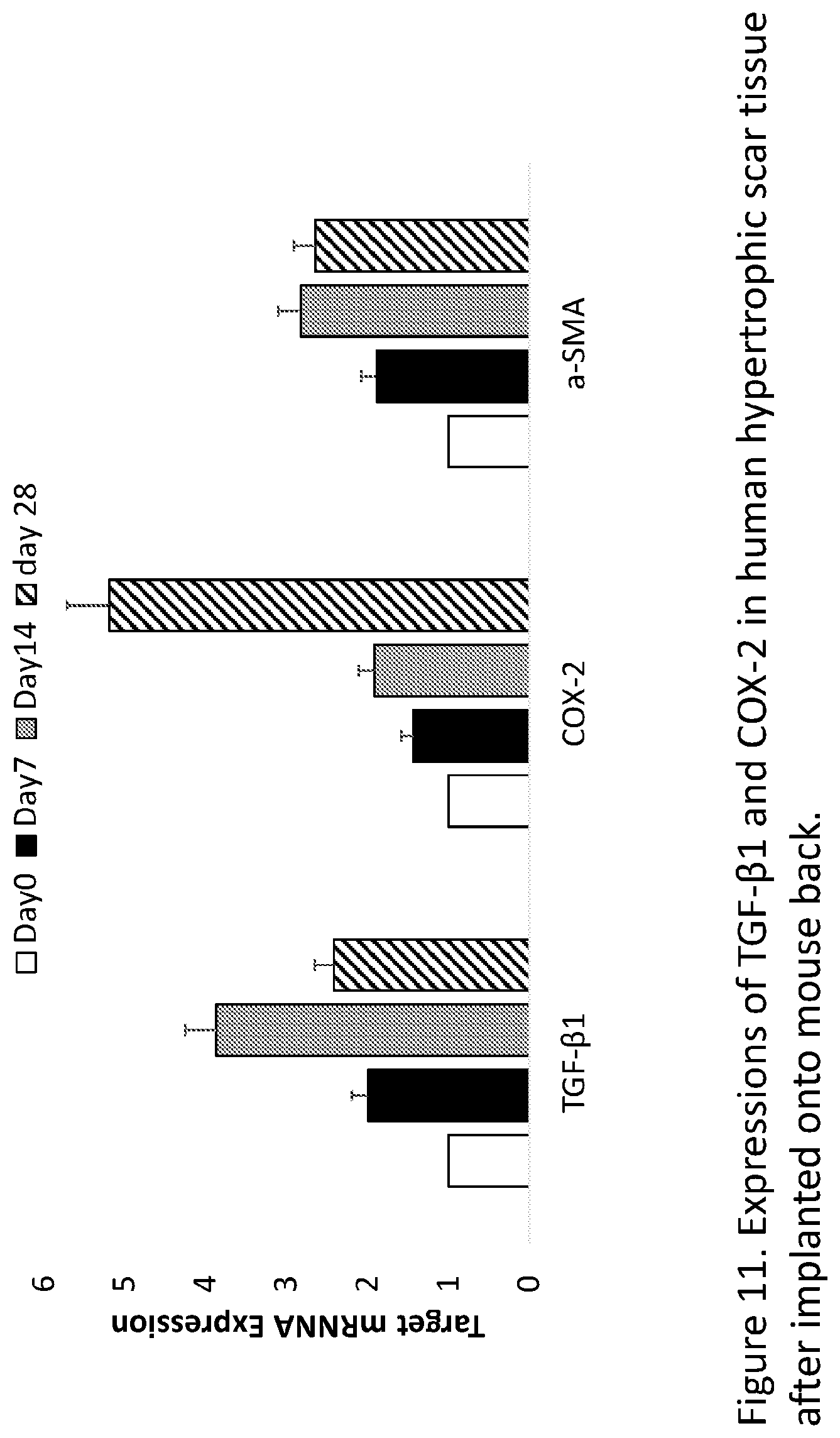
[0016] FIG. 11. Expression dynamics of TGF-.beta.1, COX-2 and .alpha.-SMA in human HTS tissue after being implanted, at day 0, 7, 14 and 28 (N=3), have been evaluated. Interestingly, the peak level of TGF-.beta.1 expression was on day 14. The COX-2 expression kept continuing increase until day 28. Meanwhile, the .alpha.-SMA expression pattern was similar as the TGF-.beta.1 expression.
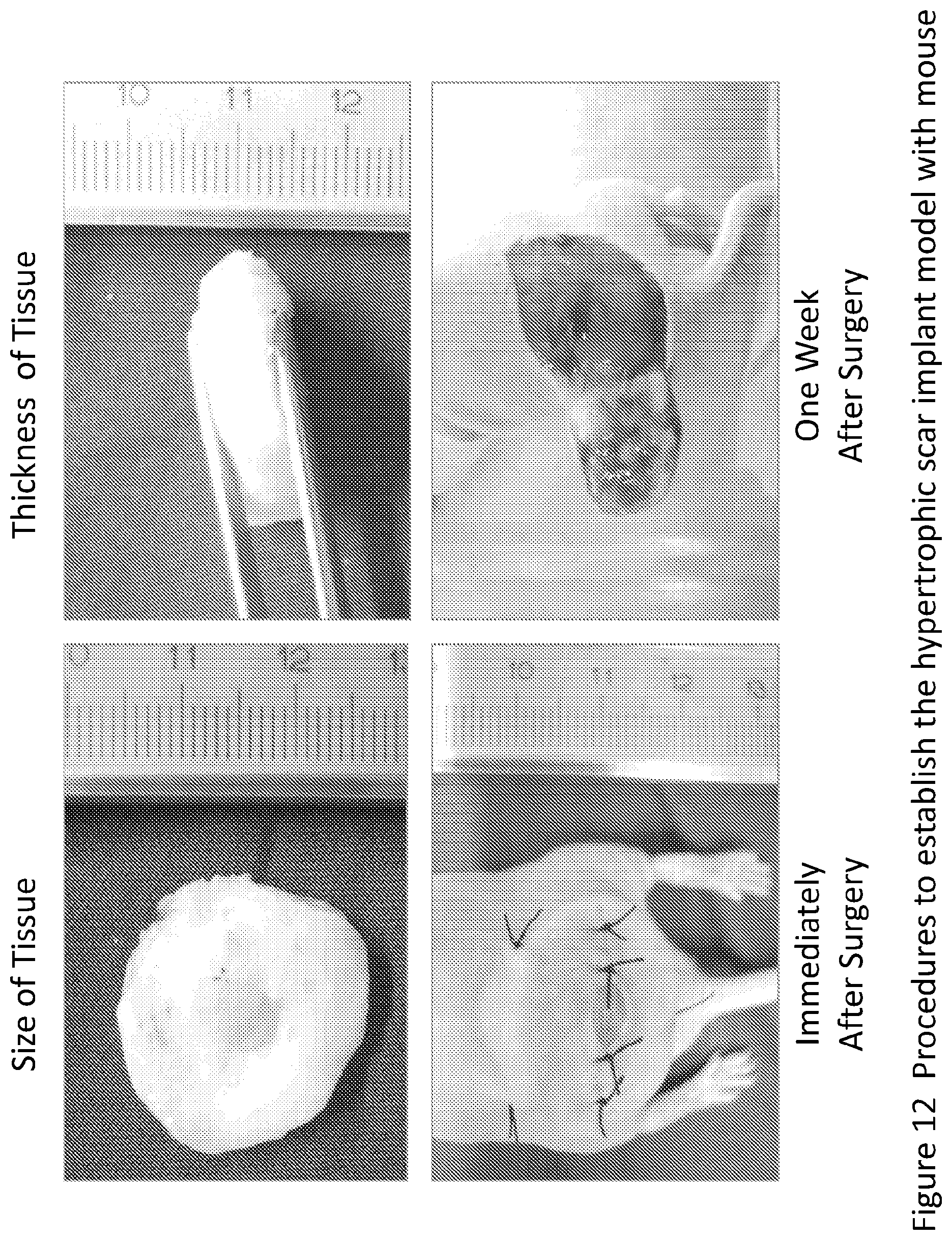
[0017] FIG. 12. The process for establishing a human hypertrophic scar implant mouse model. The size and thickness of the human hypertrophic scar tissue used for nude mouse implantation. The appearance after the scar tissue implanted and the wound site closed. Key improvement to the original procedures for establishing such a model was that several stitches were performed to fix transplant to the skin pocket. The human HTS transplant became very much viable when dissected one week later as seen with enrich blood vessel network.
[0018] FIG. 13. Appearance images after the human hypertrophic scar implanted provided an overview of appearances after the implanted tissue from day 1, one-week, two-week, three-week, four-week, one-month and all the way to six-month.
[0019] FIG. 14. A close view of human skin implant on nude mouse shows both top and side views of a human skin sample implanted onto the nude mouse back. The tissue samples were collected and stained with H&E showing the implanted tissue has grown into the surrounding tissues.
[0020] FIG. 15. The overview of the appearances of human skin tissue implanted onto nude mouse back, from two weeks to six months after the implantation. The observations was started from 2 weeks to 1 month, 2 month, 3 month, and all the way to 6 month.
[0021] FIG. 16. Images of human HTS implants, either treated with HKP (TGF-.beta.1/COX-2.sub.siRNAS) or control solution, at day 0 and 28 post treatments. (d) The reduction of the scar tissue sizes about 45% for HKP (TGF-.beta.1/COX-2.sub.siRNAS) treated group (N=4), *P<0.05. (e) Expressions of TGF-.beta.1, COX-2, .alpha.-SMA and Col1A1 mRNAs in the HKP (TGF-.beta.1/COX-2.sub.siRNAS) treated scar implants, N=3, *P<0.05.
[0022] FIG. 17. The reduction of the scar tissue sizes about 45% for HKP (TGF-.beta.1/COX-2.sub.siRNAS) treated group (N=4), *P<0.05. (e) Expressions of TGF-.beta.1, COX-2, .alpha.-SMA and Col1A1mRNAs in the HKP (TGF-.beta.1/COX-2.sub.siRNAS) treated scar implants, N=3, * P<0.05.
[0023] FIG. 18. (Left) Images of human skin implants, either treated with HKP (TGF-.beta.1/COX-2.sub.siRNAS) or control aqueous solution, at day 0 and day 28.sup.th post treatments. (Right) Quantitative illustration of the size changes of the human skin implants. The reduction of the skin tissue sizes reaches up to 38% for HKP (TGF-.beta.1/COX-2.sub.siRNAS) treated group (N=4), *P<0.05.
[0024] FIG. 19. Expressions of TGF-.beta.1, COX-2, .alpha.-SMA and Col1A1 mRNAs in the HKP (TGF-.beta.1/COX-2.sub.siRNAS) treated skin implants, were significantly down regulated (n=3). *P<0.05.
[0025] FIG. 20. After HKP (TGF-.beta.1/COX-2.sub.siRNAS) treatment, we observed 70% down regulations of hydroxyproline acid in human scar tissue implants (a) and 40% in human skin tissue implants (b), N=3, *P<0.05.
[0026] FIG. 21. Tissue samples with H&E and Masson's trichrome staining, and IHC staining with antibodies against human VEGF, CD31 and .alpha.-SMA proteins, revealed down regulations of the angiogenesis, micro blood vessel marker and fibrogenesis after repeated treatments with HKP (TGF-.beta.1/COX-2.sub.siRNAS). Red arrows indicate epidermis layer of the skin.
[0027] FIG. 22. HKP (TGF-.beta.1/COX-2.sub.siRNAS) treatment induces activations of fibroblast and myofibroblast apoptosis (black arrows) in those implanted HTS tissues and skin tissues, as showed in the images on the left and quantitative measurements on the right.
[0028] FIG. 23. Schematic demonstration of a novel mechanism for prevention and reduction of skin fibrotic scarring. A normal skin wound healing process includes three phases: inflammation, proliferation and remodeling. Scar Prevention: STP705 treatment results in down regulation of TGF-.beta.1 and COX-2 expressions and a fine-tuned fibroblast proliferation, which maintains a balance between deposition and degradation of ECM and promotes a scar-free wound healing. Scar Reduction: An optimal dosing of STP705 is expected to down regulate TGF-.beta.1 and COX-2 expressions in the scar tissue, resulting in apoptosis activation of fibroblasts/myofibroblasts within the scar tissue. The therapeutic effect of STP705 is to reverse fibrotic scarring and reduce hypertrophic scar.
DESCRIPTION OF THE INVENTION
[0029] The current invention provides a composition comprising an siRNA molecule that binds to an mRNA that codes for TGF.beta.1 protein in a mammalian cell, an siRNA molecule that binds to an mRNA that codes for COX-2 protein in a mammalian cell, and a pharmaceutically acceptable carrier comprising a pharmaceutically acceptable histidine-lysine co-polymer. The current invention also provides methods of using the composition. In one embodiment, it provides a method of down-regulating pro-fibrotic factors and fibrotic pathways in the cells of a tissue of a mammal, comprising administering to the tissue a therapeutically effective amount of the composition. In another embodiment, it provides a method of activating fibroblast and myofibroblast apoptosis in a tissue of a mammal, comprising administering to the tissue a therapeutically effective amount of the composition. In still another embodiment, it provides a method of reducing the size of a hypertrophic scar in the tissue of a mammal, comprising administering to the scar a therapeutically effective amount of the composition. In still another embodiment, the invention provides a method of reducing fibrosis in the tissue of a mammal, comprising administering to the tissue a therapeutically effective amount of the composition.
[0030] The siRNA molecules can produce additive or synergistic effects in the cells, depending on the compositions and structures of the particular molecules. In a preferred embodiment, they produce a synergistic effect.
[0031] As used herein, an "siRNA molecule" is a duplex oligonucleotide, that is a short, double-stranded polynucleotide, that interferes with the expression of a gene in a cell that produces RNA, after the molecule is introduced into the cell. For example, it targets and binds to a complementary nucleotide sequence in a single stranded (ss) target RNA molecule, such as an mRNA or a micro RNA (miRNA). The target RNA is then degraded by the cell. Such molecules are constructed by techniques known to those skilled in the art. Such techniques are described in U.S. Pat. Nos. 5,898,031, 6,107,094, 6,506,559, 7,056,704 and in European Pat. Nos. 1214945 and 1230375, which are incorporated herein by reference in their entireties. By convention in the field, when an siRNA molecule is identified by a particular nucleotide sequence, the sequence refers to the sense strand of the duplex molecule.
[0032] The siRNA molecule can be made of naturally occurring ribonucleotides, i.e., those found in living cells, or one or more of its nucleotides can be chemically modified by techniques known in the art. In addition to being modified at the level of one or more of its individual nucleotides, the backbone of the oligonucleotide can be modified. Additional modifications include the use of small molecules (e.g. sugar molecules), amino acid molecules, peptides, cholesterol, and other large molecules for conjugation onto the siRNA molecule.
[0033] In one embodiment, the molecule is an oligonucleotide with a length of about 19 to about 35 base pairs. In one aspect of this embodiment, the molecule is an oligonucleotide with a length of about 19 to about 27 base pairs. In another aspect, the molecule is an oligonucleotide with a length of about 21 to about 25 base pairs. In all of these aspects, the molecule may have blunt ends at both ends, or sticky ends at both ends, or a blunt end at one end and a sticky end at the other.
[0034] In the composition of the invention, the relative amounts of the two different molecules and the copolymer can vary. In one embodiment, the ratio of the two different siRNA molecules is about 1:1 by mass. In another embodiment, the ratio of these molecules to the copolymer is about 1:4, 1:4.5, or 1:5 by mass. Preferably, the ratio of the two different siRNA molecules is about 1:1 by mass and the ratio of these molecules to the copolymer is about 1:4, 1:4.5, or 1:5 by mass. With these ratios, the composition forms nanoparticles with an average size of about 150 nm in diameter.
[0035] In one embodiment, the siRNA molecules are selected from the ones identified in Table 1. An example is the pair designated hmTF-25-2 and hmCX-25-1 in the table, which has the following sequences:
TABLE-US-00001 hmTF-25-2: sense, 5'-r(CCCAAGGGCUACCAUGCCAACUUCU)-3', antisense, 5'-r(AGAAGUUGGCAUGGUAGCCCUUGGG)-3', and hmCX-25-1: sense, 5'-r(GGUCUGGUGCCUGGUCUGAUGAUGU)-3', antisense, 5'-r(ACAUCAUCAGACCAGGCACCAGACC)-3'.
[0036] The invention includes a method for identifying the desired siRNA molecules comprising the steps of: (a) creating a collection of siRNA molecules designed to target a complementary nucleotide sequence in the target mRNA molecules, wherein the targeting strands of the siRNA molecules comprise various sequences of nucleotides; (b) selecting the siRNA molecules that show the highest desired effect against the target mRNA molecules in vitro; (c) evaluating the selected siRNA molecules in an animal wound model; and (d) selecting the siRNA molecules that show the greatest efficacy in the model for their silencing activity and therapeutic effect.
[0037] Importantly, it is presently not possible to predict with high degree of confidence which of many possible candidate siRNA sequences potentially targeting an mRNA sequence of a disease gene will, in fact, exhibit effective RNAi activity. Instead, individually specific candidate siRNA polynucleotide or oligonucleotide sequences must be generated and tested in mammalian cell culture, such as an in vitro organ culture assay, to determine whether the intended interference with expression of a targeted gene has occurred. The unique advantage of siRNA makes it possible to be combined with multiple siRNA duplexes to target multiple disease causing genes in the same treatment, since all siRNA duplexes are chemically homogenous with same source of origin and same manufacturing process.
[0038] A preferred animal wound model is a back skin excisional wound model in a Balb/c mouse or a back excisional wound model in a pig. In another aspect, the animal wound model is a skin burn wound model in a pig. In a further aspect, the animal wound model is a back skin excisional wound model in a transgenic diabetic (db+/db+) mouse. Preferably, the siRNA molecules are evaluated in at least two of the animal models. In one embodiment, the method further includes the steps of adding a pharmaceutically acceptable carrier to each of the siRNA molecules selected by step (b) to form pharmaceutical compositions and evaluating each of the pharmaceutical compositions in the animal wound model or models.
[0039] In one embodiment, the siRNA sequences are prepared in such way that each one can target and inhibit the same gene from, at least, both human and mouse, or human and non-human primate. In one aspect, the siRNA molecules bind to both a human mRNA molecule and a homologous mouse mRNA molecule. That is, the human and mouse mRNA molecules encode proteins that are substantially the same in structure or function. Therefore, the efficacy and toxicity reactions observed in the mouse disease models provide a good understanding about what is going to happen in humans. More importantly, the siRNA molecules tested in the mouse model are good candidates for human pharmaceutical agents. The human/mouse homology design of an siRNA drug agent can eliminate the toxicity and adverse effect of those species specificities observed in monoclonal antibody drugs.
[0040] In one embodiment, the invention provides a composition comprising two or more different siRNA molecules that bind to an mRNA that codes for TGF.beta.1 protein in a mammalian cell and two or more different siRNA molecules that bind to an mRNA that codes for COX-2 protein in a mammalian cell. The molecules may bind to different nucleotide sequences within the target mRNA. The siRNA molecules can produce additive or synergistic effects in the cells, depending on the compositions and structures of the particular molecules. In a preferred embodiment, they produce a synergistic effect. In certain applications of these embodiments, the siRNA molecules are selected from the ones identified in Table 1.
[0041] The siRNA molecules are combined with a pharmaceutically acceptable carrier to provide pharmaceutical compositions for administering to a mammal. In one aspect of this embodiment, the mammal is a laboratory animal, which includes dogs, cats, pigs, non-human primates, and rodents, such as mice, rats, and guinea pigs. In another aspect, the mammal is a human.
[0042] The carrier is a histidine-lysine copolymer that forms a nanoparticle containing an siRNA molecule, wherein the nanoparticle has a size of 100-400 nm in diameter. In one aspect of this embodiment, the carrier is selected from the group consisting of the HKP species, H3K4b and PT73, which have a Lysine backbone with four branches containing multiple repeats of Histidine, Lysine, or Asparagine. When an HKP aqueous was mixed with siRNA at a N/P ratio of 4:1 by mass, the nanoparticles (average size of 100-200 nm in diameter) were self-assembled. In another aspect of this embodiment, the HKP has the following formula:
(R)K(R)--K(R)--(R)K(X), where R=KHHHKHHHKHHHKHHHK, or R=KHHHKHHHNHHHNHHHN, X=C(O)NH2, K=lysine, H=histidine, and N=asperagine.
[0043] In still another aspect of this embodiment, the HKP has the following formula:
(R)K(R)--K(R)--(R)K(X), where R=KHHHKHHHKHHHKHHHK, X=C(O)NH2, K=lysine, H=histidine.
[0044] The compositions of the invention are useful for down-regulating pro-fibrotic factors, such as .alpha.-smooth muscle actin (.alpha.-SMA), Hydroxyproline Acid, Smad 3, and Connective Tissue Growth Factor (CTGF), and fibrotic pathways, such as TGF-.beta.1/Smad .sup.3/.alpha.-SMA/Collagen I-III, in the cells of a tissue of a mammal. A therapeutically effective amount of the composition is administered to the tissue of the mammal. We hypothesized that using RNAi blocking the upstream factor of the pathway, such as TGF-.beta.1, is a more potent inhibitor. Knowing the complicated network involved in this pathway, we hypothesized that inhibition of a related factor, such as COX-2, in a different pathway may result in a synergistic effort for tighter control of the fibrosis pathway and its relevant network. In one embodiment, the tissue is skin scar, liver, lung, kidney, or heart tissue. In one aspect of this embodiment, the tissue is skin scar tissue. In another embodiment, the cells comprise fibroblasts and myofibroblasts. In one aspect of this embodiment, the fibroblasts and myofibroblasts are dermal fibroblasts and myofibroblasts. Preferably, the mammal is a human.
[0045] The compositions of the invention are also useful for activating fibroblast and myofibroblast apoptosis in the tissue of a mammal. This reduces tissue fibrosis caused by scarring after chronic inflammation of the tissue. A therapeutically effective amount of the composition is administered to the tissue of the mammal. Such apoptosis may be determined and measured by measuring the apoptotic cell population with FACS analysis, counting body numbers, and detecting expression levels of TGF-.beta.1, COX-2, .alpha.-SMA, Collagen I and Collagen III, Hydroxyproline acid, in vitro and in vivo. In one embodiment, the tissue is skin scar, liver, lung, kidney, or heart tissue. In one aspect of this embodiment, the tissue is skin scar tissue. In another embodiment, the fibroblasts and myofibroblasts are dermal fibroblasts and myofibroblasts. Preferably, the mammal is a human.
[0046] One particular embodiment of the invention provides a method of activating fibroblast and myofibroblast apoptosis in a tissue of a human, comprising injecting into the tissue a therapeutically effective amount of a composition comprising the siRNA molecule hmTF-25-2: sense, 5'-r(CCCAAGGGCUACCAUGCCAACUUCU)-3', antisense, 5'-r(AGAAGUUGGCAUGGUAGCCCUUGGG)-3', the siRNA molecule hmCX-25-1: sense, 5'-r(GGUCUGGUGCCUGGUCUGAUGAUGU)-3', anti sense, 5'-r(ACAUCAUCAGACCAGGCACCAGACC)-3', and a pharmaceutically acceptable carrier comprising a pharmaceutically acceptable histidine-lysine co-polymer. In one aspect of this embodiment, the histidine-lysine co-polymer comprises the histidine-lysine co-polymer species H3K4b or the histidine-lysine co-polymer species PT73. In another aspect of this embodiment, the histidine-lysine co-polymer has the formula (R)K(R)--K(R)--(R)K(X), where R=KHHHKHHHKHHHKHHHK, X=C(O)NH2, K=lysine, H=histidine, and N=asperagine. In still another aspect of this embodiment, the histidine-lysine co-polymer has the formula (R)K(R)--K(R)--(R)K(X), where R=KHHHKHHHKHHHKHHHK, or R=KHHHKHHHKHHHHKHHHK, X=C(O)NH2, K=lysine, H=histidine.
[0047] The compositions of the invention are also useful for reducing the size of a hypertrophic scar in the tissue of a mammal. A therapeutically effective amount of the composition is administered to the scar tissue. Such tissue includes, but is not limited to, skin, liver, lung, kidney, and heart tissue. In one embodiment, the scar comprises fibroblasts and myofibroblasts. In one aspect of this embodiment, the scar comprises dermal fibroblasts in dermal myofibroblasts. The mammal may be a laboratory animal, such as a dog, cat, pig, non-human primate, or rodent, such as a mouse, rat, or guinea pig. Preferably, the mammal is a human. In one embodiment, hypertrophic scar formation is reversed.
[0048] One particular embodiment of the invention provides a method of reducing the size of a hypertrophic scar in the skin tissue of a human, comprising injecting into the scar tissue a therapeutically effective amount of a composition comprising the siRNA molecule hmTF-25-2: sense, 5'-r(CCCAAGGGCUACCAUGCCAACUUCU)-3', antisense, 5'-r(AGAAGUUGGCAUGGUAGCCCUUGGG)-3', the siRNA molecule hmCX-25-1: sense, 5' -r(GGUCUGGUGCCUGGUCUGAUGAUGU)-3', antisense, 5'-r(ACAUCAUCAGACCAGGCACCAGACC)-3', and a pharmaceutically acceptable carrier comprising a pharmaceutically acceptable histidine-lysine co-polymer. In one aspect of this embodiment, the histidine-lysine co-polymer comprises the histidine-lysine co-polymer species H3K4b or the histidine-lysine co-polymer species PT73. In another aspect of this embodiment, the histidine-lysine co-polymer has the formula (R)K(R)--K(R)--(R)K(X), where R=KHHHKHHHKHHHKHHHK, X=C(O)NH2, K=lysine, H=histidine, and N=asperagine. In still another aspect of this embodiment, the histidine-lysine co-polymer has the formula (R)K(R)--K(R)--(R)K(X), where R=KHHHKHHHKHHHKHHHK, or R=KHHHKHHHKHHHHKHHHK, X=C(O)NH2, K=lysine, H=histidine.
[0049] The compositions of the invention are also useful for reducing fibrosis in the tissue of a mammal. A therapeutically effective amount of the composition is delivered to the tissue. Such tissue includes, but is not limited to, skin, liver, lung, kidney, and heart tissue. In one embodiment, the fibrotic tissue comprises fibroblasts and myofibroblasts. The composition may be delivered by injection into the tissue, subcutaneous injection into the mammal, or intravenous injection into the mammal. The mammal may be a laboratory animal, such as a dog, cat, pig, non-human primate, or rodent, such as a mouse, rat, or guinea pig. Preferably, the mammal is a human.
[0050] The dosages, methods of administration, and times of administration are readily determinable by a person skilled in the art, given the teachings contained herein. In one embodiment, the composition is administered by injection into the tissue. In another embodiment, the composition is ministered by subcutaneous injection into the mammal. In still another embodiment, the composition is administered intravenously to the mammal. In a preferred embodiment, the mammal is a human.
TABLE-US-00002 TABLE I Sense Antisense hmTF-25-1 5'-r(GGAUCCACGAGCCCAAGGGCUACCA)-3' 5'-r(UGGUAGCCCUUGGGCUCGUGGAUCC)-3' hmTF-25-2 5'-r(CCCAAGGGCUACCAUGCCAACUUCU)-3' 5'-r(AGAAGUUGGCAUGGUAGCCCUUGGG)-3' hmTF-25-3 5'-r(GAGCACCAUUCUCCUUGAAAGGACU)-3' 5'-r(AGUCCUUUCAAGGAGAAUGGUGCUA)-3' hmTF-25-4 5'-r(GAUCCACGAGCCCAAGGGCUACCAU)-3' 5'-r(AUGGUAGCCCUUGGGCUCGUGGAUC)-3' hmTF-25-5 5'-r(CACGAGCCCAAGGGCUACCAUGCCA)-3' 5'-r(UGGCAUGGUAGCCCUUGGGCUCGUG)-3' hmTF-25-6 5'-r(GAGGUCACCCGCGUGCUAAUGGUGG)-3' 5'-r(CCACCAUUAGCACGCGGGUGACCUC)-3' hmTF-25-7 5'-r(GUACAACAGCACCCGCGACCGGGUG)-3' 5'-r(CACCCGGUUGCGGGUGCUGUUGUAC)-3' hmTF-25-8 5'-r(GUGGAUCCACGAGCCCAAGGGCUAC)-3' 5'-r(GUAGCCCUUGGGCUCGUGGAUCCAC)-3' hmCX-25-1 5'-r(GGUCUGGUGCCUGGUCUGAUGAUGU)-3' 5'-r(ACAUCAUCAGACCAGGCACCAGACC)-3' hmCX-25-2 5'-r(GAGCACCAUUCUCCUUGAAAGGACU)-3' 5'-r(AGUCCUUUCAAGGAGAAUGGUGCUC)-3' hmCX-25-3 5'-r(CCUCAAUUCAGUCUCUCAUCUGCAA)-3' 5'-r(UUGCAGAUGAGAGACUGAAUUGAGG)-3' hmCX-25-4 5'-r(GAUGUUUGCAUUCUUUGCCCAGCAC)-3' 5'-r(GUGCUGGGCAAAGAAUGCAAACAUC)-3' hmCX-25-5 5'-r(GUCUUUGGUCUGGUGCCUGGUCUGA)-3' 5'-r(UCAGACCAGGCACCAGACCAAAGAC)-3' hmCX-25-6 5'-r(GUGCCUGGUCUGAUGAUGUAUGCCA)-3' 5'-r(UGGCAUACAUCAUCAGACCAGGCAC)-3 hmCX-25-7 5'-r(CACCAUUCUCCUUGAAAGGACUUAU)-3' 5'-r(AUAAGUCCUUUCAAGGAGAAUGGUG)-3' hmCX-25-8 5'-r(CAAUUCAGUCUCUCAUCUGCAAUAA)-3' 5'-r(UUAUUGCAGAUGAGAGACUGAAUUG)-3'
[0051] The following examples illustrate certain aspects of the invention and should not be construed as limiting the scope thereof.
EXAMPLES
Example 1: Simultaneous Silencing of TGF-.beta.1 and COX-2 Gene Expressions
[0052] We first designed siRNA sequences specific to human TGF-.beta.1 and COX-2 mRNAs in silico and then tested those sequences based on efficient cell transfection studies and analysis using qRT-PCR (FIG. 1). The potent siRNAs we selected were based on their silencing efficiencies and toxicity profiles affecting cells. When human fibroblasts isolated from the hypertrophic scar tissue were transfected with the selected siRNAs targeting either TGF-.beta.1 or COX-2 individually or in combination, we observed an efficient siRNA entry into the cells at different stages, from initial endocytosis to endosome release of the siRNAs. The measurements of target gene silencing after the transfection indicated significant knockdown of target gene expression, with either TGF-.beta.1.sub.siRNA or COX-2.sub.siRNA themselves, or upon combination TGF-.beta.1/COX-2.sub.siRNAs. Interestingly, not only was the TGF-.beta.1.sub.siRNA and TGF-.beta.1/COX-2.sub.siRNAs combination able to silence TGF-.beta.1 expression, the COX-2.sub.siRNA was also able to down regulate TGF-.beta.1 expression. This result implied a potential interconnection between TGF-.beta.1 and COX-2 pathways. Similarly, TGF-.beta.1.sub.siRNA was also able to silence COX-2 expression significantly. As the target genes were silenced, other pro-fibrotic factors such as .alpha.-SMA, Collagen 1 (Col1A1) and Collagen 3 (Col3A1) were also down regulated in the cell (FIG. 2). These results indicate that silencing TGF-.beta.1 and COX-2 simultaneously in the fibroblasts has activated down regulation of multiple pro-fibrotic factors and the fibrotic pathways.
Example 2: Simultaneous Silencing of TGF-.beta.1 and COX-2 Gene Expressions Resulted in Activation of Apoptosis of Fibroblasts/Myofibroblasts
[0053] We further investigated the fate of the fibroblasts when those pro-fibrotic factors were down regulated. Electron microscope images (FIG. 3) of the fibroblast cells transfected with the either TGF-.beta.1.sub.siRNA or COX-2.sub.siRNA only, or TGF-.beta.1/COX-2.sub.siRNAs combination, illustrates that the combination treatment resulted in activation of the fibroblast apoptotic activity, but not occurred for the individual siRNA treatment group. FACS analyses of the human fibroblasts treated with the TGF-.beta.1/COX-2.sub.siRNAs combination revealed a sharp increase in the apoptotic cell population (FIG. 4), compared to those treated with either TGF-.beta.1.sub.siRNA or COX-2.sub.siRNA individually. Fibroblasts/myofibroblast treated with the TGF-.beta.1/COX-2.sub.siRNAs combination showed much lower cell density and a different morphology. When we examined the expression of .alpha.-SMA protein in the human fibroblasts after the TGF-.beta.1/COX-2.sub.siRNAs combination treatment, a significant reduction was observed compared to the individual siRNA treatments as measured by immunofluorescence staining (FIG. 5). We also measured the level of hydroxyproline acid (HPC) in cell culture, an indicator of the synthetic activity of ECM proteins and a hallmark of tissue fibrosis, using the human fibroblasts treated with TGF-.beta.1/COX-2.sub.siRNAs combination, (FIG. 5). All above results have clearly indicated that when TGF-.beta.1 and COX-2 gene expressions in the human fibroblasts were silenced simultaneously, a chain of down regulations of expressions of multiple fibrotic factors occurred. Consequently, the treated fibroblasts became less active and more apoptotic.
Example 3. HKP Enhances siRNA Delivery Into Human Hypertrophic Scar
[0054] To ensure efficient siRNA delivery to the hypertrophic scar, we selected a biodegradable histidine-lysine polypeptides (HKP) that has been demonstrated to provide efficient siRNA delivery in vivo (Leng et al, 2008 and Yan et al, 2008). When HKP and siRNA are mixed in aqueous solution with an optimized N/P ratio (4/1), the self-assembly of nanoparticles occurs through an electrostatic binding. These nanoparticles can be lyophilized into dry powder or formulated with aqueous solution directly (FIG. 6). The lyophilized HKP (TGF-.beta.1/COX-2.sub.siRNA) nanoplex powder, with an average size about 150 nm in diameter and a Zeta potential about 40 mV, is highly stable at 4.degree. C., preserving potent activity for silencing TGF-.beta.1 expression up to 12 months (FIG. 7).
[0055] To further understand the duration and distribution of the locally delivered siRNA, we used a human hypertrophic scar tissue implant mouse model. Two intrascar injections were conducted with one containing a naked Fluorescence-labeled FAM-siRNA and the other one containing the HKP packaged FAM-siRNA nanoparticle formulation (FIG. 8). Tissue samples were collected at 4 hours, 24 hours and 48 hours post treatment injections, and analyzed under a Fluorescence microscope. The naked siRNA are quickly dispersed after being injected into the scar tissue, and could not be detected after 24 hours. The HKP-packaged siRNA illustrated a quick dispersion and long lasting release that can be detected even after 48 hours. Therefore, we predicted that HKP-packaged siRNA nanoparticle formulation represents a useful means for evaluation of the target gene silencing in vivo. When HKP-packaged TGF-.beta.1/COX-2.sub.siRNAs combination was administrated through intrascar injection, we observed a potent silencing effect at day 1, day3 and day 5 post treatment. This silencing activity resulted in parallel down regulation of both TGF-.beta.1 and COX-2 with the greatest inhibition being seen at day 5 (FIG. 9).
Example 4. HKP-Packaged TGF-.beta.1/COX-2siRNA Reduces Size of Human Hypertrophic Scar
[0056] As expected, we first found that TGF-.beta.1 and COX-2 are significantly over-expressed in the human hypertrophic scar (HTS) tissue from patients scar biopsy compared to the normal skin tissue (FIG. 10). Human HTS tissues were implanted onto nude mice subcutaneously for studying pathophysiology of HTS and its novel therapeutic options. After HTS tissue was implanted, we collected some of these tissues at day 7, day 14 and day 28 post implantation. We then isolated mRNA from those tissue samples for evaluating the expression dynamics of TGF-.beta.1, COX-2 and .alpha.-SMA using qRT-PCR analyses (FIG. 11). The expression of TGF-.beta.1 and COX-2 in the implanted human scar tissues exhibited a unique pattern with immediate escalation of TGF-.beta.1 versus a steady increase in COX-2. The TGF-.beta.1 expression reached a peak level at day 14 while COX-2 expression was still up regulated on day 28 post initial implantation.
[0057] The detailed procedure for HTS implant onto nude mouse back has been demonstrated by FIG. 12. The appearances of the HTS implants have been illustrated from the image of 1st week to the image of 6th month (FIG. 13). Similarly, the detailed procedure for Skin implant onto nude mouse back has also been demonstrated by FIG. 14. The appearances of the skin implants have been illustrated from the image of 1st week to the image of 6th month (FIG. 15).
[0058] Based on the readouts from FIG. 10 and FIG. 11, we decided to start the treatment of the implanted human hypertrophic scars on mice 4 weeks after surgery. A 20 .mu.g/50 .mu.l/cm.sup.3 HKP (TGF-.beta.1/COX-2.sub.siRNAs) was administered to each scar implant using 5 aliquots into 5 different sites of the scar, with three repeated injections at 5 day intervals. The HKP (TGF-.beta.1/COX-2.sub.siRNAs) combination treated HTS implants resulted in a significant reduction in size of implanted tissues at day 28 post-treatment (FIG. 16), about 40% in comparison to the untreated group. After taking tissue samples from those implants for further analyses, we found that not only the targeted genes TGF-.beta.1 and COX-2 were significantly silenced based on the qRT-PCR results, but other factors such as .alpha.-SMA and col1A1 were also significantly down regulated (FIG. 17). This result provided solid evidence that the simultaneous inhibition of TGF-.beta.1 and COX-2 contributes to the scar reduction.
Example 5. HKP (TGF-.beta.1/COX-2.sub.siRNAs) Reduces Size of Human Skin Grafts
[0059] Similar to human hypertrophic scar implants, human skin grafted onto the nude mouse is able to regenerate after being subjected to a full-thickness wound. This approach has been used to determine the cells involved in the connective tissue repair process following superficial wounding. In addition, this model has been used to study the wound healing process of human skin. The hypertrophic scar model is established by transplanting human skin grafts onto nude mice, resulting in obvious, persistent hypertrophic scars that have both macroscopic and histologic properties similar to human hypertrophic scars. This model makes possible the observation of the entire process of hypertrophic scar formation. Thus, it is an ideal tool for studying hypertrophic scar (Yang, et al. 2007). The initial dosing time and dosing regimen were similar to the treatment of the implanted human hypertrophic scars on mice. Four weeks after the surgery, 20 .mu.g/50 .mu.l/cm.sup.3 HKP (TGF-.beta.1/COX-2.sub.siRNAs) solution was injected into each skin graft using 5 aliquots to 5 different sites of the graft, with three repeated injections at 5 day intervals. The HKP (TGF-.beta.1/COX-2.sub.siRNAs) combination treated human skin grafts resulted in a significant size reduction at day 28 post-treatment (FIG. 18), about 40% in comparison to the untreated group. After taking tissue samples from those skin grafts for further analyses, we found that not only the targeted genes, TGF-.beta.1 and COX-2, were significantly silenced based on qRT-PCR analysis but that .alpha.-SMA and col1A1 were also significantly down regulated (FIG. 19). This result is similar to what we observed with the human hypertrophic scar implants and provides further evidence that the simultaneous inhibitions of TGF-.beta.1 and COX-2 contributes to the skin scar reduction.
Example 6. HKP (TGF-.beta.1/COX-2siRNAs) Demonstrates a Novel Anti-Fibrotic Mechanism of Action
[0060] To investigate the underlying biology of the observed scar tissue reductions with the human hypertrophic scar and human skin graft implants after HKP (TGF-.beta.1/COX-2.sub.siRNAs) treatment, we first measured hydroxyproline acid level from the tissue samples and then measured the differences between the treated and control groups. As we expected, the treatment groups presented a significant down regulated expression of the hydroxyproline acid, in comparison to the control groups, from both human scar and skin implants (FIG. 20). Further, using H&E, or Masson's trichrome staining, or immunohistochemistry (IHC) staining against human .alpha.-SMA, VEGF and CD3, we compared the histology between samples from both HKP (TGF-.beta.1/COX-2.sub.siRNAs) treated and untreated tissues. The marked differences of the tissue structures and expression levels of these pro-fibrotic factors were revealed (FIG. 21).
Example 7. HKP (TGF-.beta.1/COX-2 siRNAs) Induced Apoptotic Activities of the Fibroblast/Myofibroblasts In Vivo
[0061] We further measured the apoptotic activity of the fibroblasts in vivo as we did in the cell culture study, using a TUNEL assay. Histology images illustrated remarkably increased apoptotic fibroblasts populations in the treated tissue samples (FIG. 21). A further quantification analysis demonstrated a significant increase of the numbers of cells moving into apoptotic stage (FIG. 22).
Example 8. HKP (TGF-.beta.1/COX-2 siRNAs) Serves as a Novel Anti-Fibrotic Therapeutic Agent
[0062] The up regulation of fibroblast apoptosis in the cell culture and HTS tissue confirmed a critical role of HKP (TGF-.beta.1/COX-2 siRNAs), in maintaining an optimized fibroblast proliferation and a balance between deposition and degradation of ECM production, to avoid fibrotic scarring. Down regulations of .alpha.-SMA, Collagen 1, Collagen 3 and hydroxproline acid in both human fibroblasts and HTS tissue after the HKP (TGF-.beta.1/COX-2.sub.siRNAs) treatments implicate a complex network regulating skin fibrotic scarring (FIG. 21). These results have further advanced our understanding of the mechanism of actions of the pathophysiological pathways for skin fibrotic scarring. The synergistic activity of HKP (TGF-.beta.1/COX-2.sub.siRNAs) silencing both TGF-.beta.1 and COX-2 at proliferation stage and remodeling stage of skin wound healing process have provided solid evidence that the skin fibrotic scarring can be prevented and the skin hypertrophic scarring can be reversed. Therefore, we are proposing a novel siRNA based therapeutic approach of using HKP (TGF-.beta.1/COX-2siRNAs) for hypertrophic scar treatment.
Discussion
[0063] A dermal wound healing process can be specified into three phases: inflammation, cell proliferation and matrix remodeling, which involve multiple interactions within a complex network of pro-fibrotic and anti-fibrotic molecules (Dabiri et al, 2006). After dermal injury occurs, the aggregated inflammatory cells become sources of growth factors and cytokines. When active angiogenesis and collagen synthesis ensue in concert with the tissue remodeling process, a delicate balance of deposition and degradation of fibroblast-expressed ECM determines normal skin wound healing or whether a wound heals but with HTS. Fibroblasts are the most common cells in connective tissue and key players in skin wound healing process, functioning to maintain the physical integrity of the connective tissue, participate in wound closure, and produce and remodel ECM (Wang et al, 2011). Regulation of fibroblast proliferation, their transition to myofibroblasts, and their apoptotic activity during the wound healing process can be critical modalities for therapeutic intervention.
[0064] We found that silencing TGF-.beta.1 in human fibroblasts down regulates COX-2 expression, and vice versa (FIG. 2). Although there are reports indicating that high levels of COX-2 were able to reverse TGF-.beta.1 into an anti-fibrotic regulator in cell culture studies, it has been suggested that these observations are due to the terminal stages of the cells used and do not reflect the early responses of TGF-.beta.1 and COX-2 pathways to the injury in vivo (Su et al, 2010) and the evidence of up-regulated expression of these two targets in the human hypertrophic scar tissue (FIG. 9) warranted our selection of these two targets for therapeutic intervention.
[0065] We evaluated the HKP (siRNA) nanoparticle formulation, through intrascar administrations, to determine whether a clinically viable siRNA therapeutic product can be realized for treatment of the skin hypertrophic scar (Yang et al, 2007 and Rossio-Pasquier et al, 1999). We also developed scalable procedures for HKP (siRNA) nanoparticle formulation, which not only facilitated efficient siRNA delivery but exhibited no signs of adverse and toxic effects. In comparison with two ongoing clinical studies using oligo nucleotide inhibitors for the similar indications, EXC001 and RXI109, the therapeutic dose of HKP (TGF-.beta.1/COX-2.sub.siRNAs) formulation we used is at least 2 logs below what has been reported for 2 other agents (data not shown). We believe that the excellent potency of HKP (TGF-.beta.1/COX-2.sub.siRNAs) comes from not only the dual-targeted drug design but also from HKP-enhanced delivery to the site of action, which had also been demonstrated in this study (FIG. 7, 8).
[0066] The therapeutic benefits we observed through the human hypertrophic scar models further validated that simultaneous silencing of expression of two target genes indeed represents a novel treatment regimen. Using HKP-enhanced in vivo siRNA deliveries through intrascar injection, we observed significant silencing effects on TGF-.beta.1 and COX-2 expressions about 40% (FIG. 4e, 5c), with only microgram level of the siRNA inhibitors. Further, the activation and upregulation of fibroblast apoptosis within human hypertrophic scars both in vitro (FIG. 3, 4) and in vivo (FIG. 21) after HKP (TGF-.beta.1/COX-2.sub.siRNAs) treatment revealed a novel underlying mechanism of action. Therefore, we hypothesized that simultaneously silencing TGF-.beta.1 and COX-2 expression within the human hypertrophic scars is able to inhibit inflammatory activity and fibroblast proliferation, minimizing the excessive accumulation of extracellular matrix (ECM) in and around the damaged tissue. This inhibition leads to reduced ECM deposition and enhanced excessive collagen degradation. As a result, a hypertrophic scar can be reduced and eliminated eventually. Therefore, we are confident that HKP (TGF-.beta.1/COX-2.sub.siRNAs) will represent another clinically viable modality for novel siRNA therapeutics to reduce human skin excessive scar after damage.
[0067] The up regulation of fibroblast apoptosis in the cell culture and human HTS and skin tissue implants confirmed a critical therapeutic potential of HKP (TGF-.beta.1/COX-2.sub.siRNAs), in maintaining an optimized fibroblast proliferation and a balance between deposition and degradation of ECM production, to avoid fibrotic scarring. Down regulation of .alpha.-SMA, Collagen 1, Collagen 3 and hydroxproline acid in both human fibroblasts and HTS tissue, at both mRNA and protein levels after the treatments, implicates a complex network regulating skin fibrotic scarring. The results from this study have further advanced our understanding of the mechanism of actions of the pathophysiological pathways involved in the hypertrophic scar formation. The synergistic activity of HKP (TGF-.beta.1/COX-2.sub.siRNAs) silencing both TGF-.beta.1 and COX-2 at the proliferation and remodeling stages of skin wound healing process provides a solid evidence that the skin hypertrophic scar formation can be potentially reversed through activation of the fibroblasts apoptosis within the scar.
Materials and Methods
siRNA Sequence Design and Selection
[0068] 25-mer blunt-ended siRNA duplexes targeting TGF.beta.1 or COX-2 mRNA sequences were designed (Table 1). (Does this refer to Table 1; i.e., should "Supplemented" be deleted?) Eight siRNA for each gene were screened in human PC3 cells for target gene silencing with qRT-PCR analyses.
Growth Inhibition In Vitro
[0069] Cells were seeded into the wells of 96-well plate at density 2.times.10.sup.3 cells per well in 100 ul media. Six hours later culture media was replaced with fresh media supplemented containing Lipofectamine 2000 (Lipo2000) formulations, Lipo (TGF-.beta.1/COX-2.sub.siRNAs), or Lipo2000, or Lipo2000 (TGF-.beta.1.sub.siRNA) or Lipo2000 (COX-2.sub.siRNA). The cells were incubated for 48 hours. For growth inhibition assay, cells were treated and analyzed with target gene silencing, FACS, .alpha.-SMA and Hydroxylproline Acid expressions.
Nanoparticle Preparation
[0070] Optimized Histidine-Lysine polymers (HKP) were applied for the siRNA delivery in vivo. One of HKP species, H3K4b, having a lysine backbone with four branches that contain multiple repeats of histidine and lysine, was used for packaging siRNAs against TGF-.beta.1 and COX-2, with a carrier and payload ratio of 4:1 by mass. The nanoparticle (average size of 150 nm in diameter) were self-assembled.
Mouse
[0071] The 8-week old male nude mice (nu/nu Balb/c) were purchased from Center for Experimental Animals in Shanghai, China. Animal housing and experiment protocols were approved by the IACUC committee of the 9.sup.th People's Hospital of Shanghai.
Human Hypertrophic Scar and Skin Tissue Implant Models
[0072] Skin hypertrophic scar tissue was obtained from the surgical excisions with the informed consent were implanted under the skin on the mouse back. Scar tissue was fixed to the mouse deep fascia with 4-5 sutures before cut on skin was closed. The skin tissue samples used in experiments were from skin excisions from three women of age 23-36 undergone breast reconstruction for treatment of macromastia with signed informed consent. The skin tissues were grafted to fill the excision wounds by sutures to subcutaneous fascia and surrounding mouse skin.
Therapeutic Evaluation with the Human Tissue Implant Models
[0073] Four weeks after the human hypertrophic scar implant model established, HKP (TGF-.beta.1/COX-2.sub.siRNAs) was administrated via intrascar injection. The mRNA levels of TGF-.beta.1, COX-2, .alpha.-SMA, Col1a1, and Col3a1 were analyzed with qRT-PCR. The In Situ Cell Death Detection Kit from Roche (South SF, CA, USA) was applied for detection of apoptotic cells.
Statistical Analysis
[0074] Mean.+-.SD was used for cell culture results, and mean.+-.SE was used for in vivo results. An unequal variance two-tailed Student's t test was applied to compare the means of samples. A difference was considered statistically significant when P<0.05.
REFERENCES
[0075] A. J. Singer, S. S. Huang, J. S. Huang et al. A novel TGFbeta antagonist speeds reepithelialization and reduces scarring of partial thickness porcine burns. Journal of Burn Care and Research. 2009, 30, 2, 329-334. [0076] Chen W, Fu X, Ge S, Sun T, Zhou G, Jiang D, et al. Ontogeny of expression of transforming growth factor-beta and its receptors and their possible relationship with scarless healing in human fetal skin. Wound Repair Regen. 2005, 13, 1:68-75. [0077] Chia Soo, Steven R. Beanes, Fei-Ya Hu, Xinli Zhang, Catherine Dang, et al. et al. Ontogenetic Transition in Fetal Wound Transforming Growth Factor-.beta. Regulation Correlates with Collagen Organization American Journal of Pathology. 2003, 163:2459-2476. [0078] Dabiri. G, Campaner A, Morgan J R, Van De Water L A. TGF-beta1-dependent autocrine loop regulates the structure of focal adhesions in hypertrophic scar fibroblasts. J Invest Dermatol. 2006, 126:963-970. [0079] E, Nedelec B, Scott P G, Ghahary A. Hypertrophic scars, keloids, and contractures. The cellular and molecular basis for therapy. Surg Clin North Am. 1997, 77, 3:701-730. [0080] J. S. Huang, Y. H. Wang, T. Y. Ling, S. S. Chuang, F. E. Johnson, and S. S. Huang. Synthetic TGF-beta antagonist accelerates wound healing and reduces scarring. The FASEB Journal. 2002, 16, 10: 1269-1270. [0081] Kerstin J Rolfe, Janette Richardson, Charlotte Vigor, Laurie M Irvine, Addie O Grobbelaar and Claire Linge A Role for TGF.beta.-1-Induced Cellular Responses during Wound Healing of the Non-Scarring Early Human Fetus? Journal of Investigative Dermatology. 2007, 127: 2656-2667. [0082] Kopp J, Preis E, Said H, Hafemann B, Wickert L, et al. Abrogation of transforming growth factor-beta signaling by SMAD7 inhibits collagen gel contraction of human dermal fibroblasts. J Biol Chem. 2005, 280:21570-6. [0083] Leng, Q., P Scaria, P Y Lu, Woodle M C, and A. J. Mixson. Systemic delivery of HK Raf-1siRNA Polyplexes Inhibits MDA-MB-435 Xenografts. Cancer Gene Therapy. 2008,1-11. [0084] Levinson H. A Paradigm of Fibroblast Activation and Dermal Wound Contraction to Guide the Development of Therapies for Chronic Wounds and Pathologic Scars. Advances In Wound Care, 2013, 2, 4:149-159. (2013). [0085] Matsumura T, Suzuki T, Aizawa K, Sawaki D, Munemasa Y, Ishida et al. Regulation of transforming growth factor-beta-dependent cyclooxygenase-2 expression in fibroblasts. J Biol Chem. 2009, 284, 51:35861-71. [0086] Matthew Rhett, Gautam S. Ghatnekar, Joseph A. Palatinus, Michael O'Quinn, Michael J. Yost and Robert G. Gourdie. Novel therapies for scar reduction and regenerative healing of skin wounds. Trends in Biotechnology, 2008, 26, 4:173-180. [0087] McDougall S, Dalton J, Sherratt J, Maini P. Fibroblast migration and collagen deposition during dermal wound healing: mathematical modeling and clinical implications. Philos Transact A Math Phys Eng Sci, 2006, 364, 1843:1385-1405. [0088] Miller M C, Nanchahal J. Advances in the modulation of cutaneous wound healing and scarring. BioDrugs. 2005, 19, 6:363-381. [0089] Mustoe T A, Cooter R D, Gold M H, Hobbs F D, Ramelet A A, et al. International clinical recommendations on scar management. Plast Reconstr Surg 2002, 110:560-571. (2002). [0090] NCT02030275, 2014. A Study to Evaluate the Effectiveness and Safety of RXI 109 on the Outcome of Scar Revision Surgery in Healthy Adults. https://clinicaltrials.gov/ct2/results?term=RXI109 [0091] NCT01346969, 2013. Safety and Efficacy of EXC 001 in Subjects Who Have Participated in Prior Studies of EXC 001. https://clinicaltrials.gov/ct2/results?term=EXC001+AND+fibrosis [0092] Nguan Soon Tan, Liliane Michalik, Beatrice Desvergne and Walter Wahli. Genetic- or Transforming Growth Factor-.beta.1-induced Changes in Epidermal Peroxisome Proliferator-activated Receptor .beta./.delta. Expression Dictate Wound Repair Kinetics. The Journal of Biological Chemistry. 2005, 280:18163-18170. [0093] Rossio-Pasquier P.sup.1, Casanova D, Jomard A, Demarchez M. Wound healing of human skin transplanted onto the nude mouse after a superficial excisional injury: human dermal reconstruction is achieved in several steps by two different fibroblast subpopulations. Arch Dermatol Res. 1999, 291, 11:591-9. [0094] Su W H, Cheng M H, Lee W L, Tsou T S, Chang W H and Chen C S, et al. Nonsteroidal anti-inflammatory drugs for wounds: pain relief or excessive scar formation? Mediators Inflamm. Article ID: 413238. 2010, 1-8. [0095] Sung I I Kim, Hee-Jun Na, Yan Ding, Zhibo Wang, Seon Jin Lee and Mary E Choi. Autophagy Promotes intracellular degradation of type I collagen induced by TGF-.beta.1. The Jour. Of Biological Chemistry, 2012, 287, 15:11677-11687. [0096] Tredget EWynn T A, Ramalingam T R. Mechanisms of fibrosis: therapeutic translation for fibrotic disease. Nat Med. 2012, 18, 7:1028-40. [0097] V. L. Kumar and Y. M. Shivkar. Involvement of prostaglandins in inflammation induced by latex of Calotropis procera. Mediators of Inflammation. 2004, 13, 3) 151-155. [0098] Yan, Z W, P Y Lu and LY Li et al. The Human Rhomboid Family-1 Gene RHBDF1 Is Essential to Cancer Cells Survival and Critical in EGFR Activation. Molecular Cancer Therapeutics. 2008, 7(6):1355-64. [0099] Yang D Y, Li S R, Wu J L, Chen Y Q, Li G, Bi S, et al. Establishment of a hypertrophic scar model by transplanting full-thickness human skin grafts onto the backs of nude mice. Plast Reconstr Surg. 2007, 119, 1:104-9 [0100] Wilgus T A, Bergdall V K, Tober K L, Hill K J, Mitra S, Flavahan N A, et al. The impact of cyclooxygenase-2 mediated inflammation on scarless fetal wound, 2 mediated inflammation on scarless fetal wound healing. Am J Pathol. 2004, 165, 3:753-61. [0101] Wang J, Ding J, Jiao H, Honardoust D, Momtazi M, Shankowsky H A, et al. Human hypertrophic scar-like nude mouse model: characterization of the molecular and cellular biology of the scar process.Wound Repair Regen. 2011, 19, 2:274-85. [0102] Zhu Z, Ding J, Shankowsky H A, Tredget E E. The molecular mechanism of hypertrophic scar. J Cell Commun Signal. 2013, 7, 4:239-52.
[0103] The disclosures of all publications identified herein, including issued patents and published patent applications, and all database entries identified herein by url addresses or accession numbers are incorporated herein by reference in their entirety.
[0104] Although this invention has been described in relation to certain embodiments thereof, and many details have been set forth for purposes of illustration, it will be apparent to those skilled in the art that the invention is susceptible to additional embodiments and that certain of the details described herein may be varied considerably without departing from the basic principles of the invention.
Sequence CWU 1
1
45125RNAArtificial SequenceDescription of Artificial Sequence
Synthetic oligonucleotide 1cccaagggcu accaugccaa cuucu
25225RNAArtificial SequenceDescription of Artificial Sequence
Synthetic oligonucleotide 2agaaguuggc augguagccc uuggg
25325RNAArtificial SequenceDescription of Artificial Sequence
Synthetic oligonucleotide 3ggucuggugc cuggucugau gaugu
25425RNAArtificial SequenceDescription of Artificial Sequence
Synthetic oligonucleotide 4acaucaucag accaggcacc agacc
25517PRTArtificial SequenceDescription of Artificial Sequence
Synthetic peptide 5Lys His His His Lys His His His Lys His His His
Lys His His His1 5 10 15Lys617PRTArtificial SequenceDescription of
Artificial Sequence Synthetic peptide 6Lys His His His Lys His His
His Asn His His His Asn His His His1 5 10 15Asn718PRTArtificial
SequenceDescription of Artificial Sequence Synthetic peptide 7Lys
His His His Lys His His His Lys His His His His Lys His His1 5 10
15His Lys825RNAArtificial SequenceDescription of Artificial
Sequence Synthetic oligonucleotide 8ggauccacga gcccaagggc uacca
25925RNAArtificial SequenceDescription of Artificial Sequence
Synthetic oligonucleotide 9ccucaauuca gucucucauc ugcaa
251025RNAArtificial SequenceDescription of Artificial Sequence
Synthetic oligonucleotide 10gauccacgag cccaagggcu accau
251125RNAArtificial SequenceDescription of Artificial Sequence
Synthetic oligonucleotide 11cacgagccca agggcuacca ugcca
251225RNAArtificial SequenceDescription of Artificial Sequence
Synthetic oligonucleotide 12gaggucaccc gcgugcuaau ggugg
251325RNAArtificial SequenceDescription of Artificial Sequence
Synthetic oligonucleotide 13guacaacagc acccgcgacc gggug
251425RNAArtificial SequenceDescription of Artificial Sequence
Synthetic oligonucleotide 14guggauccac gagcccaagg gcuac
251525RNAArtificial SequenceDescription of Artificial Sequence
Synthetic oligonucleotide 15gagcaccauu cuccuugaaa ggacu
251625RNAArtificial SequenceDescription of Artificial Sequence
Synthetic oligonucleotide 16ccucaauuca gucucucauc ugcaa
251725RNAArtificial SequenceDescription of Artificial Sequence
Synthetic oligonucleotide 17gauguuugca uucuuugccc agcac
251825RNAArtificial SequenceDescription of Artificial Sequence
Synthetic oligonucleotide 18gucuuugguc uggugccugg ucuga
251925RNAArtificial SequenceDescription of Artificial Sequence
Synthetic oligonucleotide 19gugccugguc ugaugaugua ugcca
252025RNAArtificial SequenceDescription of Artificial Sequence
Synthetic oligonucleotide 20caccauucuc cuugaaagga cuuau
252125RNAArtificial SequenceDescription of Artificial Sequence
Synthetic oligonucleotide 21caauucaguc ucucaucugc aauaa
252225RNAArtificial SequenceDescription of Artificial Sequence
Synthetic oligonucleotide 22ugguagcccu ugggcucgug gaucc
252325RNAArtificial SequenceDescription of Artificial Sequence
Synthetic oligonucleotide 23uugcagauga gagacugaau ugagg
252425RNAArtificial SequenceDescription of Artificial Sequence
Synthetic oligonucleotide 24augguagccc uugggcucgu ggauc
252525RNAArtificial SequenceDescription of Artificial Sequence
Synthetic oligonucleotide 25uggcauggua gcccuugggc ucgug
252625RNAArtificial SequenceDescription of Artificial Sequence
Synthetic oligonucleotide 26agaaguuggc augguagccc uuggg
252725RNAArtificial SequenceDescription of Artificial Sequence
Synthetic oligonucleotide 27cacccggucg cgggugcugu uguac
252825RNAArtificial SequenceDescription of Artificial Sequence
Synthetic oligonucleotide 28guagcccuug ggcucgugga uccac
252925RNAArtificial SequenceDescription of Artificial Sequence
Synthetic oligonucleotide 29aguccuuuca aggagaaugg ugcuc
253025RNAArtificial SequenceDescription of Artificial Sequence
Synthetic oligonucleotide 30uugcagauga gagacugaau ugagg
253125RNAArtificial SequenceDescription of Artificial Sequence
Synthetic oligonucleotide 31gugcugggca aagaaugcaa acauc
253225RNAArtificial SequenceDescription of Artificial Sequence
Synthetic oligonucleotide 32ucagaccagg caccagacca aagac
253325RNAArtificial SequenceDescription of Artificial Sequence
Synthetic oligonucleotide 33uggcauacau caucagacca ggcac
253425RNAArtificial SequenceDescription of Artificial Sequence
Synthetic oligonucleotide 34auaaguccuu ucaaggagaa uggug
253525RNAArtificial SequenceDescription of Artificial Sequence
Synthetic oligonucleotide 35uuauugcaga ugagagacug aauug
253625RNAArtificial SequenceDescription of Artificial Sequence
Synthetic oligonucleotide 36cccaagggcu accaugccaa cuucu
253725RNAHomo sapiens 37cccaagggcu accaugccaa cuucu 253825RNAMus
sp. 38cccaagggcu accaugccaa cuucu 253925RNAMacaca sp. 39cccaagggcu
accaugccaa cuucu 254025RNASus scrofa 40cccaagggcu accaugccaa uuucu
254125RNAArtificial SequenceDescription of Artificial Sequence
Synthetic oligonucleotide 41ggucuggugc cuggucugau gaugu
254225RNAHomo sapiens 42ggucuggugc cuggucugau gaugu 254325RNAMus
sp. 43ggucuggugc cuggucugau gaugu 254425RNAMacaca sp. 44ggucuggugc
cuggucugau gaugu 254525RNASus scrofa 45ggucuggugc cuggucugau gaugu
25
References
D00000
D00001
D00002
D00003
D00004
D00005
D00006
D00007
D00008
D00009
D00010
D00011
D00012
D00013

D00014

D00015

D00016

D00017

D00018

D00019

D00020

D00021

D00022

D00023

S00001
XML
uspto.report is an independent third-party trademark research tool that is not affiliated, endorsed, or sponsored by the United States Patent and Trademark Office (USPTO) or any other governmental organization. The information provided by uspto.report is based on publicly available data at the time of writing and is intended for informational purposes only.
While we strive to provide accurate and up-to-date information, we do not guarantee the accuracy, completeness, reliability, or suitability of the information displayed on this site. The use of this site is at your own risk. Any reliance you place on such information is therefore strictly at your own risk.
All official trademark data, including owner information, should be verified by visiting the official USPTO website at www.uspto.gov. This site is not intended to replace professional legal advice and should not be used as a substitute for consulting with a legal professional who is knowledgeable about trademark law.