Compositions And Methods For Inhibiting Factor D
Erickson; Carl ; et al.
U.S. patent application number 16/742741 was filed with the patent office on 2020-12-17 for compositions and methods for inhibiting factor d. The applicant listed for this patent is 396419 B.C. Ltd.. Invention is credited to Carl Erickson, Kevin G. McLure, Christopher P. Rusconi.
| Application Number | 20200392502 16/742741 |
| Document ID | / |
| Family ID | 1000005051475 |
| Filed Date | 2020-12-17 |


View All Diagrams
| United States Patent Application | 20200392502 |
| Kind Code | A1 |
| Erickson; Carl ; et al. | December 17, 2020 |
COMPOSITIONS AND METHODS FOR INHIBITING FACTOR D
Abstract
The application discloses methods and compositions for the inhibition of the alternative complement pathway. The methods and compositions involve the use of aptamers for inhibiting complement Factor D. The application further provides anti-Factor D aptamers for the treatment of dry age-related macular degeneration, geographic atrophy, wet age-related macular degeneration or Stargardt disease.
| Inventors: | Erickson; Carl; (Corte Madera, CA) ; Rusconi; Christopher P.; (Durham, NC) ; McLure; Kevin G.; (Oakland, CA) | ||||||||||
| Applicant: |
|
||||||||||
|---|---|---|---|---|---|---|---|---|---|---|---|
| Family ID: | 1000005051475 | ||||||||||
| Appl. No.: | 16/742741 | ||||||||||
| Filed: | January 14, 2020 |
Related U.S. Patent Documents
| Application Number | Filing Date | Patent Number | ||
|---|---|---|---|---|
| 16121458 | Sep 4, 2018 | |||
| 16742741 | ||||
| 15693361 | Aug 31, 2017 | 10174325 | ||
| 16121458 | ||||
| PCT/US2017/014458 | Jan 20, 2017 | |||
| 15693361 | ||||
| 62297095 | Feb 18, 2016 | |||
| 62281092 | Jan 20, 2016 | |||
| Current U.S. Class: | 1/1 |
| Current CPC Class: | C12N 2310/16 20130101; C12N 15/115 20130101; A61K 31/7105 20130101 |
| International Class: | C12N 15/115 20060101 C12N015/115; A61K 31/7105 20060101 A61K031/7105 |
Claims
1.-18. (canceled)
19. A method comprising: administering to a subject in need thereof a therapeutically effective amount of an aptamer that selectively blocks a catalytic cleft of complement Factor D.
20. The method of claim 19, wherein said aptamer inhibits a function associated with complement Factor D.
21.-24. (canceled)
25. The method of claim 19, wherein said aptamer is an RNA aptamer or modified RNA aptamer.
26. The method of claim 19, wherein said aptamer inhibits a function of said complement Factor D with an IC.sub.50 of about 50 nM or less as measured by an alternative complement-dependent hemolysis assay.
27.-29. (canceled)
30. The method of claim 19, wherein said aptamer inhibits activity of complement Factor D as measured by a Factor D esterase activity assay as compared to a control.
31. The method of claim 19, wherein said aptamer inhibits activity of complement Factor D as measured by a hemolysis assay.
32. The method of claim 19, wherein said aptamer selectively binds to said catalytic cleft of complement Factor D with a K.sub.d of less than about 50 nM.
33.-66. (canceled)
67. The method of claim 19, wherein said subject in need thereof has or is suspected of having an ocular disease or disorder.
68. The method of claim 67, wherein said ocular disease or disorder is selected from the group consisting of: dry age-related macular degeneration, wet age-related macular degeneration, geographic atrophy, and Stargardt disease.
69. The method of claim 67, wherein said therapeutically effective amount is an amount effective to treat said ocular disease or disorder, or an amount effective to ameliorate a symptom associated therewith.
70. The method of claim 19, wherein said administering further comprises administering said therapeutically effective amount of said aptamer to said subject by intravitreal administration.
71. The method of claim 19, wherein said administering further comprises administering said therapeutically effective amount of said aptamer to said subject in need thereof at least once every 8 weeks.
72. The method of claim 19, wherein said administering further comprises administering to said subject in need thereof a pharmaceutical composition comprising said therapeutically effective amount of said aptamer.
73. The method of claim 19, wherein said subject in need thereof is a human.
74. The method of claim 73, wherein said human is greater than 50 years old.
75. The method of claim 73, wherein said human is from 6-20 years old.
76. The method of claim 19, wherein said subject in need thereof has one or more mutations in complement factor H, complement component 3, complement component 2, complement factor B, complement factor I, ABC4A, ELOVL4, or any combination thereof.
77. The method of claim 19, wherein said aptamer is conjugated to a polyethylene glycol (PEG) polymer.
78. The method of claim 19, wherein said aptamer comprises one or more modified nucleotides.
79. The method of claim 19, wherein said aptamer has an intravitreal half-life of at least 6 days in a human, an intravitreal half-life of at least 2 days in a rabbit, an intravitreal half-life of at least 3 days in a primate, or any combination thereof.
80. The method of claim 19, wherein said therapeutically effective amount is from about 0.01 mg to about 150 mg of said aptamer in about from 25 .mu.l to about 100 .mu.l volume per eye.
Description
CROSS-REFERENCE
[0001] This application is a continuation application of U.S. patent application Ser. No. 16/121,458, filed on Sep. 4, 2018, which application is a divisional application of U.S. patent application Ser. No. 15/693,361, filed on Aug. 31, 2017, now U.S. Pat. No. 10,174,325, issued on Jan. 8, 2019, which application is a continuation application of International Patent Application No. PCT/US2017/014458, filed on Jan. 20, 2017, which application claims the benefit of U.S. Provisional Application No. 62/281,092, filed on Jan. 20, 2016, and 62/297,095, filed on Feb. 18, 2016, which applications are incorporated herein by reference in their entireties.
SEQUENCE LISTING
[0002] The instant application contains a Sequence Listing which has been submitted electronically in ASCII format and is hereby incorporated by reference in its entirety. Said ASCII copy, created on Jan. 19, 2017, is named 49644-701_601_SL.txt and is 37,821 bytes in size.
BACKGROUND OF THE INVENTION
[0003] Visual impairment is a national and global health concern that has a negative impact on physical and mental health. The number of people with visual impairment and blindness is increasing due to an overall aging population. Visual impairment and blindness can be caused by any one of a large number of eye diseases and disorders affecting people of all ages. In one example, age-related macular degeneration (AMD) is an eye disorder that is currently the leading cause of vision loss in people fifty years of age or older in industrialized countries. It is estimated that by 2020, the number of people with AMD could exceed 196 million and by 2040, that number is expected to rise to 288 million. AMD is a degenerative eye disease that progresses from early stages to advanced stages of the disease. Risk factors for the disease include aging, lifestyle factors such as smoking, and genetics. The clearest indicator of progression to AMD is the appearance of drusen, yellow-white deposits under the retina, and it is an important component of both forms of AMD: exudative ("wet") and non-exudative ("dry"). Wet AMD causes vision loss due to abnormal blood vessel growth in the choriocapillaris through Bruch's membrane. The most advanced form of dry AMD, known as geographic atrophy, is generally more gradual and occurs when light-sensitive cells in the macula atrophy, blurring and eliminating vision in the affected eye. While there are currently some promising treatments for wet AMD, no FDA-approved treatment exists for dry AMD or geographic atrophy.
[0004] A second example is childhood-onset Stargardt Disease ("STGD"), also known as Stargardt 1, a genetic, rare juvenile macular dystrophy generally associated with loss of central vision in the first two decades of life. STGD has a prevalence of approximately 1/20,000 affecting approximately 30,000 people in the US. STGD affects many ages, with the childhood-onset population at highest risk and most need. Patients with childhood-onset STGD tend to develop early severe visual acuity loss, significantly compromised retinal function, and rapid retinal pigment epithelial (RPE) cell atrophy with accompanying loss of retinal function. The median ages of onset and the median age at baseline examination are 8.5 (range, 3-16) and 12 years (range, 7-16), respectively. Patients with adult-onset disease are more likely to preserve visual acuity for a longer time and show slighter retinal dysfunction. STGD is an autosomal recessive genetic disease or complex heterozygous disease, caused by mutations in the ABCA4 gene. The ABCA4 gene encodes the photoreceptor protein ABCA4 Transporter, which is responsible for removal of all-trans-retinal from photoreceptor cells. Accumulation of all-trans-retinal in photoreceptor cells is believed to damage RPE cells via oxidative stress, and trigger or promote complement-mediated damage to RPE cells, leading to retinal atrophy. A related disease termed Stargardt-like macular dystrophy, also known as STGD3, is inherited in a dominant autosomal manner and is due to mutations in the ELOVL4 gene. ELOVL4 encodes the ELOVL4 protein, ELOVL fatty acid elongase 4. Mutations in ELOVL4 protein associated with STGD lead to mis-folding and accumulation of ELOVL4 protein aggregates in retinal cells, which impact retinal cell function, eventually leading to cell death and retinal atrophy. No treatments exist for STGD or Stargardt-like disease.
SUMMARY OF THE INVENTION
[0005] In one aspect, a pharmaceutical composition is provided for treating an ocular disease, comprising a therapeutically effective amount of an aptamer, wherein the aptamer inhibits a function associated with complement Factor D. In some cases, the aptamer binds to complement Factor D. In some instances, the aptamer binds to a catalytic cleft of complement Factor D. In one aspect, a pharmaceutical composition for treating an ocular disease, comprising a therapeutically effective amount of an aptamer, wherein said aptamer inhibits a function associated with complement Factor D, wherein said aptamer binds to an active site, catalytic cleft, or exosite of complement Factor D.
[0006] In some instances, the aptamer can bind to an exosite of complement Factor D. In some cases, the aptamer binds to a region of complement Factor D that is recognized by an anti-Factor D antibody or antibody fragment thereof, wherein the anti-Factor D antibody or antibody fragment thereof inhibits a function associated with complement Factor D. In some examples, the anti-Factor D antibody or antibody fragment thereof is an anti-fD Fab having an amino acid sequence of heavy chain variable region according to SEQ ID NO: 71 and a light chain variable region according to SEQ ID NO: 72; an anti-fD Fab having an amino acid sequence of heavy chain variable region according to SEQ ID NOS: 85 or 86 and an amino acid sequence of light chain variable region according to SEQ ID NOS: 87-89; or MAb 166-32 or LS-C135735. In some cases, the aptamer binds to a region of complement Factor D that is recognized by an anti-Factor D small molecule or peptide, wherein the anti-Factor D small molecule or peptide inhibits a function associated with complement Factor D. In some cases, the small molecule is dichloroisocoumarin (DIC) or any one of the small molecules depicted in FIGS. 13A-D. In some cases, the region is an epitope recognized by the anti-Factor D antibody or antibody fragment thereof. In some cases, the aptamer is an RNA aptamer, a modified RNA aptamer, a DNA aptamer, a modified DNA aptamer, or any combination thereof. In some cases, the aptamer is coupled to a high-molecular weight polyethylene glycol (PEG) polymer. The PEG polymer can have a molecular weight of about 10 kDa to about 80 kDa. The pharmaceutical composition can be formulated for intravitreal administration. The pharmaceutical composition can be formulated for topical administration. In some cases, the ocular disease is macular degeneration. In some cases, the ocular disease is age-related macular degeneration. In some cases, the ocular disease is dry age-related macular degeneration. In some cases, the ocular disease is geographic atrophy. In some cases, the ocular disease is wet age-related macular degeneration. In some cases, the ocular disease is Stargardt disease. In some cases, the aptamer has an intraocular half-life of greater than about 7 days in a human. In some cases, the aptamer inhibits a function of complement Factor D with an IC.sub.50 of about 50 nM or less as measured by a C3 hemolysis assay. In some cases, the aptamer inhibits a function of complement Factor D with an IC.sub.50 of about 5 nM or less as measured by a C3 hemolysis assay. In some cases, the aptamer increases activity of complement Factor D as measured by a Factor D esterase activity assay as compared to a control, and further inhibits activity of complement Factor D as measured by a hemolysis assay. In other cases, the aptamer inhibits activity of complement Factor D as measured by a Factor D esterase activity assay as compared to a control, and further inhibits activity of complement Factor D as measured by a hemolysis assay. In yet other cases, the aptamer does not inhibit activity of complement Factor D as measured by a Factor D esterase activity assay as compared to a control, and does inhibit activity of complement Factor D as measured by a hemolysis assay. In some cases, the aptamer binds to complement Factor D with a K.sub.d of less than about 50 nM. In some cases, the aptamer binds to complement Factor D with a K.sub.d of less than about 5 nM. In some cases, the aptamer binds to complement Factor D with a K.sub.d of less than about 500 pM. In some cases, the aptamer binds to complement Factor D with a K.sub.d of less than about 50 pM. In some cases, the aptamer binds to complement Factor D with a K.sub.d of less than about 5 pM. In some cases, the aptamer binds to the catalytic cleft, the active site, the exosite, and/or the self-inhibitory loop of fD with a K.sub.d of less than about 50 nM, 5 nM, 50 pM, or 5 pM. In some instances, the aptamer binds to complement Factor D with a specificity at least 10-fold greater than the aptamer binds to any of C3, C5, Factor B, Factor H or Factor I at relative serum concentrations. In some instances, the aptamer binds to complement Factor D with a specificity at least 50-fold greater than the aptamer binds to any of C3, C5, Factor B, Factor H or Factor I at relative serum concentrations. In some instances, the aptamer binds to complement Factor D with a specificity at least 100-fold greater than the aptamer binds to any of C3, C5, Factor B, Factor H or Factor I at relative serum concentrations. In some cases, the therapeutically effective amount is about 0.01 mg to about 60 mg in about 25p to about 100 .mu.l volume per eye. In some cases, the pharmaceutical composition is formulated for delivery to a subject once every 4 weeks. In some cases, the pharmaceutical composition is formulated for delivery to a subject once every 6 weeks. In some cases, the pharmaceutical composition is formulated for delivery to a subject once every 8 weeks. In some cases, the pharmaceutical composition is formulated for delivery to a subject once every 10 weeks. In some cases, the pharmaceutical composition is formulated for delivery to a subject once every 12 weeks.
[0007] In another aspect, a method is provided for treating an ocular disease in a subject, the method comprising: administering to the subject a pharmaceutical composition comprising a therapeutically effective amount of an aptamer, wherein the aptamer inhibits a function associated with complement Factor D. In some cases, the aptamer binds to complement Factor D. In some instances the aptamer binds to a catalytic cleft of complement Factor D. In some instances, the aptamer binds to an exosite of complement Factor D. In some cases, the aptamer binds to a region of complement Factor D that is recognized by an anti-Factor D antibody or antibody fragment thereof, wherein the anti-Factor D antibody or antibody fragment thereof inhibits a function associated with complement Factor D. In some cases, the anti-Factor D antibody or antibody fragment thereof is an anti-fD Fab having an amino acid sequence of heavy chain variable region according to SEQ ID NO: 71 and a light chain variable region according to SEQ ID NO: 72; or an anti-fD Fab having an amino acid sequence of heavy chain variable region according to SEQ ID NOS: 85 or 86 and an amino acid sequence of light chain variable region according to SEQ ID NOS: 87-89; or MAb 166-32 or LS-C135735. In some cases, the aptamer binds to a region of complement Factor D that is recognized by an anti-Factor D small molecule or peptide, wherein the anti-Factor D small molecule or peptide inhibits a function associated with complement Factor D. In some cases, the small molecule is dichloroisocoumarin (DIC) or any one of the small molecules depicted in FIGS. 13A-D. In some cases, the region is an epitope recognized by an anti-Factor D antibody or antibody fragment thereof. In some cases, the aptamer is an RNA aptamer, a modified RNA aptamer, a DNA aptamer, a modified DNA aptamer, or any combination thereof. In some cases, the aptamer is coupled to a high-molecular weight polyethylene glycol (PEG) polymer. In some cases, the PEG polymer has a molecular weight of about 10 kDa to about 80 kDa. In some cases, pharmaceutical composition is administered by intravitreal administration. In some cases, the pharmaceutical composition is administered by topical administration. In some cases, the ocular disease is macular degeneration. In some cases, the ocular disease is age-related macular degeneration. In some cases, the ocular disease is dry age-related macular degeneration. In some cases, the ocular disease is geographic atrophy. In some cases, the ocular disease is wet age-related macular degeneration. In some cases, the ocular disease is Stargardt disease. In some cases, the aptamer has an intraocular half-life of greater than about 7 days. In some cases, the aptamer inhibits a function of complement Factor D with an IC.sub.50 of about 50 nM or less as measured by a C3 hemolysis assay. In some cases, the aptamer inhibits a function of complement Factor D with an IC.sub.50 of about 5 nM or less as measured by a C3 hemolysis assay. In some cases, the aptamer increases activity of complement Factor D as measured by a Factor D esterase activity assay as compared to a control, and further inhibits activity of complement Factor D as measured by a hemolysis assay. In other cases, the aptamer inhibits activity of complement Factor D as measured by a Factor D esterase activity assay as compared to a control, and further inhibits activity of complement Factor D as measured by a hemolysis assay. In yet other cases, the aptamer does not inhibit activity of complement Factor D as measured by a Factor D esterase activity assay as compared to a control, and does inhibit activity of complement Factor D as measured by a hemolysis assay. In some instances, the aptamer binds to complement Factor D with a K.sub.d of less than about 50 nM. In some instances, the aptamer binds to complement Factor D with a K.sub.d of less than about 5 nM. In some instances, the aptamer binds to complement Factor D with a K.sub.d of less than about 500 pM. In some instances, the aptamer binds to complement Factor D with a K.sub.d of less than about 50 pM. In some instances, the aptamer binds to complement Factor D with a K.sub.d of less than about 5 pM. In some cases, the aptamer binds to the catalytic cleft, the active site, the exosite, and/or the self-inhibitory loop of fD with a K.sub.d of less than about 50 nM, 5 nM, 50 pM, or 5 pM. In some cases, the aptamer binds to complement Factor D with a specificity at least 10-fold greater than the aptamer binds to any of C3, C5, Factor B, Factor H or Factor I at relative serum concentrations. In some cases, the aptamer binds to complement Factor D with a specificity at least 50-fold greater than the aptamer binds to any of C3, C5, Factor B, Factor H or Factor I at relative serum concentrations. In some cases, the aptamer binds to complement Factor D with a specificity at least 100-fold greater than aptamer binds to any of C3, C5, Factor B, Factor H or Factor I at relative serum concentrations. In some cases, the therapeutically effective amount comprises about 0.01 mg to about 60 mg in about 25 .mu.l to about 100 .mu.l volume per eye. In some cases, the pharmaceutical composition is administered to the subject once every 4 weeks. In some cases, the pharmaceutical composition is administered to the subject once every 6 weeks. In some cases, the pharmaceutical composition is administered to the subject once every 8 weeks. In some cases, the pharmaceutical composition is administered to the subject once every 10 weeks. In some cases, the pharmaceutical composition is administered to the subject once every 12 weeks.
INCORPORATION BY REFERENCE
[0008] All publications, patents, and patent applications mentioned in this specification are herein incorporated by reference to the same extent as if each individual publication, patent, or patent application was specifically and individually indicated to be incorporated by reference.
BRIEF DESCRIPTION OF THE DRAWINGS
[0009] The novel features of the invention are set forth with particularity in the appended claims. A better understanding of the features and advantages of the present invention will be obtained by reference to the following detailed description that sets forth illustrative embodiments, in which the principles of the invention are utilized, and the accompanying drawings of which:
[0010] FIG. 1 depicts aspects of the alternative complement pathway.
[0011] FIG. 2A and FIG. 2B depict modeling of the intravitreal (IVT) inhibition of Factor D by an anti-Factor D aptamer at various IVT concentrations. FIGS. 2A & 2B demonstrate IVT inhibition of Factor D at various IVT concentrations of an anti-Factor D aptamer. Effective inhibition of IVT Factor D inhibition was modeled using a standard 2 compartment model, assuming reported IVT half-lives for Fabs (7 days, LUCENTIS.RTM.) and PEGylated aptamers (10 days, MACUGEN.RTM.) and 1:1 inhibition of Factor D by each therapy at the relevant IVT concentrations (IC.sub.50 data). As depicted in FIG. 2A, effective inhibition curves after IVT injection are shown for an anti-Factor D Fab (dashed line), an anti-Factor D aptamer VT-001 (solid line), and the intercept with the serum level of Factor D (dotted line) can be visualized as a surrogate for loss of clinically relevant Factor D inhibition. FIG. 2B depicts the predicted IVT drug concentration (nM) of PEGylated aptamer (dotted line) and an anti-Factor D antibody (solid line) over the number of weeks post IVT injection.
[0012] FIG. 3A, FIG. 3B, and FIG. 3C depict a non-limiting example of an aptamer library sequence that may be utilized to generate anti-Factor D aptamers according to an embodiment of the disclosure. FIG. 3A discloses SEQ ID NO: 95. FIG. 3B discloses SEQ ID NOS: 95 and 65, respectively, in order of appearance.
[0013] FIG. 4 depicts a non-limiting example of a method for selecting anti-Factor D aptamers according to an embodiment of the disclosure.
[0014] FIG. 5 depicts binding analysis of anti-Factor D aptamers by flow cytometry according to an embodiment of the disclosure.
[0015] FIG. 6A and FIG. 6B depict measurement of K.sub.d values of anti-Factor D aptamers according to an embodiment of the disclosure.
[0016] FIG. 7 depicts a competition assay according to an embodiment of the disclosure.
[0017] FIG. 8 depicts a plot of the percentage of unique sequences identified during generation of DNA aptamers to human complement fD.
[0018] FIG. 9 depicts a plot of the average base frequency across rounds of selection for DNA aptamers to human complement fD.
[0019] FIG. 10 depicts a sequence logo generated based on multiple sequence alignment of DNA aptamers to human complement fD.
[0020] FIG. 11 depicts examples of data obtained from a hemolysis assay according to an embodiment of the disclosure.
[0021] FIG. 12 depicts examples of data obtained from a fD esterase activity assay according to an embodiment of the disclosure.
[0022] FIG. 13A, FIG. 13B, FIG. 13C, and FIG. 13D depict non-limiting examples of small molecule inhibitors of fD.
[0023] FIG. 14 depicts the amino acid sequence of human complement Factor D, chymotrypsin numbering scheme, and fD numbering scheme.
DETAILED DESCRIPTION OF THE INVENTION
[0024] The disclosure herein provides methods and compositions for the treatment of ocular diseases or disorders. In some cases, the ocular disease is macular degeneration. In some cases, macular degeneration is age-related macular degeneration. In some cases, age-related macular degeneration is dry age-related macular degeneration. In some cases, dry age-related macular degeneration is advanced dry age-related macular degeneration (i.e., geographic atrophy). In some cases, the ocular disease is wet age-related macular degeneration. In some cases, the ocular disease is Stargardt disease. In some cases, the methods and compositions involve the inhibition of the alternative complement pathway. In some cases, the methods and compositions involve the inhibition of a function associated with Factor D (fD). In some cases, the methods and compositions involve the inhibition of a function associated with fD for the treatment of ocular diseases. In some cases, the methods and compositions involve the inhibition of a function associated with fD for the treatment of dry age-related macular degeneration or geographic atrophy. In some cases, the methods and compositions involve the inhibition of a function associated with fD for the treatment of wet age-related macular degeneration. In some cases, the methods and compositions involve the inhibition of a function associated with fD for the treatment of Stargardt disease. In some cases, the methods and compositions include the use of an anti-fD aptamer.
[0025] The practice of some embodiments disclosed herein employ, unless otherwise indicated, conventional techniques of immunology, biochemistry, chemistry, molecular biology, microbiology, cell biology, genomics and recombinant DNA, which are within the skill of the art. See for example Sambrook and Green, Molecular Cloning: A Laboratory Manual, 4th Edition (2012); the series Current Protocols in Molecular Biology (F. M. Ausubel, et al. eds.); the series Methods In Enzymology (Academic Press, Inc.), PCR 2: A Practical Approach (M. J. MacPherson, B. D. Hames and G. R. Taylor eds. (1995)), Harlow and Lane, eds. (1988) Antibodies, A Laboratory Manual, and Culture of Animal Cells: A Manual of Basic Technique and Specialized Applications, 6th Edition (R. I. Freshney, ed. (2010)).
[0026] In general, "sequence identity" refers to an exact nucleotide-to-nucleotide or amino acid-to-amino acid correspondence of two polynucleotides or polypeptide sequences, respectively. Typically, techniques for determining sequence identity include determining the nucleotide sequence of a polynucleotide and/or determining the amino acid sequence encoded thereby, and comparing these sequences to a second nucleotide or amino acid sequence. Two or more sequences (polynucleotide or amino acid) can be compared by determining their "percent identity." The percent identity of two sequences, whether nucleic acid or amino acid sequences, is the number of exact matches between two aligned sequences divided by the length of the shorter sequences and multiplied by 100. Percent identity may also be determined, for example, by comparing sequence information using the advanced BLAST computer program, including version 2.2.9, available from the National Institutes of Health. The BLAST program is based on the alignment method of Karlin and Altschul, Proc. Natl. Acad. Sci. USA 87:2264-2268 (1990) and as discussed in Altschul, et al., J. Mol. Biol. 215:403-410 (1990); Karlin And Altschul, Proc. Natl. Acad. Sci. USA 90:5873-5877 (1993); and Altschul et al., Nucleic Acids Res. 25:3389-3402 (1997). Briefly, the BLAST program defines identity as the number of identical aligned symbols (generally nucleotides or amino acids), divided by the total number of symbols in the shorter of the two sequences. The program may be used to determine percent identity over the entire length of the proteins being compared. Default parameters are provided to optimize searches with short query sequences in, for example, with the blastp program. The program also allows use of an SEG filter to mask-off segments of the query sequences as determined by the SEG program of Wootton and Federhen, Computers and Chemistry 17:149-163 (1993). Ranges of desired degrees of sequence identity are approximately 80% to 100% and integer values therebetween. Typically, the percent identities between a disclosed sequence and a claimed sequence are at least 80%, at least 85%, at least 90%, at least 95%, or at least 98%.
[0027] The terms "peptide" and "protein" are used interchangeably herein to refer to polymers of amino acids of any length. A polypeptide can be any protein, peptide, protein fragment or component thereof. A polypeptide can be a protein naturally occurring in nature or a protein that is ordinarily not found in nature. A polypeptide can consist largely of the standard twenty protein-building amino acids or it can be modified to incorporate non-standard amino acids. A polypeptide can be modified, typically by the host cell, by e.g., adding any number of biochemical functional groups, including phosphorylation, acetylation, acylation, formylation, alkylation, methylation, lipid addition (e.g. palmitoylation, myristoylation, prenylation, etc) and carbohydrate addition (e.g. N-linked and O-linked glycosylation, etc). Polypeptides can undergo structural changes in the host cell such as the formation of disulfide bridges or proteolytic cleavage. The peptides described herein may be therapeutic peptides utilized for e.g., the treatment of a disease.
[0028] The term "aptamer" as used herein refers to an oligonucleotide and/or nucleic acid analogues that can bind to a specific target molecule. Aptamers can include RNA, DNA, any nucleic acid analogue, and/or combinations thereof. Aptamers can be single-stranded oligonucleotides. Without wishing to be bound by theory, aptamers are thought to bind to a three-dimensional structure of a target molecule. Aptamers may be monomeric (composed of a single unit) or multimeric (composed of multiple units). Multimeric aptamers can be homomeric (composed of multiple identical units) or heteromeric (composed of multiple non-identical units).
[0029] The term "exosite" as used herein may refer to a protein domain or region of a protein that is capable of binding to another protein. The exosite may also be referred to herein as a "secondary binding site", for example, a binding site that is remote from or separate from a primary binding site (e.g., an active site). In some cases, the primary and secondary binding sites may overlap. Binding of a molecule to an exosite may cause a physical change in the protein (e.g., a conformational change). In some cases, the activity of a protein may be dependent on occupation of the exosite. In some examples, the exosite may be distinct from an allosteric site.
[0030] The term "catalytic cleft" or "active site" as used herein refers to a domain of an enzyme in which a substrate molecule binds to and undergoes a chemical reaction. The active site may include amino acid residues that form temporary bonds with the substrate (e.g., a binding site) and amino acid residues that catalyze a reaction of that substrate (e.g., catalytic site). The active site may be a groove or pocket (e.g., a cleft) of the enzyme which can be located in a deep tunnel within the enzyme or between the interfaces of multimeric enzymes.
[0031] The term "epitope" as used herein refers to the part of an antigen (e.g., a substance that stimulates an immune system to generate an antibody against) that is specifically recognized by the antibody. In some cases, the antigen is a protein or peptide and the epitope is a specific region of the protein or peptide that is recognized and bound by an antibody. In some cases, the aptamers described herein bind to a region of fD that is an epitope for an anti-fD antibody or antibody fragment thereof, wherein the anti-fD antibody inhibits a function associated with fD. In some cases, the aptamer binding region of fD overlaps with at least 50%, at least 60%, at least 70%, at least 80%, at least 90%, at least 95%, or 100% of the epitope for an anti-fD antibody or the binding site of another fD-inhibiting molecule.
[0032] The terms "subject" and "patient" are used interchangeably herein to refer to a vertebrate, preferably a mammal, more preferably a human. Mammals include, but are not limited to, murines, simians, humans, farm animals, sport animals, and pets. Tissues, cells, and their progeny of a biological entity obtained in vivo or cultured in vitro are also encompassed.
The Complement System and the Alternative Complement Pathway
[0033] The complement system is a part of the innate immune system that enhances the ability of antibodies and phagocytic cells to clear pathogens from an organism. Although the system is not adaptable and does not change over the course of an individual's lifetime, it can be recruited and brought into action by the adaptive immune system.
[0034] The complement system consists of a number of small proteins found in the blood, in general synthesized by the liver, and normally circulating as inactive precursors (pro-proteins). When stimulated by one of several triggers, proteases in the system cleave specific proteins to release cytokines and initiate an amplifying cascade of further cleavages. The end result of this complement activation or complement fixation cascade is massive amplification of the response and activation of the cell-killing membrane attack complex. Over 30 proteins and protein fragments make up the complement system, including serum proteins, serosal proteins, and cell membrane receptors.
[0035] The alternative complement pathway is a rapid, antibody-independent route for complement system activation and amplification. The alternative pathway comprises several components: C3, Factor B (fB), and fD. Activation of the alternative pathway occurs when C3b, a proteolytic cleavage form of C3, is bound to an activating surface agent such as a bacterium. fB is then bound to C3b, and cleaved by fD to yield the C3 convertase C3bBb. Amplification of C3 convertase activity occurs as additional C3b is produced and deposited. The amplification response is further aided by the binding of the positive regulator protein properdin (Factor P), which stabilizes the active convertase against degradation, extending its half-life from 1-2 minutes to 18 minutes.
[0036] The C3 convertase further assembles into a C5 convertase (C3b3bBb). This complex subsequently cleaves complement component C5 into two components: the C5a polypeptide (9 kDa) and the C5b polypeptide (170 kDa). The C5a polypeptide binds to a 7 transmembrane G-protein coupled receptor, which was originally associated with leukocytes and is now known to be expressed on a variety of tissues including hepatocytes and neurons. The C5a molecule is the primary chemotactic component of the human complement system and can trigger a variety of biological responses including leukocyte chemotaxis, smooth muscle contraction, activation of intracellular signal transduction pathways, neutrophil-endothelial adhesion, cytokine and lipid mediator release and oxidant formation.
[0037] The alternative complement pathway is believed to play a role in the pathogenesis of a variety of ischemic, inflammatory and autoimmune diseases including age-related macular degeneration, geographic atrophy, Stargardt disease, systemic lupus erythematosus, rheumatoid arthritis, and asthma. Thus, components of the alternative complement pathway may be important targets for the treatment of these diseases.
Age-Related Macular Degeneration
[0038] Age-related macular degeneration ("AMD") is a chronic and progressive eye disease that is the leading cause of irreparable vision loss in the United States, Europe, and Japan. AMD is characterized by the progressive deterioration of the central portion of the retina referred to as the macula. The clearest indicator of progression to AMD is the appearance of drusen, yellow-white deposits under the retina, which are plaques of material that are derived from the metabolic waste products of retinal cells. The appearance of drusen is an important component of both forms of AMD: exudative ("wet") and non-exudative ("dry"). The presence of numerous, intermediate-to-large drusen is associated with the greatest risk of progression to late-stage disease, characterized by geographic atrophy and/or neovascularization. The majority of patients with wet AMD experience severe vision loss in the affected eye within months to two years after diagnosis of the disease, although vision loss can occur within hours or days. Dry AMD is more gradual and occurs when light-sensitive cells in the macula slowly atrophy, gradually blurring central vision in the affected eye. Vision loss is exacerbated by the formation and accumulation of drusen and sometimes the deterioration of the retina, although without abnormal blood vessel growth and bleeding. Geographic atrophy is a term used to refer to advanced dry AMD. Geographic atrophy is characterized by an "island" of atrophied photoreceptors cells. It is believed that the alternative complement pathway may play a role in the pathogenesis of AMD.
Stargardt Disease
[0039] Stargardt Disease ("STGD") is a rare, genetic, macular dystrophy with an incidence of 1/20,000, affecting approximately 30,000 individuals in the United States. STGD is an autosomal recessive or complex heterozygous genetic disease caused by mutations in the ABCA4 gene. The ABCA4 gene encodes the photoreceptor protein ABCA4 Transporter, which is responsible for removal of all-trans-retinal from photoreceptor cells. Accumulation of all-trans-retinal in photoreceptor cells is believed to damage RPE cells via oxidative stress, and trigger or promote complement-mediated damage to RPE cells, leading to retinal atrophy. STGD is characterized by the progressive deterioration of the central portion of the retina referred to as the macula, generally beginning in the first two decades of life. The clearest indicator of progression of STGD is the appearance of drusen, yellow-white deposits under the retina, which are plaques of material that are derived from the metabolic waste products of retinal cells, including all-trans-retinal and other vitamin A-related metabolites. The onset of STGD is typically between the ages of 6-20 years, with early symptoms including difficulties in reading and adjusting to light. Patients with childhood-onset STGD tend to develop early severe visual acuity loss, significantly compromised retinal function, and rapid retinal pigment epithelial (RPE) cell atrophy with accompanying loss of retinal function. The median ages of onset and the median age at baseline examination are 8.5 (range, 3-16) and 12 years (range, 7-16), respectively. Patients with adult-onset disease are more likely to preserve visual acuity for a longer time and show slighter retinal dysfunction. Accumulation of all-trans-retinal in photoreceptor cells leads to inflammation, oxidative stress, deposition of auto-fluorescent lipofuscin pigments in the retinal pigment epithelium and retinal atrophy. Lipofuscin deposits (drusen), and oxidative products, trigger the alternative complement pathway into an inflammatory response leading to cell death. Data supporting the role of alternative complement in STGD include human cell models, genetic mouse models and the accumulation of complement factors in humans in drusen during disease progression. Therefore, inhibitors of complement, particularly complement factor D, are anticipated to stop or slow the progression of vision loss in individuals with STGD. A related disease termed Stargardt-like macular dystrophy, also known as STGD3, is inherited in a dominant autosomal manner and is due to mutations in the ELOVL4 gene. ELOVL4 encodes the ELOVL4 protein, ELOVL fatty acid elongase 4. Mutations in ELOVL4 protein associated with STGD lead to mis-folding and accumulation of ELOVL4 protein aggregates in retinal cells, which impact retinal cell function, eventually leading to cell death and retinal atrophy. Complement pathway activation is also thought to play a role in Stargardt-like disease, and therefore inhibitors of complement, particularly complement factor D, are anticipated to stop or slow the progression of vision loss in individuals with Stargardt-like disease.
Aptamers
[0040] In some cases, the methods and compositions described herein utilize one or more aptamers for the treatment of an ocular disease. The term aptamer as used herein refers to oligonucleotide molecules that bind to a target (e.g., a protein) with high affinity and specificity through non-Watson-Crick base pairing interactions. Generally, the aptamers described herein are non-naturally occurring oligonucleotides (i.e., synthetically produced) that are isolated and used for the treatment of a disorder or a disease. Aptamers can bind to essentially any target molecule including, without limitation, proteins, oligonucleotides, carbohydrates, lipids, small molecules, and even bacterial cells. The aptamers described herein are oligonucleotides that bind to proteins of the alternative complement pathway. Whereas many naturally occurring oligonucleotides, such as mRNA, encode information in their linear base sequences, aptamers can be distinguished from these naturally occurring oligonucleotides in that binding of the aptamer to a target molecule is dependent upon secondary and tertiary structures of the aptamer rather than a conserved linear base sequence and the aptamer generally does not encode information in its linear base sequence.
[0041] Aptamers may be suitable as therapeutic agents and may be preferable to other therapeutic agents because: 1) aptamers may be fast and economical to produce because aptamers can be developed entirely by in vitro processes; 2) aptamers may have low toxicity and may lack an immunogenic response; 3) aptamers may have high specificity and affinity for their targets; 4) aptamers may have good solubility; 5) aptamers have tunable pharmacokinetic properties; 6) aptamers are amenable to site-specific conjugation of PEG and other carriers; and 7) aptamers may be stable at ambient temperatures.
[0042] Aptamers as described herein may include any number of modifications than can affect the function or affinity of the aptamer. For example, aptamers may be unmodified or they may contain modified nucleotides to improve stability, nuclease resistance or delivery characteristics. Examples of such modifications may include chemical substitutions at the sugar and/or phosphate and/or base positions, for example, at the 2' position of ribose, the 5 position of pyrimidines, and the 8 position of purines, various 2'-modified pyrimidines and modifications with 2'-amino (2'-NH2), 2'-fluoro (2'-F), and/or 2'-O-methyl (2'-OMe) substituents. In some cases, aptamers described herein comprise a 2'-OMe modification to increase in vivo stability. In some cases, the aptamers described herein contain modified nucleotides to improve the affinity and specificity of the aptamers for a specific epitope, exosite or active site. Examples of modified nucleotides include those modified with guanidine, indole, amine, phenol, hydroxymethyl, or boronic acid. In other cases, pyrimidine nucleotide triphosphate analogs or CE-phosphoramidites may be modified at the 5 position to generate, for example, 5-benzylaminocarbonyl-2'-deoxyuridine (BndU); 5-[N-(phenyl-3-propyl)carboxamide]-2'-deoxyuridine (PPdU); 5-(N-thiophenylmethylcarboxyamide)-2'-deoxyuridine (ThdU); 5-(N-4-fluorobenzylcarboxyamide)-2'-deoxyuridine (FBndU); 5-(N-(1-naphthylmethyl)carboxamide)-2'-deoxyuridine (NapdU); 5-(N-2-naphthylmethylcarboxyamide)-2'-deoxyuridine (2NapdU); 5-(N-1-naphthylethylcarboxyamide)-2'-deoxyuridine (NEdU); 5-(N-2-naphthylethylcarboxyamide)-2'-deoxyuridine (2NEdU); 5-(N-tryptaminocarboxyamide)-2'-deoxyuridine (TrpdU); 5-isobutylaminocarbonyl-2'-deoxyuridine (IbdU); 5-(N-tyrosylcarboxyamide)-2'-deoxyuridine (TyrdU); 5-(N-isobutylaminocarbonyl-2'-deoxyuridine (iBudU); 5-(N-benzylcarboxyamide)-2'-O-methyluridine, 5-(N-benzylcarboxyamide)-2'-fluorouridine, 5-(N-phenethylcarboxyamide)-2'-deoxyuridine (PEdU), 5-(N-3,4-methylenedioxybenzylcarboxyamide)-2'-deoxyuridine (MBndU), 5-(N-imidizolylethylcarboxyamide)-2'-deoxyuridine (ImdU), 5-(N-isobutylcarboxyamide)-2'-O-methyluridine, 5-(N-isobutylcarboxyamide)-2'-fluorouridine, 5-(N--R-threoninylcarboxyamide)-2'-deoxyuridine (ThrdU), 5-(N-tryptaminocarboxyamide)-2'-O-methyluridine, 5-(N-tryptaminocarboxyamide)-2'-fluorouridine, 5-(N-[1-(3-trimethylamonium)propyl]carboxyamide)-2'-deoxyuridine chloride, 5-(N-naphthylmethylcarboxyamide)-2'-O-methyluridine, 5-(N-naphthylmethylcarboxyamide)-2'-fluorouridine, 5-(N-[1-(2,3-dihydroxypropyl)]carboxyamide)-2'-deoxyuridine), 5-(N-2-naphthylmethylcarboxyamide)-2'-O-methyluridine, 5-(N-2-naphthylmethylcarboxyamide)-2'-fluorouridine, 5-(N-1-naphthylethylcarboxyamide)-2'-O-methyluridine, 5-(N-1-naphthylethylcarboxyamide)-2'-fluorouridine, 5-(N-2-naphthylethylcarboxyamide)-2'-O-methyluridine, 5-(N-2-naphthylethylcarboxyamide)-2'-fluorouridine, 5-(N-3-benzofuranylethylcarboxyamide)-2'-deoxyuridine (BFdU), 5-(N-3-benzofuranylethylcarboxyamide)-2'-O-methyluridine, 5-(N-3-benzofuranylethylcarboxyamide)-2'-fluorouridine, 5-(N-3-benzothiophenylethylcarboxyamide)-2'-deoxyuridine (BTdU), 5-(N-3-benzothiophenylethylcarboxyamide)-2'-O-methyluridine, 5-(N-3-benzothiophenylethylcarboxyamide)-2'-fluorouridine; 5-[N-(1-morpholino-2-ethyl)carboxamide]-2'-deoxyuridine (MOEdu); R-tetrahydrofuranylmethyl-2'-deoxyuridine (RTMdU); 3-methoxybenzyl-2'-deoxyuridine (3MBndU); 4-methoxybenzyl-2'-deoxyuridine (4MBndU); 3,4-dimethoxybenzyl-2'-deoxyuridine (3,4DMBndU); S-tetrahydrofuranylmethyl-2'-deoxyuridine (STMdU); 3,4-methylenedioxyphenyl-2-ethyl-2'-deoxyuridine (MPEdU); 4-pyridinylmethyl-2'-deoxyuridine (PyrdU); or 1-benzimidazol-2-ethyl-2'-deoxyuridine (BidU); 5-(amino-1-propenyl)-2'-deoxyuridine; 5-(indole-3-acetamido-1-propenyl)-2'-deoxyuridine; or 5-(4-pivaloylbenzamido-1-propenyl)-2'-deoxyuridine.
[0043] Modifications of the aptamers contemplated in this disclosure include, without limitation, those which provide other chemical groups that incorporate additional charge, polarizability, hydrophobicity, hydrogen bonding, electrostatic interaction, and functionality to the nucleic acid aptamer bases or to the nucleic acid aptamer as a whole. Modifications to generate oligonucleotide populations that are resistant to nucleases can also include one or more substitute internucleotide linkages, altered sugars, altered bases, or combinations thereof. Such modifications include, but are not limited to, 2'-position sugar modifications, 5-position pyrimidine modifications, 8-position purine modifications, modifications at exocyclic amines, substitution of 4-thiouridine, substitution of 5-bromo or 5-iodo-uracil; backbone modifications, phosphorothioate or alkyl phosphate modifications, methylations, and unusual base-pairing combinations such as the isobases isocytidine and isoguanosine. Modifications can also include 3' and 5' modifications such as capping, e.g., addition of a 3'-3'-dT cap to increase exonuclease resistance.
[0044] The length of the aptamer can be variable. In some cases, the length of the aptamer is less than 100 nucleotides. In some cases, the length of the aptamer is greater than 10 nucleotides. In some cases, the length of the aptamer is between 10 and 90 nucleotides. The aptamer can be, without limitation, about 10, about 15, about 20, about 25, about 30, about 35, about 40, about 45, about 50, about 55, about 60, about 65, about 70, about 75, about 80, about 85, or about 90 nucleotides in length.
[0045] In some instances, a polyethylene glycol (PEG) polymer chain is covalently bound to the aptamer, referred to herein as PEGylation. Without wishing to be bound by theory, PEGylation may increase the half-life and stability of the aptamer in physiological conditions. In some cases, the PEG polymer is covalently bound to the 5' end of the aptamer. In some cases, the PEG polymer is covalently bound to the 3' end of the aptamer. In some cases, the PEG polymer is covalently bound to specific site on a nucleobase within the aptamer, including the 5-position of a pyrimidine or 8-position of a purine.
[0046] In some cases, an aptamer described herein may be conjugated to a PEG having the general formula, H--(O--CH.sub.2--CH.sub.2).sub.n--OH. In some cases, an aptamer described herein may be conjugated to a methoxy-PEG (mPEG) of the general formula, CH.sub.3O--(CH.sub.2--CH.sub.2--O).sub.n--H. In some cases, the aptamer is conjugated to a linear chain PEG or mPEG. The linear chain PEG or mPEG may have an average molecular weight of up to about 30 kD. Multiple linear chain PEGs or mPEGs can be linked to a common reactive group to form multi-arm or branched PEGs or mPEGs. For example, more than one PEG or mPEG can be linked together through an amino acid linker (e.g., lysine) or another linker, such as glycerine. In some cases, the aptamer is conjugated to a branched PEG or branched mPEG. Branched PEGs or mPEGs may be referred to by their total mass (e.g., two linked 20 kD mPEGs have a total molecular weight of 40 kD). Branched PEGs or mPEGs may have more than two arms. Multi-arm branched PEGs or mPEGs may be referred to by their total mass (e.g. four linked 10 kD mPEGs have a total molecular weight of 40 kD). In some cases, an aptamer of the present disclosure is conjugated to a PEG polymer having a total molecular weight from about 5 kD to about 200 kD, for example, about 5 kD, about 10 kD, about 20 kD, about 30 kD, about 40 kD, about 50 kD, about 60 kD, about 70 kD, about 80 kD, about 90 kD, about 100 kD, about 110 kD, about 120 kD, about 130 kD, about 140 kD, about 150 kD, about 160 kD, about 170 kD, about 180 kD, about 190 kD, or about 200 kD. In one non-limiting example, the aptamer is conjugated to a PEG having a total molecular weight of about 40 kD.
[0047] In some cases, the reagent that may be used to generate PEGylated aptamers is a branched PEG N-Hydroxysuccinimide (mPEG-NHS) having the general formula:
##STR00001##
with a 20 kD, 40 kD or 60 kD total molecular weight (e.g., where each mPEG is about 10 kD, 20 kD or about 30 kD). As described above, the branched PEGs can be linked through any appropriate reagent, such as an amino acid (e.g., lysine or glycine residues).
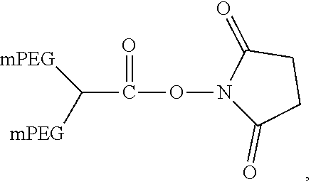
[0048] In one non-limiting example, the reagent used to generate PEGylated aptamers is [N.sup.2-(monomethoxy 20K polyethylene glycol carbamoyl)-N.sup.6-(monomethoxy 20K polyethylene glycol carbamoyl)]-lysine N-hydroxysuccinimide having the formula:
##STR00002##
[0049] In yet another non-limiting example, the reagent used to generate PEGylated aptamers has the formula:
##STR00003##
where X is N-hydroxysuccinimide and the PEG arms are of approximately equivalent molecular weight. Such PEG architecture may provide a compound with reduced viscosity compared to a similar aptamer conjugated to a two-armed or single-arm linear PEG.
[0050] In some examples, the reagent used to generate PEGylated aptamers has the formula:
##STR00004##
where X is N-hydroxysuccinimide and the PEG arms are of different molecular weights, for example, a 40 kD PEG of this architecture may be composed of 2 arms of 5 kD and 4 arms of 7.5 kD. Such PEG architecture may provide a compound with reduced viscosity compared to a similar aptamer conjugated to a two-armed PEG or a single-arm linear PEG.
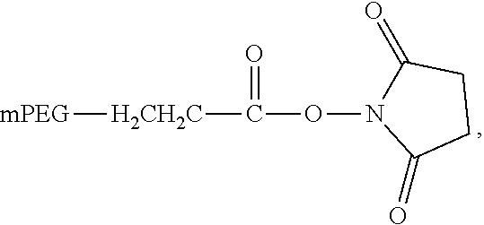
[0051] In some cases, the reagent that may be used to generate PEGylated aptamers is a non-branched mPEG-Succinimidyl Propionate (mPEG-SPA), having the general formula:
##STR00005##
where mPEG is about 20 kD or about 30 kD. In one example, the reactive ester may be --O--CH.sub.2--CH.sub.2--CO.sub.2--NHS.
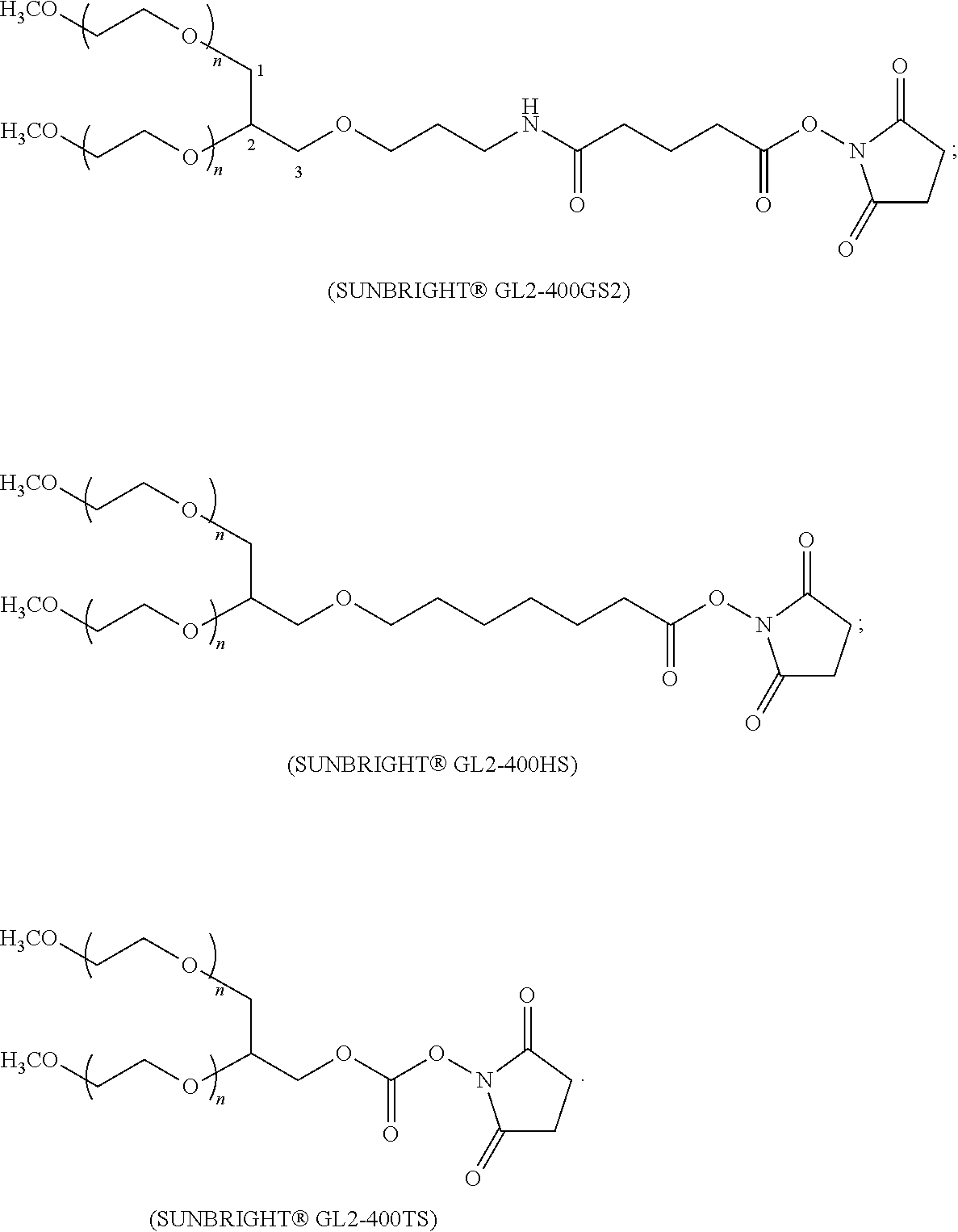
[0052] In some instances, the reagent that may be used to generate PEGylated aptamers may include a branched PEG linked through glycerol, such as the Sunbright.TM. series from NOF Corporation, Japan. Non-limiting examples of these reagents include:
##STR00006##
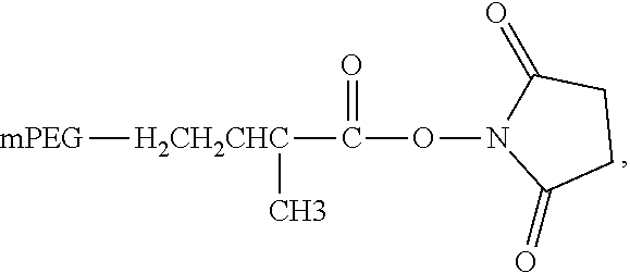
[0053] In another example, the reagents may include a non-branched mPEG Succinimidyl alpha-methylbutanoate (mPEG-SMB) having the general formula:
##STR00007##
where mPEG is between 10 and 30 kD. In one example, the reactive ester may be --O--CH.sub.2--CH.sub.2--CH(CH.sub.3)--CO.sub.2--NHS.
[0054] In other instances, the PEG reagents may include nitrophenyl carbonate-linked PEGs, having the general formula:
##STR00008##
[0055] Compounds including nitrophenyl carbonate can be conjugated to primary amine containing linkers.
[0056] In some cases, the reagents used to generate PEGylated aptamers may include PEG with thiol-reactive groups that can be used with a thiol-modified linker. One non-limiting example may include reagents having the following general structure:
##STR00009##
where mPEG is about 10 kD, about 20 kD or about 30 kD. Another non-limiting example may include reagents having the following general structure:
##STR00010##
where each mPEG is about 10 kD, about 20 kD, or about 30 kD and the total molecular weight is about 20 kD, about 40 kD, or about 60 kD, respectively. Branched PEGs with thiol reactive groups that can be used with a thiol-modified linker, as described above, may include reagents in which the branched PEG has a total molecular weight of about 40 kD or about 60 kD (e.g., where each mPEG is about 20 kD or about 30 kD).
[0057] In some cases, the reagents used to generated PEGylated aptamers may include reagents having the following structure:
##STR00011##
In some cases, the reaction is carried out between about pH 6 and about pH 10, or between about pH 7 and pH 9 or about pH 8.
[0058] In some cases, the aptamer is associated with a single PEG molecule. In other cases, the aptamer is associated with two or more PEG molecules.
[0059] In some cases, the aptamers described herein may be bound or conjugated to one or more molecules having desired biological properties. Any number of molecules can be bound or conjugated to aptamers, non-limiting examples including antibodies, peptides, proteins, carbohydrates, enzymes, polymers, drugs, small molecules, gold nanoparticles, radiolabels, fluorescent labels, dyes, haptens (e.g., biotin), other aptamers, or nucleic acids (e.g., siRNA). In some cases, aptamers may be conjugated to molecules that increase the stability, the solubility or the bioavailability of the aptamer. Non-limiting examples include polyethylene glycol (PEG) polymers, carbohydrates and fatty acids. In some cases, molecules that improve the transport or delivery of the aptamer may be used, such as cell penetration peptides. Non-limiting examples of cell penetration peptides can include peptides derived from Tat, penetratin, polyarginine peptide Args sequence (SEQ ID NO: 90), Transportan, VP22 protein from Herpes Simplex Virus (HSV), antimicrobial peptides such as Buforin I and SynB, polyproline sweet arrow peptide molecules, Pep-1 and MPG. In some embodiments, the aptamer is conjugated to a lipophilic compound such as cholesterol, dialkyl glycerol, diacyl glycerol, or a non-immunogenic, high molecular weight compound or polymer such as polyethylene glycol (PEG) or other water-soluble pharmaceutically acceptable polymers including, but not limited to, polyaminoamines (PAMAM) and polysaccharides such as dextran, or polyoxazolines (POZ).
[0060] The molecule to be conjugated can be covalently bonded or can be associated through non-covalent interactions with the aptamer of interest. In one example, the molecule to be conjugated is covalently attached to the aptamer. The covalent attachment may occur at a variety of positions on the aptamer, for example, to the exocyclic amino group on the base, the 5-position of a pyrimidine nucleotide, the 8-position of a purine nucleotide, the hydroxyl group of the phosphate, or a hydroxyl group or other group at the 5' or 3' terminus. In one example, the covalent attachment is to the 5' or 3' hydroxyl group of the aptamer.
[0061] In some cases, the aptamer can be attached to another molecule directly or with the use of a spacer or linker. For example, a lipophilic compound or a non-immunogenic, high molecular weight compound can be attached to the aptamer using a linker or a spacer. Various linkers and attachment chemistries are known in the art. In a non-limiting example, 6-(trifluoroacetamido)hexanol (2-cyanoethyl-N,N-diisopropyl)phosphoramidite can be used to add a hexylamino linker to the 5' end of the synthesized aptamer. This linker, as with the other amino linkers provided herein, once the group protecting the amine has been removed, can be reacted with PEG-NHS esters to produce covalently linked PEG-aptamers. Other non-limiting examples of linker phosphoramidites may include: TFA-amino C4 CED phosphoramidite having the structure:
##STR00012##
5'-amino modifier C3 TFA having the structure:
##STR00013##
MT amino modifier C6 CED phosphoramidite having the structure:
##STR00014##
5'-amino modifier 5 having the structure:
##STR00015##
5'-amino modifier C12 having the structure:
##STR00016##
and 5' thiol-modifier C6 having the structure:
##STR00017##
[0062] The 5'-thiol modified linker may be used, for example, with PEG-maleimides, PEG-vinylsulfone, PEG-iodoacetamide and PEG-orthopyridyl-disulfide. In one example, the aptamer may be bonded to the 5'-thiol through a maleimide or vinyl sulfone functionality.
[0063] In some cases, the aptamer formulated according to the present disclosure may also be modified by encapsulation within aliposome. In other cases, the aptamer formulated according to the present disclosure may also be modified by encapsulation within a micelle. Liposomes and micelles may be comprised of any lipids, and in some cases the lipids may be phospholipids, including phosphatidylcholine.
[0064] In some cases, the aptamers described herein are designed to inhibit a function associated with an alternative complement pathway enzyme. In one example, an anti-fD aptamer is used to inhibit a function associated with fD (e.g., inhibit the catalytic activity off fD). In other cases, the aptamers described herein are designed to prevent an interaction or binding of two or more proteins of the alternative complement pathway. In one example, an aptamer binds to fD and prevents binding of the complex C3bBb to fD. The aptamers described herein may bind to a region of fD that is recognized by an antibody or antibody fragment thereof that inhibits a function associated with fD. In some cases, the antibody or antibody fragment thereof that inhibits a function associated with fD has an amino acid sequence of heavy chain variable region of
TABLE-US-00001 (SEQ ID NO: 71) EVQLVQSGPELKKPGASVKVSCKASGYTFTNYGMNWVRQAPGQGLEWMGW INTYTGETTYADDFKGRFVFSLDTSVSTAYLQISSLKAEDTAVYYCERGG VNNWGQGTLVTVSSASTKGPSVFPLAPSSKSTSGGTAALGCLVKDYFPEP VTVSWNSGALTSGVHTFPAVLQSSGLYSLSSVVTVPSSSLGTQTYICNVN HKPSNTKVDKKVEPKSCDKTHT
and an amino acid sequence of light chain variable region of:
TABLE-US-00002 (SEQ ID NO: 72) DIQVTQSPSSLSASVGDRVTITCITSTDIDDDMNWYQQKPGKVPKLLISG GNTLRPGVPSRFSGSGSGTDFTLTISSLQPEDVATYYCLQSDSLPYTFGQ GTKVEIKRTVAAPSVFIFPPSDEQLKSGTASVVCLLNNFYPREAKVQWKV DNALQSGNSQESVTEQDSKDSTYSLSSTLTLSKADYEKHKVYACEVTHQG LSSPVTKSFNRGEC.
[0065] In some cases, the antibody or antibody fragment thereof that inhibits a function associated with fD has an amino acid sequence of heavy chain variable region of:
TABLE-US-00003 (SEQ ID NO: 85) EVQLVQSGPELKKPGASVKVSCKASGYTFTNYGMNWVRQAPGQGLEWMGW INTYTGETTYAEDFKGRFVFSLDTSVSTAYLQISSLKAEDTAVYYCEREG GVSNWGQGTLVTVSS; or (SEQ ID NO: 86) EVQLVQSGPELKKPGASVKVSCKASGYTFTNYGMNWVRQAPGQGLEWMGW INTYTGETTYAEDFKGRFVFSLDTSVSTAYLQISSLKAEDTAVYYCEREG GVNNWGQGTLVTVSS,
and an amino acid sequence of light chain variable region of:
TABLE-US-00004 (SEQ ID NO: 87) DIQVTQSPSSLSASVGDRVTITCITSTDIESDMNAVYQQKPGKVPKLLIS GGNTLRPGVPSRFSGSGSGTDFTLTISSLQPEDVATYYCLQSDSLPYTFG QGTKVEIK; (SEQ ID NO: 88) DIQVTQSPSSLSASVGDRVTITCITSTDIESDMNWYQQKPGKVPKLLISG GNTLRPGVPSRFSGSGSGTDFTLTISSLQPEDVATYYCLQSESLPYTFGQ GTKVEIK; or (SEQ ID NO: 89) DIQVTQSPSSLSASVGDRVTITCITSTSIESDMNWYQQKPGKVPKLLISG GNTLRPGVPSRFSGSGSGTDFTLTISSLQPEDVATYYCLQSDSLPYTFGQ GTKVEIK.
[0066] The aptamers described herein may bind to a region of fD that is recognized by a small molecule inhibitor that inhibits a function associated with fD, non-limiting examples including dichloroisocoumarin or any one of the compounds depicted in FIGS. 13A-D. The aptamers described herein may bind to a region of fD that is recognized by a peptide inhibitor that inhibits a function associated with fD.
[0067] In some cases, an aptamer of the disclosure comprises one of the following sequences described in Table 1.
TABLE-US-00005 TABLE 1 fD Aptamer Sequences SEQ ID Aptamer Back- NO. Number bone Sequence 5' to 3' SEQ ID Aptamer 1 RNA GGGAGUGUGUACGAGGCAUUAGGCCGCCAC NO: 1 CCAAACUGCAGUCCUCGUAAGUCUGCCUGG CGGCUUUGAUACUUGAUCGCCCUAGAAGC SEQ ID Aptamer 2 RNA GGGAGUGUGUACGAGGCAUUAGUCCGCCGA NO: 2 AGUCUUUUGGCUCGGUUUUUUCAAGGUCGG CGGCUUUGAUACUUGAUCGCCCUAGAAGC SEQ ID Aptamer 3 RNA GGGAGUGUGUACGAGGCAUUAGGCCGCCAC NO: 3 CUCGUUUGAUUGCGGUUGUUCGGCCGCGGG CGGCUUUGAUACUUGAUCGCCCUAGAAGC SEQ ID Aptamer 4 DNA GTGACGACTGACATATCTGCTCCGAGGTTA NO: 4 TTGGGGTTGGGGCCTGGGCGATTGGGGCCT CGTAGTTGAGTCTGAGTGCT SEQ ID Aptamer 5 DNA GTGACGACTGACATATCTGCGTTTGGGGTT NO: 5 GGGGCCTGGGAGTTTGGGGAGCAGAAAGGA CGTAGTTGAGTCTGAGTGCT SEQ ID Aptamer 6 DNA GTGACGACTGACATATCTGCTGTGGGTGTT NO: 6 GTGGGGGTGGGTGGTGGGCCCTTCGCCATG CGTAGTTGAGTCTGAGTGCT SEQ ID Aptamer 7 DNA GTGACGACTGACATATCTGCGGCGGTTGGG NO: 7 GTCGAAGGGCGAGGGGTGGGAGGTCGCCGT AGTTGAGTCTGAGTGCT SEQ ID Aptamer 8 DNA GTGACGACTGACATATCTGCTATTTTGGGG NO: 8 CCTGGGTGTTGGGGATTGGGGACTATGTGT CGTAGTTGAGTCTGAGTGCT SEQ ID Aptamer 9 DNA GTGACGACTGACATATCTGCTGTGGATGGT NO: 9 GGGGGGTGGTGTGGGAGGGCTGGTCGGTCG CGTAGTTGAGTCTGAGTGCT SEQ ID Aptamer 10 DNA GTGACGACTGACATATCTGCCCTATAGGGG NO: 10 TGTGGGCGAGGGGTGGGTGGTAGGGCGGCT CGTAGTTGAGTCTGAGTGCT SEQ ID Aptamer 11 DNA GTGACGACTGACATATCTGCGGAGGTGGGT NO: 11 GGGTGGGTGCGTGCGAGGGCGGTGTAGGTC CGTAGTTGAGTCTGAGTGCT SEQ ID Aptamer 12 DNA GTGACGACTGACATATCTGCAAAAGTTAGA NO: 12 TTGACATGGTATGCACCGTCTGAGGTTGGT CGTAGTTGAGTCTGAGTGCT SEQ ID Aptamer 13 DNA GTGACGACTGACATATCTGCACCACGCTAG NO: 13 GGGTGAGGGCGAGGGGTGGGTAGCGCGTGG CGTAGTTGAGTCTGAGTGCT SEQ ID Aptamer 14 DNA GTGACGACTGACATATCTGCTGTGGGTGTT NO: 14 GTGGGGGCGGGTGGTGGGTGCGTCGGTGGT CGTAGTTGAGTCTGAGTGCT SEQ ID Aptamer 15 DNA GTGACGACTGACATATCTGCTGCTTCCAGC NO: 15 GGTCATGATATGCACTGTCTGAAGCTCGGT CGTAGTTGAGTCTGAGTGCT SEQ ID Aptamer 16 DNA GTGACGACTGACATATCTGCTGTGTTATGA NO: 16 TATGCACCGTCTGAGGGTAGTCGCGGGGTG CGTAGTTGAGTCTGAGTGCT SEQ ID Aptamer 17 DNA GTGACGACTGACATATCTGCTGCTTGTTTA NO: 17 GTGGGTGGGTGGGTGGTGTGGTGGTGATGC GTAGTTGAGTCTGAGTGCT SEQ ID Aptamer 18 DNA GTGACGACTGACATATCTGCCTTGGGGTTG NO: 18 GGGCCTGGGTGTTTGGGGTGGCCTAGAAGT CGTAGTTGAGTCTGAGTGCT SEQ ID Aptamer 19 DNA GTGACGACTGACATATCTGCGCTAGGGGTG NO: 19 GGTTGGGGTTGGTGGTGTGCGTGTGGGTTG CGTAGTTGAGTCTGAGTGCT SEQ ID Aptamer 20 DNA GTGACGACTGACATATCTGCTGTTGAGGTT NO: 20 GGTGGGGGGTGGGCGGTGGGATGGTTGTGC CGTAGTTGAGTCTGAGTGCT SEQ ID Aptamer 21 DNA GTGACGACTGACATATCTGCTTGACAGTCT NO: 21 GCTTTGCAGGGGCCGAGAGCGCCATTGCGT CGTAGTTGAGTCTGAGTGCT SEQ ID Aptamer 22 DNA GTGACGACTGACATATCTGCTGTGGTTGGT NO: 22 GGGGGGTGGAGGGTGGGAGGCCGTGTGTCC CGTAGTTGAGTCTGAGTGCT SEQ ID Aptamer 23 DNA GTGACGACTGACATATCTGCTGTGGTGGTG NO: 23 GGGGAGGGTGGTGGGGTGGCCGGCGCTCGT CGTAGTTGAGTCTGAGTGCT SEQ ID Aptamer 24 DNA GTGACGACTGACATATCTGCTGGGTTACGT NO: 24 GGTTCGGGGCTAGGGGGGTGGGGTGTGTTT CGTAGTTGAGTCTGAGTGCT SEQ ID Aptamer 25 DNA GTGACGACTGACATATCTGCTGGTGGTGTG NO: 25 CGGTGGGTTCTTGGGTGGGATGGGTGGTAC CGTAGTTGAGTCTGAGTGCT SEQ ID Aptamer 26 DNA GTGACGACTGACATATCTGCTATTAGATCC NO: 26 TCGGTGGGTGGGTGGGTGTGTGGTGGTGTG CGTAGTTGAGTCTGAGTGCT SEQ ID Aptamer 27 DNA GTGACGACTGACATATCTGCGGGCGTCTGA NO: 27 GCGCATGGATGACCCACCGACAGATTGCGG CGTAGTTGAGTCTGAGTGCT SEQ ID Aptamer 28 DNA GTGACGACTGACATATCTGCGCTTTGGGTG NO: 28 GGCTCGGTGTGCGGTGTGCGGGTGGGTTTG CGTAGTTGAGTCTGAGTGCT SEQ ID Aptamer 29 DNA GTGACGACTGACATATCTGCGTTTGGGGTT NO: 29 GGGGCCTGGGAGTTTGGGGAGCAGAAAGGG CGTAGTTGAGTCTGAGTGCT SEQ ID Aptamer 30 DNA GTGACGACTGACATATCTGCGGGTGGGTTG NO: 30 GGTTGGGTTTGGTGGTGGTGCCTGTTAGTT CGTAGTTGAGTCTGAGTGCT SEQ ID Aptamer 31 DNA GTGACGACTGACATATCTGCAGGTGGGTGG NO: 31 GTGGGTGTGTGTGCGGTGGTGTGATTTGGC CGTAGTTGAGTCTGAGTGCT SEQ ID Aptamer 32 DNA GTGACGACTGACATATCTGCTGTGGTTGGT NO: 32 GGGGGGCGGCGGGTGGGGAGCCTGGTGTTC CGTAGTTGAGTCTGAGTGCT SEQ ID Aptamer 33 DNA GTGACGACTGACATATCTGCTCCCGTTTGA NO: 33 GGGCTTGTCGGACAGATTGCTGGCACGTCA CGTAGTTGAGTCTGAGTGCT SEQ ID Aptamer 34 DNA GTGACGACTGACATATCTGCTCTTGGTGGT NO: 34 GGTGGTGGGTTGGGATGGGTCTTGGGCTGC CGTAGTTGAGTCTGAGTGCT SEQ ID Aptamer 35 DNA GTGACGACTGACATATCTGCCTGTGAGGGG NO: 35 AGGGAGGGTGGGTTTGGCGGTGGCGCAGGC CGTAGTTGAGTCTGAGTGCT SEQ ID Aptamer 36 DNA GTGACGACTGACATATCTGCGTGGTGGTGC NO: 36 GTGGGTGGTGGGGGGGGGAGCTGGGTGCCC CGTAGTTGAGTCTGAGTGCT SEQ ID Aptamer 37 DNA GTGACGACTGACATATCTGCTGTGGGTGTT NO: 37 GTGGGGGTGGGTGGTGGGCCCTTCGCCGTG CGTAGTTGAGTCTGAGTGCT SEQ ID Aptamer 38 DNA GTGACGACTGACATATCTGCTTCCGGTATG NO: 38 TGTGGGTGGGTGGGTGGTGTGGTGGTGTTG CGTAGTTGAGTCTGAGTGCT SEQ ID Aptamer 39 DNA GTGACGACTGACATATCTGCTCTCTTCTGT NO: 39 TGTGGGTGGGTGGGTGGTGTGGTGCGTGTG CGTAGTTGAGTCTGAGTGCT SEQ ID Aptamer 40 DNA GTGACGACTGACATATCTGCGGCTGGGTGG NO: 40 GTTGGGTTAGGGTGGTGTGCGGTGGGTTGC CGTAGTTGAGTCTGAGTGCT SEQ ID Aptamer 41 DNA GTGACGACTGACATATCTGCGTTTAGGTGG NO: 41 GCGGGTGGGTGTGCGGTGGGCGGTGTTGAA CGTAGTTGAGTCTGAGTGCT SEQ ID Aptamer 42 DNA GTGACGACTGACATATCTGCGGTGATTGGG NO: 42 GTTGGGGCCTGGGCGTTTGGGGACCGCATG CGTAGTTGAGTCTGAGTGCT SEQ ID Aptamer 43 DNA GTGACGACTGACATATCTGCGTTTGGGGTT NO: 43 GGGGCCTGGGAGTTTGGGGAGCAGAGAGGA CGTAGTTGAGTCTGAGTGCT SEQ ID Aptamer 44 DNA GTGACGACTGACATATCTGCTAACTTGTTG NO: 44 GGGTTTGGGGCCTGGGTGTTGGGGTTGTTT CGTAGTTGAGTCTGAGTGCT SEQ ID Aptamer 45 DNA GTGACGACTGACATATCTGCTGGGGTTGGT NO: 45 GGGGGGAGGTGGGTGGGTTATGTGCGCTGG CGTAGTTGAGTCTGAGTGCT SEQ ID Aptamer 46 DNA GTGACGACTGACATATCTGCTGTGGGTGTT NO: 46 GTGGGGGTGGGTTGGTGGGCATTGCGTGTG CGTAGTTGAGTCTGAGTGCT SEQ ID Aptamer 47 DNA GTGACGACTGACATATCTGCGAGTGGGTTC NO: 47 GGTGGTGGTGTGTGGGAGGGTTGGGTACGT CGTAGTTGAGTCTGAGTGCT SEQ ID Aptamer 48 DNA GTGACGACTGACATATCTGCTGGACATGAT NO: 48 TGCACCGTATGAGGTTTAGTCGTTAATGTG CGTAGTTGAGTCTGAGTGCT SEQ ID Aptamer 49 DNA GTGACGACTGACATATCTGCAGTGGGGCCT NO: 49 GGGCGTTGGGGTTTGGGGTGCCTCGTCAGT CGTAGTTGAGTCTGAGTGCT SEQ ID Aptamer 50 DNA GTGACGACTGACATATCTGCATGGATTTTC NO: 50 GGTGGGTGGGTGGGTTGGTGTGGTGGTGTG CGTAGTTGAGTCTGAGTGCT SEQ ID Aptamer 51 DNA GTGACGACTGACATATCTGCTGTGGTTGGT NO: 51 GGGGGGTGGGTGGTGGGAAGGTTCCGGTGC CGTAGTTGAGTCTGAGTGCT SEQ ID Aptamer 52 DNA GTGACGACTGACATATCTGCGGTTGGGGTT NO: 52 GGGGCCTGGGTGTTGGGGAGCAGGTAGCAC CGTAGTTGAGTCTGAGTGCT SEQ ID Aptamer 53 DNA GTGACGACTGACATATCTGCGGCCTGGGAG NO: 53 GGTTCGGTGGTGGTGCGAGGGTGGGCAAGC CGTAGTTGAGTCTGAGTGCT SEQ ID Aptamer 54 DNA ACCTAGTTTGGCTTGCAXAAGTAACYAGCA NO: 54 CGTGGGCTAG SEQ ID Aptamer 55 DNA ACGATCGCCCCYGTCTWTAAGAXCGAATAC NO: 55 TATGGGCTAG SEQ ID Aptamer 56 DNA ACCTAGAAAGGCTTAGTGAAGTAAWGATCA NO: 56 GGGCGGGATC SEQ ID Aptamer 57 DNA ACCTAGTTCCCYGTCTAXYAGAXCCGAGXG NO: 57 TATGCCGATC SEQ ID Aptamer 58 DNA ACCTAGGCAGTCTTGCCGAATTTACGAGXG NO: 58 GGGAGGGATC SEQ ID Aptamer 59 DNA ACGATCACTGCYCAGCWTYATTAACYAGCY NO: 59 TCGACCCTAG SEQ ID Aptamer 60 DNA ACGATCTTCCGCCAGCTGYATTXCGAAGXG NO: 60 CGTGAGGATC SEQ ID Aptamer 61 DNA ACCTAGGCGGTCTTXCCGTCGTTACGTCCY NO: 61 CGGCCCCTAG SEQ ID Aptamer 62 DNA ACCTAGTTTGGCGTAGCGYATTAAWGGGXG NO: 62 CGGCAGCTAG SEQ ID Aptamer 63 DNA ACGATCGCTGACGTXCAXYAGTATGAGGCA NO: 63 CGTGGGCTAG
[0068] In some aspects, an aptamer of the disclosure comprises the nucleic acid sequence of any one of Aptamers 1-3 (SEQ ID NOS: 1-3). In some cases, any one of Aptamers 1-3 comprises one or more modified nucleotides. In a preferred example, an aptamer of the disclosure comprises one of Aptamers 1-3 where G is 2'F and A, C and U are 2'OMe modified RNA. In some aspects, an aptamer of the disclosure comprises the nucleic acid sequence of any one of Aptamers 54-63 (SEQ ID NOS: 54-63). In some cases, any one of Aptamers 54-63 comprises one or more modified nucleotides. In a preferred example, an aptamer of the disclosure comprises one of Aptamers 54-63, where W=5-(indole-3-acetamido-1-propenyl)-2'-deoxyuridine; X=5-(amino-1-propenyl)-2'-deoxyuridine; and Y=5-(4-pivaloylbenzamido-1-propenyl)-2'-deoxyuridine.
[0069] In some cases, an aptamer of the disclosure may have at least 80%, 81%, 82%, 83%, 84%, 85%, 86%, 87%, 88%, 89%, 90%, 91%, 92%, 93%, 94%, 95%, 96%, 97%, 98%, or 99% sequence identity with any aptamer described herein. For example, an anti-fD aptamer of the disclosure may have at least 80%, 81%, 82%, 83%, 84%, 85%, 86%, 87%, 88%, 89%, 90%, 91%, 92%, 93%, 94%, 95%, 96%, 97%, 98%, or 99% sequence identity with any aptamer described in Table 1. In some cases, an aptamer of the disclosure may have at least 80%, 81%, 82%, 83%, 84%, 85%, 86%, 87%, 88%, 89%, 90%, 91%, 92%, 93%, 94%, 95%, 96%, 97%, 98%, or 99% sequence homology with any aptamer described herein. For example, an anti-fD aptamer of the disclosure may have at least 80%, 81%, 82%, 83%, 84%, 85%, 86%, 87%, 88%, 89%, 90%, 91%, 92%, 93%, 94%, 95%, 96%, 97%, 98%, or 99% sequence homology with any aptamer described in Table 1.
[0070] In such cases where specific nucleotide modifications have been recited, it should be understood that any number and type of nucleotide modifications may be substituted. For example, 2'OMeG may be substituted for 2'FG. Non-limiting examples of nucleotide modifications have been provided herein. In some instances, all of the nucleotides of an aptamer are modified. In some instances, at least 50%, 55%, 60%, 65%, 70%, 75%, 80%, 85%, 86%, 87%, 88%, 89%, 90%, 91%, 92%, 93%, 94%, 95%, 96%, 97%, 98%, 99%, or 100% of the nucleotides of an aptamer of the disclosure may be modified.
[0071] In some instances, the aptamer does not comprise any one of the following nucleic acid sequences (from 5' to 3'):
TABLE-US-00006 (SEQ ID NO: 73) ACGGAGAAAGAGAGAGTGTAATTGCTAGCATAACCGCTGC, (SEQ ID NO: 74) GTAACCACGTTGCCAGACCGAGTCTACCAGCGATCCTCAG, (SEQ ID NO: 75) TATGCCCAAATCCCTCAAGTCGGCCAGGATACACCACCGT, (SEQ ID NO: 76) AATCAAAAGGCTCACGCGCGGATTGGTCAACCTTACAACC, (SEQ ID NO: 77) TCGGCCTTCCCAGACCACCGCAATCCCCAGGGAACAGGCA, (SEQ ID NO: 78) CATCACACTGTCAACATACCCAGCCTGGGGAAAGACGAAC, (SEQ ID NO: 79) AACCCGCATGCCGATCGATGTCGTGCCTCGCTCCACGCTC, or (SEQ ID NO: 80) ACCAGGCACCCGACGGACTAACTCATCACTCAGGCGAGGG
Anti-fD Compositions
[0072] fD is a component of the alternative complement pathway and is believed to be involved in the pathogenesis of AMD and other ocular disorders. fD is unique among serine proteases in that it does not require cleavage of a zymogen for expression of proteolytic activity. Rather, fD requires a conformational change that is believed to be induced by the complex C3bB resulting in a reversible reorientation of the catalytic center and substrate binding site of fD. fD is primarily produced by adipocytes and is systemically available in serum at low levels. fD contains a self-inhibitory loop that prevents catalytic activity of fD. Binding of the C3bB complex to fD displaces the self-inhibitory loop and fD cleaves C3bB to form the C3 convertase C3bBb. The catalytic activity of fD only occurs in the context of complexed fB; fD does not cleave uncomplexed fB. The complex of fD, fB, and C3b forms an amplification loop of the alternative complement pathway of which fD is the rate-limited enzyme.
[0073] In some aspects, the methods and compositions described herein involve inhibition of fD, resulting in inhibition of the amplification step of the alternative complement pathway. The anti-fD compositions herein may involve the use of one or more anti-fD aptamers for the treatment of ocular diseases. In some cases, the ocular disease is macular degeneration. In some cases, macular degeneration is age-related macular degeneration. In some cases, age-related macular degeneration is dry age-related macular degeneration. In some cases, dry age-related macular degeneration is advanced dry age-related macular degeneration (i.e., geographic atrophy). In some cases, age-related macular degeneration is wet age-related macular degeneration. In some cases, macular degeneration is Stargardt disease or Stargardt-like disease.
Anti-fD Inhibitors
[0074] The anti-fD compositions disclosed herein may be designed to bind to specific regions of fD with high specificity and affinity. The compositions may bind to fD in such a way as to inhibit, either directly or indirectly, the catalytic activity of the enzyme. In some cases, the anti-fD aptamers can bind to the active site (e.g., the catalytic cleft) of fD and directly inhibit the catalytic activity of fD. In this example, the aptamer may be designed to target the active site (e.g., the catalytic cleft) of fD. When the aptamer is bound to the active site of fD, it can prevent the substrate (e.g., C3bB) from accessing the active site. In some cases, the anti-fD aptamer can bind to an exosite of fD and indirectly inhibit the catalytic activity of fD by e.g., preventing the binding of C3bB. In some cases, the exosite may be remote from the catalytic site. In other cases, there may be some overlap with the catalytic site. In some cases the anti-fD aptamer can bind to the self-inhibitory loop of fD to prevent displacement of the self-inhibitory loop and thus, prevent activation of fD.
[0075] Amino acid residues of fD may be referenced according to the chymotrypsin numbering scheme and this numbering system is used throughout the disclosure to refer to specific amino acid residues of fD. Chymotrypsin numbering scheme for fD may be as depicted in FIG. 14 (SEQ ID NO: 94) (chymotrypsin numbering displayed above amino acid sequence and fD numbering scheme below amino acid sequence).
[0076] Anti-fD aptamers as described herein can modulate or inhibit the activity of fD or a fD variant thereof. A fD variant as used herein encompasses variants that perform essentially the same function as fD. A fD variant includes essentially the same structure as fD and in some cases includes at least 80%, 85%, 86%, 87%, 88%, 89%, 90%, 91%, 92%, 93%, 94%, 95%, 96%, 97%, 98%, 99%, 99.5%, or 99.9% sequence identity to the amino acid sequence (shown above) of the fD protein.
[0077] In certain embodiments of the disclosure, methods are provided for the identification of fD aptamers that specifically bind to epitopes of fD. These methods may be utilized, for example, to determine the binding site and/or the mechanism of action of the aptamer.
[0078] In one instance, methods are provided for testing a fD aptamer in alternative complement dependent hemolysis of red blood cells. Human serum that is rendered deficient in the classical complement pathway by depleting C1q may be dependent on alternative complement activity to lyse rabbit red blood cells, an activity that may be dependent on fD (Katschke, Wu, Ganesan, et al. (2012) Inhibiting alternative pathway complement activation by targeting the Factor D exosite. J. Biol. Chem. 287, 12886-12892). In some cases, the fD aptamers disclosed herein may inhibit alternative complement dependent hemolysis of red blood cells (see Example 4).
[0079] In another instance, methods are provided for testing a fD aptamer in fD esterase activity assays (see Example 5). Cleavage of a modified peptide substrate of fD, Z-lys-S-Bzl, may be monitored by the cleaved product reducing 5,5'-Dithiobis(2-nitrobenzoic acid). FD may have a lower catalytic rate than other complement proteases when using peptide thioester substrates, and one such substrate Z-lys-SBzl was found to be cleaved by fD and useful as a synthetic substrate (fD is called protein D in Kam, McRae et al. (1987) Human complement proteins D, C2, and B. J. Biol. Chem. 262, 3444-3451). In some cases, a molecule that binds fD may block catalytic activity by binding in the catalytic cleft to sterically prevent access of the peptide substrate to the catalytic residues of fD (Katschke, Wu, Ganesan, et al. (2012) Inhibiting alternative pathway complement activation by targeting the Factor D exosite. J. Biol. Chem. 287, 12886-12892). In other cases, a molecule that binds fD may block catalytic activity by an allosteric mechanism that induces structural changes in the enzyme. In yet other cases, a molecule that binds fD may bind to the fD exosite region to sterically inhibit binding of the physiologic substrate protein C3bB, but not of the synthetic modified peptide substrate Z-Lys-SBzl (Katschke, Wu, Ganesan, et al. (2012) Inhibiting alternative pathway complement activation by targeting the Factor D exosite. J. Biol. Chem. 287, 12886-12892). In some instances, where a molecule inhibits fD binding and proteolytic cleavage of FB but not Z-Lys-SBzl, the binding may be similar to how anti-factor D FAb antibody fragment binds to the exosite and induces a subtle conformational change that increases fD cleaving Z-Lys-S-Bzl (Katschke, Wu, Ganesan, et al. (2012) Inhibiting alternative pathway complement activation by targeting the Factor D exosite. J. Biol. Chem. 287, 12886-12892).
[0080] In another instance, methods are provided for testing a fD aptamer in a reconstituted biochemical fD activity assay which is composed of purified proteins fD, FB, and C3b (see Example 6). When fD binds to the complex of FB and C3b (C3bB), FB is cleaved by fD into fragments Ba and Bb (Katschke, Wu, Ganesan, et al. (2012) Inhibiting alternative pathway complement activation by targeting the Factor D exosite. J. Biol. Chem. 287, 12886-12892). The activity of fD can be monitored by the rate of FB cleavage and Ba fragment production using an ELISA that uses an antibody that specifically binds Ba (Quidel, A033), or by other means known in the art to measure Ba levels. In some cases, the concentrations of FB and C3b are equal so they form a 1:1 complex which can then bind fD and allow enzymatically active fD to cleave FB to fragments Ba and Bb. In some cases, the FB:C3b complex is present in 4-fold excess of fD. In other cases, the concentrations of fD and/or C3bB are varied in such a manner as to measure enzymatic constants, including, but not limited to k.sub.cat, K.sub.m and k.sub.cat/K.sub.m.
[0081] In yet another instance, methods are provided for the identification of fD binding to C3bB in complex (see Example 7). FD is the rate-limiting enzyme in the alternative complement pathway, and converts the proconvertases C3bB and C3b2B to form the active C3 convertase C3bBb or the active C5 convertase C3bBb (Katschke et al 2012). For surface plasmon resonance (SPR) to detect fD in a stable complex with FB, catalytically inactive fD (S195A) may be used so that it does not cleave the FB upon binding to the FB:C3b complex (Katschke, Wu, Ganesan, et al. (2012) Inhibiting alternative pathway complement activation by targeting the Factor D exosite. J. Biol. Chem. 287, 12886-12892). When C3b is amine-coupled to a CM5 chip, SPR may detect binding of FB as increased mass, and binding of fD to the C3b:FB complex as a further increase in mass. In one aspect, the fD binding compounds are aptamers that bind fD and prevent fD binding to FB:C3b as determined by a reduced mass detected by SPR.
[0082] In some cases, a cell model of Stargardt disease may be used to detect activity of anti-fD aptamers (see Example 8). Retinal pigment epithelial (RPE) cells may undergo cell death early during the progress of Stargardt disease, and evidence points toward the involvement of the alternative complement pathway (AP) in RPE cell death (Berchuck, Yang, et al (2013) All-trans-retinal (atRal) sensitizes human RPE cells to alternative complement pathway-induced cell death. Invest Ophthalmol Vis Sci 54, 2669-2677). ARPE-19 cells are a spontaneously arising RPE cell line derived from the normal eyes of a 19-year-old male. The ARPE-19 cell line, established using the cuboidal basal cell layer cultured in specific culture media, expresses the RPE-specific markers cellular retinaldehyde binding protein and RPE-65. Stargardt disease is a hereditary juvenile macular degeneration that occurs in patients with homozygous mutations in the ABCA4 genes, which encode a protein that processes all-trans retinal (Molday (2007) ATP-binding cassette transporter ABCA4: molecular properties and role in vision and macular degeneration. J. Bioenerg Biomembr 39, 507-517). An ABCA4 and RDH8 mouse model of Stargardt disease presents with retinal pathology caused by accumulated atRal, and ABCA4 mutations are present in 16% of AMD patients, suggesting that elevated atRal may contribute to Stargardt disease and AMD disease progression (Berchuck et al 2013). Mechanistically, atRal decreased expression of CD46 and CD59 on RPE cells in vitro, which increased susceptibility to cell lysis mediated by alternative complement in response to anti-RPE antibody binding to the RPE cell membranes (Berchuck et al 2013). In some cases, the disclosure provides for the identification of fD inhibitors that inhibit alternative complement-mediated lysis of human retinal pigmented epithelial cells.
[0083] The anti-fD aptamers as disclosed herein, in some cases, may bind to the region of fD that includes the active site cleft. Upon activation by binding to C3bB, fD exhibits serine protease activity towards fB. Activation of fD by substrate binding is a two-step process: first, fD binds to fB in the open C3bB configuration at the Von Willebrand factor type-A (VWA)-serine protease (SP) interface of fB, interacting mainly via its exosite residues within loops 145-149, 169-173, 185-188 and 220-224. Binding of fD to C3bB causes the self-inhibitory loop of fD to be displaced from the active site cleft. The global architecture of fD is comprised of two anti-parallel beta barrel domains, each composed of six or seven beta strands that have the same topology in both domains. The beta-strands are connected by 14 turns/loops and three short alpha helices. The active site cleft is located within the loop formed between the two beta barrels, and encompasses structural elements including helix 1, loop 7 and beta-strand 7, loop 11 and beta-strand 11, beta-strand 12, loop 13 and beta-strand 13 (Jing et. al. 1998). Aptamers which bind the active site cleft could recognize any portion of the alpha helices, loops and beta strands which comprise the portion of fD within which the active site cleft resides, and by binding to this region, may prevent access to the active site cleft. Such residues include the catalytic triad, His57, Asp102 and Ser195, the oxyanion hole including the backbone amine of residue 193 and Ser195, the residues linking the catalytic triad to the oxyanion hole via a salt bridge including residue 16, 194 and Ser195, the S pocket, including residues 189-192, 214-216, and 224-228, as well as other elements of the specificity pocket including those residues comprising the S2, S3, S4 and Sn pockets. In particular, such aptamers would prevent interaction of P2-Pn residues of fB with specificity pockets S2-Sn of fD. In some cases, the aptamers as described herein specifically bind to the active site cleft or a region comprising the active site cleft of fD. Aptamers that are said to bind to the active site cleft or a region comprising the active site cleft may include any aptamers that bind to one or more of the regions including the catalytic triad (His57, Asp102 and Ser195); the oxyanion hole including the backbone amine of residue 193 and Ser195; the residues linking the catalytic triad to the oxyanion hole via a salt bridge including residue 16, 194 and Ser195; the S pocket, including residues 189-192, 214-216, and 224-228; as well as other elements of the specificity pocket including those residues comprising the S2, S3, S4 and Sn pockets.
[0084] Such fD inhibitors may inhibit alternative complement dependent hemolysis of red blood cells, may inhibit esterase activity of fD against thioester substrates of fD such as Z-Lys-S-Bzl, and may inhibit fB cleavage in the C3bB complex by fD. In esterase assays, such inhibitors may reduce k.sub.cat and increase K.sub.m of fD, with the primary effect decreasing k.sub.cat and decreasing k.sub.cat/K.sub.m (Hedstrom). In complete biochemical assays, such inhibitors may decrease k.sub.cat and increase K.sub.m, with a primary effect decreasing k.sub.cat and decreasing k.sub.cat/K.sub.m. Such inhibitors may not prevent formation of the enzyme-substrate complex (fD-C3bB complex) as assessed in enzymatic assays or enzyme-substrate assembly assays, such as surface plasmon resonance (SPR) assays described in Forneris et. al. or Katschke et. al., or similar E-S assembly assays assessed by ELISA or similar assays.
[0085] The anti-fD aptamers as disclosed herein, in some cases, may bind to the region of fD that includes the self-inhibitory loop (residues 212-218) and regions adjacent to the self-inhibitory loop, so as to stabilize the self-inhibited state of fD. Mature fD maintains a self-inhibited state through a set of conformations in the free fD state including the conformation of residues 212-218, which may be referred to as the self-inhibitory loop of fD. These residues may comprise portions of the polypeptide binding site as well as the S1 specificity pocket of fD. In the inactive state of fD, this loop is in an elevated conformation and forms specific bonds with key components of the catalytic triad and S1 specificity pocket, rendering fD inactive. In some cases, the anti-fD compounds of the disclosure are designed to target the self-inhibitory loop of fD to prevent the activation of fD. For example, the anti-fD compounds may bind to the self-inhibitory loop or to regions around the self-inhibitory loop to prevent displacement of the self-inhibitory loop from the active site cleft. In some cases, the anti-fD compounds may be designed to target residues 212-218 of fD. In cases where anti-fD aptamers bind to a region comprising one or more of amino acid residues 212-218 of fD, it may be said that such anti-fD aptamers bind to the self-inhibitory loop or a portion thereof of fD.
[0086] Such fD inhibitors may inhibit alternative complement dependent hemolysis of red blood cells, may inhibit esterase activity of fD against thioester substrates of fD such as Z-Lys-S-Bzl, and may inhibit fB cleavage in the C3bB complex by fD. In esterase assays, such inhibitors may reduce k.sub.cat and increase K.sub.m of fD, with the primary effect decreasing k and decreasing k.sub.cat/K.sub.m. In complete biochemical assays, such inhibitors may decrease k.sub.cat and increase K.sub.m, with a primary effect decreasing k.sub.cat and decreasing k.sub.cat/K.sub.m. Such inhibitors may not prevent formation of the enzyme-substrate complex (fD-C3bB complex) as assessed in enzymatic assays or enzyme-substrate assembly assays, such as surface plasmon resonance (SPR) assays described in Forneris et. al. or Katschke et. al., or similar E-S assembly assays assessed by ELISA or similar assays.
[0087] The anti-fD aptamers as disclosed herein, in some cases, may bind to the exosite of fD so as to prevent formation of the ES complex. Without wishing to be bound by theory, the high specificity of fD for fB may be due to protein-protein interactions between the exosites of fD and fB. The exosite of fD is approximately 25 .ANG. from the catalytic center and consists of 4 loops comprised by residues 145-149, 169-173, 185-188 and 220-224. In some cases, the anti-fD compounds of the disclosure may target the exosite of fD and prevent the interaction of fD with fB. Anti-fD compounds of this nature may target one or more of the 4 loops of the fD exosite, for example, the anti-fD compounds may be designed to target one or more of amino acid residues 145-149, 169-173, 185-188 and 220-224 of fD. In cases where an anti-fD aptamer binds to one or more of amino acid residues 145-149, 169-173, 185-188, and 220-224, it may be said that such aptamers bind to the exosite of fD.
[0088] Aptamer inhibitors that block binding of the C3bB substrate to fD may inhibit alternative complement dependent hemolysis of red blood cells. Such inhibitors may enhance the esterase activity of fD against thioester substrates of fD such as Z-Lys-S-Bzl, as observed for the anti-fD Fab's when bound to human fD (Katschke et. al.). Alternatively, aptamers which bind to the exosite of fD may not impact the esterase activity of fD, as for example, when the anti-fD Fab in Katschke et. al. binds fD from cynomolgus monkeys, it neither inhibits nor enhances fD esterase activity. Exosite binding aptamers would inhibit fB cleavage in the C3bB complex by fD. In esterase assays, such inhibitors may increase k.sub.cat and have no or minimal impact on K.sub.m of fD, with the primary effect increasing k.sub.cat and increasing k.sub.cat/K.sub.m, or such inhibitors would neither impact k.sub.cat or K.sub.m or k.sub.cat/K.sub.m. In complete biochemical assays, such inhibitors would primarily increase K.sub.m and decrease k.sub.cat/K.sub.m. Such inhibitors may prevent formation of the enzyme-substrate complex (fD-C3bB complex) as assessed in enzymatic assays or enzyme-substrate assembly assays, such as surface plasmon resonance (SPR) assays described in Forneris et. al. or Katschke et. al., or similar ES assembly assays assessed by ELISA or similar assays.
[0089] Catalytic turn-over of fD activation of fB requires dissociation of the ES complex if bound in a non-productive state or the EP (fD-C3bBb) complex upon fB cleavage. The anti-fD aptamers as disclosed herein, in some cases, may bind to fD in such a way as to prevent dissociation of fD from C3bB or C3bBb. As envisioned, such aptamers may bind near the exosite of fD and bind to fD in such a manner as to increase the affinity of fD for C3bB or C3bBb by decreasing the off-rate of this interaction. Such aptamers could be generated by selection against the fD-C3bB complex, by for example using a catalytically inactivated form of fD such as a mutant form in which Ser195 is mutated to Ala195 (Forneris et. al.), to provide a stable, non-reactive ES complex as a target for selection. Aptamers possessing such a mechanism of action would inhibit alternative complement dependent hemolysis of red blood cells. Such inhibitors may inhibit the esterase activity of fD against thioester substrates of fD such as Z-Lys-S-Bzl, or may not impact the esterase activity of fD. Such binding aptamers would inhibit the turn-over of fB cleavage in the C3bB complex by fD. In esterase assays, such inhibitors may decrease the k.sub.cat and have no or minimal impact on K.sub.m of fD, with the primary effect decreasing k.sub.cat and decreasing k.sub.cat/K.sub.m, or such inhibitors would neither impact k.sub.cat or K.sub.m or k.sub.cat/K.sub.m. In complete biochemical assays, such inhibitors would primarily decrease K.sub.cat and decrease k.sub.cat/K.sub.m. Such inhibitors would enhance formation of the enzyme-substrate complex (fD-C3bB complex) as assessed in enzymatic assays or enzyme-substrate assembly assays, such as surface plasmon resonance (SPR) assays described in Forneris et. al., and may increase the apparent affinity of fD for C3bB or C3bBb.
[0090] In some cases, an aptamer as described herein may bind the same epitope as an anti-fD antibody or antibody fragment thereof. In some cases, an aptamer as described herein may bind to the same epitope as an anti-fD therapeutic antibody. For example, the anti-fD aptamer may bind to the same or similar region of fD to that which an anti-fD therapeutic antibody such as an anti-fD Fab with an amino acid sequence of heavy chain variable region according to SEQ ID NO: 71 and an amino acid sequence of light chain variable region according to SEQ ID NO: 72; or an anti-fD Fab with an amino acid sequence of heavy chain variable region according to any one of SEQ ID NOS: 85 or 86 and an amino acid sequence of light chain variable region according to SEQ ID NOS: 87-89; or Mab 166-3 or LS-C135735 bind. For example, an anti-fD Fab with an amino acid sequence of heavy chain variable region according to SEQ ID NO: 71 and light chain variable region according to SEQ ID NO: 72 may bind residues 129-132, residues 164-178, Arg223 and Lys224, with the bulk of the interaction involving the loop encompassing amino acid 170 (the "170 loop"). In some cases, an aptamer that binds to the same or similar region of fD to that which an anti-fD Fab with an amino acid sequence of heavy chain variable region according to SEQ ID NO: 71 and light chain variable region according to SEQ ID NO: 72 binds (e.g., a region comprising one or more of amino acid residues 129-132, 164-178, Arg223 and Lys224) may be said to be binding to the exosite of fD.
[0091] In some cases, an anti-fD aptamer for the modulation of fD is provided. In some cases, an anti-fD aptamer for the inhibition of a function associated with fD is provided. In some cases, the anti-fD aptamer inhibits the catalytic activity of fD. In some cases, an anti-fD aptamer for the treatment of dry AMD or geographic atrophy is provided. In some cases, an anti-fD aptamer for the treatment of wet AMD is provided. In some cases, an anti-fD aptamer for the treatment of Stargardt disease is provided.
[0092] The dissociation constant (K.sub.d) can be used to describe the affinity of an aptamer for a target (or to describe how tightly the aptamer binds to the target) or to describe the affinity of an aptamer for a specific epitope of a target (e.g., exosite, catalytic cleft, etc.). The dissociation constant is defined as the molar concentration at which half of the binding sites of a target are occupied by the aptamer. Thus, the smaller the K.sub.d, the tighter the binding of the aptamer to its target. In some cases, an anti-fD aptamer has a dissociation constant (K.sub.d) for fD protein of less than 1 mM, less than 100 .mu.M, less than 10 .mu.M, less than 1 .mu.M, less than 100 nM, less than 10 nM, less than 1 nM, less than 500 pM, or less than 100 pM. In some cases, an anti-fD aptamer has a dissociation constant (K.sub.d) for fD protein of less than 50 nM. In some cases, an anti-fD aptamer has a dissociation constant (K.sub.d) for fD protein of less than 25 nM. In some cases, an anti-fD aptamer has a dissociation constant (K.sub.d) for fD protein of less than 10 nM. In some cases, an anti-fD aptamer has a dissociation constant (K.sub.d) for fD protein of less than 5 nM. In some cases, an anti-fD aptamer has a dissociation constant (K.sub.d) for fD protein of less than 500 pM. In some cases, an anti-fD aptamer has a dissociation constant (K.sub.d) for fD protein of less than 50 pM. In some cases, an anti-fD aptamer has a dissociation constant (K.sub.d) for fD protein of less than 5 pM. In some cases, the aptamer binds to the catalytic cleft, the active site, the exosite, and/or the self-inhibitory loop of fD with a K.sub.d of less than about 1 mM, 100 .mu.M, 10 .mu.M, 1 .mu.M, 100 nM, 50 nM, 25 nM, 10 nM, 5 nM, 500 pM, 50 pM, or 5 pM. In some cases, the K.sub.d is determined by a flow cytometry assay as described herein.
[0093] The aptamers disclosed herein may bind to the catalytic cleft of fD with a K.sub.d of less than about 50 nM and have an IC.sub.50 of less than about 50 nM as measured by a C3 hemolysis assay. The aptamers disclosed herein may bind to the catalytic cleft of fD with a K.sub.d of less than about 50 nM and have an IC.sub.50 of less than about 10 nM as measured by a C3 hemolysis assay. The aptamers disclosed herein may bind to the catalytic cleft of fD with a K.sub.d of less than about 50 nM and have an IC.sub.50 of less than about 5 nM as measured by a C3 hemolysis assay. The aptamers disclosed herein may bind to the catalytic cleft of fD with a K.sub.d of less than about 10 nM and have an IC.sub.50 of less than about 50 nM as measured by a C3 hemolysis assay. The aptamers disclosed herein may bind to the catalytic cleft of fD with a K.sub.d of less than about 10 nM and have an IC.sub.50 of less than about 10 nM as measured by a C3 hemolysis assay. The aptamers disclosed herein may bind to the catalytic cleft of fD with a K.sub.d of less than about 10 nM and have an IC.sub.50 of less than about 5 nM as measured by a C3 hemolysis assay. The aptamers disclosed herein may bind to the catalytic cleft of fD with a K.sub.d of less than about 5 nM and have an IC.sub.50 of less than about 50 nM as measured by a C3 hemolysis assay. The aptamers disclosed herein may bind to the catalytic cleft of fD with a K.sub.d of less than about 5 nM and have an IC.sub.50 of less than about 10 nM as measured by a C3 hemolysis assay. The aptamers disclosed herein may bind to the catalytic cleft of fD with a K.sub.d of less than about 5 nM and have an IC.sub.50 of less than about 5 nM as measured by a C3 hemolysis assay.
[0094] The aptamers disclosed herein may bind to the active site of fD with a K.sub.d of less than about 50 nM and have an IC.sub.50 of less than about 50 nM as measured by a C3 hemolysis assay. The aptamers disclosed herein may bind to the active site of fD with a K.sub.d of less than about 50 nM and have an IC.sub.50 of less than about 10 nM as measured by a C3 hemolysis assay. The aptamers disclosed herein may bind to the active site of fD with a K.sub.d of less than about 50 nM and have an IC.sub.50 of less than about 5 nM as measured by a C3 hemolysis assay. The aptamers disclosed herein may bind to the active site of fD with a K.sub.d of less than about 10 nM and have an IC.sub.50 of less than about 50 nM as measured by a C3 hemolysis assay. The aptamers disclosed herein may bind to the active site of fD with a K.sub.d of less than about 10 nM and have an IC.sub.50 of less than about 10 nM as measured by a C3 hemolysis assay. The aptamers disclosed herein may bind to the active site of fD with a K.sub.d of less than about 10 nM and have an IC.sub.50 of less than about 5 nM as measured by a C3 hemolysis assay. The aptamers disclosed herein may bind to the active site of fD with a K.sub.d of less than about 5 nM and have an IC.sub.50 of less than about 50 nM as measured by a C3 hemolysis assay. The aptamers disclosed herein may bind to the active site of fD with a K.sub.d of less than about 5 nM and have an IC.sub.50 of less than about 10 nM as measured by a C3 hemolysis assay. The aptamers disclosed herein may bind to the active site of fD with a K.sub.d of less than about 5 nM and have an IC.sub.50 of less than about 5 nM as measured by a C3 hemolysis assay.
[0095] The aptamers disclosed herein may bind to the exosite of fD with a K.sub.d of less than about 50 nM and have an IC.sub.50 of less than about 50 nM as measured by a C3 hemolysis assay. The aptamers disclosed herein may bind to the exosite of fD with a K.sub.d of less than about 50 nM and have an IC.sub.50 of less than about 10 nM as measured by a C3 hemolysis assay. The aptamers disclosed herein may bind to the exosite of fD with a K.sub.d of less than about 50 nM and have an IC.sub.50 of less than about 5 nM as measured by a C3 hemolysis assay. The aptamers disclosed herein may bind to the exosite of fD with a K.sub.d of less than about 10 nM and have an IC.sub.50 of less than about 50 nM as measured by a C3 hemolysis assay. The aptamers disclosed herein may bind to the exosite of fD with a K.sub.d of less than about 10 nM and have an IC.sub.50 of less than about 10 nM as measured by a C3 hemolysis assay. The aptamers disclosed herein may bind to the exosite of fD with a K.sub.d of less than about 10 nM and have an IC.sub.50 of less than about 5 nM as measured by a C3 hemolysis assay. The aptamers disclosed herein may bind to the exosite of fD with a K.sub.d of less than about 5 nM and have an IC.sub.50 of less than about 50 nM as measured by a C3 hemolysis assay. The aptamers disclosed herein may bind to the exosite of fD with a K.sub.d of less than about 5 nM and have an IC.sub.50 of less than about 10 nM as measured by a C3 hemolysis assay. The aptamers disclosed herein may bind to the exosite of fD with a K.sub.d of less than about 5 nM and have an IC.sub.50 of less than about 5 nM as measured by a C3 hemolysis assay.
[0096] The aptamers disclosed herein may bind to the self-inhibitory loop of fD with a K.sub.d of less than about 50 nM and have an IC.sub.50 of less than about 50 nM as measured by a C3 hemolysis assay. The aptamers disclosed herein may bind to the self-inhibitory loop of fD with a K.sub.d of less than about 50 nM and have an IC.sub.50 of less than about 10 nM as measured by a C3 hemolysis assay. The aptamers disclosed herein may bind to the self-inhibitory loop of fD with a K.sub.d of less than about 50 nM and have an IC.sub.50 of less than about 5 nM as measured by a C3 hemolysis assay. The aptamers disclosed herein may bind to the self-inhibitory loop of fD with a K.sub.d of less than about 10 nM and have an IC.sub.50 of less than about 50 nM as measured by a C3 hemolysis assay. The aptamers disclosed herein may bind to the self-inhibitory loop of fD with a K.sub.d of less than about 10 nM and have an IC.sub.50 of less than about 10 nM as measured by a C3 hemolysis assay. The aptamers disclosed herein may bind to the self-inhibitory loop of fD with a K.sub.d of less than about 10 nM and have an IC.sub.50 of less than about 5 nM as measured by a C3 hemolysis assay. The aptamers disclosed herein may bind to the self-inhibitory loop of fD with a K.sub.d of less than about 5 nM and have an IC.sub.50 of less than about 50 nM as measured by a C3 hemolysis assay. The aptamers disclosed herein may bind to the self-inhibitory loop of fD with a K.sub.d of less than about 5 nM and have an IC.sub.50 of less than about 10 nM as measured by a C3 hemolysis assay. The aptamers disclosed herein may bind to the self-inhibitory loop of fD with a K.sub.d of less than about 5 nM and have an IC.sub.50 of less than about 5 nM as measured by a C3 hemolysis assay.
[0097] In some aspects, the aptamers disclosed herein have an improved half-life as compared to other therapeutics, including antibodies. In some cases, the aptamers have an improved half-life in a biological fluid or solution as compared to an antibody. In some cases, the aptamers have an improved half-life in vivo as compared to an antibody. In one example, the aptamers have an improved half-life when injected into the eye (intraocular half-life) as compared to an antibody. In some cases, the aptamers may have an improved intraocular half-life when injected into the eye of a human. In some cases, the aptamers may demonstrate improved stability over antibodies under physiological conditions.
[0098] In some cases, the aptamers described herein have an intraocular half-life of at least 7 days in a human. In some cases, the aptamers described herein have an intraocular half-life of at least 8 days, at least 9 days, at least 10 days, at least 11 days, at least 12 days, at least 13 days, at least 14 days, at least 15 days, at least 20 days or greater in a human.
[0099] In some cases, the aptamers described herein have an intraocular half-life of at least 1 day in a non-human animal (e.g., rodent/rabbit/monkey). In some cases, the aptamers described herein have an intraocular half-life of at least 1 day, at least 2 days, at least 3 days, at least 4 days, at least 5 days, at least 6 days, at least 7 days, at least 8 days, at least 9 days, at least 10 days or greater in a non-human animal such as a rodent, rabbit or monkey.
[0100] In some aspects, the aptamers described herein may have a shorter half-life as compared to other therapeutics. For example, an unmodified or unconjugated aptamer may have a lower half-life as compared to a modified or conjugated aptamer, however, the low molecular weight of the unmodified or unconjugated forms may allow for orders of magnitude greater initial concentrations, thereby achieving greater duration/efficacy. In some examples, the aptamer may have an intraocular half-life of less than about 7 days in a human. In some examples, the aptamers described herein have an intraocular half-life of less than about 6 days, less than about 5 days or even less than about 4 days in a human.
[0101] The aptamers disclosed herein may demonstrate high specificity for fD versus other complement pathway components. Generally, the aptamer may be selected such that the aptamer has high affinity for fD, but with little to no affinity for other complement pathway components or serine proteases. In some cases, the aptamers bind to fD with a specificity of at least 5-fold, at least 10-fold, at least 15-fold, at least 20-fold, or greater than 20-fold greater than the aptamers bind to any of C3, C5, Factor B, Factor H or Factor I (or any of their related dimeric, trimeric, or multimeric complexes, units or subunits) at relative serum concentrations. For example, in some cases the aptamers bind to fD with a specificity of at least 50-fold greater than the aptamers bind to any of C3, C5, Factor B, Factor H or Factor I (or any of their related dimeric, trimeric, or multimeric complexes, units or subunits) at relative serum concentrations. For example, in some cases the aptamers bind to FD with a specificity of at least 100-fold greater than the aptamers bind to any of C3, C5, Factor B, Factor H or Factor I (or any of their related dimeric, trimeric, or multimeric complexes, units or subunits) at relative serum concentrations.
[0102] The activity of a therapeutic agent can be characterized by the half maximal inhibitory concentration (IC.sub.50). The IC.sub.50 is calculated as the concentration of therapeutic agent in nM at which half of the maximum inhibitory effect of the therapeutic agent is achieved. The IC.sub.50 is dependent upon the assay utilized to calculate the value. In some examples, the IC.sub.50 of an aptamer described herein is less than 100 nM, less than 50 nM, less than 25 nM, less than 10 nM, less than 5 nM, less than M, less than 0.5 nM, less than 0.1 nM or less than 0.01 nM as measured by a C3 hemolysis assay (Pangburn, 1988, Methods in Enzymology; and Katschke, 2009, Journal of Biological Chemistry).
[0103] In some examples, the aptamers described herein increase the activity of fD as measured by a fD esterase activity assay as compared to a control, and inhibit activity of fD as measured by a hemolysis assay. In other examples, the aptamers described herein inhibit activity of fD as compared to a control, and inhibit activity of fD as measured by a hemolysis assay. In yet other cases, the aptamer does not inhibit activity of complement Factor D as measured by a Factor D esterase activity assay as compared to a control, and does inhibit activity of complement Factor D as measured by a hemolysis assay.
[0104] Aptamers generally have high stability at ambient temperatures for extended periods of time. The aptamers described herein demonstrate greater than 70%, greater than 75%, greater than 80%, greater than 85%, greater 90%, greater than 91%, greater than 92%, greater than 93%, greater than 94%, greater than 95%, greater than 96%, greater than 97%, greater than 98%, greater than 99%, greater than 99.5%, or greater than 99.9% activity in solution under physiological conditions at 30 days or later.
Indications
[0105] In some aspects, the methods and compositions provided herein are used for the treatment of ocular diseases or disorders. Ocular diseases or disorders can include, without limitation, any complement-mediated ocular disorders such as inflammatory conjunctivitis, including allergic and giant papillary conjunctivitis, macular edema, uveitis, endophthalmitis, scleritis, corneal ulcers, dry eye syndrome, glaucoma, ischemic retinal disease, corneal transplant rejection, complications related to intraocular surgery such intraocular lens implantation and inflammation associated with cataract surgery, Behcets disease, Stargardt disease, immune complex vasculitis, Fuch's disease, Vogt-Koyanagi-Harada disease, subretinal fibrosis, keratitis, vitreo-retinal inflammation, ocular parasitic infestation/migration, retinitis pigmentosa, cytomeglavirus retinitis and choroidal inflammation.
[0106] Other examples of ocular diseases or disorders that may be amendable to treatment by the methods and compositions provided herein may include, without limitation, ectropion, lagophthalmos, blepharochalasis, ptosis, xanthelasma of the eyelid, parasitic infestation of the eyelid, dermatitis of the eyelid, dacryoadenitis, epiphora, dysthyroid exophthalmos, conjunctivitis, scleritis, keratitis, corneal ulcer, corneal abrasion, snow blindness, arc eye, Thygeson's superficial punctate keratopathy, corneal neovascularization, Fuchs' dystrophy, keratoconus, keratoconjunctivitis sicca, iritis, uveitis, sympathetic ophthalmia, cataracts, chorioretinal inflammation, focal chorioretinal inflammation, focal chorioretinitis, focal choroiditis, focal retinitis, focal retinochoroiditis, disseminated chorioretinal inflammation, disseminated chorioretinitis, disseminated choroiditis, disseminated retinitis, disseminated retinochoroiditis, exudative retinopathy, posterior cyclitis, pars planitis, Harada's disease, chorioretinal scars, macula scars of posterior pole, solar retinopathy, choroidal degeneration, choroidal atrophy, choroidal sclerosis, angioid streaks, hereditary choroidal dystrophy, choroideremia, choroidal dystrophy (central arealor), gyrate atrophy (choroid), ornithinaemia, choroidal haemorrhage and rupture, choroidal haemorrhage (not otherwise specified), choroidal haemorrhage (expulsive), choroidal detachment, retinoschisis, retinal artery occlusion, retinal vein occlusion, hypertensive retinopathy, diabetic retinopathy, retinopathy, retinopathy of prematurity, macular degeneration, Bull's Eye maculopathy, epiretinal membrane, peripheral retinal degeneration, hereditary retinal dystrophy, retinitis pigmentosa, retinal haemorrhage, separation of retinal layers, central serous retinopathy, retinal detachment, macular edema, glaucoma-optic neuropathy, glaucoma suspect-ocular hypertension, primary open-angle glaucoma, primary angle-closure glaucoma, floaters, Leber's hereditary optic neuropathy, optic disc drusen, strabismus, ophthalmoparesis, progressive external ophthaloplegia, esotropia, exotropia, disorders of refraction and accommodation, hypermetropia, myopia, astigmastism, anisometropia, presbyopia, internal ophthalmoplegia, amblyopia, Leber's congenital amaurosis, scotoma, anopsia, color blindness, achromatopsia, maskun, nyctalopia, blindness, River blindness, micropthalmia, coloboma, red eye, Argyll Robertson pupil, keratomycosis, xerophthalmia, aniridia, sickle cell retinopathy, ocular neovascularization, retinal neovascularization, subretinal neovascularization; rubeosis iritis inflammatory diseases, chronic posterior and pan uveitis, neoplasms, retinoblastoma, pseudoglioma, neovascular glaucoma; neovascularization resulting following a combined vitrectomy-2 and lensectomy, vascular diseases, retinal ischemia, choroidal vascular insufficiency, choroidal thrombosis, neovascularization of the optic nerve, diabetic macular edema, cystoid macular edema, proliferative vitreoretinopathy, and neovascularization due to penetration of the eye or ocular injury.
[0107] In some aspects, the methods and compositions provided herein are suitable for the treatment of macular degeneration. In some cases, macular degeneration is age-related macular degeneration. In some cases, the methods and compositions can be utilized to treat neovascular or exudative ("wet") age-related macular degeneration. In other cases, the methods and compositions can be utilized to treat non-exudative ("dry") age-related macular degeneration. In some cases, advanced forms of dry age-related macular degeneration can be treated, including geographic atrophy. In some cases, the methods and compositions herein can be utilized to prevent age-related macular degeneration and associated diseases thereof. In other cases, the methods and compositions herein can be utilized to slow or halt the progression of age-related macular degeneration and associated diseases thereof.
[0108] In some aspects, the methods and compositions provided herein are suitable for the treatment of Stargardt disease. In some cases, the methods and compositions herein can be utilized to prevent age-related Stargardt disease. In other cases, the methods and compositions herein can be utilized to slow or halt the progression of Stargardt disease.
[0109] In some aspects, the methods and compositions provided herein are suitable for the treatment of diseases causing ocular symptoms. Examples of symptoms which may be amenable to treatment with the methods disclosed herein include: increased drusen volume, reduced reading speed, reduced color vision, retinal thickening, increase in central retinal volume and/or, macular sensitivity, loss of retinal cells, increase in area of retinal atrophy, reduced best corrected visual acuity such as measured by Snellen or ETDRS scales, Best Corrected Visual Acuity under low luminance conditions, impaired night vision, impaired light sensitivity, impaired dark adaptation, contrast sensitivity, and patient reported outcomes.
[0110] In some cases, the methods and compositions provided herein may alleviate or reduce a symptom of a disease. In some cases, treatment with an aptamer provided herein may result in a reduction in the severity of any of the symptoms described herein. In some cases, treatment with an aptamer described herein may slow, halt or reverse the progression of any of the symptoms described herein. In some cases, treatment with an aptamer described herein may prevent the development of any of the symptoms described herein. In some cases, treatment with an aptamer described herein may slow, halt or reverse the progression of a disease, as measured by the number and severity of symptoms experienced. Examples of symptoms and relevant endpoints where the aptamer may have a therapeutic effect include increased drusen volume, reduced reading speed, reduced color vision, retinal thickening, increase in central retinal volume and/or, macular sensitivity, loss of retinal cells, increase in area of retinal atrophy, reduced best corrected visual acuity such as measured by Snellen or ETDRS scales, Best Corrected Visual Acuity under low luminance conditions, impaired night vision, impaired light sensitivity, impaired dark adaptation, contrast sensitivity, and patient reported outcomes. In some instances, treatment with an aptamer described herein may have beneficial effects as measured by clinical endpoints including drusen volume, reading speed, retinal thickness as measured by Optical Coherence Tomography or other techniques, central retinal volume, number and density of retinal cells, area of retinal atrophy as measured by Fundus Photography or Fundus Autofluoresence or other techniques, best corrected visual acuity such as measured by Snellen or ETDRS scales, Best Corrected Visual Acuity under low luminance conditions, light sensitivity, dark adaptation, contrast sensitivity, and patient reported outcomes as measured by such tools as the National Eye Institute Visual Function Questionnaire and Health Related Quality of Life Questionnaires.
Subjects
[0111] In some aspects, the methods and compositions provided herein are utilized to treat a subject in need thereof. In some cases, the subject suffers from an ocular disease or disorder. The subject can be a non-human animal, for example, a non-human primate, a livestock animal, a domestic pet, or a laboratory animal. For example, a non-human animal can be an ape (e.g., a chimpanzee, a baboon, a gorilla, or an orangutan), an old world monkey (e.g., a rhesus monkey), a new world monkey, a dog, a cat, a bison, a camel, a cow, a deer, a pig, a donkey, a horse, a mule, a lama, a sheep, a goat, a buffalo, a reindeer, a yak, a mouse, a rat, a rabbit, or any other non-human animal. In some cases, the subject is a human. In some cases, the human is a patient at a hospital or a clinic.
[0112] In cases where the subject is a human, the subject may be of any age. In some cases, the subject has an age-related ocular disease or disorder (e.g., age-related macular degeneration, Stargardt disease). In some cases, the subject is about 50 years or older. In some cases, the subject is about 55 years or older. In some cases, the subject is about 60 years or older. In some cases, the subject is about 65 years or older. In some cases, the subject is about 70 years or older. In some cases, the subject is about 75 years or older. In some cases, the subject is about 80 years or older. In some cases, the subject is about 85 years or older. In some cases, the subject is about 90 years or older. In some cases, the subject is about 95 years or older. In some cases, the subject is about 100 years or older. In some cases, the subject is about 50, 51, 52, 53, 54, 55, 56, 57, 58, 59, 60, 61, 62, 63, 64, 65, 66, 67, 68, 69, 70, 71, 72, 73, 74, 75, 76, 77, 78, 79, 80, 81, 82, 83, 84, 85, 86, 87, 88, 89, 90, 91, 92, 93, 94, 95, 96, 97, 98, 99, 100 or greater than 100 years old. In some cases, the subject is about 1, 2, 3, 4, 5, 6, 7, 8, 9, 10, 11, 12, 13, 14, 15, 16, 17, 18, 19, 20 or greater than 20 years old.
[0113] In cases where the subject is a human, the subject may have any genetic profile. In some cases, the subject may have mutations in complement Factor H (CFH), complement component 3(C3), complement component 2 (C2), complement Factor B, complement Factor I (CFI), ABC4A, ELOVL4, or any combination thereof.
[0114] In some aspects, the methods and compositions provided herein are utilized to treat a subject suffering from ocular symptoms as described herein. In some aspects, the methods and compositions provided herein are utilized to treat a subject suffering from an ocular disease as provided herein. In some cases, the methods and compositions provided herein are utilized to treat a subject suffering from wet AMD. In some cases, the methods and compositions provided herein are utilized to treat a subject suffering from dry AMD or geographic atrophy. In some cases, the methods and compositions provided herein are utilized to treat a subject suffering from Stargardt disease.
[0115] In some aspects, the methods and compositions provided herein may be utilized to treat a subject with a highly active immune system. In some cases, the methods and compositions provided herein may be used to treat a subject with an autoimmune disease. In some cases, the methods and compositions provided herein may be used to treat a subject with an inflammatory disease. In some cases, the methods and compositions provided herein may be used to treat a subject undergoing an inflammatory reaction to a disease such as an infectious disease. For example, the aptamers described herein may be used to treat a subject with a fever. In some cases, the aptamers described herein may be used to treat a subject with an allergy. In some cases, the aptamers described herein may be used to treat a subject suffering from an allergic response. In some cases, the aptamers described herein may be particularly useful for treating a subject who has experienced an allergic reaction to an antibody treatment, and/or who has developed neutralizing antibodies against an antibody treatment.
Pharmaceutical Compositions
[0116] Disclosed herein are pharmaceutical compositions for the treatment of ocular diseases. In some cases, the pharmaceutical compositions can be used to treat AMD. In some cases, the pharmaceutical compositions can be used to treat non-exudative (dry) AMD. In some cases, the pharmaceutical compositions can be used to treat geographic atrophy (advanced dry AMD). In some cases, the pharmaceutical compositions can be used to treat wet AMD. In some cases, the pharmaceutical compositions can be used to treat Stargardt disease. Pharmaceutical compositions described herein may include one or more aptamers for the treatment of dry AMD. Pharmaceutical compositions described herein may include one or more aptamers for the treatment of wet AMD. Pharmaceutical compositions described herein may include one or more aptamers for the treatment of Stargardt disease. In some cases, the one or more aptamers bind to and inhibit a component of the alternative complement pathway. In some cases, the one or more aptamers bind to one or more targets of fD as described herein. In some cases, the one or more aptamers inhibit fD as described herein. In some cases, the compositions include, e.g., an effective amount of the aptamer, alone or in combination, with one or more vehicles (e.g., pharmaceutically acceptable compositions or e.g., pharmaceutically acceptable carriers). In some cases, the compositions described herein are administered with one or more additional pharmaceutical treatments (e.g., co-administered, sequentially administered or co-formulated). In some examples, the compositions described herein are co-administered with one or more of an anti-vascular endothelial growth factor (VEGF) therapy, an anti-Factor P therapy, an anti-complement component 5 (C5) therapy, an anti-complement component 3 (C3) therapy, an anti-platelet-derived growth factor (PDGF) therapy, an anti-hypoxia-inducible factor 1-alpha (HIF1.alpha.) therapy, an anti-FAS therapy, an anti-integrin therapy or an anti-angiopoietin-2 (Ang2) therapy.
Formulations
[0117] Compositions as described herein may comprise a liquid formulation, a solid formulation or a combination thereof. Non-limiting examples of formulations may include a tablet, a capsule, a gel, a paste, a liquid solution and a cream. The compositions of the present disclosure may further comprise any number of excipients. Excipients may include any and all solvents, coatings, flavorings, colorings, lubricants, disintegrants, preservatives, sweeteners, binders, diluents, and vehicles (or carriers). Generally, the excipient is compatible with the therapeutic compositions of the present disclosure. The pharmaceutical composition may also contain minor amounts of non-toxic auxiliary substances such as wetting or emulsifying agents, pH buffering agents, and other substances such as, for example, sodium acetate, and triethanolamine oleate.
Dosage and Routes of Administration
[0118] Therapeutic doses of formulations of the disclosure can be administered to a subject in need thereof. In some cases, a formulation is administered to the eye of a subject to treat, for example, dry AMD, geographic atrophy, wet AMD or Stargardt disease. Administration to the eye can be a) topical; b) local ocular delivery; or c) systemic. A topical formulation can be applied directly to the eye (e.g., eye drops, contact lens loaded with the formulation) or to the eyelid (e.g., cream, lotion, gel). In some cases, topical administration can be to a site remote from the eye, for example, to the skin of an extremity. This form of administration may be suitable for targets that are not produced directly by the eye. In one non-limiting example, fD is thought to be produced primarily by adipose cells, and thus an anti-fD aptamer may be administered topically to a non-ocular region of the body. In some cases, a formulation of the disclosure is administered by local ocular delivery. Non-limiting examples of local ocular delivery include intravitreal (IVT), intracamarel, subconjunctival, subtenon, retrobulbar, posterior juxtascleral, and peribulbar. In some cases, a formulation of the disclosure is delivered by intravitreal administration (IVT). Local ocular delivery may generally involve injection of a liquid formulation. In other cases, a formulation of the disclosure is administered systemically. Systemic administration can involve oral administration. In some cases, systemic administration can be intravenous administration, subcutaneous administration, infusion, implantation, and the like.
[0119] Other formulations suitable for delivery of the pharmaceutical compositions described herein may include a sustained release gel or polymer formulations by surgical implantation of a biodegradable microsize polymer system, e.g., microdevice, microparticle, or sponge, or other slow release transscleral devices, implanted during the treatment of an ophthalmic disease, or by an ocular delivery device, e.g. polymer contact lens sustained delivery device. In some cases, the formulation is a polymer gel, a self-assembling gel, a durable implant, an eluting implant, a biodegradable matrix or biodegradable polymers. In some cases, the formulation may be administered by iontophoresis using electric current to drive the composition from the surface to the posterior of the eye. In some cases, the formulation may be administered by a surgically implanted port with an intravitreal reservoir, an extra-vitreal reservoir or a combination thereof. Examples of implantable ocular devices can include, without limitation, the Durasert.TM. technology developed by Bausch & Lomb, the ODTx device developed by On Demand Therapeutics, the Port Delivery System developed by ForSight VISION4 and the Replenish MicroPump.TM. System developed by Replenish, Inc.
[0120] In some cases, nanotechnologies can be used to deliver the pharmaceutical compositions including nanospheres, nanoparticles, nanocapsules, liposomes, nanomicelles and dendrimers.
[0121] A composition of the disclosure can be administered once or more than once each day. In some cases, the composition is administered as a single dose (i.e., one-time use). In this example, the single dose may be curative. In other cases, the composition may be administered serially (e.g., taken every day without a break for the duration of the treatment regimen). In some cases, the treatment regime can be less than a week, a week, two weeks, three weeks, a month, or greater than a month. In some cases, the composition is administered over a period of at least 12 weeks. In other cases, the composition is administered for a day, at least two consecutive days, at least three consecutive days, at least four consecutive days, at least five consecutive days, at least six consecutive days, at least seven consecutive days, at least eight consecutive days, at least nine consecutive days, at least ten consecutive days, or at least greater than ten consecutive days. In some cases, a therapeutically effective amount can be administered one time per week, two times per week, three times per week, four times per week, five times per week, six times per week, seven times per week, eight times per week, nine times per week, 10 times per week, 11 times per week, 12 times per week, 13 times per week, 14 times per week, 15 times per week, 16 times per week, 17 times per week, 18 times per week, 19 times per week, 20 times per week, 25 times per week, 30 times per week, 35 times per week, 40 times per week, or greater than 40 times per week. In some cases, a therapeutically effective amount can be administered one time per day, two times per day, three times per day, four times per day, five times per day, six times per day, seven times per day, eight times per day, nine times per day, 10 times per day, or greater than 10 times per day. In some cases, the composition is administered at least twice a day. In further cases, the composition is administered at least every hour, at least every two hours, at least every three hours, at least every four hours, at least every five hours, at least every six hours, at least every seven hours, at least every eight hours, at least every nine hours, at least every 10 hours, at least every 11 hours, at least every 12 hours, at least every 13 hours, at least every 14 hours, at least every 15 hours, at least every 16 hours, at least every 17 hours, at least every 18 hours, at least every 19 hours, at least every 20 hours, at least every 21 hours, at least every 22 hours, at least every 23 hours, or at least every day.
[0122] Aptamers as described herein may be particularly advantageous over antibodies as they may sustain therapeutic intravitreal concentrations of drug for longer periods of time, thus requiring less frequent administration. For example, an anti-fD Fab having an amino acid sequence of heavy chain variable region according to SEQ ID NO: 71 and a light chain variable region according to SEQ ID NO: 72, may show clinical efficacy for the treatment of geographic atrophy at 10 mg when dosed every 4 weeks (q4w) but not every 8 weeks (q8w). The aptamers described herein have a longer intraocular half-life, and/or sustain therapeutic intravitreal concentrations of drug for longer periods of time, than an anti-fD Fab with an amino acid sequence of heavy chain variable region according to SEQ ID NO: 71 and light chain variable region according to SEQ ID NO: 72 and other antibody therapies and thus, can be dosed less frequently. In some cases, the aptamers are dosed at least every 4 weeks (q4w), every 5 weeks (q5w), every 6 weeks (q6w), every 7 weeks (q7w), every 8 weeks (q8w), every 9 weeks (q9w), every 10 weeks (q10w), every 12 weeks (q12w) or greater than q12w.
[0123] In some aspects, a therapeutically effective amount of the aptamer is administered. A "therapeutically effective amount" or "therapeutically effective dose" are used interchangeably herein and refer to an amount of a therapeutic agent (e.g., an aptamer) that provokes a therapeutic or desired response in a subject. The therapeutically effective amount of the composition may be dependent on the route of administration. In the case of systemic administration, a therapeutically effective amount may be about 10 mg/kg to about 100 mg/kg. In some cases, a therapeutically effective amount may be about 10 .mu.g/kg to about 1000 .mu.g/kg for systemic administration. For intravitreal administration, a therapeutically effective amount can be about 0.01 mg to about 150 mg in about 25 .mu.l to about 100 .mu.l volume per eye.
Methods
[0124] Disclosed herein are methods for the treatment of ocular diseases. In some cases, the ocular disease is dry age-related macular degeneration or geographic atrophy. In some cases, the method involves administering a therapeutically effective amount of a composition to a subject to treat the disease. In some cases, the composition includes one or more aptamers as described herein. The aptamers may inhibit a function associated with fD as described herein. The methods can be performed at a hospital or a clinic, for example, the pharmaceutical compositions can be administered by a health-care professional. In other cases, the pharmaceutical compositions can be self-administered by the subject. Treatment may commence with the diagnosis of a subject with an ocular disease (e.g., AMD). In the event that further treatments are necessary, follow-up appointments may be scheduled for the administration of subsequence doses of the composition, for example, administration every 8 weeks.
Methods of GeneratinE Antamers
The SELEX.TM. Method
[0125] The aptamers described herein can be generated by any method suitable for generating aptamers. In some cases, the aptamers described herein are generated by a process known as Systematic Evolution of Ligands by Exponential Enrichment" ("SELEX.TM."). The SELEX.TM. process is described in, e.g., U.S. patent application Ser. No. 07/536,428, filed Jun. 11, 1990, now abandoned, U.S. Pat. No. 5,475,096 entitled "Nucleic Acid Ligands", and U.S. Pat. No. 5,270,163 (see also WO 91/19813) entitled "Nucleic Acid Ligands", each of which are herein incorporated by reference. By performing iterative cycles of selection and amplification, SELEX.TM. may be used to obtain aptamers with any desired level of target binding affinity.
[0126] The SELEX.TM. method relies as a starting point upon a large library or pool of single stranded oligonucleotides comprising randomized sequences. The oligonucleotides can be modified or unmodified DNA, RNA, or DNA/RNA hybrids. In some examples, the pool comprises 100% random or partially random oligonucleotides. In other examples, the pool comprises random or partially random oligonucleotides containing at least one fixed sequence and/or conserved sequence incorporated within randomized sequence. In other examples, the pool comprises random or partially random oligonucleotides containing at least one fixed sequence and/or conserved sequence at its 5' and/or 3' end which may comprise a sequence shared by all the molecules of the oligonucleotide pool. Fixed sequences are sequences common to oligonucleotides in the pool which are incorporated for a preselected purpose such as, CpG motifs, hybridization sites for PCR primers, promoter sequences for RNA polymerases (e.g., T3, T4, T7, and SP6), sequences to form stems to present the randomized region of the library within a defined terminal stem structure, restriction sites, or homopolymeric sequences, such as poly A or poly T tracts, catalytic cores, sites for selective binding to affinity columns, and other sequences to facilitate cloning and/or sequencing of an oligonucleotide of interest. Conserved sequences are sequences, other than the previously described fixed sequences, shared by a number of aptamers that bind to the same target.
[0127] The oligonucleotides of the pool can include a randomized sequence portion as well as fixed sequences necessary for efficient amplification. Typically the oligonucleotides of the starting pool contain fixed 5' and 3' terminal sequences which flank an internal region of 30-50 random nucleotides. The randomized nucleotides can be produced in a number of ways including chemical synthesis and size selection from randomly cleaved cellular nucleic acids. Sequence variation in test nucleic acids can also be introduced or increased by mutagenesis before or during the selection/amplification iterations.
[0128] The random sequence portion of the oligonucleotide can be of any length and can comprise ribonucleotides and/or deoxyribonucleotides and can include modified or non-natural nucleotides or nucleotide analogs. Typical syntheses carried out on automated DNA synthesis equipment yield 10.sup.14-10.sup.16 individual molecules, a number sufficient for most SELEX.TM. experiments. Sufficiently large regions of random sequence in the sequence design increases the likelihood that each synthesized molecule is likely to represent a unique sequence.
[0129] The starting library of oligonucleotides may be generated by automated chemical synthesis on a DNA synthesizer. To synthesize randomized sequences, mixtures of all four nucleotides are added at each nucleotide addition step during the synthesis process, allowing for random incorporation of nucleotides. As stated above, in some cases, random oligonucleotides comprise entirely random sequences; however, in other cases, random oligonucleotides can comprise stretches of nonrandom or partially random sequences. Partially random sequences can be created by adding the four nucleotides in different molar ratios at each addition step.
[0130] The starting library of oligonucleotides may be RNA, DNA, substituted RNA or DNA or combinations thereof. In those instances where an RNA library is to be used as the starting library it is typically generated by synthesizing a DNA library, optionally PCR amplifying, then transcribing the DNA library in vitro using T7 RNA polymerase or modified T7 RNA polymerases (e.g., T7 RNA polymerase bearing the mutations Y639L and H784A), and purifying the transcribed library. The nucleic acid library is then mixed with the target under conditions favorable for binding and subjected to step-wise iterations of binding, partitioning and amplification, using the same general selection scheme, to achieve virtually any desired criterion of binding affinity and selectivity. More specifically, starting with a mixture containing the starting pool of nucleic acids, the SELEX.TM. method includes steps of: (a) contacting the mixture with the target under conditions favorable for binding; (b) partitioning unbound nucleic acids from those nucleic acids which have bound specifically to target molecules; (c) dissociating the nucleic acid-target complexes; (d) amplifying the nucleic acids dissociated from the nucleic acid-target complexes to yield a ligand-enriched mixture of nucleic acids; and (e) reiterating the steps of binding, partitioning, dissociating and amplifying through as many cycles as desired to yield highly specific, high affinity nucleic acid ligands to the target molecule. In those instances where RNA aptamers are being selected, the SELEX.TM. method further comprises the steps of: (i) reverse transcribing the nucleic acids dissociated from the nucleic acid-target complexes before amplification in step (d); and (ii) transcribing the amplified nucleic acids from step (d) before restarting the process.
[0131] Within a nucleic acid mixture containing a large number of possible sequences and structures, there is a wide range of binding affinities for a given target. Those which have the higher affinity (lower dissociation constants) for the target are most likely to bind to the target. After partitioning, dissociation and amplification, a second nucleic acid mixture is generated, enriched for the higher binding affinity candidates. Additional rounds of selection progressively favor the best ligands until the resulting nucleic acid mixture is predominantly composed of only one or a few sequences. These can then be cloned, sequenced and individually tested as ligands or aptamers for 1) target binding affinity; and 2) ability to effect target function.
[0132] Cycles of selection and amplification are repeated until a desired goal is achieved. In the most general case, selection/amplification is continued until no significant improvement in binding strength is achieved on repetition of the cycle. The method is typically used to sample approximately 10.sup.14 different nucleic acid species but may be used to sample as many as about 10.sup.18 different nucleic acid species. Generally, nucleic acid aptamer molecules are selected in a 5 to 20 cycle procedure.
[0133] In some cases, the aptamers of the disclosure are generated using the SELEX.TM. method as described above. In other cases, the aptamers of the disclosure are generated using any modification or variant of the SELEX.TM. method.
[0134] In some cases, the aptamers described herein have been generated using methodologies to select for specific sites related to activity or function of a target protein. In some cases, the aptamers described herein may be selected using methods that improve the chances of selecting an aptamer with a desired function or desired binding site. In some cases, the aptamers described herein are generated using methods that increase the chances of selecting an aptamer that binds to a region of fD that serves as an epitope for an anti-fD therapeutic antibody, which anti-fD therapeutic antibody inhibits a function associated with fD.
EXAMPLES
[0135] The following examples are given for the purpose of illustrating various embodiments of the invention and are not meant to limit the present invention in any fashion. The present examples, along with the methods described herein are presently representative of preferred embodiments, are exemplary, and are not intended as limitations on the scope of the invention. Changes therein and other uses which are encompassed within the spirit of the invention as defined by the scope of the claims will occur to those skilled in the art.
Example 1. Identification of Modified RNA Aptamers to fD
A. Selection of Anti-Factor D Aptamers
[0136] Anti-factor D (fD) aptamers were identified using an N30 library (N30S) comprised of a 30-nucleotide random region flanked by constant regions containing a built-in stem region as depicted in FIG. 3A. The sequence in italics represents the forward and reverse primer binding sites. The built-in stem region is shown in bold. FIG. 3B depicts a representation of the N30S library with the reverse oligo hybridized. For nuclease stability, the library was composed of 2'F G and 2'-O-methyl A/C/U. FIG. 3C depicts structures of modified nucleotides used to generate the N30S library for selection against target fD. For simplicity, the nucleosides, and not the nucleotide triphosphates are shown.
[0137] The library sequence (underlined sequences represent the built-in stem) and the sequence of oligos used to amplify the library are described in Table 2.
TABLE-US-00007 TABLE 2 Library sequence and sequence of oligos used to amplify the library SEQ ID NO. Sequence (5' to 3') SEQ ID NO: Library GGGAGTGTGTACGAGGCATTAGGCCGCC- 93 sequence N30-GGCGGCTTTGATACTTGATCGCCCT (Total AGAAGC library length: 89 bases) SEQ ID NO: N30S.F TCTTAATACGACTCACTATAGGGAGTGTG 64 TACGAGGCATTA SEQ ID NO: N30S.R GCTTCTAGGGCGATCAAGTATCA 65
[0138] The starting library was transcribed from a pool of .about.10.sup.14 double-stranded DNA (dsDNA) molecules. The dsDNA library was generated by primer extension using Klenow exo (-) DNA polymerase, the pool forward primer (N30S.F) and synthetic single-stranded DNA (ssDNA) molecule encoding the library. The dsDNA was subsequently converted to 100% backbone modified RNA via transcription using a mixture of 2'F GTP, 2'-O-methyl ATP/CTP/UTP and a variant of T7 RNA polymerase bearing the mutations Y639L and H784A in buffer optimized to facilitate efficient transcription. Following transcription, RNAs were treated with DNAse to remove the template dsDNA and purified.
[0139] The selection targeting fD was facilitated by the use of a His-tagged recombinant human complement Factor D protein and magnetic His capture beads. Briefly, beads (the amount varied with the amount of target protein coupled) were washed three times with immobilization buffer (50 mM sodium phosphate, pH 8.0, 300 mM NaCl, 0.01% Tween-20) and were resuspended in 50 .mu.L of immobilization buffer. His-tagged recombinant fD, in immobilization buffer, was then added to the beads and incubated at room temperature for 30 mins. The amount of target protein varied with the rounds (Table 3). The beads were washed three times with binding buffer SB1T (40 mM HEPES, pH 7.5, 125 mM NaCl, 5 mM KCl, 1 mM MgCl.sub.2, 1 mM CaCl.sub.2), 0.05% Tween-20) to remove any unbound protein and then re-suspended in 50 .mu.L SB1T buffer containing 1 .mu.g/.mu.l ssDNA and 0.1% BSA.
[0140] For the first round of selection, .about.3 nanomoles of the Round 0 RNA pool, .about.10.sup.14 sequences, was used. Prior to each round, the library was thermally equilibrated by heating at 80.degree. C. for 5 mins and cooled at room temperature for 15 mins in the presence of a 1.5-fold molar excess of reverse primer (N30S.R) to allow the library to refold and simultaneously block the 3' end of the pool. Following renaturation, the final volume of the reaction was adjusted to 50 .mu.L in SB1T supplemented with 1 .mu.g/ml ssDNA and 0.1% BSA.
[0141] For the first round, the library was added to the fD immobilized on beads and incubated at 37.degree. C. for 1 hour with intermittent mixing. After one hour, the beads were washed using 3.times.1 ml SB1T buffer to remove unbound aptamers. For round 0, each wash step was incubated for 5 minutes. After washing, fD-bound aptamers were eluted using 200 .mu.L elution buffer (2M Guanidine-HCI in SB1T buffer) two times (total volume 400 .mu.L). The eluted aptamers, in 400 .mu.L of elution buffer, were precipitated by adding 40 .mu.L 3M NaOAc, pH 5.2, 1 ml ethanol and 2 .mu.l glycogen and incubating at -80.degree. C. for 15 mins. The recovered library was converted to DNA by reverse transcription using Super Script IV reverse transcriptase, and the ssDNA was subsequently amplified by PCR. The resulting dsDNA library was subsequently converted back into modified RNA via transcription as described above. DNased, purified RNA was used for subsequent rounds.
[0142] For subsequent rounds, the washing time and number of washes was varied as the selection progressed, the input RNA was kept fixed at 25 picomole, and the protein input varied (Table 3). After the first round, a negative selection step was included in all the subsequent rounds. For the negative selection, the pool was prepared as described before and first incubated with non-labelled beads for 1 hour at 37.degree. C. in SB1T buffer. The beads were then spun down and the supernatant containing molecules that did not bind to the unlabeled beads was incubated with fD-labeled beads for an additional 1 hour at 37.degree. C.
B. Assessing the Progress of Selection
[0143] Flow cytometry was used to assess the progress of the selection. For these assays, RNA from each round was first hybridized with reverse complement oligonucleotide composed of 2'OMe RNA labeled with Dylight.RTM. 650 (Dy650-N30S.R.OMe). Briefly, the library was combined with 1.5-fold molar excess of Dy650-N30S.R.OMe, heated at 80.degree. C. for 6 mins and allowed to cool at room temperature for 15 min. after which it was incubated with beads labelled with fD, in SB1T buffer containing 0.1% BSA and 1 .mu.g/.mu.l ssDNA. Following incubation for 1 hour at 37.degree. C., the beads were washed 3 times with SB1T, re-suspended in SB1T buffer and analyzed by flow cytometry. As shown in FIG. 4, an improvement in fluorescent signal with the progressing rounds was seen as early as Round 3. After Round 6, there was little change in the binding signal through Round 8. "Beads" refers to the signal of fD-labelled beads in the absence of labeled RNA. The apparent affinity of rounds 6, 7, and 8 for fD was also measured using flow cytometry-based assays and revealed K.sub.ds in the range of 8-45 nM (FIG. 6A, Table 5).
C. Selection, Purification and Characterization of Clones
[0144] The enriched aptamer populations recovered from rounds 6, 7 and 8 of the selection were sequenced to identify individual functional clones. The sequences were grouped in families based on sequence similarity. From an analysis of Rounds 6, 7 and 8, 7 individual clones were selected for testing. Individual bacterial colonies corresponding to these clones were picked up and plasmid isolated using QIAGEN Mini Prep Kit. The sequences for each clone was PCR amplified using the F and R oligo of the library. Each full length clone was transcribed from the PCR product using the protocol described before. The clones were gel purified and used for further analysis.
[0145] A summary of the clones tested is shown in Table 4. For simplicity, the constant regions have been omitted from sequences C1 though C3.
D. Assaying Individual Clones for Binding
[0146] Individual clones were assayed by flow cytometry in a manner similar to that described above for individual rounds of selection. In the case of clones C1 through C3, fluorescent labeling of each aptamer was achieved via hybridization to Dy650-N30S.R.OMe as described above.
[0147] As an initial assay, the binding of each aptamer to fD was assessed using bead-immobilized fD when incubated at 100 nM for 1 hour at 37.degree. C. As shown in FIG. 5, all aptamers displayed significant levels of binding to fD beads. No binding was observed when similar experiments were performed using beads bearing no target or a non-specific target, human growth factor.
E. Measurement of Apparent K.sub.d on Beads
[0148] Flow cytometry was used to measure the binding affinity of each individual aptamer to fD. Assays were again performed as described before but using serially diluted solutions of each aptamer. Following incubation for 1 hour at 37.degree. C., the beads were washed and fluorescence was measured using flow cytometry and a plot of median fluorescent intensity versus aptamer concentration (FIG. 6B) was used to determine the apparent binding constant for each clone. Apparent K.sub.d values were obtained using the equation Y=Bmax.RTM. X/(KD+X). The apparent binding constant for each clone is also reported in Table 5. The apparent affinity of aptamers to fD ranged from approximately 3 to 20 nM.
F. Competition Assays with Rounds or Individual Clones
[0149] Competition binding assays were performed using a clone of an anti-fD Fab with an amino acid sequence of heavy chain variable region according to SEQ ID NO: 71 and light chain variable region according to SEQ ID NO: 72 (hereinafter, "AFD") to further assess binding. For the competition assays, beads labelled with fD were first incubated with 50 nM round or individual aptamer, in 50 .mu.l SB1T (with ssDNA and BSA), for 30 mins at 37.degree. C. The beads were then washed with SB1T to remove unbound aptamers and incubated with or without 100 nM AFD for 30 mins at 37.degree. C. Following incubation, the beads were washed three times with SB1T, and assayed by flow cytometry (FIG. 7). These assays revealed that binding of AFD reduced the aptamer signal by .about.75%-.about.90%, for both the Round 7 and 8 populations as well all selected aptamers. In cases where aptamers are sufficiently outcompeted by AFD as described above, such aptamers were presumed to be binding to the exosite or the self-inhibitory loop of fD.
TABLE-US-00008 TABLE 3 Selection details Target Input library protein Binding Washing Round pmoles/conc pmoles/conc buffer buffer washes #cycles NGS 0 1000 pm/40 .mu.M 40 pm/0.4 .mu.M SB1T SB1T 3 .times. 5 min 22 yes 1 25 pm/1 .mu.M 40 pm/0.4 .mu.M SB1T SB1T 3 .times. 5 min 22 yes 2 25 pm/1 .mu.M 40 pm/0.4 .mu.M SB1T SB1T 3 .times. 5 min 20 yes 3 25 pm/1 .mu.M 4 pm/0.04 .mu.M SB1T SB1T 3 .times. 5 min 18 yes 4 25 pm/1 .mu.M 8 pm/0.08 .mu.M SB1T SB1T 3 .times. 10 min 18 yes 5 25 pm/1 .mu.M 8 pm/0.08 .mu.M SB1T SB1T 3 .times. 10 min 16 yes 6 25 pm/1 .mu.M 4 pm/0.04 .mu.M HBSS SB1T 4 .times. 15 min 14 yes 7 25 pm/1 .mu.M 4 pm/0.04 .mu.M HBSS HBSS + SB1T 4 .times. 15 min 14 yes 8 25 pm/1 .mu.M 4 pm/0.04 .mu.M SB1T SB1T 4 .times. 15 min 12 yes
TABLE-US-00009 TABLE 4 Sequences of random region-derived sequences of select ID aptamers Compound SEQ ID NO. Name Sequence (5' to 3') SEQ ID NO: 1 C1 GGGAGUGUGUACGAGGCAUUAGGCCGCCA with CCCAAACUGCAGUCCUCGUAAGUCUGCCU modifications GGCGGCUUUGAUACUUGAUCGCCCUAGAA GC where G is 2'F and A, C and U are 2'OMe modified RNA SEQ ID NO: 2 C2 GGGAGUGUGUACGAGGCAUUAGUCCGCCG with AAGUCUTJTJUGGCUCGGUTJTJTJUUCA modifications AGGUCGGCGGCUUUGAUACUUGAUCGCCC UAGAAGC where G is 2'F and A, C and U are 2'OMe modified RNA SEQ ID NO: 3 C3 GGGAGUGUGUACGAGGCAUUAGGCCGCCA with CCUCGUUUGAUUGCGGUUGUUCGGCCGCG modifications GGCGGCUUUGAUACUUGAUCGCCCUAGAA GC where G is 2'F and A, C and U are 2'OMe modified RNA
TABLE-US-00010 TABLE 5 Affinity constant of selected rounds and aptamers generated in selection to fD Round/Clone K.sub.d (nM) Rd 6 34.4 Rd 7 45.1 Rd 8 8.8 C1 12.2 C2 20.6 C3 8.5
Example 2. Selection of DNA Aptamers to Human Complement Factor D
[0150] Aptamers against human complement factor D (fD) were isolated by selection using an aptamer library composed of a 40-nucleotide random region flanked by defined fixed sequences (see Table 6). The library was comprised of unmodified DNA. The selection library was produced by solid phase DNA synthesis and gel purified prior to use.
TABLE-US-00011 TABLE 6 Oligonucleotides used to construct and replicate aptamer library SEQ ID NO. Compound Sequence (5' to 3') SEQ ID NO: Selection GTGACGACTGACATATCTGC- 67 library* NNNNNNNNNNNNNNNNNNNNN NNNNNNNNNNNNNNNNNNN- CGTAGTTGAGTCTGAGTGCT SEQ ID NO: Forward GTGACGACTGACATATCTGC 68 primer SEQ ID NO: Reverse AGCACTCAGACTCAACTACG 69 primer 1 SEQ ID NOS: Reverse AAAAAAAAAAAAAAAAAAAA- 70 and 91 primer 2** S9-AGCACTCAGACT CAACTACG *"N" indicates the nucleotide at this position is randomized and could be A, G, C or T in the starting library, with an ~25% chance of each base at each randomized position. **"S9" represents a hexaethylene glycol spacer
[0151] Prior to each round of selection, recombinant 6.times.His-tagged (SEQ ID NO: 92) human factor D was immobilized on Ni-NTA magnetic beads by adding 3 .mu.g protein to 10 .mu.l of beads in a reaction volume of 200 .mu.l in a buffer consisting of phosphate buffered saline and 0.01% Tween-20, and incubating for 1 hour at 4.degree. C. with rotation. Following this incubation, beads were captured using a magnetic stand, and washed 3 times with selection buffer consisting of 50 mM HEPES, pH 7.4, 150 mM NaCl, 6 mM KCl, 2.5 mM MgCl.sub.2, 2.5 mM CaCl.sub.2, 0.01% Tween-20 and 10 mM imidazole, and the selection buffer was then removed. Selection of DNA aptamers to complement fD was conducted as outlined in Table 7. For each round of selection, the DNA library was radiolabeled on the 5' terminus of the library with .sup.32P to facilitate tracking of the library during the selection cycle. Typically, >20,000 CPM of library was used as tracer in each round, with the remaining input DNA unlabeled. For each round of selection, the DNA library was heated to 90.degree. C. for 5 minutes, then cooled on ice for 5 minutes, and at room temperature for an additional 20 minutes. Following this library renaturation step, the selection rounds were initiated by resuspending the bead-immobilized fD in the library mixture. The library and fD were then incubated for 30 minutes at 37.degree. C. with rotation. Following this binding reaction, the beads containing immobilized fD were washed, and then fD bound aptamers eluted as described in Table 7. In the first round of the selection, the library was composed of approximately 1-2.times.10.sup.15 unique sequences.
TABLE-US-00012 TABLE 7 Selection conditions Selection Library Target Input protein Conc. Conc. Wash Elution Round Picomoles (.mu.M) Picomoles (.mu.M) Conditions Conditions 1 3000 15 1000 5 200 .mu.l, E1: 200 .mu.l, 90.degree. C., 10 min; 37.degree. C., E2: 200 .mu.l, 90.degree. C., 10 min 3x at 1 minute per 2 600 15 200 5 40 .mu.l, E1: 40 .mu.l, 90.degree. C., 10 min; 37.degree. C., E2: 40 .mu.l, 90.degree. C., 10 min; 3x at 1 E3: 40 .mu.l, 90.degree. C., 10 min minute per 3 600 15 200 5 40 .mu.l, E1: 40 .mu.l, room 37.degree. C., temperature, 10 min; 3x at 1 E2: 40 .mu.l, 90.degree. C., 10 min; minute per E3: 40 .mu.l, 90.degree. C., 10 min 4 600 15 200 5 40 .mu.l, E1: 40 .mu.l, room 37.degree. C., temperature, 10 min; 3x at 1 E2: 40 .mu.l, 90.degree. C., 10 min; minute per E3: 40 .mu.l, 90.degree. C., 10 min 4-repeat 600 15 200 5 40 .mu.l, E1: 40 .mu.l, room 37.degree. C., temperature, 10 min; 3x at 1 E2: 40 .mu.l, 90.degree. C., 10 min; minute per E3: 40 .mu.l, 90.degree. C., 10 min 5 600 15 200 5 40 .mu.l, E1: 40 .mu.l, room 37.degree. C., temperature, 10 min; 3x at 1 E2: 40 .mu.l, 90.degree. C., 10 min; minute per E3: 40 .mu.l, 90.degree. C., 10 min 6 600 15 200 5 40 .mu.l, E1: 40 .mu.l, room 37.degree. C., temperature, 10 min; 3x at 1 E2: 40 .mu.l, 90.degree. C., 10 min; minute per E3: 40 .mu.l, 90.degree. C., 10 min 7 600 15 200 5 40 .mu.l, E1: 40 .mu.l, room 37.degree. C., temperature, 10 min; 3x at 1 E2: 40 .mu.l, 90.degree. C., 10 min; minute per E3: 40 .mu.l, 90.degree. C., 10 min 8 600 15 200 5 40 .mu.l, E1: 40 .mu.l, room 37.degree. C., temperature, 10 min; 3x at 1 E2: 40 .mu.l, 90.degree. C., 10 min; minute per E3: 40 .mu.l, 90.degree. C., 10 min 9 600 15 200 5 40 .mu.l, E1: 40 .mu.l, room 37.degree. C., temperature, 10 min; 3x at 1 E2: 40 .mu.l, 90.degree. C., 10 min; minute per E3: 40 .mu.l, 90.degree. C., 10 min 10 600 15 200 5 40 .mu.l, E1: 40 .mu.l, room 37.degree. C., temperature, 10 min; 3x at 1 E2: 40 .mu.l, 90.degree. C., 10 min; minute per E3: 40 .mu.l, 90.degree. C., 10 min 11 600 15 200 5 40 .mu.l, E1: 40 .mu.l, room 37.degree. C., temperature, 10 min; 3x at 1 E2: 40 .mu.l, 90.degree. C., 10 min; minute per E3: 40 .mu.l, 90.degree. C., 10 min 12 600 15 200 5 40 .mu.l, E1: 40 .mu.l, room 37.degree. C., temperature, 10 min; 3x at 1 E2: 40 .mu.l, 90.degree. C., 10 min; minute per E3: 40 .mu.l, 90.degree. C., 10 min
[0152] DNA recovered in elution 1 was used as template for PCR 1 to generate an archival pool for each selection round. The product from PCR 1 was subsequently used as template material for bulk PCR 2 amplification to regenerate the library for further rounds of selection, binding assays and sequencing pools. PCR reactions were setup in 50 .mu.L volumes containing 5 .mu.L Elution 1 eluent, 500 nM forward primer, 500 nM reverse primer 1, 200 .mu.M dNTP, 1.times.PCR buffer, and 2.5u Taq DNA Polymerase. Reactions were cycled at 95.degree. C. for 1 minute, followed by repeating cycles of 95.degree. C. for 30 seconds, 55.degree. C. for 45 seconds and 72.degree. C. for 55 seconds. The optimal number of amplification cycles for each round was determined by analyzing aliquots from even PCR cycles to check for appropriate product size and yield. In early selection rounds (1-4) the entire elution 1 eluent volume was used for PCR 1 amplification by performing multiple reactions followed by pooling. In later rounds, .about.10% of the elution 1 eluent was used for PCR 1. The pooled reactions from the amplification cycle determined by agarose gel to be optimal were diluted 1:50 in water and archived at -20.degree. C.
[0153] To generate the library for the next round of selection, diluted PCR 1 product was used as template in place of selection eluent, and PCR was conducted under cycle conditions as PCR 1, using the forward primer with the blocked reverse primer 2. After cycling, PCR reactions were pooled and concentrated by ethanol precipitation. Concentrated PCR reactions were then gel purified prior to use, with the sense strand (80 nucleotide product) being eluted and carried forward to the next round of selection.
[0154] The progress of the selection was monitored by double-filter nitrocellulose filter binding by measuring the fraction of the library bound with 100 nM input DNA and 5 .mu.M human fD. As shown in Table 8, the fraction of the library bound increased over the course of the selection, indicating an enrichment of fD aptamers in the library as the selection progressed from round 0
TABLE-US-00013 TABLE 8 Progress of DNA selection of human fD Round % of Library Bound 0 0.88 1 1.13 2 1.22 3 1.68 4 2.55 5 3.03 6 11.26 7 9.56 8 10.99 9 14.64
[0155] Selected DNA pools from rounds 2 to 8 were analyzed by deep sequencing. In round 8, both elution 1 and elution 2 were sequenced as separate pools, R8E and R8E2 respectively. Sequencing libraries for individual selection rounds were prepared using the archived primary PCR product as template and amplified in a PCR reaction using forward and reverse library primers modified to include binding and barcoding sequences for multiplexed Illumina DNA sequencing. Sequencing reactions were run on an Illumina MiSeq sequencer using a 150 bp paired end read kit. Raw sequencing data consists of paired-end sequence and read quality data in two FASTQ format files, one for each DNA strand.
[0156] Forward and reverse library primer sequences were trimmed from the forward and reverse reads respectively using the Cutadapt software package. The trimmed forward and reverse reads were then merged into a consensus sense overlap sequence using the USEARCH software package. Only sequences which possessed perfect complementarity in the overlap region were passed on to yield a FASTA file containing sense reads of only the random library domain. This random domain sequence file was then dereplicated using USEARCH to produce a FASTA file containing only unique sequences with sequence identifiers noting the copy number of each unique sequence. The sequence diversity of each round of the selection was determined by comparing the total number of sequences after read merging to the number of unique sequences after dereplication. This data is summarized in Table 9.
[0157] Analysis of the base frequency was calculated by importing unique sequence reads into Excel and determining the base composition of each sequence with copy >1 using custom VBA scripts. The overall base composition for enriched rounds was then expressed as a mean and standard deviation of unique sequence base compositions in the round of interest.
[0158] Cross-round sequence analysis was performed by importing unique sequence reads into Excel for all rounds of interest. One sequence round was chosen as the query round, typically the latest selection round, to be used as the reference for sequence data ranking. Using built-in Excel functions, query sequences were cross referenced across other sequencing rounds to build a table showing the fractional representation of a given sequence across rounds.
[0159] Multiple sequence alignments (MSA) were performed using the 50 most abundant unique sequences processed through the MUSCLE software package. The resulting alignment file in FASTA format was converted to a sequence logo using the WebLogo software package.
TABLE-US-00014 TABLE 9 Summary of sequence data from rounds 2-8. R2 R3 R4 R5 R6 R7 R8E1 R8E2 Reads 463925 439335 478898 427651 344841 391016 369265 411913 Unique 456717 432321 471277 421294 297829 283716 230414 324189 % Unique 98% 98% 98% 99% 86% 73% 62% 79%
[0160] Sequence diversity decreaseed rapidly after round 5 until round 8, decreasing approximately 12% per round (FIG. 8). The harsher R8E2 elution condition yielded a population with greater diversity compared to the relatively mild elution condition in R8E1.
[0161] Coinciding with the diversity trend in FIG. 8, the distribution of base composition changed dramatically between rounds 5 and 6 (FIG. 9), with guanine-rich sequences dominating in rounds 6 through 8.
TABLE-US-00015 TABLE 10 Sequences of DNA Aptamers to fD. Sequence SEQ ID NO. Rank Sequence (5' to 3') SEQ ID NO: 4 1 GTGACGACTGACATATCTGCTCCGAGGT TATTGGGGTTGGGGCCTGGGCGATTGGG GCCTCGTAGTTGAGTCTGAGTGCT SEQ ID NO: 5 2 GTGACGACTGACATATCTGCGTTTGGGG TTGGGGCCTGGGAGTTTGGGGAGCAGAA AGGACGTAGTTGAGTCTGAGTGCT SEQ ID NO: 6 3 GTGACGACTGACATATCTGCTGTGGGTG TTGTGGGGGTGGGTGGTGGGCCCTTCGC CATGCGTAGTTGAGTCTGAGTGCT SEQ ID NO: 7 4 GTGACGACTGACATATCTGCGGCGGTTG GGGTCGAAGGGCGAGGGGTGGGAGGTC GCCGTAGTTGAGTCTGAGTGCT SEQ ID NO: 8 5 GTGACGACTGACATATCTGCTATTTTGG GGCCTGGGTGTTGGGGATTGGGGACTAT GTGTCGTAGTTGAGTCTGAGTGCT SEQ ID NO: 9 6 GTGACGACTGACATATCTGCTGTGGATG GTGGGGGGTGGTGTGGGAGGGCTGGTCG GTCGCGTAGTTGAGTCTGAGTGCT SEQ ID NO: 10 7 GTGACGACTGACATATCTGCCCTATAGG GGTGTGGGCGAGGGGTGGGTGGTAGGGC GGCTCGTAGTTGAGTCTGAGTGCT SEQ ID NO: 11 8 GTGACGACTGACATATCTGCGGAGGTGG GTGGGTGGGTGCGTGCGAGGGCGGTGTA GGTCCGTAGTTGAGTCTGAGTGCT SEQ ID NO: 12 9 GTGACGACTGACATATCTGCAAAAGTTA GATTGACATGGTATGCACCGTCTGAGGT TGGTCGTAGTTGAGTCTGAGTGCT SEQ ID NO: 13 10 GTGACGACTGACATATCTGCACCACGCT AGGGGTGAGGGCGAGGGGTGGGTAGCGC GTGGCGTAGTTGAGTCTGAGTGCT SEQ ID NO: 14 11 GTGACGACTGACATATCTGCTGTGGGTG TTGTGGGGGCGGGTGGTGGGTGCGTCGG TGGTCGTAGTTGAGTCTGAGTGCT SEQ ID NO: 15 12 GTGACGACTGACATATCTGCTGCTTCCA GCGGTCATGATATGCACTGTCTGAAGCT CGGTCGTAGTTGAGTCTGAGTGCT SEQ ID NO: 16 13 GTGACGACTGACATATCTGCTGTGTTAT GATATGCACCGTCTGAGGGTAGTCGCGG GGTGCGTAGTTGAGTCTGAGTGCT SEQ ID NO: 17 14 GTGACGACTGACATATCTGCTGCTTGTT TAGTGGGTGGGTGGGTGGTGTGGTGGTG ATGCGTAGTTGAGTCTGAGTGCT SEQ ID NO: 18 15 GTGACGACTGACATATCTGCCTTGGGGT TGGGGCCTGGGTGTTTGGGGTGGCCTAG AAGTCGTAGTTGAGTCTGAGTGCT SEQ ID NO: 19 16 GTGACGACTGACATATCTGCGCTAGGGG TGGGTTGGGGTTGGTGGTGTGCGTGTGG GTTGCGTAGTTGAGTCTGAGTGCT SEQ ID NO: 20 17 GTGACGACTGACATATCTGCTGTTGAGG TTGGTGGGGGGTGGGCGGTGGGATGGTT GTGCCGTAGTTGAGTCTGAGTGCT SEQ ID NO: 21 18 GTGACGACTGACATATCTGCTTGACAGT CTGCTTTGCAGGGGCCGAGAGCGCCATT GCGTCGTAGTTGAGTCTGAGTGCT SEQ ID NO: 22 19 GTGACGACTGACATATCTGCTGTGGTTG GTGGGGGGTGGAGGGTGGGAGGCCGTGT GTCCCGTAGTTGAGTCTGAGTGCT SEQ ID NO: 23 20 GTGACGACTGACATATCTGCTGTGGTGG TGGGGGAGGGTGGTGGGGTGGCCGGCGC TCGTCGTAGTTGAGTCTGAGTGCT SEQ ID NO: 24 21 GTGACGACTGACATATCTGCTGGGTTAC GTGGTTCGGGGCTAGGGGGGTGGGGTGT GTTTCGTAGTTGAGTCTGAGTGCT SEQ ID NO: 25 22 GTGACGACTGACATATCTGCTGGTGGTG TGCGGTGGGTTCTTGGGTGGGATGGGTG GTACCGTAGTTGAGTCTGAGTGCT SEQ ID NO: 26 23 GTGACGACTGACATATCTGCTATTAGAT CCTCGGTGGGTGGGTGGGTGTGTGGTGG TGTGCGTAGTTGAGTCTGAGTGCT SEQ ID NO: 27 24 GTGACGACTGACATATCTGCGGGCGTCT GAGCGCATGGATGACCCACCGACAGATT GCGGCGTAGTTGAGTCTGAGTGCT SEQ ID NO: 28 25 GTGACGACTGACATATCTGCGCTTTGGG TGGGCTCGGTGTGCGGTGTGCGGGTGGG TTTGCGTAGTTGAGTCTGAGTGCT SEQ ID NO: 29 26 GTGACGACTGACATATCTGCGTTTGGGG TTGGGGCCTGGGAGTTTGGGGAGCAGAA AGGGCGTAGTTGAGTCTGAGTGCT SEQ ID NO: 30 27 GTGACGACTGACATATCTGCGGGTGGGT TGGGTTGGGTTTGGTGGTGGTGCCTGTT AGTTCGTAGTTGAGTCTGAGTGCT SEQ ID NO: 31 28 GTGACGACTGACATATCTGCAGGTGGGT GGGTGGGTGTGTGTGCGGTGGTGTGATT TGGCCGTAGTTGAGTCTGAGTGCT SEQ ID NO: 32 29 GTGACGACTGACATATCTGCTGTGGTTG GTGGGGGGCGGCGGGTGGGGAGCCTGGT GTTCCGTAGTTGAGTCTGAGTGCT SEQ ID NO: 33 30 GTGACGACTGACATATCTGCTCCCGTTT GAGGGCTTGTCGGACAGATTGCTGGCAC GTCACGTAGTTGAGTCTGAGTGCT SEQ ID NO: 34 31 GTGACGACTGACATATCTGCTCTTGGTG GTGGTGGTGGGTTGGGATGGGTCTTGGG CTGCCGTAGTTGAGTCTGAGTGCT SEQ ID NO: 35 32 GTGACGACTGACATATCTGCCTGTGAGG GGAGGGAGGGTGGGTTTGGCGGTGGCGC AGGCCGTAGTTGAGTCTGAGTGCT SEQ ID NO: 36 33 GTGACGACTGACATATCTGCGTGGTGGT GCGTGGGTGGTGGGGGGGGGAGCTGGGT GCCCCGTAGTTGAGTCTGAGTGCT SEQ ID NO: 37 34 GTGACGACTGACATATCTGCTGTGGGTG TTGTGGGGGTGGGTGGTGGGCCCTTCGC CGTGCGTAGTTGAGTCTGAGTGCT SEQ ID NO: 38 35 GTGACGACTGACATATCTGCTTCCGGTA TGTGTGGGTGGGTGGGTGGTGTGGTGGT GTTGCGTAGTTGAGTCTGAGTGCT SEQ ID NO: 39 36 GTGACGACTGACATATCTGCTCTCTTCT GTTGTGGGTGGGTGGGTGGTGTGGTGCG TGTGCGTAGTTGAGTCTGAGTGCT SEQ ID NO: 40 37 GTGACGACTGACATATCTGCGGCTGGGT GGGTTGGGTTAGGGTGGTGTGCGGTGGG TTGCCGTAGTTGAGTCTGAGTGCT SEQ ID NO: 41 38 GTGACGACTGACATATCTGCGTTTAGGT GGGCGGGTGGGTGTGCGGTGGGCGGTGT TGAACGTAGTTGAGTCTGAGTGCT SEQ ID NO: 42 39 GTGACGACTGACATATCTGCGGTGATTG GGGTTGGGGCCTGGGCGTTTGGGGACCG CATGCGTAGTTGAGTCTGAGTGCT SEQ ID NO: 43 40 GTGACGACTGACATATCTGCGTTTGGGG TTGGGGCCTGGGAGTTTGGGGAGCAGAG AGGACGTAGTTGAGTCTGAGTGCT SEQ ID NO: 44 41 GTGACGACTGACATATCTGCTAACTTGT TGGGGTTTGGGGCCTGGGTGTTGGGGTT GTTTCGTAGTTGAGTCTGAGTGCT SEQ ID NO: 45 42 GTGACGACTGACATATCTGCTGGGGTTG GTGGGGGGAGGTGGGTGGGTTATGTGCG CTGGCGTAGTTGAGTCTGAGTGCT SEQ ID NO: 46 43 GTGACGACTGACATATCTGCTGTGGGTG TTGTGGGGGTGGGTTGGTGGGCATTGCG TGTGCGTAGTTGAGTCTGAGTGCT SEQ ID NO: 47 44 GTGACGACTGACATATCTGCGAGTGGGT TCGGTGGTGGTGTGTGGGAGGGTTGGGT ACGTCGTAGTTGAGTCTGAGTGCT SEQ ID NO: 48 45 GTGACGACTGACATATCTGCTGGACATG ATTGCACCGTATGAGGTTTAGTCGTTAA TGTGCGTAGTTGAGTCTGAGTGCT SEQ ID NO: 49 46 GTGACGACTGACATATCTGCAGTGGGGC CTGGGCGTTGGGGTTTGGGGTGCCTCGT CAGTCGTAGTTGAGTCTGAGTGCT SEQ ID NO: 50 47 GTGACGACTGACATATCTGCATGGATTT TCGGTGGGTGGGTGGGTTGGTGTGGTGG TGTGCGTAGTTGAGTCTGAGTGCT SEQ ID NO: 51 48 GTGACGACTGACATATCTGCTGTGGTTG GTGGGGGGTGGGTGGTGGGAAGGTTCCG GTGCCGTAGTTGAGTCTGAGTGCT SEQ ID NO: 52 49 GTGACGACTGACATATCTGCGGTTGGGG TTGGGGCCTGGGTGTTGGGGAGCAGGTA GCACCGTAGTTGAGTCTGAGTGCT SEQ ID NO: 53 50 GTGACGACTGACATATCTGCGGCCTGGG AGGGTTCGGTGGTGGTGCGAGGGTGGGC AAGCCGTAGTTGAGTCTGAGTGCT
[0162] The individual sequence results up to round 8 are shown in Table 10, presented based on the 50 most abundant sequences obtained from the R8E1 sequencing results. These results indicated the dominant DNA aptamer family to human fD is composed of aptamers with the potential to form G-quadruplex structures. Only 6 of the top 50 sequences have compositions containing less than 40% guanine. Comparing enrichment between R8E1 and R8E2 pools, 18% of R8E1 sequences with copy number 210 and possessing >40% G showed enrichment in the second elution, compared to 57% of sequences with <40% G composition. Filtering the sequence data base to remove sequences with 10 copies and >40% G present at >10 copies yielded the sequence logo shown in FIG. 10. This analysis indicated several (3 to 5) minor non-G-quadruplex aptamer families were also identified in this selection.
[0163] Potential G-quadruplex and non-G-quadruplex sequences were chosen for analysis of affinity to factor D. Specifically, potential G-quadruplex sequences ranked 1, 2, 4, 5 and 7, and non-G-quadruplex sequences ranked 9, 12 and 24 in Table 11 were synthesized by solid phase DNA synthesis, gel purified, and end labeled to assess binding to fD in the double-filter nitrocellulose filter binding assay. Specificity of binding was assessed by measuring binding to casein. In all binding reactions, the oligonucleotide concentration was 100 nM, the fD concentration 5 .mu.M and the casein concentration 20 .mu.M. As shown in Table 11, several selected sequences showed specific binding to fD, indicating the selection successfully generated DNA aptamers to human complement fD, with members of the G-quadruplex family exhibiting the highest affinity and specificity for human complement fD.
TABLE-US-00016 TABLE 11 Analysis of binding of selected oligonucleotides to fD. % % SEQ ID Sequence Bound Bound NO. Rank Sequence (5' to 3') fD Casein SEQ ID Naive GTGACGACTGACATATCTGC- 3.6 0.5 NO: 67 library NNNNNNNNNNNNNNNNNNNN NNNNNNNNNNNNNNNNNNNN- CGTAGTTGAGTCTGAGTGCT SEQ ID 1 GTGACGACTGACATATCTGC 58.3 0.5 NO: 4 TCCGAGGTTATTGGGGTTGG GGCCTGGGCGATTGGGGCCT CGTAGTTGAGTCTGAGTGCT SEQ ID 2 GTGACGACTGACATATCTGC 63.0 0.5 NO: 5 GTTTGGGGTTGGGGCCTGGG AGTTTGGGGAGCAGAAAGGA CGTAGTTGAGTCTGAGTGCT SEQ ID 4 GTGACGACTGACATATCTGC 3.2 0.1 NO: 7 GGCGGTTGGGGTCGAAGGGC GAGGGGTGGGAGGTCGCCGT AGTTGAGTCTGAGTGCT SEQ ID 5 GTGACGACTGACATATCTGC 60.3 0.3 NO: 8 TATTTTGGGGCCTGGGTGTT GGGGATTGGGGACTATGTGT CGTAGTTGAGTCTGAGTGCT SEQ ID 7 GTGACGACTGACATATCTGC 1.9 0.2 NO: 10 CCTATAGGGGTGTGGGCGAG GGGTGGGTGGTAGGGCGGCT CGTAGTTGAGTCTGAGTGCT SEQ ID 9 GTGACGACTGACATATCTGC 1.6 0.6 NO: 12 AAAAGTTAGATTGACATGGT ATGCACCGTCTGAGGTTGGT CGTAGTTGAGTCTGAGTGCT SEQ ID 12 GTGACGACTGACATATCTGC 0.4 0.2 NO: 15 TGCTTCCAGCGGTCATGATA TGCACTGTCTGAAGCTCGGT CGTAGTTGAGTCTGAGTGCT SEQ ID 24 GTGACGACTGACATATCTGC 0.7 0.6 NO: 27 GGGCGTCTGAGCGCATGGAT GACCCACCGACAGATTGCGG CGTAGTTGAGTCTGAGTGCT
Example 3. Selection of Base Modified Antamers to fD
A. Preparation of Bead-Immobilized, Base-Modified Aptamer Libraries
[0164] Bead-immobilized, base-modified libraries for selection of aptamers to fD were constructed as follows. Briefly, polystyrene beads were used to synthesize bead-based library designs. Representative random regions are shown in Table 12. For each library, synthesis was performed on four separate columns with a pool and split step after every second base to create a random region of fifteen two-base blocks based on a software-generated design. The two-base block library design enables a means to identify sites of incorporation of base-modified residues during analysis of the resultant aptamer sequence data. 5-Position-modified deoxyuridine residues (in bold) were randomly scattered in the random region. This allows for library sequences that have from zero to twelve modifications. The three modifications used in this example (indoles, phenols and primary amines) were introduced with modified nucleoside phosphoramidites during library synthesis.
TABLE-US-00017 TABLE 12 Design of Base-Modified, Bead-Immobilized Libraries for Aptamer Selection Column Random Region SEQ ID NO. Position (5' to 3', 30 nt) SEQ ID NO: 81 1 WC*AT*GC*CA*TT*AC*TC*WC*GA* CT*GY*GC*CW*GA*AT SEQ ID NO: 82 2 CC*TG*WG*AG*CC*TG*AT*CG*AG* AC*AT*GT*CG*GA*AG SEQ ID NO: 83 3 GT*CT*AC*CT*TC*TA*XA*AA*GG* GT*CC*TC*TT*TA*CW SEQ ID NO: 84 4 CA*GG*TT*TG*GY*CT*YG*TA*TT* GC*CG*AW*YG*CC*CA where W = 5-(indole-3-acetamido-1-propenyl)-2'-deoxyuridine; X = 5-(amino-1-propenyl)-2'-deoxyuridine; Y = 5-(4-pivaloylbenzamido-1-propenyl)-2'-deoxyuridine
A. Preparation of Bead Immobilized Human Complement fD.
[0165] Human complement fD was resuspended at 5 .mu.M final concentration in PBS, pH 7.2, and 100 .mu.L of fD was combined with 1 .mu.L of 20 mM EZ-Link.TM. NHS-PEG4 Biotin and incubated 2 hours on ice. Following this incubation, unreacted biotin was removed by dialysis into selection buffer B (PBS pH 7.4 (10 mM phosphate buffer, 137.5 mM NaCl), 5.7 mM KCl, 1 mM MgCl.sub.2, 1 mM CaCl.sub.2), and 0.05% Tween), the biotin incorporation determined and then the biotinylated fD was diluted to 500 nM in selection buffer B.
[0166] Prior to the selection of X-Aptamers (XA), biotinylated fD was coupled to Dynabeads.RTM. M-280 Streptavidin Beads. M-280 beads were washed 3.times. in 250 .mu.L buffer B and resuspended in 100 .mu.L buffer B, and then 100 .mu.L of 500 nM biotinylated fD was added, and the solution was incubated at room temperature with rotation for 30 minutes. The fD-coupled beads were then captured using a magnetic stand, washed 3.times. by gentle inversion with 200 .mu.l buffer B, and resuspended in 100 .mu.l of selection buffer A (PBS pH 7.4 (10 mM phosphate buffer, 137.5 mM NaCl), 5.7 mM KCl, 1 mM MgCl.sub.2, 1 mM CaCl.sub.2, 0.2% BSA and 0.05% Tween).
B. XA Library Preparation and Negative Selection Against M-280 Beads.
[0167] The bead-coupled XA Library was resuspended in 10 mL of buffer B, and washed by centrifugation at 3,000 rcf for 10 minutes, and the supernatant removed. The XA library was then resuspended in 3 mL of buffer B, heated at 95.degree. C. for 5 minutes, and then cooled for 30 minutes at room temperature to renature the bead-immobilized XA library. The renatured XA library was then washed by adding 7 mL of buffer B, followed by centrifugation as before and resuspended in 1.8 mL buffer A. A 250 .mu.L aliquot of non-fD coupled M-280 beads was washed 3.times. with 500 .mu.L buffer A, resuspended in final volume of 50 .mu.L buffer A, and transferred to the tube containing the XA library. The XA library and non-fD coupled beads were incubated for 1 hour at 37.degree. C. with rotation to allow any XA aptamers with affinity to the M-280 beads to bind to the M-280 beads. Following this incubation, the M-280 beads and any associated bead-immobilized XA library were collected on the magnetic stand, and the supernatant containing unbound XA library was removed and transferred to a fresh tube. The M-280 beads were gently washed 4.times. with 500 .mu.L of buffer A, and the supernatants from each wash combined with the prior supernatant to generate a pool of XA library beads, pre-cleared of those with affinity to the M-280 streptavidin beads. The pre-cleared XA library was subsequently washed 3.times. with 10 mL buffer A, and resuspended in 1.8 mL buffer A prior to use in selection of aptamers to fD.
C. Isolation of X-Aptamers to fD.
[0168] To identify X-aptamers to fD, the 100 .mu.L of M-280 immobilized fD was added to the pre-cleared XA library, and incubated for 90 minutes at 37.degree. C. with rotation to enable binding of X-aptamers with affinity for fD to the M-280 bead coupled fD. Following the incubation, X-aptamers bound to fD were isolated by collection of the X-aptamer/fD-coupled M-280 beads complex using the magnetic stand, and the supernatant discarded. The X-aptamer/fD-coupled M-280 beads were then washed 8.times. with 1 mL of buffer A, followed by 2.times. with 1 mL buffer B, with all wash buffers having been pre-warmed to 37.degree. C. The X-aptamer/fD-coupled M-280 beads, now enriched for aptamers to fD, were then resuspended in 50 .mu.L of buffer B.
[0169] Aptamers enriched to fD were then cleaved from beads by addition of an equal volume of 1 N NaOH and incubation at 65.degree. C. for 30 minutes, followed by neutralization of the solution with 2 M Tris-C1 at a volume equivalent to 80% of the cleavage reaction. The aptamers to fD cleaved from the XA library beads were then desalted into selection buffer B.
[0170] To further enrich the isolated X-aptamers for those with affinity to fD, 15 .mu.L of the cleaved XA pool was incubated with 100 nM biotinylated fD in selection buffer B in a total volume of 150 .mu.L, and incubated at 37.degree. C. for 1 hour with rotation. X-aptamers were isolated by addition of 5 .mu.L of M-280 beads, followed by incubation for 30 minutes at 37.degree. C., and subsequently captured with a magnetic stand and washed 3.times. with 150 .mu.L of buffer B pre-warmed to 37.degree. C., and resuspended in 100 .mu.L buffer B to generate an X-aptamer pool enriched for aptamers to fD. To generate controls for sequence comparison, separate reactions containing 15 .mu.L of cleaved pool in a final volume of 150 .mu.L selection B was prepared and incubated without (start control) or with (negative control) 5 .mu.L of M-280 beads and processed per the cleaved pool incubated with fD.
D. Preparation of Isolated X-Aptamer Pools for Sequencing.
[0171] A PCR reaction was prepared for the fD enriched X-aptamer pool as well as the start and negative control reactions by combining 5 .mu.L of the isolated X-aptamers or control pools as template for each of 5.times.20 .mu.L PCR reactions containing 1.times.PCR buffer, 2.5 mM MgCl.sub.2, 0.2 mM dNTPs, 0.4 .mu.M forward primer and 0.4 .mu.M of reverse primer, with each set of PCR reactions containing a unique reverse primer containing a 6-nucleotide index for next generation sequencing, and 1 unit Taq polymerase. PCR reactions were cycles were run using an initial denaturation 94.degree. C. for 1 minute, followed by cycles of 94.degree. C. for 30 seconds; 50.degree. C. for 30 seconds; 72.degree. C. for 1 minute, with a final extension of 72.degree. C. for 3 minutes. The appropriate number of PCR cycles for each condition was determined in initial pilot PCR reactions. PCR products were subsequently purified using a Qiagen MinElute PCR Purification Kit, and subjected to next generation sequencing.
[0172] Sequences obtained from the selection strategy were analyzed as follows. Briefly, sites of base-modifications were restored to the individual sequences based on the two-base block synthetic codes and the design of the library. Frequencies for each sequence for each condition were determined, and normalized across each condition, and those sequences with approximately 2.times. or greater enrichment over the control fractions were identified as potential fD aptamers.
[0173] As shown in Table 13, this approach led to the identification of a number of base-modified X-aptamers enriched against human complement fD.
TABLE-US-00018 TABLE 13 Sequences of Base-Modified DNA Aptamers to fD. Occurrence Occurrence in Occurrence in negative in fD Start control SEQ ID NO Sequence (5' to 3')* pool pool pool SEQ ID NO: 54 ACCTAGTTTGGCTTGCAXAAGTAACYA 8632 2703 1695 with GCACGTGGGCTAG modifications where X = 5-(amino-1- propenyl)-2'-deoxyuridine; Y = 5-(4-pivaloylbenzamido- 1-propenyl)-2'-deoxyuridine SEQ ID NO 55 ACGATCGCCCCYGTCTWTAAGAXCGAA 6029 3131 1669 with TACTATGGGCTAG modifications where W = 5-(indole-3- acetamido-1-propenyl)-2'- deoxyuridine; X = 5-(amino- 1-propenyl)-2'-deoxyuridine; Y = 5-(4-pivaloylbenzamido- 1-propenyl)-2'-deoxyuridine SEQ ID NO 56 ACCTAGAAAGGCTTAGTGAAGTAAWGA 5966 4311 2961 with TCAGGGCGGGATC where W = 5- modifications (indole-3-acetamido-1- propenyl)-2'-deoxyuridine SEQ ID NO: 57 ACCTAGTTCCCYGTCTAXYAGAXCCGA 4379 2487 2114 with GXGTATGCCGATC modifications where X = 5-(amino-1- propenyl)-2'-deoxyuridine; Y = 5-(4-pivaloylbenzamido- 1-propenyl)-2'-deoxyuridine SEQ ID NO: 58 ACCTAGGCAGTCTTGCCGAATTTACGA 4062 269 55 with GXGGGGAGGGATC modifications where X = 5-(amino-1- propenyl)-2'-deoxyuridine SEQ ID NO: 59 ACGATCACTGCYCAGCWTYATTAACYA 3300 1793 1554 with GCYTCGACCCTAG modifications where W = 5-(indole-3- acetamido-1-propenyl)- 2'-deoxyuridine; Y = 5-(4-pivaloylbenzamido- 1-propenyl)-2'-deoxyuridine SEQ ID NO: 60 ACGATCTTCCGCCAGCTGYATTXCGAA 3110 1589 1592 with GXGCGTGAGGATC modifications where X = 5-(amino-1- propenyl)-2'-deoxyuridine; Y = 5-(4-pivaloylbenzamido- 1-propenyl)-2'-deoxyuridine SEQ ID NO 61 ACCTAGGCGGTCTTXCCGTCGTTACGT 2539 1273 1116 with CCYCGGCCCCTAG modifications where X = 5-(amino-1- propenyl)-2'-deoxyuridine; Y = 5-(4-pivaloylbenzamido- 1-propenyl)-2'-deoxyuridine SEQ ID NO: 62 ACCTAGTTTGGCGTAGCGYATTAAWGG 1650 715 487 with GXGCGGCAGCTAG modifications where W = 5-(indole-3- acetamido-1-propenyl)-2'- deoxyuridine; X = 5-(amino-1-propenyl)- 2'-deoxyuridine; Y = 5-(4-pivaloylbenzamido- 1-propenyl)-2'-deoxvuridine SEQ ID NO 63 ACGATCGCTGACGTXCAXYAGTATGAG 1650 594 426 with GCACGTGGGCTAG modifications where X = 5-(amino-1- propenyl)-2 '-deoxyuridine; Y = 5-(4-pivaloylbenzamido- 1-propenyl)-2'-deoxyuridine Sequences listed represent only that portion of the X-aptamer derived from the random region of the X-aptamer library
Example 4. Identification of Aptamers that Bind to the Exosite of M with High Affinity
[0174] In some cases, the disclosure provides for the identification of aptamers that inhibit a function associated with fD. In some cases, the identification of aptamers that that inhibit a function associated with fD may involve performing an alternative complement-dependent hemolysis assay. Human serum that is rendered deficient in the classical complement pathway by depleting C1q may be dependent on alternative complement activity to lyse rabbit red blood cells, an activity that may be dependent on fD. (Katschke, Wu, Ganesan, et al. (2012) Inhibiting alternative pathway complement activation by targeting the Factor D exosite. J. Biol. Chem. 287, 12886-12892).
[0175] Briefly, citrated rabbit blood was centrifuged at 500.times.g for 5 minutes at room temperature. The top plasma fraction was removed and the volume was replaced with 1.times. Veronal buffer containing 0.1% gelatin (prepared from 5.times. Veronal buffer, Lonza #12-624E and 2% gelatin solution, Sigma-Aldrich, G1393). The red blood cells were washed two more times. The washed rabbit red blood cells were diluted in 1.times. Veronal buffer to a concentration of 2.times.10.sup.9 cells/mL (RBCs).
[0176] In V-bottom 96-well plates the following reagents were added to a final volume of 250 .mu.L: appropriate volume of 1.times. Veronal buffer with 0.1% gelatin, 100 .mu.L aptamer, 30 .mu.L of C1q-depleted human serum and 20 .mu.L RBCs. This mixture was incubated for 25 minutes at room temperature, then the reaction was stopped by the addition of 5 .mu.L of 500 mM EDTA. The plate was centrifuged for 5 minutes at 500.times.g at room temperature, then 100 .mu.L of supernatant was removed and the extent of RBC lysis was determined by measuring absorbance at 405 nm. Controls for the assay were provided by complete RBC lysis with water in the absence of C1q-depleted serum, and by inhibition of lysis caused by C1q-depleted serum by 100 .mu.M small molecule fD inhibitor 3,4-dichloroisocoumarin.
[0177] C1-C3 identified in Example 1, a non-specific control oligo (C8), and one anti-fD Fab antibody fragment as described in Example 1 (AFD) were incubated with C1q-depleted human serum to allow binding to fD present in the serum, then assayed for the ability to inhibit fD-dependent lysis of rabbit red blood cells (FIG. 11). The endogenous concentration of fD was expected to be about 9.6 nM in 10% C1q-depleted human serum (Loyet, Good, Davancaze et al. (2014) Complement inhibition in cynomolgus monkeys by anti-factor D antigen-binding fragment for the treatment of an advanced form of dry age-related macular degeneration. J. Pharm. Exp. Ther. 351, 527-537), so compounds that bound fD with significantly better affinity, such as less than 1 nM, were expected to bind nearly stoichiometrically to the fD present in the assay. This appeared to be the case for AFD (FIG. 11; Table 14), which was reported to have a low pM affinity for fD (20 nM, Loyet et al. 2014). IC.sub.50 values for C1-C3, C8 and AFD are depicted in Table 14.
TABLE-US-00019 TABLE 14 IC.sub.50 values for C1-C3, C8 and AFD inhibiting alternative complement in human serum Aptamer AFD C1 C2 C3 C8 IC.sub.50 (nM) 3.3 5.1 3.3 7.0 >160
Example 5. Factor D Esterase Activity Assay
[0178] In some cases, a fD esterase activity assay may be used to test the activity of putative anti-fD aptamers. In some cases, inhibition of esterase activity may suggest that the anti-fD aptamer is binding to the catalytic cleft and associated substrate binding specificity pockets. In some cases, an enhancement of esterase activity may suggest that the anti-fD aptamer is binding to the exosite in a manner which causes allosteric activation, such as observed for an anti-fD Fab having an amino acid sequence of heavy chain variable region according to SEQ ID NO: 71 and a light chain variable region according to SEQ ID NO: 72. In yet other cases, no effect on esterase activity in combination with inhibition of hemolysis may suggest that the anti-fD aptamer is binding the exosite in manner that does not cause allosteric activation, or is binding to neither the exosite or catalytic cleft. Cleavage of a modified peptide substrate of fD, such as Z-lys-S-Bzl, may be monitored by measuring the amount of reduced 5,5'-Dithiobis(2-nitrobenzoic acid) (DTNB). FD may have a lower catalytic rate than other complement proteases when using peptide thioester substrates, and one such substrate Z-lys-SBzl was found to be cleaved by fD and useful as a synthetic substrate (fD is called protein D in Kam, McRae et al. (1987) Human complement proteins D, C2, and B. J. Biol. Chem. 262, 3444-3451).
[0179] In one aspect a molecule that binds fD could block catalytic activity by binding in the catalytic cleft to sterically prevent access of the peptide substrate to the catalytic residues of fD (Katschke, Wu, Ganesan, et al. (2012) Inhibiting alternative pathway complement activation by targeting the Factor D exosite. J. Biol. Chem. 287, 12886-12892). In another aspect a molecule that binds fD could block catalytic activity by an allosteric mechanism that induces structural changes in the enzyme. In a further aspect, a molecule that binds fD could bind to the fD exosite region to sterically inhibit binding of the physiologic substrate protein FB, but not of the synthetic modified peptide substrate Z-Lys-SBzl (Katschke, Wu, Ganesan, et al. (2012) Inhibiting alternative pathway complement activation by targeting the Factor D exosite. J. Biol. Chem. 287, 12886-12892).
[0180] In a further aspect where a molecule inhibits fD binding and proteolytic cleavage of FB but not Z-Lys-SBzl, the binding could be similar to how anti-factor D FAb antibody fragment binds to the exosite and induces a subtle conformational change that increases fD cleaving Z-Lys-S-Bzl (Katschke, Wu, Ganesan, et al. (2012) Inhibiting alternative pathway complement activation by targeting the Factor D exosite. J. Biol. Chem. 287, 12886-12892).
[0181] Briefly, in flat bottom 96-well plates, the following reagents were added to a final volume of 200 .mu.L: 1.times. Veronal buffer with 0.1% gelatin and 10 mM MgCl.sub.2; anti-fD antibody (AFD), aptamers (C1-C3, see Example 1) or a non-specific oligo control (C8); and a final concentration of fD at or within 5% of 10 nM, 20 nM, 40 nM, 80 nM, or 160 nM. After incubating for 10 min. at room temperature, Z-Lys-SBzl was added at or within 5% of 94 .mu.M, 188 .mu.M, 375 .mu.M, or 750 .mu.M and DTNB at or within 5% of 5 .mu.M, 20 .mu.M, or 40 .mu.M. In some cases, fD was added at 41.7 nM, Z-Lys-SBzl at 375 uM, and DTNB at 20.0 uM. The absorbance was immediately read in a plate reader at 405 nm for 1.5 hours with a read every 30 seconds and a 3 second plate shaking before each read.
[0182] Results of the assay are depicted in Table 15 and FIG. 12. Briefly, C3 was determined to be an active site inhibitor based on having inhibitory activity comparable to a known active site inhibitor of fD, dichloroisocoumarin (DIC). When DIC was used as a positive control in this assay under these conditions, fD activity was reduced to 29.+-.15.8% (mean.+-.SD), which established that C3 was a potent fD inhibitor, operating via the catalytic or active site cleft. The data further established that C2 bound the exosite in a manner similar to that of AFD. The data also established that C1 either worked by a different mechanism of action than C2 and C3, or it functioned like C2 via the exosite, but did not affect fD in exactly the same way to cause allosteric activation of fD.
TABLE-US-00020 TABLE 15 Impact of C1, C2, C3, C8 and AFD on fD Esterase activity. Aptamer AFD C1 C2 C3 C8 Activity (%) 195 83 147 26 105
Example 6. Identification of fD Inhibitors in Reconstituted Enzymatic fD Assay
[0183] In some cases, the disclosure provides for the identification of fD inhibitors in a reconstituted biochemical fD activity assay which is composed of purified proteins fD, FB, and C3b. When fD binds to the complex of FB and C3b (C3bB), FB is cleaved by fD into fragments Ba and Bb (Katschke, Wu, Ganesan, et al. (2012) Inhibiting alternative pathway complement activation by targeting the Factor D exosite. J. Biol. Chem. 287, 12886-12892). The activity of fD can be monitored by the rate of FB cleavage and Ba fragment production using an ELISA that uses an antibody that specifically binds Ba (Quidel, A033).
[0184] The FB convertase assay mixture is 0.1% gelatin Veronal buffer and 10 mM MgCl.sub.2 with complement proteins fD at or within 5% of 7.5 nM, 15 nM, 30 nM, 60 nM, 120 nM, 240 nM (0.125 .mu.M), factor B (FB) at 125 nM, 250 nM, 500 nM, or 1 .mu.M and C3b at 125 nM, 250 nM, 500 nM, or 1 .mu.M and antibodies or aptamers.
[0185] In one example, the concentrations of FB and C3b are equal so they form a 1:1 complex which can then bind fD and allow enzymatically active fD to cleave FB to fragments Ba and Bb. In another example, the FB:C3b complex is present in 4-fold excess of fD. For example, final reaction concentrations of fD of 125 nM and 0.5 .mu.M aptamer (or a concentration range) are mixed for 15 minutes, then 0.5 .mu.M FB and 0.5 .mu.M of C3b are added to the FD/inhibitor mixture and incubated for 30 minutes at 37.degree. C., then 10 mM EDTA in 0.1% gelatin Veronal buffer is added to stop the reaction.
Example 7. Identification of Inhibitors of fD Binding to C3bB
[0186] In some aspects, the disclosure provides for the identification of inhibitors of fD binding to FB in complex with C3b. FD is the rate-limiting enzyme in the alternative complement pathway, and converts the proconvertases C3bB and C3b2B to form the active C3 convertase C3bBb or the active C5 convertase C3b2Bb (Katschke et al 2012). For surface plasmon resonance (SPR) to detect fD in a stable complex with FB, catalytically inactive fD (S195A) is used so that it does not cleave the FB upon binding to the FB:C3b complex (Katschke, Wu, Ganesan, et al. (2012) Inhibiting alternative pathway complement activation by targeting the Factor D exosite. J. Biol. Chem. 287, 12886-12892).
[0187] When C3b is amine-coupled to a CM5 chip, SPR detects binding of FB as increased mass, and binding of fD to the C3b:FB complex as a further increase in mass. FB, catalytically inactive S195A fD and fD binding compounds in assay buffer (Veronal buffer, 1 mM NiCl.sub.2, and 0.05% surfactant P-20) are flowed over the SPR chip at a flow rate of 10, 20, 30, 40, 50, or 60 .mu.L/min, 90 .mu.L. FB is flowed over the immobilized C3b at 0.25, 0.5, 1, 2, or 4 .mu.M, then FB and fD are co-injected at 0.25, 0.5, 1, 2, or 4 .mu.M FB and fD (S195A) at 2-fold dilutions concentration range of 7.8 nM to 8 .mu.M. In some cases, the flow rate is 30 .mu.L/min and the FB concentration is 1 .mu.M, and complexes formed are allowed to dissociate in assay buffer for 5 minutes.
[0188] In one example, fD binding compounds are co-injected with a mixture of FB and fD. For example, 1 .mu.M FB and 1 .mu.M fD (S195A) are co-injected with aptamers at a 2-fold dilution range of 1 .mu.M to 128 .mu.M. In one aspect, the fD binding compounds are aptamers that bind fD and prevent fD binding to FB:C3b as determined by a reduced mass detected by SPR.
Example 8. Inhibition of fD in Cell-Based Model Complement Pathology in Stargardt Disease
[0189] Retinal pigment epithelial (RPE) cells undergo cell death early during the progress of Stargardt disease, and evidence points toward the involvement of the alternative complement pathway (AP) in RPE cell death (Berchuck, Yang, et al (2013) All-trans-retinal (atRal) sensitizes human RPE cells to alternative complement pathway-induced cell death. Invest Ophthalmol Vis Sci 54, 2669-2677). ARPE-19 cells are a spontaneously arising RPE cell line derived from the normal eyes of a 19-year-old male. The ARPE-19 cell line, established using the cuboidal basal cell layer cultured in specific culture media, expresses the RPE-specific markers cellular retinaldehyde binding protein and RPE-65.
[0190] Stargardt disease is a hereditary juvenile macular degeneration that occurs in patients with homozygous mutations in the ABCA4 genes, which encode a protein that processes all-trans retinal (Molday (2007) ATP-binding cassette transporter ABCA4: molecular properties and role in vision and macular degeneration. J. Bioenerg Biomembr 39, 507-517). An ABCA4 and RDH8 mouse model of Stargardt disease presents with retinal pathology caused by accumulated atRal, and ABCA4 mutations are present in 16% of AMD patients, suggesting that elevated atRal may contribute to Stargardt disease and AMD disease progression (Berchuck et al 2013).
[0191] Mechanistically, atRal decreased expression of CD46 and CD59 on RPE cells in vitro, which increased susceptibility to cell lysis mediated by alternative complement in response to anti-RPE antibody binding to the RPE cell membranes (Berchuck et al 2013).
[0192] In some cases, the disclosure provides for the identification of fD inhibitors that inhibit alternative complement-mediated lysis of human retinal pigmented epithelial cells. Briefly, human RPE cells (ARPE-19 cells, ATCC, Manassas, Va., USA) are grown in 1:1 mixture (vol/vol) of Dulbecco's modified Eagle's and Ham's nutrient mixture F-12; (Invitrogen-Gibco, Carlsbad, Calif., USA), non-essential amino acids 10 mM, 0.37% sodium bicarbonate, 0.058% L-glutamine, 10% fetal bovine serum, and antibiotics (penicillin G 100 U/mL, streptomycin sulfate 0.1 mg/mL, gentamicin 10 .mu.g/mL, amphotericin-B 2.5 .mu.g/mL). Cells are incubated at 37.degree. C. in 5% CO2 and 95% relative humidity.
[0193] ARPE-19 cells are plated on six-well plates for determining cell viability in an in vitro model of Stargardt disease. 5.times.105 cells in 2 mL of culture media per well are plated and incubated in standard conditions for 24 hours. To sensitize cells to complement mediated lysis by atRal, ARPE-19 cells are treated with atRal for 90 minutes or 24 hours. To activate the fD-dependent alternative complement pathway, cells are incubated with 24% sheep anti-RPE antibody for 30 minutes and then treated with 6% C1q-depleted human serum. After 90 minutes at 37.degree. C., the supernatant is collected in a 96-well plate and replaced with fresh medium. LDH release is measured in the supernatant using a Cytotoxicity Detection Kit. The effect of fD-neutralizing aptamers is determined in the AP-induced cytotoxicity assay using defined doses (control-no drug, 1/2.times., 1.times., 2.times. and 10.times.) of all drugs.
Example 9. Treatment of Geographic Atrophy with Anti-fD Aptamer
[0194] In this example, a patient is diagnosed with geographic atrophy secondary to AMD. The patient is treated with a therapeutically effective dose of a PEGylated-anti-fD aptamer by intravitreal administration. The aptamer targets the exosite of fD and prevents binding and cleavage of the C3bB complex. The patient is treated once every 4 weeks or once every 8 weeks. After six months of treatment, one year of treatment, and every six months thereafter, the patient is assessed for stabilization of geographic atrophy. The patient shows significantly greater stabilization when compared to an untreated patient and comparable or greater stabilization when compared to a patient who has been treated with an anti-fD antibody fragment therapy once every 4 weeks.
[0195] While preferred embodiments of the present invention have been shown and described herein, it will be obvious to those skilled in the art that such embodiments are provided by way of example only. Numerous variations, changes, and substitutions will now occur to those skilled in the art without departing from the invention. It should be understood that various alternatives to the embodiments of the invention described herein may be employed in practicing the invention. It is intended that the following claims define the scope of the invention and that methods and structures within the scope of these claims and their equivalents be covered thereby.
Sequence CWU 1 SEQUENCE LISTING <160> NUMBER OF SEQ ID
NOS: 95 <210> SEQ ID NO 1 <211> LENGTH: 89 <212>
TYPE: RNA <213> ORGANISM: Artificial Sequence <220>
FEATURE: <223> OTHER INFORMATION: Description of Artificial
Sequence: Synthetic oligonucleotide <400> SEQUENCE: 1
gggagugugu acgaggcauu aggccgccac ccaaacugca guccucguaa gucugccugg
60 cggcuuugau acuugaucgc ccuagaagc 89 <210> SEQ ID NO 2
<211> LENGTH: 89 <212> TYPE: RNA <213> ORGANISM:
Artificial Sequence <220> FEATURE: <223> OTHER
INFORMATION: Description of Artificial Sequence: Synthetic
oligonucleotide <400> SEQUENCE: 2 gggagugugu acgaggcauu
aguccgccga agucuuuugg cucgguuuuu ucaaggucgg 60 cggcuuugau
acuugaucgc ccuagaagc 89 <210> SEQ ID NO 3 <211> LENGTH:
89 <212> TYPE: RNA <213> ORGANISM: Artificial Sequence
<220> FEATURE: <223> OTHER INFORMATION: Description of
Artificial Sequence: Synthetic oligonucleotide <400>
SEQUENCE: 3 gggagugugu acgaggcauu aggccgccac cucguuugau ugcgguuguu
cggccgcggg 60 cggcuuugau acuugaucgc ccuagaagc 89 <210> SEQ ID
NO 4 <211> LENGTH: 80 <212> TYPE: DNA <213>
ORGANISM: Artificial Sequence <220> FEATURE: <223>
OTHER INFORMATION: Description of Artificial Sequence: Synthetic
oligonucleotide <400> SEQUENCE: 4 gtgacgactg acatatctgc
tccgaggtta ttggggttgg ggcctgggcg attggggcct 60 cgtagttgag
tctgagtgct 80 <210> SEQ ID NO 5 <211> LENGTH: 80
<212> TYPE: DNA <213> ORGANISM: Artificial Sequence
<220> FEATURE: <223> OTHER INFORMATION: Description of
Artificial Sequence: Synthetic oligonucleotide <400>
SEQUENCE: 5 gtgacgactg acatatctgc gtttggggtt ggggcctggg agtttgggga
gcagaaagga 60 cgtagttgag tctgagtgct 80 <210> SEQ ID NO 6
<211> LENGTH: 80 <212> TYPE: DNA <213> ORGANISM:
Artificial Sequence <220> FEATURE: <223> OTHER
INFORMATION: Description of Artificial Sequence: Synthetic
oligonucleotide <400> SEQUENCE: 6 gtgacgactg acatatctgc
tgtgggtgtt gtgggggtgg gtggtgggcc cttcgccatg 60 cgtagttgag
tctgagtgct 80 <210> SEQ ID NO 7 <211> LENGTH: 77
<212> TYPE: DNA <213> ORGANISM: Artificial Sequence
<220> FEATURE: <223> OTHER INFORMATION: Description of
Artificial Sequence: Synthetic oligonucleotide <400>
SEQUENCE: 7 gtgacgactg acatatctgc ggcggttggg gtcgaagggc gaggggtggg
aggtcgccgt 60 agttgagtct gagtgct 77 <210> SEQ ID NO 8
<211> LENGTH: 80 <212> TYPE: DNA <213> ORGANISM:
Artificial Sequence <220> FEATURE: <223> OTHER
INFORMATION: Description of Artificial Sequence: Synthetic
oligonucleotide <400> SEQUENCE: 8 gtgacgactg acatatctgc
tattttgggg cctgggtgtt ggggattggg gactatgtgt 60 cgtagttgag
tctgagtgct 80 <210> SEQ ID NO 9 <211> LENGTH: 80
<212> TYPE: DNA <213> ORGANISM: Artificial Sequence
<220> FEATURE: <223> OTHER INFORMATION: Description of
Artificial Sequence: Synthetic oligonucleotide <400>
SEQUENCE: 9 gtgacgactg acatatctgc tgtggatggt ggggggtggt gtgggagggc
tggtcggtcg 60 cgtagttgag tctgagtgct 80 <210> SEQ ID NO 10
<211> LENGTH: 80 <212> TYPE: DNA <213> ORGANISM:
Artificial Sequence <220> FEATURE: <223> OTHER
INFORMATION: Description of Artificial Sequence: Synthetic
oligonucleotide <400> SEQUENCE: 10 gtgacgactg acatatctgc
cctatagggg tgtgggcgag gggtgggtgg tagggcggct 60 cgtagttgag
tctgagtgct 80 <210> SEQ ID NO 11 <211> LENGTH: 80
<212> TYPE: DNA <213> ORGANISM: Artificial Sequence
<220> FEATURE: <223> OTHER INFORMATION: Description of
Artificial Sequence: Synthetic oligonucleotide <400>
SEQUENCE: 11 gtgacgactg acatatctgc ggaggtgggt gggtgggtgc gtgcgagggc
ggtgtaggtc 60 cgtagttgag tctgagtgct 80 <210> SEQ ID NO 12
<211> LENGTH: 80 <212> TYPE: DNA <213> ORGANISM:
Artificial Sequence <220> FEATURE: <223> OTHER
INFORMATION: Description of Artificial Sequence: Synthetic
oligonucleotide <400> SEQUENCE: 12 gtgacgactg acatatctgc
aaaagttaga ttgacatggt atgcaccgtc tgaggttggt 60 cgtagttgag
tctgagtgct 80 <210> SEQ ID NO 13 <211> LENGTH: 80
<212> TYPE: DNA <213> ORGANISM: Artificial Sequence
<220> FEATURE: <223> OTHER INFORMATION: Description of
Artificial Sequence: Synthetic oligonucleotide <400>
SEQUENCE: 13 gtgacgactg acatatctgc accacgctag gggtgagggc gaggggtggg
tagcgcgtgg 60 cgtagttgag tctgagtgct 80 <210> SEQ ID NO 14
<211> LENGTH: 80 <212> TYPE: DNA <213> ORGANISM:
Artificial Sequence <220> FEATURE: <223> OTHER
INFORMATION: Description of Artificial Sequence: Synthetic
oligonucleotide <400> SEQUENCE: 14 gtgacgactg acatatctgc
tgtgggtgtt gtgggggcgg gtggtgggtg cgtcggtggt 60 cgtagttgag
tctgagtgct 80 <210> SEQ ID NO 15 <211> LENGTH: 80
<212> TYPE: DNA <213> ORGANISM: Artificial Sequence
<220> FEATURE: <223> OTHER INFORMATION: Description of
Artificial Sequence: Synthetic oligonucleotide <400>
SEQUENCE: 15 gtgacgactg acatatctgc tgcttccagc ggtcatgata tgcactgtct
gaagctcggt 60 cgtagttgag tctgagtgct 80 <210> SEQ ID NO 16
<211> LENGTH: 80 <212> TYPE: DNA <213> ORGANISM:
Artificial Sequence <220> FEATURE: <223> OTHER
INFORMATION: Description of Artificial Sequence: Synthetic
oligonucleotide <400> SEQUENCE: 16 gtgacgactg acatatctgc
tgtgttatga tatgcaccgt ctgagggtag tcgcggggtg 60 cgtagttgag
tctgagtgct 80 <210> SEQ ID NO 17 <211> LENGTH: 79
<212> TYPE: DNA <213> ORGANISM: Artificial Sequence
<220> FEATURE: <223> OTHER INFORMATION: Description of
Artificial Sequence: Synthetic oligonucleotide <400>
SEQUENCE: 17 gtgacgactg acatatctgc tgcttgttta gtgggtgggt gggtggtgtg
gtggtgatgc 60 gtagttgagt ctgagtgct 79 <210> SEQ ID NO 18
<211> LENGTH: 80 <212> TYPE: DNA <213> ORGANISM:
Artificial Sequence <220> FEATURE: <223> OTHER
INFORMATION: Description of Artificial Sequence: Synthetic
oligonucleotide <400> SEQUENCE: 18 gtgacgactg acatatctgc
cttggggttg gggcctgggt gtttggggtg gcctagaagt 60 cgtagttgag
tctgagtgct 80 <210> SEQ ID NO 19 <211> LENGTH: 80
<212> TYPE: DNA <213> ORGANISM: Artificial Sequence
<220> FEATURE: <223> OTHER INFORMATION: Description of
Artificial Sequence: Synthetic oligonucleotide <400>
SEQUENCE: 19 gtgacgactg acatatctgc gctaggggtg ggttggggtt ggtggtgtgc
gtgtgggttg 60 cgtagttgag tctgagtgct 80 <210> SEQ ID NO 20
<211> LENGTH: 80 <212> TYPE: DNA <213> ORGANISM:
Artificial Sequence <220> FEATURE: <223> OTHER
INFORMATION: Description of Artificial Sequence: Synthetic
oligonucleotide <400> SEQUENCE: 20 gtgacgactg acatatctgc
tgttgaggtt ggtggggggt gggcggtggg atggttgtgc 60 cgtagttgag
tctgagtgct 80 <210> SEQ ID NO 21 <211> LENGTH: 80
<212> TYPE: DNA <213> ORGANISM: Artificial Sequence
<220> FEATURE: <223> OTHER INFORMATION: Description of
Artificial Sequence: Synthetic oligonucleotide <400>
SEQUENCE: 21 gtgacgactg acatatctgc ttgacagtct gctttgcagg ggccgagagc
gccattgcgt 60 cgtagttgag tctgagtgct 80 <210> SEQ ID NO 22
<211> LENGTH: 80 <212> TYPE: DNA <213> ORGANISM:
Artificial Sequence <220> FEATURE: <223> OTHER
INFORMATION: Description of Artificial Sequence: Synthetic
oligonucleotide <400> SEQUENCE: 22 gtgacgactg acatatctgc
tgtggttggt ggggggtgga gggtgggagg ccgtgtgtcc 60 cgtagttgag
tctgagtgct 80 <210> SEQ ID NO 23 <211> LENGTH: 80
<212> TYPE: DNA <213> ORGANISM: Artificial Sequence
<220> FEATURE: <223> OTHER INFORMATION: Description of
Artificial Sequence: Synthetic oligonucleotide <400>
SEQUENCE: 23 gtgacgactg acatatctgc tgtggtggtg ggggagggtg gtggggtggc
cggcgctcgt 60 cgtagttgag tctgagtgct 80 <210> SEQ ID NO 24
<211> LENGTH: 80 <212> TYPE: DNA <213> ORGANISM:
Artificial Sequence <220> FEATURE: <223> OTHER
INFORMATION: Description of Artificial Sequence: Synthetic
oligonucleotide <400> SEQUENCE: 24 gtgacgactg acatatctgc
tgggttacgt ggttcggggc taggggggtg gggtgtgttt 60 cgtagttgag
tctgagtgct 80 <210> SEQ ID NO 25 <211> LENGTH: 80
<212> TYPE: DNA <213> ORGANISM: Artificial Sequence
<220> FEATURE: <223> OTHER INFORMATION: Description of
Artificial Sequence: Synthetic oligonucleotide <400>
SEQUENCE: 25 gtgacgactg acatatctgc tggtggtgtg cggtgggttc ttgggtggga
tgggtggtac 60 cgtagttgag tctgagtgct 80 <210> SEQ ID NO 26
<211> LENGTH: 80 <212> TYPE: DNA <213> ORGANISM:
Artificial Sequence <220> FEATURE: <223> OTHER
INFORMATION: Description of Artificial Sequence: Synthetic
oligonucleotide <400> SEQUENCE: 26 gtgacgactg acatatctgc
tattagatcc tcggtgggtg ggtgggtgtg tggtggtgtg 60 cgtagttgag
tctgagtgct 80 <210> SEQ ID NO 27 <211> LENGTH: 80
<212> TYPE: DNA <213> ORGANISM: Artificial Sequence
<220> FEATURE: <223> OTHER INFORMATION: Description of
Artificial Sequence: Synthetic oligonucleotide <400>
SEQUENCE: 27 gtgacgactg acatatctgc gggcgtctga gcgcatggat gacccaccga
cagattgcgg 60 cgtagttgag tctgagtgct 80 <210> SEQ ID NO 28
<211> LENGTH: 80 <212> TYPE: DNA <213> ORGANISM:
Artificial Sequence <220> FEATURE: <223> OTHER
INFORMATION: Description of Artificial Sequence: Synthetic
oligonucleotide <400> SEQUENCE: 28 gtgacgactg acatatctgc
gctttgggtg ggctcggtgt gcggtgtgcg ggtgggtttg 60 cgtagttgag
tctgagtgct 80 <210> SEQ ID NO 29 <211> LENGTH: 80
<212> TYPE: DNA <213> ORGANISM: Artificial Sequence
<220> FEATURE: <223> OTHER INFORMATION: Description of
Artificial Sequence: Synthetic oligonucleotide <400>
SEQUENCE: 29 gtgacgactg acatatctgc gtttggggtt ggggcctggg agtttgggga
gcagaaaggg 60 cgtagttgag tctgagtgct 80 <210> SEQ ID NO 30
<211> LENGTH: 80 <212> TYPE: DNA <213> ORGANISM:
Artificial Sequence <220> FEATURE: <223> OTHER
INFORMATION: Description of Artificial Sequence: Synthetic
oligonucleotide <400> SEQUENCE: 30 gtgacgactg acatatctgc
gggtgggttg ggttgggttt ggtggtggtg cctgttagtt 60 cgtagttgag
tctgagtgct 80 <210> SEQ ID NO 31 <211> LENGTH: 80
<212> TYPE: DNA <213> ORGANISM: Artificial Sequence
<220> FEATURE: <223> OTHER INFORMATION: Description of
Artificial Sequence: Synthetic oligonucleotide <400>
SEQUENCE: 31 gtgacgactg acatatctgc aggtgggtgg gtgggtgtgt gtgcggtggt
gtgatttggc 60 cgtagttgag tctgagtgct 80 <210> SEQ ID NO 32
<211> LENGTH: 80 <212> TYPE: DNA <213> ORGANISM:
Artificial Sequence <220> FEATURE: <223> OTHER
INFORMATION: Description of Artificial Sequence: Synthetic
oligonucleotide <400> SEQUENCE: 32 gtgacgactg acatatctgc
tgtggttggt ggggggcggc gggtggggag cctggtgttc 60 cgtagttgag
tctgagtgct 80 <210> SEQ ID NO 33 <211> LENGTH: 80
<212> TYPE: DNA <213> ORGANISM: Artificial Sequence
<220> FEATURE: <223> OTHER INFORMATION: Description of
Artificial Sequence: Synthetic oligonucleotide <400>
SEQUENCE: 33 gtgacgactg acatatctgc tcccgtttga gggcttgtcg gacagattgc
tggcacgtca 60 cgtagttgag tctgagtgct 80 <210> SEQ ID NO 34
<211> LENGTH: 80 <212> TYPE: DNA <213> ORGANISM:
Artificial Sequence <220> FEATURE: <223> OTHER
INFORMATION: Description of Artificial Sequence: Synthetic
oligonucleotide <400> SEQUENCE: 34 gtgacgactg acatatctgc
tcttggtggt ggtggtgggt tgggatgggt cttgggctgc 60 cgtagttgag
tctgagtgct 80 <210> SEQ ID NO 35 <211> LENGTH: 80
<212> TYPE: DNA <213> ORGANISM: Artificial Sequence
<220> FEATURE: <223> OTHER INFORMATION: Description of
Artificial Sequence: Synthetic oligonucleotide <400>
SEQUENCE: 35 gtgacgactg acatatctgc ctgtgagggg agggagggtg ggtttggcgg
tggcgcaggc 60 cgtagttgag tctgagtgct 80 <210> SEQ ID NO 36
<211> LENGTH: 80 <212> TYPE: DNA <213> ORGANISM:
Artificial Sequence <220> FEATURE: <223> OTHER
INFORMATION: Description of Artificial Sequence: Synthetic
oligonucleotide <400> SEQUENCE: 36 gtgacgactg acatatctgc
gtggtggtgc gtgggtggtg ggggggggag ctgggtgccc 60 cgtagttgag
tctgagtgct 80 <210> SEQ ID NO 37 <211> LENGTH: 80
<212> TYPE: DNA <213> ORGANISM: Artificial Sequence
<220> FEATURE: <223> OTHER INFORMATION: Description of
Artificial Sequence: Synthetic oligonucleotide <400>
SEQUENCE: 37 gtgacgactg acatatctgc tgtgggtgtt gtgggggtgg gtggtgggcc
cttcgccgtg 60 cgtagttgag tctgagtgct 80 <210> SEQ ID NO 38
<211> LENGTH: 80 <212> TYPE: DNA <213> ORGANISM:
Artificial Sequence <220> FEATURE: <223> OTHER
INFORMATION: Description of Artificial Sequence: Synthetic
oligonucleotide <400> SEQUENCE: 38 gtgacgactg acatatctgc
ttccggtatg tgtgggtggg tgggtggtgt ggtggtgttg 60 cgtagttgag
tctgagtgct 80 <210> SEQ ID NO 39 <211> LENGTH: 80
<212> TYPE: DNA <213> ORGANISM: Artificial Sequence
<220> FEATURE: <223> OTHER INFORMATION: Description of
Artificial Sequence: Synthetic oligonucleotide <400>
SEQUENCE: 39 gtgacgactg acatatctgc tctcttctgt tgtgggtggg tgggtggtgt
ggtgcgtgtg 60 cgtagttgag tctgagtgct 80 <210> SEQ ID NO 40
<211> LENGTH: 80 <212> TYPE: DNA <213> ORGANISM:
Artificial Sequence <220> FEATURE: <223> OTHER
INFORMATION: Description of Artificial Sequence: Synthetic
oligonucleotide <400> SEQUENCE: 40 gtgacgactg acatatctgc
ggctgggtgg gttgggttag ggtggtgtgc ggtgggttgc 60 cgtagttgag
tctgagtgct 80 <210> SEQ ID NO 41 <211> LENGTH: 80
<212> TYPE: DNA <213> ORGANISM: Artificial Sequence
<220> FEATURE: <223> OTHER INFORMATION: Description of
Artificial Sequence: Synthetic oligonucleotide <400>
SEQUENCE: 41 gtgacgactg acatatctgc gtttaggtgg gcgggtgggt gtgcggtggg
cggtgttgaa 60 cgtagttgag tctgagtgct 80 <210> SEQ ID NO 42
<211> LENGTH: 80 <212> TYPE: DNA <213> ORGANISM:
Artificial Sequence <220> FEATURE: <223> OTHER
INFORMATION: Description of Artificial Sequence: Synthetic
oligonucleotide <400> SEQUENCE: 42 gtgacgactg acatatctgc
ggtgattggg gttggggcct gggcgtttgg ggaccgcatg 60 cgtagttgag
tctgagtgct 80 <210> SEQ ID NO 43 <211> LENGTH: 80
<212> TYPE: DNA <213> ORGANISM: Artificial Sequence
<220> FEATURE: <223> OTHER INFORMATION: Description of
Artificial Sequence: Synthetic oligonucleotide <400>
SEQUENCE: 43 gtgacgactg acatatctgc gtttggggtt ggggcctggg agtttgggga
gcagagagga 60 cgtagttgag tctgagtgct 80 <210> SEQ ID NO 44
<211> LENGTH: 80 <212> TYPE: DNA <213> ORGANISM:
Artificial Sequence <220> FEATURE: <223> OTHER
INFORMATION: Description of Artificial Sequence: Synthetic
oligonucleotide <400> SEQUENCE: 44 gtgacgactg acatatctgc
taacttgttg gggtttgggg cctgggtgtt ggggttgttt 60 cgtagttgag
tctgagtgct 80 <210> SEQ ID NO 45 <211> LENGTH: 80
<212> TYPE: DNA <213> ORGANISM: Artificial Sequence
<220> FEATURE: <223> OTHER INFORMATION: Description of
Artificial Sequence: Synthetic oligonucleotide <400>
SEQUENCE: 45 gtgacgactg acatatctgc tggggttggt ggggggaggt gggtgggtta
tgtgcgctgg 60 cgtagttgag tctgagtgct 80 <210> SEQ ID NO 46
<211> LENGTH: 80 <212> TYPE: DNA <213> ORGANISM:
Artificial Sequence <220> FEATURE: <223> OTHER
INFORMATION: Description of Artificial Sequence: Synthetic
oligonucleotide <400> SEQUENCE: 46 gtgacgactg acatatctgc
tgtgggtgtt gtgggggtgg gttggtgggc attgcgtgtg 60 cgtagttgag
tctgagtgct 80 <210> SEQ ID NO 47 <211> LENGTH: 80
<212> TYPE: DNA <213> ORGANISM: Artificial Sequence
<220> FEATURE: <223> OTHER INFORMATION: Description of
Artificial Sequence: Synthetic oligonucleotide <400>
SEQUENCE: 47 gtgacgactg acatatctgc gagtgggttc ggtggtggtg tgtgggaggg
ttgggtacgt 60 cgtagttgag tctgagtgct 80 <210> SEQ ID NO 48
<211> LENGTH: 80 <212> TYPE: DNA <213> ORGANISM:
Artificial Sequence <220> FEATURE: <223> OTHER
INFORMATION: Description of Artificial Sequence: Synthetic
oligonucleotide <400> SEQUENCE: 48 gtgacgactg acatatctgc
tggacatgat tgcaccgtat gaggtttagt cgttaatgtg 60 cgtagttgag
tctgagtgct 80 <210> SEQ ID NO 49 <211> LENGTH: 80
<212> TYPE: DNA <213> ORGANISM: Artificial Sequence
<220> FEATURE: <223> OTHER INFORMATION: Description of
Artificial Sequence: Synthetic oligonucleotide <400>
SEQUENCE: 49 gtgacgactg acatatctgc agtggggcct gggcgttggg gtttggggtg
cctcgtcagt 60 cgtagttgag tctgagtgct 80 <210> SEQ ID NO 50
<211> LENGTH: 80 <212> TYPE: DNA <213> ORGANISM:
Artificial Sequence <220> FEATURE: <223> OTHER
INFORMATION: Description of Artificial Sequence: Synthetic
oligonucleotide <400> SEQUENCE: 50 gtgacgactg acatatctgc
atggattttc ggtgggtggg tgggttggtg tggtggtgtg 60 cgtagttgag
tctgagtgct 80 <210> SEQ ID NO 51 <211> LENGTH: 80
<212> TYPE: DNA <213> ORGANISM: Artificial Sequence
<220> FEATURE: <223> OTHER INFORMATION: Description of
Artificial Sequence: Synthetic oligonucleotide <400>
SEQUENCE: 51 gtgacgactg acatatctgc tgtggttggt ggggggtggg tggtgggaag
gttccggtgc 60 cgtagttgag tctgagtgct 80 <210> SEQ ID NO 52
<211> LENGTH: 80 <212> TYPE: DNA <213> ORGANISM:
Artificial Sequence <220> FEATURE: <223> OTHER
INFORMATION: Description of Artificial Sequence: Synthetic
oligonucleotide <400> SEQUENCE: 52 gtgacgactg acatatctgc
ggttggggtt ggggcctggg tgttggggag caggtagcac 60 cgtagttgag
tctgagtgct 80 <210> SEQ ID NO 53 <211> LENGTH: 80
<212> TYPE: DNA <213> ORGANISM: Artificial Sequence
<220> FEATURE: <223> OTHER INFORMATION: Description of
Artificial Sequence: Synthetic oligonucleotide <400>
SEQUENCE: 53 gtgacgactg acatatctgc ggcctgggag ggttcggtgg tggtgcgagg
gtgggcaagc 60 cgtagttgag tctgagtgct 80 <210> SEQ ID NO 54
<211> LENGTH: 40 <212> TYPE: DNA <213> ORGANISM:
Artificial Sequence <220> FEATURE: <223> OTHER
INFORMATION: Description of Artificial Sequence: Synthetic
oligonucleotide <220> FEATURE: <223> OTHER INFORMATION:
Description of Combined DNA/RNA Molecule: Synthetic oligonucleotide
<400> SEQUENCE: 54 acctagtttg gcttgcauaa gtaacuagca
cgtgggctag 40 <210> SEQ ID NO 55 <211> LENGTH: 40
<212> TYPE: DNA <213> ORGANISM: Artificial Sequence
<220> FEATURE: <223> OTHER INFORMATION: Description of
Artificial Sequence: Synthetic oligonucleotide <220> FEATURE:
<223> OTHER INFORMATION: Description of Combined DNA/RNA
Molecule: Synthetic oligonucleotide <400> SEQUENCE: 55
acgatcgccc cugtctutaa gaucgaatac tatgggctag 40 <210> SEQ ID
NO 56 <211> LENGTH: 40 <212> TYPE: DNA <213>
ORGANISM: Artificial Sequence <220> FEATURE: <223>
OTHER INFORMATION: Description of Artificial Sequence: Synthetic
oligonucleotide <220> FEATURE: <223> OTHER INFORMATION:
Description of Combined DNA/RNA Molecule: Synthetic oligonucleotide
<400> SEQUENCE: 56 acctagaaag gcttagtgaa gtaaugatca
gggcgggatc 40 <210> SEQ ID NO 57 <211> LENGTH: 40
<212> TYPE: DNA <213> ORGANISM: Artificial Sequence
<220> FEATURE: <223> OTHER INFORMATION: Description of
Artificial Sequence: Synthetic oligonucleotide <220> FEATURE:
<223> OTHER INFORMATION: Description of Combined DNA/RNA
Molecule: Synthetic oligonucleotide <400> SEQUENCE: 57
acctagttcc cugtctauua gauccgagug tatgccgatc 40 <210> SEQ ID
NO 58 <211> LENGTH: 40 <212> TYPE: DNA <213>
ORGANISM: Artificial Sequence <220> FEATURE: <223>
OTHER INFORMATION: Description of Artificial Sequence: Synthetic
oligonucleotide <220> FEATURE: <223> OTHER INFORMATION:
Description of Combined DNA/RNA Molecule: Synthetic oligonucleotide
<400> SEQUENCE: 58 acctaggcag tcttgccgaa tttacgagug
gggagggatc 40 <210> SEQ ID NO 59 <211> LENGTH: 40
<212> TYPE: DNA <213> ORGANISM: Artificial Sequence
<220> FEATURE: <223> OTHER INFORMATION: Description of
Artificial Sequence: Synthetic oligonucleotide <220> FEATURE:
<223> OTHER INFORMATION: Description of Combined DNA/RNA
Molecule: Synthetic oligonucleotide <400> SEQUENCE: 59
acgatcactg cucagcutua ttaacuagcu tcgaccctag 40 <210> SEQ ID
NO 60 <211> LENGTH: 40 <212> TYPE: DNA <213>
ORGANISM: Artificial Sequence <220> FEATURE: <223>
OTHER INFORMATION: Description of Artificial Sequence: Synthetic
oligonucleotide <220> FEATURE: <223> OTHER INFORMATION:
Description of Combined DNA/RNA Molecule: Synthetic oligonucleotide
<400> SEQUENCE: 60 acgatcttcc gccagctgua ttucgaagug
cgtgaggatc 40 <210> SEQ ID NO 61 <211> LENGTH: 40
<212> TYPE: DNA <213> ORGANISM: Artificial Sequence
<220> FEATURE: <223> OTHER INFORMATION: Description of
Artificial Sequence: Synthetic oligonucleotide <220> FEATURE:
<223> OTHER INFORMATION: Description of Combined DNA/RNA
Molecule: Synthetic oligonucleotide <400> SEQUENCE: 61
acctaggcgg tcttuccgtc gttacgtccu cggcccctag 40 <210> SEQ ID
NO 62 <211> LENGTH: 40 <212> TYPE: DNA <213>
ORGANISM: Artificial Sequence <220> FEATURE: <223>
OTHER INFORMATION: Description of Artificial Sequence: Synthetic
oligonucleotide <220> FEATURE: <223> OTHER INFORMATION:
Description of Combined DNA/RNA Molecule: Synthetic oligonucleotide
<400> SEQUENCE: 62 acctagtttg gcgtagcgua ttaaugggug
cggcagctag 40 <210> SEQ ID NO 63 <211> LENGTH: 40
<212> TYPE: DNA <213> ORGANISM: Artificial Sequence
<220> FEATURE: <223> OTHER INFORMATION: Description of
Artificial Sequence: Synthetic oligonucleotide <220> FEATURE:
<223> OTHER INFORMATION: Description of Combined DNA/RNA
Molecule: Synthetic oligonucleotide <400> SEQUENCE: 63
acgatcgctg acgtucauua gtatgaggca cgtgggctag 40 <210> SEQ ID
NO 64 <211> LENGTH: 41 <212> TYPE: DNA <213>
ORGANISM: Artificial Sequence <220> FEATURE: <223>
OTHER INFORMATION: Description of Artificial Sequence: Synthetic
oligonucleotide <400> SEQUENCE: 64 tcttaatacg actcactata
gggagtgtgt acgaggcatt a 41 <210> SEQ ID NO 65 <211>
LENGTH: 23 <212> TYPE: DNA <213> ORGANISM: Artificial
Sequence <220> FEATURE: <223> OTHER INFORMATION:
Description of Artificial Sequence: Synthetic oligonucleotide
<400> SEQUENCE: 65 gcttctaggg cgatcaagta tca 23 <210>
SEQ ID NO 66 <400> SEQUENCE: 66 000 <210> SEQ ID NO 67
<211> LENGTH: 80 <212> TYPE: DNA <213> ORGANISM:
Artificial Sequence <220> FEATURE: <223> OTHER
INFORMATION: Description of Artificial Sequence: Synthetic
oligonucleotide <220> FEATURE: <221> NAME/KEY:
modified_base <222> LOCATION: (21)..(60) <223> OTHER
INFORMATION: a, c, t or g <400> SEQUENCE: 67 gtgacgactg
acatatctgc nnnnnnnnnn nnnnnnnnnn nnnnnnnnnn nnnnnnnnnn 60
cgtagttgag tctgagtgct 80 <210> SEQ ID NO 68 <211>
LENGTH: 20 <212> TYPE: DNA <213> ORGANISM: Artificial
Sequence <220> FEATURE: <223> OTHER INFORMATION:
Description of Artificial Sequence: Synthetic primer <400>
SEQUENCE: 68 gtgacgactg acatatctgc 20 <210> SEQ ID NO 69
<211> LENGTH: 20 <212> TYPE: DNA <213> ORGANISM:
Artificial Sequence <220> FEATURE: <223> OTHER
INFORMATION: Description of Artificial Sequence: Synthetic primer
<400> SEQUENCE: 69 agcactcaga ctcaactacg 20 <210> SEQ
ID NO 70 <211> LENGTH: 20 <212> TYPE: DNA <213>
ORGANISM: Artificial Sequence <220> FEATURE: <223>
OTHER INFORMATION: Description of Artificial Sequence: Synthetic
primer <400> SEQUENCE: 70 aaaaaaaaaa aaaaaaaaaa 20
<210> SEQ ID NO 71 <211> LENGTH: 222 <212> TYPE:
PRT <213> ORGANISM: Artificial Sequence <220> FEATURE:
<223> OTHER INFORMATION: Description of Artificial Sequence:
Synthetic polypeptide <400> SEQUENCE: 71 Glu Val Gln Leu Val
Gln Ser Gly Pro Glu Leu Lys Lys Pro Gly Ala 1 5 10 15 Ser Val Lys
Val Ser Cys Lys Ala Ser Gly Tyr Thr Phe Thr Asn Tyr 20 25 30 Gly
Met Asn Trp Val Arg Gln Ala Pro Gly Gln Gly Leu Glu Trp Met 35 40
45 Gly Trp Ile Asn Thr Tyr Thr Gly Glu Thr Thr Tyr Ala Asp Asp Phe
50 55 60 Lys Gly Arg Phe Val Phe Ser Leu Asp Thr Ser Val Ser Thr
Ala Tyr 65 70 75 80 Leu Gln Ile Ser Ser Leu Lys Ala Glu Asp Thr Ala
Val Tyr Tyr Cys 85 90 95 Glu Arg Gly Gly Val Asn Asn Trp Gly Gln
Gly Thr Leu Val Thr Val 100 105 110 Ser Ser Ala Ser Thr Lys Gly Pro
Ser Val Phe Pro Leu Ala Pro Ser 115 120 125 Ser Lys Ser Thr Ser Gly
Gly Thr Ala Ala Leu Gly Cys Leu Val Lys 130 135 140 Asp Tyr Phe Pro
Glu Pro Val Thr Val Ser Trp Asn Ser Gly Ala Leu 145 150 155 160 Thr
Ser Gly Val His Thr Phe Pro Ala Val Leu Gln Ser Ser Gly Leu 165 170
175 Tyr Ser Leu Ser Ser Val Val Thr Val Pro Ser Ser Ser Leu Gly Thr
180 185 190 Gln Thr Tyr Ile Cys Asn Val Asn His Lys Pro Ser Asn Thr
Lys Val 195 200 205 Asp Lys Lys Val Glu Pro Lys Ser Cys Asp Lys Thr
His Thr 210 215 220 <210> SEQ ID NO 72 <211> LENGTH:
214 <212> TYPE: PRT <213> ORGANISM: Artificial Sequence
<220> FEATURE: <223> OTHER INFORMATION: Description of
Artificial Sequence: Synthetic polypeptide <400> SEQUENCE: 72
Asp Ile Gln Val Thr Gln Ser Pro Ser Ser Leu Ser Ala Ser Val Gly 1 5
10 15 Asp Arg Val Thr Ile Thr Cys Ile Thr Ser Thr Asp Ile Asp Asp
Asp 20 25 30 Met Asn Trp Tyr Gln Gln Lys Pro Gly Lys Val Pro Lys
Leu Leu Ile 35 40 45 Ser Gly Gly Asn Thr Leu Arg Pro Gly Val Pro
Ser Arg Phe Ser Gly 50 55 60 Ser Gly Ser Gly Thr Asp Phe Thr Leu
Thr Ile Ser Ser Leu Gln Pro 65 70 75 80 Glu Asp Val Ala Thr Tyr Tyr
Cys Leu Gln Ser Asp Ser Leu Pro Tyr 85 90 95 Thr Phe Gly Gln Gly
Thr Lys Val Glu Ile Lys Arg Thr Val Ala Ala 100 105 110 Pro Ser Val
Phe Ile Phe Pro Pro Ser Asp Glu Gln Leu Lys Ser Gly 115 120 125 Thr
Ala Ser Val Val Cys Leu Leu Asn Asn Phe Tyr Pro Arg Glu Ala 130 135
140 Lys Val Gln Trp Lys Val Asp Asn Ala Leu Gln Ser Gly Asn Ser Gln
145 150 155 160 Glu Ser Val Thr Glu Gln Asp Ser Lys Asp Ser Thr Tyr
Ser Leu Ser 165 170 175 Ser Thr Leu Thr Leu Ser Lys Ala Asp Tyr Glu
Lys His Lys Val Tyr 180 185 190 Ala Cys Glu Val Thr His Gln Gly Leu
Ser Ser Pro Val Thr Lys Ser 195 200 205 Phe Asn Arg Gly Glu Cys 210
<210> SEQ ID NO 73 <211> LENGTH: 40 <212> TYPE:
DNA <213> ORGANISM: Artificial Sequence <220> FEATURE:
<223> OTHER INFORMATION: Description of Artificial Sequence:
Synthetic oligonucleotide <400> SEQUENCE: 73 acggagaaag
agagagtgta attgctagca taaccgctgc 40 <210> SEQ ID NO 74
<211> LENGTH: 40 <212> TYPE: DNA <213> ORGANISM:
Artificial Sequence <220> FEATURE: <223> OTHER
INFORMATION: Description of Artificial Sequence: Synthetic
oligonucleotide <400> SEQUENCE: 74 gtaaccacgt tgccagaccg
agtctaccag cgatcctcag 40 <210> SEQ ID NO 75 <211>
LENGTH: 40 <212> TYPE: DNA <213> ORGANISM: Artificial
Sequence <220> FEATURE: <223> OTHER INFORMATION:
Description of Artificial Sequence: Synthetic oligonucleotide
<400> SEQUENCE: 75 tatgcccaaa tccctcaagt cggccaggat
acaccaccgt 40 <210> SEQ ID NO 76 <211> LENGTH: 40
<212> TYPE: DNA <213> ORGANISM: Artificial Sequence
<220> FEATURE: <223> OTHER INFORMATION: Description of
Artificial Sequence: Synthetic oligonucleotide <400>
SEQUENCE: 76 aatcaaaagg ctcacgcgcg gattggtcaa ccttacaacc 40
<210> SEQ ID NO 77 <211> LENGTH: 40 <212> TYPE:
DNA <213> ORGANISM: Artificial Sequence <220> FEATURE:
<223> OTHER INFORMATION: Description of Artificial Sequence:
Synthetic oligonucleotide <400> SEQUENCE: 77 tcggccttcc
cagaccaccg caatccccag ggaacaggca 40 <210> SEQ ID NO 78
<211> LENGTH: 40 <212> TYPE: DNA <213> ORGANISM:
Artificial Sequence <220> FEATURE: <223> OTHER
INFORMATION: Description of Artificial Sequence: Synthetic
oligonucleotide <400> SEQUENCE: 78 catcacactg tcaacatacc
cagcctgggg aaagacgaac 40 <210> SEQ ID NO 79 <211>
LENGTH: 40 <212> TYPE: DNA <213> ORGANISM: Artificial
Sequence <220> FEATURE: <223> OTHER INFORMATION:
Description of Artificial Sequence: Synthetic oligonucleotide
<400> SEQUENCE: 79 aacccgcatg ccgatcgatg tcgtgcctcg
ctccacgctc 40 <210> SEQ ID NO 80 <211> LENGTH: 40
<212> TYPE: DNA <213> ORGANISM: Artificial Sequence
<220> FEATURE: <223> OTHER INFORMATION: Description of
Artificial Sequence: Synthetic oligonucleotide <400>
SEQUENCE: 80 accaggcacc cgacggacta actcatcact caggcgaggg 40
<210> SEQ ID NO 81 <211> LENGTH: 30 <212> TYPE:
DNA <213> ORGANISM: Artificial Sequence <220> FEATURE:
<223> OTHER INFORMATION: Description of Artificial Sequence:
Synthetic oligonucleotide <220> FEATURE: <223> OTHER
INFORMATION: Description of Combined DNA/RNA Molecule: Synthetic
oligonucleotide <400> SEQUENCE: 81 ucatgccatt actcucgact
gugccugaat 30 <210> SEQ ID NO 82 <211> LENGTH: 30
<212> TYPE: DNA <213> ORGANISM: Artificial Sequence
<220> FEATURE: <223> OTHER INFORMATION: Description of
Artificial Sequence: Synthetic oligonucleotide <220> FEATURE:
<223> OTHER INFORMATION: Description of Combined DNA/RNA
Molecule: Synthetic oligonucleotide <400> SEQUENCE: 82
cctgugagcc tgatcgagac atgtcggaag 30 <210> SEQ ID NO 83
<211> LENGTH: 30 <212> TYPE: DNA <213> ORGANISM:
Artificial Sequence <220> FEATURE: <223> OTHER
INFORMATION: Description of Artificial Sequence: Synthetic
oligonucleotide <220> FEATURE: <223> OTHER INFORMATION:
Description of Combined DNA/RNA Molecule: Synthetic oligonucleotide
<400> SEQUENCE: 83 gtctaccttc tauaaagggt cctctttacu 30
<210> SEQ ID NO 84 <211> LENGTH: 30 <212> TYPE:
DNA <213> ORGANISM: Artificial Sequence <220> FEATURE:
<223> OTHER INFORMATION: Description of Artificial Sequence:
Synthetic oligonucleotide <220> FEATURE: <223> OTHER
INFORMATION: Description of Combined DNA/RNA Molecule: Synthetic
oligonucleotide <400> SEQUENCE: 84 caggtttggu ctugtattgc
cgauugccca 30 <210> SEQ ID NO 85 <211> LENGTH: 115
<212> TYPE: PRT <213> ORGANISM: Artificial Sequence
<220> FEATURE: <223> OTHER INFORMATION: Description of
Artificial Sequence: Synthetic polypeptide <400> SEQUENCE: 85
Glu Val Gln Leu Val Gln Ser Gly Pro Glu Leu Lys Lys Pro Gly Ala 1 5
10 15 Ser Val Lys Val Ser Cys Lys Ala Ser Gly Tyr Thr Phe Thr Asn
Tyr 20 25 30 Gly Met Asn Trp Val Arg Gln Ala Pro Gly Gln Gly Leu
Glu Trp Met 35 40 45 Gly Trp Ile Asn Thr Tyr Thr Gly Glu Thr Thr
Tyr Ala Glu Asp Phe 50 55 60 Lys Gly Arg Phe Val Phe Ser Leu Asp
Thr Ser Val Ser Thr Ala Tyr 65 70 75 80 Leu Gln Ile Ser Ser Leu Lys
Ala Glu Asp Thr Ala Val Tyr Tyr Cys 85 90 95 Glu Arg Glu Gly Gly
Val Asn Asn Trp Gly Gln Gly Thr Leu Val Thr 100 105 110 Val Ser Ser
115 <210> SEQ ID NO 86 <211> LENGTH: 115 <212>
TYPE: PRT <213> ORGANISM: Artificial Sequence <220>
FEATURE: <223> OTHER INFORMATION: Description of Artificial
Sequence: Synthetic polypeptide <400> SEQUENCE: 86 Glu Val
Gln Leu Val Gln Ser Gly Pro Glu Leu Lys Lys Pro Gly Ala 1 5 10 15
Ser Val Lys Val Ser Cys Lys Ala Ser Gly Tyr Thr Phe Thr Asn Tyr 20
25 30 Gly Met Asn Trp Val Arg Gln Ala Pro Gly Gln Gly Leu Glu Trp
Met 35 40 45 Gly Trp Ile Asn Thr Tyr Thr Gly Glu Thr Thr Tyr Ala
Glu Asp Phe 50 55 60 Lys Gly Arg Phe Val Phe Ser Leu Asp Thr Ser
Val Ser Thr Ala Tyr 65 70 75 80 Leu Gln Ile Ser Ser Leu Lys Ala Glu
Asp Thr Ala Val Tyr Tyr Cys 85 90 95 Glu Arg Glu Gly Gly Val Ser
Asn Trp Gly Gln Gly Thr Leu Val Thr 100 105 110 Val Ser Ser 115
<210> SEQ ID NO 87 <211> LENGTH: 107 <212> TYPE:
PRT <213> ORGANISM: Artificial Sequence <220> FEATURE:
<223> OTHER INFORMATION: Description of Artificial Sequence:
Synthetic polypeptide <400> SEQUENCE: 87 Asp Ile Gln Val Thr
Gln Ser Pro Ser Ser Leu Ser Ala Ser Val Gly 1 5 10 15 Asp Arg Val
Thr Ile Thr Cys Ile Thr Ser Thr Asp Ile Glu Ser Asp 20 25 30 Met
Asn Trp Tyr Gln Gln Lys Pro Gly Lys Val Pro Lys Leu Leu Ile 35 40
45 Ser Gly Gly Asn Thr Leu Arg Pro Gly Val Pro Ser Arg Phe Ser Gly
50 55 60 Ser Gly Ser Gly Thr Asp Phe Thr Leu Thr Ile Ser Ser Leu
Gln Pro 65 70 75 80 Glu Asp Val Ala Thr Tyr Tyr Cys Leu Gln Ser Asp
Ser Leu Pro Tyr 85 90 95 Thr Phe Gly Gln Gly Thr Lys Val Glu Ile
Lys 100 105 <210> SEQ ID NO 88 <211> LENGTH: 107
<212> TYPE: PRT <213> ORGANISM: Artificial Sequence
<220> FEATURE: <223> OTHER INFORMATION: Description of
Artificial Sequence: Synthetic polypeptide <400> SEQUENCE: 88
Asp Ile Gln Val Thr Gln Ser Pro Ser Ser Leu Ser Ala Ser Val Gly 1 5
10 15 Asp Arg Val Thr Ile Thr Cys Ile Thr Ser Thr Asp Ile Glu Ser
Asp 20 25 30 Met Asn Trp Tyr Gln Gln Lys Pro Gly Lys Val Pro Lys
Leu Leu Ile 35 40 45 Ser Gly Gly Asn Thr Leu Arg Pro Gly Val Pro
Ser Arg Phe Ser Gly 50 55 60 Ser Gly Ser Gly Thr Asp Phe Thr Leu
Thr Ile Ser Ser Leu Gln Pro 65 70 75 80 Glu Asp Val Ala Thr Tyr Tyr
Cys Leu Gln Ser Glu Ser Leu Pro Tyr 85 90 95 Thr Phe Gly Gln Gly
Thr Lys Val Glu Ile Lys 100 105 <210> SEQ ID NO 89
<211> LENGTH: 107 <212> TYPE: PRT <213> ORGANISM:
Artificial Sequence <220> FEATURE: <223> OTHER
INFORMATION: Description of Artificial Sequence: Synthetic
polypeptide <400> SEQUENCE: 89 Asp Ile Gln Val Thr Gln Ser
Pro Ser Ser Leu Ser Ala Ser Val Gly 1 5 10 15 Asp Arg Val Thr Ile
Thr Cys Ile Thr Ser Thr Ser Ile Glu Ser Asp 20 25 30 Met Asn Trp
Tyr Gln Gln Lys Pro Gly Lys Val Pro Lys Leu Leu Ile 35 40 45 Ser
Gly Gly Asn Thr Leu Arg Pro Gly Val Pro Ser Arg Phe Ser Gly 50 55
60 Ser Gly Ser Gly Thr Asp Phe Thr Leu Thr Ile Ser Ser Leu Gln Pro
65 70 75 80 Glu Asp Val Ala Thr Tyr Tyr Cys Leu Gln Ser Asp Ser Leu
Pro Tyr 85 90 95 Thr Phe Gly Gln Gly Thr Lys Val Glu Ile Lys 100
105 <210> SEQ ID NO 90 <211> LENGTH: 8 <212>
TYPE: PRT <213> ORGANISM: Artificial Sequence <220>
FEATURE: <223> OTHER INFORMATION: Description of Artificial
Sequence: Synthetic peptide <400> SEQUENCE: 90 Arg Arg Arg
Arg Arg Arg Arg Arg 1 5 <210> SEQ ID NO 91 <211>
LENGTH: 20 <212> TYPE: DNA <213> ORGANISM: Artificial
Sequence <220> FEATURE: <223> OTHER INFORMATION:
Description of Artificial Sequence: Synthetic primer <400>
SEQUENCE: 91 agcactcaga ctcaactacg 20 <210> SEQ ID NO 92
<211> LENGTH: 6 <212> TYPE: PRT <213> ORGANISM:
Artificial Sequence <220> FEATURE: <223> OTHER
INFORMATION: Description of Artificial Sequence: Synthetic 6xHis
tag <400> SEQUENCE: 92 His His His His His His 1 5
<210> SEQ ID NO 93 <211> LENGTH: 89 <212> TYPE:
DNA <213> ORGANISM: Artificial Sequence <220> FEATURE:
<223> OTHER INFORMATION: Description of Artificial Sequence:
Synthetic oligonucleotide <220> FEATURE: <221>
NAME/KEY: modified_base <222> LOCATION: (29)..(58)
<223> OTHER INFORMATION: a, c, t, g, unknown or other
<400> SEQUENCE: 93 gggagtgtgt acgaggcatt aggccgccnn
nnnnnnnnnn nnnnnnnnnn nnnnnnnngg 60 cggctttgat acttgatcgc cctagaagc
89 <210> SEQ ID NO 94 <211> LENGTH: 228 <212>
TYPE: PRT <213> ORGANISM: Homo sapiens <400> SEQUENCE:
94 Ile Leu Gly Gly Arg Glu Ala Glu Ala His Ala Arg Pro Tyr Met Ala
1 5 10 15 Ser Val Gln Leu Asn Gly Ala His Leu Cys Gly Gly Val Leu
Val Ala 20 25 30 Glu Gln Trp Val Leu Ser Ala Ala His Cys Leu Glu
Asp Ala Ala Asp 35 40 45 Gly Lys Val Gln Val Leu Leu Gly Ala His
Ser Leu Ser Gln Pro Glu 50 55 60 Pro Ser Lys Arg Leu Tyr Asp Val
Leu Arg Ala Val Pro His Pro Asp 65 70 75 80 Ser Gln Pro Asp Thr Ile
Asp His Asp Leu Leu Leu Leu Gln Leu Ser 85 90 95 Glu Lys Ala Thr
Leu Gly Pro Ala Val Arg Pro Leu Pro Trp Gln Arg 100 105 110 Val Asp
Arg Asp Val Ala Pro Gly Thr Leu Cys Asp Val Ala Gly Trp 115 120 125
Gly Ile Val Asn His Ala Gly Arg Arg Pro Asp Ser Leu Gln His Val 130
135 140 Leu Leu Pro Val Leu Asp Arg Ala Thr Cys Asn Arg Arg Thr His
His 145 150 155 160 Asp Gly Ala Ile Thr Glu Arg Leu Met Cys Ala Glu
Ser Asn Arg Arg 165 170 175 Asp Ser Cys Lys Gly Asp Ser Gly Gly Pro
Leu Val Cys Gly Gly Val 180 185 190 Leu Glu Gly Val Val Thr Ser Gly
Ser Arg Val Cys Gly Asn Arg Lys 195 200 205 Lys Pro Gly Ile Tyr Thr
Arg Val Ala Ser Tyr Ala Ala Trp Ile Asp 210 215 220 Ser Val Leu Ala
225 <210> SEQ ID NO 95 <211> LENGTH: 89 <212>
TYPE: RNA <213> ORGANISM: Artificial Sequence <220>
FEATURE: <223> OTHER INFORMATION: Description of Artificial
Sequence: Synthetic oligonucleotide <220> FEATURE:
<221> NAME/KEY: modified_base <222> LOCATION:
(29)..(58) <223> OTHER INFORMATION: a, c, u, g, unknown or
other <400> SEQUENCE: 95 gggagugugu acgaggcauu aggccgccnn
nnnnnnnnnn nnnnnnnnnn nnnnnnnngg 60 cggcuuugau acuugaucgc ccuagaagc
89
1 SEQUENCE LISTING <160> NUMBER OF SEQ ID NOS: 95 <210>
SEQ ID NO 1 <211> LENGTH: 89 <212> TYPE: RNA
<213> ORGANISM: Artificial Sequence <220> FEATURE:
<223> OTHER INFORMATION: Description of Artificial Sequence:
Synthetic oligonucleotide <400> SEQUENCE: 1 gggagugugu
acgaggcauu aggccgccac ccaaacugca guccucguaa gucugccugg 60
cggcuuugau acuugaucgc ccuagaagc 89 <210> SEQ ID NO 2
<211> LENGTH: 89 <212> TYPE: RNA <213> ORGANISM:
Artificial Sequence <220> FEATURE: <223> OTHER
INFORMATION: Description of Artificial Sequence: Synthetic
oligonucleotide <400> SEQUENCE: 2 gggagugugu acgaggcauu
aguccgccga agucuuuugg cucgguuuuu ucaaggucgg 60 cggcuuugau
acuugaucgc ccuagaagc 89 <210> SEQ ID NO 3 <211> LENGTH:
89 <212> TYPE: RNA <213> ORGANISM: Artificial Sequence
<220> FEATURE: <223> OTHER INFORMATION: Description of
Artificial Sequence: Synthetic oligonucleotide <400>
SEQUENCE: 3 gggagugugu acgaggcauu aggccgccac cucguuugau ugcgguuguu
cggccgcggg 60 cggcuuugau acuugaucgc ccuagaagc 89 <210> SEQ ID
NO 4 <211> LENGTH: 80 <212> TYPE: DNA <213>
ORGANISM: Artificial Sequence <220> FEATURE: <223>
OTHER INFORMATION: Description of Artificial Sequence: Synthetic
oligonucleotide <400> SEQUENCE: 4 gtgacgactg acatatctgc
tccgaggtta ttggggttgg ggcctgggcg attggggcct 60 cgtagttgag
tctgagtgct 80 <210> SEQ ID NO 5 <211> LENGTH: 80
<212> TYPE: DNA <213> ORGANISM: Artificial Sequence
<220> FEATURE: <223> OTHER INFORMATION: Description of
Artificial Sequence: Synthetic oligonucleotide <400>
SEQUENCE: 5 gtgacgactg acatatctgc gtttggggtt ggggcctggg agtttgggga
gcagaaagga 60 cgtagttgag tctgagtgct 80 <210> SEQ ID NO 6
<211> LENGTH: 80 <212> TYPE: DNA <213> ORGANISM:
Artificial Sequence <220> FEATURE: <223> OTHER
INFORMATION: Description of Artificial Sequence: Synthetic
oligonucleotide <400> SEQUENCE: 6 gtgacgactg acatatctgc
tgtgggtgtt gtgggggtgg gtggtgggcc cttcgccatg 60 cgtagttgag
tctgagtgct 80 <210> SEQ ID NO 7 <211> LENGTH: 77
<212> TYPE: DNA <213> ORGANISM: Artificial Sequence
<220> FEATURE: <223> OTHER INFORMATION: Description of
Artificial Sequence: Synthetic oligonucleotide <400>
SEQUENCE: 7 gtgacgactg acatatctgc ggcggttggg gtcgaagggc gaggggtggg
aggtcgccgt 60 agttgagtct gagtgct 77 <210> SEQ ID NO 8
<211> LENGTH: 80 <212> TYPE: DNA <213> ORGANISM:
Artificial Sequence <220> FEATURE: <223> OTHER
INFORMATION: Description of Artificial Sequence: Synthetic
oligonucleotide <400> SEQUENCE: 8 gtgacgactg acatatctgc
tattttgggg cctgggtgtt ggggattggg gactatgtgt 60 cgtagttgag
tctgagtgct 80 <210> SEQ ID NO 9 <211> LENGTH: 80
<212> TYPE: DNA <213> ORGANISM: Artificial Sequence
<220> FEATURE: <223> OTHER INFORMATION: Description of
Artificial Sequence: Synthetic oligonucleotide <400>
SEQUENCE: 9 gtgacgactg acatatctgc tgtggatggt ggggggtggt gtgggagggc
tggtcggtcg 60 cgtagttgag tctgagtgct 80 <210> SEQ ID NO 10
<211> LENGTH: 80 <212> TYPE: DNA <213> ORGANISM:
Artificial Sequence <220> FEATURE: <223> OTHER
INFORMATION: Description of Artificial Sequence: Synthetic
oligonucleotide <400> SEQUENCE: 10 gtgacgactg acatatctgc
cctatagggg tgtgggcgag gggtgggtgg tagggcggct 60 cgtagttgag
tctgagtgct 80 <210> SEQ ID NO 11 <211> LENGTH: 80
<212> TYPE: DNA <213> ORGANISM: Artificial Sequence
<220> FEATURE: <223> OTHER INFORMATION: Description of
Artificial Sequence: Synthetic oligonucleotide <400>
SEQUENCE: 11 gtgacgactg acatatctgc ggaggtgggt gggtgggtgc gtgcgagggc
ggtgtaggtc 60 cgtagttgag tctgagtgct 80 <210> SEQ ID NO 12
<211> LENGTH: 80 <212> TYPE: DNA <213> ORGANISM:
Artificial Sequence <220> FEATURE: <223> OTHER
INFORMATION: Description of Artificial Sequence: Synthetic
oligonucleotide <400> SEQUENCE: 12 gtgacgactg acatatctgc
aaaagttaga ttgacatggt atgcaccgtc tgaggttggt 60 cgtagttgag
tctgagtgct 80 <210> SEQ ID NO 13 <211> LENGTH: 80
<212> TYPE: DNA <213> ORGANISM: Artificial Sequence
<220> FEATURE: <223> OTHER INFORMATION: Description of
Artificial Sequence: Synthetic oligonucleotide <400>
SEQUENCE: 13 gtgacgactg acatatctgc accacgctag gggtgagggc gaggggtggg
tagcgcgtgg 60 cgtagttgag tctgagtgct 80 <210> SEQ ID NO 14
<211> LENGTH: 80 <212> TYPE: DNA <213> ORGANISM:
Artificial Sequence <220> FEATURE: <223> OTHER
INFORMATION: Description of Artificial Sequence: Synthetic
oligonucleotide <400> SEQUENCE: 14 gtgacgactg acatatctgc
tgtgggtgtt gtgggggcgg gtggtgggtg cgtcggtggt 60 cgtagttgag
tctgagtgct 80 <210> SEQ ID NO 15 <211> LENGTH: 80
<212> TYPE: DNA <213> ORGANISM: Artificial Sequence
<220> FEATURE: <223> OTHER INFORMATION: Description of
Artificial Sequence: Synthetic oligonucleotide <400>
SEQUENCE: 15 gtgacgactg acatatctgc tgcttccagc ggtcatgata tgcactgtct
gaagctcggt 60 cgtagttgag tctgagtgct 80 <210> SEQ ID NO 16
<211> LENGTH: 80 <212> TYPE: DNA <213> ORGANISM:
Artificial Sequence
<220> FEATURE: <223> OTHER INFORMATION: Description of
Artificial Sequence: Synthetic oligonucleotide <400>
SEQUENCE: 16 gtgacgactg acatatctgc tgtgttatga tatgcaccgt ctgagggtag
tcgcggggtg 60 cgtagttgag tctgagtgct 80 <210> SEQ ID NO 17
<211> LENGTH: 79 <212> TYPE: DNA <213> ORGANISM:
Artificial Sequence <220> FEATURE: <223> OTHER
INFORMATION: Description of Artificial Sequence: Synthetic
oligonucleotide <400> SEQUENCE: 17 gtgacgactg acatatctgc
tgcttgttta gtgggtgggt gggtggtgtg gtggtgatgc 60 gtagttgagt ctgagtgct
79 <210> SEQ ID NO 18 <211> LENGTH: 80 <212>
TYPE: DNA <213> ORGANISM: Artificial Sequence <220>
FEATURE: <223> OTHER INFORMATION: Description of Artificial
Sequence: Synthetic oligonucleotide <400> SEQUENCE: 18
gtgacgactg acatatctgc cttggggttg gggcctgggt gtttggggtg gcctagaagt
60 cgtagttgag tctgagtgct 80 <210> SEQ ID NO 19 <211>
LENGTH: 80 <212> TYPE: DNA <213> ORGANISM: Artificial
Sequence <220> FEATURE: <223> OTHER INFORMATION:
Description of Artificial Sequence: Synthetic oligonucleotide
<400> SEQUENCE: 19 gtgacgactg acatatctgc gctaggggtg
ggttggggtt ggtggtgtgc gtgtgggttg 60 cgtagttgag tctgagtgct 80
<210> SEQ ID NO 20 <211> LENGTH: 80 <212> TYPE:
DNA <213> ORGANISM: Artificial Sequence <220> FEATURE:
<223> OTHER INFORMATION: Description of Artificial Sequence:
Synthetic oligonucleotide <400> SEQUENCE: 20 gtgacgactg
acatatctgc tgttgaggtt ggtggggggt gggcggtggg atggttgtgc 60
cgtagttgag tctgagtgct 80 <210> SEQ ID NO 21 <211>
LENGTH: 80 <212> TYPE: DNA <213> ORGANISM: Artificial
Sequence <220> FEATURE: <223> OTHER INFORMATION:
Description of Artificial Sequence: Synthetic oligonucleotide
<400> SEQUENCE: 21 gtgacgactg acatatctgc ttgacagtct
gctttgcagg ggccgagagc gccattgcgt 60 cgtagttgag tctgagtgct 80
<210> SEQ ID NO 22 <211> LENGTH: 80 <212> TYPE:
DNA <213> ORGANISM: Artificial Sequence <220> FEATURE:
<223> OTHER INFORMATION: Description of Artificial Sequence:
Synthetic oligonucleotide <400> SEQUENCE: 22 gtgacgactg
acatatctgc tgtggttggt ggggggtgga gggtgggagg ccgtgtgtcc 60
cgtagttgag tctgagtgct 80 <210> SEQ ID NO 23 <211>
LENGTH: 80 <212> TYPE: DNA <213> ORGANISM: Artificial
Sequence <220> FEATURE: <223> OTHER INFORMATION:
Description of Artificial Sequence: Synthetic oligonucleotide
<400> SEQUENCE: 23 gtgacgactg acatatctgc tgtggtggtg
ggggagggtg gtggggtggc cggcgctcgt 60 cgtagttgag tctgagtgct 80
<210> SEQ ID NO 24 <211> LENGTH: 80 <212> TYPE:
DNA <213> ORGANISM: Artificial Sequence <220> FEATURE:
<223> OTHER INFORMATION: Description of Artificial Sequence:
Synthetic oligonucleotide <400> SEQUENCE: 24 gtgacgactg
acatatctgc tgggttacgt ggttcggggc taggggggtg gggtgtgttt 60
cgtagttgag tctgagtgct 80 <210> SEQ ID NO 25 <211>
LENGTH: 80 <212> TYPE: DNA <213> ORGANISM: Artificial
Sequence <220> FEATURE: <223> OTHER INFORMATION:
Description of Artificial Sequence: Synthetic oligonucleotide
<400> SEQUENCE: 25 gtgacgactg acatatctgc tggtggtgtg
cggtgggttc ttgggtggga tgggtggtac 60 cgtagttgag tctgagtgct 80
<210> SEQ ID NO 26 <211> LENGTH: 80 <212> TYPE:
DNA <213> ORGANISM: Artificial Sequence <220> FEATURE:
<223> OTHER INFORMATION: Description of Artificial Sequence:
Synthetic oligonucleotide <400> SEQUENCE: 26 gtgacgactg
acatatctgc tattagatcc tcggtgggtg ggtgggtgtg tggtggtgtg 60
cgtagttgag tctgagtgct 80 <210> SEQ ID NO 27 <211>
LENGTH: 80 <212> TYPE: DNA <213> ORGANISM: Artificial
Sequence <220> FEATURE: <223> OTHER INFORMATION:
Description of Artificial Sequence: Synthetic oligonucleotide
<400> SEQUENCE: 27 gtgacgactg acatatctgc gggcgtctga
gcgcatggat gacccaccga cagattgcgg 60 cgtagttgag tctgagtgct 80
<210> SEQ ID NO 28 <211> LENGTH: 80 <212> TYPE:
DNA <213> ORGANISM: Artificial Sequence <220> FEATURE:
<223> OTHER INFORMATION: Description of Artificial Sequence:
Synthetic oligonucleotide <400> SEQUENCE: 28 gtgacgactg
acatatctgc gctttgggtg ggctcggtgt gcggtgtgcg ggtgggtttg 60
cgtagttgag tctgagtgct 80 <210> SEQ ID NO 29 <211>
LENGTH: 80 <212> TYPE: DNA <213> ORGANISM: Artificial
Sequence <220> FEATURE: <223> OTHER INFORMATION:
Description of Artificial Sequence: Synthetic oligonucleotide
<400> SEQUENCE: 29 gtgacgactg acatatctgc gtttggggtt
ggggcctggg agtttgggga gcagaaaggg 60 cgtagttgag tctgagtgct 80
<210> SEQ ID NO 30 <211> LENGTH: 80 <212> TYPE:
DNA <213> ORGANISM: Artificial Sequence <220> FEATURE:
<223> OTHER INFORMATION: Description of Artificial Sequence:
Synthetic oligonucleotide <400> SEQUENCE: 30 gtgacgactg
acatatctgc gggtgggttg ggttgggttt ggtggtggtg cctgttagtt 60
cgtagttgag tctgagtgct 80 <210> SEQ ID NO 31 <211>
LENGTH: 80 <212> TYPE: DNA <213> ORGANISM: Artificial
Sequence <220> FEATURE: <223> OTHER INFORMATION:
Description of Artificial Sequence: Synthetic oligonucleotide
<400> SEQUENCE: 31 gtgacgactg acatatctgc aggtgggtgg
gtgggtgtgt gtgcggtggt gtgatttggc 60 cgtagttgag tctgagtgct 80
<210> SEQ ID NO 32 <211> LENGTH: 80 <212> TYPE:
DNA <213> ORGANISM: Artificial Sequence <220> FEATURE:
<223> OTHER INFORMATION: Description of Artificial Sequence:
Synthetic oligonucleotide <400> SEQUENCE: 32 gtgacgactg
acatatctgc tgtggttggt ggggggcggc gggtggggag cctggtgttc 60
cgtagttgag tctgagtgct 80 <210> SEQ ID NO 33 <211>
LENGTH: 80 <212> TYPE: DNA <213> ORGANISM: Artificial
Sequence <220> FEATURE: <223> OTHER INFORMATION:
Description of Artificial Sequence: Synthetic oligonucleotide
<400> SEQUENCE: 33 gtgacgactg acatatctgc tcccgtttga
gggcttgtcg gacagattgc tggcacgtca 60 cgtagttgag tctgagtgct 80
<210> SEQ ID NO 34 <211> LENGTH: 80 <212> TYPE:
DNA <213> ORGANISM: Artificial Sequence <220> FEATURE:
<223> OTHER INFORMATION: Description of Artificial Sequence:
Synthetic oligonucleotide <400> SEQUENCE: 34 gtgacgactg
acatatctgc tcttggtggt ggtggtgggt tgggatgggt cttgggctgc 60
cgtagttgag tctgagtgct 80 <210> SEQ ID NO 35 <211>
LENGTH: 80 <212> TYPE: DNA <213> ORGANISM: Artificial
Sequence <220> FEATURE: <223> OTHER INFORMATION:
Description of Artificial Sequence: Synthetic oligonucleotide
<400> SEQUENCE: 35 gtgacgactg acatatctgc ctgtgagggg
agggagggtg ggtttggcgg tggcgcaggc 60 cgtagttgag tctgagtgct 80
<210> SEQ ID NO 36 <211> LENGTH: 80 <212> TYPE:
DNA <213> ORGANISM: Artificial Sequence <220> FEATURE:
<223> OTHER INFORMATION: Description of Artificial Sequence:
Synthetic oligonucleotide <400> SEQUENCE: 36 gtgacgactg
acatatctgc gtggtggtgc gtgggtggtg ggggggggag ctgggtgccc 60
cgtagttgag tctgagtgct 80 <210> SEQ ID NO 37 <211>
LENGTH: 80 <212> TYPE: DNA <213> ORGANISM: Artificial
Sequence <220> FEATURE: <223> OTHER INFORMATION:
Description of Artificial Sequence: Synthetic oligonucleotide
<400> SEQUENCE: 37 gtgacgactg acatatctgc tgtgggtgtt
gtgggggtgg gtggtgggcc cttcgccgtg 60 cgtagttgag tctgagtgct 80
<210> SEQ ID NO 38 <211> LENGTH: 80 <212> TYPE:
DNA <213> ORGANISM: Artificial Sequence <220> FEATURE:
<223> OTHER INFORMATION: Description of Artificial Sequence:
Synthetic oligonucleotide <400> SEQUENCE: 38 gtgacgactg
acatatctgc ttccggtatg tgtgggtggg tgggtggtgt ggtggtgttg 60
cgtagttgag tctgagtgct 80 <210> SEQ ID NO 39 <211>
LENGTH: 80 <212> TYPE: DNA <213> ORGANISM: Artificial
Sequence <220> FEATURE: <223> OTHER INFORMATION:
Description of Artificial Sequence: Synthetic oligonucleotide
<400> SEQUENCE: 39 gtgacgactg acatatctgc tctcttctgt
tgtgggtggg tgggtggtgt ggtgcgtgtg 60 cgtagttgag tctgagtgct 80
<210> SEQ ID NO 40 <211> LENGTH: 80 <212> TYPE:
DNA <213> ORGANISM: Artificial Sequence <220> FEATURE:
<223> OTHER INFORMATION: Description of Artificial Sequence:
Synthetic oligonucleotide <400> SEQUENCE: 40 gtgacgactg
acatatctgc ggctgggtgg gttgggttag ggtggtgtgc ggtgggttgc 60
cgtagttgag tctgagtgct 80 <210> SEQ ID NO 41 <211>
LENGTH: 80 <212> TYPE: DNA <213> ORGANISM: Artificial
Sequence <220> FEATURE: <223> OTHER INFORMATION:
Description of Artificial Sequence: Synthetic oligonucleotide
<400> SEQUENCE: 41 gtgacgactg acatatctgc gtttaggtgg
gcgggtgggt gtgcggtggg cggtgttgaa 60 cgtagttgag tctgagtgct 80
<210> SEQ ID NO 42 <211> LENGTH: 80 <212> TYPE:
DNA <213> ORGANISM: Artificial Sequence <220> FEATURE:
<223> OTHER INFORMATION: Description of Artificial Sequence:
Synthetic oligonucleotide <400> SEQUENCE: 42 gtgacgactg
acatatctgc ggtgattggg gttggggcct gggcgtttgg ggaccgcatg 60
cgtagttgag tctgagtgct 80 <210> SEQ ID NO 43 <211>
LENGTH: 80 <212> TYPE: DNA <213> ORGANISM: Artificial
Sequence <220> FEATURE: <223> OTHER INFORMATION:
Description of Artificial Sequence: Synthetic oligonucleotide
<400> SEQUENCE: 43 gtgacgactg acatatctgc gtttggggtt
ggggcctggg agtttgggga gcagagagga 60 cgtagttgag tctgagtgct 80
<210> SEQ ID NO 44 <211> LENGTH: 80 <212> TYPE:
DNA <213> ORGANISM: Artificial Sequence <220> FEATURE:
<223> OTHER INFORMATION: Description of Artificial Sequence:
Synthetic oligonucleotide <400> SEQUENCE: 44 gtgacgactg
acatatctgc taacttgttg gggtttgggg cctgggtgtt ggggttgttt 60
cgtagttgag tctgagtgct 80 <210> SEQ ID NO 45 <211>
LENGTH: 80 <212> TYPE: DNA <213> ORGANISM: Artificial
Sequence <220> FEATURE: <223> OTHER INFORMATION:
Description of Artificial Sequence: Synthetic oligonucleotide
<400> SEQUENCE: 45 gtgacgactg acatatctgc tggggttggt
ggggggaggt gggtgggtta tgtgcgctgg 60 cgtagttgag tctgagtgct 80
<210> SEQ ID NO 46 <211> LENGTH: 80 <212> TYPE:
DNA <213> ORGANISM: Artificial Sequence <220> FEATURE:
<223> OTHER INFORMATION: Description of Artificial Sequence:
Synthetic oligonucleotide <400> SEQUENCE: 46 gtgacgactg
acatatctgc tgtgggtgtt gtgggggtgg gttggtgggc attgcgtgtg 60
cgtagttgag tctgagtgct 80 <210> SEQ ID NO 47 <211>
LENGTH: 80 <212> TYPE: DNA <213> ORGANISM: Artificial
Sequence <220> FEATURE: <223> OTHER INFORMATION:
Description of Artificial Sequence: Synthetic oligonucleotide
<400> SEQUENCE: 47
gtgacgactg acatatctgc gagtgggttc ggtggtggtg tgtgggaggg ttgggtacgt
60 cgtagttgag tctgagtgct 80 <210> SEQ ID NO 48 <211>
LENGTH: 80 <212> TYPE: DNA <213> ORGANISM: Artificial
Sequence <220> FEATURE: <223> OTHER INFORMATION:
Description of Artificial Sequence: Synthetic oligonucleotide
<400> SEQUENCE: 48 gtgacgactg acatatctgc tggacatgat
tgcaccgtat gaggtttagt cgttaatgtg 60 cgtagttgag tctgagtgct 80
<210> SEQ ID NO 49 <211> LENGTH: 80 <212> TYPE:
DNA <213> ORGANISM: Artificial Sequence <220> FEATURE:
<223> OTHER INFORMATION: Description of Artificial Sequence:
Synthetic oligonucleotide <400> SEQUENCE: 49 gtgacgactg
acatatctgc agtggggcct gggcgttggg gtttggggtg cctcgtcagt 60
cgtagttgag tctgagtgct 80 <210> SEQ ID NO 50 <211>
LENGTH: 80 <212> TYPE: DNA <213> ORGANISM: Artificial
Sequence <220> FEATURE: <223> OTHER INFORMATION:
Description of Artificial Sequence: Synthetic oligonucleotide
<400> SEQUENCE: 50 gtgacgactg acatatctgc atggattttc
ggtgggtggg tgggttggtg tggtggtgtg 60 cgtagttgag tctgagtgct 80
<210> SEQ ID NO 51 <211> LENGTH: 80 <212> TYPE:
DNA <213> ORGANISM: Artificial Sequence <220> FEATURE:
<223> OTHER INFORMATION: Description of Artificial Sequence:
Synthetic oligonucleotide <400> SEQUENCE: 51 gtgacgactg
acatatctgc tgtggttggt ggggggtggg tggtgggaag gttccggtgc 60
cgtagttgag tctgagtgct 80 <210> SEQ ID NO 52 <211>
LENGTH: 80 <212> TYPE: DNA <213> ORGANISM: Artificial
Sequence <220> FEATURE: <223> OTHER INFORMATION:
Description of Artificial Sequence: Synthetic oligonucleotide
<400> SEQUENCE: 52 gtgacgactg acatatctgc ggttggggtt
ggggcctggg tgttggggag caggtagcac 60 cgtagttgag tctgagtgct 80
<210> SEQ ID NO 53 <211> LENGTH: 80 <212> TYPE:
DNA <213> ORGANISM: Artificial Sequence <220> FEATURE:
<223> OTHER INFORMATION: Description of Artificial Sequence:
Synthetic oligonucleotide <400> SEQUENCE: 53 gtgacgactg
acatatctgc ggcctgggag ggttcggtgg tggtgcgagg gtgggcaagc 60
cgtagttgag tctgagtgct 80 <210> SEQ ID NO 54 <211>
LENGTH: 40 <212> TYPE: DNA <213> ORGANISM: Artificial
Sequence <220> FEATURE: <223> OTHER INFORMATION:
Description of Artificial Sequence: Synthetic oligonucleotide
<220> FEATURE: <223> OTHER INFORMATION: Description of
Combined DNA/RNA Molecule: Synthetic oligonucleotide <400>
SEQUENCE: 54 acctagtttg gcttgcauaa gtaacuagca cgtgggctag 40
<210> SEQ ID NO 55 <211> LENGTH: 40 <212> TYPE:
DNA <213> ORGANISM: Artificial Sequence <220> FEATURE:
<223> OTHER INFORMATION: Description of Artificial Sequence:
Synthetic oligonucleotide <220> FEATURE: <223> OTHER
INFORMATION: Description of Combined DNA/RNA Molecule: Synthetic
oligonucleotide <400> SEQUENCE: 55 acgatcgccc cugtctutaa
gaucgaatac tatgggctag 40 <210> SEQ ID NO 56 <211>
LENGTH: 40 <212> TYPE: DNA <213> ORGANISM: Artificial
Sequence <220> FEATURE: <223> OTHER INFORMATION:
Description of Artificial Sequence: Synthetic oligonucleotide
<220> FEATURE: <223> OTHER INFORMATION: Description of
Combined DNA/RNA Molecule: Synthetic oligonucleotide <400>
SEQUENCE: 56 acctagaaag gcttagtgaa gtaaugatca gggcgggatc 40
<210> SEQ ID NO 57 <211> LENGTH: 40 <212> TYPE:
DNA <213> ORGANISM: Artificial Sequence <220> FEATURE:
<223> OTHER INFORMATION: Description of Artificial Sequence:
Synthetic oligonucleotide <220> FEATURE: <223> OTHER
INFORMATION: Description of Combined DNA/RNA Molecule: Synthetic
oligonucleotide <400> SEQUENCE: 57 acctagttcc cugtctauua
gauccgagug tatgccgatc 40 <210> SEQ ID NO 58 <211>
LENGTH: 40 <212> TYPE: DNA <213> ORGANISM: Artificial
Sequence <220> FEATURE: <223> OTHER INFORMATION:
Description of Artificial Sequence: Synthetic oligonucleotide
<220> FEATURE: <223> OTHER INFORMATION: Description of
Combined DNA/RNA Molecule: Synthetic oligonucleotide <400>
SEQUENCE: 58 acctaggcag tcttgccgaa tttacgagug gggagggatc 40
<210> SEQ ID NO 59 <211> LENGTH: 40 <212> TYPE:
DNA <213> ORGANISM: Artificial Sequence <220> FEATURE:
<223> OTHER INFORMATION: Description of Artificial Sequence:
Synthetic oligonucleotide <220> FEATURE: <223> OTHER
INFORMATION: Description of Combined DNA/RNA Molecule: Synthetic
oligonucleotide <400> SEQUENCE: 59 acgatcactg cucagcutua
ttaacuagcu tcgaccctag 40 <210> SEQ ID NO 60 <211>
LENGTH: 40 <212> TYPE: DNA <213> ORGANISM: Artificial
Sequence <220> FEATURE: <223> OTHER INFORMATION:
Description of Artificial Sequence: Synthetic oligonucleotide
<220> FEATURE: <223> OTHER INFORMATION: Description of
Combined DNA/RNA Molecule: Synthetic oligonucleotide <400>
SEQUENCE: 60 acgatcttcc gccagctgua ttucgaagug cgtgaggatc 40
<210> SEQ ID NO 61 <211> LENGTH: 40 <212> TYPE:
DNA <213> ORGANISM: Artificial Sequence <220> FEATURE:
<223> OTHER INFORMATION: Description of Artificial Sequence:
Synthetic oligonucleotide <220> FEATURE: <223> OTHER
INFORMATION: Description of Combined DNA/RNA Molecule: Synthetic
oligonucleotide <400> SEQUENCE: 61 acctaggcgg tcttuccgtc
gttacgtccu cggcccctag 40 <210> SEQ ID NO 62 <211>
LENGTH: 40 <212> TYPE: DNA <213> ORGANISM: Artificial
Sequence <220> FEATURE: <223> OTHER INFORMATION:
Description of Artificial Sequence: Synthetic oligonucleotide
<220> FEATURE: <223> OTHER INFORMATION: Description of
Combined DNA/RNA Molecule: Synthetic oligonucleotide <400>
SEQUENCE: 62
acctagtttg gcgtagcgua ttaaugggug cggcagctag 40 <210> SEQ ID
NO 63 <211> LENGTH: 40 <212> TYPE: DNA <213>
ORGANISM: Artificial Sequence <220> FEATURE: <223>
OTHER INFORMATION: Description of Artificial Sequence: Synthetic
oligonucleotide <220> FEATURE: <223> OTHER INFORMATION:
Description of Combined DNA/RNA Molecule: Synthetic oligonucleotide
<400> SEQUENCE: 63 acgatcgctg acgtucauua gtatgaggca
cgtgggctag 40 <210> SEQ ID NO 64 <211> LENGTH: 41
<212> TYPE: DNA <213> ORGANISM: Artificial Sequence
<220> FEATURE: <223> OTHER INFORMATION: Description of
Artificial Sequence: Synthetic oligonucleotide <400>
SEQUENCE: 64 tcttaatacg actcactata gggagtgtgt acgaggcatt a 41
<210> SEQ ID NO 65 <211> LENGTH: 23 <212> TYPE:
DNA <213> ORGANISM: Artificial Sequence <220> FEATURE:
<223> OTHER INFORMATION: Description of Artificial Sequence:
Synthetic oligonucleotide <400> SEQUENCE: 65 gcttctaggg
cgatcaagta tca 23 <210> SEQ ID NO 66 <400> SEQUENCE: 66
000 <210> SEQ ID NO 67 <211> LENGTH: 80 <212>
TYPE: DNA <213> ORGANISM: Artificial Sequence <220>
FEATURE: <223> OTHER INFORMATION: Description of Artificial
Sequence: Synthetic oligonucleotide <220> FEATURE:
<221> NAME/KEY: modified_base <222> LOCATION:
(21)..(60) <223> OTHER INFORMATION: a, c, t or g <400>
SEQUENCE: 67 gtgacgactg acatatctgc nnnnnnnnnn nnnnnnnnnn nnnnnnnnnn
nnnnnnnnnn 60 cgtagttgag tctgagtgct 80 <210> SEQ ID NO 68
<211> LENGTH: 20 <212> TYPE: DNA <213> ORGANISM:
Artificial Sequence <220> FEATURE: <223> OTHER
INFORMATION: Description of Artificial Sequence: Synthetic primer
<400> SEQUENCE: 68 gtgacgactg acatatctgc 20 <210> SEQ
ID NO 69 <211> LENGTH: 20 <212> TYPE: DNA <213>
ORGANISM: Artificial Sequence <220> FEATURE: <223>
OTHER INFORMATION: Description of Artificial Sequence: Synthetic
primer <400> SEQUENCE: 69 agcactcaga ctcaactacg 20
<210> SEQ ID NO 70 <211> LENGTH: 20 <212> TYPE:
DNA <213> ORGANISM: Artificial Sequence <220> FEATURE:
<223> OTHER INFORMATION: Description of Artificial Sequence:
Synthetic primer <400> SEQUENCE: 70 aaaaaaaaaa aaaaaaaaaa 20
<210> SEQ ID NO 71 <211> LENGTH: 222 <212> TYPE:
PRT <213> ORGANISM: Artificial Sequence <220> FEATURE:
<223> OTHER INFORMATION: Description of Artificial Sequence:
Synthetic polypeptide <400> SEQUENCE: 71 Glu Val Gln Leu Val
Gln Ser Gly Pro Glu Leu Lys Lys Pro Gly Ala 1 5 10 15 Ser Val Lys
Val Ser Cys Lys Ala Ser Gly Tyr Thr Phe Thr Asn Tyr 20 25 30 Gly
Met Asn Trp Val Arg Gln Ala Pro Gly Gln Gly Leu Glu Trp Met 35 40
45 Gly Trp Ile Asn Thr Tyr Thr Gly Glu Thr Thr Tyr Ala Asp Asp Phe
50 55 60 Lys Gly Arg Phe Val Phe Ser Leu Asp Thr Ser Val Ser Thr
Ala Tyr 65 70 75 80 Leu Gln Ile Ser Ser Leu Lys Ala Glu Asp Thr Ala
Val Tyr Tyr Cys 85 90 95 Glu Arg Gly Gly Val Asn Asn Trp Gly Gln
Gly Thr Leu Val Thr Val 100 105 110 Ser Ser Ala Ser Thr Lys Gly Pro
Ser Val Phe Pro Leu Ala Pro Ser 115 120 125 Ser Lys Ser Thr Ser Gly
Gly Thr Ala Ala Leu Gly Cys Leu Val Lys 130 135 140 Asp Tyr Phe Pro
Glu Pro Val Thr Val Ser Trp Asn Ser Gly Ala Leu 145 150 155 160 Thr
Ser Gly Val His Thr Phe Pro Ala Val Leu Gln Ser Ser Gly Leu 165 170
175 Tyr Ser Leu Ser Ser Val Val Thr Val Pro Ser Ser Ser Leu Gly Thr
180 185 190 Gln Thr Tyr Ile Cys Asn Val Asn His Lys Pro Ser Asn Thr
Lys Val 195 200 205 Asp Lys Lys Val Glu Pro Lys Ser Cys Asp Lys Thr
His Thr 210 215 220 <210> SEQ ID NO 72 <211> LENGTH:
214 <212> TYPE: PRT <213> ORGANISM: Artificial Sequence
<220> FEATURE: <223> OTHER INFORMATION: Description of
Artificial Sequence: Synthetic polypeptide <400> SEQUENCE: 72
Asp Ile Gln Val Thr Gln Ser Pro Ser Ser Leu Ser Ala Ser Val Gly 1 5
10 15 Asp Arg Val Thr Ile Thr Cys Ile Thr Ser Thr Asp Ile Asp Asp
Asp 20 25 30 Met Asn Trp Tyr Gln Gln Lys Pro Gly Lys Val Pro Lys
Leu Leu Ile 35 40 45 Ser Gly Gly Asn Thr Leu Arg Pro Gly Val Pro
Ser Arg Phe Ser Gly 50 55 60 Ser Gly Ser Gly Thr Asp Phe Thr Leu
Thr Ile Ser Ser Leu Gln Pro 65 70 75 80 Glu Asp Val Ala Thr Tyr Tyr
Cys Leu Gln Ser Asp Ser Leu Pro Tyr 85 90 95 Thr Phe Gly Gln Gly
Thr Lys Val Glu Ile Lys Arg Thr Val Ala Ala 100 105 110 Pro Ser Val
Phe Ile Phe Pro Pro Ser Asp Glu Gln Leu Lys Ser Gly 115 120 125 Thr
Ala Ser Val Val Cys Leu Leu Asn Asn Phe Tyr Pro Arg Glu Ala 130 135
140 Lys Val Gln Trp Lys Val Asp Asn Ala Leu Gln Ser Gly Asn Ser Gln
145 150 155 160 Glu Ser Val Thr Glu Gln Asp Ser Lys Asp Ser Thr Tyr
Ser Leu Ser 165 170 175 Ser Thr Leu Thr Leu Ser Lys Ala Asp Tyr Glu
Lys His Lys Val Tyr 180 185 190 Ala Cys Glu Val Thr His Gln Gly Leu
Ser Ser Pro Val Thr Lys Ser 195 200 205 Phe Asn Arg Gly Glu Cys 210
<210> SEQ ID NO 73 <211> LENGTH: 40 <212> TYPE:
DNA <213> ORGANISM: Artificial Sequence <220> FEATURE:
<223> OTHER INFORMATION: Description of Artificial Sequence:
Synthetic oligonucleotide <400> SEQUENCE: 73 acggagaaag
agagagtgta attgctagca taaccgctgc 40 <210> SEQ ID NO 74
<211> LENGTH: 40 <212> TYPE: DNA <213> ORGANISM:
Artificial Sequence <220> FEATURE: <223> OTHER
INFORMATION: Description of Artificial Sequence: Synthetic
oligonucleotide <400> SEQUENCE: 74
gtaaccacgt tgccagaccg agtctaccag cgatcctcag 40 <210> SEQ ID
NO 75 <211> LENGTH: 40 <212> TYPE: DNA <213>
ORGANISM: Artificial Sequence <220> FEATURE: <223>
OTHER INFORMATION: Description of Artificial Sequence: Synthetic
oligonucleotide <400> SEQUENCE: 75 tatgcccaaa tccctcaagt
cggccaggat acaccaccgt 40 <210> SEQ ID NO 76 <211>
LENGTH: 40 <212> TYPE: DNA <213> ORGANISM: Artificial
Sequence <220> FEATURE: <223> OTHER INFORMATION:
Description of Artificial Sequence: Synthetic oligonucleotide
<400> SEQUENCE: 76 aatcaaaagg ctcacgcgcg gattggtcaa
ccttacaacc 40 <210> SEQ ID NO 77 <211> LENGTH: 40
<212> TYPE: DNA <213> ORGANISM: Artificial Sequence
<220> FEATURE: <223> OTHER INFORMATION: Description of
Artificial Sequence: Synthetic oligonucleotide <400>
SEQUENCE: 77 tcggccttcc cagaccaccg caatccccag ggaacaggca 40
<210> SEQ ID NO 78 <211> LENGTH: 40 <212> TYPE:
DNA <213> ORGANISM: Artificial Sequence <220> FEATURE:
<223> OTHER INFORMATION: Description of Artificial Sequence:
Synthetic oligonucleotide <400> SEQUENCE: 78 catcacactg
tcaacatacc cagcctgggg aaagacgaac 40 <210> SEQ ID NO 79
<211> LENGTH: 40 <212> TYPE: DNA <213> ORGANISM:
Artificial Sequence <220> FEATURE: <223> OTHER
INFORMATION: Description of Artificial Sequence: Synthetic
oligonucleotide <400> SEQUENCE: 79 aacccgcatg ccgatcgatg
tcgtgcctcg ctccacgctc 40 <210> SEQ ID NO 80 <211>
LENGTH: 40 <212> TYPE: DNA <213> ORGANISM: Artificial
Sequence <220> FEATURE: <223> OTHER INFORMATION:
Description of Artificial Sequence: Synthetic oligonucleotide
<400> SEQUENCE: 80 accaggcacc cgacggacta actcatcact
caggcgaggg 40 <210> SEQ ID NO 81 <211> LENGTH: 30
<212> TYPE: DNA <213> ORGANISM: Artificial Sequence
<220> FEATURE: <223> OTHER INFORMATION: Description of
Artificial Sequence: Synthetic oligonucleotide <220> FEATURE:
<223> OTHER INFORMATION: Description of Combined DNA/RNA
Molecule: Synthetic oligonucleotide <400> SEQUENCE: 81
ucatgccatt actcucgact gugccugaat 30 <210> SEQ ID NO 82
<211> LENGTH: 30 <212> TYPE: DNA <213> ORGANISM:
Artificial Sequence <220> FEATURE: <223> OTHER
INFORMATION: Description of Artificial Sequence: Synthetic
oligonucleotide <220> FEATURE: <223> OTHER INFORMATION:
Description of Combined DNA/RNA Molecule: Synthetic oligonucleotide
<400> SEQUENCE: 82 cctgugagcc tgatcgagac atgtcggaag 30
<210> SEQ ID NO 83 <211> LENGTH: 30 <212> TYPE:
DNA <213> ORGANISM: Artificial Sequence <220> FEATURE:
<223> OTHER INFORMATION: Description of Artificial Sequence:
Synthetic oligonucleotide <220> FEATURE: <223> OTHER
INFORMATION: Description of Combined DNA/RNA Molecule: Synthetic
oligonucleotide <400> SEQUENCE: 83 gtctaccttc tauaaagggt
cctctttacu 30 <210> SEQ ID NO 84 <211> LENGTH: 30
<212> TYPE: DNA <213> ORGANISM: Artificial Sequence
<220> FEATURE: <223> OTHER INFORMATION: Description of
Artificial Sequence: Synthetic oligonucleotide <220> FEATURE:
<223> OTHER INFORMATION: Description of Combined DNA/RNA
Molecule: Synthetic oligonucleotide <400> SEQUENCE: 84
caggtttggu ctugtattgc cgauugccca 30 <210> SEQ ID NO 85
<211> LENGTH: 115 <212> TYPE: PRT <213> ORGANISM:
Artificial Sequence <220> FEATURE: <223> OTHER
INFORMATION: Description of Artificial Sequence: Synthetic
polypeptide <400> SEQUENCE: 85 Glu Val Gln Leu Val Gln Ser
Gly Pro Glu Leu Lys Lys Pro Gly Ala 1 5 10 15 Ser Val Lys Val Ser
Cys Lys Ala Ser Gly Tyr Thr Phe Thr Asn Tyr 20 25 30 Gly Met Asn
Trp Val Arg Gln Ala Pro Gly Gln Gly Leu Glu Trp Met 35 40 45 Gly
Trp Ile Asn Thr Tyr Thr Gly Glu Thr Thr Tyr Ala Glu Asp Phe 50 55
60 Lys Gly Arg Phe Val Phe Ser Leu Asp Thr Ser Val Ser Thr Ala Tyr
65 70 75 80 Leu Gln Ile Ser Ser Leu Lys Ala Glu Asp Thr Ala Val Tyr
Tyr Cys 85 90 95 Glu Arg Glu Gly Gly Val Asn Asn Trp Gly Gln Gly
Thr Leu Val Thr 100 105 110 Val Ser Ser 115 <210> SEQ ID NO
86 <211> LENGTH: 115 <212> TYPE: PRT <213>
ORGANISM: Artificial Sequence <220> FEATURE: <223>
OTHER INFORMATION: Description of Artificial Sequence: Synthetic
polypeptide <400> SEQUENCE: 86 Glu Val Gln Leu Val Gln Ser
Gly Pro Glu Leu Lys Lys Pro Gly Ala 1 5 10 15 Ser Val Lys Val Ser
Cys Lys Ala Ser Gly Tyr Thr Phe Thr Asn Tyr 20 25 30 Gly Met Asn
Trp Val Arg Gln Ala Pro Gly Gln Gly Leu Glu Trp Met 35 40 45 Gly
Trp Ile Asn Thr Tyr Thr Gly Glu Thr Thr Tyr Ala Glu Asp Phe 50 55
60 Lys Gly Arg Phe Val Phe Ser Leu Asp Thr Ser Val Ser Thr Ala Tyr
65 70 75 80 Leu Gln Ile Ser Ser Leu Lys Ala Glu Asp Thr Ala Val Tyr
Tyr Cys 85 90 95 Glu Arg Glu Gly Gly Val Ser Asn Trp Gly Gln Gly
Thr Leu Val Thr 100 105 110 Val Ser Ser 115 <210> SEQ ID NO
87 <211> LENGTH: 107 <212> TYPE: PRT <213>
ORGANISM: Artificial Sequence <220> FEATURE: <223>
OTHER INFORMATION: Description of Artificial Sequence: Synthetic
polypeptide <400> SEQUENCE: 87 Asp Ile Gln Val Thr Gln Ser
Pro Ser Ser Leu Ser Ala Ser Val Gly 1 5 10 15 Asp Arg Val Thr Ile
Thr Cys Ile Thr Ser Thr Asp Ile Glu Ser Asp 20 25 30 Met Asn Trp
Tyr Gln Gln Lys Pro Gly Lys Val Pro Lys Leu Leu Ile 35 40 45 Ser
Gly Gly Asn Thr Leu Arg Pro Gly Val Pro Ser Arg Phe Ser Gly 50 55
60 Ser Gly Ser Gly Thr Asp Phe Thr Leu Thr Ile Ser Ser Leu Gln
Pro
65 70 75 80 Glu Asp Val Ala Thr Tyr Tyr Cys Leu Gln Ser Asp Ser Leu
Pro Tyr 85 90 95 Thr Phe Gly Gln Gly Thr Lys Val Glu Ile Lys 100
105 <210> SEQ ID NO 88 <211> LENGTH: 107 <212>
TYPE: PRT <213> ORGANISM: Artificial Sequence <220>
FEATURE: <223> OTHER INFORMATION: Description of Artificial
Sequence: Synthetic polypeptide <400> SEQUENCE: 88 Asp Ile
Gln Val Thr Gln Ser Pro Ser Ser Leu Ser Ala Ser Val Gly 1 5 10 15
Asp Arg Val Thr Ile Thr Cys Ile Thr Ser Thr Asp Ile Glu Ser Asp 20
25 30 Met Asn Trp Tyr Gln Gln Lys Pro Gly Lys Val Pro Lys Leu Leu
Ile 35 40 45 Ser Gly Gly Asn Thr Leu Arg Pro Gly Val Pro Ser Arg
Phe Ser Gly 50 55 60 Ser Gly Ser Gly Thr Asp Phe Thr Leu Thr Ile
Ser Ser Leu Gln Pro 65 70 75 80 Glu Asp Val Ala Thr Tyr Tyr Cys Leu
Gln Ser Glu Ser Leu Pro Tyr 85 90 95 Thr Phe Gly Gln Gly Thr Lys
Val Glu Ile Lys 100 105 <210> SEQ ID NO 89 <211>
LENGTH: 107 <212> TYPE: PRT <213> ORGANISM: Artificial
Sequence <220> FEATURE: <223> OTHER INFORMATION:
Description of Artificial Sequence: Synthetic polypeptide
<400> SEQUENCE: 89 Asp Ile Gln Val Thr Gln Ser Pro Ser Ser
Leu Ser Ala Ser Val Gly 1 5 10 15 Asp Arg Val Thr Ile Thr Cys Ile
Thr Ser Thr Ser Ile Glu Ser Asp 20 25 30 Met Asn Trp Tyr Gln Gln
Lys Pro Gly Lys Val Pro Lys Leu Leu Ile 35 40 45 Ser Gly Gly Asn
Thr Leu Arg Pro Gly Val Pro Ser Arg Phe Ser Gly 50 55 60 Ser Gly
Ser Gly Thr Asp Phe Thr Leu Thr Ile Ser Ser Leu Gln Pro 65 70 75 80
Glu Asp Val Ala Thr Tyr Tyr Cys Leu Gln Ser Asp Ser Leu Pro Tyr 85
90 95 Thr Phe Gly Gln Gly Thr Lys Val Glu Ile Lys 100 105
<210> SEQ ID NO 90 <211> LENGTH: 8 <212> TYPE:
PRT <213> ORGANISM: Artificial Sequence <220> FEATURE:
<223> OTHER INFORMATION: Description of Artificial Sequence:
Synthetic peptide <400> SEQUENCE: 90 Arg Arg Arg Arg Arg Arg
Arg Arg 1 5 <210> SEQ ID NO 91 <211> LENGTH: 20
<212> TYPE: DNA <213> ORGANISM: Artificial Sequence
<220> FEATURE: <223> OTHER INFORMATION: Description of
Artificial Sequence: Synthetic primer <400> SEQUENCE: 91
agcactcaga ctcaactacg 20 <210> SEQ ID NO 92 <211>
LENGTH: 6 <212> TYPE: PRT <213> ORGANISM: Artificial
Sequence <220> FEATURE: <223> OTHER INFORMATION:
Description of Artificial Sequence: Synthetic 6xHis tag <400>
SEQUENCE: 92 His His His His His His 1 5 <210> SEQ ID NO 93
<211> LENGTH: 89 <212> TYPE: DNA <213> ORGANISM:
Artificial Sequence <220> FEATURE: <223> OTHER
INFORMATION: Description of Artificial Sequence: Synthetic
oligonucleotide <220> FEATURE: <221> NAME/KEY:
modified_base <222> LOCATION: (29)..(58) <223> OTHER
INFORMATION: a, c, t, g, unknown or other <400> SEQUENCE: 93
gggagtgtgt acgaggcatt aggccgccnn nnnnnnnnnn nnnnnnnnnn nnnnnnnngg
60 cggctttgat acttgatcgc cctagaagc 89 <210> SEQ ID NO 94
<211> LENGTH: 228 <212> TYPE: PRT <213> ORGANISM:
Homo sapiens <400> SEQUENCE: 94 Ile Leu Gly Gly Arg Glu Ala
Glu Ala His Ala Arg Pro Tyr Met Ala 1 5 10 15 Ser Val Gln Leu Asn
Gly Ala His Leu Cys Gly Gly Val Leu Val Ala 20 25 30 Glu Gln Trp
Val Leu Ser Ala Ala His Cys Leu Glu Asp Ala Ala Asp 35 40 45 Gly
Lys Val Gln Val Leu Leu Gly Ala His Ser Leu Ser Gln Pro Glu 50 55
60 Pro Ser Lys Arg Leu Tyr Asp Val Leu Arg Ala Val Pro His Pro Asp
65 70 75 80 Ser Gln Pro Asp Thr Ile Asp His Asp Leu Leu Leu Leu Gln
Leu Ser 85 90 95 Glu Lys Ala Thr Leu Gly Pro Ala Val Arg Pro Leu
Pro Trp Gln Arg 100 105 110 Val Asp Arg Asp Val Ala Pro Gly Thr Leu
Cys Asp Val Ala Gly Trp 115 120 125 Gly Ile Val Asn His Ala Gly Arg
Arg Pro Asp Ser Leu Gln His Val 130 135 140 Leu Leu Pro Val Leu Asp
Arg Ala Thr Cys Asn Arg Arg Thr His His 145 150 155 160 Asp Gly Ala
Ile Thr Glu Arg Leu Met Cys Ala Glu Ser Asn Arg Arg 165 170 175 Asp
Ser Cys Lys Gly Asp Ser Gly Gly Pro Leu Val Cys Gly Gly Val 180 185
190 Leu Glu Gly Val Val Thr Ser Gly Ser Arg Val Cys Gly Asn Arg Lys
195 200 205 Lys Pro Gly Ile Tyr Thr Arg Val Ala Ser Tyr Ala Ala Trp
Ile Asp 210 215 220 Ser Val Leu Ala 225 <210> SEQ ID NO 95
<211> LENGTH: 89 <212> TYPE: RNA <213> ORGANISM:
Artificial Sequence <220> FEATURE: <223> OTHER
INFORMATION: Description of Artificial Sequence: Synthetic
oligonucleotide <220> FEATURE: <221> NAME/KEY:
modified_base <222> LOCATION: (29)..(58) <223> OTHER
INFORMATION: a, c, u, g, unknown or other <400> SEQUENCE: 95
gggagugugu acgaggcauu aggccgccnn nnnnnnnnnn nnnnnnnnnn nnnnnnnngg
60 cggcuuugau acuugaucgc ccuagaagc 89


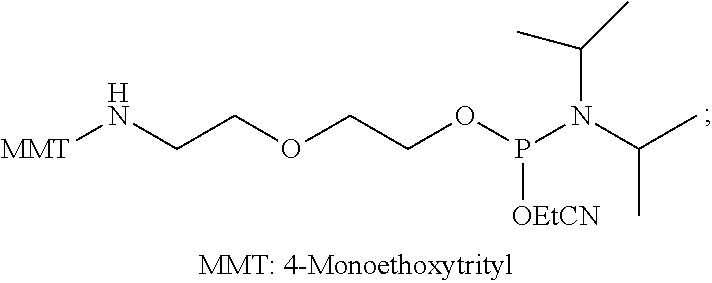
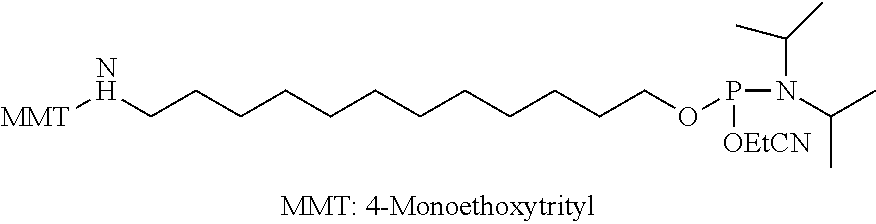

D00001

D00002

D00003
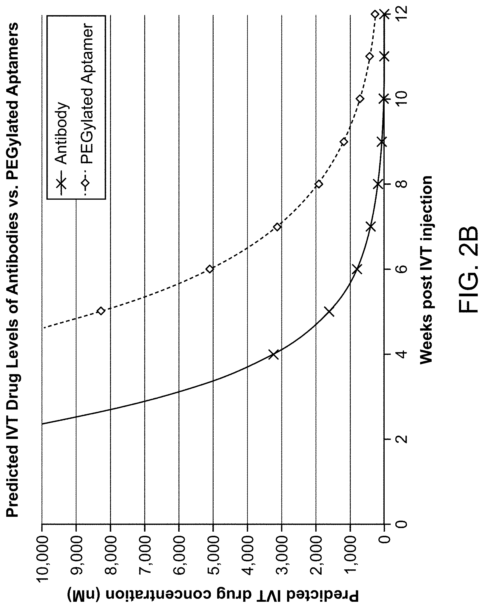
D00004

D00005

D00006
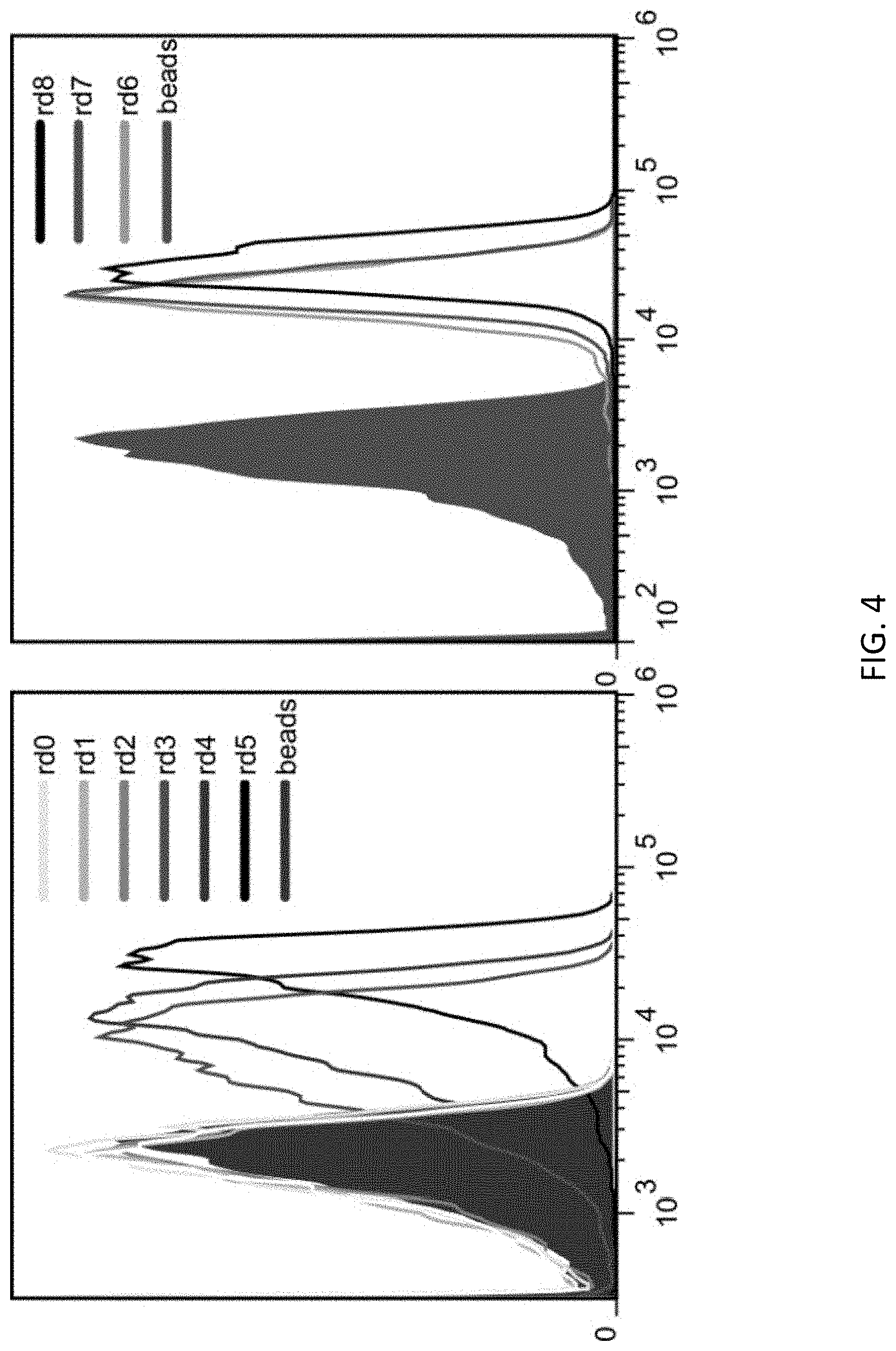
D00007

D00008

D00009

D00010

D00011

D00012

D00013

D00014
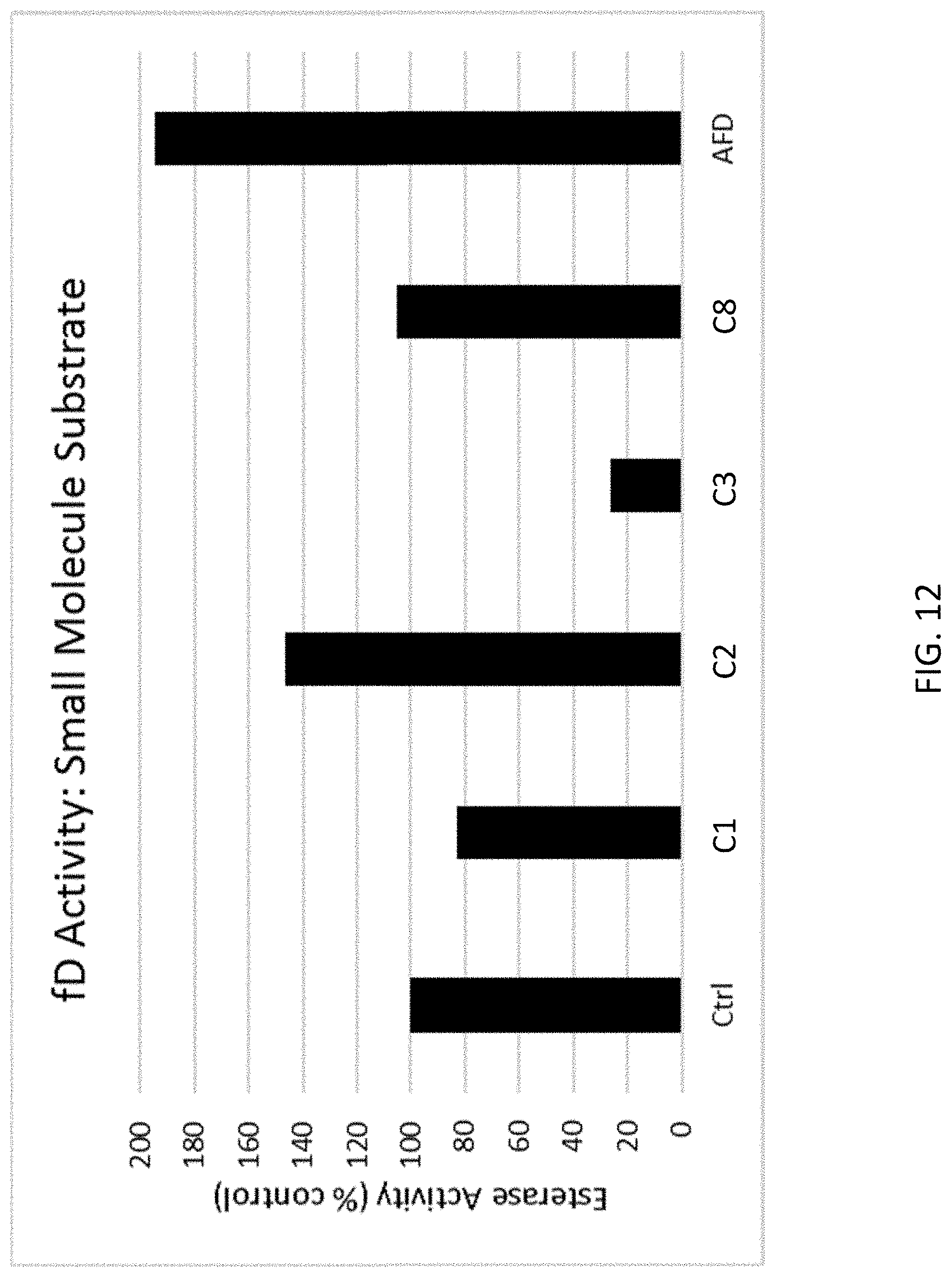
D00015

D00016

S00001
XML
uspto.report is an independent third-party trademark research tool that is not affiliated, endorsed, or sponsored by the United States Patent and Trademark Office (USPTO) or any other governmental organization. The information provided by uspto.report is based on publicly available data at the time of writing and is intended for informational purposes only.
While we strive to provide accurate and up-to-date information, we do not guarantee the accuracy, completeness, reliability, or suitability of the information displayed on this site. The use of this site is at your own risk. Any reliance you place on such information is therefore strictly at your own risk.
All official trademark data, including owner information, should be verified by visiting the official USPTO website at www.uspto.gov. This site is not intended to replace professional legal advice and should not be used as a substitute for consulting with a legal professional who is knowledgeable about trademark law.