Antigen Discovery for T Cell Receptors Isolated from Patient Tumors Recognizing Wild-Type Antigens and Potent Peptide Mimotopes
GEE; MARVIN ; et al.
U.S. patent application number 16/983937 was filed with the patent office on 2020-12-17 for antigen discovery for t cell receptors isolated from patient tumors recognizing wild-type antigens and potent peptide mimotopes. The applicant listed for this patent is THE BOARD OF TRUSTEES OF THE LELAND STANFORD JUNIOR UNIVERSITY. Invention is credited to MARK M. DAVIS, KENAN CHRISTOPHER GARCIA, MARVIN GEE, ARNOLD HAN.
| Application Number | 20200392201 16/983937 |
| Document ID | / |
| Family ID | 1000005059445 |
| Filed Date | 2020-12-17 |


View All Diagrams
| United States Patent Application | 20200392201 |
| Kind Code | A1 |
| GEE; MARVIN ; et al. | December 17, 2020 |
Antigen Discovery for T Cell Receptors Isolated from Patient Tumors Recognizing Wild-Type Antigens and Potent Peptide Mimotopes
Abstract
Compositions and methods are provided for peptide sequences that are ligands for a T cell receptor (TCR) of interest, in a given MHC context.
| Inventors: | GEE; MARVIN; (PALO ALTO, CA) ; DAVIS; MARK M.; (ATHERTON, CA) ; HAN; ARNOLD; (LOS ALTOS HILLS, CA) ; GARCIA; KENAN CHRISTOPHER; (MENLO PARK, CA) | ||||||||||
| Applicant: |
|
||||||||||
|---|---|---|---|---|---|---|---|---|---|---|---|
| Family ID: | 1000005059445 | ||||||||||
| Appl. No.: | 16/983937 | ||||||||||
| Filed: | August 3, 2020 |
Related U.S. Patent Documents
| Application Number | Filing Date | Patent Number | ||
|---|---|---|---|---|
| 16492898 | Sep 10, 2019 | |||
| PCT/US2018/023569 | Mar 21, 2018 | |||
| 16983937 | ||||
| 62476575 | Mar 24, 2017 | |||
| Current U.S. Class: | 1/1 |
| Current CPC Class: | C07K 2319/21 20130101; C07K 2319/50 20130101; C12N 15/86 20130101; C12N 15/905 20130101; C12N 15/85 20130101; C12N 15/63 20130101; G01N 33/57492 20130101; C12N 2310/20 20170501; A61K 39/00 20130101; C12N 15/01 20130101; G01N 2800/52 20130101; C12N 15/1086 20130101; C07K 14/70539 20130101; C07K 14/7051 20130101; A61K 2039/5158 20130101; G01N 33/505 20130101; C40B 50/10 20130101 |
| International Class: | C07K 14/725 20060101 C07K014/725; A61K 39/00 20060101 A61K039/00; C07K 14/74 20060101 C07K014/74; G01N 33/50 20060101 G01N033/50; G01N 33/574 20060101 G01N033/574 |
Claims
1.-20. (canceled)
21. A method of creating a cell library of candidate antigens of a T-cell receptor (TCR), the method comprising: providing a population of cells; introducing into the cells nucleic acids and a CRISPR system to create polypeptides comprising the candidate antigens, wherein the polypeptides are configured to be displayed on a surface of the cells; and allowing the cells to express and display the candidate antigens on the surface of the cells.
22. The method of claim 21, wherein the cells are yeast cells.
23. The method of claim 21, wherein the polypeptides further comprise a tag.
24. The method of claim 21, wherein the cells co-express the candidate antigens and MHC proteins, or portions thereof.
25. The method of claim 24, wherein the cells co-express the candidate antigens and binding domains of the MHC proteins.
26. The method of claim 25, wherein the binding domains comprise .alpha.1 and .alpha.2 domains of a Class I MHC protein and a .beta.2 microglobulin.
27. The method of claim 24, wherein the MHC proteins, or portions thereof, are complexed to the candidate antigens.
28. The method of claim 23, wherein the tag is a barcode, and the method further comprises selecting a subset of the cells using the barcode.
29. The method of claim 21, further comprising monitoring the cell library by detecting the tag.
30. The method of claim 21, further comprising screening the cells displaying the candidate antigens and identifying candidate antigens that bind to the TCR.
31. The method of claim 30, wherein the screening comprises combining a multimerized TCR with the cell library expressing the candidate antigens, and selecting cells that bind to the multimerized TCR.
32. The method of claim 31, further comprising isolating candidate antigens displayed on the cells that bind to the multimerized TCR.
33. The method of claim 21, wherein one or more of the candidate antigens bind to an orphan TCR.
34. The method of claim 21, wherein one or more of the candidate antigens are unknown antigens of the TCR.
35. The method of claim 21, wherein the cell library comprises at least 10.sup.8 different single chain polypeptides each comprising a candidate antigen and a binding domain of a MHC protein.
36. The method of claim 35, wherein the MHC protein is an allele of HLA-A2.
37. The method of claim 36, wherein the HLA-A2 allele comprises a Y84A amino acid substitution.
38. The method of claim 21, wherein the cell library is a multiplexed cell library.
39. The method of claim 21, wherein: the cells are yeast cells; the cells co-express the candidate antigens and binding domains of the MHC proteins, wherein the binding domains comprise .alpha.1 and .alpha.2 domains of a Class I MHC protein and a .beta.2 microglobulin; and wherein the binding domains are complexed to the candidate antigens.
40. The method of claim 39, wherein the cell library comprises at least 10.sup.8 different single chain polypeptides.
Description
CROSS REFERENCE
[0001] This application is a continuation and claims benefit of 371 application Ser. No. 16/492,898, filed Sep. 10, 2019, which claims benefit of PCT Application No. PCT/US2018/023569, filed Mar. 21, 2018, which claims benefit of U.S. Provisional Patent Application No. 62/476,575, filed Mar. 24, 2017, which applications are incorporated herein by reference in their entireties.
BACKGROUND
[0002] T cells are integral to the adaptive immune system and provide protection against pathogens and cancer. They function through extracellular recognition by the TCR, which is specific for short peptides presented on the human leukocyte antigen (HLA) on cells (Bimbaum et al., (2014) Cell 157, 1073-1087). The diversities inherent to the TCR, peptide, and HLA molecules make identifying the specificity of any one TCR an extremely complex problem. While our ability to characterize T cells and sequence their TCRs has recently improved considerably (Han et al., (2014) Nat Biotechnol 32, 684-692; Stubbington et al., (2016) Nat Methods 13, 329-332), the ability to determine and study the antigen specificities of T cells has not similarly advanced.
[0003] Each human individual has 10.sup.12 T cells in their body with 10.sup.7 to 10.sup.8 unique T cell receptors. Each T cell expresses a unique T cell receptor (TCR), selected for the ability to bind to major histocompatibility complex (MHC) molecules presenting peptides. TCR recognition of peptide-MHC (pMHC) drives T cell development, survival, and effector functions. Even though TCR ligands are relatively low affinity (1-100 .mu.M), the TCRs are remarkably sensitive, requiring as few as 10 agonist peptides to fully activate a T cell. After recognition, a signaling cascade allows T cells to carry out their immune functions.
[0004] Extensive structural studies of TCR recognition of pMHC show the vast majority of studied TCR-pMHC complexes share a consistent binding orientation, driven by conserved contacts between the tops of the MHC helices and the germline-encoded TCR CDR1 and CDR2 loops (see Garcia and Adams (2005) Cell 122, 333-336; Garcia et al. (2009) Nat Immunol 10, 143-147; and Rudolph et al. (2006) Annual Review of Immunology 24, 419-466). These conserved contacts have likely coevolved throughout the development of the adaptive immune system and serve as the basis of MHC restriction of the as TCR repertoire (Scott-Browne et al., 2011). Alteration to the typical TCR-pMHC interaction has been shown to correlate with abrogated signaling and, when present in development, skewed TCR repertoires (Adams et al. (2011) Immunity 35(5):681-93; Birnbaum et al. (2012) Immunol. Rev. 250(1):82-101).
[0005] An additional important feature of the TCR is the ability to balance cross-reactivity with specificity. Since the number of T cells that would be necessary to uniquely recognize every possible pMHC combination is extremely high, and since there are few if any `holes` characterized in the TCR repertoire, it has been posited that a large degree of TCR cross-reactivity is a requirement of functional antigen recognition. How the T cell repertoire can simultaneously be MHC restricted, cross-reactive enough to ensure all potential antigenic challenges can be met, yet still specific enough to avoid aberrant autoimmunity, has remained an open and pressing question in immunology.
[0006] There have been a number of strategies used to determine the specificity of orphan TCRs (Bimbaum et al., (2012) Immunol Rev 250, 82-101). Mass spectrometry can provide an unbiased method of antigen isolation, but is restricted to experiments requiring large cell numbers, typically 10.sup.7 to 10.sup.9, and the targets must still be presented by the correct HLA. Traditionally, most studies of T cell antigen specificities have involved testing candidate antigens empirically. For example, studies of anti-tumor T cell specificities have correctly postulated that there are productive T cell responses towards neo-antigens. Such studies involve sequencing of tumors to identify mutations, using epitope prediction algorithms to predict immunogenic mutant peptides, and testing for T cell responses directed at these mutant peptides (Kreiter et al., (2015) Nature 520, 692-696; Rajasagi et al., (2014) Blood 124, 453-462; Tran et al., (2014) Science 344, 641-645). Other strategies query established T cell specificities in patients by using pHLA multimers (Bentzen et al., (2016) Nat Biotechnol 34, 1037-1045; Newell et al., (2013) Nat Biotechnol 31, 623-629).
[0007] High-throughput and sensitive approaches to determining the specificity of `orphan` TCRs (i.e. TCRs of unknown antigen specificity) that could help uncover potential targets for cancer immunotherapy, autoimmunity, and infection and provide mechanistic insight into disease pathogenesis are of great interest.
SUMMARY
[0008] Compositions are provided for ligands for a T cell receptor (TCR) of interest in a defined MHC context. The composition may comprise or consist of a defined peptide, or may comprise or consist of a polynucleotide encoding such a peptide. Such peptides may be fragments of naturally occurring antigenic proteins; may be fragments of neoantigenic proteins that are the subject of somatic mutation during tumorigenesis, or may be a synthetically generated mimic of an antigenic protein. The synthetic peptides can act as highly potent agonists of T cell receptors. In some embodiments a peptide, or encoding sequence, is selected from sequences provided herein, including without limitation any one or a combination of the peptide sequences set forth in SEQ ID NO:1-257. A peptide may be provided as short antigenic sequence active in stimulating T cells; or may be provided in the form of the larger protein, e.g. an intact domain, a soluble protein portion, a complete protein, etc. In some embodiments, peptide antigens are identified that are shared between patients and provide a means for broadly applicable therapy. In other embodiments identification of antigens provides for a personalized medicine approach.
[0009] Identification of T cell receptors and cognate antigens provides targets for immunotherapy, including screening of patient T cells for responsiveness, vaccination with peptides or nucleic acids encoding such peptides, cell-based therapies, protein-based therapies, etc. The peptides and methods disclosed herein are useful in classifying TCRs based on peptide antigen specificities, which allows the identification of clinical candidate TCRs that recognize shared antigens across patients.
[0010] In some embodiments, methods are provided for vaccination against cancer, for example colorectal cancer, the method comprising administering an effective dose of a vaccine composition, which composition may comprise a peptide identified herein; a combination of peptides, e.g. 2, 3, 4, 5, 6, 7, 8, 9, 10 or more distinct peptides; a complex of a peptide and at least a portion of an MHC protein; an autologous or allogeneic T cell that has been stimulated to respond to an antigenic peptide identified herein; a nucleic acid encoding an antigenic peptide identified herein; and optionally a pharmaceutically acceptable excipient, which may comprise a vaccine adjuvant. The peptide vaccination strategy may be used to initially prime an immune response, e.g. with a synthetic peptide provided herein, followed by a boost with the corresponding known wildtype antigen or wildtype whole protein.
[0011] The defined peptides are identified by screening peptide-MHC libraries by yeast-display was used to identify the recognition landscape of individual T cell receptors. The screening method may be utilized in a multiplex method to screen a plurality of peptide libraries simultaneously, e.g. screening 2, 3, 4 or more libraries simultaneously. Multiplexing allows improved efficiency of antigen discovery. Each library may comprise a unique epitope tag, e.g. an epitope targetable by an antibody, to allow identification; may comprise DNA barcodes; protein barcodes; etc. Each library utilizing the epitope tags were generated separately and diversities calculated, e.g. based on colony counts from limiting dilution of the initial libraries on growth plates. Pooling T cell receptors for library selection can further multiplex the selection, e.g. multiplexing of peptide sequence, peptide lengths, collections of different MHC or HLA alleles, etc. For selections, each barcode, epitope tag, etc. may be monitored via anti-epitope tag staining to detect the level of peptide-specific enrichment. statistical algorithms and machine-learning algorithms may be used for identification.
[0012] In some embodiments sequences of T cell receptors responsive to cancer antigens are provided. T cell receptor sequences may include, without limitation, the proteins having an alpha chain with sequence set forth in SEQ ID NO:258, optionally combined with a beta chain sequence of SEQ ID NO:259 or SEQ ID NO:260. The binding regions (CDR) sequences of these T cell receptors may be grafted onto an antibody framework to provide a TCR-like antibody. Because T cell receptors are adaptable and often unique from patient-to-patient, the individual T cell receptor sequences may differ between patients. Despite these differences, different TCR can still recognize the same target. Thus, different T cell receptors may have slight sequence variations from these T cell receptors that can bind the same target. Additionally, T cell receptors may be modified to introduce amino acid substitutions that will allow binding to the same antigen. Such cases include affinity maturation of the T cell receptor for the specific target or receptor modification to improve the specificity of the T cell receptor for its target. The recognition portion of a T cell receptor can be grafted onto other protein scaffolds to be used as a therapeutic reagent. Because T cell receptors are somewhat cross-reactive, the list of synthetic peptides is not exhaustive. Slight modifications to peptide sequences can still result in T cell stimulation.
[0013] In some embodiments the T cells from which TCR sequences for screening are obtained are isolated from tumor sites, and may include without limitation tumor infiltrating T cells (TILs). In other embodiments the T cells are obtained from an individual responsive to an infection, e.g. bacterial, viral, protozoan, etc. infection. In other embodiments the T cells are obtained from a graft recipient, and may be isolated from the site of a graft.
BRIEF DESCRIPTION OF THE DRAWINGS
[0014] The invention is best understood from the following detailed description when read in conjunction with the accompanying drawings. The patent or application file contains at least one drawing executed in color. It is emphasized that, according to common practice, the various features of the drawings are not to-scale. On the contrary, the dimensions of the various features are arbitrarily expanded or reduced for clarity. Included in the drawings are the following figures.
[0015] FIGS. 1A-1F. Design of the peptide-HLA-A*02:01 yeast-display library. FIG. 1A: Methodology for selecting a yeast-display library of pHLA. Each yeast display a unique peptide that is genetically encoded. A typical library contains .about.10.sup.8 unique peptides, which is selected by a TCR of interest. Yeast are enriched in an affinity-based selection using bead-multimerized TCR and grown for iterative rounds of selection. Peptides are successively enriched and all yeast DNA is deep-sequenced. These synthetic peptide sequences are used to generate a model to make predictions for TCR ligands derived from the human proteome and/or patient-specific exome. FIG. 1B: The goal of the study is to use the yeast-display selection to de-orphanize a TCR of unknown antigen specificity. The peptides selected by a TCR from the yeast-display selection generates a recognition landscape for a particular TCR, which is then used to make predictions of antigen specificity for orphan TCRs. Predicted targets can be validated in a T cell stimulation assay. FIG. 1C: The construct utilizes a single-chain design to display the pHLA-A*02:01 complex tethered to an epitope tag and Aga2p, which binds to the native Aga1 protein on yeast. Each component is connected covalently by a Gly-Ser linker. The epitope tag is introduced to monitor expression of the library. FIG. 1D: The MART-1/HLA-A*02 complex structure (PDB 4L3E) highlighting the two peptide anchors with orange arrows. These peptide positions at P2 and P.OMEGA. of the peptide allow for peptide binding to HLA-A*02. FIG. 1E: An example 8mer peptide library shows the anchor preferences for the HLA-A*02:01 library and the remaining positions that are randomized to any of the twenty amino acids (X=twenty amino acids and stop codon). Nucleotide abbreviations for codon usage are listed according to the IUPAC nucleotide code. FIG. 1F: A multi-length library designed to capture the most common length peptides presented by HLA-A*02:01. Each peptide length is placed in a construct using a unique epitope tag for selection monitoring. The libraries have theoretical nucleotide diversities dictated by the peptide length and library composition. The functional diversity represents the true capacity of the physical libraries based on yeast colony counting after limiting dilution of the library.
[0016] FIGS. 2A-2F. Validation of the HLA-A*02:01 library with the DMF5 TCR. FIG. 2A: The DMF5 TCR stains yeast displaying the MART-1 peptide (ELAGIGILTV) (SEQ ID NO: 264) in complex with HLA-A*02:01 on the surface of yeast. Streptavidin-647 (SA-647) was used to tetramerize and fluorescently label the DMF5 TCR. FIG. 2B: Enrichment of the 10mer length HLA-A*02:01 yeast-display library by the DMF5 TCR as measured by anti-HA epitope tag staining by flow cytometry. Three of four rounds of selection shown. FIG. 2C: Highly-enriched peptides sequenced from the 10mer selection by the DMF5 TCR are stained by the DMF5 TCR tetramer and measured by flow cytometry. ((C) sequences from left to right: SEQ ID NOs: 264, 324, 286, 323, 283, 285). FIG. 2D: The fraction of total sequencing read counts of the top 10 peptides according to deep sequencing of round 3 of the 10mer HLA-A*02:01 library selections by the DMF5 TCR. ((D) sequences from top to bottom: SEQ ID NOs: 287, 326, 325, 324, 286, 323, 285, 322, 284, 283). FIG. 2E: Unique peptides from round 3 of selection fall into two major clusters that appear similar to the wildtype MART-1 peptide sequence (SEQ ID NO: 267). Clusters are determined by first calculating reverse hamming distance between all peptides present in round 3 of the selection and then clustered by score. The MART-1 decamer structure (PDB: 4L3E) is aligned to the selected peptides. FIG. 2F: A substitution matrix (2014PWM) using cluster 1 peptides predicts the MART-1 peptide as the most probable peptide to bind the DMF5 TCR among eight other predicted peptides. ((F) sequences from top to bottom: 321, 320, 319, 318, 317, 316, 315, 314, 267)
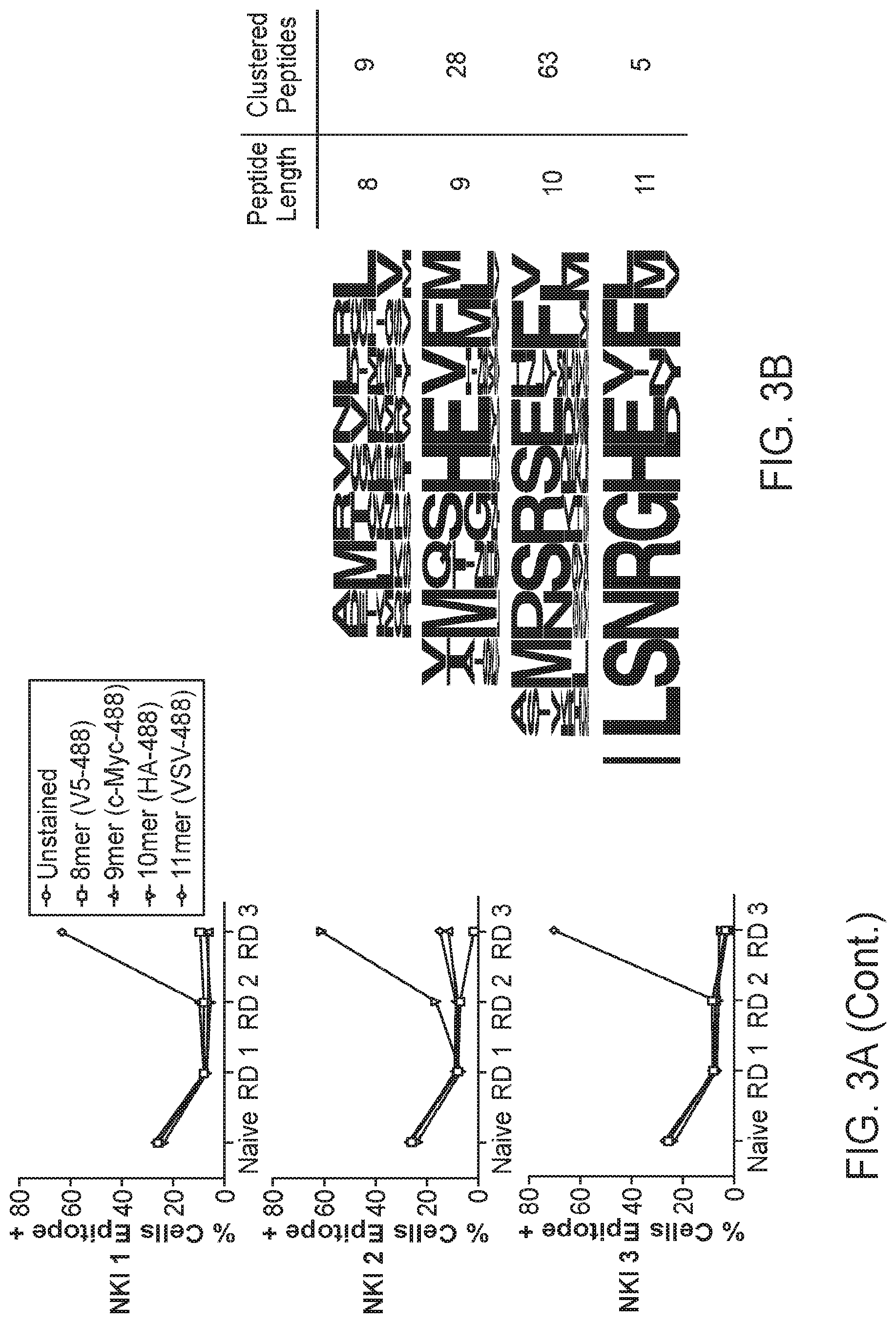
[0017] FIGS. 3A-3E. Blinded validation of the HLA-A*02:01 library by neoantigen-specific TCRs. FIG. 3A: Three TCRs of blinded specificity separately enrich the HLA-A*02:01 library for a specific peptide length according to epitope tag staining over the rounds of selection. The left panels indicate tetramer and epitope staining after all 4 rounds of selection have completed and the right panels indicate epitope staining through the course of selections. FIG. 3B: Unique peptides selected by NKI 2 in round 3 of the selection are parsed by peptide length and clustered by reverse hamming distance. The number of peptides identified in the cluster are shown on the right along with the respective peptide lengths. FIG. 3C: The maximum reverse hamming distance computed between every 10mer of the selected peptides by NKI 2 at round 3 and each 10mer neoantigen peptide from the list of 127 total neoantigens. ((C) sequences from top to bottom: SEQ ID NOs: 501, 502, 620, 503-519. FIG. 3D: Two peptides Lib-1 (SEQ ID NO: 434) and Lib-2 (SEQ ID NO: 269) from the selected library closely resemble the 10mer neoantigen peptide ALDPHSGHFV (SEQ ID NO: 265) derived from CDK4. Identical amino acids with the neoantigen are colored in red. FIG. 3E: The top 5 peptides of length 10 selected by the NKI 2 TCR were used to stimulate peripheral blood lymphocytes transduced to express TCRs NKI1 or NKI2, which are both specific for the CDK4 neoantigen ALDPHSGHFV (SEQ ID NO: 265). Transduced lymphocytes were mixed 1:1 with JY cells pulsed with peptide, control peptide, or no peptide, and IFN.gamma. production as measured by intracellular antibody staining was assessed using flow cytometry. ((E) sequences from top to bottom: 1) SEQ ID NO: 269, 2) SEQ ID NO: 427, 3) SEQ ID NO: 423, 4) SEQ ID NO: 420, 5) SEQ ID NO: 417).
[0018] FIGS. 4A-4D. Profiling TCRs identified in two HLA-A*02 patients with colorectal adenocarcinoma. FIG. 4A: Study design to de-orphanize patient-derived TCRs on the HLA-A*02:01 library with summarized results. FIG. 4B: Bar graph of abundances of unique paired as TCR sequences from TILs. *=TCRs that enriched peptides from the library. FIG. 4C: Venn diagrams representing the overlap of individual unique CDR3.alpha. or CDR3.beta. chain sequences between tumor and healthy tissues for each patient. The number indicates the amount of CDR3 sequences in the nearest section of the Venn diagram. FIG. 4D: Heatmaps identifying the binary measurement of transcription factors using sequencing of amplified and barcoded transcripts. The alternating black and white panels indicate boundaries of single T cell clones with the same receptor sequences, with the most abundance clones beginning from the left most side. The left panel identifies those T cells with TCRs chosen from Patient A to be screened and green denoting the presence of transcript. The right panel identifies those T cells with TCRs chosen from Patient B to be screened and blue denoting the presence of transcript. White indicates lack of transcript detected. TCRs 1A, 2A, 3B, and 4B are labeled.
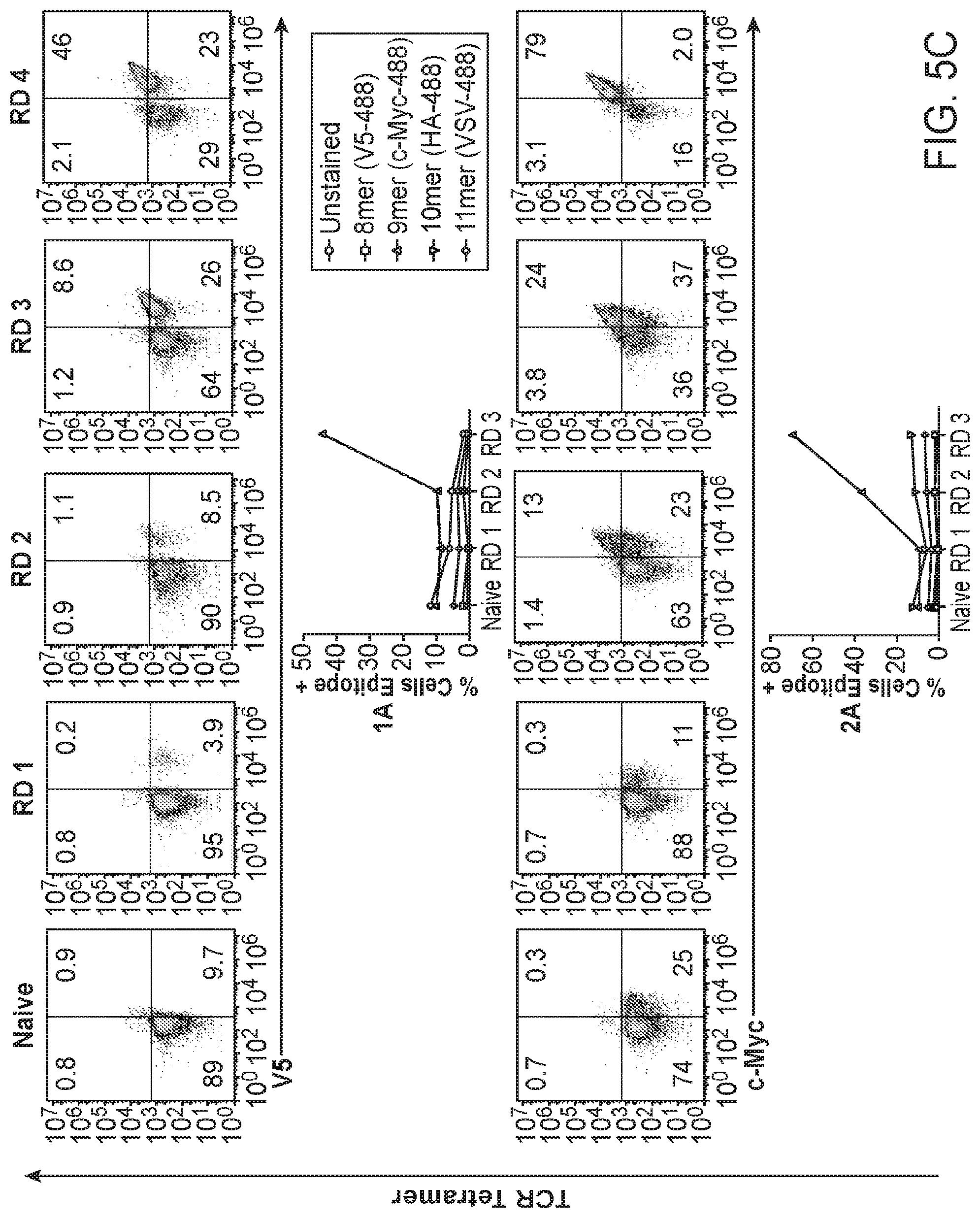
[0019] FIGS. 5A-5C. Four TIL-derived TCRs enrich the HLA-A*02:01 library for peptides. FIG. 5A: TCR sequences of the four orphan TCRs that selected peptides from the HLA-A*02:01 library. The TCR gene segments variable and joining are shown along with the corresponding CDR3 sequence. The abundance represents the amount of times a single cell was found to have the exact TCR sequence in tumor/healthy tissue. ((A)) sequences: 1A CDR3.alpha.: (SEQ ID NO: 472), 2A CDR3.alpha.: (SEQ ID NO: 261), 3B CDR3.alpha.: (SEQ ID NO: 261), 4B CDR3.alpha.: (SEQ ID NO: 495), 1A CDR3.beta.: (SEQ ID NO: 463), 2A CDR3.beta.: (SEQ ID NO: 262), 3B CDR3.beta.: (SEQ ID NO: 263), 4B CDR3.beta.: (SEQ ID NO: 484)). FIG. 5B: Nucleotide sequences of the two sequence-similar TCRs isolated from patients A and B. Non-encoded nucleotides are highlighted in red. ((B) amino acid sequences: CDR3.alpha. 2A: (SEQ ID NO: 261), CDR3.alpha. 3B: (SEQ ID NO: 261), CDR3.beta. 2A: (SEQ ID NO: 262), CDR3.beta. 3B: (SEQ ID NO: 263)); nucleotide sequences: CDR3.alpha. 2A nucleotide sequence: (SEQ ID NO: 536), CDR3.alpha. 3B nucleotide sequence: (SEQ ID NO: 537), CDR3.beta. 2A nucleotide sequence: (SEQ ID NO: 538), CDR3.beta. 38 nucleotide sequence (SEQ ID NO: 539). FIG. 5C: HLA enrichment and tetramer staining per round of selection by the four orphan TCRs as measured by flow cytometry. The left panels indicate tetramer and epitope staining after all 4 rounds of selection have completed and the right panels indicate epitope staining through the course of selections.
[0020] FIGS. 6A-6C. Deep-sequencing results of the yeast selections by the four TIL TCRs. FIG. 6A: Word logos display the unique round 3 selected peptides for each TCR not accounting for deep sequencing read count abundance. The size of the amino acid letter represents its proportional abundance at the given position among the unique peptides. FIG. 6B: Heatmap plots showing the amino acid composition per position of the peptide accounting for peptide enrichment at round 3 of the selection. Darker colors indicate greater abundance of a given amino acid at a given position. Anchor residues are outlined in black. FIG. 6C: TCRs 2A and 3B select an overlapping set of 11 peptides in round 3 of the selection shown as a fraction of total reads in round 3. ((C) sequences from top to bottom: SEQ ID NOs: 95, 249, 54, 195, 42, 191, 196, 198, 200, 201, 4).
[0021] FIGS. 7A-7H. Activation of TIL TCRs with predicted human targets and peptide mimotopes. TCRs are retrovirally infected into CD8.sup.+ SKW-3 cells and sorted for stable TCR (IP26) and CD3 (UCHT1) co-expression. T2 antigen-presenting cells are pulsed with 100 .mu.M peptide for 3 hours, co-incubated with the T cell lines for 18 hours and analyzed for CD69 expression by flow cytometry. FIG. 7A: TCR1A, FIG. 7C: TCR2A, FIG. 7E: TCR3B, and FIG. 7G: TCR4B are tested for CD69 activation by peptide stimulation in technical triplicate with standard deviation shown. A representative experiment is shown from biological triplicate. ((A) sequences from left to right: SEQ ID NOs: 540-555; (C) SEQ ID NOs: 556-574; (E) SEQ ID NOs: 556-574; (G) SEQ ID NOs: 596-619). FIG. 7B (TCR1A), FIG. 7D (TCR2A), FIG. 7F (TCR3B), FIG. 7H (TCR4B): A dose-response curve for each stimulatory peptide is shown on the right plotted with means of biological triplicates with standard error of the mean. For both experiments, p-values are calculated using ordinary one-way ANOVA. For TCRs 2A and 3B, 17 non-stimulating peptides are removed for simplicity. ((B) sequences from top to bottom: SEQ ID NOs: 540-543; (D) sequences from top to bottom: 556-558, 560, 562-567; (F) sequences from top to bottom: 41, 42, 193, 194, 195, 257; (H) sequences from top to bottom: 596-602, 604, 608, 610, 613, 615).
[0022] FIGS. 8A-8C. Validation of the HLA-A2*01 library with the DMF5 TCR. FIG. 8A: MA2.1 antibody staining for correctly folded HLA-A*02:01 complex with DMF5 TCR wildtype peptide or peptide mimotopes. Histograms show staining by MA2.1 antibody followed by secondary antibody. ((A) sequences from left to right: SEQ ID NOs: 264, 324, 286, 323, 283, 285). FIG. 8B: The scores of predicted human peptides using the 2014PWM algorithm on cluster 2 of the round 3 sequences for the DMF5 TCR 10mer selection. FIG. 8C: The scores of the top 10 peptides identified in FIG. 8B. ((C) sequences from top to bottom: SEQ ID NO: 364, 363, 362, 361, 360, 359, 358, 357, 356, 355).
[0023] FIGS. 9A-9E. Patient tissue immunohistochemistry and TCR repertoire sequencing and phenotyping. FIG. 9A: Patient immunohistochemistry using H&E staining, anti-CD4/hematoxylin or anti-CD8/hematoxylin. All representative images are taken using 300.times. magnification. FIG. 9B: Patient CDR3 length as measured from the Cys to Phe. FIG. 9C: Patient distribution of TCR variable a genes in healthy and tumor tissue. FIG. 9D: Patient distribution of TCR variable P genes in healthy and tumor tissue. FIG. 9E: t-SNE plots of Patient B T cells showing transcriptional profiling by transcript sequencing (left) and cell surface markers by flow cytometry (right). The presence of transcripts is binary based off of deep-sequencing reads (1=yes, 0=no) and intensity relates to MFI of cell surface marker.
[0024] FIGS. 10A-10D. Design of the Machine-Learning Algorithm 2017DL to Predict Human Peptide Specificities. FIG. 10A: Schematic showing the process to take data from the yeast-display library selections to train a machine learning model, which scores peptides derived from proteins from the Uniprot database or patient-specific exomes. The model is generated from yeast-display selection data utilizing the deep-sequencing round counts per peptide and the composition of the peptide. An exponential curve is fit to each peptide to capture the enrichment over the rounds of selection using a fitness function. FIG. 10B: Fitness function to fit an exponential curve to the deep sequencing round counts for peptides selected by a TCR. FIG. 10C: Matrix representation of an example peptide, in which each amino acid is represented as a one-hot vector. FIG. 10D: The architecture of the machine-learning algorithm utilizing a two-layer convolutional neural network. The input consists of peptide sequences represented as a vector of one-hot vectors and the fitness scores of the peptides determined from the fitness function. The output is the fitness score.
[0025] FIGS. 11A-11H. Activation of SKW-3 cells according to CD69 Median MFI and TCR tetramer staining of yeast expressing predicted peptide targets. Data analyzed from FIG. 7, but using mean fluorescence intensity of CD69 expression instead of percent cells positive for CD69 expression for FIG. 11A, FIG. 11B, FIG. 11C and FIG. 11D. SKW-3 T cells with TCRs (FIG. 11A) 1A, (FIG. 11B) 2A, (FIG. 11C) 3B, or (FIG. 11D) 4B were co-cultured with peptide-pulsed T2 antigen-presenting cells as in FIG. 7. The mean fluorescence intensity was measured from anti-CD69 staining of CD3-gated SKW-3 cells. in technical triplicate with mean values and standard deviation shown. A representative experiment from biological triplicate is shown. P-values were measured using ordinary one-way ANOVA. Yeast expressing single-chain trimers of the library peptides and predicted target peptides for TCRs (FIG. 11E) 1A, (FIG. 11F) 2A, (FIG. 11G), 3B, and (FIG. 11H) 48 stained with 400 nM TCR tetramers. Tetramer negative populations are stained with streptavidin-647 only. All yeast are gated on epitope tag positive yeast. ((A) sequences from top to bottom: SEQ ID NOs: 540-542).
[0026] FIGS. 12A-12E. U2AF2 quantitative RNA expression and affinity measurements for U2AF2 peptide. FIG. 12A: Quantitative PCR expression of the U2AF2 transcript expression of tumor over healthy tissue in patients A and B using 18S as the housekeeping gene. Samples are done in technical quadruplicate with standard deviation shown. FIG. 12B: Log base 2 quantitative PCR expression of U2AF2 RNA in various human-derived tumors compared to U2AF2 RNA expression in Patient A healthy tissue using the 18S as the housekeeping gene. Samples are done in technical quadruplicate with standard deviation shown. Cell lines shown are listed in the methods section in the appropriate order. FIG. 12C: Log base 2 quantitative PCR expression of U2AF2 RNA in various human-derived tumors compared to U2AF2 RNA expression in Patient B healthy tissue using the 18S as the housekeeping gene. Samples are done in technical quadruplicate with standard deviation shown. Cell lines shown are listed in the methods section in the appropriate order. FIG. 12D: Surface plasmon resonance traces of increasing concentrations of TCR 2A flown over a chip coated with MMDFFNAQM-HLA-A*02:01 (SEQ ID NO: 266) with a range of 93.6 .mu.M to 0.365 .mu.M using 2-fold dilutions. The peaks prior to and after association of the TCR to the peptide-HLA-A*02 generated from flow cell subtraction are removed for simplicity. Only the colored curves labeled with concentrations are used to calculate the K.sub.d. FIG. 12E: Curve-fitting to data points generated at various concentrations of TCR labeled in FIG. 12D.
DETAILED DESCRIPTION OF THE EMBODIMENTS
[0027] Before the subject invention is described further, it is to be understood that the invention is not limited to the particular embodiments of the invention described below, as variations of the particular embodiments may be made and still fall within the scope of the appended claims. It is also to be understood that the terminology employed is for the purpose of describing particular embodiments, and is not intended to be limiting. In this specification and the appended claims, the singular forms "a," "an" and "the" include plural reference unless the context clearly dictates otherwise.
[0028] Where a range of values is provided, it is understood that each intervening value, to the tenth of the unit of the lower limit unless the context clearly dictates otherwise, between the upper and lower limit of that range, and any other stated or intervening value in that stated range, is encompassed within the invention. The upper and lower limits of these smaller ranges may independently be included in the smaller ranges, and are also encompassed within the invention, subject to any specifically excluded limit in the stated range. Where the stated range includes one or both of the limits, ranges excluding either or both of those included limits are also included in the invention.
[0029] Unless defined otherwise, all technical and scientific terms used herein have the same meaning as commonly understood to one of ordinary skill in the art to which this invention belongs. Although any methods, devices and materials similar or equivalent to those described herein can be used in the practice or testing of the invention, illustrative methods, devices and materials are now described.
[0030] All publications mentioned herein are incorporated herein by reference for the purpose of describing and disclosing the subject components of the invention that are described in the publications, which components might be used in connection with the presently described invention.
[0031] The present invention has been described in terms of particular embodiments found or proposed by the present inventor to comprise preferred modes for the practice of the invention. It will be appreciated by those of skill in the art that, in light of the present disclosure, numerous modifications and changes can be made in the particular embodiments exemplified without departing from the intended scope of the invention. For example, due to codon redundancy, changes can be made in the underlying DNA sequence without affecting the protein sequence. Moreover, due to biological functional equivalency considerations, changes can be made in protein structure without affecting the biological action in kind or amount. All such modifications are intended to be included within the scope of the appended claims.
[0032] Screening methods. Antigenic sequences were discovered by generating a library of single chain polypeptides that comprise: the binding domains of a major histocompatibility complex protein; and diverse peptide ligands. The library was introduced into a suitable host cell that expresses the encoded polypeptide, which host cells include, without limitation, yeast cells. A TCR of interest is multimerized to enhance binding, and used to select for host cells expressing those single chain polypeptides that bind to the T cell receptor. Iterative rounds of selection are performed, i.e. the cells that are selected in the first round provide the starting population for the second round, etc. until the selected population has a signal above background, usually at least three and more usually at least four rounds of selection are performed. Polynucleotides encoding the final selected population from the library of single chain polypeptides are subjected to high throughput sequencing. The selected set of peptide ligands exhibit a restricted choice of amino acids at residues, e.g. the residues that contact the TCR, which information can be input into an algorithm that can be used to analyze public databases for all peptides that meet the criteria for binding, and which provides a set of peptides that meet these criteria.
[0033] The peptide ligand is from about 8 to about 20 amino acids in length, usually from about 8 to about 18 amino acids, from about 8 to about 16 amino acids, from about 8 to about 14 amino acids, from about 8 to about 12 amino acids, from about 10 to about 14 amino acids, from about 10 to about 12 amino acids. It will be appreciated that a fully random library would represent an extraordinary number of possible combinations. In preferred methods, the diversity is limited at the residues that anchor the peptide to the MHC binding domains, which are referred to herein as MHC anchor residues. The position of the anchor residues in the peptide are determined by the specific MHC binding domains. Class I binding domains can have anchor residues at the P2 position, and at the last contact residue. Class II binding domains have an anchor residue at P1, and depending on the allele, at one of P4, P6 or P9. For example, the anchor residues for IE.sup.k are P1 {I,L,V} and P9 {K}; the anchor residues for HLA-DR15 are P1 {I,L,V} and P4{F, Y}. Anchor residues for DR alleles are shared at P1, with allele-specific anchor residues at P4, P6, P7, and/or P9.
[0034] In some embodiments, the binding domains of a major histocompatibility complex protein are soluble domains of Class II alpha and beta chain. In some such embodiments the binding domains have been subjected to mutagenesis and selected for amino acid changes that enhance the solubility of the single chain polypeptide, without altering the peptide binding contacts. In certain specific embodiments, the binding domains are HLA-DR4.alpha. comprising the set of amino acid changes {M36L, V132M}; and HLA-DR4.beta. comprising the set of amino acid changes {H62N, D72E}. In certain specific embodiments, the binding domains are HLA-DR15.alpha. comprising the set of amino acid changes (F12S, M23K; and HLA-DR15.beta. comprising the amino acid change {P11S}. In certain specific embodiments, the binding domains are H2 IE.sup.k.alpha. comprising the set of amino acid changes {I8T, F12S, L14T, A56V} and H2 IE.sup.k.beta. comprising the set of amino acid changes {W6S, L8T, L34S}.
[0035] In some embodiments, the binding domains of a major histocompatibility complex protein comprise the alpha 1 and alpha 2 domains of a Class I MHC protein, which are provided in a single chain with .beta.2 microglobulin. In some such embodiments the Class I protein has been subjected to mutagenesis and selected for amino acid changes that enhance the solubility of the single chain polypeptide, without altering the peptide binding contacts. In certain specific embodiments, the binding domains are HLA-A2 alpha 1 and alpha 2 domains, comprising the amino acid change {Y84A}. In certain specific embodiments, the binding domains are H2-L.sup.d alpha 1 and alpha 2 domains, comprising the amino acid change {M31R}. In certain specific embodiments the binding domains are HLA-B57 alpha 1, alpha 2 and alpha 3 domains, comprising the amino acid change {Y84A}.
[0036] The sequences of peptides are determined by any convenient methods of high throughput sequencing. Sequences may be analyzed, for example by the methods disclosed in the Examples, using clustering algorithms. Peptides may be analyzed to search human protein (Uniprot) or patient-specific exomes to score peptides of fixed lengths using a sliding window. Substitution matrices are made by determining the frequency of all amino acids per position of the peptide. A cutoff of 0.1% frequency for an amino acid at a given position may be instituted to remove noise.
[0037] To determine the statistical significance of a peptide, the human proteome and exome peptide set is scored. To calculate the p-values for the exome peptide set, the percentile score is calculated in context of the human proteome scores. The uncorrected p-value is 1-percentile. The Bonferroni-corrected p-value is the uncorrected p-value multiplied by the number of peptides in the mutant set.
[0038] MHC Proteins. Major histocompatibility complex proteins (also called human leukocyte antigens, HLA, or the H2 locus in the mouse) are protein molecules expressed on the surface of cells that confer a unique antigenic identity to these cells. MHC/HLA antigens are target molecules that are recognized by T-cells and natural killer (NK) cells as being derived from the same source of hematopoietic reconstituting stem cells as the immune effector cells ("self") or as being derived from another source of hematopoietic reconstituting cells ("non-self"). Two main classes of HLA antigens are recognized: HLA class I and HLA class II.
[0039] The MHC proteins used in the libraries and methods of the invention may be from any mammalian or avian species, e.g. primate sp., particularly humans; rodents, including mice, rats and hamsters; rabbits; equines, bovines, canines, felines; etc. Of particular interest are the human HLA proteins, and the murine H-2 proteins. Included in the HLA proteins are the class II subunits HLA-DP.alpha., HLA-DP.beta., HLA-DQ.alpha., HLA-DQ.beta., HLA-DR.alpha. and HLA-DR.beta., and the class I proteins HLA-A, HLA-B, HLA-C, and .beta..sub.2-microglobulin. Included in the murine H-2 subunits are the class I H-2K, H-2D, H-2L, and the class II I-A.alpha., I-A.beta., I-E.alpha. and I-E.beta., and .beta..sub.2-microglobulin.
[0040] The MHC binding domains are typically a soluble form of the normally membrane-bound protein. The soluble form is derived from the native form by deletion of the transmembrane domain. Conveniently, the protein is truncated, removing both the cytoplasmic and transmembrane domains. In some embodiments, the binding domains of a major histocompatibility complex protein are soluble domains of Class II alpha and beta chain. In some such embodiments the binding domains have been subjected to mutagenesis and selected for amino acid changes that enhance the solubility of the single chain polypeptide, without altering the peptide binding contacts.
[0041] An "allele" is one of the different nucleic acid sequences of a gene at a particular locus on a chromosome. One or more genetic differences can constitute an allele. An important aspect of the HLA gene system is its polymorphism. Each gene, MHC class I (A, B and C) and MHC class II (DP, DQ and DR) exists in different alleles. Current nomenclature for HLA alleles are designated by numbers, as described by Marsh et al.: Nomenclature for factors of the HLA system, 2010. Tissue Andgens 75:291-455, herein specifically incorporated by reference. For HLA protein and nucleic acid sequences, see Robinson et al. (2011), The IMGT/HLA database. Nucleic Acids Research 39 Supp 1:D1171-6, herein specifically incorporated by reference.
[0042] The numbering of amino acid residues on the various MHC proteins and variants disclosed herein is made to be consistent with the full length polypeptide. Boundaries were set to either be the end of the MHC peptide binding domain (as judged by examining crystal structures) for the `mini` MHCs, e.g. as exemplified herein with I-Ek, H2-Ld, and HLA-DR15, and the end of the Beta2/Alpha2/Alpha3 domains as judged by structure and/or sequence for the `full length` MHCs, as exemplified herein with HLA-A2, -B57, and -DR4.
[0043] In some embodiments, the MHC portion of a construct is the MHC portion delineated in any of SEQ ID NO:1-6. It will be understood by one of skill in the art that the peptide and linker portions can be varied from the provided sequences.
[0044] MHC context. The function of MHC molecules is to bind peptide fragments derived from pathogens and display them on the cell surface for recognition by the appropriate T cells. Thus T cell receptor recognition can be influenced by the MHC protein that is presenting the antigen. The term MHC context refers to the recognition by a TCR of a given peptide, when it is presented by a specific MHC protein.
[0045] Class H HLA/MHC. Class II binding domains generally comprise the .alpha.1 and .alpha.2 domains for the a chain, and the .beta.1 and .beta.2 domains for the .beta. chain. Not more than about 10, usually not more than about 5, preferably none of the amino acids of the transmembrane domain will be included. The deletion will be such that it does not interfere with the ability of the .alpha.2 or .beta.2 domain to bind peptide ligands.
[0046] In some embodiments, the binding domains of a major histocompatibility complex protein are soluble domains of Class II alpha and beta chain. In some such embodiments the binding domains have been subjected to mutagenesis and selected for amino acid changes that enhance the solubility of the single chain polypeptide, without altering the peptide binding contacts.
[0047] In certain specific embodiments, the binding domains are an HLA-DR allele. The HLA-DRA protein can be selected, without limitation, from the binding domains of DRA*0101:01:01; DRA*01:01:01:02; DRA*01:01:01:03; DRA*01:01:02; DRA*01:02:01; DRA*01:02:02; and DRA*01:02:03, which may be modified to comprise the amino acid changes {M36L, V132M}; or {F12S, M23K}, depending on whether it is provided in the context of a full-length or mini-allele. The HLA-DRA binding domains can be combined with any one of the HLA-DRB binding domains.
[0048] In certain such embodiments, the HLA-DRA allele is paired with the binding domains of an HLA-DRB4 allele. The HLA-DRB4 allele can be selected from the publicly available DRB4 alleles.
[0049] In other such embodiments the HLA-DRA allele is paired with the binding domains of an HLA-DRB15 allele. The HLA-DRB15 allele can be selected from the publicly available DRB15 alleles.
[0050] In other embodiments the Class II binding domains are an H2 protein, e.g. I-A.alpha., I-A.beta., I-E.alpha. and I-E.beta.. In some such embodiments, the binding domains are H2 IE.sup.k.alpha. which may comprise the set of amino acid changes {8T, F12S, L14T, A56V}; and H2 IE.sup.k.beta. which may comprise the set of amino acid changes {W6S, L8T, L34S}.
[0051] Class I HLA/MHC. For class I proteins, the binding domains may include the .alpha.1, .alpha.2 and .alpha.3 domain of a Class I allele, including without limitation HLA-A, HLA-B, HLA-C, H-2K, H-2D, H-2L, which are combined with .beta..sub.2-microglobulin. Not more than about 10, usually not more than about 5, preferably none of the amino acids of the transmembrane domain will be included. The deletion will be such that it does not interfere with the ability of the domains to bind peptide ligands.
[0052] In certain specific embodiments, the binding domains are HLA-A2 binding domains, e.g. comprising at least the alpha 1 and alpha 2 domains of an A2 protein. A large number of alleles have been identified in HLA-A2, including without limitation HLA-A*02:01:01:01 to HLA-A*02:478, which sequences are available at, for example, Robinson et al. (2011), The IMGT/HLA database. Nucleic Acids Research 39 Suppl 1:D1171-6. Among the HLA-A2 allelic variants, HLA-A*02:01 is the most prevalent. The binding domains may comprise the amino acid change {Y84A}.
[0053] In certain specific embodiments, the binding domains are HLA-B57 binding domains, e.g. comprising at least the alpha1 and alpha 2 domains of a B57 protein. The HLA-B57 allele can be selected from the publicly available B57 alleles.
[0054] T cell receptor, refers to the antigen/MHC binding heterodimeric protein product of a vertebrate, e.g. mammalian, TCR gene complex, including the human TCR .alpha., .beta., .gamma. and .delta. chains. For example, the complete sequence of the human .beta. TCR locus has been sequenced, as published by Rowen et al. (1996) Science 272(5269):1755-1762; the human .alpha. TCR locus has been sequenced and resequenced, for example see Mackelprang et al. (2006) Hum Genet. 119(3):255-66; see a general analysis of the T-cell receptor variable gene segment families in Arden Immunogenetics. 1995; 42(6):455-500; each of which is herein specifically incorporated by reference for the sequence information provided and referenced in the publication.
[0055] The multimerized T cell receptor for selection in the methods of the invention is a soluble protein comprising the binding domains of a TCR of interest, e.g. TCR.alpha./.beta., TCR.gamma./.delta.. The soluble protein may be a single chain, or more usually a heterodimer. In some embodiments, the soluble TCR is modified by the addition of a biotin acceptor peptide sequence at the C terminus of one polypeptide. After biotinylation at the acceptor peptide, the TCR can be multimerized by binding to biotin binding partner, e.g. avidin, streptavidin, traptavidin, neutravidin, etc. The biotin binding partner can comprise a detectable label, e.g. a fluorophore, mass label, etc., or can be bound to a particle, e.g. a paramagnetic particle. Selection of ligands bound to the TCR can be performed by flow cytometry, magnetic selection, and the like as known in the art.
[0056] Peptide ligands of the TCR are peptide antigens against which an immune response involving T lymphocyte antigen specific response can be generated. Such antigens include antigens associated with autoimmune disease, infection, foodstuffs such as gluten, etc., allergy or tissue transplant rejection. Antigens also include various microbial antigens, e.g. as found in infection, in vaccination, etc., including but not limited to antigens derived from virus, bacteria, fungi, protozoans, parasites and tumor cells. Tumor antigens include tumor specific antigens, e.g. immunoglobulin idiotypes and T cell antigen receptors; oncogenes, such as p21/ras, p53, p210/bcr-abl fusion product; etc.; developmental antigens, e.g. MART-1/Melan A; MAGE-1, MAGE-3; GAGE family; telomerase; etc.; viral antigens, e.g. human papilloma virus, Epstein Barr virus, etc.; tissue specific self-antigens, e.g. tyrosinase; gp100; prostatic acid phosphatase, prostate specific antigen, prostate specific membrane antigen; thyroglobulin, .alpha.-fetoprotein; etc.; and self-antigens, e.g. her-2/neu; carcinoembryonic antigen, muc-1, and the like.
[0057] In the methods of the invention, a library of diverse peptide antigens is generated. The peptide ligand is from about 8 to about 20 amino acids in length, usually from about 8 to about 18 amino acids, from about 8 to about 16 amino acids, from about 8 to about 14 amino acids, from about 8 to about 12 amino acids, from about 10 to about 14 amino acids, from about 10 to about 12 amino acids. It will be appreciated that a fully random library would represent an extraordinary number of possible combinations. In preferred methods, the diversity is limited at the residues that anchor the peptide to the MHC binding domains, which are referred to herein as MHC anchor residues. The position of the anchor residues in the peptide are determined by the specific MHC binding domains. Diversity may also be limited at other positions as informed by binding studies, e.g. at TCR anchors.
[0058] Library. In some embodiments of the invention, a library is provided of polypeptides, or of nucleic acids encoding such polypeptides, wherein the polypeptide structure has the formula: polynucleotide composition encoding the P-L.sub.1-.beta.-L.sub.2-.alpha.-L.sub.3-T polypeptide wherein each of L.sub.1, L.sub.2 and L.sub.3 are flexible linkers of from about 4 to about 12 amino acids in length, e.g. comprising glycine, serine, alanine, etc.
.alpha. is a soluble form of a domains of a class I MHC protein, or class II .alpha. MHC protein; .beta. is a soluble form of (i) a .beta. chain of a class II MHC protein or (ii) .beta..sub.2 microglobulin for a class I MHC protein; T is a domain that allows the polypeptide to be tethered to a cell surface, including without limitation yeast Aga2; and P is a peptide ligand, usually a library of different peptide ligands as described above, where at least 10.sup.6, at least 10.sup.7, more usually at least 10.sup.8 different peptide ligands are present in the library.
[0059] Conventional methods of assembling the coding sequences can be used. In order to generate the diversity of peptide ligands, randomization, error prone PCR, mutagenic primers, and the like as known in the art are used to create a set of polynucleotides. The library of polynucleotides is typically ligated to a vector suitable for the host cell of interest. In various embodiments the library is provided as a purified polynucleotide composition encoding the P-L.sub.1-.beta.-L.sub.2-.alpha.-L.sub.3-T polypeptides; as a purified polynucleotide composition encoding the P-L.sub.1-.beta.-L.sub.2-.alpha.-L.sub.3-T polypeptides operably linked to an expression vector, where the vector can be, without limitation, suitable for expression in yeast cells; as a population of cells comprising the library of polynucleotides encoding the P-L.sub.1-.beta.-L.sub.2-.alpha.-L.sub.3-T polypeptides, where the population of cells can be, without limitation yeast cells, and where the yeast cells may be induced to express the polypeptide library.
[0060] "Suitable conditions" shall have a meaning dependent on the context in which this term is used. That is, when used in connection with binding of a T cell receptor to a polypeptide of the formula polynucleotide composition encoding the P-L.sub.1-.beta.-L.sub.2-.alpha.-L.sub.3-T polypeptide, the term shall mean conditions that permit a TCR to bind to a cognate peptide ligand. When this term is used in connection with nucleic acid hybridization, the term shall mean conditions that permit a nucleic acid of at least 15 nucleotides in length to hybridize to a nucleic acid having a sequence complementary thereto. When used in connection with contacting an agent to a cell, this term shall mean conditions that permit an agent capable of doing so to enter a cel and perform its intended function. In one embodiment, the term "suitable conditions" as used herein means physiological conditions.
[0061] The term "specificity" refers to the proportion of negative test results that are true negative test result. Negative test results include false positives and true negative test results.
[0062] The term "sensitivity" is meant to refer to the ability of an analytical method to detect small amounts of analyte. Thus, as used here, a more sensitive method for the detection of amplified DNA, for example, would be better able to detect small amounts of such DNA than would a less sensitive method. "Sensitivity" refers to the proportion of expected results that have a positive test result.
[0063] The term "reproducibility" as used herein refers to the general ability of an analytical procedure to give the same result when carried out repeatedly on aliquots of the same sample.
[0064] Sequencing platforms that can be used in the present disclosure include but are not limited to: pyrosequencing, sequencing-by-synthesis, single-molecule sequencing, second-generation sequencing, nanopore sequencing, sequencing by ligation, or sequencing by hybridization. Preferred sequencing platforms are those commercially available from Illumina (RNA-Seq) and Helicos (Digital Gene Expression or "DGE"). "Next generation" sequencing methods include, but are not limited to those commercialized by: 1) 454/Roche Lifesciences including but not limited to the methods and apparatus described in Margulies et al., Nature (2005) 437:376-380 (2005); and U.S. Pat. Nos. 7,244,559; 7,335,762; 7,211,390; 7,244,567; 7,264,929; 7,323,305; 2) Helicos BioSciences Corporation (Cambridge, Mass.) as described in U.S. application Ser. No. 11/167,046, and U.S. Pat. Nos. 7,501,245; 7,491,498; 7,276,720; and in U.S. Patent Application Publication Nos. US20090061439; US20080087826; US20060286566; US20060024711; US20060024678; US20080213770; and US20080103058; 3) Applied Biosystems (e.g. SOLID sequencing); 4) Dover Systems (e.g., Polonator G.007 sequencing); 5) Illumina as described U.S. Pat. Nos. 5,750,341; 6,306,597; and 5,969,119; and 6) Pacific Biosciences as described in U.S. Pat. Nos. 7,462,452; 7,476,504; 7,405,281; 7,170,050; 7,462,468; 7,476,503; 7,315,019; 7,302,146; 7,313,308; and US Application Publication Nos. US20090029385; US20090068655; US20090024331; and US20080206764. All references are herein incorporated by reference. Such methods and apparatuses are provided here by way of example and are not intended to be limiting.
[0065] Expression construct: Sequences encoding a peptide disclosed herein or a TCR disclosed herein may be introduced on an expression vector, e.g. into a cell to be engineered, as a vaccine, etc. The TCR sequence may be introduced at the site of the endogenous gene, e.g., using CRISPR technology (see, for example Eyquem et al. (2017) Nature 543:113-117; Ren et al. (2017) Protein & Cell 1-10; Ren et al. (2017) Oncotarget 8(10):17002-17011).
[0066] Amino acid sequence variants are prepared by introducing appropriate nucleotide changes into the coding sequence, as described herein. Such variants represent insertions, substitutions, and/or specified deletions of, residues as noted. Any combination of insertion, substitution, and/or specified deletion is made to arrive at the final construct, provided that the final construct possesses the desired biological activity as defined herein.
[0067] The nucleic acid encoding the sequence is inserted into a vector for expression and/or integration. Many such vectors are available. For example, the CRISPR/Cas9 system can be directly applied to human cells by transfection with a plasmid that encodes Cas9 and sgRNA. The viral delivery of CRISPR components has been extensively demonstrated using lentiviral and retroviral vectors. Gene editing with CRISPR encoded by non-integrating virus, such as adenovirus and adenovirus-associated virus (AAV), has also been reported. Recent discoveries of smaller Cas proteins have enabled and enhanced the combination of this technology with vectors that have gained increasing success for their safety profile and efficiency, such as AAV vectors.
[0068] The vector components generally include, but are not limited to, one or more of the following: an origin of replication, one or more marker genes, an enhancer element, a promoter, and a transcription termination sequence. Vectors include viral vectors, plasmid vectors, integrating vectors, and the like.
[0069] The sequences may be produced recombinantly as a fusion polypeptide with a heterologous polypeptide, e.g., a signal sequence or other polypeptide having a specific cleavage site at the N-terminus of the mature protein or polypeptide. In general, the signal sequence may be a component of the vector, or it may be a part of the coding sequence that is inserted into the vector. The heterologous signal sequence selected preferably is one that is recognized and processed (i.e., cleaved by a signal peptidase) by the host cell. In mammalian cell expression the native signal sequence may be used, or other mammalian signal sequences may be suitable, such as signal sequences from secreted polypeptides of the same or related species, as well as viral secretory leaders, for example, the herpes simplex gD signal.
[0070] Expression vectors may contain a selection gene, also termed a selectable marker. This gene encodes a protein necessary for the survival or growth of transformed host cells grown in a selective culture medium. Host cells not transformed with the vector containing the selection gene will not survive in the culture medium. Typical selection genes encode proteins that (a) confer resistance to antibiotics or other toxins, e.g., ampicillin, neomycin, methotrexate, or tetracycline, (b) complement auxotrophic deficiencies, or (c) supply critical nutrients not available from complex media.
[0071] Expression vectors will contain a promoter that is recognized by the host organism and is operably linked to the coding sequence. Promoters are untranslated sequences located upstream (5') to the start codon of a structural gene (generally within about 100 to 1000 bp) that control the transcription and translation of particular nucleic acid sequence to which they are operably linked. Such promoters typically fall into two classes, inducible and constitutive. Inducible promoters are promoters that initiate increased levels of transcription from DNA under their control in response to some change in culture conditions, e.g., the presence or absence of a nutrient or a change in temperature. A large number of promoters recognized by a variety of potential host cells are well known.
[0072] Transcription from vectors in mammalian host cells may be controlled, for example, by promoters obtained from the genomes of viruses such as polyoma virus, fowlpox virus, adenovirus (such as Adenovirus 2), bovine papilloma virus, avian sarcoma virus, cytomegalovirus, a retrovirus (such as murine stem cell virus), hepatitis-B virus and most preferably Simian Virus 40 (SV40), from heterologous mammalian promoters, e.g., the actin promoter, PGK (phosphoglycerate kinase), or an immunoglobulin promoter, or from heat-shock promoters, provided such promoters are compatible with the host cel systems. The early and late promoters of the SV40 virus are conveniently obtained as an SV40 restriction fragment that also contains the SV40 viral origin of replication.
[0073] Transcription by higher eukaryotes is often increased by inserting an enhancer sequence into the vector. Enhancers are cis-acting elements of DNA, usually about from 10 to 300 bp in length, which act on a promoter to increase its transcription. Enhancers are relatively orientation and position independent, having been found 5' and 3' to the transcription unit, within an intron, as well as within the coding sequence itself. Many enhancer sequences are now known from mammalian genes (globin, elastase, albumin, .alpha.-fetoprotein, and insulin). Typically, however, one will use an enhancer from a eukaryotic virus. Examples include the SV40 enhancer on the late side of the replication origin, the cytomegalovirus early promoter enhancer, the polyoma enhancer on the late side of the replication origin, and adenovirus enhancers. The enhancer may be spliced into the expression vector at a position 5' or 3' to the coding sequence, but is preferably located at a site 5' from the promoter.
[0074] Expression vectors for use in eukaryotic host cells will also contain sequences necessary for the termination of transcription and for stabilizing the mRNA. Such sequences are commonly available from the 5' and, occasionally 3', untranslated regions of eukaryotic or viral DNAs or cDNAs. Construction of suitable vectors containing one or more of the above-listed components employs standard techniques.
[0075] Suitable host cells for cloning or expressing the DNA in the vectors herein are the prokaryotic, yeast, or other eukaryotic cells described above. Examples of useful mammalian host cell lines are mouse L cells (L-M[TK-], ATCC #CRL-2648), monkey kidney CV1 line transformed by SV40 (COS-7, ATCC CRL 1651); human embryonic kidney line (293 or 293 cells subcloned for growth in suspension culture; baby hamster kidney cells (BHK, ATCC CCL 10); Chinese hamster ovary cells/-DHFR (CHO); mouse Sertoli cells (TM4); monkey kidney cells (CV1 ATCC CCL 70); African green monkey kidney cells (VERO-76, ATCC CRL-1 587); human cervical carcinoma cells (HELA, ATCC CCL 2); canine kidney cells (MDCK, ATCC CCL 34); buffalo rat liver cells (BRL 3A, ATCC CRL 1442); human lung cells (W138, ATCC CCL 75); human liver cells (Hep G2, HB 8065); mouse mammary tumor (MMT 060562, ATCC CCL51); TRI cells; MRC 5 cells; FS4 cells; and a human hepatoma line (Hep G2).
[0076] Host cells, including engineered T cells, etc. can be transfected with the above-described expression vectors. Cells may be cultured in conventional nutrient media modified as appropriate for inducing promoters, selecting transformants, or amplifying the genes encoding the desired sequences. Mammalian host cells may be cultured in a variety of media. Commercially available media such as Ham's F10 (Sigma), Minimal Essential Medium ((MEM), Sigma), RPMI 1640 (Sigma), and Dulbecco's Modified Eagle's Medium ((DMEM), Sigma) are suitable for culturing the host cells. Any of these media may be supplemented as necessary with hormones and/or other growth factors (such as insulin, transferrin, or epidermal growth factor), salts (such as sodium chloride, calcium, magnesium, and phosphate), buffers (such as HEPES), nucleosides (such as adenosine and thymidine), antibiotics, trace elements, and glucose or an equivalent energy source. Any other necessary supplements may also be included at appropriate concentrations that would be known to those skilled in the art. The culture conditions, such as temperature, pH and the like, are those previously used with the host cell selected for expression, and will be apparent to the ordinarily skilled artisan.
[0077] Nucleic acids are "operably linked" when placed into a functional relationship with another nucleic acid sequence. For example, DNA for a signal sequence is operably linked to DNA for a polypeptide if it is expressed as a preprotein that signals the secretion of the polypeptide; a promoter or enhancer is operably linked to a coding sequence if it affects the transcription of the sequence; and a ribosome binding site is operably linked to a coding sequence if it is positioned so as to facilitate translation. Generally, "operably linked" means that the DNA sequences being linked are contiguous, and, in the case of a secretory leader, contiguous and in reading phase. However, enhancers do not have to be contiguous.
[0078] In the event the polypeptides or nucleic acids of the disclosure are "substantially pure," they can be at least about 60% by weight (dry weight) the biomolecule of interest. For example, the composition can be at least about 75%, about 80%, about 85%, about 90%, about 95% or about 99%, by weight, the biomolecule of interest. Purity can be measured by any appropriate standard method, for example, column chromatography, polyacrylamide gel electrophoresis, or HPLC analysis.
[0079] In another embodiment of the invention, an article of manufacture containing materials useful for the treatment of the conditions described above is provided. The article of manufacture comprises a container and a label. Suitable containers include, for example, bottles, vials, syringes, and test tubes. The containers may be formed from a variety of materials such as glass or plastic. The container holds a composition that is effective for treating the condition and may have a sterile access port (for example the container may be an intravenous solution bag or a vial having a stopper pierceable by a hypodermic injection needle). The active agent in the composition can be a vector suitable for introducing the sequence into a targeted cell for expression. The label on or associated with the container indicates that the composition is used for treating the condition of choice. Further container(s) may be provided with the article of manufacture which may hold, for example, a pharmaceutically-acceptable buffer, such as phosphate-buffered saline, Ringer's solution or dextrose solution. The article of manufacture may further include other materials desirable from a commercial and user standpoint, including other buffers, diluents, filters, needles, syringes, and package inserts with instructions for use.
[0080] The term "sequence identity," as used herein in reference to polypeptide or DNA sequences, refers to the subunit sequence identity between two molecules. When a subunit position in both of the molecules is occupied by the same monomeric subunit (e.g., the same amino acid residue or nucleotide), then the molecules are identical at that position. The similarity between two amino acid or two nucleotide sequences is a direct function of the number of identical positions. In general, the sequences are aligned so that the highest order match is obtained. If necessary, identity can be calculated using published techniques and widely available computer programs, such as the GCS program package (Devereux et al., Nucleic Acids Res. 12:387, 1984), BLASTP, BLASTN, FASTA (Atschul et al., J. Molecular Biol. 215:403, 1990).
[0081] The terms "polypeptide," "protein" or "peptide" refer to any chain of amino acid residues, regardless of its length or post-translational modification (e.g., glycosylation or phosphorylation).
[0082] By "protein variant" or "variant protein" or "variant polypeptide" herein is meant a protein that differs from a wild-type protein by virtue of at least one amino acid modification. The parent polypeptide may be a naturally occurring or wild-type (WT) polypeptide, or may be a modified version of a WT polypeptide. Variant polypeptide may refer to the polypeptide itself, a composition comprising the polypeptide, or the amino sequence that encodes it. Preferably, the variant polypeptide has at least one amino acid modification compared to the parent polypeptide, e.g. from about one to about ten amino acid modifications, and preferably from about one to about five amino acid modifications compared to the parent.
[0083] The peptides disclosed herein can be flanked with additional amino acid residues so long as the peptide retains its TCR inducibility. Such peptides can be less than about 40 amino acids, for example, less than about 20 amino acids, for example, less than about 15 amino acids. The amino acid sequence flanking the peptides consisting of the amino acid sequence selected from the group of SEQ ID NOs: 3-5, 7-9, 12, 15-19, 22, 24, 27-30, 37, 67 and 74 is not limited and can be composed of any kind of amino acids so long as it does not inhibit the TCR recognition. The amino acid sequence may be modified by substituting wherein one or more amino acids. One of skill in the art will recognize that individual additions or substitutions to an amino acid sequence which alters a single amino acid or a small percentage of amino acids results in the conservation of the properties of the original amino acid side-chain; it is thus is referred to as "conservative substitution" or "conservative modification", wherein the alteration of a protein results in a protein with similar functions.
[0084] In addition to the above-mentioned sequence modification of the peptides, the peptides can be further linked to other substances, so long as they retain the TCR binding activity. Usable substances include: peptides, lipids, sugar and sugar chains, acetyl groups, natural and synthetic polymers, etc. The peptides can contain modifications such as glycosylation, side chain oxidation, or phosphorylation; so long as the modifications do not destroy the biological activity of the peptides as described herein. These kinds of modifications can be performed to confer additional functions (e.g., targeting function, and delivery function) or to stabilize the polypeptide.
[0085] For example, to increase the in vivo stability of a polypeptide, it is known in the art to introduce particularly useful various D-amino acids, amino acid mimetics or unnatural amino acids; this concept can also be adopted for the present polypeptides. The stability of a polypeptide can be assayed in a number of ways. For instance, peptidases and various biological media, such as human plasma and serum, have been used to test stability (see, e.g., Verhoef et al., Eur J Drug Metab Pharmacokin 11: 291-302, 1986). [0053] III. Preparation of the peptides
[0086] The peptides disclosed herein can be prepared using well known techniques. For example, the peptides can be prepared synthetically, by recombinant DNA technology or chemical synthesis. Peptides disclosed herein can be synthesized individually or as longer polypeptides comprising two or more peptides (e.g., two or more peptides or a peptide and a non-peptide). The peptides can be isolated i.e., purified to be substantially free of other naturally occurring host cel proteins and fragments thereof, e.g., at least about 70%, 80% or 90% purified.
[0087] By "parent polypeptide", "parent protein", "precursor polypeptide", or "precursor protein" as used herein is meant an unmodified polypeptide that is subsequently modified to generate a variant. A parent polypeptide may be a wild-type (or native) polypeptide, or a variant or engineered version of a wild-type polypeptide. Parent polypeptide may refer to the polypeptide itself, compositions that comprise the parent polypeptide, or the amino acid sequence that encodes it.
[0088] The terms "recipient", "individual", "subject", "host", and "patient", are used interchangeably herein and refer to any mammalian subject for whom diagnosis, treatment, or therapy is desired, particularly humans. "Mammal" for purposes of treatment refers to any animal classified as a mammal, including humans, domestic and farm animals, and zoo, sports, or pet animals, such as dogs, horses, cats, cows, sheep, goats, pigs, etc. Preferably, the mammal is human.
[0089] As used herein, a "therapeutically effective amount" refers to that amount of the therapeutic agent, e.g. an infusion of primed T cells, a peptide or polynucleotide vaccine, etc, sufficient to treat or manage a disease or disorder. A therapeutically effective amount may refer to the amount of therapeutic agent sufficient to delay or minimize the onset of disease, e.g., to delay or minimize the spread of cancer, or the amount effective to decrease or increase signaling from a receptor of interest. A therapeutically effective amount may also refer to the amount of the therapeutic agent that provides a therapeutic benefit in the treatment or management of a disease. Further, a therapeutically effective amount with respect to a therapeutic agent of the invention means the amount of therapeutic agent alone, or in combination with other therapies, that provides a therapeutic benefit in the treatment or management of a disease.
[0090] As used herein, the term "dosing regimen" refers to a set of unit doses (typically more than one) that are administered individually to a subject, typically separated by periods of time. In some embodiments, a given therapeutic agent has a recommended dosing regimen, which may involve one or more doses. In some embodiments, a dosing regimen comprises a plurality of doses each of which are separated from one another by a time period of the same length; in some embodiments, a dosing regimen comprises a plurality of doses and at least two different time periods separating individual doses. In some embodiments, all doses within a dosing regimen are of the same unit dose amount. In some embodiments, different doses within a dosing regimen are of different amounts. In some embodiments, a dosing regimen comprises a first dose in a first dose amount, followed by one or more additional doses in a second dose amount different from the first dose amount. In some embodiments, a dosing regimen comprises a first dose in a first dose amount, followed by one or more additional doses in a second dose amount same as the first dose amount. In some embodiments, a dosing regimen is correlated with a desired or beneficial outcome when administered across a relevant population (i.e., is a therapeutic dosing regimen).
[0091] As used herein, the terms "cancer" (or "cancerous"), or "tumor" are used to refer to ells having the capacity for autonomous growth (e.g., an abnormal state or condition characterized by rapidly proliferating cell growth). Hyperproliferative and neoplastic disease states may be categorized as pathologic (e.g., characterizing or constituting a disease state), or they may be categorized as non-pathologic (e.g., as a deviation from normal but not associated with a disease state). The terms are meant to include all types of cancerous growths or oncogenic processes, metastatic tissues or malignantly transformed cells, tissues, or organs, irrespective of histopathologic type or stage of invasiveness. Pathologic hyperproliferative cells occur in disease states characterized by malignant tumor growth. Examples of non-pathologic hyperproliferative cells include proliferation of cells associated with wound repair. The terms "cancer" or "tumor" are also used to refer to malignancies of the various organ systems, including those affecting the lung, breast, thyroid, lymph glands and lymphoid tissue, gastrointestinal organs, and the genitourinary tract, as well as to adenocarcinomas which are generally considered to include malignancies such as most colon cancers, renal-cell carcinoma, prostate cancer and/or testicular tumors, non-small cell carcinoma of the lung, cancer of the small intestine and cancer of the esophagus.
[0092] The term "carcinoma" is art-recognized and refers to malignancies of epithelial or endocrine tissues including respiratory system carcinomas, gastrointestinal system carcinomas, genitourinary system carcinomas, testicular carcinomas, breast carcinomas, prostatic carcinomas, endocrine system carcinomas, and melanomas. An "adenocarcinoma" refers to a carcinoma derived from glandular tissue or in which the tumor cells form recognizable glandular structures.
[0093] Exemplary cancer types include but are not limited to AML, ALL, CML, adrenal cortical cancer, anal cancer, aplastic anemia, bile duct cancer, bladder cancer, bone cancer, bone metastasis, brain cancers, central nervous system (CNS) cancers, peripheral nervous system (PNS) cancers, breast cancer, cervical cancer, childhood Non-Hodgkin's lymphoma, colon and rectal cancer, endometrial cancer, esophagus cancer, Ewing's family of tumors (e.g., Ewing's sarcoma), eye cancer, gallbladder cancer, gastrointestinal carcinoid tumors, gastrointestinal stromal tumors, gestational trophoblastic disease, Hodgkin's lymphoma, Kaposi's sarcoma, kidney cancer, laryngeal and hypopharyngeal cancer, liver cancer, lung cancer, lung carcinoid tumors, Non-Hodgkin's lymphoma, male breast cancer, malignant mesothelioma, multiple myeloma, myelodysplastic syndrome, myeloproliferative disorders, nasal cavity and paranasal cancer, nasopharyngeal cancer, neuroblastoma, oral cavity and oropharyngeal cancer, osteosarcoma, ovarian cancer, pancreatic cancer, penile cancer, pituitary tumor, prostate cancer, retinoblastoma, rhabdomyosarcoma, salivary gland cancer, sarcomas, melanoma skin cancer, non-melanoma skin cancers, stomach cancer, testicular cancer, thymus cancer, thyroid cancer, uterine cancer (e.g. uterine sarcoma), transitional cell carcinoma, vaginal cancer, vulvar cancer, mesothelioma, squamous cell or epidermoid carcinoma, bronchial adenoma, choriocarcinoma, head and neck cancers, teratocarcinoma, or Waldenstrom's macroglobulinemia.
Methods and Compositions
[0094] Compositions and methods are provided for accurately identifying the set of peptides recognized by a T cell receptor in a given MHC context; and provide antigens obtained from such screening using a multiplex method to simultaneously screen 2, 3, 4, 5, or more libraries. The peptide ligand (antigen) thus identified is from about 8 to about 20 amino acids in length, usually from about 8 to about 18 amino acids, from about 8 to about 16 amino acids, from about 8 to about 14 amino acids, from about 8 to about 12 amino acids, from about 10 to about 14 amino acids, from about 10 to about 12 amino acids, and may include any of the peptides provided herein as SEQ ID NO:1-257.
[0095] Selection for a peptide that binds to the TCR of interest is performed by combining a multimerized TCR with the population of host cells expressing the library. The multimerized T cell receptor for selection is a soluble protein comprising the binding domains of a TCR of interest, e.g. .alpha./.beta., TCR.gamma./.delta., and can be synthesized by any convenient method. The TCR may be a single chain, or a heterodimer. In some embodiments, the soluble TCR is modified by the addition of a biotin acceptor peptide sequence at the C terminus of one polypeptide. After biotinylation at the acceptor peptide, the TCR can be multimerized by binding to biotin binding partner, e.g. avidin, streptavidin, traptavidin, neutravidin, etc. The biotin binding partner can comprise a detectable label, e.g. a fluorophore, mass label, etc., or can be bound to a particle, e.g. a paramagnetic particle. Selection of ligands bound to the TCR can be performed by flow cytometry, magnetic selection, and the like as known in the art.
[0096] Rounds of selection are performed until the selected population has a signal above background, usually at least three and more usually at least four rounds of selection are performed. In some embodiments, initial rounds of selection, e.g. until there is a signal above background, are performed with a TCR coupled to a magnetic reagent, such as a superparamagnetic microparticle, which may be referred to as "magnetized". Herein incorporated by reference, Molday (U.S. Pat. No. 4,452,773) describes the preparation of magnetic iron-dextran microparticles and provides a summary describing the various means of preparing particles suitable for attachment to biological materials. A description of polymeric coatings for magnetic particles used in high gradient magnetic separation (HGMS) methods are found in U.S. Pat. No. 5,385,707. Methods to prepare superparamagnetic particles are described in U.S. Pat. No. 4,770,183. The microparticles will usually be less than about 100 nm in diameter, and usually will be greater than about 10 nm in diameter. The exact method for coupling is not critical to the practice of the invention, and a number of alternatives are known in the art. Direct coupling attaches the TCR to the particles. Indirect coupling can be accomplished by several methods. The TCR may be coupled to one member of a high affinity binding system, e.g. biotin, and the particles attached to the other member, e.g. avidin. Alternatively one may also use second stage antibodies that recognize species-specific epitopes of the TCR, e.g. anti-mouse Ig, anti-rat Ig, etc. Indirect coupling methods allow the use of a single magnetically coupled entity, e.g. antibody, avidin, etc., with a variety of separation antibodies.
[0097] Alternatively, and in a preferred embodiment for final rounds of selection, the TCR is multimerized to a reagent having a detectable label, e.g. for flow cytometry, mass cytometry, etc. For example, FACS sorting can be used to increase the concentration of the cells of having a peptide ligand binding to the TCR. Techniques include fluorescence activated cel sorters, which can have varying degrees of sophistication, such as multiple color channels, low angle and obtuse light scattering detecting channels, impedance channels, etc.
[0098] After a final round of selection, polynucleotides are isolated from the selected host cells, and the sequence of the selected peptide ligands are determined, usually by high throughput sequencing. It is shown herein that the selection process results in determination of a set of peptides that are bound by the TCR in the specific HLA context. The biological activity of these ligands in the activation of T cells has been validated. The set of selected ligands provides information about the restrictions on amino acid positions required for binding to the T cell receptor. Usually a plurality of peptide ligands are selected, e.g. up to 10, up to 100, up to 500, up to 1000 or more different peptide sequences.
[0099] The sequence data from this selected set of peptide ligands provides information about the restrictions on amino acids at each position of the peptide ligand. This can be shown graphically. The restrictions can be particularly relevant at the residues contacting the TCR. Data regarding the restrictions on amino acids at positions of the peptide are input to design a search algorithm for analysis of public databases. The results of the search provide a set of peptides that meet the criteria for binding to the TCR in the MHC context. The search algorithm is usually embodied as a program of instructions executable by computer and performed by means of software components loaded into the computer.
[0100] The peptides and T cell receptors that are identified by these methods may be used in vaccine methods, screening methods to classify patient T cell populations, to prime T cells in vitro, and the like.
[0101] In some embodiments, the compositions comprise one or more peptides that elicit an immune response to cancer cells, e.g. colorectal cancer cells, in a subject with at least one HLA allele that is HLA-A2. In another aspect, the invention provides compositions comprising a polynucleotide encoding a peptide disclosed herein. In some embodiments, the compositions comprise a plurality (i.e., two or more) polynucleotides encoding a plurality of peptides disclosed herein. In some embodiments, the compositions comprise a polynucleotide that encodes a plurality of peptides disclosed herein.
[0102] In a related aspect, methods are provided for treating cancer (e.g., reducing tumor cell growth, promoting tumor cell death) by administering to an individual a peptide or a polynucleotide encoding a peptide disclosed herein. In a related aspect, isolated primed T cells that have been primed with a peptide disclosed herein are provided. In another aspect, an antigen-presenting cell is provided, which comprises a complex formed between an HLA antigen and a peptide disclosed herein. In some embodiments, the antigen presenting cell is isolated.
[0103] The term "vaccine" (also referred to as an immunogenic composition) refers to a substance that has the function to induce anti-tumor (or anto-pathogen) immunity upon inoculation into animals.
[0104] Cancers to be treated by the pharmaceutical agents are not limited and include all kinds of cancers wherein the corresponding protein to a peptide identified herein is expressed in the subject. Exemplified cancers carcinomas, e.g. colorectal carcinomas.
[0105] If needed, the pharmaceutical agents, composed of either a peptide or a polynucleotide encoding a peptide, can optionally include other therapeutic substances as an active ingredient, so long as the substance does not inhibit the TCR stimulating effect of the peptide of interest. For example, formulations can include anti-inflammatory agents, pain killers, chemotherapeutics, and the like. In addition to including other therapeutic substances in the medicament itself, the medicaments can also be administered sequentially or concurrently with the one or more other pharmacologic agents. The amounts of medicament and pharmacologic agent depend, for example, on what type of pharmacologic agent(s) is/are used, the disease being treated, and the scheduling and routes of administration.
[0106] The peptides can be administered directly as a pharmaceutical agent, if necessary, that has been formulated by conventional formulation methods. In such cases, in addition to the peptides, carriers, excipients, and such that are ordinarily used for drugs can be included as appropriate without particular limitations. Examples of such carriers are sterilized water, physiological saline, phosphate buffer, culture fluid and such. Furthermore, the pharmaceutical agents can contain as necessary, stabilizers, suspensions, preservatives, surfactants and such. The pharmaceutical agents can be used for treating and/or preventing cancer.
[0107] The peptides can be prepared in a combination, which comprises two or more of peptides disclosed herein, to stimulate T cells in vivo. The peptides can be in a cocktail or can be conjugated to each other using standard techniques. For example, the peptides can be expressed as a single polypeptide sequence. The peptides in the combination can be the same or different. By administering the peptides, the peptides are presented at a high density on the HLA antigens of antigen-presenting cells, then T cells that specifically react toward the complex formed between the displayed peptide and the HLA antigen are stimulated. Alternatively, antigen presenting cells that have immobilized the peptides on their cell surface are obtained by removing dendritic cells from the subjects, which are stimulated by the peptides, then endogenous T cells are stimulated in the subjects by readministering the peptide-loaded dendritic cells to the subjects, and as a result, aggressiveness towards the target cells can be increased.
[0108] The pharmaceutical agents comprising a peptide described herein as the active ingredient, optionally can comprise an adjuvant so that cellular immunity will be established effectively, or they can be administered with other active ingredients, and they can be administered by formulation into granules. An adjuvant refers to a compound that enhances the immune response against the protein when administered together (or successively) with the protein having immunological activity. An adjuvant that can be applied includes those described in the literature. Exemplary adjuvants include aluminum phosphate, aluminum hydroxide, alum, cholera toxin, salmonella toxin, and such, but are not limited thereto.
[0109] Furthermore, liposome formulations, granular formulations in which the peptide is bound to few-mcm diameter beads, and formulations in which a lipid is bound to the peptide can be conveniently used. Alternatively, intracellular vesicles called exosomes are provided, which present complexes formed between the peptides and HLA antigens on their surface. The exosomes can be inoculated as vaccines, similarly to the peptides.
[0110] In some embodiments the pharmaceutical agents disclosed herein comprise a component that primes T lymphocytes. Lipids have been identified as agents capable of priming CTL in vivo against viral antigens. For example, palmitic acid residues can be attached to the epsilon- and alpha-amino groups of a lysine residue and then linked to a peptide disclosed herein. The lipidated peptide can then be administered either directly in a micelle or particle, incorporated into a liposome, or emulsified in an adjuvant. As another example of lipid priming of CTL responses, E. coli lipoproteins, such as tripalmitoy-S-glycerylcysteinlyseryl-serine (P3CSS) can be used to prime CTL when covalently attached to an appropriate peptide (see, e.g., Deres et al., Nature 342: 561, 1989).
[0111] The method of administration can be oral, intradermal, subcutaneous, intravenous injection, or such, and systemic administration or local administration to the vicinity of the targeted sites finds use. The administration can be performed by single administration or boosted by multiple administrations. The dose of the peptides can be adjusted appropriately according to the disease to be treated, age of the patient, weight, method of administration, and such, and is ordinarily 0.001 mg to 1000 mg, for example, 0.001 mg to 1000 mg, for example, 0.1 mg to 10 mg, and can be administered once every a few days to once every few months. One skilled in the art can appropriately select the suitable dose.
[0112] The pharmaceutical agents disclosed herein can also comprise nucleic acids encoding the peptides disclosed herein in an expressible form. Herein, the phrase "in an expressible form" means that the polynucleotide, when introduced into a cell, will be expressed in vivo as a polypeptide that has stimulates anti-tumor immunity. In one embodiment, the nucleic acid sequence of the polynucleotide of interest includes regulatory elements necessary for expression of the polynucleotide in a target cell. The polynucleotide(s) can be equipped to stably insert into the genome of the target cell (see, e.g., Thomas K R & Capecchi M R, Cell 51: 503-12, 1987 for a description of homologous recombination cassette vectors). See, e.g., Wolff et al., Science 247: 1465-8, 1990; U.S. Pat. Nos. 5,580,859; 5,589,466; 5,804,566; 5,739,118; 5,736,524; 5,679,647; and WO 98/04720. Examples of DNA-based delivery technologies include "naked DNA", facilitated (bupivacaine, polymers, peptide-mediated) delivery, cationic lipid complexes, and particle-mediated ("gene gun") or pressure-mediated delivery (see, e.g., U.S. Pat. No. 5,922,687).
[0113] The peptides disclosed herein can also be expressed by viral or bacterial vectors. Examples of expression vectors include attenuated viral hosts, such as vaccinia or fowlpox. This approach involves the use of vaccinia virus, e.g., as a vector to express nucleotide sequences that encode the peptide. Upon introduction into a host, the recombinant vaccinia virus expresses the immunogenic peptide, and thereby elicits an immune response. Vaccinia vectors and methods useful in immunization protocols are described in, e.g., U.S. Pat. No. 4,722,848. Another vector is BCG (Bacille Calmette Guerin). BCG vectors are described in Stover et al., Nature 351: 456-60, 1991. A wide variety of other vectors useful for therapeutic administration or immunization e.g., adeno and adeno-associated virus vectors, retroviral vectors, Salmonella typhi vectors, detoxified anthrax toxin vectors, and the like, will be apparent. See, e.g., Shata et al., Mol Med Today 6: 66-71, 2000; Shedlock et al. J Leukoc Biol 68: 793-806, 2000; Hipp et al., In Vivo 14: 571-85, 2000.
[0114] The method of administration can be oral, intradermal, subcutaneous, intravenous injection, or such, and systemic administration or local administration to the vicinity of the targeted sites finds use. The administration can be performed by single administration or boosted by multiple administrations. The dose of the polynucleotide in the suitable carrier or cells transformed with the polynucleotide encoding the peptides can be adjusted appropriately according to the disease to be treated, age of the patient, weight, method of administration, and such, and is ordinarily 0.001 mg to 1000 mg, for example, 0.001 mg to 100 mg, for example, 0.1 mg to 10 mg, and can be administered once every a few days to once every few months. One skilled in the art can appropriately select the suitable dose.
[0115] Also provided are antigen-presenting cells (APCs) that present complexes formed between HLA antigens and the peptides on its surface. APCs are obtained by contacting the peptides, or the nucleotides encoding the peptides, and can be prepared from subjects who are the targets of treatment and/or prevention, and can be administered as vaccines by themselves or in combination with other drugs including the peptides, exosomes, or cytotoxic T cells. The APCs are not limited to any kind of cells and includes dendritic cells (DCs), Langerhans cells, macrophages, B cells, and activated T cells, all of which are known to present proteinaceous antigens on their cell surface so as to be recognized by lymphocytes. Since DC is a representative APC having the strongest CTL inducing action among APCs, DCs find particular use as the APCs.
[0116] For example, an APC can be obtained by inducing dendritic cells from the peripheral blood monocytes and then contacting (stimulating) them with the peptides in vitro, ex vivo or in vivo. When the peptides are administered to the subjects, APCs that have the peptides immobilized to them are stimulated in the body of the subject, "inducing APC" includes contacting (stimulating) a cell with the peptides, or nucleotides encoding the peptides to present complexes formed between HLA antigens and the peptides on cell's surface. Alternatively, after immobilizing the peptides to the APCs, the APCs can be administered to the subject as a vaccine. For example, the ex vivo administration can comprise steps of: a: collecting APCs from subject, and b: contacting with the APCs of step a, with the peptide. The APCs obtained by step b can be administered to the subject as a vaccine.
[0117] Such APCs can be prepared by a method which comprises the step of transferring genes comprising polynucleotides that encode the peptides to APCs in vitro. The introduced genes can be in the form of DNAs or RNAs. For the method of introduction, without particular limitations, various methods conventionally performed in this field, such as lipofection, electroporation, and calcium phosphate method can be used.
[0118] Cells may be engineered to express a TCR provided here, or to respond to a peptide antigen provided herein. A number of different cell types are suitable for engineering, particularly T cells or NK cells. In some embodiments the cells for engineering are autologous. In some embodiments the cells are allogeneic.
[0119] A T cell stimulated against any of the peptides disclosed herein can be used as vaccines similar to the peptides. Thus, the present invention provides isolated T cells that are stimulated by any of the present peptides. Such T cells can be obtained by (1) administering to a subject or (2) contacting (stimulating) subject-derived APCs, and CD8-positive cells, or peripheral blood mononuclear leukocytes in vitro with the peptide. T cells, which have been stimulated by stimulation from APCs that present the peptides, can be derived from subjects who are targets of treatment and/or prevention, and can be administered by themselves or in combination with other drugs including the peptides or exosomes for the purpose of regulating effects. The obtained T cells act specifically against target cells presenting the peptides, for example, the same peptides used for priming. The target cells can be ells that express endogenously, or cells that are transfected with genes, and cells that present the peptides on the cell surface due to stimulation by these peptides can also become targets of attack.
[0120] In some embodiments, the engineered cell is a T cell. The term "T cells" refers to mammalian immune effector cells that may be characterized by expression of CD3 and/or T cell antigen receptor, which cells can be engineered to express a TCR provided herein or stimulated to respond to a peptide provided herein. In some embodiments the T cells are selected from naive CD8.sup.+ T cells, cytotoxic CD8.sup.+ T cells, naive CD4.sup.+ T cells, helper T cells, e.g. T.sub.H1, T.sub.H2, T.sub.H9, T.sub.H11, T.sub.H22, T.sub.FH; regulatory T cells, e.g. T.sub.R1, natural T.sub.Reg, inducible T.sub.Reg memory T cells, e.g. central memory T cells, T stem cell memory cells (T.sub.SCM). effector memory T cells, NKT cells, .gamma..delta. T cells. In some embodiments, the engineered cells comprise a complex mixture of immune cells, e.g., tumor infiltrating lymphocytes (TILs) isolated from an individual in need of treatment. See, for example, Yang and Rosenberg (2016) Adv Immunol. 130279-94, "Adoptive T Cell Therapy for Cancer; Feldman et al (2015) Semin Oncol. 42(4):626-39 "Adoptive Cell Therapy-Tumor-Infiltrating Lymphocytes, T-Cell Receptors, and Chimeric Antigen Receptors"; Clinical Trial NCT01174121, "Immunotherapy Using Tumor Infiltrating Lymphocytes for Patients With Metastatic Cancer"; Tran et al. (2014) Science 344(6184)641-645, "Cancer immunotherapy based on mutation-specific CD4+ T cells in a patient with epithelial cancer". In some embodiments, T cells are contacted with a peptide in vitro, i.e. where the T cells are then transferred to a recipient.
[0121] Effector cells, for the purposes of the invention, can include autologous or allogeneic immune cells having cytolytic activity against a target cell, including without limitation tumor cells. The effector cells can be obtained by engineering peripheral blood lymphocytes (PBL) in vitro, then culturing with a cytokine and/or antigen combination that increases activation. The cells are optionally separated from non-desired cells prior to culture, prior to administration, or both. Cell-mediated cytolysis of target cells by immunological effector cells is believed to be mediated by the local directed exocytosis of cytoplasmic granules that penetrate the cell membrane of the bound target cell.
[0122] Cytotoxic T lymphocytes (CTL) reactive to tumor cells are specific effector cells for adoptive immunotherapy and are of interest for engineering by priming with peptides disclosed herein, or engineering to express a TCR disclosed herein. Induction and expansion of CTL is antigen-specific and MHC restricted.
[0123] T cells collected from a subject may be separated from a mixture of cells by techniques that enrich for desired cells, or may be engineered and cultured without separation. An appropriate solution may be used for dispersion or suspension. Such solution will generally be a balanced salt solution, e.g. normal saline, PBS, Hank's balanced salt solution, etc., conveniently supplemented with fetal calf serum or other naturally occurring factors, in conjunction with an acceptable buffer at low concentration, generally from 5-25 mM. Convenient buffers include HEPES, phosphate buffers, lactate buffers, etc.
[0124] Techniques for affinity separation may include magnetic separation, using antibody-coated magnetic beads, affinity chromatography, cytotoxic agents joined to a monoclonal antibody or used in conjunction with a monoclonal antibody, e.g., complement and cytotoxins, and "panning" with antibody attached to a solid matrix, e.g., a plate, or other convenient technique. Techniques providing accurate separation include fluorescence activated cell sorters, which can have varying degrees of sophistication, such as multiple color channels, low angle and obtuse light scattering detecting channels, impedance channels, etc. The cells may be selected against dead cells by employing dyes associated with dead cells (e.g., propidium iodide). Any technique may be employed which is not unduly detrimental to the viability of the selected cells. The affinity reagents may be specific receptors or ligands for the cell surface molecules indicated above. In addition to antibody reagents, peptide-MHC antigen and T cell receptor pairs may be used; peptide ligands and receptor; effector and receptor molecules, and the like.
[0125] The separated cells may be collected in any appropriate medium that maintains the viability of the cells, usually having a cushion of serum at the bottom of the collection tube. Various media are commercially available and may be used according to the nature of the cells, including dMEM, HBSS, dPBS, RPMI, Iscove's medium, etc., frequently supplemented with fetal calf serum (FCS).
[0126] The collected and optionally enriched cell population may be used immediately for genetic modification, or may be frozen at liquid nitrogen temperatures and stored, being thawed and capable of being reused. The cells will usually be stored in 10% DMSO, 50% FCS, 40% RPMI 1640 medium.
[0127] The engineered cells may be infused to the subject in any physiologically acceptable medium by any convenient route of administration, normally intravascularly, although they may also be introduced by other routes, where the cells may find an appropriate site for growth. Usually, at least 1.times.10.sup.6 cells/kg will be administered, at least 1.times.10.sup.7 cells/kg, at least 1.times.10.sup.8 cells/kg, at least 1.times.10.sup.9 cells/kg, at least 1.times.0.sup.10 cells/kg, or more, usually being limited by the number of T cells that are obtained during collection.
[0128] The peptide and T cell receptor sequences are also useful in screening assays for patient samples, where a T cell containing sample from an individual, e.g. a blood sample, tumor biopsy sample, lymph node sample, bone marrow sample, etc. is analyzed for (i) the presence of T cells comprising a TCR identified herein, and/or (ii) the presence of T cells response to a peptide described herein. The determination of the presence of T cells may be made according to any convenient method, e.g. determining stimulation by measuring proliferation, etc., in response to the presence of the peptide in an HLA complex, or as presented by an APC. The presence of a specific TCR may be determined by sequencing of mRNA, sequencing of genomic DNA, etc. The presence of T cells responsive to the peptide or having a TCR of interest allows the patient to be assigned to a group that can be treated by vaccination, APC transfer, etc. with that group.
[0129] Also provided herein are software products tangibly embodied in a machine-readable medium, the software product comprising instructions operable to cause one or more data processing apparatus to perform operations comprising: generating a n.times.20 matrix from the positional frequencies of selected peptide ligands obtained by the screening methods of the invention, where n is the number of amino acid positions in the peptide ligand library. A cutoff of amino acid frequencies is set, e.g. less than 0.1, less than 0.05, less than 0.01, and frequencies below the cutoff are set to zero. A database of sequences, e.g. a set of human polypeptide sequences; a set of pathogen polypeptide sequences, a set of microbial polypeptide sequences, a set of allergen polypeptide sequences; etc. are searched with the algorithm using an n-position sliding window alignment with scoring the product of positional amino acid frequencies from the substitution matrix. An aligned segment containing at least one amino acid where the frequency is below the cutoff is excluded as a match. The results of the search can be output as a data file in a computer readable medium
[0130] The peptide sequence results and database search results may be provided in a variety of media to facilitate their use. "Media" refers to a manufacture that contains the expression repertoire information of the present invention. The databases of the present invention can be recorded on computer readable media, e.g. any medium that can be read and accessed directly by a computer. Such media include, but are not limited to: magnetic storage media, such as floppy discs, hard disc storage medium, and magnetic tape; optical storage media such as CD-ROM; electrical storage media such as RAM and ROM; and hybrids of these categories such as magnetic/optical storage media. One of skill in the art can readily appreciate how any of the presently known computer readable mediums can be used to create a manufacture comprising a recording of the present database information. "Recorded" refers to a process for storing information on computer readable medium, using any such methods as known in the art. Any convenient data storage structure may be chosen, based on the means used to access the stored information. A variety of data processor programs and formats can be used for storage, e.g. word processing text file, database format, etc.
[0131] As used herein, "a computer-based system" refers to the hardware means, software means, and data storage means used to analyze the information of the present invention. The minimum hardware of the computer-based systems of the present invention comprises a central processing unit (CPU), input means, output means, and data storage means. A skilled artisan can readily appreciate that any one of the currently available computer-based system are suitable for use in the present invention. The data storage means may comprise any manufacture comprising a recording of the present information as described above, or a memory access means that can access such a manufacture.
[0132] A variety of structural formats for the input and output means can be used to input and output the information in the computer-based systems of the present invention. Such presentation provides a skilled artisan with a ranking of similarities and identifies the degree of similarity contained in the test expression repertoire.
[0133] The search algorithm and sequence analysis may be implemented in hardware or software, or a combination of both. In one embodiment of the invention, a machine-readable storage medium is provided, the medium comprising a data storage material encoded with machine readable data which, when using a machine programmed with instructions for using said data, is capable of displaying any of the datasets and data comparisons of this invention. In some embodiments, the invention is implemented in computer programs executing on programmable computers, comprising a processor, a data storage system (including volatile and non-volatile memory and/or storage elements), at least one input device, and at least one output device. Program code is applied to input data to perform the functions described above and generate output information. The output information is applied to one or more output devices, in known fashion. The computer may be, for example, a personal computer, microcomputer, or workstation of conventional design.
[0134] Each program can be implemented in a high level procedural or object oriented programming language to communicate with a computer system. However, the programs can be implemented in assembly or machine language, if desired. In any case, the language may be a compiled or interpreted language. Each such computer program can be stored on a storage media or device (e.g., ROM or magnetic diskette) readable by a general or special purpose programmable computer, for configuring and operating the computer when the storage media or device is read by the computer to perform the procedures described herein. The system may also be considered to be implemented as a computer-readable storage medium, configured with a computer program, where the storage medium so configured causes a computer to operate in a specific and predefined manner to perform the functions described herein.
[0135] Further provided herein is a method of storing and/or transmitting, via computer, sequence, and other, data collected by the methods disclosed herein. Any computer or computer accessory including, but not limited to software and storage devices, can be utilized to practice the present invention. Sequence or other data can be input into a computer by a user either directly or indirectly. Additionally, any of the devices which can be used to sequence DNA or analyze DNA or analyze peptide binding data can be linked to a computer, such that the data is transferred to a computer and/or computer-compatible storage device. Data can be stored on a computer or suitable storage device (e.g., CD). Data can also be sent from a computer to another computer or data collection point via methods well known in the art (e.g., the internet, ground mail, air mail). Thus, data collected by the methods described herein can be collected at any point or geographical location and sent to any other geographical location.
EXPERIMENTAL
Example 1
Antigen Identification for Orphan T Cell Receptors Expressed on Tumor-Infiltrating Lymphocytes
[0136] The immune system can mount T cell responses against tumors; however, the antigen specificities of tumor-infiltrating lymphocytes (TILs) are not well understood. Given recent findings that TCRs often exhibit strong preferences for their endogenous ligands, we used yeast-display libraries of peptide-human leukocyte antigen (pHLA) to screen for antigens of `orphan` T cell receptors (TCRs) expressed on TILs from human colorectal adenocarcinoma. Four TIL-derived TCRs exhibited strong selection for peptides presented in a highly diverse pHLA-A*02:01 library. Three of the TIL TCRs were specific for non-mutated self-antigens, two of which were present in separate patient tumors, and shared specificity for a non-mutated self-antigen derived from U2AF2. These results show that the limited recognition surface of MHC-bound peptide accessible to the TCR contains sufficient structural information to enable reconstruction of sequences of peptide targets for pathogenic TCRs of unknown specificity. This finding has enabled the facile identification of tumor antigens.
[0137] To date, no direct interaction screen or combinatorial display system has been used to determine the antigen specificity of an orphan TCR. Here, we tested our methodology with the goal of identifying antigens recognized by TCRs derived from TILs (FIG. 1B). We applied single-cell T cell phenotyping and TCR sequencing of CD8.sup.+ TILs in two HLA-A2 homozygous patients with colorectal adenocarcinoma to predict candidate antigen targets from yeast-display library selections (FIG. 1B). Of the TCRs screened, four TCRs isolated peptide targets in the HLA-A*02:01 library. Two of these TCRs were highly similar in sequence and had specificity for an overlapping group of peptides, implying shared antigen specificity. The synthetic peptides isolated from the library, in addition to predicted peptides from the Uniprot human reference genome, stimulated the respective T cell receptors of interest. Surprisingly, three of the four receptors recognized unmutated self-antigens. This serves as proof-of-principle for linking T cell immune responses and their clonal TCRs with a direct antigen identification method using yeast-display libraries. This methodology can serve as a powerful tool to identify novel cancer antigens recognized by the immune response.
[0138] Design of the HLA-A*02:01 yeast-display library. The HLA-A*02:01 allele is highly prevalent, present in up to 50% of a number of populations. The binding motifs for peptides presented by HLA-A*02 have been well characterized and a number of restricted clinically relevant TCRs identified. For these reasons, we generated a yeast-display library for screening potential HLA-A*02:01-restricted T cell receptors (FIG. 1A). Individual yeast express a random peptide covalently linked to the HLA molecule, which enables peptide identification by DNA sequencing (FIG. 1C). This pHLA library features an N-terminal peptide library linked to wildtype .beta.-2-microglobulin (B2M) and HLA-A*02:01 heavy chain with a single point mutation Y84A (See STAR Methods). To ensure proper display of peptides in the binding groove, the peptide library restricts amino acid usage at P2 and P.OMEGA. to the aliphatic hydrophobic residues preferred by HLA-A*02:01 (FIGS. 1D-F). At other positions, NNK codons randomly encode all twenty amino acids to provide an unbiased library. Because HLA-A*02:01 typically presents peptides 8 to 11 amino acids in length, we generated multiple peptide length libraries using epitope tags for multiplexed selections (FIG. 1F). Each library has a theoretical nucleotide diversity dictated by the library composition and length, but the functional diversity of the library is limited (FIG. 1F). In total, we estimate that approximately 400 million unique peptides ranging from 8 to 11 amino acids are represented in the combined libraries.
[0139] Validation of the library with the MART-1-specific DMF5 TCR. To determine whether the HLA-A*02:01 complex is properly folded to present peptides, we used a `proxy` TCR with known specificity. We used the DMF5 TCR, which is a naturally occurring TCR that recognizes a 10 amino acid sequence (EAAGIGILTV) (SEQ ID NO: 267) derived from the MART-1 melanoma antigen bound to HLA-A*02:01. To validate the HLA-A*02:01 library, the 10mer heteroclitic peptide ELAGIGILTV (SEQ ID NO: 264), which has improved HLA stability, was displayed with HLA-A*02:01 on yeast and stained by both an anti-hemagglutinin (HA) antibody and 400 nM tetramerized DMF5 TCR, indicating surface expression of the protein complex and proper folding of the pHLA (FIG. 2A). To confirm that the library could be used to identify the antigen of the DMF5 TCR, the HLA-A*02:01 10mer library (FIG. 1F) was selected by MACS bead-multimerized DMF5 TCR (See STAR Methods, FIG. 2B). A sample of the fourth round of selection was sequenced by Sanger sequencing to identify enriched peptides, most of which were found to be highly related to the MART-1 10mer peptide (FIG. 2C). Five sequences were individually expressed on the yeast with HLA-A*02:01 and stained with 400 nM DMF5 TCR tetramer to show TCR-specific binding (FIG. 2C) and anti-HLA-A*02 to show conformational expression of the complex (FIG. 8A).
[0140] All rounds of the yeast-display selection by the DMF5 TCR were deep-sequenced. The library converged significantly by round 3 of the selection to 68 unique peptides, of which the top 10 peptides dominated 91.7% of the library (FIG. 2D). The most striking observation was that almost all peptides selected had a Gly at P6 (P6G) (Table 1), consistent with the DMF5-MART-1/HLA-A*02:01 crystal structure showing that P6G provides flexibility to allow a cleft for CDR3.beta. 100F, to which P6G hydrogen bonds. Deep-sequencing revealed two major clusters of peptide sequences (FIG. 2E). To clarify these clusters, the reverse hamming distance, which is a metric to identify the number of exact amino acid matches between two peptides, was calculated between all peptides and then clustered by score (FIG. 2E, Table 1). The two major clusters diverged at P4 to P6 with a central `GIG` motif in 29 peptides (cluster 1) and a central `DRG` motif in 32 peptides (cluster 2). Cluster 1 peptides were used in a search matrix to score potential human peptide targets, a method used previously to predict human antigens from yeast-display selection data (2014PWM). However, because the 10mer library did not allow for Ala at P2 of the library, P2A was manually included in the search matrix matching the anchor with the lowest frequency--Leu at 16.67%. From this analysis, 9 peptides from the human proteome were predicted with varying probabilistic scores to bind the DMF5 TCR (FIG. 2F, Table 1). Strikingly, the human MART-1 peptide was the most probable to bind the DMF5 TCR of the 9 peptides predicted (FIG. 2F). Using cluster 2, orders of magnitude more peptides were predicted to bind the TCR (FIG. 8B, 8C, Table 1). However, the DMF5 TCR has not shown any off-target toxicity, indicating that this other `DRG` peptide motif may not be physiologically relevant in the immune responses of cancer patients in that study.
[0141] Blinded validation of the HLA-A*02:01 library with neoantigen-specific TCRs. To test the ability of the HLA-A*02:01 library to identify the antigens of TCRs with unknown antigen specificity, we screened three TCRs derived from a melanoma patient, in which all TCRs had blinded specificities to neoantigens. These antigens had been identified independently by exome sequencing of tumor material, predicting neoantigen presentation by HLA-A*02:01 and staining of patient-derived tumor-infiltrating T cells with peptide-loaded HLA-A*02:01 multimers. The three TCRs, labeled NKI1, NKI2, and NKI3 were recombinantly expressed and used to select the HLA-A*02:01 library containing all four peptide lengths.
[0142] Only the selection for NKI2 produced 400 nM tetramer-positive yeast beginning at round 2 of the selection, indicating strong binding of the peptide-HLA-A*02:01 library (FIG. 3A). All rounds of the selection were deep-sequenced, and the data was then parsed based on peptide length per selection round (Table 2). The peptides converged by round 3 of the selection and peptides were clustered by reverse hamming distance (FIG. 3B). The selection results for NKI2 showed dramatic similarity in 9mer, 10mer, and 11mer sequences. These peptide sequences share a conserved Glu in the 9mer, 10mer, and 11mer sequences at P6, P7, and P8 respectively, and the peptides share a positively charged residue at P5 of the 9mer, 10mer, and 11mer. NKI11 and NKI3 did not produce tetramer-positive selected yeast (FIG. 3A) nor did the deep-sequencing indicate strong peptide selection.
[0143] As part of the blinded validation, a list of 127 neoantigens predicted to be presented by HLA-A*02:01 served as candidate ligands for the NKI2 TCR. The reverse hamming distance was calculated for each of these 127 potential neoantigen peptides compared to the list of 10mer synthetic peptides selected by NKI2 (FIG. 3C). ALDPHSGHFV (SEQ ID NO: 265), a peptide neoantigen derived from cyclin-dependent kinase 4 (CDK4), had 5 and 6 of the 10 positions being identical to library peptides Lib-1 and Lib-2, respectively. (FIG. 3D). CDK4 was correctly identified and confirmed as the neoantigen target of NKI2. The targets of NKI1 and NKI3 could not be unambiguously identified through this blinded validation. NKI1 is specific for the same CDK4 neoantigen and NKI3 is specific for a GCN1L1 neoantigen ALLETPSLLL (SEQ ID NO: 268). Reasons for the lack of target identification are discussed later.
[0144] We have established that these synthetic peptides isolated from the pHLA library are specifically recognized by NKI2. We next asked whether they could stimulate either NKI1- or NKI2-expressing T cells. Human peripheral blood lymphocytes were transduced with either NKI1 or NKI2. and co-cultured with HLA-A*02:01 JY cells loaded with each of the top 5 peptides selected by NKI2. Interestingly, all 5 peptides elicited IFN.gamma. production by NKI2 transduced T cells in a dose-dependent manner (FIG. 3F). Furthermore, the top selected peptide mimotope ALDSRSEHFM (SEQ ID NO: 269) stimulated these cells as potently as the CDK4 neoantigen ALDPHSGHFV itself. The 5.sup.th most selected peptide by NKI2 stimulated the NKI1 receptor in a dose-dependent manner, indicating overlapping specificities.
[0145] Single-cell characterization of tumor-infiltrating lymphocytes in colorectal cancer patients. Our ultimate goal is to identify peptide ligands for TCRs derived from expanded and cytotoxic T cell populations infiltrating patient tumors using the yeast-display platform (FIG. 1B). Single-cell technology for analyzing T cells provides a means to individually phenotype single T cells and to sequence their paired as TCRs in a high-throughput manner.
[0146] We selected patients homozygous for the HLA-A*02 allele (FIG. 4A). This improves the probability that a T cell isolated from a patient has a receptor restricted to the HLA-A*02 allele; however, it does not exclude the possibility that this TCR may have specificity to other classically or non-classically restricted antigens. The full HLA locus was typed for both patients sans HLA-C (Table 3). HLA-A*02:01 and HLA-A*02:06 differ only by an F9Y substitution in the .beta.-sheet floor which is unlikely to affect TCR recognition. These suballeles have been described to share a subset of presentable peptide antigens, although differences can amount to distinct patterns of TCR multimer staining of pHLA.
[0147] Both patients were males in their mid-60s with colorectal adenocarcinoma (FIG. 4A). Tissue samples of the tumors were analyzed for infiltration of CD8.sup.+ and CD4.sup.+ T cells and the overall structure observed by H&E staining (FIG. 9A). For Patient A, CD4.sup.+ and CD8.sup.+ T cells were found in the lamina propria of the colon, but less in the tumor. For Patient B, CD4.sup.+ T cells were not abundant within the colon tissue; however, there was significant CD8.sup.+ T cell infiltration into the tumor.
[0148] From these two patients, several hundred CD8.sup.+ T cells were phenotyped and sequenced from the site of the tumor with 53-paired sequences from the healthy tissues and 709-paired sequences from the tumor tissues (FIG. 4B). Any clone seen more than once at the site of the tumor is considered an expanded clone. In both cases, there were expanded TCR clones in the tumor, suggesting antigen-specific expansion. The most expanded TCR clones comprised 12.9% (23/178) of the sequenced population in Patient A and 6.67% (35/526) in Patient B, respectively. This level of expansion at the tumor is consistent with other reports of T cell repertoire populations in primary liver carcinoma and CD4+ T cells infiltrating colorectal carcinoma. Because not many T cells were identified from healthy tissue, clones were considered exclusive to the tumor and not shared with healthy tissue if either .alpha. or .beta. chain are not shared. For both patients, both .alpha. and .beta. chain sequences showed only a small overlap of sequences between tumor and healthy tissues (FIG. 4C). This suggests that most TIL T cell clones are enriched and present in the tumor as a result of tumor-driven responses; however, we cannot conclude that any TIL TCR is exclusively present within tumor due to limited sampling of healthy tissue.
[0149] The T cell receptors sequenced from the patients exhibited typical CDR3.alpha. and CDR3.beta. lengths (FIG. 9B). Both patients had a predominance of TRAV8-3, TRAV19 (FIG. 9C), and TRBV7-2 (FIG. 9D) expression. Unlike T cells from Patient A, T cells from Patient B were analyzed by index sorting, allowing for pairing of cell surface marker expression and transcript expression. When separating T cell populations based on cell surface markers and transcriptional profiles using t-Distributed Stochastic Neighbor Embedding (t-SNE), CD8.sup.+ and CD4.sup.+ T cell populations separated into major clusters (FIG. 9E). For Patient B, there was significant CD8.sup.+ T cell infiltration into the tumor and the majority of cells sampled co-expressed PD-1 and IFN.gamma. with a heterogenous expression of other cytotoxic markers granzyme B, perforin, and TNF-.alpha.. It has been suggested that the PD-1.sup.+CD8.sup.+ T cell population is the tumor-reactive population.
[0150] Screening Orphan TCRs on the HLA-A*02:01 Library. Twenty candidate receptors were chosen based on local expansion at the tumor, cytotoxic profile (IFN.gamma., TNF.alpha., perforin, granzyme B), and in some cases based on common TCR chain usage (FIG. 4B, 4D). Of the twenty candidate TCRs (Table 4) screened on the HLA-A*02:01 library, four TCRs enriched peptides from the library, TCRs 1A and 2A derived from Patient A and TCRs 3B and 4B derived from Patient B (FIG. 5A). Interestingly, two receptors, 2A and 3B, isolated from separate patients, express the same TCR.alpha. chain and similar TCR.beta. chains, which contain CDR3.beta. sequences of the same length with five conservative amino acid differences and a central Val residue completely generated by NP addition (FIG. 5B).
[0151] Each TCR was screened on the HLA-A*02:01 library. Each of the four TCRs enriched an HLA-linked epitope tag expressed by the yeast, while the remaining sixteen TCRs did not (FIG. 5C). For TCRs 1A, 2A, and 3B, tetramer stained yeast gradually increased across the rounds of selection. However, TCR 4B did not stain the yeast despite successive enrichment of the 9mer epitope tag (FIG. 5C). A reason for the lack of enrichment of the remaining sixteen TCRs screened is most likely HLA restriction to alternative HLA alleles with other possibilities explored in the discussion.
[0152] The yeast selected by TCRs 1A, 2A, 3B, and 4B were deep sequenced (Table 4). For all four TCRs, sequences converged by round 3 of the selection and the unique peptide sequences were used to generate peptide motifs to identify positional hotspots (FIG. 6A). The highly similar TCRs 2A and 3B selected for related peptide sequences, 11 of which were common to both (FIG. 6C). The selection of a common pool of peptides suggests that these TCRs recognize the same antigen. However, significant differences are seen between these two motifs at P6 with an invariant Asn for TCR 2A and Asn, Glu, and Ser predominant for TCR 3B. In general, TCR 2A displays a wider degree of cross-reactivity selecting 190 unique peptides with positions P1, P4, and P5 allowing more amino acid substitutions than in the 66 unique peptides selected by TCR 3B. TCRs 1A and 4B have different motifs entirely with 15 and 61 unique peptides selected, respectively at the third round of selection.
[0153] One method to measure cross-reactivity of a T cell receptor is to observe the selected breadth of tolerated amino acids at a particular position of the peptide. To do this, we determined the proportions of all amino acids at every position, accounting for peptide enrichment at round 3 (FIG. 6B). TCR 1A and 3B are relatively specific for their peptide motif with more rigidity in amino acid preference per position. In contrast, TCRs 2A and 4B are more cross-reactive in their specificity, allowing degeneracy at positions along the peptide, except for the limited anchor residues. Despite the close similarities in amino acid sequences between 2A and 3B, the TCRs display a high contrast in cross-reactivity for their peptide landscapes. In this respect, the pHLA library screening is effective at `measuring` the relative cross-reactivity of TCRs, which could be important for selection of TCRs for adoptive cell therapy, in which limited cross-reactivity may be desired to limit autoreactivity.
[0154] TCR target prediction from human proteome and patient exomes. The peptides identified in the yeast-display selections generate a recognition landscape of sequences for each TCR. As was done for the DMF5 TCR using the 2014PWM, this information can be used in an algorithm to predict stimulatory human antigens. In applying the algorithm to the colorectal cancer data, we generated human predictions for TCR 2A, but yielded no predictions for TCR 1A and TCR 3B and limited predictions for TCR 4B. This motivated the development of two additional methods to predict human peptides from selection data--a modified variant of the previous statistical method (2017PWM) and a method utilizing a two-layer convolutional neural network (2017DL) (See STAR Methods). Data from previous selections using the DR15 library was used to test the accuracy of the 2017PWM and 2017DL algorithms in predicting peptide antigens. MBP was the best prediction using 2017DL and the second best prediction using 2017PWM for TCR OB1.A12 and the second best prediction in both algorithms for TCR OB1.2F3.
[0155] The additional two algorithms were used to score predicted peptides from the human proteome using the UniProt database. For TCRs 2A and 3B, there were many peptides that were predicted by multiple algorithms for both TCRs, indicating shared target specificity. Overall, the three algorithms were able to collectively make predictions from the human proteome for all four TCRs.
[0156] Because patient mutations can generate neoantigens recognized by T cells, we performed exome sequencing and variant calling to identify potential candidates. In total, 762 PASS variants were identified in Patient A and 4,763 PASS variants identified in Patient B with at least 30.times. sequencing coverage for both healthy and tumor tissue. Exome peptides were scored by the 2017PWM and 2017DL algorithms, but very few were significant across the TCRs. One exception was a 21-nucleotide translocation from an intron to exon 7 of the same WDR66 gene, which generated a neoantigen peptide in Patient A, albeit with sub-optimal HLA anchors that would result in it being poorly presented, if at all. This resulted in a novel peptide sequence EYGVSYEW (SEQ ID NO: 270), which closely matches the peptide motif for patient A-derived TCR 1A. Overall, the predictions for the four TCRs suggest that three of the four are likely to bind unmutated self-antigens.
[0157] In vitro target validation of synthetic and predicted human peptides. Both synthetic peptides selected from the library and the predicted human peptides from the human and/or exome were presented by T2 cells used to stimulate SKW-3 CD8.sup.+ T cell lines modified to express the four TCRs identified from the patients. Interestingly, the synthetic library peptides selected by TCR 1A all potently stimulated the T cells via CD69 activation (FIG. 7A, FIG. 10A) and in a dose-dependent manner (FIG. 7B). For TCR 1A, the exome peptide (EYGVSYEW) (SEQ ID NO: 270), the anchor-modified exome peptide (EMGVSYEM) (SEQ ID NO: 271), nor the human peptide predictions stimulated the cell line (FIG. 7A). Although we have identified a strong antigen recognition motif for TCR 1A, we have not been able to recover a stimulatory endogenous antigen, only mimotopes.
[0158] For the three TCRs 2A, 3B, and 4B (FIG. 7C-H), we were able to identify stimulatory endogenous antigens. TCR 4B was stimulated by its selected synthetic peptide libraries and also stimulated by 6/19 of the predicted human peptides, which is in accord with the higher degree of cross-reactivity seen in the yeast selection deep-sequencing analyses (FIG. 7G, 7H, FIG. 10D). Interestingly, we see that TCR 4B is stimulated by antigens from two different putative driver genes WDR87.sub.1310-1318 (peptide LLEDLDWDV) (SEQ ID NO: 272), a testis-expressed antigen found to be recurrently mutated in colorectal cancer, and CRISPLD1.sub.82-90 (peptide NMEYMTWDV) (SEQ ID NO: 273), a protein expressed in many cancers with no known function. The cysteine-rich secretory proteins, antigen 4, and pathogenesis-related 1 proteins (CAP) superfamily includes CRISPLD1, and these proteins have been implicated in a wide-range of functions including ion channel regulation, reproduction, cancer, cell-cell adhesion, and others. From exome analysis, Patient B has a mutation in CRISPLD1 at D143Y. TCR 4B is also stimulated by 5 other human antigens including CD74.sub.181-189 peptide TMETIDWKV (SEQ ID NO: 274), FANCI.sub.1104-1112 peptide VLEEVDWLI (SEQ ID NO: 275), GEMIN4.sub.771-779 peptide KLEQLDWTV (SEQ ID NO: 276), PDE4a.sub.243-251 peptide TLEELDWCL (SEQ ID NO: 277) or PDE4b.sub.231-239 peptide TLEELDWCL (SEQ ID NO: 277), and KLHL7.sub.506-514 peptide NVEYYDIKL (SEQ ID NO: 278). The true in vivo specificity cannot be unambiguously identified without additional tumor information.
[0159] The highly similar TCRs 2A and 3B have different stimulatory profiles against the selected synthetic peptides (FIG. 7C-F, FIG. 10B-C). TCR 2A cells were stimulated by four of the top five peptides selected by TCR 2A and four of the top five peptides selected by TCR 3B. However, TCR 3B cells were only stimulated by four out of the top five peptides selected by its own TCR and none selected by TCR 2A. These results support the finding that TCR 3B is relatively selective compared to TCR2A (FIG. 6B). Strikingly, of the 26 human peptides tested from the predictions (Table 6), only a single human peptide was found to stimulate T cells with bearing either receptor (FIG. 6C, 6E). This peptide is MMDFFNAQM (SEQ ID NO: 279), which is derived from U2AF2.sub.174-182, a protein involved in an RNA splicing complex. U2AF2 is normally expressed in many human tissues and overexpressed in many cancers including colorectal cancer as determined by antibody staining deposited in the Protein Atlas. In fact, U2AF2 RNA was overexpressed in tumor tissue over healthy tissue by 2.11- and 2.65-fold in Patient A and Patient B, respectively (FIG. 11A). When examining human lymphoma, breast, colon, and lung tumor cell lines, U2AF2 RNA is overexpressed significantly relative to patient samples (FIG. 11B-C). U2AF2 has been implicated in promotion of tumor metastasis in melanoma and is rarely mutated in chronic myelogenous leukemia, myelodysplastic syndromes, and solid tumors like lung adenocarcinomas. U2AF1, U2AF2's binding partner, is commonly mutated in cancer and mutations have shown enhanced RNA splicing and exon skipping, leading to gene dysregulation in vitro. In both patients, no mutations were found in U2AF2 or U2AF1. For the more cross-reactive TCR 2A compared to TCR 3B, an additional human peptide (SEQ ID NO:280) VLDFQGQL derived from protein TXNDC11.sub.107-115 was able to stimulate the receptor, which has not been previously described to be involved in cancer, but is expressed in the colon and many other tissue types.
[0160] We determined by surface plasmon resonance the affinity of TCR 2A for the peptide MMDFFNAQM (SEQ ID NO: 279) displayed by HLA-A*02:01 to be 110 .mu.M, identifying a bona fide interaction (FIG. 11D-E). An affinity could not be determined for TCR 3B. These low affinities may explain, in part, the lack of TCR tetramer staining of yeast expressing the single-chain MMDFFNAQM-HLA-A*02:01 (SEQ ID NO: 281) (FIG. 10F-G). These discordant results of stimulation versus tetramer binding are seen across all TCRs studied (FIG. 10E-H). Conversely, MMDFFNAQM-HLA-A*02:01 (SEQ ID NO: 281) tetramers failed to stain SKW-3 cells expressing either TCR2A or TCR 3B. Unfortunately, tissue samples were not available to confirm peptide presentation by HLA-A02 by mass spectrometry. Although we cannot definitively determine an immune response targeting the peptide derived from U2AF2, the evidence from the yeast-display screen, prediction algorithm, and in vitro stimulation identify this peptide as the likely target. These results serve as proof-of-principle that pHLA libraries can identify the antigen specificity of TCRs, having identified a shared specificity across two patients. The pHLA libraries can also correctly distinguish relative cross-reactivities for peptide antigens.
[0161] The fundamentally surprising insight from our studies is that the specificity encoded in the small recognition kernel of the MHC-bound peptide visible to the TCR is sufficient to enable reconstruction of entire sequences of endogenous peptides to TCRs of unknown specificity. This finding has important implications for the identification of antigens in T cell mediated diseases. T cells provide an avenue of therapeutic treatment in infectious diseases, autoimmunity, allergy and cancer. In most of these, we have very little information about T cell specificities, especially in humans, because of limited methods. This situation has advanced by the availability of high-throughput methods to obtain TCR sequences from single T cells directly ex vivo, but one is still faced with the daunting task of determining peptide ligand(s). Here we combine a single cell TCR analysis method with a refined version of the yeast display library screening approach to discover novel pHLA specificities in human colorectal adenocarcinoma. This has broad implications for our understanding of T cell specificities in cancer and can be applied to other diseases.
[0162] To our knowledge, this is the first instance of TCR ligand identification using a combinatorial biology screening technology, in which three TCRs were found to be specific for wildtype antigens, which have roles in cancer. A single wildtype antigen derived from U2AF2 is likely a shared immune response target in 2/2 patients studied. For all TCRs that were successfully screened on the HLA-A*02 library, we were able to identify multiple mimotope peptides that stimulated these TCRs, often more potently than the native peptide. Akin to neoantigens, the synthetic peptide antigens or mimotopes have utility as DNA, RNA or peptide vaccines to stimulate particular antigen-specific T cells and generate a more immunogenic response than the self-antigen that the immune response is likely tolerant towards.
[0163] The success of predicting the cognate tumor antigen from deep sequencing selection data depends on improved and refined search algorithms and patient tissue validation. Additionally, screening large numbers of TCRs from a given tumor can increase the odds of linking selection data to the cognate antigen, especially when coupled to relevant patient data including RNA expression and/or mass spectrometry of eluted peptides.
[0164] Two principal applications are available for this method in immunotherapy: 1) to identify endogenous and mimotope ligands for orphan TCRs and/or 2) as a means of classifying TCRs based on peptide antigen specificities, which will allow the identification of clinical candidate TCRs that recognize shared antigens across patients. Shared TCRs can either be receptors that share similar TCR sequence, which can potentially lead to shared antigen specificity, or TCRs that do not have any shared sequence but recognize the same antigen. Such TCRs recognizing shared antigens would be especially useful in engineered T cell or vaccine therapies. As TCR sequencing continues to advance and more TCR sequencing data becomes available, we can infer TCR restriction for patient HLA and infer a common TCR specificity for convergent TCR sequence clusters. This enables TCR ligand identification to be more effectively directed at impactful TCRs with known HLA restriction.
[0165] Unlike other methods utilizing exome data to identify patient-specific neoantigens that can serve as potential targets of the T cell immune response, this method is an unbiased interrogation of TCR specificities of the present immune response that relies on a physical interaction between the TCR and pHLA. This ligand identification method may be especially important in cancers that have low mutational burden, in which neoantigen targets may not be as prevalent compared to wildtype antigens. We have developed a methodology improving upon the use of yeast-display libraries to de-orphanize TCRs that can provide a means for identifying clinically important TCRs and novel antigens. We have validated the HLA-A*02:01 library as a tool for de-orphanization of TILs in two patients with colorectal adenocarcinoma. We predominantly identified wildtype antigens as targets of these patient immune responses, with a shared response to a wildtype antigen of potential therapeutic value.
STAR Methods
Experimental Model and Subject Details
[0166] Human Subjects. Two male subjects of age 64 and 66, both with colorectal adenocarcinoma. The Stanford University Institutional Review Board approved all protocols for collection of human tissue and blood. Patient samples were obtained with patient consent from the Pathology Department at Stanford Hospital. Both patients were HLA typed sans HLA-C and specifically chosen for their HLA-A*02 allelic expression.
[0167] Primary and Cell Lines. All cells are grown at 37.degree. C. with 5% CO.sub.2 unless otherwise stated.
[0168] Human PBMCs were cultured in RPMI complete (ThermoFisher) containing 10% fetal bovine serum (FBS), 2 mM L-glutamine (ThermoFisher) and 50 U/mL penicillin and streptomycin (ThermoFisher). SKW-3 cells are derived from a human T cell leukemia and cultured in RPMI complete containing 10% FBS, 2 mM L-glutamine, and 50 U/mL penicillin and streptomycin. Transduced cells are cultured with additional 1 ug/mL puromycin (ThermoFisher) and 20 ug/mL zeocin (ThermoFisher). T2 cells are HLA-A*02 positive cells used as antigen-presenting cells to SKW-3 cells. They were cultured in IMDM (ThermoFisher) with 10% FBS, 2 mM L-glutamine, and 50 U/mL penicillin and streptomycin. JY cells are EBV-immortalized B cell line cultured in RPMI complete containing 10% FBS, 2 mM glutamine, and 50 U/mL penicillin and streptomycin. HEK 293T cells are grown in DMEM complete (ThermoFisher) containing 10% FBS, 2 mM L-glutamine, and 50 U/mL penicillin and streptomycin. FLYRD18 are grown in DMEM complete with 10% FBS with 2 mM glutamine with 50 U/mL penicillin and streptomycin.
[0169] EBY100 yeast cells are grown in either SDCAA, which contains 20 g dextrose, 6.7 g Difco yeast nitrogen base (BD Biosciences), 5 g Bacto casamino acids (BD Biosciences), 14.7 g sodium citrate (Sigma-Aldrich), 4.29 g citric acid monohydrate (Sigma-Aldrich) per liter of H.sub.2O at pH 4.5 or SGCAA, which replaces dextrose with galactose. The yeast are grown at 30.degree. C. in SDCAA or 20.degree. C. in SGCAA for protein induction at atmospheric CO.sub.2.
[0170] High Five cells are grown in Insect X-press media (Lonza) with final concentration 10 mg/L of gentamicin sulfate (ThermoFisher) at 27.degree. C. at atmospheric CO.sub.2. SF9 cells are grown in SF900-III serum-free media (ThermoFisher) with 10% FBS and final concentration 10 mg/L of gentamicin sulfate at 27.degree. C. at atmospheric CO.sub.2.
[0171] Preparation and selection of yeast-display libraries. Yeast-display libraries were generated as previously reported (Bimbaum et al., 2014) using chemically competent EBY100 yeast (ATCC). In short, primers encoding chosen codon sets were used to generate DNA-encoded peptide libraries. Anchor positions at P2 and PD of the peptide has limited codon usage to Leu-Met and Leu-Met-Val, respectively, while NNK codon diversity was allowed at all other positions (FIG. 1E, Table 8). Separate length libraries encode different length codon sets and vectors used unique epitope tags for multiplexed selections: 8mer--V5 tag, 9mer--myc tag, 10mer--HA tag, 11mer--VSV tag. To display the peptide/HLA*A-02:01 complex on the yeast, the heavy chain of the HLA*A-02:01 was modified with Y84A mutation and the heavy chain truncated at S302. This mutation allows an opening for a linker to thread between the C-terminal end of the peptide, through the end of the peptide binding groove, to B2M to generate a single-chain trimer. The transmembrane-truncated heavy chain is linked to an epitope tag linked to the Aga2p protein for yeast-display. The diversities of the yeast libraries were determined post-electroporation by colony counting after limiting dilutions.
[0172] Yeast were mixed at 10.times. diversity of the individual length libraries and frozen at -80.degree. C. in 2% glycerol and 0.67% yeast nitrogen base. Libraries were thawed as needed in SDCAA pH 4.5, passaged, induced in SGCAA, and subsequently selected as described previously (Birnbaum et al., 2014) using biotinylated soluble TCR coupled to streptavidin-coated magnetic MACS beads (SAb) (Miltenyi). In short, 10.times. diversity of yeast containing all four length libraries (4.times.10.sup.9 cells) were negatively selected with 250 .mu.L SAb for 1 hr at 4.degree. C. in 10 mL of PBS+0.5% bovine serum albumin and 1 mM EDTA (PBE). Yeast were passed through an LS column (Miltenyi) attached to a magnetic stand (Miltenyi) and washed three times. The flow through was then incubated for 3 hr at 4.degree. C. with 250 .mu.L SAb pre-incubated with 400 nM biotinylated TCR for 15 minutes at 4.degree. C. Once again, yeast were passed through an LS column and the elution was grown in SDCAA pH 4.5 overnight after an SDCAA wash. Once yeast reached an OD>2, they were induced in SGCAA with 10% SDCAA for 2-3 days before an additional selection. All subsequent selections were done using 50 .mu.L SAb or TCR-coated SAb in 500 .mu.L of PBE. The fourth round was done using a negative selection following a 1 hr incubation of yeast with 400 nM SA-647 in 500 uL PBE followed by a PBE wash and an incubation with 50 .mu.L of anti-Alexa647 Microbeads (Miltenyi) for 20 minutes. The positive selection was done after a 3 hr incubation with 400 nM SA-647 TCR tetramer followed by 20 minutes of anti-Alexa647 Microbeads for 20 minutes. The naive library and all rounds of selection were processed for deep-sequencing as described below. Each round was monitored post-induction with anti-epitope staining and 400 nM TCR tetramer staining completed at 4.degree. C. for 3 hrs.
[0173] Individual yeast clones isolated from the selections or competent yeast electroporated with reconstructed peptide-HLA constructs identified from the deep sequencing were stained with 400 nM TCR tetramer labeled with SA-647 or SA-647 alone in combination with anti-epitope tag.
[0174] Deep sequencing of pHLA libraries. DNA was isolated from 5.times.10.sup.7 yeast per round of selection by miniprep (Zymoprep II kit, Zymo Research). Individual barcodes and random 8mer sequences were added to the flanking regions of the sequencing product by PCR and amplified for 25 cycles (Table 8). These primers amplified from the signal peptide of the construct to mid-sequence of the B2M. This was followed by an additional PCR amplification adding the Illumina chip primer sequences to generate final products containing Illumina P5-Truseq read 1-(N.sub.8)-Barcode-pHLA-(N.sub.8)-Truseq read 2-IlluminaP7. The library was purified by agarose gel purification, quantified by nanodrop and/or BioAnalyzer (Agilent Genomics), and deep sequenced by Illumina Miseq sequencer using a 2.times.150 V2 kit for a low-diversity library.
[0175] Expression of soluble TCR. Each chain of the F5 TCR was expressed separately in E. coli BL21 (DE3) and purified, refolded, and functionally validated. For all other TCRs, each chain of the TCR was expressed separately using SF9 cells to produce baculovirus in the pAcGP67a vector (BD Biosciences). Both the .alpha. and .beta. chain contained the gp67 signal peptide corresponding to the TCR V.alpha. or TCR V.beta.. Both constructs utilized a polyhedrin promoter expressing the TCR V region with human constant regions truncated at the connecting peptide for soluble expression and with an engineered disulfide (Boulter et al., 2003). Both chains either expressed a C-terminal acidic GCN4 zipper-6.times.His tag or a C-terminal basic GCN4 zipper-6.times.His tag. All chains containing the acid zipper contained the biotinylation acceptor peptide. Both chains contained a 3C protease site between the C-terminus of the TCR ectodomains and the GCN4 zippers. The DNA was co-transfected into SF9 cells with BD baculogold linearized baculovirus DNA (BD Biosciences) with Cellfectin II (Life Technologies). Viruses were generated in 2 mL cultures. Viruses were passaged at dilution of 1:1000 in 25 mL cultures at 1.times.10.sup.6 cells/mL to generate more potent virus, which was then co-titrated in 2 mL of High Five (Hi5) (ThermoFisher Scientific) cells at 2.times.10.sup.6 cells/mL to generate dilutions for 1:1 expression of TCR .alpha. and .beta. chains by SDS-PAGE gel and coomassie staining. Co-titrations ranged from 1:1000 to 1:250 for each chain.
[0176] Virus was used to infect Hi5 cells for protein expression in 1 to 4 L volumes at 2.times.10.sup.6 Hi5 cells/mL. Cells were removed 2-3 days post-infection and supernatant treated to 100 mM Tris-HCl pH 8.0, 1 mM NiCl.sub.2, and 5 mM CaCl.sub.2 to precipitate contaminants. Precipitants were removed by centrifugation and supernatant incubated for 3 hrs with Ni-NTA resin (Qiagen) at room temperature. Protein was washed with 20 mM imidazole in 1.times.HBS pH 7.2 and then eluted in 200 mM imidazole in 1.times.HBS pH 7.2. Protein was biotinylated overnight with birA ligase, 100 uM biotin, 40 mM Bicine pH 8.3, 10 mM ATP, and 10 mM Magnesium Acetate at 4.degree. C. after buffer-exchange to 1.times.HBS pH 7.2 in a 30 kDa filter (Millipore). Protein used for surface plasmon resonance was treated with 3C protease (10 ug/mg of TCR) O/N. Protein was purified by size-exclusion chromatography using an AKTAPurifier (GE Healthcare) Superdex 200 column (GE Healthcare). Fractions were isolated, run on SDS-PAGE gel to confirm 1:1 stoichiometry and biotinylation by streptavidin shift. Fractions were pooled and TCRs were quantified by nanodrop and frozen at -80.degree. C. for storage in 1.times.HBS buffer pH 7.2.
[0177] The Stanford University Institutional Review Board approved all protocols for collection of human tissue and blood. Patient samples from two males aged 64 and 66 were obtained with patient consent from the Pathology Department at Stanford Hospital. A portion of tumor tissue sample was processed by formalin-fixed paraffin embedding for immunohistochemical staining. Tissue was stained used anti-CD4 (clone 1F6, Leica biosystems), anti-CD8 (clone C8/144b, Dako), or hematoxylin/eosin. Fresh tumor and healthy samples were processed as previously done (Han et al., 2014). In short, tumor tissue was divided and incubated with 10 MM EDTA in PBS for 30 min. Cell suspensions were made and passed through a 10-.mu.M nylon cell strainer (Becton Dickinson) and treated with 0.5 mg/mL Type 4 collagenase for 30 min (Worthington Biochemical) in RPMI with 5% FBS. Tissue was disrupted with a blunt-ended 16-gauge needle and syringe. Some samples were saved for antibody staining to isolate tumor tissue by staining for EpCam (clone 9C4, Biolegend) and LIVE/DEAD Fixable Dead Cell Stain kit (Invitrogen) and sorted by FACS using ARIA II (Becton Dickinson) to be processed by AllPrep DNA/RNA Mini Kit (Qiagen) for DNA/RNA extraction. Otherwise, lymphocytes were enriched by Percoll (GE Healthcare) gradient centrifugation and cells frozen in RPMI containing 10% dimethylsulfoxide and 40% FBS or used immediately for antibody staining. Lymphocytes were pre-stimulated non-specifically for 3 hours using 150 ng/mL PMA+1 .mu.M ionomycin prior to staining for FACS. Cells were washed with PBS+0.05% sodium azide+2 mM EDTA+2% FCS.
[0178] Lymphocytes were stained with the following antibodies: anti-CD4 (RPA-T4, BioLegend), anti-CD8 (OKT8, eBiosciences), anti-.alpha..beta. TCR (IP26, BioLegend), anti-TIM3 (F38-2E2, BioLegend), anti-CD28 (CD28.2, Biolegend), anti-CD103 (Ber-ACT8, BioLegend), anti-CCR7 (G043H7, BioLegend), anti-LAG3 (3DS223H, Invitrogen), anti-CD38 (HIT2, BioLegend), anti-CD45RO (UCHL1, BioLegend), and anti-PD1 (EH12.2H7, BioLegend). Dead cells were excluded using a LIVE/DEAD Fixable Dead Cell Stain kit (Invitrogen). Cells were sorted by fluorescence-activated cell sorting (FACS) using an ARIA II (Becton Dickinson) directly into One-Step RT-PCR buffer (Qiagen). Patient B samples were analyzed by index sorting. Reactions were amplified using pooled primer sets as generated previously (Han et al., 2014), barcoded, and pooled for purification by agarose gel purification and deep-sequenced by Illumina Miseq using the 2.times.250 V2 kit. Data was processed using a custom software pipeline and individual wells were called for CDR3, TCR.alpha. and TCR.beta. variable, joining, and diversity regions using VDJFasta. Data was analyzed using t-SNE based on T cell transcriptional markers and phenotypic markers to separate cell populations.
[0179] Sequencing and variant calling of patient exomes. The DNA extracted from tumor and healthy tissue was used to generate libraries for exome sequencing. DNA of 50 ng from tumor and normal tissue were made into Illumina sequencing libraries using Nextera (Illumina). Libraries were pooled and enriched for exonic regions using Roche Nimblegen SeqCap EZ 3.0 (Roche). Paired-end 75 bp reads were generated using a Nextseq500. Tumor-specific variants were determined following GATK Best Practices. Briefly, adapters and low quality bases were trimmed using cutadapt v1.9. Reads were aligned to hg19 using BWA MEM 0.7.12. Duplicates were removed using Picard tools v1.119 followed by indel realignment and base recalibration using GATK v3.5 and reference files downloaded from the GATK Resource Bundle 2.8. Median coverage was determined using bedtools v2.25.0. Lastly, variants between normal and tumor were determined using mutect2. Manufacturer's instructions were followed in all kits and default software parameters were used in all pipelines.
[0180] All exome variants were used to generate alternate coding sequences using the Grch37 assembly from Ensembl. Each alternate coding sequence was processed and scored based on the length of the library peptide. Peptides were scored using the 2017PWM and 2017DL algorithms.
[0181] Developing algorithms and predictions for human peptides. Deep sequencing results were analyzed as done previously (Birnbaum et al., 2014) with a modification to incorporate deconvolution of the library for different peptide lengths. Different length peptides were identified based on the number of amino acids flanked by the signal peptide and GS linker. In short, paired-end reads were determined from the deep sequencing results using PandaSeq. Paired-end reads are parsed by barcode using Geneious version 6 to identify the round of selection. All nucleotide sequences with less than 10 counts in rounds 3 and 4 of the selection and which differed by only 1 nucleotide sequence from another sequence in the round were coalesced to the dominant sequence. Any data with frameshifts or stop codons were removed from further analysis. Sequences were processed using custom per scripts and shell commands.
[0182] Reverse hamming distances are hamming distances subtracted from the total length of the peptide, representing the number of shared amino acids between two peptides. They were calculated using Matlab (Mathworks Inc.) by iterating through each peptide against all other peptides from the selected round 3 library sequences. The output score generated is the number of matching amino acid positions between peptides. Based on the reverse hamming distances, peptides were clustered using Cytoscape and cutoffs determined manually based on peptide similarity. For the DMF5 TCR, clustering was done and clusters were used to generate substitution matrices for predictions using no cutoff for amino acid frequencies. For the NKI TCRs, the reverse hamming distance was sufficient for determining the neoantigen specificity for the NKI2 TCR. The 2014PWM model did not yield any prediction results from the list of 127 neoantigens. Clustering was not done for the four colorectal cancer-derived TCRs prior to algorithm prediction.
[0183] For 2014PWM and 2017PWM, substitution matrices were generated from round 3 of all the selections and used to search human protein (Uniprot) or patient-specific exomes to score peptides of fixed lengths using a sliding window. Substitution matrices are made by determining the frequency of all amino acids per position of the peptide. For all predictions made using the 2014PWM except for those made for the DMF5 TCR, a cutoff of 0.1% frequency for an amino acid at a given position was instituted to remove noise. The scores of the peptides are calculated as the product of amino acid frequencies at each position. The 2017PWM is less stringent than the 2014PWM, in that it allows predicted peptides to incorporate amino acids at positions not found in the selected peptides of the library. This prevents discarding peptide sequences that may not have been selected for, but could potentially be a viable peptide solution.
[0184] The deep learning method 2017DL was generated to consider peptides as whole entities rather than taking each individual position of the peptide as independent of every other, as the previous algorithms do (FIG. 12A). Sequencing data including peptide sequences and round counts were pre-processed in R to remove any peptide sequences that had fewer than 3 counts across all rounds. The data was then normalized by multiplying each round count by the average number of counts across the rounds and then divided by the number of counts in a given round. An adapted fitness score was used to score each peptide in the library derived from a fitness function represented by an exponential curve fit to each peptide through the normalized round counts (FIG. 12B).
[0185] Next a model was generated using the fitness scores for each peptide and the peptides represented as a 20.times.L matrix, where L is the length of the peptide sequence (FIG. 12C). The 20 rows of the matrix relate to the 20 possible amino acids. Amino acids are represented as a one-hot vector, in which a vector contains a single 1 with the remaining being Os. The matrix representing the peptide was flattened to a feature vector of length 20.times.L for use in training the neural network. The one-hot matrix was used as input and the fitness scores used as output. A network architecture described previously utilizing a two-hidden layer network using 10 nodes and 5 nodes respectively was implemented using the data from the library peptides (FIG. 12D). The training was done in Lua with the Torch package. This model was used to score given peptides from the Uniprot database (downloaded Dec. 18, 2015) and patient-specific exomes using peptides isolated from an L-length sliding window converted to one-hot matrices for neural network input. P-values and Bonferroni-corrected p-values were calculated for each peptide, representing the probability of randomly selecting, from the whole proteome, a peptide with fitness score as high as or higher than the scored peptide.
[0186] Measuring T cell activation in co-culture assays. The four TCRs identified from the colorectal cancer patients that selected peptides from the library were cloned into a MSCV-based vector pMIG II in .alpha.-P2A-.beta. configuration using the wildtype signal peptides of the TCR variable genes and full length, unmodified constant regions. The P2A skip sequence allows for 1:1 stoichiometric expression of the TCRs. A MSCV-based vector pMIG II was also used to generate human CD3 in the format of .delta.-F2A-.gamma.-T2A-.epsilon.-P2A-.zeta.. A packaging vector pCL10A was used to incorporate env, gag, and pol to allow for human mammalian tropism and viral generation. The vectors introduced puromycin and zeocin selectivity into infected cells. Retrovirus was generated for each TCR and human CD3 in human embryonic kidney 293T cells using 5 .mu.g TCR or human CD3 DNA and 3.3 .mu.g pCL10A DNA. The viruses were generated using X-tremeGENE 9 DNA transfection reagent (Sigma-Aldrich) in serum-free DMEM. In cell culture, 2% FBS DMEM was used to recover the cells and media was changed at 12 hours. Virus was harvested at 36, 40, 44, and 48 hours each in 2.5 mL amounts to be pooled, filtered with 0.45 .mu.M syringe filters (Fischer Scientific), and frozen at -80.degree. C. or used immediately to infect TCR-CD8.sup.+ SKW-3 cells. The 2 mL virus of TCR and 2 mL virus of human CD3 was used to co-infect 2.times.10.sup.6 SKW-3 cells with 5 ug/mL polybrene (Millipore) by spinning for 2 hrs at 2500 rpm at 32.degree. C. The virus was removed and replaced with media and cells cultured. The transduced SKW-3 cells were cultured after 2-3 days in 20 ug zeocin and 1 ug puromycin indefinitely to select for TCR and human CD3 co-expression. Cells were then co-stained for TCR (IP26, BioLegend) and human CD3 (UCHT1, BioLegend) and sorted on the SH800 cel sorter (Sony Biotechnology Inc.).
[0187] The transduced SKW-3 cells were co-cultured with TAP-deficient T2 cells in a 2:1 ratio with various peptide dilutions. The top 5 synthetic peptides isolated from the yeast-display selections were tested along with predictions determined from the 3 prediction algorithms. Peptides were synthesized to >70% purity (Genscript) (Elim Biopharm) and resuspended in dimethylsulfoxide to 20 mM and stored at -20.degree. C. CD69 (FN50, BioLegend) was measured at 18 hours to detect early T cell activation by flow cytometry using the Accuri C6 (BD Biosciences). SKW-3 T cells were detected by UCHT1 staining and checked for TCR and CD3 expression. T2 cells were checked for HLA-A*02 expression by antibody (BB7.2, BioLegend). Data was analyzed using FlowJo version 10 (FlowJo, LLC) and samples were gated on SKW-3 cells by forward and side scatter and UCHT1+ cells followed by analysis for CD69 expression. Experiments were done in biological triplicate and technical triplicate. P-values were calculated by ordinary one-way ANOVA in Prism and experiments plotted with either standard deviation or standard error of the mean as indicated.
[0188] CDK4-specific TCRs clone 10 (NK1) and 17 (NKI2) were derived from TILs of a melanoma patient that were screened with HLA multimers loaded with predicted neoantigens, essentially as described. The variable parts of both TCRs were cloned into a retroviral vector encoding the murine TCR .alpha. and .beta. constant domains. FLYRD18 packaging cells were plated in 10 cm dishes at 1.2.times.10.sup.6 cells/well. After one day, cells were transfected with 10 .mu.g retroviral vector DNA encoding the CDK4 TCRs using 25 .mu.l X-tremeGENE HP DNA (Sigma-Aldrich). After 48 hrs, retroviral supernatant was isolated and transferred to retronectin-coated 24-well plates and centrifuged for 90 minutes at 430 g. PBMCs were activated and selected with anti-CD3/CD28 beads (ThermoFisher) at a bead-to-cel ratio of 3:1. Forty-eight hours after stimulation, T cells were plated at 0.5.times.10.sup.6 cells/mL on virus-coated plates. Surface expression of the introduced CDK4 TCRs on transduced T cells was measured using APC labeled CDK4 R>L HLA-A*02:01 tetramers in combination with anti-murine V.beta. TCR-PE labeled antibody (BD Biosciences). Cells were analyzed using a FACSCalibur (Becton Dickinson). JY cells were pulsed with the CDK4 peptide or the predicted peptides at the indicated concentrations for 1 hr at 37.degree. C. and then washed two times. Next, 0.2.times.10.sup.6 TCR-transduced T cells were incubated with 0.2.times.10.sup.6 peptide-pulsed JY cells in the presence of 1 .mu.L/mL Golgiplug (BD Biosciences). T cells not exposed to JY cells, exposed to unloaded JY cells, and exposed to JY cells loaded with an irrelevant peptide (MART-1) were used as controls. After a 5-hour incubation at 37.degree. C., 5% CO.sub.2, cells were washed and stained with PerCP-cy5.5 anti-CD8, FITC anti-CD3, PE anti-murine V.beta. TCR and APC anti-IFN.gamma. labeled antibodies.
[0189] Expression of refolded HLA-A*02:01 with exogenous peptide. The pet26b vector was used to express HLA-A*02:01 (1-275) and .beta.2M (1-100) separately in Rosetta BL21 DE3 E. coli cells. Inclusion bodies containing the separate proteins were dissolved in 8 M urea, 40 mM Tris-HCl pH 8.0, 10 mM EDTA, and 10 mM DTT. For in vitro refolding, the HLA-A*02 heavy chain, P2M, and MMDFFNAQM (SEQ ID NO: 279) peptide were mixed in a 1:2:10 molar ratio and diluted into a refolding buffer containing 0.4 M L-arginine-HCl, 100 mM Tris-HCl pH 8.0, 4 mM EDTA, 0.5 mM oxidized glutathione, and 4 mM reduced glutathione. After 72 hours at 4'C, the protein was dialyzed in 10 L of 10 mM Tris-HCl and purified via weak ion exchange using a DEAE cellulose column. The protein elution was purified using size exclusion chromatography on a Superdex 200 column and ion-exchange chromatography on a 5/50 Mono Q column (GE Healthcare). Protein was biotinylated overnight with birA ligase, 100 uM biotin, 40 mM Bicine pH 8.3, 10 mM ATP, and 10 mM Magnesium Acetate at 4.degree. C. after buffer-exchange to 1.times.HBS pH 7.2 in a 30 kDa filter (Millipore) before being run on a size exclusion Superdex 200 column.
[0190] Surface plasmon resonance to measure TCR 2A and 3B binding affinity to MMDFFNAQM-HLA-A*02:01. The interaction of TCR 2A and 3B with MMDFFNAQM-HLA-A*02 (SEQ ID NO: 281) was measured by surface plasmon resonance using a BIAcore T100 (GE Healthcare) biosensor at 25.degree. C. Biotinylated MMDFFNAQM-HLA-A2 (SEQ ID NO: 282) was immobilized on a streptavidin-coated BIAcore SA chip at approximately 1000 resonance units (RU). A different flow cel was immobilized with non-relevant peptide-HLA-A2 to serve as blank control. Different concentrations of either 2A or 3B TCR were flowed sequentially over blank and MMDFFNAQM-HLA-A2 (SEQ ID NO: 282). Injections of TCR were stopped after 60 s to allow sufficient time for SPR signals to reach plateau. The dissociation constant (K) was obtained by fitting equilibrium data with a 1:1 binding model using BIAcore evaluation software.
[0191] Quantitative PCR to determine relative RNA expression of U2AF2. RNA extracted previously as mentioned above from the tumor and healthy patient tissue were used to determine the relative quantities of U2AF2 RNA expression. In addition, RNA was extracted from the following cell lines: Lymphoma: K562, Daudi; Breast: MDA MB 231; Lung: A549, EKVX, HCC78, H358, H441, H1373, H1437, H1650, H1792, H2009, H2126, H3122, LC-2/ad. cDNA was generated using the High-Capacity RNA-to-cDNA kit (Thermofisher) in triplicates. cDNA samples were pooled for quantity and quantitative real-time PCR carried out using TaqMan probes (ThermoFisher), TaqMan Universal Master Mix II, no UNG (ThermoFisher), and QuantStudio 3 Real-Time PCR System (ThermoFisher) in technical quadruplicate. The U2AF2 probe (ThermoFisher, Hs00200737_m1) amplified a 75 bp region spanning exons of U2AF2. The 18S RNA probe (ThermoFisher, Hs99999901_s1) was used as a housekeeping gene, amplifying a 187 bp region. The cycle threshold values of U2AF2 to 18S RNA were calculated for each sample and compared to either Patient A healthy tissue or Patient B healthy tissue cycle threshold values to determine relative expression levels. The standard deviation is plotted.
[0192] Quantification and statistical analysis. T-cell stimulation assays using SKW-3 cells. Data is analyzed using Flowjo to gate SKW-3 cells and CD3.sup.+ group to identify T cells. T cells are then gated on CD69 expression using the negative control (no peptide). The median MFI expression of CD69 in the CD3.sup.+ group and the percentage of cells expressing CD69 have been analyzed. One-way ordinary ANOVA was determined for both analyses using Prism in comparison to the negative control (no peptide). The 100 .mu.M peptide stimulation is completed in biological and technical triplicate. Only one of the biological triplicates is shown. The peptide titration experiments were done in biological triplicate. All biological triplicates were analyzed collectively. Legends for p-value designations are listed for each figure. Either SEM (n=3; technical triplicate) or SD (n=3, biological replicate) are used and is listed in the corresponding figure legends.
[0193] 2014PWM scoring. Scoring is done as presented in (Bimbaum et al., 2014). A frequency matrix is generated from the round 3 selection data using the sequencing read counts as a multiplier for peptide sequence. Each position of the peptide is multiplied by the read counts to get a count of the number of times a given amino acid is present. This is done for each unique peptide in round 3 and the amino acid counts per position is divided by the number of total reads. The frequency matrix is then used to score every Nmer peptide of the human proteome, in which N is the length of the selected peptides from the library. Scoring is done by multiplying the frequencies of the given amino acid across the peptide.
[0194] 2017PWM and 2017DL peptide scoring. Algorithms were generated in this paper. For both the 2017PWM, a frequency matrix is generated as in 2014PWM, except an additional frequency matrix is generated for data across all rounds of selection, instead of just round 3. A ratio per position per amino acid is taken for round 3 frequency matrix to all round frequency matrix. A pseudocount frequency of 0.05 is implemented for zero values, and the log 10 is taken of the ratio. This score is interpreted as the enrichment ratio of a particular amino acid at a position. This score is used to determine the overall enrichment of a given peptide from the exome or human proteome by multiplying scores for each position. The 2017DL algorithm is implemented as described in the methods.
[0195] To determine the statistical significance of a peptide, the human proteome and exome peptide set is scored. To calculate the p-values for the exome peptide set, the percentile score is calculated in context of the human proteome scores. The uncorrected p-value is 1-percentile. The Bonferroni-corrected p-value is the uncorrected p-value multiplied by the number of peptides in the mutant set.
[0196] Quantitative PCR analysis. Quantitative PCR was carried out in technical quadruplicate samples. The relative expression levels of U2AF2 RNA to 18S RNA (delta cycle threshold) was calculated by subtracting cycle threshold values. The fold-change over healthy (delta delta cycle threshold) was determined by subtracting the relative cycle threshold values (delta cycle threshold) of the reference to the sample. The standard deviation of a delta cycle threshold was calculated using
s=(s.sub.1.sup.2+s.sub.2.sup.2).sup.1/2
where s=standard deviation, s.sub.1=standard deviation of target sample and s.sub.2=standard deviation of reference sample. The delta delta cycle threshold standard deviation takes the standard deviation of the delta cycle threshold test sample.
[0197] Data and software availability. Exome sequencing. Data is available in the short read archive under BioSample accessions SAMN07350021, SAMN07350022, SAMN07350023, SAMN07350024, SAMN07350025, SAMN07350026, SAMN07350027, SAMN07350028, SAMN07350029, SAMN07350030, SAMN07350031, and SAMN07350032.
[0198] Deep-sequencing. Data is available in the short read archive under BioSample accessions SAMN07977164, SAMN07977165, SAMN07977166, SAMN07977167, SAMN07977168, and SAMN07977169.
TABLE-US-00001 TABLE 1 DMF5 selection data and human target prediction. Top 10 Cluster 2 Cluster 1 Peptides Cluster 1 Predictions Cluster 2 Peptides Predictions SMLGIGIVPV (SEQ ID EAAGIGILTV (SEQ MMWDRGMGLL (SEQ MLWDVQSGQM NO: 283) ID NO: 313) ID NO: 322) (SEQ ID NO: 355) SMAGIGIVDV (SEQ ID TLGGIGLVTV (SEQ IMEDVGWLNV (SEQ ID LLLQVGLSLL (SEQ NO: 284) ID NO: 314) NO: 323) ID NO: 356) NMGGLGIMPV (SEQ ID ILLGIGIYAL (SEQ ID MMWDRGLGMM (SEQ SLEDVVMLNV NO: 285) NO: 315) ID NO: 324) (SEQ ID NO: 357) NLSNLGILPV (SEQ ID ILSGIGVSQV (SEQ ILEDRGFNQV (SEQ ID MLEDRDLFVM NO: 286) ID NO: 316) NO: 325) (SEQ ID NO: 358) SMLGIGIYPV (SEQ ID IMGNLGLIAV (SEQ LMFDRGMSLL (SEQ ID MLEDMSLGIM NO: 287) ID NO: 317) NO: 326) (SEQ ID NO: 359) TMAGIGVHVV (SEQ ID MAGNLGIITL (SEQ LMLDFDGSLL (SEQ ID SLENRGLSML NO: 288) ID NO: 318) NO: 327) (SEQ ID NO: 360) SMAGIGTLVV (SEQ ID IMGNLGLIVL (SEQ IMEDRGSLNM (SEQ ID ILDDGGFLLM NO: 289) ID NO: 319) NO: 328) (SEQ ID NO: 361) SMSGLGILPM (SEQ ID ILAGLGTSLL (SEQ LMNDMGFHIV (SEQ ID LLWNFGLLIV (SEQ NO: 290) ID NO: 320) NO: 329) ID NO: 362) SMAGIGIVPV (SEQ ID ELGGLKISTL (SEQ IMEDRGSGEM (SEQ ID LLFDISFLML (SEQ NO: 291) ID NO: 321) NO: 330) ID NO: 363) SMLGIGIVDV (SEQ ID LMWDVGLSIM (SEQ ID IMGDRNRNLL NO: 292) NO: 331) (SEQ ID NO: 364) NMAGIGMGTV (SEQ ID SMWDRGTFIM (SEQ ID NO: 293) NO: 332) SMLGIGILPV (SEQ ID LMLDRGSPNM (SEQ ID NO: 294) NO: 333) SLSGIGISAV (SEQ ID IMFDRGIGIM (SEQ ID NO: 295) NO: 334) DLAGLGLYPV (SEQ ID ILFDRGMNLM (SEQ ID NO: 296) NO: 335) NMAGIGIIQV (SEQ ID MLLDRGLSLM (SEQ ID NO: 297) NO: 336) NMGGLGILPV (SEQ ID IMEDRGSLIL (SEQ ID NO: 298) NO: 337) SMAGIGIYPV (SEQ ID LMRDYQLLQV (SEQ ID NO: 299) NO: 338) NLSNLGIVPV (SEQ ID LMFDRGMSVL (SEQ ID NO: 300) NO: 339) IMLGIGIDTL (SEQ ID LMEDIGRELV (SEQ ID NO: 301) NO: 340) NLSNLGIMPV (SEQ ID ILEDRGMGLL (SEQ ID NO: 302) NO: 341) SMLGIGIVLV (SEQ ID MMDQFNGLMM (SEQ NO: 303) ID NO: 342) SMAGIGVHVV (SEQ ID IMWDRDYGVM (SEQ ID NO: 304) NO: 343) NMAGIGILTV (SEQ ID MMWDRGFNQV (SEQ NO: 305) ID NO: 344) MMAGIGIVDV (SEQ ID IMSMSVSNYL (SEQ ID NO: 306) NO: 345) NMGGLGIVPV (SEQ ID AMGDGSYLLM (SEQ ID NO: 307) NO: 346) SMLGIKIVPV (SEQ ID SMWDRGMGLL (SEQ ID NO: 308) NO: 347) ELSGLGIQTV (SEQ ID MMENRGSGAL (SEQ ID NO: 309) NO: 348) SMLGIGILPM (SEQ ID LMWDSGLELM (SEQ ID NO: 310) NO: 349) SMAGIGILPV (SEQ ID SMWDRGLGMM (SEQ NO: 311) ID NO: 350) SMLGIGIVPV (SEQ ID LMWDVGWLNV (SEQ ID NO: 312) NO: 351) MMWDRGTFIM (SEQ ID NO: 352) MMWDRGIVPV (SEQ ID NO: 353) ILFDRGMNLM (SEQ ID NO: 354)
The sequences identified from the round 3 deep-sequencing of the DMF5 10mer library selections after clustering by reverse hamming distance. Using these clusters, predictions were made on the Uniprot database using 2014 PPM. The 9 predictions for the `GIG` cluster and top 10 predictions for the `DRG` cluster are listed.
TABLE-US-00002 TABLE 2 Table 2. NKI2 selection data by peptide length. NKI2 9mers NKI2 10mers NKI2 11mers VMISHENFM (SEQ ID VMNGDSGTFL (SEQ ID TLMSRSDLFL (SEQ ILSNRGHEVFV NO: 365) NO: 393) ID NO: 435) (SEQ ID NO: 456) TMQSHEVML (SEQ ID YMAVRSENFM (SEQ ILNSRDEAMM (SEQ ILSNRGHENFM NO: 366) ID NO: 394) ID NO: 436) (SEQ ID NO: 457) TMQSHENFM (SEQ ID RMPNKQENFV (SEQ ALNSRDEAMM (SEQ ILSNRGHDVFM NO: 367) ID NO: 395) ID NO: 437) (SEQ ID NO: 458) VMQSHEVML (SEQ ID IMDSKSEHFM (SEQ ID ALDSRLEFFV (SEQ ILSNRGHEIFL (SEQ NO: 368) NO: 396) ID NO: 438) ID NO: 459) VMISHEIFL (SEQ ID IMDSREEVFV (SEQ ID VMDSRLEFFV (SEQ ILSNRGHEYFL (SEQ NO: 369) NO: 397) ID NO: 439) ID NO: 460) IMTSHEVML (SEQ ID IMDSRSEHFM (SEQ ID ALDSRSELFL (SEQ NO: 370) NO: 398) ID NO: 440) IMTSHEVMM (SEQ ID GMDSRAEVFM (SEQ AMYSNSDFMV (SEQ NO: 371) ID NO: 399) ID NO: 441) VMESHDVFM (SEQ ID ALDSRSEYFL (SEQ ID VMDSRLEHFM (SEQ NO: 372) NO: 400) ID NO: 442) IMNSHEVMM (SEQ ID KMANRDENFV (SEQ ID SMNSRSEHFM (SEQ NO: 373) NO: 401) ID NO: 443) SMNSHEVMM (SEQ ID RLDGQDTKFM (SEQ ID SMNSKSENFL (SEQ NO: 374) NO: 402) ID NO: 444) KMNSHEVMM (SEQ ID LMDSRSEHFM (SEQ ID VLDSSSSSFL (SEQ NO: 375) NO: 403) ID NO: 445) AMQGHEYFL (SEQ ID IMNSRSELFL (SEQ ID ALDSRSENFL (SEQ NO: 376) NO: 404) ID NO: 446) AMQGHEIFL (SEQ ID MMNVRSELFV (SEQ ID ALDSKSENFL (SEQ NO: 377) NO: 405) ID NO: 447) VLQSHEVSM (SEQ ID TMNVRSELFV (SEQ ID ALDSRSEIFL (SEQ NO: 378) NO: 406) ID NO: 448) AMQSHEVTL (SEQ ID KMNSRSELFL (SEQ ID SMNSRADMFV (SEQ NO: 379) NO: 407) ID NO: 449) LMSGDYQFV (SEQ ID TMNVRSEHFM (SEQ SMYSRQEMMV NO: 380) ID NO: 408) (SEQ ID NO: 450) TMHNHEVMM (SEQ ID SMNSRSELFL (SEQ ID RMWSRSEDMV NO: 381) NO: 409) (SEQ ID NO: 451) VMHNHEVMM (SEQ ID KMNSRSEHFM (SEQ VLRARSDVFV (SEQ NO: 382) ID NO: 410) ID NO: 452) TMTGHEVFM (SEQ ID TMQSHDASFL (SEQ ID ALDSREEVFV (SEQ NO: 383) NO: 411) ID NO: 453) TMTGHEVFV (SEQ ID VMQGHDASFL (SEQ ID SMNSREEIFL (SEQ NO: 384) NO: 412) ID NO: 454) VMQGHESFL (SEQ ID KMNSHSGTFL (SEQ ID SMSGFSESFV (SEQ NO: 385) NO: 413) ID NO: 455) VMISHEVML (SEQ ID KMNGKSEDFM (SEQ NO: 386) ID NO: 414) TMTGHEVML (SEQ ID DMDNRLDRDM (SEQ NO: 387) ID NO: 415) SMVGMEHSM (SEQ ID IMDSKSEIFL (SEQ ID NO: 388) NO: 416) AMQGHEHFM (SEQ ID SMNSHSGTFL (SEQ ID NO: 389) NO: 417) VMEGDYWFL (SEQ ID SMNSREEHFM (SEQ NO: 390) ID NO: 418) SMQSHEWML (SEQ ID IMNSHSGTFL (SEQ ID NO: 391) NO: 419) YMQTHESFM (SEQ ID IMDSKSENFL (SEQ ID NO: 392) NO: 420) AMDSKSENFL (SEQ ID NO: 421) IMDSRADMFV (SEQ ID NO: 422) SMNSREEVFV (SEQ ID NO: 423) KMNSREEVFV (SEQ ID NO: 424) ALDSRSEHFM (SEQ ID NO: 425) AMDSRSEHFM (SEQ ID NO: 426) AMDSRADMFV (SEQ ID NO: 427) LMDSRSQIFV (SEQ ID NO: 428) GMTSRSDYMV (SEQ ID NO: 429) VMNSRSEHFM (SEQ ID NO: 430) VMNSRSDWFL (SEQ ID NO: 431) YMNSHDPYTV (SEQ ID NO: 432) RMDSRSQDFV (SEQ ID NO: 433) RMEAHSSHFV (SEQ ID NO: 434)
The sequences identified from the round 3 deep-sequencing of the NKI2 library selections listed by peptide length. Related to FIG. 3.
TABLE-US-00003 TABLE 3 Patient HLA typing results. HLA Patient A Patient B A 2:01 2:01 2:01 2:06 B 7:02 15:01 15:01 35:01:00 C ND ND ND ND DRB1 1:01 4:07 4:04 4:07 DRB345 4*01:01 4*01:01 ND 4*01:01 DQA 1:01 3:01 3:01 3:01 DQB 3:02 3:02 5:01 3:02
TABLE-US-00004 TABLE 4 Tumor Healthy V.beta. CDR3.beta. V.alpha. CDR3.alpha. Patient A 23 12 TRBV7-2 CASSLGLEQFF (SEQ ID TRAV8-3 CAGGGGADGLTF NO: 461) (SEQ ID NO: 470) 6 0 TRBV7-3 CASSLGGGHTEAFF TRAV19 CALSEAEAAGNKL (SEQ ID NO: 462) TF (SEQ ID NO: 471) 5 0 TRBV7-9 CASSLVNGLGYTF (SEQ TRAV19 CALSEAGMDSNYQ ID NO: 463) LIW (SEQ ID NO: 472) 4 0 TRBV15 CATSRDRGQDEKLFF TRAV14/DV4 CAMREGRYSGAG (SEQ ID NO: 464) SYQLTF (SEQ ID NO: 473) 4 0 TRBV9 CASSADTGVNQPQHF TRAV10 CVVTETNAGKSTF (SEQ ID NO: 465) (SEQ ID NO: 474) 4 0 TRBV10-1 CASSRDTVNTEAFF TRAV19 CALSEARGGATNK (SEQ ID NO: 466) LIF (SEQ ID NO: 475) 1 0 TRBV20-1 CSARDYQGSQPQHF TRAV12-2 CAVNSGNTGKLIF (SEQ ID NO: 467) (SEQ ID NO: 476) 1 0 TRBV20-1 CSARDYQGSQPQHF TRAV20 CAVPFLYNQGGKLI (SEQ ID NO: 468) F (SEQ ID NO: 477) 1 0 TRBV9 CASSADTGVNQPQHF TRAV12-2 CAVNDFNKFYF (SEQ ID NO: 469) (SEQ ID NO: 478) Patient B 35 0 TRBV11-2 CASSQGVGQFKNTQYF TRAV12-2 CAVETSNTGKLIF (SEQ ID NO: 479) (SEQ ID NO: 490) 23 0 TRBV7-2 CASSLSGRQGGSYEQYF TRAV29/DV5 CAASSTGNQFYF (SEQ ID NO: 480) (SEQ ID NO: 491) 21 0 TRBV9 CASSSSGGLVDTQYF TRAV19 CALSAGASGAGSY (SEQ ID NO: 481) QLTF (SEQ ID NO: 492) 20 0 TRBV2 CASMGRSYGYTF (SEQ TRAV39 CALMNYGGATNKLI ID NO: 482) F (SEQ ID NO: 493) 16 0 TRBV11-3 CASSLETGTAIYEQYF TRAV13-1 CAADNNNARLMF (SEQ ID NO: 483) (SEQ ID NO: 494) 12 0 TRBV11-3 CASSPSGLAGSNLGNEQ TRAV19 CALSSRGSTLGRL FF (SEQ ID NO: 484) YF (SEQ ID NO: 495) 11 0 TRBV5-1 CASSRIDSTDTQYF (SEQ TRAV4 CLVGEVGTASKLTF ID NO: 485) (SEQ ID NO: 496) 10 0 TRBV19 CASSIPRGSSQPQHF TRAV12-2 CAVDSGGYNKLIF (SEQ ID NO: 486) (SEQ ID NO: 497) 8 0 TRBV10-3 CAIKGGDRGVNTEAFF TRAV14/DV4 CAMREPNNAGNM (SEQ ID NO: 487) LTF (SEQ ID NO: 498) 4 3 TRBV20-1 CSARLASYNEQFF (SEQ TRAV12-2 CAVRRATDSWGKL ID NO: 488) QF (SEQ ID NO: 499) 1 1 TRBV10-1 CASSRDFVSNEQYF TRAV19 CALSEARGGATNK (SEQ ID NO: 489) LIF (SEQ ID NO: 500)
TCRs screened on the HLA-A*02:01 library. TCR sequences were chosen based on clonality in the tumor, phenotypic profile, exclusivity to the tumor, and additionally by related TCR sequences. The number beneath tumor and healthy labels indicate the number of times a paired TCR sequence was seen from this tissue. Related to FIGS. 5 and 6.
TABLE-US-00005 SEQ ID NO Sequence 1. LMDMHNGQL 2. RLDAMNGQL 3. RMDYNNMQM 4. SMDTFQGQM 5. GMDYHNGHL 6. YLDFHNGQL 7. LMDYTNMQL 8. NLDWANVQL 9. MMDLHNGQL 10. KMDYHEGQL 11. TLDGFNGQM 12. VMSHFEGQL 13. AMDYLNAQL 14. QLDWNNMQM 15. RMGYHNGQL 16. RMDRFNGQL 17. AMSYDNMQL 18. VMTHNNMQL 19. NMSWQNMQL 20. RMDVNNMQL 21. NLDWNNVQM 22. ELDWFNSQL 23. CMDVFNGQL 24. GMSYSNMQL 25. SMTWMNGQL 26. SMDRFNGQM 27. VLDQHNGQL 28. HMDFNNVQM 29. SMSWMNGQL 30. MLDWNNVQL 31. EMDVHNGQM 32. KMHWFNGQL 33. SMDSLNGQL 34. VMTYQNGQL 35. VMDHLNGQL 38. WMSDFQGQL 37. RLDSFNGQL 38. SMDSWNGQM 39. TMDWHSGQL 40. KLDIWNGQL 41. TMDFYQGQL 42. KMDYFSGQL 43. YLDYRNMQL 44. EMDHLNMQL 45. HMDINNMQM 46. SLDWFNSQL 47. RMDWLQAQL 48. FLDFRNGQM 49. EMMWWNGQV 50. TMEWFNGHL 51. TMDTLNAQL 52. FMDSFNGQM 53. NMMWFQGQL 54. NMGFENMQL 55. NMDYINVQL 56. EMDWSNLQL 57. LMGIHNGQL 58. EMSWFSGQL 59. VMDLFQGQM 60. LLDVHNMQL 61. KMDYNNVQM 62. SMDYNNVQM 63. LMENFQGQL 64. RMSFHNGQL 65. SMMYMNGQL 66. RMEWQNAQL 67. VMSHQNMQL 68. MMDFFDGQM 69. IMSHQNMQL 70. HMEFMNMQL 71. NMDTYNGQM 72. NLDYTNGQL 73. SMTWENMQL 74. AMTFHNGQL 75. SMDFTNAQM 76. NMSTRDERM 77. SMTFENMQL 78. EMDWWNGHL 79. TMDDNNGQL 80. LMDENNMQL 81. EMTNWNGQL 82. YMDYHNGHM 83. KMTWNNMQM 84. YMTHLNGQL 85. EMTWTNAQM 86. KMNNFEGQL 87. MMDLYNGQL 88. VLDNNNMQL 89. KLAWFNGQL 90. NLDHNNGQM 91. LMDNSNMQL 92. NMDYNNVQL 93. RMDYNNVQM 94. EMEIMNMQL 95. YMDRFQGQL 98. YMNVFEGQL 97. LMDTFNAQM 98. GMDYHNGQL 99. MLDLYNGQL 100. RLSWFQGQL 101. VLNGFDGQL 102. SMGWEQLQL 103. SMTWFTGQL 104. WMDISNMQL 105. TMQWQNAQL 106. SMTVFNGQL 107. NMDMHNMQL 108. RMSSFDGQL 109. YMSFDNVQL 110. LMSGFDGQL 111. YLDYLNMQL 112. SMDYNNIQM 113. GMDTHNGQL 114. LMDMHNGHL 115. SLNYWEGQL 116. ALNHFEGQL 117. AMDNMNGQL 118. RMGIFNGQL 119. NLDWSNAQL 120. RMDHMNGHL 121. MMSPFNGQL 122. TMNSWNGQL 123. SMNWQNGQL
124. IMETFNGQM 125. YLDNNNMQM 126. QMDLMKTYL 127. GLDWINGQL 128. RLTYLNGQL 129. AMDDWNGQM 130. NLDWQNMQM 131. TMDYNNAQM 132. TMDENNMQL 133. WMDDINGQL 134. MLDYMNAQM 135. AMDKHNGQM 136. KMDWRVVQM 137. RMDYTNMQL 138. RMDHSNMQM 139. TLEIHNGQL 140. LMDMHNMQM 141. SLTYFNGQM 142. YMDMHNGQL 143. NMDRHNGQM 144. NMDRNNMQL 145. TLDVHNMQL 146. RLSTFEGQL 147. QMDTMNGQL 148. KMDYHNGHL 149. IMDWSNVQM 150. KLDAFNGQM 151. CLSESLQWV 152. SMCYQNMQL 153. LMTCAGNDM 154. KLDVFNAQL 155. LMDYNNMQM 156. YLDFHNGHL 157. AMDMHNGQL 158. SMNYYDGQL 159. YMDWSNSQM 160. TLDHMNAQM 161. HMNYFDGQM 162. TLCYNNMQL 163. FMDDFSGQL 164. QLDWNNVQL 165. TLDFRNMQL 166. VLLRDASWM 167. TMEWFNGQM 168. FMDFNSGQL 169. SMDMHNGQL 170. RLQDISGVM 171. ELMAWNGQL 172. NLDWNNMQM 173. RMDYLNAQL 174. FMDFHNGQL 175. MMDLHNGHL 176. LMDTFQGQM 177. AMDFHNGQL 178. TMDFSNIQL 179. GMDDHNMQL 180. KMHYFNGQM 181. YMDYHNGQL 182. RMDYNNGHL 183. LMDYHEGQL 184. RMDRFNGQM 185. RMDVNNGQL 186. GMDTANMQL 187. MLDYMNGQL 188. KMTFHNAQL 189. FMDFNNVQM 190. SLDHFQGHL 191. TMDFYQGQL 192. KMDYFSGQL 193. SMDWFQGQM 194. LMDYWQGQL 195. NMMWFQGQL 196. KMHWFNGQL 197. TMDYWQGHL 198. RMDRFNGQL 199. SMDTFQGQM 200. VMSHFEGQL 201. LMDYTNMQL 202. KMDYHIGQM 203. VMDHFQAQL 204. NMGFENMQL 205. YLDHKTLRL 206. TMDYWQGQL 207. KMRMNRHKL 208. YMDRFQGQM 209. SMDFFNSQL 210. NMEEYCALV 211. SMDFYQGQL 212. SMDWFQGQL 213. NMMWFQGQM 214. AMYKLSGLM 215. HMEYRYANM 216. LMDYFSGQL 217. TMDWFQGQM 218. FMSVAKFVV 219. RLDYHNMQL 220. LMDFYQGQL 221. LMDYWQGHL 222. TMDFYQGQM 223. KMLSIDVVM 224. SMDYFSGQL 225. KMKNHHTKV 226. SMDYVVQGQL 227. KLHRHKQHM 228. LMDWFQGQM 229. KMTSWWDML 230. DMDWFQGQM 231. MLYELTEHL 232. SMDWFNGQL 233. RLHRRDNLM 234. DMDYWQGQL 235. KMDYTNMQL 236. TMDYWQGQM 237. FMGVSYEMM 238. LMDYWQGQM 239. SMDTFQGQL 240. KMHGHKHYM 241. KMHWFQGQM 242. SLDYFNSQL 243. YMDRFQGQL 244. RMWSDRMDL 245. KMDYFNSQL 246. YMHSHSVLL 247. DMDYFSGQL 248. SMDWFQGHL 249. VMDLFQGQM
250. NMESWLSMM 251. RMDRFQGQM 252. SMEISNLNM 253. DMERALMNL 254. DMDTFQGQM 255. KMKKNHDHM 256. KMREMPVKM 257. MMDFFNAQM
TABLE-US-00006 TCR 2A: TCR comprised of TRAV19, TRAJ32, CDR3: (SEQ ID NO: 261) CALSEARGGATNKLIF and TRBV10-1, TRBJ1-1, CDR3: (SEQ ID NO: 262) CASSRDTVNTEAFF alpha chain: (SEQ ID NO: 258) QKVTQAQTEISVVEKEDVTLDCVYETRDTTYYLFWYKQPPSGELVFLIR RNSFDEQNEISGRYSWNFQKSTSSFNFTITASQVVDSAVYFCALSEARG GATNKLIFGTGTLLAVQPNIQNPDPAVYQLRDSKSSDKSVCLFTDFDSQ TNVSQSKDSDVYITDKCVLDMRSMDFKSNSAVAWSNKSDFACANAFNNS IIPEDTFFPSPESS beta chain (SEQ ID NO: 259) EITQSPRHKITETGRQVTLACHQTWNHNNMFWYRQDLGHGLRLIHYSYG VQDTNKGEVSDGYSVSRSNTEDLPLTLESAASSQTSVYFCASSRDTVNT EAFFGQGTRLTVVEDLKNVFPPEVAVFEPSEAEISHTQKATLVCLATGF YPDHVELSWWVNGKEVHSGVCTDPQPLKEQPALNDSRYALSSRLRVSAT FWQNPRNHFRCQVQFYGLSENDEWTQDRAKPVTQIVSAEAWGRAD TCR3B: TCR comprised of TRAV19, TRAJ32, CDR3: (SEQ ID NO: 261) CALSEARGGATNKLIF and TRBV10-1, TRBJ2-7, CDR3: (SEQ ID NO: 263) CASSRDFVSNEQYF alpha same as TCR 2A beta chain (SEQ ID NO: 260) EITQSPRHKITETGRQVTLACHQTWNHNNMFWYRQDLGHGLRLIHYSYG VQDTNKGEVSDGYSVSRSNTEDLPLTLESAASSQTSVYFCASSRDFVSN EQYFGPGTRLTVTEDLKNVFPPEVAVFEPSEAEISHTQKATLVCLATGF YPDHVELSWWVNGKEVHSGVCTDPQPLKEQPALNDSRYALSSRLRVSAT FWQNPRNHFRCQVQFYGLSENDEWTQDRAKPVTQIVSAEAWGRAD
Sequence CWU 1
1
62019PRTArtificial sequencesynthetic polypeptide 1Leu Met Asp Met
His Asn Gly Gln Leu1 529PRTArtificial sequencesynthetic polypeptide
2Arg Leu Asp Ala Met Asn Gly Gln Leu1 539PRTArtificial
sequencesynthetic polypeptide 3Arg Met Asp Tyr Asn Asn Met Gln Met1
549PRTArtificial sequencesynthetic polypeptide 4Ser Met Asp Thr Phe
Gln Gly Gln Met1 559PRTArtificial sequencesynthetic polypeptide
5Gly Met Asp Tyr His Asn Gly His Leu1 569PRTArtificial
sequencesynthetic polypeptide 6Tyr Leu Asp Phe His Asn Gly Gln Leu1
579PRTArtificial sequencesynthetic polypeptide 7Leu Met Asp Tyr Thr
Asn Met Gln Leu1 589PRTArtificial sequencesynthetic polypeptide
8Asn Leu Asp Trp Ala Asn Val Gln Leu1 599PRTArtificial
sequencesynthetic polypeptide 9Met Met Asp Leu His Asn Gly Gln Leu1
5109PRTArtificial sequencesynthetic polypeptide 10Lys Met Asp Tyr
His Glu Gly Gln Leu1 5119PRTArtificial sequencesynthetic
polypeptide 11Thr Leu Asp Gly Phe Asn Gly Gln Met1
5129PRTArtificial sequencesynthetic polypeptide 12Val Met Ser His
Phe Glu Gly Gln Leu1 5139PRTArtificial sequencesynthetic
polypeptide 13Ala Met Asp Tyr Leu Asn Ala Gln Leu1
5149PRTArtificial sequencesynthetic polypeptide 14Gln Leu Asp Trp
Asn Asn Met Gln Met1 5159PRTArtificial sequencesynthetic
polypeptide 15Arg Met Gly Tyr His Asn Gly Gln Leu1
5169PRTArtificial sequencesynthetic polypeptide 16Arg Met Asp Arg
Phe Asn Gly Gln Leu1 5179PRTArtificial sequencesynthetic
polypeptide 17Ala Met Ser Tyr Asp Asn Met Gln Leu1
5189PRTArtificial sequencesynthetic polypeptide 18Val Met Thr His
Asn Asn Met Gln Leu1 5199PRTArtificial sequencesynthetic
polypeptide 19Asn Met Ser Trp Gln Asn Met Gln Leu1
5209PRTArtificial sequencesynthetic polypeptide 20Arg Met Asp Val
Asn Asn Met Gln Leu1 5219PRTArtificial sequencesynthetic
polypeptide 21Asn Leu Asp Trp Asn Asn Val Gln Met1
5229PRTArtificial sequencesynthetic polypeptide 22Glu Leu Asp Trp
Phe Asn Ser Gln Leu1 5239PRTArtificial sequencesynthetic
polypeptide 23Cys Met Asp Val Phe Asn Gly Gln Leu1
5249PRTArtificial sequencesynthetic polypeptide 24Gly Met Ser Tyr
Ser Asn Met Gln Leu1 5259PRTArtificial sequencesynthetic
polypeptide 25Ser Met Thr Trp Met Asn Gly Gln Leu1
5269PRTArtificial sequencesynthetic polypeptide 26Ser Met Asp Arg
Phe Asn Gly Gln Met1 5279PRTArtificial sequencesynthetic
polypeptide 27Val Leu Asp Gln His Asn Gly Gln Leu1
5289PRTArtificial sequencesynthetic polypeptide 28His Met Asp Phe
Asn Asn Val Gln Met1 5299PRTArtificial sequencesynthetic
polypeptide 29Ser Met Ser Trp Met Asn Gly Gln Leu1
5309PRTArtificial sequencesynthetic polypeptide 30Met Leu Asp Trp
Asn Asn Val Gln Leu1 5319PRTArtificial sequencesynthetic
polypeptide 31Glu Met Asp Val His Asn Gly Gln Met1
5329PRTArtificial sequencesynthetic polypeptide 32Lys Met His Trp
Phe Asn Gly Gln Leu1 5339PRTArtificial sequencesynthetic
polypeptide 33Ser Met Asp Ser Leu Asn Gly Gln Leu1
5349PRTArtificial sequencesynthetic polypeptide 34Val Met Thr Tyr
Gln Asn Gly Gln Leu1 5359PRTArtificial sequencesynthetic
polypeptide 35Val Met Asp His Leu Asn Gly Gln Leu1
5369PRTArtificial sequencesynthetic polypeptide 36Trp Met Ser Asp
Phe Gln Gly Gln Leu1 5379PRTArtificial sequencesynthetic
polypeptide 37Arg Leu Asp Ser Phe Asn Gly Gln Leu1
5389PRTArtificial sequencesynthetic polypeptide 38Ser Met Asp Ser
Trp Asn Gly Gln Met1 5399PRTArtificial sequencesynthetic
polypeptide 39Thr Met Asp Trp His Ser Gly Gln Leu1
5409PRTArtificial sequencesynthetic polypeptide 40Lys Leu Asp Ile
Trp Asn Gly Gln Leu1 5419PRTArtificial sequencesynthetic
polypeptide 41Thr Met Asp Phe Tyr Gln Gly Gln Leu1
5429PRTArtificial sequencesynthetic polypeptide 42Lys Met Asp Tyr
Phe Ser Gly Gln Leu1 5439PRTArtificial sequencesynthetic
polypeptide 43Tyr Leu Asp Tyr Arg Asn Met Gln Leu1
5449PRTArtificial sequencesynthetic polypeptide 44Glu Met Asp His
Leu Asn Met Gln Leu1 5459PRTArtificial sequencesynthetic
polypeptide 45His Met Asp Ile Asn Asn Met Gln Met1
5469PRTArtificial sequencesynthetic polypeptide 46Ser Leu Asp Trp
Phe Asn Ser Gln Leu1 5479PRTArtificial sequencesynthetic
polypeptide 47Arg Met Asp Trp Leu Gln Ala Gln Leu1
5489PRTArtificial sequencesynthetic polypeptide 48Phe Leu Asp Phe
Arg Asn Gly Gln Met1 5499PRTArtificial sequencesynthetic
polypeptide 49Glu Met Met Trp Trp Asn Gly Gln Val1
5509PRTArtificial sequencesynthetic polypeptide 50Thr Met Glu Trp
Phe Asn Gly His Leu1 5519PRTArtificial sequencesynthetic
polypeptide 51Thr Met Asp Thr Leu Asn Ala Gln Leu1
5529PRTArtificial sequencesynthetic polypeptide 52Phe Met Asp Ser
Phe Asn Gly Gln Met1 5539PRTArtificial sequencesynthetic
polypeptide 53Asn Met Met Trp Phe Gln Gly Gln Leu1
5549PRTArtificial sequencesynthetic polypeptide 54Asn Met Gly Phe
Glu Asn Met Gln Leu1 5559PRTArtificial sequencesynthetic
polypeptide 55Asn Met Asp Tyr Ile Asn Val Gln Leu1
5569PRTArtificial sequencesynthetic polypeptide 56Glu Met Asp Trp
Ser Asn Leu Gln Leu1 5579PRTArtificial sequencesynthetic
polypeptide 57Leu Met Gly Ile His Asn Gly Gln Leu1
5589PRTArtificial sequencesynthetic polypeptide 58Glu Met Ser Trp
Phe Ser Gly Gln Leu1 5599PRTArtificial sequencesynthetic
polypeptide 59Val Met Asp Leu Phe Gln Gly Gln Met1
5609PRTArtificial sequencesynthetic polypeptide 60Leu Leu Asp Val
His Asn Met Gln Leu1 5619PRTArtificial sequencesynthetic
polypeptide 61Lys Met Asp Tyr Asn Asn Val Gln Met1
5629PRTArtificial sequencesynthetic polypeptide 62Ser Met Asp Tyr
Asn Asn Val Gln Met1 5639PRTArtificial sequencesynthetic
polypeptide 63Leu Met Glu Asn Phe Gln Gly Gln Leu1
5649PRTArtificial sequencesynthetic polypeptide 64Arg Met Ser Phe
His Asn Gly Gln Leu1 5659PRTArtificial sequencesynthetic
polypeptide 65Ser Met Met Tyr Met Asn Gly Gln Leu1
5669PRTArtificial sequencesynthetic polypeptide 66Arg Met Glu Trp
Gln Asn Ala Gln Leu1 5679PRTArtificial sequencesynthetic
polypeptide 67Val Met Ser His Gln Asn Met Gln Leu1
5689PRTArtificial sequencesynthetic polypeptide 68Met Met Asp Phe
Phe Asp Gly Gln Met1 5699PRTArtificial sequencesynthetic
polypeptide 69Ile Met Ser His Gln Asn Met Gln Leu1
5709PRTArtificial sequencesynthetic polypeptide 70His Met Glu Phe
Met Asn Met Gln Leu1 5719PRTArtificial sequencesynthetic
polypeptide 71Asn Met Asp Thr Tyr Asn Gly Gln Met1
5729PRTArtificial sequencesynthetic polypeptide 72Asn Leu Asp Tyr
Thr Asn Gly Gln Leu1 5739PRTArtificial sequencesynthetic
polypeptide 73Ser Met Thr Trp Glu Asn Met Gln Leu1
5749PRTArtificial sequencesynthetic polypeptide 74Ala Met Thr Phe
His Asn Gly Gln Leu1 5759PRTArtificial sequencesynthetic
polypeptide 75Ser Met Asp Phe Thr Asn Ala Gln Met1
5769PRTArtificial sequencesynthetic polypeptide 76Asn Met Ser Thr
Arg Asp Glu Arg Met1 5779PRTArtificial sequencesynthetic
polypeptide 77Ser Met Thr Phe Glu Asn Met Gln Leu1
5789PRTArtificial sequencesynthetic polypeptide 78Glu Met Asp Trp
Trp Asn Gly His Leu1 5799PRTArtificial sequencesynthetic
polypeptide 79Thr Met Asp Asp Asn Asn Gly Gln Leu1
5809PRTArtificial sequencesynthetic polypeptide 80Leu Met Asp Glu
Asn Asn Met Gln Leu1 5819PRTArtificial sequencesynthetic
polypeptide 81Glu Met Thr Asn Trp Asn Gly Gln Leu1
5829PRTArtificial sequencesynthetic polypeptide 82Tyr Met Asp Tyr
His Asn Gly His Met1 5839PRTArtificial sequencesynthetic
polypeptide 83Lys Met Thr Trp Asn Asn Met Gln Met1
5849PRTArtificial sequencesynthetic polypeptide 84Tyr Met Thr His
Leu Asn Gly Gln Leu1 5859PRTArtificial sequencesynthetic
polypeptide 85Glu Met Thr Trp Thr Asn Ala Gln Met1
5869PRTArtificial sequencesynthetic polypeptide 86Lys Met Asn Asn
Phe Glu Gly Gln Leu1 5879PRTArtificial sequencesynthetic
polypeptide 87Met Met Asp Leu Tyr Asn Gly Gln Leu1
5889PRTArtificial sequencesynthetic polypeptide 88Val Leu Asp Asn
Asn Asn Met Gln Leu1 5899PRTArtificial sequencesynthetic
polypeptide 89Lys Leu Ala Trp Phe Asn Gly Gln Leu1
5909PRTArtificial sequencesynthetic polypeptide 90Asn Leu Asp His
Asn Asn Gly Gln Met1 5919PRTArtificial sequencesynthetic
polypeptide 91Leu Met Asp Asn Ser Asn Met Gln Leu1
5929PRTArtificial sequencesynthetic polypeptide 92Asn Met Asp Tyr
Asn Asn Val Gln Leu1 5939PRTArtificial sequencesynthetic
polypeptide 93Arg Met Asp Tyr Asn Asn Val Gln Met1
5949PRTArtificial sequencesynthetic polypeptide 94Glu Met Glu Ile
Met Asn Met Gln Leu1 5959PRTArtificial sequencesynthetic
polypeptide 95Tyr Met Asp Arg Phe Gln Gly Gln Leu1
5969PRTArtificial sequencesynthetic polypeptide 96Tyr Met Asn Val
Phe Glu Gly Gln Leu1 5979PRTArtificial sequencesynthetic
polypeptide 97Leu Met Asp Thr Phe Asn Ala Gln Met1
5989PRTArtificial sequencesynthetic polypeptide 98Gly Met Asp Tyr
His Asn Gly Gln Leu1 5999PRTArtificial sequencesynthetic
polypeptide 99Met Leu Asp Leu Tyr Asn Gly Gln Leu1
51009PRTArtificial sequencesynthetic polypeptide 100Arg Leu Ser Trp
Phe Gln Gly Gln Leu1 51019PRTArtificial sequencesynthetic
polypeptide 101Val Leu Asn Gly Phe Asp Gly Gln Leu1
51029PRTArtificial sequencesynthetic polypeptide 102Ser Met Gly Trp
Glu Gln Leu Gln Leu1 51039PRTArtificial sequencesynthetic
polypeptide 103Ser Met Thr Trp Phe Thr Gly Gln Leu1
51049PRTArtificial sequencesynthetic polypeptide 104Trp Met Asp Ile
Ser Asn Met Gln Leu1 51059PRTArtificial sequencesynthetic
polypeptide 105Thr Met Gln Trp Gln Asn Ala Gln Leu1
51069PRTArtificial sequencesynthetic polypeptide 106Ser Met Thr Val
Phe Asn Gly Gln Leu1 51079PRTArtificial sequencesynthetic
polypeptide 107Asn Met Asp Met His Asn Met Gln Leu1
51089PRTArtificial sequencesynthetic polypeptide 108Arg Met Ser Ser
Phe Asp Gly Gln Leu1 51099PRTArtificial sequencesynthetic
polypeptide 109Tyr Met Ser Phe Asp Asn Val Gln Leu1
51109PRTArtificial sequencesynthetic polypeptide 110Leu Met Ser Gly
Phe Asp Gly Gln Leu1 51119PRTArtificial sequencesynthetic
polypeptide 111Tyr Leu Asp Tyr Leu Asn Met Gln Leu1
51129PRTArtificial sequencesynthetic polypeptide 112Ser Met Asp Tyr
Asn Asn Ile Gln Met1 51139PRTArtificial sequencesynthetic
polypeptide 113Gly Met Asp Thr His Asn Gly Gln Leu1
51149PRTArtificial sequencesynthetic polypeptide 114Leu Met Asp Met
His Asn Gly His Leu1 51159PRTArtificial sequencesynthetic
polypeptide 115Ser Leu Asn Tyr Trp Glu Gly Gln Leu1
51169PRTArtificial sequencesynthetic polypeptide 116Ala Leu Asn His
Phe Glu Gly Gln Leu1 51179PRTArtificial sequencesynthetic
polypeptide 117Ala Met Asp Asn Met Asn Gly Gln Leu1
51189PRTArtificial sequencesynthetic polypeptide 118Arg Met Gly Ile
Phe Asn Gly Gln Leu1 51199PRTArtificial sequencesynthetic
polypeptide 119Asn Leu Asp Trp Ser Asn Ala Gln Leu1
51209PRTArtificial sequencesynthetic polypeptide 120Arg Met Asp His
Met Asn Gly His Leu1 51219PRTArtificial sequencesynthetic
polypeptide 121Met Met Ser Pro Phe Asn Gly Gln Leu1
51229PRTArtificial sequencesynthetic polypeptide 122Thr Met Asn Ser
Trp Asn Gly Gln Leu1 51239PRTArtificial sequencesynthetic
polypeptide 123Ser Met Asn Trp Gln Asn Gly Gln Leu1
51249PRTArtificial sequencesynthetic polypeptide 124Ile Met Glu Thr
Phe Asn Gly Gln Met1 51259PRTArtificial sequencesynthetic
polypeptide 125Tyr Leu Asp Asn Asn Asn Met Gln Met1
51269PRTArtificial sequencesynthetic polypeptide 126Gln Met Asp Leu
Met Lys Thr Tyr Leu1 51279PRTArtificial sequencesynthetic
polypeptide 127Gly Leu Asp Trp Ile Asn Gly Gln Leu1
51289PRTArtificial sequencesynthetic polypeptide 128Arg Leu Thr Tyr
Leu Asn Gly Gln Leu1 51299PRTArtificial sequencesynthetic
polypeptide 129Ala Met Asp Asp Trp Asn Gly Gln Met1
51309PRTArtificial sequencesynthetic polypeptide 130Asn Leu Asp Trp
Gln Asn Met Gln Met1 51319PRTArtificial sequencesynthetic
polypeptide 131Thr Met Asp Tyr Asn Asn Ala Gln Met1
51329PRTArtificial sequencesynthetic polypeptide 132Thr Met Asp Glu
Asn Asn Met Gln Leu1 51339PRTArtificial sequencesynthetic
polypeptide 133Trp Met Asp Asp Ile Asn Gly Gln Leu1
51349PRTArtificial sequencesynthetic polypeptide 134Met Leu Asp Tyr
Met Asn Ala Gln Met1 51359PRTArtificial sequencesynthetic
polypeptide 135Ala Met Asp Lys His Asn Gly Gln Met1
51369PRTArtificial sequencesynthetic polypeptide 136Lys Met Asp Trp
Arg Val Val Gln Met1 51379PRTArtificial sequencesynthetic
polypeptide 137Arg Met Asp Tyr Thr Asn Met Gln Leu1
51389PRTArtificial sequencesynthetic polypeptide 138Arg Met Asp His
Ser Asn Met Gln Met1 51399PRTArtificial sequencesynthetic
polypeptide 139Thr Leu Glu Ile His Asn Gly Gln Leu1
51409PRTArtificial sequencesynthetic polypeptide 140Leu Met Asp Met
His Asn Met Gln Met1 51419PRTArtificial sequencesynthetic
polypeptide 141Ser Leu Thr Tyr Phe Asn Gly Gln Met1
51429PRTArtificial sequencesynthetic polypeptide 142Tyr Met Asp Met
His Asn Gly Gln Leu1 51439PRTArtificial sequencesynthetic
polypeptide 143Asn Met Asp Arg His Asn Gly Gln Met1
51449PRTArtificial sequencesynthetic polypeptide 144Asn Met Asp Arg
Asn Asn Met Gln Leu1 51459PRTArtificial sequencesynthetic
polypeptide 145Thr Leu Asp Val His Asn Met Gln Leu1
51469PRTArtificial sequencesynthetic polypeptide 146Arg Leu Ser Thr
Phe Glu Gly Gln Leu1 51479PRTArtificial sequencesynthetic
polypeptide 147Gln Met Asp Thr Met Asn Gly Gln Leu1
51489PRTArtificial sequencesynthetic polypeptide 148Lys Met Asp Tyr
His Asn Gly His Leu1 51499PRTArtificial sequencesynthetic
polypeptide 149Ile Met Asp Trp Ser Asn Val Gln Met1
51509PRTArtificial sequencesynthetic polypeptide 150Lys Leu Asp Ala
Phe Asn Gly Gln Met1 51519PRTArtificial sequencesynthetic
polypeptide 151Cys Leu Ser Glu Ser Leu Gln Trp Val1
51529PRTArtificial sequencesynthetic polypeptide 152Ser Met Cys Tyr
Gln Asn Met Gln Leu1 51539PRTArtificial sequencesynthetic
polypeptide 153Leu Met Thr Cys Ala Gly Asn Asp Met1
51549PRTArtificial sequencesynthetic polypeptide 154Lys Leu Asp Val
Phe Asn Ala Gln Leu1 51559PRTArtificial sequencesynthetic
polypeptide 155Leu Met Asp Tyr Asn Asn Met Gln Met1
51569PRTArtificial sequencesynthetic polypeptide 156Tyr Leu Asp Phe
His Asn Gly His Leu1 51579PRTArtificial sequencesynthetic
polypeptide 157Ala Met Asp Met His Asn Gly Gln Leu1
51589PRTArtificial sequencesynthetic polypeptide 158Ser Met Asn Tyr
Tyr Asp Gly Gln Leu1 51599PRTArtificial sequencesynthetic
polypeptide 159Tyr Met Asp Trp Ser Asn Ser Gln Met1
51609PRTArtificial sequencesynthetic polypeptide 160Thr Leu Asp His
Met Asn Ala Gln Met1 51619PRTArtificial sequencesynthetic
polypeptide 161His Met Asn Tyr Phe Asp Gly Gln Met1
51629PRTArtificial sequencesynthetic polypeptide 162Thr Leu Cys Tyr
Asn Asn Met Gln Leu1 51639PRTArtificial sequencesynthetic
polypeptide 163Phe Met Asp Asp Phe Ser Gly Gln Leu1
51649PRTArtificial sequencesynthetic polypeptide 164Gln Leu Asp Trp
Asn Asn Val Gln Leu1 51659PRTArtificial sequencesynthetic
polypeptide 165Thr Leu Asp Phe Arg Asn Met Gln Leu1
51669PRTArtificial sequencesynthetic polypeptide 166Val Leu Leu Arg
Asp Ala Ser Trp Met1 51679PRTArtificial sequencesynthetic
polypeptide 167Thr Met Glu Trp Phe Asn Gly Gln Met1
51689PRTArtificial sequencesynthetic polypeptide 168Phe Met Asp Phe
Asn Ser Gly Gln Leu1
51699PRTArtificial sequencesynthetic polypeptide 169Ser Met Asp Met
His Asn Gly Gln Leu1 51709PRTArtificial sequencesynthetic
polypeptide 170Arg Leu Gln Asp Ile Ser Gly Val Met1
51719PRTArtificial sequencesynthetic polypeptide 171Glu Leu Met Ala
Trp Asn Gly Gln Leu1 51729PRTArtificial sequencesynthetic
polypeptide 172Asn Leu Asp Trp Asn Asn Met Gln Met1
51739PRTArtificial sequencesynthetic polypeptide 173Arg Met Asp Tyr
Leu Asn Ala Gln Leu1 51749PRTArtificial sequencesynthetic
polypeptide 174Phe Met Asp Phe His Asn Gly Gln Leu1
51759PRTArtificial sequencesynthetic polypeptide 175Met Met Asp Leu
His Asn Gly His Leu1 51769PRTArtificial sequencesynthetic
polypeptide 176Leu Met Asp Thr Phe Gln Gly Gln Met1
51779PRTArtificial sequencesynthetic polypeptide 177Ala Met Asp Phe
His Asn Gly Gln Leu1 51789PRTArtificial sequencesynthetic
polypeptide 178Thr Met Asp Phe Ser Asn Ile Gln Leu1
51799PRTArtificial sequencesynthetic polypeptide 179Gly Met Asp Asp
His Asn Met Gln Leu1 51809PRTArtificial sequencesynthetic
polypeptide 180Lys Met His Tyr Phe Asn Gly Gln Met1
51819PRTArtificial sequencesynthetic polypeptide 181Tyr Met Asp Tyr
His Asn Gly Gln Leu1 51829PRTArtificial sequencesynthetic
polypeptide 182Arg Met Asp Tyr Asn Asn Gly His Leu1
51839PRTArtificial sequencesynthetic polypeptide 183Leu Met Asp Tyr
His Glu Gly Gln Leu1 51849PRTArtificial sequencesynthetic
polypeptide 184Arg Met Asp Arg Phe Asn Gly Gln Met1
51859PRTArtificial sequencesynthetic polypeptide 185Arg Met Asp Val
Asn Asn Gly Gln Leu1 51869PRTArtificial sequencesynthetic
polypeptide 186Gly Met Asp Thr Ala Asn Met Gln Leu1
51879PRTArtificial sequencesynthetic polypeptide 187Met Leu Asp Tyr
Met Asn Gly Gln Leu1 51889PRTArtificial sequencesynthetic
polypeptide 188Lys Met Thr Phe His Asn Ala Gln Leu1
51899PRTArtificial sequencesynthetic polypeptide 189Phe Met Asp Phe
Asn Asn Val Gln Met1 51909PRTArtificial sequencesynthetic
polypeptide 190Ser Leu Asp His Phe Gln Gly His Leu1
51919PRTArtificial sequencesynthetic polypeptide 191Thr Met Asp Phe
Tyr Gln Gly Gln Leu1 51929PRTArtificial sequencesynthetic
polypeptide 192Lys Met Asp Tyr Phe Ser Gly Gln Leu1
51939PRTArtificial sequencesynthetic polypeptide 193Ser Met Asp Trp
Phe Gln Gly Gln Met1 51949PRTArtificial sequencesynthetic
polypeptide 194Leu Met Asp Tyr Trp Gln Gly Gln Leu1
51959PRTArtificial sequencesynthetic polypeptide 195Asn Met Met Trp
Phe Gln Gly Gln Leu1 51969PRTArtificial sequencesynthetic
polypeptide 196Lys Met His Trp Phe Asn Gly Gln Leu1
51979PRTArtificial sequencesynthetic polypeptide 197Thr Met Asp Tyr
Trp Gln Gly His Leu1 51989PRTArtificial sequencesynthetic
polypeptide 198Arg Met Asp Arg Phe Asn Gly Gln Leu1
51999PRTArtificial sequencesynthetic polypeptide 199Ser Met Asp Thr
Phe Gln Gly Gln Met1 52009PRTArtificial sequencesynthetic
polypeptide 200Val Met Ser His Phe Glu Gly Gln Leu1
52019PRTArtificial sequencesynthetic polypeptide 201Leu Met Asp Tyr
Thr Asn Met Gln Leu1 52029PRTArtificial sequencesynthetic
polypeptide 202Lys Met Asp Tyr His Ile Gly Gln Met1
52039PRTArtificial sequencesynthetic polypeptide 203Val Met Asp His
Phe Gln Ala Gln Leu1 52049PRTArtificial sequencesynthetic
polypeptide 204Asn Met Gly Phe Glu Asn Met Gln Leu1
52059PRTArtificial sequencesynthetic polypeptide 205Tyr Leu Asp His
Lys Thr Leu Arg Leu1 52069PRTArtificial sequencesynthetic
polypeptide 206Thr Met Asp Tyr Trp Gln Gly Gln Leu1
52079PRTArtificial sequencesynthetic polypeptide 207Lys Met Arg Met
Asn Arg His Lys Leu1 52089PRTArtificial sequencesynthetic
polypeptide 208Tyr Met Asp Arg Phe Gln Gly Gln Met1
52099PRTArtificial sequencesynthetic polypeptide 209Ser Met Asp Phe
Phe Asn Ser Gln Leu1 52109PRTArtificial sequencesynthetic
polypeptide 210Asn Met Glu Glu Tyr Cys Ala Leu Val1
52119PRTArtificial sequencesynthetic polypeptide 211Ser Met Asp Phe
Tyr Gln Gly Gln Leu1 52129PRTArtificial sequencesynthetic
polypeptide 212Ser Met Asp Trp Phe Gln Gly Gln Leu1
52139PRTArtificial sequencesynthetic polypeptide 213Asn Met Met Trp
Phe Gln Gly Gln Met1 52149PRTArtificial sequencesynthetic
polypeptide 214Ala Met Tyr Lys Leu Ser Gly Leu Met1
52159PRTArtificial sequencesynthetic polypeptide 215His Met Glu Tyr
Arg Tyr Ala Asn Met1 52169PRTArtificial sequencesynthetic
polypeptide 216Leu Met Asp Tyr Phe Ser Gly Gln Leu1
52179PRTArtificial sequencesynthetic polypeptide 217Thr Met Asp Trp
Phe Gln Gly Gln Met1 52189PRTArtificial sequencesynthetic
polypeptide 218Phe Met Ser Val Ala Lys Phe Val Val1
52199PRTArtificial sequencesynthetic polypeptide 219Arg Leu Asp Tyr
His Asn Met Gln Leu1 52209PRTArtificial sequencesynthetic
polypeptide 220Leu Met Asp Phe Tyr Gln Gly Gln Leu1
52219PRTArtificial sequencesynthetic polypeptide 221Leu Met Asp Tyr
Trp Gln Gly His Leu1 52229PRTArtificial sequencesynthetic
polypeptide 222Thr Met Asp Phe Tyr Gln Gly Gln Met1
52239PRTArtificial sequencesynthetic polypeptide 223Lys Met Leu Ser
Ile Asp Val Val Met1 52249PRTArtificial sequencesynthetic
polypeptide 224Ser Met Asp Tyr Phe Ser Gly Gln Leu1
52259PRTArtificial sequencesynthetic polypeptide 225Lys Met Lys Asn
His His Thr Lys Val1 52269PRTArtificial sequencesynthetic
polypeptide 226Ser Met Asp Tyr Trp Gln Gly Gln Leu1
52279PRTArtificial sequencesynthetic polypeptide 227Lys Leu His Arg
His Lys Gln His Met1 52289PRTArtificial sequencesynthetic
polypeptide 228Leu Met Asp Trp Phe Gln Gly Gln Met1
52299PRTArtificial sequencesynthetic polypeptide 229Lys Met Thr Ser
Trp Trp Asp Met Leu1 52309PRTArtificial sequencesynthetic
polypeptide 230Asp Met Asp Trp Phe Gln Gly Gln Met1
52319PRTArtificial sequencesynthetic polypeptide 231Met Leu Tyr Glu
Leu Thr Glu His Leu1 52329PRTArtificial sequencesynthetic
polypeptide 232Ser Met Asp Trp Phe Asn Gly Gln Leu1
52339PRTArtificial sequencesynthetic polypeptide 233Arg Leu His Arg
Arg Asp Asn Leu Met1 52349PRTArtificial sequencesynthetic
polypeptide 234Asp Met Asp Tyr Trp Gln Gly Gln Leu1
52359PRTArtificial sequencesynthetic polypeptide 235Lys Met Asp Tyr
Thr Asn Met Gln Leu1 52369PRTArtificial sequencesynthetic
polypeptide 236Thr Met Asp Tyr Trp Gln Gly Gln Met1
52379PRTArtificial sequencesynthetic polypeptide 237Phe Met Gly Val
Ser Tyr Glu Met Met1 52389PRTArtificial sequencesynthetic
polypeptide 238Leu Met Asp Tyr Trp Gln Gly Gln Met1
52399PRTArtificial sequencesynthetic polypeptide 239Ser Met Asp Thr
Phe Gln Gly Gln Leu1 52409PRTArtificial sequencesynthetic
polypeptide 240Lys Met His Gly His Lys His Tyr Met1
52419PRTArtificial sequencesynthetic polypeptide 241Lys Met His Trp
Phe Gln Gly Gln Met1 52429PRTArtificial sequencesynthetic
polypeptide 242Ser Leu Asp Tyr Phe Asn Ser Gln Leu1
52439PRTArtificial sequencesynthetic polypeptide 243Tyr Met Asp Arg
Phe Gln Gly Gln Leu1 52449PRTArtificial sequencesynthetic
polypeptide 244Arg Met Trp Ser Asp Arg Met Asp Leu1
52459PRTArtificial sequencesynthetic polypeptide 245Lys Met Asp Tyr
Phe Asn Ser Gln Leu1 52469PRTArtificial sequencesynthetic
polypeptide 246Tyr Met His Ser His Ser Val Leu Leu1
52479PRTArtificial sequencesynthetic polypeptide 247Asp Met Asp Tyr
Phe Ser Gly Gln Leu1 52489PRTArtificial sequencesynthetic
polypeptide 248Ser Met Asp Trp Phe Gln Gly His Leu1
52499PRTArtificial sequencesynthetic polypeptide 249Val Met Asp Leu
Phe Gln Gly Gln Met1 52509PRTArtificial sequencesynthetic
polypeptide 250Asn Met Glu Ser Trp Leu Ser Met Met1
52519PRTArtificial sequencesynthetic polypeptide 251Arg Met Asp Arg
Phe Gln Gly Gln Met1 52529PRTArtificial sequencesynthetic
polypeptide 252Ser Met Glu Ile Ser Asn Leu Asn Met1
52539PRTArtificial sequencesynthetic polypeptide 253Asp Met Glu Arg
Ala Leu Met Asn Leu1 52549PRTArtificial sequencesynthetic
polypeptide 254Asp Met Asp Thr Phe Gln Gly Gln Met1
52559PRTArtificial sequencesynthetic polypeptide 255Lys Met Lys Lys
Asn His Asp His Met1 52569PRTArtificial sequencesynthetic
polypeptide 256Lys Met Arg Glu Met Pro Val Lys Met1
52579PRTArtificial sequencesynthetic polypeptide 257Met Met Asp Phe
Phe Asn Ala Gln Met1 5258210PRTArtificial sequencesynthetic
polypeptide 258Gln Lys Val Thr Gln Ala Gln Thr Glu Ile Ser Val Val
Glu Lys Glu1 5 10 15Asp Val Thr Leu Asp Cys Val Tyr Glu Thr Arg Asp
Thr Thr Tyr Tyr 20 25 30Leu Phe Trp Tyr Lys Gln Pro Pro Ser Gly Glu
Leu Val Phe Leu Ile 35 40 45Arg Arg Asn Ser Phe Asp Glu Gln Asn Glu
Ile Ser Gly Arg Tyr Ser 50 55 60Trp Asn Phe Gln Lys Ser Thr Ser Ser
Phe Asn Phe Thr Ile Thr Ala65 70 75 80Ser Gln Val Val Asp Ser Ala
Val Tyr Phe Cys Ala Leu Ser Glu Ala 85 90 95Arg Gly Gly Ala Thr Asn
Lys Leu Ile Phe Gly Thr Gly Thr Leu Leu 100 105 110Ala Val Gln Pro
Asn Ile Gln Asn Pro Asp Pro Ala Val Tyr Gln Leu 115 120 125Arg Asp
Ser Lys Ser Ser Asp Lys Ser Val Cys Leu Phe Thr Asp Phe 130 135
140Asp Ser Gln Thr Asn Val Ser Gln Ser Lys Asp Ser Asp Val Tyr
Ile145 150 155 160Thr Asp Lys Cys Val Leu Asp Met Arg Ser Met Asp
Phe Lys Ser Asn 165 170 175Ser Ala Val Ala Trp Ser Asn Lys Ser Asp
Phe Ala Cys Ala Asn Ala 180 185 190Phe Asn Asn Ser Ile Ile Pro Glu
Asp Thr Phe Phe Pro Ser Pro Glu 195 200 205Ser Ser
210259241PRTArtificial sequencesynthetic polypeptide 259Glu Ile Thr
Gln Ser Pro Arg His Lys Ile Thr Glu Thr Gly Arg Gln1 5 10 15Val Thr
Leu Ala Cys His Gln Thr Trp Asn His Asn Asn Met Phe Trp 20 25 30Tyr
Arg Gln Asp Leu Gly His Gly Leu Arg Leu Ile His Tyr Ser Tyr 35 40
45Gly Val Gln Asp Thr Asn Lys Gly Glu Val Ser Asp Gly Tyr Ser Val
50 55 60Ser Arg Ser Asn Thr Glu Asp Leu Pro Leu Thr Leu Glu Ser Ala
Ala65 70 75 80Ser Ser Gln Thr Ser Val Tyr Phe Cys Ala Ser Ser Arg
Asp Thr Val 85 90 95Asn Thr Glu Ala Phe Phe Gly Gln Gly Thr Arg Leu
Thr Val Val Glu 100 105 110Asp Leu Lys Asn Val Phe Pro Pro Glu Val
Ala Val Phe Glu Pro Ser 115 120 125Glu Ala Glu Ile Ser His Thr Gln
Lys Ala Thr Leu Val Cys Leu Ala 130 135 140Thr Gly Phe Tyr Pro Asp
His Val Glu Leu Ser Trp Trp Val Asn Gly145 150 155 160Lys Glu Val
His Ser Gly Val Cys Thr Asp Pro Gln Pro Leu Lys Glu 165 170 175Gln
Pro Ala Leu Asn Asp Ser Arg Tyr Ala Leu Ser Ser Arg Leu Arg 180 185
190Val Ser Ala Thr Phe Trp Gln Asn Pro Arg Asn His Phe Arg Cys Gln
195 200 205Val Gln Phe Tyr Gly Leu Ser Glu Asn Asp Glu Trp Thr Gln
Asp Arg 210 215 220Ala Lys Pro Val Thr Gln Ile Val Ser Ala Glu Ala
Trp Gly Arg Ala225 230 235 240Asp260241PRTArtificial
sequencesynthetic polypeptide 260Glu Ile Thr Gln Ser Pro Arg His
Lys Ile Thr Glu Thr Gly Arg Gln1 5 10 15Val Thr Leu Ala Cys His Gln
Thr Trp Asn His Asn Asn Met Phe Trp 20 25 30Tyr Arg Gln Asp Leu Gly
His Gly Leu Arg Leu Ile His Tyr Ser Tyr 35 40 45Gly Val Gln Asp Thr
Asn Lys Gly Glu Val Ser Asp Gly Tyr Ser Val 50 55 60Ser Arg Ser Asn
Thr Glu Asp Leu Pro Leu Thr Leu Glu Ser Ala Ala65 70 75 80Ser Ser
Gln Thr Ser Val Tyr Phe Cys Ala Ser Ser Arg Asp Phe Val 85 90 95Ser
Asn Glu Gln Tyr Phe Gly Pro Gly Thr Arg Leu Thr Val Thr Glu 100 105
110Asp Leu Lys Asn Val Phe Pro Pro Glu Val Ala Val Phe Glu Pro Ser
115 120 125Glu Ala Glu Ile Ser His Thr Gln Lys Ala Thr Leu Val Cys
Leu Ala 130 135 140Thr Gly Phe Tyr Pro Asp His Val Glu Leu Ser Trp
Trp Val Asn Gly145 150 155 160Lys Glu Val His Ser Gly Val Cys Thr
Asp Pro Gln Pro Leu Lys Glu 165 170 175Gln Pro Ala Leu Asn Asp Ser
Arg Tyr Ala Leu Ser Ser Arg Leu Arg 180 185 190Val Ser Ala Thr Phe
Trp Gln Asn Pro Arg Asn His Phe Arg Cys Gln 195 200 205Val Gln Phe
Tyr Gly Leu Ser Glu Asn Asp Glu Trp Thr Gln Asp Arg 210 215 220Ala
Lys Pro Val Thr Gln Ile Val Ser Ala Glu Ala Trp Gly Arg Ala225 230
235 240Asp26116PRTArtificial sequencesynthetic polypeptide 261Cys
Ala Leu Ser Glu Ala Arg Gly Gly Ala Thr Asn Lys Leu Ile Phe1 5 10
1526214PRTArtificial sequencesynthetic polypeptide 262Cys Ala Ser
Ser Arg Asp Thr Val Asn Thr Glu Ala Phe Phe1 5 1026314PRTArtificial
sequencesynthetic polypeptide 263Cys Ala Ser Ser Arg Asp Phe Val
Ser Asn Glu Gln Tyr Phe1 5 1026410PRTArtificial sequencesynthetic
polypeptide 264Glu Leu Ala Gly Ile Gly Ile Leu Thr Val1 5
1026510PRTArtificial sequencesynthetic polypeptide 265Ala Leu Asp
Pro His Ser Gly His Phe Val1 5 102669PRTArtificial
sequencesynthetic polypeptide 266Met Met Asp Phe Phe Asn Ala Gln
Met1 526710PRTArtificial sequencesynthetic polypeptide 267Glu Ala
Ala Gly Ile Gly Ile Leu Thr Val1 5 1026810PRTArtificial
sequencesynthetic polypeptide 268Ala Leu Leu Glu Thr Pro Ser Leu
Leu Leu1 5 1026910PRTArtificial sequencesynthetic polypeptide
269Ala Leu Asp Ser Arg Ser Glu His Phe Met1 5 102708PRTArtificial
sequencesynthetic polypeptide 270Glu Tyr Gly Val Ser Tyr Glu Trp1
52718PRTArtificial sequencesynthetic polypeptide 271Glu Met Gly Val
Ser Tyr Glu Met1 52729PRTArtificial sequencesynthetic polypeptide
272Leu Leu Glu Asp Leu Asp Trp Asp Val1 52739PRTArtificial
sequencesynthetic polypeptide 273Asn Met Glu Tyr Met Thr Trp Asp
Val1 52749PRTArtificial sequencesynthetic polypeptide 274Thr Met
Glu Thr Ile Asp Trp Lys Val1 52759PRTArtificial sequencesynthetic
polypeptide 275Val Leu Glu Glu Val Asp Trp Leu Ile1
52769PRTArtificial sequencesynthetic polypeptide 276Lys Leu Glu Gln
Leu Asp Trp Thr Val1 52779PRTArtificial sequencesynthetic
polypeptide 277Thr Leu Glu Glu Leu Asp Trp Cys Leu1
52789PRTArtificial sequencesynthetic polypeptide 278Asn Val Glu Tyr
Tyr Asp Ile Lys Leu1 52799PRTArtificial sequencesynthetic
polypeptide 279Met Met Asp Phe Phe Asn Ala Gln Met1
52808PRTArtificial sequencesynthetic polypeptide 280Val Leu Asp Phe
Gln Gly Gln Leu1 52819PRTArtificial sequencesynthetic polypeptide
281Met Met Asp Phe Phe Asn Ala Gln Met1 52829PRTArtificial
sequencesynthetic polypeptide 282Met Met Asp Phe Phe Asn Ala Gln
Met1 528310PRTArtificial sequencesynthetic polypeptide 283Ser Met
Leu Gly Ile Gly Ile Val Pro Val1 5 1028410PRTArtificial
sequencesynthetic polypeptide 284Ser Met Ala Gly Ile Gly Ile Val
Asp Val1 5 1028510PRTArtificial sequencesynthetic polypeptide
285Asn Met Gly Gly Leu Gly Ile Met Pro Val1 5 1028610PRTArtificial
sequencesynthetic polypeptide 286Asn Leu Ser Asn Leu Gly Ile Leu
Pro Val1 5 1028710PRTArtificial sequencesynthetic polypeptide
287Ser Met Leu Gly Ile
Gly Ile Tyr Pro Val1 5 1028810PRTArtificial sequencesynthetic
polypeptide 288Thr Met Ala Gly Ile Gly Val His Val Val1 5
1028910PRTArtificial sequencesynthetic polypeptide 289Ser Met Ala
Gly Ile Gly Thr Leu Val Val1 5 1029010PRTArtificial
sequencesynthetic polypeptide 290Ser Met Ser Gly Leu Gly Ile Leu
Pro Met1 5 1029110PRTArtificial sequencesynthetic polypeptide
291Ser Met Ala Gly Ile Gly Ile Val Pro Val1 5 1029210PRTArtificial
sequencesynthetic polypeptide 292Ser Met Leu Gly Ile Gly Ile Val
Asp Val1 5 1029310PRTArtificial sequencesynthetic polypeptide
293Asn Met Ala Gly Ile Gly Met Gly Thr Val1 5 1029410PRTArtificial
sequencesynthetic polypeptide 294Ser Met Leu Gly Ile Gly Ile Leu
Pro Val1 5 1029510PRTArtificial sequencesynthetic polypeptide
295Ser Leu Ser Gly Ile Gly Ile Ser Ala Val1 5 1029610PRTArtificial
sequencesynthetic polypeptide 296Asp Leu Ala Gly Leu Gly Leu Tyr
Pro Val1 5 1029710PRTArtificial sequencesynthetic polypeptide
297Asn Met Ala Gly Ile Gly Ile Ile Gln Val1 5 1029810PRTArtificial
sequencesynthetic polypeptide 298Asn Met Gly Gly Leu Gly Ile Leu
Pro Val1 5 1029910PRTArtificial sequencesynthetic polypeptide
299Ser Met Ala Gly Ile Gly Ile Tyr Pro Val1 5 1030010PRTArtificial
sequencesynthetic polypeptide 300Asn Leu Ser Asn Leu Gly Ile Val
Pro Val1 5 1030110PRTArtificial sequencesynthetic polypeptide
301Ile Met Leu Gly Ile Gly Ile Asp Thr Leu1 5 1030210PRTArtificial
sequencesynthetic polypeptide 302Asn Leu Ser Asn Leu Gly Ile Met
Pro Val1 5 1030310PRTArtificial sequencesynthetic polypeptide
303Ser Met Leu Gly Ile Gly Ile Val Leu Val1 5 1030410PRTArtificial
sequencesynthetic polypeptide 304Ser Met Ala Gly Ile Gly Val His
Val Val1 5 1030510PRTArtificial sequencesynthetic polypeptide
305Asn Met Ala Gly Ile Gly Ile Leu Thr Val1 5 1030610PRTArtificial
sequencesynthetic polypeptide 306Met Met Ala Gly Ile Gly Ile Val
Asp Val1 5 1030710PRTArtificial sequencesynthetic polypeptide
307Asn Met Gly Gly Leu Gly Ile Val Pro Val1 5 1030810PRTArtificial
sequencesynthetic polypeptide 308Ser Met Leu Gly Ile Lys Ile Val
Pro Val1 5 1030910PRTArtificial sequencesynthetic polypeptide
309Glu Leu Ser Gly Leu Gly Ile Gln Thr Val1 5 1031010PRTArtificial
sequencesynthetic polypeptide 310Ser Met Leu Gly Ile Gly Ile Leu
Pro Met1 5 1031110PRTArtificial sequencesynthetic polypeptide
311Ser Met Ala Gly Ile Gly Ile Leu Pro Val1 5 1031210PRTArtificial
sequencesynthetic polypeptide 312Ser Met Leu Gly Ile Gly Ile Val
Pro Val1 5 1031310PRTArtificial sequencesynthetic polypeptide
313Glu Ala Ala Gly Ile Gly Ile Leu Thr Val1 5 1031410PRTArtificial
sequencesynthetic polypeptide 314Thr Leu Gly Gly Ile Gly Leu Val
Thr Val1 5 1031510PRTArtificial sequencesynthetic polypeptide
315Ile Leu Leu Gly Ile Gly Ile Tyr Ala Leu1 5 1031610PRTArtificial
sequencesynthetic polypeptide 316Ile Leu Ser Gly Ile Gly Val Ser
Gln Val1 5 1031710PRTArtificial sequencesynthetic polypeptide
317Ile Met Gly Asn Leu Gly Leu Ile Ala Val1 5 1031810PRTArtificial
sequencesynthetic polypeptide 318Met Ala Gly Asn Leu Gly Ile Ile
Thr Leu1 5 1031910PRTArtificial sequencesynthetic polypeptide
319Ile Met Gly Asn Leu Gly Leu Ile Val Leu1 5 1032010PRTArtificial
sequencesynthetic polypeptide 320Ile Leu Ala Gly Leu Gly Thr Ser
Leu Leu1 5 1032110PRTArtificial sequencesynthetic polypeptide
321Glu Leu Gly Gly Leu Lys Ile Ser Thr Leu1 5 1032210PRTArtificial
sequencesynthetic polypeptide 322Met Met Trp Asp Arg Gly Met Gly
Leu Leu1 5 1032310PRTArtificial sequencesynthetic polypeptide
323Ile Met Glu Asp Val Gly Trp Leu Asn Val1 5 1032410PRTArtificial
sequencesynthetic polypeptide 324Met Met Trp Asp Arg Gly Leu Gly
Met Met1 5 1032510PRTArtificial sequencesynthetic polypeptide
325Ile Leu Glu Asp Arg Gly Phe Asn Gln Val1 5 1032610PRTArtificial
sequencesynthetic polypeptide 326Leu Met Phe Asp Arg Gly Met Ser
Leu Leu1 5 1032710PRTArtificial sequencesynthetic polypeptide
327Leu Met Leu Asp Phe Asp Gly Ser Leu Leu1 5 1032810PRTArtificial
sequencesynthetic polypeptide 328Ile Met Glu Asp Arg Gly Ser Leu
Asn Met1 5 1032910PRTArtificial sequencesynthetic polypeptide
329Leu Met Asn Asp Met Gly Phe His Ile Val1 5 1033010PRTArtificial
sequencesynthetic polypeptide 330Ile Met Glu Asp Arg Gly Ser Gly
Glu Met1 5 1033110PRTArtificial sequencesynthetic polypeptide
331Leu Met Trp Asp Val Gly Leu Ser Ile Met1 5 1033210PRTArtificial
sequencesynthetic polypeptide 332Ser Met Trp Asp Arg Gly Thr Phe
Ile Met1 5 1033310PRTArtificial sequencesynthetic polypeptide
333Leu Met Leu Asp Arg Gly Ser Pro Asn Met1 5 1033410PRTArtificial
sequencesynthetic polypeptide 334Ile Met Phe Asp Arg Gly Ile Gly
Ile Met1 5 1033510PRTArtificial sequencesynthetic polypeptide
335Ile Leu Phe Asp Arg Gly Met Asn Leu Met1 5 1033610PRTArtificial
sequencesynthetic polypeptide 336Met Leu Leu Asp Arg Gly Leu Ser
Leu Met1 5 1033710PRTArtificial sequencesynthetic polypeptide
337Ile Met Glu Asp Arg Gly Ser Leu Ile Leu1 5 1033810PRTArtificial
sequencesynthetic polypeptide 338Leu Met Arg Asp Tyr Gln Leu Leu
Gln Val1 5 1033910PRTArtificial sequencesynthetic polypeptide
339Leu Met Phe Asp Arg Gly Met Ser Val Leu1 5 1034010PRTArtificial
sequencesynthetic polypeptide 340Leu Met Glu Asp Ile Gly Arg Glu
Leu Val1 5 1034110PRTArtificial sequencesynthetic polypeptide
341Ile Leu Glu Asp Arg Gly Met Gly Leu Leu1 5 1034210PRTArtificial
sequencesynthetic polypeptide 342Met Met Asp Gln Phe Asn Gly Leu
Met Met1 5 1034310PRTArtificial sequencesynthetic polypeptide
343Ile Met Trp Asp Arg Asp Tyr Gly Val Met1 5 1034410PRTArtificial
sequencesynthetic polypeptide 344Met Met Trp Asp Arg Gly Phe Asn
Gln Val1 5 1034510PRTArtificial sequencesynthetic polypeptide
345Ile Met Ser Met Ser Val Ser Asn Tyr Leu1 5 1034610PRTArtificial
sequencesynthetic polypeptide 346Ala Met Gly Asp Gly Ser Tyr Leu
Leu Met1 5 1034710PRTArtificial sequencesynthetic polypeptide
347Ser Met Trp Asp Arg Gly Met Gly Leu Leu1 5 1034810PRTArtificial
sequencesynthetic polypeptide 348Met Met Glu Asn Arg Gly Ser Gly
Ala Leu1 5 1034910PRTArtificial sequencesynthetic polypeptide
349Leu Met Trp Asp Ser Gly Leu Glu Leu Met1 5 1035010PRTArtificial
sequencesynthetic polypeptide 350Ser Met Trp Asp Arg Gly Leu Gly
Met Met1 5 1035110PRTArtificial sequencesynthetic polypeptide
351Leu Met Trp Asp Val Gly Trp Leu Asn Val1 5 1035210PRTArtificial
sequencesynthetic polypeptide 352Met Met Trp Asp Arg Gly Thr Phe
Ile Met1 5 1035310PRTArtificial sequencesynthetic polypeptide
353Met Met Trp Asp Arg Gly Ile Val Pro Val1 5 1035410PRTArtificial
sequencesynthetic polypeptide 354Ile Leu Phe Asp Arg Gly Met Asn
Leu Met1 5 1035510PRTArtificial sequencesynthetic polypeptide
355Met Leu Trp Asp Val Gln Ser Gly Gln Met1 5 1035610PRTArtificial
sequencesynthetic polypeptide 356Leu Leu Leu Gln Val Gly Leu Ser
Leu Leu1 5 1035710PRTArtificial sequencesynthetic polypeptide
357Ser Leu Glu Asp Val Val Met Leu Asn Val1 5 1035810PRTArtificial
sequencesynthetic polypeptide 358Met Leu Glu Asp Arg Asp Leu Phe
Val Met1 5 1035910PRTArtificial sequencesynthetic polypeptide
359Met Leu Glu Asp Met Ser Leu Gly Ile Met1 5 1036010PRTArtificial
sequencesynthetic polypeptide 360Ser Leu Glu Asn Arg Gly Leu Ser
Met Leu1 5 1036110PRTArtificial sequencesynthetic polypeptide
361Ile Leu Asp Asp Gly Gly Phe Leu Leu Met1 5 1036210PRTArtificial
sequencesynthetic polypeptide 362Leu Leu Trp Asn Phe Gly Leu Leu
Ile Val1 5 1036310PRTArtificial sequencesynthetic polypeptide
363Leu Leu Phe Asp Ile Ser Phe Leu Met Leu1 5 1036410PRTArtificial
sequencesynthetic polypeptide 364Ile Met Gly Asp Arg Asn Arg Asn
Leu Leu1 5 103659PRTArtificial sequencesynthetic polypeptide 365Val
Met Ile Ser His Glu Asn Phe Met1 53669PRTArtificial
sequencesynthetic polypeptide 366Thr Met Gln Ser His Glu Val Met
Leu1 53679PRTArtificial sequencesynthetic polypeptide 367Thr Met
Gln Ser His Glu Asn Phe Met1 53689PRTArtificial sequencesynthetic
polypeptide 368Val Met Gln Ser His Glu Val Met Leu1
53699PRTArtificial sequencesynthetic polypeptide 369Val Met Ile Ser
His Glu Ile Phe Leu1 53709PRTArtificial sequencesynthetic
polypeptide 370Ile Met Thr Ser His Glu Val Met Leu1
53719PRTArtificial sequencesynthetic polypeptide 371Ile Met Thr Ser
His Glu Val Met Met1 53729PRTArtificial sequencesynthetic
polypeptide 372Val Met Glu Ser His Asp Val Phe Met1
53739PRTArtificial sequencesynthetic polypeptide 373Ile Met Asn Ser
His Glu Val Met Met1 53749PRTArtificial sequencesynthetic
polypeptide 374Ser Met Asn Ser His Glu Val Met Met1
53759PRTArtificial sequencesynthetic polypeptide 375Lys Met Asn Ser
His Glu Val Met Met1 53769PRTArtificial sequencesynthetic
polypeptide 376Ala Met Gln Gly His Glu Tyr Phe Leu1
53779PRTArtificial sequencesynthetic polypeptide 377Ala Met Gln Gly
His Glu Ile Phe Leu1 53789PRTArtificial sequencesynthetic
polypeptide 378Val Leu Gln Ser His Glu Val Ser Met1
53799PRTArtificial sequencesynthetic polypeptide 379Ala Met Gln Ser
His Glu Val Thr Leu1 53809PRTArtificial sequencesynthetic
polypeptide 380Leu Met Ser Gly Asp Tyr Gln Phe Val1
53819PRTArtificial sequencesynthetic polypeptide 381Thr Met His Asn
His Glu Val Met Met1 53829PRTArtificial sequencesynthetic
polypeptide 382Val Met His Asn His Glu Val Met Met1
53839PRTArtificial sequencesynthetic polypeptide 383Thr Met Thr Gly
His Glu Val Phe Met1 53849PRTArtificial sequencesynthetic
polypeptide 384Thr Met Thr Gly His Glu Val Phe Val1
53859PRTArtificial sequencesynthetic polypeptide 385Val Met Gln Gly
His Glu Ser Phe Leu1 53869PRTArtificial sequencesynthetic
polypeptide 386Val Met Ile Ser His Glu Val Met Leu1
53879PRTArtificial sequencesynthetic polypeptide 387Thr Met Thr Gly
His Glu Val Met Leu1 53889PRTArtificial sequencesynthetic
polypeptide 388Ser Met Val Gly Met Glu His Ser Met1
53899PRTArtificial sequencesynthetic polypeptide 389Ala Met Gln Gly
His Glu His Phe Met1 53909PRTArtificial sequencesynthetic
polypeptide 390Val Met Glu Gly Asp Tyr Trp Phe Leu1
53919PRTArtificial sequencesynthetic polypeptide 391Ser Met Gln Ser
His Glu Trp Met Leu1 53929PRTArtificial sequencesynthetic
polypeptide 392Tyr Met Gln Thr His Glu Ser Phe Met1
539310PRTArtificial sequencesynthetic polypeptide 393Val Met Asn
Gly Asp Ser Gly Thr Phe Leu1 5 1039410PRTArtificial
sequencesynthetic polypeptide 394Tyr Met Ala Val Arg Ser Glu Asn
Phe Met1 5 1039510PRTArtificial sequencesynthetic polypeptide
395Arg Met Pro Asn Lys Gln Glu Asn Phe Val1 5 1039610PRTArtificial
sequencesynthetic polypeptide 396Ile Met Asp Ser Lys Ser Glu His
Phe Met1 5 1039710PRTArtificial sequencesynthetic polypeptide
397Ile Met Asp Ser Arg Glu Glu Val Phe Val1 5 1039810PRTArtificial
sequencesynthetic polypeptide 398Ile Met Asp Ser Arg Ser Glu His
Phe Met1 5 1039910PRTArtificial sequencesynthetic polypeptide
399Gly Met Asp Ser Arg Ala Glu Val Phe Met1 5 1040010PRTArtificial
sequencesynthetic polypeptide 400Ala Leu Asp Ser Arg Ser Glu Tyr
Phe Leu1 5 1040110PRTArtificial sequencesynthetic polypeptide
401Lys Met Ala Asn Arg Asp Glu Asn Phe Val1 5 1040210PRTArtificial
sequencesynthetic polypeptide 402Arg Leu Asp Gly Gln Asp Thr Lys
Phe Met1 5 1040310PRTArtificial sequencesynthetic polypeptide
403Leu Met Asp Ser Arg Ser Glu His Phe Met1 5 1040410PRTArtificial
sequencesynthetic polypeptide 404Ile Met Asn Ser Arg Ser Glu Leu
Phe Leu1 5 1040510PRTArtificial sequencesynthetic polypeptide
405Met Met Asn Val Arg Ser Glu Leu Phe Val1 5 1040610PRTArtificial
sequencesynthetic polypeptide 406Thr Met Asn Val Arg Ser Glu Leu
Phe Val1 5 1040710PRTArtificial sequencesynthetic polypeptide
407Lys Met Asn Ser Arg Ser Glu Leu Phe Leu1 5 1040810PRTArtificial
sequencesynthetic polypeptide 408Thr Met Asn Val Arg Ser Glu His
Phe Met1 5 1040910PRTArtificial sequencesynthetic polypeptide
409Ser Met Asn Ser Arg Ser Glu Leu Phe Leu1 5 1041010PRTArtificial
sequencesynthetic polypeptide 410Lys Met Asn Ser Arg Ser Glu His
Phe Met1 5 1041110PRTArtificial sequencesynthetic polypeptide
411Thr Met Gln Ser His Asp Ala Ser Phe Leu1 5 1041210PRTArtificial
sequencesynthetic polypeptide 412Val Met Gln Gly His Asp Ala Ser
Phe Leu1 5 1041310PRTArtificial sequencesynthetic polypeptide
413Lys Met Asn Ser His Ser Gly Thr Phe Leu1 5 1041410PRTArtificial
sequencesynthetic polypeptide 414Lys Met Asn Gly Lys Ser Glu Asp
Phe Met1 5 1041510PRTArtificial sequencesynthetic polypeptide
415Asp Met Asp Asn Arg Leu Asp Arg Asp Met1 5 1041610PRTArtificial
sequencesynthetic polypeptide 416Ile Met Asp Ser Lys Ser Glu Ile
Phe Leu1 5 1041710PRTArtificial sequencesynthetic polypeptide
417Ser Met Asn Ser His Ser Gly Thr Phe Leu1 5 1041810PRTArtificial
sequencesynthetic polypeptide 418Ser Met Asn Ser Arg Glu Glu His
Phe Met1 5 1041910PRTArtificial sequencesynthetic polypeptide
419Ile Met Asn Ser His Ser Gly Thr Phe Leu1 5
1042010PRTArtificial sequencesynthetic polypeptide 420Ile Met Asp
Ser Lys Ser Glu Asn Phe Leu1 5 1042110PRTArtificial
sequencesynthetic polypeptide 421Ala Met Asp Ser Lys Ser Glu Asn
Phe Leu1 5 1042210PRTArtificial sequencesynthetic polypeptide
422Ile Met Asp Ser Arg Ala Asp Met Phe Val1 5 1042310PRTArtificial
sequencesynthetic polypeptide 423Ser Met Asn Ser Arg Glu Glu Val
Phe Val1 5 1042410PRTArtificial sequencesynthetic polypeptide
424Lys Met Asn Ser Arg Glu Glu Val Phe Val1 5 1042510PRTArtificial
sequencesynthetic polypeptide 425Ala Leu Asp Ser Arg Ser Glu His
Phe Met1 5 1042610PRTArtificial sequencesynthetic polypeptide
426Ala Met Asp Ser Arg Ser Glu His Phe Met1 5 1042710PRTArtificial
sequencesynthetic polypeptide 427Ala Met Asp Ser Arg Ala Asp Met
Phe Val1 5 1042810PRTArtificial sequencesynthetic polypeptide
428Leu Met Asp Ser Arg Ser Gln Ile Phe Val1 5 1042910PRTArtificial
sequencesynthetic polypeptide 429Gly Met Thr Ser Arg Ser Asp Tyr
Met Val1 5 1043010PRTArtificial sequencesynthetic polypeptide
430Val Met Asn Ser Arg Ser Glu His Phe Met1 5 1043110PRTArtificial
sequencesynthetic polypeptide 431Val Met Asn Ser Arg Ser Asp Trp
Phe Leu1 5 1043210PRTArtificial sequencesynthetic polypeptide
432Tyr Met Asn Ser His Asp Pro Tyr Thr Val1 5 1043310PRTArtificial
sequencesynthetic polypeptide 433Arg Met Asp Ser Arg Ser Gln Asp
Phe Val1 5 1043410PRTArtificial sequencesynthetic polypeptide
434Arg Met Glu Ala His Ser Ser His Phe Val1 5 1043510PRTArtificial
sequencesynthetic polypeptide 435Thr Leu Met Ser Arg Ser Asp Leu
Phe Leu1 5 1043610PRTArtificial sequencesynthetic polypeptide
436Ile Leu Asn Ser Arg Asp Glu Ala Met Met1 5 1043710PRTArtificial
sequencesynthetic polypeptide 437Ala Leu Asn Ser Arg Asp Glu Ala
Met Met1 5 1043810PRTArtificial sequencesynthetic polypeptide
438Ala Leu Asp Ser Arg Leu Glu Phe Phe Val1 5 1043910PRTArtificial
sequencesynthetic polypeptide 439Val Met Asp Ser Arg Leu Glu Phe
Phe Val1 5 1044010PRTArtificial sequencesynthetic polypeptide
440Ala Leu Asp Ser Arg Ser Glu Leu Phe Leu1 5 1044110PRTArtificial
sequencesynthetic polypeptide 441Ala Met Tyr Ser Asn Ser Asp Phe
Met Val1 5 1044210PRTArtificial sequencesynthetic polypeptide
442Val Met Asp Ser Arg Leu Glu His Phe Met1 5 1044310PRTArtificial
sequencesynthetic polypeptide 443Ser Met Asn Ser Arg Ser Glu His
Phe Met1 5 1044410PRTArtificial sequencesynthetic polypeptide
444Ser Met Asn Ser Lys Ser Glu Asn Phe Leu1 5 1044510PRTArtificial
sequencesynthetic polypeptide 445Val Leu Asp Ser Ser Ser Ser Ser
Phe Leu1 5 1044610PRTArtificial sequencesynthetic polypeptide
446Ala Leu Asp Ser Arg Ser Glu Asn Phe Leu1 5 1044710PRTArtificial
sequencesynthetic polypeptide 447Ala Leu Asp Ser Lys Ser Glu Asn
Phe Leu1 5 1044810PRTArtificial sequencesynthetic polypeptide
448Ala Leu Asp Ser Arg Ser Glu Ile Phe Leu1 5 1044910PRTArtificial
sequencesynthetic polypeptide 449Ser Met Asn Ser Arg Ala Asp Met
Phe Val1 5 1045010PRTArtificial sequencesynthetic polypeptide
450Ser Met Tyr Ser Arg Gln Glu Met Met Val1 5 1045110PRTArtificial
sequencesynthetic polypeptide 451Arg Met Trp Ser Arg Ser Glu Asp
Met Val1 5 1045210PRTArtificial sequencesynthetic polypeptide
452Val Leu Arg Ala Arg Ser Asp Val Phe Val1 5 1045310PRTArtificial
sequencesynthetic polypeptide 453Ala Leu Asp Ser Arg Glu Glu Val
Phe Val1 5 1045410PRTArtificial sequencesynthetic polypeptide
454Ser Met Asn Ser Arg Glu Glu Ile Phe Leu1 5 1045510PRTArtificial
sequencesynthetic polypeptide 455Ser Met Ser Gly Phe Ser Glu Ser
Phe Val1 5 1045611PRTArtificial sequencesynthetic polypeptide
456Ile Leu Ser Asn Arg Gly His Glu Val Phe Val1 5
1045711PRTArtificial sequencesynthetic polypeptide 457Ile Leu Ser
Asn Arg Gly His Glu Asn Phe Met1 5 1045811PRTArtificial
sequencesynthetic polypeptide 458Ile Leu Ser Asn Arg Gly His Asp
Val Phe Met1 5 1045911PRTArtificial sequencesynthetic polypeptide
459Ile Leu Ser Asn Arg Gly His Glu Ile Phe Leu1 5
1046011PRTArtificial sequencesynthetic polypeptide 460Ile Leu Ser
Asn Arg Gly His Glu Tyr Phe Leu1 5 1046111PRTArtificial
sequencesynthetic polypeptide 461Cys Ala Ser Ser Leu Gly Leu Glu
Gln Phe Phe1 5 1046214PRTArtificial sequencesynthetic polypeptide
462Cys Ala Ser Ser Leu Gly Gly Gly His Thr Glu Ala Phe Phe1 5
1046313PRTArtificial sequencesynthetic polypeptide 463Cys Ala Ser
Ser Leu Val Asn Gly Leu Gly Tyr Thr Phe1 5 1046415PRTArtificial
sequencesynthetic polypeptide 464Cys Ala Thr Ser Arg Asp Arg Gly
Gln Asp Glu Lys Leu Phe Phe1 5 10 1546515PRTArtificial
sequencesynthetic polypeptide 465Cys Ala Ser Ser Ala Asp Thr Gly
Val Asn Gln Pro Gln His Phe1 5 10 1546614PRTArtificial
sequencesynthetic polypeptide 466Cys Ala Ser Ser Arg Asp Thr Val
Asn Thr Glu Ala Phe Phe1 5 1046714PRTArtificial sequencesynthetic
polypeptide 467Cys Ser Ala Arg Asp Tyr Gln Gly Ser Gln Pro Gln His
Phe1 5 1046814PRTArtificial sequencesynthetic polypeptide 468Cys
Ser Ala Arg Asp Tyr Gln Gly Ser Gln Pro Gln His Phe1 5
1046915PRTArtificial sequencesynthetic polypeptide 469Cys Ala Ser
Ser Ala Asp Thr Gly Val Asn Gln Pro Gln His Phe1 5 10
1547012PRTArtificial sequencesynthetic polypeptide 470Cys Ala Gly
Gly Gly Gly Ala Asp Gly Leu Thr Phe1 5 1047115PRTArtificial
sequencesynthetic polypeptide 471Cys Ala Leu Ser Glu Ala Glu Ala
Ala Gly Asn Lys Leu Thr Phe1 5 10 1547216PRTArtificial
sequencesynthetic polypeptide 472Cys Ala Leu Ser Glu Ala Gly Met
Asp Ser Asn Tyr Gln Leu Ile Trp1 5 10 1547318PRTArtificial
sequencesynthetic polypeptide 473Cys Ala Met Arg Glu Gly Arg Tyr
Ser Gly Ala Gly Ser Tyr Gln Leu1 5 10 15Thr Phe47413PRTArtificial
sequencesynthetic polypeptide 474Cys Val Val Thr Glu Thr Asn Ala
Gly Lys Ser Thr Phe1 5 1047516PRTArtificial sequencesynthetic
polypeptide 475Cys Ala Leu Ser Glu Ala Arg Gly Gly Ala Thr Asn Lys
Leu Ile Phe1 5 10 1547613PRTArtificial sequencesynthetic
polypeptide 476Cys Ala Val Asn Ser Gly Asn Thr Gly Lys Leu Ile Phe1
5 1047715PRTArtificial sequencesynthetic polypeptide 477Cys Ala Val
Pro Phe Leu Tyr Asn Gln Gly Gly Lys Leu Ile Phe1 5 10
1547811PRTArtificial sequencesynthetic polypeptide 478Cys Ala Val
Asn Asp Phe Asn Lys Phe Tyr Phe1 5 1047916PRTArtificial
sequencesynthetic polypeptide 479Cys Ala Ser Ser Gln Gly Val Gly
Gln Phe Lys Asn Thr Gln Tyr Phe1 5 10 1548017PRTArtificial
sequencesynthetic polypeptide 480Cys Ala Ser Ser Leu Ser Gly Arg
Gln Gly Gly Ser Tyr Glu Gln Tyr1 5 10 15Phe48115PRTArtificial
sequencesynthetic polypeptide 481Cys Ala Ser Ser Ser Ser Gly Gly
Leu Val Asp Thr Gln Tyr Phe1 5 10 1548212PRTArtificial
sequencesynthetic polypeptide 482Cys Ala Ser Met Gly Arg Ser Tyr
Gly Tyr Thr Phe1 5 1048316PRTArtificial sequencesynthetic
polypeptide 483Cys Ala Ser Ser Leu Glu Thr Gly Thr Ala Ile Tyr Glu
Gln Tyr Phe1 5 10 1548419PRTArtificial sequencesynthetic
polypeptide 484Cys Ala Ser Ser Pro Ser Gly Leu Ala Gly Ser Asn Leu
Gly Asn Glu1 5 10 15Gln Phe Phe48514PRTArtificial sequencesynthetic
polypeptide 485Cys Ala Ser Ser Arg Ile Asp Ser Thr Asp Thr Gln Tyr
Phe1 5 1048615PRTArtificial sequencesynthetic polypeptide 486Cys
Ala Ser Ser Ile Pro Arg Gly Ser Ser Gln Pro Gln His Phe1 5 10
1548716PRTArtificial sequencesynthetic polypeptide 487Cys Ala Ile
Lys Gly Gly Asp Arg Gly Val Asn Thr Glu Ala Phe Phe1 5 10
1548813PRTArtificial sequencesynthetic polypeptide 488Cys Ser Ala
Arg Leu Ala Ser Tyr Asn Glu Gln Phe Phe1 5 1048914PRTArtificial
sequencesynthetic polypeptide 489Cys Ala Ser Ser Arg Asp Phe Val
Ser Asn Glu Gln Tyr Phe1 5 1049013PRTArtificial sequencesynthetic
polypeptide 490Cys Ala Val Glu Thr Ser Asn Thr Gly Lys Leu Ile Phe1
5 1049112PRTArtificial sequencesynthetic polypeptide 491Cys Ala Ala
Ser Ser Thr Gly Asn Gln Phe Tyr Phe1 5 1049217PRTArtificial
sequencesynthetic polypeptide 492Cys Ala Leu Ser Ala Gly Ala Ser
Gly Ala Gly Ser Tyr Gln Leu Thr1 5 10 15Phe49315PRTArtificial
sequencesynthetic polypeptide 493Cys Ala Leu Met Asn Tyr Gly Gly
Ala Thr Asn Lys Leu Ile Phe1 5 10 1549412PRTArtificial
sequencesynthetic polypeptide 494Cys Ala Ala Asp Asn Asn Asn Ala
Arg Leu Met Phe1 5 1049515PRTArtificial sequencesynthetic
polypeptide 495Cys Ala Leu Ser Ser Arg Gly Ser Thr Leu Gly Arg Leu
Tyr Phe1 5 10 1549614PRTArtificial sequencesynthetic polypeptide
496Cys Leu Val Gly Glu Val Gly Thr Ala Ser Lys Leu Thr Phe1 5
1049713PRTArtificial sequencesynthetic polypeptide 497Cys Ala Val
Asp Ser Gly Gly Tyr Asn Lys Leu Ile Phe1 5 1049815PRTArtificial
sequencesynthetic polypeptide 498Cys Ala Met Arg Glu Pro Asn Asn
Ala Gly Asn Met Leu Thr Phe1 5 10 1549915PRTArtificial
sequencesynthetic polypeptide 499Cys Ala Val Arg Arg Ala Thr Asp
Ser Trp Gly Lys Leu Gln Phe1 5 10 1550016PRTArtificial
sequencesynthetic polypeptide 500Cys Ala Leu Ser Glu Ala Arg Gly
Gly Ala Thr Asn Lys Leu Ile Phe1 5 10 1550110PRTArtificial
sequencesynthetic polypeptide 501Tyr Leu Ala Pro Gln Glu Ser Tyr
Gly Ala1 5 1050210PRTArtificial sequencesynthetic polypeptide
502Tyr Ala Ser Ser Tyr Ile Ile Leu Ala Met1 5 1050310PRTArtificial
sequencesynthetic polypeptide 503Val Met Leu Gln Ile Ile Asn Ile
Val Leu1 5 1050410PRTArtificial sequencesynthetic polypeptide
504Val Leu Ser Trp Leu Leu Lys Tyr Lys Ile1 5 1050510PRTArtificial
sequencesynthetic polypeptide 505Ser Val Leu Asn Tyr Phe Lys Pro
Tyr Leu1 5 1050610PRTArtificial sequencesynthetic polypeptide
506Ser Leu Met Thr Pro Asn Thr Ile Thr Met1 5 1050710PRTArtificial
sequencesynthetic polypeptide 507Arg Val Leu Ser His Asp Ser Ile
Phe Ile1 5 1050810PRTArtificial sequencesynthetic polypeptide
508Asn Leu Asn Pro Asn Val Asp Pro Gln Val1 5 1050910PRTArtificial
sequencesynthetic polypeptide 509Leu Leu Gln Glu Glu Ala His Val
Pro Leu1 5 1051010PRTArtificial sequencesynthetic polypeptide
510Leu Ile Tyr Glu Leu Tyr Val Ser Glu Leu1 5 1051110PRTArtificial
sequencesynthetic polypeptide 511Lys Thr Tyr Ile Ile Phe Phe Val
Leu Val1 5 1051210PRTArtificial sequencesynthetic polypeptide
512Lys Leu Tyr Gly Leu Asp Trp Ala Glu Leu1 5 1051310PRTArtificial
sequencesynthetic polypeptide 513Lys Leu Phe Glu Phe Leu Val Tyr
Gly Val1 5 1051410PRTArtificial sequencesynthetic polypeptide
514Ile Val Ala Ala Asp Leu Ile Met Thr Leu1 5 1051510PRTArtificial
sequencesynthetic polypeptide 515Ile Gln Tyr Leu Glu Leu Asn Arg
Leu Val1 5 1051610PRTArtificial sequencesynthetic polypeptide
516Ile Gln Val Trp Glu Ala Leu Leu Thr Leu1 5 1051710PRTArtificial
sequencesynthetic polypeptide 517Ile Leu Ser Gly Gly Arg Thr Leu
Gln Ile1 5 1051810PRTArtificial sequencesynthetic polypeptide
518His Val Met Leu Gln Ile Ile Asn Ile Val1 5 1051910PRTArtificial
sequencesynthetic polypeptide 519His Met Met Gly Phe Arg Thr Gln
Glu Val1 5 1052010PRTArtificial sequencesynthetic polypeptide
520His Ile Tyr Ile Gly Ile His Met Cys Val1 5 1052110PRTArtificial
sequencesynthetic polypeptide 521Gly Met Tyr Ala Ser Ser Tyr Ile
Ile Leu1 5 1052210PRTArtificial sequencesynthetic polypeptide
522Gly Leu Leu Pro Val Leu Ser Trp Leu Leu1 5 1052310PRTArtificial
sequencesynthetic polypeptide 523Phe Asn Gln Leu Ile Tyr Glu Leu
Tyr Val1 5 1052410PRTArtificial sequencesynthetic polypeptide
524Phe Met Thr Lys Ile Asn Asp Leu Glu Val1 5 1052510PRTArtificial
sequencesynthetic polypeptide 525Phe Leu Val Tyr Gly Val Arg Pro
Gly Met1 5 1052610PRTArtificial sequencesynthetic polypeptide
526Phe Leu Pro Val Thr Asp Ala Ser Ser Val1 5 1052710PRTArtificial
sequencesynthetic polypeptide 527Phe Ala Leu Leu Gln Glu Glu Ala
His Val1 5 1052810PRTArtificial sequencesynthetic polypeptide
528Phe Ala Leu Gly Asn Val Ile Ser Ala Leu1 5 1052910PRTArtificial
sequencesynthetic polypeptide 529Asp Leu Ser Tyr Thr Trp Asn Ile
Pro Val1 5 1053010PRTArtificial sequencesynthetic polypeptide
530Ala Val Phe Tyr Thr Ile Leu Thr Pro Val1 5 1053110PRTArtificial
sequencesynthetic polypeptide 531Ala Thr Leu Asp Trp Ser Lys Asn
Ala Val1 5 1053210PRTArtificial sequencesynthetic polypeptide
532Ala Ser Met Thr Gly Ile Val Tyr Ser Leu1 5 1053310PRTArtificial
sequencesynthetic polypeptide 533Ala Leu Leu Glu Thr Pro Ser Leu
Leu Leu1 5 1053410PRTArtificial sequencesynthetic polypeptide
534Ala Leu Asp Pro His Ser Gly His Phe Val1 5 1053510PRTArtificial
sequencesynthetic polypeptide 535Ala Leu Ala Phe Thr Pro Val Glu
Gln Val1 5 1053648DNAArtificial sequencesynthetic nucleotide
536tgtgctctga gtgaggcgag gggtggtgct acaaacaagc tcatcttt
4853748DNAArtificial sequencesynthetic nucleotide 537tgtgctctga
gtgaggcgcg gggcggtgct acaaacaagc tcatcttt 4853842DNAArtificial
sequencesynthetic nucleotide 538tgcgccagca gccgggacac tgttaatact
gaagctttct tt 4253942DNAArtificial sequencesynthetic nucleotide
539tgcgccagca gtcgggactt cgtgtccaac gagcagtact tc
425408PRTArtificial sequencesynthetic polypeptide 540Ser Met Gly
Val Thr Tyr Glu Met1 55418PRTArtificial sequencesynthetic
polypeptide 541Tyr Met Gly Val Ser Tyr Glu Met1 55428PRTArtificial
sequencesynthetic polypeptide 542Tyr Met Gly Val Val Tyr Glu Met1
55438PRTArtificial sequencesynthetic polypeptide 543Lys Met Gly Val
Thr Tyr Glu Met1 55448PRTArtificial sequencesynthetic polypeptide
544Phe Met Gly Val Thr Tyr Glu Met1 55458PRTArtificial
sequencesynthetic polypeptide 545Asn Met Glu Val Thr Tyr Glu Ile1
55468PRTArtificial sequencesynthetic polypeptide 546Phe Ile Thr Val
Thr Glu Glu Ile1 55478PRTArtificial sequencesynthetic polypeptide
547His Ile Gln Val Thr Asn Glu Ile1 55488PRTArtificial
sequencesynthetic polypeptide 548His Leu Ile Val Ser Tyr Glu Leu1
55498PRTArtificial sequencesynthetic polypeptide 549His Leu Gly Val
Thr Lys Glu Leu1 55508PRTArtificial sequencesynthetic polypeptide
550Arg Leu Gly Val Thr Tyr Phe Val1 55518PRTArtificial
sequencesynthetic polypeptide 551Tyr Leu Pro Val Thr Tyr His Ile1
55528PRTArtificial sequencesynthetic polypeptide 552Gly Leu Gly Gln
Thr Tyr Glu Ile1 55538PRTArtificial sequencesynthetic polypeptide
553Glu Tyr Gly Val Ser Tyr Glu Trp1 55548PRTArtificial
sequencesynthetic polypeptide 554Glu Tyr Gly Val Gln Asn Tyr Val1
55558PRTArtificial sequencesynthetic polypeptide 555Glu Met Gly Val
Ser Tyr Glu Met1 55569PRTArtificial sequencesynthetic polypeptide
556Leu Met Asp Met His Asn Gly Gln Leu1 55579PRTArtificial
sequencesynthetic polypeptide 557Arg Leu Asp Ala Met Asn Gly Gln
Leu1 55589PRTArtificial sequencesynthetic polypeptide 558Arg Met
Asp Tyr Asn Asn Met Gln Met1 55599PRTArtificial sequencesynthetic
polypeptide 559Ser Met Asp Thr Phe Gln Gly Gln Met1
55609PRTArtificial sequencesynthetic polypeptide 560Gly Met Asp Tyr
His Asn Gly His Leu1 55619PRTArtificial sequencesynthetic
polypeptide 561Thr Met Asp Phe Tyr Gln Gly Gln Leu1
55629PRTArtificial sequencesynthetic polypeptide 562Lys Met Asp Tyr
Phe Ser Gly Gln Leu1 55639PRTArtificial sequencesynthetic
polypeptide 563Ser Met Asp Trp Phe Gln Gly Gln Met1
55649PRTArtificial sequencesynthetic polypeptide 564Leu Met Asp Tyr
Trp Gln Gly Gln Leu1 55659PRTArtificial sequencesynthetic
polypeptide 565Asn Met Met Trp Phe Gln Gly Gln Leu1
55669PRTArtificial sequencesynthetic polypeptide 566Val Leu Asp Leu
Phe Gln Gly Gln Leu1 55679PRTArtificial sequencesynthetic
polypeptide 567Met Met Asp Phe Phe Asn Ala Gln Met1
55689PRTArtificial sequencesynthetic polypeptide 568Leu Leu Asn Leu
Asn Asn Gly Gln Leu1 55699PRTArtificial sequencesynthetic
polypeptide 569Gln Met Asp Tyr Glu Glu Gly Gln Leu1
55709PRTArtificial sequencesynthetic polypeptide 570Gly Leu Ser Ser
Gln Asn Gly Gln Leu1 55719PRTArtificial sequencesynthetic
polypeptide 571Thr Leu His Tyr Tyr Glu Met His Leu1
55729PRTArtificial sequencesynthetic polypeptide 572Val Ile Asp Phe
Leu Asn Asn Gln Leu1 55739PRTArtificial sequencesynthetic
polypeptide 573Val Ile Asp Gln Leu Asn Gly Gln Leu1
55749PRTArtificial sequencesynthetic polypeptide 574Val Val Asp Phe
Leu Lys Gly Gln Leu1 55759PRTArtificial sequencesynthetic
polypeptide 575Leu Met Asp Met His Asn Gly Gln Leu1
55769PRTArtificial sequencesynthetic polypeptide 576Arg Leu Asp Ala
Met Asn Gly Gln Leu1 55779PRTArtificial sequencesynthetic
polypeptide 577Arg Met Asp Tyr Asn Asn Met Gln Met1
55789PRTArtificial sequencesynthetic polypeptide 578Ser Met Asp Thr
Phe Gln Gly Gln Met1 55799PRTArtificial sequencesynthetic
polypeptide 579Gly Met Asp Tyr His Asn Gly His Leu1
55809PRTArtificial sequencesynthetic polypeptide 580Thr Met Asp Phe
Tyr Gln Gly Gln Leu1 55819PRTArtificial sequencesynthetic
polypeptide 581Lys Met Asp Tyr Phe Ser Gly Gln Leu1
55829PRTArtificial sequencesynthetic polypeptide 582Ser Met Asp Trp
Phe Gln Gly Gln Met1 55839PRTArtificial sequencesynthetic
polypeptide 583Leu Met Asp Tyr Trp Gln Gly Gln Leu1
55849PRTArtificial sequencesynthetic polypeptide 584Asn Met Met Trp
Phe Gln Gly Gln Leu1 55859PRTArtificial sequencesynthetic
polypeptide 585Val Leu Asp Leu Phe Gln Gly Gln Leu1
55869PRTArtificial sequencesynthetic polypeptide 586Met Met Asp Phe
Phe Asn Ala Gln Met1 55879PRTArtificial sequencesynthetic
polypeptide 587Leu Leu Asn Leu Asn Asn Gly Gln Leu1
55889PRTArtificial sequencesynthetic polypeptide 588Met Met Asp Phe
Phe Asn Ala Gln Met1 55899PRTArtificial sequencesynthetic
polypeptide 589Leu Leu Asn Leu Asn Asn Gly Gln Leu1
55909PRTArtificial sequencesynthetic polypeptide 590Gln Met Asp Tyr
Glu Glu Gly Gln Leu1 55919PRTArtificial sequencesynthetic
polypeptide 591Gly Leu Ser Ser Gln Asn Gly Gln Leu1
55929PRTArtificial sequencesynthetic polypeptide 592Thr Leu His Tyr
Tyr Glu Met His Leu1 55939PRTArtificial sequencesynthetic
polypeptide 593Val Ile Asp Phe Leu Asn Asn Gln Leu1
55949PRTArtificial sequencesynthetic polypeptide 594Val Ile Asp Gln
Leu Asn Gly Gln Leu1 55959PRTArtificial sequencesynthetic
polypeptide 595Val Val Asp Phe Leu Lys Gly Gln Leu1
55969PRTArtificial sequencesynthetic polypeptide 596Arg Met Glu Gln
Val Asp Trp Thr Val1 55979PRTArtificial sequencesynthetic
polypeptide 597Lys Leu Glu Phe Met Asp Trp Arg Leu1
55989PRTArtificial sequencesynthetic polypeptide 598Trp Leu Asp Asn
Phe Glu Leu Cys Leu1 55999PRTArtificial sequencesynthetic
polypeptide 599Thr Leu Glu Tyr Met Asp Trp Leu Val1
56009PRTArtificial sequencesynthetic polypeptide 600Glu Met Met Leu
Phe Asp Trp Lys Val1 56019PRTArtificial sequencesynthetic
polypeptide 601Lys Leu Glu Gln Leu Asp Trp Thr Val1
56029PRTArtificial sequencesynthetic polypeptide 602Thr Met Glu Thr
Ile Asp Trp Lys Val1 56039PRTArtificial sequencesynthetic
polypeptide 603Asp Leu Glu Gln Met Glu Gln Thr Val1
56049PRTArtificial sequencesynthetic polypeptide 604Thr Leu Glu Glu
Leu Asp Trp Cys Leu1 56059PRTArtificial sequencesynthetic
polypeptide 605Thr Leu Glu Asp Met Ala Trp Arg Leu1
56069PRTArtificial sequencesynthetic polypeptide 606Asn Val Glu Glu
Met Asp Trp Leu Ile1 56079PRTArtificial sequencesynthetic
polypeptide 607Asn Val Glu Glu Met Asp Trp Met Val1
56089PRTArtificial sequencesynthetic polypeptide 608Leu Leu Glu Asp
Leu Asp Trp Asp Val1 56099PRTArtificial sequencesynthetic
polypeptide 609Thr Leu Glu Ala Met Asn Thr Thr Val1
56109PRTArtificial sequencesynthetic polypeptide 610Val Leu Glu Glu
Val Asp Trp Leu Ile1 56119PRTArtificial sequencesynthetic
polypeptide 611Trp Leu Glu Asp Val Glu Trp Gln Val1
56129PRTArtificial sequencesynthetic polypeptide 612Lys Met Glu Asn
Phe Asp Lys Thr Val1 56139PRTArtificial sequencesynthetic
polypeptide 613Asn Met Glu Tyr Met Thr Trp Asp Val1
56149PRTArtificial sequencesynthetic polypeptide 614Phe Val Glu Asn
Val Glu Trp Arg Val1 56159PRTArtificial sequencesynthetic
polypeptide 615Asn Val Glu Tyr Tyr Asp Ile Lys Leu1
56169PRTArtificial sequencesynthetic polypeptide 616His Leu Glu Gln
Val Asp Lys Ala Val1 56179PRTArtificial sequencesynthetic
polypeptide 617Glu Met Glu Gln Val Asp Ala Val Val1
56189PRTArtificial sequencesynthetic polypeptide 618Ser Met Glu Gln
Phe Thr Val Arg Val1 56199PRTArtificial sequencesynthetic
polypeptide 619His Met Asn Asn Val Thr Val Thr Leu1
562010PRTArtificial sequencesynthetic polypeptide 620Trp Leu Ile
Asp Met Lys Ser Leu Val Met1 5 10
D00000
D00001
D00002
D00003
D00004
D00005
D00006
D00007
D00008
D00009
D00010
D00011
D00012

D00013

D00014

D00015

D00016

D00017

D00018

D00019

D00020

D00021

D00022

D00023

D00024

D00025

D00026

S00001
XML
uspto.report is an independent third-party trademark research tool that is not affiliated, endorsed, or sponsored by the United States Patent and Trademark Office (USPTO) or any other governmental organization. The information provided by uspto.report is based on publicly available data at the time of writing and is intended for informational purposes only.
While we strive to provide accurate and up-to-date information, we do not guarantee the accuracy, completeness, reliability, or suitability of the information displayed on this site. The use of this site is at your own risk. Any reliance you place on such information is therefore strictly at your own risk.
All official trademark data, including owner information, should be verified by visiting the official USPTO website at www.uspto.gov. This site is not intended to replace professional legal advice and should not be used as a substitute for consulting with a legal professional who is knowledgeable about trademark law.