Ligand Regulated Protein-protein Interaction System
TRAXLMAYR; Michael ; et al.
U.S. patent application number 16/954914 was filed with the patent office on 2020-12-17 for ligand regulated protein-protein interaction system. The applicant listed for this patent is ST. ANNA KINDERKREBSFORSCHUNG, UNIVERSITAT FUR BODENKULTUR WIEN. Invention is credited to Charlotte BREY, Manfred LEHNER, Christian OBINGER, Michael TRAXLMAYR.
| Application Number | 20200390813 16/954914 |
| Document ID | / |
| Family ID | 1000005107893 |
| Filed Date | 2020-12-17 |


View All Diagrams
| United States Patent Application | 20200390813 |
| Kind Code | A1 |
| TRAXLMAYR; Michael ; et al. | December 17, 2020 |
LIGAND REGULATED PROTEIN-PROTEIN INTERACTION SYSTEM
Abstract
A ligand regulated protein-protein interaction system based on a lipocalin-fold molecule including: (a) a lipocalin-fold molecule; (b) a lipocalin-fold ligand with a low molecular weight of 1500 Da or below; and (c) a lipocalin-fold binding interaction partner; wherein the lipocalin-fold molecule can bind to the lipocalin-fold ligand; and wherein the lipocalin-fold molecule bound to the lipocalin-fold ligand binds to the lipocalin-fold binding interaction partner with an affinity which is at least 10-fold higher than the affinity of the lipocalin-fold molecule not bound to the lipocalin-fold ligand; and wherein the lipocalin-fold binding interaction partner is not a naturally occurring protein which has an affinity of <10 .mu.M to any naturally occurring lipocalin-fold molecule in the presence of any lipocalin-fold ligand.
| Inventors: | TRAXLMAYR; Michael; (Vienna, AT) ; OBINGER; Christian; (Vienna, AT) ; BREY; Charlotte; (Vienna, AT) ; LEHNER; Manfred; (Vienna, AT) | ||||||||||
| Applicant: |
|
||||||||||
|---|---|---|---|---|---|---|---|---|---|---|---|
| Family ID: | 1000005107893 | ||||||||||
| Appl. No.: | 16/954914 | ||||||||||
| Filed: | December 20, 2018 | ||||||||||
| PCT Filed: | December 20, 2018 | ||||||||||
| PCT NO: | PCT/EP2018/086299 | ||||||||||
| 371 Date: | June 17, 2020 |
| Current U.S. Class: | 1/1 |
| Current CPC Class: | A61K 38/00 20130101; C07K 2319/70 20130101; C07K 14/775 20130101; C07K 14/70521 20130101; A61K 35/17 20130101; C07K 14/70517 20130101; C07K 14/7051 20130101 |
| International Class: | A61K 35/17 20060101 A61K035/17; C07K 14/705 20060101 C07K014/705; C07K 14/725 20060101 C07K014/725; C07K 14/775 20060101 C07K014/775 |
Foreign Application Data
| Date | Code | Application Number |
|---|---|---|
| Dec 20, 2017 | EP | 17208924.5 |
| Mar 9, 2018 | EP | 18160863.9 |
| Oct 25, 2018 | EP | 18202544.5 |
Claims
1. A ligand regulated protein-protein interaction system based on a lipocalin-fold molecule comprising: (a) a lipocalin-fold molecule; (b) a lipocalin-fold ligand with a low molecular weight of 1500 Da or below; and (c) a lipocalin-fold binding interaction partner; wherein the lipocalin-fold molecule can bind to the lipocalin-fold ligand; and wherein the lipocalin-fold molecule bound to the lipocalin-fold ligand binds to the lipocalin-fold binding interaction partner with an affinity which is at least 10-fold higher than the affinity of the lipocalin-fold molecule not bound to the lipocalin-fold ligand; and wherein the lipocalin-fold binding interaction partner is not a naturally occurring protein which has an affinity of <10 .mu.M to any naturally occurring lipocalin-fold molecule in the presence of any lipocalin-fold ligand.
2. A ligand regulated protein-protein interaction system based on a lipocalin-fold molecule comprising: (a) a lipocalin-fold molecule; (b) a lipocalin-fold ligand with a low molecular weight of 1500 Da or below; and (c) a lipocalin-fold binding interaction partner; wherein the lipocalin-fold molecule has at least a first conformation when the lipocalin-fold ligand is not bound to the lipocalin-fold molecule and at least a second conformation when the lipocalin-fold ligand is bound to the lipocalin-fold molecule; and wherein the lipocalin-fold molecule bound to the lipocalin-fold ligand in the second conformation binds to the lipocalin-fold binding interaction partner with an affinity which is at least 10-fold higher than the affinity of the lipocalin-fold molecule not bound to the lipocalin-fold ligand in the first conformation; and wherein the lipocalin-fold binding interaction partner is not a naturally occurring protein which has an affinity of <10 .mu.M to any naturally occurring lipocalin-fold molecule in the presence of any lipocalin-fold ligand.
3. The ligand regulated protein-protein interaction system according to claim 1, wherein the lipocalin-fold molecule is a molecule identical with a naturally occurring iLBP (intracellular lipid binding protein), a naturally occurring lipocalin or an anticalin, or is a derivative of any of these molecules with 1-30 amino acid exchanges and/or 1-50 amino acid deletions and/or 1-50 amino acid insertions.
4. The ligand regulated protein-protein interaction system according to claim 1, wherein the lipocalin-fold molecule is a derivative of a naturally occurring lipocalin or iLBP with at least one, two, three, four, five, six, seven, eight, nine, ten, 25 or 30 amino acid exchanges.
5. The ligand regulated protein-protein interaction system according to claim 1, wherein the lipocalin-fold molecule is a derivative of a naturally occurring lipocalin or iLBP with up to 50 amino acid deletions and/or up to 50 amino acid insertions outside of the structurally conserved .beta.-barrel structure, preferably corresponding structurally to the regions of amino acid residues selected from amino acid residues 1-20, 31-40, 48-51, 59-70, 79-84, 89-101, 110-113, 121-131 and 139-183 in human RBP4, which define the regions adjoining the structurally conserved .beta.-strands in human RBP4 according to the amino acid residue numbering scheme in the PDB entry 1RBP; amino acid residues 1-13, 24-36, 44-47, 55-61, 70-75, 80-83, 92-95, 103-110 and 118-158 in human TLC according to the amino acid residue numbering scheme in Schiefner et al., Acc Chem Res. 2015; 48(4):976-985, which define the regions adjoining the structurally conserved .beta.-strands in human TLC; amino acid residues 1-43, 54-68, 76-80, 88-95, 104-109, 114-118, 127-130, 138-141 and 149-188 in human ApoM according to the amino acid residue numbering scheme in Schiefner et al., Acc Chem Res. 2015; 48(4):976-985, which define the regions adjoining the structurally conserved .beta.-strands in human ApoM; amino acid residues 1-4, 13-40, 46-49, 55-60, 66-70, 74-80, 88-92, 97-107, 113-118, 125-128 and 136-137 in human CRABPII according to the amino acid residue numbering scheme in PDB entry 2FS6, which define the regions adjoining the structurally conserved .beta.-strands in human CRABPII; amino acid residues 1-4, 13-38, 44-47, 53-58, 64-68, 72-78, 86-90, 95-98, 104-108, 115-118 and 126-127 in human FABP1 according to the amino acid residue numbering scheme in PDB entry 2F73, which define the regions adjoining the structurally conserved .beta.-strands in human FABP1.
6. The ligand regulated protein-protein interaction system according to claim 1, wherein the lipocalin-fold molecule is a derivative of a naturally occurring lipocalin or iLBP with at least 70%, sequence identity in the .beta.-barrel structure, whereby this .beta.-barrel structure is defined as the regions preferably corresponding structurally to the regions of amino acid residues selected from amino acid residues 21-30, 41-47, 52-58, 71-78, 85-88, 102-109, 114-120 and 132-138 in human RBP4 according to the amino acid residue numbering scheme in the PDB entry 1RBP, which define the structurally conserved .beta.-strands in human RBP4; amino acid residues 14-23, 37-43, 48-54, 62-69, 76-79, 84-91, 96-102 and 111-117 in human tear lipocalin (TLC) as defined by Schiefner et al., Acc Chem Res. 2015; 48(4):976-985, which define the structurally conserved .beta.-strands in human TLC; amino acid residues 44-53, 69-75, 81-87, 96-103, 110-113, 119-126, 131-137 and 142-148 in human apolipoprotein M (ApoM) as defined by Schiefner et al., Acc Chem Res. 2015; 48(4):976-985, which define the structurally conserved .beta.-strands in human ApoM; amino acid residues 5-12, 41-45, 50-54, 61-65, 71-73, 81-87, 93-96, 108-112, 119-124 and 129-135 in human cellular retinoic acid binding protein II (CRABPII) according to the amino acid residue numbering scheme in PDB entry 2FS6, which define the structurally conserved .beta.-strands in human CRABPII; and amino acid residues 5-12, 39-43, 48-52, 59-63, 69-71, 79-85, 91-94, 99-103, 109-114 and 119-125 in human fatty acid binding protein 1 (FABP1) according to the amino acid residue numbering scheme in PDB entry 2F73, which define the structurally conserved .beta.-strands in human FABP1.
7. The ligand regulated protein-protein interaction system according to claim 1, wherein the lipocalin-fold molecule is a fragment of a naturally occurring lipocalin or a derivative thereof with a length of at least 80 amino acids covering at least the structurally conserved .beta.-barrel structure of the lipocalin-fold, or wherein the lipocalin-fold molecule is a fragment of a naturally occurring iLBP or a derivative thereof with a length of at least 80, amino acids covering at least the structurally conserved .beta.-barrel structure of the lipocalin-fold; wherein the structurally conserved .beta.-barrel structure comprises or consists of amino acid positions preferably corresponding structurally to the regions of amino acid residues selected from amino acid residues 21-30, 41-47, 52-58, 71-78, 85-88, 102-109, 114-120 and 132-138 in human RBP4 according to the amino acid residue numbering scheme in the PDB entry 1RBP, which define the structurally conserved .beta.-strands in human RBP4; amino acid residues 14-23, 37-43, 48-54, 62-69, 76-79, 84-91, 96-102 and 111-117 in human tear lipocalin (TLC) as defined by Schiefner et al., Acc Chem Res. 2015; 48(4):976-985, which define the structurally conserved .beta.-strands in human TLC; amino acid residues 44-53, 69-75, 81-87, 96-103, 110-113, 119-126, 131-137 and 142-148 in human apolipoprotein M (ApoM) as defined by Schiefner et al., Acc Chem Res. 2015; 48(4):976-985, which define the structurally conserved .beta.-strands in human ApoM; amino acid residues 5-12, 41-45, 50-54, 61-65, 71-73, 81-87, 93-96, 108-112, 119-124 and 129-135 in human cellular retinoic acid binding protein II (CRABPII) according to the amino acid residue numbering scheme in PDB entry 2FS6, which define the structurally conserved .beta.-strands in human CRABPII; and amino acid residues 5-12, 39-43, 48-52, 59-63, 69-71, 79-85, 91-94, 99-103, 109-114 and 119-125 in human fatty acid binding protein 1 (FABP1) according to the amino acid residue numbering scheme in PDB entry 2F73, which define the structurally conserved .beta.-strands in human FABP1.
8. The ligand regulated protein-protein interaction system according to claim 1, wherein the lipocalin-fold molecule bound to the lipocalin-fold ligand binds to the lipocalin-fold binding interaction partner with an affinity which is at least 20-fold higher than the affinity of the lipocalin-fold molecule not bound to the lipocalin-fold ligand.
9. The ligand regulated protein-protein interaction system according to claim 1, wherein the lipocalin-fold molecule bound to the lipocalin-fold ligand binds to the lipocalin-fold binding interaction partner with an affinity which is at least 100-fold higher than the affinity of the lipocalin-fold molecule not bound to the lipocalin-fold ligand.
10. The ligand regulated protein-protein interaction system according claim 1, wherein the lipocalin-fold molecule bound to the lipocalin-fold ligand binds to the lipocalin-fold binding interaction partner with an affinity which is at least 1000-fold higher than the affinity of the lipocalin-fold molecule not bound to the lipocalin-fold ligand.
11. The ligand regulated protein-protein interaction system according to claim 1, wherein the lipocalin-fold ligand has a molecular weight of 1500 to 75 Da, preferably of 750 Da to 150 Da.
12. The ligand regulated protein-protein interaction system according to claim 1, wherein the lipocalin-fold binding interaction partner is or wherein the lipocalin-fold molecule and/or the lipocalin-fold binding interaction partner comprises an antigen, a cell surface receptor, an antibody, an antibody fragment, or a non-antibody based scaffold, preferably an affibody, a lipocalin-fold molecule, preferably an iLPB or a LCN, especially an anticalin; an avimer, a DARPin, a fynomer, a Kunitz domain, a knottin, a monobody, a Sso7d-based binder, reduced charge Sso7d (rcSso7d)-based binder or Sac7d-based binder.
13. The ligand regulated protein-protein interaction system according to claim 1, wherein the lipocalin-fold binding interaction partner and/or the lipocalin-fold molecule comprises an antigen, a cell surface receptor, an antibody, an antibody fragment, or a non-antibody based scaffold, preferably an affibody, a lipocalin-fold molecule, preferably an iLPB or a LCN, especially an anticalin; an avimer, a DARPin, a fynomer, a Kunitz domain, a knottin, a monobody, a Sso7d-based binder, reduced charge Sso7d (rcSso7d)-based binder or Sac7d-based binder.
14. The ligand regulated protein-protein interaction system according to claim 1, wherein the lipocalin-fold ligand has an affinity to the lipocalin-fold molecule of below 1 mM, preferably of below 100 .mu.M.
15. The ligand regulated protein-protein interaction system according to claim 1, wherein the lipocalin-fold ligand is a ligand selected from Table 1, preferably selected from the group fenretinide (15-[(4-hydroxyphenyl)amino]retinal), N-Ethylretinamide (PubChem CID: 5288173), all-trans retinoic acid (PubChem CID: 444795), axerophthene (PubChem CID: 5287722), A1120 (PubChem CID 25138295) and derivatives thereof, 1,4-butanediol (Pubchem CID: 8064), sphingosine-1-phosphate (Pubchem CID: 5283560), tetradecanoic acid (Pubchem CID: 11005), indicaxanthin (Pubchem CID: 6096870 and 12310796), vulgaxanthin I (Pubchem CID: 5281217), Montelukast (Pubchem CID: 5281040), Cyclandelate (Pubchem CID: 2893), Oxolamine (Pubchem CID: 13738), Mazaticol (PubchemCID: 4019), Butoctamid (Pubchem CID: 65780), Tonabersat (Pubchem CID: 6918324), Novazin (Pubchem CID: 65734), Diphenidol (Pubchem CID: 3055), Neobornyval, Erlotinib (Pubchem CID: 92131336), Tanespimycin (Pubchem CID: 6505803), LMI070 (Pubchem CID: 85471316), Alloclamide (Pubchem CID: 71837), Diacetolol (Pubchem CID: 50894), Acotiamide (Pubchem CID: 5282338), Acoziborole (Pubchem CID: 44178354), Acumapimod (Pubchem CID: 11338127), Apalutamide (Pubchem CID: 24872560), ASP3026 (Pubchem CID: 25134326), AZD1480 (Pubchem CID: 16659841), BIIB021 (Pubchem CID: 16736529), Branaplam (Pubchem CID: 89971189), Brequinar (Pubchem CID: 57030), Chlorproguanil (Pubchem CID: 9571037), Clindamycin (Pubchem CID: 446598), Emricasan (Pubchem CID: 12000240), Enasidenib (Pubchem CID: 89683805), Enolicam (Pubchem CID: 54679203), Flurazepam (Pubchem CID: 3393), ILX-295501 (Pubchem CID: 127737), Indibulin (Pubchem CID: 2929), Metoclopramide (Pubchem CID: 12598248), Mevastatin (Pubchem CID: 64715), MGGBYMDAPCCKCTUHFFFAOYSA-N (Pubchem CID: 25134326), MK0686 (Pubchem CID: 16102897), Navarixin (Pubchem CID: 71587743), Nefazodone hydrochloride (Pubchem CID: 54911), Pantoprazole (Pubchem CID: 4679), Pavinetant (Pubchem CID: 23649245), Proxazole (Pubchem CID: 8590), Siccanin (Pubchem CID: 71902), Sulfaguanole (Pubchem CID: 9571041), Sunitinib (Pubchem CID: 5329102), Suvorexant (Pubchem CID: 24965990), Tiapride (Pubchem CID: 5467), Tonabersat (Pubchem CID: 6918324), VNBRGSXVFBYQNN-UHFFFAOYSA-N (Pubchem CID: 24794418), YUHNXUAATAMVKD-PZJWPPBQSA-N (Pubchem CID: 44548240), Ulimorelin (Pubchem CID: 11526696), Xipamide (Pubchem CID: 26618), Tropesin (Pubchem CID: 47530), Triclabendazole (Pubchem CID: 50248), Triclabendazole sulfoxide (Pubchem CID: 127657), Triclabendazole sulfone (Pubchem CID: 10340439) and Trametinib (Pubchem CID: 11707110).
16. The ligand regulated protein-protein interaction system according to claim 1, wherein the lipocalin-fold molecule and the lipocalin-fold binding interaction partner are polypeptides.
17. A nucleic acid molecule comprising nucleotide sequences encoding the lipocalin-fold molecule and/or the lipocalin-fold binding interaction partner according to claim 1, wherein the nucleic acid is preferably selected from DNA or RNA, more preferably in vitro transcribed RNA or RNA packaged in a retrovirus, especially RNA packaged in a lentivirus.
18. A kit of at least two nucleic acid molecules, wherein the first nucleic acid molecule comprises nucleotide sequences encoding the lipocalin-fold molecule according to claim 16, and wherein the second nucleic acid molecule comprises sequences encoding the lipocalin-fold binding interaction partner according to claim 16, wherein the nucleic acids are preferably selected from DNA or RNA, more preferably in vitro transcribed RNA or RNA packaged in a retrovirus, especially RNA packaged in a lentivirus.
19. A vector comprising the nucleic acid molecule according claim 17.
20. A kit of at least two vectors comprising nucleic acid molecules according to claim 18, wherein the first vector comprises a nucleic acid molecule encoding the lipocalin-fold molecule, and wherein the second vector comprises a nucleic acid molecule encoding the lipocalin-fold binding interaction partner.
21. A cell modified in vitro or ex vivo with a nucleic acid molecule, or with a vector or a kit of vectors to produce the lipocalin-fold molecule and/or the lipocalin-fold binding interaction partner according to claim 1, or a kit comprising two or more of said modified cells.
22. A pharmaceutical preparation comprising a nucleic acid molecule according to claim 17, and/or a vector or a kit of vectors, and/or a cell or a kit of cells.
23. A method of making a cell according to claim 21, the method comprising introducing into the cell, preferably stably integrating into the genome of the cell, in vitro or ex vivo a nucleic acid molecule or a kit of nucleic acid molecules, or a vector or a kit of vectors.
24. A non-human animal comprising the cell according to claim 21.
25. A plant comprising the cell according to claim 21.
Description
[0001] The present invention discloses ligand regulated protein-protein interaction systems and their use in diagnosis and therapy, especially in tumour therapy.
[0002] Protein-protein interactions (PPIs) are the physical contacts of high specificity established between two or more protein molecules as a result of biochemical events steered by electrostatic forces including the hydrophobic effect. Many are physical contacts with molecular associations between chains that occur in a cell or in a living organism in a specific biomolecular context. PPIs have also been used in the prior art for establishing screening systems or defined switches for pharmaceutical purposes, especially in human medicine.
[0003] A specific example of PPIs is dimerization, especially chemically induced dimerization (CID) by small molecules. There are several systems for CID of proteins by small molecules. In these systems PPIs, i.e. homo- or heterodimerization, can only occur in the presence of a small molecule, which thus acts as a chemical dimerizer. In most cases, the dimerizing proteins, or respective domains thereof, are expressed as parts of fusion constructs containing the proteins of interest. Some of the systems are functional not only inside cells but also in the oxidative environment outside from cells. Until now CID systems mainly have been used for, e.g., regulating enzyme function, signal transduction, gene transcription, genome editing (by e.g. CRISPR/Cas), protein stability, and for generating logic gates. CID systems are also increasingly considered for clinical applications, as e.g., for regulating the function of T cells modified with a chimeric antigen receptor. The most frequently used system is based on FRB/FBKP-domains for heterodimerization or a mutant FKBP-domain for homodimerization. Several rapamycin derivatives have been developed, but they have not yet solved the problem of simultaneously regulating two processes in the same cell. Such functionality has been introduced by the more recent development of CID systems, which are based on different proteins and also different small molecules. These systems work well in vitro, however, their use in vivo is limited. A much higher impact would have the development of CID systems that are suited for in vivo and even clinical application and for simultaneous control of multiple processes. The current systems are not suited for this purpose, since they are based on xenogeneic proteins and are thus expected to be highly immunogenic, and/or since the small molecules are not suited for in vivo application.
[0004] The FKBP-based system for homodimerization is the most advanced system and has been in clinical use for several years for inducing apoptosis in administered T cells. It is based on a human protein, depends on an inert molecule and is fully orthogonalized, i.e., the dimerizer does not bind to endogenous proteins and the mutated FKBP domain binds only the synthetic dimerizer. This process of insulating a protein ligand pair, called "orthogonalization", prevents unwanted interaction in both directions and is essential for the use of CID systems in cellular systems. In the case of heterodimerization even the most advanced human protein based system (FKBP/FRB-system) is still not fully orthogonal. Engineering of the protein binding pocket and complementarily of the small molecule was performed only for the interaction of the rapalog with FRB but not with FKBP, which is still based on the wildtype FBKP domain. Due to the binding mode of rapalogues to FRB and FKBP (see PDB 1NSG for the structure), redesigning the rapalogues for orthogonalization and for pharmacokinetic optimization poses a serious obstacle. Furthermore, the synthesis of rapalogues is difficult, potential contamination with rapamycin is a danger, and the molecules are not clinically approved.
[0005] Examples for CID systems have been disclosed in WO 2014/127261 A1 and WO 2017/032777 A1. Voss et al. (Curr. Op. Chem. Biol. 28 (2015): 194-201) disclose chemically induced dimerization systems, mainly rapamycin-based CID systems, and their reversible and spatiotemporal control of protein function in cells by using such CIDs. As a result, however, it was reported that these current CID systems, even these advanced rapamycin-based CID systems, can still induce undesired heterodimerization of FKBP and FRB. DeRose et al. (Europ. J. Physiol. 465 (2013): 409-417) review CID techniques to resolve problems in cell biology.
[0006] It is an object of the present invention to provide ligand regulated PPI systems (LRPPI) that can be regulated by small molecules for inducing dimerization of interaction partners and which are advantageous compared to existing systems. More specifically, novel LRPPI systems should be applicable in vivo, especially for the treatment of human patients, without the risk of adverse reactions or at least with reduced adverse reactions. It is a further object to provide means for tumour treatment, especially immunotherapy concepts for the treatment of tumours.
[0007] Therefore, the present invention provides a ligand regulated protein-protein interaction system based on a lipocalin-fold molecule comprising:
(a) a lipocalin-fold molecule (b) a lipocalin-fold ligand with a low molecular weight of 1500 Da or below, and (c) a lipocalin-fold binding interaction partner, wherein the lipocalin-fold molecule can bind to the lipocalin-fold ligand; and wherein the lipocalin-fold molecule bound to the lipocalin-fold ligand binds to the lipocalin-fold binding interaction partner with an affinity which is at least 10-fold higher than the affinity of the lipocalin-fold molecule not bound to the lipocalin-fold ligand, and wherein the lipocalin-fold binding interaction partner is not a naturally occurring protein which has an affinity of <10 M to any naturally occurring lipocalin-fold molecule in the presence of any lipocalin-fold ligand.
[0008] The present invention also refers to a ligand regulated protein-protein interaction system based on a lipocalin-fold molecule comprising:
(a) a lipocalin-fold molecule (b) a lipocalin-fold ligand with a low molecular weight of 1500 Da or below, and (c) a lipocalin-fold binding interaction partner, wherein the lipocalin-fold molecule has at least a first conformation when the lipocalin-fold ligand is not bound to the lipocalin-fold molecule and at least a second conformation when the lipocalin-fold ligand is bound to the lipocalin-fold molecule; and wherein the lipocalin-fold molecule bound to the lipocalin-fold ligand in the second conformation binds to the lipocalin-fold binding interaction partner with an affinity which is at least 10-fold higher than the affinity of the lipocalin-fold molecule not bound to the lipocalin-fold ligand in the first conformation, and wherein the lipocalin-fold binding interaction partner is not a naturally occurring protein which has an affinity of <10 .mu.M to any naturally occurring lipocalin-fold molecule in the presence of any lipocalin-fold ligand.
[0009] The present invention is based on the high flexibility of the lipocalin-fold molecule system for binding to its binding partners ("lipocalin-fold binding interaction partner") with and without involvements of other molecules, especially small molecules which can be used as pharmaceutical agents. Although the system is based on naturally occurring counterparts, the systems of the present invention are artificial. This means that the lipocalin-fold molecule and/or the lipocalin-fold binding interaction partner may be derived from naturally occurring scaffolds by adapting the binding affinities and specificities of the three essential components of the present system (lipocalin-fold molecule, lipocalin-fold ligand and the lipocalin-fold binding interaction partner) to the binding specificities needed, especially those which are required in a targeted and specific pharmaceutical intervention on human patients.
[0010] The feature that the lipocalin-fold binding interaction partner is not a naturally occurring binding partner of a naturally occurring lipocalin-fold molecule is important for reducing or avoiding significant cross-reactivity (which is disadvantageous for the intended use of the present invention for pharmaceutical application). Such cross-reactivity could lead to side-effects which can eventually become severe (such side effects could also be hard to predict, if naturally occurring lipocalin-fold binding interaction partners were part of the "on-switch" system based on a lipocalin-fold molecule).
[0011] In the course of the present invention, it was found out that LRPPI systems based on lipocalin-fold molecules have surprisingly advantageous properties which make them excellently suitable for establishing LRPPI systems which have also the capability of working in vivo in human pharmaceutical therapy. This is not only based on the fact that lipocalin-fold molecule affinities for lipocalin-fold ligands are easily "tunable", specifically in the micro- and nanomolar range, but also due to the robust architecture of the central structure of the lipocalin-fold molecule (see further disclosure to the "lipocalin-fold", below) and the ability of some lipocalin-fold ligands to bind in the lipocalin-fold molecule. Due to these excellent properties, the LRPPI systems according to the present invention can be designed very specific, robust and sensitive to allow use in human medicine.
[0012] The present LRPPI system is therefore based on three essential components: the lipocalin-fold molecule ("a" in FIG. 1), the lipocalin-fold ligand ("b" in FIG. 1) and the lipocalin-fold binding interaction partner ("c" in FIG. 1).
[0013] The "lipocalin-fold molecule" according to the present invention can be any naturally occurring lipocalin-fold molecule or derived version thereof that is part of the "lipocalin-fold" superfamily of proteins according to the Structural Classification of Proteins (SCOP) database (version 1.75) (Murzin et al. J Mol Biol. 1995; 247(4):536-540). It is this structural motif of the "lipocalin-fold" proteins that allows the generation of the flexible LRPPI systems according to the present invention. Including the lipocalin-fold molecules in the LRPPI system according to the present invention was thus a crucial step towards enabling flexible engineering of orthogonal LRPPI systems for clinical use. In the course of the present invention, this lipocalin-fold was identified as a uniquely shaped scaffold that can inherently bind a large variety of structurally different small molecules ("lipocalin-fold ligands" (or "ligands") according to the present invention) and can be engineered even for binding of small molecules that initially cannot be captured. A lipocalin-fold protein contains a small characteristic 8- or 10-stranded up-and-down .beta.-barrel motif, in which the antiparallel .beta.-strands are arranged in a +1 topology and which has evolved to wrap around mostly hydrophobic small molecule ligands (Lakshmi et al. PLoS One. 2015; 10(8): e0135507; Zhang et al., PLoS One. 2012; 7(5): e36772; Smathers et al., Hum Genomics. 2011; 5(3):170-191; Grzyb et al., J Plant Physiol. 2006; 163(9):895-915; Flower et al., Biochim Biophys Acta. 2000; 1482(1-2):9-24; Schiefner et al., Acc Chem Res. 2015; 48(4):976-985). In the SCOP database (version 1.75) the lipocalin-fold comprises only the lipocalin superfamily (Lakshmi et al. PLoS One. 2015; 10(8): e0135507). Among the 9 families that are assigned to the lipocalin superfamily, retinol binding protein-like and fatty acid binding protein-like proteins comprise the lipocalins (LCNs) and intracellular lipid binding proteins (iLBPs), respectively, and are the most relevant families. LCNs are 8-stranded .beta.-barrel proteins (molecular mass roughly 20 kDa) (Schiefner et al., Acc Chem Res. 2015; 48(4):976-985) and iLBPs are 10-stranded .beta.-barrel proteins (molecular mass roughly 15 kDa) that comprise FABPs (fatty acid binding proteins), CRBPs (cellular retinol binding proteins) and CRABPs (cellular retinoic acid binding proteins) (Smathers et al., Hum Genomics. 2011; 5(3):170-191). It is assumed that lipocalin-fold proteins have evolved from a common ancestor by gene duplication and evolutionary divergence for adapting to capture a multitude of different small molecules. LCNs have already evolved in bacteria (>600 LCNs have been described among species; the human genome encodes at least 15 members), and iLBPs have likely evolved in animals after divergence from fungi and plants (the human genome encodes 10 FABPs and 6 retinoid binding proteins; Lakshmi et al. PLoS One. 2015; 10(8): e0135507; Zhang et al., PLoS One. 2012; 7(5): e36772; Smathers et al., Hum Genomics. 2011; 5(3):170-191). All these proteins have maintained a striking structural homology despite very low sequence homology (for some FABPs around 20% and for many LCNs below 30%), which illustrates the extraordinary high tolerance of the .beta.-barrel structure of the lipocalin superfamily to mutations in virtually all regions of the structure for adaption to binding of different ligands. In fact, it has not been possible to define any sequence motif that is common to all members of the lipocalin superfamily, i.e. especially the lipocalins and iLBPs (Flower et al., Biochim Biophys Acta. 2000; 1482(1-2):9-24).
[0014] This extraordinary flexible .beta.-barrel structure of the lipocalin-fold is used according to the present invention as a broad platform (based on the "lipocalin-fold molecule") for generating a novel family of LRPPI systems that can be regulated by ligands which insert into the hydrophobic pocket of the lipocalin-fold. Such regulating lipocalin-fold ligands can be chosen from a large pool of possible molecules with different characteristics. The ligand-bound lipocalin-fold molecule can be used as a target molecule that is recognized by another engineered protein, i.e. the lipocalin-fold binding interaction partner, in a ligand-dependent manner. That is, in this embodiment, the lipocalin-fold binding interaction partner is engineered to specifically recognize the lipocalin-fold molecule with higher affinity if the ligand is inserted into the hydrophobic pocket. This other engineered protein can be, but is not limited to, an antibody, an antibody fragment or any other protein that can be engineered for antigen binding. Alternatively, the ligand-bound lipocalin-fold molecule, i.e. the lipocalin-fold thereof, may be engineered for binding to another molecule, i.e. the lipocalin-fold binding interaction partner (e.g. a protein, carbohydrate, lipid, among others) in a ligand-dependent manner. That is, in this embodiment, in the ligand-bound state the engineered lipocalin-fold molecule is able to bind to the other interaction partner with strongly enhanced affinity. As a consequence, the ligand can be used for regulating the interaction of the lipocalin-fold molecule with the lipocalin-fold binding interaction partner.
[0015] These regulating lipocalin-fold ligands can include clinically applicable molecules with beneficial pharmacokinetics. Moreover, it is possible to select lipocalin-fold ligands that are orthogonal to each other, thereby even enabling separate regulation of multiple processes in parallel. All this founds firstly on the capacity of this .beta.-barrel structure to inherently accommodate a multitude of different ligands in its calyx-like binding pocket and secondly on its unique tolerance to mutations, which enables engineering of high affinity and specificity to a broad spectrum of non-natural ligands as previously disclosed (e.g. DE 19742706 A1, WO 99/016873 A1, EP 1 017 814 B1, WO 2012/065978 A1, WO 2016/113203 A1, Skerra, Biochim Biophys Acta. 2000; 1482(1-2):337-350; Korndorfer et al., Proteins 2003; 53(1):121-129; Korndorfer et al., J Mol Biol. 2003; 330(2):385-396; Kim et al., J Am Chem Soc. 2009; 131(10):3565-3576; Schlehuber et al., Biophys Chem. 2002; 96(2-3):213-228). Just these two unique features of the .beta.-barrel structure enable orthogonalization by engineering of the binding pocket for binding the ligands with high affinity and specificity, instead of redesigning the ligands, which significantly reduces the clinical entry barrier and allows for choosing molecules from a large pool of possible candidates. An additional degree of flexibility and specificity may be introduced by lipocalin-fold binding interaction partners that are engineered for specifically recognizing the .beta.-barrel containing lipocalain-fold molecules in the ligand-bound state. For this purpose, the ligand-loaded .beta.-barrels (i.e. the ligand-loaded lipocalin-fold molecules) may be used as antigens for binder screening from appropriate libraries. Alternatively, the lipocalin-fold can also be engineered as ligand-dependent binders. The latter is based on the fact that lipocalin-fold molecules can not only be engineered for small molecule binding but also for binding of large proteins. This has been meanwhile exemplified in the form of so-called "anticalins" or "muteins of lipocalin" with a range of protein antigens and has been applied for generating soluble blocking agents and novel tumour binding moieties in chimeric antigen receptors (e.g. DE 19742706 A1, WO 99/016873 A1, EP 1 017 814 B1, WO 2012/065978 A1, WO 2016/113203 A1, Richter et al., FEBS Lett. 2014; 588(2):213-218; Schonfeld et al., Proc Natl Acad Sci USA. 2009; 106(20):8198-8203; Gebauer et al., J Mol Biol 2013; 425(4):780-802; Barinka et al., Protein Eng Des Sel. 2016; 29(3):105-115). Accordingly, there are already numerous examples of lipocalin-fold molecules available in the prior art which may be applied in the LRPPI system according to the present invention, such as the naturally occurring lipocalins and engineered lipocalins ("anticalins", "muteins of lipocalin", etc.). In contrast to the previous strategies provided on the basis of engineering LCN-based binder scaffolds, the present invention provides the engineering of lipocalin-fold based LRPPI systems for regulating PPIs by addition of small molecules.
[0016] Accordingly, any molecule comprising the lipocalin-fold as the central structural element may be used or adapted to be used in the LRPPI system according to the present invention. The LRPPI systems according to the present invention can easily be optimised and tuned with respect to affinity of the lipocalin-fold molecules to lipocalin-fold ligands and also with respect to differences in the affinity of lipocalin-fold molecules to lipocalin-fold binding interaction partners in absence and presence of lipocalin-fold ligands. In the present invention the "bound" or "unbound" state of the LRPPI system, i.e., a lipocalin-fold molecule "bound" or "unbound" to a lipocalin-fold ligand, is referred to a difference in affinity of the lipocalin-fold molecule to the lipocalin-fold binding interaction partner of at least ten-fold. However, preferred embodiments apply an even more significant affinity difference between the (at least) two states of the lipocalin-fold molecules in absence and presence of lipocalin-fold ligands. This is why the difference in affinity is preferably at least 20-fold, especially at least 50-fold. The present invention allows differences in affinity to be designed and tuned even further, e.g. at least 100-fold, at least 200-fold, at least 500-fold or at least 1000-fold. This increase in affinity difference may be specifically advantageous in the human therapy environment, especially as a safety measure to exclude unwanted side effects or toxicity.
[0017] Although the LRPPI systems according to the present invention are based on the advantageous properties of the naturally occurring lipocalin-fold molecules, the LRPPI systems according to the present invention are artificial systems which are designed to provide a suitable pharmaceutical system. This means that the LRPPI systems according to the present invention cannot have a naturally occurring counterpart (because this could not be used for the purpose intended by the present invention (since the natural systems have to fulfil their naturally intended purpose)). In this context, it is important to note here that in the course of the present invention the surprising observation was made that the LRPPI systems according to the present invention are highly flexible regarding the type (i.e. the structure (or fold)) of the lipocalin-fold binding interaction partner. The LRPPI systems described in Example 1 of the present invention demonstrate that different types of proteins with structurally very different binding sites (located on either rigid .beta.-strands or loop regions, respectively) can be used as lipocalin-fold binding interaction partners. Moreover, the proteins used as lipocalin-fold binding interaction partners in Example 1 are not (and are not mutants of) any naturally occurring lipocalin-fold binding interaction partners. Instead, the lipocalin-fold binding interaction partners used in Example 1 are either mutated versions of the protein Sso7d (a DNA-binding protein from the archaeon Sulfolobus solfataricus) or mutated versions of the 10.sup.th type III domain of human fibronectin (FN3), demonstrating that structurally distinct proteins (or protein domains) derived from molecules with completely different original function can efficiently act as lipocalin-fold binding interaction partners according to the present invention. Accordingly, to create LRPPI systems which are more independent from naturally occurring PPI systems and to reduce the risk of disadvantageous cross-reactivity with endogenous lipocalin-fold binding interaction partners, the lipocalin-fold binding interaction partners in the LRPPI systems according to the present invention are not (and are preferably also not derived from) naturally occurring lipocalin-fold binding interaction partners. More precisely, the lipocalin-fold binding interaction partner (or any domain of it that mediates binding to the lipocalin-fold molecule) is not and is preferably also not derived from a naturally occurring protein which has an affinity of <10 .mu.M to any naturally occurring lipocalin-fold molecule (in the presence of any lipocalin-fold ligand), wherein "being derived from" is defined as containing at least one segment of at least 50 consecutive amino acids with an amino acid sequence that is at least 98% identical with the amino acid sequence of any segment of that naturally occurring protein and which has an affinity of <10 .mu.M to any naturally occurring lipocalin-fold molecule (in the presence of any lipocalin-fold ligand).
[0018] Accordingly, a preferred embodiment of the LRPPI system according to the present invention employs a lipocalin-fold binding interaction partner that does not contain a segment of at least 50 consecutive amino acids with an amino acid sequence that is at least 98% identical with the amino acid sequence of any segment of a naturally occurring protein and which has an affinity of <400 nM, preferably <2 .mu.M, especially <10 .mu.M to any naturally occurring lipocalin-fold molecule, especially in the presence of a lipocalin-fold ligand. Preferably, the lipocalin-fold binding interaction partner does not contain a domain of a naturally occurring protein that mediates binding to a naturally occurring lipocalin-fold molecule. This further safeguards lack of cross-reactivity. Preferably, the affinity of the lipocalin-fold binding interaction partner (used in the LRPPI systems according to the present invention) to the lipocalin-fold molecule when bound to the lipocalin-fold ligand or in the second conformation, respectively, is below 10 .mu.M, preferably below 2 .mu.M, especially below 400 nM.
[0019] In order to further lowering the risk of immunogenicity in a human patient, the ligand regulated protein-protein interaction system according to the present invention preferably comprises a lipocalin-fold binding interaction partner that is not a homolog of a different species than human of a naturally occurring human lipocalin-fold binding interaction partner. Even more preferred, the lipocalin-fold molecule is not even a homolog of a different species than human of a naturally occurring human lipocalin-fold molecule.
[0020] Preferred ligand regulated protein-protein interaction systems according to the present invention apply molecules as lipocalin-fold ligands which are useable in a pharmaceutical environment, preferably molecules which are suitable and appropriate to be applied to humans. Accordingly, preferred embodiments of the present invention comprise a lipocalin-fold ligand which is a pharmaceutically active molecule, especially a pharmaceutically active molecule with a therapeutic activity in human patients. In this connection, molecules are preferred as lipocalin-fold ligands which can be effectively administered orally. This means that such molecules are suitable for oral administration and are taken up by the individual to whom the molecule is administered through intestinal absorption. Also molecules are preferred as lipocalin-fold ligands which can be effectively administered intravenously. This means that the molecule can be administered intravenously without significant side effects to a patient. Specifically preferred lipocalin-fold ligands are suitable for effective administration in both manners, intravenously and orally, to a human patient. Preferred lipocalin-fold ligands are therefore molecules which are (or have been) registered drugs for human use for which e.g. a valid marketing authorisation is present, e.g. in either the EU or the US, or both.
[0021] This artificial character of the LRPPI systems according to the present invention enables the proper regulation of the system (e.g. by the small molecule ligand) also in vivo (as needed to solve the object of the present invention). In a specifically preferred embodiment, both main system components of the ligand regulated protein-protein interaction system according to the present invention, namely, the lipocalin-fold molecule and the lipocalin-fold binding interaction partner and/or any domain of them that mediates binding to each other with an affinity (in the presence of a lipocalin-fold ligand) of <10 .mu.M, preferably <2 .mu.M, especially <400 nM, are not part of a naturally occurring LRPPI system, i.e. a biological pathway, wherein the physiological function is performed by such a system. Accordingly, in such a preferred embodiment, both the lipocalin-fold molecule and the lipocalin-fold binding interaction partner are therefore not naturally occurring proteins, but mutated or artificially designed non-natural proteins (i.e. proteins with no native counterpart existing in nature).
[0022] Moreover, in order to design an LRPPI system that is independent from naturally occurring LRPPI or PPI systems, the LRPPI systems according to the present invention are artificial systems in which preferably the lipocalin-fold molecule and the lipocalin-fold binding interaction partner are not derived from naturally occurring molecules that bind to each other with an affinity of <10 .mu.M. This means that (1) if the lipocalin-fold molecule used in a given LRPPI system according to the present invention is engineered, the naturally occurring lipocalin-fold molecule, that it is derived from, does not bind with an affinity of <10 .mu.M to the lipocalin-fold binding interaction partner used in that LRPPI system; or (2) if the lipocalin-fold binding interaction partner used in a given LRPPI system according to the present invention is engineered, the naturally occurring molecule, that this lipocalin-fold binding interaction partner is derived from, does not bind with an affinity of <10 .mu.M to the lipocalin-fold molecule used in that LRPPI system; or (3) if both the lipocalin-fold molecule and the lipocalin-fold binding interaction partner used in a given LRPPI system according to the present invention are engineered, the naturally occurring molecules, which the lipocalin-fold molecule and the lipocalin-fold binding interaction partner are derived from, respectively, do not bind to each other with an affinity of <10 .mu.M.
[0023] Moreover, to avoid any disadvantageous cross-reactivity, the lipocalin-fold binding interaction partner used in a given LRPPI system according to the present invention preferably does not bind with an affinity of <1 .mu.M (neither in the presence nor in the absence of a lipocalin-fold ligand) to any naturally occurring lipocalin-fold molecule except for (1) the naturally occurring lipocalin-fold molecule used in that LRPPI system (if a naturally occurring lipocalin-fold molecule is used in that LRPPI system) or the naturally occurring lipocalin-fold molecule that the .beta.-barrel sequence of the engineered lipocalin-fold molecule used in that LRPPI system was derived from and except for (2) the naturally occurring lipocalin-fold molecules which are homologs (i.e. homologous lipocalin-fold molecules from other species) of the naturally occurring lipocalin-fold molecule used in that LRPPI system (if a naturally occurring lipocalin-fold molecule is used in that LRPPI system) or the naturally occurring lipocalin-fold molecules which are homologs of the naturally occurring lipocalin-fold molecule that the .beta.-barrel sequence of the engineered lipocalin-fold molecule used in that LRPPI system was derived from.
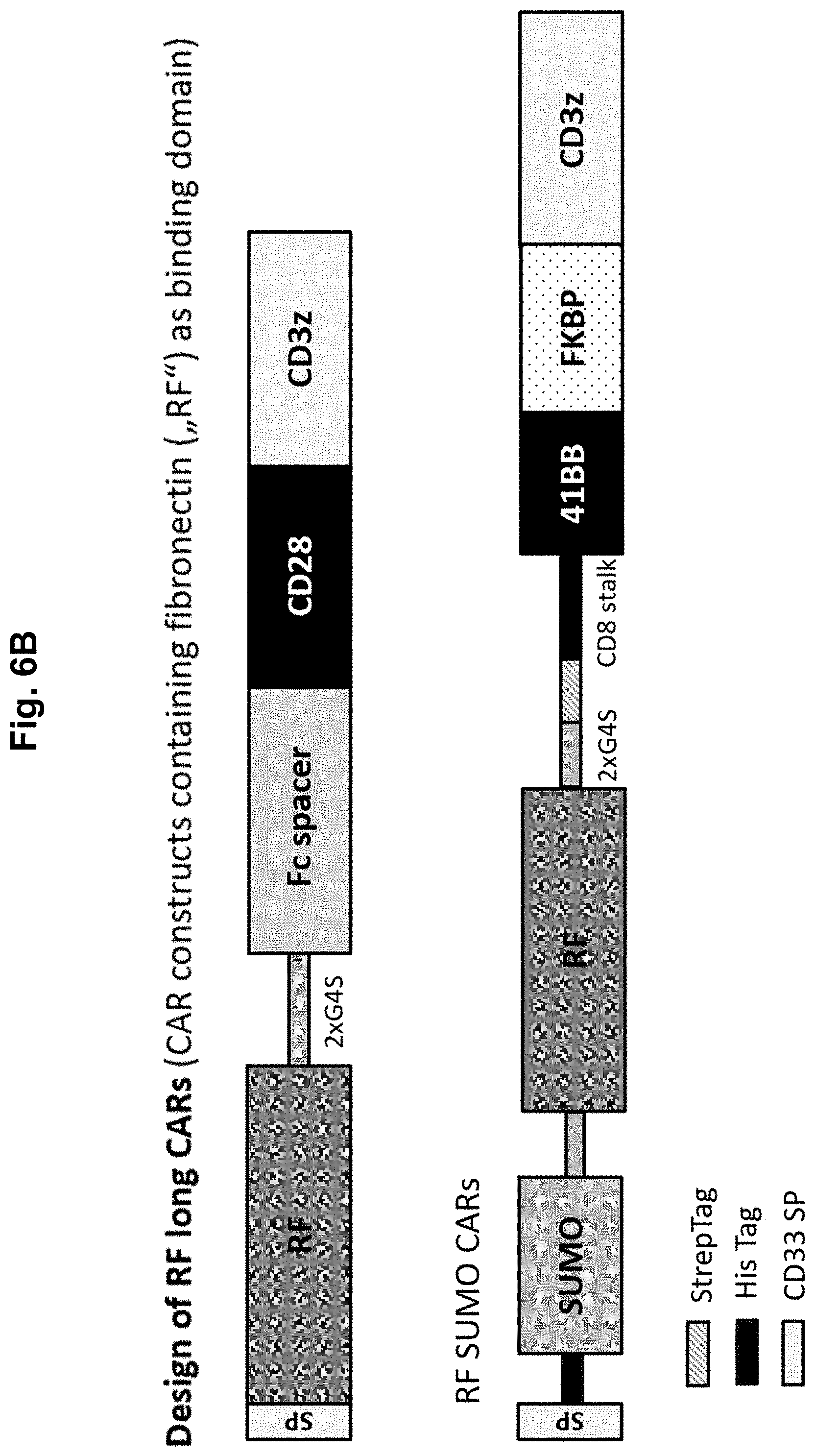
[0024] A highly attractive field for clinical application of LRPPI systems based on lipocalin-fold molecules according to the present invention is for regulating the function of T cells modified with chimeric antigen receptors (CARs) instead of using the FKBP-based systems for homo- and heterodimerization for regulating the CAR T cell function. Importantly, the function of currently applied CARs cannot be regulated in a clinically applicable manner. Instead, inducing apoptosis in the CAR expressing effector cells is the only clinically applicable safety mechanism in the CAR field until now (Jones et al., Front Pharmacol. 2014; 5:254). The most advanced strategies for regulating the function of the CAR molecule itself employ the FRB/FKBP-based system in different versions either for LRPPI (WO 2014/127261 A1, WO 2015/017214 A1, EP 3 087 101 A1, US 2017/0081411 A1) or for regulating protein stability (WO 2017/032777 A1). As mentioned above, however, the FRB/FKBP system is associated with severe problems in any potential clinical application (Sun et al., Cell Res. 2015; 25(12):1281-1282). Thus, alternatively, the lipocalin-fold molecule based LRPPI systems according to the present invention are highly attractive for integration into CARs in order to control T cell activation upon CAR mediated target antigen recognition by administering small molecules. In contrast to the rather problematic FRB/FBKP-system the lipocalin-fold based LRPPI systems according to the present invention can pave the way for broad clinical application of switchable CARs.
[0025] The LRPPI system according to the present invention and shown in FIG. 1 can be engineered using two strategies in principle: In strategy A, the lipocalin-fold binding interaction partner "c" (which may preferably be fused (+/-flexible linker) to a protein "II") is a binder, which binds to the barrel-containing structure of the lipocalin-fold molecule "a" with higher affinity when lipocalin-fold ligand "b" is present. The lipocalin-fold binding interaction partner "c" can e.g. be generated either by immunization of animals with ligand ("b")-loaded lipocalin-fold molecule, or by state of the art protein engineering methods such as phage display, yeast display, bacterial display, mammalian cell display, ribosome display, mRNA display or covalent DNA display, among others (Sergeeva et al., Advanced Drug Delivery Reviews 2006; 58:1622-1654). In strategy B, the lipocalin-fold molecule "a" itself can be engineered as a binder that after loading with a lipocalin-fold ligand "b" can bind with higher affinity to a chosen lipocalin-fold binding interaction partner "c" (which again can (according to a preferred embodiment) be fused to protein "II" or itself is a protein or non-protein antigen "II" (e.g. a tumor associated antigen)). The lipocalin-fold molecule "a" and the lipocalin-fold binding interaction partner "c" can be fused to the N- or C-termini or also internal sites of proteins "I" and "II" (see FIG. 1B). The lipocalin-fold molecule "a" may be engineered for increasing the affinity to a chosen small molecule lipocalin-fold ligand "b" and/or for decreasing the affinity towards endogenous natural lipocalin-fold ligands. Furthermore, the lipocalin-fold structure of lipocalin-fold molecule "a" may also be engineered for preventing or reducing interaction with natural interaction partners.
[0026] Preferably, the lipocalin-fold ligand induces a conformational change in the lipocalin-fold molecule "a", which facilitates the selection of a ligand-dependent binder (lipocalin-fold binding interaction partner) "c" (or also the selection of a ligand-dependent lipocalin-fold molecule "a" when in turn used as a binder). The lipocalin-fold ligand "b" can, but does not need to, interact directly with "c" or protein "II". However, in cases where binding of molecule "b" results only in rigidification of the structure of the lipocalin-fold molecule "a" with rather limited conformational selection, any direct contribution of molecule "b" for increasing the affinity of the interaction of "a" and "c" is beneficial.
[0027] Depending on the application, the LRPPI system can--according to a specific embodiment--be further employed in a strategic variant for regulating dimerization of two identical or different lipocalin-fold molecules (the first one being the lipocalin-fold molecule "a" according to the present invention, the second one being a "lipocalin-fold binding interaction partner" in the system according to the present invention (although being a "lipocalin-fold molecule", such second lipocalin-fold molecule would serve as the binding partner of the first lipocalin-fold molecule according to the present invention)). Accordingly, this specific embodiment uses lipocalin-fold ligands "b" with two identical or different head groups, respectively, to form a LRPPI with two lipocalin-fold molecules (different or the same (i.e. homodimerization or hetereodimerization)), in analogy to existing LRPPI systems (Rutkowska et al., Angew Chem Int Ed Engl. 2012; 51(33):8166-8176). One of the two lipocalin-fold molecules serves then (formally) as a "lipocalin-fold binding interaction partner" within the definitions in the system according to the present invention.
[0028] As already outlined above, the lipocalin-fold molecule according to the present invention may be any suitable molecule comprising the structural element of a lipocalin-fold. The lipocalin-fold of the lipocalin-fold molecule according to the present invention ("a") can be derived from the .beta.-barrel of any known member of the LCN protein family or from the .beta.-barrel of the iLBP protein family. This also includes LCN- and iLBP-variants, which deviate in the numbers of .beta.-strands from the prototypic architecture as has been reported, e.g., for some LCNs (Papiz et al., Nature. 1986; 324(6095):383-385; Spinelli et al., Eur J Biochem. 2002; 269(10):2449-2456; Sevvana et al., J Mol Biol. 2010; 404(3):363-371). As LCNs have evolved for extracellular transport of small molecules and iLBPs have evolved for intracellular transport of many of the same molecules, the two .beta.-barrel variants offer different advantages for application in oxidizing versus reducing environments outside and inside of cells. In principle, the lipocalin-fold of both families are suited for the flexible LRPPI systems according to the present invention as they have very similar structure and all can accommodate a large variety of lipocalin-fold ligands in their binding pocket with high affinity. However, lipocalin-fold molecules with known lipocalin-fold ligand induced conformational adaption and variants enabling a direct contribution of the lipocalin-fold ligand "b" in the interaction with the lipocalin-fold binding interaction partner "c" are preferred. The organisms of origin of the lipocalin-fold molecule can be selected according to the target organisms, in which the proteins are supposed to be expressed at maximum levels and with correct posttranslational modification. In this context LCNs have the advantage that they have evolved already in bacteria. However, the intended primary advantage of the .beta.-barrel based LRPPI systems is their clinical applicability, thus, lipocalin-fold molecules of human protein origin are preferred.
[0029] Currently, human LCNs comprise 15 well characterized proteins and a couple of yet elusive members (Schiefner et al., Acc Chem Res. 2015; 48(4):976-985). For the well characterized LCNs a large diversity in the size and shape of the binding pockets has been described as well as an accordingly highly diverse spectrum of lipocalin-fold ligands such as, e.g., simple fatty acids, glycoand phospholipids, all-trans retinol, cholesterol, vanillin, imatinib, staurosporine, or even large, complex molecules such as bacillibactin and heme. This adaptiveness has been found to particularly depend on the length and the sequence of the four loops at the entry site of the .beta.-barrel (Schiefner et al., Acc Chem Res. 2015; 48(4):976-985; Skerra, Biochim Biophys Acta. 2000; 1482(1-2):337-350; Korndorfer et al., Proteins 2003; 53(1):121-129; Korndorfer et al., J Mol Biol. 2003; 330(2):385-396; Kim et al., J Am Chem Soc. 2009; 131(10):3565-3576; Schlehuber et al., Biophys Chem. 2002; 96(2-3):213-228; Richter et al., FEBS Lett. 2014; 588(2):213-218). Extensive conformational changes upon binding of different lipocalin-fold ligands so far have been reported for human and bovine retinol binding protein 4 (RBP4), human tear lipocalin (TLC) and human apolipoprotein M (ApoM) (Berni et al., FEBS Lett. 1992; 308(1):43-45; Zanotti et al., J Biol Chem. 1993; 268(33):24873-24879; Zanotti et al., J Biol Chem. 1994; 269(47):29613-29620; Pattanayek et al., Protein Sci. 1999; 8(10):2027-2032; Motani et al., J Biol Chem. 2009; 284(12):7673-7680; Gasymov et al., Biochim Biophys Acta. 1998; 1386(1):145-156; Breustedt et al., Acta Crystallogr D Biol Crystallogr. 2009; 65(Pt 10):1118-1125; Gasymov et al., Biochemistry. 2012; 51(14):2991-3002; Zhang et al., Sci Rep. 2016; 6:30655; Christoffersen et al., Proc Natl Acad Sci USA. 2011; 108(23):9613-9618). Among them, human TLC is characterized by elevated conformational flexibility and an extraordinary flexible binding behavior compared to other LCNs (Schiefner et al., Acc Chem Res. 2015; 48(4):976-985; Breustedt et al., Acta Crystallogr D Biol Crystallogr. 2009; 65(Pt 10):1118-1125; Gasymov et al., Biochemistry. 2012; 51(14):2991-3002). In the case of the particularly well characterized native human RBP4 there are two loop regions, which undergo conformational alteration upon lipocalin-fold ligand binding and are involved in interaction with natural interaction partners transthyretin (TTR) (via EF loop, residues 89-101) and the receptor STRA6 (via CD loop, residues 59-68; Redondo et al., FASEB J. 2008; 22(4):1043-1054). Meanwhile, several natural and synthetic retinoid and non-retinoid lipocalin-fold ligands for human RBP4 have been described (i.e., Fenretinide, N-Ethylretinamide, all-trans retinoic acid, retinyl acetate, axerophthene, A1120 (PubChem CID 25138295)), which induce conformational changes resulting in dissociation from TTR (Berni et al., FEBS Lett. 1992; 308(1):43-45; Zanotti et al., J Biol Chem. 1993; 268(33):24873-24879; Zanotti et al., J Biol Chem. 1994; 269(47):29613-29620; Motani et al., J Biol Chem. 2009; 284(12):7673-7680; Coward et al., Anal Biochem. 2009; 384(2):312-320; Sharif et al., Anal Biochem. 2009; 392(2):162-168;). Reported crystal structures for human RBP4 illustrate that different lipocalin-fold ligands can induce different conformations in distinct loop regions of the LCN (e.g., protein data bank (PDB) 1RBP, 3FMZ and 2WR6 for retinol, A1120 and linoleic acid, respectively). Similar effects have been reported for ApoM (PDB 2YG2 and 2WEW for sphingosine-1-phosphate versus myristic acid; Christoffersen et al., Proc Natl Acad Sci USA. 2011; 108(23):9613-9618).
[0030] For selecting appropriate lipocalin-fold molecules according to the present invention, differences with regard to existence of glycosylation sites, free cysteines, disulfide bridges, oligomerization behavior, ligand spectrum etc., and the necessity for removing more or less characterized interaction sites for preventing interaction with natural protein partners or lipid membranes can be considered (Schiefner et al., Acc Chem Res. 2015; 48(4):976-985). Among the human LCNs, RBP4, TLC and ApoM (UniProt IDs P02753, P31025, and 095445, respectively) are characterized by already known lipocalin-fold ligand induced conformational adaption and are thus preferred members of the lipocalin family for generating LRPPI systems according to the present invention. One additional preferred lipocalin molecule is the human neutrophil gelatinase-associated lipocalin (NGAL) (UniProt ID P80188), for which a very detailed structural knowledge with respect to interacting amino acid residues and binding pocket engineering has accumulated (Kim et al., J Am Chem Soc. 2009; 131(10):3565-3576; Schonfeld et al., Proc Natl Acad Sci USA. 2009; 106(20):8198-8203; Barinka et al., Protein Eng Des Sel. 2016; 29(3):105-115; Gebauer et al., J Mol Biol 2013; 425(4):780-802; Bao et al., RSC Adv. 2015; 5(126):104363-104374; Eggenstein et al., J Struct Biol. 2014; 185(2):203-214). This is in particular due to the fact that its ligand binding pocket can easily be modified and was the basis of structurally resolved anticalins engineered for high affinity binding against different ligands.
[0031] Importantly, .beta.-barrels of LCNs are not only an option for extracellular use but also for intracellular use of the LRPPI systems. This is based on the fact that, although all human LCNs have at least one disulfide bridge in their .beta.-barrel structure, the .beta.-barrel of, e.g., human TLC is functional and sufficiently stable also in the fully reduced state (Gasymov et al., Biochim Biophys Acta. 2011; 1814(5):671-683). Other LCNs could be more affected under reducing conditions, however, these proteins can be stabilized by e.g. inserting stabilizing mutations. The latter has been exemplified for a zinc-binding mutant of human RBP4, which was stabilized by introducing the five mutations A43L, A55V, A57I, H104W and Q117I (Skerra; Biochim Biophys Acta. 2000; 1482(1-2):337-35049; Schmidt, Untersuchungen zur Proteinfaltung durch Protein-Design am Retinol-Bindungsprotein. Vol. ISBN 3-89675-314-2. Munchen: Herbert Utz Verlag; 1998).
[0032] The LCN-derived .beta.-barrels preferably represent the full-length coding sequences; signal peptides can be replaced; both, the N- and the C-terminal ends of the full length or trimmed .beta.-barrels (i.e. lipocalin molecules) are suited for fusion to protein partners (with or without linker sequences), protein domains, peptides or single amino acids since the termini of the .beta.-barrels are not involved in ligand binding (Skerra; Biochim Biophys Acta. 2000; 1482(1-2):337-35049).
[0033] Possible sequence modifications can comprise:
[0034] a) Preferably, the engineering of the binding pocket of the lipocalin-fold molecules for increasing the affinity to a chosen ligand "b" and/or lowering the affinity to other lipocalin-fold ligands by directed evolution including any sort of random mutagenesis and subsequent selection or screening processes, such as phage display, yeast display, bacterial display, mammalian cell display, ribosome display, mRNA display or covalent DNA display, among others (Sergeeva et al., Advanced Drug Delivery Reviews 2006; 58:1622-1654). Alternatively, mutations can be based on in silico calculations and subsequently be introduced by site-directed mutagenesis (Whitehead et al., Methods Enzymol. 2013; 523:1-19; Strauch et al., Proc Natl Acad Sci USA. 2014 Jan. 14; 111(2):675-80). Such engineering processes can require only limited mutagenesis at specific sites. If small molecules are chosen that initially do not bind the lipopocalin-fold, then more extensive mutagenesis in or nearby the center of the binding pocket of the .beta.-barrel may be required for generating mutants with binding capability (as described in DE 19742706 A1 and exemplified for several small molecules in Korndorfer et al., Proteins 2003; 53(1):121-129; Korndorfer et al., J Mol Biol. 2003; 330(2):385-396; Kim et al., J Am Chem Soc. 2009; 131(10):3565-3576; Schlehuber et al., Biophys Chem. 2002; 96(2-3):213-228). In the preferred case, this process is compatible with screening for lipocalin-fold ligand induced conformational changes. Such a screening process is feasible by employing proteins that can bind to these lipocalin-fold molecules in the absence of any ligand but dissociate upon lipocalin-fold ligand binding for selecting lipocalin-fold molecule mutants, which have maintained conformational switch behaviour. Such a protein can be the natural protein TTR in the case of RBP4 or other natural binding partners that bind to the respective lipocalin-fold molecule. Alternatively, binders, which have been separately engineered for binding to a chosen lipocalin-fold molecule in the absence of a ligand, may also be used for such a screening process. In a typical screening process, the lipocalin-fold molecule libraries are alternately screened for mutants that are capable for binding to these proteins in the absence of any ligand. Then the capability for lipocalin-fold ligand binding and conformational switching of the lipocalin-fold molecule is concomitantly selected by screening of the lipocalin-fold molecule libraries for non-binding to these proteins, or binding with lower affinity, in the presence of the lipocalin-fold ligand.
[0035] b) Additional mutagenesis (possibly, but not necessarily, after engineering of the binding pocket of the lipocalin-fold molecule for lipocalin-fold ligand binding) with a focus on the loop regions (as described in DE 19742706 A1), if the lipocalin-fold molecule is intended for use as a binder in the LRPPI system (instead of using it as an antigen); preferably, the lipocalin-fold molecule is engineered by directed evolution including any sort of random mutagenesis and subsequent selection or screening processes, such as phage display, yeast display, bacterial display, mammalian cell display, ribosome display, mRNA display or covalent DNA display, among others (Sergeeva et al., Advanced Drug Delivery Reviews 2006; 58:1622-1654). Maintenance of the capability for small molecule binding of the lipocalin-fold molecules may be warranted by alternating screening for antigen binding of the lipocalin-fold molecules in presence of the lipocalin-fold ligand and for non-binding (or binding with lower affinity) in the absence of the lipocalin-fold ligand.
[0036] c) Mutation/deletion/insertion of residues for preventing dimerization or interaction with other proteins or lipid membranes (e.g., free cysteines, unprocessed signal peptide in ApoM, loop CD residues 59-68 in RBP4, etc.; see e.g. Skerra, Biochim Biophys Acta. 2000; 1482(1-2):337-350; Zhang et al., Sci Rep. 2016; 6:30655; Redondo et al., FASEB J. 2008; 22(4):1043-1054).
[0037] d) Mutation/deletion/insertion of residues for preventing post translational protein modification.
[0038] e) Engineering the lipocalin-fold molecule for improved stability, e.g., under reducing conditions in the cytoplasm as has been demonstrated for antibody fragments (Worn et al., J Biol Chem. 2000; 275(4):2795-2803). This can be achieved, for example, by rational design of stabilizing mutations or by directed evolution experiments which select for improved stability (Traxlmayr et al, Biochim Biophys Acta. 2012; 1824(4):542-549). However, also any other method for stabilization of proteins is possible.
[0039] The human intracellular iLBP protein family comprises a group of 6 retinoid binding proteins termed CRBPs and CRABPs, and the group of 10 fatty acid binding proteins (FABPs). Although iLBPs are intracellular proteins, some of them, e.g., FABP4 may have a function also in the extracellular space (Hotamisligil et al., Nat Rev Endocrinol. 2015; 11(10):592-605). All iLBPs have the same architecture with 10 anti-parallel .beta.-strands connected by more or less elongated loops with the exception of an intervening helixturn-helix motif between strands RA and SB (Lakshmi et al. PLoS One. 2015; 10(8): e0135507; Zhang et al., PLoS One. 2012; 7(5): e36772; Smathers et al., Hum Genomics. 2011; 5(3):170-191). Compared to LCNs their barrel structure is more compact and their ligands are hardly exposed to the solvent due to shielding by the helix-turn-helix motif at the entrance. However, like LCNs iLBPs have adapted to binding of a diverse spectrum of partially shared ligands and they also have in common the high tolerance to mutagenesis. The latter includes even the complete deletion of the helix-turn-helix motif at the barrel entrance and substitution by a simple loop (Curto et al., Protein Sci. 2009; 18(4):735-746; Ogbay et al., Protein Sci. 2004; 13(5):1227-1237). Among the iLBPs, FABP1 and FABP6 are characterized by higher backbone flexibility and a larger ligand entrance, resulting in their unique capacity to accommodate bulky molecules and two of each. Binding of the so far tested ligands generally revealed only small conformational changes due to ligand induced rigidification (Yu et al., Sci Rep. 2016; 6:34171; Sharma et al., J Biol Chem. 2011; 286(36):31924-31928; Cai et al., Biophys J. 2012; 102(11):2585-2594; Franzoni et al., J Lipid Res. 2010; 51(6):1332-1343; Vaezeslami et al., J Mol Biol. 2006; 363(3):687-701; Gillilan et al., J Mol Biol. 2007; 372(5):1246-1260; Menozzi et al., J Struct Biol. 2017; 197(3):330-339; Long et al., Biophys J. 2010; 98(12):3054-3061). Obviously, these changes are sufficient to control interaction with other proteins and to mediate, e.g., nuclear transport and interaction with nuclear receptors, as well as, e.g., shuttling retinoids within the cell by mediating interaction with the receptor STRA6 in the cytoplasmic membrane in the case of CRBP-I (Gillilan et al., J Mol Biol. 2007; 372(5):1246-1260; Armstrong et al., J Biol Chem. 2014; 289(21):14941-14954; Amber-Vitos et al., PLoS One. 2015; 10(8):e0132138; Berry et al., Mol Cell Biol. 2012; 32(15):3164-3175; Sessler et al., Mol Cell. 2005; 18(3):343-353; Hofer et al., J Biol Chem. 2015; 290(30):18438-18453; Furuhashi et al., Nat Rev Drug Discov. 2008; 7(6):489-503). Some iLBPs thereby interact via the formation/selection of a structural nuclear location signal (NLS) within the .alpha.2-helix at the entry of the calyx, which is elicited by some but not all of their small molecule ligands (Furuhashi et al., Nat Rev Drug Discov. 2008; 7(6):489-503).
[0040] Like with LCNs, the human variants of iLBPs are preferred for use in a LRPPI system if applied in humans in vivo. Among them the retinoid binding proteins, in particular the very well characterized CRABP-II (UniProt ID P29373), are preferred due to their retinoid ligand specificity (Zhang et al., PLoS One. 2012; 7(5): e36772; Franzoni et al., J Lipid Res. 2010; 51(6):1332-1343; Vaezeslami et al., J Mol Biol. 2006; 363(3):687-701; Menozzi et al., J Struct Biol. 2017; 197(3):330-339). Furthermore, FABP2 (P12104), for which the helix-turn-helix substitution was exemplified, and FABP1 (P07148) due to their known low affinity to several clinically approved lipophilic drugs are also attractive iLBP members for engineering the binding pocket for recognition of clinically applicable ligands (Smathers et al., Hum Genomics. 2011; 5(3):170-191; Curto et al., Protein Sci. 2009; 18(4):735-746; Ogbay et al., Protein Sci. 2004; 13(5):1227-1237; Velkov T, Chem Biol. 2007; 14(4):453-465; Velkov T. PPAR Res. 2013; 2013:938401; Beringhelli T, PLoS One. 2015; 10(7):e0132096; Chuang S, J Med Chem. 2008; 51(13):3755-3764).
[0041] For integration into the LRPPI system according to the present invention, fusion of the .beta.-barrels of iLBPs to protein partners (with or without linker sequences), protein domains, peptides or single amino acids is possible via the N- and the C-terminal ends (of full length or trimmed iLBP proteins). The sequences are preferably modified for preventing unwanted interaction with their natural protein partners in the cytoplasm by modifying or deleting the respective interacting sequence elements (e.g., the helixturn-helix motif containing the hidden structural NLS in the .alpha.2-helix and other interaction sites; Gillilan et al., J Mol Biol. 2007; 372(5):1246-1260; Armstrong et al., J Biol Chem. 2014; 289(21):14941-14954; Amber-Vitos et al., PLoS One. 2015; 10(8):e0132138; Berry et al., Mol Cell Biol. 2012; 32(15):3164-3175; Sessler et al., Mol Cell. 2005; 18(3):343-353). For increasing the affinity to selected lipocalin-fold ligands ("b") and/or for decreasing the affinity to their endogenous small molecule ligands, the iLBPs may be engineered similarly to LCNs. To facilitate the engineering of lipocalin-fold ligand dependent recognition, it is possible to substitute the helix-turn-helix motif by a loop sequence (as exemplified by Curto et al. and Ogbay et al. (Curto et al., Protein Sci. 2009; 18(4):735-746; Ogbay et al., Protein Sci. 2004; 13(5):1227-1237).
[0042] The rationale of developing the LRPPI system according to the present invention (i.e. based on the "lipocalin-fold" of the lipocalin-fold molecule) is to maximize the freedom of choice in selecting the lipocalin-fold ligands ("b"). The crucial innovation for enabling this freedom is based on proteins of the superfamily of lipocalin proteins that contain the "lipocalin-fold" structure. Their key feature, i.e., the characteristic deep binding pocket with a calyx like shape and high structural flexibility is crucial for the flexibility and reliability of the present invention. As a consequence, these binding pockets can be engineered by relatively few mutations for binding to a broad range of small molecule ligands with diverse structural and biophysical characteristics. This has been exemplified with the proteins BBP and NGAL, which were engineered by substitution of 12 to 17 amino acid residues for high affinity binding to originally non-binding small molecules such as fluorescein, digoxigenin, digitoxigenin and a diamine-pentaacetic acid (DTPA) based chelator (Korndorfer et al., Proteins 2003; 53(1):121-129; Korndorfer et al., J Mol Biol. 2003; 330(2):385-396; Kim et al., J Am Chem Soc. 2009; 131(10):3565-3576). Thus, contrary to existing LRPPI systems, the novel system according to the present invention is defined by employing lipocalin-fold containing proteins (i.e., lipocalin-fold molecules) and neither by any definitive list of possible small molecules (i.e., lipocalin-fold ligands) nor by any specific structural and chemical properties of such molecules.
[0043] The lipocalin-fold ligand "b" according to the present invention is a "small molecule", e.g. "small" compared to polypeptides and proteins, such as the lipocalin-fold molecule. Accordingly, the lipocalin-fold ligand according to the present invention has a molecular weight of 1500 Da or less, preferably 1000 Da or less, especially 750 Da or less. It may be as small as, e.g. glycine (75 Da) or even below, provided that it allows a specific binding within the LRPPI system according to the present invention (i.e. binding to the lipocalin-fold molecule). Accordingly, preferred Mw ranges of the lipocalin-fold ligand of the present invention are 50 to 1500 Da, preferably 75 to 1500 Da, especially 150 to 750 Da. Preferably, the lipocalin-fold ligand can bind in the calyx of the lipocalin-fold molecule formed by the barrel and the loop regions of the lipocalin-fold structure. The lipocalin-fold ligand has a substantial and specific affinity to the lipocalin-fold molecule (in at least one conformation of the lipocalin-fold molecule) which may be 1 mM or lower, preferably 100 .mu.M or lower, especially 10 .mu.M or lower. Such affinity can be increased by directed evolution of the binding pocket of the lipocalin-fold molecules. Although up to 30 mutations or more may be applied to a given lipocalin-fold molecule (see, for example, Schonfeld et al., Proc Natl Acad Sci USA. 2009; 106(20):8198-8203), it is usually not necessary to exchange more than 25 or more than 20 to significantly increase the affinity to a given lipocalin-fold ligand (see, for example, Kim et al., J Am Chem Soc. 2009; 131(10):3565-3576; Korndorfer et al., Proteins 2003; 53(1):121-129; Korndorfer et al., J Mol Biol. 2003; 330(2):385-396; Gebauer et al., J Mol Biol 2013; 425(4):780-802). Methods for generating lipocalin-fold molecules with an improved affinity towards the lipocalin-fold ligand are well available in the art (see above). Usually, only a few mutations are necessary for a significant increase in affinity towards a given lipocalin-fold ligand, for example only one, two or less, three or less, four or less, five or less, six or less, seven or less, eight or less, nine or less, or ten or less. The capability of a lipocalin-fold ligand "b" to induce a conformational alteration in the lipocalin-fold molecule "a" and/or to positively affect the affinity of a lipocalin-fold binding interaction partner "c" by direct interaction can be predicted to some extent using pharmacophores from existing small molecule ligands with such function. Additionally, this functional capability can be screened for by employing binding proteins, which have a conformation specific binding affinity for the lipocalin-fold molecule (detailed in Example 2, below, and in Coward et al. Anal Biochem. 2009; 384(2):312-320, Sharif et al., Anal Biochem. 2009; 392(2):162-168 and Dobri et al., Invest Ophthalmol Vis Sci. 2013; 54(1):85-95). In the case of RBP4, e.g., TTR can be used as a naturally occurring conformation specific binding protein. If there is no such protein available, as for example for TLC or ApoM, which have also been reported to undergo lipocalin-fold ligand dependent conformational switching (Gasymov et al., Biochim Biophys Acta. 1998; 1386(1):145-156; Breustedt et al., Acta Crystallogr D Biol Crystallogr. 2009; 65(Pt 10):1118-1125; Zhang et al., Sci Rep. 2016; 6:30655; Christoffersen et al., Proc Natl Acad Sci USA. 2011; 108(23):9613-9618), then it is possible to first generate a protein-based binder that interacts with the unloaded lipocalin-fold molecule, i.e. in the absence of lipocalin-fold ligands, by a process of alternating selection for binding to the lipocalin-fold molecule in the absence and non-binding (or binding with reduced affinity) in the presence of lipocalin-fold ligands known to shift conformational states of the lipocalin-fold molecule. This alternating selection strategy ensures that a binder is selected, which specifically recognizes the unloaded (i.e. not loaded with any lipocalin-fold ligand) state of the lipocalin-fold molecule. In Example 1 we exemplified this process of alternating screening in presence and absence of a lipocalin-fold ligand (A1120) in opposite direction for selecting a lipocalin-fold binding interaction partner, i.e., a protein that binds RBP4 (i.e. the lipocalin-fold molecule) with increased affinity in presence of the lipocalin-fold ligand A1120.
[0044] Typically, the process of selecting lipocalin-fold ligand molecules can start from:
1. any given molecule with or without initial binding affinity 2. any existing compound libraries for high throughput screening for binding affinity and/or function 3. any structural databases of compounds that can be used in virtual screening for binding and/or function
[0045] Ad 1: Initial binding affinity is not required in this approach, since the binding pocket of the lipocalin-fold molecule can be engineered for binding (see e.g. DE 19742706 A1). The feasibility of engineering of lipocalin-fold molecules for high affinity binding to arbitrary, initially non-binding, molecules has been exemplified by engineering of BBP and NGAL for binding of fluorescein, digoxigenin and a DTPA-based chelator (Korndorfer et al., Proteins 2003; 53(1):121-129; Korndorfer et al., J Mol Biol. 2003; 330(2):385-396; Kim et al., J Am Chem Soc. 2009; 131(10):3565-3576). Hence, clinically attractive lipocalin-fold ligand "b" candidates can be selected regardless of initial binding affinity, just considering pharmacological properties as, e.g., tolerability, bioavailability, plasma levels, pharmacokinetics, tissue penetrance etc. Such molecules could be, for example, Indicaxanthin and Vulgaxanthin, which are pharmacologically well investigated and tolerated and are contained in sufficiently high amounts in a narrow segment of edible plants. There are, however, numerous examples of clinically approved drugs which have already been demonstrated to bind to certain lipocalin-fold molecules (e.g., human FABP1; Smathers et al., Hum Genomics. 2011; 5(3):170-191; Velkov et al., Chem Biol. 2007; 14(4):453-465; Velkov T., PPAR Res. 2013; 2013:938401; Beringhelli et al., PLoS One. 2015; 10(7):e0132096; Chuang at al., J Med Chem. 2008; 51(13):3755-3764). Of course, such molecules are attractive starting points. The most attractive molecules are non-natural ligands, for which conformational effects have been proven and which have clinical application potential. For RBP4, for example, these ligands are A1120 and numerous derivatives as well as synthetic retinoids such as, e.g., fenretinide, N-Ethylretinamide, all-trans retinoic acid, retinyl acetate and axerophthene (Berni et al., FEBS Lett. 1992; 308(1):43-45; Zanotti et al., J Biol Chem. 1993; 268(33):24873-24879; Zanotti et al., J Biol Chem. 1994; 269(47):29613-29620; Motani et al., J Biol Chem. 2009; 284(12):7673-7680; Cioffi et al., J Med Chem. 2014; 57(18):7731-7757; Cioffi et al., J Med Chem. 2015; 58(15):5863-5888).
[0046] Ad 2: High throughput screening of molecule libraries for molecules with binding affinity can be performed by various assays (reviewed in Auld et al., Receptor Binding Assays for HTS and Drug Discovery. In: Sittampalam et al., eds. Assay Guidance Manual. Bethesda Md.: Eli Lilly & Company and the National Center for Advancing Translational Sciences; 2004). Screening of molecule libraries was, e.g., employed for identifying A1120 as a high affinity ligand for RBP4 (Motani et al., J Biol Chem. 2009; 284(12):7673-7680). A consequence of this strategy is that the identified lipocalin-fold ligands display already some binding affinity and thus can be further assayed for mediating ligand-dependent binding or dissociation of a binder "c" without requirement of prior binding pocket engineering of the lipocalin-fold molecule. Of note, there are possibilities that enable high throughput screening for binding and function, like inducing conformational switching, in a single step. In the case of RBP4 this could be done, for example, by FRET based assays for detecting the conformation dependent interaction with TTR (Coward et al., Anal Biochem. 2009; 384(2):312-320; Sharif et al., Anal Biochem. 2009; 392(2):162-168; Dobri et al., Invest Ophthalmol Vis Sci. 2013; 54(1):85-95)). If desired, the affinity of the respective lipocalin-fold ligand to the lipocalin-fold molecule can be further increased by engineering (i.e. mutagenesis) of the binding pocket of the lipocalin-fold molecule.
[0047] Ad 3: Virtual screening of compound databases is attractive when there are known small molecule ligands, which mediate conformational alterations in a lipocalin-fold molecule. Then a modeled pharmacophore of the small molecule can be used to screen for molecules, which might similarly interact with the lipocalin-fold molecule and thereby show similar function (see Qing et al., Journal of Receptor, Ligand and Channel Research 2014; 7:81-92 for a review on virtual screening using pharmacophore modeling). In the optimal case, the crystal structure of the lipocalin-fold molecule charged with the modeled small molecule ligand is also known, since this helps to improve the accuracy of the prediction algorithms. There are several examples of known structures for lipocalin-fold molecules bound to a small molecule ligand (Berni et al., FEBS Lett. 1992; 308(1):43-45; Zanotti et al., J Biol Chem. 1993; 268(33):24873-24879; Zanotti et al., J Biol Chem. 1994; 269(47):29613-29620; Pattanayek et al., Protein Sci. 1999; 8(10):2027-2032; Motani et al., J Biol Chem. 2009; 284(12):7673-7680; Gasymov et al., Biochim Biophys Acta. 1998; 1386(1):145-156; Breustedt et al., Acta Crystallogr D Biol Crystallogr. 2009; 65(Pt 10):1118-1125; Gasymov et al., Biochemistry. 2012; 51(14):2991-3002; Zhang et al., Sci Rep. 2016; 6:30655; Christoffersen et al., Proc Natl Acad Sci USA. 2011; 108(23):9613-9618; Coward et al., Anal Biochem. 2009; 384(2):312-320). This strategy is exemplified in the example section below with RBP4 charged with A1120, for which a pharmacophore model was generated in order to identify and prioritize molecules for functional testing and for engineering of the binding pocket of RBP4 (described in Example 2, below). Importantly, the pharmacophore approach does not deliver a final complete list of potentially conformation inducing molecules due to limitations of pharmacophore methods (reviewed in Qing et al., Journal of Receptor, Ligand and Channel Research 2014; 7:81-92) and due to incomplete and not updated databases etc. Moreover, there are other molecules also known to alter the conformation of RBP4, e.g., fenretinide, for which a pharmacophore based approach would lead to identification of yet another set of molecules in the chemical space. In fact, in preferred embodiments, in silico filtering of millions of molecules for function together with directed evolution of the highly flexible binding pocket of the lipocalin-fold structure in the lipocalin-fold molecules according to the present invention is a perfect combination and a crucial strength of the novel LRPPI system. Of note, virtual screening also enables identifying functional molecules, which do not have sufficient initial binding affinity, which is not a problem, since affinity can easily be generated by engineering of the lipocalin-fold molecule. In this case, lipocalin-fold molecule mutants are selected by use of a conformation specific binding protein that binds to the lipocalin-fold molecule mutatants in the absence but not in the presence of a known conformation inducing ligand. Thus, the dissociation of this conformation specific binding protein enables the identification of lipocalin-fold ligand candidates that bind to the lipocalin-fold molecule and that at the same time induce conformational alterations (detailed in Example 2, below).
[0048] Principally, the lipocalin-fold binding interaction partner "c" can be provided or engineered based on any available molecular binder scaffold including antibodies, antibody fragments [e.g. single-chain variable fragments (scFv), antigen binding fragments (Fabs), nanobodies, among others] and non-antibody based scaffolds such as affibodies, a further (or the same) lipocalin-fold molecule, preferably LCNs, especially anticalins; avimers, DARPins, fynomers, Kunitz domains, knottins, monobodies, binders based on Sso7d, reduced charge Sso7d (rcSso7d) or Sac7d, among many others (Simeon et al., Protein Cell. 2017; Gilbreth et al., Curr Opin Struct Biol. 2012; 22(4):413-420; Koide et al., ACS Chem Biol. 2009; 4(5):325-334; Traxlmayr et al., J Biol Chem. 2016; 291(43):22496-22508). Meanwhile, many more non-antibody binding proteins have been reported (Pluckthun, Alternative Scaffolds: Expanding the options of antibodies. In: Little M, ed. New York: Cambridge University Press; 2009:244-271; Chapman et al., Cell Chem Biol. 2016; 23(5):543-553; Binz et al., Nat Biotechnol. 2005; 23(10):1257-1268; Vazquez-Lombardi et al., Drug Discov Today. 2015; 20(10):1271-1283), and, in fact, synthetic library design and selection can be applied to any protein which then can potentially serve as lipocalin-fold binding interaction partner "c", too (Pluckthun, Alternative Scaffolds: Expanding the options of antibodies. In: Little M, ed. New York: Cambridge University Press; 2009:244-271). With the aim of clinical applicability of the LRPPI systems according to the present invention, lipocalin-fold binding interaction partners "c" are preferably derived from small human single protein domains (e.g., fibronectin type III domain (FN3) based Monobodies) with a minimized number of mutated amino acids for keeping immunogenicity as low as possible. It is of course also possible to attach such further molecules and scaffolds to the lipocalin-fold molecule (or to a lipocalin-fold binding interaction partner).
[0049] Accordingly, the lipocalin-fold molecule according to the present invention may be any protein that contains the structural motif of a lipocalin-fold to which (or in which) the lipocalin-fold ligand binds and which enables binding of the lipocalin-fold molecule to the lipocalin-fold binding interaction partner. Usually, the lipocalin-fold molecule according to the present invention is adapted to the specific needs for the lipocalin-fold molecule "a"/lipocalin-fold ligand "b"/lipocalin-fold binding interaction partner "c" triangle by providing a "starting" lipocalin-fold molecule which may already have a certain affinity to a given lipocalin-fold ligand and/or for which clinically attractive ligands with functional activity could be identified by virtual screening. This starting lipocalin-fold molecule may then be engineered by one or more amino acid exchanges, insertions and/or deletions to optimize lipocalin-fold ligand binding, and in the case of strategy B of the lipocalin-fold based LRPPI system also for binding to a lipocalin-fold binding interaction partner "c". In strategy A the lipocalin-fold binding interaction partner "c" is generated by engineering of a protein (that is originally not able to bind to any naturally occurring lipocalin-fold molecule with an affinity of <10 .mu.M in the presence of any lipocalin-fold ligand) for binding to the (starting and/or engineered) lipocalin-fold molecule (bound to the lipocalin-fold ligand). Such sequence optimization of a protein for generating a lipocalin-fold ligand dependent lipocalin-fold binding interaction partner is shown in the example section of the present invention and well available for a person skilled in the art with the disclosure contained herein. In fact, it is established in the art of protein engineering that protein scaffolds (especially well-established scaffolds, such as antibodies and non-antibody-based scaffolds, especially lipocalins) can be adapted so that virtually any biological molecules (especially proteins) can be bound (see e.g. (for non-antibody scaffolds) Vasquez-Lombardi et al., Drug Discov. Today 20 (2015), 1271-1283; Pluckthun, Alternative Scaffolds: Expanding the Options of Antibodies. In: Recombinant Antibodies for Immunotherapy, Melvyn Little, Cambridge University Press, New York (2009), pp. 244-271).
[0050] The (starting) lipocalin-fold molecule therefore preferably contains the structural lipocalin-fold of a naturally occurring protein of the lipocalin-fold superfamily (for clinical applications in human patients: preferably a naturally occurring human lipocalin-fold molecule) or a known variant thereof (e.g. an "anticalin" or "mutein of lipocalin" and the like; a vast number of such variants have already been disclosed in the prior art). The lipocalin-fold molecule to be used in the LRPPI system according to the present invention may then be adapted to the specificities of the lipocalin-fold ligand/lipocalin-fold binding interaction partner by the introduction of modifications, as disclosed above (e.g. a) to e)), by engineering (i.e. amino acid changes, insertions and/or deletions) of the binding pocket, amino acid positions in the .beta.-barrel structure, and/or amino acid positions in the regions adjoining the .beta.-strands of the .beta.-barrel structure, mutation/deletion/insertion of residues for preventing dimerization or interaction with other molecules or for preventing post-translational protein modification and/or engineering of the lipocalin-fold for improved stability.
[0051] As already stated above, the lipocalin-fold architecture is very robust and has high sequence flexibility, as demonstrated by the fact that it has not been possible to define any sequence motif that is common to all naturally occurring members of the lipocalin superfamily, including LCNs and iLBPs (Lakshmi et al. PLoS One. 2015; 10(8): e0135507; Flower et al., Biochim Biophys Acta. 2000; 1482(1-2):9-24). Apart from the generally high sequence variation in naturally occurring members of the lipocalin superfamily, the regions adjoining the structurally conserved .beta.-strands not only show high variation in sequence, but also in structure (Flower et al., Biochim Biophys Acta. 2000; 1482(1-2):9-24; Schiefner et al., Acc Chem Res. 2015; 48(4):976-985). Given the extensive sequence variation among naturally occurring lipocalin superfamily members and/or structural variation in the regions adjoining the .beta.-strands, it is not surprising that in the past lipocalin molecules have been successfully engineered by mutating more than 30 amino acid positions, corresponding to more than 15% of all amino acid positions in the respective protein, without any major structural changes in the structurally conserved .beta.-strands of the characteristic .beta.-barrel structure (Schonfeld et al., Proc Natl Acad Sci USA. 2009; 106(20):8198-8203). Thus, it is well-established in the field that lipocalin-fold molecules can be extensively mutated without impairing the overall .beta.-barrel fold (i.e. particularly the loop regions but also the .beta.-strands of the .beta.-barrel can be designed by exchanging, inserting and/or deleting a considerable number of amino acids). Therefore, in the present invention, which describes the usage of the lipocalin-fold structure in LRPPI systems, a lipocalin-fold molecule is defined as any naturally occurring molecule classified into the lipocalin superfamily in the SCOP database (version 1.75), or a mutant thereof. However, it is preferred to exchange only a limited number of amino acids. The LRPPI system according to the present invention therefore preferably comprises (1) a lipocalin-fold molecule being identical to a naturally occurring member of the lipocalin superfamily or (2) an already existing variant thereof (e.g. an already existing anticalin, lipocalin mutein, iLBP with complete deletion of the helixturn-helix motif at the barrel entrance etc.) or (3) a derivative of (1) or (2) with at least 70%, preferably at least 80%, especially at least 90% sequence identity to its counterpart that it derives from.
[0052] According to a preferred embodiment, the lipocalin-fold molecule is a derivative of a naturally occurring or otherwise disclosed (by its amino acid sequence) lipocalin-fold molecule with at least 70%, preferably at least 80%, especially at least 90% sequence identity in the .beta.-barrel structure, whereby this .beta.-barrel structure is defined as the regions which correspond structurally to amino acid positions 21-30, 41-47, 52-58, 71-78, 85-88, 102-109, 114-120 and 132-138 in human RBP4 (according to the amino acid residue numbering scheme in the PDB entry 1RBP); or to the amino acid positions 14-23, 37-43, 48-54, 62-69, 76-79, 84-91, 96-102 and 111-117 in human tear lipocalin (TLC; as defined by Schiefner et al., Acc Chem Res. 2015; 48(4):976-985); or to the amino acid positions 44-53, 69-75, 81-87, 96-103, 110-113, 119-126, 131-137 and 142-148 in human apolipoprotein M (ApoM; as defined by Schiefner et al., Acc Chem Res. 2015; 48(4):976-985); or to the amino acid positions 5-12, 41-45, 50-54, 61-65, 71-73, 81-87, 93-96, 108-112, 119-124 and 129-135 in human cellular retinoic acid binding protein II (CRABPII; according to the amino acid residue numbering scheme in PDB entry 2FS6); or to the amino acid positions 5-12, 39-43, 48-52, 59-63, 69-71, 79-85, 91-94, 99-103, 109-114 and 119-125 in human fatty acid binding protein 1 (FABP1; according to the amino acid residue numbering scheme in PDB entry 2F73).
[0053] Preferred embodiments of the lipocalin-fold molecule according to the present invention also include lipocalin-fold molecules which comprise 1-30 amino acid exchanges and/or 1-50 amino acid deletions and/or 1-50 amino acid insertions of (1), (2) or (3) which (at least) contain the lipocalin-fold and are able to bind to the lipocalin-fold ligand and the lipocalin-fold binding interaction partner. Accordingly, the ligand regulated protein-protein interaction system preferably comprises as lipocalin-fold molecule a molecule identical with a naturally occurring iLBP (intracellular lipid binding protein), a naturally occurring lipocalin or an anticalin, and derivatives of any of these molecules with 1-30 amino acid exchanges and/or 1-50 amino acid deletions and/or 1-50 amino acid insertions.
[0054] Alternatively, the LRPPI system according to the present invention may also comprise a (4) fragment of (1) or (2) or (3) with a length of at least 80, preferably at least 100, especially at least 120, amino acids in the case of LCNs [or with a length of at least 80, preferably at least 85, especially at least 90, amino acids in the case of iLBPs (which are generally smaller proteins than LCNs)] covering at least the structurally conserved .beta.-barrel structure of the lipocalin-fold. This structurally conserved .beta.-barrel structure comprises or consists of amino acid positions which correspond structurally to the amino acid positions 21-30, 41-47, 52-58, 71-78, 85-88, 102-109, 114-120 and 132-138 in human RBP4 (according to the amino acid residue numbering scheme in the PDB entry 1RBP); or to the amino acid positions 14-23, 37-43, 48-54, 62-69, 76-79, 84-91, 96-102 and 111-117 in human tear lipocalin (TLC; as defined by Schiefner et al., Acc Chem Res. 2015; 48(4):976-985); or to the amino acid positions 44-53, 69-75, 81-87, 96-103, 110-113, 119-126, 131-137 and 142-148 in human apolipoprotein M (ApoM; as defined by Schiefner et al., Acc Chem Res. 2015; 48(4):976-985); or to the amino acid positions 5-12, 41-45, 50-54, 61-65, 71-73, 81-87, 93-96, 108-112, 119-124 and 129-135 in human cellular retinoic acid binding protein II (CRABPII; according to the amino acid residue numbering scheme in PDB entry 2FS6); or to the amino acid positions 5-12, 39-43, 48-52, 59-63, 69-71, 79-85, 91-94, 99-103, 109-114 and 119-125 in human fatty acid binding protein 1 (FABP1; according to the amino acid residue numbering scheme in PDB entry 2F73).
[0055] Further preferred lipocalin-fold molecules in the LRPPI system according to the present invention are derivatives of a naturally occurring lipocalin or iLBP with up to 15, up to 30, or up to 50 amino acid deletions and/or up to 15, up to 30, or up to 50 amino acid insertions outside of the structurally conserved .beta.-barrel structure. Outside the structurally conserved .beta.-barrel structure these molecules are very flexible to amino acid changes and therefore combinations of mutations, deletions and insertions are possible in these regions which correspond structurally to amino acid residues 1-20, 31-40, 48-51, 59-70, 79-84, 89-101, 110-113, 121-131 and 139-183 in human RBP4 (according to the amino acid residue numbering scheme in the PDB entry 1RBP), which define the regions adjoining the structurally conserved .beta.-strands in human RBP4; or corresponding structurally to the regions of amino acid residues 1-13, 24-36, 44-47, 55-61, 70-75, 80-83, 92-95, 103-110 and 118-158 in human TLC (according to the amino acid residue numbering scheme in Schiefner et al., Acc Chem Res. 2015; 48(4):976-985), which define the regions adjoining the structurally conserved .beta.-strands in human TLC; or corresponding structurally to the regions of amino acid residues 1-43, 54-68, 76-80, 88-95, 104-109, 114-118, 127-130, 138-141 and 149-188 in human ApoM (according to the amino acid residue numbering scheme in Schiefner et al., Acc Chem Res. 2015; 48(4):976-985), which define the regions adjoining the structurally conserved .beta.-strands in human ApoM; or corresponding structurally to the regions of amino acid residues 1-4, 13-40, 46-49, 55-60, 66-70, 74-80, 88-92, 97-107, 113-118, 125-128 and 136-137 in human CRABPII (according to the amino acid residue numbering scheme in PDB entry 2FS6), which define the regions adjoining the structurally conserved .beta.-strands in human CRABPII; or corresponding structurally to the regions of amino acid residues 1-4, 13-38, 44-47, 53-58, 64-68, 72-78, 86-90, 95-98, 104-108, 115-118 and 126-127 in human FABP1 (according to the amino acid residue numbering scheme in PDB entry 2F73), which define the regions adjoining the structurally conserved .beta.-strands in human FABP1.
[0056] In a preferred embodiment, the LRPPI system according to the present invention comprises as lipocalin-fold molecule a derivative of a naturally occurring member of the lipocalin superfamily with at least one, two, three, four, five, six, seven, eight, nine, ten, eleven, twelve, 13, 14, 15, 16, 17, 18, 19, 20, 21, 22, 23, 24, 25, 26, 27, 28, 29, or 30 amino acid exchanges.
[0057] According to a further preferred embodiment, the lipocalin-fold molecule used in the LRPPI system according to the present invention is a lipocalin, i.e., a protein containing an eight-stranded up-and-down .beta.-barrel arranged in a +1 topology, followed by an .alpha.-helix after the C-terminal end of the eighth .beta.-strand, or a derivative of a lipocalin with 1-30 amino acid exchanges and/or 1-50 amino acid deletions and/or 1-50 amino acid insertions.
[0058] In designing the lipocalin-fold molecule according to the present invention, one parameter which may be significantly improved in the derivatization/mutation process from a starting lipocalin-fold molecule to a molecule actually optimized for a given LRPPI system, is affinity to the lipocalin-fold ligand "b". Additionally or alternatively, in the case in which the lipocalin-fold molecule itself is engineered as a binder instead of the lipocalin-fold binding interaction partner, the parameter to improve is increasing the affinity difference between the two statuses of the lipocalin-fold molecule (bound or not bound to the lipocalin-fold ligand) for binding to the lipocalin-fold binding interaction partner "c". Selection of lipocalin-fold molecule/lipocalin-fold ligand pairs that are preferentially characterized by structural differences between the bound and unbound statuses (preferably resulting from conformational alterations) is the basis for maximum affinity differences of the two lipocalin-fold molecule statuses for a lipocalin-fold binding interaction partner "c". In the case, in which the lipocalin-fold molecule is used as an antigen (i.e. strategy A of the lipocalin-fold based LRPPI system), the lipocalin-fold binding interaction partner "c" can be subsequently engineered for maximum selectivity between the two statuses of the lipocalin-fold molecule, i.e., maximum affinity difference. The engineering of the lipocalin-fold binding interaction partner and/or the lipocalin-fold molecule leads to LRPPI systems wherein the affinity of the lipocalin-fold binding interaction partner to the lipocalin-fold molecule in the lipocalin-fold ligand-bound state is at least 10-fold higher, preferably at least 20-fold higher, especially at least 50-fold higher than the affinity of the lipocalin-fold binding interaction partner to the lipocalin-fold molecule in the unbound state (i.e. in the absence of the lipocalin-fold ligand). In the course of the present invention, the human lipocalin RBP4, which is known to undergo conformational alteration upon binding of some ligands, has been investigated in more detail to prove the concept of the present invention in principle. Similarly to RBP4, the human lipocalins TLC and ApoM are also known to undergo significant ligand induced conformational alterations and it is thus preferred to use one of these three lipocalins as lipocalin-fold molecule or as starting lipocalin-fold molecule for the generation of a LRPPI system according to the present invention.
[0059] A preferred embodiment of the LRPPI system according to the present invention therefore applies a lipocalin-fold molecule that has a sequence identity with the native versions of human RBP4, TLC or ApoM of at least 70%, preferably at least 80%, especially at least 90%.
[0060] According to a preferred embodiment, the lipocalin-fold molecule according to the present invention has a sequence identity with human RBP4 of at least 70%, preferably of at least 80%, especially of at least 90% in the structurally conserved .beta.-barrel structure including the regions of amino acid residues 21-30, 41-47, 52-58, 71-78, 85-88, 102-109, 114-120 and 132-138 according to the amino acid residue numbering scheme in the PDB entry 1RBP, or a fragment thereof with at least 80, preferably at least 100, especially at least 120 amino acid residues and comprising the regions corresponding to amino acid residues 21-30, 41-47, 52-58, 71-78, 85-88, 102-109, 114-120 and 132-138 of human RBP4 (according to the amino acid residue numbering scheme in the PDB entry 1RBP); or (2) the lipocalin-fold molecule has a sequence identity with human tear lipocalin (TLC) of at least 70%, preferably of at least 80%, especially of at least 90% in the structurally conserved .beta.-barrel structure including the regions of amino acid residues 14-23, 37-43, 48-54, 62-69, 76-79, 84-91, 96-102 and 111-117 in human TLC as defined by Schiefner et al., Acc Chem Res. 2015; 48(4):976-985, or a fragment thereof with at least 80, preferably at least 100, especially at least 120 amino acid residues and comprising the regions corresponding to amino acid residues 14-23, 37-43, 48-54, 62-69, 76-79, 84-91, 96-102 and 111-117 of human TLC; or (3) the lipocalin-fold molecule has a sequence identity with human apolipoprotein M (ApoM) of at least 70%, preferably of at least 80%, especially of at least 90% in the structurally conserved .beta.-barrel structure including the regions of amino acid residues 44-53, 69-75, 81-87, 96-103, 110-113, 119-126, 131-137 and 142-148 in human ApoM as defined by Schiefner et al., Acc Chem Res. 2015; 48(4):976-985, or a fragment thereof with at least 80, preferably at least 100, especially at least 120 amino acid residues and comprising the regions corresponding to amino acid residues 44-53, 69-75, 81-87, 96-103, 110-113, 119-126, 131-137 and 142-148 of human ApoM.
[0061] In a preferred embodiment, the lipocalin-fold molecule of the LRPPI system according to the present invention has a sequence identity with human RBP4 of at least 95%, or a sequence identity with human tear lipocalin (TLC) of at least 95%, or a sequence identity with human apolipoprotein M (ApoM) of at least 95%.
[0062] The LRPPI system according to the present invention consists of the central lipocalin-fold molecule "a"/lipocalin-fold ligand "b"/lipocalin-fold binding interaction partner "c" triangle (see FIG. 1A). However, this triangle can be further functionalized by linking further moieties to the lipocalin-fold molecule "a" and/or the lipocalin-fold binding interaction partner "c" (see e.g. FIG. 1B). The LRPPI triangle according to the present invention can therefore be expanded into various architectures, e.g. as CARs (as disclosed below). Again, it may be emphasised that the "triangles" according to the present invention differ from any naturally occurring (physiological) triangle, because the lipocalin-fold binding interaction partner (or any domain of it that mediates binding to the lipocalin-fold molecule) is not a naturally occurring lipocalin-fold binding interaction partner (i.e. a protein which has an affinity of <10 .mu.M to any naturally occurring lipocalin-fold molecule in the presence of any lipocalin-fold ligand). Preferably, the lipocalin-fold binding interaction partner is also not derived from a naturally occurring lipocalin-fold binding interaction partner. "Being derived from" is defined as containing at least one segment of at least 50 consecutive amino acids with an amino acid sequence that is at least 98% identical with the amino acid sequence of any segment of that naturally occurring protein and which has an affinity of <10 .mu.M to any naturally occurring lipocalin-fold molecule (in the presence of any lipocalin-fold ligand). Preferably both the lipocalin-fold molecule and the lipocalin-fold binding interaction partner are not naturally occurring molecules but artificially designed molecules (e.g. designed by recombinant technology and/or directed evolution). In a preferred embodiment, the lipocalin-fold binding interaction partner is or comprises a lipocalin-fold molecule and/or the lipocalin-fold binding interaction partner comprises an antigen, a cell surface receptor, an antibody, etc.
[0063] For the LRPPI system according to the present invention, it is also preferred that the affinity of the lipocalin-fold ligand to the lipocalin-fold molecule is in a range that allows binding of the lipocalin-fold ligand to the lipocalin-fold molecule at lipocalin-fold ligand concentrations that can be achieved in the physiological environment in the human body. Accordingly, it is preferred that the lipocalin-fold ligand has an affinity to the lipocalin-fold molecule of below 1 mM, preferably of below 100 .mu.M, especially of below 10 .mu.M. This affinity between the lipocalin-fold ligand and the lipocalin-fold molecule is defined as a K.sub.d (dissociation constant) value and preferably determined by isothermal titration calorimetry (ITC) using an automated MicroCal PEAQ-ITC instrument (Malvern Instruments).
[0064] The LRPPI system according to the present invention generally relies on a substantial difference in the affinities of the lipocalin-fold molecule "a" to the lipocalin-fold binding interaction partner "c" depending on whether the lipocalin-fold ligand "b" is bound or not. It is also preferred that this affinity window (i.e. the affinities of the lipocalin-fold binding interaction partner to the lipocalin-fold molecule bound or not bound to the lipocalin-fold ligand, respectively) is present in a reasonable affinity range which allows for regulation of this LRPPI system under physiological conditions. Therefore, it is preferred that the affinity of the lipocalin-fold binding interaction partner to the lipocalin-fold molecule in the ligand-bound state is below 10 .mu.M, preferably below 2 .mu.M, especially below 400 nM. This affinity between the lipocalin-fold binding interaction partner and the lipocalin-fold molecule in the ligand-bound state is defined as a K.sub.d value and preferably determined by surface plasmon resonance (SPR) using a BiacoreT200 instrument (GE healthcare).
[0065] For the LRPPI system according to the present invention, the affinity difference of the lipocalin-fold molecule binding to the lipocalin-fold binding interaction partner between the states bound/unbound to the lipocalin-fold ligand should be as high as possible. This can be achieved by directed evolution of the interaction partner and screening for suitable affinity differences (for increased affinity differences). For being applicable in an in vivo environment, the lipocalin-fold molecule has to have a sufficiently high affinity to the lipocalin-fold ligand. This enables functioning of the LRPPI system according to the present invention also in a patient in vivo. Here, it has also to be considered that drug concentrations in plasma (e.g. for the lipocalin-fold ligand) is usually a few .mu.M. Accordingly, the affinity of the lipocalin-fold ligand to the lipocalin-fold molecule has to be sufficiently high that such plasma concentrations can achieve appropriate binding of the ligand to the lipocalin-fold molecule. Moreover, the LRPPI system according to the present invention should be designed to minimize relevant binding of other substances to the lipocalin-fold molecules (which could elicit adverse reactions or hinder effectiveness of in vivo functioning of the system (e.g. by competing with binding to the lipocalin-fold molecule)).
[0066] The lipocalin-fold based LRPPI system enables the highest flexibility with respect to choosing the lipocalin-fold ligand used for controlling PPI. Whereas certain embodiments of the present invention work with substances already known to have a certain affinity to (certain) lipocalin-fold molecule(s), also the lipocalin-fold molecule may be designed to bind a small molecule of interest, thereby enabling the small molecule to become a lipocalin-fold ligand by such engineering of a lipocalin-fold molecule. Accordingly, any interesting small molecule, interesting for pharmaceutical purposes and thus preferred to be used as a lipocalin-fold ligand "b" according to the present invention (i.e. a substance enabling the binding of the lipocalin-fold molecule to the lipocalin-fold binding interaction partner) can be included in a LRPPI system according to the present invention. According to a preferred embodiment of the present invention, the lipocalin-fold ligand is fenretinide (PubChem CID: 5288209), N-Ethylretinamide (PubChem CID: 5288173), all-trans retinoic acid (PubChem CID: 444795), axerophthene (PubChem CID: 5287722), A1120 (PubChem CID 25138295), derivatives of A1120 (Cioffi et al., J Med Chem. 2014; 57(18):7731-7757; Cioffi et al., J Med Chem. 2015; 58(15):5863-5888)), 1,4-butanediol (Pubchem CID: 8064), sphingosine-1-phosphate (Pubchem CID: 5283560), tetradecanoic acid (Pubchem CID: 11005), indicaxanthin (Pubchem CID: 6096870 and 12310796), vulgaxanthin I (Pubchem CID: 5281217), Montelukast (Pubchem CID: 5281040), Cyclandelate (Pubchem CID: 2893), Oxolamine (Pubchem CID: 13738), Mazaticol (PubchemCID: 4019), Butoctamid (Pubchem CID: 65780), Tonabersat (Pubchem CID: 6918324), Novazin (Pubchem CID: 65734), Diphenidol (Pubchem CID: 3055), Neobornyval, Erlotinib (Pubchem CID: 92131336), Tanespimycin (Pubchem CID: 6505803), LMI070 (Pubchem CID: 85471316), Alloclamide (Pubchem CID: 71837), Diacetolol (Pubchem CID: 50894), Acotiamide (Pubchem CID: 5282338), Acoziborole (Pubchem CID: 44178354), Acumapimod (Pubchem CID: 11338127), Apalutamide (Pubchem CID: 24872560), ASP3026 (Pubchem CID: 25134326), AZD1480 (Pubchem CID: 16659841), BIIB021 (Pubchem CID: 16736529), Branaplam (Pubchem CID: 89971189), Brequinar (Pubchem CID: 57030), Chlorproguanil (Pubchem CID: 9571037), Clindamycin (Pubchem CID: 446598), Emricasan (Pubchem CID: 12000240), Enasidenib (Pubchem CID: 89683805), Enolicam (Pubchem CID: 54679203), Flurazepam (Pubchem CID: 3393), ILX-295501 (Pubchem CID: 127737), Indibulin (Pubchem CID: 2929), Metoclopramide (Pubchem CID: 12598248), Mevastatin (Pubchem CID: 64715), MGGBYMDAPCCKCT-UHFFFAOYSA-N (Pubchem CID: 25134326), MK0686 (Pubchem CID: 16102897), Navarixin (Pubchem CID: 71587743), Nefazodone hydrochloride (Pubchem CID: 54911), Pantoprazole (Pubchem CID: 4679), Pavinetant (Pubchem CID: 23649245), Proxazole (Pubchem CID: 8590), Siccanin (Pubchem CID: 71902), Sulfaguanole (Pubchem CID: 9571041), Sunitinib (Pubchem CID: 5329102), Suvorexant (Pubchem CID: 24965990), Tiapride (Pubchem CID: 5467), Tonabersat (Pubchem CID: 6918324), VNBRGSXVFBYQNN-UHFFFAOYSA-N (Pubchem CID: 24794418), YUHNXUAATAMVKD-PZJWPPBQSA-N (Pubchem CID: 44548240), Ulimorelin (Pubchem CID: 11526696), Xipamide (Pubchem CID: 26618), Tropesin (Pubchem CID: 47530), Triclabendazole (Pubchem CID: 50248), Triclabendazole sulfoxide (Pubchem CID: 127657), Triclabendazole sulfone (Pubchem CID: 10340439) and Trametinib (Pubchem CID: 11707110) etc. Preferred substances are also provided in Table 1, below.
[0067] An unmet need in the field of cellular immunotherapy is the reversible control of CAR function by clinically applicable small molecules. Thus, in a preferred application the LRPPI system of the present invention is integrated into a CAR which is expressed in T cells or other effector cells such as, e.g., NK cells. The preferred version of the LRPPI system for controlling CAR function is where the lipocalin-fold ligand ("b")-loaded lipocalin-fold molecule "a" is used as antigen for a binder "c" (see schematic representation in FIGS. 2 A and B). In this version, the CAR construct that mediates binding to the target cell is separated from the CAR construct that mediates signal transduction, whereby the CAR construct that mediates binding to the target cell can be secreted by the effector cell or can be administered/added exogeneously as soluble protein (LRPPI-CAR strategy A) or can be membrane-anchored (LRPPI-CAR strategy B). If the target cell-binding CAR construct is membrane-anchored, it may also contain one or more intracellular signal transducing domains. Addition of lipocalin-fold ligand "b" induces interaction of the two constructs (containing the lipocalin-fold molecule "a" and the lipocalin-fold binding interaction partner "c"). The components "a" and "c" of the LRPPI system can be extracellularly (preferred) or intracellularly integrated (with or without linker sequences) into the two CAR constructs. Shown in FIGS. 2A and 2B is the fusion of the lipocalin-fold molecule "a" to the CAR construct that mediates target cell binding and fusion of the lipocalin-fold binding interaction partner "c" to the signal transducing construct, which, however, could also be reverse. Binding to the target cell can be mediated by any protein capable of binding to a chosen antigen on the target cell surface. In an alternative version (LRPPI-CAR strategy C) the lipocalin-fold molecule itself can directly bind to the antigen, whereby the antigen then is part of the LRPPI system and acts as lipocalin-fold binding interaction partner "c". The lipocalin-fold molecule "a" in this case is engineered by in vitro directed evolution using protein engineering technologies and subsequent selection for lipocalin-fold ligand-dependent binding to the lipocalin-fold binding interaction partner "c", which in this case is an antigen of a target cell.
[0068] Examples of CAR architectures are well-known in the art (e.g. reviewed by Abate-Daga et al., Mol Ther Oncolytics. 2016; 3:16014, and see e.g. WO 2014/127261 A1 and WO 2015017214 A1). In an embodiment, the signal transducing construct usually comprises one or two of the costimulatory signaling domains of the receptors CD27, CD28, CD134, CD137, ICOS, DAP12, activating NK cell receptors etc. (in any order from N- to C-terminus), with or without a domain for transmitting signal 1 (e.g. CD3zeta), or alternatively comprises the inhibitory cytoplasmic domains of the inhibitory receptors PD1, CTLA4, LAG3, TIM3, inhibitory NK cell receptors etc. Transmembrane domains and extracellular membrane anchors can be derived from these receptors or from other proteins such as, e.g., CD3zeta, CD8alpha, CD28 etc.
[0069] In LRPPI-CAR strategy B (FIG. 2B), the signal transducing domain(s) do not need to be necessarily confined to one of the two constructs, but instead could be separated from each other by fusing them to the two different CAR constructs separately (not shown). Further diversity arises due to the fact that both CAR constructs, the antigen binding and/or the signal transducing chain, can contain spacer domains comprised of fragments of the signal transducing proteins or of, e.g., IgG-Fc domains. Moreover, the antigen binding domains can be based on single-chain Fv fragments, endogenous receptor- or ligand domains (e.g., NKG2D, IL13 etc.) or any other available binder scaffold (Simeon et al., Protein Cell. 2017; DOI 10.1007/s13238-017-0386-). If the lipocalin-fold molecule itself is the binding domain engineered for binding to a chosen target antigen (LRPPI-CAR strategy C, FIG. 2C), the antigen on the target cell itself is part of the LRPPI system, i.e., representing the lipocalin-fold binding interaction partner "c". Principally, the binding domains of the CAR can be directed to any antigen, including non-protein antigens.
[0070] Accordingly, a preferred embodiment of the LRPPI system according to the present invention is a system, wherein the lipocalin-fold molecule is part of an ectodomain of a chimeric antigen receptor and wherein the lipocalin-fold binding interaction partner is a cell surface antigen.
[0071] Due to the nature of the present invention, the lipocalin-fold molecule and the lipocalin-fold binding interaction partner are preferably provided as polypeptides, especially polypeptides obtained by recombinant technologies. The molecules according to the present invention can--at least theoretically--also be provided by chemical syntheses; however, molecular biology techniques are, of course, the practically most relevant techniques for providing the lipocalin-fold molecule and the lipocalin-fold binding interaction partner. On the other hand, the lipocalin-fold ligand is preferably produced by chemical synthesis or by extraction from natural sources.
[0072] Accordingly, another aspect of the present invention relates to nucleic acid molecules comprising nucleotide sequences encoding the lipocalin-fold molecule and/or the lipocalin-fold binding interaction partner according to the present invention. The nucleic acid according to the present invention will in some embodiments be DNA or RNA, including, e.g., a recombinant expression vector. The nucleic acid molecules according to the present invention may also be provided in other form, e.g. in viral vectors. The nucleic acid molecules may be active or conditionally active in cells and be present or presents in some embodiments as RNA, e.g., in vitro or in vivo synthesized RNA or RNA packaged in a retrovirus, preferably RNA packaged in a lentivirus.
[0073] In some cases, the nucleic acid molecule of the present invention comprises a nucleotide sequence encoding only the lipocalin-fold molecule (and not the lipocalin-fold binding interaction partner). In some cases, the nucleic acid molecule of the present invention comprises a nucleotide sequence encoding only the lipocalin-fold binding interaction partner (and not the lipocalin-fold molecule). In some cases, the nucleic acid molecule of the present disclosure comprises a nucleotide sequence (or two separate nucleotide sequences) encoding both the lipocalin-fold molecule and the lipocalin-fold binding interaction partner of the present invention.
[0074] In the case where the lipocalin-fold molecule and the lipocalin-fold binding interaction partner are encoded by different nucleic acid molecules, the present invention provides a kit of at least two nucleic acid molecules, wherein the first nucleic acid molecule comprises nucleotide sequences encoding the lipocalin-fold molecule according to the present invention and wherein the second nucleic acid molecule comprises sequences encoding the lipocalin-fold binding interaction partner according to the present invention, wherein, again, the nucleic acids are preferably selected from DNA or RNA, more preferably in vitro transcribed RNA or RNA packaged in a retrovirus, especially RNA packaged in a lentivirus.
[0075] The present invention also provides a vector, e.g. a recombinant expression vector, comprising the nucleic acid molecules according to the present invention (i.e. encoding the lipocalin-fold molecule and/or the lipocalin-fold binding interaction partner) and/or the kit of nucleic acid molecules (encoding the lipocalin-fold molecule and the lipocalin-fold binding interaction partner).
[0076] Such a vector can include a selectable marker, an origin of replication, and other features that provide for replication and/or maintenance of the vector. Suitable vectors include, e.g., plasmids, viral vectors, and the like. Large numbers of suitable vectors and promoters are known to those of skill in the art; many are commercially available for generating the recombinant constructs according to the present invention. The following vectors are provided by way of example. Bacterial: pBs, phagescript, PsiX 174, pBluescript SK, pBs KS, pNH8a, pNH16a, pNH18a, pNH46a (Stratagene, La Jolla, Calif., USA); pTrc99A, pKK223-3, pKK233-3, pDR540, and pRIT5 (Pharmacia, Uppsala, Sweden). Eukaryotic: pWLneo, pSV2cat, pOG44, PXRl, pSG (Stratagene) pSVK3, pBPV, pMSG and pSVL (Pharmacia). Vectors generally can have convenient restriction sites located near the promoter sequence to provide for the insertion of nucleic acid sequences encoding heterologous proteins. A selectable marker operative in the expression host may be present. Suitable vectors include viral vectors (e.g. viral vectors based on vaccinia virus, poliovirus, adenovirus, adeno-associated virus, SV40, herpes simplex virus, human immunodeficiency virus, a retroviral vector (e.g., Murine Leukemia Virus, spleen necrosis virus, and vectors derived from retroviruses such as Rous Sarcoma Virus, Harvey Sarcoma Virus, avian leukosis virus, human immunodeficiency virus, myeloproliferative sarcoma virus, and mammary tumor virus); and the like). Preferred vectors, due to the ability of efficiently integrating into the genome of the transduced cells, are retroviral vectors, especially gamma-retroviral vectors and lentiviral vectors, i.e. vectors derived from at least a portion of a retrovirus genome. An example of a preferred retroviral vector is a self-inactivating lentiviral vector (as provided in Milone et al., Mol Ther. 2009; 17(8):1453-1464). Other examples of lentivirus vectors that may be used in the clinic include, e.g., the LENTIVECTOR.RTM. gene delivery technology from Oxford BioMedica, the LENTIMAX.TM. vector System from Lentigen and the like. Nonclinical types of lentiviral vectors are also available and would be known to one skilled in the art. Other types of preferred vectors that can efficiently integrate into the genome of transfected cells are transposon vectors, preferably PiggyBAC-based vectors and Sleeping beauty-based vectors. Further important non-viral strategies for integrating a gene of interest into the genome of a cell are based on site-specific nuclease technologies (e.g., based on Zinc-finger nucleases (ZFNs) or transcription activator-like effector nucleases (TALENs)) or on CRISPR/Cas-technology (as described, e.g., by Gaj et al., Trends Biotechnol. 2013; 31(7):397-405; and Ren et al., Protein Cell 2017; 8(9):634-643). These technologies allow for integration of defined nucleotide sequences from any DNA molecule (single stranded DNA or double stranded DNA; in the form of a vector, PCR amplicon etc.) and are attractive because the gene of interest can be integrated into the genome downstream of endogenous promoters (as described, e.g., by Eyquem et al., Nature. 2017; 543(7643):113-117).
[0077] The present invention also provides a kit of at least two vectors, wherein the first vector comprises a nucleic acid molecule encoding the lipocalin-fold molecule according to the present invention and wherein the second vector comprises a nucleic acid molecule encoding the lipocalin-fold binding interaction partner according to the present invention. The two vectors may be provided with the same or different regulation sequences in order to achieve expression in the same or different host systems (e.g. suitable cells where the vectors express the lipocalin-fold molecule and/or the lipocalin-fold binding interaction partner after transformation with the vector or propagation).
[0078] In the vector or kit of vectors of the present invention, the nucleic acid molecules encoding the lipocalin-fold molecule and/or the lipocalin-fold binding interaction partner can be operably linked to a transcriptional control element, yielding an expression vector. Such a transcriptional control element can be e.g. a promoter, an enhancer, etc., wherein suitable promoter and enhancer elements are known in the art. For expression in a bacterial cell, suitable promoters include lacl, lacZ, T3, T7, gpt, lambda P and trc. For expression in a eukaryotic cell, suitable promoters include light and/or heavy chain immunoglobulin gene promoter and enhancer elements, cytomegalovirus immediate early promoter, herpes simplex virus thymidine kinase promoter, early and late SV40 promoters, promoter present in long terminal repeats from a retrovirus (e.g. the 5'-LTR of a gamma retrovirus or a promoter sequence comprising subelements R and U3 of the 5'-LTR of the Moloney murine leukaemia virus (MMLV)), promoter present in the murine stem cell virus (MSCV), mouse metallothionein-I promoter, EF1-alpha with or without intron, promoter of phosphoglycerate kinase (PGK), and various art-known tissue specific promoters. Suitable reversible promoters, including reversible inducible promoters are known in the art. Such reversible promoters may be isolated and derived from many organisms, e.g. eukaryotes and prokaryotes. Modification of reversible promoters derived from a first organism for use in a second organism, e.g. a first prokaryote and a second a eukaryote, a first eukaryote and a second a prokaryote, etc., is well known in the art. Such reversible promoters, and systems based on such reversible promoters but also comprising additional control proteins, include alcohol regulated promoters (e.g. alcohol dehydrogenase I (alcA) gene promoter, promoters responsive to alcohol transactivator proteins (AlcR), etc.), tetracycline regulated promoters, (e.g. promoter systems including TetActivators, TetON, TetOFF, etc.), steroid regulated promoters (e.g. rat glucocorticoid receptor promoter systems, human estrogen receptor promoter systems, retinoid promoter systems, thyroid promoter systems, ecdysone promoter systems, mifepristone promoter systems, etc.), metal regulated promoters (e.g. metallothionein promoter systems, etc.), pathogenesis-related regulated promoters (e.g. salicylic acid regulated promoters, ethylene regulated promoters, benzothiadiazole regulated promoters, etc.), temperature regulated promoters (e.g., heat shock inducible promoters (e.g. HSP-70, HSP-90, soybean heat shock promoter, etc.), light regulated promoters, synthetic inducible promoters, and the like.
[0079] In some instances, the locus or construct or transgene containing the suitable promoter can be irreversibly switched through the induction of an inducible system. Suitable systems for induction of an irreversible switch are well known in the art, e.g., induction of an irreversible switch may make use of a Cre-lox-mediated recombination. Any suitable combination of recombinase, endonuclease, ligase, recombination sites, etc. known to the art may be used in generating an irreversibly switchable promoter. Methods, mechanisms, and requirements for performing site-specific recombination, described elsewhere herein, find use in generating irreversibly switched promoters and are well known in the art. In some cases, the promoter is a CD8 cell-specific promoter, a CD4 cell-specific promoter, a neutrophil-specific promoter, or an NK-specific promoter. For example, a CD4 gene promoter can be used. As another example, a CD8 gene promoter can be used. NK cell-specific expression can be achieved by use of a Neri (p46) promoter. In some embodiments, e.g. for expression in a yeast cell, a suitable promoter is a constitutive promoter such as an ADH1 promoter, a PGK 1 promoter, an ENO promoter, a PYK 1 promoter and the like; or a regulatable promoter such as a GAL1 promoter, a GAL10 promoter, an ADH2 promoter, a PH05 promoter, a CUP1 promoter, a GAL7 promoter, a MET25 promoter, a MET3 promoter, a CYC1 promoter, a HIS3 promoter, an ADH1 promoter, a PGK promoter, a GAPDH promoter, an ADC1 promoter, a TRP1 promoter, a URA3 promoter, a LEU2 promoter, an ENO promoter, a TPl promoter, and AOX l (e.g. for use in Pichia). Selection of the appropriate vector and promoter is well within the level of ordinary skill in the art. Suitable promoters for use in prokaryotic host cells include a bacteriophage T7 RNA polymerase promoter; a trp promoter; a lac operon promoter; a hybrid promoter, e.g. a lac/tac hybrid promoter, a tac/trc hybrid promoter, a trp/lac promoter, a T7/lac promoter; a trc promoter; a tac promoter, and the like; an araBAD promoter; in vivo regulated promoters, such as an ssaG promoter or a related promoter, a pagC promoter, a nirB promoter, and the like; a sigma70 promoter, e.g. a consensus sigma70 promoter (see, e.g., GenBank Accession Nos. AX798980, AX798961, and AX798183); a stationary phase promoter, e.g. a dps promoter, an spv promoter, and the like; a promoter derived from the pathogenicity island SPI-2; an actA promoter; an rpsM promoter; a tet promoter; an SP6 promoter; and the like. Suitable strong promoters for use in prokaryotes such as Escherichia coli include Trc, Tac, T5, T7, and PLambda. Examples of operators for use in bacterial host cells include a lactose promoter operator (Laci repressor protein changes conformation when contacted with lactose, thereby preventing the Laci repressor protein from binding to the operator), a tryptophan promoter operator (when complexed with tryptophan, TrpR repressor protein has a conformation that binds the operator; in the absence of tryptophan, the TrpR repressor protein has a conformation that does not bind to the operator), and a tac promoter operator.
[0080] According to a preferred embodiment of the present invention, the vector or the kit of at least two vectors, or at least one, preferably a least two, of the vectors (in the kit) comprise a T lymphocyte-specific promoter or an NK cell-specific promoter operably linked to the nucleotide sequences encoding the lipocalin-fold molecule and/or the lipocalin-fold binding interaction partner.
[0081] According to a further aspect, the present invention also relates to a genetically modified cell which has been modified to produce the lipocalin-fold molecule and/or the lipocalin-fold binding interaction partner according to the present invention. Preferably, a cell is provided which has been modified to produce both, the lipocalin-fold molecule and the lipocalin-fold binding interaction partner according to the present invention. As an alternative, the present invention also provides a kit of at least two cells, wherein the first cell is genetically modified to produce the lipocalin-fold molecule according to the present invention and wherein the second cell is genetically modified to produce the lipocalin-fold binding interaction partner according to the present invention.
[0082] The cells according to the present invention are designed to be capable of expressing the lipocalin-fold molecule and/or the lipocalin-fold binding interaction partner. With the ligand regulated protein-protein interaction system according to the present invention, protein interaction can be regulated and steered within such a cell. As a research tool, all cells (which are in principle capable of being transformed with the nucleic acid molecules according to the present invention) as well as organisms comprising such cells may be designed with the ligand regulated protein-protein interaction system according to the present invention to address versatile biological and biochemical questions. The cells of the present invention may also be used to produce the vectors of the present invention (e.g. as virus or plasmid supernatant) from where they may then be further purified and provide these vectors in amplified and purified form.
[0083] According to a preferred embodiment, the cell (or the cells of the kit) are mammalian cells which are genetically modified to produce the lipocalin-fold molecule and/or the lipocalin-fold binding interaction partner according to the present invention. Preferred mammalian cells are stem cells, progenitor cells, or cells derived from a stem cell or a progenitor cell. Further preferred cells to be genetically modified according to the present invention are primary cells and immortalized cell lines. For pharmaceutical uses, human primary cells and human transformed cell lines are specifically preferred. However, also non-human cells and cell lines may be suitable cell types, especially for addressing scientific questions with the system according to the present invention, e.g. non-human primate cell lines, rodent (e.g., mouse, rat) cell lines, and the like.
[0084] Further preferred cells according to the present invention may be HeLa cells (e.g., American Type Culture Collection (ATCC) No. CCL-2), CHO cells (e.g., ATCC Nos. CRL9618, CCL61, CRL9096), 293 cells (e.g., ATCC No. CRL-1573), Vero cells, NIH 3T3 cells (e.g., ATCC No. CRL-1658), Huh-7 cells, BHK cells (e.g., ATCC No. CCLlO), PC12 cells (ATCC No. CRL1721), COS cells, COS-7 cells (ATCC No. CRL1651), RATl cells, mouse L cells (ATCC No. CCLl.3), human embryonic kidney (HEK) cells (ATCC No. CRL1573), HLHepG2 cells, Hut-78, Jurkat, HL-60, NK cell lines (e.g., NKL, NK92, and YTS), and the like. In some preferred instances, the cell according to the present invention is not an immortalized cell line, but is instead a cell (e.g. a primary cell) obtained from an individual. For example, in some cases, the cell is an immune cell obtained from an individual. As an example, the cell is a T lymphocyte obtained from an individual. As another example, the cell is a cytotoxic cell obtained from an individual. As another example, the cell is a stem cell or progenitor cell obtained from an individual.
[0085] According to a specifically preferred embodiment, the mammalian cell according to the present invention, which is transformed with a vector or a kit of at least two vectors encoding the lipocalin-fold molecule and/or the lipocalin-fold binding interaction partner according to the present invention, is a T cell or an NK cell.
[0086] According to a further aspect, the present invention relates to a pharmaceutical preparation which comprises a nucleic acid molecule or a kit of nucleic acid molecules according to the present invention, a kit according to the present invention, a vector or a kit of vectors according to the present invention, or a cell or a kit of cells according to the present invention.
[0087] According to another aspect, the present invention also relates to a non-human animal comprising a cell (or cells) according to the present invention, which is capable of expressing the lipocalin-fold molecule and the lipocalin-fold binding interaction partner and which is therefore able to provide the system according to the present invention by e.g. the external addition of the lipocalin-fold ligand. With such a transgenic animal model, the ligand regulated protein-protein interaction system can be used in an in vivo model for addressing various biological questions both, scientifically and as test system for various industrial uses. Preferred animal models are those that are typically used in scientific research, such as primates, pigs, sheep, rodents, especially mice, rabbits and rats; chicken, frog (Xenopus laevis), insects, such as drosphila; zebrafish, etc. Specifically preferred animal models are those with an immune system which is similar to the human immune system, especially if the system according to the present invention is established as CAR (see above). Establishment of the ligand regulated protein-protein interaction system is also possible in plant cells. Accordingly, another aspect of the present invention relates to a plant comprising a cell (or cells) according to the present invention, which is also capable of expressing the lipocalin-fold molecule and the lipocalin-fold binding interaction partner and which is therefore also able to provide the system according to the present invention. Preferred plants wherein the system according to the present invention can be established are Arabidopsis, but also crop plants, such as corn, wheat, potato, tomato, soy, etc. Other preferred organisms are yeast and bacteria.
[0088] The present invention is further described by the following examples and the figures, yet without being limited thereto.
[0089] FIG. 1 shows the schematics of the present invention as an LRPPI system based on a lipocalin-fold (A) and a preferred embodiment thereof (B). (A) lipocalin-fold molecule ("a") can accommodate in its calyx a small molecule lipocalin-fold ligand ("b") and can bind to a lipocalin-fold binding interaction partner "c" with higher affinity in the presence of "b"; (B) the lipocalin-fold molecule ("a") may be fused (+/-flexible linker) to terminal or internal sites of a protein "I" and also accommodates in its calyx a small molecule lipocalin-fold ligand ("b") and can bind to a lipocalin-fold binding interaction partner "c", which may be preferably a protein (that may be fused (+/-flexible linker) to terminal or internal sites of a further protein), with higher affinity in the presence of "b".
[0090] FIG. 2 shows a schematic example of lipocalin-fold molecule-based LRPPI systems according to the present invention integrated into CARs. "a" can be part of a soluble protein (secreted or exogenously added) or of a transmembrane construct and "c" can be part of a signal transducing construct (shown in A and B). Of course, "a" and "c" can be integrated in the constructs vice versa (not shown). There are many more possible arrangements (not shown). For example, the signal transducing domains do not need to be necessarily confined to one of the two constructs, and "a" and "c" can alternatively be part of the cytoplasmic domains of the constructs. (C) shows an example in which "c" is an antigen.
[0091] FIG. 3 shows A1120 dependent binding of increasing concentrations of human RBP4 to different rcSso7d- and FN3-based lipocalin-fold binding interaction partners displayed on the surface of yeast. The assay was performed in the presence (5 .mu.M) or absence of the lipocalin-fold ligand A1120. RBP4-binding was detected with an anti-penta-His antibody and subsequently analyzed by flow cytometry.
[0092] FIG. 4 shows binding of rcSso7d- and FN3-based lipocalin-fold binding interaction partners (displayed on the surface of yeast) to RBP4 loaded with different small molecules. All small molecules were present at a concentration of 5 .mu.M. RBP4-binding was detected with an anti-penta-His antibody and subsequently analyzed by flow cytometry.
[0093] FIGS. 5A and 5B show sequences of rcSso7d- and FN3-based lipocalin-fold binding interaction partners. The amino acids differing from the original rcSso7d or FN3 sequence are highlighted in blue.
[0094] FIGS. 6A and 6B show schematics of the signal transducing constructs, i.e. the part of the CAR, in which either the lipocalin-fold molecule or the lipocalin-fold binding interaction partner is fused to the signaling domains.
[0095] FIGS. 7A, 7B, 7C and 7D show sequences of the signal transducing CAR constructs containing rcSso7d- and FN3-based RBP4 binding interaction partners.
[0096] FIGS. 8A and 8B show expression of the signal transducing CAR constructs in primary T cells: (A) Expression of constructs containing different rcSso7d-based RBP4 binding interaction partners; (B) Expression of constructs containing different FN3-based RBP4 binding interaction partners or alternatively RBP4 fused to the signaling domains.
[0097] FIG. 9 shows schematics of different antigen binding CAR contructs, i.e., the part of the CAR, in which an antigen binding domain is fused to lipocalin-fold molecule or the lipocalin-fold binding interactin partner. In the shown example either a scFv directed against CD19 or an rcSso7d directed against EGFR was used as antigen binding domain.
[0098] FIG. 10 shows sequences of the antigen binding CAR constructs.
[0099] FIG. 11 shows the binding of antigen binding constructs (fusion proteins A-D) to antigen expressing target cells (A) and signaling CAR construct expressing primary human T cells (B): (A) CD19 positive Nalm-6 cells with and without stable expression of a truncated EGFR (tEGFR) were co-incubated with supernatants of Jurkat cells expressing fusion proteins A-D, as indicated; (B) Fusion proteins were either co-expressed in primary human T cells "co-electroporated") or added to primary T cells as supernatants obtained from Jurkat cells that expressed the fusion proteins ("supernatant added"). The primary T cells expressed a signaling transmembrane CAR construct that was either RS3 short CAR or RS3 long CAR, as indicated. Staining of fusion protein binding was performed in the presence of 5 .mu.M A1120.
[0100] FIG. 12 shows lipocalin-fold ligand (in this case 5 .mu.M A1120) dependent function of different CAR variants in primary T cells, whereby the soluble CAR constructs (fusion proteins A and B) were expressed in Jurkat cells and the supernatants therefrom were added to the primary T cells as indicated. Depicted in (A) and (B) is the capacity for triggering IFN-.gamma. production and cytotoxicity, respectively.
[0101] FIG. 13 shows lipocalin-fold ligand (in this case 5 .mu.M A1120) dependent function of the soluble fusion protein B when co-expressed in primary T cells together with CAR construct "RS3 long" ("RS3 long+co-elpo B"). T cells without CAR ("no construct") and T cells expressing only the signal transducing construct but no antigen binding construct ("RS3 long only") served as negative controls. T cells expressing an anti-CD19 CAR served as positive control. Depicted in (A) and (B) is the capacity for triggering IFN-.gamma. production and cytotoxicity, respectively. Statistical significance was calculated using the paired two-tailed and the ratio paired two-tailed Student's t test for specific lysis and IFN-.gamma. levels, respectively (*=p<0.05; ns=p>0.05).
[0102] FIG. 14 shows analysis of RBP4 binding interaction partners RS3, RS5 and RF2 (in this case the lipocalin-fold binding interaction partners) by differential scanning calorimetry (DSC) for evaluation of the melting temperature (T.sub.m).
[0103] FIG. 15 shows analysis of the affinity of the RBP4 binding interaction partner RS3 to RBP4 by surface plasmon resonance (SPR). (A) Binding of RS3 to RBP4 in the presence of 5 .mu.M A1120. (B) and (C) Binding of RS3 to RBP4 in the absence of A1120. Applied concentrations of RS3 are indicated in the plots. Steady state analysis of K.sub.d values (dissociation constants) is shown in the right panel of A and C.
[0104] FIG. 16 shows the elution profiles of RBP4 binding interaction partners RS1, RS3, RS5, RF1, RF2 and RF3 analyzed by size exclusion chromatography (SEC).
[0105] FIG. 17 shows analysis of the affinities of the RBP4 binding interaction partners RS3 (A), RS5 (B) and RF2 (C) to RBP4 in the presence (50 .mu.M) and absence of A1120 as determined by isothermal titration calorimetry (ITC).
[0106] FIG. 18 shows the overview of all performed affinity measurements for the RBP4 binding interaction partners RS3, RS5 and RF2 for binding to RBP4 in the presence and absence of A1120 with three different methods (*n.a., not analyzable).
[0107] FIG. 19A shows binding of TTR to RBP4 displayed on the surface of yeast. FIG. 19B shows binding of TTR to yeast-displayed RBP4 in the presence (50 .mu.M) or absence (PBSA) of different potential lipocalin-fold ligands.
[0108] FIG. 20 shows binding of TTR to yeast-displayed RBP4 in the presence (50 .mu.M) or absence (PBSA) of different potential lipocalin-fold ligands.
EXAMPLES
Example 1: RBP4-Based LRPPI System
[0109] For bovine and human RBP4 there are a series of ligands known to induce conformational changes in the loop regions, which result in dissociation of the natural protein partner TTR (see prior art cited above). In the present example, it is demonstrated by using human RBP4 and its synthetic non-retinoid ligand A1120 (PubChem CID 25138295) how such a ligand-induced conformational switch of a lipocalin-fold molecule can be used as an element in LRPPI. For this purpose, His-tagged full length RBP4 (UniProt ID P02753) was employed as antigen in an alternating screening process in presence (5 .mu.M) and absence of A1120, respectively, in which ligand-dependent lipocalin-fold binding interaction partners were selected from libraries of two scaffolds with very different protein structure, i.e., FN3- and Sso7d-based scaffolds (Traxlmayr et al., J Biol Chem. 2016; 291(43):22496-22508; Chen et al., Methods Enzymol. 2013; 523:303-326). As described below, this process yielded lipocalin-fold binding interaction partners which bind RBP4 in an A1120-dependent manner. FIG. 3 shows the affinity of these lipocalin-fold binding interaction partners in the presence (5 .mu.M) and absence of the RBP4-ligand A1120. The diagrams in FIG. 3 show the flow cytometric analysis of the binding intensity at different concentrations of human RBP4 in the presence (5 .mu.M) or absence of A1120 to selected rcSso7d- or FN3-based lipocalin-fold binding interaction partners, which were displayed on the surface of yeast. RBP4-binding was detected by using an anti-penta-His antibody (Qiagen), followed by flow cytometric detection. rcSso7d-based lipocalin-fold binding interaction partners are termed RS1 through RS5, respectively, whereas FN3-based lipocalin-fold binding interaction partners are termed RF1, RF2 and RF3. The calculated K.sub.d values are displayed below the diagrams (mean of three measurements). These data in FIG. 3 clearly show that it is possible to engineer LRPPI systems based on lipocalin-fold molecules, i.e. that the affinity of a lipocalin-fold molecule "a" (in this case RBP4) to a lipocalin-fold binding interaction partner "c" (in this case different mutants based on the protein scaffolds rcSso7d or FN3) is increased in the presence of a lipocalin-fold ligand "b" (in this case A1120). Moreover, the data in FIG. 3 also show that LRPPI systems based on lipocalin-fold molecules can be engineered by using structurally diverse lipocalin-fold binding interaction partners (in this example based on either rcSso7d or FN3, respectively) containing binding sites that are structurally very different (in FN3-based binders the binding sites are composed of loop regions, whereas in rcSso7d-based binders the binding sites are composed of rigid .beta.-strands; (Traxlmayr et al., J Biol Chem. 2016; 291(43):22496-22508; Chen et al., Methods Enzymol. 2013; 523:303-326)). This means that LRPPI systems based on lipocalin-fold molecules are not crucially dependent on a particular structure (i.e. fold) of the lipocalin-fold binding interaction partner and therefore such LRPPI systems are highly flexible regarding the choice of the lipocalin-fold binding interaction partner.
[0110] The possibility to engineer lipocalin-fold binding interaction partners that can specifically recognize conformational changes in the lipocalin-fold molecule induced by one lipocalin-fold ligand but not or much less the conformations induced by other lipocalin-fold ligands is a key advantage in engineering clinically applicable LRPPI systems that are orthogonal to each other and can work in parallel. FIG. 4 shows that the lipocalin-fold binding interaction partners selected for the A1120-induced conformation of RBP4 indeed have only very low or absent affinity to RBP4 loaded with other ligands known to induce conformational switching of RBP4. This was determined by flow cytometric analysis of the binding intensity (mean of three measurements) of different concentrations of human RBP4 in the presence (5 .mu.M) or absence of the indicated ligands to selected rcSso7d- or FN3-based lipocalin-fold binding interaction partners, which were displayed on the surface of yeast. The sequences of the respective lipocalin-fold binding interaction partners are shown in FIGS. 5A and 5B.
Expression and Purification of Human RBP4
[0111] Human His-tagged RBP4 (residues 19-201) in pPICZ-alpha-A vector was kindly provided by John Findlay (Marie Curie lab for membrane proteins, Department of Biology, Maynooth University, Co Kildare, Ireland (Wysocka-Kapcinska et al., Protein Expr Purif. 2010; 71(1):28-32)) and the sequence was verified using Sanger-sequencing. The protein was expressed in P. pastoris strain KM71H. Cells were cultivated in YPG (2% peptone, 1% yeast extract, 1% glycerol) supplemented with Zeocin (100 .mu.g/mL) at 30.degree. C. and 180 rpm overnight. Cells were diluted on the following day and further cultivated until OD.sub.600 of 2 was reached. Protein expression was induced by centrifugation of the cells and resuspension in YP-medium supplemented with 1% methanol and further incubation at 20.degree. C. and 180 rpm for 3 days. Every day fresh methanol was added to a final concentration of 1% to enhance protein expression and subsequent secretion. After 3 days, the supernatant was harvested by 2-step centrifugation (1500 g, 15 min, 4.degree. C. and 12200 g, 25 min, 4.degree. C.) to first remove the cells and further small particles, respectively. Subsequently, diafiltration was performed to exchange the medium with 50 mM phosphate buffer, pH 7.5. The following purification protocol was adapted from Wysocka-Kapcinska et al. (Protein Expr Purif. 2010; 71(1):28-32).
[0112] Briefly, the diafiltrated supernatant was supplemented with 5 mM imidazole and applied to a HisTrap FF column (GE healthcare) connected to an AKTA FPLC purifier system to enable binding of the His-tagged RBP4 to the column. After washing with 50 mM phosphate buffer (pH 7.5 containing 500 mM NaCl and 5 mM imidazole), elution was performed using a linear imidazole gradient (from 5% to 100% with 50 mM phosphate buffer, pH 7.5, containing 500 mM NaCl and 500 mM imidazole). Absorbance at 280 nm was detected to observe elution of the protein and the corresponding fractions were analysed by SDS-PAGE. Those fractions showing both absorbance and the presence of a protein band at the expected size (22 kDa corresponding to the His-tagged RBP4), were pooled and concentrated using Amicon Ultra-15 10K Centrifugal filters (Merck Millipore). In addition, buffer was exchanged to phosphate buffered saline (PBS, pH 7.4) to prepare the protein solution for size exclusion chromatography (SEC) with a Superdex 200 column (10 mm.times.300 mm, GE Healthcare).
[0113] Before SEC, a fraction of RBP4 was labelled with biotin using the EZ-Link Sulfo-NHS-LC-LC-Biotin kit (Thermofisher Scientific). 10 mM biotin solution was added in a molar excess of 5:1 to the protein solution and incubated at room temperature for 1 hour while stirring. Subsequent preparative SEC was performed to remove remaining unbound biotin molecules and possible aggregates of RBP4, which could only be observed to a limited amount.
Screening for RBP4 Binding Interaction Partners Using Yeast Display
[0114] Two different scaffolds were used for screening of RBP4 binding interaction partners ("binders"), one of them being reduced charge Sso7d (rcSso7d), a small (7 kDa) thermostable DNA-binding protein from Sulfolobus solfataricus in which excess positive charges have been minimized previously (Traxlmayr et al., J Biol Chem. 2016; 291(43):22496-22508). The second binder scaffold is the tenth type III domain of human fibronectin (FN3), with a molecular weight of 10 kDa (Chen et al., Methods Enzymol. 2013; 523:303-326). Yeast libraries based on rcSso7d (rcSso7d-11 and rcSso7d-18), containing 1.4.times.10.sup.9 transformants each and the library G4 based on the FN3 domain (Hackel et al., JMB 2010; 401:84-96), containing 2.5.times.10.sup.8 transformants, were used. S. cerevisiae cells (strain EBY100) were grown in SD-CAA medium (20 g/L glucose, 6.7 g/L yeast nitrogen base, 5 g/L bacto casamino acids, 11.85 g/L sodium citrate dihydrate and 7.4 g/L citric acid monohydrate) at 30.degree. C. overnight while shaking. On the following day, cells were sub-cultivated to an OD.sub.600 of 1 in SD-CAA and growth was monitored by measuring the OD.sub.600. After reaching a maximum OD.sub.600 of 4, yeast cells were centrifuged and resuspended in SG-CAA medium (2 g/L glucose, 20 g/L galactose, 6.7 g/L yeast nitrogen base, 5 g/L casamino acids, 10.2 g/L disodium hydrogen phosphate and 4.82 g/L sodium phosphate monobasic) for induction of surface expression of rcSso7d- or FN3-mutants, respectively. Cells were incubated at 20.degree. C. overnight with shaking and harvested by centrifugation. Two cycles of bead selection were performed with magnetic streptavidin-coated Dynabeads (Life technologies) loaded with biotinylated RBP4. To avoid the selection for non-specific binders or binders which are specific for streptavidin, negative selection (with bare beads) was performed between cycles of positive selection. In all bead selections, the RBP4 ligand A1120 was present at a concentration of 5 .mu.M. After the second bead-selection cycle, error prone PCR (epPCR) was conducted in order to increase mutations in rcSso7d and FN3 genes which might contribute to binding. The plasmid DNA was subjected to 19 cycles of PCR using nucleotide analogs (2 .mu.M of 8-oxo-2'-deoxyguanosine-5'-triphosphate, 8-oxo-dGTP, and 2 .mu.M 2'-deoxy-p-nucleoside-5'-triphosphate, dPTP) and the primers epSso_fwd (5'-GGCTCTGGTGGAGGCGGTAGCGGAGGCGGAGGGTCGGCTAGC-3') and epSso_rev (5'-CTATTACAAGTCCTCTTCAGAAATAAGCTTTTGTTCGGATCC-3') for the rcSso7d-based lipocalin-fold binding interaction partners; and the primers FN3 fwd (5'-CGACGATTGAAGGTAGATACCCATACGACGTTCCAGACTACGCTCTGCAG3') and FN3_rev (5'-ATCTCGAGCTATTACAAGTCCTCTTCAGAAATAAGCTTTTGTTCGGAT CC-3') for the FN3-based lipocalin-fold binding interation partners. The resulting product was used as a template for amplification by a second PCR. Afterwards, EBY100 cells were transformed with linearized pCTCON2 vector (Chao et al., Nat Protoc. 2006; 1(2):755-768; Angelini et al., Methods Mol Biol. 2015; 1319:3-36) and insert (PCR product) using the square wave protocol (single pulse, 500 V, 15 ms) and Bio-Rad Gene Pulser Xcell (Bio-Rad). After a third round of bead-selection (comprising 3 negative and 1 positive selection), libraries were further enriched by fluorescence activated cell sorting (FACS). Staining of yeast cells was performed in PBS supplemented with 0.1% bovine serum albumin (BSA) in the presence of 5 .mu.M A1120 (Sigma-Aldrich) using 300 nM biotinylated antigen (RBP4) and 5 .mu.g/mL mouse anti-c-myc antibody 9E10 (Thermo Fisher Scientific) and incubation at 4.degree. C. for 1 hour while shaking. After a washing step, secondary staining was performed with 20 .mu.g/mL streptavidin-Alexa Fluor 647 and 20 .mu.g/mL anti-mIgG-AF488 (both from Thermo Fisher Scientific) for 20 minutes at 4.degree. C. while shaking. After a final washing step, cells were sorted using a FACS Aria Fusion (BD). In some sorting rounds, cells were stained with non-biotinylated RBP4 as primary reagent followed by secondary staining with 1 .mu.g/mL anti-HA-Alexa Fluor 647 (clone 16B12, BioLegend) and 5 .mu.g/mL Penta-His-Alexa Fluor 488 (Qiagen), to avoid enrichment of binders recognizing biotinylated epitopes. Between the second and third FACS selection, another epPCR was performed. One round of negative selection was performed in which no A1120 was present and mutants with strongly reduced binding signal were sorted. In total, 6 or 7 rounds of FACS were performed for selection of RBP4-binding interaction partners.
Soluble Expression of Selected RBP4-Binding Interaction Partners
[0115] After the last selection round, 96 clones were sequenced. Based on the sequence, 16 RBP4-binders (9 rcSso7d- and 7 FN3-based lipocalin-fold binding interaction partners) were chosen and used for transformation of EBY100 cells. The affinity of single clones was determined by flow cytometry and titration of RBP4. Based on the affinity and the expression level, 8 binders (5 rcSso7d- and 3 FN3-based lipocalin-fold binding interaction partners) were further chosen for soluble expression. For this purpose, binders were subcloned into the pE-SUMO-vector (LifeSensors) and expressed as fusion proteins with His6-tagged small ubiquitin-like modifier (SUMO) protein. After transformation of Rosetta (DE3) E. coli cells (Merck Millipore) with the sequence-verified plasmids, cultures were incubated in LB medium supplemented with kanamycin (50 .mu.g/mL) and chloramphenicol (34 .mu.g/mL) at 37.degree. C. overnight while shaking. Cells were diluted in terrific broth (12 g/L tryptone, 24 g/L yeast extract, 4% glycerol, 2.31 g/L KH.sub.2PO.sub.4 and 16.43 g/L K.sub.2HPO.sub.4*3H.sub.2O) supplemented with kanamycin (50 .mu.g/mL) and chloramphenicol (34 .mu.g/mL) and further incubated at 37.degree. C. until an OD.sub.600 of 2 was reached. Expression was induced by addition of 1 mM isopropyl .beta.-D-1-thiogalactopyranoside (IPTG) and cells were further cultured overnight at 20.degree. C. On the following day, cells were harvested by centrifugation (5000 g, 20 min, 4.degree. C.) and resuspended in sonication buffer (50 mM sodium phosphate, 300 mM NaCl, 3% glycerol, 1% Triton X100, pH 8). After sonication (2.times.90 s, duty cycle 50%, amplitude set to 5) and centrifugation (20000 g, 30 min, 4.degree. C.), purification of the supernatants containing the HIS6-SUMO-fusion proteins was performed using the TALON metal affinity resin (Clontech). Before application to the column, imidazole was added to the supernatants to a final concentration of 10 mM to avoid unspecific binding. Afterwards, supernatants were applied to the column twice, followed by several washing steps with equilibration buffer (50 mM sodium phosphate, 300 mM NaCl, pH 8) containing increasing amounts of imidazole (5-15 mM). Elution was performed with equilibration buffer supplemented with 250 mM imidazole. The buffer was exchanged to PBS using Amicon Ultra-15 10K Centrifugal filters (Merck Millipore) and the concentration was determined by measuring the absorbance at 280 nm with a Nanodrop instrument. A portion of the fusion-proteins was directly frozen at -80.degree. C. while the rest was digested overnight at room temperature with SUMO-protease 1 (1 mg enzyme was used for 100 mg fusion-protein), resulting in cleavage of the hexahistidine tag and SUMO protein from the respective binder. The digested protein was again purified with TALON metal affinity resin (Clontech). His-tagged SUMO protein and His-tagged SUMO protease 1 bound to the resin while cleaved rcSso7d- or FN3-based lipocalin-fold binding interaction partners were found in the flow-through, which was further analyzed by SDS-PAGE. The absorbance at 280 nm of purified rcSso7d- and FN3-based lipocalin-fold binding interaction partners was determined by Nanodrop analysis and the concentration was calculated using the corresponding extinction coefficients. For biophysical measurements, the buffer was exchanged with PBS using Amicon Ultra-15 3K centrifugal filters (Merck Millipore) and the proteins were frozen at -80.degree. C.
Biophysical Characterization of RBP4-Binding Interaction Partners
[0116] The stability of RBP4-binding interaction partners is evaluated by determining the melting temperature (T.sub.m) by differential scanning calorimetry (DSC) using MicroCal VP-DSC capillary cell microcalorimeter (MicroCal). Briefly, 30 .mu.M protein solution is exposed to an increasing temperature ranging from 20-110.degree. C. with a heating rate of 1.degree. C./min. Buffer baseline is subtracted and the resulting data are normalized for protein concentration, followed by fitting with a non-two-state thermal unfolding model.
[0117] Surface plasmon resonance (SPR) is used to determine the affinity of the interaction between RBP4 and rcSso7d- or FN3-based lipocalin-fold binding interaction partners and is performed with BiacoreT200 (GE healthcare). The antigen (RBP4) is coated onto a chip and incubated with various concentrations of binder solution in PBS, followed by a dissociation phase in PBS only. All measurements are performed both in the presence and absence of A1120 or other ligands. Finally, k.sub.on, k.sub.off and K.sub.d values are obtained by global fitting.
[0118] For analytical SEC analysis, a total amount of 25 .mu.g rcSso7d or FN3 mutants in running buffer (PBS with 200 mM NaCl) is filtered through 0.1 .mu.m Ultrafree-MC filter (Merck Millipore), applied to a Superdex 200 10/300 GL column (GE healthcare) connected to an HPLC prominence LC20 system (Shimadzu) and eluted with a flow rate of 0.75 mL/min at 25.degree. C. In addition, multi-angle light scattering (MALS) is used for determining the molecular mass using WYATT Heleos Dawn8+ plus QELS, a refractive index detector RID-10A (Shimadzu) and a diode array detector SPD-M20A (Shimadzu).
[0119] For the thermodynamic characterization of the interaction between RBP4 and rcSso7d- or FN3-based lipocalin-fold binding interaction partners, isothermal titration calorimetry (ITC) is conducted using an automated MicroCal PEAQ-ITC instrument (Malvern Instruments). The samples are centrifuged (17,000 g, 10 min, 20.degree. C.) and filtered (0.1 .mu.m Ultrafree-MC filter, Merck Millipore). RBP4 protein solution is applied to the sample cell and the respective binders are titrated with varying intervals. Different concentrations both for RBP4 and binders are used and in some experiments, binders are applied to the sample cell and RBP4 is titrated. The experiments are performed both in the presence and absence of the ligand A1120 or other ligands. Data analysis is performed with the MicroCal PEAQ-ITC analysis software.
Example 2: Identification of Clinically Applicable Lipocalin-Fold Ligands with Capacity for Inducing Conformational Switching in a Lipocalin-Fold
[0120] By using human RBP4 and its native interaction partner TTR we exemplified a strategy for identifying novel lipocalin-fold ligands with capacity for inducing conformational switching of a lipocalin-fold. With the aim of identifying clinically applicable lipocalin-fold ligands we performed a virtual screening of several databases (World Drug Index, KEGG Medicus, KEGG Ligands, DrugBank, Human Metabolome Database, ChEBI, ChEMBL, MDDR) with different stringency by using a pharmacophore model deduced from the RBP4/A1120 complex (PDB 3FMZ). The result of this virtual screening is shown in Table 1, which contains a series of approved and experimental drugs and other molecules potentially attractive for clinical and non-clinical applications.
TABLE-US-00001 TABLE 1 Nr database HMDB 1 Nafcillin 2 Gerberinol 3 Montelukast 4 Flurazepam 5 Quinidine barbiturate 6 Glyceollin I 7 Glyceollin II 8 (-)-Shinpterocarpin 9 Kanzonol W 10 6alpha-Hydroxyphaseoilin 11 (1a,5b,6a)-7-Pro-toilludene-1,5,6,14- tetrol 14-(2,4-dihydroxy-6-enzoic acid) 12 4'-O-Methylkanzonol W 13 Cyclokievitone 14 Licofuranone 15 Armillatin 16 2-(4-Methyl-3-pentenyl)anthraquinone 17 Sorafenib beta-D-Glucuronide 18 Heterophyllin 19 2'-O-Methylphaseol-linisoflavan 20 Tiapride 21 Gluten exorphin C 22 Mulberrofuran M 23 Dulxanthone G 24 Sclareol 25 Colupdox a 26 Kanzonol F 27 Mangostinone 28 Gancaonin X 29 Rubraflavone D 30 Cyclokievitone hydrate 31 Glyceollidin II 32 Cyclandelate 33 Dulxanthone E 34 Morusin 35 (E)-2',4,4'-Trihydroxy-3-prenylchalcone 36 Dulxanthone H 37 Judeol 38 Artonin E 39 Kanzonol T 40 Fragransol A 41 Dulxanthone F 42 Mulberrofuran T 43 Garcimangosone A 44 Artonin B 45 Asteltoxin database KEGG 46 Oxyfedrine 47 Profluthrin 48 Momfluorothrin 49 Xyloylsulfamine 50 Cetotiamine hydrochloride hydrate 51 Dicethiamine hydrochloride hydrate 52 Dicetamin 53 Oxolamine 54 Oksalamin 55 Crisnatol mesylate 56 Ioflubenzamide I 57 Ceritinib 58 Zykadia 59 Imiprothrin 60 Triclabendazole 61 Fasinex 62 Brivanib alaninate 63 Transfluthrin 64 Enolicam sodium 65 Enolicam sodium monohydrate 66 Dibucaine 67 Cinchocaine 68 Nupercaine 69 Trametinib database WDI 70 FLUCLOXACILLIN 71 S-FARNESYLTHIOSALI-CYLIC ACID 72 2-FLUOROTROPAPRIDE 73 BUTAMPICILLIN 74 DICLOXACILLIN SULFATE 75 FUROXACILLIN 76 IBUCILLIN 77 PRAZOCILLIN 78 SALETAMIDE 79 TETRACHLORSALICYLANILIDE 80 CARBENICILLIN 81 DICLOMETIDE 82 METHYL-SULFOMETURON 83 TRICHLOROSALICYLANILIDE 84 CLOMETOCILLIN 85 CYSTODYTIN-F 86 DETANOSAL 87 DIARBARONE 88 DICLOFOP 89 EPIPHENETHICILLIN 90 FTALIL-MEDEYOL 91 SALETAMIDE HYDROCHLORIDE 92 TIAPRIDE HYDROCHLORIDE 93 TRUNCULIN-A 94 DEOXYPENTALENYLGLUCURON 95 LAFLUNIMUS 96 MERAZOLAM 97 DIBUSADOL 98 PHENETICILLIN POTASSIUM 99 PRANOSAL 100 DALEFORMIS 101 DIPHENICILLIN 102 FENOTEROL HYDROCHLORIDE 103 GIGANTIC-ACID 104 HALOLITORALIN-B 105 ISOPROPICILLIN 106 PROGUANIL HYDROCHLORIDE 107 PYRANOKUNTHONE-B 108 TUBEROSIN 109 ZOPFIELLAMIDE-B 110 CLOXACILLIN SODIUM 111 LETIMIDE HYDROCHLORIDE 112 BAIGENE-B 113 BIDWILLON-B 114 CARBENICILLIN DISODIUM 115 HYDROXYPROCAINE 116 OCHRATOXIN-A 117 THEROX 118 MACARANGAFLAVANONE-B 119 MENOXYMYCIN-B 120 PENICILLIN-S 121 PSORALIDIN 122 RUBIGINONE-C1 123 SECOPSEUDOPTEROSIN-E 124 ASADISULFIDE 125 BARANGCADOIC-ACID-A 126 BEPHEDON 127 MELLEDONAL-B 128 NERAMINOL 129 PHOMOPSOLIDE-A 130 ROBUSTIC-ACID 131 ZOPFIELLAMIDE-A 132 CYSTODYTIN-B 133 DICLOXACILLIN 134 FLUOROPROPRANOLOL 135 ILIOCICOLIN-B 136 INDICANINE-B 137 JACAREUBIN 138 KOTTAMIDE-C 139 MEXOLAMINE 140 MYCAPEROXIDE-H 141 OTOGIRIN 142 OXOLAMINE HYDROCHLORIDE 143 OXOPROPALINE-A 144 PURVALANOL-A 145 RUBIGINONE-C2 146 TERACRYLSHIKONIN 147 CLODINAFOP-PROPAR-GYLESTER 148 MAZATICOL 149 SETHOXYDIM 150 SULFAGUANOLE 151 BALAPERIDONE 152 FLUCLOXACILLIN SODIUM 153 GEODIAMOLIDE-TA 154 LUCANTHONE-SULFOXIDE 155 MELLEOLIDE-D 156 NADOXOLOL HYDROCHLORIDE 157 BECLOBRIC-ACID-GLUCURONIDE 158 CLOXACILLIN 159 HYPERGUINONE-B 160 OLIGOSPOROL-A 161 PROPOXYCAINE HYDROCHLORIDE 162 RONIFIBRATE 163 SUDAN-BLUE-GN 164 TRICHODERMAMIDE-B 165 BOTRYLLAMIDE-A 166 CARFECILLIN 167 CLETHODIM 168 DUTADRUPINE 169 EPICOCHLIOQUINONE-B-14 170 FLURAZEPAM 171 HYDROXYFLUCLOXACILLIN 172 RHINACANTHIN-C 173 TEFLUBENZURON 174 XENYSALATE 175 ANTIMYCIN-A8A 176 ARTOINDONESIANIN-U 177 BRONCHOCAINE 178 ENOLICAM 179 IPAZILIDE 180 MORDANT-BROWN-1 181 PROPRANOLOL PHENOBARBITAL 182 PUGHIININ-A 183 AMPHIBINE-H 184 ARMILLARIC-ACID 185 CARINDACILLIN 186 CHLOROBIOCATE 187 CHLORPROGUANIL 188 CYLINDROL-B 189 ECLIPTALBINE 190 GARCIGERRIN-A 191 O-DEMETHYLCHLOROTHRICIN 192 SANGGENON-C 193 TETRAPTEROL-G 194 CHLORSULFURON 195 DEXAMETHASONE-DI-ETHYLAMINOACETATE 196 DIETHYXIME 197 PYRIDOVERICIN 198 SUBENDAZOLE 199 THIOCAINE 200 TRAPEZIFOLIXANTHONE 201 TRICLAZAN 202 3',4'-DICHLOROBEN-ZAMIL 203 CHAETOVIRIDIN-C 204 CYCLOGREGATIN 205 FLURAZEPAM MONOHY-DROCHLORIDE 206 FUSIDILACTONE-B 207 GRISEOCHELIN-METHYL-ESTER 208 ISOBUTYL-SHIKONIN 209 MICINICATE 210 AJUDAZOL-B 211 CYSTODYTIN-E 212 DEMETHYLPRAECANSONE-A 213 DESTRUXIN-A 214 DIFENIDOL EMBONATE 215 DISCOKIOLIDE-B 216 IRUMANOLIDE-2 217 LACTOQUINOMYCIN 218 NEOBORNYVAL 219 SAROTHRALEN-D 220 ABYSSINONE-V 221 AXITIROME 222 CHLORFLUAZURON 223 CHONDRILLIDIENE-18,20 224 EUGLOBAL-G2 225 IDARUBICIN HYDROCHLORIDE 226 MUTISICOUMARANONE-A 227 3-O-METHYLCALOPOCARPIN 228 ALLOCLAMIDE HYDROCHLORIDE 229 ANOPTERINE 230 DIOXATION 231 EUGLOBAL-IIC 232 ILICICOLIN-C 233 MYCINOLIDE-II 234 SUILLIN 235 TOCOTRIENOL-GAMMA 236 INDOMYCINONE-BETA 237 ISOTHIORBAMINE 238 MEBEVERINE 239 MUTISICOUMARANONE-D 240 NYMPHAEOL-C 241 PRUSOCAIN
242 ZIMET-20-84 243 2,4-D-BUTOXYPROPYL 244 ABYSSINONE-IV 245 BAIGENE-A 246 CHRYSOCHLAMIC-ACID 247 EPOLONE-B 248 ETHOXYCAINE 249 FLUORENAMIL 250 GUAIACOL-MEFENAMATE 251 MAXIMA-ISOFLAVONE-C 252 SARCODICTYIN-B 253 TRICLABENDAZOLE 254 ACHYOFURAN 255 ASTERRIQUINONE 256 DIETHYLGLYCOLATE-TOLYHYDRAZIDE 257 MALLOTOPHILIPPENS-D 258 NAFTOXATE 259 PACHYDICTYOL-A-EPOXIDE 260 RATJADONE 261 SALVERINE HYDROCHLORIDE 262 4-MENAHYDROQUINONE 263 BOTRYLLAMIDE-C 264 ELSAMICIN 265 FENFLUTHRIN 266 MUTISIPHENONE-A 267 SANGGENON-A 268 SCHWEINFURTHIN-A 269 VANYLIDILOL 270 4-O-METHYLMELLEOLIDE 271 ACETYLVISMIONE-F 272 ALFENTANIL HYDROCHLORIDE 273 ASPERTETRONIN-A 274 BETA-HYDROXYISOVALERYLSHIKONIN 275 CARBISOCAINE 276 CHLORPROGUANIL HYDROCHLORIDE 277 CRYPTOPHYCIN-52 278 DIDEMETHYL-TOCOTRIENOL 279 FLUOSOL-DA 280 PYRIDOXYPHEN 281 TIAZESIM HYDROCHLORIDE 282 BUTOCTAMIDE 283 DEOXYSHIKONIN 284 GAMBIERIC-ACID-A 285 LICOFURANONE 286 PREDNYLIDENE-DIETHYLAMINOACETATE 287 PSEUDOPTEROSIN-G 288 TRIKENDIOL 289 ZOAPATANOL 290 ERGOKININ-C 291 PENTAMOXANE HYDROCHLORIDE 292 SCANDENIN 293 ACTINOPYRONE-C 294 AMITON (als Bsp. fur Gift!) 295 CRATOXYARBORENONE-C 296 CYMOPOL 297 DOXYCYCLINE-HYCLATE 298 FLAVIDULOL-C 299 FRAN-12 300 MYRIAPORONE-3 301 ORINOCINOLIDE 302 TONABERSAT 303 VISMIONE-B 304 AMIKHELLINE 305 BUTAMOXANE HYDROCHLORIDE 306 CHLOROBIOCIN 307 CYCLOCOMMUNIN 308 FENAZAFLOR 309 VISMIONE-D 310 3-TRICHLORMETAPHOS 311 AMINOPROPYLONE PHENYLBUTAZONE 312 DIOCLENOL 313 GRIFOLIN 314 HYPERJOVINOL-A 315 TAMOLARIZINE 316 TOMENTOL 317 TRANS-DELTA-TOCOTRIENOLOIC ACID 318 DIACETOLOL HYDROCHLORIDE 319 DUTOMYCIN 320 LIGUROBUSTOSIDE-O 321 RAISPAILOL-B 322 ERECTQUIONE-A 323 SAROTHRALEN-A 324 SITAMAQUINE 325 TRICHOPOLYN-II 326 3-HYDROXYTOLUFAZEPAM 327 DIMETPRAMIDE 328 FENOTEROL HYDROBROMIDE 329 PHENKAPTON 330 CRATOXYARBORENONE-B 331 ETHOMOXANE HYDROCHLORIDE 332 ISOCENTRATHERIN 333 KALIMANTACIN-A 334 HISPAGLABRIDIN-A 335 LANKACYCLINOL 336 MACLURAXANTHONE 337 PIVAMPICILLIN HYDROCHLORIDE 338 PLURAFLAVIN-A 339 SIGMOIDIN-I 340 FENALAMIDE 341 HYDROXYACLACINOMY-CIN-M,2 342 SOPHORADIN 343 ASCOCHLORIN 344 BETA-HYDROXYSANSHOOL 345 BISTRAMIDE-K 346 CHLORTETRACYCLINE-BORATE 347 DEHYDROASCOCHLORIN-8+, 9+ 348 DIMETHIALIUM CHLORIDE 349 MACARANGIN 350 MARCELLOMYCIN 351 BENZOYLGOMISIN-H 352 CLINDAMYCIN HYDROCHLORIDE 353 HYDROXYASCOCHLORIN-8+ 354 NEOCARZILIN-A 355 CHAETOVIRIDIN-B 356 CHLORTETRACYCLINE BISULFATE 357 NAPYRADIOMYCIN-C1 358 SAROASPIDIN-B 359 SARRACINE 360 ERECTONE-B 361 HOMOHARZIANIC-ACID 362 ASTERRIQUINONE-B1 363 DETAMID 364 RUBRAXANTHONE 365 DOLASTATIN-19 366 EDETOL 367 PHASEOLLIDIN 368 GEDOCARNIL 369 MANUMYCIN-B 370 SANTALOL-BETA-SALICYLATE 371 COWANIN 372 INDIBULIN 373 COWANOL 374 KARATAVICIN 375 PICLOXYDINE 376 PROXAZOLE 377 STROBILURIN-E 378 ERECTQUIONE-B 379 SURICAINIDE 380 DIETHYLAMINOMETHYLRUTIN 381 TROPESIN 382 PICLOXYDINE HYDROCHLORIDE 383 HEX-1 384 DECLOVANILLOBIOCIN database MDDR 385 RUBIGINONE C2 386 FLOBUFEN 387 TETRONOTHIODIN 388 LAFLUNIMUS 389 MYCAPEROXIDE A 390 FLURAZEPAM HYDROCHLORIDE 391 RUBIGINONE C1 392 CRISNATOL MESYLATE 393 DOLASTATIN D 394 EPOLACTAENE 395 LEXIPAFANT database CHEMBL 396 CARBENICILLIN 397 DICLOFOP 398 EPIPHENITICILLIN 399 FLOBUFEN 400 GIGANTIC ACID 401 PHENETICILLIN POTASSIUM 402 DIPHENICILLIN database Pubchem 403 Acotiamide 404 Acoziborole 405 Acumapimod 406 Apalutamide 407 ASP3026 408 AZD1480 409 BIIB021 410 Branplam 411 Brequinar 412 Chlorproguanil 413 Emricasan 414 Enasidenib 415 Enolicam 416 Flurazepam 417 ILX295501 418 Indibulin 419 Metoclopramide 420 Mevastatin 421 MK0686 422 Navarixin 423 Nefazodone 424 Pantoprazole 425 Pavinetant 426 SCYX-7158 427 Siccanin 428 Sulfoguanole 429 Sunitinib 430 Suvorexant 431 Tiapride 432 Tonabersat 433 Ulimorelin 434 Xipammide 435 MGGBYMDAPCCKCT-UHFFFAOYSA-N 436 VNBRGSXVFBYQNN-UHFFFAOYSA-N 437 YUHNXUAATAMVKD-PZJWPPBQSA-N 438 LMI070 439 Erlotinib 440 Tanespimycin
[0121] The capacity of some of these ligands for inducing conformational switching of RBP4 is detected by exploiting the conformation dependent binding of TTR, which is inhibited by ligands with capacity for inducing conformational switching of RBP4.
Flow Cytometric Detection of Ligand Induced Conformational Switching of RBP4
[0122] RBP4-encoding DNA is isolated out of P.pastoris using the zymoprep yeast plasmid miniprep kit II (Zymo Research) followed by amplification by PCR using the primers picZalpha_fwd (5'-ATTGCCAGCATTGCTGCTAAAGAAG-3') and picZalpha_rev (5'-GCAAATGGCATTCTGACATCC-3'). The DNA is purified using the illustra GFX PCR DNA and Gel Band Purification Kit (GE healthcare) and sequenced. For cloning into the vector pCTCon2, the RBP4-encoding DNA is amplified with PCR using primers encoding NheI/BamHI restriction sites homologous to the vector. After digestion with NheI and BamHI (both New England Biolabs), the RBP4 encoding sequence is ligated into the linearized vector and electroporated in E. coli for verification of the sequence. The different domains of the resulting fusion protein have the order Aga2p-HA-tag-(Gly.sub.4Ser).sub.3 Linker-RBP4-c-myc Tag. S.cerevisiae strain EBY100 is then transformed with the Quick and easy transformation kit (Takara).
[0123] Expression of RBP4 on yeast is induced by culturing the cells in SG-CAA medium (2 g/L glucose, 20 g/L galactose, 6.7 g/L yeast nitrogen base, 5 g/L casamino acids, 10.2 g/L disodium hydrogen phosphate and 4.82 g/L sodium phosphate monobasic) overnight at 20.degree. C. and 180 rpm. For detection of RBP4 expression on the surface of yeast cells, cells are stained with either mouse anti-c-myc antibody 9E10 (Thermo Fisher Scientific) followed by secondary staining with anti-mIgG-AF488 (Thermo Fisher Scientific) or with anti-HA-Alexa Fluor 647 (BioLegend).
[0124] Flow cytometric detection of ligand induced conformational switching of RBP4 is based on employing the conformation dependent binding behaviour of TTR. Hereto, recombinant TTR (labelled with Alexa Fluor 488 NHS ester kit, Thermo Fisher Scientific) is added to RBP4-expressing yeast in absence or presence of different concentrations of various small molecule ligands in the staining buffer (PBS supplemented with 0.1% BSA).
Example 3: Integration of an RBP4-Based LRPPI System into a CAR
[0125] The third example demonstrates the applicability of a LCN-fold based LRPPI for regulation of CAR function by using the RBP4-based LRPPI system described in example 1. The schematics of the CAR constructs shown in FIGS. 6A, 6B and 9 illustrate some tested possibilities of integrating such a lipocalin-fold based LRPPI into a CAR. FIGS. 7A, 7B, 7C, 7D and 10 show the sequences of the tested constructs and FIGS. 8A, 8B and 8C illustrates the expression of signaling CAR constructs in primary human T cells. Primary human T cells were electroporated with 5 .mu.g mRNA for each construct and CAR expression was detected 20 h after electroporation via Strep II Tag or in the case of "RS3 CAR long without c-myc Tag" and "RF2 long CAR" via the Fc domain by using a biotinylated anti-human-IgG1-antibody as primary antibody and a PE-conjugated streptavidin as secondary staining reagent. FIGS. 11A and 11B show the binding of CAR constructs containing the antigen binding domain (fusion proteins A-D, detected via the integrated His Tag) to target cells and to primary human T cells expressing different transmembrane CAR signaling constructs in the presence of 5 .mu.M A1120 (20 h after electroporation of 5 .mu.g mRNA). The fusion proteins were either co-expressed in the primary T cells or added to the primary T cells as supernatants obtained form Jurkat cells that expressed the fusion proteins. The ligand-dependent function of the resulting CARs, as determined by induction of cytotoxicity and cytokine producinon of primary human T cells in absence and presence of 5 .mu.M A1120, is shown in FIGS. 12 and 13.
Design of CARs
[0126] Selected rcSso7d- and FN3-based RBP4-binding interaction partners were incorporated into a second-generation CAR signaling backbone, comprising a CD8 stalk, a 4-1BB costimulatory domain and a CD3zeta signaling domain ("8a-BBz"), or an IgG-Fc spacer fused to a CD28 costimulatory domain and a CD3zeta signaling domain ("Fc-28z"), respectively. RBP4-binding interaction partners based on rcSso7d (RS1-RS5) and Fibronectin (RF1-RF3) were fused to the CAR backbones via a Strep-Tag and one or two repeats of G4S (4.times. glycine and 1.times. serine) amino acid residues as linker. The composition of the constructs is given in the schematics of FIGS. 6A and 6B and the sequences are shown in FIGS. 7A, 7B, 7C and 7D. The secreted antigen binding CAR constructs contained either a FMC63-based scFv directed against human CD19 or rcSso7d directed against human EGFR fused to either IgG1-Fc-RBP4, or RBP4 or alternatively a lipocalin-fold binding interaction partner rcSso7d RS3. The schematics and sequences are shown in FIGS. 9 and 10, respectively. CAR constructs were constructed by Gibson Assembly (NEB), using 0.02-0.5 pmol of 2-3 PCR-amplified DNA fragments and 0.2-1 pmol of 4-6 PCR-amplified DNA fragments for assembly, respectively. The resulting constructs were amplified by PCR and subsequently used for in vitro transcription.
In Vitro Transcription and Electroporation of mRNA
[0127] In vitro transcription was performed with 50-200 ng of purified PCR product using the mMessage mMachine T7 Ultra Kit (Ambion) according to the manufacturer's instructions. The resulting mRNAs were column purified with an adapted protocol using the RNeasy Kit (Qiagen). According to this protocol, RLT buffer from the kit and 1% beta-mercaptoethanol were added followed by addition of absolute ethanol. The mixture was loaded onto an RNeasy column and purification was performed following the manufacturer's protocol. Purified mRNAs were frozen at -80.degree. C. until electroporation. For CAR expression, Jurkat cells and primary T cells were electroporated with 5 or 10 .mu.g mRNA using the Gene Pulser (Biorad) (square wave protocol, 500 V, 5 ms, 4 mm cuvettes).
Detection of the Expression of Signaling CAR Constructs
[0128] Expression of rcSso7d- and FN3-based CARs was detected via the Strep-II Tag using an anti-Strep-II Tag antibody (clone 5A9F9, GenScript) as primary antibody and a PE-conjugated secondary antibody (eBioscience). CARs containing an IgG spacer were detected by using a biotinylated anti-human-IgG1-antibody as primary antibody and a PE-conjugated streptavidin as secondary staining reagent. Finally, cells were analyzed by flow cytometry.
Detection of Binding of the Antigen Binding CAR Constructs Fusion Proteins A-D
[0129] Binding of fusion proteins A-D, which were either co-expressed with the signaling CAR constructs or produced by Jurkat cells, was detected by flow cytometry. Accordingly, 25.times.10.sup.3 cells were either first incubated with fusion protein-containing supernatant for 45 min at 4.degree. C. in the presence of 5 .mu.M A1120 (Sigma-Aldrich) and thereafter washed in the presence of 5 .mu.M A1120 before the second step staining with anti-penta-His antibody (Qiagen), or were directly stained with the anti penta-His antibody for 25 min at 4.degree. C. Before flow cytometric analysis, two washing steps were performed again in the presence of A1120.
Cytotoxicity Assay
[0130] Analysis of the cytotoxic potential of CAR T cells was performed by a luciferase-based cytotoxicity assay. Therefore, luciferase-expressing tumor target cells were co-cultured with CAR T cells at different effector:target cell ratios (1:1, 2:1, 5:1, 10:1, 25:1, 100:1) in round-bottom 96 well plates for 4 h or 24 h at 37.degree. C. Remaining living cells were then quantified by addition of luciferin (150 .mu.g/mL final concentration; Perkin Elmer) and luciferase activity was measured by using the ENSPIRE Multimode plate reader. The percentage of specific lysis was determined with the following formula:
% killing=100-((RLU from well with effector and target cell co-culture)/(RLU from well with target cells only).times.100)).
Cytokine Release by CAR T Cells and CAR-Expressing Jurkat Cells
[0131] Secretion of cytokines into supernatants was assessed by co-culturing the target cells with effector cells at effector:target (E:T) ratios of 1:1 or 2:1 in flat-bottom 96 well plates for 4 or 24 h at 37.degree. C. The supernatants were centrifuged (1600 rpm, 7 min) to remove remaining cells and were frozen at -20.degree. C. For analysis of secreted cytokines, IFN-.gamma. ELISA was performed using the Human IFN gamma ELISA Ready-SET-Go!.RTM. (eBioscience) according to the manufacturer's instructions. Secreted IL-8 was quantified by IL-8 Ready-SET-Go!.RTM. ELISA (Thermo Fisher Scientific). Measurements were conducted using the ENSPIRE Multimode plate reader.
Example 4: Biophysical Characterization of RBP4 Binding Interaction Partners
[0132] The RBP4 binding interaction partners of example 1 (in this case the lipocalin-fold binding interaction partners) were further characterized with different biophysical methods. DSC analysis clearly demonstrated that the RBP4 binding interaction partners RS3, RS5 and RF2 are stable proteins. Moreover, SEC analysis showed that the RBP4 binding interaction partners RS3, RS5 and RF2 are well folded, monomeric proteins.
[0133] Furthermore, the affinities between RBP4 binding interaction partners (RS3, RS5 and RF2) and RBP4 were analyzed by isothermal titration calorimetry (ITC) and surface plasmon resonance (SPR). These measurements clearly demonstrated that the affinities of those lipocalin-fold binding interaction partners (in this case RS3, RS5 and RF2) to the lipocalin-fold molecule (in this case RBP4) strongly depend on the presence of the lipocalin-fold ligand (in this case A1120). FIGS. 15, 17 and 18 show that addition of the lipocalin-fold ligand A1120 increases the affinity between those lipocalin-fold binding interaction partners and the lipocalin-fold molecule RPB4 by up to several hundred-fold.
Characterization of RBP4 Binding Interaction Partners by DSC
[0134] The thermal stability of RBP4 binding interaction partners was evaluated by determining the melting temperature (T.sub.m) by differential scanning calorimetry (DSC) using the PEAQ Differential Scanning Calorimeter Automated (Malvern Panalytical). Briefly, 80 .mu.M protein solution was exposed to an increasing temperature ranging from 20-110.degree. C. with a heating rate of 1.degree. C./min. Buffer baseline was subtracted and the resulting data were normalized for protein concentration, followed by fitting with a non-two-state thermal unfolding model. FIG. 14 shows the determined T.sub.m values for the RBP4 binding interaction partners RS3, RS5 and RF2 (mean of 3 independent experiments+/-s.d.).
Characterization of the Interaction Between RBP4 Binding Interaction Partners and RBP4 by SPR
[0135] Surface plasmon resonance (SPR) was used to determine the affinity of the interaction between RBP4 and rcSso7d- or FN3-based lipocalin-fold binding interaction partners and was performed with BiacoreT200 (GE Healthcare). The antigen (RBP4) was coated onto a CM5 chip (GE Healthcare) and incubated with various concentrations of lipocalin-fold binding interaction partner solution in HBS-EP (GE Healthcare), followed by a dissociation phase in HBS-EP. All measurements were performed both in the presence (5 .mu.M) and absence of A1120. Finally, K.sub.d values (dissociation constants) were obtained by steady-state analysis. FIG. 15A shows a single-cycle kinetics (SCK) SPR experiment with RBP4 immobilized on a sensor chip and titrated with the lipocalin-fold binding interaction partner RS3 in the presence of 5 .mu.M A1120. FIG. 15B shows an SCK experiment in the absence of A1120 with the same RS3 concentrations used in (A). Since almost no signal could be observed with these concentrations, they were elevated. FIG. 15C shows an SCK experiment with higher RS3 concentrations in the absence of A1120. K.sub.D values were calculated by steady-state analysis (right diagram in A and C). One representative experiment out of four independent experiments is shown.
Characterization of RBP4 Binding Interaction Partners by SEC
[0136] For analytical SEC analysis, a total amount of 25 .mu.g of RBP4 binding interaction partners in running buffer (PBS with 200 mM NaCl) was filtered through a 0.1 .mu.m Ultrafree-MC filter (Merck Millipore), applied to a Superdex 200 10/300 GL column (GE healthcare) connected to an HPLC Prominence LC20 system (Shimadzu) and eluted with a flow rate of 0.75 mL/min at 25.degree. C. FIG. 16 shows the elution profiles of 6 different RBP4 binding interaction partners (RS1, RS3, RS5, RF1, RF2 and RF3).
Characterization of the Interaction Between RBP4 Binding Interaction Partners and RBP4 by ITC
[0137] For the thermodynamic characterization of the interaction between RBP4 and rcSso7d- or FN3-based lipocalin-fold binding interaction partners (in this case RBP4 binding interaction partners), isothermal titration calorimetry (ITC) was conducted using the PEAQ Isothermal Titration Calorimeter Automated (Malvern Panalytical). The samples were centrifuged (17000 g, 10 min, 20.degree. C.) and dialysed against the same buffer (PBS). RBP4 protein solution was applied to the sample cell and the respective RBP4 binding interaction partners were titrated with varying intervals. Different concentrations both for RBP4 and RBP4 binding interaction partners were used. The experiments were performed both in the presence and absence of the lipocalin-fold ligand A1120. In the experiments where A1120 was present, the ligand was added both to the RBP4 protein and RBP4 binding interaction partner solution to achieve the same buffer compositions. Data analysis was performed with the MicroCal PEAQ-ITC analysis software and the resulting data were fitted to a one-set-of-sites binding model. FIG. 17A shows the analysis of the interaction of one RBP4 binding interaction partner (RS3) with RBP4 in the presence (50 .mu.M) and absence of A1120 as determined by ITC. The integrated data were fitted to a one-set-of-sites binding model to calculate the K.sub.D values shown in FIG. 18. The signature plots show the thermodynamic parameters of the interaction between RBP4 and RS3. One representative experiment out of four independent experiments is shown. FIG. 17B shows binding of the RBP4 binding interaction partners RS5 and RF2 to RBP4 in the presence (50 .mu.M) and absence of A1120. The integrated data were fitted to one-set-of-sites binding model to calculate the K.sub.D values in FIG. 18. Signature plots show the thermodynamic parameters of the interaction. One representative experiment out of four independent experiments is shown.
[0138] FIG. 18 summarizes the affinity measurements for RBP4 and the RBP4 binding interaction partners RS3, RS5 and RF2 by yeast surface display (n=3), SPR (n=4) and ITC (n=4). Mean values+/-s.d. are shown.
Example 5: Screening of Clinically Applicable Lipocalin-Fold Ligands with Capacity to Induce Conformational Switching in a Lipocalin-Fold Molecule
[0139] The capacity of some of the ligands described in example 2 to induce conformational switching of RBP4 was detected by exploiting the conformation-dependent binding of TTR to RBP4. That is, if a lipocalin-fold ligand induces a conformational switch in RBP4, this can result in reduction (or elevation) of TTR affinity to RBP4, which can be detected by flow cytometric analysis of TTR binding.
Subcloning of RBP4 into the Vector pCTCON2 and Yeast Transformation
[0140] RBP4-encoding DNA was isolated from P. pastoris using the zymoprep yeast plasmid miniprep kit II (Zymo Research) followed by PCR amplification using the primers picZalpha_fwd (5'-ATTGCCAGCATTGCTGCTAAAGAAG-3') and picZalpha_rev (5'-GCAAATGGCATTCTGACATCC-3'). The DNA was purified using the Illustra GFX PCR DNA and Gel Band Purification Kit (GE healthcare) and sequenced. For cloning into the vector pCTCON2, the RBP4-encoding DNA was amplified by PCR using primers encoding NheI/BamHI restriction sites. After digestion with NheI and BamHI (both New England Biolabs), the RBP4 encoding sequence was ligated into the linearized vector and E. coli cells were electroporated with the resulting construct for verification of the sequence. The different parts of the resulting fusion protein have the order Aga2p-HA-tag-(Gly.sub.4Ser).sub.3 Linker-RBP4-c-myc Tag. S. cerevisiae strain EBY100 was then transformed with the sequence-verified construct using the Quick and easy transformation kit (Takara).
[0141] Expression of RBP4 on the surface of yeast was induced by culturing the cells in SG-CAA medium (2 g/L glucose, 20 g/L galactose, 6.7 g/L yeast nitrogen base, 5 g/L casamino acids, 10.2 g/L disodium hydrogen phosphate and 4.82 g/L sodium phosphate monobasic) overnight at 20.degree. C. and 180 rpm. For detection of RBP4 expression on the surface of yeast cells, cells were stained with either mouse anti-c-myc antibody 9E10 (Thermo Fisher Scientific) followed by secondary staining with anti-mouse IgG-AF488 (Thermo Fisher Scientific) or with anti-HA-Alexa Fluor 647 (BioLegend).
Expression and Purification of TTR
[0142] Human StrepII-tagged TTR was cloned into the pET52b+ vector and E. coli pLys cells were transformed with the construct. For the expression, LB medium (consisting of 10 g/L peptone, 5 g/L yeast extract and 5 g/L sodium chloride) containing ampicillin (100 .mu.g/mL) was inoculated with E. coli cells containing the sequence-verified plasmid and cultivated overnight at 37.degree. C. while shaking (180 rpm). Cells were diluted on the following morning and further cultivated until an OD.sub.600 of 1 was reached. Induction of protein expression was performed by addition of IPTG (Isopropyl-.beta.-D-thiogalactopyranoside, 1 mM). In addition, the temperature was decreased to 20.degree. C. and cells were cultivated overnight at 180 rpm. Cells were harvested by centrifugation (5000 g, 15 min), the pellet was resuspended in lysis buffer (PBS supplemented with 150 mM NaCl, 10 mM imidazole, 5 mM .beta.-mercaptoethanol, pH 7.4) and the protease inhibitor PMSF (Phenylmethylsulfonylfluorid, 1 mM) was added to prevent protein degradation. Cells were lysed by sonication (frequency 50 kHz, 2 times 90 s, amplitude: 5) using the Vibra Cell sonicator (Sonics & Materials Inc.) followed by centrifugation (10 000 g, 20 min). The supernatant was filtered through a 0.45 .mu.m filter (Merck Millipore). A StrepTrap HP column (GE Healthcare) connected to an AKTA FPLC purifier system was equilibrated with binding buffer (PBS supplemented with 150 mM NaCl, pH 7.4) and the sample was applied with a flow rate of 1 mL/min. After washing with binding buffer, elution was performed with elution buffer (binding buffer with 2.5 mM desthiobiotin) and the fractions were analysed by NanoDrop and SDS-PAGE. Those fractions showing both absorbance at 280 nm and the presence of a protein band at the expected size were pooled and concentrated using Amicon Ultra-15 10K Centrifugal filters (Merck Millipore).
Flow Cytometric Detection of Lipocalin-Fold Ligand-Induced Conformational Switching of RBP4
[0143] Flow cytometric detection of lipocalin-fold ligand induced conformational switching of RBP4 is based on employing the dependence of the RBP4-TTR interaction on the RBP4-conformation. For this purpose, 1.times.10.sup.6 yeast cells displaying RBP4 were incubated with PBSA (PBS supplemented with 0.1% BSA) containing 100 .mu.M of the potential lipocalin-fold ligand and incubated for 20 min at 4.degree. C. while shaking. Afterwards, different concentrations of purified TTR (ranging between 0 and 1000 nM) were added in presence (50 .mu.M) or absence of the potential lipocalin-fold ligand and incubated for 1 h at 4.degree. C. while shaking. Two washing steps were performed followed by incubation (25 min at 4.degree. C.) with a biotinylated anti-Strep II antibody (Genscript), which recognizes the StrepII-tagged TTR. After two washing steps, tertiary staining was performed using Streptavidin-PE (Biolegend) and anti-HA-Alexa Fluor 488 (Biolegend) and incubation for 25 min at 4.degree. C. After two final washing steps, cells were kept as a pellet and resuspended in PBSA just before flow cytometric analysis. FIG. 19A shows dotplots from one representative experiment with stained yeast cells displaying RBP4 and binding to TTR. FIG. 19B shows single measurements of binding of TTR (Median fluorescence intensity) to RBP4 displayed on the surface of yeast in the presence of different potential lipocalin-fold ligands (50 .mu.M) or in the absence of any lipocalin-fold ligand (PBSA).
[0144] FIG. 20 shows binding of TTR (Median fluorescence intensity) to RBP4 displayed on the surface of yeast in the presence of various potential lipocalin-fold ligands (50 .mu.M) or in the absence of any lipocalin-fold ligand (PBSA) at two different concentrations of TTR (100 and 300 nM). Mean values of triplicate measurements+/-s.d. are shown.
[0145] Accordingly, the present invention discloses the following preferred embodiments:
1. A ligand regulated protein-protein interaction system based on a lipocalin-fold molecule comprising: (a) a lipocalin-fold molecule (b) a lipocalin-fold ligand with a low molecular weight of 1500 Da or below, and (c) a lipocalin-fold binding interaction partner, wherein the lipocalin-fold molecule can bind to the lipocalin-fold ligand; and wherein the lipocalin-fold molecule bound to the lipocalin-fold ligand binds to the lipocalin-fold binding interaction partner with an affinity which is at least 10-fold higher than the affinity of the lipocalin-fold molecule not bound to the lipocalin-fold ligand, and wherein the lipocalin-fold binding interaction partner is not a naturally occurring protein which has an affinity of <10 .mu.M to any naturally occurring lipocalin-fold molecule in the presence of any lipocalin-fold ligand. 2. A ligand regulated protein-protein interaction system based on a lipocalin-fold molecule comprising: (a) a lipocalin-fold molecule (b) a lipocalin-fold ligand with a low molecular weight of 1500 Da or below, and (c) a lipocalin-fold binding interaction partner, wherein the lipocalin-fold molecule has at least a first conformation when the lipocalin-fold ligand is not bound to the lipocalin-fold molecule and at least a second conformation when the lipocalin-fold ligand is bound to the lipocalin-fold molecule; and wherein the lipocalin-fold molecule bound to the lipocalin-fold ligand in the second conformation binds to the lipocalin-fold binding interaction partner with an affinity which is at least 10-fold higher than the affinity of the lipocalin-fold molecule not bound to the lipocalin-fold ligand in the first conformation, and wherein the lipocalin-fold binding interaction partner is not a naturally occurring protein which has an affinity of <10 .mu.M to any naturally occurring lipocalin-fold molecule in the presence of any lipocalin-fold ligand. 3. A ligand regulated protein-protein interaction system according to embodiment 1 or 2, wherein the lipocalin-fold binding interaction partner does not contain any segment of at least 50 consecutive amino acids with an amino acid sequence that is at least 98% identical with the amino acid sequence of any segment of a naturally occurring protein which has an affinity of <10 .mu.M to any naturally occurring lipocalin-fold molecule, especially in the presence of a lipocalin-fold ligand. 4. A ligand regulated protein-protein interaction system according to any one of embodiments 1 to 3, wherein the affinity of the lipocalin-fold binding interaction partner to the lipocalin-fold molecule when bound to the lipocalin-fold ligand or in the second conformation, respectively, is below 10 .mu.M, preferably below 2 .mu.M, especially below 400 nM. 5. A ligand regulated protein-protein interaction system according to any one of embodiments 1 to 4, wherein the lipocalin-fold binding interaction partner does not contain any segment of at least 50 consecutive amino acids with an amino acid sequence that is at least 98% identical with the amino acid sequence of any segment of a naturally occurring protein and wherein this segment of at least 50 consecutive amino acids has an affinity of <400 nM to any naturally occurring lipocalin-fold molecule, especially in the presence of a lipocalin-fold ligand. 6. A ligand regulated protein-protein interaction system according to any one of embodiments 1 to 4, wherein the lipocalin-fold binding interaction partner does not contain any segment of at least 50 consecutive amino acids with an amino acid sequence that is at least 98% identical with the amino acid sequence of any segment of a naturally occurring protein and wherein this segment of at least 50 consecutive amino acids has an affinity of <2 .mu.M to any naturally occurring lipocalin-fold molecule, especially in the presence of a lipocalin-fold ligand. 7. A ligand regulated protein-protein interaction system according to any one of embodiments 1 to 4, wherein the lipocalin-fold binding interaction partner does not contain any segment of at least 50 consecutive amino acids with an amino acid sequence that is at least 98% identical with the amino acid sequence of any segment of a naturally occurring protein and wherein this segment of at least 50 consecutive amino acids has an affinity of <10 .mu.M to any naturally occurring lipocalin-fold molecule, especially in the presence of a lipocalin-fold ligand. 8. A ligand regulated protein-protein interaction system according to any one of embodiments 1 to 7, wherein the lipocalin-fold binding interaction partner is not a homolog of a different species than human of a naturally occurring human lipocalin-fold binding interaction partner. 9. A ligand regulated protein-protein interaction system according to any one of embodiments 1 to 8, wherein the lipocalin-fold molecule is an artificial lipocalin-fold molecule which has no natural counterpart. 10. A ligand regulated protein-protein interaction system according to any one of embodiments 1 to 9, wherein the lipocalin-fold binding interaction partner is engineered to specifically recognize the lipocalin-fold molecule with higher affinity if the lipocalin-fold molecule is bound to the lipocalin-fold ligand compared to the affinity to the lipocalin-fold molecule not bound to the lipocalin-fold ligand, and wherein the lipocalin-fold molecule is optionally engineered for binding with higher affinity to the lipocalin-fold ligand. 11. A ligand regulated protein-protein interaction system according to any one of embodiments 1 to 10, wherein the lipocalin-fold molecule is not a homolog of a different species than human of a naturally occurring human lipocalin-fold molecule. 12. A ligand regulated protein-protein interaction system according to any one of embodiments 1 to 11, wherein the lipocalin-fold ligand is a pharmaceutically active molecule, especially a pharmaceutically active molecule with a therapeutic activity in human patients. 13. A ligand regulated protein-protein interaction system according to any one of embodiments 1 to 12, wherein the lipocalin-fold ligand is a molecule which can be effectively administered orally. 14. A ligand regulated protein-protein interaction system according to any one of embodiments 1 to 12, wherein the lipocalin-fold ligand is a molecule which can be effectively administered intravenously. 15. A ligand regulated protein-protein interaction system according to any one of embodiments 1 to 14, wherein the lipocalin-fold ligand is a molecule which can be effectively administered intravenously and/or orally to a human patient. 16. A ligand regulated protein-protein interaction system according to any one of embodiments 1 to 15, wherein the lipocalin-fold binding interaction partner does not contain a domain of a naturally occurring protein that mediates binding to a naturally occurring lipocalin-fold molecule with an affinity of <10 .mu.M, especially in the presence of a lipocalin-fold ligand. 17. A ligand regulated protein-protein interaction system according to any one of embodiments 1 to 16, wherein the lipocalin-fold molecule is a molecule identical with a naturally occurring iLBP (intracellular lipid binding protein), a naturally occurring lipocalin or an anticalin, or is a derivative of any of these molecules with 1-30 amino acid exchanges and/or 1-50 amino acid deletions and/or 1-50 amino acid insertions. 18. A ligand regulated protein-protein interaction system according to any one of embodiments 1 to 17, wherein the lipocalin-fold molecule is a derivative of a naturally occurring or otherwise disclosed lipocalin-fold molecule with at least 70%, preferably at least 80%, especially at least 90% sequence identity in the .beta.-barrel structure. 19. A ligand regulated protein-protein interaction system according to any one of embodiments 1 to 18, wherein the lipocalin-fold molecule is a derivative of a naturally occurring lipocalin or iLBP with at least one, two, three, four, five, six, seven, eight, nine, ten, 25 or 30 amino acid exchanges. 20. A ligand regulated protein-protein interaction system according to any one of embodiments 1 to 19, wherein the lipocalin-fold molecule is a derivative of a naturally occurring lipocalin or iLBP with up to 15, up to 30, or up to 50 amino acid deletions and/or up to 15, up to 30, or up to 50 amino acid insertions outside of the structurally conserved .beta.-barrel structure, preferably corresponding structurally to the regions of amino acid residues selected from [0146] amino acid residues 1-20, 31-40, 48-51, 59-70, 79-84, 89-101, 110-113, 121-131 and 139-183 in human RBP4, which define the regions adjoining the structurally conserved .beta.-strands in human RBP4 according to the amino acid residue numbering scheme in the PDB entry 1RBP; [0147] amino acid residues 1-13, 24-36, 44-47, 55-61, 70-75, 80-83, 92-95, 103-110 and 118-158 in human TLC according to the amino acid residue numbering scheme in Schiefner et al., Acc Chem Res. 2015; 48(4):976-985, which define the regions adjoining the structurally conserved .beta.-strands in human TLC; [0148] amino acid residues 1-43, 54-68, 76-80, 88-95, 104-109, 114-118, 127-130, 138-141 and 149-188 in human ApoM according to the amino acid residue numbering scheme in Schiefner et al., Acc Chem Res. 2015; 48(4):976-985, which define the regions adjoining the structurally conserved .beta.-strands in human ApoM; [0149] amino acid residues 1-4, 13-40, 46-49, 55-60, 66-70, 74-80, 88-92, 97-107, 113-118, 125-128 and 136-137 in human CRABPII according to the amino acid residue numbering scheme in PDB entry 2FS6, which define the regions adjoining the structurally conserved .beta.-strands in human CRABPII; [0150] amino acid residues 1-4, 13-38, 44-47, 53-58, 64-68, 72-78, 86-90, 95-98, 104-108, 115-118 and 126-127 in human FABP1 according to the amino acid residue numbering scheme in PDB entry 2F73, which define the regions adjoining the structurally conserved .beta.-strands in human FABP1. 21. A ligand regulated protein-protein interaction system according to any one of embodiments 1 to 20, wherein the lipocalin-fold molecule is a derivative of a naturally occurring lipocalin or iLBP with at least 70%, preferably at least 80%, especially at least 90% sequence identity in the .beta.-barrel structure, whereby this .beta.-barrel structure is defined as the regions preferably corresponding structurally to the regions of amino acid residues selected from [0151] amino acid residues 21-30, 41-47, 52-58, 71-78, 85-88, 102-109, 114-120 and 132-138 in human RBP4 according to the amino acid residue numbering scheme in the PDB entry 1RBP, which define the structurally conserved .beta.-strands in human RBP4; [0152] amino acid residues 14-23, 37-43, 48-54, 62-69, 76-79, 84-91, 96-102 and 111-117 in human tear lipocalin (TLC) as defined by Schiefner et al., Acc Chem Res. 2015; 48(4):976-985, which define the structurally conserved .beta.-strands in human TLC; [0153] amino acid residues 44-53, 69-75, 81-87, 96-103, 110-113, 119-126, 131-137 and 142-148 in human apolipoprotein M (ApoM) as defined by Schiefner et al., Acc Chem Res. 2015; 48(4):976-985, which define the structurally conserved .beta.-strands in human ApoM; [0154] amino acid residues 5-12, 41-45, 50-54, 61-65, 71-73, 81-87, 93-96, 108-112, 119-124 and 129-135 in human cellular retinoic acid binding protein II (CRABPII) according to the amino acid residue numbering scheme in PDB entry 2FS6, which define the structurally conserved .beta.-strands in human CRABPII; [0155] amino acid residues 5-12, 39-43, 48-52, 59-63, 69-71, 79-85, 91-94, 99-103, 109-114 and 119-125 in human fatty acid binding protein 1 (FABP1) according to the amino acid residue numbering scheme in PDB entry 2F73, which define the structurally conserved .beta.-strands in human FABP1; 22. A ligand regulated protein-protein interaction system according to any one of embodiments 1 to 21, wherein the lipocalin-fold molecule is a fragment of a naturally occurring lipocalin or a derivative thereof with a length of at least 80, preferably at least 100, especially at least 120, amino acids covering at least the structurally conserved .beta.-barrel structure of the lipocalin-fold, or wherein the lipocalin-fold molecule is a fragment of a naturally occurring iLBP or a derivative thereof with a length of at least 80, preferably at least 85, especially at least 90, amino acids covering at least the structurally conserved .beta.-barrel structure of the lipocalin-fold, wherein the structurally conserved .beta.-barrel structure comprises or consists of amino acid positions preferably corresponding structurally to the regions of amino acid residues selected from [0156] amino acid residues 21-30, 41-47, 52-58, 71-78, 85-88, 102-109, 114-120 and 132-138 in human RBP4 according to the amino acid residue numbering scheme in the PDB entry 1RBP, which define the structurally conserved .beta.-strands in human RBP4; [0157] amino acid residues 14-23, 37-43, 48-54, 62-69, 76-79, 84-91, 96-102 and 111-117 in human tear lipocalin (TLC) as defined by Schiefner et al., Acc Chem Res. 2015; 48(4):976-985, which define the structurally conserved .beta.-strands in human TLC; [0158] amino acid residues 44-53, 69-75, 81-87, 96-103, 110-113, 119-126, 131-137 and 142-148 in human apolipoprotein M (ApoM) as defined by Schiefner et al., Acc Chem Res. 2015; 48(4):976-985, which define the structurally conserved .beta.-strands in human ApoM; [0159] amino acid residues 5-12, 41-45, 50-54, 61-65, 71-73, 81-87, 93-96, 108-112, 119-124 and 129-135 in human cellular retinoic acid binding protein II (CRABPII) according to the amino acid residue numbering scheme in PDB entry 2FS6, which define the structurally conserved .beta.-strands in human CRABPII; [0160] amino acid residues 5-12, 39-43, 48-52, 59-63, 69-71, 79-85, 91-94, 99-103, 109-114 and 119-125 in human fatty acid binding protein 1 (FABP1) according to the amino acid residue numbering scheme in PDB entry 2F73, which define the structurally conserved .beta.-strands in human FABP1; 23. A ligand regulated protein-protein interaction system according to any one of embodiments 1 to 22, wherein the lipocalin-fold molecule bound to the lipocalin-fold ligand binds to the lipocalin-fold binding interaction partner with an affinity which is at least 20-fold higher, preferably at least 50-fold higher, than the affinity of the lipocalin-fold molecule not bound to the lipocalin-fold ligand. 24. A ligand regulated protein-protein interaction system according to any one of embodiments 1 to 23, wherein the lipocalin-fold molecule bound to the lipocalin-fold ligand binds to the lipocalin-fold binding interaction partner with an affinity which is at least 100-fold higher, preferably at least 200-fold higher, especially at least 500-fold higher, than the affinity of the lipocalin-fold molecule not bound to the lipocalin-fold ligand. 25. A ligand regulated protein-protein interaction system according to any one of embodiments 1 to 24, wherein the lipocalin-fold molecule bound to the lipocalin-fold ligand binds to the lipocalin-fold binding interaction partner with an affinity which is at least 1000-fold higher than the affinity of the lipocalin-fold molecule not bound to the lipocalin-fold ligand. 26. A ligand regulated protein-protein interaction system according to any one of embodiments 1 to 25, wherein the lipocalin-fold ligand has a molecular weight of 1500 to 75 Da, preferably of 750 Da to 150 Da. 27. A ligand regulated protein-protein interaction system according to any one of embodiments 1 to 26, wherein the lipocalin-fold binding interaction partner is or wherein the lipocalin-fold molecule and/or the lipocalin-fold binding interaction partner comprises an antigen, a cell surface receptor, an antibody, an antibody fragment, or a non-antibody based scaffold, preferably an affibody, a lipocalin-fold molecule, preferably an iLPB or a LCN, especially an anticalin; an avimer, a DARPin, a fynomer, a Kunitz domain, a knottin, a monobody, a Sso7d-based binder, reduced charge Sso7d (rcSso7d)-based binder or Sac7d-based binder. 28. A ligand regulated protein-protein interaction system according to any one of embodiments 1 to 27, wherein the lipocalin-fold binding interaction partner and/or the lipocalin-fold molecule comprises an antigen, a cell surface receptor, an antibody, an antibody fragment, or a non-antibody based scaffold, preferably an affibody, a lipocalin-fold molecule, preferably an iLPB or a LCN, especially an anticalin; an avimer, a DARPin, a fynomer, a Kunitz domain, a knottin, a monobody, a Sso7d-based binder, reduced charge Sso7d (rcSso7d)-based binder or Sac7d-based binder. 29. A ligand regulated protein-protein interaction system according to any one of embodiments 1 to 28, wherein the lipocalin-fold ligand has an affinity to the lipocalin-fold molecule of below 1 mM, preferably of below 100 .mu.M, especially of below 10 .mu.M. 30. A ligand regulated protein-protein interaction system according to any one of embodiments 1 to 29, wherein the lipocalin-fold ligand is a ligand selected from Table 1, especially fenretinide (15-[(4-hydroxyphenyl)amino]retinal), N-Ethylretinamide (PubChem CID: 5288173), all-trans retinoic acid (PubChem CID: 444795), axerophthene (PubChem CID: 5287722), A1120 (PubChem CID 25138295) and derivatives thereof, 1,4-butanediol (Pubchem CID: 8064), sphingosine-1-phosphate (Pubchem CID: 5283560), tetradecanoic acid
(Pubchem CID: 11005), indicaxanthin (Pubchem CID: 6096870 and 12310796), vulgaxanthin I (Pubchem CID: 5281217), Montelukast (Pubchem CID: 5281040), Cyclandelate (Pubchem CID: 2893), Oxolamine (Pubchem CID: 13738), Mazaticol (PubchemCID: 4019), Butoctamid (Pubchem CID: 65780), Tonabersat (Pubchem CID: 6918324), Novazin (Pubchem CID: 65734), Diphenidol (Pubchem CID: 3055), Neobornyval, Erlotinib (Pubchem CID: 92131336), Tanespimycin (Pubchem CID: 6505803), LMI070 (Pubchem CID: 85471316), Alloclamide (Pubchem CID: 71837), Diacetolol (Pubchem CID: 50894), Acotiamide (Pubchem CID: 5282338), Acoziborole (Pubchem CID: 44178354), Acumapimod (Pubchem CID: 11338127), Apalutamide (Pubchem CID: 24872560), ASP3026 (Pubchem CID: 25134326), AZD1480 (Pubchem CID: 16659841), BIIB021 (Pubchem CID: 16736529), Branaplam (Pubchem CID: 89971189), Brequinar (Pubchem CID: 57030), Chlorproguanil (Pubchem CID: 9571037), Clindamycin (Pubchem CID: 446598), Emricasan (Pubchem CID: 12000240), Enasidenib (Pubchem CID: 89683805), Enolicam (Pubchem CID: 54679203), Flurazepam (Pubchem CID: 3393), ILX-295501 (Pubchem CID: 127737), Indibulin (Pubchem CID: 2929), Metoclopramide (Pubchem CID: 12598248), Mevastatin (Pubchem CID: 64715), MGGBYMDAPCCKCT-UHFFFAOYSA-N (Pubchem CID: 25134326), MK0686 (Pubchem CID: 16102897), Navarixin (Pubchem CID: 71587743), Nefazodone hydrochloride (Pubchem CID: 54911), Pantoprazole (Pubchem CID: 4679), Pavinetant (Pubchem CID: 23649245), Proxazole (Pubchem CID: 8590), Siccanin (Pubchem CID: 71902), Sulfaguanole (Pubchem CID: 9571041), Sunitinib (Pubchem CID: 5329102), Suvorexant (Pubchem CID: 24965990), Tiapride (Pubchem CID: 5467), Tonabersat (Pubchem CID: 6918324), VNBRGSXVFBYQNN-UHFFFAOYSA-N (Pubchem CID: 24794418), YUHNXUAATAMVKD-PZJWPPBQSA-N (Pubchem CID: 44548240), Ulimorelin (Pubchem CID: 11526696), Xipamide (Pubchem CID: 26618), Tropesin (Pubchem CID: 47530), Triclabendazole (Pubchem CID: 50248), Triclabendazole sulfoxide (Pubchem CID: 127657), Triclabendazole sulfone (Pubchem CID: 10340439) and Trametinib (Pubchem CID: 11707110). 31. A ligand regulated protein-protein interaction system according to any one of embodiments 1 to 30, wherein the lipocalin-fold molecule is part of an ectodomain of a chimeric antigen receptor and wherein the lipocalin-fold binding interaction partner is a cell surface antigen. 32. A ligand regulated protein-protein interaction system according to any one of embodiments 1 to 30, wherein the lipocalin-fold molecule and the lipocalin-fold binding interaction partner are parts of intra- and/or extracellular domains of a chimeric antigen receptor. 33. A ligand regulated protein-protein interaction system according to any one of embodiments 1 to 32, wherein the lipocalin-fold molecule is a lipocalin or a derivative thereof with 1-30 amino acid exchanges and/or 1-50 amino acid deletions and/or 1-50 amino acid insertions. 34. A ligand regulated protein-protein interaction system according to any one of embodiments 1 to 33, wherein the lipocalin-fold molecule has a sequence identity with human RBP4 of at least 95%. 35. A ligand regulated protein-protein interaction system according to any one of embodiments 1 to 33, wherein the lipocalin-fold molecule has a sequence identity with human tear lipocalin (TLC) of at least 95%. 36. A ligand regulated protein-protein interaction system according to any one of embodiments 1 to 33, wherein the lipocalin-fold molecule has a sequence identity with human apolipoprotein M (ApoM) of at least 95%. 37. A ligand regulated protein-protein interaction system according to any one of embodiments 1 to 36, wherein both, the lipocalin-fold molecule and the lipocalin-fold binding interaction partner are not part of a naturally occurring ligand regulated protein-protein interaction system. 38. A ligand regulated protein-protein interaction system according to any one of embodiments 1 to 37, wherein the lipocalin-fold ligand is selected from the group fenretinide (15-[(4-hydroxyphenyl)amino]retinal), N-Ethylretinamide (PubChem CID: 5288173), all-trans retinoic acid (PubChem CID: 444795), axerophthene (PubChem CID: 5287722), A1120 (PubChem CID 25138295) and derivatives thereof, 1,4-butanediol (Pubchem CID: 8064), sphingosine-1-phosphate (Pubchem CID: 5283560), tetradecanoic acid (Pubchem CID: 11005), indicaxanthin (Pubchem CID: 6096870 and 12310796), vulgaxanthin I (Pubchem CID: 5281217), Montelukast (Pubchem CID: 5281040), Cyclandelate (Pubchem CID: 2893), Oxolamine (Pubchem CID: 13738), Mazaticol (PubchemCID: 4019), Butoctamid (Pubchem CID: 65780), Tonabersat (Pubchem CID: 6918324), Novazin (Pubchem CID: 65734), Diphenidol (Pubchem CID: 3055), Neobornyval, Erlotinib (Pubchem CID: 92131336), Tanespimycin (Pubchem CID: 6505803), LMI070 (Pubchem CID: 85471316), Alloclamide (Pubchem CID: 71837), Diacetolol (Pubchem CID: 50894), Acotiamide (Pubchem CID: 5282338), Acoziborole (Pubchem CID: 44178354), Acumapimod (Pubchem CID: 11338127), Apalutamide (Pubchem CID: 24872560), ASP3026 (Pubchem CID: 25134326), AZD1480 (Pubchem CID: 16659841), BIIB021 (Pubchem CID: 16736529), Branaplam (Pubchem CID: 89971189), Brequinar (Pubchem CID: 57030), Chlorproguanil (Pubchem CID: 9571037), Clindamycin (Pubchem CID: 446598), Emricasan (Pubchem CID: 12000240), Enasidenib (Pubchem CID: 89683805), Enolicam (Pubchem CID: 54679203), Flurazepam (Pubchem CID: 3393), ILX-295501 (Pubchem CID: 127737), Indibulin (Pubchem CID: 2929), Metoclopramide (Pubchem CID: 12598248), Mevastatin (Pubchem CID: 64715), MGGBYMDAPCCKCT-UHFFFAOYSA-N (Pubchem CID: 25134326), MK0686 (Pubchem CID: 16102897), Navarixin (Pubchem CID: 71587743), Nefazodone hydrochloride (Pubchem CID: 54911), Pantoprazole (Pubchem CID: 4679), Pavinetant (Pubchem CID: 23649245), Proxazole (Pubchem CID: 8590), Siccanin (Pubchem CID: 71902), Sulfaguanole (Pubchem CID: 9571041), Sunitinib (Pubchem CID: 5329102), Suvorexant (Pubchem CID: 24965990), Tiapride (Pubchem CID: 5467), Tonabersat (Pubchem CID: 6918324), VNBRGSXVFBYQNN-UHFFFAOYSA-N (Pubchem CID: 24794418), YUHNXUAATAMVKD-PZJWPPBQSA-N (Pubchem CID: 44548240), Ulimorelin (Pubchem CID: 11526696), Xipamide (Pubchem CID: 26618), Tropesin (Pubchem CID: 47530), Triclabendazole (Pubchem CID: 50248), Triclabendazole sulfoxide (Pubchem CID: 127657), Triclabendazole sulfone (Pubchem CID: 10340439) and Trametinib (Pubchem CID: 11707110). 39. A ligand regulated protein-protein interaction system according to any one of embodiments 1 to 38, wherein the lipocalin-fold molecule and the lipocalin-fold binding interaction partner are polypeptides. 40. A nucleic acid molecule comprising nucleotide sequences encoding the lipocalin-fold molecule and/or the lipocalin-fold binding interaction partner according to any one of embodiments 1 to 39, wherein the nucleic acid is preferably selected from DNA or RNA, more preferably in vitro transcribed RNA or RNA packaged in a retrovirus, especially RNA packaged in a lentivirus. 41. A kit of at least two nucleic acid molecules, wherein the first nucleic acid molecule comprises nucleotide sequences encoding the lipocalin-fold molecule according to embodiment 39 and wherein the second nucleic acid molecule comprises sequences encoding the lipocalin-fold binding interaction partner according to embodiment 39, wherein the nucleic acids are preferably selected from DNA or RNA, more preferably in vitro transcribed RNA or RNA packaged in a retrovirus, especially RNA packaged in a lentivirus. 42. A nucleic acid molecule or a kit of nucleic acid molecules according to embodiments 40 or 41, wherein the nucleic acid molecules are present in a vector and preferably packaged as DNA or RNA into an infectious virus particle. 43. A nucleic acid molecule or a kit of nucleic acid molecules according to any one of embodiments 40 to 42, wherein the nucleic acid sequences are linked to a sequence mediating strong and stable transgene expression in lymphocytes, wherein such a sequence preferably comprises the 5'-LTR of a gamma retrovirus or subelements R and U3 of a 5'-LTR of the Moloney murine leukaemia virus (MMLV) or the promoter of the murine stem cell virus (MSCV) or the promoter of phosphoglycerate kinase (PGK) or even more preferably the human elongation factor 1 (EF-1) alpha promoter. 44. A recombinant expression vector comprising the nucleic acid molecule according to embodiment 40 or the kit of nucleic acid molecules according to embodiment 41. 45. A kit of at least two recombinant expression vectors, wherein the first recombinant expression vector comprises a nucleic acid molecule encoding the lipocalin-fold molecule according to embodiment 40 and wherein the second recombinant expression vector comprises a nucleic acid molecule encoding the lipocalin-fold binding interaction partner according to embodiment 40. 46. A recombinant expression vector or a kit of at least two recombinant expression vectors, wherein the vector or at least one, preferably at least two, of the vectors comprise a T lymphocyte-specific promoter or an NK cell-specific promoter operably linked to the nucleotide sequences encoding the lipocalin-fold molecule and/or the lipocalin-fold binding interaction partner according to embodiment 39. 47. A vector comprising a nucleic acid molecule or a kit of nucleic acid molecules according to any one of embodiments 40 to 43. 48. A kit of at least two vectors comprising nucleic acid molecules according to any one of embodiments 41 to 43, wherein the first vector comprises a nucleic acid molecule encoding the lipocalin-fold molecule and wherein the second vector comprises a nucleic acid molecule encoding the lipocalin-fold binding interaction partner. 49. A vector or a kit of vectors according to embodiments 47 or 48, wherein a vector is a recombinant adeno-associated virus (rAAV) vector or a transposon vector, preferably a Sleeping Beauty transposon vector or PiggyBac transposon vector, or wherein a vector is a retroviral vector, preferably a gamma-retroviral vector or a lentiviral vector. 50. A vector or a kit of at least two vectors according to any one of embodiments 47 to 49, wherein the vector or at least one, preferably at least two, of the vectors are expression vectors, preferably expression vectors in which the nucleotide sequences encoding the lipocalin-fold molecule and/or the lipocalin-fold binding interaction partner according to any one of embodiments 1 to 39 are operably linked to a sequence mediating strong and stable transgene expression in lymphocytes, wherein such a sequence preferably comprises the 5'-LTR of a gamma retrovirus or subelements R and U3 of a 5'-LTR of the Moloney murine leukaemia virus (MMLV) or the promoter of the murine stem cell virus (MSCV) or the promoter of phosphoglycerate kinase (PGK) or even more preferably the human elongation factor 1 (EF-1) alpha promoter. 51. A cell genetically modified to produce the lipocalin-fold molecule and/or the lipocalin-fold binding interaction partner according to embodiment 39. 52. A cell genetically modified to produce the lipocalin-fold molecule and the lipocalin-fold binding interaction partner according to embodiment 39. 53. A cell modified in vitro or ex vivo with a nucleic acid molecule or a kit of nucleic acid molecules according to any one of embodiments 40 to 43 or with a vector or a kit of vectors according to any one of embodiments 47 to 50 to produce the lipocalin-fold molecule and/or the lipocalin-fold binding interaction partner according to any one of embodiments 1 to 39, or a kit comprising two or more of said modified cells. 54. A kit of at least two cells according to embodiment 53, wherein the first cell is genetically modified to produce the lipocalin-fold molecule according to embodiment 39 and wherein the second cell is genetically modified to produce the lipocalin-fold binding interaction partner according to embodiment 39. 55. A cell according to embodiment 53 or a kit of cells according to embodiments 53 or 54, wherein the cell is a prokaryotic or eukaryotic cell, preferably a mammalian cell, more preferably a hematopoietic stem cell, a progenitor cell, or a cell derived from a hematopoietic stem cell or a progenitor cell, especially a T cell or an NK cell. 56. A cell or a kit of cells according to any one of embodiments 53 to 55, wherein the cell is transfected or transformed with a vector or a kit of at least two vectors according to any one of embodiments 47 to 50. 57. A cell or kit of cells according to any one of embodiments 53 to 56, wherein the cell has stably integrated the nucleotide sequences encoding a lipocalin-fold molecule and/or a lipocalin-fold binding interaction partner into its genome. 58. A cell or kit of cells according to any one of embodiments 53 to 57, wherein the cell has stably integrated the nucleotide sequences encoding a lipocalin-fold molecule and/or a lipocalin-fold binding interaction partner into its genome by the use of site directed nuclease technology, preferably by the use of zinc finger nucleases or TALENs, or even more preferably CRISPR/Cas technology. 59. A cell transformed with a recombinant expression vector or a kit of at least two recombinant expression vectors according to any one of embodiments 44 to 46, preferably a mammalian cell, especially a T cell or an NK cell. 60. A pharmaceutical preparation comprising a nucleic acid molecule or a kit of nucleic acid molecules according to any one of embodiments 40 to 43, and/or a vector or a kit of vectors according to any one of embodiments 47 to 50, and/or a cell or a kit of cells according to any one of embodiments 53 to 58. 61. A pharmaceutical preparation according to embodiment 60, wherein the vectors are contained in infectious virus particles. 62. A method of making a cell according to any one of embodiments 53 to 58, the method comprising introducing into the cell, preferably stably integrating into the genome of the cell, in vitro or ex vivo a nucleic acid molecule or a kit of nucleic acid molecules according to any one of embodiments 40 to 43, or a vector or a kit of vectors according to any one of embodiments 47 to 50. 63. A non-human animal comprising a cell according to any one of embodiments 53 to 58. 64. A plant comprising a cell according to any one of embodiments 53 to 58.
Sequence CWU 1
1
42142DNAArtificial SequenceepSSo primer 1ggctctggtg gaggcggtag
cggaggcgga gggtcggcta gc 42242DNAArtificial SequenceepSso primer
2ctattacaag tcctcttcag aaataagctt ttgttcggat cc 42325DNAArtificial
SequencepicZalpha primer 3attgccagca ttgctgctaa agaag
25421DNAArtificial SequencepicZalpha primer 4gcaaatggca ttctgacatc
c 215457PRTArtificial Sequencefusion protein 5Met Lys Trp Val Trp
Ala Leu Leu Leu Leu Ala Ala Leu Gly Ser Gly1 5 10 15Arg Ala Asp Ile
Gln Met Thr Gln Thr Thr Ser Ser Leu Ser Ala Ser 20 25 30Leu Gly Asp
Arg Val Thr Ile Ser Cys Arg Ala Ser Gln Asp Ile Ser 35 40 45Lys Tyr
Leu Asn Trp Tyr Gln Gln Lys Pro Asp Gly Thr Val Lys Leu 50 55 60Leu
Ile Tyr His Thr Ser Arg Leu His Ser Gly Val Pro Ser Arg Phe65 70 75
80Ser Gly Ser Gly Ser Gly Thr Asp Tyr Ser Leu Thr Ile Ser Asn Leu
85 90 95Glu Gln Glu Asp Ile Ala Thr Tyr Phe Cys Gln Gln Gly Asn Thr
Leu 100 105 110Pro Tyr Thr Phe Gly Gly Gly Thr Lys Leu Glu Ile Thr
Gly Ser Thr 115 120 125Ser Gly Ser Gly Lys Pro Gly Ser Gly Glu Gly
Ser Thr Lys Gly Glu 130 135 140Val Lys Leu Gln Glu Ser Gly Pro Gly
Leu Val Ala Pro Ser Gln Ser145 150 155 160Leu Ser Val Thr Cys Thr
Val Ser Gly Val Ser Leu Pro Asp Tyr Gly 165 170 175Val Ser Trp Ile
Arg Gln Pro Pro Arg Lys Gly Leu Glu Trp Leu Gly 180 185 190Val Ile
Trp Gly Ser Glu Thr Thr Tyr Tyr Asn Ser Ala Leu Lys Ser 195 200
205Arg Leu Thr Ile Ile Lys Asp Asn Ser Lys Ser Gln Val Phe Leu Lys
210 215 220Met Asn Ser Leu Gln Thr Asp Asp Thr Ala Ile Tyr Tyr Cys
Ala Lys225 230 235 240His Tyr Tyr Tyr Gly Gly Ser Tyr Ala Met Asp
Tyr Trp Gly Gln Gly 245 250 255Thr Ser Val Thr Val Ser Ser Gly Gly
Gly Gly Ser His His His His 260 265 270His His Glu Arg Asp Cys Arg
Val Ser Ser Phe Arg Val Lys Glu Asn 275 280 285Phe Asp Lys Ala Arg
Phe Ser Gly Thr Trp Tyr Ala Met Ala Lys Lys 290 295 300Asp Pro Glu
Gly Leu Phe Leu Gln Asp Asn Ile Val Ala Glu Phe Ser305 310 315
320Val Asp Glu Thr Gly Gln Met Ser Ala Thr Ala Lys Gly Arg Val Arg
325 330 335Leu Leu Asn Asn Trp Asp Val Cys Ala Asp Met Val Gly Thr
Phe Thr 340 345 350Asp Thr Glu Asp Pro Ala Lys Phe Lys Met Lys Tyr
Trp Gly Val Ala 355 360 365Ser Phe Leu Gln Lys Gly Asn Asp Asp His
Trp Ile Val Asp Thr Asp 370 375 380Tyr Asp Thr Tyr Ala Val Gln Tyr
Ser Cys Arg Leu Leu Asn Leu Asp385 390 395 400Gly Thr Cys Ala Asp
Ser Tyr Ser Phe Val Phe Ser Arg Asp Pro Asn 405 410 415Gly Leu Pro
Pro Glu Ala Gln Lys Ile Val Arg Gln Arg Gln Glu Glu 420 425 430Leu
Cys Leu Ala Arg Gln Tyr Arg Leu Ile Val His Asn Gly Tyr Cys 435 440
445Asp Gly Arg Ser Glu Arg Asn Leu Leu 450 4556694PRTArtificial
Sequencefusion protein 6Met Lys Trp Val Trp Ala Leu Leu Leu Leu Ala
Ala Leu Gly Ser Gly1 5 10 15Arg Ala Asp Ile Gln Met Thr Gln Thr Thr
Ser Ser Leu Ser Ala Ser 20 25 30Leu Gly Asp Arg Val Thr Ile Ser Cys
Arg Ala Ser Gln Asp Ile Ser 35 40 45Lys Tyr Leu Asn Trp Tyr Gln Gln
Lys Pro Asp Gly Thr Val Lys Leu 50 55 60Leu Ile Tyr His Thr Ser Arg
Leu His Ser Gly Val Pro Ser Arg Phe65 70 75 80Ser Gly Ser Gly Ser
Gly Thr Asp Tyr Ser Leu Thr Ile Ser Asn Leu 85 90 95Glu Gln Glu Asp
Ile Ala Thr Tyr Phe Cys Gln Gln Gly Asn Thr Leu 100 105 110Pro Tyr
Thr Phe Gly Gly Gly Thr Lys Leu Glu Ile Thr Gly Ser Thr 115 120
125Ser Gly Ser Gly Lys Pro Gly Ser Gly Glu Gly Ser Thr Lys Gly Glu
130 135 140Val Lys Leu Gln Glu Ser Gly Pro Gly Leu Val Ala Pro Ser
Gln Ser145 150 155 160Leu Ser Val Thr Cys Thr Val Ser Gly Val Ser
Leu Pro Asp Tyr Gly 165 170 175Val Ser Trp Ile Arg Gln Pro Pro Arg
Lys Gly Leu Glu Trp Leu Gly 180 185 190Val Ile Trp Gly Ser Glu Thr
Thr Tyr Tyr Asn Ser Ala Leu Lys Ser 195 200 205Arg Leu Thr Ile Ile
Lys Asp Asn Ser Lys Ser Gln Val Phe Leu Lys 210 215 220Met Asn Ser
Leu Gln Thr Asp Asp Thr Ala Ile Tyr Tyr Cys Ala Lys225 230 235
240His Tyr Tyr Tyr Gly Gly Ser Tyr Ala Met Asp Tyr Trp Gly Gln Gly
245 250 255Thr Ser Val Thr Val Ser Ser Gly Gly Gly Gly Ser Glu Pro
Lys Ser 260 265 270Pro Asp Lys Thr His Thr Cys Pro Pro Cys Pro Ala
Pro Glu Leu Leu 275 280 285Gly Gly Pro Ser Val Phe Leu Phe Pro Pro
Lys Pro Lys Asp Thr Leu 290 295 300Met Ile Ser Arg Thr Pro Glu Val
Thr Cys Val Val Val Asp Val Ser305 310 315 320His Glu Asp Pro Glu
Val Lys Phe Asn Trp Tyr Val Asp Gly Val Glu 325 330 335Val His Asn
Ala Lys Thr Lys Pro Arg Glu Glu Gln Tyr Asn Ser Thr 340 345 350Tyr
Arg Val Val Ser Val Leu Thr Val Leu His Gln Asp Trp Leu Asn 355 360
365Gly Lys Glu Tyr Lys Cys Lys Val Ser Asn Lys Ala Leu Pro Ala Pro
370 375 380Ile Glu Lys Thr Ile Ser Lys Ala Lys Gly Gln Pro Arg Glu
Pro Gln385 390 395 400Val Tyr Thr Leu Pro Pro Ser Arg Asp Glu Leu
Thr Lys Asn Gln Val 405 410 415Ser Leu Thr Cys Leu Val Lys Gly Phe
Tyr Pro Ser Asp Ile Ala Val 420 425 430Glu Trp Glu Ser Asn Gly Gln
Pro Glu Asn Asn Tyr Lys Thr Thr Pro 435 440 445Pro Val Leu Asp Ser
Asp Gly Ser Phe Phe Leu Tyr Ser Lys Leu Thr 450 455 460Val Asp Lys
Ser Arg Trp Gln Gln Gly Asn Val Phe Ser Cys Ser Val465 470 475
480Met His Glu Ala Leu His Asn His Tyr Thr Gln Lys Ser Leu Ser Leu
485 490 495Ser Pro Gly Lys Gly Gly Gly Gly Ser His His His His His
His Glu 500 505 510Arg Asp Cys Arg Val Ser Ser Phe Arg Val Lys Glu
Asn Phe Asp Lys 515 520 525Ala Arg Phe Ser Gly Thr Trp Tyr Ala Met
Ala Lys Lys Asp Pro Glu 530 535 540Gly Leu Phe Leu Gln Asp Asn Ile
Val Ala Glu Phe Ser Val Asp Glu545 550 555 560Thr Gly Gln Met Ser
Ala Thr Ala Lys Gly Arg Val Arg Leu Leu Asn 565 570 575Asn Trp Asp
Val Cys Ala Asp Met Val Gly Thr Phe Thr Asp Thr Glu 580 585 590Asp
Pro Ala Lys Phe Lys Met Lys Tyr Trp Gly Val Ala Ser Phe Leu 595 600
605Gln Lys Gly Asn Asp Asp His Trp Ile Val Asp Thr Asp Tyr Asp Thr
610 615 620Tyr Ala Val Gln Tyr Ser Cys Arg Leu Leu Asn Leu Asp Gly
Thr Cys625 630 635 640Ala Asp Ser Tyr Ser Phe Val Phe Ser Arg Asp
Pro Asn Gly Leu Pro 645 650 655Pro Glu Ala Gln Lys Ile Val Arg Gln
Arg Gln Glu Glu Leu Cys Leu 660 665 670Ala Arg Gln Tyr Arg Leu Ile
Val His Asn Gly Tyr Cys Asp Gly Arg 675 680 685Ser Glu Arg Asn Leu
Leu 6907345PRTArtificial Sequencefusion protein 7Met Lys Trp Val
Trp Ala Leu Leu Leu Leu Ala Ala Leu Gly Ser Gly1 5 10 15Arg Ala Asp
Ile Gln Met Thr Gln Thr Thr Ser Ser Leu Ser Ala Ser 20 25 30Leu Gly
Asp Arg Val Thr Ile Ser Cys Arg Ala Ser Gln Asp Ile Ser 35 40 45Lys
Tyr Leu Asn Trp Tyr Gln Gln Lys Pro Asp Gly Thr Val Lys Leu 50 55
60Leu Ile Tyr His Thr Ser Arg Leu His Ser Gly Val Pro Ser Arg Phe65
70 75 80Ser Gly Ser Gly Ser Gly Thr Asp Tyr Ser Leu Thr Ile Ser Asn
Leu 85 90 95Glu Gln Glu Asp Ile Ala Thr Tyr Phe Cys Gln Gln Gly Asn
Thr Leu 100 105 110Pro Tyr Thr Phe Gly Gly Gly Thr Lys Leu Glu Ile
Thr Gly Ser Thr 115 120 125Ser Gly Ser Gly Lys Pro Gly Ser Gly Glu
Gly Ser Thr Lys Gly Glu 130 135 140Val Lys Leu Gln Glu Ser Gly Pro
Gly Leu Val Ala Pro Ser Gln Ser145 150 155 160Leu Ser Val Thr Cys
Thr Val Ser Gly Val Ser Leu Pro Asp Tyr Gly 165 170 175Val Ser Trp
Ile Arg Gln Pro Pro Arg Lys Gly Leu Glu Trp Leu Gly 180 185 190Val
Ile Trp Gly Ser Glu Thr Thr Tyr Tyr Asn Ser Ala Leu Lys Ser 195 200
205Arg Leu Thr Ile Ile Lys Asp Asn Ser Lys Ser Gln Val Phe Leu Lys
210 215 220Met Asn Ser Leu Gln Thr Asp Asp Thr Ala Ile Tyr Tyr Cys
Ala Lys225 230 235 240His Tyr Tyr Tyr Gly Gly Ser Tyr Ala Met Asp
Tyr Trp Gly Gln Gly 245 250 255Thr Ser Val Thr Val Ser Ser Gly Gly
Gly Gly Ser His His His His 260 265 270His His Ala Thr Val Lys Leu
Thr Tyr Gln Gly Glu Glu Lys Gln Val 275 280 285Asp Ile Ser Lys Ile
Lys Arg Val Ala Arg Tyr Gly Gln Asn Ile Tyr 290 295 300Phe Ser Tyr
Asp Glu Gly Gly Gly Ala Tyr Asp Tyr Gly Ala Val Ser305 310 315
320Glu Lys Asp Ala Pro Lys Glu Leu Leu Gln Met Leu Glu Lys Gln Glu
325 330 335Gln Lys Leu Ile Ser Glu Glu Asp Leu 340
3458273PRTArtificial Sequencefusion protein 8Met Lys Trp Val Trp
Ala Leu Leu Leu Leu Ala Ala Leu Gly Ser Gly1 5 10 15Arg Ala Ala Thr
Val Lys Phe Thr Tyr Gln Gly Glu Glu Lys Gln Val 20 25 30Asp Ile Ser
Lys Ile Met Tyr Val Ile Arg Gly Gly Gln Arg Ile Ala 35 40 45Phe Gly
Tyr Asp Glu Gly Asp Gly Ala Trp Gly Asp Gly Ile Val Ser 50 55 60Glu
Lys Asp Ala Pro Lys Glu Leu Leu Gln Met Leu Glu Lys Gln Gly65 70 75
80Gly Gly Gly Ser His His His His His His Glu Arg Asp Cys Arg Val
85 90 95Ser Ser Phe Arg Val Lys Glu Asn Phe Asp Lys Ala Arg Phe Ser
Gly 100 105 110Thr Trp Tyr Ala Met Ala Lys Lys Asp Pro Glu Gly Leu
Phe Leu Gln 115 120 125Asp Asn Ile Val Ala Glu Phe Ser Val Asp Glu
Thr Gly Gln Met Ser 130 135 140Ala Thr Ala Lys Gly Arg Val Arg Leu
Leu Asn Asn Trp Asp Val Cys145 150 155 160Ala Asp Met Val Gly Thr
Phe Thr Asp Thr Glu Asp Pro Ala Lys Phe 165 170 175Lys Met Lys Tyr
Trp Gly Val Ala Ser Phe Leu Gln Lys Gly Asn Asp 180 185 190Asp His
Trp Ile Val Asp Thr Asp Tyr Asp Thr Tyr Ala Val Gln Tyr 195 200
205Ser Cys Arg Leu Leu Asn Leu Asp Gly Thr Cys Ala Asp Ser Tyr Ser
210 215 220Phe Val Phe Ser Arg Asp Pro Asn Gly Leu Pro Pro Glu Ala
Gln Lys225 230 235 240Ile Val Arg Gln Arg Gln Glu Glu Leu Cys Leu
Ala Arg Gln Tyr Arg 245 250 255Leu Ile Val His Asn Gly Tyr Cys Asp
Gly Arg Ser Glu Arg Asn Leu 260 265 270Leu961PRTArtificial
Sequencefusion protein 9Ala Thr Val Lys Phe Thr Tyr Gln Gly Glu Glu
Lys Gln Val Asp Ile1 5 10 15Ser Lys Ile Lys Trp Val Ile Arg Trp Gly
Gln His Ile Ala Phe Lys 20 25 30Tyr Asp Glu Gly Gly Gly Ala Ala Gly
Tyr Gly Trp Val Ser Glu Lys 35 40 45Asp Ala Pro Lys Glu Leu Leu Gln
Met Leu Glu Lys Gln 50 55 6010183DNAArtificial Sequencefusion
protein 10gcaaccgtga aattcacata ccaaggcgaa gaagaacagg tggatattag
caaaatcaag 60aaagtggctc gttacggcca gaacatttac ttttcttatg atgaaggtgg
tggtgcctgg 120gattatggtg gcgtgagcga aaaagatgca ccgaaagaac
tgctgcagat gctggaaaag 180caa 1831161PRTArtificial Sequencefusion
protein 11Ala Thr Val Lys Phe Thr Tyr Gln Gly Glu Glu Glu Gln Val
Asp Ile1 5 10 15Ser Lys Ile Lys Lys Val Ala Arg Tyr Gly Gln Asn Ile
Tyr Phe Ser 20 25 30Tyr Asp Glu Gly Gly Gly Ala Trp Asp Tyr Gly Gly
Val Ser Glu Lys 35 40 45Asp Ala Pro Lys Glu Leu Leu Gln Met Leu Glu
Lys Gln 50 55 6012183DNAArtificial Sequencefusion protein
12gcaaccgtga aactcacata ccaaggcgaa gaaaaacagg tggatattag caaaatcaag
60cgtgtggctc gttacggcca gggtatttac tttgactatg gtgaaggtgg tggtgcctgg
120ggttacggta gcgtgagcga aaaagatgca ccgaaagaac tgctgcagat
gctggaaaag 180caa 1831361PRTArtificial Sequencefusion protein 13Ala
Thr Val Lys Leu Thr Tyr Gln Gly Glu Glu Lys Gln Val Asp Ile1 5 10
15Ser Lys Ile Lys Arg Val Ala Arg Tyr Gly Gln Gly Ile Tyr Phe Asp
20 25 30Tyr Gly Glu Gly Gly Gly Ala Trp Gly Tyr Gly Ser Val Ser Glu
Lys 35 40 45Asp Ala Pro Lys Glu Leu Leu Gln Met Leu Glu Lys Gln 50
55 6014183DNAArtificial Sequencefusion protein 14gcaaccgtga
aactcacata ccaaggcgaa gaaaaacagg tggatattag caaaatcaag 60cgtgtggctc
gttacggcca gaacatttac ttttcttatg atgaaggtgg tggtgcctat
120gattatggtg cagtgagcga aaaagatgca ccgaaagaac tgctgcagat
gctggaaaag 180caa 1831561PRTArtificial Sequencefusion protein 15Ala
Thr Val Lys Leu Thr Tyr Gln Gly Glu Glu Lys Gln Val Asp Ile1 5 10
15Ser Lys Ile Lys Arg Val Ala Arg Tyr Gly Gln Asn Ile Tyr Phe Ser
20 25 30Tyr Asp Glu Gly Gly Gly Ala Tyr Asp Tyr Gly Ala Val Ser Glu
Lys 35 40 45Asp Ala Pro Lys Glu Leu Leu Gln Met Leu Glu Lys Gln 50
55 6016183DNAArtificial Sequencefusion protein 16gcaaccgcga
aattcacata ccaaggcgaa gaaaaacagg tggatattag caaaatcaag 60cgcgtggctc
gttacggcca gggtatttac ttttcttatg atgaaggtgg tggtgcctat
120ggttatggta gcgtgagcga aaaagatgca ccgaaagaac tgctgcagat
gctggaaaag 180caa 1831761PRTArtificial Sequencefusion protein 17Ala
Thr Ala Lys Phe Thr Tyr Gln Gly Glu Glu Lys Gln Val Asp Ile1 5 10
15Ser Lys Ile Lys Arg Val Ala Arg Tyr Gly Gln Gly Ile Tyr Phe Ser
20 25 30Tyr Asp Glu Gly Gly Gly Ala Tyr Gly Tyr Gly Ser Val Ser Glu
Lys 35 40 45Asp Ala Pro Lys Glu Leu Leu Gln Met Leu Glu Lys Gln 50
55 6018183DNAArtificial Sequencefusion protein 18gcaaccgtga
aattcacata ccaaggcgaa gaaaaacagg tggatattag caaaatcaag 60cgtgtggctc
gttacggcca gggtatttac tttgactatg gtgaaggtgg tggtgcctgg
120ggttacggta gcgtgagcga agaagatgca ccgaaagaac tgctgcagat
gctggaaaag 180caa 1831961PRTArtificial Sequencefusion protein 19Ala
Thr Val Lys Phe Thr Tyr Gln Gly Glu Glu Lys Gln Val Asp Ile1 5 10
15Ser Lys Ile Lys Arg Val Ala Arg Tyr Gly Gln Gly Ile Tyr Phe Asp
20 25 30Tyr Gly Glu Gly Gly Gly Ala Trp Gly Tyr Gly Ser Val Ser Glu
Glu 35 40 45Asp Ala Pro Lys Glu Leu Leu Gln
Met Leu Glu Lys Gln 50 55 6020101PRTArtificial Sequencefusion
protein 20Val Ser Asp Val Pro Arg Asp Leu Glu Val Val Ala Ala Thr
Pro Thr1 5 10 15Ser Leu Leu Ile Ser Trp Asp Ala Pro Ala Val Thr Val
Arg Tyr Tyr 20 25 30Arg Ile Thr Tyr Gly Glu Thr Gly Gly Asn Ser Pro
Val Gln Glu Phe 35 40 45Thr Val Pro Gly Ser Lys Ser Thr Ala Thr Ile
Ser Gly Leu Lys Pro 50 55 60Gly Val Asp Tyr Thr Ile Thr Val Tyr Ala
Val Thr Gly Arg Gly Asp65 70 75 80Ser Pro Ala Ser Ser Lys Pro Ile
Ser Ile Asn Tyr Arg Thr Glu Ile 85 90 95Asp Lys Pro Ser Gln
10021313DNAArtificial Sequencefusion protein 21gtttctgatg
ttccgaggga cctggaagtt gttgctgcga cccccaccag cctactgatc 60agctggtatt
atcccaacgc tagtcatgcg ggttattaca gggtcactta cggagaaaca
120ggaggaaata gccctgtcca ggagttcact gtgcctttct ctattcggta
tactattgct 180accatcagcg gccttaaacc tggagttgat cataccatca
ctgtgtatgc tgtcactgac 240tacgcctatt actaccgctc gtctgagcca
atttccatta attaccgaac agaaattgac 300aaaccatccc agg
31322104PRTArtificial Sequencefusion protein 22Val Ser Asp Val Pro
Arg Asp Leu Glu Val Val Ala Ala Thr Pro Thr1 5 10 15Ser Leu Leu Ile
Ser Trp Tyr Tyr Pro Asn Ala Ser His Ala Gly Tyr 20 25 30Tyr Arg Val
Thr Tyr Gly Glu Thr Gly Gly Asn Ser Pro Val Gln Glu 35 40 45Phe Thr
Val Pro Phe Ser Ile Arg Tyr Thr Ile Ala Thr Ile Ser Gly 50 55 60Leu
Lys Pro Gly Val Asp His Thr Ile Thr Val Tyr Ala Val Thr Asp65 70 75
80Tyr Ala Tyr Tyr Tyr Arg Ser Ser Glu Pro Ile Ser Ile Asn Tyr Arg
85 90 95Thr Glu Ile Asp Lys Pro Ser Gln 10023313DNAArtificial
Sequencefusion protein 23gtttctgatg ttccgaggga cctggaagtt
gttgctgcga cccccaccag cctactgatc 60agctggtatt atcccaacgc tagtcatgcg
ggttattaca ggatcactta cggagaaaca 120ggaggaaata gccctgtcca
ggagttcact gtgcctttct ctattcggta tactattgct 180accatcagcg
gccttaaacc tggagttgat tataccatca ctgtgtatgc tgtcactgac
240tacgcctatt actaccgctt gtctgagcca atttccatta attaccgaac
agaaattgac 300aaaccatccc agg 31324104PRTArtificial Sequencefusion
protein 24Val Ser Asp Val Pro Arg Asp Leu Glu Val Val Ala Ala Thr
Pro Thr1 5 10 15Ser Leu Leu Ile Ser Trp Tyr Tyr Pro Asn Ala Ser His
Ala Gly Tyr 20 25 30Tyr Arg Ile Thr Tyr Gly Glu Thr Gly Gly Asn Ser
Pro Val Gln Glu 35 40 45Phe Thr Val Pro Phe Ser Ile Arg Tyr Thr Ile
Ala Thr Ile Ser Gly 50 55 60Leu Lys Pro Gly Val Asp Tyr Thr Ile Thr
Val Tyr Ala Val Thr Asp65 70 75 80Tyr Ala Tyr Tyr Tyr Arg Leu Ser
Glu Pro Ile Ser Ile Asn Tyr Arg 85 90 95Thr Glu Ile Asp Lys Pro Ser
Gln 10025307DNAArtificial Sequencefusion protein 25gtttctgatg
ttccgaggga cctggaagtt gttgctgcga cccccaccag cctactgatc 60agctggtatt
atcccaacgc tagtcatgcg ggttattaca ggatcactta cggagaaaca
120ggaggaaata gccctgtcca ggagttcact gtgcctttct ctattcggta
tactattgct 180accatcagcg gccttaaacc tggagttgat tataccatca
ctgtgtatgc tgtcactgcc 240agttgtcagt attgctctta tccaatttcc
attaattacc gaacagaaat tgacaaacca 300tcccagg 30726102PRTArtificial
Sequencefusion protein 26Val Ser Asp Val Pro Arg Asp Leu Glu Val
Val Ala Ala Thr Pro Thr1 5 10 15Ser Leu Leu Ile Ser Trp Tyr Tyr Pro
Asn Ala Ser His Ala Gly Tyr 20 25 30Tyr Arg Ile Thr Tyr Gly Glu Thr
Gly Gly Asn Ser Pro Val Gln Glu 35 40 45Phe Thr Val Pro Phe Ser Ile
Arg Tyr Thr Ile Ala Thr Ile Ser Gly 50 55 60Leu Lys Pro Gly Val Asp
Tyr Thr Ile Thr Val Tyr Ala Val Thr Ala65 70 75 80Ser Cys Gln Tyr
Cys Ser Tyr Pro Ile Ser Ile Asn Tyr Arg Thr Glu 85 90 95Ile Asp Lys
Pro Ser Gln 10027320PRTArtificial Sequencefusion protein 27Met Pro
Leu Leu Leu Leu Leu Pro Leu Leu Trp Ala Gly Ala Leu Ala1 5 10 15Met
Ala Thr Val Lys Phe Thr Tyr Gln Gly Glu Glu Glu Gln Val Asp 20 25
30Ile Ser Lys Ile Lys Lys Val Ala Arg Tyr Gly Gln Asn Ile Tyr Phe
35 40 45Ser Tyr Asp Glu Gly Gly Gly Ala Trp Asp Tyr Gly Gly Val Ser
Glu 50 55 60Lys Asp Ala Pro Lys Glu Leu Leu Gln Met Leu Glu Lys Gln
Gly Gly65 70 75 80Gly Gly Ser Gly Gly Gly Gly Ser Asn Trp Ser His
Pro Gln Phe Glu 85 90 95Lys Thr Thr Thr Pro Ala Pro Arg Pro Pro Thr
Pro Ala Pro Thr Ile 100 105 110Ala Ser Gln Pro Leu Ser Leu Arg Pro
Glu Ala Cys Arg Pro Ala Ala 115 120 125Gly Gly Ala Val His Thr Arg
Gly Leu Asp Phe Ala Cys Asp Ile Tyr 130 135 140Ile Trp Ala Pro Leu
Ala Gly Thr Cys Gly Val Leu Leu Leu Ser Leu145 150 155 160Val Ile
Thr Leu Tyr Cys Lys Arg Gly Arg Lys Lys Leu Leu Tyr Ile 165 170
175Phe Lys Gln Pro Phe Met Arg Pro Val Gln Thr Thr Gln Glu Glu Asp
180 185 190Gly Cys Ser Cys Arg Phe Pro Glu Glu Glu Glu Gly Gly Cys
Glu Leu 195 200 205Arg Val Lys Phe Ser Arg Ser Ala Asp Ala Pro Ala
Tyr Lys Gln Gly 210 215 220Gln Asn Gln Leu Tyr Asn Glu Leu Asn Leu
Gly Arg Arg Glu Glu Tyr225 230 235 240Asp Val Leu Asp Lys Arg Arg
Gly Arg Asp Pro Glu Met Gly Gly Lys 245 250 255Pro Arg Arg Lys Asn
Pro Gln Glu Gly Leu Tyr Asn Glu Leu Gln Lys 260 265 270Asp Lys Met
Ala Glu Ala Tyr Ser Glu Ile Gly Met Lys Gly Glu Arg 275 280 285Arg
Arg Gly Lys Gly His Asp Gly Leu Tyr Gln Gly Leu Ser Thr Ala 290 295
300Thr Lys Asp Thr Tyr Asp Ala Leu His Met Gln Ala Leu Pro Pro
Arg305 310 315 32028320PRTArtificial Sequencefusion protein 28Met
Pro Leu Leu Leu Leu Leu Pro Leu Leu Trp Ala Gly Ala Leu Ala1 5 10
15Met Ala Thr Val Lys Leu Thr Tyr Gln Gly Glu Glu Lys Gln Val Asp
20 25 30Ile Ser Lys Ile Lys Arg Val Ala Arg Tyr Gly Gln Gly Ile Tyr
Phe 35 40 45Asp Tyr Gly Glu Gly Gly Gly Ala Trp Gly Tyr Gly Ser Val
Ser Glu 50 55 60Lys Asp Ala Pro Lys Glu Leu Leu Gln Met Leu Glu Lys
Gln Gly Gly65 70 75 80Gly Gly Ser Gly Gly Gly Gly Ser Asn Trp Ser
His Pro Gln Phe Glu 85 90 95Lys Thr Thr Thr Pro Ala Pro Arg Pro Pro
Thr Pro Ala Pro Thr Ile 100 105 110Ala Ser Gln Pro Leu Ser Leu Arg
Pro Glu Ala Cys Arg Pro Ala Ala 115 120 125Gly Gly Ala Val His Thr
Arg Gly Leu Asp Phe Ala Cys Asp Ile Tyr 130 135 140Ile Trp Ala Pro
Leu Ala Gly Thr Cys Gly Val Leu Leu Leu Ser Leu145 150 155 160Val
Ile Thr Leu Tyr Cys Lys Arg Gly Arg Lys Lys Leu Leu Tyr Ile 165 170
175Phe Lys Gln Pro Phe Met Arg Pro Val Gln Thr Thr Gln Glu Glu Asp
180 185 190Gly Cys Ser Cys Arg Phe Pro Glu Glu Glu Glu Gly Gly Cys
Glu Leu 195 200 205Arg Val Lys Phe Ser Arg Ser Ala Asp Ala Pro Ala
Tyr Lys Gln Gly 210 215 220Gln Asn Gln Leu Tyr Asn Glu Leu Asn Leu
Gly Arg Arg Glu Glu Tyr225 230 235 240Asp Val Leu Asp Lys Arg Arg
Gly Arg Asp Pro Glu Met Gly Gly Lys 245 250 255Pro Arg Arg Lys Asn
Pro Gln Glu Gly Leu Tyr Asn Glu Leu Gln Lys 260 265 270Asp Lys Met
Ala Glu Ala Tyr Ser Glu Ile Gly Met Lys Gly Glu Arg 275 280 285Arg
Arg Gly Lys Gly His Asp Gly Leu Tyr Gln Gly Leu Ser Thr Ala 290 295
300Thr Lys Asp Thr Tyr Asp Ala Leu His Met Gln Ala Leu Pro Pro
Arg305 310 315 32029320PRTArtificial Sequencefusion protein 29Met
Pro Leu Leu Leu Leu Leu Pro Leu Leu Trp Ala Gly Ala Leu Ala1 5 10
15Met Ala Thr Val Lys Leu Thr Tyr Gln Gly Glu Glu Lys Gln Val Asp
20 25 30Ile Ser Lys Ile Lys Arg Val Ala Arg Tyr Gly Gln Asn Ile Tyr
Phe 35 40 45Ser Tyr Asp Glu Gly Gly Gly Ala Tyr Asp Tyr Gly Ala Val
Ser Glu 50 55 60Lys Asp Ala Pro Lys Glu Leu Leu Gln Met Leu Glu Lys
Gln Gly Gly65 70 75 80Gly Gly Ser Gly Gly Gly Gly Ser Asn Trp Ser
His Pro Gln Phe Glu 85 90 95Lys Thr Thr Thr Pro Ala Pro Arg Pro Pro
Thr Pro Ala Pro Thr Ile 100 105 110Ala Ser Gln Pro Leu Ser Leu Arg
Pro Glu Ala Cys Arg Pro Ala Ala 115 120 125Gly Gly Ala Val His Thr
Arg Gly Leu Asp Phe Ala Cys Asp Ile Tyr 130 135 140Ile Trp Ala Pro
Leu Ala Gly Thr Cys Gly Val Leu Leu Leu Ser Leu145 150 155 160Val
Ile Thr Leu Tyr Cys Lys Arg Gly Arg Lys Lys Leu Leu Tyr Ile 165 170
175Phe Lys Gln Pro Phe Met Arg Pro Val Gln Thr Thr Gln Glu Glu Asp
180 185 190Gly Cys Ser Cys Arg Phe Pro Glu Glu Glu Glu Gly Gly Cys
Glu Leu 195 200 205Arg Val Lys Phe Ser Arg Ser Ala Asp Ala Pro Ala
Tyr Lys Gln Gly 210 215 220Gln Asn Gln Leu Tyr Asn Glu Leu Asn Leu
Gly Arg Arg Glu Glu Tyr225 230 235 240Asp Val Leu Asp Lys Arg Arg
Gly Arg Asp Pro Glu Met Gly Gly Lys 245 250 255Pro Arg Arg Lys Asn
Pro Gln Glu Gly Leu Tyr Asn Glu Leu Gln Lys 260 265 270Asp Lys Met
Ala Glu Ala Tyr Ser Glu Ile Gly Met Lys Gly Glu Arg 275 280 285Arg
Arg Gly Lys Gly His Asp Gly Leu Tyr Gln Gly Leu Ser Thr Ala 290 295
300Thr Lys Asp Thr Tyr Asp Ala Leu His Met Gln Ala Leu Pro Pro
Arg305 310 315 32030330PRTArtificial Sequencefusio protein 30Met
Pro Leu Leu Leu Leu Leu Pro Leu Leu Trp Ala Gly Ala Leu Ala1 5 10
15Met Ala Thr Val Lys Leu Thr Tyr Gln Gly Glu Glu Lys Gln Val Asp
20 25 30Ile Ser Lys Ile Lys Arg Val Ala Arg Tyr Gly Gln Asn Ile Tyr
Phe 35 40 45Ser Tyr Asp Glu Gly Gly Gly Ala Tyr Asp Tyr Gly Ala Val
Ser Glu 50 55 60Lys Asp Ala Pro Lys Glu Leu Leu Gln Met Leu Glu Lys
Gln Glu Gln65 70 75 80Lys Leu Ile Ser Glu Glu Asp Leu Gly Gly Gly
Gly Ser Gly Gly Gly 85 90 95Gly Ser Asn Trp Ser His Pro Gln Phe Glu
Lys Thr Thr Thr Pro Ala 100 105 110Pro Arg Pro Pro Thr Pro Ala Pro
Thr Ile Ala Ser Gln Pro Leu Ser 115 120 125Leu Arg Pro Glu Ala Cys
Arg Pro Ala Ala Gly Gly Ala Val His Thr 130 135 140Arg Gly Leu Asp
Phe Ala Cys Asp Ile Tyr Ile Trp Ala Pro Leu Ala145 150 155 160Gly
Thr Cys Gly Val Leu Leu Leu Ser Leu Val Ile Thr Leu Tyr Cys 165 170
175Lys Arg Gly Arg Lys Lys Leu Leu Tyr Ile Phe Lys Gln Pro Phe Met
180 185 190Arg Pro Val Gln Thr Thr Gln Glu Glu Asp Gly Cys Ser Cys
Arg Phe 195 200 205Pro Glu Glu Glu Glu Gly Gly Cys Glu Leu Arg Val
Lys Phe Ser Arg 210 215 220Ser Ala Asp Ala Pro Ala Tyr Lys Gln Gly
Gln Asn Gln Leu Tyr Asn225 230 235 240Glu Leu Asn Leu Gly Arg Arg
Glu Glu Tyr Asp Val Leu Asp Lys Arg 245 250 255Arg Gly Arg Asp Pro
Glu Met Gly Gly Lys Pro Arg Arg Lys Asn Pro 260 265 270Gln Glu Gly
Leu Tyr Asn Glu Leu Gln Lys Asp Lys Met Ala Glu Ala 275 280 285Tyr
Ser Glu Ile Gly Met Lys Gly Glu Arg Arg Arg Gly Lys Gly His 290 295
300Asp Gly Leu Tyr Gln Gly Leu Ser Thr Ala Thr Lys Asp Thr Tyr
Asp305 310 315 320Ala Leu His Met Gln Ala Leu Pro Pro Arg 325
33031320PRTArtificial Sequencefusion protein 31Met Pro Leu Leu Leu
Leu Leu Pro Leu Leu Trp Ala Gly Ala Leu Ala1 5 10 15Met Ala Thr Ala
Lys Phe Thr Tyr Gln Gly Glu Glu Lys Gln Val Asp 20 25 30Ile Ser Lys
Ile Lys Arg Val Ala Arg Tyr Gly Gln Gly Ile Tyr Phe 35 40 45Ser Tyr
Asp Glu Gly Gly Gly Ala Tyr Gly Tyr Gly Ser Val Ser Glu 50 55 60Lys
Asp Ala Pro Lys Glu Leu Leu Gln Met Leu Glu Lys Gln Gly Gly65 70 75
80Gly Gly Ser Gly Gly Gly Gly Ser Asn Trp Ser His Pro Gln Phe Glu
85 90 95Lys Thr Thr Thr Pro Ala Pro Arg Pro Pro Thr Pro Ala Pro Thr
Ile 100 105 110Ala Ser Gln Pro Leu Ser Leu Arg Pro Glu Ala Cys Arg
Pro Ala Ala 115 120 125Gly Gly Ala Val His Thr Arg Gly Leu Asp Phe
Ala Cys Asp Ile Tyr 130 135 140Ile Trp Ala Pro Leu Ala Gly Thr Cys
Gly Val Leu Leu Leu Ser Leu145 150 155 160Val Ile Thr Leu Tyr Cys
Lys Arg Gly Arg Lys Lys Leu Leu Tyr Ile 165 170 175Phe Lys Gln Pro
Phe Met Arg Pro Val Gln Thr Thr Gln Glu Glu Asp 180 185 190Gly Cys
Ser Cys Arg Phe Pro Glu Glu Glu Glu Gly Gly Cys Glu Leu 195 200
205Arg Val Lys Phe Ser Arg Ser Ala Asp Ala Pro Ala Tyr Lys Gln Gly
210 215 220Gln Asn Gln Leu Tyr Asn Glu Leu Asn Leu Gly Arg Arg Glu
Glu Tyr225 230 235 240Asp Val Leu Asp Lys Arg Arg Gly Arg Asp Pro
Glu Met Gly Gly Lys 245 250 255Pro Arg Arg Lys Asn Pro Gln Glu Gly
Leu Tyr Asn Glu Leu Gln Lys 260 265 270Asp Lys Met Ala Glu Ala Tyr
Ser Glu Ile Gly Met Lys Gly Glu Arg 275 280 285Arg Arg Gly Lys Gly
His Asp Gly Leu Tyr Gln Gly Leu Ser Thr Ala 290 295 300Thr Lys Asp
Thr Tyr Asp Ala Leu His Met Gln Ala Leu Pro Pro Arg305 310 315
32032320PRTArtificial Sequencefusion protein 32Met Pro Leu Leu Leu
Leu Leu Pro Leu Leu Trp Ala Gly Ala Leu Ala1 5 10 15Met Ala Thr Val
Lys Phe Thr Tyr Gln Gly Glu Glu Lys Gln Val Asp 20 25 30Ile Ser Lys
Ile Lys Arg Val Ala Arg Tyr Gly Gln Gly Ile Tyr Phe 35 40 45Asp Tyr
Gly Glu Gly Gly Gly Ala Trp Gly Tyr Gly Ser Val Ser Glu 50 55 60Glu
Asp Ala Pro Lys Glu Leu Leu Gln Met Leu Glu Lys Gln Gly Gly65 70 75
80Gly Gly Ser Gly Gly Gly Gly Ser Asn Trp Ser His Pro Gln Phe Glu
85 90 95Lys Thr Thr Thr Pro Ala Pro Arg Pro Pro Thr Pro Ala Pro Thr
Ile 100 105 110Ala Ser Gln Pro Leu Ser Leu Arg Pro Glu Ala Cys Arg
Pro Ala Ala 115 120 125Gly Gly Ala Val His Thr Arg Gly Leu Asp Phe
Ala Cys Asp Ile Tyr 130 135 140Ile Trp Ala Pro Leu Ala Gly Thr Cys
Gly Val Leu Leu Leu Ser Leu145 150 155 160Val Ile Thr Leu Tyr Cys
Lys Arg Gly Arg Lys Lys Leu Leu Tyr Ile 165 170 175Phe Lys Gln Pro
Phe Met Arg Pro Val Gln Thr Thr Gln Glu Glu Asp 180 185
190Gly Cys Ser Cys Arg Phe Pro Glu Glu Glu Glu Gly Gly Cys Glu Leu
195 200 205Arg Val Lys Phe Ser Arg Ser Ala Asp Ala Pro Ala Tyr Lys
Gln Gly 210 215 220Gln Asn Gln Leu Tyr Asn Glu Leu Asn Leu Gly Arg
Arg Glu Glu Tyr225 230 235 240Asp Val Leu Asp Lys Arg Arg Gly Arg
Asp Pro Glu Met Gly Gly Lys 245 250 255Pro Arg Arg Lys Asn Pro Gln
Glu Gly Leu Tyr Asn Glu Leu Gln Lys 260 265 270Asp Lys Met Ala Glu
Ala Tyr Ser Glu Ile Gly Met Lys Gly Glu Arg 275 280 285Arg Arg Gly
Lys Gly His Asp Gly Leu Tyr Gln Gly Leu Ser Thr Ala 290 295 300Thr
Lys Asp Thr Tyr Asp Ala Leu His Met Gln Ala Leu Pro Pro Arg305 310
315 32033330PRTArtificial Sequencefusion protein 33Met Pro Leu Leu
Leu Leu Leu Pro Leu Leu Trp Ala Gly Ala Leu Ala1 5 10 15Met Ala Thr
Val Lys Phe Thr Tyr Gln Gly Glu Glu Lys Gln Val Asp 20 25 30Ile Ser
Lys Ile Lys Arg Val Ala Arg Tyr Gly Gln Gly Ile Tyr Phe 35 40 45Asp
Tyr Gly Glu Gly Gly Gly Ala Trp Gly Tyr Gly Ser Val Ser Glu 50 55
60Glu Asp Ala Pro Lys Glu Leu Leu Gln Met Leu Glu Lys Gln Glu Gln65
70 75 80Lys Leu Ile Ser Glu Glu Asp Leu Gly Gly Gly Gly Ser Gly Gly
Gly 85 90 95Gly Ser Asn Trp Ser His Pro Gln Phe Glu Lys Thr Thr Thr
Pro Ala 100 105 110Pro Arg Pro Pro Thr Pro Ala Pro Thr Ile Ala Ser
Gln Pro Leu Ser 115 120 125Leu Arg Pro Glu Ala Cys Arg Pro Ala Ala
Gly Gly Ala Val His Thr 130 135 140Arg Gly Leu Asp Phe Ala Cys Asp
Ile Tyr Ile Trp Ala Pro Leu Ala145 150 155 160Gly Thr Cys Gly Val
Leu Leu Leu Ser Leu Val Ile Thr Leu Tyr Cys 165 170 175Lys Arg Gly
Arg Lys Lys Leu Leu Tyr Ile Phe Lys Gln Pro Phe Met 180 185 190Arg
Pro Val Gln Thr Thr Gln Glu Glu Asp Gly Cys Ser Cys Arg Phe 195 200
205Pro Glu Glu Glu Glu Gly Gly Cys Glu Leu Arg Val Lys Phe Ser Arg
210 215 220Ser Ala Asp Ala Pro Ala Tyr Lys Gln Gly Gln Asn Gln Leu
Tyr Asn225 230 235 240Glu Leu Asn Leu Gly Arg Arg Glu Glu Tyr Asp
Val Leu Asp Lys Arg 245 250 255Arg Gly Arg Asp Pro Glu Met Gly Gly
Lys Pro Arg Arg Lys Asn Pro 260 265 270Gln Glu Gly Leu Tyr Asn Glu
Leu Gln Lys Asp Lys Met Ala Glu Ala 275 280 285Tyr Ser Glu Ile Gly
Met Lys Gly Glu Arg Arg Arg Gly Lys Gly His 290 295 300Asp Gly Leu
Tyr Gln Gly Leu Ser Thr Ala Thr Lys Asp Thr Tyr Asp305 310 315
320Ala Leu His Met Gln Ala Leu Pro Pro Arg 325
33034524PRTArtificial Sequencefusion protein 34Met Pro Leu Leu Leu
Leu Leu Pro Leu Leu Trp Ala Gly Ala Leu Ala1 5 10 15Met Ala Thr Val
Lys Leu Thr Tyr Gln Gly Glu Glu Lys Gln Val Asp 20 25 30Ile Ser Lys
Ile Lys Arg Val Ala Arg Tyr Gly Gln Asn Ile Tyr Phe 35 40 45Ser Tyr
Asp Glu Gly Gly Gly Ala Tyr Asp Tyr Gly Ala Val Ser Glu 50 55 60Lys
Asp Ala Pro Lys Glu Leu Leu Gln Met Leu Glu Lys Gln Glu Gln65 70 75
80Lys Leu Ile Ser Glu Glu Asp Leu Gly Gly Gly Gly Ser Gly Gly Gly
85 90 95Gly Ser Asn Trp Ser His Pro Gln Phe Glu Lys Glu Pro Lys Ser
Pro 100 105 110Asp Lys Thr His Thr Cys Pro Pro Cys Pro Ala Pro Glu
Leu Leu Gly 115 120 125Gly Pro Ser Val Phe Leu Phe Pro Pro Lys Pro
Lys Asp Thr Leu Met 130 135 140Ile Ser Arg Thr Pro Glu Val Thr Cys
Val Val Val Asp Val Ser His145 150 155 160Glu Asp Pro Glu Val Lys
Phe Asn Trp Tyr Val Asp Gly Val Glu Val 165 170 175His Asn Ala Lys
Thr Lys Pro Arg Glu Glu Gln Tyr Asn Ser Thr Tyr 180 185 190Arg Val
Val Ser Val Leu Thr Val Leu His Gln Asp Trp Leu Asn Gly 195 200
205Lys Glu Tyr Lys Cys Lys Val Ser Asn Lys Ala Leu Pro Ala Pro Ile
210 215 220Glu Lys Thr Ile Ser Lys Ala Lys Gly Gln Pro Arg Glu Pro
Gln Val225 230 235 240Tyr Thr Leu Pro Pro Ser Arg Asp Glu Leu Thr
Lys Asn Gln Val Ser 245 250 255Leu Thr Cys Leu Val Lys Gly Phe Tyr
Pro Ser Asp Ile Ala Val Glu 260 265 270Trp Glu Ser Asn Gly Gln Pro
Glu Asn Asn Tyr Lys Thr Thr Pro Pro 275 280 285Val Leu Asp Ser Asp
Gly Ser Phe Phe Leu Tyr Ser Lys Leu Thr Val 290 295 300Asp Lys Ser
Arg Trp Gln Gln Gly Asn Val Phe Ser Cys Ser Val Met305 310 315
320His Glu Ala Leu His Asn His Tyr Thr Gln Lys Ser Leu Ser Leu Ser
325 330 335Pro Gly Lys Lys Asp Pro Lys Phe Trp Val Leu Val Val Val
Gly Gly 340 345 350Val Leu Ala Cys Tyr Ser Leu Leu Val Thr Val Ala
Phe Ile Ile Phe 355 360 365Trp Val Arg Ser Lys Arg Ser Arg Leu Leu
His Ser Asp Tyr Met Asn 370 375 380Met Thr Pro Arg Arg Pro Gly Pro
Thr Arg Lys His Tyr Gln Pro Tyr385 390 395 400Ala Pro Pro Arg Asp
Phe Ala Ala Tyr Arg Ser Leu Arg Val Lys Phe 405 410 415Ser Arg Ser
Ala Asp Ala Pro Ala Tyr Gln Gln Gly Gln Asn Gln Leu 420 425 430Tyr
Asn Glu Leu Asn Leu Gly Arg Arg Glu Glu Tyr Asp Val Leu Asp 435 440
445Lys Arg Arg Gly Arg Asp Pro Glu Met Gly Gly Lys Pro Arg Arg Lys
450 455 460Asn Pro Gln Glu Gly Leu Tyr Asn Glu Leu Gln Lys Asp Lys
Met Ala465 470 475 480Glu Ala Tyr Ser Glu Ile Gly Met Lys Gly Glu
Arg Arg Arg Gly Lys 485 490 495Gly His Asp Gly Leu Tyr Gln Gly Leu
Ser Thr Ala Thr Lys Asp Thr 500 505 510Tyr Asp Ala Leu His Met Gln
Ala Leu Pro Pro Arg 515 52035524PRTArtificial Sequencefusion
protein 35Met Pro Leu Leu Leu Leu Leu Pro Leu Leu Trp Ala Gly Ala
Leu Ala1 5 10 15Met Ala Thr Val Lys Phe Thr Tyr Gln Gly Glu Glu Lys
Gln Val Asp 20 25 30Ile Ser Lys Ile Lys Arg Val Ala Arg Tyr Gly Gln
Gly Ile Tyr Phe 35 40 45Asp Tyr Gly Glu Gly Gly Gly Ala Trp Gly Tyr
Gly Ser Val Ser Glu 50 55 60Glu Asp Ala Pro Lys Glu Leu Leu Gln Met
Leu Glu Lys Gln Glu Gln65 70 75 80Lys Leu Ile Ser Glu Glu Asp Leu
Gly Gly Gly Gly Ser Gly Gly Gly 85 90 95Gly Ser Asn Trp Ser His Pro
Gln Phe Glu Lys Glu Pro Lys Ser Pro 100 105 110Asp Lys Thr His Thr
Cys Pro Pro Cys Pro Ala Pro Glu Leu Leu Gly 115 120 125Gly Pro Ser
Val Phe Leu Phe Pro Pro Lys Pro Lys Asp Thr Leu Met 130 135 140Ile
Ser Arg Thr Pro Glu Val Thr Cys Val Val Val Asp Val Ser His145 150
155 160Glu Asp Pro Glu Val Lys Phe Asn Trp Tyr Val Asp Gly Val Glu
Val 165 170 175His Asn Ala Lys Thr Lys Pro Arg Glu Glu Gln Tyr Asn
Ser Thr Tyr 180 185 190Arg Val Val Ser Val Leu Thr Val Leu His Gln
Asp Trp Leu Asn Gly 195 200 205Lys Glu Tyr Lys Cys Lys Val Ser Asn
Lys Ala Leu Pro Ala Pro Ile 210 215 220Glu Lys Thr Ile Ser Lys Ala
Lys Gly Gln Pro Arg Glu Pro Gln Val225 230 235 240Tyr Thr Leu Pro
Pro Ser Arg Asp Glu Leu Thr Lys Asn Gln Val Ser 245 250 255Leu Thr
Cys Leu Val Lys Gly Phe Tyr Pro Ser Asp Ile Ala Val Glu 260 265
270Trp Glu Ser Asn Gly Gln Pro Glu Asn Asn Tyr Lys Thr Thr Pro Pro
275 280 285Val Leu Asp Ser Asp Gly Ser Phe Phe Leu Tyr Ser Lys Leu
Thr Val 290 295 300Asp Lys Ser Arg Trp Gln Gln Gly Asn Val Phe Ser
Cys Ser Val Met305 310 315 320His Glu Ala Leu His Asn His Tyr Thr
Gln Lys Ser Leu Ser Leu Ser 325 330 335Pro Gly Lys Lys Asp Pro Lys
Phe Trp Val Leu Val Val Val Gly Gly 340 345 350Val Leu Ala Cys Tyr
Ser Leu Leu Val Thr Val Ala Phe Ile Ile Phe 355 360 365Trp Val Arg
Ser Lys Arg Ser Arg Leu Leu His Ser Asp Tyr Met Asn 370 375 380Met
Thr Pro Arg Arg Pro Gly Pro Thr Arg Lys His Tyr Gln Pro Tyr385 390
395 400Ala Pro Pro Arg Asp Phe Ala Ala Tyr Arg Ser Leu Arg Val Lys
Phe 405 410 415Ser Arg Ser Ala Asp Ala Pro Ala Tyr Gln Gln Gly Gln
Asn Gln Leu 420 425 430Tyr Asn Glu Leu Asn Leu Gly Arg Arg Glu Glu
Tyr Asp Val Leu Asp 435 440 445Lys Arg Arg Gly Arg Asp Pro Glu Met
Gly Gly Lys Pro Arg Arg Lys 450 455 460Asn Pro Gln Glu Gly Leu Tyr
Asn Glu Leu Gln Lys Asp Lys Met Ala465 470 475 480Glu Ala Tyr Ser
Glu Ile Gly Met Lys Gly Glu Arg Arg Arg Gly Lys 485 490 495Gly His
Asp Gly Leu Tyr Gln Gly Leu Ser Thr Ala Thr Lys Asp Thr 500 505
510Tyr Asp Ala Leu His Met Gln Ala Leu Pro Pro Arg 515
52036514PRTArtificial Sequencefusion protein 36Met Pro Leu Leu Leu
Leu Leu Pro Leu Leu Trp Ala Gly Ala Leu Ala1 5 10 15Met Ala Thr Val
Lys Leu Thr Tyr Gln Gly Glu Glu Lys Gln Val Asp 20 25 30Ile Ser Lys
Ile Lys Arg Val Ala Arg Tyr Gly Gln Asn Ile Tyr Phe 35 40 45Ser Tyr
Asp Glu Gly Gly Gly Ala Tyr Asp Tyr Gly Ala Val Ser Glu 50 55 60Lys
Asp Ala Pro Lys Glu Leu Leu Gln Met Leu Glu Lys Gln Gly Gly65 70 75
80Gly Gly Ser Gly Gly Gly Gly Ser Asn Trp Ser His Pro Gln Phe Glu
85 90 95Lys Glu Pro Lys Ser Pro Asp Lys Thr His Thr Cys Pro Pro Cys
Pro 100 105 110Ala Pro Glu Leu Leu Gly Gly Pro Ser Val Phe Leu Phe
Pro Pro Lys 115 120 125Pro Lys Asp Thr Leu Met Ile Ser Arg Thr Pro
Glu Val Thr Cys Val 130 135 140Val Val Asp Val Ser His Glu Asp Pro
Glu Val Lys Phe Asn Trp Tyr145 150 155 160Val Asp Gly Val Glu Val
His Asn Ala Lys Thr Lys Pro Arg Glu Glu 165 170 175Gln Tyr Asn Ser
Thr Tyr Arg Val Val Ser Val Leu Thr Val Leu His 180 185 190Gln Asp
Trp Leu Asn Gly Lys Glu Tyr Lys Cys Lys Val Ser Asn Lys 195 200
205Ala Leu Pro Ala Pro Ile Glu Lys Thr Ile Ser Lys Ala Lys Gly Gln
210 215 220Pro Arg Glu Pro Gln Val Tyr Thr Leu Pro Pro Ser Arg Asp
Glu Leu225 230 235 240Thr Lys Asn Gln Val Ser Leu Thr Cys Leu Val
Lys Gly Phe Tyr Pro 245 250 255Ser Asp Ile Ala Val Glu Trp Glu Ser
Asn Gly Gln Pro Glu Asn Asn 260 265 270Tyr Lys Thr Thr Pro Pro Val
Leu Asp Ser Asp Gly Ser Phe Phe Leu 275 280 285Tyr Ser Lys Leu Thr
Val Asp Lys Ser Arg Trp Gln Gln Gly Asn Val 290 295 300Phe Ser Cys
Ser Val Met His Glu Ala Leu His Asn His Tyr Thr Gln305 310 315
320Lys Ser Leu Ser Leu Ser Pro Gly Lys Lys Asp Pro Lys Phe Trp Val
325 330 335Leu Val Val Val Gly Gly Val Leu Ala Cys Tyr Ser Leu Leu
Val Thr 340 345 350Val Ala Phe Ile Ile Phe Trp Val Arg Ser Lys Arg
Ser Arg Leu Leu 355 360 365His Ser Asp Tyr Met Asn Met Thr Pro Arg
Arg Pro Gly Pro Thr Arg 370 375 380Lys His Tyr Gln Pro Tyr Ala Pro
Pro Arg Asp Phe Ala Ala Tyr Arg385 390 395 400Ser Leu Arg Val Lys
Phe Ser Arg Ser Ala Asp Ala Pro Ala Tyr Gln 405 410 415Gln Gly Gln
Asn Gln Leu Tyr Asn Glu Leu Asn Leu Gly Arg Arg Glu 420 425 430Glu
Tyr Asp Val Leu Asp Lys Arg Arg Gly Arg Asp Pro Glu Met Gly 435 440
445Gly Lys Pro Arg Arg Lys Asn Pro Gln Glu Gly Leu Tyr Asn Glu Leu
450 455 460Gln Lys Asp Lys Met Ala Glu Ala Tyr Ser Glu Ile Gly Met
Lys Gly465 470 475 480Glu Arg Arg Arg Gly Lys Gly His Asp Gly Leu
Tyr Gln Gly Leu Ser 485 490 495Thr Ala Thr Lys Asp Thr Tyr Asp Ala
Leu His Met Gln Ala Leu Pro 500 505 510Pro Arg37514PRTArtificial
Sequencefusion protein 37Met Pro Leu Leu Leu Leu Leu Pro Leu Leu
Trp Ala Gly Ala Leu Ala1 5 10 15Met Ala Thr Val Lys Phe Thr Tyr Gln
Gly Glu Glu Lys Gln Val Asp 20 25 30Ile Ser Lys Ile Lys Arg Val Ala
Arg Tyr Gly Gln Gly Ile Tyr Phe 35 40 45Asp Tyr Gly Glu Gly Gly Gly
Ala Trp Gly Tyr Gly Ser Val Ser Glu 50 55 60Glu Asp Ala Pro Lys Glu
Leu Leu Gln Met Leu Glu Lys Gln Gly Gly65 70 75 80Gly Gly Ser Gly
Gly Gly Gly Ser Asn Trp Ser His Pro Gln Phe Glu 85 90 95Lys Glu Pro
Lys Ser Pro Asp Lys Thr His Thr Cys Pro Pro Cys Pro 100 105 110Ala
Pro Glu Leu Leu Gly Gly Pro Ser Val Phe Leu Phe Pro Pro Lys 115 120
125Pro Lys Asp Thr Leu Met Ile Ser Arg Thr Pro Glu Val Thr Cys Val
130 135 140Val Val Asp Val Ser His Glu Asp Pro Glu Val Lys Phe Asn
Trp Tyr145 150 155 160Val Asp Gly Val Glu Val His Asn Ala Lys Thr
Lys Pro Arg Glu Glu 165 170 175Gln Tyr Asn Ser Thr Tyr Arg Val Val
Ser Val Leu Thr Val Leu His 180 185 190Gln Asp Trp Leu Asn Gly Lys
Glu Tyr Lys Cys Lys Val Ser Asn Lys 195 200 205Ala Leu Pro Ala Pro
Ile Glu Lys Thr Ile Ser Lys Ala Lys Gly Gln 210 215 220Pro Arg Glu
Pro Gln Val Tyr Thr Leu Pro Pro Ser Arg Asp Glu Leu225 230 235
240Thr Lys Asn Gln Val Ser Leu Thr Cys Leu Val Lys Gly Phe Tyr Pro
245 250 255Ser Asp Ile Ala Val Glu Trp Glu Ser Asn Gly Gln Pro Glu
Asn Asn 260 265 270Tyr Lys Thr Thr Pro Pro Val Leu Asp Ser Asp Gly
Ser Phe Phe Leu 275 280 285Tyr Ser Lys Leu Thr Val Asp Lys Ser Arg
Trp Gln Gln Gly Asn Val 290 295 300Phe Ser Cys Ser Val Met His Glu
Ala Leu His Asn His Tyr Thr Gln305 310 315 320Lys Ser Leu Ser Leu
Ser Pro Gly Lys Lys Asp Pro Lys Phe Trp Val 325 330 335Leu Val Val
Val Gly Gly Val Leu Ala Cys Tyr Ser Leu Leu Val Thr 340 345 350Val
Ala Phe Ile Ile Phe Trp Val Arg Ser Lys Arg Ser Arg Leu Leu 355 360
365His Ser Asp Tyr Met Asn Met Thr Pro Arg Arg Pro Gly Pro Thr Arg
370 375 380Lys His Tyr Gln Pro Tyr Ala Pro Pro Arg Asp Phe Ala Ala
Tyr Arg385 390 395 400Ser Leu Arg Val Lys Phe Ser Arg Ser Ala Asp
Ala Pro Ala Tyr Gln 405 410 415Gln Gly Gln Asn Gln Leu Tyr Asn Glu
Leu Asn Leu Gly Arg Arg Glu 420
425 430Glu Tyr Asp Val Leu Asp Lys Arg Arg Gly Arg Asp Pro Glu Met
Gly 435 440 445Gly Lys Pro Arg Arg Lys Asn Pro Gln Glu Gly Leu Tyr
Asn Glu Leu 450 455 460Gln Lys Asp Lys Met Ala Glu Ala Tyr Ser Glu
Ile Gly Met Lys Gly465 470 475 480Glu Arg Arg Arg Gly Lys Gly His
Asp Gly Leu Tyr Gln Gly Leu Ser 485 490 495Thr Ala Thr Lys Asp Thr
Tyr Asp Ala Leu His Met Gln Ala Leu Pro 500 505 510Pro
Arg38544PRTArtificial Sequencefusion protein 38Met Pro Leu Leu Leu
Leu Leu Pro Leu Leu Trp Ala Gly Ala Leu Ala1 5 10 15Met Val Ser Asp
Val Pro Arg Asp Leu Glu Val Val Ala Ala Thr Pro 20 25 30Thr Ser Leu
Leu Ile Ser Trp Tyr Tyr Pro Asn Ala Ser His Ala Gly 35 40 45Tyr Tyr
Arg Ile Thr Tyr Gly Glu Thr Gly Gly Asn Ser Pro Val Gln 50 55 60Glu
Phe Thr Val Pro Phe Ser Ile Arg Tyr Thr Ile Ala Thr Ile Ser65 70 75
80Gly Leu Lys Pro Gly Val Asp Tyr Thr Ile Thr Val Tyr Ala Val Thr
85 90 95Asp Tyr Ala Tyr Tyr Tyr Arg Leu Ser Glu Pro Ile Ser Ile Asn
Tyr 100 105 110Arg Thr Gly Gly Gly Gly Ser Gly Gly Gly Gly Ser Asp
Pro Ala Glu 115 120 125Pro Lys Ser Pro Asp Lys Thr His Thr Cys Pro
Pro Cys Pro Ala Pro 130 135 140Glu Leu Leu Gly Gly Pro Ser Val Phe
Leu Phe Pro Pro Lys Pro Lys145 150 155 160Asp Thr Leu Met Ile Ser
Arg Thr Pro Glu Val Thr Cys Val Val Val 165 170 175Asp Val Ser His
Glu Asp Pro Glu Val Lys Phe Asn Trp Tyr Val Asp 180 185 190Gly Val
Glu Val His Asn Ala Lys Thr Lys Pro Arg Glu Glu Gln Tyr 195 200
205Asn Ser Thr Tyr Arg Val Val Ser Val Leu Thr Val Leu His Gln Asp
210 215 220Trp Leu Asn Gly Lys Glu Tyr Lys Cys Lys Val Ser Asn Lys
Ala Leu225 230 235 240Pro Ala Pro Ile Glu Lys Thr Ile Ser Lys Ala
Lys Gly Gln Pro Arg 245 250 255Glu Pro Gln Val Tyr Thr Leu Pro Pro
Ser Arg Asp Glu Leu Thr Lys 260 265 270Asn Gln Val Ser Leu Thr Cys
Leu Val Lys Gly Phe Tyr Pro Ser Asp 275 280 285Ile Ala Val Glu Trp
Glu Ser Asn Gly Gln Pro Glu Asn Asn Tyr Lys 290 295 300Thr Thr Pro
Pro Val Leu Asp Ser Asp Gly Ser Phe Phe Leu Tyr Ser305 310 315
320Lys Leu Thr Val Asp Lys Ser Arg Trp Gln Gln Gly Asn Val Phe Ser
325 330 335Cys Ser Val Met His Glu Ala Leu His Asn His Tyr Thr Gln
Lys Ser 340 345 350Leu Ser Leu Ser Pro Gly Lys Lys Asp Pro Lys Phe
Trp Val Leu Val 355 360 365Val Val Gly Gly Val Leu Ala Cys Tyr Ser
Leu Leu Val Thr Val Ala 370 375 380Phe Ile Ile Phe Trp Val Arg Ser
Lys Arg Ser Arg Leu Leu His Ser385 390 395 400Asp Tyr Met Asn Met
Thr Pro Arg Arg Pro Gly Pro Thr Arg Lys His 405 410 415Tyr Gln Pro
Tyr Ala Pro Pro Arg Asp Phe Ala Ala Tyr Arg Ser Leu 420 425 430Arg
Val Lys Phe Ser Arg Ser Ala Asp Ala Pro Ala Tyr Gln Gln Gly 435 440
445Gln Asn Gln Leu Tyr Asn Glu Leu Asn Leu Gly Arg Arg Glu Glu Tyr
450 455 460Asp Val Leu Asp Lys Arg Arg Gly Arg Asp Pro Glu Met Gly
Gly Lys465 470 475 480Pro Arg Arg Lys Asn Pro Gln Glu Gly Leu Tyr
Asn Glu Leu Gln Lys 485 490 495Asp Lys Met Ala Glu Ala Tyr Ser Glu
Ile Gly Met Lys Gly Glu Arg 500 505 510Arg Arg Gly Lys Gly His Asp
Gly Leu Tyr Gln Gly Leu Ser Thr Ala 515 520 525Thr Lys Asp Thr Tyr
Asp Ala Leu His Met Gln Ala Leu Pro Pro Arg 530 535
54039604PRTArtificial Sequencefusion protein 39Met Glu Thr Asp Thr
Leu Leu Leu Trp Val Leu Leu Leu Trp Val Pro1 5 10 15Gly Ser Thr Gly
Asp Gly His His His His His His Gly Ser Leu Gln 20 25 30Asp Ser Glu
Val Asn Gln Glu Ala Lys Pro Glu Val Lys Pro Glu Val 35 40 45Lys Pro
Glu Thr His Ile Asn Leu Lys Val Ser Asp Gly Ser Ser Glu 50 55 60Ile
Phe Phe Lys Ile Lys Lys Thr Thr Pro Leu Arg Arg Leu Met Glu65 70 75
80Ala Phe Ala Lys Arg Gln Gly Lys Glu Met Asp Ser Leu Thr Phe Leu
85 90 95Tyr Asp Gly Ile Glu Ile Gln Ala Asp Gln Thr Pro Glu Asp Leu
Asp 100 105 110Met Glu Asp Asn Asp Ile Ile Glu Ala His Arg Glu Gln
Ile Gly Gly 115 120 125Gly Gly Gly Ser Gly Gly Gly Gly Ser Val Ser
Asp Val Pro Arg Asp 130 135 140Leu Glu Val Val Ala Ala Thr Pro Thr
Ser Leu Leu Ile Ser Trp Tyr145 150 155 160Tyr Pro Asn Ala Ser His
Ala Gly Tyr Tyr Arg Ile Thr Tyr Gly Glu 165 170 175Thr Gly Gly Asn
Ser Pro Val Gln Glu Phe Thr Val Pro Phe Ser Ile 180 185 190Arg Tyr
Thr Ile Ala Thr Ile Ser Gly Leu Lys Pro Gly Val Asp Tyr 195 200
205Thr Ile Thr Val Tyr Ala Val Thr Asp Tyr Ala Tyr Tyr Tyr Arg Leu
210 215 220Ser Glu Pro Ile Ser Ile Asn Tyr Arg Thr Gly Gly Gly Gly
Ser Gly225 230 235 240Gly Gly Gly Ser Asn Trp Ser His Pro Gln Phe
Glu Lys Thr Thr Thr 245 250 255Pro Ala Pro Arg Pro Pro Thr Pro Ala
Pro Thr Ile Ala Ser Gln Pro 260 265 270Leu Ser Leu Arg Pro Glu Ala
Ser Arg Pro Ala Ala Gly Gly Ala Val 275 280 285His Thr Arg Gly Leu
Asp Phe Ala Ser Asp Ile Tyr Ile Trp Ala Pro 290 295 300Leu Ala Gly
Thr Cys Gly Val Leu Leu Leu Ser Leu Val Ile Thr Leu305 310 315
320Tyr Cys Lys Arg Gly Arg Lys Lys Leu Leu Tyr Ile Phe Lys Gln Pro
325 330 335Phe Met Arg Pro Val Gln Thr Thr Gln Glu Glu Asp Gly Cys
Ser Cys 340 345 350Arg Phe Pro Glu Glu Glu Glu Gly Gly Cys Glu Leu
Ser Arg Gly Ser 355 360 365Gly Ser Gly Ser Gly Ser Met Gly Val Gln
Val Glu Thr Ile Ser Pro 370 375 380Gly Asp Gly Arg Thr Phe Pro Lys
Arg Gly Gln Thr Cys Val Val His385 390 395 400Tyr Thr Gly Met Leu
Glu Asp Gly Lys Lys Val Asp Ser Ser Arg Asp 405 410 415Arg Asn Lys
Pro Phe Lys Phe Met Leu Gly Lys Gln Glu Val Ile Arg 420 425 430Gly
Trp Glu Glu Gly Val Ala Gln Met Ser Val Gly Gln Arg Ala Lys 435 440
445Leu Thr Ile Ser Pro Asp Tyr Ala Tyr Gly Ala Thr Gly His Pro Gly
450 455 460Ile Ile Pro Pro His Ala Thr Leu Val Phe Asp Val Glu Leu
Leu Lys465 470 475 480Leu Glu Gly Ser Gly Ser Gly Ser Gly Ser Ser
Leu Arg Val Lys Phe 485 490 495Ser Arg Ser Ala Asp Ala Pro Ala Tyr
Lys Gln Gly Gln Asn Gln Leu 500 505 510Tyr Asn Glu Leu Asn Leu Gly
Arg Arg Glu Glu Tyr Asp Val Leu Asp 515 520 525Lys Arg Arg Gly Arg
Asp Pro Glu Met Gly Gly Lys Pro Arg Arg Lys 530 535 540Asn Pro Gln
Glu Gly Leu Tyr Asn Glu Leu Gln Lys Asp Lys Met Ala545 550 555
560Glu Ala Tyr Ser Glu Ile Gly Met Lys Gly Glu Arg Arg Arg Gly Lys
565 570 575Gly His Asp Gly Leu Tyr Gln Gly Leu Ser Thr Ala Thr Lys
Asp Thr 580 585 590Tyr Asp Ala Leu His Met Gln Ala Leu Pro Pro Arg
595 60040636PRTArtificial Sequencefusion protein 40Met Pro Leu Leu
Leu Leu Leu Pro Leu Leu Trp Ala Gly Ala Leu Ala1 5 10 15Met Glu Arg
Asp Cys Arg Val Ser Ser Phe Arg Val Lys Glu Asn Phe 20 25 30Asp Lys
Ala Arg Phe Ser Gly Thr Trp Tyr Ala Met Ala Lys Lys Asp 35 40 45Pro
Glu Gly Leu Phe Leu Gln Asp Asn Ile Val Ala Glu Phe Ser Val 50 55
60Asp Glu Thr Gly Gln Met Ser Ala Thr Ala Lys Gly Arg Val Arg Leu65
70 75 80Leu Asn Asn Trp Asp Val Cys Ala Asp Met Val Gly Thr Phe Thr
Asp 85 90 95Thr Glu Asp Pro Ala Lys Phe Lys Met Lys Tyr Trp Gly Val
Ala Ser 100 105 110Phe Leu Gln Lys Gly Asn Asp Asp His Trp Ile Val
Asp Thr Asp Tyr 115 120 125Asp Thr Tyr Ala Val Gln Tyr Ser Cys Arg
Leu Leu Asn Leu Asp Gly 130 135 140Thr Cys Ala Asp Ser Tyr Ser Phe
Val Phe Ser Arg Asp Pro Asn Gly145 150 155 160Leu Pro Pro Glu Ala
Gln Lys Ile Val Arg Gln Arg Gln Glu Glu Leu 165 170 175Cys Leu Ala
Arg Gln Tyr Arg Leu Ile Val His Asn Gly Tyr Cys Asp 180 185 190Gly
Arg Ser Glu Arg Asn Leu Leu Gly Gly Gly Gly Ser Gly Gly Gly 195 200
205Gly Ser Asn Trp Ser His Pro Gln Phe Glu Lys Glu Pro Lys Ser Pro
210 215 220Asp Lys Thr His Thr Cys Pro Pro Cys Pro Ala Pro Glu Leu
Leu Gly225 230 235 240Gly Pro Ser Val Phe Leu Phe Pro Pro Lys Pro
Lys Asp Thr Leu Met 245 250 255Ile Ser Arg Thr Pro Glu Val Thr Cys
Val Val Val Asp Val Ser His 260 265 270Glu Asp Pro Glu Val Lys Phe
Asn Trp Tyr Val Asp Gly Val Glu Val 275 280 285His Asn Ala Lys Thr
Lys Pro Arg Glu Glu Gln Tyr Asn Ser Thr Tyr 290 295 300Arg Val Val
Ser Val Leu Thr Val Leu His Gln Asp Trp Leu Asn Gly305 310 315
320Lys Glu Tyr Lys Cys Lys Val Ser Asn Lys Ala Leu Pro Ala Pro Ile
325 330 335Glu Lys Thr Ile Ser Lys Ala Lys Gly Gln Pro Arg Glu Pro
Gln Val 340 345 350Tyr Thr Leu Pro Pro Ser Arg Asp Glu Leu Thr Lys
Asn Gln Val Ser 355 360 365Leu Thr Cys Leu Val Lys Gly Phe Tyr Pro
Ser Asp Ile Ala Val Glu 370 375 380Trp Glu Ser Asn Gly Gln Pro Glu
Asn Asn Tyr Lys Thr Thr Pro Pro385 390 395 400Val Leu Asp Ser Asp
Gly Ser Phe Phe Leu Tyr Ser Lys Leu Thr Val 405 410 415Asp Lys Ser
Arg Trp Gln Gln Gly Asn Val Phe Ser Cys Ser Val Met 420 425 430His
Glu Ala Leu His Asn His Tyr Thr Gln Lys Ser Leu Ser Leu Ser 435 440
445Pro Gly Lys Lys Asp Pro Lys Phe Trp Val Leu Val Val Val Gly Gly
450 455 460Val Leu Ala Cys Tyr Ser Leu Leu Val Thr Val Ala Phe Ile
Ile Phe465 470 475 480Trp Val Arg Ser Lys Arg Ser Arg Leu Leu His
Ser Asp Tyr Met Asn 485 490 495Met Thr Pro Arg Arg Pro Gly Pro Thr
Arg Lys His Tyr Gln Pro Tyr 500 505 510Ala Pro Pro Arg Asp Phe Ala
Ala Tyr Arg Ser Leu Arg Val Lys Phe 515 520 525Ser Arg Ser Ala Asp
Ala Pro Ala Tyr Gln Gln Gly Gln Asn Gln Leu 530 535 540Tyr Asn Glu
Leu Asn Leu Gly Arg Arg Glu Glu Tyr Asp Val Leu Asp545 550 555
560Lys Arg Arg Gly Arg Asp Pro Glu Met Gly Gly Lys Pro Arg Arg Lys
565 570 575Asn Pro Gln Glu Gly Leu Tyr Asn Glu Leu Gln Lys Asp Lys
Met Ala 580 585 590Glu Ala Tyr Ser Glu Ile Gly Met Lys Gly Glu Arg
Arg Arg Gly Lys 595 600 605Gly His Asp Gly Leu Tyr Gln Gly Leu Ser
Thr Ala Thr Lys Asp Thr 610 615 620Tyr Asp Ala Leu His Met Gln Ala
Leu Pro Pro Arg625 630 6354150DNAArtificial SequenceFN3 primer
41cgacgattga aggtagatac ccatacgacg ttccagacta cgctctgcag
504250DNAArtificial SequenceFN3 primer 42atctcgagct attacaagtc
ctcttcagaa ataagctttt gttcggatcc 50
D00000
D00001
D00002
D00003
D00004
D00005
D00006
D00007
D00008
D00009
D00010
D00011
D00012

D00013

D00014

D00015

D00016

D00017

D00018

D00019

D00020

D00021

D00022

D00023

D00024

D00025
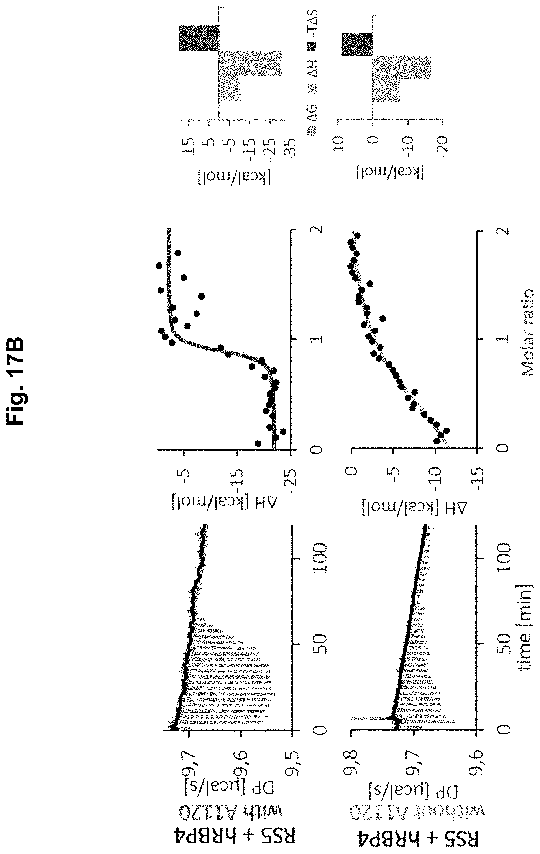
D00026

D00027

D00028

D00029

S00001
XML
uspto.report is an independent third-party trademark research tool that is not affiliated, endorsed, or sponsored by the United States Patent and Trademark Office (USPTO) or any other governmental organization. The information provided by uspto.report is based on publicly available data at the time of writing and is intended for informational purposes only.
While we strive to provide accurate and up-to-date information, we do not guarantee the accuracy, completeness, reliability, or suitability of the information displayed on this site. The use of this site is at your own risk. Any reliance you place on such information is therefore strictly at your own risk.
All official trademark data, including owner information, should be verified by visiting the official USPTO website at www.uspto.gov. This site is not intended to replace professional legal advice and should not be used as a substitute for consulting with a legal professional who is knowledgeable about trademark law.