Method For Manufacturing A Hyperbranched Polyester Polyol Derivative
HAAG; Rainer ; et al.
U.S. patent application number 16/763939 was filed with the patent office on 2020-12-10 for method for manufacturing a hyperbranched polyester polyol derivative. This patent application is currently assigned to FREIE UNIVERSITAT BERLIN. The applicant listed for this patent is FREIE UNIVERSITAT BERLIN. Invention is credited to Mohsen ADELI, Magda FERRARO, Rainer HAAG, Ehsan MOHAMMADIFAR, Fatemeh ZABIHI.
| Application Number | 20200385517 16/763939 |
| Document ID | / |
| Family ID | 1000005100555 |
| Filed Date | 2020-12-10 |


View All Diagrams
| United States Patent Application | 20200385517 |
| Kind Code | A1 |
| HAAG; Rainer ; et al. | December 10, 2020 |
METHOD FOR MANUFACTURING A HYPERBRANCHED POLYESTER POLYOL DERIVATIVE
Abstract
It is provided a method for manufacturing a hyperbranched polyester polyol derivative, comprising the following steps: a) reacting only glycidol and .epsilon.-caprolactone at a temperature lying in a range of between 40.degree. C. and 140.degree. C. to obtain a hyperbranched polyester polyol in which caprolactone residues are randomly arranged; b) reacting the hyperbranched polyester polyol of step a) with a sulfation reagent to obtain a sulfated hyperbranched polyester polyol as hyperbranched polyester polyol derivative.
| Inventors: | HAAG; Rainer; (Berlin, DE) ; MOHAMMADIFAR; Ehsan; (Berlin, DE) ; FERRARO; Magda; (Berlin, DE) ; ZABIHI; Fatemeh; (Berlin, DE) ; ADELI; Mohsen; (Berlin, DE) | ||||||||||
| Applicant: |
|
||||||||||
|---|---|---|---|---|---|---|---|---|---|---|---|
| Assignee: | FREIE UNIVERSITAT BERLIN Berlin DE |
||||||||||
| Family ID: | 1000005100555 | ||||||||||
| Appl. No.: | 16/763939 | ||||||||||
| Filed: | November 13, 2018 | ||||||||||
| PCT Filed: | November 13, 2018 | ||||||||||
| PCT NO: | PCT/EP2018/081068 | ||||||||||
| 371 Date: | May 13, 2020 |
| Current U.S. Class: | 1/1 |
| Current CPC Class: | C08G 63/664 20130101; C08G 63/82 20130101; C08G 65/2645 20130101; A61K 47/34 20130101; C08G 63/08 20130101; C08G 63/6882 20130101; C08G 83/006 20130101 |
| International Class: | C08G 63/08 20060101 C08G063/08; C08G 63/664 20060101 C08G063/664; C08G 63/688 20060101 C08G063/688; C08G 63/82 20060101 C08G063/82; C08G 83/00 20060101 C08G083/00; C08G 65/26 20060101 C08G065/26; A61K 47/34 20060101 A61K047/34 |
Foreign Application Data
| Date | Code | Application Number |
|---|---|---|
| Nov 14, 2017 | EP | 17201626.3 |
Claims
1. A method for manufacturing a hyperbranched polyester polyol derivative, comprising the following steps: a) reacting only glycidol and .epsilon.-caprolactone at a temperature lying in a range of between 40.degree. C. and 140.degree. C. to obtain a hyperbranched polyester polyol in which caprolactone residues and glycerol residues are randomly arranged, b) reacting the hyperbranched polyester polyol of step a) with a sulfation reagent to obtain a sulfated hyperbranched polyester polyol as hyperbranched polyester polyol derivative.
2. The method according to claim 1, wherein step a) is carried out in presence of a catalyst.
3. The method according to claim 2, wherein the catalyst is a Lewis acid.
4. The method according to claim 2, wherein the catalyst is tin(II) 2-ethylhexanoate.
5. The method according to claim 1, wherein a molar ratio between glycidol and .epsilon.-caprolactone is between 1:1 and 10:1.
6. The method according to claim 1, wherein the sulfation reagent is a sulfur trioxide base complex.
7. A hyperbranched polyester polyol derivative obtainable by a method according to claim 1.
8. The hyperbranched polyester polyol derivative according to claim 7, wherein it has a degree of sulfation of from 70 to 100%.
9.-12. (canceled)
13. A medicament, comprising a hyperbranched polyester polyol derivative according to claim 7 both as carrier for a pharmaceutically active substance and as a pharmaceutically active substance itself.
14.-15. (canceled)
Description
CROSS-REFERENCE TO A RELATED APPLICATION
[0001] This application is a National Phase Patent Application of International Patent Application Number PCT/EP2018/081068, filed on Nov. 13, 2018, which claims priority of European Patent Application Number 17 201 626.3, filed on Nov. 14, 2017.
BACKGROUND
[0002] The disclosure relates to a method for manufacturing a hyperbranched polyester polyol derivative, to a hyperbranched polyester polyol derivative that can be produced with this method and to a medicament comprising such a hyperbranched polyester polyol derivative.
[0003] Hyperbranched polyglycerols (hPGs) offer a variety of biomedical applications due to their unique physicochemical properties such as biocompatibility and multi-functionality. However, lack of biodegradability under physiological conditions hampers their in vivo applications. In prior art, different approaches have been described in order to enhance the biodegradability of linear, star-shaped and branched polyglycerols. However, many of those approaches are connected with severe drawbacks.
[0004] WO 2012/031245 A1 describes a composition comprising hyperbranched polyglycerol macromers, a cross-linker, a biodegradable component and a thermoresponsive component. This composition is manufactured by producing hyperbranched polyglycerol in a first step, adding methacrylate residues in a second step, preparing 2-hydroxyethyl methacrylate-poly(lactic acid) (HEMAPLA) in a third step, and producing a copolymer of the methacrylate-activated polyglycerol and HEMAPLA. The resulting compound has a star-shaped structure, i.e., it is no hyperbranched polymer, but only comprises a polar hyperbranched polyglycerol core onto which HEMAPLA residues are attached. The polyglycerol core of this compound is not biodegradable even after HEMAPLA addition.
[0005] US 2015/0037375 A1 describes a surfactant in the form of an oligoglycerol typically having 3 to 4 glycerol units. It is (chemically incorrect) also denoted as polyglycerol and has a star-shaped structure with grafted substituents. Other manufacturing methods described in this patent application lead to a linear polymer (due to the use of a protective group) or to a hydrophobic linear block copolymer comprising glycerol units.
[0006] US 2016/0331875 A1 describes a linear block copolymer made of caprolactone units and glycerol units. Due to its structure, no surface modification like sulfation of this linear block copolymer is possible.
[0007] Li and Li (Li, Z., & Li, J. (2013): Control of hyperbranched structure of polycaprolactone/poly (ethylene glycol) polyurethane block copolymers by glycerol and their hydrogels for potential cell delivery. The Journal of Physical Chemistry B, 117(47), 14763-14774) describe a complex manufacturing method for a polymer always comprising polyethylene glycol (PEG) segments, caprolactone segments and glycerol residues. The reaction necessitates hexamethylene diisocyanate (HDI) as coupling reagent. In doing so, the resulting polymer also always comprises HDI residues, wherein the PEG segments, caprolactone segments and glycerol residues are linked to each other by urethane linkages.
[0008] Zhou et al. (Zhou, J., Wang, W., Villarroya, S., Thurecht, K. J., & Howdle, S. M. (2008): Epoxy functionalised poly (.epsilon.-caprolactone): synthesis and application. Chemical Communications, (44), 5806-5808) describe an epoxy functionalization of .epsilon.-caprolactone, wherein the functionalized .epsilon.-caprolactone is further copolymerized with CO.sub.2 or succinic anhydride. The resulting copolymer comprises polycarbonate residues or polysuccinate residues. In order to keep the glycidol ring intact during the initial functionalization step, lipase is used as catalyst.
SUMMARY
[0009] It is an object underlying the proposed solution to provide a particularly simple manufacturing method for a pharmaceutically active compound that can also act as carrier for other pharmaceutically active compounds.
[0010] This object is achieved by a method for manufacturing a hyperbranched polyester polyol derivative having features as described herein. Such a manufacturing method is a two-step method and comprises the steps explained in the following. In a first step, glycidol is reacted with .epsilon.-caprolactone. A hyperbranched polyester polyol results in which caprolactone residues and glycerol residues are randomly arranged. The reaction is carried out as ring-opening polymerization so that the caprolactone residues in the resulting polymer are no longer circular but rather linear. They can also be denoted as hexanoyl residues.
[0011] In a second step, the hyperbranched polyester polyol of the first step is reacted with a sulfation reagent. A sulfated hyperbranched polyester polyol results that serves as hyperbranched polyester polyol derivative.
[0012] In an embodiment, the reaction is carried out as one-pot reaction. In an embodiment, it only consists of the precedingly explained steps (and optional purification steps). Thus, the hyperbranched polyester polyol derivative can be manufactured in an extremely simple manner. As will be explained below, it has anti-inflammatory properties, i.e., it is pharmaceutically active. Furthermore, it can be used as carrier for other pharmaceutically active substances.
[0013] Due to its simplicity, it is possible to perform this manufacturing method in technically large-scale so that it is suited to produce significant amounts of the hyperbranched polyester polyol derivative.
[0014] It is neither necessary nor intended to copolymerize any other substances than glycidol and .epsilon.-caprolactone. The first step of the method specifically does not make use of any other substances than glycidol and .epsilon.-caprolactone to be polymerized. To be more precisely, no coupling reagents like HDI are used. In addition, no aromatic substances are used to be copolymerized for building up the hyperbranched polyester polyol derivative.
[0015] Due to the copolymerized caprolactone residues, the hyperbranched polyester polyol derivative comprises aliphatic hydrophobic segments. Together with hydrophilic glycerol residues, an overall amphiphilic structure results. It is particularly appropriate for transporting substances, in particular pharmaceutically active substances, in particular hydrophobic substances (whether pharmaceutically active or not).
[0016] The hyperbranched polyester polyol derivative has no core-shell structure, but rather shows a random structure having a statistical distribution of hydrophobic caprolactone residues within hydrophilic glycerol residues. A significant part of hydroxyl groups of the glycerol residues are sulfated.
[0017] It could be experimentally shown that the synthesized hyperbranched polyester polyol derivative is susceptible to enzymatic cleavage, but remains stable under neutral and acidic conditions. It showed high cellular uptake that was proven by laser scanning confocal microscopy (LSCM). MTT assays did not show a significant toxicity against HaCaT cells up to concentration of 1000 .mu.g/ml. Thus, the hyperbranched polyester polyol derivative showed good biodegradability and biocompatibility. Furthermore, it was able to form nanoparticles in aqueous solutions and to load hydrophobic guest molecules.
[0018] In an embodiment, the first step of the method is carried out in presence of a catalyst. It turned out that a Lewis acid such as a tin compounds acting as Lewis acid is a particularly appropriate catalyst for this reaction step. In an embodiment, tin(II) 2-ethylhexanoate (tin octoate, Sn(Oct).sub.2) is used as catalyst. Preliminary data revealed that other catalysts such as Novozym 435 are likewise appropriate. Furthermore, the reaction can be carried out without any catalysts as further experiments of the inventors revealed. Then, only an elevated temperature lying in a range of 30.degree. C. and 150.degree. C., in particular 30.degree. C. to 70.degree. C. or any of the temperature ranges mentioned in the next paragraph appears favorable to achieve a sufficiently high yield.
[0019] The reaction can be carried out under mild (ambient) conditions. In an embodiment, the reaction is carried out at a reaction temperature lying in a range of between 30.degree. C. and 150.degree. C., in particular between 40.degree. C. and 140.degree. C., in particular between 50.degree. C. and 130.degree. C., in particular between 60.degree. C. and 120.degree. C., in particular between 70.degree. C. and 110.degree. C., in particular between 80.degree. C. and 105.degree. C., in particular between 90.degree. C. and 100.degree. C. Particular appropriate reaction temperatures are temperatures at or around 50.degree. C., at or around 90.degree. C., and at or around 120.degree. C.
[0020] In an embodiment, a molar ratio between glycidol and .epsilon.-caprolactone is between 1 to 1 and 10 to 1, in particular between 2 to 1 and 9 to 1, in particular between 3 to 1 and 8 to 1, in particular between 4 to 1 and 7 to 1, in particular between 5 to 1 and 6 to 1. Thus, it is always used at least as much glycidol as .epsilon.-caprolactone, but particularly more glycidol than .epsilon.-caprolactone. Particular appropriate molar ratios between glycidol and .epsilon.-caprolactone are molar ratios of 2 to 1 and 4 to 1.
[0021] Obviously, the molar ratio between glycidol and .epsilon.-caprolactone influences the structure of the resulting hyperbranched polyester polyol. In addition, the reaction temperature also influences the resulting structure. Particular appropriate combinations between molar ratios and reaction temperatures are a molar ratio of 2 to 1 and a temperature of 50.degree. C., 90.degree. C., or 120.degree. C. as well as a molar ratio of 4 to 1 and a temperature of 50.degree. C., 90.degree. C., or 120.degree. C.
[0022] The sulfation reagent used in the second step of the method is, in an embodiment, a sulfur trioxide base complex (e.g., SO.sub.3-Pyridine, or SO.sub.3-triethylamine). Sulfur trioxide pyridine complex is particularly appropriate. The sulfation can be carried out as generally known in the art. It takes place at the free hydroxyl groups of the hyperbranched polyester polyol so that a sulfated hyperbranched polyester polyol results.
[0023] Appropriate reaction conditions for the sulfation step comprise a reaction temperature of 40.degree. C. to 80.degree. C., in particular of 45.degree. C. to 75.degree. C., in particular of 50.degree. C. to 70.degree. C., in particular of 55.degree. C. to 65.degree. C., in particular of 60.degree. C. to 80.degree. C., and/or a reaction duration of 12 hours to 2 days, in particular of 1 day to 1.5 days. Particular appropriate reaction conditions are a reaction temperature of 55.degree. C. to 65.degree. C. (such as 60.degree. C.) and a reaction duration of approximately one day.
[0024] In an aspect, the proposed solution also relates to a hyperbranched polyester polyol derivative that can be obtained by a method according to any of the preceding explanations. As already stated above, the concrete structure of such hyperbranched polyester polyol derivative cannot be exactly described a more concrete terms than by making reference to its manufacturing method since the reaction temperature and the molar ratio of the educts, i.e., of glycidol and of .epsilon.-caprolactone, influence the resulting molecular structure of the hyperbranched polyester polyol derivative.
[0025] In an embodiment, the hyperbranched polyester polyol derivative has a degree of sulfation of from 70 to 100%, in particular of from 75 to 99%, in particular of from 80 to 98%, in particular of from 85 to 97%, in particular of from 90 to 95%. Thus, a significant amount of hydroxyl groups of the hyperbranched polyester polyol resulting from the first step of the method is sulfated and the second step of the method according to this embodiment.
[0026] In an embodiment, the hyperbranched polyester polyol derivative has a number average molecular weight lying in a range of between 25 and 75 kDa, in particular of between 30 and 70 kDa, in particular of between 35 and 65 kDa, in particular of between 40 and 60 kDa, in particular of between 45 and 55 kDa.
[0027] The hyperbranched polyester polyol derivative as described above has a very good anti-inflammatory activity. This will be shown in detail with respect to exemplary embodiments. Due to this activity, the use of such a hyperbranched polyester polyol derivative as drug is also claimed.
[0028] In an embodiment, the drug is a drug for inhibiting the complement system of an organism and/or for inhibiting L-selectin binding to its natural receptor. By such an inhibition, an anti-inflammatory effect is achieved. Thus, in an embodiment, the hyperbranched polyester polyol derivative is used as drug for treating an inflammatory disease.
[0029] In an aspect, the solution relates to a medical method comprising administering a hyperbranched polyester polyol derivative according to the above explanations or a drug comprising such a hyperbranched polyester polyol derivative to a person in need thereof. This medical method is in particular a method for treating an inflammatory condition of the person. In an embodiment, the method is a method for inhibiting the complement system of an organism and/or for inhibiting L-selectin binding to its natural receptor.
[0030] In another embodiment, the hyperbranched polyester polyol derivative is used for targeting inflammatory tissue in vivo. It is possible to couple the hyperbranched polyester polyol derivative with a detectable probe so as to use the construct of hyperbranched polyester polyol derivative and probe for diagnosing inflammatory diseases, in particular chronic inflammatory diseases such as rheumatoid arthritis or psoriasis or acute inflammatory processes such as those occurring after an organ transplant. Suited detectable probes are fluorescence probes, contrast agents, magnetic agents etc.
[0031] In an aspect, the solution relates to a medical method for targeting inflammatory tissue in vivo, comprising administering a hyperbranched polyester polyol derivative according to the above explanations or a drug comprising such a hyperbranched polyester polyol derivative to a person in need thereof. Any of the embodiments described in the preceding section for an according use can, in an embodiment, also be applied to such a medical method.
[0032] In an embodiment, the solution relates to a drug comprising a hyperbranched polyester polyol derivative according to the preceding explanations as active ingredient. However, the hyperbranched polyester polyol derivative cannot be used only as active ingredient, but also as a carrier molecule for transporting other molecules to a desired site of action (such as inflammatory tissue). To give an example, skin penetration tests showed that the synthesized polymers penetrated into the skin and transported the hydrophobic guest molecules through the stratum corneum to deeper skin layers. The hyperbranched polyester polyols (hPPs) were degraded to smaller segments in the presence of skin lysate. This property makes them proper candidates for the intradermal drug delivery due to both a facilitated drug release and low accumulation inside the tissue. Therefore, the solution relates in an embodiment also to a drug comprising both a hyperbranched polyester polyol derivative and additionally a further pharmaceutically active substance. Thereby, the hyperbranched polyester polyol derivative acts as carrier for the further pharmaceutically active substance and is itself pharmaceutically active.
[0033] In an aspect, the solution relates to a medical method comprising administering such a hyperbranched polyester polyol derivative or a drug comprising such a hyperbranched polyester polyol derivative carrying in each case a pharmaceutically active substance, in particular a hydrophobic pharmaceutically active substance, to a person in need thereof. In an embodiment, administering is carried out as intradermal administering.
[0034] In an aspect, the solution relates to the use of a hyperbranched polyester polyol that can be obtained by a method according to the first step of the method according to the above explanations (i.e., a non-sulfated intermediate product) as carrier for a pharmaceutically active substance, in particular of a hydrophobic pharmaceutically active substance. Such intermediate product does not carry sulfate groups and is thus not pharmaceutically active itself. As will be outlined in connection to an exemplary embodiment, the intermediate product is nonetheless well appropriate to act as substance carrier.
[0035] In an aspect, the solution relates to a medical method comprising administering such an intermediate product or a drug comprising such an intermediate product carrying in each case a pharmaceutically active substance, in particular a hydrophobic pharmaceutically active substance, to a person in need thereof. In an embodiment, administering is carried out as intradermal administering.
[0036] In an aspect, the solution relates to the use of a hyperbranched polyester polyol derivative that can be obtained by a method according to the above explanations in intradermal delivery of a pharmaceutically active substance, in particular of a hydrophobic pharmaceutically active substance.
[0037] In an aspect, the solution relates to the use of a hyperbranched polyester polyol that can be obtained by a method according to the first step of the method according to the above explanations (i.e., a non-sulfated intermediate product) in intradermal delivery of a pharmaceutically active substance, in particular of a hydrophobic pharmaceutically active substance.
[0038] All embodiments explained in connection to the described manufacturing method can also be applied to the described hyperbranched polyester polyol derivative, to the described medicament, and to the described uses, and vice versa in each case.
BRIEF DESCRIPTION OF THE DRAWINGS
[0039] Details of aspects of the proposed solution will be explained with respect to exemplary embodiment and accompanying Figures.
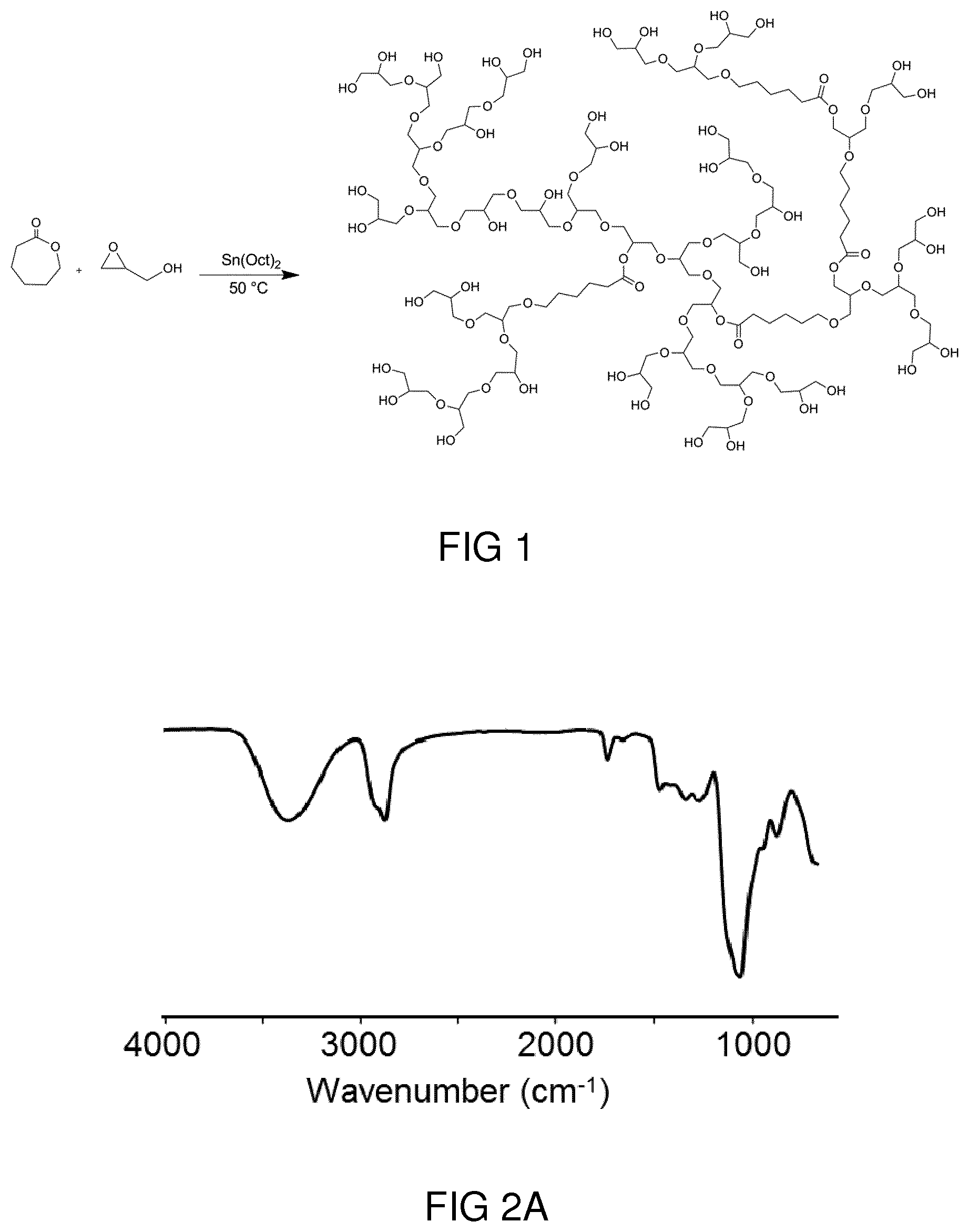
[0040] FIG. 1 shows a schematic depiction of the synthesis of a random copolymer of glycidol and .epsilon.-caprolactone through ring opening polymerization.
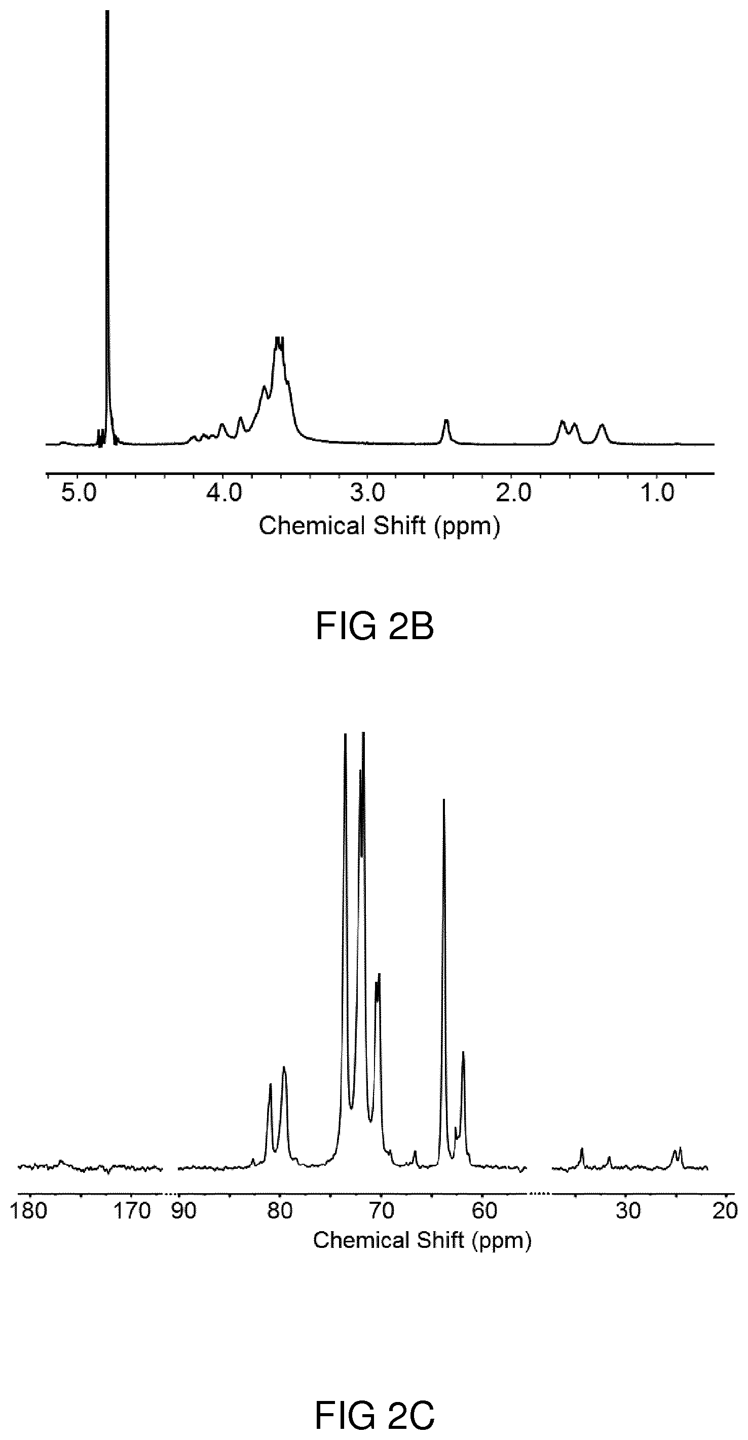
[0041] FIG. 2A shows an IR spectrum of hPP41-50 as exemplary embodiment of a hyperbranched polyester polyol.
[0042] FIG. 2B shows a .sup.1H NMR spectrum of hPP41-50 recorded in D.sub.2O.
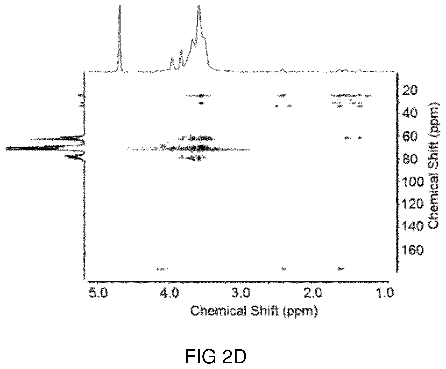
[0043] FIG. 2C shows a .sup.13C NMR spectrum of hPP41-50 recorded in D.sub.2O.
[0044] FIG. 2D shows a .sup.1H-HSQC NMR spectrum of hPP41-50 recorded in D.sub.2O.
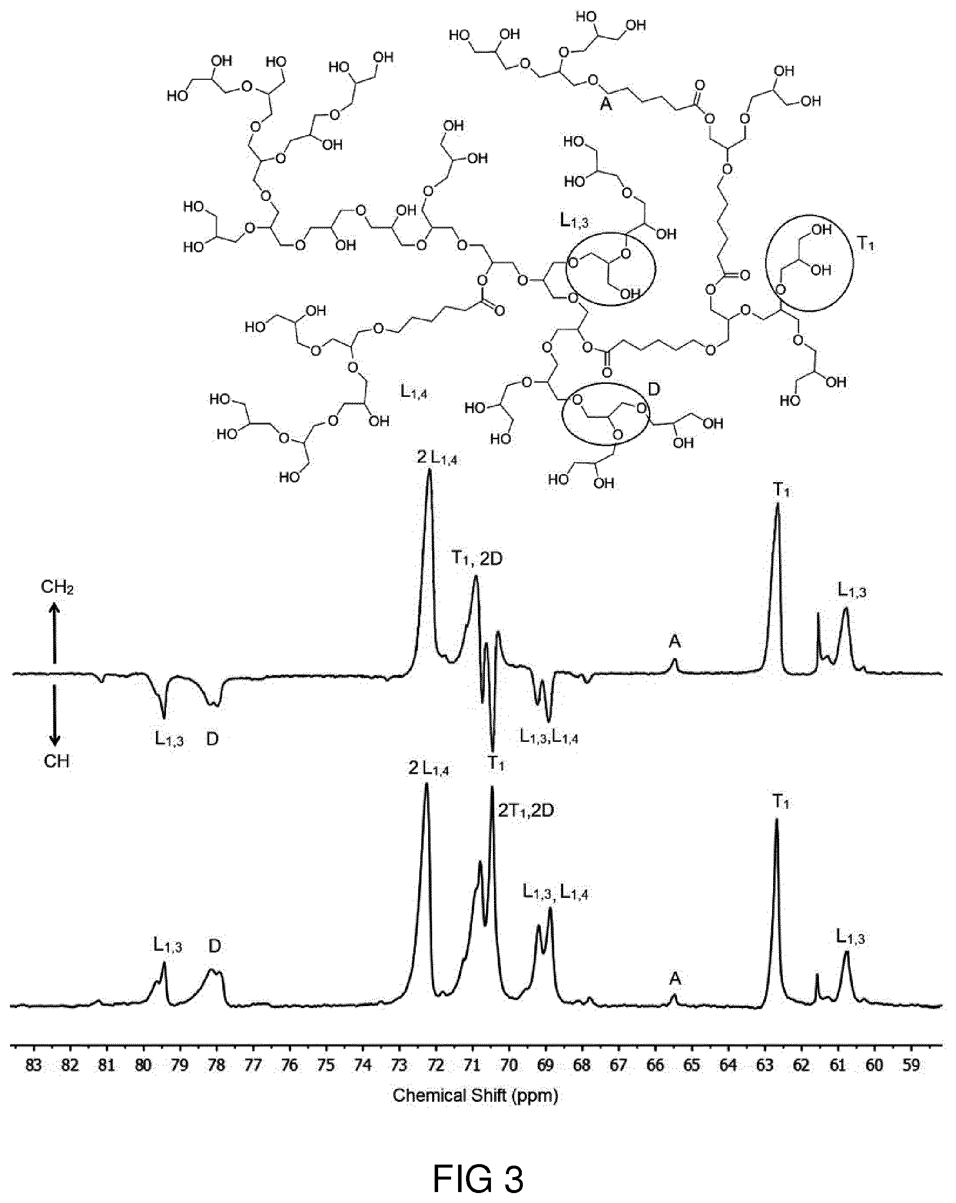
[0045] FIG. 3 shows an assignment of different structural units in inverse-gated .sup.13C NMR (upper panel) and .sup.13C DEPT (lower panel) spectra of hPP41-50 in D.sub.2O.
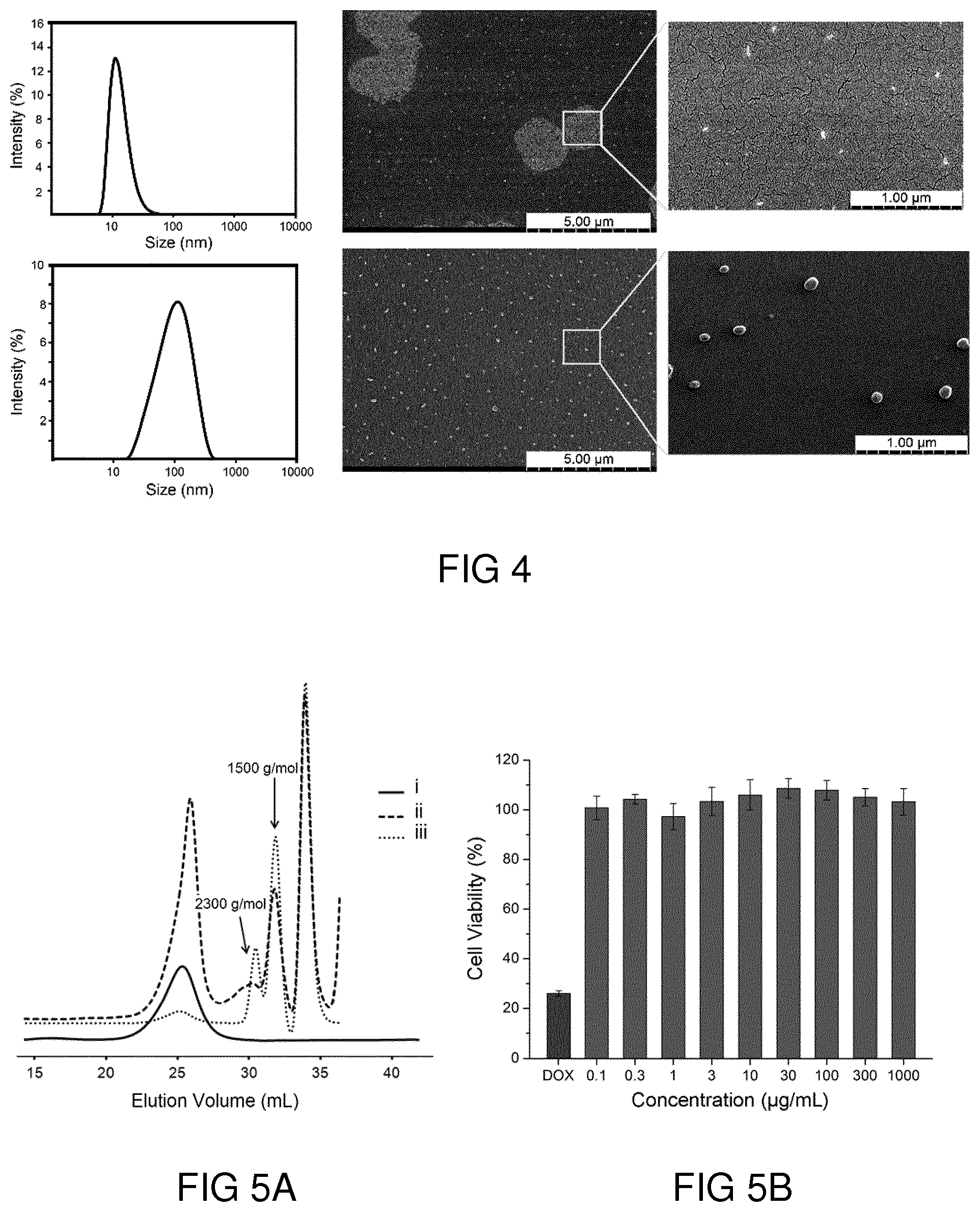
[0046] FIG. 4 shows scanning electron microscopy (SEM) images (in two scales) and DLS diagrams of hPPs synthesized at 50.degree. C. (upper row) and 120.degree. C. (lower row).
[0047] FIG. 5A shows a gel permeation chromatography (GPC) diagram of (i) pristine hPOGC41-50, its degradation products in presence of (ii) skin lysate after 12 hours and (iii) Novozyme after 3 days.
[0048] FIG. 5B shows cytotoxicity results obtained from a CCK8 assay upon incubation of HaCaT cells with hPP41-50 after 24 h.
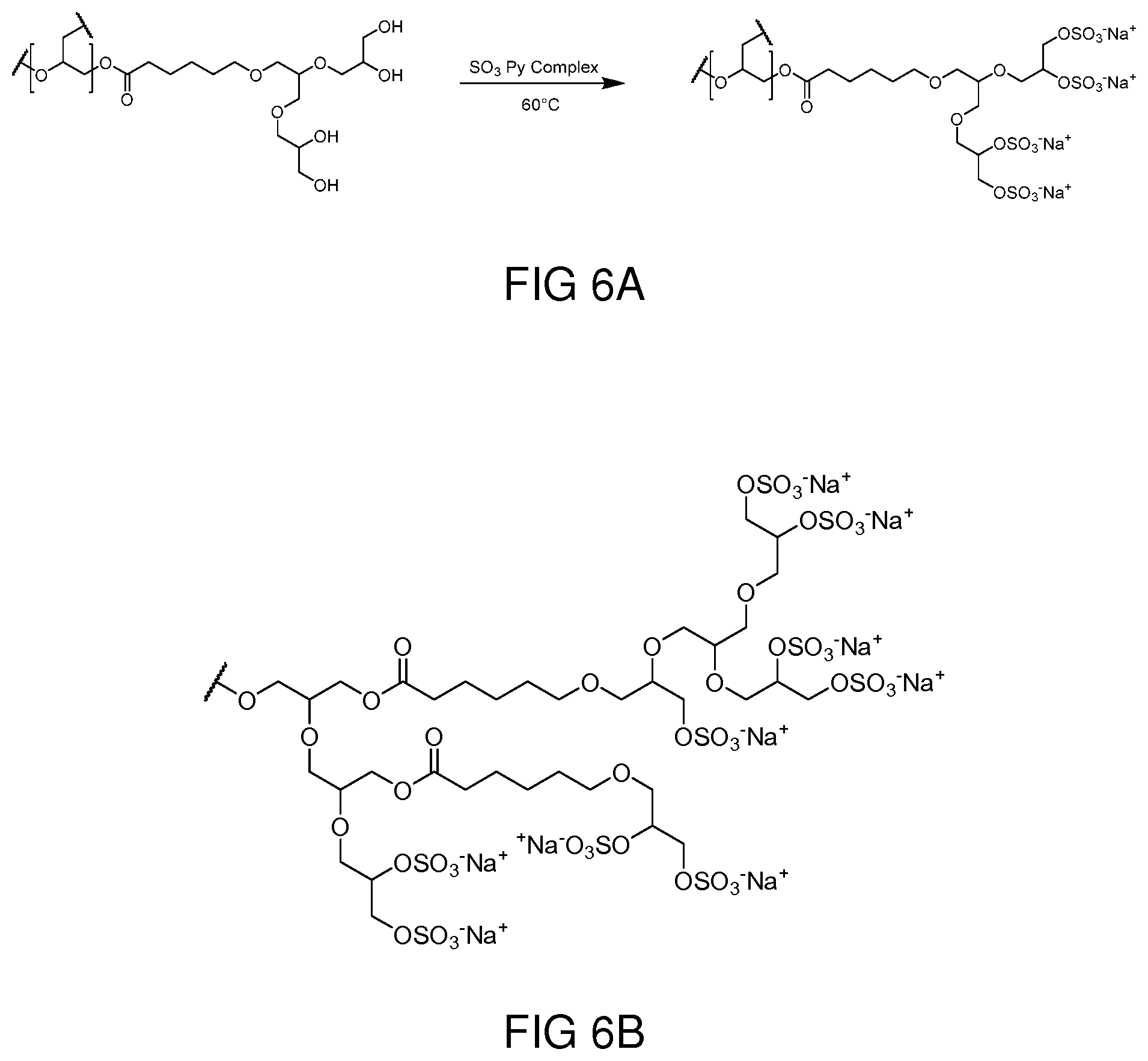
[0049] FIG. 6A shows a schematic depiction of the synthetic pathway for the sulfation of terminal hydroxyl groups of a hyperbranched polyester polyol.
[0050] FIG. 6B shows a schematic depiction of a sulfated hyperbranched polyester polyol.
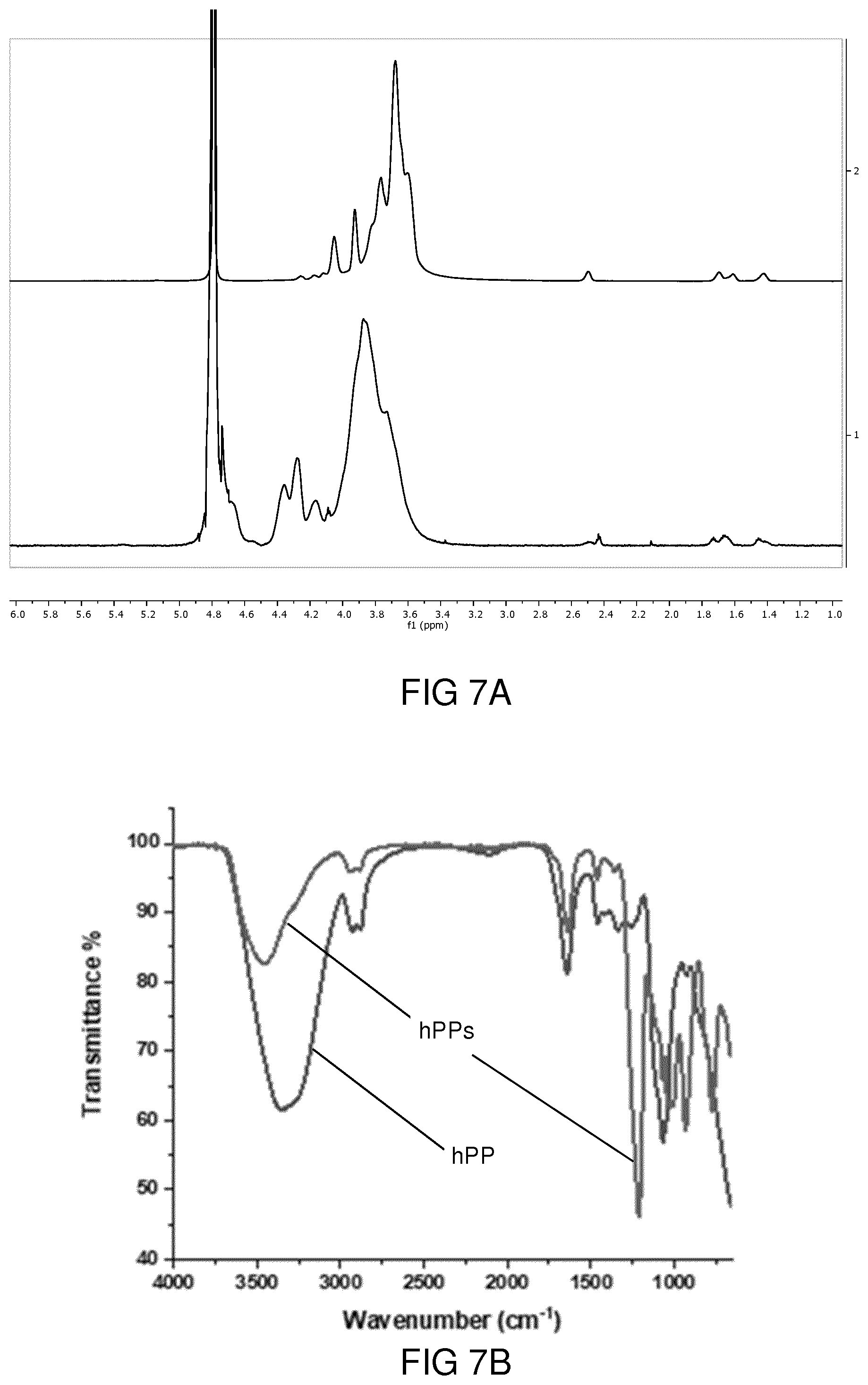
[0051] FIG. 7A shows .sup.1H NMR spectra of hPP and its sulfated derivative both measured in D.sub.2O.
[0052] FIG. 7B shows infrared (IR) spectra of hPP and its sulfated derivative hPPs.
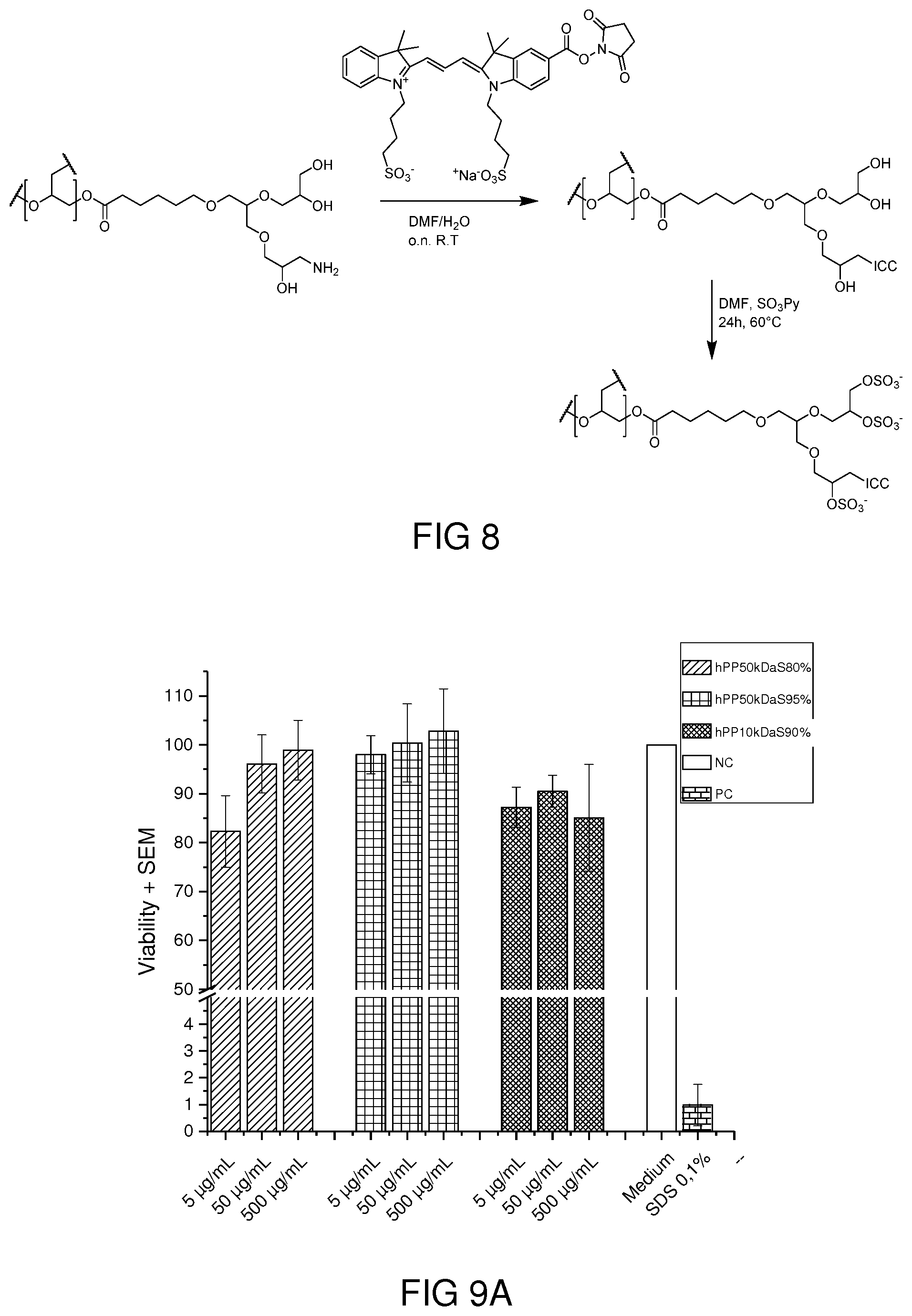
[0053] FIG. 8 shows a schematic depiction of the synthetic pathway of dye-conjugate polymer synthesis.
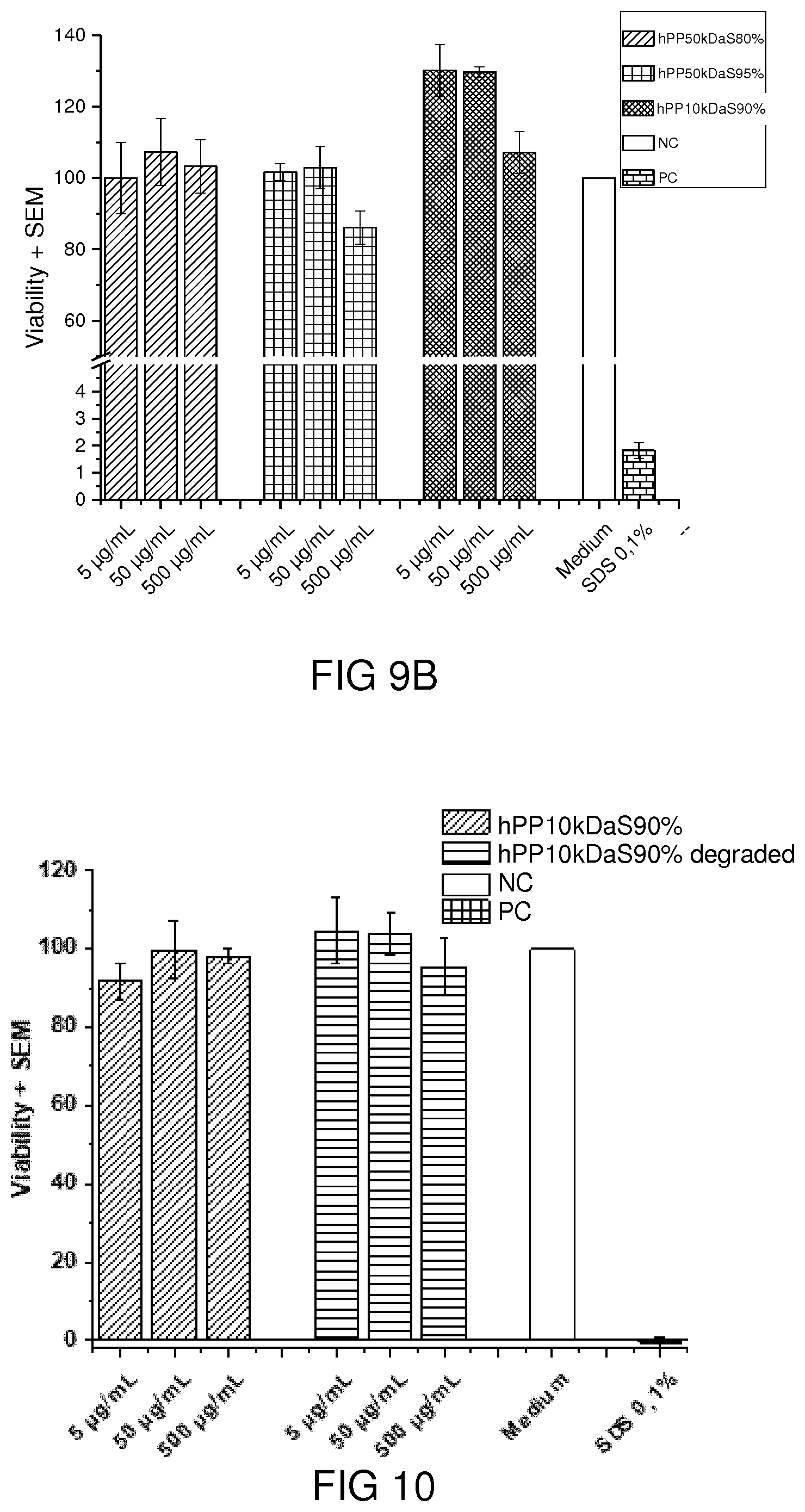
[0054] FIG. 9A shows the results of a cell viability test on Caco-2 cell line.
[0055] FIG. 9B shows the results of a cell viability test on A549cell line.
[0056] FIG. 10 shows the results of a cell viability test before and after degradation on HaCaT cell line.
[0057] FIG. 11 shows the results of fluorescence-activated cell sorting of A549 cells and Caco-2 cells.
[0058] FIG. 12 shows the results of an L-selectin inhibition tests.
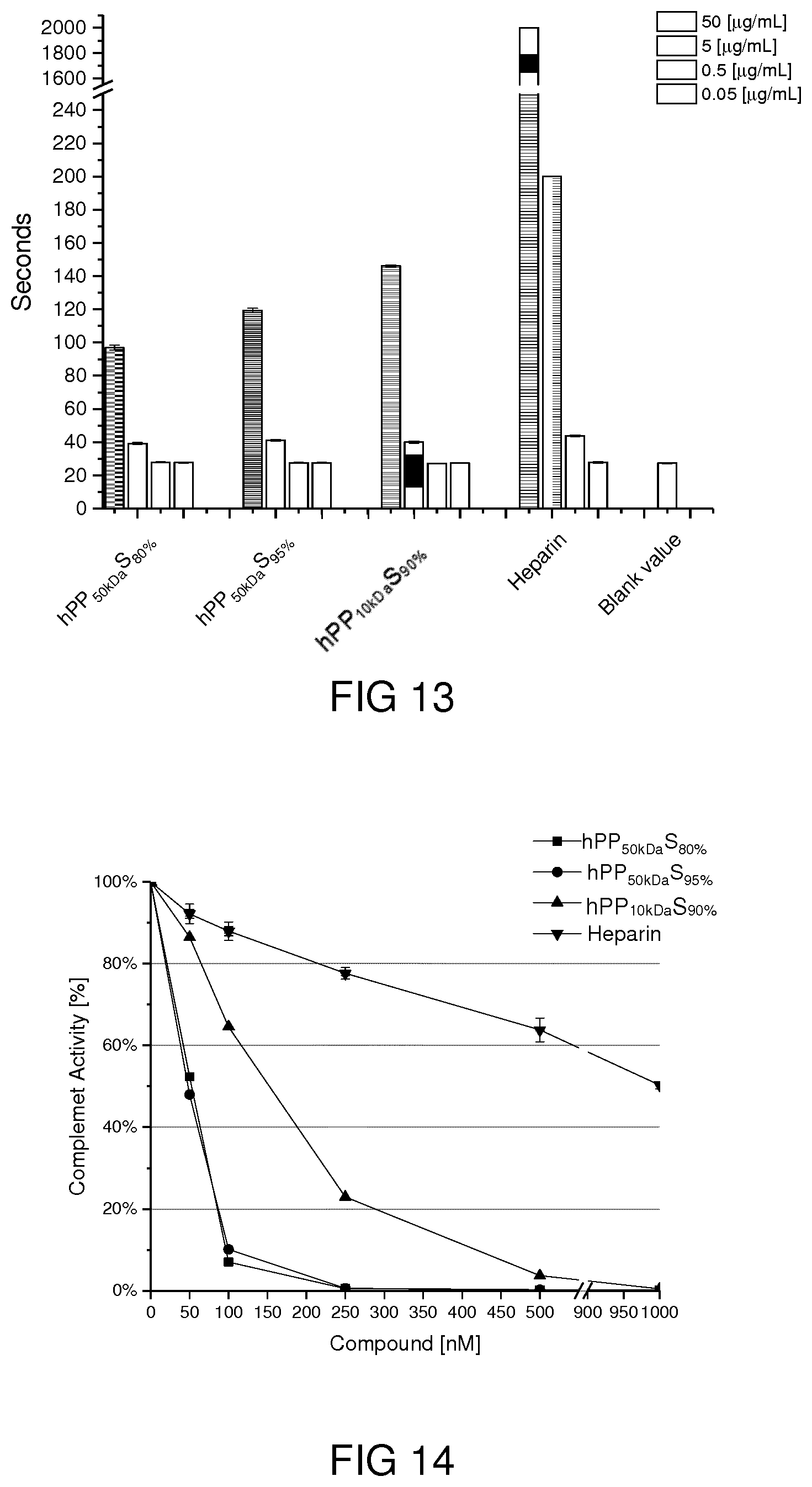
[0059] FIG. 13 shows the results of a blood clotting assay.
[0060] FIG. 14 shows the results of a complement system activation test.
[0061] FIG. 15A shows 1H NMR spectra of the sulfated polymers at different times of incubation with esterase.
[0062] FIG. 15B shows the variation of the integral of the signals highlighted in the NMR spectra of FIG. 15A.
[0063] FIG. 16A shows the results of a gel permeation chromatography analysis of degrading polymer.
[0064] FIG. 16B shows the results of a complement system activity analysis.
DETAILED DESCRIPTION
[0065] FIGS. 1 to 5B will be explained in connection to an exemplary embodiment relating to different non-sulfated hyperbranched polyester polyols, i.e., intermediate products resulting from the first step of the above-explained manufacturing method. In the following, details of the experiments will be explained that were performed in order to obtain the results depicted in FIGS. 1 to 5B.
[0066] Gel permeation chromatography (GPC). GPC measurements were performed using an Agilent 1100 solvent delivery system with a manual injector, isopump, and Agilent 1100 differential refractometer. The Brookhaven BIMwA7-angle light scattering detector was coupled to a size exclusion chromatography (SEC) to measure the molecular weight for each fraction of the polymer that was eluted from the SEC columns. For separation of the polymer samples, three 30 cm columns were used (10 .mu.m PSS Suprema columns with pore sizes of 100 .ANG., 1000 .ANG., 3000 .ANG.). Water was used as mobile phase; the flow rate was set at 1.0 mL/min. All columns were held at room temperature. For each measurement, 100 .mu.L of samples with concentration of 5 mg mL.sup.-1 solution was injected. For acquisition of the data from seven scattering angles (detectors) and differential refractometer WinGPC Unity from PSS was used. Molecular-weight distributions were determined by comparison with Pullulan standards (10 different sizes from 342 to 710,000 g mol.sup.-1). Water was used as a solvent with 0.1 M NaNO.sub.3.
[0067] Dynamic light scattering (DLS). The size of hPP nanoparticles in aqueous solution was measured using a Zetasizer Nano ZS analyzer with an integrated 4 mW He--Ne laser at a wavelength of 633 nm with a backscattering detector angle of 173.degree. (Malvern Instruments Ltd, UK) at 25.degree. C. For DLS experiments, an aqueous solution of polymer with different concentrations was prepared in Milli-Q water and vigorously stirred for 18 hours at room temperature (25.degree. C.). Solutions were filtered via 0.45 .mu.m polytetrafluoroethylene (PTFE) filters and used for dynamic light scattering measurements. Disposable UV-transparent cuvettes (Sarstedt AG & Co., Germany) were used for all the experiments.
[0068] Thermogravimetric analysis (TGA). TGA experiments were performed on a LINSEIS STA
[0069] PT1600 (TG-DTA/DSC) machine in air atmosphere. The heating rate was set to 10.degree. C./min. Calibration curves were measured for each sample. Measurements were performed in Al.sub.2O.sub.3 crucibles. Mass of samples varied from 15 mg to 20 mg.
[0070] Fourier transform infrared spectroscopy (FTIR). IR spectra were recorded with Nicolet AVATAR 320 FT-IR 5 SXC (Thermo Fisher Scientific, Waltham, Mass., USA) with a DTGS detector from 4000 to 650 cm.sup.-1. Sample measurement was performed by dropping a solution of compound and letting the solvent evaporate for a few seconds.
[0071] Nuclear magnetic resonance (NMR). NMR spectra were recorded on a Jeol ECX 400 or a Jeol Eclipse 700 MHz spectrometer. Proton and carbon NMR were recorded in ppm and were referenced to the indicated solvents.
[0072] Confocal laser scanning microscopy (CLSM). CLSM images were recorded by Leica TCS SP8 with 63.times. oil-immersion objective lens and analyzed by Leica Confocal Software.
[0073] Microplate reader. The adsorption of CCK8 assay was measured on a TECAN Infinite M200 Pro microplate reader at the wavelength of 450 nm.
[0074] General procedure for synthesis and purification of hyperbranched polymer. A catalytic amount of tin octoate ([monomers including Glycidol and .epsilon.-carpolactone]/[catalyst]: 200/1) solution in toluene was added to a round-bottom flask connected to argon and a vacuum inlet and toluene was evaporated under reduced pressure. Glycidol and .epsilon.-carpolactone with predetermined molar ratios were then injected to the flask under argon atmosphere and stirred at 50.degree. C., 90.degree. C. and 120.degree. C. For example in one of the reactions, 378 mg catalyst in a toluene solution was transferred to a 100 mL round bottom flask. After removing the toluene 5.0 mL (0.15 mol) of glycidol and 4.15 mL (0.037 mol) of .epsilon.-carpolactone were added to reaction flask. The mixture was stirred (250 rpm) using a mechanical stirrer at 50.degree. C. for 48 h. The crude product was dissolved in methanol and dialyzed against a mixture of methanol/dimethyl formamide (DMF) (70:30) vol % for 24 h using dialysis bag (MWCO=10 kDa) to remove the reactants and low molecular weight side products. Dialysis process was continued for other 24 h against distilled water to remove the remained DMF. Then, the final product (4.5 g) was obtained as a highly viscous and colorless compound upon lyophilization.
[0075] Enzymatic degradation of polymers using Novozyme 435. The degradation of hPP41-50 was studied according to reported procedure (F. Du, S. Honzke, F. Neumann, J. Keilitz, W. Chen, N. Ma, S. Hedtrich and R. Haag, Journal of Controlled Release, 2016, 242, 42-49.) in presence of Candida Antarctica Lipase B that had been immobilized on acrylic resin, commercially known as Novozym-435. To a phosphate buffer solution (pH7.4) of polymer (5 mg mL.sup.-1), 200 wt % of Novozyme-435 and 10 .mu.L n-butanol were added and the samples were incubated at 37.degree. C. using a BioShake XP.RTM. (Biometra) and shaken at 1000 rpm for 3 days. The solution was then filtered centrifuged and freeze-dried and the obtained lyophilisate was analyzed via GPC and .sup.1H NMR to monitor the degradation.
[0076] Enzymatic degradation of hPP41-50 using skin lysate. Degradation of hPP41-50 nanocarriers in presence of skin lysate was investigated according to a previously reported procedure (F. M. Batz, W. Klipper, H. C. Korting, F. Henkler, R. Landsiedel, A. Luch, U. von Fritschen, G. Weindl and M. Schafer-Korting, European Journal of Pharmaceutics and Biopharmaceutics, 2013, 84, 374-385.). Briefly, freshly excised human skin was homogenized and then 0.5 mg ml.sup.-1 solution of hPP41-50 was incubated with the skin lysate for 12 h at 37.degree. C. Samples were centrifuged to separate the solid residues and the supernatant was lyophilized and subsequently analyzed by GPC. hPP41-50 nanocarriers have been also incubated with phosphate buffered saline (PBS) as negative control.
[0077] Covalent conjugation of fluorescein isothiocyanate (FITC) to hPP41-50. hPP41-50 (50 mg, 0.001 mmol) and fluorescein isothiocyanate (10 mg, 0.02 mmol) were dissolved in 20 mL of dry DMF and stirred at 60.degree. C. for 24 h. DMF was evaporated under reduced pressure and the crude product was dissolved in minimum amount of water and the unreacted FITC was separated through the size exclusion chromatography (SEC) with Sephadex G-25 (GE Healthcare) under ambient pressure and temperature. FITC-labeled hPP41-50 was obtained after lyophilization. .sup.1H NMR and fluorescence emission spectroscopy proved that FITC is successfully conjugated to hPP41-50.
[0078] Preparation of Nile red loaded nanocarriers. hPP41-50 nanocarriers were loaded with Nile red using the solvent evaporation method (Y. G. Bachhav, K. Mondon, Y. N. Kalia, R. Gurny and M. Moller, Journal of Controlled Release, 2011, 153, 126-132; M. Lapteva, K. Mondon, M. Moller, R. Gurny and Y. N. Kalia, Molecular Pharmaceutics, 2014, 11, 2989-3001.). Briefly, an excess of Nile red (50 wt % of polymer) was dissolved in methanol and added dropwise to an aqueous solution of polymer (5 mg mL.sup.-1) under vigorous stirring. Methanol was then slowly removed by using a rotary evaporator. After equilibration overnight, the solution was centrifuged at 5000 rpm for 15 min and the supernatant was carefully collected, lyophilized and dissolved in methanol for determination the concentration of loaded Nile red by calculated by UV-vis spectroscopy and using calibration curve. Loading capacity was calculated as Nile red concentration divided by polymer concentration.
[0079] Cell viability assays. All bio-experiments were performed according to the German genetic engineering law and German biosafety guidelines in laboratory, level 1. Dulbecco's Modified Eagle Medium (DMEM), Gibco and fetal bovine serum (FBS) were employed in the following experiments. HaCat cells were cultured in DMEM with 10% (v/v) FBS and 1% (v/v) antibiotics. And cells were incubated in a humidified atmosphere with 5% CO.sub.2 at 37.degree. C. HaCat cells (1.times.10.sup.4 cells/well) were seeded in a 96-well plate and incubated for 24 h before the tests. hPP41-50 in the culture medium (concentration from 0.1 .mu.g mL.sup.-1 to 1000 .mu.g mL.sup.-1) were added to the medium-removed 96-well plate and incubated for another 24 h. After that, medium solutions were removed and the cells were rinsed with PBS twice. Subsequently, 100 .mu.L DMEM with 10 .mu.L CCK8 solution was added to each well. After incubation for the following 2 hours, the absorption of each well was measured at wavelength of 450 nm with microplate reader. Cells without any treatments and cells treated with DOX.HCl (2 .mu.g mL.sup.-1) were used as negative and positive controls. Each sample was measured 3 times.
[0080] Cellular uptake. HaCat cells were seeded on 8-well plates (3.times.10.sup.4 cells/well). After 24 h, FITC labelled hPP41-50 dissolved in DMEM (10 .mu.g mL.sup.-1) was added to the cells. The cells were incubated at 37.degree. C. for different time intervals. Afterwards, the culture medium was removed and the cells were rinsed with PBS. The nucleus of HaCat cells were stained with Hoechst for 30 min and then the cells were observed with CLSM (Leica TCS SP8). Hoechst was excited at 350 nm with the emission at 460 nm and FITC was excited at 490 nm with the emission at 520 nm.
[0081] Skin penetration. Human skin (obtained from cosmetic surgeries with informed consent, ethics vote EA1/081/13) was punched to discs of 2 cm diameter and mounted onto static-type Franz cells (diameter 15 mm, volume 12 mL, PermeGear Inc., Bethlehem, Pa., USA) and experiments was performed according to finite dose approach. In order to investigate the potential dermal drug delivery and skin distribution of hPP41-50, Nile red and FITC were physically encapsulated and covalently attached to the hPP41-50, respectively. The experiments have been performed according to a reported procedure (F. Zabihi, S. Wieczorek, M. Dimde, S. Hedtrich, H. G. Borner and R. Haag, Journal of Controlled Release, 2016, 242, 35-41.) using excised human skin and a conventional base cream served as a reference. All formulations contained the same Nile red concentration (0.001 wt %). Prior to the experiment, human skin was thawed and discs of 2 cm diameter were punched and mounted onto static-type Franz cells (diameter 15 mm, volume 12 mL, PermeGear Inc., Bethlehem, Pa., USA) with the horny layer facing the air and the dermis having contact with the receptor fluid phosphate buffered saline pH 7.4 (PBS, 33.5.degree. C., skin surface temperature about 32.degree. C.) stirred at 500 rpm. After 30 min, 36 .mu.L of the test formulations and reference cream was applied onto the skin surface (finite-dose approach) and remained there for 6 h. Subsequently, the skin was removed from the Franz cells, and the skin surface was gently cleaned. Afterwards, treated skin areas were punched, embedded in tissue freezing medium (Jung, Nussloch, Germany) and stored at a temperature of -80.degree. C. To determine the polymer penetration, the skin discs were cut into vertical slices of 10 .mu.m thickness using a freeze microtome (Frigocut 2800 N, Leica, Bensheim, Germany). The slices were subjected to normal light and fluorescence light. Nile red and FITC distribution were visualized by fluorescence microscopy.
[0082] FIG. 1 shows a reaction scheme according to which different hyperbranched polyester polyols (hPPs) were synthesized by cationic ring-opening polymerization of glycidol in the presence of .epsilon.-caprolactone and Sn(Oct).sub.2 as catalyst in a one-step reaction. A series of polymerizations using different [glycidol]/[.epsilon.-caprolactone] ([G]/[C]) ratios were performed to investigate the role of the reactant molar ratio and the reaction temperature on the polymer structure.
[0083] Polymers synthesized by 2/1 and 4/1 molar ratios of [G]/[C] at 50.degree. C., are nominated as hPP21-50 and hPP41-50, respectively. The polymerizations for ([G]/[C]) 2/1 have been also performed at 90.degree. C. and 120.degree. C. and they are termed as hPP21-90 and hPP21-120, respectively (see Table 1 below). Since the hPP41-50 has been synthesized under milder conditions and showed high water solubility and relatively narrow polydispersity, it was selected for further characterization and skin penetration studies.
[0084] IR spectroscopy and different NMR techniques including .sup.1H NMR, .sup.13C NMR, DEPT NMR, DOSY, and .sup.13C, .sup.1H-HSQC spectra clearly showed the incorporation of caprolactone segments into the copolymer. The IR spectrum of hPPs shows absorbance bands at 1100 cm.sup.-1, 1727 cm.sup.-1, 2900 cm.sup.-1, and 3400 cm.sup.-1, which are assigned to the C--O, carbonyl groups, aliphatic C--H, and end hydroxyl groups, respectively (FIG. 2A). Thereby, areas highlighted in light grey are attributed to chemical groups of glycerol residues, and areas highlighted in dark grey are attributed to chemical groups of caprolactone residues.
[0085] In the .sup.1H NMR spectrum of FIG. 2B, signals at 1.4 ppm, 1.6 ppm, and 2.5 ppm (highlighted in dark grey) are assigned to methylene protons of caprolactone segments. Signals at 3.4-4.2 ppm and 4.8 ppm (highlighted in light grey) are attributed to the protons of glycerol residues and hydroxyl groups, respectively. Protons of --OCH2 groups in caprolactone segments, which appeared at 4.0 ppm, are covered by glycerol signals. In contrast to homopolymers and block copolymers, signals of caprolactone segments of the hyperbranched polyester polyol are broadened in .sup.1H NMR spectra. Additionally, signals of methylene groups in the vicinity of central carbon of caprolactone units are split into two signals (FIG. 2B).
[0086] In order to make sure that the formation of self-assemblies in water does not affect the .sup.1HNMR spectra, this measurement has been also performed in deuterated DMSO and DMF. It could be seen that the spectra are similar to that of measured in D.sub.2O (data not shown).
[0087] In the .sup.13C NMR spectrum of FIG. 2C, signals at 22-35 ppm and 177 ppm are assigned to caprolactone segments (highlighted in dark grey). In this spectrum, the glycerol signals appeared at 60-82 ppm (highlighted in light grey).
[0088] Two-dimensional NMR techniques, including heteronuclear single quantum coherence spectroscopy (HSQC) and diffusion-ordered NMR spectroscopy (DOSY), were used to study the correlation of caprolactone and glycerol segments in hPP41-50. The cross-relaxations observed in HSQC NMR spectra confirmed the correlation and covalently bonding of caprolactone and glycidol in the polymer backbone (FIG. 2D). A correlation of --CH protons of glycidol units with the caprolactone carbons indicated a similar diffusion coefficient of signals of caprolactone and glycerol segments measured by DOSY NMR. This is another proof of a covalent attachment of these monomers in the polymer architecture.
[0089] Polymerization of glycidol would result in a branched architecture. Since the branched structure of the synthesized hPP originated from glycidol monomer, the degree of branching (DB) can be calculated by using the same methods that are generally used for polyglycerol. Therefore, in order to distinguish between methylene and methine carbon atoms and to determine the different structural units such as linear (L), dendritic (D), and terminal (T) units, as well as a degree of branching of polymer, inverse-gated (quantitative) .sup.13C NMR together with DEPT .sup.13C NMR have been used and the degree of branching was calculated using the method reported by Sunder et al. (A. Sunder, R. Hanselmann, H. Frey and R. Mulhaupt, Macromolecules, 1999, 32, 4240-4246.). The results are depicted in FIG. 3.
[0090] As shown in Table 1, the feed molar ratio does not show any effect on the polymer composition and structure. However, increasing the reaction temperature changes the caprolactone content of the polymer and the relative abundance of different structural units. This could be due to different reactivity of glycidol and caprolactone. The DBs of hPPs are in the range of 0.43-0.53, which indicates the formation of hyperbranched polymers. Due to the AB.sub.2 structure of glycidol, copolymers with higher glycidol content possess a higher DB (Table 1).
TABLE-US-00001 TABLE 1 Relative abundance of terminal, linear and dendritic units and effect of reaction temperature and glycidol/.epsilon.-caprolactone molar ratios on the hPPs composition. Shift hPP hPP hPP hPP Structural units (ppm) 21-50 41-50 21-90 21-120 L.sub.1.3 79-80 1.00 1.00 1.00 1.00 D 77.5-79.sup. 1.85 2.18 1.59 1.89 2L.sub.1.4 .sup. 72-73.5 4.97 5.07 5.16 6.31 2D, 2T.sub.1 .sup. 70-71.5 6.71 8.61 7.04 8.83 L.sub.1.3, L.sub.1.4 68.5-70.sup. 2.19 3.55 2.43 4.00 Ti 62-63 1.80 2.38 2.21 2.62 L.sub.1.3 60-61 0.73 1.20 0.91 1.71 Degree of branching .sup.a 0.53 0.54 0.47 0.43 Terminal units (%) 21 26 26 26 Dendritic units (%) 29 27 23 23 Linear 1.3 units (%) 11 15 13 13 Linear 1.4 units (%) 39 32 38 38 Feed molar ratio 2/1 4/1 2/1 2/1 [Gly]/[CL] Reaction temperature 50 50 90 120 (.degree. C.) Caprolactone content (%) .sup.b 5 6 9 16 Yield (%) 43 45 50 55 Mn (kDa) (GPC) 20 20 64 66 PDI .sup.c (GPC) 1.7 1.4 2.4 2.5 .sup.a Degree of branching (DB) was calculated from an inverse-gated .sup.13C NMR analysis. .sup.b Caprolactone content was calculated from .sup.1H NMR integrals. .sup.c Determined from GPC in water solution calibrated with pullulan molecular weight standards. PDI = Mw/Mn.
[0091] According to .sup.1H NMR spectroscopy, the caprolactone content of hPPs did not exceed 16%, even at low glycidol/caprolactone molar ratios and high temperatures (Table 1). This is due to the higher reactivity of glycidol, as a highly strained monomer, in the ring-opening polymerization.
[0092] Thermogravimetric analysis (TGA) showed similar thermal behavior for all samples. Although the decomposition temperatures of polycaprolactone and polyglycerol are different, there was only one main weight-loss temperature for the synthesized hPPs. This is a clear indication for the random incorporation of caprolactone segments in the backbone of the copolymer rather than the formation of a block copolymer. Otherwise the produced polymer would have decomposed at two different temperatures and would have shown two weight losses.
[0093] The particle size of the synthesized polymers as well as their aggregation in aqueous solution were studied using dynamic light scattering (DLS) and scanning electron microscopy (SEM). DLS of hPPs, synthesized by different monomer feed ratios at 50.degree. C., was almost the same. However, the size of aggregates in aqueous solution increased upon raising the reaction temperature, which also increased the caprolactone content of the polymers. In consistence with the DLS results, SEM images showed that hPPs assembled in the form of spherical particles in the range of 20-100 nm (cf. FIG. 4).
[0094] In vitro biodegradability. Biodegradability of polymers and nanomaterials is an important parameter, which should be taken into account for their biomedical applications. The degradation test is helpful, not only to prove the biodegradability of polymers but also to infer the location of ester bonds in the polymer structure. Therefore, degradation of polymers was investigated under acidic and enzymatic conditions, and the degradation products were analyzed by GPC and NMR.
[0095] While the synthesized polymers were stable in acidic conditions (pH=5.4) for one week, the results of GPC, NMR and skin penetration tests showed that they break down to smaller segments in the presence of Candida Antarctica Lipase B (Novozyme 435) and skin lysate. hPP41-50 was incubated with Novozyme 435, and then a .sup.1H NMR spectrum of degradation products was recorded. The appearance of a set of new signals in .sup.1H NMR proved an ester bond cleavage and carboxylic acid formation due to polymer degradation. The degree of degradation was calculated using peak area ratio of signals of protons in the alpha position to the carbonyl groups of ester (2.44 ppm) and carboxylic acid (2.15 ppm). Correspondingly, the degree of degradation for hPP41-50 could be calculated to be 33% after one week from the following equation:
Degree of degradation = .intg. 2.15 ppm .intg. 2.15 ppm + .intg. 2.44 ppm ##EQU00001##
[0096] Measuring the molecular weight of the degradation products was further proof of the degradation of polymers. hPP41-50 degraded to segments that had molecular weights much lower than the pristine polymer (FIG. 5A). Regardless of the enzyme type, GPC diagrams showed a similar breaking pattern for hPP41-50. These results proved that cleaving the ester bonds caused biodegradability of the polymers and that there were also caprolactone segments in the backbone of hPPs.
[0097] Biocompatibility of hPP. As pure polyglycerols and pure polycaprolactone are well known biocompatible materials, the synthesized hPPs were expected to be non-toxic as well. To obtain information about the biocompatibility of the synthesized polymers, CCK8 assays with HaCaT cells (keratinocytes from adult human skin) were carried out. As can be seen in FIG. 5B, no significant cytotoxicity for hPP41-50 was observed up to a concentration of 1000 .mu.g mL.sup.-1 after an incubation time of 24 hours. Cells without any treatment were considered as a negative control and cells incubated with DOX-HCl (5 .mu.g mL.sup.-1) was employed as a positive control. This shows the biocompatibility for hPPs and their great potential for further biomedical applications.
[0098] Cellular uptake of hPP41-50. Cellular uptake and intercellular distribution of polymers incubated with HaCaT cells were investigated using confocal laser scanning microscopy (CLSM). Fluorescein isothiocyanate was conjugated to polymers, and then HaCaT cells were incubated with hPP41-50-FITC for different time intervals. Polymers were efficiently and quickly taken up by HaCaT keratinocytes cells, they were mostly accumulated in the cytoplasm and perinuclear space. In order to study the transfer ability of polymers, they were non-covalently loaded with Nile red, and the cellular uptake of encapsulated dye was evaluated by CLSM. The resulting data showed that Nile red was efficiently transferred to the cells upon different incubation times.
[0099] These results show the high potential of the synthesized polymers for loading of hydrophobic molecules, due to the caprolactone segments in their backbone, and their ability to transfer the loaded agents into the cells. Because of these advantages and their biodegradability as well as their biocompatibility, the synthesized polymers are of great interest for the biomedical applications. Therefore, their application as dermal delivery systems was investigated.
[0100] Skin penetration. In order to study the skin penetration of the synthesized polymers, as well as the effect of enzymatic activity of stratum corneum on the penetration and cargo release, hPP41-50 was either covalently labelled with FITC or loaded with Nile red. Then penetration of polymer into the frozen skin (with reduced enzymatic activity) and fresh skin was visualized using fluorescence microscopy. The fluorescence microscopy images of hPP41-50-FITC conjugate showed that hPP41-50 nanocarriers penetrated into the stratum corneum of frozen skin after 6 hours. Meanwhile, the red fluorescent signal of the Nile red-loaded hPP41-50 could be detected in the viable epidermis (VE)). As a result, hPP41-50 remained in stratum corneum but it enhanced the transport of Nile red into the VE layer. This means that the polymers did not penetrate the viable layers of skin. Therefore they do not cause adverse effects on those layers. Additionally, the higher penetration of Nile red into the deeper layers of fresh skin, which is enzymatically more active compared to frozen skin, are considered to be the effect of the enzymatic activity of stratum corneum on the degradation of the polymers or generally faster transport mechanisms in fresh skin.
[0101] Conclusions. Biodegradable hyperbranched polyester polyols were synthesized through one-pot, ring-opening polymerization of glycidol in the presence of different amounts of .epsilon.-caprolactone. The biocompatibility tests showed that the synthesized polymers were biocompatible and suitable for biomedical applications. Due to their amphiphilicity, the synthesized polymers were able to load hydrophobic molecules, i.e. to act as nanocarriers, and to improve the penetration of these molecules into skin. The skin penetration tests showed that the novel nanocarriers remained in the stratum corneum, while they enhanced penetration of loaded molecules to deeper skin layers. This property makes hPPs an appropriate intradermal delivery system without adverse effect on viable skin layers.
[0102] FIGS. 6 to 16B will be explained in connection to an exemplary embodiment relating to different sulfated hyperbranched polyester polyols, i.e., final products according to an aspect of the proposed solution resulting from the second step of the above-explained manufacturing method. In the following, details of the experiments will be explained that were performed in order to obtain the results depicted in FIGS. 6 to 16B.
[0103] General procedures. 1H NMR spectra were recorded on a Bruker AMX 500 (Bruker Corporation, Billerica, Mass., USA), Jeol ECP 500 (JEOL (Germany) GmbH, Freising, Germany) or a Bruker Avance III 700 (Bruker Corporation, Billerica, Mass., USA). Chemical shifts .delta. were reported in ppm using the deuterated solvent peak as the internal standard.
[0104] Elemental analysis was performed on a VARIO EL III instrument (Elementar, Hanau, Germany). DLS and .zeta.-potential measurements were carried out on a Zetasizer Nano ZS (Malvern Instruments Ltd., Worcestershire, U.K.) equipped with a He--Ne laser (633 nm) using backscattering mode (detector angle 173.degree.). Particle size was measured in UV-transparent disposable cuvettes (Plastibrand.RTM. micro cuvette, Brand GmbH +Co KG, Wertheim, Germany) at 25.degree. C. Samples were dissolved in phosphate buffer (PB, 10.times.10-3 M, pH 7.4) at a concentration of 1 mg/mL. The solutions were filtered once a 0.2 .mu.m RC syringe filter. Samples were equilibrated for 120 s at 25.degree. C.; subsequently, the measurement was performed with 13 scans per sample. For .zeta.-potential measurements samples were dissolved in phosphate buffer (PB, 10.times.10-3 M, pH 7.4) at a concentration of 1 mg/mL. The solutions were measured by applying an electric field across the polymer at 25.degree. C. in folded DTS 1060 capillary cells (Malvern Instruments Ltd., Worcestershire, U.K.). Data evaluation was performed with Malvern Zetasizer Software 6.12. The stated values and standard deviations are the mean of three independent measurements with 15 scans each and are based on the Smoluchowski model.
[0105] Synthesis of sulfated hPPS. hPP was prepared as explained with respect to FIGS. 1 to 5B. For sulfation, the polymer was pre-treated overnight in vacuum, at 60.degree. C. Dry DMF was added and the solution was homogenized at 60.degree. C. for lhour. A solution of SO.sub.3 pyridine complex (1.5 eq/OH groups) in dry DMF was added over a period of 3 hours. The reaction was stirred at 60.degree. C. for 24 hours, under argon atmosphere. The reaction was quenched with water and the pH was adjusted to 7 by addition of NaOH.sub.aq.
[0106] The solvent was evaporated under reduced pressure and the product was dissolved in brine. Dialysis was performed against NaCl-solution (MWCO=2 kDa), using an ever decreasing NaCl concentration, until medium was changed with distilled water. The product was lyophilized and obtained as a yellow-brown solid. Based on the sulfur content, the degree of sulfation was determined by elemental analysis.
[0107] hPP Functionalization, Dye Conjugation and Sulfation. The dye conjugate was prepared as follows. hPP (0.1g, 0.012 mol OH, 1 eq.) was dissolved in dry DMF (2 mL), then triethylamine (0.07 mL, 2 eq./20% OH groups) was added and the solution cooled in an ice-bath. Mesyl chloride (0.02 mL, 1 eq.) was added and the reaction was magnetically stirred over night at room temperature, under an argon atmosphere. The product was dialyzed for 24 h against MeOH (MWCO=3.5-5 kDa).
[0108] The mesyl-derivate was dissolved in DMF (5 mL) and sodium azide (0.08g, 0.0012 mol, 5 eq.) was added. The reaction was performed at room temperature for 24 h. After filtration, the product was dialyzed for 2 days, first in H.sub.2O, then in MeOH (MWCO=3.5-5 kDa).
[0109] The azide-derivate was dissolved in water/THF mixture and PPh3 (0.2g, 0.00076 mol, 5 eq.) was added. The solution was stirred for 48 h, and then dialyzed against MeOH (MWCO=3.5-5 kDa). The degree of functionalization (DF) was determined to be 6%.
[0110] Amine-factionalized hPP (8.5 mg) was dissolved in DMF (1 mL). The ICC dye was dissolved in a mixture of H.sub.2O/DMF (2 mg, 2 eq.) and together with DIPEA (2 eq.) added to the reaction mixture. The solution was stirred overnight at room temperature. The product was purified by dialysis in water (MWCO=2 kDa). The conjugation was controlled by reversed-phase thin layer chromatography (RP TLC). Afterwards, the product was sulfated using the procedure described.
[0111] Cell culture, cellular uptake and fluorescence-activated cell sorting. Viability tests were performed using epithelial human lung cancer cell line A549 and epithelial human colorectal adenocarcinoma cell line Caco-2. Propagation was performed for A549 cells in Dulbecco' s Modified Eagle Medium (DMEM, low Glucose) with 10% fetal bovine serum (FBS) and 1% penicillin/streptomycin. Cells were seeded in medium at 1.times.10.sup.5 cells mL.sup.-1, cultured at 37.degree. C. with 5% CO.sub.2 and split 1:5 two times a week. Propagation of Caco-2 cells was performed in Minimum Essential Medium (MEM) with 1% MEM Non-essential Amino Acid Solution 100.times., 1% Sodium Pyruvate, 20% fetal bovine serum (FBS) and 1% penicillin/streptomycin. Cells were seeded in medium at 1.times.10.sup.5 cells mL.sup.-1, cultured at 37.degree. C. with 5% CO.sub.2 and split 1:5 two times a week. Viability was determined using CCK-8 kit. Cells were cultured in 96-well plates at a density of 4.times.10.sup.4 cells/mL (A549) or 1.times.10.sup.5 cells/mL (Caco-2) with normal culture medium or medium containing the sulfated polymer for 24 h. Absorbance measurements at wavelength of 450 nm and a reference wavelength of 650 nm enable viability quantification. Tests were conducted using a Tecan plate reader (Infinite pro200, TECAN-reader Tecan Group Ltd., Mannedorf, Switzerland). Measurements were performed in pentaplicate and repeated thrice. The cell viability was calculated by setting the negative control, corresponding to cells cultivated in their own medium, equal to 100%; the positive control is represented by cells treated with 0.1% SDS. The background signal was subtracted prior to calculation.
[0112] Viability before and after degradation was tested with spontaneously transformed keratinocytes HaCaT cells. Degradation was observed after compound's exposure to air moisture at room temperature beyond 16 months. This led to an acidic pH, which caused the cleavage of the ester bonds. HaCaT cells were propagated in RPMI 1640 medium with L-glutamine, 10% FBS, 1% penicillin/streptomycin. Cells were seeded in medium at 1.times.10.sup.5 cells/mL, cultured at 37.degree. C. with 5% CO.sub.2 and passaged 1:5 two times a week. The viability assay was performed using a CCK-8 Kit (Sigma Aldrich). Cells were seeded in 96-well plates at a density of 5.times.10.sup.4 cells/mL with culture medium. Samples were added in serial dilution, including positive and negative controls. After 24 h incubation absorbance was measured as previously described, using a Tecan plate reader (Infinite pro200, TECAN-reader Tecan Group Ltd). Measurements were performed in triplicate and repeated three times. The cell viability was calculated as described above.
[0113] Cellular uptake was verified using a confocal laser scanning microscope (Leica SP8). Cells were seeded in 24-well plates at 1.times.10.sup.4 cells/500 .mu.L, on a transparent dish and incubated with culture medium or medium containing dye-labelled substance (final concentration 0.5 mg/mL) for 1, 4 and 24 h at 37.degree. C. The culture medium was then replaced with 500 .mu.L staining agent solution, followed by several washes with PBS after ten minutes. 250 .mu.L of a 4% PFA (p-Formaldehyde) solution was added for crosslinking, then, after 15 minutes, the solution was replaced with 4',6-diamidino-2-phenylindole (DAPI) and incubated for 20 minutes. After several washes, the dishes were glued on microscope glass slide and used for analysis. Images of different groups were acquired with the same laser and detector settings using the Leica LAS X software.
[0114] For flow cytometry analysis, cells were seeded at a density of 5.times.10.sup.4 cells/mL (A549) or 1.times.10.sup.5 cells/mL (Caco-2) and cultivated for 24 h at 37.degree. C. After washing with PBS, cells were detached with trypsin, recovered with PBS and centrifuged for 4 minutes at -4.degree. C., at 140 rpm. After supernatant removal, cells were suspended in PBS and immediately analyzed in a Accuri C6 analysis instrument.
[0115] Competitive Selectin Ligand Binding Assay. The measurements of this assay were estimated by surface plasmon resonance (SPR) by using a BlAcore X100 (GE Healthcare Europe GmbH, Freiburg, Germany) at 25.degree. C. and a constant flow rate of 20 .mu.l/min. Since it has been described in detail by S. Enders et al. (S. Enders, G. Bernhard, A. Zakrzewicz, R. Tauber, Biochim. Biophys. Acta, Gen. Subj. 2007, 1770, 1441.), it will be described shortly: to mimic leukocytes, Au nanoparticles (15 nm diameter, Aurion Immuno Gold Reagents & Accessories, Wageningen, The Netherlands) were coated with L-selectin-IgG chimera (R&D Systems GmbH, Wiesbaden, Germany) whereas the SPR sensor chip's surface imitates the vascular endothelium. Therefore, SiaLe.sup.x-(20 mol-%)-PAA-sTyr-(5 mol-%)) (PAA=polyacrylamide) (Lectinity Holdings, Inc., Moscow, Russia) on the measuring channel and N-acetyllactosamine-PAA (Lectinity Holdings) on the reference channel were immobilized. The preparation of the selectin nanoparticles and the measurements were conducted with 20 mM HEPES-Buffer (pH 7.4) containing 150 mM NaCl and 1 mM CaCl.sub.2. To assess the inhibitory potential of polycaprolactones against the binding of L-selectin to its ligand the substances were pre-incubated. The results of L-selectin binding without inhibitor were given in response units and set to 100%, after reference subtraction. Due to the inhibitor concentrations (0.1 pM-100 nM) the reduced binding signal was calculated as x % binding of the control. Hence, the necessary inhibitor concentration for binding reduction of 50% was set as IC.sub.50 value. Measurements were done in duplicates.
[0116] Blood Clotting Assay. The influence of different polycaprolactone derivatives on blood coagulation was estimated by determining the partial thromboplastin time (PTT). Therefore an Amelung coagulometer (Type 410A4MD, Lemgo, Germany) was used. A mixture of 100 .mu.l standard citrated human plasma, 100 .mu.L Actin FS solution (both from Siemens Healthcare, Erlangen, Germany) and optionally 4 .mu.l of the polycaprolactone was incubated (3 min, 37.degree. C.). The final concentration of the test compounds was ranging from 0.05 .mu.g/ml to 50 .mu.g/ml. The anticoagulant heparin (Sigma-Aldrich, St. Louis, USA) was used as a control in the same concentration range. By adding 100 .mu.l pre-warmed (37.degree. C.) CaCl.sub.2 solution the blood coagulation was started. Measurements for each concentration were determined in triplicates. Finally the untreated standard human plasma conduced to control and its clotting time was set to 100%.
[0117] Complement System Activity. The Complement system activity was tested by using the WIESLAB.RTM. Complement system classical pathway kit is described in the manufacturer's instructions in detail, briefly: human serum was diluted 1:101 with Diluent CP and treated, if applicable, with different final concentrations (1000, 500, 250, 100, 50 nM) 1:50 of the polycaprolactones. After 5 min pre-incubation step at room temperature, they were distributed into respective manufacturer's well-plate and were again incubated (1 h, 37.degree. C.). Subsequent, the wells were washed three times with the washing buffer and refilled with 100 .mu.l conjugate solution. Another incubation step (30 min, 20-25.degree. C.) was followed by a second washing step as described before. Finally 100 .mu.l of the substrate solution were added and incubated for 30 minutes at room temperature before the well absorbance at 405 nm was measured on a microplate reader (Tecan). The untreated serum was set to 100% activity and the potency of the inhibitor concentrations was calculated. The concentration where the activity was reduced by 50% was set to the IC.sub.50 value.
[0118] Degradation Studies. The enzymatic degradation studies were performed in PB solution at pH=7.4, at 37.degree. C. Samples (C=5 mg/mL) were incubated with 50 .mu.L human leukocyte esterase (HLE) (5.0-6.0 U/mL) solution for different timeframes (from 4 to 24 h). For each time point, a sample was first shock frozen in liquid nitrogen and then lyophilized. The degradation profile was analyzed by .sup.1H NMR and GPC. Moreover, HLE alone and hPPs together with HLE were also analyzed directly after the addition of the enzyme (0 h) and after an incubation time of 8 h regarding their activity towards the complement activity inhibition. The experiment procedure was performed as described in the section "Complement system activity". Instead of a concentration range, final concentrations of 250 nM for each sample were used.
[0119] Synthesis and characterization of sulfated hPPs. Due to its favorable characteristics such as the biocompatibility, biodegradability and the possibility to load a host cargo, the hyperbranched polyester polyol with a glycidol to caprolactone ratio of 4/1, synthesized at 50.degree. C., was chosen as candidate for further functionalization studies. The conversion of the hydroxyl groups to sulfates was performed by the aid of a SO.sub.3 pyridine complex. The one-pot process is schematically depicted in FIG. 6A. FIG. 6B shows a bigger detail of the chemical structure of an exemplary embodiment of a hyperbranched polyester polyol derivative. The amount and the location of caprolactone residues is only to be understood exemplarily.
[0120] The successful transformation of the hydroxyl groups was confirmed by .sup.1H NMR analysis; it was also possible to see that the process did not lead to degradation of the overall polymer structure. In FIG. 7A the .sup.1H NMR spectra before (upper spectrum) and after (lower spectrum) the sulfation are depicted. A shift to higher chemical shifts assigned to the glycerol residues of the backbone, in the region between 3 and 4.5 ppm, as well as changes in the signals at 4.2 to 4.4 ppm can be observed, which corresponds to the formation of the sulfate groups.
[0121] The conversion of hydroxy to sulfate groups was further confirmed by IR analysis. FIG. 7B shows the respective IR spectra. Sulfation was confirmed by the appearance of a new strong band at 1200 cm.sup.-1.
[0122] In order to further confirm the successful sulfation process, elemental analysis was performed to determine the sulfur content of the polymer. Based on that data, it was possible to define the degree of sulfation (DS) of each polymer. Finally, GPC measurement enabled the determination of the molecular weight and polydispersity of the polymers.
[0123] Two derivatives were synthesized from a high molecular weight starting material (50 kDa) and one from a low molecular weight starting material (10 kDa). The polymers with high molecular weight differ consistently in their degree of sulfation (80% or 99%, respectively); the polymer with low molecular weight possesses an intermediate functionalization (90%).
[0124] The polymers were also investigated in relation to their size and charge in aqueous solution. The study was conducted in phosphate buffer at pH 7.4 (PB).
[0125] In Table 2, the size and surface charge for synthesized compound, measured by DLS, is displayed. It clearly shows similar sizes around 10 nm, slightly bigger for the non-sulfated precursors; moreover, all particles display a strong negative charge ranging from -30 to almost -70 mV for the sulfated samples. A possible tendency to aggregate in solution is, however, hypothesized.
TABLE-US-00002 TABLE 2 Properties of polymers in solution. M.sub.n DS Size in .zeta. potential Compound [kDa] [%] PB [nm] in PB [mV] High MW hPP (50 kDa) 47.sup.a 0 17 .+-. 1 -11 .+-. 1 hPP.sub.50 kDaS.sub.80% 93.sup.b 80 11 .+-. 1 -19 .+-. 2 hPP.sub.50 kDaS.sub.95% 99.sup.b 95 13 .+-. 1 -14 .+-. 3 low MW hPP (10 kDa) 13.sup.a 0 10 .+-. 1 -7 .+-. 0.5 hPP.sub.10 kDaS.sub.90% 34.sup.b 90 9 .+-. 1 -10 .+-. 2 .sup.aMW obtained by GPC. .sup.bMW calculated basing on the elemental analysis. Hydrodynamic size (mean diameter .+-. standard deviation (SD) as obtained from the size distribution by volume, measured in phosphate buffer (PB, C = 0.010M), pH 7.4.
[0126] hPPs Functionalization, Dye Conjugation and Sulfation. A polymer-dye conjugate was also synthesized for performing cellular uptake studies. First of all, a small percentage of hydroxyl groups were converted to amine groups, using a multistep procedure which involves the formation of mesyl groups, their nucleophilic substitution with azide and the subsequent reduction of the latter to amine. A degree of functionalization equal to 5% was sufficient to enable the conjugation of an ICC NHS ester dye. The synthetic pathway is shown in FIG. 8. The elemental analysis of the labeled polymer showed a degree of sulfation of 88%.
[0127] Biological evaluation. The biocompatibility of products that shall be used in vivo is a fundamental prerequisite. In order to obtain deep information about this aspect, the polymers were first tested in vitro using different cell lines. In particular, attention was pointed at any possible cytotoxic effect, which is expected to be shown by affecting the viability of cells while cultivated in presence of a possible toxic agent.
[0128] The cell viability in the presence of the sulfated polymer was tested using two epithelial cell lines, namely A549 (human lung carcinoma) and Caco-2 (colorectal adenocarcinoma) cells.
[0129] Medium without additions was used as negative control (NC); a solution of 0.1% sodium dodecyl sulfate (SDS) was used as positive control (PC).
[0130] Cells were placed in 96 wells plate, grown for 24 hours and the polymer solution was then added. The viability was investigated 24 hours after the treatment, using the CCK-8 test: its response is based on the activity of cells in converting a tetrazolium salt to a formazan dye. The test was performed using three different concentrations of polymer solution, ranging from 5 .mu.g/mL to 500 .mu.g/mL; each concentration was tested as pentaplicate and the testes were performed three times.
[0131] As can be seen from FIG. 9A, the sulfated polymers showed no or only a very slight toxicity on Caco-2 cells at all concentrations. In some case, the viability resembles to be higher than that exhibited by untreated cells, therefore it is possible to pose that the compound could also stimulate the cellular activity. Similar results could be observed when testing the viability of A594 cells upon incubation with the sulfated polymers, as displayed in FIG. 9B. Also in this case, the tested sulfated polymers do not to induce cell death. Moreover, it could be observed that the cell viability is not negatively influenced neither by the molecular weight nor the degree of sulfation of the polymer, confirming the biocompatibility of all products. Furthermore, it appears that hPP.sub.10kDaS.sub.90% has a positive effect on the cell viability.
[0132] In order to further investigate the biocompatibility of the polymer, HaCaT cells (human immortilized keratinocytes) were incubated with the low MW compound prior and after its degradation. In both cases, only a slight reduction of viability was observed, cf. FIG. 10. This is in accordance to the results obtained for the cancer cell lines.
[0133] Cellular uptake and Flow Cytometry. Another examined aspect is the possibility for the polymer to be internalized by a cell (cellular uptake). For that aspect, the cells were incubated with a low molecular weight polymer which had been conjugated with a red dye prior to sulfation. The uptake was investigated over 1 hour, 4 hours and 24 hours after incubation. The nucleus of cells was stained in blue and the membrane in green in order to localize the conjugated polymer. The overlapping of the signal of membrane and polymer results in yellow color. Images were taken using a confocal microscope.
[0134] Already after one hour a light localization of the polymer in the cell membrane could be observed in case of A549 cells. Moreover, as the incubation time increased, an increased cellular uptake could be observed with its best results after 24 hours.
[0135] The Caco-2 cells showed a slightly different behavior. Even though a small uptake could be observed in the first hours, a significant uptake could be observed only after 24 hours. These cells resemble to possess a much lower activity, which could be correlated to their much higher duplication time. Moreover, the cells seem to agglomerate upon polymer contact; uptake can mainly be observed in the peripheral region of agglomerated cells.
[0136] A further investigation was performed using flow cytometry. The cells were incubated for the same timeframe as in the previous experiments. The results are shown in FIG. 11. They confirm the previous findings that an increased polymer uptake occurred with increasing time.
[0137] L-selectin Inhibition experiments via SPR Measurements. The family of selectins plays a key role in the recruitment of leukocyte to the target of inflammation. In order to avoid the undesired activity, it is possible to interfere with this mechanism by blocking the receptor of selectins. In a competitive SPR based assay, it is possible to determine the IC.sub.50 of a product, which corresponds to the quantity of substance necessary to inhibit the 50% of the activity of L-selectin.
[0138] Different performances in L-selectin inhibition could be observed depending on the degree of sulfation and the molecular weight of the polymer (FIG. 12). The high molecular weight compound that possessed the highest degree of sulfation (HMW-HS; hPP.sub.50kDaS.sub.95%) gave the best result. In particular, the IC.sub.50 value was 20 pM, which is one of the best ever determined in this assay. Decrease of sulfation (HMW-LS; hPP.sub.50kDaS.sub.80%) led to IC.sub.50 value of 150 pM, which was still 2.5 times better in comparison to the non-degradable dPGS. The lower molecular weight compound (LMW-HS; hPP.sub.10kDaS.sub.90%) resulted in an IC.sub.50 value of 1.5 nM. This behavior was also in accordance with the previous studies for non-degradable dPGS. As already previously shown unfractionated heparin (UFH, mean MW.about.15 kDa) displayed a binding affinity towards L-selectin in only the .mu.M range. Obviously, the core architecture (linear vs. dendritic) is essential and plays an important role in target-binding affinity.
[0139] Blood Clotting Assay and Complement Activity. Possible effects of the sulfated polymers on blood coagulation and complement activation was tested. As can be seen in FIG. 13, all sulfated samples exhibit an influence on blood coagulation time; however, the effect is remarkable only at the highest tested concentration, corresponding to 50 .mu.g/mL. In comparison, heparin exhibits at the same concentration an effect which is more than 10 times higher; even at a 10 times smaller concentration has heparin still has a stronger effect than the sulfated polymers.
[0140] An undesired effect which usually arises during an inflammatory process is the activation of the complement system. Compounds which are able to disturb the complement activation are highly desired. As can be seen in FIG. 14, all tested sulfated polymers are able to inhibit complement activation and show a better performance than heparin.
[0141] Degradation studies. The biodegradability of a compound is a key aspect concerning its utilization in living systems. Therefore, the possibility to degrade the polymers in presence of a natural occurring enzyme was examined. As explained in connection to FIG. 5A, it could be demonstrated that the non-sulfated polymers can undergo enzymatic degradation. For confirming that the functionalization had not interfered with this property, the sulfated polymers were incubated with human leukocyte esterase in PBS buffer at pH 7.4
[0142] Previous studies already demonstrated that the non-sulfated precursor could undergo enzymatic degradation in the skin. In order to confirm whether the sulfate functionalization interferes with this property, the compound's degradation was tested with human leukocyte esterase (HLE), an enzyme which is present during various inflammatory processes. The hPP.sub.10kDaS.sub.90% was time-dependently incubated with HLE.
[0143] The ester bonds that connect the polycaprolactone to the polyglycerol represent the enzymatically cleavable moieties. These bonds are recognizable in the .sup.1H NMR spectrum as they give a specific signal at 2.5 ppm. This signal is generated by the hydrogen bound to the carbon in alpha position to the ester.
[0144] NMR spectra of the sulfated polymers at different times of incubation with the esterase were evaluated (cf. FIG. 15A).
[0145] FIG. 15A shows .sup.1H NMR spectra for evaluating the time frame-dependent degradation profile of hPP.sub.10kDaS.sub.90%. The lowest spectrum was recorded prior to incubation with the enzyme. Enzyme-dependent signal changes are boxed at given time frames. The disappearing ester signal is highlighted with a box around 2.5 ppm, while the arising signals are highlighted with a box around 2.3 ppm.
[0146] FIG. 15B shows the variation of the integral of the signals highlighted in the NMR spectra versus time of incubation. The variation of the integrals was plotted against the time of incubation and normalized to the signal of the hPG backbone.
[0147] Summarizing, a signal at 2.5 ppm, representing the hydrogen bound to the carbon in alpha position to the ester group, was disappearing over time. Even though after 4 hours a small signal was still recognizable, no such signal could be observed at all after 48 hours. Moreover, at the same time it is possible to observe upon ester cleavage the appearance of a new tiny signal at 2.2 ppm, which can be assigned to a CH.sub.2 group in alpha position to the carbonyl carbon.
[0148] Furthermore, some new signals in the region between 0.5 and 2 ppm and between 2.8 and 3.3 ppm were detected, which were due to the esterase's presence and which changed with time. This aspect may have implied the enzyme's deactivation and the loss of its activity.
[0149] The samples were also analyzed by gel permeation chromatography (GPC) to further confirm the degradation. Here, a significant reduction in molecular weight could be observed already after 4 h, which was recorded after the incubation with the enzyme (FIG. 16A). Thereby, FIG. 16A shows degradation profiles of hPP.sub.10kDaS.sub.90% analyzed by GPC. The molecular weight after 4, 8, and 24 h is compared to that of untreated copolymer (hPP.sub.10kDaS.sub.90% ). Small oscillations of 1 to 3 kDa were due to the instrumental error. In comparison to the results reported in a previous study, the effect of HLE was not as intensive as Novozyme (a technically applied lipase), but more relevant towards inflamed tissue.
[0150] As a further confirmation of the degradation process, the samples were tested regarding their bioactivity in the complement assay system. As an assumption, the degradation of the multivalent hPP.sub.10kDaS.sub.90% by HLE implied a loss of function. FIG. 16B shows the result of this complement system activity investigation, directly after the addition (0 h) and after incubation with the enzyme HLE (8 h). An increasing percentage value corresponds to a lower inhibition of the complement system. Complement activity was not affected upon HLE addition at time point To (0 h incubation) and gave a value of .about.23%. In contrast, after an 8 h incubation of HLE and polymer, the activity increased to .about.45%, which indicates the loss of the inhibitory function due to polymer degradation of more than 20%. As control, the performance of the HLE itself was also investigated but no significant influence on the complement activity could be detected.
LIST OF REFERENCES CITED IN THE PRECEDING SECTIONS OR GIVING OTHERWISE APPROPRIATE BACKGROUND INFORMATION
[0151] 1. WO 2012/031245 A1 [0152] 2. US 2015/0037375 A1 [0153] 3. US 2016/0331875 A1 [0154] 4. Z. Li & J. Li, The Journal of Physical Chemistry B, 2013, 117, 14763-14774. [0155] 5. D. Wilms, S.-E. Stiriba and H. Frey, Accounts of Chemical Research, 2010, 43, 129-141. [0156] 6. I. Gitsov, Journal of Polymer Science Part A: Polymer Chemistry, 2008, 46, 5295-5314. [0157] 7. E. Mohammadifar, A. Nemati Kharat and M. Adeli, Journal of Materials Chemistry B, 2015, 3, 3896-3921. [0158] 8. N. Kurniasih, J. Keilitz and R. Haag, Chemical Society Reviews, 2015, 44, 4145-4164. [0159] 9. S. Stefani, I. N. Kurniasih, S. K. Sharma, C. Bottcher, P. Servin and R. Haag, Polymer Chemistry, 2016, 7, 887-898. [0160] 10. R. A. Shenoi, B. F. L. Lai and J. N. Kizhakkedathu, Biomacromolecules, 2012, 13, 3018-3030. [0161] 11. R. K. Kainthan, E. B. Muliawan, S. G. Hatzikiriakos and D. E. Brooks, Macromolecules, 2006, 39, 7708-7717. [0162] 12. R. K. Kainthan and D. E. Brooks, Biomaterials, 2007, 28, 4779-4787. [0163] 13. E. Mohammadifar, A. Bodaghi, A. Dadkhahtehrani, A. Nemati Kharat, M. Adeli and R. Haag, ACS Macro Letters, 2016, DOI: 10.102.sup.1/acsmacrolett.6b00804, 35-40. [0164] 14. C. Dingels, S. S. Muller, T. Steinbach, C. Tonhauser and H. Frey, Biomacromolecules, 2013, 14, 448-459. [0165] 15. R. A. Shenoi, J. K. Narayanannair, J. L. Hamilton, B. F. L. Lai, S. Horte, R. K. Kainthan, J. P. Varghese, K. G. Rajeev, M. Manoharan and J. N. Kizhakkedathu, Journal of the American Chemical Society, 2012, 134, 14945-14957. [0166] 16. S. S. Muller, T. Fritz, M. Gimnich, M. Worm, M. Helm and H. Frey, Polymer Chemistry, 2016, 7, 6257-6268. [0167] 17. S. Son, E. Shin and B.-S. Kim, Macromolecules, 2015, 48, 600-609. [0168] 18. R. A. Shenoi, I. Chafeeva, B. F. L. Lai, S. Horte and J. N. Kizhakkedathu, Journal of Polymer Science Part A: Polymer Chemistry, 2015, 53, 2104-2115. [0169] 19. R. Namgung, Y. Mi Lee, J. Kim, Y. Jang, B.-H. Lee, I.-S. Kim, P. Sokkar, Y. M. Rhee, A. S. Hoffman and W. J. Kim, Nature Communications, 2014, 5, 3702. [0170] 20. W. Zhang, Y. Zhang, M. Lobler, K.-P. Schmitz, A. Ahmad, I. Pyykko and J. Zou, International Journal of Nanomedicine, 2011, 6, 535-546. [0171] 21. M. Hu, M. Chen, G. Li, Y. Pang, D. Wang, J. Wu, F. Qiu, X. Zhu and J. Sun, Biomacromolecules, 2012, 13, 3552-3561. [0172] 22. C. C. Lee, S. M. Grayson and J. M. J. Frechet, Journal of Polymer Science Part A: Polymer Chemistry, 2004, 42, 3563-3578. [0173] 23. A. Parssinen, M. Kohlmayr, M. Leskela, M. Lahcini and T. Repo, Polymer Chemistry, 2010, 1, 834-836. [0174] 24. A. R. Spears, J. Waksal, C. McQuade, L. Lanier and E. Harth, Chemical Communications, 2013, 49, 2394-2396. [0175] 25. J. Zhou, W. Wang, S. Villarroya, K. J. Thurecht and S. M. Howdle, Chemical Communications, 2008, DOI: 10.1039/b810297j, 5806-5808. [0176] 26. F. Du, S. Honzke, F. Neumann, J. Keilitz, W. Chen, N. Ma, S. Hedtrich and R. Haag, Journal of Controlled Release, 2016, 242, 42-49. [0177] 27. F. M. Batz, W. Klipper, H. C. Korting, F. Henkler, R. Landsiedel, A. Luch, U. von Fritschen, G. Weindl and M. Schafer-Korting, European Journal of Pharmaceutics and Biopharmaceutics, 2013, 84, 374-385. [0178] 28. Y. G. Bachhav, K. Mondon, Y. N. Kalia, R. Gurny and M. Moller, Journal of Controlled Release, 2011, 153, 126-132. [0179] 29. M. Lapteva, K. Mondon, M. Moller, R. Gurny and Y. N. Kalia, Molecular Pharmaceutics, 2014, 11, 2989-3001. [0180] 30. F. Zabihi, S. Wieczorek, M. Dimde, S. Hedtrich, H. G. Borner and R. Haag, Journal of Controlled Release, 2016, 242, 35-41. [0181] 31. H. Jun, T. H. Kim, S. W. Han, M. Seo, J. W. Kim and Y. S. Nam, Colloid and Polymer Science, 2015, 293, 2949-2956. [0182] 32. Y.-J. Kim, B. Kim, D. C. Hyun, J. W. Kim, H.-E. Shim, S.-W. Kang and U. Jeong, Macromolecular Chemistry and Physics, 2015, 216, 1161-1170. [0183] 33. M. Ejaz, A. M. Alb and S. M. Grayson, Reactive and Functional Polymers, 2016, 102, 39-46. [0184] 34. M. Adeli, A. Kakanejadifard, M. Khani, F. Bani, R. Kabiri and M. Sadeghizad, Journal of Materials Chemistry B, 2014, 2, 3589-3596. [0185] 35. A. Sunder, R. Hanselmann, H. Frey and R. Mullhaupt, Macromolecules, 1999, 32, 4240-4246. [0186] 36. A. R. Spears, M. A. Marin, J. R. Montenegro-Burke, B. C. Evans, J. McLean and E. Harth, Macromolecules, 2016, 49, 2022-2027. [0187] 37. I. Donskyi, K. Achazi, V. Wycisk, C. Bottcher and M. Adeli, Chemical Communications, 2016, 52, 4373-4376. [0188] 38. A. Vogel and H. W. Siesler, Macromolecular Symposia, 2008, 265, 183-194. [0189] 39. W. L. Chen, Y. F. Peng, S. K. Chiang and M. H. Huang, International Journal of Nanomedicine, 2015, 10, 2815-2822. [0190] 40. R. K. Kainthan, J. Janzen, E. Levin, D. V. Devine and D. E. Brooks, Biomacromolecules, 2006, 7, 703-709. [0191] 41. N. Bhaskar, N. Padmavathy, S. Jain, S. Bose and B. Basu, ACS Applied Materials & Interfaces, 2016, 8, 29721-29733. [0192] 42. Paulus, F.; Steinhilber, D.; Welker, P.; Mangoldt, D.; Licha, K.; Depner, H.; Sigrist, S.; Haag, R. Polym. Chem. 2014, 5, 5020-5028. [0193] 43. Weinhart, M.; Gr, D.; Enders, S.; Dernedde, J.; Haag, R. 2011, 2502-2511. [0194] 44. Weinhart, M.; Gro, D.; Enders, S.; Riese, S. B.; Dernedde, J.; Kainthan, R. K.; Brooks, D. E.; Haag, R. 2011, 1088-1098. [0195] 45. Pant, K.; Groger, D.; Bergmann, R.; Pietzsch, J.; Steinbach, J.; Graham, B.; Spiccia, L.; Berthon, F.; Czarny, B.; Devel, L.; Dive, V.; Stephan, H.; Haag, R. Bioconjug. Chem. 2015, 26 (5), 906-918. [0196] 46. Riese, S. B.; Dernedde, J.; Reimann, S.; Groger, D.; Kuhne, C.; Riese, S. B.; Dernedde, J. [0197] 47. Baumann, K.; Kowalczyk, D.; Kunz, H. Angew. Chemie--Int. Ed. 2008, 47 (18), 3445-3449. [0198] 48. S. Enders, G. Bernhard, A. Zakrzewicz, R. Tauber, Biochim. Biophys. Acta, Gen. Subj. 2007, 1770, 1441.
* * * * *
D00000
D00001
D00002
D00003
D00004
D00005
D00006
D00007
D00008
D00009
D00010
D00011
D00012

D00013


XML
uspto.report is an independent third-party trademark research tool that is not affiliated, endorsed, or sponsored by the United States Patent and Trademark Office (USPTO) or any other governmental organization. The information provided by uspto.report is based on publicly available data at the time of writing and is intended for informational purposes only.
While we strive to provide accurate and up-to-date information, we do not guarantee the accuracy, completeness, reliability, or suitability of the information displayed on this site. The use of this site is at your own risk. Any reliance you place on such information is therefore strictly at your own risk.
All official trademark data, including owner information, should be verified by visiting the official USPTO website at www.uspto.gov. This site is not intended to replace professional legal advice and should not be used as a substitute for consulting with a legal professional who is knowledgeable about trademark law.