Trpv6 Inhibitors And Combination Therapies For Treating Cancers
STEWART; John M.
U.S. patent application number 16/768431 was filed with the patent office on 2020-12-10 for trpv6 inhibitors and combination therapies for treating cancers. The applicant listed for this patent is Soricimed Biopharma Inc.. Invention is credited to John M. STEWART.
| Application Number | 20200384069 16/768431 |
| Document ID | / |
| Family ID | 1000005085999 |
| Filed Date | 2020-12-10 |


View All Diagrams
| United States Patent Application | 20200384069 |
| Kind Code | A1 |
| STEWART; John M. | December 10, 2020 |
TRPV6 INHIBITORS AND COMBINATION THERAPIES FOR TREATING CANCERS
Abstract
Provided is the use of TRPV6 inhibitor for treating cancer in combination with immune checkpoint modulators, such as PD-1 and PD-L1 inhibitors, and related compositions and kits.
| Inventors: | STEWART; John M.; (Moncton, CA) | ||||||||||
| Applicant: |
|
||||||||||
|---|---|---|---|---|---|---|---|---|---|---|---|
| Family ID: | 1000005085999 | ||||||||||
| Appl. No.: | 16/768431 | ||||||||||
| Filed: | November 30, 2018 | ||||||||||
| PCT Filed: | November 30, 2018 | ||||||||||
| PCT NO: | PCT/US2018/063289 | ||||||||||
| 371 Date: | May 29, 2020 |
Related U.S. Patent Documents
| Application Number | Filing Date | Patent Number | ||
|---|---|---|---|---|
| 62656276 | Apr 11, 2018 | |||
| 62593743 | Dec 1, 2017 | |||
| Current U.S. Class: | 1/1 |
| Current CPC Class: | A61P 35/00 20180101; A61K 39/3955 20130101; A61K 38/16 20130101; A61K 38/10 20130101; A61K 47/64 20170801 |
| International Class: | A61K 38/16 20060101 A61K038/16; A61K 38/10 20060101 A61K038/10; A61K 47/64 20060101 A61K047/64; A61K 39/395 20060101 A61K039/395; A61P 35/00 20060101 A61P035/00 |
Claims
1. A method of treating a cancer in a subject in need thereof, comprising administering to the subject (a) a TRPV6 inhibitor; and (b) an immune checkpoint modulatory agent.
2. The method of claim 1, wherein the TRPV6 inhibitor is a peptide or polypeptide, or a small molecule.
3. The method of claim 2, wherein the TRPV6 inhibitor peptide comprises, consists, or consists essentially of an amino acid sequence with at least 80%, 85%, 90%, 95%, 98%, 99%, or 100% identity to KEFLHPSKVDLPR (SEQ ID NO:2) or EGKLSSNDTEGGLCKEFLHPSKVDLPR (SEQ ID NO:3).
4. The method of claim 2 or 3, wherein the TRPV6 inhibitor is a peptide that comprises, consists, or consists essentially of an amino acid sequence with at least 80%, 85%, 90%, 95%, 98%, 99%, or 100% identity to a sequence in Table T1, and wherein the TRPV6 inhibitor peptide inhibits calcium uptake in a cancer cell without paralytic activity.
5. The method of any one of claims 2-4, wherein the TRPV6 inhibitor peptide is about, less than about, or no more than about 10, 11, 12, 13, 14, 15, 16, 17, 18, 19, 20, 21, 22, 23, 24, 25, 26, 27, 28, 29, 30, 31, 32, 33, 34, 35, 36, 37, 38, 39, or 40 amino acids in length, including all ranges in between.
6. The method of any one of claims 1-5, wherein the TRPV6 inhibitor is conjugated to a chemotherapeutic agent.
7. The method of any one of claims 1-6, wherein the immune checkpoint modulatory agent is a peptide or polypeptide, optionally an antibody or antigen-binding fragment thereof or a ligand, or a small molecule.
8. The method of any one of claims 1-7, wherein the immune checkpoint modulatory agent comprises: (i) an antagonist of a inhibitory immune checkpoint molecule; or (ii) an agonist of a stimulatory immune checkpoint molecule.
9. The method of claim 8, wherein the immune checkpoint modulatory agent specifically binds to the immune checkpoint molecule.
10. The method of claim 8 or 9, wherein the inhibitory immune checkpoint molecule is selected from one or more of Programmed Death-Ligand 1 (PD-L1), Programmed Death 1 (PD-1), Programmed Death-Ligand 2 (PD-L2), Cytotoxic T-Lymphocyte-Associated protein 4 (CTLA-4), Indoleamine 2,3-dioxygenase (IDO), tryptophan 2,3-dioxygenase (TDO), T-cell Immunoglobulin domain and Mucin domain 3 (TIM-3), Lymphocyte Activation Gene-3 (LAG-3), V-domain Ig suppressor of T cell activation (VISTA), B and T Lymphocyte Attenuator (BTLA), CD160, Herpes Virus Entry Mediator (HVEM), and T-cell immunoreceptor with Ig and ITIM domains (TIGIT).
11. The method of any one of claims 8-10, wherein the antagonist is a PD-L1 and/or PD-L2 antagonist optionally selected from one or more of an antibody or antigen-binding fragment or small molecule that specifically binds thereto, atezolizumab (MPDL3280A), avelumab (MSB0010718C), and durvalumab (MEDI4736), and wherein the cancer is optionally selected from one or more of colorectal cancer, melanoma, breast cancer, non-small-cell lung carcinoma, bladder cancer, and renal cell carcinoma.
12. The method of any one of claims 8-10, wherein the antagonist is a PD-1 antagonist optionally selected from one or more of an antibody or antigen-binding fragment or small molecule that specifically binds thereto, nivolumab, pembrolizumab, PDR001, and pidilizumab.
13. The method of claim 12, wherein the PD-1 antagonist is nivolumab and the cancer is optionally selected from one or more of Hodgkin's lymphoma, melanoma, non-small cell lung cancer, hepatocellular carcinoma, renal cell carcinoma, and ovarian cancer.
14. The method of claim 12, wherein the PD-1 antagonist is pembrolizumab and the cancer is optionally selected from one or more of melanoma, non-small cell lung cancer, small cell lung cancer, head and neck cancer, and urothelial cancer.
15. The method of any one of claims 8-10, wherein the antagonist is a CTLA-4 antagonist optionally selected from one or more of an antibody or antigen-binding fragment or small molecule that specifically binds thereto, ipilimumab, and tremelimumab.
16. The method of claim 15, wherein the cancer is selected from one or more of melanoma, prostate cancer, lung cancer, and bladder cancer.
17. The method of any one of claims 8-10, wherein the antagonist is an IDO antagonist optionally selected from one or more of an antibody or antigen-binding fragment or small molecule that specifically binds thereto, indoximod (NLG-8189), 1-methyl-tryptophan (1MT), .beta.-Carboline (norharmane; 9H-pyrido[3,4-b]indole), rosmarinic acid, and epacadostat, and wherein the cancer is optionally selected from one or more of metastatic breast cancer and brain cancer optionally Glioblastoma Multiforme, glioma, gliosarcoma or malignant brain tumor.
18. The method of any one of claims 8-10, wherein the antagonist is a TDO antagonist optionally selected from one or more of an antibody or antigen-binding fragment or small molecule that specifically binds thereto, 680C91, and LM10.
19. The method of any one of claims 8-10, wherein the antagonist is a TIM-3 antagonist optionally selected from one or more of an antibody or antigen-binding fragment or small molecule that specifically binds thereto.
20. The method of any one of claims 8-10, wherein the antagonist is a LAG-3 antagonist optionally selected from one or more of an antibody or antigen-binding fragment or small molecule that specifically binds thereto, and BMS-986016.
21. The method of any one of claims 8-10, wherein the antagonist is a VISTA antagonist optionally selected from one or more of an antibody or antigen-binding fragment or small molecule that specifically binds thereto.
22. The method of any one of claims 8-10, wherein the antagonist is a BTLA, CD160, and/or HVEM antagonist optionally selected from one or more of an antibody or antigen-binding fragment or small molecule that specifically binds thereto.
23. The method of any one of claims 8-10, wherein the antagonist is a TIGIT antagonist optionally selected from one or more of an antibody or antigen-binding fragment or small molecule that specifically binds thereto.
24. The method of claim 8 or 9, wherein the stimulatory immune checkpoint molecule is selected from one or more of OX40, CD40, Glucocorticoid-Induced TNFR Family Related Gene (GITR), CD137 (4-1BB), CD27, CD28, CD226, and Herpes Virus Entry Mediator (HVEM).
25. The method of any one of claim 8-9 or 24, wherein the agonist is an OX40 agonist optionally selected from one or more of an antibody or antigen-binding fragment or small molecule or ligand that specifically binds thereto, OX86, Fc-OX40L, and GSK3174998.
26. The method of any one of claim 8-9 or 24, wherein the agonist is a CD40 agonist optionally selected from one or more of an antibody or antigen-binding fragment or small molecule or ligand that specifically binds thereto, CP-870,893, dacetuzumab, Chi Lob 7/4, ADC-1013, and rhCD40L, and wherein the cancer is optionally selected from one or more of melanoma, pancreatic carcinoma, mesothelioma, and hematological cancers optionally lymphoma such as Non-Hodgkin's lymphoma.
27. The method of any one of claim 8-9 or 24, wherein the agonist is a GITR agonist optionally selected from one or more of an antibody or antigen-binding fragment or small molecule or ligand that specifically binds thereto, INCAGN01876, DTA-1, and MEDI1873.
28. The method of any one of claim 8-9 or 24, wherein the agonist is a CD137 agonist optionally selected from one or more of an antibody or antigen-binding fragment or small molecule or ligand that specifically binds thereto, utomilumab, and 4-1BB ligand.
29. The method of any one of claim 8-9 or 24, wherein the agonist is a CD27 agonist optionally selected from one or more of an antibody or antigen-binding fragment or small molecule or ligand that specifically binds thereto, varlilumab, and CDX-1127 (1F5).
30. The method of any one of claim 8-9 or 24, wherein the agonist is a CD28 agonist optionally selected from one or more of an antibody or antigen-binding fragment or small molecule or ligand that specifically binds thereto, and TAB08.
31. The method of any one of claim 8-9 or 24, wherein the agonist is an HVEM agonist optionally selected from one or more of an antibody or antigen-binding fragment or small molecule or ligand that specifically binds thereto.
32. The method of any one of claims 1-31, wherein (a) and (b) are administered separately.
33. The method of any one of claims 1-31, wherein (a) and (b) are administered together as part of the same composition.
34. The method of any one of claims 1-33, wherein the cancer over-expresses TRPV6.
35. The method of any one of claims 1-34, wherein the cancer is selected from one or more of prostate cancer, breast cancer, thyroid cancer, colon or colorectal cancer, ovarian cancer, melanoma (e.g., metastatic melanoma), pancreatic cancer, bone cancer, small cell lung cancer, non-small cell lung cancer (NSCLC), mesothelioma, leukemia (e.g., lymphocytic leukemia, chronic myelogenous leukemia, acute myeloid leukemia, relapsed acute myeloid leukemia), lymphoma, hepatoma (hepatocellular carcinoma), sarcoma, B-cell malignancy, glioma, glioblastoma multiforme, meningioma, pituitary adenoma, vestibular schwannoma, primary CNS lymphoma, primitive neuroectodermal tumor (medulloblastoma), kidney cancer (e.g., renal cell carcinoma), bladder cancer, uterine cancer, esophageal cancer, brain cancer, head and neck cancers, cervical cancer, testicular cancer, and stomach cancer.
36. A therapeutic composition, comprising (a) a TRPV6 inhibitor; and (b) an immune checkpoint modulatory agent.
37. The therapeutic composition of claim 36, wherein the TRPV6 inhibitor is a peptide or polypeptide, or a small molecule.
38. The therapeutic composition of claim 37, wherein the TRPV6 inhibitor peptide comprises, consists, or consists essentially of an amino acid sequence with at least 80%, 85%, 90%, 95%, 98%, 99%, or 100% identity to KEFLHPSKVDLPR (SEQ ID NO:2) or EGKLSSNDTEGGLCKEFLHPSKVDLPR (SEQ ID NO:3).
39. The therapeutic composition of claim 37 or 38, wherein the TRPV6 inhibitor is a peptide that comprises, consists, or consists essentially of an amino acid sequence with at least 80%, 85%, 90%, 95%, 98%, 99%, or 100% identity to a sequence in Table T1, and wherein the TRPV6 inhibitor peptide inhibits calcium uptake in a cancer cell without paralytic activity.
40. The therapeutic composition of any one of claims 37-39, wherein the TRPV6 inhibitor peptide is about, less than about, or no more than about 10, 11, 12, 13, 14, 15, 16, 17, 18, 19, 20, 21, 22, 23, 24, 25, 26, 27, 28, 29, 30, 31, 32, 33, 34, 35, 36, 37, 38, 39, or 40 amino acids in length, including all ranges in between.
41. The therapeutic composition of any one of claims 36-40, wherein the TRPV6 inhibitor is conjugated to a therapeutic agent, optionally a chemotherapeutic agent.
42. The therapeutic composition of any one of claims 36-41, wherein the immune checkpoint modulatory agent is a peptide or polypeptide, optionally an antibody or antigen-binding fragment thereof or a ligand, or a small molecule.
43. The therapeutic composition of any one of claims 36-42, wherein the immune checkpoint modulatory agent comprises (i) an antagonist of a inhibitory immune checkpoint molecule; or (ii) an agonist of a stimulatory immune checkpoint molecule.
44. The therapeutic composition of claim 43, wherein the immune checkpoint modulatory agent specifically binds to the immune checkpoint molecule.
45. The therapeutic composition of claim 43 or 44, wherein the inhibitory immune checkpoint molecule is selected from one or more of Programmed Death-Ligand 1 (PD-L1), Programmed Death 1 (PD-1), Programmed Death-Ligand 2 (PD-L2), Cytotoxic T-Lymphocyte-Associated protein 4 (CTLA-4), Indoleamine 2,3-dioxygenase (IDO), tryptophan 2,3-dioxygenase (TDO), T-cell Immunoglobulin domain and Mucin domain 3 (TIM-3), Lymphocyte Activation Gene-3 (LAG-3), V-domain Ig suppressor of T cell activation (VISTA), B and T Lymphocyte Attenuator (BTLA), CD160, Herpes Virus Entry Mediator (HVEM), and T-cell immunoreceptor with Ig and ITIM domains (TIGIT).
46. The therapeutic composition of any one of claims 43-45, wherein the antagonist is a PD-L1 and/or PD-L2 antagonist optionally selected from one or more of an antibody or antigen-binding fragment or small molecule that specifically binds thereto, atezolizumab (MPDL3280A), avelumab (MSB0010718C), and durvalumab (MEDI4736), and wherein the cancer is optionally selected from one or more of colorectal cancer, melanoma, breast cancer, non-small-cell lung carcinoma, bladder cancer, and renal cell carcinoma.
47. The therapeutic composition of any one of claims 43-45, wherein the antagonist is a PD-1 antagonist optionally selected from one or more of an antibody or antigen-binding fragment or small molecule that specifically binds thereto, nivolumab, pembrolizumab, PDR001, and pidilizumab.
48. The therapeutic composition of claim 47, wherein the PD-1 antagonist is nivolumab and the cancer is optionally selected from one or more of Hodgkin's lymphoma, melanoma, non-small cell lung cancer, hepatocellular carcinoma, renal cell carcinoma, and ovarian cancer.
49. The therapeutic composition of claim 47, wherein the PD-1 antagonist is pembrolizumab and the cancer is optionally selected from one or more of melanoma, non-small cell lung cancer, small cell lung cancer, head and neck cancer, and urothelial cancer.
50. The therapeutic composition of any one of claims 43-45, wherein the antagonist is a CTLA-4 antagonist optionally selected from one or more of an antibody or antigen-binding fragment or small molecule that specifically binds thereto, ipilimumab, tremelimumab.
51. The therapeutic composition of claim 50, wherein the cancer is selected from one or more of melanoma, prostate cancer, lung cancer, and bladder cancer.
52. The therapeutic composition of any one of claims 43-45, wherein the antagonist is an IDO antagonist optionally selected from one or more of an antibody or antigen-binding fragment or small molecule that specifically binds thereto, indoximod (NLG-8189), 1-methyl-tryptophan (1MT), .beta.-Carboline (norharmane; 9H-pyrido[3,4-b]indole), rosmarinic acid, and epacadostat, and wherein the cancer is optionally selected from one or more of metastatic breast cancer and brain cancer optionally Glioblastoma Multiforme, glioma, gliosarcoma or malignant brain tumor.
53. The therapeutic composition of any one of claims 43-45, wherein the antagonist is a TDO antagonist optionally selected from one or more of an antibody or antigen-binding fragment or small molecule that specifically binds thereto, 680C91, and LM10.
54. The therapeutic composition of any one of claims 43-45, wherein the antagonist is a TIM-3 antagonist optionally selected from one or more of an antibody or antigen-binding fragment or small molecule that specifically binds thereto.
55. The therapeutic composition of any one of claims 43-45, wherein the antagonist is a LAG-3 antagonist optionally selected from one or more of an antibody or antigen-binding fragment or small molecule that specifically binds thereto, and BMS-986016.
56. The therapeutic composition of any one of claims 43-45, wherein the antagonist is a VISTA antagonist optionally selected from one or more of an antibody or antigen-binding fragment or small molecule that specifically binds thereto.
57. The therapeutic composition of any one of claims 43-45, wherein the antagonist is a BTLA, CD160, and/or HVEM antagonist optionally selected from one or more of an antibody or antigen-binding fragment or small molecule that specifically binds thereto.
58. The therapeutic composition of any one of claims 43-45, wherein the antagonist is a TIGIT antagonist optionally selected from one or more of an antibody or antigen-binding fragment or small molecule that specifically binds thereto.
59. The therapeutic composition of claim 43 or 44, wherein the stimulatory immune checkpoint molecule is selected from one or more of OX40, CD40, Glucocorticoid-Induced TNFR Family Related Gene (GITR), CD137 (4-1BB), CD27, CD28, CD226, and Herpes Virus Entry Mediator (HVEM).
60. The therapeutic composition of any one of claim 43-44 or 59, wherein the agonist is an OX40 agonist optionally selected from one or more of an antibody or antigen-binding fragment or small molecule or ligand that specifically binds thereto, OX86, Fc-OX40L, and GSK3174998.
61. The therapeutic composition of any one of claim 43-44 or 59, wherein the agonist is a CD40 agonist optionally selected from one or more of an antibody or antigen-binding fragment or small molecule or ligand that specifically binds thereto, CP-870,893, dacetuzumab, Chi Lob 7/4, ADC-1013, and rhCD40L, and wherein the cancer is optionally selected from one or more of melanoma, pancreatic carcinoma, mesothelioma, and hematological cancers optionally lymphoma such as Non-Hodgkin's lymphoma.
62. The therapeutic composition of any one of claim 43-44 or 59, wherein the agonist is a GITR agonist optionally selected from one or more of an antibody or antigen-binding fragment or small molecule or ligand that specifically binds thereto, INCAGN01876, DTA-1, and MEDI1873.
63. The therapeutic composition of any one of claim 43-44 or 59, wherein the agonist is a CD137 agonist optionally selected from one or more of an antibody or antigen-binding fragment or small molecule or ligand that specifically binds thereto, utomilumab, and 4-1BB ligand.
64. The therapeutic composition of any one of claim 43-44 or 59, wherein the agonist is a CD27 agonist optionally selected from one or more of an antibody or antigen-binding fragment or small molecule or ligand that specifically binds thereto, varlilumab, and CDX-1127 (1F5).
65. The therapeutic composition of any one of claim 43-44 or 59, wherein the agonist is a CD28 agonist optionally selected from one or more of an antibody or antigen-binding fragment or small molecule or ligand that specifically binds thereto, and TAB08.
66. The therapeutic composition of any one of claim 43-44 or 59, wherein the agonist is an HVEM agonist optionally selected from one or more of an antibody or antigen-binding fragment or small molecule or ligand that specifically binds thereto.
67. The therapeutic composition of any one of claims 36-66 for use in treating cancer in a subject in need thereof, optionally according to a method of any one of claims 1-35.
68. A patient care kit, comprising: (a) a TRPV6 inhibitor; and (b) an immune checkpoint modulatory agent.
69. The patient care kit of claim 68, wherein (a) and (b) are in separate compositions.
70. The patient care kit of claim 68, wherein (a) and (b) are in the same composition.
Description
CROSS-REFERENCE TO RELATED APPLICATIONS
[0001] This application claims priority under 35 U.S.C. .sctn. 119(e) to U.S. Application No. 62/593,743, filed Dec. 1, 2017; and U.S. Application No. 62/656,276, filed Apr. 11, 2018, each if which is incorporated by reference in its entirety.
STATEMENT REGARDING SEQUENCE LISTING
[0002] The Sequence Listing associated with this application is provided in text format in lieu of a paper copy, and is hereby incorporated by reference into the specification. The name of the text file containing the Sequence Listing is SORI_002_02WO_ST25.txt. The text file is about 2 KB, was created on Nov. 29, 2018, and is being submitted electronically via EFS-Web.
BACKGROUND
Technical Field
[0003] Embodiments of the present disclosure relate to the use of TRPV6 inhibitor for treating cancer in combination with immune checkpoint modulators, such as PD-1 and PD-L1 inhibitors, and related compositions and kits.
BRIEF SUMMARY
[0004] Embodiments of the present disclosure relate, in pertinent part, to methods of treating a cancer in a subject in need thereof, comprising administering to the subject (a) a TRPV6 inhibitor; and (b) an immune checkpoint modulatory agent.
[0005] In some embodiments, the TRPV6 inhibitor is a peptide or polypeptide, or a small molecule. In some embodiments, the TRPV6 inhibitor is a peptide that comprises, consists, or consists essentially of an amino acid sequence with at least 80%, 85%, 90%, 95%, 98%, 99%, or 100% identity to a sequence in Table T1, wherein the TRPV6 inhibitor peptide inhibits calcium uptake in a cancer cell without paralytic activity. In specific embodiments, the TRPV6 inhibitor peptide comprises, consists, or consists essentially of an amino acid sequence with at least 80%, 85%, 90%, 95%, 98%, 99%, or 100% identity to KEFLHPSKVDLPR (SEQ ID NO:2) or EGKLSSNDTEGGLCKEFLHPSKVDLPR (SEQ ID NO:3). In some embodiments, the TRPV6 inhibitor peptide is about, less than about, or no more than about 10, 11, 12, 13, 14, 15, 16, 17, 18, 19, 20, 21, 22, 23, 24, 25, 26, 27, 28, 29, 30, 31, 32, 33, 34, 35, 36, 37, 38, 39, or 40 amino acids in length, including all ranges in between. In some embodiments, the TRPV6 inhibitor is conjugated to a chemotherapeutic agent.
[0006] In some embodiments, the immune checkpoint modulatory agent is a peptide or polypeptide, optionally an antibody or antigen-binding fragment thereof or a ligand, or a small molecule. In some embodiments, the immune checkpoint modulatory agent comprises: (i) an antagonist of a inhibitory immune checkpoint molecule; or (ii) an agonist of a stimulatory immune checkpoint molecule. In some embodiments, the immune checkpoint modulatory agent specifically binds to the immune checkpoint molecule.
[0007] In some embodiments, the inhibitory immune checkpoint molecule is selected from one or more of Programmed Death-Ligand 1 (PD-L1), Programmed Death 1 (PD-1), Programmed Death-Ligand 2 (PD-L2), Cytotoxic T-Lymphocyte-Associated protein 4 (CTLA-4), Indoleamine 2,3-dioxygenase (IDO), tryptophan 2,3-dioxygenase (TDO), T-cell Immunoglobulin domain and Mucin domain 3 (TIM-3), Lymphocyte Activation Gene-3 (LAG-3), V-domain Ig suppressor of T cell activation (VISTA), B and T Lymphocyte Attenuator (BTLA), CD160, Herpes Virus Entry Mediator (HVEM), and T-cell immunoreceptor with Ig and ITIM domains (TIGIT).
[0008] In some embodiments, the antagonist is a PD-L1 and/or PD-L2 antagonist optionally selected from one or more of an antibody or antigen-binding fragment or small molecule that specifically binds thereto, atezolizumab (MPDL3280A), avelumab (MSB0010718C), and durvalumab (MEDI4736), and wherein the cancer is optionally selected from one or more of colorectal cancer, melanoma, breast cancer, non-small-cell lung carcinoma, bladder cancer, and renal cell carcinoma.
[0009] In some embodiments, the antagonist is a PD-1 antagonist optionally selected from one or more of an antibody or antigen-binding fragment or small molecule that specifically binds thereto, nivolumab, pembrolizumab, PDR001, and pidilizumab. In some embodiments, the PD-1 antagonist is nivolumab and the cancer is optionally selected from one or more of Hodgkin's lymphoma, melanoma, non-small cell lung cancer, hepatocellular carcinoma, renal cell carcinoma, and ovarian cancer. In some embodiments, the PD-1 antagonist is pembrolizumab and the cancer is optionally selected from one or more of melanoma, non-small cell lung cancer, small cell lung cancer, head and neck cancer, and urothelial cancer.
[0010] In some embodiments, the antagonist is a CTLA-4 antagonist optionally selected from one or more of an antibody or antigen-binding fragment or small molecule that specifically binds thereto, ipilimumab, and tremelimumab. In some embodiments, the cancer is selected from one or more of melanoma, prostate cancer, lung cancer, and bladder cancer.
[0011] In some embodiments, the antagonist is an IDO antagonist optionally selected from one or more of an antibody or antigen-binding fragment or small molecule that specifically binds thereto, indoximod (NLG-8189), 1-methyl-tryptophan (1MT), .beta.-Carboline (norharmane; 9H-pyrido[3,4-b]indole), rosmarinic acid, and epacadostat, and wherein the cancer is optionally selected from one or more of metastatic breast cancer and brain cancer optionally Glioblastoma Multiforme, glioma, gliosarcoma or malignant brain tumor.
[0012] In some embodiments, the antagonist is a TDO antagonist optionally selected from one or more of an antibody or antigen-binding fragment or small molecule that specifically binds thereto, 680C91, and LM10.
[0013] In some embodiments, the antagonist is a TIM-3 antagonist optionally selected from one or more of an antibody or antigen-binding fragment or small molecule that specifically binds thereto.
[0014] In some embodiments, the antagonist is a LAG-3 antagonist optionally selected from one or more of an antibody or antigen-binding fragment or small molecule that specifically binds thereto, and BMS-986016.
[0015] In some embodiments, the antagonist is a VISTA antagonist optionally selected from one or more of an antibody or antigen-binding fragment or small molecule that specifically binds thereto.
[0016] In some embodiments, the antagonist is a BTLA, CD160, and/or HVEM antagonist optionally selected from one or more of an antibody or antigen-binding fragment or small molecule that specifically binds thereto.
[0017] In some embodiments, the antagonist is a TIGIT antagonist optionally selected from one or more of an antibody or antigen-binding fragment or small molecule that specifically binds thereto.
[0018] In some embodiments, the stimulatory immune checkpoint molecule is selected from one or more of OX40, CD40, Glucocorticoid-Induced TNFR Family Related Gene (GITR), CD137 (4-1BB), CD27, CD28, CD226, and Herpes Virus Entry Mediator (HVEM).
[0019] In some embodiments, the agonist is an OX40 agonist optionally selected from one or more of an antibody or antigen-binding fragment or small molecule or ligand that specifically binds thereto, OX86, Fc-OX40L, and GSK3174998.
[0020] In some embodiments, the agonist is a CD40 agonist optionally selected from one or more of an antibody or antigen-binding fragment or small molecule or ligand that specifically binds thereto, CP-870,893, dacetuzumab, Chi Lob 7/4, ADC-1013, and rhCD40L, and wherein the cancer is optionally selected from one or more of melanoma, pancreatic carcinoma, mesothelioma, and hematological cancers optionally lymphoma such as Non-Hodgkin's lymphoma.
[0021] In some embodiments, the agonist is a GITR agonist optionally selected from one or more of an antibody or antigen-binding fragment or small molecule or ligand that specifically binds thereto, INCAGN01876, DTA-1, and MEDI1873.
[0022] In some embodiments, the agonist is a CD137 agonist optionally selected from one or more of an antibody or antigen-binding fragment or small molecule or ligand that specifically binds thereto, utomilumab, and 4-1BB ligand.
[0023] In some embodiments, the agonist is a CD27 agonist optionally selected from one or more of an antibody or antigen-binding fragment or small molecule or ligand that specifically binds thereto, varlilumab, and CDX-1127 (1F5).
[0024] In some embodiments, the agonist is a CD28 agonist optionally selected from one or more of an antibody or antigen-binding fragment or small molecule or ligand that specifically binds thereto, and TAB08.
[0025] In some embodiments, the agonist is an HVEM agonist optionally selected from one or more of an antibody or antigen-binding fragment or small molecule or ligand that specifically binds thereto.
[0026] In some embodiments, (a) and (b) are administered separately. In some embodiments, (a) and (b) are administered together as part of the same composition.
[0027] In some embodiments, the cancer over-expresses TRPV6. In some embodiments, the cancer is selected from one or more of prostate cancer, breast cancer, thyroid cancer, colon or colorectal cancer, ovarian cancer, melanoma (e.g., metastatic melanoma), pancreatic cancer, bone cancer, small cell lung cancer, non-small cell lung cancer (NSCLC), mesothelioma, leukemia (e.g., lymphocytic leukemia, chronic myelogenous leukemia, acute myeloid leukemia, relapsed acute myeloid leukemia), lymphoma, hepatoma (hepatocellular carcinoma), sarcoma, B-cell malignancy, glioma, glioblastoma multiforme, meningioma, pituitary adenoma, vestibular schwannoma, primary CNS lymphoma, primitive neuroectodermal tumor (medulloblastoma), kidney cancer (e.g., renal cell carcinoma), bladder cancer, uterine cancer, esophageal cancer, brain cancer, head and neck cancers, cervical cancer, testicular cancer, and stomach cancer.
[0028] Also included are therapeutic composition, comprising (a) a TRPV6 inhibitor; and (b) an immune checkpoint modulatory agent.
[0029] In some embodiments, the TRPV6 inhibitor is a peptide or polypeptide, or a small molecule. In some embodiments, the TRPV6 inhibitor is a peptide that comprises, consists, or consists essentially of an amino acid sequence with at least 80%, 85%, 90%, 95%, 98%, 99%, or 100% identity to a sequence in Table T1, and wherein the TRPV6 inhibitor peptide inhibits calcium uptake in a cancer cell without paralytic activity. In some embodiments, the TRPV6 inhibitor peptide comprises, consists, or consists essentially of an amino acid sequence with at least 80%, 85%, 90%, 95%, 98%, 99%, or 100% identity to KEFLHPSKVDLPR (SEQ ID NO:2) or EGKLSSNDTEGGLCKEFLHPSKVDLPR (SEQ ID NO:3). In some embodiments, the TRPV6 inhibitor peptide is about, less than about, or no more than about 10, 11, 12, 13, 14, 15, 16, 17, 18, 19, 20, 21, 22, 23, 24, 25, 26, 27, 28, 29, 30, 31, 32, 33, 34, 35, 36, 37, 38, 39, or 40 amino acids in length, including all ranges in between. In some embodiments, the TRPV6 inhibitor is conjugated to a therapeutic agent, optionally a chemotherapeutic agent.
[0030] In some embodiments, the immune checkpoint modulatory agent is a peptide or polypeptide, optionally an antibody or antigen-binding fragment thereof or a ligand, or a small molecule.
[0031] In some embodiments, the immune checkpoint modulatory agent comprises (i) an antagonist of a inhibitory immune checkpoint molecule; or (ii) an agonist of a stimulatory immune checkpoint molecule. In some embodiments, the immune checkpoint modulatory agent specifically binds to the immune checkpoint molecule.
[0032] In some embodiments, the inhibitory immune checkpoint molecule is selected from one or more of Programmed Death-Ligand 1 (PD-L1), Programmed Death 1 (PD-1), Programmed Death-Ligand 2 (PD-L2), Cytotoxic T-Lymphocyte-Associated protein 4 (CTLA-4), Indoleamine 2,3-dioxygenase (IDO), tryptophan 2,3-dioxygenase (TDO), T-cell Immunoglobulin domain and Mucin domain 3 (TIM-3), Lymphocyte Activation Gene-3 (LAG-3), V-domain Ig suppressor of T cell activation (VISTA), B and T Lymphocyte Attenuator (BTLA), CD160, Herpes Virus Entry Mediator (HVEM), and T-cell immunoreceptor with Ig and ITIM domains (TIGIT).
[0033] In some embodiments, the antagonist is a PD-L1 and/or PD-L2 antagonist optionally selected from one or more of an antibody or antigen-binding fragment or small molecule that specifically binds thereto, atezolizumab (MPDL3280A), avelumab (MSB0010718C), and durvalumab (MEDI4736), and wherein the cancer is optionally selected from one or more of colorectal cancer, melanoma, breast cancer, non-small-cell lung carcinoma, bladder cancer, and renal cell carcinoma. In some embodiments, the antagonist is a PD-1 antagonist optionally selected from one or more of an antibody or antigen-binding fragment or small molecule that specifically binds thereto, nivolumab, pembrolizumab, PDR001, and pidilizumab.
[0034] In some embodiments, the PD-1 antagonist is nivolumab and the cancer is optionally selected from one or more of Hodgkin's lymphoma, melanoma, non-small cell lung cancer, hepatocellular carcinoma, renal cell carcinoma, and ovarian cancer. In some embodiments, the PD-1 antagonist is pembrolizumab and the cancer is optionally selected from one or more of melanoma, non-small cell lung cancer, small cell lung cancer, head and neck cancer, and urothelial cancer.
[0035] In some embodiments, the antagonist is a CTLA-4 antagonist optionally selected from one or more of an antibody or antigen-binding fragment or small molecule that specifically binds thereto, ipilimumab, tremelimumab. In some embodiments, the cancer is selected from one or more of melanoma, prostate cancer, lung cancer, and bladder cancer.
[0036] In some embodiments, the antagonist is an IDO antagonist optionally selected from one or more of an antibody or antigen-binding fragment or small molecule that specifically binds thereto, indoximod (NLG-8189), 1-methyl-tryptophan (1MT), .beta.-Carboline (norharmane; 9H-pyrido[3,4-b]indole), rosmarinic acid, and epacadostat, and wherein the cancer is optionally selected from one or more of metastatic breast cancer and brain cancer optionally Glioblastoma Multiforme, glioma, gliosarcoma or malignant brain tumor.
[0037] In some embodiments, the antagonist is a TDO antagonist optionally selected from one or more of an antibody or antigen-binding fragment or small molecule that specifically binds thereto, 680C91, and LM10.
[0038] In some embodiments, the antagonist is a TIM-3 antagonist optionally selected from one or more of an antibody or antigen-binding fragment or small molecule that specifically binds thereto.
[0039] In some embodiments, the antagonist is a LAG-3 antagonist optionally selected from one or more of an antibody or antigen-binding fragment or small molecule that specifically binds thereto, and BMS-986016.
[0040] In some embodiments, the antagonist is a VISTA antagonist optionally selected from one or more of an antibody or antigen-binding fragment or small molecule that specifically binds thereto.
[0041] In some embodiments, the antagonist is a BTLA, CD160, and/or HVEM antagonist optionally selected from one or more of an antibody or antigen-binding fragment or small molecule that specifically binds thereto.
[0042] In some embodiments, the antagonist is a TIGIT antagonist optionally selected from one or more of an antibody or antigen-binding fragment or small molecule that specifically binds thereto.
[0043] In some embodiments, the stimulatory immune checkpoint molecule is selected from one or more of OX40, CD40, Glucocorticoid-Induced TNFR Family Related Gene (GITR), CD137 (4-1BB), CD27, CD28, CD226, and Herpes Virus Entry Mediator (HVEM).
[0044] In some embodiments, the agonist is an OX40 agonist optionally selected from one or more of an antibody or antigen-binding fragment or small molecule or ligand that specifically binds thereto, OX86, Fc-OX40L, and GSK3174998.
[0045] In some embodiments, the agonist is a CD40 agonist optionally selected from one or more of an antibody or antigen-binding fragment or small molecule or ligand that specifically binds thereto, CP-870,893, dacetuzumab, Chi Lob 7/4, ADC-1013, and rhCD40L, and wherein the cancer is optionally selected from one or more of melanoma, pancreatic carcinoma, mesothelioma, and hematological cancers optionally lymphoma such as Non-Hodgkin's lymphoma.
[0046] In some embodiments, the agonist is a GITR agonist optionally selected from one or more of an antibody or antigen-binding fragment or small molecule or ligand that specifically binds thereto, INCAGN01876, DTA-1, and MEDI1873.
[0047] In some embodiments, the agonist is a CD137 agonist optionally selected from one or more of an antibody or antigen-binding fragment or small molecule or ligand that specifically binds thereto, utomilumab, and 4-1BB ligand.
[0048] In some embodiments, the agonist is a CD27 agonist optionally selected from one or more of an antibody or antigen-binding fragment or small molecule or ligand that specifically binds thereto, varlilumab, and CDX-1127 (1F5).
[0049] In some embodiments, the agonist is a CD28 agonist optionally selected from one or more of an antibody or antigen-binding fragment or small molecule or ligand that specifically binds thereto, and TAB08.
[0050] In some embodiments, the agonist is an HVEM agonist optionally selected from one or more of an antibody or antigen-binding fragment or small molecule or ligand that specifically binds thereto.
[0051] Also included is a therapeutic composition described herein for use in treating cancer in a subject in need thereof.
[0052] Certain embodiments include patient care kits, comprising: (a) a TRPV6 inhibitor; and (b) an immune checkpoint modulatory agent. In some embodiments, (a) and (b) are in separate compositions. In some embodiments, (a) and (b) are in the same composition.
BRIEF DESCRIPTION OF THE DRAWINGS
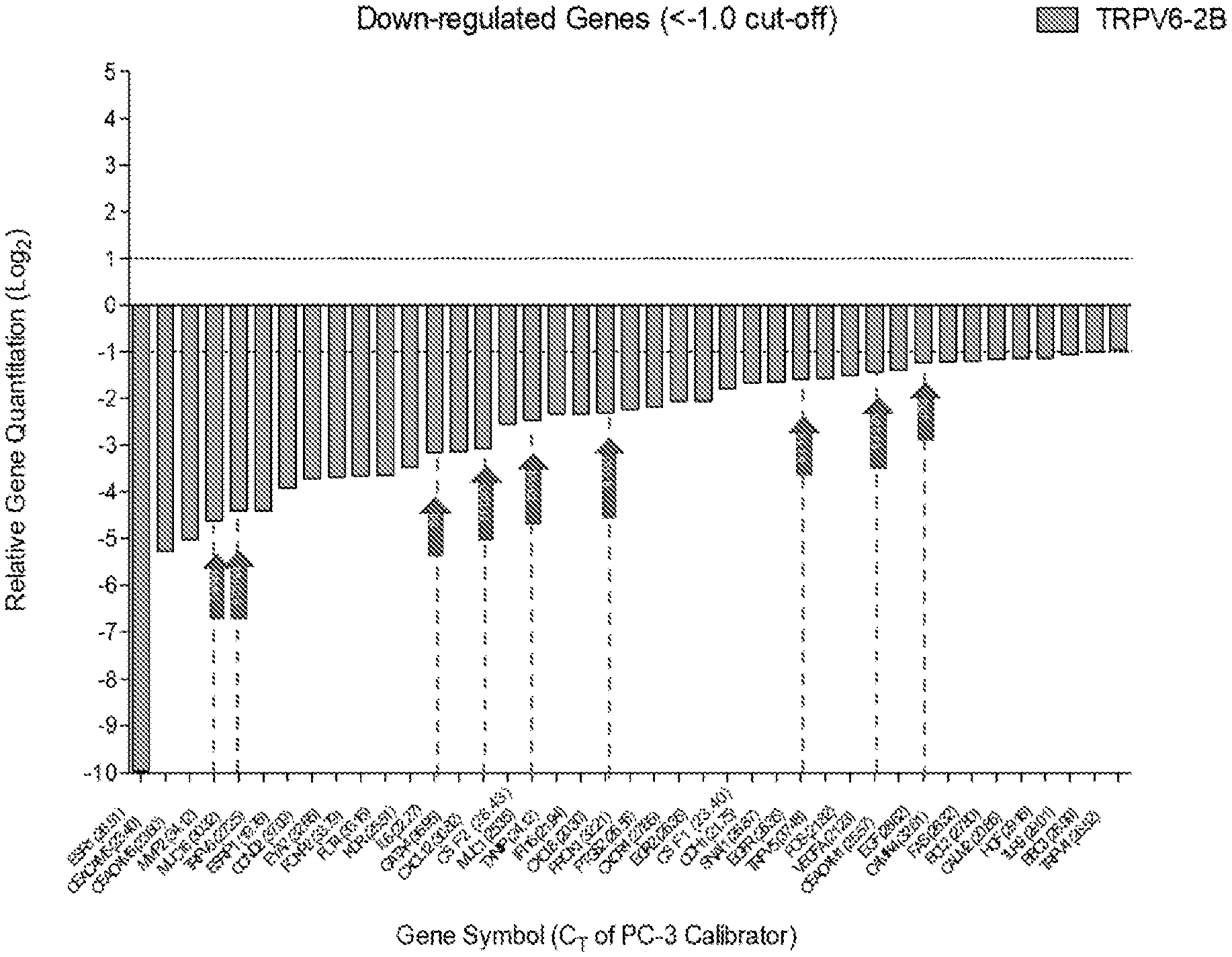
[0053] FIG. 1 shows the change in relative gene quantification in TRPV6 knockout (KO) relative to wild-type prostate cancer cells, which is equivalent to a 2-fold reduction in expression of the targeted gene (1 CT). Arrows indicate the genes that have an impact on the tumor micro-environment and immune evasion. Each gene's associated CT values are in parenthesis on the x-axis.
[0054] FIG. 2 shows a flowchart for the protocol of Example 2, relative to typical PD-1 inhibition assay.
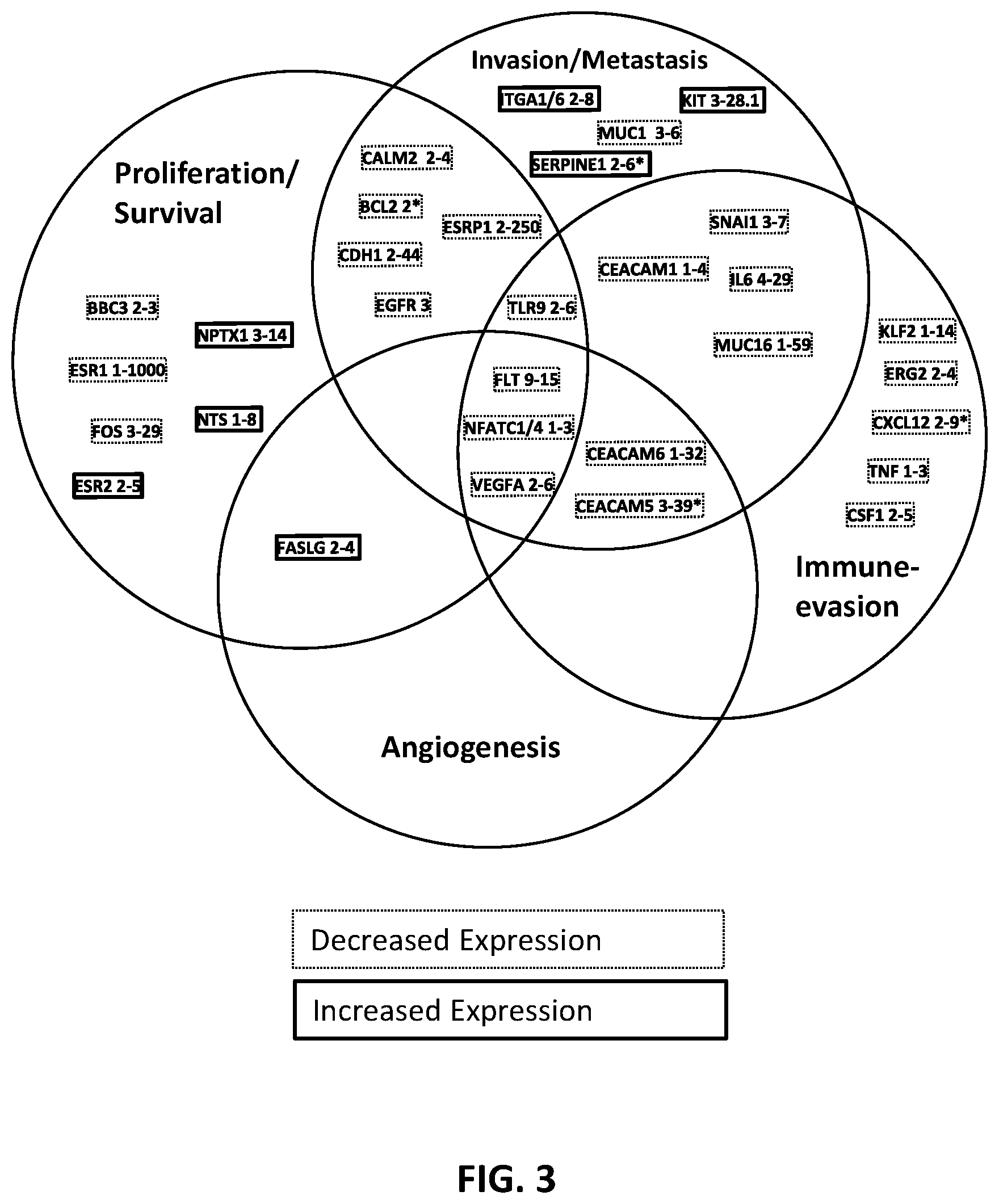
[0055] FIG. 3 summarizes certain of the up- and down-regulated genes in the TRPV6 knock-out (KO) and TRPV6 knock-down (KD) castration-resistant prostate cancer (CRPC) PC3 cells. Genes with >=1.4-fold reduction or increase in expression in 3/4 of the KO/KD cells are shown. The numbers beside the gene name are the fold increase (solid boxes) or decrease (dashed boxes) in expression compared to wild-type (numbers are rounded-up). *Indicates no/small increase in expression in one of the 4 KD/KO experiment. "Survival" includes genes that are resistant to apoptosis or pro-apoptotic.
[0056] FIG. 4 shows the impact of TRPV6 KO and KD on the expression of TRPV2-6 genes in CRPC PC-3 cells. TRPV6 KO/KD CRPC cells do not have up-regulated expression of TRPV channels.
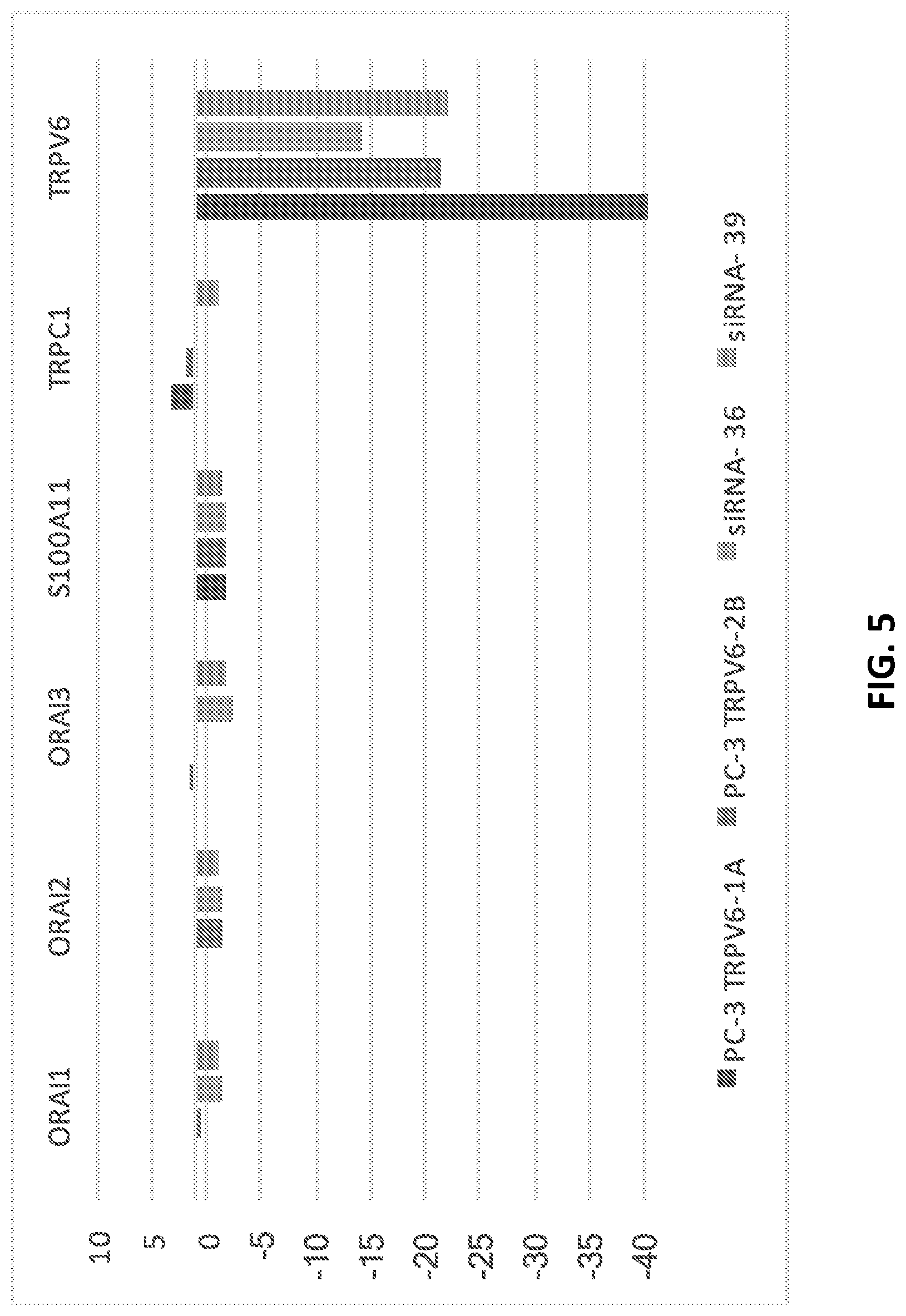
[0057] FIG. 5 shows the impact of TRPV6 KO and KD on the expression of genes involved in modulating intracellular calcium levels relative to expression of TRPV6. TRPV6 KO/KD CRPC cells do not show up-regulated expression of other calcium channels involved in cancer.
[0058] FIG. 6 shows the level of genes involved in cell proliferation, metastasis and angiogenesis up-and down regulated by >=1.5-fold from a 187 genes array panel.
[0059] FIG. 7 shows the level of genes involved in apoptosis up-and down regulated by >=1.5-fold from a 187 genes array panel.
[0060] FIG. 8 shows the level of genes involved in immune evasion and inflammation up-and down regulated by >=1.5-fold from a 187 genes array panel.
[0061] FIG. 9 shows the effect of SOR-C13 on NFAT activation in T-47D cells treated with SOR-C13 (500 .mu.M): A significant difference (*: p<0.05) was observed in NFAT activation between the SOR-C13-treated cells vs. PBS (no treatment).
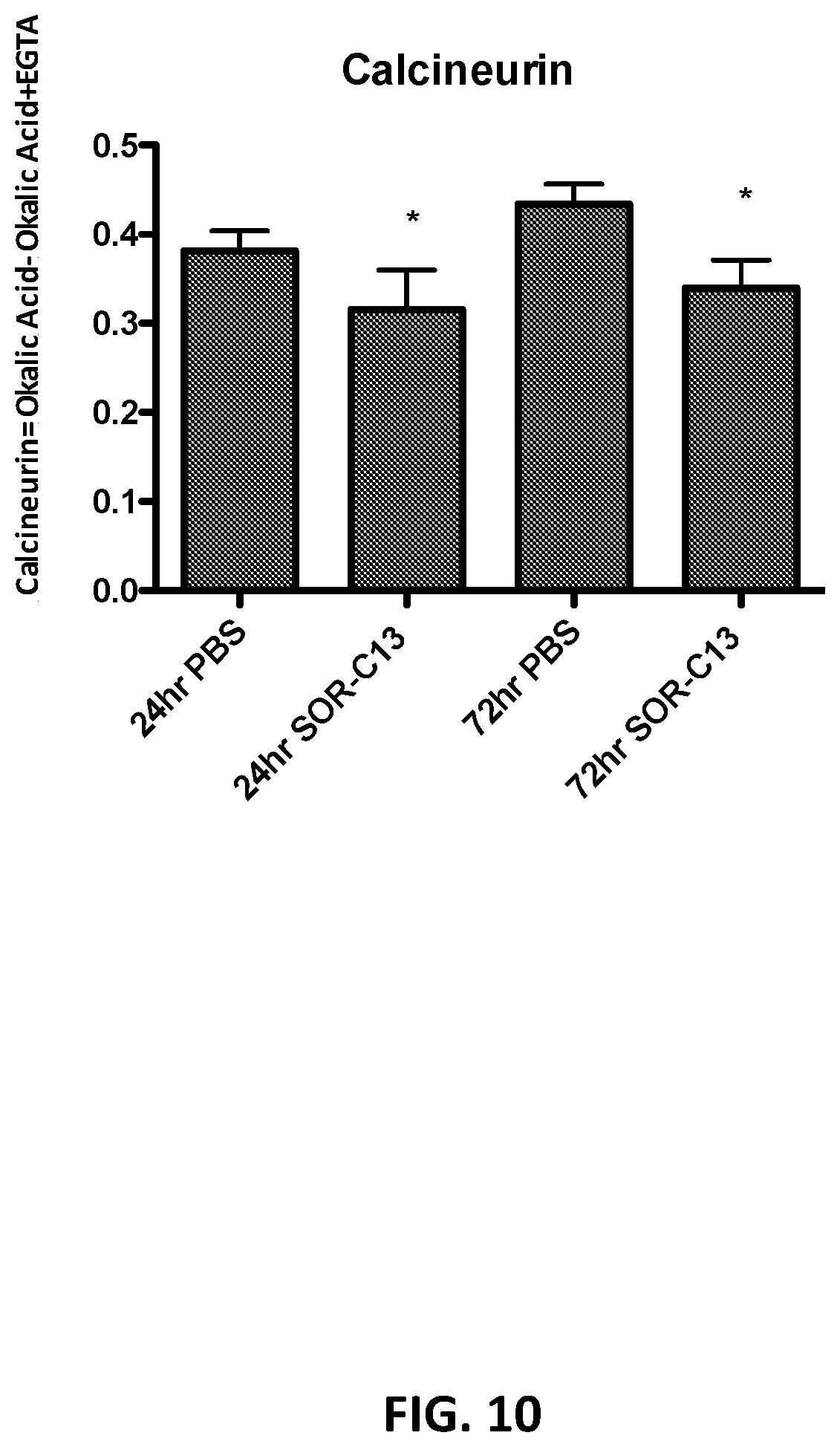
[0062] FIG. 10 shows the calcineurin activity inhibition by SOR-C13 (500 .mu.M) in BxPC-3 cell lysates (*: p<0.05) at 24 hrs and 72 hrs.
[0063] FIG. 11 shows total MMP-9 (% NT ctrl) in BxPC-3 cells treated for 96 hrs daily with SOR-C13 (100, 500 .mu.M) (*: p<0.05).
[0064] FIG. 12 show Bcl-2 expression in BxPC-3 cells treated with SOR-C13 (500 .mu.M). A significant decrease (*: p<0.05) was observed at 96 hrs.
[0065] FIG. 13 shows p and down-regulated genes (>1.5 fold change in expression) from a 187 gene panel in T-47D cells lines treated with SOR-C13. These genes are up or down-regulated in at least one of the 4 other cancer cell lines tested (BxPC-3, PC-3, SKOV-3 and SU.86.86). Genes involved in Calcineurin/NFAT pathway that are shown to be affected: NFATC1, MMP-2, GSK3A and RCAN1. Interestingly, Bcl-2 and MMP-9 were down-regulated (app. 0.6 CT) in BxPC-3 (not shown), in line with the protein expression data.
[0066] FIG. 14 shows the lowest levels of TRPV6 mRNA were observed after 3 days of treatment, having an 85% reduction in TRPV6 expression.
[0067] FIG. 15 shows the second vector TRPV6-2 CRISPR-Cas9 resulted in a deletion of one G at the CRISPR-Cas9 cute site at bp 441 of TRPV6 (exon 3), causing a frameshift mutation.
[0068] FIG. 16 shows 23 genes were down-regulated in at least 2 TRPV6 treatments.
[0069] FIG. 17 shows 33 genes up-regulated in at least 2 TRPV6 treatments.
[0070] FIG. 18 shows a volcano plot showing the 57 differentially expressed genes (>0.6 Log FC and corrected p<0.05) when the TaqMan Array data from the two PC-3 TRPV6 knockout cell lines (TRPV6-1A and TRPV6-2B) are pooled (n=6) and compared to the PC-3 control (n=3).
[0071] FIGS. 19-21 depict the 57 differentially expressed genes from the analysis of the pooled TRPV6 knockouts (n=6) compared to PC-3 Control (n=3) shown in FIG. 18. The three graphs group the differentially expressed genes by functional mechanism of oncogenesis. There were 3 genes differentially expressed but not grouped into one of the three graphs; Down-regulated (TRPV6), up-regulated (TRPC1 and ORAI3).
[0072] FIG. 19 shows data for cell proliferation, metastasis, and angiogenesis-related genes.
[0073] FIG. 20 shows data for apoptosis-related genes.
[0074] FIG. 21 shows data for immune evasion and/or stimulation-related genes.
DETAILED DESCRIPTION
[0075] Unless defined otherwise, all technical and scientific terms used herein have the same meaning as commonly understood by those of ordinary skill in the art to which the disclosure belongs. Although any methods, materials, compositions, reagents, cells, similar or equivalent similar or equivalent to those described herein can be used in the practice or testing of the subject matter of the present disclosure, preferred methods and materials are described. All publications and references, including but not limited to patents and patent applications, cited in this specification are herein incorporated by reference in their entirety as if each individual publication or reference were specifically and individually indicated to be incorporated by reference herein as being fully set forth. Any patent application to which this application claims priority is also incorporated by reference herein in its entirety in the manner described above for publications and references.
[0076] Standard techniques may be used for recombinant DNA, oligonucleotide synthesis, and tissue culture and transformation (e.g., electroporation, lipofection). Enzymatic reactions and purification techniques may be performed according to manufacturer's specifications or as commonly accomplished in the art or as described herein. These and related techniques and procedures may be generally performed according to conventional methods well known in the art and as described in various general and more specific references that are cited and discussed throughout the present specification. Unless specific definitions are provided, the nomenclature utilized in connection with, and the laboratory procedures and techniques of, molecular biology, analytical chemistry, synthetic organic chemistry, and medicinal and pharmaceutical chemistry described herein are those well-known and commonly used in the art. Standard techniques may be used for recombinant technology, molecular biological, microbiological, chemical syntheses, chemical analyses, pharmaceutical preparation, formulation, and delivery, and treatment of patients.
[0077] For the purposes of the present disclosure, the following terms are defined below.
[0078] The articles "a" and "an" are used herein to refer to one or to more than one (i.e., to at least one) of the grammatical object of the article. By way of example, "an element" means one element or more than one element.
[0079] By "about" is meant a quantity, level, value, number, frequency, percentage, dimension, size, amount, weight or length that varies by as much as 30, 25, 20, 15, 10, 9, 8, 7, 6, 5, 4, 3, 2 or 1% to a reference quantity, level, value, number, frequency, percentage, dimension, size, amount, weight or length.
[0080] The term "antigen" refers to a molecule or a portion of a molecule capable of being bound by a selective binding agent, such as an antibody, and additionally capable of being used in an animal to produce antibodies capable of binding to an epitope of that antigen. An antigen may have one or more epitopes. As used herein, the term "antigen" includes substances that are capable, under appropriate conditions, of inducing an immune response to the substance and of reacting with the products of the immune response. For example, an antigen can be recognized by antibodies (humoral immune response) or sensitized T-lymphocytes (T helper or cell-mediated immune response), or both. Antigens can be soluble substances, such as toxins and foreign proteins, or particulates, such as bacteria and tissue cells; however, only the portion of the protein or polysaccharide molecule known as the antigenic determinant (epitopes) combines with the antibody or a specific receptor on a lymphocyte. More broadly, the term "antigen" includes any substance to which an antibody binds, or for which antibodies are desired, regardless of whether the substance is immunogenic. For such antigens, antibodies can be identified by recombinant methods, independently of any immune response.
[0081] An "antagonist" refers to biological structure or chemical agent that interferes with or otherwise reduces the physiological action of another agent or molecule. In some instances, the antagonist specifically binds to the other agent or molecule. Included are full and partial antagonists.
[0082] An "agonist" refers to biological structure or chemical agent that increases or enhances the physiological action of another agent or molecule. In some instances, the agonist specifically binds to the other agent or molecule. Included are full and partial agonists.
[0083] The term "anergy" refers to the functional inactivation of a T-cell, or B-cell response to re-stimulation by antigen.
[0084] As used herein, the term "amino acid" is intended to mean both naturally occurring and non-naturally occurring amino acids as well as amino acid analogs and mimetics. Naturally-occurring amino acids include the 20 (L)-amino acids utilized during protein biosynthesis as well as others such as 4-hydroxyproline, hydroxylysine, desmosine, isodesmosine, homocysteine, citrulline and ornithine, for example. Non-naturally occurring amino acids include, for example, (D)-amino acids, norleucine, norvaline, p-fluorophenylalanine, ethionine and the like, which are known to a person skilled in the art. Amino acid analogs include modified forms of naturally and non-naturally occurring amino acids. Such modifications can include, for example, substitution or replacement of chemical groups and moieties on the amino acid or by derivatization of the amino acid. Amino acid mimetics include, for example, organic structures which exhibit functionally similar properties such as charge and charge spacing characteristic of the reference amino acid. For example, an organic structure which mimics arginine (Arg or R) would have a positive charge moiety located in similar molecular space and having the same degree of mobility as the e-amino group of the side chain of the naturally occurring Arg amino acid. Mimetics also include constrained structures so as to maintain optimal spacing and charge interactions of the amino acid or of the amino acid functional groups. Those skilled in the art know or can determine what structures constitute functionally equivalent amino acid analogs and amino acid mimetics.
[0085] As used herein, a subject "at risk" of developing a disease, or adverse reaction may or may not have detectable disease, or symptoms of disease, and may or may not have displayed detectable disease or symptoms of disease prior to the treatment methods described herein. "At risk" denotes that a subject has one or more risk factors, which are measurable parameters that correlate with development of a disease, as described herein and known in the art. A subject having one or more of these risk factors has a higher probability of developing disease, or an adverse reaction than a subject without one or more of these risk factor(s).
[0086] "Biocompatible" refers to materials or compounds which are generally not injurious to biological functions of a cell or subject and which will not result in any degree of unacceptable toxicity, including allergenic and disease states.
[0087] The term "binding" refers to a direct association between two molecules, due to, for example, covalent, electrostatic, hydrophobic, and ionic and/or hydrogen-bond interactions, including interactions such as salt bridges and water bridges.
[0088] By "coding sequence" is meant any nucleic acid sequence that contributes to the code for the polypeptide product of a gene. By contrast, the term "non-coding sequence" refers to any nucleic acid sequence that does not directly contribute to the code for the polypeptide product of a gene.
[0089] Throughout this disclosure, unless the context requires otherwise, the words "comprise," "comprises," and "comprising" will be understood to imply the inclusion of a stated step or element or group of steps or elements but not the exclusion of any other step or element or group of steps or elements.
[0090] By "consisting of" is meant including, and limited to, whatever follows the phrase "consisting of" Thus, the phrase "consisting of" indicates that the listed elements are required or mandatory, and that no other elements may be present. By "consisting essentially of" is meant including any elements listed after the phrase, and limited to other elements that do not interfere with or contribute to the activity or action specified in the disclosure for the listed elements. Thus, the phrase "consisting essentially of" indicates that the listed elements are required or mandatory, but that other elements are optional and may or may not be present depending upon whether or not they materially affect the activity or action of the listed elements.
[0091] The term "endotoxin free" or "substantially endotoxin free" relates generally to compositions, solvents, and/or vessels that contain at most trace amounts (e.g., amounts having no clinically adverse physiological effects to a subject) of endotoxin, and preferably undetectable amounts of endotoxin. Endotoxins are toxins associated with certain micro-organisms, such as bacteria, typically gram-negative bacteria, although endotoxins may be found in gram-positive bacteria, such as Listeria monocytogenes. The most prevalent endotoxins are lipopolysaccharides (LPS) or lipo-oligo-saccharides (LOS) found in the outer membrane of various Gram-negative bacteria, and which represent a central pathogenic feature in the ability of these bacteria to cause disease. Small amounts of endotoxin in humans may produce fever, a lowering of the blood pressure, and activation of inflammation and coagulation, among other adverse physiological effects.
[0092] Therefore, in pharmaceutical production, it is often desirable to remove most or all traces of endotoxin from drug products and/or drug containers, because even small amounts may cause adverse effects in humans. A depyrogenation oven may be used for this purpose, as temperatures in excess of 300.degree. C. are typically required to break down most endotoxins. For instance, based on primary packaging material such as syringes or vials, the combination of a glass temperature of 250.degree. C. and a holding time of 30 minutes is often sufficient to achieve a 3 log reduction in endotoxin levels. Other methods of removing endotoxins are contemplated, including, for example, chromatography and filtration methods, as described herein and known in the art.
[0093] Endotoxins can be detected using routine techniques known in the art. For example, the Limulus Amoebocyte Lysate assay, which utilizes blood from the horseshoe crab, is a very sensitive assay for detecting presence of endotoxin. In this test, very low levels of LPS can cause detectable coagulation of the limulus lysate due a powerful enzymatic cascade that amplifies this reaction. Endotoxins can also be quantitated by enzyme-linked immunosorbent assay (ELISA). To be substantially endotoxin free, endotoxin levels may be less than about 0.001, 0.005, 0.01, 0.02, 0.03, 0.04, 0.05, 0.06, 0.08, 0.09, 0.1, 0.5, 1.0, 1.5, 2, 2.5, 3, 4, 5, 6, 7, 8, 9, or 10 EU/mg of active compound. Typically, 1 ng lipopolysaccharide (LPS) corresponds to about 1-10 EU.
[0094] The term "epitope" includes any determinant, preferably a polypeptide determinant, capable of specific binding to an immunoglobulin or T-cell receptor. An epitope includes a region of an antigen that is bound by an antibody. In certain embodiments, epitope determinants include chemically active surface groupings of molecules such as amino acids, sugar side chains, phosphoryl or sulfonyl, and may in certain embodiments have specific three-dimensional structural characteristics, and/or specific charge characteristics. Epitopes can be contiguous or non-contiguous in relation to the primary structure of the antigen or reference sequence or target molecule described herein. In particular embodiments, an epitope comprises, consists, or consists essentially of about, at least about, or no more than about 3, 4, 5, 6, 7, 8, 9, 10, 11, 12, 13, 14, 15, 16, 17, 18, 19, or 20 contiguous amino acids (i.e., a linear epitope) or non-contiguous amino acids (i.e., conformational epitope) of a reference sequence or target molecule described herein.
[0095] An "epitope" includes that portion of an antigen or other macromolecule capable of forming a binding interaction that interacts with the variable region binding pocket of a binding protein. Such binding interaction can be manifested as an intermolecular contact with one or more amino acid residues of a CDR. Antigen binding can involve a CDR3 or a CDR3 pair. An epitope can be a linear peptide sequence (i.e., "continuous") or can be composed of noncontiguous amino acid sequences (i.e., "conformational" or "discontinuous"). A binding protein can recognize one or more amino acid sequences; therefore an epitope can define more than one distinct amino acid sequence. Epitopes recognized by binding protein can be determined by peptide mapping and sequence analysis techniques well known to one of skill in the art. A "cryptic epitope" or a "cryptic binding site" is an epitope or binding site of a protein sequence that is not exposed or substantially protected from recognition within an unmodified polypeptide, but is capable of being recognized by a binding protein of a denatured or proteolyzed polypeptide Amino acid sequences that are not exposed, or are only partially exposed, in the unmodified polypeptide structure are potential cryptic epitopes. If an epitope is not exposed, or only partially exposed, then it is likely that it is buried within the interior of the polypeptide. Candidate cryptic epitopes can be identified, for example, by examining the three-dimensional structure of an unmodified polypeptide.
[0096] The term "half maximal effective concentration" or "EC50" refers to the concentration of an agent as described herein at which it induces a response halfway between the baseline and maximum after some specified exposure time; the EC50 of a graded dose response curve therefore represents the concentration of a compound at which 50% of its maximal effect is observed. EC50 also represents the plasma concentration required for obtaining 50% of a maximum effect in vivo. Similarly, the "EC90" refers to the concentration of an agent or composition at which 90% of its maximal effect is observed. The "EC90" can be calculated from the "EC50" and the Hill slope, or it can be determined from the data directly, using routine knowledge in the art. In some embodiments, the EC50 of an agent is less than about 0.01, 0.05, 0.1, 0.2, 0.3, 0.4, 0.5, 0.6, 0.7, 0.8, 0.9, 1, 2, 3, 4, 5, 6, 7, 8, 9, 10, 11, 12, 13, 14, 15, 16, 17, 18, 19, 20, 25, 30, 40, 50, 60, 70, 80, 90, 100, 200 or 500 nM. In some embodiments, an agent will have an EC50 value of about 1 nM or less.
[0097] The "half-life" of an agent can refer to the time it takes for the agent to lose half of its pharmacologic, physiologic, or other activity, relative to such activity at the time of administration into the serum or tissue of an organism, or relative to any other defined time-point. "Half-life" can also refer to the time it takes for the amount or concentration of an agent to be reduced by half of a starting amount administered into the serum or tissue of an organism, relative to such amount or concentration at the time of administration into the serum or tissue of an organism, or relative to any other defined time-point. The half-life can be measured in serum and/or any one or more selected tissues.
[0098] The terms "modulating" and "altering" include "increasing," "enhancing" or "stimulating," as well as "decreasing" or "reducing," typically in a statistically significant or a physiologically significant amount or degree relative to a control. An "increased," "stimulated" or "enhanced" amount is typically a "statistically significant" amount, and may include an increase that is 1.1, 1.2, 1.5, 2, 3, 4, 5, 6, 7, 8, 9, 10, 15, 20, 30, 40, 50, 60, 70, 80, 90, 100 or more times (e.g., 500, 1000 times) (including all integers and ranges in between e.g., 1.5, 1.6, 1.7, 1.8, etc.) the amount produced by no composition (e.g., the absence of agent) or a control composition. A "decreased" or "reduced" amount is typically a "statistically significant" amount, and may include a 1%, 2%, 3%, 4%, 5%, 6%, 7%, 8%, 9%, 10%, 11%, 12%, 13%, 14%, 15%, 16%, 17%, 18%, 19%, 20%, 25%, 30%, 35%, 40%, 45%, 50%, 55%, 60%, 65%, 70%, 75%, 80%, 85%, 90%, 95%, or 100% decrease (including all integers and ranges in between) in the amount produced by no composition (e.g., the absence of an agent) or a control composition. Examples of comparisons and "statistically significant" amounts are described herein.
[0099] The terms "polypeptide," "protein" and "peptide" are used interchangeably and mean a polymer of amino acids not limited to any particular length. The term "enzyme" includes polypeptide or protein catalysts. The terms include modifications such as myristoylation, sulfation, glycosylation, phosphorylation and addition or deletion of signal sequences. The terms "polypeptide" or "protein" means one or more chains of amino acids, wherein each chain comprises amino acids covalently linked by peptide bonds, and wherein said polypeptide or protein can comprise a plurality of chains non-covalently and/or covalently linked together by peptide bonds, having the sequence of native proteins, that is, proteins produced by naturally-occurring and specifically non-recombinant cells, or genetically-engineered or recombinant cells, and comprise molecules having the amino acid sequence of the native protein, or molecules having deletions from, additions to, and/or substitutions of one or more amino acids of the native sequence. In certain embodiments, the polypeptide is a "recombinant" polypeptide, produced by recombinant cell that comprises one or more recombinant DNA molecules, which are typically made of heterologous polynucleotide sequences or combinations of polynucleotide sequences that would not otherwise be found in the cell.
[0100] The term "polynucleotide" and "nucleic acid" includes mRNA, RNA, cRNA, cDNA, and DNA. The term typically refers to polymeric form of nucleotides of at least 10 bases in length, either ribonucleotides or deoxynucleotides or a modified form of either type of nucleotide. The term includes single and double stranded forms of DNA. The terms "isolated DNA" and "isolated polynucleotide" and "isolated nucleic acid" refer to a molecule that has been isolated free of total genomic DNA of a particular species. Therefore, an isolated DNA segment encoding a polypeptide refers to a DNA segment that contains one or more coding sequences yet is substantially isolated away from, or purified free from, total genomic DNA of the species from which the DNA segment is obtained. Also included are non-coding polynucleotides (e.g., primers, probes, oligonucleotides), which do not encode a polypeptide. Also included are recombinant vectors, including, for example, expression vectors, viral vectors, plasmids, cosmids, phagemids, phage, viruses, and the like, which can be used, for example, to produce a polypeptide agent by recombinant methods.
[0101] Additional coding or non-coding sequences may, but need not, be present within a polynucleotide described herein, and a polynucleotide may, but need not, be linked to other molecules and/or support materials. Hence, a polynucleotide or expressible polynucleotides, regardless of the length of the coding sequence itself, may be combined with other sequences, for example, expression control sequences.
[0102] "Expression control sequences" include regulatory sequences of nucleic acids, or the corresponding amino acids, such as promoters, leaders, enhancers, introns, recognition motifs for RNA, or DNA binding proteins, polyadenylation signals, terminators, internal ribosome entry sites (IRES), secretion signals, subcellular localization signals, and the like, which have the ability to affect the transcription or translation, or subcellular, or cellular location of a coding sequence in a host cell. Exemplary expression control sequences are described in Goeddel; Gene Expression Technology: Methods in Enzymology 185, Academic Press, San Diego, Calif. (1990).
[0103] A "promoter" is a DNA regulatory region capable of binding RNA polymerase in a cell and initiating transcription of a downstream (3' direction) coding sequence. As used herein, the promoter sequence is bounded at its 3' terminus by the transcription initiation site and extends upstream (5' direction) to include the minimum number of bases or elements necessary to initiate transcription at levels detectable above background. A transcription initiation site (conveniently defined by mapping with nuclease S1) can be found within a promoter sequence, as well as protein binding domains (consensus sequences) responsible for the binding of RNA polymerase. Eukaryotic promoters can often, but not always, contain "TATA" boxes and "CAT" boxes. Prokaryotic promoters contain Shine-Dalgarno sequences in addition to the -10 and -35 consensus sequences.
[0104] A large number of promoters, including constitutive, inducible and repressible promoters, from a variety of different sources are well known in the art. Representative sources include for example, viral, mammalian, insect, plant, yeast, and bacterial cell types), and suitable promoters from these sources are readily available, or can be made synthetically, based on sequences publicly available on line or, for example, from depositories such as the ATCC as well as other commercial or individual sources. Promoters can be unidirectional (i.e., initiate transcription in one direction) or bi-directional (i.e., initiate transcription in either a 3' or 5' direction). Non-limiting examples of promoters include, for example, the T7 bacterial expression system, pBAD (araA) bacterial expression system, the cytomegalovirus (CMV) promoter, the SV40 promoter, the RSV promoter. Inducible promoters include the Tet system, (U.S. Pat. Nos. 5,464,758 and 5,814,618), the Ecdysone inducible system (No et al., Proc. Natl. Acad. Sci. (1996) 93 (8): 3346-3351; the T-REx.TM. system (Invitrogen Carlsbad, Calif.), LacSwitch.RTM. (Stratagene, (San Diego, Calif.) and the Cre-ERT tamoxifen inducible recombinase system (Indra et al. Nuc. Acid. Res. (1999) 27 (22): 4324-4327; Nuc. Acid. Res. (2000) 28 (23): e99; U.S. Pat. No. 7,112,715; and Kramer & Fussenegger, Methods Mol. Biol. (2005) 308: 123-144) or any promoter known in the art suitable for expression in the desired cells.
[0105] The term "isolated" polypeptide or protein referred to herein means that a subject protein (1) is free of at least some other proteins with which it would typically be found in nature, (2) is essentially free of other proteins from the same source, e.g., from the same species, (3) is expressed by a cell from a different species, (4) has been separated from at least about 50 percent of polynucleotides, lipids, carbohydrates, or other materials with which it is associated in nature, (5) is not associated (by covalent or non-covalent interaction) with portions of a protein with which the "isolated protein" is associated in nature, (6) is operably associated (by covalent or non-covalent interaction) with a polypeptide with which it is not associated in nature, or (7) does not occur in nature. Such an isolated protein can be encoded by genomic DNA, cDNA, mRNA or other RNA, of may be of synthetic origin, or any combination thereof. In certain embodiments, the isolated protein is substantially free from proteins or polypeptides or other contaminants that are found in its natural environment that would interfere with its use (therapeutic, diagnostic, prophylactic, research or otherwise).
[0106] In certain embodiments, the "purity" of any given agent in a composition may be defined. For instance, certain compositions may comprise an agent such as a polypeptide agent that is at least 80%, 85%, 90%, 91%, 92%, 93%, 94%, 95%, 96%, 97%, 98%, 99%, or 100% pure on a protein basis or a weight-weight basis, including all decimals and ranges in between, as measured, for example and by no means limiting, by high performance liquid chromatography (HPLC), a well-known form of column chromatography used frequently in biochemistry and analytical chemistry to separate, identify, and quantify compounds.
[0107] The term "reference sequence" refers generally to a nucleic acid coding sequence, or amino acid sequence, to which another sequence is being compared. All polypeptide and polynucleotide sequences described herein are included as references sequences, including those described by name and those described in the Tables and the Sequence Listing.
[0108] Certain embodiments include biologically active "variants" and "fragments" of the polypeptides described herein, and the polynucleotides that encode the same. "Variants" contain one or more substitutions, additions, deletions, and/or insertions relative to a reference polypeptide or polynucleotide (see, e.g., the Tables and the Sequence Listing). A variant polypeptide or polynucleotide comprises an amino acid or nucleotide sequence with at least about 50%, 55%, 60%, 65%, 70%, 75%, 80%, 85%, 90%, 91%, 92%, 93%, 94%, 95%, 96%, 97%, 98%, 99% or more sequence identity or similarity or homology to a reference sequence, as described herein, and substantially retains the activity of that reference sequence. Also included are sequences that consist of or differ from a reference sequences by the addition, deletion, insertion, or substitution of 1, 2, 3, 4, 5, 6, 7, 8, 9, 10, 11, 12, 13, 14, 15, 16, 17, 18, 19, 20, 30, 40, 50, 60,70, 80, 90, 100, 110, 120, 130, 140, 150 or more amino acids or nucleotides and which substantially retain the activity of that reference sequence. In certain embodiments, the additions or deletions include C-terminal and/or N-terminal additions and/or deletions.
[0109] The terms "sequence identity" or, for example, comprising a "sequence 50% identical to," as used herein, refer to the extent that sequences are identical on a nucleotide-by-nucleotide basis or an amino acid-by-amino acid basis over a window of comparison. Thus, a "percentage of sequence identity" may be calculated by comparing two optimally aligned sequences over the window of comparison, determining the number of positions at which the identical nucleic acid base (e.g., A, T, C, G, I) or the identical amino acid residue (e.g., Ala, Pro, Ser, Thr, Gly, Val, Leu, Ile, Phe, Tyr, Trp, Lys, Arg, His, Asp, Glu, Asn, Gln, Cys and Met) occurs in both sequences to yield the number of matched positions, dividing the number of matched positions by the total number of positions in the window of comparison (i.e., the window size), and multiplying the result by 100 to yield the percentage of sequence identity. Optimal alignment of sequences for aligning a comparison window may be conducted by computerized implementations of algorithms (GAP, BESTFIT, FASTA, and TFASTA in the Wisconsin Genetics Software Package Release 7.0, Genetics Computer Group, 575 Science Drive Madison, Wis., USA) or by inspection and the best alignment (i.e., resulting in the highest percentage homology over the comparison window) generated by any of the various methods selected. Reference also may be made to the BLAST family of programs as for example disclosed by Altschul et al., Nucl. Acids Res. 25:3389, 1997.
[0110] The term "solubility" refers to the property of an agent provided herein to dissolve in a liquid solvent and form a homogeneous solution. Solubility is typically expressed as a concentration, either by mass of solute per unit volume of solvent (g of solute per kg of solvent, g per dL (100 mL), mg/ml, etc.), molarity, molality, mole fraction or other similar descriptions of concentration. The maximum equilibrium amount of solute that can dissolve per amount of solvent is the solubility of that solute in that solvent under the specified conditions, including temperature, pressure, pH, and the nature of the solvent. In certain embodiments, solubility is measured at physiological pH, or other pH, for example, at pH 5.0, pH 6.0, pH 7.0, pH 7.4, pH 7.6, pH 7.8, or pH 8.0 (e.g., about pH 5-8). In certain embodiments, solubility is measured in water or a physiological buffer such as PBS or NaCl (with or without NaP). In specific embodiments, solubility is measured at relatively lower pH (e.g., pH 6.0) and relatively higher salt (e.g., 500 mM NaCl and 10 mM NaP). In certain embodiments, solubility is measured in a biological fluid (solvent) such as blood or serum. In certain embodiments, the temperature can be about room temperature (e.g., about 20, 21, 22, 23, 24, 25.degree. C.) or about body temperature (37.degree. C.). In certain embodiments, an agent has a solubility of at least about 0.1, 0.2, 0.3, 0.4, 0.5, 0.6, 0.7, 0.8, 0.9, 1, 2, 3, 4, 5, 6, 7, 8, 9, 10, 11, 12, 13, 14, 15, 16, 17, 18, 19, 20, 25, 30, 40, 50, 60, 70, 80, 90 or 100 mg/ml at room temperature or at 37.degree. C.
[0111] A "subject" or a "subject in need thereof" or a "patient" or a "patient in need thereof" includes a mammalian subject such as a human subject.
[0112] "Substantially" or "essentially" means nearly totally or completely, for instance, 95%, 96%, 97%, 98%, 99% or greater of some given quantity.
[0113] By "statistically significant," it is meant that the result was unlikely to have occurred by chance. Statistical significance can be determined by any method known in the art. Commonly used measures of significance include the p-value, which is the frequency or probability with which the observed event would occur, if the null hypothesis were true. If the obtained p-value is smaller than the significance level, then the null hypothesis is rejected. In simple cases, the significance level is defined at a p-value of 0.05 or less.
[0114] "Therapeutic response" refers to improvement of symptoms (whether or not sustained) based on administration of one or more therapeutic agents.
[0115] As used herein, the terms "therapeutically effective amount", "therapeutic dose," "prophylactically effective amount," or "diagnostically effective amount" is the amount of an needed to elicit the desired biological response following administration.
[0116] As used herein, "treatment" of a subject (e.g. a mammal, such as a human) or a cell is any type of intervention used in an attempt to alter the natural course of the individual or cell. Treatment includes, but is not limited to, administration of a pharmaceutical composition, and may be performed either prophylactically or subsequent to the initiation of a pathologic event or contact with an etiologic agent. Also included are "prophylactic" treatments, which can be directed to reducing the rate of progression of the disease or condition being treated, delaying the onset of that disease or condition, or reducing the severity of its onset. "Treatment" or "prophylaxis" does not necessarily indicate complete eradication, cure, or prevention of the disease or condition, or associated symptoms thereof.
[0117] The term "wild-type" refers to a gene or gene product (e.g., a polypeptide) that is most frequently observed in a population and is thus arbitrarily designed the "normal" or "wild-type" form of the gene.
[0118] Each embodiment in this specification is to be applied to every other embodiment unless expressly stated otherwise.
[0119] TRPV6 Inhibitors
[0120] Certain embodiments employ one or more "TRPV6 inhibitors". TRPV6 is a member of the super family of Transient Receptor Potential (TRP) channels, subfamily vanilloid (TRPV), member 6, and is selective for Ca2+ ions. It is also highly expressed in a variety of cancer tissues including prostate, colon, breast, thyroid, and ovarian carcinomas, among others. Its expression coincides with cancer progression, suggesting that it drives cancer cell growth. Thus, certain embodiments include an agent that inhibits or antagonizes TRPV6, for example, by reducing or inhibiting calcium uptake in a cell (e.g., cancer cell), including agents that specifically bind to TRPV6. Exemplary agents include TRPV6 inhibitor small molecules (see, e.g., Landowski et al., Pharm Res. 2011 February; 28(2):322-30, incorporated by reference) and TRPV6 inhibitor peptides (see, e.g., U.S. Pat. Nos. 8,211,857; 8,618,058; 9,303,077, incorporated by reference).
[0121] In particular embodiments, the TRPV6 inhibitor is a peptide. Certain exemplary TRPV6 inhibitor peptides include the soricidin oligopeptide (SEQ ID NO:1) and variants and fragments thereof which reduce or inhibit calcium uptake in a cell (e.g., cancer cell). In certain embodiments, the TRPV6 inhibitor peptide inhibits calcium uptake without the paralytic activity associated with soricidin. The amino acid sequence of soricidin and exemplary fragments thereof are provided in Table T1 below.
TABLE-US-00001 TABLE T1 TRPV6 Inhibitor Peptides SEQ Name Sequence ID NO: Soricidin DCSQDCAACSILARPAELNTETCILECE 1 GKLSSNDTEGGLCKEFLHPSKVDLPR SOR-C13 KEFLHPSKVDLPR 2 SOR-C27 EGKLSSNDTEGGLCKEFLHPSKVDLPR 3
[0122] Thus, in certain embodiments, the TRPV6 inhibitor is a peptide that comprises, consists, or consists essentially of an amino acid in Table T1, or a variant and/fragment thereof which inhibits calcium uptake without the paralytic activity. Examples include variants/fragments that comprise, consist, or consist essentially of an amino acid sequence with at least 80%, 85%, 90%, 95%, 98%, 99%, or 100% identity to a sequence in Table T1, and which inhibit calcium uptake in a (e.g., cancer) cell without paralytic activity. Also included are TRPV6 inhibitor peptides of about, less than about, or no more than about 10, 11, 12, 13, 14, 15, 16, 17, 18, 19, 20, 21, 22, 23, 24, 25, 26, 27, 28, 29, 30, 31, 32, 33, 34, 35, 36, 37, 38, 39, or 40 amino acids in length, including all ranges in between, including peptides that comprise, consists, or consist essentially of about, less than about, or no more than about, 10, 11, 12, 13, 14, 15, 16, 17, 18, 19, 20, 21, 22, 23, 24, 25, 26, 27, 28, 29, 30, 31, 32, 33, 34, 35, 36, 37, 38, 39, or 40 contiguous amino acids of a sequence from Table T1.
[0123] In specific embodiments, the TRPV6 inhibitor peptide comprises, consists, or consists essentially of an amino acid sequence with at least 80%, 85%, 90%, 95%, 98%, 99%, or 100% identity to KEFLHPSKVDLPR (SEQ ID NO:2) or EGKLSSNDTEGGLCKEFLHPSKVDLPR (SEQ ID NO:3).
[0124] As noted herein, peptide agents including TRPV6 inhibitor peptides may be altered in various ways including amino acid substitutions, deletions, truncations, additions, and insertions. Methods for such manipulations are generally known in the art. For example, amino acid sequence variants of a reference polypeptide can be prepared by mutations in the DNA. Methods for mutagenesis and nucleotide sequence alterations are well known in the art. See, for example, Kunkel (1985, Proc. Natl. Acad. Sci. USA. 82: 488-492), Kunkel et al., (1987, Methods in Enzymol, 154: 367-382), U.S. Pat. No. 4,873,192, Watson, J. D. et al., ("Molecular Biology of the Gene", Fourth Edition, Benjamin/Cummings, Menlo Park, Calif., 1987) and the references cited therein. Guidance as to appropriate amino acid substitutions that do not affect biological activity of the protein of interest may be found in the model of Dayhoff et al., (1978) Atlas of Protein Sequence and Structure (Natl. Biomed. Res. Found., Washington, D.C.).
[0125] Biologically active truncated and/or variant peptides may contain conservative amino acid substitutions at various locations along their sequence, relative to a reference amino acid residue. A "conservative amino acid substitution" is one in which the amino acid residue is replaced with an amino acid residue having a similar side chain Families of amino acid residues having similar side chains have been defined in the art, which can be generally sub-classified as follows:
[0126] Acidic: The residue has a negative charge due to loss of H ion at physiological pH and the residue is attracted by aqueous solution so as to seek the surface positions in the conformation of a peptide in which it is contained when the peptide is in aqueous medium at physiological pH Amino acids having an acidic side chain include glutamic acid and aspartic acid.
[0127] Basic: The residue has a positive charge due to association with H ion at physiological pH or within one or two pH units thereof (e.g., histidine) and the residue is attracted by aqueous solution so as to seek the surface positions in the conformation of a peptide in which it is contained when the peptide is in aqueous medium at physiological pH Amino acids having a basic side chain include arginine, lysine and histidine.
[0128] Charged: The residues are charged at physiological pH and, therefore, include amino acids having acidic or basic side chains (i.e., glutamic acid, aspartic acid, arginine, lysine and histidine).
[0129] Hydrophobic: The residues are not charged at physiological pH and the residue is repelled by aqueous solution so as to seek the inner positions in the conformation of a peptide in which it is contained when the peptide is in aqueous medium. Amino acids having a hydrophobic side chain include tyrosine, valine, isoleucine, leucine, methionine, phenylalanine and tryptophan.
[0130] Neutral/polar: The residues are not charged at physiological pH, but the residue is not sufficiently repelled by aqueous solutions so that it would seek inner positions in the conformation of a peptide in which it is contained when the peptide is in aqueous medium Amino acids having a neutral/polar side chain include asparagine, glutamine, cysteine, histidine, serine and threonine.
[0131] This description also characterizes certain amino acids as "small" since their side chains are not sufficiently large, even if polar groups are lacking, to confer hydrophobicity. With the exception of proline, "small" amino acids are those with four carbons or less when at least one polar group is on the side chain and three carbons or less when not. Amino acids having a small side chain include glycine, serine, alanine and threonine. The gene-encoded secondary amino acid proline is a special case due to its known effects on the secondary conformation of peptide chains. The structure of proline differs from all the other naturally-occurring amino acids in that its side chain is bonded to the nitrogen of the .alpha.-amino group, as well as the .alpha.-carbon. Several amino acid similarity matrices are known in the art (see e.g., PAM120 matrix and PAM250 matrix as disclosed for example by Dayhoff et al., 1978, A model of evolutionary change in proteins). Matrices for determining distance relationships In M. O. Dayhoff, (ed.), Atlas of protein sequence and structure, Vol. 5, pp. 345-358, National Biomedical Research Foundation, Washington D.C.; and by Gonnet et al., (Science, 256: 14430-1445, 1992), however, include proline in the same group as glycine, serine, alanine and threonine. Accordingly, proline is classified as a "small" amino acid.
[0132] The degree of attraction or repulsion required for classification as polar or nonpolar is arbitrary and, therefore, amino acids specifically contemplated by the invention have been classified as one or the other. Most amino acids not specifically named can be classified on the basis of known behavior.
[0133] Amino acid residues can be further sub-classified as cyclic or non-cyclic, and aromatic or non-aromatic, self-explanatory classifications with respect to the side-chain substituent groups of the residues, and as small or large. The residue is considered small if it contains a total of four carbon atoms or less, inclusive of the carboxyl carbon, provided an additional polar substituent is present; three or less if not. Small residues are, of course, always non-aromatic. Dependent on their structural properties, amino acid residues may fall in two or more classes. For the naturally-occurring protein amino acids, sub-classification according to this scheme is presented in Table A.
TABLE-US-00002 TABLE A Sub-classes Amino acids Acidic Aspartic acid, Glutamic acid Basic Noncyclic: Arginine, Lysine; Cyclic: Histidine Charged Aspartic acid, Glutamic acid, Arginine, Lysine, Histidine Small Glycine, Serine, Alanine, Threonine, Proline Polar/neutral Asparagine, Histidine, Glutamine, Cysteine, Serine, Threonine Polar/large Asparagine, Glutamine Hydrophobic Tyrosine, Valine, Isoleucine, Leucine, Methionine, Phenylalanine, Tryptophan Aromatic Tryptophan, Tyrosine, Phenylalanine Residues that Glycine and Proline influence chain orientation
[0134] Conservative amino acid substitution also includes groupings based on side chains. For example, a group of amino acids having aliphatic side chains is glycine, alanine, valine, leucine, and isoleucine; a group of amino acids having aliphatic-hydroxyl side chains is serine and threonine; a group of amino acids having amide-containing side chains is asparagine and glutamine; a group of amino acids having aromatic side chains is phenylalanine, tyrosine, and tryptophan; a group of amino acids having basic side chains is lysine, arginine, and histidine; and a group of amino acids having sulphur-containing side chains is cysteine and methionine. For example, it is reasonable to expect that replacement of a leucine with an isoleucine or valine, an aspartate with a glutamate, a threonine with a serine, or a similar replacement of an amino acid with a structurally related amino acid will not have a major effect on the properties of the resulting variant polypeptide. Whether an amino acid change results in a functional truncated and/or variant polypeptide can readily be determined by assaying its non-canonical activity, as described herein. Conservative substitutions are shown in Table B under the heading of exemplary substitutions Amino acid substitutions falling within the scope of the invention, are, in general, accomplished by selecting substitutions that do not differ significantly in their effect on maintaining (a) the structure of the peptide backbone in the area of the substitution, (b) the charge or hydrophobicity of the molecule at the target site, (c) the bulk of the side chain, or (d) the biological function. After the substitutions are introduced, the variants are screened for biological activity.
TABLE-US-00003 TABLE B Original Exemplary Preferred Residue Substitutions Substitutions Ala Val, Leu, Ile Val Arg Lys, Gln, Asn Lys Asn Gln, His, Lys, Arg Gln Asp Glu Glu Cys Ser, Ala, Leu, Val Ser, Ala Gln Asn, His, Lys, Asn Glu Asp, Lys Asp Gly Pro Pro His Asn, Gln, Lys, Arg Arg Ile Leu, Val, Met, Ala, Phe, Norleu Leu Leu Norleu, Ile, Val, Met, Ala, Phe Ile Lys Arg, Gln, Asn Arg Met Leu, Ile, Phe Leu Phe Leu, Val, Ile, Ala Leu Pro Gly Gly Ser Thr Thr Thr Ser Ser Trp Tyr Tyr Tyr Trp, Phe, Thr, Ser Phe Val Ile, Leu, Met, Phe, Ala, Norleu Leu
[0135] Alternatively, similar amino acids for making conservative substitutions can be grouped into three categories based on the identity of the side chains. The first group includes glutamic acid, aspartic acid, arginine, lysine, histidine, which all have charged side chains; the second group includes glycine, serine, threonine, cysteine, tyrosine, glutamine, asparagine; and the third group includes leucine, isoleucine, valine, alanine, proline, phenylalanine, tryptophan, methionine, as described in Zubay, G., Biochemistry, third edition, Wm. C. Brown Publishers (1993). After the substitutions, deletions, and/or additions are introduced, the variants/fragments are screened for biological activity, including the ability to reduce or otherwise inhibit calcium uptake by TRPV6.
[0136] In certain embodiments, the TRPV6 inhibitor, for example, the TRPV6 inhibitor peptide, is conjugated or otherwise attached to a second therapeutic agent, for example, a chemotherapeutic agent. Certain embodiments thus include peptide drug conjugates (PDCs), comprising a TRPV6 inhibitor peptide, as described herein, which is conjugated to a chemotherapeutic agent directly or via an optional linker or spacer in between.
[0137] The TRPV6 peptides described herein can be readily prepared by recombinant or chemical synthesis using techniques well known in the chemistry of proteins such as solid phase synthesis (Merrifield, 1964, J. Am. Chem. Assoc. 85:2149-2154) or synthesis in homogenous solution (Houbenweyl, 1987, Methods of Organic Chemistry, ed. E. Wansch, Vol. 15 I and II, Thieme, Stuttgart).
[0138] The skilled artisan will appreciate that the various TRPV6 inhibitors described herein can be combined with any one or more of the various immune checkpoint modulatory agents described herein, and used according to any one or more of the methods and compositions described herein.
[0139] Immune Checkpoint Modulatory Agents
[0140] Certain embodiments employ one or more immune checkpoint modulatory agent. In certain instances, an immune checkpoint modulatory agent modulates the immune response of a subject, for example, to increase or maintain a cancer-related or cancer-specific immune response, and thereby results in increased immune cell inhibition or reduction of cancer cells. Exemplary immune checkpoint modulatory agent include polypeptides, for example, antibodies and antigen-binding fragments thereof, ligands, small peptides, small molecules, and mixtures thereof.
[0141] Particular examples of immune checkpoint modulatory agents include "antagonists" of one or more inhibitory immune checkpoint molecules, and "agonists" of one or more stimulatory immune checkpoint molecules. Generally, immune checkpoint molecules are components of the immune system that either turn up a signal (co-stimulatory molecules) or turn down a signal, the targeting of which has therapeutic potential in cancer because cancer cells can perturb the natural function of immune checkpoint molecules (see, e.g., Sharma and Allison, Science. 348:56-61, 2015; Topalian et al., Cancer Cell. 27:450-461, 2015; Pardoll, Nature Reviews Cancer. 12:252-264, 2012). In some embodiments, the immune checkpoint modulatory agent (e.g., antagonist, agonist) "binds" or "specifically binds" to the one or more immune checkpoint molecules, as described herein.
[0142] In particular embodiments, the immune checkpoint modulatory agent is a polypeptide or peptide. The terms "peptide" and "polypeptide" are used interchangeably herein, however, in certain instances, the term "peptide" can refer to shorter polypeptides, for example, polypeptides that consist of about 2, 3, 4, 5, 6, 7, 8, 9, 10, 11, 12, 13, 14, 15, 16, 17, 18, 19, 20, 25, 30, 35, 40, 45, or 50 amino acids, including all integers and ranges (e.g., 5-10, 8-12, 10-15) in between. Polypeptides and peptides can be composed of naturally-occurring amino acids and/or non-naturally occurring amino acids, as described herein. Antibodies are also included as polypeptides.
[0143] The binding properties of polypeptides can be quantified using methods well known in the art (see Davies et al., Annual Rev. Biochem. 59:439-473, 1990). In some embodiments, a polypeptide specifically binds to a target molecule, for example, an immune checkpoint molecule or an epitope thereof, with an equilibrium dissociation constant that is about or ranges from about .ltoreq.10-7 to about 10-8 M. In some embodiments, the equilibrium dissociation constant is about or ranges from about .ltoreq.10-9 M to about .ltoreq.10-10 M. In certain illustrative embodiments, the polypeptide has an affinity (Kd) for a target described herein (to which it specifically binds) of about, at least about, or less than about, 0.01, 0.05, 0.1, 0.2, 0.3, 0.4, 0.5, 0.6, 0.7, 0.8, 0.9, 1, 2, 3, 4, 5, 6, 7, 8, 9, 10, 11, 12, 13, 14, 15, 16, 17, 18, 19, 20, 21, 22, 23, 24, 25, 26, 27, 28, 29, 30, 40, or 50 nM.
[0144] In some embodiments, the immune checkpoint modulatory polypeptide agent is an antibody or "antigen-binding fragment thereof." The antibody or antigen-binding fragment can be of essentially any type. As is well known in the art, an antibody is an immunoglobulin molecule capable of specific binding to a target, such as an immune checkpoint molecule, through at least one epitope recognition site, located in the variable region of the immunoglobulin molecule.
[0145] As used herein, the term "antibody" encompasses not only intact polyclonal or monoclonal antibodies, but also fragments thereof (such as dAb, Fab, Fab', F(ab')2, Fv), single chain (ScFv), synthetic variants thereof, naturally occurring variants, fusion proteins comprising an antibody portion with an antigen-binding fragment of the required specificity, humanized antibodies, chimeric antibodies, and any other modified configuration of the immunoglobulin molecule that comprises an antigen-binding site or fragment (epitope recognition site) of the required specificity. Certain features and characteristics of antibodies (and antigen-binding fragments thereof) are described in greater detail herein.
[0146] The term "antigen-binding fragment" as used herein refers to a polypeptide fragment that contains at least one CDR of an immunoglobulin heavy and/or light chain that binds to the antigen of interest. In this regard, an antigen-binding fragment of the herein described antibodies may comprise 1, 2, 3, 4, 5, or all 6 CDRs of a VH and VL sequence from antibodies that bind to a target molecule.
[0147] A molecule such as a polypeptide or antibody is said to exhibit "specific binding" or "preferential binding" if it reacts or associates more frequently, more rapidly, with greater duration and/or with greater affinity with a particular cell or substance than it does with alternative cells or substances. An antibody "specifically binds" or "preferentially binds" to a target if it binds with greater affinity, avidity, more readily, and/or with greater duration than it binds to other substances, for example, by a statistically significant amount. For instance, an antibody that specifically or preferentially binds to a specific epitope is an antibody that binds that specific epitope with greater affinity, avidity, more readily, and/or with greater duration than it binds to other epitopes. It is also understood by reading this definition that, for example, an antibody (or moiety or epitope) that specifically or preferentially binds to a first target may or may not specifically or preferentially bind to a second target. As such, "specific binding" or "preferential binding" does not necessarily require (although it can include) exclusive binding. Generally, but not necessarily, reference to binding means preferential binding.
[0148] Immunological binding generally refers to the non-covalent interactions of the type which occur between an immunoglobulin molecule and an antigen for which the immunoglobulin is specific, for example by way of illustration and not limitation, as a result of electrostatic, ionic, hydrophilic and/or hydrophobic attractions or repulsion, steric forces, hydrogen bonding, van der Waals forces, and other interactions. The strength, or affinity of immunological binding interactions can be expressed in terms of the dissociation constant (Kd) of the interaction, wherein a smaller Kd represents a greater affinity Immunological binding properties of selected polypeptides can be quantified using methods well known in the art. One such method entails measuring the rates of antigen-binding site/antigen complex formation and dissociation, wherein those rates depend on the concentrations of the complex partners, the affinity of the interaction, and on geometric parameters that equally influence the rate in both directions. Thus, both the "on rate constant" (Kon) and the "off rate constant" (Koff) can be determined by calculation of the concentrations and the actual rates of association and dissociation. The ratio of Koff/Kon enables cancellation of all parameters not related to affinity, and is thus equal to the dissociation constant Kd.
[0149] Antibodies may be prepared by any of a variety of techniques known to those of ordinary skill in the art. See, e.g., Harlow and Lane, Antibodies: A Laboratory Manual, Cold Spring Harbor Laboratory, 1988. Monoclonal antibodies specific for a polypeptide of interest may be prepared, for example, using the technique of Kohler and Milstein, Eur. J. Immunol. 6:511-519, 1976, and improvements thereto. Also included are methods that utilize transgenic animals such as mice to express human antibodies. See, e.g., Neuberger et al., Nature Biotechnology 14:826, 1996; Lonberg et al., Handbook of Experimental Pharmacology 113:49-101, 1994; and Lonberg et al., Internal Review of Immunology 13:65-93, 1995. Particular examples include the VELOCIMMUNE.RTM. platform by REGENEREX.RTM. (see, e.g., U.S. Pat. No. 6,596,541).
[0150] Antibodies can also be generated or identified by the use of phage display or yeast display libraries (see, e.g., U.S. Pat. No. 7,244,592; Chao et al., Nature Protocols. 1:755-768, 2006). Non-limiting examples of available libraries include cloned or synthetic libraries, such as the Human Combinatorial Antibody Library (HuCAL), in which the structural diversity of the human antibody repertoire is represented by seven heavy chain and seven light chain variable region genes. The combination of these genes gives rise to 49 frameworks in the master library. By superimposing highly variable genetic cassettes (CDRs=complementarity determining regions) on these frameworks, the vast human antibody repertoire can be reproduced. Also included are human libraries designed with human-donor-sourced fragments encoding a light-chain variable region, a heavy-chain CDR-3, synthetic DNA encoding diversity in heavy-chain CDR-1, and synthetic DNA encoding diversity in heavy-chain CDR-2. Other libraries suitable for use will be apparent to persons skilled in the art.
[0151] In certain embodiments, antibodies and antigen-binding fragments thereof as described herein include a heavy chain and a light chain CDR set, respectively interposed between a heavy chain and a light chain framework region (FR) set which provide support to the CDRs and define the spatial relationship of the CDRs relative to each other. As used herein, the term "CDR set" refers to the three hypervariable regions of a heavy or light chain V region. Proceeding from the N-terminus of a heavy or light chain, these regions are denoted as "CDR1," "CDR2," and "CDR3" respectively. An antigen-binding site, therefore, includes six CDRs, comprising the CDR set from each of a heavy and a light chain V region. A polypeptide comprising a single CDR, (e.g., a CDR1, CDR2 or CDR3) is referred to herein as a "molecular recognition unit." Crystallographic analysis of a number of antigen-antibody complexes has demonstrated that the amino acid residues of CDRs form extensive contact with bound antigen, wherein the most extensive antigen contact is with the heavy chain CDR3. Thus, the molecular recognition units are primarily responsible for the specificity of an antigen-binding site.
[0152] As used herein, the term "FR set" refers to the four flanking amino acid sequences which frame the CDRs of a CDR set of a heavy or light chain V region. Some FR residues may contact bound antigen; however, FRs are primarily responsible for folding the V region into the antigen-binding site, particularly the FR residues directly adjacent to the CDRs. Within FRs, certain amino residues and certain structural features are very highly conserved. In this regard, all V region sequences contain an internal disulfide loop of around 90 amino acid residues. When the V regions fold into a binding-site, the CDRs are displayed as projecting loop motifs which form an antigen-binding surface. It is generally recognized that there are conserved structural regions of FRs which influence the folded shape of the CDR loops into certain "canonical" structures--regardless of the precise CDR amino acid sequence. Further, certain FR residues are known to participate in non-covalent interdomain contacts which stabilize the interaction of the antibody heavy and light chains.
[0153] The structures and locations of immunoglobulin variable domains may be determined by reference to Kabat, E. A. et al., Sequences of Proteins of Immunological Interest. 4th Edition. US Department of Health and Human Services. 1987, and updates thereof.
[0154] Also include are "monoclonal" antibodies, which refer to a homogeneous antibody population wherein the monoclonal antibody is comprised of amino acids (naturally occurring and non-naturally occurring) that are involved in the selective binding of an epitope. Monoclonal antibodies are highly specific, being directed against a single epitope. The term "monoclonal antibody" encompasses not only intact monoclonal antibodies and full-length monoclonal antibodies, but also fragments thereof (such as Fab, Fab', F(ab')2, Fv), single chain (ScFv), variants thereof, fusion proteins comprising an antigen-binding portion, humanized monoclonal antibodies, chimeric monoclonal antibodies, and any other modified configuration of the immunoglobulin molecule that comprises an antigen-binding fragment (epitope recognition site) of the required specificity and the ability to bind to an epitope. It is not intended to be limited as regards the source of the antibody or the manner in which it is made (e.g., by hybridoma, phage selection, recombinant expression, transgenic animals). The term includes whole immunoglobulins as well as the fragments etc. described above under the definition of "antibody."
[0155] The proteolytic enzyme papain preferentially cleaves IgG molecules to yield several fragments, two of which (the F(ab) fragments) each comprise a covalent heterodimer that includes an intact antigen-binding site. The enzyme pepsin is able to cleave IgG molecules to provide several fragments, including the F(ab')2 fragment which comprises both antigen-binding sites. An Fv fragment for use according to certain embodiments of the present invention can be produced by preferential proteolytic cleavage of an IgM, and on rare occasions of an IgG or IgA immunoglobulin molecule. Fv fragments are, however, more commonly derived using recombinant techniques known in the art. The Fv fragment includes a non-covalent VH::VL heterodimer including an antigen-binding site which retains much of the antigen recognition and binding capabilities of the native antibody molecule. See Inbar et al., PNAS USA. 69:2659-2662, 1972; Hochman et al., Biochem. 15:2706-2710, 1976; and Ehrlich et al., Biochem. 19:4091-4096, 1980.
[0156] In certain embodiments, single chain Fv or scFV antibodies are contemplated. For example, Kappa bodies (Ill et al., Prot. Eng. 10:949-57, 1997); minibodies (Martin et al., EMBO J 13:5305-9, 1994); diabodies (Holliger et al., PNAS 90: 6444-8, 1993); or Janusins (Traunecker et al., EMBO J 10: 3655-59, 1991; and Traunecker et al., Int. J. Cancer Suppl. 7:51-52, 1992), may be prepared using standard molecular biology techniques following the teachings of the present application with regard to selecting antibodies having the desired specificity.
[0157] A single chain Fv (scFv) polypeptide is a covalently linked VH::VL heterodimer which is expressed from a gene fusion including VH- and VL-encoding genes linked by a peptide-encoding linker. Huston et al. (PNAS USA. 85(16):5879-5883, 1988). A number of methods have been described to discern chemical structures for converting the naturally aggregated--but chemically separated--light and heavy polypeptide chains from an antibody V region into an scFv molecule which will fold into a three dimensional structure substantially similar to the structure of an antigen-binding site. See, e.g., U.S. Pat. Nos. 5,091,513 and 5,132,405, to Huston et al.; and U.S. Pat. No. 4,946,778, to Ladner et al.
[0158] In certain embodiments, an antibody as described herein is in the form of a "diabody." Diabodies are multimers of polypeptides, each polypeptide comprising a first domain comprising a binding region of an immunoglobulin light chain and a second domain comprising a binding region of an immunoglobulin heavy chain, the two domains being linked (e.g. by a peptide linker) but unable to associate with each other to form an antigen binding site: antigen binding sites are formed by the association of the first domain of one polypeptide within the multimer with the second domain of another polypeptide within the multimer (WO94/13804). A dAb fragment of an antibody consists of a VH domain (Ward et al., Nature 341:544-546, 1989). Diabodies and other multivalent or multispecific fragments can be constructed, for example, by gene fusion (see WO94/13804; and Holliger et al., PNAS USA. 90:6444-6448, 1993)).
[0159] Minibodies comprising a scFv joined to a CH3 domain are also included (see Hu et al., Cancer Res. 56:3055-3061, 1996). See also Ward et al., Nature. 341:544-546, 1989; Bird et al., Science. 242:423-426, 1988; Huston et al., PNAS USA. 85:5879-5883, 1988); PCT/US92/09965; WO94/13804; and Reiter et al., Nature Biotech. 14:1239-1245, 1996.
[0160] Where bispecific antibodies are to be used, these may be conventional bispecific antibodies, which can be manufactured in a variety of ways (Holliger and Winter, Current Opinion Biotechnol. 4:446-449, 1993), e.g. prepared chemically or from hybrid hybridomas, or may be any of the bispecific antibody fragments mentioned above. Diabodies and scFv can be constructed without an Fc region, using only variable domains, potentially reducing the effects of anti-idiotypic reaction.
[0161] Bispecific diabodies, as opposed to bispecific whole antibodies, may also be particularly useful because they can be readily constructed and expressed in E. coli. Diabodies (and many other polypeptides such as antibody fragments) of appropriate binding specificities can be readily selected using phage display (WO94/13804) from libraries. If one arm of the diabody is to be kept constant, for instance, with a specificity directed against antigen X, then a library can be made where the other arm is varied and an antibody of appropriate specificity selected. Bispecific whole antibodies may be made by knobs-into-holes engineering (Ridgeway et al., Protein Eng., 9:616-621, 1996).
[0162] In certain embodiments, the antibodies described herein may be provided in the form of a UniBody.RTM.. A UniBody.RTM. is an IgG4 antibody with the hinge region removed (see GenMab Utrecht, The Netherlands; see also, e.g., US20090226421). This antibody technology creates a stable, smaller antibody format with an anticipated longer therapeutic window than current small antibody formats. IgG4 antibodies are considered inert and thus do not interact with the immune system. Fully human IgG4 antibodies may be modified by eliminating the hinge region of the antibody to obtain half-molecule fragments having distinct stability properties relative to the corresponding intact IgG4 (GenMab, Utrecht). Halving the IgG4 molecule leaves only one area on the UniBody.RTM. that can bind to cognate antigens (e.g., disease targets) and the UniBody.RTM. therefore binds univalently to only one site on target cells. For certain cancer cell surface antigens, this univalent binding may not stimulate the cancer cells to grow as may be seen using bivalent antibodies having the same antigen specificity, and hence UniBody.RTM. technology may afford treatment options for some types of cancer that may be refractory to treatment with conventional antibodies. The small size of the UniBody.RTM. can be a great benefit when treating some forms of cancer, allowing for better distribution of the molecule over larger solid tumors and potentially increasing efficacy.
[0163] In certain embodiments, the antibodies provided herein may take the form of a nanobody. Minibodies are encoded by single genes and are efficiently produced in almost all prokaryotic and eukaryotic hosts, for example, E. coli (see U.S. Pat. No. 6,765,087), molds (for example Aspergillus or Trichoderma) and yeast (for example Saccharomyces, Kluyvermyces, Hansenula or Pichia (see U.S. Pat. No. 6,838,254). The production process is scalable and multi-kilogram quantities of nanobodies have been produced. Nanobodies may be formulated as a ready-to-use solution having a long shelf life. The Nanoclone method (see WO 06/079372) is a proprietary method for generating Nanobodies against a desired target, based on automated high-throughput selection of B-cells.
[0164] In certain embodiments, the antibodies or antigen-binding fragments thereof are humanized. These embodiments refer to a chimeric molecule, generally prepared using recombinant techniques, having an antigen-binding site derived from an immunoglobulin from a non-human species and the remaining immunoglobulin structure of the molecule based upon the structure and/or sequence of a human immunoglobulin. The antigen-binding site may comprise either complete variable domains fused onto constant domains or only the CDRs grafted onto appropriate framework regions in the variable domains Epitope binding sites may be wild type or modified by one or more amino acid substitutions. This eliminates the constant region as an immunogen in human individuals, but the possibility of an immune response to the foreign variable region remains (LoBuglio et al., PNAS USA 86:4220-4224, 1989; Queen et al., PNAS USA. 86:10029-10033, 1988; Riechmann et al., Nature. 332:323-327, 1988). Illustrative methods for humanization of antibodies include the methods described in U.S. Pat. No. 7,462,697.
[0165] Another approach focuses not only on providing human-derived constant regions, but modifying the variable regions as well so as to reshape them as closely as possible to human form. It is known that the variable regions of both heavy and light chains contain three complementarity-determining regions (CDRs) which vary in response to the epitopes in question and determine binding capability, flanked by four framework regions (FRs) which are relatively conserved in a given species and which putatively provide a scaffolding for the CDRs. When nonhuman antibodies are prepared with respect to a particular epitope, the variable regions can be "reshaped" or "humanized" by grafting CDRs derived from nonhuman antibody on the FRs present in the human antibody to be modified. Application of this approach to various antibodies has been reported by Sato et al., Cancer Res. 53:851-856, 1993; Riechmann et al., Nature 332:323-327, 1988; Verhoeyen et al., Science 239:1534-1536, 1988; Kettleborough et al., Protein Engineering. 4:773-3783, 1991; Maeda et al., Human Antibodies Hybridoma 2:124-134, 1991; Gorman et al., PNAS USA. 88:4181-4185, 1991; Tempest et al., Bio/Technology 9:266-271, 1991; Co et al., PNAS USA. 88:2869-2873, 1991; Carter et al., PNAS USA. 89:4285-4289, 1992; and Co et al., J Immunol. 148:1149-1154, 1992. In some embodiments, humanized antibodies preserve all CDR sequences (for example, a humanized mouse antibody which contains all six CDRs from the mouse antibodies). In other embodiments, humanized antibodies have one or more CDRs (one, two, three, four, five, six) which are altered with respect to the original antibody, which are also termed one or more CDRs "derived from" one or more CDRs from the original antibody.
[0166] In certain embodiments, the antibodies may be chimeric antibodies. In this regard, a chimeric antibody is comprised of an antigen-binding fragment of an antibody operably linked or otherwise fused to a heterologous Fc portion of a different antibody. In certain embodiments, the heterologous Fc domain is of human origin. In other embodiments, the heterologous Fc domain may be from a different Ig class from the parent antibody, including IgA (including subclasses IgA1 and IgA2), IgD, IgE, IgG (including subclasses IgG1, IgG2, IgG3, and IgG4), and IgM. In further embodiments, the heterologous Fc domain may be comprised of CH2 and CH3 domains from one or more of the different Ig classes. As noted above with regard to humanized antibodies, the antigen-binding fragment of a chimeric antibody may comprise only one or more of the CDRs of the antibodies described herein (e.g., 1, 2, 3, 4, 5, or 6 CDRs of the antibodies described herein), or may comprise an entire variable domain (VL, VH or both).
[0167] In some embodiments, the agent is or comprises a "ligand," for example, a natural ligand, of the immune checkpoint molecule. A "ligand" refers generally to a substance or molecule that forms a complex with a target molecule (e.g., biomolecule) to serve a biological purpose, and includes a "protein ligand," which generally produces a signal by binding to a site on a target molecule or target protein. Thus, certain agents are protein ligands that, in nature, bind to an immune checkpoint molecule and produce a signal. Also included are "modified ligands," for example, protein ligands that are fused to a pharmacokinetic modifier, for example, an Fc region derived from an immunoglobulin.
[0168] In some embodiments, the agent is a "small molecule," which refers to an organic compound that is of synthetic or biological origin (biomolecule), but is typically not a polymer. Organic compounds refer to a large class of chemical compounds whose molecules contain carbon, typically excluding those that contain only carbonates, simple oxides of carbon, or cyanides. A "biomolecule" refers generally to an organic molecule that is produced by a living organism, including large polymeric molecules (biopolymers) such as peptides, polysaccharides, and nucleic acids as well, and small molecules such as primary secondary metabolites, lipids, phospholipids, glycolipids, sterols, glycerolipids, vitamins, and hormones. A "polymer" refers generally to a large molecule or macromolecule composed of repeating structural units, which are typically connected by covalent chemical bond.
[0169] In certain embodiments, a small molecule has a molecular weight of about or less than about 1000-2000 Daltons, typically between about 300 and 700 Daltons, and including about or less than about 50, 100, 150, 200, 250, 300, 350, 400, 450, 500, 550, 500, 650, 600, 750, 700, 850, 800, 950, 1000 or 2000 Daltons.
[0170] Certain small molecules can have the "specific binding" characteristics described for herein polypeptides such as antibodies. For instance, in some embodiments a small molecule specifically binds to a target, for example, an immune checkpoint molecule, with a binding affinity (Kd) of about, at least about, or less than about, 0.01, 0.05, 0.1, 0.2, 0.3, 0.4, 0.5, 0.6, 0.7, 0.8, 0.9, 1, 2, 3, 4, 5, 6, 7, 8, 9, 10, 11, 12, 13, 14, 15, 16, 17, 18, 19, 20, 21, 22, 23, 24, 25, 26, 27, 28, 29, 30, 40, or 50 nM.
[0171] In some embodiments, the immune checkpoint modulatory agent is an antagonist or inhibitor of one or more inhibitory immune checkpoint molecules. Exemplary inhibitory immune checkpoint molecules include Programmed Death-Ligand 1 (PD-L1), Programmed Death-Ligand 2 (PD-L2), Programmed Death 1 (PD-1), Cytotoxic T-Lymphocyte-Associated protein 4 (CTLA-4), Indoleamine 2,3-dioxygenase (IDO), tryptophan 2,3-dioxygenase (TDO), T-cell Immunoglobulin domain and Mucin domain 3 (TIM-3), Lymphocyte Activation Gene-3 (LAG-3), V-domain Ig suppressor of T cell activation (VISTA), B and T Lymphocyte Attenuator (BTLA), CD160, and T-cell immunoreceptor with Ig and ITIM domains (TIGIT).
[0172] In certain embodiments, the agent is a PD-1 (receptor) antagonist or inhibitor, the targeting of which has been shown to restore immune function in the tumor environment (see, e.g., Phillips et al., Int Immunol. 27:39-46, 2015). PD-1 is a cell surface receptor that belongs to the immunoglobulin superfamily and is expressed on T cells and pro-B cells. PD-1 interacts with two ligands, PD-L1 and PD-L2. PD-1 functions as an inhibitory immune checkpoint molecule, for example, by reducing or preventing the activation of T-cells, which in turn reduces autoimmunity and promotes self-tolerance. The inhibitory effect of PD-1 is accomplished at least in part through a dual mechanism of promoting apoptosis in antigen specific T-cells in lymph nodes while also reducing apoptosis in regulatory T cells (suppressor T cells). Some examples of PD-1 antagonists or inhibitors include an antibody or antigen-binding fragment or small molecule that specifically binds to PD-1 and reduces one or more of its immune-suppressive activities, for example, its downstream signaling or its interaction with PD-L1. Specific examples of PD-1 antagonists or inhibitors include the antibodies nivolumab, pembrolizumab, PDR001, MK-3475, AMP-224, AMP-514, and pidilizumab, and antigen-binding fragments thereof (see, e.g., U.S. Pat. Nos. 8,008,449; 8,993,731; 9,073,994; 9,084,776; 9,102,727; 9,102,728; 9,181,342; 9,217,034; 9,387,247; 9,492,539; 9,492,540; and U.S. Application Nos. 2012/0039906; 2015/0203579).
[0173] In some embodiments, the agent is a PD-L1 antagonist or inhibitor. As noted above, PD-L1 is one of the natural ligands for the PD-1 receptor. General examples of PD-L1 antagonists or inhibitors include an antibody or antigen-binding fragment or small molecule that specifically binds to PD-L1 and reduces one or more of its immune-suppressive activities, for example, its binding to the PD-1 receptor. Specific examples of PD-L1 antagonists include the antibodies atezolizumab (MPDL3280A), avelumab (MSB0010718C), and durvalumab (MEDI4736), and antigen-binding fragments thereof (see, e.g., U.S. Pat. Nos. 9,102,725; 9,393,301; 9,402,899; 9,439,962).
[0174] In some embodiments, the agent is a PD-L2 antagonist or inhibitor. As noted above, PD-L2 is one of the natural ligands for the PD-1 receptor. General examples of PD-L2 antagonists or inhibitors include an antibody or antigen-binding fragment or small molecule that specifically binds to PD-L2 and reduces one or more of its immune-suppressive activities, for example, its binding to the PD-1 receptor.
[0175] In some embodiments, the agent is a CTLA-4 antagonist or inhibitor. CTLA4 or CTLA-4 (cytotoxic T-lymphocyte-associated protein 4), also known as CD152 (cluster of differentiation 152), is a protein receptor that functions as an inhibitory immune checkpoint molecule, for example, by transmitting inhibitory signals to T-cells when it is bound to CD80 or CD86 on the surface of antigen-presenting cells. General examples CTLA-4 antagonists or inhibitors include an antibody or antigen-binding fragment or small molecule that specifically binds to CTLA-4. Particular examples include the antibodies ipilimumab and tremelimumab, and antigen-binding fragments thereof. At least some of the activity of ipilimumab is believed to be mediated by antibody-dependent cell-mediated cytotoxicity (ADCC) killing of suppressor Tregs that express CTLA-4.
[0176] In some embodiments, the agent is an IDO antagonist or inhibitor, or a TDO antagonist or inhibitor. IDO and TDO are tryptophan catabolic enzymes with immune-inhibitory properties. For example, IDO is known to suppress T-cells and NK cells, generate and activate Tregs and myeloid-derived suppressor cells, and promote tumor angiogenesis. General examples of IDO and TDO antagonists or inhibitors include an antibody or antigen-binding fragment or small molecule that specifically binds to IDO or TDO (see, e.g., Platten et al., Front Immunol. 5: 673, 2014) and reduces or inhibits one or more immune-suppressive activities. Specific examples of IDO antagonists or inhibitors include indoximod (NLG-8189), 1-methyl-tryptophan (1MT), .beta.-Carboline (norharmane; 9H-pyrido[3,4-b]indole), rosmarinic acid, and epacadostat (see, e.g., Sheridan, Nature Biotechnology. 33:321-322, 2015). Specific examples of TDO antagonists or inhibitors include 680C91 and LM10 (see, e.g., Pilotte et al., PNAS USA. 109:2497-2502, 2012).
[0177] In some embodiments, the agent is a TIM-3 antagonist or inhibitor. T-cell Immunoglobulin domain and Mucin domain 3 (TIM-3) is expressed on activated human CD4+ T-cells and regulates Th1 and Th17 cytokines. TIM-3 also acts as a negative regulator of Th1/Tc1 function by triggering cell death upon interaction with its ligand, galectin-9. TIM-3 contributes to the suppressive tumor microenvironment and its overexpression is associated with poor prognosis in a variety of cancers (see, e.g., Li et al., Acta Oncol. 54:1706-13, 2015). General examples of TIM-3 antagonists or inhibitors include an antibody or antigen-binding fragment or small molecule that specifically binds to TIM-3 and reduces or inhibits one or more of its immune-suppressive activities.
[0178] In some embodiments, the agent is a LAG-3 antagonist or inhibitor. Lymphocyte Activation Gene-3 (LAG-3) is expressed on activated T-cells, natural killer cells, B-cells and plasmacytoid dendritic cells. It negatively regulates cellular proliferation, activation, and homeostasis of T-cells, in a similar fashion to CTLA-4 and PD-1 (see, e.g., Workman and Vignali. European Journal of Immun 33: 970-9, 2003; and Workman et al., Journal of Immun 172: 5450-5, 2004), and has been reported to play a role in Treg suppressive function (see, e.g., Huang et al., Immunity. 21: 503-13, 2004). LAG3 also maintains CD8+ T-cells in a tolerogenic state and combines with PD-1 to maintain CD8 T-cell exhaustion. General examples of LAG-3 antagonists or inhibitors include an antibody or antigen-binding fragment or small molecule that specifically binds to LAG-3 and inhibits one or more of its immune-suppressive activities. Specific examples include the antibody BMS-986016, and antigen-binding fragments thereof.
[0179] In some embodiments, the agent is a VISTA antagonist or inhibitor. V-domain Ig suppressor of T cell activation (VISTA) is primarily expressed on hematopoietic cells and is an inhibitory immune checkpoint regulator that suppresses T-cell activation, induces Foxp3 expression, and is highly expressed within the tumor microenvironment where it suppresses anti-tumor T cell responses (see, e.g., Lines et al., Cancer Res. 74:1924-32, 2014). General examples of VISTA antagonists or inhibitors include an antibody or antigen-binding fragment or small molecule that specifically binds to VISTA and reduces one or more of its immune-suppressive activities.
[0180] In some embodiments, the agent is a BTLA antagonist or inhibitor. B- and T-lymphocyte attenuator (BTLA; CD272) expression is induced during activation of T-cells, and it inhibits T-cells via interaction with tumor necrosis family receptors (TNF-R) and B7 family of cell surface receptors. BTLA is a ligand for tumor necrosis factor (receptor) superfamily, member 14 (TNFRSF14), also known as herpes virus entry mediator (HVEM). BTLA-HVEM complexes negatively regulate T-cell immune responses, for example, by inhibiting the function of human CD8+ cancer-specific T-cells (see, e.g., Derre et al., J Clin Invest 120:157-67, 2009). General examples of BTLA antagonists or inhibitors include an antibody or antigen-binding fragment or small molecule that specifically binds to BTLA-4 and reduce one or more of its immune-suppressive activities.
[0181] In some embodiments, the agent is an HVEM antagonist or inhibitor, for example, an antagonist or inhibitor that specifically binds to HVEM and interferes with its interaction with BTLA or CD160. General examples of HVEM antagonists or inhibitors include an antibody or antigen-binding fragment or small molecule that specifically binds to HVEM, optionally reduces the HVEM/BTLA and/or HVEM/CD160 interaction, and thereby reduces one or more of the immune-suppressive activities of HVEM.
[0182] In some embodiments, the agent is a CD160 antagonist or inhibitor, for example, an antagonist or inhibitor that specifically binds to CD160 and interferes with its interaction with HVEM. General examples of CD160 antagonists or inhibitors include an antibody or antigen-binding fragment or small molecule that specifically binds to CD160, optionally reduces the CD160/HVEM interaction, and thereby reduces or inhibits one or more of its immune-suppressive activities.
[0183] In some embodiments, the agent is a TIGIT antagonist or inhibitor. T cell Ig and ITIM domain (TIGIT) is a co-inhibitory receptor that is found on the surface of a variety of lymphoid cells, and suppresses antitumor immunity, for example, via Tregs (Kurtulus et al., J Clin Invest. 125:4053-4062, 2015). General examples of TIGIT antagonists or inhibitors include an antibody or antigen-binding fragment or small molecule that specifically binds to TIGIT and reduce one or more of its immune-suppressive activities (see, e.g., Johnston et al., Cancer Cell. 26:923-37, 2014).
[0184] In certain embodiments, the immune checkpoint modulatory agent is an agonist of one or more stimulatory immune checkpoint molecules. Exemplary stimulatory immune checkpoint molecules include OX40, CD40, Glucocorticoid-Induced TNFR Family Related Gene (GITR), CD137 (4-1BB), CD27, CD28, CD226, and Herpes Virus Entry Mediator (HVEM).
[0185] In some embodiments, the agent is an OX40 agonist. OX40 (CD134) promotes the expansion of effector and memory T cells, and suppresses the differentiation and activity of T-regulatory cells (see, e.g., Croft et al., Immunol Rev. 229:173-91, 2009). Its ligand is OX40L (CD252). Since OX40 signaling influences both T-cell activation and survival, it plays a key role in the initiation of an anti-tumor immune response in the lymph node and in the maintenance of the anti-tumor immune response in the tumor microenvironment. General examples of OX40 agonists include an antibody or antigen-binding fragment or small molecule or ligand that specifically binds to OX40 and increases one or more of its immunostimulatory activities. Specific examples include OX86, OX-40L, Fc-OX40L, GSK3174998, MEDI0562 (a humanized OX40 agonist), MEDI6469 (murine OX4 agonist), and MEDI6383 (an OX40 agonist), and antigen-binding fragments thereof.
[0186] In some embodiments, the agent is a CD40 agonist. CD40 is expressed on antigen-presenting cells (APC) and some malignancies. Its ligand is CD40L (CD154). On APC, ligation results in upregulation of costimulatory molecules, potentially bypassing the need for T-cell assistance in an antitumor immune response. CD40 agonist therapy plays an important role in APC maturation and their migration from the tumor to the lymph nodes, resulting in elevated antigen presentation and T cell activation. Anti-CD40 agonist antibodies produce substantial responses and durable anticancer immunity in animal models, an effect mediated at least in part by cytotoxic T-cells (see, e.g., Johnson et al. Clin Cancer Res. 21: 1321-1328, 2015; and Vonderheide and Glennie, Clin Cancer Res. 19:1035-43, 2013). General examples of CD40 agonists include an antibody or antigen-binding fragment or small molecule or ligand that specifically binds to CD40 and increases one or more of its immunostimulatory activities. Specific examples include CP-870,893, dacetuzumab, Chi Lob 7/4, ADC-1013, CD40L, rhCD40L, and antigen-binding fragments thereof.
[0187] In some embodiments, the agent is a GITR agonist. Glucocorticoid-Induced TNFR family Related gene (GITR) increases T cell expansion, inhibits the suppressive activity of Tregs, and extends the survival of T-effector cells. GITR agonists have been shown to promote an anti-tumor response through loss of Treg lineage stability (see, e.g., Schaer et al., Cancer Immunol Res. 1:320-31, 2013). These diverse mechanisms show that GITR plays an important role in initiating the immune response in the lymph nodes and in maintaining the immune response in the tumor tissue. Its ligand is GITRL. General examples of GITR agonists include an antibody or antigen-binding fragment or small molecule or ligand that specifically binds to GITR and increases one or more of its immunostimulatory activities. Specific examples include GITRL, INCAGN01876, DTA-1, MEDI1873, and antigen-binding fragments thereof.
[0188] In some embodiments, the agent is a CD137 agonist. CD137 (4-1BB) is a member of the tumor necrosis factor (TNF) receptor family, and crosslinking of CD137 enhances T-cell proliferation, IL-2 secretion, survival, and cytolytic activity. CD137-mediated signaling also protects T-cells such as CD8+ T-cells from activation-induced cell death. General examples of CD137 agonists include an antibody or antigen-binding fragment or small molecule or ligand that specifically binds to CD137 and increases one or more of its immunostimulatory activities. Specific examples include the CD137 (or 4-1BB) ligand (see, e.g., Shao and Schwarz, J Leukoc Biol. 89:21-9, 2011) and the antibody utomilumab, including antigen-binding fragments thereof.
[0189] In some embodiments, the agent is a CD27 agonist. Stimulation of CD27 increases antigen-specific expansion of naive T cells and contributes to T-cell memory and long-term maintenance of T-cell immunity. Its ligand is CD70. The targeting of human CD27 with an agonist antibody stimulates T-cell activation and antitumor immunity (see, e.g., Thomas et al., Oncoimmunology. 2014; 3:e27255. doi:10.4161/onci.27255; and He et al., J Immunol. 191:4174-83, 2013). General examples of CD27 agonists include an antibody or antigen-binding fragment or small molecule or ligand that specifically binds to CD27 and increases one or more of its immunostimulatory activities. Specific examples include CD70 and the antibodies varlilumab and CDX-1127 (1F5), including antigen-binding fragments thereof.
[0190] In some embodiments, the agent is a CD28 agonist. CD28 is constitutively expressed CD4+ T cells some CD8+ T cells. Its ligands include CD80 and CD86, and its stimulation increases T-cell expansion. General examples of CD28 agonists include an antibody or antigen-binding fragment or small molecule or ligand that specifically binds to CD28 and increases one or more of its immunostimulatory activities. Specific examples include CD80, CD86, the antibody TAB08, and antigen-binding fragments thereof.
[0191] In some embodiments, the agent is CD226 agonist. CD226 is a stimulating receptor that shares ligands with TIGIT, and opposite to TIGIT, engagement of CD226 enhances T-cell activation (see, e.g., Kurtulus et al., J Clin Invest. 125:4053-4062, 2015; Bottino et al., J Exp Med. 1984:557-567, 2003; and Tahara-Hanaoka et al., Int Immunol. 16:533-538, 2004). General examples of CD226 agonists include an antibody or antigen-binding fragment or small molecule or ligand (e.g., CD112, CD155) that specifically binds to CD226 and increases one or more of its immunostimulatory activities.
[0192] In some embodiments, the agent is an HVEM agonist. Herpesvirus entry mediator (HVEM), also known as tumor necrosis factor receptor superfamily member 14 (TNFRSF14), is a human cell surface receptor of the TNF-receptor superfamily. HVEM is found on a variety of cells including T-cells, APCs, and other immune cells. Unlike other receptors, HVEM is expressed at high levels on resting T-cells and down-regulated upon activation. It has been shown that HVEM signaling plays a crucial role in the early phases of T-cell activation and during the expansion of tumor-specific lymphocyte populations in the lymph nodes. General examples of HVEM agonists include an antibody or antigen-binding fragment or small molecule or ligand that specifically binds to HVEM and increases one or more of its immunostimulatory activities.
[0193] The skilled artisan will appreciate that the various immune checkpoint modulatory agents described herein can be combined with any one or more of the various TRPV6 inhibitors described herein, and used according to any one or more of the methods and compositions described herein.
[0194] Methods of Use and Therapeutic Compositions
[0195] As noted herein, embodiments of the present disclosure relate to the discovery that TRPV6 inhibitors possess potential immune-oncology mechanism(s) of action which are relevant to treating cancer, and which could potentiate the anti-cancer effects of other cancer immunotherapy agents such as immune checkpoint modulators. Certain embodiments therefore include combination therapies, including methods of treating, ameliorating the symptoms of, and/or inhibiting the progression of, a cancer in a subject in need thereof, comprising administering to the subject at least TRPV6 inhibitor and at least one immune checkpoint modulatory agent. Exemplary TRPV6 inhibitors and immune checkpoint modulatory agents are described elsewhere herein.
[0196] In some instances, the TRPV6 inhibitor and the immune checkpoint modulatory agent are administered separately, for example, in separate therapeutic compositions and at the same or different times. In some embodiments, the TRPV6 inhibitor and the immune checkpoint modulatory agent are administered as part of the same therapeutic composition, at the same time.
[0197] In certain embodiments, the combination therapies described herein have an increased (e.g., synergistically increased) or potentiated anti-tumor activity and/or immunostimulatory activity relative to each agent alone. In some embodiments, the TRPV6 inhibitor increases (e.g., synergistically increases), potentiates, complements, or otherwise enhances the anti-tumor and/or immunostimulatory activity of the immune checkpoint modulatory agent, relative to the immune checkpoint modulatory agent alone. In some embodiments, the TRPV6 inhibitor increases (e.g., synergistically increases), potentiates, complements, or otherwise enhances the anti-tumor and/or immunostimulatory activity of the immune checkpoint modulatory agent by about, or at least about, 5, 10, 15, 20, 25, 30, 35, 40, 45, 50, 60, 70, 80, 90, 100, 200, 300, 400, 500, 600, 700, 800, 900, 1000, 2000% or more, relative to the immune checkpoint modulatory agent alone.
[0198] In certain embodiments, the combination therapies described herein are sufficient to result in tumor regression, as indicated by a statistically significant decrease in the amount of viable tumor, for example, at least a 10%, 20%, 30%, 40%, 50% or greater decrease in tumor mass, or by altered (e.g., decreased with statistical significance) scan dimensions. In particular embodiments, the TRPV6 inhibitor potentiates or otherwise enhances the ability of the immune checkpoint modulatory agent to cause a decrease in the amount of viable tumor.
[0199] In certain embodiments, the methods and therapeutic compositions described herein are sufficient to result in stable disease. In certain embodiments, the methods and therapeutic compositions described herein are sufficient to result in clinically relevant reduction in symptoms of a particular disease indication known to the skilled clinician.
[0200] In some embodiments, the combination therapies described herein increase median survival time of a subject by 4 weeks, 5 weeks, 6 weeks, 7 weeks, 8 weeks, 9 weeks, 10 weeks, 15 weeks, 20 weeks, 25 weeks, 30 weeks, 40 weeks, or longer, for example, relative to each agent alone. In certain embodiments, the methods and therapeutic compositions described herein increase median survival time of a subject by 1 year, 2 years, 3 years, or longer. In some embodiments, the methods and therapeutic compositions described increase progression-free survival by 2 weeks, 3 weeks, 4 weeks, 5 weeks, 6 weeks, 7 weeks, 8 weeks, 9 weeks, 10 weeks or longer. In certain embodiments, the methods or therapeutic compositions described herein increase progression-free survival by 1 year, 2 years, 3 years, or longer. In some embodiments, the combination therapies described herein increase the median survival time and/or the progression-free survival time relative to each agent alone.
[0201] The methods and therapeutic compositions described herein can be used in the treatment of any variety of cancers. In some embodiments, the subject or patient has a cancer selected from one or more of pancreatic cancer, bone cancer, prostate cancer, small cell lung cancer, non-small cell lung cancer (NSCLC), mesothelioma, leukemia (e.g., lymphocytic leukemia, chronic myelogenous leukemia, acute myeloid leukemia, relapsed acute myeloid leukemia), lymphoma, hepatoma (hepatocellular carcinoma or HCC), sarcoma, B-cell malignancy, melanoma (e.g., metastatic melanoma), breast cancer (for example, estrogen receptor positive (ER+), estrogen receptor negative (ER-), Her2 positive (Her2+), Her2 negative (Her2-), or a combination thereof, e.g., ER+/Her2+, ER+/Her2-, ER-/Her2+, or ER-/Her2-; or "triple negative" breast cancer which is estrogen receptor-negative, progesterone receptor-negative, and HER2-negative), ovarian cancer, colorectal cancer, glioma (e.g., astrocytoma, oligodendroglioma, ependymoma, or a choroid plexus papilloma), glioblastoma multiforme (e.g., giant cell gliobastoma or a gliosarcoma), meningioma, pituitary adenoma, vestibular schwannoma, primary CNS lymphoma, primitive neuroectodermal tumor (medulloblastoma), kidney cancer (e.g., renal cell carcinoma), bladder cancer, uterine cancer, esophageal cancer, brain cancer, head and neck cancers, cervical cancer, testicular cancer, stomach cancer, virus-induced tumors such as, for example, papilloma virus-induced carcinomas (e.g., cervical carcinoma, cervical cancer), adenocarcinomas, herpes virus-induced tumors (e.g. Burkitt's lymphoma, EBV-induced B-cell lymphoma), hepatitis B-induced tumors (hepatocellular carcinomas), HTLV-1-induced and HTLV-2-induced lymphomas, acoustic neuroma, lung cancers (e.g., lung carcinoma, bronchial carcinoma), small-cell lung carcinomas, pharyngeal cancer, anal carcinoma, glioblastoma, rectal carcinoma, astrocytoma, brain tumors, retinoblastoma, basalioma, brain metastases, medulloblastomas, vaginal cancer, pancreatic cancer, testicular cancer, Hodgkin's syndrome, meningiomas, Schneeberger disease, hypophysis tumor, Mycosis fungoides, carcinoids, neurinoma, spinalioma, Burkitt's lymphoma, laryngeal cancer, renal cancer, thymoma, corpus carcinoma, bone cancer, non-Hodgkin's lymphomas, urethral cancer, CUP syndrome, head/neck tumors, oligodendroglioma, vulval cancer, intestinal cancer, colon carcinoma, oesophageal cancer (e.g., oesophageal carcinoma), wart involvement, tumors of the small intestine, craniopharyngeomas, ovarian carcinoma, genital tumors, ovarian cancer (e.g., ovarian carcinoma), pancreatic cancer (e.g., pancreatic carcinoma), endometrial carcinoma, liver metastases, penile cancer, tongue cancer, gall bladder cancer, leukaemia, plasmocytoma, and lid tumor.
[0202] In certain embodiments, the cancer over-expresses TRPV6, for example, relative to non-cancerous cells from the corresponding tissue type.
[0203] In some embodiments, for example, where the immune checkpoint modulatory agent is a PD-1 or PD-L1 antagonist or inhibitor, the subject has one or more biomarkers (e.g., increased PD-1 or PD-L1 levels in cells such as cancer cells or cancer-specific CTLs) that make the suitable for PD-1 or PD-L1 inhibitor therapy. For instance, in some embodiments, the subject has increased fractions of programmed cell death 1 high/cytotoxic T lymphocyte-associated protein 4 high (e.g., PD-1.sup.hiCTLA-4.sup.hi) cells within a tumor-infiltrating CD8+ T cell subset (see, e.g., Daud et al., J Clin Invest. 126:3447-3452, 2016). As another example, in some embodiments, the subject has increased levels of Bim (B cell lymphoma 2-interacting (Bcl2-interacting) mediator) in circulating tumor-reactive (e.g., PD-1.sup.+CD11a.sup.hiCD8.sup.+) T cells, and optionally has metastatic melanoma (see, e.g., Dronca et al., JCI Insight. May 5; 1(6): e86014, 2016).
[0204] Certain specific combinations include a TRPV6 inhibitor and a PD-L1 antagonist or inhibitor, for example, atezolizumab (MPDL3280A), avelumab (MSB0010718C), and durvalumab (MEDI4736), for treating a cancer selected from one or more of colorectal cancer, melanoma, breast cancer, non-small-cell lung carcinoma, bladder cancer, and renal cell carcinoma.
[0205] Some specific combinations include a TRPV6 inhibitor and a PD-1 antagonist, for example, nivolumab, for treating a cancer selected from one or more of Hodgkin's lymphoma, melanoma, non-small cell lung cancer, hepatocellular carcinoma, renal cell carcinoma, and ovarian cancer.
[0206] Particular specific combinations include a TRPV6 inhibitor and a PD-1 antagonist, for example, pembrolizumab, for treating a cancer selected from one or more of melanoma, non-small cell lung cancer, small cell lung cancer, head and neck cancer, and urothelial cancer.
[0207] Certain specific combinations include TRPV6 inhibitor and a CTLA-4 antagonist, for example, ipilimumab and tremelimumab, for treating a cancer selected from one or more of melanoma, prostate cancer, lung cancer, and bladder cancer.
[0208] Some specific combinations include a TRPV6 inhibitor and an IDO antagonist, for example, indoximod (NLG-8189), 1-methyl-tryptophan (1MT), .beta.-Carboline (norharmane; 9H-pyrido[3,4-b]indole), rosmarinic acid, or epacadostat, for treating a cancer selected from one or more of metastatic breast cancer and brain cancer optionally Glioblastoma Multiforme, glioma, gliosarcoma or malignant brain tumor.
[0209] The methods for treating cancers can be combined with other therapeutic modalities. For example, a combination therapy described herein can be administered to a subject before, during, or after other therapeutic interventions, including symptomatic care, radiotherapy, surgery, transplantation, hormone therapy, photodynamic therapy, antibiotic therapy, or any combination thereof. Symptomatic care includes administration of corticosteroids, to reduce cerebral edema, headaches, cognitive dysfunction, and emesis, and administration of anti-convulsants, to reduce seizures. Radiotherapy includes whole-brain irradiation, fractionated radiotherapy, and radiosurgery, such as stereotactic radiosurgery, which can be further combined with traditional surgery.
[0210] Methods for identifying subjects with one or more of the diseases or conditions described herein are known in the art.
[0211] For in vivo use, as noted above, for the treatment of human disease or testing, the agents described herein are generally incorporated into one or more therapeutic or pharmaceutical compositions prior to administration.
[0212] Thus, certain embodiments relate to therapeutic compositions that comprise at least one TRPV6 inhibitor, as described herein. In some instances, a therapeutic or pharmaceutical composition comprises one or more of the agents described herein in combination with a pharmaceutically- or physiologically-acceptable carrier or excipient. Certain therapeutic compositions further comprise at least one immune checkpoint modulatory agent, as described herein.
[0213] In particular embodiments, the therapeutic composition comprising the agent(s) is substantially pure on a protein basis or a weight-weight basis, for example, the composition has a purity of at least about 80%, 85%, 90%, 95%, 98%, or 99% on a protein basis or a weight-weight basis.
[0214] In some embodiments, the polypeptide (e.g., peptide) agents provided herein do not form aggregates, have a desired solubility, and/or have an immunogenicity profile that is suitable for use in humans, as described herein and known in the art. Thus, in some embodiments, the therapeutic composition comprising a polypeptide agent (for example, a peptide or an antibody) is substantially aggregate-free. For example, certain compositions comprise less than about 10% (on a protein basis) high molecular weight aggregated proteins, or less than about 5% high molecular weight aggregated proteins, or less than about 4% high molecular weight aggregated proteins, or less than about 3% high molecular weight aggregated proteins, or less than about 2% high molecular weight aggregated proteins, or less than about 1% high molecular weight aggregated proteins. Some compositions comprise a polypeptide agent that is at least about 50%, about 60%, about 70%, about 80%, about 90% or about 95% monodisperse with respect to its apparent molecular mass.
[0215] In some embodiments, polypeptide agents are concentrated to about or at least about 0.1 mg/ml, 0.2 mg/ml, 0.3 mg/ml, 0.4 mg/ml, 0.5 mg/ml, 0.6, 0.7, 0.8, 0.9, 1 mg/ml, 2 mg/ml, 3 mg/ml, 4 mg/ml, 5 mg/ml, 6 mg/ml, 7 mg/ml, 8 mg/ml, 9 mg/ml, 10 mg/ml, 11, 12, 13, 14 or 15 mg/ml and are formulated for biotherapeutic uses.
[0216] To prepare a therapeutic or pharmaceutical composition, an effective or desired amount of one or more agents is mixed with any pharmaceutical carrier(s) or excipient known to those skilled in the art to be suitable for the particular agent and/or mode of administration. A pharmaceutical carrier may be liquid, semi-liquid or solid. Solutions or suspensions used for parenteral, intradermal, subcutaneous or topical application may include, for example, a sterile diluent (such as water), saline solution (e.g., phosphate buffered saline; PBS), fixed oil, polyethylene glycol, glycerin, propylene glycol or other synthetic solvent; antimicrobial agents (such as benzyl alcohol and methyl parabens); antioxidants (such as ascorbic acid and sodium bisulfite) and chelating agents (such as ethylenediaminetetraacetic acid (EDTA)); buffers (such as acetates, citrates and phosphates). If administered intravenously (e.g., by IV infusion), suitable carriers include physiological saline or phosphate buffered saline (PBS), and solutions containing thickening and solubilizing agents, such as glucose, polyethylene glycol, polypropylene glycol and mixtures thereof.
[0217] Administration of agents described herein, in pure form or in an appropriate therapeutic or pharmaceutical composition, can be carried out via any of the accepted modes of administration of agents for serving similar utilities. The therapeutic or pharmaceutical compositions can be prepared by combining an agent-containing composition with an appropriate physiologically acceptable carrier, diluent or excipient, and may be formulated into preparations in solid, semi-solid, liquid or gaseous forms, such as tablets, capsules, powders, granules, ointments, solutions, suppositories, injections, inhalants, gels, microspheres, and aerosols. In addition, other pharmaceutically active ingredients (including other small molecules as described elsewhere herein) and/or suitable excipients such as salts, buffers and stabilizers may, but need not, be present within the composition.
[0218] Administration may be achieved by a variety of different routes, including oral, parenteral, nasal, intravenous, intradermal, intramuscular, subcutaneous or topical. Preferred modes of administration depend upon the nature of the condition to be treated or prevented. Particular embodiments include administration by IV infusion.
[0219] Carriers can include, for example, pharmaceutically- or physiologically-acceptable carriers, excipients, or stabilizers that are non-toxic to the cell or mammal being exposed thereto at the dosages and concentrations employed. Often the physiologically-acceptable carrier is an aqueous pH buffered solution. Examples of physiologically acceptable carriers include buffers such as phosphate, citrate, and other organic acids; antioxidants including ascorbic acid; low molecular weight (less than about 10 residues) polypeptide; proteins, such as serum albumin, gelatin, or immunoglobulins; hydrophilic polymers such as polyvinylpyrrolidone; amino acids such as glycine, glutamine, asparagine, arginine or lysine; monosaccharides, disaccharides, and other carbohydrates including glucose, mannose, or dextrins; chelating agents such as EDTA; sugar alcohols such as mannitol or sorbitol; salt-forming counterions such as sodium; and/or nonionic surfactants such as polysorbate 20 (TWEEN.TM.) polyethylene glycol (PEG), and poloxamers (PLURONICS.TM.), and the like.
[0220] In some embodiments, one or more agents can be entrapped in microcapsules prepared, for example, by coacervation techniques or by interfacial polymerization (for example, hydroxymethylcellulose or gelatin-microcapsules and poly-(methylmethacylate)microcapsules, respectively), in colloidal drug delivery systems (for example, liposomes, albumin microspheres, microemulsions, nano-particles and nanocapsules), or in macroemulsions. Such techniques are disclosed in Remington's Pharmaceutical Sciences, 16th edition, Oslo, A., Ed., (1980). The particle(s) or liposomes may further comprise other therapeutic or diagnostic agents.
[0221] The precise dosage and duration of treatment is a function of the disease being treated and may be determined empirically using known testing protocols or by testing the compositions in model systems known in the art and extrapolating therefrom. Controlled clinical trials may also be performed. Dosages may also vary with the severity of the condition to be alleviated. A pharmaceutical composition is generally formulated and administered to exert a therapeutically useful effect while minimizing undesirable side effects. The composition may be administered one time, or may be divided into a number of smaller doses to be administered at intervals of time. For any particular subject, specific dosage regimens may be adjusted over time according to the individual need.
[0222] Typical routes of administering these and related therapeutic or pharmaceutical compositions thus include, without limitation, oral, topical, transdermal, inhalation, parenteral, sublingual, buccal, rectal, vaginal, and intranasal. The term parenteral as used herein includes subcutaneous injections, intravenous, intramuscular, intrasternal injection or infusion techniques. Therapeutic or pharmaceutical compositions according to certain embodiments of the present disclosure are formulated so as to allow the active ingredients contained therein to be bioavailable upon administration of the composition to a subject or patient. Compositions that will be administered to a subject or patient may take the form of one or more dosage units, where for example, a tablet may be a single dosage unit, and a container of a herein described agent in aerosol form may hold a plurality of dosage units. Actual methods of preparing such dosage forms are known, or will be apparent, to those skilled in this art; for example, see Remington: The Science and Practice of Pharmacy, 20th Edition (Philadelphia College of Pharmacy and Science, 2000). The composition to be administered will typically contain a therapeutically effective amount of an agent described herein, for treatment of a disease or condition of interest.
[0223] A therapeutic or pharmaceutical composition may be in the form of a solid or liquid. In one embodiment, the carrier(s) are particulate, so that the compositions are, for example, in tablet or powder form. The carrier(s) may be liquid, with the compositions being, for example, an oral oil, injectable liquid or an aerosol, which is useful in, for example, inhalatory administration. When intended for oral administration, the pharmaceutical composition is preferably in either solid or liquid form, where semi-solid, semi-liquid, suspension and gel forms are included within the forms considered herein as either solid or liquid. Certain embodiments include sterile, injectable solutions.
[0224] As a solid composition for oral administration, the pharmaceutical composition may be formulated into a powder, granule, compressed tablet, pill, capsule, chewing gum, wafer or the like. Such a solid composition will typically contain one or more inert diluents or edible carriers. In addition, one or more of the following may be present: binders such as carboxymethylcellulose, ethyl cellulose, microcrystalline cellulose, gum tragacanth or gelatin; excipients such as starch, lactose or dextrins, disintegrating agents such as alginic acid, sodium alginate, Primogel, corn starch and the like; lubricants such as magnesium stearate or Sterotex; glidants such as colloidal silicon dioxide; sweetening agents such as sucrose or saccharin; a flavoring agent such as peppermint, methyl salicylate or orange flavoring; and a coloring agent. When the pharmaceutical composition is in the form of a capsule, for example, a gelatin capsule, it may contain, in addition to materials of the above type, a liquid carrier such as polyethylene glycol or oil.
[0225] The therapeutic or pharmaceutical composition may be in the form of a liquid, for example, an elixir, syrup, solution, emulsion or suspension. The liquid may be for oral administration or for delivery by injection, as two examples. When intended for oral administration, preferred composition contain, in addition to the present compounds, one or more of a sweetening agent, preservatives, dye/colorant and flavor enhancer. In a composition intended to be administered by injection, one or more of a surfactant, preservative, wetting agent, dispersing agent, suspending agent, buffer, stabilizer and isotonic agent may be included.
[0226] The liquid therapeutic or pharmaceutical compositions, whether they be solutions, suspensions or other like form, may include one or more of the following adjuvants: sterile diluents such as water for injection, saline solution, preferably physiological saline, Ringer's solution, isotonic sodium chloride, fixed oils such as synthetic mono or diglycerides which may serve as the solvent or suspending medium, polyethylene glycols, glycerin, propylene glycol or other solvents; antibacterial agents such as benzyl alcohol or methyl paraben; antioxidants such as ascorbic acid or sodium bisulfite; chelating agents such as ethylenediaminetetraacetic acid; buffers such as acetates, citrates or phosphates and agents for the adjustment of tonicity such as sodium chloride or dextrose. The parenteral preparation can be enclosed in ampoules, disposable syringes or multiple dose vials made of glass or plastic. Physiological saline is a preferred adjuvant. An injectable pharmaceutical composition is preferably sterile.
[0227] A liquid therapeutic or pharmaceutical composition intended for either parenteral or oral administration should contain an amount of an agent such that a suitable dosage will be obtained. Typically, this amount is at least 0.01% of the agent of interest in the composition. When intended for oral administration, this amount may be varied to be between 0.1 and about 70% of the weight of the composition. Certain oral therapeutic or pharmaceutical compositions contain between about 4% and about 75% of the agent of interest. In certain embodiments, therapeutic or pharmaceutical compositions and preparations according to the present invention are prepared so that a parenteral dosage unit contains between 0.01 to 10% by weight of the agent of interest prior to dilution.
[0228] The therapeutic or pharmaceutical compositions may be intended for topical administration, in which case the carrier may suitably comprise a solution, emulsion, ointment or gel base. The base, for example, may comprise one or more of the following: petrolatum, lanolin, polyethylene glycols, bee wax, mineral oil, diluents such as water and alcohol, and emulsifiers and stabilizers. Thickening agents may be present in a therapeutic or pharmaceutical composition for topical administration. If intended for transdermal administration, the composition may include a transdermal patch or iontophoresis device.
[0229] The therapeutic or pharmaceutical compositions may be intended for rectal administration, in the form, for example, of a suppository, which will melt in the rectum and release the drug. The composition for rectal administration may contain an oleaginous base as a suitable nonirritating excipient. Such bases include, without limitation, lanolin, cocoa butter, and polyethylene glycol.
[0230] The therapeutic or pharmaceutical composition may include various materials, which modify the physical form of a solid or liquid dosage unit. For example, the composition may include materials that form a coating shell around the active ingredients. The materials that form the coating shell are typically inert, and may be selected from, for example, sugar, shellac, and other enteric coating agents. Alternatively, the active ingredients may be encased in a gelatin capsule. The therapeutic or pharmaceutical compositions in solid or liquid form may include a component that binds to agent and thereby assists in the delivery of the compound. Suitable components that may act in this capacity include monoclonal or polyclonal antibodies, one or more proteins or a liposome.
[0231] The therapeutic or pharmaceutical composition may consist essentially of dosage units that can be administered as an aerosol. The term aerosol is used to denote a variety of systems ranging from those of colloidal nature to systems consisting of pressurized packages. Delivery may be by a liquefied or compressed gas or by a suitable pump system that dispenses the active ingredients. Aerosols may be delivered in single phase, bi-phasic, or tri-phasic systems in order to deliver the active ingredient(s). Delivery of the aerosol includes the necessary container, activators, valves, subcontainers, and the like, which together may form a kit. One of ordinary skill in the art, without undue experimentation may determine preferred aerosols.
[0232] The compositions described herein may be prepared with carriers that protect the agents against rapid elimination from the body, such as time release formulations or coatings. Such carriers include controlled release formulations, such as, but not limited to, implants and microencapsulated delivery systems, and biodegradable, biocompatible polymers, such as ethylene vinyl acetate, polyanhydrides, polyglycolic acid, polyorthoesters, polylactic acid and others known to those of ordinary skill in the art.
[0233] The therapeutic or pharmaceutical compositions may be prepared by methodology well known in the pharmaceutical art. For example, a therapeutic or pharmaceutical composition intended to be administered by injection may comprise one or more of salts, buffers and/or stabilizers, with sterile, distilled water so as to form a solution. A surfactant may be added to facilitate the formation of a homogeneous solution or suspension. Surfactants are compounds that non-covalently interact with the agent so as to facilitate dissolution or homogeneous suspension of the agent in the aqueous delivery system.
[0234] In some embodiments, the therapeutic or pharmaceutical compositions are administered in a therapeutically effective amount, which will vary depending upon a variety of factors including the activity of the specific compound employed; the metabolic stability and length of action of the compound; the age, body weight, general health, sex, and diet of the subject; the mode and time of administration; the rate of excretion; the drug combination; the severity of the particular disorder or condition; and the subject undergoing therapy. In some instances, a therapeutically effective daily dose is (for a 70 kg mammal) from about 0.001 mg/kg (i.e., .about.0.07 mg) to about 100 mg/kg (i.e., .about.7.0 g); preferably a therapeutically effective dose is (for a 70 kg mammal) from about 0.01 mg/kg (i.e., 0.7 mg) to about 50 mg/kg (i.e., .about.3.5 g); more preferably a therapeutically effective dose is (for a 70 kg mammal) from about 1 mg/kg (i.e., .about.70 mg) to about 25 mg/kg (i.e., .about.1.75 g). In some embodiments, the therapeutically effective dose is administered on a weekly, bi-weekly, or monthly basis. In specific embodiments, the therapeutically effective dose is administered on a weekly, bi-weekly, or monthly basis, for example, at a dose of about 1-10 or 1-5 mg/kg, or about 1, 2, 3, 4, 5, 6, 7, 8, 9, or 10 mg/kg.
[0235] The combination therapies described herein may include administration of a single pharmaceutical dosage formulation, which contains a TRPV6 inhibitor and an immune checkpoint modulatory agent (optionally with one or more additional active agents), as well as administration of compositions comprising a TRPV6 inhibitor and an immune checkpoint modulatory agent in its own separate pharmaceutical dosage formulation. For example, a TRPV6 inhibitor and an immune checkpoint modulatory agent can be administered to the subject together in a single oral dosage composition such as a tablet or capsule, or each agent administered in separate oral dosage formulations. Similarly, a TRPV6 inhibitor and an immune checkpoint modulatory agent can be administered to the subject together in a single parenteral dosage composition such as in a saline solution or other physiologically acceptable solution, or each agent administered in separate parenteral dosage formulations. As another example, for cell-based therapies, the TRPV6 inhibitor can be mixed with the cells prior to administration, administered as part of a separate composition, or both. Where separate dosage formulations are used, the compositions can be administered at essentially the same time, i.e., concurrently, or at separately staggered times, i.e., sequentially and in any order; combination therapy is understood to include all these regimens.
[0236] Also included are patient care kits, comprising (a) a TRPV6 inhibitor; and (b) an immune checkpoint modulatory agent. In certain kits, (a) and (b) are in separate therapeutic compositions. In some kits, (a) and (b) are in the same therapeutic composition.
[0237] The kits herein may also include a one or more additional therapeutic agents or other components suitable or desired for the indication being treated, or for the desired diagnostic application. The kits herein can also include one or more syringes or other components necessary or desired to facilitate an intended mode of delivery (e.g., stents, implantable depots, etc.).
[0238] In some embodiments, a patient care kit contains separate containers, dividers, or compartments for the composition(s) and informational material(s). For example, the composition(s) can be contained in a bottle, vial, or syringe, and the informational material(s) can be contained in association with the container. In some embodiments, the separate elements of the kit are contained within a single, undivided container. For example, the composition is contained in a bottle, vial or syringe that has attached thereto the informational material in the form of a label. In some embodiments, the kit includes a plurality (e.g., a pack) of individual containers, each containing one or more unit dosage forms (e.g., a dosage form described herein) of a TRPV6 inhibitor and an immune checkpoint modulatory agent. For example, the kit includes a plurality of syringes, ampules, foil packets, or blister packs, each containing a single unit dose of a TRPV6 inhibitor and an immune checkpoint modulatory agent. The containers of the kits can be air tight, waterproof (e.g., impermeable to changes in moisture or evaporation), and/or light-tight.
[0239] The patient care kit optionally includes a device suitable for administration of the composition, e.g., a syringe, inhalant, dropper (e.g., eye dropper), swab (e.g., a cotton swab or wooden swab), or any such delivery device. In some embodiments, the device is an implantable device that dispenses metered doses of the agent(s). Also included are methods of providing a kit, e.g., by combining the components described herein.
[0240] All publications, patent applications, and issued patents cited in this specification are herein incorporated by reference as if each individual publication, patent application, or issued patent were specifically and individually indicated to be incorporated by reference.
[0241] Although the foregoing invention has been described in some detail by way of illustration and example for purposes of clarity of understanding, it will be readily apparent to one of ordinary skill in the art in light of the teachings of this invention that certain changes and modifications may be made thereto without departing from the spirit or scope of the appended claims. The following examples are provided by way of illustration only and not by way of limitation. Those of skill in the art will readily recognize a variety of noncritical parameters that could be changed or modified to yield essentially similar results.
EXAMPLES
Example 1
Molecular Profiling of Prostate Cancer Cell TRPV6 Knock-Out
[0242] The TRPV6 calcium channel is over-expressed in many epithelial cancers and the resulting elevation of cellular calcium ion increases resistance of cancer cells to apoptosis, and increases metastasis and cell proliferation. By over-expressing TRPV6, cancer cells can also influence the tumor micro-environment, including the immune synapse, making cancer cells less susceptible to NK or lymphocyte T killing.
[0243] Studies were performed to evaluate the molecular/expression profiles of TRPV6 knock-out (KO) and knock-down (KD) PC-3 prostate cancer cells, relative to the wild-type prostate cancer cell control. For TRPV6 KO experiments, castration-resistant prostate cancer (CRPC) PC-3 cell clones were generated using the CRISPR-Cas9 technology. Specifically, the PC-3 cells were transfected with the TRPV6-1 or TRPV6-4 CRISPR-Cas9 plasmids, and TRPV6 KO was assessed 2 days after transfection using Genomic Cleavage Detection Assay. TRPV6 KO cells were then cloned and assayed. For TRPV6 KD experiments, CRPC PC-3 cells were transfected with TRPV6 siRNAs (#1156936 and #1156939) at 50 nM for 3 days.
[0244] Molecular profiling of 184 genes revealed that EGF/VGEF, MUC-1, MUC-16 (CA-125), MMP2, CXCL12, CXCL8 (Il8), and IL-6 mRNA expression levels are significantly reduced in the TRPV6 KO relative to the control (see FIG. 1). These genes are directly or indirectly associated with inflammation and influence the tumor microenvironment (TME) including stroma formation. Additionally, MUC-1 is significantly down-regulated in the TRPV6 KO, decreasing one of the physical barriers to lymphocyte access to tumor cells and immunosuppression. MUC16 (CA-125), an NK cell suppressor, is also greatly downregulated. The down-regulation of these gene activities is presumably through reduced cellular calcium and concomitant calcium-dependent signaling pathways.
[0245] The data in FIG. 3, FIG. 6, FIG. 7, and FIG. 8 illustrate that TRPV6 KO and KD caused the down-regulation of genes involved in PC-3 tumor cell proliferation/survival, angiogenesis, invasion/metastasis, immune evasion (e.g., IL-6, FASLG, VEGF), and the immune composition (e.g., M2 macrophages) of the tumor microenvironment (e.g., CXCL12, EGFR, IK-6, TNF-.alpha.). FIG. 6 shows the level of genes involved in cell proliferation, metastasis and angiogenesis up-and down regulated by >=1.5-fold from a 187 genes array panel. FIG. 7 shows the level of genes involved in apoptosis up-and down regulated by >=1.5-fold from a 187 genes array panel. FIG. 8 shows the level of genes involved in immune evasion and inflammation up-and down regulated by >=1.5-fold from a 187 genes array panel. This data also evidences the central role of NFAT signaling in this regard. FIG. 4 shows the impact of TRPV6 KO and KD on the expression of TRPV2-6 genes in CRPC PC-3 cells. TRPV6 KO/KD CRPC cells do not have up-regulated expression of TRPV channels. FIG. 5 shows the impact of TRPV6 KO and KD on the expression of genes involved in modulating intracellular calcium levels relative to expression of TRPV6. TRPV6 KO/KD CRPC cells do not show up-regulated expression of other calcium channels involved in cancer.
[0246] The results illustrate the central role of TRPV6 in cancer, and indicate the potential immune-oncology mechanism of action TRPV6 inhibitors and their positive anti-cancer effects, for example, by affecting the tumor micro-environment including the composition of the tumor stroma. Indeed, TRPV6 inhibitors such as SOR-C13 (a 13-amino acid peptide inhibitor of TRPV6-mediated calcium import) also specifically decrease calcium import.
[0247] More specifically, multiple genes that can potentiate the action of PD-1 are down regulated in the TRPV6 KO, including CXCL12 and IL-6. TRPV6 KO prostate cancer cells have an approximately 9-fold decreased level of CXCL12 expression compared to wild type (see FIG. 1). CXCL12, a chemokine that binds the CXCR4 receptor, augments immunosuppression in the tumor microenvironment (Gil M et al. J Immunol. Nov. 15, 2014, 193:5327-5337). Inhibition of CXCR4 is synergistic with anti-PD-L1 immune therapies in preclinical studies (Chen Y et al. Hepatology. 2015; 61:1591-602) and some antagonists, like BL-8040 (a peptide CXCR4 antagonist), are being evaluated in a metastatic pancreatic adenocarcinoma clinical trial in combination with pembrolizumab.
[0248] TRPV6 KO prostate cancer cells also have an approximately >=1.5-fold decrease of IL-6 expression compared to wild type (see FIG. 1 and FIG. 6). IL-6 is a cytokine over-expressed in many cancers and its expression has been associated with poor survival. In pancreatic cancer IL-6 promotes tumor growth and modulates the tumor micro-environment (Ancrile B et al. Genes Dev. 2007; 21:1714-1719). In prostate cancer, IL-6 is involved in resistance to androgen therapies (Feng S et al. Molecular cancer therapeutics. 2009; 8(3):665-671). In pancreatic cancer patients, elevated level of IL-6 has been associated with advanced cancer stage and poor survival (Holmer R et al. Hepathobiliary Pancreat Dis Int. 2014; 13:371-380). Furthermore, inhibition of IL-6 using an anti-IL-6 antibody is synergistic with anti-PD-L1 immune therapies in preclinical studies (Mace et al., 2015; 3(Suppl 2):P366; and Liu et al., Biochem Res Commun. 2017; 486:239-244).
[0249] It is therefore hypothesized that inhibition of TRPV6 function by TRPV6 inhibitors such as SOR-C13 (and related peptides), for example, operating through calcium-dependent signaling pathways, can have a significant positive (e.g., synergistic) influence on the efficacy of immune checkpoint modulatory agents in the treatment of cancers.
Example 2
Molecular Profiling of Hormone-Resistant Prostate Cancer Cells with TRPV6 Oncochannel Knockout/Knockdown
[0250] PC-3 TRPV6 Knockout and Knockdown.
[0251] TRPV6 mRNA expression was knocked-down 77% 1 day after TRPV6 siRNA 1156939 treatment and the TRPV6 mRNA suppression lasted 4 days. The lowest levels of TRPV6 mRNA were observed after 3 days of treatment, having an 85% reduction in TRPV6 expression.
[0252] Sequencing confirmed the TRPV6-1 CRISPR-Cas9 vector resulted in an insertion of 122 bp at the CRISPR-Cas9 cut-site at bp 293 of TRPV6 (exon 1) in the PC-3 TRPV6-1A cell colony, leading to a frameshift mutation that results in an nonfunctional TRPV6 protein. The 122 bp insert is also a dinucleotide GA repeat sequence, which can lead to DNA polymerase pausing and dissociation, down-regulating gene as seen in the mRNA reduction in TRPV6-1A. The second vector TRPV6-2 CRISPR-Cas9 resulted in a deletion of one G at the CRISPR-Cas9 cute site at bp 441 of TRPV6 (exon 3), causing a frameshift mutation. TRPV6 expression wasn't significantly decreased by siRNA 36, therefore was removed from analysis.
[0253] Analysis of 187 Genes by RT-qPCR TaqMan Array.
[0254] The Venn-Diagrams depicts the number of differentially expressed genes in each TRPV6 treatment; PC-3 TRPV6 knockout TRPV6-1A (n=3), PC-3 TRPV6 knockout TRPV6-2B (n=3) and TRPV6 siRNA 39 knockdown (n=2), as well as how many differentially expressed genes are shared amongst TRPV6 treatments. The TaqMan Array consisted of 187 genes involved cell proliferation, metastasis, apoptosis, angiogenesis, immuno-oncology, and intracellular calcium regulation. Genes significantly different from PC-3 control (n=3) (corrected p<0.10) were deemed as up or down-regulated.
[0255] There were 26 genes down-regulated in TRPV6-1A, 27 in TRPV6-2B knockouts, 9 in siRNA 39 knockdown. There were significantly more genes up-regulated in both knockout and knockdown treatments than down-regulated, TRPV6-1A (76), TRPV6-2B (37) and siRNA 39 (16). A total of 23 genes were down-regulated in at least 2 TRPV6 treatments (FIG. 16) and 33 genes up-regulated in at least 2 TRPV6 treatments (FIG. 17).
[0256] PC-3 TRPV6 Knockouts.
[0257] The volcano plot (FIG. 18) shows the 57 differentially expressed genes (>0.6 Log FC and corrected p<0.05) when the TaqMan Array data from the two PC-3 TRPV6 knockout cell lines (TRPV6-1A and TRPV6-2B) are pooled (n=6) and compared to the PC-3 control (n=3) (FIGS. 19-21).
[0258] Materials and Methods
[0259] Cell Culture and TRPV6 Knockdown by siRNA
[0260] PC-3 cells were obtained from ATCC and cultured as recommended. Cells were transfected with either TRPV6 siRNA 1156936 or 1156939 from BioNEER using Lipofectamine.RTM. RNAiMax. Cells were harvested at 80-100% confluence 72 hours after transfection.
[0261] Generation of TRPV6 Knockout Cell Lines
[0262] Two TRPV6 knockout PC-3 cell lines (PC-3 TRPV6-1A and PC-3 TRPV6-2B) were generated using GeneArt.TM. CRISPR Nuclease Vectors with CD4 Enrichment and two different CRISPR crRNA (ThermoFisher Scientific). Two CRISPR nuclease vectors were created TRPV6-1 and TRPV6-2. PC-3 cells were transfected with either CRISPR-Cas9 TRPV6-1 or TRPV6-2 vector using Lipofectamine.RTM. 3000. Two days after transfection cells were harvested and CD4 positive PC-3 cells isolated using Dynabeads CD4 positive isolation kit. The isolated CD4 positive cells were cloned to isolate single cell colonies. The cloned colonies were screened for TRPV6 double allele knockouts using GeneArt.TM. Genomic Cleavage Detection Kit. Colonies positive for pure TRPV6 gene mutation were sent for sequencing for confirmation.
[0263] RNA Isolation and RT-qPCR Analysis
[0264] Cells were harvested by lysing directly in 6-well culture plates with lysis buffer from the PureLink RNA Mini Kit and then RNA isolation carried out as per manufacturers instructions (ThermoFisher Scientific). RNA was quantified using the Qubit Fluorometer. Reverse Transcription was performed and cDNA libraries created using SuperScript.TM. IV VILO Master Mix with ezDNase Enzyme. Each port of the Custom TaqMan.RTM. Array Card was loaded with 500 ng of cDNA and 1.times. TaqMan Fast Advanced Master Mix. RT-qPCR was performed using the Quantstudio.TM. 7Flex and analysis performed by ThermoFisher cloud software using GUSB and HRPT1 as endogenous controls and calibrated to PC-3 control cells.
[0265] Summary
[0266] Inhibiting/ablating TRPV6 expression leads to a cascade of dysregulated genes including transcription factors (e.g. ERSP1).
[0267] Knocking out TRPV6 resulted in dysregulation of a high percentage of the genes in the panel (30%), that are involved in proliferation, metastasis, apoptosis, angiogenesis as expected but interestingly genes involved in immune evasion/stimulation.
[0268] The mechanism of action of TRPV6 was confirmed in both CRISPR TRPV6 knockout and siRNA TRPV6 knockdown in castrate-resistant prostate cancer cells (apoptosis, proliferation, metastasis and angiogenesis).
[0269] The TRPV6 knockout and knockdown data points to a central role TRPV6 in oncogenesis of prostate cancer and the opportunity of TRPV6 inhibitors in that field.
[0270] TRPV6 inhibitors have the potential to be synergistic with checkpoint inhibitors (e.g., NFKB overlapping pathways) and modify the immune composition of the tumor micro-environment to help trigger an anti-cancer immune response.
Example 3
Impact of SOR-C13 Cancer Cell Treatment on Expression/Activation of Involved in the NFAT-Calcineurin Pathway and a 187 Genes Array
[0271] The central role of NFAT signalling in the mechanism of action of the TRPV6 oncochannel inhibitor and clinical candidate SOR-C13 was tested. SOR-C13 provides a unique mechanism for anticancer activity through inhibition of TRPV6 calcium channel over-expressed in epithelial-derived cancers. The oncogenic mechanism of action of TRPV6 involves the Calcineurin/NFAT signalling molecular pathway. This pathway involves the following proteins NFAT, calcineurin, Bcl-2, ATX, MMP2, MMP-9, GSK3 and RCAN1.
[0272] FIG. 9 shows the Effect of SOR-C13 treatment on NFAT activation in T-47D cancer cells. A significant difference (*:p<0.05) was observed in NFAT activation between the SOR-C13-treated cells vs. PBS (no drug).
[0273] The structure of SOR-C13 (SEQ ID NO:2) is as follows:
##STR00001##
[0274] FIG. 10 shows the calcineurin activity inhibition by SOR-C13 in BxPC-3 pancreatic cancer cell lysates treated with SOR-C13 (*:p<0.05) at 24 hrs and 72 hrs.
[0275] FIG. 11 shows MMP-9 expression reduction, % from PBS control, in BxPC-3 cells treated for 96 hr daily at 100 and 500 .mu.M SOR-C13 (*:p<0.05).
[0276] FIG. 12 show the impact of SOR-C13 treatment on Bcl-2 expression in BxPC-3 cells treated with SOR-C13. A significant decrease in Bcl-2 expression (*:p<0.05) was observed at 96 hrs.
[0277] FIG. 13 shows up and down-regulated genes (>1.5 fold change in expression) from a 187 gene panel Array in T-47D breast cancer cell lines treated with SOR-C13. These genes are up or down regulated in at least one of the 5 other cancer cell lines tested (BxPC-3, PC3, SKOV3 and Su 86.86). NFATC1, MMP2, GSK3 and RCAN1 mRNA expression involved in Calcineurin/NFAT pathway are shown to be affected by SOR-C13 treatment. FIG. 13 also shows that SOR-C13 impacts the expression of genes involved in resistance to apoptosis, proliferation, metastasis and angiogenesis as well as on the expression of immune cytokines and transcription factors in multiple cancer cells.
[0278] Interestingly, Bcl-2 and MMP9 were downregulated approx. -0.6 CT in BxPC-3 (not shown).
[0279] Materials and Methods
[0280] Peptide:
[0281] SOR-C13, its modified form was synthesized by CanPeptide (QC).
[0282] Cell Cultures:
[0283] Breast (T-47D), Prostate (PC-3) and Pancreatic (BxPC-3, SU.86.86) cancer cell lines were obtained from ATCC and cultured as recommended.
[0284] Cells:
[0285] Cells lines were selected for analysis based on their TRPV6 mRNA expression as either high/low and to cell types that responded in SOR-C13 Phase 1 Clinical Trial.
[0286] In Vitro NFAT Analysis:
[0287] Transfection:
[0288] T-47D cells were plated at 1.times.10.sup.4 cells/well in a 96-well plate and cultured overnight. Cells were transfected for 24 hrs at 37.degree. C./5% CO.sub.2 using Liptofectamine 3000 (0.3 .mu.L/well)/P3000 Reagent (1 .mu.L/well) in Opti-MEM medium and with a 500 ng of NFAT dual reporter plasmid (Cignal NFAT Reporter, Qiagen).
[0289] Drug Dosing:
[0290] After transfection the T-47D cells were dosed daily with NT (s.f. PBS) or 500 .mu.M SOR-C13 (prepared fresh daily in f.s. PBS) for 72 hrs. Once dosed the 96-well plate was incubated at 37.degree. C./5% CO.sub.2 for the duration dosing period of 72 hrs. Dosing was repeated every 24 hrs for 72 hrs.
[0291] Plate Assay:
[0292] Luminescence was monitored using Dual-Glo.RTM. Luciferase assay (Promega) to measure changes in NFAT luminescence expression (related to pKC pathway) between PBS and SOR-C13 treated T-47D cells. Firefly and Renilla Luminescence was measured using a molecular devices microplate reader.
[0293] In Vitro Calnr and Bcl-2 Analysis:
[0294] Drug Dosing:
[0295] Cells were plated at 5.times.10.sup.5 cells/well in a 6-well plate. After 24 hrs incubation the BxPC-3 cells were dosed daily with NT (s.f. PBS) or 500 .mu.M SOR-C13 (prepared fresh daily in f.s. PBS) for 72 hrs. Once dosed cells were incubated at 37.degree. C./5% CO.sub.2 for the duration dosing period of 72 hrs. Dosing was repeated every 24 hrs for 72 hrs. Every 24 hrs protein lysates (RIPA) were prepared from cells and de-salted (7 kDa MWCO, Zebra spin columns, Fisher). Bcl-2 assay was a 96 hrs dosing parameter.
[0296] Plate Assay (Calnr):
[0297] Cellular Calcineurin concentration was monitored at 620 nm using the Calcineurin Cellular (PP2B) Phosphate Activity Assay kit (EMD Millipore). Results were interpolated from standard curve for amount of Phosphate released.
[0298] Plate Assay (Bcl-2):
[0299] Human total Bcl-2 concentration was performed using DuoSet.RTM. Human Total BcL-2 sandwich ELISA (R&D Systems). Bcl-2 concentration for BxPC-3 cells was extrapolated from (4-PL) standard curve Bcl-2.
[0300] In Vitro MMP-9 Analysis:
[0301] Drug Dosing:
[0302] BxPC-3 cells were plated at 2.times.10.sup.4 cells/well in a 96-well plate and cultured overnight. Cells were dosed daily with NT (s.f. PBS) 100, 500 .mu.M SOR-C13 (prepared fresh daily in f.s. PBS) for 96 hrs. Dosing was repeated every 24 hrs for 96 hrs.
[0303] Plate Assay:
[0304] MMP-9 concentration was monitored using the MMP-9 Cytoglow ELISA (Assay Biotech). Cells were fixed to plate, and ELISA was performed, MMP-9 expression was calculated as % of No Treatment control.
[0305] RT-qPCR TaqMan Array NFAT/Ca.sup.2+ Gene Pathway Profiling:
[0306] mRNA Expression:
[0307] The expression NFAT/Ca.sup.2+ related genes mRNA levels were determined in 6 Cancer Cell lines treated with SOR-C13 (500 .mu.M) for 120 hrs using a qPCR TaqMan.RTM. Array panel consisting of 187 genes (ThermoFisher). Cells were harvested with lysis buffer from the PureLink RNA Mini Kit and then RNA isolation carried out as per manufacturers instructions (ThermoFisher Scientific). RNA was quantified using the Qubit Fluorometer. Reverse Transcription was performed and cDNA created using SuperScript.TM. IV VILO Master Mix with ezDNase Enzyme (ThermoFisher Scientific). Each port of the Custom TaqMan.RTM. Array Card was loaded with 500 ng of cDNA and 1.times. TaqMan Fast Advanced Master Mix. RT-qPCR was performed using the Quantstudio.TM. 7Flex and analysis performed by ThermoFisher cloud software using GUSB and HRPT1 as endogenous controls.
[0308] Summary
[0309] SOR-C13 peptide targets and inhibits TRPV6 channels, and has shown decreases with treatment in the NFAT signalling molecular pathway targets (Calnr, Bcl-2, ATX, MMP-9 and NFAT) in in vitro dosing experiments.
[0310] Up and Down-regulation of NFAT/Ca2+ signalling genes indicates SOR-C13 MOA involvement in the NFAT/Ca2+ signalling pathways, along with other downstream molecular interactions associated with oncogenesis.
[0311] The impact of SOR-C13 on the expression of genes involved in resistance to apoptosis, proliferation, metastasis and angiogenesis as well as on the expression of immune cytokines and transcription factors in multiple cancer cells, makes SOR-C13 an attractive, novel anti-cancer drug.
[0312] SOR-C13 provides a unique mechanism for anticancer activity through inhibition of TRPV6 calcium channel Based on good tolerability and manageable safety profile and with promising anti-tumour activity (Fu et al. 2017), further studies with SOR-C13 as an anti-cancer agent are planned, as it is the first highly specific TRPV6 inhibitor to be identified and to be taken into clinical development.
Example 4
Effect of TRPV6 Inhibitors in Combination with Immune Checkpoint Modulators
[0313] Experiments are performed to evaluate the effect of TRPV6 inhibitors in combination with immune checkpoint modulators such as PD-1 inhibitors (see Yang et al., Invest Ophthalmol Vis Sci. 2008 June; 49(6): 2518-2525; and FIG. 2). Briefly, BxPC-3 pancreatic cancer cells are treated with INF-.gamma. for 48 hours to induce the cell surface expression of PD-L1. Once activated, sets of BxPC-3 cells are treated with 0 or 100 .mu.M of SOR-C13 (KEFLHPSKVDLPR; SEQ ID NO:2) for 48 hours. Immune checkpoint modulatory test agents such as pembrolizumab (KEYTRUDA.RTM.) are added at various concentrations, along with activated Jurkat cells (i.e., cytotoxic T-cells), and co-incubated with the BxPC-3 cells for 48 hours.
[0314] Viability and IL-2 production is determined 48 hours later. Activated Jurkat cells produce IL-2 which mediates the killing of BxPC-3 cells. Cell death is quantified using the Cell Titre Glow Cellular Viability Assay. The effect of SOR-C13 on IL-2 mediated killing of BxPC-3 cells is compared to IL-2 mediated killing of BxPC-3 cells in the absence of SOR-C13.
[0315] The results will show that the addition of SOR-C13 increases the anti-cancer activity of immune checkpoint modulators such as pembrolizumab.
Sequence CWU 1
1
3154PRTBlarina brevicauda 1Asp Cys Ser Gln Asp Cys Ala Ala Cys Ser
Ile Leu Ala Arg Pro Ala1 5 10 15Glu Leu Asn Thr Glu Thr Cys Ile Leu
Glu Cys Glu Gly Lys Leu Ser 20 25 30Ser Asn Asp Thr Glu Gly Gly Leu
Cys Lys Glu Phe Leu His Pro Ser 35 40 45Lys Val Asp Leu Pro Arg
50213PRTArtificial SequenceSynthesized peptide 2Lys Glu Phe Leu His
Pro Ser Lys Val Asp Leu Pro Arg1 5 10327PRTArtificial
SequenceSynthesized peptide 3Glu Gly Lys Leu Ser Ser Asn Asp Thr
Glu Gly Gly Leu Cys Lys Glu1 5 10 15Phe Leu His Pro Ser Lys Val Asp
Leu Pro Arg 20 25
D00000
D00001
D00002
D00003
D00004
D00005
D00006
D00007
D00008
D00009
D00010
D00011
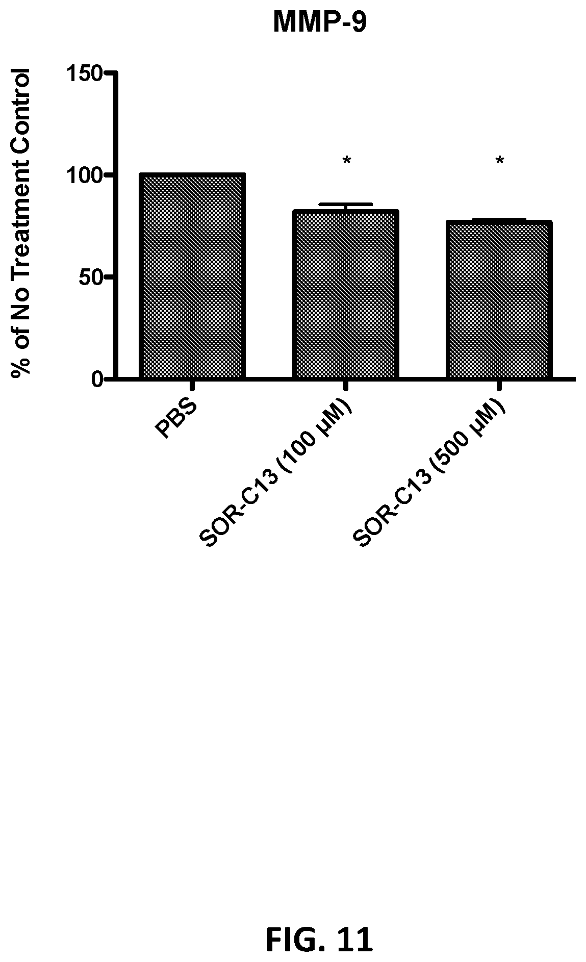
D00012
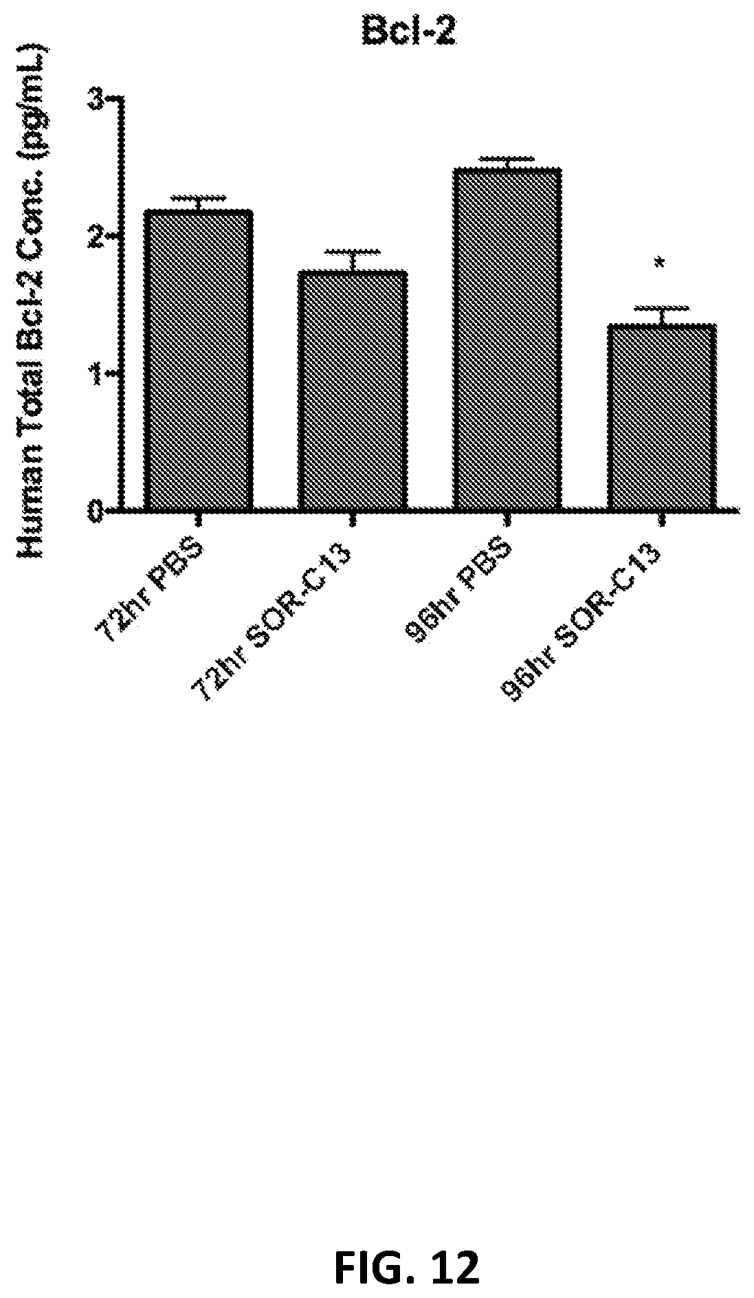
D00013
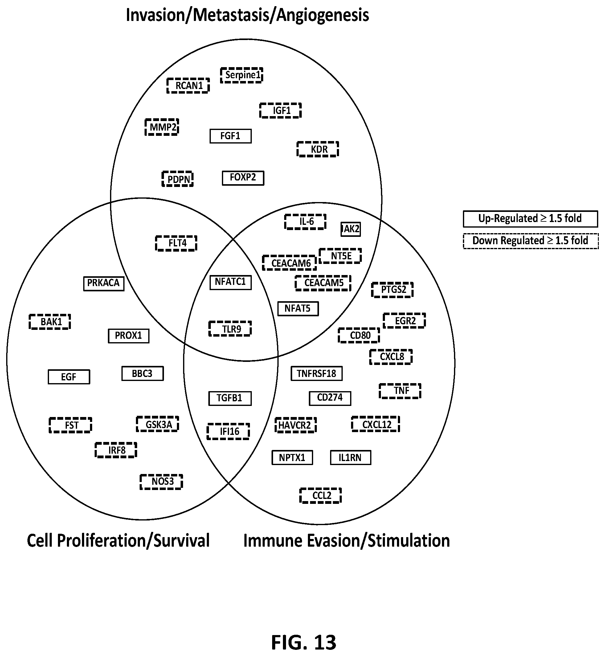
D00014

D00015

D00016

D00017

D00018

D00019

D00020

D00021

S00001
XML
uspto.report is an independent third-party trademark research tool that is not affiliated, endorsed, or sponsored by the United States Patent and Trademark Office (USPTO) or any other governmental organization. The information provided by uspto.report is based on publicly available data at the time of writing and is intended for informational purposes only.
While we strive to provide accurate and up-to-date information, we do not guarantee the accuracy, completeness, reliability, or suitability of the information displayed on this site. The use of this site is at your own risk. Any reliance you place on such information is therefore strictly at your own risk.
All official trademark data, including owner information, should be verified by visiting the official USPTO website at www.uspto.gov. This site is not intended to replace professional legal advice and should not be used as a substitute for consulting with a legal professional who is knowledgeable about trademark law.