Methods For Attenuating Viral Infection And For Treating Lung Injury
Glassberg Csete; Marilyn ; et al.
U.S. patent application number 16/893306 was filed with the patent office on 2020-12-10 for methods for attenuating viral infection and for treating lung injury. This patent application is currently assigned to Spiritus Therapeutics, Inc.. The applicant listed for this patent is Spiritus Therapeutics, Inc.. Invention is credited to Marilyn Glassberg Csete, Jodi Gurney.
| Application Number | 20200384034 16/893306 |
| Document ID | / |
| Family ID | 1000005049312 |
| Filed Date | 2020-12-10 |


View All Diagrams
| United States Patent Application | 20200384034 |
| Kind Code | A1 |
| Glassberg Csete; Marilyn ; et al. | December 10, 2020 |
METHODS FOR ATTENUATING VIRAL INFECTION AND FOR TREATING LUNG INJURY
Abstract
The described invention provides compositions and methods for treating a susceptible subject at risk of pulmonary complications of an acute lung injury caused by a severe infection with a respiratory virus and for restoring lung function to donor lungs. The methods include administering a therapeutic amount of a pharmaceutical composition comprising extracellular vesicles (EVs) comprising one or more miRNAs and a pharmaceutically acceptable carrier. The population of EVs can be derived from a patient who has recovered from an infection with the respiratory virus or has been exposed to anti-viral antibodies through treatment, can be derived from MSCs of a normal healthy individual, can be modified by a viral vector, or can be synthetic.
| Inventors: | Glassberg Csete; Marilyn; (Miami, FL) ; Gurney; Jodi; (New York, NY) | ||||||||||
| Applicant: |
|
||||||||||
|---|---|---|---|---|---|---|---|---|---|---|---|
| Assignee: | Spiritus Therapeutics, Inc. New York NY |
||||||||||
| Family ID: | 1000005049312 | ||||||||||
| Appl. No.: | 16/893306 | ||||||||||
| Filed: | June 4, 2020 |
Related U.S. Patent Documents
| Application Number | Filing Date | Patent Number | ||
|---|---|---|---|---|
| 63023684 | May 12, 2020 | |||
| 63007909 | Apr 9, 2020 | |||
| 62945917 | Dec 10, 2019 | |||
| 62858154 | Jun 6, 2019 | |||
| Current U.S. Class: | 1/1 |
| Current CPC Class: | C12N 15/113 20130101; A61K 45/06 20130101; A61K 35/28 20130101; A61P 11/00 20180101; A61K 35/17 20130101; A61K 9/127 20130101; A61K 9/0073 20130101 |
| International Class: | A61K 35/28 20060101 A61K035/28; C12N 15/113 20060101 C12N015/113; A61K 9/00 20060101 A61K009/00; A61K 35/17 20060101 A61K035/17; A61K 45/06 20060101 A61K045/06; A61K 9/127 20060101 A61K009/127; A61P 11/00 20060101 A61P011/00 |
Claims
1. A precision medicine method for optimizing therapeutic benefit for a susceptible subject at risk of pulmonary complications of an acute lung injury caused by a severe infection with a respiratory virus, comprising: (a) obtaining a urine sample from the subject and from a healthy control; (b) isolating EVs from the urine sample obtained from the subject and healthy control; (c) measuring a level of expression of each of a plurality of miRNAs in the EVs from the urine sample from the subject and in the EVs from the urine sample from the healthy control; (d) determining that expression of the one or more of the miRNAs in the EVs from the subject is dysregulated compared to the healthy control; (e) identifying a predisposition to develop pulmonary complications of the acute lung injury: (1) when the presence of one or more dysregulated miRNAs in the EVs from the urine sample obtained from the subject is detected; (2) when an increase in levels of one or more WNT proteins in the EVs from the urine sample from the subject compared to the control is detected; or (3) when the presence of one or more dysregulated miRNAs and an increase in levels of one or more WNT proteins in the EVs from the urine sample from the subject compared to the control is detected; and (f) tailoring a medical treatment that delays development of the pulmonary complications.
2. The method according to claim 1, wherein the medical treatment comprises: administering a therapeutic amount of a pharmaceutical composition comprising either: (i') at least about 1.times.10.sup.8 whole MSCs comprising synthetic exosomes comprising a therapeutic amount of an miR-29a mimic, an miR-199-3p inhibitor, or both to the diagnosed subject; or (ii') a therapeutic amount of a purified and enriched population of synthetic exosomes comprising one or more of an miR-29a derived from normal healthy MSCs, an miR-29a mimic, an miR-199-3p inhibitor, to the diagnosed subject; or (iii') a therapeutic amount of a purified and enriched population of exosomes derived from a patient who has recovered from an infection with the respiratory virus or has been exposed to anti-viral antibodies through treatment; wherein the therapeutic amount upregulates expression of miR-29a, downregulates expression of miR-199-3p, or both, and effectively delays development of the pulmonary complications.
3. The method according to claim 2, wherein the administering is by inhalation.
4. The method according to claim 2, wherein the population of EVs is derived from peripheral blood mononuclear cells (PBMCs) of the recovered patient.
5. The method according to claim 4, wherein the PBMCs comprise T lymphocytes, B lymphocytes and NK cells.
6. The method according to claim 2 wherein the population of EVs is derived from T lymphocytes of the recovered patient.
7. The method according to claim 2, wherein the population of EVs is derived from B lymphocytes of the recovered patient.
8. The method according to claim 2, wherein the population of EVs is derived from NK cells of the recovered patient.
9. The method according to claim 2, wherein the population of EVs is derived from urine of the recovered patient.
10. The method according to claim 2, wherein the population of EVs is modified by a viral vector.
11. The method according to claim 1, wherein the pulmonary complications comprise a fibrotic lung disease.
12. The method according to claim 1, wherein the fibrotic disease is a fibrotic lung disease.
13. The method according to claim 12, wherein the fibrotic lung disease is pulmonary fibrosis.
14. The method according to claim 1, wherein isolating step (b) comprises purifying the urine-derived exosomes by one or more of a) ultracentrifugation; b) sucrose density gradient centrifugation; c) column chromatography; d) size exclusion; or e) filtration through a device containing an affinity matrix selective towards the EVs.
15. The method according to claim 1, wherein the urine-derived EVs are characterized by sedimentation at about 100,000.times.g, a buoyant density in sucrose of about 1.10-1.21 .mu.g/ml, and an average diameter of from about 30 nm to about 200 nm.
16. The method according to claim 1, wherein the average diameter of the EVs ranges from about 140 nm to about 150 nm.
17. The method according to claim 1, wherein the EVs comprise microvesicles whose diameter is >200 nm.
18. The method according to claim 1, wherein the predisposition to develop a fibrotic disease comprises onset of pulmonary fibrosis at an age earlier than the healthy control; typical predispositions based on genetic, environmental and lifestyle factors if not for the respiratory virus infection, or both.
19. The method according to claim 1, wherein the susceptible subject includes a very young individual, an elderly individual, an individual who is suffering from a respiratory disease; an individual who is receiving immunosuppressant therapy; an individual with a long term health condition; and an individual who is physically weak due to malnutrition or dehydration.
20. The method according to claim 1, wherein the severe infection with the respiratory virus comprises an acute lung injury, acute respiratory distress syndrome, or both.
21. The method according to claim 1, wherein the severe infection is caused by a respiratory virus selected from the group consisting of human coronavirus; human influenza, human herpesvirus, hantavirus; human cytomegalovirus; and human immunodeficiency virus.
22. The method according to claim 1, wherein the dysregulated miRNAs comprise one or more of miR-134-5p, miR-196b-5p, miR-629-5p, miR-206, miR-192-5p, miR-320c, miR-125a-3p, miR-215-5p, miR-642a-3p, miR-576-3p, miR-3679-5p, miR-134-5p, miR-196b-5p, miR-629-5p, or miR-206. miR-let7d; miR-29a, miR 20-b,miR-29c, or miR-199.
23. The method according to claim 1, wherein the miRNA is downregulated compared to the healthy control.
24. The method according to claim 1, wherein the miRNA is upregulated compared to the healthy control.
25. The method according to claim 1, further comprising detecting a level of expression of one or more WNT proteins in the EVs from the urine samples; and determining that expression of one or more of the WNT proteins in the urine sample from the subject is dysregulated compared to the healthy control.
26. The method of claim 25, wherein the one or more WNT proteins comprise WNT-5A.
27. The method according to claim 1, further comprising (a) obtaining a blood or serum sample from the subject and from the healthy control; (b) detecting a level of expression of one or more biomarkers selected from KL-6/MUC1, SP-A, SP-D, CCL18, MMP-1, and MMP-7 in the samples; and (c) comparing the levels of expression of the one or more biomarkers in the samples from the subject and from the healthy control; wherein an increase in the levels of the one or more biomarkers in the sample from the subject compared to the healthy control indicates a poor prognosis in the subject; and a decrease in the levels of the one or more biomarkers in the sample from the susceptible subject compared to the healthy control indicates a positive prognosis for the susceptible subject.
28. The method according to claim 1, further comprising obtaining a bronchoalveolar lavage fluid (BALF) sample from the subject and from the healthy control; isolating EVs from the BALF samples; detecting a level of expression of one or more WNT proteins in the EVs; and comparing the level of expression of the one or more WNT proteins in the EVs from the subject and from the healthy control; wherein an increase in the levels of the one or more WNT proteins in the EVs from the subject indicates the subject has a fibrotic disease.
29. The method according to claim 25, wherein the one or more WNT proteins comprise WNT-5A.
30. The method according to claim 2, wherein the miR-29a mimic has at least about 70% sequence homology with SEQ ID NO: 1.
31. The method according to claim 2, wherein the miR-199-3p inhibitor has at least about 70% sequence homology with SEQ ID NO: 2.
32. A method of diagnosing and treating a lung injury caused by a severe infection by a respiratory virus in a susceptible subject comprising (a) obtaining a urine sample from the subject and from a healthy control; (b) isolating EVs from the urine sample obtained from the subject and healthy control; (c) detecting a level of expression of miRNAs in the EVs from the urine sample from the subject and in the EVs from the urine sample from the healthy control; (d) determining that expression of one or more of the miRNAs in the EVs from the subject is dysregulated compared to the healthy control; and (e) identifying the subject as one that can benefit therapeutically from being treated for a fibrotic disease: (i) when the presence of one or more dysregulated miRNAs in the EVs from the urine sample obtained from the subject is detected; (ii) when an increase in levels of one or more WNT proteins in the EVs from the urine sample from the subject compared to the healthy control is detected; or (iii) when the presence of one or more dysregulated miRNAs and an increase in levels of one or more WNT proteins in the EVs from the urine sample from the subject compared to the healthy control is detected; and (f) administering a therapeutic amount of a pharmaceutical composition comprising either: (i') at least about 1.times.10 whole mesenchymal stem cells (MSCs) comprising synthetic exosomes comprising a therapeutic amount of an miR-29a derived from normal healthy MSCs, an miR-29a mimic, an miR-199-3p inhibitor, or both to the diagnosed subject; or (ii') a therapeutic amount of a purified and enriched population of synthetic exosomes comprising one or more of an miR-29a derived from normal healthy MSCs, an miR-29a mimic, an miR-199-3p inhibitor, to the diagnosed subject; wherein the therapeutic amount upregulates expression of miR-29a, downregulates expression of miR-199-3p, and treats the fibrotic disease.
33. The method according to claim 32, wherein the administering is by inhalation.
34. The method according to claim 32, wherein the sequence of the miR-29a mimic is at least about 70% homologous with SEQ ID NO: 1.
35. The method according to claim 32, wherein the sequence of the miR-199-3p inhibitor is at least about 70% homologous with SEQ ID NO: 2.
36. The method according to claim 32, wherein the upregulated expression of miR-29a decreases expression of MMP-2.
37. The method according to claim 32, wherein the downregulated expression of miR-199-3p upregulates CAV-1 expression.
38. The method according to claim 32, wherein the lung injury includes acute respiratory failure, acute respiratory distress syndrome, or both.
39. The method according to claim 32, wherein the acute lung injury is progressive to a fibrotic lung disease.
40. The method according to claim 39 wherein the fibrotic lung disease is pulmonary fibrosis.
41. A method of attenuating a severe virus infection comprising a lung injury in a susceptible subject comprising administering to the subject a therapeutic amount of a pharmaceutical composition comprising a population of EVs comprising one or more of an miRNA derived from normal, healthy MSCs, an miR-29a mimic, an miR-199-3p inhibitor, and a pharmaceutically acceptable carrier, and wherein the pharmaceutical composition upregulates expression of miR-29a, downregulates miR-199-3p, or both, wherein the method attenuates the severe virus infection and the lung injury.
42. The method according to claim 41, wherein the susceptible subject includes a very young individual, an elderly individual, an individual who is suffering from a respiratory disease; an individual who is receiving immunosuppressant therapy; an individual with a long term health condition; and an individual who is physically weak due to malnutrition or dehydration.
43. The method according to claim 41, wherein the population of exosomes is derived from a patient who has recovered from an infection with a respiratory virus or has been exposed to anti-viral antibodies through treatment.
44. The method according to claim 43, wherein the population of EVs is derived from peripheral blood mononuclear cells (PBMCs) of the recovered patient.
45. The method according to claim 43, wherein the PBMCs comprise T lymphocytes, B lymphocytes and NK cells.
46. The method according to claim 43, wherein the population of EVs is derived from T lymphocytes of the recovered patient.
47. The method according to claim 43, wherein the population of EVs is derived from B lymphocytes of the recovered patient.
48. The method according to claim 43, wherein the population of EVs is derived from NK cells of the recovered patient.
49. The method according to claim 43, wherein the population of EVs is derived from urine of the recovered patient.
50. The method according to claim 43, wherein the population of EVs is modified by a viral vector.
51. The method according to claim 43, wherein the respiratory virus is selected from the group consisting of human coronavirus; human influenza, human herpesvirus, hantavirus; human cytomegalovirus; and human immunodeficiency virus.
52. The method according to claim 41, wherein the exosomes are synthetic.
53. The method according to claim 41, wherein attenuating the lung injury accomplishes one or more of decreasing one or more symptoms of a fibrotic lung disease, increasing repair of a lung injury, restoring lung function, reducing or eliminating a need for other active agents or therapeutics; and slowing progression of fibrotic lung disease.
54. The method according to claim 41, wherein the lung injury is an acute lung injury.
55. The method according to claim 41, wherein the acute lung injury comprises inflammation, acute respiratory failure, or both.
56. The method according to claim 41, wherein the lung injury includes acute respiratory distress syndrome.
57. The method according to claim 41, wherein the method treats progression to a fibrotic lung disease.
58. The method according to claim 57 wherein the fibrotic lung disease is pulmonary fibrosis.
59. The method according to claim 41, wherein the upregulated expression of miR-29a decreases expression of MMP-2.
60. The method according to claim 41, wherein the downregulated expression of miR-199-3p upregulates CAV-1 expression.
61. The method according to claim 41, wherein the miR-29a mimic has at least about 70% sequence homology with SEQ ID NO: 1.
62. The method according to claim 41, wherein the miR-199-3p inhibitor has at least about 70% sequence homology with SEQ ID NO: 2.
63. The method according to claim 41, wherein the normal healthy MSCs are obtained from a human subject.
64. The method according to claim 41, wherein the MSCs are derived from a tissue selected from the group consisting of amniotic membrane, chorionic membrane, umbilical cord tissue, bone marrow, and adipose tissue.
65. The method of claim 41, wherein the EVs are characterized by sedimentation at about 100,000.times.g, a buoyant density in sucrose of about 1.10-1.21 g/ml, and an average diameter of from about 30 nm to about 200 nm.
66. The method according to claim 41, wherein the average diameter of the EVs ranges from about 140 nm to about 150 nm.
67. The method according to claim 41, wherein the administering occurs nasally, intratracheally, orally, parenterally, topically, or by inhalation.
68. The method according to claim 41, wherein the administering occurs by inhalation.
69. The method according to claim 41, wherein the pharmaceutical composition further comprises at least one additional therapeutic agent.
70. The method according to claim 69, wherein the additional therapeutic agent is selected from one or more of an immunomodulatory agent, an analgesic agent, an anti-inflammatory agent, an anti-infective agent, an anti-malarial agent, an anti-viral agent, an anti-fibrotic agent, or a proton pump inhibitor.
71. The method according to claim 70, wherein the immunomodulatory agent is a corticosteroid.
72. The method according to claim 71, wherein the corticosteroid is selected from prednisone, azathioprine, mycophenolate, mycophenolate mofetil, colchicine, interferon-gamma 1b, and combinations thereof.
73. The method according to claim 70, wherein the analgesic agent is selected from codeine, hydrocodone, oxycodone, methadone, hydromorphone, morphine, fentanyl, and combinations thereof.
74. The method according to claim 70, wherein the anti-inflammatory agent is selected from aspirin, celecoxib, diclofenac, diflunisal, etodolac, ibuprofen, indomethacin, ketoprofen, ketorolac nabumetone, naproxen, nintedanib, oxaprozin, pirfenidone, piroxicam, salsalate, sulindac, tolmetin, and combinations thereof.
75. The method according to claim 70, wherein the anti-viral agent is selected from acyclovir, gancidovir, foscarnet; ribavirin; amantadine, azidodeoxythymidine/zidovudine), nevirapine, tetrahydroimidazobenzodiazepinone (TIBO) compound; efavirenz; remdecivir, and delavirdine.
76. The method according to claim 70, wherein the anti-malarial agent is selected from an aryl aminoalcohol compound selected from quinine, quinidine, chloroquine, amodiaquine, mefloquine, halofantrine, lumefantrine, piperaquine, tafenoquine; an antifolate compound, selected from pyrimethamine, proguanil, chlorproguanil, trimethoprim; an artemisinin compound selected from artemisinin, dihydroartemisinin, artemether, artesunate; atovaquone.
77. The method according to claim 70, wherein the anti-infective agent is selected from amoxicillin, doxycycline, demeclocycline; eravacycline, minocycline, ormadacycline, tetracycline, cephalexin, defotaxime, cetazidime, cefuroxime, ceftaroline; ciprofloxacin, levofloxacin, moxifloxacin clindamycin, lincomycin, metronidazole, azithromycin; clarithromycin, erythromycin, sulfamethoxazle and trimethoprim; sulfasalazine, amoxicillin and clavulanate; vancomycin, dalbavancin, oritavancin, telavancin, gentamycin, tobramycin, amikacin, imipenem and cilastatin, meropenem, doripenem, and ertapenem.
78. The method according to claim 70, wherein the anti-fibrotic agent is selected from nintedanib, pirfenidone, and combinations thereof.
79. The method according to claim 70, wherein the proton pump inhibitor is selected from omeprazole, lansoprazole, dexlansoprazole, esomeprazole, pantoprazole, rabeprazole, ilaprazole, and combinations thereof.
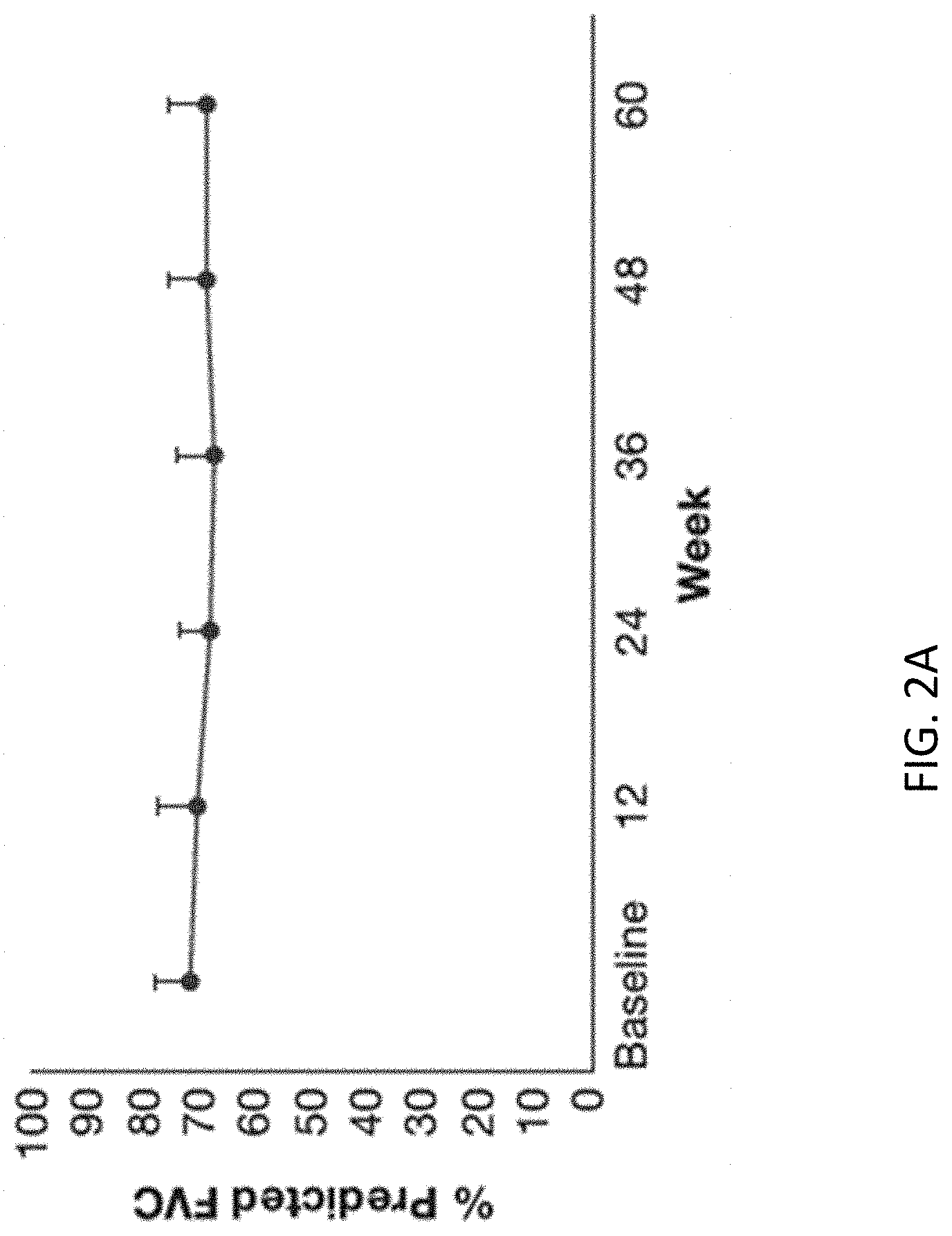
80. The method of claim 41, further comprising: (a) Obtaining a urine sample from the subject and from a healthy control; (b) Isolating extracellular vesicles (EVs) from the urine sample obtained from the subject and healthy control; (c) Measuring a level of expression of one or more Idiopathic Pulmonary Fibrosis (IPF) markers selected from the group consisting of integrin mRNA, collagen type 1.alpha.1 mRNA, miR29, c-jun protein; estrogen receptor alpha (ER.alpha.), androgen receptor (AR), caveollin-1 protein; pAKT/AKT protein in the EVs obtained from the subject and from the healthy control prior to treatment; and treating the subject by administering a pharmaceutical composition comprising a therapeutic amount of EVs purified from the healthy control; wherein the therapeutic amount modulates the level of expression of the one or more IPF markers in the urine of the subject.
81. A method for resuscitating donor lungs comprising administering ex vivo a therapeutic amount of a pharmaceutical composition comprising a population of EVs comprising one or more of an miRNA derived from normal, healthy MSCs, an miR-29a mimic, an miR-199-3p inhibitor, and a pharmaceutically acceptable carrier, and wherein the pharmaceutical composition upregulates expression of miR-29a, downregulates miR-199-3p, or both, wherein the method restores lung function.
82. The method of claim 81, wherein the exosomes are derived from a patient who has recovered from an infection with a respiratory virus or has been exposed to anti-viral antibodies through treatment.
83. The method of claim 82, wherein the population of EVs is derived from peripheral blood mononuclear cells (PBMCs) of the recovered patient.
84. The method of claim 83, wherein the PBMCs comprise T lymphocytes, B lymphocytes and NK cells.
85. The method of claim 82, wherein the population of EVs is derived from T lymphocytes of the recovered patient.
86. The method of claim 82, wherein the population of EVs is derived from B lymphocytes of the recovered patient.
87. The method of claim 82, wherein the population of EVs is derived from NK cells of the recovered patient.
88. The method of claim 82, wherein the population of EVs is derived from urine of the recovered patient.
89. The method of claim 82, wherein the population of EVs is modified by a viral vector.
90. The method of claim 81, wherein the exosomes are synthetic.
Description
CROSS-REFERENCE TO RELATED APPLICATIONS
[0001] This application claims the benefit of priority to U.S. provisional applications 63/023,684 (filed May 12, 2020), 63/007,909 (filed Apr. 9, 2020), 62/945,917 (filed Dec. 10, 2019) and 62/858,154 (filed Jun. 6, 2019). Each of these applications is incorporated herein by reference in its entirety.
FIELD OF INVENTION
[0002] The described invention generally relates to mesenchymal stem cell-derived extracellular vesicles, compositions thereof, and uses thereof for attenuating viral infection and for treating lung injury.
SEQUENCE LISTING
[0003] The instant application contains a Sequence Listing which has been submitted electronically in ASCII format and is hereby incorporated by reference in its entirety. Said ASCII copy, created on Jun. 4, 2020, is named 130309-00203_SL.txt and is 8999 bytes in size.
BACKGROUND
[0004] Respiratory viral infections are an important cause of morbidity and, in some settings, of mortality. One important feature of RV infections is the nonspecific nature of clinical signs and symptoms. For most respiratory viruses, the epidemiology is characterized by annual outbreaks during the winter and spring seasons; some respiratory viruses, however some (e.g., PIVs), are prevalent throughout the year. [Nichols, W. G. et al., "Respiratory Viruses other than influenza virus: impact and therapeutic advances. Clin. Microbiol. Rev. (2008) 21(2): 274-90].
[0005] The Baltimore classification of viruses [Baltimore, D., 1974 Haravey Lecture 70 Series: 57-74) is based on the viral mechanism of mRNA production. Viral genomes may be single-stranded (ss) or double stranded (ds), RNA or DNA, and may or may not use reverse transcriptase. Additionally, single stranded RNA viruses may be either positive sense (+) or negative or antisense (-). This classification places viruses into seven groups, as shown in Table 1.
TABLE-US-00001 TABLE 1 Baltimore classification of viruses Group Examples I. Ds DNA viruses Adenoviruses, Herpesviruses, Poxviruses II. Ss DNA viruses +sense DNA (Parvoviruses) III. dsRNA viruses Rheoviruses IV. +ssRNA viruses Picornaviruses, Togaviruses V. (-) ssRNA viruses Orthomyxoviruses, Rhabdoviruses VI. ssRNA-RT viruses Retroviruses VII. dsDNA-RT viruses Hepadnaviruses
[0006] Though the impact of respiratory viruses in an immunocompetent host is substantial, patients with compromised immune function are far more likely to experience poor outcomes on a per-subject basis. For example, among hematopoietic cell transplant (HCT) recipients and severely immunosuppressed nontransplant patients with hematologic malignancies [Id., citing Whimbey, E., R. B., et al. Respiratory syncytial virus pneumonia in hospitalized adult patients with leukemia. (1995) Clin. Infect. Dis. 21:376-379], upper respiratory tract infection with RSV may progress to fatal pneumonia. Human metapneumovirus (hMPV) can cause upper respiratory tract infection and fatal interstitial pneumonia in HCT recipients [Id., citing Dignan, F., C. et al. Parainfluenza type 3 infection post stem cell transplant: high prevalence but low mortality. J. Hosp. Infect. (2006) 63:452-458]. Adenovirus pneumonia can occur in ICT recipients, especially following in vivo or ex vivo T-cell depletion [Id., citing Flomenberg, P., J., et al. Increasing incidence of adenovirus disease in bone marrow transplant recipients. J. Infect. Dis. (1994) 169:775-781; Lion, T, et al Intestinal adenovirus infection with increasing viral load in stool is an early indicator of impending disseminated disease in children undergoing T-cell depleted allogeneic stem cell transplantation. Blood (2003) 102:196a, Yusuf, U. et al. Cidofovir for the treatment of adenoviral infection in pediatric hematopoietic stem cell transplant patients. Transplantation (2006) 81:1398-1404]. Respiratory viruses can cause major morbidity and also mortality in solid organ transplantation (SOT) recipients. Incident infection depends on virus exposure, but the disease severity appears to be different in different organ transplant settings. The setting with the most significant impact is lung and heart-lung transplantation, while liver and renal transplant recipients appear to be somewhat less affected [Id., citing Peige-Lafeuile, H N, et al., Severe respiratory syncytial virus pneumonia in an adult renal transplant recipient: successful treatment with ribavirin. Scand. J. Infect. Dis. (1990) 22:87-89; Sinnott, J. T., et al. Respiratory syncytial virus pneumonia in a cardiac transplant recipient. J. Infect. Dis. (1988) 158:650-651, Wendt, C. H. Community respiratory viruses: organ transplant recipients. Am. J. Med. (1997) 102:31-36, 42-43]. The burden of severe pneumonia due to RSV, influenza virus, PIV, and adenovirus was substantially higher in South African children infected with HIV than in non-IV-infected children [Madhi, S. A., L et al. Five-year cohort study of hospitalization for respiratory syncytial virus associated lower respiratory tract infection in African children. J. Clin. Virol. (2006) 36:215-221; Madhi, S. A., B. et al. Increased burden of respiratory viral associated severe lower respiratory tract infections in children infected with human immunodeficiency virus type-1. J. Pediatr. (2000) 137:78-84; Madhi, S. A., M. et al. Differing manifestations of respiratory syncytial virus-associated severe lower respiratory tract infections in human immunodeficiency virus type 1-infected and uninfected children. Pediatr. Infect. Dis. J. (2001) 20:164-170]. In another South African study, PIV infection was associated with a higher mortality in HIV-infected children than in uninfected individuals [Madhi, S. A., N. Lower respiratory tract infections associated with influenza A and B viruses in an area with a high prevalence of pediatric human immunodeficiency type 1 infection. Pediatr. Infect. Dis. J. (2002) 21:291-297133). hMPV was identified as a possible cause of pneumonia in 3.7% of HIV-infected versus 9.1% of non-HIV-infected children and was the second most common RV associated with hospitalization (after RSV).
TABLE-US-00002 TABLE 2 Classification of exemplary human respiratory viruses Family (nucleic acid) Representative virus Cellular receptor(s) Paramyxoviridae (negative Respiratory syncytial virus Sialyl-glycoproteins, glycolipids sense single stranded RNA (RSV) Human parainfluenza virus Sialyl-glycoproteins, glycolipids (PIV) Human metapneumovirus Sialyl-glycoproteins, glycolipids (hMPV) measlesvirus Picornaviridae (positive Enteroviruses ICAM-1, decay accelerating factor sense single strained RNA) (cocksackievirus Type A or (CD55), CAR, integrins B, echovirus) Rhinovirus ICAM-1, sialic acid Coronaviridae (positive Human coronavirus C13 (HCoV 229E), ACE2 (HCoV sense single-stranded RNA) (HCoV) NL63, SARS CoV), CD209L (SARS-CoV-229E) Adenoviridae (double Adenovirus CAR (cocksackievirus and stranded DNA) adenovirus receptor, integrins Parvoviridae (positive or Human bocavirus (HBoV) Unknown negative sense single stranded DNA) Taken from Nichols. W. G. et al., "Respiratory Viruses other than influenza virus: impact and therapeutic advances. Clin. Microbiol. Rev. (2008) 21(2): 274-90.
[0007] Despite commonalties in disease presentation among many of the respiratory viruses, they are genetically and antigenically distinct.
[0008] The RNA viruses of the Mononegavirales order contain single-stranded genomes that are negative sense. This order is comprised of seven families, including Orthomyxoviridae (which contains the influenza viruses) and Paramyxoviridae (which contains the parainfluenza viruses (PIVs), human respiratory syncytial virus [RSV], and human metapneumovirus [hMPV]). Members of both families are enveloped; unlike viruses in the family Pramyxoviridae, the viral genomes from family Orthomyxoviridae are segmented. [Id.]
[0009] The Picornaviridae are nonenveloped viruses with a single-stranded genome of positive polarity. The rhinoviruses are the most important members of this family causing respiratory diseases in humans; other viruses in this group that can cause respiratory disease are enteroviruses (such as coxsackieviruses and numbered enteroviruses). Over 100 serotypes of rhinovirus have been identified. Viruses from the family Coronaviridae also contain positive-sense single-stranded PNA (ssRNA) but are enveloped. [Id.]
[0010] Human bocavirus, a member of the Parvoviridae family, was discovered in 2005 in nasopharyngeal aspirates from children with respiratory tract infections. Although suspected, establishing its role as a respiratory pathogen has been difficult for several reasons. First, human bocavirus is not related to any known human respiratory pathogen. Second, it is commonly detected with other respiratory viruses, which have established pathogenic potential. Third, detection may simply reflect asymptomatic persistence or prolonged viral shedding, since other human parvoviruses also show this capacity. It has been suggested that human bocavirus may be reactivated or produce a transient asymptomatic super-infection triggered by the presence of another replicating respiratory agent. [McNamara, P S, Van Doom, H R, Respiratory viruses and atypical bacteria. In Manson's Tropical Infectious Diseases (23.sup.rd Ed.) (2014), 215-224].
[0011] Measles is caused by Rubeola virus, which belongs to the Paramyxovirus family. It is a major cause of lower respiratory tract infection, accounting for 6-21% of its morbidity and 8-50% of its mortality. Radiographic evidence of pneumonia is common, also in clinically uncomplicated measles. The effects of the virus on the respiratory tract can be direct (giant cell pneumonitis), in any part of the respiratory tract, or indirect. The latter includes the suppressive effects of the virus on the host immune system, stores of vitamin A and overall nutritional status, all of which may lead to an increased risk of super-infection with other (viral or bacterial) pathogens. Measles pneumonitis can be especially severe in immunocompromised patients. [Id.]
Hantaviruses
[0012] Hantavirus pulmonary syndrome (HPS) was first recognized in May 1993 during an outbreak of severe, and frequently fatal, respiratory disease in the four corners region of the United States, where Arizona, Colorado, New Mexico and Utah abut. Closely related viruses have since been isolated with different rodent hosts. These viruses belong to the same Hantavirus genus as those causing hemorrhagic fever with renal syndrome (HFRS). Both HFRS and HPS have a similar febrile prodrome with thrombocytopenia and leucocytosis. In HPS, the key differences are that the capillary leakage which follows is localized to the lungs. There is evidence that some of the South American hantaviruses causing IPS may be transmitted between humans in a nosocomial setting. [Id.]
Herpesvirus (Herpesviridae)
[0013] Herpesviruses are a member of the family Herpesviridae. They are double-stranded DNA viruses. Varicella pneumonitis can occur as a severe complication of chickenpox or in the absence of classical symptoms. It occurs more commonly in adults (an estimated 1:400) and can be life threatening it occurs during pregnancy or in immunocompromised patients. Although relatively rare, radiographical abnormalities of the lungs without respiratory symptoms are reported in more than 15% of adults with chicken pox. Pneumonitis can also rarely occur as a complication of cytomegalovirus or Epstein-Barr virus mononucleosis. [Id.)
Influenza (Myoviridae)
[0014] There are three genera of influenza viruses: influenza virus A, B, and C. The influenza viruses, especially influenza virus A, are the most variable of the respiratory viruses. Influenza A viruses are subtyped based on their two surface antigens: hemagglutinin (HA; H-1-H16) and neuraminidase (NA; N1-N9), which are responsible for host receptor binding/cell entry and cleavage of the HA-receptor complex to release newly formed viruses, respectively. Aquatic birds are the natural reservoir of influenza A viruses, harboring all possible subtypes. (Id.)
[0015] Both influenza virus A and B exhibit antigenic drift. This phenomenon occurs when the surface antigens of the virus gradually change, progressively and directionally, to escape immunological pressure from the host species. Yearly epidemics of influenza virus A and B are caused worldwide by these drift variants, and contribute to mortality (an estimate 250-500,000 every year) in the elderly, and in those with pre-existing conditions, such as chronic cardiopulmonary or renal disease; diabetes, immunosuppression, or severe anemia). New lineages of influenza virus A emerge every few decades through re-assortment of gene segments in animal hosts infected with two different viruses (antigenic shift), resulting in global pandemics with varying severity due to the absence of immunity in the human population (e.g., 1918 Spanish flu: H1N1, 40-100 million deaths; 1957 Asian flu: H2N2, 2 million deaths; 1968 Hong Kong flu: H3N2, 500,000 deaths; 2009 H1N1-pdm09, 15,000 deaths). Sporadic dead-end human infections of animal (especially avian) viruses are known to occur and have caused concern regarding pandemic potential. Highly pathogenic H5N1 viruses were first detected in birds in 1996 in China. In 2003, the virus re-emerged in China. Since then it has become panzootic among poultry and wild birds. The disease presents as a rapidly progressive viral pneumonia with severe leucopenia and lymphopenia, progressing to acute respiratory distress syndrome (ARDS) and multi-organ dysfunction. In 2013, another avian influenza viruses (H7N9) caused zoonotic transmission events to humans in China, with no recorded sustained human-to-human transmission. The case fatality rate was around 20% and the elderly were most affected. [Id.)
Coronavirus
[0016] Coronaviruses (CoVs), a large family of single-stranded RNA viruses, can infect a wide variety of animals, including humans, causing respiratory, enteric, hepatic and neurological diseases. [Yin, Y., Wunderink, R G, Respirology (2018) 23 (2): 130-37, citing Weiss, S R, Leibowitz, I L, Coronavirus pathogenesis. Adv. Virus Res. (2011) 81: 85-164]. Human coronaviruses, which were considered to be relatively harmless respiratory pathogens in the past, have now received worldwide attention as important pathogens in respiratory tract infection. As the largest known RNA viruses, CoVs are further divided into four genera: alpha-, beta-, gamma- and delta-coronavirus. In humans, CoVs cause mainly respiratory tract infections. Until November 2019, only six human coronaviruses (HCoVs) had been identified. These included the alpha-CoVs HCoV-NL63 and HCoV-229E and the beta-CoVs HCoV-OC43, HCoV-HKU1, severe acute respiratory syndrome-CoV (SARS-CoV), [Id., citing Drosten, C. et al. N. Engl. J. Med. (2003) 348: 1967-76] and Middle East respiratory syndrome-CoV (MERS-CoV) [Id., citing Zaki, A M et al. N. Engl. J. Med. (2012) 367: 1814-20].
[0017] Human-to-human transmission of SARS-CoV and MERS-CoV occurs mainly through nosocomial transmission. From 43.5-100% of MERS patients in individual outbreaks were linked to hospitals, [Id., citing Hunter, I C et al. Transmission of Middle East respiratory syndrome coronavirus infections in healthcare settings, Abu Dhabi. Emerg. Infect. Dis. (2016) 22: 647-56. Osong Public Health Res. Perspect. (2015) 6: 269-78], which was similar in SARS patients. [Anderson, R M et al. Philos. Trans. R. Soc. Lond. B. Biol. Sci. (2004) 359: 1091-105]. A study from the Republic of Korea revealed that index patients who transmitted to others had more non-isolated days in the hospital, body temperature of .gtoreq.38.5.degree. C. and pulmonary infiltration of .gtoreq.3 lung zones. [Id., citing Kang, C K, et al. J. Korean Med. Sci. (2017) 32: 744-49]. Transmission between family members occurred in only 13-21% of MERS cases and 22-39% of SARS cases. [Id., citing Kang, C K, et al. J. Korean Med. Sci. (2017) 32: 744-49]. Another Korean study suggested that transmission of MERS from an asymptomatic patient is rare. [Id., citing Moon, S Y, Son, J S. Clin. Infect. Dis. (2017) 64: 1457-58]. In contrast to SARS-CoV and MERS-CoV, direct human-to-human transmission was not reported for the other four HCoVs. [Id., citing Woo, P C et al. Hong Kong Med. J. (2008) 15 (Suppl. 9): 46-47].
[0018] Current understanding of the pathogenesis of HCoVs infection is still limited. However, several significant differences in the pathogenesis exist among SARS-CoV, MERS-CoV and the other HCoVs.
Cell Entry and Receptors
[0019] The critical first step for HCoV infection is entry into the susceptible host cells by combining with a specific receptor. Spike proteins (S proteins) of HCoVs are a surface-located trimeric glycoprotein consisting of two subunits: the N-terminal S subunit and the C-terminal S2 subunit. The S1 subunit specializes in recognizing and binding to the host cell receptor while the S2 region is responsible for membrane fusion. [Id., citing Gallagher, T M, Buchmeier, M J. Virology (2001) 279: 371-74]. To date, a wide range of diverse cellular receptors specifically recognized by the S1 domains have been identified for all HCoVs except HCoV-HKU1.
[0020] ACE2, the receptor for SARS-CoV and HCoV-NL63 [Id., citing Weiss, S R, Leibowitz, I L, Adv. Virus Res. (2011) 81: 85-164), Li, W. et al. Angiotensin-converting enzyme 2 is a functional receptor for the SARS coronavirus. Nature (2003) 426: 450-54; Hofmann, H. et al., Proc. Natl Acad. Sci. USA (2005) 102: 7988-93] is a surface molecule localized on arterial and venous endothelial cells, arterial smooth muscle cells, epithelia of the small intestine and the respiratory tract. In the respiratory tract, ACE2 is expressed on the epithelial cells of alveoli, trachea, and bronchi, bronchial serous glands, and alveolar monocytes and macrophages. ACE2 is a homologue of the ACE protein, and both are key enzymes of the renin-angiotensin system.35 ACE2 plays a protective role in lung failure and its counterpart ACE promoting lung edema and impaired lung function. [Id., citing Imai, Y., et al. Cir. J. (2010) 74: 405-10]. Downregulation of ACE2, as occurs during SARS-CoV infection, is believed to contribute to pathological changes in the lung. [Id., citing Imai, Y., et al. Cir. J. (2010) 74: 405-10; Kuba, K. et al. Nat. Med. (2005) 11: 875-79]. This form of lung damage can be attenuated by blocking the renin-angiotensin pathway. [Id., citing Kuba, K. et al. Nat. Med. (2005) 11: 875-79]. HCoV-NL63 also employs the SARS receptor for cellular entry [Id., citing Hofmann, H. et al., Proc. Natl Acad. Sci. USA (2005) 102: 7988-93] despite their markedly different pathogenicity and disease courses. This finding suggests that receptor usage may not be the only factor that determines the severity of HCoV infection.
[0021] Dipeptidyl peptidase 4 (DPP4, also known as CD26), the receptor for MERS-CoV, [Id., citing Meyerholz, D K et al. Am. J. Pathol. (2016) 186: 78-86] is a multifunctional cell-surface protein widely expressed on epithelial cells in kidney, small intestine, liver and prostate and on activated leukocytes. DPP4 is expressed in the upper respiratory tract epithelium of camels. [Id., citing Widagdo, W. et al. J. Vir. (2016) 90: 4838-42]. In the human respiratory tract, DPP4 is mainly expressed in alveoli rather than the nasal cavity or conducting airways. [Id., citing Meyerholz, D K et al. Am. J. Pathol. (2016) 186: 78-86] DPP4 is a key factor in the activation of T cells and immune response costimulatory signals in T cells, which could indicate a possible manipulation of the host immune system. [Id., citing Boonacker, E., Van Noorden, C J. Eur. J. Cell Biol. (2003) 82: 53-73].
[0022] Human aminopeptidase N (CD13), a cell-surface metalloprotease on intestinal, lung and kidney epithelial cells, has been identified as the receptor for hCoV-229E. [Id., citing Yeager, C L et al. Nature (1992) 357: 420-22]. The receptor for HCoV-OC43 is 9-O-acetylated sialic acid. Currently, the receptor for HCoV-HKU1 has not been identified.
Interferon and Interferon-Stimulated Genes
[0023] The interferon (IFN) family of cytokines, including IFN-.alpha., IFN-.beta. and IFN-.gamma., provide the first line of defense against viral pathogens. They initiate transcription of hundreds of IFN-stimulated genes (ISGs) that have antiviral, immune modulatory and cell regulatory functions.
[0024] Delayed recognition is critical for HCoVs to survive and replicate in the host. In vitro studies showed that both SARS-CoV and MERS-CoV have evolved genetic mechanisms to delay IFN induction and dysregulate ISG effector functions in primary human airway epithelial cells or in cultured cells. [Id., citing Manachery, V D, et al. Mol. Bio. (2014) 5: e01174-14; Lau, S K et al. J. Gen. Virol. (2013) 94: 2679-90]. It was reported that SARS-CoV infection could result in IFN-.alpha. induction only after 12 h in cultured Calu3 cells, with IFN-.beta.5 and IFN-.gamma.1 induction even further delayed. [Id., citing Manachery, V D, et al. Mol. Bio. (2014) 5: e01174-14] Similar to SARS-CoV, MERS-CoV also fails to induce IFNs prior to 12 h, with the exception of IFN-.alpha.5. Lau et al. serially measured mRNA levels of eight cytokine genes up to 30 h post-infection in Calu-3 cells infected with MERS-CoV and SARS-CoV. [Id., citing Lau, S K et al. J. Gen. Virol. (2013) 94: 2679-90]. Calu-3 cells infected by MERS-CoV showed marked induction of the proinflammatory cytokines IL-10, IL-6 and IL-8 at 30 h but lack of production of the innate antiviral cytokines tumour necrosis factor (TNF)-.alpha., IFN-.beta. and IFN-.gamma.-induced protein-10, compared with SARS-CoV. These data suggest that MERS-CoV attenuates innate immunity and induces a delayed proinflammatory response in human lung epithelial cells, which correlates with disease severity and clinical course.
Demographic and Clinical Features
[0025] Both SARS and MERS present with a spectrum of disease severity ranging from flu-like symptoms to acute respiratory distress syndrome (ARDS).
[0026] Age and underlying disease are significant independent predictors of various adverse outcomes in SARS. [Id., citing Chan, K S, et al. Respirology (2003) 8-Suppl.: S36-40]. SARS cases were mainly seen in young healthy individuals; whereas half of the cases of MERS-CoV infection occurred in individuals older than 50 years. [Id., citing Assiri, A. et al. Lancet Inf. Dis. (2013) 13: 752-61]. Compared with SARS patients, pre-existing chronic illnesses, such as diabetes (31%), hypertension (33%), chronic renal failure (15%), chronic heart disease (15%) and chronic pulmonary disease (13%), were more frequent in MERS patients. Clinical symptoms on admission included fever, cough, myalgia and shortness of breath in both SARS and MERS patients, while symptoms of upper respiratory tract infection such as sore throat were also frequent. Atypical symptoms such as diarrhea and vomiting developed in both SARS and MERS patients.
Kidney Impairment
[0027] Acute kidney injury (AKI) is a significant characteristic of both SARS and MERS patients. One study reported that 6.7% of SARS patients had acute renal impairment and 84.6% had proteinuria. [Id., citing Chu, K H et al. Kidney Int. (2005) 67: 98-705]. AKI is much more common in MERS patients, occurring in up to 43%. [Id., citing Saad, M. et al. Int. J. Infect. Dis. (2014). 29: 301-6].
[0028] The mechanism of the high AKI incidence in both SARS and MERS patients is not well clarified. Pre-existing co-morbid conditions and direct viral involvement of the kidneys [Id., citing Das, K M, et al. Am. J. Roentgenol. (2015) 204: 736-42; Morgenstern, B. et al., Biochem. Biophys. Res. Commun. (2005) 326: 905-8] may contribute to development of AKI. [Id., citing Chu, K H et al. A Kidney Intl. (2005) 67: 698-705; Saad, M. et al. Int. J. Infect. Dis. (20140 29: 301-06). Since ACE2 and DPP4, the receptors for SARS-CoV and MERS-CoV, are expressed at high levels in the kidney, functional impairment of these cell receptors by viral binding may contribute to the risk of AKI. Elevated creatinine kinase (CK) values (176-1466 U/L) observed in 36% of SARS patients suggests rhabdomyolysis may also contribute. [Id. citing Peiris, J S, et al. Lancet (2003) 361: 1767-72].
Cardiovascular Manifestations
[0029] A cardinal difference between MERS and SARS is the frequency of cardiovascular involvement. Despite the high lethality, shock was distinctly unusual in SARS until late stages when hypotension likely resulted from bacterial superinfections. [Id., citing Booth, C M, et al. JAMA (2003) 289: 2801-9; Lee, N. et al. A major outbreak of severe acute respiratory syndrome in Hong Kong. N. Engl. J. Med. (2003) 348: 1986-94; Almekhlafi, G. A., et al., Crit. Care (2016) 20: 123]. In contrast, need for vasopressor therapy was much more common in MERS, [Id., citing Chan, K S, et al. Respirology (2003): 8-(Suppl.: S36-40; Almekhlafi, G. A. et al., Crit. Care (2016) 20: 123) up to 81% in one series. (Id., citing Almekhlafi, G. A. et al., Crit. Care (2016) 20: 123). Need for vasopressors was an independent risk factor for death in the intensive care unit (ICU) (odds ratio=18.3, 95% confidence interval: 1.1-302.1, P=0.04). [Id., citing Almekhlafi, G. A. et al., Crit. Care (2016) 20: 123]. Multi-organ involvement was seldom reported with the endemic HCoV infections, despite occasional fatal pneumonia in highly immunocompromised patients.
Other Manifestations
[0030] Hematological abnormalities such as thrombocytopenia and lymphopenia were common in both SARS [Id., citing Peiris, J S, et al., Lancet (2003) 361: 1767-72; Booth, C M et al., JAMA (2003) 289: 2801-9] and MERS patients. [Id., citing Assiri, A. et al. Lancet Infect. Dis. (2013) 13: 752-61; Arabi, Y M, et al, Ann. Intern. Med. (2014) 160: 387-97]. Thrombocytopenia and lymphopenia may be predictive of fatal outcome in MERS-CoV patients. [Id., citing Arabi, Y M, et al, Ann. Intern. Med. (2014) 160: 387-97]. Other laboratory findings included elevated CK, lactate dehydrogenase, alanine aminotransferase and aspartate aminotransferase levels.
Radiological Features
[0031] Air-space opacities are the main radiographical feature in SARS patients. [Id., citing Booth, C M, et al. JAMA (2003) 289: 2801-9; Wong, K T, et al. Radiology (2003) 228: 401-6]. In one retrospective study, initial chest radiographs were abnormal in 108 of 138 (78.3%) of SARS patients and all showed air-space opacities. [Id., citing Wong, K T, et 1. Radiology (2003) 228: 401-6]. Of these 108 patients, 59 had unilateral focal involvement while 49 had either unilateral multifocal or bilateral involvement. Lower lung zone (64.8%) and right lung (75.9%) were more commonly involved. Four patterns of radiographical progression were recognized in those patients: type 1) initial radiographical deterioration to peak level followed by radiographical improvement occurred in in the majority (97 of 138 patients, 70.3%); type 2) fluctuating radiographical changes were seen in 24 patients (17.4%); type 3) static radiographical appearance in 10 patients (7.3%); and type 4) progressive radiographic deterioration in 7 patients (5.1%). In contrast, the most common radiographical features in MERS patients were ground-glass opacities and consolidation. [Id., citing Choi, W S, et al. Infect. Chemother. (2016) 48: 118-26; Das, K M, et al. Am. J. Roentgenol. (2015) 205: W267-74]. It was reported that ground-glass opacity was the most common abnormality (66%) in MERS patients, followed by consolidation (18%). [Id., citing Das, K M et al., Am. J. Roentgenol. (2015) 205: W267-74]. Meanwhile, type 2 radiographical progression (20 patients) was most common in those MERS patients, followed by type 4 (14 patients) and type 3 (7 patients). Type 1 radiographical progression was observed only in four patients. Pleural effusion (P=0.001), pneumothorax (P=0.001) and type 4 radiographical progression (P=0.001) were more frequent in MERS patients who died compared with recovered patients. Similar to the radiographical findings, computed tomography findings in MERS patients also included ground-glass opacity (53%), consolidation (20%) or a combination of both (33%). [Id., citing Das, K M, et al. Am. J. Roentgenol. (2015) 204: 736-42]. Pleural effusion was noted in 33% of cases and was associated with a poor prognosis for MERS-CoV infection. [Id., citing Das, K M, et al. Am. J. Roentgenol. (2015) 205: W267-74].
Clinical Outcome
[0032] More MERS cases progressed to respiratory failure and received invasive mechanical ventilation therapy than SARS patients. The occurrence of AKI, [Id., citing Saad, M. et al. Intl J. Infect. Dis. (2014) 29: 301-6] and the usage of vasopressor therapy were also more frequent in MERS patients in comparison with SARS. [Id., citing Chu, K H et al. Kidney Int. (2005) 67: 698-705; Almekhlafi, G A et al. Crit. Care (2016) 20: 123]. In a retrospective analysis, vasopressor therapy was proposed to be an independent risk factor for death in the ICU. [Id., citing Almekhlafi, G A et al. Crit. Care (2016) 20: 123].
[0033] MERS demonstrated a higher case fatality rate than SARS. Differences in host factors, such as age and underlying diseases, [Id., citing Chan, K S, et al. Respirology (2003) 8-(Suppl): S36-40); Choi, W S et al. Infect. Chemother. (2016) 48: 118-26] may explain some differences. However, differential cell line susceptibility, viral replication efficiency, ability to inhibit IFN production and receptor characteristics may also be responsible for the difference in the outcome of SARS-CoV and MERS-CoV infection. [Id., citing Lau, S K et al. J. Gen. Virol. (2013) 94: 2679-90; Chan, J F, et al. Differential cell line susceptibility to the emerging novel human beta coronavirus 2c EMC/2012: implications for disease pathogenesis and clinical manifestation. J. Infect. Dis. (2013) 207: 1743-52].
[0034] Compared with SARS and MERS, other HCoVs-associated pneumonia cases usually have relatively mild symptoms and recovered quickly. [Id., citing Woo, P C et al. Hong Kong Med. J. (2009) 15 (Supp. 9): 46-47]. Fatal cases were reported mainly in frail patients, such as neonates, the elderly and immunocompromised patients.
Treatment
[0035] At the moment, no specific therapy for SARS-CoV, MERS-CoV and the other HCoVs infection is available. Symptomatic and supportive treatment is the mainstay of therapy for patients infected by HCoVs.
[0036] A number of agents show effectiveness in vitro and/or in animal models and may improve the outcome in patients. Currently, the most commonly prescribed antiviral regimens in the clinical settings are ribavirin, IFNs and lopinavir/ritonavir.
[0037] To date, ribavirin and ribavirin plus various types of IFN have been the most common therapeutic interventions tried in patients with SARS and MERS. [Id., citing Morganstern, B. et al. Biochem. Biophys. Res. Commun. (2005) 326: 905-8); Al-Tawfiq, I A, et al. Int. J. Infect. Dis. (2014) 20: 42-6; Omrani, A S et al., Lancet Infect. Dis. (2014) 14: 1090-5]. Ribavirin, a nucleoside analogue, has a wide spectrum of antiviral activity by inhibiting viral RNA synthesis and mRNA capping. [Id., citing von Grotthuss, M. et al. Cell (2003) 113: 701-2]. When used alone for treatment of SARS, the clinical effect was inconsistent. Although in vitro studies show that combination with IFN-.beta. will give both these agents better antiviral activity, the clinical effect remains controversial.
[0038] IFNs are important for host defense against viruses. In in vitro experiments, IFN products were effective in inhibiting both SARS-CoV and MRES-CoV, with best antiviral activity seen with IFN-.beta.1b. [Id., citing Morgenstern, B. et al. Biochem. Biophyus. Res. Commun. (2005) 326: 905-8; Chan, J F, et al., J. Infect. (2013) 67: 606-16]. Previous studies had shown a positive impact of various IFNs on aspects of treatment of SARS and MERS patients, such as a better oxygen saturation and rapid resolution of inflammation, but no effect on more significant outcomes like hospital stay and long-term survival. [Id., citing Al-Tawfiq, I A et al. Intl. J. Infect. Dis. (2014) 20: 42-6; Omrani, A S et al. Lancet Infect. Dis. (2014) 14: 1090-95; Loutfy, M R et al. JAMA (2003) 290: 3222-28].
[0039] Lopinavir and ritonavir are protease inhibitors that may inhibit the 3C-like protease of MERS-CoV and modulate apoptosis in human cells. Addition of lopinavir/ritonavir to ribavirin was associated with improved clinical outcome compared with ribavirin alone in SARS patients. [Id., citing Chu, C M et al., Thorax (2004) 59: 252-56] Although lopinavir only showed suboptimal 50% effective cytotoxic concentration (EC50) against MERS-CoV in vitro, [Id., citing Chan, J F et al. J. Infect. (2013) 67: 606-16] lopinavir/ritonavir experimental therapy was proved to improve the outcome of MERS-CoV infection in an animal model. [Id., citing Chan, J F, et al. J. Infect. Dis. (2015) 212: 1904-13].
[0040] Mycophenolic acid (MPA) is another potential therapeutic choice. Frequently used as an immunosuppressant drug to prevent rejection in organ transplantation by inhibiting lymphocyte proliferation, MPA also prevents replication of viral RNA. In vitro studies showed that MPA had strong inhibition activity against MERS-CoV. [Id., citing Hart, B J, et al. J. Gen. Virol. (2014) 95: 571-77]. However, use in a non-human primate model showed that all MPA-treated animals developed severe and/or fatal disease with higher mean viral loads than the untreated animals. [Id., citing Chan, J F, et al. J. Infect. Dis. (2015) 212: 1904-13].
[0041] Passive immunotherapy using convalescent phase human plasma was also used in the treatment of SARS and MERS. An exploratory meta-analysis found that convalescent plasma decreased mortality in SARS-CoV patients only if administered within 14 days of illness. [Id., citing Mair-Jenkins, J. et al. J. Infect. Dis. (2015) 211: 80-90].
[0042] Corticosteroids were used extensively during the SARS outbreak, generally in combination with ribavirin. Lessons from SARS showed that corticosteroid treatment was associated with a higher subsequent plasma viral load [Id., citing Lee, N. et al., J. Clin. Virol. (2004) 31: 304-9] with increased complications.
COVID 19:
[0043] Since the emergence of coronavirus disease 2019 (COVID-19) (formerly known as the 2019 novel coronavirus (2019-nCoV) in Wuhan, China in December 2019, which is caused by severe acute respiratory syndrome coronavirus 2 (SARS-CoV-2), more than 75,000 cases have been reported in 32 countries/regions, resulting in more than 2000 deaths worldwide. [Lai C-C, et al., Asymptomatic carrier state, acute respiratory disease, and pneumonia due to severe acute respiratory syndrome coronavirus 2 (SARS-CoV-2): Facts and myths. J. Microbiol. Immunol. Infect. (2020) doi.org/10.1016/j.imii.2020.02.012].
[0044] COVID-19 can present as an asymptomatic carrier state, acute respiratory disease, and pneumonia. Adults represent the population with the highest infection rate; however, neonates, children, and elderly patients can also be infected by SARS-CoV-2. In addition, nosocomial infection of hospitalized patients and healthcare workers, and viral transmission from asymptomatic carriers are possible. The most common finding on chest imaging among patients with pneumonia was ground-glass opacity with bilateral involvement. Severe cases are more likely to be older patients with underlying comorbidities compared to mild cases. Indeed, age and disease severity may be correlated with the outcomes of COVID-19. [Id].
[0045] To date, effective treatment is lacking; however, clinical trials investigating the efficacy of several agents, including remdesivir and chloroquine, are underway in China. Currently, effective infection control intervention is the only way to prevent the spread of SARS-CoV-2.L. (Id.)
The Host Immune Response to Viral Infection
[0046] The human immune system is a complex arrangement of cells and molecules that maintain immune homeostasis to preserve the integrity of the organism by elimination of all elements judged to be dangerous. Responses in the immune system may generally be divided into two arms, referred to as "innate immunity" and "adaptive immunity." The two arms of immunity do not operate independently of each other, but rather work together to elicit effective immune responses.
Innate Immune Response
[0047] The innate arm of the immune system is a nonspecific fast response to pathogens that is predominantly responsible for an initial inflammatory response via a number of soluble factors, including the complement system and the chemokine/cytokine system; and a number of specialized cell types, including mast cells, macrophages, dendritic cells (DCs), and natural killer cells (NKs).
[0048] Most pathogenic microorganisms can overcome innate immune responses. The adaptive immune response is required to eliminate them and to prevent subsequent reinfection.
Adaptive Immune Response
[0049] The adaptive arm involves a specific, delayed and longer-lasting response by various types of cells that create long-term immunological memory against a specific antigen. It can be further subdivided into cellular and humoral branches, the former largely mediated by T cells and the latter by B cells. This arm further encompasses cell lineage members of the adaptive arm that have effector functions in the innate arm, thereby bridging the gap between the innate and adaptive immune response.
[0050] Generally speaking, these immune responses are initiated by an encounter between an individual and a foreign substance, e.g., an infectious microorganism. The infected individual rapidly responds with both a humoral immune response with the production of antibody molecules specific for the antigenic determinants/epitopes of the immunogen, and a cell mediated immune response with the expansion and differentiation of antigen-specific regulatory and effector T-lymphocytes, including cells that produce cytokines and killer T cells, capable of lysing infected cells. Primary immunization with a given microorganism evokes antibodies and T cells that are specific for the antigenic determinants/epitopes found on that microorganism; these usually fail to recognize or recognize only poorly antigenic determinants expressed by unrelated microbes [Paul, W. E., "Chapter 1: The immune system: an introduction," Fundamental Immunology, 4th Edition, Ed. Paul, W. E., Lippicott-Raven Publishers, Philadelphia, (1999), at p. 102].
[0051] As a consequence of this initial response, the immunized individual develops a state of immunologic memory. If the same or a closely related microorganism is encountered again, a secondary response ensues. This secondary response generally consists of an antibody response that is more rapid, greater in magnitude and composed of antibodies that bind to the antigen with greater affinity and that are more effective in clearing the microbe from the body, and a similarly enhanced and often more effective T-cell response. However, immune responses against infectious agents do not always lead to elimination of the pathogen [Paul, W. E., "Chapter 1: The immune system: an introduction," Fundamental Immunology, 4th Edition, Ed. Paul, W. E., Lippicott-Raven Publishers, Philadelphia, (1999), at p. 102].
Acute Lung Injury (ALI) and Acute Respiratory Distress Syndrome (ARDS)
[0052] ALI and its more severe form, ARDS, are syndromes of acute respiratory failure that result from acute pulmonary edema and inflammation. ALI/ARDS is a cause of acute respiratory failure that develops in patients of all ages from a variety of clinical disorders, including sepsis (pulmonary and nonpulmonary), pneumonia (bacterial, viral, and fungal), aspiration of gastric and oropharyngeal contents, major trauma, and several other clinical disorders, including severe acute pancreatitis, drug over dose, and blood products [Ware, L. and Matthay, M., N Engl J Med, 342:1334-1349, 2000]. Most patients require assisted ventilation with positive pressure. The primary physiologic abnormalities are severe arterial hypoxemia as well as a marked increase in minute ventilation secondary to a sharp increase in pulmonary dead space fraction. Patients with ALI/ARDS develop protein-rich pulmonary edema resulting from exudation of fluid into the interstitial and airspace compartments of the lung secondary to increased permeability of the barrier. Additional pathologic changes indicate that the mechanisms involved in lung edema are complex and that edema is only one of the pathophysiologic events in ALI/ARDS. One physiologic consequence is a significant decrease in lung compliance that results in an increased work of breathing [Nuckton T. et al., N Engl J Med, 346:1281-1286, 2002], one of the reasons why assisted ventilation is required to support most patients.
[0053] It was suggested that mechanical ventilation (MV), a mainstay treatment for ALI, potentially contributes to and worsens permeability by exacting mechanical stress on various components of the respiratory system causing ventilator-associated lung injury (VALI) [Fan, E. et al., JAMA, 294:2889-2896, 2005; MacIntyre N., Chest, 128:561 S-567, 2005]. A 2000 trial demonstrated a significant improvement in survival in patients ventilated with low (LV.sub.T) compared to high tidal volumes (HV.sub.T) [The Acute Respiratory Distress Syndrome N. Ventilation with Lower Tidal Volumes as Compared with Traditional Tidal Volumes for Acute Lung Injury and the Acute Respiratory Distress Syndrome. N Engl J Med; 342:1301-1308, 2000]. Other than ventilating at lower tidal volumes, which presumably imparts lower mechanical stress, there is little mechanistic understanding of the pathophysiology and no directed therapies for VALI.
[0054] It was suggested that the high tidal volumes (HV.sub.T) mechanical ventilation (MV) results in phosphorylation of p38 MAP kinase, activation of MK2, and phosphorylation of HSPB1, a process that causes actin to disassociate from HSPB1 and polymerize to form stress fibers, which ultimately leads to paracellular gaps and increased vascular permeability. Furthermore, it was shown that inhibiting p38 MAP kinase or its downstream effector MK2 prevents the phosphorylation of HSPB1 and protects from vascular permeability by abrogating actin stress fiber formation and cytoskeletal rearrangement, suggesting that targeted inhibition of MK2 could be a potential therapeutic strategy for the treatment of acute lung injury [Damarla, M. et al., PLoS ONE, 4(2): E4600, 2009].
[0055] An increasing body of evidence supports the role of an activated renin-angiotensin system in acute lung injury. [Nicholls, J., Peiris, M., Nature Medicine (2005) 11 (8): 821-22]. ACE levels increase in the brochoalveolar fluid of individuals with ARDS [Id., citing Idell, S. et al., Chest (1987) 91: 52-56], and ACE is thought to influence both vascular permeability and the air-vessel interface, as well as maintain pneumocyte viability. Furthermore, treatment of rats with ALI using ACE antagonists delays the onset of ARDS. [Id., citing Raiden, S., et al. J. Pharmacol. Exp. Ther. (2002) 303: 45-51]. As for ACE2, Imai et al have shown that ACE2 has an opposing function to ACE and protects against lung injury. The protective effect of ACE2 seems to result partially from the coversion of angiotensin II by ACE2 to angiotensin 1-7, thereby reducing angiotensin II binding to the cell membrane receptors AT1aR (angiotensin II type 1a receptor) and AT2R (angiotensin II type 2 receptor). It is believed that angiotensin II binding to AT1aR will stimulate lung injury, whereas binding to AT2R reduces lung injury. Kuba et al propose that binding of SARS-CoV to ACE2 downregulates ACE2, thus leaving angiotensin II unmodified, allowing it to continue to bind to the AT1aR to aggravate the lung injury and produce lung edema. [Kuba, K. et al., Nat. Med. (2005) 11: 875-79].
[0056] Age-dependent differences in host defense and the pulmonary renin-angiotensin system are responsible for observed differences in epidemiology of ARDS between children and adults. In particular, higher levels of markers involved in the neutrophil response (e.g., levels of myeloperoxidase, interleukin (IL-6), IL-10 and p-selectin were higher with increasing age, whereas ICAM-1 was higher in neonates. While preclinical studies had suggested that age-dependent differences in the RAS system are responsible for observed differences in epidemiology of ARDS between children and adults, age was not associated with changes in the pulmonary renin-angiotensin system (RAS); indeed, no differences in activity of ACE and ACE2 were seen. [Schouten, L R et al. Ann. Intensive Care (2019) 9: 55].
[0057] Moreover, studies have suggested that pulmonary fibrosis can also result from ALI. ALI may completely resolve or proceed to fibrosing alveolitis accompanied by persistent low oxygen in the blood (hypoxemia) and a reduced ability of the lung to expand with every breath (reduced pulmonary compliance). It was suggested that while the etiology of injury-induced lung fibrosis is different from idiopathic pulmonary fibrosis, both diseases share a common pathological mechanism, i.e., infiltration of fibroblasts into the airspaces of lung [Tager et al., Nat. Med. 14: 45-54, 2008; Ley, K. and Zarbock, A., Nat. Med. 14: 20-21; 2008].
[0058] After infection with SARS-CoV, the acute lung injury caused by the virus must be repaired to regain lung function; a dysregulation in this wound healing process leads to fibrosis. Many survivors of SARS-CoV infection develop pulmonary fibrosis with higher prevalence in older patients. EGFR plays a key role in the host response to SARS-CoV, and also may be implicated in lung disease induced by other highly pathogenic respiratory viruses. [Venkataraman et al. J. Virool. (2017) 91 (12): e00182-17]. Using mouse models of SARS-CoV pathogenesis, the wound repair pathway, controlled by the epidermal growth factor receptor (EGFR), was shown to be critical to recovery from SARS-CoV-induced tissue damage. In mice with constitutively active EGFR, they found that SARS-CoV infection causes enhanced lung disease. During infection, the EGFR ligands amphiregulin and heparin-binding EGF-like growth factor (HB-EGF) are upregulated, and exogenous addition of these ligands during infection leads to enhanced lung disease and altered wound healing dynamics.
Fibrosis as a Pathology
[0059] Fibrosis represents the formation or development of excess fibrous connective tissue in an organ or tissue, which is formed as a consequence of the normal or abnormal/reactive wound healing response leading to a scar. Although the fibrogenic response may have adaptive features in the short term, when it progresses over a prolonged period of time, parenchymal scarring and ultimately cellular dysfunction and organ failure ensue (Rockey D C et al., N Engl J Med. 2015 Mar. 19; 372(12): 1138-49). Fibrosis is characterized by, for example, without limitation, an aberrant deposition of an extracellular matrix protein, an aberrant promotion of fibroblast proliferation, an aberrant induction of differentiation of a population of fibroblasts into a population of myofibroblasts, an aberrant promotion of attachment of myofibroblasts to an extracellular matrix, or a combination thereof.
[0060] There are four major phases of the fibrinogenic response. First is initiation of the response, driven by primary injury to the organ. The second phase is the activation of effector cells, and the third phase is the elaboration of extracellular matrix, both of which overlap with the fourth phase, during which the dynamic deposition (and insufficient resorption) of extracellular matrix promotes progression to fibrosis and ultimately to end-organ failure (Id.).
[0061] The fact that diverse diseases in different organ systems are associated with fibrotic changes suggests common pathogenic pathways (Id.). This "wounding response" is orchestrated by complex activities within different cells in which specific molecular pathways have emerged. Cellular constituents include inflammatory cells (e.g., macrophages and T cells), epithelial cells, fibrogenic effector cells, endothelial cells, and others. Many different effector cells, including fibroblasts, myofibroblasts, cells derived from bone marrow, fibrocytes, and possibly cells derived from epithelial tissues (epithelial-to-mesenchymal transition) have been identified; there is some controversy regarding the identity of specific effectors in different organs. Beyond the multiple cells essential in the wounding response, core molecular pathways are critical; for example, the transforming growth factor beta (TGF-.beta.) pathway is important in virtually all types of fibrosis (Id.).
[0062] As fibrosis progresses, myofibroblasts proliferate and sense physical and biochemical stimuli in the local environment by means of integrins and cell-surface molecules; contractile mediators trigger pathological tissue contraction. This chain of events, in turn, causes physical organ deformation, which impairs organ function. Thus, the biology of fibrogenesis is dynamic, although the degree of plasticity appears to vary from organ to organ (Id.).
[0063] Acute and chronic inflammation often trigger fibrosis (Id.). Inflammation leads to injury of resident epithelial cells and often endothelial cells, resulting in enhanced release of inflammatory mediators, including cytokines, chemokines, and others. This process leads to the recruitment of a wide range of inflammatory cells, including lymphocytes, polymorphonuclear leukocytes, eosinophils, basophils, mast cells, and macrophages. These inflammatory cells elicit the activation of effector cells which drive the fibrogenic process (Id., citing Wynn T A. Nat Rev Immunol 2004; 4: 583-94). In addition, macrophages can play a prominent role in interstitial fibrosis, often driven by the TGF-.beta. pathway (Id., citing Meng X M, et al. Nat Rev Nephrol 2014; 10: 493-503). However, some inflammatory cells may be protective. For example, certain populations of macrophages phagocytose apoptotic cells that promote the fibrogenic process and activate matrix-degrading metalloproteases (Id., citing Ramachandran P, Iredale J P. J Hepatol 2012; 56: 1417-9). Fibroblasts and myofibroblasts have been identified as key fibrosis effectors in many organs, and as such are responsible for the synthesis of extracellular matrix proteins (Id., citing Hinz B, et al. Am J Pathol 2007; 170: 1807-16).
[0064] The matrix proteins that compose the fibrotic scar, which are highly conserved across tissues, consist predominantly of interstitial collagens (types I and III), cellular fibronectin, basement-membrane proteins such as laminin, and other, less abundant elements. In addition, myofibroblasts, which by definition are cells that express smooth-muscle proteins, including actin (ACTA2), are contractile (Id., citing Rockey D C, et al. J Clin Invest 1993; 92: 1795-804). The contraction of these cells contributes to the distortion of parenchymal architecture, which can promote disease pathogenesis and tissue failure. However, myofibroblasts also contribute to the normal wound healing process by contracting the edges of the wound and synthesizing and depositing extracellular matrix components (Hinz B. Curr Res Transl Med. 2016 October-December; 64(4): 171-177; Darby I A, et al. Clin Cosmet Investig Dermatol. 2014; 7: 301-311).
[0065] The molecular processes driving fibrosis are wide-ranging and complex. The TGF-.beta. cascade, which plays a major role in fibrosis, involves the binding of a ligand to a serine-threonine kinase type II receptor that recruits and phosphorylates a type I receptor. This type I receptor subsequently phosphorylates SMADs, which function as downstream effectors, typically by modulating target gene expression. TGF-.beta., which is a potent stimulator of the synthesis of extracellular matrix proteins in most fibrogenic cells, and is synthesized and secreted by inflammatory cells and by effector cells, thereby functioning in both an autocrine and paracrine fashion (Id.).
[0066] Platelet-derived growth factor (PDGF), connective-tissue growth factor (CTGF), and vasoactive peptide systems (especially angiotensin II and endothelin-1) play important roles (Id., citing Wynn T A. J Clin Invest 2007; 117: 524-9). Among vasoactive systems, endothelin plays a role in fibrosis in virtually all organ systems, acting through G-protein-coupled endothelin-A or endothelin-B cell-surface receptors or both (Id., citing Khimji A K, Rockey D C. Cell Signal 2010; 22: 1615-25). Furthermore, angiogenic pathways may be important in fibrosis (Id., citing Johnson A, DiPietro L A. FASEB J 2013; 27: 3893-901). Integrins, which link extracellular matrix to cells, are considered critical in the pathogenesis of fibrosis (Id., citing Levine D, et al. Am J Pathol 2000; 156: 1927-35; Henderson N C, et al. Nat Med 2013; 19: 1617-24).
Pulmonary Fibrosis
[0067] Pulmonary fibrosis (PF) occurs in association with a wide range of diseases, including scleroderma (systemic sclerosis), sarcoidosis, and infection, and as a result of environmental exposures (e.g., silica dust or asbestos), but in most patients it is idiopathic and progressive. Pulmonary fibrosis is characterized by parenchymal honeycombing (meaning the characteristic appearance of variably sized cysts in a background of densely scarred lung tissue. Microscopically, enlarged airspaces surrounded by fibrosis with hyperplastic or bronchiolar type epithelium are present. (From https://emedicine.medscape.com/article/2078590-overview), reduced lung compliance, and restrictive lung function (meaning a decreased lung capacity or volume, so a person's breathing rate often increases to meet the oxygen needs on inhalation). Fibrosis of the interstitial spaces (meaning the walls of the air sacs of the lungs (alveoli) and the spaces around blood vessels and small airways) hinders gas exchange, culminating in abnormal oxygenation and clinical dyspnea (meaning shortness of breath, inability to take a deep breath, or chest tightness). Progressive pulmonary fibrosis also leads to pulmonary hypertension, right-sided heart failure, and ultimately respiratory failure (Id.).
Idiopathic Pulmonary Fibrosis (IPF)
[0068] Idiopathic Pulmonary fibrosis (IPF, also known as cryptogenic fibrosing alveolitis, CFA, or Idiopathic Fibrosing Interstitial Pneumonia) is defined as a specific form of chronic, progressive fibrosing interstitial pneumonia of uncertain etiology that occurs primarily in older adults, is limited to the lungs, and is associated with the radiologic and histological pattern of usual interstitial pneumonia (UIP) (Raghu G. et al., Am J Respir Crit Care Med., 183(6): 788-824, 2011; Thannickal, V. et al., Proc Am Thorac Soc., 3(4): 350-356, 2006). It may be characterized by abnormal and excessive deposition of fibrotic tissue in the pulmonary interstitium. On high-resolution computed tomography (HRCT) images, UIP is characterized by the presence of reticular opacities often associated with traction bronchiectasis. As IPF progresses, honeycombing becomes more prominent (Neininger A. et al., J Biol. Chem., 277(5): 3065-8, 2002). Pulmonary function tests often reveal restrictive impairment and reduced diffusing capacity for carbon monoxide (Thomas, T. et al., J Neurochem., 105(5): 2039-52, 2008). Studies have reported significant increases in TNF-.alpha. and IL-6 release in patients with idiopathic pulmonary fibrosis (IPF) (Zhang, Y, et al. J. Immunol. 150(9): 4188-4196, 1993), which has been attributed to the level of expression of IL-10 (Kolb, M., et al. J. Clin. Invest, 107(12): 1529-1536, 2001). The onset of IPF symptoms, shortness of breath and cough, are usually insidious but gradually progress, with death occurring in 70% of patients within five years after diagnosis. This grim prognosis is similar to numbers of annual deaths attributable to breast cancer (Raghu G. et al., Am J Respir Crit Care Med., 183(6): 788-824, 2011).
[0069] IPF afflicts nearly 130,000 patients in the United States, with approximately 50,000 new patients annually and nearly 40,000 deaths each year worldwide (Raghu G. et al., Am J Respir Crit Care Med., 183(6): 788-824, 2011). While these data are notable, a recent study reported that IPF may be 5-10 times more prevalent than previously thought, perhaps due to increasing prevalence or enhanced diagnostic capabilities (Thannickal, V. et al., Proc Am Thorac Soc., 3(4): 350-356, 2006). Lung transplantation is considered a definitive therapy for IPF, but the five year survival post lung transplantation is less than 50%. Accordingly, even lung transplantation cannot be considered a "cure" for IPF. In addition to the physical and emotional toll on the patient, IPF is extremely expensive to treat and care for, with national healthcare costs in the range of $2.8 billion dollars for every 100,000 patients annually.
[0070] Previous studies have suggested that superimposed environmental insults may be important in the pathogenesis of IPF. In most reported case series, up to 75 percent of index patients with IPF are current or former smokers. In large epidemiologic studies, cigarette smoking has been strongly associated with IPF. In addition, many of the inflammatory features of IPF are more strongly linked to smoking status than to the underlying lung disease. Thus, cigarette smoking may be an independent risk factor for IPF. Latent viral infections, especially those of the herpes virus family, have also been reported to be associated with IPF.
[0071] Histopathologically, IPF can be described as accumulation of activated myofibroblasts (or mesenchymal cells) in fibroblastic foci (Thannickal, V. et al., Proc Am Thorac Soc., 3(4): 350-356, 2006). Impaired apoptosis of myofibroblasts may result in a persistent and dysregulated repair process that culminates in tissue fibrosis. Arguably, inflammation also plays a critical role in IPF, perhaps through cyclic acute stimulation of fibroblasts. These findings point to potential targets for therapeutic intervention.
Pathogenesis of Idiopathic Pulmonary Fibrosis (IPF)
[0072] While pathogenic mechanisms are incompletely understood, the currently accepted paradigm proposes that injury to the alveolar epithelium is followed by a burst of pro-inflammatory and fibroproliferative mediators that invoke responses associated with normal tissue repair. For unclear reasons, these repair processes never resolve and progressive fibrosis ensues (Selman M, et al., Ann Intern Med, 134(2): 136-151, 2001; Noble, P. and Homer R., Clin Chest Med, 25(4): 749-58, 2004; Strieter, R., Chest, 128 (5 Suppl 1): 526S-532S, 2005).
Cardiac Fibrosis
[0073] The heart undergoes extensive structural and functional remodeling (meaning a group of molecular, cellular and interstitial changes that manifest clinically as changes in size, mass, geometry and function of the heart) in response to injury, central to which is the hypertrophy (meaning an increase in size of the individual muscle cells without changing their total number) of cardiac myocytes, with excessive deposition of extracellular matrix (Rockey D C et al., N Engl J Med. 2015 Mar. 19; 372(12): 1138-49, citing Hill J A, Olson E N. N Engl J Med 2008; 358: 1370-80). Myocardial fibrosis is commonly categorized as one of two types: reactive fibrosis or replacement fibrosis. Reactive fibrosis occurs in perivascular spaces (meaning the fluid-filled space that surrounds a blood vessel or organ) and corresponds to similar fibrogenic responses in other tissues; replacement fibrosis occurs at the site of myocyte loss.
[0074] In the heart, fibrosis is attributed to cardiac fibroblasts, the most abundant cell type in the myocardium, the middle muscular layer of the heart wall. These cells are derived from fibroblasts that are native to the myocardium, from circulating fibroblasts, and from fibroblasts that emerge from epithelial-to-mesenchymal transition (Id., citing Zeisberg E M, et al. Nat Med 2007; 13: 952-61; Moore-Morris T, et al. J Clin Invest 2014; 124: 2921-34). All these cell types proliferate and differentiate into myofibroblasts in response to injury, a process that is driven by classic factors such as TGF-01, endothelin-1, and angiotensin II (Id., citing Burchfield J S, et al. Circulation 2013; 128: 388-400). Cross-talk and feedback also occurs between cells--in this case, between activated fibroblasts and cardiomyocytes--which further fuels fibrogenesis (Id., citing Martin M L, Blaxall B C. J Cardiovasc Transl Res 2012; 5: 768-82).
[0075] Cardiac fibrosis contributes to both systolic and diastolic dysfunction and to perturbations of electrical excitation; it also disrupts repolarization (Id., citing Spinale F G. Physiol Rev 2007; 87: 1285-342). Proarrhythmic effects (meaning worsening of existing arrhythmias) are the most prominent. Collagenous septa in the failing heart contribute to arrhythmogenesis by inducing a discontinuous slowing of conduction (Id., citing Spach M S, Boineau J P. Pacing Clin Electrophysiol 1997; 20: 397-413). Areas of arrhythmogenic fibrosis slow conduction through junctions in the heterocellular gap (meaning intercellular channels that allow direct diffusion of ions and small molecules between adjacent cells) that couple fibroblasts and cardiomyocytes (Id., citing Miragoli M, et al. Circ Res 2006; 98: 801-10). Conduction of vasoconstrictor and vasodilator responses in the microcirculation involves electrical coupling through gap junction channels among cells of the vascular wall (Sandow S L, et al. Cardiovasc. Res. (2003); 60(3): 643-653). Endocardial breakthrough of microreentrant circuits (meaning small areas of continuous circulating electricity in which an impulse reenters and repetitively excites a region of the heart; reentrant circuits are the basis of most clinical arrhythmias. Smith W, Hood M. Arrhythmias, in Cardiothoracic Critical Care, 2007) occurs as a result of the heterogeneous spatial distribution of fibrosis and the triggering of activity caused by the depolarization of myocytes by electrically coupled myofibroblasts (Rockey D C et al., N Engl J Med. 2015 Mar. 19; 372(12): 1138-49, citing Tanaka K, et al. Circ Res 2007; 101: 839-47; Miragoli M, et al. Circ Res 2007; 101: 755-8).
[0076] Fibrotic scarring in the heart correlates strongly with an increased incidence of arrhythmias and sudden cardiac death (Id., citing Wu K C, et al. J Am Coll Cardiol 2008; 51: 2414-21). For example, a 3% increase in the extracellular volume fraction of fibrous tissue (measured by means of magnetic resonance imaging after the administration of gadolinium) is associated with a 50% increase in the risk of adverse cardiac events (Id., citing Wong T C, et al. Circulation 2012; 126: 1206-16).
Hepatic Fibrosis
[0077] The liver is made up of two lobes, each of which is made up of thousands of hexagonally-shaped lobules. Each lobule is made up of numerous liver cells, called hepatocytes, that are cuboidal epithelial cells that line up in radiating rows and make up the majority of cells in the liver. Hepatocytes perform most of the liver's functions metabolism, storage, digestion, and bile production. Between each row are sinusoids, which are small blood vessels lined by hepatocytes that diffuse oxygen and nutrients through their capillary walls into the liver cells. The lobules are connected to small bile ducts that connect with larger ducts to ultimately form the hepatic duct. Hepatic biliary cells, which line the bile ducts, are targets of liver injury, but also orchestrate liver repair. They undergo extensive morphogenesis to form a complex network of intrahepatic biliary ducts. This network functions to drain the bile produced by hepatocytes to the intestine. Hepatic stellate cells exist in the space between parenchymal cells and sinusoidal endothelial cells of the hepatic lobule and store 80% of retinoids in the whole body as retinyl palmitate in lipid droplets in the cytoplasm. In physiological conditions, these cells play pivotal roles in the regulation of retinoid homeostasis, which contributes to many diverse functions including vision, inflammatory/immune response, adipogenesis, cell differentiation, and insulin sensitivity. In pathological conditions such as liver fibrosis, hepatic stellate cells lose retinoids and synthesize a large amount of extracellular matrix (ECM) components including collagen, proteoglycan, and adhesive glycoproteins (Senoo H. Med Electron Microsc. 2004 March; 37(1): 3-15). Healthy sinusoidal endothelial cells maintain hepatic stellate cell quiescence, thus inhibiting their vasoconstrictive effect (Poisson J, et al. J Hepatol. 2017 January; 66(1): 212-227).
[0078] Hepatic fibrosis typically results from an inflammatory process that affects hepatocytes or biliary cells. Inflammation leads to the activation of effector cells, which results in the deposition of extracellular matrix. Although a variety of effectors synthesize extracellular matrix in the liver, hepatic stellate cells appear to be the primary source of extracellular matrix. Abundant evidence suggests that the stellate cell is pericyte-like (pericytes are spatially isolated contractile cells on capillaries which control blood flow), undergoing a transformation into a myofibroblast in response to injury (Rockey D C et al., N Engl J Med. 2015 Mar. 19; 372(12): 1138-49, citing Rockey D C, et al. J Clin Invest 1993; 92: 1795-804).
[0079] In the liver, multiple cell types, including stellate cells, endothelial cells, Kupffer cells (the resident macrophages of the liver), bile-duct cells, and immune cells, orchestrate the cellular and molecular response to injury (Id., citing Rockey D C. Clin Gastroenterol Hepatol 2013; 11(3): 224-31). Numerous molecular pathways, similar to those found in other organs, are involved. A pathway that appears to be unique to the liver involves toll-like receptor 4 (TLR4); TLR4 is activated on the surface of stellate cells by intestinal bacterial lipopolysaccharides derived from the gut (i.e., translocated bacteria), triggering cell activation and fibrogenesis and thereby linking fibrosis to the microbiome (Id., citing Seki E, et al. Nat Med 2007; 13: 1324-32; Fouts D E, et al. J Hepatol 2012; 56: 1283-92). TLR4 expression is associated with portal inflammation and fibrosis in patients with fatty liver disease (Id., citing Vespasiani-Gentilucci U, et al. Liver Int 2015; 35: 569-81).
[0080] The end result of hepatic fibrogenesis is cirrhosis, an ominous parenchymal lesion that underlies a wide range of devastating complications that have adverse effects on survival. Portal hypertension (meaning an increase in the pressure within the portal vein, which carries blood from the digestive organs to the liver), a devastating result of injury, develops during the fibrogenic response after disruption of the normal interaction between sinusoidal endothelial cells and hepatic stellate cells; the resulting activation and contraction of pericyte-like stellate cells leads to sinusoidal constriction (sinusoidal capillaries are a special type of capillary that have a wide diameter) and increased intrahepatic resistance (meaning the resistance in the liver vascular bed to the flow that reaches the liver via the portal vein, which can be assessed experimentally, based on Ohm's law, by measuring portal pressure changes when an increasing portal venous flow is applied). This increase in resistance in turn activates abnormal signaling by smooth-muscle cells in mesenteric vessels. An increase in angiogenesis and collateral blood flow follows, resulting in an increase in mesenteric blood flow (meaning blood flow to the intestines) and a worsening of portal hypertension (Id., citing Sanyal A J, et al. Gastroenterology 2008; 134: 1715-28). The major clinical sequelae of portal hypertension, variceal hemorrhage (varices are dilated blood vessels caused by portal hypertension; they cause no symptoms unless they rupture and bleed, which can be life threatening) and ascites (meaning an abnormal accumulation of protein-containing fluid within the abdomen), emerge relatively late, after the portal pressure rises to a hepatic venous pressure gradient of more than 12 mm Hg (Id.).
Renal Fibrosis
[0081] Events that initiate renal fibrosis are diverse, ranging from primary renal injury to systemic diseases (Id., citing Liu Y. Nat Rev Nephrol 2011; 7: 684-96; Kaissling B, et al. Biochim Biophys Acta 2013; 1832: 931-9). The kidneys are susceptible to hypertension and diabetes, the two leading causes of renal fibrosis. As is true in other organs, fibrosis of the kidney is mediated by cellular elements (e.g., inflammatory cells) and molecular elements (e.g., cytokines, TGF-.beta.1, CTGF, PDGF, and endothelin-1) (Id., citing Liu Y. Nat Rev Nephrol 2011; 7: 684-96; Kaissling B, et al. Biochim Biophys Acta 2013; 1832: 931-9; Chen J, et al. J Am Soc Nephrol 2012; 23: 215-24; Mezzano S A, et al. Hypertension 2001; 38: 635-8). The intrarenal renin-angiotensin-aldosterone axis (a signaling pathway that regulates the body's blood pressure by homeostatic control of arterial pressure, tissue perfusion, and extracellular volume) is particularly important in hypertension-induced fibrosis (Id., citing Mezzano S A, et al. Hypertension 2001; 38: 635-8).
[0082] The kidney's unique cellular architecture consists of the glomeruli (meaning a tuft formed of capillary loops at the beginning of each nephiric tubule in the kidney; this tuft with its capsule (Bowman's capsule) constitutes the Malpighian body), tubules (meaning the portion that extends from the Bowman capsule in the kidney cortex (meaning the outer part of the kidney between the renal capsule and the renal medulla) into the kidney medulla (meaning the innermost part of the kidney), interstitium (meaning the intratubular, exxtraglomerular, extravascular space of the kidney), and capillaries. Injury at any of these sites triggers the deposition of extracellular matrix (Id., citing Burchfield J S, et al. Circulation 2013; 128: 388-400). The location of the initial injury is an important determinant of the clinical consequences. Injuries that initially target glomeruli elicit patterns of disease that are different from those that are elicited by injuries to the tubular-interstitial environment. For example, NSAIDs, urinary obstruction, polycystic kidney disease, and infections can provoke tubulointerstitial fibrosis (a progressive detrimental connective tissue deposition on the kidney parenchyma), whereas glomerular immune deposition (e.g., the deposition of IgA in the glomeruli) leads to glomerulonephritis (meaning acute inflammation of the kidney, typically caused by an immune response) (Id., citing Miragoli M, et al. Circ Res 2007; 101: 755-8; Wu K C, et al. J Am Coll Cardiol 2008; 51: 2414-21). Glomeruli and podocytes (highly specialized cells of the kidney glomerulus that wrap around capillaries and that neighbor cells of the Bowman's capsule, see Reiser J and Altintas M M. Podocytes. F1000Research 2016, 5(F1000 Faculty Rev): 114) are sensitive to systemic and local immunologic insults; high glomerular capillary pressure, exacerbated by systemic hypertension and diabetes, leads to proteinuria (meaning the presence of abnormal quantities of protein in the urine), the activation of cytokines and complement, and the infiltration of immune cells, resulting in epithelial cell and interstitial fibrosis (Id., citing Wong T C, et al. Circulation 2012; 126: 1206-16; Rockey D C. Clin Gastroenterol Hepatol 2013; 11(3): 224-31). Podocytes cooperate with mesangial cells (contractile cells that constitute the central stalk of the glomerulus) to support the structure and function of the glomerulus (see, e.g., Pavenstadt, H, Am. J. Physiol. Renal Physol. (2000) 278 (2): F173-F179). Mesangial cells have characteristics of a modified smooth muscle cell, but also are capable of generation of prostaglandins and mediators of inflammation; production and breakdown of basement membrane and other biomatrix material; synthesis of cytokines, and uptake of macromolecules, including immune complexes (see Schlndorff D., FASEB J. (1987) 1(4): 272-81).
[0083] Glomerular fibrosis, regardless of the cause, diminishes renal blood flow, which leads to hypoxia and the activation of hypoxia-inducible factor 1, a dimeric protein complex that plays an integral role in the body's response to low oxygen concentrations, or hypoxia, which in turn triggers nephron collapse and fibrotic replacement by means of rarefaction (meaning a decrease in the capillary density) (Id., citing Seki E, et al. Nat Med 2007; 13: 1324-32). The renal interstitium and capillaries contribute substantially to tubulointerstitial disease, as peritubular pericytes migrate into the interstitium, where they are transformed into myofibroblasts (Id., citing Fouts D E, et al. J Hepatol 2012; 56: 1283-92).
[0084] Regardless of the initiating insult, renal fibrosis leads to loss of function and organ failure. Homeostasis can be maintained with a glomerular filtration rate as low as approximately 10% of the normal rate. As the mechanisms maintaining homeostasis are progressively disrupted, anemia develops and the regulation of electrolyte balance and pH is disrupted (Id.).
Radiation Fibrosis
[0085] Patients with cancer often receive external beam ionizing radiation therapy either alone or in combination with surgery and/or chemotherapy. Ionizing radiation induces damage not only in rapidly proliferating tumor cells but also in normal tissue in the radiation field. A significant contributor to patient morbidity is radiation-induced fibrosis (RIF), which may occur in the skin and subcutaneous tissue, lungs, gastrointestinal and genitourinary tracts, as well as any other organs in the treatment field. Radiation injury triggers inflammation and ultimately stimulates transdifferentiation of fibroblasts into myofibroblasts. In addition to their excessive proliferation, these myofibroblasts produce excess collagen and other extracellular matrix (ECM) components, which is compounded by a reduction in remodeling enzymes. Subsequent fibrosis reduces tissue compliance and--in a majority of cancer patients and particularly those with head and neck cancer--causes cosmetic and functional impairment that significantly impacts quality of life (Straub J M, et al. J Cancer Res Clin Oncol. 2015 November; 141(11): 1985-1994).
[0086] RIF usually occurs 4-12 months after radiation therapy and progresses over several years. It affects almost every part of the body that is exposed to radiation. The clinical presentation depends on the type of tissue exposed to irradiation. In general, RIF may manifest as skin induration and thickening, muscle shortening and atrophy, limited joint mobility, lymphedema, mucosal fibrosis, ulceration, fistula, hollow organ stenosis, and pain (Id., citing Dorr W, Hendry J H. Radiother Oncol J Eur Soc Ther Radiol Oncol. 2001; 61: 223-231).
[0087] The mechanism of RIF is similar to that of any chronic wound healing process. An initial injury incites an acute response that leads to inflammation, followed by fibroblast recruitment and activation with extracellular matrix deposition. Radiation is energy in the form of waves or high-speed particles. The term "ionizing" indicates that said energy is strong enough to displace bound electrons. Ionizing radiation refers to three types of emissions-alpha, beta, and gamma--with therapeutic radiation being predominantly gamma (Id., citing Harrison J D, Stather J W. J Anat. 1996; 189(Pt 3): 521-530). Radiation injury results from two primary mechanisms: direct DNA damage and the generation of reactive oxygen species (ROS) (Id., citing Travis E L. Semin Radiat Oncol. 2001 July; 11(3): 184-96). The latter is more prominent in RIF and involves the interaction of ionizing radiation with water molecules to form free radicals, including superoxide, hydrogen peroxide, and hydroxyl radical (Id., citing Tak J K, Park J W. Free Radic Biol Med. 2009; 46: 1177-1185), the last of which accounts for 60-70% of the total damage (Id., citing Terasaki Y, et al. Am J Physiol Lung Cell Mol Physiol. 2011; 301: L415-L426; Zhao W, Robbins M E. Curr Med Chem. 2009; 16: 130-143). Reactive nitrogen species (RNS) also likely play a role in radiation injury, as treatment with the inducible nitric oxide synthase (iNOS) inhibitor, L-nitroarginine methyl ester (L-NAME), prevented acute lung injury in rats (Id., citing Khan M A, et al. Radiother Oncol J Eur Soc Ther Radiol Oncol. 2003; 66: 95-102). Free radicals damage all components of cells, including proteins, nucleic acids, and lipids (Id., citing Terasaki Y, et al. Am J Physiol Lung Cell Mol Physiol. 2011; 301: L415-L426; Zhao W, Robbins M E. Curr Med Chem. 2009; 16: 130-143). Superoxide dismutase, catalase, and glutathione peroxidase are responsible for controlling free radical damage (Id., citing Greenberger J S, Epperly M W. In vivo. 2007; 21: 141-146). A deficiency in these enzymes or excess ROS/RNS leads to oxidative stress in tissues (Id., citing Chaudiere J, Ferrari-Iliou R. Food Chem Toxicol. 1999; 37: 949-962; Darley-Usmar V, Halliwell B. Pharm Res. 1996; 13: 649-662; Evans P, Halliwell B. Ann N Y Acad Sci. 1999; 884: 19-40). Injured cells release chemoattractant molecules that trigger nonspecific inflammation (Id., citing Denham J W, Hauer-Jensen M. Radiother Oncol J Eur Soc Ther Radiol Oncol. 2002; 63: 129-145; Travis E L. Semin Radiat Oncol. 2001 July; 11(3): 184-96; Williams J P, et al. Curr Drug Targets. 2010; 11: 1386-1394). Furthermore, thrombosis and ischemia exacerbate local injury leading to further release of inflammatory chemokines and cytokines (Id., citing Boerma M, Hauer-Jensen M. Curr Drug Targets. 2010; 11: 1405-1412; Lefaix J L, Daburon F. Health Phys. 1998; 75: 375-384).
[0088] Neutrophils are the first inflammatory cells to arrive at the site of injury (Id., citing Abreu M T, et al. J Immunol. 2005; 174: 4453-4460). Increased expression of intercellular adhesion molecule 1 (ICAM-1) (Id., citing Hallahan D E, et al. J Natl Cancer Inst. 2002; 94: 733-741) and platelet endothelial cell adhesion molecule 1 (PECAM-1) (Id., citing Quarmby S, et al. Arterioscler Thromb Vasc Biol. 1999; 19: 588-597) on disrupted endothelial surfaces contributes to neutrophil extravasation and transmigration into tissues (Id., citing Lefaix J L, Daburon F. Health Phys. 1998; 75: 375-384). When these cells come into contact with collagen fragments and fibronectin, they release proinflammatory cytokines like tumor necrosis factor alpha (TNF-.alpha.), IL-1, and IL-6 that perpetuate the development of ROS and lead to even greater local inflammation (Id., citing Calveley V L, et al. Int J Radiat Biol. 2005; 81: 887-899; Finkelstein J N, et al. Environ Health Perspect. 1997; 105(Suppl 5): 1179-1182; Olman M A, et al. Chest. 2002; 121: 69S-70S; Porter D W, et al. Inhalation Toxicol. 2002; 14: 349-367; Sedgwick J B, et al. J Allergy Clin Immunol. 2002; 110: 752-756). The next cells to arrive are the monocytes and lymphocytes (Id., citing Haston C K, Travis E L. Cancer Res. 1997; 57: 5286-5291; Sharplin J, Franko A J. Radiat Res. 1989; 119: 1-14), which interact with each other to lead to the differentiation of monocytes into two subsets of macrophages (Id., citing Gordon S, Martinez F O. Immunity. 2010; 32: 593-604; Sica A, Mantovani A. J Clin Investig. 2012; 122: 787-795; Varin A, Gordon S. Immunobiology. 2009; 214: 630-641): classically activated pro-inflammatory M1 or alternatively activated anti-inflammatory M2 (Id., citing Ford A Q, et al. BMC Immunol. 2012; 13: 6; Zhang H, et al. J Radiat Res. 2011; 52: 717-726). Platelet-derived growth factor (PDGF) secreted from the M2 subset promotes neoangiogenesis and stimulates the migration of fibroblasts into the injured tissue from either the surrounding stroma or from circulating mesenchymal stem cells (Id., citing Li M, Jendrossek V, Belka C. Radiat Oncol. 2007; 2: 5; Mathew M, Thomas S M. In: Li X, editor. Squamous cell carcinoma. InTech; 2012. pp. 163-174). They also secrete TGF-.beta., which is heavily implicated in RIF (Id., citing Li M O, et al. Annu Rev Immunol. 2006; 24: 99-146). Indeed, TGF-.beta. is responsible for a number of functions that contribute to the pathogenesis of this condition, including the production of fibroblasts from bone marrow progenitors (Id., citing Campana F, et al. J Cell Mol Med. 2004; 8: 109-116; Rodemann H P, Bamberg M. Radiother Oncol J Eur Soc Ther Radiol Oncol. 1995; 35: 83-90) and the differentiation of fibroblasts into myofibroblasts (Id., citing Yarnold J, Brotons M C. Radiother Oncol J Eur Soc Ther Radiol Oncol. 2010; 97: 149-161), whereby a phenotypic change in the fibroblasts results in increased expression of alpha-smooth muscle actin (.alpha.-SMA), followed by subsequent transformation into protomyofibroblasts and eventual maturation into myofibroblasts (Id., citing Tomasek J J, et al. Nat Rev Mol Cell Biol. 2002; 3: 349-363). These myofibroblasts may also derive from circulating bone marrow-derived progenitor cells known as fibrocytes or from epithelial cells undergoing epithelial-mesenchymal transition (EMT) (Id., citing Darby I A, Hewitson T D. Int Rev Cytol. 2007; 257: 143-179). In response to TGF-.beta., myofibroblasts secrete excess collagen, fibronectin, and proteoglycans (Id., citing Chithra P, et al. J Ethnopharmacol. 1998; 59: 179-186), and in doing so they are responsible for the increased stiffness and thickening of the tissue (Id., citing Lefaix J L, Daburon F. Health Phys. 1998; 75: 375-384; Martin M, et al. Int J Radiat Oncol Biol Phys. 2000; 47: 277-290). Furthermore, TGF-.beta. promotes decreased matrix metalloproteinase (MMP) activity (especially MMP-2 and MMP-9) and increased activity of tissue inhibitors of metalloproteinases (TIMPs), compounding the already excessive ECM deposition (Id., citing Pardo A, Selman M. Proc Am Thorac Soc. 2006 June; 3(4): 383-8). Lastly, although myofibroblasts promote endothelial cell proliferation and angiogenesis through the secretion of basic fibroblast growth factor (bFGF) (Id., citing Finlay G A, et al. J Biol Chem. 2000; 275: 27650-27656), excess collagen reduces vascularity over time (Id., citing Lefaix J L, Daburon F. Health Phys. 1998; 75: 375-384). This makes fibrotic areas susceptible to physical trauma and gradual ischemia, which may lead to loss of function, tissue atrophy, reduction in the number fibroblasts, or necrosis (Id., citing Burger A, et al. Int J Radiat Biol. 1998; 73: 401-408; Delanian S, et al. Radiother Oncol J Eur Soc Ther Radiol Oncol. 1998; 47: 255-261; Delanian S, et al. Radiother Oncol J Eur Soc Ther Radiol Oncol. 2001; 58: 325-331; Denham J W, Hauer-Jensen M. Radiother Oncol J Eur Soc Ther Radiol Oncol. 2002; 63: 129-145; Rudolph R, et al. Plast Reconstr Surg. 1988; 82: 669-677; Toussaint O, et al. Mech Ageing Dev. 2002; 123: 937-946).
Other Forms of Fibrosis
[0089] Fibrosis also occurs in the joints, bone marrow, brain, eyes, intestines, peritoneum and retroperitoneum, pancreas, and skin. Retroperitoneal fibrosis is a rare condition characterized by inflammation and fibrosis in the retroperitoneal space; most cases are idiopathic, but secondary causes include drugs, infections, autoimmune and inflammatory stimuli, and radiation. Patients may present with pain, and the major clinical sequelae of this condition are related to its involvement with structures in the retroperitoneum, including arteries (leading to acute and chronic renal failure) and ureters (leading to hydronephrosis, the swelling of a kidney due to a build-up of urine). Currently, there is no treatment available for this primary fibrosing disorder. In certain cancers, fibrosis is linked to TGF-.beta.-integrin signaling (Rockey D C et al., N Engl J Med. 2015 Mar. 19; 372(12): 1138-49, citing Margadant C, Sonnenberg A. EMBO Rep 2010; 11: 97-105). TGF-.beta. affects integrin-mediated cell adhesion and migration by regulating the expression of integrins, their ligands and integrin-associated proteins. Conversely, several integrins directly control TGF-.beta. activation. In addition, a number of integrins can interfere with both Smad-dependent and Smad-independent TGF-.beta. signaling in different ways, including the regulation of the expression of TGF-.beta. signaling pathway components, the physical association of integrins with TGF-.beta. receptors and the modulation of downstream effectors. Reciprocal TGF-.beta.-integrin signaling is implicated in normal physiology, as well as in a variety of pathological processes including systemic sclerosis, idiopathic pulmonary fibrosis, chronic obstructive pulmonary disease and cancer (Margadant C, Sonnenberg A. EMBO Rep. 2010 February; 11(2): 97-105). In scleroderma, the prototypical fibrosing skin disease, skin fibroblasts and myofibroblasts are activated through the TGF-.beta.-SMAD signaling pathway (Rockey D C et al., N Engl J Med. 2015 Mar. 19; 372(12): 1138-49, citing Jinnin M. J Dermatol 2010; 37: 11-25). Nephrogenic systemic fibrosis, a debilitating condition that is marked by widespread organ fibrosis, occurs in patients with renal insufficiency who have been exposed to gadolinium-based contrast material. Initial systemic inflammatory-response reactions and the reaction of gadolinium (Gd3+) ions with circulating proteins and heavy metals lead to the deposition of insoluble elements in tissue (Id., citing Swaminathan S, et al. N Engl J Med 2007; 357: 720-2). Since no effective therapies have been identified, prevention is key (Id.). A recently recognized IgG4-related disease appears to involve autoimmune-driven inflammation that provokes fibrosis in multiple organs, including the pancreas, retroperitoneum, lung, kidney, liver, and aorta (Id., citing Umehara H, et al. Mod Rheumatol 2012; 22: 1-14).
Wound Healing
[0090] The term "wound healing" refers to the process by which the body repairs trauma to any of its tissues, especially those caused by physical means and with interruption of continuity.
[0091] A wound-healing response often is described as having three distinct phases-injury, inflammation and repair. Generally speaking, the body responds to injury with an inflammatory response, which is crucial to maintaining the health and integrity of an organism. If, however, it goes awry, it can result in tissue destruction.
[0092] Although these three phases are often presented sequentially, during chronic or repeated injury, these processes function in parallel, placing significant demands on regulatory mechanisms. (Wilson and Wynn, Mucosal Immunol., 2009, 3(2): 103-121).
Phase I: Injury
[0093] Injury caused by factors including, but not limited to, autoimmune or allergic reactions, environmental particulates, or infection or mechanical damage, often results in the disruption of normal tissue architecture, initiating a healing response. Damaged epithelial and endothelial cells must be replaced to maintain barrier function and integrity and prevent blood loss, respectively. Acute damage to endothelial cells leads to the release of inflammatory mediators and initiation of an anti-fibrinolytic coagulation cascade, temporarily plugging the damaged vessel with a platelet and fibrin-rich clot. For example, lung homogenates, epithelial cells or bronchoalveolar lavage fluid from idiopathic pulmonary fibrosis (IPF) patients contain greater levels of the platelet-differentiating factor, X-box-binding protein-1, compared with chronic obstructive pulmonary disease (COPD) and control patients, suggesting that clot-forming responses are continuously activated. In addition, thrombin (a serine protease required to convert fibrinogen into fibrin) is also readily detected within the lung and intra-alveolar spaces of several pulmonary fibrotic conditions, further confirming the activation of the clotting pathway. Thrombin also can directly activate fibroblasts, increasing proliferation and promoting fibroblast differentiation into collagen-producing myofibroblasts. Damage to the airway epithelium, specifically alveolar pneumocytes, can evoke a similar anti-fibrinolytic cascade and lead to interstitial edema, areas of acute inflammation, and separation of the epithelium from the basement membrane.
[0094] Platelet recruitment, degranulation and clot formation rapidly progress into a phase of vasoconstriction with increased permeability, allowing the extravasation (movement of white blood cells from the capillaries to the tissues surrounding them) and direct recruitment of leukocytes to the injured site. The basement membrane, which forms the extracellular matrix underlying the epithelium and endothelium of parenchymal tissue, precludes direct access to the damaged tissue. To disrupt this physical barrier, zinc-dependent endopeptidases, also called matrix metalloproteinases (MMPs), cleave one or more extracellular matrix constituents allowing extravasation of cells into, and out of, damaged sites.
Phase H: Inflammation
[0095] Once access to the site of tissue damage has been achieved, chemokine gradients recruit inflammatory cells. Neutrophils, eosinophils, lymphocytes, and macrophages are observed at sites of acute injury with cell debris and areas of necrosis cleared by phagocytes.
[0096] The early recruitment of eosinophils, neutrophils, lymphocytes, and macrophages providing inflammatory cytokines and chemokines can contribute to local TGF-.beta. and IL-13 accumulation. Following the initial insult and wave of inflammatory cells, a late-stage recruitment of inflammatory cells may assist in phagocytosis, in clearing cell debris, and in controlling excessive cellular proliferation, which together may contribute to normal healing. Late-stage inflammation may serve an anti-fibrotic role and may be required for successful resolution of wound-healing responses. For example, a late-phase inflammatory profile rich in phagocytic macrophages, assisting in fibroblast clearance, in addition to IL-10-secreting regulatory T cells, suppressing local chemokine production and TGF-.beta., may prevent excessive fibroblast activation.
[0097] The nature of the insult or causative agent often dictates the character of the ensuing inflammatory response. For example, exogenous stimuli like pathogen-associated molecular patterns (PAMPs) are recognized by pathogen recognition receptors, such as toll-like receptors and NOD-like receptors (cytoplasmic proteins that have a variety of functions in regulation of inflammatory and apoptotic responses), and influence the response of innate cells to invading pathogens. Endogenous danger signals also can influence local innate cells and orchestrate the inflammatory cascade.
[0098] The nature of the inflammatory response dramatically influences resident tissue cells and the ensuing inflammatory cells. Inflammatory cells themselves also propagate further inflammation through the secretion of chemokines, cytokines, and growth factors. Many cytokines are involved throughout a wound-healing and fibrotic response, with specific groups of genes activated in various conditions. Fibrotic lung disease (such as idiopathic pulmonary fibrosis) patients more frequently present pro-inflammatory cytokine profiles (including, but not limited to, interleukin-1 alpha (IL-1.alpha.), interleukin-1 beta (IL-1.beta.), interleukin-6 (IL-6), tumor necrosis factor alpha (TNF-.alpha.), transforming growth factor beta (TGF-.beta.), and platelet-derived growth factors (PDGFs)). Each of these cytokines has been shown to exhibit significant pro-fibrotic activity, acting through the recruitment, activation and proliferation of fibroblasts, macrophages, and myofibroblasts.
Phase III: Tissue Repair and Contraction
[0099] The closing phase of wound healing consists of an orchestrated cellular reorganization guided by a fibrin (a fibrous protein that is polymerized to form a "mesh" that forms a clot over a wound site)-rich scaffold formation, wound contraction, closure and re-epithelialization. The vast majority of studies elucidating the processes involved in this phase of wound repair have come from dermal wound studies and in vitro systems.
[0100] Myofibroblast-derived collagens and smooth muscle actin (.alpha.-SMA) form the provisional extracellular matrix, with macrophage, platelet, and fibroblast-derived fibronectin forming a fibrin scaffold. Collectively, these structures are commonly referred to as granulation tissues. Primary fibroblasts or alveolar macrophages isolated from IPF patients produce significantly more fibronectin and .alpha.-SMA than control fibroblasts, indicative of a state of heightened fibroblast activation. It has been reported that IPF patients undergoing steroid treatment had similar elevated levels of macrophage-derived fibronectin as IPF patients without treatment. Thus, similar to steroid resistant IL-13-mediated myofibroblast differentiation, macrophage-derived fibronectin release also appears to be resistant to steroid treatment, providing another reason why steroid treatment may be ineffective. From animal models, fibronectin appears to be required for the development of pulmonary fibrosis, as mice with a specific deletion of an extra type III domain of fibronectin (EDA) developed significantly less fibrosis following bleomycin administration compared with their wild-type counterparts.
[0101] In addition to fibronectin, the provisional extracellular matrix consists of glycoproteins (such as PDGF), glycosaminoglycans (such as hyaluronic acid), proteoglycans and elastin. Growth factor and TGF-.beta.-activated fibroblasts migrate along the extracellular matrix network and repair the wound. Within skin wounds, TGF-.beta. also induces a contractile response, regulating the orientation of collagen fibers. Fibroblast to myofibroblast differentiation, as discussed above, also creates stress fibers and the neo-expression of .alpha.-SMA, both of which confer the high contractile activity within myofibroblasts. The attachment of myofibroblasts to the extracellular matrix at specialized sites called the "fibronexus" or "super mature focal adhesions" pull the wound together, reducing the size of the lesion during the contraction phase. The extent of extracellular matrix laid down and the quantity of activated myofibroblasts determines the amount of collagen deposition. To this end, the balance of matrix metalloproteinases (MMPs) to tissue inhibitor of metalloproteinases (TIMPs) and collagens to collagenases vary throughout the response, shifting from pro-synthesis and increased collagen deposition towards a controlled balance, with no net increase in collagen. For successful wound healing, this balance often occurs when fibroblasts undergo apoptosis, inflammation begins to subside, and granulation tissue recedes, leaving a collagen-rich lesion. The removal of inflammatory cells, and especially .alpha.-SMA-positive myofibroblasts, is essential to terminate collagen deposition. Interestingly, in IPF patients, the removal of fibroblasts can be delayed, with cells resistant to apoptotic signals, despite the observation of elevated levels of pro-apoptotic and FAS-signaling molecules.
[0102] Several studies also have observed increased rates of collagen-secreting fibroblast and epithelial cell apoptosis in IPF, suggesting that yet another balance requires monitoring of fibroblast apoptosis and fibroblast proliferation. From skin studies, re-epithelialization of the wound site re-establishes the barrier function and allows encapsulated cellular re-organization. Several in vitro and in vivo models, using human or rat epithelial cells grown over a collagen matrix, or tracheal wounds in vivo, have been used to identify significant stages of cell migration, proliferation, and cell spreading. Rapid and dynamic motility and proliferation, with epithelial restitution from the edges of the denuded area, occur within hours of the initial wound. In addition, sliding sheets of epithelial cells can migrate over the injured area assisting wound coverage. Several factors have been shown to regulate re-epithelialization, including serum-derived transforming growth factor alpha (TGF-.alpha.), and matrix metalloproteinase-7 (MMP-7) (which itself is regulated by TIMP-1).
[0103] Collectively, the degree of inflammation, angiogenesis, and amount of extracellular matrix deposition all contribute to ultimate development of a fibrotic lesion.
Pro-Inflammatory Mediators
[0104] Accumulating evidence has suggested that polypeptide mediators known as cytokines, including various lymphokines, interleukins, and chemokines, are important stimuli to collagen deposition in fibrosis. Released by resident tissue cells and recruited inflammatory cells, cytokines are thought to stimulate fibroblast proliferation and increased synthesis of extracellular matrix proteins, including collagen. For example, an early feature in the pathogenesis of idiopathic pulmonary fibrosis is alveolar epithelial and/or capillary cell injury. This promotes recruitment into the lung of circulating immune cells, such as monocytes, neutrophils, lymphocytes and eosinophils. These effector cells, together with resident lung cells, such as macrophages, alveolar epithelial and endothelial cells, then release cytokines, which stimulate target cells, typically fibroblasts, to replicate and synthesize increased amounts of collagen. Breakdown of extracellular matrix protein also may be inhibited, thereby contributing to the fibrotic process. (Coker and Laurent, Eur Respir J, 1998, 11: 1218-1221)
[0105] Numerous cytokines have been implicated in the pathogenesis of fibrosis, including, without limitation, transforming growth factor-.beta. (TGF-.beta.), tumor necrosis factor-.alpha. (TNF-.alpha.), platelet-derived growth factor (PDGF), insulin-like growth factor-1 (IGF-1), endothelin-1 (ET-1) and the interleukins, interleukin-1 (IL-1), interleukin-6 (IL-6), interleukin-8 (IL-8), and interleukin-17 (IL-17). Chemokine leukocyte chemoattractants, including the factor Regulated upon Activation in Normal T-cells, Expressed and Secreted (RANTES), are also thought to play an important role. Elevated levels of pro-inflammatory cytokines, such as Interleukin 8 (IL-8), as well as related downstream cell adhesion molecules (CAMs) such as intercellular adhesion molecule-1 (ICAM-1) and vascular cell adhesion molecule-1 (VCAM-1), matrix metalloproteinases such as matrix metalloproteinase-7 (MMP-7), and signaling molecules such as S100 calcium-binding protein A12 (S100A12, also known as calgranulin C), in the peripheral blood have been found to be associated with mortality, lung transplant-free survival, and disease progression in patients with IPF (Richards et al, Am J Respir Crit Care Med, 2012, 185: 67-76).
[0106] The TGF-.beta. family of proteins has a potent stimulatory effect on extracellular matrix deposition, and in fact has been used in constructing induced animal models of fibrosis through gene transfer. In vitro studies show that TGF-.beta.1, secreted as a latent precursor, promotes fibroblast procollagen gene expression and protein synthesis. The data suggest that the other mammalian isoforms, TGF-.beta.2 and TGF-.beta.3, also stimulate human lung fibroblast collagen synthesis and reduce breakdown in vitro. In animal models of pulmonary fibrosis, enhanced TGF-.beta.1 gene expression is temporally and spatially related to increased collagen gene expression and protein deposition. TGF-.beta.1 antibodies reduce collagen deposition in murine bleomycin-induced lung fibrosis, and human fibrotic lung tissue shows enhanced TGF-.beta.1 gene and protein expression.
[0107] TNF-.alpha. can stimulate fibroblast replication and collagen synthesis in vitro, and pulmonary TNF-.alpha. gene expression rises after administration of bleomycin in mice. Soluble TNF-.alpha. receptors reduce lung fibrosis in murine models, and pulmonary overexpression of TNF-.alpha. in transgenic mice is characterized by lung fibrosis. In patients with IPF or asbestosis (a chronic inflammatory and fibrotic medical condition affecting the parenchymal tissue of the lungs caused by the inhalation and retention of asbestos fibers), bronchoalveolar lavage fluid-derived macrophages release increased amounts of TNF-.alpha. compared with controls.
[0108] Endothelin (ET-1) also fulfills the criteria for a profibrotic cytokine. This molecule promotes fibroblast proliferation and chemotaxis and stimulates procollagen production. It is present in the lungs of patients with pulmonary fibrosis, and a recent report suggests that bosentan, an ET-1 receptor antagonist, ameliorates lung fibrosis when administered to experimental animals.
Regenerative Cells of the Lungs
[0109] The lung is a highly quiescent tissue, previously thought to have limited reparative capacity and a susceptibility to scarring. It is now known that the lung has a remarkable reparative capacity, when needed, and scarring or fibrosis after lung injury may occur infrequently in scenarios where this regenerative potential is disrupted or limited (Kotten, D. N. and Morrisey, E. E., "Lung regeneration: mechanisms, applications and emerging stem cell populations," Nat. Med. (2014) 20(8): 822-32, citing Beers, M F and Morrisey, E E, "The 3 R's of lung health and disease--repair, remodeling and regeneration," J. Clin. Invest. (2011) 121: 2065-73; and Wansleeben, C. et al, "Stem cells of the adult lung: their development and role in homeostasis, regeneration and disease," Wiley Interdiscip. Rev. Dev. Biol. (2013) 2: 131-148). Thus, the tissues of the lung may be categorized as having facultative progenitor cell populations that can be induced to proliferate in response to injury as well as to differentiate into one or more cell types.
[0110] The adult lung comprises at least 40-60 different cell types of endodermal, mesodermal, and ectodermal origin, which are precisely organized in an elaborate 3D structure with regional diversity along the proximal-distal axis. In addition to the variety of epithelial cells, these include cartilaginous cells of the upper airways, airway smooth muscle cells, interstitial fibroblasts, myofibroblasts, lipofibroblasts, and pericytes as well as vascular, microvascular, and lymphatic endothelial cells, and innervating neural cells. The regenerative ability of lung epithelial stem/progenitor cells in the different regions of the lung are thought to be determined not only by their intrinsic developmental potential but also by the complex interplay of permissive or restrictive cues provided by these intimately associated cell lineages as well as the circulating cells, soluble and insoluble factors and cytokines within their niche microenvironment (McQualter & Bertoncello, Stem Cells. 2012 May; 30(5); 811-16).
[0111] The crosstalk between the different cell lineages is reciprocal, multidirectional, and interdependent. Autocrine and paracrine factors elaborated by mesenchymal and endothelial cells are required for lung epithelial cell proliferation and differentiation (Yamamoto et al., Dev Biol. 2007 Aug. 1; 308(1): 44-53; Ding et al., Cell. 2011 Oct. 28; 147(3): 539-53), while endothelial and epithelial cell-derived factors also regulate mesenchymal cell proliferation and differentiation, extracellular matrix deposition and remodeling, and adhesion-mediated signaling (Crivellato. Int J Dev Biol. 2011; 55(4-5): 365-75; Grinnell & Harrington. Pulmonary endothelial cell interactions with the extracellular matrix. In: Voelkel N F, Rounds S, eds. The Pulmonary Endothelium: Function in Health and Disease. Chichester, West Sussex: Wiley-Blackwell, 2009: 51-72). Chemotactic factors elaborated by these cell lineages also orchestrate the recruitment of inflammatory cells, which participate in the remodeling of the niche and the regulation of the proliferation and differentiation of its cellular constituents (McQualter & Bertoncello. Stem Cells. 2012 May; 30(5); 811-16).
Stem Cell Niches
[0112] Adult tissue compartments contain endogenous niches of adult stem cells that are capable of differentiating into diverse cell lineages of determined endodermal, mesodermal or ectodermal fate depending on their location in the body. For example, in the presence of an appropriate set of internal and external signals, bone marrow-derived adult hematopoietic stem cells (HSCs) have the potential to differentiate into blood, endothelial, hepatic and muscle cells; brain-derived neural stem cells (NSCs) have the potential to differentiate into neurons, astrocytes, oligodendrocytes and blood cells; gut- and epidermis-derived adult epithelial stem cells (EpSCs) have the potential to give rise to cells of the epithelial crypts and epidermal layers; adipose-derived stem cells (ASCs) have the potential to give rise to fat, muscle, cartilage, endothelial cells, neuron-like cells and osteoblasts; and bone-marrow-derived adult mesenchymal stem cells (MSCs) have the potential to give rise to bone, cartilage, tendon, adipose, muscle, marrow stroma and neural cells.
[0113] Endogenous adult stem cells are embedded within the ECM component of a given tissue compartment, which, along with support cells, form the cellular niche. Such cellular niches within the ECM scaffold together with the surrounding microenvironment contribute important biochemical and physical signals, including growth factors and transcription factors required to initiate stem cell differentiation into committed precursors cells and subsequent precursor cell maturation to form adult tissue cells with specialized phenotypic and functional characteristics.
Lung Mesenchymal Stem/Progenitor Cells
[0114] Tracheal and distal embryonic lung mesenchyme have been demonstrated to have inductive properties for the regional specification of the embryonic epithelium (Shannon & Deterding. Epithelial-mesenchymal interactions in lung development. In: McDonald J A, ed. Lung Biology in Health and Disease. Vol. 100. New York: Marcel Dekker Inc, 1997, pp. 81-118). During lung development, mesenchymal stromal cells at the distal tip of the branching epithelium are known to secrete fibroblast growth factor (FGF)-10, which influences the fate and specificity of early lung epithelial progenitor cells (Bellusci et al. Development. 1997 December; 124(23): 4867-78; Ramasamy et al. Dev Biol. 2007 Jul. 15; 307(2): 237-47). FGF-10 is a component of a multifaceted epithelial-mesenchymal cell signaling network involving BMP, Wnt, and Shh pathways which coordinate the proliferation and differentiation of progenitor cells in the developing lung (reviewed in Morrisey & Hogan. Dev Cell. 2010 Jan. 19; 18(1): 8-23). Lineage tracing studies have also revealed that FGF-10+ mesenchymal cells residing at the branching tip of the epithelium function as stem/progenitor cells for smooth muscle cells, which become distributed along the elongating airways (De Langhe et al., Dev Biol. 2006 Nov. 1; 299(1): 52-62; Mailleuix et al., Development. 2005 May; 132(9): 2157-66). In other studies, mesenchymal stromal cells adjacent to the trachea and extrapulmonary bronchi have also been shown to give rise to bronchiolar smooth muscle cells (Shan et al., Dev Dyn. 2008; 237: 750-5). Collectively, these studies suggest that at least two distinct populations of mesenchymal stromal cells endowed with epithelial modulating properties emerge during development.
[0115] Several studies have identified resident mesenchymal stromal cells in adult lungs with the capacity for adipogenic, chondrogenic, osteogenic, and myogenic differentiation. These cells have been clonally expanded from heterogeneous populations of mixed lineage cells defined by their ability to efflux Hoechst 33342 (Giangreco et al., Am J Physiol Lung Cell Mol Physiol. 2004; 286: L624-30; Summer et al., Am J Respir Cell Mol Biol. 2007; 37: 152-9), by their capacity for outgrowth from lung explant cultures (Hoffman et al., Stem Cells Dev. (2011); 20: 1779-92), or by their characteristic expression of Sca-1 (McQualter et al., Stem Cells. (2009); 27: 612-22; Hegab et al. Stem Cells Dev. 2010; 19: 523-36). In addition, further enrichment of CD45- CD31- Sca-l+ mesenchymal stromal cells has been achieved based on their lack of EpCAM expression, which selectively labels epithelial lineage cells (McQualter et al. Proc Natl Acad Sci USA 2010; 107: 1414-19). Resolution of the mesenchymal and epithelial lineages has revealed that the endogenous lung mesenchymal stromal cell population is necessary and sufficient to support the proliferation and differentiation of bronchiolar epithelial stem/progenitor cells in coculture (Id.). This suggests that adult mesenchymal stromal cells share similar epithelial inductive properties to their embryonic counterparts and are an important element of the epithelial stem/progenitor cell niche in the adult lung. This concept is also supported by in vivo studies showing that following naphthalene injury of club cells, parabronchial mesenchymal cells secrete FGF-10 to support epithelial regeneration from surviving epithelial stem/progenitor cells (Volckaert et al., J Clin Invest. 2011; 121: 4409-19).
Lung Endothelial Progenitor Cells
[0116] Endothelial-epithelial cell interactions and angiogenic and angiocrine factors elaborated in the lung epithelial stem/progenitor cell microenvironment also play a role in the regulation of endogenous lung epithelial stem/progenitor cell regeneration and repair (Yamamoto et al., Dev Biol. 2007 Aug. 1; 308(1): 44-53; Ding et al., Cell. 2011 Oct. 28; 147(3): 539-53; Crivellato. Int J Dev Biol. 2011; 55(4-5): 365-75; Grinnell & Harrington. Pulmonary endothelial cell interactions with the extracellular matrix. In: Voelkel NF, Rounds S, eds. The Pulmonary Endothelium: Function in Health and Disease. Chichester, West Sussex: Wiley-Blackwell, 2009: 51-72). For example, it has been reported that the coculture of human vascular endothelial cells with a human bronchial epithelial cell line promotes the generation of branching bronchioalveolar epithelial structures in a 3D culture system (Frazdottir et al. Respir Res. 2010; 11: 162). While considerable progress has been made in understanding the heterogeneity, functional diversity, and pathophysiological behavior of lung vascular and microvascular endothelial cells, the immunophenotypic profiling, quantitation, and functional analysis of lung endothelial progenitor cells (EPC) lags far behind. As for EPC derived from human umbilical cord blood, bone marrow, and mobilized peripheral blood (Timmermans et al., J Cell Mol Med. 2009; 13: 87-102), the rarity of EPC in the lung, their lack of distinguishing markers, and the inability to discriminate circulating EPC and tissue resident EPC have been major impediments in assessing the contribution of endogenous lung EPC in lung vascular repair, and lung regeneration and remodeling (Thebaud & Yoder. Pulmonary endothelial progenitor cells. In: Voelkel N F, Rounds S, eds. The Pulmonary Endothelium: Function in Health and Disease. Chichester, West Sussex: Wiley, 2009: 203-16; Yoder. Proc Am Thorac Soc. 2011; 8: 466-70).
[0117] Lung macrovascular and microvascular endothelial cells can be resolved on the basis of their preferential binding to the lectins Helix pomatia and Griffonia simplicifolica, respectively (King et al., Microvasc Res. 2004; 67: 139-51), but there are no other cell surface markers that can discriminate mature lung endothelial cells and EPC (Yoder. Proc Am Thorac Soc. 2011; 8: 466-70). In addition, the rarity of EPC has necessitated the ex vivo expansion and passaging of adherent heterogeneous rat (Alvarez et al., Am J Physiol Lung Cell Mol Physiol. 2008; 294: L419-30) or mouse (Schniedermann et al., BMC Cell Biol. 2010; 11: 50) lung endothelial cells in liquid culture prior to quantitation and flow cytometric and functional analysis of lung-derived EPC in in vitro assays. These assays suggest that the lung microvasculature is a rich source of EPC. However, the incidence, immunophenotypic and functional properties of EPC in the primary explanted endothelial cells compared with their ex vivo manipulated, selected, and expanded counterparts remains indeterminate. The ability of these endogenous lung EPCs to contribute to vascular repair and remodeling in vivo is also unproven (Yoder. Proc Am Thorac Soc. 2011; 8: 466-70). Studies suggest it likely that both circulating EPC and resident lung EPC contribute to endothelial cell regeneration and repair (Balasubramian et al., Am J Physiol Lung Cell Mol Physiol. 2010; 298: L315-23; Duong et al., Angiogenesis. 2011: 411-22; Chamoto et al. Am J Respir Cell Mol Biol. 2012 March; 46(3): 283-9).
Mesenchymal Stem Cells (MSCs)
[0118] Mesenchymal stem cells (MSCs) (also known as stromal stem cells) are non-blood adult stem cells found in a variety of tissues. They are characterized by their spindle-shape morphologically, by the expression of specific markers on their cell surface, and by their ability, under appropriate conditions, to differentiate along a minimum of three lineages (osteogenic, chondrogenic, and adipogenic).
[0119] No single marker that definitely delineates MSCs in vivo has been identified due to a lack of consensus regarding the MSC phenotype, but it generally is considered that MSCs are positive for cell surface markers CD105, CD166, CD90, and CD44, and that MSCs are negative for typical hematopoietic antigens, such as CD45, CD34, and CD14. As for the differentiation potential of MSCs, studies have reported that populations of bone marrow-derived MSCs have the capacity to develop into terminally differentiated mesenchymal phenotypes both in vitro and in vivo, including bone, cartilage, tendon, muscle, adipose tissue, and hematopoietic-supporting stroma. Studies using transgenic and knockout mice and human musculoskeletal disorders have reported that MSC differentiate into multiple lineages during embryonic development and adult homeostasis.
[0120] Analyses of the in vitro differentiation of MSCs under appropriate conditions that recapitulate the in vivo process have led to the identification of various factors essential for stem cell commitment. Among them, secreted molecules and their receptors (e.g., transforming growth factor-.beta.), extracellular matrix molecules (e.g., collagens and proteoglycans), the actin cytoskeleton, and intracellular transcription factors (e.g., Cbfa1/Runx2, PPAR.gamma., Sox9, and MEF2) have been shown to play important roles in driving the commitment of multipotent MSCs into specific lineages, and maintaining their differentiated phenotypes.
[0121] MSCs are known to undergo phenotypic rearrangements during ex vivo manipulations, losing expression of some markers while also acquiring new ones. (Augello, A. et al. Eur. Cells and Materials (2010) (20): 121-33, citing Jones, et al. 2002 Arthritis Rheum. 46: 3349-60).
Role of MSCs in Wound Healing
[0122] MSCs are thought to orchestrate wound repair by: (1) structural repair via cellular differentiation; (2) immune-modulation; (3) secretion of growth factors that drive neovascularization and re-epithelialization; and (4) mobilization of resident stem cells. (Balaji, S. et al. Adv. Wound Care (2012) 1(40): 159-65).
MSC Migration In Vivo
[0123] Results indicate that MSCs play several simultaneous roles: limiting inflammation through releasing cytokines; aiding healing by expressing growth factors; altering host immune responses by secreting immuno-modulatory proteins; enhancing responses from endogenous repair cells; and serving as mature functional cells in some tissues such as bone (Phinney, D G and Pittenger, M F. Stem Cells (2017) 35: 851-58). When labeled and delivered in vivo, MSCs will migrate to sites of tissue injury (Id.). CD44-HA interaction is involved in MSC migratory activities (Zhu, H. et al. Stem Cells (2006) 24: 928-35).
[0124] Several reports indicate that the SDF-1/CXCR4 axis is present and functional in MSC populations (Augello, A. et al. Eur. Cells and Materials (2010) (20): 121-33, citing Wynn et al. 2004 Blood 104: 2643-45; Dar et al. 2005 Nat. Immunol. 6: 1038-46). MSCs also can respond to chemotactic signaling molecules acting on pathways other than the SDF-1/CXCR-4 axis, including monocyte chemotactic protein-3 (MCP-3) (Id.).
MSCs for Cell-Based Therapy
[0125] While pre-clinical studies in experimental animal models of immune and inflammatory disorders have shown great promise using autologous, allogeneic and even xenogeneic MSCs, clinical studies in human subjects have yielded mixed results (Theofilopoulos A N, et al. Nat Immunol. 2017 Jun. 20; 18(7): 716-724).
[0126] In experimental disease models including colitis (Id., citing Zhang Q, et al. J Immunol 2009; 183: 7787-7798), radiation proctitis (Id., citing Bessout R, et al. Mucosal Immunol 2014; 7: 656-669), immune thrombocytopenia (Id., citing Xiao J, et al. Transfusion 2012; 52: 2551-2558) and autoimmune encephalomyelitis (Id., citing Zappia E, et al. Blood 2005; 106: 1755-1761), MSCs reduce T-cell proliferation, suppress the inflammatory infiltrates and cytokines and express anti-inflammatory cytokines (Id.). Similarly, prominent immunosuppressive effects of MSCs for animal immune disorder models of arthritis (Id., citing Zhou B, et al. Clin Immunol 2011; 141: 328-337; Gonzalez M A, et al. Arthritis Rheum 2009; 60: 1006-1019; Liu Y, et al. Arthritis Res Ther 2010; 12: R210), SLE (Id., citing Sun L, et al. Stem Cells 2009; 27: 1421-1432; Chang J W, et al. Cell Transplant 2011; 20: 245-257; Sun J C, et al. Cancer Biol Ther 2010; 10: 368-375; Gu Z, et al. Lupus 2010; 19: 1502-1514), GvHD (Id., citing Guo J, et al. Eur J Haematol 2011; 87: 235-243), and multiple sclerosis (Id., citing Oh D Y, et al. J Immunol 2012; 188: 2207-2217; Morando S, et al. Stem Cell Res Ther 2012; 3: 3; Liu X J, et al. Clin Exp Immunol 2009; 158: 37-44) have been well documented.
[0127] MSCs have been used in clinical trials as an immunomodulator in the treatment of diseases such as GvHD, organ transplantation, diabetes, multiple sclerosis and Crohn's disease. Several clinical trials on GvHD and multiple sclerosis have shown complete or partial responses in some patients, with no serious adverse effects (Id., citing Le Blanc K, et al. Lancet 2008; 371: 1579-1586; Perez-Simon J A, et al. Haematologica 2011; 96: 1072-1076; Muller I, et al. Blood Cells Mol Dis 2008; 40: 25-32; Baron F, et al. Biol Blood Marrow Transplant 2010; 16: 838-847; Arima N, et al. Cytotherapy 2010; 12: 265-268; Zhou H, et al. Biol Blood Marrow Transplant 2010; 16: 403-412; Kebriaei P, et al. Biol Blood Marrow Transplant 2009; 15: 804-811; Ringden 0, et al. Transplantation 2006; 81: 1390-1397; Connick P, et al. Trials 2011; 12: 62; Connick P, et al. Lancet Neurol 2012; 11: 150-156; Karussis D, et al. Arch Neurol 2010; 67: 1187-1194; Yamout B, et al. J Neuroimmunol2010; 227: 185-189).
[0128] Other trials have shown no difference between placebo control and treatment groups, or even a worsening of the condition (Id., citing Osiris therapeutics, Inc., 2009 (http://clinicaltrials.gov/show/NCT00366145; http://investor.osiris.com/releasedetail.cfm?Release ID=407404); Duijvestein M, et al. Gut 2010; 59: 1662-1669; Ning H, et al. Leukemia 2008; 22: 593-599; Wang D, et al. Arthritis Res Ther 2014; 16: R79).
[0129] Recent evidence has shown that, in certain settings, MSCs can even be immunostimulatory. The mechanisms involved in this process are largely unknown. Zhou et al. (2013) showed that when mouse spleen T cells were stimulated with allogeneic mixed lymphocyte reaction (MLR) or anti-CD3/CD28 beads and treated with autologous bone marrow MSC or MSC-conditioned medium, MSCs had both suppressive and stimulatory functions toward T cells (Zhou Y, et al. Cytotherapy. 2013 October; 15(10): 1195-207). This depended on the ratio of MSC to responder T cells, with low numbers of MSC increasing and higher numbers inhibiting T-cell proliferation. Immunostimulatory function was mediated, in part, by soluble factors. MSC immunosuppression of the MLR was indirect and related to inhibition of antigen-presenting cell maturation. Direct effects of MSC-conditioned medium during anti-CD3/CD28 stimulated proliferation were entirely stimulatory and required the presence of the T-cell receptor. MSC supernatant contained both CCL2 and CCL5 at high levels, but only CCL2 level correlated with the ability to augment proliferation. An anti-CCL2 antibody blocked this proliferative activity. It was therefore determined that CCL2 plays an important role in the immunostimulatory function of MSC, and that the immunomodulatory role of MSC is determined by a balance between inhibitory and stimulatory factors, suggesting the need for caution when these cells are investigated in clinical protocols.
[0130] Additionally, Cui et al. (2016) found that MSCs can acquire immunostimulatory properties in certain contexts. MSCs cultured with natural killer (NK) cells primed the NK cells for increased release of IFN-.gamma. (a cytokine critical for innate and adaptive immunity) in response to IL-12 and IL-18 (interleukins produced by activated antigen-presenting cells). Priming of NK cells by MSCs occurred in a cell-cell contact-independent manner and was impaired by inhibition of the CCR2, the receptor of CCL2, on NK cells (Cui R, et al. Stem Cell Res Ther. 2016; 7: 88). Waterman et al. (2010) have suggested that MSCs may polarize into two distinctly acting phenotypes following specific TLR stimulation, resulting in different immune modulatory effects and distinct secretomes (Bernardo M E, Fibbe W E. Cell Stem Cell. 2013 Oct. 3; 13(4): 392-402, citing Waterman R S, et al. PLoS One. 2010 Apr. 26; 5(4): e10088).
[0131] This ability of MSCs to adopt a different phenotype in response to sensing an inflammatory environment is not captured in assays that are commonly used to characterize these cells, but it is crucial for understanding their therapeutic potential in immune-mediated disorders. Much of the characterization of these properties has been conducted in vitro, and there are outstanding questions about the degree to which they represent activities that are functionally relevant for endogenous and/or transplanted cells in vivo (Id.).
Mesenchymal Stem Cells in IPF
[0132] MSC administration has been proposed as an effective therapy to alleviate bleomycin-induced lung injury and fibrosis. The mechanisms involve multiple biological effects of MSCs, including homing, differentiation, secretome, and promotion of lung endogenous antidamage ability. However, a large proportion of these studies have explored the early inflammatory stage rather than the late fibrotic stage (Li, Xiaohong, et al. (2017) Oncotarget 8(60): 102600-102616).
[0133] For example, MSCs have been used as a cell therapy to treat pulmonary fibrosis (Id., citing Tzouvelekis, A. et al., Curr. Opinion. Pulm. Med. (2011) 17: 368-73). Bone marrow-MSC (BM-MSC) transplants significantly reduced lung injury and fibrosis in animal bleomycin-induced PF models (Id., citing Srour, N. and Thebaud, B. Stem Cells Trans. Med. (2015) 4: 1500-1510). In 2003, Ortiz reported that BM-MSC injection (5.times.10.sup.5/mouse in 200 .mu.l of PBS) through the jugular vein immediately after challenge with BLM can significantly reduce pulmonary fibrosis (Id., citing Ortiz, L A, et al. Proc. Natl Acad. Sci. USA (2003) 100: 8407-8411). In a SiO.sub.2-induced IPF mice model, human mesenchymal stem cell (hMSC) transplantation directly replaced fibrosis with normal lung cells and reduced IPF symptoms, such as collagen deposition and inflammation (Id., citing Choi, M. et al. Mol. Cells (2014) 37: 133-39). Bleomycin-induced lung injury and fibrosis were significantly reduced by injection of BM-MSCs by downregulating proinflammatory and angiogenic cytokines and nitric oxide metabolites after 4 days of BLM inhalation (Id., citing Lee, S H et al. Respir. Res. (2010) 11: 16). Zhao et al. also showed the therapeutic effects of BM-MSC engraftment in bleomycin-induced lung damage in rats (Id., citing Zhao, F. et al. Transplant Proc (2008) 40: 1700-1705). Cyclophosphamide alone did not improve PF and may even aggravate PF, but the combination with BM-MSCs can protect against bleomycin-induced lung fibrosis in mice (Id., citing Zu, J. et al. Ann. Clin. Lab. Sci. (2015) 45: 292-300). Moreover, data from MSC-based clinical trials support the safety of a single infusion of hMSC in patients with IPF (Id., citing Glassberg, M K, et al. Chest (2017) 151: 971-81).
[0134] Some results have suggested that BM-MSCs home to the lungs after damage, exhibiting epithelioid phenotypes and reducing inflammation and collagen deposition in BLM-induced animal models (Id., citing Ortiz L A, et al. Proc. Natl Acad. Sci. USA (2003) 100: 8407-8411; Ricciardi M. et al. Blood (2013) 122: 5414). Akram et al. found that hMSCs showed a strong migratory response to alveolar epithelial cell injury in a 3D direct-contact wound repair model (Id., citing Akram K M, et al. Respir. Res. (2013) 14: 9).
[0135] The migration of BM-MSCs is mediated by some chemotactic factors and their receptors. The chemokine SDF-1 is crucial for migration to injured tissues via interacting with its cognate receptor CXCR4 on the cellular surface (Id., citing Marquez-Curtis, L A and Janowska-Wieczork, A. BioMed. Res. Int. (2013): 2013; 561098). Xu et al. found that SDF-1 significantly promoted the chemotactic migration of BM-MSCs, but this effect was mimicked by lung extracts from mice after bleomycin treatment and was completely inhibited by TN14003, a synthetic specific CXCR4 antagonist (Id., citing Xu, J. et al. Am. J. Respir. Cell Mol. Biol. (2007) 37: 291-99). SDF-1 and CXCR4 were increased in lungs of IPF patients compared to normal human lungs, and the concentration of SDF-1 in serum and brochoalveolar lavage fluid (BALF) and the expression level of CXCR4 in lungs were elevated in BLM-induced animal models (Id.). On day 7 after bleomycin challenge, SDF-1.alpha. mRNA levels in the lungs increased significantly compared with saline groups and remained on day 14 (Id., citing Hashimoto, N. et al. J. Clin. Invest. (2004) 113: 243-52). SDF-1 expression was also increased in the lungs of patients with idiopathic interstitial pneumonia (Id., citing Yang, I V et al. Am. J. Respir. Crit. Care Med. (2007) 175: 45-54). Another study showed chemokine CXCL8 (interleukin-8) also promoted the migration of hMSCs (Id., citing Ringe, J. et al. J. Cell Biochem. (2007) 101: 135-46).
[0136] After homing to injured lungs, MSCs can differentiate into type II alveolar epithelial cells (AECs) and be involved in the renewal of the alveolar epithelium in vitro and in vivo (Id., citing Liu, A R. J. Cell Physiol. (2013) 228: 1270-83; Cai, S X et al. Stem Cell Res. Ther. (2015) 6: 65; Liu, A. et al. PLoS One (2014) 9: e90229). MSC differentiation into type II AECs is mainly mediated by the Wnt pathway (Id., citing Ling, L. et al. Gene (2009) 433: 1-7). Liu et al. found that .beta.-catenin and glycogen synthase kinase-30 (GSK-30) in the canonical Wnt pathway were activated during the differentiation of mouse MSCs into type II AECs (Id., citing Liu, A R. J. Cell Physiol. (2013) 228: 1270-83). Overexpression of .beta.-catenin in mouse MSCs to activate the canonical Wnt/.beta.-catenin pathway further improved their protective effect against epithelial impairment and therapeutic effects for ARDS in mice (Id., citing Cai, S X et al. Stem Cell Res. Ther. (2015) 6: 65). Further studies indicated that Wnt5a contributes to MSC differentiation into type II AECs through noncanonical c-Jun N-terminal kinase (INK) or protein kinase C (PKC) signaling in vitro (Id., citing Liu, A. et al. PLoS One (2014) 9: e90229).
[0137] However, the role of MSCs in resistance to PF through differentiation into epithelial cells remains controversial. In HCl-induced models of acute lung injury (ALI), MSCs did not improve the pathologic changes of ALI and PF (Id., citing Sun, Z. et al. J. Cell Physiol. (2014) 229: 213-24). It was found that the activation of canonical Wnt/.beta.-catenin signaling induced most MSCs to differentiate into fibroblasts or myofibroblasts, and that blocking this signal after MSC transplantation ameliorated PF and improved pulmonary function in vivo. Tang et al. also showed that BM-MSCs induce .alpha.-SMA-positive myofibroblasts in a transplanted BM model (Id., citing Tang, N. et al. J. Cell Mol. Med. (2014) 18: 156-69). MSCs administered to mice during the fibrotic stage of a radiation-induced PF model likewise differentiated into fibroblast-like phenotype and aggravated the fibrotic lesion (Id., citing Mora, A L, Rojas, M. J. Cell Biochem. (2008) 105: 641-47). MSCs isolated from bleomycin-injured mice lungs also were more likely to differentiate into fibroblasts in vitro (Id., citing Skurikhin, E. G. et al. Bull Exp. Biol. Med. (2013) 154: 537-43).
[0138] MSC-derived conditioned medium (MSC-CM) can also exert a protective effect against BLM-induced lung injury and fibrosis (Id., citing Shen, Q. et al. Mol. Med. Re. (2015) 11: 2831-37). In the bleomycin-induced rat model, MSC-CM prevented PF by reducing pulmonary inflammation, fibrosis score, collagen deposition, and cell apoptosis. MSC-CM also protected human non-small-cell lung cancer epithelial cells (A549) from bleomycin-induced apoptosis. However, unlike fibrosis, bleomycin injury does resolve in some cases, and also responds to use of anti-inflammatory and antifibrotic agents (Borzone G, et al. Am J Respir Crit Care Med. 2001 June; 163(7):1648-53).
[0139] MSCs have been proposed to possess the capacity to secrete a broad range of bioactive molecules, such as growth factors, cytokines, and chemokines (Li, Xiaohong, et al. (2017) Oncotarget 8(60): 102600-102616, citing Monsel, A. et al. Expert Opin. Biol. Ther. (2016) 16: 859-71; Caplan, A. and Correa, D. Cell Stem Cell (2011) 9: 11-15; Kosuma, G D, et al. Stem Cells Dev. (2017) 26: 617-31). These bioactive molecules regulate local immune response to establish a regenerative microenvironment and subsequently inhibit inflammation and repair the injured tissues (Id.).
MSCs in Cardiac Fibrosis
[0140] MSCs have been intensively studied in basic cardiovascular research (Chou S H, et al. Cell Transplant. 2014; 23(4-5): 513-29). Since Wakitani et al. reported that MSCs can differentiate in vitro into a myogenic phenotype, there has been a growing body of evidence that MSCs are effective in improving the cardiac performance of the ischemia/reperfusion (IR) heart (Id., citing Wakitani, S, et al. Muscle Nerve 18: 1417-1426; 1995). In vitro differentiation of MSCs into cells resembling cardiomyocytes prompted early expectation of their capacity to regenerate these cells in vivo. Exposure of the cells to a chemical 5-azacytidine (5-Aza), a DNA-methylating agent, has been the most common strategy for inducing their cardiac differentiation in vitro (Id., citing Balana, B, et al. Cell Res. 16: 949-960; 2006; Wakitani, S, et al. Muscle Nerve 18: 1417-1426; 1995). Under this condition, stromal cell lines, primary stromal cells and MSCs, from different species and different tissue sources exhibited a modified phenotype with the adoption of myotube morphology, expression of immature action potentials, and a variety of cardiac-specific genes (e.g., MEF-2A/MEF-2D) and peptides (e.g., myosin, desmin, actinin, atrial natriuretic peptides) (Id., citing Chung, Y S, et al. Chin. J. Physiol. 54: 205-218; 2011; Moscoso, I, et al. Transplant. Proc. 37: 481-482; 2005; Rangappa, S, et al. Ann. Thorac. Surg. 75: 775-779; 2003; Wang, J S, et al. J. Thorac. Cardiovasc. Surg. 120: 999-1005; 2000; Xu, W, et al. Exp. Biol. Med. 229: 623-631; 2004). Further, functional differentiation has been indicated by the formation of gap junctions and spontaneous cell contractibility (Id., citing Kadivar, M, et al. Biochem. Biophys. Res. Commun. 340: 639-647; 2006). Recently, in vitro alternative methods to cardiomyocyte transdifferentiation included culturing in medium enriched with dexamethasone and ascorbic acid, bone morphogenetic protein-2, and fibroblast growth factor-4, and coculture with cardiomyocytes have also been tried (Id., citing Rangappa, S, et al. Ann. Thorac. Surg. 75: 775-779; 2003; Shim, W S, et al. Biochem. Biophys. Res. Commun. 324: 481-488; 2004; Yoshioka, T, et al. Stem Cells 23: 355-364; 2005). However, it is currently unknown whether in vitro differentiation of MSCs into cardiomyocytes will enhance the reparative effects of these cells once they are transplanted in vivo.
[0141] Engraftment and differentiation rates of MSCs are relatively low compared with other cellular effects they render, including their paracrine actions (Golpanian S, et al. Physiol Rev. 2016 July; 96(3): 1127-1168, citing Leri A, et al. Stem Cell Res: 631-646, 2014). Still, numerous reports regarding these mechanisms of action for both autologous and allogeneic MSCs exist in the literature, albeit conflicting evidence (Id., citing Kim P J, et al. Circ Res: e40-e50, 2015; Makkar R R, et al. J Cardiovasc Pharmacol Ther: 225-233, 2005; Price M J, et al. Int J Cardiol: 231-239, 2006; Shake J G, et al. Ann Thoracic Surg: 1919-1926, 2002; Silva G V, et al. Circulation: 150-156, 2005; Toma C, et al. Circulation: 93-98, 2002). For example, porcine hearts directly injected with autologous, Di-I-labeled MSCs into the infarct zone, 2 wk following left anterior descending (LAD) artery occlusion, were found to have marked engraftment in the host myocardium as well as markers for myocardial-specific proteins troponin T, tropomyosin, myosin heavy chain, and .alpha.-actinin (Id., citing Shake J G, et al. Ann Thoracic Surg: 1919-1926, 2002). Similarly, allogeneic bromodeoxyuridine-labeled MSCs, delivered via direct intramyocardial injections 1 mo after MI induction, engrafted in the peri-infarct zone and differentiated into cardiomyocytes (Id., citing Makkar R R, et al. J Cardiovasc Pharmacol Ther: 225-233, 2005).
[0142] Others have found little to no engraftment or differentiation in myocardial injury models (Id., citing Kim P J, et al. Circ Res: e40-e50, 2015; Toma C, et al. Circulation: 93-98, 2002). Human .beta.-galactosidase.sup.+ MSCs transdiaphragmatically delivered to rat left ventricles engrafted in host myocardium at a rate of only 0.44% 4 days following injection (Id., citing Toma C, et al. Circulation: 93-98, 2002). Significantly, most cells were found in the lungs, spleen, and liver. The engrafted cells however began to appear morphologically indistinguishable from the host cardiac myocytes and, starting as early as 2 wk post-injection, revealed de novo expression of desmin, .beta.-myosin heavy chain, .alpha.-actinin, cardiac troponin T, and phospholamban with sarcomeric organization of contractile proteins (Id., citing Toma C, et al. Circulation: 93-98, 2002). In another study, mouse hearts that were administered human placenta-derived amniotic MSCs (AMCs) following left anterior descending (LAD) artery ligation did not demonstrate any immunohistological evidence of engraftment, while those that were treated with c-kit.sup.+ AMCs showed engraftment yet no cardiac differentiation of cells (Id., citing Kim P J, et al. Circ Res: e40-e50, 2015). These inconsistent findings on MSC survival have led to the concept of cell transiency, suggesting that MSCs' engraftment persists only for a limited time (Id., citing Jameel M N, et al. Am J Physiol Heart Circ Physiol: H1348-H1356, 2010). Although the reasons behind this phenomenon are not exactly clear, they may possibly be related to the MSC source, mode of delivery (Id., citing Tao B, et al. Theranostics: 196-205, 2015), and/or disease model employed (i.e., acute vs. chronic).
[0143] Formation of scar tissue not only initiates cardiac remodeling to a spherical shape but is also closely interrelated with endogenous myogenesis. Type I collagen is the most frequently found collagen in fibrotic tissues, and the presence of tissue fibrosis has been associated with dysregulation of myocyte regeneration and repair (Id., citing Alexakis C, et al. Am J Physiol Cell Physiol: C661-C669, 2007; Brack A S, et al. Science: 807-810, 2007). Indeed, exposure of MSCs to type I collagen leads to a downregulation of growth and inflammatory gene factors with a resultant decrease in MSC-induced myoblast proliferation potential (Id., citing De Lisio M, et al. Stem Cell Res Ther: 74, 2014). Matrix metalloproteinases (MMPs) are a group of essential molecules that maintain extracellular matrix (ECM) homeostasis and ECM remodeling plays a large role in regulating myocyte migration, differentiation, and regeneration (Id., citing Chen X, et al. Cell Adhesion Migration: 337-341, 2009). MSCs release a combination of various MMPs and tissue inhibitors that are involved in extracellular remodeling (Id., citing Molina E J, et al. J Tissue Engineering Regenerative Med: 85-91, 2009). Notably, the ratio of MMPs to tissue inhibitors can be modulated by overexpression of certain factors, which can reverse the process of cardiac remodeling (Id., citing Shu T, et al. Tissue Cell: 217-222, 2010; Tang J, et al. Molecules Cells: 9-19, 2010). Moreover, MSCs are capable of regulating the ECM degradative potential of cardiac fibroblasts, thereby supporting an indirect antifibrotic mechanism (Id., citing Mias C, et al. Stem Cells: 2734-2743, 2009). Indeed, the reduction of fibrosis in scarred tissues, which involves ECM degradation, aids in improving the regenerative capacity of endogenous myocytes (Id., citing Serrano A L, et al. Curr Top Dev Biol: 167-201, 2011). Regardless of the method of cardiac neomyogenesis, the process seems to occur under significant manipulation in vitro while biological in vivo influences may not be adequate for driving cardiac regeneration (Id., citing Toma C, et al. Circulation: 93-98, 2002).
[0144] Although many studies involving multipotent MSCs have not reported any major health concerns, implying the safety of MSC therapy, some trials have recounted serious adverse events (Singh A, et al. Stem Cell Res Ther. 2016; 7: 82, citing Herberts C A, et al. J Transl Med. 2011; 9: 29), such as malignant tumour formation on transplantation of unmodified BM-MSCs in the peri-infarct area of a mouse model (Id., citing Jeong J O, et al. Circ Res. 2011; 108: 1340-7). In a study conducted to observe an infarcted heart region, several calcified or ossified encapsulated structures were identified after the injection of MSCs (Id., citing Ko I K, Kim B S. Int J Stem Cells. 2008; 1: 49-54). A study on arrhythmic mechanisms established the pro-arrhythmic effects of hMSCs in neonatal rat cardiomyocytes and the pattern of the MSCs was said to be determinant of the arrhythmic severity of the myocardial tissue (Id., citing Askar S F, et al. Circ Arrhythm Electrophysiol. 2013; 6: 380-91). Another study concluded the possibility of primary cardiac sarcoma formation from MSCs, which can further develop into tumours with multi-lineage differentiation (Id., citing Hegyi L, et al. Histopathology. 2012; 61: 966-73). According to a study conducted by Huang et al., allogeneic MSC transplantation in the myocardium exhibited a biphasic immune response of these cells, resulting in a shift from an immune-privileged state to an immunogenic phenotype after differentiation leading to characteristics such as fractional shortening and progressive ventricular dysfunction (Id., citing Huang X P, et al. Circulation. 2010; 122: 2419-29). Many pathways and underlying processes concerning MSCs still exist that remain unexplored in the field of reparative medicine. Cell therapy has been adopted as a novel therapeutic strategy for treatment of cardiac disorders such as severe heart failure and CAD. Unfortunately, although these approaches have led to advancements in the field of safety and efficacy of these cell therapies, the mediocre success rates in terms of functional improvement serve as a disappointment in the field (Id., citing Doppler S A, et al. J Thorac Dis. 2013; 5: 683-97).
MSCs in Renal Fibrosis
[0145] Mesenchymal stem cell-based therapies have been shown to confer renal protection in several models of acute kidney injury (AKI) (Matsui F, et al. Am J Physiol Renal Physiol. 2017 Jan. 1; 312(1): F25-F32, citing Hara, Y, et al. Transpl Int 24: 1112-1123, 2011; Herrera M B, et al. Int J Mol Med 14: 1035-1041, 2004; Herrera M B, et al. Kidney Int 72: 430-441, 2007; Kim J H, et al. Am J Physiol Renal Physiol 302: F1141-F1150, 2012; Morigi M, et al. J Am Soc Nephrol 15: 1794-1804, 2004; Togel F, et al. Am J Physiol Renal Physiol 289: F31-F42, 2005; Togel F, et al. Kidney Int 67: 1772-1784, 2005; Zhu X Y, et al. Stem Cells 31: 117-125, 2013), and early clinical trials have demonstrated the safety and efficacy of MSCs in protecting against renal dysfunction and reducing both the length of stay and need for hospital readmission in cardiac surgery patients at high risk for postoperative AKI (Id., citing Togel F E, Westenfelder C. Am J Kidney Dis 60: 1012-1022, 2012; Westenfelder C, Togel F E. Kidney Int Suppl (2011) 1: 103-106, 2011). A decreased incidence of acute rejection has also been demonstrated in patients receiving MSCs at the time of kidney transplantation (Id., citing Tan J, et al. JAMA 307: 1169-1177, 2012).
[0146] Although MSC therapy may be an attractive strategy for renal repair, most clinical trials involve only early phases of kidney disease (Id., citing Squillaro T, et al. Cell Transplant 25: 829-848, 2016), and the potential of MSC-based therapy to prevent or ameliorate chronic kidney disease (CKD) is only beginning to be elucidated. Unlike acute alterations elicited in AKI, regression of longstanding structural remodeling like fibrosis is difficult to attain with any therapeutic intervention (Zhu X Y, et al. Stem Cells. 2013 September; 31(9): 1731-1736). Several recent animal studies show the capacity of exogenously administered MSCs to reduce tubulointerstitial fibrosis, preserve peritubular capillary density, and prevent epithelial mesenchymal transition in multiple different models of chronic renal injury (Matsui F, et al. Am J Physiol Renal Physiol. 2017 Jan. 1; 312(1): F25-F32, citing Asanuma H, et al. J Surg Res 168: e51-e59, 2011; Bai Z M, et al. Chin Med J (Engl) 126: 1890-1894, 2013; da Silva A F, et al. Cell Transplant 24: 2657-2666, 2015; Ninichuk V, et al. Kidney Int 70: 121-129, 2006; Semedo P, et al. Stem Cells 27: 3063-3073, 2009; Sun D, et al. PLoS One 8: e65042, 2013; van Koppen A, et al. PLoS One 7: e38746, 2012). Results have also demonstrated a significant reduction in obstruction-induced collagen I and III mRNA expression, collagen deposition, fibronectin and .alpha.-SMA expression, and fibronectin and .alpha.-SMA deposition in the kidney in the presence of MSCs (Id.).
[0147] The route of MSC delivery, intravenous, intra-arterial, or intra-parenchymal, may affect their efficiency for kidney repair. When labeled MSC intravenously infused into baboons were observed for 9-21 months, estimated levels of engraftment in the kidney, lung, liver, thymus, and skin ranged from 0.1-2.7% (Zhu X Y, et al. Stem Cells. 2013 September; 31(9): 1731-1736, citing Devine S M, et al. Blood. 2003; 101: 2999-3001). Indeed, the intravenous route lags in delivery efficiency, because MSC may initially be trapped in the lungs (Id., citing Fischer U M, et al. Stem Cells Dev. 2009; 18: 683-692). Intra-arterial infusion of MSC was the most effective route to achieve immunomodulation in rat kidney transplantation, possibly by avoiding lodging in the pulmonary circulation, allowing MSC to home to the injured kidney (Id., citing Zonta S, et al. Transplant Proc. 2010; 42:1336-1340). Indeed, a retention of 12-14% of intra-arterially injected MSC was observed in one study of experimental ischemic CKD (Id., citing Eirin A, et al. Stem Cells. 2012; 30: 1030-1041). Contrarily, a recent study found similar functional efficacy, with most MSC label diminished within 7 days after either intravenous or intra-arterial infusion in rat ischemia-reperfusion injury (IRI) (Id., citing Zhuo W, et al. Transplant Proc. 2013; 45: 503-510). Intra-parenchymal administration of MSC also reduces renal fibrosis and promoted functional recovery, but is impractical for clinical applications, especially when kidney pathology is diffuse (Id., citing Alfarano C, et al. Cell Transplant. 2012; 21: 2009-2019).
[0148] How long the effects of MSC on kidney protection can last also remains unclear. In a pilot study, ARAS (atherosclerotic renal artery stenosis, the major cause of ischemic CKD) pigs were studied 4 or 12 weeks after injection of MSC (Id., citing Zhu X Y, et al. Stem Cells. 2013; 31: 117-125). Very few pre-labeled MSC were detectable in the kidney by 12 weeks, possibly because of dilution and decay of the label. Nevertheless, comparable improvements in RBF were observed at both time-points, suggesting that their beneficial effects are sustained for at least 3 months. The decrease over time in the paracrine/endocrine effects of MSC may be more important for CKD than AKI, in which the injurious trigger might have been removed. Repeated weekly administration of MSC improves their protective effects in the rat remnant kidney, primarily via paracrine effects (Id., citing Lee S R, et al. Ren Fail. 2010; 32: 840-848). Whether CKD would benefit from multiple MSC administration awaits further testing in CKD models. Furthermore, hypoxic preconditioning enhances MSC recruitment and functional recovery from IRI (Id., citing Liu H, et al. PLoS One. 2012; 7: e34608), but remains to be tested in CKD.
MSCs in Hepatic Fibrosis
[0149] Several pre-clinical in vivo studies have been performed over the past several years to evaluate the therapeutic potential of MSCs in the context of liver fibrosis (Berardis S, et al. World J Gastroenterol. 2015 Jan. 21; 21(3): 742-758). In most of the studies, liver fibrosis was induced by intraperitoneal or subcutaneous injection of CCl.sub.4 (carbon tetrachloride, a potent hepatotoxin). This model has the advantage of being the best characterized model with respect to histological, biochemical, cellular and molecular changes associated with the development of liver fibrosis. Moreover, it can reproduce the pattern of most of the diseases observed in human fibrosis. However, this model has some limitations. First, it is not a suitable model to study all types of liver fibrosis, such as biliary fibrosis. Second, it cannot provide a perfect simulation of a human disease because there are large species differences in immune reactions, gene expression/regulation, and metabolic, pharmacological and tissue responses (Id., citing Starkel P, Leclercq I A. Best Pract Res Clin Gastroenterol. 2011; 25: 319-333).
[0150] The results of the in vivo studies are promising because they report a decrease in the liver fibrosis with frequent improvement of hepatic functions. In addition to an improvement in liver fibrosis and liver function, one study reported an improvement in liver microcirculation after MSC injection (Id., citing Wang Y, et al. J Transl Med. 2012; 10: 133). In two other studies, the decrease in the collagen deposition was correlated to a decrease in .alpha.-SMA expression, a classical marker of activated stellate cells (Id., citing Zhao D C, et al. World J Gastroenterol. 2005; 11: 3431-3440; Tanimoto H, et al. Cell Tissue Res. 2013; 354: 717-728). Most of the time, these results are observed 4 weeks after cell infusion. It is unknown whether the observed anti-fibrotic effect persists over time, because the CCl.sub.4 injections need to be continued after MSC injection to avoid a regression of liver fibrosis. This represents an obstacle to long-term studies, because animals can hardly support CCl.sub.4 injections over a long period of time (Id.).
[0151] In vivo studies highlight the controversy that remains concerning the exact mechanisms by which MSCs may exert a beneficial effect. Indeed, some studies have mentioned the differentiation of MSCs into hepatocyte-like cells (Id., citing Jung K H, et al. Liver Int. 2009; 29: 898-909; Li Q, et al. PLoS One. 2013; 8: e62363) and/or the expression of metalloproteinases by MSCs (Id., citing Chang Y J, et al. Life Sci. 2009; 85: 517-525; Rabani V, et al. Cell Biol Int. 2010; 34: 601-605; Tanimoto H, et al. Cell Tissue Res. 2013; 354: 717-728). The promotion of hepatocyte proliferation and modulation of inflammation have also been proposed (Id., citing Li Q, et al. PLoS One. 2013; 8: e62363).
[0152] Over the past few years, several clinical trials using human MSCs to treat patients presenting liver fibrosis have been published. The endpoints of the studies were to evaluate the safety and efficacy of bone marrow and umbilical cord MSCs transplantation. The cells were mostly infused intravenously, although two studies reported infusions via the hepatic artery (Id., citing Peng L, et al. Hepatology. 2011; 54: 820-828; Jang Y O, et al. Liver Int. 2014; 34: 33-41), and in one study, the cells were injected into the spleen (Id., citing Amin M A, et al. Clin Transplant. 2013; 27: 607-612). There was also great variation in the number of cells infused per patient and in the frequency of injection in the different trials. The results of the studies seemed promising in terms of improvement of liver function and model for end-stage liver disease score, which is based on objective variables (INR, serum albumin and serum bilirubin) and has been validated as a predictor of survival among patients with advanced liver disease (Id., citing Kamath P S, Kim W R. Hepatology. 2007; 45: 797-805).
[0153] However, there is a lack of data regarding the evaluation of liver histology after cell transplantation, and globally, the size of the samples is small in most studies and there is a lack of controls in five of the studies. The follow up period is quite short, except in one study with a 192-wk follow up. Thus it is crucial to evaluate the long term efficacy, prognosis and safety before proposing this therapy routinely in the clinical practice. The use of MSCs in clinical practice is currently hindered by the incapacity to monitor the transplanted cells in the patients and by the lack of standardized transplantation protocols (Id.).
MSCs for Treating Viral Diseases
[0154] As described in WO2010/053350, no effects of MSCs on viral infection and replication had been reported at the time it was filed. It was known that MSCs could take up hepatitis B virus particles, as detected by antibody staining, but did not support productive infection [Id., citing Rong, Q. et al. J. Viral Hepatitis (2008) 15:607-614]. Neither closed circular supercoiled DNA (ccDNA) nor pregenomic RNA were detected in MSCs, consistent with their anti-(hepatitis B) viral activity. One study showed that when MSCs are challenged with VSV or Sendai viruses in vitro, these cells will produce relevant quantities of interferon-gamma (IFN-.gamma.) [Id., citing Kang, et al. Exp. Hematol. (2005) 33: 796-803]. In this same study, it was reported that unstimulated MSCs produce only very low levels of IFN-.gamma..
[0155] MSCs can be genetically modified using adeno-associated virus vectors to express interferon-alpha (IFN-.alpha.) as a therapeutic strategy tested in a mouse model for lung melanoma metastasis [Id., citing Ren, et al., Stem Cells (2008) 26: 2332-38]. Evidence suggests that MSCs do not inhibit T cell responses directed against virus-specific antigens like EBV and CMV [Id., citing Karlson, et al., Blood (2008) 112:532-541]. MSCs have been proposed as viral packaging cell lines for transport of viruses and transduction into target tissues (Id., citing Silva, F., Nardi, N., Med. Hypoth. (2006) 67:922-925; Hakkarainen, T. et al., Human Gene Therapy (2007) 18:627-641, Ricks, D. M., et al. Stem Cells Dev. (2008) 17:441-450).
[0156] WO2010/053350 disclosed that one or more factors excreted from MSCs is able to inhibit viral replication of hepatitis C virus (HCV) and hepatitis B virus (HBV). At least one factor is larger than 50 kD, because filtration of the exudate through a 3 kD, 10 kD and/or through a 50 kD filter, in which all compounds less than the indicated size were filtered out of the exudate, resulted in a concentrated MSC preparation with an enrichment of antiviral activity. These results were interpreted as indicating that one or more large (>50 kD) compounds are responsible for this effect. Furthermore, at least one antiviral compound(s) seemed to have a molecular mass between 10 and 50 kD (approx. 80-400 aa), equivalent to the mass of all interferons (180-200 aa) and most chemokines, cytokines and growth factors. The antiviral activity of this latter compound is less than of the >50 kD compound(s). Heat treatment, for instance for sterilization of the exudate, did not decrease the antiviral activity, indicating that, if the compound is of a proteinaceous structure, the activity is mainly residing in the primary structure of the protein(s) and is independent of a specific folding of the protein.
[0157] In the search for the nature of the activity of the MSC exudate, it was reported that there is no interferon-like activity of the MSCs that would be responsible for the antiviral activity. An attempt to show that the activity is mediated through the IMPDH/GTP pathway, via which the known antiviral compounds ribavirin and mycophenolic acid cause their effects, also proved negative. It was concluded that the inhibitory nature of the exudate of MSCs on the replication of viruses seems to be mediated through a new pathway.
[0158] One embodiment disclosed is a method for treating viral infections in a mammal by exposing the cells in which the virus is replicating to MSCs or to the exudate of MSCs. In order to obtain MSCs or the exudate, the MSCs are obtained from human bone marrow, liver or spleen, although other sources for MSC could be considered. MSCs from bone marrow are commercially available, MSCs from liver and or spleen can be prepared as indicated in the examples. The exudate of the MSCs can, if necessary, be enriched by passing it through a 10 kD cut-off filter, discarding the filtrate and resuspending the retentate. The remaining exudate can be enriched in antiviral activity, and any toxic or pathogenic low molecular substance will be deleted from it by this filtration step. Further, resuspending the retentate enables using medium or solvent that is acceptable in pharmaceutical formulations.
Therapeutic Effects of MSCs Mediated by Stem Cell Secretion
[0159] A `paracrine hypothesis` that the observed therapeutic effects of MSCs are partly mediated by stem cell secretion has gained much attention and is supported by experimental data (Arlan, F. et al. Stem Cell Res. (2013) 10: 301-12, citing Gnecchi et al. Circ. Res., 103 (2008): 1204-1219). It has been shown that MSC-CM enhanced cardiomyocyte and/or progenitor survival after hypoxia-induced injury (Id., citing Chimenti et al. Circ. Res., 106 (2010): 971-980; Deuse et al. Circulation, 120 (2009): S247-S254; Gnecchi et al. Circ. Res., 103 (2008): 1204-1219; Matsuura et al. J. Clin. Invest., 119 (2009): 2204-2217; Rogers et al., 2011). Furthermore, MSC-CM induces angiogenesis in infarcted myocardium (Id., citing Chimenti et al. Circ. Res., 106 (2010): 971-980; Deuse et al. Circulation, 120 (2009): S247-S254; Li et al. Am. J. Physiol. Heart Circ. Physiol., 299 (2010): H1772-H1781). In both murine and porcine models of myocardial ischemia/reperfusion (I/R) injury it has been shown that MSC-CM reduces infarct size (Id., citing Timmers et al. Stem Cell Res., 1 (2007): 129-137).
High performance liquid chromatography (HPLC) and dynamic light scatter (DLS) analyses revealed that MSCs secrete cardioprotective microparticles with a hydrodynamic radius ranging from 50 to 65 nm (Id., citing Chen et al., 2011; Lai et al. J. Mol. Cell. Cardiol. (2010), 48: 1215-1224). The therapeutic efficacy of such MSC-derived extracellular vesicles (EVs) was independent of the tissue source of the MSCs. For example, exosomes from human embryonic stem cell-derived MSCs were similar to those derived from other fetal tissue sources (e.g. limb, kidney). This suggested that secretion of therapeutic EVs may be a general property of all MSCs (Id., citing Lai et al. Stem Cell Res., 4 (2010): 214-222).
Exosomes: General Principles
[0160] Exosomes are released by most if not all cell types, including platelets, blood cells, dendritic cells, mast cells, T cells, B cells, epithelial cells, endothelial cells, mesenchymal stem cells, smooth muscle cells, neuronal cells and many tumor cells. [Zhang, J. et al. GenomicsProteomics Bioinformatics (2015) 13: 17-24, citing Liao J., et al. Int J Mol Sci. 2014; 15:15530-15551; Kopers-Lalic, D. et al. Adv. Drug delivery rev. (2012) doi: 10.1016/j.addr.2012.07.006, citing Thery V., et al. Nat. rev. Immunol. (2002) 2: 569-79].
[0161] Most exosomes share a core set of proteins and lipids; there seems to be a clear conserved protein repertoire in exosomes across cell-types and species [Kopers-Lalic, D. et al. Adv. Drug delivery rev. (2012) doi: 10.1016/j.addr.2012.07.006, citing Simpson, R J, et al. Proteomics (2008) 8: 4083-99]. For example the endosomal proteins such as Alix and TSG101 have been identified in the majority of the exosomes studied for their protein content thus far. In addition, heat shock proteins, which are involved in protein trafficking, are frequently found in exosomes [Id., citing van Dommelen, S M et al. J. Control. Release (2011) 161: 635-44]. Exosomes are further enriched in tetraspanins, like CD9, CD63, CD81 and CD82, which are important molecules for protein-protein interactions in cellular membranes. Tetraspanins bind many proteins, including integrins and MHC molecules [Id., citing Thery, C., et al. Nat. Rev. Immunol. (2009) 9: 581-93; Escola, J M. J. Biol. Chem. (1998) 273: 20121-27; Keller, S. et al. Immunol. Lett (2006) 107: 102-108; Stoorvogel, W. et al. Traffic (2002) 3: 321-330]. Specific Rab proteins a highly conserved family of small GTPases that functional as molecular switches and coordinate membrane traffic [Id., citing Stenmark, H. Nat. Rev. Mol. Cell Biol. (2009) 10: 513-25] are often observed in exosomes by mass-spectrometry. Exosomes are also rich in annexins, membrane trafficking proteins that are involved in fusion events. Furthermore cytoskeletal proteins like myosine, actin and tubulin are present in exosomes. Finally, metabolic enzymes, antigen presentation molecules, ribosomal proteins and signal transduction molecules are shown to be present in exosomes [Id., citing Mathivanan, S., et al. Proteomics (2008) 8: 4083-99].
[0162] Besides selected sets of proteins, exosomes also incorporate (functional) nucleic acids, most notably small RNA molecules [Id., citing Zomer, A. et al. Commun. Integr. Biol. (2010) 3: 447-50; Gibbings, D, Voinnet, O. Trends Cell Biol. (2010) 20: 491-501]. Of all RNA molecules detected in exosomes, the class of 22 nt long, non-coding miRNAs has received attention since the discovery that miRNAs can be functionally transferred to recipient cells [Id., citing Pegtel, D M et al. Proc. Nat. Acad. Sci. USA (2010) 107: 6328-33, Valadi, H. et al. Nat. Cell Biol. (2007) 9: 654-59]. MiRNAs regulate gene expression by binding imperfectly to the 3' untranslated region of the target mRNA that results in translational repression of the mRNA into protein [Id., citing Bartel, D P. Cell (2004) 116: 281-97; Brennecke, J. et al. PLoS Biol. (2005) 3: e85].
[0163] Exosomes also contain specific proteins depending on the cell of origin. As examples, exosomes from tumor cells contain tumor antigens, platelet-derived exosomes contain coagulation factors, and exosomes from dendritic cells express toll-like receptor ligands [Zhu, L. et al., Artificial Cells, nanomedicine and biotechnology (2018) 46 (53): S166-S179, citing Jong, A Y et al. J. Extracell. Vesicles (2017) 6: 1294368; Sobo-Vujanovic, A. et al., Cell Immunol. (2014) 289: 119-127]. Exosome-mediated functions vary depending on the condition or the origin of the cells [Id., citing Kim, O Y, et al. Semin. Cell Dev. Biol. (2017) 67: 74-82].
[0164] The ability of exosomes to incorporate and transfer functional miRNAs in a cell-type specific manner has drawn much attention by gene-therapists as an attractive alternative for delivery of therapeutic genetic material [Kopers-Lalic, D. et al. Adv. Drug delivery rev. (2012) doi: 10.1016/j.addr.2012.07.006, citing Lakhal, S. et al. Bioessays (2011) 33: 737-41].
Trafficking of Exosomes
[0165] There are three mechanisms of interaction between exosomes and their recipient cells. First, the transmembrane proteins of exosomes directly interact with the signaling receptors of target cells [Zhang, J. et al. GenomicsProteomics Bioinformatics (2015) 13: 17-24, citing Munich S., Sobo-Vujanovic A., Buchser W. J., Beer-Stolz D., Vujanovic N. L. Dendritic cell exosomes directly kill tumor cells and activate natural killer cells via TNF superfamily ligands. Oncoimmunology. 2012; 1:1074-1083]. Second, the exosomes fuse with the plasma membrane of recipient cells and deliver their content into the cytosol [Id., citing Mulcahy L. A., et al. J Extracell Vesicles. (2014) 3: 10.3402/jev/v3/24641]. Third, the exosomes are internalized into the recipient cells and have two fates. In one case, some engulfed exosomes may merge into endosomes and undergo transcytosis, which will move exosomes across the recipient cells and release them into neighboring cells. In the other case, endosomes fused from engulfed exosomes will mature into lysosomes and undergo degradation [Id., citing Mulcahy L. A., et al. J Extracell Vesicles. (2014) 3: 10.3402/jev/v3/24641, Tian T., et al. J Cell Physiol. 2013; 228:1487-1495]. Some recent studies have reported the factors influencing internalization of exosomes in recipient cells. Koumangoye et al. observed that disruption of exosomal lipid rafts resulted in the inhibition of internalization of exosomes and that annexins, which are related to cell adhesion and growth, were essential for the uptake of exosomes in the breast carcinoma cell line BT-549 [Id., citing Koumangoye R. B., et al. PLoS One. 2011; 6:e24234]. Escrevente et al. described a decrease in exosome uptake after the ovarian carcinoma cell line SKOV3 and its derived exosomes were treated with protease K, which indicated that the proteins mediating exosome internalization are presented on the surface of both the cells and the exosomes [Escrevente C., et al. BMC Cancer. 2011; 11:108].
Exosome Function
[0166] It is believed that exosomes can regulate the bioactivities of recipient cells by the transportation of lipids, proteins, and nucleic acids while circulating in the extracellular space. [Zhang, J. et al. GenomicsProteomics Bioinformatics (2015) 13: 17-24, citing Liao J., et al. Int J Mol Sci. 2014; 15:15530-15551]. Several reports have shown that exosomes play important roles in immune response, tumor progression, and neurodegenerative disorders. Esther et al. reported that activated T cells could recruit dendritic cell (DC)-derived exosomes that contain major histocompatibility complex (MHC) class II to down-regulate the immune response during interaction of T cells and DCs [Id., citing Nolte-'t Hoen, E N et al. Blood. 2009; 113:1977-1981]. Exosomes derived from platelets that were treated with thrombin and collagen stimulated proliferation and increased chemoinvasion in the lung adenocarcinoma cell line A549 [Id., citing Janowska-Wieczorek A., et al. Int J Cancer. 2005; 113:752-760]. Exosomes derived from SGC7901 promoted the proliferation of SGC7901 and another gastric cancer cell line, BGC823 [Id., citing Qu J. L., et al. Dig Liver Dis. 2009; 41:875-8]. In addition, CD147-positive exosomes derived from epithelial ovarian cancer cells promoted angiogenesis in endothelial cells in vitro [Id., citing Millimaggi D., et al. Neoplasia. 2007; 9:349-357]. Webber et al. incubated exosomes derived from a mesothelioma cell line, a prostate cancer cell line, a bladder cancer cell line, a colorectal cancer cell line, and a breast cancer cell line with primary fibroblasts in vitro, and found that fibroblasts could be transformed into myofibroblasts [Id., citing Webber J., et al. Cancer Res. 2010; 70:9621-9630]. A similar phenomenon was also observed by Cho et al., who described that tumor-derived exosomes converted mesenchymal stem cells within the stroma of the tumor tissue into cancer-associated myofibroblasts [Id., citing Cho J. A., et al. Int J Oncol. 2012; 40:130-138].
[0167] miRNAs are not randomly incorporated into exosomes. Guduric-Fuchs et al. analyzed miRNA expression levels in a variety of cell lines and their respective derived exosomes, and found that a subset of miRNAs (e.g., miR-150, miR-142-3p, and miR-451) preferentially enter exosomes [Id., citing Guduric-Fuchs J., et al. BMC Genomics. 2012; 13:357]. Similarly, Ohshima et al. compared the expression levels of let-7 miRNA family members in exosomes derived from the gastric cancer cell line AZ-P7a with those from other cancer cell lines, including the lung cancer cell line SBC-3/DMS-35/NCI-H69, the colorectal cancer cell line SW480/SW620, and the stomach cancer cell line AZ-521. As a result, they found that members of the let-7 miRNA family are abundant in exosomes derived from AZ-P7a, but are less abundant in exosomes derived from other cancer cells [Id., citing Ohshima K., et al. PLoS One. 2010; 5:e13247]. Moreover, some reports have shown that exosomal miRNA expression levels are altered under different physiological conditions. The level of miR-21 was lower in exosomes from the serum of healthy donors than those glioblastoma patients [Id., citing Skog J., et al. Nat Cell Biol. 2008; 10:1470-1476]. Levels of let-7f, miR-20b, and miR-30e-3p were lower in vesicles from the plasma of non-small-cell lung carcinoma patients than normal controls [Id., citing Silva J., et al. Eur Respir J. 2011; 37:617-623]. Different levels of eight exosomal miRNAs, including miR-21 and miR141, were also found between benign tumors and ovarian cancers [Id., citing Taylor D. D., et al. Gynecol Oncol. 2008; 110:13-21].
[0168] There are four potential modes for sorting of miRNAs into exosomes, although the underlying mechanisms remain largely unclear. These include: 1) The neural sphingomyelinase 2 (nSMase2)-dependent pathway. nSMase2 is the first molecule reported to be related to miRNA secretion into exosomes. Kosaka et al. found that overexpression of nSMase2 increased the number of exosomal miRNAs, and conversely inhibition of nSMase2 expression reduced the number of exosomal miRNAs [Id., citing Kosaka N., et al. J Biol Chem. 2013; 288:10849-10859]. 2) The miRNA motif and sumoylated heterogeneous nuclear ribonucleoproteins (hnRNPs)-dependent pathway. Villarroya-Beltri et al. discovered that sumoylated hnRNPA2B1 could recognize the GGAG motif in the 3' portion of miRNA sequences and cause specific miRNAs to be packed into exosomes [Id., citing Villarroya-Beltri C., et al. Nat Commun. 2013; 4:2980]. Similarly, another two hnRNP family proteins, hnRNPA1 and hnRNPC, can also bind to exosomal miRNAs, suggesting that they might be candidates for miRNA sorting as well. However, no binding motifs have been identified yet [Id., citing Villarroya-Beltri C., et al. Nat Commun. 2013; 4:2980]. 3) The 3'-end of the miRNA sequence-dependent pathway. Koppers-Lalic et al. discovered that the 3' ends of uridylated endogenous miRNAs were mainly presented in exosomes derived from B cells or urine, whereas the 3' ends of adenylated endogenous miRNAs were mainly presented in B cells [Id., citing Koppers-Lalic D., et al. Cell Rep. 2014; 8:1649-1658]. The above two selection modes commonly indicate that the 3' portion or the 3' end of the miRNA sequence contains a critical sorting signal. 4) The miRNA induced silencing complex (miRISC)-related pathway. It is well known that mature miRNAs can interact with assembly proteins to form a complex called miRISC. The main components of miRISC include miRNA, miRNA-repressible mRNA, GW182, and AGO2. The AGO2 protein in humans, which prefers to bind to U or A at the 5' end of miRNAs, plays an important role in mediating mRNA:miRNA formation and the consequent translational repression or degradation of the mRNA molecule [Id., citing Frank F., et al. Nature. 2010; 465:818-822]. Recent studies recognized a possible correlation between AGO2 and exosomal miRNA sorting.
[0169] The miRNAs in cell-released exosomes can circulate with the associated vehicles to reach neighboring cells and distant cells. After being delivered into acceptor cells, exosomal miRNAs play functional roles. Although it is difficult to completely exclude the effects of other exosomal cargos on recipient cells, miRNAs are considered the key functional elements. The functions of exosomal miRNAs can be generally classified into two types. One is the conventional function, i.e., miRNAs perform negative regulation and confer characteristic changes in the expression levels of target genes. For example, exosomal miR-105 released from the breast cancer cell lines MCF-10A and MDA-MB-231 reduced ZO-1 gene expression in endothelial cells and promoted metastases to the lung and brain [Id., citing Zhou W., et al. Cancer Cell. 2014; 25:501-515]. Exosomal miR-214, derived from the human microvascular endothelial cell line HMEC-1, stimulated migration and angiogenesis in neighboring HMEC-1 cells [Id., citing van Balkom B. W., et al. Blood. 2013; 121:S1-S15]. Exosomal miR-92a, derived from K562 cells, significantly reduced the expression of integrin a5 in the human umbilical vein endothelial (HUVEC) cells and enhanced endothelial cell migration and tube formation [Id., citing Umezu T., et al. Oncogene. 2013; 32:2747-2755]. The other one is a novel function that has been identified in some miRNAs when they are studied as exosomal miRNAs rather than intracellular miRNAs. Exosomal miR-21 and miR-29a, in addition to the classic role of targeting mRNA, were first discovered to have the capacity to act as ligands that bind to toll-like receptors (TLRs) and activate immune cells [Id., citing Fabbri M., et al. Proc Natl Acad Sci USA. 2012; 109:E2110-E2116]. Exosomal miRNAs can stably exist in the blood, urine, and other body fluids of patients, and exosomes can reflect their tissue or cell of origin by the presence of specific surface proteins [Id., citing Simons M., et al. Curr Opin Cell Biol. 2009; 21:575-581, Mathivanan S., et al. J Proteomics. 2010; 73:1907-1920, Gross J. C., et al. Nat Cell Biol. 2012; 14:1036-1045]. Furthermore, the amount and composition of exosomal miRNAs differ between patients with disease and healthy individuals. Thus, exosomal miRNAs show potential for use as noninvasive biomarkers to indicate disease states. Several previous studies have profiled exosomal miRNAs in different samples. Some exosomal miRNAs can be used to aid in clinical diagnosis [Id., citing Skog J., et al. Nat Cell Biol. 2008; 10:1470-1476; Silva J., et al. Eur Respir J. 2011; 37:617-623; Taylor D. D., et al. Gynecol Oncol. 2008; 110:13-21; Rabinowits G., et al. Clin Lung Cancer. 2009; 10:42-46]. For example, a set of exosomal miRNAs, including let-7a, miR-1229, miR-1246, miR-150, miR-21, miR-223, and miR-23a, can be used as the diagnostic biomarker of colorectal cancer [Id., citing Ogata-Kawata H., et al. PLoS One. 2014; 9:e92921]. Another set, miR-1290 and miR-375, can be used as the prognostic marker in castration-resistant prostate cancer [Id., citing Huang X., et al. BMC Genomics. 2013; 14:319].
[0170] Exosomal miRNAs can stably exist in the blood, urine, and other body fluids of patients, and exosomes can reflect their tissue or cell of origin by the presence of specific surface proteins [Id., citing Simons M., et al. Curr Opin Cell Biol. 2009; 21:575-581, Mathivanan S., et al. J Proteomics. 2010; 73:1907-1920, Gross J. C., et al. Nat Cell Biol. 2012; 14:1036-1045]. Furthermore, the amount and composition of exosomal miRNAs differ between patients with disease and healthy individuals. Thus, exosomal miRNAs show potential for use as noninvasive biomarkers to indicate disease states. Several previous studies have profiled exosomal miRNAs in different samples. Some exosomal miRNAs can be used to aid in clinical diagnosis [Id., citing Skog J., et al. Nat Cell Biol. 2008; 10:1470-1476; Silva J., et al. Eur Respir J. 2011; 37:617-623; Taylor D. D., et al. Gynecol Oncol. 2008; 110:13-21; Rabinowits G., et al. Clin Lung Cancer. 2009; 10:42-46]. For example, a set of exosomal miRNAs, including let-7a, miR-1229, miR-1246, miR-150, miR-21, miR-223, and miR-23a, can be used as the diagnostic biomarker of colorectal cancer [Id., citing Ogata-Kawata H., et al. PLoS One. 2014; 9:e92921]. Another set, miR-1290 and miR-375, can be used as the prognostic marker in castration-resistant prostate cancer [Id., citing Huang X., et al. BMC Genomics. 2013; 14:319].
[0171] Besides endogenous miRNAs, exogenous miRNAs can also be sorted into exosomes, which has been experimentally confirmed by Pegtel et al. [Id., citing Pegtel D. M., et al. Proc Natl Acad Sci USA. 2010; 107:6328-6333] and Meckes et al. [Id., citing 79], who observed that human tumor viruses can exploit exosomes as delivery vectors to transfer their exogenous miRNAs to other non-infected cells [Id., citing Pegtel D. M., et al. Proc Natl Acad Sci USA. 2010; 107:6328-6333, Meckes D. G., Jr., et al. Proc Natl Acad Sci USA. 2010; 107:20370-20375]. Hence, exogenous small RNAs have also been transferred by exosomes by mimicking the molecular mechanism of endogenous miRNAs transportation.
MSC-Derived EVs Comprising Exosomes and Microvesicles
[0172] MSC-derived EVs, which include exosomes and microvesicles (MV), are involved in cell-to-cell communication, cell signaling, and altering cell or tissue metabolism at short or long distances in the body, and can influence tissue responses to injury, infection, and disease (Phinney, D G and Pittenger, M F. Stem Cells (2017) 35: 851-58). Their content includes cytokines and growth factors, signaling lipids, mRNAs, and regulatory miRNAs (Id.). The content of MSC EVs is not static; they are a product of the MSC tissue origin, its activities, and the immediate intercellular neighbors of the MSCs (Id.).
[0173] MSCs secrete a plethora of biologically active proteins (Id., citing Tremain N, et al. Stem Cells 2001; 19: 408-418; Phinney D G, et al. Stem Cells 2006; 24: 186-198; Ren J, et al. Cytotherapy 2011; 13: 661-674).
[0174] Most cells produce EVs as a consequence of intracellular vesicle sorting, including both microvesicles of >200 nm, and exosomes of 50-200 nm diameter. The microvesicles are shed from the plasma membrane, whereas exosomes originate from early endosomes and, as they mature into late endosomes/multivesicular bodies, acquire increasing numbers of intraluminal vesicles, which are released as exosomes upon fusion of the endosome with the cell surface (Id., citing Lee Y, et al. Hum Mol Genet 2012; 21: R15-134; Tkach M, Thery C. Cell 2016; 164: 1226-1232).
[0175] Although MSC-derived EVs recapitulate to a large extent the immensely broad therapeutic effects previously attributed to MSCs, most studies fall short of rigorously validating this hypothesis (Id.) For example, various groups have compared the potency of MSCs versus MSC-derived EVs, and in some cases MSC-conditioned media, in animal models of myocardial infarction (Id., citing Bian S, et al. J Mol Med (Berlin) 2014; 92: 387-397), focal cerebral ischemia (Doeppner T R, et al. Stem Cells Transl Med 2015; 4: 1131-1143), gentamicin-induced kidney injury (Reis L A, et al. PLoS One 2012; 7: e44092), and silicosis (Choi M, et al. Mol Cells 2014; 37: 133-1394). While most studies report that MSC-derived EVs are equally effective as MSCs in sparing tissue and/or promoting functional recovery from injury, this desired outcome is compromised by lack of appropriate controls, comparable dosing, evaluation of the different disease endpoints, variations in frequency and timing of dosage, and absence of dose-dependent effects, thereby making it difficult to draw reliable conclusions about comparable efficacy and potency (Id.)
[0176] MSC-derived EVs may function largely via horizontal transfer of mRNAs, miRNAs, and proteins, which then function by a variety of mechanisms to alter the activity of target cells. For example, it has been reported that transfer of IGF-1R mRNA from MSC-derived exosomes to cisplatin-damaged proximal tubular epithelial cells sensitized the epithelial cells to the renal-protective effects of locally produced IGF-1 (Id., citing Tomasoni S, et al. Stem Cells Dev 2013; 22: 772-780). With respect to miRNAs, those contained within MSC-derived EVs have been shown to inhibit tumor growth (Id., citing Katakowski M, et al. Cancer Lett 2013; 335: 201-204; Ono M, et al. Sci Signal 2014; 7: ra63), reduce cardiac fibrosis following myocardial infarction (Feng, Y. et al. PLoS One (2014) 9: e88685), stimulate axonal growth from cortical neurons (Id., citing Zhang Y, et al. Mol Neurobiol (2017) 54(4): 2659-73), promote neurite remodeling and functional recovery after stroke (Id., citing Xin H, et al. Stem Cells 2013; 31: 2737-2746), and stimulate endothelial cell angiogenesis (Id., citing Liang X, et al. J Cell Sci 2016; 129:2182-2189).
[0177] Several studies have validated a direct role for exosome-derived miRNAs in modulating target cell function via use of loss-of-function approaches (Id., citing Wang X, et al. Sci Rep 2015; 5: 13721; Xin H, et al. Stem Cells 2013; 31: 2737-2746). Other studies have shown that EVs secreted by bone marrow-derived MSCs contain cystinosin (CTNS), a cystine efflux channel in the lysosomal membrane, and that coculture of fibroblasts and proximal tubular cells from cystinosis patients with MSC-derived EVs resulted in a dose-dependent decrease in cellular cystine levels (Id., citing Iglessias, D M et al. PLoS One (2012) 7: e42840).
[0178] It has been demonstrated that exosomes produced from adipose-derived MSCs (ASCs) contain neprilysin, an enzyme that degrades the amyloid beta (A) peptide, and that coculture of N2a cells engineered to overexpress human A with ASCs significantly reduced the levels of secreted A040 and A042 by exosome-mediated transfer of neprilysin (Id., citing Katsuda T, et al. Sci Rep (2013); 3: 1197). A separate study reported that MSC-derived exosomes suppress human-into-mouse graft-versus-host disease (GvHD) by inhibiting Th1 cell effector function via the release of CD73 containing exosomes, which, when taken up by CD39-expressing CD4+ Th1 cells, resulted in enhanced adenosine production and increased Th1 cell apoptosis (Id., citing Amarnath A, et al. Stem Cells (2015) 33: 1200-1212). Together, these studies indicate that dissecting the therapeutic effects of MSC-derived EVs and their mechanism of action in vivo may be equally as challenging as determining that for the parent MSCs (Id.).
[0179] Not all MSC-derived EVs are equivalent. For example, it has been reported that exosomes isolated from adipose-derived MSCs contain up to fourfold higher levels of enzymatically active neprilysin, as compared to bone marrow-derived MSCs (Id., citing Katsuda T, et al. Sci Rep (2013) 3: 1197). EVs from marrow and umbilical cord-derived MSCs were shown to inhibit the growth and to induce apoptosis of U87MG glioblastoma cells in vitro whereas those from adipose-derived MSCs promoted cell growth but had no effect on U87MG survival (Id., citing Del Fattore, A. et al. Expert Opin. Biol. Ther. (2015) 15: 495-504). Moreover, it has been shown that exosomes prepared from different tissue-specific MSCs have measurably different effects on neurite outgrowth in primary cortical neurons and dorsal root ganglia explant cultures (Id., citing Lopez-Verrilli et al. Neuroscience 2016; 320: 129-139).
Small RNAs as Critical Regulators in the Expression and Function of Eukaryotic Genes and Genomes
[0180] Small (about 20-30 nucleotide (nt)) noncoding RNAs regulate eukaryotic genes and genomes (Carthew, R W and Sontheimer, E J. Cell (2009) 136: 642-55). This regulation can occur at multiple levels of genome function, including chromatin structure, chromosome segregation, transcription, RNA processing, RNA stability, and translation (Id.). The effects of small RNAs on gene expression and control are generally inhibitory, and the corresponding regulatory mechanisms are therefore collectively subsumed under the heading of RNA silencing (Id.). The central theme that runs throughout is that the small RNAs serve as specificity factors that direct bound effector proteins to target nucleic acid molecules via base-pairing interactions (Id.). Invariably, the core component of the effector machinery is a member of the Argonaute protein superfamily (Id.).
[0181] There are three main categories of small RNAs: short interfering RNAs (siRNAs), microRNAs (miRNAs), and piwi-interacting RNAs (piRNAs) (Id.). siRNAs and miRNAs are the most broadly distributed in both phylogenetic and physiological terms and are characterized by the double-stranded nature of their precursors (Id.). In contrast, piRNAs are primarily found in animals, exert their functions most clearly in the germline, and derive from precursors that are poorly understood, but appear to be single stranded (Id.). Where siRNAs and miRNAs bind to members of the Ago clade of Argonaute proteins, piRNAs bind to members of the Piwi clade (Id.).
[0182] The signature components of RNA silencing are Dicers, Agos, and 21-23 nt duplex-derived RNAs (Id.). Both siRNA and miRNA small RNAs depend on Dicer enzymes to excise them from their precursors, and Ago proteins to support their silencing effector functions (Id.).
[0183] RNase III enzymes, which are dsRNA-specific nucleases, are the source of miRNA/siRNA biogenesis (Id.). One class of large RNase III enzymes has several domains in a specific order from the amino to carboxy terminus: a DEXD/H ATPase domain, a DUF283 domain, a PAZ domain, two tandem RNase III domains, and a dsRNA-binding domain (Id.). Some members of this family differ slightly from this arrangement (Id.).
[0184] The PAZ and RNase III domains play central roles in excising siRNAs preferentially from ends of dsRNA molecules. PAZ domains are shared with Argonaute proteins and are specialized to bind RNA ends, especially duplex ends with short (2 nt) 3' overhangs. An end engages the Dicer PAZ domain, and the substrate dsRNA then extends approximately two helical turns along the surface of the protein before it reaches a single processing center that resides in a cleft of an intramolecular dimer involving the RNase III domains. Each of the two RNase IIII active sites cleaves one of the two strands, leading to staggered duplex scission to generate new ends with 2-3' nt overhangs. The reaction leaves a 5' monophosphate on the product ends, consistent with a requirement for this group during later stages of silencing. This general model pertains equally to pre-miRNA stem-loop substrates and to long, perfectly base-paired dsRNAs. In some species, different functional categories of small RNAs exhibit slightly different lengths; this appears to be dictated by the distance between the PAZ domain and the processing center in the relevant Dicer enzyme (Id.).
[0185] The roles of the ATPase domain probably vary among different forms of Dicer (Id.). ATP promotes dsRNA processing by Drosophila Dicer 2 and C. elegans Dcr-1, and mutations predicted to cripple ATPase activity in Drosophila Dicer-2 specifically abolish dsRNA processing. In contrast, ATP is dispensable for dsRNA processing by human Dcr (hDcr), and an ATPase defective mutant exhibits no processing defect (Id.).
[0186] Dicers isolated from their natural sources generally are found in a heterodimeric complex with a protein that contains two or three double stranded Ras binding domains (dsRBDs); the Ras-binding domain (RBD) is an independent domain of about 75 residues, which is sufficient for GTP-dependent binding of Ras and other G alpha GTPases. Both hDcr and Drosophila Dcr-2 process dsRNAs effectively in the absence of the heterodimeric partner (TRBP and R2D2, respectively). In at least some cases, the role of Dicer in silencing extends beyond dsRNA processing and into the pathway of RISC assembly; this activity is much more dependent on the dsRBD partner protein (Id.).
Argonautes
[0187] The Argonaute superfamily can be divided into three separate subgroups: the Piwi clade that binds piRNAs, the Ago clade that associates with miRNAs and siRNAs, and a third clade described in nematodes. All gene regulatory phenomena involving 20-30 nt RNAs are thought to require one or more Argonaute proteins, which are the central, defining components of an RNA-induced silencing complex (RISC). The double-stranded products of Dicer enter into a RISC assembly pathway that involves duplex unwinding, culminating in the stable association of only one of the two strands with the Ago effector protein. This guide strand directs target recognition by Watson-Crick base pairing; the other strand of the original small RNA duplex (the passenger strand) is discarded (Id.).
[0188] Argonaute proteins are defined by the presence of four domains: the PAZ domain (shared with Dicer enzymes), the PIWI domain that is unique to the Argonaute superfamily, and the N and Mid domains. The overall protein structure is bi-lobed, with one lobe consisting of the PAZ domain and the other lobe consisting of the PIWI domain flanked by N-terminal (N) and middle (Mid) domains. The Argonaute PAZ domain has RNA 3' terminus binding activity, and the co-crystal structures reveal that this function is used in guide strand binding. The other end of the guide strand engages a 5' phosphate binding pocket in the Mid domain, and the remainder of the guide tracks along a positively charged surface to which each of the domains contributes. The protein-DNA contacts are dominated by sugar-phosphate backbone interactions. Guide strand nucleotides 2-6, which are especially important for target recognition, are stacked with their Watson-Crick faces exposed and available for base pairing (Id.).
[0189] The PIWI domain adopts an RNase H-like fold that in some cases can catalyze guide strand-dependent endonucleolytic cleavage of a base pair target. This initial cut represents the critical first step in a subset of small RNA silencing events that proceed through RNA destabilization. Not all Argonaute proteins have endonucleolytic activity, and those that lack it usually also lack critical active-site residues that coordinate a presumptive catalytic metal ion (Id.).
[0190] In humans, four of the eight Argonaute proteins are from the Ago clade and associate with both siRNAs and miRNAs (Id.).
MicroRNAs
[0191] MicroRNAs are found in plant and animal branches of Eukaryotes and are encoded by a bewildering array of genes. Transcription of miRNAs is typically performed by RNA polymerase II, and transcripts are capped and polyadenylated. Although some animal miRNAs are individually produced from separate transcription units, many more are produced from transcription units that make more than one product. A transcript may encode clusters of distinct miRNAs, or it may encode miRNA and protein. The latter type of transcript is organized such that the miRNA sequence is located within an intron. Many new animal miRNAs are thought to arise from accumulation of nucleotide sequence changes and not from gene duplication (Id.).
[0192] The resulting primary or pri-miRNA transcript extends both 5' and 3' from the miRNA sequence, and two sequential processing reactions trim the transcript into the mature miRNA. Processing depends on the miRNA sequence folding into a step-loop structure. A typical animal pri-miRNA consists of an imperfectly paired stem of .about.33 bp, with a terminal loop and flanking segments. The first processing step, which occurs in the nucleus, excises the stem-loop from the remainder of the transcript to create a pre-miRNA product. For most pri-miRNAs, a nuclear member of the RNase III family (Drosha in animals) carries out this cleavage reaction. Although Drosha catalyzes pri-miRNA processing, it depends on a protein cofactor, which contains two dsRBD domains and stably associates with the ribonuclease to form the microprocessor complex (Id.).
[0193] An alternative pathway uses splicing of pri-miRNA transcripts to liberate introns that precisely mimic the structural features of pre-miRNAs. These introns then enter the miRNA processing pathway without the aid of the Microprocessor (Id.).
[0194] The second processing step excises the terminal loop from the pre-miRNA stem to create a mature miRNA duplex of approximately 22 bp length. In animals, the pre-miRNA is exported from the nucleus, and the canonical Dicer enzyme carries out the cleavage reaction in the cytoplasm (Id.).
[0195] MicroRNAs behave like traditional polymeric products of gene activity, such that most species of a miRNA have highly exact ends, although there is a little variation. This feature of miRNAs has probably allowed them to interact with greater specificity on substrate mRNAs without a need for stringent complementarity or large overlap (Id.).
[0196] Consequently, the processing machinery is constructed to produce miRNA duplexes with highly exact ends. The first cut, carried out by Drosha with the aid of its dsRBD domain binding partner protein (called DGCR8), is most critical. DGCR8 directly interacts with the pri-miRNA stem and flanking single-stranded segments. The cleavage site is determined by the distance from the stem-flank junction, which is precisely one turn of a dsRNA helix (11 bp) and is the minimal processing length for an RNase III enzyme. Although Drosha carries out the cleavage reaction, it relies upon DGCR8 to serve as a molecular anchor that properly positions Drosha's catalytic site the correct distance from the stem-flank junction. Thus, the endpoint of the stem is a critical determinant for one end of the mature miRNA (Id.).
[0197] The second cut performed by Dicer defines the other end of the mature miRNA. Dicer will cleave anywhere along a dsRNA molecule but has a strong preference for the terminus. The PAZ domain of Dicer interacts with the 3' overhang at the terminus and determines the cleavage site in a ruler-like fashion. The RNase III catalytic sites are positioned two helical turns or 22 bp away from the terminus/PAZ portion of the Dicer-RNA complex (Id.).
[0198] While regulation of miRNA biogenesis has not been extensively studied, a surprising number of miRNA genes are formed under the control of the very targets that they regulate. A rationale behind these double-negative regulatory relationships is that tight regulation of miRNA biogenesis is crucial. Misexpression of miRNAs frequently mimics loss of function phenotypes for their targets. This would be prevented if biogenesis of a miRNA is strictly controlled by its targets. The restriction would also explain how off-targeting effects by wayward miRNAs are carefully limited (Id.).
MicroRNA Associations
[0199] The mature miRNA duplex is a short-lived entity; it is rapidly unwound when it associates with an Ago protein. Unwinding occurs so rapidly after duplex formation, because the two processes are physically coupled due to Ago2's presence in a complex with Dicer and TRBP, the double-stranded RNA binding protein that loads siRNA into the RISC (Id.).
[0200] miRNA unwinding is accompanied by differential strand retention, i.e., one strand is retained while the other strand is lost. Strand retention is based on the relative thermodynamic stability of the duplex's ends. Although the rule is that the 5' terminus of the retained strand is at the less stably base-paired end of the duplex, this rule is not absolute. The other strand is appreciably detected in Ago complexes, lending ambiguity to the notion of strand asymmetry. Although either strand can become stably associated with Ago proteins, the more commonly associate strand is termed the miRNA strand; the other strand is called the miRNA* strand. miRNA unwinding is not accompanied by cleavage of the ejected strand by the associated Ago (Id.).
[0201] The mammalian Dicer/Ag/miRNA complex is associated with other proteins, e.g., Gemin3, Gemin4, Mov1, and Imp8, as well as the mammalian protein GW182, associate with Ago2. GW182 is both necessary and sufficient for miRNA-bound Ago to silence gene expression. Thus miRNA-bound Ago in association with GW182 can be thought of as the miRISC complex (Id.).
Post-Transcriptional Repression by miRNAs
[0202] An miRNA acts as an adaptor for miRISC to specifically recognize and regulate particular mRNAs. If miRISC is tethered to a heterologous RNA recognition factor, the factor enables miRISC to recognize and repress mRNAs that lack miRNA-binding sites. With few exceptions, miRNA-binding sites in animal mRNAs lie in the 3' untranslated region (UTR) and are usually present in multiple copies. Most animal miRNAs bind with mismatches and bulges, although a key feature of recognition involves Watson-Crick base pairing of miRNA nucleotides 2-8, representing the seed region (Id.).
[0203] While it was thought that perfect complementarity allows Ago-catalyzed cleavage of the mRNA strand, whereas central mismatches exclude cleavage and promote repression of mRNA translation, it appears that translational repression is the default mechanism by which miRNAs repress gene expression, both in animals and plants. Perfectly complementary miRNAs may additionally engage in mRNA cleavage such that their effects are the result of both mechanisms (Id.).
[0204] The mechanisms by which miRISC regulates translation have been subject to ongoing debate. The fundamental issue of whether repression occurs at translation initiation or post-initiation has not yet been resolved. There are three competing models for how miRISC represses initiation. One proposes that there is competition between miRISC and elF4E for binding to the mRNA 5' cap structure. A second model has proposed that miRISC stimulates de-adenylation of the mRNA tail; translation is repressed because the cap and PABP1-free tail of the deadenylated mRNA are unable to circularize. A third model has proposed that miRISC blocks association of the 60S ribosomal subunit with the 40S preinitiation complex, i.e., the recruitment of eIF6 by miRISC may repress translation by preventing the assembly of translationally competent ribosomes at the start codon (Id.).
[0205] It is unclear why some targets are degraded and others are not (Id.).
[0206] Without being limited by any particular theory, it appears that the mode of regulation of any miRNA (repression vs. activation) in the context of the whole cell and the myriad activities that affect posttranscriptional gene regulation may be context dependent (Id.).
[0207] The cell's position in the cell cycle is one such context. For example, miRNA let-7 and an artificial miRNA (CXCR-4) repress translation in proliferating human cells, but change into translational activators when the cell cycle is arrested at the G1 checkpoint by serum starvation. Aphidicollin-induced arrest at G1 also generates translational activation, whereas nocodazole-induced arrest at G2/M generates translational repression. Lymphocyte growth arrest induces TNF.alpha. expression that is required for macrophage maturation; miR-369-3p switches from a repressor to an activator of TNF.alpha. translation when cells in culture are growth arrested (Id., citing Vasudevan, S. et al. Science (2007) 318: 1931-34).
[0208] Binding site position is another context. Interaction of miR-10a with the 5'UTR of certain ribosomal subunit mRNAs leads to their activated translation, whereas interaction with the 3'UTR leads to repression (Id., citing Orom, U A et al. (2008) Mol. Cell 30: 460-71).
[0209] Another context is how small RNA regulation is organized and modulated within the cell. Ago proteins are frequently associated with membrane trafficking compartments, such as the Golgi and ER (Id., citing Cikaluk, D. E. et al. Mol. Biol. Cell (1999) 10: 3357-72). It has been hypothesized that miRISC factors might become anchored in certain subcellular compartments, e.g., P bodies or GW bodies, two separate pools of sequestered non-translating RNAs (Patel, P H, et al. PLos One (2016) 11(3): e015029). Subunits of miRISC (miRNAs, Ago and GW1821) and their repressed targets also are enriched in GW bodies. While GW bodies are not essential for miRNA repression, GW body formation requires an intact miRNA pathway (Carthew, R W and Sontheimer, E J. Cell (2009) 136: 642-55).
Viral Infection
[0210] Viruses hijack the endosomal pathway or the endosomal machinery for their own benefit, not only to produce new virions in the productive stages, but also for ingenious means of immunoevasion or to facilitate viral spread. Kopers-Lalic, D. et al. Adv. Drug delivery rev. (2012) doi: 10.1016/j.addr.2012.07.006] Besides modifying the proteome and genetic content of exosomes, it appears that viruses exploit a whole range of secreted vesicles [Id., citing Meckes, D G et al. J. virol. (2011) 85: 12844-54]. HIV for instance exploits the Endosomal sorting complex Required for Transport (ESCRT) machinery for the formation of a particular exosome-like vesicle that is closely involved with viral budding from the plasma membrane [Id., citing Meckes, D G et al. J. Virol. (2011) 85: 12844-54, Benaroch, P. et al. Retrovirol. (2010) 7: 29; Penchen-Matthews, A., et al. Trends Microbiol. (2004) 12: 310-316] that allows a stealthy infection of neighboring cells i.e. without the use of envelope glycoproteins (Env) and/or retroviral receptors [Id., citing gould, S J et al. Proc. Nat. Acad. Sci. USA (2003) 100: 10592-97]. In addition, exosomes released from HIV infected cells may contain co-receptors (CCR5) that when transferred to neighboring/recipient cells may enhance their susceptibility to infection by HIV promoting viral spread [Id., citing Mack, M. et al. Nat. Med. (2000) 6: 769-75]. Although, many details are unknown, another interesting observation is the specific release and transport of the HIV Nef protein to neighboring cells via exosomes or `nanotubes` [Id., citing daSilva, L L. et al. J. Virol. (2009) 83: 6578-90]. Nef is able to alter the endosomal system altogether by increasing the number of endosomes, lysosomes and MVBs [Id., citing Mack, M. et al. Nat. Med. (2000) 6: 769-75; Madrid, R. et al. J. Biol. Chem. (2005) 280: 5032-44]. Nef is widely considered as an HIV virulence factor and one mechanism maybe the secretion via exosomes that is associated with the induction of apoptosis of responding CD4+ T cells [Campbell, T D et al. ethn. Dis. (2008) 18: S2-S9]. Collectively, these observations link Nef in exosomes to HIV pathogenesis by means of stimulating immuno-evasion.
[0211] Virus-infected cells also package virus-encoded RNAs into exosomes that are delivered into non-infected recipient cells. [Id.].
[0212] The number and contents of exosomes in body fluids may change significantly with disease. For example, there are increased numbers of BAL exosomes in sarcoidosis patients compared with healthy volunteers [Alipoor, S D et al. Mediators of Inflammation (2016) 5628404, citing Qazi, K R, et al. Thorax, (2010) 65 (11): 1016-1024]. BAL exosomes from sarcoidosis patients induce the production of inflammatory cytokines by PBMCs and promote the release of CXCL-8 by airway epithelial cells through delivery of pathogen-associated proinflammatory mediators [Qazi, K R, et al. Thorax (2010) 65 (11): 1016-1024]. In addition, increased numbers of circulating exosomes are associated with disease progression in cancer [Id., citing Masyuk, A, et al. Journal of Hepatology (2013) 59 (3): 621-625; Skog, J. et al. Nature Cell Biology (2008) 10 (12): 1470-1476]. The exosomal content may also provide valuable information about disease status [Id., citing Tesselaar, M E T et al. Journal of Thrombosis and Haemostasis (2007) 5 (3): 520-527].
Role of miRNAs in Modulating Gene Expression During Viral Infection
[0213] MicroRNAs (miRNAs) are implicated in modulating gene expression by interfering with mRNA translation most commonly by destabilizing mRNA thereby facilitating degradation. One miRNA may target a large number of genes, and the targets of an miRNA may belong to a variety of functional groups. In turn, the 3'-UTR of a single mRNA transcript may be the target for several different miRNAs, miRNA-mediated regulation of gene expression has been found to affect many cellular functions, including innate and antiviral responses. [Brogaard, L. et al., Sci. Reports (2016) 6:21812].
[0214] Members of the miR-29 family are predicted to function as inhibitors of numerous mRNAs involved in ECM production and fibrosis. [van Rooij, E. et al. Proc. Natl. Acad. Sci. USA (2008) 105: 13027-32]. These investigators showed that cardiac hypertrophy and heart failure are accompanied by characteristic changes in the expression of a collection of specific microRNAs (miRNAs), which act as negative regulators of gene expression, and that myocardial infarction (MI) in mice and humans also results in the dysregulation of specific miRNAs, which are similar to but distinct from those involved in hypertrophy and heart failure. Among the MI-regulated miRNAs are members of the miR-29 family, which are down-regulated in the region of the heart adjacent to the infarct. The miR-29 family targets a cadre of mRNAs that encode proteins involved in fibrosis, including multiple collagens, fibrillins, and elastin. Thus, down-regulation of miR-29 would be predicted to derepress the expression of these mRNAs and enhance the fibrotic response. Indeed, down-regulation of all three members of the miR-29 family with anti-miRs in vitro and in vivo induced the expression of collagens, and enhanced the fibrotic response. This down-regulation remained present even after initial infarct healing had taken place. Downregulation was more pronounced in the border zone than in the remote myocardium. To determine whether overexpression of miR-29 was capable of reducing collagen expression, fibroblasts were exposed to a miR-29b mimic. The level of miR-29b expression in fibroblast cultures increased by as much as 400-fold after 3 days of exposure to miR-29b mimic. miR-29a expression was unaffected and miR-29c expression was increased only slightly by miR-29b mimic. Real-time PCR analysis indicated that the expression of collagen transcripts was diminished in response to miR-29b mimic. However, the magnitude of the decrease in collagen expression was modest compared with the increase in expression of miR-29b, suggesting that miR-29 levels are not the sole determinant of collagen mRNA expression
[0215] Steiner, D F, et al., [Immunity (2011) 35: 169-81], demonstrated that miR-29 is an important regulator of the INF-.gamma. pathway in helper T cells and that this regulation is mediated in part through the Th1 cell transcription factor T-bet. They showed that miR-29 regulates helper T cell differentiation by repressing multiple target genes, including at least two that are independently capable of inducing the Th1 cell gene expression program. miRNA-deficient helper T cells exhibit abnormal INF-.gamma. production and decreased proliferation. Multiple members of the miR-17 and miR-92 families enhanced miRNA-deficient T cell proliferation, whereas miR-29 largely corrected their aberrant INF-.gamma. expression. Repression of INF-.gamma. production by miR-29 involved direct targeting of both the CD4+ T cell T-box family transcription factor T-bet and a closely related T-box family transcription, factor particularly in CD8+ T cells Eomesodermin (Eomes), which are known to induce INF-.gamma. production. Although not usually expressed at functionally relevant amounts in helper T cells, Eomes was abundant in miRNA-deficient cells and was upregulated after miR-29 inhibition in wild type cells.
[0216] Differential expression of T bet and Eomes facilitates the cooperative maintenance of the pool of antiviral CD8+ T cells during chronic viral infection. [Paley, M A et al., Science (2012) 338: 1220-125]. During chronic infections, T-bet is reduced in virus-specific CD8+ T cells; this reduction correlates with T cell dysfunction. In contrast, Eomes mRNA expression is up-regulated in exhausted CD8+ T cells during chronic infection. [Id.]
[0217] miR-122 is an indispensable factor in supporting hepatitis C virus (HCV) replication [Li, Y., et al. J. Virol. (2010) 84(6): 3023-32, citing Jopling, C L, et al., Modulation of hepatitis C virus RNA abundance by a liver-specific microRNA. Science (2005) 309:1577-1581], whereas miR-196 and miR-296 substantially attenuate viral replication through type I interferon (IFN)-associated pathways in liver cells [Id., citing Pedersen, I M, et al, Interferon modulation of cellular microRNAs as an antiviral mechanism. Nature (2007) 449:919-922]. Furthermore, miR-125b and miR-223 directly target human immunodeficiency virus type 1 (HIV-1) mRNA, thereby attenuating viral gene expression in resting CD4+ T cells [Id., citing Huang, J, et al. Cellular microRNAs contribute to HIV-1 latency in resting primary CD4+ T lymphocytes. Nat. Med. (2007(13:1241-1247), and miR-198 modulates HIV-1 replication indirectly by repressing the expression of ccnt1 [Id., citing Sung, T. L., and A. P. Rice. 2009. miR-198 inhibits HIV-1 gene expression and replication in monocytes and its mechanism of action appears to involve repression of cyclin T1. PLoS Pathog. 5:e1000263], a cellular factor necessary for HIV-1 replication.
[0218] The mammalian miRNA pathway has been shown to restrict the replication of infecting viruses and promotes latency. [Aqil, M. et al., J. Extracell. Vesicles (2014) 3. doi: 10342/jev.v3.23129]. While miR-323, miR-491 and miR-654 inhibit the replication of H1N1 influenza virus, miR-24 and miR-93 limit vesicular stomatitis virus (VSV) replication, and miR-32 restricts primate foamy virus type 1 (PFV-1) [Id., citing Song L, et al. Silencing suppressors: viral weapons for countering host cell defenses. Protein Cell. (2011) 2: 273-81]. Human miR-28, miR-125b, miR-150, miR-223 and miR-382 target the 3'UTR of HIV-1 transcripts potentially taking productive infection towards latency [Id., citing Wang X, et al. Cellular microRNA expression correlates with susceptibility of monocytes/macrophages to HIV-1 infection. Blood. (2009) 113:671-4].
[0219] Furthermore, viruses may promote their life cycles by modulating the intracellular environment through actively regulating the expression of multiple cellular microRNAs. For example, human T-cell lymphotropic virus type 1 (HTLV-1) modulates the expression of a number of cellular microRNAs in order to control T-cell differentiation [Li, Y., et al., J. Virol. (2010) 84(6): 3023-32, citing Bellon, M. et al. Deregulation of microRNA involved in hematopoiesis and the immune response in HTLV-I adult T-cell leukemia. Blood (2009) 113:4914-4917]. Similarly, human cytomegalovirus (hCMV) selectively manipulates the expression of miR-100 and miR-101 to facilitate its own replication [Id., citing Wang, F Z et al, 2008. Human cytomegalovirus infection alters the expression of cellular microRNA species that affect its replication. J. Virol. (2008) 82:9065-9074].
[0220] MiR-132 has been shown to be highly induced after herpes simplex virus-1 (HSV-1), and human cytomegalovirus (hCMV) infection, and to down-regulate the expression of interferon-stimulated genes thereby facilitating virus replication [Bakre, A. et al., PLos One (2013) 6: e6679, citing Lagos D, et al. Nature Cell Biology (2010) 12: 513-519]. HSV-1 replication is suppressed when miRNA-101 (miR-101) targets a subunit of mitochondrial ATP synthase (ATP5B) [Id., citing Zheng S Q, et al. Antiviral Res (2011) 89: 219-226]. Human immunodeficiency virus type 1 (HIV-1) down-regulates the expression of many cellular miRNAs [Id., citing Yeung M L, et al. Retrovirology (2005) 2: 81], and for miR-17/92, miRNA suppression is required for efficient virus replication [Id., citing Triboulet R, et al. Science (2007)315:1579-1582].
Influenza Virus
[0221] Influenza viruses are ubiquitous, causing acute respiratory disease and substantial morbidity and mortality each year [Bakre, A. et al., PLoS One (2013) 6: e66796, citing Thompson W W, et al. (2004) Influenza-associated hospitalizations in the United States. JAMA 292: 1333-1340; Thompson W W, et al. (2003) Mortality associated with influenza and respiratory syncytial virus in the United States. JAMA 289: 179-186; MMWR (2010) Estimates of Deaths Associated with Seasonal Influenza--United States, 1976-2007. Atlanta, Ga.: Centers for Disease Control and Prevention. pp. 1057-1062]. Influenza viruses belong to the family Orthomyxoviridae, are enveloped, and have an eight segmented, negative-sense, single-stranded RNA genome that encodes up to 11 proteins [Id., citing Palese P SM (2007) Fields Virology; Knipe D M HP, editor. Philadelphia: Raven]. The viral envelope contains the surface glycoproteins and antigenic determinants, hemagglutinin (HA) and neuraminidase (NA), as well as the membrane ion channel protein, M2. Within the virion, the matrix protein (M1) provides structure and secures the viral ribonucleoprotein (vRNP) complexes consisting of viral RNA coupled to nucleoprotein (NP) and the three polymerase proteins (PB1, PB2 and PA). The remaining viral proteins include the nonstructural proteins, NS1 and NS2, and the recently identified PB1-F2 protein found in some virus species. The virus must infect a host cell to co-opt host proteins and pathways for the successful generation of progeny virus. host cell pathways affected. Overlap was identified in pathways used for virus entry [Id., citing Shapira S D, et al. (2009) Cell 139: 1255-1267; Karlas, A. et al., Nature (2010) 463 (7282): 818-22]; Konig R, et al. Human host factors required for influenza virus replication. Nature (2010) 463: 813-817; Brass A L, et al. (2008) Science 319: 921-926], fusion of the endosomal and viral membrane [Id., citing Hao L, et al. (2008) Nature 454: 890-893 Karlas, A. et al., Nature (2010) 463 (7282): 818-22; Konig R, et al. Nature (2010) 463: 813-817; Brass A L, et al. (2008) Science 319: 921-926], transport of the viral components to the nucleus [Id., citing Karlas, A. et al., Nature (2010) 463 (7282): 818-22; Konig R, et al. (2009) Nature 463: 813-817], as well as late events including export of the vRNP complex and RNA into the cytoplasm [Id., citing Brass A L, et al. (2009) Cell 139: 1243-1254; Hao L, et al. (2008) Nature 454: 890-893; Shapira S D, Gat-Viks I, Shum B O, Dricot A, de Grace M M, et al. (2009) Cell 139: 1255-1267; Karlas, A. et al., Nature (2010) 463 (7282): 818-22; Konig R, et al. Nature (2010) 463: 813-817].
[0222] A number of miRNAs have been demonstrated to bind to influenza PB1 mRNA and inhibit viral replication in vitro [Brogaard, et al., Sci. Repts. (2016) 6: 21812, citing Song, L. et al., J. Virol. (2010) 84: 8849-60]. Three studies have investigated the role of circulating miRNA during influenza A infection in human patients. Each study employed a different type of sample material, namely whole blood [Id., citing Tambyah, P. et al., PLoS One (2013) 8: e76811], PBMCs [Id., citing Song, H., et al., BMC Infect. Dis. (2013) 13: 257] and serum [Id., citing Zhu, Z. et al. Viruses (2014) 6: 1525-39]. Human miRNAs hsa-miR-29a, 29b, and 29c are known to be expressed in human peripheral blood mononuclear cells. Ahluwalia, J. et al. Retrovirology (2008) 5: 117). One study reported that in PBMCs of critically ill patients with H1N1 infection, expression of hsa-miR-29a, -31, and -148a were all determined individually to have diagnostic potential [Brogaard, et al., Sci. Repts. (2016) 6: 21812, citing Song, H., et al. BMC Infect. Dis. (2013) 13: 257], whereas serum levels of hsa-miR-17, -20a, -106a, and -376c in combination could discriminate between avian influenza infected patients and healthy controls. [Id., citing Zhu, Z. et al., Viruses (2014) 6: 1525-39]. Another study employing whole blood reported some overlap with these findings, namely hsa-miR-29a and hsa-mir-17 and -106a, respectively. [Id., citing Tambyah, P. et al., PLoS One (2013) 8: e76811]. A porcine model used for demonstration of the temporal dynamics of miRNA expression after influenza A virus challenge from the first days of infection to after the infection had cleared showed that there is a time factor to consider when assessing the relation and involvement of cell-associated circulating miRNAs in response to influenza A virus infection. For example, ssc-miR29a was initially down-regulated (24 h p.i.), but later up-regulated (72 h and 14 d pi) in the porcine model, whereas human studies have reported only down-regulation of its homolog has-miR29.alpha.-3p. [Id., citing Tambyah, P. et al., PLoS One (2013) 8: e76811; Song, H., et al., BMC Infect. Dis. (2013) 13: 257; Zhu, Z. et al., Viruses (2014) 6: 1525-39]. Among the targets for hsa-miR-29a-3p is phosphatidylinositol 3,4,5-trisphosphate 3-phosphatase and dual-specificity protein phosphatase (PTEN), a tumor suppressor that acts as a dual-specificity protein phosphatase, dephosphorylating tyrosine-, serine- and threonine-phosphorylated proteins. PTEN was significantly down-regulated at both 24 h and 14 d pi. This is consistent with up-regulation of ssc-miR-29a (porceine) at 14 d pi, whereas upregulation of ssc-miR-182--another PTEN-targeting miRNA--may be a contributing factor to PTEN down-regulation at 24 h pi given that ssc-miR-29a is itself down-regulated at this time point. In a previous study, inhibition of miR-29a (-3p) in human hepatoma cell lines was shown to lead to up-regulation of PTEN at mRNA and protein levels, whereas overexpression of miR-29a down-regulated PTEN mRNA and protein [Id., citing Kong, G., et al., PLoS One (2011) 6: 1-10]. Moreover, miR-29a inhibition negatively regulated phosphorylation of Akt (protein kinase B), which is an important step in the PI3K/AKT signaling pathway [Id., citing Kong, G., et al., PLoS One (2011) 6: 1-10]. Akt family member AKT2 as well as FOX03A, a pro-apoptotic transcription factor (FoxO) downstream of PTEN and Akt, are also among the targets for hsa-miR-29a-3p. Despite being heavily targeted by up-regulated miRNAs at both 72 h and 14 d po, no regulation of these two genes was seen. This is consistent with up-regulation of ssc-miR-29miR-150-5p (porcine), another miRNA identified in human studies as regulated after H1N1 infection. miR-150p was reported by one study to be up-regulated [Id., citing Tambyah, P. et al., PLoS One (2013) 8: e76811], while it was found to downregulated in another. [Id., citing Song, H., et al., BMC Infect. Dis. (2013) 13: 257]. In the porcine model, hsa-miR-150p was found to be downregulated in porcine leukocytes 72 h and 114 d after H1N2 challenge in pigs. [Id.]
[0223] In a previous study [Id., citing Wang, W., BMC Genomics (2012) 13: 278], the chemokine transcripts CCL2, CCL3, and CXCL10 in the lung tissue of the same animals were highly up-regulated at 24 h and 72 h pi. This is in accordance with an expected need for recruitment of various immune cells to the affected lung tissue, in order to combat and contain the influenza A virus (IAV) infection. The results show that leukocytes available for extravasation display an miRNA expression profile specialized toward contributing to the control of cell survival and apoptosis in response to IAV infection.
[0224] Viral infection triggers host responses that engage signaling networks which have a fundamental role in the anti-viral response. Previous studies have identified human protein kinases (HPKs) having key functions in influenza biology. By example these include protein kinase C (PKC) which is induced by viral binding to cell surface [Bakre, A. et al., PLoS One (2013) 6: e66796, citing Arora D J, Gasse N (1998) Arch Virol 143: 2029-2037; Kunzelmann K, et al. (2000) Proc Natl Acad Sci USA 97: 10282-10287], the extracellular signal-regulated kinase ERK [Id., citing Pleschka S, et al. (2001) Nature cell biology 3: 301-305; Pleschka S (2008) Biol Chem 389: 1273-1282] induced by accumulation of viral HA on the cell surface via PKC and regulating RNP export [Id., citing Marjuki H, et al. (2006) J Biol Chem 281: 16707-16715], and phosphatidylinositol-3 kinase (PI3K)]. The inhibition of this signaling network results in nuclear retention of the vRNP and decrease in influenza virus replication [Id., citing Pleschka S, et al. (2001) Nature Cell Biology 3: 301-305; Ludwig S, et al. (2004) FEBS Letters 561: 37-43]. NF-.kappa..beta., a key mediator, is induced by accumulation of viral HA, NP and M1 proteins [Id., citing Ludwig S, et al. (2004) FEBS Letters 561: 37-43; Wei L, et al. (2006) J Biol Chem 281: 11678-11684; Wang X, et al. (2000) J Virol 74: 11566-11573; Pinto R, et al. (2011) I Antiviral Res 92: 45-56; Pauli E K, et al. (2008) PLoS Pathog 4: e1000196; Pahl H L, Baeuerle P A (1995) J Virol. 69: 1480-1484; Nimmerjahn F, et al. (2004) J Gen Virol 85: 2347-2356; Ludwig S, Planz O (2008) Biol Chem 389: 1307-1312; Kumar N, et al. (2008) J Virol 82: 9880-9889; Flory E, et al. (2000) J Biol Chem 275: 8307-8314]. Additionally, the presence of viral dsRNA has been shown to activate signaling cascades involving IKK-NF-.kappa..beta., c-Jun N-terminal kinase (INK), and P38 mitogen-activated protein kinase (MAPK) cascades all which regulate the expression of antiviral cytokines [Id., citing Majde J A (2000) J Interferon Cytokine Res 20: 259-272; Chu W M, et al. (1999) JNK2 and IKKbeta are required for activating the innate response to viral infection. Immunity 11: 721-731; Ludwig S, et al. (2006) Cell Microbiol 8: 375-386].
[0225] Influenza virus infection modulates multiple cellular miRNAs, and miR-323, miR-491, and miR-654 have been shown to inhibit viral replication by binding to the viral PB1 gene [Id., citing Liu H, et al. (2010) MiR26a and miR939 regulate the replication of H1N1 influenza virus in MDCK cell. Wei Sheng Wu Xue Bao 50: 1399-1405]. miR-507 and miR-136 have potential binding sites within the viral PB2 and HA genes [Id., citing 49]. miR-26a and miR-939 regulate the replication of H1N1 influenza virus in MDCK cells [Id., citing Liu H, et al. (2010) Wei Sheng Wu Xue Bao 50: 1399-1405].
[0226] Influenza virus A/WSN/33 was chosen for primary and validation screens; because it is a lab-adapted strain, another influenza strain (influenza A/New Caledonia/20/199 was tested to validate the HPK hits. Human kinase genes identified as important for influenza virus replication include: NPR2 (natriuretic peptide receptor B/guanylate cyclase B; MAP3K1 (mitogen-activated protein kinase kinase kinase 1), DYRK3 (dual specificity tyrosine (Y)-phosphorylation regulated kinase 3); EPPHA6 (EPH receptor A6; TPK1 (thiamin pyrophosphokinase 1); PDK2 (pyruvate dehydrogenase kinase, isozyme 2) C90RF96 (chromosome 9 open reading frame 96); EXOSC10 (exosome component 10); NEKS (never in mitosis gene .alpha.-related kinase 8); PLLK4 (polo-like kinase 4); SGK3 (serum glucocorticoid regulated kinase family, member 3); NEK3 (never in mitosis gene .alpha.-related kinase 3); PANK4 (pantothenate kinase 4): ITPKB (inositol 1, 4, 5-triphosphate 3-kinase B); CDCl.sub.2L5 (cell division cycle 2-like 5); CDK3 (cyclin-dependent kinase 3); CALM2 (calmodulin 2); PRKAG3 (protein kinase, AMP-activated, gamma 3 non-catalytic subunit); ERBB4 (v-erb-a erthroblastic leukemia viral oncogene homolog 4); ADK (adenosine kinase); PKN3 (protein kinase N3): HK2 (hexokinase 2). Three (NPR, MAP3K1 and DYRK3 when silenced led to increased viral replication, suggesting they have antiviral activity. The remaining 19 likely are pro-viral and indispensable for viral replication.
[0227] The validated HPKs affect critical pathways during influenza infection and replication, Four of the 6 validated HPKs, i.e. CDK13, NEK8, PLK4 and SGK3, have roles in cell cycle regulation. Similarly, two of 3 anti-viral HPKs (MAP3K1, DYRK3) are also implicated in regulation of cell cycle. SGK3 belongs to the three member family of serum glucocorticoid kinases (SGK1, 2 and 3), and has been shown to regulate influenza vRNP nuclear export into the cytoplasm [Id., citing Alamares-Sapuay, J G et al. J. Virol. (2013) 87: 6020-26]. SGK3 has been implicated in regulating cell survival [Liu M, et al. (2012) Serum and glucocorticoid kinase 3 at 8q13.1 promotes cell proliferation and survival in hepatocellular carcinoma. Hepatology 55: 1754-1765]. MAP3K1 is a multifunctional protein and important for induction of IFN-.beta. induction in response to poly I: C challenge via IRF-3 activation [Yoshida R, et al. (2008) TRAF6 and MEKK1 play a pivotal role in the RIG-I-like helicase antiviral pathway. J Biol Chem 283: 36211-36220]. MAP3K1 also inhibits expansion of virus specific CD8+ T cells [Id., citing Labuda T, et al. (2006) MEK kinase 1 is a negative regulator of virus-specific CD8(+) T cells. Eur J Immunol 36: 2076-2084]. DYRK3 belongs to a family of dual specificity tyrosine kinases that activate by auto phosphorylation and catalyze phosphorylation of histone H3 and H2B. DYRK3 phosphorylates and activates sirtuin 1 (SIRT1) turnover, causes deacetylation of p53 and increased apoptosis [Id., citing Guo X, et al. (2010) DYRKIA and DYRK3 promote cell survival through phosphorylation and activation of SIRT1. J Biol Chem 285: 13223-13232]. Influenza virus infection upregulates mTORC1 signaling pathway [Id., citing Mata M A, et al. (2011) Chemical inhibition of RNA viruses reveals REDD1 as a host defense factor. Nat Chem Biol 7: 712-719] and inhibition of mTORC1 can significantly delay mortality due lethal challenge of influenza virus in mice [Id., citing Murray J L, et al. (2012) Inhibition of influenza A virus replication by antagonism of a PI3K-AKT-mTOR pathway member identified by gene-trap insertional mutagenesis. Antivir Chem Chemother 22: 205-215]. DYRK3 has been shown to stabilize P-granule like structures and the mTORC pathway during cellular stress. Inactivation of DYRK3 traps mTORC1 inside cytosolic stress granules while activation of DYRK3 promotes dissolution of stress granules and release of mTORC [Wippich F, et al. (2013) Dual specificity kinase DYRK3 couples stress granule condensation/dissolution to mTORC1 signaling. Cell 152: 791-805]. CDK13, HK2, NEK8, PANK4, PLK4, SGK3 increased or decreased viral replication of A/New Caladonia/20/99 infection as measured by influenza NP localization and influenza M gene levels. HK2, NEK8, PANK4, PLK4 have been identified as important for influenza virus replication in other influenza genome screens.
[0228] miRNAs regulate multiple aspects of the host response to infection. The number of miRNAs that were validated to be affected was limited. miRNAs of HPKs important for influenza replication include the following. Targeting DYRK3, CDK13 and SGK3 did not alter HPK expression or viral replication and were not discussed further. No effect of miR-149* modulation on influenza NP staining was evident suggesting that under the conditions of assay, NEK 8 transcript modulation by miR-149* did not have any effect on viral replication. Indeed, miR-149* induction during influenza infection has been reported to occur only post 72 hrs [Id., citing Loveday, E K et al. J. Virol (2012) 86: 6109-6122]. miR-149 is known to induce apoptosis by repressing Akt1 and E2F1. MAP3K1 transcript expression was significantly up-regulated by miR-548d inhibitor treatment; the mimic down-regulated MAP3K1 transcript expression. miR-29a or miR-138* treatments did not have any appreciable effect on MAP3K1 expression; miR29a has been implicated in regulation of MAPK1, and miR-29b has been implicated in regulation of DUYRK3 and CDK13. miR-548d inhibitor/mimic treatments did not alter MAP3K1 protein expression; similar to the NEK8 findings, MAP3K1 transcript modulation by miR-548d did not alter viral replication.
[0229] The most significant effects were observed for miR-34c and the PLK4 gene. While miR-34b and let-7i inhibitor/mimic treatments had no substantial effects on PLK4 transcript and protein expression, miR-34c mimic considerably up-regulated PLK4 transcript and protein expression, as well as influenza NP levels. It was hypothesized that during influenza virus infection, NS1 mediated p53 up-regulation triggers miR-34c activity to regulate cell cycle through Myc, PLK4 and NEK8. miR-34c alters PLK4 activity by modulating the activity of either p53 or by stabilizing PLK4 translation.
[0230] Li et al [J. Virol. (2010) 84(6): 3023-32] investigated whether differential expression of cellular microRNAs plays a role in the host response to infection by the 1918 H1N1 influenza virus. Unlike the nonlethal infections of some other H1N1 influenza virus strains, such as A/Texas/36/91 (Tx/91) or A.Kawasaki/173/01 (K173), the r1918 causes severe and lethal pulmonary disease. Functional genomics analyses revealed that the extreme virulence of r918 was correlated with atypical expression of immune response related genes, including massive induction of cellular genes related to inflammatory response and cell death pathways. [Id., citing Kash, J C, et al. Genomic analysis of increased host immune and cell death responses induced by 1918 influenza virus. Nature (2006) 443:578-581; Kobasa, D., et al. Nature (2007) 445:319-323].
[0231] A systematic profiling of cellular microRNAs in lung tissues from ice infected with r1918 or a nonlethal seasonal influenza virus [Id., citing Kash, J C, et al. Nature (2006) 443:578-581] was performed to identify miRNAs whose expression patterns differentiated the host response to r918 and Tx/91 infections, and the potential functions of differentially expressed miRNAs was assessed by analyzing the predicted target genes whose expression was inversely correlated with the expression of these miRNAs.
[0232] Because of their high abundance in lung tissue, 18 miRNAs were the focus of the analysis. These miRNAs demonstrated distinct expression patterns between the infections of r1918 and Tx/91 in mouse lungs. For example, miR-193 was strongly downregulated during r1918 infection, while it was upregulated during Tx/91 infection. In contrast, miR-709 was strongly upregulated during r918 infection, while it was strongly downregulated during Tx/91 infection. miR-223 and miR-21, which were strongly upregulated in r1918 infection, were moderately upregulated only upon Tx/91 infection. On the other hand, while strongly downregulated in r1918 infection, miR-29a and miR-29b were moderately downregulated only upon Tx/91 infection. Finally, the expression levels of miR-200a, miT-34a, and miR-30a were downregulated in r1918 infection but were below the cutoff in Tx/91 infection. [Id.]
[0233] These microRNAs have been implicated in multiple key functions. MiR-223 and Let-7 have been shown to be involved in immune responses, and miR-223 is a negative modulator of neutrophil activation and neutrophil-mediated killing [Id., citing Jopling, C L M, et al. Science (2006) 309: 1577-81]. A decreased expression level of Let-7 is associated with the activation of NF-.kappa.B in response to microbial challenge [Id., citing Hu, G., et al. J. Immunol. (2009) 183z: 1617-24]. Upregulation of miR-21 is closely related to airway inflammation [Id., citing Lu, T X, et al. J. Immunol. (2009) 182: 4994-5002], a symptom of lethal r1918 infection, and miR-34a is associated with tumorigenesis, as the mutual activation of MiR-34a and p53 has been shown both in a human cell line [Id., citing Uamakuchi, M. et al. Proc. Natl Acad. Sci. USA (2008) 105: 13421-26] and in patients [Id., citing Mraz, M. e al. Leukemia (2009) 23: 1159-63]. In addition, a stable expression of miR-200a is critical in maintaining the phenotype of epithelial cells [Id., citing Gregory, P A, et al. 2008. The miR-200 family and miR-205 regulate epithelial to mesenchymal transition by targeting ZEB1 and SIP1. Nat. Cell Biol. 2008) 10:593-6011].
[0234] As microRNAs predominately function as repressors of target gene expression, these investigators therefore focused only on the targets whose expression was inversely correlated with the expression of their corresponding microRNAs. microRNA and cellular gene expression was directly compared between r1918 and Tx/91 infections to assess expression changes. In the analyses, an upregulation means that a microRNA or a cellular gene was expressed more abundantly during r1918 infection relative to Tx/91 infection, while a downregulation means that a microRNA or a cellular gene was expressed less abundantly during r1918 infection relative to Tx/91 infection. [Id.]
[0235] The statistical analysis indicated a significant enrichment of the inversely related targets of 18 highly abundant microRNAs during r918 infection; these microRNAs may regulate target gene expression at different stages of infection. For example, although miR-223 was upregulated during r1918 infection at all three time points, only the hypergeometric (HG) test results from day 1 had a P of <0.05. Theseresults suggested that miR-223-mediated regulation of gene expression was predominately elicited on day 1, an early stage of infection. In contrast, although miR-29a was significantly downregulated in r1918 infection at all three time points, the HG test results from days 3 and 5, but not day 1, had P values of <0.05; by inference, miR-29a-mediated regulation on target gene expression occurred predominately at a later stage of infection. Furthermore, although miR-34a was downregulated in r1918 infection at all three time points, the HG test results from only day 3 had a P of <0.05, suggesting that miR-34.alpha.-mediated regulation on gene expression may be transient. [Id.]
[0236] Gene ontology analysis indicated that the inversely correlated targets of four microRNAs are associated with the immune response. The targets of Let-7f are associated with lymphocyte-mediated immune response, while the targets of miR-200a are associated with viral gene replication and the JAK-STAT signaling pathway, which is closely related to the type I IFN-mediated innate immune response. The inversely correlated miR-34a targets are associated with calcium ion homeostasis, which is critical for immune cell activation. In addition, the targets of miR-27a are associated with regulation of the immune response. The inversely correlated targets of three microRNAs, including miR-652, miR-27a, and miR-27b, are associated with apoptosis and cell death. Intriguingly, atypical expression of immune response-related and cell death-related genes was previously shown to be related to the extreme virulence of r1918 in mouse and macaque models of infection [Id., citing Kash, J C, et al. Nature (2006) 443:578-581, Kobasa, D., et al. Nature (2007) 445:319-323]. Furthermore, the inversely correlated targets of miR-223, miR-29a, miR-29b, and miR-709 are related to cell division and the cell cycle. Finally, the inversely correlated targets of four microRNAs, including miR-200b, -30a, -30d, and -429, are associated with the regulation of fever, a key sign of illness during influenza virus infection.
[0237] Two related pathways comprising inversely correlated targets were analyzed to better understand how specific miRNAs affected these biological functions.
[0238] Type 1 Interferon Pathway.
[0239] The analysis of microRNA targets indicated that the type I IFN pathway was subject to microRNA-mediated regulation, since key genes in this pathway, such as IFNAR1 and STAT2, are direct targets of miR-200a, and their expression was inversely correlated with the expression of this microRNA). The data suggested that the downregulation of miR-200a in r1918-infected lungs may induce the upregulation of key genes in the type I IFN signaling pathway. Indeed, many IFN-stimulated genes demonstrated increased expression levels in r1918-infected lungs compared with those in the Tx/91-infected lungs [Id., citing Kash, J C, et al. Nature (2006) 443:578-581; Kobasa, D., et al. Nature (2007) 445:319-323].
[0240] CREB pathway. The cyclic AMP (cAMP) responsive element binding protein, CREB, is a transcription factor that regulates the expression of hundreds of genes. CREB-null mice die immediately after birth from respiratory distress [Id., citing Rudolph, D A, et al. Proc. Natl. Acad. Sci. U.S.A (1998) 95:4481-4486], as CREB is involved in critical functions, including T-cell development [Id., citing Rudolph, D A, et al. Proc. Natl. Acad. Sci. U.S.A (1998) 95:4481-4486], as CREB is involved in critical functions, including T-cell development) and cell survival [Id., citing Bonni, A, et al. Science (1999) 286:1358-1362]. The activity of CREB is regulated by multiple upstream pathways, including insulin-like growth factor, Ca2+, and G protein-coupled receptor signaling pathways. Many key genes in the CREB upstream pathways are miR-233 targets, and concomitant with the strongly increased expression of miR-233 in r1918-infected lungs, the miR-223 targets in the CREB upstream pathways were significantly downregulated. The data therefore suggested that upregulation of miR-223 may repress the activity of CREB.
[0241] The authors concluded that taken together, this study reveals that influenza virus infection induces changes in the cellular microRNAome and that unique patterns of differential expression of microRNAs may contribute to the extreme virulence of r918 influenza virus infection by regulating the expression of cellular targets involving immune response and other critical cellular functions. Regulating cellular microRNA expression may be a common activity among different influenza viruses; however, the pathogenic capacity of the viruses may eventually be determined by the expression pattern of a specific microRNA or a group of microRNAs.
Role of Cellular microRNAs in the Host Immune Response.
[0242] In a further study, a potential role for miR-200a in regulating the immune response was identified. Previously, miR-200a was known only for regulating the epithelial-to-mesenchymal transition by targeting zeb1 and sip I[Id., citing Park, S M, et al. Genes Dev. (2008) 0.22:894-907]. In r1918-infected mice, the investigators not only found inversely correlated expression of miR-200a and sip1 but also found evidence implicating miR-200a in the type I IFN response. The aberrant activation of the type I IFN pathway during r1918 infection may contribute to an unconstrained inflammatory response [Id., citing Kash, J C, et al. Nature (2006) 443:578-581; Kobasa, D., et al. Nature (2007) 445:319-323]. Increased expression of miR-200a target genes in the type I IFN pathway, including IFN-.alpha. receptors and STAT2/4 was found; the upregulation of these cellular genes, owing to the downregulation of miR-200a, may induce profound effects during infection. As these genes are located at the upstream end of the IFN signaling cascade, a moderate expression change could be exponentially amplified at the downstream end of the cascade. As transcription factors, STAT2/4 are able to regulate the expression of a large number of IFN-stimulated genes (ISGs). An increase in the expression of STAT2/4 caused by the downregulation of miR-200a may induce an overwhelming increase of ISG expression. Massive induction of ISG expression has been observed in mice and macaques infected with the r1918 virus [Id., citing Kash, J C, et al. Nature (2006) 443:578-581; Kobasa, D., et al. Nature (2007) 445:319-323]).
[0243] In addition to directly regulating the expression of transcription factors, microRNAs may also impose indirect regulation. For instance, miR-223 may indirectly repress the activity of the transcription factor CREB by regulating its upstream pathways. In this study, it was demonstrated that miR-223 may downregulate CREB activity by repressing three such upstream pathways, namely the IGF-1 receptor, Ca2+ channel, and GPCR pathways. In addition, miR-223 also repressed the expression of crucial intermediate molecules in these pathways, such as PI3K, PP2A, and PKA. One critical function of CREB is maintaining cell survival and growth in vivo (20). Indirect repression of CREB activity by miR-223 would be expected to result in increased cell death, which is a signature of lethal r918 infection in vivo Id., citing Kash, J C, et al. Nature (2006) 443:578-581; Kobasa, D., et al. Nature (2007) 445:319-323].
Human Immunodeficiency Virus (HIV)
[0244] There is significant functional interplay between HIV-1 and miRNA mediated silencing in host cells. Human immunodeficiency virus may be targeted by several host miRNAs [Aqil, M. et al., J. Extracell. Vesicles (2014) 3. doi: 10342/jev.v3.23129, citing Jariharan, M. et al., Biochem. Biophys. Res. Commun. (2005) 337: 1214-18]. The HIV-1Tat and Nef proteins inhibit key components of the host miRNA pathway [Id, citing Bennasser, Y. et al., Immunity (2005) 22: 607-19; Bennasser, Y., Jeang, K T. Retrovirology (2006) 3: 95]. Knockdown of the miRNA biogenesis proteins Drosha and Dicer in latently infected cells increases HIV-1 replication, and host miRNAs such as the miR-17/92 cluster indirectly modulate HIV-1 replication through the p300/CBP-associated factor (Id., citing Triboulet, R. et al. Science (2007) 315: 15799-82). miR-29a, which targets the nef-3'UTR, is a potent inhibitor of HIV-1 replication [Id., citing Sunm, G. et al., Nucleic Acids Res. (2012) 40: 2181-96); Ahluwalia, J K, et al., Retrovirology (2008) 5: 117; Nathans, R. et al., Mol. Cell (2009) 34: 696-709]. Another report showed that miR-29a, mR-29b, miR-9, and miR-146a target the SIV/HIV 3'UTR [Id. citing Sisk, J M et al., Retrovirology (2013) 10: 95]. Two cell line models of latent HIV-1 infection (U1, a derivative of U937 monocytic cells, and J1.1, derived from CD4+ Jurkat T cells) [Id. citing Folks, T M. Science (1987) 238: 800-802; Perez, V L, et al. J. Immunol. (1991) 147: 3145-48] were used to show, by qRT-PCR, that miR-29a levels are high during latency and are reduced during active viral replication. Microarray analysis of acutely infected PBMcs showed downregulation of miR-29a and similar results were observed in PBMCs isolated from HIV infected individuals with high viral load. [Id., citing Sunm, G. et al., Nucleic Acids Res. (2012) 40: 2181-96; Houzet, L. et al., Retrovirology (2008) 5: 118].
[0245] By regulating cellular miRNA, Nef may inhibit Kaposi's sarcoma-associated herpesvirus (KSHV) replication to promote viral latency and contribute to the pathogenesis of AIDS-related malignancies. [Yan, Q. et al., J. Virol. 88 (9): 4987-5000]. Kaposi's sacoma-associated herpesvirus (KSHV) is causally linked to several AIDS-related malignancies, including Kaposi's sarcoma (KS), primary effusion hymphoma (PEL), and multicentric Castleman's disease. [Id.] Bioinformatics and luciferase reporter analyses showed that hsa-miR-1258, a Nef-upregulated miRNA, directly targeted a seed sequence in the 3' untranslated region (UTR) of the mRNA encoding the major lytic switch protein (RTA), which controls KSHV reactivation from latency. Ectopic expression of hsa-miR-1258 impaired RTA synthesis and enhanced Nef-mediated inhibition of KSHV replication, whereas repression of hsa-miR-1258 has the opposite effect. Mutation of the seed sequence in the RTA 3' UTR was found to abolish downregulation of RTA by hsa-miR-1258.
HIV-1 Replication and miRNAs
[0246] miRNAs affect HIV-1 replication indirectly by affecting host cell HIV-1 dependency factors that regulate HIV-1 integration and transcription, and directly by binding to viral transcripts and inhibiting translation. [Frattari, G. et al. The role of miR-29a in HIV-1 replication and latency. J. Virus Eradication (2017) 3: 185-191, citing Swaminathan, G. et al. J. Mol. Biol. (2014) 426: 1178-97]. Specifically, miR-144 targets cellular LEDGF/p75, which plays a critical role in guiding reverse-transcribed genomes to the intronic regions of highly expressed genes. [Id., citing Ruelas, D S, Green, W C. Cell (2013) 155: 519-29]. At the transcriptional level, miRNAs can reinforce HIV-1 latency through their regulation of constitutively expressed factors that control cellular proliferation regardless of the cell's infection status. It has been hypothesized that miRNAs known to target cyclin T1, whose association with CDK9 in the POTEFb complex is essential for hIV-1 transcription (miR-27b, miR-29b, miR-150, miR-198, and miR-223) might play a key role in HIV-1 latency regulation. [Id. citing Chiang, K. et al. J. Virol (2012) 86: 3244-52]. miR-17-5p and miR-20a are involved in the epigenetic control of HIV-1 replication. They affect the cellular levels of p300/CBP-associated factor (PCAF) [Id., citing Triboulket, R. et al. Science (2007) 315: 1579-82], a histone acetyltransferase that enhances HIV-1 transcription by acetylating both the histone proteins and the p65 component of NF-.kappa. B [Id., citing Ruelas, D S, Green, W C. Cell (2013) 155: 519-29]. miR-155, another regulator of NF-.kappa.B activity, inhibits the ubiquitinating effect of TRIM-32 on I.kappa.B.alpha., this enhancing the availability of I.kappa.B and the following sequestration of NF.kappa.B in the cytoplasm [Id. citing Ruelas, D S, et al. J. Biol. Chem. (2015) 290: 13736-48].
[0247] Conversely, some miRNAs can enhance HIV-1 infection by inhibiting cellular repressors of viral replication. For example, miR-34a and miR-217 downregulate SIRT-1, a p65 and Tat deacetylase, thus enhancing their efficiency under HIV-1 transcription [Id., citing Zhang, H S et al., FEBS Lett. (2012) 586: 4203-7; Zhang, H S, et al., Biochim. Biophys. Acta (2012) 1823: 1017-23].
[0248] Host miRNAs also can bind directly to viral transcripts. miR-28, miR-125b, miR-150, miR-223, and miR-382 were shown to downregulate transcripts containing a 1.2 kb fragment from HIV-1 3'UTR harboring target sequences for these miRNAs; mutations in these putative miRNA targets relieved the inhibition. [Id., citing Huang, J. et al. Nat. Med. (2007) 13: 1241-47]. Virus transcript downregulation was reported to take place in resting, but not stimulated CD4+ T cells. Inhibition of the five miRNAs simultaneously was reported to cause reactivation of HIV-1 infection in CD4 T cells from HIV-1 on combined retroviral therapy [Id. citing Huang, J. et al. Nat. Med. (2007) 13: 1241-47].
Interactions Between miR-29a and HIV Replication
[0249] The following in vitro experiments suggested an existing association between cellular miR-29a levels and HIV-1 protein expression. Experiments conducted in cell lines transfected with HIV-1 plasmids (mainly pNL4-3 or its luciferase variant pNL4-3LucR-E) showed that overexpression of miR-29a downregulates HIV-1 virus production [Id., citing Ahluwalia, J. et al. Retrovirology (2008) 5: 117, Nathans, R. et al. Mol. Cell (2009) 34: 696-709; Sun, G. et al, Nucleic Acid Res. (2012) 40: 2181-96] and reduces the infectivity of resulting viruses (Id., citing Nathans, R. et al. Mol. Cell (2009) 34: 696-709). In a similar experiment, cells harboring artificial constructs that mimic miR-29a in structure and function show decreased HIV-1 production. [Id., citing Nathans, R. et al. Mol. Cell (2009) 34: 696-709]. In a reciprocal experiment, inhibition of endogenous miR-29a was shown to reactivate latent provirus in HIV-1 latently infected Jurkat E6 cells (J1.1 cells) [Id., citing Patel, P. et al., Retrovirology (2014) 11: 108].
[0250] A putative binding site for miR-29a, located in a highly conserved region of the Nef gene that also serves as 3'UTR for HIV-1 transcripts, has been reported to exist in the HIV-1 genome. [Id., citing Hariharan, M. et al., Biochem. Biophys. Res. Comun. (2005) 337: 1214-18, confirmed by Nathans, R. et al. Mol. Cell (2009) 34: 696-709]. Experiments suggested that miR-29a allows the RISC to bind HIV-1 mRNA, and that the miR-29a-HIV-1-mRNA-RISC complex then associates with P-bodies, the cytoplasmic substructures where Ago-proteins, miRNAs and untranslated mRNAs accumulate, together with other enzymes involved in mRNA turnover and translational repression [Id. citing Winter, J. et al. Nat. Cell Biol. (2008) 11: 228-34], where mRNA translational repression takes place.
[0251] Evidence from in vitro studies [Id., citing Ahluwalia, J. et al. Retrovirology (2008) 5: 117; Nathans, R. et al. Mol. Cell (2009) 34: 696-709; Patel, P. et al., Retrovirology (2014) 11: 108] indicates that miR-29a interacts with HIV-1 transcripts, silencing viral production and infectivity, but results from two other studies have questioned whether this interaction may exist in vitro [Id., citing Sun, G. et al. Nucleic Acid Res. (2012) 40: 2181-96, Whisnant, A W et al. MBio (2013) 4: e000193], and suggested that the virus can escape miR-29a-mediated restriction when cellular miR-29a expression is at physiological concentrations.
[0252] Evidence suggests that miR-29a downregulates Nef protein levels in infected cells [Id., citing Ahluwalia, J. et al. Retrovirology (2008) 5: 117; Patel, P. et al., Retrovirology (2014) 11: 108]. The ability to target Nef mRNA is common for the miR-29 family but stronger for miR-29a [Id., citing Ahluwalia, J. et al. Retrovirology (2008) 5: 117]; such targeting has been hypothesized to play a role in regulating HIV-1 pathogenesis, given that functional Nef is essential for in vivo viral pathogenesis [Id., citing Zou, W., et al., Retrovirology (2012) 9: 44]. Although miR-29a also mediated HIV-1 gag mRNA association with Ago2 proteins [Id., citing Nathans, R. et al. Mol. Cell (2009) 34: 696-709], direct 3'UTR targeting of whole length HIV-1 mRNAs by miR-29a could be sufficient to cause the inhibitory effects observed. [Id.]
Exosome Co-Option by Retroviruses
[0253] The exosomal membrane-sorting pathway can be co-opted by retroviruses for the generation of Trojan virions. [Willis, G R et al., Front. Cardiovasc. Med. (2017) 4: article 63]. Further, some miRNAs are selectively retained in cells, while others are preferentially secreted.
[0254] The human immunodeficiency virus (HIV) encodes prototypic retroviral proteins (Gag, Pol, Env) as regulatory (Rev, Tat) and accessory (Nef, Vif, Vpr, Vpu/Vpx) proteins, the last group being dispensable for virus replication in vitro, but important for persistence and disease in an immunocompetent host [Aqil, M. et al., J. Extracell. Vesicles (2014) 3. doi: 10342/jev.v3.23129, citing Das S R, Jameel S. Indian J Med Res. 2005; 121:315-32]. The nef gene is located at the 3'-end of the viral genome, partially overlapping the env gene and the U3 region in the 3' long terminal repeat (LTR). Nef is the largest of the four HIV accessory proteins, is expressed early in infection and at far higher levels than the other early proteins, Tat and Rev [Id., citing Landi A, et al Curr HIV Res. 2011; 9:496-504]. It is primarily localized at cellular membranes, which include endosomal membranes, the perinuclear region and the inner surface of plasma membrane. Nef is also released from cells, either in microvesicles (MVs) [Id., citing Campbell T D, et al. Ethn Dis. (2008) 18:S2-14-93; Lenassi M, et al. Traffic. 2010; 11:110-22] or as a soluble protein, and has effects on bystander cells [Id., citing Raymond A D, et al. AIDS Res Hum Retroviruses. (2011) 27:167-78; Fujii Y, et al. FEBS Lett. (1996) 393:93; Campbell P E, et al. J Mol Model. (2012) 18:4603-13].
[0255] The first evidence of extracellular Nef came from reports on the detection of anti-Nef antibodies in the sera of AIDS patients [Id., citing Ameisen J C, et al. N Engl J Med. 1989; 320:251-2]. Later, Nef was also found in exosome-like vesicles released from HEK293 cells expressing a Nef-green fluorescent protein (Nef-GFP) fusion protein [Id., citing Campbell T D, et al. Ethn Dis. (2008) 18:S2-14-9]. These vesicles were shown to be taken up by Jurkat CD4+ T cells, in which the Nef-GFP fusion protein localized mainly to the cytoplasm as punctate structures [Id., citing Campbell T D, et al. Ethn Dis. 2008; 18:S2-14-9]. The vesicles are likely to enter recipient cells via endocytosis, which had been reported in other systems [Id., citing Svensson K J, et al. J Biol Chem. (2013) 288:17713-24]. Nef exosomes could also fuse with Nef-deficient HIV-1 virions and restore the infectivity of mature particles [Id., citing Campbell, T D et al. Ethn. Dis. (2008) 18: S2-14-9]. Lastly, in vitro evidence obtained using U937/Nef-EYFP cells and exosomes purified from these cells suggests that Nef mediates the redistribution of miRNAs between cells and exosomes to aid in viral replication and persistence.
[0256] Nef-expressing U937 cells secreted on an average about 60% more exosomes than control cells. Nef exosomes are enriched in miRNAs that can target proinflammatory cytokines and other genes involved in key pathways like JAK-STAT signaling, MAPK signaling and apoptosis. Further, an overwhelming percentage of miRNAs that can potentially target HIV-1 are secreted out of Nef-expressing cells into exosomes. As reported earlier for other cell types, U937 cells expressing Nef also showed increased exosome secretion. There was differential expression of about 50% of detected miRNAs under the influence of Nef We observed significant changes in the levels of several miRNAs that regulate innate immune responses, especially the proinflammatory cytokines. These include miR-16, miR-125b, miR-146a, miR-146b-3p and miR-181a, which are reduced in Nef-expressing U937 cells.
[0257] Host miRNAs also target viral transcripts and limit replication. miR-18, miR-19a, miR-20a, miR-21 and miR-29b were found to be downregulated in Nef expressing monocytes. A comparison of miRNA expression patterns in resting and activated CD4+ T cells and use of specific antagomirs concluded that miR-28, miR-125b, miR-150, miR-223 and miR-382 target the nef/3'LTR region and contribute to HIV latency in resting CD4+ T cells; similar data were also reported for monocytes and macrophages [Id., citing Wang X, et al. Blood. 2009; 113:671-4; Sun G, et al. Nucleic Acids Res. 2012; 40:2181-96]. Profiling demonstrated reduced levels of miR-125b and miR-223 in Nef-expressing monocytes, whereas miR-382 was not detected.
[0258] Based on in silico analyses, miR-29a, miR-29b, miR-149, miR-324-5p and miR-378 have been reported to target conserved regions of the HIV-1 genome, including the nefgene [Id., citing Sun G, et al. Nucleic Acids Res. 2012; 40:2181-966]. Of these, miR-29b was found to be downregulated in Nef-expressing cells. A majority of miRNAs that inhibit HIV replication, including miR-17, miR-19a, miR-19b, miR-20a, miR-26a, miR-28, miR-29a, miR-29b, miR-29c, miR-92a, miR-125b, miR-149, miR-150, miR-223, miR-324-5p, miR-378 and miR-382 were present at 1.5-folds or higher levels in Nef exosomes. Further, when correlated with the reported in silico analysis of miRNA target sites in the HIV genome, an overwhelming majority of miRNAs that can potentially target HIV-1 genomes were present at increased levels in exosomes secreted by Nef-expressing cells. 47 miRNAs were found to be present at increased levels in Nef exosomes despite being present at reduced levels in Nef-expressing cells. Of these selectively secreted miRNAs, 21 also had target sites on HIV-1 genomes. Thus, Nef expression reduced cellular levels of several host miRNAs that target innate immune responses and viral transcripts by exosome-mediated export, which likely modifies the host cell environment to favor virus replication.
Role of miRNAs in the Proliferation and Differentiation of MSCs in Wound Healing
[0259] Little attention has been paid to the role of miRNAs in the proliferation and differentiation of MSCs in the setting of wound healing (Guo, L. et al. Exptl Hematol. (2011) 39: 608-616, citing Silo, S., et al. DNA Cell Biol. (2007) 26: 227-37). Using a skin excision model, altered expression in a panel of miRNAs, including upregulated expression of miR-31, -21, -223, -142, -205, -203, -18b, -19a, -130b, -16, -26b, -125b, and let-7f, and down regulated expression of miR-133a, -181, -30.alpha.-3p, -193b, -30.alpha.-5p, -204, -200b, -96, -127, -181c, -182 and -130a was demonstrated in wounded tissue in the stage of active granulation formation (Id., citing Zou, Z. et al. Expert Opin. Biol. Thera. (2010) 10: 215-30). Further, Zou et al. found that TGF.beta., a key growth factor elevated in the wound site, stimulated upregulation of miR-21 in MSCs as well as in multipotential C3H10T1/2 cells, and promoted proliferation and differentiation of these cells in vitro. Consistently, knockdown of miR-21 in the wound bed delayed the healing process. These results suggest that miR-21 regulates gene expression and, subsequently, the behavior of MSCs in wound healing.
miRNA Expression in MSCs and Microvesicles
[0260] Recent studies have shown that MSCs secrete microvesicles (MVs) (Id., citing Bruno S, et al. J. Am. Soc. Neprhol. (2009) 20: 1053-67; Collino, F. et al. PLoS One (2010) 5: e11803). An miRNA profile on MVs from MSCs and HLSCs showed that MVs contained a pattern of miRNAs shared with their cells of origin (Collino F, et al. PLoS One (2010) 5: e11803).
[0261] MVs are circular fragments of membrane released from the endosomal compartment as exosomes or shed from the surface membranes of most cell types (Guo, L. et al. Exptl Hematol. (2011) 39: 608-616, citing Lotvall, J., Valadi, H. Cell Adh. Mgr. (2007) 1: 156-58). Accumulating data suggest that MVs may serve as a means of cell-to-cell communication through which genetic information or gene products are transferred and cell activities are regulated (Id., citing Carmussi, et al. Kidney Int. (2010) 78: 838-848; Lotvall, J., Valadi, H. Cell Adh. Mgr. (2007) 1: 156-58). MSC EVs have been shown to harbor a variety of mRNAs and miRNAs (Carmussi, et al. Kidney Int. (2010) 78: 838-848; Lotvall, J., Valadi, H. Cell Adh. Mgr. (2007) 1: 156-58; Chen, T S et al. Nucleic Acids Res. (2010) 38: 215-224). Differential miRNA expression profiles in MSCs and MVs derived from MSCs have been observed (Carmussi, et al. Kidney Int. (2010) 78: 838-848; Chen, T S et al. Nucleic Acids Res. (2010) 38: 215-224). Microarray analysis for the presence of miRNAs revealed that the secreted RNA contained many miRNAs that were essentially a subset of those in MSCs. 9 of the 13 members in one of the most highly conserved and developmentally important human let-7 family were expressed in MSCs (citing Jerome T, et al. Curr. Genomics. 2007; 8: 229-233; Roush S, Slack F J. Trends Cell Biol. 2008; 18: 505-516.). They were: hsa-let-7a, hsa-let-7b, hsa-let-7c, hsa-let-7d, hsa-let-7e, hsa-let-7f, hsa-let-7g, hsa-let-7i, and hsa-miR-98. Of these, only hsa-let-7a, -7b, -7c, and -7d were detected in the conditioned medium (CM) (Chen, T S et al. Nucleic Acids Res. (2010) 38: 215-224). The passenger miRNA sequences of -7b and -7d also were detected in the CM and not detectable in MSCs (Id.). These differences suggested that secretion of miRNAs including passenger miRNA sequences is a selective, and not a random process, by MSCs (Id.). Microarray analysis also revealed the presence of miRNA-923, a degradative product of ribosomal RNA (Id.). Therefore, while the secretion did not contain intact rRNA, it contained degraded ribosomal RNA and possibly degraded mRNA (Id.). MSCs were found to preferentially secrete miRNA in the precursor instead of the mature form; these pre-miRNAs were enriched in MVs, which were readily taken up by neighbor cells, suggesting a potential mechanism in regulation of activities of other cells (Id.).
[0262] MSC EVs in Treatment of Organ Fibrosis
[0263] Beneficial effects have been descried from MSC-exosomes in several disease models, e.g., promoting functional recovery and nruovascular plasticity following traumatic brain injury [Willis, G R et al. Front. Cardiovasc. Med. (2017) 4: article 63, citing Zhang, Y et al. J. Neruroscurg. (2015) '11: 856-67]; reducing myocardial infarction size [Id., citing Lai, R C, et al. Stem Cell Res. (2010) 4: 214-22; Arslan, F. et al. Stem Cell Res. (2013) 10: 301-312]; ameliorating hypoxia-induced pulmonary hypertension [Id., citing Lee, C. et al. Circulation (2012) 126: 2601-11], aiding repair of kidney injury [Id., citing Dorronosoro, A., Robbins, P D. Stem Cell Res. Ther. (2013) 4: 39; Zhang, G. Exp. Ther. Med. (2016) 1: 1519-25], and orchestrating neurological protection by the transfer of miRNA [Id., citing Xin, H. et al. Stem Cells (2012) 30: 1556-64; Kalani, A., Tyagi, N. Neural Regen. Res. (2015) 10: 1565-7].
[0264] MSC-derived EVs have shown protective effects in several models of organ injury and fibrosis.
[0265] For example, the therapeutic effects of EVs produced by human BM MSCs (MEx) were tested in a murine bleomycin-induced pulmonary fibrosis model. [Mansouri, N. et al., JCI Insight (2019) 4(21). doiL: 19.1172/jci.insight.128060]. Exosomes isolated from fraction 9 of concentrated cell culture supernatants after flotation on an iodixanol cushion were about 34-150 mm in diameter and positive for established exosome markers (CD63, ALEX, Flotillin-1 and CD9). Adult C57BL/6 mice were challenged with endotracheal instillation of bleomycin and treated with 8.6.+-.1.times.10.sup.10 MEx concurrently, or for reversal models, at day 7 or 21. Experimental groups were assessed at day 7, 14, or 28. Bleomycin-challenged mice presented with severe septal thickening and prominent fibrosis, which was effectively prevented or reversed by MEx treatment. MEx treatment modulated alveolar macrophage and monocyte populations in the lung, shifting the proportions of lung proinflammatory/classical and nonclassical monocytes and alveolar macrophages toward the monocyte/macrophage profiles of control mice. Bleomycin-induced elevation in the gene expression levels of proinflammatory cytokines was reduced by Mex administration. MEc delivered i.v. concomitantly with bleomycin shifted the bone marrow nonclassical monocyte profile toward that of their untreated counterparts. Transplantation of MEx-preconditioned BM-derived monocytes alleviated core features of pulmonary fibrosis and lung inflammation. Proteomic analysis revealed that MEx therapy reprogrammed bone marrow derived monocytes to a nonclassical anti-inflammatory phenotype. A bolus dose of purified MSC-exosomes significantly improved lung morphology and pulmonary development, decreased lung fibrosis, and ameliorated pulmonary vascular remodeling in a neonatal hyperoxia-induced murine model of bronchopulmonary dysplasia (BPD). While term mouse lungs in this model present a development stage resembling that of a human preterm neonate between 14 and 28 weeks gestation, these lungs, albeit in the saccular stage, are competent for proper gas exchange, whereas human preterm neonates often require supplemental oxygen and surfactant administration. [Id.] It was suggested that MSC-exosomes can modulate proinflammatory signaling and immune responses in the hyperoxic lung via modulation of lung macrophage phenotype. (Willis, G R, et al. Am. J. Respir. Crit. Care Med. (2018) 197 (1): 104-116).
[0266] In murine models of kidney injury, MSC-derived EVs protected against renal injury by reducing levels of creatinine, uric acid, lymphocyte response and fibrosis through shuttling miR-let7c to induce renal tubular cell proliferation (Kusuma G D, et al. Front Pharmacol. 2018; 9: 1199, citing Wang B, et al. Mol Ther. 2016 August; 24(7): 1290-301). In a murine model of carbon tetrachloride-induced hepatic injury, concurrent treatments of MSC-EVs attenuated the injury by increasing the proliferation, survival and prevented the apoptosis of hepatocytes (Id., citing Tan C Y, et al. Stem Cell Res Ther. 2014; 5(3): 76). In animal models of lung injury, MSC and hAEC-EVs have been shown to reduce pulmonary inflammation, improved lung tissue recovery and supported the proliferation of alveolar type II and bronchioalveolar stem cells (Id., citing Rubenfeld G D, et al. N Engl J Med. 2005 Oct. 20; 353(16): 1685-93; Cruz F F, et al. Stem Cells Transl Med. 2015 November; 4(11): 1302-16; Monsel A, et al. Am J Respir Crit Care Med. 2015 Aug. 1; 192(3): 324-36; Tan J L, et al. Stem Cells Transl Med. 2018 February; 7(2): 180-196). In models of stroke, MSC-EVs delivery of miR-133b directly to neurite cells reportedly enhanced the outgrowth of neurites resulting in increased proliferation of neuroblasts and endothelial cells (Id., citing Xin H, et al. Stem Cells. 2013 December; 31(12): 2737-46). Additionally, Anderson et al. showed through a comprehensive proteomic analysis that MSC-derived EVs mediated angiogenesis via NF-.kappa.B signaling (Anderson J D, et al. Stem Cells. 2016 March; 34(3): 601-13), while Zhang et al. (Stem Cells Transl Med. 2015 May; 4(5): 513-22) showed that UC MSC-EVs mediated angiogenesis via the Wnt4/.beta.-catenin pathway.
[0267] Cocksakievirus B (CVB), a member of the Picornaviridae family, is a common enterovirus that can cause various human systemic inflammatory disease, such as myocarditis, meningitis, and pancreatitis; the six CVB serotypes are each responsible for different diseases and symptoms. [Li, X et al. Cell Death & Disease (2015) 10: 691, citing Sin, J. et al, J. Viol. (2017) 91: e0134-17; Wang, Y. et al. PLoS Pathog. (2018) 14: e10068872; An, B. et al. J. Cardiovasc. Pharmacol. (2017) 69: 305-313]. Viral myocarditis is a common cause of dilated cardiomyopathy and sudden cardiac death. Several preclinical stem cell therapies have made some progress in reducing inflammation and improving myocardial function, but they still have limitations. [Id., citing Werner, L. et al. J. Mol. Cell Cardio. (2005) 39: 691-97; Miteva, K. et al. Stem Cells Trans. Med. (2017) 6: 1249-61; Van Linthout, S. et al. Eur. Heart J. (2011) 32: 2168-78].
[0268] Cardiac progenitor cells CPCs are a group of heterogeneous cells distributed throughout the heart and able to differentiate into several cell types, including cardiomyocytes, vascular smooth muscle cells, and endothelial cells. Direct transdifferentiating into cardiac tissue is considered unlikely. The mechanism of adult stem cell therapy has been tested to be mediated through paracrine release of EVs containing growth factors and cytokines to exert anti-apoptosis effects, suppress immunity, and promote angiogenesis. [Id., citing Le, T., Chang, J. Cell Death Discov. (2016) 2: 16052; Chimenti, I. et al. Circulation Res. (2010) 106: 971-80]. As a cell-free method, exosomes could avoid many of the limitations of cell therapy.
[0269] The role of exosomes isolated from expanded rat CPCs isolated and cultured from rat heart tissue was determined. Exosomes were isolated and purified using ExoQuick-TC.TM. Exosome Isolation Reagent (System Biosciences, USA). The presence and size of exosomes was determined using transmission electron microscopy. H9C2 cells also were used as an in vitro model; H9C2 myoblasts are a cell model used as an alternative for cardiomyocytes. To assess in vitro uptake of the CPC exosomes by H9C2 cells, purified exosomes were labeled with green fluorescent labeling (DiO perchlorate, Dio). The fluorescent green labeled CPC-exosomes were incubated with H9C2 cells for 12 h at 37 C and visualized by fluorescence microscopy. Cells in CVB3 groups were infected at 100 TCID50 with CVB3 or with FMEM containing 2% exosome free FBS as a negative control. TCID50 (median tissue culture infectious dose) signifies the concentration at which 50% of the cells are infected when a test tube or well plate upon which cells have been cultured is inoculated with a diluted solution of viral fluid.
[0270] For in vivo experiments, rats were injected with CVB3 containing 10.sup.4 TCID50 through the tail vein to establish the viral myocarditis model. Rats infected with CVB3 Nancy strain were injected with CPC-Ex i.v. at 24 h p.i. All rats were sacrificed by cervical dislocation at 7 days p.i. and heart tissue acquired. Tissue structure was observed using H & E staining and TEM. Apoptosis of myocardial cells was detected by TdT-mediated dUTP nick-end labeling (TUNEL) and immunofluorescence. Apoptosis was determined by FACS analysis of cells stained with Annexin-V FITC and propidium iodide by flow cytometry. CVB3-induced apoptosis was inhibited by CPC-Ex at 12, 24 and 48 h. p.i.
[0271] After injection of CPC-Exosomes, the apoptotic rate of rat cardiomyocytes was significantly lower than that of the control group, and CPC-Exosomes improve myocardial function. The data indicate that the CPC-exosomes reduced the CVB3-induced apoptosis by inhibiting viral replication. Protein expression levels of Akt, mTor, p70S6K and 4EBP1 did not change much after injection of CPC-exosomes. Phosphorylation of Akt, mTor, and p70S6K was enhanced, and that of 4EBP1 was inhibited compared with the control groups.
[0272] Previous studies had shown that the Akt/mTOR signaling pathway plays an important role in anti-apoptosis and regulation of cellular transcription by activating p70S6K and 4EBP1. p70S6K is a serine/threonine-protein kinase that acts downstream of mTOR signaling in response to growth factors and nutrients to promote cell proliferation, cell growth and cell cycle progression. Eukaryotic translation initiation factor 4E (eIF4E)-binding protein 1 (4EBP1) is a member of a family of translation repressor proteins, and a well-known substrate of mechanistic target of rapamycin (mTOR) signaling pathway. [Qin, X et al. Cell Cycle (2016) 15(6): 781-86]. The expression levels of related proteins and their phosphorylation levels was measured to further determine whether the mTOR signaling pathway is involved in this anti-apoptotic effect. When CPC-exosomes were added to cells in vitro and in vivo after CVB3 infection there was an increase in the expression of 4EBP1 and a decrease of its phosphorylation, together with reduced VP1 expression. It was hypothesized that since CVB3 replication can be suppressed by non-phosphorylated 4EBP1, which can lead to suppression of translation initiation of 5'TOP mRNA [Li, X et al. Cell Death & Disease (2019) 10: 691, citing Zhang, H M et al. Biochem. J. (2016) 473: 473-485], CPC-exosomes could inhibit CVB3 replication, thereby reducing the CVB3-induced apoptosis by inhibiting phosphorylation of 4EBP1. TOP mRNAs are vertebrate transcripts which contain a 5'terminal oligopyrimidine tract (5TOP), encode for ribosomal proteins and elongation factors 1alpha and 2, and are candidates for growth-dependent translational control mediated through their 5TOP. (Avni, D. et al. Nucleic Acid Res. (1997) 25(5): 995-1001). In Akt2, p70S6K and 4EBP1 overexpression groups, CPC-exosomes promoted CVB3-induced apoptosis, viral capsid protein VP1 expression and cleavage of caspase 3.
[0273] However, EV composition is determined not only by the cell type but also by the physiological state of the producer cells. This diversity of mechanisms by which EVs are generated and confer effects provides both opportunities and challenges for developing EV-based therapeutics (Gyorgy B, et al. Annu Rev Pharmacol Toxicol. (2015) 55: 439-464). As described above, MSCs have been shown to be both immunosuppressive and immunostimulatory, depending on the context, and many questions about EVs remain. For example, many methods are used to isolate EVs, and EV contents and properties overlap with those of the cells of origin and other EV types. Formalizing EV nomenclature and defining attributes is a work in progress. There is a pressing need for useful standards to enable cross-lab comparisons and reproduction of results. The mechanisms of EV uptake and content delivery (or degradation) vary among EV types and recipient cell types. Elucidating and understanding these processes is critical for harnessing EVs as therapeutic delivery vehicles. Multiple lines of evidence indicate that EVs can transfer biomolecules to modulate recipient cell state in vivo, for example, following bolus injection of purified or concentrated EVs. However, the extent to which such processes naturally shape cellular function and intercellular communication, particularly under homeostatic conditions, remains poorly understood. Moreover, we do not understand the relative importance of EV-mediated transfer between proximal cells, for example, when diffusional barriers lead to local accumulation of secreted EVs rather than transfer of EVs via the circulation, where EV concentrations may be lower. EV-mediated signaling is dose-dependent (Id., citing Yu S, et al. J. Immunol. 2007; 178: 6867-75), so the tuning of EV dose may enable the balancing of potential deleterious and therapeutic effects of EV administration. Understanding the role of EV dose is also important for achieving therapeutic efficacy.
[0274] EV binding is mediated by receptors that interact with either universal EV molecules, such as lipids and carbohydrates, or specific peptides present on subsets of EVs. Following initial binding, cells internalize EVs by processes that include receptor-mediated phagocytosis or endocytosis via receptors that include T cell immunoglobulin- and mucin-domain-containing molecule-4 (TIM4), which binds to phosphatidylserine (PS) on EVs; scavenger receptors; integrins; and complement receptors (Id., citing Record M, et al. Biochem. Pharmacol. 2011; 81: 1171-82). How EV cargo is released into the cytoplasm after entry into recipient cells is unclear. Furthermore, uptake of cargo into a cell is not equivalent to cargo functionality. For instance, EVs may potentially pass through cells within the multivesicular body compartment, which could explain how EVs cross the blood-brain barrier (BBB) (i.e., via a transendothelial route). Endocytotic mechanisms must circumvent the lysosomal degradative pathway, and direct fusion between the EV and target cell plasma membrane or endocytotic membrane does not always ensure functionality of the contents. In many cases, EV cargo can be degraded by recipient cells, thereby inhibiting therapeutic delivery but limiting the impact of off-target delivery. In general, the fate of EVs within the body and cells remains poorly understood and requires additional investigation to elucidate how these processes impact functional EV-mediated delivery (Id.).
[0275] Alterations in the WNT signaling pathways are known to contribute to cellular (dys)functions in pulmonary fibrosis (Martin-Medina A, et al. Am J Respir Crit Care Med. 2018 Jul. 25, citing Konigshoff M, et al. PLoS One 2008; 3: e2142; Chilosi M, et al. Am J Pathol 2003; 162: 1495-1502; Selman M, et al. PLoS medicine 2008; 5: e62) and more recently, it has been demonstrated that secreted WNT proteins can be transported by EVs to exert their intercellular communication (Id., citing Gross J C, et al. Nat Cell Biol 2012; 14: 1036-1045). The vast majority of research has focused on the role of the WNT/.beta.-catenin pathway in pulmonary fibrosis, which has been linked to disturbed lung epithelial cell function and impaired repair (Id., citing Konigshoff M, et al. PLoS One 2008; 3: e2142; Chilosi M, et al. Am J Pathol 2003; 162: 1495-1502; Selman M, et al. PLoS medicine 2008; 5: e62; Baarsma H A, Konigshoff M. Thorax 2017; 72: 746-759). .beta.-catenin independent WNT signaling in lung fibrosis is much less studied. The WNT protein WNT-5A is largely known to exert its effects .beta.-catenin independent and has been found upregulated in IPF fibroblasts (Id., citing Vuga L J, et al. Am J Respir Cell Mol Biol. 2009 November; 41(5): 583-9).
[0276] A recent study showed that lung fibroblasts are a source of EVs and demonstrate autocrine effects of EVs on fibroblast proliferation, which was enhanced by TGF-.beta. (Id.). Similarly, MSC-derived exosomes were found to induce dermal fibroblast proliferation (Id., citing McBride J D, et al. Stem Cells Dev. 2017 Oct. 1; 26(19):1384-1398). Fibroblast-derived EVs did not promote myofibroblast differentiation, but rather decreased mRNA levels of myofibroblast markers. MSC-EVs have also been reported to suppress myofibroblast differentiation (Id., citing Fang S, et al. Stem Cells Transl Med. 2016 October; 5(10): 1425-1439). The proliferative effect of EVs on fibroblasts was to a large extent mediated by WNT-5A, as it was demonstrated that this effect could not only be attenuated by siRNA-mediated WNT-5A knockdown, but also by antibody-mediated neutralization of WNT-5A on EVs or upon destruction of EV structure (Id.). WNT transport on EVs has important implications with respect to the signaling range of WNT proteins, which is generally thought to be rather short and limited to close neighboring cells. EV-mediated transport can contribute to a larger signaling range of WNT proteins and thus determine the signaling outcome on other cells. WNT-5A has also been reported to promote processes as fibroblast adhesion (Id., citing Kawasaki A, et al. Cell Signal. 2007 December; 19(12): 2498-506) or invasion (Id., citing Waster P, et al. It J Oncol. 2011 July; 39(1): 193-202), as well as epithelial-mesenchymal transition (Id., citing Gujral T S, et al. Cell. 2014 Nov. 6; 159(4): 844-56). WNT-5A bound EVs in IPF bronchoalveolar lavage fluid (BALF) were shown to contribute to the functional effects, thus suggesting that fibroblast derived EVs can be found in IPF BALF. This work further raises the more general question whether EVs promote lung fibrosis development or might have a protective role in vivo (Id.).
[0277] Although many of the cellular and molecular processes underlying fibrosis are understood, there are few effective therapies and fewer that target fibrogenesis specifically (Rockey D C., N Engl J Med. 2015 Mar. 19; 372(12):1138-49). Since there is no known effective treatment for fibrosis, in particular for IPF, including lung transplantation, there remains a critical need for the development of novel therapeutics. There are a variety of therapeutic approaches currently being investigated, including anti-fibrotic therapies that may slow or inhibit the body's ability to produce scar or fibrotic tissue and pulmonary vasodilators to increase the tissue area for gas exchange in the lung. Aside from lung transplantation, potential IPF treatments have included corticosteroids, azathioprine, cyclophosphamide, anticoagulants, and N-acetylcysteine (Raghu G. et al., Am J Respir Crit Care Med., 183(6): 788-824, 2011). In addition, supportive therapies such as oxygen therapy and pulmonary rehabilitation are employed routinely. However, none of these have definitely impacted the long term survival of IPF patients, which further highlights the unmet medical need for treatment options in IPF. As an example, despite mixed clinical program results, InterMune's oral small-molecule Esbriet.RTM. (pirfenidone) received European and Japanese approvals for patients with IPF. Esbriet.RTM. thus became the first medication specifically indicated for the treatment of IPF; due to equivocal trial outcomes and drug side effects, the drug's utility is viewed with skepticism in the United States, and did not receive an FDA approval based on the data submitted at that time. A large, double-blind, placebo-controlled phase 3 clinical trial to assess the safety and efficacy of pirfenidone in patients with IPF was completed in 2017.
[0278] The AETHER trial described in Example 1 was a single single-center, nonrandomized, non-placebo-controlled phase I study of 9 patients with mild to moderate IPF to assess the effect of infusion of hMSCs on clinical outcome. Patients with other infiltrative diseases, connective tissue disease, pulmonary hypertension, peripheral capillary oxygen saturation <93% at rest at sea level, life expectancy shorter than 1 year, and those actively listed for any organ transplant were excluded. Donor eligibility included testing for antibodies against HIV-1/2, human T-lymphocyte virus I/II, hepatitis C virus, hepatitis B core (IgG and IgM), and cytomegalovirus; nucleic acid testing for HIV-1, hepatitis C virus, and West Nile virus; and testing for the surface antigen of the hepatitis B virus, Trypanosoma cruzi enzyme-linked immunosorbent assay, and rapid plasma reagin. This raised the possibility of treating viral infection in potential donors.
[0279] The repair of lung tissue after infection or mechanical trauma normally occurs in a controlled series of events beginning with damage signals sent from infected cells, which recruit inflammatory cells, which then induce secretion of growth factors, which activates basement membrane repair and finally leads to the replacement of injured tissue. Under normal circumstances, the wound healing response is downregulated once the injury is repaired. However, when either the infectious burden overwhelms the system (e.g., with AARS-CoV) or there is persistent damage (e.g., with hepatitis C virus infection), the wound healing response can become dysregulated, resulting in scarring and fibrosis. When fibrosis occurs, it leads to reduced lung function, resulting in a low quality of life and often death. There are limited treatment options for pulmonary fibrosis. Traditionally, corticosteroids are used for the treatment of acute respiratory distress syndrome (ARDS) and pulmonary fibrosis. However, during a viral infection, these interventions dampen the immune response and often result in worsened disease. Due to this lack of therapeutic options, there is a critical need to understand the molecular pathways involved in the development of fibrosis, thus helping to identify novel targets for therapy.
[0280] Exosomes act as intercellular communicators to facilitate direct transfer of proteins, lipids and nucleic acids between cells in vitro and in vivo. Pioneering work has demonstrated that exosomal cargos changed markedly during viral infection: (1) exosomes can participate directly in viral replication; (2) exosomes modulate immune response during viral infections; (3) exosomal RNAs and proteins might be selected as novel biomarkers for the diagnosis of viral infections; and (4) exosomes can also be designed as therapeutics to attenuate viral replication.
[0281] Exosomes are released by a wide range of cell types present within the lung including endothelial cells, stem cells, epithelial cells, alveolar macrophage, and tumor cells, although epithelial cells are reported to be the main source of lung-derived exosomes [Id., citing Y. Fujita, N. Kosaka, J. Araya, K. Kuwano, and T. Ochiya, "Extracellular vesicles in lung microenvironment and pathogenesis," Trends in Molecular Medicine, vol. 21, no. 9, pp. 533-542, 2015]. Exosomes released by airway epithelial cells contain mucins and alpha 2,6-linked sialic acid which have a neutralizing effect on human influenza virus infection [Id., citing N. T. Eissa, "The exosome in lung diseases: message in a bottle," Journal of Allergy and Clinical Immunology, vol. 131, no. 3, pp. 904-905, 2013]. Membrane-tethered mucins within epithelial cell-derived exosomes affect the structural properties, conformation, and surface charge of exosomes. The properties of exosomes contribute to mucociliary defense by the lung's innate immune system [Id., citing M. C. Rose and J. A. Voynow, "Respiratory tract mucin genes and mucin glycoproteins in health and disease," Physiological Reviews, vol. 86, no. 1, pp. 245-278, 2006. Bourdonnay, Z. Zaslona, L. R. K. Penke et al., "Transcellular delivery of vesicular SOCS proteins from macrophages to epithelial cells blunts inflammatory signaling," The Journal of Experimental Medicine, vol. 212, no. 5, pp. 729-742, 2015].
[0282] Exosomes can modulate the immune response during viral infection by increasing the function of macrophages and NK cells, and delivering antiviral molecules among cells, as well as by inhibiting immune responses directly or indirectly and influencing cytokine-mediated signaling pathways and cytokine production. For example, exosomes released from DCs laden with pathogen-derived antigens can protect against infection [Id., citing A. I. Masyuk, T. V. Masyuk, and N. F. LaRusso, "Exosomes in the pathogenesis, diagnostics and therapeutics of liver diseases," Journal of Hepatology, vol. 59, no. 3, pp. 621-625, 2013]. In contrast, exosomes can also contain exogenous viral RNAs and be involved in the spreading of infection [Id., citing H. S. Chahar, X. Bao, and A. Casola, "Exosomes and their role in the life cycle and pathogenesis of RNA viruses," Viruses, vol. 7, no. 6, pp. 3204-3225, 2015]. On the other hand, due to their biological properties, exosomes have also been proposed as possible delivery vectors for therapeutic purposes [Id., citing E. van der Pol, A. N. Boing, P. Harrison, A. Sturk, and R. Nieuwland, "Classification, functions, and clinical relevance of extracellular vesicles," Pharmacological Reviews, vol. 64, no. 3, pp. 676-705, 2012] and vaccine delivery vehicles [Id., citing N. A. Kruh-Garcia, L. M. Wolfe, and K. M. Dobos, "Deciphering the role of exosomes in tuberculosis," Tuberculosis, vol. 95, no. 1, pp. 26-30, 2015, P. K. Anand, E. Anand, C. K. E. Bleck, E. Anes, and G. Griffiths, "Exosomal hsp70 induces a pro-inflammatory response to foreign particles including mycobacteria," PLoS ONE, vol. 5, no. 4, Article ID e10136, 2010]. Natural killer (NK) cells are innate immune lymphocytes that destroy infecting or transformed cells without the need for activation, in contrast to T and B cells.
[0283] Human natural killer (NK) cells have been reported to release exosomes in both resting and activated condition. [Fais, S. Oncoimmunology (2013) 2: 1, e:2237, citing Lugini, L. et al. J. Immunol. (2012) 189: 2833-42]. The NK cell-derived exosomes not only express both typical NK markers (i.e., CD56) and killer proteins (i.e., FASL and perforin) but also exert antitumor and immune homeostatic activities. These findings demonstrate that at odds with T and B cells [Id., citing Raposo, G. et al. J. Exp. Med. (1996) 183: 1161-72; Peters, P J et al. Eur. J. Immunol. (1989(19: 1469-75] NK cells secrete exosomes in a constitutive way and independently from their activation status. These in vitro results were strongly supported by ex vivo findings on circulating exosomes obtained from healthy donors, showing expression of NK markers, such as CD56 and perforin, and exerting exosome-induced cytotoxicity. Thus, it appears conceivable that NK cell-derived exosomes control immune responses both in a paracrine fashion and systemically. Together with CD56, NK cell-derived express detectable amounts of the activating receptor NKG2D, whereas natural cytotoxicity receptors (NCRs), the other NK-cell receptors that mediate cytotoxic functions (i.e., NKp30, NKp46 and NKp44), are less expressed. Perforin was detected in exosomes purified from both NK-cell culture supernatants and the plasma of healthy individuals, whereas FASL was undetectable in plasmatic exosomes. [Id., citing Lugini, L. et al. J. Immunol. (2012) 189: 2833-42] Moreover, perforin-containing plasmatic exosomes were exclusively associated with NK-cell but not CD8+ T-cell markers; this is in line with a previous report showing that perforin is highly expressed by resting NK cells, but not by resting CD8+ T lymphocytes. [Id., citing Obata-Onai, A., et al. Int. Immunol. (2002) 14: 1085-98].
[0284] It has also been demonstrated that exosomes can serve as vectors, e.g., for gene delivery. While adeno-associated virus (AAV)-containing exosomes can be used for delivering genes to the cardiomyocytes and to the heart, they are susceptible to blocking by AAV neutralizing antibodies. AAV exosomes purified so as to minimize contamination with free AAVs were used to deliver the sarcoplasmic reticulum calcium ATPase gene (SERCA2a) in a mouse model with myocardial infarction. AAVExo vectors showed a significant enhancement in gene transduction compared to free AAVs and were more resistant to neutralizing antibodies than AAV both in vitro and in vivo. AAVExo-SERCA2a outperformed conventional AAV vectors in preserving cardiac function in presence and absence of neutralizing antibodies. [Liang, Y. et al. Circulation (2017) 136: A15439].
[0285] There must be a balance between infection and anti-infection processes, and exosomes as crucial messengers might modulate this balance in different ways.
[0286] Exosomes may mitigate apoptosis and block viral replication by inhibiting phosphorylation and suppressing pro-apoptosis factors. Moreover, the synergistic anti-apoptosis effect of exosomes relies on the Akt and caspase-3 dependent pathways.
[0287] The described invention has utility as both a diagnostic and therapeutic candidate utilized in a precision medicine method for diagnosing and optimizing long term health and therapeutic benefit for a susceptible subject with an acute lung injury progressive to pulmonary fibrosis caused by viral infection by, without limitation, SARS-CoV, CoVID-19, MERS and other emerging respiratory viruses.
SUMMARY OF THE INVENTION
[0288] According to one aspect, the described invention provides a precision medicine method for optimizing therapeutic benefit for a susceptible subject at risk of pulmonary complications of an acute lung injury caused by a severe infection with a respiratory virus, comprising:
[0289] (a) obtaining a urine sample from the subject and from a healthy control;
[0290] (b) isolating EVs from the urine sample obtained from the subject and healthy control;
[0291] (c) measuring a level of expression of each of a plurality of miRNAs in the EVs from the urine sample from the subject and in the EVs from the urine sample from the healthy control;
[0292] (d) determining that expression of the one or more of the miRNAs in the EVs from the subject is dysregulated compared to the healthy control;
[0293] (e) identifying a predisposition to develop pulmonary complications of the acute lung injury:
[0294] (1) when the presence of one or more dysregulated miRNAs in the EVs from the urine sample obtained from the subject is detected;
[0295] (2) when an increase in levels of one or more WNT proteins in the EVs from the urine sample from the subject compared to the control is detected; or
[0296] (3) when the presence of one or more dysregulated miRNAs and an increase in levels of one or more WNT proteins in the EVs from the urine sample from the subject compared to the control is detected; and
[0297] (f) tailoring a medical treatment that delays development of the pulmonary complications.
[0298] According to some embodiments, the medical treatment comprises: administering a therapeutic amount of a pharmaceutical composition comprising either:
[0299] (i) at least about 1.times.10.sup.8 whole MSCs comprising synthetic exosomes comprising a therapeutic amount of an miR-29a mimic, an miR-199-3p inhibitor, or both to the diagnosed subject; or
[0300] (ii) a therapeutic amount of a purified and enriched population of synthetic exosomes comprising one or more of an miR-29a derived from normal healthy MSCs, an miR-29a mimic, an miR-199-3p inhibitor, to the diagnosed subject; or
[0301] (iii) a therapeutic amount of a purified and enriched population of exosomes derived from a patient who has recovered from an infection with the respiratory virus or has been exposed to anti-viral antibodies through treatment;
[0302] wherein the therapeutic amount upregulates expression of miR-29a, downregulates expression of miR-199-3p, or both, and effectively delays development of the pulmonary complications.
[0303] According to some embodiments, the administering occurs by inhalation.
[0304] According to some embodiments, the population of EVs is derived from peripheral blood mononuclear cells (PBMCs) of the recovered patient. According to some embodiments, the PBMCs comprise T lymphocytes, B lymphocytes and NK cells. According to some embodiments, the population of EVs is derived from T lymphocytes of the recovered patient. According to some embodiments, the population of EVs is derived from B lymphocytes of the recovered patient. According to some embodiments, the population of EVs is derived from NK cells of the recovered patient. According to some embodiments, the population of EVs is derived from urine of the recovered patient. According to some embodiments, the population of EVs is modified by a viral vector.
[0305] According to some embodiments, the pulmonary complications comprise a fibrotic lung disease. According to some embodiments, the fibrotic disease is a fibrotic lung disease. According to some embodiments, the fibrotic lung disease is pulmonary fibrosis.
[0306] According to some embodiments, the isolating step (b) comprises purifying the urine-derived exosomes by one or more of: a) ultracentrifugation; b) sucrose density gradient centrifugation; c) column chromatography; d) size exclusion; or e) filtration through a device containing an affinity matrix selective towards the EVs. According to some embodiments, the urine-derived EVs are characterized by sedimentation at about 100,000.times.g, a buoyant density in sucrose of about 1.10-1.21 g/ml, and an average diameter of from about 30 nm to about 200 nm. According to some embodiments, the average diameter of the EVs ranges from about 140 nm to about 150 nm. According to some embodiments, the EVs comprise microvesicles whose diameter is >200 nm. According to some embodiments, the predisposition to develop a fibrotic disease comprises onset of pulmonary fibrosis at an age earlier than the healthy control; typical predispositions based on genetic, environmental and lifestyle factors if not for the respiratory virus infection, or both.
[0307] According to some embodiments, the susceptible subject includes a very young individual, an elderly individual, an individual who is suffering from a respiratory disease; an individual who is receiving immunosuppressant therapy; an individual with a long term health condition; and an individual who is physically weak due to malnutrition or dehydration. According to some embodiments, the severe infection with the respiratory virus comprises an acute lung injury, acute respiratory distress syndrome, or both. According to some embodiments, the severe infection is caused by a respiratory virus selected from the group consisting of human coronavirus; human influenza virus, human herpesvirus, hantavirus; human cytomegalovirus; or human immunodeficiency virus.
[0308] According to some embodiments, the dysregulated miRNAs comprise one or more of miR-134-5p, miR-196b-5p, miR-629-5p, miR-206, miR-192-5p, miR-320c, miR-125.alpha.-3p, miR-215-5p, miR-642.alpha.-3p, miR-576-3p, miR-3679-5p, miR-134-5p, miR-196b-5p, miR-629-5p, or miR-206. miR-let7d; miR-29a, miR 20-b,miR-29c, or miR-199. According to some embodiments, the one or more miRNAs is downregulated compared to the healthy control. According to some embodiments, the one or more miRNAs is upregulated compared to the healthy control.
[0309] According to some embodiments, the method further comprises steps of detecting a level of expression of one or more WNT proteins in the EVs from the urine samples; and determining that expression of one or more of the WNT proteins in the urine sample from the subject is dysregulated compared to the healthy control. According to some embodiments, the one or more WNT proteins comprise WNT-5A.
[0310] According to some embodiments, the method further comprises
[0311] (g) obtaining a blood or serum sample from the subject and from the healthy control;
[0312] (h) detecting a level of expression of one or more biomarkers selected from KL-6/MUC1, SP-A, SP-D, CCL18, MMP-1, and MMP-7 in the samples; and
[0313] (i) comparing the levels of expression of the one or more biomarkers in the samples from the subject and from the healthy control; [0314] wherein an increase in the levels of the one or more biomarkers in the sample from the subject compared to the healthy control indicates a poor prognosis for the subject; and a decrease in the levels of the one or more biomarkers in the sample from the susceptible subject compared to the healthy control indicates a positive prognosis for the susceptible subject.
[0315] According to some embodiments, the method further comprises obtaining a bronchoalveolar lavage fluid (BALF) sample from the subject and from the healthy control; isolating EVs from the BALF samples; detecting a level of expression of one or more WNT proteins in the EVs; and comparing the level of expression of the one or more WNT proteins in the EVs from the subject and from the healthy control; wherein an increase in the levels of the one or more WNT proteins in the EVs from the subject indicates the subject has a fibrotic disease. According to some embodiments, the one or more WNT proteins comprise WNT-5A.
[0316] According to some embodiments, the fibrotic disease is selected from one or more of a fibrotic lung disease, a fibrotic cardiac disease, a fibrotic renal disease, a fibrotic hepatic disease, a fibrotic skin disease, a fibrotic pancreatic disease, a fibrotic eye disease, a fibrotic joint disease, a fibrotic bone marrow disease, a fibrotic brain disease, a fibrotic intestinal disease, a fibrotic peritoneum disease, or a fibrotic retroperitoneum disease. According to some embodiments, the fibrotic disease is a fibrotic
[0317] According to some embodiments, the miR-29a mimic has at least about 70% sequence homology with SEQ ID NO: 1. According to some embodiments, the miR-199-3p inhibitor has at least about 70% sequence homology with SEQ ID NO: 2.
[0318] According to another aspect, the described invention provides a method of diagnosing and treating a lung injury caused by a severe infection by a respiratory virus fibrotic disease in a susceptible subject comprising
[0319] (a) obtaining a urine sample from the subject and from a healthy control;
[0320] (b) isolating EVs from the urine sample obtained from the subject and healthy control;
[0321] (c) detecting a level of expression of miRNAs in the EVs from the urine sample from the subject and in the EVs from the urine sample from the healthy control;
[0322] (d) determining that expression of one or more of the miRNAs in the EVs from the subject is dysregulated compared to the healthy control; and
[0323] (e) identifying the subject as one that can benefit therapeutically from being treated for a fibrotic disease:
[0324] (i) when the presence of one or more dysregulated miRNAs in the EVs from the urine sample obtained from the subject is detected;
[0325] (ii) when an increase in levels of one or more WNT proteins in the EVs from the urine sample from the subject compared to the healthy control is detected; or
[0326] (iii) when the presence of one or more dysregulated miRNAs and an increase in levels of one or more WNT proteins in the EVs from the urine sample from the subject compared to the healthy control is detected; and
[0327] (f) administering a therapeutic amount of a pharmaceutical composition comprising either:
[0328] (i') at least about 1.times.10.sup.8 whole mesenchymal stem cells (MSCs) comprising synthetic exosomes comprising a therapeutic amount of an miRNA derived from normal healthy MSCs, an miR-29a mimic, an miR-199-3p inhibitor, or both to the diagnosed subject; or
[0329] (ii') a therapeutic amount of a purified and enriched population of synthetic exosomes comprising one or more of an miR-29a derived from normal healthy MSCs, an miR-29a mimic, an miR-199-3p inhibitor, or both to the diagnosed subject;
[0330] wherein the therapeutic amount upregulates expression of miR-29a, downregulates expression of miR-199-3p, and treats the fibrotic disease.
[0331] According to some embodiments, the administering occurs by inhalation. According to some embodiments, the sequence of the miR-29a mimic is at least about 70% homologous with SEQ ID NO: 1. According to some embodiments, the sequence of the miR-199-3p inhibitor is at least about 70% homologous with SEQ ID NO: 2. According to some embodiments, the upregulated expression of miR-29a is effective to decreases expression of MMP-2. According to some embodiments, the downregulated expression of miR-199-3p is effective to upregulates CAV-1 expression.
[0332] According to some embodiments, the lung injury includes acute respiratory failure, acute respiratory distress syndrome, or both. According to some embodiments, the acute lung injury is progressive to a fibrotic lung disease. According to some embodiments, the fibrotic lung disease is pulmonary fibrosis.
[0333] According to another aspect, the described invention provides a method of attenuating a severe infection with a respiratory virus comprising a lung injury treating a fibrotic disease in a subject in need thereof comprising administering to the subject a therapeutic amount of a pharmaceutical composition comprising a population of EVs comprising one or more of an miRNA derived from normal, healthy MSCs, an miR-29a mimic, an miR-199-3p inhibitor, or both, and a pharmaceutically acceptable carrier, and wherein the pharmaceutical composition is effective to upregulates expression of miR-29a, downregulates miR-199-3p, or both, wherein the method attenuates the severe virus infection and the lung injury.
[0334] According to some embodiments, the respiratory virus is selected from the group consisting of human coronavirus; human influenza, human herpesvirus, hantavirus; human cytomegalovirus; and human immunodeficiency virus. According to some embodiments, the susceptible subject includes a very young individual, an elderly individual, an individual who is suffering from a respiratory disease; an individual who is receiving immunosuppressant therapy; an individual with a long term health condition; and an individual who is physically weak due to malnutrition or dehydration. According to some embodiments, the population of EVs is derived from peripheral blood mononuclear cells (PBMCs) of the recovered patient. According to some embodiments, the PBMCs comprise T lymphocytes, B lymphocytes and NK cells. According to some embodiments, the population of EVs is derived from T lymphocytes of the recovered patient. According to some embodiments, the population of EVs is derived from B lymphocytes of the recovered patient. According to some embodiments, the population of EVs is derived from NK cells of the recovered patient. According to some embodiments, the population of EVs is derived from urine of the recovered patient. According to some embodiments, the population of EVs is modified by a viral vector. According to some embodiments, the exosomes are synthetic.
[0335] According to some embodiments, attenuating the lung injury accomplishes one or more of decreasing one or more symptoms of a fibrotic lung disease, increasing repair of a lung injury, restoring lung function, reducing or eliminating a need for other active agents or therapeutics; and slowing progression of fibrotic lung disease. According to some embodiments, the exosomes are derived from a patient who has recovered from a viral infection. According to some embodiments, the exosomes are synthetic.
[0336] According to some embodiments, the lung injury is an acute lung injury. According to some embodiments, the acute lung injury comprises inflammation, acute respiratory failure, or both. According to some embodiments, lung injury includes acute respiratory distress syndrome. According to some embodiments, the method treats progression to a fibrotic lung disease. According to some embodiments, the fibrotic lung disease is pulmonary fibrosis.
[0337] According to some embodiments, the upregulated expression of miR-29a is effective to decreases expression of MMP-2. According to some embodiments, the downregulated expression of miR-199-3p is effective to upregulates CAV-1 expression. According to some embodiments, the miR-29a mimic has at least about 70% sequence homology with SEQ ID NO: 1. According to some embodiments, the miR-199-3p inhibitor has at least about 70% sequence homology with SEQ ID NO: 2. According to some embodiments, the normal healthy MSCs are obtained from a human subject. According to some embodiments, the MSCs are derived from a tissue selected from the group consisting of amniotic membrane, chorionic membrane, umbilical cord tissue, bone marrow, and adipose tissue. According to some embodiments, the EVs are characterized by sedimentation at about 100,000.times.g, a buoyant density in sucrose of about 1.10-1.21 g/ml, and an average diameter of from about 30 nm to about 200 nm. According to some embodiments, the average diameter of the EVs ranges from about 140 nm to about 150 nm.
[0338] According to some embodiments, the step of administering occurs nasally, intratracheally, orally, parenterally, topically, or by inhalation. According to some embodiments, the administering occurs by inhalation.
[0339] According to some embodiments, the pharmaceutical composition further comprises at least one additional therapeutic agent. According to some embodiments, the additional therapeutic agent is selected from one or more of an immunomodulatory agent, an analgesic agent, an anti-inflammatory agent, an anti-infective agent, an anti-malarial agent, an anti-viral agent, an anti-fibrotic compound, an anti-fibrotic agent, or a proton pump inhibitor. According to some embodiments, the immunomodulatory agent is a corticosteroid. According to some embodiments, the corticosteroid is selected from prednisone, azathioprine, mycophenolate, mycophenolate mofetil, colchicine, interferon-gamma 1b, and combinations thereof. According to some embodiments, the analgesic agent is selected from codeine, hydrocodone, oxycodone, methadone, hydromorphone, morphine, fentanyl, and combinations thereof. According to some embodiments, the anti-inflammatory agent is selected from aspirin, celecoxib, diclofenac, diflunisal, etodolac, ibuprofen, indomethacin, ketoprofen, ketorolac nabumetone, naproxen, nintedanib, oxaprozin, pirfenidone, piroxicam, salsalate, sulindac, tolmetin, and combinations thereof. According to some embodiments, the anti-viral agent is selected from acyclovir, gancidovir, foscarnet; ribavirin; amantadine, azidodeoxythymidine/zidovudine), nevirapine, a tetrahydroimidazobenzodiazepinone (TIBO) compound; efavirenz; remdecivir, and delavirdine. According to some embodiments, the anti-malarial agent is selected from an aryl aminoalcohol compound selected from quinine, quinidine, chloroquine, amodiaquine, mefloquine, halofantrine, lumefantrine, piperaquine, and tafenoquine; an antifolate compound, selected from pyrimethamine, proguanil, chlorproguanil, trimethoprim; an artemisinin compound selected from artemisinin, dihydroartemisinin, artemether, artesunate; and atovaquone, According to some embodiments, the anti-infective agent is selected from amoxicillin, doxycycline, demeclocycline; eravacycline, minocycline, ormadacycline, tetracycline, cephalexin, defotaxime, cetazidime, cefuroxime, ceftaroline; ciprofloxacin, levofloxacin, moxifloxacin clindamycin, lincomycin, metronidazole, azithromycin; clarithromycin, erythromycin, sulfamethoxazle and trimethoprim; sulfasalazine, amoxicillin and clavulanate; vancomycin, dalbavancin, oritavancin, telavancin, gentamycin, tobramycin, amikacin, imipenem and cilastatin, meropenem, doripenem, or ertapenem. According to some embodiments, the anti-fibrotic agent is selected from nintedanib, pirfenidone, and combinations thereof. According to some embodiments, the proton pump inhibitor is selected from omeprazole, lansoprazole, dexlansoprazole, esomeprazole, pantoprazole, rabeprazole, ilaprazole, and combinations thereof.
[0340] According to some embodiments, the method of attenuating a severe virus infection comprising a lung injury in a subject in need thereof, further comprises:
[0341] (a) Obtaining a urine sample from the subject and from a healthy control;
[0342] (b) Isolating extracellular vesicles (EVs) from the urine sample obtained from the subject and healthy control;
[0343] (c) Measuring a level of expression of one or more Idiopathic Pulmonary Fibrosis (IPF) markers selected from the group consisting of integrin mRNA, collagen type 1.alpha.1 mRNA, miR29, c-jun protein; estrogen receptor alpha (ER.alpha.), androgen receptor (AR), caveollin-1 protein; pAKT/AKT protein in the EVs obtained from the subject and from the healthy control prior to treatment; and
[0344] (d) Treating the subject by administering a pharmaceutical composition comprising a therapeutic amount of EVs purified from the healthy control; wherein the therapeutic amount modulates the level of expression of the one or more IPF markers in the urine of the subject.
[0345] According to another aspect, the described invention has utility as a diagnostic and therapeutic candidate utilized to assess the health of donor lungs and to clinically aid in their resuscitation, since mR-29a is specifically involved in the downregulation of MMP2; MMP-2 levels correlate with poor organ function in donor lungs undergoing ex vivo lung perfusion; bronchiolocentric fibrosis is essentially represented by the pathological pattern of constrictive fibrotic bronchiolitis obliterans; and bronchiolitis obliterans may result from alloimmune chronic processes after lung transplantation.
[0346] According to some embodiments, a method for resuscitating donor lungs comprises administering ex vivo a therapeutic amount of a pharmaceutical composition comprising a population of EVs comprising one or more of an miRNA derived from normal, healthy MSCs, an miR-29a mimic, an miR-199-3p inhibitor, and a pharmaceutically acceptable carrier, and wherein the pharmaceutical composition upregulates expression of miR-29a, downregulates miR-199-3p, or both, wherein the method restores lung function. According to some embodiments, the exosomes are derived from a patient who has recovered from an infection with a respiratory virus or has been exposed to anti-viral antibodies through treatment. According to some embodiments, the respiratory virus is selected from the group consisting of human coronavirus; human influenza, human herpesvirus, hantavirus; human cytomegalovirus; and human immunodeficiency virus. According to some embodiments, the population of EVs is derived from peripheral blood mononuclear cells (PBMCs) of the recovered patient. According to some embodiments, the PBMCs comprise T lymphocytes, B lymphocytes and NK cells. According to some embodiments, the population of EVs is derived from T lymphocytes of the recovered patient. According to some embodiments, the population of EVs is derived from B lymphocytes of the recovered patient. According to some embodiments, the population of EVs is derived from NK cells of the recovered patient. According to some embodiments, the population of EVs is derived from urine of the recovered patient. According to some embodiments, the population of EVs is modified by a viral vector. According to some embodiments, the exosomes are synthetic.
BRIEF DESCRIPTION OF DRAWINGS
[0347] The patent or application file contains at least one drawing executed in color. Copies of this patent or patent application publication with color drawing(s) will be provided by the Office upon request and payment of the necessary fee.
[0348] FIG. 1 is a flow chart of clinical trial participants in the Allogeneic Human Cells (hMSC) in patients with Idiopathic Pulmonary Fibrosis via Intravenous Delivery (AETHER) trial.
[0349] FIG. 2A-FIG. 2D are a series of graphs showing secondary efficacy outcomes during the 60-week study period. FIG. 2A: Changes in mean % predicted FVC for overall study period. FIG. 2B: Changes in % predicted diffusing capacity of the lungs for carbon monoxide (Dlco). FIG. 2C: Changes in 6-min walk test (6-MWT) distance. N=7 for all data. FIG. 2D: A representative HRCT scan at baseline, week 24, and week 48, showing decreasing fibrotic reticulation in all lobes at 100.times.10.sup.6 dose.
[0350] FIG. 3A-FIG. 3B show bleomycin-induced lung injury evidenced by micro computed tomography (.mu.CT) 7 days post-instillation. FIG. 3A: Representative .mu.CT transverse and coronal lung sections acquired from aged (22 month-old) male C57BL/6 mice at baseline (left) and 7 days following intratracheal bleomycin (BLM, 2.0 units/kg) administration (right) demonstrating increased lung density and loss of airspaces. FIG. 3B: Saline treatment did not result in evidence of lung injury on .mu.CT scan at baseline (left) or 7 days post-instillation (right).
[0351] FIG. 4A-FIG. 4E show that allogeneic adipose-derived mesenchymal stem cells (ASCs) administered 12 days after bleomycin (BLM)-instillation reduce severity of pulmonary fibrosis and collagen content in aged mice. Histological sections of lung tissue were stained with Masson's-Trichrome. FIGS. 4A and 4B show representative photomicrographs (20.times. and 40.times. magnification) of lung sections from saline-treated control mice (FIG. 4A) and BLM-treated mice (FIG. 4B). FIG. 4C: Administration of ASCs 12 days after BLM-instillation resulted in reduced severity of pulmonary fibrosis. FIG. 4D: Degree of pulmonary fibrosis on histological sections was measured by semi-quantitative Ashcroft score. BLM instillation resulted in increased Ashcroft score compared to saline controls. Treatment with ASCs 12 days following BLM administration resulted in decreased Ashcroft score. FIG. 4E: Intratracheal BLM instillation increased lung collagen content as measured by hydroxyproline assays. Mice treated with ASCs on day 12 post-BLM had decreased lung collagen content compared to BLM only controls. Data are graphed as mean.+-.standard error of the mean (n=6-8/group). *P<0.05; **P<0.01; ***P<0.001; ****P<0.0001.
[0352] FIG. 5 shows that bleomycin (BLM)-induced AKT activation is inhibited by treatment with allogeneic adipose-derived mesenchymal stem cells (ASCs). Ratio of phosphorylated AKT to AKT protein expression in lung tissue of subjects was quantified by western analysis at 21-day sacrifice (bottom). Aged C57Bl/6 mice treated with intratracheal BLM demonstrated increased pAKT/AKT protein expression compared to saline-treated controls. Lungs from mice treated with intravenous infusion of allogeneic ASCs 12 days following BLM administration demonstrated decreased expression of pAKT/AKT compared to BLM only group. Inset (top) shows a representative western blot of 2 mice per treatment group and .beta.-actin loading control. Data are graphed as mean.+-.standard error of the mean (n=6-8/group). *P<0.05; **P<0.01; ***P<0.001; ****P<0.0001.
[0353] FIG. 6A-FIG. 6D show that lung miR-29a expression is increased and miR-199-3p expression is decreased by infusion of allogeneic adipose-derived mesenchymal stem cells (ASCs) following bleomycin (BLM)-induced downregulation. Downstream targets of miR-29a, metalloproteinase-2 (MMP-2) activity is decreased and miR-199-3p, caveolin 1(CAV-1) expression is increased in vivo following administration of adipose-derived mesenchymal stem cells (ASCs) on day 12 post-injury. Aged C57Bl/6 mouse lung expression of miR-29a is decreased by 21-day sacrifice in response to bleomycin (BLM) lung injury compared to saline controls. Treatment with allogeneic ASCs on day 12 post-BLM infusion resulted in higher expression of miR-29a versus BLM-only controls. miR-29a (FIG. 6A) and miR-199-3p (FIG. 6B) expression were measured by reverse transcriptase polymerase chain reaction from total lung RNA. U6 expression was used as a control. Data are graphed as mean.+-.standard error of the mean (n=6-8/group). *P<0.05, **P<0.01. FIG. 6C: BLM lung injury resulted in increased MMP-2 activity in lungs of aged C56B1/6 mice measured at 21-day sacrifice. Intravenous infusion of ASCs on day 12 post-BLM resulted in decreased MMP-2 activity compared to BLM-only control mice. Zymography was performed on protein extracts from lung tissue from saline, BLM or BLM+ASC treated mice exposure to measure MMP-2 activity. Insert (top) is a representative zymogram of 2 mice per group. Data are graphed as the mean.+-.standard error of the mean of n=3/group. *P<0.05. FIG. 6D: Cav-1 protein expression was determined by western analysis. Inset (top) is a representative western blot of two mice per group and .beta.-actin loading control. Data are graphed as mean.+-.standard error of the mean (n=3/group).
[0354] FIG. 7 shows that in vitro transfection of miR-29a mimic (SEQ ID NO: 1) and miR-199-3p inhibitor (SEQ ID NO: 2) regulates downstream targets MMP-2 and CAV-1 in human myofibroblasts. In vitro double transfection of miR-29a mimic and miR-199-3p inhibitor to myofibroblasts isolated from lungs of patients with IPF recapitulates the effects of in vivo ASC infusion. Myofibroblasts were transfected with scrambled controls (Control) or miR-29a and -199-3p (29/199). Upper panel is a representative zymogram for MMP-2 activity (as indicated by arrow) and lower panel is a western blot for CAV-1 expression and -actin loading control. (n=2 experiments, shown is representative).
[0355] FIG. 8 shows a schematic of the strategy to determine if E2 is protective and androgens detrimental to fibrosis development, and if E2 is protective and androgens detrimental to fibrosis development in a mouse model, and to determine the contribution of AR and ER in the development of lung fibrosis using myofibroblasts isolated from lungs of male and female patients with IPF.
[0356] FIG. 9 shows that ASCs isolated from post-menopausal (old) adipose have decreased estrogen receptor expression and response to estrogen compared to ASCs isolated from pre-menopausal (young) adipose. FIG. 9A compares ER.alpha. protein expression for pre menopausal and post-menopausal tissue. FIG. 9B compares ER.beta. protein expression for pre menopausal and post-menopausal tissue. FIG. 9C (premenopausal) and FIG. 9D (post-menopausal) show the ratio of luciferase/B-gal for samples V, 0.1, 1, 1, 10, ICI (ICI 182,780 stands for filvestrant, an estrogen receptor antagonist), E2 is estradiol; ICI/E2(1) and ICI/E2(10) in nM concentration.
[0357] FIG. 10 shows that ASCs isolated from post-menopausal (old) adipose have decreased catalase expression compared to ASCs isolated from pre-menopausal (young) adipose.
[0358] FIG. 11 shows lung histology 21 days post-BLM treatment and infusion of pre-menopausal ASCs ("pre-ASCs") or post-menopausal ASCs ("post-ASCs""). FIG. 11 panels 1, 5, 9: control-preASCs; FIG. 11 panels 2, 6, 8: pre-ASCs transfected with catalase inhibitor ("inh."); FIG. 11 panels 3, 7, 10: post-ASC control; FIG. 11 panels 4, 8, 12: post-hASCs transfected with catalase activator. Infusion of pre-hASC transfected with inhibitor (hASCs+inh, FIG. 11 panels 6 and 8) did not reduce severity of fibrosis in the lung compared to post-hASCs transfected with catalase activator (hASCs+activator, FIG. 11, panels 10 and 12).
[0359] FIG. 12 shows that pre-menstrual hASCs transfected with catalase inhibitor increased lung hydroxyproline (collagen accumulation, FIG. 12A), and TNF.alpha. mRNA expression (FIG. 12B, relevant endpoints in the lung fibrosis model. C=pre-menopausal ASC control; inhibitor-pre-menopausal ASC transfected with catalase inhibitor; C'=post-menopausal ASC control; activator=post-menopausal ASC transfected with catalase activator.
[0360] FIG. 13A and FIG. 13B show that wound healing is enhanced in post-menopausal ASCs after transfection with catalase activator. FIG. 13A is a plot showing % wound healing on the Y-axis; the x axis identifies the samples tested: control media, pre-menopausal human ASC (control); inhibitor at 48 hours, inhibitor for 72 hours, control post-human-ASCs; activator at 48 hours, and activator at 72 hours. FIG. 13B shows tissue treated (from top to bottom) with Control media (a). pre-hASC control (b); pre-hASC+inhibitor (c), post-hASC control (d); and post-hASC+activator. The white arrows indicate wound edges after initial wounding. The red arrowheads point at the epithelialized edges of the migrating fronts 4 days after wounding. Scale bar=200 .mu.m.
[0361] FIG. 14 shows graphs showing that IPF myofibroblast derived EVs increase pro-fibrotic markers in 3D lung punches, either by increasing integrin (FIG. 14A), c-Jun (FIG. 14B), or by decreasing the expression of caveolin-1(FIG. 14C), microRNA 29a (miR-29) (FIG. 14D), and miR-let 7D (FIG. 14E).
[0362] FIG. 15 panels A, B, C, D, E, and F are pictures showing the effects of IPF and non-IPF lung fibroblast-derived EVs on representative ex vivo lung punches from an aging male mouse at 20.times. (panels A, B, C) and 40.times. (panels D, E, F). Panels A and D, media control; Panels B and E, IPF lung fibroblast-derived exosomes; Panels C and F, non-IPF lung fibroblast derived exosomes.
[0363] FIG. 16 shows graphs showing that mesenchymal stem cell-derived EVs reduced fibrosis in an established model of pulmonary fibrosis. FIG. 16A: Ashcroft score; FIG. 16B hydroxyproline (collagen content) P<0.05.
[0364] FIG. 17 is a picture showing that EVs derived from IPF lung fibroblasts prevent ex vivo wound healing.
[0365] FIG. 18 is a representative picture showing that EVs performed equally to whole cell MSCs in an ex vivo wound healing assay. FIG. 18A, whole cell MSC; FIG. 18B exosomes; FIG. 18C control.
[0366] FIG. 19A, FIG. 19B, FIG. 19C show that mesenchymal stem cell-derived EVs are equally efficacious as whole cells in preventing pulmonary fibrosis. FIG. 19A shows representative pictures from BLM control (panel 1), BLM+hASC whole cell (panel 2), BLM+hASC exosomes (panel 3); BLM+mASC (panel 4); and BLM+mASC exosomes (panel 5). FIG. 19B is a graph comparing Ashcroft scores. FIG. 19C is a graph comparing collagen content.
[0367] FIG. 20 shows two graphs depicting changes in expression of anti-fibrotic marker caveolin-1 (FIG. 20A) and profibrotic marker c-jun (FIG. 20B) after injection of ASC-derived EVs. EVs or media were injected into a lung punch from an aging mouse treated for 10 days with bleomycin. Punches were collected after 4 days and analyzed.
[0368] FIG. 21 shows urine-derived exosome expression of t integrin mRNA n (FIG. 21A), collagen type 1.alpha.1 mRNA (FIG. 21B), profibrotic c-Jun protein (FIG. 21 C); pAK/pAKT ratio (FIG. 21D); and MMP-9 activity (FIG. 21E) for media control, urine exosomes from control subjects; and urine exosomes from IPF subjects. Expression of integrin mRNA, collagen type 1a1 mRNA, c-June protein; and pAkt/AKT ratio all of which are markers for IPF, were altered in the urine derived exosomes from patients with IPF compared to the urine derived exosomes derived from controls.
[0369] FIG. 22 shows photographs of ex vivo lung punches (4 mm) from the agarose-infused young and old mouse lungs that were injected with ASC derived exosomes and collected after 4 days.
[0370] FIG. 23 shows results obtained when exosomes derived from young ASCs were injected into lung punches isolated from day 10 post-BLM treated lung (right panels). Media control (left panels) received treatment with media only. Results show that punches treated with ASC exosomes (FIG. 23B) display reduced a smooth muscle actin expression compared to the control (FIG. 23A). The ASC exosomes treated punches also show an increase in anti-fibrotic CAV-1 (FIG. 23D) compared to the control (FIG. 23C); and a decrease in pro-fibrotic c-Jun (FIG. 23F) compared to the control (FIG. 23E). No modification in .beta. actin was detected (FIG. 23H, FIG. 23G) This demonstrates that treatment of ex vivo lung punches with ASC exosomes modifies ex vivo lung punch tissue.
[0371] FIG. 24 left panels show ex vivo mouse punches injected with a media control; right panels show punches injected with ASC exosomes. FIG. 24A and FIG. 24B show trichrome histology of ex vivo mouse punches following contact with a media control (FIG. 24A) and ASC exosomes (FIG. 24B). FIG. 24C, 24D show results of immunofluorescent staining for surfactant protein C (SPC). Surfactant proteins are mainly expressed by distal lung epithelial cells. SPC is a pulmonary surfactant protein. SPC therefore was used as a marker for alveolar cell regeneration. FIG. 24E (control), and FIG. 24F show results of immunofluorescent staining for aquaporin 5 (AQP5). Aquaporin is a water channel protein, which plays a role in the generation of pulmonary secretions. Punches treated with ASC exosomes express increased Surfactant Protein C (SPC) and Aquaporin 5 (AQP5) compared to the control. The left panels show a few alveolar type 2 cells. The right panels show an increase in both alveolar type 1 and type 2 cells, mostly type 2, with a little of type 1. This demonstrates that punch treatment with ASC exosomes increases expression of alveolar cell type 2 and type 1.
[0372] FIG. 25 shows results of experiments in which exosomes derived from either fibroblasts isolated from young male control lungs or myofibroblasts isolated from IPF lungs were injected into a naive aging mouse lung punch and parameters associated with pulmonary fibrosis, namely integrin, miR-29, c-jun protein, ER.alpha., and CAV-1 protein levels measured. FIG. 25 shows that expression of integrin mRNA, MIR-29a, Caveolin-1 protein; c-June protein; and estrogen receptor alpha, all of which are markers for IPF, were altered in the exosomes from patients with IPF compared to the normal control. More specifically, the level of integrin mRNA was increased (FIG. 25A), miR-29 decreased [FIG. 25B shows, antifibrotic CAV-1 protein decreased (FIG. 25C), profibrotic c-jun protein increased (FIG. 25D), ER.alpha. protein increased (FIG. 25E) compared to a media control and exosomes prepared from control lung fibroblasts. EVs from control lungs increased CAV-1 protein, an anti-fibrotic marker.
[0373] FIG. 26 shows results of experiments in which exosomes derived from the urine of subjects without lung disease (control) or from the urine of patients with IPF were injected into naive aging mouse punches. The result show that EVs derived from the urine of patients with IPF likewise display an increase in integrin mRNA expression (FIG. 26A), an increase in collagen 1.alpha.1 mRNA expression (FIG. 26 B); an increase in profibrotic c-Jun protein expression (FIG. 26C) and an increase in the ratio of pAKT/AKT (FIG. 26D) compared to a urine control and a media control.
[0374] FIG. 27A shows results obtained when human lung punches were injected with exosomes derived from normal fibroblasts (control fib) and exosomes derived from IPF lung myofibroblasts (IPF fib) and collected 4 days later. Punches were processed for mRNA and protein expression. FIG. 27A shows that expression of .alpha.V integrin and of type I collagen in lungs injected with IPF fibroblast-derived exosomes is increased compared to the control. FIG. 27B shows that ER.alpha. protein expression in punches contacted with IPF urine was increased compared to controls. FIG. 27C shows that anti-fibrotic caveolin-1 protein expression was decreased in IPF urine and IPF fib samples compared to the controls.
[0375] FIGS. 28 A, 26B, 28C, 28D, and 28E show time course of distribution of ExoGlow.TM. labeled exosomes injected via tail vein in a mouse 8 days after treatment with BLM. FIG. 28A, distribution after 5 minutes. As shown in FIG. 28B, after 30 minutes, the distribution of ExoGlow.TM. indicates migration of exosomes to the lungs. FIG. 28C shows the distribution at 2 hours. FIG. 28D shows distribution at 8 hours. FIG. 28E shows that at 20 hours, the distribution of ExoGlow indicates migration of exosomes to the kidneys.
[0376] Two doses of ExGlow.TM. were studied by transfusing 90 .mu.g (FIG. 29 left hand side of each panel) and 40 .mu.g (FIG. 29 right hand side of each panel) exosome dosages to mice and tracked the distribution of the labeled exosomes over a 24 hour period (panel A, 60 minutes; panel B, 90 minutes, pnel C 6 hours; panel D 24 hours. The results show more vivid ExoGlow.TM. fluorescence at the 90 .mu.g dosing.
[0377] The ExoGlow.TM. mice were sacrificed, and ExoGlow.TM. fluorescence in the mouse lung studied after 24 hours. FIG. 30 shows the vivid Exo-Glow.TM. fluorescence 24 hours post-injection of Exo-Glow.TM. exosomes in kidney (1) and liver (2) at the 90 .mu.g dosing. At 24 hour, exosomes cannot be detected in the lungs, spleen and heart ((3, 4, 5) at the 90 .mu.g dosing.
[0378] Mouse lung punch was injected with exosomes containing nanoparticles and then examined by electron microscopy. Electron micrographs of Type II alveolar epithelial cells with exosomes containing nanoparticles are shown in FIG. 31A, FIG. 31B (higher magnification), FIG. 32A and FIG. 32B (higher magnification). FIG. 31A and FIG. 31B show the exosomes membrane opposed to the membrane of the type I alveolar epithelial cell, and the nanoparticles are seen within the alveolar epithelial cell. The arrows in FIG. 31A, FIG. 31B (higher magnification) show that the exosomes cell membrane is still intact. FIG. 32A and FIG. 32B show exosomes containing nanoparticles being engulfed.
[0379] FIGS. 33A, 33B, 33C, and 33D show 15 month old mouse lung punch injected with urine derived exosomes from control subjects without lung disease. Urine-derived exosomes labeled with nanoparticles (gold arrow) enter the lung alveolar epithelial cells in the punch.
[0380] FIGS. 34A, 34B, 34C, 34D, and 34E show 15 month old mouse lung punch injected with urine derived exosomes from IPF subjects. The lung punch shows evidence of collagen bands not seen when urine-derived exosomes are injected (See FIG. 33 punches). Urine-derived exosomes labeled with nanoparticles (gold arrow) are evident in AEC I cells in panels D and E
[0381] FIG. 35 shows Ex-Glow mice following tail vein injection of urine-derived exosomes imaged with IVIS (in vivo imaging system) at 5 minutes (I), 24 hours (II) and 19 days (III). A corresponds to PBS injection (negative control). Panels B and D urine derived exosomes from control subjects; Panels C and E, urine derived exosomes from IPF subjects. Regardless of source (control or IPF subject), exosomes were located in the lung 5 mins post tail vein injection and continued to be present at 24 hours. The signal was no longer evident at 19 days.
[0382] FIGS. 36A, 36B and 36C show urine-derived exosomes expression of selected microRNAs. FIG. 36A: miR-let7d/U6; FIG. 36B MiR-29a/U6; FIG. 36C MiR-199/U6. The relative expression of mirR-let7d (A) and mir-29A (B) in urine derived exosomes from IPF subjects was decreased compared to exosomes from control subjects. The relative expression of miR-199 (C) in urine derived exosomes from IPF subjects was increased compared to urine-derived exosomes from control subjects. These data parallel the expression levels of these miRNAs as shown in lung and serum of subjects with IPF compared to control subjects.
[0383] FIG. 37 compares miRNA expression in urine exosome injected explants (FIG. 37A, miR-199a; FIG. 37B, miR-29a; FIG. 37C let7d); to miRNA expression in fibroblast exosome injected explants (FIG. 37D, miR0199-3p; FIG. 37E, miR-29a; FIG. 37F, miR-let7d. The results show that microRNA expression of the punches changes after injection with exosomes. IPF exosomes confer disease to the punch by changing the miR to mimic what is seen in IPF lungs.
DETAILED DESCRIPTION OF EMBODIMENTS
[0384] As used herein and in the appended claims, the singular forms "a", "an", and "the" include plural reference unless the context clearly dictates otherwise. Thus, for example, reference to a "peptide" is a reference to one or more peptides and equivalents thereof known to those skilled in the art, and so forth.
[0385] As used herein, the term "about" means plus or minus 20% of the numerical value of the number with which it is being used. Therefore, about 50% means in the range of 40%-60%.
[0386] The term "adipocyte" as used herein refers to the functional cell type of fat, or adipose tissue that is found throughout the body, particularly under the skin. Adipocytes store and synthesize fat for energy, thermal regulation and cushioning against mechanical shock. Although the lineage of adipocytes is still unclear, it appears that mesenchymal stem cells can differentiate into two types of lipoblasts, one that give rise to white adipocytes and the other to brown adipocytes. Both types of adipocytes store fat. Adipose tissue may be brown or white adipose tissue, derived from, for example, subcutaneous, omental/visceral, mammary, gonadal, periorgan or other adipose tissue site.
[0387] The term "adipose stem cell," "adipose-derived stem cell," or "ASC" as used herein refers to pluripotent stem cells, mesenchymal stem cells, and more committed adipose progenitors and stroma obtained from adipose tissue.
[0388] "Administering" when used in conjunction with a therapeutic means to give or apply a therapeutic directly into or onto a target organ, tissue or cell, or to administer a therapeutic to a subject, whereby the therapeutic positively impacts the organ, tissue, cell, or subject to which it is targeted. Thus, as used herein, the term "administering", when used in conjunction with EVs or compositions thereof, can include, but is not limited to, providing EVs into or onto the target organ, tissue or cell; or providing EVs systemically to a patient by, e.g., intravenous injection, whereby the therapeutic reaches the target organ, tissue or cell. "Administering" may be accomplished by parenteral, oral or topical administration, by inhalation, or by such methods in combination with other known techniques.
[0389] The term "allogeneic" as used herein refers to being genetically different although belonging to or obtained from the same species.
[0390] The term "allograft" as used herein refers to a transplant of tissue from an allogeneic donor of the same species.
[0391] The term "allograft immunity" as used herein refers to any immune response by the host against a transplanted tissue.
[0392] The term "alveolus" or "alveoli" as used herein refers to an anatomical structure that has the form of a hollow cavity. Found in the lung, the pulmonary alveoli are spherical outcroppings of the respiratory sites of gas exchange with the blood. The alveoli contain some collagen and elastic fibers. Elastic fibers allow the alveoli to stretch as they fill with air when breathing in. They then spring back during breathing out, in order to expel the carbon dioxide-rich air.
[0393] The term "amino acid" is used to refer to an organic molecule containing both an amino group and a carboxyl group; those that serve as the building blocks of naturally occurring proteins are alpha amino acids, in which both the amino and carboxyl groups are linked to the same carbon atom. The terms "amino acid residue" or "residue" are used interchangeably to refer to an amino acid that is incorporated into a protein, a polypeptide, or a peptide, including, but not limited to, a naturally occurring amino acid and known analogs of natural amino acids that can function in a similar manner as naturally occurring amino acids.
[0394] The abbreviations used herein for amino acids are those abbreviations which are conventionally used: A=Ala=Alanine; R=Arg=Arginine; N=Asn=Asparagine; D=Asp=Aspartic acid; C=Cys=Cysteine; Q=Gln=Glutamine; E=Glu=Glutamic acid; G=Gly=Glycine; H=His=Histidine; I=Ile=lsoleucine; L=Leu=Leucine; K=Lys=Lysine; M=Met=Methionine; F=Phe=Phenyalanine; P=Pro=Proline; S=Ser=Serine; T=Thr=Threonine; W=Trp=Tryptophan; Y=Tyr=Tyrosine; V=Val=Valine. The amino acids may be L- or D-amino acids. An amino acid may be replaced by a synthetic amino acid which is altered so as to increase the half-life of the peptide or to increase the potency of the peptide, or to increase the bioavailability of the peptide.
[0395] The following represent groups of amino acids that are conservative substitutions for one another:
[0396] Alanine (A), Serine (S), Threonine (T);
[0397] Aspartic Acid (D), Glutamic Acid (E);
[0398] Asparagine (N), Glutamine (Q);
[0399] Arginine (R), Lysine (K);
[0400] Isoleucine (I), Leucine (L), Methionine (M), Valine (V); and
[0401] Phenylalanine (F), Tyrosine (Y), Tryptophan (W).
[0402] Amnionic Membranes.
[0403] Amniotic membranes develop from extra-embryonic tissue and consist of a fetal component (the chorionic plate) and a maternal component (the decidua, meaning the lining of the pregnant uterus), which are held together by the chorionic villi and connect the cytotrophoblastic shell of the chorionic sac to the decidua basalis. The fetal component, which includes the amniotic and chorionic fetal membranes, separates the fetus from the endometrium. The amniochorionic membrane forms the outer limits of the sac that encloses the fetus, while the innermost layer of the sac is the amniotic membrane.
[0404] From within outward, the amniotic membrane (AM) consists of (A) an epithelial monolayer, (B) a thick basement membrane, (C) a compact layer, (D) a fibroblast layer, and (E) a spongy layer. The amniotic epithelium, the innermost layer nearest to the fetus, and in contact with the amniotic fluid, consists of a single layer of cells uniformly arranged on the basement membrane. The epithelial layer can be removed while the basement membrane and stromal surfaces remain morphologically intact. The basement membrane is composed of a network of reticular fibers. The compact layer of stromal matrix adjacent to the basement membrane forms the main fibrous skeleton of the AM. The collagens of the compact layer are secreted by mesenchymal cells situated in the fibroblast layer. Interstitial collagens (types I and III) predominate and form parallel bundles that maintain the mechanical integrity of the AM. Collagens type V and VI form filamentous connections between interstitial collagens and the epithelial basement membrane. The fibroblast layer is composed of a loose fibroblast network embedded in a mass of reticulum. The spongy layer of the stromal matrix sits adjacent to the chorionic membrane, and represents the tissue of the extraembryonic coelom, which is compressed between the amnion and the chorion. It contains a nonfibrillar meshwork of mostly type III collagen. The spongy layer is loosely connected to the chorionic membrane; hence the AM is easily separated from the chorion by means of blunt dissection (Niknejad, H. et al, Eur. Cells and Materials (2008) 15: 88-99).
[0405] The term "amniotic stem cells" as used herein refers to pluripotent stem cells, multipotent stem cells, and progenitor cells derived from amniotic membrane, which can give rise to a limited number of cell types in vitro and/or in vivo under an appropriate condition, and expressly includes both amniotic epithelial cells and amniotic stromal cells.
[0406] The term "angiogenic factor" as used herein refers to any of a group of substances present in the circulation (most of which are polypeptides--e.g., angiogenin, fibroblast growth factor, transforming growth factors and some lipids), which play a role in blood vessel formation (angiogenesis). The expression levels of angiogenic factors, such as VEGF, IGF, PDGF, HGF, FGF, TGF, Angiopoeitin-1, and stem cell factor (SCF) have been found to differ amongst bone-derived-, cartilage-derived-, and adipose-derived MSCs (Peng et al., 2008, Stems Cells and Development, 17: 761-774).
[0407] The terms "animal," "patient," and "subject" as used herein include, but are not limited to, humans and non-human vertebrates such as wild, domestic and farm animals. According to some embodiments, the terms "animal," "patient," and "subject" may refer to humans. According to some embodiments, the terms "animal," "patient," and "subject" may refer to non-human mammals.
[0408] As used herein, the phrase "subject in need" of treatment for a particular condition is a subject having that condition, diagnosed as having that condition, or at risk of developing that condition. According to some embodiments, the phrase "subject in need" of such treatment also is used to refer to a patient who (i) will be administered a composition of the described invention; (ii) is receiving a composition of the described invention; or (iii) has received at least one a composition of the described invention, unless the context and usage of the phrase indicates otherwise.
[0409] The term "antibody" as used herein refers to a polypeptide or group of polypeptides comprised of at least one binding domain that is formed from the folding of polypeptide chains having three-dimensional binding spaces with internal surface shapes and charge distributions complementary to the features of an antigenic determinant of an antigen. An antibody typically has a tetrameric form, comprising two identical pairs of polypeptide chains, each pair having one "light" and one "heavy" chain. The variable regions of each light/heavy chain pair form an antibody binding site. As used herein, a "targeted binding agent" is an antibody, or binding fragment thereof, that preferentially binds to a target site. According to some embodiments, the targeted binding agent is specific for only one target site. According to some embodiments, the targeted binding agent is specific for more than one target site. According to some embodiments, the targeted binding agent may be a monoclonal antibody and the target site may be an epitope. The term "epitope" as used herein refers to that portion of an antigen or other macromolecule capable of forming a binding interaction that interacts with the variable region binding pocket of an antibody. "Binding fragments" of an antibody are produced by recombinant DNA techniques, or by enzymatic or chemical cleavage of intact antibodies. Binding fragments include Fab, Fab', F(ab')2, Fv, and single-chain antibodies. An antibody other than a "bispecific" or "bifunctional" antibody is understood to have each of its binding sites identical. An antibody substantially inhibits adhesion of a receptor to a counter-receptor when an excess of antibody reduces the quantity of receptor bound to counter-receptor by at least about 20%, 40%, 60% or 80%, and more usually greater than about 85% (as measured in an in vitro competitive binding assay). An antibody may be an oligoclonal antibody, a polyclonal antibody, a monoclonal antibody, a chimeric antibody, a CDR-grafted antibody, a multi-specific antibody, a bi-specific antibody, a catalytic antibody, a chimeric antibody, a humanized antibody, a fully human antibody, an anti-idiotypic antibody, and an antibody that can be labeled in soluble or bound form, as well as fragments, variants or derivatives thereof, either alone or in combination with other amino acid sequences provided by known techniques. An antibody may be from any species. The term antibody also includes binding fragments of the antibodies of the invention; exemplary fragments include Fv, Fab, Fab', single stranded antibody (svFC), dimeric variable region (Diabody) and di-sulphide stabilized variable region (dsFv). As discussed herein, minor variations in the amino acid sequences of antibodies or immunoglobulin molecules are contemplated as being encompassed by the described invention, providing that the variations in the amino acid sequence maintain at least about 75%, and in some embodiments, at least about 80%, about 90%, about 95%, and about 99% sequence identity to the antibodies or immunoglobulin molecules described herein. Conservative amino acid replacements are contemplated. For example, it is reasonable to expect that an isolated replacement of a leucine with an isoleucine or valine, an aspartate with a glutamate, a threonine with a serine, or a similar replacement of an amino acid with a structurally related amino acid will not have a major effect on the binding function or properties of the resulting molecule, especially if the replacement does not involve an amino acid within a framework site. Whether an amino acid change results in a functional peptide can readily be determined by assaying the specific activity of the polypeptide derivative. Assays are described in detail herein. Fragments or analogs of antibodies or immunoglobulin molecules can be readily prepared by those of ordinary skill in the art. According to some embodiments, amino- and carboxy-termini of fragments or analogs occur near boundaries of functional domains. Structural and functional domains can be identified by comparison of the nucleotide and/or amino acid sequence data to public or proprietary sequence databases. For example, computerized comparison methods can be used to identify sequence motifs or predicted protein conformation domains that occur in other proteins of known structure and/or function. Methods to identify protein sequences that fold into a known three-dimensional structure are known. See, for example, Bowie et al. Science 253:164 (1991), which is incorporated by reference in its entirety.
[0410] As used herein, the term "antigen" refers to a molecule, e.g., a peptide, polypeptide, protein, fragment, or other biological moiety, which elicits an antibody response in a subject, or is recognized and bound by an antibody.
[0411] The term "autocrine signaling" as used herein refers to a type of cell signaling in which a cell secretes signal molecules that act on itself or on other adjacent cells of the same type.
[0412] The terms "autologous" or "autogeneic" as used interchangeably herein mean derived from the same organism.
[0413] The term "binding" and its other grammatical forms as used herein means a lasting attraction between chemical substances. Binding specificity involves both binding to a specific partner and not binding to other molecules. Functionally important binding may occur at a range of affinities from low to high, and design elements may suppress undesired cross-interactions. Post-translational modifications also can alter the chemistry and structure of interactions. "Promiscuous binding" may involve degrees of structural plasticity, which may result in different subsets of residues being important for binding to different partners. "Relative binding specificity" is a characteristic whereby in a biochemical system a molecule interacts with its targets or partners differentially, thereby impacting them distinctively depending on the identity of individual targets or partners.
[0414] The term "biomarker" (or "biosignature") as used herein refers to a peptide, a protein, a nucleic acid, an antibody, a gene, a metabolite, or any other substance used as an indicator of a biologic state. It is a characteristic that is measured objectively and evaluated as a cellular or molecular indicator of normal biologic processes, pathogenic processes, or pharmacologic responses to a therapeutic intervention. The term "indicator" as used herein refers to any substance, number or ratio derived from a series of observed facts that may reveal relative changes as a function of time; or a signal, sign, mark, note or symptom that is visible or evidence of the existence or presence thereof. Once a proposed biomarker has been validated, it may be used to diagnose disease risk, presence of disease in an individual, or to tailor treatments for the disease in an individual (choices of drug treatment or administration regimes). In evaluating potential drug therapies, a biomarker may be used as a surrogate for a natural endpoint, such as survival or irreversible morbidity. If a treatment alters the biomarker, and that alteration has a direct connection to improved health, the biomarker may serve as a surrogate endpoint for evaluating clinical benefit. Clinical endpoints are variables that can be used to measure how patients feel, function or survive. Surrogate endpoints are biomarkers that are intended to substitute for a clinical endpoint; these biomarkers are demonstrated to predict a clinical endpoint with a confidence level acceptable to regulators and the clinical community.
[0415] The term "caveollins (Cavs)" as used herein refers to integrated plasma membrane proteins that are complex signaling regulators with numerous partners and whose activity is highly dependent on cellular context (Boscher, C, Nabi, IR. Adv. Exp. Med. Biol. (2012) 729: 29-50). Cavs are both positive and negative regulators of cell signaling in and/or out of caveolae, invaginated lipid raft domains whose formation is caveolin expression dependent. Caveolins and rafts have been implicated in membrane compartmentalization; proteins and lipids accumulate in these membrane microdomains where they transmit fast, amplified and specific signaling cascades. The term "caveolin 1 (CAV1)", refers to a scaffolding protein that links integrin subunits to the tyrosine kinase FYN, an initiating step in coupling integrins to the Ras-ERK pathway and promoting cell cycle progression.
[0416] The term "CCC motif chemokine ligand 18 (CCL18)" as used herein refers to a small protein derived from alveolar macrophages that acts as a chemo-attractant. CCL18 is mainly secreted by antigen-presenting cells such as monocytes, macrophages and dendritic cells (Guiot, J. et al. Lung (2017) 195(3): 273-280, citing Hieshima K, et al. J Immunol. 1997; 159(3): 1140-49). In the setting of pulmonary fibrosis, alveolar macrophages are believed to be the main source of CCL18 in the lung and play a role in the pathogenesis of pulmonary fibrosis (Id., citing Prasse A, et al. Am J Respir Crit Care Med. 2006; 173(7): 781-92). Serum CCL18 is increased in IPF but is not specific of the disease (Id., citing Prasse A, et al. Am J Respir Crit Care Med. 2006; 173(7): 781-92; Prasse A, et al. Arthritis Rheum. 2007; 56(5): 1685-93; Luzina I G, et al. J Cell Physiol. 2006; 206(1): 221-8). In IPF, CCL18 is negatively correlated to pulmonary function tests (TLC and DLCO) (Id., citing Prasse A, et al. Arthritis Rheum. 2007; 56(5): 1685-93). In a prospective study, it has been shown that patients with serum levels of CCL18>150 ng/ml were independently associated with death in IPF (HR 1.98, 95% CI 2.49-25.51, p=0.005) (Id., citing Prasse A, et al. Am J Respir Crit Care Med. 2009; 179(8): 717-23). Moreover, pirfenidone, one of the specific anti-fibrotic therapies in IPF, significantly suppressed the expression of CCL18 on macrophages (Id., citing Saito Y, et al. Immunopharmacol Immunotoxicol. 2016; 38(6): 46471). Baseline concentration>150 ng/ml is associated with higher mortality (Id.).
[0417] The term "chorion" as used herein refers to the outer fetal membrane that surrounds the amnion, the embryo, and other membranes and entities in the womb. A spongy layer of loosely arranged collagen fibers separates the amniotic and chorionic mesoderm. The chorionic membrane consists of mesodermal and trophoblastic regions. Chorionic and amniotic mesoderm are similar in composition. A large and incomplete basal lamina separates the chorionic mesoderm from the extravillous trophoblast cells. The latter, similar to trophoblast cells present in the basal plate, are dispersed within the fibrinoid layer and express immunohistochemical markers of proliferation. The Langhans fibrinoid layer usually increases during pregnancy and is composed of two different types of fibrinoid: a matrix type on the inner side (more compact) and a fibrin type on the outer side (more reticulate). At the edge of the placenta and in the basal plate, the trophoblast interdigitates extensively with the decidua (Cunningham, F. et al., The placenta and fetal membranes, Williams Obstetrics, 20th ed. Appleton and Lange, 1997, 95-125; Benirschke, K. and Kaufmann, P. Pathology of the human placenta. New York, Springer-Verlag, 2000, 42-46, 116, 281-297). The chorion, which interfaces maternal tissues, consists of four layers. These are, from within outward: (F) the cellular layer, a thin layer consisting of an interlacing fibroblast network, which is frequently imperfect or completely absent; (G) a reticular layer, which consists of a reticular network, the fibers of which tend to be parallel, along with a few fibroblasts and many Hofbauer cells; (H) a pseudo-basement membrane, which is a layer of dense connective tissue firmly adherent to the reticular layer above, and which sends anchoring and branching fibers down into the trophoblast; and (I) a trophoblast layer, which is the deepest layer of the chorion consisting of from two to 10 layers of trophoblast cells in contact, on their deeper aspect, with maternal decidua. This layer contains the chorionic villi (Bourne, G L, Am. J. Obstet. & Gynec. (1960) 79 (6): 1070-73).
[0418] "Cluster of Differentiation" or "cluster of designation" (CD) molecules are utilized in cell sorting using various methods, including flow cytometry. Cell populations usually are defined using a "+" or a "-" symbol to indicate whether a certain cell fraction expresses or lacks a particular CD molecule.
[0419] The term "comparison window" refers to a contiguous and specified segment of a polynucleotide sequence, wherein the polynucleotide sequence may be compared to a reference sequence and wherein the portion of the polynucleotide sequence in the comparison window may comprise additions or deletions (i.e., gaps) compared to the reference sequence (which does not comprise additions or deletions) for optimal alignment of the two sequences.
[0420] The term "conditioned medium" (or plural, media), as used herein refers to spent culture medium harvested from cultured cells containing metabolites, growth factors, RNA and proteins released into the medium by the cultured cells.
[0421] The term "contact" and its various grammatical forms as used herein refers to a state or condition of touching or of immediate or local proximity.
[0422] The term "culture medium" (or plural, media), as used herein refers to a substance containing nutrients in which cells or tissues are cultivated for controlled growth.
[0423] The term "cytokine" as used herein refers to small soluble protein substances secreted by cells, which have a variety of effects on other cells. Cytokines mediate many important physiological functions, including growth, development, wound healing, and the immune response. They act by binding to their cell-specific receptors located in the cell membrane, which allows a distinct signal transduction cascade to start in the cell, which eventually will lead to biochemical and phenotypic changes in target cells. Generally, cytokines act locally. They include type I cytokines, which encompass many of the interleukins, as well as several hematopoietic growth factors; type II cytokines, including the interferons and interleukin-10; tumor necrosis factor (TNF)-related molecules, including TNF.alpha. and lymphotoxin; immunoglobulin super-family members, including interleukin 1 (IL-1); and the chemokines, a family of molecules that play a critical role in a wide variety of immune and inflammatory functions. The same cytokine can have different effects on a cell depending on the state of the cell. Cytokines often regulate the expression of, and trigger cascades of, other cytokines.
[0424] As used herein, the term "derived from" is meant to encompass any method for receiving, obtaining, or modifying something from a source of origin.
[0425] As used herein, the terms "detecting", "determining", and their other grammatical forms, are used to refer to methods performed for the identification or quantification of a biomarker, such as, for example, the presence or level of miRNA, or for the presence or absence of a condition in a biological sample. The amount of biomarker expression or activity detected in the sample can be none or below the level of detection of the assay or method.
[0426] The term "differentiation" as used herein refers to a process of development with an increase in the level of organization or complexity of a cell or tissue, accompanied by a more specialized function.
[0427] The terms "disease" or "disorder" as used herein refer to an impairment of health or a condition of abnormal functioning. The term "fibrotic disease" as used herein refers to a condition marked by an increase of interstitial fibrous tissue. The terms "lung tissue disease" or "lung disease" as used herein refers to a disease that affects the structure of the lung tissue, for example, without limitation, pulmonary interstitium. Scarring or inflammation of lung tissue makes the lungs unable to expand fully ("restrictive lung disease"). It also makes the lungs less capable of taking up oxygen (oxygenation) and releasing carbon dioxide. Examples of lung tissue diseases include, but are not limited to, idiopathic pulmonary fibrosis (IPF), acute lung injury (ALI), radiation-induced fibrosis in the lung, a fibrotic condition associated with lung transplantation, and sarcoidosis, a disease in which swelling (inflammation) occurs in the lymph nodes, lungs, liver, eyes, skin, or other tissues. According to some embodiments, pulmonary fibrosis is due to acute lung injury caused by viral infection, including, without limitation, influenza, SARS-CoV, MERS, COVID-19, and other emerging respiratory viruses.
[0428] The term "dispersion", as used herein, refers to a two-phase system, in which one phase is distributed as droplets in the second, or continuous phase. In these systems, the dispersed phase frequently is referred to as the discontinuous or internal phase, and the continuous phase is called the external phase and comprises a continuous process medium. For example, in course dispersions, the particle size is 0.5.mu..eta.. In colloidal dispersions, size of the dispersed particle is in the range of approximately 1 nm to 0.5.mu..eta.. A molecular dispersion is a dispersion in which the dispersed phase consists of individual molecules; if the molecules are less than colloidal size, the result is a true solution
[0429] The term "dry powder inhaler" or "DPI" as used herein refers to a device similar to a metered-dose inhaler, but where the drug is in powder form. The patient exhales out a full breath, places the lips around the mouthpiece, and then quickly breathes in the powder. Dry powder inhalers do not require the timing and coordination that are necessary with MDIs.
[0430] When used to describe the expression of a gene or polynucleotide sequence, the terms "down-regulation", "disruption", "inhibition", "inactivation", and "silencing" are used interchangeably herein to refer to instances when the transcription of the polynucleotide sequence is reduced or eliminated. This results in the reduction or elimination of RNA transcripts from the polynucleotide sequence, which results in a reduction or elimination of protein expression derived from the polynucleotide sequence (if the gene comprised an ORF). Alternatively, down-regulation can refer to instances where protein translation from transcripts produced by the polynucleotide sequence is reduced or eliminated. Alternatively still, down-regulation can refer to instances where a protein expressed by the polynucleotide sequence has reduced activity. The reduction in any of the above processes (transcription, translation, protein activity) in a cell can be by about 20%, 30%, 40%, 50%, 60%, 70%, 80%, 90%, 95%, or 100% relative to the transcription, translation, or protein activity of a suitable control cell. Down-regulation can be the result of a targeting event as disclosed herein (e.g., indel, knock-out), for example.
[0431] The term "endogenous" as used herein refers to that which is naturally occurring, incorporated within, housed within, adherent to, attached to, or resident in. The term "exogenous" as used herein refers to that which is non-naturally occurring, or that is originating or produced outside of a specific EV, cell, organism, or species.
[0432] As used herein, the term "enrich" is meant to refer to increasing the proportion of a desired substance, for example, to increase the relative frequency of a subtype of cell or cell component compared to its natural frequency in a cell population. Positive selection, negative selection, or both are generally considered necessary to any enrichment scheme. Selection methods include, without limitation, magnetic separation and fluorescence-activated cell sorting (FACS).
[0433] The term "ESCRT machinery" as used herein refers to an evolutionarily-conserved, multi-subunit membrane remodeling complex originally identified in yeast for its essential role in the biogenesis of intraluminal vesicles (ILVs) upon a class of endosome called the multivesicular body (MVB), whose role in mammalian cells includes a number of topologically equivalent membrane modeling events, see Olmos, Y., Oarlton, J G. Curr. Opin. Cell Biol. (2016) 38: 1-11, citing Katzmann, D J et al. Cell (2001) 106: 145-55; Babst, M. et al. dev. Cell (2002) 3: 283-89; Babst, M. et al., dev. Cell (2002) 3: 272-82]
[0434] The term "exacerbation" as used herein refers to an increase in the severity of a disease or any of its signs or symptoms.
[0435] The term "expand" and its various grammatical forms as used herein refers to a process by which dispersed living cells propagate in vitro in a culture medium that results in an increase in the number or amount of viable cells.
[0436] As used herein, the term "expression" and its various grammatical forms refers to the process by which a polynucleotide is transcribed from a DNA template (such as into an mRNA or other RNA transcript) and/or the process by which a transcribed mRNA is subsequently translated into peptides, polypeptides, or proteins. Transcripts and encoded polypeptides may be collectively referred to as "gene product." If the polynucleotide is derived from genomic DNA, expression may include splicing of the mRNA in a eukaryotic cell. Expression may also refer to the post-translational modification of a polypeptide or protein.
[0437] The term "extracellular vesicles" or "EVs" as used herein includes exosomes and microvesicles that carry bioactive molecules, such as proteins, RNAs and microRNAs, that may be released into and influence the extracellular environment. Microvesicles are small membrane-enclosed sacs thought to be generated by the outward budding and fission of membrane vesicles from the cell surface. Exosomes originate predominantly from preformed multivesicular bodies that are released upon fusion with the plasma membrane.
[0438] The term "fibroblast" as used herein refers to a connective tissue cell that makes and secrets collagen protein. Fibroblasts, the most common cell type found in connective tissues, play an important role in healing wounds. Like other cells of connective tissue, fibroblasts are derived from primitive mesenchyme (a type of loose connective tissue derived from all three germ layers and located in the embryo). In certain situations, epithelial cells can give rise to fibroblasts, a process called epithelial-mesenchymal transition. The term "myofibroblasts" as used herein refers to fibroblasts in wound areas that have some characteristics of smooth muscle, such as contractile properties and fibers, and are believed to produce, temporarily, type III collagen.
[0439] As used herein the term "fragment" and its other grammatical forms are meant to refer to portions of a nucleic acid, polynucleotide or oligonucleotide shorter than the full sequence of a reference. The sequence of bases in a fragment is unaltered from the sequence of the corresponding portion in the molecule from which it arose; there are no insertions or deletions in a fragment in comparison with the corresponding portion of the molecule from which it arose. As contemplated herein, a fragment of a nucleic acid or polynucleotide, such as an oligonucleotide, is 15 or more bases in length, or 16 or more, 17 or more, 18 or more, or 19 or more, or 20 or more, or 21 or more, or 22 or more, or 23 or more, or 24 or more, or 25 or more, or 26 or more, or 27 or more, or 28 or more, or 29 or more, 30 or more, 50 or more, 75 or more, 100 or more bases in length, up to a length that is one base shorter than the full length sequence.
[0440] The term "gene" as used herein refers to a DNA polynucleotide sequence that expresses an RNA (RNA is transcribed from the DNA polynucleotide sequence) from a coding region, which RNA can be a messenger RNA (encoding a protein) or a non-protein-coding RNA (e.g., a crRNA, tracrRNA, or gRNA herein). A "gene" may refer to the coding region alone, or may include regulatory sequences upstream and/or downstream to the coding region (e.g., promoters, 5'-untranslated regions, 3'-transcription terminator regions). A coding region encoding a protein can alternatively be referred to herein as an "open reading frame" (ORF). A gene that is "native" or "endogenous" refers to a gene as found in nature with its own regulatory sequences; such a gene is located in its natural location in the genome of a host cell. A "chimeric" gene refers to any gene that is not a native gene, comprising regulatory and coding sequences that are not found together in nature (i.e., the regulatory and coding regions are heterologous with each other). Accordingly, a chimeric gene may comprise regulatory sequences and coding sequences that are derived from different sources, or regulatory sequences and coding sequences derived from the same source, but arranged in a manner different than that found in nature. A "foreign" or "heterologous" gene refers to a gene that is introduced into the host organism by gene transfer. Foreign/heterologous genes can comprise native genes inserted into a non-native organism, native genes introduced into a new location within the native host, or chimeric genes. The polynucleotide sequences in certain embodiments disclosed herein are heterologous. A "transgene" is a gene that has been introduced into the genome by a gene delivery procedure (e.g., transformation). A "codon-optimized" open reading frame has its frequency of codon usage designed to mimic the frequency of preferred codon usage of the host cell.
[0441] "Ground-glass opacity" (GGO) is a radiological finding in computed tomography (CT) consisting of a hazy opacity that does not obscure the underlying bronchial structures or pulmonary vessels Kobayashi, Y., Mitsudomi, T., "Management of ground glass opacities: should all pulmonary lesions with ground-glass opacity be surgically resected? Tras. Lung Cancer Ares. (2013) 2(5): 354-63), citing Hansell D M, et al. Fleischner Society: glossary of terms for thoracic imaging. Radiology (2008) 246:697-722. Pure GGOs are those with no solid components, whereas part-solid GGOs contain both GGO and a solid component. GGO can be a manifestation of a wide variety of clinical features, including malignancies and benign conditions, such as focal interstitial fibrosis, inflammation, and hemorrhage (Id., citing Park C M, et al. Nodular ground-glass opacity at thin-section CT: histologic correlation and evaluation of change at follow-up. Radiographics (2007) 27:391-408).
[0442] The term "growth factor" as used herein refers to extracellular polypeptide molecules that bind to a cell-surface receptor triggering an intracellular signaling pathway, leading to proliferation, differentiation, or other cellular response. These pathways stimulate the accumulation of proteins and other macromolecules, e.g., by increasing their rate of synthesis, decreasing their rate of degradation, or both.
[0443] Fibroblast Growth Factor (FGF).
[0444] The fibroblast growth factor (FGF) family currently has over a dozen structurally related members. FGF1 is also known as acidic FGF; FGF2 is sometimes called basic FGF (bFGF); and FGF7 sometimes goes by the name keratinocyte growth factor. Over a dozen distinct FGF genes are known in vertebrates; they can generate hundreds of protein isoforms by varying their RNA splicing or initiation codons in different tissues. FGFs can activate a set of receptor tyrosine kinases called the fibroblast growth factor receptors (FGFRs). Receptor tyrosine kinases are proteins that extend through the cell membrane. The portion of the protein that binds the paracrine factor is on the extracellular side, while a dormant tyrosine kinase (i.e., a protein that can phosphorylate another protein by splitting ATP) is on the intracellular side. When the FGF receptor binds an FGF (and only when it binds an FGF), the dormant kinase is activated, and phosphorylates certain proteins within the responding cell, activating those proteins.
[0445] FGFs are associated with several developmental functions, including angiogenesis (blood vessel formation), mesoderm formation, and axon extension. While FGFs often can substitute for one another, their expression patterns give them separate functions. For example, FGF2 is especially important in angiogenesis, whereas FGF8 is involved in the development of the midbrain and limbs.
[0446] Insulin-Like Growth Factor (IGF-1).
[0447] IGF-1, a hormone similar in molecular structure to insulin, has growth-promoting effects on almost every cell in the body, especially skeletal muscle, cartilage, bone, liver, kidney, nerves, skin, hematopoietic cell, and lungs. It plays an important role in childhood growth and continues to have anabolic effects in adults. IGF-1 is produced primarily by the liver as an endocrine hormone as well as in target tissues in a paracrine/autocrine fashion. Production is stimulated by growth hormone (GH) and can be retarded by undernutrition, growth hormone insensitivity, lack of growth hormone receptors, or failures of the downstream signaling molecules, including tyrosine-protein phosphatase non-receptor type 11 (also known as SHP2, which is encoded by the PTPN11 gene in humans) and signal transducer and activator of transcription 5B (STAT5B), a member of the STAT family of transcription factors. Its primary action is mediated by binding to its specific receptor, the Insulin-like growth factor 1 receptor (IGF1R), present on many cell types in many tissues. Binding to the IGF1R, a receptor tyrosine kinase, initiates intracellular signaling; IGF-1 is one of the most potent natural activators of the AKT signaling pathway, a stimulator of cell growth and proliferation, and a potent inhibitor of programmed cell death. IGF-1 is a primary mediator of the effects of growth hormone (GH). Growth hormone is made in the pituitary gland, released into the blood stream, and then stimulates the liver to produce IGF-1. IGF-1 then stimulates systemic body growth. In addition to its insulin-like effects, IGF-1 also can regulate cell growth and development, especially in nerve cells, as well as cellular DNA synthesis.
[0448] IGF-1 was shown to increase the expression levels of the chemokine receptor CXCR4 (receptor for stromal cell-derived factor-1, SDF-1) and to markedly increase the migratory response of MSCs to SDF-1 (Li, Y, et al. 2007 Biochem. Biophys. Res. Communic. 356(3): 780-784). The IGF-1-induced increase in MSC migration in response to SDF-1 was attenuated by PI3 kinase inhibitor (LY294002 and wortmannin) but not by mitogen-activated protein/ERK kinase inhibitor PD98059. Without being limited by any particular theory, the data indicate that IGF-1 increases MSC migratory responses via CXCR4 chemokine receptor signaling which is PI3/Akt dependent.
[0449] Transforming Growth Factor Beta (TGF-.beta.).
[0450] There are over 30 structurally related members of the TGF-.beta. superfamily, and they regulate some of the most important interactions in development. The proteins encoded by TGF-.beta. superfamily genes are processed such that the carboxy-terminal region contains the mature peptide. These peptides are dimerized into homodimers (with themselves) or heterodimers (with other TGF-.beta. peptides) and are secreted from the cell. The TGF-.beta. superfamily includes the TGF-.beta. family, the activin family, the bone morphogenetic proteins (BMPs), the Vg-1 family, and other proteins, including glial-derived neurotrophic factor (GDNF, necessary for kidney and enteric neuron differentiation) and Mullerian inhibitory factor, which is involved in mammalian sex determination. TGF-.beta. family members TGF-.beta.1, 2, 3, and 5 are important in regulating the formation of the extracellular matrix between cells and for regulating cell division (both positively and negatively). TGF-.beta.1 increases the amount of extracellular matrix epithelial cells make both by stimulating collagen and fibronectin synthesis and by inhibiting matrix degradation. TGF-.beta.s may be critical in controlling where and when epithelia can branch to form the ducts of kidneys, lungs, and salivary glands.
[0451] Vascular Endothelial Growth Factor (VEGF).
[0452] VEGFs are growth factors that mediate numerous functions of endothelial cells including proliferation, migration, invasion, survival, and permeability. The VEGFs and their corresponding receptors are key regulators in a cascade of molecular and cellular events that ultimately lead to the development of the vascular system, either by vasculogenesis, angiogenesis, or in the formation of the lymphatic vascular system. VEGF is a critical regulator in physiological angiogenesis and also plays a significant role in skeletal growth and repair.
[0453] VEGF's normal function creates new blood vessels during embryonic development, after injury, and to bypass blocked vessels. In the mature established vasculature, the endothelium plays an important role in the maintenance of homeostasis of the surrounding tissue by providing the communicative network to neighboring tissues to respond to requirements as needed. Furthermore, the vasculature provides growth factors, hormones, cytokines, chemokines and metabolites, and the like, needed by the surrounding tissue and acts as a barrier to limit the movement of molecules and cells.
[0454] The term "healthy control` as used herein refers to a subject in a state of physical well-being without signs or symptoms of a fibrotic disease.
[0455] The term "hybridization" as used herein refers to the binding of two single stranded nucleic acid molecules to each other through base pairing. Nucleotides will bind to their complement under normal conditions, so two perfectly complementary strands will bind (or `anneal`) to each other readily. However, due to the different molecular geometries of the nucleotides, a single inconsistency between the two strands will make binding between them more energetically unfavorable. Measuring the effects of base incompatibility by quantifying the rate at which two strands anneal can provide information as to the similarity in base sequence between the two strands being annealed.
[0456] As used herein, the term "identical," "percent identity," "shared identity," and the like, in the context of two or more nucleic acid or amino acid sequences, refers to two or more sequences or subsequences that are the same, or that have a specified percentage of amino acid residues or nucleotides that are the same (e.g., at least about 60% or about 65% identity, or at least about 70-95% identity, or at least 95% identity), when compared and aligned for maximum correspondence over a window of comparison, or over a designated region as measured using a sequence comparison algorithm as known in the art, or by manual alignment and visual inspection. Sequences having, for example, at least about 60% to 95% or greater sequence identity are considered to be substantially identical. Such a definition also applies to the complement of a test sequence. When percentage of sequence identity is used in reference to proteins it is recognized that residue positions that are not identical often differ by conservative amino acid substitutions, i.e., where amino acid residues are substituted for other amino acid residues with similar chemical properties (e.g. charge or hydrophobicity) and therefore do not change the functional properties of the molecule. Where sequences differ in conservative substitutions, the percent sequence identity may be adjusted upwards to correct for the conservative nature of the substitution. Sequences that differ by such conservative substitutions are said to have "sequence similarity" or "similarity". Means for making this adjustment are well-known to those of skill in the art. Typically this involves scoring a conservative substitution as a partial rather than a full mismatch, thereby increasing the percentage sequence identity. Thus, for example, where an identical amino acid is given a score of 1 and a non-conservative substitution is given a score of zero, a conservative substitution is given a score between zero and 1. The scoring of conservative substitutions is calculated, e.g., according to the algorithm of Meyers and Miller, Computer Applic. Biol. Sci., 4:11-17 (1988), e.g., as implemented in the program PC/GENE (Intelligenetics, Mountain View, Calif., USA). For example, the described identity can exist over a region that is at least about 15 to 25 amino acids or nucleotides in length, or over a region that is about 50 to 100 amino acids or nucleotides in length. Those having skill in the art will know how to determine percent identity between/among sequences using, for example, algorithms such as those based on CLUSTALW computer program (Thompson Nucl. Acids Res. 2 (1994), 4673-4680) or FASTDB (Brutlag Comp. App. Biosci. 6 (1990), 237-245), as known in the art. Although the FASTDB algorithm typically does not consider internal non-matching deletions or additions in sequences, i.e., gaps, in its calculation, this can be corrected manually to avoid an overestimation of the % identity. CLUSTALW, however, does take sequence gaps into account in its identity calculations. Also available to those having skill in this art are the BLAST and BLAST 2.0 algorithms (Altschul Nucl. Acids Res. 25 (1977), 3389-3402). The BLASTN program for nucleic acid sequences uses as defaults a word length (W) of 11, an expectation (E) of 10, M=5, N=4, and a comparison of both strands. For amino acid sequences, the BLASTP program uses as defaults a wordlength (W) of 3, and an expectation (E) of 10. The BLOSUM62 scoring matrix (Henikoff Proc. Natl. Acad. Sci., USA, 89, (1989), 10915) uses alignments (B) of 50, expectation (E) of 10, M=5, N=4, and a comparison of both strands. The described invention also relates to nucleic acid molecules, the sequence of which is degenerate in comparison with the sequence of an above-described hybridizing molecule. When used in accordance with the present invention the term "being degenerate as a result of the genetic code" means that due to the redundancy of the genetic code, different nucleotide sequences code for the same amino acid. The described invention also relates to nucleic acid molecules which comprise one or more mutations or deletions, and to nucleic acid molecules which hybridize to one of the herein described nucleic acid molecules, which show (a) mutation(s) or (a) deletion(s).
[0457] The terms "immune response" and "immune-mediated" are used interchangeably herein to refer to any functional expression of a subject's immune system, against either foreign or self-antigens, whether the consequences of these reactions are beneficial or harmful to the subject.
[0458] The term "immunogen" and its various grammatical forms as used herein refers to a substance that elicits an immune response
[0459] The terms "immunomodulatory", "immune modulator" and "immune modulatory" are used interchangeably herein to refer to a substance, agent, or cell that is capable of augmenting or diminishing immune responses directly or indirectly, e.g., by expressing chemokines, cytokines and other mediators of immune responses.
[0460] The term "infection" as used herein refers to the initial entry of a pathogen into a host; and the condition in which a pathogen has become established in or on cells or tissues of a host; such a condition does not necessarily constitute or lead to a disease.
[0461] The term "infectious disease" as used herein refers to any disease which, under natural conditions, can be transmitted from one individual to another by a causal agent which passes either directly or indirectly from the infected to the non-infected individual. A transmissible disease may be transmitted from the affected individual to another by any means.
[0462] The terms "infectious agent" or "causal agent" are used interchangeably to refer to a pathogen that causes an infection.
[0463] The term "inhalation" as used herein refers to the act of drawing in a medicated vapor with the breath.
[0464] The term "insufflation" as used herein refers to the act of delivering air, a gas, or a powder under pressure to a cavity or chamber of the body. For example, nasal insufflation relates to the act of delivering air, a gas, or a powder under pressure through the nose.
[0465] The term "inhalation delivery device" as used herein refers to a machine/apparatus or component that produces small droplets or an aerosol from a liquid or dry powder aerosol formulation and is used for administration through the mouth in order to achieve pulmonary administration of a drug, e.g., in solution, powder, and the like. Examples of inhalation delivery device include, but are not limited to, a nebulizer, a metered-dose inhaler, and a dry powder inhaler (DPI).
[0466] The term "infuse" and its other grammatical forms as used herein refers to introduction of a fluid other than blood into a vein.
[0467] The terms "inhibiting", "inhibit" or "inhibition" are used herein to refer to reducing the amount or rate of a process, to stopping the process entirely, or to decreasing, limiting, or blocking the action or function thereof. Inhibition may include a reduction or decrease of the amount, rate, action function, or process of a substance by at least about 5%, at least about 10%, at least about 15%, at least about 20%, at least about 25%, at least about 30%, at least about 40%, at least about 45%, at least about 50%, at least about 55%, at least about 60%, at least about 65%, at least about 70%, at least about 75%, at least about 80%, at least about 85%, at least about 90%, at least about 95%, at least about 98%, or at least about 99%.
[0468] The term "inhibitor" as used herein refers to a molecule that reduces the amount or rate of a process, stops the process entirely, or that decreases, limits, or blocks the action or function thereof. Enzyme inhibitors are molecules that bind to enzymes thereby decreasing enzyme activity. Inhibitors may be evaluated by their specificity and potency.
[0469] The term "isolated" is used herein to refer to material, such as, but not limited to, a nucleic acid, peptide, polypeptide, or protein, which is: (1) substantially or essentially free from components that normally accompany or interact with it as found in its naturally occurring environment. The terms "substantially free" or "essentially free" are used herein to refer to considerably or significantly free of, or more than about 95%, 96%, 97%, 98%, 99% or 100% free. The isolated material optionally comprises material not found with the material in its natural environment; or (2) if the material is in its natural environment, the material has been synthetically (non-naturally) altered by deliberate human intervention to a composition and/or placed at a location in the cell (e.g., genome or subcellular organelle) not native to a material found in that environment. The alteration to yield the synthetic material may be performed on the material within, or removed, from its natural state.
[0470] The term "Krebs von den Lungen-6 (KL-6)/MUC1" as used herein refers to a mucinous high-molecular-weight glycoprotein, classified as cluster 9 (MUC1) of lung tumor and differentiation antigens. KL-6 splits off at the cystine bond near the epithelial membrane surface and becomes distributed in pulmonary epithelial lining fluid. It is predominantly expressed on alveolar type II cells in the lung, with expression increasing in proliferating, regenerating or injured type II cells more than normal type II cells. Serum levels of KL-6 are elevated in a variety of interstitial lung diseases that are characterized by alveolar epithelial cell damage. Serum KL-6 concentrations are associated with alveolar epithelial barrier dysfunction, as they have been shown to correlate with indices of alveolar capillary permeability. Serum baseline level>1000 U/ml is associated with worse prognosis and >1300 U/ml with increased risk of acute exacerbation (Guiot, J. et al. Lung (2017) 195(3): 273-280).
[0471] The term "liposome" as used herein refers to a synthetic, spherical extracellular vesicle consisting of one or more phospholipid bilayers surrounding a hollow or aqueous core.
[0472] The term "lung consolidation" as used herein refers to a chest X-ray finding indicating the presence of a radio-opaque area in the lung. The opacification is caused by fluid or solid material within the airways or lung parenchyma.
[0473] The terms "lung function" or "pulmonary function" are used interchangeably to refer to the process of gas exchange called respiration (or breathing). In respiration, oxygen from incoming air enters the blood, and carbon dioxide, a waste gas from the metabolism, leaves the blood. A reduced lung function means that the ability of lungs to exchange gases is reduced.
[0474] The terms "lung interstitium" or "pulmonary interstitium" are used interchangeably herein to refer to an area located between the airspace epithelium and pleural mesothelium in the lung. Fibers of the matrix proteins, collagen and elastin, are the major components of the pulmonary interstitium. The primary function of these fibers is to form a mechanical scaffold that maintains structural integrity during ventilation.
[0475] The term "lymphocyte" refers to a small white blood cell formed in lymphatic tissue throughout the body and in normal adults making up about 22-28% of the total number of leukocytes in the circulating blood that plays a large role in defending the body against disease. Individual lymphocytes are specialized in that they are committed to respond to a limited set of structurally related antigens. This commitment, which exists before the first contact of the immune system with a given antigen, is expressed by the presence on the lymphocyte's surface membrane of receptors specific for determinants (epitopes) on the antigen. Each lymphocyte possesses a population of receptors, all of which have identical combining sites. One set, or clone, of lymphocytes differs from another clone in the structure of the combining region of its receptors and thus differs in the epitopes that it can recognize. Lymphocytes differ from each other not only in the specificity of their receptors, but also in their functions. Two broad classes of lymphocytes are recognized: the B-lymphocytes (B-cells), which are precursors of antibody-secreting cells, and T-lymphocytes (T-cells),
B-Lymphocytes
[0476] B-lymphocytes are derived from hematopoietic cells of the bone marrow. A mature B-cell can be activated with an antigen that expresses epitopes that are recognized by its cell surface. The activation process may be direct, dependent on cross-linkage of membrane Ig molecules by the antigen (cross-linkage-dependent B-cell activation), or indirect, via interaction with a helper T-cell, in a process referred to as cognate help. In many physiological situations, receptor cross-linkage stimuli and cognate help synergize to yield more vigorous B-cell responses. (Paul, W. E., "Chapter 1: The immune system: an introduction," Fundamental Immunology, 4.sup.th Edition, Ed. Paul, W. E., Lippicott-Raven Publishers, Philadelphia (1999)).
[0477] Cross-linkage dependent B-cell activation requires that the antigen express multiple copies of the epitope complementary to the binding site of the cell surface receptors because each B-cell expresses Ig molecules with identical variable regions. Such a requirement is fulfilled by other antigens with repetitive epitopes, such as capsular polysaccharides of microorganisms or viral envelope proteins. Cross-linkage-dependent B-cell activation is a major protective immune response mounted against these microbes. (Paul, W. E., "Chapter 1: The immune system: an introduction," Fundamental Immunology, 4.sup.th Edition, Ed. Paul, W. E., Lippicott-Raven Publishers, Philadelphia (1999)).
[0478] Cognate help allows B-cells to mount responses against antigens that cannot cross-link receptors and, at the same time, provides costimulatory signals that rescue B cells from inactivation when they are stimulated by weak cross-linkage events. Cognate help is dependent on the binding of antigen by the B-cell's membrane immunoglobulin (Ig), the endocytosis of the antigen, and its fragmentation into peptides within the endosomal/lysosomal compartment of the cell. Some of the resultant peptides are loaded into a groove in a specialized set of cell surface proteins known as class II major histocompatibility complex (MHC) molecules. The resultant class II/peptide complexes are expressed on the cell surface and act as ligands for the antigen-specific receptors of a set of T-cells designated as CD4+ T-cells. The CD4+ T-cells bear receptors on their surface specific for the B-cell's class II/peptide complex. B-cell activation depends not only on the binding of the T cell through its T cell receptor (TCR), but this interaction also allows an activation ligand on the T-cell (CD40 ligand) to bind to its receptor on the B-cell (CD40) signaling B-cell activation. In addition, T helper cells secrete several cytokines that regulate the growth and differentiation of the stimulated B-cell by binding to cytokine receptors on the B cell. (Paul, W. E., "Chapter 1: The immune system: an introduction," Fundamental Immunology, 4.sup.th Edition, Ed. Paul, W. E., Lippicott-Raven Publishers, Philadelphia (1999)).
[0479] During cognate help for antibody production, the CD40 ligand is transiently expressed on activated CD4+ T helper cells, and it binds to CD40 on the antigen-specific B cells, thereby transducing a second costimulatory signal. The latter signal is essential for B cell growth and differentiation and for the generation of memory B cells by preventing apoptosis of germinal center B cells that have encountered antigen. Hyperexpression of the CD40 ligand in both B and T cells is implicated in the pathogenic autoantibody production in human SLE patients. (Desai-Mehta, A. et al., "Hyperexpression of CD40 ligand by B and T cells in human lupus and its role in pathogenic autoantibody production," J. Clin. Invest., 97(9): 2063-2073 (1996)).
T-Lymphocytes
[0480] T-lymphocytes derive from precursors in hematopoietic tissue, undergo differentiation in the thymus, and are then seeded to peripheral lymphoid tissue and to the recirculating pool of lymphocytes. T-lymphocytes or T cells mediate a wide range of immunologic functions. These include the capacity to help B cells develop into antibody-producing cells, the capacity to increase the microbicidal action of monocytes/macrophages, the inhibition of certain types of immune responses, direct killing of target cells, and mobilization of the inflammatory response. These effects depend on their expression of specific cell surface molecules and the secretion of cytokines. (Paul, W. E., "Chapter 1: The immune system: an introduction," Fundamental Immunology, 4.sup.th Edition, Ed. Paul, W. E., Lippicott-Raven Publishers, Philadelphia (1999)).
[0481] T cells differ from B cells in their mechanism of antigen recognition. Immunoglobulin, the B cell's receptor, binds to individual epitopes on soluble molecules or on particulate surfaces. B-cell receptors see epitopes expressed on the surface of native molecules. Antibody and B-cell receptors evolved to bind to and to protect against microorganisms in extracellular fluids. In contrast, T cells recognize antigens on the surface of other cells and mediate their functions by interacting with, and altering, the behavior of these antigen-presenting cells (APCs). There are three main types of antigen-presenting cells in peripheral lymphoid organs that can activate T cells: dendritic cells, macrophages and B cells. The most potent of these are the dendritic cells, whose only function is to present foreign antigens to T cells. Immature dendritic cells are located in tissues throughout the body, including the skin, gut, and respiratory tract. When they encounter invading microbes at these sites, they endocytose the pathogens and their products, and carry them via the lymph to local lymph nodes or gut associated lymphoid organs. The encounter with a pathogen induces the dendritic cell to mature from an antigen-capturing cell to an antigen-presenting cell (APC) that can activate T cells. APCs display three types of protein molecules on their surface that have a role in activating a T cell to become an effector cell: (1) MIIC proteins, which present foreign antigen to the T cell receptor; (2) costimulatory proteins which bind to complementary receptors on the T cell surface; and (3) cell-cell adhesion molecules, which enable a T cell to bind to the antigen-presenting cell (APC) for long enough to become activated. ("Chapter 24: The adaptive immune system," Molecular Biology of the Cell, Alberts, B. et al., Garland Science, N Y, 2002).
[0482] T-cells are subdivided into two distinct classes based on the cell surface receptors they express. The majority of T cells express T cell receptors (TCR) consisting of .alpha. and .beta. chains. A small group of T cells express receptors made of .gamma. and .delta. chains. Among the .alpha./.beta. T cells are two important sublineages: those that express the coreceptor molecule CD4 (CD4+ T cells); and those that express CD8 (CD8+ T cells). These cells differ in how they recognize antigen and in their effector and regulatory functions.
[0483] CD4+ T cells are the major regulatory cells of the immune system. Their regulatory function depends both on the expression of their cell-surface molecules, such as CD40 ligand whose expression is induced when the T cells are activated, and the wide array of cytokines they secrete when activated.
[0484] T cells also mediate important effector functions, some of which are determined by the patterns of cytokines they secrete. The cytokines can be directly toxic to target cells and can mobilize potent inflammatory mechanisms.
[0485] In addition, T cells particularly CD8+ T cells, can develop into cytotoxic T-lymphocytes (CTLs) capable of efficiently lysing target cells that express antigens recognized by the CTLs. (Paul, W. E., "Chapter 1: The immune system: an introduction," Fundamental Immunology, 4.sup.th Edition, Ed. Paul, W. E., Lippicott-Raven Publishers, Philadelphia (1999)).
[0486] T cell receptors (TCRs) recognize a complex consisting of a peptide derived by proteolysis of the antigen bound to a specialized groove of a class II or class I MHC protein. The CD4+ T cells recognize only peptide/class II complexes while the CD8+ T cells recognize peptide/class I complexes. (Paul, W. E., "Chapter 1: The immune system: an introduction," Fundamental Immunology, 4.sup.th Edition, Ed. Paul, W. E., Lippicott-Raven Publishers, Philadelphia (1999)).
[0487] The TCR's ligand (i.e., the peptide/MHC protein complex) is created within antigen-presenting cells (APCs). In general, class II MHC molecules bind peptides derived from proteins that have been taken up by the APC through an endocytic process. These peptide-loaded class II molecules are then expressed on the surface of the cell, where they are available to be bound by CD4+ T cells with TCRs capable of recognizing the expressed cell surface complex. Thus, CD4+ T cells are specialized to react with antigens derived from extracellular sources. (Paul, W. E., "Chapter 1: The immune system: an introduction," Fundamental Immunology, 4.sup.th Edition, Ed. Paul, W. E., Lippicott-Raven Publishers, Philadelphia (1999)).
[0488] In contrast, class I MHC molecules are mainly loaded with peptides derived from internally synthesized proteins, such as viral proteins. These peptides are produced from cytosolic proteins by proteolysis by the proteasome and are translocated into the rough endoplasmic reticulum. Such peptides, generally nine amino acids in length, are bound into the class I MHC molecules and are brought to the cell surface, where they can be recognized by CD8+ T cells expressing appropriate receptors. This gives the T cell system, particularly CD8+ T cells, the ability to detect cells expressing proteins that are different from, or produced in much larger amounts than, those of cells of the remainder of the organism (e.g., vial antigens) or mutant antigens (such as active oncogene products), even if these proteins in their intact form are neither expressed on the cell surface nor secreted. (Paul, W. E., "Chapter 1: The immune system: an introduction," Fundamental Immunology, 4.sup.h Edition, Ed. Paul, W. E., Lippicott-Raven Publishers, Philadelphia (1999)).
[0489] T cells can also be classified based on their function as helper T cells; T cells involved in inducing cellular immunity; suppressor T cells; and cytotoxic T cells.
Helper T Cells
[0490] Helper T cells are T cells that stimulate B cells to make antibody responses to proteins and other T cell-dependent antigens. T cell-dependent antigens are immunogens in which individual epitopes appear only once or a limited number of times such that they are unable to cross-link the membrane immunoglobulin (Ig) of B cells or do so inefficiently. B cells bind the antigen through their membrane Ig, and the complex undergoes endocytosis. Within the endosomal and lysosomal compartments, the antigen is fragmented into peptides by proteolytic enzymes and one or more of the generated peptides are loaded into class II MHC molecules, which traffic through this vesicular compartment. The resulting peptide/class II MHC complex is then exported to the B-cell surface membrane. T cells with receptors specific for the peptide/class II molecular complex recognize this complex on the B-cell surface. (Paul, W. E., "Chapter 1: The immune system: an introduction," Fundamental Immunology, 4.sup.th Edition, Ed. Paul, W. E., Lippicott-Raven Publishers, Philadelphia (1999)).
[0491] B-cell activation depends both on the binding of the T cell through its TCR and on the interaction of the T-cell CD40 ligand (CD40L) with CD40 on the B cell. T cells do not constitutively express CD40L. Rather, CD40L expression is induced as a result of an interaction with an APC that expresses both a cognate antigen recognized by the TCR of the T cell and CD80 or CD86. CD80/CD86 is generally expressed by activated, but not resting, B cells so that the helper interaction involving an activated B cell and a T cell can lead to efficient antibody production. In many cases, however, the initial induction of CD40L on T cells is dependent on their recognition of antigen on the surface of APCs that constitutively express CD80/86, such as dendritic cells. Such activated helper T cells can then efficiently interact with and help B cells. Cross-linkage of membrane Ig on the B cell, even if inefficient, may synergize with the CD40L/CD40 interaction to yield vigorous B-cell activation. The subsequent events in the B-cell response, including proliferation, Ig secretion, and class switching (of the Ig class being expressed) either depend or are enhanced by the actions of T cell-derived cytokines. (Paul, W. E., "Chapter 1: The immune system: an introduction," Fundamental Immunology, 4.sup.th Edition, Ed. Paul, W. E., Lippicott-Raven Publishers, Philadelphia (1999)).
[0492] CD4+ T cells tend to differentiate into cells that principally secrete the cytokines IL-4, IL-5, IL-6, and IL-10 (T.sub.H2 cells) or into cells that mainly produce IL-2, IFN-.gamma., and lymphotoxin (T.sub.H1 cells). The T.sub.H2 cells are very effective in helping B-cells develop into antibody-producing cells, whereas the T.sub.H1 cells are effective inducers of cellular immune responses, involving enhancement of microbicidal activity of monocytes and macrophages, and consequent increased efficiency in lysing microorganisms in intracellular vesicular compartments. Although the CD4+ T cells with the phenotype of T.sub.H2 cells (i.e., IL-4, IL-5, IL-6 and IL-10) are efficient helper cells, T.sub.H1 cells also have the capacity to be helpers. (Paul, W. E., "Chapter 1: The immune system: an introduction," Fundamental Immunology, 4.sup.th Edition, Ed. Paul, W. E., Lippicott-Raven Publishers, Philadelphia (1999)).
T Cells Involved in Induction of Cellular Immunity
[0493] T cells also may act to enhance the capacity of monocytes and macrophages to destroy intracellular microorganisms. In particular, interferon-gamma (IFN-.gamma.) produced by helper T cells enhances several mechanisms through which mononuclear phagocytes destroy intracellular bacteria and parasitism including the generation of nitric oxide and induction of tumor necrosis factor (TNF) production. The T.sub.H1 cells are effective in enhancing the microbicidal action because they produce IFN-.gamma.. By contrast, two of the major cytokines produced by T.sub.H2 cells, IL-4 and IL-10, block these activities. (Paul, W. E., "Chapter 1: The immune system: an introduction," Fundamental Immunology, 4.sup.th Edition, Ed. Paul, W. E., Lippicott-Raven Publishers, Philadelphia (1999)).
Suppressor or Regulatory T (Treg) Cells
[0494] A controlled balance between initiation and downregulation of the immune response is important to maintain immune homeostasis. Both apoptosis and T cell anergy (a tolerance mechanism in which the T cells are intrinsically functionally inactivated following an antigen encounter (Scwartz, R. H., "T cell anergy," Annu. Rev. Immunol., 21: 305-334 (2003)) are important mechanisms that contribute to the downregulation of the immune response. A third mechanism is provided by active suppression of activated T cells by suppressor or regulatory CD4+ T (Treg) cells. (Reviewed in Kronenberg, M. et al., "Regulation of immunity by self-reactive T cells," Nature 435: 598-604 (2005)). CD4+ Tregs that constitutively express the IL-2 receptor alpha (IL-2R.alpha.) chain (CD4+ CD25+) are a naturally occurring T cell subset that are anergic and suppressive. (Taams, L. S. et l., "Human anergic/suppressive CD4+CD25+ T cells: a highly differentiated and apoptosis-prone population," Eur. J. Immunol., 31: 1122-1131 (2001)). Depletion of CD4.sup.+CD25.sup.+ Tregs results in systemic autoimmune disease in mice. Furthermore, transfer of these Tregs prevents development of autoimmune disease. Human CD4.sup.+CD25.sup.+ Tregs, similar to their murine counterpart, are generated in the thymus and are characterized by the ability to suppress proliferation of responder T cells through a cell-cell contact-dependent mechanism, the inability to produce IL-2, and the anergic phenotype in vitro. Human CD4.sup.+CD25.sup.+ T cells can be split into suppressive (CD25.sup.high) and nonsuppressive (CD25.sup.low) cells, according to the level of CD25 expression. A member of the forkhead family of transcription factors, FOXP3, has been shown to be expressed in murine and human CD4.sup.+CD25.sup.+ Tregs and appears to be a master gene controlling CD4.sup.+CD25.sup.+ Treg development. (Battaglia, M. et al., "Rapamycin promotes expansion of functional CD4+CD25+Foxp3+ regulator T cells of both healthy subjects and type 1 diabetic patients," J. Immunol., 177: 8338-8347 (200)).
Cytotoxic T Lymphocytes (CTL)
[0495] The CD8+ T cells that recognize peptides from proteins produced within the target cell have cytotoxic properties in that they lead to lysis of the target cells. The mechanism of CTL-induced lysis involves the production by the CTL of perforin, a molecule that can insert into the membrane of target cells and promote the lysis of that cell. Perforin-mediated lysis is enhanced by a series of enzymes produced by activated CTLs, referred to as granzymes. Many active CTLs also express large amounts of fas ligand on their surface. The interaction of fas ligand on the surface of CTL with fas on the surface of the target cell initiates apoptosis in the target cell, leading to the death of these cells. CTL-mediated lysis appears to be a major mechanism for the destruction of virally infected cells.
[0496] The terms "Major Histocompatibility Complex (MHC), MHC-like molecule" and "HLA" are used interchangeably herein to refer to cell-surface molecules that display a molecular fraction known as an epitope or an antigen and mediate interactions of leukocytes with other leukocyte or body cells. MHCs are encoded by a large gene group and can be organized into three subgroups--class I, class II, and class III. In humans, the MHC gene complex is called HLA ("Human leukocyte antigen"); in mice, it is called H-2 (for "histocompatibility"). Both species have three main MHC class I genes, which are called HLA-A, HLA-B, and HLA-C in humans, and H2-K, H2-D and H2-L in the mouse. These encode the .alpha. chain of the respective MHC class I proteins. The other subunit of an MHC class I molecule is .beta.2-microglobulin. The class II region includes the genes for the .alpha. and .beta. chains (designated A and B) of the MHC class II molecules HLA-DR, HLA-DP, and HLA-DQ in humans. Also in the MHC class II region are the genes for the TAP1:TAP2 peptide transporter, the PSMB (or LMP) genes that encode proteasome subunits, the genes encoding the DM.alpha. and BM.beta. chains (DMA and DMB), the genes encoding the .alpha. and .beta. chains of the DO molecule (DOA and DOB, respectively), and the gene encoding tapasin (TAPBP). The class II genes encode various other proteins with functions in immunity. The DMA and DMB agenes conceding the subunits of the HLA-DM molecule that catalyzes peptide binding to MHC class II molecules are related to the MHC class II genes, as are the DOA and DOB genes that encode the subunits of the regulatory HLA-DO molecule. [Janeways Immunobiology. 9th ed., GS, Garland Science, Taylor & Francis Group, 2017. pps. 232-233]. MHC-like molecules, while not encoded by the same gene group as true MHCs, have the same folding and overall structure of MHCs, and specifically MHC class I molecules, and thus possesses similar biological functions such as antigen presentation. MHC Class I-like molecules are nonclassical MHC type molecules, while including Cd1d also include CD1a, CD1b, CD1c, CD1e, and MR1 are also expressed on APCs and can activate various subsets of T cells. Kumar and Delovitch (2014) "Different subsets of natural killer T cells may vary in their roles in health and disease." Immunology 142: 321-336. Other non-classical histocompatibility molecules include MR1, which activate MAIT cells.
[0497] The term "mesenchymal stem cells" or "MSCs" as used herein refers to non-blood adult stem cells found in a variety of tissues. They are characterized by their spindle-shape morphologically, by the expression of specific markers on their cell surface, and by their ability, under appropriate conditions, to differentiate along a minimum of three lineages (osteogenic, chondrogenic, and adipogenic). When referring to bone or cartilage, MSCs commonly are known as osteochondrogenic, osteogenic, or chondrogenic, since a single MSC has shown the ability to differentiate into chondrocytes or osteoblasts, depending on the medium. MSCs secrete many biologically important molecules, including interleukins 6, 7, 8, 11, 12, 14, and 15, M-CSF, Flt-3 ligand, SCF, LIF, bFGF, VEGF, P1GF and MCP1 (Majumdar, et al., J. Cell Physiol. 176: 57-66 (1998); Kinnaird et al., Circulation 109: 1543-49 (2004)). There is general agreement that MSCs lack typical hematopoietic antigens, namely CD14, CD34, and CD45 (Pittenger et al., Science 284: 143-47 (1999)).
[0498] The term "metered-dose inhaler", "MDI", or "puffer" as used herein refers to a pressurized, hand-held device that uses propellants to deliver a specific amount of medicine ("metered dose") to the lungs of a patient. The term "propellant" as used herein refers to a material that is used to expel a substance usually by gas pressure through a convergent, divergent nozzle. The pressure may be from a compressed gas, or a gas produced by a chemical reaction. The exhaust material may be a gas, liquid, plasma, or, before the chemical reaction, a solid, liquid or gel. Propellants used in pressurized metered dose inhalers are liquefied gases, traditionally chlorofluorocarbons (CFCs) and increasingly hydrofluoroalkanes (HFAs). Suitable propellants include, for example, a chlorofluorocarbon (CFC), such as trichlorofluoromethane (also referred to as propellant 11), dichlorodifluoromethane (also referred to as propellant 12), and 1,2-dichloro-1,1,2,2-tetrafluoroethane (also referred to as propellant 114), a hydrochlorofluorocarbon, a hydrofluorocarbon (HFC), such as 1,1,1,2-tetrafluoroethane (also referred to as propellant 134a, HFC-134a, or HFA-134a) and 1,1,1,2,3,3,3-heptafluoropropane (also referred to as propellant 227, HFC-227, or HFA-227), carbon dioxide, dimethyl ether, butane, propane, or mixtures thereof. In other embodiments, the propellant includes a chlorofluorocarbon, a hydrochlorofluorocarbon, a hydrofluorocarbon, or mixtures thereof. In other embodiments, a hydrofluorocarbon is used as the propellant. In other embodiments, HFC-227 and/or HFC-134a are used as the propellant.
[0499] The term "matrix metalloproteinases" as used herein refers to a collection of zinc-dependent proteases involved in the breakdown and the remodeling of extracellular matrix components (Guiot, J. et al. Lung (2017) 195(3): 273-280, citing Oikonomidi et al. Curr Med Chem. 2009; 16(10): 1214-1228). MMP-1 and MMP-7 seem to be primarily overexpressed in plasma of IPF patients compared to hypersensitivity pneumonitis, sarcoidosis and COPD with a possible usefulness in differential diagnosis (Id., citing Rosas I O, et al. PLoS Med. 2008; 5(4): e93). They are also involved in inflammation and seem to take part to the pathophysiological process of pulmonary fibrosis (Id., citing Vij R, Noth I. Transl Res. 2012; 159(4): 218-27; Dancer R C A, et al. Eur Respir J. 2011; 38(6): 1461-67). The most studied is MMP-7, which is known as being significantly increased in epithelial cells both at the gene and protein levels and is considered to be active in hyperplastic epithelial cells and alveolar macrophages in IPF (Id., citing Fujishima S, et al. Arch Pathol Lab Med. 2010; 134(8): 1136-42). There is also a significant correlation between higher MMP-7 concentrations and disease severity assessed by forced vital capacity (FVC) and DLCO (% pred) (Id., citing Rosas I O, et al. PLoS Med. 2008; 5(4): e93). Higher levels associated to disease progression and worse survival (>4.3 ng/ml for MMP-7) (Id.). The MMP2 gene provides instructions for making matrix metallopeptidase 2. This enzyme is produced in cells throughout the body and becomes part of the extracellular matrix, which is an intricate lattice of proteins and other molecules that forms in the spaces between cells. One of the major known functions of MMP-2 is to cleave type IV collagen, which is a major structural component of basement membranes, the thin, sheet-like structures that separate and support cells as part of the extracellular matrix.
[0500] The term "microRNA," "miRNA", or "miR" as used herein refers to a class of small, non-coding RNA molecules, usually from about 18 to about 28 nucleotides in length. MicroRNAs are partially complementary to one or more messenger RNA (mRNA) molecules, and function in posttranscriptional regulation of gene expression and RNA silencing.
[0501] The term "mimic" as used herein means a compound or substance that chemically resembles a parent compound or substance and retains at least a degree of the desired function of the parent compound or substance. The term "modulate" as used herein means to regulate, alter, adapt, or adjust to a certain measure or proportion.
[0502] The term "natural killer (NK) cells" as used herein is meant to refer to lymphocytes in the same family as T and B cells, classified as group I innate lymphocytes. NK cells have an ability to kill invading pathogens cells without any priming or prior activation, in contrast to cytotoxic T cells, which need priming by antigen presenting cells. NK cells secrete cytokines such as IFN.gamma. and TNF.alpha., which act on other immune cells, like macrophages and dendritic cells, to enhance the immune response. Activating receptors on the NK cell surface recognize molecules expressed on the surface of cancer cells and infected cells and switch on the NK cell. Inhibitory receptors act as a check on NK cell killing. Most normal healthy cells express MHCI receptors, which mark them as "self" Inhibitory receptors on the surface of the NK cell recognize cognate MHCI, which switches off the NK cell, preventing it from killing. Once the decision is made to kill, the NK cell releases cytotoxic granules containing perforin and granzymes, which leads to lysis of the target cell. Natural killer reactivity, including cytokine secretion and cytotoxicity, is controlled by a balance of several germ-line encoded inhibitory and activating receptors such as killer immunoglobulin-like receptors (KRs) and natural cytotoxicity receptors (NCRs). The presence of the MHC Class I molecule on target cells serves as one such inhibitory ligand for MHC Class I-specific receptors, the Killer cell Immunoglobulin-like Receptor (KIR), on NK cells. Engagement of KIR receptors blocks NK activation and, paradoxically, preserves their ability to respond to successive encounters by triggering inactivating signals. Therefore, if a KIR is able to sufficiently bind to MHC Class I, this engagement may override the signal for killing and allows the target cell to live. In contrast, if the NK cell is unable to sufficiently bind to MHC Class I on the target cell, killing of the target cell may proceed.
[0503] The term "normal healthy subject" as used herein refers to a subject having no symptoms or other evidence of a fibrotic condition.
[0504] The term "nucleic acid" is used herein to refer to a deoxyribonucleotide or ribonucleotide polymer in either single- or double-stranded form, and, unless otherwise limited, encompasses known analogues having the essential nature of natural nucleotides in that they hybridize to single-stranded nucleic acids in a manner similar to naturally occurring nucleotides (e.g., peptide nucleic acids).
[0505] The term "nucleotide" is used herein to refer to a chemical compound that consists of a heterocyclic base, a sugar, and one or more phosphate groups. In the most common nucleotides, the base is a derivative of purine or pyrimidine, and the sugar is the pentose deoxyribose or ribose. Nucleotides are the monomers of nucleic acids, with three or more bonding together in order to form a nucleic acid. Nucleotides are the structural units of RNA, DNA, and several cofactors, including, but not limited to, CoA, FAD, DMN, NAD, and NADP. Purines include adenine (A), and guanine (G); pyrimidines include cytosine (C), thymine (T), and uracil (U).
[0506] The term "organ" as used herein refers to a differentiated structure consisting of cells and tissues and performing some specific function in an organism.
[0507] As used herein, the term "paracrine signaling" refers to short range cell-cell communication via secreted signal molecules that act on adjacent cells.
[0508] The term "particles" as used herein refers to refers to an extremely small constituent (e.g., nanoparticles, microparticles, or in some instances larger) in or on which is contained the composition as described herein.
[0509] The term "particulate" as used herein refers to fine particles of solid or liquid matter suspended in a gas or liquid.
[0510] The term "peripheral blood mononuclear cell" or PBMC as used herein refers to a type of white blood cells that contains one nucleus, such as a lymphocyte or a macrophage.
[0511] The term "pharmaceutical composition" is used herein to refer to a composition that is employed to prevent, reduce in intensity, cure or otherwise treat a target condition or disease. The terms "formulation" and "composition" are used interchangeably herein to refer to a product of the described invention that comprises all active and inert ingredients.
[0512] The term "pharmaceutically acceptable," is used to refer to the carrier, diluent or excipient being compatible with the other ingredients of the formulation or composition and not deleterious to the recipient thereof. The carrier must be of sufficiently high purity and of sufficiently low toxicity to render it suitable for administration to the subject being treated. The carrier further should maintain the stability and bioavailability of an active agent. For example, the term "pharmaceutically acceptable" can mean approved by a regulatory agency of the Federal or a state government or listed in the U.S. Pharmacopeia or other generally recognized pharmacopeia for use in animals, and more particularly in humans.
[0513] The term "pluripotent" as used herein refers to the ability to develop into multiple cells types, including all three embryonic lineages, forming the body organs, nervous system, skin, muscle, and skeleton. A "pluripotent stem cell," "PSC," or "pluripotent cell" is a cell that has the ability under appropriate conditions of producing progeny of several different cell types that are derivatives of all of the three germinal layers (endoderm, mesoderm, and ectoderm). Examples of pluripotent stem cells are embryonic stem (ES) cells, embryonic germ stem (EG) cells, embryonic carcinoma (EC) cells, induced pluripotent stem (iPS) cells, and adult stem cells. PSCs may be derived from any organism of interest, including, e.g., primate, human, canine, feline, murine, equine, porcine, avian, camel, bovine, ovine, etc.
[0514] The terms "polynucleotide", "polynucleotide sequence", and "nucleic acid sequence" are used interchangeably herein. These terms encompass nucleotide sequences and the like. A polynucleotide may be a polymer of DNA or RNA that is single- or double-stranded, that optionally contains synthetic, non-natural or altered nucleotide bases. A polynucleotide may be comprised of one or more segments of cDNA, genomic DNA, synthetic DNA, or mixtures thereof. Nucleotides (ribonucleotides or deoxyribonucleotides) can be referred to by a single letter designation as follows: "A" for adenylate or deoxyadenylate (for RNA or DNA, respectively), "C" for cytidylate or deoxycytidylate (for RNA or DNA, respectively), "G" for guanylate or deoxyguanylate (for RNA or DNA, respectively), "U" for uridylate (for RNA), "T" for deoxythymidylate (for DNA), "R" for purines (A or G), "Y" for pyrimidines (C or T), "K" for G or T, "H" for A or C or T, "I" for inosine, "W" for A or T, and "N" for any nucleotide (e.g., N can be A, C, T, or G, if referring to a DNA sequence; N can be A, C, U, or G, if referring to an RNA sequence). Any RNA sequence (e.g., crRNA, tracrRNA, gRNA) disclosed herein may be encoded by a suitable DNA sequence.
[0515] The term "precision medicine" as used herein refers to an approach for disease treatment and prevention that takes into account individual variability in genes, environment and lifestyle. A precision medicine approach allows for a more accurate prediction of which treatment and prevention strategies for a particular disease will work in which groups of patients. This is in contrast to a one-size-fits-all approach, in which disease treatment and prevention strategies are developed for the average person with less consideration for differences between individuals.
[0516] The term "primer" refers to a nucleic acid which, when hybridized to a strand of DNA, is capable of initiating the synthesis of an extension product in the presence of a suitable polymerization agent. The primer is sufficiently long to uniquely hybridize to a specific region of the DNA strand. A primer also may be used on RNA, for example, to synthesize the first strand of cDNA.
[0517] The term "progenitor cell" as used herein refers to an early descendant of a stem cell that can only differentiate, but can no longer renew itself. Progenitor cells mature into precursor cells that mature into mature phenotypes. Hematopoietic progenitor cells are referred to as colony-forming units (CFU) or colony-forming cells (CFC). The specific lineage of a progenitor cell is indicated by a suffix, such as, but not limited to, CFU-E (erythrocytic), CFU-F (fibroblastic), CFU-GM (granulocytic/macrophage), and CFU-GEMM (pluripotent hematopoieticprogenitor).
[0518] The term "pulmonary compliance" as used herein refers to the change in lung volume per unit change in pressure. Dynamic compliance is the volume change divided by the peak inspiratory transthoracic pressure. Static compliance is the volume change divided by the plateau inspiratory pressure. Pulmonary compliance measurements reflect the elastic properties of the lungs and thorax and are influenced by factors such as degree of muscular tension, degree of interstitial lung water, degree of pulmonary fibrosis, degree of lung inflation, and alveolar surface tension (Doyle D J, O'Grady K F. Physics and Modeling of the Airway, D, in Benumof and Hagberg's Airway Management, 2013). Total respiratory system compliance is given by the following calculation:
C=.DELTA.V/.DELTA.P
[0519] where .DELTA.V=change in lung volume, and .DELTA.P=change in airway pressure
[0520] This total compliance may be related to lung compliance and thoracic (chest wall) compliance by the following relation:
1 C T = 1 C L + 1 C Th ##EQU00001##
[0521] where C.sub.T=total compliance (e.g., 100 mL/cm H.sub.2O) [0522] C.sub.L=lung compliance (e.g., 200 mL/cm H.sub.2O) [0523] C.sub.Th=thoracic compliance (e.g., 200 mL/cm H2O)
[0524] The values shown in parentheses are some typical normal adult values that can be used for modeling purposes (Id.).
[0525] The term "purification" and its various grammatical forms as used herein refers to the process of isolating or freeing from foreign, extraneous, or objectionable elements.
[0526] The term "recombinant" as used herein refers to an artificial combination of two otherwise separated segments of sequence, e.g., by chemical synthesis or by the manipulation of isolated segments of nucleic acids by genetic engineering techniques.
[0527] The term "reference sequence" refers to a sequence used as a basis for sequence comparison. A reference sequence may be a subset or the entirety of a specified sequence.
[0528] The term "repair" as used herein as a noun refers to any correction, reinforcement, reconditioning, remedy, making up for, making sound, renewal, mending, patching, or the like that restores function. When used as a verb, it means to correct, to reinforce, to recondition, to remedy, to make up for, to make sound, to renew, to mend, to patch or to otherwise restore function.
[0529] The terms "soluble" and "solubility" refer to the property of being susceptible to being dissolved in a specified fluid (solvent). The term "insoluble" refers to the property of a material that has minimal or limited solubility in a specified solvent. In a solution, the molecules of the solute (or dissolved substance) are uniformly distributed among those of the solvent. Among the acceptable vehicles and solvents that may be employed are water, Ringer's solution, and isotonic sodium chloride solution.
[0530] A "solution" generally is considered as a homogeneous mixture of two or more substances. It is frequently, though not necessarily, a liquid. In a solution, the molecules of the solute (or dissolved substance) are uniformly distributed among those of the solvent.
[0531] The term "solvate" as used herein refers to a complex formed by the attachment of solvent molecules to that of a solute.
[0532] The term "solvent" as used herein refers to a substance capable of dissolving another substance (termed a "solute") to form a uniformly dispersed mixture (solution).
[0533] The term "stem cells" refers to undifferentiated cells having high proliferative potential with the ability to self-renew that can generate daughter cells that can undergo terminal differentiation into more than one distinct cell phenotype. The term "renewal" or "self renewal" as used herein, refers to the process by which a stem cell divides to generate one (asymmetric division) or two (symmetric division) daughter cells having development potential indistinguishable from the mother cell. Self renewal involves both proliferation and the maintenance of an undifferentiated state.
[0534] The term "adult (somatic) stem cells" as used herein refers to undifferentiated cells found among differentiated cells in a tissue or organ. Their primary role in vivo is to maintain and repair the tissue in which they are found. Adult stem cells, which have been identified in many organs and tissues, including brain, bone marrow, peripheral blood, blood vessels, skeletal muscles, skin, teeth, gastrointestinal tract, liver, ovarian epithelium, and testis, are thought to reside in a specific area of each tissue, known as a stem cell niche, where they may remain quiescent (non-dividing) for long periods of time until they are activated by a normal need for more cells to maintain tissue, or by disease or tissue injury. Mesenchymal stem cells are an example of adult stem cells.
[0535] The terms "surfactant protein A (SP-A)" and "surfactant protein D (SP-D)" refer to hydrophobic, collagen-containing calcium-dependent lectins, with a range of nonspecific immune functions at pulmonary and cardiopulmonary sites. SP-A and SP-D play crucial roles in the pulmonary immune response, and are secreted by type II pneumocytes, nonciliated bronchiolar cells, submucosal glands, and epithelial cells of other respiratory tissues, including the trachea and bronchi. SP-D is important in maintaining pulmonary surface tension, and is involved in the organization, stability, and metabolism of lung parenchyma (Wang K, et al. Medicine (2017) 96 (23): e7083). An increase of 49 ng/mL (1 SD) in baseline SP-A level was associated with a 3.3-fold increased risk of mortality in the first year after presentation. SP-A and SP-D are predictors of worse survival in a one year mortality regression model (Guiot, J. et al. Lung (2017) 195(3): 273-280).
[0536] The term "susceptible subject" as used herein refers to an individual vulnerable to developing infection when their body is invaded by an infectious agent. Examples of individuals vulnerable to developing a serious lung infection include, without limitation, the very young, the elderly, those who are ill; those who are receiving immunosuppressants; those with long term health conditions; and those who are physically weak, e.g., due to malnutrition or dehydration.
[0537] The term "suspension" as used herein refers to a dispersion (mixture) in which a finely-divided species is combined with another species, with the former being so finely divided and mixed that it doesn't rapidly settle out. In everyday life, the most common suspensions are those of solids in liquid.
[0538] The term "symptom" as used herein refers to a sign or an indication of disorder or disease, especially when experienced by an individual as a change from normal function, sensation, or appearance.
[0539] As used herein, the term "therapeutic agent" or "active agent" refers to refers to the ingredient, component or constituent of the compositions of the described invention responsible for the intended therapeutic effect.
[0540] The term "therapeutic component" as used herein refers to a therapeutically effective dosage (i.e., dose and frequency of administration) that eliminates, reduces, or prevents the progression of a particular disease manifestation in a percentage of a population. An example of a commonly used therapeutic component is the ED50, which describes the dose in a particular dosage that is therapeutically effective for a particular disease manifestation in 50% of a population.
[0541] The term "therapeutic effect" as used herein refers to a consequence of treatment, the results of which are judged to be desirable and beneficial. A therapeutic effect may include, directly or indirectly, the arrest, reduction, or elimination of a disease manifestation. A therapeutic effect may also include, directly or indirectly, the arrest, reduction, or elimination of the progression of a disease manifestation.
[0542] As used herein, the term "tissue" refers to a collection of similar cells and the intercellular substances surrounding them. For example, adipose tissue is a connective tissue consisting chiefly of fat cells surrounded by reticular fibers and arranged in lobular groups or along the course of smaller blood vessels. Connective tissue is the supporting or framework tissue of the body formed of fibrous and ground substance with numerous cells of various kinds. It is derived from the mesenchyme, and this in turn from the mesoderm. The varieties of connective tissue include, without limitation, areolar or loose; adipose; sense, regular or irregular, white fibrous; elastic; mucous; lymphoid tissue; cartilage and bone.
[0543] The term "transformation" as used herein refers to the transfer of a nucleic acid molecule into a host organism or host cell by any method. A nucleic acid molecule that has been transformed into an organism/cell may be one that replicates autonomously in the organism/cell, or that integrates into the genome of the organism/cell, or that exists transiently in the cell without replicating or integrating. Non-limiting examples of nucleic acid molecules suitable for transformation are disclosed herein, such as plasmids and linear DNA molecules.
[0544] The terms "treat," "treated," or "treating" as used herein refers to both therapeutic treatment and/or prophylactic or preventative measures, wherein the object is to prevent or slow down (lessen) an undesired physiological condition, disorder or disease, or to obtain beneficial or desired clinical results. For the purposes of this invention, beneficial or desired clinical results include, but are not limited to, alleviation of symptoms; diminishment of the extent of the condition, disorder or disease; stabilization (i.e., not worsening) of the state of the condition, disorder or disease; delay in onset or slowing of the progression of the condition, disorder or disease; amelioration of the condition, disorder or disease state; and remission (whether partial or total), whether detectable or undetectable, or enhancement or improvement of the condition, disorder or disease. Treatment includes eliciting a clinically significant response without excessive levels of side effects. Treatment also includes prolonging survival as compared to expected survival if not receiving treatment.
[0545] The term "vector," as used herein, refers to a nucleic acid molecule capable of propagating another nucleic acid to which it is linked. The term includes the vector as a self-replicating nucleic acid structure as well as the vector incorporated into the genome of a host cell into which it has been introduced. Certain vectors are capable of directing the expression of nucleic acids to which they are operatively linked. Such vectors are referred to herein as "expression vectors." Vector DNA can be introduced into prokaryotic or eukaryotic cells via conventional transformation or transfection techniques. As used herein, the terms "transformation" and "transfection" are intended to refer to a variety of art-recognized techniques for introducing foreign nucleic acid (e.g., DNA) into a host cell, including calcium phosphate or calcium chloride co-precipitation, DEAE-dextran-mediated transfection, lipofection, or electroporation. Suitable methods for transforming or transfecting host cells can be found in Sambrook, et al. (2001), Ausubel et al. (2002), and other laboratory manuals.
[0546] The term "viral vector" as used herein refers to a means of gene transfer for modifying specific cell types or tissues that can be manipulated to express therapeutic genes wherein additional DNA segments can be ligated into a viral genome. Exemplary viral vectors include, without limitation, replication defective adenoviruses, adeno-associated viruses, retroviruses (.gamma.-retroviruses and lentiviruses), poxviruses, baculoviruses, and herpes simplex viruses.
[0547] As used herein, the terms "wild type," "naturally occurring," or grammatical equivalents thereof, are meant to refer to an amino acid sequence or a nucleotide sequence that is found in nature and includes allelic variations; that is, an amino acid sequence or a nucleotide sequence that usually has not been intentionally modified. Accordingly, the term "non-naturally occurring," "synthetic," "recombinant," or grammatical equivalents thereof, are used interchangeably to refer to an amino acid sequence or a nucleotide sequence that is not found in nature; that is, an amino acid sequence or a nucleotide sequence that usually has been intentionally modified. It is understood that once a recombinant nucleic acid is made and reintroduced into a host cell or organism, it will replicate non-recombinantly, i.e., using the in vivo cellular machinery of the host cell rather than in vitro manipulations, however, such nucleic acids, once produced recombinantly, although subsequently replicated non-recombinantly, are still considered recombinant for the purpose of the described invention.
EVs and EV Preparations
[0548] According to some embodiments, the described invention provides a composition comprising a population of isolated EVs. When included in a pharmaceutical composition, the pharmaceutical composition contains the composition comprising a population of isolated EVs and a pharmaceutically acceptable carrier. According to some embodiments, the population of EVs is derived from peripheral blood mononuclear cells (PBMCs). According to some embodiments, the PBMCs comprise T lymphocytes, B lymphocytes and NK cells. According to some embodiments, the population of EVs is derived from T lymphocytes. According to some embodiments, the population of EVs is derived from B lymphocytes. According to some embodiments, the population of EVs is modified by a viral vector. According to some embodiments, the population of EVs is derived from NK cells. According to some embodiments, the population of EVs is derived from a patient who has recovered from a viral infection or has been exposed to anti-viral antibodies through treatment. According to some embodiments, the population of EVs is derived from peripheral blood mononuclear cells (PBMCs) of the recovered patient. According to some embodiments, the PBMCs comprise T lymphocytes, B lymphocytes and NK cells. According to some embodiments, the population of EVs is derived from T lymphocytes of the recovered patient. According to some embodiments, the population of EVs is derived from B lymphocytes of the recovered patient. According to some embodiments, the population of EVs is derived from NK cells of the recovered patient. According to some embodiments, the population of EVs is derived from urine of the recovered patient. According to some embodiments, the population of EVs is modified by a viral vector.
[0549] According to some embodiments, the EVs are membrane (i.e., lipid bilayer) vesicles derived from mesenchymal stem cells (MSCs). According to some embodiments, the MSCs are allogeneic to a subject for whom administration of the pharmaceutical composition is contemplated. According to some embodiments, the MSCs are autologous to a subject for whom administration of the pharmaceutical composition is contemplated.
[0550] According to some embodiments, the source of MSCs is a tissue autologous to the recipient subject. According to some embodiments, the source of the MSCs is a tissue allogeneic to the recipient subject. According to some embodiments, the tissue is mammalian. According to some embodiments, the tissue is human. According to some embodiments, the source of the MSCs is placental tissue obtained from one or more areas, including both material and fetal tissue, e.g., amniotic membrane, chorionic membrane, or umbilical cord. According to some embodiments, the source of MSCs is adipose tissue. According to some embodiments, the adipose tissue is subcutaneous white adipose tissue. According to some embodiments, the source of MSCs is bone marrow, umbilical cord tissue, dental pulp, lung tissue, or heart tissue. According to some embodiments, the source of the MSCs is a body fluid. According to some embodiments, the body fluid is peripheral blood, umbilical cord blood, or amniotic fluid.
Amnionic and Chorionic Tissue
[0551] Human amniotic mesenchymal cells (hAMSC) and human chorionic mesenchymal cells (hCMSC) are thought to be derived from extraembryonic mesoderm. hAMSC and hCMSC can be isolated from first-, second-, and third-trimester mesoderm of amnion and chorion, respectively. For hAMSC, isolations are usually performed with term amnion dissected from the deflected part of the fetal membranes to minimize the presence of maternal cells. For example, homogenous hAMSC populations can be obtained by a two-step procedure, whereby: minced amnion tissue is treated with trypsin to remove hAEC and the remaining mesenchymal cells are then released by digestion (e.g., with collagenase or collagenase and DNase). The yield from term amnion is about 1 million hAMSC and 10-fold more hAEC per gram of tissue (Casey, M. and MacDonald P., Biol Reprod, 1996, 55: 1253-1260).
[0552] hCMSCs are isolated from both first- and third-trimaster chorion after mechanical and enzymatic removal of the trophoblastic layer with dispase. Chorionic mesodermal tissue is then digested (e.g., with collagenase or collagenase plus DNase). Mesenchymal cells also have been isolated from chorionic fetal villi through explant culture, although maternal contamination is more likely (Zhang, X., et al., Biochem Biophys Res Commun, 2006, 340: 944-952; Soncini, M. et al., J Tissue Eng Regen Med, 2007, 1: 296-305; Zhang et al., Biochem Biophys Res Commun, 2006, 351: 853-859).
[0553] The surface marker profile of cultured hAMSC and hCMSC, and mesenchymal stromal cells (MSC) from adult bone marrow are similar. All express typical mesenchymal markers (CD90, CD73, CD105) but are negative for hematopoietic (CD34 and CD45) and monocytic markers (CD14). Surface expression of SSEA-3 and SSEA-4 and RNA for OCT-4 has been reported (Wei J. et al., Cell Transplant, 2003, 12: 545-552; Wolbank, S. et al., Tissue Eng, 2007, 13: 1173-1183; Alviano, F. et al., BMC Dev Biol, 2007, 7: 11; Zhao, P. et al, Transplantation, 2005, 79: 528-535).
[0554] Both first- and third trimester hAMSC and hCMSC express low levels of HLA-A, B, C but not HLA-DR, indicating an immunoprivileged status (Portmann-Lanz, C. et al, Am J Obstet Gynecol, 2006, 194: 664-673; Wolbank, S. et al., Tissue Eng, 2007, 13: 1173-1183).
Umbilical Cord Tissue
[0555] MSCs from the umbilical cord matrix (UC-MM) are obtained by different culture methods depending on the source of cells, e.g., MSCs from the connective matrix, from subendothelial cells from the umbilical vein or even from whole umbilical cord explant. They are generally well cultured in DMEM medium, supplemented with various nutritional and growth factors; in certain cases prior treatment of vessels with hyaluronic acid has proved beneficial (Baban, B. et al., J Reprod Immunol, 2004, 61: 67-77).
Bone Marrow
[0556] Human bone marrow can be obtained from the iliac crest of patients after having obtained their written consent. BM is collected aseptically into K2EDTA tubes. The buffy coat is isolated by centrifugation (450.times.g, 10 min), suspended in 1.5 mL PBS, and used for culture. The separated buffy coat is layered onto equal volume of Ficoll (GE Health Care, USA) and centrifuged (400.times.g, 20 min). Cells at the interface are removed, and washed twice in sterile PBS.
[0557] Human bone marrow progenitor cells are cultured on tissue treated culture plates in DMEM medium supplemented with 10% FBS and penicillin/streptomycin (50 U/mL and 50 mg/mL, respectively). The plates are maintained at 37.degree. C. in a humidified atmosphere containing 5% CO.sub.2 for 48 h. To exchange the medium, the plates are washed with PBS in order to remove non-adhered cells and the medium is replaced. The remaining cells have a heterogeneous fibroblastic-like appearance and exhibit colony formation. The cultures can be maintained for an additional week with one medium exchange.
Adipose Tissue
[0558] In comparison to BM-MSC, MSC from adipose tissue, the adipose-derived stromal/stem cells (ASCs), occur at a 100-1000-fold higher frequency within adipose tissue on a volume basis (Aust L, et al., Cytotherapy. 2004; 6(1): 7-14.). Harvesting adipose tissue is also minimally invasive and less painful than bone marrow tissue. Conventional enzymatic methods, using enzymes such as collagenase, trypsin, or dispase, are widely used for MSC isolation from adipose tissue. Although the isolation techniques for adipose tissue-derived cells are rather diverse, they follow a certain standard procedure. Differences lie mainly in numbers of washing steps, enzyme concentrations, centrifugation parameters, erythrocyte lysis methods as well as in filtration, and eventually culture conditions (Oberbauer E, et al., Cell Regen (Lond). 2015; 4: 7, citing Zuk P A, et al., Mol Biol Cell. 2002; 13(12): 4279-95; Gimble J, Guilak F. Cytotherapy. 2003; 5(5): 362-9; Carvalho P P, et al., Tissue Eng Part C Meth. 2013; 19(6): 473-8).
[0559] An exemplary protocol for isolating MSCs from adipose tissue includes the steps of obtaining adipose tissue by surgical resection or lipoaspiration; washing the tissue 3-5 times for 5 minutes in PBS each wash, discarding the lower phase until clear; adding collagenase and incubating 1-4 hr at 37.degree. C. on a shaker; adding 10% FBS to neutralize the collagenase; centrifuging the digested fat at 800.times.g for 10 min; aspirating floating adipocytes, lipids and liquid, leaving the stromal vascular fraction (SVF) pellet; resuspending the SVF pellet in 160 mM NH.sub.4Cl and incubating for 10 minutes at room temperature; centrifuging at 400.times.g for 10 min at room temperature; layering cells on Percoll or Histopaque gradient; centrifuging at 1000.times.g for 30 minutes at room temperature; washing cells twice with PBS and centrifuging at 400.times.g for 10 min between each wash; resuspending the cell pellet in PBS and filtering cells through a 100 .mu.M nylon mesh; passing the cells through a 400 .mu.M nylon mesh; centrifuging at 400.times.g for 10 minutes; resuspending the cell pellet in 40% FBS/DMEM culture medium and plating the cells. The plastic-adherent cell fraction, including ASCs, can be obtained after passaging or cryopreservation or further cultivated for expansion for a more homogeneous ASC population (Id.).
[0560] An exemplary protocol for expansion and subculture of human MSCs includes the following steps: Preocating a tissue culture vessel with 5 .mu.g/mL of PRIME-XV MatrIS F or PRIME-XV Human Fibronectin for 3 hr at room temperature or overnight at 2-8.degree. C.; prewarming PRIME-XV MSC Expansion SFM to 37.degree. C. for no more than 30 min; removing spent media from T-75 flask culture and gently rinsing cells once with 10 mL of PBS for each T-75 flask; adding 3 mL of room temperature TrupLE.TM. Express to each T-75 flask, and tilting the flask in all directions to disperse the TrypLE.TM. Express evenly over the cells; incubating the cells at 37.degree. C., 5% CO.sub.2 to allow the cells to detach; adding 5 mL of PRIME-XV MSC expansion SFM to the flask and dispersing the cells by pipetting the media over the entire growing surface of the flask; transferring the contents to a 15 mL conical tube; centrifuging the cells at 400.times.g for 5 min and aspirating the supernatant; resuspending the cell pellet in a small amount of pre-warmed PRIME-XV MSC Expansion SFM and counting the cells; resuspending 4.5-5.0.times.10.sup.5 cells into 20 mL of the pre-warmed PRIME-XV MSC Expansion SFM for each pre-coated T-75 flask; gently aspirating off PRIME-XV attachment substrate solution from the flask and slowly adding the cell suspension to a T-75 flask; and incubating the cells at 37.degree. C., 5% CO.sub.2. Spent media is removed and discarded and the cells fed with pre-warmed PRIME-XV MSC Expansion SFM every two days.
Dental Pulp
[0561] Similar to adipose tissue, generating stem cells from dental pulp is a relatively noninvasive and noncontroversial process. Deciduous teeth may be sterilized, and the dental pulp tissue separated from the pulp chamber and root canal, revealed by cutting around the cementoenamel junction using sterilized dental burs (Tsai A, et al., Biomed Res Int. 2017: 2851906). After separation, the dental pulp may be isolated using, for example, a barbed broach or a sharp excavator (Id.). MSCs may be isolated enzymatically or non-enzymatically as described above for adipose tissue.
Lung or Heart Tissue
[0562] MSCs may be cultured from tissue biopsies or transplanted tissues. A study in heart transplant patients demonstrated that MSCs present in transplanted hearts were all of donor origin (Hoogduijn M J, et al., Am J Transplant. 2009 January; 9(1): 222-30). No MSCs of recipient origin were found, even not many years after transplantation. Similar data were found in lung transplant patients (Lama V N, et al., J Clin Invest. 2007 April; 117(4): 989-96). These data suggest that MSCs do not migrate between tissues, not even under inflammatory conditions as found in transplanted organs (Eggenholfer E, et al., Front Immunol. 2014; 5: 148).
[0563] For the isolation of lung or heart tissue-derived MSCs, tissues are minced into pieces and digested with a culture medium containing 0.2% collagenase (Wako) at 37.degree. C. for 30 min. The collagenase is removed by washing twice with 1.times.PBS. The cell suspension is filtered through a cell strainer (40-.mu.m) and collected in a 50-ml tube. Red blood cells are removed by incubating cells in 1.times.RBC lysis buffer (BioLegend) for 5 min at room temperature. Then, 2.times.10.sup.7 cells are seeded onto a collagen I-coated, 10-cm dish using MesenCult medium containing 1.times. MesenPure and 10 nM of a Rock inhibitor. MSCs may be cultured for up to three passages to reduce any artefacts potentially introduced by long-term culture.
Blood
[0564] Umbilical cord blood MSCs are obtained from 40 mL of UCB with citrate phosphate dextrose (Sigma-Aldrich, St. Louis, Mo.) as anticoagulant, and centrifuged through Ficoll-Paque (1.077 g/cm3) according to the manufacturer's instructions. MSC fractions are washed with PBS, counted using trypan blue exclusion staining and plated onto fibronectin-coated tissue culture flasks (Becton Dickinson) in MSC expansion medium (Iscove modified Dulbecco medium (IMDM, Life Technologies) and 20% FBS supplemented with 10 ng/mL recombinant human bFGF (Peprotech, Rocky Hill N.J.), 100 U penicillin, 100 U streptomycin and 2 mM L-Glutamine (Life Technologies/Gibco). Cells are allowed to adhere overnight and nonadherent cells washed out with medium changes.
[0565] In an exemplary protocol for obtaining MSCs from whole blood, a diluted mixture of PBS and peripheral blood is layered in a 50 ml centrifuge tube on top of Ficoll-Paque, and centrifuged at 400.times.g for 30-40 minutes at 20.degree. C. in a swinging-bucket rotor without break. The upperlayer is aspirated, leaving the mononuclear cell layer (lymphocytes, monocytes and thrombocytes) undisturbed at the interface. The mononuclear cell layer is carefully transferred into a new 50 ml centrifuge tube. Cells are washed with PBS (pH 7.2) containing 2 mM EDTA, centrifuged at 300.times.g for 10 min at room temperature and the supernatant discarded. For removal of platelets, the cell pellet is resuspended in 50 mL buffer and centrifuged at 200.times.g for 10-15 minutes at room temperature. The supernatant containing the platelets is removed. This step is repeated. The cell pellet is resuspended in DMEM, 20% FBS and 1% antibiotic-antimycotic. Cultures are maintained at 37.degree. C. in a humidified atmosphere containing 5% CO.sub.2. Suspended cells are discarded after 5-7 days of culture and adherent cells left to grow on the flask surface. Culture medium is changed every 3 days.
Amniotic Fluid
[0566] Amniotic fluid is formed at 2 weeks after fertilization in the amniotic cavity of early gestation (Kim E Y, et al., BMB Rep. (2014) March; 47(3): 135-140). Amniotic fluid keeps the fetus safe and supports organ development. The first progenitor cells derived from amniotic fluid was reported in 1993 by Torricelli et al. (Ital J Anat Embryol. (1993) April-June; 98(2): 119-26). Many studies have identified amniotic fluid (AF) as a source of MSCs. These AF-MSCs express the pluripotent marker Oct-4 in almost 90% of the active condition, and they also have multiple differentiation capacity like amniotic membrane MSCs (Tsai M S, et al., Hum Reprod. (2004) June; 19(6): 1450-6; De Coppi P, et al., Nat. Biotechnol. (2007) January; 25(1): 100-6). AF is also routinely used to perform the standard evaluation of karyotyping, and genetic and molecular tested for diagnostic purposes. After prenatal diagnostic testing, AF cells can be used as a source of fetal progenitor cells or otherwise discarded (Prusa A R, et al., Med Sci Monit. (2002) November; 8(11): RA253-7). Use of these cells could minimize ethical objections, have a high renewal activity, and maintain genetic stability (Kim E Y, et al., BMB Rep. (2014) March; 47(3): 135-140). AF-MSCs are easily isolated and offer advantages of nontumorigenicity and low immunogenic activity. (Id.).
[0567] Amniotic fluid samples are obtained by amniocentesis performed between 16 and 20 weeks of gestation for fetal karyotyping. A two-stage culture protocol can be used for isolating MSCs from amniotic fluid (Tsai M S, et al., Hum Reprod. (2004) June; 19(6): 1450-6). For culturing amniocytes (first stage), primary in situ cultures are set up in tissue culture-grade dishes using Chang medium (Irvine Scientific, Santa Ana, Calif.). Metaphase selection and colony definition is based on the basic requirements for prenatal cytogenetic diagnosis in amniocytes (Moertel C A, et al., (1992); Prenat Diagn 12, 671-683). For culturing MSCs (second stage), non-adhering amniotic fluid cells in the supernatant medium are collected on the fifth day after the primary amniocytes culture and kept until completion of fetal chromosome analysis. The cells are then centrifuged and plated in 5 ml of .alpha.-modified minimum essential medium (.alpha.-MEM; Gibco-BRL) supplemented with 20% fetal bovine serum (FBS; Hyclone, Logan, Utah) and 4 ng/ml basic fibroblast growth factor (bFGF; R&D systems, Minneapolis, Minn.) in a 25 cm.sup.2 flask and incubated at 37.degree. C. with 5% humidified CO.sub.2 for MSC culture. Similar to MSCs from umbilical cord blood and first-trimester fetal tissues, surface antigens such as SH3, SH4, CD29, CD44 and HLA-A,B,C (MHC class I) may be found, and CD10, CD11b, CD14, CD34, CD117, HLA-DR,DP,DQ (MHC class II) and EMA are absent (Tsai M S, et al., Hum Reprod. (2004) June; 19(6): 1450-6; Pittenger M F, et al., Science (1999) 284, 143-7; Colter D C, et al., Proc Natl Acad Sci USA 98, 78415; Young H Y, et al., Anat Rec (2001) 264, 51-62).
[0568] According to some embodiments, to characterize the adherent MSCs, osteoblastic differentiation is induced by culturing confluent human MSCs for 3 weeks in osteoblastic differentiation media (all from Sigma) and after three weeks, the cells are stained by Alizarin. To induce adipocyte differentiation, confluent MSCs are cultured 1 to 3 weeks in differentiation medium, and lipid droplet staining is carried out by S Red Oil (Sigma).
[0569] According to some embodiments, flow cytometry can be used to characterize cell markers expressed on the surface of the isolated MSCs. According to some embodiments, the phenotype of the adherent MSCs is CD73+, CD90+, CD105+, CD34-, CD45-.
[0570] According to some embodiments, the EVs contain microvesicles, exosomes, or both. According to some embodiments, the EVs have a diameter ranging from about 30 nm to 200 nm, i.e., at least 30 nm, at least 31 nm, at least 32 nm, at least 33 nm, at least 34 nm, at least 35 nm, at least 36 nm, at least 37 nm, at least 38 nm, at least 39 nm, at least 40 nm, at least 41 nm, at least 42 nm, at least 43 nm, at least 44 nm, at least 45 nm, at least 46 nm, at least 47 nm, at least 48 nm, at least 49 nm, at least 50 nm, at least 51 nm, at least 52 nm, at least 53 nm, at least 54 nm, at least 55 nm, at least 56 nm, at least 57 nm, at least 58 nm, at least 59 nm, at least 60 nm, at least 61 nm, at least 62 nm, at least 63 nm, at least 64 nm, at least 65 nm, at least 66 nm, at least 67 nm, at least 68 nm, at least 69 nm, at least 70 nm, at least 71 nm, at least 72 nm, at least 73 nm, at least 74 nm, at least 75 nm, at least 76 nm, at least 77 nm, at least 78 nm, at least 79 nm, at least 80 nm, at least 81 nm, at least 82 nm, at least 83 nm, at least 84 nm, at least 85 nm, at least 86 nm, at least 87 nm, at least 88 nm, at least 89 nm, at least 90 nm, at least 91 nm, at least 92 nm, at least 93 nm, at least 94 nm, at least 95 nm, at least 96 nm, at least 97 nm, at least 98 nm, at least 99 nm, at least 100 nm, at least 101 nm, at least 102 nm, at least 103 nm, at least 104 nm, at least 105 nm, at least 106 nm, at least 107 nm, at least 108 nm, at last 109 nm, at least 110 nm, at least 120 nm, at least 121 nm, at least 122 nm, at least 123 nm, at least 124 nm, at least 125 nm, at least 126 nm, at least 127 nm, at least 128 nm, at least 129 nm, at least 130 nm, at least 131 nm, at least 132 nm, at least 133 nm, at least 134 nm, at least 135 nm, at least 136 nm, at least 137 nm, at least 138 nm, at least 139 nm, at least 140 nm, at least 141 nm, at least 142 nm, at least 143 nm, at least 144 nm, at least 145 nm, at least 146 nm, at least 147 nm, at least 148 nm, at least 149 nm, at least 150 nm, at least 151 nm, at least 152 nm, at least 153 nm, at least 154 nm, at least 155 nm, at least 156 nm, at least 157 nm, at least 158 nm, at least 159 nm, at least 160 nm, at least 161 nm, at least 162 nm, at least 163 nm, at least 164 nm, at least 165 nm, at least 166 nm, at least 167 nm, at least 168 nm, at least 169 nm, at least 170 nm, at least 171 nm, at least 172 nm, at least 173 nm, at least 174 nm, at least 175 nm, at least 176 nm, at least 177 nm, at least 178 nm, at least 179 nm, at least 180 nm, at least 181 nm, at least 182 nm, at least 183 nm, at least 184 nm, at least 185 nm, at least 186 nm, at least 187 nm, at least 188 nm, at least 189 nm, at least 190 nm, at least 191 nm, at least 192 nm, at least 193 nm, at least 194 nm, at least 195 nm, at least 196 nm, at least 197 nm, at least 198 nm, at least 199 nm, or at least 200 nm. According to some embodiments, by electron microscopy, the EVs appear to have a cup-shaped morphology. According to some embodiments, they sediment at about 100,000.times.g and have a buoyant density in sucrose of about 1.10 to about 1.21 g/ml.
[0571] According to some embodiments, the EVs comprise proteins, nucleic acids, or both, including RNA species, such as miRNAs. According to some embodiments, the EVs are produced by transfection (meaning introduction of one or more foreign nucleic acid molecules into a eukaryotic cell usually followed by expression of those nucleic acid molecules in the cell) with an miRNA-29a mimic, an miRNA-199 inhibitor, or both.
[0572] According to some embodiments, the extracelluar vesicles are isolated EVs. The term "an isolated population of EVs" as used herein refers to a population of EVs that is physically separated from its natural environment. According to some embodiments, isolated populations of EVs can be physically separated, in whole or in part, from tissue or cells with which the populations naturally exist. According to some embodiments, a composition comprising isolated EVs may be substantially free of cells or cell components, or it may be free or substantially free of conditioned media. According to some embodiments, the concentration of isolated EVs may be higher than the concentration EVs present in unmanipulated conditioned media. According to some embodiments, the population of EVs comprises an enriched subpopulation of EVs.
[0573] According to some embodiments, the EVs can be isolated from conditioned media harvested from cultured MSCs containing metabolites, growth factors, RNA and proteins released into the medium by the cultured MSCs.
[0574] According to some embodiments, a method for harvesting EVs from MSCs involves first culturing MSCs under standard conditions until they reach about 70% confluency, and then culturing the cells in a serum-free media for 24 hours. The conditioned media is then collected and subjected to differential centrifugation at 400.times.g for 10 minutes and 12000.times.g for 10 minutes in order to remove whole cells and cellular debris, producing a clarified conditioned media. The clarified conditioned media then is concentrated by ultrafiltration using a 100 kDa MWCO filter (Millipore), and then centrifuged again at 12000.times.g for 10 minutes. EVs then are isolated using size exclusion chromatography by loading the concentrated clarified conditioned media on a PBS-equilibrated Chroma S-200 column (Clontech), eluting with PBS, and collecting fractions of 350-550 microliters. Fractions containing EVs are identified and potentially pooled. Protein concentration is measured using a standard Bradford assay (Bio-Rad). Aliquots of the enriched extracellular vesicle preparations can be stored at -80.degree. C.
[0575] According to some embodiments, EVs can be isolated from plasma. According to an exemplary method for isolating EVs from plasma, plasma is centrifuged at room temperature at 2000.times.g for 20 minutes. The supernatant is then transferred to a new microcentrifuge tube and centrifuged at 10,000.times.g 20 minutes. The supernatant is then transferred to a new microcentrifuge tube. 100 .mu.L of PBS is added to the sample and then is mixed by vortexing. 60 .mu.L of the Exosome Precipitation Reagent is added and was mixed thoroughly by vortexing. The samples are incubated at room temperature for 10 minutes and then are centrifuged at 10,000.times.g for 5 minutes at room temperature. The supernatant is discarded and the samples are centrifuged again at 10,000.times.g for 30 seconds. The residual supernatant is discarded and pellets are resuspended in 200 .mu.L of PBS for RNA extraction and miRNA profiling.
[0576] According to some embodiments, EVs can be isolated from bronchoalveolar lavage fluid (BALF). According to an exemplary method for isolating EVs from BALF, BALF is diluted with an equal volume of PBS and transferred to 50-ml tubes. The tubes are centrifuged for 30 minutes at 2,000.times.g at 4.degree. C. The supernatant is then transferred to ultracentrifuge tubes or bottles without pellet contamination and centrifuged for 45 min at 12,000.times.g, 4.degree. C. The supernatant is then transferred to ultracentrifuge tubes or bottles and centrifuged for 2 hours at 110,000.times.g, 4.degree. C. The pellets are then resuspended in 1 ml PBS and pooled in one of the tubes. The tube can be filled with PBS to dilute the resuspension in a large volume. The suspension is then filtered through a 0.22-.mu.m filter, collected in a fresh ultracentrifuge tube or bottle, and centrifuged for 70 minutes at 110,000.times.g, 4.degree. C. The supernatant is poured off. The pellet is resuspended in 1 ml PBS, and then the tube filled with PBS and centrifuged for 70 min at 110,000.times.g, 4.degree. C. The supernatant is then discarded and the pellet resuspended in 30 to 100 .mu.l sterile PBS and used or stored at -80.degree. C.
[0577] According to some embodiments, EVs also can be purified by ultracentrifugation of the clarified conditioned media at 100,000.times.g. According to some embodiments, they also can be purified by ultracentrifugation into a sucrose cushion. GMP methods for extracellular vesicle purification from dendritic cells have been described in J Immunol Methods. 2002; 270: 211-226, which is incorporated by reference herein.
[0578] According to some embodiments, EVs can be purified by differential filtration through nylon membrane filters of defined pore size. For example, a first filtration though a large pore size will retain cellular fragments and debris; a subsequent filtration through a smaller pore size will retain EVs and purify them from smaller size contaminants.
[0579] According to some embodiments, the EV preparation can comprise synthetically engineered EVs. According to some embodiments, these synthetic EVs can be synthesized in vitro. According to some embodiments, the synthetic populations of EVs can be engineered to express an miRNA-29a mimic, an miRNA-199 inhibitor, or both. According to some embodiments, the miRNA-29a mimic, miRNA-199 inhibitor, or both, may or may not comprise nucleic acids that encode the parent miRNA-29a, miRNA-199, or both. According to some embodiments, the synthetic EVs comprise liposome membranes. Liposome synthesis is known in the art, and liposomes may be purchased from commercial sources.
[0580] The basic strategies involved in the preparation of liposomes include: steps for separation of lipids from organic solvent; steps for dispersion of lipids in an aqueous medium; purifying the resultant liposomes; and steps for analyzing the manufactured liposomes. Exemplary methods for dispersion of the lipids in the aqueous medium are outlined below.
[0581] Sonication is likely the most commonly used method for the dispersion of lipids, particularly for the manufacture of small unilamellar vesicles (SUVs). In this method, either a bath type sonicator or a probe sonicator is used to produce the liposomes passively. With bath sonication, the liposome dispersion is placed inside the sonicator, which allows for easy control of temperature, in comparison to the probe type. The probe sonication method requires a high input of energy to enhance the dispersion; because this creates heat, the vessel must be placed in a water or ice bath to control the temperature. The sonication method is limited by its low internal volume or ability to encapsulate large molecules. Additionally, the phospholipids and internal molecules may be subject to degradation, resulting in an unsuccessful encapsulation.
[0582] The French pressure cell method uses a process of extrusion and pushes multilamellar vesicles (MLVs) through a small orifice to disperse the lipids. The resulting liposomes tend to be larger than with the sonication method and it recalls encapsulated solutes longer than SUVs. However, the manufacturing process requires particularly high temperatures and there is a restricted working volume.
[0583] In the freeze-thaw method, the SUVs are frozen for a short period and then allowed to thaw over a long timeframe. This disperses the lipids and leads to the formation of large unilamellar vesicles (LUVs). The freeze-thaw method is limited by the concentration of phospholipids and ionic strength of the medium.
[0584] Other methods of dispersion include lipid film hydration, micro-emulsification, membrane extrusion and dried reconstituted vesicles. Factors to be considered include physicochemical characteristics of the material to be encapsulated, the medium in which lipid vesicle will be dispersed, concentration and potential toxicity of the encapsulated substance, the process of administration of the vesicles, size, polydispersity and shelf-life of vesicles, as well as reproducibility of safe and efficient products. According to some embodiments, the invention contemplates immediate use of EV preparations or short- and/or long-term storage of EV preparations, for example, in a cryopreserved state prior to use. Proteinase inhibitors are typically included in freezing media as they provide extracellular vesicle integrity during long-term storage. Freezing at -20.degree. C. is not preferable since it is associated with increased loss of extracellular vesicle activity. According to some embodiments, the EV preparations are quick frozen at -80.degree. C. to preserve activity. (See, for example, Kidney International (2006) 69, 1471-1476, incorporated herein by reference). Additives to the freezing media similar to those used for cryopreservation of intact cells, including, without limitation, DMSO, glycerol and polyethylene glycol, may be used in order to enhance preservation of extracellular vesicle biological activity.
Viral Vectors
[0585] Methods for preparing recombinant constructs/vectors herein can follow standard recombinant DNA and molecular cloning techniques as described by J. Sambrook and D. Russell (Molecular Cloning: A Laboratory Manual, 3rd Edition, Cold Spring Harbor Laboratory Press, Cold Spring Harbor, N.Y., 2001); T. J. Silhavy et al. (Experiments with Gene Fusions, Cold Spring Harbor Laboratory Press, Cold Spring Harbor, N Y, 1984); and F. M. Ausubel et al. (Short Protocols in Molecular Biology, 5th Ed. Current Protocols, John Wiley and Sons, Inc., NY, 2002), for example.
[0586] The recombinant expression vectors of the invention comprise a nucleic acid of the invention in a form suitable for expression of the nucleic acid in a host cell, which means that the recombinant expression vectors include one or more regulatory sequences, selected on the basis of the host cells to be used for expression, that is operatively linked to the nucleic acid sequence to be expressed. Within a recombinant expression vector, "operably linked" is intended to mean that the nucleotide sequence of interest is linked to the regulatory sequence(s) in a manner that allows for expression of the nucleotide sequence (e.g. in an in vitro transcription/translation system or in a host cell when the vector is introduced into the host cell). The term "regulatory sequence" is intended to includes promoters, enhancers and other expression control elements (e.g., polyadenylation signals). Such regulatory sequences are described, for example, in Goeddel (1990) GENE EXPRESSION TECHNOLOGY: METHODS IN ENZYMOLOGY 185, Academic Press, San Diego, Calif. Regulatory sequences include those that direct constitutive expression of a nucleotide sequence in many types of host cell and those that direct expression of the nucleotide sequence only in certain host cells (e.g., tissue-specific regulatory sequences). In yet another embodiment, a nucleic acid of the invention is expressed in mammalian cells using a mammalian expression vector. Examples of mammalian expression vectors include pCDM8 (Seed (1987) Nature 329:840) and pMT2PC (Kaufman et al. (1987) EMBO J 6: 187-195). When used in mammalian cells, the expression vector's control functions are often provided by viral regulatory elements. For example, commonly used promoters are derived from polyoma, Adenovirus 2, cytomegalovirus and Simian Virus 40. Additional vectors include minichromosomes such as bacterial artificial chromosomes, yeast artificial chromosomes, or mammalian artificial chromosomes. For other suitable expression systems for both prokaryotic and eukaryotic cells, see, e.g., Chapters 16 and 17 of Sambrook et al., MOLECULAR CLONING: A LABORATORY MANUAL. 2nd ed., Cold Spring Harbor Laboratory, Cold Spring Harbor Laboratory Press, Cold Spring Harbor, N.Y., 1989.
[0587] According to some embodiments, the recombinant mammalian expression vector is capable of directing expression of the nucleic acid preferentially in a particular cell type such as a cell of the respiratory tract. Tissue-specific regulatory elements are known in the art. The invention further provides a recombinant expression vector comprising a DNA molecule of the invention cloned into the expression vector. The DNA molecule is operatively linked to a regulatory sequence in a manner that allows for expression (by transcription of the DNA molecule) of an RNA molecule that includes guide polynucleotide targeting a viral RNA. Regulatory sequences operatively linked to a nucleic acid can be chosen that direct the continuous expression of the nucleic acid molecule in a variety of cell types, for instance viral promoters and/or enhancers, or regulatory sequences can be chosen that direct constitutive, tissue specific or cell type specific expression.
Compositions
[0588] According to some embodiments, the composition of the described invention may be prepared in a solid form (including granules, powders or suppositories) or in a liquid form (e.g., solutions, suspensions, or emulsions).
[0589] According to some embodiments, the compositions of the described invention may be formulated as a dispersible dry powder for delivery by inhalation or insufflation (either through the mouth or through the nose, respectively). Dry powder compositions may be prepared by processes known in the art, such as lyophilization and jet milling, as disclosed in International Patent Publication No. WO 91/16038 and as disclosed in U.S. Pat. No. 6,921,527, the disclosures of which are incorporated by reference. For example, the composition of the described invention may be placed within a suitable dosage receptacle in an amount sufficient to provide a subject with a unit dosage treatment. The dosage receptacle can be one that fits within a suitable inhalation device to allow for the aerosolization of the dry powder composition by dispersion into a gas stream to form an aerosol and then capturing the aerosol so produced in a chamber having a mouthpiece attached for subsequent inhalation by a subject in need of treatment. Such a dosage receptacle includes any container enclosing the composition known in the art such as gelatin or plastic capsules with a removable portion that allows a stream of gas (e.g., air) to be directed into the container to disperse the dry powder composition. Such containers are exemplified by those shown in U.S. Pat. Nos. 4,227,522; 4,192,309; and 4,105,027. Further exemplary containers also include those used in conjunction with Glaxo's Ventolin.RTM. Rotohaler brand powder inhaler or Fison's Spinhaler.RTM. brand powder inhaler. Another suitable unit-dose container which provides a superior moisture barrier is formed from an aluminum foil plastic laminate. The pharmaceutical-based powders is filled by weight or by volume into the depression in the formable foil and hermetically sealed with a covering foil-plastic laminate. Such a container for use with a powder inhalation device is described in U.S. Pat. No. 4,778,054 and is used with Glaxo's Diskhaler.RTM. (U.S. Pat. Nos. 4,627,432; 4,811,731; and 5,035,237). Each of the above cited references is incorporated by reference herein in its entirety.
[0590] According to some embodiments, the dry powder may be produced by a spray drying process.
[0591] According to some embodiments, the composition of the invention may be formulated as a solution. An exemplary formulation is sterile, contains a sufficient amount of the active, and is not harmful for the proposed application. For example, the compositions of the described invention may be formulated as aqueous suspensions wherein the MSCs, EVs or both are in admixture with excipients suitable for the manufacture of aqueous suspensions. Such excipients include, without limitation, suspending agents (e.g., sodium carboxymethylcellulose, methylcellulose, hydroxy-propylmethylcellulose, sodium alginate, polyvinylpyrrolidone, gum tragacanth, and gum acacia), dispersing or wetting agents including, a naturally-occurring phosphatide (e.g., lecithin), or condensation products of an alkylene oxide with fatty acids (e.g., polyoxyethylene stearate), or condensation products of ethylene oxide with long chain aliphatic alcohols (e.g., heptadecaethyl-eneoxycetanol), or condensation products of ethylene oxide with partial esters derived from fatty acids and a hexitol (e.g., polyoxyethylene sorbitol monooleate), or condensation products of ethylene oxide with partial esters derived from fatty acids and hexitol anhydrides (e.g., polyethylene sorbitan monooleate).
[0592] Compositions of the described invention also may be formulated in the form of dispersible powders and granules suitable for preparation of an aqueous suspension by the addition of water. The active ingredient in such powders and granules is provided in admixture with a dispersing or wetting agent, suspending agent, and one or more preservatives. Suitable dispersing or wetting agents and suspending agents are exemplified by those already mentioned above. Additional excipients also may be present.
[0593] According to another embodiment, the pharmaceutical composition is packaged in an inhalation device, including, for example, but not limited to a nebulizer, a metered-dose inhaler (MDI), and a dry powder inhaler (DPI).
[0594] According to some other embodiments, the pharmaceutical composition is a liquid for aerosolized delivery using a nebulizer. According to some such embodiments, the flow-rate of the pharmaceutical composition is at least 0.3 ml/min, and the pharmaceutical composition is delivered as 2 mm particles, with distribution into deepest alveoli.
[0595] Diagnosis and Methods of Treatment According to one aspect, the described invention provides a precision medicine method for optimizing therapeutic benefit for a susceptible subject at risk of pulmonary complications of an acute lung injury caused by a severe infection with a respiratory virus, comprising:
[0596] (a) obtaining a urine sample from the subject and from a healthy control;
[0597] (b) isolating EVs from the urine sample obtained from the subject and healthy control;
[0598] (c) measuring a level of expression of each of a plurality of miRNAs in the EVs from the urine sample from the subject and in the EVs from the urine sample from the healthy control;
[0599] (d) determining that expression of the one or more of the miRNAs in the EVs from the subject is dysregulated compared to the healthy control;
[0600] (e) identifying a predisposition to develop pulmonary complications of the acute lung injury:
[0601] (1) when the presence of one or more dysregulated miRNAs in the EVs from the urine sample obtained from the subject is detected;
[0602] (2) when an increase in levels of one or more WNT proteins in the EVs from the urine sample from the subject compared to the control is detected; or
[0603] (3) when the presence of one or more dysregulated miRNAs and an increase in levels of one or more WNT proteins in the EVs from the urine sample from the subject compared to the control is detected; and
[0604] (f) tailoring a medical treatment that delays development of the pulmonary complications.
[0605] According to some embodiments, the medical treatment comprises: administering a therapeutic amount of a pharmaceutical composition comprising either:
[0606] (i) at least about 1.times.10.sup.8 whole MSCs comprising synthetic exosomes comprising a therapeutic amount of an miR-29a mimic, an miR-199-3p inhibitor, or both to the diagnosed subject; or
[0607] (ii) a therapeutic amount of a purified and enriched population of synthetic exosomes comprising one or more of an miR-29a derived from normal healthy MSCs, an miR-29a mimic, an miR-199-3p inhibitor, to the diagnosed subject; or
[0608] (iii) a thereapeutic amount of a purified and enriched population of exosomes derived from a patient who has recovered from an infection with the respiratory virus or has been exposed to anti-viral antibodies through treatment;
[0609] wherein the therapeutic amount upregulates expression of miR-29a, downregulates expression of miR-199-3p, or both, and effectively delays development of the pulmonary complications.
[0610] According to some embodiments, the administering is by inhalation. According to some embodiments, the pulmonary complications comprise a fibrotic lung disease. According to some embodiments, the fibrotic disease is a fibrotic lung disease. According to some embodiments, the fibrotic lung disease is pulmonary fibrosis.
[0611] According to some embodiments, the isolating step (b) comprises purifying the urine-derived exosomes by one or more of: a) ultracentrifugation; b) sucrose density gradient centrifugation; c) column chromatography; d) size exclusion; or e) filtration through a device containing an affinity matrix selective towards the EVs. According to some embodiments, the urine-derived EVs are characterized by sedimentation at about 100,000.times.g, a buoyant density in sucrose of about 1.10-1.21 g/ml, and an average diameter of from about 30 nm to about 200 nm. According to some embodiments, the average diameter of the EVs ranges from about 140 nm to about 150 nm. According to some embodiments, the EVs comprise microvesicles whose diameter is >200 nm. According to some embodiments, the predisposition to develop a fibrotic disease comprises onset of pulmonary fibrosis at an age earlier than the healthy control; typical predispositions based on genetic, environmental and lifestyle factors if not for the respiratory virus infection, or both
[0612] According to some embodiments, the susceptible subject includes a very young individual, an elderly individual, an individual who is suffering from a respiratory disease; an individual who is receiving immunosuppressant therapy; an individual with a long term health condition; and an individual who is physically weak due to malnutrition or dehydration. According to some embodiments, the severe infection with the respiratory virus comprises an acute lung injury, acute respiratory distress syndrome, or both. According to some embodiments, the severe infection is caused by a respiratory virus selected from the group consisting of human coronavirus; herpesvirus, human influenza virus, hantavirus; human cytomegalovirus; or human immunodeficiency virus. According to some embodiments, pulmonary fibrosis is due to acute lung injury caused by viral infection, including but not limited to influenza, SARS-CoV, MERS, COVID-19.
[0613] According to some embodiments, the dysregulated miRNAs comprise one or more of miR-134-5p, miR-196b-5p, miR-629-5p, miR-206, miR-192-5p, miR-320c, miR-125a-3p, miR-215-5p, miR-642a-3p, miR-576-3p, miR-3679-5p, miR-134-5p, miR-196b-5p, miR-629-5p, or miR-206. miR-let7d; miR-29a, miR 20-b,miR-29c, or miR-199. According to some embodiments, the one or more miRNAs is downregulated compared to the healthy control. According to some embodiments, the one or more miRNAs is upregulated compared to the healthy control.
[0614] According to some embodiments, the purified and enriched population of exosomes derived from the patient who has recovered from an infection with the respiratory virus or who has been exposed to anti-viral antibodies through treatment is derived from peripheral blood mononuclear cells of the recovered patient. According to some embodiments, the PBMCs comprise T lymphocytes, B lymphocytes and NK cells. According to some embodiments, the population of EVs is derived from T lymphocytes of the recovered patient. According to some embodiments, the population of EVs is derived from B lymphocytes of the recovered patient. According to some embodiments, the population of EVs is derived from NK cells of the recovered patient. According to some embodiments, the population of EVs is derived from urine of the recovered patient. According to some embodiments, the population of EVs is modified by a viral vector.
[0615] According to some embodiments, the method further comprises steps of detecting a level of expression of one or more WNT proteins in the EVs from the urine samples; and determining that expression of one or more of the WNT proteins in the urine sample from the subject is dysregulated compared to the healthy control. According to some embodiments, the one or more WNT proteins comprise WNT-5A.
[0616] According to some embodiments, the method further comprises
[0617] (g) obtaining a blood or serum sample from the subject and from the healthy control;
[0618] (h) detecting a level of expression of one or more biomarkers selected from KL-6/MUC1, SP-A, SP-D, CCL18, MMP-1, and MMP-7 in the samples; and
[0619] (i) comparing the levels of expression of the one or more biomarkers in the samples from the subject and from the healthy control; [0620] wherein an increase in the levels of the one or more biomarkers in the sample from the subject compared to the healthy control indicates a poor prognosis for the subject; and decrease in the levels of the one or more biomarkers in the sample from the susceptible subject compared to the healthy control indicates a positive prognosis for the susceptible subject.
[0621] According to some embodiments, the method further comprises obtaining a bronchoalveolar lavage fluid (BALF) sample from the subject and from the healthy control; isolating EVs from the BALF samples; detecting a level of expression of one or more WNT proteins in the EVs; and comparing the level of expression of the one or more WNT proteins in the EVs from the subject and from the healthy control; wherein an increase in the levels of the one or more WNT proteins in the EVs from the subject indicates the subject has a fibrotic disease. According to some embodiments, the one or more WNT proteins comprise WNT-5A.
[0622] According to some embodiments, the fibrotic disease is selected from one or more of a fibrotic lung disease, a fibrotic cardiac disease, a fibrotic renal disease, a fibrotic hepatic disease, a fibrotic skin disease, a fibrotic pancreatic disease, a fibrotic eye disease, a fibrotic joint disease, a fibrotic bone marrow disease, a fibrotic brain disease, a fibrotic intestinal disease, a fibrotic peritoneum disease, or a fibrotic retroperitoneum disease. According to some embodiments, the fibrotic disease is a fibrotic
[0623] According to some embodiments, the miR-29a mimic has at least about 70% sequence homology with SEQ ID NO: 1. According to some embodiments, the miR-199-3p inhibitor has at least about 70% sequence homology with SEQ ID NO: 2.
[0624] According to another aspect, the described invention provides a method of diagnosing and treating a lung injury caused by a severe infection by a respiratory virus fibrotic disease in a susceptible subject comprising
[0625] (a) obtaining a urine sample from the subject and from a healthy control;
[0626] (b) isolating EVs from the urine sample obtained from the subject and healthy control;
[0627] (c) detecting a level of expression of miRNAs in the EVs from the urine sample from the subject and in the EVs from the urine sample from the healthy control;
[0628] (d) determining that expression of one or more of the miRNAs in the EVs from the subject is dysregulated compared to the healthy control; and
[0629] (e) identifying the subject as one that can benefit therapeutically from being treated for a fibrotic disease:
[0630] (i) when the presence of one or more dysregulated miRNAs in the EVs from the urine sample obtained from the subject is detected;
[0631] (ii) when an increase in levels of one or more WNT proteins in the EVs from the urine sample from the subject compared to the healthy control is detected; or
[0632] (iii) when the presence of one or more dysregulated miRNAs and an increase in levels of one or more WNT proteins in the EVs from the urine sample from the subject compared to the healthy control is detected; and
[0633] (f) administering a therapeutic amount of a pharmaceutical composition comprising either:
[0634] (i) at least about 1.times.10 whole mesenchymal stem cells (MSCs) comprising synthetic exosomes comprising a therapeutic amount of an miRNA derived from normal healthy MSCs, an miR-29a mimic, an miR-199-3p inhibitor, or both to the diagnosed subject; or
[0635] (ii) a therapeutic amount of a purified and enriched population of synthetic exosomes comprising one or more of an miR-29a derived from normal healthy MSCs, an miR-29a mimic, an miR-199-3p inhibitor, or both to the diagnosed subject;
[0636] wherein the therapeutic amount upregulates expression of miR-29a, downregulates expression of miR-199-3p, and treats the fibrotic disease.
[0637] According to some embodiments, the administering is by inhalation. According to some embodiments, the sequence of the miR-29a mimic is at least about 70% homologous with SEQ ID NO: 1. According to some embodiments, the sequence of the miR-199-3p inhibitor is at least about 70% homologous with SEQ ID NO: 2. According to some embodiments, the upregulated expression of miR-29a is effective to decreases expression of MMP-2. According to some embodiments, the downregulated expression of miR-199-3p is effective to upregulates CAV-1 expression.
[0638] According to some embodiments, the lung injury includes acute respiratory failure, acute respiratory distress syndrome, or both. According to some embodiments, the acute lung injury is progressive to a fibrotic lung disease. According to some embodiments, the fibrotic lung disease is pulmonary fibrosis.
[0639] According to another aspect, the described invention provides a method of attenuating a severe virus infection comprising a lung injury treating a fibrotic disease in a subject in need thereof comprising administering to the subject a therapeutic amount of a pharmaceutical composition comprising a population of EVs comprising one or more of an miRNA derived from normal, healthy MSCs, a population of EVs modified by a viral vector, a population of EVs derived from a patient who has recovered from the infection with the respiratory virus or has been exposed to anti-viral antibodies through treatment, an miR-29a mimic, an miR-199-3p inhibitor, or both, and a pharmaceutically acceptable carrier, and wherein the pharmaceutical composition is effective to upregulates expression of miR-29a, downregulates miR-199-3p, or both, wherein the method attenuates the severe virus infection and the lung injury. According to some embodiments, the population of EVs has been modified with a viral vector. According to some embodiments, the population of exosomes is derived from peripheral blood mononuclear cells (PBMCs) of the recovered patient. According to some embodiments, the PBMCs comprise T lymphocytes, B lymphocytes and NK cells. According to some embodiments, the population of EVs is derived from T lymphocytes of the recovered patient. According to some embodiments, the population of EVs is derived from B lymphocytes of the recovered patient. According to some embodiments, the population of EVs is derived from NK cells of the recovered patient. According to some embodiments, the population of EVs is derived from urine of the recovered patient.
[0640] According to some embodiments, the exosomes are synthetic.
[0641] According to some embodiments, attenuating the lung injury accomplishes one or more of decreasing one or more symptoms of a fibrotic lung disease, increasing repair of a lung injury, restoring lung function, reducing or eliminating a need for other active agents or therapeutics; and slowing progression of fibrotic lung disease.
[0642] According to some embodiments, the lung injury is an acute lung injury. According to some embodiments, the acute lung injury comprises inflammation, acute respiratory failure, or both. According to some embodiments, lung injury includes acute respiratory distress syndrome. According to some embodiments, the method treats progression to a fibrotic lung disease. According to some embodiments, the fibrotic lung disease is pulmonary fibrosis.
[0643] According to some embodiments, the severe infection with the respiratory virus comprises an acute lung injury, acute respiratory distress syndrome, or both. According to some embodiments, the severe infection is caused by a respiratory virus selected from the group consisting of human coronavirus; herpesvirus, human influenza virus, hantavirus; human cytomegalovirus; or human immunodeficiency virus. According to some embodiments, pulmonary fibrosis is due to acute lung injury caused by viral infection, including but not limited to influenza, SARS-CoV, MERS, COVID-19.
[0644] According to some embodiments, the upregulated expression of miR-29a is effective to decreases expression of MMP-2. According to some embodiments, the downregulated expression of miR-199-3p is effective to upregulates CAV-1 expression. According to some embodiments, the miR-29a mimic has at least about 70% sequence homology with SEQ ID NO: 1. According to some embodiments, the miR-199-3p inhibitor has at least about 70% sequence homology with SEQ ID NO: 2. According to some embodiments, the normal healthy MSCs are obtained from a human subject. According to some embodiments, the MSCs are derived from a tissue selected from the group consisting of amniotic membrane, chorionic membrane, umbilical cord tissue, bone marrow, and adipose tissue. According to some embodiments, the EVs are characterized by sedimentation at about 100,000.times.g, a buoyant density in sucrose of about 1.10-1.21 g/ml, and an average diameter of from about 30 nm to about 200 nm. According to some embodiments, the average diameter of the EVs ranges from about 140 nm to about 150 nm.
[0645] According to some embodiments, the step of administering occurs nasally, intratracheally, orally, parenterally, topically, or by inhalation. According to some embodiments, the administering occurs by inhalation.
[0646] According to some embodiments, the pharmaceutical composition further comprises at least one additional therapeutic agent. According to some embodiments, the additional therapeutic agent is selected from one or more of an immunomodulatory agent, an analgesic agent, an anti-inflammatory agent, an anti-infective agent, an anti-malarial agent, an anti-viral agent, an anti-fibrotic compound, an anti-fibrotic agent, or a proton pump inhibitor. According to some embodiments, the immunomodulatory agent is a corticosteroid. According to some embodiments, the corticosteroid is selected from prednisone, azathioprine, mycophenolate, mycophenolate mofetil, colchicine, interferon-gamma 1b, and combinations thereof. According to some embodiments, the analgesic agent is selected from codeine, hydrocodone, oxycodone, methadone, hydromorphone, morphine, fentanyl, and combinations thereof. According to some embodiments, the anti-inflammatory agent is selected from aspirin, celecoxib, diclofenac, diflunisal, etodolac, ibuprofen, indomethacin, ketoprofen, ketorolac nabumetone, naproxen, nintedanib, oxaprozin, pirfenidone, piroxicam, salsalate, sulindac, tolmetin, and combinations thereof. According to some embodiments, the anti-viral agent is selected from acyclovir, gancidovir, foscarnet; ribavirin; amantadine, azidodeoxythymidine/zidovudine), nevirapine, a tetrahydroimidazobenzodiazepinone (TIBO) compound; efavirenz; remdecivir, and delavirdine. According to some embodiments, the anti-malarial agent is selected from an aryl aminoalcohol compound selected from quinine, quinidine, chloroquine, amodiaquine, mefloquine, halofantrine, lumefantrine, piperaquine, and tafenoquine; an antifolate compound, selected from pyrimethamine, proguanil, chlorproguanil, trimethoprim; an artemisinin compound selected from artemisinin, dihydroartemisinin, artemether, artesunate; and atovaquone, According to some embodiments, the anti-infective agent is selected from amoxicillin, doxycycline, demeclocycline; eravacycline, minocycline, ormadacycline, tetracycline, cephalexin, defotaxime, cetazidime, cefuroxime, ceftaroline; ciprofloxacin, levofloxacin, moxifloxacin clindamycin, lincomycin, metronidazole, azithromycin; clarithromycin, erythromycin, sulfamethoxazle and trimethoprim; sulfasalazine, amoxicillin and clavulanate; vancomycin, dalbavancin, oritavancin, telavancin, gentamycin, tobramycin, amikacin, imipenem and cilastatin, meropenem, doripenem, or ertapenem. According to some embodiments, the anti-fibrotic agent is selected from nintedanib, pirfenidone, and combinations thereof. According to some embodiments, the proton pump inhibitor is selected from omeprazole, lansoprazole, dexlansoprazole, esomeprazole, pantoprazole, rabeprazole, ilaprazole, and combinations thereof.
[0647] According to some embodiments, the method of attenuating a severe virus infection comprising a lung injury in a subject in need thereof, further comprises:
[0648] (a) Obtaining a urine sample from the subject and from a healthy control;
[0649] (b) Isolating extracellular vesicles (EVs) from the urine sample obtained from the subject and healthy control;
[0650] (c) Measuring a level of expression of one or more Idiopathic Pulmonary Fibrosis (IPF) markers selected from the group consisting of integrin mRNA, collagen type 1.alpha.1 mRNA, miR29, c-jun protein; estrogen receptor alpha (ER.alpha.), androgen receptor (AR), caveollin-1 protein; pAKT/AKT protein in the EVs obtained from the subject and from the healthy control prior to treatment; and
[0651] (d) Treating the subject by administering a pharmaceutical composition comprising a therapeutic amount of EVs purified from the healthy control; wherein the therapeutic amount modulates the level of expression of the one or more IPF markers in the urine of the subject.
[0652] According to another aspect, the described invention has utility as a diagnostic and therapeutic candidate utilized to assess the health of donor lungs and to clinically aid in their resuscitation, since miR-29a is specifically involved in the downregulation of MMP2; MMP-2 levels correlate with poor organ function in donor lungs undergoing ex vivo lung perfusion; bronchiolocentric fibrosis is essentially represented by the pathological pattern of constrictive fibrotic bronchiolitis obliterans; and bronchiolitis obliterans may result from alloimmune chronic processes after lung transplantation. According to some embodiments, a method for resuscitating donor lungs comprises administering ex vivo a therapeutic amount of a pharmaceutical composition comprising a population of synthetic EVs comprising one or more of an miRNA derived from normal, healthy MSCs, an miR-29a mimic, an miR-199-3p inhibitor, and a pharmaceutically acceptable carrier, and wherein the pharmaceutical composition upregulates expression of miR-29a, downregulates miR-199-3p, or both, wherein the method restores lung function.
[0653] According to some embodiments, the population of exosomes is derived from a patient who has recovered from an infection with a respiratory virus or has been exposed to anti-viral antibodies through treatment. According to some embodiments, the population of EVs is derived from peripheral blood mononuclear cells (PBMCs) of the recovered patient. According to some embodiments, the PBMCs comprise T lymphocytes, B lymphocytes and NK cells. According to some embodiments, the population of EVs is derived from T lymphocytes of the recovered patient. According to some embodiments, the population of EVs is derived from B lymphocytes of the recovered patient. According to some embodiments, the population of EVs is derived from NK cells of the recovered patient. According to some embodiments, the population of EVs is derived from urine of the recovered patient. According to some embodiments, the population of EVs is modified by a viral vector.
[0654] According to some embodiments, the respiratory virus is selected from the group consisting of human coronavirus; herpesvirus, human influenza virus, hantavirus; human cytomegalovirus; or human immunodeficiency virus.
[0655] According to some embodiments, a method for diagnosing a fibrotic disease in a subject comprises: (a) obtaining a urine sample from a subject and from a normal healthy control; (b) isolating EVs from the urine sample of the subject and the normal healthy control; (c) comparing miRNA composition of the urine sample from the subject to the miRNA composition of the urine sample from the normal healthy control; (d) detecting dysregulated miRNAs in the urine sample from the subject; and (e) diagnosing the subject with a fibrotic disease when the presence of one or more dysregulated miRNAs in the urine sample is detected. According to some embodiments, the fibrotic disease is one or more of a fibrotic lung disease, a fibrotic cardiac disease, a fibrotic renal disease, a fibrotic hepatic disease, a fibrotic skin disease, a fibrotic pancreatic disease, a fibrotic eye disease, a fibrotic joint disease, a fibrotic bone marrow disease, a fibrotic brain disease, a fibrotic intestinal disease, a fibrotic peritoneum disease, or a fibrotic retroperitoneum disease. According to some embodiments, the dysregulated miRNAs comprise one or more of miR-134-5p, miR-196b-5p, miR-629-5p, miR-206, miR-192-5p, miR-320c, miR-125.alpha.-3p, miR-215-5p, miR-642.alpha.-3p, miR-576-3p, miR-3679-5p, miR-134-5p, miR-196b-5p, miR-629-5p, or miR-206. According to some embodiments, the one or more miRNAs is downregulated compared to the normal healthy control. According to some embodiments, the one or more miRNAs is upregulated compared to the normal healthy control.
[0656] According to some embodiments, an increased level of one or more of miR-192-5p, miR-320c, miR-125.alpha.-3p, miR-215-5p, miR-642.alpha.-3p, miR-576-3p, or miR-3679-5p compared to the control sample indicates that the subject has a fibrotic lung disease. According to some embodiments, a decreased level of one or more of miR-134-5p, miR-196b-5p, miR-629-5p, or miR-206 compared to the control sample indicates that the subject has a fibrotic lung disease. According to some embodiments, the method further comprises detecting the absence, presence, or level of expression of one or more biomarkers selected from KL-6/MUC1, SP-A, SP-D, CCL18, MMP-1, and MMP-7 in blood or serum from the subject. According to some embodiments, a level of expression of the one or more biomarkers is compared to the level of expression of the one or more biomarkers in a sample from a normal healthy control. According to some embodiments, the level of expression of the one or more biomarkers indicates a prognosis for the subject.
[0657] According to some embodiments, the subject is a human patient that has been diagnosed with or demonstrates symptoms of a lung injury. According to some embodiments, the subject is a human patient that has been diagnosed with or is at risk of a lung injury progressing to a fibrotic lung disease. According to some embodiments, the subject is a human patient that has been diagnosed with or demonstrates symptoms of pulmonary fibrosis. According to some embodiments, the subject is a human patient that has been diagnosed with or demonstrates symptoms of idiopathic pulmonary fibrosis. According to some embodiments, the subject is a human patient that has been diagnosed with or demonstrates symptoms of a bleomycin-induced lung injury.
[0658] According to some embodiments, the subject is a human patient that has been diagnosed with or demonstrates symptoms of a heart, kidney, or liver injury. According to some embodiments, the subject is a human patient that has been diagnosed with or is at risk of a heart, kidney, or liver injury progressing to a fibrotic disease. According to some embodiments, the subject is a human patient that has been diagnosed with or demonstrates symptoms of heart, kidney, or liver fibrosis.
[0659] According to some embodiments, a method of diagnosing and treating a fibrotic lung disease in a subject comprises diagnosing the subject with fibrotic lung disease according to steps (a), (b), (c), (d), and (e) above, and (f) administering a therapeutic amount of a pharmaceutical composition comprising either (i) a therapeutic amount of whole MSCs comprising synthetic EVs comprising an miR-29a mimic, an miR-199-3p inhibitor, or both to the diagnosed subject; or (ii) a therapeutic amount of a purified and enriched population of synthetic EVs comprising an miR-29a mimic, an miR-199-3p inhibitor, or both to the diagnosed patient, wherein the therapeutic amount is effective to upregulate expression of miR29a, downregulate expression of miR199-3p, or both, and to treat the fibrotic lung disease.
[0660] According to some embodiments, the pharmaceutical composition is effective to accomplish one or more of decreasing one or more symptoms of a fibrotic lung disease, increasing repair of a lung injury, reducing lung fibrosis, restoring lung function, reducing or eliminating the need for other active agents or therapeutics; and slowing progression of fibrotic lung disease.
[0661] According to some embodiments, a method of diagnosing and treating a fibrotic disease in a subject comprises diagnosing the subject with fibrotic disease according to steps (a), (b), (c), (d), and (e) above, and (f) administering a therapeutic amount of a pharmaceutical composition comprising either (i) a therapeutic amount of whole MSCs comprising synthetic EVs comprising an miR-29a mimic, an miR-199-3p inhibitor, or both to the diagnosed subject; or (ii) a therapeutic amount of a purified and enriched population of synthetic EVs comprising an miR-29a mimic, an miR-199-3p inhibitor, or both to the diagnosed patient, wherein the therapeutic amount is effective to upregulate expression of miR29a, downregulate expression of miR199-3p, or both, and to treat the fibrotic disease. According to some embodiments, the pharmaceutical composition is effective to accomplish one or more of decreasing one or more symptoms of a fibrotic disease, increasing repair of an organ injury, reducing organ fibrosis, restoring organ function, reducing or eliminating the need for other active agents or therapeutics; and slowing progression of fibrotic organ disease.
[0662] A "therapeutically effective amount," "therapeutic amount" or "effective amount" of a pharmaceutical composition comprising the EVs of the described invention is a predetermined amount calculated to achieve the desired biological effect. The activity contemplated by the described methods includes both medical therapeutic and/or prophylactic treatment, as appropriate. The specific dose of a composition administered according to the described invention to obtain a therapeutic and/or prophylactic therapeutic effect will, of course, be determined by the particular circumstances surrounding the case, including, for example, the composition administered, the route of administration, and the condition being treated. For example, a therapeutic dosage per day of the pharmaceutical composition described can be from about 1 mg/kg to about 1.6 mg/kg based on a 60 kg adult human subject. According to some embodiments, the therapeutically effective dose of the pharmaceutical composition containing EVs is about 1 mg/kg, about 1.1 mg/kg, about 1.2 mg/kg, about 1.3 mg/kg, about 1.4 mg/kg, about 1/5 mg/kg, or about 1.6 mg/kg. According to some embodiments, a standard effective dose of the pharmaceutical composition contains from about 1.times.10.sup.5 to about 1.times.10.sup.9 MSCs, i.e., 1.times.10.sup.5, 2.times.10.sup.5, 3.times.10.sup.5, 4.times.10.sup.5, 5.times.10.sup.5, 6.times.10.sup.5, 7.times.10.sup.5, 8.times.10.sup.5, 9.times.10.sup.5, 1.times.10.sup.6, 2.times.10.sup.6, 3.times.10.sup.6, 4.times.10.sup.6, 5.times.10.sup.6, 6.times.10.sup.6, 7.times.10.sup.6, 8.times.10.sup.6, 9.times.10.sup.6, 1.times.10.sup.7, 2.times.10.sup.7, 3.times.10.sup.7, 4.times.10.sup.7, 5.times.10.sup.7, 6.times.10.sup.7, 7.times.10.sup.7, 8.times.10.sup.7, 9.times.10.sup.7, 1.times.10.sup.8, 2.times.10.sup.8, 3.times.10.sup.8, 4.times.10.sup.8, 5.times.10.sup.8, 6.times.10.sup.8, 7.times.10.sup.8, 8.times.10.sup.8, 9.times.10.sup.8, 1.times.10.sup.9 whole MSCs. According to some embodiments, a standard effective dose of the pharmaceutical composition contains synthetic EVs comprising an miR-29a mimic, an miR199-3p inhibitor or both, derived from about 1.times.10.sup.5 to about 10.sup.9 MSCs, i.e., 1.times.10.sup.5, 2.times.10.sup.5, 3.times.10.sup.5, 4.times.10.sup.5, 5.times.10.sup.5, 6.times.10.sup.5, 7.times.10.sup.5, 8.times.10.sup.5, 9.times.10.sup.5, 1.times.10.sup.6, 2.times.10.sup.6, 3.times.10.sup.6, 4.times.10.sup.6, 5.times.10.sup.6, 6.times.10.sup.6, 7.times.10.sup.6, 8.times.10.sup.6, 9.times.10.sup.6, 1.times.10.sup.7, 2.times.10.sup.7, 3.times.10.sup.7, 4.times.10.sup.7, 5.times.10.sup.7, 6.times.10.sup.7, 7.times.10.sup.7, 8.times.10.sup.7, 9.times.10.sup.7, 1.times.10.sup.8, 2.times.10.sup.8, 3.times.10.sup.8, 4.times.10.sup.8, 5.times.10.sup.8, 6.times.10.sup.8, 7.times.10.sup.8, 8.times.10.sup.8, 9.times.10.sup.8, or 1.times.10.sup.9 MSCs. However, it will be understood that the effective amount administered will be determined by the physician in the light of the relevant circumstances including the condition to be treated, the choice of composition to be administered, and the chosen route of administration, and therefore the above dosage ranges are not intended to limit the scope of the invention in any way. A therapeutically effective amount of composition of embodiments of this invention is typically an amount such that when it is administered in a physiologically tolerable excipient composition, it is sufficient to achieve an effective systemic concentration or local concentration in the tissue.
[0663] According to some embodiments, a method of treating a lung condition in a subject comprises administering to the subject a therapeutically effective amount of a pharmaceutical composition comprising a purified population of EVs comprising an miR-29a mimic, an miR-199-3p inhibitor, or both, and a pharmaceutically acceptable carrier.
[0664] According to some embodiments, the miR-29a mimic has at least about 70%, at least about 71%, at least about 72%, at least about 73%, at least about 74%, at least about 75%, at least about 76%, at least about 77%, at least about 78%, at least about 79%, at least about 80%, at least about 81%, at least about 82%, at least about 83%, at least about 84%, at least about 85%, at least about 86%, at least about 87%, at least about 88%, at least about 89%, at least about 90%, at least about 91%, at least about 92%, at least about 93%, at least about 94% at least about 95%, at least about 96%, at least about 97%, at least about 98%, or at least about 99% sequence homology with SEQ ID NO: 1. According to some embodiments, the miR-199-3p inhibitor has at least about 70%, at least about 71%, at least about 72%, at least about 73%, at least about 74%, at least about 75%, at least about 76%, at least about 77%, at least about 78%, at least about 79%, at least about 80%, at least about 81%, at least about 82%, at least about 83%, at least about 84%, at least about 85%, at least about 86%, at least about 87%, at least about 88%, at least about 89%, at least about 90%, at least about 91%, at least about 92%, at least about 93%, at least about 94%, at least about 95%, at least about 96%, at least about 97%, at least about 98%, or at least about 99% sequence homology with SEQ ID NO: 2 2.
[0665] According to some embodiments, the administering occurs nasally, intratracheally, orally, parenterally, intravenously, or intraperitoneally. The term "parenteral" as used herein refers to introduction into the body by means other than through the digestive tract, for example, without limitation, by way of an injection (i.e., administration by injection), including, for example, subcutaneously (i.e., an injection beneath the skin), intramuscularly (i.e., an injection into a muscle), intravenously (i.e., an injection into a vein), or infusion techniques.
[0666] According to some embodiments, a therapeutic effect of treating the lung condition comprises one or more of: decreasing one or more symptoms of a lung condition, increasing repair of a lung injury, reducing lung fibrosis, restoring lung function, reducing or eliminating the need for other active agents or therapeutics, and slowing progression of fibrotic lung disease. According to some embodiments, lung function may be determined using one or more pulmonary function assays and measuring one or more pulmonary function values.
[0667] Pulmonary function assays or pulmonary function tests (PFTs) are well known in the art, and include spirometry, the most common PFT. Spirometry is the measurement of respiration, specifically the amount (volume) and/or speed (flow) of air that can be inhaled and exhaled. Lung function is physiologically divided into four volumes: expiratory reserve volume, inspiratory reserve volume, residual volume, and tidal volume (Barreiro T J, Perillo I. Am Fam Physician. 2004 Mar. 1; 69(5): 1107-14). Together, the four lung volumes equal the total lung capacity (TLC) (Id.). Lung volumes and their combinations measure various lung capacities such as functional residual capacity (FRC), inspiratory capacity, and vital capacity (VC) (Id.).
[0668] Pulmonary function values are the clinical outcome measurements of PFTs, and are well known in the art. Examples of pulmonary function values include, without limitation, FEV (forced expiratory volume), FVC (forced vital capacity), FEF (forced expiratory flow), Vmax (maximum flow), PEFR (peak expiratory flow rate), FRC (functional residual capacity), RV (residual volume), TLC (total lung capacity), DLCO (diffusion capacity of the lungs to carbon monoxide), and 6-MWT (6 minute walk test). Other pulmonary function values, or combinations thereof, are intended to be within the scope of the disclosure.
[0669] FEV measures the volume of air exhaled over a pre-determined period of time by a forced expiration (exhalation) immediately after a full inspiration. For example, FEV1 is the volume that can be forcibly blown out in the first 1 second after full inspiration. FVC measures the total volume of air exhaled immediately after a full inspiration. Forced expiratory flow measures the volume of air exhaled during a FVC divided by the time in seconds. Vmax is the maximum flow measured during FVC. PEFR measures the maximum flow rate during a forced exhale starting from full inspiration. RV is the volume of air remaining in the lungs after a full expiration. TLC is the volume of air in the lungs at maximal inflation. Diffusing capacity (also known as transfer factor) is assessed using small volumes of carbon monoxide (CO) and measures the transfer of CO across the alveolar-capillary membrane (DLCO). The six-minute walk test (6MWT) measures the distance an individual is able to walk over a total of six minutes on a hard, flat surface. The goal is for the individual to walk as far as possible in six minutes. The individual is allowed to self-pace and rest as needed as they traverse back and forth along a marked walkway (Balke B. Rep Civ Aeromed Res Inst US. 1963(53): 1-8).
[0670] Other lung function tests include pulse oximetry, wherein a small device placed on a subject's finger measures the oxygen saturation of the blood, and exercise stress tests on a treadmill or stationary bicycle to monitor active lung function. An arterial blood gas test is used to measure oxygen and carbon dioxide levels in a blood sample.
[0671] According to some embodiments, lung function may be determined using an imaging assay. A chest X-ray may be used to reveal scar tissue typical of pulmonary fibrosis, and it is useful for monitoring the course of the illness and treatment. Computerized tomography (CT) scanners use a computer to combine X-ray images taken from many different angles to produce cross-sectional images of internal structures in the body. HRCT (high-resolution computed tomography) is used to visualize the lung paranchyma and can be helpful in determining the extent of lung damage caused by pulmonary fibrosis.
[0672] According to some embodiments, lung function may be determined from a lung tissue sample (biopsy). The biopsy is then examined in a laboratory to diagnose pulmonary fibrosis or rule out other conditions. Biopsies may be obtained by any method known in the art. For example, in bronchoscopy, a small, flexible tube (bronchoscope) is passed through the mouth or nose into the lungs to obtain a small tissue sample. In bronchoalveolar lavage, salt water is injected through a bronchoscope into a section of lung, and then immediately suctioned out. The solution that is withdrawn contains cells from the air sacs. Although bronchoalveolar lavage samples a larger area of the lung than do other procedures, it may not provide enough information to diagnose pulmonary fibrosis. However, it might also be used to rule out other conditions.
[0673] Although a surgical biopsy is more invasive and has potential complications, it may be used to obtain a large enough tissue sample to make an accurate diagnosis. This procedure may be done as a minimally invasive surgery, called video-assisted thoracoscopic surgery (VATS), or as an open surgery (thoracotomy). During VATS, a small camera is inserted through two or three small incisions between the ribs. The camera allows the lungs to be viewed on a video monitor while removing tissue samples. During open surgery (thoracotomy), a lung sample is removed through an incision in the chest between the ribs.
Formulations
[0674] The phrase "pharmaceutically acceptable carrier" is art recognized. It is used to mean any substantially non-toxic carrier conventionally useable for administration of pharmaceuticals in which the isolated EVs of the present invention will remain stable and bioavailable. The pharmaceutically acceptable carrier must be of sufficiently high purity and of sufficiently low toxicity to render it suitable for administration to the mammal being treated. It further should maintain the stability and bioavailability of an active agent. The pharmaceutically acceptable carrier can be liquid or solid and is selected, with the planned manner of administration in mind, to provide for the desired bulk, consistency, etc., when combined with an active agent and other components of a given composition. Exemplary carriers include liquid or solid filler, diluent, excipient, solvent or encapsulating material, involved in carrying or transporting the subject agent from one organ, or portion of the body, to another organ, or portion of the body. Each carrier must be "acceptable" in the sense of being compatible with the other ingredients of the formulation and not injurious to the patient. Some examples of materials which can serve as pharmaceutically acceptable carriers include: sugars, such as lactose, glucose and sucrose; starches, such as corn starch and potato starch; cellulose, and its derivatives, such as sodium carboxymethyl cellulose, ethyl cellulose and cellulose acetate; powdered tragacanth; malt; gelatin; talc; excipients, such as cocoa butter and suppository waxes; oils, such as peanut oil, cottonseed oil, safflower oil, sesame oil, olive oil, corn oil and soybean oil; glycols, such as propylene glycol; polyols, such as glycerin, sorbitol, mannitol and polyethylene glycol; esters, such as ethyl oleate and ethyl laurate; agar; buffering agents, such as magnesium hydroxide and aluminum hydroxide; alginic acid; pyrogen-free water; isotonic saline; Ringer's solution; ethyl alcohol; phosphate buffer solutions; and other non-toxic compatible substances employed in pharmaceutical formulations. Suitable pharmaceutical carriers are described in "Remington's Pharmaceutical Sciences" by E. W. Martin, which is incorporated herein by reference in its entirety. According to some embodiments, the pharmaceutically acceptable carrier is sterile and pyrogen-free water. According to some embodiments, the pharmaceutically acceptable carrier is Ringer's Lactate, sometimes known as lactated Ringer's solution.
[0675] Wetting agents, emulsifiers and lubricants, such as sodium lauryl sulfate and magnesium stearate, as well as coloring agents, release agents, coating agents, sweetening, flavoring and perfuming agents, preservatives and antioxidants can also be present in the compositions.
[0676] Examples of pharmaceutically acceptable antioxidants include: water soluble antioxidants, such as ascorbic acid, cysteine hydrochloride, sodium bisulfate, sodium metabisulfite, sodium sulfite and the like; oil-soluble antioxidants, such as ascorbyl palmitate, butylated hydroxyanisole (BHA), butylated hydroxytoluene (BHT), lecithin, propyl gallate, .alpha.-tocopherol, and the like; and metal chelating agents, such as citric acid, ethylenediamine tetraacetic acid (EDTA), sorbitol, tartaric acid, phosphoric acid, and the like.
[0677] Some examples of suitable carriers, excipients, and diluents include lactose, dextrose, sucrose, sorbitol, mannitol, starches, gum acacia, calcium phosphate alginates, calcium salicate, microcrystalline cellulose, polyvinylpyrrolidone, cellulose, tragacanth, gelatin, syrup, methyl cellulose, methyl- and propylhydroxybenzoates, tale, magnesium stearate, water, and mineral oil. The formulations can additionally include lubricating agents, wetting agents, emulsifying and suspending agents, preserving agents, sweetening agents or flavoring agents. The compositions may be formulated so as to provide quick, sustained, or delayed release of the active ingredient after administration to the patient by employing procedures well known in the art.
[0678] The local delivery of therapeutic amounts of a composition for the treatment of a lung injury or fibrotic lung disease can be by a variety of techniques that administer the compound at or near the targeted site. Examples of local delivery techniques are not intended to be limiting but to be illustrative of the techniques available. Examples include local delivery catheters, site specific carriers, implants, direct injection, or direct applications, such as topical application and, for the lungs, administration by inhalation.
[0679] Local delivery by an implant describes the surgical placement of a matrix that contains the pharmaceutical agent into the affected site. The implanted matrix releases the pharmaceutical agent by diffusion, chemical reaction, or solvent activators.
[0680] Specific modes of administration will depend on the indication. The selection of the specific route of administration and the dose regimen is to be adjusted or titrated by the clinician according to methods known to the clinician in order to obtain the optimal clinical response. The amount of active agent to be administered is that amount sufficient to provide the intended benefit of treatment. The dosage to be administered will depend on the characteristics of the subject being treated, e.g., the particular mammal or human treated, age, weight, health, types of concurrent treatment, if any, and frequency of treatments, and can be easily determined by one of skill in the art (e.g., by the clinician).
[0681] Pharmaceutical formulations containing the active agents of the described invention and a suitable carrier can be solid dosage forms which include, but are not limited to, tablets, capsules, cachets, pellets, pills, powders and granules; topical dosage forms which include, but are not limited to, solutions, powders, fluid emulsions, fluid suspensions, semi-solids, ointments, pastes, creams, gels, jellies, and foams; and parenteral dosage forms which include, but are not limited to, solutions, suspensions, emulsions, and dry powder; comprising an effective amount of a polymer or copolymer of the described invention. It is also known in the art that the active ingredients can be contained in such formulations with pharmaceutically acceptable diluents, fillers, disintegrants, binders, lubricants, surfactants, hydrophobic vehicles, water soluble vehicles, emulsifiers, buffers, humectants, moisturizers, solubilizers, preservatives and the like. The means and methods for administration are known in the art and an artisan can refer to various pharmacologic references for guidance. For example, Modern Pharmaceutics, Banker & Rhodes, Marcel Dekker, Inc. (1979); and Goodman & Gilman's The Pharmaceutical Basis of Therapeutics, 6th Edition, MacMillan Publishing Co., New York (1980) can be consulted.
[0682] The pharmaceutical compositions of the described invention can be formulated for parenteral administration, for example, by injection, such as by bolus injection or continuous infusion. The pharmaceutical compositions can be administered by continuous infusion subcutaneously over a predetermined period of time. Formulations for injection can be presented in unit dosage form, e.g., in ampoules or in multi-dose containers, with an added preservative. The pharmaceutical compositions can take such forms as suspensions, solutions or emulsions in oily or aqueous vehicles, and can contain formulatory agents such as suspending, stabilizing and/or dispersing agents.
[0683] For oral administration, the pharmaceutical compositions can be formulated readily by combining the active agent(s) with pharmaceutically acceptable carriers well known in the art. Such carriers enable the actives of the disclosure to be formulated as tablets, pills, dragees, capsules, liquids, gels, syrups, slurries, suspensions and the like, for oral ingestion by a patient to be treated. Pharmaceutical preparations for oral use can be obtained by adding a solid excipient, optionally grinding the resulting mixture, and processing the mixture of granules, alter adding suitable auxiliaries, if desired, to obtain tablets or dragee cores. Suitable excipients include, but are not limited to, fillers such as sugars, including, but not limited to, lactose, sucrose, mannitol, and sorbitol; cellulose preparations such as, but not limited to, maize starch, wheat starch, rice starch, potato starch, gelatin, gum tragecanth, methyl cellulose, hydroxypropylmethyl-cellose, sodium carboxymethylcellulose, and polyvinylpyrrolidone (PVP). If desired, disintegrating agents can be added, such as, but not limited to, the cross-linked polyvinyl pyrrolidone, agar, or alginic acid or a salt thereof such as sodium alginate.
[0684] Dragee cores can be provided with suitable coatings. For this purpose, concentrated sugar solutions can be used, which can optionally contain gum arabic, talc, polyvinyl pyrrolidone, carbopol gel, polyethylene glycol, and/or titanium dioxide, lacquer solutions, and suitable organic solvents or solvent mixtures. Dyestuffs or pigments can be added to the tablets or dragee coatings for identification or to characterize different combinations of active compound doses.
[0685] Pharmaceutical preparations that can be used orally include, but are not limited to, push-fit capsules made of gelatin, as well as soft, scaled capsules made of gelatin and a plasticizer, such as glycerol or sorbitol. The push-fit capsules can contain the active ingredients in admixture with filler such as, e.g., lactose, binders such as, e.g., starches, and/or lubricants such as, e.g., talc or magnesium stearate and, optionally, stabilizers. In soft capsules, the active compounds can be dissolved or suspended in suitable liquids, such as fatty oils, liquid paraffin, or liquid polyethylene glycols. In addition, stabilizers can be added. All formulations for oral administration should be in dosages suitable for such administration.
[0686] For buccal administration, the compositions can take the form of, e.g., tablets or lozenges formulated in a conventional manner.
[0687] For administration by inhalation, the compositions for use according to the described invention can be conveniently delivered in the form of an aerosol spray presentation from pressurized packs or a nebulizer, with the use of a suitable propellant, e.g., dichlorodifluoromethane, trichlorofluoromethane, dichlorotetrafluoroethane, carbon dioxide or other suitable gas. In the case of a pressurized aerosol, the dosage unit can be determined by providing a valve to deliver a metered amount. Capsules and cartridges of, e.g., gelatin for use in an inhaler or insufflator can be formulated containing a powder mix of the compound and a suitable powder base such as lactose or starch.
[0688] In addition to the formulations described previously, the compositions of the described invention can also be formulated as a depot preparation. Such long acting formulations can be administered by implantation (for example subcutaneously or intramuscularly) or by intramuscular injection.
[0689] Depot injections can be administered at about 1 to about 6 months or longer intervals. Thus, for example, the compositions can be formulated with suitable polymeric or hydrophobic materials (for example as an emulsion in an acceptable oil) or ion exchange resins, or as sparingly soluble derivatives, for example, as a sparingly soluble salt.
[0690] Pharmaceutical compositions comprising any one or plurality of the active agents disclosed herein also can comprise suitable solid or gel phase carriers or excipients. Examples of such carriers or excipients include but are not limited to calcium carbonate, calcium phosphate, various sugars, starches, cellulose derivatives, gelatin, and polymers such as, e.g., polyethylene glycols.
[0691] For parenteral administration, a pharmaceutical composition can be, for example, formulated as a solution, suspension, emulsion or lyophilized powder in association with a pharmaceutically acceptable parenteral vehicle. Examples of such vehicles are water, saline, Ringer's solution, dextrose solution, and 5% human serum albumin. Liposomes and nonaqueous vehicles such as fixed oils may also be used. The vehicle or lyophilized powder may contain additives that maintain isotonicity (e.g., sodium chloride, mannitol) and chemical stability (e.g., buffers and preservatives). The formulation is sterilized by commonly used techniques.
[0692] The described invention relates to all routes of administration including intramuscular, subcutaneous, sublingual, intravenous, intraperitoneal, intranasal, intratracheal, topical, intradermal, intramucosal, intracavernous, intrarectal, into a sinus, gastrointestinal, intraductal, intrathecal, intraventricular, intrapulmonary, into an abscess, intraarticular, subpericardial, into an axilla, into the pleural space, intradermal, intrabuccal, transmucosal, transdermal, via inhalation, via nebulizer, and via subcutaneous injection. Alternatively, the pharmaceutical composition may be introduced by various means into cells that are removed from the individual. Such means include, for example, microprojectile bombardment, via liposomes or via other nanoparticle device.
[0693] According to some embodiments, the pharmaceutical compositions of the claimed invention comprises one or more therapeutic agent other than the EVs as described. Examples of such additional active therapeutic agents include one or more immunomodulators, analgesics, anti-inflammatory agents, anti-fibrotic agents, proton pump inhibitors, or oxygen therapy.
[0694] Examples of immunomodulators include corticosteroids, for example, prednisone, azathioprine, mycophenolate, mycophenolate mofetil, colchicine, and interferon-gamma 1b.
[0695] Examples of analgesics include codeine, hydrocodone, oxycodone, methadone, hydromorphone, morphine, and fentanyl.
[0696] Examples of anti-inflammatory agents include aspirin, celecoxib, diclofenac, diflunisal, etodolac, ibuprofen, indomethacin, ketoprofen, ketorolac nabumetone, naproxen, nintedanib, oxaprozin, pirfenidone, piroxicam, salsalate, sulindac, and tolmetin.
[0697] Examples of anti-fibrotic agents are nintedanib and pirfenidone.
[0698] Examples of proton pump inhibitors are omeprazole, lansoprazole, dexlansoprazole, esomeprazole, pantoprazole, rabeprazole, and ilaprazole.
[0699] According to the foregoing embodiments, the pharmaceutical composition may be administered once, for a limited period of time or as a maintenance therapy over an extended period of time, for example until the condition is ameliorated, cured or for the life of the subject. A limited period of time may be for 1 week, 2 weeks, 3 weeks, 4 weeks and up to one year, including any period of time between such values, including endpoints. According to some embodiments, the pharmaceutical composition may be administered for about 1 day, for about 3 days, for about 1 week, for about 10 days, for about 2 weeks, for about 18 days, for about 3 weeks, or for any range between any of these values, including endpoints. According to some embodiments, the pharmaceutical composition may be administered for more than one year, for about 2 years, for about 3 years, for about 4 years, or longer.
[0700] According to the foregoing embodiments, the composition or pharmaceutical composition may be administered once daily, twice daily, three times daily, four times daily or more.
[0701] All referenced journal articles, patents, and other publications are incorporated by reference herein in their entirety.
[0702] Where a range of values is provided, it is understood that each intervening value, to the tenth of the unit of the lower limit unless the context clearly dictates otherwise, between the upper and lower limit of that range and any other stated or intervening value in that stated range is encompassed within the invention. The upper and lower limits of these smaller ranges which may independently be included in the smaller ranges is also encompassed within the invention, subject to any specifically excluded limit in the stated range. Where the stated range includes one or both of the limits, ranges excluding either both of those included limits are also included in the invention.
[0703] Unless defined otherwise, all technical and scientific terms used herein have the same meaning as commonly understood by one of ordinary skill in the art to which this invention belongs. Although any methods and materials similar or equivalent to those described herein can also be used in the practice or testing of the present invention, exemplary methods and materials have been described. All publications mentioned herein are incorporated herein by reference to disclose and described the methods and/or materials in connection with which the publications are cited.
EXAMPLES
[0704] The following examples are put forth so as to provide those of ordinary skill in the art with a complete disclosure and description of how to make and use the present invention, and are not intended to limit the scope of what the inventors regard as their invention nor are they intended to represent that the experiments below are all or the only experiments performed.
Example 1: AETHER Clinical Trial
Introduction
[0705] Idiopathic pulmonary fibrosis (IPF) is a progressive and debilitating lung disease characterized by interstitial fibrosis with decreasing lung volumes and pulmonary insufficiency, eventually resulting in death..sup.1 Because of the insidious onset of symptoms, however, most patients receive a diagnosis at late stages of the disease after significant fibrosis has occurred. Diagnosis is established by the pathologic finding of usual interstitial pneumonia (UIP) and/or by high-resolution CT (HRCT)..sup.2-4
[0706] The natural history of this disease is characterized by inexorable progressive decline interspersed with "exacerbations" or periods of accelerated disease, which are often fatal..sup.1 Although two new drugs were recently approved by the Food and Drug Administration (FDA) for patients with IPF, neither is curative..sup.5-6
[0707] In preclinical studies, mesenchymal stem cells (MSCs) have shown promise as a potential novel treatment for lung disease..sup.7-9 Studies of MSCs have shown that they contribute to tissue regeneration, home to sites of lung injury, contribute to tissue remodeling, decrease chronic airway inflammation, and restore alveolar fluid balance in acute lung injury..sup.10-15
[0708] In addition to safety data from preclinical studies, human trials have also demonstrated the safety and tolerability of IV allogeneic mesenchymal stem cells (hMSCs)..sup.16-23 A single-center, open-label phase Ib study assessed the safety and tolerability of multiple IV doses of adipose-derived stromal cell-stromal vascular fraction (n=14) for the treatment of IPF. Although short-term infusion toxicities and long-term ectopic tissue formation were reported, no adverse events related to the study treatment were observed..sup.21 In another single-center phase I study, patients with IPF received IV placenta-derived hMSCs (n=8). In this study, most adverse events were mild and self-limiting and no deaths were reported..sup.19
[0709] Our study, the Allogeneic Human Cells in Patients With Idiopathic Pulmonary Fibrosis via Intravenous Delivery (AETHER) trial, was the first human trial designed to evaluate the safety of bone marrow-derived human allogeneic mesenchymal stem cells in patients with mild to moderate IPF.
Methods
[0710] AETHER was a single-center, nonrandomized, non-placebo-controlled phase I study of 9 patients with mild to moderate IPF. The study was conducted at the University of Miami Miller School of Medicine (Miami, Fla.). Eligible patients were between the ages of 40 and 90, had a diagnosis of IPF according to American Thoracic Society guidelines, an FVC of at least 50% predicted, and a diffusing capacity of the lungs for carbon monoxide (Dlco) of at least 30% predicted..sup.1 Patients received diagnoses by HRCT (lung biopsy was required in instances of inconclusive diagnosis). Patients with other infiltrative diseases, connective tissue disease, pulmonary hypertension, peripheral capillary oxygen saturation <93% at rest at sea level, life expectancy shorter than 1 year, and those actively listed for any organ transplant were excluded. Concomitant therapies, except oxygen supplementation and pulmonary rehabilitation, were prohibited.
[0711] Eleven patients were enrolled between Oct. 30, 2013, and Sep. 9, 2014 (FIG. 1). Two participants withdrew before treatment (one from clinical instability and exacerbation of disease [patient 008] and another from an unforeseen inability to attend the week 4 safety visit [patient 009]). All patients provided written informed consent before enrollment and were treated according to the protocol approved by the University of Miami Institutional Review Board (protocol approval #20120924).
[0712] The primary end point was the incidence (at week 4 postinfusion) of any treatment emergent serious adverse events, defined as the composite of death, nonfatal pulmonary embolism, stroke, hospitalization for worsening dyspnea, and clinically significant laboratory test abnormalities. This definition of treatment-emergent adverse events was made on the basis of single-dose IV MSC clinical trials in cardiovascular disease and aging. These trials used 30-day treatment-emergent adverse events as a primary safety end point..sup.18, 24, 25 Secondary efficacy end points were exploratory and related to disease progression (rate of acute exacerbations as defined by consensus guidelines, and decline of lung function as measured by absolute FVC and Dlco).
[0713] Patients were assigned to 1 of 3 cohorts and received treatment between Nov. 21, 2013, and Oct. 13, 2014. Allocation ratio to cohorts was 1:1:1 (n=9), with enrolled patients sequentially assigned to the 3 cohorts. Dose escalation occurred between cohorts as shown in Table 1.
TABLE-US-00003 TABLE 1 Dosing Schedule of AETHER Participants Cohort Subject ID Dosing Date Cohort 1 001 Nov. 21, 2013 2 .times. 10.sup.7 hMSCs/infusion 002 Jan. 22, 2014 (20 million) 003 Feb. 26, 2014 Cohort 2 004 Apr. 17, 2014 1 .times. 10.sup.8 hMSCs/infusion 005 May 9, 2014 (100 million) 006 May 15, 2104 Cohort 3 007 Sep. 5, 2014 2 .times. 10.sup.8 hMSCs/infusion 010 Oct. 8, 2014 (200 million) 011 Oct. 13, 2014 AETHER = Allogeneic Human Mesenchymal Stem Cells in Patients With Idiopathic Pulmonary Fibrosis via Intravenous Delivery trial; hMSCs = human mesenchymal stem cells
[0714] Patients in the study received a standard dose of hMSCs rather than weight-based doses made on the basis of results from previous studies in patients with cardiovascular disease..sup.16 Detailed study procedures are listed in Table 2. At the initial screening visit, informed consent was obtained and medical history was reviewed. Baseline studies included physical examination, routine bloodwork, urinalysis, ECG, echocardiogram, high-resolution computed tomography (HRCT), spirometry, Dlco, lung volumes, 6-min walk test (6-MWT), and quality of life questionnaires. Treatment infusion was considered day 1. Adverse events were reviewed at day 1, week 1, and at all visits thereafter. The primary end point was assessed starting at week 4 until week 60 and additionally 28 days thereafter. Secondary efficacy end points were measured at baseline and every 12 weeks until week 60.
TABLE-US-00004 TABLE 2 AETHER schedule of assessments Month Month Month Month Month Week Week Month 3 6 9 12 15 1 2 1 Week Week Week Week Week Screening Baseline Day 1 Day (Day (Day (Week 12 24 36 48 60 Visit .+-.28 d 2-4 wk Week 1 2 7) 14) 4) (.+-.3-5 d) (.+-.3-5 d) (.+-.3-5 d) (.+-.3-5 d) (.+-.3-5 d) Informed x consent Full medical x histoly Physical x x x x x x x x x x x x examination Chem7, LFTs, x x x x x x x PT/INR Urinalysis, CBC, x x x x x x x x and metabolic profile Spirometry x x x x x x Dlco x x x x x x Echocardiogram x x ECG x x x x x x x x x x x x Treatment x Review adverse x x x x x x x x x events HRCT x x x 6-MWT x x x x x x Lung volumes x x x x x x QOL x x x x x x x questionnaires CBC = complete blood count; Chem7 = sodium, potassium, chloride, uric acid, glucose, blood urea nitrogen, creatinine; Dlco = diffusing capacity of the lungs for carbon monoxide; ECG = electrocardiogram; HRCT = high-resolution CT; LFTs = liver function tests (alanine transaminase, alkaline phosphatase, aspartate transaminase, bilirubin, albumin, total protein, gamma glutamyl transpeptidase); PT/INR = prothrombin time (PT) along with its derived measures of prothrombin ratio and international normalized ratio (INR); QOL = quality of life; 6-MWT = six minute walk test.
Isolation of hMSCs
[0715] Because of the potential for pregnancy-induced antibodies to men's antigens, hMSCs were obtained only from men. Two men aged 24 and 25 years underwent bone marrow aspiration. Donors were neither related nor human leukocyte antigen-matched to recipients. Screening of allogeneic donors followed standard transplant practices and all allogeneic donors met allogeneic donor eligibility criteria as outlined in 21 CFR Part 1271. Donor eligibility screening included testing for antibodies against HIV-1/2, human T-lymphocyte virus I/II, hepatitis C virus, hepatitis B core (IgG and IgM), and cytomegalovirus; nucleic acid testing for HIV-1, hepatitis C virus, and West Nile virus; and testing for the surface antigen of the hepatitis B virus, Trypanosoma cruzi enzyme-linked immunosorbent assay, and rapid plasma reagin.
[0716] For each donor, a total of 60 mL of bone marrow was aspirated from the posterior iliac crest. The mononuclear cell fraction was isolated using a density gradient with lymphocyte separation media (specific gravity, 1.077). Low-density cells were collected and washed with Plasma-Lyte A containing 1% human serum albumin. Washed cells were sampled and viable cell numbers determined. The bone marrow mononuclear cells were seeded into 225 cm.sup.2 tissue culture flasks in alpha Minimal Essential Medium containing 20% fetal bovine serum. After 14 days of culture, passage zero (P0) cells were harvested by trypsin treatment and expanded into 60 individual flasks. These flasks were incubated for a further 7 to 10 days before harvesting of MSCs by trypsin treatment (P1 cells). All procedures used in the preparation of the investigational product followed protocols previously published..sup.26
Safety and Monitoring
[0717] After administration of hMSCs, patients were observed overnight in the ICU for any clinically significant changes in respiratory or cardiovascular parameters. Vital signs were assessed 2 hours before infusion, at the start of the infusion, and every 15 minutes after infusion.
[0718] The incidence and nature of all serious adverse events were reviewed and independently evaluated by the data safety monitoring board to determine whether they could be related to MSC administration. The data safety monitoring board was responsible for reviewing data for each cohort before dose escalation and for making recommendations regarding the continuation of the trial on the basis of the interim safety analysis performed 4 weeks after treatment of the last patient in cohort 2.
[0719] A nonsafety-related temporary hold was placed on the study on Jun. 30, 2015, by the FDA. All 9 participants were dosed before the hold; therefore, the dosing schedule was not affected. Adverse events were graded according to the Medical Dictionary for Regulatory Activities (MedDRA) scale.
Statistical Analysis
[0720] No formal statistical justification was performed to determine sample size. Cohort size was determined on the basis of expected requirements for safety analyses and projected enrollment rates. A 2-tailed Student t test was used to evaluate differences in secondary end points from baseline. A P value<0.05 was considered statistically significant.
Results
[0721] Table 3 summarizes the baseline characteristics of the 9 patients receiving treatment. Mean age of patients was 71.6 (.+-.6.13) years, and all patients were white men of Hispanic/Latino or Caucasian descent. Mean time from diagnosis was 22 months. On the basis of baseline total lung capacity, FVC, Dlco, 6-MWT results, and the use of supplemental oxygen, patients in cohort 3 appear to have had more advanced disease than patients in cohorts 1 and 2. Eight patients received a diagnosis by HRCT; 1 required a lung biopsy because of a lack of honeycombing on the baseline HRCT.
TABLE-US-00005 TABLE 3 Baseline Characteristics of Treated Patients s Cohort 1 Cohort 2 Cohort 3 2 .times. 10.sup.7 1 .times. 10.sup.8 2 .times. 10.sup.8 All Characteristic hMSCs/Infusion hMSCs/Infusion hMSCs/Infusion Cohorts Age, years, mean (SD) 71.00 (7.21) 73.33 (4.04) 70.33 (8.62) 71.6 (6.13) Men, No. (%) 3 (100) 3 (100) 3 (100) 9 (100) Race, white, No. (%) 3 (100) 3 (100) 3 (100) 9 (100) Ethnicity, Caucasian, No. 1 (33.3) 2 (66.7) 3 (100) 6 (67) (%) Ethnicity, Hispanic/Latino, 2 (66.7) 1 (33.3) 0 (0) 3 (33) No. (%) Time from diagnosis .ltoreq.1 y, 2 (66.7) 0 (0) 1 (33.3) 3 (33) No. (%) Time from diagnosis .ltoreq.1 y, 1 (33.3) 3 (100) 2 (66.7) 6 (67) No. (%) HRCT diagnosis, No. (%) 2 (66.7) 3 (100) 3 (100) 8 (88.9) HRCT+ biopsy diagnosis, 1 (33.3) 0 (0) 0 (0) 1 (11.1) No. (%) TLC, L, mean (SD) 4.15 (0.59) 4.39 (1.22) 3.93 (0.21) 4.16 (0.71) FVC, % predicted, mean 76.00 (18.73) 69.67 (21.55) 56.33 (8.39) 67.33 (17.23) (SD) FVC, mL, mean (SD) 2.88 (0.45) 2.77 (0.82) 2.49 (0.23) 2.75 (0.52) DLCO, % predicted, mean 69.67 (21.78) 44.33 (4.62) 45.33 (11.24) 53.11 (17.60) (SD) 6-MWT, meters, mean (SD) 415 (58.66) 493 (48.77) 340 (186.35) 416 (120.52) Baseline supplemental O.sub.2, 0 (0) 1 (33.3) 2 (66.7) 3 (33.3) No. (%) HRCT = high-resolution CT; TLC = total lung capacity; Dlco = diffusing capacity of the lungs for carbon monoxide; 6-MWT = 6 Minute Walk Test; FVC = forced vital capacity; SD = standard deviation.
[0722] Eleven patients were enrolled in the study, but 2 patients withdrew before treatment. A total of 9 patients (3 per cohort) received treatment, and 7 patients completed the study (FIG. 1). Two patients in cohort 3 died before study completion, 1 at 10 weeks and 3 days postinfusion and the other died at 29 weeks and 6 days postinfusion (Table 4). Reported results are made on the basis of the modified intention-to-treat set, which includes all 9 patients that received treatment.
TABLE-US-00006 TABLE 4 Modified Intention-to-Treat Set Cohort 1 Cohort 2 Cohort 3 2 .times. 10.sup.7 1 .times. 10.sup.8 2 .times. 10.sup.8 Total, hMSCs/ hMSCs/ hMSCs/ No. Subject Status Infusion Infusion Infusion (%) Started, No. (%) 3 (100) 3 (100) 3 (100) 9 (100) Completed, No. (%) 3 (100) 3 (100) 1 (33.3) 7 (78) Not completed, No. 0 (0) 0 (0) 2 (66.7) 2 (22) (%) Data are No. of participants (%). Modified intention-to-treat set = participants treated with hMSCs, regardless of study completion.
[0723] Table 5 summarizes patients' respiratory and hemodynamic parameters at baseline, during treatment, and at 2 hours postinfusion. None of the participants experienced clinically significant changes in any of these parameters and all patients received the full treatment dose
TABLE-US-00007 TABLE 5 Respiratory and Hemodynamic Parameters at Baseline and After hMSC Infusion 2 h Before Infusion (Baseline) Start/During Infusion 2 h After Infusion Subject HR MAP SpO.sub.2 HR MAP SpO.sub.2 HR MAP SpO.sub.2 ID (beats/min) (mm Hg) (%) (beats/min) (mm Hg) (%) (beats/min) (mm Hg) (%) 001 69 120/73 95 76 121/70 96 79 115/74 96 002 67 116/71 97 75 108/63 95 74 115/60 97 003 65 158/68 99 63 150/49 99 68 134/55 98 004 54 132/61 98 56 120/68 100 62 129/72 99 005 54 153/83 97 58 162/77 98 56 154/76 94 006 70 152/72 99 65 148/82 100 67 130/80 99 007 61 127/63 94 58 137/58 94 58 140/55 95 010 61 158/76 97 60 165/74 98 66 155/74 96 011 56 139/78 98 57 126/71 98 61 97/49 95 HR = heart rate; MAP = mean arterial pressure; SpO.sub.2 = peripheral capillary oxygen saturation.
[0724] A total of 21 adverse events occurred in 7 patients in the modified intention-to-treat set (Table 6). The most frequently recorded adverse events included bronchitis (3 patients) and common cold (2 patients). Of the 21 adverse events recorded, only 1 (generalized anxiety disorder in patient 007 that began at 8 weeks postinfusion) was classified as possibly related to the study intervention (grade 3; MedDRA). No probable (grade 4; MedDRA) or definite (grade 5; MedDRA) adverse events were reported.
TABLE-US-00008 TABLE 6 Adverse Events: Pooled Data From the AETHER Trial Cohort 1 (n = 3) Cohort 2 (n = 3) Cohort 3 (n = 3) 2 .times. 10.sup.7 1 .times. 10.sup.8 2 .times. 10.sup.8 Total, No. Adverse Events hMSCs/Infusion hMSCs/Infusion hMSCs/Infusion (%) Treatment-emergent adverse 0 0 0 0 events Any adverse events 3 1 3 7 (78) Most frequent adverse events.sup.a Bronchitis 3 0 0 3 (33) Common cold 1 0 1 2 (22) Less frequent adverse events Sinusitis 1 0 0 1 (11) Squamous cell carcinoma 1 0 0 1 (11) Worsening hypoxia 0 0 1 1 (11) Dyspnea 0 0 1 1 (11) Increased cough 0 0 1 1 (11) Mild sore throat 1 0 0 1 (11) Rhinitis 0 0 1 1 (11) Body aches 0 0 1 1 (11) Leg swelling 1 0 0 1 (11) Prostatitis 0 0 1 1 (11) Generalized anxiety disorder.sup.b 0 0 1 1 (11) Serious adverse event(s) Respiratory failure 0 0 1 1 (11) Progression of idiopathic 0 0 2 1 (22) pulmonary fibrosis.sup.c Fatal adverse event(s) 0 0 2 2 (22) .sup.aAdverse events reported by more than one patient in the study. .sup.bAdverse event possibly related to the study. .sup.cCorresponds to MedDRA term "IPF," which includes disease worsening and exacerbations of IPF.
[0725] There were no instances of treatment-emergent adverse events. No events of worsened dyspnea or acute exacerbation were reported within 30 days of treatment. One patient experienced worsened dyspnea at 4 weeks and 5 days postinfusion (patient 007), and the same patient experienced an acute exacerbation at 7 weeks and 3 days postinfusion.
[0726] Three serious adverse events (2 instances of death [patients 007 and 010] and 1 instance of respiratory failure [patient 007]) occurred in cohort 3. Patient 007 experienced an acute exacerbation and subsequent respiratory failure resulting in death at 10 weeks and 3 days postinfusion. Patient 010 experienced progression of IPF (defined as disease worsening according to MedDRA), resulting in death at 29 weeks and 6 days postinfusion. None of these serious adverse events was determined to be treatment-related.
[0727] Table 7 shows the progression of lung function parameters over the course of the study. Data for participants 007 and 010 are not available beyond week 4. FIG. 2 shows progression of select respiratory parameters up to 60 weeks postinfusion. Data combined for all cohorts (n=7) demonstrated a mean absolute decline in % predicted FVC of 3.0% and a 5.4% decline in % predicted D.sub.LCO. Overall, 6-MWT improved by 36 weeks postinfusion (+1% improvement from baseline), and declined to -4.4% from baseline by week 60 (FIG. 2). These data are considered exploratory because the study was not powered for efficacy analyses and lacked a placebo-control arm.
TABLE-US-00009 TABLE 7 Progression of Lung Function Parameters Subject ID Baseline Week 12 Week 24 Week 36 Week 48 Week 60 TLC, L, Mean 001 3.60 3.21 3.90 3.12 3.16 3.12 002 4.08 4.59 4.04 4.63 4.76 4.80 003 4.78 5.08 4.07 4.39 4.39 3.34 004 5.79 4.39 4.97 4.50 5.81 5.62 005 3.85 3.66 3.53 4.45 4.17 4.39 006 3.54 3.47 3.31 3.62 4.29 4.52 007 3.73 N/A N/A N/A N/A N/A 010 4.14 N/A N/A N/A N/A N/A 011 3.91 4.09 4.25 4.18 4.67 4.85 FVC, L, Mean 001 2.48 2.14 2.56 2.20 2.26 1.95 002 3.38 3.64 2.98 3.39 3.34 3.34 003 2.91 2.85 2.92 2.65 2.69 2.83 004 3.76 3.50 3.67 3.62 3.75 3.61 005 2.18 2.20 2.17 2.05 2.07 2.03 006 2.58 2.62 2.4 2.54 2.42 2.48 007 2.25 N/A N/A N/A N/A N/A 010 2.51 N/A N/A N/A N/A N/A 011 2.70 2.76 2.50 2.47 2.75 2.94 DLCO, % Predicted, Mean 001 63 50 50 52 46 45 002 52 50 44 46 40 43 003 94 79 84 80 72 79 004 47 42 49 50 47 46 005 47 44 51 44 39 45 006 39 41 33 33 41 43 007 48 N/A N/A N/A N/A N/A 010 33 N/A N/A N/A N/A N/A 011 55 63 58 58 58 51 6-MWT, meters, Mean 001 471 460 417 540 450 360 002 420 402 270 315 381 300 003 354 393 405 465 420 366 004 531 423 495 540 560 486 005 510 540 540 525 432 393 006 438 396 405 390 432 405 007 225 N/A N/A N/A N/A N/A 010 240 N/A N/A N/A N/A N/A 011 555 540 540 537 630 510 TLC = total lung capacity; FVC = forced vital capacity 6-MWT = 6 minute walk test; N/A = not applicable.
Discussion
[0728] AETHER was the first clinical trial conducted over 60 weeks to support the safety of a single IV infusion of bone marrow-derived hMSCs in patients with IPF. All study objectives followed the recommendations of the FDA and the American Thoracic Society..sup.1
[0729] AETHER trial met its primary end point of safety, showing that the administration of hMSCs is safe in patients with IPF up to 2.times.10.sup.8 cells/infusion. The intervention was well-tolerated in all patients and there were no treatment-emergent serious adverse events reported. A majority of patients (78%) experienced treatment unrelated adverse events including, but not limited to, bronchitis, common cold, and sinusitis (Table 7), which one might expect given the long duration of the study and the characteristics of the population being studied.
REFERENCES FOR EXAMPLE 1
[0730] 1. Raghu G, et al. An official ATS/ERS/JRS/ALAT statement: idiopathic pulmonary fibrosis: evidence-based guidelines for diagnosis and management. Am J Resp Crit Care Med. 183(6):788-824. [0731] 2. Travis W D, et al. Idiopathic nonspecific interstitial pneumonia: report of an American Thoracic Society project. Am J Resp Crit Care Med. 2008; 177(12):1338-1347. [0732] 3. Nishimura K, et al. Usual interstitial pneumonia: histologic correlation with high-resolution CT. Radiology. 1992; 182(2):337-342. [0733] 4. Johkoh T, Muller N L, Cartier Y, et al. Idiopathic interstitial pneumonias: diagnostic accuracy of thin-section CT in 129 patients. Radiology. 1999; 211(2):555-560. [0734] 5. King T E Jr., Bradford W Z, Castro-Bernardini S, et al. A phase 3 trial of pirfenidone in patients with idiopathic pulmonary fibrosis. N Engl J Med. 2014; 370(22):2083-2092. [0735] 6. Richeldi L, et al. Nintedanib in patients with idiopathic pulmonary fibrosis: Combined evidence from the TOMORROW and INPULSIS.RTM. trials. Res Med. 2016; 113:74-79. [0736] 7. Moodley Y, et al. Human umbilical cord mesenchymal stem cells reduce fibrosis of bleomycin-induced lung injury. Am J Pathol. 2009; 175(1):303-313. [0737] 8. Rojas M, Xu J, Woods C R, et al. Bone marrow-derived mesenchymal stem cells in repair of the injured lung. Am J Resp Cell Mol Bio. 2005; 33(2):145-152. [0738] 9. Tashiro J, et al. Therapeutic benefits of young, but not old, adipose-derived mesenchymal stem cells in a chronic mouse model of bleomycin-induced pulmonary fibrosis. Transl Res. 2015; 166(6):554-567. [0739] 10. Ortiz L A, et al. Mesenchymal stem cell engraftment in lung is enhanced in response to bleomycin exposure and ameliorates its fibrotic effects. Proc Natl Acad Sci USA. 2003; 100(14):8407-8411. [0740] 11. Ishizawa K, et al. Bone marrow-derived cells contribute to lung regeneration after elastase-induced pulmonary emphysema. FEBS Lett. 2004; 556(1-3):249-252. [0741] 12. Spees J L, et al. Engraftment of bone marrow progenitor cells in a rat model of asbestos-induced pulmonary fibrosis. Am J Resp Crit Care Med. 2007; 176(4):385-394. [0742] 13. Spees J L, et al. Bone marrow progenitor cells contribute to repair and remodeling of the lung and heart in a rat model of progressive pulmonary hypertension. FASEB J. 2008; 22(4):1226-1236. [0743] 14. Bonfield T L, et al. Human mesenchymal stem cells suppress chronic airway inflammation in the murine ovalbumin asthma model. Am J Physiol. 2010; 299(6):L760-L770. [0744] 15. Lee J W, et al. Allogeneic human mesenchymal stem cells for treatment of E. coli endotoxin-induced acute lung injury in the ex vivo perfused human lung. Proc Natl Acad Sci USA. 2009; 106(38): 16357-16362. [0745] 16. Hare J M, Traverse J H, Henry T D, et al. A randomized, double-blind, placebo-controlled, dose-escalation study of intravenous adult human mesenchymal stem cells (prochymal) after acute myocardial infarction. J Am Coll Cardiol. 2009; 54(24):2277-2286. [0746] 17. Liang J, et al. Allogenic mesenchymal stem cells transplantation in refractory systemic lupus erythematosus: a pilot clinical study. Ann Rheum Dis. 2010; 69(8):1423-1429. [0747] 18. Hare J M, et al. Comparison of allogeneic vs autologous bone marrow-derived mesenchymal stem cells delivered by transendocardial injection in patients with ischemic cardiomyopathy: the POSEIDON randomized trial. JAMA. 2012; 308(22):2369-2379. [0748] 19. Chambers D C, et al. A phase 1b study of placenta-derived mesenchymal stromal cells in patients with idiopathic pulmonary fibrosis. Respirology. 2014; 19(7):1013-1018. [0749] 20. Le Blanc K, et al. Mesenchymal stem cells for treatment of steroid-resistant, severe, acute graft-versus-host disease: a phase II study. Lancet. 2008; 371(9624):1579-1586. [0750] 21. Tzouvelekis A, et al. A prospective, non-randomized, no placebo-controlled, phase Ib clinical trial to study the safety of the adipose derived stromal cells-stromal vascular fraction in idiopathic pulmonary fibrosis. J Transl Med. 2013; 11:171. [0751] 22. Weiss D J, et al. A placebo-controlled, randomized trial of mesenchymal stem cells in COPD. Chest. 2013; 143(6):1590-1598. [0752] 23. Wilson J G, Liu K D, Zhuo H, et al. Mesenchymal stem (stromal) cells for treatment of ARDS: a phase 1 clinical trial. Lancet. 2015; 3(1):24-32. [0753] 24. Heldman A W, et al. Transendocardial mesenchymal stem cells and mononuclear bone marrow cells for ischemic cardiomyopathy: the TAC-HFT randomized trial. JAMA. 2014; 311(1):62-73. [0754] 25. Golpanian S, et al. Rationale and design of the allogeneiC human mesenchymal stem cells (hMSC) in patients with aging fRAilTy via intravenoUS delivery (CRATUS) study: a phase I/II, randomized, blinded and placebo controlled trial to evaluate the safety and potential efficacy of allogeneic human mesenchymal stem cell infusion in patients with aging frailty. Oncotarget. 2016; 7(11):11899-11912. [0755] 26. Trachtenberg B, Velazquez D L, Williams A R, et al. Rationale and design of the Transendocardial Injection of Autologous Human Cells (bone marrow or mesenchymal) in Chronic Ischemic Left Ventricular Dysfunction and Heart Failure Secondary to Myocardial Infarction (TAC-HFT) trial: a randomized, double-blind, placebo-controlled study of safety and efficacy. Am Heart J. 2011; 161(3):487-493. [0756] 27. Ley B, et al. Unified baseline and longitudinal mortality prediction in idiopathic pulmonary fibrosis. Eur Resp J. 2015; 45(5): 1374-1381. [0757] 28. Lama V N, Phan S H. The extrapulmonary origin of fibroblasts: stem/progenitor cells and beyond. Proc Am Thorac Soc. 2006; 3(4):373-376. [0758] 29. Phillips R J, Burdick M D, Hong K, et al. Circulating fibrocytes traffic to the lungs in response to CXCL12 and mediate fibrosis. J Clin Invest. 2004; 114(3): 438-446. [0759] 30. Salazar K D, et al. Mesenchymal stem cells produce Wnt isoforms and TGF-beta1 that mediate proliferation and procollagen expression by lung fibroblasts. Am J Physiol. 2009; 297(5):L1002-L1011. [0760] 31. Hashimoto N, Jin H, Liu T, Chensue S W, Phan S H. Bone marrow-derived progenitor cells in pulmonary fibrosis. J Clin Invest. 2004; 113(2): 243-252. [0761] 32. Raghu G, et al. Idiopathic pulmonary fibrosis: clinically meaningful primary endpoints in phase 3 clinical trials. Am J Resp Crit Care Med. 2012; 185(10):1044-1048.
Example 2: ReCELL-IPF Repeated Dose Study
[0762] To expand our prior study (AETHER) and clarify the safety of MSCs in lung disease, this study proposes to test the safety of multi-dose bone marrow derived mesenchymal stem cells (MSCs).
[0763] The Allogeneic Human Mesenchymal Stem Cells (MSCs) in patients with IPF (AETHER) trial was the first study designed to evaluate the safety of a single intravenous infusion of bone marrow-derived MSCs in patients with IPF (NCT02013700). This Phase I Trial to Evaluate the Safety, Tolerability, and Potential Efficacy of Multi-dose Allogeneic Human Mesenchymal Stem Cell Infusions in Patients with Idiopathic Pulmonary Fibrosis (ReCELL-IPF) Trial uses the same intravenous delivery method as in our completed AETHER trial. ReCELL-IPF is the first multi-dose safety study with MSCs delivered intravenously and that will establish safety and explore efficacy of this treatment in patients with IPF. We have designed ReCELL-IPF to advance the safety findings of AETHER and establish safety and tolerability of a multi-dose regimen of infusion of MSCs. The first-in-man trial design will address whether safety of MSC therapy is dose-dependent and/or donor-dependent. This 52 week trial, randomized by donor, will be the first trial to use MSCs from multiple donors; whereby each subject receives the same donor MSCs for all three dosages. Our safety and tolerability measures ensure valuable data for a future later phase trial design, while exploratory data for efficacy measures will establish future power calculations as well as potential factors to be used in assessing efficacy. Coupled to these exploratory studies assessing biomarkers and transcriptomics, we will innovatively develop intermediary measures for determination of treatment efficacy in those studies, accelerating development.
[0764] The trial is focused on patients with mild to moderate IPF, ages 40-75. While the use of the approved "anti-fibrotic" drugs to treat IPF, pirfenidone and nintedanib, has been shown to slow the progression of the disease, (1, 2) both compounds have considerable side effects and neither is curative. Their efficacy also appears similar from "real-world" analyses (3). Morbidity and mortality from IPF remains high, adding to the urgency for alternative therapeutic options. The AETHER trial was a single dose safety study; safety needs to be assured with a multi-dose regimen in the same mild to moderate stage patients with IPF.
[0765] The basic study design consists of block randomization by donor of patients with mild to moderate IPF using a multi-dose intervention (MSCs infusion) for three dosages of allogeneic MSCs of 1.times.10.sup.8 (100 million) MSCs/infusion delivered via peripheral intravenous infusion for a total of 3.times.10.sup.8 (300 million) MSCs/patient every four months for one year. Subjects will be randomized by donor so that they receive all three dosages from the same donor in a proof of concept clinical investigation that has clinical equipoise. Differences in the rate of decline of FVC (percent predicted) and DLCO in patients with mild to moderate IPF at 52 week follow up, are expected to reflect results from AETHER showing that the mean decline in % predicted FVC and DLCO were below the thresholds for disease suggesting that MSC therapy could have efficacy in patients with IPF. Subjects in AETHER also had a dip in their walk distance and DLCO at 24 and 48 weeks raising the question of enhanced efficacy with a multi-dose regimen. We realize that only descriptive statistics will be associated with outcomes in this Aim (mean change in FVC, DLCO, and six-minute walk distance/oxygen saturation pre and post treatment at 52 weeks) for the three different treatment groups. Means at baseline and 52 week follow up for each outcome, as well as the change over time, will be provided for each group from this data, we will also attempt to obtain estimates of the efficacy based on the major outcomes, such as FVC (percent predicted), absolute decline of DLCO, and six-minute walk distance. Changes in % FVC are an established outcome of disease progression in patients with IPF as demonstrated in numerous studies. In fact, decreases in FVC as small as 5-10% at 24 weeks have been associated with more than twofold higher mortality risk in IPF patients (4).
[0766] We will enroll 18 mild to moderate IPF patients who meet all inclusion and exclusion criteria. The study will include a total of 17 visits (+screening) over the 52 week study, and four telephone follow-up calls, as listed below:
[0767] Screening Visit: Within 28 Days of Day 1 visit
[0768] Visit 1=Baseline: Within 14 days of Day 1 visit
[0769] Visit 2: Day 1 Treatment administration--first dose
[0770] Visit 3: Day 2
[0771] Visit 4: Week 1--Day 7 (.+-.2 days)
[0772] Telephone follow-up: Day 14 (.+-.2 days)
[0773] Visit 5: Week 4: Day 28 (.+-.3 days)
[0774] Visit 6: Week 12 (.+-.3 days)
[0775] Visit 7: Week 16 (.+-.2 days) Treatment administration--second dose
[0776] Visit 8: 1 day after visit 7
[0777] Visit 9: Week 17 (.+-.2 days)
[0778] Telephone follow-up: Week 18 (+2 days)
[0779] Visit 10: Week 20(.+-.3 days)
[0780] Visit 11: Week 28 (.+-.3 days)
[0781] Visit 12: Week 32 (.+-.2 days) Treatment administration--third dose
[0782] Visit 13: 1 day after visit 12
[0783] Visit 14: Week 33 (.+-.2 days)
[0784] Telephone follow-up: Week 34 (.+-.2 days)
[0785] Visit 15: Week 36 (.+-.3 days)
[0786] Visit 16: Week 44 (.+-.3 days)
[0787] Visit 17: Week 52 (.+-.5 days)
[0788] Samples will be collected at baseline and before and after each infusion of MSCs for banking for exploratory studies on selected biomarkers and transcriptomics. Additional exploratory endpoints include difference in frequency of acute exacerbations of IPF; difference in subject reported dyspnea and quality of life (QOL) assessment using University of California San Diego Shortness of Breath Questionnaire (UCSD-SOBQ) (5, 6), and St George's Respiratory Questionnaire (SGRQ) (7); all-cause mortality; quantitative changes in HRCT scans of chest; and difference in selected biomarkers and transcriptomics.
[0789] We have chosen to measure KL-6, surfactant proteins SP-A and D, and MMP-7 as exploratory biomarkers before and after each infusion (8-11). There is limited data to validate the role of clinically useful biomarkers that are able to diagnose disease, identify responses to therapy, or define prognosis at the time of diagnosis and none have been studied in the setting of cell-based therapy in IPF (12). The most recent ATS consensus guidelines referenced several notable clinical trials that identified biomarkers including KL-6/MUC1, SP-A and D, CCL18, MMP-1, and MMP-7 (12).
[0790] Many of these biomarkers relate to alterations in type II alveolar epithelial cell behavior including release of Krebs von den Lungen-6 antigen (KL-6) (13) and changes in surfactant protein (SP) levels in the bloodstream (10). Multiple studies of small groups of IPF patients have shown that serum levels of SP-A and SP-D are higher in patients with a UIP (usual interstitial pneumonia) pattern compared to healthy controls. However, both SP-A and SP-D levels are also elevated in other chronic interstitial lung diseases, and may therefore not be able to distinguish UIP from other interstitial pneumonias (NSIP, BOOP) or sarcoid (14). While studies have demonstrated higher serum SP-A and SP-D levels in IPF subjects compared to patients with sarcoidosis and berylliosis (15) patients with ILD secondary to systemic sclerosis have also shown similar levels of serum surfactant proteins to those seen in IPF subjects. In some models, high serum levels of surfactant appear to be associated with worse survival (15, 16). Kinder and colleagues found that serum SP-A, but not serum SP-D, was an independent predictor of mortality (17). The utility in using these serum levels to predict mortality is again variable. Greene, et al. noted that when SP-A and SP-D serum levels were used in a multivariate analysis they did not improve mortality prediction beyond clinical variables (15).
[0791] Peripheral blood levels of MMP-7 alone have been shown to be an independent predictor of mortality in IPF (18, 19). In a study of 118 South Korean IPF patients, an MMP-7 level>12.1 ng/mL was associated with a risk of death during follow-up more than twice that of patients with lower plasma levels. However, high levels of MMP-7 and SP-A in combination predicted shorter survival and greater lung function decline compared with those with high levels of one biomarker. Furthermore, high baseline levels of both MMP-7 and SP-A were associated with a risk of death during follow up 3.8 times that of patients with low levels of both biomarkers. Unfortunately, the addition of these two biomarkers to clinical parameters (age, % FVC, % DLCO, and change in FVC in 6 months) did not improve prognostication beyond clinical parameters alone (19).
[0792] The experimental approach consists of random assignment of patients with mild to moderate IPF to one of three donor MSCs. Subjects will be randomized by donor and receive all dosages from the same donor. Data will be collected at different time points. A total of 18 patients will be enrolled with the expectation that all patients will complete 52 week follow up. Because the current literature shows equivalent efficacy for either of the anti-fibrotic therapies, and allowing background therapy with pirfenidone or nintedanib will facilitate enrollment, either drug will be permitted (3, 20). Subjects who take pirfenidone or nintedanib for at least 2 months prior to enrollment will not be excluded from this study.
[0793] High resolution computed tomography of chest: Three HRCT of chests will be performed with 0.45 Rem total exposure. The protocol is the same as used for the AETHER study. HRCT (1 mm) will be run on a Siemens Definition 64 slice CT scanner (Siemens Healthineers, Malvern, Pa.). Scanning parameters are: supine position, full inspiration, kV 120, effective mAs 100, collimation 64.times.0.6 mm, axial reconstructed slice thickness 1 mm, reconstruction algorithm B45f. Coronal, sagittal, and MIP images will also be reconstructed.
[0794] Echocardiograms: Four echocardiograms are conducted in the study at screening to confirm normal right ventricular function. The other three echocardiograms done in the study determine that there is no development of impaired right ventricular function and/or echocardiographic evidence of pulmonary hypertension defined as right ventricular systolic pressure greater than 40 mm Hg from the multi-dose MSC infusions.
[0795] Biological sample processing: Blood will be centrifuged at 500 g, serum removed for plasma studies, aliquoted into Eppendorf tubes, and stored at -80.degree. C. until use. Analyses of biological samples will be batched to minimize freeze/thawing, which can influence measurements. The candidate biomarkers to be tested include MMP-7 (R&D systems), KL-6 (myBiosource), SFA and D (BioVendor ELISA). Blood will be sent for transgenomics.
[0796] Health-related quality of life questionnaires: The St. George's Respiratory Questionnaire (SGRQ) (12-month version) (7) is a self-administered health-related quality of life (HRQL) questionnaire used as an important outcome of treatment effect in patients with IPF. This instrument for asthma and COPD is applied to patients with IPF and contains 50 items divided into three components: Symptoms (8 items), Activity (16 items) and Impacts (26 items). Each item has an empirically derived weight, and scores ranging from 0 to 100 are calculated for each component, as well as a total score. Higher scores indicate greater impairment in HRQL. The University of California, San Diego shortness of Breath questionnaire (UCSD-SOBQ) (5, 6) is another HRQL instrument that has 21 items that assess severity of shortness of breath during specific activities of daily living and is used as an important outcome of treatment effect in patients with IPF. If patients do not routinely perform the activity, they are asked to estimate the degree of shortness of breath anticipated. Three additional items ask about limitations due to: shortness of breath, fear of harm from overexertion and fear of shortness of breath. Items are scored on a 6 point scale (0="not at all" to 5="maximal or unable to do because of breathlessness") with scores ranging from 0 to 120.
REFERENCES FOR EXAMPLE 2
[0797] 1. King T E, Jr., et al. A phase 3 trial of pirfenidone in patients with idiopathic pulmonary fibrosis. N Engl J Med 2014; 370: 2083-2092. [0798] 2. Richeldi L, et al. Efficacy and safety of nintedanib in idiopathic pulmonary fibrosis. N Engl J Med 2014; 370:2071-2082. [0799] 3. Hughes G, et al. Real World Experiences: Pirfenidone and Nintedanib are Effective and Well Tolerated Treatments for Idiopathic Pulmonary Fibrosis. J Clin Med 2016; 5. [0800] 4. du Bois R M, et al. Forced vital capacity in patients with idiopathic pulmonary fibrosis: test properties and minimal clinically important difference. Am J Respir Crit Care Med 2011; 184: 1382-1389. [0801] 5. Eakin E G, et al. Validation of a new dyspnea measure: the UCSD Shortness of Breath Questionnaire. University of California, San Diego. Chest 1998; 113: 619-624. [0802] 6. Kupferberg D H, et al. Minimal clinically important difference for the UCSD Shortness of Breath Questionnaire. J Cardiopulm Rehabil 2005; 25: 370-377. [0803] 7. Swigris J J, et al. The SF-36 and SGRQ: validity and first look at minimum important differences in IPF. Respir Med 2010; 104: 296-304. [0804] 8. Ishikawa N, Hattori N, Yokoyama A, Kohno N. Utility of KL-6/MUC1 in the clinical management of interstitial lung diseases. Respir Investig 2012; 50: 3-13. [0805] 9. Kennedy B, et al. Biomarkers to identify ILD and predict lung function decline in scleroderma lung disease or idiopathic pulmonary fibrosis. Sarcoidosis Vasc Diffuse Lung Dis 2015; 32: 228-236. [0806] 10. Hamai K, et al. Comparative Study of Circulating MMP-7, CCL18, KL-6, SP-A, and SP-D as Disease Markers of Idiopathic Pulmonary Fibrosis. Dis Markers 2016: 4759040. [0807] 11. Guiot J, Moermans C, et al. Blood Biomarkers in Idiopathic Pulmonary Fibrosis. Lung 2017; 195: 273-280. [0808] 12. Raghu G, et al, American Thoracic Society ERSJRS, Latin American Thoracic S. Diagnosis of Idiopathic Pulmonary Fibrosis. An Official ATS/ERS/JRS/ALAT Clinical Practice Guideline. Am J Respir Crit Care Med 2018; 198: e44-e68. [0809] 13. Zheng P, et al. Diagnostic value of KL-6 in idiopathic interstitial pneumonia. J Thorac Dis 2018; 10: 4724-4732. [0810] 14. Chiba H, Otsuka M, Takahashi H. Significance of molecular biomarkers in idiopathic pulmonary fibrosis: A mini review. Respir Investig 2018; 56: 384-391. [0811] 15. Greene K E, et al. Serum surfactant proteins-A and -D as biomarkers in idiopathic pulmonary fibrosis. Eur Respir J 2002; 19: 439-446. [0812] 16. Takahashi H, et al. Serum surfactant proteins A and D as prognostic factors in idiopathic pulmonary fibrosis and their relationship to disease extent. Am J Respir Crit Care Med 2000; 162: 1109-1114. [0813] 17. Kinder B W, et al. Serum surfactant protein-A is a strong predictor of early mortality in idiopathic pulmonary fibrosis. Chest 2009; 135: 1557-1563. [0814] 18. Bauer Y, et al. MMP-7 is a predictive biomarker of disease progression in patients with idiopathic pulmonary fibrosis. ERJ Open Res 2017; 3. [0815] 19. Song J W, et al. Blood biomarkers MMP-7 and SP-A: predictors of outcome in idiopathic pulmonary fibrosis. Chest 2013; 143: 1422-1429. [0816] 20. Zhang Y, et al. Histopathologic and Molecular Analysis of Idiopathic Pulmonary Fibrosis Lungs from Patients Treated with Pirfenidone or Nintedanib. Histopathology 2018.
Example 3: ASC Tempering of Established Fibrosis by Modulating the Myofibroblast Phenotype Through microRNAs 29a and 199-3p
Introduction
[0817] In this study, we investigated the hypothesis that allogeneic ASCs from young mouse donors (7) have the ability to reduce pulmonary fibrosis when administered 12 days post bleomycin (BLM) injury. This time point represents the fibrotic phase (days 10-21 after BLM, with a peak at approximately day 14) (6, 8, 16, 17).
Bleomycin Mouse Model of Pulmonary Fibrosis
[0818] Although a number of animal models exist and can be useful (e.g., the TGF-.beta. adenovirus transduction model or the radiation-induced fibrosis model), the bleomycin model is well-documented and the best characterized murine model in use today to demonstrate efficacy of a particular drug or protein kinase inhibitor in the post-inflammatory/pre-fibrotic/fibro-preventive stages (Vittal, R. et al., J Pharmacol Exp Ther., 321(1): 35-44, 2007; Vittal, R. et al., Am J Pathol., 166(2): 367-75, 2005; Hecker L. et al., Nat. Med., 15(9): 1077-81, 2009).
[0819] The antibiotic bleomycin, which was originally isolated from Streptomyces verticillatus (Umezawa, H. et al., Cancer 20: 891-895, 1967), was subsequently found to be effective against squamous cell carcinomas and skin tumors (Umezawa, H., Fed Proc, 33: 2296-2302, 1974); however, its usefulness as an anti-neoplastic agent was limited by dose-dependent pulmonary toxicity resulting in fibrosis (Muggia, F. et al., Cancer Treat Rev, 10: 221-243, 1983). The delivery of bleomycin via the intratracheal route (generally 1.25-4 U/kg, depending on the source) has the advantage that a single injection of the drug produces lung injury and resultant fibrosis in rodents (Phan, S. et al., Am Rev Respir Dis 121: 501-506, 1980; Snider, G. et al., Am Rev Respir Dis. 117: 289-297, 1978; Thrall, R. et al., Am J Pathol, 95: 117-130, 1979). Intratracheal delivery of the drug to rodents results in direct damage initially to alveolar epithelial cells. This event is followed by the development of neutrophilic and lymphocytic pan-alveolitis within the first week (Janick-Buckner, D. et al., Toxicol Appl Pharmacol., 100(3): 465-73, 1989). Subsequently, alveolar inflammatory cells are cleared, fibroblast proliferation is noted, and extracellular matrix is synthesized (Schrier D. et al., Am Rev Respir Dis., 127(1): 63-6, 1983). The development of fibrosis in this model can be seen biochemically and histologically by day 14 with maximal responses generally noted around days 21-28 (Izbicki G. et al., Int J Exp Pathol., 83(3): 111-9, 2002; Phan, S. et al., Chest., 83(5 Suppl): 44S-45S, 1983). Beyond 28 days, however, the response to bleomycin is more variable. Original reports suggest that bleomycin delivered intratracheally may induce fibrosis that progresses or persists for 60-90 days (Thrall R. et al., Am J Pathol., 95(1): 117-30, 1979; Goldstein R., et al., Am Rev Respir Dis., 120(1): 67-73, 1979; Starcher B. et al., Am Rev Respir Dis., 117(2): 299-305, 1978); however, other reports demonstrate a self-limiting response that begins to resolve after this period (Thrall R. et al., Am J Pathol., 95(1): 117-30, 1979; Phan, S. et al., Chest, 83(5 Suppl): 44S-45S, 1983; Lawson W. et al., Am J Pathol. 2005; 167(5): 1267-1277). While the resolving nature of this model does not mimic human disease, this aspect of the model offers an opportunity for studying fibrotic resolution at these later time points.
Materials and Methods
[0820] Animals.
[0821] Male C57BL/6 mice were obtained from the Jackson Laboratories (Bar Harbor, Me.). 22-month old male mice were used for all experiments (n=6-8/group). 4-month old male C57BL/6 were used for isolation of ASCs. Animals were housed under pathogen-free conditions with food and water ad libitum. All experiments and procedures were approved by the Institutional Animal Care and Use Committee at University of Miami Miller School of Medicine (Miami, Fla.).
[0822] BLM-Induced Lung Injury.
[0823] After induction of anesthesia with ketamine, bleomycin sulfate (Sigma-Aldrich Corp; St. Louis, Mo.) dissolved in 50 sterile saline was administered by direct intratracheal instillation (2.0 U/kg). Control mice received 50 of intratracheal sterile saline. Mice were weighed at baseline, day 7 post-BLM, and at sacrifice. Mice were sacrificed 21 days following BLM or saline administration.
[0824] ASC Isolation from Young Mice.
[0825] Donor ASCs were isolated from the subcutaneous adipose pads of 4-month-old male C57Bl/6 mice, as previously described (7). Mice were anesthetized with ketamine (200 mg/kg) and xylazine (10 mg/kg) injected intraperitoneally. Subcutaneous adipose tissue was excised, washed in phosphate buffer solution without Ca.sup.2+ and Mg.sup.2+ (PBS) containing 30% GIBCO.RTM. Pen/Strep (Life Technologies; Grand Island, N.Y.) and digested in media containing 0.75% type II collagenase (Sigma-Aldrich; St. Louis, Mo.). The suspension was centrifuged to separate floating adipocytes from the stromal vascular fraction. The resultant pellet was resuspended and cultured in ADSC.TM. Growth Medium (Lonza Group Ltd; Basel, Switzerland). Cells were expanded in plastic Thermo Scientific.TM. Nunc.TM. Cell Culture Treated Flasks with Filter Caps (Thermo Fisher Scientific, Inc., Waltham, Mass.). After a 24-hr incubation period, non-adherent cells were removed. When the adherent cells became confluent, they were trypsinized, expanded for 2-3 passages and cryopreserved in Recovery.TM. Cell Culture Freezing Medium (Life Technologies). Characterization of ASCs was performed as previously described (7). Briefly, ASCs were incubated with fluorescence-labeled antibodies and analyzed by flow-assisted cell sorting (FACS) Canto.TM. II (BD Biosciences; San Jose, Calif.). For mesenchymal differentiation potential, the Mouse Mesenchymal Stem Cell Functional Identification Kit (R&D Systems Inc.; Minneapolis, Minn.) was used according to the manufacturer's instructions and pluripotency assessed via osteogenic and adipogenic differentiation (9).
[0826] Briefly, for osteogenic differentiation, 4.2.times.10.sup.3 MSCs/cm.sup.2 were plated on a 24-well culture plate in StemXVivo.RTM. Osteogenic/Adipogenic Base Media. Cells were cultured to 50-70% confluency, and then the medium was replaced with Osteogenic Differentiation Media to induce osteogenesis. Every 3-4 days, media was replaced with fresh Differentiation Media. After 14-21 days, osteocytes were fixed and osteopontin was detected using immunocytochemistry for confirmation of differentiation. For adipogenic differentiation, 2.1.times.10.sup.4 MSCs/cm.sup.2 were plated on a 24-well culture plate in StemXVivo Osteogenic/Adipogenic Base Media. Cells were cultured to 100% confluency, and then the medium was replaced with Adipogenic Differentiation Medium to induce osteogenesis. Every 3-4 days, media was replaced with fresh Differentiation Medium. After 10-14 days, adipocytes were fixed and FABP4 was detected using immunocytochemistry for confirmation of differentiation.
[0827] Lung Micro-Computed Tomography.
[0828] Mice underwent thoracic imaging by micro-computed tomography (.mu.CT) (SkyScan microCT, Bruker, Belgium) at baseline and 7 days following BLM or saline administration. Scan parameters used were according to a previously validated protocol as follows (18). Mice were lightly anesthetized by intraperitoneal ketamine injection. Respiratory-gated .mu.CT images were acquired with the following image parameters: 50 kVp X-ray source, 500 pA current and 193 millisecond exposure time per projection, with 0.7.degree. increments, 0.5 mm aluminum filter. Total scan time was approximately 9 minutes per mouse. Images obtained were reconstructed using manufacturer's software (SkyScan NRecon, Bruker, Belgium) with the following settings: image smoothing 5, beam-hardening correction 31%, ring artifact reduction 6, and histogram dynamic range 0-0.03 attenuation values. Since aging mice are frail, they were unable to be anesthetized and scanned after day 7 post-BLM.
[0829] ASC Administration.
[0830] Young donor-derived ASCs (passage 2 or 3) were thawed in a 37.degree. C. water bath and washed in PBS to remove the cell freezing solution prior to injection. ASCs were then passed through a 70 .mu.m cell strainer to remove cell clumps. Cells were counted and resuspended in PBS immediately prior to injection. At 12 days post-BLM injury, mice were administered 5.times.10.sup.5 ASCs in 2001 of PBS by tail vein injection over 1 minute. Control mice received 200 of PBS by tail-vein injection.
[0831] Histological Analysis and Ashcroft Scoring.
[0832] Right lung lobes were inflated with 10% neutral buffered formalin (NBF) under 25 cm H.sub.2O constant pressure. Lungs were fixed in 10% NBF for 24 hours and then transferred to PBS at 4.degree. C. Samples were embedded in paraffin and 4 .mu.m sections were taken for hematoxylin-eosin and Masson's Trichrome staining. Pulmonary fibrosis was assessed by a pathologist (S.S) blinded to the experimental groups using the numerical Ashcroft scale (19) on Masson's Trichrome-stained slides at 20.times. magnification. Individual fields were assessed by systematically moving over a 32-square grid; each field was assessed for severity of fibrosis and assigned a score of 0 (normal lung) to 8 (total fibrosis of the field). Mean.+-.SEM values are reported.
[0833] Hydroxyproline Assay.
[0834] Collagen content is assessed by quantifying hydroxyproline, an amino acid present in appreciable quantities in collagen. Left lung lobes were harvested for tissue analyses. Lung hydroxyproline assay was performed according to the manufacturer's instructions (Hydroxyproline Assay Kit; Sigma-Aldrich, St. Louis, Mo.). Briefly, 2 mg lung fragments were weighed and homogenized in 100 .mu.l of distilled water. An equal volume of 10 N HCl was added to the samples before drying at 49.degree. C. for 3 hours. 50 .mu.l of sample was loaded onto the plate and incubated overnight at 37.degree. C. A hydroxyproline standard curve was prepared according to a standard solution (between 0 and 1 .mu.g/well). Absorbance was measured at 557 nm, using the SoftMax Pro Software (Molecular Devices Corp; Sunnyvale, Calif.). Lung collagen content per mg of tissue was calculated from hydroxyproline measurement by dividing by a factor of 13.5%, as previously described (48).
[0835] Isolation of Myofibroblasts from Human and Mouse Lungs.
[0836] After receiving signed informed consent, lung samples were obtained at the time of lung biopsy at the University of Miami from patients with IPF. This study was approved by the Institutional Review Board at the University of Miami Leonard M. Miller School of Medicine and was conducted in compliance with HIPAA regulations. Human lung and a portion of mouse left lung 21 days post-BLM injury, were cut into small pieces and plated in a 6 well plate (NUNC, Thermoscientific, Waltham, Mass.) for 30 minutes prior to adding media. Human and mouse cells were allowed to grow and transferred to a T25 flask when confluent. A portion of cells were placed on a chamber slide and myofibroblasts identified by positive staining for .alpha.-SMA (Abcam, Cambridge, Mass.) and vimentin (Abcam, Cambridge, Mass.). Cells were used for experiments between 2 and 4 passages.
[0837] Western Analyses.
[0838] Lung tissue and myofibroblasts were homogenized and lysates were collected for Western analyses as previously described (20). For AKT and pAKT, 10 and 25 .mu.g of protein lysate, respectively, were loaded onto 10% polyacrylamide gels. Goat anti-AKT (1:1000) and rabbit anti-pAKT (1:1000) were used to detect protein expression, respectively (Santa Cruz Biotechnology, Dallas, Tex.). For CAV-1, 15 .mu.g of protein was loaded. Immunoreactive bands were determined by exposing nitrocellulose blots to a chemiluminescence solution (Denville Scientific Inc.; Metuchen, N.J.) followed by exposure to Amersham Hyperfilm ECL (GE Healthcare Limited; Buckinghamshire, UK). Image J version 1.48v (National Institutes of Health; Bethesda, Md.) was used to determine relative density of bands. .beta.-actin expression was determined using mouse anti-3-actin (1:10000). All values were corrected for corresponding .beta.-actin band.
[0839] Isolation of RNA and Real-Time Polymerase Chain Reaction.
[0840] Total RNA was extracted from lung tissue and myofibroblast homogenates. Amplification and measurement of target RNA was performed on the Step 1 real time PCR system, as previously described (49). .alpha..sub.v-integrin, collagen .alpha.1 and tumor necrosis factor alpha (TNF.alpha.) mRNA expression were measured. TaqMan probes and primers for amplification of the specific transcripts were designed using the Primer Express 1.5 from Applied Biosystems (Foster City, Calif.). TaqMan ribosomal RNA control reagents (Life Technologies, Carlsbad, Calif.) designed to detect 18S ribosomal RNA, were used as an endogenous control to normalize for variations in the isolated RNA amount. For microRNA 29a and microRNA-199-3p analyses, cDNA was generated using qScript.TM. microDNA cDNA Synthesis Kit (Quanta Biosciences, Beverly, Mass.) according to manufacturer's instructions. Amplification of microRNA-29a and microRNA-199-3p was performed using specific primers (IDT, Coralville, Iowa) using Real-Time SYBR Green qRT-PCR Amplication kit (Quanta Biosciences, Beverly, Mass.). U6 expression was used as a control for microRNA analyses, and relative expression was calculated using the comparative C(T) method (50).
[0841] Double Transfection of Myofibroblasts.
[0842] Myofibroblasts were isolated as previously described from lungs obtained at biopsy (21) from patients with IPF and mouse lungs 21 day post BLM. Myofibroblasts expressed positive staining for .alpha.SMA. Inhibitors and mimic plasmids were commercially synthesized (Exiqon, Germantown Md.). Cells were plated in 6 well plates 24 hours prior to transfection and transfected when 80% confluent in complete medium. Cells were co-transfected with plasmids containing miR-29a mimic AACCGATTTCAGATGGTGCT (SEQ ID NO: 1) (Exiqon, Germantown, Md.) and miR-199-3p inhibitor AACCAATGTGCAGACTACTG (SEQ ID NO: 2) (Exiqon, Germantown, Md.). The nucleotide sequence of a control plasmid with CMV promoter is shown in Table 8 below. Media was changed to 0.1% BSA and was collected at 24, 48 and 72 hours to perform a time course of miRNA expression. Mutated control reporter plasmids were used as controls. Protein was subsequently collected 48 hours post-transfection at the time of maximum response to measure MMP-2 activity and CAV-1 expression.
TABLE-US-00010 TABLE 8 SEQ ID NO: 3 AACAAAATATTAACGCTTACAATTTCCATTCGCCATTCAGGCTGCGCAACTGTTGGGAAGGGCGATCGGTGCGG- GCCTCTTCGCTATTACG CCAGCTGGCGAAAGGGGGATGTGCTGCAAGGCGATTAAGTTGGGTAACGCCAGGGTTTTCCCAGTCACGACGTT- GTAAAACGACGGCCA GTGCCAAGCTGATCTATACATTGAATCAATATTGGCAATTAGCCATATTAGTCATTGGTTATATAGCATAAATC- AATATTGGCTATTGGCCA TTGCATACGTTGTATCTATATCA TAATATGTACATTTATATTGGCTCATGTCCAATATGACCGCCATGTTGACATTGATTATTGACTAGTTATTAAT- AGTAATCAATTACGGGGTC ATTAGTTCATAGCCCATATATGGAGTTCCGCGTTACATAACTTACGGTAAATGGCCCGCCTGGCTGACCGCCCA- ACGACCCCCGCCCATTG ACGTCAATAATGACGTATGTTCCCATAGTAACGCCAATAGGGACTTTCCATTGACGTCAATGGGTGGAGTATTT- ACGGTAAACTGCCCACT TGGCAGTACATCAAGTGTAT CATATGCCAAGTCCGCCCCCTATTGACGTCAATGACGGTAAATGGCCCGCCTGGCATTATGCCCAGTACATGAC- CTTACGGGACTTTCCTAC TTGGCAGTACATCTACGTATTAGTCATCGCTATTACCATGGTGATGCGGTTTTGGCAGTACACCAATGGGCGTG- GATAGCGGTTTGACTCA CGGGGATTTCCAAGTCTCCACCCCATTGACGTCAATGGGAGTTTGTTTTGGCACCAAAATCAACGGGACTTTCC- AAAATGTCGTAATAACC CCGCCCCGTTGACGCAAATGG GCGGTAGGCGTGTACGGTGGGAGGTCTATATAAGCAGAGCTCGTTTAGTGAACCGTCAGAATTTTGTAATACGA- CTCACTATAGGGCGGC CGGGAATTCGTCGACTGGATCCAGTACCGAGGAGATCTGCGCCGCGATCGCCGGCGCGCCAGATCTCAAGCTTA- ACTAGCTAGCGGACCG ACGCGTACGCGGCCGCTCGAGCAGAAACTCATCTCAGAAGAGGATCTGGCAGCAAATGATATCCTGGATTACAA- GGATGACGACGATAA GGTTTAAACGGCCGGCCGCGGTCATAGCTGTTTCCTGAACAGATCCCGGGTGGCATCCCTGTGACCCCTCCCCA- GTGCCTCTCCTGGCCCT GGAAGTTGCCACTCCAGTGCCCACCAGCCTTGTCCTAATAAAATTAAGTTGCATCATTTTGTCTGACTAGGTGT- CCTTCTATAATATTATGG GGTGGAGGGGGGTGGTATGGAGCAAGGGGCAAGTTGGGAAGACAACCTGTAGGGCCTGCGGGGTCTATTGGGAA- CCAAGCTGGAGTG CAGTGGCACAATCTTGGCTCACTGCAATCTCCGCCTCCTGGGTTCAAGCGA TTCTCCTGCCTCAGCCTCCCGAGTTGTTGGGATTCCAGGCATGCATGACCAGGCTCAGCTAATTTTTGTTTTTT- TGGTAGAGACGGGGTTTC ACCATATTGGCCAGGCTGGTCTCCAACTCCTAATCTCAGGTGATCTACCCACCTTGGCCTCCCAAATTGCTGGG- ATTACAGGCGTGAACCAC TGCTCCCTTCCCTGTCCTTCTGATTTTAAAATAACTATACCAGCAGGAGGACGTCCAGACACAGCATAGGCTAC- CTGGCCATGCCCAACCGG TGGGACATTTGAGTTGCTT GCTTGGCACTGTCCTCTCATGCGTTGGGTCCACTCAGTAGATGCCTGTTGAATTGGGTACGCGGCCAGCGGCGA- GCGGTATCAGCTCACTC AAAGGCGGTAATACGGTTATCCACAGAATCAGGGGATAACGCAGGAAAGAACATGTGAGCAAAAGGCCAGCAAA- AGGCCAGGAACCGT AAAAAGGCCGCGTTGCTGGCGTTTTTCCATAGGCTCCGCCCCCCTGACGAGCATCACAAAAATCGACGCTCAAG- TCAGAGGTGGCGAAAC CCGACAGGACTATAAAGATACCAGGCGTTTCCCCCTGGAAGCTCCCTCGTGCGCTCTCCTGTTCCGACCCTGCC- GCTTACCGGATACCTGTC CGCCTTTCTCCCTTCGGGAAGCGTGGCGCTTTCTCATAGCTCACGCTGTAGGTATCTCAGTTCGGTGTAGGTCG- TTCGCTCCAAGCTGGGCT GTGTGCACGAACCCCCCGTTCAGCCCGACCGCTGCGCCTTATCCGGTAACTATCGTCTTGAGTCCAACCCGGTA- AGACACGACTTATCGCC ACTGGCAGCAGCCACTGGTAACAGGATTAGCAGAGCGAGGTATGTA GGCGGTGCTACAGAGTTCTTGAAGTGGTGGCCTAACTACGGCTACACTAGAAGAACAGTATTTGGTATCTGCGC- TCTGCTGAAGCCAGTT ACCTTCGGAAAAAGAGTTGGTAGCTCTTGATCCGGCAAACAAACCACCGCTGGTAGCGGTGGTTTTTTTGTTTG- CAAGCAGCAGATTACGC GCAGAAAAAAAGGATCTCAAGAAGATCCTTTGATCTTTTCTACGGGGTCTGACGCTCAGTGGAACGAAAACTCA- CGTTAAGGGATTTTGG TCATGAGATTATCAAAAAGGATCT TCACCTAGATCCTTTTAAATTAAAAATGAAGTTTTAAATCAATCTAAAGTATATATGAGTAACCTGAGGCTATG- GCAGGGCCTGCCGCCCCG ACGTTGGCTGCGAGCCCTGGGCCTTCACCCGAACTTGGGGGGTGGGGTGGGGAAAAGGAAGAAACGCGGGCGTA- TTGGCCCCAATGGG GTCTCGGTGGGGTATCGACAGAGTGCCAGCCCTGGGACCGAACCCCGCGTTTATGAACAAACGACCCAACACCG- TGCGTTTTATTCTGTCT TTTTATTGCCGTCATAGCGCGGGT TCCTTCCGGTATTGTCTCCTTCCGTGTTTCAGTTAGCCTCCCCCTAGGGTGGGCGAAGAACTCCAGCATGAGAT- CCCCGCGCTGGAGGATC ATCCAGCCGGCGTCCCGGAAAACGATTCCGAAGCCCAACCTTTCATAGAAGGCGGCGGTGGAATCGAAATCTCG- TGATGGCAGGTTGGG CGTCGCTTGGTCGGTCATTTTCTTCGAATTATTCTTCACCGGCATCTGCATCCGGGGTCTTGAAGGCGTGCTGG- TACTCCACGATGCCCAGC TCGGTGTTGCTGTGATCCTCCTC CACGCGGCGGAAGGCGAACATGGGGCCCCCGTTCTGCAGGATGCTGGGGTGGATGGCGCTCTTGAAGTGCATGT- GGCTGTCCACCACGG AGCTGTAGTAGCCGCCGTCGCGCAGGCTGAAGGTGCGGGTGAAGCTGCCATCCAGATCGTTATCGCCCATGGGG- TGCAGGTGCTCCACG GTGGCGTTGCTGCGGATGATCTTGTCGGTGAAGATCACGCTGTCCTCGGGGAAGCCGGTGCCCATCACCTTGAA- GTCGCCGATCACGCGG CCGGCCTCGTAGCGGTAGCTGAAGCTC ACGTGCAGCACGCCGCCGTCCTCGTACTTCTCGATGCGGGTGTTGGTGTAGCCGCCGTTGTTGATGGCGTGCAG- GAAGGGGTTCTCGTAG CCGCTGGGGTAGGTGCCGAAGTGGTAGAAGCCGTAGCCCATCACGTGGCTCAGCAGGTAGGGGCTGAAGGTCAG- GGCGCCTTTGGTGCT CTTCATCTTGTTGGTCATGCGGCCCTGCTCGGGGGTGCCCTCTCCGCCGCCCACCAGCTCGAACTCCACGCCGT- TCAGGGTGCCGGTGATG CGGCACTCGATCTCCATGGCGGGCA GGCCGCTCTCGTCGCTCTCCATGGTTGTGGCCATATTATCATCGTGTTTTTCAAAGGAAAACCACGTCCCCGTG- GTTCGGGGGGCCTAGAC GTTTTTTTAACCTCGACTAAACACATGTAAAGCATGTGCACCGAGGCCCCAGATCAGATCCCATACAATGGGGT- ACCTTCTGGGCATCCTTC AGCCCCTTGTTGAATACGCTTGAGGAGAGCCATTTGACTCTTTCCACAACTATCCAACTCACAACGTGGCACTG- GGGTTGTGCCGCCTTTGC AGGTGTATCTTATACACGTG GCTTTTGGCCGCAGAGGCACCTGTCGCCAGGTGGGGGGTTCCGCTGCCTGCAAAGGGTCGCTACAGACGTTGTT- TGTCTTCAAGAAGCTT CCAGAGGAACTGCTTCCTTCACGACATTCAACAGACCTTGCATTCCTTTGGCGAGAGGGGAAAGACCCCTAGGA- ATGCTCGTCAAGAAGA CAGGGCCAGGTTTCCGGGCCCTCACATTGCCAAAAGACGGCAATATGGTGGAAAATAACATATAGACAAACGCA- CACCGGCCTTATTCCA AGCGGCTTCGGCCAGTAACGTTAGG GGGGGGGGCGGAATTCGAACCCCAGAGTCCCGCTCAGAAGAACTCGTCAAGAAGGCGATAGAAGGCGATGCGCT- GCGAATCGGGAGCG GCGATACCGTAAAGCACGAGGAAGCGGTCAGCCCATTCGCCGCCAAGCTCTTCAGCAATATCACGGGTAGCCAA- CGCTATGTCCTGATAG CGATCCGCCACACCCAGCCGGCCACAGTCGATGAATCCAGAAAAGCGGCCATTTTCCACCATGATATTCGGCAA- GCAGGCATCGCCATGG GTCACGACGAGATCCTCGCCGTCGGGCATGCTCGCCTTGAGCCTGGCGAACAGTTCGGCTGGCGCGAGCCCCTG- ATGCTCTTCGTCCAGA TCATCCTGATCGACAAGACCGGCTTCCATCCGAGTACGTGCTCGCTCGATGCGATGTTTCGCTTGGTGGTCGAA- TGGGCAGGTAGCCGGA TCAAGCGTATGCAGCCGCCGCATTGCATCAGCCATGATGGATACTTTCTCGGCAGGAGCAAGGTGAGATGACAG- GAGATCCTGCCCCGGC ACTTCGCCCAATAGCAGCCAGTCCCTTCCCGCTTCAGTGACAACGTCGAGCA CAGCTGCGCAAGGAACGCCCGTCGTGGCCAGCCACGATAGCCGCGCTGCCTCGTCTTGCAGTTCATTCAGGGCA- CCGGACAGGTCGGTCT TGACAAAAAGAACCGGGCGCCCCTGCGCTGACAGCCGGAACACGGCGGCATCAGAGCAGCCGATTGTCTGTTGT- GCCCAGTCATAGCCG AATAGCCTCTCCACCCAAGCGGCCGGAGAACCTGCGTGCAATCCATCTTGTTCAATCATGCGAAACGATCCTCA- TCCTGTCTCTTGATCGAT CTTTGCAAAAGCCTAGGCCTCCAA AAAAGCCTCCTCACTACTTCTGGAATAGCTCAGAGGCCGAGGCGGCCTCGGCCTCTGCATAAATAAAAAAAATT- AGTCAGCCATGGGGCG GAGAATGGGCGGAACTGGGCGGAGTTAGGGGCGGGATGGGCGGAGTTAGGGGCGGGACTATGGTTGCTGACTAA- TTGAGATGCATGC TTTGCATACTTCTGCCTGCTGGGGAGCCTGGGGACTTTCCACACCTGGTTGCTGACTAATTGAGATGCATGCTT- TGCATACTTCTGCCTGCT GGGGAGCCTGGGGACTTTCCACACCC TAACTGACACACATTCCACAGCTGGTTCTTTCCGCCTCAGGACTCTTCCTTTTTCAATATTATTGAAGCATTTA- TCAGGGTTATTGTCTCATGA GCGGATACATATTTGAATGTATTTAGAAAAATAAACAAATAGGGGTTCCGCGCACATTTCCCCGAAAAGTGCCA- CCTGACGCGCCCTGTAG CGGCGCATTAAGCGCGGCGGGTGTGGTGGTTACGCGCAGCGTGACCGCTACACTTGCCAGCGCCCTAGCGCCCG- CTCCTTTCGCTTTCTTC CCTTCCTTTCTCGCCACGT TCGCCGGCTTTCCCCGTCAAGCTCTAAATCGGGGGCTCCCTTTAGGGTTCCGATTTAGTGCTTTACGGCACCTC- GACCCCAAAAAACTTGAT TAGGGTGATGGTTCACGTAGTGGGCCATCGCCCTGATAGACGGTTTTTCGCCCTTTGACGTTGGAGTCCACGTT- CTTTAATAGTGGACTCTT GTTCCAAACTGGAACAACACTCAACCCTATCTCGGTCTATTCTTTTGATTTATAAGGGATTTTGCCGATTTCGG- CCTATTGGTTAAAAAATGA GCTGATTTAACAAAAATT TAACGCGAATTTTAACAAAATATT
[0843] Zymography for Matrix Metalloproteinase Activity-2.
[0844] Matrix metalloproteinase-2 (MMP-2) activity was measured in lung tissue and myofibroblasts homogenates, as previously described (7). Briefly, samples and standards (Chemicon) were loaded onto 10% zymogram gels (Novex--Life Technologies). Following electrophoresis, gels were incubated for 24 hours at 37.degree. C. in a gelatinase solution to allow for determination of MMP-2 proteolytic activity without interference from associated tissue inhibitors. Relative MMP-2 activity was measured by densitometry using Image J version v1.48 (National Institutes of Health, Bethesda, Md.). Data were analyzed using GraphPad Prism 6.0 (San Diego, Calif.). All values are expressed as mean.+-.SEM. Overall significance of differences within experimental groups was determined using unpaired Student's t-tests, using Welch's correction as appropriate. P values less than 0.05 were considered statistically significant.
Results
BLM-Induced Pulmonary Fibrosis.
[0845] BLM-induced lung injury was confirmed prior to ASC administration in the aged mouse model by interval weight change and in vivo lung imaging with .mu.CT 7 days following BLM administration. Mice treated with intratracheal BLM lost significantly more weight than saline controls (Table 9). There was no difference in interval weight loss in response to BLM between mice ultimately in the BLM only or BLM+ASC day 12 group (Table 9). However, at 21-day sacrifice, mice treated with BLM+ASC at day 12 weighed more than the BLM only group (Table 9, p<0.05). Saline controls lost minimal weight by day of sacrifice compared to both BLM-only and BLM+ASCs day 12 groups (Table 9).
TABLE-US-00011 TABLE 9 Weight changes Interval weight Overall weight change at day change at day Group (n = 4-5/group) 7 (mean .+-. SEM) 21 (mean .+-. SEM) Saline -2.2 .+-. 0.68% -6.5 .+-. 1.3% BLM only -14.2 .+-. 1.7%.sup.a -27.3 .+-. 3.7%.sup.aa BLM + ASCs at day 12 -11.2 .+-. 2.4%.sup.a -15.0 .+-. 2.6%.sup.a,b .sup.ap < 0.05 vs. saline; .sup.aap < 0.01 vs. saline; .sup.bp < 0.05 vs. BLM only
[0846] Baseline chest .mu.CT prior to BLM administration demonstrated well-aerated lungs without evidence of pulmonary edema or increased tissue density (FIG. 3A, left). By day 7 post-BLM, chest .mu.CT demonstrated increased attenuation over the lung fields and loss of aeration indicating lung injury in mice given intra-tracheal BLM (FIG. 3A, right). Control mice treated with intra-tracheal saline showed no significant changes or evidence of lung injury compared to their baseline chest .mu.CT (FIG. 3B).
[0847] ASCs Administered 12 Days after BLM-Injury Decrease Lung Fibrosis in Aged Mice.
[0848] At 21-day sacrifice, lungs were harvested for histologic analysis of lung fibrosis and collagen content. Lungs from BLM-treated mice exhibited interstitial fibrosis with increased collagen deposition, alveolar wall thickening, and distortion of alveolar architecture (FIG. 4B). Saline controls did not demonstrate evidence of pulmonary fibrosis (FIG. 4A). Compared to BLM controls, mice treated with ASCs on day 12 post-BLM (FIG. 4C) had less severe lung fibrosis on histological sections as measured by Ashcroft scoring (FIG. 4D, p<0.05).
[0849] Lung collagen content, another indirect quantification of pulmonary fibrosis, was increased in BLM-only group compared to saline controls (FIG. 4E). Treatment with ASCs 12 days after BLM injury resulted in a significant decrease in lung collagen content compared to BLM-only group (FIG. 4E).
ASCs Decrease mRNA Expression of Established Molecular Markers of Fibrosis and Inflammation.
[0850] Delayed administration of ASCs (day 12 post-BLM) resulted in a significant decrease in markers associated with BLM-induced pulmonary injury. TNF-.alpha., a marker of inflammation, was increased in BLM-treated mice compared to saline controls (Table 10; p<0.05). ASC treatment at day 12 decreased BLM-induced mRNA expression of TNF-.alpha. by sacrifice on day 21 (Table 10; p<0.05). Expression of .alpha..sub.v-integrin mRNA, a transmembrane cell adhesion molecule that modulates tissue fibrosis (22) and collagen type 1, were also increased in BLM-treated mice compared to saline controls (Table 10; p<0.05). Treatment with ASCs on day 12 resulted in decreased mRNA expression of .alpha..sub.v-integrin and collagen (Table 10; p<0.05).
TABLE-US-00012 TABLE 10 Effect of ASC treatment on markers of fibrosis and inflammation after bleomycin-induced lung injury .alpha..sub.vintegrin Collagen type 1.alpha.1 TNF-.alpha. Group (n = 4-5/group) mRNA/18S mRNA/18s mRNA/18S Saline 0.45 .+-. 0.095 31 .+-. 13 0.24 .+-. 0.009 BLM only 1.09 .+-. 0.14.sup.a 213 .+-. 63 0.80 .+-. 0.180.sup.a BLM + ASCs at day 12 0.35 .+-. 0.19.sup.b 54 .+-. 25.sup.b 0.02 .+-. 0.009.sup.b .sup.ap < 0.05 vs. saline; .sup.bp < 0.05 vs. BLM only BLM, bleomycin; ASCs, adipose-derived mesenchymal stem cells.
ASCs Administered on Day 12 Decrease BLM-Induced Lung AKT Activation.
[0851] Protein kinase B (PKB, or Akt) plays a role in cell metabolism, growth, proliferation, and survival. Its activation is controlled by a multi-step process that involves phosphoinositide-3-kinase (PI3K). (See Hemmings, B A, and Restuccia, D F, Cold Spring Harb. Perspect. Biol. (2012) 4Z(9): a011189, corrected by Cold Spring Harb. Perspect. Biol. (1015) 7(4): a026609). The PI3K-PKB/Akt pathway is highly conserved, and its activation is tightly controlled via a multistep process. Activated receptors directly stimulate class 1A PI3Ks bound via their regulatory subunit or adapter molecules such as the insulin receptor substrate (IRS) proteins. This triggers activation of PI3K and conversion by its catalytic domain of phosphatidylinositol (4,5)-bisphosphate (PIP2) lipids to phosphatidylinositol (3,4,5)-trisphosphate (PIP3). PKB/Akt binds to PIP3 at the plasma membrane, allowing PDK1 to access and phosphorylate T308 in the "activation loop," leading to partial PKB/Akt activation (Id., citing Alessi, D R et al., "Characterization of a 3-phosphoinositide-dependent protein kinase which phosphorylates and activates protein kinase B.alpha.," Curr Biol (1997) 7: 261-269). This PKB/Akt modification is sufficient to activate mTORC1 by directly phosphorylating and inactivating proline-rich Akt substrate of 40 kDa (PRAS40) and tuberous sclerosis protein 2 (TSC2) (Id., citing Vander Haar, E et al., "Insulin signaling to mTOR mediated by the Akt/PKB substrate PRAS40," Nat Cell Biol (2007) 9: 316-323). mTORC1 substrates include the eukaryotic translation initiation factor 4E binding protein 1 (4EBP1), and ribosomal protein S6 kinase, 70 kDa, polypeptide 1 (S6K1), which, in turn, phosphorylates the ribosomal protein S6 (S6/RPS6), promoting protein synthesis and cellular proliferation. Phosphorylation of Akt at S473 in the carboxy-terminal hydrophobic motif, either by mTOR (Id., citing Sarbassov, D D et al., "Phosphorylation and regulation of Akt/PKB by the rictor-mTOR complex," (2005) Science 307: 1098-1101) or by DNA-PK (Id., citing Feng, J et al., "Identification of a PKB/Akt hydrophobic motif Ser-473 kinase as DNA-dependent protein kinase," J Biol Chem (2004) 279: 41189-41196), stimulates full Akt activity. Full activation of Akt leads to additional substrate-specific phosphorylation events in both the cytoplasm and nucleus, including inhibitory phosphorylation of the pro-apoptotic FOXO proteins (Id., citing Guertin D A et al., "Ablation in mice of the mTORC components raptor, rictor, or mLST8 reveals that mTORC2 is required for signaling to Akt-FOXO and PKC.alpha., but not S6K1," Dev Cell (2006) 11: 859-871). Fully active PKB/Akt mediates numerous cellular functions including angiogenesis, metabolism, growth, proliferation, survival, protein synthesis, transcription, and apoptosis. Dephosphorylation of T308 by PP2A (Id., citing Andjelkovid, M et al., "Activation and phosphorylation of a pleckstrin homology domain containing protein kinase (RAC-PK/PKB) promoted by serum and protein phosphatase inhibitors," Proc Natl Acad Sci (1996) 93: 5699-5704), and S473 by PHLPP1/2 (Id., citing Brognard, J et al., "PHLPP and a second isoform, PHLPP2, differentially attenuate the amplitude of Akt signaling by regulating distinct Akt isoforms," Mol Cell (2007) 25: 917-931), and the conversion of PIP3 to PIP2 by PTEN (Stambolic, V et al., "Negative regulation of PKB/Akt-dependent cell survival by the tumor suppressor PTEN," Cell (1998) 95: 29-39) antagonize Akt signaling.
[0852] The AKT pathway is an active component in the development of lung fibrosis (23). Saline controls had significantly less phosphorylated AKT (pAKT) to AKT protein expression ratio compared to mice that were treated with intratracheal BLM (FIG. 5, p<0.05). Treatment with ASCs on day 12 post-BLM resulted in decreased activation of AKT (pAKT/AKT) compared to BLM controls (FIG. 5, p<0.05).
ASCs Increase Expression of Anti-Fibrotic miR-29a and Decrease Expression of Profibrotic miR-199-3p Following BLM-Administration In Vivo.
[0853] MiR-29a, a well-characterized anti-fibrotic mediator in several diseases including lung fibrosis (24), was significantly decreased in the lungs of BLM-treated mice compared to saline control lungs (FIG. 6; p<0.01). Mice treated with ASCs 12 days following BLM-injury demonstrated increased expression of miR-29a compared to BLM only controls (FIG. 6A; p<0.05). In contrast, miR-199-3p expression, reported to be upregulated in lung, kidney, and liver fibrosis (25), increased in BLM-treated lungs. ASC treatment downregulated miR-199-3p expression. (FIG. 6B; p=0.05).
Downstream Targets of miR-29a and -199-3p (MMP-2 and CAV-1) are Regulated In Vivo by ASC Infusion.
[0854] As previously shown (7), MMP-2 activity, a downstream target of miR-29a, was increased in the lungs of mice that received BLM compared to saline controls (FIG. 6C, p<0.05). Infusion of ASCs 12 days after BLM injury resulted in a decrease in MMP-2 activity compared to the BLM-only group (FIG. 6C, *p=0.05). CAV-1, known to be regulated by miR-199-3p, is an important structural molecule that is downregulated in fibrotic lung disease (25). Western analysis showed that ASC treatment at day 12 resulted in an increase of CAV-1 (FIG. 6D; p<0.05).
In Vitro Double Transfection with miR-29a Mimic and miR-199-3p Inhibitor Directly Regulates Relevant Downstream Targets, MMP-2 and CAV-1.
[0855] Given the observed downregulation of miR-29a and upregulation of miR-199-3p lung expression following BLM injury, we next sought to confirm that ASC-induced changes in downstream targets MMP-2 and CAV-1 expression were a direct effect of miRNA changes. We transfected myofibroblasts isolated from human lung tissue of patients with IPF and from BLM-treated mouse lungs with a mimic of miR-29a and an inhibitor of miR-199-3p. Upregulation of miR-29a (increased >5000 fold) and downregulation of lung miR-199-3p expression (decreased at least 100 fold) confirmed by RT-PCR, correlated with decreased MMP-2 activity (FIG. 7, top, arrow) and increased CAV-1 expression (FIG. 7, bottom) in both human and mouse isolates (data not shown). These in vitro data confirmed the relevance of ASC induced miR changes in vivo.
Discussion
[0856] Our previously published study demonstrated a preventive effect of infusing ASCs 24 hours after bleomycin instillation (26). In human disease, however, clinicians cannot discern when the insult(s) leading to eventual pulmonary fibrosis occurs, as most patients present to pulmonologists with moderate- or advanced-stage disease. Thus, to achieve closer clinical relevance in modeling human disease, we assessed the benefits of MSC therapy in aged mice with established fibrosis (16, 17).
[0857] We found that injection of ASCs 12 days following BLM instillation, after radiographic confirmation of lung injury at day 7, reduced severity of pulmonary fibrosis and diminished weight loss. In addition, treatment with ASCs on day 12 simultaneously reversed the BLM-induced downregulation of miR-29a, (24, 27, 28) and the BLM-induced upregulation of miR-199-3p (25), known anti-fibrotic and profibrotic mediators.
[0858] In our study, we used aged male mice since they develop more severe pulmonary fibrosis in response to BLM instillation compared to young mice (29). More importantly, BLM-induced pulmonary fibrosis in aged mice does not spontaneously recover as is seen with young male mice (15, 30). We also used young-donor derived ASCs, which we have previously shown to have benefits in this model, unlike ASCs derived from aged mice (7).
[0859] We performed chest .mu.CT 7 days following BLM instillation in order to establish the presence of lung injury prior to ASC infusion on day 12 (16). Intratracheal BLM-treatment resulted in changes in lung images of BLM-treated mice by 7 days post-instillation, similar to the study by De Langhe et al (31). Changes in lungs seen on .mu.CT have been correlated with histological changes following BLM administration (31, 32). While .mu.CT scanning is not sensitive enough to accurately distinguish between pulmonary inflammation and fibrosis at this early stage (31, 32), it does provide a non-invasive test to confirm lung injury in response to BLM. Non-invasive lung imaging is increasingly used in pre-clinical studies to longitudinally evaluate lung pathology without need for terminal procedures (18, 32, 33). Although a .mu.CT time course would be ideal, the older age of the mice renders them more susceptible to anesthesia-related death. Therefore we confirmed BLM-injury prior to treatment by interval weight loss at the time of .mu.CT on day 7 and day 12.
[0860] To our knowledge this is the first study to demonstrate that a single-dose of ASCs can attenuate (meaning to dilute, thin, reduce, weaken, diminish) lung fibrosis when administered in the second week of BLM-induced pulmonary injury.
[0861] Multiple pathways leading to lung fibrosis appear to be targeted by MSCs. Activation of the AKT signaling pathway has been linked to dysregulation of ECM turnover resulting in lung fibrosis in lung tissue from patients with IPF (23) as well as BLM rodent models (38). PI3K/AKT signaling pathway is a potential therapeutic target in IPF (23) and the target of a current clinical trial in patients with IPF (NCT 01725139). More recently this pathway has been shown to be activated by the transcription factor c-Jun in multiple fibrotic diseases including IPF (39). Infusion of ASCs at day 12 post-BLM resulted in decreased activation of AKT in the lungs of treated mice compared to BLM controls, similar to our reported results of ASC infusion one day after BLM injury (7).
[0862] Dysregulation of miRNAs has been implicated in epigenetic changes in gene expression that are associated with the development of lung fibrotic diseases, including IPF (24, 28, 40). Studies have shown differential expression of approximately 10% of miRNAs in IPF versus control patients (41). Upregulation of profibrotic miRNAs and downregulation of anti-fibrotic miRNAs appear to contribute to the proliferation of fibroblasts and myofibroblasts leading to the aberrant response to epithelial injury and ECM collagen deposition (24, 27, 40, 42). These miRNAs regulate multiple pathways involved in fibrosis, such as TGF-.beta., TNF-.alpha., AKT, and MAPK, which are modulated by MSCs (7, 8, 40). Furthermore, there is increasing evidence that MSCs may attenuate tissue fibrosis by delivering miRNAs to target organs such as kidneys (43), skin (44), and lungs (45). Thus, MSCs may act as a "factory" of miRNAs to modulate multiple target networks through co-operative action.
[0863] In this study, we evaluated in vivo changes of miR-29a expression, a well-established anti-fibrotic ECM mediator dysregulated in several fibrotic conditions, including IPF and BLM-lung injury (2, 24). Downregulation of miR-29a participates in upregulation of profibrotic target ECM genes including collagenlal and MMP-2 (46), and its overexpression reduces tissue fibrosis in several organs, including lungs (27). Montgomery and colleagues showed that administration of a pharmacological miR-29 mimic attenuated BLM-induced pulmonary fibrosis in C57Bl/6 mice, even when administered 10-17 days post-BLM. Similarly, we found that miR-29a expression decreased significantly in the lungs of the aged BLM model and was increased in the lungs of mice receiving ASCs on day 12 post-BLM. This correlated with decreased lung expression of known miR-29a targets MMP-2 and Col1.alpha.1 in ASC-treated mice. In fact, increased miR-29a has also been implicated in dampening TGF.beta.-induced AKT activation (47) which we also found to be the case in ASC-treated mice. Therefore, the effects of MSCs in pulmonary fibrosis may be carried out in part via gene expression regulation by miRNAs, such as miR-29a.
[0864] In parallel we also determined that miR-199-3p expression was downregulated in the lungs of BLM-injured mice receiving ASCs compared to BLM-injury alone. This occurred even though ASCs were administered at day 12 post-BLM. To complement and validate our studies we performed in vitro double transfection studies on myofibroblasts isolated from the lungs of patients with IPF or isolated from the lungs of mice treated with BLM for 21 days. We were able to simultaneously upregulate miR-29a and downregulate miR-199-3p expression in human and mouse myofibroblasts. Manipulation of these miRs downregulated MMP-2 activity and upregulated CAV-1 expression, downstream targets. These data confirmed the results we obtained in vivo after ASC treatment.
[0865] In summary, the current study evaluates the effect of administering young-donor allogeneic ASCs in the early fibrotic phase of BLM-induced pulmonary fibrosis in an aged mouse model. Our results suggest that ASCs administered in established fibrosis have the ability to attenuate lung fibrosis. At least one of the mechanisms appears to be via regulation of miRNA.
REFERENCES FOR EXAMPLE 3
[0866] 1. Raghu G, et al. Idiopathic pulmonary fibrosis in US Medicare beneficiaries aged 65 years and older: incidence, prevalence, and survival, 2001-11. Lancet Respir Med 2014; 2; 566-572. [0867] 2. Collard H R, et al. A new era in idiopathic pulmonary fibrosis: considerations for future clinical trials. Eur Respir J 2015. [0868] 3. Hunninghake G M. A new hope for idiopathic pulmonary fibrosis. N Engl J Med 2014; 370: 2142-2143. [0869] 4. Toonkel R L, et al. Mesenchymal stem cells and idiopathic pulmonary fibrosis. Potential for clinical testing. Am J Respir Crit Care Med 2013; 188: 133-140. [0870] 5. Redente E F, et al, Age and sex dimorphisms contribute to the severity of bleomycin-induced lung injury and fibrosis. Am J Physiol Lung Cell Mol Physiol 2011; 301: L510-518. [0871] 6. Peng R, et al. Bleomycin induces molecular changes directly relevant to idiopathic pulmonary fibrosis: a model for "active" disease. PLoS One 2013; 8: e59348. [0872] 7. Tashiro J, et al. Therapeutic benefits of young, but not old, adipose-derived mesenchymal stem cells in a chronic mouse model of bleomycin-induced pulmonary fibrosis. Transl Res 2015; 166: 554-567. [0873] 8. Srour N, Thebaud B. Mesenchymal Stromal Cells in Animal Bleomycin Pulmonary Fibrosis Models: A Systematic Review. Stem Cells Transl Med 2015.9. Aguilar S, et al. Bone marrow stem cells expressing keratinocyte growth factor via an inducible lentivirus protects against bleomycin-induced pulmonary fibrosis. PLoS One 2009; 4: e8013. [0874] 10. Garcia 0, et al. Amniotic fluid stem cells inhibit the progression of bleomycin-induced pulmonary fibrosis via CCL2 modulation in bronchoalveolar lavage. PLoS One 2013; 8: e71679. [0875] 11. Lee S H, et al. The effect of adipose stem cell therapy on pulmonary fibrosis induced by repetitive intratracheal bleomycin in mice. Exp Lung Res 2014; 40: 117-125. [0876] 12. Moodley Y, et al. Human umbilical cord mesenchymal stem cells reduce fibrosis of bleomycin-induced lung injury. Am J Pathol 2009; 175: 303-313. [0877] 13. Glassberg M K, et al. Allogeneic Human Mesenchymal Stem Cells in Patients With Idiopathic Pulmonary Fibrosis via Intravenous Delivery (AETHER): A Phase I Safety Clinical Trial. Chest 2017; 151: 971-981. [0878] 14. Squillaro T, Peluso G, Galderisi U. Clinical Trials With Mesenchymal Stem Cells: An Update. Cell Transplant 2016; 25: 829-848. [0879] 15. Rubio G A, Elliot S J, Glassberg M K. What Should Be Chronic: The Animal, the Model, or Both? Stem Cells Transl Med 2016; 5: 703. [0880] 16. Bauer Y, et al. A novel genomic signature with translational significance for human idiopathic pulmonary fibrosis. Am J Respir Cell Mol Biol 2015; 52: 217-231. [0881] 17. Izbicki G, et al. Time course of bleomycin-induced lung fibrosis. Int J Exp Pathol 2002; 83: 111-119. [0882] 18. Vande Velde G, et al. Longitudinal micro-CT provides biomarkers of lung disease that can be used to assess the effect of therapy in preclinical mouse models, and reveal compensatory changes in lung volume. Dis Model Mech 2016; 9: 91-98. [0883] 19. Ashcroft T, Simpson J M, Timbrell V. Simple method of estimating severity of pulmonary fibrosis on a numerical scale. J Clin Pathol 1988; 41: 467-470. [0884] 20. Glassberg M K, et al. 17beta-estradiol replacement reverses age-related lung disease in estrogen-deficient C57BL/6J mice. Endocrinology 2014; 155: 441-448. [0885] 21. Glassberg M K, et al. Activation of the estrogen receptor contributes to the progression of pulmonary lymphangioleiomyomatosis via matrix metalloproteinase-induced cell invasiveness. J Clin Endocrinol Metab 2008; 93: 1625-1633. [0886] 22. Conroy K P, et al. alphav integrins: key regulators of tissue fibrosis. Cell Tissue Res 2016. [0887] 23. Mercer P F, et al. Exploration of a potent PI3 kinase/mTOR inhibitor as a novel anti-fibrotic agent in IPF. Thorax 2016. [0888] 24. Pandit K V, Milosevic J. MicroRNA regulatory networks in idiopathic pulmonary fibrosis. Biochem Cell Biol 2015; 93: 129-137. [0889] 25. Lino Cardenas C L, et al. miR-199.alpha.-5p Is upregulated during fibrogenic response to tissue injury and mediates TGFbeta-induced lung fibroblast activation by targeting caveolin-1. PLoS Genet 2013; 9: e1003291. [0890] 26. Tashiro J, et al. Therapeutic benefits of young, but not old, adipose-derived mesenchymal stem cells in a chronic mouse model of bleomycin-induced pulmonary fibrosis. Translation Research 2015. [0891] 27. Montgomery R L, et al. MicroRNA mimicry blocks pulmonary fibrosis. EMBO Mol Med 2014; 6: 1347-1356. [0892] 28. Lino Cardenas C L, Kaminski N, Kass D J. Micromanaging microRNAs: using murine models to study microRNAs in lung fibrosis. Drug Discov Today Dis Models 2013; 10: e145-e151. [0893] 29. Sueblinvong V, et al. Predisposition for disrepair in the aged lung. Am J Med Sci 2012; 344: 41-51. [0894] 30. Redente E F, et al. Tumor necrosis factor-alpha accelerates the resolution of established pulmonary fibrosis in mice by targeting profibrotic lung macrophages. Am J Respir Cell Mol Biol 2014; 50: 825-837. [0895] 31. De Langhe E, et al. Quantification of lung fibrosis and emphysema in mice using automated micro-computed tomography. PLoS One 2012; 7: e43123. [0896] 32. Cavanaugh D, et al. Quantification of bleomycin-induced murine lung damage in vivo with micro-computed tomography. Acad Radiol 2006; 13: 1505-1512. [0897] 33. Marenzana M, Vande Velde G. Refine, reduce, replace: Imaging of fibrosis and arthritis in animal models. Best Pract Res Clin Rheumatol 2015; 29: 715-740. [0898] 34. Ortiz L A, et al. Mesenchymal stem cell engraftment in lung is enhanced in response to bleomycin exposure and ameliorates its fibrotic effects. Proc Natl Acad Sci USA 2003; 100: 8407-8411. [0899] 35. Moodley Y, et al. Anti-inflammatory effects of adult stem cells in sustained lung injury: a comparative study. PLoS One 2013; 8: e69299. [0900] 36. Huleihel L, et al. Modified mesenchymal stem cells using miRNA transduction alter lung injury in a bleomycin model. Am J Physiol Lung Cell Mol Physiol 2017: ajplung 00323 02016. [0901] 37. Hecker L, et al. Reversal of persistent fibrosis in aging by targeting Nox4-Nrf2 redox imbalance. Sci Transl Med 2014; 6: 231ra247. [0902] 38. Russo R C, et al. Phosphoinositide 3-kinase gamma plays a critical role in bleomycin-induced pulmonary inflammation and fibrosis in mice. J Leukoc Biol 2011; 89: 269-282. [0903] 39. Wernig G, et al. Unifying mechanism for different fibrotic diseases. Proc Natl Acad Sci USA 2017; 114: 4757-4762. [0904] 40. Yang G, et al. Discovery and validation of extracellular/circulating microRNAs during idiopathic pulmonary fibrosis disease progression. Gene 2015; 562: 138-144. [0905] 41. Pandit K V, Milosevic J, Kaminski N. MicroRNAs in idiopathic pulmonary fibrosis. Transl Res 2011; 157: 191-199. [0906] 42. Kapetanaki M G, Mora A L, Rojas M. Influence of age on wound healing and fibrosis. J Pathol 2013; 229: 310-322. [0907] 43. Wang B, et al. Mesenchymal Stem Cells Deliver Exogenous MicroRNA-let7c via Exosomes to Attenuate Renal Fibrosis. Mol Ther 2016. [0908] 44. Fang S, et al. Umbilical Cord-Derived Mesenchymal Stem Cell-Derived Exosomal MicroRNAs Suppress Myofibroblast Differentiation by Inhibiting the Transforming Growth Factor-beta/SMAD2 Pathway During Wound Healing. Stem Cells Transl Med 2016. [0909] 45. Tang G N, et al. MicroRNAs Involved in Asthma After Mesenchymal Stem Cells Treatment. Stem Cells Dev 2016; 25: 883-896. [0910] 46. Cushing L, et al. miR-29 is a major regulator of genes associated with pulmonary fibrosis. Am J Respir Cell Mol Biol 2011; 45: 287-294. [0911] 47. Yang T, et al. miR-29 mediates TGFbeta1-induced extracellular matrix synthesis through activation of PI3K-AKT pathway in human lung fibroblasts. J Cell Biochem 2013; 114: 1336-1342. [0912] 48. Kliment C R, et al A novel method for accurate collagen and biochemical assessment of pulmonary tissue utilizing one animal. Int J Clin Exp Pathol 2011; 4: 349-355. [0913] 49. Karl M, et al. Differential effects of continuous and intermittent 17beta-estradiol replacement and tamoxifen therapy on the prevention of glomerulosclerosis: modulation of the mesangial cell phenotype in vivo. The American journal of pathology 2006; 169: 351-361. [0914] 50. Schmittgen T D, Livak K J. Analyzing real-time PCR data by the comparative C(T) method. Nat Protoc 2008; 3: 1101-1108.
Example 4: Estrogen Receptor Expression in ASCs Isolated from Old and Young Adipose Tissue
[0915] Gonadal hormone production/activation declines during reproductive aging and has been linked to multiple age-associated diseases including cardiovascular disease (22, 23), diabetic kidney disease (16, 17, 19, 24), prostate cancer (25), lung cancer (26), and other lung disease (6, 7). Underlying differences between males and females may become more apparent with age-associated changes of gonadal hormones and their signaling due to either loss of protection and/or gain of harmful effects, or by the emergence of sex chromosome effects that may be suppressed by gonadal hormones. Our prior studies support a protective effect of estrogen in the lungs of aged female mice (6, 7); E2 replacement partially restored the destruction of interalveolar septa in the lungs of the aged mice. To date, to our knowledge there are no comparable studies in aged male mice. Recent population-based studies continue to suggest a protective effect of estrogens as menopause is associated with accelerated lung function decline (27). Changes of gonadal hormones in aging men are not as predictable as in women making comparable population studies in males more challenging (28).
Gonadal Hormones and the Human Lung.
[0916] The human lung is a gonadal hormone target tissue (34). Gonadal hormones regulate normal lung development, physiology, and are implicated in several lung diseases including asthma, pulmonary fibrosis, and pulmonary hypertension in males and females (2, 34). Although expression of AR and estrogen receptor (ER) have been documented in the lung (35), their signaling remains poorly understood.
Gonadal Hormones and the Rodent Lung.
[0917] Young male mice display a greater decline in static lung compliance compared with young female mice following BLM instillation (36). Markova et al (37) showed that young male C57BL/6 mice had .about.25% more lung hydroxyproline, a measure of collagen content compared to the lungs of age-matched females. The increased level of lung collagen was not present in male mice deficient in the AR, indicating a contribution of the AR pathway to the observed male-female differences in lung collagen levels (37).
[0918] Summary: Taken together, these findings suggest that both estrogens and androgens may impact the pattern of lung inflammation and fibrosis.
[0919] In young mice, estrogens may be protective against fibrotic lung disease, while androgens may be harmful (36, 38, 39). These data have not been collected in aged BLM-treated mice. In preliminary data, we found no abnormalities in gonadal hormone concentrations in patients with IPF (unpublished data). However, increased gonadal sensitivity and responsiveness, which partly depends on the level of functional gonadal hormone receptors (40), could potentially account for the development of a gonadal hormone-driven disease and could also be responsible for the accelerated rate of its progression. In lung tissue obtained from male patients with IPF, we found a 30-fold increase in AR mRNA (Table 1) accompanied by an increase in AR receptor protein, which is reflected in increased transcriptionally active receptors at a dose of 5.alpha.-dihydroxytestosterone (DHT) that is physiologically relevant in older males (41). In support of the BLM model, there was a six-fold increase of AR mRNA in the lungs of BLM-treated male mice (Table XX). AR mediated pathways including protein kinase B (AKT) phosphorylation and transforming growth factor (TFG).beta. are known fibrotic pathways (42, 43).
TABLE-US-00013 TABLE 11 IPF (n = 6) Control (n = 5) AR mRNA expression/18s 337 .+-. 101 11.41 .+-. 3.2* BLM (n = 15) Saline (n = 16) R mRNA expression/18s 2.0 .+-. 0.5 0.3 .+-. 0.2* *p < 0.05
[0920] IPF is predominately a male disease, although women are diagnosed with the disease (53). Our preliminary data suggest that AR expression and transcriptional activation is increased in lung tissue isolated from male patients with IPF (data not shown).
[0921] We will compare GDX aging male and female BLM-treated mice and determine if E2 is protective and androgens detrimental to fibrosis development, and to determine if E2 is protective and androgens detrimental to fibrosis development. We also will determine the contribution of AR and ER in the development of lung fibrosis using myofibroblasts isolated from lungs of male and female patients with IPF (FIG. 8).
TABLE-US-00014 TABLE 12 All mice will be GDX four weeks prior to BLM administration Male Female Placebo 8 8 DHT 8 8 E2 8 8 DHT + Flutamide OR 8 8 E2 + ICI
[0922] BLM-induced lung fibrosis is worse in male mice compared to female mice (8), mimicking the sex difference in patients with IPF. We hypothesize that aging male patients with IPF have more severe disease than females of the same age due to androgens in males since we also hypothesize that estrogens are protective.
[0923] We will gonadectomize (GDX) mice to see if sex differences are removed and determine the effects of E2 and DHT replacement on mice in the setting of lung fibrosis. To avoid the potential confound of T being converted to E2, we use DHT. Finally we will assess whether gonadal hormones stimulate or prevent AKT phosphorylation and TFG.beta. activation, fibrotic pathways shown to be important in IPF (44).
[0924] Experimental design: We will use 16 month old C57BL6 male and female mice (equivalent to 65 year old males and females) (Table XX). We will perform gonadectomy (GDX) four weeks prior to BLM administration to ensure the absence of confounding hormones between individual mice. BLM (2.0 Ukg/BW in 50 .mu.l saline) or 50 .mu.l of sterile saline (controls) will be administered by direct intratracheal instillation via intubation. At the time of BLM administration, mice will receive placebo, E2 (0.05 mg/pellet) (45), or DHT (5.0 mg/pellet) (46). We will replace mice with a dose of DHT that will replicate concentrations found in serum of adult C57Bl/6 mice (.about.0.25 ng/ml) (47). E2 infusions will maintain E2 blood levels similar to E2 levels reported to be in the range of 10-30 .mu.g/ml in 4-7 month old female C57BL6 mice depending on the stage of the estrous cycle (48). Mice will be sacrificed at 21 days following BLM administration and lung tissue collected. Uterine weight will be measured in female mice as a measure of efficacy of E2 replacement.
[0925] Lung Assessments: At sacrifice, lungs will be inflated, perfused (49), and studied as follows: 1) We will measure ER subtype and AR mRNA and protein expression; 2) In parallel we will measure metrics of fibrosis including histologic and quantitative measures (Ashcroft, collagen types I and III expression, hydroxyproline) (49), as well as associated molecular markers and downstream pathways of fibrosis (e.g. avintegrin, matrix metalloproteinases (MMP), AKT phosphorylation) and TFG.beta.. Lung function will be measured using FlexiVent system (Scireq, Montreal Canada) as described by De Vleeschauwer et al. (50).
[0926] Measurement of serum hormone levels: At the time of sacrifice, blood will be collected for measurement of E2 DHT, and testosterone concentrations by competitive enzyme immunoassay kits (Ligand Assay and Analysis Core at University of Virginia, Charlottesville Va.).
REFERENCES CITED
[0927] 2. Sathish V; Prakash Y. Sex differences in pulmonary anatomy and physiology: Implications for health and disease. Sex differences in physiology; 2016. p. 89-106. [0928] 6. Glassberg M K, Catanuto P, Shahzeidi S, Aliniazee M, Lilo S, Rubio G A, Elliot S J. Estrogen deficiency promotes cigarette smoke-induced changes in the extracellular matrix in the lungs of aging female mice. Transl Res 2016; 178: 107-117. [0929] 7. Glassberg M K, Choi R, Manzoli V, Shahzeidi S, Rauschkolb P, Voswinckel R, Aliniazee M, Xia X, Elliot S J. 17beta-estradiol replacement reverses age-related lung disease in estrogen-deficient C57BL/6J mice. Endocrinology 2014; 155: 441-448. [0930] 16. Elliot S J, Karl M, Berho M, Potier M, Zheng F, Leclercq B, Striker G E, Striker U. Estrogen deficiency accelerates progression of glomerulosclerosis in susceptible mice. The American journal of pathology 2003; 162: 1441-1448. [0931] 17. Elliot S J, Karl M, Berho M, Xia X, Pereria-Simon S, Espinosa-Heidmann D, Striker G E. Smoking induces glomerulosclerosis in aging estrogen-deficient mice through cross-talk between TGF-beta1 and IGF-I signaling pathways. J Am Soc Nephrol 2006; 17: 3315-3324. [0932] 19. Karl M, Berho M, Pignac-Kobinger J, Striker G E, Elliot S J. Differential effects of continuous and intermittent 17beta-estradiol replacement and tamoxifen therapy on the prevention of glomerulosclerosis: modulation of the mesangial cell phenotype in vivo. The American Journal of Pathology 2006; 169: 351-361. [0933] 24. Doublier S, Lupia E, Catanuto P, Elliot S J. Estrogens and progression of diabetic kidney damage. Curr Diabetes Rev 2011; 7: 28-34. [0934] 25. Nelson A W, Tilley W D, Neal D E, Carroll J S. Estrogen receptor beta in prostate cancer: friend or foe? Endocrine-related cancer 2014; 21: T219-234. [0935] 26. Siegfried J M, Stabile L P. Estrongenic steroid hormones in lung cancer. Seminars in oncology 2014; 41: 5-16. [0936] 27. Triebner K, Matulonga B, Johannessen A, Suske S, Benediktsdottir B, Demoly P, Dharmage S C, Franklin K A, Garcia-Aymerich J, Gullon Blanco J A, Heinrich J, Holm M, Jarvis D, Jogi R, Lindberg E, Moratalla Rovira J M, Muniozguren Agirre N, Pin I, Probst-Hensch N, Puggini L, Raherison C, Sanchez-Ramos J L, Schlunssen V, Sunyer J, Svanes C, Hustad S, Leynaert B, Gomez Real F. Menopause Is Associated with Accelerated Lung Function Decline. Am J Respir Crit Care Med 2017; 195: 1058-1065. [0937] 28. Vermeulen A, Kaufman J M, Goemaere S, van Pottelberg I. Estradiol in elderly men. Aging Male 2002; 5: 98-102. [0938] 34. Sathish V, Martin Y N, Prakash Y S. Sex steroid signaling: implications for lung diseases. Pharmacol Ther 2015; 150: 94-108. [0939] 35. Taylor A H, Al-Azzawi F. Immunolocalisation of oestrogen receptor beta in human tissues. J Mol Endocrinol 2000; 24: 145-155. [0940] 36. Voltz J W, Card J W, Carey M A, Degraff L M, Ferguson C D, Flake G P, Bonner J C, Korach K S, Zeldin D C. Male sex hormones exacerbate lung function impairment after bleomycin-induced pulmonary fibrosis. Am J Respir Cell Mol Biol 2008; 39: 45-52. [0941] 37. Markova M S, Zeskand J, McEntee B, Rothstein J, Jimenez S A, Siracusa L D. A role for the androgen receptor in collagen content of the skin. J Invest Dermatol 2004; 123: 1052-1056. [0942] 38. Carey M A, Card J W, Voltz J W, Germolec D R, Korach K S, Zeldin D C. The impact of sex and sex hormones on lung physiology and disease: lessons from animal studies. American journal of physiology Lung cellular and molecular physiology 2007; 293: L272-278. [0943] 39. McGee S P, Zhang H, Karmaus W, Sabo-Attwood T. Influence of sex and disease severity on gene expression profiles in individuals with idiopathic pulmonary fibrosis. Int J Mol Epidemiol Genet 2014; 5: 71-86. [0944] 40. Webb P, Lopez G N, Greene G L, Baxter J D, Kushner P J. The limits of the cellular capacity to mediate an estrogen response. Mol Endocrinol 1992; 6: 157-167. [0945] 42. Li Y, Kishimoto I, Saito Y, Harada M, Kuwahara K, Izumi T, Hamanaka I, Takahashi N, Kawakami R, Tanimoto K, Nakagawa Y, Nakanishi M, Adachi Y, Garbers D L, Fukamizu A, Nakao K. Androgen contributes to gender-related cardiac hypertrophy and fibrosis in mice lacking the gene encoding guanylyl cyclase-A. Endocrinology 2004; 145: 951-958. [0946] 43. Kono M, Fujii T, Lim B, Karuturi M S, Tripathy D, Ueno N T. Androgen Receptor Function and Androgen Receptor-Targeted Therapies in Breast Cancer: A Review. JAMA Oncol 2017; 3: 1266-1273. [0947] 44. Friedman S L, Sheppard D, Duffield J S, Violette S. Therapy for fibrotic diseases: nearing the starting line. Sci Transl Med 2013; 5: 167sr161. [0948] 45. Doublier S, Lupia E, Catanuto P, Periera-Simon S, Xia X, Korach K, Berho M, Elliot S J, Karl M. Testosterone and 17beta-estradiol have opposite effects on podocyte apoptosis that precedes glomerulosclerosis in female estrogen receptor knockout mice. Kidney Int 2011; 79: 404-413. [0949] 46. Oshida K, Waxman D J, Corton J C. Chemical and Hormonal Effects on STAT5b-Dependent Sexual Dimorphism of the Liver Transcriptome. PLoS One 2016; 11: e0150284. [0950] 47. Brouillette J, Rivard K, Lizotte E, Fiset C. Sex and strain differences in adult mouse cardiac repolarization: importance of androgens. Cardiovasc Res 2005; 65: 148-157. [0951] 48. Nelson J F, Felicio L S, Osterburg H H, Finch C E. Altered profiles of estradiol and progesterone associated with prolonged estrous cycles and persistent vaginal cornification in aging C57BL/6J mice. Biol Reprod 1981; 24: 784-794. [0952] 49. Tashiro J, Elliot S J, Gerth D J, Xia X, Pereira-Simon S, Choi R, Catanuto P, Shahzeidi S, [0953] Toonkel R L, Shah R H, El Salem F, Glassberg M K. Therapeutic benefits of young, but not old, adipose-derived mesenchymal stem cells in a chronic mouse model of bleomycin-induced pulmonary fibrosis. Transl Res 2015; 166: 554-567. [0954] 50. De Vleeschauwer S I, Rinaldi M, De Vooght V, Vanoirbeek J A, Vanaudenaerde B M, Verbeken E K, Decramer M, Gayan-Ramirez G N, Verleden G M, Janssens W. Repeated invasive lung function measurements in intubated mice: an approach for longitudinal lung research. Lab Anim 2011; 45: 81-89. [0955] 53. Raghu G, Chen S Y, Hou Q, Yeh W S, Collard H R. Incidence and prevalence of idiopathic pulmonary fibrosis in U S adults 18-64 years old. Eur Respir J 2016; 48: 179-186.
Example 5. Estrogen Receptor Expression in Premenopausal and Post-Menopausal ASCS
[0956] Estrogen receptor-.alpha. (ER.alpha.) (Id., citing Eckert, R. L., Mullick, A., Rorke, E. A., and Katzenellenbogen, B. S. (1984) Endocrinology 114, 629-637), a member of the nuclear receptor family, is a ligand-dependent transcription factor that mediates physiological responses to its cognate ligand, 17.beta.-estradiol (E2), in estrogen target tissues such as the breast, uterus, and bone (Id., citing Barkhem, T., Nilsson, S., and Gustafsson, J. A. (2004) Am. J. Pharmacogenomics 4, 19-28). Because ER.alpha. is a short-lived protein (half-life of 4-5 h), its cellular levels are strictly regulated (Id., citing Eckert, R. L., Mullick, A., Rorke, E. A., and Katzenellenbogen, B. S. (1984) Endocrinology 114, 629-637). Although ER.alpha. turnover is a continuous process (Id., citing Eckert, R. L., Mullick, A., Rorke, E. A., and Katzenellenbogen, B. S. (1984) Endocrinology 114, 629-637 Eckert, R. L., Mullick, A., Rorke, E. A., and Katzenellenbogen, B. S. (1984) Endocrinology 114, 629-637), dynamic fluctuations in receptor levels, mediated primarily by the ubiquitin-proteasome pathway (Id., citing Alarid, E. T., Bakopoulos, N., and Solodin, N. (1999) Mol. Endocrinol. 13, 1522-1534; El Khissiin, A., and Leclercq, G. (1999) FEBS Lett. 448, 160-166, Nawaz, Z., Lonard, D. M., Dennis, A. P., Smith, C. L., and O'Malley, B. W. (1999) Proc. Natl. Acad. Sci. U.S.A 96, 1858-1862; Lonard, D. M., Nawaz, Z., Smith, C. L., and O'Malley, B. W. (2000) Mol. Cell 5, 939-948), occur in response to changing cellular conditions (Id., citing Reid, G., Denger, S., Kos, M., and Gannon, F. (2002) Cell. Mol. Life Sci. 59, 821-831; Fan, M., Bigsby, R. M., and Nephew, K. P. (2003) Mol. Endocrinol. 17, 356-365; Fan, M., Nakshatri, H., and Nephew, K. P. (2004) Mol. Endocrinol. 18, 2603-2615). In addition, differing ligands have been demonstrated to exert differential effects on steady-state levels of ER.alpha. (ID., citing Wijayaratne, A. L., and McDonnell, D. P. (2001) J. Biol. Chem. 276, 35684-35692, Preisler-Mashek, M. T., Solodin, N., Stark, B. L., Tyriver, M. K., and Alarid, E. T. (2002) Am. J. Physiol. Endocrinol. Metab. 282, 891-898). For example, E2 and the "pure" ER.alpha. antagonists (i.e. ICI 164,384, ICI 182,780, RU 58,668, and ZK-703) (12, 13) induce receptor turnover, whereas the "partial" agonist/antagonist 4-hydroxytamoxifen (4-OHT) stabilizes ER.alpha. (Id., citing Wijayaratne, A. L., Nagel, S. C., Paige, L. A., Christensen, D. J., Norris, J. D., Fowlkes, D. M., and McDonnell, D. P. (1999) Endocrinology 140, 5828-5840, Fan, M., Park, A., and Nephew, K. P. (2005) Mol. Endocrinol. 19, 2901-2914). E2-mediated ER.alpha. degradation is dependent on transcription, coactivator recruitment, and new protein synthesis, whereas ICI-induced degradation of ER.alpha. is independent of these processes (Id., citing Reid, G., Hubner, M. R., Metivier, R., Brand, H., Denger, S., Manu, D., Beaudouin, J., Ellenberg, J., and Gannon, F. (2003) Mol. Cell 11, 695-707; Nardulli, A. M., and Katzenellenbogen, B. S. (1986) Endocrinology 119, 2038-2046; Seo, H. S., Larsimont, D., Querton, G., El Khissiin, A., Laios, I., Legros, N., and Leclercq, G. (1998) Int. J. Cancer 78, 760-765.
[0957] The antiestrogen fulvestrant (ICI 182,780) causes immobilization of estrogen receptor-.alpha. (ER.alpha.) in the nuclear matrix accompanied by rapid degradation by the ubiquitin-proteasome pathway. (Long, X and Nephew, K P), J. Biological Chem. (2006) 281: 9607-15).
[0958] Mitochondrial reactive oxygen species (ROS) are implicated in the pathogenesis of aging and lung diseases, some of which include idiopathic pulmonary fibrosis (IPF), asbestosis, chronic obstructive lung disease (COPD), and lung cancer (Kim, S-J et al., "Mitochondrial catalase overexpressed transgenic mice are protected against lung fibrosis in part via preventing alveolar epithelial cell mitochondrial DNA damage," (2016) Free Radic. Biol. Med. 101: 482-90), citing Schumacker P T, Gillespie M N, Nakahira K, Choi A M K, Crouser E D, Piantadosi C A, Bhattacharya J. Mitochondria in lung biology and pathology: more than just a powerhouse. Am J Physiology--Lung Cell Mol Physiol. 2014; 306(11):L962-L974; Agrawal A, Mabalirajan U. Rejuvenating cellular respiration for optimizing respiratory function: targeting mitochondria. Am J Physiol--Lung Cell Mol Physiol. 2016; 310(2):L103-L113; Mossman B T, Lippmann M, Hesterberg T W, Kelsey K T, Barchowsky A, Bonner J C. Pulmonary endpoints (lung carcinomas and asbestosis) following inhalation exposure to asbestos. J Toxicol Environ Health Part B Crit Rev. 2011; 14(1-4):76-12; Cheresh P, Kim S J, Tulasiram S, Kamp D W. Oxidative stress and pulmonary fibrosis. Biochim Biophys Acta. 2013; 1832(7):1028-104; Kim S J, Cheresh P, Jablonski R P, Williams D B, Kamp D W. The role of mitochondrial DNA in mediating alveolar epithelial cell apoptosis and pulmonary fibrosis. Int J Mol Sci. 2015; 16(9):21486-21519). ROS, including H2O2, oxidize multiple cellular targets (i.e. DNA, proteins, and lipids) which activate a wide range of biological processes, such as mitochondrial dysfunction, DNA damage-response (i.e. p53 activation), apoptosis, altered cell growth, and signal transduction that can result in tissue injury, aberrant wound healing, and fibrosis [Id., citing Schumacker P T, Gillespie M N, Nakahira K, Choi A M K, Crouser E D, Piantadosi C A, Bhattacharya J. Mitochondria in lung biology and pathology: more than just a powerhouse. Am J Physiology--Lung Cell Mol Physiol. 2014; 306(11):L962-L974; Agrawal A, Mabalirajan U. Rejuvenating cellular respiration for optimizing respiratory function: targeting mitochondria. Am J Physiol--Lung Cell Mol Physiol. 2016; 310(2):L103-L113; Mossman B T, Lippmann M, Hesterberg T W, Kelsey K T, Barchowsky A, Bonner J C. Pulmonary endpoints (lung carcinomas and asbestosis) following inhalation exposure to asbestos. J Toxicol Environ Health Part B Crit Rev. 2011; 14(1-4):76-12; Cheresh P, Kim S J, Tulasiram S, Kamp D W. Oxidative stress and pulmonary fibrosis. Biochim Biophys Acta. 2013; 1832(7):1028-104; Kim S J, Cheresh P, Jablonski R P, Williams D B, Kamp D W. The role of mitochondrial DNA in mediating alveolar epithelial cell apoptosis and pulmonary fibrosis. Int J Mol Sci. 2015; 16(9):21486-21519]. Alveolar epithelial cell (AEC) injury from `exaggerated` lung aging and mitochondrial dysfunction is prominently involved in the pathogenesis of pulmonary fibrosis [Id., citing Cheresh P, Kim S J, Tulasiram S, Kamp D W. Oxidative stress and pulmonary fibrosis. Biochim Biophys Acta. 2013; 1832(7):1028-1040; Kim S J, Cheresh P, Jablonski R P, Williams D B, Kamp D W. The role of mitochondrial DNA in mediating alveolar epithelial cell apoptosis and pulmonary fibrosis. Int J Mol Sci. 2015; 16(9):21486-21519; Selman M, Pardo A. Revealing the pathogenic and aging-related mechanisms of the enigmatic idiopathic pulmonary fibrosis. an integral model. Am J Respir Crit Care Med. 2014; 189(10):1161-1172; Thannickal V J, Murthy M, Balch W E, Chandel N S, Meiners S, Eickelberg O, Selman M, Pardo A, White E S, Levy B D, Busse P J, Tuder R M, Antony V B, Sznajder J I, Budinger G R. Blue journal conference. Aging and susceptibility to lung disease. Am J Respir Crit Care Med. 2015; 191(3):261-269; Bueno M, Lai Y C, Romero Y, Brands J, St Croix C M, Kamga C, Corey C, Herazo-Maya J D, Sembrat J, Lee J S, Duncan S R, Rojas M, Shiva S, Chu C T, Mora A L. PINK1 deficiency impairs mitochondrial homeostasis and promotes lung fibrosis. J Clin Investig. 2015; 125(2):521-538; Patel A S, Song J W, Chu S G, Mizumura K, Osorio J C, Shi Y, El-Chemaly S, Lee C G, Rosas I O, Elias J A, Choi A M, Morse D. Epithelial cell mitochondrial dysfunction and PINK1 are induced by transforming growth factor-beta1 in pulmonary fibrosis. PLoS One. 2015; 10(3):e0121246].
[0959] There appears to be a link between oxidant-induced AEC mtDNA damage and apoptosis in the pathophysiology of pulmonary fibrosis. Transgenic mitochondria-targeted human catalase enforced expression (MCAT) mice have a prolonged lifespan associated with reduced mitochondrial H.sub.2O.sub.2 production, mtDNA damage, and preserved mitochondrial function [Id., citing Schriner S E, Linford N J, Martin G M, Treuting P, Ogburn C E, Emond M, Coskun P E, Ladiges W, Wolf N, Van Remmen H, Wallace D C, Rabinovitch P S. Extension of murine life span by overexpression of catalase targeted to mitochondria. Science. 2005; 308(5730):1909-1911]. Compared to wild-type (WT), MCAT mice are less susceptible to/are protected against degenerative diseases involving the brain, cardiac fibrosis, pulmonary hypertension, and lung cancer [Id., citing Schriner S E, Linford N J, Martin G M, Treuting P, Ogburn C E, Emond M, Coskun P E, Ladiges W, Wolf N, Van Remmen H, Wallace D C, Rabinovitch P S. Extension of murine life span by overexpression of catalase targeted to mitochondria. Science. 2005; 308(5730):1909-1911; Lee H Y, Choi C S, Birkenfeld A L, Alves T C, Jornayvaz F R, Jurczak M J, Zhang D, Woo D K, Shadel G S, Ladiges W, Rabinovitch P S, Santos J H, Petersen K F, Samuel V T, Shulman G I. Targeted expression of catalase to mitochondria prevents age-associated reductions in mitochondrial function and insulin resistance. Cell Metab. 2010; 12(6):668-674; Adesina S E, Kang B Y, Bijli K M, Ma J, Cheng J, Murphy T C, Hart C Michael, Sutliff R L. Targeting mitochondrial reactive oxygen species to modulate hypoxia-induced pulmonary hypertension. Free Radic Biol Med. 2015; 87:36-47]. AEC mitochondrial catalase therefore may play a role in limiting mtDNA damage and apoptosis following exposure to fibrogenic agents such as asbestos or bleomycin.
[0960] We studied wounds, wound healing and how to repair an oxidant injury using adipose derived cells from young and old models.
[0961] Model:
[0962] An ex vivo human skin wound model (Pastar I, Stojadinovic O, Sawaya A P, Stone R C, Lindley L E, Ojeh N, Vukelic S, Samuels H H, Tomic-Canic M. Skin Metabolite, Farnesyl Pyrophosphate, Regulates Epidermal Response to Inflammation, Oxidative Stress, and Migration. J Cell Physiol 2016; 231: 2452-2463; Stojadinovic O, Tomic-Canic M. Human ex vivo wound healing model. Methods Mol Biol 2013; 1037: 255-264) was utilized to evaluate functional effect of ASCs on wound repair. Human skin samples were obtained from healthy women following panniculectomy (abdominal skin; median age 44). Informed consent was obtained per the requirements of the Institutional Review Board at the University of Miami protocol #20070922). Under sterile conditions, subcutaneous fat was trimmed from skin prior to generating wounds. A 3 mm punch (Acuderm) was used to make wounds in the epidermis through the reticular dermis and 3 mm discs of epidermis were excised. Skin discs (8 mm), with the 3 mm epidermal wound in the middle, were excised using a 6 mm biopsy punch (Acuderm). Wounded skin specimens were immediately transferred to air-liquid interface with DMEM medium (BioWhittaker) supplemented with antibiotics-antimycotics and 10% fetal bovine serum (Gemimi Bio--Products). The skin samples were incubated at 37.degree. C. in a humidified atmosphere of 5% CO.sub.2 for 4 days. Tissues were fixed in 10% formalin (Sigma-Aldrich), processed for paraffin embedding and stained with hematoxylin and eosin to follow the rate of healing. One-way analysis of variance was used to analyse rate of epithelialization among treatment groups; p<0.05 was considered significant.
[0963] Luciferase is part of the plasmid containing the gene being transfected-used as a reporter. Luciferase is then measured as a function of the transfection. .beta.-galactosidase gene pRSV-.beta.gal is co-transfected at the same time to control for transfection efficiency. To Assay, cells were washed two times in PBS and lysed with 100 of reporter lysis buffer (Promega) at room temperature for 15 min. Wells were scraped and the lysate transferred to a Microfuge tube, vortexed, and microcentrifuged for 2 min at 4 C. The supernatant was collected and frozen at -70 C until assayed.
[0964] ER expression and regulation decline in aging post-hASCs. Estrogen responsiveness is largely determined by the ER levels in target tissues (Webb P, Lopez G N, Greene G L, Baxter J D, Kushner P J. The limits of the cellular capacity to mediate an estrogen response. Molecular endocrinology 1992; 6: 157-167). Therefore we hypothesized that the decline in female ASC function could be in part related to declining ER expression.
[0965] FIG. 9 shows that ASCs isolated from post-menopausal (old female) adipose have decreased estrogen receptor expression and response to estrogen compared to ASCs isolated from pre-menopausal (young female) adipose. FIG. 9A compares ER.alpha. protein expression for pre-menopausal and post-menopausal tissue. FIG. 9B compares ER.beta. protein expression for pre-menopausal and post-menopausal female tissue. FIG. 9C (premenopausal) and FIG. 9D (post-menopausal) show the ratio of luciferase/B-gal for samples V, 0.1, 1, 1, 10, ICI (ICI 182,780 stands for filvestrant, an estrogen receptor antagonist), E2 is estradiol; 1ICI/E2(1) and ICI/E2(10) in nM concentration.
[0966] We found a 2-fold decrease of baseline ER.alpha. protein and mRNA expression in hASCs isolated from pre (<45 years old) relative to that of hASCs isolated from post-menopausal (>55 years old) women (FIG. 9A). ER.beta. expression decreased in the post-hASC group, although to a much lesser degree than ER.alpha. (FIG. 9B), leading to an alteration of the ER subtype ratio. We also tested whether ER transcriptional activation paralleled changes in ER expression levels. hASCs transfected with a luciferase-based reporter construct containing four estrogen response elements (ERE) were stimulated with physiologic concentrations of E2 (0.1 and 1 nm). Increased transcriptional activation up to greater than five-fold was noted in premenopausal cells (FIG. 9C) while pharmacologic levels (10 nM) increased activation greater than sixfold). In contrast, there was less than a two-fold change in ER transcriptional activation in hASCs from post-menopausal women (FIG. 9D). As expected ICI 182,780, the complete ER antagonist, was able to block activation by E2 suggesting that these effects were ER-mediated. These results confirmed that the loss of ER.alpha. protein expression leads to repressed transcriptional activity in hASCs isolated from post-menopausal women.
[0967] FIG. 10 shows that ASCs isolated from post-menopausal (old) adipose have decreased catalase expression compared to ASCs isolated from pre-menopausal (young) adipose. Pre-hASCs express three fold more catalase compared to post-hASCs. Upper panel is a representative western blot showing catalase expression, lower panel shows .beta.-actin loading control expression, Rec=recombinant. Data are graphed as mean SEM % of post-hASCs (n=3 unique cell lines, **p<0.01).
[0968] Pre and post-hASCs were plated in 6 well plates until they reached 80% confluence. 24 hours prior to transfections, cells were exposed to antibiotic free-media. Transfections were performed using UltraCruz transfection reagent (Santa Cruz Biotechnology, Inc. Dallas, Tex.) according to manufacturers' directions. A time course was performed with Catalase CRISPR Activation plasmid (human, cat #sc-400353-ACT) or Catalase CRISPR/Cas9 inhibitor plasmid (human, cat #sc-400353) to determine optimum transfection efficiency. Relevant 20 nt non-coding scrambled control CRISPR plasmids were transfected in parallel (cat #sc-437275 for activation, and sc-418922 for inhibitor plasmid). To establish a mechanism related to the repair capacity of young hASCs, we infused pre and post hASCs (transfected with inhibitor or activator of catalase) into the BLM lung injury mouse model (Tashiro J, Elliot S J, Gerth D J, Xia X, Pereira-Simon S, Choi R, Catanuto P, Shahzeidi S, Toonkel R L, Shah R H, El Salem F, Glassberg M K. Therapeutic benefits of young, but not old, adipose-derived mesenchymal stem cells in a chronic mouse model of bleomycin-induced pulmonary fibrosis. Transl Res 2015; 166: 554-567.). Infusion of pre-hASC transfected with inhibitor (hASCs+inh, FIG. 11 panels 6-8) did not reduce severity of fibrosis in the lung compared to post-hASCs transfected with catalase activator (hASCs+activator, FIG. 11, panels 10-12).
[0969] FIG. 12 shows parameters of fibrosis in lungs 21 days post BLM-treatment and infusion of pre or post-menopausal ASCs. Pre hASCs+inh increased lung hydroxyproline (collagen accumulation, FIG. 12A) and TNF.alpha. mRNA expression (FIG. 12B), relevant endpoints in the lung fibrosis model (34).
[0970] Taken together, decreasing ER and catalase expression in pre-hASCs similar to that noted in post-hASCs, reverse the effectiveness of the pre hASCs in wound repair and BLM-induced lung injury FIG. 13A and FIG. 13B show that wound healing is enhanced in post-menopausal ASCs after transfection with catalase activator. FIG. 13A is a plot showing % wound healing on the Y-axis; the x axis identifies the samples tested: control media, pre-menopausal human ASC (control); inhibitor at 48 hours, inhibitor for 72 hours, control post-human-ASCs; activator at 48 hours, and activator at 72 hours. FIG. 13B shows tissue treated (from top to bottom) with Control media (a). pre-hASC control (b); pre-hASC+inhibitor (c), post-hASC control (d); and post-hASC+activator. White arrows indicate wound edges after initial wounding, whereas red arrowheads point at the epithelialized edges of the migrating fronts 4 days after wounding; scale bar=200 .mu.m.
Example 6: ASC Extracellular Vesicles Derived from Human Adipose and Lung Myofibroblasts
Introduction
[0971] In this study, we investigated the hypothesis that EVs derived from young ASCs could replicate the effects of whole cell MSCs in preventing or reversing BLM-induced pulmonary fibrosis in aged mice, while those derived from old ASCs would be ineffective or detrimental.
[0972] Our second objective was to determine whether EVs derived from myofibroblasts of patients with IPF could confer disease to normal lung tissue in contrast to EVs isolated from healthy lung fibroblasts of age matched controls. We compared the miRNA and protein profiles of lung tissue after in vivo exposure to young and old extracellular vesicle preparations to determine potential pathway alterations leading to lack of efficacy/promotion of damage/aging in the old. We investigated in parallel miRNA and protein differences in response between IPF and control EV preparations on ex vivo lung punches to determine pathways that promoted the disease phenotype and if any of the pathways were similar between IPF and old ASC derived EV preparations.
Methods
[0973] Cell Culture.
[0974] ASCs isolated from young and old male C57BL/6 mice were propagated and characterized (Tashiro J, Elliot S J, Gerth D J, Xia X, Pereira-Simon S, Choi R, Catanuto P, Shahzeidi S, Toonkel R L, Shah R H, El Salem F, Glassberg M K. Therapeutic benefits of young, but not old, adipose-derived mesenchymal stem cells in a chronic mouse model of bleomycin-induced pulmonary fibrosis. Transl Res 2015; 166: 554-567.). ASCs (passage 2 or 3) were grown until 80% confluence in T175 flasks. Media was removed and each flask was washed 3 times with PBS to remove serum and serum proteins. Serum free media was added back to each flask. After 48 hours, media was collected and exosomes isolated and characterized (Zen Bio, NC).
[0975] Fibroblasts and Myofibroblasts
[0976] Lung samples were obtained at the time of lung biopsy at the University of Miami from patients with IPF. (Informed consent was obtained per the requirements of the Institutional Review Board at the University of Miami protocol #20060249). Human lung was cut into small pieces and plated in a 6 well plate (NUNC, Thermoscientific, Waltham, Mass.) for 30 minutes prior to adding media. Human cells were allowed to grow and transferred to a T25 flask when confluent. A portion of cells were placed on a chamber slide and myofibroblasts identified by positive staining for .alpha.-SMA (Abcam, Cambridge, Mass.) and vimentin (Abcam, Cambridge, Mass.). Cells were used for experiments between 2 and 4 passages.
[0977] Animal Model.
[0978] 22 month old male C57BL/6 mice were obtained from Jackson Laboratories. Animals were housed under specific pathogen-free conditions with food and water ad libitum. All experiments and procedures were approved by the Institutional Animal Care and Use Committee at the Leonard M. Miller School of Medicine at the University of Miami (Miami, Fla.), a facility accredited by the American Association for the Accreditation of Laboratory Animal Care.
[0979] BLM Administration.
[0980] After the administration of anesthesia, bleomycin sulfate (Sigma-Aldrich Corp; St. Louis, Mo.) dissolved in 50 sterile saline at 2.5 U per kg of bodyweight was administered by direct intratracheal instillation via intubation. Control mice received 50 of sterile saline using the same method. Mice were sacrificed at 21 days following BLM administration.
[0981] EV Injections and Time Course.
[0982] An ASC- or fibroblast-derived EV preparation, prepared as described above, was thawed immediately prior to injection in a 37.degree. C. water bath and washed in PBS to remove the cell freezing solution. Twenty-four hours or ten days following BLM administration, each animal received 100 either PBS (control) or 40 .mu.g of an EV preparation in 100 .mu.l of PBS by tail vein injection over a 1 minute period. [30, 31] Control mice given intratracheal saline also received injections of a donor EV preparation, as described above. An initial group of mice received 20 and 40 .mu.g of EVs. This was calculated based on the amount of the EV preparation derived from 10.sup.5 cells (number of cells utilized in whole cell experiments, equivalent to 20 .mu.g).
[0983] Lung Tissue Analysis/Immunohistochemistry.
[0984] Left lung lobes were harvested for protein, MMP, and mRNA analysis. For morphometry and histology studies, right lung lobes were inflated with 10% neutral buffered formalin (NBF) under 25 cm H.sub.2O pressure. The lungs were postfixed by immersion in 10% NBF for 24 hours and then transferred to PBS at 4.degree. C. Samples were paraffin-embedded and 4 .mu.m sections were obtained for hematoxylin-eosin and Masson's Trichrome staining,
[0985] Ashcroft Scoring.
[0986] Pulmonary fibrosis was assessed by a pathologist blinded to the experimental groups using the semi-quantitative Ashcroft method [32] on Masson's Trichrome-stained slides at 20.times. magnification. Individual fields were assessed by systematically moving over a 32-square grid; each field was assessed for fibrosis severity and assigned a score on a scale of 0 (normal lung) to 8 (total fibrosis of the field) and an average was obtained for each slide.
[0987] Collagen Content Assessment by Hydroxyproline Content.
[0988] Hydroxyproline content was determined according to the manufacturer's instructions (Hydroxyproline Assay Kit; Sigma-Aldrich, St. Louis, Mo.). Briefly, 2 mg lung fragments were weighed and homogenized in 100 of distilled water. An equal volume of 10 N HCl was added to the samples before drying at 49.degree. C. for 3 hours. 50 of sample was loaded in the plate and incubated overnight at 37.degree. C. A hydroxyproline standard curve was prepared according to a standard solution (between 0 and 1 ug/well). Hydroxyproline content was read at 557 nm, using the SoftMax Pro Software (Molecular Devices Corp; Sunnyvale, Calif.).
[0989] Real-Time PCR.
[0990] Amplification and measurement of target RNA was performed on the Step 1 real time PCR system as previously described. [33] Transforming growth factor .beta. (TGF.beta.), .alpha..sub.v-integrin, tumor necrosis factor alpha (TNF.alpha.), vascular endothelial growth factor (VEGF) and Nrf2 expression was measured using RNA extracted from lung tissues. In addition, MMP-2 and insulin-like growth factor (IGF) receptor mRNA expression was assessed in yASCs and oASCs. The TaqMan rRNA control reagents kit (Life Technologies) was used to detect 18S rRNA gene, an endogenous control, and samples were normalized to the 18S transcript content as previously described. [34]
[0991] Western Blot.
[0992] Lung tissue was homogenized and western analysis was performed as previously described [35]. For pAKT, AKT, and .beta.-actin, 5 to 25 .mu.g of protein lysate was fractionated on 10% polyacrylamide gels. For TGF.beta. analysis, 60 .mu.g of protein lysate was fractioned on a 12.5% gel. Immunoreactive bands were determined by exposing nitrocellulose blots to a chemiluminescence solution (Denville Scientific Inc.; Metuchen, N.J.) followed by exposure to Amersham Hyperfilm ECL (GE Healthcare Limited; Buckinghamshire, UK) (data not shown). To determine the relative amounts of protein densitometry Image J version 1.48v (National Institutes of Health; Bethesda, Md.) was utilized. All values from western blots were initially standardized to the corresponding .beta.-actin band prior to comparative analyses.
[0993] MMP Activity.
[0994] MMP-2 activity was assessed in lung tissue supernatants using a previously described method. [35] Briefly, Novex.RTM. 10% zymogram gels (Life Technologies) were incubated for 24 hours in a gelatinase solution, which allows the determination of total proteolytic MMP activities without interference from associated tissue inhibitors. Relative MMP activity was determined by densitometry using Image J (NIH).
[0995] Ex Vivo Human Wound Healing Model.
[0996] An ex vivo human skin wound model (14, 15) was utilized to evaluate functional effect of ASCs on wound repair. Human skin samples were obtained from healthy women following panniculectomy (abdominal skin; median age 44 (young)). Informed consent was obtained per the requirements of the Institutional Review Board at the University of Miami protocol #20070922). Under sterile conditions, subcutaneous fat was trimmed from skin prior to generating wounds. A 3 mm punch (Acuderm) was used to make wounds in the epidermis through the reticular dermis and 3 mm discs of epidermis were excised. Skin discs (8 mm), with the 3 mm epidermal wound in the middle, were excised using a 6 mm biopsy punch (Acuderm). Wounded skin specimens were immediately transferred to air-liquid interface with DMEM medium (BioWhittaker) supplemented with antibiotics-antimycotics and 10% fetal bovine serum (Gemimi Bio--Products). The skin samples were incubated at 37.degree. C. in a humidified atmosphere of 5% CO2 for 4 days. Tissues were fixed in 10% formalin (Sigma-Aldrich), processed for paraffin embedding and stained with hematoxylin and eosin to follow the rate of healing.
[0997] Ex Vivo Lung Punches.
[0998] FIG. 14 is a series of graphs showing that IPF myofibroblast-derived IPF EVs increase pro-fibrotic markers in 3 D lung punches either by increasing integrin (FIB. 14A) or c-Jun (FIG. 14B), or by decreasing the expression of caveolin-1 (FIG. 14C), microRNA 29a (miR-29) (FIG. 14D), and miR-let 7D (FIB. 14E).
[0999] FIG. 15 is a series of pictures showing the effects of IPF and non-IPF lung fibroblast-derived EVs on representative ex vivo lung punches from an aging male mouse.
[1000] Statistics.
[1001] One-way analysis of variance was used to analyze the rate of epithelialization among treatment groups; p<0.05 was considered significant.
[1002] Results
[1003] Given the inherent issues with separation of EVs into MVs and exosomes, we utilized size and protein content as characterization methods. Our EV preparation comprised vesicles at the upper limit of exosome size running an average of 140-150 nm in size, the higher end of exosome sizing.
[1004] Similar to our previous study, infusion of a young EV preparation prevented BLM-induced fibrosis while infusion of an old EV preparation did not. We therefore infused the EV preparation after established fibrosis at day 10. We found that the young EV preparation was able to reverse the effects of BLM (mice gained weight or stopped losing weight). FIG. 16A-FIG. 16B are graphs showing that mesenchymal stem cell-derived EVs reduced fibrosis in an established model of pulmonary fibrosis. FIG. 16A: Ashcroft score; and FIG. 16B: hydroxyproline (collagen content) P<0.05
[1005] We used an ex vivo wound healing model to assess efficacy of whole cell human ASCs and the EV preparations since fibrosis has been equated to a non-healing wound. We found that healing rate of a young EV preparation was higher than media alone and similar to whole cell therapy. FIG. 17 is a picture showing that EVs derived from IPF lung fibroblasts prevent ex vivo wound healing. FIG. 18 is a representative picture showing that EVs performed equally to whole cell MSCs in an ex vivo wound healing assay.
[1006] We performed micro arrays on the EV preparations and whole cells from young and old ASCs. Comparisons showed several miRs reported to be involved or associated with aging and reported as biomarkers of age-associated diseases including cardiovascular and chronic kidney disease.
Discussion
[1007] We have previously shown that young MSCs are effective in preventing BLM-induced fibrosis in an aging mouse model. This study extends those data to show that young EV preparations derived from mouse ASCs (mASCs) and human ASCs (hASCs) are equally efficacious as whole cell therapy in preventing the development of fibrosis or reversing established fibrosis. FIG. 19A, FIG. 19B, and FIG. 19C show that mesenchymal stem cell-derived EVs are equally efficacious as whole cells in preventing pulmonary fibrosis. FIG. 19A shows representative pictures from BLM control (panel 1)), whole human ASC (panel 2), human ASC exosomes (panel 3), whole mouse ASC (panel 4), and mouse ASC exosomes (panel 5). FIG. 19B is a graph comparing Ashcroft scores. FIG. 19C is a graph comparing collagen content.
[1008] FIG. 20 is a graph showing changes in expression of anti-fibrotic marker caveolin-1 (FIG. 20A) and profibrotic marker c-jun (FIG. 20B) after injection of ASC-derived EVs. EVs or media were injected into a lung punch from an aging mouse treated for 10 days with bleomycin. Punches were collected after 4 days and analyzed.
[1009] To illustrate the effectiveness of the EV preparations we also performed functional assays on ex vivo skin wounds and obtained parallel results to that found in the lung.
[1010] We reasoned that aging cells/EVs could lack efficacy due to their miRNA profile. Others have shown that a bidirectional exchange of miRNAs between injured cells and MSCs could reprogram the phenotype of MSCs, to acquire features of the injured tissues. To test this hypothesis, we performed preliminary arrays on lungs and ex vivo lung punches isolated from mice with established fibrosis treated with young and old EV preparations. We found that MSC-derived young EV preparations could activate regenerative programs, while aged EV preparations may send senescent signals. Wang et al. [78] investigated the role of MSC-EVs in the transmission of senescence signals limiting the tissue ability to repair kidney damage. The analysis of miRNAs differential expression in bone marrow MSC-EVs between young or old rats, and the study of their influence on epithelial-mesenchymal transition (EMT), showed that miR-133b-3p and miR-294 were downregulated in EVs from old rats and inhibited TGF-01-mediated EMT. This suggested that these vesicular miRNAs could actually play a role in aged renal tissue fibrosis.
[1011] We have shown that MSCs derived from the adipose tissue of young mice prevent the progression of bleomycin (BLM)-induced lung fibrosis, while those derived from old mice do not (2). Cell-based therapy, particularly EVs derived from MSCs, may offer reprogramming of the fibrotic pathway, not only in the lung but also in other organs such as the skin allowing one systemic therapy to provide potentially multiple treatment effects.
REFERENCES FOR EXAMPLE 6
[1012] 1. Gimble J M, Bunnell B A, and Guilak F. Human adipose-derived cells: an update on the transition to clinical translation. Regen Med. 2012; 7(2):225-35. [1013] 2. Liang X, Ding Y, Zhang Y, Tse H F, and Lian Q. Paracrine mechanisms of Mesenchymal Stem cell-based therapy: Current status and perspectives. Cell transplantation. 2013. [1014] 3. Ranganath S H, et al. Harnessing the Mesenchymal Stem Cell Secretome for the Treatment of Cardiovascular Disease. Cell Stem Cell. 2012; 10(3):244-58. [1015] 4. Tolar J, Le Blanc K, Keating A, and Blazar B R. Concise Review: Hitting the Right Spot with Mesenchymal Stromal Cells. Stem Cells. 2010; 28(8):1446-55. [1016] 5. Beach A, et al. Exosomes: an overview of biogenesis, composition and role in ovarian cancer. Journal of ovarian research. 2014; 7(1):14. [1017] 6. Thery C, Zitvogel L, and Amigorena S. Exosomes: composition, biogenesis and function. Nat Rev Immunol. 2002; 2(8):569-79. [1018] 7. Williams A E. Functional aspects of animal microRNAs. Cellular and molecular life sciences: CMLS. 2008; 65(4):545-62. [1019] 8. Bruno S, et al. Mesenchymal stem cell-derived microvesicles protect against acute tubular injury. J of the Am Society of Nephrology: JASN. 2009; 20(5):1053-67. [1020] 9. Buyanovskaya O A, et al. Spontaneous aneuploidy and clone formation in adipose tissue stem cells during different periods of culturing. Bulletin of experimental biology and medicine. 2009; 148(1):109-12. [1021] 10. Farsad K. Exosomes: novel organelles implicated in immunomodulation and apoptosis. The Yale journal of biology and medicine. 2002; 75(2):95-101. [1022] 11. Neven K Y, et al. Extracellular Vesicles: How the External and Internal Environment Can Shape Cell-To-Cell Communication. Curr Environ Health Rep. 2017. [1023] 12. Tashiro J, et al. Therapeutic benefits of young, but not old, adipose-derived mesenchymal stem cells in a chronic mouse model of bleomycin-induced pulmonary fibrosis. Transl Res. 2015; 166(6):554-67. [1024] 13. Tome M, et al. miR-335 orchestrates cell proliferation, migration and differentiation in human mesenchymal stem cells. Cell Death and Differentiation. 2011; 18(6):985-95. [1025] 14. Pastar I, et al. Skin Metabolite, Farnesyl Pyrophosphate, Regulates Epidermal Response to Inflammation, Oxidative Stress, and Migration. J Cell Physiol. 2016; 231(11):2452-63. [1026] 15. Stojadinovic O, and Tomic-Canic M. Human ex vivo wound healing model. Methods Mol Biol. 2013; 1037(255-64. [1027] 16. Tan J L, et al. Amnion Epithelial Cell-Derived Exosomes Restrict Lung Injury and Enhance Endogenous Lung Repair. Stem Cells Transl Med. 2018; 7(2):180-96.
Example 7: Detection of Dysregulated miRNAs and Diagnosis of IPF
[1028] Dysregulation of miRNAs participate in the progression of fibrosis including idiopathic pulmonary fibrosis (IPF). Supporting this concept, molecular analysis of lung biopsies from patients with IPF reveal a unique mRNA transcriptome compared with the mRNA transcriptome found from non-fibrotic lung biopsy samples. Similarly, a recent study reported 47 differentially expressed serum miRNAs found in IPF patients compared to controls. In fact, miRNAs have emerged as diagnostic biomarkers for multiple diseases. Since EVs incorporate miRNAs and other cell-specific components that can be transferred to target cells, the focus has been on EVs that may carry a signature useful as a diagnostic biomarker for IPF. Since urine is a valuable diagnostic medium and has been shown to carry extracellular vesicle-containing miRNAs, we collected urine from 15 male subjects with IPF or fibrosis or interstitial lung disease (ILD) without IPF and compared their EV preparation-derived miRNA signatures to age-matched controls. Patients were screened to rule out kidney disease. We found 73 miRNAs that were dysregulated in IPF urine compared to patients with ILD (non-IPF). Of these at least 43 were identified as miRNAs previously shown to be dysregulated in either lung or serum from patients with IPF. These data suggest that urinary EVs could function as a non-invasive screening tool for IPF and potentially other lung diseases.
Methods
[1029] Urine Collection:
[1030] Random urine samples were collected from either control subjects or patients seen in clinic for pulmonary fibrosis. Urine was spun at 3000.times.g for 15 minutes to remove sediment and supernatant was aliquoted at 10 ml/tube. Tubes were frozen at -80.degree. C. until exosome isolation.
[1031] EV Isolation (Conditioned Tissue Culture Medium, Urine) and Characterization:
[1032] Cold (4.degree. C.) sample was centrifuged at 3,000.times.g for 20 minutes at room temperature in a swinging bucket rotor to remove large cells and debris. The clarified supernatant was collected and then ultracentrifuged at 100,000.times.g for 2 hours, fixed angle rotor, 4.degree. C., to pellet EVs. The EV pellet was then resuspended in minimum volume of DPBS (approximately 120 .mu.L/ultracentrifugation tube).
[1033] EVs were then characterized using a Thermo NanoDrop spectrophotometer for protein determination and approximate RNA concentration by direct absorbance; EVs were not lysed, stained, or RNA extracted prior to taking these measurements.
[1034] Particle diameter and concentration was assessed by tunable resistive pulse sensing (TRPS; (qNano, Izon Science Ltd) using a NP150 nanopore membrane at a 47 mm stretch. The concentration of particles was standardized using multi-pressure calibration with carboxylated polystyrene beads of a defined size (nm diameter) and at a defined concentration (particles/mL).
[1035] RNA Sequencing:
[1036] RNA (including miRNA) from each sample (approx. 100 .mu.g) was isolated using a commercial kit (Preserved Blood RNA Purification Kit I; Norgen; Cat #43400), which enables purification of total RNA, including RNA from approximately 18 nucleotides (nt) upwards. RNA was quantitated using a NanoDrop Spectrophotometer. RNA (50-200 ng) was used for sequencing.
[1037] a. Sequencing Service Provided: Small RNA-Seq
[1038] b. Sequencing Platform Illumina: MiSeq
[1039] c. Sequencing Platform Reagent: MiSeq Reagent Kit v3
[1040] d. Product Used for Library Preparation: Norgen Biotek Small RNA Library Prep Kit.
[1041] e. Small RNA-Seq Data Analysis Workflow Used: exceRpt small RNA-seq Pipeline (v4.3.3) (http://genboree.org/theCommons/projects/exmatools-may2014/wiki/Small RNA-seq Pipeline)
[1042] Real-Time PCR:
[1043] Amplification and measurement of target RNA was performed on the Step 1 real time PCR system. Transforming growth factor .beta. (TGF.beta.), .alpha..sub.v-integrin, and tumor necrosis factor alpha (TNF.alpha.) was measured using RNA extracted from lung tissues. The TaqMan rRNA control reagents kit (Life Technologies) was used to detect 18S rRNA gene, an endogenous control, and samples were normalized to the 18Stranscript content. For microRNA 29a and microRNA-199-3p analyses, cDNA was generated using qScript.TM. microDNA cDNA Synthesis Kit (Quanta Biosciences, Beverly, Mass.) according to manufacturer's instructions. Amplification of microRNA-29a and microRNA-199-3p was performed (IDT, Coralville, Iowa) using Real-Time SYBR Green qRT-PCR Amplication kit (Quanta Biosciences, Beverly, Mass.). U6 expression was used as a control for microRNA analyses, and relative expression was calculated using the comparative C(T) method (8).
[1044] miRNA Profiling and Bioinformatics:
[1045] In some experiments, the Nanostring nCounter.RTM. platform was used to screen for expression level of 800 miRNAs. A volume of three microliters (3 .mu.L) for each sample was prepared and analyzed according to the manufacturer's protocol (NanoString Technologies, Seattle, Wash.). Briefly, a thermally controlled multiplexed ligation reaction was used to add specific DNA tag sequences on mature miRNAs. Following ligation, the excess tags were removed by affinity and the purified material was hybridized overnight at 65.degree. C. with the nCounter.RTM. Human (V2) miRNA Expression Assay CodeSet. The nCounter.RTM. Prep Station was used to purify the hybridized probes and to attach the purified biotinylated complexes on the streptavidin-coated slides. miRNA counts were measured in two batches by the nCounter.RTM. Digital Analyzer. All samples were analyzed at NanoString's laboratory (NanoString Technologies, Seattle, Wash.). The nSolver software (http://www.nanostring.com/products/nSolver) was used to analyze and normalize the raw data using the top 100 most abundant miRNAs in all samples, according to the manufacturer's instructions. Positive controls were included to normalize for any differences in preparation, hybridization, and processing efficiency. Data were further tested for batch effects, normalized to the starting median volume and corrected for background noise using negative controls.
[1046] The following eleven miRs were found to be dysregulated in urinary EVs from patients with IPF, shown below in Table 13.
TABLE-US-00015 TABLE 13 miRNA Comparison to control P Value miR-134-5p Downregulated 0.004811 miR-196b-5p Downregulated 0.011259 miR-629-5p Downregulated 0.003832 miR-206 Downregulated 0.00472 miR-192-5p Upregulated 0.005371 miR-320c Upregulated 0.021017 miR-125a-3p Upregulated 0.049727 miR-215-5p Upregulated 0.000206 miR-642a-3p Upregulated 0.025611 miR-576-3p Upregulated 0.022969 miR-3679-5p Upregulated 0.017913
Results
[1047] We found 73 miRNAs that were dysregulated in IPF urine compared to patients with ILD (non-IPF). Of these at least 43 were identified as miRNAs previously shown to be dysregulated in either lung or serum from patients with IPF.
[1048] FIG. 21 shows the results of analysis of exosomes injected into punches urine derived exocome expression of integrin mRNA expression (FIG. 21A), collagen type 1.alpha.1 mRNA expression (FIG. 21), profibrotic c-Jun protein expression (FIG. 21 C); pAK/pAKT ratio (FIG. 21D); and MMP-9 activity (FIG. 21E) for media control, urine exosomes from control subjects; and urine exosomes from IPF subjects. Expression of integrin mRNA, collagen type 1.alpha.1 mRNA, c-June protein; and pAkt/AKT ratio all of which are markers for IPF, were altered in the urine derived exosomes from patients with IPF compared to the urine derived exosomes derived from controls.
[1049] These data suggest that urinary exosomes could function as a non-invasive screening tool for IPF and potentially other fibrotic diseases.
Example 8: 3D Lung Model--Ex Vivo Lung Punch
[1050] Agarose infused young and old mouse lungs were punched with a 4 mm punch, injected with MSC derived exosomes and collected after 4 days. Lung punches model the cellular and molecular interplay in the lung and make possible live cell imaging and genetic modifications.
[1051] FIG. 22 shows photographs of ex vivo lung punches (4 mm) from the agarose-infused young and old mouse lungs that were injected with ASC derived exosomes and collected after 4 days.
[1052] Exosomes derived from young ASCs were injected into punches isolated from day 10 post BLM-treated lung. The control did not receive treatment with the ASCs. FIG. 23 shows punches isolated from day 10 post-BLM treated lung injected with exosomes derived from young ASCs (right panels). Media control (left panels) received treatment with media only. Results show that punches treated with ASC exosomes (FIG. 23B) display reduced a smooth muscle actin expression compared to the control (FIG. 23A). The ASC exosomes treated punches also show an increase in anti-fibrotic CAV-1 (FIG. 23D) compared to the control (FIG. 23C); and a decrease in pro-fibrotic c-Jun (FIG. 23F) compared to the control (FIG. 23E). No modification in .beta. actin was detected (FIG. 23H, FIG. 23G) This demonstrates that treatment of ex vivo lung punches with ASC exosomes modifies ex vivo lung punch tissue.
[1053] FIG. 24 left panels show ex vivo mouse punches injected with a media control; right panels show punches injected with ASC exosomes. FIGS. 24A and 24B show trichrome histology of ex vivo mouse punches following contact with a media control (FIG. 24A) and ASC exosomes (FIG. 24B). FIG. 24C, 24D show results of immunofluorescent staining for surfactant protein C (SPC). Surfactant proteins are mainly expressed by distal lung epithelial cells. SPC is a pulmonary surfactant protein. SPC therefore was used as a marker for alveolar cell regeneration. FIG. 24E (control), and FIG. 24F show results of immunofluorescent staining for aquaporin 5 (AQP5). Aquaporin is a water channel protein, which plays a role in the generation of pulmonary secretions. Punches treated with ASC exosomes express increased Surfactant Protein C (SPC) and Aquaporin 5 (AQP5) compared to the control. The left panels show a few alveolar type 2 cells. The right panels show an increase in both alveolar type 1 and type 2 cells, mostly type 2, with a little of type 1. This demonstrates that punch treatment with ASC exosomes increases expression of alveolar cell type 2 and type 1.
[1054] Exosomes derived from either fibroblasts isolated from young male control lungs or myofibroblasts isolated from IPF lungs (purchased from Lonza or developed in our lab IRB number #20060249) were injected into a naive aging mouse lung punch and parameters associated with pulmonary fibrosis, namely integrin, miR-29, c-jun protein, ER.alpha., and CAV-1 protein levels measured. FIG. 25 shows that expression of integrin mRNA, MIR-29a, Caveolin-1 protein; c-June protein; and estrogen receptor alpha, all of which are markers for IPF, were altered in the exosomes from patients with IPF compared to the normal control. More specifically, the level of integrin mRNA was increased (FIG. 25A), miR-29 decreased [FIG. 25B], CAV-1 protein decreased (FIG. 25C), c-jun protein increased (FIG. 25D), ER.alpha. protein increased (FIG. 25E) compared to a media control and exosomes prepared from control lung fibroblasts. EVs from control lungs increased CAV-1 protein, an antifibrotic marker.
[1055] FIG. 26 shows results of experiments in which exosomes derived from the urine of subjects without lung disease (control) or from the urine of patients with IPF were injected into naive aging mouse punches. The result show that EVs derived from the urine of patients with IPF likewise display an increase in integrin mRNA (FIG. 26A), an increase in collagen 1.alpha.1 mRNA (FIG. 26 B); an increase in profibrotic c-Jun protein expression (FIG. 26C) and an increase in the ratio of pAKT/AKT (FIG. 26D) compared to a urine control and a media control.
[1056] Human lung punches were injected with exosomes derived from normal fibroblasts (control fib), exosomes derived from IPF lung myofibroblasts (IPF fib), exosomes derived from urine from an IPF patient (IPF urine) and controls and collected 4 days later. Punches were processed for mRNA and protein expression.
[1057] FIG. 27A shows that expression of .alpha.V integrin and of type I collagen in lungs injected with IPF fibroblast-derived exosomes is increased compared to the control. FIG. 27B shows that ER.alpha. protein expression in punches contacted with IPF urine was increased compared to controls. FIG. 27C shows that anti-fibrotic caveolin-1 protein expression was decreased in IPF urine and IPF fib samples compared to the controls.
Discussion
[1058] We have shown that exosomes derived from young ASCs and injected into punches isolated from day 10 post-BLM treatment can modify tissue.
[1059] We have also shown that exosomes injected into ex vivo mouse punches can result in the increase of mainly type 2 epithelial cells, and some type 1 epithelial cells, which are indicators of wound healing progression.
[1060] We have shown that exosomes derived from myofibroblasts isolated from lungs of IPF patients injected into lung punches confer IPF. For example, we have shown that lung punches injected with exosomes derived from myofibroblasts isolated from lungs of patients with IPF showed an increase in markers for IPF, i.e., integrin mRNA increased, miR-29 decreased, profibrotic c-jun protein increased, ER.alpha. protein increased, and antifibrotic caveolin-1 decreased, compared to controls.
[1061] We have shown that exosomes derived from the urine of IPF patients when injected into naive aging mouse lung punch showed the same changes, i.e., integrin mRNA increased, Collagen type 1 mRNA increased, profibrotic c-jun protein increased, and pAKT activation increased.
Example 9. ExoGlow-Labeled Exosomes In Vivo
[1062] ExoGlow.TM. (Systemsbio.com) specifically labels EV membranes with a proprietary fluorescent dye that delivers very low levels of background signal. (See https://systembio.com/products/exosome-research/exosome-labeling/exoglow-- membrane-ev-labeling-kit/). ExoGlow.TM.-membrane properties include excitation at 465 nm, emission at 635 nm, and laser line: 488 nm.
[1063] ExoGlow.TM. labeled exosomes were injected via tail vein in a mouse 8 days after treatment with BLM and the time course of their distribution determined. FIG. 28A, shows distribution after 5 minutes. As shown in FIG. 28B, after 30 minutes, the distribution of ExoGlow.TM. indicates migration of exosomes to the lungs. FIG. 28C shows the distribution at 2 hours. FIG. 28D shows distribution at 8 hours. FIG. 28E shows that at 20 hours, the distribution of ExoGlow indicates migration of exosomes to the kidneys.
[1064] We studied two doses of ExGow.TM. by transfusing 90 .mu.g (FIG. 29 left hand side of each panel) and 40 .mu.g (FIG. 29 right hand side of each panel) exosome dosages to mice and tracked the distribution of the labeled exosomes over a 24 hour period (FIG. 29A, 60 mins; FIG. 29B 90 mins; FIG. 29C 6 hrs; FIG. 29D 24 hrs). The results show more vivid ExoGlow.TM. fluorescence at the 90 .mu.g dosing.
[1065] We sacrificed the ExoGlow.TM. mice from FIG. 29, and studied ExoGlow.TM. fluorescence in the mouse lung after 24 hours. FIG. 30 shows the vivid Exo-Glow.TM. fluorescence 24 hours post-injection of Exo-Glow.TM. exosomes in kidney (top left) and liver (right) at the 90 .mu.g dosing. At 24 hours, exosomes cannot be detected in the lungs (3), spleen (4) and heart (5).
[1066] Mouse lung punch was injected with exosomes containing nanoparticles and then examined by electron microscopy. 0.001 mg of gold nanoparticles (nanospheres) modified with branched polyethylenimine (BPEI) of 10 nm size were mixed with 10.sup.8 exosomes. The mixture was vortexed and then placed in a thermomixer (Eppendorf ThermoMixer F1.5) @ 37.degree. C. and speed of 300 rpm. After 3 hours, the mixture was vortexed, allowed to stand about 15 minutes at room temperature, and then placed @ 4.degree. C. until used in the punches. Electron micrographs of Type II alveolar epithelial cells with exosomes containing nanoparticles are shown in FIG. 31A, FIG. 31B (higher magnification), FIG. 32A and FIG. 32B (higher magnification). FIG. 31A and FIG. 31B show the exosomes membrane opposed to the membrane of the type I alveolar epithelial cell, and the nanoparticles are seen within the alveolar epithelial cell. The arrows in FIG. 31A, FIG. 31B (higher magnification) show that the exosomes cell membrane is still intact. FIG. 32A and FIG. 32B show exosomes containing nanoparticles being engulfed.
[1067] FIGS. 33A, 33B, 33C, and 33D show 15 month old mouse lung punch injected with urine derived exosomes from control subjects without lung disease. Urine-derived exosomes labeled with nanoparticles (gold arrow) enter the lung alveolar epithelial cells in the punch.
[1068] FIGS. 34A, 34B, 34C, 34D, and 34E show 15 month old mouse lung punch injected with urine derived exosomes from IPF subjects. The lung punch shows evidence of collagen bands not seen when urine-derived exosomes are injected (See FIG. 33 punches). Urine-derived exosomes labeled with nanoparticles (gold arrow) are evident in AEC I cells in panels D and E
[1069] FIG. 35 shows Ex-Glow mice following tail vein injection of urine-derived exosomes imaged with IVIS (in vivo imaging system) at 5 minutes (I), 24 hours (II) and 19 days (III). A corresponds to PBS injection (negative control). Panels B and D urine derived exosomes from control subjects; Panels C and E, urine derived exosomes from IPF subjects. Regardless of source (control or IPF subject), exosomes were located in the lung 5 mins post tail vein injection and continued to be present at 24 hours. The signal was no longer evident at 19 days.
[1070] FIGS. 36A, 36B and 36C show urine-derived exosomes expression of selected microRNAs. FIG. 36A: miR-let7d/U6; FIG. 36B MiR-29a/U6; FIG. 36C MiR-199/U6. The relative expression of mirR-let7d (A) and mir-29A (B) in urine derived exosomes from IPF subjects was decreased compared to exosomes from control subjects. The relative expression of miR-199 (C) in urine derived exosomes from IPF subjects was increased compared to urine-derived exosomes from control subjects. These data parallel the expression levels of these miRNAs as shown in lung and serum of subjects with IPF compared to control subjects.
[1071] FIG. 37 compares miRNA expression in urine exosome injected explants (FIG. 37A, miR-199a; FIG. 37B, miR-29a; FIG. 37C let7d); to miRNA expression in fibroblast exosome injected explants (FIG. 37D, miR0199-3p; FIG. 37E, miR-29a; FIG. 37F, miR-let7d. The results show that microRNA expression of the punches changes after injection with exosomes. IPF exosomes confer disease to the punch by changing the miR to mimic what is seen in IPF lungs.
[1072] While the present invention has been described with reference to the specific embodiments thereof it should be understood by those skilled in the art that various changes may be made and equivalents may be substituted without departing from the true spirit and scope of the invention. In addition, many modifications may be made to adopt a particular situation, material, composition of matter, process, process step or steps, to the objective spirit and scope of the present invention. All such modifications are intended to be within the scope of the claims appended hereto.
Sequence CWU 1
1
3120DNAArtificial SequencemiR-29a mimic 1aaccgatttc agatggtgct
20220DNAArtificial SequencemiR-199-3p inhibitor 2aaccaatgtg
cagactactg 2036219DNAArtificial SequencePlasmid 3aacaaaatat
taacgcttac aatttccatt cgccattcag gctgcgcaac tgttgggaag 60ggcgatcggt
gcgggcctct tcgctattac gccagctggc gaaaggggga tgtgctgcaa
120ggcgattaag ttgggtaacg ccagggtttt cccagtcacg acgttgtaaa
acgacggcca 180gtgccaagct gatctataca ttgaatcaat attggcaatt
agccatatta gtcattggtt 240atatagcata aatcaatatt ggctattggc
cattgcatac gttgtatcta tatcataata 300tgtacattta tattggctca
tgtccaatat gaccgccatg ttgacattga ttattgacta 360gttattaata
gtaatcaatt acggggtcat tagttcatag cccatatatg gagttccgcg
420ttacataact tacggtaaat ggcccgcctg gctgaccgcc caacgacccc
cgcccattga 480cgtcaataat gacgtatgtt cccatagtaa cgccaatagg
gactttccat tgacgtcaat 540gggtggagta tttacggtaa actgcccact
tggcagtaca tcaagtgtat catatgccaa 600gtccgccccc tattgacgtc
aatgacggta aatggcccgc ctggcattat gcccagtaca 660tgaccttacg
ggactttcct acttggcagt acatctacgt attagtcatc gctattacca
720tggtgatgcg gttttggcag tacaccaatg ggcgtggata gcggtttgac
tcacggggat 780ttccaagtct ccaccccatt gacgtcaatg ggagtttgtt
ttggcaccaa aatcaacggg 840actttccaaa atgtcgtaat aaccccgccc
cgttgacgca aatgggcggt aggcgtgtac 900ggtgggaggt ctatataagc
agagctcgtt tagtgaaccg tcagaatttt gtaatacgac 960tcactatagg
gcggccggga attcgtcgac tggatccagt accgaggaga tctgcgccgc
1020gatcgccggc gcgccagatc tcaagcttaa ctagctagcg gaccgacgcg
tacgcggccg 1080ctcgagcaga aactcatctc agaagaggat ctggcagcaa
atgatatcct ggattacaag 1140gatgacgacg ataaggttta aacggccggc
cgcggtcata gctgtttcct gaacagatcc 1200cgggtggcat ccctgtgacc
cctccccagt gcctctcctg gccctggaag ttgccactcc 1260agtgcccacc
agccttgtcc taataaaatt aagttgcatc attttgtctg actaggtgtc
1320cttctataat attatggggt ggaggggggt ggtatggagc aaggggcaag
ttgggaagac 1380aacctgtagg gcctgcgggg tctattggga accaagctgg
agtgcagtgg cacaatcttg 1440gctcactgca atctccgcct cctgggttca
agcgattctc ctgcctcagc ctcccgagtt 1500gttgggattc caggcatgca
tgaccaggct cagctaattt ttgttttttt ggtagagacg 1560gggtttcacc
atattggcca ggctggtctc caactcctaa tctcaggtga tctacccacc
1620ttggcctccc aaattgctgg gattacaggc gtgaaccact gctcccttcc
ctgtccttct 1680gattttaaaa taactatacc agcaggagga cgtccagaca
cagcataggc tacctggcca 1740tgcccaaccg gtgggacatt tgagttgctt
gcttggcact gtcctctcat gcgttgggtc 1800cactcagtag atgcctgttg
aattgggtac gcggccagcg gcgagcggta tcagctcact 1860caaaggcggt
aatacggtta tccacagaat caggggataa cgcaggaaag aacatgtgag
1920caaaaggcca gcaaaaggcc aggaaccgta aaaaggccgc gttgctggcg
tttttccata 1980ggctccgccc ccctgacgag catcacaaaa atcgacgctc
aagtcagagg tggcgaaacc 2040cgacaggact ataaagatac caggcgtttc
cccctggaag ctccctcgtg cgctctcctg 2100ttccgaccct gccgcttacc
ggatacctgt ccgcctttct cccttcggga agcgtggcgc 2160tttctcatag
ctcacgctgt aggtatctca gttcggtgta ggtcgttcgc tccaagctgg
2220gctgtgtgca cgaacccccc gttcagcccg accgctgcgc cttatccggt
aactatcgtc 2280ttgagtccaa cccggtaaga cacgacttat cgccactggc
agcagccact ggtaacagga 2340ttagcagagc gaggtatgta ggcggtgcta
cagagttctt gaagtggtgg cctaactacg 2400gctacactag aagaacagta
tttggtatct gcgctctgct gaagccagtt accttcggaa 2460aaagagttgg
tagctcttga tccggcaaac aaaccaccgc tggtagcggt ggtttttttg
2520tttgcaagca gcagattacg cgcagaaaaa aaggatctca agaagatcct
ttgatctttt 2580ctacggggtc tgacgctcag tggaacgaaa actcacgtta
agggattttg gtcatgagat 2640tatcaaaaag gatcttcacc tagatccttt
taaattaaaa atgaagtttt aaatcaatct 2700aaagtatata tgagtaacct
gaggctatgg cagggcctgc cgccccgacg ttggctgcga 2760gccctgggcc
ttcacccgaa cttggggggt ggggtgggga aaaggaagaa acgcgggcgt
2820attggcccca atggggtctc ggtggggtat cgacagagtg ccagccctgg
gaccgaaccc 2880cgcgtttatg aacaaacgac ccaacaccgt gcgttttatt
ctgtcttttt attgccgtca 2940tagcgcgggt tccttccggt attgtctcct
tccgtgtttc agttagcctc cccctagggt 3000gggcgaagaa ctccagcatg
agatccccgc gctggaggat catccagccg gcgtcccgga 3060aaacgattcc
gaagcccaac ctttcataga aggcggcggt ggaatcgaaa tctcgtgatg
3120gcaggttggg cgtcgcttgg tcggtcattt tcttcgaatt attcttcacc
ggcatctgca 3180tccggggtct tgaaggcgtg ctggtactcc acgatgccca
gctcggtgtt gctgtgatcc 3240tcctccacgc ggcggaaggc gaacatgggg
cccccgttct gcaggatgct ggggtggatg 3300gcgctcttga agtgcatgtg
gctgtccacc acggagctgt agtagccgcc gtcgcgcagg 3360ctgaaggtgc
gggtgaagct gccatccaga tcgttatcgc ccatggggtg caggtgctcc
3420acggtggcgt tgctgcggat gatcttgtcg gtgaagatca cgctgtcctc
ggggaagccg 3480gtgcccatca ccttgaagtc gccgatcacg cggccggcct
cgtagcggta gctgaagctc 3540acgtgcagca cgccgccgtc ctcgtacttc
tcgatgcggg tgttggtgta gccgccgttg 3600ttgatggcgt gcaggaaggg
gttctcgtag ccgctggggt aggtgccgaa gtggtagaag 3660ccgtagccca
tcacgtggct cagcaggtag gggctgaagg tcagggcgcc tttggtgctc
3720ttcatcttgt tggtcatgcg gccctgctcg ggggtgccct ctccgccgcc
caccagctcg 3780aactccacgc cgttcagggt gccggtgatg cggcactcga
tctccatggc gggcaggccg 3840ctctcgtcgc tctccatggt tgtggccata
ttatcatcgt gtttttcaaa ggaaaaccac 3900gtccccgtgg ttcggggggc
ctagacgttt ttttaacctc gactaaacac atgtaaagca 3960tgtgcaccga
ggccccagat cagatcccat acaatggggt accttctggg catccttcag
4020ccccttgttg aatacgcttg aggagagcca tttgactctt tccacaacta
tccaactcac 4080aacgtggcac tggggttgtg ccgcctttgc aggtgtatct
tatacacgtg gcttttggcc 4140gcagaggcac ctgtcgccag gtggggggtt
ccgctgcctg caaagggtcg ctacagacgt 4200tgtttgtctt caagaagctt
ccagaggaac tgcttccttc acgacattca acagaccttg 4260cattcctttg
gcgagagggg aaagacccct aggaatgctc gtcaagaaga cagggccagg
4320tttccgggcc ctcacattgc caaaagacgg caatatggtg gaaaataaca
tatagacaaa 4380cgcacaccgg ccttattcca agcggcttcg gccagtaacg
ttaggggggg gggcggaatt 4440cgaaccccag agtcccgctc agaagaactc
gtcaagaagg cgatagaagg cgatgcgctg 4500cgaatcggga gcggcgatac
cgtaaagcac gaggaagcgg tcagcccatt cgccgccaag 4560ctcttcagca
atatcacggg tagccaacgc tatgtcctga tagcgatccg ccacacccag
4620ccggccacag tcgatgaatc cagaaaagcg gccattttcc accatgatat
tcggcaagca 4680ggcatcgcca tgggtcacga cgagatcctc gccgtcgggc
atgctcgcct tgagcctggc 4740gaacagttcg gctggcgcga gcccctgatg
ctcttcgtcc agatcatcct gatcgacaag 4800accggcttcc atccgagtac
gtgctcgctc gatgcgatgt ttcgcttggt ggtcgaatgg 4860gcaggtagcc
ggatcaagcg tatgcagccg ccgcattgca tcagccatga tggatacttt
4920ctcggcagga gcaaggtgag atgacaggag atcctgcccc ggcacttcgc
ccaatagcag 4980ccagtccctt cccgcttcag tgacaacgtc gagcacagct
gcgcaaggaa cgcccgtcgt 5040ggccagccac gatagccgcg ctgcctcgtc
ttgcagttca ttcagggcac cggacaggtc 5100ggtcttgaca aaaagaaccg
ggcgcccctg cgctgacagc cggaacacgg cggcatcaga 5160gcagccgatt
gtctgttgtg cccagtcata gccgaatagc ctctccaccc aagcggccgg
5220agaacctgcg tgcaatccat cttgttcaat catgcgaaac gatcctcatc
ctgtctcttg 5280atcgatcttt gcaaaagcct aggcctccaa aaaagcctcc
tcactacttc tggaatagct 5340cagaggccga ggcggcctcg gcctctgcat
aaataaaaaa aattagtcag ccatggggcg 5400gagaatgggc ggaactgggc
ggagttaggg gcgggatggg cggagttagg ggcgggacta 5460tggttgctga
ctaattgaga tgcatgcttt gcatacttct gcctgctggg gagcctgggg
5520actttccaca cctggttgct gactaattga gatgcatgct ttgcatactt
ctgcctgctg 5580gggagcctgg ggactttcca caccctaact gacacacatt
ccacagctgg ttctttccgc 5640ctcaggactc ttcctttttc aatattattg
aagcatttat cagggttatt gtctcatgag 5700cggatacata tttgaatgta
tttagaaaaa taaacaaata ggggttccgc gcacatttcc 5760ccgaaaagtg
ccacctgacg cgccctgtag cggcgcatta agcgcggcgg gtgtggtggt
5820tacgcgcagc gtgaccgcta cacttgccag cgccctagcg cccgctcctt
tcgctttctt 5880cccttccttt ctcgccacgt tcgccggctt tccccgtcaa
gctctaaatc gggggctccc 5940tttagggttc cgatttagtg ctttacggca
cctcgacccc aaaaaacttg attagggtga 6000tggttcacgt agtgggccat
cgccctgata gacggttttt cgccctttga cgttggagtc 6060cacgttcttt
aatagtggac tcttgttcca aactggaaca acactcaacc ctatctcggt
6120ctattctttt gatttataag ggattttgcc gatttcggcc tattggttaa
aaaatgagct 6180gatttaacaa aaatttaacg cgaattttaa caaaatatt 6219
References
-
emedicine.medscape.com/article/2078590-overview
-
clinicaltrials.gov/show/NCT00366145
-
investor.osiris.com/releasedetail.cfm?ReleaseID=407404
-
genboree.org/theCommons/projects/exmatools-may2014/wiki/SmallRNA-seqPipeline
-
nanostring.com/products/nSolver
-
systembio.com/products/exosome-research/exosome-labeling/exoglow-membrane-ev-labeling-kit
D00000
D00001
D00002
D00003
D00004
D00005
D00006
D00007
D00008
D00009
D00010
D00011
D00012

D00013

D00014

D00015

D00016
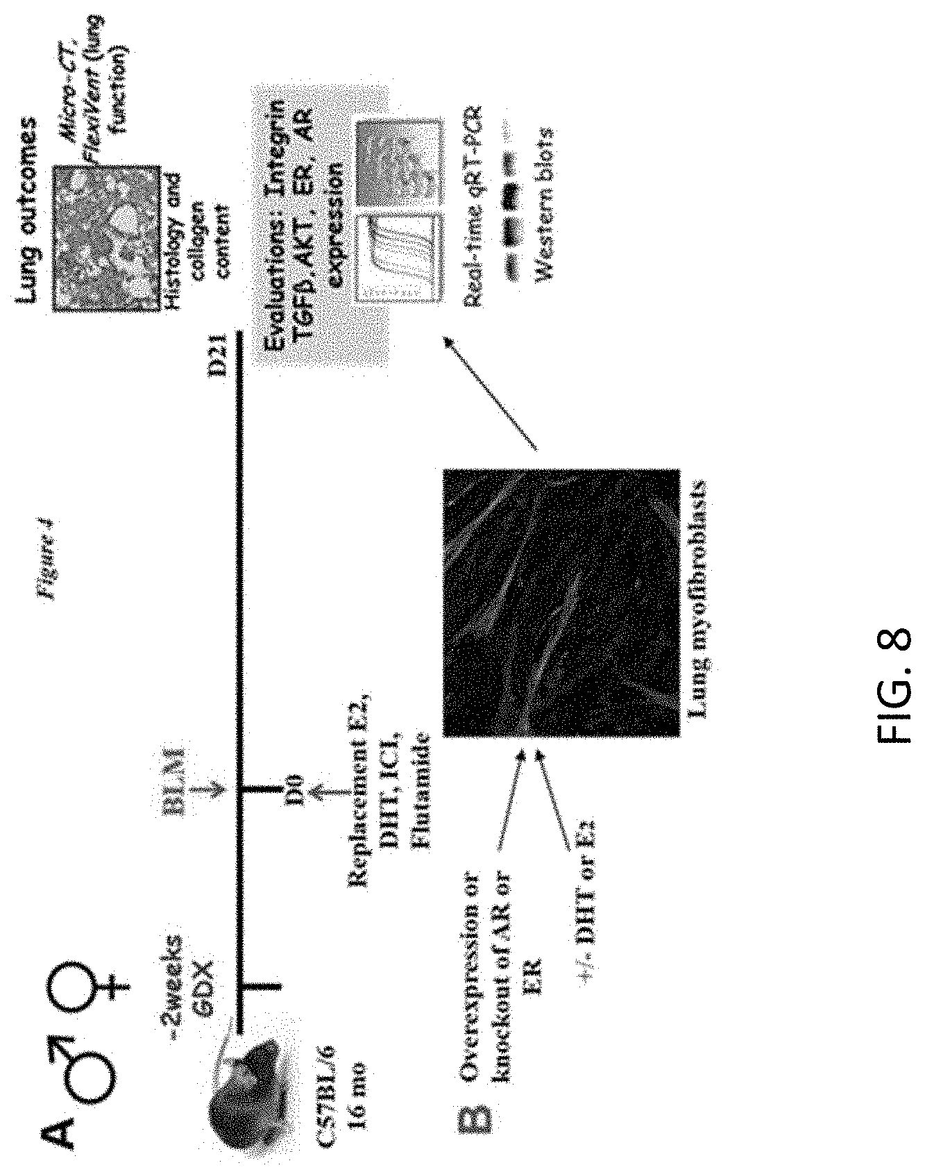
D00017

D00018

D00019
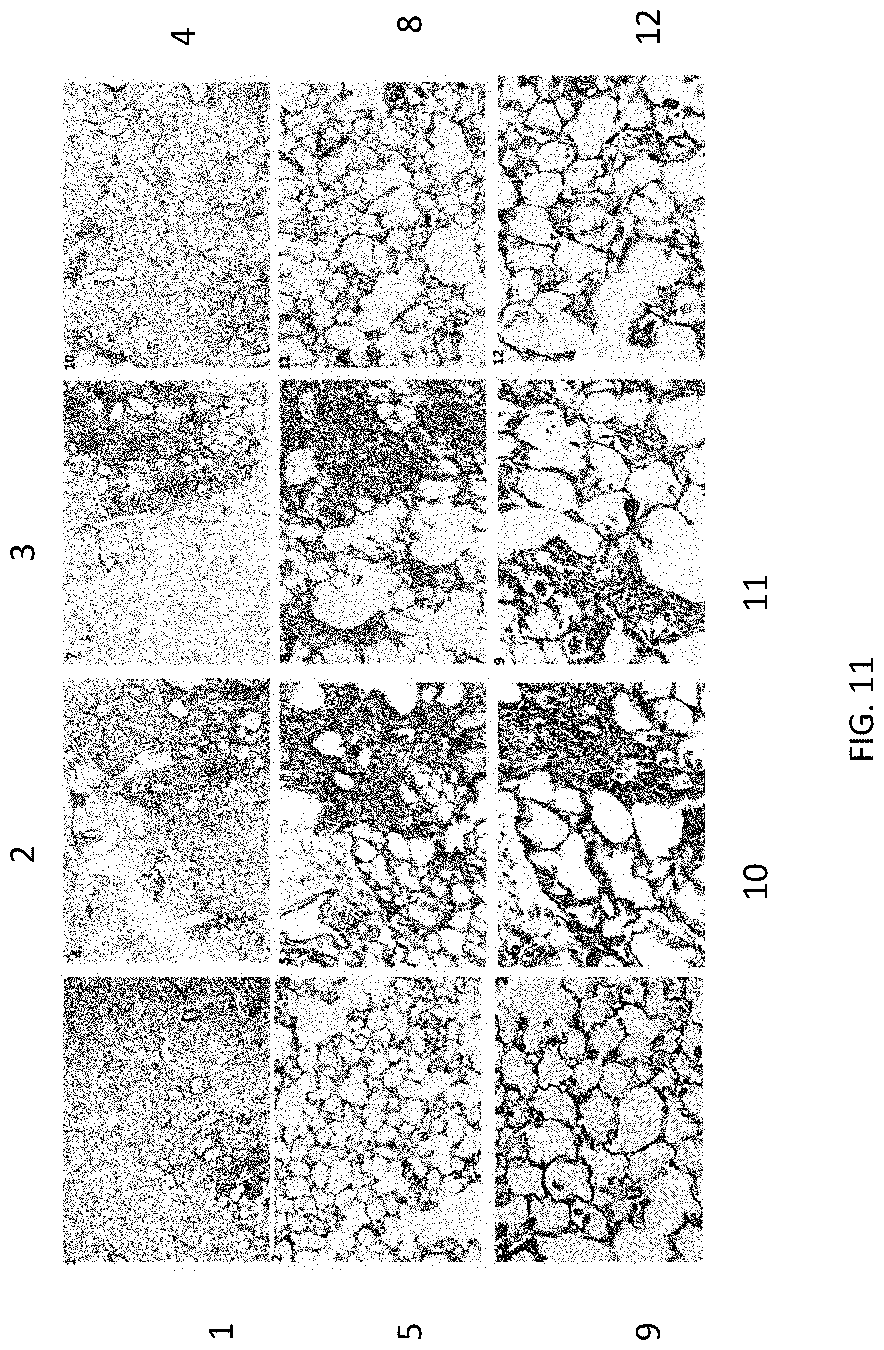
D00020
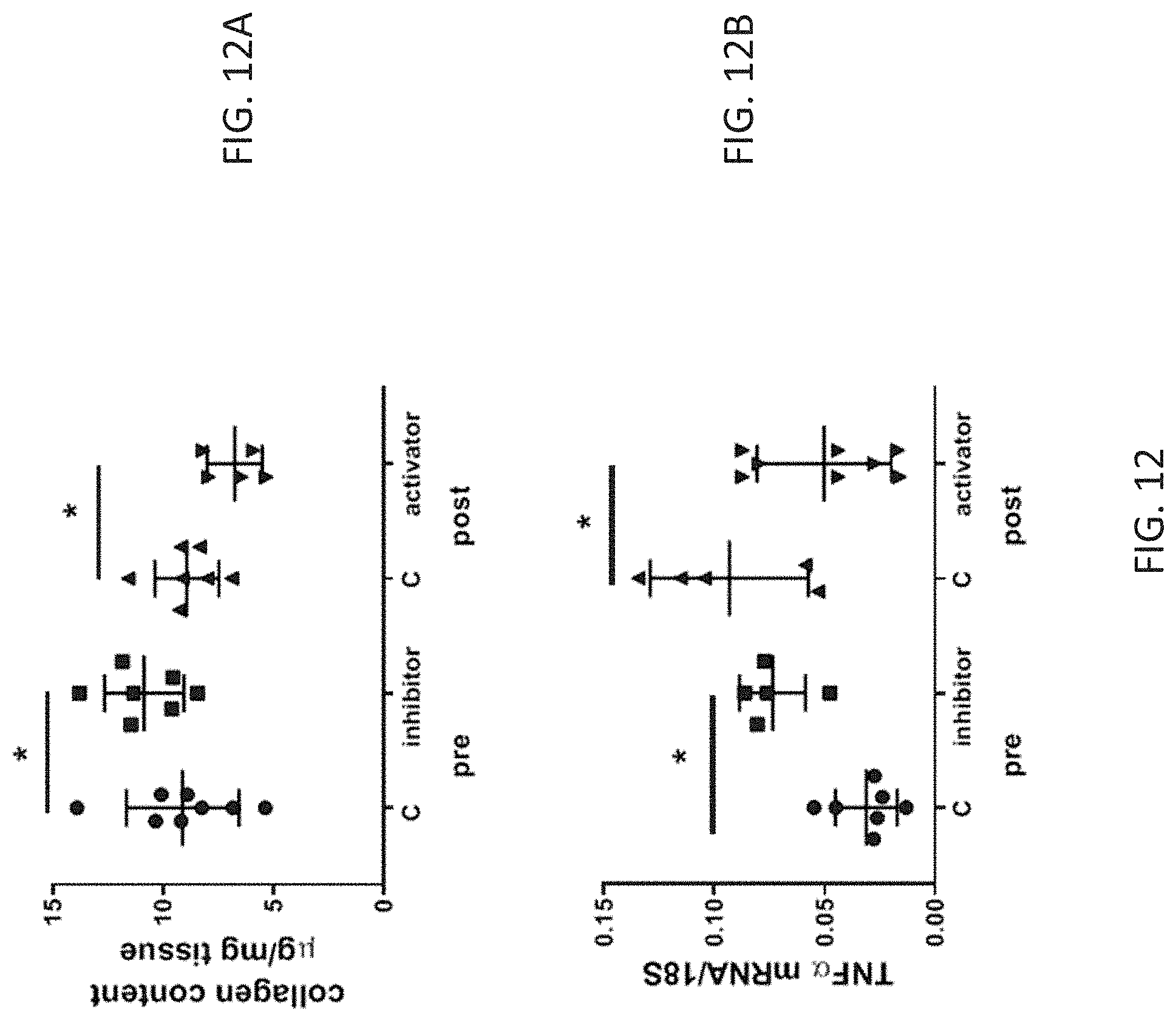
D00021
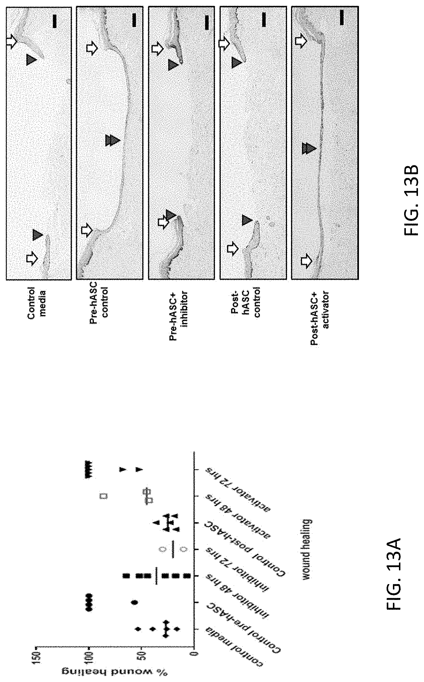
D00022

D00023

D00024

D00025

D00026

D00027

D00028

D00029

D00030

D00031

D00032

D00033

D00034

D00035

D00036

D00037

D00038

D00039

D00040

D00041

D00042

D00043
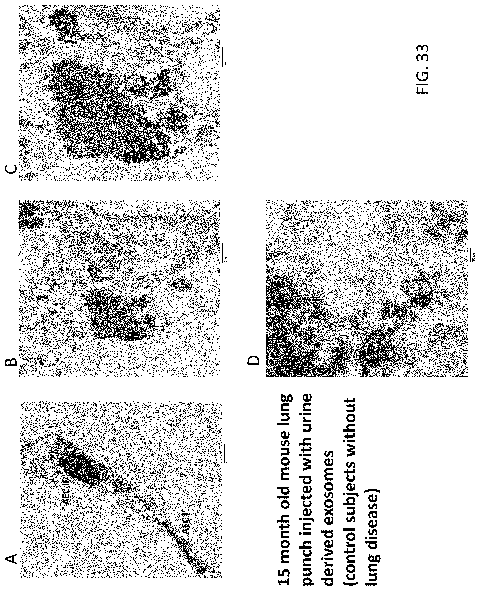
D00044

D00045

D00046

D00047


S00001
XML
uspto.report is an independent third-party trademark research tool that is not affiliated, endorsed, or sponsored by the United States Patent and Trademark Office (USPTO) or any other governmental organization. The information provided by uspto.report is based on publicly available data at the time of writing and is intended for informational purposes only.
While we strive to provide accurate and up-to-date information, we do not guarantee the accuracy, completeness, reliability, or suitability of the information displayed on this site. The use of this site is at your own risk. Any reliance you place on such information is therefore strictly at your own risk.
All official trademark data, including owner information, should be verified by visiting the official USPTO website at www.uspto.gov. This site is not intended to replace professional legal advice and should not be used as a substitute for consulting with a legal professional who is knowledgeable about trademark law.