Anti-Tumor Immunotherapy Enhancer
Tsukamoto; Nobuo ; et al.
U.S. patent application number 16/625936 was filed with the patent office on 2020-12-10 for anti-tumor immunotherapy enhancer. This patent application is currently assigned to Keio University. The applicant listed for this patent is Keio University. Invention is credited to Yutaka Kawakami, Nobuo Tsukamoto.
| Application Number | 20200383981 16/625936 |
| Document ID | / |
| Family ID | 1000005089463 |
| Filed Date | 2020-12-10 |


View All Diagrams
| United States Patent Application | 20200383981 |
| Kind Code | A1 |
| Tsukamoto; Nobuo ; et al. | December 10, 2020 |
Anti-Tumor Immunotherapy Enhancer
Abstract
The disclosure provides a composition for inhibiting the growth and/or invasion of tumor cells, an enhancer of an anti-tumor effect of a drug for the purpose of removing immunosuppression caused by cancer, or the like, which is characterized by being used for a specific subject.
| Inventors: | Tsukamoto; Nobuo; (Shinjuku-ku, Tokyo, JP) ; Kawakami; Yutaka; (Shinjuku-ku, Tokyo, JP) | ||||||||||
| Applicant: |
|
||||||||||
|---|---|---|---|---|---|---|---|---|---|---|---|
| Assignee: | Keio University Tokyo JP |
||||||||||
| Family ID: | 1000005089463 | ||||||||||
| Appl. No.: | 16/625936 | ||||||||||
| Filed: | June 29, 2018 | ||||||||||
| PCT Filed: | June 29, 2018 | ||||||||||
| PCT NO: | PCT/JP2018/024950 | ||||||||||
| 371 Date: | April 6, 2020 |
| Current U.S. Class: | 1/1 |
| Current CPC Class: | G01N 2500/20 20130101; A61K 39/39533 20130101; C07K 16/2818 20130101; A61P 35/00 20180101; C12Q 1/26 20130101; G01N 33/574 20130101; G01N 2333/90241 20130101; A61K 31/506 20130101; G01N 2440/14 20130101; G01N 2500/04 20130101; A61K 2039/505 20130101; C12Y 113/11052 20130101 |
| International Class: | A61K 31/506 20060101 A61K031/506; C07K 16/28 20060101 C07K016/28; A61K 39/395 20060101 A61K039/395; A61P 35/00 20060101 A61P035/00; G01N 33/574 20060101 G01N033/574; C12Q 1/26 20060101 C12Q001/26 |
Foreign Application Data
| Date | Code | Application Number |
|---|---|---|
| Jun 29, 2017 | JP | 2017-127973 |
Claims
1-20. (canceled)
21. A method for inhibiting the growth and/or invasion of a tumor cell, wherein the method includes administering a phosphorylation inhibitor or a dephosphorylation agent against phosphorylation of: a tyrosine residue(s) at the 111-th position and/or the 249-th position of human IDO1; or a tyrosine residue(s) of non-human IDO1 at a position(s) corresponding to the 111-th position and/or the 249-th position of human IDO1 to a tumor cell in which the tyrosine residue(s) is/are phosphorylated or a subject who carries the tumor cell.
22. The method according to claim 21, wherein the phosphorylation inhibitor or the dephosphorylation agent is a Src inhibitor or an inhibitor against a factor which increases phosphorylation activity of Src on the tyrosine residue(s).
23. The method according to claim 21, wherein the phosphorylation inhibitor or the dephosphorylation agent is a Src inhibitor selected from a group consisting of dasatinib, bosutinib, saracatinib, ponatinib, ilorasertib, N-benzyl-2-(5-(4-(2-morpholinoethoxy)phenyl)pyridin-2-yl)acetamide, 4-amino-5-(4-chlorophenyl)-7-(tert-butyl)pyrazolo-[3,4-d]pyrimidine, 4-amino-5-(4-methylphenyl)-7-(tert-butyl)pyrazolo-[3,4-d]pyrimidine, 4-(4'-phenoxyanilino)-6,7-dimethoxyquinazoline, 2-((3,5-di-tert-Butyl-4-hydroxyphenyl)-methylene)-4-cyclopentene-1,3-dion- e, 4-N-(5-cyclopropyl-1H-pyrazol-3-yl)-6-(4-methylpiperazin-1-yl)-2-N-[(3-- propan-2-yl-1,2-oxazol-5-yl)methyl]pyrimidine-2,4-diamine, [7-(2,6-dichloro-phenyl)-5-methyl-benzo[1,2,4]triazin-3-yl]-[4-(2-pyrroli- din-1-yl-ethoxy)-phenyl]-amine, [7-(2,6-dichloro-phenyl)-5-methyl-benzo[1,2,4]triazin-3-yl]-{4-[2-(1-oxy-- pyrrolidin-1-yl)-ethoxy]-phenyl}-amine, and rebastinib, and salts, hydrates, and solvates thereof.
24. The method according to claim 21, including further administering a drug to the tumor cell or the subject for the purpose of removing immunosuppression caused by cancer.
25. The method according to claim 24, wherein the drug includes anti-PD-1 antibody or anti-PD-L1 antibody.
26. A method for enhancing anti-tumor effect of a therapy for the purpose of removing immunosuppression caused by cancer, wherein the method includes administering a phosphorylation inhibitor or a dephosphorylation agent against phosphorylation of: a tyrosine residue(s) at the 111-th position and/or the 249-th position of human IDO1; or a tyrosine residue(s) of non-human IDO1 at a position(s) corresponding to the 111-th position and/or the 249-th position of human IDO1 to a tumor cell in which the tyrosine residue(s) is/are phosphorylated or a subject who carries the tumor cell.
27. The method according to claim 26, wherein the phosphorylation inhibitor or the dephosphorylation agent is a Src inhibitor or an inhibitor against a factor which increases phosphorylation activity of Src on the tyrosine residue(s).
28. The method according to claim 26, wherein the phosphorylation inhibitor or the dephosphorylation agent is a Src inhibitor selected from a group consisting of dasatinib, bosutinib, saracatinib, ponatinib, ilorasertib, N-benzyl-2-(5-(4-(2-morpholinoethoxy)phenyl)pyridin-2-yl)acetamide, 4-amino-5-(4-chlorophenyl)-7-(tert-butyl)pyrazolo-[3,4-d]pyrimidine, 4-amino-5-(4-methylphenyl)-7-(tert-butyl)pyrazolo-[3,4-d]pyrimidine, 4-(4'-phenoxyanilino)-6,7-dimethoxyquinazoline, 2-((3,5-di-tert-Butyl-4-hydroxyphenyl)-methylene)-4-cyclopentene-1,3-dion- e, 4-N-(5-cyclopropyl-1H-pyrazol-3-yl)-6-(4-methylpiperazin-1-yl)-2-N-[(3-- propan-2-yl-1,2-oxazol-5-yl)methyl]pyrimidine-2,4-diamine, [7-(2,6-dichloro-phenyl)-5-methyl-benzo[1,2,4]triazin-3-yl]-[4-(2-pyrroli- din-1-yl-ethoxy)-phenyl]-amine, [7-(2,6-dichloro-phenyl)-5-methyl-benzo[1,2,4]triazin-3-yl]-{4-[2-(1-oxy-- pyrrolidin-1-yl)-ethoxy]-phenyl}-amine, and rebastinib, and salts, hydrates, and solvates thereof.
29. The method according to claim 26, characterized in that the method is used in combination with a therapy for the purpose of removing immunosuppression caused by cancer.
30. The method according to claim 26, wherein the therapy includes administering anti-PD-1 antibody or anti-PD-L1 antibody to the tumor cell or the subject.
31. A method for obtaining data to identify a subject suitable for administration of the phosphorylation inhibitor or the dephosphorylation agent in the method according to claim 21, wherein the method includes: detecting, in a tumor cell obtained from the subject, phosphorylation of: a tyrosine residue(s) at the 111-th position and/or the 249-th position of human IDO1; or a tyrosine residue(s) of non-human IDO1 at a position(s) corresponding to the 111-th position and/or the 249-th position of human IDO1.
32. The method according to claim 31, wherein the detection includes using an antibody to detect, in a tumor cell obtained from the subject, phosphorylation of: a tyrosine residue(s) at the 111-th position and/or the 249-th position of human IDO1; or a tyrosine residue(s) of non-human IDO1 at a position(s) corresponding to the 111-th position and/or the 249-th position of human IDO1.
33. A method for screening a candidate substance for a method for inhibiting the growth and/or invasion of a tumor cell or a method for enhancing anti-tumor effect of a therapy for the purpose of removing immunosuppression caused by cancer, wherein the screening method includes: ex vivo measuring inhibition activity or dephosphorylation activity of a test substance against phosphorylation of: a tyrosine residue(s) at the 111-th position and/or the 249-th position of human IDO1; or a tyrosine residue(s) of non-human IDO1 at a position(s) corresponding to the 111-th position and/or the 249-th position of human IDO1; and selecting the candidate substance based on the measured inhibition activity or dephosphorylation activity of the test substance.
34. A method for screening a candidate substance to suppress expression of Slug gene induced by phosphorylation of IDO1 or synthesis of Slug induced by phosphorylation of IDO1, wherein the method includes: ex vivo measuring inhibition activity or dephosphorylation activity of a test substance against phosphorylation of: a tyrosine residue(s) at the 111-th position and/or the 249-th position of human IDO1; or a tyrosine residue(s) of non-human IDO1 at a position corresponding to the 111-th position and/or the 249-th position of human IDO1; and selecting the candidate substance based on the measured inhibition activity or dephosphorylation activity of the test substance.
35. A method for screening a candidate substance for the phosphorylation inhibitor or the dephosphorylation agent in the method according to claim 21, wherein the method includes: ex vivo measuring inhibition activity by a test substance against Src or Slug, inhibition activity by a test substance against a factor which activates Src or Slug, synthesis of Src or Slug by a test substance, or expression of Src gene or Slug gene by a test substance; and selecting the candidate substance based on the measured inhibition activity by the test substance against Src or Slug, the measured inhibition activity by the test substance against a factor which activates Src or Slug, measured synthesis inhibition of Src or Slug by the test substance, or measured expression inhibition of Src gene or Slug gene by the test substance.
36. A method for screening a candidate substance for inhibiting expression of Slug gene or inhibiting synthesis of Slug, wherein the method includes: ex vivo measuring Slug inhibition activity by a test substance, inhibition activity against a factor which activates Slug by a test substance, expression of Slug gene by a test substance, or synthesis of Slug by a test substance, in a cell in which a tyrosine residue(s) at the 111-th position and/or the 249-th position of human IDO1, or a tyrosine residue(s) of non-human IDO1 at a position(s) corresponding to the 111-th position and/or the 249-th position of human IDO1 is/are phosphorylated, and/or in a cell in which AhR is activated; and selecting the candidate substance based on the measured Slug inhibition activity by the test substance, the inhibition activity against a factor which activates Slug by the test substance, measured expression inhibition of Slug gene by the test substance, or measured synthesis inhibition of Slug by the test substance.
37. A method for inhibiting the growth and/or invasion of a tumor cell including administering a Src inhibitor to the tumor cell or a subject who carries the tumor cell and further administering a drug to the tumor cell or the subject for the purpose of removing immunosuppression caused by cancer.
38. A method for inhibiting the growth and/or invasion of a tumor cell including administering a Src inhibitor to the tumor cell or a subject who carries the tumor cell, wherein the method is used in combination with a therapy for the purpose of removing immunosuppression caused by cancer.
39. The method according to claim 37, wherein the Src inhibitor is a phosphorylation inhibitor against phosphorylation of: a tyrosine residue(s) at the 111-th position and/or the 249-th position of human IDO1; or a tyrosine residue(s) of non-human IDO1 at a position(s) corresponding to the 111-th position and/or the 249-th position of human IDO1.
40. The method according to claim 37, wherein the tumor cell is from a solid tumor.
Description
TECHNICAL FIELD
[0001] The present invention relates to an anti-tumor immunotherapy and/or an anti-tumor immunotherapy enhancer etc.
BACKGROUND ART
[0002] Indoleamine 2,3-dioxygenase (IDO) is an enzyme that catalyzes the conversion of L-tryptophan to N-formylkynurenine and is a rate-limiting enzyme that catalyzes the first step of tryptophan metabolisms in the kynurenine pathway. Similarly, Tryptophan 2,3-dioxygenase (TDO) is known as an enzyme that catalyzes the conversion of L-tryptophan to N-formylkynurenine.
[0003] It is known that when IDO or TDO is expressed in tumor tissues, etc., since it metabolizes tryptophan (Trp), T cells become unable to utilize Trp, and also known that kynurenine produced from Trp catalyzed by IDO or TDO is released from cancer cells and activates the aryl hydrocarbon receptor (AhR) of surrounding immune cells and converts the immune cells suppressive. Therefore, drugs (epacadostat, indoximod, etc.) that suppress the enzyme activity of IDO or TDO to metabolize Trp have been developed, and in addition to monotherapy, combination therapies with immune checkpoint inhibitors have been clinically applied (Non-Patent Document 1).
[0004] Due to the effect of immune checkpoint inhibitors on anti-tumor immunity, immune checkpoint inhibitors have been attracting attention as an unprecedented cancer treatment, but patients who actually show effective responses are limited, and even in effective cases part of the patients are not completely cured, thus further development of anti-tumor immunotherapy is required. For example, various attempts have been made to use an immune checkpoint inhibitor and a BRAF/MEK kinase inhibitor in combination (Non-Patent Document 2), and it has been reported that some of such combinations enhance the anti-tumor effect in melanoma (Non-Patent Document 3), however, development of useful anti-tumor immunotherapy is still expected.
PRIOR ART DOCUMENTS
Non-Patent Documents
[0005] Non-patent document 1: L. Brochez et al., Eur J Cancer 76, 2017, pp. 167-182 [0006] Non-patent document 2: Sangeetha M. Reddy et al. Curr Oncol Rep, 2016, 18(7), pp 42-50 [0007] Non-patent document 3: Ashish Kulkarni et al., ACS NANO, 2016, 10(10), pp. 9227-9242)
SUMMARY OF INVENTION
Problem to be Solved by Invention
[0008] However, there is still a need for further development of anti-tumor immunotherapy. In particular, the demand for personalized medical treatment for a specific treatment target patient is particularly strong in the field of cancer treatment, and it is necessary to select a specific subject and then use a particularly useful drug for the subject.
[0009] One aspect of the present invention is intended to provide a composition for inhibiting the growth and/or invasion of a tumor cell, an enhancer of anti-tumor effect of a drug for the purpose of removing immunosuppression caused by cancer, or the like, which is used for a specific subject. Another aspect of the present invention is intended to provide a method for specifying such a subject (a method for acquiring data for specifying such a subject).
Means of Solving Problem
[0010] The present inventors have conducted intensive studies and newly found that, in some tumor cells in tumor tissues, inhibiting phosphorylation at a specific site(s) of IDO1 enhances an anti-tumor immune response and exhibits an anti-tumor effect. In addition, the present inventors have newly found that inhibition of phosphorylation at a specific site(s) of IDO1 is particularly useful in combination with treatment with a drug for the purpose of removing immunosuppression caused by cancer. The present invention is based on such new findings.
[0011] One embodiment of the present invention relates to the following.
[1] A composition for inhibiting the growth and/or invasion of a tumor cell, wherein the composition includes a phosphorylation inhibitor or a dephosphorylation agent against phosphorylation of:
[0012] a tyrosine residue(s) at the 111-th position and/or the 249-th position of human IDO1; or
[0013] a tyrosine residue(s) of non-human IDO1 at a position(s) corresponding to the 111-th position and/or the 249-th position of human IDO1 and wherein the composition is used for a tumor cell in which the tyrosine residue(s) is/are phosphorylated or used for a subject who carries the tumor cell.
[2] The composition according to [1] described above, wherein the phosphorylation inhibitor or the dephosphorylation agent is a Src inhibitor or an inhibitor against a factor which increases phosphorylation activity of Src on the tyrosine residue(s). [3] The composition according to [1] or [2] described above, wherein the phosphorylation inhibitor or the dephosphorylation agent is a Src inhibitor selected from a group consisting of dasatinib, bosutinib, saracatinib, ponatinib, ilorasertib, N-benzyl-2-(5-(4-(2-morpholinoethoxy)phenyl)pyridin-2-yl)acetamide, 4-amino-5-(4-chlorophenyl)-7-(tert-butyl)pyrazolo-[3,4-d]pyrimidine, 4-amino-5-(methylphenyl-7-(tert-butyl)pyrazolo-[3,4-d]pyrimidine, 4-(4'-phenoxyanilino)-6,7-dimethoxyquinazoline, 2-((3,5-di-tert-Butyl-4-hydroxyphenyl)-methylene)-4-cyclopentene-1,3-dion- e, 4-N-(5-cyclopropyl-1H-pyrazol-3-yl)-6-(4-methylpiperazin-1-yl)-2-N-[(3-- propan-2-yl-1,2-oxazol-5-yl)methyl]pyrimidine-2,4-diamine, [7-(2,6-dichlorophenyl)-5-methylbenzo[1,2,4]triazin-3-yl]-[4-(2-pyrrolidi- n-1-ylethoxy) phenyl]amine, [7-(2,6-dichlorophenyl)-5-methylbenzo[1,2,4]triazin-3-yl]-{4-[2-(1-oxopyr- rolidin-1-yl) ethoxy]phenyl}amine, and rebastinib, and salts, hydrates, and solvates thereof. [4] The composition according to any one of [1] to [3] described above, characterized in that the composition is used in combination with a drug for the purpose of removing immunosuppression caused by cancer. [5] The composition according to [4] described above, wherein the drug for the purpose of removing immunosuppression caused by cancer includes anti-PD-1 antibody or anti-PD-L1 antibody. [6] An enhancer of anti-tumor effect of a drug for the purpose of removing immunosuppression caused by cancer, wherein the enhancer of anti-tumor effect includes a phosphorylation inhibitor or a dephosphorylation agent against phosphorylation of:
[0014] a tyrosine residue(s) at the 111-th position and/or the 249-th position of human IDO1; or
[0015] a tyrosine residue(s) of non-human IDO1 at a position(s) corresponding to the 111-th position and/or the 249-th position of human IDO1, and
[0016] wherein the enhancer of anti-tumor effect is used for a tumor cell in which the tyrosine residue(s) is/are phosphorylated or used for a subject who carries the tumor cell.
[7] The enhancer of anti-tumor effect according to [6] described above, wherein the phosphorylation inhibitor or the dephosphorylation agent is a Src inhibitor or an inhibitor against a factor which increases phosphorylation activity of Src on the tyrosine residue(s). [8] The enhancer of anti-tumor effect according to [6] or [7] described above, wherein the phosphorylation inhibitor or the dephosphorylation agent is a Src inhibitor selected from a group consisting of dasatinib, bosutinib, saracatinib, ponatinib, ilorasertib, N-benzyl-2-(5-(4-(2-morpholinoethoxy)phenyl)pyridin-2-yl)acetamide, 4-amino-5-(4-chlorophenyl)-7-(tert-butyl)pyrazolo-[3,4-d]pyrimidine, 4-amino-5-(methylphenyl-7-(tert-butyl)pyrazolo-[3,4-d]pyrimidine, 4-(4'-phenoxyanilino)-6,7-dimethoxyquinazoline, 2-((3,5-di-tert-Butyl-4-hydroxyphenyl)-methylene)-4-cyclopentene-1,3-dion- e, 4-N-(5-cyclopropyl-1H-pyrazol-3-yl)-6-(4-methylpiperazin-1-yl)-2-N-[(3-- propan-2-yl-1,2-oxazol-5-yl)methyl]pyrimidine-2,4-diamine, [7-(2,6-dichlorophenyl)-5-methylbenzo[1,2,4]triazin-3-yl]-[4-(2-pyrrolidi- n-1-ylethoxy) phenyl]amine, [7-(2,6-dichlorophenyl)-5-methylbenzo[1,2,4]triazin-3-yl]-{4-[2-(1-oxypyr- rolidin-1-yl) ethoxy]phenyl}amine, and rebastinib, and salts, hydrates, and solvates thereof. [9] The enhancer of anti-tumor effect according to any one of [6] to [8] described above, characterized in that the enhancer is used in combination with a drug for the purpose of removing immunosuppression caused by cancer. [10] The enhancer of anti-tumor effect according to any one of [6] to [9] described above, wherein the drug for the purpose of removing immunosuppression caused by cancer includes anti-PD-1 antibody or anti-PD-L1 antibody. [11] A method for obtaining data to identify a subject suitable for administration of a composition for inhibiting the growth and/or invasion of a tumor cell or an enhancer of anti-tumor effect of a drug for the purpose of removing immunosuppression caused by cancer, wherein the method includes:
[0017] detecting, in a tumor cell obtained from the subject, phosphorylation of: [0018] a tyrosine residue(s) at the 111-th position and/or the 249-th position of human IDO1; or [0019] a tyrosine residue(s) of non-human IDO1 at a position(s) corresponding to the 111-th position and/or the 249-th position of human IDO1, and
[0020] wherein the composition or the enhancer of anti-tumor effect includes a phosphorylation inhibitor or a dephosphorylation agent against phosphorylation of the tyrosine residue(s).
[12] An antibody to identify a subject suitable for administration of a composition for inhibiting the growth and/or invasion of a tumor cell or an enhancer of anti-tumor effect of a drug for the purpose of removing immunosuppression caused by cancer,
[0021] wherein the antibody detects, in a tumor cell obtained from the subject, phosphorylation of: [0022] a tyrosine residue(s) at the 111-th position and/or the 249-th position of human IDO1; or [0023] a tyrosine residue(s) of non-human IDO1 at a position(s) corresponding to the 111-th position and/or the 249-th position of human IDO1, and
[0024] wherein the composition or the enhancer of anti-tumor effect includes a phosphorylation inhibitor or a dephosphorylation agent against phosphorylation of the tyrosine residue(s).
[13] A method for screening a candidate substance for a composition for inhibiting the growth and/or invasion of a tumor cell or for an enhancer of anti-tumor effect of a drug for the purpose of removing immunosuppression caused by cancer, wherein the method includes:
[0025] ex vivo measuring inhibition activity or dephosphorylation activity of a test substance against phosphorylation of: [0026] a tyrosine residue(s) at the 111-th position and/or the 249-th position of human IDO1; or [0027] a tyrosine residue(s) of non-human IDO1 at a position(s) corresponding to the 111-th position and/or the 249-th position of human IDO1; and
[0028] selecting the candidate substance based on the measured inhibition activity or dephosphorylation activity of the test substance.
[14] A method for screening a candidate substance to suppress expression of Slug gene induced by phosphorylation of IDO1 or synthesis of Slug induced by phosphorylation of IDO1, wherein the method includes:
[0029] ex vivo measuring inhibition activity or dephosphorylation activity of a test substance against phosphorylation of: [0030] a tyrosine residue(s) at the 111-th position and/or the 249-th position of human IDO1; or [0031] a tyrosine residue(s) of non-human IDO1 at a position corresponding to the 111-th position and/or the 249-th position of human IDO1; and
[0032] selecting the candidate substance based on the measured inhibition activity or dephosphorylation activity of the test substance.
[15] A method for screening a candidate substance for a composition for inhibiting the growth and/or invasion of a tumor cell or for an enhancer of anti-tumor effect of a drug for the purpose of removing immunosuppression caused by cancer, wherein the method includes: [0033] ex vivo measuring inhibition activity by a test substance against Src or Slug, inhibition activity by a test substance against a factor which activates Src or Slug, synthesis of Src or Slug by a test substance, or expression of Src gene or Slug gene by a test substance; and
[0034] selecting the candidate substance based on the measured inhibition activity by the test substance against Src or Slug, the measured inhibition activity by the test substance against a factor which activates Src or Slug, measured synthesis inhibition of Src or Slug by the test substance, or measured expression inhibition of Src gene or Slug gene by the test substance, and
[0035] wherein the composition or the enhancer of anti-tumor effect is used for a tumor cell in which a tyrosine residue(s) at the 111-th position and/or the 249-th position of human IDO1, or a tyrosine residue(s) of non-human IDO1 at a position(s) corresponding to the 111-th position and/or the 249-th position of human IDO1 is/are phosphorylated, or used for a subject who carries the tumor cell.
[16] A method for screening a candidate substance for inhibiting expression of Slug gene or inhibiting synthesis of Slug, wherein the method includes:
[0036] ex vivo measuring Slug inhibition activity by a test substance, inhibition activity against a factor which activates Slug by a test substance, expression of Slug gene by a test substance, or synthesis of Slug by a test substance, in a cell in which a tyrosine residue(s) at the 111-th position and/or the 249-th position of human IDO1, or a tyrosine residue(s) of non-human IDO1 at a position(s) corresponding to the 111-th position and/or the 249-th position of human IDO1 is/are phosphorylated, and/or in a cell in which AhR is activated; and
[0037] selecting the candidate substance based on the measured Slug inhibition activity by the test substance, the inhibition activity against a factor which activates Slug by the test substance, measured expression inhibition of Slug gene by the test substance, or measured synthesis inhibition of Slug by the test substance.
[17] A composition for inhibiting the growth and/or invasion of a tumor cell including a Src inhibitor and a drug for the purpose of removing immunosuppression caused by cancer. [18] A composition for inhibiting the growth and/or invasion of a tumor cell including a Src inhibitor, wherein the composition is used in combination with a drug for the purpose of removing immunosuppression caused by cancer. [19] The composition according to [17] or [18] described above, wherein the Src inhibitor is a phosphorylation inhibitor against phosphorylation of:
[0038] a tyrosine residue(s) at the 111-th position and/or the 249-th position of human IDO1; or
[0039] a tyrosine residue(s) of non-human IDO1 at a position(s) corresponding to the 111-th position and/or the 249-th position of human IDO1.
[20] The composition according to any one of [17] to [19] described above, wherein the tumor cell is from a solid tumor.
Effect of Invention
[0040] According to one embodiment of the present invention, an anti-tumor effect can be exerted on a specific tumor cell. Here, the anti-tumor effect may include inhibiting the growth of a tumor cell and/or inhibiting invasion of a tumor cell, and/or shrinking a tumor.
[0041] According to one embodiment of the present invention, a beneficial anti-tumor effect may be achieved in combination with treatment with a drug for the purpose of removing immunosuppression caused by cancer.
[0042] In one embodiment of the present invention, a particular subject suitable for a particular cancer treatment may be identified.
[0043] In one embodiment of the present invention, a candidate substance useful for treating cancer may be screened.
BRIEF DESCRIPTION OF DRAWINGS
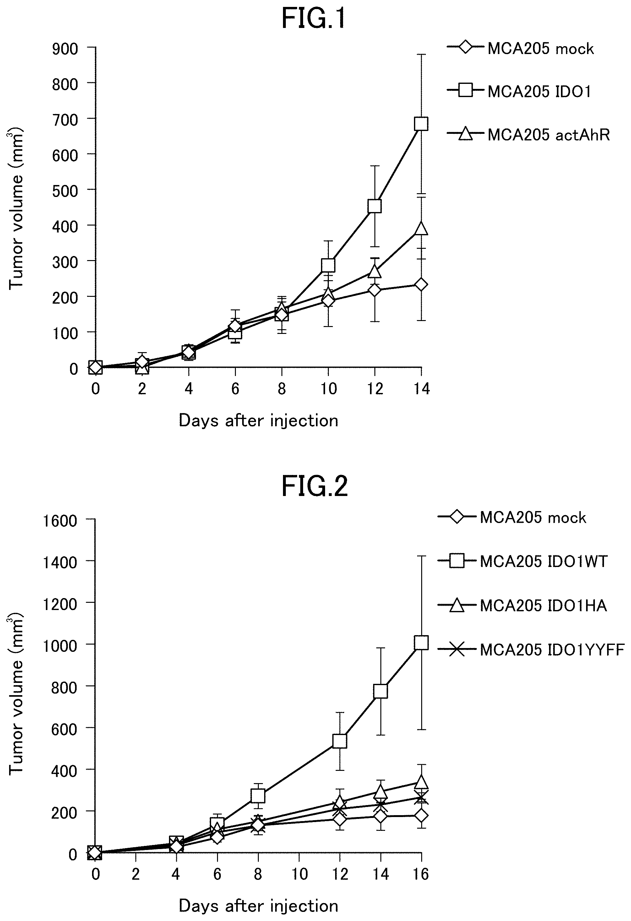
[0044] FIG. 1 is a graph showing an increase in tumor volume in C57BL/6 mice implanted with mouse cancer cells MCA205. MCA205 mock represents MCA205 transfected with an empty vector without gene insertion. MCA205 IDO1 represents MCA205 in which a wild-type mouse IDO1 gene was transfected and constitutively expressed. MCA205 actAhR represents MCA205 in which a constitutively active mouse AhR gene was transfected and constitutively expressed.
[0045] FIG. 2 is a graph showing an increase in tumor volume in C57BL/6 mice implanted with mouse cancer cells MCA205. MCA205 mock represents MCA205 transfected with an empty vector without gene insertion. MCA205 IDO1WT represents MCA205 in which a wild-type mouse IDO1 gene was transfected and constitutively expressed. MCA205 IDO1HA represents MCA205 in which a mouse IDO1 mutant gene prepared to synthesize IDO1 having an amino acid mutation of H350A was transfected and constitutively expressed. MCA205 IDO1YYFF represents MCA205 in which a mouse IDO1 mutant gene prepared to synthesize IDO1 having amino acid mutations of Y115F and Y253F was transfected and constitutively expressed.
[0046] FIG. 3 is a graph showing the amount of mouse IFN-.gamma., an index of anti-tumor immunity, produced by cytotoxic T cells in C57BL/6 mice implanted with mouse cancer cells MCA205. MCA205 mock, MCA205 IDO1WT, MCA205 IDO1HA and MCA205 IDO1YYFF are the same as those in FIG. 2.
[0047] FIG. 4 is a graph which shows the invasion ability of the cancer cells measured by the invasion assay using matrigel. MCA205 mock, MCA205 IDO1WT, MCA205 IDO1HA and MCA205 IDO1YYFF are the same as those in FIG. 2.
[0048] FIG. 5 is photographs showing the phosphorylation of Y249 in IDO1 and the localization of Slug by immunohistochemical staining for tumor tissues derived from colorectal cancer patients and tumor tissues derived from breast cancer patients.
[0049] FIG. 6 is a graph showing the results of quantitative PCR analysis of Slug gene expression levels in mouse cancer cells MCA205. MCA205 mock, MCA205 IDO1WT, MCA205 IDO1HA and MCA205 IDO1YYFF are the same as those in FIG. 2. MCA205 actAhR is the same as in FIG. 1. The vertical axis indicates relative values of the amount of mRNA where the amount of mRNA detected in the case of MCA205 mock is set as 1.
[0050] FIG. 7 is a graph showing the results of quantitative PCR analysis of cyplal gene expression levels in mouse cancer cells MCA205. MCA205 mock, MCA205 IDO1WT, MCA205 IDO1HA and MCA205 IDO1YYFF are the same as those in FIG. 2. MCA205 active AhR represents MCA205 in which the constitutively active mouse AhR gene was transfected and constitutively expressed (same as MCA205 actAhR). The vertical axis indicates relative values of the amount of mRNA where the amount of mRNA detected in the case of MCA205 mock is set as 1.
[0051] FIG. 8 is a graph showing the results of quantitative PCR analysis of Slug gene expression levels in mouse cancer cells MCA205. The MCA205 mock, MCA205 IDO1WT and MCA205 IDO1YYFF are the same as those in FIG. 2. MCA205 IDO1Y115F represents MCA205 in which a mouse IDO1 mutant gene prepared to synthesize IDO1 having an amino acid mutation of Y115F was transfected and constitutively expressed. MCA205 IDO1Y253F represents MCA205 in which a mouse IDO1 mutant gene prepared to synthesize IDO1 having an amino acid mutation of Y253F was transfected and constitutively expressed. The vertical axis indicates relative values of the amount of mRNA where the amount of mRNA detected in the case of MCA205 mock is set as 1.
[0052] FIG. 9 is a graph showing an increase in tumor volume in C57BL/6 mice implanted with mouse cancer cells MCA205. MCA205 mock represents MCA205 transfected with an empty vector without gene insertion. MCA205 Slug represents MCA205 in which Slug gene was transfected and constitutively expressed.
[0053] FIG. 10 is a graph showing the amount of mouse IFN-.gamma., an index of anti-tumor immunity, produced by cytotoxic T cells in C57BL/6 mice implanted with mouse cancer cells MCA205. MCA205 mock and MCA205 Slug are the same as those in FIG. 9.
[0054] FIG. 11 is a graph showing the results of quantitative PCR analysis of Slug gene expression levels in mouse cancer cells MCA205. MCA205 actAhR shSlug represents MCA205 in which a constitutively active mouse AhR gene was introduced and constitutively expressed, and Slug gene expression is constitutively knocked down. MCA205 actAhR NonTarget represents a negative control. The vertical axis indicates relative values of the amount of mRNA where the amount of mRNA detected in the case of MCA205 actAhR NonTarget is set as 1.
[0055] FIG. 12 is a graph showing an increase in tumor volume in mice implanted with mouse cancer cells MCA205. MCA205 mock represents MCA205 transfected with an empty vector without gene insertion. MCA205 actAhR shSlug and MCA205 actAhR NonTarget are the same as those in FIG. 11.
[0056] FIG. 13 is a graph showing the amount of mouse IFN-.gamma., an index of anti-tumor immunity, produced by cytotoxic T cells in C57BL/6 mice implanted with mouse cancer cells MCA205. MCA205 mock, MCA205 actAhR shSlug and MCA205 actAhR NonTarget are the same as those in FIG. 11.
[0057] FIG. 14 is a graph showing the results of quantitative PCR analysis of Slug gene expression levels in human cancer cells SW837. SW837 mock represents SW837 transfected with an empty vector without gene insertion. SW837 IDO1WT represents SW837 in which a wild-type human IDO1 gene was transfected and constitutively expressed. SW837 IDO1HA represents SW837 in which a human IDO1 mutant gene prepared to synthesize IDO1 having an amino acid mutation of H346A was transfected and constitutively expressed. SW837 IDO1YYFF represents SW837 in which a human IDO1 mutant gene prepared to synthesize IDO1 having amino acid mutations of Y111F and Y249F was transfected and constitutively expressed. SW837 actAhR represents SW837 in which a constitutively active human AhR gene was introduced and constitutively expressed. The vertical axis indicates relative values of the amount of mRNA where the amount of mRNA detected in the case of SW837 mock is set as 1.
[0058] FIG. 15 is photographs showing, by immunohistological staining, sites where Y249 is phosphorylated in IDO1, Src localization, and Csk localization in tumor tissues derived from colorectal cancer patients.
[0059] FIG. 16 is a photograph showing, by immunohistological staining, sites where Y249 is phosphorylated in IDO1 and sites where Y530 is phosphorylated in Src in a tumor tissue derived from a colorectal cancer patient.
[0060] FIG. 17 is a photograph showing protein detection of IDO1 phosphorylated at Y249 and total IDO1 in human cancer cells SW837 in which a wild-type human IDO1 gene was transfected and constitutively expressed. Mock represents a human cancer cell SW837 in which a wild-type human IDO1 gene was transfected and constitutively expressed and an empty vector (pcDNA3.1) without gene insertion was transiently transfected. hSrc Y530F represents SW837 in which a wild-type human IDO1 gene was transfected and constitutively expressed and further a human Src mutant gene prepared to synthesize an activated Src having an amino acid mutation of Y530F was transfected and transiently expressed.
[0061] FIG. 18 is a photograph showing the results of detecting a FLAG-fused human IDO1 protein or a GAPDH protein by western blotting in the precipitate after immunoprecipitation with anti-FLAG antibody M2 or in cell extract (Total lysate) before immunoprecipitation from human cancer cells SW837 in which FLAG-tagged human IDO1 was transiently expressed. Mock means use of empty vector without gene insertion (pME-FLAG). hIDO1 WT represents wild type human IDO1 gene. hIDO1 Y111F represents a human IDO1 mutant gene prepared to synthesize IDO1 having an amino acid mutation of Y111F. hIDO1 Y249F represents a human IDO1 mutant gene prepared to synthesize IDO1 having an amino acid mutation of Y249F. hIDO1 Y111,249FF represents a human IDO1 mutant gene prepared to synthesize IDO1 having amino acid mutations of Y111F and Y249F.
[0062] FIG. 19 is a graph showing an increase in tumor volume in C57BL/6 mice implanted with mouse cancer cells MCA205. MCA205 mock represents MCA205 transfected with an empty vector without gene insertion. MCA205 IDO1WT represents MCA205 in which a wild-type mouse IDO1 gene was transfected and constitutively expressed. MCA205 IDO1Y253F represents MCA205 in which a mouse IDO1 mutant gene prepared to synthesize IDO1 having an amino acid mutation of Y253F was transfected and constitutively expressed.
[0063] FIG. 20 is a graph showing an increase in tumor volume in C57BL/6 mice implanted with mouse cancer cells MC38. MC38 (IgG2a+vehicle) represents the case where a rat IgG2a isotype control antibody and a vehicle were administered. MC38 (IgG2a+dasatinib) represents the case where a rat IgG2a isotype control antibody and dasatinib were administered. MC38 (.alpha.-PD-1+vehicle) represents the case where an anti-PD-1 antibody and a vehicle were administered. MC38 (.alpha.-PD-1+dasatinib) represents the case where an anti-PD-1 antibody and dasatinib were administered.
[0064] FIG. 21 is a graph showing the amount of mouse IFN-.gamma., an index of anti-tumor immunity, produced by cytotoxic T cells in C57BL/6 mice implanted with mouse cancer cells MC38. MC38 IgG+vehicle, MC38 IgG+dasatinib, MC38 .alpha.-PD1+vehicle, and MC38 .alpha.-PD1+dasatinib are the same as MC38 (IgG2a+vehicle), MC38 (IgG2a+ dasatinib), MC38 (.alpha.-PD-1+vehicle), and MC38 (.alpha.-PD-1+dasatinib) in FIG. 20, respectively.
[0065] FIG. 22 is a graph showing an increase in tumor volume in C57BL/6 mice implanted with mouse cancer cells MCA205 transfected with a wild-type mouse IDO1 gene. MCA205 IDO1WT (IgG2a+vehicle) represents the case where a rat IgG2a isotype control antibody and a vehicle were administered. MCA205 IDO1WT (IgG2a+ dasatinib) represents the case where a rat IgG2a isotype control antibody and dasatinib were administered. MCA205 IDO1WT (.alpha.-PD1+vehicle) represents the case where an anti-PD-1 antibody and a vehicle were administered. MCA205 IDO1WT (.alpha.-PD1+ dasatinib) represents the case where an anti-PD-1 antibody and dasatinib were administered.
[0066] FIG. 23 is a graph showing an increase in tumor volume in C57BL/6 mice implanted with mouse cancer cells MCA205 transfected with a wild-type mouse IDO1 gene. MCA205 IDO1WT (water+PBS) represents the case where water and PBS were administered. MCA205 IDO1WT (water+Y15) represents the case where water and FAK inhibitor Y15 were administered. MCA205 IDO1WT (1-MT+PBS) represents the case where 1-Methyl-D-tryptophan and PBS were administered. MCA205 IDO1WT (1-MT+Y15) represents the case where 1-Methyl-D-tryptophan and the FAK inhibitor Y15 were administered.
DESCRIPTION OF EMBODIMENTS
[0067] Hereinafter, the present invention is described in detail.
[0068] One embodiment of the present invention relates to a composition for inhibiting the growth and/or invasion of tumor cells.
[0069] A composition for inhibiting the growth and/or invasion of a tumor cell of the present invention includes a phosphorylation inhibitor or a dephosphorylation agent against phosphorylation of a specific tyrosine residue(s) of IDO1. In addition, a composition for inhibiting the growth and/or invasion of a tumor cell of the present invention is used for a tumor cell in which a specific tyrosine residue(s) of IDO1 is/are phosphorylated or a subject who carries a tumor cell in which a specific tyrosine residue(s) of IDO1 is/are phosphorylated.
[0070] One embodiment of the present invention relates to an enhancer of anti-tumor effect of a drug for the purpose of removing immunosuppression caused by cancer.
[0071] An enhancer of anti-tumor effect of a drug for the purpose of removing immunosuppression caused by cancer of the present invention includes a phosphorylation inhibitor or a dephosphorylation agent against phosphorylation of a specific tyrosine residue(s) of IDO1. Further, an enhancer of anti-tumor effect of a drug for the purpose of removing immunosuppression caused by cancer of the present invention is used for a tumor cell in which a specific tyrosine residue(s) of IDO1 is/are phosphorylated or used for a subject who carries a tumor cell in which a specific tyrosine residue(s) of IDO1 is/are phosphorylated.
[0072] One embodiment of the present invention relates to a composition for inhibiting the growth and/or invasion of a tumor cell, including a Src inhibitor and a drug for the purpose of removing immunosuppression caused by cancer.
[0073] One embodiment of the present invention relates to a composition for inhibiting the growth and/or invasion of a tumor cell, including a Src inhibitor, which is used in combination with a drug for the purpose of removing immunosuppression caused by cancer.
[0074] As used herein, "tumor cell" refers to a cell(s) or a cell population(s) that proliferates abnormally, and includes benign tumors and malignant tumors. Tumor cells, also simply referred to as tumors, include solid tumors and hematological tumors. Tumor cells (tumors) may be present in organs such as, for example, lung, stomach, esophagus, liver, pancreas, intestine, kidney, spleen, genitalia, urinary organs, brain, nerves, bone marrow, and lymph nodes.
[0075] Malignant tumors are also commonly referred to as cancers. Therefore, a composition for inhibiting the growth and/or invasion of a tumor cell of the present invention and an enhancer of anti-tumor effect of a drug for the purpose of removing immunosuppression caused by cancer of the present invention may be used for treating cancer. Here, cancer is used in a broad sense including carcinomas, sarcomas, and hematological malignancies (hematopoietic tumors). A composition for inhibiting the growth and/or invasion of a tumor cell of the present invention and an enhancer of anti-tumor effect of drug for the purpose of removing immunosuppression caused by cancer of the present invention may be used for treatment of cancer, for example, lung cancer, gastric cancer, esophageal cancer, liver cancer, biliary tract cancer, pancreatic cancer, colon cancer, renal cancer, bladder cancer, prostate cancer, testicular cancer, uterine cancer, ovarian cancer, breast cancer, skin cancer, laryngeal cancer, brain tumor, neuroblastoma, colorectal cancer, melanoma, head and neck cancer, bone and soft tissue tumor, thyroid cancer, fibrosarcoma, dermal fibrosarcoma, liposarcoma, myosarcoma, hemangiosarcoma, Kaposi's sarcoma, lymphatic sarcoma, osteosarcoma, leukemia, lymphoma, and myeloma, but not limited thereto.
[0076] Since IDO1 expressed in a stromal cell of tumor tissue also metabolizes tryptophan and suppresses anti-tumor immunity, a composition for inhibiting the growth and/or invasion of a tumor cell of the present invention, and an enhancer of anti-tumor effect of a drug for the purpose of removing immunosuppression caused by cancer of the present invention may be used for a subject in which phosphorylation of a specific tyrosine residue(s) described herein is observed in IDO1 expressed in a stromal cell.
[0077] A composition for inhibiting the growth and/or invasion of a tumor cell of the present invention, and an enhancer of anti-tumor effect of a drug for the purpose of removing immunosuppression caused by cancer of the present invention may exert an anti-tumor effect on a tumor cell in which a specific tyrosine residue(s) of IDO1 is/are phosphorylated.
[0078] In the case of a solid tumor, the anti-tumor effect may be evaluated by inhibiting the growth of a tumor cell, inhibiting invasion of a tumor cell, or reducing the size of a tumor. In the case of a liquid tumor, the anti-tumor effect may be evaluated using the decrease in the number of tumor cells in blood as an index.
[0079] A specific tyrosine residue(s) of IDO1 is a tyrosine residue(s) at the 111-th position and/or the 249-th position in the case of human IDO1 (SEQ ID NO: 1), and a tyrosine residue(s) at a position(s) corresponding to the 111-th position and/or the 249-th position of human IDO1 in the case of non-human IDO1. A tyrosine residue(s) of non-human IDO1 at a position(s) corresponding to the 111-th position and/or the 249-th position of human IDO1 varies in positions in each animal. For example, in mouse IDO1 (SEQ ID NO: 2), tyrosine residue at the 115-th position corresponds to tyrosine residue at the 111-th position of human IDO1 and tyrosine residue at the 253-th position corresponds to tyrosine residue at the 249-th position of human IDO1.
[0080] IDO1 has two immunoreceptor tyrosine-based inhibitory motifs (ITIMs). The site where the tyrosine residue at position 111 of human IDO1 exists is called ITIM1, and the site where the tyrosine residue at position 249 of human IDO1 is called ITIM2. ITIM1 and ITIM2 are thought to have similar functions.
[0081] The present inventors have found that phosphorylation of a tyrosine residue(s) in a tumor cell at the 111-th position and/or the 249-th position of human IDO1 or a tyrosine residue(s) of non-human IDO1 at a position(s) corresponding to the 111-th position and/or the 249-th position of human IDO1 is involved in the suppression of anti-tumor immunity, the growth of the tumor cell, and the like. In addition, the present inventors have found that in a tumor cell, inhibition of phosphorylation of a tyrosine residue(s) at the 111-th position and/or the 249-th position of human IDO1 or a tyrosine residue(s) of non-human IDO1 at a position(s) corresponding to the 111-th position and/or the 249-th position of human IDO1 results in the exertion of an anti-tumor effect through enhancement of anti-tumor immune response, and also results in significant reduction of the amount of IDO1 in the tumor cell.
[0082] Therefore, a composition for inhibiting the growth and/or invasion of a tumor cell of the present invention and an enhancer of anti-tumor effect of a drug for the purpose of removing immunosuppression caused by cancer of the present invention are characterized by including a phosphorylation inhibitor or a dephosphorylation agent against phosphorylation of a tyrosine residue(s) at the 111-th position and/or the 249-th position of human IDO1 or a tyrosine residue(s) of non-human IDO1 at a position(s) corresponding to the 111-th position and/or the 249-th position of human IDO1. Preferably, a composition for inhibiting the growth and/or invasion of a tumor cell of the present invention and an enhancer of anti-tumor effect of a drug for the purpose of removing immunosuppression caused by cancer of the present invention include a phosphorylation inhibitor or a dephosphorylation agent against phosphorylation of a tyrosine residue at the 249-th position of human IDO1 or a tyrosine residue of non-human IDO1 at a position(s) corresponding to the 249-th position of human IDO1.
[0083] Examples of a phosphorylation inhibitor or a dephosphorylation agent against phosphorylation of a tyrosine residue(s) at the 111-th position and/or the 249-th position of human IDO1 or a tyrosine residue(s) of non-human IDO1 at a position(s) corresponding to the 111-th position and/or the 249-th position of human IDO1 includes, for example, but are not limited to, a Src inhibitor or an inhibitor against a factor which increases phosphorylation activity of Src on the tyrosine residue(s).
[0084] Examples of a Src inhibitor include compounds and antibodies that inhibit the action of the kinase Src, and compounds and nucleic acids that inhibit the expression of the Src gene. As a specific Src inhibitor, those known to have a Src inhibitory activity may be used based on literatures such as Lauren N. Puls et al., The Oncologist 2011; 16: 566-578 and others, and the knowledge of a person skilled in the art. For example, Src inhibitors include: dasatinib, bosutinib, saracatinib, ponatinib, ilorasertib, N-benzyl-2-(5-(4-(2-morpholinoethoxy)phenyl)pyridin-2-yl)acetamide (KX2-391; CAS number: 897016-82-9), 4-amino-5-(4-chlorophenyl-7-(tert-butyl)pyrazolo-[3,4-d]pyrimidine (PP2; CAS number: 172889-27-9), 4-amino-5-(methylphenyl)-7-(tert-butyl)pyrazolo-[3,4-d]pyrimidine (PPH1; CAS number: 172889-26-8), 4-(4'-phenoxyanilino)-6,7-dimethoxyquinazoline (Src Kinase Inhibitor I; CAS number: 179248-59-0), 2((3,5-di-tert-Butyl-4-hydroxyphenyl)-methylene)-4-cyclopentene-1,3-dione (TX-1123; CAS number: 157397-06-3), 4-N-(5-cyclopropyl-1H-pyrazol-3-yl)-6-(4-methylpiperazin-1-yl)-2-N-[(3-pr- opan-2-yl-1,2-oxazol-5-yl)methyl]pyrimidine-2,4-diamine (XL-228; CAS number: 898280-07-4), [7-(2,6-dichlorophenyl)-5-methylbenzo[1,2,4]triazin-3-yl]-[4-(2-pyrrolidi- n-1-ylethoxy) phenyl]amine (TG100435), [7-(2,6-dichlorophenyl)-5-methylbenzo[1,2,4]triazin-3-yl]-{4-[2-(1-oxypyr- rolidin-1-yl) ethoxy]phenyl}amine (TG100855), and rebastinib (DCC2036), and salts, hydrates, and solvates thereof, but are not limited thereto.
[0085] The salt, hydrate and solvate are not particularly limited, but may be appropriately selected as long as they are pharmaceutically acceptable. Pharmaceutically acceptable salts include, but are not particularly limited to, for example, inorganic acid salts such as hydrochloride, hydrobromide, sulfate, nitrate and phosphate; organic acid salts such as acetate, succinate, fumarate, maleate, salicylate, tartrate, citrate, oxalate, lactate, malate, stearate, benzoate, methanesulfonate, and p-toluenesulfonate; inorganic base salts such as alkali metal salts (sodium salts and potassium salts, etc.), alkaline earth metal salts (calcium salts and magnesium salts, etc.) and ammonium salts; organic base salts such as diethylamine salts; acidic amino acid salts such as aspartate and glutamate; and basic amino acid salts such as arginine salts, lysine salts and ornithine salts.
[0086] An inhibitor of a factor that enhances the specific tyrosine phosphorylation activity of Src also includes an inhibitor of a factor that enhances the activation of Src and an inhibitor of a factor that activates the expression of Src gene. Examples of an inhibitor of a factor that enhances the specific tyrosine phosphorylation activity of Src include a FAK inhibitor etc. For example, specific FAK inhibitors include 1,2,4,5-benzenetetraamine tetrahydrochloride (Y15; CAS number: 4506-66-5), defactinib, 6-[4-(3-methylsulfonylbenzylamino)-5-trifluoromethylpyrimidin-2-ylamino]-- 3,4-dihydro-1H-quinolin-2-one (PF-573228; CAS number: 869288-64-2) and 2-[[2-(2-methoxy-4-morpholin-4-ylanilino)-5-(trifluoromethyl)pyridin-4-yl- ]amino]-N-methylbenzamide (PND-1186; CAS number: 1061353-68-1), PF-00562271 (CAS number: 939791-38-5), and salts, hydrates, and solvates thereof, but are not limited thereto.
[0087] The salt, hydrate and solvate are not particularly limited, but may be appropriately selected as long as they are pharmaceutically acceptable. Pharmaceutically acceptable salts include those exemplified above.
[0088] In one embodiment of the present invention, a composition for inhibiting the growth and/or invasion of a tumor cell of the present invention is used in combination with a drug for the purpose of removing immunosuppression caused by cancer.
[0089] In one embodiment of the present invention, an enhancer of anti-tumor effect of a drug for the purpose of removing immunosuppression caused by cancer of the present invention is used in combination with a drug for the purpose of removing immunosuppression caused by cancer.
[0090] When used in combination with such a drug for the purpose of removing immunosuppression caused by cancer, an excellent anti-tumor effect may be exerted, and the anti-tumor effect may be synergistic. In particular, dasatinib, bostinib and ponatinib, which are Src inhibitors, at the time of filing the present application, are used as a medicine for treating chronic myelogenous leukemia and relapsed or refractory Philadelphia chromosome positive acute lymphocytic leukemia. On the other hand, it is surprising that a composition for inhibiting the growth and/or invasion of a tumor cell of the present invention and an enhancer of anti-tumor effect of a drug for the purpose of removing immunosuppression caused by cancer of the present invention are also useful for solid tumors (solid tumors in which the above specific tyrosine residue(s) of IDO1 is/are phosphorylated). Furthermore, it is very surprising that use of the composition or the enhancer of anti-tumor effect in combination with a drug for the purpose of removing immunosuppression caused by cancer enhances the anti-tumor effect against solid tumors (solid tumors in which the above specific tyrosine residue(s) of IDO1 is/are phosphorylated), and the enhancement of the anti-tumor effect may be synergistic.
[0091] A composition for inhibiting the growth and/or invasion of a tumor cell of the present invention, and an enhancer of anti-tumor effect of a drug for the purpose of removing immunosuppression caused by cancer of the present invention are not prevented to be used in combination with a drug other than drugs for the purpose of removing immunosuppression caused by cancer.
[0092] A drug for the purpose of removing immunosuppression caused by cancer include, for example, anti-PD-1 antibodies (nivolumab, pembrolizumab, etc.), anti-PD-L1 antibodies (atezolizumab, durvalumab, averumab, etc.), anti-CTLA-4 antibodies (ipilimumab, tremelimumab, etc.), IDO enzyme activity inhibitors (1-methyl-tryptophan, epacadostat, indoximod, etc.), and TDO enzyme activity inhibitors (E)-6-Fluoro-3-[2-(3-pyridyl)vinyl]-1H-indole (680C91), trans-6-Fluoro-3-[2-(1H-tetrazol-5-yl)vinyl]-1H-indole (LM10), etc.), but are not limited thereto.
[0093] Preferably, a composition for inhibiting the growth and/or invasion of a tumor cell of the present invention, and an enhancer of anti-tumor effect of a drug for the purpose of removing immunosuppression caused by cancer of the present invention are used in combination with anti-PD-1 antibody and/or anti-PD-L1 antibody. More preferably, a composition for inhibiting the growth and/or invasion of a tumor cell of the present invention, and an enhancer of anti-tumor effect of a drug for the purpose of removing immunosuppression caused by cancer of the present invention include a Src inhibitor, and are used in combination with anti-PD-1 antibody and/or anti-PD-L1 antibody. Combined use of a Src inhibitor and anti-PD-1 antibody and/or anti-PD-L1 antibody, particularly combined use of a Src inhibitor and anti-PD-1 antibody may provide a better anti-tumor effect.
[0094] A composition for inhibiting the growth and/or invasion of a tumor cell of the present invention, and an enhancer of anti-tumor effect of a drug for the purpose of removing immunosuppression caused by cancer of the present invention may contain additives that can be used in medicine, such as a pharmaceutically acceptable carrier, a diluent, excipients and stabilizers. Such additives may be appropriately selected based on common technical knowledge of those skilled in the art.
[0095] A composition for inhibiting the growth and/or invasion of a tumor cell of the present invention and an enhancer of anti-tumor effect of a drug for the purpose of removing immunosuppression caused by cancer of the present invention may be particularly useful in a cancer patient who was treated with an IDO enzyme activity inhibitor and for which the IDO enzyme activity inhibitor is no longer effective. This is because the action point of a phosphorylation inhibitor or a dephosphorylation agent against phosphorylation of a tyrosine residue(s) at the 111-th position and/or the 249-th position of human IDO1 or a tyrosine residue(s) of non-human IDO1 at a position(s) corresponding to the 111-th position and/or the 249-th position of human IDO1 is different from that of an IDO enzyme activity inhibitor.
[0096] One embodiment of the present invention relates to a method for obtaining data to identify a subject suitable for administration of a composition for inhibiting the growth and/or invasion of a tumor cell or an enhancer of anti-tumor effect of a drug for the purpose of removing immunosuppression caused by cancer, wherein the method includes:
[0097] detecting, in a tumor cell obtained from the subject, phosphorylation of: [0098] a tyrosine residue(s) at the 111-th position and/or the 249-th position of human IDO1; or [0099] a tyrosine residue(s) of non-human IDO1 at a position(s) corresponding to the 111-th position and/or the 249-th position of human IDO1, and
[0100] wherein the composition or the enhancer of anti-tumor effect includes a phosphorylation inhibitor or a dephosphorylation agent against phosphorylation of the tyrosine residue(s).
[0101] The tyrosine residue(s) of non-human IDO1 at a position(s) corresponding to the 111-th position and/or the 249-th position of human IDO1 is/are as described above. Further, the phosphorylation inhibitor or the dephosphorylation agent against the phosphorylation of the tyrosine residue(s) is also as described above.
[0102] The specific step of detecting phosphorylation of a tyrosine residue(s) at the 111-th position and/or the 249-th position of human IDO1 or a tyrosine residue(s) of non-human IDO1 at a position(s) corresponding to the 111-th position and/or the 249-th position of human IDO1 in a tumor cell obtained from a subject includes for example, contacting an antibody that specifically binds to IDO1 phosphorylated on the specific tyrosine residue(s) with a tumor tissue or a tumor cell obtained from a subject or IDO1 produced from the tumor cell, and detecting an antigen-antibody reaction. The contact between an antibody and a tumor tissue or a tumor cell or IDO1 is performed, but is not limited to, for example, by adding the antibody to a tumor tissue section or a medium containing a tumor cell, or a medium containing IDO1 or an extract derived from a tumor cell, and incubating them. The detection of an antigen-antibody reaction may be performed by a method well known to those skilled in the art, and includes, but is not limited to, immunohistochemistry, western blotting, ELISA, EIA, surface plasmon resonance, and the like. An antigen-antibody reaction may also be detected by labeling an antibody and/or an antigen with an enzyme, a fluorescent substance, a luminescent substance, a radioisotope, or the like, and performing a measurement method using the physical and/or chemical properties of the label.
[0103] An antibody that specifically binds to IDO1 phosphorylated on the specific tyrosine residue(s) can be prepared by using a well-known antibody preparation technique. For example, in the case of an antibody that specifically binds to IDO1 in which a tyrosine residue at the 249-th position of human IDO1 is phosphorylated, a desired antibody can be obtained by immunizing an animal such as mouse, rabbit, rat, hamster, a guinea pig, chicken, goat, or sheep using a peptide of a sequence around the phosphorylated tyrosine at the 249-th position of human IDO1 as an antigen, and collecting the produced antibody from the blood of the animal, and purifying the antibody. Such an antibody may be a polyclonal antibody or a monoclonal antibody.
[0104] The above-mentioned antibody is used as an antibody for identifying a subject suitable for administration of a composition for inhibiting the growth and/or invasion of a tumor cell or an enhancer of anti-tumor effect of a drug for the purpose of removing immunosuppression caused by cancer. Therefore, according to one embodiment of the present invention, an antibody is provided to identify a subject suitable for administration of a composition for inhibiting the growth and/or invasion of a tumor cell or an enhancer of anti-tumor effect of a drug for the purpose of removing immunosuppression caused by cancer,
[0105] wherein the antibody is for detecting, in a tumor cell obtained from the subject, phosphorylation of: [0106] a tyrosine residue(s) at the 111-th position and/or the 249-th position of human IDO1; or [0107] a tyrosine residue(s) of non-human IDO1 at a position(s) corresponding to the 111-th position and/or the 249-th position of human IDO1
[0108] and wherein the composition or the enhancer of anti-tumor effect includes a phosphorylation inhibitor or a dephosphorylation agent against the phosphorylation of the tyrosine residue(s).
[0109] The tyrosine residue(s) of non-human IDO1 at a position(s) corresponding to the 111-th position and/or the 249-th position of human IDO1 is/are as described above. And, the phosphorylation inhibitor or the dephosphorylation agent against the phosphorylation of the tyrosine residue(s) is also as described above.
[0110] One embodiment of the present invention relates to a method for screening a candidate substance for a composition for inhibiting the growth and/or invasion of a tumor cell or for an enhancer of anti-tumor effect of a drug for the purpose of removing immunosuppression caused by cancer, wherein the method includes:
[0111] ex vivo measuring inhibition activity or dephosphorylation activity of a test substance against phosphorylation of: [0112] a tyrosine residue(s) at the 111-th position and/or the 249-th position of human IDO1; or [0113] a tyrosine residue(s) of non-human IDO1 at a position(s) corresponding to the 111-th position and/or the 249-th position of human IDO1; and
[0114] selecting the candidate substance based on the measured inhibition activity or dephosphorylation activity of the test substance.
[0115] The tyrosine residue(s) of non-human IDO1 at a position(s) corresponding to the 111-th position and/or the 249-th position of human IDO1 is/are as described above.
[0116] As used herein, "screening" refers to selecting a desired substance having a certain property from test substances (evaluation target substances).
[0117] The specific step for ex vivo measuring inhibition activity or dephosphorylation activity of a test substance against phosphorylation of a tyrosine residue(s) at the 111-th position and/or the 249-th position of human IDO1 or a tyrosine residue(s) of non-human IDO1 at a position(s) corresponding to the 111-th position and/or the 249-th position of human IDO1 may include, for example, but is not limited to, the following:
(1) A step of contacting the test substance with IDO1 possible to be phosphorylated on tyrosine residue(s) at the specific position(s), or with a cell producing IDO1 possible to be phosphorylated on tyrosine residue(s) at the specific position(s); and (2) A step of detecting phosphorylated tyrosine residue(s) at the specific position(s) in IDO1;
[0118] The step (1) can be performed using a technique well known to those skilled in the art, for example, it may be performed by adding both the test substance and IDO1 to a vehicle and incubating them, or adding the test substance in a medium containing a cell producing IDO1 or a medium containing IDO1 or an extract containing IDO1 derived from the cell, and incubating them.
[0119] The step (2) can be carried out using a technique well-known to those skilled in the art, for example, it may be performed by adding an antibody that specifically binds to IDO1 phosphorylated on the specific tyrosine residue(s) to a vehicle, medium or extract containing IDO1 or a cell that has been contacted with the test substance in the above-mentioned step (1), and incubating them, and by measuring the amount of phosphorylated tyrosine at the specific position(s) in IDO1 through the detection of an antigen-antibody reaction. As an antibody that specifically binds to IDO1 phosphorylated on the specific tyrosine residue(s), the above-described antibody may be used. The amount of phosphorylated tyrosine can be measured by a method well-known to those skilled in the art, and can be performed by a western blot method, an ELISA method, or the like.
[0120] The specific step of selecting a candidate substance based on the measured phosphorylation inhibitory activity or dephosphorylation activity of a test substance includes, for example, comparing the amount of phosphorylated tyrosine at the specific position(s) in IDO1 in the absence of the test substance with the amount of phosphorylated tyrosine at the specific position(s) in IDO1 detected in the presence of the test substance. Such a comparison can be performed using a technique well known to those skilled in the art, for example, it may be performed by comparing the amount of phosphorylated tyrosine measured in the above step (2) with the amount of phosphorylated tyrosine measured in the same manner except that the amount is measured in the absence of the test substance. As a result of the comparison, if the amount of phosphorylated tyrosine measured in the presence of the test substance is less than the amount of phosphorylated tyrosine measured in the absence of the test substance, the test substance is evaluated as having tyrosine phosphorylation inhibitory activity or dephosphorylation activity against the phosphorylation of tyrosine residue(s) at the specific position(s) in IDO1 The test substance having such a phosphorylation inhibitory activity or a dephosphorylation activity is the candidate substance for a composition for inhibiting the growth and/or invasion of a tumor cell, or for an enhancer of anti-tumor effect of a drug for the purpose of removing immunosuppression caused by cancer.
[0121] One embodiment of the present invention relates to a method for screening a candidate substance to suppress expression of Slug gene induced by phosphorylation of IDO1 or synthesis of Slug induced by phosphorylation of IDO1, wherein the method includes:
[0122] ex vivo measuring inhibition activity or dephosphorylation activity of a test substance against phosphorylation of: [0123] a tyrosine residue(s) at the 111-th position and/or the 249-th position of human IDO1; or [0124] a tyrosine residue(s) of non-human IDO1 at a position corresponding to the 111-th position and/or the 249-th position of human IDO1; and
[0125] selecting the candidate substance based on the measured inhibition activity or dephosphorylation activity of the test substance.
[0126] The tyrosine residue(s) of non-human IDO1 at a position(s) corresponding to the 111-th position and/or the 249-th position of human IDO1 is/are as described above.
[0127] Slug is a transcription factor involved in epithelial-mesenchymal transition and is known to be involved in metastasis of tumor cells. For example, the amino acid sequence of human Slug is shown in SEQ ID NO: 3, and the amino acid sequence of mouse Slug is shown in SEQ ID NO: 4.
[0128] The present inventors newly found that Slug expressed in a tumor cell suppresses anti-tumor immunity, that activation of AhR in a tumor cell suppresses anti-tumor immunity, that by the suppression of the Slug gene, suppression of anti-tumor immunity by AhR activated in a tumor cell is remarkably removed and the anti-tumor immune activity is enhanced, and that IDO1 expressed in a tumor cell induces the expression of Slug, which can be suppressed by inhibiting IDO1 enzyme activity, but also by inhibiting phosphorylation of the specific tyrosine residue(s).
[0129] The specific step of ex vivo measuring inhibition activity or dephosphorylation activity of a test substance against phosphorylation of a tyrosine residue(s) at the 111-th position and/or the 249-th position of human IDO1 or a tyrosine residue(s) of non-human IDO1 at a position corresponding to the 111-th position and/or the 249-th position of human IDO1 is the same as described above in the method for screening a candidate substance for a composition for inhibiting the growth and/or invasion of a tumor cell, or an enhancer of anti-tumor effect of a drug for the purpose of removing immunosuppression caused by cancer.
[0130] The specific step of selecting a candidate substance based on the measured phosphorylation inhibitory activity or dephosphorylation activity of a test substance is the same as described above in the method for screening a candidate substance for a composition for inhibiting the growth and/or invasion of a tumor cell, or an enhancer of anti-tumor effect of a drug for the purpose of removing immunosuppression caused by cancer. The test substance that can be evaluated as having a phosphorylation inhibitory activity or a dephosphorylation activity against the phosphorylation of the specific tyrosine residue(s) in IDO1 is a candidate substance to suppress expression of Slug gene induced by phosphorylation of IDO1 or synthesis of Slug induced by phosphorylation of IDO1
[0131] One embodiment of the present invention relates to a method for screening a candidate substance for a composition for inhibiting the growth and/or invasion of a tumor cell or for an enhancer of anti-tumor effect of a drug for the purpose of removing immunosuppression caused by cancer, wherein the method includes:
[0132] ex vivo measuring inhibition activity by a test substance against Src or Slug, inhibition activity by a test substance against a factor which activates Src or Slug, synthesis of Src or Slug by a test substance, or expression of Src gene or Slug gene by a test substance; and
[0133] selecting the candidate substance based on the measured inhibition activity by the test substance against Src or Slug, the measured inhibition activity by the test substance against a factor which activates Src or Slug, measured synthesis inhibition of Src or Slug by the test substance, or measured expression inhibition of Src gene or Slug gene by the test substance, and
[0134] wherein the composition or the enhancer of anti-tumor effect is used for a tumor cell in which a tyrosine residue(s) at the 111-th position and/or the 249-th position of human IDO1, or a tyrosine residue(s) of non-human IDO1 at a position(s) corresponding to the 111-th position and/or the 249-th position of human IDO1 is/are phosphorylated, or used for a subject who carries the tumor cell.
[0135] The tyrosine residue(s) of non-human IDO1 at a position(s) corresponding to the 111-th position and/or the 249-th position of human IDO1 is/are as described above.
[0136] The specific step of ex vivo measuring inhibition activity by a test substance against Src or Slug, inhibition activity by a test substance against a factor which activates Src or Slug, synthesis of Src or Slug by a test substance, or expression of Src gene or Slug gene by a test substance may include, for example, but is not limited to, the following:
(1') A step of contacting the test substance with Src, Slug, a cell that produces Src, or a cell that produces Slug; and (2') A step of detecting phosphorylation of a specific tyrosine residue in Src (tyrosine residue at the 530-th position in human Src), detecting nuclear localization of Slug, measuring Src production, measuring Slug production, measuring the Src gene expression level, or measuring the Slug gene expression level.
[0137] The step (1') can be performed using a technique well-known to those skilled in the art, for example, it may be performed by adding the test substance and Src or Slug to a vehicle and incubating them, or by adding the test substance to a medium containing a Src-producing cell or a Slug-producing cell, or a medium containing Src or Slug, or an extract containing Src or Slug, derived from the cell, and incubating them.
[0138] The step (2') can be performed using a technique well-known to those skilled in the art. In one embodiment, the step (2'), for example, may be performed by adding an antibody that specifically binds to human Src in which tyrosine at position 530 is phosphorylated to a vehicle, medium or extract containing Src or a cell producing Src that has been contacted with the test substance in the above-mentioned step (1'), and incubating them, and by measuring the amount of phosphorylated tyrosine at position 530 in human Src through the detection of an antigen-antibody reaction. As an antibody that specifically binds to human Src in which tyrosine at position 530 is phosphorylated, a commercially available antibody may be used, or an antibody may be produced using antibody production techniques well known to those skilled in the art. The amount of phosphorylated tyrosine can be measured by western blotting, ELISA, or the like.
[0139] In one embodiment, as the step (2'), for example, it may be performed by adding an antibody that specifically binds to Src or Slug to a vehicle, medium or extract containing a Src-producing cell or a Slug-producing cell that has been contacted with the test substance in the above-mentioned step (1), and incubating them, and by measuring the amount of Src production or Slug production through the detection of an antigen-antibody reaction. As an antibody that specifically binds to Src or Slug, a commercially available antibody may be used, or an antibody may be produced using an antibody production technique well known to those skilled in the art. The amount of Src production or Slug production can be measured by a method well known to those skilled in the art, and can be performed by western blotting, ELISA, or the like.
[0140] In one embodiment, as the step (2'), for example, it may be performed by extracting RNA from a Src-producing cell or a Slug-producing cell that has been contacted with the test substance in the above-mentioned step (1), and by measuring the Src gene expression level or Slug gene expression level by a quantitative PCR method.
[0141] The specific step of selecting a candidate substance based on the measured Src or Slug inhibitory activity by a test substance, or the measured inhibitory activity on a factor that activates Src or Slug by a test substance, or the measured Src or Slug synthesis inhibition by a test substance, or the measured Src gene or Slug gene expression inhibition by a test substance includes, for example, comparing the measurement result in the absence of the test substance with the measurement result in the presence of the test substance. Such a comparison can be performed using techniques well known to those skilled in the art, for example, it may be performed by comparing the amount of phosphorylated tyrosine at a specific site in Src (position 530 in human Src), the amount of Src production, the amount of Slug production, the amount of Src gene expression or the amount of Slug gene expression, measured in the above-mentioned step (2'), with those measured in the same manner except that the measurement is performed in the absence of the test substance.
[0142] As a result of the comparison, if the amount of phosphorylated tyrosine at a specific site in Src (position 530 in human Src) measured in the presence of the test substance is higher than that measured in the absence of the test substance, the test substance is a candidate substance for a composition for inhibiting the growth and/or invasion of a tumor cell, or for an enhancer of anti-tumor effect of a drug for the purpose of removing immunosuppression caused by cancer. In addition, as a result of the comparison, if the amount of Src production, the amount of Slug production, the amount of Src gene expression or the amount of Slug gene expression measured in the presence of the test substance are smaller than those measured in the absence of the test substance, the test substance is a candidate substance for a composition for inhibiting the growth and/or invasion of a tumor cell, or for an enhancer of anti-tumor effect of a drug for the purpose of removing immunosuppression caused by cancer.
[0143] One embodiment of the present invention relates to a method for screening a candidate substance for inhibiting expression of Slug gene or inhibiting synthesis of Slug, wherein the method includes:
[0144] ex vivo measuring Slug inhibition activity by a test substance, inhibition activity against a factor which activates Slug by a test substance, expression of Slug gene by a test substance, or synthesis of Slug by a test substance, in a cell in which a tyrosine residue(s) at the 111-th position and/or the 249-th position of human IDO1, or a tyrosine residue(s) of non-human IDO1 at a position(s) corresponding to the 111-th position and/or the 249-th position of human IDO1 is/are phosphorylated, and/or in a cell in which AhR is activated; and
[0145] selecting the candidate substance based on the measured Slug inhibition activity by the test substance, the inhibition activity against a factor which activates Slug by the test substance, measured expression inhibition of Slug gene by the test substance, or measured synthesis inhibition of Slug by the test substance.
[0146] The tyrosine residue(s) of non-human IDO1 at a position(s) corresponding to the 111-th position and/or the 249-th position of human IDO1 is/are as described above.
[0147] The specific step of ex vivo measuring Slug inhibition activity by a test substance, inhibition activity against a factor which activates Slug by a test substance, expression of Slug gene by a test substance, or synthesis of Slug by a test substance, in a cell in which a tyrosine residue(s) at the 111-th position and/or the 249-th position of human IDO1, or a tyrosine residue(s) of non-human IDO1 at a position(s) corresponding to the 111-th position and/or the 249-th position of human IDO1 is/are phosphorylated, and/or in a cell in which AhR is activated may include, for example, but is not limited to, the following step:
(1'') A step of contacting the test substance with a cell in which a tyrosine residue(s) at the 111-th position and/or the 249-th position of human IDO1 or a tyrosine residue(s) of non-human IDO1 at a position(s) corresponding to the 111-th position and/or the 249-th position of human IDO1 is/are phosphorylated, and/or with a cell in which AhR is activated, and/or with Slug produced from the cell; and (2'') A step of detecting nuclear localization of Slug, measuring the amount of Slug production, or measuring the amount of Slug gene expression.
[0148] The step (1'') can be performed using a technique well-known to those skilled in the art, for example, it may be performed by adding the test substance to a medium containing a cell in which a tyrosine residue(s) at the 111-th position and/or the 249-th position of human IDO1, or a tyrosine residue(s) of non-human IDO1 at a position(s) corresponding to the 111-th position and/or the 249-th position of human IDO1 is/are phosphorylated, and/or a cell in which AhR is activated, and/or to a medium or a vehicle containing Slug produced from the cell, or an extract derived from the cell, containing Slug produced therefrom, and incubating.
[0149] The step (2'') can be performed using a technique known to those skilled in the art. As the step (2''), in one embodiment, for example, it may be performed by adding an antibody that specifically binds to Slug to a medium, vehicle or extract containing a cell or Slug that has been contacted with the test substance in the above-mentioned step (1''), and incubating them, and by measuring the amount of Slug produced through the detection of an antigen-antibody reaction. As an antibody that specifically binds to Slug, a commercially available antibody may be used, or it may be produced using an antibody production technique well known to those skilled in the art. The measurement of the amount of Slug production can be performed by a method known to those skilled in the art, and can be performed by western blotting, ELISA, or the like.
[0150] In one embodiment, as the step (2''), for example, it may be performed by extracting RNA from a cell that have been contacted with the test substance in the step (1''), and by measuring the Slug gene expression level by a quantitative PCR method.
[0151] The specific step of selecting the candidate substance based on the measured Slug inhibition activity by the test substance, the inhibition activity against a factor which activates Slug by the test substance, measured expression inhibition of Slug gene by the test substance, or measured synthesis inhibition of Slug by the test substance includes, for example, a step of comparing the measurement result in the absence of the test substance with the measurement result in the presence of the test substance. Such a comparison can be performed using a technique well-known to those skilled in the art, for example, it may be performed by comparing the amount of Slug production or the amount of Slug gene expression measured in the above step (2'') with those measured in the same manner except that the measurement is performed in the absence of the test substance.
[0152] As a result of the comparison, if the amount of Slug production or the amount of Slug gene expression measured in the presence of the test substance is less than those measured in the absence of the test substance, the test substance is the candidate substance for inhibiting expression of Slug gene or inhibiting synthesis of Slug.
[0153] Further, it is preferable to measure the AhR activity in a cell that has contacted with the test substance in the step (1''). It is preferable to, by comparing this measurement result with the measurement result in the absence of the test substance, measure the influence of the test substance on the AhR activity. In this case, if the test substance has no effect on the AhR activity and the amount of Slug production or the amount of Slug gene expression measured in the presence of the test substance is less than those measured in the absence of the test substance, the test substance may be an inhibitor of a signal transduction pathway involved in the induction of the Slug gene expression originated from AhR activation, or may be an inhibitor of Slug to be synthesized by this expression induction. The measurement of AhR activity can be performed by a method known to those skilled in the art, for example, by detecting expression of Cyplal.
[0154] Further, it is preferable to measure the phosphorylation of the tyrosine residue(s) in the cell that has been contacted with the test substance in the step (1''). It is preferable to, by comparing this measurement result with the measurement result in the absence of the test substance, measure the influence of the test substance on the phosphorylation of the tyrosine residue(s). In this case, if the test substance has no effect on the phosphorylation of the tyrosine residue(s), and the amount of Slug production or the amount of Slug gene expression measured in the presence of the test substance is less than those measured in the absence of the test substance, the test substance may be an inhibitor of a signal transduction pathway involved in the induction of the Slug gene expression originated from the phosphorylation of the tyrosine residue(s), or may be an inhibitor of Slug to be synthesized by this expression induction. The measurement of the phosphorylation of the tyrosine residue(s) can be performed according to the method described above.
[0155] Further, in this screening method, it is preferable to use both a cell in which a tyrosine residue(s) at the 111-th position and/or the 249-th position of human IDO1, or a tyrosine residue(s) of non-human IDO1 at a position(s) corresponding to the 111-th position and/or the 249-th position of human IDO1 is/are phosphorylated, and a cell in which AhR is activated. By comparing the obtained measurement results, it may be determined whether the test substance is an inhibitor of a signal transduction pathway involved in the induction of the Slug gene expression originated from AhR activation, or an inhibitor of Slug to be synthesized by the induction of the Slug gene expression originated from AhR activation, or an inhibitor of a signal transduction pathway involved in the induction of the Slug gene expression originated from the phosphorylation of the tyrosine residue(s), or an inhibitor of Slug to be synthesized by the induction of the Slug gene expression originated from the phosphorylation of the tyrosine residue(s).
EXAMPLES
[0156] Hereinafter, the present invention will be described in more detail with reference to specific examples, but the scope of the present invention is not limited to these examples.
Example 1
[0157] Preparation of Cancer Cells Transfected and Constitutively Expressed with Genes
1. Preparation of Mutant Genes
[0158] The introduction of the Y111F, Y249F, and H346A mutations into human IDO1, the introduction of the Y115F, Y253F, and H350A mutations into mouse IDO1 and the introduction of the Y530F mutation into human Src were performed using the Kunkel method.
[0159] The constitutively active human AhR gene was prepared by joining the nucleotide sequence corresponding to the region from amino acids 1 to 294 and the region from amino acids 428 to 848 of human AhR after PCR amplification. The constitutively active mouse AhR gene was prepared by joining the nucleotide sequence corresponding to the region from amino acids 1 to 288 and the region from amino acids 422 to 805 of mouse AhR after PCR amplification.
2. Gene Transfer into Cancer Cells
[0160] Lentivirus was used for constitutive gene expression in cancer cells (mouse cancer cell MCA205, human cancer cell SW837). Genes were inserted into the multi cloning site (MCS) of the lentivirus vector CSII-CDF-MCS-IRES-puro-PRE, and these were transfected into 293T cells together with the envelope plasmid pCMV-VSV-G-RSV-Rev and the packaging plasmid pMDLg/pRRE. After the transfection, the lentivirus was concentrated from the culture supernatant. Cancer cells were infected with the lentivirus, and puromycin was added for selection, thereby establishing constitutively expressing strains of the genes. For comparison, a cell line infected with a lentivirus prepared from an empty vector without gene insertion was prepared.
Example 2
[0161] Evaluation of the effect of IDO1 or active form of AhR gene expression on tumor growth in mouse cancer cells MCA205
[0162] 1.times.10.sup.6 cancer cells were implanted subcutaneously on the ventral side of C57BL/6 mice, and the major axis and minor axis of the tumor were measured every two days. The tumor volume was calculated as 0.5.times. (major axis).times.(minor axis).
[0163] The cancer cells were prepared according to 2. of Example 1 and the following was used. [0164] MCA205 transfected with empty vector without gene insertion (MCA205 mock) [0165] MCA205 in which a wild-type mouse IDO1 gene was transfected and constitutively expressed (MCA205 IDO1) [0166] MCA205 in which a constitutively active mouse AhR gene was transfected and constitutively expressed (MCA205 actAhR)
[0167] FIG. 1 shows the average value and the standard deviation of the tumor volume for each group (n=5). Due to the effects of IDO1 gene expression and AhR gene expression, an increase in tumor volume was observed in each case.
Example 3
[0168] Evaluation of the effect of IDO1 mutations on tumor growth in mouse cancer cells MCA205
1. Measurement of Tumor Volume
[0169] 1.times.10.sup.6 cancer cells were implanted subcutaneously on the ventral side of C57BL/6 mice, and the major axis and minor axis of the tumor were measured every two days. The tumor volume was calculated as 0.5.times.(major axis).times.(minor axis).
[0170] The cancer cells were prepared according to 2. of Example 1. The following was used. [0171] MCA205 transfected with empty vector without gene insertion (MCA205 mock) [0172] MCA205 in which a wild-type mouse IDO1 gene was transfected and constitutively expressed (MCA205 IDO1WT) [0173] MCA205 in which a mouse IDO1 mutant gene prepared to synthesize IDO1 having an amino acid mutation of H350A was transfected and constitutively expressed (MCA205 IDO1HA) [0174] MCA205 in which a mouse IDO1 mutant gene prepared to synthesize IDO1 having amino acid mutations of Y115F and Y253F was transfected and constitutively expressed (MCA205 IDO1YYFF)
[0175] FIG. 2 shows the average value and the standard deviation of the tumor volume for each group (n=5). When the wild-type mouse IDO1 gene was expressed, a tumor significantly larger than the mock was formed. On the other hand, when the mouse IDO1 gene mutated to show the amino acid mutation of H350A was expressed, and when the mouse IDO1 gene mutated to show the amino acid mutations of Y115F and Y253F was expressed, small tumors comparable to the mock were formed.
[0176] The amino acid mutation H350A in mouse IDO1 reduces the enzyme activity of IDO to metabolizes Trp. Epacadostat and indoximod have been developed as drugs that suppress the enzyme activity of IDO to metabolize Trp, and are used in cancer immunotherapy. In contrast, amino acid mutations of Y115F and Y253F in mouse IDO1 eliminate the phosphorylation sites of ITIMs in IDO1. From the above results, it was shown that tumor growth can be inhibited by eliminating the phosphorylation sites of ITIMs in IDO1, and that its growth inhibitory effect is comparable to inhibiting the enzyme activity of IDO.
2. Evaluation of Antigen-Specific IFN-.gamma. Production
[0177] From the mice implanted with the cancer cells as described in 1. above, spleen cells were isolated from the spleens on day 16 after the implantation of the cancer cells. For each group, 2.times.10.sup.8 cells were collectively suspended in 20 ml of a medium RPMI1640 supplemented with 10% FCS and 50 .mu.M 2-mercaptoethanol, and added the H-2K.sup.b-restricted epitope peptide KSPWFTTL (SEQ ID NO: 5) of gp70, a cancer antigen, at a concentration of 10 .mu.g/.mu.L and cultured. Five days later, CD8-positive T cells were collected by MACS sorting using CD8a microbeads (Miltenyi Biotec). 1.times.10.sup.5 of collected CD8-positive T cells were cultured with 1.times.10.sup.5 of spleen cells from naive mice irradiated with 30 Gy radiation and peptides at a concentration of 0.01, 0.1 or 1.0 .mu.g/mL in a volume of 200 .mu.L using a 96-well plate for 24 hours in 3 wells for each condition. As the peptides, an H-2K.sup.b-restricted epitope peptide of gp70, or an H-2K.sup.b-restricted epitope peptide of .beta.-galactosidase DAPIYTNV (SEQ ID NO: 6) as a negative control, were used. The culture supernatant after culturing was collected and mouse IFN-.gamma. production, as an indicator of anti-tumor immune activity by cytotoxic T cells, was measured using an OptEIA mouse IFN-.gamma. ELISA set from BD Pharmingen.
[0178] FIG. 3 shows the average and standard deviation of the measured values of the three wells in each condition for each group. When the wild-type mouse IDO1 gene was expressed, IFN-.gamma. production was significantly reduced as compared with the mock, indicating that the anti-tumor immunity was reduced. On the other hand, when the mouse IDO1 gene mutated to show the amino acid mutation of H350A was expressed, and when the mouse IDO1 gene mutated to show the amino acid mutations of Y115F and Y253F was expressed, IFN-.gamma. production is lower than that of the mock, but is higher than when wild-type mouse IDO1 gene is expressed, thus it was shown that the anti-tumor immune activity is enhanced as compared to when wild-type mouse IDO1 gene is expressed.
3. Evaluation of Invasive Ability of Cancer Cells
[0179] Using Roche's XCELLigence Syetem and CIM-Plate 16, 20,000 cancer cells suspended in serum-free medium above 2.5% matrigel, medium containing 10% fetal bovine serum below matrigel, and further below an electrode for detecting cells were placed, and the invasive ability of cancer cells was evaluated by detecting the number of cells that reached the electrode as a cell index using electric resistance as an index.
[0180] The cell index values at 48 hours after the start of the culture are shown in FIG. 4 (n=3). When the wild-type mouse IDO1 gene was expressed, increased invasive ability was observed as compared with the mock. On the other hand, when the mouse IDO1 gene mutated to show the amino acid mutation of H350A was expressed, and when the mouse IDO1 gene mutated to show the amino acid mutations of Y115F and Y253F was expressed, invasive ability equivalent to the mock was observed.
Example 4
Evaluation of the Effects of IDO1 Mutations in Cancer Cells on Slug Gene Expression and Tumor Growth and Anti-Tumor Immunity
1. Immunohistochemical Staining of Cancer Patient Tissue
[0181] Paraffin sections of colorectal cancer patients and breast cancer patients were stained using VECTOR's VECTASTAIN Elite ABC KIT according to the recommended protocol. Antigen retrieval was performed with 10 mM citric acid (pH 6.0). Antibodies prepared as described below were used for staining IDO1 pTyr249, and ab27568 from abcam was used for Slug staining.
<Preparation of Human IDO1 Phosphorylated Tyr249-Specific Antibody>
[0182] Rabbits were immunized with a peptide having the sequence of DGLV(pY)EGFWEDPKEFAGGSC (SEQ ID NO: 7) around phosphorylated Tyr249 of human IDO1 as an antigen to prepare an antibody. The antibody to the corresponding non-phosphorylated peptide was absorbed and removed, and the antibody was purified.
[0183] FIG. 5 shows the results of immunohistochemical staining observed with an optical microscope. This showed that the sites where the tyrosine at position 249 (ITIM2) in human IDO1 were phosphorylated coincides with the sites where Slug was highly expressed.
2. Gene Expression Analysis by Quantitative PCR
[0184] RNA was extracted from the cancer cells using RNeasy mini kit from QIAGEN, and cDNA was synthesized using oligo dT and Superscript III from Invitrogen. Using 5'-TTACCCAGTGGCCTTTCTCCTC-3' (SEQ ID NO: 8) and 5'-GGTTCGAATGTGCATCTTCAGG-3' (SEQ ID NO: 9) as primers, the amount of mouse Slug gene expression was measured by a quantitative PCR method using CYBER GREEN. And using 5'-GGTTGGCCACTTTGACCCTTAC-3' (SEQ ID NO: 10) and 5'-AACCTCCCCAAACTCATTGCTC-3' (SEQ ID NO: 11) as primers, the amount of mouse Cyplal gene expression was measured by a quantitative PCR method using CYBER GREEN. As endogenous controls required for relative comparison, using 5'-CATGACAACTTTGGCATTGTGG-3' (SEQ ID NO: 12) and 5'-GTCCACCACCCTGTTGCTGTAG-3' (SEQ ID NO: 13) as primers, the level of mouse GAPDH expression was measured by a quantitative PCR method using CYBER GREEN.
[0185] The cancer cells were prepared according to 2. of Example 1, and the following was used. [0186] MCA205 transfected with empty vector without gene insertion (MCA205 mock) [0187] MCA205 in which a wild-type mouse IDO1 gene was transfected and constitutively expressed (MCA205 IDO1WT) [0188] MCA205 in which a mouse IDO1 mutant gene prepared to synthesize IDO1 having an amino acid mutation of H350A was transfected and constitutively expressed (MCA205 IDO1HA) [0189] MCA205 in which a mouse IDO1 mutant gene prepared to synthesize IDO1 having amino acid mutations of Y115F and Y253F was transfected and constitutively expressed (MCA205 IDO1YYFF) [0190] MCA205 in which a constitutively active mouse AhR gene was transfected and constitutively expressed (MCA205 actAhR) [0191] MCA205 in which a mouse IDO1 mutant gene prepared to synthesize IDO1 having an amino acid mutation of Y115F was transfected and constitutively expressed (MCA205 IDO1Y115F) [0192] MCA205 in which a mouse IDO1 mutant gene prepared to synthesize IDO1 having an amino acid mutation of Y253F was transfected and constitutively expressed (MCA205 IDO1Y253F)
[0193] The results are shown in FIG. 6 and FIG. 7. When the wild-type mouse IDO1 gene was expressed, the amount of the mouse Slug gene expression was significantly increased as compared with the mock. Also, when the constitutively active AhR gene was expressed, the amount of the mouse Slug gene expression was significantly increased as compared with the mock. On the other hand, when the mouse IDO1 gene mutated to show the amino acid mutation of H350A was expressed, and when the mouse IDO1 gene mutated to show the amino acid mutations of Y115F and Y253F was expressed, the level of mouse Slug gene expression was significantly lower than that of wild-type mouse IDO1 gene.
[0194] Cyplal (cytochrome P450 1A1) is one of the downstream molecules induced by AhR expression, and Cyplal expression is an indicator of AhR activity. When the AhR gene was expressed, the expression level of the mouse Cyplal gene was significantly increased (the value of MCA205 active AhR is 219.97). In addition, when the wild-type mouse IDO1 gene was expressed, and when the mouse IDO1 gene mutated to show amino acid mutations of Y115F and Y253F was expressed, the level of mouse Cyplal gene expression was increased as compared with the mock.
[0195] FIG. 8 shows the effect of the single mutation of Y115F or Y253F of mouse IDO1 on mouse Slug gene expression. When the wild-type mouse IDO1 gene was expressed, the level of the mouse Slug gene expression was significantly increased as compared with the mock, and when the mouse IDO1 gene mutated to show the amino acid mutations of Y115F and Y253F in is expressed, the level of the mouse Slug gene expression was significantly lower than that of wild-type mouse IDO1 gene. When the mouse IDO1 gene mutated to show the amino acid mutation of Y115F was expressed, and when the mouse IDO1 gene mutated to show the amino acid mutation of Y253F was expressed, the level of the mouse Slug gene expression was significantly lower than that of wild-type mouse IDO1 gene.
[0196] From the above results, it has been shown that loss of the phosphorylation sites of ITIMs in IDO1 does not seem to show much difference from wild-type IDO1 from the viewpoint of the effect on AhR activity, but it significantly suppresses the expression of Slug gene.
3. Measurement of Tumor Volume
[0197] 1.times.10.sup.6 cancer cells were implanted subcutaneously on the ventral side of C57BL/6 mice, and the major axis and minor axis of the tumor were measured every two days. The tumor volume was calculated as 0.5.times.(major axis).times.(minor axis).
[0198] The following were used as cancer cells. [0199] MCA205 transfected with empty vector without gene insertion (MCA205 mock) [0200] MCA205 in which a mouse Slug gene was transfected and constitutively expressed (MCA205 Slug)
[0201] FIG. 9 shows the average value and the standard deviation of the tumor volume for each group (n=5). When the mouse Slug gene was expressed, a tumor significantly larger than the mock was formed. This indicated that the Slug gene promoted tumor growth.
4. Evaluation of Antigen-Specific IFN-.gamma. Production
[0202] The amount of mouse IFN-.gamma. production was measured in the same manner as in 2. of Example 3, except that spleen cells were isolated from the spleens 14 days after the implantation of the cancer cells from the mice implanted with the cancer cells as described in the 3 above, and CD8-positive T cells were collected by MACS sorting using CD8a microbeads 6 days after the start of the culture.
[0203] FIG. 10 shows the average and standard deviation of the measured values of the three wells under each condition for each group. When the mouse Slug gene was expressed, the production of IFN-.gamma. was significantly reduced compared with that of the mock, indicating that the anti-tumor immunity was reduced.
Example 5
Evaluation of the Effect of Slug Induced by AhR Activation on Tumor Growth and Anti-Tumor Immunity
[0204] 1. Knockdown of Slug Gene Expression by shRNA
[0205] For knockdown of Slug gene expression by shRNA, a lentivirus vector HIV-U6i-blast was used. For knockdown of mouse Slug, shRNA having GCAGACCCACTCTGATGTAAA (SEQ ID NO: 14) as a target sequence was used. As a negative control, NonTarget shRNA having a non-specific sequence CAACAAGATGAAGAGCACCAA (SEQ ID NO: 15) as a target sequence was used. The lentiviral vector made by inserting the target sequence was transfected into 293T cells together with the envelope plasmid pCMV-VSV-G-RSV-Rev and the packaging plasmid pMDLg/pRRE, and the lentivirus was concentrated from the culture supernatant. The lentivirus thus prepared was infected to MCA205 in which a constitutively active mouse AhR gene was transfected and constitutively expressed obtained by gene transfection as described in 2. of Example 1, and after infection blasticidin S hydrochloride was added for selection, a stable knockdown cell line was established and designated as MCA205 actAhR shSlug. The one into which NonTarget shRNA was introduced was designated as MCA205 actAhR NonTarget.
2. Gene Expression Analysis by Quantitative PCR
[0206] RNA was extracted from the cells prepared as described above (MCA205 actAhR shSlug and MCA205 actAhR NonTarget) using a RNeasy mini kit from QIAGEN, and cDNA was synthesized using oligo dT and Superscript III from Invitrogen. Using 5'-TTACCCAGTGGCCTTTCTCCTC-3' and 5'-GGTTCGAATGTGCATCTTCAGG-3' as primers, the amount of mouse Slug gene expression was measured by a quantitative PCR method using CYBER GREEN. As an endogenous control required for the relative comparison, the expression level of mouse GAPDH was measured by a quantitative PCR method using CYBER GREEN using 5'-CATGACAACTTTGGCATTGTGG-3' and 5'-GTCCACCACCCTGTTGCTGTAG-3' as primers.
[0207] The results are shown in FIG. 11. Compared with MCA205 actAhR NonTarget, MCA205 actAhR shSlug in which Slug gene expression was stably knocked down significantly reduced the amount of mouse Slug gene expression.
3. Measurement of Tumor Volume
[0208] 1.times.10.sup.6 cancer cells were implanted subcutaneously on the ventral side of C57BL/6 mice, and the major axis and minor axis of the tumor were measured every two days. The tumor volume was calculated as 0.5.times.(major axis).times.(minor axis).
[0209] The following were used as cancer cells. [0210] MCA205 transfected with empty vector without gene insertion (MCA205 mock) [0211] MCA205 in which the constitutively active mouse AhR gene was transfected and constitutively expressed, and the Slug gene expression was stably knocked down (MCA205 actAhR shSlug) [0212] MCA205 in which the constitutively active mouse AhR gene was introduced and constitutively expressed, and NonTarget shRNA has been transfected (MCA205 actAhR NonTarget)
[0213] FIG. 12 shows the average value and the standard deviation of the tumor volume for each group (n=5). MCA205 actAhR NonTarget formed a tumor that was significantly larger than the mock. On the other hand, MCA205 actAhR shSlug, in which Slug gene expression was stably knocked down, formed small tumors equivalent to the mock. This indicates that tumor growth can be inhibited by suppressing the Slug gene.
4. Evaluation of Antigen-Specific IFN-.gamma. Production
[0214] The amount of mouse IFN-.gamma. production was measured in the same manner as in 2. of Example 3, except that spleen cells were isolated from the spleens 14 days after the implantation of the cancer cells from the mice implanted with the cancer cells as described in the 3 above.
[0215] FIG. 13 shows the average and standard deviation of the measured values of the three wells in each condition for each group. In the case of MCA205 actAhR shSlug in which Slug gene expression was stably knocked down, IFN-.gamma. production was significantly increased, indicating that anti-tumor immunity was enhanced.
Example 6
Evaluation of the Effect of IDO1 Mutations on Slug Gene Expression in Human Cancer Cell SW837
[0216] RNA was extracted from the cancer cells using RNeasy mini kit from QIAGEN, and cDNA was synthesized using oligo dT and Superscript III from Invitrogen. Using 5'-ACTGTGTGGACTACCGCTGCTC-3' (SEQ ID NO: 16) and 5'-CCCCCGTGTGAGTTCTAATGTG-3' (SEQ ID NO: 17) as primers, the amount of the human Slug gene expression was measured by a quantitative PCR method using CYBER GREEN. As an endogenous control required for the relative comparison, the expression level of human GAPDH was measured by a quantitative PCR method using CYBER GREEN using 5'-TGAACGGGAAGCTCACTGG-3' (SEQ ID NO: 18) and 5'-TCCACCACCCTGTTGCTGTA-3' (SEQ ID NO: 19) as primers.
[0217] The cancer cells were prepared according to 2. of Example 1, and the following was used. [0218] SW837 transfected with empty vector without gene insertion (SW837 mock) [0219] SW837 in which a wild-type human IDO1 gene was transfected and constitutively expressed (SW837 IDO1WT) [0220] SW837 in which a human IDO1 mutant gene prepared to synthesize IDO1 having an amino acid mutation of H346A was transfected and constitutively expressed (SW837 IDO1HA) [0221] SW837 in which a human IDO1 mutant gene prepared to synthesize IDO1 having amino acid mutations of Y111F and Y249F was transfected and constitutively expressed (SW837 IDO1YYFF) [0222] SW837 in which a constitutively active human AhR gene was transfected and constitutively expressed (SW837 actAhR)
[0223] FIG. 14 shows the results. When the wild-type human IDO1 gene was expressed, the amount of the human Slug gene expression was significantly increased as compared with the mock. Similarly, when the constitutively active AhR gene was expressed, the amount of the human Slug gene expression was significantly increased as compared with the mock. On the other hand, when the human IDO1 gene mutated to show the amino acid mutation of H346A was expressed, and when the human IDO1 gene mutated to show the amino acid mutations of Y111F and Y249F was expressed, the level of the human Slug gene expression was equivalent to the mock.
Example 7
[0224] Evaluation of the relationship between ITIM phosphorylation and Src in cancer cells
1. Immunohistochemical Staining of Cancer Patient Tissues
[0225] Paraffin sections from colorectal cancer patients were stained using VECTOR's VECTASTAIN Elite ABC KIT according to the recommended protocol. Antigen retrieval was performed with 10 mM citric acid (pH 6.0). For the staining of IDO1 pTyr249, the antibody prepared as described in 1. of Example 4 was used, AF3389 from R&D was used for Src staining, sc-286 from Santa Cruz was used for Csk staining, and Ab60028 antibody from abcam was used for Src pY530 staining.
[0226] Csk is a protein that phosphorylates the C-terminal tyrosine of Src and negatively regulates the activity of Src. Src pY530 represents Src in which tyrosine at position 530 is phosphorylated, and the activity of Src is negatively regulated by this phosphorylation.
[0227] The results of immunohistological staining observed with an optical microscope are shown in FIG. 15 and FIG. 16. As a result, the sites where the tyrosine at position 249 (ITIM2) in human IDO1 were phosphorylated were in good agreement with the sites where Src was expressed and Csk was not expressed.
2. Identification of a Kinase that Phosphorylates Human IDO1 Tyr249 Expressed in Cancer Cells
[0228] Human Src Y530F inserted into pcDNA3.1 was transiently transfected with Lipofectamine LTX into human cancer cells SW837 in which a wild-type human IDO1 gene was transfected and constitutively expressed (SW837 IDO1WT), prepared as described in 2. of Example 1. Two days later, the cells were lysed with a cell lysis buffer (20 mM Tris pH 7.4, 150 mM NaCl, 1% NP-40, 0.1% SDS, 0.1% deoxycholate, 1 mM EDTA) supplemented with Roche's phosphatase inhibitor cocktail (PhosSTOP EASYpack) and Roche's protease inhibitor cocktail (complete, EDTA-free), and the cell lysate was electrophoresed on a 10% acrylamide gel and transferred to a PVDF membrane. The phosphorylation of human IDO1 Tyr249 was detected by western blotting using the human IDO1 phosphorylated Tyr249-specific antibody prepared as described in 1. of Example 4, and the entire human IDO1 protein was detected by western blotting using an anti-human IDO1 antibody (Thermo PA1-40279).
[0229] The results are shown in FIG. 17. Expression of human Src Y530F in SW837 IDO1WT cells was shown to enhance tyrosine phosphorylation of human IDO1 Tyr249 (ITIM2). Because in human Src Y530F, phosphorylation of tyrosine at position 530, which negatively regulates the activity of Src, is inhibited, and the expression of the active form human Src Y530F enhances the phosphorylation of tyrosine of ITIM2, Src was shown to be the kinase responsible for tyrosine phosphorylation of ITIM2.
Example 8
Evaluation of the Effect of Amino Acid Mutations on Human IDO1 Protein Level
[0230] Wild-type or mutants of IDO1 were inserted into an expression vector pME-FLAG that adds a FLAG tag to the N-terminal side, and the genes were transiently transfected into human cancer cells SW837 using Lipofectamine LTX using this vector. The transfected genes are as follows. [0231] Empty vector without gene insertion (mock) [0232] Wild-type human IDO1 gene (hIDO1 WT) [0233] Human IDO1 mutant gene prepared to synthesize IDO1 having Y111F amino acid mutation (hIDO1 Y111F) [0234] Human IDO1 mutant gene prepared to synthesize IDO1 having Y249F amino acid mutation (hIDO1 Y249F) [0235] Human IDO1 mutant gene prepared to synthesize IDO1 having amino acid mutations of Y111F and Y249F (hIDO1 Y111,249FF)
[0236] Forty hours after gene transfection, the cells were lysed with a cell lysis buffer (20 mM Tris pH7.4, 150 mM NaCl, 1% NP-40, 0.1% SDS, 0.1% deoxycholate, 1 mM EDTA) supplemented with Roche's phosphatase inhibitor cocktail (PhosSTOP EASYpack) and Roche's protease inhibitor cocktail (complete, EDTA-free), and immunoprecipitated with anti-FLAG antibody M2. Dasatinib, a Src inhibitor, was added at a concentration of 0.3 .mu.M or 3 .mu.M 16 hours or 34 hours after transfection of wild-type IDO1. The cell lysate (total lysate) before immunoprecipitation and the immunoprecipitate were each electrophoresed on a 10% acrylamide gel and transferred to a PVDF membrane. IDO1 protein in the immunoprecipitate or total lysate was detected by western blotting using anti-FLAG antibody M2, and GAPDH protein in total lysate was detected by western blotting using anti-GAPDH antibody (Santa Cruz). The results are shown in FIG. 18. When a human IDO1 mutant gene (hIDO1 Y249F) prepared to synthesize IDO1 having an amino acid mutation of Y249F was transfected, and when a human IDO1 mutant gene prepared to synthesize IDO1 having amino acid mutations of Y111F and Y249F was transfected, the amount of IDO1 protein was significantly reduced. It was also shown that the addition of dasatinib, a Src inhibitor, significantly reduced the amount of IDO1 protein in a concentration-dependent and time-dependent manner.
Example 9
Evaluation of the Effect of IDO1 Mutations on Tumor Growth in Mouse Cancer Cell MCA205
1. Measurement of Tumor Volume
[0237] 1.times.10.sup.6 cancer cells were implanted subcutaneously on the ventral side of C57BL/6 mice, and the major axis and minor axis of the tumor were measured every two days. The tumor volume was calculated as 0.5.times.(major axis).times.(minor axis).sup.2.
[0238] The cancer cells were prepared according to 2. of Example 1. The following was used. [0239] MCA205 transfected with empty vector without gene insertion (MCA205 mock) [0240] MCA205 in which a wild-type mouse IDO1 gene was transfected and constitutively expressed (MCA205 IDO1WT) [0241] MCA205 in which a mouse IDO1 mutant gene prepared to synthesize IDO1 having an amino acid mutation of Y253F was transfected and constitutively expressed (MCA205 IDO1Y253F)
[0242] FIG. 19 shows the average value and the standard deviation of the tumor volume for each group (n=5). When the wild-type mouse IDO1 gene was expressed, a tumor significantly larger than the mock was formed. On the other hand, when the mouse IDO1 gene mutated to show the amino acid mutation of Y253F was expressed, a small tumor close to the mock was formed.
[0243] The amino acid mutation of Y253F in mouse IDO1 eliminates the phosphorylation site of Y253 in IDO1. The above results indicate that inhibiting the phosphorylation of Y253 alone in IDO1 also has the effect of inhibiting the promotion of tumor growth by IDO1.
Example 10
[0244] Combination Therapy of Anti-PD-1 Antibody and Dasatinib in Mice Implanted with MC38 Mouse Cancer Cells
1. Measurement of Tumor Volume
[0245] On Day 0, 5.times.10.sup.5 MC38 were implanted on the ventral side of C57BL/6 mice. MC38 is a mouse cancer cell that expresses IDO1 endogenously.
[0246] At 4, 6, and 8 days after implantation (Day 4, Day 6, Day 8), anti-PD-1 antibody (BioXCell, clone RMP-1-14) or the corresponding Rat IgG2a isotype control antibody (BioXCell, clone 2A3) was administered intraperitoneally at 200 .mu.g/animal.
[0247] Dasatinib was dissolved in DMSO at 100 mM, diluted with 0.5% methylcellulose, and orally administered at 0.2 mg/100 .mu.l/animal once daily from Day 4. In the control group, a vehicle obtained by diluting DMSO at the same ratio was orally administered at 100 .mu.l/animal once daily from Day 4.
[0248] The major axis and minor axis of the tumor were measured every two days. The tumor volume was calculated as 0.5.times.(major axis).times.(minor axis).sup.2.
[0249] FIG. 20 shows the average value and the standard deviation of the tumor volume for each group (n=5). In the group that received dasatinib alone as a drug and the group that received anti-PD-1 antibody alone as a drug, a smaller tumor was formed as compared to the group that did not receive any of the drugs, and almost equivalent tumor growth inhibitory effect was shown. In the group in which dasatinib and anti-PD-1 antibody were used in combination, remarkable tumor reduction was observed, thus the combination of dasatinib and anti-PD-1 antibody showed an excellent tumor growth inhibitory effect.
2. Evaluation of Antigen-Specific IFN-.gamma. Production
[0250] The amount of mouse IFN-.gamma. production was measured in the same manner as in 2. of Example 3, except that spleen cells were isolated from the spleen 20 days after the implantation of the cancer cells from mice implanted with the cancer cells as described in 1 above.
[0251] FIG. 21 shows the average and standard deviation of the measured values of the three wells in each condition for each group. In the group to which dasatinib was administered alone as a drug, the production of IFN-.gamma. was increased and the anti-tumor immunity was enhanced as compared to the group to which no drug was administered. In addition, in the group in which dasatinib was used in combination with anti-PD-1 antibody, the production of IFN-.gamma. was significantly increased, and the anti-tumor immune activity was significantly enhanced.
Example 11
[0252] Combination Therapy of Anti-PD-1 Antibody and Dasatinib for Mice Implanted with MCA205 IDO1WT Mouse Cancer Cells Expressing Wild-Type Mouse IDO1 Gene
[0253] The major axis and minor axis of the tumor, and the tumor volume were measured in the same manner as in 1. of Example 10, except that 5.times.10.sup.5 mouse cancer cells MCA205 IDO1WT expressing the wild-type mouse IDO1 gene were implanted to the ventral side of C57BL/6 mice on Day 0.
[0254] FIG. 22 shows the average value and the standard deviation of the tumor volume for each group (n=5). In the group to which dasatinib was administered alone as a drug and the group to which anti-PD-1 antibody was administered alone as a drug, a smaller tumor was formed as compared with the group to which neither drug was administered. In the group in which dasatinib and anti-PD-1 antibody were used in combination, remarkable tumor reduction was observed, and the combination of dasatinib and anti-PD-1 antibody showed a remarkable tumor growth inhibitory effect.
Example 12
[0255] Combination Therapy of IDO1 Enzyme Activity Inhibitor 1-MT and FAK Inhibitor Y15 on Mice Implanted with MCA205 IDO1WT Mouse Cancer Cells Expressing Wild-Type Mouse IDO1 Gene
[0256] On Day 0, 5.times.10.sup.5 mouse cancer cells MCA205 IDO1WT expressing the wild-type mouse IDO1 gene were implanted into the ventral side of C57BL/6 mice.
[0257] From five days after implantation (Day 5), 1-Methyl-D-tryptophan (1-MT, SIGMA, 4 mg/mL aqueous solution adjusted to pH 7.4 with hydrochloric acid after dissolving with 1N NaOH) or water was administered freely from a water bottle.
[0258] The FAK inhibitor Y15 (MedChem Express) was dissolved in PBS and administered intraperitoneally once a day from Day 5 at 600 .mu.g/100 .mu.l/animal. In the control group, PBS was intraperitoneally administered at 100 .mu.l/animal once a day from Day 5.
[0259] The major axis and minor axis of the tumor were measured every two days. The tumor volume was calculated as 0.5.times.(major axis).times.(minor axis).
[0260] FIG. 23 shows the average value and the standard deviation of the tumor volume for each group (n=5). In the group to which Y15 was administered alone as a drug and the group to which 1-MT was administered alone as a drug, a smaller tumor was formed as compared with the group to which neither drug was administered. In the group in which Y15 and 1-MT were used in combination, remarkable tumor reduction was observed, thus the combination of Y15 and 1-MT showed a remarkable tumor growth inhibitory effect.
INDUSTRIAL APPLICABILITY
[0261] This invention can be utilized as a cancer therapeutic agent, and can be utilized for the development of a cancer therapeutic agent.
[0262] Furthermore, the present invention is expected to be used in cancer immunotherapy, and in particular, it may be useful in combination therapy with a drug for the purpose of removing immunosuppression caused by cancer such as anti-PD-1 antibody, anti-PD-L1 antibody, anti-CTLA-4 antibody and IDO enzyme activity inhibitor.
Sequence CWU 1
1
191403PRTHuman IDO1 1Met Ala His Ala Met Glu Asn Ser Trp Thr Ile
Ser Lys Glu Tyr His1 5 10 15Ile Asp Glu Glu Val Gly Phe Ala Leu Pro
Asn Pro Gln Glu Asn Leu 20 25 30Pro Asp Phe Tyr Asn Asp Trp Met Phe
Ile Ala Lys His Leu Pro Asp 35 40 45Leu Ile Glu Ser Gly Gln Leu Arg
Glu Arg Val Glu Lys Leu Asn Met 50 55 60Leu Ser Ile Asp His Leu Thr
Asp His Lys Ser Gln Arg Leu Ala Arg65 70 75 80Leu Val Leu Gly Cys
Ile Thr Met Ala Tyr Val Trp Gly Lys Gly His 85 90 95Gly Asp Val Arg
Lys Val Leu Pro Arg Asn Ile Ala Val Pro Tyr Cys 100 105 110Gln Leu
Ser Lys Lys Leu Glu Leu Pro Pro Ile Leu Val Tyr Ala Asp 115 120
125Cys Val Leu Ala Asn Trp Lys Lys Lys Asp Pro Asn Lys Pro Leu Thr
130 135 140Tyr Glu Asn Met Asp Val Leu Phe Ser Phe Arg Asp Gly Asp
Cys Ser145 150 155 160Lys Gly Phe Phe Leu Val Ser Leu Leu Val Glu
Ile Ala Ala Ala Ser 165 170 175Ala Ile Lys Val Ile Pro Thr Val Phe
Lys Ala Met Gln Met Gln Glu 180 185 190Arg Asp Thr Leu Leu Lys Ala
Leu Leu Glu Ile Ala Ser Cys Leu Glu 195 200 205Lys Ala Leu Gln Val
Phe His Gln Ile His Asp His Val Asn Pro Lys 210 215 220Ala Phe Phe
Ser Val Leu Arg Ile Tyr Leu Ser Gly Trp Lys Gly Asn225 230 235
240Pro Gln Leu Ser Asp Gly Leu Val Tyr Glu Gly Phe Trp Glu Asp Pro
245 250 255Lys Glu Phe Ala Gly Gly Ser Ala Gly Gln Ser Ser Val Phe
Gln Cys 260 265 270Phe Asp Val Leu Leu Gly Ile Gln Gln Thr Ala Gly
Gly Gly His Ala 275 280 285Ala Gln Phe Leu Gln Asp Met Arg Arg Tyr
Met Pro Pro Ala His Arg 290 295 300Asn Phe Leu Cys Ser Leu Glu Ser
Asn Pro Ser Val Arg Glu Phe Val305 310 315 320Leu Ser Lys Gly Asp
Ala Gly Leu Arg Glu Ala Tyr Asp Ala Cys Val 325 330 335Lys Ala Leu
Val Ser Leu Arg Ser Tyr His Leu Gln Ile Val Thr Lys 340 345 350Tyr
Ile Leu Ile Pro Ala Ser Gln Gln Pro Lys Glu Asn Lys Thr Ser 355 360
365Glu Asp Pro Ser Lys Leu Glu Ala Lys Gly Thr Gly Gly Thr Asp Leu
370 375 380Met Asn Phe Leu Lys Thr Val Arg Ser Thr Thr Glu Lys Ser
Leu Leu385 390 395 400Lys Glu Gly2407PRTMouse IDO1 2Met Ala Leu Ser
Lys Ile Ser Pro Thr Glu Gly Ser Arg Arg Ile Leu1 5 10 15Glu Asp His
His Ile Asp Glu Asp Val Gly Phe Ala Leu Pro His Pro 20 25 30Leu Val
Glu Leu Pro Asp Ala Tyr Ser Pro Trp Val Leu Val Ala Arg 35 40 45Asn
Leu Pro Val Leu Ile Glu Asn Gly Gln Leu Arg Glu Glu Val Glu 50 55
60Lys Leu Pro Thr Leu Ser Thr Asp Gly Leu Arg Gly His Arg Leu Gln65
70 75 80Arg Leu Ala His Leu Ala Leu Gly Tyr Ile Thr Met Ala Tyr Val
Trp 85 90 95Asn Arg Gly Asp Asp Asp Val Arg Lys Val Leu Pro Arg Asn
Ile Ala 100 105 110Val Pro Tyr Cys Glu Leu Ser Glu Lys Leu Gly Leu
Pro Pro Ile Leu 115 120 125Ser Tyr Ala Asp Cys Val Leu Ala Asn Trp
Lys Lys Lys Asp Pro Asn 130 135 140Gly Pro Met Thr Tyr Glu Asn Met
Asp Ile Leu Phe Ser Phe Pro Gly145 150 155 160Gly Asp Cys Asp Lys
Gly Phe Phe Leu Val Ser Leu Leu Val Glu Ile 165 170 175Ala Ala Ser
Pro Ala Ile Lys Ala Ile Pro Thr Val Ser Ser Ala Val 180 185 190Glu
Arg Gln Asp Leu Lys Ala Leu Glu Lys Ala Leu His Asp Ile Ala 195 200
205Thr Ser Leu Glu Lys Ala Lys Glu Ile Phe Lys Arg Met Arg Asp Phe
210 215 220Val Asp Pro Asp Thr Phe Phe His Val Leu Arg Ile Tyr Leu
Ser Gly225 230 235 240Trp Lys Cys Ser Ser Lys Leu Pro Glu Gly Leu
Leu Tyr Glu Gly Val 245 250 255Trp Asp Thr Pro Lys Met Phe Ser Gly
Gly Ser Ala Gly Gln Ser Ser 260 265 270Ile Phe Gln Ser Leu Asp Val
Leu Leu Gly Ile Lys His Glu Ala Gly 275 280 285Lys Glu Ser Pro Ala
Glu Phe Leu Gln Glu Met Arg Glu Tyr Met Pro 290 295 300Pro Ala His
Arg Asn Phe Leu Phe Phe Leu Glu Ser Ala Pro Pro Val305 310 315
320Arg Glu Phe Val Ile Ser Arg His Asn Glu Asp Leu Thr Lys Ala Tyr
325 330 335Asn Glu Cys Val Asn Gly Leu Val Ser Val Arg Lys Phe His
Leu Ala 340 345 350Ile Val Asp Thr Tyr Ile Met Lys Pro Ser Lys Lys
Lys Pro Thr Asp 355 360 365Gly Asp Lys Ser Glu Glu Pro Ser Asn Val
Glu Ser Arg Gly Thr Gly 370 375 380Gly Thr Asn Pro Met Thr Phe Leu
Arg Ser Val Lys Asp Thr Thr Glu385 390 395 400Lys Ala Leu Leu Ser
Trp Pro 4053268PRTHuman Slug 3Met Pro Arg Ser Phe Leu Val Lys Lys
His Phe Asn Ala Ser Lys Lys1 5 10 15Pro Asn Tyr Ser Glu Leu Asp Thr
His Thr Val Ile Ile Ser Pro Tyr 20 25 30Leu Tyr Glu Ser Tyr Ser Met
Pro Val Ile Pro Gln Pro Glu Ile Leu 35 40 45Ser Ser Gly Ala Tyr Ser
Pro Ile Thr Val Trp Thr Thr Ala Ala Pro 50 55 60Phe His Ala Gln Leu
Pro Asn Gly Leu Ser Pro Leu Ser Gly Tyr Ser65 70 75 80Ser Ser Leu
Gly Arg Val Ser Pro Pro Pro Pro Ser Asp Thr Ser Ser 85 90 95Lys Asp
His Ser Gly Ser Glu Ser Pro Ile Ser Asp Glu Glu Glu Arg 100 105
110Leu Gln Ser Lys Leu Ser Asp Pro His Ala Ile Glu Ala Glu Lys Phe
115 120 125Gln Cys Asn Leu Cys Asn Lys Thr Tyr Ser Thr Phe Ser Gly
Leu Ala 130 135 140Lys His Lys Gln Leu His Cys Asp Ala Gln Ser Arg
Lys Ser Phe Ser145 150 155 160Cys Lys Tyr Cys Asp Lys Glu Tyr Val
Ser Leu Gly Ala Leu Lys Met 165 170 175His Ile Arg Thr His Thr Leu
Pro Cys Val Cys Lys Ile Cys Gly Lys 180 185 190Ala Phe Ser Arg Pro
Trp Leu Leu Gln Gly His Ile Arg Thr His Thr 195 200 205Gly Glu Lys
Pro Phe Ser Cys Pro His Cys Asn Arg Ala Phe Ala Asp 210 215 220Arg
Ser Asn Leu Arg Ala His Leu Gln Thr His Ser Asp Val Lys Lys225 230
235 240Tyr Gln Cys Lys Asn Cys Ser Lys Thr Phe Ser Arg Met Ser Leu
Leu 245 250 255His Lys His Glu Glu Ser Gly Cys Cys Val Ala His 260
2654269PRTMouse Slug 4Met Pro Arg Ser Phe Leu Val Lys Lys His Phe
Asn Ala Ser Lys Lys1 5 10 15Pro Asn Tyr Ser Glu Leu Asp Thr His Thr
Val Ile Ile Ser Pro Tyr 20 25 30Leu Tyr Glu Ser Tyr Pro Ile Pro Val
Ile Pro Lys Pro Glu Ile Leu 35 40 45Thr Ser Gly Ala Tyr Ser Pro Ile
Thr Val Trp Thr Ser Ser Ala Ala 50 55 60Pro Leu His Ser Pro Leu Pro
Ser Gly Leu Ser Pro Leu Thr Gly Tyr65 70 75 80Ser Ser Ser Leu Gly
Arg Val Ser Pro Pro Pro Ser Ser Asp Thr Ser 85 90 95Ser Lys Asp His
Ser Gly Ser Glu Ser Pro Ile Ser Asp Glu Glu Glu 100 105 110Arg Leu
Gln Pro Lys Leu Ser Asp Pro His Ala Ile Glu Ala Glu Lys 115 120
125Phe Gln Cys Asn Leu Cys Asn Lys Thr Tyr Ser Thr Phe Ser Gly Leu
130 135 140Ala Lys His Lys Gln Leu His Cys Asp Ala Gln Ser Arg Lys
Ser Phe145 150 155 160Ser Cys Lys Tyr Cys Asp Lys Glu Tyr Val Ser
Leu Gly Ala Leu Lys 165 170 175Met His Ile Arg Thr His Thr Leu Pro
Cys Val Cys Lys Ile Cys Gly 180 185 190Lys Ala Phe Ser Arg Pro Trp
Leu Leu Gln Gly His Ile Arg Thr His 195 200 205Thr Gly Glu Lys Pro
Phe Ser Cys Pro His Cys Asn Arg Ala Phe Ala 210 215 220Asp Arg Ser
Asn Leu Arg Ala His Leu Gln Thr His Ser Asp Val Lys225 230 235
240Lys Tyr Gln Cys Lys Asn Cys Ser Lys Thr Phe Ser Arg Met Ser Leu
245 250 255Leu His Lys His Glu Glu Ser Gly Cys Cys Val Ala His 260
26558PRTArtificial Sequencegp70 H-2Kb-restricted epitope peptide
5Lys Ser Pro Trp Phe Thr Thr Leu1 568PRTArtificial Sequencebeta-gal
H-2Kb-restricted epitope peptide 6Asp Ala Pro Ile Tyr Thr Asn Val1
5720PRTArtificial SequenceSurrounding sequence for IDO1
pTyr249MOD_RES(5)..(5)PHOSPHORYLATION 7Asp Gly Leu Val Tyr Glu Gly
Phe Trp Glu Asp Pro Lys Glu Phe Ala1 5 10 15Gly Gly Ser Cys
20822DNAArtificial SequencePrimer for mSlug 8ttacccagtg gcctttctcc
tc 22922DNAArtificial SequencePrimer for mSlug 9ggttcgaatg
tgcatcttca gg 221022DNAArtificial SequencePrimer for mCyp1a1
10ggttggccac tttgaccctt ac 221122DNAArtificial SequencePrimer for
mCyp1a1 11aacctcccca aactcattgc tc 221222DNAArtificial
SequencePrimer for mGAPDH 12catgacaact ttggcattgt gg
221322DNAArtificial SequencePrimer for mGAPDH 13gtccaccacc
ctgttgctgt ag 221421DNAArtificial SequenceTarget sequence for shRNA
14gcagacccac tctgatgtaa a 211521DNAArtificial Sequencenon-specific
sequence as negative control 15caacaagatg aagagcacca a
211622DNAArtificial SequencePrimer for hSlug 16actgtgtgga
ctaccgctgc tc 221722DNAArtificial SequencePrimer for hSlug
17cccccgtgtg agttctaatg tg 221819DNAArtificial SequencePrimer for
hGAPDH 18tgaacgggaa gctcactgg 191920DNAArtificial SequencePrimer
for hGAPDH 19tccaccaccc tgttgctgta 20
D00001
D00002
D00003
D00004
D00005
D00006
D00007
D00008
D00009
D00010
D00011
D00012
S00001
XML
uspto.report is an independent third-party trademark research tool that is not affiliated, endorsed, or sponsored by the United States Patent and Trademark Office (USPTO) or any other governmental organization. The information provided by uspto.report is based on publicly available data at the time of writing and is intended for informational purposes only.
While we strive to provide accurate and up-to-date information, we do not guarantee the accuracy, completeness, reliability, or suitability of the information displayed on this site. The use of this site is at your own risk. Any reliance you place on such information is therefore strictly at your own risk.
All official trademark data, including owner information, should be verified by visiting the official USPTO website at www.uspto.gov. This site is not intended to replace professional legal advice and should not be used as a substitute for consulting with a legal professional who is knowledgeable about trademark law.