An Inhibitor For Use For Preventing And/or Treating An Infection With Hepatitis B Virus
QUIVY; Jean Pierre ; et al.
U.S. patent application number 16/643293 was filed with the patent office on 2020-12-03 for an inhibitor for use for preventing and/or treating an infection with hepatitis b virus. The applicant listed for this patent is Centre Leon-Berard, Centre national de la recherche scientifique, HOSPICES CIVILS DE LYON, INSTITUT NATIONAL DE LA SANTE ET DE LA RECHERCHE MEDICALE (INSERM), Universite Claude Bernard Lyon 1. Invention is credited to Maelle LOCATELLI, Jean Pierre QUIVY, Barbara TESTONI, Fabien ZOULIM.
| Application Number | 20200377892 16/643293 |
| Document ID | / |
| Family ID | 1000005029640 |
| Filed Date | 2020-12-03 |


View All Diagrams
| United States Patent Application | 20200377892 |
| Kind Code | A1 |
| QUIVY; Jean Pierre ; et al. | December 3, 2020 |
AN INHIBITOR FOR USE FOR PREVENTING AND/OR TREATING AN INFECTION WITH HEPATITIS B VIRUS
Abstract
The present invention concerns an inhibitor for use for preventing and/or treating an infection with hepatitis B virus (HBV) and an in vitro screening method for the identification of a candidate compound suitable for preventing and/or treating an infection with hepatitis B virus. Inventors show that the knock-down of histone chaperone HIRA before HBV inoculation led to a strong decrease in HBV cccDNA (covalently closed circular DNA) accumulation and stable rcDNA (relaxed circular DNA) levels, in HepG2-NTCP cell line, indicating either a possible incomplete or delayed rcDNA to cccDNA transition. This effect was independent from HBx protein expression in Primary human hepatocytes (PHH). HIRA is able to interact with cccDNA and its recruitment is concomitant with deposition of histone variant H3.3 in HepG2-NTCP cell line. HIRA was able to interact with HBc (HBV capsid protein) in infected hepatocytes and in an HepaRG cell line expressing HBc in an inducible manner.
| Inventors: | QUIVY; Jean Pierre; (Paris Cedex 05, FR) ; TESTONI; Barbara; (Lyon, FR) ; ZOULIM; Fabien; (VILLEURBANNE, FR) ; LOCATELLI; Maelle; (LYON, FR) | ||||||||||
| Applicant: |
|
||||||||||
|---|---|---|---|---|---|---|---|---|---|---|---|
| Family ID: | 1000005029640 | ||||||||||
| Appl. No.: | 16/643293 | ||||||||||
| Filed: | August 31, 2018 | ||||||||||
| PCT Filed: | August 31, 2018 | ||||||||||
| PCT NO: | PCT/EP2018/073535 | ||||||||||
| 371 Date: | February 28, 2020 |
| Current U.S. Class: | 1/1 |
| Current CPC Class: | C12N 2310/11 20130101; C12N 2310/14 20130101; C12N 15/1131 20130101 |
| International Class: | C12N 15/113 20060101 C12N015/113 |
Foreign Application Data
| Date | Code | Application Number |
|---|---|---|
| Sep 1, 2017 | EP | 17306134.2 |
Claims
1. A method for preventing and/or treating an infection with hepatitis B virus (HBV) comprising administering a therapeutically effective amount of an histone chaperone HIRA inhibitor to a subject in need thereof.
2. The method according to claim 1, wherein said HBV virus is an HBV of any genotype.
3. The method according to claim 1, wherein said inhibitor is an inhibitor of HIRA expression, or an inhibitor of HIRA interaction with HBV covalently-closed-circular (ccc)DNA and/or HBV capsid protein (HBc).
4. The method according to claim 1, wherein said inhibitor is an inhibitor of HIRA expression chosen from siRNA and antisense oligonucleotides.
5. The method according to claim 1, wherein said inhibitor is an inhibitor of HIRA interaction with cccDNA and/or an inhibitor of HIRA interaction with HBV capsid protein (HBc) chosen from chemical molecules, peptides, proteins, aptamers, antibodies and antibody fragment.
6. The method according to claim 1, wherein the histone chaperone HIRA inhibitor is formulated in a pharmaceutical composition.
7. The method according to claim 1, wherein said inhibitor reduces cccDNA amount in the nucleus of hepatocytes infected with HBV.
8. The method according to claim 1, wherein said HIRA inhibitor is administered by topical route, oral route, intranasal route, intraocular route, parenteral route, or by intramuscular, subcutaneous, intravenous, intraperitoneal or local injections.
9. An in vitro screening method for the identification of a candidate compound suitable for preventing and/or treating an infection with hepatitis B virus comprising: a) infecting hepatocytes with HBV, in the presence and in the absence of a candidate compound; b) measuring the binding of HIRA to cccDNA or HBc in hepatocytes infected with HBV, in the presence and in the absence of the candidate compound; c) comparing the level of binding of HIRA to cccDNA or HBc in hepatocytes infected with HBV in the presence the candidate compound, with the level of binding of HIRA to cccDNA or HBc in hepatocytes infected with HBV in the absence of the candidate compound; and d) identifying the candidate compound as suitable for preventing and/or treating an infection with hepatitis B virus if the level of binding of HIRA to cccDNA or HBc in hepatocytes infected with HBV in the presence the candidate compound is decreased compared with the level of binding of HIRA to cccDNA or HBc in hepatocytes infected with HBV in the absence of the candidate.
10. The in vitro screening method according to claim 9, wherein the measure of the binding of HIRA to cccDNA or HBc comprises: a) nuclear extraction of hepatocytes; b) sonication of obtained nuclei; c) immunoprecipitation with an antibody against HIRA or with an antibody against HBc; d) purification of obtained immune complexes; and e) quantification of immune complexes comprising HIRA and cccDNA or HIRA and HBc.
11. The in vitro screening method according to claim 10, wherein the quantification of immune complexes for measuring the binding of HIRA to cccDNA is done by quantitative PCR using cccDNA specific primers.
12. The in vitro screening method according to claim 10, wherein the quantification of immune complexes for measuring the binding of HIRA to HBc is done by Western blot of immune complexes with an antibody against HIRA and/or by Western blot of immune complexes with an antibody against HBc.
13. (canceled)
Description
[0001] The present invention concerns the prevention and/or treatment of an infection with hepatitis B virus and an in vitro screening method for the identification of a candidate compound suitable for preventing and/or treating an infection with hepatitis B virus.
[0002] Hepatitis B is a viral infection caused by the hepatitis B virus (HBV) that attacks the liver and can cause both acute and chronic disease. It is a worldwide major public health problem. An estimated 257 million people are living with hepatitis B virus infection (defined as hepatitis B surface antigen positive). In 2015, hepatitis B resulted in 887 000 deaths, mostly from complications (including cirrhosis and hepatocellular carcinoma).
[0003] The life cycle of hepatitis B virus is complex. Hepatitis B is a non-retroviral virus which uses reverse transcription as a part of its replication process. The virus enters the cell by binding to a receptor on the surface of the cell. The virus membrane then fuses with the host cell's membrane releasing the DNA and core proteins into the cytoplasm. Because the virus multiplies via RNA made by a host enzyme, the viral genomic DNA has to be transferred to the cell nucleus. The core proteins dissociate from the partially double stranded viral DNA (relaxed circular (rc)DNA) which is then made fully double stranded (by host DNA polymerases) and transformed into covalently closed circular DNA (cccDNA) that serves as a template for transcription of four viral mRNAs The largest mRNA, (which is longer than the viral genome), is used to make the new copies of the genome, to make the capsid core protein and the viral RNA-dependent-DNA-polymerase. These four viral transcripts undergo additional processing and go on to form progeny virions which are released from the cell or returned to the nucleus and re-cycled to produce even more copies of cccDNA. The long mRNA is then transported back to the cytoplasm where the viral P protein synthesizes DNA via its reverse transcriptase activity.
[0004] The mechanisms leading to cccDNA formation and chromatinization are still largely unknown and their elucidation would be a first step toward the identification of new therapeutic targets to impair cccDNA persistence. cccDNA chromatinization may depend on viral genome packaging by cellular histones which may be influenced by viral proteins such as HBV capsid protein (HBc) and/or HBV X protein (HBx).
[0005] Surprisingly, the inventors have discovered that chromatinization of incoming viral DNA is a very early event, requiring the histone chaperone HIRA. They showed that histone chaperone HIRA binds to cccDNA; thus the interaction between HIRA and cccDNA represents a new therapeutic target. Furthermore, while the HBV X protein (HBx) is not required for this process, HBV capsid protein (HBc) could play a major role, thus the interaction between HIRA and HBc also represents a new therapeutic target.
[0006] HIRA is a histone chaperone that preferentially places the variant histone H3.3 in nucleosomes. The gene coding for HIRA plays an important role in the formation of the senescence-associated heterochromatin foci. These foci likely mediate the irreversible cell cycle changes that occur in senescent cells. It is considered the primary candidate gene in some haploinsufficiency syndromes such as DiGeorge syndrome, and insufficient production of the gene may disrupt normal embryonic development.
[0007] According to one aspect, the invention thus concerns an histone chaperone HIRA inhibitor for use for preventing and/or treating an infection with hepatitis B virus (HBV).
[0008] The invention concerns the use of an histone chaperone HIRA inhibitor for the manufacture of a medicament for preventing and/or treating an infection with hepatitis B virus.
[0009] The invention also concerns a method for preventing and/or treating an infection with hepatitis B virus comprising administering a therapeutically effective amount of an histone chaperone HIRA inhibitor to a subject in need thereof.
[0010] The invention also concerns an in vitro screening method for the identification of a candidate compound suitable for preventing and/or treating an infection with hepatitis B virus comprising:
[0011] a) infecting hepatocytes with HBV, in the presence and in the absence of a candidate compound;
[0012] b) measuring the binding of HIRA to cccDNA or HBc in hepatocytes infected with HBV, in the presence and in the absence of the candidate compound;
[0013] c) comparing the level of binding of HIRA to cccDNA or HBc in hepatocytes infected with HBV in the presence the candidate compound, with the level of binding of HIRA to cccDNA or HBc in hepatocytes infected with HBV in the absence of the candidate compound; and
[0014] d) identifying the candidate compound as suitable for preventing and/or treating an infection with hepatitis B virus if the level of binding of HIRA to cccDNA or HBc in hepatocytes infected with HBV in the presence the candidate compound is decreased compared with the level of binding of HIRA to cccDNA or HBc in hepatocytes infected with HBV in the absence the candidate.
DETAILED DESCRIPTION OF THE INVENTION
[0015] By "hepatitis B virus" or "HBV" according to the present invention, it is meant any hepatitis B virus of any serotype or genotype.
[0016] By "HIRA" is meant herein any naturally occurring isoform of the HIRA protein, allelic variants thereof, splice variants thereof and orthologous proteins. Typically, the sequence of the human HIRA protein is as set forth under Genbank Accession Number CAG30389 as of 2 Feb. 2011, or as set forth under UniProtKB accession number P54198 as of Feb. 21, 2006 and corresponds to the amino acid sequence SEQ ID NO: 34.
[0017] By "an histone chaperone HIRA inhibitor" or "HIRA inhibitor" according to the present invention, it is meant any compound that inhibits or reduces HIRA biological activity. The biological activity of HIRA depends on the expression of the HIRA coding gene and/or on the amount of the HIRA protein; on HIRA interaction with its targets, particularly HBc and cccDNA of HBV. Therefore, the HIRA inhibitor may reduce or inhibit HIRA expression, or reduce or inhibit HIRA interaction ability with its targets, particularly HBc and/or cccDNA of HBV.
[0018] "HIRA expression" also refers to events modifying HIRA mRNA transcriptionally or post-transcriptionally, by cleavage and maturation, to provide a functional HIRA, notably any reaction which results in inhibition of HIRA mRNA processing; it also includes events modifying HIRA protein during translation, as well as post-translational modifications.
[0019] An "inhibitor of HIRA expression" refers to any compound that has a biological effect to inhibit the expression of a HIRA gene and/or the expression of a HIRA protein. In one embodiment of the invention, said inhibitor of HIRA gene expression is a Small inhibitory RNA (siRNA), or an antisense oligonucleotide. Preferably said inhibitor has a nucleotide sequence having at least 80% and preferably at least 95% of complementary residues with HIRA's messenger RNA or part thereof. In one embodiment of the invention, said inhibitor of HIRA gene expression is a siRNA with a nucleotide sequence which is at least 80% 85%, 90%, and preferably at least 95% identical to the sequence GGAUAACACUGUCGUCAUC (SEQ ID NO: 1).
[0020] Nucleic acid sequence identity can be calculated by methods well-known to one of skill in the art. The percentage of identity may be calculated by performing a pairwise global alignment based on the Needleman-Wunsch alignment algorithm to find the optimum alignment (including gaps) of two sequences along their entire length, for instance using Needle, and using the DNAFULL matrix with a gap opening penalty of 10 and a gap extension penalty of 0.5.
[0021] Small inhibitory RNAs (siRNAs) can function as inhibitors of gene expression for use in the invention. Gene expression can be reduced with a small double stranded RNA (dsRNA), or a vector or construct causing the production of a small double stranded RNA, such that gene expression is specifically inhibited (i.e. RNA interference or RNAi). Methods for selecting an appropriate dsRNA or dsRNA-encoding vector are well known in the art for genes whose sequence is known.
[0022] Inhibitors of HIRA for use in the invention may be based on antisense oligonucleotide (ODNs) constructs. Antisense oligonucleotides, including antisense RNA molecules and antisense DNA molecules, would act to directly block the activity of HIRA by binding to HIRA mRNA and thus preventing binding leading to mRNA degradation. For example, antisense oligonucleotides of at least about 15 bases and complementary to unique regions of the HIRA transcript sequence can be synthesized, e.g., by conventional phosphodiester techniques and administered by e.g., intravenous injection or infusion. Methods for using antisense techniques for specifically inhibiting gene expression of genes whose sequence is known are well known in the art. It should be further noted that antisense oligonucleotides may be modified with phosphorothioate to prevent their in vivo hydrolysis by nucleases. Such modifications are well known in the art. Antisense oligonucleotides useful as inhibitors of HIRA can be prepared by known methods. These include techniques for chemical synthesis such as, e.g., by solid phase phosphoramadite chemical synthesis. They can also be generated by in vitro or in vivo transcription of DNA sequences encoding the RNA molecule. Such DNA sequences can be incorporated into a wide variety of vectors that incorporate suitable RNA polymerase promoters such as the T7 or SP6 polymerase promoters.
[0023] As used herein, the terms "inhibitor of the interaction" means preventing or reducing the direct or indirect association of one or more molecules, nucleic acids, peptides, proteins.
[0024] As used herein, the term "inhibitor of the HIRA interaction with HBV covalently-closed-circular (ccc)DNA and/or HBV capsid protein" is a molecule which can prevent the interaction between HIRA and cccDNA and/or HBc, by competition or by fixing to one of the molecules. Preferably, the inhibitor is a chemical molecule, peptide, protein, aptamer, antibody or antibody fragment. In a particular embodiment the inhibitor is an antibody or antibody fragment.
[0025] In an embodiment the inhibitor of the HIRA interaction with HBV capsid protein is a molecule fixing to one or several amino acids of HIRA (UniProtKB accession number P54198 as available as of Feb. 21, 2006, SEQ ID NO:34) chosen among the group consisting of Leu464, Cys465 and Ile466.
[0026] In an embodiment the inhibitor of the HIRA interaction with HBV capsid protein is a molecule fixing to one or several amino acids of HBV capsid protein (UniProtKB accession number: Q89714 as of Nov. 1, 1996, SEQ ID NO:35) chosen among the group consisting of Ser78, His80, Cys136, Leu137, Thr138, Phe 139, Gly140, Arg141 and Thr171.
[0027] By "peptide", it is meant an amino acid sequence comprising from 2 to 30 amino acids. By "protein", it is meant an amino acid sequence comprising at least 31 amino acids, preferably 50 to 500 amino acids.
[0028] In a particular embodiment the peptide comprises or consist of the sequence CLTFGR (SEQ ID NO: 36).
[0029] By "antibody" it is meant immunoglobulin molecules and immunologically active portions of immunoglobulin molecules, i.e., molecules that contain an antigen binding site which immunospecifically binds an antigen. As such, the term antibody encompasses not only whole antibody molecules, but also antibody fragments as well as variants (including derivatives) of antibodies and antibody fragments. In particular, the antibody according to the invention may correspond to a polyclonal antibody, a monoclonal antibody (e.g. a chimeric, humanized or human antibody), a fragment of a polyclonal or monoclonal antibody or a diabody.
[0030] By "antibody fragments" it is meant a portion of an intact antibody, preferably the antigen binding or variable region of the intact antibody. Examples of antibody fragments include Fv, Fab, F(ab').sub.2, Fab', Fd, dAb, dsFv, scFv, sc(Fv).sub.2, CDRs, diabodies and multi-specific antibodies formed from antibodies fragments.
[0031] By "aptamers" it is meant the class of molecule that represents an alternative to antibodies in term of molecular recognition. Aptamers are oligonucleotide or oligopeptide sequences with the capacity to recognize virtually any class of target molecules with high affinity and specificity. Such ligands may be isolated through Systematic Evolution of Ligands by EXponential enrichment (SELEX) of a random sequence library, as described in Tuerk C. and Gold L., Science, 1990, 249(4968):505-10. The random sequence library is obtainable by combinatorial chemical synthesis of DNA. In this library, each member is a linear oligomer, eventually chemically modified, of a unique sequence. Possible modifications, uses and advantages of this class of molecules have been reviewed in Jayasena S. D., Clin. Chem., 1999, 45(9):1628-50. Then, after identifying the aptamers directed against HIRA as described above, those skilled in the art can readily select the ones inhibiting HIRA. Preferably the aptamer is an oligonucleotide or polypeptide from 10 to 30 kDa.
[0032] In a particular embodiment, the antibody, antibody fragment or aptamer according to the invention specifically binds to one or several amino acids selected among the group consisting of Leu 464, Cys465 and Ile466 of HIRA, or to one or several amino acids selected among the group consisting of Ser78, His80, Cys136, Leu137, Thr138, Phe 139, Gly140, Arg141 and Thr171 of the HBV capsid protein.
[0033] In a preferred embodiment, the antibody, antibody fragment or aptamer according to the invention specifically binds to: [0034] amino acids 464-466 of HIRA, or [0035] amino acids 78-80 of the HBV capsid protein, or [0036] amino acids 136-141 (SEQ ID NO: 36) of the HBV capsid protein, or [0037] amino acid 171 of the HBV capsid protein.
[0038] In a preferred embodiment, the antibody, antibody fragment or aptamer according to the invention specifically binds to: [0039] amino acids 464-466 of HIRA, or [0040] amino acids 136-141 (SEQ ID NO: 36) of the HBV capsid protein.
[0041] Methods for determining whether a compound is an HIRA inhibitor are well known by the person skilled in the art. For example, the person skilled in the art can assess whether a compound decreases HIRA expression. According to the invention, the "level of expression of HIRA" is determined by quantifying HIRA mRNA, or a fragment thereof, or by quantifying the amount of HIRA protein. For example the amount of HIRA mRNA can be quantified by RT-qPCR. Primers that can be used for this analysis can be primers of sequence SEQ ID NO: 2-3.
[0042] Other methods for determining whether a compound is an inhibitor of HIRA may be for example, by measuring the biological activity of HIRA, through measuring one of the phenomenon in which HIRA is known to play a role.
[0043] For instance, the inventors have demonstrated that HIRA is implicated in chromatinization of HBV in hepatocytes. Indeed, the biological activity of HIRA may be assessed through measuring cccDNA amount in hepatocytes exposed to HBV. cccDNA levels can be measured in particular by qPCR with cccDNA specific primers and a cccDNA specific probe. In particular cccDNA specific primers can be primers of sequence SEQ ID NO: 4-5 and the cccDNA specific probe can be the probe of sequence SEQ ID NO: 6. Sequences of these primers and probes (see table 1) spans on the nick in the HBV rcDNA and hybridize to its "gap region".
[0044] The inventors have demonstrated that HIRA binds to cccDNA and to HBc, other methods for determining whether a compound is an inhibitor of HIRA may be for example, by measuring the inhibition of the interaction of HIRA with cccDNA and/or HBc in presence of the compound.
TABLE-US-00001 TABLE 1 Primers and probes sequences Target name sequence HIRA HIRA GGCCTCGGAAGGACTCTC Forward (SEQ ID NO: 2) HIRA HIRA AGACAGACACATGGCCTCCT Reverse (SEQ ID NO: 3) cccDNA cccDNA CCGTGTGCACTTCGCTTCA Taqman (SEQ ID NO: 4) Forward cccDNA cccDNA GCACAGCTTGGAGGCTTGA Taqman (SEQ ID NO: 5) Reverse cccDNA cccDNA [6FAM]CATGGAGACCACCG Taqman TGAACGCCC[BBQ] Probe (SEQ ID NO: 6)
[0045] In the context of the invention, the term "treating" or "treatment", refers to a therapeutic use (i.e. on a subject having a given disease) and means reversing, alleviating, inhibiting the progress of one or more symptoms of such disorder or condition. Therefore, treatment does not only refer to a treatment that leads to a complete cure of the disease, but also to treatments that slow down the progression of the disease and/or prolong the survival of the subject.
[0046] By "preventing" is meant a prophylactic use (i.e. on a subject susceptible of developing a given disease).
[0047] Preferably the HIRA inhibitor for use for preventing and/or treating an infection with hepatitis B virus is used on a subject. The "subject" or "individual" may be, for example, a human or non-human mammal, such as a rodent (mouse, rat), a feline, a canine or a primate, affected by or likely to be affected by a HBV infection. Typically, the subject is a human.
[0048] The HIRA inhibitor is advantageously formulated in a pharmaceutical composition, together with a pharmaceutically acceptable carrier.
[0049] "Pharmaceutically" or "pharmaceutically acceptable" refers to molecular entities and compositions that do not produce an adverse, allergic or other untoward reaction when administered to a mammal, especially a human, as appropriate. A pharmaceutically acceptable carrier, excipient or diluent refers to a non-toxic solid, semi-solid or liquid filler, diluent, encapsulating material or formulation auxiliary of any type.
[0050] Pharmaceutically acceptable carriers and excipient that may be used in the compositions of this invention include, but are not limited to, ion exchangers, alumina, aluminium stearate, lecithin, self-emulsifying drug delivery systems (SEDDS) such as d-a-tocopherol polyethyleneglycol 1000 succinate, surfactants used in pharmaceutical dosage forms such as Tweens or other similar polymeric delivery matrices, serum proteins, such as human serum albumin, buffer substances such as phosphates, glycine, sorbic acid, potassium sorbate, partial glyceride mixtures of saturated vegetable fatty acids, water, salts or electrolytes, such as protamine sulfate, disodium hydrogen phosphate, potassium hydrogen phosphate, sodium chloride, zinc salts, colloidal silica, magnesium trisilicate, polyvinyl pyrrolidone, cellulose-based substances, polyethylene glycol, sodium carboxymethylcellulose, polyacrylates, waxes, polyethylene-polyoxypropylene-block polymers, polyethylene glycol and wool fat.
[0051] As appreciated by skilled artisans, the pharmaceutical composition is suitably formulated to be compatible with the intended route of administration. Examples of suitable routes of administration include topical route, oral route, intranasal route, intraocular route, parenteral route, and including intramuscular, subcutaneous, intravenous, intraperitoneal or local injections. The oral route can be used, provided that the composition is in a form suitable for oral administration, i.e. able to protect the active principle from the gastric and intestinal enzymes. Preferably the HIRA inhibitor for the use according to the invention is administered by topical route, oral route, intranasal route, intraocular route, parenteral route, or by intramuscular, subcutaneous, intravenous, intraperitoneal or local injections.
[0052] Preferably, the pharmaceutical composition contains carriers that are pharmaceutically acceptable for an injectable formulation. They may in particular be sterile, isotonic, saline solutions (monosodium phosphate, disodium phosphate, sodium chloride, potassium chloride, calcium chloride or magnesium chloride etc., or mixtures of such salts), or dry, in particular lyophilized, compositions which by means of the addition, as appropriate, of sterilized water or physiological saline, can form injectable solutes.
[0053] The doses used for the administration can be adapted as a function of various parameters, and in particular as a function of the mode of administration used, of the relevant pathology, or alternatively of the desired duration of treatment. For example, it is well within the skill of the art to start doses of the compound at levels lower than those required to achieve the desired therapeutic effect and to gradually increase the dosage until the desired effect is achieved. However, the daily dosage of the products may be varied over a wide range from 0.01 to 1,000 mg per adult per day. Preferably, the compositions contain 0.01, 0.05, 0.1, 0.5, 1.0, 2.5, 5.0, 10.0, 15.0, 25.0, 50.0, 100, 250 and 500 mg of the active ingredient for the symptomatic adjustment of the dosage to the subject to be treated. A medicament typically contains from about 0.01 mg to about 500 mg of the active ingredient, preferably from 1 mg to about 100 mg of the active ingredient. An effective amount of the drug is ordinarily supplied at a dosage level from 0.0002 mg/kg to about 20 mg/kg of body weight per day, especially from about 0.001 mg/kg to 7 mg/kg of body weight per day.
[0054] It is routine for those skilled in the art to adjust the nature and amount of excipients in the pharmaceutical composition so as not to affect the desired properties thereof, notably with regard to the stability of the HIRA inhibitor according to the invention, and the route of administration considered.
[0055] Preferably, an effective amount, preferably a therapeutically effective amount of the HIRA inhibitor of the invention is administered. An "effective amount" refers to an amount effective, at dosages and for periods of time necessary, to achieve the desired therapeutic or prophylactic result.
[0056] A "therapeutically effective amount" of a HIRA inhibitor of the invention may vary according to factors such as the disease state, age, sex, and weight of the individual, and the ability of the HIRA inhibitor, to elicit a desired therapeutic result. A therapeutically effective amount encompasses an amount in which any toxic or detrimental effects of the HIRA inhibitor are outweighed by the therapeutically beneficial effects. A therapeutically effective amount also encompasses an amount sufficient to confer benefit, e.g., clinical benefit.
[0057] In a particular embodiment the HIRA inhibitor for the use according to the invention reduces cccDNA amount in the nucleus of hepatocytes infected with HBV. In particular the HIRA inhibitor reduces the cccDNA levels of at least 20%, preferably at least 40% and most preferably at least 50%. cccDNA levels can be measured in particular by qPCR with cccDNA specific primers and probe. In particular cccDNA specific primers can be primers of sequence SEQ ID NO: 4-5 and cccDNA specific probe can be the probe of sequence SEQ ID NO: 6.
[0058] The invention also concerns an in vitro screening method for the identification of a candidate compound suitable for preventing and/or treating an infection with hepatitis B virus comprising:
[0059] a) infecting hepatocytes with HBV, in the presence and in the absence of a candidate compound;
[0060] b) measuring the binding of HIRA to cccDNA or HBc in hepatocytes infected with HBV, in the presence and in the absence of the candidate compound;
[0061] c) comparing the level of binding of HIRA to cccDNA or HBc in hepatocytes infected with HBV in the presence the candidate compound, with the level of binding of HIRA to cccDNA or HBc in hepatocytes infected with HBV in the absence of the candidate compound; and
[0062] d) identifying the candidate compound as suitable for preventing and/or treating an infection with hepatitis B virus if the level of binding of HIRA to cccDNA or HBc in hepatocytes infected with HBV in the presence the candidate compound is decreased compared with the level of binding of HIRA to cccDNA or HBc in hepatocytes infected with HBV in the absence the candidate.
[0063] Preferably the measure of the binding of HIRA to cccDNA or HBc comprises:
[0064] a) nuclear extraction of hepatocytes;
[0065] b) sonication of obtained nuclei;
[0066] c) immunoprecipitation with an antibody against HIRA or with an antibody against HBc;
[0067] d) purification of obtained immune complexes; and
[0068] e) quantification of immune complexes comprising HIRA and cccDNA or HIRA and HBc.
[0069] An "immune complex" refers to the complex comprising the antibody used in step c) bound to its target. In particular the target can be bound to another molecule, in particular a binding partner of interest. In the method according to the invention immune complexes which are quantified comprise:
[0070] 1) when an antibody against HIRA is used in step c), (i) an antibody against HIRA, HIRA and cccDNA, or (ii) an antibody against HIRA, HIRA and HBc,
[0071] or
[0072] 2) when an antibody against HBc is used in step c), an antibody against HBc, HBc, and HIRA.
[0073] In the method according to the invention, wherein the quantification of immune complexes for measuring the binding of HIRA to cccDNA is done, i.e. the quantification of immune complexes comprising an antibody against HIRA, HIRA and cccDNA is done, it is preferably by quantitative PCR using cccDNA specific primers and probe such as primers of sequence SEQ ID NO: 4-5 and probe of sequence SEQ ID NO: 6.
[0074] In the method according to the invention, wherein the quantification of immune complexes for measuring the binding of HIRA to HBc is done, i.e. the quantification of immune complexes comprising an antibody against HIRA, HIRA and HBc, or an antibody against HBc, HBc, and HIRA, is done, it is preferably by Western blot of immune complexes with an antibody against HIRA and/or by Western blot of immune complexes with an antibody against HBc. In particular the quantification of immune complexes comprising an antibody against HIRA, HIRA and HBc is preferably done by Western blot with an antibody against HBc. In particular the quantification of immune complexes comprising an antibody against HBc, HBc, and HIRA is preferably done by Western blot with an antibody against HIRA.
[0075] Antibodies which can be used in these methods according to the invention are preferably the ones used in the example (see table 3).
[0076] Throughout the instant application, the term "comprising" is to be interpreted as encompassing all specifically mentioned features as well optional, additional, unspecified ones. As used herein, the use of the term "comprising" also discloses the embodiment wherein no features other than the specifically mentioned features are present (i.e. "consisting of").
[0077] Furthermore, the indefinite article "a" or "an" does not exclude a plurality.
[0078] The invention will be further illustrated in view of the following figures and examples.
FIGURES
[0079] FIG. 1. Effect of HIRA's extinction before infection on viral parameters (cccDNA, total DNA, pgRNA and total RNAs).
[0080] (A) HepG2-NTCP cells were transfected twice within 4 days, and infected with 250 vge/cell (vge: virus genome copies or equivalents) of HBV at day 4, then harvested 2 days later. Total intracellular HBV DNA and RNA was extracted and subjected to qPCR for cccDNA, total HBV DNA, pgRNA (pregenome RNA) and total HBV RNA quantification. After normalization to housekeeping genes, results were expressed as percentage of siLUC (CTL) transfected cells. (B, C). HepG2-NTCP cells were transfected twice within 4 days, and infected with 250 vge/cell of HBV at day 4, followed by a tenofovir (Teno) treatment (100 .mu.M) at day 3 and 5 post-infection. Cells were then harvested at day 1, 2, 3 and 7 post-infection, and viral parameters were analyzed as mentioned above.
[0081] FIG. 2: Assessment of HIRA, histone H3 and variant H3.3 binding on cccDNA
[0082] (A) HepG2-NTCP cells were infected with 250 vge/cell of HBV from 2 hours to 3 days. After crosslinking and nuclear extraction, ChIP experiments were performed using antibodies against HIRA, panH3 and H3.3 histone variant. On those same cells, total intracellular HBV DNA was extracted, T5 digested and analyzed by cccDNA qPCR (B). Total HBV RNA was extracted too, retrotranscribed and pgRNA appearance was analysed by qPCR (C). HKG=housekeeping gene
[0083] FIG. 3: Evaluation of HBx involvement in HIRA's binding to cccDNA and in cccDNA establishment.
[0084] (A) Primary human hepatocytes were infected with 250 vge/cell of HBV wild-type (WT) or .DELTA.HBx virus from 4 hours to 7 days. After crosslinking and nuclear extraction, ChIP experiments were performed using antibodies against HIRA. (B) PHH were transfected twice within 4 days, and infected with 250 vge/cell of HBV wild-type or .DELTA.HBx virus at day 4, for 2 days. Total intracellular HBV DNA was extracted, followed by qPCR quantification of cccDNA and total HBV DNA (tDNA).
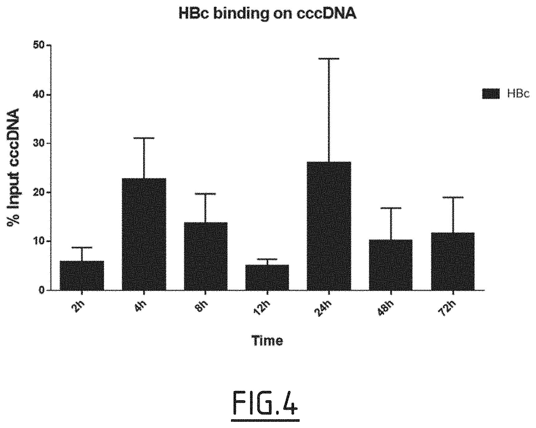
[0085] FIG. 4: Evaluation of HIRA interaction with HBV core protein.
[0086] HepG2-NTCP cells were infected with 250 vge/cell of HBV from 2 hours to 3 days. After crosslinking and nuclear extraction, ChIP experiments were performed using antibodies against HBc protein.
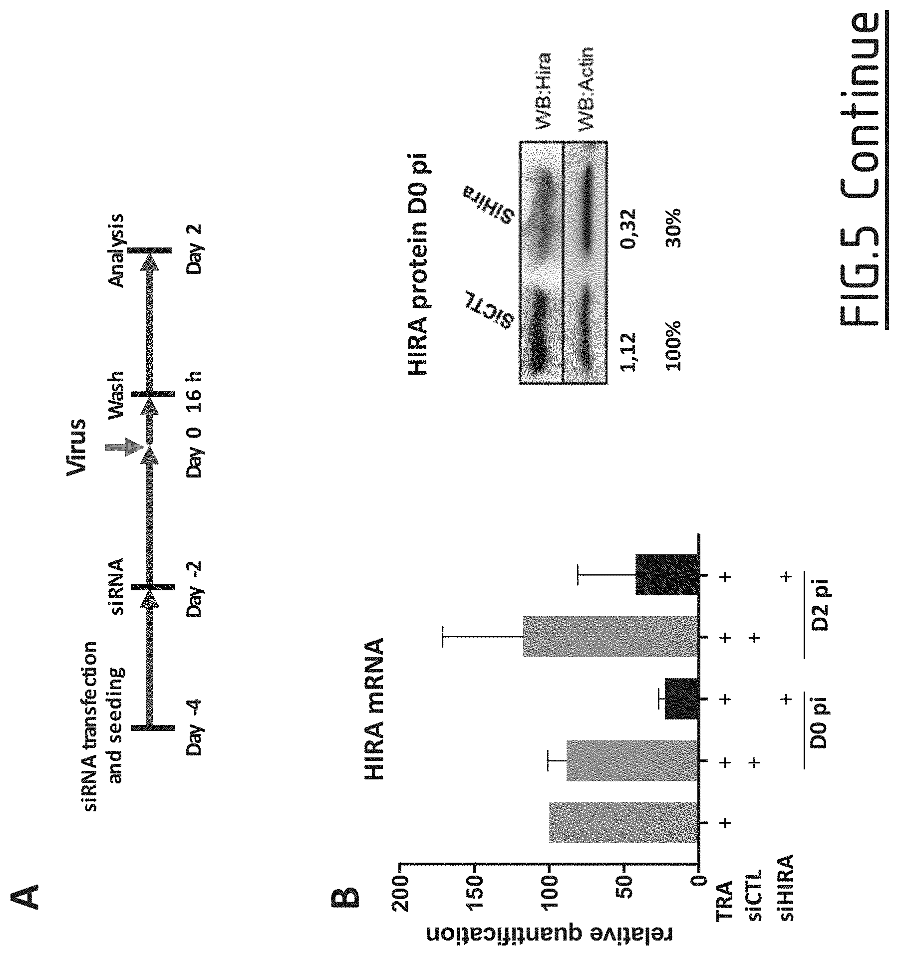
[0087] FIG. 5: Effect of HIRA's downregulation before infection on viral cccDNA establishment.
[0088] (A) Experimental timeline for HIRA silencing. HepG2-NTCP cells were transfected two-times with siRNA anti HIRA or a non-targeting siRNA and then inoculated for 16 h with HBV at MOI of 250 vge/cell in the presence of 4% PEG8000. Cells were harvested for analysis 2 days after infection. (B) HIRA mRNA and protein expression after siRNA transfection was determined at the moment of infection (D0 pi) and at cells harvesting (D2 pi), by real-time qPCR (left panel) and Western blot (right panel). Results of densitometric analysis is shown under the image. (C) cccDNA amount at day 2 pi (post infection) was measured by qPCR (left panel) and Southern Blotting (right panel). HIRA mRNA expression was normalized over the housekeeping gene Gusb and expressed as percentage of transfection reagent (TRA) treated cells. Actin protein served as loading control for Western Blotting analysis. cccDNA amount was expressed as percentage of transfection reagent (TRA) treated cells. In Southern Blotting, HBV and mitochondrial DNA were revealed using DIG-coupled probes (See Materials and Methods section). Mitochondrial DNA was used as internal loading control. Specificity of cccDNA band is demonstrated by linearization after digestion with EcoRI. Densitometric analysis of cccDNA band normalized over mitochondrial DNA signal is shown under the image.
[0089] FIG. 6: HIRA Transcomplementation rescues cccDNA establishment.
[0090] (A) Experimental timeline for rescue of HIRA expression after silencing. HepG2-NTCP cells were transfected two-times with siRNA anti HIRA or a non-targeting siRNA (see Material and Methods) and then transfected with pEYFP-N1-HIRA before inoculation with HBV at MOI of 250 vge/cell in the presence of 4% PEG8000 for 16 h. The cells were harvested for analysis 2 days after infection. (B) HIRA mRNA and protein expression after siRNA transfection was determined at the moment of infection and at cells harvesting, by real-time qPCR (left panel) and Western blot (right panel). (C) cccDNA amount at day 2 pi was measured by qPCR. HIRA mRNA expression was normalized over the housekeeping gene Gusb and expressed as percentage of transfection reagent (TRA) treated cells. Actin protein served as loading control for Western Blotting analysis. cccDNA amount was expressed as percentage of transfection reagent (TRA) treated cells.
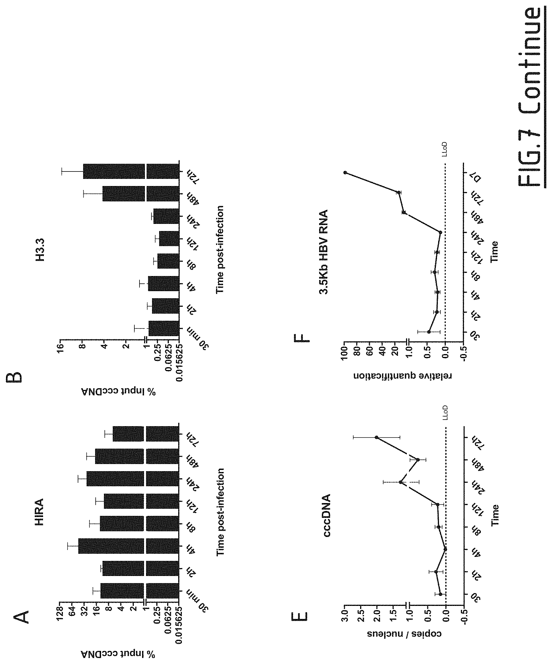
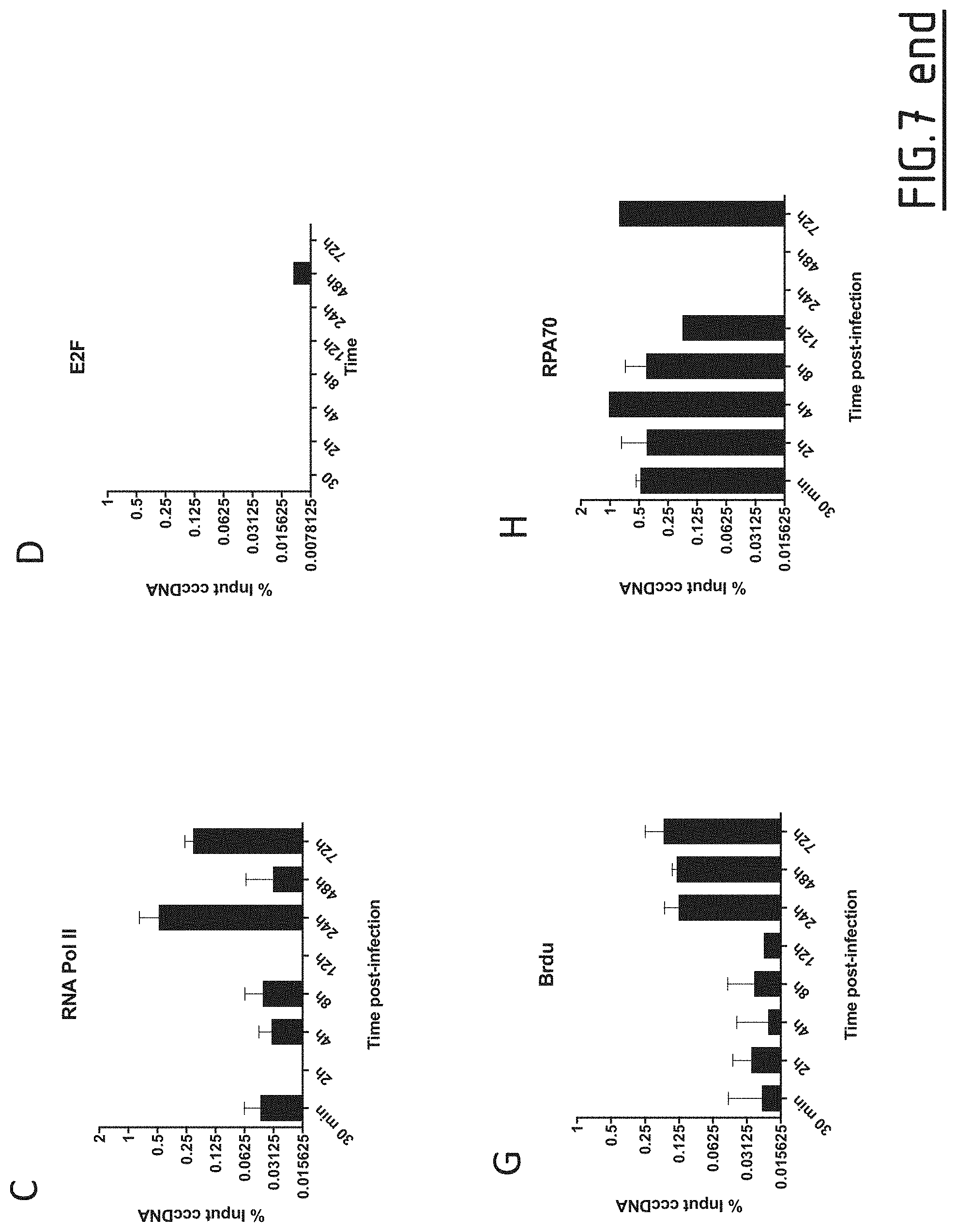
[0091] FIG. 7: Assessment of HIRA and histone variant H3.3 binding on cccDNA
[0092] HepG2-NTCP cells were infected at MOI of 250 vge/cell in the presence of 4% PEG8000 for up to 16 h and then extensively washed and cultured for the indicated time points before ChIP analysis using antibodies against HIRA (A), H3.3 (B), RNA Polymerase II (C), and E2F (D). E2F ChIP was used as negative control. Immunoprecipitated cccDNA was quantified by qPCR and expressed as percentage of Input after normalization over NoAb condition. (E-F) qPCR quantification of viral cccDNA, and 3.5 Kb RNA in HepG2-NTCP-infected cells. cccDNA quantification was normalized over b-globin quantity, while relative RNA amount was calculated by normalizing over the housekeeping gene GUSB expression. Graphs represent the mean of at least three independent experiments. NoAb: no antibody; LLoD: lower limit of detection.
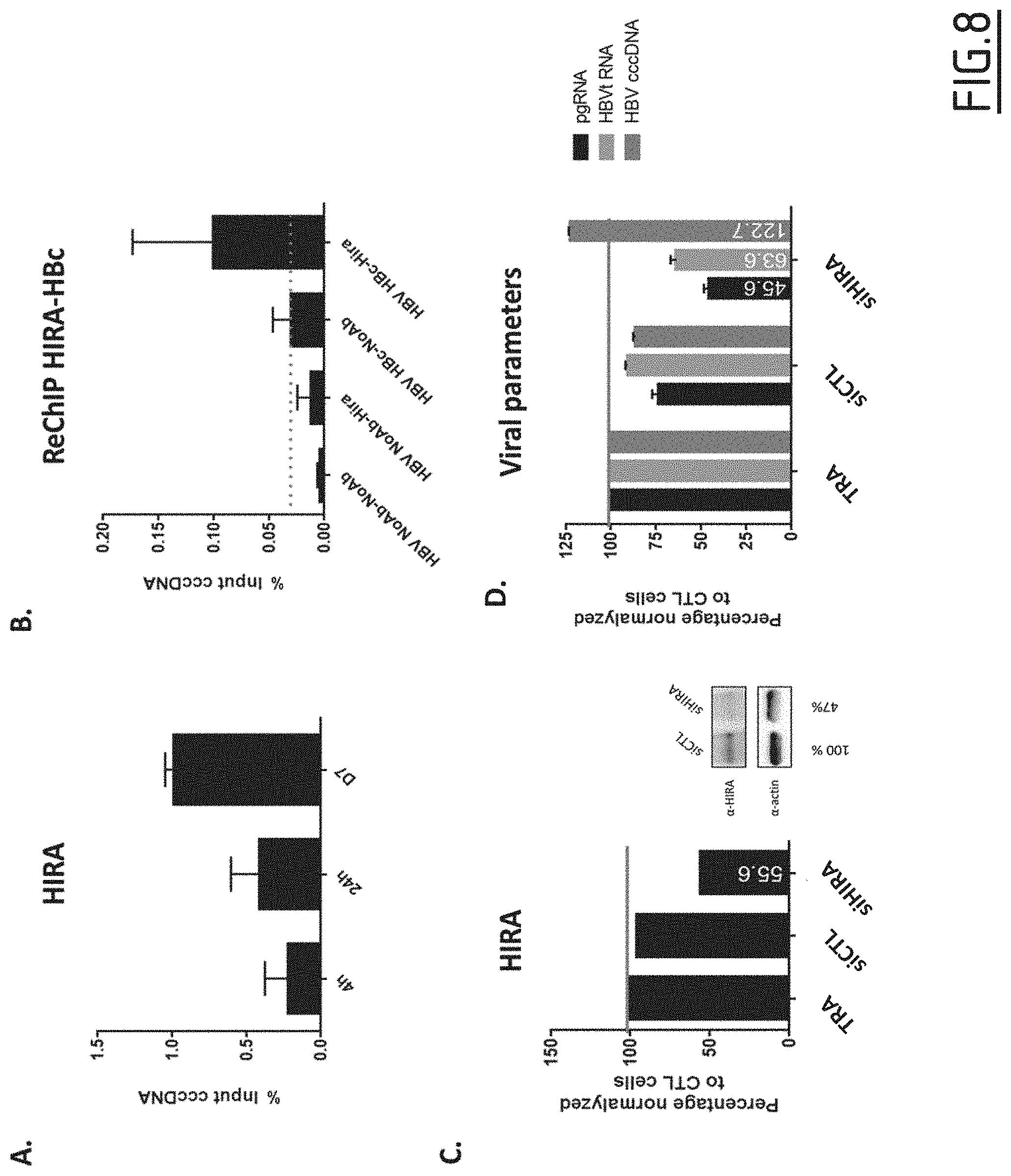
[0093] FIG. 8: Effect of HIRA's downregulation during chronic infection.
[0094] HepG2-NTCP cells were infected at MOI of 250 vge/cell in the presence of 4% PEG8000 for up to 16 h and then extensively washed and cultured for the indicated time points before ChIP analysis using antibodies against HIRA (A) or against HIRA and then against HBc (B). Immunoprecipitated cccDNA was quantified by qPCR and expressed as percentage of Input. (C-D) Silencing experiments were performed onto an established infection. HepG2-NTCP cells were infected with HBV at MOI of 250 vge/cell in the presence of 4% PEG8000. After 5 days, cells were transfected two-times with siRNA anti HIRA or a non-targeting siRNA. Cells were harvested 48 h after the second transfection (i.e. day 9 post-infection). (C) HIRA mRNA expression after siRNA transfection was determined by real-time qPCR. HIRA mRNA expression was normalized over the housekeeping gene Gusb and expressed as percentage of transfection reagent (TRA) treated cells. (D) cccDNA amount was measured by qPCR. cccDNA amount was expressed as percentage of transfection reagent (TRA) treated cells. NoAb: no antibody;
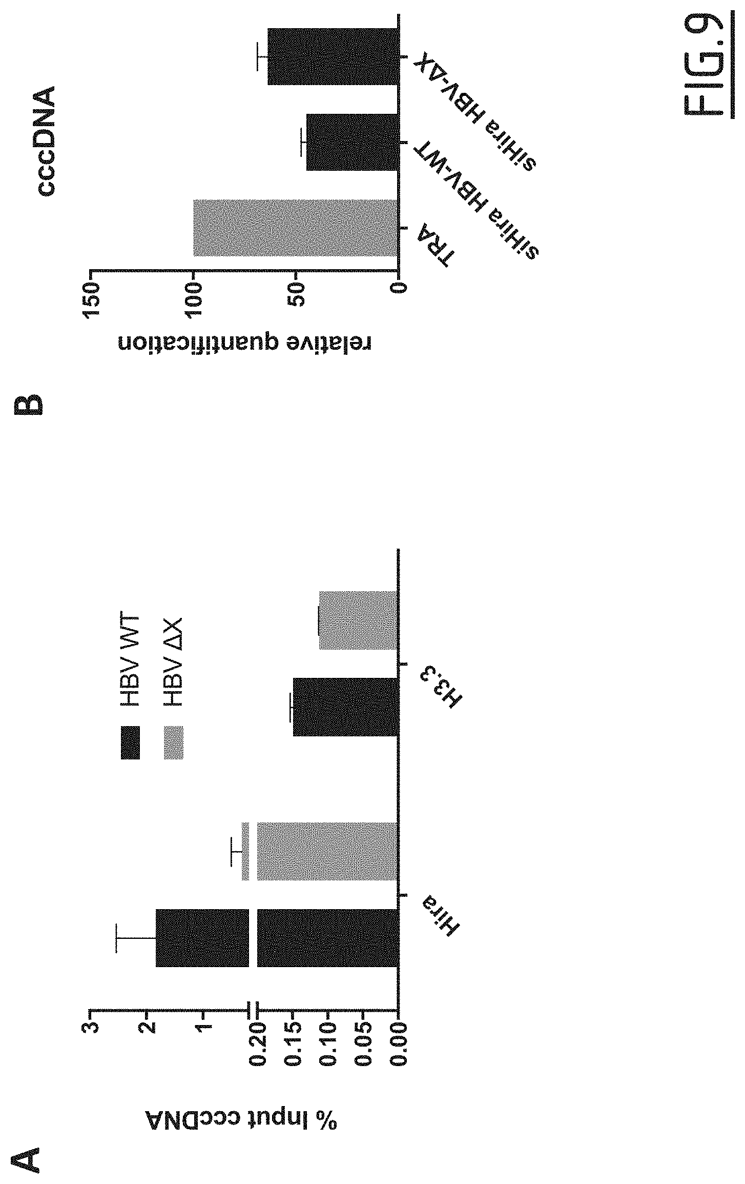
[0095] FIG. 9: Evaluation of HBx involvement in HIRA's binding to cccDNA and in cccDNA establishment.
[0096] (A) HepG2-NTCP cells were infected at MOI of 250 vge/cell in the presence of 4% PEG8000 for 4 h. The levels of HIRA and H3.3 on cccDNA were analyzed by ChIPqPCR. (B) HepG2-NTCP cells were transfected with siRNA against HIRA according to the experimental timeline shown in FIG. 6A and inoculated for 16 h with HBV at MOI of 250 vge/cell in the presence of 4% PEG8000. Cells were harvested for analysis 2 days after infection. cccDNA amount at day 2 pi was measured by qPCR and expressed as percentage of transfection reagent (TRA) treated cells.
[0097] FIG. 10: Evaluation of HIRA interaction with HBV core protein.
[0098] (A) HepG2-NTCP cells were infected at MOI of 250 vge/cell in the presence of 4% PEG8000 up to 16 h and then extensively washed and cultured for the indicated time points before ChIP analysis using an antibody against HBc. The levels of HBc on cccDNA were analyzed through the infection kinetics by ChIPqPCR. (B) The simultaneous presence of HIRA and HBc on the same cccDNA molecule was assessed by sequential ChIPqPCR 24 h after infection using an antibody against HIRA first and then an antibody anti HBc for immunoprecipitation. (C) Proximity between HBc and HIRA was assessed by proximity ligation assay (PLA) in HBV-infected HepG2-NTCP cells 24 hours post-infection. PLA signals are indicated by arrows. Uninfected HepG2-NTCP cells and infected HepG2-NTCP cells stained with only HBc or HIRA antibodies were used as negative controls (right panels). (D) Co-immunoprecipitation between and HIRA was realized in HepaRG-TR-HBc cells treated for 48 h with 10 .mu.g/ml tetracyclin to induce HBc expression. Cell lysates were immunoprecipitated with an antibody against HBc and subjected to Western Blotting analysis with an antibody anti HBc and HIRA. Lysates from HepaRG-TR-HBc cells not treated with tetracyclin were used as negative control.
EXAMPLES
Example 1: Study of the Role of HIRA
Materials and Methods
[0099] siRNA Transfection siRNA (Individual: ON-TARGETplus HIRA siRNA; ON-TARGETplus LUC siRNA, Dharmacon) (Table 2) for HIRA and Luciferase (10 nM) were transfected in HepG2-NTCP cells and PHH before infection, using Lipofectamine RNAiMAX (ThermoFisher), following manufacturer's protocol.
TABLE-US-00002 TABLE 2 ON-TARGETplus siRNA sequences SEQ ID Target Name Sequence number ON-TARGETplus siHIRA GGAUAACACUGUCGUCAUC SEQ ID HIRA NO: 1 ON-TARGETplus siLUC GAUUAUGUCCGGUUAUGUA SEQ ID LUC NO: 7
Cell Culture, HBV Infection and Analysis of Viral Parameters During Replication
[0100] HepG2-NTCP cells were seeded at 105 cells/cm.sup.2 in DMEM medium supplemented with penicillin (Life Technology), streptomycin (Life Technology), sodium pyruvate (Life Technology), 5% Fetal Calf Serum (FCS; Fetalclone II.TM., PERBIO). The day after, medium was renewed and complemented with 2.5% DMSO (SIGMA). After 72 h, cells were infected at a multiplicity of infection of 250 in the presence of 4% PEG800 for 16 h. Cells were then extensively washed with PBS and maintained in complete DMEM medium containing 2.5% DMSO until harvesting. Intracellular accumulation of viral RNA and DNA, secretion of HBe and HBs antigens were monitored by RT-qPCR, qPCR and southern blotting, and ELISA, respectively. Briefly, HBeAg and HBsAg were quantified in culture medium by ELISA, using a chemiluminescence immunoassay kit (Autobio, China) following manufacturer's instructions. Total DNA was purified from infected cells using MasterPure.TM. Complete DNA Purification Kit (Epicentre). To increase the specificity of HBV cccDNA detection, 3 nuclease digestion was performed using 10 U of T5 exonuclease (Epicentre) at 37.degree. C. for 45 minutes on 500 ng of total DNA, before selective cccDNA qPCR, based on the use of primers (and probes) spanning the nick in the HBV rcDNA and hybridizing to its "gap region"(see table 1).
[0101] Total RNA was extracted from infected cells using NucleoSpin.RTM. RNA kit (Macherey-Nagel) and retro-transcribed into cDNA using SuperScript III reverse transcriptase according to manufacturer's instructions (Invitrogen, Carlsbad, USA). Real-time PCR for total HBV DNA and cccDNA was performed using an Applied QuantStudio 7 machine and TaqMan Advanced Fast Master Mix or SYBR Green Master Mix (see examples of primers and probes, table 1).
Hirt Procedure and Southern Blot Analyses
[0102] DNA was extracted following a modified Hirt procedure (Hirt B. Selective extraction of polyoma DNA from infected mouse cell cultures. J Mol Biol. 1967). 90 .mu.g of DNA were subjected to Southern blot analyses using a mix of DIG-labeled probes, an AP conjugated anti-DIG antibody (Roche) and CDP-Star.RTM. (Roche) according to the manufacturer's instructions.
Chromatin Immunoprecipitation (ChIP)
[0103] ChIP experiments were carried out on infected cells from 2 h to 3 days post-infection. Cells were washed in phosphate buffered saline (PBS), and incubated for 15 minutes with 1% formaldehyde at 37.degree. C. and quenched with 0.125 M Glycine. For nuclear extracts preparation, cells were lysed in lysis buffer (PIPES 5 mM, KCl 85 mM, NP-40 0.5%, PMSF 1 mM, Protease inhibitor cocktail (PIC) 1.times.). After douncing (10 times) and centrifugation, nuclei were resuspended in sonication buffer (SDS 1%, EDTA 10 mM, Tris-HCl pH 8 50 mM, PMSF 1 mM, PIC 1.times.). After sonication, chromatin was cleaned in Ripa buffer with Protein A Sepharose, and then subjected to overnight immunoprecipitation at 4.degree. C. using 2-5 .mu.g of antibodies. Immune complexes were, then, incubated 2 h with protein G agarose beads at 4.degree. C., washed, and eluted in Tris-HCl pH 8 10 mM, EDTA 5 mM, NaCl 50 mM, SDS 1%, Proteinase K 50 ug, PIC 1.times.. Immunoprecipitated DNA was extracted and quantified by qPCR using cccDNA specific primers (see table 1). Samples were normalized to Input DNA using the .DELTA.Ct method were .DELTA.Ct=Ct (input)-Ct (immunoprecipitation) and expressed as percentage of the Input.
Co-Immunoprecipitation
[0104] Co-immunoprecipitation experiments were carried out on HepaRG-TR-HBc cells, in which the expression of HBc protein can induced upon doxycycline treatment (10 .mu.g/ml)). Nuclear extraction and antibody incubation was performed as for ChIP experiments (see above). Immune complexes were then incubated 2 h with protein G agarose beads at 4.degree. C., washed, and eluted in 2.times.Laemmli buffer (65.8 mM Tris-HCl, pH 6.8, 2.1% SDS, 26.3% (w/v) glycerol, 0.01% bromophenol blue) to release and denature the proteins. Whole cell lysates (Input) were incubated with 6.times.Laemmli buffer. Proteins were separated on standard 12% gradient gels and blotted on a nitrocellulose membrane. Blots were blocked 1 hour with 5% milk in PBS and stained with primary antibody in blocking buffer overnight at 4.degree. C. (HIRA 1:500, HBc 1:500). After primary antibody incubation, blots were washed 3.times. with TBS-Tween (TBST, 0.2M Tris-HCl 1.5M NaCl, 0.05 Tween 20), stained with secondary antibodies for 1 hour at room temperature and washed again 3 times with TBST. Detection occurred using Biorad Clarity Western ECL and the ChemiDoc XRS system (Biorad).
Proximity Ligation Assay
[0105] Proximity ligation assay (PLA) was performed on HepG2-NTCP infected cells according to manufacturer's instructions (DU092014 Duolink.RTM., DUO92004 Duolink.RTM., DUO92002 Duolink.RTM., and DUO82040 Duolink.RTM.). Briefly, after fixation by 4% PFA 30 minutes at room temperature (RT), and quenching by 1M glycine, cells were permeabilized for 30 minutes at RT with PBS-Triton 0.3%, and then blocked 30 minutes at 37.degree. C. with blocking buffer. Cells were incubated with the two primary antibodies overnight at 4.degree. C. PLA probes were diluted and added to the coverslips for 1 hour at 37.degree. C. After a 30 minutes ligation step at 37.degree. C., the amplification was performed for 100 minutes at 37.degree. C. Finally, coverslips were mounted onto slips with Mounting Medium with DAPI.
Antibodies
[0106] ChIP and Co-immunoprecipitation experiments were performed using the following antibodies:
TABLE-US-00003 TABLE 3 Antibodies used for PLA, co-immunoprecipitation (co-IP), ChIP and western blotting experiments (WB) Target Company Dilution HBc for PLA, co-IP and ChIP Invitrogen SC2362651 4 .mu.g HBc for WB Dako B0586 1:500 HIRA for PLA, ChIP and WB Abcam Ab20655 2.5 .mu.g panH3 for ChIP Diagenode C15200011 2.5 .mu.g H3.3 for ChIP Abcam Ab62642 5 .mu.g
Results
[0107] The inventors investigated the effect of HIRA downregulation on HBV cccDNA establishment and replication by siRNA mediated knockdown (siHIRA). The efficiency of HIRA knockdown was determined by quantification of HIRA mRNA levels after transfection, using a Luciferase siRNA (CTL) as a control. Transfection of siHIRA reduced by nearly 70% endogenous level of HIRA mRNA from 24 h to 96 h post-transfection, and remained at nearly 50% of extinction at day 2 post-infection (FIG. 1A) while Luciferase siRNA had no effect on HIRA mRNA levels.
[0108] qPCR analysis showed that knockdown of HIRA expression resulted in the reduction of 50% of cccDNA levels, accompanied by a decrease of intracellular total HBV DNA, total HBV RNAs and pregenomic (pg)RNA, in particular (FIG. 1A). Southern Blotting analysis confirmed the reduction in cccDNA, especially at day 3 post-infection. On the contrary, rcDNA levels remained stable, ruling out a defect in viral entry and suggesting a possible either incomplete or delayed rcDNA to cccDNA transition. The use of Tenofovir (Teno), a nucleoside analogue blocking pgRNA to rcDNA conversion, allowed us to inhibit the recycling of nucleocapsids from the cytoplasm. In this condition, cccDNA amounts remained downregulated also at day 7 post infection, when HIRA mRNA went back to basal levels (FIG. 1B), while, in untreated cells, cccDNA levels went back to basal levels (FIG. 1C). These results suggest that HIRA extinction leads not to a delayed, but to a blockade of cccDNA formation that needs nucleocapsids recycling to be overcome.
[0109] Chromatin Immunoprecipitation analysis showed that HIRA was bound to cccDNA already at 2 hours post-infection (FIG. 2A). This binding was persistent until 3 days post-infection. The recruitment of HIRA was concomitant with the deposition of histone variant H3.3. Indeed, the presence of H3.3 on cccDNA was observable after 2 hours post-infection, stable until 24 hours, and increased after 48 hours of infection.
[0110] Concerning total histone H3, component of stable nucleosomes, it showed an early recruitment between 2 h and 8 h post-infection and, then, a second wave of binding, slower and more progressive, beginning from 12 h and still increasing at 72 h post-infection. The increase in H3.3 and total H3 binding to cccDNA after 48 h of infection correlated with the beginning of cccDNA transcriptional activity, as indicated by pgRNA kinetic of appearance by qPCR (FIG. 2B, C).
[0111] To investigate a possible involvement of the viral protein HBx in HIRA-mediated cccDNA establishment, the inventors took advantage of a .DELTA.HBx virus strain. Chromatin Immunoprecipitation analysis showed that HIRA binding to cccDNA was not affected by the absence of HBx protein (FIG. 3A). Similarly, siRNA mediated knockdown of HIRA in absence of X protein expression resulted in a similar reduction of cccDNA levels and total intracellular HBV DNA compared to WT virus (FIG. 3B).
[0112] By ChIP experiments, the inventors were able to demonstrate that HBc binds to cccDNA as early as after 2 hours of infection (FIG. 4), correlating with the recruitment of HIRA on the HBV minichromosome (FIG. 2A). The inventors decided, then, to investigate a possible interaction between the two proteins. Firstly, by co-immunoprecipitation, the inventors showed that HIRA was able to interact with HBc in an HepaRG cell line expressing HBc in an inducible manner. Secondly, performing Proximity Ligation Assay (PLA) in HBV infected hepatocytes, the inventors showed an interaction between HBc and HIRA as soon as 24 h post-infection.
[0113] Altogether, the inventors' results suggest that chromatinization of incoming viral DNA is a very early event, requiring the histone chaperone HIRA, thus suggesting that the inhibition of HIRA interaction with cccDNA could represent a new therapeutic target to be investigated. While HBx is not required for this process, HBc could play a major role, thus opening a new perspective for the investigation of a specific role for HBc in recruiting HIRA to the cccDNA.
Example 2: Complement Study of the Role of HIRA
Materials & Methods
Production of Wild-Type and Mutated HBV Viral Inoculum
[0114] HBV inoculum was prepared from filtered HepAD38 cells (Ladner et al., 1997) or K6 (HBx negative virus; Lucifora et al., JHep 2011) supernatants by polyethylene-glycol-MW-8000 (PEG8000, SIGMA) precipitation (8% final). Viral stock with a titer reaching at least 1.times.10.sup.10 vge/mL was tested endotoxin free and used for infection.
Cell Culture and HBV Infection
[0115] HepG2-NTCP cells were seeded at 10.sup.5 cells/cm.sup.2 in DMEM medium supplemented with penicillin (Life Technology), streptomycin (Life Technology), sodium pyruvate (Life Technology), 5% Fetal Calf Serum (FCS; Fetalclone II.TM., PERBIO). The day after, medium was renewed and complemented with 2.5% DMSO (SIGMA). After 72 h, cells were infected at a multiplicity of infection of 250 in the presence of 4% PEG800 for up to 16 h and then extensively washed and cultured for the indicated time points in complete DMEM medium containing 2.5% DMSO until harvesting. Intracellular accumulation of viral RNA and DNA were monitored by RT-qPCR, qPCR and Southern Blotting. For de novo DNA synthesis analysis, cells were treated with 20 .mu.M BrdU (Sigma) 24 h hours before infection and during viral inoculation.
Analysis of Viral Parameters During Replication and qPCR
[0116] Total DNA was purified from infected cells using MasterPure.TM. Complete DNA Purification Kit (Epicentre). Total HBV DNA was quantified using TaqMan.RTM. Gene Expression assay ID: Pa03453406_s1. To increase the specificity of HBV cccDNA detection, qPCR was preceded by a nuclease digestion using 10 U of T5 exonuclease (Epicentre) for 500 ng of total DNA. Selective cccDNA qPCR was performed using primers and probes spanning the "gap region" in the HBV rcDNA (see Table 4). Serial dilutions of an HBV monomer plasmid (pHBV-EcoRI) were used as standard for quantification. For normalization, the number of human hepatocytes was estimated by measuring human b-globin (TaqMan.RTM. assay ID Hs00758889_s1) while human genomic DNA (Roche Applied Science, Mannheim, Germany) was used as a standard curve for quantification. Total RNA was extracted from infected cells using NucleoSpin.RTM. RNA kit (Macherey-Nagel) and retro-transcribed using SuperScript III reverse transcriptase according to manufacturer's instructions (Invitrogen, Carlsbad, USA). 3.5 Kb viral RNA (comprising pgRNA and pre-core RNA) and total HBV RNA were amplified using previously described primers and probes (Table 4). Primers for HIRA mRNA expression are listed in Table 4. The expression of the human housekeeping gene GUSb (TaqMan.RTM. assay ID Hs00939627_m1) was used for normalization. Real-time PCRs were performed using an Applied QuantStudio 7 machine and TaqMan Advanced Fast Master Mix or SYBR Green Master Mix (ThermoFischer).
TABLE-US-00004 Table 4 Primers and probes sequences used for Taqman and SYBRgreen qPCR. Target Name Sequence cccDNA cccDNA CCGTGTGCACTTCGCTTCA (SEQ ID NO: 4) Taqman cccDNA cccDNA GCACAGCTTGGAGGCTTGA (SEQ ID NO: 5) Taqman cccDNA cccDNA [6FAM]CATGGAGACCACCGTGAACGCCC[BBQ] Taqman (SEQ ID NO: 6) Probe pgRNA pgRNA GGAGTGTGGATTCGCACTCCT (SEQ ID Taqman NO: 8) pgRNA pgRNA AGATTGAGATCTTCTGCGAC (SEQ ID Taqman NO: 9) pgRNA pgRNA [6FAM]AGGCAGGT0000TAGAAGAAGAACTCC Taqman [BBQ] (SEQ ID NO: 10) Probe GUSb FW_gusb CGTGGTTGGAGAGCTCATTTGGAA (SEQ ID NO: 11) GUSb RV_gusb ATTCCCCAGCACTCTCGTCGGT (SEQ ID NO: 12) Hira Hira GGCCTCGGAAGGACTCTC (SEQ ID Forward NO: 2) Hira Hira AGACAGACACATGGCCTCCT (SEQ ID Reverse NO: 3)
Plasmid and siRNA Cell Transfection
[0117] pEYFP-N1-HIRA construct was transfected in HepG2-NTCP cells and PHH using TransIT-2020 (Mirus Bio LLC) following manufacturer's protocol. siRNA targeting HIRA (ON-TARGETplus HIRA siRNA GGAUAACACUGUCGUCAUC, SEQ ID NO: 1) and the corresponding siRNA mutated in position 9-11 (siRNA CTL: GGAUAACAGACUCGUCAUC, SEQ ID NO: 13) to serve as negative control were transfected at 10 nM concentration in HepG2-NTCP cells and PHH using Lipofectamine RNAiMAX reagent (ThermoFisher), following manufacturer's instructions.
Hirt Extraction and Southern Blot Analysis
[0118] DNA was extracted following a modified Hirt procedure. 90 .mu.g of DNA were subjected to Southern blot analyses using a mix of DIG-labeled probes (synthesized using primers listed below and the "PCR DIG probe synthesis kit" (Roche)), an AP conjugated anti-DIG antibody (Roche) and CDP-Star.RTM. (Roche) according to the manufacturer's instructions. Mitochondrial DNA served as internal loading control.
TABLE-US-00005 TABLE 5 DIG-labeled HBV DNA probes sequences Target Name Sequence HBV HBV-F1 TAGCGCCTCATTTTGTGGGT (SEQ ID NO: 14) HBV-R1 CTTCCTGTCTGGCGATTGGT (SEQ ID NO: 15) HBV-F2 TAGGACCCCTTCTCGTGTTA (SEQ ID NO: 16) HBV-R2 CCGTCCGAAGGTTTGGTACA (SEQ ID NO: 17) HBV-F3 ATGTGGTATTGGGGGCCAAG (SEQ ID NO: 18) HBV-R3 GGTTGCGTCAGCAAACACTT (SEQ ID NO: 19) HBV-F4 TGGACCTTTTCGGCTCCTC (SEQ ID NO: 20) HBV-R4 GGGAGTCCGCGTAAAGAGAG (SEQ ID NO: 21) HBV-F5 GTCTGTGCCTTCTCATCTG (SEQ ID NO: 22) HBV-R5 AGGAGACTCTAAGGCTTCC (SEQ ID NO: 23) HBV-F6 TACTGCACTCAGGCAAGCAA (SEQ ID NO: 24) HBV-R6 TGCGAATCCACACTCCGAAA (SEQ ID NO: 25) HBV-F8 AGACGAAGGTCTCAATCGCC (SEQ ID NO: 26) HBV-R8 ACCCACAAAATGAGGCGCTA (SEQ ID NO: 27)
TABLE-US-00006 TABLE 6 DIG-labeled mitochondrial DNA probes sequences Target Name Sequence Mito- Fw-huND1 CCCTACTTCTAACCTCCCTGTTCTTAT chondrial (SEQ ID NO: 28) DNA Rw-huND1 CATAGGAGGTGTATGAGTTGGTCGTA (SEQ ID NO: 29) Fw-huND5 ATTTTATTTCTCCAACATACTCGGATT (SEQ ID NO: 30) Rw-huND5 GGGCAGGTTTTGGCTCGTA (SEQ ID NO: 31) Fw-huATP6 CATTTACACCAACCACCCAACTATC (SEQ ID NO: 32) Rw-huATP6 CGAAAGCCTATAATCACTGTGCC (SEQ ID NO: 33)
Chromatin Immunoprecipitation (ChIP)
[0119] ChIP experiments were carried out at the indicated time points (from 30 minutes to 72 hours) post-infection. Briefly, cells were washed in phosphate buffered saline (PBS), and incubated for 15 minutes with 1% formaldehyde at 37.degree. C. and quenched with 0.125 M Glycine. For nuclear extracts preparation, cells were lysed in lysis buffer (PIPES 5 mM, KCl 85 mM, NP-40 0.5%, PMSF 1 mM, Protease inhibitor cocktail (PIC) 1.times.). After douncing (10 times) and centrifugation, nuclei were resuspended in sonication buffer (SDS 1%, EDTA 10 mM, Tris-HCl pH 8 50 mM, PMSF 1 mM, PIC 1.times.). After sonication, chromatin was cleaned in Ripa buffer with Protein A Sepharose, and then subjected to overnight immunoprecipitation at 4.degree. C. using 2-5 .mu.g of antibodies indicated in Table 4 or No Antibody. Immune complexes were, then, incubated for 2 h with protein G agarose beads at 4.degree. C., washed, and eluted in Tris-HCl pH 8 10 mM, EDTA 5 mM, NaCl 50 mM, SDS 1%, Proteinase K 50 .mu.g, PIC 1.times.. Immunoprecipitated DNA was extracted and quantified by qPCR using cccDNA specific primers (see table 4). Samples were normalized to input DNA using the .DELTA.Ct method were .DELTA.Ct=Ct (input)-Ct (immunoprecipitation) and expressed as percentage of the input after normalization over No Antibody signal.
Sequential Chromatin Immunoprecipitation
[0120] Cells were processed as for ChIP experiment until the overnight immunoprecipitation. Immune complexes were, then, incubated 2 h with protein G agarose beads at 4.degree. C., washed and eluted in 10 mM DTT. Eluted samples were then re-exposed to overnight immunoprecipitation at 4.degree. C. using 2-5 .mu.g of antibodies in Re-ChIP buffer (1% Triton X-100; 2 mM EDTA; 150 mM NaCl; 20 mM Tris-HCl pH 8). Immune complexes were, then, processed as for a classic chromatin immunoprecipitation described above.
Western Blotting
[0121] Cells were lysed in RIPA buffer supplemented with PIC 1.times. and PMSF 1.times.. Proteins were migrated in 4-20% mini-PROTEAN.sup.@ TGX stain-Free.TM. Precast Gel (Bio-Rab Laboratories) and transferred onto a nitrocellulose membrane (Bio-Rab Laboratories). Blots were blocked 1 hour with 5% milk in TBS (1.times.Tris Buffered Saline (Sigma)) and stained with primary antibody in blocking buffer overnight at 4.degree. C. After primary antibody incubation, blots were washed 3.times. with TBST (1.times.TBS with 0.1% Tween 20), stained with HRP-conjugated secondary antibodies for 1 hour at room temperature and washed again 3 times with TBST. Detection occurred using Biorad Clarity Western ECL and the ChemiDoc XRS system (Biorad). Antibodies are listed in Table 7.
Protein Co-Immunoprecipitation (Co-IP)
[0122] Protein co-immunoprecipitation experiments were carried out on HepaRG-TR-HBc cell lines. HBc expression was induced by doxycycline treatment (10 .mu.g/ml) for 48 h. Nuclear extraction and antibody incubation was performed as for ChIP experiments. Immune complexes were then incubated 2 h with protein G agarose beads at 4.degree. C., washed, and eluted in 2.times.laemmli buffer (65.8 mM Tris-HCl, pH 6.8, 2.1% SDS, 26.3% (w/v) glycerol, 0.01% bromophenol blue) to release and denature the proteins. 1/10 of cell lysate, serving as input condition, was incubated with 6.times.laemmli buffer and subjected to western blotting analysis together with immunoprecipitated samples, as previously detailed. Antibodies used are listed in Table 7.
Proximity Ligation Assay
[0123] Proximity ligation assay (PLA) was performed on infected HepG2-NTCP cells according to manufacturer's instructions (DUO92014 Duolink.RTM., DUO92004 Duolink.RTM., DUO92002 Duolink.RTM., and DUO82040 Duolink.RTM.). Briefly, after fixation by 4% PFA 30 minutes at RT, and quenching by 1M glycine, cells were permeabilized for 30 minutes at RT with PBS-Triton 0.3%, and then blocked 30 minutes at 37.degree. C. with blocking buffer. Cells were incubated with the two primary antibodies overnight at 4.degree. C. (see Table 7). PLA probes were diluted and added to the coverslips for 1 hour at 37.degree. C. After a ligation step of 30 minutes at 37.degree. C., the amplification was performed during 100 minutes at 37.degree. C. Finally, coverslips were mounted onto slips with Mounting Medium with DAPI. Images were acquired with the confocal microscope zeiss LSM 780 NLO.
TABLE-US-00007 TABLE 7 Antibodies used for co-immunoprecipitation, ChiP and western blotting experiments Target Company Dilution HBc for PLA, co-IP and ChIP Invitrogen 4 .mu.g Hira for PLA, ChIP and WB Abeam Ab20655 2.5 .mu.g/1:500 H3.3 for ChIP Abeam Ab62642 5 .mu.g RNA Polymerase II for ChIP Diagenode 1 .mu.g RPA 70 for ChIP Abeam Ab79398 5 .mu.g BrdU for ChIP BD Pharmingen 5 .mu.g E2F for ChIP Santa Cruz 5 .mu.g beta-actin for WB Abcam Ab6276 1:10000 HBc for WB Dako B0586 1:500
Results
[0124] The inventors investigated the effect of HIRA downregulation on HBV cccDNA establishment and replication by siRNA mediated knockdown (siHIRA). The efficiency of HIRA knockdown was determined by quantification of HIRA mRNA levels after transfection, using an anti-HIRA siRNA mutated in position 9 and 11 (CTL) as a control. Transfection of siHIRA reduced by nearly 70% endogenous level of HIRA mRNA and protein levels while Luciferase siRNA had no effect on HIRA mRNA or protein expression (FIG. 5B).
[0125] qPCR analysis showed that knockdown of HIRA expression resulted in the reduction of 50% of cccDNA levels measured by qPCR and Southern Blotting (FIG. 1C). On the contrary, rcDNA levels remained stable, ruling out a defect in viral entry and suggesting a possible either incomplete or delayed rcDNA to cccDNA transition.
[0126] To further confirm the involvement of HIRA in HBV cccDNA establishment, the HIRA expression was rescued after silencing in HepG2-NTCP cells by transfecting the pEYFP-N1-HIRA construct 8 h before HBV infection (FIG. 6A). Analysis of HIRA expression by both qRT-PCR and Western Blotting, confirmed the restoration of its levels both at the mRNA and protein level (FIG. 6B). In parallel, cccDNA amount were evaluated by qPCR and went back to the level of non-silenced controls (TRA) (FIG. 6C).
[0127] Chromatin Immunoprecipitation analysis showed that HIRA was bound to cccDNA already at 30 minutes post-infection (FIG. 7A). This binding was persistent until 3 days post-infection. The recruitment of HIRA was concomitant with the deposition of histone variant H3.3 (FIG. 7B). Indeed, the presence of H3.3 on cccDNA was observable after 30 minutes post-infection, stable until 24 hours, and increased after 48 hours of infection.
[0128] The increase in H3.3 binding to cccDNA after 48 h of infection correlated with the amplification of cccDNA pool (FIG. 7E) and the beginning of cccDNA transcriptional activity, as indicated by pgRNA kinetic of appearance by qPCR (FIG. 7F) and RNA Pol II recruitment to cccDNA (FIG. 7C).
[0129] In an attempt to define the kinetics of H3.3 incorporation by HIRA during the rcDNA to cccDNA conversion, the kinetic of incorporation of BRdU and the recruitment of RPA70 to cccDNA was investigated (FIG. 7G-H). Since rcDNA to cccDNA conversion is thought to involve host cell DNA repair pathways and the activity of POLK for the completion of plus-strand and ligation of both strands HepG2-NTCP cells were treated with BrdU before and during virus inoculation and then ChIP analysis was performed using a BrdU-specific antibody to detect new nucleotides incorporation into cccDNA. As shown in FIG. 7G, no significant BrdU incorporation is found between 30 minutes and 12 h, corresponding to the initial phase of cccDNA formation from incoming HBV genome. On the contrary, BrdU incorporation increases concomitantly to the beginning of transcription and synthesis of new rcDNA. Interestingly, the inventors found that RPA70, was recruited to cccDNA only from 30 minutes to 12 h to come back again at 72 h, when newly synthesized rcDNA could be recycled to the nucleus to be converted into cccDNA (FIG. 7H). Altogether, these ChIP data suggest that histone deposition occurs on a cccDNA precursor which has both DNA strands completed but at least one of them not ligated, which corresponds to the main PF-rcDNA species found in the nucleus of infected cells. To further confirm this hypothesis, immunoprecipitated DNA in FIG. 7 was also amplified with primers located in a HBV double stranded region common to PF-rcDNA and cccDNA. The analysis showed no differences in the percentage of immunoprecipitated HBV DNA with respect to cccDNA, thus confirming that the immunoprecipitated material is mostly constituted by HBV DNA with complete strands (data not shown).
[0130] By ChIP experiment, (FIG. 8A), going from 4 hours, to 7 days post-infection, the inventors could show that HIRA was found bound to cccDNA even when infection is considered chronic. Furthermore, sequential ChIP showed that HBc was found bound onto the same cccDNA molecule as HIRA, as compared to negative controls (No antibody-No antibody, No antibody-HIRA, HBc-No antibody, FIG. 8B). Finally, to investigate the functional presence of HIRA onto cccDNA at that time of infection, silencing experiments were performed onto an established infection. Indeed, after 5 days of infection, cells were transfected with siHIRA (a first time at day 5, a second time at day 7, to obtain a better knock-down). At day 9 post-infection, proteins, RNA and DNA were harvested. RNA and protein expression analysis showed a strong effect of HIRA knock-down (FIG. 8C). 3.5 Kb RNA and total HBV RNA levels appeared impaired by HIRA knock-down (FIG. 8D). These preliminary results are suggesting an additional role for HIRA concerning cccDNA biology, where HIRA could be involved in the maintenance of cccDNA transcriptional activity.
[0131] To investigate a possible involvement of the viral protein HBx in HIRA-mediated cccDNA establishment, the inventors took advantage of a .DELTA.HBx virus strain. Chromatin Immunoprecipitation analysis showed that HIRA binding to cccDNA was not affected by the absence of HBx protein (FIGS. 3A and 9A). Similarly, siRNA mediated knockdown of HIRA in absence of X protein expression resulted in a similar reduction of cccDNA levels and total intracellular HBV DNA compared to WT virus (FIGS. 3B and 9B).
[0132] By ChIP experiments, the inventors were able to demonstrate that HBc binds to cccDNA as early as after 30 minutes of infection (FIG. 10A), correlating with the recruitment of HIRA on the HBV minichromosome (FIG. 7A). The inventors decided, then, to investigate a possible interaction between the two proteins. Firstly, by co-immunoprecipitation, the inventors showed that HIRA was able to interact with HBc in an HepaRG cell line expressing HBc in an inducible manner, suggesting that the two proteins are able to associate also in the absence of viral genome (FIG. 10B). Secondly, performing Proximity Ligation Assay (PLA) in HBV infected hepatocytes, the inventors showed an interaction between HBc and HIRA as soon as 24 h post-infection (FIG. 100). Moreover, sequential ChIP experiments indicated that HBc and HIRA could bind to the same cccDNA molecule 24 h post-infection (FIG. 10D).
Example 3: In Silico Study of HBc Interaction with HIRA
[0133] The Protein Data Bank (PDB) is a universal protein database that contains crystal structures and information about the structure and functions of proteins. This information was used to retrieve the crystal structures of HBc dimer (accession number: 1GQT) and a portion of HIRA protein (accession number: 2i32, as available on Jul. 26, 2018).
[0134] PRISM server (PRotein Interactions by Structural Matching) is a resource portal for the bioinformatics which offers computational methods and approaches for large scale structural modelling of protein-protein interactions. PRISM server was used to perform prediction of protein-protein interaction and allowed to uncover 4 different predictive models of HBc/HIRA interaction. The server includes the tools for predicting interacting structure, interface, and amino acids possible interactions between the two proteins, as listed in Table 8. For example, in the selected model 1, Isoleucine 466 of HIRA (as represented in sequence SEQ ID NO: 34) could be interacting with Cystein 136 of HBc protein (as represented in sequence SEQ ID NO: 35).
[0135] This in silico experiment shows a predicted interaction of HBc and HIRA through 4 different possible interfaces, requiring from -5.73 to -1.05 kJ/mol energy to interact. The more negative the energy required is, the more efficient the protein-protein interaction will potentially be.
TABLE-US-00008 TABLE 8 Table of predictive interface residues contact between HIRA and HBc. HIRA (UniProtKB Possible interaction HBc (UniProtKB P54198) with Q89714) ILE 466 CYS 136 ILE 466 LEU 137 CYS 465 CYS 136 ILE 466 HIS 80 LEU 464 THR 138 LEU 464 LEU 137 LEU 464 THR 171 ILE 466 GLY 140 ILE 466 ARG 141 CYS 465 GLY 140 CYS 465 PHE 139 ILE 466 SER 78
Sequence CWU 1
1
36119RNAArtificial SequencesiRNA 1ggauaacacu gucgucauc
19218DNAArtificial SequencePrimer 2ggcctcggaa ggactctc
18320DNAArtificial SequencePrimer 3agacagacac atggcctcct
20419DNAArtificial SequencePrimer 4ccgtgtgcac ttcgcttca
19519DNAArtificial SequencePrimer 5gcacagcttg gaggcttga
19623DNAArtificial SequenceProbe 6catggagacc accgtgaacg ccc
23719RNAArtificial SequencesiRNA 7gauuaugucc gguuaugua
19821DNAArtificial Sequenceprimer 8ggagtgtgga ttcgcactcc t
21920DNAArtificial Sequenceprimer 9agattgagat cttctgcgac
201027DNAArtificial Sequenceprobe 10aggcaggtcc cctagaagaa gaactcc
271124DNAArtificial Sequenceprimer 11cgtggttgga gagctcattt ggaa
241222DNAArtificial Sequenceprimer 12attccccagc actctcgtcg gt
221319RNAArtificial SequencesiRNA 13ggauaacaga cucgucauc
191420DNAArtificial Sequenceprimer 14tagcgcctca ttttgtgggt
201520DNAArtificial Sequenceprimer 15cttcctgtct ggcgattggt
201620DNAArtificial Sequenceprimer 16taggacccct tctcgtgtta
201720DNAArtificial Sequenceprimer 17ccgtccgaag gtttggtaca
201820DNAArtificial Sequenceprimer 18atgtggtatt gggggccaag
201920DNAArtificial Sequenceprimer 19ggttgcgtca gcaaacactt
202019DNAArtificial Sequenceprimer 20tggacctttt cggctcctc
192120DNAArtificial Sequenceprimer 21gggagtccgc gtaaagagag
202219DNAArtificial Sequenceprimer 22gtctgtgcct tctcatctg
192319DNAArtificial Sequenceprimer 23aggagactct aaggcttcc
192420DNAArtificial Sequenceprimer 24tactgcactc aggcaagcaa
202520DNAArtificial Sequenceprimer 25tgcgaatcca cactccgaaa
202620DNAArtificial Sequenceprimer 26agacgaaggt ctcaatcgcc
202720DNAArtificial Sequenceprimer 27acccacaaaa tgaggcgcta
202827DNAArtificial Sequenceprimer 28ccctacttct aacctccctg ttcttat
272926DNAArtificial Sequenceprimer 29cataggaggt gtatgagttg gtcgta
263027DNAArtificial Sequenceprimer 30attttatttc tccaacatac tcggatt
273119DNAArtificial Sequenceprimer 31gggcaggttt tggctcgta
193225DNAArtificial Sequenceprimer 32catttacacc aaccacccaa ctatc
253323DNAArtificial Sequenceprimer 33cgaaagccta taatcactgt gcc
23341017PRTHomo sapiens 34Met Lys Leu Leu Lys Pro Thr Trp Val Asn
His Asn Gly Lys Pro Ile1 5 10 15Phe Ser Val Asp Ile His Pro Asp Gly
Thr Lys Phe Ala Thr Gly Gly 20 25 30Gln Gly Gln Asp Ser Gly Lys Val
Val Ile Trp Asn Met Ser Pro Val 35 40 45Leu Gln Glu Asp Asp Glu Lys
Asp Glu Asn Ile Pro Lys Met Leu Cys 50 55 60Gln Met Asp Asn His Leu
Ala Cys Val Asn Cys Val Arg Trp Ser Asn65 70 75 80Ser Gly Met Tyr
Leu Ala Ser Gly Gly Asp Asp Lys Leu Ile Met Val 85 90 95Trp Lys Arg
Ala Thr Tyr Ile Gly Pro Ser Thr Val Phe Gly Ser Ser 100 105 110Gly
Lys Leu Ala Asn Val Glu Gln Trp Arg Cys Val Ser Ile Leu Arg 115 120
125Asn His Ser Gly Asp Val Met Asp Val Ala Trp Ser Pro His Asp Ala
130 135 140Trp Leu Ala Ser Cys Ser Val Asp Asn Thr Val Val Ile Trp
Asn Ala145 150 155 160Val Lys Phe Pro Glu Ile Leu Ala Thr Leu Arg
Gly His Ser Gly Leu 165 170 175Val Lys Gly Leu Thr Trp Asp Pro Val
Gly Lys Tyr Ile Ala Ser Gln 180 185 190Ala Asp Asp Arg Ser Leu Lys
Val Trp Arg Thr Leu Asp Trp Gln Leu 195 200 205Glu Thr Ser Ile Thr
Lys Pro Phe Asp Glu Cys Gly Gly Thr Thr His 210 215 220Val Leu Arg
Leu Ser Trp Ser Pro Asp Gly His Tyr Leu Val Ser Ala225 230 235
240His Ala Met Asn Asn Ser Gly Pro Thr Ala Gln Ile Ile Glu Arg Glu
245 250 255Gly Trp Lys Thr Asn Met Asp Phe Val Gly His Arg Lys Ala
Val Thr 260 265 270Val Val Lys Phe Asn Pro Lys Ile Phe Lys Lys Lys
Gln Lys Asn Gly 275 280 285Ser Ser Ala Lys Pro Ser Cys Pro Tyr Cys
Cys Cys Ala Val Gly Ser 290 295 300Lys Asp Arg Ser Leu Ser Val Trp
Leu Thr Cys Leu Lys Arg Pro Leu305 310 315 320Val Val Ile His Glu
Leu Phe Asp Lys Ser Ile Met Asp Ile Ser Trp 325 330 335Thr Leu Asn
Gly Leu Gly Ile Leu Val Cys Ser Met Asp Gly Ser Val 340 345 350Ala
Phe Leu Asp Phe Ser Gln Asp Glu Leu Gly Asp Pro Leu Ser Glu 355 360
365Glu Glu Lys Ser Arg Ile His Gln Ser Thr Tyr Gly Lys Ser Leu Ala
370 375 380Ile Met Thr Glu Ala Gln Leu Ser Thr Ala Val Ile Glu Asn
Pro Glu385 390 395 400Met Leu Lys Tyr Gln Arg Arg Gln Gln Gln Gln
Gln Leu Asp Gln Lys 405 410 415Ser Ala Ala Thr Arg Glu Met Gly Ser
Ala Thr Ser Val Ala Gly Val 420 425 430Val Asn Gly Glu Ser Leu Glu
Asp Ile Arg Lys Asn Leu Leu Lys Lys 435 440 445Gln Val Glu Thr Arg
Thr Ala Asp Gly Arg Arg Arg Ile Thr Pro Leu 450 455 460Cys Ile Ala
Gln Leu Asp Thr Gly Asp Phe Ser Thr Ala Phe Phe Asn465 470 475
480Ser Ile Pro Leu Ser Gly Ser Leu Ala Gly Thr Met Leu Ser Ser His
485 490 495Ser Ser Pro Gln Leu Leu Pro Leu Asp Ser Ser Thr Pro Asn
Ser Phe 500 505 510Gly Ala Ser Lys Pro Cys Thr Glu Pro Val Val Ala
Ala Ser Ala Arg 515 520 525Pro Ala Gly Asp Ser Val Asn Lys Asp Ser
Met Asn Ala Thr Ser Thr 530 535 540Pro Ala Ala Leu Ser Pro Ser Val
Leu Thr Thr Pro Ser Lys Ile Glu545 550 555 560Pro Met Lys Ala Phe
Asp Ser Arg Phe Thr Glu Arg Ser Lys Ala Thr 565 570 575Pro Gly Ala
Pro Ala Leu Thr Ser Met Thr Pro Thr Ala Val Glu Arg 580 585 590Leu
Lys Glu Gln Asn Leu Val Lys Glu Leu Arg Pro Arg Asp Leu Leu 595 600
605Glu Ser Ser Ser Asp Ser Asp Glu Lys Val Pro Leu Ala Lys Ala Ser
610 615 620Ser Leu Ser Lys Arg Lys Leu Glu Leu Glu Val Glu Thr Val
Glu Lys625 630 635 640Lys Lys Lys Gly Arg Pro Arg Lys Asp Ser Arg
Leu Met Pro Val Ser 645 650 655Leu Ser Val Gln Ser Pro Ala Ala Leu
Thr Ala Glu Lys Glu Ala Met 660 665 670Cys Leu Ser Ala Pro Ala Leu
Ala Leu Lys Leu Pro Ile Pro Ser Pro 675 680 685Gln Arg Ala Phe Thr
Leu Gln Val Ser Ser Asp Pro Ser Met Tyr Ile 690 695 700Glu Val Glu
Asn Glu Val Thr Val Val Gly Gly Val Lys Leu Ser Arg705 710 715
720Leu Lys Cys Asn Arg Glu Gly Lys Glu Trp Glu Thr Val Leu Thr Ser
725 730 735Arg Ile Leu Thr Ala Ala Gly Ser Cys Asp Val Val Cys Val
Ala Cys 740 745 750Glu Lys Arg Met Leu Ser Val Phe Ser Thr Cys Gly
Arg Arg Leu Leu 755 760 765Ser Pro Ile Leu Leu Pro Ser Pro Ile Ser
Thr Leu His Cys Thr Gly 770 775 780Ser Tyr Val Met Ala Leu Thr Ala
Ala Ala Thr Leu Ser Val Trp Asp785 790 795 800Val His Arg Gln Val
Val Val Val Lys Glu Glu Ser Leu His Ser Ile 805 810 815Leu Ala Gly
Ser Asp Met Thr Val Ser Gln Ile Leu Leu Thr Gln His 820 825 830Gly
Ile Pro Val Met Asn Leu Ser Asp Gly Lys Ala Tyr Cys Phe Asn 835 840
845Pro Ser Leu Ser Thr Trp Asn Leu Val Ser Asp Lys Gln Asp Ser Leu
850 855 860Ala Gln Cys Ala Asp Phe Arg Ser Ser Leu Pro Ser Gln Asp
Ala Met865 870 875 880Leu Cys Ser Gly Pro Leu Ala Ile Ile Gln Gly
Arg Thr Ser Asn Ser 885 890 895Gly Arg Gln Ala Ala Arg Leu Phe Ser
Val Pro His Val Val Gln Gln 900 905 910Glu Thr Thr Leu Ala Tyr Leu
Glu Asn Gln Val Ala Ala Ala Leu Thr 915 920 925Leu Gln Ser Ser His
Glu Tyr Arg His Trp Leu Leu Val Tyr Ala Arg 930 935 940Tyr Leu Val
Asn Glu Gly Phe Glu Tyr Arg Leu Arg Glu Ile Cys Lys945 950 955
960Asp Leu Leu Gly Pro Val His Tyr Ser Thr Gly Ser Gln Trp Glu Ser
965 970 975Thr Val Val Gly Leu Arg Lys Arg Glu Leu Leu Lys Glu Leu
Leu Pro 980 985 990Val Ile Gly Gln Asn Leu Arg Phe Gln Arg Leu Phe
Thr Glu Cys Gln 995 1000 1005Glu Gln Leu Asp Ile Leu Arg Asp Lys
1010 101535212PRTHepatitis B virus 35Met Gln Leu Phe His Leu Cys
Leu Ile Ile Ser Cys Ser Cys Pro Thr1 5 10 15Val Gln Ala Ser Lys Leu
Cys Leu Gly Trp Leu Trp Gly Met Asp Ile 20 25 30Asp Pro Tyr Lys Glu
Phe Gly Ala Ser Val Glu Leu Leu Ser Phe Leu 35 40 45Pro Ser Asp Phe
Phe Pro Ser Ile Arg Asp Leu Leu Asp Thr Ala Ser 50 55 60Ala Leu Tyr
Arg Glu Ala Leu Glu Ser Pro Glu His Cys Ser Pro His65 70 75 80His
Thr Ala Leu Arg Gln Ala Ile Leu Cys Trp Gly Glu Leu Met Asn 85 90
95Leu Ala Thr Trp Val Gly Ser Asn Leu Glu Asp Pro Ala Ser Arg Glu
100 105 110Leu Val Val Ser Tyr Val Asn Val Asn Met Gly Leu Lys Ile
Arg Gln 115 120 125Leu Leu Trp Phe His Ile Ser Cys Leu Thr Phe Gly
Arg Glu Thr Val 130 135 140Leu Glu Tyr Leu Val Ser Phe Gly Val Trp
Ile Arg Thr Pro Pro Ala145 150 155 160Tyr Arg Pro Pro Asn Ala Pro
Ile Leu Ser Thr Leu Pro Glu Thr Thr 165 170 175Val Val Arg Arg Arg
Gly Arg Ser Pro Arg Arg Arg Thr Pro Ser Pro 180 185 190Arg Arg Arg
Arg Ser Gln Ser Pro Arg Arg Arg Arg Ser Gln Ser Arg 195 200 205Glu
Ser Gln Cys 210366PRTHepatitis B virus 36Cys Leu Thr Phe Gly Arg1
5
D00001
D00002
D00003
D00004
D00005
D00006
D00007
D00008
D00009
D00010
D00011
D00012
D00013
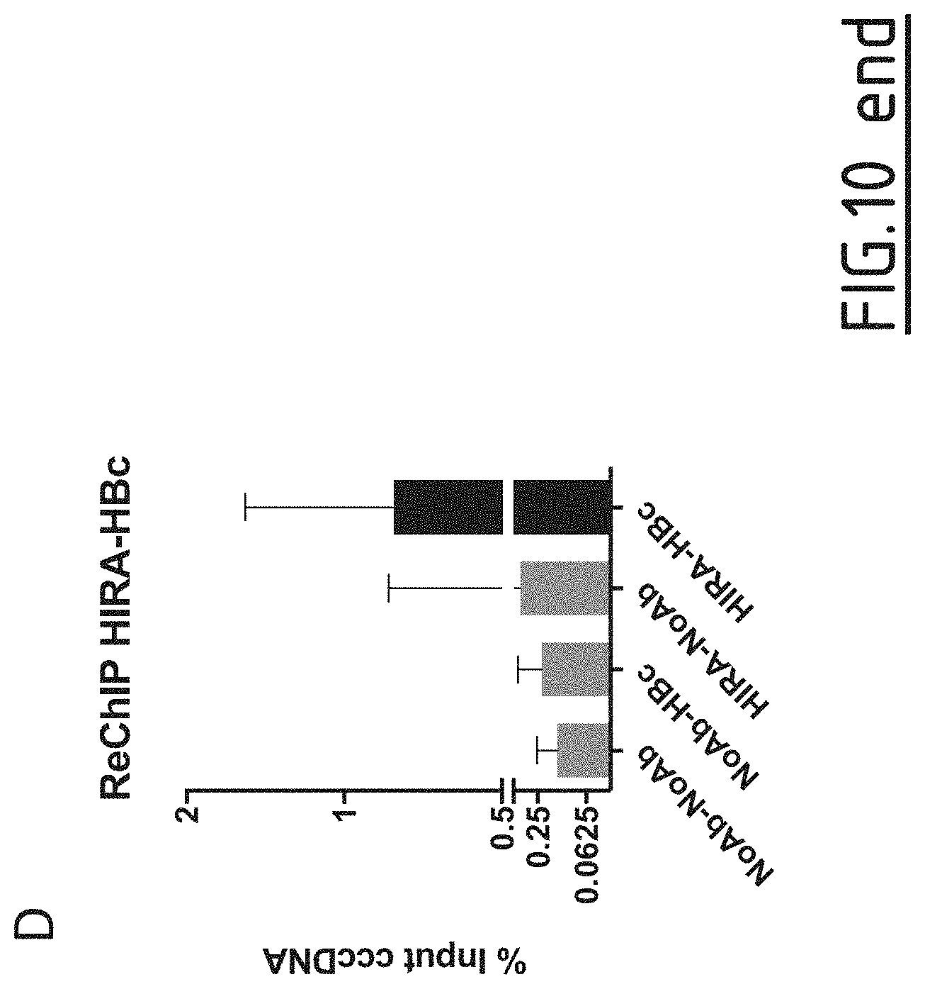
S00001
XML
uspto.report is an independent third-party trademark research tool that is not affiliated, endorsed, or sponsored by the United States Patent and Trademark Office (USPTO) or any other governmental organization. The information provided by uspto.report is based on publicly available data at the time of writing and is intended for informational purposes only.
While we strive to provide accurate and up-to-date information, we do not guarantee the accuracy, completeness, reliability, or suitability of the information displayed on this site. The use of this site is at your own risk. Any reliance you place on such information is therefore strictly at your own risk.
All official trademark data, including owner information, should be verified by visiting the official USPTO website at www.uspto.gov. This site is not intended to replace professional legal advice and should not be used as a substitute for consulting with a legal professional who is knowledgeable about trademark law.